CN107530178B - Implant insertion system - Google Patents
Implant insertion system Download PDFInfo
- Publication number
- CN107530178B CN107530178B CN201680024076.5A CN201680024076A CN107530178B CN 107530178 B CN107530178 B CN 107530178B CN 201680024076 A CN201680024076 A CN 201680024076A CN 107530178 B CN107530178 B CN 107530178B
- Authority
- CN
- China
- Prior art keywords
- implant
- release tube
- release
- wire
- tube
- Prior art date
- Legal status (The legal status is an assumption and is not a legal conclusion. Google has not performed a legal analysis and makes no representation as to the accuracy of the status listed.)
- Active
Links
Images


Classifications
-
- A—HUMAN NECESSITIES
- A61—MEDICAL OR VETERINARY SCIENCE; HYGIENE
- A61F—FILTERS IMPLANTABLE INTO BLOOD VESSELS; PROSTHESES; DEVICES PROVIDING PATENCY TO, OR PREVENTING COLLAPSING OF, TUBULAR STRUCTURES OF THE BODY, e.g. STENTS; ORTHOPAEDIC, NURSING OR CONTRACEPTIVE DEVICES; FOMENTATION; TREATMENT OR PROTECTION OF EYES OR EARS; BANDAGES, DRESSINGS OR ABSORBENT PADS; FIRST-AID KITS
- A61F2/00—Filters implantable into blood vessels; Prostheses, i.e. artificial substitutes or replacements for parts of the body; Appliances for connecting them with the body; Devices providing patency to, or preventing collapsing of, tubular structures of the body, e.g. stents
- A61F2/95—Instruments specially adapted for placement or removal of stents or stent-grafts
- A61F2/962—Instruments specially adapted for placement or removal of stents or stent-grafts having an outer sleeve
- A61F2/966—Instruments specially adapted for placement or removal of stents or stent-grafts having an outer sleeve with relative longitudinal movement between outer sleeve and prosthesis, e.g. using a push rod
-
- A—HUMAN NECESSITIES
- A61—MEDICAL OR VETERINARY SCIENCE; HYGIENE
- A61F—FILTERS IMPLANTABLE INTO BLOOD VESSELS; PROSTHESES; DEVICES PROVIDING PATENCY TO, OR PREVENTING COLLAPSING OF, TUBULAR STRUCTURES OF THE BODY, e.g. STENTS; ORTHOPAEDIC, NURSING OR CONTRACEPTIVE DEVICES; FOMENTATION; TREATMENT OR PROTECTION OF EYES OR EARS; BANDAGES, DRESSINGS OR ABSORBENT PADS; FIRST-AID KITS
- A61F2/00—Filters implantable into blood vessels; Prostheses, i.e. artificial substitutes or replacements for parts of the body; Appliances for connecting them with the body; Devices providing patency to, or preventing collapsing of, tubular structures of the body, e.g. stents
- A61F2/0004—Closure means for urethra or rectum, i.e. anti-incontinence devices or support slings against pelvic prolapse
- A61F2/0022—Closure means for urethra or rectum, i.e. anti-incontinence devices or support slings against pelvic prolapse placed deep in the body opening
- A61F2/0027—Closure means for urethra or rectum, i.e. anti-incontinence devices or support slings against pelvic prolapse placed deep in the body opening inflatable
-
- A—HUMAN NECESSITIES
- A61—MEDICAL OR VETERINARY SCIENCE; HYGIENE
- A61F—FILTERS IMPLANTABLE INTO BLOOD VESSELS; PROSTHESES; DEVICES PROVIDING PATENCY TO, OR PREVENTING COLLAPSING OF, TUBULAR STRUCTURES OF THE BODY, e.g. STENTS; ORTHOPAEDIC, NURSING OR CONTRACEPTIVE DEVICES; FOMENTATION; TREATMENT OR PROTECTION OF EYES OR EARS; BANDAGES, DRESSINGS OR ABSORBENT PADS; FIRST-AID KITS
- A61F2/00—Filters implantable into blood vessels; Prostheses, i.e. artificial substitutes or replacements for parts of the body; Appliances for connecting them with the body; Devices providing patency to, or preventing collapsing of, tubular structures of the body, e.g. stents
- A61F2/01—Filters implantable into blood vessels
- A61F2/011—Instruments for their placement or removal
-
- A—HUMAN NECESSITIES
- A61—MEDICAL OR VETERINARY SCIENCE; HYGIENE
- A61F—FILTERS IMPLANTABLE INTO BLOOD VESSELS; PROSTHESES; DEVICES PROVIDING PATENCY TO, OR PREVENTING COLLAPSING OF, TUBULAR STRUCTURES OF THE BODY, e.g. STENTS; ORTHOPAEDIC, NURSING OR CONTRACEPTIVE DEVICES; FOMENTATION; TREATMENT OR PROTECTION OF EYES OR EARS; BANDAGES, DRESSINGS OR ABSORBENT PADS; FIRST-AID KITS
- A61F2/00—Filters implantable into blood vessels; Prostheses, i.e. artificial substitutes or replacements for parts of the body; Appliances for connecting them with the body; Devices providing patency to, or preventing collapsing of, tubular structures of the body, e.g. stents
- A61F2/01—Filters implantable into blood vessels
- A61F2/013—Distal protection devices, i.e. devices placed distally in combination with another endovascular procedure, e.g. angioplasty or stenting
-
- A—HUMAN NECESSITIES
- A61—MEDICAL OR VETERINARY SCIENCE; HYGIENE
- A61F—FILTERS IMPLANTABLE INTO BLOOD VESSELS; PROSTHESES; DEVICES PROVIDING PATENCY TO, OR PREVENTING COLLAPSING OF, TUBULAR STRUCTURES OF THE BODY, e.g. STENTS; ORTHOPAEDIC, NURSING OR CONTRACEPTIVE DEVICES; FOMENTATION; TREATMENT OR PROTECTION OF EYES OR EARS; BANDAGES, DRESSINGS OR ABSORBENT PADS; FIRST-AID KITS
- A61F2/00—Filters implantable into blood vessels; Prostheses, i.e. artificial substitutes or replacements for parts of the body; Appliances for connecting them with the body; Devices providing patency to, or preventing collapsing of, tubular structures of the body, e.g. stents
- A61F2/02—Prostheses implantable into the body
- A61F2/24—Heart valves ; Vascular valves, e.g. venous valves; Heart implants, e.g. passive devices for improving the function of the native valve or the heart muscle; Transmyocardial revascularisation [TMR] devices; Valves implantable in the body
- A61F2/2427—Devices for manipulating or deploying heart valves during implantation
- A61F2/243—Deployment by mechanical expansion
-
- A—HUMAN NECESSITIES
- A61—MEDICAL OR VETERINARY SCIENCE; HYGIENE
- A61F—FILTERS IMPLANTABLE INTO BLOOD VESSELS; PROSTHESES; DEVICES PROVIDING PATENCY TO, OR PREVENTING COLLAPSING OF, TUBULAR STRUCTURES OF THE BODY, e.g. STENTS; ORTHOPAEDIC, NURSING OR CONTRACEPTIVE DEVICES; FOMENTATION; TREATMENT OR PROTECTION OF EYES OR EARS; BANDAGES, DRESSINGS OR ABSORBENT PADS; FIRST-AID KITS
- A61F2/00—Filters implantable into blood vessels; Prostheses, i.e. artificial substitutes or replacements for parts of the body; Appliances for connecting them with the body; Devices providing patency to, or preventing collapsing of, tubular structures of the body, e.g. stents
- A61F2/02—Prostheses implantable into the body
- A61F2/30—Joints
- A61F2/46—Special tools or methods for implanting or extracting artificial joints, accessories, bone grafts or substitutes, or particular adaptations therefor
- A61F2/4603—Special tools or methods for implanting or extracting artificial joints, accessories, bone grafts or substitutes, or particular adaptations therefor for insertion or extraction of endoprosthetic joints or of accessories thereof
-
- A—HUMAN NECESSITIES
- A61—MEDICAL OR VETERINARY SCIENCE; HYGIENE
- A61F—FILTERS IMPLANTABLE INTO BLOOD VESSELS; PROSTHESES; DEVICES PROVIDING PATENCY TO, OR PREVENTING COLLAPSING OF, TUBULAR STRUCTURES OF THE BODY, e.g. STENTS; ORTHOPAEDIC, NURSING OR CONTRACEPTIVE DEVICES; FOMENTATION; TREATMENT OR PROTECTION OF EYES OR EARS; BANDAGES, DRESSINGS OR ABSORBENT PADS; FIRST-AID KITS
- A61F2/00—Filters implantable into blood vessels; Prostheses, i.e. artificial substitutes or replacements for parts of the body; Appliances for connecting them with the body; Devices providing patency to, or preventing collapsing of, tubular structures of the body, e.g. stents
- A61F2/82—Devices providing patency to, or preventing collapsing of, tubular structures of the body, e.g. stents
- A61F2/86—Stents in a form characterised by the wire-like elements; Stents in the form characterised by a net-like or mesh-like structure
- A61F2/90—Stents in a form characterised by the wire-like elements; Stents in the form characterised by a net-like or mesh-like structure characterised by a net-like or mesh-like structure
-
- A—HUMAN NECESSITIES
- A61—MEDICAL OR VETERINARY SCIENCE; HYGIENE
- A61F—FILTERS IMPLANTABLE INTO BLOOD VESSELS; PROSTHESES; DEVICES PROVIDING PATENCY TO, OR PREVENTING COLLAPSING OF, TUBULAR STRUCTURES OF THE BODY, e.g. STENTS; ORTHOPAEDIC, NURSING OR CONTRACEPTIVE DEVICES; FOMENTATION; TREATMENT OR PROTECTION OF EYES OR EARS; BANDAGES, DRESSINGS OR ABSORBENT PADS; FIRST-AID KITS
- A61F2/00—Filters implantable into blood vessels; Prostheses, i.e. artificial substitutes or replacements for parts of the body; Appliances for connecting them with the body; Devices providing patency to, or preventing collapsing of, tubular structures of the body, e.g. stents
- A61F2/02—Prostheses implantable into the body
- A61F2/04—Hollow or tubular parts of organs, e.g. bladders, tracheae, bronchi or bile ducts
- A61F2/06—Blood vessels
- A61F2/07—Stent-grafts
-
- A—HUMAN NECESSITIES
- A61—MEDICAL OR VETERINARY SCIENCE; HYGIENE
- A61F—FILTERS IMPLANTABLE INTO BLOOD VESSELS; PROSTHESES; DEVICES PROVIDING PATENCY TO, OR PREVENTING COLLAPSING OF, TUBULAR STRUCTURES OF THE BODY, e.g. STENTS; ORTHOPAEDIC, NURSING OR CONTRACEPTIVE DEVICES; FOMENTATION; TREATMENT OR PROTECTION OF EYES OR EARS; BANDAGES, DRESSINGS OR ABSORBENT PADS; FIRST-AID KITS
- A61F2/00—Filters implantable into blood vessels; Prostheses, i.e. artificial substitutes or replacements for parts of the body; Appliances for connecting them with the body; Devices providing patency to, or preventing collapsing of, tubular structures of the body, e.g. stents
- A61F2/82—Devices providing patency to, or preventing collapsing of, tubular structures of the body, e.g. stents
- A61F2/844—Devices providing patency to, or preventing collapsing of, tubular structures of the body, e.g. stents folded prior to deployment
-
- A—HUMAN NECESSITIES
- A61—MEDICAL OR VETERINARY SCIENCE; HYGIENE
- A61F—FILTERS IMPLANTABLE INTO BLOOD VESSELS; PROSTHESES; DEVICES PROVIDING PATENCY TO, OR PREVENTING COLLAPSING OF, TUBULAR STRUCTURES OF THE BODY, e.g. STENTS; ORTHOPAEDIC, NURSING OR CONTRACEPTIVE DEVICES; FOMENTATION; TREATMENT OR PROTECTION OF EYES OR EARS; BANDAGES, DRESSINGS OR ABSORBENT PADS; FIRST-AID KITS
- A61F2/00—Filters implantable into blood vessels; Prostheses, i.e. artificial substitutes or replacements for parts of the body; Appliances for connecting them with the body; Devices providing patency to, or preventing collapsing of, tubular structures of the body, e.g. stents
- A61F2/82—Devices providing patency to, or preventing collapsing of, tubular structures of the body, e.g. stents
- A61F2/86—Stents in a form characterised by the wire-like elements; Stents in the form characterised by a net-like or mesh-like structure
-
- A—HUMAN NECESSITIES
- A61—MEDICAL OR VETERINARY SCIENCE; HYGIENE
- A61F—FILTERS IMPLANTABLE INTO BLOOD VESSELS; PROSTHESES; DEVICES PROVIDING PATENCY TO, OR PREVENTING COLLAPSING OF, TUBULAR STRUCTURES OF THE BODY, e.g. STENTS; ORTHOPAEDIC, NURSING OR CONTRACEPTIVE DEVICES; FOMENTATION; TREATMENT OR PROTECTION OF EYES OR EARS; BANDAGES, DRESSINGS OR ABSORBENT PADS; FIRST-AID KITS
- A61F2/00—Filters implantable into blood vessels; Prostheses, i.e. artificial substitutes or replacements for parts of the body; Appliances for connecting them with the body; Devices providing patency to, or preventing collapsing of, tubular structures of the body, e.g. stents
- A61F2/95—Instruments specially adapted for placement or removal of stents or stent-grafts
- A61F2/9522—Means for mounting a stent or stent-graft onto or into a placement instrument
-
- A—HUMAN NECESSITIES
- A61—MEDICAL OR VETERINARY SCIENCE; HYGIENE
- A61F—FILTERS IMPLANTABLE INTO BLOOD VESSELS; PROSTHESES; DEVICES PROVIDING PATENCY TO, OR PREVENTING COLLAPSING OF, TUBULAR STRUCTURES OF THE BODY, e.g. STENTS; ORTHOPAEDIC, NURSING OR CONTRACEPTIVE DEVICES; FOMENTATION; TREATMENT OR PROTECTION OF EYES OR EARS; BANDAGES, DRESSINGS OR ABSORBENT PADS; FIRST-AID KITS
- A61F2/00—Filters implantable into blood vessels; Prostheses, i.e. artificial substitutes or replacements for parts of the body; Appliances for connecting them with the body; Devices providing patency to, or preventing collapsing of, tubular structures of the body, e.g. stents
- A61F2/82—Devices providing patency to, or preventing collapsing of, tubular structures of the body, e.g. stents
- A61F2002/823—Stents, different from stent-grafts, adapted to cover an aneurysm
-
- A—HUMAN NECESSITIES
- A61—MEDICAL OR VETERINARY SCIENCE; HYGIENE
- A61F—FILTERS IMPLANTABLE INTO BLOOD VESSELS; PROSTHESES; DEVICES PROVIDING PATENCY TO, OR PREVENTING COLLAPSING OF, TUBULAR STRUCTURES OF THE BODY, e.g. STENTS; ORTHOPAEDIC, NURSING OR CONTRACEPTIVE DEVICES; FOMENTATION; TREATMENT OR PROTECTION OF EYES OR EARS; BANDAGES, DRESSINGS OR ABSORBENT PADS; FIRST-AID KITS
- A61F2/00—Filters implantable into blood vessels; Prostheses, i.e. artificial substitutes or replacements for parts of the body; Appliances for connecting them with the body; Devices providing patency to, or preventing collapsing of, tubular structures of the body, e.g. stents
- A61F2/95—Instruments specially adapted for placement or removal of stents or stent-grafts
- A61F2002/9505—Instruments specially adapted for placement or removal of stents or stent-grafts having retaining means other than an outer sleeve, e.g. male-female connector between stent and instrument
-
- A—HUMAN NECESSITIES
- A61—MEDICAL OR VETERINARY SCIENCE; HYGIENE
- A61F—FILTERS IMPLANTABLE INTO BLOOD VESSELS; PROSTHESES; DEVICES PROVIDING PATENCY TO, OR PREVENTING COLLAPSING OF, TUBULAR STRUCTURES OF THE BODY, e.g. STENTS; ORTHOPAEDIC, NURSING OR CONTRACEPTIVE DEVICES; FOMENTATION; TREATMENT OR PROTECTION OF EYES OR EARS; BANDAGES, DRESSINGS OR ABSORBENT PADS; FIRST-AID KITS
- A61F2/00—Filters implantable into blood vessels; Prostheses, i.e. artificial substitutes or replacements for parts of the body; Appliances for connecting them with the body; Devices providing patency to, or preventing collapsing of, tubular structures of the body, e.g. stents
- A61F2/95—Instruments specially adapted for placement or removal of stents or stent-grafts
- A61F2/962—Instruments specially adapted for placement or removal of stents or stent-grafts having an outer sleeve
- A61F2/966—Instruments specially adapted for placement or removal of stents or stent-grafts having an outer sleeve with relative longitudinal movement between outer sleeve and prosthesis, e.g. using a push rod
- A61F2002/9665—Instruments specially adapted for placement or removal of stents or stent-grafts having an outer sleeve with relative longitudinal movement between outer sleeve and prosthesis, e.g. using a push rod with additional retaining means
-
- A—HUMAN NECESSITIES
- A61—MEDICAL OR VETERINARY SCIENCE; HYGIENE
- A61M—DEVICES FOR INTRODUCING MEDIA INTO, OR ONTO, THE BODY; DEVICES FOR TRANSDUCING BODY MEDIA OR FOR TAKING MEDIA FROM THE BODY; DEVICES FOR PRODUCING OR ENDING SLEEP OR STUPOR
- A61M25/00—Catheters; Hollow probes
- A61M25/0021—Catheters; Hollow probes characterised by the form of the tubing
- A61M2025/0042—Microcatheters, cannula or the like having outside diameters around 1 mm or less
Abstract
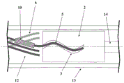
The invention relates to a device for introducing an implant (1) into a blood vessel or a hollow organ of the human or animal body, comprising an implant (1), an insertion wire (14) and a release tube (13), wherein the implant (1) is deformable such that it adopts a shape with a reduced diameter in a microcatheter (8) and, at the implantation site, expands to adapt to the diameter of the blood vessel or hollow organ once the external constraints of the microcatheter (8) disappear, wherein a holding element (2) is arranged on the insertion wire (14) and the holding element (2) has at its periphery at least one, preferably a plurality of grooves (3) provided into the holding element (2), which grooves extend along the circumference of the holding element (2) and form a trajectory in the form of a curve, wherein the implant (1) has at least one, preferably a plurality of grooves extending in the proximal direction at the proximal end, Preferably a plurality of retaining wires (5) which fit into the groove (3), wherein the release tube (13) is pulled over the retaining element (2) and the retaining wires (5) which fit into the groove (3) in a form-fit, so that the retaining wires (5) are held in the groove (3) by friction locking, and the release of the implant (1) takes place by pulling back the release tube (13) in the proximal direction. According to an alternative embodiment, the invention relates to a device for introducing an implant (1), wherein the release tube (13) protrudes into the proximal end of the implant (1), wherein an elastic contact surface is present between the inner side of the implant (1) and the outer side of the release tube (13), such that a frictional lock is generated between the implant (1) and the release tube (13), resulting in a lengthwise movement of the implant (1) inside the microcatheter (8) by a distal or proximal lengthwise movement of the release tube (13).
Description
Technical Field
The invention relates to a device for introducing an implant into a blood vessel or a hollow organ of the human or animal body, comprising an implant, an insertion wire and a release tube, wherein the implant is deformable such that it takes a shape with a reduced diameter in a microcatheter and, at the implantation site, expands to adapt to the diameter of the blood vessel or hollow organ once the external constraints of the microcatheter have disappeared, wherein a retaining element is arranged on the insertion wire. According to an alternative embodiment, the invention relates to a device for introducing an implant into a blood vessel or a hollow organ of the human or animal body, comprising an implant and a release tube, wherein the implant is deformable such that it takes a shape with a reduced diameter in a microcatheter and, at the implantation site, expands to adapt to the diameter of the blood vessel or hollow organ once the external constraints of the microcatheter have disappeared, and the release tube has a lumen extending in the longitudinal direction of the device, through which lumen an insertion wire can be guided in a longitudinally movable manner.
Background
Arteriovenous malformations in patients can lead to serious injury and risk, even death. This is particularly true for arteriovenous fistulas and aneurysms, particularly when they occur in the brain region. Often, one attempts to close such deformities by an implant. Such implants are typically placed in a blood vessel by means of a catheter.
The implantation of platinum spirals has proved effective, in particular in the case of aneurysms, which spirals fill the aneurysm substantially completely, block the inflow of blood to a large extent and lead to the formation of local thrombi, eventually closing the aneurysm. However, this treatment method is only suitable for aneurysms with a narrow access to the vascular system, the so-called berry-like aneurysms. For a vessel outgrowth with a wide access to the vessel, the implanted helix is easily flushed out again and causes damage in other areas of the vascular system.
In such cases, it has been proposed to install a stent that "plugs" the opening of the aneurysm and thereby prevents the occlusion helix from being flushed out. However, such stents with relatively wide mesh walls have a number of disadvantages.
On the one hand, this is a wide mesh structure that allows an unobstructed flow of blood into the aneurysm. But if the aneurysm is not sufficiently filled by the occluding device, the pressure on the vessel wall remains unattenuated. In these cases, further treatment may be difficult, but not impossible, because the stent obstructs access to the aneurysm and placement of additional occlusive devices.
Another disadvantage is the poor adaptability of the stent to its installation site. For optimal function, the stent should be applied snugly to the vessel wall without applying excessive pressure to the wall. In contrast to stents, which in the case of stenosis should lead to vessel dilatation, these stents are considered to be a kind of cuff, which should influence the vessel lumen and the endothelial wall of the vessel as little as possible.
Stents consisting of a braided wire fabric have long been known, in particular for the coronary region. These stents are generally manufactured as circular braids with individual filaments forming the stent wall in oppositely directed helical or spiral layers. The result is a mesh braid that is supported in the radial direction and is also permeable to blood.
When used to treat stenosis, such stents, consisting of filaments in the form of a circular braid, are typically hydraulically expanded at the installation site and secured to the vessel wall by means of a balloon. During insertion, the balloon secured to the insertion wire serves as a delivery vehicle on which the stent is crimped. But for implants for influencing or guiding the blood flow in the brain area, implants that adapt spontaneously to the vessel diameter and abut against the vessel wall are advantageous.
WO 2008/107172a 1 describes an implant in which the braid has an elongated shape of reduced diameter in a microcatheter to expand at the implantation site, adapt to the vessel diameter and increase the braid density, wherein the ends of the filaments projecting at the ends of the implant are brought together and joined together at least in pairs. In this way, an implant is provided which can be adapted to a specific vessel diameter, wherein the filament ends are atraumatic.
According to this prior art, the connecting element is arranged at the end of the coupled filaments, interacting with the retaining element by the lock and key principle. The retaining element, by means of which the implant is coupled to the insertion wire, has a recess in which the connecting element fits. The connecting elements have a thickening, such as a spherical shape, so that they are held in the recess of the holding element by a form fit. The fixing in the recess can be done by means of a tube which is pulled in a form-fitting manner over the holding element with the connecting element fitted therein. After reaching the end position of the implant, the tube is pulled back in the proximal direction and the implant is released. Thereafter, the insertion wire with the holding element, tube and catheter can be pulled back and removed from the body.
The described prior art is basically effective, but in some cases it may happen that, after retraction of the tube, not all the connecting elements are released from the recesses of the holding elements provided for it, for example because of skewing occurring and continuing to hold the connecting elements in the recesses of the holding elements. In such a case, the implant does not open at its proximal end as quickly as desired, but may only be released from the holding element after further movement of the insertion wire. On the other hand, a release system is desired in which the implant is completely released from the holding element immediately after the tube is retracted and becomes unconstrained in this way. Starting from the prior art described in WO 2008/107172a 1, therefore, the problem arises of further optimizing the release system.
Disclosure of Invention
According to a first embodiment of the invention, this problem is solved by a device for introducing an implant into a blood vessel or a hollow organ of the human or animal body, comprising an implant, an insertion wire and a release tube, wherein the implant is deformable such that it takes a shape with a reduced diameter in a microcatheter and, at the implantation site, expands to the diameter of the blood vessel or hollow organ once the outer constraints of the microcatheter disappear, wherein a holding element is arranged on the insertion wire and has at its periphery at least one, preferably a plurality of grooves provided into the holding element, which grooves extend along the circumference of the holding element and form a track in the form of a curve, wherein the implant has at least one, preferably a plurality of holding wires extending in the proximal direction, which holding wires fit into the grooves, wherein the release tube is pulled over the retaining element and the retaining wire fitted into the groove in a form-fit manner, such that the retaining wire is held in the groove by a friction lock, and the release of the implant takes place by pulling back the release tube in the proximal direction. The release tube constitutes the groove for the retaining element and a tubular sheath of the retaining wire inserted in the groove.
According to the invention, the holding element therefore has a groove arranged radially into the holding element on the outside of the holding element. The groove allows a retaining wire extending proximally from the implant to be inserted into the groove. The retaining wire extends generally proximally through the implant itself. In order to be able to advance the implant in the distal direction and retract in the proximal direction by means of the insertion wire without the implant disengaging from the holding element, the groove has a curved course on the outer side of the holding element. In case the groove in the longitudinal direction of the device has a straight course, there will be a risk that the holding wire of the implant will be pulled out of the groove when the insertion wire is retracted. In addition, the dimensions of the groove and the retaining wire should be coordinated so that the frictional force between the groove and the retaining wire that prevents release of the implant is greater than the pulling or pushing force generated during retraction or advancement of the insertion wire. In particular, the cross section of the groove should only be slightly larger than the cross section of the retaining wire, so that on the one hand, after retraction of the release tube, expansion of the implant in the radial direction is easily achieved for release purposes, but on the other hand, the friction between the groove and the retaining wire is sufficiently large that pulling out the retaining wire from the groove in the axial direction can only take place with an unusually large force.
Unlike the prior art described previously, in which the connection of the connecting element and the retaining element relies on a form fit, the fixing of the retaining wire in the groove provided for it is based on a friction fit or force fit according to the invention. The friction between the retaining wire and the groove is so great that it is virtually impossible to release by the action of force in the axial direction, i.e. the longitudinal direction of the device or the insertion wire or implant. However, if the release tube is pulled back so far that the grooves are radially exposed, the implant can expand, with the retaining wires moving radially outward from the grooves. In this way, release of the implant from the retaining element is ensured; the implant is thus finally released and implanted at the desired site. Thereafter, the insertion wire and the retaining element, release tube and microcatheter attached thereto may be retracted and removed from the body.
For the placement of the implant, the implant is first advanced by means of an insertion wire all the way through the microcatheter to the desired location. The retaining element and generally the entire insertion wire are surrounded by the release tube during this process. Once release of the implant is desired, the microcatheter catheter is first withdrawn. However, this does not in itself lead to a final release, since the release tube continues to ensure that the retaining wire from the implant remains in the groove of the retaining element. The groove is arranged in an outer region of the holding element; thus, there is a natural tendency for the retaining wires to move outward and release from the grooves as the implant expands after it is released from the microcatheter. However, this is only possible if the release tube has also been pulled back. The treating physician thus has sufficient time to judge the situation after retracting the microcatheter and achieve final release of the implant by pulling back the release tube in the proximal direction, or if placement of the implant does not occur as desired, move the implant back into the microcatheter and implant it in a different location, or remove the device from the body again by pulling back the insertion wire. Once the implant has been successfully released in the correct position, the insertion wire with the retaining element and the release tube can be retracted into the micro-catheter and removed from the vascular system together therewith.
In the context of the specification, the term proximal refers to a direction facing the treating physician, i.e. the proximal end points in the outside of the body. Distal, on the other hand, means facing away from the physician, i.e. the distal end is in the direction of the interior of the body.
Typically, a release tube extends from the retaining element, the groove of which must be covered in order to securely retain the retaining wire in the groove, proximal to the outside of the body. It is however also conceivable that the release tube does not cover the entire insertion wire, it being sufficient for the release tube to extend through the groove of the holding element. In this case, the retraction of the release tube is accomplished by a second wire or thread that is parallel to the insertion wire and extends proximally from the release tube in the proximal direction.
In order to make the friction between the groove and the retaining wire sufficiently large, an undulating track for the groove on the circumference of the retaining element is advantageous. For example, the grooves may extend in a sinusoidal fashion. Typically, the groove extends in the proximal to distal direction, forming a curved, in particular wave-shaped track on the outer side of the holding element, wherein the groove does not need to extend over the entire length of the holding element. In particular, a plurality of wave shaped grooves may extend from the proximal to the distal direction and be distributed over the circumference of the holding element, the wave shaped grooves being arranged substantially parallel to each other. It is also conceivable that the wave-shaped groove extends not only in the longitudinal direction, i.e. from the proximal to the distal direction, but also in a helical manner around the holding element in the longitudinal direction.
Intentionally, a plurality of grooves for holding the wires is arranged in the holding element, in particular at least 4 grooves. Preferably at least 8, especially 8 to 32 grooves. This ensures that the implant is held evenly over its circumference and is detached evenly from the holding element after retraction of the release tube. The retaining element may be made, for example, of refined steel or a nickel titanium alloy (e.g., nitinol).
The implant itself is typically a braid made of a plurality of braided filaments extending in a spiral or helical pattern, wherein oppositely extending braided filaments are interdigitated and form a mesh braid. Such a braided structure is known from the prior art, for example from WO 2008/107172a already cited above. However, it is also conceivable for the implant to be a tubular or sheet-like implant, wherein a retaining wire for fitting into a groove of the retaining element is arranged at the proximal end.
In the case of an implant assembled from a wire braid, it is advisable to use the proximal section of the braided wires forming the implant as retaining wires. For this purpose, the individual braided filaments may be elongated in the proximal direction. For example, every second, fourth or eighth braided wire may be formed longer at the proximal end, this increase in the proximal direction of the braided wire constituting a proximal section, also referred to as a retaining wire. The retaining wire is placed in a groove provided for this purpose. In the case of an implant consisting of 64 braided wires, for example, every second braided wire can be formed longer at the proximal end, so that there are a total of 32 retaining wires and they must be provided with 32 recesses in the retaining element. Similarly, every fourth braided wire (in the case of 64 braided wires, there are therefore 16 retaining wires and 16 grooves) or every 8 th braided wire (in the case of 64 braided wires, there are therefore 8 retaining wires for 8 grooves) can be formed longer.
Furthermore, it is advisable that the release tube covers not only the retaining element and the retaining wire fitted into the groove, but also the proximal end of the implant itself, before being retracted in the proximal direction in order to release the implant. Thus, the release tube also covers the shorter proximal end of the braided wire, which is not elongated in the proximal direction for forming the retaining wire. These proximal ends of the braided wires, which are not extended to retain the wires, are typically loose, but are also covered by a release tube. An advantage of such a configuration is that even after the implant is pushed out of the microcatheter or retracted back into the microcatheter so that the implant is substantially unconstrained, retraction of the implant into the microcatheter is still possible as long as the release tube covers the retaining element and the retaining wire that fits into the groove of the retaining element. Although a larger portion of the implant may be radially expanded after the microcatheter is retracted, this does not apply to the proximal end of the implant as long as the release tube slides over it. If no expansion has occurred at the proximal end of the implant, it is possible for the implant to retract into the microcatheter, in which case the expanded region of the implant is again tightly folded.
According to a further advantageous embodiment, the retaining wire is deformed such that the friction between the retaining wire and the groove is increased. This further improves the safe placement of the retaining wire in the groove and ensures that it is virtually impossible to pull the retaining wire unintentionally out of the groove. For example, the retaining wire may be given a two-dimensional or three-dimensional shape that prevents the retaining wire from sliding out of the groove under tensile stress. Such two-dimensional or three-dimensional structures can be produced, for example, by machining or heat treatment.
According to a second embodiment, a simplified release system is similarly provided based on friction locking (force locking). This embodiment relates to a device for introducing an implant into a blood vessel or a hollow organ of the human or animal body, comprising an implant and a release tube, wherein the implant is deformable such that it takes a shape with a reduced diameter in a microcatheter and, at the implantation site, expands to the diameter of the blood vessel or the hollow organ once the external constraints of the microcatheter disappear, and the release tube has a lumen extending in the longitudinal direction of the device through which an insertion wire can be guided in a length-wise movable manner, wherein the release tube protrudes into the proximal end of the implant and an elastic contact surface is present between the inner side of the implant and the outer side of the release tube, such that a frictional lock is generated between the implant and the release tube, a length-wise movement of the implant inside the microcatheter in the length direction being induced by a distal or proximal movement of the release tube in the length direction causing a length-wise movement of the implant inside the microcatheter And (6) moving.
Unlike the previously described embodiments, the release tube does not surround the proximal end of the implant or the retaining wire emerging from the implant, but projects into the latter. A friction-locking connection is created between the release tube and the implant so that the implant can be moved distally or proximally in the same manner by advancing or retracting the release tube. The frictional locking between the release tube and the implant results in the portion of the release tube protruding into the implant having at least a partially resilient contact surface. Preferably this involves an intermediate layer which is present on the outside of the release tube in the part of the release tube protruding into the implant, and which may also be referred to as a pad. The resilient contact surface, preferably the intermediate layer, ensures a friction-locking connection with the implant. Such an embodiment is particularly easy to construct and does not require additional elements to be formed thereon to result in a form fit.
The release of the implant occurs as the implant is pushed out of the microcatheter or the microcatheter is retracted proximally relative to the implant. Since the implant has a natural tendency to expand radially after the external constraint caused by the microcatheter has disappeared, the implant detaches from the elastic pad and abuts against the inner wall of the blood vessel or hollow organ after being released from the microcatheter. The release tube, microcatheter and optionally the insertion wire extending through the release tube are then retracted and removed from the body.
The release tube has an inner lumen through which an insertion wire may extend. Typically, after placing the insertion wire at the desired location, the microcatheter is pushed through the insertion wire all the way to the target site. Thereafter, the release tube and the implant frictionally coupled thereto may be advanced distally through the microcatheter. Retraction and removal of the insertion wire is possible before, during or after implant release. Retraction of the insertion wire may occur before or during release of the implant if it is desired to prevent movement of the insertion wire tip into the blood vessel at the distal location.
The resilient contact surface/intermediate layer typically extends in a ring around or around the release tube. In other words, the release tube is surrounded in a radially surrounding manner in one region by the elastic contact surface/intermediate layer, which ensures a friction-locked connection with the implant. The implant is typically a braid made of braided wires, but other types of implants, such as tubular or sheet implants, are not excluded.
In order to create a friction-locking connection with the implant, one or more intermediate layers (pads) may be provided, wherein the pads may extend a certain length inside the implant. Typically, the number of pads will be 1 or 2, wherein in case two pads are used, these may be constructed correspondingly shorter. At the location where the intermediate layer is arranged on the release tube, the latter has a larger outer diameter than the adjoining region without the intermediate layer.
The material for the intermediate layer/pad must be elastic in order to create a sufficient friction between the release tube and the implant, which enables the implant to be advanced or retracted via the release tube without the implant having to be moved in the longitudinal direction relative to the release tube or even disengaged from the release tube. Many different materials are conceivable as material for the elastic intermediate layer, in particular it may be an elastomer. For example, rubber, Indian rubber, or silicone may be used.
The intermediate layer may also be made of a polymeric material such as polytetrafluoroethylene, polyester, polyamide, polyurethane or polyolefin. Particularly preferred are polycarbonate polyurethanes. The intermediate layer is preferably produced by electrospinning. In this process, fibrils or fibers are deposited from a polymer solution onto a substrate with the aid of an electric current. During deposition, the fibrils stick together to form a fluff. Typically, the fibrils have a diameter of 100 to 3000 nm. The layer produced by electrospinning is very uniform, tough and mechanically durable. With regard to the production of the intermediate layer by electrospinning, reference is made in particular to WO 2008/049386, DE 2806030A 1 and the documents cited therein.
The intermediate layer can likewise be made of the same material as the release tube, which material preferably consists of polyimide. In this case, the intermediate layer can be formed in one piece with the release tube, wherein the release tube has a correspondingly larger outer diameter in the region of the intermediate layer than in the adjoining region without the intermediate layer. According to the invention, an intermediate layer formed in one piece with the release tube is also covered by the term intermediate layer, as long as the intermediate layer is elastic and produces a sufficiently large frictional locking with the implant. It is also possible for the release tube to have a uniform cross section in the section in which the implant extends, provided that a sufficiently strong frictional locking is produced between the implant and the release tube. For example, the distal section of the release tube may also have a reduced diameter, so that it can be introduced into the implant in the longitudinal direction in order to create the desired friction-locked connection there.
In addition, a radiopaque material may be provided between the elastic intermediate layer and the actual release tube in order to improve visualization of the implant installation procedure. Coils of radiopaque material are particularly preferred as they are sufficiently flexible to ensure problem-free advancement of the implant. However, another form of radiopaque material is also possible, such as in the form of a sleeve placed over the release tube. Preferred radiopaque materials are platinum and platinum alloys. In particular, the elastic intermediate layer can be provided with a polymer layer, preferably of polycarbonate polyurethane, particularly preferably produced by means of electrospinning, as described above.
According to another variant of the second embodiment, the release wire is wound around the implant in the area where the release tube protrudes into the proximal end of the implant, and wherein an elastic contact surface is present between the inner side of the implant and the outer side of the release tube, such that the release wire produces an increase of the friction between the implant and the release tube, wherein the release wire is electrolytically corrodible.
According to this variant, the implant is not held in compressed form by the microcatheter or only by it, but (also) by a release wire which is wound around the implant and a release tube with an elastic contact surface introduced into the implant. The release wire fits as securely onto the implant. Thus, as long as the release wire does not loosen, the final release of the implant does not occur, because the friction between the implant and the release tube is too great. To generate a large friction force, the release tube has a resilient contact surface, preferably a resilient intermediate layer/pad as described above. But unlike the previously described variants, the implant is still retained on the release tube by a friction lock when the microcatheter has been retracted from the implant or the implant has been pushed out of the microcatheter. In this way, a particularly good safety is ensured in the placement of the implant, since even after the implant is released from the microcatheter, the implant first retains its compressed shape and thus it is still possible for the implant to retract into the microcatheter.
The release wire surrounds the implant at an axial position where the resilient contact surface (preferably the resilient intermediate layer/pad) is located. The release wire may be tied directly around the implant at the location of the resilient contact surface or pad. It is also possible to ligature around the implant at an axial position of the implant between two resilient contact surfaces or pads.
The release wire wound around the implant is electrolytically erodable at least at one release site. The release site should be located in a section of the release wire that is placed around the implant in the form of a loop or winding. Instead of electrolytic corrosion, thermal detachment is also conceivable, in which case the release site is heated to such an extent that the release wire is cut. Electrolytic corrosion or heating is also caused by the application of a voltage source. To this end, the end of the release wire is preferably guided proximally to the point where attachment can occur. In other words, the release wire extends proximally to the location of the release tube introduced into the implant, where a loop or winding is formed around the implant, and then returns in the proximal direction. When a voltage is applied to the release wire, electrolytic corrosion of the release wire occurs at the release location, causing the release wire to be cut at that location. Thus, the release wire is no longer able to press the implant against the release tube. Due to the tendency of the implant to expand after the external constraint disappears, the implant detaches from the release tube and is eventually released. The microcatheter, release tube, insertion wire and release wire may then be retracted.
In order to induce electrolytic corrosion of the release wire, it is desirable to weaken the release wire at least at one location such that electrolytic corrosion preferably occurs at the release site. The release site may have, for example, a smaller cross-section than other areas of the release wire. Furthermore, the portion of the release wire may be made of a material that dissolves particularly well when a voltage is applied. The material that can be used for the release site corresponds to, for example, the material that is also used in the electrolytic detachable implant. These include steel, magnesium or magnesium alloys and cobalt-chromium alloys. The latter is described in WO 2011/147567a1, to which reference is made for this purpose. To achieve current concentration at the release site, the release wire may be partially or completely insulated by a sheath outside the release site.
In the described variation with a release wire, an implant secured to the release tube is introduced by advancing the release tube distally through a microcatheter to a target site. The friction-locked connection between the release tube and the implant thus ensures that the implant moves with the release tube. At the target site, the microcatheter is retracted proximally or the implant is advanced distally from the microcatheter. Due to the tying of the release wire, the implant continues to remain on the release tube until the treating physician decides on the final release of the implant. To do this, a voltage is applied to the release wire, whereby it is cut and the implant is released to expand.
The principle of the release wire described here, which surrounds the implant at the location where the release tube is introduced into the implant by its elastic contact surface, so that the friction between the implant and the release tube increases until a movement of the implant in the length direction can be performed by an advancement and retraction of the release tube, is also applicable to embodiments in which the insertion wire itself, rather than the release tube, has an elastic contact surface, in particular an elastic intermediate layer or pad. In this case, therefore, no friction lock is produced between the implant and the release tube, but between the implant and the insertion wire. This enables the implant to be advanced by advancing the insertion wire through the microcatheter. In which case the release tube surrounding the insertion wire may be omitted. Such embodiments are also included in the present invention.
The creation of a friction-locking connection between the implant and the insertion wire (instead of the release tube) also constitutes a third embodiment according to the invention covered by the present application, i.e. without the release wire described above. For this purpose, an elastic contact surface, in particular in the form of an elastic intermediate layer or an elastic pad, is provided on the insertion wire, which greatly increases the friction between the implant and the insertion wire, so that a displacement of the implant can be carried out by an advancement or retraction of the insertion wire. In this case, the above-described release tube need not be used.
The elastic intermediate layer or the elastic cushion in this variant of the invention is preferably made of polycarbonate polyurethane, in particular by means of electrospinning. The method has been described above.
In particular, according to a third embodiment of the invention, in which a friction-locking connection is produced between the insertion wire and the implant, a coil, as is commonly referred to in the medical art, can be arranged on the insertion wire, the coil being coated with an elastic material. This may involve the aforementioned elastic materials: rubber, vityan rubber or silicone, or also a polymeric material such as polytetrafluoroethylene, polyester, polyamide, polyurethane or polyolefin. Particularly preferred are polycarbonate polyurethanes. The elastic material thus forms an intermediate layer. When using the polymers, in particular when using polycarbonate polyurethanes, application by means of electrospinning is likewise possible, as described in connection with the above examples.
The coil itself is preferably made of a radiopaque material, preferably platinum or a platinum alloy, which makes it possible to visualize the implantation procedure. At the same time, however, the coil is sufficiently flexible that the insertion wire on which the implant is placed can also easily follow the narrow-lumen vessel during its advancement. In principle, it is also possible to place the radiopaque material on the insertion wire in a different form (e.g. a metal sleeve), but in this case the bendability is smaller than in the case of a coil.
The following description refers to the first and second and third embodiments of the invention as described above, unless the context indicates otherwise. Basically, all features mentioned in connection with the first, second or third embodiment in this description may also be features of other respective embodiments, unless the context dictates otherwise, or unless it is not technically possible.
According to a preferred embodiment, the outer diameter of the release tube varies between the proximal and distal ends, wherein the variation in outer diameter is associated with regions of the release tube that do not surround the retaining element or are located outside of the implant. These latter regions will be referred to as distal sections in the following. The benefits of good flexibility and easy, predictable detachment are combined with each other by the variation in the outer diameter of the release tube between the proximal and distal ends. Good flexibility is particularly important in certain sections of the release tube, especially in the region proximally adjacent to the distal section, immediately surrounding the retaining element or engaged with the implant, so that the entire device can even follow minor vessel bends when introduced. For this reason, a small outer diameter is of interest here. On the other hand, a further proximally positioned section of the release tube will have sufficient resistance to undesired elongation. This is particularly important in the proximal section, since this takes up a larger part of the total length of the release tube, and thus its stretchability in the longitudinal direction will be least likely, otherwise an undesired total length expansion may occur along the total length. A greater resistance to undesired elongation is also advantageous in its distal section, which according to the first embodiment of the invention surrounds the retaining element, so that this section of the release tube actually moves proximally during retraction and is not only stretched in the longitudinal direction. For this reason, the distal section may also have a larger outer diameter than the intermediate section, but this is not absolutely necessary. The desired outer and inner diameters in the distal section must also meet the dimensions of the surrounding retaining element.
The release tube therefore advantageously has a distal section which in particular surrounds the retaining element (first embodiment of the invention) or extends into the implant (second embodiment of the invention), follows in the proximal direction an intermediate section which has a small outer diameter, and follows in the proximal direction a section which has a large outer diameter of the intermediate section. Furthermore, it is advisable that the distal section also has a large outer diameter in order to enclose a retaining element with retaining wires fitted thereon. In other words, the section covering the groove in the holding element has a large outer diameter and therefore a greater rigidity than the intermediate section following it in the proximal direction, the flexibility of which is particularly important for introducing the device. The section which is longest so far, here called proximal section, has again a large outer diameter in order to enable insertion and retraction of the release tube even over a relatively long distance.
Typically, the length of the middle section is 50 to 500mm, in particular 80 to 120mm, particularly preferably about 100 mm. The distal section may have a length of 2 to 10 mm; this is generally sufficient to cover the recess in the holding element. The total length of the release tube may be 1000 to 2000mm, for example 1800mm, and therefore the proximal section is typically the longest and has a length of 500 to 1900 mm.
The phrases "large outer diameter" and "small outer diameter" according to the present invention should be understood to mean that the outer diameter is larger in the region with the large outer diameter than in the region with the small outer diameter. The exact dimensions may vary and the ratio of diameters may vary, depending on, among other things, the conditions in the vascular system and the particular purpose of implantation. But typically have a large outer diameter in the range of 0.4 to 0.8mm, especially 0.5 to 0.7mm, for example about 0.6 mm. Typical small outer diameters are 0.3 to 0.55mm, especially 0.4 to 0.5mm, for example about 0.45 mm.
The proximal section of the release tube, which typically has a large outer diameter, may be followed by a further proximal end, which in turn has a relatively small outer diameter. The release tube here is expediently clamped on the insertion wire, for example by means of a torquer, in order to produce a frictional lock and prevent an undesired mutual displacement of the insertion wire and the release tube. When using an implant according to the first embodiment of the invention, the displacement occurs only when the implant needs to be released.
To facilitate retraction of the release tube for release of the implant, a gripping feature may be provided at the proximal end of the release tube, regardless of the outer diameter in this region. This may be in the form of a thickening or sleeve closing the proximal end of the release tube. When release of the implant should occur, the torquer that clamps the release tube onto the insertion wire is typically loosened and possibly replaced on the insertion wire to make it easier to handle. The release tube may then be grasped by a user at the gripping features and retracted in a proximal direction.
The implant plus insertion wire and surrounding release through the catheter may be facilitated by providing a coating of the release tube on the outside that reduces friction between the release tube and the catheter. This is preferably a hydrophilic coating.
When retracting the release tube, it is further desirable to keep the friction between the insertion wire and the release tube as low as possible. For this purpose, a friction reducing coating can be used at least in sections on the outside of the insertion wire or on the inside of the release tube. The use of Polytetrafluoroethylene (PTFE) is preferred. This is particularly applicable to areas where the insertion wire has been ground, as is typically the case at the proximal end, so as to be able to be grasped by the torquer.
In addition to the outer diameter, the wall thickness of the release tube may also vary, i.e., have a greater wall thickness in regions where the release tube has a large outer diameter than in regions having a small outer diameter. Reducing the wall thickness further increases the flexibility and bendability of the release tube, so that it can follow the thin branches of the vascular system particularly easily inside the microcatheter.
The release tube may be created by starting with a uniformly configured release tube having constant outer and inner diameters, i.e. also a constant wall thickness, for at least a major part of its length. Material is removed from the release tube on the outside in the desired section, thereby reducing the outer diameter. Since no material is removed on the inner side, the wall thickness of the release tube is reduced to the same extent. Thus, the release tube is obtained as a single piece, wherein in the partial sections (in particular the intermediate section) the outer diameter as well as the wall thickness has been reduced by removing material. In other part sections, such as the proximal section and possibly the distal section, material is usually not removed, i.e. the original outer diameter remains intact here.
The removal of material can be carried out by methods known per se in the prior art, for example lathe turning, grinding or planing by means of a machine tool or also by means of a laser. Material may also be removed at the proximal end to enable grasping by a torquer here.
The release tube is typically made of plastic. Polyimides have proven particularly good. However, it is also conceivable to use other materials, such as polypropylene or Polytetrafluoroethylene (PTFE). Combinations of different plastics or multi-layer co-extruded polymers may also be used. In addition, the release tube may also have reinforcement in that fibers (e.g., metal fibers) may be embedded in the release tube. For example, a release tube reinforced by a fabric or braid is conceivable.
In addition, the release tube may also be made of metal, in which case the release tube should be thin-walled so that the bending stiffness is not too great. In particular, nickel titanium alloys (such as nitinol) are attractive as metals.
The material may be used for the release tube, whether it is the release tube of the first or second embodiment of the invention, and whether the outer diameter of the release tube varies.
To further reduce the bending stiffness, the release tube may have recesses or material thinning, for example in the form of slots or openings. This is true regardless of the material used to make the release tube, i.e., for plastic and metal. The recess or thinning of material may especially be provided in certain areas of the release tube where a small bending stiffness is particularly important (e.g. in the distal area), or also over the entire length of the release tube. The flexibility of the release tube is increased in this manner, but without adversely affecting the tensile strength.
The removal of material can be performed in such a way that the release tube has a plurality of different outer diameters after the machining. In particular, the transition from a section with a large outer diameter to a section with a small outer diameter (or vice versa) may be gradual, for example over several small segments, each of which has a slightly different outer diameter. Similarly, a continuous transition is possible such that the outer diameter decreases or increases uniformly. In this case, the transition is conical. The wall of the release tube, viewed in longitudinal cross section, may have a bevel or a simultaneously rounded or inclined or curved course at the location of the transition from the large to the small outer diameter.
Alternatively, the release tube may also be divided into several pieces. In this case, the partial sections of the release tube with different outer diameters are usually joined together by an integral joint. It may be desirable to connect the segments by adhesive.
When joining partial segments having different outer diameters, the partial segments should overlap in order to ensure a firm connection, in particular a sufficient adhesive surface of the adhesive. It is possible that the inner diameter of the partial section with the larger outer diameter can be widened to make partial insertion of the partial section with the smaller diameter possible. In addition, it can be ensured that the transition between the partial sections is as uniform as possible and that the outer diameter does not increase or decrease abruptly but continuously. For this purpose, the partial sections may be beveled; it is also possible to remove material in different ways. Similarly, an additional amount of adhesive may be applied, for example, to achieve a continuous transition from a large to a small outer diameter.
The partial sections may also overlap over a longer distance, for example, the layer of release tube may extend continuously over a larger portion of the length of the release tube. It is possible to have a layer starting at or slightly proximal to the distal end of the release tube and extending continuously to the proximal end and in this way ensuring a largely uniform inner diameter of the release tube. A uniform inner diameter is advantageous in terms of manufacturing technology. In some sections, particularly in the distal and proximal sections, an outer layer of the release tube is applied on the outside of a continuous layer of release tube. The inner and outer layers are joined together, in particular by means of an adhesive. Thus, where the inner and outer layers are joined together, a release tube is obtained with a larger outer diameter and a larger overall wall thickness, but in sections where the outer layer is not present, the outer diameter and wall thickness are smaller. Surprisingly, it has been found that the multilayer construction also provides greater flexibility to the section of the release tube (especially the proximal section) having a large outer diameter. However, the tensile strength is high due to the relatively large wall thickness and associated large cross-sectional surface of the outer wall. Thus, compared to a single layer construction of the wall of a release tube having the same overall wall thickness, flexibility is greater, but tensile strength is comparable.
Also in this embodiment, the transition between the sections with large and small outer diameters can of course also be designed in the form of a continuous or several small sections. Furthermore, the release tube may have additional layers in addition to the inner and outer layers, and thus the release tube may be constructed substantially from the desired number of layers.
Regardless of the exact configuration of the release tube, the distance between the insertion wire and the inner wall of the release tube is important because in case the distance is too large, i.e. the insertion wire is too thin relative to the inner diameter of the release tube, bending or buckling may occur during advancement in the microcatheter, which in extreme cases will make further advancement impossible. On the other hand, too small a distance between the inner wall of the release tube and the insertion wire is a problem, since large friction forces are generated during the relative movement, which may e.g. prevent retraction of the release tube for releasing the implant.
It is advantageous that the inner layer of the release tube extends continuously at least for the most part from the distal to the proximal direction. This means that the inner layer extends at least 70%, preferably at least 80%, particularly preferably at least 90% of the length. The inner layer here not only means the layer which is first separated and only subsequently joined to the outer layer, but also the inner part of the one-piece release tube, as described above. This not only results in a uniform inner diameter, but also largely avoids undesirable lengthening of the release tube during proximal retraction. On the one hand, the section where flexibility is particularly important (in particular the intermediate section) is particularly thin and flexible, so that the release tube can be easily manoeuvred through a stenotic blood vessel. On the other hand, the other sections (in particular the proximal section and possibly the distal section) are sufficient to resist undesired lengthening of the release tube when the release tube is being retracted proximally. This ensures safe and problem-free release of the implant.
The insert wire may also have different diameters in different sections. In particular, the diameter can be smaller distally than in the proximal part section, since a low bending stiffness is also advantageous for the insertion wire distal, so that it can follow the course of the blood vessel inside the microcatheter in the best possible way. On the other hand, however, too small a diameter will cause the insertion wire to buckle during advancement, making further advancement difficult, if not impossible. It is therefore advisable to provide the insertion wire with a smaller diameter in the distal section, since the insertion wire needs to follow the course of the vessel especially at this location, whereas in the proximal section, problem-free advancement is of greater concern. The diameter can also vary repeatedly over the length of the insertion wire, preferably increasing or decreasing uniformly at the transition of the diameter. Thus, the transition is preferably conical. The change in diameter of the insertion wire may also occur independently of the change in outer diameter of the release tube.
Although a substantially smaller diameter is advantageous in the distal section of the insertion wire, individual regions of the insertion wire may also have a larger diameter in the distal section. This applies in particular to the insertion wire tip. However, in dividing the insertion wire into proximal and distal halves, it is advisable that the diameter in the distal half is smaller on average than in the proximal half.
Regions of the inserted wire having a small diameter may be coated in a polymer such as PTFE. In this way, play between the insertion wire and the release tube is avoided, thereby preventing undesired deformation of the insertion wire during its advancement. Even so, the insert wire remains sufficiently flexible and bendable in this section, since the polymer hardly stiffens the insert wire at all. The polymer may also be provided in the form of a helical coil only in a partial region or completely around the insertion wire. The spiral coil may also be composed of another material, in particular a metal.
It is advantageous that the outer diameter of the release tube and the diameter of the insertion wire increase or decrease substantially in a synchronized manner. This is also advisable because good flexibility is desired in the same section of the release tube on the one hand and the insertion wire on the other hand. Furthermore, this ensures that the distance between the inner wall of the release tube and the insertion wire remains relatively constant. The diameter of the insertion wire can be reduced quite significantly distally, so that the inner diameter of the release tube can also be smaller in the respective section; for example, the release tube may have an inner diameter in the intermediate section that is smaller than the diameter of the insertion wire in the proximal section.
The insertion wire may also extend through the actual implant intended for release. In particular, the insertion wire may also extend distally beyond the distal end of the implant when the implant is in a compressed state. In other words, the insertion wire tip is located more distally than the distal end of the implant when the implant has not been released from the retaining element. In this way, even after release of the implant, the object initially extends through the interior of the implant until such time as the insertion wire is retracted. This allows further exploration of the vessel or implant, for example by guiding the catheter along the insertion wire and the adjacent tip of the insertion wire. The catheter is moved through the released and expanded implant in this manner. The tip of the insertion wire is removed only by final retraction of the insertion wire.
The insertion wire tip may have a rotationally symmetrical design. The cross-section may be circular, oval, rectangular, or substantially any other shape. Furthermore, it may be desirable to visualize the insertion wire tip, for example by making the insertion wire tip itself at least partially of radiopaque material and/or by having the insertion wire tip with radiopaque markers at its distal end. The insertion wire tip may be made of refined steel, nitinol, platinum/iridium, platinum/tungsten or other metals.
The tip of the insertion wire and the actual insertion wire can be made in one piece, i.e. it is finally a continuous wire. It is also possible to manufacture the insertion wire tip and the insertion wire separately and to join them together only afterwards. In this way, the advantageous properties of different materials may be combined with each other, for example, the actual insertion wire may be composed of refined steel with good pushability, while the tip of the insertion wire may be composed of a nickel-titanium alloy (such as nitinol) to increase flexibility. The manufacture in nitinol need not be limited to the insertion wire tip itself, but may involve the entire distal section of the insertion wire. Thus, the insertion wire may have proximal and distal sections, for example the proximal section is made of refined steel and the distal section is made of nitinol. In a first embodiment of the invention, the transition between the proximal section and the distal section typically occurs approximately where the retaining element is located. Distal sections made of nitinol also have the advantage of minimizing the risk of buckling ("kink resistance"). On the other hand, the use of a harder material (e.g. refined steel) is advantageous for the proximal section of the insertion wire, since this allows the transmission of torque, which is advantageous for the propulsion capability.
Thus, independently of or together with the device for introducing implants disclosed in the text of the remainder of the present description, the invention also relates to an insertion wire having a proximal section and a distal section, wherein the distal section is made of a nickel-titanium alloy, preferably nitinol, while the proximal section is made of a stiffer material, i.e. a material with a higher elastic modulus (young's modulus). In particular, the material for the proximal section may be refined steel, but also Co-Ni-Cr-Mo alloys, such as MP35N, MP35NLT or Elgiloy.
The term insertion wire is to be understood broadly and does not need to denote a typical wire in every case. For example, elongated insertion aids having an internal lumen are also contemplated. In this case, the diameter of the insertion wire discussed above corresponds to the outer diameter. However, it is important that the insertion wire extend proximally far enough so that the treating physician can grasp and move the insertion wire.
The implant itself intended to be released preferably has a wall composed of individual crossed filaments, forming a tubular braid. The tubular braid is typically a circular braid and has a circular cross-section as viewed from the proximal or distal end. However, deviations from circular are also possible, for example oval cross sections.
The filaments forming the braided structure may be individual metal filaments, but it is also possible to provide twisted filaments, i.e. several filaments of a minute diameter which together form a filament and are preferably twisted together.
The implant will be described below by means of a shunt adapted to influence the blood flow in a blood vessel such that arteriovenous malformations may be occluded as much as possible from the blood flow. The malformation is typically an aneurysm. However, the device according to the invention is not limited thereto and is basically also suitable for other implants which are designed to be introduced into a blood vessel and released there, such as conventional stents which should provide a support function.
Implants are also used to seal blood vessels that need to be isolated from the blood circulation, for example because they supply blood to a tumor. An implant with an optimally chosen ratio between the implant diameter and the vessel diameter should be able to adapt itself to the respective vessel diameter. In the enlarged and grown-out region, it should at most adopt its nominal diameter, i.e. the diameter taken by the implant without applying external constraints.
The material used for the implant can in particular be a material with a high restoring force or spring action. These are in particular materials with superelastic or shape memory properties, such as nitinol. Filaments of different diameters may also be used for the individual filaments. In this way, the advantages and disadvantages of wires with different cross-sections can be combined and compensated. The cross-section of the filaments is in most cases circular, but filaments with an oval or polygonal cross-section or a combination thereof are also possible.
In any case, it is important that the implant is capable of taking a compressed form, on the one hand, in order to be guided through the microcatheter, and, on the other hand, automatically expands and abuts against the inner wall of the blood vessel at the implantation site when released from the external constraints of the microcatheter. The implant may also be made of a composite material, such as platinum-sheathed nickel titanium wire or nickel titanium-sheathed platinum wire. In this way, the shape memory properties of nickel titanium alloy (nitinol) are combined with the radiopacity of platinum.
The diameter of the implant in the expanded state is typically between 2.5 and 5.0 mm. The length is for example 20 to 40 mm.
The insertion wire may be made of refined steel or a shape memory material, in particular a nickel titanium alloy, such as nitinol. In the case of an insert wire with a varying diameter, the insert wire may be ground from a single wire, i.e., material removed in the region of the smaller diameter. However, it is also possible to join several individual wires together to form an insertion wire where the diameter of the insertion wire varies. Different materials may be used for this. In particular, an insertion wire made of refined steel may be provided having a tip made of nitinol at the distal end, or the more distally located region of the insertion wire is typically machined from nitinol and the more proximally located region is machined from a material having a greater modulus of elasticity (such as refined steel).
When the implant is used as a shunt, it does not have to provide the support function as a typical stent. Rather, the implant serves primarily as an inner cuff to direct blood flow in the area of the deformity. For example, it should also prevent an occluding device placed in an aneurysm from being flushed into the blood stream. Furthermore, inflow and/or outflow of blood in the aneurysm may be prevented.
The implant is typically manufactured as a braid of a plurality of filaments, which in principle forms a continuous tube. A particular desired implant length may then be cut from the continuous tube. The individual filaments are thus wound in a spiral or helical shape, wherein the individual filaments are introduced as a framework structure, which cross over one another. Typically, the individual filaments are cross-wound in two directions at a constant angle (e.g., an angle of 90 °). In the unstressed normal state, an angle of more than 90 °, in particular 90 ° to 160 °, is preferred, meaning an angle which is open towards the axial end of the implant. Such abrupt winding of individual filaments, if sufficiently tight, can result in a braid with a large surface density or surface coverage that can be pulled apart to form a significantly smaller diameter when axially stretched. When the stretching force ceases, and if the filament material has sufficient restoring force, the braid again approaches the nominal diameter, i.e., the initial unstressed state, and expands, causing it to hug the vessel wall at the implantation site and the denser network against the wall. This also applies in particular in the region of the expansion of the blood vessel. The surface coverage in the expanded region of the vessel (e.g. an aneurysm) is thus greater than in the adjoining region of the vessel. Additionally or alternatively, the surface coverage of the braid may also be varied by the braiding technique employed. For example, the implant may be woven more densely in the middle region that generally covers the aneurysm than in the end regions, so that a wide coverage of the neck of the aneurysm is ensured. On the other hand, sufficient flexibility is ensured by the smaller surface density in the end regions. For example, vessel branching (bifurcation) may be provided in the implant by areas having a smaller mesh density. The thickness of the filaments is typically 0.01 to 0.2mm, especially 0.02 to 0.05 mm. Each individual filament may consist of a monofilament or a twisted filament of several individual filaments combined and preferably twisted together. The individual filaments may have the same diameter, or have different diameters. The filaments may also be composed of different materials (nitinol, cobalt chromium alloy, platinum alloy). For example, a wire made of radiopaque material ensures radiopacity of the implant.
In the braid, the filament ends projecting at the implant ends can be brought together at least in pairs and permanently joined to one another. This can be done, for example, by welding, but also by mechanical clamping, twisting, soldering or gluing. The attachment of the filament ends may also be accomplished by placing a sleeve over it. The sleeve may be connected integrally with the filament ends, for example by welding or also by crimping. An alternative is that the sleeve is dimensioned such that thickening on the filament ends is prevented from sliding through the sleeve. Thus, the sleeve can move in the axial direction relative to the filament, but it cannot be pulled out completely. Also, the sleeves may be movable relative to each other in the axial direction. In this way, when the implant is compressed, the sleeves do not overlap each other, so that the implant as a whole has a smaller diameter.
The closeness and attachment of the filament ends is particularly important at the proximal end of the implant; it has been found that there are no problems at the distal end of the implant with even the free filament ends. Even so, of course, the filament ends at the distal end of the implant may be brought together and joined.
It is also possible to draw the filaments together to form a first braid end, which in turn is connected to form a second braid end, as described in DE 102009006180 a 1.
In this case, or in addition, the connected filament ends are non-invasively profiled. In particular, the filament ends may have a distally and/or proximally atraumatic thickening, for example approximately spherical in shape. The thickening may be formed by the filament ends or arranged on the filament ends by laser welding, brazing, gluing, crimping or the like.
In practice, the positioning of the implant according to the invention is performed under X-ray control. For this reason, the implant and optional insertion wire should include radiopaque marker material unless made of radiopaque material itself. Such radiopaque materials are in particular tantalum, gold, tungsten and platinum metals, in particular platinum alloys such as platinum iridium or platinum tungsten. These markers may be attached to the filament ends in a known manner, for example as marker elements, or they may also be interwoven as marker filaments in the braided structure of the implant. The individual filaments may also be surrounded by a helix or wire made of a radiopaque material such as platinum. The spirals or wires may be welded, glued or similarly connected to the filaments. Another option is to coat or ballast the filaments with a radiopaque material.
It is also possible that the radiopaque marker takes the form of a sleeve enclosing the assembled filaments. These sleeves may be welded or crimped to the filament ends. The radiopaque sleeve may be the same as the sleeve described above for holding the filament ends together and thus serves a dual purpose. Furthermore, the distal section of the insertion wire may be provided with a coil of radiopaque material, such as a Pt coil. Preferably, this is arranged proximally adjacent to the holding element.
It is also conceivable to introduce radiopaque substances into the release tube. This may include radiopaque particles, such as are typically used as contrast agents in X-ray technology. Such radiopaque substances are, for example, heavy metal salts, such as barium sulfate or iodine compounds. The radiopacity of the release tube facilitates introduction and positioning of the implant and may be used in addition to or instead of a marker element.
As mentioned above, in order to close the aneurysm in a stress-free arrangement of individual filaments in a braid, the implant surface should have the most dense possible configuration. Since the flexibility of the braid must remain intact, 100% surface coverage of the filaments is best possible approximately. However, depending on the application, even lower surface coverage, or even lower surface coverage, has proven to be sufficient. It is preferred that the surface coverage is in the range of 30 to 80%, preferably 35 to 70%.
To improve surface coverage, the braid may be wrapped in a film, such as Teflon (Teflon), silicone, or other plastic that the body can tolerate. To increase flexibility and stretchability, such a plastic film may be slit, the arrangement of slits is staggered, and the longitudinal direction of the slits extends along the contour of the implant. Such a film can be obtained, for example, by dipping the implant into a corresponding liquid film material (dispersion or solution) and then making slits, for example with a laser. By impregnation, a partial or complete filling of the mesh can also be achieved, for example.
Alternatively, individual filaments of the implant can be wrapped with such plastic by dipping into a plastic dispersion or solution, and the filament cross-section thereby increased. In this case, open meshes remain, but the mesh size is significantly reduced.
The implant can also be coated in a manner known per se. The coating material may in particular be those described for stents, for example materials having anti-proliferative, anti-inflammatory, anti-thrombotic, growth-promoting and/or anti-deposition hemocompatible properties. Preferred are coatings that promote implant ingrowth and neointima formation. It may be desirable to coat the implant with adhesion-reducing agents (e.g. heparin or derivatives, ASS, or suitable oligosaccharides and chitin derivatives) both laterally and medially in this manner. Also suitable for this is a nanoparticle layer, such as an ultra-thin layer of polymeric SiO2, which reduces adhesion.
As described above, in a first embodiment of the invention, the combination of the insertion wire and the retaining element, release tube, and implant are guided through a microcatheter. In which case the diameters of the retaining element and the release tube are dimensioned such that both can be easily guided together through a conventional microcatheter. The invention therefore also relates to a device which, in addition to the implant, the release tube and the insertion wire, also comprises a microcatheter by means of which other components can be brought to the target site. In addition, the device may include a storage sleeve in which the implant and optional release tube and insertion wire may be retained for storage. For use, the implant is pulled out of the storage sleeve by means of an insertion wire and is typically pushed into the microcatheter using a conical transition piece.
Drawings
The invention will now be explained in more detail by way of example with the aid of the following representation. Wherein:
FIGS. 1a, b: a device according to a first embodiment of the invention having a distal insertion wire tip;
fig. 2a, b: a device according to the first embodiment of the invention without a distal insertion wire tip;
fig. 3a, b: a holding element in transverse and longitudinal cross-section according to a first embodiment of the invention;
FIG. 4: a holding element according to a first embodiment of the invention, fixed with the implant in longitudinal cross section;
FIG. 5: a device according to the invention in a longitudinal cross-section according to a second embodiment of the invention;
FIGS. 6 to 11: different variants of the release tube according to the second embodiment of the invention; and
FIG. 12: a variation of the second embodiment of the present invention wherein a release wire is used.
Detailed Description
Fig. 1a shows a basic layout of the inventive device according to a first embodiment of the invention, wherein the specific properties of the holding elements are not visible in the representation. The device consists of an implant 1, an insertion wire 14 and a release tube 13. The implant 1 consists of a braid in which individual filaments 4 of radiopaque material are interwoven in order to ensure the radiopacity of the implant. At the proximal end, the implant 1 is coupled to an insertion wire 14 having a retaining element not shown in detail here. The retaining wire emerging from the proximal end of the implant 1 is fixed in the retaining element, wherein the release tube 13 prevents the retaining wire from being released from the retaining element. The insertion wire 14 extends through the implant 1 in the distal direction and has an insertion wire tip 9 at the distal end. The implant 1 is advanced through the microcatheter 8. At the proximal end, the insertion wire 14 and the release tube 13 are held together by the torquer 7.
Fig. 1b shows the implant 1 of fig. 1a in a released state. The release tube 13 has been retracted so that the retaining wire can be released from the retaining element of the insertion wire 14. The insertion wire tip 9 still extends through the implant 1, but it can be retracted together with the insertion wire 14 and the release tube 13.
Fig. 2a and 2b show an embodiment which essentially corresponds to the embodiment of fig. 1a and 1b, but here without the distal insertion wire tip 9.
Fig. 3a shows the holding element 2 in cross section. The holding element 2 is substantially cylindrical and thus rotationally symmetrical. A plurality of recesses 3 have been recessed in the holding element 2, the number of recesses 3 being four in the example chosen here. However, it is also possible to make more grooves 3 in the holding element, for example 8-32 grooves 3. The groove 3 is open on the outside so that a retaining wire 5 can be inserted.
The course of the groove 3 is shown in fig. 3b, which is a longitudinal cross section through the holding element 2 in fig. 3 b. The holding element 2 is fastened to the insertion wire 14. The grooves 3 have an undulating course and thus the retaining wires 5 can be inserted, so that a sufficiently strong frictional locking is produced, preventing the implant 1 from being pulled out in the longitudinal direction. On the other hand, the groove 3 opens radially outwards, so that a radial withdrawal of the retaining wire 5 can be easily performed once the release tube 13 pulled over the retaining element 2 and the retaining wire 5 has been removed. The grooves 3 of the holding element 2 at the edges are disclosed only in fig. 3b, but they have the same wave-shaped course as the other grooves 3.
The general principle of release is further illustrated in fig. 4, which shows the proximal implant end 12 in fig. 4. The representation chosen here and in fig. 3b is in contrast to the representation chosen in fig. 1a, b and 2a, b, the left corresponding to the distal side, while the device continues to the right in the proximal direction. The implant 1 consists of a plurality of braided filaments 6. Among the braided wires 6, some of the braided wires 6 have an elongated proximal end, which elongation results in the retaining wire 5 being inserted into the groove 3. The number of retaining wires 5 generally corresponds to the number of grooves 3. Due to the wave-shaped course of the groove 3, a corresponding wave shape is also applied to the retaining wire 5, so that the retaining wire 5 is held in the groove 3 by friction locking. The stiffness of the retaining wire 5 should be adapted to the device such that it is virtually impossible for a pushing or pulling force, which normally occurs during implantation, to pull the retaining wire 5 unintentionally out of the groove 3. Typically, every 2 nd, 4 th or 8 th braided wire 6 is formed longer, thereby creating a retaining wire 5. Furthermore, some of the braided wires 6 have platinum coils 10, which serve to increase the radiopacity of the implant 1.
The release tube 13 is pulled over both the holding element 2 and the proximal implant end 12. This ensures on the one hand that release of the implant 1 does not take place before the release tube 13 has been pulled down sufficiently from the holding element 2 to expose all the recesses 3. On the other hand, the fact that the release tube 13 is also pulled over the proximal implant end 12 ensures that even after releasing the implant from the microcatheter, the braided wires 6 remain together at the proximal end 12 and can still be retracted into the microcatheter 8 if desired, as long as the release tube 13 has not yet been retracted.
Fig. 5-11 show a second embodiment of the invention. The release tube 13 comprises at its distal end one or more pads 11 which are made of an elastic material and create a sufficiently strong frictional lock between the pads 11 and the implant 1, so that advancement and retraction of the implant 1 can be achieved by moving the release tube 13. The implant 1 in the representation chosen here is located inside the microcatheter 8, i.e. in its compressed form. The insertion wire 14 here extends through the entire implant such that the insertion wire tip 9 is located distal to the distal end of the implant, but the insertion wire tip 9 is not essential. In contrast to the representation chosen in fig. 3a, 3b and 4, in the representation of fig. 5-11 the proximal side is on the left and the distal side on the right. At the proximal end of the device, the insertion wire 14 and the release tube 13 are held by means of the torquer 7.
Once the implant 1 has reached the target position, it can be pushed distally out of the microcatheter 8, or the microcatheter 8 can be retracted proximally, so that the implant 1 expands radially and adapts to the inner wall of the blood vessel. As in the first embodiment of the invention, the release principle is therefore based on the fact that: the release takes place by expansion of the implant 1 in the radial direction, while a friction force is generated in the axial direction which is large enough to prevent the implant 1 from releasing in the axial direction.
Fig. 6-11 show different variants of the release tube 13. In the embodiment shown in fig. 6, the proximal section 15 of the release tube 13 has a larger cross-section, while the more distally located intermediate section 16 has a smaller cross-section. This means that on the one hand the intermediate section 16 of the release tube 13 is rather flexible and can be easily advanced during delivery through narrow lumen vessels, while on the other hand the release tube 13 can also be easily retracted in the proximal direction, since the proximal section 15 has a larger cross section and thus limits the stretchability of the release tube 13 in the length direction. According to fig. 6, the pad 11 is made of the tube material itself, i.e. there is no need to additionally arrange the pad 11 on the release tube 13.
Fig. 7 shows a similar embodiment, wherein the intermediate section 16 similarly has a smaller cross-section than the proximal section 15 of the release tube 13. However, in contrast to the representation shown in fig. 6, two pads 11 are here arranged independently, like a ring around the release tube 13.
Fig. 8 shows a further variant in which the proximal section 15 similarly has a larger cross-section than the more distally located intermediate section 16 of the release tube 13, but here only one pad 11 is arranged in a ring around the release tube 13, being longer in construction than the separate pad of fig. 7.
Fig. 9, 10 and 11 correspond to fig. 6, 7 and 8, but the release tube 13 has no shoulder and, seen from the distal end, has a uniform cross-section.
Fig. 12 shows a variant in which a frictional locking is created between the implant 1 and the release tube 13 by winding a release wire 17 around the implant 1. The release wire 17 ties the implant 1 between the two pads 11, thereby creating a fixation of the implant 1 on the release tube 13. Thus, even without a microcatheter, the implant 1 remains secured to the release tube 13 until the voltage is applied and electrolytic corrosion of the release wire 17 occurs at the designated release site, and the release wire is detached from the implant 1. After this, the implant 1 can be expanded and the remaining ends of the microcatheter, release tube 13, insertion wire 14 and release wire 17 retracted. In order to be able to apply a voltage source to the release wire 17, both ends of the release wire 17 extend in the proximal direction.
Claims (11)
1. A device for introducing an implant (1) into a hollow organ of the human or animal body, comprising an implant (1) and a release tube (13), wherein the implant (1) is deformable such that it takes a shape with a reduced diameter in a microcatheter (8) and, at the implantation site, expands to adapt to the diameter of the hollow organ once the external constraints of the microcatheter (8) have disappeared, and the release tube (13) has a lumen extending in the longitudinal direction of the device through which an insertion wire (14) can be guided in a length-wise movable manner, characterized in that the release tube (13) protrudes into the proximal end of the implant (1), wherein an elastic contact surface is present directly between the inner side of the implant (1) and the outer side of the release tube (13), such that a frictional lock is created between the implant (1) and the release tube (13), inducing a lengthwise movement of the implant (1) inside the micro-catheter (8) by a distal or proximal movement of the release tube (13), wherein the elastic contact surface is a pad, and wherein one side of the elastic contact surface contacts the inside of the implant (1) and the opposite side of the elastic contact surface contacts the outside of the release tube (13).
2. The device according to claim 1, wherein the release tube (13) at the proximal end is clampable against the insertion wire (14).
3. The device according to claim 1, characterized in that the pad (11) extends in a ring around the release tube (13).
4. The device according to claim 1, characterized in that the pad (11) and the release tube (13) consist of the same material.
5. The device according to claim 4, characterized in that the pad (11) consists of polyimide.
6. Device according to claim 1, characterized in that the pad (11) consists of an elastomer.
7. The device of claim 6, wherein the elastomer is rubber or silicone.
8. The device according to claim 1, characterized in that a release wire (17) is wound around the implant (1) in the area where the release tube (13) protrudes into the proximal end of the implant (1), and in which area an elastic contact surface is present between the inside of the implant (1) and the outside of the release tube (13), so that the release wire (17) produces an increase in the friction between the implant (1) and the release tube (13), wherein the release wire (17) is electrolytically corrodible or thermally severable at least at one release site.
9. The device according to claim 1, wherein the outer diameter of the release tube (13) varies between a proximal end and a distal end.
10. Device according to claim 1, characterized in that said implant (1) is a braid made of a plurality of braided filaments (6).
11. The device of claim 1, wherein the hollow organ is a blood vessel.
Applications Claiming Priority (3)
| Application Number | Priority Date | Filing Date | Title |
|---|---|---|---|
| DE102015103240.6A DE102015103240A1 (en) | 2015-03-05 | 2015-03-05 | implant delivery |
| DE102015103240.6 | 2015-03-05 | ||
| PCT/EP2016/054692 WO2016139357A1 (en) | 2015-03-05 | 2016-03-04 | Implant insertion system |
Publications (2)
| Publication Number | Publication Date |
|---|---|
| CN107530178A CN107530178A (en) | 2018-01-02 |
| CN107530178B true CN107530178B (en) | 2021-05-18 |
Family
ID=55586271
Family Applications (1)
| Application Number | Title | Priority Date | Filing Date |
|---|---|---|---|
| CN201680024076.5A Active CN107530178B (en) | 2015-03-05 | 2016-03-04 | Implant insertion system |
Country Status (12)
| Country | Link |
|---|---|
| US (3) | US11166827B2 (en) |
| EP (1) | EP3265038B1 (en) |
| JP (2) | JP7017010B2 (en) |
| KR (1) | KR102199073B1 (en) |
| CN (1) | CN107530178B (en) |
| AR (1) | AR104090A1 (en) |
| AU (1) | AU2016227624B2 (en) |
| BR (1) | BR112017018933B1 (en) |
| CA (1) | CA2978654C (en) |
| DE (1) | DE102015103240A1 (en) |
| SG (1) | SG11201707164QA (en) |
| WO (1) | WO2016139357A1 (en) |
Families Citing this family (9)
| Publication number | Priority date | Publication date | Assignee | Title |
|---|---|---|---|---|
| US7763063B2 (en) | 2003-09-03 | 2010-07-27 | Bolton Medical, Inc. | Self-aligning stent graft delivery system, kit, and method |
| WO2010005524A2 (en) | 2008-06-30 | 2010-01-14 | Bolton Medical, Inc. | Abdominal aortic aneurysms: systems and methods of use |
| US9439751B2 (en) | 2013-03-15 | 2016-09-13 | Bolton Medical, Inc. | Hemostasis valve and delivery systems |
| CN109662821A (en) * | 2019-01-15 | 2019-04-23 | 李卫校 | Bracket quickly conveys release module and bracket quickly conveys release |
| US20210077777A1 (en) * | 2019-09-13 | 2021-03-18 | Boston Scientific Scimed, Inc. | Elastomeric strain relief layering for catheters |
| JP2023533437A (en) * | 2020-06-24 | 2023-08-03 | ボルトン メディカル インコーポレイテッド | Anti-rotating component for vascular prosthesis delivery device |
| KR102287468B1 (en) | 2021-02-10 | 2021-08-06 | 가톨릭대학교 산학협력단 | Micro catheter |
| WO2022266378A1 (en) * | 2021-06-17 | 2022-12-22 | Starlight Cardiovascular, Inc. | Ductus arteriosus and septal conduit implants and related delivery systems and methods |
| CN116370008B (en) * | 2023-04-24 | 2024-01-30 | 上海励楷科技有限公司 | Multi-pitch woven support |
Family Cites Families (30)
| Publication number | Priority date | Publication date | Assignee | Title |
|---|---|---|---|---|
| JPS5317785A (en) | 1976-07-31 | 1978-02-18 | Horiba Ltd | Solidified ion responsive glass electrode and method of making same |
| DE2806030C2 (en) | 1978-02-14 | 1984-02-02 | B. Braun Melsungen Ag, 3508 Melsungen | Process for the production of a tubular blood vessel prosthesis |
| EP0408245B1 (en) * | 1989-07-13 | 1994-03-02 | American Medical Systems, Inc. | Stent placement instrument |
| US6875197B1 (en) * | 2000-11-14 | 2005-04-05 | Advanced Cardiovascular Systems, Inc. | Dimensionally stable and growth controlled inflatable member for a catheter |
| US20050049670A1 (en) | 2003-08-29 | 2005-03-03 | Jones Donald K. | Self-expanding stent and stent delivery system for treatment of vascular disease |
| US7578838B2 (en) * | 2005-01-12 | 2009-08-25 | Cook Incorporated | Delivery system with helical shaft |
| AU2005332044B2 (en) * | 2005-05-25 | 2012-01-19 | Covidien Lp | System and method for delivering and deploying and occluding device within a vessel |
| DE102006051013A1 (en) | 2006-05-02 | 2007-11-08 | Nonwotecc Medical Gmbh | Surface structure for use as stent or fiber patch, has fibers adhered with one another and broken by ultrasonic treatment using ultrasonic generator, and stent drawn over arbor, where surface of arbor possesses small surface roughness |
| DE102007012964A1 (en) | 2007-03-06 | 2008-09-11 | Phenox Gmbh | Implant for influencing blood flow |
| US20090264978A1 (en) * | 2008-03-27 | 2009-10-22 | Dieck Martin S | Friction-Release Distal Latch Implant Delivery System and Components |
| CA2720466A1 (en) * | 2008-05-09 | 2009-11-12 | Juergen Dorn | Method of loading a stent into a sheath |
| DE202008009604U1 (en) * | 2008-07-17 | 2008-11-27 | Sahl, Harald, Dr. | Membrane implant for the treatment of cerebral artery aneurysms |
| DE102009006180A1 (en) | 2008-10-29 | 2010-05-06 | Acandis Gmbh & Co. Kg | Medical implant and method for manufacturing an implant |
| JP2013500792A (en) * | 2009-07-30 | 2013-01-10 | ストライカー コーポレイション | Stent delivery system |
| CN102639086A (en) * | 2009-09-10 | 2012-08-15 | 挪威斯腾特公司 | Vascular prosthesis assembly with retention mechanism and method |
| DE102010021947A1 (en) | 2010-05-28 | 2011-12-01 | Phenox Gmbh | implant replacement |
| AU2011279727B2 (en) * | 2010-07-15 | 2014-03-27 | St. Jude Medical, Inc. | Retainers for transcatheter heart valve delivery systems |
| DE102010044746A1 (en) * | 2010-09-08 | 2012-03-08 | Phenox Gmbh | Implant for influencing the blood flow in arteriovenous malformations |
| US8915950B2 (en) * | 2010-12-06 | 2014-12-23 | Covidien Lp | Vascular remodeling device |
| EP2680796A1 (en) | 2011-03-04 | 2014-01-08 | Stryker Corporation | Stent delivery system |
| US20130123901A1 (en) * | 2011-11-14 | 2013-05-16 | Robert A. Connor | Stent with in situ determination of wall areas with differences in porosity |
| EP2628470B1 (en) * | 2012-02-16 | 2020-11-25 | Biotronik AG | Release device for releasing a medical implant from a catheter and catheter having a release device and method for clamping an implant therein |
| US9072624B2 (en) | 2012-02-23 | 2015-07-07 | Covidien Lp | Luminal stenting |
| US20130226278A1 (en) * | 2012-02-23 | 2013-08-29 | Tyco Healthcare Group Lp | Methods and apparatus for luminal stenting |
| WO2013138519A1 (en) * | 2012-03-14 | 2013-09-19 | Access Scientific, Llc | Flexible medical article and method of making the same |
| EP2825129B2 (en) | 2012-03-16 | 2020-02-19 | Terumo Corporation | Stent and stent delivery device |
| JP5897408B2 (en) | 2012-06-04 | 2016-03-30 | 株式会社カネカ | Stent delivery catheter |
| CN102784019B (en) * | 2012-08-07 | 2014-12-03 | 湖南瑞康通科技发展有限公司 | Cerebral aneurysm scaffold system and preparation method thereof |
| US9956103B2 (en) * | 2013-03-11 | 2018-05-01 | DePuy Synthes Products, Inc. | Stent delivery system and method |
| US9463105B2 (en) * | 2013-03-14 | 2016-10-11 | Covidien Lp | Methods and apparatus for luminal stenting |
-
2015
- 2015-03-05 DE DE102015103240.6A patent/DE102015103240A1/en active Pending
-
2016
- 2016-03-04 JP JP2017546683A patent/JP7017010B2/en active Active
- 2016-03-04 EP EP16710691.3A patent/EP3265038B1/en active Active
- 2016-03-04 WO PCT/EP2016/054692 patent/WO2016139357A1/en active Application Filing
- 2016-03-04 CA CA2978654A patent/CA2978654C/en active Active
- 2016-03-04 SG SG11201707164QA patent/SG11201707164QA/en unknown
- 2016-03-04 AR ARP160100584A patent/AR104090A1/en active IP Right Grant
- 2016-03-04 AU AU2016227624A patent/AU2016227624B2/en active Active
- 2016-03-04 KR KR1020177027793A patent/KR102199073B1/en active IP Right Grant
- 2016-03-04 BR BR112017018933-0A patent/BR112017018933B1/en active IP Right Grant
- 2016-03-04 CN CN201680024076.5A patent/CN107530178B/en active Active
- 2016-03-04 US US15/555,738 patent/US11166827B2/en active Active
-
2021
- 2021-11-04 US US17/518,974 patent/US11793656B2/en active Active
- 2021-12-06 JP JP2021197510A patent/JP7206363B2/en active Active
-
2023
- 2023-10-24 US US18/493,151 patent/US20240058141A1/en active Pending
Also Published As
| Publication number | Publication date |
|---|---|
| KR20170126969A (en) | 2017-11-20 |
| WO2016139357A1 (en) | 2016-09-09 |
| AU2016227624A1 (en) | 2017-09-28 |
| CA2978654A1 (en) | 2016-09-09 |
| BR112017018933B1 (en) | 2022-08-23 |
| JP7206363B2 (en) | 2023-01-17 |
| JP2018510693A (en) | 2018-04-19 |
| BR112017018933A2 (en) | 2018-05-15 |
| CA2978654C (en) | 2023-10-31 |
| US11793656B2 (en) | 2023-10-24 |
| JP7017010B2 (en) | 2022-02-08 |
| EP3265038A1 (en) | 2018-01-10 |
| SG11201707164QA (en) | 2017-10-30 |
| US11166827B2 (en) | 2021-11-09 |
| KR102199073B1 (en) | 2021-01-07 |
| AU2016227624B2 (en) | 2020-03-26 |
| AR104090A1 (en) | 2017-06-28 |
| US20240058141A1 (en) | 2024-02-22 |
| EP3265038B1 (en) | 2024-05-01 |
| CN107530178A (en) | 2018-01-02 |
| US20180049892A1 (en) | 2018-02-22 |
| JP2022031857A (en) | 2022-02-22 |
| DE102015103240A1 (en) | 2016-09-08 |
| US20220110763A1 (en) | 2022-04-14 |
Similar Documents
| Publication | Publication Date | Title |
|---|---|---|
| CN107530178B (en) | Implant insertion system | |
| US11484400B2 (en) | Bifurcated medical device for treating a target site and associated method | |
| US11045337B2 (en) | Vessel prosthesis | |
| KR20140019286A (en) | Implant for influencing the blood flow in arteriovenous malformations | |
| US11369499B2 (en) | Insertion and release system for implants | |
| JP2012506726A (en) | Voided multilayer device and associated method for treating a target site | |
| US20230301806A1 (en) | Implant For Treating Aneurysms | |
| US20230039773A1 (en) | Implant for treating aneurysms |
Legal Events
| Date | Code | Title | Description |
|---|---|---|---|
| PB01 | Publication | ||
| PB01 | Publication | ||
| SE01 | Entry into force of request for substantive examination | ||
| SE01 | Entry into force of request for substantive examination | ||
| GR01 | Patent grant | ||
| GR01 | Patent grant |