CN107172880B - Modified antibodies comprising a modified IgG2 domain that elicit agonistic or antagonistic properties and uses thereof - Google Patents
Modified antibodies comprising a modified IgG2 domain that elicit agonistic or antagonistic properties and uses thereof Download PDFInfo
- Publication number
- CN107172880B CN107172880B CN201580023010.XA CN201580023010A CN107172880B CN 107172880 B CN107172880 B CN 107172880B CN 201580023010 A CN201580023010 A CN 201580023010A CN 107172880 B CN107172880 B CN 107172880B
- Authority
- CN
- China
- Prior art keywords
- human igg2
- antibody
- cells
- igg2 antibody
- human
- Prior art date
- Legal status (The legal status is an assumption and is not a legal conclusion. Google has not performed a legal analysis and makes no representation as to the accuracy of the status listed.)
- Expired - Fee Related
Links
- 230000001270 agonistic effect Effects 0.000 title claims abstract description 117
- 230000003042 antagnostic effect Effects 0.000 title abstract description 62
- 241000282414 Homo sapiens Species 0.000 claims abstract description 200
- 108020003175 receptors Proteins 0.000 claims abstract description 48
- 101000851370 Homo sapiens Tumor necrosis factor receptor superfamily member 9 Proteins 0.000 claims abstract description 26
- 102100036856 Tumor necrosis factor receptor superfamily member 9 Human genes 0.000 claims abstract description 26
- 210000004027 cell Anatomy 0.000 claims description 143
- 125000000151 cysteine group Chemical group N[C@@H](CS)C(=O)* 0.000 claims description 78
- 101150013553 CD40 gene Proteins 0.000 claims description 48
- 102100040245 Tumor necrosis factor receptor superfamily member 5 Human genes 0.000 claims description 48
- 102000005962 receptors Human genes 0.000 claims description 47
- 230000027455 binding Effects 0.000 claims description 42
- 206010028980 Neoplasm Diseases 0.000 claims description 40
- 238000000034 method Methods 0.000 claims description 38
- 210000001744 T-lymphocyte Anatomy 0.000 claims description 34
- 125000003607 serino group Chemical group [H]N([H])[C@]([H])(C(=O)[*])C(O[H])([H])[H] 0.000 claims description 31
- 108060008683 Tumor Necrosis Factor Receptor Proteins 0.000 claims description 29
- 102000003298 tumor necrosis factor receptor Human genes 0.000 claims description 29
- 230000014509 gene expression Effects 0.000 claims description 28
- 239000000203 mixture Substances 0.000 claims description 27
- 235000018417 cysteine Nutrition 0.000 claims description 26
- 230000004048 modification Effects 0.000 claims description 23
- 238000012986 modification Methods 0.000 claims description 23
- 230000001965 increasing effect Effects 0.000 claims description 21
- 238000011282 treatment Methods 0.000 claims description 20
- NFGXHKASABOEEW-UHFFFAOYSA-N 1-methylethyl 11-methoxy-3,7,11-trimethyl-2,4-dodecadienoate Chemical compound COC(C)(C)CCCC(C)CC=CC(C)=CC(=O)OC(C)C NFGXHKASABOEEW-UHFFFAOYSA-N 0.000 claims description 19
- XUJNEKJLAYXESH-UHFFFAOYSA-N cysteine Natural products SCC(N)C(O)=O XUJNEKJLAYXESH-UHFFFAOYSA-N 0.000 claims description 18
- 201000011510 cancer Diseases 0.000 claims description 16
- 102100027207 CD27 antigen Human genes 0.000 claims description 11
- 101000914511 Homo sapiens CD27 antigen Proteins 0.000 claims description 11
- 210000005260 human cell Anatomy 0.000 claims description 11
- 230000004071 biological effect Effects 0.000 claims description 10
- 206010020751 Hypersensitivity Diseases 0.000 claims description 9
- 206010061218 Inflammation Diseases 0.000 claims description 9
- 230000004054 inflammatory process Effects 0.000 claims description 9
- 239000008194 pharmaceutical composition Substances 0.000 claims description 9
- 208000026935 allergic disease Diseases 0.000 claims description 8
- 208000015181 infectious disease Diseases 0.000 claims description 8
- 238000004519 manufacturing process Methods 0.000 claims description 8
- 101000801234 Homo sapiens Tumor necrosis factor receptor superfamily member 18 Proteins 0.000 claims description 7
- 101000851376 Homo sapiens Tumor necrosis factor receptor superfamily member 8 Proteins 0.000 claims description 7
- 102100033728 Tumor necrosis factor receptor superfamily member 18 Human genes 0.000 claims description 7
- 102100022153 Tumor necrosis factor receptor superfamily member 4 Human genes 0.000 claims description 7
- 101710165473 Tumor necrosis factor receptor superfamily member 4 Proteins 0.000 claims description 7
- 102100036857 Tumor necrosis factor receptor superfamily member 8 Human genes 0.000 claims description 7
- 230000007815 allergy Effects 0.000 claims description 7
- 230000001580 bacterial effect Effects 0.000 claims description 7
- 239000003862 glucocorticoid Substances 0.000 claims description 7
- 238000002560 therapeutic procedure Methods 0.000 claims description 7
- 101000801228 Homo sapiens Tumor necrosis factor receptor superfamily member 1A Proteins 0.000 claims description 6
- 102000010498 Receptor Activator of Nuclear Factor-kappa B Human genes 0.000 claims description 6
- 108010038036 Receptor Activator of Nuclear Factor-kappa B Proteins 0.000 claims description 6
- MTCFGRXMJLQNBG-UHFFFAOYSA-N Serine Natural products OCC(N)C(O)=O MTCFGRXMJLQNBG-UHFFFAOYSA-N 0.000 claims description 6
- 102100033732 Tumor necrosis factor receptor superfamily member 1A Human genes 0.000 claims description 6
- 239000003937 drug carrier Substances 0.000 claims description 5
- 238000006467 substitution reaction Methods 0.000 claims description 5
- 230000005784 autoimmunity Effects 0.000 claims description 4
- 241000238631 Hexapoda Species 0.000 claims description 3
- 230000002538 fungal effect Effects 0.000 claims description 3
- 230000001939 inductive effect Effects 0.000 claims description 3
- 230000001105 regulatory effect Effects 0.000 claims description 3
- 102000004190 Enzymes Human genes 0.000 claims description 2
- 108090000790 Enzymes Proteins 0.000 claims description 2
- 230000013595 glycosylation Effects 0.000 claims description 2
- 238000006206 glycosylation reaction Methods 0.000 claims description 2
- 108090000144 Human Proteins Proteins 0.000 claims 1
- 102000003839 Human Proteins Human genes 0.000 claims 1
- 230000000694 effects Effects 0.000 abstract description 116
- 239000000556 agonist Substances 0.000 abstract description 44
- 108020001507 fusion proteins Proteins 0.000 abstract description 28
- 102000037865 fusion proteins Human genes 0.000 abstract description 28
- 239000003795 chemical substances by application Substances 0.000 abstract description 20
- 230000001419 dependent effect Effects 0.000 abstract description 20
- 238000000338 in vitro Methods 0.000 abstract description 20
- 230000001225 therapeutic effect Effects 0.000 abstract description 20
- 230000003308 immunostimulating effect Effects 0.000 abstract description 18
- 238000001727 in vivo Methods 0.000 abstract description 17
- 238000002703 mutagenesis Methods 0.000 abstract description 12
- 231100000350 mutagenesis Toxicity 0.000 abstract description 12
- 238000011161 development Methods 0.000 abstract description 7
- 239000000126 substance Substances 0.000 abstract description 5
- 238000013459 approach Methods 0.000 abstract description 3
- 238000013461 design Methods 0.000 abstract description 2
- 230000001506 immunosuppresive effect Effects 0.000 abstract description 2
- 210000003719 b-lymphocyte Anatomy 0.000 description 92
- 238000002474 experimental method Methods 0.000 description 91
- 239000005557 antagonist Substances 0.000 description 50
- 241000699670 Mus sp. Species 0.000 description 48
- 208000011580 syndromic disease Diseases 0.000 description 42
- 230000035755 proliferation Effects 0.000 description 41
- 208000037265 diseases, disorders, signs and symptoms Diseases 0.000 description 40
- 230000004044 response Effects 0.000 description 40
- 108090000765 processed proteins & peptides Proteins 0.000 description 37
- 230000004913 activation Effects 0.000 description 36
- 108010021472 Fc gamma receptor IIB Proteins 0.000 description 35
- 102100029205 Low affinity immunoglobulin gamma Fc region receptor II-b Human genes 0.000 description 35
- 201000010099 disease Diseases 0.000 description 32
- 102000004196 processed proteins & peptides Human genes 0.000 description 32
- 229920001184 polypeptide Polymers 0.000 description 31
- 241000699666 Mus <mouse, genus> Species 0.000 description 27
- 230000001363 autoimmune Effects 0.000 description 27
- 230000003993 interaction Effects 0.000 description 26
- 108090000623 proteins and genes Proteins 0.000 description 26
- 150000001875 compounds Chemical class 0.000 description 24
- 239000003446 ligand Substances 0.000 description 24
- 108060008682 Tumor Necrosis Factor Proteins 0.000 description 23
- 125000000539 amino acid group Chemical group 0.000 description 23
- 235000018102 proteins Nutrition 0.000 description 22
- 102000004169 proteins and genes Human genes 0.000 description 22
- 239000000427 antigen Substances 0.000 description 21
- 108091007433 antigens Proteins 0.000 description 21
- 102000036639 antigens Human genes 0.000 description 21
- 238000000684 flow cytometry Methods 0.000 description 21
- 239000012634 fragment Substances 0.000 description 21
- 238000003556 assay Methods 0.000 description 20
- 230000035772 mutation Effects 0.000 description 20
- 108010046080 CD27 Ligand Proteins 0.000 description 18
- 102100025221 CD70 antigen Human genes 0.000 description 18
- 101000914514 Homo sapiens T-cell-specific surface glycoprotein CD28 Proteins 0.000 description 18
- 208000015914 Non-Hodgkin lymphomas Diseases 0.000 description 18
- 102100027213 T-cell-specific surface glycoprotein CD28 Human genes 0.000 description 18
- 230000001684 chronic effect Effects 0.000 description 18
- 101000878605 Homo sapiens Low affinity immunoglobulin epsilon Fc receptor Proteins 0.000 description 17
- 102100038007 Low affinity immunoglobulin epsilon Fc receptor Human genes 0.000 description 17
- 230000004663 cell proliferation Effects 0.000 description 17
- 238000002198 surface plasmon resonance spectroscopy Methods 0.000 description 17
- 208000023275 Autoimmune disease Diseases 0.000 description 16
- 210000003819 peripheral blood mononuclear cell Anatomy 0.000 description 16
- 108060003951 Immunoglobulin Proteins 0.000 description 15
- 102000018358 immunoglobulin Human genes 0.000 description 15
- 102000003390 tumor necrosis factor Human genes 0.000 description 15
- 108010029485 Protein Isoforms Proteins 0.000 description 14
- 102000001708 Protein Isoforms Human genes 0.000 description 14
- 238000004132 cross linking Methods 0.000 description 14
- 241001465754 Metazoa Species 0.000 description 12
- 208000010668 atopic eczema Diseases 0.000 description 12
- 230000006870 function Effects 0.000 description 12
- 238000011534 incubation Methods 0.000 description 12
- 210000001821 langerhans cell Anatomy 0.000 description 12
- 230000001404 mediated effect Effects 0.000 description 12
- 206010039073 rheumatoid arthritis Diseases 0.000 description 12
- 210000000952 spleen Anatomy 0.000 description 12
- 230000003827 upregulation Effects 0.000 description 12
- 230000008484 agonism Effects 0.000 description 11
- 239000003814 drug Substances 0.000 description 11
- 210000002865 immune cell Anatomy 0.000 description 11
- 208000014018 liver neoplasm Diseases 0.000 description 11
- 208000003950 B-cell lymphoma Diseases 0.000 description 10
- 230000003844 B-cell-activation Effects 0.000 description 10
- 206010047115 Vasculitis Diseases 0.000 description 10
- 239000002158 endotoxin Substances 0.000 description 10
- 239000003550 marker Substances 0.000 description 10
- 201000006417 multiple sclerosis Diseases 0.000 description 10
- 201000000596 systemic lupus erythematosus Diseases 0.000 description 10
- 239000013598 vector Substances 0.000 description 10
- BWGNESOTFCXPMA-UHFFFAOYSA-N Dihydrogen disulfide Chemical compound SS BWGNESOTFCXPMA-UHFFFAOYSA-N 0.000 description 9
- IQFYYKKMVGJFEH-XLPZGREQSA-N Thymidine Chemical compound O=C1NC(=O)C(C)=CN1[C@@H]1O[C@H](CO)[C@@H](O)C1 IQFYYKKMVGJFEH-XLPZGREQSA-N 0.000 description 9
- 201000007270 liver cancer Diseases 0.000 description 9
- 239000000463 material Substances 0.000 description 9
- 201000008383 nephritis Diseases 0.000 description 9
- 229940124597 therapeutic agent Drugs 0.000 description 9
- 108010047041 Complementarity Determining Regions Proteins 0.000 description 8
- 239000004698 Polyethylene Substances 0.000 description 8
- 230000001154 acute effect Effects 0.000 description 8
- 230000000172 allergic effect Effects 0.000 description 8
- 206010003246 arthritis Diseases 0.000 description 8
- 208000006673 asthma Diseases 0.000 description 8
- 150000001945 cysteines Chemical class 0.000 description 8
- 210000004443 dendritic cell Anatomy 0.000 description 8
- LOKCTEFSRHRXRJ-UHFFFAOYSA-I dipotassium trisodium dihydrogen phosphate hydrogen phosphate dichloride Chemical compound P(=O)(O)(O)[O-].[K+].P(=O)(O)([O-])[O-].[Na+].[Na+].[Cl-].[K+].[Cl-].[Na+] LOKCTEFSRHRXRJ-UHFFFAOYSA-I 0.000 description 8
- 239000002953 phosphate buffered saline Substances 0.000 description 8
- 238000002360 preparation method Methods 0.000 description 8
- 230000002829 reductive effect Effects 0.000 description 8
- 208000015943 Coeliac disease Diseases 0.000 description 7
- 102100039498 Cytotoxic T-lymphocyte protein 4 Human genes 0.000 description 7
- 108020004414 DNA Proteins 0.000 description 7
- 101000914484 Homo sapiens T-lymphocyte activation antigen CD80 Proteins 0.000 description 7
- 108010073807 IgG Receptors Proteins 0.000 description 7
- 102000009490 IgG Receptors Human genes 0.000 description 7
- 208000022559 Inflammatory bowel disease Diseases 0.000 description 7
- 206010034277 Pemphigoid Diseases 0.000 description 7
- 201000004681 Psoriasis Diseases 0.000 description 7
- 102100027222 T-lymphocyte activation antigen CD80 Human genes 0.000 description 7
- 208000031981 Thrombocytopenic Idiopathic Purpura Diseases 0.000 description 7
- 206010046851 Uveitis Diseases 0.000 description 7
- 235000001014 amino acid Nutrition 0.000 description 7
- 201000003710 autoimmune thrombocytopenic purpura Diseases 0.000 description 7
- 230000036755 cellular response Effects 0.000 description 7
- 230000008859 change Effects 0.000 description 7
- 230000000875 corresponding effect Effects 0.000 description 7
- 206010012601 diabetes mellitus Diseases 0.000 description 7
- 208000035475 disorder Diseases 0.000 description 7
- 210000004408 hybridoma Anatomy 0.000 description 7
- 230000028993 immune response Effects 0.000 description 7
- 230000004968 inflammatory condition Effects 0.000 description 7
- 206010009944 Colon cancer Diseases 0.000 description 6
- 208000011231 Crohn disease Diseases 0.000 description 6
- 201000004624 Dermatitis Diseases 0.000 description 6
- 101001021491 Homo sapiens HERV-H LTR-associating protein 2 Proteins 0.000 description 6
- 101000666896 Homo sapiens V-type immunoglobulin domain-containing suppressor of T-cell activation Proteins 0.000 description 6
- 102100024213 Programmed cell death 1 ligand 2 Human genes 0.000 description 6
- 102100040678 Programmed cell death protein 1 Human genes 0.000 description 6
- DNIAPMSPPWPWGF-UHFFFAOYSA-N Propylene glycol Chemical compound CC(O)CO DNIAPMSPPWPWGF-UHFFFAOYSA-N 0.000 description 6
- FAPWRFPIFSIZLT-UHFFFAOYSA-M Sodium chloride Chemical compound [Na+].[Cl-] FAPWRFPIFSIZLT-UHFFFAOYSA-M 0.000 description 6
- 102100038282 V-type immunoglobulin domain-containing suppressor of T-cell activation Human genes 0.000 description 6
- 230000002776 aggregation Effects 0.000 description 6
- 238000004220 aggregation Methods 0.000 description 6
- 150000001413 amino acids Chemical class 0.000 description 6
- 210000004369 blood Anatomy 0.000 description 6
- 239000008280 blood Substances 0.000 description 6
- 238000004113 cell culture Methods 0.000 description 6
- 201000001981 dermatomyositis Diseases 0.000 description 6
- 230000018109 developmental process Effects 0.000 description 6
- 238000005516 engineering process Methods 0.000 description 6
- 210000005081 epithelial layer Anatomy 0.000 description 6
- 230000003053 immunization Effects 0.000 description 6
- 238000002649 immunization Methods 0.000 description 6
- 230000010534 mechanism of action Effects 0.000 description 6
- 201000008482 osteoarthritis Diseases 0.000 description 6
- UCSJYZPVAKXKNQ-HZYVHMACSA-N streptomycin Chemical compound CN[C@H]1[C@H](O)[C@@H](O)[C@H](CO)O[C@H]1O[C@@H]1[C@](C=O)(O)[C@H](C)O[C@H]1O[C@@H]1[C@@H](NC(N)=N)[C@H](O)[C@@H](NC(N)=N)[C@H](O)[C@H]1O UCSJYZPVAKXKNQ-HZYVHMACSA-N 0.000 description 6
- 230000004083 survival effect Effects 0.000 description 6
- 238000010600 3H thymidine incorporation assay Methods 0.000 description 5
- 206010002198 Anaphylactic reaction Diseases 0.000 description 5
- 206010002556 Ankylosing Spondylitis Diseases 0.000 description 5
- 201000001320 Atherosclerosis Diseases 0.000 description 5
- 102100038078 CD276 antigen Human genes 0.000 description 5
- 108010029697 CD40 Ligand Proteins 0.000 description 5
- 102100032937 CD40 ligand Human genes 0.000 description 5
- 229940045513 CTLA4 antagonist Drugs 0.000 description 5
- 208000035473 Communicable disease Diseases 0.000 description 5
- PEDCQBHIVMGVHV-UHFFFAOYSA-N Glycerine Chemical compound OCC(O)CO PEDCQBHIVMGVHV-UHFFFAOYSA-N 0.000 description 5
- 206010021245 Idiopathic thrombocytopenic purpura Diseases 0.000 description 5
- -1 LILT Proteins 0.000 description 5
- 206010033128 Ovarian cancer Diseases 0.000 description 5
- 206010061535 Ovarian neoplasm Diseases 0.000 description 5
- 206010063837 Reperfusion injury Diseases 0.000 description 5
- 240000004808 Saccharomyces cerevisiae Species 0.000 description 5
- 206010039710 Scleroderma Diseases 0.000 description 5
- 206010042953 Systemic sclerosis Diseases 0.000 description 5
- 102100024584 Tumor necrosis factor ligand superfamily member 12 Human genes 0.000 description 5
- 101710097155 Tumor necrosis factor ligand superfamily member 12 Proteins 0.000 description 5
- 230000009471 action Effects 0.000 description 5
- 208000003455 anaphylaxis Diseases 0.000 description 5
- 208000007502 anemia Diseases 0.000 description 5
- 238000006243 chemical reaction Methods 0.000 description 5
- 206010014599 encephalitis Diseases 0.000 description 5
- 239000013604 expression vector Substances 0.000 description 5
- 230000036541 health Effects 0.000 description 5
- 239000004615 ingredient Substances 0.000 description 5
- 208000014674 injury Diseases 0.000 description 5
- 238000002955 isolation Methods 0.000 description 5
- 210000004072 lung Anatomy 0.000 description 5
- 150000007523 nucleic acids Chemical class 0.000 description 5
- 208000002574 reactive arthritis Diseases 0.000 description 5
- 238000011160 research Methods 0.000 description 5
- 210000002966 serum Anatomy 0.000 description 5
- 239000000243 solution Substances 0.000 description 5
- 238000012360 testing method Methods 0.000 description 5
- 230000009261 transgenic effect Effects 0.000 description 5
- 230000008733 trauma Effects 0.000 description 5
- 229960005486 vaccine Drugs 0.000 description 5
- 210000003462 vein Anatomy 0.000 description 5
- 108010082808 4-1BB Ligand Proteins 0.000 description 4
- CIWBSHSKHKDKBQ-JLAZNSOCSA-N Ascorbic acid Chemical compound OC[C@H](O)[C@H]1OC(=O)C(O)=C1O CIWBSHSKHKDKBQ-JLAZNSOCSA-N 0.000 description 4
- 208000031212 Autoimmune polyendocrinopathy Diseases 0.000 description 4
- DWRXFEITVBNRMK-UHFFFAOYSA-N Beta-D-1-Arabinofuranosylthymine Natural products O=C1NC(=O)C(C)=CN1C1C(O)C(O)C(CO)O1 DWRXFEITVBNRMK-UHFFFAOYSA-N 0.000 description 4
- 206010005003 Bladder cancer Diseases 0.000 description 4
- 108010021064 CTLA-4 Antigen Proteins 0.000 description 4
- 208000006344 Churg-Strauss Syndrome Diseases 0.000 description 4
- 208000001333 Colorectal Neoplasms Diseases 0.000 description 4
- 208000018428 Eosinophilic granulomatosis with polyangiitis Diseases 0.000 description 4
- DHMQDGOQFOQNFH-UHFFFAOYSA-N Glycine Chemical compound NCC(O)=O DHMQDGOQFOQNFH-UHFFFAOYSA-N 0.000 description 4
- 201000005569 Gout Diseases 0.000 description 4
- 206010072579 Granulomatosis with polyangiitis Diseases 0.000 description 4
- 102100035943 HERV-H LTR-associating protein 2 Human genes 0.000 description 4
- 101100099884 Homo sapiens CD40 gene Proteins 0.000 description 4
- 101100207070 Homo sapiens TNFSF8 gene Proteins 0.000 description 4
- 206010022557 Intermediate uveitis Diseases 0.000 description 4
- 206010022941 Iridocyclitis Diseases 0.000 description 4
- 206010025323 Lymphomas Diseases 0.000 description 4
- 101100407308 Mus musculus Pdcd1lg2 gene Proteins 0.000 description 4
- 101100207071 Mus musculus Tnfsf8 gene Proteins 0.000 description 4
- 102100029527 Natural cytotoxicity triggering receptor 3 ligand 1 Human genes 0.000 description 4
- 206010061902 Pancreatic neoplasm Diseases 0.000 description 4
- 241000721454 Pemphigus Species 0.000 description 4
- 206010065159 Polychondritis Diseases 0.000 description 4
- 208000007048 Polymyalgia Rheumatica Diseases 0.000 description 4
- 108700030875 Programmed Cell Death 1 Ligand 2 Proteins 0.000 description 4
- 208000021386 Sjogren Syndrome Diseases 0.000 description 4
- 208000005718 Stomach Neoplasms Diseases 0.000 description 4
- 201000009594 Systemic Scleroderma Diseases 0.000 description 4
- 102100032100 Tumor necrosis factor ligand superfamily member 8 Human genes 0.000 description 4
- 102100032101 Tumor necrosis factor ligand superfamily member 9 Human genes 0.000 description 4
- 206010067584 Type 1 diabetes mellitus Diseases 0.000 description 4
- 208000007097 Urinary Bladder Neoplasms Diseases 0.000 description 4
- 102100038929 V-set domain-containing T-cell activation inhibitor 1 Human genes 0.000 description 4
- 238000004458 analytical method Methods 0.000 description 4
- 201000004612 anterior uveitis Diseases 0.000 description 4
- 239000003242 anti bacterial agent Substances 0.000 description 4
- IQFYYKKMVGJFEH-UHFFFAOYSA-N beta-L-thymidine Natural products O=C1NC(=O)C(C)=CN1C1OC(CO)C(O)C1 IQFYYKKMVGJFEH-UHFFFAOYSA-N 0.000 description 4
- 230000037396 body weight Effects 0.000 description 4
- 230000020411 cell activation Effects 0.000 description 4
- 238000004587 chromatography analysis Methods 0.000 description 4
- 206010009887 colitis Diseases 0.000 description 4
- 230000021615 conjugation Effects 0.000 description 4
- 239000000539 dimer Substances 0.000 description 4
- 239000006185 dispersion Substances 0.000 description 4
- 206010014801 endophthalmitis Diseases 0.000 description 4
- 201000001155 extrinsic allergic alveolitis Diseases 0.000 description 4
- 239000012530 fluid Substances 0.000 description 4
- IJJVMEJXYNJXOJ-UHFFFAOYSA-N fluquinconazole Chemical compound C=1C=C(Cl)C=C(Cl)C=1N1C(=O)C2=CC(F)=CC=C2N=C1N1C=NC=N1 IJJVMEJXYNJXOJ-UHFFFAOYSA-N 0.000 description 4
- 230000004927 fusion Effects 0.000 description 4
- 206010017758 gastric cancer Diseases 0.000 description 4
- 208000005017 glioblastoma Diseases 0.000 description 4
- 208000006454 hepatitis Diseases 0.000 description 4
- 210000003630 histaminocyte Anatomy 0.000 description 4
- 208000022098 hypersensitivity pneumonitis Diseases 0.000 description 4
- 239000012651 immune agonist Substances 0.000 description 4
- 229940044680 immune agonist Drugs 0.000 description 4
- 230000016784 immunoglobulin production Effects 0.000 description 4
- 229940072221 immunoglobulins Drugs 0.000 description 4
- 230000002757 inflammatory effect Effects 0.000 description 4
- 208000030603 inherited susceptibility to asthma Diseases 0.000 description 4
- 201000004614 iritis Diseases 0.000 description 4
- 206010025135 lupus erythematosus Diseases 0.000 description 4
- 210000004698 lymphocyte Anatomy 0.000 description 4
- 210000002540 macrophage Anatomy 0.000 description 4
- 230000036210 malignancy Effects 0.000 description 4
- 208000015486 malignant pancreatic neoplasm Diseases 0.000 description 4
- 210000001616 monocyte Anatomy 0.000 description 4
- 206010028417 myasthenia gravis Diseases 0.000 description 4
- 210000000822 natural killer cell Anatomy 0.000 description 4
- 208000002154 non-small cell lung carcinoma Diseases 0.000 description 4
- 201000002528 pancreatic cancer Diseases 0.000 description 4
- 208000008443 pancreatic carcinoma Diseases 0.000 description 4
- 201000006292 polyarteritis nodosa Diseases 0.000 description 4
- 208000005987 polymyositis Diseases 0.000 description 4
- 208000017805 post-transplant lymphoproliferative disease Diseases 0.000 description 4
- 239000000843 powder Substances 0.000 description 4
- 210000002763 pyramidal cell Anatomy 0.000 description 4
- 238000006722 reduction reaction Methods 0.000 description 4
- 230000000717 retained effect Effects 0.000 description 4
- 201000009890 sinusitis Diseases 0.000 description 4
- 210000003491 skin Anatomy 0.000 description 4
- 239000002904 solvent Substances 0.000 description 4
- 206010041823 squamous cell carcinoma Diseases 0.000 description 4
- 201000011549 stomach cancer Diseases 0.000 description 4
- 229940104230 thymidine Drugs 0.000 description 4
- 238000013518 transcription Methods 0.000 description 4
- 230000035897 transcription Effects 0.000 description 4
- 208000029729 tumor suppressor gene on chromosome 11 Diseases 0.000 description 4
- 208000001072 type 2 diabetes mellitus Diseases 0.000 description 4
- 201000005112 urinary bladder cancer Diseases 0.000 description 4
- 208000019553 vascular disease Diseases 0.000 description 4
- XLYOFNOQVPJJNP-UHFFFAOYSA-N water Chemical compound O XLYOFNOQVPJJNP-UHFFFAOYSA-N 0.000 description 4
- JVJGCCBAOOWGEO-RUTPOYCXSA-N (2s)-2-[[(2s)-2-[[(2s)-2-[[(2s)-2-[[(2s)-4-amino-2-[[(2s,3s)-2-[[(2s,3s)-2-[[(2s)-2-azaniumyl-3-hydroxypropanoyl]amino]-3-methylpentanoyl]amino]-3-methylpentanoyl]amino]-4-oxobutanoyl]amino]-3-phenylpropanoyl]amino]-4-carboxylatobutanoyl]amino]-6-azaniumy Chemical compound OC[C@H](N)C(=O)N[C@@H]([C@@H](C)CC)C(=O)N[C@@H]([C@@H](C)CC)C(=O)N[C@@H](CC(N)=O)C(=O)N[C@H](C(=O)N[C@@H](CCC(O)=O)C(=O)N[C@@H](CCCCN)C(=O)N[C@@H](CC(C)C)C(O)=O)CC1=CC=CC=C1 JVJGCCBAOOWGEO-RUTPOYCXSA-N 0.000 description 3
- 206010001052 Acute respiratory distress syndrome Diseases 0.000 description 3
- 208000008190 Agammaglobulinemia Diseases 0.000 description 3
- 208000032671 Allergic granulomatous angiitis Diseases 0.000 description 3
- 206010001889 Alveolitis Diseases 0.000 description 3
- 208000024827 Alzheimer disease Diseases 0.000 description 3
- 208000003343 Antiphospholipid Syndrome Diseases 0.000 description 3
- 206010003267 Arthritis reactive Diseases 0.000 description 3
- 201000002909 Aspergillosis Diseases 0.000 description 3
- 208000036641 Aspergillus infections Diseases 0.000 description 3
- 102100029822 B- and T-lymphocyte attenuator Human genes 0.000 description 3
- 208000009299 Benign Mucous Membrane Pemphigoid Diseases 0.000 description 3
- WVDDGKGOMKODPV-UHFFFAOYSA-N Benzyl alcohol Chemical compound OCC1=CC=CC=C1 WVDDGKGOMKODPV-UHFFFAOYSA-N 0.000 description 3
- 206010006187 Breast cancer Diseases 0.000 description 3
- 208000026310 Breast neoplasm Diseases 0.000 description 3
- 238000011740 C57BL/6 mouse Methods 0.000 description 3
- 206010009346 Clonus Diseases 0.000 description 3
- 206010011715 Cyclitis Diseases 0.000 description 3
- 206010011878 Deafness Diseases 0.000 description 3
- 206010012438 Dermatitis atopic Diseases 0.000 description 3
- 206010012442 Dermatitis contact Diseases 0.000 description 3
- 206010014950 Eosinophilia Diseases 0.000 description 3
- LFQSCWFLJHTTHZ-UHFFFAOYSA-N Ethanol Chemical compound CCO LFQSCWFLJHTTHZ-UHFFFAOYSA-N 0.000 description 3
- 101150089023 FASLG gene Proteins 0.000 description 3
- 201000003542 Factor VIII deficiency Diseases 0.000 description 3
- 208000001640 Fibromyalgia Diseases 0.000 description 3
- 206010016654 Fibrosis Diseases 0.000 description 3
- 208000007465 Giant cell arteritis Diseases 0.000 description 3
- 206010018364 Glomerulonephritis Diseases 0.000 description 3
- 208000030836 Hashimoto thyroiditis Diseases 0.000 description 3
- 208000035186 Hemolytic Autoimmune Anemia Diseases 0.000 description 3
- 208000009292 Hemophilia A Diseases 0.000 description 3
- 241000282412 Homo Species 0.000 description 3
- 101000864344 Homo sapiens B- and T-lymphocyte attenuator Proteins 0.000 description 3
- 101100369992 Homo sapiens TNFSF10 gene Proteins 0.000 description 3
- 241000701044 Human gammaherpesvirus 4 Species 0.000 description 3
- 241000725303 Human immunodeficiency virus Species 0.000 description 3
- 206010020983 Hypogammaglobulinaemia Diseases 0.000 description 3
- 208000010159 IgA glomerulonephritis Diseases 0.000 description 3
- 206010021263 IgA nephropathy Diseases 0.000 description 3
- 208000005615 Interstitial Cystitis Diseases 0.000 description 3
- 201000002481 Myositis Diseases 0.000 description 3
- 208000012902 Nervous system disease Diseases 0.000 description 3
- 208000025966 Neurological disease Diseases 0.000 description 3
- 108091028043 Nucleic acid sequence Proteins 0.000 description 3
- 208000000733 Paroxysmal Hemoglobinuria Diseases 0.000 description 3
- 229930182555 Penicillin Natural products 0.000 description 3
- JGSARLDLIJGVTE-MBNYWOFBSA-N Penicillin G Chemical compound N([C@H]1[C@H]2SC([C@@H](N2C1=O)C(O)=O)(C)C)C(=O)CC1=CC=CC=C1 JGSARLDLIJGVTE-MBNYWOFBSA-N 0.000 description 3
- 208000031845 Pernicious anaemia Diseases 0.000 description 3
- 206010060862 Prostate cancer Diseases 0.000 description 3
- 208000000236 Prostatic Neoplasms Diseases 0.000 description 3
- 108010076504 Protein Sorting Signals Proteins 0.000 description 3
- 201000001263 Psoriatic Arthritis Diseases 0.000 description 3
- 208000036824 Psoriatic arthropathy Diseases 0.000 description 3
- 239000012980 RPMI-1640 medium Substances 0.000 description 3
- 208000012322 Raynaud phenomenon Diseases 0.000 description 3
- 208000007893 Salpingitis Diseases 0.000 description 3
- 206010039491 Sarcoma Diseases 0.000 description 3
- HEMHJVSKTPXQMS-UHFFFAOYSA-M Sodium hydroxide Chemical compound [OH-].[Na+] HEMHJVSKTPXQMS-UHFFFAOYSA-M 0.000 description 3
- 108700012411 TNFSF10 Proteins 0.000 description 3
- 206010043561 Thrombocytopenic purpura Diseases 0.000 description 3
- 102100024598 Tumor necrosis factor ligand superfamily member 10 Human genes 0.000 description 3
- 102100031988 Tumor necrosis factor ligand superfamily member 6 Human genes 0.000 description 3
- 208000002552 acute disseminated encephalomyelitis Diseases 0.000 description 3
- 125000003275 alpha amino acid group Chemical group 0.000 description 3
- 230000008485 antagonism Effects 0.000 description 3
- 206010003230 arteritis Diseases 0.000 description 3
- 201000008937 atopic dermatitis Diseases 0.000 description 3
- 208000027625 autoimmune inner ear disease Diseases 0.000 description 3
- 210000004899 c-terminal region Anatomy 0.000 description 3
- 238000005251 capillar electrophoresis Methods 0.000 description 3
- 238000013368 capillary electrophoresis sodium dodecyl sulfate analysis Methods 0.000 description 3
- 230000010261 cell growth Effects 0.000 description 3
- 210000000170 cell membrane Anatomy 0.000 description 3
- 239000003153 chemical reaction reagent Substances 0.000 description 3
- 208000002849 chondrocalcinosis Diseases 0.000 description 3
- 201000010309 chronic salpingitis Diseases 0.000 description 3
- 208000024376 chronic urticaria Diseases 0.000 description 3
- 238000000576 coating method Methods 0.000 description 3
- 108091008034 costimulatory receptors Proteins 0.000 description 3
- 238000012217 deletion Methods 0.000 description 3
- 230000037430 deletion Effects 0.000 description 3
- 238000010790 dilution Methods 0.000 description 3
- 239000012895 dilution Substances 0.000 description 3
- 108010007093 dispase Proteins 0.000 description 3
- 239000002612 dispersion medium Substances 0.000 description 3
- 239000002552 dosage form Substances 0.000 description 3
- 201000002491 encephalomyelitis Diseases 0.000 description 3
- 230000002124 endocrine Effects 0.000 description 3
- 230000006862 enzymatic digestion Effects 0.000 description 3
- 238000009472 formulation Methods 0.000 description 3
- 230000002068 genetic effect Effects 0.000 description 3
- 208000024908 graft versus host disease Diseases 0.000 description 3
- 201000010536 head and neck cancer Diseases 0.000 description 3
- 208000014829 head and neck neoplasm Diseases 0.000 description 3
- 208000016354 hearing loss disease Diseases 0.000 description 3
- 208000007475 hemolytic anemia Diseases 0.000 description 3
- 231100000283 hepatitis Toxicity 0.000 description 3
- 230000005934 immune activation Effects 0.000 description 3
- 230000001900 immune effect Effects 0.000 description 3
- 230000001976 improved effect Effects 0.000 description 3
- 230000002458 infectious effect Effects 0.000 description 3
- 208000027866 inflammatory disease Diseases 0.000 description 3
- 238000002347 injection Methods 0.000 description 3
- 239000007924 injection Substances 0.000 description 3
- 239000000543 intermediate Substances 0.000 description 3
- 230000004068 intracellular signaling Effects 0.000 description 3
- 238000001990 intravenous administration Methods 0.000 description 3
- 208000028867 ischemia Diseases 0.000 description 3
- 210000004379 membrane Anatomy 0.000 description 3
- 239000012528 membrane Substances 0.000 description 3
- 230000005012 migration Effects 0.000 description 3
- 238000013508 migration Methods 0.000 description 3
- 201000005328 monoclonal gammopathy of uncertain significance Diseases 0.000 description 3
- 208000008795 neuromyelitis optica Diseases 0.000 description 3
- 201000001119 neuropathy Diseases 0.000 description 3
- 230000007823 neuropathy Effects 0.000 description 3
- 108020004707 nucleic acids Proteins 0.000 description 3
- 102000039446 nucleic acids Human genes 0.000 description 3
- 208000005963 oophoritis Diseases 0.000 description 3
- 201000003045 paroxysmal nocturnal hemoglobinuria Diseases 0.000 description 3
- 229940049954 penicillin Drugs 0.000 description 3
- 208000033808 peripheral neuropathy Diseases 0.000 description 3
- 230000002685 pulmonary effect Effects 0.000 description 3
- 239000000018 receptor agonist Substances 0.000 description 3
- 229940044601 receptor agonist Drugs 0.000 description 3
- 229940044551 receptor antagonist Drugs 0.000 description 3
- 239000002464 receptor antagonist Substances 0.000 description 3
- 230000000306 recurrent effect Effects 0.000 description 3
- 239000006176 redox buffer Substances 0.000 description 3
- 230000009467 reduction Effects 0.000 description 3
- 201000000306 sarcoidosis Diseases 0.000 description 3
- 230000028327 secretion Effects 0.000 description 3
- 208000017520 skin disease Diseases 0.000 description 3
- 239000011780 sodium chloride Substances 0.000 description 3
- 241000894007 species Species 0.000 description 3
- 201000005671 spondyloarthropathy Diseases 0.000 description 3
- 210000000130 stem cell Anatomy 0.000 description 3
- 230000000638 stimulation Effects 0.000 description 3
- 229960005322 streptomycin Drugs 0.000 description 3
- 230000002483 superagonistic effect Effects 0.000 description 3
- 230000008093 supporting effect Effects 0.000 description 3
- 239000003826 tablet Substances 0.000 description 3
- 206010043207 temporal arteritis Diseases 0.000 description 3
- 210000001550 testis Anatomy 0.000 description 3
- 230000001988 toxicity Effects 0.000 description 3
- 231100000419 toxicity Toxicity 0.000 description 3
- 238000002054 transplantation Methods 0.000 description 3
- 201000008827 tuberculosis Diseases 0.000 description 3
- 210000004881 tumor cell Anatomy 0.000 description 3
- 208000035408 type 1 diabetes mellitus 1 Diseases 0.000 description 3
- YBJHBAHKTGYVGT-ZKWXMUAHSA-N (+)-Biotin Chemical compound N1C(=O)N[C@@H]2[C@H](CCCCC(=O)O)SC[C@@H]21 YBJHBAHKTGYVGT-ZKWXMUAHSA-N 0.000 description 2
- 208000002008 AIDS-Related Lymphoma Diseases 0.000 description 2
- 208000024893 Acute lymphoblastic leukemia Diseases 0.000 description 2
- 208000002267 Anti-neutrophil cytoplasmic antibody-associated vasculitis Diseases 0.000 description 2
- 208000032467 Aplastic anaemia Diseases 0.000 description 2
- 208000036487 Arthropathies Diseases 0.000 description 2
- 208000004300 Atrophic Gastritis Diseases 0.000 description 2
- 208000032116 Autoimmune Experimental Encephalomyelitis Diseases 0.000 description 2
- 206010003827 Autoimmune hepatitis Diseases 0.000 description 2
- 208000022106 Autoimmune polyendocrinopathy type 2 Diseases 0.000 description 2
- 241000271566 Aves Species 0.000 description 2
- 208000010839 B-cell chronic lymphocytic leukemia Diseases 0.000 description 2
- 238000011725 BALB/c mouse Methods 0.000 description 2
- 208000033386 Buerger disease Diseases 0.000 description 2
- 101100022187 Caenorhabditis elegans mab-10 gene Proteins 0.000 description 2
- CURLTUGMZLYLDI-UHFFFAOYSA-N Carbon dioxide Chemical compound O=C=O CURLTUGMZLYLDI-UHFFFAOYSA-N 0.000 description 2
- 208000002177 Cataract Diseases 0.000 description 2
- 206010008342 Cervix carcinoma Diseases 0.000 description 2
- 206010008690 Chondrocalcinosis pyrophosphate Diseases 0.000 description 2
- 208000002691 Choroiditis Diseases 0.000 description 2
- 206010008909 Chronic Hepatitis Diseases 0.000 description 2
- 208000030939 Chronic inflammatory demyelinating polyneuropathy Diseases 0.000 description 2
- 102100026735 Coagulation factor VIII Human genes 0.000 description 2
- 108091026890 Coding region Proteins 0.000 description 2
- 206010009900 Colitis ulcerative Diseases 0.000 description 2
- 208000027932 Collagen disease Diseases 0.000 description 2
- 206010010619 Congenital rubella infection Diseases 0.000 description 2
- 201000003883 Cystic fibrosis Diseases 0.000 description 2
- 102000004127 Cytokines Human genes 0.000 description 2
- 108090000695 Cytokines Proteins 0.000 description 2
- 208000016192 Demyelinating disease Diseases 0.000 description 2
- 238000002965 ELISA Methods 0.000 description 2
- 208000005235 Echovirus Infections Diseases 0.000 description 2
- 108010000912 Egg Proteins Proteins 0.000 description 2
- 102000002322 Egg Proteins Human genes 0.000 description 2
- 238000011510 Elispot assay Methods 0.000 description 2
- 241000196324 Embryophyta Species 0.000 description 2
- 206010014733 Endometrial cancer Diseases 0.000 description 2
- 206010014759 Endometrial neoplasm Diseases 0.000 description 2
- 206010014954 Eosinophilic fasciitis Diseases 0.000 description 2
- 108010021468 Fc gamma receptor IIA Proteins 0.000 description 2
- 102000009109 Fc receptors Human genes 0.000 description 2
- 108010087819 Fc receptors Proteins 0.000 description 2
- 206010017993 Gastrointestinal neoplasms Diseases 0.000 description 2
- 108010010803 Gelatin Proteins 0.000 description 2
- 239000004471 Glycine Substances 0.000 description 2
- 208000024869 Goodpasture syndrome Diseases 0.000 description 2
- 206010018634 Gouty Arthritis Diseases 0.000 description 2
- 206010018691 Granuloma Diseases 0.000 description 2
- 206010073069 Hepatic cancer Diseases 0.000 description 2
- 101000911390 Homo sapiens Coagulation factor VIII Proteins 0.000 description 2
- 101000589307 Homo sapiens Natural cytotoxicity triggering receptor 3 Proteins 0.000 description 2
- 101000633520 Homo sapiens Natural cytotoxicity triggering receptor 3 ligand 1 Proteins 0.000 description 2
- 101000764622 Homo sapiens Transmembrane and immunoglobulin domain-containing protein 2 Proteins 0.000 description 2
- 101000648505 Homo sapiens Tumor necrosis factor receptor superfamily member 12A Proteins 0.000 description 2
- 101000955999 Homo sapiens V-set domain-containing T-cell activation inhibitor 1 Proteins 0.000 description 2
- VEXZGXHMUGYJMC-UHFFFAOYSA-N Hydrochloric acid Chemical compound Cl VEXZGXHMUGYJMC-UHFFFAOYSA-N 0.000 description 2
- 208000000038 Hypoparathyroidism Diseases 0.000 description 2
- 206010061598 Immunodeficiency Diseases 0.000 description 2
- 208000029462 Immunodeficiency disease Diseases 0.000 description 2
- 102000006496 Immunoglobulin Heavy Chains Human genes 0.000 description 2
- 108010019476 Immunoglobulin Heavy Chains Proteins 0.000 description 2
- 206010022489 Insulin Resistance Diseases 0.000 description 2
- 208000012659 Joint disease Diseases 0.000 description 2
- 208000008839 Kidney Neoplasms Diseases 0.000 description 2
- 208000004554 Leishmaniasis Diseases 0.000 description 2
- 208000012309 Linear IgA disease Diseases 0.000 description 2
- 108090001030 Lipoproteins Proteins 0.000 description 2
- 102000004895 Lipoproteins Human genes 0.000 description 2
- 102100029204 Low affinity immunoglobulin gamma Fc region receptor II-a Human genes 0.000 description 2
- 206010058467 Lung neoplasm malignant Diseases 0.000 description 2
- 208000016604 Lyme disease Diseases 0.000 description 2
- 206010025312 Lymphoma AIDS related Diseases 0.000 description 2
- 208000025205 Mantle-Cell Lymphoma Diseases 0.000 description 2
- 201000009906 Meningitis Diseases 0.000 description 2
- 206010027480 Metastatic malignant melanoma Diseases 0.000 description 2
- 208000003250 Mixed connective tissue disease Diseases 0.000 description 2
- 208000010190 Monoclonal Gammopathy of Undetermined Significance Diseases 0.000 description 2
- 208000012192 Mucous membrane pemphigoid Diseases 0.000 description 2
- 208000034578 Multiple myelomas Diseases 0.000 description 2
- 208000005647 Mumps Diseases 0.000 description 2
- 102100023302 Myelin-oligodendrocyte glycoprotein Human genes 0.000 description 2
- 208000009525 Myocarditis Diseases 0.000 description 2
- 208000002033 Myoclonus Diseases 0.000 description 2
- 206010028665 Myxoedema Diseases 0.000 description 2
- 102100032852 Natural cytotoxicity triggering receptor 3 Human genes 0.000 description 2
- 101710201161 Natural cytotoxicity triggering receptor 3 ligand 1 Proteins 0.000 description 2
- 206010029164 Nephrotic syndrome Diseases 0.000 description 2
- 108010042215 OX40 Ligand Proteins 0.000 description 2
- 102000004473 OX40 Ligand Human genes 0.000 description 2
- 206010030113 Oedema Diseases 0.000 description 2
- 241000283973 Oryctolagus cuniculus Species 0.000 description 2
- 208000001132 Osteoporosis Diseases 0.000 description 2
- 208000018737 Parkinson disease Diseases 0.000 description 2
- 208000008071 Parvoviridae Infections Diseases 0.000 description 2
- 206010057343 Parvovirus infection Diseases 0.000 description 2
- 208000029082 Pelvic Inflammatory Disease Diseases 0.000 description 2
- 201000011152 Pemphigus Diseases 0.000 description 2
- 102000057297 Pepsin A Human genes 0.000 description 2
- 108090000284 Pepsin A Proteins 0.000 description 2
- 208000018262 Peripheral vascular disease Diseases 0.000 description 2
- ISWSIDIOOBJBQZ-UHFFFAOYSA-N Phenol Chemical compound OC1=CC=CC=C1 ISWSIDIOOBJBQZ-UHFFFAOYSA-N 0.000 description 2
- 102100036050 Phosphatidylinositol N-acetylglucosaminyltransferase subunit A Human genes 0.000 description 2
- 206010035226 Plasma cell myeloma Diseases 0.000 description 2
- 206010035664 Pneumonia Diseases 0.000 description 2
- 206010036030 Polyarthritis Diseases 0.000 description 2
- 206010036105 Polyneuropathy Diseases 0.000 description 2
- 208000003971 Posterior uveitis Diseases 0.000 description 2
- RJKFOVLPORLFTN-LEKSSAKUSA-N Progesterone Chemical compound C1CC2=CC(=O)CC[C@]2(C)[C@@H]2[C@@H]1[C@@H]1CC[C@H](C(=O)C)[C@@]1(C)CC2 RJKFOVLPORLFTN-LEKSSAKUSA-N 0.000 description 2
- 102000018795 RELT Human genes 0.000 description 2
- 108010052562 RELT Proteins 0.000 description 2
- 208000033464 Reiter syndrome Diseases 0.000 description 2
- 206010038389 Renal cancer Diseases 0.000 description 2
- 208000013616 Respiratory Distress Syndrome Diseases 0.000 description 2
- 208000025747 Rheumatic disease Diseases 0.000 description 2
- 206010039085 Rhinitis allergic Diseases 0.000 description 2
- 241000283984 Rodentia Species 0.000 description 2
- 241000710799 Rubella virus Species 0.000 description 2
- 208000004337 Salivary Gland Neoplasms Diseases 0.000 description 2
- 206010061934 Salivary gland cancer Diseases 0.000 description 2
- 206010039705 Scleritis Diseases 0.000 description 2
- 229920005654 Sephadex Polymers 0.000 description 2
- 239000012507 Sephadex™ Substances 0.000 description 2
- 229920002684 Sepharose Polymers 0.000 description 2
- 206010041067 Small cell lung cancer Diseases 0.000 description 2
- 208000006045 Spondylarthropathies Diseases 0.000 description 2
- 229920002472 Starch Polymers 0.000 description 2
- 206010042033 Stevens-Johnson syndrome Diseases 0.000 description 2
- 208000006011 Stroke Diseases 0.000 description 2
- 238000000692 Student's t-test Methods 0.000 description 2
- 206010042276 Subacute endocarditis Diseases 0.000 description 2
- 230000006052 T cell proliferation Effects 0.000 description 2
- 108091008874 T cell receptors Proteins 0.000 description 2
- 102000016266 T-Cell Antigen Receptors Human genes 0.000 description 2
- 206010043540 Thromboangiitis obliterans Diseases 0.000 description 2
- 208000024770 Thyroid neoplasm Diseases 0.000 description 2
- 208000003441 Transfusion reaction Diseases 0.000 description 2
- 102100026224 Transmembrane and immunoglobulin domain-containing protein 2 Human genes 0.000 description 2
- 102100028786 Tumor necrosis factor receptor superfamily member 12A Human genes 0.000 description 2
- 201000006704 Ulcerative Colitis Diseases 0.000 description 2
- 208000025851 Undifferentiated connective tissue disease Diseases 0.000 description 2
- 208000017379 Undifferentiated connective tissue syndrome Diseases 0.000 description 2
- 208000024780 Urticaria Diseases 0.000 description 2
- 208000006105 Uterine Cervical Neoplasms Diseases 0.000 description 2
- 208000002495 Uterine Neoplasms Diseases 0.000 description 2
- 108010079206 V-Set Domain-Containing T-Cell Activation Inhibitor 1 Proteins 0.000 description 2
- 206010047741 Vulval cancer Diseases 0.000 description 2
- 208000033559 Waldenström macroglobulinemia Diseases 0.000 description 2
- 230000002159 abnormal effect Effects 0.000 description 2
- 238000010521 absorption reaction Methods 0.000 description 2
- 239000013543 active substance Substances 0.000 description 2
- 238000007792 addition Methods 0.000 description 2
- 239000002671 adjuvant Substances 0.000 description 2
- 201000010105 allergic rhinitis Diseases 0.000 description 2
- 150000001412 amines Chemical class 0.000 description 2
- 206010002026 amyotrophic lateral sclerosis Diseases 0.000 description 2
- 230000036783 anaphylactic response Effects 0.000 description 2
- 238000010171 animal model Methods 0.000 description 2
- 230000000844 anti-bacterial effect Effects 0.000 description 2
- 229940121375 antifungal agent Drugs 0.000 description 2
- 239000003429 antifungal agent Substances 0.000 description 2
- 208000002399 aphthous stomatitis Diseases 0.000 description 2
- 235000010323 ascorbic acid Nutrition 0.000 description 2
- 229960005070 ascorbic acid Drugs 0.000 description 2
- 239000011668 ascorbic acid Substances 0.000 description 2
- 230000003143 atherosclerotic effect Effects 0.000 description 2
- 201000000448 autoimmune hemolytic anemia Diseases 0.000 description 2
- 201000009780 autoimmune polyendocrine syndrome type 2 Diseases 0.000 description 2
- 208000029407 autoimmune urticaria Diseases 0.000 description 2
- 239000011230 binding agent Substances 0.000 description 2
- 201000000053 blastoma Diseases 0.000 description 2
- 206010006451 bronchitis Diseases 0.000 description 2
- 239000000872 buffer Substances 0.000 description 2
- 208000000594 bullous pemphigoid Diseases 0.000 description 2
- 239000002775 capsule Substances 0.000 description 2
- 150000001720 carbohydrates Chemical group 0.000 description 2
- 210000003169 central nervous system Anatomy 0.000 description 2
- 201000010881 cervical cancer Diseases 0.000 description 2
- OSASVXMJTNOKOY-UHFFFAOYSA-N chlorobutanol Chemical compound CC(C)(O)C(Cl)(Cl)Cl OSASVXMJTNOKOY-UHFFFAOYSA-N 0.000 description 2
- 208000016644 chronic atrophic gastritis Diseases 0.000 description 2
- 201000005795 chronic inflammatory demyelinating polyneuritis Diseases 0.000 description 2
- 201000010002 cicatricial pemphigoid Diseases 0.000 description 2
- 239000011248 coating agent Substances 0.000 description 2
- 208000029742 colonic neoplasm Diseases 0.000 description 2
- 208000018631 connective tissue disease Diseases 0.000 description 2
- 208000010247 contact dermatitis Diseases 0.000 description 2
- 230000008878 coupling Effects 0.000 description 2
- 238000010168 coupling process Methods 0.000 description 2
- 238000005859 coupling reaction Methods 0.000 description 2
- 238000009402 cross-breeding Methods 0.000 description 2
- 201000003278 cryoglobulinemia Diseases 0.000 description 2
- 208000004921 cutaneous lupus erythematosus Diseases 0.000 description 2
- 239000003398 denaturant Substances 0.000 description 2
- 230000029087 digestion Effects 0.000 description 2
- 230000006334 disulfide bridging Effects 0.000 description 2
- 239000012636 effector Substances 0.000 description 2
- 201000008184 embryoma Diseases 0.000 description 2
- 230000002357 endometrial effect Effects 0.000 description 2
- 239000003623 enhancer Substances 0.000 description 2
- 238000003114 enzyme-linked immunosorbent spot assay Methods 0.000 description 2
- 210000003979 eosinophil Anatomy 0.000 description 2
- 230000004761 fibrosis Effects 0.000 description 2
- 238000001943 fluorescence-activated cell sorting Methods 0.000 description 2
- 201000005206 focal segmental glomerulosclerosis Diseases 0.000 description 2
- 231100000854 focal segmental glomerulosclerosis Toxicity 0.000 description 2
- 230000003325 follicular Effects 0.000 description 2
- 201000003444 follicular lymphoma Diseases 0.000 description 2
- 230000002496 gastric effect Effects 0.000 description 2
- 239000008273 gelatin Substances 0.000 description 2
- 229920000159 gelatin Polymers 0.000 description 2
- 235000019322 gelatine Nutrition 0.000 description 2
- 235000011852 gelatine desserts Nutrition 0.000 description 2
- 238000002523 gelfiltration Methods 0.000 description 2
- 235000011187 glycerol Nutrition 0.000 description 2
- 229960000789 guanidine hydrochloride Drugs 0.000 description 2
- PJJJBBJSCAKJQF-UHFFFAOYSA-N guanidinium chloride Chemical compound [Cl-].NC(N)=[NH2+] PJJJBBJSCAKJQF-UHFFFAOYSA-N 0.000 description 2
- 201000009277 hairy cell leukemia Diseases 0.000 description 2
- 230000010370 hearing loss Effects 0.000 description 2
- 231100000888 hearing loss Toxicity 0.000 description 2
- 206010073071 hepatocellular carcinoma Diseases 0.000 description 2
- 238000002744 homologous recombination Methods 0.000 description 2
- 230000006801 homologous recombination Effects 0.000 description 2
- 230000009610 hypersensitivity Effects 0.000 description 2
- 208000003532 hypothyroidism Diseases 0.000 description 2
- 238000003384 imaging method Methods 0.000 description 2
- 230000002519 immonomodulatory effect Effects 0.000 description 2
- 230000036039 immunity Effects 0.000 description 2
- 230000007813 immunodeficiency Effects 0.000 description 2
- 208000000509 infertility Diseases 0.000 description 2
- 231100000535 infertility Toxicity 0.000 description 2
- 230000036512 infertility Effects 0.000 description 2
- 230000008595 infiltration Effects 0.000 description 2
- 238000001764 infiltration Methods 0.000 description 2
- 230000002401 inhibitory effect Effects 0.000 description 2
- 208000036971 interstitial lung disease 2 Diseases 0.000 description 2
- 208000002551 irritable bowel syndrome Diseases 0.000 description 2
- 239000007951 isotonicity adjuster Substances 0.000 description 2
- 230000000366 juvenile effect Effects 0.000 description 2
- 210000003734 kidney Anatomy 0.000 description 2
- 201000010982 kidney cancer Diseases 0.000 description 2
- 208000017169 kidney disease Diseases 0.000 description 2
- 230000003902 lesion Effects 0.000 description 2
- 208000032839 leukemia Diseases 0.000 description 2
- 201000002364 leukopenia Diseases 0.000 description 2
- 231100001022 leukopenia Toxicity 0.000 description 2
- 239000007788 liquid Substances 0.000 description 2
- 210000004185 liver Anatomy 0.000 description 2
- 238000011068 loading method Methods 0.000 description 2
- 201000005249 lung adenocarcinoma Diseases 0.000 description 2
- 201000005202 lung cancer Diseases 0.000 description 2
- 208000020816 lung neoplasm Diseases 0.000 description 2
- 230000001926 lymphatic effect Effects 0.000 description 2
- 230000000527 lymphocytic effect Effects 0.000 description 2
- 239000006166 lysate Substances 0.000 description 2
- 201000000564 macroglobulinemia Diseases 0.000 description 2
- HQKMJHAJHXVSDF-UHFFFAOYSA-L magnesium stearate Chemical compound [Mg+2].CCCCCCCCCCCCCCCCCC([O-])=O.CCCCCCCCCCCCCCCCCC([O-])=O HQKMJHAJHXVSDF-UHFFFAOYSA-L 0.000 description 2
- 201000004792 malaria Diseases 0.000 description 2
- 238000005259 measurement Methods 0.000 description 2
- 230000007246 mechanism Effects 0.000 description 2
- 239000002609 medium Substances 0.000 description 2
- 201000001441 melanoma Diseases 0.000 description 2
- 201000008350 membranous glomerulonephritis Diseases 0.000 description 2
- 208000021039 metastatic melanoma Diseases 0.000 description 2
- OSWPMRLSEDHDFF-UHFFFAOYSA-N methyl salicylate Chemical compound COC(=O)C1=CC=CC=C1O OSWPMRLSEDHDFF-UHFFFAOYSA-N 0.000 description 2
- LXCFILQKKLGQFO-UHFFFAOYSA-N methylparaben Chemical compound COC(=O)C1=CC=C(O)C=C1 LXCFILQKKLGQFO-UHFFFAOYSA-N 0.000 description 2
- 244000005700 microbiome Species 0.000 description 2
- 208000010805 mumps infectious disease Diseases 0.000 description 2
- 208000025113 myeloid leukemia Diseases 0.000 description 2
- 208000010125 myocardial infarction Diseases 0.000 description 2
- 208000031225 myocardial ischemia Diseases 0.000 description 2
- 208000003786 myxedema Diseases 0.000 description 2
- 235000015097 nutrients Nutrition 0.000 description 2
- 239000002674 ointment Substances 0.000 description 2
- 201000005737 orchitis Diseases 0.000 description 2
- 230000008816 organ damage Effects 0.000 description 2
- 210000001672 ovary Anatomy 0.000 description 2
- 208000012111 paraneoplastic syndrome Diseases 0.000 description 2
- 230000037361 pathway Effects 0.000 description 2
- 239000008188 pellet Substances 0.000 description 2
- 229940111202 pepsin Drugs 0.000 description 2
- 208000028169 periodontal disease Diseases 0.000 description 2
- 201000002628 peritoneum cancer Diseases 0.000 description 2
- 239000000546 pharmaceutical excipient Substances 0.000 description 2
- 229920001223 polyethylene glycol Polymers 0.000 description 2
- 230000007824 polyneuropathy Effects 0.000 description 2
- 206010063401 primary progressive multiple sclerosis Diseases 0.000 description 2
- 230000000750 progressive effect Effects 0.000 description 2
- 230000002062 proliferating effect Effects 0.000 description 2
- 230000001737 promoting effect Effects 0.000 description 2
- 238000003908 quality control method Methods 0.000 description 2
- 230000007115 recruitment Effects 0.000 description 2
- 230000008929 regeneration Effects 0.000 description 2
- 238000011069 regeneration method Methods 0.000 description 2
- 230000010076 replication Effects 0.000 description 2
- 208000037803 restenosis Diseases 0.000 description 2
- 201000003068 rheumatic fever Diseases 0.000 description 2
- 239000012146 running buffer Substances 0.000 description 2
- 201000004409 schistosomiasis Diseases 0.000 description 2
- 230000011664 signaling Effects 0.000 description 2
- 208000000587 small cell lung carcinoma Diseases 0.000 description 2
- 230000009870 specific binding Effects 0.000 description 2
- 230000003393 splenic effect Effects 0.000 description 2
- 208000017572 squamous cell neoplasm Diseases 0.000 description 2
- 238000010186 staining Methods 0.000 description 2
- 239000008107 starch Substances 0.000 description 2
- 235000019698 starch Nutrition 0.000 description 2
- 238000007619 statistical method Methods 0.000 description 2
- 208000008467 subacute bacterial endocarditis Diseases 0.000 description 2
- 238000007920 subcutaneous administration Methods 0.000 description 2
- 239000000725 suspension Substances 0.000 description 2
- 238000003786 synthesis reaction Methods 0.000 description 2
- 230000008685 targeting Effects 0.000 description 2
- 206010043554 thrombocytopenia Diseases 0.000 description 2
- 208000008732 thymoma Diseases 0.000 description 2
- 201000002510 thyroid cancer Diseases 0.000 description 2
- 206010043778 thyroiditis Diseases 0.000 description 2
- 210000001519 tissue Anatomy 0.000 description 2
- 239000003053 toxin Substances 0.000 description 2
- 231100000765 toxin Toxicity 0.000 description 2
- 108700012359 toxins Proteins 0.000 description 2
- 238000012546 transfer Methods 0.000 description 2
- 230000001052 transient effect Effects 0.000 description 2
- 230000005748 tumor development Effects 0.000 description 2
- 230000004614 tumor growth Effects 0.000 description 2
- 208000027930 type IV hypersensitivity disease Diseases 0.000 description 2
- 206010046766 uterine cancer Diseases 0.000 description 2
- 230000024883 vasodilation Effects 0.000 description 2
- 230000009385 viral infection Effects 0.000 description 2
- 230000003612 virological effect Effects 0.000 description 2
- 201000005102 vulva cancer Diseases 0.000 description 2
- 238000001262 western blot Methods 0.000 description 2
- IIZPXYDJLKNOIY-JXPKJXOSSA-N 1-palmitoyl-2-arachidonoyl-sn-glycero-3-phosphocholine Chemical compound CCCCCCCCCCCCCCCC(=O)OC[C@H](COP([O-])(=O)OCC[N+](C)(C)C)OC(=O)CCC\C=C/C\C=C/C\C=C/C\C=C/CCCCC IIZPXYDJLKNOIY-JXPKJXOSSA-N 0.000 description 1
- RNAMYOYQYRYFQY-UHFFFAOYSA-N 2-(4,4-difluoropiperidin-1-yl)-6-methoxy-n-(1-propan-2-ylpiperidin-4-yl)-7-(3-pyrrolidin-1-ylpropoxy)quinazolin-4-amine Chemical compound N1=C(N2CCC(F)(F)CC2)N=C2C=C(OCCCN3CCCC3)C(OC)=CC2=C1NC1CCN(C(C)C)CC1 RNAMYOYQYRYFQY-UHFFFAOYSA-N 0.000 description 1
- QTBSBXVTEAMEQO-UHFFFAOYSA-M Acetate Chemical compound CC([O-])=O QTBSBXVTEAMEQO-UHFFFAOYSA-M 0.000 description 1
- 208000002874 Acne Vulgaris Diseases 0.000 description 1
- 206010056508 Acquired epidermolysis bullosa Diseases 0.000 description 1
- 208000032194 Acute haemorrhagic leukoencephalitis Diseases 0.000 description 1
- 206010001076 Acute sinusitis Diseases 0.000 description 1
- 208000026872 Addison Disease Diseases 0.000 description 1
- 206010001257 Adenoviral conjunctivitis Diseases 0.000 description 1
- 206010062269 Adrenalitis Diseases 0.000 description 1
- 201000010000 Agranulocytosis Diseases 0.000 description 1
- 208000035285 Allergic Seasonal Rhinitis Diseases 0.000 description 1
- 206010027654 Allergic conditions Diseases 0.000 description 1
- 201000004384 Alopecia Diseases 0.000 description 1
- 206010001766 Alopecia totalis Diseases 0.000 description 1
- 208000024985 Alport syndrome Diseases 0.000 description 1
- 206010001881 Alveolar proteinosis Diseases 0.000 description 1
- 208000035939 Alveolitis allergic Diseases 0.000 description 1
- 206010001935 American trypanosomiasis Diseases 0.000 description 1
- 206010002091 Anaesthesia Diseases 0.000 description 1
- 206010002412 Angiocentric lymphomas Diseases 0.000 description 1
- 208000028185 Angioedema Diseases 0.000 description 1
- 208000025494 Aortic disease Diseases 0.000 description 1
- 206010003210 Arteriosclerosis Diseases 0.000 description 1
- 208000006820 Arthralgia Diseases 0.000 description 1
- 241000416162 Astragalus gummifer Species 0.000 description 1
- 206010003591 Ataxia Diseases 0.000 description 1
- 206010003594 Ataxia telangiectasia Diseases 0.000 description 1
- 208000012657 Atopic disease Diseases 0.000 description 1
- 206010003694 Atrophy Diseases 0.000 description 1
- 206010003805 Autism Diseases 0.000 description 1
- 208000020706 Autistic disease Diseases 0.000 description 1
- 206010071576 Autoimmune aplastic anaemia Diseases 0.000 description 1
- 206010071577 Autoimmune hyperlipidaemia Diseases 0.000 description 1
- 206010064539 Autoimmune myocarditis Diseases 0.000 description 1
- 206010055128 Autoimmune neutropenia Diseases 0.000 description 1
- 206010069002 Autoimmune pancreatitis Diseases 0.000 description 1
- 108090001008 Avidin Proteins 0.000 description 1
- 241000894006 Bacteria Species 0.000 description 1
- 208000009137 Behcet syndrome Diseases 0.000 description 1
- 208000008439 Biliary Liver Cirrhosis Diseases 0.000 description 1
- 208000033222 Biliary cirrhosis primary Diseases 0.000 description 1
- 201000004569 Blindness Diseases 0.000 description 1
- 241001174990 Boros Species 0.000 description 1
- 241000283690 Bos taurus Species 0.000 description 1
- 201000006474 Brain Ischemia Diseases 0.000 description 1
- 208000003174 Brain Neoplasms Diseases 0.000 description 1
- 201000004813 Bronchopneumonia Diseases 0.000 description 1
- 125000001433 C-terminal amino-acid group Chemical group 0.000 description 1
- 210000001266 CD8-positive T-lymphocyte Anatomy 0.000 description 1
- 201000002829 CREST Syndrome Diseases 0.000 description 1
- 208000004434 Calcinosis Diseases 0.000 description 1
- OYPRJOBELJOOCE-UHFFFAOYSA-N Calcium Chemical compound [Ca] OYPRJOBELJOOCE-UHFFFAOYSA-N 0.000 description 1
- 241000282465 Canis Species 0.000 description 1
- 201000009030 Carcinoma Diseases 0.000 description 1
- 208000031229 Cardiomyopathies Diseases 0.000 description 1
- 208000025985 Central nervous system inflammatory disease Diseases 0.000 description 1
- 208000018152 Cerebral disease Diseases 0.000 description 1
- 206010008120 Cerebral ischaemia Diseases 0.000 description 1
- 208000024699 Chagas disease Diseases 0.000 description 1
- 206010068051 Chimerism Diseases 0.000 description 1
- 206010008609 Cholangitis sclerosing Diseases 0.000 description 1
- 206010008748 Chorea Diseases 0.000 description 1
- 206010008874 Chronic Fatigue Syndrome Diseases 0.000 description 1
- 208000006545 Chronic Obstructive Pulmonary Disease Diseases 0.000 description 1
- 208000000094 Chronic Pain Diseases 0.000 description 1
- 206010009137 Chronic sinusitis Diseases 0.000 description 1
- KRKNYBCHXYNGOX-UHFFFAOYSA-K Citrate Chemical compound [O-]C(=O)CC(O)(CC([O-])=O)C([O-])=O KRKNYBCHXYNGOX-UHFFFAOYSA-K 0.000 description 1
- 208000011038 Cold agglutinin disease Diseases 0.000 description 1
- 206010009868 Cold type haemolytic anaemia Diseases 0.000 description 1
- 102000008186 Collagen Human genes 0.000 description 1
- 108010035532 Collagen Proteins 0.000 description 1
- 208000013586 Complex regional pain syndrome type 1 Diseases 0.000 description 1
- 206010056370 Congestive cardiomyopathy Diseases 0.000 description 1
- 206010010741 Conjunctivitis Diseases 0.000 description 1
- 229920002261 Corn starch Polymers 0.000 description 1
- 239000004971 Cross linker Substances 0.000 description 1
- 208000019707 Cryoglobulinemic vasculitis Diseases 0.000 description 1
- 208000031994 Cutaneous small vessel vasculitis Diseases 0.000 description 1
- 241000701022 Cytomegalovirus Species 0.000 description 1
- FBPFZTCFMRRESA-FSIIMWSLSA-N D-Glucitol Natural products OC[C@H](O)[C@H](O)[C@@H](O)[C@H](O)CO FBPFZTCFMRRESA-FSIIMWSLSA-N 0.000 description 1
- FBPFZTCFMRRESA-KVTDHHQDSA-N D-Mannitol Chemical compound OC[C@@H](O)[C@@H](O)[C@H](O)[C@H](O)CO FBPFZTCFMRRESA-KVTDHHQDSA-N 0.000 description 1
- FBPFZTCFMRRESA-JGWLITMVSA-N D-glucitol Chemical compound OC[C@H](O)[C@@H](O)[C@H](O)[C@H](O)CO FBPFZTCFMRRESA-JGWLITMVSA-N 0.000 description 1
- 206010012289 Dementia Diseases 0.000 description 1
- 208000001490 Dengue Diseases 0.000 description 1
- 206010012310 Dengue fever Diseases 0.000 description 1
- 206010012434 Dermatitis allergic Diseases 0.000 description 1
- 206010012468 Dermatitis herpetiformis Diseases 0.000 description 1
- 208000007342 Diabetic Nephropathies Diseases 0.000 description 1
- 206010012689 Diabetic retinopathy Diseases 0.000 description 1
- 201000003066 Diffuse Scleroderma Diseases 0.000 description 1
- 201000010046 Dilated cardiomyopathy Diseases 0.000 description 1
- 208000006926 Discoid Lupus Erythematosus Diseases 0.000 description 1
- 208000021866 Dressler syndrome Diseases 0.000 description 1
- 208000003556 Dry Eye Syndromes Diseases 0.000 description 1
- 206010013774 Dry eye Diseases 0.000 description 1
- 208000001708 Dupuytren contracture Diseases 0.000 description 1
- KCXVZYZYPLLWCC-UHFFFAOYSA-N EDTA Chemical compound OC(=O)CN(CC(O)=O)CCN(CC(O)=O)CC(O)=O KCXVZYZYPLLWCC-UHFFFAOYSA-N 0.000 description 1
- 206010014561 Emphysema Diseases 0.000 description 1
- 208000017701 Endocrine disease Diseases 0.000 description 1
- 206010060742 Endocrine ophthalmopathy Diseases 0.000 description 1
- 201000009273 Endometriosis Diseases 0.000 description 1
- 241000792859 Enema Species 0.000 description 1
- 206010014952 Eosinophilia myalgia syndrome Diseases 0.000 description 1
- 206010014989 Epidermolysis bullosa Diseases 0.000 description 1
- 206010015084 Episcleritis Diseases 0.000 description 1
- 206010015108 Epstein-Barr virus infection Diseases 0.000 description 1
- 206010015150 Erythema Diseases 0.000 description 1
- 206010015218 Erythema multiforme Diseases 0.000 description 1
- 206010015226 Erythema nodosum Diseases 0.000 description 1
- 241000588724 Escherichia coli Species 0.000 description 1
- 101000658547 Escherichia coli (strain K12) Type I restriction enzyme EcoKI endonuclease subunit Proteins 0.000 description 1
- 101000658543 Escherichia coli Type I restriction enzyme EcoAI endonuclease subunit Proteins 0.000 description 1
- 101000658546 Escherichia coli Type I restriction enzyme EcoEI endonuclease subunit Proteins 0.000 description 1
- 101000658530 Escherichia coli Type I restriction enzyme EcoR124II endonuclease subunit Proteins 0.000 description 1
- 101000658540 Escherichia coli Type I restriction enzyme EcoprrI endonuclease subunit Proteins 0.000 description 1
- 208000004929 Facial Paralysis Diseases 0.000 description 1
- 208000027445 Farmer Lung Diseases 0.000 description 1
- 108091006020 Fc-tagged proteins Proteins 0.000 description 1
- 201000006353 Filariasis Diseases 0.000 description 1
- 208000004262 Food Hypersensitivity Diseases 0.000 description 1
- 206010016952 Food poisoning Diseases 0.000 description 1
- 208000019331 Foodborne disease Diseases 0.000 description 1
- 241000233866 Fungi Species 0.000 description 1
- 241000287828 Gallus gallus Species 0.000 description 1
- 208000036495 Gastritis atrophic Diseases 0.000 description 1
- 208000005577 Gastroenteritis Diseases 0.000 description 1
- 208000018522 Gastrointestinal disease Diseases 0.000 description 1
- 108700028146 Genetic Enhancer Elements Proteins 0.000 description 1
- 241000699694 Gerbillinae Species 0.000 description 1
- 206010018366 Glomerulonephritis acute Diseases 0.000 description 1
- 206010018367 Glomerulonephritis chronic Diseases 0.000 description 1
- 206010018372 Glomerulonephritis membranous Diseases 0.000 description 1
- WQZGKKKJIJFFOK-GASJEMHNSA-N Glucose Natural products OC[C@H]1OC(O)[C@H](O)[C@@H](O)[C@@H]1O WQZGKKKJIJFFOK-GASJEMHNSA-N 0.000 description 1
- 208000003807 Graves Disease Diseases 0.000 description 1
- 208000015023 Graves' disease Diseases 0.000 description 1
- 208000035895 Guillain-Barré syndrome Diseases 0.000 description 1
- 208000031886 HIV Infections Diseases 0.000 description 1
- 102000025850 HLA-A2 Antigen Human genes 0.000 description 1
- 108010074032 HLA-A2 Antigen Proteins 0.000 description 1
- 208000008899 Habitual abortion Diseases 0.000 description 1
- 206010018910 Haemolysis Diseases 0.000 description 1
- 101000658545 Haemophilus influenzae (strain ATCC 51907 / DSM 11121 / KW20 / Rd) Type I restriction enyme HindI endonuclease subunit Proteins 0.000 description 1
- 208000031071 Hamman-Rich Syndrome Diseases 0.000 description 1
- 206010019263 Heart block congenital Diseases 0.000 description 1
- 208000018565 Hemochromatosis Diseases 0.000 description 1
- 208000032843 Hemorrhage Diseases 0.000 description 1
- 241000711549 Hepacivirus C Species 0.000 description 1
- 241000700721 Hepatitis B virus Species 0.000 description 1
- 206010019755 Hepatitis chronic active Diseases 0.000 description 1
- 206010019939 Herpes gestationis Diseases 0.000 description 1
- 101001002709 Homo sapiens Interleukin-4 Proteins 0.000 description 1
- 101000801227 Homo sapiens Tumor necrosis factor receptor superfamily member 19 Proteins 0.000 description 1
- 241000713772 Human immunodeficiency virus 1 Species 0.000 description 1
- 241000713340 Human immunodeficiency virus 2 Species 0.000 description 1
- 208000004454 Hyperalgesia Diseases 0.000 description 1
- 208000035154 Hyperesthesia Diseases 0.000 description 1
- 206010020631 Hypergammaglobulinaemia benign monoclonal Diseases 0.000 description 1
- 206010020772 Hypertension Diseases 0.000 description 1
- 206010020850 Hyperthyroidism Diseases 0.000 description 1
- 206010058359 Hypogonadism Diseases 0.000 description 1
- XQFRJNBWHJMXHO-RRKCRQDMSA-N IDUR Chemical compound C1[C@H](O)[C@@H](CO)O[C@H]1N1C(=O)NC(=O)C(I)=C1 XQFRJNBWHJMXHO-RRKCRQDMSA-N 0.000 description 1
- 201000009794 Idiopathic Pulmonary Fibrosis Diseases 0.000 description 1
- 208000016300 Idiopathic chronic eosinophilic pneumonia Diseases 0.000 description 1
- 208000014919 IgG4-related retroperitoneal fibrosis Diseases 0.000 description 1
- 108010009817 Immunoglobulin Constant Regions Proteins 0.000 description 1
- 102000009786 Immunoglobulin Constant Regions Human genes 0.000 description 1
- 108010021625 Immunoglobulin Fragments Proteins 0.000 description 1
- 102000008394 Immunoglobulin Fragments Human genes 0.000 description 1
- 208000004575 Infectious Arthritis Diseases 0.000 description 1
- 102100022297 Integrin alpha-X Human genes 0.000 description 1
- 102100037850 Interferon gamma Human genes 0.000 description 1
- 108010074328 Interferon-gamma Proteins 0.000 description 1
- 208000029523 Interstitial Lung disease Diseases 0.000 description 1
- 208000000209 Isaacs syndrome Diseases 0.000 description 1
- 208000003456 Juvenile Arthritis Diseases 0.000 description 1
- 208000012528 Juvenile dermatomyositis Diseases 0.000 description 1
- 206010059176 Juvenile idiopathic arthritis Diseases 0.000 description 1
- 208000007766 Kaposi sarcoma Diseases 0.000 description 1
- 208000011200 Kawasaki disease Diseases 0.000 description 1
- GUBGYTABKSRVRQ-QKKXKWKRSA-N Lactose Natural products OC[C@H]1O[C@@H](O[C@H]2[C@H](O)[C@@H](O)C(O)O[C@@H]2CO)[C@H](O)[C@@H](O)[C@H]1O GUBGYTABKSRVRQ-QKKXKWKRSA-N 0.000 description 1
- 235000019687 Lamb Nutrition 0.000 description 1
- 208000010000 Lambert syndrome Diseases 0.000 description 1
- 201000010743 Lambert-Eaton myasthenic syndrome Diseases 0.000 description 1
- 206010069698 Langerhans' cell histiocytosis Diseases 0.000 description 1
- 201000008197 Laryngitis Diseases 0.000 description 1
- 206010024229 Leprosy Diseases 0.000 description 1
- 208000009829 Lewy Body Disease Diseases 0.000 description 1
- 201000002832 Lewy body dementia Diseases 0.000 description 1
- 208000001244 Linear IgA Bullous Dermatosis Diseases 0.000 description 1
- 206010067737 Lupus hepatitis Diseases 0.000 description 1
- 206010025280 Lymphocytosis Diseases 0.000 description 1
- 208000035719 Maculopathy Diseases 0.000 description 1
- 241000124008 Mammalia Species 0.000 description 1
- 229930195725 Mannitol Natural products 0.000 description 1
- 108010061593 Member 14 Tumor Necrosis Factor Receptors Proteins 0.000 description 1
- 102000018697 Membrane Proteins Human genes 0.000 description 1
- 108010052285 Membrane Proteins Proteins 0.000 description 1
- 208000027530 Meniere disease Diseases 0.000 description 1
- 244000246386 Mentha pulegium Species 0.000 description 1
- 235000016257 Mentha pulegium Nutrition 0.000 description 1
- 235000004357 Mentha x piperita Nutrition 0.000 description 1
- 206010027406 Mesothelioma Diseases 0.000 description 1
- 208000001145 Metabolic Syndrome Diseases 0.000 description 1
- 208000029725 Metabolic bone disease Diseases 0.000 description 1
- 101000658548 Methanocaldococcus jannaschii (strain ATCC 43067 / DSM 2661 / JAL-1 / JCM 10045 / NBRC 100440) Putative type I restriction enzyme MjaIXP endonuclease subunit Proteins 0.000 description 1
- 101000658542 Methanocaldococcus jannaschii (strain ATCC 43067 / DSM 2661 / JAL-1 / JCM 10045 / NBRC 100440) Putative type I restriction enzyme MjaVIIIP endonuclease subunit Proteins 0.000 description 1
- 101000658529 Methanocaldococcus jannaschii (strain ATCC 43067 / DSM 2661 / JAL-1 / JCM 10045 / NBRC 100440) Putative type I restriction enzyme MjaVIIP endonuclease subunit Proteins 0.000 description 1
- 229920000168 Microcrystalline cellulose Polymers 0.000 description 1
- 208000019695 Migraine disease Diseases 0.000 description 1
- 206010027603 Migraine headaches Diseases 0.000 description 1
- 206010049567 Miller Fisher syndrome Diseases 0.000 description 1
- 206010061296 Motor dysfunction Diseases 0.000 description 1
- 206010028080 Mucocutaneous candidiasis Diseases 0.000 description 1
- 241000711386 Mumps virus Species 0.000 description 1
- 241001529936 Murinae Species 0.000 description 1
- 101000597780 Mus musculus Tumor necrosis factor ligand superfamily member 18 Proteins 0.000 description 1
- 208000029578 Muscle disease Diseases 0.000 description 1
- 206010028372 Muscular weakness Diseases 0.000 description 1
- 208000000112 Myalgia Diseases 0.000 description 1
- 102000006386 Myelin Proteins Human genes 0.000 description 1
- 108010083674 Myelin Proteins Proteins 0.000 description 1
- 208000003926 Myelitis Diseases 0.000 description 1
- 206010028570 Myelopathy Diseases 0.000 description 1
- 108091007491 NSP3 Papain-like protease domains Proteins 0.000 description 1
- 206010028703 Nail psoriasis Diseases 0.000 description 1
- 206010051606 Necrotising colitis Diseases 0.000 description 1
- 206010029240 Neuritis Diseases 0.000 description 1
- 206010072359 Neuromyotonia Diseases 0.000 description 1
- 206010071579 Neuronal neuropathy Diseases 0.000 description 1
- 208000008589 Obesity Diseases 0.000 description 1
- 206010029888 Obliterative bronchiolitis Diseases 0.000 description 1
- 208000022873 Ocular disease Diseases 0.000 description 1
- 108091034117 Oligonucleotide Proteins 0.000 description 1
- 208000003435 Optic Neuritis Diseases 0.000 description 1
- 206010031252 Osteomyelitis Diseases 0.000 description 1
- 206010049088 Osteopenia Diseases 0.000 description 1
- 102000016979 Other receptors Human genes 0.000 description 1
- 208000025174 PANDAS Diseases 0.000 description 1
- 229910019142 PO4 Inorganic materials 0.000 description 1
- 206010053869 POEMS syndrome Diseases 0.000 description 1
- 208000021155 Paediatric autoimmune neuropsychiatric disorders associated with streptococcal infection Diseases 0.000 description 1
- 208000002193 Pain Diseases 0.000 description 1
- 206010033645 Pancreatitis Diseases 0.000 description 1
- 206010033661 Pancytopenia Diseases 0.000 description 1
- 108090000526 Papain Proteins 0.000 description 1
- 208000009608 Papillomavirus Infections Diseases 0.000 description 1
- 206010048705 Paraneoplastic cerebellar degeneration Diseases 0.000 description 1
- 208000030852 Parasitic disease Diseases 0.000 description 1
- 208000004788 Pars Planitis Diseases 0.000 description 1
- 208000008223 Pemphigoid Gestationis Diseases 0.000 description 1
- 208000027086 Pemphigus foliaceus Diseases 0.000 description 1
- 208000008469 Peptic Ulcer Diseases 0.000 description 1
- 206010034620 Peripheral sensory neuropathy Diseases 0.000 description 1
- 201000005702 Pertussis Diseases 0.000 description 1
- 241000233870 Pneumocystis Species 0.000 description 1
- 229920002732 Polyanhydride Polymers 0.000 description 1
- 208000008601 Polycythemia Diseases 0.000 description 1
- 229920000954 Polyglycolide Polymers 0.000 description 1
- 229920001710 Polyorthoester Polymers 0.000 description 1
- 206010036242 Post vaccination syndrome Diseases 0.000 description 1
- 208000004347 Postpericardiotomy Syndrome Diseases 0.000 description 1
- 208000012654 Primary biliary cholangitis Diseases 0.000 description 1
- 206010036697 Primary hypothyroidism Diseases 0.000 description 1
- 239000004365 Protease Substances 0.000 description 1
- 208000003670 Pure Red-Cell Aplasia Diseases 0.000 description 1
- 206010037549 Purpura Diseases 0.000 description 1
- 241001672981 Purpura Species 0.000 description 1
- 206010037575 Pustular psoriasis Diseases 0.000 description 1
- 206010037596 Pyelonephritis Diseases 0.000 description 1
- 102000014128 RANK Ligand Human genes 0.000 description 1
- 108010025832 RANK Ligand Proteins 0.000 description 1
- 208000003782 Raynaud disease Diseases 0.000 description 1
- 208000015634 Rectal Neoplasms Diseases 0.000 description 1
- 201000001947 Reflex Sympathetic Dystrophy Diseases 0.000 description 1
- 208000006265 Renal cell carcinoma Diseases 0.000 description 1
- 208000005793 Restless legs syndrome Diseases 0.000 description 1
- 206010038748 Restrictive cardiomyopathy Diseases 0.000 description 1
- 206010038979 Retroperitoneal fibrosis Diseases 0.000 description 1
- 208000034189 Sclerosis Diseases 0.000 description 1
- 206010040047 Sepsis Diseases 0.000 description 1
- 206010040070 Septic Shock Diseases 0.000 description 1
- 208000032384 Severe immune-mediated enteropathy Diseases 0.000 description 1
- VYPSYNLAJGMNEJ-UHFFFAOYSA-N Silicium dioxide Chemical compound O=[Si]=O VYPSYNLAJGMNEJ-UHFFFAOYSA-N 0.000 description 1
- 201000010001 Silicosis Diseases 0.000 description 1
- 208000021712 Soft tissue sarcoma Diseases 0.000 description 1
- 101001042773 Staphylococcus aureus (strain COL) Type I restriction enzyme SauCOLORF180P endonuclease subunit Proteins 0.000 description 1
- 101000838760 Staphylococcus aureus (strain MRSA252) Type I restriction enzyme SauMRSORF196P endonuclease subunit Proteins 0.000 description 1
- 101000838761 Staphylococcus aureus (strain MSSA476) Type I restriction enzyme SauMSSORF170P endonuclease subunit Proteins 0.000 description 1
- 101000838758 Staphylococcus aureus (strain MW2) Type I restriction enzyme SauMW2ORF169P endonuclease subunit Proteins 0.000 description 1
- 101001042566 Staphylococcus aureus (strain Mu50 / ATCC 700699) Type I restriction enzyme SauMu50ORF195P endonuclease subunit Proteins 0.000 description 1
- 101000838763 Staphylococcus aureus (strain N315) Type I restriction enzyme SauN315I endonuclease subunit Proteins 0.000 description 1
- 101000838759 Staphylococcus epidermidis (strain ATCC 35984 / RP62A) Type I restriction enzyme SepRPIP endonuclease subunit Proteins 0.000 description 1
- 101000838756 Staphylococcus saprophyticus subsp. saprophyticus (strain ATCC 15305 / DSM 20229 / NCIMB 8711 / NCTC 7292 / S-41) Type I restriction enzyme SsaAORF53P endonuclease subunit Proteins 0.000 description 1
- 108091081024 Start codon Proteins 0.000 description 1
- 231100000168 Stevens-Johnson syndrome Toxicity 0.000 description 1
- 208000004350 Strabismus Diseases 0.000 description 1
- 108010090804 Streptavidin Proteins 0.000 description 1
- 229930006000 Sucrose Natural products 0.000 description 1
- CZMRCDWAGMRECN-UGDNZRGBSA-N Sucrose Chemical compound O[C@H]1[C@H](O)[C@@H](CO)O[C@@]1(CO)O[C@@H]1[C@H](O)[C@@H](O)[C@H](O)[C@@H](CO)O1 CZMRCDWAGMRECN-UGDNZRGBSA-N 0.000 description 1
- 206010061373 Sudden Hearing Loss Diseases 0.000 description 1
- 208000002286 Susac Syndrome Diseases 0.000 description 1
- 208000027522 Sydenham chorea Diseases 0.000 description 1
- 206010042742 Sympathetic ophthalmia Diseases 0.000 description 1
- 206010042938 Systemic candida Diseases 0.000 description 1
- 230000006044 T cell activation Effects 0.000 description 1
- 230000024932 T cell mediated immunity Effects 0.000 description 1
- 230000005867 T cell response Effects 0.000 description 1
- 241000244155 Taenia Species 0.000 description 1
- 208000000491 Tendinopathy Diseases 0.000 description 1
- 206010043255 Tendonitis Diseases 0.000 description 1
- 244000299461 Theobroma cacao Species 0.000 description 1
- 235000009470 Theobroma cacao Nutrition 0.000 description 1
- 201000007023 Thrombotic Thrombocytopenic Purpura Diseases 0.000 description 1
- 201000009365 Thymic carcinoma Diseases 0.000 description 1
- 206010043781 Thyroiditis chronic Diseases 0.000 description 1
- 206010043784 Thyroiditis subacute Diseases 0.000 description 1
- 206010044223 Toxic epidermal necrolysis Diseases 0.000 description 1
- 231100000087 Toxic epidermal necrolysis Toxicity 0.000 description 1
- 206010044248 Toxic shock syndrome Diseases 0.000 description 1
- 231100000650 Toxic shock syndrome Toxicity 0.000 description 1
- 229920001615 Tragacanth Polymers 0.000 description 1
- 206010052779 Transplant rejections Diseases 0.000 description 1
- 239000007983 Tris buffer Substances 0.000 description 1
- 102100028785 Tumor necrosis factor receptor superfamily member 14 Human genes 0.000 description 1
- 102100033760 Tumor necrosis factor receptor superfamily member 19 Human genes 0.000 description 1
- 102100033733 Tumor necrosis factor receptor superfamily member 1B Human genes 0.000 description 1
- 101710187830 Tumor necrosis factor receptor superfamily member 1B Proteins 0.000 description 1
- 208000026928 Turner syndrome Diseases 0.000 description 1
- 206010070517 Type 2 lepra reaction Diseases 0.000 description 1
- 208000025865 Ulcer Diseases 0.000 description 1
- 208000036826 VIIth nerve paralysis Diseases 0.000 description 1
- 206010046914 Vaginal infection Diseases 0.000 description 1
- 201000008100 Vaginitis Diseases 0.000 description 1
- 206010047124 Vasculitis necrotising Diseases 0.000 description 1
- 102100026383 Vasopressin-neurophysin 2-copeptin Human genes 0.000 description 1
- 241000251539 Vertebrata <Metazoa> Species 0.000 description 1
- 241000082085 Verticillium <Phyllachorales> Species 0.000 description 1
- 241000700605 Viruses Species 0.000 description 1
- 206010047642 Vitiligo Diseases 0.000 description 1
- 238000004097 X-ray Buerger Methods 0.000 description 1
- 201000000690 abdominal obesity-metabolic syndrome Diseases 0.000 description 1
- 230000001594 aberrant effect Effects 0.000 description 1
- 230000005856 abnormality Effects 0.000 description 1
- 239000003070 absorption delaying agent Substances 0.000 description 1
- 239000002253 acid Substances 0.000 description 1
- 206010000496 acne Diseases 0.000 description 1
- 239000004480 active ingredient Substances 0.000 description 1
- 208000037855 acute anterior uveitis Diseases 0.000 description 1
- 208000026816 acute arthritis Diseases 0.000 description 1
- 231100000851 acute glomerulonephritis Toxicity 0.000 description 1
- 201000004073 acute interstitial pneumonia Diseases 0.000 description 1
- 208000011341 adult acute respiratory distress syndrome Diseases 0.000 description 1
- 201000000028 adult respiratory distress syndrome Diseases 0.000 description 1
- 230000002411 adverse Effects 0.000 description 1
- 239000000443 aerosol Substances 0.000 description 1
- 239000000783 alginic acid Substances 0.000 description 1
- 235000010443 alginic acid Nutrition 0.000 description 1
- 229920000615 alginic acid Polymers 0.000 description 1
- 229960001126 alginic acid Drugs 0.000 description 1
- 150000004781 alginic acids Chemical class 0.000 description 1
- 208000002029 allergic contact dermatitis Diseases 0.000 description 1
- 231100000360 alopecia Toxicity 0.000 description 1
- 208000004631 alopecia areata Diseases 0.000 description 1
- 206010002022 amyloidosis Diseases 0.000 description 1
- 230000037005 anaesthesia Effects 0.000 description 1
- 230000033115 angiogenesis Effects 0.000 description 1
- 230000001093 anti-cancer Effects 0.000 description 1
- 229940121363 anti-inflammatory agent Drugs 0.000 description 1
- 239000002260 anti-inflammatory agent Substances 0.000 description 1
- 230000001740 anti-invasion Effects 0.000 description 1
- 230000000469 anti-sperm effect Effects 0.000 description 1
- 230000006023 anti-tumor response Effects 0.000 description 1
- 239000000043 antiallergic agent Substances 0.000 description 1
- 229940088710 antibiotic agent Drugs 0.000 description 1
- 230000009830 antibody antigen interaction Effects 0.000 description 1
- 210000000628 antibody-producing cell Anatomy 0.000 description 1
- 230000005904 anticancer immunity Effects 0.000 description 1
- 229960005475 antiinfective agent Drugs 0.000 description 1
- 239000004599 antimicrobial Substances 0.000 description 1
- 239000002246 antineoplastic agent Substances 0.000 description 1
- 239000003963 antioxidant agent Substances 0.000 description 1
- 235000006708 antioxidants Nutrition 0.000 description 1
- 230000006907 apoptotic process Effects 0.000 description 1
- 239000007864 aqueous solution Substances 0.000 description 1
- 230000006793 arrhythmia Effects 0.000 description 1
- 206010003119 arrhythmia Diseases 0.000 description 1
- 208000011775 arteriosclerosis disease Diseases 0.000 description 1
- 201000009361 ascariasis Diseases 0.000 description 1
- 230000037444 atrophy Effects 0.000 description 1
- 208000001974 autoimmune enteropathy Diseases 0.000 description 1
- 206010071578 autoimmune retinopathy Diseases 0.000 description 1
- 208000010928 autoimmune thyroid disease Diseases 0.000 description 1
- 201000004982 autoimmune uveitis Diseases 0.000 description 1
- 230000003376 axonal effect Effects 0.000 description 1
- 230000003385 bacteriostatic effect Effects 0.000 description 1
- 230000004888 barrier function Effects 0.000 description 1
- 235000019445 benzyl alcohol Nutrition 0.000 description 1
- WQZGKKKJIJFFOK-VFUOTHLCSA-N beta-D-glucose Chemical compound OC[C@H]1O[C@@H](O)[C@H](O)[C@@H](O)[C@@H]1O WQZGKKKJIJFFOK-VFUOTHLCSA-N 0.000 description 1
- 229920000249 biocompatible polymer Polymers 0.000 description 1
- 239000003124 biologic agent Substances 0.000 description 1
- 230000008827 biological function Effects 0.000 description 1
- 230000015572 biosynthetic process Effects 0.000 description 1
- 229960002685 biotin Drugs 0.000 description 1
- 235000020958 biotin Nutrition 0.000 description 1
- 239000011616 biotin Substances 0.000 description 1
- 230000000903 blocking effect Effects 0.000 description 1
- 230000036772 blood pressure Effects 0.000 description 1
- 238000006664 bond formation reaction Methods 0.000 description 1
- 230000008993 bowel inflammation Effects 0.000 description 1
- 210000000133 brain stem Anatomy 0.000 description 1
- 239000011449 brick Substances 0.000 description 1
- 201000003848 bronchiolitis obliterans Diseases 0.000 description 1
- 208000023367 bronchiolitis obliterans with obstructive pulmonary disease Diseases 0.000 description 1
- DQXBYHZEEUGOBF-UHFFFAOYSA-N but-3-enoic acid;ethene Chemical compound C=C.OC(=O)CC=C DQXBYHZEEUGOBF-UHFFFAOYSA-N 0.000 description 1
- 239000011575 calcium Substances 0.000 description 1
- 229910052791 calcium Inorganic materials 0.000 description 1
- 201000003984 candidiasis Diseases 0.000 description 1
- 235000014633 carbohydrates Nutrition 0.000 description 1
- 239000001569 carbon dioxide Substances 0.000 description 1
- 229910002092 carbon dioxide Inorganic materials 0.000 description 1
- 125000002057 carboxymethyl group Chemical group [H]OC(=O)C([H])([H])[*] 0.000 description 1
- 208000002458 carcinoid tumor Diseases 0.000 description 1
- 239000000969 carrier Substances 0.000 description 1
- 230000003197 catalytic effect Effects 0.000 description 1
- 239000006143 cell culture medium Substances 0.000 description 1
- 230000011748 cell maturation Effects 0.000 description 1
- 230000001413 cellular effect Effects 0.000 description 1
- 230000007969 cellular immunity Effects 0.000 description 1
- 208000010353 central nervous system vasculitis Diseases 0.000 description 1
- 208000025434 cerebellar degeneration Diseases 0.000 description 1
- 206010008118 cerebral infarction Diseases 0.000 description 1
- 239000002738 chelating agent Substances 0.000 description 1
- 239000003638 chemical reducing agent Substances 0.000 description 1
- 201000010415 childhood type dermatomyositis Diseases 0.000 description 1
- 229960004926 chlorobutanol Drugs 0.000 description 1
- 239000002812 cholic acid derivative Substances 0.000 description 1
- 201000004709 chorioretinitis Diseases 0.000 description 1
- 201000009323 chronic eosinophilic pneumonia Diseases 0.000 description 1
- 208000030949 chronic idiopathic urticaria Diseases 0.000 description 1
- 208000025302 chronic primary adrenal insufficiency Diseases 0.000 description 1
- 208000013507 chronic prostatitis Diseases 0.000 description 1
- 208000027157 chronic rhinosinusitis Diseases 0.000 description 1
- 206010072757 chronic spontaneous urticaria Diseases 0.000 description 1
- 210000004081 cilia Anatomy 0.000 description 1
- 230000007882 cirrhosis Effects 0.000 description 1
- 208000019425 cirrhosis of liver Diseases 0.000 description 1
- 238000003776 cleavage reaction Methods 0.000 description 1
- 229920001436 collagen Polymers 0.000 description 1
- 208000008609 collagenous colitis Diseases 0.000 description 1
- 229940075614 colloidal silicon dioxide Drugs 0.000 description 1
- 230000000052 comparative effect Effects 0.000 description 1
- 230000002860 competitive effect Effects 0.000 description 1
- 230000024203 complement activation Effects 0.000 description 1
- 230000000295 complement effect Effects 0.000 description 1
- 201000004395 congenital heart block Diseases 0.000 description 1
- 238000011109 contamination Methods 0.000 description 1
- 238000013270 controlled release Methods 0.000 description 1
- 239000008120 corn starch Substances 0.000 description 1
- 208000021921 corneal disease Diseases 0.000 description 1
- 208000029078 coronary artery disease Diseases 0.000 description 1
- 230000002596 correlated effect Effects 0.000 description 1
- 230000004940 costimulation Effects 0.000 description 1
- 230000000139 costimulatory effect Effects 0.000 description 1
- 239000006071 cream Substances 0.000 description 1
- 238000009109 curative therapy Methods 0.000 description 1
- 208000018261 cutaneous leukocytoclastic angiitis Diseases 0.000 description 1
- 230000016396 cytokine production Effects 0.000 description 1
- 210000001151 cytotoxic T lymphocyte Anatomy 0.000 description 1
- 229940127089 cytotoxic agent Drugs 0.000 description 1
- 231100000895 deafness Toxicity 0.000 description 1
- 230000003247 decreasing effect Effects 0.000 description 1
- 230000007812 deficiency Effects 0.000 description 1
- 230000002950 deficient Effects 0.000 description 1
- 230000001934 delay Effects 0.000 description 1
- 230000003210 demyelinating effect Effects 0.000 description 1
- 208000025729 dengue disease Diseases 0.000 description 1
- 230000000779 depleting effect Effects 0.000 description 1
- 230000008021 deposition Effects 0.000 description 1
- 239000003599 detergent Substances 0.000 description 1
- 239000008121 dextrose Substances 0.000 description 1
- 201000010064 diabetes insipidus Diseases 0.000 description 1
- 208000033679 diabetic kidney disease Diseases 0.000 description 1
- 238000010586 diagram Methods 0.000 description 1
- UGMCXQCYOVCMTB-UHFFFAOYSA-K dihydroxy(stearato)aluminium Chemical compound CCCCCCCCCCCCCCCCCC(=O)O[Al](O)O UGMCXQCYOVCMTB-UHFFFAOYSA-K 0.000 description 1
- 239000003085 diluting agent Substances 0.000 description 1
- 210000001840 diploid cell Anatomy 0.000 description 1
- 230000006806 disease prevention Effects 0.000 description 1
- 239000007884 disintegrant Substances 0.000 description 1
- 208000032625 disorder of ear Diseases 0.000 description 1
- 238000010494 dissociation reaction Methods 0.000 description 1
- 230000005593 dissociations Effects 0.000 description 1
- 125000002228 disulfide group Chemical group 0.000 description 1
- 230000007783 downstream signaling Effects 0.000 description 1
- 239000008298 dragée Substances 0.000 description 1
- 201000006549 dyspepsia Diseases 0.000 description 1
- 230000002526 effect on cardiovascular system Effects 0.000 description 1
- 238000001493 electron microscopy Methods 0.000 description 1
- 230000008030 elimination Effects 0.000 description 1
- 238000003379 elimination reaction Methods 0.000 description 1
- 206010014665 endocarditis Diseases 0.000 description 1
- 208000030172 endocrine system disease Diseases 0.000 description 1
- 201000010048 endomyocardial fibrosis Diseases 0.000 description 1
- 210000002889 endothelial cell Anatomy 0.000 description 1
- 239000007920 enema Substances 0.000 description 1
- 229940079360 enema for constipation Drugs 0.000 description 1
- 229940088598 enzyme Drugs 0.000 description 1
- 230000002327 eosinophilic effect Effects 0.000 description 1
- 208000003401 eosinophilic granuloma Diseases 0.000 description 1
- 208000021373 epidemic keratoconjunctivitis Diseases 0.000 description 1
- 201000011114 epidermolysis bullosa acquisita Diseases 0.000 description 1
- 206010015037 epilepsy Diseases 0.000 description 1
- 230000001667 episodic effect Effects 0.000 description 1
- 230000008029 eradication Effects 0.000 description 1
- 231100000321 erythema Toxicity 0.000 description 1
- 239000003797 essential amino acid Substances 0.000 description 1
- 235000020776 essential amino acid Nutrition 0.000 description 1
- 239000005038 ethylene vinyl acetate Substances 0.000 description 1
- 208000030533 eye disease Diseases 0.000 description 1
- 208000022195 farmer lung disease Diseases 0.000 description 1
- 230000001605 fetal effect Effects 0.000 description 1
- 239000000796 flavoring agent Substances 0.000 description 1
- 239000007850 fluorescent dye Substances 0.000 description 1
- 235000020932 food allergy Nutrition 0.000 description 1
- 235000013355 food flavoring agent Nutrition 0.000 description 1
- 235000003599 food sweetener Nutrition 0.000 description 1
- 210000002454 frontal bone Anatomy 0.000 description 1
- ZZUFCTLCJUWOSV-UHFFFAOYSA-N furosemide Chemical compound C1=C(Cl)C(S(=O)(=O)N)=CC(C(O)=O)=C1NCC1=CC=CO1 ZZUFCTLCJUWOSV-UHFFFAOYSA-N 0.000 description 1
- IECPWNUMDGFDKC-MZJAQBGESA-N fusidic acid Chemical class O[C@@H]([C@@H]12)C[C@H]3\C(=C(/CCC=C(C)C)C(O)=O)[C@@H](OC(C)=O)C[C@]3(C)[C@@]2(C)CC[C@@H]2[C@]1(C)CC[C@@H](O)[C@H]2C IECPWNUMDGFDKC-MZJAQBGESA-N 0.000 description 1
- 208000021302 gastroesophageal reflux disease Diseases 0.000 description 1
- 239000000499 gel Substances 0.000 description 1
- 239000007903 gelatin capsule Substances 0.000 description 1
- 210000001280 germinal center Anatomy 0.000 description 1
- 208000007565 gingivitis Diseases 0.000 description 1
- 210000004907 gland Anatomy 0.000 description 1
- 239000011521 glass Substances 0.000 description 1
- 108010065889 glycyl-leucyl-cysteinyl-threonyl-leucyl-valyl-alanyl-methionyl-leucine Proteins 0.000 description 1
- 230000002710 gonadal effect Effects 0.000 description 1
- 201000011580 granulomatous orchitis Diseases 0.000 description 1
- 230000012010 growth Effects 0.000 description 1
- 239000003102 growth factor Substances 0.000 description 1
- 206010018797 guttate psoriasis Diseases 0.000 description 1
- 230000003394 haemopoietic effect Effects 0.000 description 1
- 208000019622 heart disease Diseases 0.000 description 1
- 208000024798 heartburn Diseases 0.000 description 1
- 208000018706 hematopoietic system disease Diseases 0.000 description 1
- 230000008588 hemolysis Effects 0.000 description 1
- 208000003215 hereditary nephritis Diseases 0.000 description 1
- 238000004128 high performance liquid chromatography Methods 0.000 description 1
- 239000005556 hormone Substances 0.000 description 1
- 229940088597 hormone Drugs 0.000 description 1
- 235000001050 hortel pimenta Nutrition 0.000 description 1
- 102000055229 human IL4 Human genes 0.000 description 1
- 230000028996 humoral immune response Effects 0.000 description 1
- 230000008348 humoral response Effects 0.000 description 1
- 206010020718 hyperplasia Diseases 0.000 description 1
- 230000002989 hypothyroidism Effects 0.000 description 1
- 208000016036 idiopathic nephrotic syndrome Diseases 0.000 description 1
- 230000003100 immobilizing effect Effects 0.000 description 1
- 210000000987 immune system Anatomy 0.000 description 1
- 239000007943 implant Substances 0.000 description 1
- 238000000099 in vitro assay Methods 0.000 description 1
- 238000005462 in vivo assay Methods 0.000 description 1
- 201000008319 inclusion body myositis Diseases 0.000 description 1
- 238000010348 incorporation Methods 0.000 description 1
- 230000006698 induction Effects 0.000 description 1
- 239000003701 inert diluent Substances 0.000 description 1
- 201000006747 infectious mononucleosis Diseases 0.000 description 1
- 201000009863 inflammatory diarrhea Diseases 0.000 description 1
- 206010022000 influenza Diseases 0.000 description 1
- 239000003112 inhibitor Substances 0.000 description 1
- 239000007972 injectable composition Substances 0.000 description 1
- 230000010354 integration Effects 0.000 description 1
- 238000001361 intraarterial administration Methods 0.000 description 1
- 230000003834 intracellular effect Effects 0.000 description 1
- 238000010253 intravenous injection Methods 0.000 description 1
- 238000011835 investigation Methods 0.000 description 1
- 208000012947 ischemia reperfusion injury Diseases 0.000 description 1
- 230000000302 ischemic effect Effects 0.000 description 1
- 208000018937 joint inflammation Diseases 0.000 description 1
- 206010023332 keratitis Diseases 0.000 description 1
- 201000010666 keratoconjunctivitis Diseases 0.000 description 1
- 206010023365 keratopathy Diseases 0.000 description 1
- 239000008101 lactose Substances 0.000 description 1
- 235000010445 lecithin Nutrition 0.000 description 1
- 239000000787 lecithin Substances 0.000 description 1
- 229940067606 lecithin Drugs 0.000 description 1
- 210000000265 leukocyte Anatomy 0.000 description 1
- 230000023404 leukocyte cell-cell adhesion Effects 0.000 description 1
- 230000021633 leukocyte mediated immunity Effects 0.000 description 1
- 201000011486 lichen planus Diseases 0.000 description 1
- 230000002197 limbic effect Effects 0.000 description 1
- XMGQYMWWDOXHJM-UHFFFAOYSA-N limonene Chemical compound CC(=C)C1CCC(C)=CC1 XMGQYMWWDOXHJM-UHFFFAOYSA-N 0.000 description 1
- 208000029631 linear IgA Dermatosis Diseases 0.000 description 1
- 239000002502 liposome Substances 0.000 description 1
- 230000005923 long-lasting effect Effects 0.000 description 1
- 239000007937 lozenge Substances 0.000 description 1
- 239000000314 lubricant Substances 0.000 description 1
- 201000003265 lymphadenitis Diseases 0.000 description 1
- 208000006116 lymphomatoid granulomatosis Diseases 0.000 description 1
- 208000002780 macular degeneration Diseases 0.000 description 1
- 235000019359 magnesium stearate Nutrition 0.000 description 1
- 238000012423 maintenance Methods 0.000 description 1
- 230000014759 maintenance of location Effects 0.000 description 1
- 230000003211 malignant effect Effects 0.000 description 1
- 239000000594 mannitol Substances 0.000 description 1
- 235000010355 mannitol Nutrition 0.000 description 1
- 210000002050 maxilla Anatomy 0.000 description 1
- 231100000682 maximum tolerated dose Toxicity 0.000 description 1
- 231100000855 membranous nephropathy Toxicity 0.000 description 1
- 230000001394 metastastic effect Effects 0.000 description 1
- 208000037819 metastatic cancer Diseases 0.000 description 1
- 208000011575 metastatic malignant neoplasm Diseases 0.000 description 1
- 206010061289 metastatic neoplasm Diseases 0.000 description 1
- 239000004292 methyl p-hydroxybenzoate Substances 0.000 description 1
- 235000010270 methyl p-hydroxybenzoate Nutrition 0.000 description 1
- 229960001047 methyl salicylate Drugs 0.000 description 1
- 229960002216 methylparaben Drugs 0.000 description 1
- 239000011325 microbead Substances 0.000 description 1
- 235000019813 microcrystalline cellulose Nutrition 0.000 description 1
- 239000008108 microcrystalline cellulose Substances 0.000 description 1
- 229940016286 microcrystalline cellulose Drugs 0.000 description 1
- 206010063344 microscopic polyangiitis Diseases 0.000 description 1
- 238000002156 mixing Methods 0.000 description 1
- 239000003607 modifier Substances 0.000 description 1
- 238000002625 monoclonal antibody therapy Methods 0.000 description 1
- 239000000178 monomer Substances 0.000 description 1
- 230000002969 morbid Effects 0.000 description 1
- 238000010172 mouse model Methods 0.000 description 1
- 239000002324 mouth wash Substances 0.000 description 1
- 229940051866 mouthwash Drugs 0.000 description 1
- 208000001725 mucocutaneous lymph node syndrome Diseases 0.000 description 1
- 206010065579 multifocal motor neuropathy Diseases 0.000 description 1
- 208000029766 myalgic encephalomeyelitis/chronic fatigue syndrome Diseases 0.000 description 1
- 230000036473 myasthenia Effects 0.000 description 1
- 210000005012 myelin Anatomy 0.000 description 1
- 210000004985 myeloid-derived suppressor cell Anatomy 0.000 description 1
- 210000004165 myocardium Anatomy 0.000 description 1
- 230000003274 myotonic effect Effects 0.000 description 1
- 239000007922 nasal spray Substances 0.000 description 1
- 239000006218 nasal suppository Substances 0.000 description 1
- 239000006199 nebulizer Substances 0.000 description 1
- 230000002956 necrotizing effect Effects 0.000 description 1
- 208000004995 necrotizing enterocolitis Diseases 0.000 description 1
- 208000004235 neutropenia Diseases 0.000 description 1
- 102000042567 non-coding RNA Human genes 0.000 description 1
- 108091027963 non-coding RNA Proteins 0.000 description 1
- 230000036963 noncompetitive effect Effects 0.000 description 1
- 231100000956 nontoxicity Toxicity 0.000 description 1
- 239000000346 nonvolatile oil Substances 0.000 description 1
- 235000020824 obesity Nutrition 0.000 description 1
- 210000004248 oligodendroglia Anatomy 0.000 description 1
- 229920001542 oligosaccharide Polymers 0.000 description 1
- 150000002482 oligosaccharides Chemical class 0.000 description 1
- 239000007968 orange flavor Substances 0.000 description 1
- 201000003733 ovarian melanoma Diseases 0.000 description 1
- 229940055729 papain Drugs 0.000 description 1
- 235000019834 papain Nutrition 0.000 description 1
- 230000003071 parasitic effect Effects 0.000 description 1
- 239000002245 particle Substances 0.000 description 1
- 201000001976 pemphigus vulgaris Diseases 0.000 description 1
- 208000011906 peptic ulcer disease Diseases 0.000 description 1
- 201000006195 perinatal necrotizing enterocolitis Diseases 0.000 description 1
- 208000029308 periodic paralysis Diseases 0.000 description 1
- 210000005259 peripheral blood Anatomy 0.000 description 1
- 239000011886 peripheral blood Substances 0.000 description 1
- 230000002085 persistent effect Effects 0.000 description 1
- 229960003742 phenol Drugs 0.000 description 1
- NBIIXXVUZAFLBC-UHFFFAOYSA-K phosphate Chemical compound [O-]P([O-])([O-])=O NBIIXXVUZAFLBC-UHFFFAOYSA-K 0.000 description 1
- 239000010452 phosphate Substances 0.000 description 1
- 230000026731 phosphorylation Effects 0.000 description 1
- 238000006366 phosphorylation reaction Methods 0.000 description 1
- 239000002504 physiological saline solution Substances 0.000 description 1
- 239000006187 pill Substances 0.000 description 1
- 230000036470 plasma concentration Effects 0.000 description 1
- 239000004033 plastic Substances 0.000 description 1
- 229920003023 plastic Polymers 0.000 description 1
- 201000000317 pneumocystosis Diseases 0.000 description 1
- 229920001200 poly(ethylene-vinyl acetate) Polymers 0.000 description 1
- 229920000747 poly(lactic acid) Polymers 0.000 description 1
- 208000030428 polyarticular arthritis Diseases 0.000 description 1
- 201000010065 polycystic ovary syndrome Diseases 0.000 description 1
- 239000008389 polyethoxylated castor oil Substances 0.000 description 1
- 239000004633 polyglycolic acid Substances 0.000 description 1
- 239000004626 polylactic acid Substances 0.000 description 1
- 108091033319 polynucleotide Proteins 0.000 description 1
- 102000040430 polynucleotide Human genes 0.000 description 1
- 239000002157 polynucleotide Substances 0.000 description 1
- 229920005862 polyol Polymers 0.000 description 1
- 150000003077 polyols Chemical class 0.000 description 1
- 229920001282 polysaccharide Polymers 0.000 description 1
- 239000005017 polysaccharide Substances 0.000 description 1
- 150000004804 polysaccharides Chemical class 0.000 description 1
- 230000002516 postimmunization Effects 0.000 description 1
- 230000002980 postoperative effect Effects 0.000 description 1
- 230000003389 potentiating effect Effects 0.000 description 1
- 230000037452 priming Effects 0.000 description 1
- 239000000186 progesterone Substances 0.000 description 1
- 229960003387 progesterone Drugs 0.000 description 1
- 230000002035 prolonged effect Effects 0.000 description 1
- 230000000644 propagated effect Effects 0.000 description 1
- 239000003380 propellant Substances 0.000 description 1
- 201000007094 prostatitis Diseases 0.000 description 1
- 230000017854 proteolysis Effects 0.000 description 1
- 201000003489 pulmonary alveolar proteinosis Diseases 0.000 description 1
- 208000005069 pulmonary fibrosis Diseases 0.000 description 1
- 238000000746 purification Methods 0.000 description 1
- 201000004537 pyelitis Diseases 0.000 description 1
- 208000009954 pyoderma gangrenosum Diseases 0.000 description 1
- 239000012857 radioactive material Substances 0.000 description 1
- 230000008707 rearrangement Effects 0.000 description 1
- 230000006798 recombination Effects 0.000 description 1
- 238000005215 recombination Methods 0.000 description 1
- 206010038038 rectal cancer Diseases 0.000 description 1
- 201000001275 rectum cancer Diseases 0.000 description 1
- 210000003289 regulatory T cell Anatomy 0.000 description 1
- 230000010410 reperfusion Effects 0.000 description 1
- 230000003252 repetitive effect Effects 0.000 description 1
- 230000002207 retinal effect Effects 0.000 description 1
- 206010039083 rhinitis Diseases 0.000 description 1
- CVHZOJJKTDOEJC-UHFFFAOYSA-N saccharin Chemical compound C1=CC=C2C(=O)NS(=O)(=O)C2=C1 CVHZOJJKTDOEJC-UHFFFAOYSA-N 0.000 description 1
- 229940081974 saccharin Drugs 0.000 description 1
- 235000019204 saccharin Nutrition 0.000 description 1
- 239000000901 saccharin and its Na,K and Ca salt Substances 0.000 description 1
- 230000007017 scission Effects 0.000 description 1
- 208000010157 sclerosing cholangitis Diseases 0.000 description 1
- 230000003248 secreting effect Effects 0.000 description 1
- 201000005572 sensory peripheral neuropathy Diseases 0.000 description 1
- 201000001223 septic arthritis Diseases 0.000 description 1
- 231100000004 severe toxicity Toxicity 0.000 description 1
- 201000006476 shipyard eye Diseases 0.000 description 1
- 238000003998 size exclusion chromatography high performance liquid chromatography Methods 0.000 description 1
- 208000019116 sleep disease Diseases 0.000 description 1
- 208000020685 sleep-wake disease Diseases 0.000 description 1
- 150000003384 small molecules Chemical class 0.000 description 1
- WBHQBSYUUJJSRZ-UHFFFAOYSA-M sodium bisulfate Chemical compound [Na+].OS([O-])(=O)=O WBHQBSYUUJJSRZ-UHFFFAOYSA-M 0.000 description 1
- 229910000342 sodium bisulfate Inorganic materials 0.000 description 1
- 239000007787 solid Substances 0.000 description 1
- 239000000600 sorbitol Substances 0.000 description 1
- 210000004988 splenocyte Anatomy 0.000 description 1
- 239000007921 spray Substances 0.000 description 1
- 230000001954 sterilising effect Effects 0.000 description 1
- 238000004659 sterilization and disinfection Methods 0.000 description 1
- 230000004936 stimulating effect Effects 0.000 description 1
- 210000002784 stomach Anatomy 0.000 description 1
- 238000003860 storage Methods 0.000 description 1
- 208000011834 subacute cutaneous lupus erythematosus Diseases 0.000 description 1
- 201000007497 subacute thyroiditis Diseases 0.000 description 1
- 125000001424 substituent group Chemical group 0.000 description 1
- 239000000758 substrate Substances 0.000 description 1
- 239000005720 sucrose Substances 0.000 description 1
- 235000000346 sugar Nutrition 0.000 description 1
- 150000005846 sugar alcohols Polymers 0.000 description 1
- 150000008163 sugars Chemical class 0.000 description 1
- 239000000829 suppository Substances 0.000 description 1
- 239000002511 suppository base Substances 0.000 description 1
- 230000001629 suppression Effects 0.000 description 1
- 239000004094 surface-active agent Substances 0.000 description 1
- 239000003765 sweetening agent Substances 0.000 description 1
- 230000002194 synthesizing effect Effects 0.000 description 1
- 238000007910 systemic administration Methods 0.000 description 1
- 230000009885 systemic effect Effects 0.000 description 1
- 208000002025 tabes dorsalis Diseases 0.000 description 1
- 230000002123 temporal effect Effects 0.000 description 1
- 201000004415 tendinitis Diseases 0.000 description 1
- RTKIYNMVFMVABJ-UHFFFAOYSA-L thimerosal Chemical compound [Na+].CC[Hg]SC1=CC=CC=C1C([O-])=O RTKIYNMVFMVABJ-UHFFFAOYSA-L 0.000 description 1
- 229940033663 thimerosal Drugs 0.000 description 1
- 208000037816 tissue injury Diseases 0.000 description 1
- 230000000699 topical effect Effects 0.000 description 1
- 230000005030 transcription termination Effects 0.000 description 1
- 230000002103 transcriptional effect Effects 0.000 description 1
- 238000001890 transfection Methods 0.000 description 1
- 230000009466 transformation Effects 0.000 description 1
- 230000001131 transforming effect Effects 0.000 description 1
- 238000013519 translation Methods 0.000 description 1
- 208000009174 transverse myelitis Diseases 0.000 description 1
- LENZDBCJOHFCAS-UHFFFAOYSA-N tris Chemical compound OCC(N)(CO)CO LENZDBCJOHFCAS-UHFFFAOYSA-N 0.000 description 1
- 201000002311 trypanosomiasis Diseases 0.000 description 1
- 231100000397 ulcer Toxicity 0.000 description 1
- 241000712461 unidentified influenza virus Species 0.000 description 1
- 238000011144 upstream manufacturing Methods 0.000 description 1
- 208000019206 urinary tract infection Diseases 0.000 description 1
- 238000002255 vaccination Methods 0.000 description 1
- 238000001291 vacuum drying Methods 0.000 description 1
- 238000009777 vacuum freeze-drying Methods 0.000 description 1
- 230000002792 vascular Effects 0.000 description 1
- 230000006492 vascular dysfunction Effects 0.000 description 1
- 230000003156 vasculitic effect Effects 0.000 description 1
- 239000003981 vehicle Substances 0.000 description 1
- 206010047470 viral myocarditis Diseases 0.000 description 1
- 239000008215 water for injection Substances 0.000 description 1
- 210000005253 yeast cell Anatomy 0.000 description 1
Images


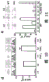


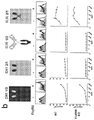


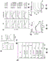


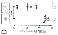


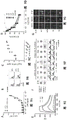


Classifications
-
- C—CHEMISTRY; METALLURGY
- C07—ORGANIC CHEMISTRY
- C07K—PEPTIDES
- C07K16/00—Immunoglobulins [IGs], e.g. monoclonal or polyclonal antibodies
- C07K16/18—Immunoglobulins [IGs], e.g. monoclonal or polyclonal antibodies against material from animals or humans
- C07K16/28—Immunoglobulins [IGs], e.g. monoclonal or polyclonal antibodies against material from animals or humans against receptors, cell surface antigens or cell surface determinants
- C07K16/2878—Immunoglobulins [IGs], e.g. monoclonal or polyclonal antibodies against material from animals or humans against receptors, cell surface antigens or cell surface determinants against the NGF-receptor/TNF-receptor superfamily, e.g. CD27, CD30, CD40, CD95
-
- C—CHEMISTRY; METALLURGY
- C07—ORGANIC CHEMISTRY
- C07K—PEPTIDES
- C07K16/00—Immunoglobulins [IGs], e.g. monoclonal or polyclonal antibodies
- C07K16/18—Immunoglobulins [IGs], e.g. monoclonal or polyclonal antibodies against material from animals or humans
- C07K16/28—Immunoglobulins [IGs], e.g. monoclonal or polyclonal antibodies against material from animals or humans against receptors, cell surface antigens or cell surface determinants
- C07K16/2803—Immunoglobulins [IGs], e.g. monoclonal or polyclonal antibodies against material from animals or humans against receptors, cell surface antigens or cell surface determinants against the immunoglobulin superfamily
- C07K16/2818—Immunoglobulins [IGs], e.g. monoclonal or polyclonal antibodies against material from animals or humans against receptors, cell surface antigens or cell surface determinants against the immunoglobulin superfamily against CD28 or CD152
-
- A—HUMAN NECESSITIES
- A61—MEDICAL OR VETERINARY SCIENCE; HYGIENE
- A61P—SPECIFIC THERAPEUTIC ACTIVITY OF CHEMICAL COMPOUNDS OR MEDICINAL PREPARATIONS
- A61P29/00—Non-central analgesic, antipyretic or antiinflammatory agents, e.g. antirheumatic agents; Non-steroidal antiinflammatory drugs [NSAID]
-
- A—HUMAN NECESSITIES
- A61—MEDICAL OR VETERINARY SCIENCE; HYGIENE
- A61P—SPECIFIC THERAPEUTIC ACTIVITY OF CHEMICAL COMPOUNDS OR MEDICINAL PREPARATIONS
- A61P31/00—Antiinfectives, i.e. antibiotics, antiseptics, chemotherapeutics
-
- A—HUMAN NECESSITIES
- A61—MEDICAL OR VETERINARY SCIENCE; HYGIENE
- A61P—SPECIFIC THERAPEUTIC ACTIVITY OF CHEMICAL COMPOUNDS OR MEDICINAL PREPARATIONS
- A61P35/00—Antineoplastic agents
-
- A—HUMAN NECESSITIES
- A61—MEDICAL OR VETERINARY SCIENCE; HYGIENE
- A61P—SPECIFIC THERAPEUTIC ACTIVITY OF CHEMICAL COMPOUNDS OR MEDICINAL PREPARATIONS
- A61P37/00—Drugs for immunological or allergic disorders
-
- A—HUMAN NECESSITIES
- A61—MEDICAL OR VETERINARY SCIENCE; HYGIENE
- A61P—SPECIFIC THERAPEUTIC ACTIVITY OF CHEMICAL COMPOUNDS OR MEDICINAL PREPARATIONS
- A61P37/00—Drugs for immunological or allergic disorders
- A61P37/02—Immunomodulators
- A61P37/06—Immunosuppressants, e.g. drugs for graft rejection
-
- A—HUMAN NECESSITIES
- A61—MEDICAL OR VETERINARY SCIENCE; HYGIENE
- A61P—SPECIFIC THERAPEUTIC ACTIVITY OF CHEMICAL COMPOUNDS OR MEDICINAL PREPARATIONS
- A61P37/00—Drugs for immunological or allergic disorders
- A61P37/08—Antiallergic agents
-
- C—CHEMISTRY; METALLURGY
- C07—ORGANIC CHEMISTRY
- C07K—PEPTIDES
- C07K16/00—Immunoglobulins [IGs], e.g. monoclonal or polyclonal antibodies
- C07K16/18—Immunoglobulins [IGs], e.g. monoclonal or polyclonal antibodies against material from animals or humans
- C07K16/28—Immunoglobulins [IGs], e.g. monoclonal or polyclonal antibodies against material from animals or humans against receptors, cell surface antigens or cell surface determinants
- C07K16/30—Immunoglobulins [IGs], e.g. monoclonal or polyclonal antibodies against material from animals or humans against receptors, cell surface antigens or cell surface determinants from tumour cells
-
- A—HUMAN NECESSITIES
- A61—MEDICAL OR VETERINARY SCIENCE; HYGIENE
- A61K—PREPARATIONS FOR MEDICAL, DENTAL OR TOILETRY PURPOSES
- A61K39/00—Medicinal preparations containing antigens or antibodies
- A61K2039/505—Medicinal preparations containing antigens or antibodies comprising antibodies
-
- C—CHEMISTRY; METALLURGY
- C07—ORGANIC CHEMISTRY
- C07K—PEPTIDES
- C07K16/00—Immunoglobulins [IGs], e.g. monoclonal or polyclonal antibodies
- C07K16/18—Immunoglobulins [IGs], e.g. monoclonal or polyclonal antibodies against material from animals or humans
- C07K16/24—Immunoglobulins [IGs], e.g. monoclonal or polyclonal antibodies against material from animals or humans against cytokines, lymphokines or interferons
- C07K16/241—Tumor Necrosis Factors
- C07K16/242—Lymphotoxin [LT]
-
- C—CHEMISTRY; METALLURGY
- C07—ORGANIC CHEMISTRY
- C07K—PEPTIDES
- C07K16/00—Immunoglobulins [IGs], e.g. monoclonal or polyclonal antibodies
- C07K16/18—Immunoglobulins [IGs], e.g. monoclonal or polyclonal antibodies against material from animals or humans
- C07K16/28—Immunoglobulins [IGs], e.g. monoclonal or polyclonal antibodies against material from animals or humans against receptors, cell surface antigens or cell surface determinants
- C07K16/2896—Immunoglobulins [IGs], e.g. monoclonal or polyclonal antibodies against material from animals or humans against receptors, cell surface antigens or cell surface determinants against molecules with a "CD"-designation, not provided for elsewhere
-
- C—CHEMISTRY; METALLURGY
- C07—ORGANIC CHEMISTRY
- C07K—PEPTIDES
- C07K2317/00—Immunoglobulins specific features
- C07K2317/20—Immunoglobulins specific features characterized by taxonomic origin
- C07K2317/24—Immunoglobulins specific features characterized by taxonomic origin containing regions, domains or residues from different species, e.g. chimeric, humanized or veneered
-
- C—CHEMISTRY; METALLURGY
- C07—ORGANIC CHEMISTRY
- C07K—PEPTIDES
- C07K2317/00—Immunoglobulins specific features
- C07K2317/50—Immunoglobulins specific features characterized by immunoglobulin fragments
- C07K2317/52—Constant or Fc region; Isotype
-
- C—CHEMISTRY; METALLURGY
- C07—ORGANIC CHEMISTRY
- C07K—PEPTIDES
- C07K2317/00—Immunoglobulins specific features
- C07K2317/50—Immunoglobulins specific features characterized by immunoglobulin fragments
- C07K2317/52—Constant or Fc region; Isotype
- C07K2317/522—CH1 domain
-
- C—CHEMISTRY; METALLURGY
- C07—ORGANIC CHEMISTRY
- C07K—PEPTIDES
- C07K2317/00—Immunoglobulins specific features
- C07K2317/50—Immunoglobulins specific features characterized by immunoglobulin fragments
- C07K2317/52—Constant or Fc region; Isotype
- C07K2317/53—Hinge
-
- C—CHEMISTRY; METALLURGY
- C07—ORGANIC CHEMISTRY
- C07K—PEPTIDES
- C07K2317/00—Immunoglobulins specific features
- C07K2317/70—Immunoglobulins specific features characterized by effect upon binding to a cell or to an antigen
-
- C—CHEMISTRY; METALLURGY
- C07—ORGANIC CHEMISTRY
- C07K—PEPTIDES
- C07K2317/00—Immunoglobulins specific features
- C07K2317/70—Immunoglobulins specific features characterized by effect upon binding to a cell or to an antigen
- C07K2317/75—Agonist effect on antigen
-
- C—CHEMISTRY; METALLURGY
- C07—ORGANIC CHEMISTRY
- C07K—PEPTIDES
- C07K2317/00—Immunoglobulins specific features
- C07K2317/70—Immunoglobulins specific features characterized by effect upon binding to a cell or to an antigen
- C07K2317/76—Antagonist effect on antigen, e.g. neutralization or inhibition of binding
Abstract
By a combination of in vitro and in vivo approaches, the inventors showed that human IgG2(h2) delivered unique FcyR-independent agonist activity to anti-CD 40 antibodies or antibodies specific for other immunostimulatory receptors including 4-1BB and CD 28. Studies with anti-human CD40mAb, LOB7.4, showed that the unique activity of h2 was dependent on the precise alignment of the hinge and CH1 disulfide bonds. Chemical 'shuffling' or mutagenesis, which 'locks' LOB7.4 into a more flexible 'h 2A' or tighter 'h 2B' conformation, confers antagonistic and agonistic properties, respectively. Engineering h2 in this way allowed the development of agents with immunostimulatory or immunosuppressive characteristics, which directly suggested the design of therapeutic mAb agents and fusion proteins.
Description
Priority
The present application claims priority from uk application No. 1405264.1 filed 24/3 2014 and uk application No. 1405275.7 filed 25/3 2014, the contents of which are incorporated herein by reference in their entirety.
FIELD
The present application relates generally to the discovery that human IgG2(h2) delivers unique Fc γ R-independent agonist activity to anti-CD 40 antibodies and other immunostimulatory receptor antibodies, including 4-1BB and CD 28; and possibly other B7/CD28 and TNF/TNFR families The members, and the additional finding that the unique agonistic or conversely antagonistic activity of h2 is dependent on hinge and C H1 precise alignment of disulfide bonds. Based on this finding, the present invention provides novel agonistic and antagonistic antibodies and their use in therapy, wherein the hinge region of IgG2 is mutagenized to 'lock' the antibody into a more flexible 'h 2A' or more compact 'h 2B' conformation in order to confer antibody antagonistic and agonistic properties, respectively.
Background
Monoclonal antibodies (mabs) are revolutionary cancer treatments that reignite the belief that the immune system can be used to eradicate tumors (Sliwkowski et AL, (2013); Hodi FS et AL, (2010); Wolchok JD et AL, (2013); results using anti-CTLA-4 and anti-PD-1 mabs have demonstrated that T-cell immunity can provide long-lasting protection against aggressive and difficult to treat cancers such as metastatic melanoma and non-small cell lung cancer (Hodi FS et AL, (2010); Wolchok JD et AL, (2013); Topalian SL et AL, (2012); different mAb agents mediate their effects in different ways and improved therapeutic efficacy requires precise understanding of their mechanism of action.a. furthermore, the choice of isotype mabs is critical primarily due to differences in Fc γ receptor (2012 γ Fc γ R) interactions that direct antigen-binding downstream events, (2013).
Agents act by depleting their cellular targets, such as malignant B cells with anti-CD 20 (Uchida J et al, (2004)) or murine T regulatory cells with anti-CTLA 4 and anti-GITR (Simpson TR et al, (2013) Bulliard Y et al, (2013). for this type of agent, mouse IgG2a (m2a) and human IgG1(h1) are effective because they have a high agonistic/inhibitory (A/I) Fc γ R binding ratio (Hamaguchi Y et al, The Journal of experimental media 203(3): 743-; Nimmermarjahn F & ravatch JV (2005)). in contrast, their action depends on mAbs that agonize receptors to engage such as immunostimulatory anti-CD 40(Brahmer et al, (Bruhns et al, Blohn 113,3716 or 3725 (JV 372012) and promote apoptosis (DR 5, Wilveson et al), (2011) xu Y et AL, (2003) appear to rely primarily on crosslinking with inhibitory Fc γ RIIB to deliver its activity (White AL et AL, (2011); Li F & ravatch JV (2013), White et AL, (2013)). For this type of agent, mouse IgG1(m1) was optimal in preclinical models because it had a low a/I ratio and conjugated Fc γ RIIB with sufficient affinity to mediate cross-linking (White et al, (2011); Li F & ravatch JV (2011)). Agonistic anti-CD 40 mAb CP870,893(von dehidede RH et al, (2007)) in clinical trials is a human IgG2(h2) that is not predicted to engage FcR based on its isotype (Bruhns P et al, (2009). Two other less agonistic CD40 antibodies, chilob7.4 and SGN40, were both h1(Advani R et al, (2009); Johnson et al, (2010)). In addition, recent studies have shown that the h2 constant region confers increased stimulatory activity on specific anti-human CD28 mAb (TGN1412) (Ball C et al, (2012)).
Despite this, it was not clear prior to the present invention that the optimal isoform to be used in human therapy should be used in an agonistic mAb or fusion protein. In particular, there is less understanding of how antibody isotypes can achieve the therapeutic properties of a therapeutic antibody, i.e., its agonistic or antagonistic properties. The present invention addresses this need, provides improved agonistic (and antagonistic) antibodies, and also provides means for synthesizing the antibodies. In an exemplary embodiment, these antibodies and fusion proteins, such as those targeting TNFR superfamily members, are useful for therapeutic indications where either agonistic or antagonistic antibodies or fusion proteins are therapeutically desirable.
SUMMARY
The present invention provides an understanding of the optimal antibody isotype for generating antibodies with agonistic or antagonistic properties. More specifically, the invention provides antibodies that protect the constant region of human IgG2 with specific mutations, wherein the antibodies have agonistic or antagonistic properties based on the specific mutations.
The present invention specifically utilizes mabs against multiple human co-stimulatory receptors (CD40, 4-1BB and CD28) to determine the immunomodulatory activity of human IgG isotypes and thus shows that human IgG2(h2) confers greater immunostimulatory activity to these agents than h1 or human h4, and that this activity is not dependent on differences in FcgR interactions but rather on the conformation of the disulfide bonds in the h2 hinge and CH1 domains, which can be manipulated using mutagenesis to 'lock' the mAb into different conformations with opposite levels of agonistic levels, thereby providing a cognate superagonionist therapeutic antibody with a defined level of activity that is independent of FcgR expression levels in the local microenvironment.
The invention also relates to the use of such antibodies and fusion proteins comprising the human IgG2 constant region with specific mutations as receptor agonists or receptor antagonists in the treatment or prevention of disease, wherein such receptor agonists or receptor antagonists have therapeutic applications. In exemplary embodiments, the antibody will specifically bind to a TNF superfamily member, such as CD40, LT α, LT β, FASL CD30, CD27, OX40, TRAIL/APO-2L, 4-1BB, 4-1BBL, TNF-R, TRANCE-R, GITR or "glucocorticoid-induced TNF receptor", TWEAK, FN14, and the like; or to join B7/CD28 family members such as B7.1(CD80), B7.2(CD86), B7-DC (PD-L2 or CD273), B7-H1, B7-H2, B7-H3(CD276), B7-H4(VTCN1), B7-H5(VISTA), B7-H6(NCR3LG1), B7-H7(HHLA2), PD-1(CD279), PD-L3, CD28, CTLA-4(CD152), ICOS (CD278), BTLA, NCR3, CD28H, and NKp 30.
Object of the Invention
It is an object of the present invention to provide novel immune agonists or antagonists, and more particularly to superagonists.
It is a more specific object of the invention to provide novel immune agonists or antagonists comprising a mutated IgG2 constant region, wherein such mutations enhance the agonistic or antagonistic properties of such agonist or antagonist polypeptides, such as an agonistic or antagonistic IgG2 antibody or an IgG2/IgG2 chimeric fusion protein.
It is another specific object of the invention to provide modified agonistic or antagonistic antibodies of the non-human IgG2 isotype, e.g., human IgG1, IgG3, IgG4, IgA, IgD, IgE or IgM antibodies, wherein the entire or substantially the entire hinge and C of hIgG2 H1 region and optionally the light chain constant region of hIgG2 for replacement of a non-human IgG2 antibody, e.g., human IgG1, IgG3, IgG4, IgA, IgD, IgE, or IgM corresponding light chain constant region, hinge, and C H1 domain or region.
It is a more specific object of the invention to provide an agonistic antibody that specifically binds to a receptor or ligand expressed on human immune cells, wherein the antibody agonizes one or more biological activities elicited by the receptor or ligand, or by the interaction of the ligand and receptor, and further wherein the agonistic antibody comprises at least the C of human IgG2 H1 and a hinge region ("H2 region"), wherein one or more cysteine residues of the IgG2 region are removed or changed to a different amino acid residue, thereby increasing the agonistic properties of the resulting agonistic antibody relative to an otherwise identical antibody in which the one or more cysteines are unchanged.
It is another specific object of the invention to provide a modified antibody that specifically binds to a receptor or ligand expressed on the surface of a human cell, wherein said modified antibody agonizes one or more biological activities elicited by said receptor or ligand or by the interaction of said ligand and its corresponding receptor, wherein said antibody is not a human IgG2 antibody, and further wherein the whole or substantially the whole hinge and C of said modified antibody is not human IgG2 H1 domain and optionally the light chain constant region is replaced by the entire or substantially the entire hinge and C of the hIgG2 antibody H1 domain ("H2 region" or "H2 domain") and optionally a light chain constant region.
It is another specific object of the present invention to provide a modified antibody selected from the group consisting of hIgG1, hIgG3, hIgG4, IgA, IgD, IgE, or IgM as described above, wherein the whole or substantially the whole hinge and C of said antibody H1 Domain has been replaced by hIgG2 corresponding to the entire or substantially entire hinge and C H1 domain ("H2 region" or "H2 domain").
It is another specific object of the present invention to provide the modified antibody as described above, wherein either or both of the heavy chain cysteine residue at position 127 and the light chain cysteine residue at position 214 (wherein numbering is according to Kabat) are deleted or changed to a different amino acid residue, such that the agonistic properties of the resulting modified antibody are increased relative to an otherwise identical antibody wherein either or both of the cysteine residues are unchanged.
It is another specific object of the present invention to provide the modified antibody as described above, wherein the only cysteine residue mutated or deleted in the H2 region of the modified antibody is the cysteine at position 214 of the light chain and the modified antibody comprises (i) a heavy chain wherein no cysteine residues are deleted or modified in the H2 region or (ii) one or more of the cysteine residues at positions 127, 232 or 233 of the heavy chain are deleted or mutated, thereby producing a modified antibody wherein the agonistic properties of the resulting modified antibody are increased relative to an otherwise identical antibody lacking said deletion or modification.
It is another specific object of the present invention to provide the modified antibody as described above, which comprises a modified hIgG1 or hIgG 3.
It is another specific object of the present invention to provide the modified antibody as described above, which specifically binds to human immune cells.
It is another specific object of the present invention to provide the modified antibody as described above, which binds to dendritic cells, mast cells, monocytes, macrophages, NK cells, B lymphocytes, T lymphocytes or any combination thereof.
It is another specific object of the present invention to provide the modified antibody as described above, wherein said antibody lacks agonist activity in the absence of said modification.
It is another specific object of the present invention to provide the modified antibody as described above, wherein said antibody has no more than 10% of the agonistic activity of said modified antibody in the absence of said modification, wherein agonistic activity is detected in an accepted assay for quantifying agonism.
It is another specific object of the present invention to provide the modified antibody as described above, wherein said antibody has no more than 20% of the agonistic activity of said modified antibody in the absence of said modification, wherein agonistic activity is detected in an accepted assay for quantifying agonism.
It is another specific object of the present invention to provide the modified antibody as described above, wherein said antibody has no more than 30% of the agonistic activity of said modified antibody in the absence of said modification, wherein agonistic activity is detected in an accepted assay for quantifying agonism.
It is another specific object of the present invention to provide the modified antibody as described above, wherein said antibody has no more than 40% of the agonistic activity of said modified antibody in the absence of said modification, wherein agonistic activity is detected in an accepted assay for quantifying agonism.
It is another specific object of the present invention to provide the modified antibody as described above, wherein said antibody has no more than 50% of the agonistic activity of said modified antibody in the absence of said modification, wherein agonistic activity is detected in an accepted assay for quantifying agonism.
It is another specific object of the present invention to provide the modified antibody as described above, wherein said antibody has no more than 50% -80% of the agonistic activity of said modified antibody in the absence of said modification, wherein agonistic activity is detected in an acceptable assay for quantifying agonism.
It is another specific object of the present invention to provide the modified antibody as described above, wherein said antibody agonizes one or more biological activities elicited by a human receptor or ligand or by the interaction of said ligand and its corresponding receptor, and further wherein said modified antibody comprises at least the light chain constant region of hIgG2 and the C of the heavy chain H1 and a hinge region ("H2 region"), wherein either or both of the cysteine residues selected from the heavy chain cysteine residue at position 127 and the light chain cysteine residue at position 214 are removed or changed to a different amino acid residue, such that the agonistic properties of the resulting agonistic antibody are increased relative to an otherwise identical antibody in which both of the cysteine residues are unchanged (wherein the amino acid numbering is according to Kabat).
It is another specific object of the present invention to provide the modified antibody as described above, wherein the cysteine residue at position 214 of the light chain of the modified antibody is deleted or mutated and the modified antibody comprises (i) a heavy chain wherein no cysteine residues are deleted or mutated within the region of H2 or (ii) a heavy chain wherein at least one of the cysteine residues at positions 127, 232 or 233 is deleted or mutated.
It is another specific object of the present invention to provide the modified antibody as described above, wherein no cysteine residues are deleted or mutated in the H2 region of the heavy chain.
It is another specific object of the present invention to provide the modified antibody as described above, wherein at least one of the cysteine residues at positions 127, 232 or 233 within the heavy chain is deleted or mutated.
It is another specific object of the present invention to provide the modified antibody as described above, wherein any combination of one or two of the cysteine residues at positions 127, 232 or 233 within the heavy chain is deleted or mutated.
It is another specific object of the present invention to provide the modified antibody as described above, wherein the H2 region of the modified antibody is in the H2B conformation.
It is another specific object of the present invention to provide the modified antibody as described above, wherein the only cysteine residue in the H2 region of the heavy chain of hIgG2 that has been removed, modified or substituted with another amino acid residue comprises the heavy chain cysteine residue at position 127.
It is another specific object of the present invention to provide the modified antibody as described above, wherein no cysteine residue in the light chain region is removed, modified or substituted with another amino acid residue.
It is another specific object of the present invention to provide the modified antibody as described above, wherein the only cysteine residue in the hIG2 light chain constant region that has been removed, modified or substituted with another amino acid residue comprises the light chain cysteine residue at position 214.
It is another specific object of the present invention to provide the modified antibody as described above, wherein the only cysteine residue in the hIgG 2H 2 region that is removed, modified or substituted with another amino acid residue is selected from the group consisting of the heavy chain cysteine residue at position 127 and the light chain cysteine residue at position 214.
It is another specific object of the present invention to provide a modified antibody as described above, which comprises a serine substituted for the cysteine at position 214 of the light chain and/or a serine residue substituted for the cysteine residue at position 127 of the heavy chain.
It is another specific object of the present invention to provide a modified antibody of the hIgG1 or hIgG3 or hIgG4 isotype as described above, wherein the entire hinge or C of the hIgG1, hIgG3 or hIgG4HRegion 1 is hinged by hIgG2 and CHArea 1 replacement.
It is another specific object of the present invention to provide the modified antibody as described above, which is hIgG 1.
It is another specific object of the present invention to provide a modified antibody as described above, which comprises one or more cysteine residues within the light or heavy chain H2 region.
It is another specific object of the present invention to provide a modified antibody as described above, comprising an additional modification outside the H2 region, which does not affect or perceptibly affects (no more than a 10% change) the agonistic properties of said modified antibody.
It is another specific object of the present invention to provide a modified antibody as described above, which specifically binds to TNF or a member of the B7 superfamily expressed on the surface of human cells, such as CD40, 4-1BB or CD 28.
It is another specific object of the present invention to provide the modified antibody as described above, which specifically binds to a receptor or ligand expressed on at least one type of human immune cell.
It is another specific object of the present invention to provide the modified antibody as described above, which binds to an immune cell selected from the group consisting of: t lymphocytes, B lymphocytes, monocytes, mast cells, macrophages, NK cells, and dendritic cells, or a combination thereof.
It is another specific object of the present invention to provide the modified antibody as described above, which comprises the entire human IgG2 CH1 and the hinge region.
It is another specific object of the present invention to provide the modified antibody as described above, which lacks said human IgG2 Fc region.
It is another specific object of the invention to provide the modified antibody as described above, wherein the agonistic properties of the antibody or fusion are Fc γ R independent.
It is another specific object of the present invention to provide the modified antibody as described above, wherein said antibody specifically binds to a member of the TNFR superfamily selected from CD40, LT α, LT β, CD30, CD27, OX40, 4-1BB, TNF-40-40 or "glucocorticoid-induced TNF receptor", TWEAK, and FN 40 or to a member of the TNFR superfamily selected from B7.1(CD 40), B7.2(CD 40), B40-H40 (CD276), B40-H40 (VTCN 40), B40-H40 (VISTA), B40-H40 (NCR3LG 40), B40-H40 (HHLA 40), PD-1(CD279), CD40, CTLA-4(CD152), ICOS (CD278), BTLA, NCR 40, CD 3628, NKp 40, and nkb/40 family.
It is another specific object of the present invention to provide the modified antibody as described above, which binds a TNFR member selected from 4-1BB, CD40 or CD27 or a B7/CD28 family member is CTLA-4 or CD 28.
It is another specific object of the present invention to provide a modified antibody as described above, which comprises the variable region of LOB 7.4.
It is another specific object of the present invention to provide a pharmaceutical composition comprising a modified antibody as described above, said pharmaceutical composition comprising a pharmaceutically effective amount of such a modified antibody.
It is another specific object of the invention to provide the use of such antibodies or such compositions with other immune agonists.
It is another specific object of the present invention to provide the use of the modified antibody as described above in a method of treatment comprising administering at least one modified antibody or a fusion protein containing said modified antibody, said method comprising administering an effective amount of at least one modified antibody or a fusion protein or composition containing a modified antibody according to the invention. In preferred embodiments, the condition being treated is selected from cancer, infection, allergy, autoimmunity, transplantation, GVHD or inflammation.
Another specific object of the present invention is to provide a method for specifically binding to the surface of human cells A modified antibody to the receptor or ligand expressed thereon, wherein the modified antibody antagonizes one or more biological activities caused by the receptor or ligand or caused by the interaction of the ligand and its corresponding receptor, wherein the antibody is not a human IgG2 antibody, and further wherein the whole or substantially the whole hinge and C of the modified antibody is not human IgG2 H1 domain and optionally the light chain constant region is replaced by the entire or substantially the entire hinge and C of the hIgG2 antibody H1 domain and a light chain constant region ("H2 region" or "H2 domain"), wherein one or more cysteine residues within the H2 region are optionally modified, which modification results in an increase in the antagonistic properties of the resulting modified antibody.
It is another specific object of the present invention to provide a modified antagonistic antibody selected from the group consisting of hIgG1, hIgG3, hIgG4, IgA, IgD, IgE, or IgM as described above, wherein the whole or substantially the whole hinge and C of said antibody H1 domain and optionally the light chain constant region has been replaced by all or substantially all of the hinge and C of hIgG2 H1 domain, and a light chain constant region ("H2 region" or "H2 domain").
It is another specific object of the present invention to provide the modified antagonist antibody as described above, wherein at least one cysteine residue selected from the cysteine residues at positions 232, 233, 236 and 239 of the IgG 2H 2 region is removed or changed to a different amino acid residue such that the antagonistic properties of the resulting modified antibody are increased relative to an otherwise identical antibody in which the one or more cysteine residues are not changed.
It is another specific object of the present invention to provide the modified antagonist antibody as described above, wherein the cysteine at residue 214 of the light chain and the cysteine at residue 127 of the heavy chain of the modified antibody are not deleted, modified or substituted with another amino acid residue.
It is another specific object of the present invention to provide the modified antagonist antibody as described above, wherein the cysteine modification within the H2 region of the antibody selectively facilitates or causes association of the heavy and light chains of the modified antibody via the cysteine at position 214 of the light chain and the cysteine at position 127 of the heavy chain.
It is another specific object of the present invention to provide the modified antagonist antibody as described above, wherein the H2 region of the modified antibody is in the H2A conformation.
It is another specific object of the present invention to provide the modified antagonist antibody as described above, wherein at least one cysteine residue other than C214 and C127 in the region of H2 is changed to a serine residue.
It is another specific object of the present invention to provide a modified antagonist antibody as described above, comprising a serine substituted for the cysteine at position 232 of the heavy chain.
It is another specific object of the present invention to provide the modified antagonist antibody as described above, which comprises a serine substituted for the cysteine at position 233 of the heavy chain.
It is another specific object of the present invention to provide a modified antagonist antibody as described above, comprising a modified hIgG1 or hIgG 3.
It is another specific object of the present invention to provide the modified antagonist antibody as described above, which specifically binds to human immune cells.
It is another specific object of the present invention to provide the modified antagonist antibody as described above, which binds to immune cells, including at least one of dendritic cells, mast cells, monocytes, macrophages, NK cells, B lymphocytes, T lymphocytes or any combination thereof.
It is another specific object of the present invention to provide a modified antagonistic antibody as described above, wherein said antibody lacks antagonistic activity in the absence of said modification.
It is another specific object of the present invention to provide a modified antagonistic antibody as described above, wherein in the absence of said modification said antibody has no more than 10% of the antagonistic activity of said modified antibody, wherein the antagonistic activity is detected in an acceptable assay for quantifying the antagonistic activity.
It is another specific object of the present invention to provide a modified antagonistic antibody as described above, wherein in the absence of said modification said antibody has no more than 20% of the antagonistic activity of said modified antibody, wherein the antagonistic activity is detected in an acceptable assay for quantifying the antagonistic activity.
It is another specific object of the present invention to provide a modified antagonistic antibody as described above, wherein in the absence of said modification said antibody has no more than 30% of the antagonistic activity of said modified antibody, wherein the antagonistic activity is detected in an acceptable assay for quantifying the antagonistic activity.
It is another specific object of the present invention to provide a modified antagonistic antibody as described above, wherein in the absence of said modification said antibody has no more than 40% of the antagonistic activity of said modified antibody, wherein the antagonistic activity is detected in an acceptable assay for quantifying the antagonistic activity.
It is another specific object of the present invention to provide a modified antagonistic antibody as described above, wherein in the absence of said modification said antibody has no more than 50% of the antagonistic activity of said modified antibody, wherein antagonistic activity is detected in an acceptable assay for quantifying antagonism.
It is another specific object of the present invention to provide the modified antagonist antibody as described above, which specifically binds to a receptor or ligand expressed on human cells, wherein said antibody antagonizes one or more biological activities elicited by said receptor or ligand, or by the interaction of said ligand with its corresponding receptor, and further wherein said antagonist antibody comprises at least the C of human IgG2 H1 and a hinge and light chain constant region ("H2 region"), wherein at least one cysteine residue of the IgG 2H 2 region selected from the cysteine residues at positions 232, 233, 236 and 239 is removed or changed to a different amino acid residue such that the antagonistic properties of the resulting antagonistic antibody are increased relative to an otherwise identical antibody in which the one or more cysteine residues are unchanged.
It is another specific object of the present invention to provide the modified antagonist antibody as described above, wherein the cysteine residue at position 214 of the light chain and at position 127 of the heavy chain is not deleted, modified or substituted with another amino acid.
It is another specific object of the present invention to provide the modified antagonist antibody as described above, wherein one or more cysteine residues or other amino acid residues within the hIgG 2H 2 region are removed, modified or substituted with another amino acid residue such that the H2 region assumes the H2A conformation.
It is another specific object of the present invention to provide the modified antagonist antibody as described above, wherein at least one of said cysteine residues is changed to a serine residue within the hIgG 2H 2 region.
It is another specific object of the present invention to provide a modified antagonist antibody as described above, comprising a serine substituted for the cysteine at position 232 of the heavy chain.
It is another specific object of the present invention to provide the modified antagonist antibody as described above, which comprises a serine substituted for the cysteine at position 233 of the heavy chain.
It is another specific object of the present invention to provide the modified antagonist antibody as described above, which specifically binds to a TNF or TNFR superfamily member or a B7 family member expressed on the surface of human cells.
It is another specific object of the present invention to provide the modified antagonist antibody as described above, which specifically binds at least one type of human immune cell.
It is another specific object of the present invention to provide the modified antagonist antibody as described above, which specifically binds to a receptor or ligand expressed on T cells, B cells, mast cells, macrophages, monocytes, NK cells or dendritic cells.
It is another specific object of the invention to provide a modified antagonist antibody as described above, comprising the entire human IgG2 constant heavy and light chain constant regions.
It is another specific object of the present invention to provide a modified antagonist antibody as described above, which lacks the human IgG2 Fc region.
It is another specific object of the present invention to provide the modified antagonist antibody as described above, wherein the antagonistic properties of said antibody are Fc γ R independent.
It is another specific object of the present invention to provide the modified antagonist antibody as described above, wherein said antibody specifically binds to a member of the TNF/R superfamily selected from: CD40, CD40L (CD154), LT α, LT β, FASL (CD178), CD30, CD30L (CD153), CD27, CD27L (CD70), OX40, OX40L, TRAIL/APO-2L, 4-1BB, 4-1BBL, TNF-R, TRANCE-R, GITR or "glucocorticoid-induced TNF receptor", TWEAK, and FN 14.
It is another specific object of the present invention to provide the modified antagonist antibody as described above, wherein the TNF/R or B7 family member is 4-1BB, 4-1BBL, CD40, CD40L, CD27, CD28, B7.1, B7.2, or CD 70.
It is another specific object of the present invention to provide a pharmaceutical composition comprising a modified antagonistic antibody as described above, said pharmaceutical composition comprising an effective amount of the modified antibody or of a fusion protein comprising a modified antibody according to the present invention.
It is another specific object of the present invention to provide a pharmaceutical composition containing a modified antagonist antibody as described above, which may comprise another immune agonist or other active agent, or use thereof.
It is another specific object of the invention to provide the use of the modified antagonist antibody as described above in a method of treatment comprising administering at least one antagonist antibody or a fusion protein or composition containing an antibody according to the invention, wherein such conditions preferably include cancer, allergy, infectious disease, autoimmunity, transplantation, GVHD or inflammatory conditions.
It is another specific object of the invention to provide a modified antagonist antibody as described above or a use thereof comprising administering an antibody comprising the variable region of LOB7.4 or another antibody specific for CD40, 4-1BB or CD28, which antibody is useful for treating conditions such as cancer, infection, allergy, autoimmunity or inflammation.
It is another specific object of the invention to provide novel IgG2 agonist antibodies or fusion proteins containing the same, which comprise the entire human IgG2 constant region, which are modified in the hinge region to alter disulfide shuffling.
Another particular object of the invention is a novel IgG2 agonist antibody or fusion protein comprising said antibody, which antibody does not contain human IgG2 Fc or another Fc region.
It is another specific object of the invention to provide novel antagonistic or agonistic antibodies in which all or substantially all of the hinge and C of human IgG2 H1 region and optionally the light chain constant region of hIgG2 for replacing the same domain or region of human IgG1, IgG3, IgG4, IgA or IgM, optionally wherein the introduced hIgG2 hinge and/or hIgG 2CHThe 1 region and the light chain constant region may comprise one or more mutations that enhance the agonistic or antagonistic properties of the resulting modified antibody.
It is another specific object of the invention to provide novel agonistic or antagonistic antibodies, wherein the agonistic properties of the antibodies or fusions are Fc γ R independent.
It is another specific object of the invention to provide novel agonistic or antagonistic antibodies or their use in therapy from humans, wherein said antibodies specifically bind to a member of the TNFR superfamily selected from: CD40, LT α, LT β, CD30, CD27, OX40, 4-1BB, TNF-R, TRANCE-R, GITR or the "glucocorticoid-induced TNF receptor", TWEAK, and FN 14.
Brief Description of Drawings
Fig. 1(a) -fig. 1(f) contain experimental results showing that human IgG2 confers independent agonistic activity on anti-hCD 40 mAb, LOB7.4 Fc γ R.
Fig. 2(a) -fig. 2(c) contain experimental results showing that human IgG2 confers independent activity on anti-TNFRSF mAb Fc γ R.
FIGS. 3(a) -3 (d) include displays h 2CH1 and hinge region confer LOB7.4 Fc γ R-independent agonistic activity.
Fig. 4(a) -fig. 4(e) contain experimental results showing that the agonistic activity of LOB7.4 h2 is dependent on its ability to form the h2B conformation.
Fig. 5(a) -fig. 5(e) contain experiments showing isotype dependence of anti-mouse CD40 mAb, 3/23.
Fig. 6(a) -6 (e) contain experimental results demonstrating that LOB7.4 human IgG2 is more active than human IgG1 on human cells and independent of Fc γ R interactions.
Fig. 7(a) -7 (d) contain experimental results demonstrating antagonistic and agonistic properties of h2A and h 2B.
Fig. 8(a) -fig. 8(h) contain experiments showing the effect of different human isoforms on anti-CD 40 activity.
Fig. 9(a) -9 (g) contain control experiments that further validate the effect of the isoforms on the anti-CD 40 activity of the experiments in fig. 8.
Fig. 10(a) -fig. 10(e) contain experimental data showing Fc γ R independent activity of human IgG 2.
Fig. 11(a) -11 (f) contain additional experimental data to verify that ChiLob7/4h2 agonist activity is Fc γ R-independent.
Fig. 12(a) -12 (e) contain data showing antagonism of human IgG2 against multiple receptor targets, indicating that human IgG2 is agonistic for multiple receptor targets.
FIGS. 13(a) and 13(b) contain experiments showing that CH1 and the hinge region confer ChiLob7/4h2 activity.
Fig. 14 contains a control experiment that demonstrates that the ChiLob7/4 switch mutant binds similarly to CD 40.
Fig. 15(a) -fig. 15(i) summarize the results of the mutagenesis experiments showing that mutagenesis generated a series of agonist forms of ChiLob 7/4h 2.
Fig. 16(a) -16 (g) contain additional experimental data to validate the different activities of the ChiLob 7/4h2A and h2B forms.
Figure 17 contains an exemplary representation of the h2 subtype linked by different disulfide bonds (adapted from (Martinez 2008). left panel shows the structure of h2A, which is considered to be the synthetic form of h2, and right panel h2B shows the main alternating arrangement.
FIG. 18(a) exemplary representation of a scheme comprising different disulfide-linked h2 subtypes (adapted from (Martinez 2008), FIG. 18(b) shows nrCE-SDS curves specifying ChiLob 7/4mAb, showing disulfide shuffling within the hinge of human IgG2, indicating the h2A and h2B positions and the 10kDa standard.
Fig. 19, 20 and 21 illustrate the method by which mutagenesis can 'lock' an antibody into the h2a and h2B forms.
Fig. 22, 23, 24 and 25 contain experimental data showing the manner in which mutagenesis of h2 produced different agonists and antagonists.
Figure 26 contains experimental data indicating that agonistic or antagonistic properties of h2B versus the h2A form may be involved in antigen capping or NFkB activation (indicating that multimerization may overcome the need for Fc γ R x-ligation).
Figure 27 contains experimental data showing the immunostimulatory properties of anti-CD 40 antibodies in different isotype formats.
Figure 28 contains B cell assay results showing the effect of different isotype forms of anti-CD 40 antibody on B cell proliferation.
Detailed description of the invention
Definition of
As stated, the present invention relates broadly to novel agonistic or antagonistic antibodies and fusion proteins, wherein the antibody or fusion protein comprises a human IgG2 constant domain, wherein the antibody comprises a specific mutation that results in the antibody or fusion protein having enhanced agonist or antagonist properties, and the manufacture and use thereof in therapy.
It is to be understood that the invention is not limited to particular methods, protocols, cell lines, animal species or genera, and reagents described, as such may vary. It is also to be understood that the terminology used herein is for the purpose of describing particular embodiments only, and is not intended to limit the scope of the present invention, which will be limited only by the appended claims. As used herein, the singular forms "a" and "the" include plural referents unless the context clearly dictates otherwise. Thus, for example, reference to "a cell" includes a plurality of such cells and reference to "a protein" includes reference to one or more proteins and equivalents thereof known to those skilled in the art, and so forth. Unless otherwise defined, all technical and scientific terms used herein have the same meaning as commonly understood by one of ordinary skill in the art to which this invention belongs.
"agonist" refers to a compound that, in combination with a receptor, produces a cellular response. An agonist may be a ligand that binds directly to a receptor. Alternatively, an agonist may be indirectly combined with a receptor, for example, by: (a) forming a complex with another molecule that directly binds to the receptor, or (b) otherwise causing a modification of another compound such that the other compound directly binds to the receptor. An agonist may be referred to as an agonist of a particular receptor or family of receptors (e.g., a TNF or TNFR agonist). A "superagonist" is an agonist with mutations in the hinge and cysteine in the CH1 region to provide optimal agonist properties.
An "antagonist" is a compound that, when contacted with a molecule of interest, e.g., a member of the TNF or TNFR family superfamily or other ligand or receptor, causes a reduction in the size of a certain activity or function of the molecule compared to the size of the activity or function observed in the absence of the antagonist. Specific antagonists of interest include antibodies and fusion proteins specific for members of the TNFR superfamily that contain IgG2 constant domains.
"antigen" refers to any substance that is capable of being the target of an immune response. An antigen can be, for example, a target of a cell-mediated immune response and/or a humoral immune response elicited by a subject organism. Alternatively, the antigen may be a target for a cellular immune response (e.g., immune cell maturation, cytokine production, antibody production, etc.) upon contact with an immune cell.
As used herein, "TNF/R" refers generally to any member of the Tumor Necrosis Factor (TNF) superfamily or Tumor Necrosis Factor Receptor (TNFR) superfamily. TNF and TNFR superfamily includes, for example, CD40, CD40L (CD154), LT α, LT β, LT- β R, FASL (CD178), CD30, CD30L (CD153), CD27, CD27L (CD70), OX40, OX40L, TRAIL/APO-2L, 4-1BB, 4-1BBL, TNF-R, TNF-R2, TRANCE-R, GITR or "glucocorticoid-induced TNF receptor", GITR ligand, RELT, TWEAK, FN14, TNF α, TNF β, RANK ligand, LILT, HVEM, GITR, TROY, and RELT. Unless otherwise indicated, reference to a TNF/R agonist or antagonist compound may include any pharmaceutically acceptable form of the compound.
"B7 family member" or "B7-CD 28 family member" refers to members of a large family of receptors and ligands expressed on immune cells involved in immune signaling. Typical structural elements common to members of the B7 polypeptide family include extracellular domains, including V-like and C-like Ig domains. The signal sequence was found at the N-terminus of the full-length B7 family polypeptide, and was followed by a V-like Ig domain, a C-like Ig domain, a transmembrane domain, and an intracellular domain in N-to-C order. Within the extracellular domain of the B7 polypeptide, there are certain key residues, namely two pairs of conserved cysteine residues (one pair within each Ig domain) involved in disulfide bond formation and three-dimensional conformation of the polypeptide. The B7 family of polypeptides is moderately conserved, in which the Ig domains of human family members are very similar to each other and to those of B7 family members from other species. The families include subfamilies, including B7-1(CD80), B7-2(CD86), and B7-H1, as well as the cremophil (BTN)/MOG (myelin oligodendrocyte glycoprotein-like) family members, where the immunomodulatory B7 subfamily lacks the B30.2 domain and the cremophil/MOG subfamily has the B30.2 domain. Members of the B7/CD28 superfamily include, as examples, B7.1(CD80), B7.2(CD86), B7-DC (PD-L2 or CD273), B7-H1, B7-H2, B7-H3(CD276), B7-H4(VTCN1), B7-H5(VISTA), B7-H6(NCR3LG1), B7-H7(HHLA2), PD-1(CD279), PD-L3, CD28, CTLA-4(CD152), ICOS (CD278), BTLA, NCR3, CD28H, and NKp 30.
The terms "biological effect associated with X" and "X activity" of e.g., a TNF or TNFR superfamily member or other immune cell receptor are used interchangeably and include any biological effect associated with a moiety that specifically interacts with an agonist or antagonist, e.g., a TNF or TNF/R superfamily member.
The term "host cell" herein generally refers to any cell engineered to express one or more antibodies according to the invention. Such cells include, by way of example, bacterial, fungal, yeast, mammalian, invertebrate such as insect, plant and avian cells.
The term "expression vector" refers to a DNA vector containing elements that facilitate the manipulation of expression of a foreign protein within a target host cell. Typically, manipulation of sequences and production of DNA for transformation is first performed in a bacterial host cell, e.g., e.coli, and the vector will typically comprise sequences that facilitate such manipulation, including a bacterial origin of replication and an appropriate bacterial selectable marker. The selectable marker encodes a protein required for survival or growth of transformed host cells grown in the selection medium. Host cells that are not transformed with a vector containing the selection gene will not survive in the medium. Typical selection genes encode the following proteins: (a) conferring resistance to antibiotics or other toxins to prokaryotic host cells; (b) complement auxotrophy deficiency; or (c) supplying key nutrients not available from complex media. Exemplary vectors and Methods for transforming yeast are described, for example, in Burke, D., Dawson, D., and Stearns, T. (2000). Methods in year genetics: a Cold Spring Harbor Laboratory course Manual. plant view, N.Y.: Cold Spring Harbor Laboratory Press. The polypeptide coding sequence of interest is operably linked to transcriptional and translational regulatory sequences that provide for expression of the polypeptide in yeast cells. These carrier components may include, but are not limited to, one or more of the following: enhancer elements, promoters, and transcription termination sequences. Sequences for secretion of the polypeptide, such as signal sequences and the like, may also be included. The yeast origin of replication is optional, as the expression vector is typically integrated into the yeast genome. In one embodiment of the invention, the polypeptide of interest is operably linked or fused to a sequence that provides for optimal secretion of the polypeptide from a yeast diploid cell.
As used herein, "antibody domain" or "region" refers to different portions or subunits of an antibody molecule. In the case of IgG, the heavy chain has four subunits, "C H3”、“C H2”、“C H1 "(constant part) and VH(variable part). The light chain has two subunits CLAnd VL. Two C H3 units are directly connected, and CHThe 2 units are separated by oligosaccharides. C H1 is located after the heavy chain "hinge" and is linked to C by a disulfide bondLAnd (4) units. "C H2 Domain "is usually derived from the IgG of the order according to the EU numbering systemResidue 231 extends to about 340. C H2 domain is unique in that it is not closely paired with another domain. In contrast, two N-linked branched carbohydrate chains are inserted at the two C's of an intact native IgG molecule H2 domain. It has been speculated that carbohydrates may provide substituents for domain-domain pairing and help stabilize the CH2 domain. Burton, molecular. Immunol.22:161-206 (1985). "CH3Domain "comprises C-terminal residues to C within the Fc regionH2A fragment of a domain (i.e., from about amino acid residue 341 to about amino acid residue 447 of an IgG according to the EU numbering system).
The term "Fc region" generally refers to a dimeric complex comprising the C-terminal polypeptide sequence of an immunoglobulin heavy chain, wherein the C-terminal polypeptide sequence is a polypeptide sequence obtainable by digestion of an intact antibody with papain. The Fc region may comprise a native or variant Fc sequence. Although the boundaries of the Fc sequence of an immunoglobulin heavy chain may vary, the human IgG heavy chain Fc sequence is generally defined as the amino acid residue from about position Cys226 or as a fragment from about position Pro230 to the carboxy terminus of the Fc sequence. The Fc sequence of an immunoglobulin generally comprises two constant domains C H2Domains and CH3A domain, and optionally protects a c.sub.h4 domain. By "Fc polypeptide" herein is meant a polypeptide that constitutes an Fc region, such as a monomeric Fc. The Fc polypeptide may be obtained from any suitable immunoglobulin, such as an IgG1, IgG2, IgG3, or IgG4 subtype, IgA, IgE, IgD, or IgM.
The term "h 2A" conformation refers herein to a specific subtype of IgG2 or modified antibody containing the hinge and CH2 regions of IgG 2. Human IgG2 is unique among immunoglobulins in that it shuffles C H1 and the hinge region ((25-28)). Two subtypes of h2 predominate. h2A has a flexible classical IgG conformation, where CHHeavy Chain (HC) C127 in 1 is linked to C214 in the Light Chain (LC) and there are 4 inter-HC disulfide bonds between opposite hinge cysteines 232, 233, 236 and 239.
The terms "h 2B" or "h 2B" conformation refer herein to the second specific subtype of IgG2 or modified antibodies containing the hinge and CH2 regions of hIgG 2. ((25-28)). h2B is more compact than h2A and HC C127 and LC C214 form disulfide bridges with HC hinge cysteines 232 and 233.
"Kabat" or "EU numbering scheme," also known as the "EU index," is a art recognized and widely accepted system of specifying antibodies and particularly human antibodies, as well as specific amino acid residues for specific domains contained within the whole antibody or antibody fragment. Unless otherwise indicated herein, the numbering of amino acid residues is according to the EU numbering system, also known as the EU index, as described in Kabat et al, Sequences of Proteins of Immunological Interest, published Health Service 5 th edition, National Institutes of Health, Bethesda, Md., 1991.
The term "operably linked" refers to a nucleic acid that is in a functional relationship with another nucleic acid sequence. For example, if the DNA for the signal sequence is expressed as a preprotein that participates in the secretion of the polypeptide, then it is operably linked to the DNA for the polypeptide; a promoter or enhancer is operably linked to a coding sequence if it affects the transcription of the sequence. Generally, "operably linked" means that the DNA sequences being linked are contiguous and, in the case of a secretory leader, contiguous and in reading frame. However, enhancers need not be contiguous. Ligation is achieved either by ligation at conventional restriction sites or via PCR/recombination methods familiar to those skilled in the art (Gateway)RTechnology; invitrogen, Carlsbad California). If such sites are not present, synthetic oligonucleotide adaptors or linkers are used according to conventional practice.
The term "promoter" refers to an untranslated sequence (typically within about 100 to 1000 bp) located upstream (5') of the start codon of a structural gene that controls the transcription and translation of the particular nucleic acid sequence to which they are operably linked. Such promoters fall into several categories: inducible promoters, constitutive promoters, and repressible promoters (increasing the level of transcription in response to the absence of a repressor). Inducible promoters may trigger an increased degree of transcription from DNA under their control in response to some change in culture conditions (e.g., the presence or absence of some nutrient or a change in temperature).
Promoter fragments may also be used as sites for homologous recombination and integration of the expression vector into the same site in the host genome; or a selectable marker may be used as a site for homologous recombination.
The term "polypeptide of interest" as used herein refers to an agonistic or antagonistic human IgG2 antibody or a human IgG2 fusion protein.
The term "desired antibody" generally refers to a parent antibody or fragment specific for a target, such as TNF or a TNF/R superfamily member. The term "antibody" is intended to include any polypeptide chain-containing molecular structure having a specific shape that matches and recognizes an epitope, wherein one or more non-covalent binding interactions stabilize the complex between the molecular structure and the epitope. The prototype antibody molecule is an immunoglobulin, and all types of immunoglobulins IgG, IgM, IgA, IgE, IgD, etc., from all sources, e.g., human, rodent, rabbit, bovine, ovine, porcine, canine, other mammals, chicken, other avians, etc., are considered "antibodies".
Antibody coding sequences of interest include those encoded by the native sequence, as well as nucleic acids that differ in sequence from the disclosed nucleic acids by virtue of the degeneracy of the genetic code, and variants thereof. Variant polypeptides may comprise amino acid (aa) substitutions, additions or deletions. Amino acid substitutions may be conservative amino acid substitutions or substitutions that remove non-essential amino acids, for example to alter glycosylation sites or to minimize misfolding by substituting or deleting one or more cysteine residues that are not functionally essential. Variants can be designed to retain or enhance the biological activity of a particular region of a protein (e.g., a functional domain, catalytic amino acid residues, etc.). Variants also encompass fragments of the polypeptides disclosed herein, in particular biologically active fragments and/or fragments corresponding to functional domains. Techniques for in vitro mutagenesis of cloned genes are known. Also included in the present invention are polypeptides that have been modified using common molecular biotechnology to increase their resistance to proteolytic degradation or to optimize solubility characteristics or to make them more suitable for use as therapeutic agents.
Chimeric antibodies can be produced by recombinant means by combining variable light and heavy chain regions (V)LAnd VH) Group ofProduced from antibody-producing cells of one species having constant light and heavy chain regions from another species. Typically chimeric antibodies utilize rodent or rabbit variable regions and human constant regions to generate antibodies with major human domains. The production of such chimeric antibodies is well known in the art and can be achieved by standard means (e.g., as described in U.S. Pat. No. 5,624,659, which is incorporated herein by reference in its entirety recombinant chimeric antibodies herein will comprise a human IgG2 constant region modified within the hinge region in order to produce antibodies that function as agonists or antagonists of the receptor or ligand.
Humanized antibodies are engineered to contain even more human-like immunoglobulin domains and only bind to complementarity determining regions of antibodies of animal origin. This is accomplished by carefully evaluating the hypervariable loop sequences of the variable regions of monoclonal antibodies and matching them to the structure of human antibody chains. Although seemingly complex, the method is straightforward to practice. See, for example, U.S. Pat. No. 6,187,287, which is fully incorporated herein by reference.
Immunoglobulins and fragments thereof may be post-translationally modified, for example to add effector moieties such as chemical linkers, detectable moieties such as fluorescent dyes, enzymes, toxins, substrates, bioluminescent materials, radioactive materials, chemiluminescent moieties, and the like, or specific binding moieties such as streptavidin, avidin or biotin and the like, which may be used in the methods and compositions of the invention.
The general structure of antibodies in vertebrates is now well understood (Edelman, g.m., ann.n.y.acad.sci.,190:5 (1971)). An antibody consists of two identical polypeptide light chains ("light chains") of molecular weight about 23,000 daltons and two identical heavy chains ("heavy chains") of molecular weight 53,000 and 70,000. The four chains are linked by disulfide bonds in the "Y" configuration, where the light chain brackets the heavy chains starting at the opening of the "Y" configuration. The "branched" part of the "Y" configuration is marked FabA zone; the trunk portion of the "Y" configuration is labeled FCAnd (4) a zone. The amino acid sequence orientation extends from the N-terminus at the top of the "Y" configuration to the bottom of each strandThe C-terminus of the moiety. The N-terminus has a variable region with specificity for the antigen that caused it and is about 100 amino acids long with slight variations between light and heavy chains and from antibody to antibody.
In each chain the variable region is linked to a constant region that extends the remaining length of the chain and does not vary with antibody specificity (i.e., the antigen from which it is produced) within a particular class of antibodies. There are five major classes of constant regions known which determine the class of immunoglobulin molecules (IgG, IgM, IgA, IgD, and IgE which correspond to the gamma, mu, alpha, delta, and epsilon (gamma, mu, alpha, delta, or epsilon) heavy chain constant regions). The constant regions or classes determine subsequent antibody effector functions, including complement activation (Kabat, E.A., Structural Concepts in Immunology and biochemistry, 2 nd edition, p 413-436, Holt, Rinehart, Winston (1976)), as well as other cellular responses (Andrews, D.W. et al, Clinical Immunology, pp 1-18, W.B. Sanders (1980); Kohl, S. et al, Immunology,48:187 (1983)); and the variable region determines the antigen with which it reacts. Light chains are classified as either kappa (κ) or lambda (λ) chains. Each heavy chain class can be made with either kappa or lambda light chains. The light and heavy chains are covalently bound to each other, and the "tail" portions of the two heavy chains are bound to each other by covalent disulfide bonds when the immunoglobulin is produced by a hybridoma or by a B cell.
The expression "variable region" or "VR" refers to a domain within each pair of light and heavy chains of an antibody that is directly involved in binding of the antibody to an antigen. Each heavy chain has a variable domain (V) at one endH) Followed by multiple constant domains. Each light chain has a variable domain (V) at one endL) And a constant domain at the other end; the constant domain of the light chain is aligned with the first constant domain of the heavy chain, and the light chain variable domain is aligned with the variable domain of the heavy chain.
The expressions "complementarity determining region", "hypervariable region" or "CDR" refer to one or more hypervariable regions or Complementarity Determining Regions (CDRs) found within the variable region of an antibody light or heavy chain (see Kabat, e.a. et al, Sequences of Proteins of Immunological Interest, National Institutes of Health, Bethesda, Md., (1987)). These include the hypervariable regions ("Sequences of Proteins of Immunological Interest," Kabat E. et al, US Dept. of Health and Human Services,1983) or the hypervariable loops in the three-dimensional structure of antibodies (Chothia and Lesk, J mol. biol. 196901-917 (1987)) as defined by Kabat et al. The CDRs in each chain are held tightly together by the framework regions and together with the CDRs from the other chain help form the antigen binding site. Within the CDRs there are selected amino acids that have been described as Selectivity Determining Regions (SDRs), which represent key contact residues used by CDRs in antibody-antigen interactions (Kashmiri, s., Methods,36:25-34 (2005)).
An "epitope" or "binding site" is a portion or region on an antigen to which an antigen-binding peptide (such as an antibody) specifically binds. A protein epitope may comprise amino acid residues directly involved in binding (also referred to as the immunodominant component of the epitope) and other amino acid residues not directly involved in binding, such as amino acid residues effectively blocked by the specific antigen binding peptide (in other words, amino acid residues within the "footprint" of the specific binding peptide).
The phrase that a first antibody "substantially" or "at least partially" binds to the same epitope as a second antibody means that the first antibody epitope binding site comprises at least 10%, 20%, 30%, 40%, 50%, 60%, 70%, 80%, 90%, or more amino acid residues on the antigen that make up the second antibody epitope binding site. Furthermore, a first antibody binding substantially or partially to the same or overlapping epitope as a second antibody means that the first and second antibodies compete for binding to the antigen as described above. Thus, the term "binds to substantially the same epitope or determinant as a monoclonal antibody" means that an antibody "competes" with the antibody.
The phrase "binds to the same or overlapping epitope or determinant as the antibody of interest" means that the antibody of interest "competes" for at least one or all residues on the antigen to which the antibody of interest specifically binds. The identification of one or more antibodies that bind substantially or substantially the same epitope as the monoclonal antibodies described herein can be readily determined using any of a number of immunoscreening assays in which antibody competition can be assessed. Many such assays are routinely performed and are well known in the art (see, e.g., U.S. patent No. 5,660,827 filed on 8/26 1997, which is specifically incorporated herein by reference). It will be understood that the determination of the epitope to which an antibody described herein binds does not require in any way the identification of an antibody that binds to the same or substantially the same or overlapping epitope as the monoclonal antibody described herein.
The term "fusion protein" refers to a molecule comprising two or more proteins or fragments thereof linked by covalent bonds via their individual peptide backbones, most preferably generated by genetic expression of a polynucleotide molecule encoding those proteins. In the present invention, the fusion protein will typically comprise an IgG2 constant domain, typically wherein the hinge region is mutated to lock in either an agonistic or antagonistic conformation of H2.
The term "immunoglobulin fusion protein" refers to a functional portion of a polypeptide (typically comprising the extracellular domain of a cell surface protein) and one or more portions of an immunoglobulin constant region, such as hinge CH1、CH2Or CH3A fusion of domains or portions or combinations thereof. In one embodiment, the polypeptide from which the extracellular domain or fragment is derived is a member of the TNF ligand and receptor superfamily. The Ig portion of the molecule will be derived in whole or in part from an IgG2 immunoglobulin and will likely include portions of other immunoglobulin isotypes, including, for example, IgG1, IgG3, IgG4, IgM, IgA, and the like. Immunoglobulin fusion proteins are referred to herein as igs of Fc fusion proteins, in which the hinge and/or cysteine within the CH1 region are modified or deleted.
Thus, the present invention relates generally to agonistic or antagonistic antibodies and immunoglobulin fusion proteins comprising the domain of the IgG2 constant region, preferably the IgG2 constant region containing specific mutations in the hinge region. The inventors herein demonstrate that human IgG2(h2) delivers unique Fc γ R-independent agonist activity to antibodies, particularly anti-CD 40 antibodies and other TNF/TNFR receptor (including 4-1BB and CD28) antibodies, using a combination of in vitro and in vivo methods. More specifically, the inventors confirmed the following using the materials and methods disclosed in the examples below:
Human IgG2 confers greater agonist activity than human IgG1 against CD40
Prior to the present invention, the optimal isotype for agonistic mAb's was unknown, as no formal study for human isotypes of agonistic mAb's has been reported. In particular, as disclosed in detail below, studies with anti-human CD40 mAb LOB7.4 surprisingly showed that the unique activity of h2 was dependent on hinge and C H1 precise alignment of disulfide bonds. Chemical 'shuffling' or mutagenesis, which 'locks' LOB7.4 into a more flexible 'h 2A' or tighter 'h 2B' conformation, confers antagonistic and agonistic properties, respectively. Engineering h2 in this way allowed the development of polypeptides with immunostimulatory or immunosuppressive characteristics, which directly suggested the design of therapeutic mabs and therapeutic fusion proteins.
As disclosed in detail below, the inventors compared the immunostimulatory properties of mabs against CD40 and the co-stimulatory T cell receptors CD28 and 4-1BB when expressed with the h1 or h2 constant regions. In all cases, the inventors found that h2 was more agonistic than h1, independent of the interaction of both mAb Fc and Fc γ R. Analysis of the chimeric form of anti-human CD40 mAb LOB7.4 showed that activity was dependent on the arrangement of disulfide bonds in the h2 hinge. In addition, mutation of a particular cysteine residue can lock the mAb in either an agonistic or antagonistic conformation. Based on these observations, human IgG2 therefore provides a unique opportunity to manipulate mAb function, allowing selection of Fc γ R-independent agonists as well as inhibitors that block inappropriate immune activation.
Previous studies with anti-mouse CD40 mAb 3/23 demonstrated that 3/23m1 had high agonism and 3/23m2a had no agonism due to differences in binding to Fc γ RIIB (White AL et AL, (2013); White et AL (2011)). To analyze the effect of human isoforms on activity; the inventors cloned the variable regions of 3/23 and anti-human (h) CD40 mAb LOB7.4 onto the h1 and h2 constant regions and compared their activity to the activity of m1 and m2a in a number of in vitro and in vivo assays.
For both mabs, h2 demonstrated an immunostimulatory profile similar to m1, whereas h1 and m2a were largely inactive (see fig. 1, 2 and 5). In vivo, 3/23m1 and h2 haveAb and CD 8T cells were potently stimulated to respond to OVA, activation of spleen DCs, and provided protection against tumor development in both the vaccine setting (OVA-expressing solid EG7 tumors) and the therapeutic setting (BCL1 lymphoma model), whereas 3/23h1 and m2a did not (fig. 5 a-fig. 5e and (18)). Similarly, LOB7.4 h2 caused much greater isolated human B cell activation (as assessed by cell aggregation or cell aggregation as an activation marker), up-regulation of the activation marker CD23, than LOB7.4 h1 in vitro3H-thymidine binding and activation of isolated major human langerhans cells (as assessed by CD70 upregulation) (see fig. 6a, 6 b).
LOB7.4 m1 and h2 (instead of m2a or h1) also allowed hCD40 Tg mouse B cells to be activated and proliferated in vitro (see figure 1a) and stimulated a large and significant increase in Ab and endogenous CD 8T cell responses to OVA in hCD40 Tg mice (figure 1B, figure 1 c). Thus, in many model systems, using mabs against mouse and human receptors, the h2 constant region confers greater agonistic activity than h1 against CD 40.
Fc-and FcyR-independent Activity of human IgG2
Surface Plasmon Resonance (SPR) confirmed that LOB7.4 h2 has low affinity for fcyr (including fcyriib (fig. 6c), which is the only fcyr on mouse B cells), indicating that the agonistic activity of h2 is fcyr-independent. Consistent therewith, LOB7.4 h2 Fc was removed to produce Fab'2Does not reduce the ability to promote the activation and proliferation of B cells of human or hCD40 Tg mice, but is further cracked into Fab 'or h1 or h1 Fab'2Activity was reduced (fig. 1d and data not shown). In addition, the pan-blocked anti-Fc γ RII mAb did not affect the ability of LOB7.4 h2 to activate human B cells, while it did block the activity of LOB7.4 h1 induced by cross-linked cells overexpressing Fc γ RII (fig. 6 e). Furthermore, although LOB7.4 m1 and h2 both promoted activation and proliferation of hCD40 Tg B cells in vitro, the genetic deletion of fcyriib ablated the activity of m1 but not h2 (fig. 1 e). Finally, when hCD40 transgenic fcyriib WT or KO mice were immunized with OVA, the ability of LOB7.4 m1 to increase CD 8T cell expansion was reduced in KO animals >80% whereas loss of Fc γ RIIB did not affect the agonistic activity of LOB7.4 h2 (fig. 1 f).
Human IgG2 agonizing other costimulatory receptors
The H2 constant region also confers Fc γ R-independent activity on other immunostimulatory mabs. Another anti-hCD 40 mAb specific chimeric h2 (currently SGN40(Seattle Genetics) in clinical development), originally derived from parent m1 SC26(24), activated isolated human B cells as assessed by clumping, CD23 up-regulation and proliferation, while chimeric h1 did not (fig. 2 a). Furthermore, the parents SC26 m1 and SGN40-Soton h2 promoted proliferation of hCD40 Tg mouse B cells (fig. 2B), while only SGN40-Soton h2 stimulated proliferation when fcyriib was genetically deleted (fig. 2B).
Similar observations were made with mAbs to two human T cell receptors, 4-1BB (CD 137; internally produced mAb) and CD28 (based on the TGN1412 patent protection sequence). For both targets, the agonism of h2 was greater than h1 when promoting human T cell proliferation in vitro (fig. 2 c). The anti-hCD 28 h2 mAb caused aggregation and proliferation of purified T cells that did not express fcyr, again supporting the fcyr-independent mechanism of action (figure 2 c).
Human IgG2 hinge and C H1 Domain required for agonistic activity
Since h2 Fc was not required for activity, C was utilized where LOB7.4 h1 and h2 H1 or CHChimeric mAb Studies C with both 1 and hinge Domain switchingH1 and hinge domain (figure 3). Use over the range of amplified mAb concentrations (<1 to>5,000ng/ml) of hCD40 Tg WT or Fc γ RIIB KO B cells. Native LOB7.4 h2 caused significant activation and proliferation of both cell types with an activity peak of about 200ng/ml, while very little response to h1 was observed at any concentration (fig. 3 a). LOB7.4 m1 stimulated proliferation in Fc γ RIIB WT but not KO cells and at low concentrations the efficacy was significantly less than h2 (fig. 3a, bottom 2 panels). C with conversion H1 Domain (CH 11/2 and C H12/1) or C of h1 thereinHmAb with 1 and hinge transferred to h2 (hinge 1/2) had little activity in one of the two cell types. In contrast, transfer of h2 CH1 and the hinge region to h1 (hinge 2/1) resulted in similar activity to native h2 (fig. 3 b). These data indicate C for h2 H1 and the hinge domain isRequired for agonistic activity.
Consistent with the in vitro data, LOB7.4 h1 and hinge 1/2 mutants caused less stimulation of CD 8T cell responses to OVA in hCD40 Tg mice, while LOB7.4 h2 and hinge 2/1 chimeras caused a large and significant increase in anti-OVA-specific CD 8T cell expansion (fig. 3 c).
Disulfide bond shuffling indicates huIgG2 agonistic activity
Human IgG2 is unique among immunoglobulins in that it shuffles C H1 and the ability of disulfide bonds within the hinge region (FIG. 17; (Wpych et al, (2008); Martinez et al, (2008); Zhang et al, (2010)). two subtypes of h2 predominate, h2A with a flexible, classical IgG conformation, in which C is C2AHHeavy Chain (HC) C127 in 1 was linked to C214 in Light Chain (LC) and there were 4 inter-HC disulfide bonds between opposite hinge cysteines 232, 233, 236 and 239 (fig. 17); and tighter h2B, in which HC C127 and LC C214 form disulfide bridges with hinge cysteines 232 and 233 (fig. 17). The formation of h2A and h2B could be separated analytically by non-reducing capillary electrophoresis (nrCE-SDS (26,29) which reveals a double peak of h2 and a single peak of h1 (fig. 17)).
nrCE-SDS analysis of the chimeric LOB7.4 mAb showed that only the hinge 2/1 mutant retained the ability to shuffle disulfide bonds and generate a doublet (fig. 3 d). This is consistent with the observation that HC cysteine 127 (at C)HMutations in either 1), 232 or 233 (in the hinge) prevented disulfide rearrangement (26,30) and suggested that agonistic activity of LOB7.4 h2 correlates with its ability to reorganize disulfide bonds.
To determine whether the h2A and h2B forms were associated with different levels of agonistic activity, LOB7.4 was chemically 'biased (skewed)' to its h2A or h2B forms in redox buffer with or without denaturants, as described in (28). Incubation in redox buffer alone forced the majority (-75%) to h2B as assessed by nrCE-SDS, while addition of guanidine hydrochloride resulted in a > 70% bias towards h2A (fig. 4A, left panel). The prone mabs were associated with significantly different activities, as assessed by their ability to cause human B cell aggregation and activation or hCD40 Tg B cell proliferation (figure 4a and data not shown), where the activity of h2B was significantly greater than h 2A. Similar differences in activity were observed when anti-mouse CD40 mAb 3/23, h2 were biased towards their a and B forms (fig. 7 a-7 c).
Point mutations may lock h2 into agonistic and antagonistic conformations
Brief studies by Ballard and others (26,30) showed that mutation of HC C232 or C233 to S can lock h2 into its h2A form, while HC C127S produces a form similar to h 2B. These mutations were introduced into LOB7.4 h2 and were shown to shift towards h2A or h2B by nrCE-SDS (FIG. 4 b). HC C232S or C233S caused a complete loss of LOB7.4 h2 agonist activity as measured by hCD40 Tg B cell proliferation (WT or Fc γ RIIB KO; FIG. 4C, blue line). In contrast, HC C127S increased activity at high concentrations relative to native h2 to a level almost as high as the inclined h2B (fig. 4C, bottom panel, blue line). Interestingly, co-incubation with HC C233S h2A reduced the activity of LOB7.4 h2B, particularly at high concentrations, resulting in a characteristic bell-shaped curve almost identical to native h2 (fig. 4 d). Similarly, LOB7.4 HC C233S also significantly reduced the agonistic activity of SGN40 h2 on hCD40 Tg B cells in vitro (fig. 7 d).
Mutagenesis of LC C214 also produced significant activity changes. LC C214S prevented disulfide bonding between LC and HC, resulting in separate LC and HC dimer peaks on nrCE-SDS (fig. 4C). It is important to note that loss of the H: L disulfide bond does not affect antibody binding because the two chains are held together by non-covalent bonds in physiological buffers. When combined with WT LC or LC C214S, this was confirmed by the same affinity of CD40 on SPR of HC C233S (fig. 4 e).
When LC C214S was combined with WT HC, HC C232S or C233S, the HC dimer ran as a single species on nrCE-SDS, indicating a single conformation in each case. In all cases, LC C214S increased agonistic activity and provided a characteristic B cell proliferation curve: a similar profile to the C127S mutant was observed when combined with WT HC; when combined with HC 232S, produced a curve similar to native h 2; and a maximum activity very similar to the inclined h2B was seen when combined with HC C233S (fig. 4C, red line). These curves are highly reproducible and similar using hCD40 Tg or human B cells (data not shown). Thus, small changes in the primary sequence may have a significant effect on h2 activity, enabling engineering of the activity profile of agonists and antagonists.
Human IgG isotype and anti-CD 40 Activity
To assess the effect of human constant regions on agonistic activity, anti-human CD40 mAb ChiLob 7/4(Chowdhury,2014) was used, typically in phase 1 clinical trials in cancer patients. Agonist activity was initially assessed by the ability of ChiLob 7/4 to cause human B cells to activate and proliferate in vitro when chimeric to different human constant regions. All chimeras bound CD40 similarly as determined by flow cytometry (fig. 8D). The Fab' fragments of ChiLob 7/4h1 and h2 also bound immobilized hCD40 (10.0 and 10.2x10, respectively) with similar affinity -9M), as measured by SPR (fig. 8E). When added to purified human B cells, ChiLob 7/4H2 caused much greater B cell activation and proliferation than either ChiLob 7/4H1 or H4, as assessed by homotypic adhesion (cell aggregation) and 3H thymidine uptake, respectively (fig. 8A). To confirm that Fab' arm exchange does not cause a lack of h4 activity, a stable S228P mutation was introduced into h4 (Angal et al, 1993). This change (h 4 in fig. 8A) did not change h4 activity.
In preclinical models, the immunostimulatory and therapeutic effects of anti-CD 40 mAb can be mediated by up-regulation of the co-stimulatory molecule CD70 on DCs, resulting in enhanced CD 8T cell immunity (French et al, 1999; French et al, 2007; Sanchez et al, 2007). To compare the ability of ChiLob 7/4h1 and h2 to activate human DCs, their ability to upregulate CD70 expression on primary human langerhans cells in vitro (fig. 8B), and their ability to enhance the langerhans cell-induced primers of EBV-specific human CD 8T cells as measured by IFN γ (fig. 8C) were assessed. Consistent with the effect on B cells, ChiLob 7/4h2 enhanced langerhans cells CD70 expression and CD 8T cell priming, while ChiLob 7/4h1 did not.
In vivo agonistic activity of the ChiLob 7/4 isoform was assessed in hCD40 Tg mice (Ahonen et al, 2002). Initial in vitro studies demonstrated that hCD40 in mouse cells responded to ligation with ChiLob 7/4, which is similar to that in human cells, since ChiLob 7/4h2 stimulated isolated hCD40 Tg mouse B cells that were much larger than h1 (fig. 8D) to activate and proliferate, which were dependent on expression of hCD40 (fig. 9F). To test for agonist activity in vivo, ChiLob 7/4 was evaluated for its ability to enhance CD 8T cell and Ab responses when co-administered with model antigen OVA. Consistent with the in vitro data, ChiLob 7/4h2 stimulated significantly greater anti-OVA CD 8T-cell expansion and Ab response than was observed with h1 (fig. 8E). Thus using a range of in vitro and in vivo methods, ChiLob 7/4 demonstrated greater immunostimulatory activity when expressed together with h2 versus h1 or h4 constant regions.
Similar differences between h1 and h2 agonistic activities were also observed in vivo when evaluating anti-mouse CD40 mAb 3/23, where again 3/23h2 instead of h1 stimulated potent anti-OVA CD 8T-cell and Ab responses (fig. 8F) and up-regulated CD70 on splenic DC (fig. 9G). Importantly, this difference in immunostimulatory capacity was correlated with differences in therapeutic activity, with 3/23h2 instead of h1 providing protection against tumor development in a vaccine setting where mice immunized with OVA and anti-CD 40 were challenged with OVA-expressed EG7 tumor (fig. 9G) and mice with established B Cell Lymphoma (BCL) were treated with a single 100 μ G dose of mAb in a therapeutic setting1Lymphoma model (White, 2014); fig. 8H).
Human IgG2 activity was Fc γ R-independent.
The low affinity of h2 for fcyriib (Bruhns et al, 2009), dominant fcyr on B cells suggests that its activity may be independent of fcyriib, with agonism m1(White et al, 2011) using fcyriib as a cross-linker. Indeed, Surface Plasmon Resonance (SPR) in which soluble Fc γ R passed through immobilized mAb showed very little binding of ChiLob 7/4h2 to any human or mouse Fc γ R (fig. 11A and 11B), while ChiLob 7/4h1 bound significantly to hfcyri, IIA and IIIA and mfccyri and IV under the same conditions (fig. 11A and 11B). Numerous methods demonstrated Fc γ R independent activity of h 2. First, the pan-blocked anti-Fc γ RII mAb (AT10) failed to prevent activation of ChiLob 7/4h2 on human B cells as assessed by homotypic adhesion and up-regulation of CD23 (fig. 10A). In contrast, AT10 completely blocked activation by ChiLob 7/4h1 induced in the presence of Fc γ II-expressing cross-linked cells (fig. 10A).
Second, removal of ChiLob 7/4h2 Fc (resulting in F (ab ') 2 fragments) by pepsin cleavage did not prevent activation and proliferation of human B cells, while reduction of Fab' abolished activity (fig. 10C). In contrast, ChiLob 7/4h1 acts as IgG, F (ab')2Or Fab' did not activate the cells when added in excess, although each form prevented activation by ChiLob 7/4h2 (fig. 10B; fig. 11F). Third, genetically deleted fcyriib from mouse B cells, which is the only fcyr expressed by these cells, did not prevent proliferation in response to a broad concentration range of ChiLob 7/4h2 (fig. 10C), while responses to ChiLob 7/4m1 with Fc γ RIIB-dependent cross-linking (White et al, 2011) were lost (fig. 10C). Note that ChiLob 7/4h2 produced a characteristic 'bell' shaped response curve when used to stimulate B cells at different concentrations and was active at very low levels, in contrast to the cross-linking dependent ChiLob 7/4m1, where activity increased with increasing concentration (fig. 10D). These different curves are presumed to reflect different mechanisms by which the isoforms confer agonistic activity and are discussed further below.
The Fc γ R-independent activity of ChiLob 7/4h2 was also demonstrated in vivo. Genetically deleted fcyriib was previously shown to result in loss of mAb agonistic activity against CD40(Li and ravatch, 2011; White et al, 2011; White,2014), Fas, DR4 and DR5(Li and ravatch, 2012; Wilson et al, 2011; Xu et al, 2003) which did not reduce OVA-specific CD 8T cell expansion induced by ChiLob 7/4h2 compared to that observed in wild-type (WT) mice, whereas activity of ChiLob 7/4m1 was expected to be lost in fcyriib KO (fig. 10D). Both mabs retained activity in gamma chain KO mice without agonistic Fc γ R (fig. 10D). Finally, when it is F (ab') 2ChiLob 7/4 provided a strong in vivo stimulation of CD 8T cell responses upon fragment administration, whereas no response was observed with F (ab') 2 of ChiLob 7/4h1 (fig. 10E).
Human IgG2 is agonistic towards multiple targets.
The effect of the h2 constant region on the activity of another hCD40 mAb, SGN40 (also known as chimeric h1), was then evaluated in clinical trials (Advani et al, 2009). The variable region of SGN40 was synthesized from the parental sequence and chimeric onto the h1 and h2 constant regions to produce SGN40-Soton h1 and h 2. Similarly to ChiLob 7/4, SGN40-Soton h2 provided greater human B cell activation and proliferation than SGN40-Soton h1 (fig. 12A) and hCD40 Tg B cells were independent of Fc γ RIIB expression in response to SGN40-Soton h2 proliferation (Hanks et al, 2005) but not its parent m1 SC26 (fig. 12B). In addition, SGN40-Soton h2 significantly and effectively increased OVA-specific CD 8T cell responses in Fc γ RIIB KO mice in vivo, whereas SGN40-Soton h1 did not (fig. 12C). Further studies in clinical development using mabs against the other two co-stimulatory receptors h4-1BB and hCD28 showed similar differences in the agonist function of h1 and h2 as assessed by human T cell proliferation in vitro (fig. 12D-fig. 12E). For hCD28, purified T cells were used. T cells generally lack fcyr but show homotypic adhesion and increased proliferation in response to anti-hCD 28 h2, again supporting an fcyr-independent mode of action (figure 12E). Similar to ChiLob 7/4 (fig. 8A), the h4 constant region did not confer anti-hCD 28 activity in purified T cell cultures (fig. 12E).
H2 activity was dependent on human IgG2 CH1 and the hinge region.
Since the variable regions of h1 and h2 ChiLob 7/4 mabs are identical and the activity of h2 is independent of its Fc domain, the role of CH1 and hinge domains of ChiLob 7/4h2 in agonistic activity was evaluated. To this end, mutants were generated in which either the CH1 domain alone or both the CH1 and hinge domains of ChiLob 7/4h1 and h2 were switched (fig. 13A). Domain exchange did not interfere with antigen binding as assessed by flow cytometry (figure 14). The comparative agonistic activity of the different mabs was assessed by their ability to promote human B cell activation and hCD40 Tg B cell proliferation in vitro (figure 13A). When the CH1 domain of h2 was replaced by the domain of h1 (CH 11/2) or CH1 of h1 was replaced by the domain of h2 (CH 12/1) (fig. 13A, (i) and (ii)), a small amount of B cell proliferation was seen and human B cells were not activated unless cells expressing high levels of fcyriib were provided to crosslink the mAb. Thus, the h2 CH1 domain alone (in CH 12/1) or the h2 hinge region alone (in CH 11/2) did not confer activity. Similarly, no activity was seen when CH1 and the hinge of h2 were replaced with CH1 and the hinge of h1 (CH1Hge 1/2) (fig. 13a (iii)). However, vigorous human B-cell activation and proliferation of both fcyriib WT and KO hCD40 Tg B cells was observed when CH1 and the hinge of h1 were replaced with CH1 and the hinge of h2 (CH1Hge 2/1) (fig. 13a (iv)), similar to that observed with native h 2. Similarly, in vivo, ChiLob 7/4CH1Hge 2/1 produced a significant increase in OVA-specific CD 8T-cell expansion while CH1Hge 1/2 was inactive (fig. 13B). These data show that the CH1 and hinge domain are required for unusual agonistic activity of h 2.
Human IgG2 activity was dependent on its disulfide bond configuration.
IgG2 is unique among human IgGs in its ability to 'shuffle' the disulfide bonds within CH1 and the hinge region (FIG. 18) to produce a range of subtypes (Dillon et al, 2008; Martinez et al, 2008; Wypych et al, 2008; Zhang et al, 2010). It is believed that the molecule is synthesized in its "h 2A" form, in which the Heavy Chain (HC) Cys127 in CH1 is linked to Cys214 in the Light Chain (LC), and then gradually converted to its 'h 2B' form in the blood through a series of intermediates (Liu et al, 2008), in which HC Cys127 and LC Cys214 form disulfide bonds with the HC hinges Cys232 and Cys233 (fig. 18A). Importantly, physiochemical properties (Dillon et al, 2008) and electron microscopy (ryazandsev et al, 2013) indicate that h2A has a classic IgG flexible ' Y ' conformation, while h2B adopts a more compact shape with the Fab ' arm held tightly together with the hinge. The h2A and h2B forms were distinguished by non-reducing capillary electrophoresis (nrCE-SDS; (Martinez et al, 2008)), where they were shown to not separate a double peak in h2 compared to a single peak of h1 (fig. 18B). In the ChiLob 7/4mAb mutants analyzed above, only CH1Hge 2/1 retained a doublet on nrCE-SDS (fig. 18B), supporting the hypothesis that disulfide shuffling is important for agonistic activity.
To determine whether different disulfide-bonded forms of ChiLob 7/4 were associated with different agonist activities, two methods were employed. First, the chemical 'propensity' of ChiLob 7/4 in redox buffer in the presence or absence of denaturant was used to enrich h2A or h2B, respectively (Dillon et al, 2008) (fig. 15A, top panel). This resulted in a significantly different activity, with the use of biased h2B resulting in much greater hCD40Tg B cell activation than h2A (figure 15A). Similar differences in B cell activation were observed when the inclined form was added to human B cells (fig. 1)6A) The method comprises the following steps A predisposing form of the chimob 7/4CH1Hge 2/1 mutant (fig. 16B) and anti-mouse CD40 mAb 3/23, where 3/23h2B was able to activate mouse B cells in soluble form, while h2A required co-incubation with Fc γ RIIB-expressing cross-linking cells (fig. 15B; these cells express non-physiological high levels of Fc γ RIIB capable of cross-linking h2 (White et al, 2011)). ChiLob 7/4 in the h 2B-inclined form was also able to function as intact IgG (FIG. 15D (i)) or as F (ab')2The fragment (fig. 16C) activated Fc γ RIIB KO B cells, confirming that its activity remained Fc γ R-independent. Second, mutagenesis was used to generate ` locked ` h 2A-and h 2B-like forms as previously described (Allen et al, 2009; Martinez et al, 2008). HC C232S or C233S mutations of ChiLob 7/4 produced homologous h2A mabs that did not stimulate hCD40Tg mouse B-cell proliferation (fig. 15d (ii) and (iii)) at any of the wide range of concentrations tested as assessed by nrCE-SDS (fig. 15C (HC C232S, HC C233S)), whereas the h2A mAb mutant, h 2B-like HC C127S (fig. 15C (HC 483127 6)), showed increased activity on Fc γ RIIB WT and KO cells relative to high concentration native h2 (fig. 15 (d (iv)).
These combined data indicate that Fc γ R independent agonistic activity of h2 depends on the precise disulfide bond conformation in its hinge and CH1 domains, and in particular on its ability to adopt the more compact h2B form. Immune activation by CD40 ligation appears to require receptor clustering on the cell membrane to allow TRAF recruitment and intracellular signaling downstream. A number of effects are mediated by NFkB activation (Elgueta et al, 2009). Experiments using primary hCD40Tg B cells (fig. 15E) and transformed human Ramos B cells (fig. 16D) showed that ChiLob 7/4h2B has a much greater ability to activate NFkB than h2A, as reflected by greatly enhanced phosphorylation of IkB following cell stimulation. This is consistent with the ability of the more compact h2B to promote clustering of CD40 on the cell membrane, resulting in NFkB signaling and cell activation.
Mutagenesis yielded a range of IgG2 agonistic activity.
Further studies have shown that ChiLob 7/4h2 can be manipulated to achieve a range of agonist activities. Mutation of LC Cys214 to Ser prevented LC-HC disulfide bonding, resulting in two peaks on nrCE-SDS (fig. 15C (LC C214S)). However, mAb remained intact without reduced binding to CD40 under non-denaturing conditions, as measured by SPR (fig. 15F) or flow cytometry (fig. 16E). LC C214S caused an increase in hCD40Tg B-cell proliferation, similar to the C127S mutant (fig. 15d (i)). However, LC C214S combined with HC 232S gave a similar curve to native h2, with greatly reduced activity at high concentrations (fig. 15d (ii)), while LC C214S combined with HC C233S provided maximum activity similar to inclined h2B (fig. 15d (iii) versus (i)). As described herein, native ChiLob 7/4 (mixture of subtypes) when used to stimulate B cells in vitro produces a characteristic 'bell' shaped concentration curve in which B cell responses are low at high concentrations (e.g., fig. 10D, fig. 12B, and fig. 15D (i)). This effect was greatly lost with pure h2B, where the high concentration retained all activity (fig. 15D), but the results were reproduced with a 1:1 mixture of h2A and h2B (fig. 15G). If there is a complete lack of response to the h2A form, this indicates that h2A can block the activity of h2B, thereby reducing its efficacy at high concentrations and that h2A has some level of antagonistic activity. Finally, the differences in ChiLob 7/4H2A and H2B activity in vivo were summarized, where H2B caused significantly greater expansion of OVA-specific CD 8T cells (fig. 15H) and OVA-specific IgG (fig. 15I) than H2A in hCD40Tg fcyriib KO mice. Similar differences in vivo activity were observed for h2A and h 2B-biased forms of the CH1Hge 2/1 mutant (fig. 16F and 16G). Taken together, the data indicate that manipulation of the disulfide bond structure of h2 enables the production of therapeutic agents with defined and distinct immunostimulatory functions, importantly independent of the presence of Fc γ R in the target tissue.
The experiments performed are further described in the examples and the materials and methods used herein are described in more detail below.
Examples
Materials and methods used in examples 1-7
Mouse
C57BI/6 and RAG-/-Mice were from Charles River Laboratories (Kent, UK). Other genetically altered lines (all in the C57BL/6 background) were C57BL/6 Fc γ RIIB-/-OTI TCR transgenic lines (from DrAn erkenschlager, Imperial College, London) and a human CD40 transgenic line (hCD40 Tg) (from Randolph Noelle (Kings College, London)). Human CD40 Tg/Fc gamma RIIB-/-Mice were obtained by cross-breeding with genotypes confirmed by flow cytometry. Animals were bred and bred in local animal factories and animals approximately 8-12 weeks old were used. All experiments were performed according to the local ethics association guidelines under the UK Home Office license numbers PPL30/2451 and PPL 30/2964.
Antibodies and reagents
The following hybridomas were used: anti-human CD23(MHM6) was from J Gordon (University of Birmingham). Anti-human Fc γ RII (AT10), anti-human CD40(LOB 7.4; (22)), and anti-human 4-1BB that bind to Fc γ RIIA and IIB (33) were generated indoors using conventional hybridoma technology. Anti-mouse CD19-PE (clone 1D3), anti-human CD19-PE (clone RFB9) were from AbDSerotec (Oxford, UK). Anti-mouse CD23-PE was from BD Biosciences. For OTI cell staining, APC-labeled anti-CD 8 α (clone 53-6.7; BD Biosciences) and the PE-labeled SIINFEKL tetramer generated indoors as previously described (12) were used. Flow cytometry was performed using FACS Caliber (BD Biosciences). Egg white protein (OVA) was purchased from Sigma-Aldridge (Poole, UK). Endotoxin-free OVA was from AG (Regensberg, Germany).
Chimeric antibodies
DNA constructs encoding the variable regions of the heavy and light chains of different antibodies were amplified from hybridomas by PCR reactions or synthesized by Genewiz, Inc. SGN40-Soton and TGN1412-Soton were generated using published sequences (U.S. Pat. Nos. WO 2007/075326 and US 7,585,960). The variable regions were subcloned into expression vectors containing constant regions of different antibody isotypes (pee 6.4 vector for the heavy chain and pee12.4 vector for the light chain, Lonza). The heavy and light chain vectors were further subcloned together and then transfected into 293F cells for transient antibody production or CHO-K1 cells for stable antibody production. Secreted antibodies were purified by protein a-sepharose (Sigma-Aldrich) chromatography and aggregates were removed by gel filtration via Sephadex 200 (Sigma-Aldrich). All preparations contained low endotoxin (<1ng/mg protein) as determined by the Endosafe-PTS portable testing system (Charles River Laboratories).
Assessment of immunity and immune response
As detailed for individual experiments, mice were immunized via tail vein injection of 200 μ L of saline. Serum anti-OVA Ab levels were determined by ELISA (34). In some experiments, 3X10 was administered via the tail vein the day before immunization 5Spleen OVA-specific spleen CD8(OTI) T cells. The OTI cell preparations deplete B cells prior to injection using anti-CD 19-PE and anti-PE microbeads (Miltenyi Biotech).
Tumor treatment
For vaccine EG7 against OVA-expressing tumors, 5X10 was used at D-64OTI cells were adoptively transferred to mice, and then received 0.5mg Sigma OVA + 100. mu.g anti-CD 40 mAb at D-5. At D0 with 5x10⁵EG7 tumor cells challenged mice subcutaneously. Tumor growth was monitored and mice were sacrificed when the humane endpoint was reached (tumors in any direction)>1.5 cm). B cell lymphoma (BCL1) treatment (18) was performed as previously described.
Cell activation and proliferation
Dendritic cell: for microscopic examination, RAG was injected 2 days after intravenous injection with 100. mu.g of anti-CD 40 mAb-/-Mice were splenomed harvested and stained for CD70 and MIDC8 sections as previously described (12). Human primary Langerhans Cells (LCs) were isolated as previously described (35). Briefly, skin samples were obtained from healthy individuals after approval and informed written consent was obtained according to Helsinki Guidelines by the Research Ethics Committee of South Ampton and South West Hampshire Research Ethics Committee. The epithelial layer was separated by enzymatic digestion (dispase, 2IU, Gibco, UK) at 20 h. LC was collected 48h after migration from the epithelial layer and passed Optiprep TMEnrichment of density gradient (Axis Shield, Norway) to>70% CD1a + HLADR + cells. Cells were plated at 5 × 10⁴Individual cells/well were seeded into 96U-plates in RPMI 1640(Gibco, UK) supplemented with penicillin/streptomycin (1%, Sigma, UK) and FBS (10%, Invitrogen, UK) and LOB7.4 human IgG1 or human IgG2 antibody or alloType controls (0.001. mu.g/ml-10. mu.g/ml) were stimulated for 18 h. Expression of activation markers (CD40, CD86, CD70, BD Biosciences, UK) on CD1a + HLADR + (BD Biosciences, UK) LC was assessed by flow cytometry.
B cell: b cells were purified from spleen (mice) or peripheral blood mononuclear cells (PBMCs, human) using a magnetic negative selection kit (Miltenyi Biotech or StemCell Technologies). Human PBMC (lymphocyte separator, Axis-Shield) were isolated from blood pyramidal cells obtained from a non-famous healthy donor by the national blood center (Southampton General Hospital). Cells were plated at 1 × 10⁵Individual cells/well were seeded into 96-well round bottom plates with different concentrations of mAb as described for individual experiments. For human cells, cultures with and without recombinant human IL-4(20ng/ml) were used. In some cases, 1x10 transfected with human Fc γ R was also added 5And 293 cells. Transfection was performed as previously described (White et al (2011)). To assess activation, cells were photographed after overnight incubation (Olympus CKX41 microscope with CC12 soft imaging system) and analyzed for activation marker expression by flow cytometry (FACSCalibur, BD Biosciences). By [ methyl-3H]Thymidine (PerkinElmer, Cambridge, UK) binding assesses proliferation as described (White et al, (2011)).
T cell: human PBMCs were labeled with 2mM CFSE and then pre-cultured at high density for 2 days in 24-well plate tiles as described (36) with 1.5ml cells per well, 1 × 10⁷One per ml. Pre-cultured cells were washed and washed at 1 × 10⁶The pellet/ml was resuspended for assay. For some experiments, T cells were isolated from pre-cultured PBMCs using a total T cell isolation kit (Miltenyi Biotec). For anti-h 4-1BB mAb, wells of a 96-well round bottom plate were coated with 0.02. mu.g/ml OKT3 in PBS for 4h, followed by two washes and 105PBMCs/well were incubated with 5. mu.g/ml mAb (final volume 150. mu.l) for 5 days. Assessment of CD4 by flow cytometry analysis of CFSE dilutions+And (4) proliferation of the cells. For anti-CD 28 mAb, incubation with mAb 10 in uncoated wells5Isolated T cells and proliferation assessed as described above. Results are expressed as percentage of dividing cells 。
Surface plasmon resonance
Biacore T100 was used to assess the interaction between soluble Fc γ receptors and 3/23 and the LOB7.4 mAb isotype. Antibodies or BSA as control were immobilized at high density (15,000RU) and low density (1,000RU) by standard amine coupling to flow cells on a CM5 sensor chip (Biacore) according to the manufacturer's instructions. Soluble Fc receptors (Fc γ RI, IIA, IIB, IIIA and IIIB, R and D Systems, Abingdon, UK) were injected at 200, 100, 50, 25, 12.5 and 6.25nM (at 50nM spots, performed in duplicate) through flow cells in HBS-EP running buffer (Biacore) at a flow rate of 30 μ L/min. Soluble Fc receptors were injected for 5min and dissociation was monitored for 10 min. Background responses of control flow cells were automatically subtracted.
Statistical analysis
Using GraphPadTMPrism software (GraphPad)TMSoftware, inc., La Jolla, California) performed student T-test (unpaired). For comparison of Ab responses, data were log transformed and subsequently analyzed. Log transformed p before analysis<Significance was accepted at 0.05. When p is<Significance was accepted at 0.05.
Example 1
Example 1 relates to the experiments in figure 1(a) -figure 1(f) showing that human IgG2 confers independent agonist activity against the anti-hCD 40 mAb LOB7.4 fcyr. Panel (a) shows the results of an experiment in which purified hCD40Tg mouse spleen B cells were incubated with 200ng/ml of the indicated isoform, LOB7.4 mAb. Cell aggregation after overnight incubation (top panel) and CD23 upregulation (middle panel; filled grey histogram, untreated control, black line treated cells) and as a measure of proliferation 3H thymidine incorporation (bottom panel; mean of triplicate samples +/-SE) was assessed for B cell activation. Data from 1 out of 7 experiments are shown. Panels (b and c) show the results in which hCD40Tg mice were immunized with 0.5mg Sigma OVA +100 μ g of the designated mAb. Circulating SIINFEKL-specific endogenous CD8+ T cells (b) and anti-OVA Ab (c) were assayed after 8 and 14 days, respectively. Combined data from 2 experiments (n-7) are shown. FIG. d shows thatMedium hCD40Tg B cells were treated with 1. mu.g/ml LOB7.4 h1 and h2 intact IgG, Fab 'as in (a)'2Or the Fab' fragments after 16h incubation. (1 out of 5 experiments shown) Panel (e) shows a peptide which is Fc γ RIIB+/+(WT) or Fc γ RIIB-/-(KO) hCD40Tg B cells incubated with 1. mu.g.ml of the indicated LOB7.4 mAb and analyzed as in (a). Results from 1 out of 5 experiments are shown. (f) As Fc γ RIIB+/+(WT) or Fc γ RIIB-/-hCD40Tg mice were adoptively transferred with OTI cells followed by immunization with 100g Sigma OVA +100 μ g of the indicated LOB7.4 mAb. Circulating OTI cells are enumerated after 5 days. Values are mean +/-SD of triplicate mice. Results from 1 out of 2 experiments are shown.
Example 2
This example relates to the demonstration in figure 2 that human IgG2 confers Fc γ R-independent activity on other anti-TNFRSF mabs. Panel (a) contains the experimental results in which human B cells were incubated with anti-hCD 40 mAb SGN40-Soton h1 or h2 and activation and proliferation as assessed in figure 1 (a). Panel (B) contains hCD40Tg B cell proliferation (WT or Fc γ RIIB) occurring in response to 200ng/ml SGN40 h1 and h2 or SC26 m1(SGN40 parent mAb) among others -/-) The experimental results evaluated as in fig. 1 (e). Results from 1 out of 2 experiments are shown. Panel (c) comprises a panel in which human T cells responding to chimerism h1 and h2 proliferate as anti-h 4-1BB in PBMC cultures or anti-hCD 28 in purified T cell cultures. Isotype treated controls (C) for comparison are shown. The photographs in the lower panel are purified T cells treated with anti-CD 28 h1 and h 2.
Example 3
This embodiment relates to the h 2C shown in FIGS. 3(a) -3 (d)H1 and hinge region confer LOB7.4Fc γ R-independent agonistic activity. Panels (a and b) comprise native (a) or wherein CH1(CH11/2 and C H12/1) or C H1 and hinge region (crosslink 1/2 and crosslink 2/1) LOB7.4 h1 and h2 mAb (top) schematic of exchanged (b). In the middle of these figures, it is shown that human B is thin after overnight incubation with the indicated mAb compared to unstimulated cells (grey histogram)CD23 expression on cells (black line). In the bottom of these figures, by3hCD40 Tg Fc γ RIIB in response to various concentrations of chimeric mAb were assessed by H thymidine incorporation+/+(WT) or Fc γ RIIB-/-(KO) B cell proliferation. Proliferation in response to LOB7.4 h1, h2 and m1 was compared in (a). (c) hCD40 Tg mice were adoptively transferred with OTI cells followed by immunization with 0.5mg Sigma OVA plus 100 μ g of the indicated LOB7.4 h1 or h2 or chimeric hinge 1/2 or hinge 2/1. Circulating OTI cells (mean +/-SD, n 6, combined from 2 experiments) with peak response (day 5) are shown. P <0.05**p<0.01. (D) CE-SDS profiles of LOB7.4h1, h2 and chimeric mAbs.
Example 4
This example relates to experiments showing that the agonistic activity of LOB7.4h 2 is dependent on its ability to form the h2B conformation in fig. 4(a) -4 (e). Panel (a) contains the CE-SDS curves and shows hCD40 Tg B cell proliferation of h2 in response to LOB7.4 native h2 or chemically biased toward its h2A ('a' biased) and h2B ('B' biased) forms. Results for 1 out of 2 experiments are shown. Panel (b) contains the CE-SDS curve of LOB7.4 mAb with the indicated point mutations. The LC C214S mutation prevents HC and LC from covalently bonding and thus these forms are electrophoretically separated. Positions of intact IgG, HC-HC complex (HH), free LC (L), and the internal 10kDa marker are indicated. Panel (c) shows Fc γ RIIB+/+(WT) or Fc γ RIIB-/-(KO) hCD40 Tg B cells proliferated in response to different concentrations of manipulation LOB7.4 mAb. (results from 1 out of at least 3 experiments are shown). Panel (d) shows the proliferation of hCD40 Tg B cells in response to a 1:1 mixture of native LOB7.4h 2, h2A (HC C233S), inclined h2B or h2A: h 2B. (show from>Data for 1 of 5 similar experiments. ) Panel (e) shows SPR curves for the indicated LOB7.4h 2 mutant bound to immobilised hCD 40. The mAb concentrations used were: 100. 20, 4, 0.8 and 0.16nM, hCD 40-8000 RU. Similar results were obtained at 1200RU hCD 40.
Example 5
This example relates to experiments showing isotype dependence of anti-mouse CD40 mAb 3/23 in fig. 5(a) -5 (e). Panels (a and b) contain OTI cells for mice thereinAdoptive transfer was followed by experimental results of immunization with 500 μ g Sigma OVA or 100 μ g endoOVA without (control) or with 100 μ g designated 3/23 mAb. Circulating OTI cells and anti-OVA Ab concentrations were measured after 5 and 14 days in (a) and (b), respectively. Results are mean +/-SD for triplicate mice and represent 1 out of 5 (Sigma OVA) or 3 (endo OVA) experiments. (. about.p)<0.05**p<0.01). Panel (c) contains experiments in which spleen sections of mice administered 100 μ g of the designated 3/23 isotype were stained for CD70 (green, left panel) and the DC marker MIDC8 (red, middle panel; pooled in the right panel). (bar 100 μm). Panel (d) shows experiments in which C57BL/6 mice were adoptively transferred with OTI cells, immunized with OVA +100 μ g of the indicated mAb, and subcutaneously challenged with EG7 tumors five days later. Survival curves for groups of 5 mice from 1 of 2 experiments are shown. The curves of m1 and h2 overlap each other. (e) BALB/c mice used 1x10⁴BCL1Tumor cells were injected intravenously. After fourteen days, mice received a single 100 μ g intravenous dose of either the indicated 3/23h1 or h2 or PBS (control) when tumors represent 5% -10% of total splenocytes. Survival of a group of 5 mice from 1 of 2 experiments is shown.
Example 6
This example relates to experiments demonstrating that LOB7.4 human IgG2 is more active than human IgG1 on human cells and independent of Fc γ R interactions in fig. 6(a) -6 (e). Panel (a) shows experiments in which human peripheral blood B cells were incubated with 1 μ g/ml LOB7.4 h1 or h2 and analyzed for cell activation and proliferation as in figure 1(a) (except proliferation was assessed after 8 days). Show that>Results for 1 out of 10 separate experiments. Panel (b) contains experiments in which human skin derived from langerhans cells was incubated with LOB7.4 h1 or h2 or isotype control mAb for 18h and analyzed by flow cytometry for CD70 expression. (results from 1 out of 2 experiments are shown). (. about.p)<0.05**p<0.01). Panel (c) shows Surface Plasmon Resonance (SPR) sensorgrams for LOB7.4 h1 and h2 bound to human Fc γ R. Similar results were obtained using mabs fixed at1,000 or 15,000 RU. Panel (d) shows experiments in which CFSE-labeled human B cells were incubated with LOB7.4 h1 or h2 plus untransfected 293 cells (control) or cells expressing high levels of the indicated human Fc γ R. In 8 daysB cell proliferation was then assessed by flow cytometry analysis of CFSE dilutions. Grey histograms indicate unstimulated cells. Panel (e) shows that the isolated human B cells were AT 50-fold excess AT10 Fab' 2And/or experiments with LOB7.4 h1 or h2 incubated for 16h in the presence or absence of 293 cells expressing high levels of the indicated hfcyriia. CD23 expression (black line) was compared to unstimulated cells (grey histogram).
Example 7
This example relates to experiments demonstrating the antagonistic and agonistic properties of h2A and h2B in fig. 7(a) -7 (d). Panel (a) shows nrCE-SDS curves for native 3/23h2 or h2 which chemically favors its h2A and h2B forms. Panel (B) shows up-regulation of CD23 expression on mouse B cells incubated with 3/23h2 in native or inclined form in the absence or presence of cross-linked cells expressing fcyriib. (1 out of 3 experiments shown). Panel (c) shows proliferation of mouse B cells in response to the indicated concentrations of 3/23h2A and h 2B. Panel (d) shows the proliferation of hCD40 Tg B cells WT or Fc γ RIIB KO in response to SGN40-Soton h2 alone or in a 1:1 mixture with LOB7.4 HC C233S (h 2A). (mean and range of duplicate samples from 1 out of 2 experiments are shown).
The following methods and materials were used in the experiments described in examples 8-16.
Materials and methods
Mouse
C57BI/6 and RAG-/-mice were from Charles River Laboratories (Kent, UK). Other genetically altered lines (all in the context of C57 BL/6) are Fc γ RIIB-/- (Boross et al, 2011), OTI TCR transgenic lines (as donated by Dr. Matthias Merkenscheler, Imperial College, London) and human CD40 transgenic line (hCD40 Tg) (as donated by Randolph Noelle (Kings College, London)) (Ahonen et al, 2002). Human CD40 Tg/Fc γ RIIB-/-and g chain-/-mice were generated by cross-breeding with genotypes confirmed by flow cytometry. Animals were bred and bred in local animal factories and animals approximately 8-12 weeks old were used. All experiments were performed according to the local ethics association guidelines under the UK Home Office license numbers PPL30/2451 and PPL 30/2964.
Antibodies and reagents.
The following hybridomas were used: anti-human CD23(MHM6) was from J Gordon (University of Birmingham). Anti-human Fcnti-human Fc)3(MHM6) was from γ RIIA and IIB (Greenman et al, 1991), anti-human Fc γ RIIB (KB61), anti-human CD40(ChiLob 7/4; (Chowdhury,2014)) and anti-human 4-1BB were generated indoors using conventional hybridoma technology. Anti-mouse CD23-PE was from BD Biosciences. For OTI cell staining, APC-labeled anti-CD 8 α (clone 53-6.7; BD Biosciences) and the PE-labeled SIINFEKL tetramer generated indoors as previously described (White et al, 2011) were used. Flow cytometry was performed using FACS Calibur (BD Biosciences). Egg white protein (OVA) was obtained from Sigma-Aldridge (Poole, UK). Endotoxin-free OVA was from AG (Regensberg, Germany).
A chimeric antibody.
DNA constructs encoding the heavy and light chain (κ) variable regions of different mabs were amplified from hybridomas by PCR reactions or synthesized by Genewiz, Inc. anti-CD 40 mAbs SGN40-Soton and anti-CD 28 TGN-Soton were generated using published sequences (U.S. Pat. Nos. WO 2007/075326 and US 7,585,960, respectively). Details of the mAb purification and quality control methods can be found in the supplemented version of the assay method.
Immunization and assessment of immune response.
Mice were immunized via tail vein injection with 200 μ L PBS as detailed for individual experiments. Serum anti-OVA Ab levels were determined by ELISA (White et al, 2010). In some experiments, 3x10 was administered via tail vein the day before immunization5Splenic OVA-specific CD8(OTI) T cells.
And (3) treating tumors.
Vaccine EG7 for OVA-expressing thymoma was administered at day-6 at 5x10⁴OTI cells were adoptively transferred to mice, and then received 0.5mg Sigma OVA +100mg anti-CD 40 mAb on day-5. At day 0 with 5x10⁵EG7 tumor cells challenged mice subcutaneously. Tumor growth was monitored and mice were sacrificed when the humanoid endpoint (15 mm mean tumor diameter when two maximal perpendicular measurements were obtained) was reached. Such as (Wh)ite,2014) was treated with B cell lymphoma (BCL 1). Briefly, 1X10 on day 04A BCL1Cells were inoculated into mice via the tail vein. Mice were treated with a single 100 μ g dose of anti-CD 40 on day 14 (when tumors were well formed and represent 5% -10% of spleen weight).
Cell activation and proliferation.
Details of isolation and assays to assess activation and/or proliferation of dendritic cells, B cells and T cells are described below.
Surface plasmon resonance.
Biacore T100(GE Healthcare) was used to assess the interaction between soluble Fc γ receptor and the clob 7/4mAb isotype as well as the interaction between soluble mAb and immobilized CD40 as described below.
And (5) carrying out statistical analysis.
Using GraphPadTMPrism software (GraphPad)TMSoftware, inc., La Jolla, California) performed student T-test (unpaired, two-tailed) and survival analysis (Log-rank Mantel-Cox test). For comparison of Ab responses, data were log transformed and subsequently analyzed. In some cases, data from multiple experiments were pooled. However, this is not always possible, since although the relative differences between experiments remain the same, the absolute measurements often vary too much to be combined. When in this case, one single representative experiment with many experiments performed as illustrated in the figure is shown.
Chimeric antibody production and quality control
The variable regions were subcloned into expression vectors containing constant regions of different antibody isotypes (pee 6.4 vector for the heavy chain and pee12.4 vector for the light chain, Lonza). The heavy and light chain vectors were further subcloned together and then transfected into 293F cells for transient mAb production or CHO-K1 cells for stable mAb production. Secreted mabs were purified by protein a-sepharose (Sigma-Aldrich) chromatography and aggregates were removed by gel filtration through Sephadex 200(Sigma-Aldrich) (as revealed by SEC-HPLC). All preparations contained low endotoxin (<1ng/mg protein) as determined by the Endosafe-PTS portable testing system (Charles River Laboratories). Contaminated endotoxin in this study could not explain mAb function because i) providing sufficient endotoxin to cause proliferation of mouse B cells in vitro would require mAb concentrations >10mg/ml (fig. 9A); ii) human B cells do not respond to endotoxin in vitro ((Bourke et al, 2003) and fig. 9B), but do show isotype-dependent differences in activation using ChiLob 7/4; iii) endotoxin providing sufficient to increase the immune response to this level in vivo would require a dose of mAb of at least 50mg (500-fold that administered) (fig. 9C).
Flow cytometry and/or SPR was used to assess differences in Ag binding. Non-reductive denaturing capillary electrophoresis (nrCE-SDS) of mAb preparations was performed by chemical reduction to generate the Fab' (Glennie et al, 1987). Any residual Fc was removed using protein a chromatography.
Cell isolation, activation and proliferation
Dendritic cell: human primary langerhans cells were isolated as previously described (Polak et al, 2012). Briefly, skin samples were obtained from healthy individuals after approval and obtaining formal written consent by the research ethics committee of south ampton and south western hampshire according to Helsinki Guidelines. The epithelial layer was separated by enzymatic digestion (dispase, 2IU, Gibco, UK) at 20 h. LC was collected 48h after migration from the epithelial layer and enriched by Optiprep gradient (Axis Shield, Norway) to>70% CD1a + HLADR + cells. Cells were plated at 5 × 10⁴Individual cells/well were seeded in RPMI 1640(Gibco, UK) supplemented with penicillin/streptomycin (1%, Sigma, UK) and FBS (10%, Invitrogen, UK) in 96U-plates and ChiLob 7/4 human IgG1 or human IgG2 mAb or isotype control stimulated for 18 h. Assessment of activation markers CD40, CD86, CD70 at CD1a by flow cytometry+HLADR+(all BD Biosciences) expression on LC.
B cell: b cells were purified from spleen (mice) or peripheral blood mononuclear cells (PBMCs, human) using a magnetic negative selection kit (Miltenyi Biotech or StemCell Technologies). From bloodHuman PBMC (lymphocyte separator, Axis-Shield) were isolated from liquid pyramidal cells obtained from a non-well-known healthy donor by the national blood center (Southampton General Hospital). Cells were plated at 1 × 10⁵Individual cells/well were seeded into 96-well round bottom plates with different concentrations of mAb as described for individual experiments. In some cases, 1x10 was transfected with human Fc γ R Beckman PA800 plus analyzer according to manufacturer's instructions5And 293F cells. To generate the inclined form of h2, mabs were dialyzed at 40 ℃ for 4 days in 0.2M Tris pH 8.0 containing 6mM cysteine plus 1mM cysteine with (for h2A) or without (for h2B)2M guanidine hydrochloride, then dialyzed in PBS before use. Pepsin digestion was used to prepare the (Fab ') 2 fragment, which was seeded to be chemically reduced to the Fab' (Glennie et al, 1987). Any residual Fc was removed using protein a chromatography.
Cell isolation, activation and proliferation
Dendritic cell: human primary langerhans cells were isolated as previously described (Polak et al, 2012). Briefly, skin samples were obtained from healthy individuals after approval and obtaining formal written consent by the research ethics committee of south ampton and south western hampshire according to Helsinki Guidelines. The epithelial layer was separated by enzymatic digestion (dispase, 2IU, Gibco, UK) at 20 h. LC was collected 48h after migration from the epithelial layer and passed Optiprep TMEnrichment of density gradient (Axis Shield, Norway) to>70% CD1a + HLADR + cells. Cells were plated at 5 × 10⁴Individual cells/well were seeded in RPMI 1640(Gibco, UK) supplemented with penicillin/streptomycin (1%, Sigma, UK) and FBS (10%, Invitrogen, UK) in 96U-plates and ChiLob 7/4 human IgG1 or human IgG2 mAb or isotype control stimulated for 18 h. Assessment of activation markers CD40, CD86, CD70 at CD1a by flow cytometry+ HLADR+(all BD Biosciences) expression on LC.
B cell: b cells were purified from spleen (mice) or peripheral blood mononuclear cells (PBMCs, human) using a magnetic negative selection kit (Miltenyi Biotech or StemCell Technologies). Isolating human PBMC (lymphocyte separator, Axis-Shield) from blood pyramidal cells,the blood pyramidal cells were obtained from a non-famous healthy donor by the national blood center (Southampton General Hospital). Cells were plated at 1 × 10⁵Individual cells/well were seeded into 96-well round bottom plates with different concentrations of mAb as described for individual experiments. In some cases, 1x10 transfected with human Fc γ R was also added5And 293F cells (White et al, 2011). To assess activation, cells were photographed after overnight incubation (Olympus CKX41 microscope with CC12 soft imaging system) and analyzed for activation marker expression by flow cytometry (FACSCalibur, BD Biosciences). By [ methyl- 3H]Thymidine (Perkinelmer, Cambridge, UK) was combined to assess proliferation as described (White et al, 2011).
T cell: human PBMCs were labeled with 2mM CFSE and then pre-cultured at high density for 2 days in 24-well plate bricks as described (Romer et al, 2011) with 1.5ml cells per well, one per ml. Pre-cultured cells were washed and washed at 1 × 10⁶The pellet/ml was resuspended for assay. For some experiments, T cells were isolated from pre-cultured PBMCs using a total T cell isolation kit (Miltenyi Biotec). For anti-h 4-1BB mAb, wells of a 96-well round bottom plate were coated with 0.02mg/ml OKT3 in PBS for 4h, followed by two washes and 105An
 PBMC/well were incubated with 5 PBMC/well for 5 or 4 h. Assessment of CD4 by flow cytometry analysis of CFSE dilutions+And (4) proliferation of the cells. For anti-CD 28 mAb, incubation with
 mAb 10 in uncoated wells5Isolated T cells and proliferation assessed as described above. Results are expressed as percentage of dividing cells.
CD8 specific for EBV-peptides+Activation of human T lymphocytes; HLA-A2-restricted T cells specific for the BMLF-1 phenotype of EBV (GLCTLVAML; Cambridge Peptides, UK) were expanded from HLA-A2 individuals as described (Polak et al, 2012). Human primary Langerhans Cells (LC) were treated with a cell culture medium containing BMLF-1 (proGLC: FNNFTVSFWLRVPKVSASHLEGLCTLVAML, 10 μ M) for 6h and stimulated with 100ng/ml ChiLob7/4 h1 or h2 mAb or isotype control for 18 h. Pulsed and washed LC (1X 10) in an ELISpot assay according to the manufacturer's protocol4Individual cells) and BMLF-1-specific T cells (5X 10)4Individual cells) for 20 hours to produce IFN- γ (Mabtech, Sweden). The spot forming units (sfu) are enumerated with an ELISpot 3.5 reader.
Surface plasmon resonance.
BiacoreT100(GE Healthcare) was used to compare the relative interactions between soluble Fc γ receptors and the ChiLob7/4mAb isotype. Antibodies or BSA as reference were immobilized in 15,000RU to a CM5 sensor chip (Biacore) by standard amine coupling according to the manufacturer's instructions. Coating of reference flow cells with isotype control mAb was excluded due to the presence of Fc. Soluble Fc γ R (R and D systems, Abingdon, UK) was injected at 100nM in HBS-EP + running buffer (Biacore) through the flow cells at 25 ℃ at a flow rate of 30 μ L/min. Regeneration was performed with glycine at pH 210 mM for 30 seconds. The integrity of the mAb coated onto flow cells was checked by using 100nM of positive (hCD40-Fc) and negative (hOX40-Fc) control fusion proteins (R and D systems) (FIG. 11C). Automatically subtracting the background response of the reference flow cell; binding of all Fc γ rs to reference cells was neglected (fig. 11D and data not shown). Verifying the integrity of each of the purified Fc γ R proteins by at least one of: expected binding curves for IgG isotypes ((White et al, 2011, White et al, 2011) and this study); increased binding to mabs using mutant Fc known to enhance Fc γ R interactions (not shown); by immobilizing anti-Fc? Binding by interaction (not shown); class Fc γ R (not shown and fig. 11E). The conditions used to compare the interaction between different anti-CD 40 mabs and immobilized hCD40 were as follows: for comparison of the conditions for Fab 'binding, hCD40-Fc (R and D systems) was fixed as described above at pH 5 at e.g. 1000RU and Fab' fragments passed at 640, 128, 25.6, 5.12 and 1.024nM (fig. 11E); for comparison of conditions for IgG binding, hCD40-Fc was immobilized at 8000RU and IgG passed at 100, 20, 4, 0.8 and 0.16nM (fig. 11F). Regeneration was performed with glycine at pH 1.510 mM for 30 seconds. The affinity of ChiLob7/4 h1 and h2 Fab' fragments for CD40 was determined using Biacore evaluation software fitted to a 1:1 binding model.
Example 8
This example relates to the experiments in fig. 8(a) -8 (h). These experiments show the effect of different human isoforms on anti-CD 40 activity. (A) Activation of human B cells in response to ChiLob 7/4(1 μ g/ml) of the indicated isotype was assessed after 16H by homotypic adhesion (top) and 3H thymidine incorporation. Each point represents an individual sample from 2-5 experiments for each isoform. (B) Human langerhans cells were untreated (black line) or incubated with ChiLob 7/4h1, h2 or isotype control (c) (grey line) for 18h and analyzed by flow cytometry for CD70 expression; 1 out of 2 experiments is shown. (C) BMLF-1-specific CD8 by HLA-matched human Langerhans cells activated with ChiLob 7/4 as in (B)+IFN-gamma ELISpot assay for T-cell activation. Data representing 2 experiments in triplicate were normalized to the activation of unpulsed cells. (D) Activation of hCD40 Tg B cells by ChiLob 7/4h1 and h2 was analyzed as in (a). Each point is an individual sample from 3 experiments performed in triplicate. (E) hCD40 Tg mice were immunized with 100 μ g OVA with or without (control) 100 μ g of the designated ChiLob 7/4 mAb. Circulating endogenous OVA-specific CD8 on day 8+T cells, responses (left panel) and anti-OVA Ab (right panel) were peaked at day 14. Individual animals from 2 of 4 experiments are shown. (F) With no (control) or with 100. mu.g of the designated 3/23mAb  Mice adoptively transferred with OTI cells were immunized with endotoxin-free OVA (n ═ 3). Circulating OTI cells are enumerated at the peak of the response (
day 5; left panel) and at day 14 at the anti-OVA Ab (right panel). Merge from>Data for 2 out of 5 experiments. (G) C57BI/6 mice (n ═ 5) were adoptively transferred with OTI cells, immunized with OVA plus 100 μ g of the indicated 3/23 isotype and subcutaneously challenged with EG7 tumors after 5 days. Survival curves from 1 out of 2 experiments are shown. (H) BALB/c mice (n ═ 5) were challenged intravenously with BCL1 tumor cells and after 14 days (when tumors represent 5% -10% of spleen weight)Time (White,2014)) 100 μ g of the indicated 3/23h1, h2 or PBS (control) were given intravenously. Survival curves from 1 out of 2 experiments are shown. P<0.05,**p<0.01,***p<0.001。
Example 9
This example relates to experiments in fig. 9(a) -9 (g) as control experiments to verify the effect of the isotype on anti-CD 40 activity in the experiments of fig. 8. Control experiments for the effect of isotype on anti-CD 40 activity. (A-C) endotoxin contamination did not explain the anti-CD 40 activity. (A) Fc γ RIIB-/-mouse B cells were incubated with increasing concentrations of LPS in the presence or absence of 400ng/ml 3/23m2 a. Proliferation was assessed by 3H thymidine incorporation (mean +/-SEM triplicates). (B) Activation of human B cells assessed by up-regulation of CD23 (black line compared to grey of control) after 16h incubation with 1mg/ml LPS in the presence or absence of the indicated 1mg/ml ChiLob 7/4h1 and/or fcyriib overexpressing cross-linked cells. (C) Mice were immunized with 100mg endotoxin-free OVA plus the indicated dose of LPS. Circulating anti-OVA Ab titers were determined on day 14. (D) Human ChiLob 7/4 isoform binds similarly to CD 40. Purified hCD40Tg mouse B cells were incubated with 10mg/ml ChiLob 7/4h1-FITC pre-mixed with different concentrations of competitive unlabeled ChiLob mAb of different human isotypes. The level of ChiLob 7/4-FITC binding was determined using flow cytometry and expressed as MFI maximum% (no competing mAb present). Non-targeted human IgG4 isotype mabs (grey triangles) were included as non-competitive controls. (E) The ChiLob 7/4h1 and h2 Fab' fragments have similar affinity for hCD 40. hCD40 was immobilized at 1000RU and either ChiLob 7/4h1 (solid line) or h2 (dashed line) Fab' fragments flowed through the chip at 640, 128, 25.6 and 1.024 nM. The affinities were calculated by fitting the 1:1 binding model and were 10.0 and 10.2nM h1 and h2, respectively. (F) Isotype controls for human and mouse CD40 mAb. Purified WT (mCD40+/+) or hCD40Tg/mCD40 KO (mCD40-/-, hCD40+/-) mouse B cells were incubated with 1mg/ml of the indicated mAb for 16 h. B cell activation was assessed by homotypic adhesion (top panel) and up-regulation of CD23 (bottom panel): fill gray histogram, untreated cells; black line, treated with mAb only; blue line, incubated with mAb + Fc γ RIIB overexpressing cross-linked cells. (G) CD70 was isoform-specifically upregulated on mouse splenic DCs. Spleen sections from mice administered 100mg of the designated 3/23 isotype were stained for CD70 (green, left panel) and the DC marker MIDC8 (red, middle panel; pooled in the right panel). The bar is 100 mm. Results from 1 out of 2 experiments are shown.
Example 10
This example relates to experiments whose results show Fc γ R-independent activity of human IgG2 in fig. 10(a) -10 (e). (A) ChiLob 7/4h1 or h2 +/-or 50-fold excess blocking anti-Fc gamma RII (AT10) F (ab')2And/or the designated hfcyriib overexpressing 293F cells (+/-Fc γ RII) activation of human B cells. CD23 expression (black line) was compared to unstimulated cells (grey histogram). (B) Intact IgG, F (ab') through ChiLob 7/4h1 and h2 at 1mg/ml2Or Fab' activation (homotypic adhesion and up-regulation of CD23, top panel) and proliferation (bottom panel) on human B cells for 16 h. The mean and range of duplicate samples from 1 out of 4 experiments are shown. (C) Proliferation of hCD40Tg B cells WT or KO using fcyriib with different concentrations of the indicated ChiLob 7/4 isoform was determined by 3H thymidine incorporation (mean and range of duplicate out of 1 out of 4 experiments). (D) hCD40Tg mice (n ═ 3-5) receiving OTI cells as fcyr WT, fcyriib KO, or common g chain KO (unactivated fcyr) followed by OVA plus the indicated ChiLob 7/4 mAb. Circulating OTI cells were enumerated on day 5. The results from 2 experiments were combined. (E) hCD40Tg/Fc γ RIIB KO mice received OTI cells and were then immunized intravenously at D0 with OVA alone (control) or with 200mg of ChiLob 7/4h1 or h2 Fab '2, followed by 100mg of Fab' 2 on days 1 and 2. Circulating OTI cells at D5 are shown. H2 Fab 'when administered intravenously to mice in a single 100mg dose' 2Similar results were obtained (not shown). P<0.001,*p<0.05。
Example 11
This example relates to experiments demonstrating the independence of ChiLob 7/4h2 agonist activity as Fc γ R in fig. 11(a) -11 (f). (A and B) show SPR curves of 100nM designated human (A) and mouse (B) Fc γ R with either ChiLob 7/4h1 or h2 fixed at 15,000 RU. All curves are presented on the same scale (Y-axis to 2500RU)To allow comparison of relative binding. The inset of the Fc γ RIIA shows the same data plotted with a Y-axis scale of 200RU to show low level binding. (C) To verify the integrity of the bound mabs, binding of 100nM hCD40 protein (solid line) or hOX40 protein (dashed line) to h1 and h2 mabs immobilized on flow cells used in a and B was determined. (D) hfcyri and IIB bound to background immobilized BSA. Similar results were obtained for all Fc γ rs. This background is subtracted from the curves in a and B. (E) To confirm the integrity of hfcyriib, anti-Fc γ RIIB-specific mAb (KB61) was fixed and binding of hfcyriib and IIB was compared. (F) Purified hCD40Tg mouse B cells were incubated with either 1mg/ml ChiLob 7/4h2 IgG alone (left panel, black bars) or in the presence of a 50-fold excess of the indicated ChiLob 7/4IgG fragment for 16 hours. CD23 expression was analyzed by flow cytometry after 20 hours (top panel; grey histogram for untreated cells, black line for mAb treated cells) and after 5 days by 3H thymidine incorporation assay B cell proliferation (bottom panel, mean and range of duplicate samples). Results from 1 out of 2 experiments are shown.
Example 12
This example relates to experiments showing the antagonism of human IgG2 against various receptor targets in fig. 12(a) -12 (e). (A) Activation of human B cells using SGN40-Soton h1 or h2 was assessed as in fig. 8A, showing 1 out of 5 experiments (mean and range of duplicate samples). (B) Tg B-cell proliferation (WT or fcyriib KO) in response to SGN40 h1 and h2 or parental SC26 m1 hCD40 (average and range of two replicates, 1 out of 3 experiments). (C) hCD40Tg/Fc γ RIIB KO mice (n ═ 5) received OTI cells and were then immunized with OVA alone (control) or with 100mg SGN40-Soton h1 or h 2. Circulating OTI cells were enumerated at D5, # p <0.001, showing 1 out of 2 experiments. (D and E) activation and proliferation of human CD 4T cells in total PBMC (C) or purified T cell (D) cultures in response to chimeric h1, h2 or h4 anti-h 4-1BB or hCD 28. Each dot represents an individual donor.
Example 13
This example relates to an experiment showing in fig. 13 that CH1 and the hinge region confer ChiLob7/4h2 activity. (A) Schematic representation of ChiLob7/4h 1 and h2 (left panel) and mutants (top panel) wherein CH1((i) CH 11/2 and (ii) CH 12/1) or CH1 and hinge region ((iii) CH1Hge 1/2 and (iv) CH1Hge 2/1) of h1 and h2 were exchanged. Middle diagram: CD23 expression on human B cells in the absence or presence of cross-linked cells expressing Fc γ RIIB, and bottom panels: hCD40Tg Fc γ RIIB WT or KO B-cell proliferation in response to chimeric mabs (mean and range of two replicates). (B) OTI response in hCD40Tg mice (n-3) treated with the indicated mabs as determined in figure 10E. Data from 2 experiments were pooled,. p < 0.0001.
Example 14
This example relates to a control experiment in fig. 14 verifying that the ChiLob 7/4 switch mutant binds similarly to CD 40. Purified hCD40Tg mouse B cells were incubated with ChiLob 7/4h1-FITC pre-mixed with varying ratios of the indicated unlabeled mutants and analyzed as shown in figure 8.
Example 15
This example relates to experiments showing that mutagenesis generated a range of clob 7/4h2 agonist forms as shown in fig. 15(a) -15 (i). (A) nrCE-SDS curves (top panel) and hCD40Tg B-cell proliferation in response to 'inclined' h2A and h2B ChiLob 7/4 (mean and range of two replicates, 1 out of 3 experiments). (B) nrCE-SDS curves (top panel) and mouse B cell activation in response to inclined 3/23h2 as assessed by CD23 upregulation in the presence and absence of cross-linked cells expressing Fc γ RIIB (bottom panel), showing 1 out of 3 experiments. (C) The nrCE-SDS curve of the ChiLob 7/4 mutant was assigned. Positions of intact IgG, HCHC complex (HH), free lc (l), and 10kDa markers are shown. (D) hCD40Tg B cells as WT or KO proliferated in response to the ChiLob 7/4 mutant for fcyriib (mean and range of two replicates from 1 out of at least 3 experiments). (E) Western blot of lysates from B cells from hCD40Tg mice treated with ChiLob 7/4h2A and h2B at 1mg/ml for the indicated times and probed with Abs specific for phospho-IKKA/B, phospho-IkB-a or IkB-a. Anti-tubulin was used as loading control. (F) SPR of ChiLob 7/4h2 mutant (100, 20, 4, 0.8 and 0.16nM) bound to hCD40 immobilized at 8000 RU. (G) hCD40Tg B-cells propagated using a 1:1 mixture of ChiLob 7/4h2, h2A (HC C233S), inclined h2B or h2A: h 2B. Mean and range from duplicate replicates of 1 out of >5 experiments. (H and I) OVA-specific OTI CD 8T-cell responses (H) and serum Ab responses (I) at day 18 in hCD40Tg γ RIIB KO mice immunized with OVA plus 100mg either ChiLob 7/4C233S (H2A) or inclined H2B (n ═ 5). Results from 1 out of 2 experiments are shown. P <0.01, p < 0.001.
Example 16
This example relates to experiments to further validate the different activities of the ChiLob 7/4h 2A and h2B forms in fig. 16(a) -16 (g). (A) Activation of purified human B cells assessed by homotypic adhesion (top panel) and up-regulation of CD23 (bottom panel) after 16h incubation with 200ng/ml of native, 'a' or 'B' biased forms of ChiLob 7/4h 2. (B) Purified hCD40Tg B cells were incubated with ChiLob 7/4h2, CH1Hge 2/1 mutant or mutant in biased form at the indicated concentrations and proliferation was measured as shown in figure 15A (mean and range of duplicate samples). (C) CD40Tg mice B cell proliferation in response to increasing concentrations of h 2B-inclined ChiLob 7/4h 2IgG or Fab' 2h, as measured in B. (D) Western blot of lysates from Ramos cells treated with 1mg/ml ChiLob 7/4h 2A and h2B for the indicated times and probed with Abs specific for phospho-IKKA/b, phospho-IkB-a or IkB-a. Anti-tubulin was used as loading control. (E) Purified hCD40Tg mouse B cells were incubated with ChiLob 7/4h 1-FITC pre-mixed with various concentrations of the indicated unlabeled mutants and analyzed as shown in figure 14. (F) And (G) hCD40Tg fcyriib KO mice adoptively transferred with OTI cells were immunized with 100mg OVA plus 100mg of the inclined mutant as in figure 12. Circulating OTI cells (mean +/-SD of 5 animals per group) (D) and anti-OVA antibody in serum measured on day 18 (E) are enumerated over time. The results of one of 2 similar experiments are shown. P <0.05, p < 0.001.
Conclusion
Recent clinical results indicate that immunostimulatory mAb's may be useful as therapeutic agents, e.g., for the treatment of cancer (Hodi et al, (2010); Wolchok et al, (2013); Topalian et al, (2012); Brahmer et al, (2012)) and infectious diseases. However, the identification of mabs and fusion proteins that can be used for in vivo therapy requires a clear understanding of the precise mechanism of action. Now, a large body of data suggests that isotype selection is crucial as it indicates different Fc γ R interactions that mediate events after Ag binding (Nimmerjahn et al, (2012); White et al, (2013)). Manipulation of mAb Fc has been proposed as a means to enhance selected interactions and increase therapeutic efficacy by cross-linking Fc with fcyriib (white et al, (2013); Li and ravatch) (2012); li and ravatch) (2013)). The data herein instead suggest isotype dependence but Fc γ R-independent mechanisms may also be important determinants of activity.
The role of Fc γ R conjugation has been demonstrated for use in many anti-cancer mabs. In the case of anti-CD 40, our and others have demonstrated binding to Fc γ RIIB as necessary for activity in preclinical models, through the need for mAb cross-linking (White et al, (2011); Li and ravatch (2011)). This presents a challenge when developing agents for human use, as no human IgG isotype can bind Fc γ RIIB with high enough affinity to mediate this function. The need for Fc γ RIIB conjugation may also be limited, as this receptor may not always be co-localized with the mAb target antigen. The description in this report that mabs of h2 isotype have Fc γ R-independent agonistic activity is therefore important as it offers the opportunity to develop agents with agonism wherever the target cell site is.
The agonistic activity of the h2 mAb was unique and unexpected without the observation that exogenous cross-linking was required. The human IgG2 isotype is abnormal in that it rearranges its hinge and C after synthesis H1 domain, thereby producing subtypes with different conformations (Martinez et al, (2008); Dillon et al, (2008); Allen et al, (2009)). It is believed that IgG2 was synthesized with classical flexible IgG binding (in its 'a' form), the structure containing 4 inter-HC hinge disulfide bonds. This is converted over time through a series of intermediates into a more rigid and compact 'B' form, of which LC and HC C H1 are disulfide bonded to HC hinge cysteines 232 and 233 (Martin)ez, etc. (2008); dillon et al, (2008); allen et al, (2009)). Using a series of genetically engineered mabs, it was demonstrated that the agonistic activity of LOB7.4 h2 was dependent on its ability to reorganize disulfide bonds in the hinge. In addition, mutations in a particular cysteine or combination of cysteine residues can lock it into antagonistic and agonistic conformations.
Previous researchers reported a limited effect of disulfide bond modification on h2 activity, with subtype B tending to have lower affinity and reduced efficacy. However, in these cases, activity in terms of the ability of the antigen to bind or block receptor-ligand interactions was measured, Dillon et al, (2008). No difference in CD40 binding by agonistic and antagonistic forms of LOB7.4 h2 was found. It is speculated that the more rigid and compact form of h2 (similar to h 2B) may form a dense, organized complex with CD40 in the membrane, allowing TRAF recruitment and downstream intracellular signaling, rather than cross-linking. The ability of more flexible forms of h2, such as h2A, to inhibit function can affect the ability to disrupt these complexes and may explain the characteristic bell-shaped curve observed when the activity of native h2 or mixtures of h2A and h2B forms is measured over a wide range of concentrations. Potentially different forms of LOB7.4 h2 will crystallize to determine the precise conformation of the device and elucidate its mechanism of action. The observation that the h2 constant region also conferred another anti-hCD 40 mAb SGN40 and mAb (4-1BB and CD28) Fc γ R-independent activity against costimulatory molecules expressed on T cells suggests that this may be a general property of this isoform.
We and others found that agonistic anti-CD 40 needs to bind Fc γ RIIB at least in murine models (Li and ravatch, 2011; White et al, 2011), which presents a challenge when developing agents for human use because human IgG binds Fc γ RIIB with very low affinity, particularly as a monomer (Bruhns et al, 2009). Although Fc engineering can enhance fcyriib interactions and increase activity (Li and ravech, 2012; Li and ravech, 2013; White et al, 2013), this approach is limited by the fact that fcyriib is not always available for cross-linking within the tumor microenvironment and can also lead to adverse events when fcyriib is engaged on endothelial cells (Xu et al, 2003). This report that mAb of h2 isotype has Fc γ R-independent agonistic activityThe description in (1) is therefore important because it provides an opportunity to develop agents that are agonistic wherever the target cell is located. The human IgG2 isotype is unique in that it rearranges its hinge and C after synthesisH1The ability of disulfide bonds within the domains to produce a series of isoforms with different conformations (Allen et al, 2009; Dillon et al, 2008; Martinez et al, 2008; Wypych et al, 2008). It is believed to be synthesized with classical flexible IgG binding (in its 'a' form), the structure containing 4 inter-HC hinge disulfide bonds. This is converted over time through a series of intermediates to a more compact 'B' form, in which LC and HC CH1 are disulfide-bonded to HC hinge cysteines 232 and 233(Allen et al, 2009; Dillon et al, 2008; Liu et al, 2008; Martinez et al, 2008; Wypych et al, 2008). Using a chemo-inclination approach and a series of genetically engineered mabs, it was demonstrated that the agonistic activity of ChiLob 7/4h2 is dependent on its ability to form the h2B form. In addition, mutations in specific cysteines or combinations thereof can lock them into conformations with varying degrees of agonistic activity.
Note that the selected light chain appears to influence the ability of h2 to form different conformations, with kappa light chains instead of lambda light chains allowing disulfide shuffling (Dillon et al, 2008). In agreement, all mabs used in the study contained kappa light chains. Of further interest, the presence of disulfide-linked h2 dimers in human blood has been described (Yoo et al, 2003). However, as also reported by others using recombinant mabs (Martinez et al, 2008), dimers were found not to be present in any mAb-making material, as revealed by nrCE-SDS.
Previous attempts to study the functional impact of h2A and h2B subtypes did not reveal differences in Fc γ R or C1q conjugation (Lightle et al, 2010), or consistent differences in the ability of Ag to bind or block receptor-ligand interactions, with h2B being less active if present (Dillon et al, 2008; Guo et al, 2008; Martinez et al, 2008). Similarly, no difference was observed in the affinity of ChiLob 7/4 h2A and h2B forms for CD40 when measured by SPR or by flow cytometry. In combination with the very similar affinities of ChiLob 7/4 h1 and h2 Fab for CD40, a change in affinity when it seems very unlikely could explain the very different properties of h2A and h 2B. Interestingly, Liu et al (Liu et al, 2008) also demonstrated that the natural conversion from the a form to the B form in patients over several days was not caused by a change in half-life. Agonistic activity of mabs conjugated to immune co-receptors is assessed herein, where receptor clustering is known to be a mandatory requirement to initiate downstream immune activation (Elgueta et al, 2009). The discovery of antibody formats that could achieve such cross-linking without Fc γ R conjugation was neither expected nor explained, as almost all agonistic mabs described so far, including the anti-CD 28 superagonist TGN1412 (suncharalinam et al, 2006), which caused severe toxicity in healthy volunteers in 2006, required Fc γ R cross-linking for activity (bartholaeus et al, 2014). The agonistic properties of h2B are presumed to be caused by its unusually tight conformation in which the Fab' arm rotates down close to the Fc region of the antibody. This may allow closing of the 'package' of adjacent receptors joined in the plane of the film. The lack of flexibility in h2B may also place the receptors in a more rigid lattice to promote efficient downstream signaling. An extension of this aspect may be that h2B, unlike h2A, can stabilize receptors within pre-existing clusters (Smulski et al, 2013), while flexibility h2A can cause these clusters to dissociate. Since h2 responds to the major isoforms produced by bacterial polysaccharides (Barrett and Ayoub,1986), the ability to form h2B can be an evolutionary reaction driven by the need to engage these repetitive tightly packaged epitopes.
The characteristic bell curve observed in this study (e.g., fig. 10D) when B cells were stimulated with a mixture of anti-CD 40 h2A and h2B such that a reduction in activity was seen at higher concentrations may reflect the ability of the more flexible h2A form to cross-link over the more structurally constrained h2B form. This is probably because the flexibility h2A binds the target molecule more efficiently, while the molecule continues to move in the fluid plasma membrane. Further investigation may determine a more precise configuration of the different forms of h2, and thus may explain more its precise mode of action. The observation that the h2 constant region also conferred another anti-hCD 40 mAb SGN40 and mAb Fc γ R-independent activity against other receptors (4-1BB and CD28) suggests that this may be a general feature of this restricted conformation.
In vivo experiments in hCD40Tg miceThe different mechanism of action of ChiLob 7/4 when administered as chimeric m1 versus h2 mAb is clearly demonstrated. In both cases, immunostimulation was observed, however, the activity of m1 was dependent on Fc γ RIIB expression, whereas the activity of h2 was completely independent of Fc γ R interactions. This increases the likelihood of further enhancement of activity by engineering the agent to join both mechanistic pathways simultaneously; for example, chimeric CH1Hge 2/1 containing SE/LF mutations to increase Fc γ RIIB affinity (Chu et al, 2008). This is the subject of ongoing research. In addition, although the h2 activity was Fc γ R-independent in the study, it was important to determine whether its activity was affected in vivo by the presence of human Fc γ R that could bind to h2 immobilized on the cell surface with sufficient affinity to allow cross-linking, particularly in the expression of Fc γ RIIA- 131H or Fc gamma RIIIA-158V (Lux et al, 2013).
Importantly, the most agonistic anti-CD 40 mAb in clinical trials to date was CP870,893 at h2, in contrast to the less agonistic ChiLob 7/4 and SGN40, both of which were h 1. The maximum tolerated dose of CP870,893 was 10-fold lower than that of either ChiLob 7/4 or SGN40 (von derheide and Glennie 2013), and use of this agent in patients with pancreatic cancer and metastatic melanoma yielded promising clinical data (Bajor et al, 2014; Beatty et al, 2013). As in the current study, Richman and von dehheide have recently demonstrated that the in vitro agonistic activity of CP870,893 is both Fc and Fc γ R independent (Richman and von dehheide, 2014). This is important because it suggests that the Fc γ R-independent pathway may deliver results in a clinical setting and current findings may explain to some extent the unusual efficacy of CP870,893.
The data presented are deeply involved in the development of agonist mAb based therapeutics. Based on these perspectives, it became possible to manipulate the disulfide bond configuration of h2 to control the activity and toxicity of mabs against a range of immunoreceptors, allowing the fine-tuning of biological functions and the subsequent development of novel therapeutic agents independent of Fc γ R interactions.
Monoclonal antibodies (mabs) that stimulate anti-cancer immunity provide curative treatment in a subset of patients with traditional terminal malignancies. However, the device is not suitable for use in a kitchenHowever, achieving the full potential of these agents would require precise engineering provided by a detailed understanding of their mechanism of action. Here it was demonstrated that the human IgG2(h2) constant region provides targeting of three immunostimulatory co-receptors CD40, 4-1BB and CD28 (which agonistic activity is independent of the one of which ) And CD28 (whose agonistic activity is required for receptor clustering and downstream intracellular signaling). This aberrant activity is conferred by a unique disulfide bond configuration within the h2 hinge and paves the way for improved clinical agents engineered to have defined activity regardless of Fc γ R expression within the local microenvironment.
In summary, it is demonstrated herein that immunostimulatory mabs when expressed as h2 have Fc γ R-independent agonistic activity that is dependent on the unique ability of h2 to shuffle disulfide bonds within their hinge region. Thus the use of h2 may allow the development of intrinsically active therapeutic agents whose function is not dependent on Fc γ R, which have long serum half-lives and whose activity is independent of cell type or anatomical location. The data also demonstrate that manipulation of the h2 mAb by disulfide modification as described herein produces agonists or antagonists that can be used to stimulate or inhibit an immune response, respectively, such as an IgG2 antibody or an IgG2 fusion protein, such as those elicited by B7/CD28 or TNF/TNFR family members. These agonists and antagonists would have application in the treatment of a number of diseases, such as cancer, autoimmune, infectious diseases, allergy and inflammatory conditions.
Therapeutic applications the agonistic and antagonistic antibodies of the present invention may be used to treat any condition in which agonistic or antagonistic effects of a specific receptor or ligand would be clinically relevant. For example, these antibodies and fusion proteins may be useful in the treatment of autoimmune disorders, such as acquired spleen atrophy, acute anterior uveitis, Acute Disseminated Encephalomyelitis (ADEM), acute gouty arthritis, acute necrotizing hemorrhagic leukoencephalitis, acute sinusitis, chronic sinusitis, acute purulent meningitis, other central nervous system inflammatory disorders, acute severe inflammation, addison's disease, adrenalitis, adult-onset diabetes (type II diabetes), adult-onset diabetesIdiopathic Hypoparathyroidism (AOIH), agammaglobulinemia, agranulocytosis, vasculitis (including vasculitis, macrovasculitis, polymyalgia rheumatica and giant cell (Takayasu) arteritis), allergic conditions, allergic contact dermatitis, allergic granulomatous vasculitis, allergic hypersensitivity disorders, allergic neuritis, anaphylaxis, alopecia areata, alopecia totalis, Alport's syndrome, alveolitis (allergic alveolitis, fibrositis, Alzheimer's disease, amyloidosis, amyotrophic lateral sclerosis (ALS; Lou Gehrig's disease), eosinophil-related disorders, eosinophilia, systemic anaphylaxis, ankylosing spondylitis, vasodilation, antibody-mediated nephritis, anti-GBM/anti-TBM nephritis, inflammatory bowel disease (ALS), inflammatory bowel syndrome, eosinophilic granulocytosis, eosinophilia, systemic anaphylaxis, ankylosing spondylitis, vasodilation, antibody-mediated nephritis, anti-GBM/anti-TBM nephritis, and anti-TBM nephritis, Antigen-antibody complex mediated diseases, anti-glomerular basement membrane disease, anti-phospholipid antibody syndrome, anti-phospholipid syndrome (APS), aphtha, aphthous stomatitis, aplastic anemia, arrhythmia, arteriosclerosis, arteriosclerotic disorder, arthritis, rheumatoid arthritis, optionally acute arthritis, chronic rheumatoid arthritis, chronic progressive arthritis, osteoarthritis, ascariasis, aspergillosis, eosinophilic granuloma, aspergillosis, aspergillogenesis, asthma, bronchial asthma (asthma bronchiale), bronchial asthma (bronchial asthma) and autoimmune asthma, ataxia telangiectasia, ataxia-dysregulated sclerosis, atherosclerosis, autism, autoimmune angioedema, autoimmune aplastic anemia, autoimmune atrophic gastritis, autoimmune diabetic diabetes, Autoimmune diseases of the testis and ovary (including autoimmune orchitis and oophoritis), autoimmune disorders associated with collagen disease, autoimmune neurological disorders, autoimmune ear disease, autoimmune inner ear disease (AGED), autoimmune endocrine disease (including thyroiditis), autoimmune thyroiditis, autoimmune enteropathy syndromes, autoimmune gonadal failure, autoimmune hearing loss, autoimmune hemolysis, autoimmune hepatitis, autoimmune haematopoietic disorders, autoimmune hyperlipidemia, autoimmune immunodeficiency, autoimmune diseases of the liver, autoimmune diseases of the kidney, liver, kidney, spleen, and stomach, autoimmune diseases of the testis and ovary (including autoimmune orchitis and oophoritis), autoimmune disorders associated with collagen disease, autoimmune neurological disorders, autoimmune otic disorders, autoimmune diseases, and so, Autoimmune Inner Ear Disease (AIED), autoimmune myocarditis, autoimmune neutropenia, autoimmune pancreatitis, autoimmune polyendocrine gland disease, autoimmune polyglandular syndrome type I, autoimmune retinopathy, Autoimmune Thrombocytopenic Purpura (ATP), autoimmune thyroid disease, autoimmune urticaria, autoimmune mediated gastrointestinal diseases, axonal and neuronal neuropathies, Barron's disease, Behcet's disease, benign familial and ischemia reperfusion injury, benign lymphatic vasculitis, Beger's disease, IgA nephropathy, aviary lung, blindness, Burkholds disease, bronchiolitis obliterans, bronchitis, bronchopneumonia aspergillosis, Bruton's syndrome, bullous pemphigoid, Carpulan's syndrome, cardiomyopathy, cardiovascular ischemia, Castleman's syndrome, Crohn's disease, inflammatory bowel disease, multiple sclerosis, autoimmune polycystic ovarian disease, autoimmune reperfusion injury, autoimmune disease, Crohn's disease, autoimmune disease, Celiac disease, sprue (gluten enteropathy), cerebellar degeneration, cerebral ischemia and diseases accompanied by angiogenesis, Chagas 'disease, channel disease, epilepsy, channel disease of CNS, chorioretinitis, choroiditis, autoimmune hematopoietic diseases, chronic active hepatitis, autoimmune chronic active hepatitis, chronic contact dermatitis, chronic eosinophilic pneumonia, chronic fatigue syndrome, chronic hepatitis, chronic hypersensitivity pneumonitis, chronic inflammatory arthritis, Chronic Inflammatory Demyelinating Polyneuropathy (CIDP), chronic intractable inflammation, chronic cutaneous mucocutaneous candidiasis, chronic neuropathy, IgM polyneuropathy, IgM-mediated neuropathy, chronic obstructive airway disease, chronic pulmonary inflammatory disease, Chronic Relapsing Multifocal Osteomyelitis (CRMO), chronic thyroiditis (Hashimoto's thyroiditis), subacute thyroiditis, Churg-Strauss syndrome, cicatricial/benign mucosal pemphigoid, CNS inflammatory disorders, CNS vasculitis, celiac disease, margarde's syndrome, cold agglutinin disease, polypoid colitis, colitis (optionally ulcerative colitis, collagenous colitis), conditions and chronic inflammatory reactions involving T-cell infiltration, congenital heart block, congenital rubella infection, Coombs positive anemia, coronary heart disease, coxsackie viral myocarditis, CREST syndrome (calcinosis, Raynaud's phenomenon), crohn's disease, cryoglobulinemia, cauchy syndrome, cyclitis, chronic cyclitis, episodic cyclitis, iridocyclitis, Fuch's cyclitis, cystic fibrosis, cytokine-induced toxicity, deafness, degenerative arthritis, demyelinating diseases, autoimmune demyelinating diseases, demyelinating neuropathies, dengue fever, dermatitis herpetiformis and atopic dermatitis, dermatitis (including contact dermatitis), dermatomyositis, skin diseases with acute inflammatory components, devyck's disease (neuromyelitis optica), diabetic aortic disease, diabetic nephropathy, diabetic retinopathy, bur-buerger's anemia, diffuse interstitial fibrosis of the lung, dilated cardiomyopathy, discoid lupus erythematosus, diseases involving leukocyte effusion, derlesch's syndrome, dupuytren's contracture, lewy-syndrome, lewy-body disease, chronic cyclitis, isochronic-lewy-type cilia disease, cystic fibrosis, chronic cyclitis, chronic salitis, chronic salpingitis, chronic salitis, chronic salpingitis, chronic salitis, chronic salpingitis, Echovirus infection, eczema (including allergic eczema, atopic eczema), encephalitis, optionally lamoreson encephalitis and limbic and/or brainstem encephalitis, encephalomyelitis, allergic myelitis and Experimental Allergic Encephalomyelitis (EAE), intra-arterial hyperplasia, endocarditis, endocrine ophthalmopathy, endometriosis, endomyocardial fibrosis, crystallin allergic endophthalmitis (endophthalmitis), endophthalmitis, irritable bowel inflammation, eosinophilia-myalgia syndrome, eosinophilic fasciitis, epidemic keratoconjunctivitis, acquired Epidermolysis Bullosa (EBA), episcleritis, epstein barr virus infection, persistent erythema erythropolis (erythemum et deutinum), erythema multiforme, erythema nodosum leprosum, erythema nodosum, fetal hyperproliferation, polycythemia anaplastic disease, epididymosis, and other diseases, Esophageal motor dysfunction, primary mixed cryoglobulinemia, ethmoid, Evens syndrome, Experimental Allergic Encephalomyelitis (EAE), factor VIII deficiency, farmer's lung, rheumatic fever, Verticillium syndrome, fibromyalgia, fibrous alveolitis, filariasis, Focal Segmental Glomerulosclerosis (FSGS), food poisoning, frontal bone (frontal), gastric atrophy, giant cell arteritis (temporal arthritis), giant cell hepatitis, giant cell polymyalgia, glomerulonephritis with or without nephrotic syndrome, chronic glomerulonephritis Glomerulonephritis, acute glomerulonephritis (optionally primary GN), Goodpasture's syndrome, gouty arthritis, granulocytic transfusion-related syndrome, granulomatous disease (including lymphomatoid granulomatosis, granulomatous disease with polyangiitis (GPA), granulomatous uveitis), Grave's disease, Guillain-Barre syndrome, guttate psoriasis, paroxysmal hemoglobinuria, Hamman-Rich's disease, Hashimoto's encephalitis, Hashimoto's thyroiditis, hemochromatosis, hemolytic anemia, immune hemolytic anemia (including autoimmune hemolytic anemia (AIHA), hemolytic anemia, hemophilia A, and hemophilia A, Purpura, herpes gestationis, Human Immunodeficiency Virus (HIV) infection, hyperalgesia, hypogammaglobulinemia, hypogonadism, hypoparathyroidism, idiopathic diabetic diabetes insipidus, idiopathic facial palsy, idiopathic hypothyroidism, idiopathic IgA nephropathy, idiopathic membranous GN, idiopathic membranous nephropathy, idiopathic nephrotic syndrome, idiopathic pulmonary fibrosis, idiopathic steatorrhea, Idiopathic Thrombocytopenic Purpura (ITP), IgA nephropathy, IgE-mediated diseases (optionally systemic anaphylaxis and allergic and specific rhinitis), IgG 4-associated scleroderma, regional ileitis, immune complex nephritis, immune response associated with acute and delayed hypersensitivity reactions mediated by cytokines and T-lymphocytes, immune-mediated GN, immune-regulatory lipoproteins (including adult respiratory distress syndrome, acute and delayed hypersensitivity reactions), immune-mediated GN, immune-regulatory lipoproteins, Acute Respiratory Distress Syndrome (ARDS), inclusion body myositis), infectious arthritis, infertility due to anti-sperm antibodies, inflammation of all or part of the uveal tract, Inflammatory Bowel Disease (IBD), inflammatory proliferative skin diseases, inflammatory myopathies, insulin-dependent diabetes mellitus (type 1), insulitis, interstitial cystitis, interstitial lung disease, interstitial fibrosis of the lung, iritis, ischemic reperfusion disorders, joint inflammation, juvenile arthritis, juvenile dermatomyositis, juvenile diabetes, juvenile onset (type I) diabetes including childhood insulin-dependent diabetes mellitus (ID) diabetes DM)), juvenile onset rheumatoid arthritis, Kawasaki syndrome (Kawasaki syndrome), dry keratoconjunctivitis, trypanosomiasis (kyanosomiasis), Lambert-Eaton syndrome, leishmaniasis, leprosy, leukopenia, defective leukocyte adhesion, thromboangiitis obliterans, leukopenia, lichen planus, lichen atrophicus, xyloid conjunctivitis, linear IgA dermatosis, linear IgA disease (LAD), luffler's syndrome, lupus hepatitis, lupus, nephritis, encephalitis, non-renal extrarenal discoid alopecia lupus erythematosus (SLE) in children, disseminated lupus erythematosus, Lyme arthritis (Lyme arthritis), lamme disease (Lyme), lymphocytic pneumonia, malaria, male and female autoimmune infertility, interstitial and middle-sized pneumonia (maxilla), vasculitis, and interstitial and female autoimmune diseases, Teratocarcinosis Chuan (Kawasaki's disease), polyarteritis nodosa, Membrane Proliferative GN (MPGN), including type I and type II, as well as rapidly progressive GN, membrane GN (membranous nephropathy), Meniere's disease, meningitis, microtocolitis, microscopic polyangiitis, migraine headaches, morbid nephropathy, Mixed Connective Tissue Disease (MCTD), infectious mononucleosis, Moren's ulcer, Mucha-Ha disease (Mucha-Haemann disease), multifocal motor neuropathy, multiple endocrine failure, multiple organ damage syndrome, sepsis secondary disease, trauma, hemorrhage, multiple organ damage syndrome, Multiple Sclerosis (MS), myelopathy MS, multiple sclerosis, mumps, muscle disease, myasthenia gravis associated with thymoma, myasthenia gravis, myocarditis, myasthenia gravis, and myasthenia gravis, Myositis, sudden sleep disorder, necrotizing enterocolitis, transmural colitis and autoimmune inflammatory bowel disease, necrotizing cutaneous hypersensitivity vasculitis, lupus newborns syndrome (NLE), nephropathy, nephrotic syndrome, neurological diseases, neuromyelitis optica (Devic's)), neuromyelitis optica, neuromyotonia nervosa, neutropenia, noncancerous lymphadenitis, non-granulomatous uveitis, non-malignant thymoma, ocular and orbital inflammatory conditions, cicatricial pemphigoid of the eye, oophoritis, lymphatic ophthalmia (ophthalmysia mphatia), ocular clonus myoclonus syphilia Sympathy (OMS), strabismus clonus, ocular clonus myoclonus syndrome (OMS), and sensory neuropathy, optic neuritis, granulomatous orchitis, osteoarthritis, recurrent rheumatism, pancreatitis, pancytopenia, PANDAS (streptococcal-related childhood autoimmune neuropsychiatric abnormalities), paraneoplastic cerebellar degeneration, paraneoplastic syndrome, paraneoplastic syndromes, including neuro-comorbicular syndromes (e.g., Lambert-Eatone myasthenia syndrome, Eton-Lambert syndrome, parasitic diseases, leishmaniasis, Paroxysmal Nocturnal Hemoglobinuria (PNH), Parry-Robert syndrome, pars planitis (peripheral uveitis), Parsonnogen-Turner syndrome, parvovirus infection, pemphigoid, bullous pemphigoid, and cutaneous pemphigoid, Pemphigus, pemphigus vulgaris, pemphigus erythematodes, pemphigus foliaceus, mucoid pemphigoid of pemphigus type, pemphigus, peptic ulcer, periodic paralysis, peripheral neuropathy, perivenous encephalomyelitis, pernicious anemia (anemia anaemia pernicious), pernicious anemia, phakic uveitis, pulmonary sclerosis, POEMS syndrome, polyarteritis nodosa, primary chronic polyarthritis types I, II and III, polychondritis, refractory polychondritis, polyendocrine autoimmune disease, multiple endocrine failure, polyadenylic syndrome, autoimmune polyglandular syndrome (or polyadenylic endocrine lesion syndrome), polymyalgia rheumatica, polymyositis/dermatomyositis, polyneuropathy, acute polyneurotitis, postcardiotomy syndrome, postsurgical syndrome, Posterior uveitis, autoimmune uveitis, post-myocardial infarction syndrome, post-pericardiotomy syndrome, post-streptococcal nephritis, post-vaccination syndrome, Alzheimer's disease, primary biliary cirrhosis, primary hypothyroidism, primary idiopathic myxedema, primary lymphoglobosis (including monoclonal B lymphocytosis, benign monoclonal gammopathy, and monoclonal gammopathy of undetermined significance), MGUS, primary myxedema, Primary Progressive MS (PPMS), and recurrent multiple MS (RRMS), primary multiple MS, and primary multiple sclerosis Sclerosing cholangitis, progesterone dermatitis, progressive systemic sclerosis, proliferative arthritis, psoriasis, plaque psoriasis, psoriatic arthritis, pulmonary alveolar proteinosis, pulmonary eosinophilic infiltration, polycystic anemia, aplastic anemia (PRCA), polycystic anemia, purulent sinusitis, non-purulent sinusitis, pustular psoriasis and nail psoriasis, pyelitis, pyoderma gangrenosum, Quervain's thyroiditis, Raynaud's phenomenon, reactive arthritis, habitual abortion, decreased blood pressure response, reflex sympathetic dystrophy, intractable sprue inflammatory diarrhea, Raet's disease or syndrome, recurrent polychondritis, reperfusion injury of cardiac muscle or other tissue, reperfusion injury, respiratory distress syndrome, restless leg syndrome, retinal self, autoimmune disease, psoriasis, pulmonary eosinophil syndrome, psoriasis, Crohn's disease, Guillain's disease, Rhabdominitis, Retroperitoneal fibrosis, raynaud's syndrome, rheumatic diseases, rheumatic fever, rheumatic diseases, rheumatoid arthritis, rheumatoid spondylitis, rubella virus infection, Sampter's syndrome, sarcoidosis, schistosomiasis, Schmidt syndrome, SCID and epstein barr virus-related diseases, scleritis (scleroderma), scleroderma, sclerocholangitis, diffuse sclerosis, systemic sclerosis, sensorimotor hearing loss, seronegative spondyloarthropathy, schin-han's syndrome, post-transfusion purpura syndrome (Shulman's syndrome), silicosis, sjogren's syndrome (Schmidt syndrome)
  syndrome), autoimmune diseases of sperm and testis, sphenoiditis, sjogren's syndrome (Stevens-Johnson syndrome), myotonic systemic (stiff man) syndrome, Subacute Bacterial Endocarditis (SBE), subacute cutaneous lupus erythematosus, sudden hearing loss, Susac's syndrome, Sydenham's chorea, sympathetic ophthalmia, Systemic Lupus Erythematosus (SLE), systemic lupus erythematosus, cutaneous SLE, systemic necrotizing vasculitis, and ANCA-associated vasculitis, churg-schneishi vasculitis syndrome (CSS), tabes spinalis, taenia hispidalis arteritis, capillary lupus erythematosus (SLE), systemic lupus erythematosus, and ANCA-associated vasculitis, dune-schneishi vasculitis syndrome (CSS), tuberculosis spina, tayasu's arteritis, and ANCAVasodilation, temporal arteritis/giant cell arteritis, thromboangiitis obliterans, thrombocytopenia (including Thrombotic Thrombocytopenic Purpura (TTP) and autoimmune or immune mediated thrombocytopenia), Idiopathic Thrombocytopenic Purpura (ITP) (including chronic or acute ITP), thrombocytopenic purpura (TTP), hyperthyroidism, tissue injury, Torsa-Huntington syndrome, toxic epidermal necrolysis, toxic shock syndrome, transfusion reaction, infant temporary hypogammaglobulinemia, transverse myelitis, tropical pneumocystis, tuberculosis, ulcerative colitis, Undifferentiated Connective Tissue Disease (UCTD), urticaria, chronic allergic grassleaved anesthesia and chronic idiopathic urticaria (including autoimmune urticaria), Uveitis, anterior uveitis, uveretinitis, valvular inflammation, vascular dysfunction, vasculitis, vertebral osteoarthritis, blistering skin disease, vitiligo, wegener's granulomatosis (granulomatosis with polyangiitis (GPA)), viso-oerdii syndrome, and x-linked high IgM syndrome.
In addition, these antibodies and fusion polypeptides may be useful in the treatment of inflammatory disorders such as rheumatism, rheumatoid arthritis, osteoarthritis, psoriatic arthritis spondyloarthropathies, ankylosing spondylitis, reactive arthritis, reiter's syndrome, crystalline arthropathies, gout, pseudogout, calcium pyrophosphate deposition disease, multiple sclerosis, lamb's disease, polymyalgia rheumatica; connective tissue disease, systemic lupus erythematosus, systemic sclerosis, polymyositis, dermatomyositis, sjogren's syndrome; vasculitis, polyarteritis nodosa, wegener's granulomatosis, Churg-Strauss syndrome; inflammatory conditions, including the consequences of trauma or ischemia, sarcoidosis; vascular diseases including atherosclerotic vascular disease, atherosclerosis and vasoocclusive disease, atherosclerosis, ischemic heart disease, myocardial infarction, stroke, peripheral vascular disease, intravascular restenosis; ocular diseases including uveitis, corneal disease, iritis, iridocyclitis, cataracts, gastric acid reflux/heartburn, acne vulgaris, allergies and anaphylaxis, alzheimer's disease, asthma, atherosclerosis, vasoocclusive disease, atherosclerosis, ischemic heart disease, myocardial infarction, stroke, peripheral vascular disease, intravascular restenosis, autoimmune disease, bronchitis, cancer, myocarditis, cataract, celiac disease, chronic pain, chronic prostatitis, cirrhosis, colitis, connective tissue disease, systemic lupus erythematosus, systemic sclerosis, polymyositis, dermatomyositis, yergren's syndrome, keratopathy, crohn's disease, crystalline arthropathy, gout, pseudogout, calcium deposition disease, dementia, dermatitis, diabetes, dry eye, eczema, polymyositis, dermatomyositis, gerbil's disease, psoriasis, and other diseases, Edema, emphysema, fibromyalgia, gastroenteritis, gingivitis, glomerulonephritis, heart disease, hepatitis, hypertension, hypersensitivity, inflammatory bowel disease, inflammatory conditions, consequences of trauma or ischemia, insulin resistance, interstitial cystitis, iridocyclitis, iritis, arthralgia/arthritis/rheumatoid arthritis, rum disease, metabolic syndrome (syndrome X), multiple sclerosis, myositis, nephritis, obesity, eye diseases, uveitis, osteopenia, osteoporosis, parkinson's disease, pelvic inflammatory disease, periodontal disease, polyarteritis, polychondritis, polymyalgia rheumatica, psoriasis, reperfusion injury, rheumatoid arthritis, rheumatism, rheumatoid arthritis, osteoarthritis, psoriatic arthritis, rheumatoid arthritis, sarcoidosis, scleroderma, sinusitis, inflammatory bowel disease, inflammatory conditions of trauma, post-operative trauma, insulin resistance, interstitial cystitis, iridocyclitoris, iritis, osteodynia, osteoporosis, parkinson's disease, pelvic inflammatory disease, periodontal disease, polyarthritis, rheumatoid arthritis, psoriasis, rheumatoid arthritis, scleroderma, and sinusitis, Sjogren's syndrome, spastic colon, spondyloarthropathies, ankylosing spondylitis, reactive arthritis, Reiter's syndrome, systemic candidiasis, tendonitis, transplant rejection, UTI's, vaginitis, vascular disease, atherosclerotic vascular disease, vasculitic disease, polyarteritis nodosa, Wegener's granulomatosis, Churg-Strauss syndrome, and vasculitis.
Furthermore, the agonists and antagonists of the invention are useful in the treatment of allergic diseases such as bronchial asthma, allergic rhinitis, atopic dermatitis, and pollen and insect allergies, eczema, allergic rhinitis, hay fever, urticaria (pseudomembranous laryngitis) and food allergies, as well as other atopic diseases.
In addition, the agonists and antagonists of the invention are useful for the treatment of various cancers, such as malignancies, lymphomas, blastomas, sarcomas, and leukemias. More specific examples of such cancers include squamous cell cancer, lung cancer (including small-cell lung cancer, non-small cell lung cancer, adenocarcinoma of the lung, and squamous carcinoma of the lung), cancer of the peritoneum, hepatocellular cancer, gastric or stomach cancer (including gastrointestinal cancer), pancreatic cancer, glioblastoma, cervical cancer, ovarian cancer, liver cancer, bladder cancer, liver cancer, breast cancer, colon cancer, colorectal cancer, endometrial or uterine cancer, salivary gland cancer, renal or renal cancer, liver cancer, prostate cancer, vulval cancer, thyroid cancer, hepatic cancer, and various types of head and neck cancer, and B-cell lymphoma (including mild/follicular non-hodgkin's lymphoma (NHL); small Lymphocyte (SL) NHL; moderate/follicular NHL; moderate diffuse NHL; severe immunoblast NHL; severe lymphoblast NHL; severe small non-dividing cell NHL; bulky lesion NHL; mantle cell lymphoma; AIDS-related lymphoma; idiopathic macroglobulinemia (.  Macroloballinemia)); chronic Lymphocytic Leukemia (CLL); acute Lymphoblastic Leukemia (ALL); hairy cell leukemia; chronic myeloblastic leukemia; multiple myeloma and post-transplant lymphoproliferative disorder (PTLD).
Moreover, cancers that can be treated using the agonists and antagonists of the invention include, but are not limited to, malignancies, lymphomas, blastomas, sarcomas, and leukemias or lymphoid malignancies. More specific examples of such cancers include bladder cancer, ovarian cancer, melanoma, squamous cell cancer, lung cancer (including small-cell lung cancer, non-small cell lung cancer, adenocarcinoma of the lung, and squamous carcinoma of the lung), peritoneal cancer, hepatocellular cancer, gastric or stomach cancer (including gastrointestinal cancer), pancreatic cancer, glioblastoma, cervical cancer, ovarian cancer, liver cancer, bladder cancer, liver cancer, breast cancer, colon cancer, colorectal, endometrial or uterine cancer, salivary gland cancer, renal or renal cancer, liver cancer, prostate cancer, vulval cancer, thyroid cancer, hepatic cancer, and various types of head and neck cancer, and B-cell lymphoma (including mild/follicular non-Hodgkin's lymphoma (NHL), small lymphocytic cell (SL) NHL, moderate/follicular NHL, moderate diffuse NHL, severe immunoblastic NHL, severe lymphoblastic NHL, severe small non-lesioned cell NHL, large volume NHL, mantle cell lymphoma, AIDS-related lymphoma (ii) a Idiopathic macroglobulinemia); chronic Lymphocytic Leukemia (CLL); acute Lymphoblastic Leukemia (ALL); hairy cell leukemia; chronic myeloblastic leukemia; and post-transplant lymphoproliferative disorder (PTLD), as well as abnormal vascular proliferation, edema (such as that associated with brain tumors), and megger syndrome associated with maculopathy. Preferably, the cancer is selected from the group consisting of: breast cancer, colorectal cancer, rectal cancer, non-small cell lung cancer, non-hodgkin's lymphoma (NHL), renal cell carcinoma, prostate cancer, liver cancer, pancreatic cancer, soft tissue sarcoma, kaposi's sarcoma, carcinoid carcinoma, head and neck cancer, melanoma, ovarian cancer, mesothelioma, and multiple myeloma. The cancer may be early stage and advanced (including metastatic) bladder cancer, ovarian cancer or melanoma. The cancer may be colorectal cancer. Cancer conditions suitable for treatment with the present invention include metastatic cancers in which expression of VISTA by myeloid-derived suppressor cells suppresses both the anti-tumor response and the anti-invasive immune response. The methods of the invention are particularly useful for treating vascularized tumors.
In addition, the agonists and antagonists of the invention may be used to treat infectious conditions, such as viral, bacterial, fungal or parasitic infectious conditions. Examples include, for example, hepatitis b virus, hepatitis c virus, epstein-barr virus, cytomegalovirus, immunodeficiency virus (HIV) infection, HIV-1, HIV-2, herpes, papillomavirus infection and related diseases, tuberculosis, malaria, schistosomiasis, echovirus infection, parvovirus infection, rubella virus infection, post-immunization syndrome, congenital rubella infection, pertussis, influenza, mumps, and epstein-barr virus-related diseases.
The CD40 or CD27 agonistic or antagonistic antibodies according to the invention may be particularly useful for the treatment of conditions such as cancer, inflammation, infectious and autoimmune diseases, transplantation, GVHD and for promoting the efficacy of vaccines.
The antibodies of the invention may be used alone or in combination with other therapeutic agents, where such therapeutic agents may include other biological or non-biological agents such as small molecules, chemotherapeutic agents, anti-infective agents, anti-inflammatory agents, anti-allergic agents, radionuclides, other receptor agonists or antagonists, hormone modulators, growth factor modulators, and the like. Suitable therapeutic agents for the treatment of cancer, infectious diseases, inflammatory conditions are known in the art. The selection of the appropriate other therapeutic agent will depend on the particular condition being treated.
The agonists and antagonists of the invention, when used in therapy, will be incorporated into pharmaceutical compositions suitable for therapeutic administration. Such compositions will typically comprise an effective amount of an agonist or antagonist antibody or fusion protein and a carrier, e.g., a pharmaceutically acceptable carrier. As used herein, the language "pharmaceutically acceptable carrier" is intended to include any and all solvents, dispersion media, coatings, antibacterial and antifungal agents, isotonic and absorption delaying agents, and the like, compatible with pharmaceutical administration. The use of such media for pharmacologically active substances is known in the art. Except insofar as any conventional media or agent is incompatible with the active compound, its use in the compositions is contemplated. Auxiliary active compounds may also be incorporated into the compositions.
The pharmaceutical compositions of the present invention are formulated to be compatible with their intended route of administration. Examples of routes of administration include parenteral, e.g., intravenous, intradermal, subcutaneous, oral (e.g., inhalation), transdermal (topical), transmucosal, and rectal administration. Solutions or suspensions for parenteral, intradermal, or subcutaneous application may contain the following components: sterile diluents such as water for injection, saline solution, fixed oils, polyethylene glycols, hepatitis, propylene glycol or other synthetic solvents; antibacterial agents such as benzyl alcohol or methyl paraben; antioxidants such as ascorbic acid or sodium bisulfate; chelating agents such as ethylenediaminetetraacetic acid; buffers such as acetate, citrate or phosphate; and agents for adjusting tonicity such as sodium chloride or dextrose. The pH may be adjusted with an acid or base, such as hydrochloric acid or sodium hydroxide. Parenteral preparations may be contained in ampoules, disposable syringes or multiple dose vials made of glass or plastic.
Pharmaceutical compositions suitable for injectable use include sterile aqueous solutions (where water soluble) or dispersions and sterile powders for the extemporaneous preparation of sterile injectable solutions or dispersions. For intravenous administration, suitable carriers include physiological saline, bacteriostatic water, Cremophor ELTM(BASF, Parsippany, n.j.) or Phosphate Buffered Saline (PBS). In all cases, the compositions must be sterile and fluid to the extent that easy syringability exists. It must be stable under the conditions of manufacture and storage and prevent the contaminating action of microorganisms such as bacteria and fungi. The carrier may be a solvent or dispersion medium containing: for example, water, ethanol, polyols (e.g., glycerol, propylene glycol, and liquid polyethylene glycols, and the like), and suitable mixtures thereof. Proper fluidity can be maintained, for example, by the use of a coating such as lecithin, by the maintenance of the required particle size in the case of dispersions, and by the use of surfactants. The action of microorganisms can be prevented by various antibacterial and antifungal agents, for example, parabens, chlorobutanol, phenol, ascorbic acid, thimerosal, and the like. In many cases, it will be preferable to include isotonic agents, for example, sugars, polyalcohols such as mannitol, sorbitol, and sodium chloride in the composition. Prolonged absorption of the injectable compositions can be brought about by including in the composition an agent which delays absorption, for example, aluminum monostearate and gelatin.
Sterile injectable solutions can be prepared by: the agonist or antagonist is incorporated in a suitable solvent in the desired amount, as needed, with one or a combination of ingredients enumerated above, followed by filter sterilization. Generally, dispersions are prepared by incorporating the active compound into a sterile vehicle which contains a basic dispersion medium and the required other ingredients from the above-enumerated ingredients. In the case of sterile powders for the preparation of sterile injectable solutions, the preferred methods of preparation are vacuum drying and freeze-drying which yields a powder of the active ingredient plus any additional desired ingredient from a previously sterile-filtered solution thereof.
Oral compositions generally comprise an inert diluent or an edible carrier. They may be contained in gelatin capsules or compressed into tablets. For the purpose of oral therapeutic administration, the active compounds may be combined with excipients and used in the form of tablets, dragees or capsules. Oral compositions are also prepared using a fluid carrier as a mouthwash, wherein the compound in the fluid carrier is applied orally and swished and expectorated or swallowed. Pharmaceutically compatible binding agents and/or adjuvant materials may be included as part of the composition. Tablets, pills, capsules, lozenges, and the like may contain any one of the following ingredients or compounds with similar properties: a binder such as microcrystalline cellulose, gum tragacanth or gelatin; excipients such as starch or lactose; disintegrants, such as alginic acid, carboxymethyl starch (Primogel) or corn starch; lubricants such as magnesium stearate or Sterotes; glidants, such as colloidal silicon dioxide; sweeteners such as sucrose or saccharin; or a flavoring agent such as peppermint, methyl salicylate, or orange flavor modifiers.
For administration by inhalation, the compounds are delivered in the form of an aerosol spray from a pressurized container or dispenser (containing a soluble propellant, e.g., a gas such as carbon dioxide) or a nebulizer. Systemic administration can also be by transmucosal or transdermal means. For transmucosal or transdermal administration, penetrants appropriate to the barrier to be permeated are used in the formulation. Such penetrants are generally known in the art, and include, for example, for transmucosal administration, detergents, cholates, and fusidic acid derivatives. Transmucosal administration can be accomplished through the use of nasal sprays or suppositories. For transdermal administration, the active compounds are formulated as ointments, salves, gels, or creams, as generally known in the art.
The compounds may also be prepared in the form of suppositories (e.g., using conventional suppository bases such as cocoa powder and other glycerin) or retention enemas for rectal delivery. In one embodiment, the active compound is prepared with a carrier that protects the compound from rapid elimination from the body, such as a controlled release formulation, including implants and microencapsulated delivery systems. Biodegradable, biocompatible polymers may be used, such as ethylene vinyl acetate, polyanhydrides, polyglycolic acid, collagen, polyorthoesters, and polylactic acid. Methods for preparing such formulations will be apparent to those skilled in the art. Such materials are also commercially available from Alza Corporation and Nova Pharmaceuticals, Inc liposol suspensions (including liposomes targeted to infected cells with monoclonal antibodies to viral antigens) can also be used as pharmaceutically acceptable carriers. These can be prepared according to methods known to those skilled in the art, for example, as described in U.S. Pat. No. 4,522,811.
For ease of administration and uniformity of dosage, it is particularly advantageous to formulate oral or parenteral compositions in unit dosage form. Unit dosage form as described herein refers to physically discrete units suitable as unitary dosages for the subject to be treated; each unit containing a predetermined quantity of active compound calculated to produce the desired therapeutic effect in association with the required pharmaceutical carrier. The specification for a unit dose of the present invention is dictated by or directly dependent on the unique characteristics of the active compound and the particular therapeutic effect to be achieved, and the limitations inherent in the art of mixing such active compounds for the treatment of individuals.
Toxicity and therapeutic efficacy of such compounds are determined by standard pharmaceutical procedures in cell cultures or experimental animals. Data obtained from cell culture assays and animal studies can be used to formulate a range of doses for use in humans. The dosage of such compounds preferably includes ED50But within a circulating concentration range with little or no toxicity. The dosage varies within this range depending upon the dosage form employed and the route of administration utilized. For any compound used in the methods of the invention, a therapeutically effective dose can be initially assessed by cell culture assays. The dose can be formulated to achieve a circulating plasma concentration range in animal models, the range including the IC as determined in cell culture 50(i.e., the test compound achieved half-maximal suppression of symptomsThe concentration of the product). This information can be used to more accurately determine useful doses in humans. Levels in plasma can be measured, for example, by high performance liquid chromatography.
As defined herein, a therapeutically effective amount (i.e., effective dose) of a protein or polypeptide ranges from about 0.001 to 30mg/kg body weight, preferably from about 0.01 to 25mg/kg body weight, more preferably from about 0.1 to 20mg/kg body weight, and even more preferably from about 1 to 10mg/kg, 2 to 9mg/kg, 3 to 8mg/kg, 4 to 7mg/kg, or 5 to 6mg/kg body weight. The skilled artisan will appreciate that certain factors may affect the dosage required to effectively treat a subject, including but not limited to the severity of the disease or disorder, the general health and/or age of the subject, and other diseases present. Furthermore, treatment of a subject with a therapeutically effective amount of a protein, polypeptide or antibody may comprise a single treatment, or preferably may comprise a series of treatments.
Reference to the literature
The contents of all references cited in this application are incorporated herein by reference in their entirety.
1.Advani,R.,Forero-Torres,A.,Furman,R.R.,Rosenblatt,J.D.,Younes,A.,Ren,H.,Harrop,K.,Whiting,N.,and Drachman,J.G.(2009).PhaseI study of the humanized anti-CD40 monoclonal antibody dacetuzumab in refractory or recurrent non-Hodgkin′s lymphoma.J Clin Oncol 27,4371-4377.
2.Ahonen,C.,Manning,E.,Erickson,L.D.,O′Connor,B.,Lind,E.F.,Pullen,S.S.,Kehry,M.R.,and Noelle,R.J.(2002).The CD40-TRAF6 axis controls affinity maturation and the generation of long-lived plasma cells.Nat Immunol 3,451-456
3.Allen,M.J.,Guo,A.,Martinez,T.,Han,M.,Flynn,G.C.,Wypych,J.,Liu,Y.D.,Shen,W.D.,Dillon,T.M.,Vezina,C.,and Balland,A.(2009).lnterchain disulfide bonding in human IgG2 antibodies probed by site-directed mutagenesis.Biochemistry 48,3755-3766
4.Angal,S.,King,D.J.,Bodmer,M.W.,Turner,A.,Lawson,A.D.,Roberts,G.,Pedley,B.,and Adair,J.R.(1993).A single amino acid substitution abolishes the heterogeneity of chimeric mouse/human(IgG4)antibody.Mol Immunol30,105-108.
5.Bajor,D.L.,Xu,X.,Torigian,D.A.,Mick,R.,Garcia,L.,Richman,L.,Desmarais,C.,Nathanson,K.L.,Schuchter,L.M.,Kalos,M.,and Vondcrheide,R.H.(2014).Immune activation and a 9-year ongoing complete remission following CD40antibody therapy and metastasectomy in a patient with metastatic melanoma.Cancer Immunol Res 2,19-26.
6.Ball C,et al.(2012)Antibody C region influences TGN1412-1ike functional activity in vitro.Journal of Immunologgy 189(12):5831-5840.
7.Barrett,D.J.,and Ayoub,E.M.(1986).IgG2 subclass restriction ofantibody to pneumococcal polysaccharides.Clin Exp lmmunol 63,127-134.
8.Bartholomaeus,P.,Semmler,L.Y.,Bukur,T.,Boisguerin,V.,Romer,P.S.,Tabares,P.,Chuvpilo,S.,Tyrsin,D.Y.,Matskevich,A.,Hengel,H.,et al.(2014).Cell contact-dependent priming and Fc interaction with CD32+immune cells contribute to the TGN1412-triggered cytokine response.J Immunol 192,2091-2098.
9.Beatty,G.L.,Chiorean,E.G.,Fishman,M.P.,Saboury,B.,Teitelbaum,U.R.,Sun,W.,Huhn,R.D.,Song,W.,Li,D.,Sharp,L.L.,et al.(2011).CD40 agonists alter tumor stroma and show efficacy against pancreatic carcinoma in mice and humans.Science 331,1612-1616.
10.Beatty,G.L.,Torigian,D.A.,Chiorean,E.G.,Saboury,B.,Brothers,A.,Alavi,A.,Troxel,A.B.,Sun,W.,Teitelbaum,U.R.,Vonderheide,R.H.,and O′Dwyer,P.J.(2013).A phase I study of an agonist CD40 monoclonal antibody(CP-870,893)in combination with gemcitabine in patients with advanced pancreatic ductal adenocarcinoma.Clin Cancer Res 19,6286-6295.
11.Boross,P.,Arandhara,V.L.,Martin-Ramirez,J.,Santiago-Raber,M.L.,Carlucci,F.,Flierman,R.,van der Kaa,J.,Breukel,C.,Claassens,J.W.,Camps,M.,et al.(2011).The inhibiting Fc receptor for IgG,FcγRIIB,is a modifier of autoimmune susceptibility.J Immunol 187,1304-1313.
12.Brahmer,J.R.,Tykodi,S.S.,Chow,L.Q.,Hwu,W.J.,Topalian,S.L.,Hwu,P.,Drake,C.G.,Camacho,L.H.,Kauh,J.,Odunsi,K.,et al.(2012).Safety and activity of anti-PD-L1 antibodyin patients with advanced cancer.New Engl J Med 366,2455-2465.
13.Bruhns,P.,lannascoli,B.,England,P.,Mancardi,D.A.,Fernandez,N.,Jorieux,S.,and Daeron,M.(2009).Specificity and affinity of human Feγ receptors and their polymorphic variants for human IgG subclasses.Blood 113,3716-3725.
14.Bulliard,Y.,Jolicoeur,R.,Windman,M.,Rue,S.M.,Ettenberg,S.,Knee,D.A.,Wilson,N.S.,Dranoff,G.,and Brogdon,J.L.(2013).Activating Fcγreceptors contribute to the antitumor activities of immunoregulatory receptor-targeting antibodies.J Exp Med 210,1685-1693.
15.Bulliard,Y.,Jolicoeur,R.,Zhang,J.,Dranoff,G.,Wilson,N.S.,and Brogdon,J.L.(2014).OX40 engagement depletes intratumoral Tregs via activating FcγRs,leading to antitumor efficacy.Immunol Cell Biol 92,475-480.
16.Bourke,E.,Bosisio,D.,Golay,J.,Polentarutti,N.,and Mantovani,A.(2003).The toll-like receptor repertoire of human B lymphocytes:inducible and selective expression of TLR9 and TLR10in normal and transformed cells.Blood 102,956-963.
17.Chowdhury,F.,Johnson,P.W.,Glennie,M.J.,and Williams,A.P.(2014).Ex vivo assays of dendritic cell activation and cytokine profiles as predictors of in vivo effects in an anti-human CD40monocloncal antibody ChiLob 7/4phase 1 trial.Cancer Immunol Res 2,229-240.
18.Chu,S.Y.,Vostiar,I.,Karki,S.,Moore,G.L.,Lazar,G.A.,Pong,E.,Joyce,P.F.,Szymkowski,D.E.,and Desjarlais,J.R.(2008).Inhibition of B cell receptor-mediated activation of primary human B cells by coengagement of CD19 and FcγRIIb with Fc-engineered antibodies.Mol Immunol 45,3926-3933.
19.Clynes,R.A.,Towers,T.L.,Presta,L.G.,and Ravetch,J.V.(2000).Inhibitory Fc receptors modulate in vivo cytotoxicity against tumor targets.Nat Med 6,443-446.
20.Dillon,T.M.,Ricci,M.S.,Vezina,C.,Flynn,G.C.,Liu,Y.D.,Rehder,D.S.,Plant,M.,Henkle,B.,Li,Y.,Deechongkit,S.,et al.(2008)Structural and functional characterization of disulfide isoforms of the human lgG2 subclass.J Biol Chem 283,16206-16215.
21.Elgueta,R.,Benson,M.J.,de Vries,V.C.,Wasiuk,A.,Guo,Y.,and Noelle,R.J.(2009).Molecular mechanism and function of CD40/CD40L engagement in the immune system,Immunol Rev 229,152-172.
22.French,R.R.,Chan,H.T.,Tutt,A.L.,and Glennie,M.J.(1999).CD40 antibody evokes a cytotoxic T-cell response thrat eradicates Iymphoma and bypasses T-cell help.Nat Med 5,548-553.
23.French,R.R.,Taraban,V.Y.,Crowther,G.R.,Rowley,T.F.,Gray,J.C.,Johnson,P.W.,Tutt,A.L.,Al-Shamkhani,A.,and Glennie,M.J.(2007).Eradication of lymphoma by CD8 T cells following anti-CD40 monoclonal antibody therapy is critically dependent on CD27 costimulation.Blood 109,4810-4815.
24.Glennie,M.J.,McBride,H.M.,Worth,A.T.,and Stevenson,G.T.(1987).Preparation and performance of bispecific F(ab′gamma)2 antibody containing thioether-linked Fab′gamma fragments.J Immunol 139,2367-2375.
25.Greenman,J.,Tutt,A.L.,George,A.J.,Pulford,K.A.,Stevenson,G.T.,and Glennie,M.J.(1991).Characterization of a new monoclonal anti-Fc gamma RII antibody,AT10,and its incorporation into a bispecific F(ab′)2 derivative for recruitment of cytotoxic effectors.Mol Immunol 28,1243-1254.
26.Guo,A.,Han,M.,Martinez,T.,Ketchem,R.R.,Novick,S.,Jochheim,C.,and Balland,A.(2008).Electrophoretic evidence for the presence of structural isoforms specific for the lgG2 isotype.Electrophoresis 29,2550-2556.
27.Hamaguchi,Y.,Xiu,Y.,Komura,K.,Nimmerjahn,F.,and Tedder,T.F.(2006).Antibody isotype-specific engagement of Fcγ receptors regulates B lymphocyte depletion during CD20 immunotherapy,J Exp Med 203,743-753.
28.Hanks,B.A.,Jiang,J.,Singh,R.A.,Song,W.,Barry,M.,Huls,M.H.,Slawin,K.M.,and Spencer,D.M.(2005).Re-engineered CD40 receptor enables potent pharmacological activation of dendritic-cell cancer vaccines in vivo.Nat Med 11,130-137.
29.Hodi,F.S.,O′Day,S.J.,McDermott,D.F.,Weber,R.W.,Sosman,J.A.,Haanen,J.B.,Gonzalez,R.,Robert,C.,Schadendorf,D.,Hassel,J.C.,et al.(2010).Improved survival with ipilimumab in patients with metastatic melanoma.New Engl J Med 363,711-723.
30.Johnson PW,Steve,N.M.,Chowdhury,F.,Dobbyn,J.,Hall,E.,Ashton-Key,M.,Hodges,E.,Ottensmeier,C.H.,Williams,A.,Glennie,M.J.(2010)A cancer research UK phase I study evaluating safety,tolerability,and biological effects of chimeric anti-CD40 monoclonal antibody(MAb),Chi Lob 7/4.Journal of Clinical Oncology:official journal of the American Society of Clinical Oncology 28(Suppl):abstr 2057.
31.Kohrt,H.E.,Colevas,A.D.,Houot,R.,Weiskopf,K.,Goldstein,M,J.,Lund,P.,Mueller,A.,Sagiv-Barfi,I.,Marabelle,A.,Lira,R.,et al.(2014).Targeting CD137 enhances the efficacy of cetuximab.J Clin Invest 124,2668-2682.
32.Kohrt,H.E.,Houot,R.,Goldstein,M.J.,Weiskopf,K.,Alizadeh,A.A.,Brody,J.,Muller,A.,Pachynski,R.,Czerwinski,D.,Coutre,S.,et al.(2011).CD137 stimulation enhances the antilymphoma activity of anti-CD20 antibodies.Blood 117,2423-2432.
33.Kohrt,H.E.,Houot,R.,Weiskopf,K.,Goldstein,M.J.,Scheeren,F,,Czerwinski,D.,Colevas,A.D.,Weng,W.K.,Clarke,M.F.,Carlson,R.W.,et al.(2012).Stimulation of natural killer cellswith a CD137-specific antibody enhances trastuzumab efficacy in xenotransplant models ofbreast cancer.J Clin Invest 122,1066-1075.
34.Kurai,J.,Chikumi,H.,Hashimoto,K.,Yamaguchi,K.,Yamasaki,A.,Sako,T.,Touge,H.,Makino,H.,Takata,M.,Miyata,M.,et al.(2007).Antibody-dependent cellular cytotoxicity mediated by cetuximab against lung cancer cell lines.Clin Cancer Res 13,1552-1561.
35.Li,F.,and Ravetch,J.V.(2011).Inhibitory Fcγ receptor engagement drives adjuvant and anti-tumor activities of agonistic CD40 antibodies.Science333,1030-1034.
36.Li,F.,and Ravetch,J.V.(2012).Apoptotic and antitumor activity of death receptor antibodies require inhibitory Fcγ receptor engagement.Proc Natl Acad Sci USA 109,10966-10971.
37.Li,F.,and Ravetch,J.V.(2013).Antitumor activities of agonistic anti-TNFR antibodies require differential FcγRIIB coengagement in vivo.Proc Natl Acad Sct U S A 110,19501-19506
38.Lightle,S.,Aykent,S.,Lacher,N.,Mitaksov,V.,Wells,K.,Zobel,J.,and Oliphant,T.(2010).Mutations within a human IgG2 antibody form distinct and homogeneous disulfide isomers but do not affect Fc gamma receptor or Clq binding.Prot Sci 19,753-762.
39.Liu,Y.D.,Chen,X.,Enk,J.Z.,Plant,M.,Dillon,T.M.,and Flynn,G.C.(2008).Human IgG2 antibody disulfide rearrangement in vivo.J Biol Chem 283,29266-29272.
40.Law CL,et al.(2005)Preclinical antilymphoma activity of a humanized anti-CD40 monoclonal antibody,SGN-40.Cancer Research 65(18):8331-8338.
41.Lux,A.,Yu,X.,Scanlan,C.N.,and Nimmerjahn,F.(2013)Impact of immune complex size and glycosylation on IgG binding to human FcγRs.J Immunol 190,4315-4323.
42.Martinez,T.,Guo,A.,Allen,M.J.,Han,M.,Pace,D.,Jones,J.,Gillespie,R.,Ketchem,R.R.,Zhang,Y.,and Balland,A.(2008).Disulfide connectivity of human immunoglobulin G2 structural isoforms.Biochemistry 47,7496-7508.
43.Moran,A.E.,Kovacsovics-Bankowski,M.,and Weinberg,A.D.(2013).The TNFRs OX40,4-1BB,and CD40 as targets for cancer immunotherapy.Curr Opin Immunol 25,230-237.
44.Nimmerjahn,F.,and Ravetch,J.V.(2005).Divergent immunoglobulin g subclass activity through selective Fc receptor binding.Science 310,1510-1512.
45.Nimmerjahn,F.,and Ravetch,J.V.(2012).Translating basic mechanisms of IgG effector activity into next generation cancer therapies.Cancer Immunity 12,13-19.
46.Polak ME,et al.(2012)CD70-CD27 interaction augments CD8+T-cell activation by human epidermal Langerhans cells.The Journal of Investigative Dermatology 132(6):1636-1644.
47.Richman,L.P.,and Vonderheide,R.H.(2014).Role of crosslinking for agonistic CD40 monoclonal antibodies as immune therapy of cancer.Cancer Immunol Res 2,19-26.
48.Romer PS,et al.(2011)Preculture of PBMCs at high cell density increases sensitivity of T-cell responses,revealing cytokine release by CD28 superagonist TGN1412.Blood 118(26):6772-6782.
49.Ryazantsev,S.,Tischenko,V.,Nguyen,C.,Abramov,V.,and Zav′yalov,V.(2013).Three dimensional structure of the human myeloma lgG2.PloS One 8,e64076.
50.Sanchez,P.J.,McWilliams,J.A.,Haluszczak,C.,Yagita,H.,and Kedl,R.M.(2007).Combined TLR/CD40 stimulation mediates potent cellular immunity by regulating dendritic cell expression of CD70 in vivo.J Immunol 178,1564-1572.
51.Simpson,T.R.,Li,F.,Montalvo-Ortiz,W.,Sepulveda,M.A.,Bergerhoff,K.,Arce,F.,Roddie,C.,Henry,J.Y.,Yagita,H.,Wolchok,J.D.,et al.(2013)Fc-dependent depletion of tumorinfiltrating regulatory T cells co-defines the efficacy of anti-CTLA-4 therapy against melanoma,J Exp Med 210,1695-1710.
52.Sliwkowski,M.X.,and Mellman,1.(2013).Antibody therapeutics in cancer.Science 341,1192-1198.
53.Smulski,C.R.,Beyrath,J.,Decossas,M.,Chekkat,N.,Wolff,P.,Estieu-Gionnet,K.,Guichard,G.,Speiser,D.,Schneider,P.,and Fournel,S.(2013).Cysteine-rich domain 1 of CD40 mediates receptor self-ass mbly.J Biol Chem 288,10914-10922
54.Suntharalingam,G.,Perry,M.R.,Ward,S.,Brett,S.J.,Castello-Cortes,A.,Brunner,M.D.,and Panoskaltsis,N.(2006)Cytokine storm in a phase 1 trial of the anti-CD28 monoclonal antibody TGN1412.New Engl J Med 355,1018-1028.
55.Topalian,S.L.,Hodi,F.S.,Brahmer,J.R.,Gettinger,S.N.,Smith,D.C.,McDermott,D.F.,Powderly,J.D.,Carvajal,R.D.,Sosman,J.A.,Atkins,M.B.,et al.(2012).Safety,activity,and immune correlates of anti-PD-1 antibody in cancer.New Engl J Med 366,2443-2454.
56.Uchida,J.,Hamaguchi,Y.,Oliver,J.A.,Ravetch,J.V.,Poe,J.C.,Haas,K.M.,and Tedder,T.F.(2004).The innate mononuclear phagocyte network depletes B lymphocytes through Fc receptor-dependent mechanisms during anti-CD20 antibody immunotherapy.J Exp Med 199,1659-1669.
57.Vonderheide,R.H.,and Glennie,M.J.,(2013).Agonistic CD40 antibodies and cancer therapy.Clin Cancer Res 19,1035-1043.
58.Vonderheide RH,et al.(2007)Clinical activity and immune modulation in cancer patients treated with CP-870,893,a novel CD40 agonist monoclonal antibody.Journal of Clinical Oncology:official journal of the American Society of Clinical Oncology 25(7):876-883.
59.White,A.L.,Chan,H.T.,French,R.R.,Beers,S.A.,Cragg,M.S.,Johnson,P.W.,and Glennie,M.J.(2013).FcγRIHB controls the potency of agonistic anti-TNFR mAbs.Cancer Immunol Immunother 62,941-948.
60.White,A.L.,Chan,H.T.,Roghanian,A.,French,R,R.,Mockridge,C.1.,Tutt,A.L.,Dixon,S.V.,Ajona,D.,Verbeek,J.S.,Al-Shamkhani,A.,et al.(2011).Interaction with FcγRIIB is critical for the agonistic activity of anti-CD40 monoclonal antibody.J Immunol 187,1754-1763.
61.White,A.L.,Dou,L.,Chan,H.T.C.,Field,V.L.,Mockridge,C.I.,Moss,K.,Williams,E.,Butts,C.,Al-Sbamkhani,A.,Cragg,M.S.,Verbeek,S.J.,Johnson,P.,Glennie,M.J.,Beers,S.A.(2014).FcR dependence of agonistic CD40 antibody is related to anatomical location and can be overcome by antibody multimerization.Journal of Immunology 193,1828-1835.
62.White,A.L.,Tutt,A.L.,James,S.,Wilkinson,K.A.,Castro,F.V.,Dixon,S.V.,Hitchcock,J.,Khan,M.,Al-Shamkhani,A.,Cunningham,A.F.,and Glennie,M.J.(2010).Ligation of CD11c during vaccination promotes germinal center induction and robust humoral responses without adjuvant.Immunology 131,141-151.
63.Wilson,N.S.,Yang,B.,Yang,A.,Loeser,S.,Marsters,S.,Lawrence,D.,Li,Y.,Pitti,R.,Totpal,K.,Yee,S.,et al.(2011).An Feγreceptor-dependent mechanism drives antibody mediated target-receptor signaling in cancer cells.Cancer Cell 19,101-113.
64.Wolchok,J.D.,Kluger,H.,Callahan,M.K.,Postow,M.A.,Rizvi,N.A.,Lesokhin,A.M.,Segal,N.H.,Ariyan,C.E.,Gordon,R.A.,Reed,K.,et al.(2013).Nivolumab plus ipilimumab in advanced melanoma.New Engl J Med 369,122-133.
65.Wypych,J.,Li,M.,Guo,A.,Zhang,Z.,Martinez,T.,Allen,M.J.,Fodor,S.,Kelner,D.N.,Flynn,G.C.,Liu,Y.D.,et al.(2008)Human IgG2 antibodies display disulfide-mediated structural isoforms.J Biol Chem 283,16194-16205.
66.Xu,Y.,Szalai,A.J.,Zhou,T.,Zinn,K.R.,Chaudhuri,T.R.,Li,X.,Koopman,W.J.,and Kimberly,R.P.(2003).Fc γRs modulate cytotoxicity of anti-Fas antibodies:implications for agonistic antibody-based therapeutics.J Immunol 171,562-568.
67.Yoo,E.M.,Wims,L.A.,Chan,L.A.,and Morrison,S.L.,(2003).Human IgG2 can form covalent dimers,J Immunol 170,3134-3138.
68.Zhang,B.,Harder,A.G.,Connelly,H.M.,Maheu,L.L.,and Cockrill,S,L.(2010).Determination of Fab-hinge disulfide connectivity in structural isoforms of a recombinant human immunoglobulin G2 antibody.Anal Chem 82,1090-1099.
Having described the invention and its exemplary embodiments, the invention is further described by the following claims.
Claims (23)
1. A modified human IgG2 antibody that (i) specifically binds to a receptor that is a member of the TNFR superfamily expressed on the surface of human cells and (ii) agonizes one or more biological activities elicited by the TNFR superfamily receptor, wherein:
(a) either or both of the cysteine residue at position 127 of the heavy chain and the cysteine residue at position 214 of the light chain in the human IgG2 antibody are changed to a serine residue, wherein numbering is according to Kabat, thereby producing a modified human IgG2 antibody; or
(b) Replacing the cysteine residue at position 214 of the light chain in the human IgG2 antibody with a serine residue and one of the cysteine residues at position 232 or 233 of the heavy chain is changed to a serine residue, wherein numbering is according to Kabat, thereby producing a modified human IgG2 antibody;
wherein the agonistic properties of the modified human IgG2 antibody of (a) or (b) are increased relative to an otherwise identical human IgG2 antibody lacking the modification.
2. The modified human IgG2 antibody of claim 1, which specifically binds to human T lymphocytes.
3. The modified human IgG2 antibody of (b) of claim 1, wherein the cysteine residue at position 214 of the hIgG2 light chain is substituted with a serine residue and the cysteine residue at position 233 of the heavy chain is substituted with a serine residue, wherein numbering is according to Kabat.
4. The modified human IgG2 antibody of (a) of claim 1, wherein only the cysteine residue at position 127 of the hIgG2 heavy chain is replaced with a serine residue in the heavy chain, wherein the numbering is according to Kabat.
5. The modified human IgG2 antibody of (a) of claim 1, wherein only the cysteine residue at position 214 of the hIgG2 light chain is replaced with a serine residue in the light chain, wherein the numbering is according to Kabat.
6. The modified human IgG2 antibody of (b) of claim 1, wherein the cysteine residue at position 214 of the hIgG2 light chain region is substituted with a serine residue and the cysteine residue at position 232 of the heavy chain H2 region is substituted with a serine residue, wherein the numbering is according to Kabat.
7. The modified human IgG2 antibody of (a) of claim 1, wherein modified human IgG2 antibody comprises a serine residue substituted for the cysteine residue at position 214 of the light chain and a serine residue comprising a cysteine residue substituted for the cysteine residue at position 127 of the heavy chain, wherein the numbering is according to Kabat.
8. The modified human IgG2 antibody of any of claims 1-7, which specifically binds to a TNFR superfamily member selected from the group consisting of CD40, LT α, LT β, CD30, CD27, OX40, 4-1BB, TNF-R, TRANCE-R, GITR, the "glucocorticoid-induced TNF receptor", TWEAK, and FN 14.
9. A pharmaceutical composition comprising the modified human IgG2 antibody according to any one of claims 1-7 and a pharmaceutically acceptable carrier.
10. The modified human IgG2 antibody according to any one of claims 1-7 or a composition comprising the modified human IgG2 antibody according to any one of claims 1-7 for use in human therapy.
11. The modified human IgG2 antibody according to any one of claims 1-7 or a composition comprising the modified human IgG2 antibody according to any one of claims 1-7 for use in the treatment of a condition selected from cancer, infection, allergy, autoimmunity, or inflammation.
12. A method of expressing the modified human IgG2 antibody of any one of claims 1-7 in a recombinant host cell.
13. The method of claim 12, wherein the expression of the light and heavy chains of the modified human IgG2 antibody is regulated by one or more inducible or constitutive promoters.
14. A recombinant host cell that expresses and optionally secretes the modified human IgG2 antibody according to any one of claims 1-7.
15. The recombinant host cell of claim 14, which is a mammalian, fungal, bacterial or insect cell.
16. The recombinant host cell according to claim 14 or claim 15 which expresses or is engineered to express an enzyme required for endogenous glycosylation of a human protein.
17. The method for producing the modified human IgG2 antibody of claim 1, said modified human IgG2 antibody specifically binds a receptor that is a member of the TNFR superfamily expressed on the surface of human cells, and said modified human IgG2 antibody further comprises one or more modifications that increase the agonistic biological activity elicited by said modified human IgG2 antibody when binding said TNFR superfamily receptor, wherein said method produces said modified human IgG2 antibody by introducing one of the following modifications:
(a) replacing either or both of the cysteine residue at position 127 of the heavy chain and the cysteine residue at position 214 of the light chain with a serine residue, wherein numbering is according to Kabat, thereby producing a modified human IgG2 antibody that elicits greater agonistic activity relative to an otherwise identical human IgG2 antibody in which either or both of the cysteine residues are not changed to a serine residue; or
(b) Replacing the cysteine residue at position 214 of the light chain in the human IgG2 antibody with a serine residue, wherein numbering is according to Kabat, and further replacing either the cysteine residue at position 232 or 233 of the heavy chain with a serine residue, wherein numbering is according to Kabat,
Thereby producing a modified human IgG2 antibody according to (a) or (b), wherein the agonistic properties are increased relative to an otherwise identical human IgG2 antibody lacking the cysteine/serine substitution.
18. The method of (b) of claim 17, wherein the cysteine residue at position 214 of the hIgG2 light chain is substituted with a serine residue and the cysteine residue at position 233 of the heavy chain H2 region is substituted with a serine residue, wherein numbering is according to Kabat.
19. The method of (a) according to claim 17, wherein the heavy chain cysteine residue at position 127 of the hIgG2 heavy chain is replaced by only a serine residue, wherein numbering is according to Kabat.
20. The method of (b) of claim 17, wherein the cysteine residue at position 214 of the light chain of the human IgG2 antibody is replaced with a serine residue, and either the cysteine residue at position 232 or 233 of the heavy chain H2 region is replaced with a serine residue, wherein numbering is according to Kabat.
21. The method of claim 17 (a), wherein the only cysteine residue substituted with a serine residue in the hIgG2 light chain region comprises the cysteine residue at position 214 of the light chain.
22. The method of (a) according to claim 17, wherein the cysteine residue at position 127 of the heavy chain and the cysteine residue at position 214 of the light chain are replaced with a serine residue, wherein the numbering is according to Kabat.
23. The method of claim 17, 18, 19, 20, 21 or 22, wherein the modified human IgG2 antibody specifically binds to a TNFR superfamily member selected from the group consisting of CD40, LT α, LT β, CD30, CD27, OX40, 4-1BB, TNF-R, TRANCE-R, GITR, i.e., "glucocorticoid-induced TNF receptor", TWEAK and FN 14.
Priority Applications (1)
| Application Number | Priority Date | Filing Date | Title |
|---|---|---|---|
| CN202111024797.5A CN113912723A (en) | 2014-03-24 | 2015-03-24 | Modified antibodies comprising a modified IgG2 domain that elicit agonistic or antagonistic properties and uses thereof |
Applications Claiming Priority (5)
| Application Number | Priority Date | Filing Date | Title |
|---|---|---|---|
| GB1405264.1 | 2014-03-24 | ||
| GB201405264A GB201405264D0 (en) | 2014-03-24 | 2014-03-24 | Modified antibodies containing lgG2 Domains which possess agonist or antagonistic properties and therapeutic use thereof |
| GB201405275A GB201405275D0 (en) | 2014-03-25 | 2014-03-25 | Modified antibodies containing lgG2 Domains which possess agonist or antagonistic properties and therapeutic use thereof |
| GB1405275.7 | 2014-03-25 | ||
| PCT/IB2015/052166 WO2015145360A1 (en) | 2014-03-24 | 2015-03-24 | Modified antibodies containing modified igg2 domains which elicit agonist or antagonistic properties and use thereof |
Related Child Applications (1)
| Application Number | Title | Priority Date | Filing Date |
|---|---|---|---|
| CN202111024797.5A Division CN113912723A (en) | 2014-03-24 | 2015-03-24 | Modified antibodies comprising a modified IgG2 domain that elicit agonistic or antagonistic properties and uses thereof |
Publications (2)
| Publication Number | Publication Date |
|---|---|
| CN107172880A CN107172880A (en) | 2017-09-15 |
| CN107172880B true CN107172880B (en) | 2021-09-28 |
Family
ID=52829255
Family Applications (2)
| Application Number | Title | Priority Date | Filing Date |
|---|---|---|---|
| CN202111024797.5A Pending CN113912723A (en) | 2014-03-24 | 2015-03-24 | Modified antibodies comprising a modified IgG2 domain that elicit agonistic or antagonistic properties and uses thereof |
| CN201580023010.XA Expired - Fee Related CN107172880B (en) | 2014-03-24 | 2015-03-24 | Modified antibodies comprising a modified IgG2 domain that elicit agonistic or antagonistic properties and uses thereof |
Family Applications Before (1)
| Application Number | Title | Priority Date | Filing Date |
|---|---|---|---|
| CN202111024797.5A Pending CN113912723A (en) | 2014-03-24 | 2015-03-24 | Modified antibodies comprising a modified IgG2 domain that elicit agonistic or antagonistic properties and uses thereof |
Country Status (7)
| Country | Link |
|---|---|
| US (2) | US11059898B2 (en) |
| EP (2) | EP3590967A1 (en) |
| JP (2) | JP6682498B2 (en) |
| CN (2) | CN113912723A (en) |
| AU (1) | AU2015237864B2 (en) |
| CA (1) | CA2943167A1 (en) |
| WO (1) | WO2015145360A1 (en) |
Families Citing this family (42)
| Publication number | Priority date | Publication date | Assignee | Title |
|---|---|---|---|---|
| DK2953634T3 (en) | 2013-02-07 | 2021-08-30 | Massachusetts Gen Hospital | Methods for expanding or depleting regulatory t cells |
| WO2014179664A2 (en) | 2013-05-02 | 2014-11-06 | Anaptysbio, Inc. | Antibodies directed against programmed death-1 (pd-1) |
| SG11201609721WA (en) | 2014-05-28 | 2016-12-29 | Agenus Inc | Anti-gitr antibodies and methods of use thereof |
| WO2015187835A2 (en) | 2014-06-06 | 2015-12-10 | Bristol-Myers Squibb Company | Antibodies against glucocorticoid-induced tumor necrosis factor receptor (gitr) and uses thereof |
| LT3221363T (en) | 2014-11-21 | 2020-08-10 | Bristol-Myers Squibb Company | Antibodies against cd73 and uses thereof |
| PT3221346T (en) * | 2014-11-21 | 2020-10-23 | Bristol Myers Squibb Co | Antibodies comprising modified heavy constant regions |
| AU2016209324B2 (en) | 2015-01-20 | 2020-02-27 | Igm Biosciences, Inc. | Tumor necrosis factor (TNF) superfamily receptor binding molecules and uses thereof |
| US10906982B2 (en) | 2015-05-15 | 2021-02-02 | The General Hospital Corporation | Antagonistic anti-tumor necrosis factor receptor 2 antibodies |
| JP6812364B2 (en) | 2015-06-03 | 2021-01-13 | ブリストル−マイヤーズ スクイブ カンパニーBristol−Myers Squibb Company | Anti-GITR antibody for cancer diagnosis |
| MX2017016253A (en) | 2015-06-29 | 2018-04-20 | Squibb Bristol Myers Co | Antibodies to cd40. |
| KR20180054824A (en) | 2015-09-29 | 2018-05-24 | 셀진 코포레이션 | PD-1 binding protein and method of use thereof |
| WO2017083525A1 (en) | 2015-11-11 | 2017-05-18 | Opi Vi- Ip Holdco Llc | Composition and methods for anti-tnfr2 antibodies |
| AU2016356780A1 (en) * | 2015-11-19 | 2018-06-28 | Bristol-Myers Squibb Company | Antibodies against glucocorticoid-induced tumor necrosis factor receptor (GITR) and uses thereof |
| WO2017096017A1 (en) | 2015-12-02 | 2017-06-08 | Stsciences, Inc. | Antibodies specific to glycosylated btla (b- and t- lymphocyte attenuator) |
| KR20180083944A (en) | 2015-12-02 | 2018-07-23 | 아게누스 인코포레이티드 | Antibodies and methods for their use |
| CN109476745B (en) | 2016-05-13 | 2023-11-24 | 综合医院公司 | Antagonistic anti-tumor necrosis factor receptor superfamily antibodies |
| US11623958B2 (en) | 2016-05-20 | 2023-04-11 | Harpoon Therapeutics, Inc. | Single chain variable fragment CD3 binding proteins |
| US10100106B2 (en) | 2016-05-20 | 2018-10-16 | Harpoon Therapeutics, Inc. | Single domain serum albumin binding protein |
| JP2019531284A (en) | 2016-09-19 | 2019-10-31 | セルジーン コーポレイション | Methods of treating immune disorders using PD-1 binding proteins |
| JP2019534859A (en) | 2016-09-19 | 2019-12-05 | セルジーン コーポレイション | Method for treating vitiligo using PD-1 binding protein |
| JOP20190055A1 (en) | 2016-09-26 | 2019-03-24 | Merck Sharp & Dohme | Anti-cd27 antibodies |
| MA49863A (en) | 2016-11-01 | 2020-06-17 | Anaptysbio Inc | ANTIBODIES DIRECTED AGAINST PROGRAMMED DEATH 1 (PD -1) |
| AU2017359467A1 (en) | 2016-11-09 | 2019-05-02 | Agenus Inc. | Anti-OX40 antibodies, anti-GITR antibodies, and methods of use thereof |
| MX2019008207A (en) | 2017-01-09 | 2019-12-11 | Tesaro Inc | Methods of treating cancer with anti-pd-1 antibodies. |
| WO2018160754A2 (en) * | 2017-02-28 | 2018-09-07 | Harpoon Therapeutics, Inc. | Inducible monovalent antigen binding protein |
| TWI788340B (en) | 2017-04-07 | 2023-01-01 | 美商必治妥美雅史谷比公司 | Anti-icos agonist antibodies and uses thereof |
| SG10202107880XA (en) | 2017-05-12 | 2021-09-29 | Harpoon Therapeutics Inc | Mesothelin binding proteins |
| GB201712032D0 (en) | 2017-07-26 | 2017-09-06 | Bioinvent Int Ab | Antibodies and uses thereof |
| JP7437301B2 (en) | 2017-08-25 | 2024-02-22 | ファイヴ プライム セラピューティクス インク | B7-H4 antibody and its usage |
| JP7346390B2 (en) * | 2017-09-19 | 2023-09-19 | マブ ディスカバリー ゲゼルシャフト ミット ベシュレンクテル ハフツング | Agonistic CD40 antibody |
| US11306149B2 (en) | 2017-12-27 | 2022-04-19 | Bristol-Myers Squibb Company | Anti-CD40 antibodies and uses thereof |
| EA202091810A1 (en) | 2018-03-02 | 2021-01-29 | Файв Прайм Терапьютикс, Инк. | ANTIBODIES TO B7-H4 AND METHODS OF THEIR APPLICATION |
| US20230243836A1 (en) | 2018-07-20 | 2023-08-03 | Pierre Fabre Medicament | Receptor for vista |
| CN112955179A (en) * | 2018-08-20 | 2021-06-11 | 综合医院公司 | Antagonistic anti-tumor necrosis factor receptor superfamily polypeptides |
| MX2021003554A (en) | 2018-09-25 | 2021-05-27 | Harpoon Therapeutics Inc | Dll3 binding proteins and methods of use. |
| CN113039201A (en) * | 2018-09-28 | 2021-06-25 | 礼进生物医药科技(上海)有限公司 | anti-CD 137 binding molecules with engineered Fc domains and therapeutic uses thereof |
| WO2020102739A1 (en) * | 2018-11-15 | 2020-05-22 | The General Hospital Corporation | Agonistic tumor necrosis factor receptor superfamily polypeptides |
| GB201820547D0 (en) * | 2018-12-17 | 2019-01-30 | Oxford Univ Innovation | Modified antibodies |
| CN112592404B (en) * | 2019-09-20 | 2023-07-25 | 上海交通大学医学院 | Antibody activity modification and screening method thereof |
| CN110669137B (en) * | 2019-10-24 | 2021-07-16 | 高新 | Multi-specificity antibody and preparation method and application thereof |
| US20230140384A1 (en) | 2020-03-09 | 2023-05-04 | Bristol-Myers Squibb Company | Antibodies to cd40 with enhanced agonist activity |
| EP4284512A1 (en) | 2021-01-28 | 2023-12-06 | Regeneron Pharmaceuticals, Inc. | Compositions and methods for treating cytokine release syndrome |
Citations (2)
| Publication number | Priority date | Publication date | Assignee | Title |
|---|---|---|---|---|
| WO2005007809A2 (en) * | 2003-05-30 | 2005-01-27 | Alexion Pharmaceuticals, Inc. | Antibodies and fusion proteins that include engineered constant regions |
| EP1707627A1 (en) * | 2003-12-25 | 2006-10-04 | Kirin Beer Kabushiki Kaisha | Mutants of anti-cd40 antibody |
Family Cites Families (14)
| Publication number | Priority date | Publication date | Assignee | Title |
|---|---|---|---|---|
| US4522811A (en) | 1982-07-08 | 1985-06-11 | Syntex (U.S.A.) Inc. | Serial injection of muramyldipeptides and liposomes enhances the anti-infective activity of muramyldipeptides |
| US5776427A (en) | 1992-03-05 | 1998-07-07 | Board Of Regents, The University Of Texas System | Methods for targeting the vasculature of solid tumors |
| WO1994021293A1 (en) | 1993-03-19 | 1994-09-29 | Duke University | Method of treatment of tumors with an antibody binding to tenascin |
| ATE306930T1 (en) | 1994-08-12 | 2005-11-15 | Immunomedics Inc | IMMUNE CONJUGATES AND HUMAN ANTIBODIES SPECIFIC FOR B-CELL LYMPHOMA AND LEUKEMIA CELLS |
| PL1772465T3 (en) * | 2005-01-05 | 2009-08-31 | F Star Biotechnologische Forschungs Und Entw M B H | Synthetic immunoglobulin domains with binding properties engineered in regions of the molecule different from the complementarity determining regions |
| WO2006075668A1 (en) * | 2005-01-12 | 2006-07-20 | Kirin Beer Kabushiki Kaisha | STABILIZED HUMAN IgG1 AND IgG3 ANTIBODIES |
| US7585960B2 (en) | 2005-05-11 | 2009-09-08 | Theramab Gmbh | Nucleic acids encoding superagonistic anti-CD28 antibodies |
| US20090304687A1 (en) | 2005-12-09 | 2009-12-10 | Seattle Genetics , Inc. | Methods of using cd40 binding agents |
| CA2687117A1 (en) * | 2007-05-14 | 2008-11-27 | Biogen Idec Ma Inc. | Single-chain fc (scfc) regions, binding polypeptides comprising same, and methods related thereto |
| AR074438A1 (en) * | 2008-12-02 | 2011-01-19 | Pf Medicament | PROCESS FOR THE MODULATION OF ANTAGONIST ACTIVITY OF A MONOCLONAL ANTIBODY |
| PT2423228E (en) * | 2009-04-20 | 2016-02-26 | Kyowa Hakko Kirin Co Ltd | Antibody containing igg2 having amino acid mutation introduced therein |
| JP5469390B2 (en) * | 2009-07-15 | 2014-04-16 | 日立コンシューマエレクトロニクス株式会社 | Reproduction signal evaluation method and recording adjustment method |
| TW201134488A (en) * | 2010-03-11 | 2011-10-16 | Ucb Pharma Sa | PD-1 antibodies |
| KR102049991B1 (en) * | 2013-03-28 | 2019-12-02 | 삼성전자주식회사 | Bispecific anti-cMet/anti-Her2 antibodies |
-
2015
- 2015-03-24 US US15/128,781 patent/US11059898B2/en active Active
- 2015-03-24 AU AU2015237864A patent/AU2015237864B2/en active Active
- 2015-03-24 JP JP2017501529A patent/JP6682498B2/en not_active Expired - Fee Related
- 2015-03-24 CA CA2943167A patent/CA2943167A1/en active Pending
- 2015-03-24 EP EP19175308.6A patent/EP3590967A1/en not_active Withdrawn
- 2015-03-24 CN CN202111024797.5A patent/CN113912723A/en active Pending
- 2015-03-24 CN CN201580023010.XA patent/CN107172880B/en not_active Expired - Fee Related
- 2015-03-24 WO PCT/IB2015/052166 patent/WO2015145360A1/en active Application Filing
- 2015-03-24 EP EP15716147.2A patent/EP3122779B1/en active Active
-
2019
- 2019-11-11 JP JP2019204004A patent/JP2020037577A/en active Pending
-
2021
- 2021-05-24 US US17/327,837 patent/US20210347907A1/en not_active Abandoned
Patent Citations (2)
| Publication number | Priority date | Publication date | Assignee | Title |
|---|---|---|---|---|
| WO2005007809A2 (en) * | 2003-05-30 | 2005-01-27 | Alexion Pharmaceuticals, Inc. | Antibodies and fusion proteins that include engineered constant regions |
| EP1707627A1 (en) * | 2003-12-25 | 2006-10-04 | Kirin Beer Kabushiki Kaisha | Mutants of anti-cd40 antibody |
Also Published As
| Publication number | Publication date |
|---|---|
| EP3590967A1 (en) | 2020-01-08 |
| AU2015237864A1 (en) | 2016-10-13 |
| WO2015145360A1 (en) | 2015-10-01 |
| CN107172880A (en) | 2017-09-15 |
| JP2020037577A (en) | 2020-03-12 |
| EP3122779A1 (en) | 2017-02-01 |
| JP6682498B2 (en) | 2020-04-15 |
| US11059898B2 (en) | 2021-07-13 |
| AU2015237864B2 (en) | 2020-12-03 |
| EP3122779B1 (en) | 2019-05-22 |
| US20210347907A1 (en) | 2021-11-11 |
| CN113912723A (en) | 2022-01-11 |
| JP2017511379A (en) | 2017-04-20 |
| CA2943167A1 (en) | 2015-10-01 |
| US20170158771A1 (en) | 2017-06-08 |
Similar Documents
| Publication | Publication Date | Title |
|---|---|---|
| CN107172880B (en) | Modified antibodies comprising a modified IgG2 domain that elicit agonistic or antagonistic properties and uses thereof | |
| JP7148539B2 (en) | immunoconjugate | |
| JP6521915B2 (en) | Multitarget binding proteins of CD86 antagonists | |
| White et al. | Conformation of the human immunoglobulin G2 hinge imparts superagonistic properties to immunostimulatory anticancer antibodies | |
| AU2016329120B2 (en) | Bispecific antibodies specific for a costimulatory TNF receptor | |
| EP3359568B1 (en) | Bispecific antibodies with tetravalency for a costimulatory tnf receptor | |
| EP3243832A1 (en) | Antigen binding molecules comprising a tnf family ligand trimer and pd1 binding moiety | |
| WO2018027042A1 (en) | Identification of vsig3/vista as a novel immune checkpoint and use thereof for immunotherapy | |
| EP3231813A1 (en) | Trimeric costimulatory tnf family ligand-containing antigen binding molecules | |
| EA030852B1 (en) | Antibody polypeptides that antagonize cd40l | |
| KR102627246B1 (en) | Antagonistic CD40 monoclonal antibodies and uses thereof | |
| US20230241168A1 (en) | April and baff inhibitory immunomodulatory proteins with and without a t cell inhibitory protein and methods of use thereof | |
| US20240043535A1 (en) | IMMUNE ACTIVATING Fc DOMAIN BINDING MOLECULES | |
| KR20240019124A (en) | Administration and treatment method of TACI-FC fusion immunoregulatory protein | |
| JP7284759B2 (en) | ANTI-CD40 ANTIBODY AND USES THEREOF | |
| IL293745A (en) | Bifunctional molecules comprising an il-7 variant | |
| TW202304514A (en) | Modulation of antibody-dependent cellular cytotoxicity |
Legal Events
| Date | Code | Title | Description |
|---|---|---|---|
| PB01 | Publication | ||
| PB01 | Publication | ||
| SE01 | Entry into force of request for substantive examination | ||
| SE01 | Entry into force of request for substantive examination | ||
| GR01 | Patent grant | ||
| GR01 | Patent grant | ||
| CF01 | Termination of patent right due to non-payment of annual fee |
Granted publication date: 20210928 |