CN106977609B - Fusion protein, preparation method and application thereof - Google Patents
Fusion protein, preparation method and application thereof Download PDFInfo
- Publication number
- CN106977609B CN106977609B CN201710257101.0A CN201710257101A CN106977609B CN 106977609 B CN106977609 B CN 106977609B CN 201710257101 A CN201710257101 A CN 201710257101A CN 106977609 B CN106977609 B CN 106977609B
- Authority
- CN
- China
- Prior art keywords
- fusion protein
- ttr
- hcg
- protein
- scfv
- Prior art date
- Legal status (The legal status is an assumption and is not a legal conclusion. Google has not performed a legal analysis and makes no representation as to the accuracy of the status listed.)
- Active
Links
Images


Classifications
-
- C—CHEMISTRY; METALLURGY
- C07—ORGANIC CHEMISTRY
- C07K—PEPTIDES
- C07K14/00—Peptides having more than 20 amino acids; Gastrins; Somatostatins; Melanotropins; Derivatives thereof
- C07K14/435—Peptides having more than 20 amino acids; Gastrins; Somatostatins; Melanotropins; Derivatives thereof from animals; from humans
- C07K14/76—Albumins
-
- A—HUMAN NECESSITIES
- A61—MEDICAL OR VETERINARY SCIENCE; HYGIENE
- A61K—PREPARATIONS FOR MEDICAL, DENTAL OR TOILETRY PURPOSES
- A61K38/00—Medicinal preparations containing peptides
-
- C—CHEMISTRY; METALLURGY
- C07—ORGANIC CHEMISTRY
- C07K—PEPTIDES
- C07K2319/00—Fusion polypeptide
- C07K2319/33—Fusion polypeptide fusions for targeting to specific cell types, e.g. tissue specific targeting, targeting of a bacterial subspecies
Abstract
The invention provides a fusion protein, a preparation method and application thereof. The fusion protein comprises a protein formed by fusing anti-hCG scFv and TTR protein. According to the invention, the anti-hCG scFv is fused with the TTR protein, so that the problem that the antibody cannot be correctly folded due to too large molecular weight can be avoided, and the TTR protein can be targeted to the local placenta to play a role by specifically binding in vivo hCG. The fusion protein can be used for treating preeclampsia diseases.
Description
Technical Field
The invention belongs to the technical field of biology, and particularly relates to a fusion protein, a preparation method and application thereof.
Background
The hypertensive disorder during pregnancy comprises hypertensive disorder during pregnancy, preeclampsia, eclampsia, chronic hypertension complicated with preeclampsia, and pregnancy complicated with chronic hypertension. After 20 weeks of gestation, hypertension and proteinuria are mainly characterized, and the medicine develops rapidly around 32 weeks of gestation and can be accompanied with systemic multi-organ functional damage or functional failure; severe cases may have cerebral edema, convulsions, coma, or even death. The incidence rate of the hypertension in the gestational period of China is 9.4-10.4%, and the incidence rate is 7% -12% abroad.
The basic pathological changes of the hypertensive disease in the gestational period include systemic arteriospasm of different degrees, vascular endothelial cell injury, ischemia, anoxia and the like; preeclampsia or eclampsia appears in severe patients, organs such as brain, heart, lung, liver, kidney and the like are often involved, the clinical manifestations of diversity and sudden change of disease conditions endanger the life of pregnant women and lying-in women; affecting the function of placenta, resulting in limited intrauterine growth of fetus; threatens the health of mother and infant and is one of the important causes of morbidity and death of pregnant and lying-in women and perinatal infants.
The etiology of the gestational hypertension is not completely clarified, the pathogenesis is complex, and inflammation, apoptosis, immunity, oxidative stress and the like are all involved in the occurrence and development of the diseases. The onset of preeclampsia can be roughly summarized in two phases: (1) poor placental formation: in preeclampsia pathological changes, the erosion of trophoblasts is incomplete, and the vascular recasting of spiral arterioles is obstructed, so that the reduction of placental blood flow can be caused due to the narrow lumen; (2) oxidative stress: inflammatory factors released by oxidative stress reaction after placental ischemia and hypoxia enter maternal circulation and vascular endothelial cells are damaged, so that a series of clinical manifestations of preeclampsia are generated.
The existing treatment method for the hypertensive disease in the gestational period comprises the following steps: general treatment, sedation, spasmolysis, blood pressure reduction, diuresis, pregnancy termination and the like, and the pregnancy termination is an effective measure for treating hypertensive diseases in pregnancy.
The problems that exist are that: the existing treatment method is general symptomatic treatment, has no specificity, can not improve the problems of placental vascular recasting and placental superficial implantation in the early gestation period, can not prevent the progress of diseases, and brings a series of problems of premature delivery and the like. In recent years, experts at home and abroad are dedicated to searching a prediction factor and a treatment method of hypertensive diseases in gestational period so as to improve maternal-fetal prognosis.
Transthyretin (TTR) is a prealbumin with a molecular weight of 55KDa and is a tetrameric protein formed by the polymerisation of four identical subunits. Mainly synthesized by liver, choroid plexus, yolk sac, pancreas, intestinal tract, etc., and can be synthesized and secreted by placenta trophoblast during gestation. The main function is to transport thyroid hormone and retinol. Abnormal deposition of TTR in the vascular wall is associated with amyloidosis-associated diseases such as Senile Systemic Amyloidosis (SSA), Familial Amyloid Polyneuropathy (FAP), Familial Amyloid Cardiomyopathy (FAC), central nervous system-associated amyloidosis (CNSA), and the like.
The fusion protein technology is an expression product obtained by recombining two genes through a DNA recombination technology. As one of biological treatment, the fusion protein technology becomes a fourth important treatment method after surgery, chemotherapy and radiotherapy in tumor treatment, and has the functions of targeted transportation, immunity regulation and the like according to different fusion forms and contents, high efficiency, less side effects and very obvious clinical treatment effect. However, biotherapy is less involved in areas other than tumors, and is unprecedented among preeclamptic diseases.
A DNA vaccine. Currently, vaccines have gone through three generations: the first generation vaccines activated the body's immune system with attenuated or killed pathogens; the second generation vaccine is a component vaccine injection body developed by biotechnology and recombinant DNA technology to induce immune response; the third generation vaccine is a DNA vaccine which activates the human immune system by directly injecting gene recombinant antigen genes. Compared with the traditional vaccine, the DNA vaccine has obvious advantages, such as easy production, strong stability, low cost and the like, and can simultaneously induce humoral and cellular immune response. At present, the multivalent recombinant antibody fusion protein CYF196 (which is not yet on the market at home) successfully prepared by utilizing genetic engineering is a DNA vaccine, and can effectively prevent and treat lower respiratory tract infection and asthma. Because CYF196 has a high affinity for the major receptors of the virus, intercellular adhesion molecules located in the respiratory epithelium, it has a strong defense function against rhinovirus infections.
The vaccine is an immune biological agent, the application premise is that the body must have a normal immune response mechanism, the vaccine injection is active immunity and is only used for pre-disease prevention, and the efficacy is related to the body state and individual difference. DNA recombination technology makes it possible to obtain large quantities of pure antigenic molecules. This is a revolutionary change in technology compared to vaccines prepared from pathogens, making quality more manageable. From the point of view of efficacy, some vaccines have a lower immunity and require a stronger adjuvant than aluminium salts. Peptide vaccines are typically manufactured by chemical synthesis techniques. However, as the molecular weight and structural complexity of the immunogen decreases, the immunogenicity also decreases significantly. Thus, these vaccines typically require special structural designs, special delivery systems, or adjuvants. In addition, the vaccine has only a preventive effect and no therapeutic effect, such as the vaccine is not effective when injected in a state where the disease is already present.
Targeted drugs are generally composed of two parts: one part is medicine; the other part is a ligand that can specifically bind to the lesion. The two parts are fused together by a fusion protein technology to form a protein with unique conformation and function. The ligand brings the drug to the target site, and local high drug concentration is formed around the target organ, thereby playing a therapeutic role.
Targeting drugs bind ligands to drugs, both ligands and drugs present certain problems: ligand: it is difficult to find a specific ligand receptor, so as to achieve the purpose of specific binding, and when a drug is taken to a target organ, multiple parts, organs, tissues and cells of a body often have the same receptor or receptor subtype or competitiveness, and each organ and tissue has different affinities for the ligand due to different parts, different receptor expression conditions and different body states. Medicine preparation: if the medicine is not specially treated, the medicine is dissolved and released in the blood and then reaches all parts of the body along with the blood, so that the meaning of the targeting effect is lost, and special treatment in the aspect of engineering is needed at the moment, so that the medicine is prevented from being eliminated for the first pass of the liver, and unnecessary waste in the blood transportation process is avoided.
Disclosure of Invention
In view of the problems in the prior art, the invention provides a fusion protein, a preparation method and application thereof, wherein the anti-hCG scFv is fused with ttr protein, so that the problem that the antibody has too large molecular weight and cannot be folded correctly is solved, the in-vivo hCG is specifically bound, and the ttr protein is targeted to the local part of the placenta to play a role.
The technical scheme for solving the technical problems is as follows:
a fusion protein, which comprises a protein obtained by fusing anti-hCG scFv and TTR protein.
The key problem of the research is the separation of specific target protein and the fusion of the specific target protein and the hCG antibody, and the search of high-purity target protein can be called as the high-point system in the research field, and the deep academic value and huge commercial opportunity are contained. By means of strong biological and medical foundations of developed western countries, a partner has started to widely explore in the field, a purification and separation technology of cell antigens is in the forefront of the world, and single antigens of cells can be separated, but the single antigens are published by a fresh article and a technical standard, the silent state means technical monopoly and closure, the separation and preparation related intellectual property rights of disease specific proteins are the focus of competition of scientists and medical enterprises in various countries, although China has huge sick people, research in the field is not broken through, and the research can solve key bottleneck technologies. The identification, separation and preparation technology of the specific pathogenic protein of the hypertensive patient in the gestational period is extremely complex, and China has only reports of independently discovering the disease pathogenic protein.
The hCG antibody is fused with the TTR protein by utilizing the specific binding of the hCG antibody and the hCG synthesized by placental syncytiotrophoblasts and the characteristic of high-concentration distribution of hCG placental local parts, so that the hCG antibody and the TTR protein can be expected to be specifically targeted to act on the placental parts by the hCG antibody protein, the TTR concentration of the placental parts is increased, and the effect of specifically adding the TTR protein of the placental parts is achieved, so that the influence on the placenta after the TTR is supplemented is observed, and the effect is achieved in hypertension.
Furthermore, the corresponding amino acid sequence of the protein also comprises a connecting peptide sequence. And (3) connecting the amino acid sequences of the parts in series through a connecting peptide sequence to obtain the fusion protein of the anti-hCG scFv and TTR.
Further, the fusion protein comprises an amino acid sequence shown in SEQ ID NO. 1.
Further, the anti-hCG scFv sequence is mouse anti-human hCG.
Antibodies are generally produced by a human being against an animal, and the preparation process is based on the principle that a human antigen is used to immunize a human being against an animal, and then the animal produces antibodies against the antigen, and monoclonal preparation is performed.
By anti-hCG scFv is meant scFv against hCG.
hCG, human chorionic gonadotropin, is a glycoprotein secreted by trophoblast cells of the placenta, and is composed of a glycoprotein of the α and β dimers.
The scFv is a single-chain antibody (short for single-chain antibody fragment), and is formed by connecting an antibody heavy chain variable region and an antibody light chain variable region through a short peptide (linker) with 15-20 amino acids.
The anti-hCG scFv can specifically recognize the variable region of the antibody of the hCG antigen.
The invention also provides a nucleotide sequence for coding the fusion protein.
Further, the nucleotide sequence also includes codon optimized sequences.
Codon optimization is one of the key steps in gene expression optimization. Among them, the secondary structure of mRNA, rare codons, ribosome binding sites and other key factors are involved. In short, whether a gene can express a protein smoothly is greatly related to the content of rare codons and whether the mRNA structure hinders translation.
The amino acid sequence of the anti-hCG scFv-ttr protein provided can be optimized by using codon optimization software, the genetic code is 64, but most organisms tend to utilize part of the codons. Those most frequently utilized are called optimal codons (optimal codons), those less frequently utilized are called rare or low-usage codons (rare or low-usage codons). Redesigning the synthetic gene can remove or alter these sequences, resulting in high levels of expression. The elimination of rare codons, the removal of any destabilizing sequences, and the redesign of the gene to utilize optimal codons all may increase protein production, making protein production more efficient and economical.
Further, the nucleotide sequence comprises a nucleotide sequence shown in SEQ ID NO. 4.
The nucleotide sequence shown in SEQ ID NO.4 is a codon optimized sequence, and the adoption of the sequence is favorable for improving the yield of the fusion protein.
The invention provides a vector, a recombinant cell or a recombinant bacterium comprising the fusion protein.
The invention provides a preparation method of fusion protein, which constructs an expression system containing the fusion protein and expresses the fusion protein by using the expression system.
Further, the method also comprises a step of purifying the expression system expression fusion protein.
The specific steps for further constructing the expression system are as follows: the nucleotide sequence expressing the amino acid sequence shown in SEQ ID NO.1 is cloned into a proEM system and transformed into Escherichia coli to prepare a transfection grade plasmid, and then the transfection plasmid is transfected into mammalian cells HEK293 for transient expression.
Further, the specific steps for constructing the expression system are as follows: constructing a lentivirus expression vector containing the gene of the fusion protein and a lentivirus packaging vector to transfect into 293T cells together, packaging and replicating the lentivirus, then harvesting a supernatant containing the lentivirus, infecting trophoblasts by using the supernatant of the lentivirus, and expressing the fusion protein by using the trophoblasts infected with the lentivirus.
The invention also provides application of the fusion protein in preparation of a medicament for treating preeclampsia diseases.
The fusion protein can fill the blank of medicine application in the aspect of preeclampsia treatment; the time window of preeclampsia treatment intervention is advanced, fusion protein is supplemented in an early stage in a targeted manner, the local TTR concentration of the placenta is directionally increased, the placenta vascular recasting is improved, and the disease is corrected from the cause of disease.
Drawings
FIG. 1 shows the sequencing results of the expression plasmids.
FIG. 2 is a diagram showing the results of double digestion of the expression plasmid.
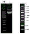
FIG. 3 shows the results of the transfection-grade plasmid extraction and gel analysis with 1% agarose L aneM DNA marker, L ane1: 3000 and 5000bp bands of approximately 4171bp transfection-grade plasmid.
FIG. 4 shows the result of analysis of TTR/anti-hCG scFv fusion Protein purification by Native-PAGE L ane M SDS-PAGE Protein marker, L ane1 supernatant after centrifugation, L ane 2 eluate after incubation of supernatant with Ni-IDA, L ane3-6 elution components, non-denaturing gel without detergent and strong reducing agent, Protein can keep complete tetramer state during electrophoresis, and target band is located at position over 120kD and has molecular weight of about 180 kD.
FIG. 5 shows the results of analysis of the purification of TTR/anti-hCG scFv fusion Protein by SDS-PAGE L ane M SDS-PAGE Protein marker, L ane1 supernatant after centrifugation, L ane 2 eluate after incubation of supernatant with Ni-IDA, L ane3-6 elution fractions, which are denatured due to the presence of strong reducing agent and detergent SDS in the denatured gel, tetrameric Protein disaggregated into monomers with a band of interest between 40-60kD and a molecular weight of about 45 kD.
FIG. 6 shows the results of measurement of protein concentration by Bradford method. The curve of the absorbance versus the BSA concentration is 3.3065x +0.6539, R2The protein concentration was calculated by the formula as 0.259mg/ml from the protein OD value 0.9984.
FIG. 7 shows Western-blot identification of TTR/anti-hCG ScFv fusion protein.Primary antibody is rabbit anti-human TTR monoclonal antibody (Abcam; 1:2500), secondary antibody is goat anti-rabbit secondary antibody (Abcam; 1:2000) labeled with horseradish peroxidase, L ane 1-3 is added with 1. mu.l, 2. mu.l and 3. mu.l TTR/anti-hCG ScFv fusion protein, no SDS is added in the gel preparation process, the protein is not denatured and protein loading buffer solution without strong reducing agent is added, the target band is between 170 and 210kD, and is about 180 kD.
FIG. 8 shows the Western-blot identification result of TTR/anti-hCG ScFv fusion protein. The primary antibody is rabbit anti-human TTR monoclonal antibody (Abcam; 1:2500), and the secondary antibody is goat anti-rabbit secondary antibody (Abcam; 1:2000) marked by horseradish peroxidase. Before the target protein is loaded, the target protein is subjected to denaturation treatment and is added with a protein loading buffer solution containing a strong reducing agent, the tetrameric protein is depolymerized into monomers, and a target band is between 43 and 55kD and is about 45 kD.
FIG. 9.1 is an experimental result for verifying that TTR/anti-hCG scFv fusion protein can promote migration of HTR-8/Svneo cells Transwell migration experiment was used to evaluate the effect of different concentrations of each histone on cell migration ability, panels A-D are addition of TTR protein group, E-H is addition of anti-hCG antibody group, I-L is addition of TTR/anti-hCG scFv fusion protein group, and the addition protein concentrations of each group are, from left to right, from top to bottom, A.100ng/ul, B.10ng/ul, C.1ng/ul, D.0ng/ul, E.100ng/ul, F.10ng/ul, G.1ng/ul, H.0ng/ul, I.100ng/ul, J.10ng/ul, K.1ng/ul, L. 0 ng/ul.
FIG. 9.2 is the result of an experiment to verify that TTR/anti-hCG scFv fusion protein can promote migration of HTR-8/Svneo cells; ttr promotes trophoblast migration, with the ability to migrate being highest at a protein concentration of 100ng/ul, indicating that the difference between the two groups is statistically significant (P < 0.05). anti-hCG did not promote trophoblast migration and there were no significant differences between groups. ttr/anti-hCG scFv was able to promote trophoblast migration and migration capacity increased with increasing protein concentration, indicating that the difference between the two groups was statistically significant (P < 0.05). Graph d.100ng/ul concentration compared to each histone, both TTR and TTR/anti-hCG scFv promoted cell migration with no difference between the two.
FIG. 10.1 shows results of validation of the ability of TTR/anti-hCG scFv fusion proteins to promote HTR-8/Svneo cell invasion Transwell migration assay to evaluate the effect of different concentrations of each histone on cell invasion capacity, panels A-D are addition of TTR proteome, E-H are addition of anti-hCG antibody histones, and I-L are addition of fusion proteome, and the concentration of each protein added to each panel is, in order from left to right, A.100ng/ul, B.10ng/ul, C.1ng/ul, D.0ng/ul, E.100ng/ul, F.10ng/ul, G.10 ng/ul, H.0ng/ul, I.100ng/ul, J.1 ng/ul, K.10 ng/ul, L. 0 ng/ul.
FIG. 10.2 is a result of verifying that TTR/anti-hCG scFv fusion protein can promote HTR-8/Svneo cell invasion. TTR promoted trophoblast invasion and was most aggressive at a protein concentration of 100ng/ul, indicating that the difference between the two groups was statistically significant (P < 0.05). anti-hCG failed to promote trophoblast invasion, with no apparent difference between groups. TTR/anti-hCG scFv was able to promote trophoblast invasion and was most aggressive at a protein concentration of 100ng/ul, indicating that the difference between the two groups was statistically significant (P < 0.05). Graph d.100ng/ul concentration TTR and TTR/anti-hCG scFv both promoted cell invasion compared to each histone, with no difference between the two.
FIG. 11 is a CCK-8 method for evaluating the effect of various histones at different concentrations on cell proliferation potency. Graph a, effect of each histone at 100ng/ul concentration on HTR-8/Svneo cell proliferation potency, wherein TTR group can significantly promote cell proliferation, indicating that the difference between the two groups is statistically significant (P < 0.05), TTR/anti-hCG scFv fusion protein has a promoting effect on cell proliferation potency, but the effect is not significant compared to TTR group. FIG. b shows the effect of each histone at a concentration of 10ng/ul on the proliferation potency of HTR-8/Svneo cells, with no apparent difference between groups. FIG. c shows the effect of each histone at 1ng/ul concentration on the proliferation capacity of HTR-8/Svneo cells, with no obvious difference among groups.
FIG. 12 is a diagram showing the construction process of a lentiviral expression vector.
FIG. 13 is a diagram of the process of lentivirus transfection into trophoblasts.
FIG. 14 is a diagram showing the expression process of TTR/anti-hCG scFv fusion protein gene in cells after lentivirus transfection into trophoblasts.
Detailed Description
The principles and features of this invention are described below in conjunction with the following drawings, which are set forth by way of illustration only and are not intended to limit the scope of the invention.
The TTR protein Sequence is derived from NCBI, NCBI Reference Sequence NP-000362.1.
The anti-hCG scFv sequence is mouse anti-human hCG. The heavy chain part and the light chain part in the anti-hCG scFv are respectively selected.
The Heavy Chain is L ink, http:// www.ncbi.nlm.nih.gov/protein/1SBS _ H, the sequence containing VH is simply referred to, and the specific amino acid sequence is shown in SEQ ID NO. 2.
The amino acid sequence shown in SEQ ID NO.2 is as follows:

an amino acid sequence having an underlined portion, which is a sequence of a variable region, was selected from the amino acid sequences shown in SEQ ID NO.2 as a VH sequence.
L light Chain: L ink: http:// www.ncbi.nlm.nih.gov/protein/1SBS _ L, is abbreviated as a sequence comprising V L, the specific amino acid sequence of which is shown in SEQ ID NO. 3.
The amino acid sequence shown in SEQ ID NO.3 is as follows:

the amino acid sequence shown in SEQ ID NO.3 was selected from the underlined amino acid sequences, and the partial sequences were sequences of the variable regions as the V L sequence.
The V L sequence, the VH sequence and the ttr sequence are combined together, a connecting peptide sequence GGGGSGGGGSGGGGS is arranged between the V L sequence and the VH sequence, and a connecting peptide sequence GGGGSGGGGS is arranged between the VH sequence and the ttr sequence to form a V L + VH + ttr sequence, namely the amino acid sequence shown in SEQ ID NO. 1.
The amino acid sequence information for the V L + VH + ttr sequence is as follows:

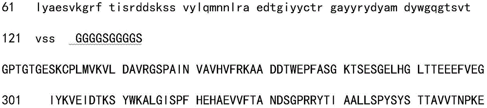
wherein the amino acid sequenceGGGGSGGGGSGGGGSAnd amino acid sequenceGGGGSGGGGSAll of them are connecting peptides, through which V L and VH and ttr can be connected, which is a step of designing fusion protein tandem fusion, and the parts must be connected in series by the connecting peptide, and the connecting peptide cannot be too long or too short, and the folding of the protein can be affected by the too long or too short sequence.
In order to improve the expression amount and stability of the target protein, the target protein sequence is generally required to be optimized, in a eukaryotic body, a signal peptide is automatically cut off in the protein maturation process, and the presence of the signal peptide can make the protein inactive, so that the signal peptide is firstly deleted in the recombinant expression design, namely the sequence of the signal peptide is not included in the sequence of V L + VH + ttr.
And obtaining the nucleotide sequence shown in SEQ ID NO.4 by a codon optimization method according to the amino acid sequence information of the V L + VH + ttr sequence.
A target gene sequence (namely a nucleotide sequence shown in SEQ ID NO. 4) is cut into fragments with different sizes (about 28-50 bp) by using a whole gene synthesis method, a sequence design full-length splicing primer is obtained by using a chemical synthesis method (the step is finished by Detai (Nanjing) Biotech company), the target gene is subcloned into a proEM system and is transformed into escherichia coli to prepare a transfection-grade plasmid, and then the transfection plasmid is transfected into mammalian cells HEK293 (purchased from L ife Technologies) for transient expression.
EXAMPLE 1 construction and expression of the target sequence (i.e., V L + VH + ttr sequence)
The amino acid sequence of the V L + VH + ttr sequence is shown in SEQ ID NO.1, and the amino acid sequence shown in SEQ ID NO.1 is shown in the specification.


The nucleotide sequence capable of expressing the amino acid sequence is the nucleotide sequence shown in SEQ ID NO. 4.
The nucleotide sequence shown in SEQ ID No.4 is cloned into an expression vector ProEM (Detai (Nanjing) Biotechnology company) to construct an expression plasmid, the expression plasmid is named pGH-DT1555, the expression plasmid is sequenced and then is compared by using B L AST, the comparison result is shown in figure 1, and the sequencing result is 100% matched with the original sequence as can be seen from figure 1.
The expression plasmid was verified by double digestion with HindIII. As shown in FIG. 2, the results of the double digestion of the expression plasmid and the analysis thereof on a 1% agarose gel are shown schematically. As can be seen from FIG. 2, bands of approximately 1278bp and 2000-3000bp were obtained after double digestion, consistent with the expected results.
EXAMPLE 2 preparation of transfection-grade plasmid
The pGH-DT1555 expression plasmid is used as a template, a full-length splicing primer is designed, a target gene (namely a nucleotide sequence comprising SEQ ID NO. 4) is subcloned into a proEM system, the vector is a pcDNA3.1 vector (Deltay Biotech company), and is transformed into escherichia coli to prepare a transfection grade plasmid, namely the TTR/anti-hCG scFv transfection plasmid. The results of DNA measurements of TTR/anti-hCG scFv transfected plasmids are shown in Table 1.
TABLE 1
| Detecting content | Transfection grade plasmid Standard | Detection result of target plasmid |
| A260/280 | 1.8-2.0 | 1.86 |
| Endotoxin (EU/mg) | <50 | <50 |
| Sterility testing | Sterile | Sterile |
In table 1, the representation is detected by limulus reagent, and the representation is detected by L B plate.
And (3) transfection-level plasmid amplification and extraction: the extracted TTR/anti-hCG scFv transfected plasmid was analyzed on 1% agarose gel, and the results are shown in FIG. 3. As can be seen from FIG. 3, the size of the TTR/anti-hCG scFv transfection plasmid was approximately 4171 bp.
Example 4scFv-ttr protein purification
Transfecting the TTR/anti-hCG scFv transfection plasmid into HEK293 cells of 1L by using a transfection reagent, placing the extracted TTR/anti-hCG scFv transfection plasmid into an incubator at 37 ℃ and 5% CO2Culture, sampling and purifying after 6 days.
The protein expressed by HEK293 cells transfected with TTR/anti-hCG scFv transfection plasmid was designated TTR/anti-hCG scFv fusion protein.
The purification method comprises the following steps:
the cell culture fluid after 6 days of transfection culture is centrifuged, the supernatant is filtered by a 0.22um membrane, dialyzed at 4 ℃ into a buffer solution of 25mM Tris, 150mM NaCl, pH8.0, and purified by a Ni-IDA column after the dialysis is finished.
Purifying by Ni-IDA affinity chromatography, wherein the target protein (namely TTR/anti-hCG scFv fusion protein) mainly exists in the elution component, collecting the target protein, dialyzing into 1XPBS, 20% Glycerol and pH 7.4, filtering by using a 0.22um membrane after dialysis is finished, and subpackaging and freezing at-80 ℃ for later use.
The results of the purification of the TTR/anti-hCG scFv fusion protein were analyzed by Native-PAGE and SDS-PAGE, respectively.
The operation method comprises the following steps: taking 20ul of the residual sample frozen in the target protein at minus 80 ℃, adding 20ul of 2xSDS reducing sample loading buffer solution, and rapidly heating the sample at 100 ℃ for 10min to denature the protein; then 20ul of the sample frozen in the protein at-80 ℃ is taken, 20ul of 2xSDS non-reducing sample loading buffer solution is added, and then the supernatant is centrifuged at 12000rpm x 5min x 4 ℃ to be taken for electrophoresis. 100V voltage-stabilized electrophoresis is carried out 10min before electrophoresis, and then the bromophenol blue indicator enters separation gel for 200V voltage-stabilized electrophoresis until the bromophenol blue band moves to 1cm from the bottom of the gel. The gel was taken out and stained with Coomassie Brilliant blue stain, and then transferred to a destaining solution to destain until the background was clear.
FIG. 4 shows the results of Native-PAGE analysis on the purification of TTR/anti-hCG scFv fusion protein, wherein L ane M is SDS-PAGE protein marker, L ane1 is supernatant after centrifugation, L ane 2 is effluent after incubation of supernatant with Ni-IDA, L ane3-6 are elution components, Native-PAGE is adopted in the electrophoresis process, and Native-PAGE is non-denaturing gel containing no strong reducing agent and detergent, and TTR/anti-hCG scFv fusion protein can keep a complete tetramer state, and a target band is located at a position over 120kD and has a molecular weight of about 180 kD.
FIG. 5 shows the result of SDS-PAGE analysis of the purification of TTR/anti-hCG scFv fusion protein L ane M is SDS-PAGEProtein marker, L ane1 is supernatant after centrifugation, L ane 2 is effluent after incubation of supernatant with Ni-IDA, L ane3-6 are elution components, respectively, it can be seen from FIG. 5 that due to the presence of strong reducing agent and detergent SDS in denatured gel, TTR/anti-hCGScFv fusion protein is denatured, tetramer protein is depolymerized to monomer, band of interest is between 40-60kD, and molecular weight is about 45 kD.
The TTR/anti-hCG scFv fusion protein can be expressed and purified through the experiment.
Example 5 quality control of TTR/anti-hCG ScFv fusion protein.
Example 5.1 TTR/anti-hCG ScFv fusion protein stability assay
Taking one part, subpackaging, freezing in TTR/anti-hCG ScFv fusion protein at-80 ℃, placing in an ice-water mixture, slowly melting, observing that no abnormal phenomenon exists after melting, and showing that the TTR/anti-hCG ScFv fusion protein obtained by the invention has the advantage of good stability.
Example 5.2 TTR/anti-hCG ScFv fusion protein concentration determination
TABLE 2 BSA standard preparation and OD determination
| BSA concentration (mg/ml) | 1 | 0.8 | 0.6 | 0.4 | 0.2 | 0 |
| OD595nm | 0.998 | 0.914 | 0.854 | 0.780 | 0.727 | 0.652 |
FIG. 6 shows the results of protein concentration measurement by the Bradford method. The curve of the absorbance versus the BSA concentration is 3.3065x +0.6539, R2The protein concentration was calculated by the formula as 0.259mg/ml from the protein OD value 0.9984.
Example 5.3 Western-blot identification of TTR/anti-hCG ScFv fusion protein
FIG. 7 shows Western-blot identification of TTR/anti-hCG ScFv fusion protein, rabbit anti-human TTR monoclonal antibody (Abcam; 1:2500), goat anti-rabbit secondary antibody labeled with horseradish peroxidase (Abcam; 1:2000), L ane1 (1-3), 1. mu.l, 2. mu.l, and 3. mu.l of TTR/anti-hCG ScFv fusion protein, no SDS (sodium dodecyl sulfate) in the gel preparation process, no protein denaturation treatment, and no strong reducing agent in the added protein loading buffer solution, and it can be seen from FIG. 7 that the target band is between 170 and 210kD, and is about 180 kD.
FIG. 8 shows TTR/anti-hCG ScFvWestern-blot identification. The primary antibody is rabbit anti-human TTR monoclonal antibody (Abcam; 1:2500), and the secondary antibody is goat anti-rabbit secondary antibody (Abcam; 1:2000) marked by horseradish peroxidase. Before loading target protein TTR/anti-hCG scFv fusion protein, denaturation treatment is carried out, and a strong reducing agent is contained in an added protein loading buffer. As can be seen in FIG. 8, the tetrameric protein disaggregates into monomers with a band of interest between 43-55kD, about 45 kD.
The TTR/anti-hCG ScFv fusion protein, namely the target protein, is proved by Western-blot experiments.
Example 6 validation of the ability of TTR/anti-hCG ScFv fusion protein to enhance migration of HTR-8/Svneo cells
FIGS. 9.1 and 9.2 both show the results of the ability of TTR/anti-hCG scFv fusion protein to promote migration of HTR-8/Svneo cells, using the Transwell migration assay to evaluate the effect of different concentrations of each histone on cell migration ability, in FIG. 9.1, A-D were separately added TTR proteomes, E-H were separately added anti-hCG antibody proteomes, and I-L were separately added TTR/anti-hCG scFv fusion protein histones, and the added protein concentrations of each histones were, from left to right, from top to bottom, sequentially A.100ng/ul, B.1 ng/ul, C.1ng/ul, D.0ng/ul, E.100ng/ul, F.10ng/ul, G.1ng/ul, H.0ng/ul, I.100ng/ul, J.10ng/ul, K.10 ng/ul, L. 0 ng/ul.
Results of the TTR/anti-hCG scFv fusion protein according to FIG. 9.2 promoting the migration ability of HTR-8/Svneo cells. It can be seen in fig. 9.2, panel a. that TTR promotes trophoblast migration, with the ability to migrate being highest at a protein concentration of 100ng/ul, indicating that the difference between the two groups is statistically significant (P < 0.05). Panel b in figure 9.2 it can be seen that anti-hCG did not promote trophoblast migration, with no significant difference between groups. Panel c in figure 9.2 it can be seen that TTR/anti-hCG scFv fusion protein is able to promote trophoblast migration and that the ability to migrate increases with increasing protein concentration, indicating that the difference between the two groups is statistically significant (P < 0.05). Panel d of FIG. 9.2 shows the results of comparison of 100ng/ul concentrations of each histone, with no difference between TTR and TTR/anti-hCG scFv, which both promote cell migration.
Example 7 validation of the ability of TTR/anti-hCG ScFv fusion protein to enhance HTR-8/Svneo cell invasion
Both FIGS. 10.1 and 10.2 show that the TTR/anti-hCG scFv fusion protein can promote HTR-8/Svneo cell invasion.
Transwell migration assay was used to assess the effect of different concentrations of each histone on cell invasion capacity, in FIG. 10.1, panels A-D are TTR protein added panels, E-H is anti-hCG antibody added panel, I-L is TTR/anti-hCG scFv fusion protein added panel, and the protein concentrations added in each panel are A.100ng/ul, B.10ng/ul, C.10 ng/ul, D.0ng/ul, E.100ng/ul, F.10ng/ul, G.1ng/ul, H.0ng/ul, I.100ng/ul, J.10ng/ul, K.10 ng/ul, L. 0ng/ul, from left to right, in sequence from top to bottom.
Panel a in fig. 10.2 shows that TTR promotes trophoblast invasion and is most aggressive at a protein concentration of 100ng/ul, indicating that the difference between the two groups is statistically significant (P < 0.05). Panel b in figure 10.2 shows that anti-hCG does not promote trophoblast invasion, with no significant difference between groups. Panel c in fig. 10.2 shows that TTR/anti-hCG scFv fusion protein is able to promote trophoblast invasion and is most aggressive at a protein concentration of 100ng/ul, indicating that the difference between the two groups is statistically significant (P < 0.05). Panel d of FIG. 10.2 shows that TTR and TTR/anti-hCG scFv both promote cell invasion compared to each histone at 100ng/ul concentration, and there is no difference between the two.
Example 8 validation of the ability of TTR/anti-hCG ScFv fusion protein to enhance the proliferation of HTR-8/Svneo cells
FIG. 11 shows that the TTR/anti-hCG scFv fusion protein can promote the proliferation of HTR-8/Svneo cells, but it is not statistically significant compared to the blank control group.
The CCK-8 method was used to evaluate the effect of various histone concentrations on cell proliferation capacity. Panel a in FIG. 11 shows the effect of each histone at 100ng/ul concentration on the proliferation potency of HTR-8/Svneo cells, where TTR group can significantly promote cell proliferation, indicating that the difference between the two groups is statistically significant (P < 0.05), and TTR/anti-hCG scFv fusion protein has a promoting effect on the proliferation potency of cells, but the effect is not significant compared to TTR group. Panel b of FIG. 11 shows that at a concentration of 10ng/ul each histone had an effect on the proliferative capacity of HTR-8/Svneo cells, with no apparent difference between groups. Panel c of FIG. 11 shows that each histone at a concentration of 1ng/ul has an effect on the proliferative capacity of HTR-8/Svneo cells, and the difference between groups is not significant.
In conclusion, it can be seen from examples 1 to 8 that the TTR/anti-hCG ScFv fusion protein can enhance the migration ability of HTR-8/Svneo cells, enhance the invasion ability of HTR-8/Svneo cells and promote the proliferation of HTR-8/Svneo cells.
The HTR-8/Svneo cell line is a human early pregnancy trophoblast cell line, and the TTR/anti-hCG scFv fusion protein can enhance invasion, migration and proliferation of the HTR-8/Svneo cell line, so that the fusion protein can be beneficial to improving the early shallow implantation problem of an embryo, and therefore the fusion protein prepared by the application is proved to provide a new direction and thought for treatment of preeclampsia.
By fusing the anti-hCG scFv with the TTR protein, the problem that the antibody cannot be folded correctly due to overlarge molecular weight can be avoided, and the TTR protein can be targeted to the local placenta to play a role by specifically binding the in-vivo hCG. The experiments prove that the TTR/anti-hCG scFv fusion protein has the functions of promoting the invasion capacity, the migration capacity and the proliferation capacity of the trophoblast, so that the fusion protein can provide a new possibility for treating preeclampsia.
The onset of preeclampsia is related to the shallow implantation of placenta, which has already been mentioned in the introduction, the shallow implantation of placenta is caused by insufficient invasion capacity of trophoblasts, and the TTR/anti-hCG scFv fusion protein can promote the invasion, migration and proliferation capacity of trophoblasts HTR-8/Svneo cells in vitro experiments, so that the TTR/anti-hCG scFv can improve the shallow implantation of placenta, thereby providing a new possibility for treating preeclampsia diseases. Therefore, the TTR/anti-hCG ScFv fusion protein can be used for preparing a medicament for treating preeclampsia diseases. Can also be used as therapeutic agent for diseases caused by placenta superficial implantation.
The above examples 1 to 8 are the protein function verification by constructing the hCG antibody and TTR fusion protein system by means of transfection of plasmid. The hCG antibody and TTR fusion protein system can also be constructed in a lentivirus-mediated manner. And then, performing functional verification on the constructed hCG antibody and TTR fusion protein system.
The steps for constructing the expression system are as follows: constructing a lentivirus expression vector containing the gene of the fusion protein and a lentivirus packaging vector to transfect into 293T cells together, packaging and replicating the lentivirus, then harvesting a supernatant containing the lentivirus, infecting trophoblasts by using the supernatant of the lentivirus, and expressing the fusion protein by using the trophoblasts infected with the lentivirus.
As in fig. 12-14, the more detailed steps are as follows:
1. the construction of a lentivirus expression vector comprises (1) hCG antibody anti-hCG scFv whole gene synthesis, (2) construction of a lentivirus mediated hCG antibody overexpression engineering system, (3) lentivirus expression vector plasmid, (4) lentivirus infection activity detection, and collection of virus supernatant, wherein the virus supernatant contains an anti-hCG scFv gene, (5) detection of expression of hCG antibody anti-hCG scFv by E L ISA or WESTEN BO L T, (6) acquisition of a TTR gene sequence, construction of a TTR/anti-hCGcFv fusion protein gene by utilizing the anti-hCG scFv, (7) construction of a lentivirus mediated TTR/anti-hCG scFv fusion protein overexpression engineering system, (8) acquisition of a lentivirus expression vector plasmid carrying the TTR/anti-hCG scFv fusion protein, (9) lentivirus infection activity detection, and (10) acquisition of virus supernatant containing hCG/anti-hCG scFv fusion protein.
2. Transfection of lentiviruses into trophoblasts:
transfecting 293T cells by virus, replicating, packaging, infecting trophoblast, integrating target gene TTR/anti-hCG scFv fusion protein gene with trophoblast gene, and constantly expressing fusion protein by the trophoblast. The method comprises the following specific steps:
(1) cotransfecting a lentivirus expression vector carrying a TTR/anti-hCG scFv fusion protein gene and a lentivirus packaging vector into 293T cells, carrying out virus replication and packaging, harvesting packaging viruses after 72 hours, carrying out centrifugal filtration, and collecting virus supernatant;
(2) the collected virus supernatant is infected with trophoblast, the target gene (TTR/anti-hCG scFv fusion protein gene) is integrated with the genome of the trophoblast, and the TTR/anti-hCG scFv fusion protein is constantly expressed by the trophoblast.
Expressing the TTR/anti-hCG scFv fusion protein gene in cells, and observing the effect of the TTR/anti-hCG scFv fusion protein gene on the function of trophoblasts. And observing the shape and function changes of the trophoblast of the constantly expressed anti-hCG scFv-TTR fusion protein gene. The expression of the TTR/anti-hCG scFv fusion protein gene in cells comprises the following steps: binding or fusion, penetration, reverse transcription, trafficking, integration, transcription, trafficking and translation.
The TTR/anti-hCG scFv fusion protein obtained by the above method was also verified to be consistent with the conclusions of examples 1 to 8.
In the application, anti-hCG is macromolecular immunoglobulin IgG, the molecular weight is about 270KD, and previous researches show that only two or more small molecular proteins can be fused, the fused molecular weight is not more than 60-70KD, if the fused protein is too large, normal processes of transcription, translation and the like are influenced, the formation of a protein quaternary structure is influenced, and the protein is nonfunctional. Therefore, according to the characteristics of immunoglobulin IgG, the inventor selects a variable region for fusion, namely the anti-hCG scFv has the molecular weight of about 30KD, not only can specifically recognize hCG antigen, but also has a relatively simple structure.
TTR is prealbumin with molecular weight of 55KDa, and is tetrameric protein formed by polymerizing four identical subunits. TTR is fused with an anti-hCG scFv gene to form an anti-hCG scFv-TTR monomer after transcription and translation, and previous researches show that the TTR monomer form can be related to local deposition of TTR in tissues and organs to cause amyloidosis. Therefore, whether the transcribed and translated anti-hCG scFv-TTR can be correctly polymerized into tetramer is particularly important. The early-stage experiment inventor adopts prokaryotic cells for expression, the result is not ideal, only a monomer structure can be obtained, tetramer cannot be formed, the later stage is changed into transfection of eukaryotic cells, expression is carried out in an eukaryotic system, western-blot verification is carried out on the protein after purification, and the fact that haploid protein can be normally folded into a natural conformation in the eukaryotic system is found, and the tetramer is formed. This has not been the case in previous studies of fusion proteins, as is well documented.
The above description is only for the purpose of illustrating the preferred embodiments of the present invention and is not to be construed as limiting the invention, and any modifications, equivalents, improvements and the like that fall within the spirit and principle of the present invention are intended to be included therein.
<110> Liu Chong east
<120> fusion protein, preparation method and application thereof
<160>4
<210>1
<211>389
<212>PRT
<213> Artificial Synthesis
<220>
<221> V L + VH + ttr amino acid sequence
<400>1
1 DIVMSQSPSS LAVSVGEKVT MTCKSSQSLL YSSNQMNYLA WYQQKPGQSP KLLIYWASTR
61 ESGVPDRFTG SGSGTDFTLT ISSVEAEDLA VYYCQQYHSY PFTFGSGTKL EIKRGGGGSG
121 GGGSGGGGSE VNLEESGGGL VQPGGSMKLS CVASGFTFSN YWMNWVRQSP EKGLEWVADI
181 RLKSNNYATL YAESVKGRFT ISRDDSKSSV YLQMNNLRAE DTGIYYCTRG AYYRYDYAMD
241 YWGQGTSVTV SSGGGGSGGG GSGPTGTGES KCPLMVKVLD AVRGSPAINV AVHVFRKAAD
301 DTWEPFASGK TSESGELHGL TTEEEFVEGI YKVEIDTKSY WKALGISPFH EHAEVVFTAN
361 DSGPRRYTIA ALLSPYSYST TAVVTNPKE
<210>2
<211>222
<212>PRT
<213> Artificial Synthesis
<220>
<221> VH-containing sequence
<400>2
1 EVNLEESGGG LVQPGGSMKL SCVASGFTFS NYWMNWVRQS PEKGLEWVAD IRLKSNNYAT
61 LYAESVKGRF TISRDDSKSS VYLQMNNLRA EDTGIYYCTR GAYYRYDYAM DYWGQGTSVT
121 VSSAKTTPPS VYPLAPGSAA QTNSMVTLGC LVKGYFPEPV TVTWNSGSLS SGVHTFPAVL
181 QSDLYTLSSS VTVPSSPRPS ETVTCNVAHP ASSTKVDKKI VP
<210>3
<211>220
<212>PRT
<213>
<220>
<221> sequence containing V L
<400>3
1 DIVMSQSPSS LAVSVGEKVT MTCKSSQSLL YSSNQMNYLA WYQQKPGQSP KLLIYWASTR
61 ESGVPDRFTG SGSGTDFTLT ISSVEAEDLA VYYCQQYHSY PFTFGSGTKL EIKR ADAAPT
121 VSIFPPSSEQ LTSGGASVVC FLNNFYPKDI NVKWKIDGSE RQNGVLNSWT DQDSKDSTYS
181 MSSTLTLTKD EYERHNSYTC EATHKTSTSP IVKSFNRNEC
<210>4
<211>1278
<212>DNA
<213> Artificial Synthesis
<220>
<221> V L + VH + ttr nucleotide sequence
<400>4
gaattcccgc cgccgccacc atgggctgga gctgcatcat cctgttcctc gtggccacag 60
ctacaggagt gcacagcgat atcgtgatga gccagagccc ctctagcctg gcagtgtccg 120
tgggcgagaa agtgaccatg acctgcaaga gcagccagag cctgctgtac agcagcaacc 180
agatgaacta cctggcttgg taccagcaga agccaggaca gagccccaag ctgctgatct 240
attgggccag caccagggag agcggcgtgc cagatagatt caccggaagc ggaagcggca 300
ccgatttcac cctgaccatc tcttcagtgg aggccgaaga tctggccgtg tactactgcc 360
agcagtacca cagctacccc ttcaccttcg gcagcggaac caagctggag atcaagagag 420
gcggcggcgg aagcggagga ggaggaagcg gaggaggagg cagcgaagtg aatctggagg 480
agagcggagg aggactggtg cagccaggcg gaagcatgaa gctgtcttgc gtggccagcg 540
gcttcacctt cagcaactac tggatgaatt gggtccggca gagcccagag aaaggactcg 600
agtgggtggc agacatccgg ctgaagagca acaactacgc caccctgtac gccgaaagcg 660
tgaagggcag gttcaccatc agcagggacg acagcaagag cagcgtgtac ctgcagatga 720
acaacctgag ggccgaggac accggcatctactattgcac caggggagcc tactaccgct 780
acgactacgc catggactat tggggacagg gaaccagcgt gacagtgtct tctggcggag 840
gaggaagcgg aggaggagga agcggaccta caggcacagg cgagtctaag tgccctctga 900
tggtgaaggt gctggacgca gtgagaggct ctccagctat caacgtggcc gtgcacgtgt 960
tcagaaaggc cgcagacgac acttgggagc cttttgccag cggcaagacc agcgaaagcg 1020
gagaactgca cggcctgaca accgaagagg agttcgtgga gggcatctac aaggtggaga 1080
tcgacaccaa gagctattgg aaggccctgg gcatcagccc ttttcacgag cacgccgaag 1140
tggtgtttac cgccaacgac agcggcccta gaagatacac catcgccgct ctgctgagcc 1200
cttacagcta cagcaccacc gccgtggtga caaaccctaa ggagcaccac caccaccatc 1260
Claims (7)
1. The fusion protein is characterized by being a protein obtained by fusing anti-hCG scFv, a connecting peptide and transthyretin; the amino acid sequence of the fusion protein is shown as SEQ ID NO. 1.
2. A nucleotide encoding the fusion protein of claim 1.
3. A vector, recombinant cell or recombinant bacterium comprising the fusion protein of claim 1.
4. A method for preparing a fusion protein, comprising the steps of: constructing an expression system containing the fusion protein of claim 1, and expressing the fusion protein using the expression system.
5. The method of claim 4, further comprising a purification step of expressing the fusion protein in the expression system.
6. The method for preparing the fusion protein according to claim 4, wherein the specific steps for constructing the expression system are as follows: cloning a nucleotide sequence expressing an amino acid sequence shown as SEQ ID NO.1 into a proEM system, converting the nucleotide sequence into escherichia coli to prepare a transfection-grade plasmid, and then transfecting the transfection plasmid into mammalian cells HEK293 for transient expression; or the specific steps for constructing the expression system are as follows: constructing a lentivirus expression vector containing a gene expressing an amino acid sequence shown in SEQ ID NO.1 and a lentivirus packaging vector, co-transfecting the lentivirus expression vector and the lentivirus packaging vector into 293T cells, packaging and copying the lentivirus, then harvesting supernate containing the lentivirus, infecting trophoblasts by utilizing the supernate of the lentivirus, and expressing fusion protein by utilizing the trophoblasts infected with the lentivirus.
7. Use of the fusion protein of claim 1 in the preparation of a medicament for the treatment of preeclampsia.
Priority Applications (1)
| Application Number | Priority Date | Filing Date | Title |
|---|---|---|---|
| CN201710257101.0A CN106977609B (en) | 2017-04-19 | 2017-04-19 | Fusion protein, preparation method and application thereof |
Applications Claiming Priority (1)
| Application Number | Priority Date | Filing Date | Title |
|---|---|---|---|
| CN201710257101.0A CN106977609B (en) | 2017-04-19 | 2017-04-19 | Fusion protein, preparation method and application thereof |
Publications (2)
| Publication Number | Publication Date |
|---|---|
| CN106977609A CN106977609A (en) | 2017-07-25 |
| CN106977609B true CN106977609B (en) | 2020-07-28 |
Family
ID=59345835
Family Applications (1)
| Application Number | Title | Priority Date | Filing Date |
|---|---|---|---|
| CN201710257101.0A Active CN106977609B (en) | 2017-04-19 | 2017-04-19 | Fusion protein, preparation method and application thereof |
Country Status (1)
| Country | Link |
|---|---|
| CN (1) | CN106977609B (en) |
Families Citing this family (2)
| Publication number | Priority date | Publication date | Assignee | Title |
|---|---|---|---|---|
| US20200317795A1 (en) * | 2017-10-04 | 2020-10-08 | Amgen Inc. | Transthyretin immunoglobulin fusions |
| CN113621080B (en) * | 2021-09-08 | 2022-04-19 | 北京大学第三医院(北京大学第三临床医学院) | Medicine for preventing or treating preeclampsia and related diseases and application thereof |
Citations (3)
| Publication number | Priority date | Publication date | Assignee | Title |
|---|---|---|---|---|
| US5869057A (en) * | 1995-06-07 | 1999-02-09 | Rock; Edwin P. | Recombinant vaccines to break self-tolerance |
| CN1361181A (en) * | 2000-12-29 | 2002-07-31 | 申庆祥 | New human chorionic gonadotropin-lutropin fusion protein and its prepn and use |
| US7767422B2 (en) * | 1998-09-22 | 2010-08-03 | Oncomedx, Inc. | Detection of 5T4 RNA in plasma and serum |
Family Cites Families (5)
| Publication number | Priority date | Publication date | Assignee | Title |
|---|---|---|---|---|
| CA2345397A1 (en) * | 1998-09-23 | 2000-03-30 | Mount Sinai Hospital | Trophoblast cell preparations |
| US20030191056A1 (en) * | 2002-04-04 | 2003-10-09 | Kenneth Walker | Use of transthyretin peptide/protein fusions to increase the serum half-life of pharmacologically active peptides/proteins |
| CA2720864C (en) * | 2008-04-07 | 2017-07-04 | National Institute Of Immunology | Compositions useful for the treatment of diabetes and other chronic disorder |
| CA2751679C (en) * | 2009-02-06 | 2018-02-27 | Women & Infants' Hospital Of Rhode Island | Compositions for treating preeclampsia-type disorders of pregnancy comprising transthyretin |
| US20110245469A1 (en) * | 2010-04-02 | 2011-10-06 | Athena Discovery, Inc. | Intermediates formed in biosynthesis of relaxin-fusion proteins with extended in vivo half-lives |
-
2017
- 2017-04-19 CN CN201710257101.0A patent/CN106977609B/en active Active
Patent Citations (3)
| Publication number | Priority date | Publication date | Assignee | Title |
|---|---|---|---|---|
| US5869057A (en) * | 1995-06-07 | 1999-02-09 | Rock; Edwin P. | Recombinant vaccines to break self-tolerance |
| US7767422B2 (en) * | 1998-09-22 | 2010-08-03 | Oncomedx, Inc. | Detection of 5T4 RNA in plasma and serum |
| CN1361181A (en) * | 2000-12-29 | 2002-07-31 | 申庆祥 | New human chorionic gonadotropin-lutropin fusion protein and its prepn and use |
Non-Patent Citations (2)
| Title |
|---|
| Expression and purification of a recombinant amyloidogenic peptide from transthyretin for solid-state NMR spectroscopy;Nadaud, Philippe等;《PROTEIN EXPRESSION AND PURIFICATION》;20100331;第70卷(第1期);全文 * |
| 转甲状腺蛋白在重度子痫前期及胎儿生长受限孕妇血清中的表达及意义;刘崇东等;《现代妇产科进展》;20161111;第25卷(第11期);全文 * |
Also Published As
| Publication number | Publication date |
|---|---|
| CN106977609A (en) | 2017-07-25 |
Similar Documents
| Publication | Publication Date | Title |
|---|---|---|
| WO2022262142A1 (en) | Recombinant sars-cov-2 rbd tripolymer protein vaccine capable of generating broad-spectrum cross-neutralization activity, preparation method therefor, and application thereof | |
| US20210093718A1 (en) | Neutralizing anti-tl1a monoclonal antibodies | |
| US10633453B2 (en) | Antibody locker for the inactivation of protein drug | |
| KR20170047351A (en) | Cancer-cell-specific antibody, anticancer agent, and cancer testing method | |
| JPH01502669A (en) | Purified platelet-derived growth factor and its purification method | |
| AU2013271428A1 (en) | Human bispecific EGFRvIII antibody engaging molecules | |
| CN106977609B (en) | Fusion protein, preparation method and application thereof | |
| CN111500586B (en) | Aptamer specifically combined with rabies virus L protein capping region and application thereof | |
| CN110204619B (en) | Chimeric antigen receptor comprising Fc gamma RI and uses thereof | |
| CN110699308A (en) | AMH-INH-GNIH three-expression gene vaccine for improving animal fertility, and preparation method and application thereof | |
| CN110051832B (en) | Heartworm disease vaccine | |
| CN112142851A (en) | Subunit fusion protein tG on rabies virus surface as well as preparation method and application thereof | |
| MXPA02006313A (en) | Improvements in nucleic acid vaccination. | |
| CN113912706A (en) | Antibody binding to hepatitis B virus surface antigen and application thereof | |
| CN113667015A (en) | Antibodies targeting PSGL-1 protein and uses thereof | |
| CN109529040B (en) | LGR4 and R-spondin binding inhibitors and their use in the treatment of tumors | |
| CN110699307A (en) | INH-GNIH double-expression gene vaccine for improving animal fertility and preparation method and application thereof | |
| Robles et al. | Recombinant MBP-pσ1 expressed in soybean seeds delays onset and reduces developing disease in an animal model of multiple sclerosis | |
| US20230242674A1 (en) | Compositions comprising antibodies to human ido-2 | |
| US20230159652A1 (en) | Transferrin receptor 1 targeting for carcinogenesis prevention | |
| CN110759975B (en) | Polypeptide, antibody with strong ADCC effect and application | |
| WO2011123826A2 (en) | Prevention and treatment of cast nephropathy | |
| US20240085427A1 (en) | AN AGR2Xcd3 BISPECIFIC ENGAGER FOR THE TREATMENT OF CANCER | |
| CN117229413A (en) | Bispecific antibody for SFTSV-Gn and CD3 and preparation method thereof | |
| EP3129054B1 (en) | Methods and compositions for the treatment of ocular diseases and disorders |
Legal Events
| Date | Code | Title | Description |
|---|---|---|---|
| PB01 | Publication | ||
| PB01 | Publication | ||
| SE01 | Entry into force of request for substantive examination | ||
| SE01 | Entry into force of request for substantive examination | ||
| GR01 | Patent grant | ||
| GR01 | Patent grant |