CN105939730B - MERS-CoV vaccine - Google Patents
MERS-CoV vaccine Download PDFInfo
- Publication number
- CN105939730B CN105939730B CN201480074433.XA CN201480074433A CN105939730B CN 105939730 B CN105939730 B CN 105939730B CN 201480074433 A CN201480074433 A CN 201480074433A CN 105939730 B CN105939730 B CN 105939730B
- Authority
- CN
- China
- Prior art keywords
- mers
- fold
- cov
- seq
- acid sequence
- Prior art date
- Legal status (The legal status is an assumption and is not a legal conclusion. Google has not performed a legal analysis and makes no representation as to the accuracy of the status listed.)
- Active
Links
- 229940124678 MERS-CoV vaccine Drugs 0.000 title description 2
- 239000000427 antigen Substances 0.000 claims abstract description 191
- 102000036639 antigens Human genes 0.000 claims abstract description 191
- 108091007433 antigens Proteins 0.000 claims abstract description 191
- 241000127282 Middle East respiratory syndrome-related coronavirus Species 0.000 claims abstract description 188
- 108090000765 processed proteins & peptides Proteins 0.000 claims description 126
- 150000007523 nucleic acids Chemical class 0.000 claims description 120
- 125000003275 alpha amino acid group Chemical group 0.000 claims description 83
- 230000002163 immunogen Effects 0.000 claims description 79
- 108020004707 nucleic acids Proteins 0.000 claims description 68
- 102000039446 nucleic acids Human genes 0.000 claims description 68
- 108091028043 Nucleic acid sequence Proteins 0.000 claims description 50
- 239000000203 mixture Substances 0.000 claims description 48
- 239000007924 injection Substances 0.000 claims description 32
- 238000002347 injection Methods 0.000 claims description 32
- 238000004520 electroporation Methods 0.000 claims description 30
- 208000015181 infectious disease Diseases 0.000 claims description 30
- 230000028993 immune response Effects 0.000 claims description 29
- 125000003729 nucleotide group Chemical group 0.000 claims description 26
- 239000002773 nucleotide Substances 0.000 claims description 25
- 230000003612 virological effect Effects 0.000 claims description 13
- 239000002671 adjuvant Substances 0.000 claims description 12
- 239000003814 drug Substances 0.000 claims description 12
- 239000013604 expression vector Substances 0.000 claims description 12
- 239000002245 particle Substances 0.000 claims description 12
- 230000001939 inductive effect Effects 0.000 claims description 10
- 238000004519 manufacturing process Methods 0.000 claims description 10
- 239000000546 pharmaceutical excipient Substances 0.000 claims description 9
- 238000011282 treatment Methods 0.000 claims description 6
- 229960005486 vaccine Drugs 0.000 abstract description 181
- 238000000034 method Methods 0.000 abstract description 39
- 208000025370 Middle East respiratory syndrome Diseases 0.000 description 246
- 210000004027 cell Anatomy 0.000 description 93
- 108090000623 proteins and genes Proteins 0.000 description 83
- 239000012634 fragment Substances 0.000 description 72
- 241000699670 Mus sp. Species 0.000 description 66
- 210000001744 T-lymphocyte Anatomy 0.000 description 66
- 102000004169 proteins and genes Human genes 0.000 description 56
- 229940024606 amino acid Drugs 0.000 description 47
- 150000001413 amino acids Chemical class 0.000 description 47
- 238000002649 immunization Methods 0.000 description 47
- 108020004414 DNA Proteins 0.000 description 46
- 230000005867 T cell response Effects 0.000 description 45
- 230000014509 gene expression Effects 0.000 description 44
- 102000004196 processed proteins & peptides Human genes 0.000 description 44
- 102100036011 T-cell surface glycoprotein CD4 Human genes 0.000 description 43
- 230000003053 immunization Effects 0.000 description 43
- 239000013598 vector Substances 0.000 description 42
- 210000001519 tissue Anatomy 0.000 description 39
- 230000003472 neutralizing effect Effects 0.000 description 37
- 241001465754 Metazoa Species 0.000 description 36
- GOZMBJCYMQQACI-UHFFFAOYSA-N 6,7-dimethyl-3-[[methyl-[2-[methyl-[[1-[3-(trifluoromethyl)phenyl]indol-3-yl]methyl]amino]ethyl]amino]methyl]chromen-4-one;dihydrochloride Chemical compound Cl.Cl.C=1OC2=CC(C)=C(C)C=C2C(=O)C=1CN(C)CCN(C)CC(C1=CC=CC=C11)=CN1C1=CC=CC(C(F)(F)F)=C1 GOZMBJCYMQQACI-UHFFFAOYSA-N 0.000 description 34
- 230000024932 T cell mediated immunity Effects 0.000 description 34
- 239000012530 fluid Substances 0.000 description 32
- 108010002350 Interleukin-2 Proteins 0.000 description 31
- 102000000588 Interleukin-2 Human genes 0.000 description 31
- 229940096437 Protein S Drugs 0.000 description 30
- 239000013612 plasmid Substances 0.000 description 29
- 108010074328 Interferon-gamma Proteins 0.000 description 28
- 101710198474 Spike protein Proteins 0.000 description 25
- 229940021995 DNA vaccine Drugs 0.000 description 23
- 238000003556 assay Methods 0.000 description 22
- 230000028996 humoral immune response Effects 0.000 description 22
- 230000004044 response Effects 0.000 description 22
- 108010041986 DNA Vaccines Proteins 0.000 description 20
- 210000002966 serum Anatomy 0.000 description 19
- 230000001965 increasing effect Effects 0.000 description 18
- 210000004988 splenocyte Anatomy 0.000 description 18
- 238000002255 vaccination Methods 0.000 description 18
- 241001112090 Pseudovirus Species 0.000 description 17
- 241000700605 Viruses Species 0.000 description 17
- 238000006386 neutralization reaction Methods 0.000 description 17
- 108060003951 Immunoglobulin Proteins 0.000 description 16
- 102000008070 Interferon-gamma Human genes 0.000 description 16
- 102000018358 immunoglobulin Human genes 0.000 description 16
- 238000001890 transfection Methods 0.000 description 16
- 108010076504 Protein Sorting Signals Proteins 0.000 description 15
- 108060008682 Tumor Necrosis Factor Proteins 0.000 description 15
- 102000000852 Tumor Necrosis Factor-alpha Human genes 0.000 description 15
- 230000001086 cytosolic effect Effects 0.000 description 15
- 229960003130 interferon gamma Drugs 0.000 description 15
- 241000699666 Mus <mouse, genus> Species 0.000 description 14
- 238000010790 dilution Methods 0.000 description 14
- 239000012895 dilution Substances 0.000 description 14
- 201000010099 disease Diseases 0.000 description 14
- 208000037265 diseases, disorders, signs and symptoms Diseases 0.000 description 14
- 238000003780 insertion Methods 0.000 description 14
- 230000037431 insertion Effects 0.000 description 14
- 108091035707 Consensus sequence Proteins 0.000 description 13
- 239000003795 chemical substances by application Substances 0.000 description 13
- 210000003205 muscle Anatomy 0.000 description 13
- 210000003491 skin Anatomy 0.000 description 13
- MZOFCQQQCNRIBI-VMXHOPILSA-N (3s)-4-[[(2s)-1-[[(2s)-1-[[(1s)-1-carboxy-2-hydroxyethyl]amino]-4-methyl-1-oxopentan-2-yl]amino]-5-(diaminomethylideneamino)-1-oxopentan-2-yl]amino]-3-[[2-[[(2s)-2,6-diaminohexanoyl]amino]acetyl]amino]-4-oxobutanoic acid Chemical compound OC[C@@H](C(O)=O)NC(=O)[C@H](CC(C)C)NC(=O)[C@H](CCCN=C(N)N)NC(=O)[C@H](CC(O)=O)NC(=O)CNC(=O)[C@@H](N)CCCCN MZOFCQQQCNRIBI-VMXHOPILSA-N 0.000 description 12
- 241000711573 Coronaviridae Species 0.000 description 12
- 102100037850 Interferon gamma Human genes 0.000 description 12
- 238000004458 analytical method Methods 0.000 description 12
- 230000005875 antibody response Effects 0.000 description 12
- 230000027455 binding Effects 0.000 description 12
- 108091032973 (ribonucleotides)n+m Proteins 0.000 description 11
- 238000002965 ELISA Methods 0.000 description 11
- 241000282560 Macaca mulatta Species 0.000 description 11
- 108020004705 Codon Proteins 0.000 description 10
- 238000011510 Elispot assay Methods 0.000 description 10
- LOKCTEFSRHRXRJ-UHFFFAOYSA-I dipotassium trisodium dihydrogen phosphate hydrogen phosphate dichloride Chemical compound P(=O)(O)(O)[O-].[K+].P(=O)(O)([O-])[O-].[Na+].[Na+].[Cl-].[K+].[Cl-].[Na+] LOKCTEFSRHRXRJ-UHFFFAOYSA-I 0.000 description 10
- 238000003114 enzyme-linked immunosorbent spot assay Methods 0.000 description 10
- 239000011159 matrix material Substances 0.000 description 10
- 239000013642 negative control Substances 0.000 description 10
- 239000002953 phosphate buffered saline Substances 0.000 description 10
- 108060001084 Luciferase Proteins 0.000 description 9
- 239000005089 Luciferase Substances 0.000 description 9
- 241000124008 Mammalia Species 0.000 description 9
- 210000004369 blood Anatomy 0.000 description 9
- 239000008280 blood Substances 0.000 description 9
- 238000002474 experimental method Methods 0.000 description 9
- 238000013507 mapping Methods 0.000 description 9
- 230000004048 modification Effects 0.000 description 9
- 238000012986 modification Methods 0.000 description 9
- 239000000523 sample Substances 0.000 description 9
- 210000003501 vero cell Anatomy 0.000 description 9
- 102000004127 Cytokines Human genes 0.000 description 8
- 108090000695 Cytokines Proteins 0.000 description 8
- 230000000694 effects Effects 0.000 description 8
- 230000005847 immunogenicity Effects 0.000 description 8
- 238000004246 ligand exchange chromatography Methods 0.000 description 8
- 239000002502 liposome Substances 0.000 description 8
- 241000282693 Cercopithecidae Species 0.000 description 7
- 108091026890 Coding region Proteins 0.000 description 7
- 241000282414 Homo sapiens Species 0.000 description 7
- 239000013592 cell lysate Substances 0.000 description 7
- 230000000295 complement effect Effects 0.000 description 7
- 238000009472 formulation Methods 0.000 description 7
- 230000005764 inhibitory process Effects 0.000 description 7
- 238000005457 optimization Methods 0.000 description 7
- 230000007170 pathology Effects 0.000 description 7
- 229920001184 polypeptide Polymers 0.000 description 7
- 230000000638 stimulation Effects 0.000 description 7
- 238000012360 testing method Methods 0.000 description 7
- 229910002092 carbon dioxide Inorganic materials 0.000 description 6
- 238000010276 construction Methods 0.000 description 6
- 230000005684 electric field Effects 0.000 description 6
- 238000000684 flow cytometry Methods 0.000 description 6
- 238000001943 fluorescence-activated cell sorting Methods 0.000 description 6
- 230000003834 intracellular effect Effects 0.000 description 6
- 230000001105 regulatory effect Effects 0.000 description 6
- 238000010186 staining Methods 0.000 description 6
- 238000006467 substitution reaction Methods 0.000 description 6
- 238000001262 western blot Methods 0.000 description 6
- 239000006144 Dulbecco’s modified Eagle's medium Substances 0.000 description 5
- 108060003393 Granulin Proteins 0.000 description 5
- 244000309467 Human Coronavirus Species 0.000 description 5
- 241000282553 Macaca Species 0.000 description 5
- -1 MadCAM-1 Proteins 0.000 description 5
- 102100022203 Tumor necrosis factor receptor superfamily member 25 Human genes 0.000 description 5
- 238000002835 absorbance Methods 0.000 description 5
- 230000000890 antigenic effect Effects 0.000 description 5
- 238000001514 detection method Methods 0.000 description 5
- 238000005516 engineering process Methods 0.000 description 5
- 238000007918 intramuscular administration Methods 0.000 description 5
- 150000002632 lipids Chemical class 0.000 description 5
- 210000004072 lung Anatomy 0.000 description 5
- 239000000463 material Substances 0.000 description 5
- 239000002609 medium Substances 0.000 description 5
- 210000003819 peripheral blood mononuclear cell Anatomy 0.000 description 5
- 230000008488 polyadenylation Effects 0.000 description 5
- 229920000447 polyanionic polymer Polymers 0.000 description 5
- 238000013518 transcription Methods 0.000 description 5
- 230000035897 transcription Effects 0.000 description 5
- YYGNTYWPHWGJRM-UHFFFAOYSA-N (6E,10E,14E,18E)-2,6,10,15,19,23-hexamethyltetracosa-2,6,10,14,18,22-hexaene Chemical compound CC(C)=CCCC(C)=CCCC(C)=CCCC=C(C)CCC=C(C)CCC=C(C)C YYGNTYWPHWGJRM-UHFFFAOYSA-N 0.000 description 4
- 108090000672 Annexin A5 Proteins 0.000 description 4
- 238000011238 DNA vaccination Methods 0.000 description 4
- 108090000626 DNA-directed RNA polymerases Proteins 0.000 description 4
- 102000004163 DNA-directed RNA polymerases Human genes 0.000 description 4
- 101710154606 Hemagglutinin Proteins 0.000 description 4
- 101000611183 Homo sapiens Tumor necrosis factor Proteins 0.000 description 4
- 108700001237 Nucleic Acid-Based Vaccines Proteins 0.000 description 4
- 101710093908 Outer capsid protein VP4 Proteins 0.000 description 4
- 101710135467 Outer capsid protein sigma-1 Proteins 0.000 description 4
- 108010038512 Platelet-Derived Growth Factor Proteins 0.000 description 4
- 102000010780 Platelet-Derived Growth Factor Human genes 0.000 description 4
- 101710176177 Protein A56 Proteins 0.000 description 4
- FAPWRFPIFSIZLT-UHFFFAOYSA-M Sodium chloride Chemical compound [Na+].[Cl-] FAPWRFPIFSIZLT-UHFFFAOYSA-M 0.000 description 4
- BHEOSNUKNHRBNM-UHFFFAOYSA-N Tetramethylsqualene Natural products CC(=C)C(C)CCC(=C)C(C)CCC(C)=CCCC=C(C)CCC(C)C(=C)CCC(C)C(C)=C BHEOSNUKNHRBNM-UHFFFAOYSA-N 0.000 description 4
- 102100040247 Tumor necrosis factor Human genes 0.000 description 4
- ISAKRJDGNUQOIC-UHFFFAOYSA-N Uracil Chemical compound O=C1C=CNC(=O)N1 ISAKRJDGNUQOIC-UHFFFAOYSA-N 0.000 description 4
- 230000004071 biological effect Effects 0.000 description 4
- 230000015572 biosynthetic process Effects 0.000 description 4
- 239000000872 buffer Substances 0.000 description 4
- 239000003153 chemical reaction reagent Substances 0.000 description 4
- 238000010586 diagram Methods 0.000 description 4
- PRAKJMSDJKAYCZ-UHFFFAOYSA-N dodecahydrosqualene Natural products CC(C)CCCC(C)CCCC(C)CCCCC(C)CCCC(C)CCCC(C)C PRAKJMSDJKAYCZ-UHFFFAOYSA-N 0.000 description 4
- 239000000185 hemagglutinin Substances 0.000 description 4
- 230000008348 humoral response Effects 0.000 description 4
- 210000000987 immune system Anatomy 0.000 description 4
- 238000003119 immunoblot Methods 0.000 description 4
- 230000001976 improved effect Effects 0.000 description 4
- 230000006698 induction Effects 0.000 description 4
- 230000002458 infectious effect Effects 0.000 description 4
- 230000000670 limiting effect Effects 0.000 description 4
- 239000003550 marker Substances 0.000 description 4
- 210000004877 mucosa Anatomy 0.000 description 4
- 230000001717 pathogenic effect Effects 0.000 description 4
- 229940023041 peptide vaccine Drugs 0.000 description 4
- 229920002643 polyglutamic acid Polymers 0.000 description 4
- 239000013641 positive control Substances 0.000 description 4
- 230000003248 secreting effect Effects 0.000 description 4
- 229940031439 squalene Drugs 0.000 description 4
- TUHBEKDERLKLEC-UHFFFAOYSA-N squalene Natural products CC(=CCCC(=CCCC(=CCCC=C(/C)CCC=C(/C)CC=C(C)C)C)C)C TUHBEKDERLKLEC-UHFFFAOYSA-N 0.000 description 4
- RWQNBRDOKXIBIV-UHFFFAOYSA-N thymine Chemical compound CC1=CNC(=O)NC1=O RWQNBRDOKXIBIV-UHFFFAOYSA-N 0.000 description 4
- 238000013519 translation Methods 0.000 description 4
- 230000014621 translational initiation Effects 0.000 description 4
- 210000000689 upper leg Anatomy 0.000 description 4
- CSCPPACGZOOCGX-UHFFFAOYSA-N Acetone Chemical compound CC(C)=O CSCPPACGZOOCGX-UHFFFAOYSA-N 0.000 description 3
- 102000004121 Annexin A5 Human genes 0.000 description 3
- 108010012236 Chemokines Proteins 0.000 description 3
- 102000019034 Chemokines Human genes 0.000 description 3
- IAZDPXIOMUYVGZ-UHFFFAOYSA-N Dimethylsulphoxide Chemical compound CS(C)=O IAZDPXIOMUYVGZ-UHFFFAOYSA-N 0.000 description 3
- 241000196324 Embryophyta Species 0.000 description 3
- 102100027723 Endogenous retrovirus group K member 6 Rec protein Human genes 0.000 description 3
- 101710091045 Envelope protein Proteins 0.000 description 3
- 101000679903 Homo sapiens Tumor necrosis factor receptor superfamily member 25 Proteins 0.000 description 3
- 108010065805 Interleukin-12 Proteins 0.000 description 3
- 101900332836 Middle East respiratory syndrome-related coronavirus Nucleoprotein Proteins 0.000 description 3
- 108090001074 Nucleocapsid Proteins Proteins 0.000 description 3
- 101710188315 Protein X Proteins 0.000 description 3
- 108010067390 Viral Proteins Proteins 0.000 description 3
- 108020000999 Viral RNA Proteins 0.000 description 3
- 230000008901 benefit Effects 0.000 description 3
- 210000000170 cell membrane Anatomy 0.000 description 3
- 230000036755 cellular response Effects 0.000 description 3
- UQLDLKMNUJERMK-UHFFFAOYSA-L di(octadecanoyloxy)lead Chemical compound [Pb+2].CCCCCCCCCCCCCCCCCC([O-])=O.CCCCCCCCCCCCCCCCCC([O-])=O UQLDLKMNUJERMK-UHFFFAOYSA-L 0.000 description 3
- 230000029087 digestion Effects 0.000 description 3
- 238000009826 distribution Methods 0.000 description 3
- 229940079593 drug Drugs 0.000 description 3
- 239000003623 enhancer Substances 0.000 description 3
- MHMNJMPURVTYEJ-UHFFFAOYSA-N fluorescein-5-isothiocyanate Chemical compound O1C(=O)C2=CC(N=C=S)=CC=C2C21C1=CC=C(O)C=C1OC1=CC(O)=CC=C21 MHMNJMPURVTYEJ-UHFFFAOYSA-N 0.000 description 3
- 239000000499 gel Substances 0.000 description 3
- 210000004201 immune sera Anatomy 0.000 description 3
- 229940042743 immune sera Drugs 0.000 description 3
- 238000010166 immunofluorescence Methods 0.000 description 3
- 238000000338 in vitro Methods 0.000 description 3
- 238000011534 incubation Methods 0.000 description 3
- 239000007927 intramuscular injection Substances 0.000 description 3
- NLYAJNPCOHFWQQ-UHFFFAOYSA-N kaolin Chemical compound O.O.O=[Al]O[Si](=O)O[Si](=O)O[Al]=O NLYAJNPCOHFWQQ-UHFFFAOYSA-N 0.000 description 3
- 239000010410 layer Substances 0.000 description 3
- 230000003902 lesion Effects 0.000 description 3
- 229920002521 macromolecule Polymers 0.000 description 3
- 239000012528 membrane Substances 0.000 description 3
- 210000004379 membrane Anatomy 0.000 description 3
- 244000052769 pathogen Species 0.000 description 3
- 230000002829 reductive effect Effects 0.000 description 3
- 230000010076 replication Effects 0.000 description 3
- 230000028327 secretion Effects 0.000 description 3
- 239000002356 single layer Substances 0.000 description 3
- 239000003381 stabilizer Substances 0.000 description 3
- 238000007619 statistical method Methods 0.000 description 3
- 230000004936 stimulating effect Effects 0.000 description 3
- 238000007920 subcutaneous administration Methods 0.000 description 3
- 230000009885 systemic effect Effects 0.000 description 3
- 230000001225 therapeutic effect Effects 0.000 description 3
- KIUKXJAPPMFGSW-DNGZLQJQSA-N (2S,3S,4S,5R,6R)-6-[(2S,3R,4R,5S,6R)-3-Acetamido-2-[(2S,3S,4R,5R,6R)-6-[(2R,3R,4R,5S,6R)-3-acetamido-2,5-dihydroxy-6-(hydroxymethyl)oxan-4-yl]oxy-2-carboxy-4,5-dihydroxyoxan-3-yl]oxy-5-hydroxy-6-(hydroxymethyl)oxan-4-yl]oxy-3,4,5-trihydroxyoxane-2-carboxylic acid Chemical compound CC(=O)N[C@H]1[C@H](O)O[C@H](CO)[C@@H](O)[C@@H]1O[C@H]1[C@H](O)[C@@H](O)[C@H](O[C@H]2[C@@H]([C@@H](O[C@H]3[C@@H]([C@@H](O)[C@H](O)[C@H](O3)C(O)=O)O)[C@H](O)[C@@H](CO)O2)NC(C)=O)[C@@H](C(O)=O)O1 KIUKXJAPPMFGSW-DNGZLQJQSA-N 0.000 description 2
- 102000040650 (ribonucleotides)n+m Human genes 0.000 description 2
- LRFVTYWOQMYALW-UHFFFAOYSA-N 9H-xanthine Chemical compound O=C1NC(=O)NC2=C1NC=N2 LRFVTYWOQMYALW-UHFFFAOYSA-N 0.000 description 2
- 241000894006 Bacteria Species 0.000 description 2
- 241000283690 Bos taurus Species 0.000 description 2
- 238000011740 C57BL/6 mouse Methods 0.000 description 2
- BHPQYMZQTOCNFJ-UHFFFAOYSA-N Calcium cation Chemical compound [Ca+2] BHPQYMZQTOCNFJ-UHFFFAOYSA-N 0.000 description 2
- CURLTUGMZLYLDI-UHFFFAOYSA-N Carbon dioxide Chemical compound O=C=O CURLTUGMZLYLDI-UHFFFAOYSA-N 0.000 description 2
- PHEDXBVPIONUQT-UHFFFAOYSA-N Cocarcinogen A1 Natural products CCCCCCCCCCCCCC(=O)OC1C(C)C2(O)C3C=C(C)C(=O)C3(O)CC(CO)=CC2C2C1(OC(C)=O)C2(C)C PHEDXBVPIONUQT-UHFFFAOYSA-N 0.000 description 2
- 241000701832 Enterobacteria phage T3 Species 0.000 description 2
- 102000004190 Enzymes Human genes 0.000 description 2
- 108090000790 Enzymes Proteins 0.000 description 2
- 241000283073 Equus caballus Species 0.000 description 2
- 108010040476 FITC-annexin A5 Proteins 0.000 description 2
- 241000282326 Felis catus Species 0.000 description 2
- 101710114810 Glycoprotein Proteins 0.000 description 2
- 108010017213 Granulocyte-Macrophage Colony-Stimulating Factor Proteins 0.000 description 2
- 102100039620 Granulocyte-macrophage colony-stimulating factor Human genes 0.000 description 2
- 241000713772 Human immunodeficiency virus 1 Species 0.000 description 2
- 102000003810 Interleukin-18 Human genes 0.000 description 2
- 108090000171 Interleukin-18 Proteins 0.000 description 2
- 108090000978 Interleukin-4 Proteins 0.000 description 2
- 102000004388 Interleukin-4 Human genes 0.000 description 2
- GUBGYTABKSRVRQ-QKKXKWKRSA-N Lactose Natural products OC[C@H]1O[C@@H](O[C@H]2[C@H](O)[C@@H](O)C(O)O[C@@H]2CO)[C@H](O)[C@@H](O)[C@H]1O GUBGYTABKSRVRQ-QKKXKWKRSA-N 0.000 description 2
- 102000012750 Membrane Glycoproteins Human genes 0.000 description 2
- 108010090054 Membrane Glycoproteins Proteins 0.000 description 2
- 102000018697 Membrane Proteins Human genes 0.000 description 2
- 108010052285 Membrane Proteins Proteins 0.000 description 2
- 229930191564 Monensin Natural products 0.000 description 2
- GAOZTHIDHYLHMS-UHFFFAOYSA-N Monensin A Natural products O1C(CC)(C2C(CC(O2)C2C(CC(C)C(O)(CO)O2)C)C)CCC1C(O1)(C)CCC21CC(O)C(C)C(C(C)C(OC)C(C)C(O)=O)O2 GAOZTHIDHYLHMS-UHFFFAOYSA-N 0.000 description 2
- 239000004677 Nylon Substances 0.000 description 2
- 108091034117 Oligonucleotide Proteins 0.000 description 2
- 241000009328 Perro Species 0.000 description 2
- 239000012980 RPMI-1640 medium Substances 0.000 description 2
- 241000700159 Rattus Species 0.000 description 2
- 201000003176 Severe Acute Respiratory Syndrome Diseases 0.000 description 2
- PXIPVTKHYLBLMZ-UHFFFAOYSA-N Sodium azide Chemical compound [Na+].[N-]=[N+]=[N-] PXIPVTKHYLBLMZ-UHFFFAOYSA-N 0.000 description 2
- 101710167605 Spike glycoprotein Proteins 0.000 description 2
- 229930006000 Sucrose Natural products 0.000 description 2
- CZMRCDWAGMRECN-UGDNZRGBSA-N Sucrose Chemical compound O[C@H]1[C@H](O)[C@@H](CO)O[C@@]1(CO)O[C@@H]1[C@H](O)[C@@H](O)[C@H](O)[C@@H](CO)O1 CZMRCDWAGMRECN-UGDNZRGBSA-N 0.000 description 2
- 208000036142 Viral infection Diseases 0.000 description 2
- 238000003491 array Methods 0.000 description 2
- 210000004436 artificial bacterial chromosome Anatomy 0.000 description 2
- 210000001106 artificial yeast chromosome Anatomy 0.000 description 2
- RIIWUGSYXOBDMC-UHFFFAOYSA-N benzene-1,2-diamine;hydron;dichloride Chemical compound Cl.Cl.NC1=CC=CC=C1N RIIWUGSYXOBDMC-UHFFFAOYSA-N 0.000 description 2
- 239000012148 binding buffer Substances 0.000 description 2
- 230000008827 biological function Effects 0.000 description 2
- 230000037396 body weight Effects 0.000 description 2
- 229910001424 calcium ion Inorganic materials 0.000 description 2
- 238000004364 calculation method Methods 0.000 description 2
- 230000030833 cell death Effects 0.000 description 2
- 230000008859 change Effects 0.000 description 2
- 238000006243 chemical reaction Methods 0.000 description 2
- 239000002299 complementary DNA Substances 0.000 description 2
- 210000000805 cytoplasm Anatomy 0.000 description 2
- OPTASPLRGRRNAP-UHFFFAOYSA-N cytosine Chemical compound NC=1C=CNC(=O)N=1 OPTASPLRGRRNAP-UHFFFAOYSA-N 0.000 description 2
- 230000034994 death Effects 0.000 description 2
- 238000012217 deletion Methods 0.000 description 2
- 230000037430 deletion Effects 0.000 description 2
- 238000013461 design Methods 0.000 description 2
- 231100000673 dose–response relationship Toxicity 0.000 description 2
- 210000002472 endoplasmic reticulum Anatomy 0.000 description 2
- 230000006870 function Effects 0.000 description 2
- 238000001502 gel electrophoresis Methods 0.000 description 2
- 230000002068 genetic effect Effects 0.000 description 2
- UYTPUPDQBNUYGX-UHFFFAOYSA-N guanine Chemical compound O=C1NC(N)=NC2=C1N=CN2 UYTPUPDQBNUYGX-UHFFFAOYSA-N 0.000 description 2
- 230000036541 health Effects 0.000 description 2
- 229920002674 hyaluronan Polymers 0.000 description 2
- 229960003160 hyaluronic acid Drugs 0.000 description 2
- 238000009396 hybridization Methods 0.000 description 2
- FDGQSTZJBFJUBT-UHFFFAOYSA-N hypoxanthine Chemical compound O=C1NC=NC2=C1NC=N2 FDGQSTZJBFJUBT-UHFFFAOYSA-N 0.000 description 2
- 238000003018 immunoassay Methods 0.000 description 2
- 230000016784 immunoglobulin production Effects 0.000 description 2
- 238000001727 in vivo Methods 0.000 description 2
- 239000003112 inhibitor Substances 0.000 description 2
- 230000000977 initiatory effect Effects 0.000 description 2
- 238000011081 inoculation Methods 0.000 description 2
- 229940079322 interferon Drugs 0.000 description 2
- 238000010212 intracellular staining Methods 0.000 description 2
- DRAVOWXCEBXPTN-UHFFFAOYSA-N isoguanine Chemical compound NC1=NC(=O)NC2=C1NC=N2 DRAVOWXCEBXPTN-UHFFFAOYSA-N 0.000 description 2
- VCMGMSHEPQENPE-UHFFFAOYSA-N ketamine hydrochloride Chemical compound [Cl-].C=1C=CC=C(Cl)C=1C1([NH2+]C)CCCCC1=O VCMGMSHEPQENPE-UHFFFAOYSA-N 0.000 description 2
- 239000008101 lactose Substances 0.000 description 2
- 239000007788 liquid Substances 0.000 description 2
- 239000012139 lysis buffer Substances 0.000 description 2
- 238000005259 measurement Methods 0.000 description 2
- 230000034217 membrane fusion Effects 0.000 description 2
- 108020004999 messenger RNA Proteins 0.000 description 2
- 229960005358 monensin Drugs 0.000 description 2
- GAOZTHIDHYLHMS-KEOBGNEYSA-N monensin A Chemical compound C([C@@](O1)(C)[C@H]2CC[C@@](O2)(CC)[C@H]2[C@H](C[C@@H](O2)[C@@H]2[C@H](C[C@@H](C)[C@](O)(CO)O2)C)C)C[C@@]21C[C@H](O)[C@@H](C)[C@@H]([C@@H](C)[C@@H](OC)[C@H](C)C(O)=O)O2 GAOZTHIDHYLHMS-KEOBGNEYSA-N 0.000 description 2
- 125000001446 muramyl group Chemical group N[C@@H](C=O)[C@@H](O[C@@H](C(=O)*)C)[C@H](O)[C@H](O)CO 0.000 description 2
- 239000002105 nanoparticle Substances 0.000 description 2
- 229920001778 nylon Polymers 0.000 description 2
- 230000003287 optical effect Effects 0.000 description 2
- 210000000056 organ Anatomy 0.000 description 2
- 239000008188 pellet Substances 0.000 description 2
- PHEDXBVPIONUQT-RGYGYFBISA-N phorbol 13-acetate 12-myristate Chemical compound C([C@]1(O)C(=O)C(C)=C[C@H]1[C@@]1(O)[C@H](C)[C@H]2OC(=O)CCCCCCCCCCCCC)C(CO)=C[C@H]1[C@H]1[C@]2(OC(C)=O)C1(C)C PHEDXBVPIONUQT-RGYGYFBISA-N 0.000 description 2
- 239000000843 powder Substances 0.000 description 2
- 230000001681 protective effect Effects 0.000 description 2
- 238000000159 protein binding assay Methods 0.000 description 2
- BOLDJAUMGUJJKM-LSDHHAIUSA-N renifolin D Natural products CC(=C)[C@@H]1Cc2c(O)c(O)ccc2[C@H]1CC(=O)c3ccc(O)cc3O BOLDJAUMGUJJKM-LSDHHAIUSA-N 0.000 description 2
- 230000003362 replicative effect Effects 0.000 description 2
- 208000023504 respiratory system disease Diseases 0.000 description 2
- 238000013207 serial dilution Methods 0.000 description 2
- 239000011780 sodium chloride Substances 0.000 description 2
- 239000000243 solution Substances 0.000 description 2
- 210000000952 spleen Anatomy 0.000 description 2
- 210000004989 spleen cell Anatomy 0.000 description 2
- 238000003860 storage Methods 0.000 description 2
- 239000000126 substance Substances 0.000 description 2
- 239000005720 sucrose Substances 0.000 description 2
- 230000001629 suppression Effects 0.000 description 2
- 239000004094 surface-active agent Substances 0.000 description 2
- 239000000725 suspension Substances 0.000 description 2
- 238000003786 synthesis reaction Methods 0.000 description 2
- 229940113082 thymine Drugs 0.000 description 2
- 210000001541 thymus gland Anatomy 0.000 description 2
- 230000005030 transcription termination Effects 0.000 description 2
- 230000032258 transport Effects 0.000 description 2
- 229940035893 uracil Drugs 0.000 description 2
- VBEQCZHXXJYVRD-GACYYNSASA-N uroanthelone Chemical compound C([C@@H](C(=O)N[C@H](C(=O)N[C@@H](CS)C(=O)N[C@@H](CC(N)=O)C(=O)N[C@@H](CS)C(=O)N[C@H](C(=O)N[C@@H]([C@@H](C)CC)C(=O)NCC(=O)N[C@@H](CC=1C=CC(O)=CC=1)C(=O)N[C@@H](CO)C(=O)NCC(=O)N[C@@H](CC(O)=O)C(=O)N[C@@H](CCCNC(N)=N)C(=O)N[C@@H](CS)C(=O)N[C@@H](CCC(N)=O)C(=O)N[C@@H]([C@@H](C)O)C(=O)N[C@@H](CCCNC(N)=N)C(=O)N[C@@H](CC(O)=O)C(=O)N[C@@H](CC(C)C)C(=O)N[C@@H](CCCNC(N)=N)C(=O)N[C@@H](CC=1C2=CC=CC=C2NC=1)C(=O)N[C@@H](CC=1C2=CC=CC=C2NC=1)C(=O)N[C@@H](CCC(O)=O)C(=O)N[C@@H](CC(C)C)C(=O)N[C@@H](CCCNC(N)=N)C(O)=O)C(C)C)[C@@H](C)O)NC(=O)[C@H](CO)NC(=O)[C@H](CC(O)=O)NC(=O)[C@H](CC(C)C)NC(=O)[C@H](CO)NC(=O)[C@H](CCC(O)=O)NC(=O)[C@@H](NC(=O)[C@H](CC=1NC=NC=1)NC(=O)[C@H](CCSC)NC(=O)[C@H](CS)NC(=O)[C@@H](NC(=O)CNC(=O)CNC(=O)[C@H](CC(N)=O)NC(=O)[C@H](CC(C)C)NC(=O)[C@H](CS)NC(=O)[C@H](CC=1C=CC(O)=CC=1)NC(=O)CNC(=O)[C@H](CC(O)=O)NC(=O)[C@H](CC=1C=CC(O)=CC=1)NC(=O)[C@H](CO)NC(=O)[C@H](CO)NC(=O)[C@H]1N(CCC1)C(=O)[C@H](CS)NC(=O)CNC(=O)[C@H]1N(CCC1)C(=O)[C@H](CC=1C=CC(O)=CC=1)NC(=O)[C@H](CO)NC(=O)[C@@H](N)CC(N)=O)C(C)C)[C@@H](C)CC)C1=CC=C(O)C=C1 VBEQCZHXXJYVRD-GACYYNSASA-N 0.000 description 2
- 230000009385 viral infection Effects 0.000 description 2
- 238000005406 washing Methods 0.000 description 2
- XLYOFNOQVPJJNP-UHFFFAOYSA-N water Substances O XLYOFNOQVPJJNP-UHFFFAOYSA-N 0.000 description 2
- DIGQNXIGRZPYDK-WKSCXVIASA-N (2R)-6-amino-2-[[2-[[(2S)-2-[[2-[[(2R)-2-[[(2S)-2-[[(2R,3S)-2-[[2-[[(2S)-2-[[2-[[(2S)-2-[[(2S)-2-[[(2R)-2-[[(2S,3S)-2-[[(2R)-2-[[(2S)-2-[[(2S)-2-[[(2S)-2-[[2-[[(2S)-2-[[(2R)-2-[[2-[[2-[[2-[(2-amino-1-hydroxyethylidene)amino]-3-carboxy-1-hydroxypropylidene]amino]-1-hydroxy-3-sulfanylpropylidene]amino]-1-hydroxyethylidene]amino]-1-hydroxy-3-sulfanylpropylidene]amino]-1,3-dihydroxypropylidene]amino]-1-hydroxyethylidene]amino]-1-hydroxypropylidene]amino]-1,3-dihydroxypropylidene]amino]-1,3-dihydroxypropylidene]amino]-1-hydroxy-3-sulfanylpropylidene]amino]-1,3-dihydroxybutylidene]amino]-1-hydroxy-3-sulfanylpropylidene]amino]-1-hydroxypropylidene]amino]-1,3-dihydroxypropylidene]amino]-1-hydroxyethylidene]amino]-1,5-dihydroxy-5-iminopentylidene]amino]-1-hydroxy-3-sulfanylpropylidene]amino]-1,3-dihydroxybutylidene]amino]-1-hydroxy-3-sulfanylpropylidene]amino]-1,3-dihydroxypropylidene]amino]-1-hydroxyethylidene]amino]-1-hydroxy-3-sulfanylpropylidene]amino]-1-hydroxyethylidene]amino]hexanoic acid Chemical compound C[C@@H]([C@@H](C(=N[C@@H](CS)C(=N[C@@H](C)C(=N[C@@H](CO)C(=NCC(=N[C@@H](CCC(=N)O)C(=NC(CS)C(=N[C@H]([C@H](C)O)C(=N[C@H](CS)C(=N[C@H](CO)C(=NCC(=N[C@H](CS)C(=NCC(=N[C@H](CCCCN)C(=O)O)O)O)O)O)O)O)O)O)O)O)O)O)O)N=C([C@H](CS)N=C([C@H](CO)N=C([C@H](CO)N=C([C@H](C)N=C(CN=C([C@H](CO)N=C([C@H](CS)N=C(CN=C(C(CS)N=C(C(CC(=O)O)N=C(CN)O)O)O)O)O)O)O)O)O)O)O)O DIGQNXIGRZPYDK-WKSCXVIASA-N 0.000 description 1
- HBZBAMXERPYTFS-SECBINFHSA-N (4S)-2-(6,7-dihydro-5H-pyrrolo[3,2-f][1,3]benzothiazol-2-yl)-4,5-dihydro-1,3-thiazole-4-carboxylic acid Chemical compound OC(=O)[C@H]1CSC(=N1)c1nc2cc3CCNc3cc2s1 HBZBAMXERPYTFS-SECBINFHSA-N 0.000 description 1
- 150000004057 1,4-benzoquinones Chemical class 0.000 description 1
- IIZPXYDJLKNOIY-JXPKJXOSSA-N 1-palmitoyl-2-arachidonoyl-sn-glycero-3-phosphocholine Chemical compound CCCCCCCCCCCCCCCC(=O)OC[C@H](COP([O-])(=O)OCC[N+](C)(C)C)OC(=O)CCC\C=C/C\C=C/C\C=C/C\C=C/CCCCC IIZPXYDJLKNOIY-JXPKJXOSSA-N 0.000 description 1
- XQCZBXHVTFVIFE-UHFFFAOYSA-N 2-amino-4-hydroxypyrimidine Chemical compound NC1=NC=CC(O)=N1 XQCZBXHVTFVIFE-UHFFFAOYSA-N 0.000 description 1
- AZKSAVLVSZKNRD-UHFFFAOYSA-M 3-(4,5-dimethylthiazol-2-yl)-2,5-diphenyltetrazolium bromide Chemical compound [Br-].S1C(C)=C(C)N=C1[N+]1=NC(C=2C=CC=CC=2)=NN1C1=CC=CC=C1 AZKSAVLVSZKNRD-UHFFFAOYSA-M 0.000 description 1
- 208000030507 AIDS Diseases 0.000 description 1
- 229930024421 Adenine Natural products 0.000 description 1
- GFFGJBXGBJISGV-UHFFFAOYSA-N Adenine Chemical compound NC1=NC=NC2=C1N=CN2 GFFGJBXGBJISGV-UHFFFAOYSA-N 0.000 description 1
- 102000009027 Albumins Human genes 0.000 description 1
- 108010088751 Albumins Proteins 0.000 description 1
- 239000012103 Alexa Fluor 488 Substances 0.000 description 1
- GUBGYTABKSRVRQ-XLOQQCSPSA-N Alpha-Lactose Chemical compound O[C@@H]1[C@@H](O)[C@@H](O)[C@@H](CO)O[C@H]1O[C@@H]1[C@@H](CO)O[C@H](O)[C@H](O)[C@H]1O GUBGYTABKSRVRQ-XLOQQCSPSA-N 0.000 description 1
- 102100034283 Annexin A5 Human genes 0.000 description 1
- 102100030346 Antigen peptide transporter 1 Human genes 0.000 description 1
- 102100030343 Antigen peptide transporter 2 Human genes 0.000 description 1
- 101100189935 Arabidopsis thaliana PER55 gene Proteins 0.000 description 1
- 102100024222 B-lymphocyte antigen CD19 Human genes 0.000 description 1
- 241000008904 Betacoronavirus Species 0.000 description 1
- 102100021943 C-C motif chemokine 2 Human genes 0.000 description 1
- 101710155857 C-C motif chemokine 2 Proteins 0.000 description 1
- 102100032367 C-C motif chemokine 5 Human genes 0.000 description 1
- 108010049990 CD13 Antigens Proteins 0.000 description 1
- 108010029697 CD40 Ligand Proteins 0.000 description 1
- 101150013553 CD40 gene Proteins 0.000 description 1
- 102100032937 CD40 ligand Human genes 0.000 description 1
- 102100032912 CD44 antigen Human genes 0.000 description 1
- 108010084313 CD58 Antigens Proteins 0.000 description 1
- 101100289995 Caenorhabditis elegans mac-1 gene Proteins 0.000 description 1
- 241000283707 Capra Species 0.000 description 1
- 102000004018 Caspase 6 Human genes 0.000 description 1
- 108090000425 Caspase 6 Proteins 0.000 description 1
- 102000004046 Caspase-2 Human genes 0.000 description 1
- 108090000552 Caspase-2 Proteins 0.000 description 1
- 102000011727 Caspases Human genes 0.000 description 1
- 108010076667 Caspases Proteins 0.000 description 1
- 108010055166 Chemokine CCL5 Proteins 0.000 description 1
- 108010061994 Coronavirus Spike Glycoprotein Proteins 0.000 description 1
- 206010011224 Cough Diseases 0.000 description 1
- 241000701022 Cytomegalovirus Species 0.000 description 1
- FBPFZTCFMRRESA-FSIIMWSLSA-N D-Glucitol Natural products OC[C@H](O)[C@H](O)[C@@H](O)[C@H](O)CO FBPFZTCFMRRESA-FSIIMWSLSA-N 0.000 description 1
- FBPFZTCFMRRESA-KVTDHHQDSA-N D-Mannitol Chemical compound OC[C@@H](O)[C@@H](O)[C@H](O)[C@H](O)CO FBPFZTCFMRRESA-KVTDHHQDSA-N 0.000 description 1
- FBPFZTCFMRRESA-JGWLITMVSA-N D-glucitol Chemical compound OC[C@H](O)[C@@H](O)[C@H](O)[C@H](O)CO FBPFZTCFMRRESA-JGWLITMVSA-N 0.000 description 1
- 102000053602 DNA Human genes 0.000 description 1
- 101710088341 Dermatopontin Proteins 0.000 description 1
- 208000000059 Dyspnea Diseases 0.000 description 1
- 206010013975 Dyspnoeas Diseases 0.000 description 1
- 108010024212 E-Selectin Proteins 0.000 description 1
- 102100023471 E-selectin Human genes 0.000 description 1
- KCXVZYZYPLLWCC-UHFFFAOYSA-N EDTA Chemical compound OC(=O)CN(CC(O)=O)CCN(CC(O)=O)CC(O)=O KCXVZYZYPLLWCC-UHFFFAOYSA-N 0.000 description 1
- 101710130332 ETS domain-containing protein Elk-4 Proteins 0.000 description 1
- 102000018233 Fibroblast Growth Factor Human genes 0.000 description 1
- 108050007372 Fibroblast Growth Factor Proteins 0.000 description 1
- 241000233866 Fungi Species 0.000 description 1
- 108010010803 Gelatin Proteins 0.000 description 1
- 108700039691 Genetic Promoter Regions Proteins 0.000 description 1
- WQZGKKKJIJFFOK-GASJEMHNSA-N Glucose Natural products OC[C@H]1OC(O)[C@H](O)[C@@H](O)[C@@H]1O WQZGKKKJIJFFOK-GASJEMHNSA-N 0.000 description 1
- 102000003886 Glycoproteins Human genes 0.000 description 1
- 108090000288 Glycoproteins Proteins 0.000 description 1
- 102000004269 Granulocyte Colony-Stimulating Factor Human genes 0.000 description 1
- 108010017080 Granulocyte Colony-Stimulating Factor Proteins 0.000 description 1
- 102100031573 Hematopoietic progenitor cell antigen CD34 Human genes 0.000 description 1
- 241000238631 Hexapoda Species 0.000 description 1
- 241000282412 Homo Species 0.000 description 1
- 101000980825 Homo sapiens B-lymphocyte antigen CD19 Proteins 0.000 description 1
- 101000868273 Homo sapiens CD44 antigen Proteins 0.000 description 1
- 101000777663 Homo sapiens Hematopoietic progenitor cell antigen CD34 Proteins 0.000 description 1
- 101000991061 Homo sapiens MHC class I polypeptide-related sequence B Proteins 0.000 description 1
- 101001109503 Homo sapiens NKG2-C type II integral membrane protein Proteins 0.000 description 1
- 101001109501 Homo sapiens NKG2-D type II integral membrane protein Proteins 0.000 description 1
- 101000934346 Homo sapiens T-cell surface antigen CD2 Proteins 0.000 description 1
- 101000914484 Homo sapiens T-lymphocyte activation antigen CD80 Proteins 0.000 description 1
- 101001050288 Homo sapiens Transcription factor Jun Proteins 0.000 description 1
- 101000610602 Homo sapiens Tumor necrosis factor receptor superfamily member 10C Proteins 0.000 description 1
- 101000610609 Homo sapiens Tumor necrosis factor receptor superfamily member 10D Proteins 0.000 description 1
- 241000725303 Human immunodeficiency virus Species 0.000 description 1
- UGQMRVRMYYASKQ-UHFFFAOYSA-N Hypoxanthine nucleoside Natural products OC1C(O)C(CO)OC1N1C(NC=NC2=O)=C2N=C1 UGQMRVRMYYASKQ-UHFFFAOYSA-N 0.000 description 1
- 229930010555 Inosine Natural products 0.000 description 1
- UGQMRVRMYYASKQ-KQYNXXCUSA-N Inosine Chemical compound O[C@@H]1[C@H](O)[C@@H](CO)O[C@H]1N1C2=NC=NC(O)=C2N=C1 UGQMRVRMYYASKQ-KQYNXXCUSA-N 0.000 description 1
- 102100025323 Integrin alpha-1 Human genes 0.000 description 1
- 102100022339 Integrin alpha-L Human genes 0.000 description 1
- 108010041341 Integrin alpha1 Proteins 0.000 description 1
- 108010055795 Integrin alpha1beta1 Proteins 0.000 description 1
- 108010064593 Intercellular Adhesion Molecule-1 Proteins 0.000 description 1
- 108010064600 Intercellular Adhesion Molecule-3 Proteins 0.000 description 1
- 102100037877 Intercellular adhesion molecule 1 Human genes 0.000 description 1
- 101710148794 Intercellular adhesion molecule 2 Proteins 0.000 description 1
- 102100037872 Intercellular adhesion molecule 2 Human genes 0.000 description 1
- 102100037871 Intercellular adhesion molecule 3 Human genes 0.000 description 1
- 102000014150 Interferons Human genes 0.000 description 1
- 108010050904 Interferons Proteins 0.000 description 1
- 108010002352 Interleukin-1 Proteins 0.000 description 1
- 102100036342 Interleukin-1 receptor-associated kinase 1 Human genes 0.000 description 1
- 101710199015 Interleukin-1 receptor-associated kinase 1 Proteins 0.000 description 1
- 108090000174 Interleukin-10 Proteins 0.000 description 1
- 102100030703 Interleukin-22 Human genes 0.000 description 1
- 108010002616 Interleukin-5 Proteins 0.000 description 1
- 108090001005 Interleukin-6 Proteins 0.000 description 1
- 108010002586 Interleukin-7 Proteins 0.000 description 1
- 102000000704 Interleukin-7 Human genes 0.000 description 1
- 108090001007 Interleukin-8 Proteins 0.000 description 1
- 102000004890 Interleukin-8 Human genes 0.000 description 1
- 108091092195 Intron Proteins 0.000 description 1
- 108020003285 Isocitrate lyase Proteins 0.000 description 1
- 108010055717 JNK Mitogen-Activated Protein Kinases Proteins 0.000 description 1
- 102000019145 JUN kinase activity proteins Human genes 0.000 description 1
- 101150069255 KLRC1 gene Proteins 0.000 description 1
- 101150074862 KLRC3 gene Proteins 0.000 description 1
- 101150018199 KLRC4 gene Proteins 0.000 description 1
- 108010092694 L-Selectin Proteins 0.000 description 1
- 102100033467 L-selectin Human genes 0.000 description 1
- 108010064548 Lymphocyte Function-Associated Antigen-1 Proteins 0.000 description 1
- 102100030301 MHC class I polypeptide-related sequence A Human genes 0.000 description 1
- 102100030300 MHC class I polypeptide-related sequence B Human genes 0.000 description 1
- 101100404845 Macaca mulatta NKG2A gene Proteins 0.000 description 1
- 108010046938 Macrophage Colony-Stimulating Factor Proteins 0.000 description 1
- 102100028123 Macrophage colony-stimulating factor 1 Human genes 0.000 description 1
- 229930195725 Mannitol Natural products 0.000 description 1
- 108010023335 Member 2 Subfamily B ATP Binding Cassette Transporter Proteins 0.000 description 1
- 108010060408 Member 25 Tumor Necrosis Factor Receptors Proteins 0.000 description 1
- 102000003792 Metallothionein Human genes 0.000 description 1
- 108090000157 Metallothionein Proteins 0.000 description 1
- 241001529936 Murinae Species 0.000 description 1
- 101100372761 Mus musculus Flt1 gene Proteins 0.000 description 1
- 102000010168 Myeloid Differentiation Factor 88 Human genes 0.000 description 1
- 108010077432 Myeloid Differentiation Factor 88 Proteins 0.000 description 1
- 102100022682 NKG2-A/NKG2-B type II integral membrane protein Human genes 0.000 description 1
- 102100022683 NKG2-C type II integral membrane protein Human genes 0.000 description 1
- 102100022680 NKG2-D type II integral membrane protein Human genes 0.000 description 1
- 102100022701 NKG2-E type II integral membrane protein Human genes 0.000 description 1
- 102100022700 NKG2-F type II integral membrane protein Human genes 0.000 description 1
- 229920002274 Nalgene Polymers 0.000 description 1
- 238000011887 Necropsy Methods 0.000 description 1
- 206010028980 Neoplasm Diseases 0.000 description 1
- 108010025020 Nerve Growth Factor Proteins 0.000 description 1
- 102000007072 Nerve Growth Factors Human genes 0.000 description 1
- 108010035766 P-Selectin Proteins 0.000 description 1
- 102100023472 P-selectin Human genes 0.000 description 1
- 101150044441 PECAM1 gene Proteins 0.000 description 1
- 239000002033 PVDF binder Substances 0.000 description 1
- 241000282577 Pan troglodytes Species 0.000 description 1
- 241001631646 Papillomaviridae Species 0.000 description 1
- 241001494479 Pecora Species 0.000 description 1
- 102000004160 Phosphoric Monoester Hydrolases Human genes 0.000 description 1
- 108090000608 Phosphoric Monoester Hydrolases Proteins 0.000 description 1
- 101710182846 Polyhedrin Proteins 0.000 description 1
- 229920001213 Polysorbate 20 Polymers 0.000 description 1
- 241000288906 Primates Species 0.000 description 1
- 102100028688 Putative glycosylation-dependent cell adhesion molecule 1 Human genes 0.000 description 1
- 206010037660 Pyrexia Diseases 0.000 description 1
- 238000011529 RT qPCR Methods 0.000 description 1
- 101710138742 Receptor-type tyrosine-protein phosphatase H Proteins 0.000 description 1
- 108091028664 Ribonucleotide Proteins 0.000 description 1
- 102400000830 Saposin-B Human genes 0.000 description 1
- 206010039491 Sarcoma Diseases 0.000 description 1
- 101100174184 Serratia marcescens fosA gene Proteins 0.000 description 1
- 108020004459 Small interfering RNA Proteins 0.000 description 1
- 108091027544 Subgenomic mRNA Proteins 0.000 description 1
- 241000282898 Sus scrofa Species 0.000 description 1
- 108010008038 Synthetic Vaccines Proteins 0.000 description 1
- 102100025237 T-cell surface antigen CD2 Human genes 0.000 description 1
- 102100027222 T-lymphocyte activation antigen CD80 Human genes 0.000 description 1
- 102000003714 TNF receptor-associated factor 6 Human genes 0.000 description 1
- 108090000009 TNF receptor-associated factor 6 Proteins 0.000 description 1
- 101150056647 TNFRSF4 gene Proteins 0.000 description 1
- 101800000849 Tachykinin-associated peptide 2 Proteins 0.000 description 1
- 108700009124 Transcription Initiation Site Proteins 0.000 description 1
- 102100023132 Transcription factor Jun Human genes 0.000 description 1
- 239000007983 Tris buffer Substances 0.000 description 1
- 108060008683 Tumor Necrosis Factor Receptor Proteins 0.000 description 1
- 102100040115 Tumor necrosis factor receptor superfamily member 10C Human genes 0.000 description 1
- 102100040110 Tumor necrosis factor receptor superfamily member 10D Human genes 0.000 description 1
- 102100040245 Tumor necrosis factor receptor superfamily member 5 Human genes 0.000 description 1
- 102000005789 Vascular Endothelial Growth Factors Human genes 0.000 description 1
- 108010019530 Vascular Endothelial Growth Factors Proteins 0.000 description 1
- 230000010530 Virus Neutralization Effects 0.000 description 1
- JLCPHMBAVCMARE-UHFFFAOYSA-N [3-[[3-[[3-[[3-[[3-[[3-[[3-[[3-[[3-[[3-[[3-[[5-(2-amino-6-oxo-1H-purin-9-yl)-3-[[3-[[3-[[3-[[3-[[3-[[5-(2-amino-6-oxo-1H-purin-9-yl)-3-[[5-(2-amino-6-oxo-1H-purin-9-yl)-3-hydroxyoxolan-2-yl]methoxy-hydroxyphosphoryl]oxyoxolan-2-yl]methoxy-hydroxyphosphoryl]oxy-5-(5-methyl-2,4-dioxopyrimidin-1-yl)oxolan-2-yl]methoxy-hydroxyphosphoryl]oxy-5-(6-aminopurin-9-yl)oxolan-2-yl]methoxy-hydroxyphosphoryl]oxy-5-(6-aminopurin-9-yl)oxolan-2-yl]methoxy-hydroxyphosphoryl]oxy-5-(6-aminopurin-9-yl)oxolan-2-yl]methoxy-hydroxyphosphoryl]oxy-5-(6-aminopurin-9-yl)oxolan-2-yl]methoxy-hydroxyphosphoryl]oxyoxolan-2-yl]methoxy-hydroxyphosphoryl]oxy-5-(5-methyl-2,4-dioxopyrimidin-1-yl)oxolan-2-yl]methoxy-hydroxyphosphoryl]oxy-5-(4-amino-2-oxopyrimidin-1-yl)oxolan-2-yl]methoxy-hydroxyphosphoryl]oxy-5-(5-methyl-2,4-dioxopyrimidin-1-yl)oxolan-2-yl]methoxy-hydroxyphosphoryl]oxy-5-(5-methyl-2,4-dioxopyrimidin-1-yl)oxolan-2-yl]methoxy-hydroxyphosphoryl]oxy-5-(6-aminopurin-9-yl)oxolan-2-yl]methoxy-hydroxyphosphoryl]oxy-5-(6-aminopurin-9-yl)oxolan-2-yl]methoxy-hydroxyphosphoryl]oxy-5-(4-amino-2-oxopyrimidin-1-yl)oxolan-2-yl]methoxy-hydroxyphosphoryl]oxy-5-(4-amino-2-oxopyrimidin-1-yl)oxolan-2-yl]methoxy-hydroxyphosphoryl]oxy-5-(4-amino-2-oxopyrimidin-1-yl)oxolan-2-yl]methoxy-hydroxyphosphoryl]oxy-5-(6-aminopurin-9-yl)oxolan-2-yl]methoxy-hydroxyphosphoryl]oxy-5-(4-amino-2-oxopyrimidin-1-yl)oxolan-2-yl]methyl [5-(6-aminopurin-9-yl)-2-(hydroxymethyl)oxolan-3-yl] hydrogen phosphate Polymers Cc1cn(C2CC(OP(O)(=O)OCC3OC(CC3OP(O)(=O)OCC3OC(CC3O)n3cnc4c3nc(N)[nH]c4=O)n3cnc4c3nc(N)[nH]c4=O)C(COP(O)(=O)OC3CC(OC3COP(O)(=O)OC3CC(OC3COP(O)(=O)OC3CC(OC3COP(O)(=O)OC3CC(OC3COP(O)(=O)OC3CC(OC3COP(O)(=O)OC3CC(OC3COP(O)(=O)OC3CC(OC3COP(O)(=O)OC3CC(OC3COP(O)(=O)OC3CC(OC3COP(O)(=O)OC3CC(OC3COP(O)(=O)OC3CC(OC3COP(O)(=O)OC3CC(OC3COP(O)(=O)OC3CC(OC3COP(O)(=O)OC3CC(OC3COP(O)(=O)OC3CC(OC3COP(O)(=O)OC3CC(OC3COP(O)(=O)OC3CC(OC3CO)n3cnc4c(N)ncnc34)n3ccc(N)nc3=O)n3cnc4c(N)ncnc34)n3ccc(N)nc3=O)n3ccc(N)nc3=O)n3ccc(N)nc3=O)n3cnc4c(N)ncnc34)n3cnc4c(N)ncnc34)n3cc(C)c(=O)[nH]c3=O)n3cc(C)c(=O)[nH]c3=O)n3ccc(N)nc3=O)n3cc(C)c(=O)[nH]c3=O)n3cnc4c3nc(N)[nH]c4=O)n3cnc4c(N)ncnc34)n3cnc4c(N)ncnc34)n3cnc4c(N)ncnc34)n3cnc4c(N)ncnc34)O2)c(=O)[nH]c1=O JLCPHMBAVCMARE-UHFFFAOYSA-N 0.000 description 1
- 239000002253 acid Substances 0.000 description 1
- 230000003213 activating effect Effects 0.000 description 1
- 230000004913 activation Effects 0.000 description 1
- 230000001154 acute effect Effects 0.000 description 1
- 230000003044 adaptive effect Effects 0.000 description 1
- 239000000654 additive Substances 0.000 description 1
- 229960000643 adenine Drugs 0.000 description 1
- 239000000443 aerosol Substances 0.000 description 1
- 230000006907 apoptotic process Effects 0.000 description 1
- 239000011324 bead Substances 0.000 description 1
- WQZGKKKJIJFFOK-VFUOTHLCSA-N beta-D-glucose Chemical compound OC[C@H]1O[C@@H](O)[C@H](O)[C@@H](O)[C@@H]1O WQZGKKKJIJFFOK-VFUOTHLCSA-N 0.000 description 1
- 230000001588 bifunctional effect Effects 0.000 description 1
- 238000013357 binding ELISA Methods 0.000 description 1
- 230000003115 biocidal effect Effects 0.000 description 1
- 230000033228 biological regulation Effects 0.000 description 1
- 238000001574 biopsy Methods 0.000 description 1
- 230000000903 blocking effect Effects 0.000 description 1
- 210000004204 blood vessel Anatomy 0.000 description 1
- 210000000481 breast Anatomy 0.000 description 1
- KQNZDYYTLMIZCT-KQPMLPITSA-N brefeldin A Chemical compound O[C@@H]1\C=C\C(=O)O[C@@H](C)CCC\C=C\[C@@H]2C[C@H](O)C[C@H]21 KQNZDYYTLMIZCT-KQPMLPITSA-N 0.000 description 1
- JUMGSHROWPPKFX-UHFFFAOYSA-N brefeldin-A Natural products CC1CCCC=CC2(C)CC(O)CC2(C)C(O)C=CC(=O)O1 JUMGSHROWPPKFX-UHFFFAOYSA-N 0.000 description 1
- 238000004422 calculation algorithm Methods 0.000 description 1
- 239000001569 carbon dioxide Substances 0.000 description 1
- 239000000969 carrier Substances 0.000 description 1
- 238000004113 cell culture Methods 0.000 description 1
- 239000008004 cell lysis buffer Substances 0.000 description 1
- 230000003833 cell viability Effects 0.000 description 1
- 238000003570 cell viability assay Methods 0.000 description 1
- 230000005754 cellular signaling Effects 0.000 description 1
- 238000012512 characterization method Methods 0.000 description 1
- 238000010367 cloning Methods 0.000 description 1
- 239000011248 coating agent Substances 0.000 description 1
- 238000000576 coating method Methods 0.000 description 1
- 210000001072 colon Anatomy 0.000 description 1
- 150000001875 compounds Chemical class 0.000 description 1
- 239000013078 crystal Substances 0.000 description 1
- 229940104302 cytosine Drugs 0.000 description 1
- 239000005547 deoxyribonucleotide Substances 0.000 description 1
- 125000002637 deoxyribonucleotide group Chemical group 0.000 description 1
- 230000001419 dependent effect Effects 0.000 description 1
- 210000004207 dermis Anatomy 0.000 description 1
- 238000011161 development Methods 0.000 description 1
- 239000008121 dextrose Substances 0.000 description 1
- 239000003085 diluting agent Substances 0.000 description 1
- 230000006806 disease prevention Effects 0.000 description 1
- BFMYDTVEBKDAKJ-UHFFFAOYSA-L disodium;(2',7'-dibromo-3',6'-dioxido-3-oxospiro[2-benzofuran-1,9'-xanthene]-4'-yl)mercury;hydrate Chemical compound O.[Na+].[Na+].O1C(=O)C2=CC=CC=C2C21C1=CC(Br)=C([O-])C([Hg])=C1OC1=C2C=C(Br)C([O-])=C1 BFMYDTVEBKDAKJ-UHFFFAOYSA-L 0.000 description 1
- 239000012153 distilled water Substances 0.000 description 1
- 239000003937 drug carrier Substances 0.000 description 1
- 230000009977 dual effect Effects 0.000 description 1
- 239000000839 emulsion Substances 0.000 description 1
- 230000012202 endocytosis Effects 0.000 description 1
- 230000002708 enhancing effect Effects 0.000 description 1
- 210000002615 epidermis Anatomy 0.000 description 1
- 150000002148 esters Chemical class 0.000 description 1
- 210000003527 eukaryotic cell Anatomy 0.000 description 1
- 210000001723 extracellular space Anatomy 0.000 description 1
- 101150078861 fos gene Proteins 0.000 description 1
- 239000012737 fresh medium Substances 0.000 description 1
- 230000005714 functional activity Effects 0.000 description 1
- 230000004927 fusion Effects 0.000 description 1
- 108020001507 fusion proteins Proteins 0.000 description 1
- 102000037865 fusion proteins Human genes 0.000 description 1
- 229940044627 gamma-interferon Drugs 0.000 description 1
- 239000008273 gelatin Substances 0.000 description 1
- 229920000159 gelatin Polymers 0.000 description 1
- 235000019322 gelatine Nutrition 0.000 description 1
- 235000011852 gelatine desserts Nutrition 0.000 description 1
- 210000004602 germ cell Anatomy 0.000 description 1
- PCHJSUWPFVWCPO-UHFFFAOYSA-N gold Chemical compound [Au] PCHJSUWPFVWCPO-UHFFFAOYSA-N 0.000 description 1
- 239000010931 gold Substances 0.000 description 1
- 229910052737 gold Inorganic materials 0.000 description 1
- 239000003102 growth factor Substances 0.000 description 1
- 210000002216 heart Anatomy 0.000 description 1
- 238000010438 heat treatment Methods 0.000 description 1
- 230000004727 humoral immunity Effects 0.000 description 1
- 210000003026 hypopharynx Anatomy 0.000 description 1
- 230000036039 immunity Effects 0.000 description 1
- 229940027941 immunoglobulin g Drugs 0.000 description 1
- 239000003547 immunosorbent Substances 0.000 description 1
- 239000000411 inducer Substances 0.000 description 1
- 230000002401 inhibitory effect Effects 0.000 description 1
- 229960003786 inosine Drugs 0.000 description 1
- 230000010354 integration Effects 0.000 description 1
- 108010074109 interleukin-22 Proteins 0.000 description 1
- 239000000543 intermediate Substances 0.000 description 1
- 238000010255 intramuscular injection Methods 0.000 description 1
- 238000001990 intravenous administration Methods 0.000 description 1
- PGHMRUGBZOYCAA-ADZNBVRBSA-N ionomycin Chemical compound O1[C@H](C[C@H](O)[C@H](C)[C@H](O)[C@H](C)/C=C/C[C@@H](C)C[C@@H](C)C(/O)=C/C(=O)[C@@H](C)C[C@@H](C)C[C@@H](CCC(O)=O)C)CC[C@@]1(C)[C@@H]1O[C@](C)([C@@H](C)O)CC1 PGHMRUGBZOYCAA-ADZNBVRBSA-N 0.000 description 1
- PGHMRUGBZOYCAA-UHFFFAOYSA-N ionomycin Natural products O1C(CC(O)C(C)C(O)C(C)C=CCC(C)CC(C)C(O)=CC(=O)C(C)CC(C)CC(CCC(O)=O)C)CCC1(C)C1OC(C)(C(C)O)CC1 PGHMRUGBZOYCAA-UHFFFAOYSA-N 0.000 description 1
- 150000002500 ions Chemical class 0.000 description 1
- 239000000644 isotonic solution Substances 0.000 description 1
- 210000003734 kidney Anatomy 0.000 description 1
- 210000000867 larynx Anatomy 0.000 description 1
- 229940067606 lecithin Drugs 0.000 description 1
- 239000000787 lecithin Substances 0.000 description 1
- 235000010445 lecithin Nutrition 0.000 description 1
- 239000003446 ligand Substances 0.000 description 1
- 238000012417 linear regression Methods 0.000 description 1
- 210000000088 lip Anatomy 0.000 description 1
- 239000012669 liquid formulation Substances 0.000 description 1
- 210000005228 liver tissue Anatomy 0.000 description 1
- 244000144972 livestock Species 0.000 description 1
- 230000004807 localization Effects 0.000 description 1
- 239000006166 lysate Substances 0.000 description 1
- 210000004962 mammalian cell Anatomy 0.000 description 1
- 239000000594 mannitol Substances 0.000 description 1
- 235000010355 mannitol Nutrition 0.000 description 1
- 230000001404 mediated effect Effects 0.000 description 1
- 229910021645 metal ion Inorganic materials 0.000 description 1
- 229930182817 methionine Natural products 0.000 description 1
- 125000001360 methionine group Chemical group N[C@@H](CCSC)C(=O)* 0.000 description 1
- 239000010445 mica Substances 0.000 description 1
- 229910052618 mica group Inorganic materials 0.000 description 1
- 239000004005 microsphere Substances 0.000 description 1
- 238000002156 mixing Methods 0.000 description 1
- 239000002991 molded plastic Substances 0.000 description 1
- 238000012544 monitoring process Methods 0.000 description 1
- 229940035032 monophosphoryl lipid a Drugs 0.000 description 1
- 210000004400 mucous membrane Anatomy 0.000 description 1
- VMGAPWLDMVPYIA-HIDZBRGKSA-N n'-amino-n-iminomethanimidamide Chemical compound N\N=C\N=N VMGAPWLDMVPYIA-HIDZBRGKSA-N 0.000 description 1
- 239000007923 nasal drop Substances 0.000 description 1
- 229940100662 nasal drops Drugs 0.000 description 1
- 239000007922 nasal spray Substances 0.000 description 1
- 201000009240 nasopharyngitis Diseases 0.000 description 1
- 210000001989 nasopharynx Anatomy 0.000 description 1
- FSVCQIDHPKZJSO-UHFFFAOYSA-L nitro blue tetrazolium dichloride Chemical compound [Cl-].[Cl-].COC1=CC(C=2C=C(OC)C(=CC=2)[N+]=2N(N=C(N=2)C=2C=CC=CC=2)C=2C=CC(=CC=2)[N+]([O-])=O)=CC=C1[N+]1=NC(C=2C=CC=CC=2)=NN1C1=CC=C([N+]([O-])=O)C=C1 FSVCQIDHPKZJSO-UHFFFAOYSA-L 0.000 description 1
- 230000009871 nonspecific binding Effects 0.000 description 1
- 231100000252 nontoxic Toxicity 0.000 description 1
- 230000003000 nontoxic effect Effects 0.000 description 1
- 229940023146 nucleic acid vaccine Drugs 0.000 description 1
- 238000001543 one-way ANOVA Methods 0.000 description 1
- 210000003300 oropharynx Anatomy 0.000 description 1
- 210000001672 ovary Anatomy 0.000 description 1
- 150000004995 p-toluidines Chemical class 0.000 description 1
- 239000005022 packaging material Substances 0.000 description 1
- 210000000496 pancreas Anatomy 0.000 description 1
- 238000007911 parenteral administration Methods 0.000 description 1
- 230000037361 pathway Effects 0.000 description 1
- 230000037368 penetrate the skin Effects 0.000 description 1
- 230000035515 penetration Effects 0.000 description 1
- 210000005259 peripheral blood Anatomy 0.000 description 1
- 239000011886 peripheral blood Substances 0.000 description 1
- 239000008194 pharmaceutical composition Substances 0.000 description 1
- 210000003800 pharynx Anatomy 0.000 description 1
- 125000002467 phosphate group Chemical group [H]OP(=O)(O[H])O[*] 0.000 description 1
- 150000003904 phospholipids Chemical class 0.000 description 1
- 150000003013 phosphoric acid derivatives Chemical group 0.000 description 1
- 238000013081 phylogenetic analysis Methods 0.000 description 1
- 230000009894 physiological stress Effects 0.000 description 1
- 108091033319 polynucleotide Proteins 0.000 description 1
- 102000040430 polynucleotide Human genes 0.000 description 1
- 239000002157 polynucleotide Substances 0.000 description 1
- 239000000256 polyoxyethylene sorbitan monolaurate Substances 0.000 description 1
- 235000010486 polyoxyethylene sorbitan monolaurate Nutrition 0.000 description 1
- 229920002981 polyvinylidene fluoride Polymers 0.000 description 1
- 239000011148 porous material Substances 0.000 description 1
- 230000024715 positive regulation of secretion Effects 0.000 description 1
- 238000012809 post-inoculation Methods 0.000 description 1
- 230000002516 postimmunization Effects 0.000 description 1
- 238000002360 preparation method Methods 0.000 description 1
- 230000002265 prevention Effects 0.000 description 1
- 125000002924 primary amino group Chemical group [H]N([H])* 0.000 description 1
- 238000011809 primate model Methods 0.000 description 1
- 230000037452 priming Effects 0.000 description 1
- 230000008569 process Effects 0.000 description 1
- 230000000069 prophylactic effect Effects 0.000 description 1
- 210000002307 prostate Anatomy 0.000 description 1
- 230000004853 protein function Effects 0.000 description 1
- 239000003531 protein hydrolysate Substances 0.000 description 1
- 239000002510 pyrogen Substances 0.000 description 1
- 150000004053 quinones Chemical class 0.000 description 1
- 102000005962 receptors Human genes 0.000 description 1
- 108020003175 receptors Proteins 0.000 description 1
- 238000003259 recombinant expression Methods 0.000 description 1
- 238000010188 recombinant method Methods 0.000 description 1
- 230000006798 recombination Effects 0.000 description 1
- 238000005215 recombination Methods 0.000 description 1
- 239000002336 ribonucleotide Substances 0.000 description 1
- 125000002652 ribonucleotide group Chemical group 0.000 description 1
- 210000003705 ribosome Anatomy 0.000 description 1
- 230000035807 sensation Effects 0.000 description 1
- 208000013220 shortness of breath Diseases 0.000 description 1
- 235000020183 skimmed milk Nutrition 0.000 description 1
- 239000007787 solid Substances 0.000 description 1
- 239000000600 sorbitol Substances 0.000 description 1
- 235000013599 spices Nutrition 0.000 description 1
- 238000010561 standard procedure Methods 0.000 description 1
- 238000003756 stirring Methods 0.000 description 1
- 210000000434 stratum corneum Anatomy 0.000 description 1
- 238000010254 subcutaneous injection Methods 0.000 description 1
- 239000007929 subcutaneous injection Substances 0.000 description 1
- 125000001424 substituent group Chemical group 0.000 description 1
- 108010012704 sulfated glycoprotein p50 Proteins 0.000 description 1
- 239000006228 supernatant Substances 0.000 description 1
- 230000008093 supporting effect Effects 0.000 description 1
- 230000004083 survival effect Effects 0.000 description 1
- 208000024891 symptom Diseases 0.000 description 1
- 239000006188 syrup Substances 0.000 description 1
- 235000020357 syrup Nutrition 0.000 description 1
- 230000002123 temporal effect Effects 0.000 description 1
- 210000001550 testis Anatomy 0.000 description 1
- 230000002103 transcriptional effect Effects 0.000 description 1
- 239000012096 transfection reagent Substances 0.000 description 1
- 230000001052 transient effect Effects 0.000 description 1
- 229950004616 tribromoethanol Drugs 0.000 description 1
- LENZDBCJOHFCAS-UHFFFAOYSA-N tris Chemical compound OCC(N)(CO)CO LENZDBCJOHFCAS-UHFFFAOYSA-N 0.000 description 1
- 102000003298 tumor necrosis factor receptor Human genes 0.000 description 1
- 238000002604 ultrasonography Methods 0.000 description 1
- 241001515965 unidentified phage Species 0.000 description 1
- 238000011144 upstream manufacturing Methods 0.000 description 1
- 230000006444 vascular growth Effects 0.000 description 1
- 239000005526 vasoconstrictor agent Substances 0.000 description 1
- 239000003981 vehicle Substances 0.000 description 1
- 230000007502 viral entry Effects 0.000 description 1
- 239000013603 viral vector Substances 0.000 description 1
- 229940075420 xanthine Drugs 0.000 description 1
Images


Classifications
-
- A—HUMAN NECESSITIES
- A61—MEDICAL OR VETERINARY SCIENCE; HYGIENE
- A61K—PREPARATIONS FOR MEDICAL, DENTAL OR TOILETRY PURPOSES
- A61K39/00—Medicinal preparations containing antigens or antibodies
- A61K39/12—Viral antigens
- A61K39/215—Coronaviridae, e.g. avian infectious bronchitis virus
-
- A—HUMAN NECESSITIES
- A61—MEDICAL OR VETERINARY SCIENCE; HYGIENE
- A61K—PREPARATIONS FOR MEDICAL, DENTAL OR TOILETRY PURPOSES
- A61K39/00—Medicinal preparations containing antigens or antibodies
- A61K39/12—Viral antigens
-
- A—HUMAN NECESSITIES
- A61—MEDICAL OR VETERINARY SCIENCE; HYGIENE
- A61K—PREPARATIONS FOR MEDICAL, DENTAL OR TOILETRY PURPOSES
- A61K39/00—Medicinal preparations containing antigens or antibodies
- A61K39/39—Medicinal preparations containing antigens or antibodies characterised by the immunostimulating additives, e.g. chemical adjuvants
-
- A—HUMAN NECESSITIES
- A61—MEDICAL OR VETERINARY SCIENCE; HYGIENE
- A61K—PREPARATIONS FOR MEDICAL, DENTAL OR TOILETRY PURPOSES
- A61K9/00—Medicinal preparations characterised by special physical form
- A61K9/0012—Galenical forms characterised by the site of application
- A61K9/0019—Injectable compositions; Intramuscular, intravenous, arterial, subcutaneous administration; Compositions to be administered through the skin in an invasive manner
-
- A—HUMAN NECESSITIES
- A61—MEDICAL OR VETERINARY SCIENCE; HYGIENE
- A61P—SPECIFIC THERAPEUTIC ACTIVITY OF CHEMICAL COMPOUNDS OR MEDICINAL PREPARATIONS
- A61P31/00—Antiinfectives, i.e. antibiotics, antiseptics, chemotherapeutics
- A61P31/12—Antivirals
- A61P31/14—Antivirals for RNA viruses
-
- A—HUMAN NECESSITIES
- A61—MEDICAL OR VETERINARY SCIENCE; HYGIENE
- A61P—SPECIFIC THERAPEUTIC ACTIVITY OF CHEMICAL COMPOUNDS OR MEDICINAL PREPARATIONS
- A61P37/00—Drugs for immunological or allergic disorders
- A61P37/02—Immunomodulators
- A61P37/04—Immunostimulants
-
- C—CHEMISTRY; METALLURGY
- C07—ORGANIC CHEMISTRY
- C07K—PEPTIDES
- C07K14/00—Peptides having more than 20 amino acids; Gastrins; Somatostatins; Melanotropins; Derivatives thereof
- C07K14/005—Peptides having more than 20 amino acids; Gastrins; Somatostatins; Melanotropins; Derivatives thereof from viruses
-
- C—CHEMISTRY; METALLURGY
- C12—BIOCHEMISTRY; BEER; SPIRITS; WINE; VINEGAR; MICROBIOLOGY; ENZYMOLOGY; MUTATION OR GENETIC ENGINEERING
- C12N—MICROORGANISMS OR ENZYMES; COMPOSITIONS THEREOF; PROPAGATING, PRESERVING, OR MAINTAINING MICROORGANISMS; MUTATION OR GENETIC ENGINEERING; CULTURE MEDIA
- C12N7/00—Viruses; Bacteriophages; Compositions thereof; Preparation or purification thereof
-
- A—HUMAN NECESSITIES
- A61—MEDICAL OR VETERINARY SCIENCE; HYGIENE
- A61K—PREPARATIONS FOR MEDICAL, DENTAL OR TOILETRY PURPOSES
- A61K39/00—Medicinal preparations containing antigens or antibodies
- A61K2039/51—Medicinal preparations containing antigens or antibodies comprising whole cells, viruses or DNA/RNA
- A61K2039/53—DNA (RNA) vaccination
-
- C—CHEMISTRY; METALLURGY
- C12—BIOCHEMISTRY; BEER; SPIRITS; WINE; VINEGAR; MICROBIOLOGY; ENZYMOLOGY; MUTATION OR GENETIC ENGINEERING
- C12N—MICROORGANISMS OR ENZYMES; COMPOSITIONS THEREOF; PROPAGATING, PRESERVING, OR MAINTAINING MICROORGANISMS; MUTATION OR GENETIC ENGINEERING; CULTURE MEDIA
- C12N2770/00—MICROORGANISMS OR ENZYMES; COMPOSITIONS THEREOF; PROPAGATING, PRESERVING, OR MAINTAINING MICROORGANISMS; MUTATION OR GENETIC ENGINEERING; CULTURE MEDIA ssRNA viruses positive-sense
- C12N2770/00011—Details
- C12N2770/20011—Coronaviridae
- C12N2770/20034—Use of virus or viral component as vaccine, e.g. live-attenuated or inactivated virus, VLP, viral protein
Landscapes
- Health & Medical Sciences (AREA)
- Life Sciences & Earth Sciences (AREA)
- Chemical & Material Sciences (AREA)
- Virology (AREA)
- Medicinal Chemistry (AREA)
- General Health & Medical Sciences (AREA)
- Immunology (AREA)
- Public Health (AREA)
- Veterinary Medicine (AREA)
- Pharmacology & Pharmacy (AREA)
- Animal Behavior & Ethology (AREA)
- Organic Chemistry (AREA)
- Microbiology (AREA)
- Epidemiology (AREA)
- Mycology (AREA)
- Engineering & Computer Science (AREA)
- Bioinformatics & Cheminformatics (AREA)
- Communicable Diseases (AREA)
- General Chemical & Material Sciences (AREA)
- Chemical Kinetics & Catalysis (AREA)
- Genetics & Genomics (AREA)
- Nuclear Medicine, Radiotherapy & Molecular Imaging (AREA)
- Wood Science & Technology (AREA)
- Zoology (AREA)
- Molecular Biology (AREA)
- Pulmonology (AREA)
- Biochemistry (AREA)
- Oncology (AREA)
- Biotechnology (AREA)
- General Engineering & Computer Science (AREA)
- Biomedical Technology (AREA)
- Gastroenterology & Hepatology (AREA)
- Biophysics (AREA)
- Proteomics, Peptides & Aminoacids (AREA)
- Dermatology (AREA)
- Medicines Containing Antibodies Or Antigens For Use As Internal Diagnostic Agents (AREA)
- Peptides Or Proteins (AREA)
- Medicines That Contain Protein Lipid Enzymes And Other Medicines (AREA)
- Medicines Containing Material From Animals Or Micro-Organisms (AREA)
Abstract
Disclosed herein is a vaccine comprising a middle east respiratory syndrome coronavirus (MERS-CoV) antigen. The antigen may be a consensus antigen. The consensus antigen may be a consensus spike antigen. Also disclosed herein is a method of treating a subject by administering the vaccine to a subject in need thereof.
Description
Citations to related applications
This application claims priority from U.S. provisional patent application No. 61/910,153 filed on 29/11/2013, which is hereby incorporated by reference.
Technical Field
The present invention relates to a vaccine for middle east respiratory syndrome coronavirus (MERS-CoV) and methods of administering the vaccine.
Background
Coronavirus (CoV) is a family of viruses that are common worldwide and causes a variety of diseases in humans ranging from the common cold to Severe Acute Respiratory Syndrome (SARS). Coronaviruses can also cause a number of diseases in animals. Human coronaviruses 229E, OC43, NL63 and HKU1 are endemic in the human population.
In 2012, a novel coronavirus (nCoV) appeared in saudi arabia and is now known as middle east respiratory syndrome coronavirus (MERS-CoV) (fig. 1). MERS-CoV can be classified as beta coronavirus (fig. 2, star MERS-CoV strain HCoV-EMC/2012). Subsequent cases of MERS-CoV infection have been reported elsewhere in the middle east (e.g., catal and jordan) and more recently in europe. Infection with MERS-CoV is manifested as a severe acute respiratory disease with symptoms of fever, cough, and shortness of breath. About half of the reported cases of MERS-CoV infection contribute to death and most of the reported cases occur in elderly to middle-aged men. Only a few reported cases involved subjects with mild respiratory disease. Interpersonal propagation of MERS-CoV is possible, but is very limited at this time.
Accordingly, there remains a need in the art to develop a safe and effective vaccine suitable for MERS-CoV, thereby providing protection against MERS-CoV infection and increasing survival of MERS-CoV infection.
Disclosure of Invention
The present invention relates to an immunogenic composition. In one embodiment, the invention relates to a vaccine comprising a nucleic acid molecule, wherein the nucleic acid molecule may comprise a nucleic acid sequence having at least about 90% identity over the full length of the nucleic acid sequence set forth in SEQ ID No. 1 or the nucleic acid molecule may comprise a nucleic acid sequence having at least about 90% identity over the full length of the nucleic acid sequence set forth in SEQ ID No. 3.
The invention also relates to a vaccine comprising a nucleic acid molecule wherein said nucleic acid molecule can encode a peptide comprising an amino acid sequence having at least about 90% identity over the full length of the amino acid sequence set forth in SEQ ID No. 2 or said nucleic acid molecule can encode a peptide comprising an amino acid sequence having at least about 90% identity over the full length of the amino acid sequence set forth in SEQ ID No. 4.
The invention also relates to nucleic acid molecules comprising the nucleic acid sequence listed in SEQ ID NO. 1.
The invention also relates to nucleic acid molecules comprising the nucleic acid sequence as set forth in SEQ ID NO 3.
The invention also relates to peptides comprising the amino acid sequence set forth in SEQ ID NO 2.
The invention also relates to peptides comprising the amino acid sequence set forth in SEQ ID NO. 4.
The invention also relates to a vaccine comprising an antigen, wherein the antigen is encoded by SEQ ID NO. 1 or SEQ ID NO. 3.
The invention also relates to a vaccine comprising a peptide, wherein the peptide may comprise an amino acid sequence having at least about 90% identity over the full length of the amino acid sequence set forth in SEQ ID No. 2 or the peptide may comprise an amino acid sequence having at least about 90% identity over the full length of the amino acid sequence set forth in SEQ ID No. 4.
The invention also relates to a method of inducing an immune response against middle east respiratory syndrome coronavirus (MERS-CoV) in a subject in need thereof. The method may comprise administering to the subject one or more of the vaccines, nucleic acid molecules or peptides described above.
The invention also relates to a method of protecting a subject in need thereof from infection with middle east respiratory syndrome coronavirus (MERS-CoV). The method may comprise administering to the subject one or more of the vaccines, nucleic acid molecules or peptides described above.
The invention also relates to a method of treating a subject in need thereof against middle east respiratory syndrome coronavirus (MERS-CoV). The method may comprise administering to the subject one or more of the vaccines, nucleic acid molecules or peptides described above.
Drawings
FIG. 1 shows a timeline illustrating the occurrence of MERS-CoV.
FIG. 2 shows a phylogenetic tree depicting the relationship between the indicated coronaviruses.
Figure 3 shows a phylogenetic tree depicting the relationship between MERS-CoV and MERS-CoV consensus spike antigens.
FIG. 4 shows (A) a schematic diagram illustrating the MERS-HCoV-WT construct; and (B) images of the stained gel.
Fig. 5 shows a graph generated by the web-based software Phobius.
Fig. 6 shows a schematic diagram generated by the web-based software InterProScan.
FIG. 7 shows (A) a schematic diagram illustrating the MERS-HCoV- Δ CD construct; and (B) images of the stained gel.
FIG. 8 shows (A) a nucleotide sequence encoding MERS-CoV consensus spike antigen; (B) the amino acid sequence of MERS-CoV consensus spike antigen; (C) a nucleotide sequence encoding MERS-CoV consensus spike antigen lacking a cytoplasmic domain (MERS-CoV consensus spike antigen Δ CD); and (D) the amino acid sequence of MERS-CoV consensus spike antigen Δ CD.
Figure 9 shows an immunoblot.
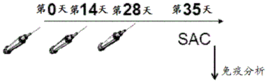
Figure 10 shows a schematic illustrating the immunization protocol.
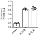
Fig. 11 shows plots plotting blood draw versus OD450 in (a) and (B).
Fig. 12 shows a matrix of peptide pools.
FIG. 13 shows plotting peptide set pairs per 106Spot-Forming Unit of Individual cells (SFU/10)6Individual cells).
FIG. 14 shows plotting peptide set pairs per 106Spot-Forming Unit of Individual cells (SFU/10)6Individual cells).
FIG. 15 shows the mapping of immunization groups to each 106Spot-Forming Unit of Individual cells (SFU/10)6Individual cells).
FIG. 16 shows (A) mapping of the immune group against CD3+CD4+IFN-γ+A graph of the percentage of T cells; (B) mapping immune group to CD3+CD4+TNF-α+A graph of the percentage of T cells; (C) mapping immune group to CD3+CD4+IL-2+A graph of the percentage of T cells; and (D)) Mapping immune group to CD3+CD4+IFN-γ+TNF-α+Graph of T cell percentage.
FIG. 17 shows (A) mapping of the immune group against CD3+CD8+IFN-γ+A graph of the percentage of T cells; (B) mapping immune group to CD3+CD8+TNF-α+A graph of the percentage of T cells; (C) mapping immune group to CD3+CD8+IL-2+A graph of the percentage of T cells; and (D) mapping the immune group against CD3+CD8+IFN-γ+TNF-α+Graph of T cell percentage.
Figure 18 shows in (a) the antibody response after vaccination with pVax1DNA vector (negative control), consensus MERS-spike DNA vaccine (MERS-S) or consensus MERS-spike- Δ CD DNA vaccine (MERS-S-CD). Antibody responses were measured by ELISA. In (B), antibody responses over time after vaccination with the consensus MERS-spike DNA vaccine (MERS-S) are shown. In (C), neutralizing antibody titers at day 35 after vaccination with pVax1DNA vector (negative control), MERS-S DNA vaccine or MERS-S-CD DNA vaccine are shown. Mouse sera were diluted serially in MEM and incubated at 37 ℃ with 50ul DMEM containing 100 infectious HCoV-EMC/2012(Human Coronavirus Erasmus Medical Center/2012) particles per well. After 90min, the virus-serum mixture was added to a monolayer of Vero cells (100,000 cells/well) in a 96-well flat-bottom plate and at 5% CO2Incubate at 37 ℃ for 5 days in an incubator. The neutralizing antibody titer for each sample was reported as the highest dilution at which less than 50% of the cells showed CPE. Values are reported as reciprocal dilutions. All samples were run in duplicate, so the final result was determined as the average of both. Percent neutralization was calculated as follows: percent neutralization ═ target 1-pfuumab (per concentration)/average PFU negative control (all concentrations) }.
FIG. 19 shows the construction and characteristics of MERS-HCoV spike DNA vaccine. (A) Dendrograms show the phylogenetic relationship at the amino acid level between MERS-HCoV virus isolates and spike vaccine strains. A rooted contiguous tree was generated from the alignment of the amino acid sequence of the spike protein displayed as (macromolecular) with a common vaccine and (□) represents an MERS-virus strain used to test Nab assays. The shaded area represents reported cases from the 2012-2013 outbreak. Brackets indicate a particular clade. The trees are depicted to scale (scale bar, 0.1% difference). (B) Schematic of spike-Wt gene insert for codon optimized DNA vaccine. The spike-Wt and spike delta CD-DNA vaccines are constructed. The different spike protein domains are shown. (C) Expression of the protein was detected by western blotting. The expression of spike-Wt and spike Δ CD-spike proteins was analyzed by Western blotting using serum from MERS-spike-Wt DNA-inoculated mice at a dilution of 1:100 two days after transfection. The amounts of transfected DNA construct and loaded cell lysate are shown. Arrows indicate spike proteins. (D) Immunofluorescence assay in Vero cells showing spike-Wt and spike Δ CD protein expression using serum from DNA-immunized mice (1: 100).
FIG. 20 shows the construction and characteristics of MERS-spike pseudovirus. (A) Schematic representation of MERS-HCoV-spike pseudovirus. (B) Determination of viral infectivity: vero cells were incubated with MERS-spikes pseudotyped with luciferase reporter backbone. Luciferase reporter activity was then assayed at the indicated times to generate the time course of expression. Curves were fitted to the data by non-linear regression analysis and the best fit was a straight line. (C) Detection of MERS-HCoV-spike pseudovirus infectivity in different cell lines. MERS-spike pseudovirus and negative control VSV-G virus were used for infection. Data are expressed as mean Relative Luciferase Units (RLU) ± Standard Deviation (SD) for 3 parallel wells in a 96-well plate. The experiment was repeated three times and similar results were obtained.
Figure 21 shows a functional profile of the cellular immune response elicited by MERS-HCoV vaccines. (A) Groups of C57/BL6 mice (n-9/group) were immunized three times with 25 μ g of the spike-Wt and spike Δ CD DNA vaccines as indicated every 2 weeks. Samples were collected one week after the third immunization. (B) Spike-specific T-lymphocyte responses were analyzed by IFN- γ ELISpot assay using a collection of 6 peptides covered with spike protein. Values represent the average response in each group one week after the third immunization. The error range is expressed in terms of standard error. (C)&D) MERS-spike specific dominant epitope characteristics. Using peptide poolsMatrix, IFN-. gamma.secretion by splenocytes from MERS-spike specific immune response. Results are expressed as the number of IFN-gamma secreting cells/10 after subtraction of the values without peptide stimulation6Individual splenocytes (mean ± SD for three mice per group). Similar results were obtained in two separate experiments.
FIG. 22 shows systemic anti-MERS-HCoV IgG levels after DNA immunization. (A) Serum anti-IgG responses in mice were measured by ELISA with serum dilutions against MERS-spike as coating antigen as indicated. Pooled sera from each group of mice were serially diluted (one week after the third immunization) and MERS-spike specific total IgG measured by ELISA. Each curve represents the OD value of each group of 9 mice receiving either the spike-Wt DNA vaccine, the spike Δ CD-spike DNA vaccine or the empty DNA vector as indicated. Each data point is the average absorbance from nine mice. (B) Endpoint titers of the sera of spike-Wt immunized mice were calculated at the indicated time points. (C) Western blot analysis of immune sera binding to recombinant spike protein at the indicated concentrations (μ g). Pooled sera from spike-Wt immunized mice were used as primary antibodies at a 1:100 dilution. (D) The detected neutralizing antibody response was determined by viral infection. Sera were collected from immunized mice one week after the third immunization. 50% Inhibition (IC) of viral infection in target cells50) The neutralizing antibody titer was measured as follows. Data shown are geometric mean titers for each group at standard deviation. Statistical differences between each test group were determined and p-values are shown. (E) Neutralization of the infectivity of MERS-spike-pseudotyped virus by antiserum obtained from mice after receiving three injections of spike-Wt DNA or pVax1 DNA. IC (integrated circuit)50Defined as the reciprocal of the antiserum dilution at which virus entry was inhibited by 50% (dashed line). Data from 4 mice per group are shown.
FIG. 23 shows an analysis of interferon-gamma (IFN-. gamma.) producing cells induced by MERS-spike DNA vaccine in non-human primates (NHPs). (A) Study design, vaccine and challenge protocol in rhesus monkeys. Three groups (low, high and control) of rhesus monkeys (n ═ were treated with MERS-spike DNA at weeks 0, 3 and 6 as indicated (wks)4/each group). Samples from the third immunization were subjected to IFN- γ ELISpot, intracellular cytokine staining and antibody binding and Nab assays. Biopsy was performed after MERS-challenge. (B) ELISPOT analysis of cells from MERS-spike DNA immunized monkeys. PBMCs were isolated from each DNA-immunized monkey and used in an ELISPOT assay to detect IFN- γ -producing cells in response to MERS-spike peptide pools for 24 hours as described in example 9. MERS-spike specific IFN-gamma secreting cells/10 as determined by ELISpot assay6The frequency of individual PBMCs. Results are expressed as mean ± SEM. (C) Generating CD4+And CD8+Macaque PBMC (n ═ 4) were isolated after the last DNA immunization and stimulated ex vivo with pooled MERS-spike peptides cells were stained for intracellular production of IFN- γ, TNF- α, and IL-2, and then analyzed by multiparameter flow cytometry.
Figure 24 shows antibody binding titers and neutralizing antibody (Nab) responses after MERS-spike DNA vaccination. (A & B) MERS-specific antibody and IgG-specific endpoint titers in serum binding at various times post-immunization. Sera were collected after each vaccination and the endpoint titers were calculated by ELISA. (C & D) Effect of DNA vaccination and characterization of neutralizing antibody response. Blood samples were obtained from the animals before each immunization and two weeks after the last vaccination. The sera were tested for neutralizing antibodies. Neutralization with live virus (C) or MERS-pseudovirus (D). Serial dilutions of immune sera were tested with MERS-virus. For the pseudovirus neutralization assay, anti-CHIKV monkey serum was used as a negative antibody control, and VSV-G pseudotyped virus was used as a pseudovirus control for neutralization specificity. Test samples were assayed by PRNT50 using MERS virus.
FIG. 25 shows CD4 primed by MERS-spike vaccine+And CD8+Functional profile of T cell response. Specific CD4 induced after vaccination+T cells and CD8+Cytokine profiles of T cells are shown in (a) and (B), respectively, mouse splenocytes (n ═ 3) were isolated one week after the last DNA immunization and stimulated ex vivo with pooled MERS-spike peptidesIntracellular production cells were stained and then analyzed by FACS. (A)&B) The scatter plot depicts MERS-specific CD4 releasing IFN-gamma, TNF- α, IL-2, and dual IFN-gamma/TNF- α cytokines+T and CD8+T cells. (C)&D) Bar graphs show single, double and triple positive CD4 for the release of the cytokines IFN-. gamma.TNF- α and IL-2+And CD8+A multifunctional subpopulation of T cells. The circle shows the proportion of each cytokine subpopulation. Data represent mean ± SEM of three mice per group.
Figure 26 shows inhibition of MERS-S pseudovirus-induced apoptosis by immune sera from spiked DNA immunized mice. (A) MERS-HCoV-spike mouse sera neutralized MERS in vitro and blocked syncytia formation. Uninfected Vero cells represent negative controls, while positive controls represent those cells infected with MERS-HCoV pseudovirus. Arrows indicate syncytia formation. Syncytia formation was observed under the microscope 36 hours after infection. (B) MERS-spike pseudoviruses were preincubated and added to Vero cells in the presence of pVax-1 or MERS-spike immune serum at a 1:100 dilution. Two days after infection, the percentage of cell death was measured by annexin V/PI staining. The bar graphs show infection of MERS-pseudovirus values from FACS data of a single experiment performed in triplicate. Similar results were obtained in three separate experiments.
Figure 27 shows the antibody response after vaccination with MERS-spike DNA in combination with electroporation. Sera from monkeys vaccinated with pVax1, pMERS-spikes (0.5 mg/animal; low dose) or pMERS-spikes (2 mg/animal; high dose) were collected two weeks after the third immunization. These sera were tested for antibodies that bind MERS-spike protein. The results of this test are shown in (A) and (B).
Figure 28 shows results from MERS challenge and pathology after vaccination. (A) Mean viral load determined by qRT-PCR from individual tissues collected at necropsy. (B) The average viral load of all lung lobes combined is shown in the inset. Log TCID50eq/g, Log TCID50 equivalents/g tissue was analyzed; one sample/tissue/animal from three animals/group.
Detailed Description
The present invention relates to a vaccine comprising a middle east respiratory syndrome coronavirus (MERS-CoV) antigen. MERS-CoV is a novel highly pathogenic virus, occurring only in 2012, and therefore the vaccine described herein is one of the first choice vaccines against the target MERS-CoV. Thus, the vaccine provides a treatment for this novel pathogenic virus, which prior treatments did not exist and still have the potential for pandemics.
The MERS-CoV antigen can be a MERS-CoV consensus spike antigen. MERS-CoV consensus spike antigen can be derived from a spike antigen sequence from MERS-CoV strain, and thus, MERS-CoV consensus spike antigen is unique. MERS-CoV consensus spike antigen may lack a cytoplasmic domain. Thus, the vaccine of the present invention is broadly applicable to multiple strains of MERS-CoV because of the unique sequence of MERS-CoV consensus spike antigen. These unique sequences allow the vaccine to be fully protective against multiple strains of MERS-CoV, including genetically diverse variants of MERS-CoV.
The vaccine can be used to protect against infection with many strains of MERS-CoV and to treat these strains. The vaccine can elicit humoral and cellular immune responses that target MERS-CoV spike antigens. The vaccine can elicit neutralizing antibodies and immunoglobulin g (igg) antibodies reactive with MERS-CoV spike antigen. The vaccine also elicits CD8 that is responsive to MERS-CoV spike antigen+And CD4+T cells respond and produce interferon-gamma (IFN-gamma), tumor necrosis factor α (TNF- α), and interleukin-2 (IL-2).
1. Definition of
Unless defined otherwise, all technical and scientific terms used herein have the same meaning as commonly understood by one of ordinary skill in the art. In case of conflict, the present document, including definitions, will control. Although methods and materials similar or equivalent to those described herein can be used in the practice or testing of the present invention, the preferred methods and materials are described below. All publications, patent applications, patents, and other references mentioned herein are incorporated by reference in their entirety. The materials, methods, and examples disclosed herein are illustrative only and not intended to be limiting.
As used herein, the terms "comprising," "including," "having," "has," "can," "contains," and variations thereof are intended to be open-ended connectors, terms, or words that do not exclude the possibility of additional acts or structures. The singular forms "a", "an" and "the" include plural references unless the context clearly dictates otherwise. The present disclosure also encompasses other embodiments that "comprise," "consist of," and "consist essentially of" the embodiments or elements presented herein, whether or not explicitly stated.
As used herein, "adjuvant" means any molecule added to the vaccines described herein to enhance the immunogenicity of an antigen.
"antibody" as used herein means an antibody of the type IgG, IgM, IgA, IgD or IgE, or a fragment, fragment or derivative thereof, including Fab, F (ab')2Fd and single chain antibodies, diabodies, bispecific antibodies, bifunctional antibodies and derivatives thereof. The antibody may be an antibody isolated from a serum sample of a mammal, a polyclonal antibody, an affinity purified antibody, or a mixture thereof, which exhibits sufficient binding specificity for the desired epitope or a sequence derived therefrom.
"coding sequence" or "coding nucleic acid" as used herein means a nucleic acid (RNA or DNA molecule) comprising a nucleotide sequence encoding a protein. The coding sequence may further include initiation and termination signals operably linked to regulatory elements including a promoter and polyadenylation signals capable of directing expression in the cells of the subject or mammal to which the nucleic acid is administered.
"complementary sequence" or "complementary" as used herein means Watson-Crick (e.g., A-T/U and C-G) or Hoogsteen base pairing between nucleotides or nucleotide analogs of a nucleic acid molecule.
As used herein, "consensus" or "consensus sequence" may refer to a synthetic nucleic acid sequence or corresponding polypeptide sequence constructed based on analysis of an alignment of multiple subtypes of a particular antigen. The sequences may be used to induce broad immunity against multiple subtypes, serotypes, or strains of a particular antigen. Synthetic antigens, such as fusion proteins, can be manipulated to produce consensus sequences (or consensus antigens).
"electroporation," "electro-permeabilization," or "electrokinetic enhancement" ("EP") as used interchangeably herein means the use of transmembrane electric field pulses to induce microscopic pathways (pores) in a biological membrane; their presence allows biomolecules such as plasmids, oligonucleotides, siRNA, drugs, ions and water to flow from one side of the cell membrane to the other.
As used herein, "fragment" means a nucleic acid sequence or portion thereof that encodes a polypeptide capable of eliciting an immune response in a mammal. The fragment may be a DNA fragment selected from at least one of various nucleotide sequences encoding the protein fragments described below.
By "fragment" or "immunogenic fragment" with respect to a polypeptide sequence is meant a polypeptide that is capable of eliciting an immune response in a mammal that is cross-reactive with the full-length wild-type strain MERS-CoV antigen. Fragments of a consensus protein may comprise at least 10%, at least 20%, at least 30%, at least 40%, at least 50%, at least 60%, at least 70%, at least 80%, at least 90%, or at least 95% of the consensus protein. In some embodiments, fragments of a consensus protein may comprise at least 20 amino acids or more, at least 30 amino acids or more, at least 40 amino acids or more, at least 50 amino acids or more, at least 60 amino acids or more, at least 70 amino acids or more, at least 80 amino acids or more, at least 90 amino acids or more, at least 100 amino acids or more, at least 110 amino acids or more, at least 120 amino acids or more of the consensus protein, at least 130 amino acids or more, at least 140 amino acids or more, at least 150 amino acids or more, at least 160 amino acids or more, at least 170 amino acids or more, at least 180 amino acids or more, at least 190 amino acids or more, at least 200 amino acids or more, at least 210 amino acids or more, at least 220 amino acids or more, at least 230 amino acids or more, or at least 240 amino acids or more.
The term "genetic construct" as used herein refers to a DNA or RNA molecule comprising a nucleotide sequence encoding a protein. The coding sequence comprises an initiation signal and a termination signal operably linked to regulatory elements including a promoter and polyadenylation signal capable of directing expression in the cells of the individual to whom the nucleic acid molecule is administered. The term "expressible form" as used herein refers to a genetic construct containing the necessary regulatory elements operably linked to a coding sequence encoding a protein, such that the coding sequence will be expressed when present in the cells of an individual.
The term "identical" or "identity" as used herein in the context of two or more nucleic acid or polypeptide sequences means that the sequences have a specified percentage of residues that are identical over the specified region. The percentage may be calculated by: optimally aligning the two sequences, comparing the two sequences over a specified region, determining the number of positions of the identical residue in the two sequences to yield the number of matched positions, dividing the number of matched positions by the total number of positions within the specified region, and multiplying the result by 100 to yield the percentage of sequence identity. Where two sequences are of different lengths or the alignment produces one or more staggered ends and the specified regions of comparison include only a single sequence, the residues of the single sequence are included in the denominator of the calculation rather than in the numerator. When comparing DNA and RNA, thymine (T) and uracil (U) can be considered equivalent. Identity can be performed manually or by using a computer sequence algorithm such as BLAST or BLAST 2.0.
As used herein, "immune response" means the activation of the host's immune system (e.g., the mammalian immune system) in response to the introduction of an antigen. The immune response may be in the form of a cellular response or a humoral response or both.
As used herein, a "nucleic acid" or "oligonucleotide" or "polynucleotide" means at least two nucleotides covalently linked together. The description of single strands also defines the sequence of the complementary strand. Thus, nucleic acids also encompass the complementary strand of the single strand described. Many variants of a nucleic acid can be used for the same purpose as a given nucleic acid. Thus, nucleic acids also encompass substantially the same nucleic acids and their complements. Single strands provide probes that can hybridize to a target sequence under stringent hybridization conditions. Thus, nucleic acids also encompass probes that hybridize under stringent hybridization conditions.
The nucleic acid may be single-stranded or double-stranded or may contain portions of both double-stranded or single-stranded sequences. The nucleic acid can be DNA, both genomic and cDNA, RNA, or a hybrid, wherein the nucleic acid can contain a combination of deoxyribonucleotides and ribonucleotides, as well as a combination of bases including uracil, adenine, thymine, cytosine, guanine, inosine, xanthine hypoxanthine, isocytosine, and isoguanine. The nucleic acid may be obtained by chemical synthesis methods or by recombinant methods.
"operably linked" as used herein means that expression of a gene is under the control of a promoter to which it is spatially linked. Under its control, the promoter may be positioned 5 '(upstream) or 3' (downstream) of the gene. The distance between the promoter and the gene may be about the same as the distance between the promoter and the gene it controls in the gene from which the promoter is derived. As is known in the art, this change in distance can be adjusted without loss of promoter function.
As used herein, "peptide," "protein," or "polypeptide" can mean a linked sequence of amino acids, and can be natural, synthetic, or a modification or combination of natural and synthetic.
As used herein, "promoter" means a molecule of synthetic or natural origin that is capable of conferring, activating or enhancing expression of a nucleic acid in a cell. The promoter may comprise one or more specific transcriptional regulatory sequences to further enhance expression and/or alter spatial and/or temporal expression thereof. A promoter may also contain distal enhancer or repressor elements, which can be located as much as several thousand pairs of base pairs from the start of transcription. Promoters may be obtained from sources including viruses, bacteria, fungi, plants, insects, and animals. Promoters may regulate constitutive or differential expression of a genomic fraction relative to a cell, tissue or organ in which expression occurs or relative to a developmental stage at which expression occurs or in response to an external stimulus such as a physiological stress, pathogen, metal ion or inducer. Representative examples of promoters include the bacteriophage T7 promoter, the bacteriophage T3 promoter, the SP6 promoter, the lactose operon-promoter, the tac promoter, the SV40 late promoter, the SV40 early promoter, the RSV-LTR promoter, the CMV IE promoter, the SV40 early promoter or the SV40 late promoter, and the CMV IE promoter.
"Signal peptide" and "leader sequence" are used interchangeably herein and refer to an amino acid sequence that can be attached to the amino terminus of a MERS-CoV protein as described herein. The signal peptide/leader sequence is generally indicative of the position of the protein. The signal peptide/leader sequence used herein preferably facilitates secretion of the protein from the cell in which it is produced. The signal peptide/leader sequence is often cleaved from the remainder of the protein, which is often referred to as the mature protein after secretion from the cell. The signal peptide/leader sequence is linked to the N-terminus of the protein.
As used herein, a "subject" can mean a mammal that wants or needs to be immunized with a vaccine described herein. The mammal can be a human, chimpanzee, dog, cat, horse, cow, mouse, or rat.
As used herein, "substantially identical" may mean that the first amino acid sequence and the second amino acid sequence are at least 60%, 65%, 70%, 75%, 80%, 81%, 82%, 83%, 84%, 85%, 86%, 87%, 88%, 89%, 90%, 91%, 92%, 93%, 94%, 95%, 96%, 97%, 98%, or 99% over a region of 1,2, 3,4, 5,6, 7, 8, 9, 10, 11, 12, 13, 14, 15, 16, 17, 18, 19, 20, 21, 22, 23, 24, 25, 30, 35, 40, 45, 50, 55, 60, 65, 70, 75, 80,85, 90, 95, 100, 200, 300, 400, 500, 600, 700, 800, 900, 1000, 1100, or more amino acids. "substantially identical" may also mean that the first nucleic acid sequence and the second nucleic acid sequence are at least 60%, 65%, 70%, 75%, 80%, 81%, 82%, 83%, 84%, 85%, 86%, 87%, 88%, 89%, 90%, 91%, 92%, 93%, 94%, 95%, 96%, 97%, 98%, or 99% over a region of 1,2, 3,4, 5,6, 7, 8, 9, 10, 11, 12, 13, 14, 15, 16, 17, 18, 19, 20, 21, 22, 23, 24, 25, 30, 35, 40, 45, 50, 55, 60, 65, 70, 75, 80,85, 90, 95, 100, 200, 300, 400, 500, 600, 700, 800, 900, 1000, 1100, or more nucleotides.
As used herein, "Treatment" or "Treating" can mean protecting an animal from disease or completely eliminating disease by means of prevention, suppression, repression. Prevention of disease involves administering the vaccine of the present invention to an animal prior to the onset of disease. Inhibiting disease involves administering the vaccine of the present invention to an animal after induction of the disease but prior to its clinical appearance. Suppression of disease involves administering a vaccine of the invention to an animal after clinical appearance of the disease.
"variant" as used herein with respect to a nucleic acid means (i) a portion or fragment of a reference nucleotide sequence; (ii) a complement of a reference nucleotide sequence or a portion thereof; (iii) a nucleic acid that is substantially identical to a reference nucleic acid or a complement thereof; or (iv) a nucleic acid that hybridizes under stringent conditions to a reference nucleic acid, its complement, or a sequence substantially identical thereto.
A variant may be further defined as a peptide or polypeptide that differs in amino acid sequence by insertion, deletion, or conservative substitution of amino acids, but retains at least one biological activity. Representative examples of "biological activity" include the ability to be bound by a specific antibody or to promote an immune response. Variant also means a protein having substantially the same amino acid sequence as a reference protein having an amino acid sequence that retains at least one biological activity. Conservative substitutions of amino acids, i.e., the replacement of an amino acid with a different amino acid of similar characteristics (e.g., hydrophilicity, extent and distribution of charged regions) are believed in the art to typically involve minor changes. These minor changes may be identified in part by considering the hydropathic index of the amino acid, as understood in the art. Kyte et al, J.mol.biol.157:105-132 (1982). The hydropathic index of the amino acid is based on considerations of its hydrophobicity and charge. It is known in the art that amino acids of similar hydropathic indices can be substituted and still retain protein function. In one aspect, amino acids with a hydropathic index of ± 2 are substituted. The hydrophilicity of amino acids can also be used to reveal substitutions that will result in proteins that retain biological function. Considering the hydrophilicity of amino acids in the case of peptides allows the calculation of the maximum local average hydrophilicity of the peptide, which is a useful measure that has been reported to correlate well with antigenicity and immunogenicity. As understood in the art, substitutions with amino acids having similar hydrophilicity values can result in peptides that retain biological activity (e.g., immunogenicity). The substitution may be with an amino acid having a hydrophilicity value within. + -. 2. Both the hydropathic index and the hydropathic value of an amino acid are affected by the specific side chain of the amino acid. Consistent with this observation, amino acid substitutions that are compatible with biological function are understood to depend on the relative similarity of these amino acids, and in particular the side chains of those amino acids, as revealed by hydrophobicity, hydrophilicity, charge, size, and other properties.
Variants may be nucleic acid sequences that are substantially identical over the entire length of the entire gene sequence or a fragment thereof. The nucleic acid sequence may be 80%, 81%, 82%, 83%, 84%, 85%, 86%, 87%, 88%, 89%, 90%, 91%, 92%, 93%, 94%, 95%, 96%, 97%, 98%, 99% or 100% identical over the entire length of the gene sequence or fragment thereof. Variants may be amino acid sequences that are substantially identical over the entire length of the amino acid sequence or fragment thereof. The amino acid sequence may be 80%, 81%, 82%, 83%, 84%, 85%, 86%, 87%, 88%, 89%, 90%, 91%, 92%, 93%, 94%, 95%, 96%, 97%, 98%, 99% or 100% identical over the entire length of the amino acid sequence or fragment thereof.
"vector" as used herein means a nucleic acid sequence containing an origin of replication. The vector may be a viral vector, a bacteriophage, a bacterial artificial chromosome, or a yeast artificial chromosome. The vector may be a DNA or RNA vector. The vector may be a self-replicating extra-chromosomal vector, and is preferably a DNA plasmid.
For the recitation of numerical ranges herein, each intervening number there between with the same degree of accuracy is specifically contemplated. For example, for the range of 6 to 9, the numbers 7 and 8 are encompassed in addition to 6 and 9, and for the range 6.0 to 7.0, the numbers 6.0, 6.1, 6.2, 6.3, 6.4, 6.5, 6.6, 6.7, 6.8, 6.9 and 7.0 are specifically encompassed.
2. Vaccine
Provided herein are immunogenic compositions, such as vaccines, comprising a middle east respiratory syndrome coronavirus (MERS-CoV) antigen, a fragment thereof, a variant thereof, or a combination thereof. The vaccine can be used to protect against infection with any number of strains of MERS-CoV, thereby treating, preventing and/or protecting against MERS-CoV-based pathologies. The vaccine can significantly induce an immune response in a subject administered the vaccine, thereby protecting against infection with MERS-CoV and treating MERS-CoV infection.
The vaccine may be a DNA vaccine, a peptide vaccine, or a combination of DNA and peptide vaccines. The DNA vaccine can comprise a nucleic acid sequence encoding a MERS-CoV antigen. The nucleic acid sequence may be DNA, RNA, cDNA, variants thereof, fragments thereof, or combinations thereof. The nucleic acid sequence may further comprise an additional sequence encoding a linker, leader or marker sequence, which is linked to the MERS-CoV antigen by a peptide bond. The peptide vaccine can include MERS-CoV antigenic peptides, MERS-CoV antigenic proteins, variants thereof, fragments thereof, or combinations thereof. The combination of DNA and peptide vaccine can comprise the above-described nucleic acid sequence encoding MERS-CoV antigen and MERS-CoV antigenic peptide or protein, wherein MERS-CoV antigenic peptide or protein has the same amino acid sequence as the encoded MERS-CoV antigen.
The vaccine can induce a humoral immune response in a subject to whom the vaccine is administered. The induced humoral immune response may be specific for the MERS-CoV antigen. The induced humoral immune response can react with MERS-CoV antigens. The humoral immune response may be induced from about 1.5-fold to about 16-fold, from about 2-fold to about 12-fold, or from about 3-fold to about 10-fold in a subject administered the vaccine. A humoral immune response can be induced at least about 1.5-fold, at least about 2.0-fold, at least about 2.5-fold, at least about 3.0-fold, at least about 3.5-fold, at least about 4.0-fold, at least about 4.5-fold, at least about 5.0-fold, at least about 5.5-fold, at least about 6.0-fold, at least about 6.5-fold, at least about 7.0-fold, at least about 7.5-fold, at least about 8.0-fold, at least about 8.5-fold, at least about 9.0-fold, at least about 9.5-fold, at least about 10.0-fold, at least about 10.5-fold, at least about 11.0-fold, at least about 11.5-fold, at least about 12.0-fold, at least about 12.5-fold, at least about 13.0-fold, at least about 13.5-fold, at least about 14.0-fold, at least about 14.5-fold, at least about 15.0-fold, at least about 15.5-fold, or at least about 16..
The humoral immune response induced by the vaccine can include an increased level of neutralizing antibodies associated with a subject administered the vaccine as compared to a subject not administered the vaccine. The neutralizing antibody can be specific for the MERS-CoV antigen. Neutralizing antibodies can react with MERS-CoV antigens. Neutralizing antibodies can provide protection against and/or treat MERS-CoV infection and associated pathologies in a subject administered the vaccine.
The humoral immune response induced by the vaccine can include increased levels of IgG antibodies associated with the subject administered the vaccine as compared to a subject not administered the vaccine. These IgG antibodies can be specific for MERS-CoV antigens. These IgG antibodies can react with MERS-CoV antigen. Preferably, the humoral response is cross-reactive to two or more strains of MERS-CoV. IgG antibody levels associated with a subject administered the vaccine can be increased from about 1.5-fold to about 16-fold, from about 2-fold to about 12-fold, or from about 3-fold to about 10-fold compared to a subject not administered the vaccine. IgG antibody levels associated with a subject administered the vaccine can be increased at least about 1.5 fold, at least about 2.0 fold, at least about 2.5 fold, at least about 3.0 fold, at least about 3.5 fold, at least about 4.0 fold, at least about 4.5 fold, at least about 5.0 fold, at least about 5.5 fold, at least about 6.0 fold, at least about 6.5 fold, at least about 7.0 fold, at least about 7.5 fold, at least about 8.0 fold, at least about 8.5 fold, at least about 9.0 fold, at least about 9.5 fold, at least about 10.0 fold, at least about 10.5 fold, at least about 11.0 fold, at least about 11.5 fold, at least about 12.0 fold, at least about 12.5 fold, at least about 13.0 fold, at least about 13.5 fold, at least about 14.0 fold, at least about 14.5 fold, at least about 15.0 fold, at least about 15.5 fold, or at least about 16 fold as compared to a subject not administered the vaccine.
The vaccine can induce a cellular immune response in a subject to which the vaccine is administered. The induced cellular immune response may be specific for MERS-CoV antigen. The induced cellular immune response may be reactive to MERS-CoV antigens. Preferably, the cellular response is cross-reactive to two or more strains of MERS-CoV. The induced cellular immune response may comprise eliciting CD8+T cell response. Induced CD8+The T cell response can react with MERS-CoV antigen. Induced CD8+T cell responses may be multifunctional. The induced cellular immune response may comprise eliciting CD8+T cell response, in which CD8+T cells produce interferon-gamma (IFN-gamma), tumor necrosis factor α (TNF- α), interleukin-2 (IL-2), or a combination of IFN-gamma and TNF- α.
The induced cellular immune response may comprise enhanced CD8 associated with the subject administered the vaccine as compared to a subject not administered the vaccine+T cell response. CD8 associated with subjects administered vaccines+The T cell response may be enhanced from about 2-fold to about 30-fold, from about 3-fold to about 25-fold, or from about 4-fold to about 20-fold compared to a subject not administered the vaccine. CD8 associated with subjects administered vaccines+The T cell response may be enhanced at least about 1.5 fold, at least about 2.0 fold, at least about 3.0 fold, at least about 4.0 fold, at least about 5.0 fold, at least about 6.0 fold, at least about 6.5 fold, at least about 7.0 fold, at least about 7.5 fold, at least about 8.0 fold, at least about 8.5 fold, at least about 9.0 fold, at least about 9.5 fold, at least about 10.0 fold, at least about 10.5 fold, at least about 11.0 fold, at least about 11.5 fold, at least about 12.0 fold, at least about 12.5 fold, at least about 13.0 fold, at least about 13.5 fold, at least about 14.0 fold, at least about 14.5 fold, at least about 15.0 fold, at least about 16.0 fold, at least about 17.0 fold, at least about 18.0 fold, at least about 19.0 fold, at least about 20.0 fold, at least about 21.0 fold, at least about 22.0 fold, at least about 22 fold, at least about 10.0 fold23.0 times, at least about 24.0 times, at least about 25.0 times, at least about 26.0 times, at least about 27.0 times, at least about 28.0 times, at least about 29.0 times, or at least about 30.0 times.
The induced cellular immune response may include IFN-gamma producing CD3+CD8+The incidence of T cells increases. CD3 associated with subjects administered vaccines+CD8+IFN-γ+The incidence of T cells may be increased at least about 2-fold, 3-fold, 4-fold, 5-fold, 6-fold, 7-fold, 8-fold, 9-fold, 10-fold, 11-fold, 12-fold, 13-fold, 14-fold, 15-fold, 16-fold, 17-fold, 18-fold, 19-fold, or 20-fold compared to a subject not administered the vaccine.
The cellular immune response induced may include CD3 producing TNF- α+CD8+The incidence of T cells increases. CD3 associated with subjects administered vaccines+CD8+TNF-α+The incidence of T cells may be increased at least about 2-fold, 3-fold, 4-fold, 5-fold, 6-fold, 7-fold, 8-fold, 9-fold, 10-fold, 11-fold, 12-fold, 13-fold, or 14-fold compared to a subject not administered the vaccine.
The induced cellular immune response may include IL-2 producing CD3+CD8+The incidence of T cells increases. CD3 associated with subjects administered vaccines+CD8+IL-2+The incidence of T cells may be increased at least about 0.5-fold, 1.0-fold, 1.5-fold, 2.0-fold, 2.5-fold, 3.0-fold, 3.5-fold, 4.0-fold, 4.5-fold, or 5.0-fold compared to a subject not administered the vaccine.
The cellular immune response induced may include IFN-. gamma.and TNF- α producing CD3+CD8+The incidence of T cells increases. CD3 associated with subjects administered vaccines+CD8+IFN-γ+TNF-α+The incidence of T cells may be increased at least about 25-fold, 30-fold, 35-fold, 40-fold, 45-fold, 50-fold, 55-fold, 60-fold, 65-fold, 70-fold, 75-fold, 80-fold, 85-fold, 90-fold, 95-fold, 100-fold, 110-fold, 120-fold, 130-fold, 140-fold, 150-fold, 160-fold, 170-fold, or 180-fold compared to a subject not administered the vaccine.
The cellular immune response induced by the vaccine may include priming of CD4+T is thin(ii) a cellular response. Induced CD4+The T cell response can react with MERS-CoV antigen. Induced CD4+T cell responses may be multifunctional. The induced cellular immune response may comprise eliciting CD4+T cell response, in which CD4+T cells produce IFN- γ, TNF- α, IL-2, or a combination of IFN- γ and TNF- α.
The induced cellular immune response may include IFN-gamma producing CD3+CD4+The incidence of T cells increases. CD3 associated with subjects administered vaccines+CD4+IFN-γ+The incidence of T cells may be increased at least about 2-fold, 3-fold, 4-fold, 5-fold, 6-fold, 7-fold, 8-fold, 9-fold, 10-fold, 11-fold, 12-fold, 13-fold, 14-fold, 15-fold, 16-fold, 17-fold, 18-fold, 19-fold, or 20-fold compared to a subject not administered the vaccine.
The cellular immune response induced may include CD3 producing TNF- α+CD4+The incidence of T cells increases. CD3 associated with subjects administered vaccines+CD4+TNF-α+The incidence of T cells may be increased at least about 2-fold, 3-fold, 4-fold, 5-fold, 6-fold, 7-fold, 8-fold, 9-fold, 10-fold, 11-fold, 12-fold, 13-fold, 14-fold, 15-fold, 16-fold, 17-fold, 18-fold, 19-fold, 20-fold, 21-fold, or 22-fold compared to a subject not administered the vaccine.
The induced cellular immune response may include IL-2 producing CD3+CD4+The incidence of T cells increases. CD3 associated with subjects administered vaccines+CD4+IL-2+The incidence of T cells can be increased at least about 2-fold, 3-fold, 4-fold, 5-fold, 6-fold, 7-fold, 8-fold, 9-fold, 10-fold, 11-fold, 12-fold, 13-fold, 14-fold, 15-fold, 16-fold, 17-fold, 18-fold, 19-fold, 20-fold, 21-fold, 22-fold, 23-fold, 24-fold, 25-fold, 26-fold, 27-fold, 28-fold, 29-fold, 30-fold, 31-fold, 32-fold, 33-fold, 34-fold, 35-fold, 36-fold, 37-fold, 38-fold, 39-fold, 40-fold, 45-fold, 50-fold, 55-fold, or 60-fold as compared to a subject not administered the vaccine.
The cellular immune response induced may include IFN-. gamma.and TNF- α producing CD3+CD4+The incidence of T cells increases. And administration of epidemic diseaseSubject-related CD3 of seedlings+CD4+IFN-γ+TNF-α+The frequency of cells may be increased by at least about 2-fold, 2.5-fold, 3.0-fold, 3.5-fold, 4.0-fold, 4.5-fold, 5.0-fold, 5.5-fold, 6.0-fold, 6.5-fold, 7.0-fold, 7.5-fold, 8.0-fold, 8.5-fold, 9.0-fold, 9.5-fold, 10.0-fold, 10.5-fold, 11.0-fold, 11.5-fold, 12.0-fold, 12.5-fold, 13.0-fold, 13.5-fold, 14.0-fold, 14.5-fold, 15.0-fold, 15.5-fold, 16.0-fold, 16.5-fold, 17.0-fold, 17.5-fold, 18.0-fold, 18.5-fold, 19.0-fold, 19.5-fold, 20.0-fold, 21-fold, 22-fold, 23-fold, 24-fold, 25-fold, 26-fold, 28-fold, 29-fold, 30-fold, 31-fold, 33-fold, or 35-fold as compared to a subject not administered the vaccine.
The vaccines of the present invention may have the characteristics required for an effective vaccine, e.g. be safe, so that the vaccine itself does not cause disease or death; protection from disease caused by exposure to a live pathogen (such as a virus or bacteria); inducing neutralizing antibodies to prevent infection of the cells; inducing protective T cells against intracellular pathogens; and providing ease of administration, few side effects, biological stability, and low cost per dose.
The vaccine may further induce an immune response when administered to different tissues such as muscle or skin. The vaccine may further induce an immune response when administered via electroporation or injection or subcutaneously or intramuscularly.
a. Middle east respiratory syndrome coronavirus (MERS-CoV) antigen
As described above, the vaccine comprises MERS-CoV antigen, fragments thereof, variants thereof, or combinations thereof. Coronaviruses, including MERS-CoV, are membrane-coated and have type 1 membrane glycoproteins, called spike (S) proteins, which form protruding spikes on the coronavirus surface. The spike protein facilitates binding of the coronavirus to a protein located on the cell surface, such as the metalloprotease aminopeptidase N, and mediates cell-virus membrane fusion. In particular, the spike protein contains the S1 subunit, which facilitates binding of the coronavirus to a cell surface protein. Thus, the S1 subunit of spike protein controls which cells are infected with coronavirus. The spike protein also contains the S2 subunit, which is a transmembrane subunit that facilitates fusion of the virus and cell membrane. Thus, the MERS-CoV antigen can comprise the MERS-CoV spike protein, the S1 subunit of the MERS-CoV spike protein, or the S2 subunit of the MERS-CoV spike protein.
Once bound to cell surface proteins and membrane fusion, the coronavirus enters the cell and its single-stranded RNA genome is released into the cytoplasm of the infected cell. The single-stranded RNA genome is the positive strand and can therefore be translated into an RNA polymerase, which produces additional viral RNA that is the negative strand. Thus, the MERS-CoV antigen can also be MERS-CoV RNA polymerase.
The viral RNA negative strand is transcribed into a smaller subgenomic RNA positive strand, which is used to translate other viral proteins, such as the nucleocapsid (N) protein, the envelope (E) protein, and the matrix (M) protein. Thus, the MERS-CoV antigen can comprise a MERS-CoV nucleocapsid protein, a MERS-CoV envelope protein, or a MERS-CoV matrix protein.
The viral RNA negative strand can also be used to replicate the viral genome, which is bound by the nucleocapsid protein. The matrix protein is integrated into the endoplasmic reticulum of the infected cell along with the spike protein. At the same time, the nucleocapsid protein binds to the viral genome and the membrane-embedded matrix and spike protein bud into the lumen of the endoplasmic reticulum, thereby encapsulating the viral genome in the membrane. The viral progeny are then transported to the cell membrane of the infected cell by the golgi vesicles and released into the intercellular spaces by endocytosis.
In some embodiments, the MERS-CoV antigen can be MERS-CoV spike protein, MERS-CoV RNA polymerase, MERS-CoV nucleocapsid protein, MERS-CoV envelope protein, MERS-CoV matrix protein, fragments thereof, variants thereof, or combinations thereof. The MERS-CoV antigen can be a consensus antigen derived from two or more MERS-CoV spike antigens, two or more MERS-CoVRNA polymerases, two or more MERS-CoV nucleocapsid proteins, two or more envelope proteins, two or more matrix proteins, or a combination thereof. MERS-CoV consensus antigens can be modified to improve expression. Modifications may include codon optimization, RNA optimization, addition of kozak sequences to increase translation initiation, and/or addition of immunoglobulin leader sequences to increase immunogenicity of MERS-CoV antigens. In some embodiments, the MERS-CoV antigen comprises an IgE leader sequence, which may be the amino acid sequence set forth in SEQ ID NO. 6 and encoded by the nucleotide sequence set forth in SEQ ID NO. 5.
(1) MERS-CoV spike antigen
The MERS-CoV antigen can be a MERS-CoV spike antigen, a fragment thereof, a variant thereof, or a combination thereof. The MERS-CoV spike antigen is capable of eliciting an immune response in a mammal against one or more MERS-CoV strains. MERS-CoV spike antigen can contain epitopes that make it particularly effective as an immunogen that can induce an immune response against MERS-CoV.
The MERS-CoV spike antigen can be a consensus sequence derived from two or more MERS-CoV strains. The MERS-CoV spike antigen can comprise a consensus sequence and/or a modification for improved expression. Modifications may include codon optimization, RNA optimization, addition of kozak sequences to increase translation initiation, and/or addition of immunoglobulin leader sequences to increase immunogenicity of MERS-CoV spike antigens. MERS-CoV consensus spike antigen may comprise a signal peptide such as an immunoglobulin signal peptide, for example, but not limited to, an immunoglobulin e (ige) or immunoglobulin (IgG) signal peptide. In some embodiments, the MERS-CoV consensus spike antigen may comprise a Hemagglutinin (HA) label. MERS-CoV consensus spike antigens can be designed to elicit stronger and broader cellular and/or humoral immune responses than corresponding codon-optimized spike antigens.
The MERS-CoV consensus spike antigen can be the nucleic acid sequence SEQ ID NO:1, which encodes SEQ ID NO:2 (FIGS. 8A and 8B). In some embodiments, the MERS-CoV consensus spike antigen can be a nucleic acid sequence having at least about 80%, 81%, 82%, 83%, 84%, 85%, 86%, 87%, 88%, 89%, 90%, 91%, 92%, 93%, 94%, 95%, 96%, 97%, 98%, 99%, or 100% identity over the entire length of the nucleic acid sequence set forth in SEQ ID No. 1. In other embodiments, the MERS-CoV consensus spike antigen can be a nucleic acid sequence encoding an amino acid sequence having at least about 80%, 81%, 82%, 83%, 84%, 85%, 86%, 87%, 88%, 89%, 90%, 91%, 92%, 93%, 94%, 95%, 96%, 97%, 98%, 99%, or 100% identity over the entire length of the amino acid sequence set forth in SEQ ID No. 2.
The MERS-CoV consensus spike antigen can be the amino acid sequence SEQ ID NO 2. In some embodiments, the MERS-CoV consensus spike antigen can be an amino acid sequence having at least about 80%, 81%, 82%, 83%, 84%, 85%, 86%, 87%, 88%, 89%, 90%, 91%, 92%, 93%, 94%, 95%, 96%, 97%, 98%, 99%, or 100% identity over the entire length of the amino acid sequence set forth in SEQ ID No. 2.
Immunogenic fragments of SEQ ID NO 2 can be provided. An immunogenic fragment can comprise at least 60%, at least 65%, at least 70%, at least 75%, at least 80%, at least 85%, at least 90%, at least 95%, at least 96%, at least 97%, at least 98%, or at least 99% of SEQ ID No. 2. In some embodiments, the immunogenic fragment comprises a leader sequence, such as, for example, an immunoglobulin leader, such as an IgE leader. In some embodiments, the immunogenic fragment does not contain a leader sequence.
Immunogenic fragments of proteins having amino acid sequences homologous to the immunogenic fragment of SEQ ID NO. 2 can be provided. The immunogenic fragment can comprise a protein that is at least 60%, at least 65%, at least 70%, at least 75%, at least 80%, at least 85%, at least 90%, at least 95%, at least 96%, at least 97%, at least 98%, or at least 99% homologous to SEQ ID NO: 295%. Some embodiments relate to immunogenic fragments that are 96% homologous to the immunogenic fragments of the consensus protein sequences herein. Some embodiments relate to immunogenic fragments that are 97% homologous to the immunogenic fragments of the consensus protein sequences herein. Some embodiments relate to immunogenic fragments that are 98% homologous to the immunogenic fragments of the consensus protein sequences herein. Some embodiments relate to immunogenic fragments that are 99% homologous to the immunogenic fragments of the consensus protein sequences herein. In some embodiments, the immunogenic fragment comprises a leader sequence, such as, for example, an immunoglobulin leader, such as an IgE leader. In some embodiments, the immunogenic fragment does not contain a leader sequence.
Some embodiments relate to an immunogenic fragment of SEQ ID NO 1. An immunogenic fragment can be at least 60%, at least 65%, at least 70%, at least 75%, at least 80%, at least 85%, at least 90%, at least 95%, at least 96%, at least 97%, at least 98%, or at least 99% of SEQ ID No. 1. The immunogenic fragment may be at least 95%, at least 96%, at least 97%, at least 98% or at least 99% homologous to a fragment of SEQ ID No. 1. In some embodiments, the immunogenic fragment comprises a sequence encoding a leader sequence, such as, for example, an immunoglobulin leader, such as an IgE leader. In some embodiments, a fragment does not contain a coding sequence that encodes a leader sequence.
(a) MERS-CoV spike antigen lacking cytoplasmic domain
The MERS-CoV antigen can be a MERS-CoV spike antigen lacking a cytoplasmic domain (i.e., also referred to herein as "MERS-CoV spike antigen Δ CD"), a fragment thereof, a variant thereof, or a combination thereof. MERS-CoV spike antigen Δ CD is capable of eliciting an immune response in a mammal against one or more MERS-CoV strains. MERS-CoV spike antigen Δ CD can comprise epitopes that make it particularly effective as an immunogen that can induce an immune response against MERS-CoV.
MERS-CoV spike antigen Δ CD can be a consensus sequence derived from two or more MERS-CoV strains. MERS-CoV spike antigen Δ CD may comprise a consensus sequence and/or a modification for improved expression. Modifications may include codon optimization, RNA optimization, addition of kozak sequences to increase translation initiation and/or addition of immunoglobulin leader sequences to increase the immunogenicity of MERS-CoV spike antigen Δ CD. MERS-CoV consensus spike antigen Δ CD may comprise a signal peptide such as an immunoglobulin signal peptide, for example, but not limited to, an immunoglobulin e (ige) or immunoglobulin (IgG) signal peptide. In some embodiments, the consensus spike antigen Δ CD may comprise a Hemagglutinin (HA) marker. MERS-CoV consensus spike antigen Δ CD can be designed to elicit stronger and more extensive cellular and/or humoral immune responses than the corresponding codon-optimized spike antigen Δ CD.
MERS-CoV consensus spike antigen Δ CD can be the nucleic acid sequence SEQ ID NO:3, which encodes SEQ ID NO:4 (FIGS. 8C and 8D). In some embodiments, MERS-CoV consensus spike antigen Δ CD can be a nucleic acid sequence having at least about 90%, 91%, 92%, 93%, 94%, 95%, 96%, 97%, 98%, 99%, or 100% identity over the entire length of the nucleic acid sequence set forth in SEQ ID No. 3. In other embodiments, MERS-CoV consensus spike antigen Δ CD may be a nucleic acid sequence encoding an amino acid sequence having at least about 90%, 91%, 92%, 93%, 94%, 95%, 96%, 97%, 98%, 99%, or 100% identity over the entire length of the amino acid sequence set forth in SEQ ID No. 4.
MERS-CoV consensus spike antigen Δ CD can be the amino acid sequence SEQ ID NO 4. In some embodiments, MERS-CoV consensus spike antigen Δ CD may be an amino acid sequence having at least about 90%, 91%, 92%, 93%, 94%, 95%, 96%, 97%, 98%, 99%, or 100% identity over the full length of the amino acid sequence set forth in SEQ ID No. 4.
Immunogenic fragments of SEQ ID NO. 4 can be provided. An immunogenic fragment can comprise at least 60%, at least 65%, at least 70%, at least 75%, at least 80%, at least 85%, at least 90%, at least 95%, at least 96%, at least 97%, at least 98%, or at least 99% of SEQ ID No. 4. In some embodiments, the immunogenic fragment comprises a leader sequence, such as, for example, an immunoglobulin leader, such as an IgE leader. In some embodiments, the immunogenic fragment does not contain a leader sequence.
Immunogenic fragments of proteins having amino acid sequences homologous to the immunogenic fragment of SEQ ID NO. 4 can be provided. The immunogenic fragment can comprise a protein that is at least 60%, at least 65%, at least 70%, at least 75%, at least 80%, at least 85%, at least 90%, at least 95%, at least 96%, at least 97%, at least 98%, or at least 99% homologous to SEQ ID NO: 495%. Some embodiments relate to immunogenic fragments that are 96% homologous to the immunogenic fragments of the consensus protein sequences herein. Some embodiments relate to immunogenic fragments that are 97% homologous to the immunogenic fragments of the consensus protein sequences herein. Some embodiments relate to immunogenic fragments that are 98% homologous to the immunogenic fragments of the consensus protein sequences herein. Some embodiments relate to immunogenic fragments that are 99% homologous to the immunogenic fragments of the consensus protein sequences herein. In some embodiments, the immunogenic fragment comprises a leader sequence, such as, for example, an immunoglobulin leader, such as an IgE leader. In some embodiments, the immunogenic fragment does not contain a leader sequence.
Some embodiments relate to an immunogenic fragment of SEQ ID NO 3. An immunogenic fragment can be at least 60%, at least 65%, at least 70%, at least 75%, at least 80%, at least 85%, at least 90%, at least 95%, at least 96%, at least 97%, at least 98%, or at least 99% of SEQ ID No. 3. The immunogenic fragment may be at least 95%, at least 96%, at least 97%, at least 98% or at least 99% homologous to a fragment of SEQ ID No. 3. In some embodiments, the immunogenic fragment comprises a sequence encoding a leader sequence, such as, for example, an immunoglobulin leader, such as an IgE leader. In some embodiments, a fragment does not contain a coding sequence that encodes a leader sequence.
b. Carrier
The vaccine may comprise one or more vectors comprising nucleic acids encoding antigens. The one or more vectors are capable of expressing an antigen. The vector may have a nucleic acid sequence comprising an origin of replication. The vector may be a plasmid, a phage, a bacterial artificial chromosome, or a yeast artificial chromosome. The vector may be an autonomously replicating extrachromosomal vector, or a vector which integrates into the host genome.
The one or more vectors may be expression constructs, which are typically plasmids used to introduce a particular gene into a target cell. Once the expression vector is inside the cell, the protein encoded by the gene is produced by the cell's transcription and translation machinery ribosomal complex. Plasmids are often engineered to contain regulatory sequences that act as enhancer and promoter regions, and result in efficient transcription of genes carried on expression vectors. The vectors of the present invention express a large number of stable messenger RNAs, and proteins formed thereby.
The vector may have expression signals (e.g., a strong promoter, a strong stop codon), regulation of the distance between the promoter and the cloned gene, and insertion of a transcription termination sequence and a PTIS (mobile translation initiation sequence).
(1) Expression vector
The vector may be a circular plasmid or a linear nucleic acid. The circular plasmid and the linear nucleic acid are capable of directing the expression of a particular nucleotide sequence in an appropriate subject cell. The vector may have a promoter operably linked to a nucleotide sequence encoding an antigen, which may be operably linked to a termination signal. The vector may also contain sequences required for proper translation of the nucleotide sequence. The vector comprising the nucleotide sequence of interest may be chimeric, meaning that at least one of its components is heterologous with respect to at least one of its other components. Expression of the nucleotide sequence in the expression cassette may be under the control of a constitutive promoter or an inducible promoter that initiates transcription only when the host cell is exposed to some specific external stimulus. In the case of multicellular organisms, the promoter may also be specific to a particular tissue or organ or developmental stage.
(2) Circular and linear vectors
The vector may be a circular plasmid that can transform a target cell by integration into the genome of the cell or that exists extrachromosomally (e.g., an autonomously replicating plasmid with an origin of replication).
The vector may be pVAX, pcdna3.0 or provax, or any other expression vector capable of expressing DNA encoding an antigen and enabling the cell to translate the sequence into an antigen recognized by the immune system.
Also provided herein is a linear nucleic acid vaccine or linear expression cassette ("LEC") capable of efficient delivery into a subject by electroporation and expressing one or more desired antigens. LECs can be any linear DNA without any phosphate backbone. The DNA may encode one or more antigens. LECs may comprise a promoter, intron, stop codon and/or polyadenylation signal. Expression of the antigen may be under the control of a promoter. LECs may not comprise any antibiotic resistance genes and/or phosphate backbones. LECs may be free of other nucleic acid sequences unrelated to gene expression of the desired antigen.
LECs can be derived from any plasmid capable of linearization. The plasmid may be capable of expressing the antigen. The plasmid may be pNP (PuertoRico/34) or pM2(New Caledonia/99). The plasmid may be WLV009, pVAX, pcdna3.0 or provax, or any other expression vector capable of expressing DNA encoding an antigen and enabling the cell to translate the sequence into an antigen recognized by the immune system.
The LEC may be pcrM 2. The LEC may be pcrNP. pcrNP and pcrMR can be derived from pNP (PuertoRico/34) and pM2(New Caledonia/99), respectively.
(3) Promoter, intron, stop codon and polyadenylation signal
The vector may have a promoter. The promoter may be any promoter capable of driving gene expression and regulating expression of the isolated nucleic acid. The promoter is a cis-acting sequence element required for transcription by a DNA-dependent RNA polymerase that transcribes the antigen sequences described herein. The choice of promoter used to direct expression of the heterologous nucleic acid depends on the particular application. In a vector, a promoter may be positioned at about the same distance from the transcription start site as it is in its natural environment. However, variations in the distance can be tolerated without loss of promoter function.
The promoter may be operably linked to nucleic acid sequences encoding the antigen as well as signals required for efficient polyadenylation, ribosome binding sites and translation termination of the transcript. The promoter may be a CMV promoter, an SV40 early promoter, an SV40 late promoter, a metallothionein promoter, a murine papilloma virus promoter, a Rous (Rous) sarcoma virus promoter, a polyhedrin promoter, or another promoter that appears to be effective for expression in eukaryotic cells.
The vector may contain enhancers and introns with functional splice donor and acceptor sites. The vector may contain a transcription termination region located downstream of the structural gene to provide efficient termination. The termination region may be obtained from the same gene as the promoter sequence or may be obtained from a different gene.
c. Excipients and other components of vaccines
The vaccine may further comprise a pharmaceutically acceptable excipient. The pharmaceutically acceptable excipient may be a functional molecule such as a vehicle, carrier or diluent. The pharmaceutically acceptable excipient may be a transfection facilitating agent, which may include surfactants such as Immune Stimulating Complexes (ISCOMS), Freunds incomplete adjuvant, LPS analogs including monophosphoryl ester a, muramyl peptides, benzoquinone analogs, vesicles such as squalene and squalene, hyaluronic acid, lipids, liposomes, calcium ions, viral proteins, polyanions, polycations or nanoparticles, or other known transfection facilitating agents.
The transfection facilitating agent is a polyanion, polycation (including poly-L-glutamic acid (LGS)), or lipid. The transfection facilitating agent is poly-L-glutamate, and the poly-L-glutamate may be present in the vaccine at a concentration of less than 6 mg/ml. The transfection facilitating agent may further include surfactants such as Immune Stimulating Complexes (ISCOMS), freunds incomplete adjuvant, LPS analogs (including monophosphoryl lipid a), muramyl peptides, quinone analogs, and vesicles (such as squalene and squalene), and hyaluronic acid may also be used in conjunction with administration of the gene construct. The DNA plasmid vaccine may further comprise a transfection facilitating agent, such as a lipid, a liposome (including a lecithin liposome or other liposomes known in the art, such as a DNA-liposome mixture (see e.g., W09324640)), calcium ions, viral proteins, polyanions, polycations, or nanoparticles, or other known transfection facilitating agents. The transfection facilitating agent is a polyanion, polycation (including poly-L-glutamic acid (LGS)), or lipid. The concentration of the transfection agent in the vaccine is less than 4mg/ml, less than 2mg/ml, less than 1mg/ml, less than 0.750mg/ml, less than 0.500mg/ml, less than 0.250mg/ml, less than 0.100mg/ml, less than 0.050mg/ml or less than 0.010 mg/ml.
The pharmaceutically acceptable excipient may be an adjuvant. The adjuvant may be other genes expressed in alternative plasmids or delivered as proteins in combination with the above plasmids in vaccines. The adjuvant may be selected from the group consisting of: alpha-interferon (IFN-. alpha.), beta. -interferon (IFN-. beta.), gamma-interferon, Platelet Derived Growth Factor (PDGF), TNF. alpha., TNF. beta., GM-CSF, Epidermal Growth Factor (EGF), cutaneous T cell withdrawal chemokine (CTACK), epithelial Thymus Expression Chemokine (TECK), mucosa-associated epithelial chemokine (MEC), IL-12, IL-15, MHC, CD80, CD86 (including IL-15 with signal sequence deletion and optionally including signal peptides from IgE). The adjuvant can be IL-12, IL-15, IL-28, CTACK, TECK, Platelet Derived Growth Factor (PDGF), TNF α, TNF β, GM-CSF, Epidermal Growth Factor (EGF), IL-1, IL-2, IL-4, IL-5, IL-6, IL-10, IL-12, IL-18, or a combination thereof.
Other genes that may be used as adjuvants include those encoding: MCP-1, MIP-1a, MIP-1P, IL-8, RANTES, L-selectin, P-selectin, E-selectin, CD34, GlyCAM-1, MadCAM-1, LFA-1, VLA-1, Mac-1, pl50.95, PECAM, ICAM-1, ICAM-2, ICAM-3, CD2, LFA-3, M-CSF, G-CSF, IL-4, mutant forms of IL-18, CD40, CD40L, vascular growth factors, fibroblast growth factors, IL-7, IL-22, nerve growth factors, vascular endothelial growth factors, Fas, TNF receptors, Flt, Apo-1, P55, WSL-1, DR3, TRAMP, Apo-3, AIR, LARD, NGRF, DR4, DR5, KILLER 2, TRICK, caspase 2, caspase 6, caspase, Fos, c-jun, Sp-1, Ap-2, p38, p65Rel, MyD88, IRAK, TRAF6, IkB, inactive NIK, SAP K, SAP-1, JNK, interferon responsive genes, NFkB, Bax, TRAIL, TRAILrec, TRAILrecDRC5, TRAIL-R3, TRAIL-R4, RANK, RANK LIGAND, Ox40, Ox40LIGAND, NKG2D, MICA, MICB, NKG2A, NKG2B, NKG2C, NKG2E, NKG2F, TAP1, TAP2, and functional fragments thereof.
The vaccine may further comprise a gene vaccine facilitator as described in U.S. serial No. 021,579 filed on month 4 and 1 of 1994, which is incorporated by reference in its entirety.
The vaccine may be formulated according to the mode of administration to be used. The injectable vaccine pharmaceutical composition may be sterile, pyrogen free and particulate free. Isotonic formulations or solutions may be used. Additives for isotonicity may include sodium chloride, dextrose, mannitol, sorbitol, and lactose. The vaccine may comprise a vasoconstrictor. The isotonic solution may include phosphate buffered saline. The vaccine may further comprise a stabilizer (including gelatin and albumin). The stabilizing agent may allow the formulation to be stable at room or ambient temperature for an extended period of time, the stabilizing agent comprising LGS or a polycation or polyanion.
3. Method of vaccination
Also provided herein is a method of treating, protecting and/or preventing a disease in a subject by administering the vaccine to a subject in need thereof. Administration of the vaccine to a subject can induce or elicit an immune response in the subject. The induced immune response can be used to treat, prevent and/or protect against diseases, such as pathologies associated with MERS-CoV infection. The induced immune response provides the subject to be administered a vaccine that is resistant to one or more MERS-CoV strains.
The induced immune response may include an induced humoral immune response and/or an induced cellular immune response. The humoral immune response may be induced from about 1.5-fold to about 16-fold, from about 2-fold to about 12-fold, or from about 3-fold to about 10-fold. The induced humoral immune response may include IgG antibodies reactive against the antigen and/or neutralizing antibodies. The induced cellular immune response may include CD8+A T cell response that is induced from about 2-fold to about 30-fold, from about 3-fold to about 25-fold, or from about 4-fold to about 20-fold.
The vaccine dose may be between 1 μ g to 10mg of active component/kg body weight/time, and may be 20 μ g to 10mg of component/kg body weight/time. The vaccine may be administered every 1,2, 3,4, 5,6, 7, 8, 9, 10, 11, 12, 13, 14, 15, 16, 17, 18, 19, 20, 21, 22, 23, 24, 25, 26, 27, 28, 29, 30, or 31 days. The number of vaccine doses for effective treatment may be 1,2, 3,4, 5,6, 7, 8, 9 or 10.
a. Administration of
The vaccine may be formulated according to standard techniques well known to those skilled in the pharmaceutical art. Such compositions may be administered by techniques well known to those skilled in the medical arts in dosages that take into account, for example, the age, sex, weight and condition of the particular subject and the route of administration. The subject may be a mammal, such as a human, horse, cow, pig, sheep, cat, dog, rat or mouse.
The vaccine may be administered prophylactically or therapeutically. In prophylactic administration, the vaccine may be administered in an amount sufficient to induce an immune response. In therapeutic applications, a vaccine is administered to a subject in need thereof in an amount sufficient to elicit a therapeutic effect. An amount sufficient to achieve this goal is defined as a "therapeutically effective dose". The amount effective for such use will depend, for example, on the specific composition of the vaccine regimen being administered, the mode of administration, the stage and severity of the disease, the general health of the patient, and the judgment of the prescribing physician.
The vaccine may be administered by methods known in the art as described in the following documents: donnelly et al (Ann. Rev. Immunol.15:617-648 (1997)); felgner et al (U.S. Pat. No. 5,580,859, granted 12 months and 3 days 1996); felgner (U.S. Pat. No. 5,703,055, granted 12/30 1997); and Carson et al (U.S. patent No. 5,679,647, granted 10/21/1997), the contents of all of which are incorporated herein by reference in their entirety. The DNA of the vaccine can be complexed with particles or beads that can be administered to an individual, for example, using a vaccine gun. One skilled in the art will recognize that the choice of a pharmaceutically acceptable carrier (including physiologically acceptable compounds) will depend, for example, on the route of administration of the expression vector.
Vaccines can be delivered via a variety of routes. Typical routes of delivery include parenteral administration, e.g., intradermal, intramuscular or subcutaneous delivery. Other routes include oral administration, intranasal, and intravaginal routes. Particularly with respect to the DNA of the vaccine, the vaccine can be delivered to the interstitial spaces of the tissue of the individual (Felgner et al, U.S. Pat. nos. 5,580,859 and 5,703,055, the contents of all of which are incorporated herein by reference in their entirety). The vaccine may also be administered to muscle, or may be administered via intradermal or subcutaneous injection or transdermally (e.g., by iontophoresis). Epidermal administration of the vaccine may also be employed. Epidermal administration may involve mechanically or chemically stimulating the outermost layer of the epidermis to stimulate an immune response to the stimulus (Carson et al, U.S. patent No. 5,679,647, the contents of which are incorporated herein by reference in their entirety).
Vaccines can also be formulated for administration via the nasal passages. Formulations suitable for nasal administration, wherein the carrier is a solid, may comprise a coarse powder having a particle size, for example, in the range of from about 10 microns to about 500 microns, which is administered in a manner in which snuff is taken, i.e., rapid inhalation through the nasal passage from a container of powder near the nose. The formulations may be presented as nasal sprays, nasal drops or as aerosols delivered by nebulisers. The formulation may comprise an aqueous or oily solution of the vaccine.
The vaccine may be a liquid formulation such as a suspension, syrup or elixir. The vaccine may also be a formulation for parenteral, subcutaneous, intradermal, intramuscular or intravenous administration (e.g., injectable administration), such as a sterile suspension or emulsion.
The vaccine may be incorporated into liposomes, microspheres, or other polymeric matrices (Felgner et al, U.S. Pat. No. 5,703,055; Gregoriadis, Liposome Technology, Vol.I to Vol.III (second edition, 1993), the contents of which are incorporated herein by reference in their entirety). Liposomes can be composed of phospholipids or other lipids, and can be relatively simple nontoxic, physiologically acceptable, and metabolizable carriers to manufacture and administer.
The vaccine may be administered via electroporation, as by the method described in U.S. patent No. 7,664,545, the contents of which are incorporated herein by reference. Electroporation can be performed by the methods and/or devices described in U.S. Pat. nos. 6,302,874, 5,676,646, 6,241,701, 6,233,482, 6,216,034, 6,208,893, 6,192,270, 6,181,964, 6,150,148, 6,120,493, 6,096,020, 6,068,650, and 5,702,359, the contents of which are incorporated herein by reference in their entirety. Electroporation can be performed via minimally invasive devices.
A minimally invasive electroporation device ("MID") may be a device for injecting the vaccines and related fluids described above into body tissue. The apparatus may comprise a hollow needle, a DNA cassette and a fluid delivery means, wherein the apparatus is adapted to actuate, in use, the fluid delivery means so as to simultaneously (e.g. automatically) inject DNA into body tissue during insertion of the needle into the body tissue. This has the following advantages: the ability to gradually inject DNA and associated fluids while inserting the needle results in a more even distribution of fluid through the body tissue. Since the injected DNA is distributed over a larger area, the pain experienced during injection can be reduced.
MIDs can inject vaccines into tissues without the use of needles. MIDs may inject the vaccine as a small stream or jet such that the vaccine penetrates the surface of the tissue and into the underlying tissue and/or muscle. The force behind the small stream or jet may be provided by the expansion of a compressed gas, such as carbon dioxide through a micro-orifice in a fraction of a second. Examples of minimally invasive electroporation devices and methods of using them are described in published U.S. patent application No. 20080234655, U.S. patent No. 6,520,950, U.S. patent No. 7,171,264, U.S. patent No. 6,208,893, U.S. patent No. 6,009,347, U.S. patent No. 6,120,493, U.S. patent No. 7,245,963, U.S. patent No. 7,328,064, and U.S. patent No. 6,763,264, the contents of each of which are incorporated herein by reference.
The MID may include a syringe that produces a high-speed jet of liquid that penetrates tissue painlessly. Such needleless injectors are commercially available. Examples of needleless injectors that may be utilized herein include those described in U.S. patent nos. 3,805,783, 4,447,223, 5,505,697, and 4,342,310, the contents of each of which are incorporated herein by reference.
A needleless syringe may be used to introduce (e.g. inject) the desired vaccine in a form suitable for direct or indirect electrotransport into the tissue to be treated, typically by contacting the tissue surface with the syringe, so as to actuate delivery of the jet of agent with sufficient force to cause penetration of the vaccine into the tissue. For example, if the tissue to be treated is a mucosa, skin, or muscle, the agent is sprayed toward the mucosal or skin surface with a force sufficient to cause the agent to penetrate through the stratum corneum and into the dermis layer or into the underlying tissue and muscle, respectively.
Needleless injectors are well suited for delivering vaccines to all types of tissues, particularly to the skin and mucosa. In some embodiments, a needle-free injector may be used to push a liquid containing a vaccine to a surface and into the skin or mucosa of a subject. Representative examples of the types of tissues that can be treated using the methods of the invention include pancreas, larynx, nasopharynx, hypopharynx, oropharynx, lip, pharynx, lung, heart, kidney, muscle, breast, colon, prostate, thymus, testis, skin, mucosal tissue, ovary, blood vessels, or any combination thereof.
The MID may have needle electrodes that electroporate tissue. Improved results over those of pulses between a pair of electrodes are provided by pulsing between pairs of electrodes in a multiple electrode array (e.g., arranged in a rectangular or square pattern). Needle arrays are disclosed, for example, in U.S. patent No. 5,702,359 entitled "Needle Electrodes for medial Delivery of Drugs and Genes" in which multiple pairs of needles may be pulsed during therapeutic treatment. In that application (which is incorporated herein by reference as if set forth in its entirety), the needles are arranged in a circular array, but with connectors and switching means to enable pulsing between opposing pairs of needle electrodes. A pair of needle electrodes for delivering the recombinant expression vector to the cell may be used. Such an apparatus and system is described in U.S. patent No. 6,763,264, the contents of which are incorporated herein by reference. Alternatively, a single needle device may be used that allows DNA injection and electroporation using a single needle similar to a normal injection needle and applies pulses of lower voltage than delivered by currently used devices, thereby reducing the electrical sensations experienced by the patient.
MIDs may include one or more electrode arrays. The array may include two or more needles having the same diameter or different diameters. The needles may be evenly or unevenly spaced. The needle may be between 0.005 inches and 0.03 inches, 0.01 inches and 0.025 inches, or 0.015 inches and 0.020 inches. The needle may be 0.0175 inches in diameter. The needles may be spaced apart by 0.5mm, 1.0mm, 1.5mm, 2.0mm, 2.5mm, 3.0mm, 3.5mm, 4.0mm or more.
The MID may consist of a pulse generator and two or more needle vaccine syringes that deliver vaccine and electroporation pulses in a single step. The pulse generator may allow a personal computer operating via a flash memory card to flexibly program the pulse and injection parameters, as well as comprehensive recording and storage of electroporation and patient data. The pulse generator may deliver multiple volt pulses during a short period of time. For example, the pulse generator may deliver three 15 volt pulses of 100ms duration. An example of such an MID is the Elgen 1000 system from Inovio Biomedical Corporation, which is described in U.S. Pat. No. 7,328,064, the contents of which are incorporated herein by reference.
The MID may be CELLECTRA (inovoi Pharmaceuticals, Blue Bell PA) devices and systems, which are modular electrode systems that facilitate the introduction of macromolecules (such as DNA) into cells of selected tissues in the body or plant. The modular electrode system may include a plurality of pin electrodes, a subcutaneous pin, an electrical connector providing a conductive link from the programmable constant current pulse controller to the plurality of pin electrodes, and a power source. An operator may grasp a plurality of needle electrodes secured to a support structure and insert them firmly into selected tissue in a body or plant. The macromolecule is then delivered into the selected tissue via a hypodermic needle. A programmable constant current pulse controller is activated and constant current electrical pulses are applied to the plurality of needle electrodes. The applied constant current electrical pulse facilitates the introduction of macromolecules into the cell between the plurality of electrodes. Cell death due to excessive heating of the cells is minimized by limiting power consumption in the tissue by means of constant current pulses. The Cellectra device and system is described in U.S. patent No. 7,245,963, the contents of which are incorporated herein by reference.
The MID may be the Elgen 1000 system (Inovio Pharmaceuticals). The Elgen 1000 system may include a device that provides a hollow needle; and a fluid delivery component, wherein the device is adapted to actuate, in use, the fluid delivery component to simultaneously (e.g. automatically) inject fluid (a vaccine as described herein) into body tissue during insertion of the needle into said body tissue. The advantages are that: the ability to gradually inject fluid while inserting the needle results in a more even distribution of fluid through the body tissue. It is also believed that the pain experienced during injection is reduced due to the volume of injected fluid being distributed over a larger area.
Furthermore, automatic injection of fluid facilitates automatic monitoring and registration of the actual dose of injected fluid. This data may be stored by the control unit for documentation purposes as required.
It will be appreciated that the injection rate may be linear or non-linear and that the injection may be performed after the needle has been inserted through the skin of the subject to be treated and as the needle is inserted further into the body tissue.
Suitable tissues into which fluid may be injected by the device of the invention include tumour tissue, skin or liver tissue, but may also be muscle tissue.
The device further comprises a needle insertion member for guiding the insertion of the needle into the body tissue. The rate of fluid injection is controlled by the rate of needle insertion. This has the following advantages: both needle insertion and fluid injection can be controlled so that the insertion rate can be matched to the injection rate as desired. It also makes the device easier for the user to operate. If desired, means for automatically inserting the needle into the body tissue may be provided.
The user may select when to begin injection of fluid. Ideally, however, the injection is initiated when the tip of the needle reaches the musculature, and the device may include means for sensing when the needle is inserted to a depth sufficient to initiate injection of fluid. This means that when the needle reaches the required depth (which will typically be the depth at which the muscle tissue begins), it can be prompted to automatically start the injection of fluid. The depth at which the muscle tissue starts can for example be taken as a preset needle insertion depth (e.g. a value of 4 mm) which will be considered sufficient for the needle to penetrate the skin layer.
The sensing component may comprise an ultrasound probe. The sensing means may comprise means for sensing a change in impedance or resistance. In this case, the component may not so register the depth of the needle in the body tissue, but would instead be adapted to sense changes in impedance or resistance as the needle moves from different types of body tissue into the muscle. Any of these alternatives provides a relatively accurate and simple to operate means of sensing that an injection can be initiated. The depth of insertion of the needle may be further recorded as desired and may be used to control the injection of fluid such that the volume of fluid to be injected is determined according to the depth of insertion of the needle being recorded.
The apparatus may further comprise: a seat for supporting the needle and a housing for receiving the seat therein, wherein the seat is movable relative to the housing such that when the seat is in a first rearward position relative to the housing, the needle is retracted within the housing, and when the seat is in a second forward position within the housing, the needle extends out of the housing. This is advantageous for the user, since the housing may be aligned on the skin of the patient and the needle may then be inserted into the skin of the patient by moving the housing relative to the base.
As mentioned above, it is desirable to achieve a controlled rate of fluid injection so that the fluid is evenly distributed over the length of the needle as it is inserted into the skin. The fluid delivery member may comprise a piston drive member adapted to inject fluid at a controlled rate. The piston drive means may be activated, for example, by a servo motor. However, the piston drive member may be actuated by the mount moving in an axial direction relative to the housing. It will be appreciated that alternative means for fluid delivery may be provided. Thus, for example, a closed container that can be squeezed for fluid delivery at a controlled or uncontrolled rate may be provided in place of a syringe and plunger system.
The device described above may be used for any type of injection. However, it is envisaged that it is particularly useful in the field of electroporation and therefore it may also comprise means for applying a voltage to the needle. This allows the needle to be used not only for injection, but also as an electrode in the electroporation process. This is particularly advantageous as it means that the electric field is applied to the same area as the injected fluid. Electroporation has traditionally presented a problem in that it is difficult to accurately align the electrodes with the previously injected fluid, and therefore users have tended to inject larger volumes of fluid than desired over larger areas and to apply electric fields over larger areas in an attempt to ensure overlap between the injected substance and the electric field. By using the invention, the volume of the injected fluid and the magnitude of the applied electric field can be reduced, and simultaneously, good matching between the electric field and the fluid is realized.
4. Reagent kit
Provided herein is a kit useful for treating a subject using the vaccination method described above. The kit may comprise the vaccine.
The kit may further comprise instructions for performing the vaccination method as described above and/or how to use the kit. The instructions contained in the kit may be attached to a packaging material or may be included as a package insert. Although the description is generally of written or printed materials, they are not limited to such. This disclosure encompasses any medium that is capable of storing instructions and communicating them to an end user. Including, but not limited to, electronic storage media (e.g., magnetic disks, magnetic tapes, cassettes), optical media (e.g., CD ROMs), and the like. As used herein, the term "specification" may include the address of the website (internet site) providing the specification.
The invention has a number of aspects, which are illustrated by the following non-limiting examples.
5. Examples of the embodiments
Example 1
MERS-CoV consensus spike antigen
As described elsewhere herein, multiple strains of MERS-CoV are present as shown in fig. 3 and generate consensus spike antigens to account for differences between these multiple MERS-CoV strains. Thus, MERS-CoV consensus spike antigen is derived from the corresponding spike antigen of the viruses listed in figure 3. Phylogenetic analysis was performed based on the spike antigens and MERS-CoV consensus spike antigen was placed against its component spike antigen (figure 3, label "MERS-HCoV-consensus" indicates MERS-CoV consensus spike antigen). MERS-CoV consensus spike antigen also contains an N-terminal immunoglobulin E (IgE) leader sequence. The nucleic acid and amino acid sequences comprising MERS-CoV consensus spike antigen are shown in fig. 8A and 8B, respectively. In fig. 8A, the nucleotides encoding the IgE leader sequence are underlined. In fig. 8B, the IgE leader sequence is underlined.
The kozak sequence was placed 5' to the nucleic acid sequence encoding the MERS-CoV consensus spike antigen and the resulting nucleic acid sequence was inserted between the BamHI and XhoI sites of the pVAX1 vector (Life Technologies, Carlsbad, CA) (fig. 4A, the resulting construct was named MERS-HCoV-WT). The correct insertion of this nucleic acid sequence into the pVAX1 vector was confirmed by BamHI and XhoI digestion followed by gel electrophoresis (fig. 4B). In FIG. 4B, lane M indicates marker, lane 1 is undigested MERS-HCoV-WT construct, and lane 2 is digested MERS-HCoV-WT construct. Digestion yielded the expected fragment size, indicating that the MERS-HCoV-WT construct comprises pVAX1 vector and inserted nucleic acid sequence (i.e., the nucleic acid sequence contains kozak sequence and encodes MERS-CoV consensus spike antigen).
The construction and characteristics of this MERS-HCoV-WT construct are also described in examples 9 and 10 below.
Example 2
MERS-CoV consensus spike antigen lacking cytoplasmic domain
Spike antigens from coronaviruses are type 1 membrane glycoproteins that have a transmembrane domain that in turn defines the cytoplasmic and non-cytoplasmic domains of the spike antigen. To determine the location of the cytoplasmic domain in MERS-CoV spike antigens, web-based software was used to predict the location of domains within various MERS-CoV spike antigens. Specifically, the web-based software Phobius and interpro scan were used in this analysis. Representative results from the Phobius and InterProScan analyses are shown in figures 5 and 6, respectively.
This domain analysis by MERS-CoV spike antigen generates a second MERS-CoV consensus spike antigen, which lacks the cytoplasmic domain. In particular, the nucleic acid sequence encoding the MERS-CoV consensus spike antigen (described in example 1, FIG. 8A, SEQ ID NO:1) was modified to insert two stop codons so that the translated protein did not contain a cytoplasmic domain (i.e., MERS-CoV consensus spike antigen Δ CD).
The nucleic acid and amino acid sequences of MERS-CoV consensus spike antigen Δ CD are shown in fig. 8C and 8D, respectively. In FIG. 8C, the nucleotides encoding the IgE leader are underlined and the two inserted stop codons are double underlined (relative to SEQ ID NO:1, which prevents translation of the cytoplasmic domain). In fig. 8D, the IgE leader sequence is underlined. A schematic diagram showing the resulting construct MERS-HCoV- Δ CD is shown in FIG. 7A.
The MERS-HCoV- Δ CD construct was digested with BamHI and XhoI followed by gel electrophoresis to confirm the presence of the insert in the pVAX1 vector (FIG. 7B). In FIG. 7B, lane M indicates marker, lane 1 is undigested MERS-HCoV- Δ CD construct, and lane 2 is digested MERS-HCoV- Δ CD construct. Digestion yielded the expected fragment size, indicating that the MERS-HCoV- Δ CD construct comprises pVAX1 vector and inserted nucleic acid sequence (i.e., the nucleic acid sequence contains a kozak sequence and encodes MERS-CoV consensus spike antigen Δ CD).
The construction and characteristics of this MERS-HCoV- Δ CD construct are also described in examples 9 and 10 below.
Example 3
Expression and humoral response
The MERS-HCoV-WT and MERS-HCoV- Δ CD constructs described above were examined to determine that the corresponding antigen was expressed in the cells and could be recognized by the antibody. MERS-HCoV-WT and MERS-HCoV- Δ CD constructs were transfected into 293T cells along with pVAX 1. pVAX1 served as a control.
Cell lysates were prepared two days after transfection and run on 5-15% SDS gels. Specifically, 10, 25 and 50 μ g of cell lysate obtained from 293T cells transfected with MERS-HCoV-WT or MERS-HCoV- Δ CD constructs was loaded into individual wells. Immunoblot analysis was then performed using sera from mice immunized with the MERS-HCoV-WT construct. No protein band was detected in cell lysates obtained from 293T cells transfected with pVAX1 (figure 9, lane labeled pVAX 1). For both MERS-HCoV-WT and MERS-HCoV- Δ CD constructs, protein bands corresponding to the predicted molecular weights of the respective antigens were detected, thereby confirming the expression of the respective antigens from MERS-HCoV-WT and MERS-HCoV- Δ CD constructs (fig. 9).
Immunoblots also showed that sera from mice immunized with MERS-HCoV-WT constructs recognized both MERS-CoV consensus spike antigen and MERS-CoV consensus spike antigen Δ CD, but not other proteins in the cell lysate (as evidenced by no bands observed in cell lysates obtained from 293T cells transfected with pVAX 1). Thus, this serum is specific for the common spike antigen. Immunoblots also showed that MERS-HCoV-WT constructs were immunogenic and produced strong humoral responses.
Example 4
Immunization procedures of examples 5-7
C57/BL6 mice were immunized with pVAX1, MERS-HCoV-WT construct, or MERS-HCoV- Δ CD according to the immunization protocol shown in FIG. 10. On day 0, each mouse was given its corresponding vaccine Intramuscularly (IM), followed by electroporation. Specifically, each mouse was anesthetized with tribromoethanol-avertin and the skin was exposed by shaving the hair at the Tibialis Anterior (TA) muscle area using small livestock scissors. The vaccine is administered via IM injection in a final volume of 30 to 50 μ Ι _. A sterile CELLECTRA 3P ID array was then inserted through the skin into the muscle surrounding the IM injection site. The CELLECTRA 3P ID array included three 26 gauge needle-electrodes that were 3mm in length and held together by molded plastic. The needle-electrode was connected to a CELLECTRA electroporation device with CELLECTRA 3P application. After insertion of the array into the muscle, transient electrical pulses were delivered and the immunized mice were placed in their respective cages and carefully observed until awakening. The above immunization procedure was repeated on day 14 and day 28. Thus, the immunization at day 0 was the prime and the immunizations at days 14 and 28 were the boost. On day 35, mice were sacrificed and subjected to an immunoassay as described in examples 5-7 below.
Example 5
IgG antibody response
To further examine the humoral immune response induced by MERS-HCoV-WT and MERS-HCoV- Δ CD constructs, mice were immunized as described in example 4 above. Pre-immunization bleeds were obtained prior to the start of the immunization schedule and also on days 21 and 35 of the immunization schedule. Pre-immune blood draw and blood draw were obtained via the retro-orbital method.
Preimmune bleeds, day 21 bleeds and day 35 bleeds from mice immunized with MERS-HCoV-WT constructs were examined to determine the level of IgG antibodies immunoreactive with peptides derived from the full-length consensus spike antigen. Specifically, sera from pre-immune bleeds and each bleed were diluted 1:50 and added to ELISA plates coated with a pool of mixed peptides. The mixed peptide pool contained peptides derived from different portions of the full-length consensus spike antigen. Specifically, the mixed peptide pool is an equal mixture of 6 linear peptide pools 1-6 as described in example 6 and figure 15 below.
As shown in fig. 11A, the MERS-HCoV-WT construct elicited high levels of IgG antibodies immunoreactive with peptides derived from the full length consensus spike antigen. The data in fig. 11A are sera from 4 mice and represent mean ± Standard Error of Mean (SEM). These data show (via pre-immune draw versus draw) that MERS-HCoV-WT constructs induced about a 4-fold to about a 6-fold increase in IgG antibody levels that recognized peptides derived from the full-length consensus spike antigen.
Preimmune bleeds, day 21 bleeds and day 35 bleeds from mice immunized with the MERS-HCoV- Δ CD construct were also examined to determine the level of IgG antibodies immunoreactive with peptides derived from the full-length consensus spike antigen. Sera from pre-immune bleeds and each bleed were diluted 1:50 and added to ELISA plates coated with pooled peptide pools. The mixed peptide pool contained peptides derived from different portions of the full-length consensus spike antigen. Specifically, the mixed peptide pool is an equal mixture of 6 linear peptide pools 1-6 as described in example 6 and figure 15 below.
As shown in fig. 11B, the MERS-HCoV- Δ CD construct elicited high levels of IgG antibodies immunoreactive with peptides derived from the full length consensus spike antigen. The data in figure 11B are sera from 4 mice and represent mean ± SEM. These data show (via pre-immune draw versus draw) that MERS-HCoV- Δ CD constructs induce about a 6-fold to about an 8-fold increase in the level of IgG antibodies that recognize peptides derived from the full-length consensus spike antigen.
In summary, the data in fig. 11A and 11B show that both MERS-HCoV-WT and MERS-HCoV- Δ CD constructs elicit high levels of IgG antibodies that are immunoreactive with peptides derived from the full length consensus spike antigen. These data also show that MERS-HCoV- Δ CD constructs elicit higher levels of immunoreactive IgG antibodies than MERS-HCoV-WT constructs. Thus, the MERS-HCoV- Δ CD construct is more immunogenic than the MERS-HCoV-WT construct as measured by IgG antibody response.
Example 6
CD8+T cell response
As described above, MERS-HCoV-WT and MERS-HCoV- Δ CD constructs each induced a significant humoral immune response against MERS-CoV spike antigen. To determine whether MERS-HCoV-WT and MERS-CoV- Δ CD constructs also induced a cellular immune response, mice were immunized as described in example 4 above. Following sacrifice on day 35 of the immunization protocol, CD8 was analyzed using an interferon-gamma (IFN- γ) ELISpot assay+Antigen-specific response of T cells, wherein the matrix of the peptide pool covers the amino acid sequence of the full-length consensus MERS-CoV spike antigen. A matrix of peptide pools is shown in FIG. 12 and is also useful for identification by CD8+Dominant epitope recognized by T cell response, CD8+T cell responses were induced by MERS-HCoV-WT and MERS-HCoV- Δ CD constructs. This peptide assembly matrix was derived from a peptide scan over the full length of the MERS-HCoV-WT spike antigen and the peptides were 15-MERS with an 11 amino acid overlap.
The results of the IFN- γ ELISpot assay with this peptide assembly matrix are shown in FIGS. 13 and 14 for MERS-HCoV-WT and MERS-HCoV- Δ CD constructs, respectively. In fig. 13 and 14, the data represent mean ± SEM. Both constructs induced significant CD8+T cell responses in which dominant epitopes reside in pools 18, 19, 20 and 21 (fig. 13 and 14, circles). The other epitope resides in set 4. Thus, these data indicate that MERS-HCoV-WT and MERS-HCoV- Δ CD constructs induce strong cellular immune responses reactive against a subset of spike antigen peptides.
For further examination of CD8+T cell response mice were immunized with 25 μ g of pVAX1, MERS-HCoV-WT construct, or MERS-HCoV- Δ CD construct following the immunization protocol described in example 4 above. Immunization with pVAX1 served as a control. Following sacrifice on day 35 of the immunization protocol, CD8 isolated from three groups of mice was examined using the IFN- γ ELISpot assay+Antigen-specific responses of T cells. Specifically, stimulation of CD8 with 6 different pools of peptides+T cells. These peptide collections 1-6 cover the matrix in FIG. 12The peptide shown.
As shown in FIG. 15, the magnitude of the cellular immune response induced by MERS-HCoV-WT and MERS-HCoV- Δ CD constructs was based on CD8 stimulation isolated from the corresponding mice+The peptide pool of T cells varies. For both MERS-HCoV-WT and MERS-HCoV- Δ CD constructs, peptide set 1 elicited CD8 similar to control pVAX1+T cell response, thereby indicating that peptide set 1 does not stimulate CD8+Epitopes of T cells. Peptide pools 1-6 elicited CD8 from mice immunized with pVAX1+Similar responses of T cells.
Peptide sets 2,3 and 4 elicited CD8 from mice immunized with MERS-HCoV-WT or MERS-HCoV- Δ CD constructs+Comparable responses of T cells. In addition, CD8 against peptide sets 2,3 and 4 when mice were immunized with MERS-HCoV-WT or MERS-HCoV- Δ CD constructs compared to pVAX1 control+The T cell response was significantly higher (figure 15). In particular, CD8 against peptide sets 2,3 and 4 when mice were immunized with the MERS-HCoV- Δ CD construct compared to control pVAX1+The T cell response is about 7 fold higher. CD8 against peptide sets 2,3 and 4 when mice were immunized with MERS-HCoV-WT constructs compared to control pVAX1+T cell responses were about 7.5-fold, about 9-fold, and about 10-fold higher, respectively. Thus, these data show that MERS-HCoV-WT and MERS-HCoV- Δ CD constructs, unlike control pVAX1, induce significant CD8 for peptides contained within peptide sets 2,3, and 4+T cell response.
In contrast to peptide sets 1,2, 3 and 4, peptide sets 5 and 6 initiated CD8 from mice immunized with MERS-HCoV-WT or MERS-HCoV- Δ CD constructs+Stronger response of T cells (fig. 15). In addition, CD8 to peptide sets 5 and 6 when mice were immunized with MERS-HCoV-WT or MERS-HCoV- Δ CD constructs compared to control pVAX1+The T cell response was significantly higher. In particular, CD8 against peptide sets 5 and 6 when mice were immunized with the MERS-HCoV- Δ CD construct compared to control pVAX1+The T cell response is about 9-fold and about 11-fold higher, respectively. CD8 to peptide pools 5 and 6 when mice were immunized with MERS-HCoV-WT constructs compared to control pVAX1+The T cell response is about 13-fold and about 15-fold higher, respectively.Thus, these data show that MERS-HCoV-WT and MERS-HCoV- Δ CD constructs, unlike control pVAX1, induce a significant CD8+ T cell response to the peptides contained within peptide sets 5 and 6.
In summary, the above data show that immunization with MERS-HCoV-WT and MERS-HCoV- Δ CD constructs induces significant cellular immune responses specific and reactive to a subset of spike antigen peptides.
Example 7
Multifunctional cellular immune response
As described above, MERS-HCoV-WT and MERS-HCoV- Δ CD constructs induced CD8 reactive and specific to MERS-CoV spike antigen+In order to further examine the cellular immune response induced by MERS-HCoV-WT and MERS-HCoV- Δ CD constructs, the functionality of the T cells after immunization was examined, in particular, following the immunization protocol described in example 4 above, mice were immunized with pVAX1, MERS-HCoV-WT constructs, or MERS-HCoV- Δ CD constructs at day 35 of the immunization protocol, splenocytes were isolated from the mice that were sacrificed, isolated splenocytes were stimulated ex vivo with a peptide pool containing peptides derived from the full-length consensus MERS-CoV spike antigen, the peptide pool was incubated with splenocytes for 5 hours, and then cells were stained for intracellular production of IFN- γ, tumor necrosis factor α (TNF- α), and interleukin-2 (IL-2) and sorted by Fluorescence Activated Cell Sorting (FACS).
FIGS. 16A, 16B, 16C and 16D show CD3 producing IFN- γ, TNF- α, IL-2, or both IFN- γ and TNF- α, respectively, in stimulated spleen cell populations+CD4+Data in FIGS. 16A-16D show mean. + -. SEM. CD3 producing IFN- γ, TNF- α, IL-2, or both IFN- γ and TNF- α+CD4+The frequency of T cells was significantly increased in the MERS-HCoV-WT and MERS-HCoV- Δ CD groups compared to the control pVAX1 group.
In particular, CD3 compared to control pVAX1+CD4+IFN-γ+Frequency of T cells splenocytes isolated from mice immunized with MERS-HCoV- Δ CD and MERS-HCoV-WT constructsThe cell population was about 6-fold and about 13-fold higher, respectively (FIG. 16A). CD3 compared to control pVAX1+CD4+TNF-α+The incidence of T cells was about 6-fold and 11-fold higher in the splenocyte populations isolated from mice immunized with MERS-HCoV- Δ CD and MERS-HCoV-WT constructs, respectively (FIG. 16B). CD3 compared to control pVAX1+CD4+IL-2+The incidence of T cells was about 12.5-fold and about 30-fold higher in the splenocyte populations isolated from mice immunized with MERS-HCoV- Δ CD and MERS-HCoV-WT constructs, respectively (FIG. 16C). CD3 compared to control pVAX1+CD4+IFN-γ+TNF-α+The incidence of T cells was about 7.5 fold and about 17.5 fold higher in the splenocyte populations isolated from mice immunized with MERS-HCoV- Δ CD and MERS-HCoV-WT constructs, respectively (fig. 16D), therefore, these data show that MERS-HCoV-WT and MERS-HCoV- Δ CD constructs, unlike control pVAX1, induce a multifunctional T cell response in which increased numbers of CD3+ CD4+ T cells produce IFN- γ, TNF- α, IL-2, or both IFN- γ and TNF- α.
FIGS. 17A, 17B, 17C and 17D show CD3 producing IFN- γ, TNF- α, IL-2, or both IFN- γ and TNF- α, respectively, in stimulated spleen cell populations+CD8+17A-17D data also represent mean + -SEM.CD 3, which produces IFN- γ, TNF- α, IL-2, or both IFN- γ and TNF- α as compared to control pVAX1+CD8+The incidence of T cells was significantly increased in the MERS-HCoV-WT and MERS-HCoV- Δ CD groups.
In particular, CD3 compared to control pVAX1+CD8+IFN-γ+The incidence of T cells was about 8-fold and about 13-fold higher in the splenocyte populations isolated from mice immunized with MERS-HCoV- Δ CD and MERS-HCoV-WT constructs, respectively (fig. 17A). CD3 compared to control pVAX1+CD8+TNF-α+The incidence of T cells was about 7-fold higher in the splenocyte population isolated from mice immunized with MERS-HCoV- Δ CD or MERS-HCoV-WT constructs (FIG. 17B). CD3 compared to control pVAX1+CD8+IL-2+The occurrence of T cells isThe isolated splenocytes from mice immunized with MERS-HCoV- Δ CD or MERS-HCoV-WT constructs were approximately 2.5 fold higher (FIG. 17C). CD3 compared to control pVAX1+CD8+IFN-γ+TNF-α+The incidence of T cells was about 50-fold and about 90-fold higher in the splenocyte populations isolated from mice immunized with MERS-HCoV- Δ CD and MERS-HCoV-WT constructs, respectively (fig. 17D). Thus, these data show that MERS-HCoV-WT and MERS-HCoV- Δ CD constructs induced a multifunctional T cell response, unlike control pVAX1, in which increased amounts of CD3+CD8+T cells produce IFN- γ, TNF- α, IL-2, or both IFN- γ and TNF- α.
In summary, the data above show that MERS-HCoV-WT and MERS-HCoV- Δ CD constructs significantly induce the production of IFN- γ, TNF- α, IL-2, or both IFN- γ and TNF- α multifunctional CD3+CD4+And CD3+CD8+T cells. MERS-HCoV-WT and MERS-HCoV- Δ CD constructs support a larger CD3 than control pVAX1+CD4+And CD3+CD8+Versatility of both T cells. Thus, the consensus construct is capable of eliciting a multifunctional cellular immune response reactive to MERS-CoV spike antigen.
Example 8
Neutralizing antibodies
As described in example 5 above, MERS-HCoV-WT and MERS-HCoV- Δ CD constructs induced humoral immune responses. To further examine the humoral immune response induced, the level of neutralizing antibodies associated with mice immunized with pVAX1, MERS-HCoV-WT constructs, or MERS-HCoV- Δ CD constructs was examined. Specifically, the serum was diluted in minimal essential medium and incubated at 37 ℃ with 50 μ l of DMEM containing 100 infectious HCoV-EMC/2012(Human coronaviruses erasmus Medical Center/2012) particles per well. After 90 minutes, the virus-serum mixture was added to a monolayer of Vero cells (100,000 cells/well) in a 96-well flat-bottom plate and at 5% CO2Incubate at 37 ℃ for 5 days in an incubator. The neutralizing antibody titer for each sample was reported as the highest dilution at which less than 50% of the cells showed CPE. Values are reported as reciprocal dilutions. All samples were run in duplicate to give the final resultsIs determined as the average of two samples.
The results are shown in table 1 below. Immunization with MERS-HCoV-WT and MERS-HCoV- Δ CD constructs differed from control pVAX1, which induced significant levels of neutralizing antibodies reactive to MERS-CoV spike antigen.
Table 1: neutralizing antibody titers.


In summary, the data provided in the examples herein indicate that MERS-HCoV-WT and MERS-CoV- Δ CD constructs are effective vaccines that significantly induce both humoral and cellular immune responses reactive to MERS-CoV spike antigen. The humoral immune response induced included increased IgG antibody and neutralizing antibody titers compared to the construct lacking the consensus antigen (i.e., pVAX1), which was immunoreactive with MERS-CoV spike antigen. The cellular immune response induced included enhanced CD3 compared to the construct lacking the consensus antigen (i.e., pVAX1)+CD4+And CD3+CD8+T cell responses that produce IFN- γ, TNF- α, IL-2, or both IFN- γ and TNF- α.
Example 9
Materials and methods of examples 10-19
Cell culture, plasmid and expression of MERS-HCoV-spike protein. HEK293T cells (ATCC #: CRL-N268) and Vero-E6 cells (ATCC #: CRL-1586) were grown in DMEM (DMEM) with 10% FBS. MERS-HCoV-spike WT and spike Δ CD plasmid DNA constructs encode an optimized consensus sequence for MERS spike protein (S). In addition, an Ig heavy chain-1 signal peptide was fused to the N-terminus of each sequence, replacing the N-terminal methionine, which facilitates expression. Each gene was genetically optimized for expression in mice, including codon-and RNA-optimization. The optimized gene was then subcloned into the modified pVax1 mammalian expression vector under the control of the cytomegalovirus immediate early (CMV) promoter (GenScript). Construction of these MERS-HCoV-spike WT and spike Δ CD plasmid DNA constructs was also described above in examples 1 and 2.
For in vitro expression studies, transfection was performed using TurboFectin 8.0 reagent according to the manufacturer's protocol (OriGene). Briefly, cells were grown to 80% confluence in 35-mm dishes and transfected with 3. mu.g of spike plasmid. Cells were harvested 48 hours post transfection, washed twice with Phosphate Buffered Saline (PBS), and then suspended in Cell lysis buffer (Cell signaling technology) to verify spike protein expression by western blot analysis.
Mice and immunization. Female C57BL-6 mice (6-8 weeks old; Jackson Laboratories) were used in these experiments and divided into three experimental groups. All animals were housed in a temperature-controlled, light-cycling facility, according to the guidelines of the Institutional Animal Care and Use Committee (IACUC) of the national institute of health (Bethesda) and University of pennsylvania (Philadelphia, PA, USA). All immunizations were delivered to the tibialis anterior (25 μ g) by minimally invasive EP delivery technology in vivo (MID-EP) in a total volume of 25 μ l.
Briefly, spleens from freshly euthanized mice were individually collected in 10ml RPMI 1640 supplemented with 10% FBS (R10) and then treated at high speed for 60 seconds via a paddle blender (stomacer 80 paddle blender; a.j.seward and co.ltd., London, England.) the treated spleen samples were filtered through a 45 μm nylon filter and then centrifuged at 800x g for 10 minutes at room temperature the cell pellet was resuspended at room temperature in 5ml ACK lysis buffer (Life Technology) for 5 minutes and then PBS was added to stop the reaction7The concentration of individual cells/ml was suspended in R10 and then passed through a 45 μm nylon filter before use in enzyme-linked immunosorbent spot (ELISpot) assays and flow cytometry analysis.
ELISpot assay. Antigen-specific T cell responses were measured using IFN- γ ELISpot. Briefly, PVDF 96-well plates (Millipore) were coated with purified anti-mouse IFN-. gamma.capture antibody and incubated at 4 deg.CRaise for 24h (R)&D Systems). The next day, plates were washed and blocked with 1% BSA and 5% sucrose for 2 h. Twenty-thousand splenocytes from immunized mice were added to each well and incubated in the presence of RPMI 1640 (negative control), Con a (5 ug/mL; positive control) or specific peptide antigen (Ag) (10 μ g/mL; GenScript) at 5% CO2And stimulation at 37 ℃ overnight. The peptide pool consisted of 15 mer peptides (GenScript) overlapping by 11 amino acids. After 24h stimulation, cells were washed and biotinylated anti-mouse IFN-. gamma.Ab (R) at 4 ℃&DSystems) for 24 h. Plates were washed and streptavidin-alkaline phosphatase (R) was added&D Systems) into each well and incubated at room temperature for 2 h. Washing the plate, adding 5-bromo-4-chloro-3' -indolephosphoric acid p-toluidine salt and nitroblue tetrazolium chloride (chromogen color developing reagent; R)&D Systems) into each well. The plates were then rinsed with distilled water and dried overnight at room temperature. Spots were counted by an automated ELISpot reader (CTL Limited).
Flow cytometry and intracellular cytokine staining (ICCS) assays splenocytes were added to 96-well plates (1 × 10)6Per well) and according to the manufacturer's instructions, in a protein transport inhibitor cocktail (brefeldin a and monensin; eBioscience) in the presence of 37C/5% CO2Next, the combined MERS antigenic peptides were used for 5-6h stimulation. Cell stimulation mixtures (protein transport inhibitors) (phorbol 12-myristate 13-acetate (PMA), ionomycin, brefeldin A and monensin; eBioscience) were used as positive controls and R10 medium was used as negative control. All cells were then stained for surface and intracellular proteins as described by the manufacturer's instructions (BD). Briefly, cells were washed in FACS buffer (PBS containing 0.1% sodium azide and 1% FCS) before surface staining with fluorochrome-conjugated antibodies. Cells were washed with FACS buffer, fixed and permeabilized according to the manufacturer's protocol using BD Cytofix/Ctyoperm (BD), followed by intracellular staining. The following antibodies were used for surface staining: LIVE/DEAD Fixable Violet Cell staining kit (Invitrogen), CD19 (V450; clone 1D 3; BD Biosciences), CD4 (FITC; clone RM 4-5; ebiosciences), CD8(APC-Cy 7; clone 53-6.7; BD Biosciences), CD44 (A700; clone IM 7; Biolegend). for intracellular staining, the following antibodies IFN- γ (APC; clone XMG 1.2; Biolegend), TNF- α (PE; clone MP6-XT 22; ebioscience), CD3 (PerCP/Cy5.5; clone 145-2C 11; Biolegend), IL-2(PeCy 7; clone JES6-SH 4; ebioscience) were used, all data were collected using a LSRII flow cytometer (BD Biosciences) and analyzed using FlowJo software (Tree Star) and SPICE v 5. Boolean gating (Boolean gating) was performed using FlowJo Jo software to examine the versatility of T cells from vaccinated animals.
Immunogen specific ELISA. Enzyme-linked immunosorbent assay (ELISA) was used to determine the titer of mouse sera. Briefly, 5 μ g/ml of purified recombinant human coronavirus-spike protein 2c EMC/2012 (clade a) (SinoBiological Inc.) was used to coat 96-well microtiter plates (Nalgene Nunc International, Naperville, IL) overnight at 4 ℃. After blocking with 10% FBS in PBS, plates were washed 4 times with 0.05% PBST (Tween 20 in PBS). Serum samples from immunized mice were serially diluted in 1% FBS, 0.2% PBST, added to the plates, and then incubated at room temperature for 1 h. The plates were washed again 4 times in 0.05% PBST and then incubated with HRP-conjugated anti-mouse IgG diluted according to the manufacturer's instructions (Sigma Aldrich) for 1h at room temperature. Bound enzyme was detected by addition of OPD (o-phenylenediamine dihydrochloride) according to the manufacturer's instructions (SIGMAFAST; Sigma Aldrich). After 15 minutes by addition of 2N H2SO4The reaction was terminated. The plates were then read at an optical density of 450nm on a 96 microplate luminometer (GLOMAX; Promega). All samples were plated in duplicate. The endpoint titer was determined as the highest dilution with an absorbance value that exceeded the mean absorbance value by at least three normal sera plus three standard deviations.
Indirect immunofluorescence assay (IFA). For cells transfected with MERS-HCoV-spike plasmid, slides were fixed with ice-cold acetone for 5 min. Nonspecific binding was then blocked with 5% skim milk in PBS for 30min at 37 ℃. Slides were then washed in PBS for 5min and subsequently incubated with serum from immunized mice at a 1:100 dilution. After 1h incubation, slides were washed as described above and incubated with Alexa Fluor 488 goat anti-mouse IgG (Invitrogen) diluted in PBS containing 1ug/mL and 46-diamidino-2-phenylindole (DAPI) for 30min at 37 ℃. After washing away unbound antibody, the cover slips were mounted with Prolong Gold anti-attenuation agent (Invitrogen) and the slides were observed under a confocal microscope (LSM 710; Carl Zeiss). The resulting images were analyzed using Zen software (Carl Zeiss).
Preparation of MERS-HCoV-spike pseudovirus particles A codon optimized consensus MERS-HCoV spike gene was synthesized and subcloned into the pVax1 vector A MERS-HCoV-spike pseudovirus was prepared, specifically, 2 × 10⁶Individual HEK293T cells were seeded in 10-cm tissue culture plates and transfected with plasmids expressing MERS-spike or VSV-G protein at about 80% confluence using TurboFectin 8.0(OriGene) transfection reagent. To generate HIV/MERS-spike pseudoparticles, 10. mu.g of pNL Luc E-R- (NIH-AIDS-Reagent Program) and 10. mu.g of pVax 1-MERS-spike from various clades were co-transfected into cells. After 12 hours, the transfection medium was removed and replaced with fresh medium for approximately 12h and the cells were incubated at 37 ℃ for 24-48 h. The medium containing the pseudovirions was collected, filtered and the pseudovirions concentrated in a SW41 rotor at 40,000rpm for 1h by a 20% sucrose pad prepared in TNE buffer (10mM Tris, 135mM NaCl, 2mM EDTA, pH 8.0). The pellet was resuspended in 500. mu.L TNE buffer overnight, aliquoted and stored at-80 ℃.
And (4) performing neutralization measurement. Calculate 50% Tissue Culture Infectious Dose (TCID)50) And the standard concentration of virus (i.e., 100 TCID)50) Throughout the study, the neutralization assay was performed with mouse serum from MERS-HCoV DNA-immunized animals. Briefly, mouse sera were diluted serially in MEM and incubated at 37 ℃ with 50ul DMEM containing 100 infectious HCoV-EMC/2012(Human Coronavirus Erasmus Medical Center/2012) particles per well. After 90min, the virus-serum mixture was added to a monolayer of Vero cells (100,000 cells/well) in a 96-well flat-bottom plate and at 5% CO2Incubate at 37 ℃ for 5 days in an incubator. The neutralizing antibody titer for each sample was reported as the highest dilution at which less than 50% of the cells showed CPE. Values are reported as reciprocal dilutions. All samples were run in duplicateAnd thus the final result is determined as the average of the two. Percent neutralization was calculated as follows: percent neutralization ═ target 1-PFU mAb (per concentration)/average PFU negative control (all concentrations) }. A neutralization curve was generated and analyzed using GraphPad Prism 5. Nonlinear regression with sigmoidal dose-response (variable slope) fit was used to determine IC50And IC80. Positive and negative control sera were included to validate the assay.
For the neutralization assay, MERS-spike pseudoparticles (50ng) were preincubated with serial dilutions of mouse serum for 30min at 4 ℃ and then added to the cells in triplicate. CPE was read three days after infection. The highest serum dilution that completely protected the cells from CPE in half of the wells was determined as the neutralizing antibody titer. For luciferase-based assays, MERS-spike pseudoparticle infected cells were lysed in 50 μ l of protein lysis buffer and 100 μ l of luciferase substrate two days after infection. Luciferase activity was measured in a 96 microplate luminometer (GLOMAX; Promega Corporation).
Cell viability assay and annexin V/FITC assay cell viability was determined using 3- (4, 5-dimethylthiazol-2-yl) -2, 5-diphenyl-tetrazolium bromide (MTT, 5mg/ml, Sigma) in 96-well plates at 2.5 × 10 per well3The culture was started at a density of individual cells. After 48h incubation, cells were infected with MERS-pseudovirus and cultured for 48 h. After incubation, 15 μ l of MTT reagent was added to each well and incubated for 4h at 37 ℃ in the dark. The supernatant was aspirated and the formazan crystals were dissolved in 100 μ l DMSO for 15min at 37 ℃ with gentle stirring. The absorbance of each well was measured at 540nm using a photometric reader (GLOMAX; Promega). Data from three independent experiments were analyzed and then normalized for absorbance for wells containing only media (0%) and untreated cells (100%). IC was calculated from sigmoidal dose response curves using Prism GraphPad software50The value is obtained.
Annexin V-FITC binding assay was performed according to the manufacturer's instructions using annexin V-FITC detection kit I (BD biosciences). cells were infected with MERS-pseudovirus 12h3Is smallThe cells/ml density were resuspended in 100. mu.l binding buffer and incubated with 5. mu.l FITC-conjugated annexin-V and 5. mu.l PI for 15min at room temperature in the dark. Four hundred microliters of 1 × binding buffer was added to each sample tube, and the samples were immediately analyzed by flow cytometry (BD Biosciences). Data were analyzed using FlowJo software (Tree Star).
Non-human primate (NHP) immunization and MERS challenge. Three groups of rhesus monkeys received 3 doses (initial and 2 boosts) administered 3 weeks apart ( weeks 0, 3, 6). Groups 1 and 2 macaques received a total of 0.5mg and 2 mg/dose of DNA vaccine expressing MERS-spike (N ═ 4) vaccine by intramuscular immunization with an EP device. Groups 1 and 2 received full-length consensus MERS spike antigen. Group 3 monkeys received a total of 2 mg/dose of control vaccine (pVax 1). The dosage and immunization schedule of the DNA vaccines previously determined for use in these studies was optimal in rhesus monkeys. Blood was collected after each dose to analyze serum antibodies and neutralizing and systemic T cell responses. Animals were anesthetized intramuscularly with ketamine HCL (10-30 mg/kg). The vaccine was administered to each thigh (one injection site per thigh per vaccination) and delivered by the Intramuscular (IM) route. Immediately after DNA injection, IM was administered with 3 pulses at 0.5A constant current with a pulse width of 52ms (1 s between pulses).
NHP attack. Three groups of rhesus monkeys received 3 doses (initial and 2 boosts) administered 3 weeks apart ( weeks 0, 3, 6). Groups 1 and 2 macaques received a total of 0.5mg and 2 mg/dose of DNA vaccine expressing MERS-spike (N ═ 4) vaccine by intramuscular immunization with an EP device. A total of 2 mg/dose of control vaccine (pVax1) was received in the group of 3 macaques. The dosage, immunization schedule of the DNA vaccine to be used in these studies was previously determined to be optimal in rhesus monkeys. Blood was collected after each dose to analyze serum antibodies and neutralizing and systemic T cell responses. Animals were anesthetized intramuscularly with ketamine HCL (10-30 mg/kg). Vaccines were administered to each thigh (one injection site per thigh per vaccination) and delivered by IM route. Immediately after DNA injection, IM was administered with 3 pulses at 0.5A constant current with a pulse width of 52ms (1 s between pulses).
Attack: mixing 12 people aged 4-6 years oldHealthy rhesus monkeys (rhesus macaques/Macaca mulatta) were used a total of 7 × 10 by combined intratracheal, intranasal, oral and ocular routes as previously established6MERS-CoV vaccination with TCID50 (Falzarano et al Nature Medicine 19, 1313-.
And (5) carrying out statistical analysis. Animal experiments to assess immune responses were repeated at least three times and the response of each mouse was scored as a separate data point for statistical analysis. All data are expressed as mean ± standard deviation. Data obtained from animal studies and various immunoassays were examined by using one-way ANOVA from GraphPad; differences were considered significant at p < 0.05. GraphPad Prism v.5.0(GraphPad Software, Inc.) was used for statistical analysis.
Example 10
Cloning and expression of MERS-HCoV spike and spike Δ CD proteins
The consensus sequence of the MERS-HCoV spike gene was generated from the 16 spike genomic sequences deposited in the GenBank-NCBI database. For immunogen design, sequences from both clade a and B were included in the consensus sequence, and the resulting consensus sequence was tested by measuring neutralizing activity in clade a and B strains of MERS-HCoV. As shown in FIG. 19A, the germline position of the MERS-HCoV-spike consensus sequence is at the center of the clade B tetrad due to multiple sequences falling within this clade. In addition, two consensus sequences of MERS spike glycoprotein were designed, where the cytoplasmic domain sequence of one construct was intact and the cytoplasmic domain of the second construct was truncated. These constructs were called MERS-HCoV spikes (spike-Wt) and truncated cytoplasmic fraction (spike Δ CD), respectively. For both immunogens, several modifications were made to increase expression in vivo, including the addition of a high-efficiency IgE leader peptide sequence to facilitate expression and mRNA export. The insert was then subcloned into the pVax1 vector (fig. 19B). The construction of these spike-Wt and spike Δ CD DNA constructs was also described above in examples 1 and 2.
Plasmids were transfected into 293T cells individually and spike protein expression was assessed by western blotting. Mouse antisera specific for the spike-Wt protein from immunized mice were used to detect the expression of the spike protein from plasmid-transfected cell lysates. Protein lysates were extracted 48 hours after transfection. A strongly specific band for MERS-spike protein (140kDa) was detected in cells transfected with spike-Wt and spike Δ CD, but not in lysates from cells transfected with the control vector (empty pVax1 plasmid) (fig. 19C).
In addition, immunofluorescence assay (IFA) was used to study the expression and localization of spike proteins after transfection. IFA using mouse spike antiserum showed a strong signal in the cytoplasm (fig. 19D). A positive signal was not detected in intact cells transfected with the pVax1 vector. Both the full-length construct (spike-Wt) and the cytoplasmic domain mutant construct (spike Δ CD) were similarly positioned within the transfected cells. These results indicate the ability of MERS-HCoV constructs to be strongly expressed in mammalian cells and that antibodies induced by these constructs can bind to their target antigen.
Example 11
Functional expression and infection of MERS-HCoV DNA constructs
To determine the functionality (e.g., viral binding and entry) of consensus sequence immunogens, a pseudoviral expression system was developed to test the viral entry properties of the consensus constructs. Specifically, MERS-HCoV pseudovirions were generated by co-transfecting 293T cells with a plasmid encoding MERS-spike antigen and an HIV-1 luciferase reporter plasmid that does not express the HIV-1 envelope (fig. 20A). Particles were generated by transfecting 293T cells with various MERS-spike gene + lentiviral genome fragments. This results in robust viral particle formation. To characterize MERS-spike mediated infection, time course analysis was performed by measuring luciferase activity in cells synchronously infected with consensus MERS-spike pseudovirus (fig. 20B). As shown in fig. 20C, inoculation of cell lines of Vero and a549 cells with MERS-CoV expressing functional receptors for pseudovirions produced strong luciferase signals, indicating productive infection with pseudovirions. These experiments show the development of MERS pseudovirions with spike proteins, which enter target cells and establish a pseudovirus-based inhibition assay for the detection of neutralizing antibodies.
Example 12
Enhanced immunogenicity of MERS-HCoV DNA vaccines in mice
To investigate the immunogenicity of the consensus and modified spike glycoproteins, the constructs were analyzed for their ability to elicit an immune response following intramuscular injection in C57BL/6 mice. Female C57BL/6 mice were immunized with 25 μ g of one of three DNA plasmids (n-9): spike-Wt, spike Δ CD or control pVax1 vector. Immediately after each immunization, an adaptive Electroporation (EP) system was used. Animals were inoculated three times at two week intervals and immune responses were measured one week after the third immunization as described in figure 21A.
Cell-mediated immunity was assessed by using a standard ELISpot assay to monitor the ability of splenocytes from immunized mice to secrete cytokines following antigen-specific in vitro restimulation with a collection of peptides encoding the entire protein region of MERS-spike glycoprotein. ELISpot assays were performed one week after the third immunization. As shown in fig. 21B, splenocytes from spike-Wt and immunized spike Δ CD vaccination induced a strong cellular immune response against multiple peptide pools. The peptides in sets 2 and 5 show dominance under both constructs.
Based on these T cell responses, detailed plots were made against 31 different pools covering the entire MERS-spike protein. A set of 227 peptides containing 15-mer peptides with 11 amino acids overlapping and covering spike protein residues was generated. A 31 peptide pool was prepared and tested using a matrix format. After restimulation with peptides, strong CD8 was detected against several regions on the spike protein+T cell responses (fig. 21C and 21D). There are 15 matrix sets showing more than 100 spots, indicating that MERS-spikes elicit a broad cellular immune response. The tetra-peptide-pool in the region from amino acids 301-334 was identified. Importantly, the spike-Wt and spike- Δ CD immunogens react to 4 major regions of the covering peptide pools 4-6, 11-13, 18-21 and 29-31. However, the dominant set appears to cover 18-21. These collections include the computer-identified CD8 at amino acids 307-321 (RKAWAAFYVYKLQPL (SEQ ID NO:7))+Immunodominant epitopes of T-lymphocytesIt can be a dominant response of both antigens (fig. 21C and 21D).
Example 13
The MERS-spike vaccine-induced T cell response is multifunctional
To further characterize the induced T cell response, the multifunctional T cell response was evaluated. Multicolor flow cytometry was used for measurements at CD4+And CD8+Antigen-specific induced production of IFN- γ, IL-2, and TNF- α in two T cells CD4 secreting MERS-spike-specific IL-2, TNF- α, and IFN- γ+And CD8+Representative flow cytometry profiles of T cells are shown in fig. 25A and 25B, respectively. Construct Generation from CD8+Full-length constructs induced a significantly higher percentage of IL-2, TNF- α and IFN-gamma secreting CD4+T cells.
Comparison of vaccine induced CD4 against MERS-HCoV-spike and MERS-HCoV Δ CD vaccine constructs+And CD8+The intensity of the T cell response. Using Boolean gating, the ability of individual cells to produce multiple cytokines, i.e., vaccine-induced CD4, was evaluated+And CD8+Versatility of T cell response (fig. 25C and 25D). This analysis showed CD8+T cell responses were similar in the full-length or truncated spike protein vaccine groups, however, CD4+The intensity of the T cell response was greater in the full-length spike antigen vaccine group. Identification of seven different spike-specific CD4+And CD8+A population of T cells. Although the proportion of tri-, bi-and mono-functional cells varied slightly between the two vaccine groups, the following trends were observed in both groups: total intensity is favored in CD4+And CD8+Full length constructs in induction of T cell responses. When the response was then further divided into 7 possible functional combinations, CD8 in both vaccinated groups was observed+T cells are similar, except that IFN-gamma producing CD8+Induction of T cells, which also favoured the full length construct in intensity (fig. 25C and 25D).
Example 14
MERS-HCoV-spike vaccination induces sufficient binding antibody response and neutralizing antibody (Nab) response in mice
The induction of humoral immunity by both constructs was also examined. Serum samples were obtained before and after DNA immunization in mice. Assays were performed for binding to recombinant spike antigens and anti-spike humoral immune responses in functional antibody studies. As shown in fig. 18A and 22A, all vaccinated animals induced specific antibody responses compared to control animals immunized with plasmid backbone. Similar to CD4+T cell response, binding antibody activity was stronger in full-length immunized animals quantified as end titers (fig. 18B and 22B). Antibodies produced by immunized mice also bound to MERS-spike recombination in western blot assays (fig. 22C). Collectively, these data indicate that the spike DNA vaccine induces specific antibody production and antibodies that specifically bind to the target spike antigen.
Example 15
Antisera to MERS-HCoV-spikes demonstrated neutralizing activity against MERS virus
Neutralizing activity of sera from mice immunized with spike-Wt or spike Δ CD vaccines was evaluated via virus neutralization assay with clade a strain virus HCoV-EMC/2012(Human Coronavirus Erasmus medical center/2012). Sera were collected from mice immunized with either spike-Wt or spike Δ CD and the negative control pVax1 (n-9/group). As shown in figures 18C and 22D, both vaccines induced neutralizing antibody titers (p ═ 0.0018) significantly higher than serum titers from mice immunized with the control vector (pVax1) alone. Similar to the antibody binding assay, although not significantly different, the truncated constructs appear to induce a lower level of neutralization response than the full-length constructs.
Since the neutralization assay is therefore limited by the lack of full-length virus available, the ability of antisera to neutralize MERS-spike pseudoviruses was tested using the pseudoparticle neutralization assay described above. As shown in fig. 22E, MERS-spike-Wt vaccinated mice (n ═ 4) antisera effectively inhibited infection of Vero cells by 50% neutralizing MERS-spike pseudoviruses with Ab titers ranging from 120 to 960 and higher. In addition, the above sera were evaluated for neutralizing activity against MERS-CoV infected differential clades using MERS-pseudovirus based inhibition assays (see figure 24D). Figure 24D shows detection of neutralizing antibodies against vaccinated sera infected with MERS-CoV. Briefly, cynomolgus sera two weeks after the third immunization were collected from vaccinated animals and MERS-low and high dose two groups were subjected to MERS-CoV pseudovirus-based inhibition assays in Vero cells. Pseudovirus entry was quantified by luciferase activity 40 hours after inoculation.
The results indicate that neutralizing antibodies from these sera showed more extensive inhibition as tested by the pseudoviral inhibition assay. Overall, these results are consistent with those obtained from the neutralization assay using wild-type MERS-HCoV and further show the cross-protective nature of the antibody response induced by this vaccine.
Example 16
Production of multifunctional T cells in peripheral blood of macaques following MERS-spike DNA vaccination
Vaccine efficacy against MERS-challenge was also evaluated in preclinical non-human primate models. This study is summarized in table 2 below.
TABLE 2

Three groups (low, high and control) of animals (four indian rhesus monkeys per group) were immunized with MERS-spike vaccine as described in example 9 (figure 23A). To determine the effect of MERS-spike vaccine on T cell response, ELISpot assay was used to enumerate T cells in the blood of vaccinated animals in response to stimulation of secretion of IFN- γ with a set of overlapping peptides derived from full length MERS-spike protein. After three immunizations, the number of MERS-spike specific T cells present in the total vaccine response in the vaccinated animal blood and low dose groups was between 600 and 1,100 SFU/million PBMC, while the high dose groups showed between 100 and 1500 SFU/million PBMC, with the exception of one animal and most vaccinated animals had positive IFN- γ responses. Results are shown as mean response ± mean standard error of stacked groups (fig. 23B and 23C). These data indicate that MERS-spike vaccine induces multifunctional specific T cell responses compared to animals that did not receive the vaccine (i.e., the pVax1 control group).
Example 17
Detection of humoral immune response
MERS-spike specific antibodies were detected in sera obtained from vaccinated animals two weeks after the third immunization. First, MERS-spike specific ELISA was performed using full-length MERS-spike protein. The binding ELISA results are shown in figures 24A, 27A and 27B. All pre-inoculated (day 0) sera were negative for MERS-spike specific antibodies by ELISA. After the third vaccination with MERS-spikes, the low and high dose groups showed high titer antibody production. A significant increase in endpoint MERS-spike specific antibody titers of over 10,000 was observed in both low and high dose vaccine-immunized monkeys with MERS-spike protein (figure 24B). Thus, unlike the animals that did not receive the vaccine (i.e., the pVax1 control group), the animals that received the vaccine had MERS-spike antigen specific humoral immune responses.
Example 18
Enhanced ability to cross-neutralize antibody response in non-human primates (NHPs)
Neutralizing antibodies (Nab) to the laboratory modified stock of MERS-HCoV were also measured from before blood draw and after the third immunization. Monkeys were immunized three times with 500ug or 2mg total DNA by IM-EP and showed high levels of Nab against live MERS-EMC/2012 isolate (clade a) (fig. 24C). The MERS-CoV genome is phylogenetically divided into 2 clades, clade a and B clusters. To test this cross-clade neutralizing activity, cross-neutralization was performed by MERS-pseudoviruses expressing multiple spike protein clades and eliciting neutralizing activity against all other MERS of MERS-CoV (fig. 24D). Thus, vaccination with DNA expressing the consensus MERS-spike resulted in higher antibody titers and a more durable Nab response against MERS-CoV. Taken together, these findings indicate that MERS-spike DNA vaccination generates polyclonal antibodies that not only bind to MERS-spike protein, but also have functional activity and elicit MERS-neutralizing antibodies.
Example 19
MERS challenge and pathology post-inoculation
Experiments were designed to evaluate the clinical observations of rhesus monkeys inoculated with MERS-CoV.
As shown in table 2 above, NHPs were challenged intranasally (i.n.) with MERS at week 10, followed by analysis at week 12. This 12 week analysis includes x-ray data/pathology analysis.
The control group immunized with the pVax1DNA vector had macroscopic lesions in all lung leaves during this period and had significant lesions in the lower lung leaves on day 5 after challenge. This macro-pathology in the control group is consistent with MERS infection. Table 3 summarizes the pathology of the animals. In summary, MERS-spike DNA vaccinated animals showed improved clinical parameters in the absence of macroscopic lesions compared to the control animal group, thereby indicating that MERS-spike DNA vaccines provide protection against MERS infection.
Table 3: radiographic findings in rhesus lungs inoculated with MERS-CoV between 1 and 6 dpi. Images and clinical observations were performed on days 1, 3, 5 and 6.


1Immunization with pVax 1.2Immunization with MERS-spikes (high).3Immunization with MERS-spikes (low).
6. Clause and subclause
It should be understood that the foregoing detailed description and accompanying examples are illustrative only, and should not be taken as limiting the scope of the invention, which is defined only by the appended claims and their equivalents.
Various changes and modifications to the disclosed embodiments will be apparent to those skilled in the art. Such changes and modifications, including but not limited to, chemical structures, substituents, derivatives, intermediates, syntheses, compositions, formulations, and/or methods of use of the invention, may be made without departing from the spirit and scope thereof.


Claims (35)
1. An immunogenic composition for inducing an immune response against MERS-CoV, comprising a nucleic acid molecule, wherein:
(a) the nucleic acid molecule comprises a nucleic acid sequence encoding a MERS-CoV antigen, wherein the encoded nucleotide sequence consists of the nucleic acid sequence set forth in SEQ ID NO. 1; or
(b) The nucleic acid molecule comprises a nucleic acid sequence encoding a MERS-CoV antigen, wherein the encoded nucleotide sequence consists of the nucleic acid sequence set forth in SEQ ID NO. 3.
2. The immunogenic composition of claim 1, wherein the encoded nucleotide sequence consists of the nucleic acid sequence set forth in SEQ ID NO 1.
3. The immunogenic composition of claim 1, wherein the encoded nucleotide sequence consists of the nucleic acid sequence set forth in SEQ ID NO 3.
4. The immunogenic composition of claim 1, further comprising:
(a) a peptide comprising the amino acid sequence set forth in SEQ ID NO 2; or
(b) A peptide comprising the amino acid sequence set forth in SEQ ID NO. 4.
5. The immunogenic composition of claim 1, wherein the nucleic acid molecule comprises an expression vector.
6. The immunogenic composition of claim 1, wherein the nucleic acid molecule is incorporated into a viral particle.
7. The immunogenic composition of claim 1, further comprising a pharmaceutically acceptable excipient.
8. The immunogenic composition of claim 1, further comprising an adjuvant.
9. An immunogenic composition for inducing an immune response against MERS-CoV, comprising a nucleic acid molecule, wherein:
(a) the nucleic acid molecule encodes a peptide consisting of the amino acid sequence set forth in SEQ ID NO 2; or
(b) The nucleic acid molecule encodes a peptide consisting of the amino acid sequence set forth in SEQ ID NO 4.
10. The immunogenic composition of claim 9, wherein said nucleic acid molecule encodes said peptide consisting of the amino acid sequence set forth in SEQ ID NO 2.
11. The immunogenic composition of claim 9, wherein said nucleic acid molecule encodes said peptide consisting of the amino acid sequence set forth in SEQ ID NO. 4.
12. The immunogenic composition of claim 9, further comprising:
(a) a peptide comprising the amino acid sequence set forth in SEQ ID NO 2; or
(b) A peptide comprising the amino acid sequence set forth in SEQ ID NO. 4.
13. The immunogenic composition of claim 9, wherein the nucleic acid molecule comprises an expression vector.
14. The immunogenic composition of claim 9, wherein the nucleic acid molecule is incorporated into a viral particle.
15. The immunogenic composition of claim 9, further comprising a pharmaceutically acceptable excipient.
16. The immunogenic composition of claim 9, further comprising an adjuvant.
17. An isolated nucleic acid molecule comprising a nucleotide sequence encoding a MERS-CoV antigen, wherein the encoded nucleotide sequence consists of the nucleic acid sequence set forth in SEQ ID No. 1.
18. An isolated nucleic acid molecule comprising a nucleotide sequence encoding a MERS-CoV antigen, wherein the encoded nucleotide sequence consists of the nucleic acid sequence set forth in SEQ ID No. 3.
19. A peptide consisting of the amino acid sequence set forth in SEQ ID NO 2.
20. A peptide consisting of the amino acid sequence set forth in SEQ ID NO 4.
21. An immunogenic composition for inducing an immune response against MERS-CoV comprising an antigen, wherein the antigen is encoded by SEQ ID NO. 1 or SEQ ID NO. 3.
22. The immunogenic composition of claim 21, wherein the antigen is encoded by SEQ ID No. 1.
23. The immunogenic composition of claim 21, wherein the antigen is encoded by SEQ ID No. 3.
24. The immunogenic composition of claim 21, wherein the antigen consists of the amino acid sequence set forth in SEQ ID No. 2 or SEQ ID No. 4.
25. The immunogenic composition of claim 24, wherein the antigen consists of the amino acid sequence set forth in SEQ ID No. 2.
26. The immunogenic composition of claim 24, wherein the antigen consists of the amino acid sequence set forth in SEQ ID No. 4.
27. An immunogenic composition for inducing an immune response against MERS-CoV comprising a peptide, wherein
(a) The peptide comprises the amino acid sequence set forth in SEQ ID NO 2; or
(b) The peptide comprises the amino acid sequence set forth in SEQ ID NO 4.
28. The immunogenic composition of claim 27, wherein the peptide consists of the amino acid sequence set forth in SEQ ID No. 2.
29. The immunogenic composition of claim 27, wherein the peptide consists of the amino acid sequence set forth in SEQ ID No. 4.
30. Use of the immunogenic composition of claim 1,9, 21 or 27 in the manufacture of a medicament for inducing an immune response against middle east respiratory syndrome coronavirus (MERS-CoV) in a subject in need thereof.
31. The use of claim 30, wherein the medicament is formulated for administration by at least one of electroporation and injection.
32. Use of the immunogenic composition of claim 1,9, 21 or 27 in the manufacture of a medicament for protecting a subject in need thereof from infection with middle east respiratory syndrome coronavirus (MERS-CoV).
33. The use of claim 32, wherein the medicament is formulated for administration by at least one of electroporation and injection.
34. Use of the immunogenic composition of claim 1,9, 21, or 27 in the manufacture of a medicament for treating middle east respiratory syndrome coronavirus (MERS-CoV) in a subject in need thereof, the treatment comprising administering the medicament to the subject, wherein the subject is thereby resistant to one or more MERS-CoV strains.
35. The use of claim 34, wherein the medicament is formulated for administration by at least one of electroporation and injection.
Priority Applications (1)
| Application Number | Priority Date | Filing Date | Title |
|---|---|---|---|
| CN202010825164.3A CN112057613A (en) | 2013-11-29 | 2014-11-26 | MERS-CoV vaccine |
Applications Claiming Priority (3)
| Application Number | Priority Date | Filing Date | Title |
|---|---|---|---|
| US201361910153P | 2013-11-29 | 2013-11-29 | |
| US61/910,153 | 2013-11-29 | ||
| PCT/US2014/067537 WO2015081155A1 (en) | 2013-11-29 | 2014-11-26 | Mers-cov vaccine |
Related Child Applications (1)
| Application Number | Title | Priority Date | Filing Date |
|---|---|---|---|
| CN202010825164.3A Division CN112057613A (en) | 2013-11-29 | 2014-11-26 | MERS-CoV vaccine |
Publications (2)
| Publication Number | Publication Date |
|---|---|
| CN105939730A CN105939730A (en) | 2016-09-14 |
| CN105939730B true CN105939730B (en) | 2020-09-11 |
Family
ID=53199622
Family Applications (2)
| Application Number | Title | Priority Date | Filing Date |
|---|---|---|---|
| CN202010825164.3A Pending CN112057613A (en) | 2013-11-29 | 2014-11-26 | MERS-CoV vaccine |
| CN201480074433.XA Active CN105939730B (en) | 2013-11-29 | 2014-11-26 | MERS-CoV vaccine |
Family Applications Before (1)
| Application Number | Title | Priority Date | Filing Date |
|---|---|---|---|
| CN202010825164.3A Pending CN112057613A (en) | 2013-11-29 | 2014-11-26 | MERS-CoV vaccine |
Country Status (10)
| Country | Link |
|---|---|
| US (5) | US10016497B2 (en) |
| EP (1) | EP3074051A4 (en) |
| JP (1) | JP6412131B2 (en) |
| KR (2) | KR20230122181A (en) |
| CN (2) | CN112057613A (en) |
| AU (1) | AU2014354797C1 (en) |
| CA (1) | CA2932055A1 (en) |
| IL (3) | IL305392A (en) |
| SA (1) | SA516371223B1 (en) |
| WO (1) | WO2015081155A1 (en) |
Cited By (1)
| Publication number | Priority date | Publication date | Assignee | Title |
|---|---|---|---|---|
| CN112057613A (en) * | 2013-11-29 | 2020-12-11 | 宾夕法尼亚大学理事会 | MERS-CoV vaccine |
Families Citing this family (22)
| Publication number | Priority date | Publication date | Assignee | Title |
|---|---|---|---|---|
| WO2014134439A1 (en) | 2013-03-01 | 2014-09-04 | New York Blood Center, Inc. | Immunogenic composition for mers coronavirus infection |
| US11103575B2 (en) | 2014-02-28 | 2021-08-31 | The New York Blood Center, Inc. | Immunogenic composition for MERS coronavirus infection |
| EP3045181B1 (en) * | 2015-01-19 | 2018-11-14 | Ludwig-Maximilians-Universität München | A novel vaccine against the middle east respiratory syndrome coronavirus (MERS-CoV) |
| WO2017044507A2 (en) * | 2015-09-08 | 2017-03-16 | Sirnaomics, Inc. | Sirna/nanoparticle formulations for treatment of middle-east respiratory syndrome coronaviral infection |
| PT3718565T (en) | 2015-10-22 | 2022-07-20 | Modernatx Inc | Respiratory virus vaccines |
| CN106397549B (en) * | 2016-10-11 | 2019-08-06 | 中国疾病预防控制中心病毒病预防控制所 | MERS-CoV specific polypeptide and its application |
| EP3538108A4 (en) | 2016-11-10 | 2020-06-17 | Oyagen, Inc. | Methods of treating and inhibiting ebola virus infection |
| WO2018097603A2 (en) * | 2016-11-23 | 2018-05-31 | 에스케이케미칼 주식회사 | Middle east respiratory syndrome coronavirus s protein immunogenic composition and method for preparing same |
| MA47790A (en) | 2017-03-17 | 2021-05-05 | Modernatx Inc | RNA-BASED VACCINES AGAINST ZOONOTIC DISEASES |
| WO2019078600A1 (en) * | 2017-10-19 | 2019-04-25 | (주)에이티젠 | Antibody for middle east respiratory syndrome coronavirus, and method for measuring antibody titer by using same |
| US11738025B2 (en) | 2020-02-04 | 2023-08-29 | Oyagen, Inc. | Method for treating coronavirus infections |
| PE20230171A1 (en) * | 2020-02-25 | 2023-02-01 | Inovio Pharmaceuticals Inc | VACCINES AGAINST CORONAVIRUS AND METHODS OF USE |
| CN111378785A (en) * | 2020-03-12 | 2020-07-07 | 仁宽(上海)生物科技有限公司 | Pseudo virus standard substance for nucleic acid diagnosis of novel coronavirus 2019-nCov and application thereof |
| US11559577B2 (en) | 2020-03-31 | 2023-01-24 | Engimata, Inc. | Immunogenic composition forming a vaccine, and a method for its manufacture |
| US20230210981A1 (en) * | 2020-06-09 | 2023-07-06 | The Wistar Institute Of Anatomy And Biology | Coronavirus disease 2019 (covid-19) combination vaccine |
| WO2022011332A2 (en) * | 2020-07-10 | 2022-01-13 | Engimata, Inc | Immunogenic composition forming a vaccine, and a method for its manufacture |
| US20230279361A1 (en) * | 2020-07-27 | 2023-09-07 | National University Corporation Shiga University Of Medical Science | Method for in vitro proliferating virus belonging to family coronaviridae, method for producing neutralizing antibody to virus belonging to family coronaviridae, and method for producing infection model of severe acute respiratory syndrome coronavirus 2 |
| AR123244A1 (en) * | 2020-08-14 | 2022-11-09 | Univ Wuerzburg J Maximilians | SALMONELLA VACCINE FOR THE TREATMENT OF CORONAVIRUS |
| US20240016919A1 (en) * | 2020-08-25 | 2024-01-18 | The Wistar Institute Of Anatomy And Biology | INTRADERMAL MERS-CoV VACCINE |
| US20230349889A1 (en) * | 2020-09-10 | 2023-11-02 | The Council Of The Queensland Institute Of Medical Research | Methods of Determining Immune Response |
| JP2023545241A (en) * | 2020-09-11 | 2023-10-27 | メムゲン,インコーポレイテッド | Enhancement of immunity using chimeric CD40 ligands and coronavirus vaccines |
| US20220370598A1 (en) * | 2021-05-05 | 2022-11-24 | Inovio Pharmaceuticals, Inc. | Vaccines Against Coronavirus and Methods of Use |
Citations (2)
| Publication number | Priority date | Publication date | Assignee | Title |
|---|---|---|---|---|
| CN101098710A (en) * | 2004-06-02 | 2008-01-02 | 纽约血液中心 | Sars vaccines and methods to produce highly potent antibodies |
| CA2819552A1 (en) * | 2010-12-09 | 2012-06-14 | Institut Pasteur | Mgmt-based method for obtaining high yield of recombinant protein expression |
Family Cites Families (33)
| Publication number | Priority date | Publication date | Assignee | Title |
|---|---|---|---|---|
| US21579A (en) | 1858-09-21 | Rotary valve fob steam-engines | ||
| US3805783A (en) | 1971-02-12 | 1974-04-23 | A Ismach | Hand powered hypodermic jet injector gun |
| US4342310A (en) | 1980-07-08 | 1982-08-03 | Istvan Lindmayer | Hydro-pneumatic jet injector |
| US4447223A (en) | 1982-04-16 | 1984-05-08 | Cct Associates | Medicament implant applicator |
| US5703055A (en) | 1989-03-21 | 1997-12-30 | Wisconsin Alumni Research Foundation | Generation of antibodies through lipid mediated DNA delivery |
| US5545130A (en) | 1992-04-08 | 1996-08-13 | Genetronics, Inc. | Flow through electroporation method |
| AU4528493A (en) | 1992-06-04 | 1994-01-04 | Regents Of The University Of California, The | In vivo gene therapy with intron-free sequence of interest |
| US5993434A (en) | 1993-04-01 | 1999-11-30 | Genetronics, Inc. | Method of treatment using electroporation mediated delivery of drugs and genes |
| US5702359A (en) | 1995-06-06 | 1997-12-30 | Genetronics, Inc. | Needle electrodes for mediated delivery of drugs and genes |
| US5679647A (en) | 1993-08-26 | 1997-10-21 | The Regents Of The University Of California | Methods and devices for immunizing a host against tumor-associated antigens through administration of naked polynucleotides which encode tumor-associated antigenic peptides |
| US5505697A (en) | 1994-01-14 | 1996-04-09 | Mckinnon, Jr.; Charles N. | Electrically powered jet injector |
| US5869326A (en) | 1996-09-09 | 1999-02-09 | Genetronics, Inc. | Electroporation employing user-configured pulsing scheme |
| US6216034B1 (en) | 1997-08-01 | 2001-04-10 | Genetronics, Inc. | Method of programming an array of needle electrodes for electroporation therapy of tissue |
| US6241701B1 (en) | 1997-08-01 | 2001-06-05 | Genetronics, Inc. | Apparatus for electroporation mediated delivery of drugs and genes |
| US6055453A (en) | 1997-08-01 | 2000-04-25 | Genetronics, Inc. | Apparatus for addressing needle array electrodes for electroporation therapy |
| US6208893B1 (en) | 1998-01-27 | 2001-03-27 | Genetronics, Inc. | Electroporation apparatus with connective electrode template |
| US6120493A (en) | 1998-01-27 | 2000-09-19 | Genetronics, Inc. | Method for the introduction of therapeutic agents utilizing an electroporation apparatus |
| US6009347A (en) | 1998-01-27 | 1999-12-28 | Genetronics, Inc. | Electroporation apparatus with connective electrode template |
| WO2000002620A1 (en) | 1998-07-13 | 2000-01-20 | Genetronics, Inc. | Method and apparatus for electrically assisted topical delivery of agents for cosmetic applications |
| US6192270B1 (en) | 1998-08-14 | 2001-02-20 | Genetronics, Inc. | Apparatus and method for the delivery of drugs and genes into tissue |
| US6150148A (en) | 1998-10-21 | 2000-11-21 | Genetronics, Inc. | Electroporation apparatus for control of temperature during the process |
| US7171264B1 (en) | 1999-05-10 | 2007-01-30 | Genetronics, Inc. | Intradermal delivery of active agents by needle-free injection and electroporation |
| EP1183068A4 (en) | 1999-05-10 | 2008-12-17 | Gentronics Inc | Method of electroporation-enhanced delivery of active agents |
| US7245963B2 (en) | 2002-03-07 | 2007-07-17 | Advisys, Inc. | Electrode assembly for constant-current electroporation and use |
| US7328064B2 (en) | 2002-07-04 | 2008-02-05 | Inovio As | Electroporation device and injection apparatus |
| JP2007502612A (en) * | 2003-08-18 | 2007-02-15 | アムステルダム インスティチュ−ト オブ ヴァイラル ゲノミックス ベー.フェー. | Coronavirus, nucleic acid, protein, and vaccine production methods, drugs and diagnostics |
| AU2005250477A1 (en) * | 2004-06-04 | 2005-12-15 | Wyeth | Enhancing protein expression |
| US7553944B2 (en) * | 2004-07-21 | 2009-06-30 | The University Of Hong Kong | Human virus causing respiratory tract infection and uses thereof |
| CN102177233B (en) * | 2008-08-08 | 2015-04-15 | 莱戈赛特医药股份有限公司 | Virus-like particles comprising composite capsid amino acid sequences for enhanced cross reactivity |
| JP6215223B2 (en) * | 2011-12-09 | 2017-10-18 | アンスティテュ・パストゥール | Multiplex immunoscreening assay |
| EP2876161B1 (en) * | 2012-07-23 | 2018-12-05 | The Institute of Biological Resources | Vaccine |
| SG11201601423SA (en) | 2013-09-19 | 2016-04-28 | Novavax Inc | Immunogenic middle east respiratory syndrome coronavirus (mers-cov) compositions and methods |
| AU2014354797C1 (en) * | 2013-11-29 | 2018-02-01 | Inovio Pharmaceuticals, Inc. | MERS-CoV vaccine |
-
2014
- 2014-11-26 AU AU2014354797A patent/AU2014354797C1/en active Active
- 2014-11-26 CA CA2932055A patent/CA2932055A1/en active Pending
- 2014-11-26 KR KR1020237027157A patent/KR20230122181A/en not_active Application Discontinuation
- 2014-11-26 US US15/039,672 patent/US10016497B2/en active Active
- 2014-11-26 IL IL305392A patent/IL305392A/en unknown
- 2014-11-26 CN CN202010825164.3A patent/CN112057613A/en active Pending
- 2014-11-26 WO PCT/US2014/067537 patent/WO2015081155A1/en active Application Filing
- 2014-11-26 CN CN201480074433.XA patent/CN105939730B/en active Active
- 2014-11-26 KR KR1020167015308A patent/KR20160091350A/en not_active Application Discontinuation
- 2014-11-26 JP JP2016534936A patent/JP6412131B2/en active Active
- 2014-11-26 EP EP14866748.8A patent/EP3074051A4/en not_active Withdrawn
- 2014-11-26 IL IL276210A patent/IL276210B2/en unknown
-
2016
- 2016-05-29 SA SA516371223A patent/SA516371223B1/en unknown
- 2016-05-29 IL IL245903A patent/IL245903B/en active IP Right Grant
-
2018
- 2018-06-29 US US16/022,839 patent/US10548971B2/en active Active
-
2020
- 2020-02-04 US US16/781,433 patent/US11135284B2/en active Active
-
2021
- 2021-09-10 US US17/472,003 patent/US11801298B2/en active Active
-
2023
- 2023-09-28 US US18/476,921 patent/US20240123056A1/en active Pending
Patent Citations (2)
| Publication number | Priority date | Publication date | Assignee | Title |
|---|---|---|---|---|
| CN101098710A (en) * | 2004-06-02 | 2008-01-02 | 纽约血液中心 | Sars vaccines and methods to produce highly potent antibodies |
| CA2819552A1 (en) * | 2010-12-09 | 2012-06-14 | Institut Pasteur | Mgmt-based method for obtaining high yield of recombinant protein expression |
Non-Patent Citations (1)
| Title |
|---|
| Middle East Respiratory Syndrome Coronavirus Spike Protein Delivered by Modified Vaccinia Virus Ankara Efficiently Induces Virus-Neutralizing Antibodies;Fei Song;《J Virol.》;20130828;第87卷(第21期);11950–11954 * |
Cited By (1)
| Publication number | Priority date | Publication date | Assignee | Title |
|---|---|---|---|---|
| CN112057613A (en) * | 2013-11-29 | 2020-12-11 | 宾夕法尼亚大学理事会 | MERS-CoV vaccine |
Also Published As
| Publication number | Publication date |
|---|---|
| US10016497B2 (en) | 2018-07-10 |
| AU2014354797B2 (en) | 2017-10-26 |
| US20240123056A1 (en) | 2024-04-18 |
| WO2015081155A1 (en) | 2015-06-04 |
| AU2014354797A1 (en) | 2016-07-07 |
| CA2932055A1 (en) | 2015-06-04 |
| IL245903A0 (en) | 2016-07-31 |
| IL245903B (en) | 2020-08-31 |
| US20200222527A1 (en) | 2020-07-16 |
| JP2016539946A (en) | 2016-12-22 |
| SA516371223B1 (en) | 2023-01-31 |
| IL276210B2 (en) | 2024-01-01 |
| JP6412131B2 (en) | 2018-10-24 |
| KR20160091350A (en) | 2016-08-02 |
| US11135284B2 (en) | 2021-10-05 |
| AU2014354797C1 (en) | 2018-02-01 |
| US20170258893A1 (en) | 2017-09-14 |
| IL276210A (en) | 2020-09-30 |
| KR20230122181A (en) | 2023-08-22 |
| US20190022213A1 (en) | 2019-01-24 |
| EP3074051A1 (en) | 2016-10-05 |
| EP3074051A4 (en) | 2017-04-12 |
| IL276210B1 (en) | 2023-09-01 |
| CN112057613A (en) | 2020-12-11 |
| US10548971B2 (en) | 2020-02-04 |
| US20210401972A1 (en) | 2021-12-30 |
| US11801298B2 (en) | 2023-10-31 |
| CN105939730A (en) | 2016-09-14 |
| IL305392A (en) | 2023-10-01 |
Similar Documents
| Publication | Publication Date | Title |
|---|---|---|
| US11135284B2 (en) | MERS-CoV vaccine | |
| US11027011B2 (en) | Vaccines with biomolecular adjuvants | |
| CN105744953B (en) | Vaccine with interleukin-33 as adjuvant | |
| KR102395493B1 (en) | Hiv-1 env dna vaccine plus protein boost | |
| US20240016919A1 (en) | INTRADERMAL MERS-CoV VACCINE | |
| US20230210981A1 (en) | Coronavirus disease 2019 (covid-19) combination vaccine | |
| TW202400635A (en) | Immunogenic compositions against sars-cov-2 variants and their methods of use | |
| WO2023073672A1 (en) | Therapeutic and vaccine candidates against sars-cov-2 |
Legal Events
| Date | Code | Title | Description |
|---|---|---|---|
| C06 | Publication | ||
| PB01 | Publication | ||
| C10 | Entry into substantive examination | ||
| SE01 | Entry into force of request for substantive examination | ||
| REG | Reference to a national code |
Ref country code: HK Ref legal event code: DE Ref document number: 1228285 Country of ref document: HK |
|
| GR01 | Patent grant | ||
| GR01 | Patent grant |