CN104968801B - Biomarkers for treating anemia - Google Patents
Biomarkers for treating anemia Download PDFInfo
- Publication number
- CN104968801B CN104968801B CN201380067792.8A CN201380067792A CN104968801B CN 104968801 B CN104968801 B CN 104968801B CN 201380067792 A CN201380067792 A CN 201380067792A CN 104968801 B CN104968801 B CN 104968801B
- Authority
- CN
- China
- Prior art keywords
- gdf11
- activity
- level
- seq
- polypeptide
- Prior art date
- Legal status (The legal status is an assumption and is not a legal conclusion. Google has not performed a legal analysis and makes no representation as to the accuracy of the status listed.)
- Active
Links
Images


Classifications
-
- G—PHYSICS
- G01—MEASURING; TESTING
- G01N—INVESTIGATING OR ANALYSING MATERIALS BY DETERMINING THEIR CHEMICAL OR PHYSICAL PROPERTIES
- G01N33/00—Investigating or analysing materials by specific methods not covered by groups G01N1/00 - G01N31/00
- G01N33/48—Biological material, e.g. blood, urine; Haemocytometers
- G01N33/50—Chemical analysis of biological material, e.g. blood, urine; Testing involving biospecific ligand binding methods; Immunological testing
- G01N33/74—Chemical analysis of biological material, e.g. blood, urine; Testing involving biospecific ligand binding methods; Immunological testing involving hormones or other non-cytokine intercellular protein regulatory factors such as growth factors, including receptors to hormones and growth factors
-
- A—HUMAN NECESSITIES
- A61—MEDICAL OR VETERINARY SCIENCE; HYGIENE
- A61K—PREPARATIONS FOR MEDICAL, DENTAL OR TOILETRY PURPOSES
- A61K38/00—Medicinal preparations containing peptides
- A61K38/16—Peptides having more than 20 amino acids; Gastrins; Somatostatins; Melanotropins; Derivatives thereof
- A61K38/17—Peptides having more than 20 amino acids; Gastrins; Somatostatins; Melanotropins; Derivatives thereof from animals; from humans
- A61K38/177—Receptors; Cell surface antigens; Cell surface determinants
- A61K38/179—Receptors; Cell surface antigens; Cell surface determinants for growth factors; for growth regulators
-
- A—HUMAN NECESSITIES
- A61—MEDICAL OR VETERINARY SCIENCE; HYGIENE
- A61P—SPECIFIC THERAPEUTIC ACTIVITY OF CHEMICAL COMPOUNDS OR MEDICINAL PREPARATIONS
- A61P7/00—Drugs for disorders of the blood or the extracellular fluid
-
- A—HUMAN NECESSITIES
- A61—MEDICAL OR VETERINARY SCIENCE; HYGIENE
- A61P—SPECIFIC THERAPEUTIC ACTIVITY OF CHEMICAL COMPOUNDS OR MEDICINAL PREPARATIONS
- A61P7/00—Drugs for disorders of the blood or the extracellular fluid
- A61P7/06—Antianaemics
-
- C—CHEMISTRY; METALLURGY
- C07—ORGANIC CHEMISTRY
- C07K—PEPTIDES
- C07K14/00—Peptides having more than 20 amino acids; Gastrins; Somatostatins; Melanotropins; Derivatives thereof
- C07K14/435—Peptides having more than 20 amino acids; Gastrins; Somatostatins; Melanotropins; Derivatives thereof from animals; from humans
- C07K14/705—Receptors; Cell surface antigens; Cell surface determinants
- C07K14/71—Receptors; Cell surface antigens; Cell surface determinants for growth factors; for growth regulators
-
- C—CHEMISTRY; METALLURGY
- C12—BIOCHEMISTRY; BEER; SPIRITS; WINE; VINEGAR; MICROBIOLOGY; ENZYMOLOGY; MUTATION OR GENETIC ENGINEERING
- C12Q—MEASURING OR TESTING PROCESSES INVOLVING ENZYMES, NUCLEIC ACIDS OR MICROORGANISMS; COMPOSITIONS OR TEST PAPERS THEREFOR; PROCESSES OF PREPARING SUCH COMPOSITIONS; CONDITION-RESPONSIVE CONTROL IN MICROBIOLOGICAL OR ENZYMOLOGICAL PROCESSES
- C12Q1/00—Measuring or testing processes involving enzymes, nucleic acids or microorganisms; Compositions therefor; Processes of preparing such compositions
- C12Q1/68—Measuring or testing processes involving enzymes, nucleic acids or microorganisms; Compositions therefor; Processes of preparing such compositions involving nucleic acids
- C12Q1/6876—Nucleic acid products used in the analysis of nucleic acids, e.g. primers or probes
- C12Q1/6883—Nucleic acid products used in the analysis of nucleic acids, e.g. primers or probes for diseases caused by alterations of genetic material
-
- G—PHYSICS
- G01—MEASURING; TESTING
- G01N—INVESTIGATING OR ANALYSING MATERIALS BY DETERMINING THEIR CHEMICAL OR PHYSICAL PROPERTIES
- G01N33/00—Investigating or analysing materials by specific methods not covered by groups G01N1/00 - G01N31/00
- G01N33/48—Biological material, e.g. blood, urine; Haemocytometers
- G01N33/50—Chemical analysis of biological material, e.g. blood, urine; Testing involving biospecific ligand binding methods; Immunological testing
- G01N33/68—Chemical analysis of biological material, e.g. blood, urine; Testing involving biospecific ligand binding methods; Immunological testing involving proteins, peptides or amino acids
- G01N33/6893—Chemical analysis of biological material, e.g. blood, urine; Testing involving biospecific ligand binding methods; Immunological testing involving proteins, peptides or amino acids related to diseases not provided for elsewhere
-
- G—PHYSICS
- G01—MEASURING; TESTING
- G01N—INVESTIGATING OR ANALYSING MATERIALS BY DETERMINING THEIR CHEMICAL OR PHYSICAL PROPERTIES
- G01N33/00—Investigating or analysing materials by specific methods not covered by groups G01N1/00 - G01N31/00
- G01N33/48—Biological material, e.g. blood, urine; Haemocytometers
- G01N33/50—Chemical analysis of biological material, e.g. blood, urine; Testing involving biospecific ligand binding methods; Immunological testing
- G01N33/72—Chemical analysis of biological material, e.g. blood, urine; Testing involving biospecific ligand binding methods; Immunological testing involving blood pigments, e.g. haemoglobin, bilirubin or other porphyrins; involving occult blood
- G01N33/721—Haemoglobin
-
- C—CHEMISTRY; METALLURGY
- C07—ORGANIC CHEMISTRY
- C07K—PEPTIDES
- C07K2319/00—Fusion polypeptide
- C07K2319/30—Non-immunoglobulin-derived peptide or protein having an immunoglobulin constant or Fc region, or a fragment thereof, attached thereto
-
- C—CHEMISTRY; METALLURGY
- C12—BIOCHEMISTRY; BEER; SPIRITS; WINE; VINEGAR; MICROBIOLOGY; ENZYMOLOGY; MUTATION OR GENETIC ENGINEERING
- C12Q—MEASURING OR TESTING PROCESSES INVOLVING ENZYMES, NUCLEIC ACIDS OR MICROORGANISMS; COMPOSITIONS OR TEST PAPERS THEREFOR; PROCESSES OF PREPARING SUCH COMPOSITIONS; CONDITION-RESPONSIVE CONTROL IN MICROBIOLOGICAL OR ENZYMOLOGICAL PROCESSES
- C12Q2600/00—Oligonucleotides characterized by their use
- C12Q2600/158—Expression markers
-
- G—PHYSICS
- G01—MEASURING; TESTING
- G01N—INVESTIGATING OR ANALYSING MATERIALS BY DETERMINING THEIR CHEMICAL OR PHYSICAL PROPERTIES
- G01N2333/00—Assays involving biological materials from specific organisms or of a specific nature
- G01N2333/435—Assays involving biological materials from specific organisms or of a specific nature from animals; from humans
- G01N2333/475—Assays involving growth factors
- G01N2333/51—Bone morphogenetic factor; Osteogenins; Osteogenic factor; Bone-inducing factor
-
- G—PHYSICS
- G01—MEASURING; TESTING
- G01N—INVESTIGATING OR ANALYSING MATERIALS BY DETERMINING THEIR CHEMICAL OR PHYSICAL PROPERTIES
- G01N2800/00—Detection or diagnosis of diseases
- G01N2800/52—Predicting or monitoring the response to treatment, e.g. for selection of therapy based on assay results in personalised medicine; Prognosis
Abstract
Provided herein are methods of treating diseases associated with anemia or ineffective erythropoiesis by using the level and/or activity of a ligand for an activin receptor, particularly growth differentiation factor 11(GDF11), which growth differentiation factor 11 is indicative of the patient's response to treatment, therapeutic efficacy, or appropriate dosage for treatment with an activin type II receptor inhibitor.
Description
The present application claims priority from U.S. provisional patent application No.61/718126, filed on 24/10/2012, the disclosure of which is incorporated herein by reference in its entirety.
1. Introduction to the design reside in
Provided herein are methods of treating diseases associated with anemia or ineffective erythropoiesis by using the level and/or activity of a ligand of an activin type II receptor, in particular, growth differentiation factor 11(GDF11), which growth differentiation factor 11 is indicative of the patient's response to treatment, therapeutic efficacy, or appropriate dosage for treatment with an activin type II receptor inhibitor. For example, provided herein are methods for determining the level and/or activity of GDF11, selecting patients with elevated levels and/or activity of GDF11, and treating patients with elevated levels and/or activity of GDF11 with an activin type II receptor inhibitor. Also provided herein are methods comprising determining the level and/or activity of GDF11 in a patient treated with an activin type II receptor inhibitor, and administering the activin type II receptor inhibitor in a dosage amount based on the level and/or activity of GDF11 in the patient. Such an activin type II receptor inhibitor may be an inhibitor of ActRIIA and/or ActRIIB signaling (e.g., a humanized fusion protein consisting of the extracellular domain of an activin-type IIA receptor and the human IgG1Fc domain).
2. Background of the invention
Anemia is a condition in which the number of red blood cells is reduced or less than the normal amount of hemoglobin in the blood. Anemia can also result from a decrease in the oxygen binding capacity of hemoglobin. Anemia is the most common blood disorder.
Anemia may be caused by ineffective erythropoiesis. Ineffective erythropoiesis exists if active erythropoiesis occurs but mature erythrocytes do not develop in the correct proportion. Progenitor cells undergo apoptosis before reaching the mature red blood cell stage.
Thalassemia is a form of ineffective erythropoiesis. In thalassemia, ineffective erythropoiesis is characterized by apoptosis of mature nucleated erythroid cells. In particular, beta thalassemia is a disease in which hemoglobin (or Hgb) synthesis is defective, resulting in impaired maturation and production of red blood cells. The reduction is thought to be primarily due to abnormally accelerated red blood cell differentiation and apoptosis at the late basophilic/polychromatic erythroblast stage of red blood cell differentiation, resulting in an overall reduction in mature red blood cell production. Beta thalassemia is characterized by a bone marrow compartment (bone marrow compartment) cell excess where abnormal erythroblasts accumulate and undergo apoptosis, resulting in systemic anemia.
The transforming growth factor beta (TGF- β) family, including TGF- β, activins, Bone Morphogenic Proteins (BMPs), and Growth and Differentiation Factors (GDFs), are secreted proteins known to regulate a variety of cellular metabolic processes during development and tissue homeostasis. TGF-. beta.1, activin A, BMP-2, and BMP-4 are all involved in the regulation of erythropoiesis in a variety of model systems. Based on the context, TGF-beta1 inhibits and promotes erythroid differentiation, activin A appears to be a pro-erythroid differentiation agent (pro-erythroid differentiation agent), while BMP-4 is associated with stress erythropoiesis and recovery of acute anemia in murine models. BMP-2 acts on early erythroid cells to increase colony formation in samples of either peripheral blood or bone marrow CD34+ cells that are automatically mobilized. Abnormally high levels of some of these growth factors are associated with various hematological disorders. For example, high levels of GDF-15 are not normally characteristic of normal erythropoiesis, but in cases of ineffective erythropoiesis, GDF-15 expression is elevated.
The TGF- β superfamily consists of more than 30 proteins and they signal only through limited receptors and signaling pathways, suggesting a clutter and redundancy inherent in their actions. Furthermore, in any given tissue, several different ligands may be present, and it is speculated that signal transduction occurs through the overlap of receptor subgroups, complicating the ability to correlate specific ligands with their function. GDF11 is a GDF subfamily member and shares about 90% amino acid homology with GDF8 (also known as myostatin). Both bind to activin type IA and type B receptors and activate the Smad 2/3 signaling pathway. GDF11 plays an important role in development, being involved in the formation of muscle, cartilage, bone, kidney and nervous system, while GDF11 is detected in the pancreas, intestine, kidney, skeletal muscle, brain and dental pulp in adult tissues. Small amounts of GDF-11 may also be present in the circulatory system. However, to date, there is no evidence describing the role of GDF-11 in erythropoiesis.
Activin type II receptors can also bind GDF 11. Two related type II receptors, ActRIIa and ActRIIb, have been identified (Mathews and Vale,1991, Cell 65:973- & 982; Attisano et al, 1992, Cell 68: 97-108). In addition to GDF11, ActRIIa and ActRIIb can biochemically interact with several other TGF- β family proteins, including BMP7, Nodal, GDF8, and activin (Yamashita et al, 1995, J.cell biol.130: 217-226; Lee and McPherron,2001, Proc.Natl.Acad.Sci.98: 9306-9311; Yeo and Whitman,2001, mol.cell 7: 949-957; Oh et al, 2002, Genes Dev.16: 2749-54). ALK4 is the major type I receptor for activins, particularly activin a, and ALK-7 may also be used as a receptor for activins, particularly activin B.
At present, humanized fusion proteins consisting of activin type IIA receptor (ActRIIA) and human IgG1Fc (ActRIIA-hFc) are being evaluated in clinical phase II trials for treatment of patients with anemia and bone disorders associated with end-stage renal disease (ESRD) and those patients with beta thalassemia. ActRIIA-hFc showed significantly increased hematocrit (Hct) and hemoglobin (Hgb) as well as bone mineral density in healthy postmenopausal women. However, the identity of ActRIIA-hFc chelated ActRIIA ligands in the bloodstream remains unknown.
3. Summary of the invention
In one aspect, provided herein are methods of treating anemia or a blood disorder associated with anemia or ineffective erythropoiesis using GDF11 as an indicator of patient response to treatment, treatment efficacy, or appropriate dosage of patient treatment with an activin type II receptor inhibitor.
In certain embodiments, provided herein are methods of determining the level and/or activity of GDF11 in a tissue sample of a patient having anemia or a disorder, e.g., a blood disorder associated with anemia or ineffective erythropoiesis. In some embodiments, provided herein are methods of determining the level and/or activity of GDF11 in a tissue sample of a patient having anemia or a disorder (e.g., a blood disorder associated with anemia or ineffective erythropoiesis) and comparing it to the normal level and/or activity of GDF11 (e.g., the average level and/or activity of GDF11 in a sample from the same tissue of a healthy subject). In some embodiments, provided herein are methods of determining the level and/or activity of GDF11 in a tissue sample (e.g., serum/plasma, blood, bone marrow, spleen, or liver) of a patient having anemia or a disorder (e.g., a blood disorder associated with anemia or ineffective erythropoiesis) and comparing it to the level and/or activity of GDF11 in samples from different tissues of the subject, such as tissues in which the level of GDF11 is not elevated in a patient having anemia.
In certain embodiments, provided herein are methods of treating anemia or a blood disorder associated with anemia or ineffective erythropoiesis, wherein the method comprises: (a) assessing the level and/or activity of GDF11 in a tissue sample of the subject, and (b) administering an activin type II receptor inhibitor to the subject if the level and/or activity of GDF11 is elevated relative to the normal level and/or activity of GDF 11. In some embodiments, provided herein are methods of treating anemia or a blood disorder associated with anemia or ineffective erythropoiesis, wherein the method comprises: (a) administering an activin type II receptor inhibitor, and (b) monitoring the level and/or activity of GDF11 in a tissue sample from the subject. In certain embodiments, provided herein are methods of treating anemia or a blood disorder associated with anemia or ineffective erythropoiesis, wherein the method comprises: (a) administering a dose of an activin type II receptor inhibitor, (b) evaluating the level and/or activity of GDF11 in a tissue sample of the subject, and (c) adjusting the dose of the activin type II receptor inhibitor, wherein the dose of the activin type II receptor inhibitor is increased if the level and/or activity of GDF11 is increased relative to the normal level and/or activity of GDF11, and wherein the dose of the activin type II receptor inhibitor is decreased if the level and/or activity of GDF11 is decreased relative to the normal level and/or activity of GDF 11. Blood disorders associated with anemia or ineffective erythropoiesis treated according to the methods described herein include, without limitation, thalassemia (e.g., beta thalassemia), myelodysplastic syndrome, chronic pernicious anemia, sickle cell anemia, bunyaemia, decreased red blood cell levels, or decreased eosinophilic erythroblasts (Ery-C). In a preferred embodiment, the subject treated according to the methods described herein is a human.
In certain embodiments, provided herein are methods of treating anemia in a subject in need of treatment, wherein the method comprises: (a) assessing the level and/or activity of GDF11 in the subject's tissue sample, and (b) administering an activin type II receptor inhibitor to the subject if the level and/or activity of GDF11 is elevated relative to the normal level and/or activity of GDF 11.
Additionally, in some embodiments, provided herein are methods of treating thalassemia (e.g., beta thalassemia) in a subject in need of treatment, wherein the method comprises: (a) assessing the level and/or activity of GDF11 in the subject's tissue sample, and (b) administering an activin type II receptor inhibitor to the subject if the level and/or activity of GDF11 is elevated relative to the normal level and/or activity of GDF 11. In specific embodiments, provided herein are methods of treating thalassemia (e.g., beta thalassemia) in a subject in need of treatment, wherein the method comprises: (a) administering a dose of an activin type II receptor inhibitor, (b) evaluating the level and/or activity of GDF11 in a tissue sample of the subject, and (c) adjusting the dose of the activin type II receptor inhibitor, wherein the dose of the activin type II receptor inhibitor is increased if the level and/or activity of GDF11 is increased relative to the normal level and/or activity of GDF11, and wherein the dose of the activin type II receptor inhibitor is decreased if the level and/or activity of GDF11 is decreased relative to the normal level and/or activity of GDF 11.
In some embodiments, provided herein are methods of increasing the level of red blood cells or eosinophilic erythroblasts (Ery-C) in a subject in need thereof, wherein the method comprises: (a) assessing the level and/or activity of GDF11 in a tissue sample from a subject, and (b) administering an activin type II receptor inhibitor to the subject if the level and/or activity of GDF11 is elevated relative to the normal level and/or activity of GDF 11. In other embodiments, provided herein are methods of increasing the level of red blood cells or eosinophilic erythroblasts (Ery-C) in a subject in need thereof, wherein the method comprises: (a) administering a dose of an activin type II receptor inhibitor, (b) evaluating the level and/or activity of GDF11 in a tissue sample of the subject, and (c) adjusting the dose of the activin type II receptor inhibitor, wherein the dose of the activin type II receptor inhibitor is increased if the level and/or activity of GDF11 is increased relative to the normal level and/or activity of GDF11, and wherein the dose of the activin type II receptor inhibitor is decreased if the level and/or activity of GDF11 is decreased relative to the normal level and/or activity of GDF 11.
In a specific embodiment, administration of an activin type II receptor inhibitor according to the methods described herein results in a decrease in cell count of late basophilic and polychromatic erythroblasts (Ery-B) in the patient. In some embodiments, the administration results in a decrease in bone marrow and/or spleen cells in the patient. In some embodiments, the administration results in a decrease in reticulocyte count in the patient. In some embodiments, the administration results in a decrease in transferrin saturation and/or a decrease in systemic iron content in the patient. In preferred embodiments, administration of an activin type II receptor inhibitor according to the methods described herein results in an increase in red blood cell count, an increase in hematocrit levels, and/or an increase in hemoglobin levels in the patient. In particular embodiments, administration of an activin type II receptor inhibitor according to the methods described herein results in an increase in Mean Corpuscular Volume (MCV), mean hemoglobin in red blood cells (MCH), and/or MCH concentration in the patient. In certain embodiments, these results are evaluated in a subject in addition to evaluating the level and/or activity of GDF11 in a subject.
In certain embodiments, an ActRII inhibitor that may be used in the methods provided herein is a polypeptide comprising an amino acid sequence selected from the group consisting of: and SEQ ID NO: 2 have 90% identity; and SEQ ID NO: 2 have 95% identity; and SEQ ID NO: 2 has 98% identity; SEQ ID NO: 2; and SEQ ID NO: 3 has 90% identity; and SEQ ID NO: 3 has 95% identity; and SEQ ID NO: 3 has 98% identity; SEQ ID NO: 3; and SEQ ID NO: 6 have 90% identity; and SEQ ID NO: 6 have 95% identity; and SEQ ID NO: 6 have 98% identity; SEQ ID NO: 6; and SEQ ID NO:7 have 90% identity; and SEQ ID NO:7 have 95% identity; and SEQ ID NO:7 have 98% identity; SEQ ID NO: 7; and SEQ ID NO: 12 have 90% identity; and SEQ ID NO: 12 have 95% identity; and SEQ ID NO: 12 have 98% identity; SEQ ID NO: 12; and SEQ ID NO: 17 have 90% identity; and SEQ ID NO: 17 have 95% identity; and SEQ ID NO: 17 has 98% identity; SEQ ID NO: 17; and SEQ ID NO: 20 have 90% identity; and SEQ ID NO: 20 have 95% identity; and SEQ ID NO: 20 have 98% identity; SEQ ID NO: 20; and SEQ ID NO: 21 have 90% identity; and SEQ ID NO: 21 have 95% identity; and SEQ ID NO: 21 have 98% identity; SEQ ID NO: 21. in some embodiments, the ActRII inhibitor is a humanized fusion protein consisting of an extracellular domain of ActRIIA and a human IgG1Fc domain. In a more specific embodiment, the ActRII inhibitor is a peptide comprising SEQ ID NO: 7. In one embodiment, the ActRII inhibitor is administered parenterally.
In certain embodiments, the normal level and/or activity of GDF11 is the average level and/or activity of GDF11 in a tissue sample of one or more healthy subjects (e.g., healthy subjects of the same age group and/or the same gender). In some embodiments, the level and/or activity of GDF11 in a tissue sample of a treated subject is compared to the level and/or activity of GDF11 in a sample of the same tissue from one or more healthy subjects. In some embodiments, the tissue in which the level and/or activity of GDF11 is assessed is whole blood, blood serum/plasma, bone marrow, liver, or spleen. In one embodiment, the tissue in which the level and/or activity of GDF11 is assessed is serum.
The level and/or activity of GDF11 can be measured by any method known in the art or described herein. In one embodiment, the level and/or activity of GDF11 is the protein level of GDF 11. In one embodiment, the level and/or activity of GDF11 is the mRNA level of GDF 11.
In certain embodiments, a level and/or activity of GDF11 is considered elevated relative to a normal level and/or activity of GDF11 when elevated by at least 10%, at least 25%, at least 30%, at least 40%, at least 50%, at least 60%, at least 75%, at least 80%, at least 90%, at least 100%, at least 200%, at least 300%, or at least 500% relative to a normal level and/or activity of GDF 11. In certain embodiments, the level and/or activity of GDF11 is considered elevated relative to the normal level and/or activity of GDF11 when elevated by at least 25%, at least 50%, at least 75%, at least 100%, or at least 200% relative to the normal level and/or activity of GDF 11. In particular embodiments, the level and/or activity of GDF11 is considered elevated relative to the normal level and/or activity of GDF11 when elevated by at least 50% relative to the normal level and/or activity of GDF 11. In particular embodiments, the level and/or activity of GDF11 is considered elevated relative to the normal level and/or activity of GDF11 when elevated by at least 100% relative to the normal level and/or activity of GDF 11. In particular embodiments, the level and/or activity of GDF11 is considered elevated relative to the normal level and/or activity of GDF11 when elevated by at least 200% relative to the normal level and/or activity of GDF 11. In one embodiment, the level and/or activity of GDF11 is considered elevated relative to the normal level and/or activity of GDF11 when elevated at least 1.5 or 2 fold relative to the normal level and/or activity of GDF 11. In some embodiments, the level and/or activity of GDF11 is considered reduced relative to the normal level and/or activity of GDF11 when reduced by at least 10%, at least 20%, at least 25%, at least 30%, at least 40%, at least 50%, at least 60%, at least 70%, at least 75%, at least 80%, at least 90% relative to the normal level and/or activity of GDF 11. In some embodiments, the level and/or activity of GDF11 is considered reduced relative to the normal level and/or activity of GDF11 when reduced by at least 25%, at least 50%, at least 75%, or at least 90% relative to the normal level and/or activity of GDF 11. In one embodiment, the level and/or activity of GDF11 is considered reduced relative to the normal level and/or activity of GDF11 when reduced by at least 25% relative to the normal level and/or activity of GDF 11. In one embodiment, the level and/or activity of GDF11 is considered reduced relative to the normal level and/or activity of GDF11 when reduced by at least 50% relative to the normal level and/or activity of GDF 11. In one embodiment, the level and/or activity of GDF11 is considered reduced relative to the normal level and/or activity of GDF11 when reduced by at least 75% relative to the normal level and/or activity of GDF 11. In one embodiment, the level and/or activity of GDF11 is considered reduced relative to the normal level and/or activity of GDF11 when reduced by at least 1.5 or 2 fold relative to the normal level and/or activity of GDF 11.
4. Description of the drawings
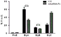
Figure 1 shows that the murine counterpart of SEQ ID NO 7(mActRIIA-Fc) improves hematological parameters in b-thalassemia mice. Hbbth1/th1 mice (Skow LC et al, Cell 34:1043-52,1983) were treated with PBS or mActriIA-Fc (10mg/kg body weight twice a week) for 60 days. Hematological parameters were evaluated on days 5, 10, 30 and 60. (A) The evaluation of the red blood cell count, (B) hematocrit and (c) hemoglobin was associated with (D) reduction of reticulocytosis. Analysis of circulating Red Blood Cell (RBC) parameters also showed increases in (E) Mean Corpuscular Volume (MCV), (F) mean hemoglobin of red blood cells (MCH), and (G) MCH concentration (MCHC) in mice treated with mActRIIA-Fc. (H) Total antioxidant status. (I) Morphological analysis showed red blood cell heterogeneity, red blood cell dysmorphism and target cell depletion. The (J) systemic iron content, (K) transferrin synthesis, (L) transferrin and (M) ferritin saturation levels were also evaluated on thalassemia mice. The (N) inflammatory cell count was also evaluated. The effect of mActRIIA-Fc on splenomegaly in thalassemia mice was evaluated by (O) spleen weight and total cell number. (P) in mice treated with mActriIA-Fc, bone marrow erythroblast number and expansion (eosin/hematoxylin staining) were also reduced. (Q) bone marrow and spleen erythroblasts were quantified by flow cytometry by TER119 staining. For each independent experiment, p <0.05, N-3-5.
Figure 2 shows that mActRIIA-Fc reduces ineffective erythropoiesis in thalassemia mice. (A-C) bone marrow and spleen were harvested and erythroblasts were assessed by flow cytometry by CD71/TER119 staining and FSC/SSC distribution. (D) Analysis of total bilirubin levels and direct bilirubin levels. (E) Primary protoerythroid differentiated Reactive Oxygen Species (ROS) production was evaluated by flow cytometry using dichlorodihydrofluorescein. (F) Analysis of hemoglobin solubility of primary thalassemic protoerythrocytes treated with mActRIIA-Fc or PBS for 48 hours. For each independent experiment, p <0.05, p <0.01, p <0.005, N-3-5.
FIG. 3 shows the effect of mActriIA-Fc on apoptosis of erythroblasts in thalassemia mice. Expression of Fas-L on bone marrow (A) and spleen (B) erythroblasts of mActriIA-Fc or PBS-treated mice. (c) Tunel staining of erythroblasts was increased in mActriIA-Fc treated mice.
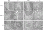
Figure 4 shows ActRIIA ligand expression in the spleen of thalassemia mice. (A) actriia-Fc treated animals had increased mRNA expression levels of ActRII, activin a, activin B, and GDF 11. (B) Immunoblot analysis of GDF11 protein levels, which was reduced in mActRIIA-Fc treated animals. (c) Immunohistochemical staining of bone marrow against GDF11 showed no change between wild type and mice treated with mActRIIA-Fc.
Figure 5 shows the effect of mActRIIA-Fc on GDF11 expression in primary erythroblasts of thalassemia. (A) Immunohistochemical analysis of activin/GDF signaling pathways in thalassemia mice treated with PBS or mActRIIA-Fc for 30 days showed increased levels of GDF11, ActRII, and p-Smad2 in thalassemia mice. (B) Immunohistochemical analysis of activin a, activin B, and GDF11 in thalassemia mice compared to other anemia models. (c) FACS analysis of primary thalassemic protoerythrocytes treated with PBS or mActRIIA-Fc for 48 hours using specific antibodies against activin a, activin B, GDF11 propeptide and GDF8/GDF11 cleavage peptides. The lines in the figure show the quantification of GDF11 staining. (D) Immunohistochemical analysis of GDF11 precursor form (proform) expression in the spleen of thalassemia mice treated with PBS or mActRIIA-Fc. P <0.05, N ═ 4.
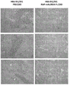
Figure 6 shows that inhibition of GDF11 reduces ineffective erythropoiesis in thalassemia mice. (A) Bone marrow and spleen were harvested and primary protored blood cells were assessed for differentiation by flow cytometry by CD71/TER119 staining and FSC/SSC profiling. (B) Primary protoerythroid differentiation was assessed for reactive oxygen species production by flow cytometry using dichlorodihydrofluorescein. P <0.05, N ═ 4.
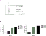
Figure 7 shows a sandwich ELISA assay to detect GDF11 in serum. (A) Schematic diagram of the assay. (B) Plates were coated with 5mg/mL mActRIIA-Fc and either an elevated dose of recombinant GDF11 (0ng/mL, 0.1ng/mL, 0.5ng/mL, 2.5ng/mL) or control serum (1/4 to 1/500 dilution) was added to the mActRIIA-Fc coated plates, the plates were washed, bound proteins were detected with anti-GDF 8/11 antibody, and then detected using horseradish peroxidase conjugated anti-rabbit IgG. GDF11 protein binds plates in a dose-dependent manner.
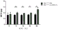
FIG. 8 shows detection of elevated levels of GDF11 in serum of b-Mediterranean anemia patients. Sera were obtained from patients exhibiting thalassemia and healthy controls.
Figure 9 shows a sandwich ELISA assay to detect activin a in serum. (A) Schematic diagram of the assay. (B) Plates were coated with 5mg/mL ActRIIA-Fc (SEQ ID NO.7) and either an elevated dose of recombinant activin A (0ng/mL, 0.1ng/mL, 0.5ng/mL, 2.5ng/mL) or control serum (1/4 to 1/500 dilutions) was added to the ActRIIA-Fc plates, the plates were washed, bound protein was detected with anti-activin A antibody, and then detected using horseradish peroxidase-conjugated anti-rabbit IgG. Activin a protein binds to the plate in a dose-dependent manner. (c) b-measurement of the level of activin A in the serum of patients with thalassemia. Sera were obtained from patients exhibiting thalassemia and healthy controls. Serum levels of activin a were unchanged in thalassemia patients.
Figure 10 shows a sandwich ELISA assay to detect activin B in serum. (A) Schematic diagram of the assay. (B) Plates were coated with 5mg/mL ActRIIA-Fc and either an elevated dose of recombinant activin B (0ng/mL, 0.1ng/mL, 0.5ng/mL, 2.5ng/mL) or control serum (1/4 to 1/500 dilutions) was added to the ActRIIA-Fc plates, the plates were washed, bound protein was detected with anti-activin B antibody, and detection was performed using horseradish peroxidase conjugated anti-rabbit IgG. Activin B protein binds to the plate in a dose-dependent manner. (c) B-measurement of the level of activin B in the serum of patients with thalassemia. Sera were obtained from patients exhibiting thalassemia and healthy controls. Serum levels of activin B were unchanged in thalassemia patients.
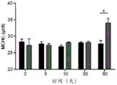
Figure 11 shows that administration of mActRIIA-Fc to C57BL/6 wild-type mice did not change their hematological parameters. (A) Evaluation of red blood cell count, (B) hematocrit, (C) hemoglobin showed no binding to mActRIIA-Fc. (D) Reticulocytosis is slightly reduced. The mActRIIA-Fc does not alter Red Blood Cell (RBC) parameters such as (E) Mean Corpuscular Volume (MCV), (F) mean hemoglobin of red blood cells (MCH), or (G) MCH concentration (MCHC). For each independent experiment, p <0.05, N-3-5.
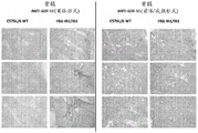
Figure 12 shows that administration of mActRIIA-Fc to C57BL/6 wild-type mice had no effect on spleen and bone marrow cell numbers in the mice.
Figure 13 shows that mActRIIA-Fc stimulates red blood cell differentiation by inhibition of GDF 11. (A-C) erythrocyte differentiation of CD34+/CD36+ cells by culturing in a medium containing EPO, +/-50. mu.g/mL mActriIA-Fc; erythroid progenitor cells (a), cell proliferation (B), and erythrocyte precursors (C) were analyzed. (D-F) analysis of red blood cell differentiation of CD34+/CD36+ cells when co-cultured with Bone Marrow (BM) cells in a medium containing EPO, +/-50 μ g/mL mActriIA-Fc; (D) erythroid progenitor cells, (E) cell proliferation, and (F) erythrocyte precursors. (G-H) erythrocyte differentiation of CD36+ cells by culturing in a medium containing EPO, +/-200ng/mL GDF11, +/-100ug/mL mActriIA-Fc; the percentage of (G) cell proliferation and (H) erythrocyte precursor GPA + was analyzed.
5. Detailed description of the invention
5.1. Overview
Provided herein are methods of treating anemia or a disease or condition associated with anemia or ineffective erythropoiesis by using a level and/or activity of GDF11 that is indicative of the patient's response to treatment with an activin type II receptor inhibitor, the efficacy of treatment with an activin type II receptor inhibitor, or an appropriate dose of treatment with an activin type II receptor inhibitor. Diseases associated with anemia or ineffective erythropoiesis that can be treated according to the methods described herein include, without limitation, thalassemia (e.g., beta thalassemia), myelodysplastic syndrome, chronic pernicious anemia, sickle cell anemia, and bubbly anemia. Conditions associated with anemia or ineffective erythropoiesis that may be treated according to the methods described herein include, without limitation, reduced red blood cell levels, reduced hemoglobin levels, reduced hematocrit levels, and reduced eosinophilia (Ery-C). In one embodiment, provided herein is a method of treating anemia. In one embodiment, provided herein are methods of treating thalassemia (e.g., beta thalassemia). In one embodiment, provided herein is a method of treating a decrease in red blood cell levels or a method of increasing red blood cell levels. The activin type II receptor inhibitor used in the methods described herein may be an inhibitor of ActRIIA and/or ActRIIB, as described herein or any inhibitor known in the art. In a preferred embodiment, the activin type II receptor inhibitor is a humanized fusion protein consisting of the extracellular domain of ActRIIA and the domain of human IgG1Fc ("ActRIIA-Fc," SEQ ID NO: 7).
The methods provided herein are based in part on the following findings: GDF11 levels were elevated in the blood of human patients with beta thalassemia and in a mouse model of beta thalassemia and ActRIIA-mFc (ActRIIA fused to murine IgG) reduced GDF11 levels elevated in a mouse model of beta thalassemia (see examples). Furthermore, without being limited by theory, ActRIIA-mFc captured ligands corrected ineffective erythropoiesis and improved anemia in the experimental mouse model of thalassemia intermedia (Hbbth1/th1 mice). As shown in the examples provided herein, treatment of thalassemia mice (Hbbth1/th1) with ActRIIA-mFc increased red blood cell count, hematocrit, hemoglobin, Mean Cell Volume (MCV), and mean cell hemoglobin, increased eosinophils (Ery-C), decreased bone marrow and spleen cell structure, decreased late basophilic/polychromatic erythroblasts (Ery-B), decreased bilirubin levels (an indication of decreased red blood cell destruction) and decreased apoptotic cells in the spleen and bone marrow. Further, the methods provided herein are based in part on the following findings: in an in vitro culture system, GDF-11 inhibits the growth of human bone marrow-derived erythrocyte precursor cells and ActRIIA-mFc rescues this inhibition (see examples). In summary, the data provided herein indicate that GDF-11 levels, e.g., blood and/or serum levels of GDF11, can identify which patients can respond to ActRIIA-Fc and can be used to monitor clinical response to drugs. The data provided herein also indicate that an ActRIIA-Fc (e.g., ActRIIA-mFc or ActRIIA-hFc, as in SEQ ID NO: 7) is useful in treating anemia associated with ineffective erythropoiesis.
The findings described herein, illustrated in the examples, indicate that detection of the level and/or activity of GDF11 can be used (i) as a marker (e.g., a serum biomarker) for the degree of ineffective erythropoiesis in a patient, (II) as a marker to measure the patient's response to an activin type II receptor inhibitor (e.g., ActRIIA-Fc), or (iii) to evaluate the pharmacodynamic effect of an activin type II receptor inhibitor (e.g., ActRIIA-Fc) in a patient after treatment, wherein the patient is a patient having anemia or a disease associated with anemia or ineffective erythropoiesis (e.g., thalassemia, e.g., beta thalassemia). Thus, in certain embodiments, GDF11 may be used in the methods described herein as an indicator of therapeutic efficacy of an ActRIIA-Fc (e.g., ActRIIA-hFc, such as SEQ ID NO: 7) and/or as an indicator of non-responsiveness to treatment with an ActRIIA-Fc (e.g., ActRIIA-hFc, such as SEQ ID NO: 7). Additionally, as described herein, GDF11 can be used as a reliable molecular marker to evaluate the time course therapeutic efficacy of an ActRIIA-Fc (e.g., ActRIIA-hFc, as set forth in SEQ ID NO: 7). Additionally, in specific embodiments, provided herein are methods comprising detecting the level and/or activity of GDF11 in the blood (e.g., detecting abnormal expression in diseases associated with ineffective erythropoiesis) and administering an activin type II receptor inhibitor, such as ActRIIA-Fc, at a dose dependent on the level and activity of GDF 11.
5.2 diagnostic/prognostic/therapeutic methods
In one aspect, the methods provided herein include determining the level and/or activity of GDF11 in a tissue sample (e.g., serum or blood) of a subject, selecting a subject with an elevated level and/or activity of GDF11 relative to the normal level and/or activity of GDF11, and treating a subject with an elevated level and/or activity of GDF11 with an activin type II receptor inhibitor. An increase in the level and/or activity of GDF11 in a tissue sample (e.g., serum or blood) of a subject can indicate that the patient can respond to treatment with an activin type II receptor inhibitor. In one embodiment of the methods described herein, the activin type II receptor inhibitor is an ActRIIA-Fc, such as an ActRIIA-hFc (e.g., SEQ ID NO: 7).
GDF11 levels and/or activity can also be used to assess the appropriate dosage of a subject as a candidate for treatment with an activin type II receptor inhibitor, to assess whether the dosage of an activin type II receptor inhibitor is adjusted during treatment, and/or to assess the appropriate maintenance dosage of an activin type II receptor inhibitor. If the level and/or activity of GDF11 is outside of normal levels and/or activities, dosing of the activin type II receptor inhibitor may be initiated, increased, decreased, delayed or terminated based on the level and/or activity of GDF 11. If the level and/or activity of GDF11 is increased relative to normal levels and/or activity, then dose administration of the activin type II receptor inhibitor can be initiated or increased, and if the level and/or activity of GDF11 is decreased relative to normal levels and/or activity, then dose administration of the activin type II receptor inhibitor can be decreased, delayed, or terminated.
In one aspect, the methods described herein comprise administering an activin type II receptor inhibitor to a subject, and monitoring the level and/or activity of GDF11 in the subject. A decrease in the level and/or activity of GDF11 following administration of the activin type II receptor inhibitor (relative to the level and/or activity prior to administration of the activin type II inhibitor) may indicate that the subject is responsive to and/or effective for treatment with the activin type II receptor inhibitor. An unchanged or elevated level or activity of GDF11 (relative to the level or activity prior to administration of the activin type II inhibitor) following administration of the activin type II receptor inhibitor can indicate that the subject is non-responsive to treatment with the activin type II receptor inhibitor or that the subject requires a higher dose of the activin type II receptor inhibitor for efficacy.
In other aspects, a degree of change in the level and/or activity of GDF11 following administration of the activin type II receptor inhibitor (relative to the level and/or activity prior to administration of the activin type II inhibitor or relative to the normal level and/or activity of GDF11) can indicate an appropriate dose or dosage regimen of the activin type II receptor inhibitor. For example, a non-reduction or a minor reduction (e.g., a reduction of less than 5%, less than 10%, less than 15%, less than 20%, or less than 25% or less than 30%) in the level and/or activity of GDF11 following administration of a dose of an activin type II receptor inhibitor (relative to the level and/or activity prior to administration of the activin type II inhibitor) may indicate that a higher dose of the activin type II receptor inhibitor is needed or desired for efficacy. Thus, in some embodiments, when an assessment of the level and/or activity of GDF11 shows that the level and/or activity of GDF11 is not reduced or undesirably reduced less (e.g., less than 5%, less than 10%, less than 15%, less than 20%, or less than 25%, or less than 30% reduction) after administration of a dose of activin type II receptor inhibitor to a patient, a higher dose of activin type II receptor inhibitor (e.g., a 20%, 25%, 30%, 50%, 75%, 100%, 200%, 300%, 400%, 500%, 600%, 700%, 800%, 900%, or 1000% higher dose) is administered to the patient.
In other embodiments, a decrease (e.g., a slight or average decrease, such as a decrease of less than 10%, less than 15%, less than 20% or less than 25%, less than 30%, less than 50% or less than 75%, or a decrease of 10% to 75%, or a decrease of 25% to 50%) in the level and/or activity of GDF11 following administration of a dose of an activin type II receptor inhibitor may indicate that treatment with the activin type II receptor inhibitor is effective at a given dose and/or dosage regimen. Thus, in some embodiments, when an assessment of the level and/or activity of GDF11 shows a decrease (e.g., a slight or average decrease, such as a decrease of less than 10%, less than 15%, less than 20% or less than 25%, less than 30%, less than 50% or less than 75%, or a decrease of 10% to 75%, or a decrease of 25% to 50%) in the level and/or activity of GDF11 following administration of a dose of an activin type II receptor inhibitor to a patient, the same dose of the activin type II receptor inhibitor is administered to the patient. In particular embodiments, when an assessment of the level and/or activity of GDF11 shows an average or desired decrease in the level and/or activity of GDF11 (e.g., a decrease between 20% and 75%, between 25% and 75%, or between 30% and 60%) following administration of a dose of an activin type II receptor inhibitor to a patient, then the same dose of the activin type II receptor inhibitor is administered to the patient. A greater (or desirably greater) reduction in the level and/or activity of GDF11 (e.g., a reduction of greater than 50%, greater than 60%, greater than 70%, greater than 75%, greater than 80%, greater than 90%, greater than 95%) following administration of the dose of the activin type II receptor inhibitor (relative to the level and/or activity prior to administration of the activin type II inhibitor) may indicate that a lower dose of the activin type II receptor inhibitor is desired (e.g., to avoid a side effect of the treatment, such as hypertension). Thus, in some embodiments, when an assessment of the level and/or activity of GDF11 shows a greater (or expected to be greater) reduction (e.g., a reduction of greater than 50%, greater than 60%, greater than 70%, greater than 75%, greater than 80%, greater than 90%, greater than 95%) in the level and/or activity of GDF11 following administration of a dose of an activin type II receptor inhibitor to a patient, a lower dose of the activin type II receptor inhibitor (e.g., a 20%, 25%, 30%, 40%, 50%, 60%, 75%, 80%, 90%, or 95% lower dose) is administered to the patient.
Monitoring of the level and/or activity of GDF11 can be performed by assessing the level and/or activity of GDF11 1 day, 1 week, 2 weeks, 3 weeks, 4 weeks, 5 weeks, 6 weeks, 7 weeks, 8 weeks, 3 months, 4 months, 5 months, 6 months, or 1 year after administration of an activin type II receptor inhibitor to the patient. The level and/or activity of GDF11 following administration of an activin type II receptor inhibitor to a patient may also be used as an indicator of the appropriate or desired frequency of administration of the activin type II receptor inhibitor to the patient. For example, if the reduction in the level and/or activity of GDF11 is maintained for a period of time (e.g., 1 day, 1 week, 2 weeks, 3 weeks, 4 weeks, 5 weeks, 6 weeks, 7 weeks, 8 weeks, 10 weeks, 3 months, 4 months, 5 months, 6 months, or 1 year) following administration of the activin type II receptor inhibitor, the activin type II receptor inhibitor may be administered once (e.g., once a day, once a week 2, once a week 3, once a week 4, once a week 5, once a week 6, once a week 7, once a week 8, once a month 3, once a month 4, once a month 5, once a month 6, or once a year 1) within the period of time. In particular embodiments, if the reduction in the level and/or activity of GDF11 is maintained for more than 1 month, 2 months, or 3 months following administration of the activin type II receptor inhibitor, the activin type II receptor inhibitor may be administered once every 1 month, every 2 months, or every 3 months, respectively.
In particular embodiments, provided herein are methods comprising (i) assessing the level of GDF11 in a tissue (e.g., blood, serum, plasma, liver, bone marrow, and/or spleen) of a subject (e.g., a subject with anemia); (ii) administering to the subject an activin type II receptor inhibitor; and (iii) evaluating the level of GDF11 in a tissue (e.g., blood, serum, plasma, liver, bone marrow, and/or spleen) of a subject (e.g., a subject with anemia) following administration of the activin type II receptor inhibitor. The administration of step (II) may comprise one administration of the activin type II receptor inhibitor (i.e., one single dose administered to the subject) or multiple administrations of the activin type II receptor inhibitor (e.g., administration may comprise a complete administration regimen). The evaluation of step (iii) may be performed at any point after the administration of step (ii). For example, the evaluation of step (iii) may be performed 1 day, 2 days, 3 days, 4 days, 5 days, 6 days, 1 week, 2 weeks, 3 weeks, 4 weeks, 5 weeks, 6 weeks, 7 weeks, 8 weeks, 10 weeks, 3 months, 4 months, 5 months, 6 months or 1 year after the administration of step (ii). As another example, the evaluation of step (iii) may be performed 1-3 days, 2-4 days, 3-5 days, 5-7 days, 1-2 weeks, 2-3 weeks, 3-4 weeks, 1-2 months, 2-3 months, 3-4 months, 4-5 months, 5-6 months, or 6-12 months after the administration of step (ii). Based on the outcome assessed in steps (i) and (iii), the treatment regimen may be adjusted. For example, if the level of GDF11 in the tissue of the subject determined in the assessment of step (i) is reduced relative to the level of GDF11 in the tissue of the subject determined in the assessment of step (iii), the dose of activin type II receptor inhibitor administered to the subject may be maintained or reduced. Conversely, if the level of GDF11 in the tissue of the subject determined in the assessment of step (i) is provided relative to the level of GDF11 in the tissue of the subject determined in the assessment of step (iii), the dose of activin type II receptor inhibitor administered to the subject may be increased. The steps of the above methods may be repeated/varied as necessary to determine the appropriate dosage/treatment regimen appropriate for the subject undergoing treatment.
In another aspect, provided herein are methods comprising (a) administering to a patient a first dose of an activin type II receptor inhibitor, (b) determining the level and/or activity of GDF11 in a tissue sample (e.g., serum) of the patient, and (c) administering a second dose of the activin type II receptor inhibitor, wherein the second dose of the activin type II receptor inhibitor is higher than the first dose (e.g., 20%, 30%, 40%, 50%, 60%, 70%, 80%, 90%, 100%, 200%, 300%, 400%, 500%, 600%, 700%, 800%, 900%, or 1000% higher) if the level and/or activity of GDF11 is increased relative to normal values, and wherein the second dose of the activin type II receptor inhibitor is lower than the first dose (e.g., 20%, or 1000%) if the level and/or activity of GDF11 is decreased relative to normal values, 30%, 40%, 50%, 60%, 70%, 80% or 90% lower). In this embodiment, the level and/or activity of GDF11 in the tissue sample is compared to the normal level and/or activity of GDF11 (e.g., the average level and/or activity of GDF11 in a sample of the same tissue from a healthy subject (e.g., a subject in the same age group that does not suffer from anemia)). In certain embodiments, an elevated level and/or activity of GDF11 relative to a normal level and/or activity of GDF11 is considered to be increased when the level and/or activity is increased by at least 10%, at least 15%, at least 20%, at least 25%, at least 30%, at least 35%, at least 40%, at least 45%, at least 50%, at least 55%, at least 60%, at least 65%, at least 70%, at least 75%, at least 80%, at least 85%, at least 90%, at least 95%, at least 100%, at least 200%, at least 300%, or at least 500% relative to a normal level and/or activity of GDF 11. In certain embodiments, the level and/or activity of GDF11 is considered elevated relative to the normal level and/or activity of GDF11 when elevated by at least 25%, at least 50%, at least 75%, at least 100%, or at least 200% relative to the normal level and/or activity of GDF 11. In some embodiments, a level and/or activity of GDF11 is considered reduced relative to a normal level and/or activity of GDF11 when reduced by at least 10%, at least 15%, at least 20%, at least 25%, at least 30%, at least 35%, at least 40%, at least 45%, at least 50%, at least 55%, at least 60%, at least 65%, at least 70%, at least 75%, at least 80%, at least 85%, at least 90%, or at least 95% relative to a normal level and/or activity of GDF 11. In some embodiments, the level and/or activity of GDF11 is considered reduced relative to the normal level and/or activity of GDF11 when reduced by at least 25%, at least 50%, at least 75%, or at least 90% relative to the normal level and/or activity of GDF 11.
The methods described herein are based in part on the following findings: GDF11 inhibits the growth of erythrocyte precursors, and thus the level of GDF11 can be correlated with the level of erythrocytes. It is desirable to maintain optimal red blood cell levels in a patient because, while a decrease in red blood cell, hemoglobin, or hematocrit levels is associated with ineffective erythropoiesis and anemia, excessive elevation of red blood cell, hemoglobin, or hematocrit levels is associated with blood pressure and other undesirable side effects (which may be caused by treatment at doses higher than optimal activin type II receptor inhibitors). Thus, it may be desirable to administer the activin type II receptor inhibitor in a dosage regimen that maintains the level and/or activity of GDF11 in the patient at or near (e.g., within 5%, 10%, 15%, 20%, 25%, 30%, 40%, 50%, 60%, or 75% of the normal level and/or activity of GDF 11).
In certain embodiments, if the level and/or activity of GDF11 is determined to decrease below the normal level and/or activity of GDF11, administration of the activin type II receptor inhibitor is adjusted accordingly, e.g., delayed until the level and/or activity of GDF11 returns to normal or within 5%, 10%, 15%, 20%, 25%, 30%, 40%, 50%, 60%, or 75% of the normal level and/or activity of GDF11, or temporarily or permanently stopped. In other embodiments where the level and/or activity of GDF11 is determined to be reduced below the normal level and/or activity of GDF11, administration of the activin type II receptor inhibitor is not delayed, but the dose or frequency of dose administration of the inhibitor is set at an amount that will reduce the risk of an unacceptable increase in red blood cells, hemoglobin, or hematocrit, or alternatively, the patient is developed a treatment regimen that combines use of the inhibitor with an agent (e.g., a hypotensive agent) that accounts for the unacceptable increase in red blood cells, hemoglobin, or hematocrit.
In certain embodiments, provided herein are methods of treating anemia in an individual in need thereof, the method comprises determining the level and/or activity of GDF11 in the individual, and administering to the individual a therapeutically effective amount of an activin type II receptor inhibitor, specifically an ActRII polypeptide (e.g., ActRIIa-hFc), and optionally, further monitoring (or determining) the level and/or activity of GDF11, and adjusting the dose of the activin type II receptor inhibitor (wherein, for example, if GDF11 is elevated relative to normal levels and/or activity, the dose of the activin type II receptor inhibitor is increased, and if the level and/or activity of GDF11 is decreased relative to normal levels and/or activity, the dose of the activin type II receptor inhibitor is decreased).
In certain embodiments, provided herein are methods of treating beta thalassemia in an individual in need thereof by determining the level and/or activity of GDF11 in the individual, and if the level and/or activity of GDF11 is elevated, administering to the individual a therapeutically effective amount of an activin type II receptor inhibitor, particularly an ActRII polypeptide (e.g., ActRIIa-hFc), and optionally, further monitoring (or determining) the level and/or activity of GDF11, and adjusting the dose of the activin type II receptor inhibitor (wherein, for example, if the level and/or activity of GDF11 is elevated relative to normal, the dose of the activin type II receptor inhibitor is increased, and if the level and/or activity of GDF11 is lower than normal, the dose of the activin type II receptor inhibitor is decreased).
In certain embodiments, provided herein are methods of increasing the level of red blood cells, hemoglobin, hematocrit, or Ery-C in an individual in need thereof by determining the level and/or activity of GDF11 in the individual, and administering to the individual a therapeutically effective amount of an activin type II receptor inhibitor, specifically an ActRII polypeptide (e.g., ActRIIa-hFc), and optionally, further monitoring (or determining) the level and/or activity of GDF11, and adjusting the dose of the activin type II receptor inhibitor (wherein, for example, if the level and/or activity of GDF11 is increased relative to normal, the dose of the activin receptor antagonist is increased, and if the level and/or activity of GDF11 is lower than normal, the dose of the activin type II receptor inhibitor is decreased).
In certain embodiments, the methods provided herein are used in conjunction with methods of treating or ameliorating anemia, ineffective erythropoiesis, reduced red blood cell levels, or any other blood disorder described herein. In certain embodiments, the methods provided herein are used in conjunction with methods of increasing red blood cell levels, hemoglobin levels, hematocrit levels, or colony forming unit levels in a patient having anemia, ineffective erythropoiesis, decreased red blood cell levels, or any other blood disorder described herein. In certain embodiments, the red blood cell level is increased by at least 5%, 10%, 15%, 20%, 25%, 30%, 35%, 40%, 45%, 50%, 55%, 60%, 65%, 70%, 75%, 80%, 85%, 90%, 95%, 100%, 125%, 150%, 175%, 200%, 250%, 300%, 350%, 400%, 450%, or at least 500%. In certain embodiments, the hemoglobin level is increased by at least 5%, 10%, 15%, 20%, 25%, 30%, 35%, 40%, 45%, 50%, 55%, 60%, 65%, 70%, 75%, 80%, 85%, 90%, 95%, 100%, 125%, 150%, 175%, 200%, 250%, 300%, 350%, 400%, 450%, or at least 500%. In certain embodiments, the level of hematocrit is increased by at least 5%, 10%, 15%, 20%, 25%, 30%, 35%, 40%, 45%, 50%, 55%, 60%, 65%, 70%, 75%, 80%, 85%, 90%, 95%, 100%, 125%, 150%, 175%, 200%, 250%, 300%, 350%, 400%, 450%, or by at least 500%. In certain embodiments, the level of colony forming units is increased by at least 5%, 10%, 15%, 20%, 25%, 30%, 35%, 40%, 45%, 50%, 55%, 60%, 65%, 70%, 75%, 80%, 85%, 90%, 95%, 100%, 125%, 150%, 175%, 200%, 250%, 300%, 350%, 400%, 450%, or at least 500%. In certain embodiments, the methods provided herein are used in conjunction with methods of reducing the level of apoptosis of erythroid progenitor cells and precursor cells.
5.3 patient population
The subject treated according to the methods described herein can be any mammal, such as rodents and primates, and in preferred embodiments, humans. In certain embodiments, the methods described herein can be used to treat anemia or ineffective erythropoiesis, or to monitor and/or increase red blood cells, hemoglobin, hematocrit or Ery-C levels, in any mammal, such as rodents and primates, and in preferred embodiments, in human patients.
In one aspect, the methods provided herein are used in conjunction with a method of treatment. In some embodiments, provided herein are methods of treating anemia (e.g., anemia caused by ineffective erythropoiesis). In certain embodiments, provided herein are methods of treating diseases associated with ineffective erythropoiesis (e.g., thalassemia, myelodysplastic syndrome, chronic pernicious anemia, or sickle cell anemia). In certain embodiments, provided herein are methods of treating hereditary bone marrow failure syndrome (such as, but not limited to, megakaryocytic thrombocytopenia, bundler's anemia, congenital keratosis, fanconi anemia, pilson's syndrome, congenital neutropenia, suddei's syndrome, thrombocytopenia-radius deficiency syndrome). In particular embodiments, provided herein are methods of treating hereditary bone marrow failure syndrome that specifically affects red blood cells. In certain embodiments, provided herein are methods of treating anemia and/or bone disorders associated with end-stage renal disease. In some embodiments, provided herein are methods of treating thalassemia (e.g., beta-thalassemia), myelodysplastic syndrome, chronic pernicious anemia, sickle cell anemia, or bubby anemia. In one embodiment, provided herein is a method of treating beta-thalassemia. In one embodiment, provided herein is a method of treating anemia buboneset.
In certain embodiments, provided herein are methods of treating a patient having anemia (e.g., diagnosed as anemia). In some embodiments, provided herein are methods of treating a patient having (e.g., diagnosed with) a disease associated with ineffective erythropoiesis, such as thalassemia, myelodysplastic syndrome, chronic pernicious anemia, or sickle cell anemia. In certain embodiments, provided herein are methods of treating a patient suffering from (e.g., diagnosed with) an inherited bone marrow failure syndrome, such as megakaryocytic thrombocytopenia, bundler's anemia, congenital dyskeratosis, fanconi's anemia, pilsner's syndrome, congenital neutropenia, sul-dedicate's syndrome, thrombocytopenia-radius loss syndrome, or bone marrow failure syndrome, which condition specifically affects red blood cells. In particular embodiments, provided herein are methods of treating a patient suffering from (e.g., diagnosed with) anemia and/or a bone disorder associated with end-stage renal disease. In specific embodiments, provided herein are methods of treating a patient having (e.g., diagnosed with) thalassemia (e.g., beta-thalassemia), myelodysplastic syndrome, chronic pernicious anemia, sickle cell anemia, or bunyaemia. In one embodiment, provided herein are methods of treating a patient having (e.g., diagnosed with) β -thalassemia.
Anemia is associated with a variety of disorders and conditions, including without limitation: chronic renal failure, myelodysplastic syndrome, rheumatoid arthritis, bone marrow transplantation, solid tumors (e.g., breast cancer, lung cancer, colon cancer), tumors of the lymphatic system (e.g., chronic lymphocytic leukemia, non-hodgkin's and hodgkin's lymphomas), tumors of the hematopoietic system (e.g., leukemia, myelodysplastic syndrome, multiple myeloma), radiation therapy, chemotherapy (e.g., platinum-containing regimens), inflammatory and autoimmune diseases (including, but not limited to, rheumatoid arthritis, other inflammatory arthritic skin rashes, Systemic Lupus Erythematosus (SLE), acute or chronic skin diseases (e.g., psoriasis), inflammatory bowel diseases (e.g., crohn's disease and ulcerative colitis)), acute or chronic kidney diseases or failure, including idiopathic or congenital conditions, acute or chronic liver diseases, acute or chronic bleeding, conditions in which transfusion of red blood cells is not possible, infections (e.g. malaria, osteomyelitis), hemoglobinopathies (including, for example, sickle cell disease, thalassemia), drug use or abuse (e.g. alcohol misuse; anemia pediatric patients who avoid blood transfusion for any reason) and situations in which elderly patients with underlying cardiopulmonary disease and anemia cannot receive blood transfusions due to problems with circulatory overload, among others due to patient allo-or auto-antibodies and/or for religious reasons. In certain embodiments, the methods described herein are used to treat anemia, or to monitor and/or increase red blood cells, hemoglobin, hematocrit or Ery-C levels, in any patient suffering from one or more of the above-described disorders or conditions.
In certain embodiments, provided herein are methods of treating anemia, wherein the subject is refractory to administration of erythropoietin. In certain embodiments, provided herein are methods of treating anemia, wherein the subject is non-responsive to administration of iron, vitamin B-12, and/or folic acid. In certain embodiments, provided herein are methods of treating anemia arising from erythroid progenitor cells and precursors that are highly sensitive to death due to apoptosis.
In certain embodiments, a subject treated (e.g., selected for treatment) according to the methods described herein has an elevated level and/or activity of GDF11 relative to a normal level of GDF 11. For example, a subject treated (e.g., selected for treatment) according to the methods described herein has an elevated level and/or activity of GDF11 in the tissue of the subject relative to the average level and/or activity of GDF11 in the same tissue of healthy subjects in the same age group. The level and/or activity of GDF11 can be assessed by any method known in the art or described herein. In some embodiments, the level and/or activity of GDF11 in a subject treated according to the methods described herein is at least 10%, 15%, 20%, 25%, 30%, 35%, 40%, 45%, 50%, 55%, 60%, 65%, 70%, 75%, 80%, 85%, 90%, 95%, 100%, 125%, 150%, 175%, 200%, 250%, 300%, 350%, 400%, 450%, 500%, or 1000% of the normal level and/or activity (e.g., average level in healthy subjects) of GDF 11. In specific embodiments, the level and/or activity of GDF11 in a subject treated according to the methods described herein is at least 50%, at least 75%, at least 100%, at least 200%, or at least 500% of the normal level and/or activity of GDF 11. In some embodiments, the methods described herein are used to monitor, modulate, or increase red blood cells, hemoglobin, hematocrit, or Ery-C levels in a subject determined to have an increased level of GDF 11.
In certain embodiments, a subject treated (e.g., selected for treatment) according to the methods described herein has (i) an elevated level and/or activity of GDF11 relative to a normal level of GDF11 (e.g., in serum, bone marrow, liver, and/or spleen), and (ii) anemia or a disease or condition associated with anemia or ineffective erythropoiesis (e.g., a subject diagnosed with anemia). In certain embodiments, a human patient treated (e.g., selected for treatment) according to the methods described herein has elevated serum levels and/or activity of GDF11 relative to normal levels of GDF11, and has anemia (e.g., has been diagnosed as anemia).
In some embodiments, a subject treated according to the methods described herein has an undesirably low red blood cell count, an undesirably low hemoglobin level, an undesirably low hematocrit level, and/or an undesirably low Ery-C level. The methods described herein can be used to monitor, regulate and/or increase red blood cell, hemoglobin, hematocrit or Ery-C levels in a selected patient population. In other embodiments, provided herein are methods of treating patients at risk of developing undesirably low red blood cell or hemoglobin levels, such as those patients who are about to undergo major surgery or other procedures that may result in substantial blood loss. In some embodiments, a subject treated according to the methods described herein will undergo major surgery or another procedure that may result in substantial blood loss.
When determining whether the level of hemoglobin is undesirably low, a level less than normal for the appropriate age and gender group may indicate anemia, despite individual variations being considered. For example, a hemoglobin level of 12g/dl is generally considered to be a normal lower limit in adults. Possible causes of low levels of hemoglobin include blood loss, malnutrition, drug therapy response, various problems with the bone marrow, and various diseases. Patients may be treated with a dosage regimen intended to restore the patient to a target hemoglobin level, typically between about 10g/dl to about 12.5g/dl, and typically about 11.0g/dl (see Jacobs et al, (2000) Nephrol Dial Transplant 15,15-19), although lower target levels may result in fewer cardiovascular side effects. Optimally, the target hemoglobin level may be considered individually for each patient. In certain embodiments, the hemoglobin level of a patient treated by the methods described herein is less than 13g/dl, less than 12.5g/dl, less than 12g/dl, less than 11.5g/dl, less than 11g/dl, less than 10.5g/dl, less than 10g/dl, less than 9.5g/dl, or less than 9 g/dl.
When determining whether the red blood cell level is undesirably low, the hematocrit level (the percentage of the volume of the blood sample that the cells occupy) can be used to assess the condition of the red blood cells. Hematocrit levels in healthy individuals range from 41 to 51% for adult males and 35 to 45% for adult females. The target hematocrit level is typically about 30-33%, although hematocrit levels vary from person to person. Optimally, the target hematocrit level may be considered individually for each patient.
In some embodiments, the methods described herein are used to treat anemia or a disease or condition associated with anemia or ineffective erythropoiesis in a patient that is susceptible to side effects when a more than optimal dose of an activin type II receptor inhibitor, specifically an ActRII polypeptide (e.g., ActRIIa-hFc), is administered. Side effects that may be associated with administration of higher than optimal doses of activin type II receptor inhibitors include, without limitation, excessive increases in red blood cell levels, hematocrit levels or hemoglobin levels, excessive increases in iron stores, and bone marrow or spleen cell overload. In turn, excessive increases in red blood cell levels, hemoglobin levels, or hematocrit levels may result in elevated blood pressure and/or other undesirable side effects. In particular embodiments, a patient treated according to the methods described herein is susceptible to hypertension (e.g., elevated systolic, diastolic, and/or mean arterial blood pressure) or another condition that may be associated with an excessive increase in red blood cell levels (e.g., headache, flu-like syndrome, or vascular thrombosis). In particular embodiments, patients treated according to the methods described herein are susceptible to hypertension or another condition that may be associated with an excessive increase in red blood cell levels when treated with an activin type II receptor inhibitor (e.g., ActRIIA-hFc, as set forth in SEQ ID NO: 7).
Subjects of any age can be treated according to the methods described herein. In some embodiments, the subject treated according to the methods described herein is over 55 years old. In some embodiments, a subject treated according to the methods described herein is less than 3 or 10 years old. In other embodiments, the subject treated according to the methods described herein is less than 18 years of age. In other embodiments, the subject treated according to the methods described herein is 18 to 55 years old.
5.4 evaluation of GDF11 level and/or Activity
The level or activity of GDF11 can be determined by any method known in the art or described herein. For example, the level of GDF11 in a tissue sample is determined by evaluating (e.g., quantifying) reverse transcribed RNA of GDF11 using, for example, northern blot, PCR analysis, real-time PCR analysis, or any other technique known in the art or described herein. In one embodiment, the level of GDF11 in a tissue sample can be determined by evaluating (e.g., quantifying) mRNA of GDF11 in the sample.
The level of GDF11 in a tissue sample can also be determined by assessing (e.g., quantifying) the protein expression level of GDF11 in the sample using, for example, immunohistochemical analysis, immunoblotting, ELISA, immunoprecipitation, flow cytometry analysis, or any other technique known in the art or described herein. In particular embodiments, the level of GDF11 is determined by a method that is capable of quantifying the amount of GDF11 in a tissue sample (e.g., human serum) of a patient following treatment with an activin type II receptor inhibitor and/or that is capable of detecting a modification in GDF11 levels. In one embodiment, the level of GDF11 in the tissue sample is determined by assessing (e.g., quantifying) protein expression of GDF11 in the sample using ELISA. For example, GDF11 in human serum can be identified and quantified using a sandwich ELISA method as described in the examples. A sandwich ELISA method used in determining the level of GDF11 in a tissue sample may include ActRIIA-Fc coating of an ELISA plate (using, for example, ActRIIA-mFc or ActRIIA-hFc, as set forth in SEQ ID NO: 7), contacting the plate with a tissue sample (e.g., human serum), and detecting an ActRIIA ligand (as GDF11) in the tissue sample (e.g., human serum) bound to ActRIIA-Fc by a specific antibody. In some embodiments, the methods (e.g., sandwich ELISA) used in determining the level and/or activity of GDF11 as described herein are capable of detecting 100pg/ml of recombinant GDF11, activin a, and/or activin B bound to ActRIIA.
Antibodies used in assays to measure the level of GDF11 in a sample (e.g., a tissue sample, e.g., a sample of blood, serum, plasma, liver, spleen, and/or bone marrow) are known in the art and can be readily developed using methods known to those skilled in the art. Examples of monoclonal antibodies that can be used in an assay to measure the level of GDF11 in a sample include antibodies from LifeSpan Biosciences inc, Seattle WA, catalog nos. LS-C121127, LS-C138772, LS-C105098; antibodies from Santa Cruz Biotechnology, inc., Santa Cruz, CA, catalog No. (X-19) for: sc-81952; and antibodies from Sigma-Aldrich co.llc, catalog No.: WH0010220M 3.
The activity of GDF11 may be measured by any assay known in the art, including without limitation, a colony formation assay, a reporter gene assay (e.g., containing a GDF 11-reactive reporter gene building block), an alkaline phosphatase assay, or any other biological activity assay. Exemplary assays that can be used to measure the activity of GDF11 are described in Souza et al, 2008, mol. endocrinology22(12): 2689-; and Bessa et al, 2009, Protein Expression and Purification63: 89-94.
The level and/or activity of GDF11 can be assessed in any tissue sample obtained from a patient treated according to the methods described herein. In certain embodiments, the level and/or activity of GDF11 is assessed in a sample of serum, liver, spleen, or bone marrow from a patient treated according to the methods described herein. In one embodiment, the level and/or activity of GDF11 is assessed in a sample from serum from a patient treated according to the methods described herein. In another embodiment, the level and/or activity of GDF11 is assessed in a sample obtained from the spleen of a patient treated according to the methods described herein. In another embodiment, the level and/or activity of GDF11 is assessed in a sample obtained from bone marrow of a patient treated according to the methods described herein.
In some embodiments, the level and/or activity of GDF11 in a patient tissue sample is compared to the average level and/or activity of GDF11 in a tissue sample (e.g., a sample from the same tissue) of a healthy subject (e.g., a subject not suffering from anemia), such as a subject of the same age group and (optionally) the same gender. In some embodiments, the level and/or activity of GDF11 in a tissue sample of a patient is compared to the level and/or activity of GDF11 in a tissue sample (e.g., a sample from the same tissue) at an earlier time point (e.g., before disease onset, before treatment initiation, or during treatment) in the subject. In some embodiments, the level and/or activity of GDF11 in a tissue sample (e.g., serum, spleen, liver, or blood bone marrow) of a patient is compared to the level and/or activity of GDF11 in another tissue sample of the patient. In some embodiments, the level and/or activity of GDF11 in a patient tissue sample is compared to the level and/or activity of another gene product (e.g., B-actin, activin a, activin B) in the patient tissue sample.
In some embodiments, the level and/or activity of GDF11 in a tissue sample of a patient is compared to the normal level and/or activity of GDF11 in a tissue sample. The normal level and/or activity of GDF11 in a tissue sample can be the average level and/or activity of GDF11 in tissue samples (e.g., samples from the same tissue) of one or more healthy subjects (e.g., subjects not suffering from anemia), such as subjects of the same age group and (optionally) of the same gender. In some embodiments, detection of an elevated level and/or activity of GDF11 as compared to the normal level and/or activity of GDF11 is performed after administration of an activin receptor inhibitor (e.g., one or more activin receptor inhibitors as described herein). In some embodiments, administration of an activin receptor inhibitor (such as one or more activin receptor inhibitors described herein) is performed after monitoring the level and/or activity of GDF11 and (optionally) comparing the level and/or activity of GDF11 to the normal level and/or activity of GDF 11. In some embodiments, administration of a first dose of an activin type II receptor inhibitor (e.g., ActRIIA-hFc as set forth in SEQ ID NO: 7) is performed after determining the level and/or activity of GDF11, and if the level and/or activity of GDF11 is increased relative to the normal level and/or activity, administering a second dose of the activin type II receptor inhibitor that is higher (e.g., 1.25, 1.5, 1.75, 2, 2.5, 3, 4, 5, 6, 7, 8, 9, or 10 fold higher) than the first dose, and if the level and/or activity of GDF11 is decreased relative to the normal level and/or activity, administering a second dose of the activin type II receptor inhibitor that is lower (e.g., 1.25, 1.5, 1.75, 2, 2.5, 3, 4, 5, 6, 7, 8, 9, or 10 fold lower) than the first dose.
As used herein, in certain embodiments, the normal level and/or activity of GDF11 is the average level and/or activity of GDF11 in a tissue sample of one or more healthy subjects (e.g., healthy subjects of the same age group and/or the same gender). In some embodiments, the level and/or activity of GDF11 in a tissue sample of a treated subject is compared to the level and/or activity of GDF11 in a sample of the same tissue from one or more healthy subjects. In some embodiments, the tissue in which the level and/or activity of GDF11 is assessed is serum, bone marrow, liver, or spleen. In one embodiment, the tissue in which the level and/or activity of GDF11 is assessed is serum.
5.5 therapeutic use
In certain embodiments, in the methods described herein, the activin type II receptor inhibitor is used to treat a patient having anemia or a disease or condition associated with anemia or ineffective erythropoiesis. In some embodiments, the activin type II receptor inhibitor is administered to a patient having anemia or a disease or condition associated with anemia or ineffective erythropoiesis. In some embodiments, an activin type II receptor inhibitor is administered to a patient having anemia or a disease or condition associated with anemia or ineffective erythropoiesis and having an elevated level and/or activity of GDF11 (e.g., in the patient's serum, bone marrow, liver, and/or spleen). Activin type II receptor inhibitors described in this section and known in the art can be used in the methods provided herein. In certain embodiments, the activin type II receptor inhibitors described in this section can be used in the methods provided herein.
ActRII receptor inhibitors contemplated herein include ActRIIA inhibitors and ActRIIB inhibitors (see below). In certain embodiments, an ActRII receptor inhibitor is specific for ActRIIA. In other embodiments, the ActRII receptor inhibitor is specific for ActRIIB. In certain embodiments, an ActRII receptor inhibitor preferably inhibits ActRIIA. In other embodiments, an ActRII receptor inhibitor preferably inhibits ActRIIB. In certain embodiments, an ActRII receptor inhibitor inhibits both ActRIIA and ActRIIB.
In certain embodiments, an inhibitor of an ActRII receptor may be a polypeptide that comprises an activin-binding domain of ActRII. Without being bound by theory, such polypeptides comprising an activin-binding domain sequester activin and thereby prevent activin signaling. These activin-binding domain-containing polypeptides may comprise all or a portion of an ActRII receptor extracellular domain (i.e., all or a portion of an ActRIIA extracellular domain or all or a portion of an ActRIIB extracellular domain). In particular embodiments, the ActRII receptor extracellular domain is soluble.
In certain embodiments, the polypeptide comprising an activin-binding domain is linked to the Fc portion of an antibody (i.e., a conjugate is generated that comprises the polypeptide comprising an activin-binding domain of an ActRII receptor and the Fc portion of an antibody). Without being bound by theory, the antibody moiety confers increased stability to the conjugate. In certain embodiments, the activin-binding domain is linked to the Fc portion of the antibody via a linker (e.g., a peptide linker).
ActRII receptor inhibitors for use in the compositions and methods described herein include molecules that directly or indirectly inhibit ActRIIA and/or ActRIIB extracellularly or intracellularly. In some embodiments, the inhibitors of ActRIIA and/or ActRIIB used in the compositions and methods described herein inhibit ActRIIA and/or ActRIIB through interaction with the receptor itself. In other embodiments, the inhibitors of ActRIIA and/or ActRIIB used in the compositions and methods described herein inhibit ActRIIA and/or ActRIIB through interaction with an ActRIIA and/or ActRIIB ligand, e.g., activin.
5.5.1 inhibitors of ACTRIIA
As used herein, the term "ActRIIA" refers to a family of activin type IIa receptor (ActRIIA) proteins from any species and variants derived from the ActRIIA proteins by mutation or other modification. In this context, reference to ActRIIA should be understood as reference to any one of the forms currently identified. ActRIIA family members are typically transmembrane proteins, consisting of a ligand-binding extracellular domain with a cysteine-rich region, a transmembrane domain, and a cytoplasmic domain with predicted serine/threonine kinase activity.
ActRIIA inhibitors for use in the compositions and methods described herein include, without limitation, activin-binding soluble ActRIIA polypeptides; an antibody that binds to activin (specifically, activin a or B subunit, also referred to as β a or β B) and disrupts ActRIIA binding; an antibody that binds to ActRIIA and disrupts activin binding; selecting a non-antibody protein for activin or ActRIIA binding (see, e.g., WO/2002/088171, WO/2006/055689, WO/2002/032925, WO/2005/037989, US 2003/0133939, and US 2005/0238646, each of which is incorporated herein by reference in its entirety, e.g., these proteins and methods for design and selection of the same inhibitor); and selecting a random peptide for activin or ActRIIA binding, which can bind to the Fc domain.
In certain embodiments, two or more different proteins (or other moieties) having activin or ActRIIA binding activity, in particular, activin-binding agents that block type I (e.g., soluble type I activin receptor) and type II (e.g., soluble type II activin receptor) binding sites, respectively, can be linked together to produce a bifunctional or multifunctional binding molecule that inhibits ActRIIA and thus can be used in the compositions and methods described herein. In certain embodiments, activin-ActRIIA signaling axis antagonists that inhibit ActRIIA include nucleic acid aptamers, small molecules, and other agents used in the compositions and methods described herein.
(a) ACTRIIA inhibitors comprising ACTRIIA polypeptides
The term "ActRIIA polypeptide" includes polypeptides comprising any naturally occurring polypeptide of an ActRIIA family member as well as any variant thereof (including mutants, fragments, fusions, and peptidomimetic forms) that retain a useful activity. For example, ActRIIA polypeptides include polypeptides derived from the sequence of any known ActRIIA having at least about 80% identity, and optionally at least 85%, 90%, 95%, 97%, 98%, 99%, or more identity, to an ActRIIA polypeptide sequence. For example, an ActRIIA polypeptide may bind to and inhibit the function of an ActRIIA protein and/or activin. ActRIIB polypeptides may be selected for their ability to promote bone growth and bone salt deposition. Examples of ActRIIA polypeptides include human ActRIIA precursor polypeptides (SEQ ID NO: 1) and soluble human ActRIIA polypeptides (e.g., SEQ ID NOS: 2,3, 7, and 12). For its amino acid sequence the amino acid sequence shown in SEQ ID NO: 1, the signal peptide of a human ActRIIA precursor polypeptide is at amino acid positions 1 through 20; the extracellular domain is located at amino acid positions 21 to 135 and the N-linked glycosylation site of the human ActRIIA precursor polypeptide (SEQ ID NO: 1) is located at the amino acid position of SEQ ID NO: 1 amino acid positions 43 to 56. As set forth in SEQ ID NO: 4 discloses a polypeptide encoding SEQ ID NO: 1 (nucleotide 164-1705 of Genbank accession No. NM _ 001616). As set forth in SEQ ID NO: 5 discloses a polypeptide encoding SEQ ID NO: 2, or a soluble human ActRIIA polypeptide as set forth in seq id no. See table 1 for sequence descriptions.
In particular embodiments, an ActRIIA polypeptide used in the compositions and methods described herein is a soluble ActRIIA polypeptide. The extracellular domain of an ActRIIA protein may bind to activin and is generally soluble, and thus may be referred to as a soluble activin-binding ActRIIA polypeptide. Thus, as used herein, the term "soluble ActRIIA polypeptide" generally refers to a polypeptide that comprises an extracellular domain of an ActRIIA protein, including any naturally occurring extracellular domain of an ActRIIA protein and any variants thereof (including mutants, fragments, and peptidomimetic forms). A soluble ActRIIA polypeptide may bind to activin; however, the wild-type ActRIIA protein did not show significant selectivity in binding to activin over GDF 8/11. A native or altered ActRIIA protein may confer additional specificity to activin by coupling it to a second activin-selective binding agent. Examples of soluble activin-binding ActRIIA polypeptides include SEQ ID NOs: 2.3, 7, 12 and 13. Other examples of soluble activin binding ActRIIA polypeptides include signal sequences other than the extracellular domain of an ActRIIA protein, such as the melittin leader (SEQ ID NO: 8), The Plasminogen Activator (TPA) leader (SEQ ID NO: 9), or the native ActRIIA leader (SEQ ID NO: 10). SEQ ID NO: the ActRIIA-hFc polypeptide shown in fig. 13 uses a TPA leader sequence.
In certain embodiments, inhibitors of ACTRIIA for use in the compositions and methods described herein include binding/fusion proteins comprising an activin-binding domain of ACTRIIA linked to an Fc portion of an antibody. In certain embodiments, the activin-binding domain is linked to the Fc portion of the antibody via a linker (e.g., a peptide linker). Optionally, the Fc domain has one or more mutations at residues, such as Asp-265, lysine 322, and Asn-434. In certain instances, a mutant Fc domain having one or more of these mutations (e.g., an Asp-265 mutation) has a reduced ability to bind to an Fc γ receptor as compared to a wild-type Fc domain. In other cases, a mutant Fc domain having one or more of these mutations (e.g., the Asn-434 mutation) has an increased ability to bind to an MHC class I-associated Fc-receptor (FcRN) as compared to a wild-type Fc domain. In SEQ ID NO: 6. exemplary fusion proteins comprising a soluble extracellular domain of ActRIIA fused to an Fc domain are shown in fig. 7, 12, and 13.
In particular embodiments, the ActRIIA inhibitors used in the compositions and methods described herein include an extracellular domain of ActRIIA or a portion thereof linked to an Fc portion of an antibody, wherein the ActRIIA inhibitor comprises an amino acid sequence identical to a sequence selected from the group consisting of SEQ ID NOs: 6.7, 12 and 13, having an amino acid sequence of at least 75% identity. In another specific embodiment, the ActRIIA inhibitor used in the compositions and methods described herein comprises an extracellular domain of ActRIIA or a portion thereof linked to an Fc portion of an antibody, wherein the ActRIIA inhibitor comprises an amino acid sequence identical to a sequence selected from the group consisting of SEQ ID NOs: 6.7, 12 and 13 has an amino acid sequence that is at least 80%, 85%, 90%, 95%, 96%, 97%, 98% or 99% identical.
In certain embodiments, inhibitors of ACTRIIA for use in the compositions and methods described herein comprise truncated forms of the extracellular domain of ACTRIIA. Truncations may be located at the carboxy terminus and/or the amino terminus of an ActRIIA polypeptide. In certain embodiments, the truncation may be 1, 2,3, 4, 5, 6, 7, 8, 9, 10, 11, 12, 13, 14, 15, 16, 17, 18, 19, 20, 21, 22, 23, 24, or 25 amino acids long relative to the mature ActRIIB polypeptide extracellular domain. In certain embodiments, the truncation may be 1, 2,3, 4, 5, 6, 7, 8, 9, 10, 11, 12, 13, 14, 15, 16, 17, 18, 19, 20, 21, 22, 23, 24, or 25N-terminal amino acids of the extracellular domain of the mature ActRIIA polypeptide. In certain embodiments, the truncation may be 1, 2,3, 4, 5, 6, 7, 8, 9, 10, 11, 12, 13, 14, 15, 16, 17, 18, 19, 20, 21, 22, 23, 24, or 25C-terminal amino acids of the mature ActRIIA polypeptide extracellular domain. For example, truncated forms of ActRIIA include those having amino acids 20-119; 20-128; 20-129; 20-130; 20-131; 20-132; 20-133; 20-134; 20-131; 21-131; 22-131; 23-131; 24-131; and 25-131, wherein the amino acid positions represent SEQ ID NO: 1.
In certain embodiments, inhibitors of ACTRIIA for use in the compositions and methods described herein include an extracellular domain of ACTRIIA having one or more amino acid substitutions. In certain embodiments, inhibitors of ACTRIIA for use in the compositions and methods described herein include truncated forms of the ACTRIIA extracellular domain that also have amino acid substitutions.
In particular embodiments, the ActRIIA inhibitors used in the compositions and methods described herein are fusion proteins between the extracellular domain of a human ActRIIA receptor and the Fc portion of IgG 1. In another specific embodiment, the ActRIIA inhibitors used in the compositions and methods described herein are fusion proteins between a truncated extracellular domain of a human ActRIIA receptor and an Fc portion of IgG 1. In another specific embodiment, the ActRIIA inhibitors used in the compositions and methods described herein are fusion proteins between a truncated extracellular domain of a human ActRIIA receptor and an Fc portion of IgG1, wherein the truncated extracellular domain of a human ActRIIA receptor has one or more amino acid substitutions.
Functionally active fragments of an ActRIIA polypeptide may be obtained, for example, by screening polypeptides recombinantly produced from a corresponding fragment of a nucleic acid encoding the ActRIIA polypeptide. Alternatively, fragments can be chemically synthesized using techniques known in the art, such as conventional Merrifield solid phase f-Moc or t-Boc chemistry. The fragments can be generated (recombinant or chemically synthesized) and tested to identify those peptidyl fragments that can act as antagonists (inhibitors) of ActRIIA protein or activin-mediated signaling.
In addition, functionally active variants of ActRIIA polypeptides may be obtained, for example, by screening libraries of modified polypeptides recombinantly produced from a corresponding mutagenized nucleic acid encoding an ActRIIA polypeptide. The variants can be generated and tested to identify those that can act as antagonists (inhibitors) of ActRIIA protein or activin-mediated signaling. In certain embodiments, a functional variant of an ActRIIA polypeptide comprises an amino acid sequence that differs from a sequence selected from SEQ ID NOs: 2 or 3, having at least 75% identity thereto. In certain instances, the functional variant has an amino acid sequence that is identical to a sequence selected from SEQ ID NOs: 2 or 3, or an amino acid sequence having at least 80%, 85%, 90%, 95%, 97%, 98%, 99% or 100% identity.
Functional variants may be created, for example, by modifying the structure of an ActRIIA polypeptide for the purpose of increasing therapeutic efficacy or stability (e.g., ex vivo shelf life and resistance to proteolytic degradation in vivo). These modified ActRIIA polypeptides may be considered functional equivalents of the naturally occurring ActRIIA polypeptides when selected to retain activin binding. Modified ActRIIA polypeptides may also be produced, for example, by amino acid substitution, deletion, or addition. For example, it is reasonable to expect that a single substitution of leucine with isoleucine or valine, an aspartic acid with glutamic acid, a threonine with serine, or a similar substitution of an amino acid with a structurally related amino acid (e.g., conservative mutations) will not have a major effect on the biological activity of the resulting molecule. Conservative substitutions are those that occur within related families of amino acids in their side chains. Whether changes in the amino acid sequence of an ActRIIA polypeptide result in the production of functional homologs can be readily determined by assessing the ability of a variant ActRIIA polypeptide to produce a response in a cell in a manner similar to a wild-type ActRIIA polypeptide.
In certain embodiments, ActRIIA inhibitors for use in the compositions and methods described and provided herein may include ActRIIA polypeptides having one or more specific mutations that may alter glycosylation of the polypeptide. These mutations may introduce or eliminate one or more glycosylation sites, such as O-linked or N-linked glycosylation sites. The asparagine-linked glycosylation recognition site typically comprises the tripeptide sequence, asparagine-X-threonine (or asparagine-X-serine) (where "X" is any amino acid), which is specifically recognized by the appropriate cellular glycosylase. Changes (for O-linked glycosylation sites) may also be made by the addition or substitution of one or more serine or threonine residues to the wild-type ActRIIA polypeptide sequence. Multiple amino acid substitutions or deletions at one or both of the first or third amino acid positions (and/or amino acid deletions at the second position) of the glycosylation recognition site result in non-glycosylation at the modified tripeptide sequence. Another method to increase the number of carbohydrate moieties on an ActRIIA polypeptide is through chemical or enzymatic ligation of a glycoside to an ActRIIA polypeptide. Depending on the coupling format used, the sugar may be linked to (a) arginine and histidine; (b) a free carboxyl group; (c) free sulfhydryl groups, such as those of cysteine; (d) free hydroxyl groups such as those of serine, threonine, or hydroxyproline; (e) aromatic residues, such as those of phenylalanine, tyrosine or tryptophan; or (f) the amide group of glutamine. These methods are described in WO 87/05330 and Aplin and Wriston (1981) CRC Crit. Rev. biochem., p 259-306, published on 9/11 1987, which is incorporated herein by reference. Removal of one or more carbohydrate moieties present on an ActRIIA polypeptide may be accomplished by chemical and/or enzymatic reactions. Chemical deglycosylation may include, for example, exposure of an ActRIIA polypeptide to the compound triflic acid or equivalent compound. This treatment results in cleavage of most or all of the sugars except the linked sugar (N-acetylglucosamine or N-acetylgalactosamine) while leaving the complete amino acid sequence. Chemical deglycosylation is further described by Hakimuddin et al, (1987) Arch.biochem.Biophys.259:52 and Edge et al (1981) anal.biochem.118: 131. Enzymatic cleavage of the carbohydrate moiety on an ActRIIA polypeptide can be achieved by using a variety of endo-and exoglycosidases, as described by Thotakura et al (1987) meth. Depending on the type of expression system used, the sequence of an ActRIIA polypeptide may be adjusted as appropriate, since mammalian, yeast, insect, and plant cells may each introduce different types of glycosylation, which may be influenced by the amino acid sequence of the peptide. Generally, ActRIIA proteins used in humans can be expressed in mammalian cell lines that provide proper glycosylation, such as HEK293 or CHO cell lines, although other expression systems, such as other mammalian expression cell lines, yeast cell lines with engineered glycosylases, and insect cells are also contemplated to be useful.
Also provided herein are methods of producing mutants, in particular, combinatorial mutant sets, of ActRIIA polypeptides, and truncation mutants; the mixture of combinatorial mutants is particularly useful for identifying functional variant sequences. The purpose of screening these combinatorial libraries may be to generate ActRIIA polypeptide variants that may, for example, act as agonists or antagonists, or alternatively, together have novel activity. A variety of screening assays are provided below, and these assays can be used to evaluate variants. For example, ActRIIA polypeptide variants may be screened for the ability to bind to an ActRIIA ligand to prevent ActRIIA ligand binding to the ActRIIA polypeptide or to interfere with signaling by the ActRIIA ligand.
Variants may be produced that have, in combination, selective or generally improved potency relative to a naturally occurring ActRIIA polypeptide. Likewise, mutations can result in the production of variants with an intracellular half-life that is significantly different from a corresponding wild-type ActRIIA polypeptide. For example, the altered protein may be made more or less stable to proteolytic degradation or other cellular metabolic processes that result in the disruption, or otherwise inactivation, of the native ActRIIA polypeptide. These variants and the genes encoding them may be used to alter ActRIIA polypeptide levels by modulating the half-life of the ActRIIA polypeptide. For example, a short half-life may result in more transient biological effects and may enable more close control of recombinant ActRIIA polypeptide levels in a patient. In an Fc fusion protein, mutations can be made in the linker (if any) and/or Fc portion to alter the half-life of the protein.
Combinatorial libraries may be generated by degenerate libraries of genes encoding polypeptide libraries that each include at least a portion of a potential ActRIIA polypeptide sequence. For example, a mixture of synthetic oligonucleotides may be enzymatically linked to a gene sequence such that a degenerate set of nucleotide sequences of potential ActRIIA polypeptides is expressible as a single polypeptide, or alternatively, as a larger set of fusion proteins (e.g., for phage display).
There are numerous methods by which a library of potential homologs can be generated from a degenerate oligonucleotide sequence. Chemical synthesis of a degenerate gene sequence can be performed in an automated DNA synthesizer and the synthetic gene then ligated into an appropriate vector for expression. The synthesis of degenerate oligonucleotides is well known in the art (see, e.g., Narang, S A (1983) Tetrahedron 39: 3; Itakura et al, (1981) Recombinan DNA, Proc.3rd Cleveland Sympos. macromolecules, AG Walton eds., Amsterdam: Elsevier, p. 273 289; Itakura et al, (1984) Annu. Rev. biochem.53: 323; Itakura et al, (1984) Science 198: 1056; Ike et al, (1983) Nucleic Acid Res.11: 477). These techniques have been used in directed evolution of other proteins (see, e.g., Scott et al, (1990) Science 249: 386-390; Roberts et al, (1992) PNAS USA 89: 2429-2433; Devrin et al, (1990) Science 249: 404-406; Cwirla et al, (1990) PNAS USA 87: 6378-6382; and U.S. Pat. Nos. 5223409, 5198346 and 5096815).
Alternatively, other forms of mutation may be used to generate combinatorial libraries. For example, ActRIIA polypeptide variants can be generated and mutated by joint scanning (Gustin et al, (1993) Virology 193: 653: 35; Grodberg et al, (1993) Eur.J.biochem.218: 597-3099; Nagashima et al, (1993) Gene 137: 109-118; Grodberg et al, (1993) Eur.J.biochem.218: 597-108601; Nagashima et al, (1993) J.biol.chem.268: 2888-2892; Lowman et al, (1991) biochem 30: 10832-10838; and Cunningham et al, (1989) Science: 1081-1085) (Gustin et al, (1993) Virology 193: 653: 660; Brown et al, (1992) mol.1989) Science 244: 1081-10812; Mc2652: 2652; 19812); by saturation mutagenesis (Meyers et al, (1986) Science232: 613); mutation by PCR (Leung et al, (1989) Method Cell Mol Biol 1: 11-19); or isolated from the library by random mutagenesis, including chemical mutagenesis, etc. (Miller et al, (1992) A Short Course in Bacterial Genetics, CSHL Press, Cold Spring Harbor, N.Y.; and Greener et al, (1994) Strategies in Mol Biol 7: 32-34). Linker scanning mutations, particularly in combinatorial environments, are an attractive approach to identifying truncated (biologically active) forms of ActRIIA polypeptides.
A large number of techniques are known in the art for screening gene products of combinatorial libraries generated by point mutations and truncations, and for this purpose, for screening cDNA libraries for gene products having certain properties. These techniques will generally be amenable to rapid screening of gene libraries generated by combinatorial mutation of ActRIIA polypeptides. The most widely used techniques for screening large gene libraries typically involve cloning the gene library into replicable expression vectors, transforming appropriate cells with the resulting vector library, and expressing the combinatorial genes in situations where detection of the desired activity facilitates relatively easy isolation of the vector encoding the gene whose product is to be detected. Preferred assays include activin binding assays and activin-mediated cell signaling assays.
In certain embodiments, ActRIIA polypeptides used in inhibitors of the methods and compositions described herein may comprise post-translational modifications in addition to any modifications naturally occurring in the ActRIIA polypeptides. Such modifications may include, but are not limited to, acetylation, carboxylation, glycosylation, phosphorylation, lipidation, and acylation. Thus, a modified ActRIIA polypeptide may contain non-amino acid elements, such as polyethylene glycol, lipids, glycans, or monosaccharides, and phosphates. The effect of these non-amino acid elements on ActRIIA polypeptide functionality may be tested by any method known to the skilled artisan. Post-translational processing may also be important for proper folding and/or function of the protein when an ActRIIA polypeptide is produced in a cell by cleavage of a nascent form of the ActRIIA polypeptide. Different cells (e.g., CHO, HeLa, MDCK, 293, W138, NIH-3T3, or HEK293) have specific cellular and characteristic mechanisms for these post-translational activities and may be selected to ensure proper modification and processing of the ActRIIA polypeptide.
In certain aspects, functional variants or modified forms of ActRIIA polypeptides for use in inhibitors of the methods and compositions described herein include fusion proteins having at least a portion of an ActRIIA polypeptide and one or more fusion domains. Well-known examples of such fusion domains include, but are not limited to, polyhistidine, Glu-Glu, Glutathione S Transferase (GST), thioredoxin, protein A, protein G, immunoglobulin heavy chain constant region (Fc), Maltose Binding Protein (MBP), or human serum albumin. The fusion domain may be selected to impart desired properties. For example, some fusion domains are particularly useful for separating fusion proteins by affinity chromatography. For the purpose of affinity purification, relevant matrices for affinity chromatography, such as glutathione-, amylase-and nickel-or cobalt-binding resins, are used. Many of these matrices are available in "kit" form, such as Pharmacia GST purification system and qiaexpress. tm. system (Qiagen) used with (HIS6) fusion partner. As another example, a fusion domain may be selected to facilitate detection of an ActRIIA polypeptide. Examples of such detection domains include various fluorescent proteins (e.g., GFP) and "epitope tags," which are typically short peptide sequences available for specific antibodies. Well known epitope tags for which specific monoclonal antibodies are readily available include FLAG, influenza Hemagglutinin (HA), and c-myc tags. In some cases, the fusion domain has a protease cleavage site, such as for factor Xa or thrombin, which enables the relevant protease to partially digest the fusion protein and thereby release the recombinant protein therefrom. The released protein can then be separated from the fusion domain by subsequent chromatographic separation. In certain preferred embodiments, an ActRIIA polypeptide is fused to a domain that stabilizes the ActRIIA polypeptide in vivo (the "stabilizer" domain). By "stable" is meant any condition that increases serum half-life, regardless of whether it is caused by reduced destruction, reduced renal clearance, or other pharmacokinetic effects. It is known that fusion to the Fc portion of an immunoglobulin confers desirable pharmacokinetic properties on a variety of proteins. Likewise, fusion with human serum albumin can confer desirable properties. Other types of fusion domains that may be selected include multimeric (e.g., dimeric, tetrameric) domains and functional domains (conferring additional biological functions, such as further stimulation of bone growth or muscle growth, as desired).
It is understood that the different elements of the fusion protein may be arranged in any way depending on the desired functionality. For example, an ActRIIA polypeptide may be C-terminal to a heterologous domain, or alternatively, a heterologous domain may be C-terminal to an ActRIIA polypeptide. ActRIIA polypeptide domains and heterologous domains need not be contiguous in a fusion protein, and may contain other domains or amino acid sequences at the C-or N-terminus of either domain or between domains.
In certain embodiments, an ActRIIA polypeptide used in inhibitors of the methods and compositions described herein may contain one or more modifications capable of stabilizing the ActRIIA polypeptide. For example, such modifications may increase the in vitro half-life of an ActRIIA polypeptide, increase the circulating half-life of an ActRIIA polypeptide, or decrease proteolytic degradation of an ActRIIA polypeptide. Such stabilizing modifications may include, but are not limited to, fusion proteins (including, for example, fusion proteins comprising an ActRIIA polypeptide and a stabilizer domain), modifications of glycosylation sites (including, for example, the addition of glycosylation sites to the ActRIIA polypeptide), and modifications of carbohydrate moieties (including, for example, the removal of carbohydrate moieties from the ActRIIA polypeptide). In the case of fusion proteins, the ActRIIA polypeptide is fused to a stabilizer domain, such as an IgG molecule (e.g., an Fc domain). As used herein, the term "stabilizer domain" refers not only to the fusion domain (e.g., Fc) in the case of a fusion protein, but also includes non-protein modifications, such as carbohydrate moieties, or non-protein polymers, such as polyethylene glycol.
In certain embodiments, isolated and/or purified forms of ActRIIA polypeptides, which are isolated from other proteins, or substantially free of other proteins, may be used with the methods and compositions described herein. ActRIIA polypeptides may generally be produced by expression from a recombinant nucleic acid.
In certain aspects, ActRIIA polypeptides, including fragments, functional variants, and fusion proteins disclosed herein, for use in the compositions and methods described herein are produced using isolated and/or recombinant nucleic acids encoding any ActRIIA polypeptide (e.g., a soluble ActRIIA polypeptide). For example, SEQ ID NO: 4 encodes a naturally occurring human ActRIIA precursor polypeptide, and SEQ ID NO: 5 encodes the extracellular domain of ActRIIA processing. These nucleic acids may be single-stranded or double-stranded. These nucleic acids may be DNA or RNA molecules. These nucleic acids may be used, for example, in methods of making ActRIIA polypeptides, or may be used as direct therapeutics (e.g., in gene therapy).
In certain aspects, a nucleic acid encoding an ActRIIA polypeptide may include a sequence set forth as SEQ ID NO: 4 or 5. Variant nucleotide sequences include sequences that differ by one or more nucleotide substitutions, additions or deletions, such as allelic variants.
In certain embodiments, an isolated or recombinant nucleic acid sequence encoding an ActRIIA polypeptide may be substantially identical to SEQ ID NO: 4 or 5 are at least 80%, 85%, 90%, 95%, 97%, 98%, 99% or 100% identical. Those skilled in the art will appreciate that ActRIIA polypeptides suitable for use in the methods and compositions described herein may be used in the production of ActRIIA polypeptides having a sequence identical to SEQ ID NO: 4 or 5 and SEQ ID NO: 4 or 5. In other embodiments, these nucleic acid sequences may be isolated, recombinant, and/or fused to heterologous nucleotide sequences, or from DNA libraries.
In other embodiments, nucleic acids used in the production of ActRIIA polypeptides suitable for use in the methods and compositions described herein may include nucleic acids that hybridize under highly stringent conditions to the sequence of SEQ ID NO: 4 or 5, the nucleotide sequence represented by SEQ ID NO: 4 or 5 or a fragment thereof. Those skilled in the art will appreciate that appropriate stringency conditions to promote DNA hybridization can be varied. For example, hybridization can be performed at about 45 ℃ under 6.0 fold sodium chloride/sodium citrate (SSC) conditions, followed by washing at 50 ℃ with 2.0 fold SSC. For example, the salt concentration in the washing step can be selected from 50 ℃, about 2.0 times the low stringency value of 50 ℃ and about 0.2 times the high stringency of SSC. In addition, the temperature in the washing step can be increased from low stringency conditions at room temperature (about 22 ℃) to high stringency conditions of about 65 ℃. Both temperature and salt may be varied, or temperature or salt concentration may be held constant while the other variable is varied. In one embodiment, nucleic acids that hybridize under low stringency conditions of 6 fold SSC at room temperature and then wash in 2 fold SSC at room temperature can be used with the methods and compositions described herein.
Due to degeneracy in the genetic code, the nucleotide sequence of SEQ ID NO: 4 or 5 may also be used in the production of ActRIIA polypeptides suitable for use in the methods and compositions described herein. For example, some amino acids are represented by more than one triplet. Codons that specify the same amino acid or synonymous codons (e.g., CAU and CAC are synonymous codons for histidine) may result in "silent" mutations that do not affect the amino acid sequence of the protein. However, it is expected that DNA sequence polymorphisms that do result in amino acid sequence changes in a subject protein will be present in mammalian cells. One skilled in the art will appreciate that due to natural allelic variations, these variations in one or more nucleotides (up to about 3-5% of the nucleotides) of a nucleic acid encoding a particular protein may be present in an individual of a given species.
In certain embodiments, the recombinant nucleic acid may be operably linked to one or more regulatory nucleotide sequences in the expression construct. The regulatory nucleotide sequence will generally be appropriate for the host cell used for expression. For a variety of host cells, various types of suitable expression vectors and suitable regulatory sequences are known in the art. Generally, the one or more regulatory nucleotide sequences can include, but are not limited to, a promoter sequence, a leader or signal sequence, a ribosome binding site, transcription initiation and termination sequences, translation initiation and termination sequences, and enhancer or activator sequences. Constitutive or inducible promoters as known in the art are contemplated herein. The promoter may be a naturally occurring promoter, or a hybrid promoter that combines elements of more than one promoter. The expression construct may be present in the cell in an episome (e.g., a plasmid), or the expression construct may be inserted into the chromosome. In a preferred embodiment, the expression vector contains a selectable marker gene to enable selection of transformed host cells. Selectable marker genes are well known in the art and will vary with the host cell used.
In certain aspects, nucleic acids used in the production of ActRIIA polypeptides suitable for use in the methods and compositions described herein may be provided in an expression vector comprising a nucleotide sequence encoding an ActRIIA polypeptide and operably linked to at least one regulatory sequence. Regulatory sequences are recognized in the art and are selected to direct the expression of an ActRIIA polypeptide. Thus, the term regulatory sequence includes promoters, enhancers and other expression control elements. Exemplary regulatory sequences are described in Goeddel; gene Expression Technology: Methods in Enzymology, Academic Press, San Diego, Calif. (1990). For example, any of a variety of expression control sequences that control the expression of DNA sequences may be used in these vectors to express a DNA sequence encoding an ActRIIA polypeptide when operatively linked thereto. Such useful expression control sequences include, for example, the early and late promoters of SV40, the tet promoter, the adenovirus or cytomegalovirus immediate early promoter, the RSV promoter, the lac system, the trp system, the TAC or TRC system, the T7 promoter for expression directed by T7RNA polymerase, the major operator and promoter regions of lambda phage, the regulatory regions of fd coat protein, the promoters of 3-phosphoglycerate kinase or other glycolytic enzymes, the promoters of acid phosphatases, such as Pho5, the promoters of yeast α mating factor, the polyhedrin promoter of baculovirus system, and other sequences known to regulate gene expression of prokaryotic or eukaryotic cells or viruses thereof, and various combinations thereof. It will be appreciated that the design of the expression vector may depend on factors such as the choice of host cell to be transformed and/or the type of protein desired to be expressed. In addition, the copy number of the vector, the ability to control the copy number, and the expression of any other proteins encoded by the vector (e.g., antibiotic markers) should also be considered.
Recombinant nucleic acids used in the production of ActRIIA polypeptides suitable for use in the methods and compositions described herein may be produced by ligating a cloned gene, or a portion thereof, into a vector suitable for expression in prokaryotic cells, eukaryotic cells (yeast, avian, insect, or mammalian), or both. Expression vectors (Expression vectors) for recombinant ActRIIA polypeptide production include plasmids and other vectors. Suitable vectors include, for example, the following types of plasmids for expression in prokaryotic cells such as e.coli: pBR 322-derived plasmids, pEMBL-derived plasmids, pEX-derived plasmids, pBTac-derived plasmids, and pUC-derived plasmids.
Some mammalian expression vectors contain prokaryotic sequences to facilitate propagation of the vector in bacteria, and one or more eukaryotic transcription units for expression in eukaryotic cells. pcDNAI/amp, pcDNAI/neo, pRc/CMV, pSV2gpt, pSV2neo, pSV2-dhfr, pTk2, pRSVneo, pMSG, pSVT7, pko-neo and pHyg derived vectors are examples of mammalian expression vectors suitable for transfection of eukaryotic cells. Some of these vectors are modified with sequences from bacterial plasmids (e.g., pBR322) to facilitate replication and drug resistance selection in prokaryotic and eukaryotic cells. Alternatively, derivatives of viruses such as bovine papilloma virus (BPV-1) or Epstein-Barr virus (pHEBo, pREP-derived and p205) can be used for transient expression of proteins in eukaryotic cells. Examples of other viral (including retroviral) expression systems can be found in the description of gene therapy delivery systems below. Various methods used in plasmid preparation and host biotransformation are well known in the art. For other expression systems and general recombination procedures suitable for both prokaryotic and eukaryotic cells, see Molecular Cloning A Laboratory Manual, 3rd edition, eds. Sambrook, Fritsch and Maniatis (Cold Spring Harbor Laboratory Press, 2001). In some cases, it may be desirable to express the recombinant polypeptide by using a baculovirus expression system. Examples of such baculovirus expression systems include pVL-derived vectors (e.g., pVL1392, pVL1393 and pVL941), pAcUW-derived vectors (e.g., pAcUW1) and pBlueBac-derived vectors (e.g., pBlueBac III containing β -gal).
Vectors can be designed for the production of subject ActRIIA polypeptides in CHO cells, such as the Pcmv-Script vector (Stratagene, La Jolla, Calif.), the pcDNA4 vector (Invitrogen, Carlsbad, Calif.), and the pCI-neo vector (Promega, Madison, Wis.). As will be apparent, the subject genetic construct may be used to cause expression of a subject ActRIIA polypeptide in cells that proliferate in culture, e.g., to produce proteins for purification, including fusion proteins or variant proteins.
Host cells transfected with recombinant genes, including the coding sequence (e.g., SEQ ID NOs: 4 or 5) of one or more of the subject ActRIIA polypeptides, may be used in the production of ActRIIA polypeptides suitable for use in the methods and compositions described herein. The host cell may be any prokaryotic or eukaryotic cell. For example, ActRIIA polypeptides provided herein can be expressed in bacterial cells, such as e.coli (e.coli), insect cells (e.g., using a baculovirus expression system), yeast, or mammalian cells. Other suitable host cells are known to those skilled in the art.
Accordingly, provided herein are methods of producing ActRIIA polypeptides. For example, a host cell transfected with an expression vector encoding an ActRIIA polypeptide may be cultured under appropriate conditions to enable expression of the ActRIIA polypeptide to occur. ActRIIA polypeptides may be secreted and isolated from a mixture of cells and culture medium containing the ActRIIA polypeptide. Alternatively, ActRIIA polypeptides may be retained in the cytoplasm or membrane fraction, and the cells harvested, lysed, and the protein isolated. Cell cultures include host cells, culture media, and other byproducts. Suitable media for cell culture are well known in the art. Subject ActRIIA polypeptides may be isolated from cell culture media, host cells, or both, using techniques known in the art for purifying proteins, including ion exchange chromatography, gel filtration chromatography, ultrafiltration, electrophoresis, immunoaffinity purification using antibodies specific for particular epitopes of an ActRIIA polypeptide, and affinity purification using reagents that bind to domains fused to ActRIIA polypeptides (e.g., ActRIIA-Fc fusion proteins may be purified using a protein a column). In a preferred embodiment, the ActRIIA polypeptide is a fusion protein containing domains that facilitate its purification. In one embodiment, the column is purified by a series of column chromatography steps in any order, including, for example, three or more of: protein a chromatography, Q sepharose chromatography, phenyl-sepharose chromatography, size exclusion chromatography and cation exchange chromatography. Purification can be accomplished by virus filtration and buffer exchange. As shown herein, ActRIIA-hFc proteins were purified to > 98% purity, as determined by size exclusion chromatography, and > 95%, as determined by SDS PAGE. This level of purity is sufficient to achieve the desired effect on mouse bone and an acceptable safety profile in mice, rats and non-human primates.
In another embodiment, a fusion gene encoding a purification leader sequence, such as a poly (His)/enterokinase cleavage site sequence N-terminal to a desired portion of a recombinant ActRIIA polypeptide, may enable the use of Ni2+The expressed fusion protein was purified by affinity chromatography on metal resin. The purification leader sequence may then be subsequently removed by treatment with enterokinase to provide a purified ActRIIA polypeptide (see, e.g., Hochuli et al, (1987) J.Chromatography 411: 177; and Janknecht et al, PNAS USA 88: 8972).
Techniques for making fusion genes are well known. Basically, blunt-ended or staggered-ended termini (stager-ended) for ligation are used, restriction enzyme digestion is performed to provide suitable termini, cohesive termini are filled in as appropriate, alkaline phosphatase treatment is performed to avoid undesired ligation and enzymatic ligation, and ligation of multiple DNA fragments encoding different polypeptide sequences is performed according to conventional methods. In another embodiment, the fusion gene can be synthesized by conventional methods, including an automated DNA synthesizer. Alternatively, PCR amplification of gene fragments can be performed using anchor primers that result in complementary overhangs between two consecutive gene fragments that can then be annealed to generate a chimeric gene sequence (see, e.g., Current Protocols in Molecular Biology, eds. Ausubel et al, John Wiley & Sons: 1992).
SEQ ID NO: 9, ActRIIA-Fc fusion protein was expressed in stably transfected CHO-DUKX Bl 1 cells from the pAID4 vector (SV40 ori/enhancer, CMV promoter). The Fc part of the human IgGl Fc sequence is shown as SEQ ID NO: shown at 7. In certain embodiments, once expressed, the included protein has an average of about 1.5 to 2.5 moles of sialic acid per molecule of ActRIIA-Fc fusion protein.
In certain embodiments, the long serum half-life of an ActRIIA-Fc fusion protein may be 25-32 days in a human patient. In addition, CHO cell-expressed material may have higher affinity for activin B ligand than reported for ActRIIA-hFc fusion protein expressed in human 293 cells (del Re et al, J Biol chem.2004Dec 17; 279(51): 53126-35). In addition, without being bound by theory, the use of a TPA leader provides greater yields than other leaders, and unlike ActRIIA-Fc which is expressed with a native leader, it can provide a highly pure N-terminal sequence. The use of a native leader sequence may result in the generation of two major ActRIIA-Fc species, each with a different N-terminal sequence.
5.5.2 inhibitors of ACTRIIB
As used herein, the term "ActRIIB" refers to a family of activin type IIB receptor (ActRIIB) proteins from any species and variants derived from the ActRIIB proteins by mutation or other modification. In this context, reference to ActRIIB is to be understood as reference to any one of the presently identified receptor forms. ActRIIB family members are typically transmembrane proteins, consisting of a ligand-binding extracellular domain with a cysteine-rich region, a transmembrane domain, and a cytoplasmic domain with predicted serine/threonine kinase activity.
ActRIIB inhibitors for use in the compositions and methods described herein include, without limitation, activin-binding soluble ActRIIB polypeptides; binding to activin (specifically, activin A or B subunit, also known as beta)AOr betaB) And disrupting an ActRIIB-binding antibody; an antibody that binds to ActRIIB and disrupts activin binding; selecting a non-antibody protein for activin or ActRIIB binding; and selecting a random peptide for activin or ActRIIB binding, which can bind to the Fc domain.
In certain embodiments, two or more different proteins (or other moieties) having activin or ActRIIB binding activity, in particular, activin-binding agents that block type I (e.g., soluble type I activin receptor) and type II (e.g., soluble type II activin receptor) binding sites, respectively, can be linked together to produce a bifunctional or multifunctional binding molecule that inhibits ActRIIB and thus can be used in the compositions and methods described herein. In certain embodiments, activin-ActRIIB signaling axis antagonists that inhibit ActRIIB include nucleic acid aptamers, small molecules, and other agents used in the compositions and methods described herein.
(a) ACTRIIB inhibitors comprising ACTRIIB polypeptides
As used herein, the term "ActRIIB polypeptide" refers to a polypeptide comprising any naturally occurring polypeptide of an ActRIIB family member and any variants thereof (including mutants, fragments, fusion forms, and peptidomimetic forms) that retain useful activity. For example, ActRIIB polypeptides include polypeptides derived from any known ActRIIB receptor sequence having a sequence at least about 80% identical, and optionally at least 85%, 90%, 95%, 96%, 97%, 98%, 99% or greater identity, to an ActRIIB polypeptide sequence. For example, an ActRIIB polypeptide may bind to and inhibit the function of an ActRIIB protein and/or activin. Examples of ActRIIB polypeptides include human ActRIIB precursor polypeptide (SEQ ID NO: 16 or SEQ ID NO: 28). Described for the amino acid sequence as SEQ ID NO: 16 or SEQ ID NO: 28 (i.e., a human ActRIIB precursor polypeptide), the signal peptide of the ActRIIB precursor polypeptide being located at amino acids 1 through 18; the ectodomain is located at amino acids 19 to 134, and possible N-linked glycosylation sites are located at amino acid positions 42 and 65. Encoding the amino acid sequence of SEQ ID NO: 16 as SEQ ID NO: 19 (SEQ ID NO: 19 provides an alanine at the codon position corresponding to amino acid position 64, but can be readily modified by one skilled in the art to provide an arginine at the codon position corresponding to amino acid position 64 using methods known in the art). See table 1 for sequence descriptions.
Unless specifically stated otherwise, the amino acid numbering of all ActRIIB-related polypeptides described herein is based on SEQ ID NO: 16 and SEQ ID NO: 28 (which differ only in the amino acid expressed at position 64). For example, if an ActRIIB polypeptide is described as having a substitution/mutation at amino acid position 79, it will be understood that position 79 refers to the amino acid sequence of SEQ ID NO: 16 or SEQ ID NO: 28, 79 th amino acid. Likewise, if an ActRIIB polypeptide is described as having an alanine or arginine at amino acid position 64, it will be understood that position 64 refers to the amino acid sequence of SEQ ID NO: 16 or SEQ ID NO: 28, 64 th amino acid.
In certain embodiments, the inhibitors of ACTRIIB used in the compositions and methods described herein comprise polypeptides comprising an activin-binding domain of ACTRIIB. In some embodiments, the activin-binding domain of ActRIIB comprises an extracellular domain of ActRIIB, or a portion thereof. In particular embodiments, the extracellular domain of ActRIIB, or a portion thereof, is soluble. Modified forms of exemplary ActRIIB polypeptides are disclosed in U.S. patent application publication nos. 20090005308 and 20100068215, the disclosures of which are incorporated herein by reference in their entireties.
In particular embodiments, the ActRIIB polypeptides used in the compositions and methods described herein are soluble ActRIIB polypeptides. Thus, the term "soluble ActRIIB polypeptide" generally refers to polypeptides comprising the extracellular domain of an ActRIIB protein, including any naturally occurring extracellular domain of an ActRIIB protein and any variants thereof (including mutants, fragments, and peptidomimetic forms). A soluble ActRIIB polypeptide may bind to activin; however, wild-type ActRIIB protein did not show significant selectivity in binding to activin over GDF 8/11. In certain embodiments, variants of ActRIIB having different binding properties may be used in the methods provided herein. Such variations are disclosed, for example, in international patent application publication nos. WO 2006/012627 and WO 2010/019261, the disclosures of which are incorporated herein by reference in their entireties. Native or altered ActRIIB proteins may be rendered additionally specific for activin by linking them to a second activin-selective binding agent. Exemplary soluble ActRIIB polypeptides include the extracellular domain of a human ActRIIB polypeptide (e.g., SEQ ID NOs: 17, 18, 23, 26, 27, 29, 30, 31, 32, 33, 36, 37, 42, and 43).
The Fc fusion protein disclosed by Hilden et al (Blood,1994,83(8):2163-70) having the extracellular sequence of ActRIIB (with an alanine at the position corresponding to amino acid 64 of the ActRIIB precursor amino acid sequence, i.e., SEQ ID NO: 16 (referred to herein as "A64")) has been shown to have relatively low affinity for activin and GDF-11. In contrast, an Fc fusion protein having an arginine at position 64 of the ActRIIB precursor amino acid sequence (referred to herein as "R64") has an affinity for activin and GDF-11 in the low nanomolar to high picomolar range (see, e.g., U.S. patent application publication No.20100068215, the disclosure of which is incorporated herein in its entirety). The ActRIIB precursor amino acid sequence for arginine at position 64 is as set forth in SEQ ID NO: 28. As such, in certain embodiments, ActRIIB polypeptides used in accordance with the compositions and methods described herein may include (i) an alanine at the position corresponding to amino acid 64 of the ActRIIB precursor amino acid sequence, i.e., SEQ ID NO: 16; or (ii) arginine at the position corresponding to amino acid 64 of the ActRIIB precursor amino acid sequence, i.e., SEQ ID NO: 28. in other embodiments, an ActRIIB polypeptide used in accordance with the compositions and methods described herein may include an amino acid that is not alanine or arginine (i.e., SEQ ID NO: 16 or SEQ ID NO: 28) at the position corresponding to amino acid 64 of the ActRIIB precursor amino acid sequence.
Deletion of the proline knot at the C-terminus of the ActRIIB extracellular domain has been shown to decrease the receptor's affinity for activin (see, e.g., Attisano et al, Cell,1992,68(1): 97-108). Relative to a polypeptide comprising SEQ ID NO: 28 (i.e., SEQ ID NO: 31), "ActRIIB (20-134) -Fc" (which includes a proline junction region and an intact membrane proximal domain), contains the amino acid sequence of SEQ ID NO: 28 (i.e., SEQ ID NO: 32), "ActRIIB (20-119) -Fc" has reduced binding to GDF-11 and activin. However, relative to the ActRIIB non-truncated extracellular domain, even if the proline junction region is cleaved, the polypeptide comprising SEQ ID NO: an ActRIIB-Fc fusion protein of amino acids 20-129 of 28, "ActRIIB (20-129) -Fc", retained similar but slightly reduced activity. Thus, the sequences contained in SEQ ID NOs: 28 (or SEQ ID NO: 16) at amino acids 134, 133, 132, 131, 130, and 129, the ActRIIB polypeptides of the extracellular domains terminated, all would be active, but the building blocks terminated at amino acids 134 or 133 may be the most active. Similarly, it is expected that mutations at any of residues 129-134 will not substantially alter ligand binding affinity, as shown in SEQ ID NO: 28, and P130, did not significantly reduce ligand binding. Thus, an ActRIIB polypeptide used in accordance with the methods and compositions described herein may be as early as SEQ ID NO: 28 (or SEQ ID NO: 16) ends at amino acid 109 (i.e., the final cysteine), however, it is predicted that the amino acid sequence shown in SEQ ID NO: 28 (or SEQ ID NO: 16) will have reduced ligand binding capacity in forms that terminate at or between amino acid positions 109 to 119.
SEQ ID NO: 16 and SEQ ID NO: amino acid 29 of 28 represents the initial cysteine in the ActRIIB precursor sequence. Predicted to be found in SEQ ID NO: 16 or SEQ ID NO: 28 or an ActRIIB polypeptide beginning before these amino acid positions will retain ligand binding activity. SEQ ID NO: 16 or SEQ ID NO: the alanine to asparagine mutation at position 24 at position 28 introduced an N-linked glycosylation sequence without substantially affecting ligand binding. This confirms that mutations in the region between the signal cleavage peptide and the cysteine cross-linking region (corresponding to amino acids 20-29 of SEQ ID NO: 16 or SEQ ID NO: 28) are well tolerated. Specifically, in SEQ ID NO: 16 or SEQ ID NO: 28, ActRIIB polypeptides beginning at amino acid positions 20, 21, 22, 23, and 24 will retain activity and are expected to be encoded by an ActRIIB polypeptide encoded by SEQ ID NO: 16 or SEQ ID NO: ActRIIB polypeptides beginning at amino acid positions 25, 26, 27, 28, and 29 of 28 will also retain activity. In SEQ ID NO: 16 or SEQ ID NO: 28, amino acid positions 22, 23, 24, or 25 will have the greatest activity.
In general, the active portion of an ActRIIB precursor protein (i.e., SEQ ID NO: 16 or SEQ ID NO: 28) used in accordance with the methods and compositions described herein (i.e., an ActRIIB polypeptide) will generally comprise the amino acid sequence of SEQ ID NO: 16 or SEQ ID NO: 28, and these ActRIIB polypeptides may be, for example, substituted at amino acids 29-109 corresponding to SEQ ID NO: 16 or SEQ ID NO: 28 and begins at a residue corresponding to any one of amino acids 19-29 of SEQ ID NO: 16 or SEQ ID NO: 28 at the position of any of amino acids 109-134. Specific examples of ActRIIB polypeptides encompassed herein include those disclosed in SEQ ID NOs: 16 or SEQ ID NO: 28 and begins at amino acid positions 19-29, 20-29, or 21-29 of SEQ ID NO: 16 or SEQ ID NO: 28 at amino acid positions 119-134, 119-133 or 129-134, 129-133. Other specific examples of ActRIIB polypeptides encompassed herein include those described in SEQ ID NOs: 16 or SEQ ID NO: 28 and begins at amino acid positions 20-24 (or 21-24 or 22-25) of SEQ ID NO: 16 or SEQ ID NO: those in which the amino acid positions 109-134 (or 109-133), 119-134 (or 119-133) or 129-134 (or 129-133) of 28 were terminated. Variant ActRIIB polypeptides within these ranges are also contemplated, specifically as set forth in SEQ ID NOs: 16 or SEQ ID NO: 28, having at least 80%, 85%, 90%, 91%, 92%, 93%, 94%, 95%, 96%, 97%, 98%, or 99% sequence identity or sequence homology.
In certain embodiments, the inhibitors of ACTRIIB used in the compositions and methods described herein comprise truncated forms of the ACTRIIB extracellular domain. The truncation may be at the carboxy terminus and/or the amino terminus of the ActRIIB polypeptide. In certain embodiments, the truncation may be 1, 2,3, 4, 5, 6, 7, 8, 9, 10, 11, 12, 13, 14, 15, 16, 17, 18, 19, 20, 21, 22, 23, 24, or 25 amino acids long relative to the mature ActRIIB polypeptide extracellular domain. In certain embodiments, the truncation may be 1, 2,3, 4, 5, 6, 7, 8, 9, 10, 11, 12, 13, 14, 15, 16, 17, 18, 19, 20, 21, 22, 23, 24, or 25N-terminal amino acids of the extracellular domain of the mature ActRIIB polypeptide. In certain embodiments, the truncation may be 1, 2,3, 4, 5, 6, 7, 8, 9, 10, 11, 12, 13, 14, 15, 16, 17, 18, 19, 20, 21, 22, 23, 24, or 25C-terminal amino acids of the extracellular domain of the mature ActRIIB polypeptide. For example, truncated forms of ActRIIB include those having amino acids 20-119; 20-128; 20-129; 20-130; 20-131; 20-132; 20-133; 20-134; 20-131; 21-131; 22-131; 23-131; 24-131; and 25-131, wherein the amino acid positions represent SEQ ID NO: 16 or SEQ ID NO: 28 amino acid position in (b).
Other exemplary truncated forms of ActRIIB include (i) the amino acid sequence set forth in SEQ ID NO: 16 or SEQ ID NO: 28 (optionally, starting at 22-25 of SEQ ID NO: 16 or SEQ ID NO: 28) and starting at any of amino acids 21-29 of SEQ ID NO: 16 or SEQ ID NO: 28 at any of amino acids 109 and 134; (ii) in SEQ ID NO: 16 or SEQ ID NO: 28 (optionally, beginning at any of amino acids 20-29 of SEQ ID NO: 16 or 22-25 of SEQ ID NO: 28) and beginning at any of amino acids 20-29 of SEQ ID NO: 16 or SEQ ID NO: 28, any of the amino acids 109-133; (iii) in SEQ ID NO: 16 or SEQ ID NO: 28 (optionally, beginning at any of amino acids 20-24 of SEQ ID NO: 16 or 22-25 of SEQ ID NO: 28) and beginning at any of amino acids 20-24 of SEQ ID NO: 16 or SEQ ID NO: 28, any of the amino acids 109-133; (iv) in SEQ ID NO: 16 or SEQ ID NO: 28 and begins at any of amino acids 21-24 of SEQ ID NO: 16 or SEQ ID NO: 28 at any of amino acids 109 and 134; (v) in SEQ ID NO: 16 or SEQ ID NO: 28 and begins at any of amino acids 20-24 of SEQ ID NO: 16 or SEQ ID NO: 28 at any of amino acids 118-133; (vi) in SEQ ID NO: 16 or SEQ ID NO: 28 and begins at any of amino acids 21-24 of SEQ ID NO: 16 or SEQ ID NO: 28 at any of amino acids 118-134; (vii) in SEQ ID NO: 16 or SEQ ID NO: 28 and begins at any of amino acids 20-24 of SEQ ID NO: 16 or SEQ ID NO: 28, any of the amino acids 128-133; (viii) in SEQ ID NO: 16 or SEQ ID NO: 28 and begins at any of amino acids 20-24 of SEQ ID NO: 16 or SEQ ID NO: 28, any of the amino acids 128-133; (ix) in SEQ ID NO: 16 or SEQ ID NO: 28 and begins at any of amino acids 21-29 of SEQ ID NO: 16 or SEQ ID NO: 28 at any of amino acids 118-134; (x) In SEQ ID NO: 16 or SEQ ID NO: 28 and begins at any of amino acids 20-29 of SEQ ID NO: 16 or SEQ ID NO: 28 at any of amino acids 118-133; (xi) In SEQ ID NO: 16 or SEQ ID NO: 28 and begins at any of amino acids 21-29 of SEQ ID NO: 16 or SEQ ID NO: 28 at any of amino acids 128-134; and (xii) in SEQ ID NO: 16 or SEQ ID NO: 28 and begins at any of amino acids 20-29 of SEQ ID NO: 16 or SEQ ID NO: 28, or any of the amino acids 128-133 of the polypeptide. In particular embodiments, the ActRIIB polypeptide is contained in SEQ ID NO: 16 or SEQ ID NO: 28 and begins at amino acid position 25 of SEQ ID NO: 16 or SEQ ID NO: 28 at amino acid position 131 or consist essentially of or consist of them. In another specific embodiment, the ActRIIB polypeptide consists or consists essentially of SEQ ID NO: 17. 18, 23, 26, 27, 29, 30, 31, 32, 33, 36, 37, 42 or 43.
Any ActRIIB polypeptides used in the compositions and methods described herein may be produced as homodimers. Any ActRIIB polypeptide used in the compositions and methods described herein may be formulated as a fusion protein with a heterologous portion comprising a constant region, such as an Fc domain, from an IgG heavy chain. Any ActRIIB polypeptide used in the compositions and methods described herein may have a sequence corresponding to SEQ ID NO: 16 or SEQ ID NO: 28 comprising an acidic amino acid at position 79 with respect to SEQ ID NO: 16 or SEQ ID NO: 28 by one or more additional amino acid substitutions, deletions or insertions.
In particular embodiments, the inhibitors of ACTRIIB used in the compositions and methods described herein include an extracellular domain of ACTRIIB having one or more amino acid substitutions/mutations. Such amino acid substitutions/mutations may be, for example, those described in SEQ ID NO: 16 or SEQ ID NO: 28 to an acidic amino acid, such as aspartic acid or glutamic acid, at amino acid position 79. For example, in an ActRIIB extracellular domain polypeptide, the amino acid sequence of SEQ ID NO: 16 or SEQ ID NO: position L79 of 28 to confer altered activin-myostatin (GDF-11) binding properties. The L79A and L79P mutations reduced the extent of GDF-11 binding more than the activin binding. The L79E and L79D mutations retained GDF-11 binding while indicating greatly reduced activin binding.
In certain embodiments, the inhibitors of ACTRIIB for use in the compositions and methods described herein comprise truncated forms of the ACTRIIB extracellular domain that also have amino acid substitutions, e.g., SEQ ID NO: 16 or SEQ ID NO: an exchange of leucine for an acidic amino acid, such as aspartic acid or glutamic acid, at amino acid position 79 of 28. In particular embodiments, the truncated form of the extracellular domain of an ActRIIB polypeptide that also has amino acid substitutions used in the compositions and methods described herein is SEQ ID NO: 23. an ActRIIB form that is truncated and/or has one or more amino acid substitutions may be linked to the Fc domain of an antibody as discussed above.
Functionally active fragments of an ActRIIB polypeptide may be obtained, for example, by screening polypeptides recombinantly produced from a corresponding fragment of a nucleic acid encoding an ActRIIB polypeptide. Alternatively, fragments can be chemically synthesized using techniques known in the art, such as conventional Merrifield solid phase f-Moc or t-Boc chemistry. The fragments may be generated (recombinantly or chemically synthesized) and tested to identify those peptidyl fragments that may be used as antagonists (inhibitors) of ActRIIB protein or activin-mediated signaling.
In addition, functionally active variants of ActRIIB polypeptides may be obtained, for example, by screening libraries of modified polypeptides recombinantly produced from a corresponding mutagenized nucleic acid encoding an ActRIIB polypeptide. The variants can be produced and tested to identify those that can act as antagonists (inhibitors) of ActRIIB protein or activin-mediated signaling. In certain embodiments, a functional variant of an ActRIIB polypeptide comprises an amino acid sequence that differs from a sequence selected from SEQ ID NOs: 17. 18, 23, 26, 27, 29, 30, 31, 32, 33, 36, 37, 42 and 43, having an amino acid sequence with at least 75% identity. In certain embodiments, the functional variant has an amino acid sequence identical to a sequence selected from SEQ ID NOs: 17. 18, 23, 26, 27, 29, 30, 31, 32, 33, 36, 37, 42 and 43, or an amino acid sequence that is at least 80%, 85%, 90%, 95%, 96%, 97%, 98% or 99% identical in amino acid sequence.
Functional variants may be generated, for example, by modifying the structure of an ActRIIB polypeptide for the purpose of increasing therapeutic efficacy or stability (e.g., ex vivo shelf life and resistance to proteolytic degradation in vivo). These modified ActRIIB polypeptides are considered functional equivalents of the naturally occurring ActRIIB polypeptides when selected to retain activin binding. Modified ActRIIB polypeptides may also be produced, for example, by amino acid substitution, deletion, or addition. For example, it is reasonable to expect that a single substitution of leucine with isoleucine or valine, an aspartic acid with glutamic acid, a threonine with serine, or a similar substitution of an amino acid with a structurally related amino acid (e.g., conservative mutations) will not have a major effect on the biological activity of the resulting molecule. Conservative substitutions are those that occur within related families of amino acids in their side chains. Whether a change in the amino acid sequence of an ActRIIB polypeptide results in the production of a functional homolog can be readily determined by evaluating the ability of a variant ActRIIB polypeptide to respond in a cell in a manner similar to a wild-type ActRIIB polypeptide.
ActRIIB polypeptide mutants, in particular, combinatorial mutant groups of ActRIIB polypeptides, as well as truncation mutants, may be used in the methods and compositions described herein; the mixture of combinatorial mutants is particularly useful for identifying functional variant sequences. The purpose of screening these combinatorial libraries may be to generate ActRIIB polypeptide variants that may, for example, act as agonists or antagonists, or alternatively, together have novel activity.
It has been shown that SEQ ID NO: 16 or SEQ ID NO: residues Y31, N33, N35, L38 to T41, E47, E50, Q53 to K55, L57, H58, Y60, S62, K74, W78 to N83, Y85, R87, a92, and E94 to F101 of 28 define the ligand binding pocket (ligand binding pocket) of ActRIIB. At these positions, it is expected that conservative mutations will be tolerated, although the K74A mutation is well tolerated, as are the mutations at R40A, K55A, F82A and position L79. R40 is K in Xenopus (Xenopus), indicating that a basic amino acid will be tolerated at this position. Q53 is R in bovine ActRIIB and K in xenopus ActRIIB, and thus amino acids including R, K, Q, N and H at this position would be tolerated. Thus, the ActRIIB polypeptides used in the methods and compositions described herein have the general formula comprising SEQ ID NO: 16 or SEQ ID NO: 28, but optionally within amino acids 29-109 of SEQ ID NO: 16 or SEQ ID NO: 28 and begins within the amino acid position range of 20-24 or 22-25 of SEQ ID NO: 16 or SEQ ID NO: 28 and comprises NO more than 1, 2, 5, or 15 conservative amino acid changes in the ligand binding pocket and within the ligand binding pocket the amino acid sequence set forth in SEQ ID NO: 16 or SEQ ID NO: 28, amino acid positions 40, 53, 55, 74, 79, and/or 82 comprising 0, one or more non-conservative changes. Such ActRIIB polypeptides may retain the same sequence as SEQ ID NO: 16 or SEQ ID NO: 28 of amino acids 29 to 109 of greater than 80%, 90%, 95% or 99% sequence identity or sequence homology. Sites outside the binding pocket that may be particularly well tolerated for variation include the amino and carboxy termini and positions 42-46 and 65-73 of the ActRIIB extracellular domain. SEQ ID NO: 16 or SEQ ID NO: the asparagine to alanine change at position 65 of 28 (N65A) actually improved ligand binding in the a64 background and therefore was not expected to have an adverse effect on ligand binding in the R64 background. This change may eliminate glycosylation at N65 in the a64 background, thus suggesting that significant changes may be tolerated in this region. Although variations in R64A are less tolerable, R64K is well tolerated and thus another basic residue, such as H, may be tolerated at position 64.
As a specific example of an ActRIIB polypeptide having a mutation in the ligand binding domain, the positively charged amino acid residue Asp (D80) of the ligand binding domain of ActRIIB may be mutated to a different amino acid residue, such that the variant ActRIIB polypeptide preferentially binds to GDF8, rather than activin. In a specific embodiment, the D80 residue alters an amino acid residue selected from the group consisting of: uncharged amino acid residues, negatively charged amino acid residues and hydrophobic amino acid residues. As other specific examples, the hydrophobic residue L79 may be changed to the acidic amino acids aspartic acid or glutamic acid to substantially reduce activin binding while retaining GDF11 binding. As will be appreciated by those skilled in the art, most of the mutations, variants or modifications may be made at the nucleic acid level, or in some cases, by post-translational modification or chemical synthesis. These techniques are well known in the art.
In particular embodiments, inhibitors of ACTRIIB for use in the compositions and methods described herein include binding/fusion proteins comprising an extracellular domain of an ACTRIIB receptor (e.g., an activin binding domain) linked to an Fc portion of an antibody. These binding/fusion proteins may comprise any of the ActRIIB polypeptides disclosed herein (e.g., any of SEQ ID NOs: 17, 18, 23, 26, 27, 29, 30, 31, 32, 33, 36, 37, 42, or 43), any ActRIIB polypeptide known in the art, or any ActRIIB polypeptide produced using methods known in the art and/or provided herein.
In certain embodiments, the extracellular domain is linked to the Fc portion of an antibody by a linker (e.g., a peptide linker). Exemplary linkers include short polypeptide sequences, such as 2-10, 2-5, 2-4, 2-3 amino acid residues (e.g., glycine residues), such as, for example: Gly-Gly-Gly linker. In a specific embodiment, the linker comprises the amino acid sequence Gly-Gly (ggg). In another specific embodiment, the linker comprises the amino acid sequence Thr-Gly-Gly-Gly (TGGG). Optionally, the Fc domain has one or more mutations at residues, such as Asp-265, lysine 322, and Asn-434. In certain instances, a mutant Fc domain having one or more of these mutations (e.g., the Asp-265 mutation) has a reduced ability to bind to an Fc γ receptor relative to a wild-type Fc domain. In other cases, a mutant Fc domain having one or more of these mutations (e.g., the Asn-434 mutation) has an increased ability to bind to an MHC class I-associated Fc-receptor (FcRN) as compared to a wild-type Fc domain. In SEQ ID NO: 20. exemplary fusion proteins comprising a soluble extracellular domain of ActRIIB fused to an Fc domain are described in 21, 24, 25, 34, 35, 38, 39, 40, 41, 44, 46, and 47.
In particular embodiments, the ActRIIB inhibitors used in the compositions and methods described herein include an extracellular domain of ActRIIB, or portion thereof, linked to an Fc portion of an antibody, wherein the ActRIIB inhibitor comprises an amino acid sequence that is substantially identical to a sequence selected from SEQ ID NOs: 20. 21, 24, 25, 34, 35, 38, 39, 40, 41, 44, 46 and 47, and amino acid sequences having at least 75% identity. In another specific embodiment, the ActRIIB inhibitors used in the compositions and methods described herein include an extracellular domain of ActRIIB, or portion thereof, linked to an Fc portion of an antibody, wherein the ActRIIB inhibitor comprises an amino acid sequence identical to a sequence selected from SEQ ID NOs: 20. 21, 24, 25, 34, 35, 38, 39, 40, 41, 44, 46 and 47, or an amino acid sequence that is at least 80%, 85%, 90%, 95%, 96%, 97%, 98% or 99% identical.
In particular embodiments, the ActRIIB inhibitors used in the compositions and methods described herein are fusion proteins between the extracellular domain of a human ActRIIB receptor and the Fc portion of IgG 1. In another specific embodiment, the ActRIIB inhibitor used in the compositions and methods described herein is a fusion protein between a truncated extracellular domain of a human ActRIIB receptor and an Fc portion of IgG 1. In another specific embodiment, the ActRIIB inhibitor used in the compositions and methods described herein is a fusion protein between a truncated extracellular domain of a human ActRIIB receptor and an Fc portion of IgG1, wherein the truncated extracellular domain of the human ActRIIB receptor is fused to a peptide corresponding to SEQ ID NO: 16 or SEQ ID NO: 28 has an amino acid substitution at the amino acid position of amino acid 79. In one embodiment, the nucleic acid sequence corresponding to SEQ ID NO: 16 or SEQ ID NO: 28 is the substitution of aspartic acid for leucine at the amino acid position of amino acid 79 (i.e., the L79D mutation).
In a specific embodiment, in SEQ ID NO: 24 or 25, ActRIIB inhibitors are described for use in the compositions and methods described herein that represent fusion proteins between an extracellular domain of a human ActRIIB receptor and an Fc portion of IgG1, wherein the ActRIIB extracellular domain comprises the amino acid sequence of SEQ ID NO: 28 amino acids 25-131. In SEQ ID NO: 45 provides the nucleic acid sequence encoding SEQ ID NO: 24 of an ActRIIB-Fc fusion protein.
In another specific embodiment, in SEQ ID NO: 34 or 35, ActRIIB inhibitors are described for use in the compositions and methods described herein that represent fusion proteins between an extracellular domain of a human ActRIIB receptor and an Fc portion of IgG1, wherein the ActRIIB extracellular domain comprises the amino acid sequence of SEQ ID NO: 16, amino acids 25-131.
The asparagine-linked glycosylation recognition site typically comprises the tripeptide sequence, asparagine-X-threonine (or asparagine-X-serine) (where "X" is any amino acid), which is specifically recognized by the appropriate cellular glycosylase. Changes (for O-linked glycosylation sites) may also be made by the addition or substitution of one or more serine or threonine residues to the wild-type ActRIIB polypeptide sequence. Multiple amino acid substitutions or deletions at one or both of the first or third amino acid positions (and/or amino acid deletions at the second position) of the glycosylation recognition site result in non-glycosylation at the modified tripeptide sequence. Another method of increasing the number of carbohydrate moieties on an ActRIIB polypeptide is through chemical or enzymatic ligation of a glycoside to the ActRIIB polypeptide. Depending on the coupling format used, the sugar may be linked to (a) arginine and histidine; (b) a free carboxyl group; (c) free sulfhydryl groups, such as those of cysteine; (d) free hydroxyl groups such as those of serine, threonine, or hydroxyproline; (e) aromatic residues, such as those of phenylalanine, tyrosine or tryptophan; or (f) the amide group of glutamine. These methods are described in International patent application No. WO 87/05330 and Aplin and Wriston (1981) CRC Crit. Rev. biochem., pp 259-306, published on 9/11 of 1987, which are incorporated herein by reference. Removal of one or more carbohydrate moieties present on an ActRIIB polypeptide may be accomplished by chemical and/or enzymatic reactions. Chemical deglycosylation may include, for example, exposure of an ActRIIB polypeptide to the compound triflic acid or equivalent compound. This treatment results in cleavage of most or all of the sugars except the linked sugar (N-acetylglucosamine or N-acetylgalactosamine) while leaving the complete amino acid sequence. Chemical deglycosylation is further described by Hakimuddin et al, (1987) Arch.biochem.Biophys.259:52 and Edge et al (1981) anal.biochem.118: 131. Enzymatic cleavage of the carbohydrate moiety on an ActRIIB polypeptide can be achieved by using a variety of endo-and exoglycosidases, as described by Thotakura et al (1987) meth.enzymol.138: 350. Depending on the type of expression system used, the sequence of an ActRIIB polypeptide may be adjusted as appropriate, since mammalian, yeast, insect, and plant cells may each introduce different types of glycosylation, which may be influenced by the amino acid sequence of the peptide. Generally, ActRIIB proteins used in humans will be expressed in mammalian cell lines that provide proper glycosylation, such as HEK293 or CHO cell lines, although other expression systems, such as other mammalian expression cell lines, yeast cell lines with engineered glycosylases, and insect cells are also contemplated to be useful.
In particular embodiments, mutant ActRIIB polypeptides comprising the addition of additional N-linked glycosylation sites (N-X-S/T) that increase the serum half-life of an ActRIIB-Fc fusion protein relative to the ActRIIB (R64) -Fc form may be used in the methods and compositions described herein. In a specific embodiment, SEQ ID NO: 16 or SEQ ID NO: the introduction of asparagine at position 24 of 28 (a24N) results in the generation of an NXT sequence that confers a longer half-life. Other NX (T/S) sequences may be present in 42-44(NQS) and 65-67(NSS), although the latter may not be efficiently glycosylated by R at position 64 (i.e., in the R64 polypeptide). Generally, the N-X-S/T sequence may be introduced at a location outside of the ligand binding pocket of ActRIIB, as described in detail above. Sites particularly suitable for the introduction of non-endogenous N-X-S/T sequences include SEQ ID NO: 16 or SEQ ID NO: 28, 20-29, 20-24, 22-25, 109-134, 120-134 or 129-134. The N-X-S/T sequence may also be introduced at the linker between the ActRIIB sequence and the Fc or other fusion components. This site can be easily introduced by introducing N at the correct position relative to the pre-existing S or T, or by introducing S or T at a position corresponding to the pre-existing N. Thus, the desired changes that will result in an N-linked glycosylation site are: A24N, R64N, S67N (possibly in combination with N65A changes), E106N, R112N, G120N, E123N, P129N, A132N, R112S and R112T (where all amino acid positions correspond to the positions at which they may be present in SEQ ID NO: 16 or SEQ ID NO: 28). Any S that would be expected to be glycosylated can be changed to T without creating an immunogenic site due to the protection provided by glycosylation. Likewise, any T that would be expected to be glycosylated may be changed to S. Thus, variations S67T and S44T are encompassed herein. Likewise, in the a24N variant, S26T variations may be used. Thus, an ActRIIB polypeptide may include one or more additional, non-endogenous N-linked glycosylation consensus sequences.
ActRIIB polypeptide variants may be evaluated using a variety of screening assays. For example, ActRIIB polypeptide variants may be screened for the ability to bind to an ActRIIB ligand to prevent the binding of an ActRIIB ligand to an ActRIIB polypeptide or to interfere with signal transduction by an ActRIIB ligand. ActRIIB polypeptides or variants thereof may also be tested for activity in cell-based assays or in vivo assays.
Variants may be produced that have, in combination, selective or generally improved potency relative to a naturally occurring ActRIIB polypeptide. Likewise, mutations can result in the production of variants having an intracellular half-life that is significantly different from a corresponding wild-type ActRIIB polypeptide. For example, the altered protein may be made more or less stable to proteolytic degradation or other cellular metabolic processes that result in the disruption, or otherwise inactivation, of the native ActRIIB polypeptide. These variants and the genes encoding them may be used to alter ActRIIB polypeptide levels by modulating the half-life of the ActRIIB polypeptide. For example, a short half-life may result in more transient biological effects and may enable more close control of recombinant ActRIIB polypeptide levels in a patient. In an Fc fusion protein, mutations can be made in the linker (if any) and/or Fc portion to alter the half-life of the protein.
Combinatorial libraries may be generated by degenerate libraries of genes encoding polypeptide libraries that each include at least a portion of a potential ActRIIB polypeptide sequence. For example, a mixture of synthetic oligonucleotides may be enzymatically linked to a gene sequence such that a degenerate set of nucleotide sequences of potential ActRIIB polypeptides is expressible as a single polypeptide, or alternatively, as a larger set of fusion proteins (e.g., for phage display).
There are numerous methods by which a library of potential homologs can be generated from a degenerate oligonucleotide sequence. Chemical synthesis of a degenerate gene sequence can be performed in an automated DNA synthesizer and the synthetic gene then ligated into an appropriate vector for expression. The synthesis of degenerate oligonucleotides is well known in the art (see, e.g., Narang, S A (1983) Tetrahedron 39: 3; Itakura et al, (1981) Recombinan DNA, Proc.3rd Cleveland Sympos. macromolecules, AG Walton eds., Amsterdam: Elsevier, p. 273 289; Itakura et al, (1984) Annu. Rev. biochem.53: 323; Itakura et al, (1984) Science 198: 1056; Ike et al, (1983) Nucleic Acid Res.11: 477). These techniques have been used in directed evolution of other proteins (see, e.g., Scott et al, (1990) Science 249: 386-390; Roberts et al, (1992) PNAS USA 89: 2429-2433; Devrin et al, (1990) Science 249: 404-406; Cwirla et al, (1990) PNAS USA 87: 6378-6382; and U.S. Pat. Nos. 5223409, 5198346 and 5096815).
Alternatively, other forms of mutation may be used to generate combinatorial libraries. For example, ActRIIB polypeptide variants can be generated and mutated by joint scanning (Gustin et al, (1993) Virology 193: 653: 35; Wang et al, (1994) J.biol.chem.269: 3095-3099; Balint et al, (1993) Gene 137: 109-118; Grodberg et al, (1993) Eur.J.biochem.218: 597-601; Nagashima et al, (1993) J.biol.chem.268: 2888-2892; Lowman et al, (1991) Biochemistry30: 10832-10838; and Cunningham et al, (1989) Science 244:1081-1085) (Gustin et al, (1993) Virology 193: 653: 660; Brown et al, (1992) mol.19812: 2652; Mc2652: 2652); by saturation mutagenesis (Meyers et al, (1986) Science232: 613); mutation by PCR (Leung et al, (1989) Method Cell Mol Biol 1: 11-19); or isolated from the library by random mutagenesis, including chemical mutagenesis, etc. (Miller et al, (1992) A Short Course in Bacterial Genetics, CSHL Press, Cold Spring Harbor, N.Y.; and Greener et al, (1994) Stretegies in Mol Biol 7: 32-34). Linker scanning mutations, particularly in combinatorial environments, are an attractive approach to identifying truncated (biologically active) forms of ActRIIB polypeptides.
A large number of techniques are known in the art for screening gene products of combinatorial libraries generated by point mutations and truncations, and for this purpose, for screening cDNA libraries for gene products having certain properties. These techniques will generally be amenable to rapid screening of gene libraries generated by combinatorial mutation of ActRIIB polypeptides. The most widely used techniques for screening large gene libraries typically involve cloning the gene library into replicable expression vectors, transforming appropriate cells with the resulting vector library, and expressing the combinatorial genes in situations where detection of the desired activity facilitates relatively easy isolation of the vector encoding the gene whose product is to be detected. Preferred assays include activin binding assays and activin-mediated cell signaling assays.
In certain embodiments, the ActRIIB polypeptides used in the methods and compositions described herein may comprise post-translational modifications in addition to any modifications naturally occurring in the ActRIIB polypeptides. These modifications include, but are not limited to, acetylation, carboxylation, glycosylation, phosphorylation, lipidation, and acylation. Thus, a modified ActRIIB polypeptide may contain non-amino acid elements, such as polyethylene glycol, lipids, glycans, or monosaccharides, and phosphates. The effect of these non-amino acid elements on the functionality of an ActRIIB polypeptide may be tested by any method known to the skilled artisan. Post-translational processing may also be important for proper folding and/or function of the protein when an ActRIIB polypeptide is produced in a cell by cleavage of a nascent form of the ActRIIB polypeptide. Different cells (e.g., CHO, HeLa, MDCK, 293, W138, NIH-3T3, or HEK293) have specific cellular and characteristic mechanisms for these post-translational activities and may be selected to ensure proper modification and processing of ActRIIB polypeptides.
In certain aspects, functional variants or modified forms of an ActRIIB polypeptide include fusion proteins having at least a portion of an ActRIIB polypeptide and one or more fusion domains. Well-known examples of such fusion domains include, but are not limited to, polyhistidine, Glu-Glu, Glutathione S Transferase (GST), thioredoxin, protein A, protein G, immunoglobulin heavy chain constant region (Fc), Maltose Binding Protein (MBP), or human serum albumin. The fusion domain may be selected to impart desired properties. For example, some fusion domains are particularly useful for separating fusion proteins by affinity chromatography. For the purpose of affinity purification, relevant matrices for affinity chromatography, such as glutathione-, amylase-and nickel-or cobalt-binding resins, are used. A variety of these matrices are available in the form of "kits", such as Pharmacia GST purification system and QIAexpressTM system (Qiagen) used with the fusion partner (HIS 6). As another example, a fusion domain may be selected to facilitate detection of an ActRIIB polypeptide. Examples of such detection domains include various fluorescent proteins (e.g., GFP) and "epitope tags," which are typically short peptide sequences available for specific antibodies. Well known epitope tags for which specific monoclonal antibodies are readily available include FLAG, influenza Hemagglutinin (HA), and c-myc tags. In some cases, the fusion domain has a protease cleavage site, such as for factor Xa or thrombin, which enables the relevant protease to partially digest the fusion protein and thereby release the recombinant protein therefrom. The released protein can then be separated from the fusion domain by subsequent chromatographic separation. In certain preferred embodiments, an ActRIIB polypeptide is fused to a domain that stabilizes the ActRIIB polypeptide in vivo (the "stabilizer" domain). By "stable" is meant any condition that increases serum half-life, regardless of whether it is caused by reduced destruction, reduced renal clearance, or other pharmacokinetic effects. It is known that fusion to the Fc portion of an immunoglobulin confers desirable pharmacokinetic properties on a variety of proteins. Likewise, fusion with human serum albumin can confer desirable properties. Other types of fusion domains that may be selected include multimeric (e.g., dimeric, tetrameric) domains and functional domains (which impart additional biological functions, such as further stimulation of bone growth or muscle growth, as desired).
It is understood that the different elements of the fusion protein may be arranged in any way depending on the desired functionality. For example, an ActRIIB polypeptide may be C-terminal to a heterologous domain, or alternatively, a heterologous domain may be C-terminal to an ActRIIB polypeptide. The ActRIIB polypeptide domain and heterologous domain need not be contiguous in a fusion protein, and may comprise other domains or amino acid sequences at the C-or N-terminus of either domain or between domains.
In certain embodiments, the ActRIIB polypeptides used in the methods and compositions described herein contain one or more modifications capable of stabilizing the ActRIIB polypeptide. For example, these modifications may increase the in vitro half-life of an ActRIIB polypeptide, increase the circulating half-life of an ActRIIB polypeptide, or decrease proteolytic degradation of an ActRIIB polypeptide. Such stabilizing modifications include, but are not limited to, fusion proteins (including, for example, fusion proteins comprising an ActRIIB polypeptide and a stabilizer domain), modifications of glycosylation sites (including, for example, the addition of glycosylation sites to the ActRIIB polypeptide), and modifications of carbohydrate moieties (including, for example, the removal of carbohydrate moieties from the ActRIIB polypeptide). In the case of fusion proteins, the ActRIIB polypeptide is fused to a stabilizer domain, such as an IgG molecule (e.g., an Fc domain). As used herein, the term "stabilizer domain" refers not only to the fusion domain (e.g., Fc) in the case of a fusion protein, but also includes non-protein modifications, such as carbohydrate moieties, or non-protein polymers, such as polyethylene glycol.
In certain embodiments, the methods and compositions described herein use isolated or purified ActRIIB polypeptides, i.e., ActRIIB polypeptides that are isolated from or substantially free of other proteins may be used with the methods and compositions described herein. Typically, an ActRIIB polypeptide will be produced by expression from a recombinant nucleic acid.
In certain aspects, the ActRIIB polypeptides used in the methods and compositions described herein are encoded by isolated and/or recombinant nucleic acids, including fragments, functional variants, and fusion proteins disclosed herein. For example, SEQ ID NO: 19 encodes a naturally occurring human ActRIIB precursor polypeptide. The subject nucleic acid may be single-stranded or double-stranded. These nucleic acids may be DNA or RNA molecules. These nucleic acids may be used, for example, in methods of making ActRIIB polypeptides, or may be used as direct therapeutics (e.g., in gene therapy).
In certain aspects, it is further understood that nucleic acids that may be used to produce ActRIIB polypeptides suitable for use in the methods and compositions described herein include SEQ ID NOs: 19, and those nucleic acid sequences encoding soluble ActRIIB polypeptides (e.g., the nucleic acids encoding SEQ ID NOs 17, 18, 23, 26, 27, 29, 30, 31, 32, 33, 36, 37, 42, and 43). Variant nucleotide sequences include sequences that differ by one or more nucleotide substitutions, additions or deletions, such as allelic variants.
In certain embodiments, an isolated or recombinant nucleic acid sequence that may be used to produce an ActRIIB polypeptide suitable for use in the methods and compositions described herein is identical to SEQ ID NO: 19 or those nucleic acid sequences encoding soluble ActRIIB polypeptides (e.g., the nucleic acids encoding SEQ ID NOs 17, 18, 23, 26, 27, 29, 30, 31, 32, 33, 36, 37, 42, and 43) have at least 80%, 85%, 90%, 95%, 97%, 98%, 99%, or 100% identity. One skilled in the art will appreciate that the methods and compositions described herein can be used with SEQ ID NOs: 19 or those nucleic acid sequences encoding soluble ActRIIB polypeptides (e.g., the nucleic acids encoding SEQ ID NOs: 17, 18, 23, 26, 27, 29, 30, 31, 32, 33, 36, 37, 42, and 43) and SEQ ID NOs: 19 or those nucleic acid sequences encoding a soluble ActRIIB polypeptide (e.g., the nucleic acids encoding SEQ ID NOs 17, 18, 23, 26, 27, 29, 30, 31, 32, 33, 36, 37, 42, and 43). In other embodiments, the nucleic acid sequence may be isolated, recombinant and/or fused to a heterologous nucleotide sequence, or in a DNA library.
In other embodiments, nucleic acids that may be used to produce ActRIIB polypeptides suitable for use in the methods and compositions described herein include polypeptides that hybridize under highly stringent conditions to the sequence of SEQ ID NO: 19 or those nucleic acid sequences encoding a soluble ActRIIB polypeptide (e.g., the nucleic acids encoding SEQ ID NOs: 17, 18, 23, 26, 27, 29, 30, 31, 32, 33, 36, 37, 42, and 43), SEQ ID NOs: 19 or those nucleic acid sequences encoding soluble ActRIIB polypeptides (e.g., the nucleic acids encoding SEQ ID NOs 17, 18, 23, 26, 27, 29, 30, 31, 32, 33, 36, 37, 42, and 43), or fragments thereof. Those skilled in the art will appreciate that appropriate stringency conditions to promote DNA hybridization can be varied. For example, hybridization can be performed at about 45 ℃ under 6.0 fold sodium chloride/sodium citrate (SSC) conditions, followed by washing at 50 ℃ with 2.0 fold SSC. For example, the salt concentration in the washing step can be selected from 50 ℃, about 2.0 times the low stringency value of 50 ℃ and about 0.2 times the high stringency of SSC. In addition, the temperature in the washing step can be increased from low stringency conditions at room temperature (about 22 ℃) to high stringency conditions of about 65 ℃. Both temperature and salt may be varied, or temperature or salt concentration may be held constant while the other variable is varied. In one embodiment, nucleic acids that hybridize under low stringency conditions of 6 fold SSC at room temperature and then wash in 2 fold SSC at room temperature can be used with the methods and compositions described herein.
Due to the degeneracy of the genetic code, unlike SEQ ID NO: the isolated nucleic acids of the nucleic acid set forth in 19 or those nucleic acid sequences encoding soluble ActRIIB polypeptides (e.g., the nucleic acids encoding SEQ ID NOs: 17, 18, 23, 26, 27, 29, 30, 31, 32, 33, 36, 37, 42, and 43) may also be used to produce ActRIIB polypeptides suitable for use in the methods and compositions described herein. For example, some amino acids are represented by more than one triplet. Codons that specify the same amino acid or synonymous codons (e.g., CAU and CAC are synonymous codons for histidine) may result in "silent" mutations that do not affect the amino acid sequence of the protein. However, it is expected that DNA sequence polymorphisms that do result in amino acid sequence changes in a subject protein will be present in mammalian cells. One skilled in the art will appreciate that due to natural allelic variations, these variations in one or more nucleotides (up to about 3-5% of the nucleotides) of a nucleic acid encoding a particular protein may be present in an individual of a given species. Any and all of these nucleotide changes and resulting amino acid polymorphisms can be used with the methods and compositions described herein.
In certain embodiments, a recombinant nucleic acid that may be used to produce an ActRIIB polypeptide suitable for use in the methods and compositions described herein may be operatively linked in an expression means to one or more regulatory nucleotide sequences. The regulatory nucleotide sequence will generally be appropriate for the host cell used for expression. For a variety of host cells, various types of suitable expression vectors and suitable regulatory sequences are known in the art. Generally, the one or more regulatory nucleotide sequences can include, but are not limited to, a promoter sequence, a leader or signal sequence, a ribosome binding site, transcription initiation and termination sequences, translation initiation and termination sequences, and enhancer or activator sequences. Constitutive or inducible promoters as known in the art may be used with the methods and compositions described herein. The promoter may be a naturally occurring promoter, or a hybrid promoter that combines elements of more than one promoter. The expression construct may be present in the cell in an episome (e.g., a plasmid), or the expression construct may be inserted into the chromosome. In a preferred embodiment, the expression vector contains a selectable marker gene to enable selection of transformed host cells. Selectable marker genes are well known in the art and will vary with the host cell used.
In certain aspects, nucleic acids that may be used to produce ActRIIB polypeptides suitable for use in the methods and compositions described herein are provided in expression vectors comprising a nucleotide sequence encoding an ActRIIB polypeptide operably linked to at least one regulatory sequence. Regulatory sequences are recognized in the art and are selected to direct the expression of an ActRIIB polypeptide. Thus, the term regulatory sequence includes promoters, enhancers and other expression control elements. Exemplary regulatory sequences are described in Goeddel; gene Expression Technology: Methods in Enzymology, Academic Press, San Diego, Calif. (1990). For example, any of a variety of expression control sequences that control the expression of DNA sequences may be used in these vectors to express a DNA sequence encoding an ActRIIB polypeptide when operatively linked thereto. Such useful expression control sequences include, for example, the early and late promoters of SV40, the tet promoter, the adenovirus or cytomegalovirus immediate early promoter, the RSV promoter, the lac system, the trp system, the TAC or TRC system, the T7 promoter for expression directed by T7RNA polymerase, the major operator and promoter regions of lambda phage, the regulatory regions of fd coat protein, the promoters of 3-phosphoglycerate kinase or other glycolytic enzymes, the promoters of acid phosphatases, such as Pho5, the promoters of yeast α mating factor, the polyhedrin promoter of baculovirus system, and other sequences known to regulate gene expression of prokaryotic or eukaryotic cells or viruses thereof, and various combinations thereof. It will be appreciated that the design of the expression vector may depend on factors such as the choice of host cell to be transformed and/or the type of protein desired to be expressed. In addition, the copy number of the vector, the ability to control the copy number, and the expression of any other proteins encoded by the vector (e.g., antibiotic markers) should also be considered.
Recombinant nucleic acids can be produced by ligating a cloned gene, or a portion thereof, into a vector suitable for expression in prokaryotic cells, eukaryotic cells (yeast, avian, insect, or mammalian), or both. Expression vectors (Expression vectors) for the production of recombinant ActRIIB polypeptides include plasmids and other vectors. Suitable vectors include, for example, the following types of plasmids for expression in prokaryotic cells such as e.coli: pBR 322-derived plasmids, pEMBL-derived plasmids, pEX-derived plasmids, pBTac-derived plasmids, and pUC-derived plasmids.
Some mammalian expression vectors contain prokaryotic sequences to facilitate propagation of the vector in bacteria, and one or more eukaryotic transcription units for expression in eukaryotic cells. pcDNAI/amp, pcDNAI/neo, pRc/CMV, pSV2gpt, pSV2neo, pSV2-dhfr, pTk2, pRSVneo, pMSG, pSVT7, pko-neo and pHyg derived vectors are examples of mammalian expression vectors suitable for transfection of eukaryotic cells. Some of these vectors are modified with sequences from bacterial plasmids (e.g., pBR322) to facilitate replication and drug resistance selection in prokaryotic and eukaryotic cells. Alternatively, derivatives of viruses such as bovine papilloma virus (BPV-1) or Epstein-Barr virus (pHEBo, pREP-derived and p205) can be used for transient expression of proteins in eukaryotic cells. Examples of other viral (including retroviral) expression systems can be found in the description of gene therapy delivery systems below. Various methods used in plasmid preparation and host biotransformation are well known in the art. For other expression systems and general recombination procedures suitable for both prokaryotic and eukaryotic cells, see Molecular Cloning A Laboratory Manual, 3rd edition, eds. Sambrook, Fritsch and Maniatis (Cold Spring Harbor Laboratory Press, 2001). In some cases, it may be desirable to express the recombinant polypeptide by using a baculovirus expression system. Examples of such baculovirus expression systems include pVL-derived vectors (e.g., pVL1392, pVL1393 and pVL941), pAcUW-derived vectors (e.g., pAcUW1) and pBlueBac-derived vectors (e.g., pBlueBac III containing β -gal).
In one embodiment, the vector may be designed for the production of ActRIIB polypeptides used in the methods and compositions described herein in CHO cells, such as the Pcmv-Script vector (Stratagene, La Jolla, Calif.), the pcDNA4 vector (Invitrogen, Carlsbad, Calif.), and the pCI-neo vector (Promega, Madison, Wis.). As will be apparent, the subject genetic construct may be used to cause expression of a subject ActRIIB polypeptide in cells that proliferate in culture, e.g., to produce proteins for purification, including fusion or variant proteins.
Host cells transfected with recombinant genes comprising the coding sequence of one or more subject ActRIIB polypeptides (e.g., SEQ ID NO: 19 or those nucleic acid sequences encoding soluble ActRIIB polypeptides (e.g., nucleic acids encoding SEQ ID NOs: 17, 18, 23, 26, 27, 29, 30, 31, 32, 33, 36, 37, 42, and 43) may be used to produce ActRIIB polypeptides suitable for use in the methods and compositions described herein.
Accordingly, provided herein are methods of producing ActRIIB polypeptides for use in the methods and compositions described herein. For example, a host cell transfected with an expression vector encoding an ActRIIB polypeptide may be cultured under appropriate conditions to enable expression of the ActRIIB polypeptide to occur. ActRIIB polypeptides may be secreted and isolated from a mixture of cells and culture medium containing the ActRIIB polypeptides. Alternatively, ActRIIB polypeptides may be retained in the cytoplasm or membrane fraction, the cells harvested, lysed, and the protein isolated. Cell cultures include host cells, culture media, and other byproducts. Suitable media for cell culture are well known in the art. Subject ActRIIB polypeptides may be isolated from cell culture media, host cells, or both, using techniques known in the art for purifying proteins, including ion exchange chromatography, gel filtration chromatography, ultrafiltration, electrophoresis, immunoaffinity purification using antibodies specific for particular epitopes of an ActRIIB polypeptide, and affinity purification using reagents that bind to domains fused to ActRIIB polypeptides (e.g., ActRIIB-Fc fusion proteins may be purified using a protein a column). In a preferred embodiment, the ActRIIB polypeptide is a fusion protein containing domains that facilitate its purification. In a preferred embodiment, the column is purified by a series of column chromatography steps in any order, including, for example, three or more of: protein a chromatography, Q sepharose chromatography, phenyl-sepharose chromatography, size exclusion chromatography and cation exchange chromatography. Purification can be accomplished by virus filtration and buffer exchange. As shown herein, ActRIIB-hFc proteins were purified to > 98% purity, as determined by size exclusion chromatography, and > 95%, as determined by SDS PAGE. This level of purity is sufficient to achieve the desired effect on mouse bone and an acceptable safety profile in mice, rats and non-human primates.
In another embodiment, a fusion gene encoding a purification leader sequence, such as a poly (His)/enterokinase cleavage site sequence N-terminal to a desired portion of a recombinant ActRIIB polypeptide, may enable the use of Ni2+The expressed fusion protein was purified by affinity chromatography on metal resin. The purification leader sequence may then be subsequently removed by treatment with enterokinase to provide a purified ActRIIB polypeptide (see, e.g., Hochuli et al, (1987) J.chromatography 411: 177; and Janknecht et al, PNAS USA 88: 8972).
Techniques for making fusion genes are well known. Basically, blunt-ended or staggered-ended termini (stager-ended) for ligation are used, restriction enzyme digestion is performed to provide suitable termini, cohesive termini are filled in as appropriate, alkaline phosphatase treatment is performed to avoid undesired ligation and enzymatic ligation, and ligation of multiple DNA fragments encoding different polypeptide sequences is performed according to conventional methods. In another embodiment, the fusion gene can be synthesized by conventional methods, including an automated DNA synthesizer. Alternatively, PCR amplification of gene fragments can be performed using anchor primers that result in complementary overhangs between two consecutive gene fragments that can then be annealed to generate a chimeric gene sequence (see, e.g., Current Protocols in Molecular Biology, Ausubel et al, John Wiley & Sons: 1992).
SEQ ID NO: 8, in stably transfected CHO-DUKX Bl 1 cells from the pAID4 vector (SV40 ori/enhancer, CMV promoter) expressing the ActRIIB-Fc fusion protein. The Fc portion may comprise a human IgGl Fc sequence as set forth in SEQ ID NO: shown at 7. In certain embodiments, once expressed, the included protein has an average of about 1.5 to 2.5 moles of sialic acid per molecule of ActRIIB-Fc fusion protein.
In certain embodiments, the long serum half-life of an ActRIIB-Fc fusion protein may be 25-32 days in a human patient. In addition, CHO cell-expressed material may have a higher affinity for activin B ligand than reported for ActRIIB-hFc fusion protein expressed in human 293 cells (del Re et al, J Biol chem.2004Dec 17; 279(51): 53126-35). In addition, without being bound by theory, the use of a TPA leader provides greater yields than other leaders, and unlike ActRIIB-Fc which is expressed with the native leader, it can provide a highly pure N-terminal sequence. The use of a native leader sequence may result in the generation of two major ActRIIB-Fc species, each with a different N-terminal sequence.
5.5.3 other ACTRII receptor inhibitors
In certain embodiments, the ActRII receptor inhibitor used in the compositions and methods described herein is a nucleic acid compound.
Examples of classes of nucleic acid compounds that inhibit ActRII receptors include antisense nucleic acids, siRNA or RNAi components, and catalytic nucleic acid components. The nucleic acid compound may be single-stranded or double-stranded. Double-stranded compounds can also include overhanging or non-complementary regions in which one or the other of the chains is single-stranded. Single-stranded compounds may include regions of self-complementarity, which means regions in which the compound may form a so-called "hairpin" or "stem-loop" structure, having a double-stranded structure.
In certain embodiments, a nucleic acid compound that inhibits an ActRII receptor may comprise a nucleotide sequence that is complementary to a region consisting of no more than 1000, no more than 500, no more than 250, no more than 100, or no more than 50, 35, 30, 25, 22, 20, or 18 nucleotides of a full length ActRII receptor nucleic acid sequence or activin nucleic acid sequence (e.g., a nucleic acid sequence of an activin a or activin B subunit, also referred to as β a or β B). In particular embodiments, the region of complementarity will be at least 8 nucleotides, and optionally at least 10 or at least 15 nucleotides, and optionally between 15 and 25 nucleotides. Regions of complementarity may be located within introns, coding sequences, or non-coding sequences of the transcript of interest, such as portions of the coding sequences. Typically, a nucleic acid compound that inhibits an ActRII receptor will have a length of about 8 to about 500 nucleotides or base pairs, and optionally the length will be about 14 to about 50 nucleotides. Nucleic acid compounds that inhibit ActRII receptors may be DNA (specifically, used as antisense DNA), RNA, or RNA: DNA hybrids. Either strand may contain a mixture of DNA and RNA, and cannot be readily classified as a modified form of DNA or RNA. Likewise, a double-stranded nucleic acid compound can be DNA, RNA, or RNA, and either strand can also comprise a mixture of DNA and RNA, and cannot be readily classified as a modified form of DNA or RNA.
Nucleic acid compounds that inhibit ActRII receptors may comprise any of a variety of modifications, including one modification to the backbone (sugar-phosphate moieties in the native nucleic acid, including internucleotide linkages) or to the base moiety (purine or pyrimidine moieties of the native nucleic acid). In certain embodiments, the antisense nucleic acid compound will have a length of about 15 to about 30 nucleotides, and will typically contain one or more modifications to improve certain properties, such as stability in serum, stability in cells, or stability at locations where the compound may be delivered, such as, for example, the stomach for orally delivered compounds and the lung for inhaled compounds. In the case of RNAi modules, the strand complementary to the target transcript will typically be RNA or a modification thereof. The other strand may be RNA, DNA or any other variation. In certain embodiments, the double-helical portion of a double-stranded or single-stranded "hairpin" RNAi construct may have a length of 18 to 40 nucleotides and optionally a length of about 21 to 23 nucleotides, so long as it functions as a Dicer substrate. The catalytic or enzymatic nucleic acid may be a ribozyme or a dnazyme and may also contain modified forms. In certain embodiments, nucleic acid compounds that inhibit ActRII receptors may inhibit expression of their targets by about 50%, 60%, 70%, 75%, 80%, 85%, 90%, 95%, 99% or more under physiological conditions and concentrations where nonsense or sense control has little or no effect. Concentrations of test nucleic acid compounds that act include 1, 5, 10 micromolar or more.
In other embodiments, the ActRII receptor inhibitor used in the compositions and methods described herein is an antibody. These antibodies include binding to activin (specifically, activin a or B subunit, also known as β)AOr betaB) And disrupting an antibody bound by an ActRII receptor; an antibody that binds to an ActRII receptor polypeptide (e.g., soluble ActRIIA or soluble ActRIIB polypeptide) and disrupts activin binding.
By using immunogens derived from ActRII receptor polypeptides or activin polypeptides, anti-protein/anti-peptide antisera or monoclonal Antibodies can be prepared by standard procedures (see, e.g., Antibodies: A Laboratory Manual, Harlow and Lane eds. (Cold Spring Harbor Press: 1988)). Mammals, such as mice, hamsters, or rabbits, may be immunized with an immunogenic form of an ActRII receptor polypeptide, an antigenic fragment capable of eliciting an antibody response, or a fusion protein. Techniques for conferring immunogenicity on a protein or peptide include conjugation to a carrier or other techniques well known in the art. An immunogenic portion of an ActRII receptor or activin polypeptide may be administered in the presence of an adjuvant. Immune progression can be monitored by detecting antibody titers in plasma or serum. The immunogen can be used as an antigen to assess antibody levels using standard ELISA or other immunoassays.
Following immunization of an animal with an antigenic preparation of an ActRII receptor polypeptide, antisera may be obtained and, if desired, polyclonal antibodies may be isolated from the sera. To produce monoclonal antibodies, antibody-producing cells (lymphocytes) can be harvested from an immunized animal and fused by standard somatic cell fusion procedures with immortalized cells, such as myeloma cells, to obtain hybridoma cells. These techniques are well known in the art and include, for example, hybridoma technology (originally developed by Kohler and Milstein, (1975) Nature,256: 495-. Hybridoma cells for the production of antibodies specifically reactive with an ActRII receptor polypeptide may be screened immunochemically, and monoclonal antibodies isolated from cultures comprising such hybridoma cells.
As used herein, the term "antibody" is intended to include fragments thereof that are also specifically reactive with a subject polypeptide. Antibodies can be fragmented using conventional methods and the fragments screened for use in the same manner as described above for intact antibodies. For example, F (ab) can be produced by treating an antibody with pepsin2And (3) fragment. The resulting f (ab)2 fragments can be treated to reduce disulfide bonds, thereby producing Fab fragments. Antibodies are also intended to include dual specific, single chain, chimeric, humanized, and fully human molecules with affinity for an ActRII receptor or activin polypeptide conferred by at least one CDR region of the antibody. The antibody may further comprise a label attached thereto and capable of being detected (e.g., the label may be a radioisotope, a fluorescent compound, an enzyme, or an enzyme cofactor).
In certain embodiments, the antibody is a recombinant antibody, which term encompasses any antibody produced in part by molecular biology techniques, including CDR-grafted or chimeric antibodies, human or other antibodies assembled from antibody domains selected from a library, single chain antibodies, and single domain antibodies (e.g., human V)HProtein or camel VHHProtein). In certain embodiments, the antibody may be a monoclonal antibody, and in certain embodiments. For example, a method of producing a monoclonal antibody that specifically binds to an ActRII receptor polypeptide or an activin polypeptide can include administering to a mouse an amount of an immunogenic composition comprising an antigenic polypeptide effective to stimulate a detectable immune response, obtaining an antibody-producing cell (e.g., a cell from a spleen) from the mouse and fusing the antibody-producing cell with a myeloma cell to obtain an antibody-producing hybridoma, and testing the antibody-producing hybridoma to identify the production of the idiotypic hybridomaHybridomas with monoclonal antibodies that bind specifically to an antigen. Once obtained, the hybridoma can be propagated in cell culture, optionally under culture conditions in which the hybridoma-derived cells produce monoclonal antibodies that specifically bind to the antigen. Monoclonal antibodies can be purified from cell cultures.
As used in reference to an antibody, the adjective "specifically reactive with … …" is intended to mean that the antibody has sufficient selectivity between the antigen of interest (e.g., an ActRII receptor polypeptide) and other antigens not of interest to be useful for detecting the presence of the antigen of interest in a particular type of biological sample at a minimum, as is commonly understood in the art. In certain methods of using the antibodies, as in therapeutic applications, a higher degree of binding specificity may be desirable. Monoclonal antibodies generally have a greater tendency (as compared to polyclonal antibodies) to effectively distinguish between the desired antigen and the cross-reactive polypeptide. Influence on antibodies: one characteristic of the specificity of an antigen-interaction is the affinity of the antibody for the antigen. Although the desired specificity can be achieved within a range of different affinities, it is generally preferred that the antibody will have about 10-6、10-7、10-8、10-9Or less affinity (dissociation constant). Given the very tight binding between activin and ActRII receptors, it is expected that neutralizing anti-activin or anti-ActRII receptor antibodies will typically have a dissociation constant of 10 "10 or less.
In addition, the techniques used to screen antibodies can affect the properties of the resulting antibodies in order to identify the desired antibody. For example, if an antibody is to be used to bind an antigen in a solution, then binding of the test solution may be desired. A variety of different techniques are available for testing the interaction between an antibody and an antigen to identify a particularly desired antibody. These techniques include ELISA, surface plasmon resonance binding assays (e.g., Biacore. tm. binding assay, Biacore AB, Uppsala, Sweden), sandwich assays (e.g., the paramagnetic bead system of IGEN International, inc., Gaithersburg, Md.), immunoblotting, immunoprecipitation assays, and immunohistochemistry.
In certain embodiments, ActRII receptor inhibitors for use in the compositions and methods described herein include alternative forms of activin, in particular those having a change in the type I receptor binding domain that can bind to a type II receptor and are unable to form an active ternary complex. In certain embodiments, a nucleic acid that inhibits the expression of activin A, B, C or E, or specifically, an ActRII receptor, such as an antisense molecule, siRNA, or ribozyme, may be used in the compositions and methods described herein. In certain embodiments, the ActRII receptor inhibitors used in the compositions and methods described herein exhibit selectivity for inhibiting GDF 11-mediated signaling relative to other members of the TGF- β family, and in particular relative to GDF8 and activin.
In other embodiments, the inhibitors of ActRII receptors used in the compositions and methods described herein are non-antibody proteins with ActRII receptor antagonist activity that include inhibin (i.e., inhibin a subunit), follistatin (e.g., follistatin-288 and follistatin-315), Cerberus, follistatin-related protein ("FSRP"), endothelin (endoglin), activin C, alpha (2) -macroglobulin, and the M108A mutant activin a (methionine to alanine at position 108).
In particular embodiments, the ActRII receptor inhibitors used in the compositions and methods described herein are follistatin polypeptides that antagonize the biological activity of activin and/or bind to activin. The term "follistatin polypeptide" includes polypeptides comprising any naturally occurring polypeptide of follistatin as well as any variant thereof (including mutants, fragments, fusions and peptidomimetic forms) that retain useful activity, and also includes any functional monomer or multimer of follistatin. Variants of follistatin polypeptides that retain activin-binding properties can be identified based on the above studies involving the interaction of follistatin and activin. For example, WO2008/030367, which is incorporated herein by reference in its entirety, discloses a specific follistatin domain ("FSD") that shows importance for activin binding. Follistatin polypeptides include polypeptides derived from any known follistatin sequence having a sequence with at least about 80% identity, and optionally at least 85%, 90%, 95%, 96%, 97%, 98%, 99% or more identity to a follistatin polypeptide sequence. Examples of follistatin polypeptides include mature follistatin polypeptides or short isoforms or other variants of human follistatin precursor polypeptides, as described, for example, in WO2005/025601, which is incorporated herein by reference in its entirety.
In particular embodiments, the ActRII receptor inhibitors used in the compositions and methods described herein are follistatin-like related genes (FLRG) that antagonize activin biological activity and/or bind to activin. The term "FLRG polypeptide" includes polypeptides comprising any naturally occurring polypeptide of FLRG as well as any variant thereof (including mutants, fragments, fusions and peptidomimetic forms) that retain useful activity. Variants of the FLRG polypeptide that retain activin-binding properties can be identified using conventional methods for determining the interaction of FLRG and activin. See, for example, U.S. patent No.6537966, which is incorporated herein by reference in its entirety. FLRG polypeptides include polypeptides derived from any known FLRG polypeptide sequence having a sequence with at least about 80% identity, and optionally at least 85%, 90%, 95%, 96%, 97%, 98%, 99% or greater identity to the FLRG polypeptide sequence.
In certain embodiments, functional variants or modified forms of follistatin polypeptides and FLRG polypeptides include fusion proteins having at least a portion of a follistatin polypeptide or an FLRG polypeptide and one or more fusion domains (such as, for example, domains that facilitate isolation, detection, stabilization, or multimerization of the polypeptides). Suitable fusion domains are discussed in detail above with reference to ActRIIA and ActRIIB polypeptides. In one embodiment, the ActRII receptor inhibitor is a fusion protein comprising an activin-binding portion of a follistatin polypeptide fused to an Fc domain. In another embodiment, the ActRII receptor inhibitor is a fusion protein comprising an activin-binding portion of the FLRG polypeptide fused to an Fc domain.
5.6 determination
A variety of ActRII polypeptide variants or soluble ActRII polypeptide variants may be tested for their ability to inhibit ActRII. In addition, compounds can be tested for their ability to inhibit ActRII. Once inhibitors of ActRII activity are identified, these compounds may be used with the methods provided herein. ActRII may be ActRIIa or ActRIIb. The following assay is described for ActRIIa, but may be used similarly for ActRIIb.
5.6.1 erythrocyte levels
RBC count is a count of the number of true red blood cells per volume of blood, and it can be included as part of the standard total blood cell count. Typically, RBC counts for men are between 4.7 and 6.1 million cells per microliter, and RBC counts for women are between 4.2 and 5.4 million cells per microliter. However, thalassemia patients may have RBC counts that are lower than the common counts. Thus, determination of RBC count in anemic (e.g., thalassemia) patients treated according to the methods provided herein enables determination of the efficacy of the treatment.
5.6.2 erythroblast colony Forming Unit (CFU-E)
CFU-e can be determined and identified, for example, in colony formation assays, by cell number and morphology and by the presence or absence of certain cell surface markers. The level of the erythroblast colony forming units can be measured using, for example, antibody staining followed by assessing expression of markers such as differentiation status markers, e.g., Epo receptor, c-Kit (stem cell factor receptor), transfer receptor (CD71+), CD36, and Ter119 (glycophorin-a associated antigen) (CFU-e cells are Ter119 (glycophorin-a associated antigen) -negative) by flow cytometry analysis (FACs) (see, e.g., Terszowsky et al, 2005). CFU-e stage cells express erythropoietin receptor (EpoR) and can be induced to undergo terminal differentiation in vitro within 2-3 days in the presence of erythropoietin alone in the culture medium. CFU-e cells can be plated on methylcellulose and stained for hemoglobin with diaminobenzidine reagent, and then CFU-e colonies can be counted. On day 2 from plating, 8 to 64 hemoglobinized cells (hemoglobinized cells) were available per CFU-e colony, most of them at the end of red blood cell differentiation.
Colony forming unit assays are known in the art (e.g., MesenCultTM medium, Stem Cell Technologies Inc., Vancouver British Columbia; see also Wu et al (Wu H, Liu X, Jaenisch R, Lodish HF (1995). "Generation of committed iterative BFU-E and CFU-E prognostics do not gene iterative oil or the iterative oil receptor". Cell 83(1): 59-67; Marley SB, Ledwis JL, Gordon JM, Gordon MY (1996)).
5.6.3 blast type colony Forming Unit of erythroblast (BFU-E)
Similar to CFU-e, BFU-e can be determined and identified, for example, in colony formation assays, by cell number and morphology and by the presence or absence of certain cell surface markers. Specifically, BFU-e can be identified by expression of several cell surface markers (e.g., CD33, CD34, and HLA-DR) and lack of expression of glycophorin-A. For example, the BFU-E assay described in Wu et al (Wu H, Liu X, Jaenisch R, Lodish HF (1995). "Generation of committed iterative BFU-E and CFU-E prognostites do not require iterative oil or the iterative oil receiver". Cell 83(1): 59-67) may be used.
5.6.4 Hematocrit
Hematocrit measures the percentage of red blood cells in a given volume of whole blood and it can be included as part of the standard total blood cell count. Hematocrit is typically about 45% for men and about 40% for women. However, thalassemia patients typically have a lower than usual hematocrit. Thus, determination of hematocrit in thalassemia patients treated according to the methods provided herein enables the efficacy of the treatment to be determined.
5.6.5 urine N-terminal peptide (uNTX)
The urinary N-terminal peptide (NTx) of collagen type 1 can be measured using, for example, an automated immunoassay (Vitros ECi; Ortho clinical, Rochester, NT, USA).
5.6.6 serum bone-specific alkaline phosphatase (BSAP)
Serum Bone Specific Alkaline Phosphatase (BSAP) levels can be measured using, for example, an enzyme immunoassay.
5.6.7 apoptosis of erythroid progenitor cells
Apoptosis of erythroid progenitor cells can be determined, for example, by staining with a deoxyribonucleotide terminal transferase-mediated dUTP nick-terminal marker (TUNEL). TUNEL staining can be performed using an in situ apoptosis detection kit (Takara Bio, Otsu, Japan).
5.6.8 erythrocyte coculture system
To test the effect of agents on erythrocyte differentiation in an in vitro environment more similar to the in vivo environment, a co-culture system of bone marrow cells and human CD36+ cells may be used. Human CD36+ cells, highly enriched for erythroid progenitors, were co-cultured with long-term bone marrow culture in erythropoietin- (EPO) -supplemented medium (2U/mL). After 6 days, the cultured cell output (e.g., cell type) can be evaluated by, for example, flow cytometry (e.g., FACS) analysis. The number of erythroid cells (e.g., pro-erythroblasts, basophils, late basophils/polychromatic erythroblasts, orthochromatic erythroblasts/reticulocytes, glycoprotein a + cells) at different levels of erythroid differentiation indicates the ability of the test agent to modulate erythroid differentiation.
5.6.9 transcription reaction assay
In certain embodiments, a transcription reaction assay may be used to test activin type II receptor inhibitors or GDF11 activity. Upon ActRII and GDF11 signaling, transcription of certain genes is up-or down-regulated. The cell culture system and transcription reaction used can be measured (e.g., by RT-PCR). The effect of an agent on a transcription reaction is a measure of its effectiveness or activity. In certain embodiments, a promoter region known to respond to ActRII or GDF11 signaling may be cloned upstream of the reporter gene. In this way, the assay can be simplified, so that only the activity of the reporter gene needs to be determined.
5.7 doses of activin type II receptor inhibitors
In certain embodiments, an ActRII inhibitor is an inhibitor of ActRIIa as described in section 5.5.1. In other embodiments, the ActRII inhibitor is an inhibitor of ActRIIb as described in section 5.5.2. In certain embodiments, an ActRII inhibitor is a combination of an ActRIIa inhibitor and an ActRIIb inhibitor.
In certain embodiments, a therapeutically effective amount of an ActRII inhibitor is sufficient to ameliorate one symptom of anemia. In certain embodiments, a therapeutically effective amount of an ActRII inhibitor is sufficient to prevent worsening of at least one symptom of anemia. In certain embodiments, a therapeutically effective amount of an ActRII inhibitor increases red blood cell levels, hemoglobin levels, hematocrit levels, and/or Ery-C in the patient.
In certain embodiments, the ActRII inhibitors are administered at intervals and in amounts sufficient to achieve a serum concentration of 0.2 μ g/kg or greater, and serum levels of 1 μ g/kg or 2 μ g/kg or greater are desirable for achieving a significant effect on bone density and strength. Dosage regimens may be designed to achieve serum concentrations of between 0.2 and 15 μ g/kg, and optionally between 1 and 5 μ g/kg. In humans, a serum level of 0.2 μ g/kg can be achieved by a single dose of 0.1mg/kg or greater, and a serum level of 1 μ g/kg can be achieved by a single dose of 0.3mg/kg or greater. The observed serum half-life of the molecule is between about 20 and 30 days, significantly longer than that of most Fc fusion proteins, and thus sustained effective serum levels can be achieved, for example, by administering 0.2-0.4mg/kg at weekly or biweekly doses, or higher doses can be used with longer intervals between dose administrations. For example, on a monthly or bi-monthly basis, doses of 1-3mg/kg may be used, and the effect on bone may be sufficiently durable that the dose administration need only be once every 3, 4, 5, 6, 9, 12 or more months. Serum levels of ActRII inhibitors may be measured by any means known to the skilled artisan. For example, antibodies against ActRII inhibitors may be used to determine serum levels of ActRII inhibitors using, for example, an ELISA.
In certain embodiments, the ActRII inhibitor is at a dose in the range of 0.01 to 3.0mg/kg (intravenous) or in the range of 0.03 to 0.1mg/kg (subcutaneous). In certain embodiments, the ActRII inhibitor is at a dose of about 0.01mg/kg, about 0.1mg/kg, about 0.2mg/kg, about 0.3mg/kg, about 0.4mg/kg, about 0.5mg/kg, about 1.0mg/kg, about 1.5mg/kg, about 2.0mg/kg, about 2.5mg/kg, about 3.0mg/kg, about 3.5mg/kg, about 4.0mg/kg, about 4.5mg/kg, or about 5.0 mg/kg. In certain embodiments, the ActRII inhibitor is at a dose of about 10.0mg/kg, about 15.0mg/kg, about 20.0mg/kg, about 25.0mg/kg, or about 30.0 mg/kg. In certain embodiments, the ActRII inhibitor is at a dose of 0.01mg/kg to 0.1mg/kg, 0.1mg/kg to 0.3mg/kg, 0.3mg/kg to 0.5mg/kg, 0.5mg/kg to 1.0mg/kg, 1.0mg/kg to 2.0mg/kg, 1.0mg/kg to 3.0mg/kg, 2.0mg/kg to 4.0mg/kg, 3.0mg/kg to 5.0mg/kg, 5.0mg/kg to 10.0mg/kg, 10.0mg/kg to 15.0mg/kg, 10.0mg/kg to 20.0mg/kg, 15.0mg/kg to 20.0mg/kg, or 20.0mg/kg to 30.0 mg/kg. When used in conjunction with a dose provided herein (e.g., a dose of an ActRII inhibitor or a dose of a second active agent), the word "about" refers to any number within 1, 5, or 10% of the number referred to.
5.8 pharmaceutical compositions
In certain embodiments, an activin-ActRII antagonist (e.g., an ActRII polypeptide) is formulated with a pharmaceutically acceptable carrier for use with the methods described herein. For example, an ActRII polypeptide may be administered alone or as a component of a pharmaceutical formulation (therapeutic composition). The subject compounds may be formulated for administration in any suitable manner for use in human or veterinary medicine. ActRII may be ActRIIa or ActRIIb.
In certain embodiments, the methods of treatment provided herein include administering the composition (comprising an ActRII inhibitor) systemically or locally as an implant or device. When administered, the therapeutic compositions for use provided herein are in a pyrogen-free, physiologically acceptable form. Therapeutically useful agents other than ActRII antagonists, which may also optionally be included in the compositions described above, may be administered simultaneously or sequentially with a test compound (e.g., an ActRII polypeptide, such as an ActRIIA and/or ActRIIB polypeptide (see section 5.2)).
Typically, the ActRII antagonist will be administered parenterally. Pharmaceutical compositions suitable for parenteral administration may comprise one or more ActRII polypeptides in combination with one or more pharmaceutically acceptable sterile isotonic aqueous or non-aqueous solutions, dispersions, suspensions or emulsions, or sterile powder formulations that may be reconstituted into sterile injectable solutions or dispersions prior to use, which may contain antioxidants, buffers, bacteriostats, solutes that render the formulation isotonic with the blood of the intended subject, or suspending or thickening agents. Examples of suitable aqueous and nonaqueous carriers that can be employed in the pharmaceutical compositions used in the methods described herein include water, ethanol, polyols (such as glycerol, propylene glycol, polyethylene glycol, and the like), and suitable mixtures thereof, vegetable oils, such as olive oil, and injectable organic esters, such as ethyl oleate. Suitable fluidity can be maintained, for example, by the use of a coating material such as lecithin, by the maintenance of the required particle size in the case of dispersions and by the use of surfactants.
In addition, the composition may be encapsulated or injected in a form that is delivered to the target tissue site (e.g., bone). In certain embodiments, compositions for use in the methods described herein may include a matrix capable of delivering one or more therapeutic compounds (e.g., ActRIIA polypeptides) to a target tissue site (e.g., bone), thereby providing a structure for tissue development and optimally capable of resorption into the body. For example, the matrix may provide sustained release of an ActRIIA polypeptide. These matrices may be formed from materials currently used in other implanted medical applications.
The choice of matrix material is based on biocompatibility, biodegradability, mechanical properties, cosmetic appearance and interfacial properties. The particular application of the subject composition will define the appropriate formulation. Possible matrices of the composition may be biodegradable and chemically define calcium sulfate, tricalcium phosphate, hydroxyapatite, polylactic acid and polyanhydrides. Other potential materials are biodegradable and are well defined biologically, such as bone or skin collagen. Other matrices consist of pure proteins or extracellular matrix components. Other potential matrices are not biodegradable and are chemically defined, such as sintered hydroxyapatite, bioglass, aluminates or other ceramics. The matrix may be composed of a combination of any of the above types of materials, such as polylactic acid and hydroxyapatite or collagen and tricalcium phosphate. In the composition (as in the calcium-aluminate-phosphate), the bioceramics are changeable and processed to change pore size, particle shape, and biodegradability.
In certain embodiments, the compositions used in the methods described herein (including ActRII inhibitors) may be administered orally, e.g., in the form of capsules, cachets, pills, tablets, lozenges (using a flavored base, typically sucrose and acacia or tragacanth), powders, granules, or as solutions or suspensions in aqueous or non-aqueous liquids, or as oil-in-water or water-in-oil liquid emulsions, or as elixirs or syrups, or as lozenges (using an inert base, such as gelatin and glycerin, or sucrose and acacia) and/or as mouthwashes and the like, each containing a predetermined amount of the agent as an active ingredient. The agent may also be administered as a pill, electuary or paste.
In solid dosage forms for oral administration (capsules, tablets, pills, dragees, powders, granules, and the like), one or more of the therapeutic compounds described herein can be mixed with one or more pharmaceutically acceptable carriers, such as sodium citrate or dicalcium phosphate, and/or any of the following: (1) fillers or supplements such as starch, lactose, sucrose, glucose, mannitol, and/or silicic acid; (2) binders, such as, for example, carboxymethylcellulose, alginates, gelatin, polyvinylpyrrolidone, sucrose and/or acacia; (3) humectants, such as glycerol; (4) disintegrating agents, such as agar-agar, calcium carbonate, potato or tapioca starch, alginic acid, certain silicates, and sodium carbonate; (5) solution retarding agents, such as paraffin; (6) absorption accelerators, such as quaternary ammonium compounds; (7) wetting agents, such as, for example, cetyl alcohol and glycerol monostearate; (8) absorbents such as kaolin and bentonite; (9) lubricants, such as talc, calcium stearate, magnesium stearate, solid polyethylene glycols, sodium lauryl sulfate and mixtures thereof; and (10) a colorant. In the case of capsules, tablets and pills, the pharmaceutical compositions may also comprise buffering agents. Solid compositions of a similar type may also be employed as fillers in soft-filled and hard-filled capsules using such excipients as lactose or milk sugars, as well as high molecular weight polyethylene glycols.
Liquid dosage forms for oral administration include pharmaceutically acceptable emulsions, microemulsions, solutions, suspensions, syrups and elixirs. In addition to the active ingredients, liquid dosage forms may contain inert diluents commonly used in the art such as, for example, water or other solvents, solubilizing agents and emulsifiers such as ethyl alcohol, isopropyl alcohol, ethyl carbonate, ethyl acetate, benzyl alcohol, benzyl benzoate, propylene glycol, 1, 3-butylene glycol, oily agents (in particular, cottonseed, groundnut, corn, germ, olive, castor and sesame oils), glycerol, tetrahydrofuryl alcohol, polyethylene glycols and fatty acid esters of sorbitan, and mixtures thereof. In addition to inert diluents, the oral compositions can also contain adjuvants such as wetting agents, emulsifying and suspending agents, sweetening, flavoring, coloring, perfuming and preservative agents.
Suspensions, in addition to the active compounds, may contain suspending agents as, for example, ethoxylated isostearyl alcohols, polyoxyethylene sorbitol and sorbitan esters, microcrystalline cellulose, aluminum metahydroxide, bentonite, agar-agar and tragacanth, and mixtures thereof.
The compositions described herein may also contain adjuvants such as preserving, wetting, emulsifying, and dispersing agents. Prevention of the action of microorganisms can be ensured by the inclusion of various antibacterial and antifungal agents, for example, parabens, chlorobutanol, phenol sorbic acid, and the like. It may also be desirable to include isotonic agents, such as sugars, sodium chloride, and the like in the compositions. In addition, prolonged absorption of the injectable pharmaceutical form can be brought about by the inclusion of agents which delay absorption, such as aluminum monostearate and gelatin.
It is understood that dosage regimens will be determined by the attending physician considering various factors that will alter the action of the compounds described herein (e.g., ActRII polypeptides, such as ActRIIA and/or ActRIIB polypeptides (see section 5.2)). Including, but not limited to, the amount of bone weight desired to be formed, the degree of bone density reduction, the site of bone injury, the condition of the injured bone, the age, sex, and diet of the patient, the severity of any disease that may contribute to bone loss, the time of administration, and other clinical factors. Optionally, the dosage may vary depending on the type of matrix used in the reconstitution and the type of compound used in the composition. The addition of other known growth factors to the final composition may also affect the dosage. Progression is monitored by periodic assessment of bone growth and/or repair, e.g., X-ray (including DEXA), histomorphometry, and tetracycline labeling.
In certain embodiments, provided herein is gene therapy for the in vivo production of ActRII polypeptides. The therapy will achieve its therapeutic effect by introducing an ActRII polynucleotide sequence into a cell or tissue afflicted with the condition described above. Delivery of ActRII polynucleotide sequences may be accomplished using a recombinant expression vector, such as a chimeric virus or a colloidal dispersion system. For therapeutic delivery of ActRII polynucleotide sequences, the use of a target liposome is preferred. An ActRII polypeptide can be an ActRIIA and/or ActRIIB polypeptide (see section 5.2)).
As taught herein, a variety of viral vectors that can be used in gene therapy include adenovirus, herpes virus, vaccinia virus, or preferably, RNA viruses, such as retroviruses. Preferably, the retroviral vector is a derivative of a murine or avian retrovirus. Examples of retroviral vectors in which a single foreign gene can be inserted include (but are not limited to): moloney murine leukemia virus (MoMuLV), Harvey murine sarcoma virus (HaMuSV), murine mammary tumor virus (MuMTV), and Rous Sarcoma Virus (RSV). Some other retroviral vectors can introduce multiple genes. All of these vectors can transfer or introduce genes for selectable markers so that transduced cells can be identified and generated. Retroviral vectors can be made target-specific by linking, for example, sugars, glycolipids or proteins. Preferred targeting is achieved by using antibodies. One skilled in the art will recognize that specific polynucleotide sequences may be inserted into the retroviral genome or linked to the viral envelope to enable target-specific delivery of a retroviral vector containing an ActRII polynucleotide. In preferred embodiments, the vector targets bone or cartilage.
Alternatively, tissue culture cells can be transfected directly with plasmids encoding retroviral structural genes gag, pol and env by conventional calcium phosphate transfection. These cells are then transfected with a vector plasmid containing the gene of interest. The resulting cells release the retroviral vector into the culture medium.
Another target delivery system for ActRII polynucleotides is a colloidal dispersion system. Colloidal dispersion systems include macromolecular complexes, nanocapsules, microspheres, beads and lipid-based systems, including oil-in-water emulsions, micelles, mixed micelles and liposomes. Preferred colloidal systems for use in the methods described herein are liposomes. Liposomes are artificial membrane vesicles useful as delivery vehicles in vitro and in vivo. RNA, DNA, and whole virus particles can be encapsulated in an aqueous interior and delivered to cells in a biologically active form (see, e.g., franey et al, Trends biochem. sci.,6:77,1981). Methods for efficient gene transfer using liposome vectors are known in the art, see, e.g., Mannino et al, Biotechniques,6:682,1988. The liposome composition is usually a combination of phospholipids, usually in combination with steroids, especially cholesterol. Other phospholipids or other lipids may also be used. The physical properties of liposomes depend on pH, ionic strength and the presence of divalent cations.
Examples of lipids useful in liposome production include phosphatidyl compounds, such as phosphatidylglycerol, phosphatidylcholine, phosphatidylserine, phosphatidylethanolamine, sphingolipids, cerebrosides, and gangliosides. Exemplary phospholipids include egg phosphatidylcholine, dipalmitoylphosphatidylcholine and distearoylphosphatidylcholine. Liposome targeting may also be based on, for example, organ specificity, cell specificity, and organelle specificity, and is known in the art.
In certain embodiments, the ActRII inhibitors are substantially pure in the pharmaceutical composition. Specifically, at most 20%, 10%, 5%, 2.5%, 1%, 0.1%, or at most 0.05% of the compounds in the pharmaceutical composition are compounds other than ActRII inhibitors and pharmaceutically acceptable carriers.
6. Examples of the embodiments
The examples provided herein demonstrate that GDF11 protein levels are elevated in thalassemia and that inhibition of GDF11 can treat anemia in a beta thalassemia mouse model. Thus, the examples provided herein demonstrate that GDF11 can be used as a biomarker for thalassemia and methods of treating anemia (e.g., thalassemia) can be evaluated.
6.1ACTRIIA INDUCTION (DECOY) TREATMENT OF BETA DIHALAR ANEMIA
Beta thalassemia is associated with ineffective erythropoiesis, accelerated differentiation of erythrocytes, and apoptosis, resulting in anemia and iron overload. The underlying molecular mechanisms of ineffective erythropoiesis are not fully understood. Although members of the TGF- β superfamily are involved in the proliferation and differentiation of erythroid progenitor cells, the role of multiple TGF- β family members in ineffective erythropoiesis seen in beta thalassemia is still unknown.
To evaluate the role of TGF- β family members in ineffective erythropoiesis in beta thalassemia, recombinant fusion proteins that bind to some TGF- β superfamily ligands were used in a mouse model of human beta thalassemia. ActRIIA-Fc fusion protein (i.e., the murine counterpart of SEQ ID NO: 7) consists of the extracellular domain of activin IIA receptor (ActRIIA) linked to the Fc domain of human immunoglobulin 1(IgG1), and is useful as a ligand trap (ligand trap) for TGF- β family members, such as activin A, activin B, growth differentiation factor-11 (GDF11), and bone morphogenic protein-10 (BMP-10). Hbbth1/th1 is a mouse model of human beta thalassemia that has a naturally occurring beta-major gene deletion (Skow et al, 1983). Hbbth1/th1 mice have abnormally low hemoglobin (Hgb), hematocrit (Hct), and Mean Cell Volume (MCV), as well as Bone Marrow (BM) hypercellularity and abnormally high levels of bilirubin (a byproduct of hemoglobin destruction indicating extensive red blood cell destruction).
6.2 materials and methods
6.2.1 mice
C57BL/6 were housed and placed in the pathogen free INSERM U699 research laboratory. All procedures were approved by the Animal protection Committee (Animal Care Committee) of INSERM. Hbbth1/th1The model is derived from a naturally occurring deletion of the β -major gene (Skow LC et al; Cell 1983). Hbbth1/th1Mice constitute beta-intermediate thalassemiaBlood model. These mice have several clinical parameters that mimic human beta-thalassemia, such as ineffective bone marrow erythropoiesis, precursor cell apoptosis, parenchymal iron distribution (parenchymal iron distribution), reduced expression of hepcidin, and lower bone marrow iron levels, with elevated iron levels in the liver and spleen.
6.2.2 Total blood count
Blood samples were collected in EDTA-coated tubes and total blood cell counts were measured on a MS9-5 hematology analyzer (Melet Schloesing Laboratories) according to the manufacturer's instructions. The parameters selected were Red Blood Cells (RBC), hematocrit (Ht), Mean Corpuscular Volume (MCV), hemoglobin (Hb). Using a Retic reagent (BD Biosciences Retic reagent-Count)TMKit) to determine the reticulocyte number.
6.2.3 real-time quantitative RT-PCR
RNA was extracted from erythroid progenitors using the RNeasy Plus Mini kit (Qiagen). 1 microgram of total RNA was used for reverse transcription at 42 ℃ over 30 minutes using an iScript reverse transcription Supermix (Bio-rad). Then, the enzyme was inactivated at 85 ℃ for 5 minutes. For qPCR, cDNA samples were amplified in a CFX96PCR system (Bio-rad). SsoFast EvaGreen Supermix (Bio-rad) was used to quantify PCR products.
6.2.4 in vitro erythroblast culture
Cells from each tissue were resuspended in serum-free "erythrocyte expansion medium," StemPro34plus nutritional supplement (Life Technologies Gibco-BRL) supplemented with 2U/mL of human recombinant Epo (Roche), 100ng/mL SCF (PeproTech), 10-6M dexamethasone (D2915; Sigma), 40, and penicillin/streptomycin (Pen/Strep; Invitrogen). After 5 days of culture, non-adherent cells were transferred to differentiation medium (StemPro-34 supplemented with 1U/ml Epo and 1mg/ml ferrous-transferrin (Sigma)) with or without 10. mu.g/ml mActriIA-Fc during 2 to 3 days. The number of live and dead cells was determined daily by trypan blue (Gibco/BRL) exclusion and the cell concentration was adjusted to a total of 2X 10 by part of the medium change daily6Individual cells/mL.
6.2.5 methylcellulose determination
Single Cell suspensions of adult mouse BM or spleen were mixed with Methocult M3434 medium (Stem Cell Technologies), plated in 35mm dishes and incubated at 37 ℃ in 5% CO2Culturing under a humidified atmosphere. BFU-E colonies were scored on day 10 (in some experiments, mouse BFU-E was scored from day 7 to day 10, and there was no difference between colony numbers on day 7 and day 10).
6.2.6 statistical analysis
Statistical analysis was performed using GraphPad Prism (version 5.0; GraphPad Software). Unless otherwise stated, data are presented as mean ± SEM of N determinations. Student's t-test or the mann-whitney test was used to compare two groups, whereas two-way anova test was used followed by post-hoc analysis (Bonferroni test) for multiple group comparisons. Differences were considered significant when the P value was less than 0.05(, less than 0.01(, or less than 0.001(, x)).
6.2.7 immunofluorescence analysis by flow cytometry
For Bone Marrow (BM) and splenocyte suspensions from mice, blockade of IgG receptors was performed using anti-fcgamma mab2.4g 2. Cells were then paired with anti-TER-119 antibody and anti-mouse TfR1 antibody (1X 10)6) And (6) dyeing. The stained cells were further analyzed by flow cytometry (FACScalibur; Becton Dickinson) using FlowJo software (Tree Star).
6.2.8 tissue harvesting and histology
Bone marrow and spleen were harvested and fixed in 10% formalin, paraffin embedded, and hematoxylin and eosin (H & E) stained in 3-6 μm sections.
6.2.9 quantitative analysis of iron, ferritin, bilirubin and transferrin.
Blood was aspirated into heparinized tubes and centrifuged (5min, 4 ℃, 1100 g). The plasma was centrifuged again (5min, 4 ℃, 1100g) to remove contaminating erythrocytes. Biochemical parameters were quantified using Olympus AU400Automat according to the manufacturer's instructions.
6.3 results
6.3.1 mActriia-Fc improves hematological parameters in thalassemia mice
Beta thalassemia is a disease characterized by defects in hemoglobin synthesis that lead to the maturation of red blood cells and the production of impaired hemoglobin. It is believed that the reduction in erythrocytes is primarily due to abnormally accelerated erythrocyte differentiation and apoptosis at the late basophilic/polychromatic erythroblast stage of erythrocyte differentiation leading to an overall reduction in mature erythrocytes. The disease is characterized by an overabundance of bone marrow cells, in which abnormal erythroblasts accumulate and undergo apoptosis, resulting in systemic anemia.
To examine the role of TGF- β ligands in the disease mechanism of beta thalassemia Hbb was treated subcutaneously with mActriIA-Fc (murine counterpart of SEQ ID NO: 7) or PBSth1/th1Mice were administered twice weekly (p for each independent experiment) on days 0, 5, 10, 30 or 60 (10mg/kg body weight)<0.05, N ═ 3-5). Treatment with mActRIIA-Fc significantly increased red blood cell count (fig. 1A) and hematocrit (fig. 1B) and hemoglobin (fig. 1C) levels with a concomitant decrease in reticulocyte count (fig. 1D) (10 to 60 days post-treatment) compared to PBS-treated animals. Analysis of circulating Red Blood Cell (RBC) parameters also showed that Mean Corpuscular Volume (MCV) (fig. 1E), Mean Corpuscular Hemoglobin (MCH) of red blood cells (fig. 1F), and MCHC concentration (MCHC) (fig. 1G) were increased in all mice treated with mActRIIA-Fc, indicating that mActRIIA-Fc improved the microcytic anemia of thalassemia and restored the hemoglobin content of each RBC. In addition, cellular and late basophilic/polychromatic erythroblasts in the bone marrow and spleen were significantly reduced following mActRIIA-Fc treatment. Morphological analysis of erythrocytes was assessed by May-Gr unwald (MGG) staining and showed red cell heterogeneity, red cell dysmorphism and target cell depletion (FIG. 1I). To determine the effect of mActRIIA-Fc on anemia associated with beta thalassemia, thalassemia mice were evaluated for systemic iron levels (fig. 1J), transferrin synthesis (fig. 1K), transferrin saturation (fig. 1L), and ferritin levels (fig. 1M). Transferrin saturation is reduced by induction of transferrin synthesis or reduction of systemic iron levels (figure 1L). Platelet, monocyte, lymphocyte and neutrophil levels were also evaluated (fig. 1N).
The effect of mActRIIA-Fc on splenomegaly in thalassemia mice was evaluated by spleen weight and measuring total splenocyte number. Splenocyte number and spleen weight were reduced in mActRIIA-Fc treated animals compared to PBS-treated thalassemia mice (figure 10). Similarly, bone marrow erythroblast numbers and amplification (as determined by eosin/hematoxylin staining) were also reduced in mActRIIA-Fc treated mice (fig. 1P). Bone marrow and spleen were harvested, stained by TER119, and erythroblasts quantified by flow cytometry. mActRIIA-Fc treatment significantly reduced the number of red blood cells in thalassemia mice (fig. 1Q), indicating that it corrected ineffective erythropoiesis in the mice.
6.3.2 mActriia-Fc reduced ineffective erythropoiesis in Mediterranean anemia mice.
To further investigate the role of TGF- β superfamily ligands in ineffective erythropoiesis in β thalassemia, spleens (fig. 2A, fig. 2C) and bone marrow (fig. 2B, fig. 2C) were harvested and erythroblast differentiation was assessed by CD71/TER119 staining, by flow cytometry and by forward scatter/side scatter (FSC/SSC) distribution. Time course analysis of the percentage of erythropoietic and differentiating progressive cells (Pro-erythroblasts (Pro-E), basophils (Ery-A), late basophils (Ery-B), and polychromatic and eosinophils (Ery-C)) showed that the mActriIA-Fc treated mice showed a significant reduction in immature TER119/CD71 cells (late basophils and polychromatic erythroblasts, Ery-B) in the spleen with an increase in the percentage of eosinophils (Ery-C) (FIG. 2A). These results are consistent with a reduction in mActRIIA-Fc production by ineffective erythrocytes. Although the numbers of TER-199+ erythroblasts and Ery-B in bone marrow from mActriIA-Fc treated mice decreased, the amount of mature erythroblasts did not increase, indicating that ineffective erythropoiesis in bone marrow of Mediterranean anemic mice was not corrected by treatment of the mice with mActriIA-Fc.
Chronic anemia of thalassemia causes stress erythropoiesis-compensating responses. However, these responses were unproductive due to ineffective erythropoiesis. Ineffective erythropoiesis is characterized by the need for RBC production, premature cell death due to apoptosis of mature cells, which cannot be compensated by accelerated proliferation and differentiation of immature erythroblasts. Therefore, an unbalanced immature/mature erythroblasts ratio is characteristic of ineffective erythropoiesis in thalassemia. To further investigate the effect of mActRIIA-Fc on erythroid differentiation and ineffective erythropoiesis, cell suspensions from mActRIIA-Fc treated mice and their respective controls were labeled with anti-TfR 1 and TER119 antibodies. Erythrocyte precursor cell differentiation was analyzed by flow cytometry in TER119high gate as described previously (Liu et al, Blood 2006). Thus, mActRIIA-Fc treated mice showed a decrease in the immature/mature erythroblasts ratio in the spleen, indicating correction for ineffective stress erythropoiesis. There was no difference in immature/mature erythroblasts ratio between control and mActRIIA-Fc treated mice in bone marrow, further indicating that ineffective erythropoiesis in the bone marrow of thalassemia mice was not corrected by mActRIIA-Fc treatment. These results indicate that ActRIIa ligands contribute to erythroblast differentiation and also to ineffective splenocyte erythropoiesis in beta-thalassemia.
Bilirubin is a hemoglobin degradation product and the increase in plasma bilirubin caused by hemolysis is characteristic of ineffective erythropoiesis in beta-thalassemia 18. In the time-course analysis, serum levels of total bilirubin and direct bilirubin were reduced in thalassemia mice treated with mActRIIA-Fc for 5 days, indicating that hemolysis caused by ineffective erythropoiesis was affected by mActRIIA-Fc administration (fig. 2D). Based on the bilirubin values, serum Lactate Dehydrogenase (LDH) levels were also reduced in mActRIIA-Fc treated animals compared to controls after 60 days of treatment (fig. 2D), further confirming reduced tissue hemolysis in mActRIIA-Fc treated mice.
Late erythropoiesis is largely committed to produce the oxygen carrier hemoglobin (Hb), which is a tetrameric protein composed of two α -globulins and two β -globulins subunits. Beta-thalassemia is a common hereditary hemoglobinopathy characterized by an impairment or deletion of the beta-globin gene, with subsequent accumulation of the unpaired alpha-subunit. Excess unbound free alpha-globulin precipitates in mature erythroid cells and leads to the production of Reactive Oxygen Species (ROS) and cellular oxygen stress damage, causing premature death of the erythroid precursors. The effect of mActRIIA-Fc on globulin precipitation was further investigated. ROS production upon differentiation of primary protoerythrocytes was assessed by flow cytometry using dichlorodihydrofluorescein (fig. 2E). Hemoglobin solubility analysis of primary thalassemia protoerythrocytes treated with mActRIIA-Fc or PBS for 48 hours (fig. 2F).
To gain insight into the cellular mechanisms involved in the processing of mActriia-Fc, splenic-derived proto-red blood cells were cultured in the presence or absence of mActriia-Fc and derived from Hbbth1/th1Recovered in mice. A well-established in vitro protoerythroid differentiation model was used (spleen precursors were cultured for 5 days in serum-free stem cell expansion medium supplemented with mouse stem cell factor, Epo, and dexamethasone). These protoerythroid-rich cultures were then differentiated for 3 days in the presence of 1U/ml Epo and 1mg/ml Fe-Tf. Similar to in vivo observations, the mActRIIA-Fc treatment increased the total amount of hemoglobin in thalassemia erythroblasts. However, those cells showed a decrease in the amount of membrane-associated precipitated hemoglobin when compared to control-treated cells (fig. 2F). Thus, in mActRIIA-Fc treated cells, the amount of Reactive Oxygen Species (ROS) detected for those cells was reduced (fig. 2E). Thus, mActRIIA-Fc treatment resulted in a reduction in cytotoxic globulin precipitation and its associated ROS production, which contributed to ineffective erythropoiesis. Taken together, these data indicate that the target ActRIIa signaling regulates erythroblastic differentiation and that capture of ActRIIa ligands by mActRIIA-Fc corrects ineffective erythropoiesis by promoting mature erythroblastic formation and reducing membrane-associated hemoglobin precipitation.
6.3.3 mActriia-Fc Regulation of apoptosis in Mediterranean anemic mice
The involvement of TGF-beta family members in the apoptotic process in beta thalassemia-associated ineffective erythropoiesis was investigated by analyzing the expression of pro-apoptotic proteins in erythrocytes using flow cytometry. Although there was no significant change in the expression of apoptotic proteins in bone marrow erythroblasts (FIG. 3A), analysis of spleen erythroblasts showed an increase in Fas-L in the Ery-B cell population (FIG. 3B). A multiparameter flow cytometric comparative analysis of splenocytes from PBS and mAbtriIA-Fc treated mice showed increased Fas-L expression and decreased eosinophil expression in immature terminal basophilic and polychromatic erythroblasts (Ery-B) (FIG. 3B). In contrast, expression of Fas-L in BM erythroblasts was not regulated (FIG. 3A). Taken together, these results show that ActRIIa signaling regulates the Fas/Fas-L pathway in mature erythroblasts. Collectively, these data indicate that ActRIIa signaling causes premature cell death of mature erythroblasts committed to ineffective erythropoiesis. Surprisingly, Fas-L expression was reduced in both the Ery-A and Ery-C cell subtypes, indicating that ActRIIa signaling had a more pronounced effect on mature erythroblasts (FIG. 3B). The number of tunel positive cells also increased in mActRIIA-Fc treated animals (fig. 3C). Ineffective erythropoiesis in thalassemia is characterized by massive apoptosis of mature erythroblasts. Studying the effect of mActRIIA-Fc treatment on apoptosis indicates that treatment of mice with mActRIIA-Fc showed a reduction in the number of tunnel positive cells compared to their respective controls (fig. 3C), indicating that ActRIIa signaling through Smad-2,3 activation can control ineffective erythropoiesis by modulating the level of apoptosis in mature erythroblasts.
6.3.4 activin/GDF 11 ligand was overexpressed in the spleen of Mediterranean anemia mice.
ActRII, activin a, activin B, and GDF11 were evaluated for rna (mrna) expression levels in the spleens of wild-type and thalassemia mice by qPCR. ActRII, activin a, activin B, and GDF11 all increased mRNA levels, indicating that mActRIIA-Fc can act through one of its ligands in improving ineffective erythropoiesis (fig. 4A). Immunoblot analysis of proteins from the spleen of mActRIIA-Fc treated thalassemia mice showed a significant reduction in GDF11 protein levels compared to PBS treated mice (fig. 4B), further suggesting that GDF11 is responsible for ineffective erythropoiesis as an ActRIIA ligand. Further immunohistochemical analysis showed that GDF11 protein levels (and to a much lesser extent activin a or activin B) were greatly increased in spleen biopsies of thalassemia mice, but ceased in mActRIIA-Fc treated animals (fig. 3A). These results were further confirmed by immunoblotting (fig. 4B). In contrast to spleen sections, ActRIIa ligand analysis of BM showed no accumulation of GDF11 in thalassemia mice (fig. 4B). Thus, overexpression of GDF11 in spleen sections of thalassemia mice may be associated with ineffective erythropoiesis.
6.3.5 mActriIA-Fc reduced the increased expression level of GDF11 observed in primary thalassemia protoerythrocytes.
To determine which TGF- β family members may be involved in the treatment of beta thalassemia by mActRIIA-Fc, immunohistochemical analysis of proteins in the activin/GDF signaling pathway was performed. Mediterranean anemia mice were treated with PBS or mActriIA-Fc for 30 days, spleens were harvested, fixed, and stained for activin A, activin B, GDF8, GDF11, ActRII, and p-Smad 2. Immunohistochemical staining revealed elevated levels of GDF11, ActRII, and p-Smad2 in thalassemia mice. To investigate whether proteins in the activin/GDF signaling pathway were overexpressed in other anemia mouse models, the expression of activin a, activin B, and GDF11 in thalassemia mice was compared to normoxic, hypoxic, and α RBC mice (fig. 5B). FACS analysis of primary thalassemia protored blood cells treated with PBS or mActRIIA-Fc for 48 hours, then incubated with specific antibodies against activin a, activin B, GDF11 propeptide, and GDF8/GDF11 cleavage peptides showed that mActRIIA-Fc treatment normalized GDF11 expression. Quantification of GDF11 staining showed that treatment of mice with mActRIIA-Fc significantly reduced GDF11 levels (fig. 5C). This reduction in GDF11 expression was confirmed by immunohistochemical analysis of mouse spleens in mediterranean anemia mice treated with mActRIIA-Fc (fig. 5D). P <0.05, N ═ 4. Thus, the fact that mActRIIA-Fc reduces GDF11 overexpression in thalassemia tissue is additional evidence that mActRIIA-Fc corrects ineffective erythropoiesis by targeting GDF 11.
6.3.6 neutralization of GDF11 restores erythroblastic differentiation.
To determine whether increased GDF11 expression results in ineffective erythropoiesis, thalassemia protoerythrocytes were cultured in the presence of blocking antibodies against activin a and B and GDF 11. anti-GDF 11 antibodies (but not activin a and B antibodies) promoted erythropoiesis, further confirming that GDF11 down-regulates erythropoiesis by causing ineffective erythropoiesis, as quantified by flow cytometry after CD71/TER119 staining (fig. 6A). ROS production upon differentiation of primary protoerythrocytes was assessed by flow cytometry using dichlorodihydrofluorescein (fig. 6B). P <0.05, N ═ 4. Thus, in thalassemia erythroblasts, anti-GDF 11 blocks antibodies restoring cell differentiation and reducing hemoglobin aggregation.
6.3.7 ActRIIA ligand detection assay
Assays were developed to detect, identify, and quantify ActRIIA ligands present in serum, and in particular, their aberrant expression in diseases associated with ineffective erythropoiesis (e.g., thalassemia, myelodysplastic syndrome, chronic pernicious anemia, and sickle cell anemia). The assay includes a sandwich ELISA based on an ActRIIA-Fc coating and subsequent detection of ActRIIA ligand present in the serum. ActRIIA ELISA assays can be used experimentally (for, e.g., animals) or clinically (for, e.g., human patients) to identify, detect, and/or quantify ligand or receptor levels in blood serum to aid in therapeutic decision making and/or to determine the effectiveness of therapies designed to modulate TGF- β ligand or receptor levels.
6.3.8 Mediterranean anemia patients have elevated levels of GDF11 in their serum.
The sandwich ELISA assay described above was developed to measure the levels of GDF11 in serum. The ELISA plates were coated with 5. mu.g/mL mActriIA-Fc (FIG. 7A). Increased doses of recombinant GDF11 (0.1 ng/. mu.l, 0.5 ng/. mu.l, 2.5 ng/. mu.l) were added to the plates. Plates were washed with PBS 0.1% Tween and bound protein was detected using anti-GDF 8/11 antibody, followed by detection using horseradish peroxidase-conjugated anti-rabbit IgG. GDF11 bound mActRIIA-Fc coated plates in a dose-dependent manner, indicating that the assay can be effectively used to detect and quantify GDF11 levels (fig. 7B). ELISA using mActRIIA-Fc detected recombinant GDF11 as low as 100 pg/mL.
Sera from patients with thalassemia were tested for GDF11 expression by sandwich ELISA using mActRIIA-Fc. As shown in figure 8, GDF11 levels were increased 3-fold in thalassemia patients compared to levels in healthy controls.
To determine whether other ActRIIA ligand levels were also elevated in thalassemia patients, the expression levels of activin a and activin B in the sera of those patients were also measured. ELISA assays were also developed to detect activin a and activin B. ELISA plates were coated with 5. mu.g/mL ActRIIA-Fc. Elevated doses of recombinant activin A and activin B (0.1ng/ml, 0.5ng/ml, 2.5ng/ml) were added to the plates. Plates were washed with PBS 0.1% tween and bound proteins were detected with anti-activin a (fig. 9A) and anti-activin B (fig. 10A) antibodies (R & D systems) and then with horseradish peroxidase-conjugated anti-rabbit IgG. Activin a (fig. 9B) and activin B (fig. 10B) bound ActRIIA-Fc coated plates in a dose-dependent manner, indicating that the assay can be effectively used to detect and quantify both proteins. Like GDF11, activin A and activin B were also detected at levels as low as 100 pg/mL.
An ActRIIA-Fc ELISA was used to determine the expression levels of activin a and activin B in serum of thalassemia patients. In contrast to GDF11, neither the protein levels of activin a (fig. 9C) nor activin B (fig. 10C) were elevated in thalassemia patients, indicating that only the ActRIIA ligand GDF11 was involved in the course of thalassemia.
6.3.9 mActriIA-Fc did not alter hematological parameters in wild-type mice.
Wild type C57BL/6 mice were treated with mActriIA-Fc (10mg/kg BW twice weekly) or PBS for 30 days. Evaluation of red blood cell count (fig. 11A), hematocrit (fig. 11B), hemoglobin (fig. 11C) as a result of mActRIIA-Fc treatment of mice showed no change in the parameter levels. Only a slight decrease in reticulocytosis was observed (fig. 11D). mActRIIA-Fc also did not change Red Blood Cell (RBC) parameters, such as MCV (fig. 11E), MCH (fig. 11F), and MCHC (fig. 12G). For each independent experiment, p <0.05, N-3-5.
The effect of mActriia-Fc on spleen and bone marrow was also evaluated in wild-type C57BL/6 mice. mActRIIA-Fc increased spleen weight in wild-type mice, but had no significant effect on spleen cell number. The mActRIIA-Fc also had no effect on bone marrow cell number (fig. 12).
6.3.10 inhibition of mActriia-Fc by GDF11 stimulates erythropoietic differentiation
To investigate the cellular mechanism by which mActRIIA-Fc increases erythrocyte parameters, a series of in vitro experiments were performed in which no evidence was found to support the direct effect of mActRIIA-Fc on human CD34+ cells as evaluated in a colony formation assay (fig. 13A) and in liquid culture erythrocyte differentiation (fig. 13B and 13C). Since both clinical and pharmacological findings indicate a clear role for mActRIIA-Fc in stimulating RBC parameters, it is hypothesized that the effects of mActRIIA-Fc can be modulated by accessory cells (accesory cells) in the Bone Marrow (BM) microenvironment. Human CD36+ cells, highly enriched for erythroid progenitors, were co-cultured with long-term BM cultures and then evaluated for erythroid differentiation after 6 days of culture in EPO (2U/mL) supplemented medium. On day 6, the output of the culture was mainly expressed as EryA (basophilic erythroblasts), but after addition of mActRIIA-Fc (50 μ M), a large number of CD36+ cells matured into EryB/C cells (polychromatic/orthochromatic erythroblasts), indicating that factors produced by BM helper cells regulate the erythropoietic effects of mActRIIA-Fc, and that mActRIIA-Fc, in contrast to EPO, may play a role in late stage or erythroblast maturation (fig. 13D-13F). To identify cytokines that could modulate the effects of mActRIIA-Fc, CD36+ cells were treated with several ActRIIA ligands. During differentiation, GDF11 treatment significantly reduced proliferation of glycoprotein a positive (GPA +) cells, and mActRIIA-Fc effectively reversed this effect with no effect on untreated cells (fig. 13G and 13H). These data show that inhibition of GDF11 modulates the erythropoiesis stimulating effect of mActRIIA-Fc.
6.4 conclusion
Overall, the data indicate that activin/BMP signaling controls erythroblast differentiation, and targeting BMP type II/activin type II receptors can reduce ineffective erythropoiesis and improve anemia in beta thalassemia. In particular, the data show that GDF11 is involved in beta thalassemia-associated anemia and indicates that GDF11 levels are important as a biomarker in subjects with anemia.


8. Equivalent substitution
Although the invention has been described in detail with reference to specific embodiments thereof, it will be understood that functionally equivalent variations are within the scope of the invention. Indeed, various modifications of the invention in addition to those shown and described herein will become apparent to those skilled in the art from the foregoing description and accompanying drawings. Such modifications are intended to be within the scope of the appended claims. Those skilled in the art will recognize, or be able to ascertain using no more than routine experimentation, many equivalents to the specific embodiments of the invention described herein. Such equivalent embodiments are intended to be encompassed by the following claims.
All patent publications, patents and patent applications mentioned in this specification are herein incorporated in their entirety by reference into the specification as if each patent publication, patent or patent application were specifically and individually indicated to be incorporated herein by reference in its entirety.


Claims (19)
1. Use of an activin type II receptor inhibitor in the manufacture of a medicament for treating beta thalassemia in a subject, wherein the treatment comprises: (a) administering the drug, and (b) monitoring the level and/or activity of GDF11 in a tissue sample from the subject, the activin type II receptor inhibitor being a polypeptide, wherein the amino acid sequence of the polypeptide is SEQ ID NO: 7.
2. The use of claim 1, wherein said administration results in a decrease in cell count of late basophilic and polychromatic erythroblasts in the patient.
3. Use of an activin type II receptor inhibitor in the manufacture of a medicament for treating beta thalassemia in a subject, wherein the treatment comprises: (a) assessing the level and/or activity of GDF11 in a tissue sample from the subject, and (b) administering the drug to the subject if the level and/or activity of GDF11 is elevated relative to the normal level and/or activity of GDF11, the activin type II receptor inhibitor being a polypeptide, wherein the amino acid sequence of the polypeptide is SEQ ID NO: 7.
4. The use of claim 3, wherein said administration results in a decrease in cell count of late basophilic and polychromatic erythroblasts in the patient.
5. Use of an activin type II receptor inhibitor in the manufacture of a medicament for treating beta thalassemia in a subject, wherein the treatment comprises: (a) administering a dose of the drug, (b) assessing the level and/or activity of GDF11 in a tissue sample of the subject, and (c) adjusting the dose of the drug, wherein the dose of the drug is increased if the level and/or activity of GDF11 is increased relative to the normal level and/or activity of GDF11, and wherein the dose of the drug is decreased if the level and/or activity of GDF11 is decreased relative to the normal level and/or activity of GDF11, the activin type II receptor inhibitor being a polypeptide, wherein the amino acid sequence of the polypeptide is SEQ ID NO: 7.
6. The use of claim 5, wherein said administration results in a decrease in cell count of late basophilic and polychromatic erythroblasts in the patient.
7. Use of an activin type II receptor inhibitor for the manufacture of a medicament for increasing a level of red blood cells or eosinophilic erythroblasts (Ery-C) in a subject in need thereof, wherein the use comprises: (a) assessing the level and/or activity of GDF11 in the subject's tissue sample, and (b) administering the drug to the subject if the level and/or activity of GDF11 is elevated relative to the normal level and/or activity of GDF11, the activin type II receptor inhibitor being a polypeptide, wherein the amino acid sequence of the polypeptide is SEQ ID NO: 7.
8. The use of claim 7, wherein said administration results in a decrease in cell count of late basophilic and polychromatic erythroblasts in the patient.
9. Use of an activin type II receptor inhibitor for the manufacture of a medicament for increasing a level of red blood cells or eosinophils (Ery-C) in a subject in need thereof, wherein the use comprises: (a) administering a dose of the drug, (b) assessing the level and/or activity of GDF11 in the subject's tissue sample, and (c) adjusting the dose of the drug, wherein the dose of the drug is increased if the level and/or activity of GDF11 is increased relative to the normal level and/or activity of GDF11, and wherein the dose of the drug is decreased if the level and/or activity of GDF11 is decreased relative to the normal level and/or activity of GDF11, the activin type II receptor inhibitor being a polypeptide, wherein the amino acid sequence of the polypeptide is SEQ ID NO: 7.
10. The use of claim 9, wherein the administration results in a decrease in cell count of late basophilic and polychromatic erythroblasts in the patient.
11. The use of any one of claims 1-10, wherein the level and/or activity of GDF11 is the protein level of GDF 11.
12. The use of any one of claims 1-10, wherein the level and/or activity of GDF11 is the mRNA level of GDF 11.
13. The use of any one of claims 1-10, wherein the level and/or activity of GDF11 is increased relative to the normal level and/or activity of GDF11 when increased by at least 25%, at least 50%, at least 75%, at least 100%, or at least 200% relative to the normal level and/or activity of GDF 11.
14. The use of any one of claims 1-10, wherein the level and/or activity of GDF11 is increased relative to the normal level and/or activity of GDF11 when increased at least 2-fold relative to the normal level and/or activity of GDF 11.
15. The use of claim 5 or 9, wherein the level and/or activity of GDF11 is reduced relative to the normal level and/or activity of GDF11 when reduced by at least 25%, at least 50%, at least 75%, or at least 90% relative to the normal level and/or activity of GDF 11.
16. The use of claim 5 or 9, wherein the level and/or activity of GDF11 is reduced relative to the normal level and/or activity of GDF11 when reduced by at least 2-fold relative to the normal level and/or activity of GDF 11.
17. The use of any one of claims 1-10, wherein the normal level and/or activity of GDF11 is the average level and/or activity of GDF11 in a sample of the same tissue from one or more healthy subjects of the same age group.
18. The use of any one of claims 1-10, wherein the tissue is serum, bone marrow, liver, or spleen.
19. The use of any one of claims 1-10, wherein the subject is a human.
Priority Applications (1)
| Application Number | Priority Date | Filing Date | Title |
|---|---|---|---|
| CN202110622231.6A CN113604550A (en) | 2012-10-24 | 2013-10-23 | Biomarkers for treating anemia |
Applications Claiming Priority (3)
| Application Number | Priority Date | Filing Date | Title |
|---|---|---|---|
| US201261718126P | 2012-10-24 | 2012-10-24 | |
| US61/718,126 | 2012-10-24 | ||
| PCT/US2013/066350 WO2014066486A2 (en) | 2012-10-24 | 2013-10-23 | Biomarker for use in treating anemia |
Related Child Applications (1)
| Application Number | Title | Priority Date | Filing Date |
|---|---|---|---|
| CN202110622231.6A Division CN113604550A (en) | 2012-10-24 | 2013-10-23 | Biomarkers for treating anemia |
Publications (2)
| Publication Number | Publication Date |
|---|---|
| CN104968801A CN104968801A (en) | 2015-10-07 |
| CN104968801B true CN104968801B (en) | 2021-06-15 |
Family
ID=50545462
Family Applications (2)
| Application Number | Title | Priority Date | Filing Date |
|---|---|---|---|
| CN201380067792.8A Active CN104968801B (en) | 2012-10-24 | 2013-10-23 | Biomarkers for treating anemia |
| CN202110622231.6A Pending CN113604550A (en) | 2012-10-24 | 2013-10-23 | Biomarkers for treating anemia |
Family Applications After (1)
| Application Number | Title | Priority Date | Filing Date |
|---|---|---|---|
| CN202110622231.6A Pending CN113604550A (en) | 2012-10-24 | 2013-10-23 | Biomarkers for treating anemia |
Country Status (11)
| Country | Link |
|---|---|
| US (2) | US20150276766A1 (en) |
| EP (2) | EP2912462B1 (en) |
| JP (3) | JP6401171B2 (en) |
| CN (2) | CN104968801B (en) |
| AU (3) | AU2013334659B2 (en) |
| CA (1) | CA2889209C (en) |
| ES (1) | ES2753811T3 (en) |
| HK (1) | HK1213948A1 (en) |
| IL (3) | IL267663B (en) |
| SA (1) | SA515360337B1 (en) |
| WO (1) | WO2014066486A2 (en) |
Families Citing this family (23)
| Publication number | Priority date | Publication date | Assignee | Title |
|---|---|---|---|---|
| KR20160137665A (en) | 2005-11-23 | 2016-11-30 | 악셀레론 파마 인코포레이티드 | Activin-actrπa antagonists and uses for promoting bone growth |
| US8128933B2 (en) | 2005-11-23 | 2012-03-06 | Acceleron Pharma, Inc. | Method of promoting bone growth by an anti-activin B antibody |
| US8895016B2 (en) | 2006-12-18 | 2014-11-25 | Acceleron Pharma, Inc. | Antagonists of activin-actriia and uses for increasing red blood cell levels |
| AU2008211007B2 (en) | 2007-02-01 | 2013-09-19 | Acceleron Pharma Inc. | Activin-ActRIIa antagonists and uses for treating or preventing breast cancer |
| TW202021980A (en) | 2007-02-02 | 2020-06-16 | 美商艾瑟勒朗法瑪公司 | Variants derived from actriib and uses therefor |
| CA3039330C (en) | 2007-02-09 | 2021-11-09 | Acceleron Pharma Inc. | Activin-actriia antagonists and uses for promoting bone growth in cancer patients |
| CN107412734A (en) | 2007-09-18 | 2017-12-01 | 阿塞勒隆制药公司 | Activin A CTRIIA antagonists and the purposes for reducing or suppressing FSH secretions |
| JP5922928B2 (en) | 2008-08-14 | 2016-05-24 | アクセルロン ファーマ, インコーポレイテッド | Use of GDF traps to increase red blood cell levels |
| US8216997B2 (en) | 2008-08-14 | 2012-07-10 | Acceleron Pharma, Inc. | Methods for increasing red blood cell levels and treating anemia using a combination of GDF traps and erythropoietin receptor activators |
| AU2010263182B2 (en) | 2009-06-12 | 2016-05-12 | Acceleron Pharma Inc. | Truncated ActRIIB-Fc fusion proteins |
| CA2781152A1 (en) | 2009-11-17 | 2011-05-26 | Acceleron Pharma Inc. | Actriib proteins and variants and uses therefore relating to utrophin induction for muscular dystrophy therapy |
| CA2817008A1 (en) | 2010-11-08 | 2012-05-18 | Acceleron Pharma Inc. | Actriia binding agents and uses thereof |
| ES2884095T3 (en) | 2012-11-02 | 2021-12-10 | Celgene Corp | Activin-actrii antagonists and uses for treating bone and other disorders |
| ES2845650T3 (en) * | 2014-04-18 | 2021-07-27 | Acceleron Pharma Inc | Procedures to increase red blood cell levels and treat sickle cell disease |
| BR122023023170A2 (en) | 2014-06-13 | 2024-02-20 | Acceleron Pharma Inc. | USE OF AN ACTRII ANTAGONIST IN THE TREATMENT OR PREVENTION OF SKIN ULCERS ASSOCIATED WITH BETA-THALASSEMIA |
| MA41052A (en) | 2014-10-09 | 2017-08-15 | Celgene Corp | TREATMENT OF CARDIOVASCULAR DISEASE USING ACTRII LIGAND TRAPS |
| MX2017006521A (en) * | 2014-11-24 | 2017-08-09 | Somalogic Inc | Nucleic acid compounds for binding growth differentiation factor 11. |
| MD4801C1 (en) * | 2014-12-03 | 2022-10-31 | Celgene Corporation | Activin-ActRII antagonists and uses for treating myelodysplastic syndromes |
| EP3878857A1 (en) | 2015-04-22 | 2021-09-15 | Biogen MA Inc. | Novel hybrid actriib ligand trap proteins for treating muscle wasting diseases |
| CN107847562A (en) * | 2015-05-13 | 2018-03-27 | 细胞基因公司 | Using ACTRII with bulk trap treatment beta Thalassemia |
| CN108350057A (en) * | 2015-05-20 | 2018-07-31 | 细胞基因公司 | Use the external cell culture method for β-thalassemia of II type activin acceptor ligand traps |
| WO2017079591A2 (en) * | 2015-11-04 | 2017-05-11 | Acceleron Pharma Inc. | Methods for increasing red blood cell levels and treating ineffective erythropoiesis |
| CN105738614B (en) * | 2016-02-23 | 2017-12-05 | 山东大学 | A kind of method that Mouse bone marrow erythrocyte systematic function is evaluated using flow cytometry analysis |
Citations (1)
| Publication number | Priority date | Publication date | Assignee | Title |
|---|---|---|---|---|
| CN102655872A (en) * | 2009-08-13 | 2012-09-05 | 阿塞勒隆制药公司 | Combined use of GDF traps and erythropoietin receptor activators to increase red blood cell levels |
Family Cites Families (25)
| Publication number | Priority date | Publication date | Assignee | Title |
|---|---|---|---|---|
| EP0272253A4 (en) | 1986-03-07 | 1990-02-05 | Massachusetts Inst Technology | Method for enhancing glycoprotein stability. |
| US5223409A (en) | 1988-09-02 | 1993-06-29 | Protein Engineering Corp. | Directed evolution of novel binding proteins |
| US5096815A (en) | 1989-01-06 | 1992-03-17 | Protein Engineering Corporation | Generation and selection of novel dna-binding proteins and polypeptides |
| US5198346A (en) | 1989-01-06 | 1993-03-30 | Protein Engineering Corp. | Generation and selection of novel DNA-binding proteins and polypeptides |
| DK0776337T3 (en) * | 1994-07-08 | 2005-12-12 | Univ Johns Hopkins Med | Growth differentiation factor-11 |
| US6891082B2 (en) * | 1997-08-01 | 2005-05-10 | The Johns Hopkins University School Of Medicine | Transgenic non-human animals expressing a truncated activintype II receptor |
| JP2001513982A (en) | 1997-08-29 | 2001-09-11 | ヒューマン ジノーム サイエンシーズ, インコーポレイテッド | Follistatin-3 |
| EP2141243A3 (en) | 2000-10-16 | 2010-01-27 | Brystol-Myers Squibb Company | Protein scaffolds for antibody mimics and other binding proteins |
| US20030133939A1 (en) | 2001-01-17 | 2003-07-17 | Genecraft, Inc. | Binding domain-immunoglobulin fusion proteins |
| US7754208B2 (en) | 2001-01-17 | 2010-07-13 | Trubion Pharmaceuticals, Inc. | Binding domain-immunoglobulin fusion proteins |
| TWI329129B (en) * | 2001-02-08 | 2010-08-21 | Wyeth Corp | Modified and stabilized gdf propeptides and uses thereof |
| CA2444854A1 (en) | 2001-04-26 | 2002-11-07 | Avidia Research Institute | Combinatorial libraries of monomer domains |
| WO2003072714A2 (en) * | 2002-02-21 | 2003-09-04 | Wyeth | Follistatin domain containing proteins |
| US20070135336A1 (en) | 2003-09-15 | 2007-06-14 | De Kretser David | Follistatin isoforms and uses thereof |
| ES2561048T3 (en) | 2004-07-23 | 2016-02-24 | Acceleron Pharma Inc. | ActRII receptor polypeptides |
| AU2005307789A1 (en) | 2004-11-16 | 2006-05-26 | Avidia Research Institute | Protein scaffolds and uses thereof |
| US8067562B2 (en) * | 2005-11-01 | 2011-11-29 | Amgen Inc. | Isolated nucleic acid molecule comprising the amino acid sequence of SEQ ID NO:1 |
| WO2008030367A2 (en) | 2006-09-01 | 2008-03-13 | The General Hospital Corporation | Selective myostatin inhibitors |
| NZ707292A (en) * | 2006-12-18 | 2017-06-30 | Acceleron Pharma Inc | Activin-actrii antagonists and uses for increasing red blood cell levels |
| TW202021980A (en) | 2007-02-02 | 2020-06-16 | 美商艾瑟勒朗法瑪公司 | Variants derived from actriib and uses therefor |
| EP3363453A1 (en) * | 2008-06-26 | 2018-08-22 | Acceleron Pharma Inc. | Soluble actriia as activin-actriia antagonist for use in treating anemia or bone-related disorders |
| JP5922928B2 (en) * | 2008-08-14 | 2016-05-24 | アクセルロン ファーマ, インコーポレイテッド | Use of GDF traps to increase red blood cell levels |
| US8216997B2 (en) * | 2008-08-14 | 2012-07-10 | Acceleron Pharma, Inc. | Methods for increasing red blood cell levels and treating anemia using a combination of GDF traps and erythropoietin receptor activators |
| JP5766179B2 (en) | 2009-04-27 | 2015-08-19 | ノバルティス アーゲー | Compositions and methods for increasing muscle growth |
| CA2817008A1 (en) * | 2010-11-08 | 2012-05-18 | Acceleron Pharma Inc. | Actriia binding agents and uses thereof |
-
2013
- 2013-10-23 EP EP13848471.2A patent/EP2912462B1/en active Active
- 2013-10-23 AU AU2013334659A patent/AU2013334659B2/en active Active
- 2013-10-23 CN CN201380067792.8A patent/CN104968801B/en active Active
- 2013-10-23 JP JP2015539750A patent/JP6401171B2/en active Active
- 2013-10-23 CN CN202110622231.6A patent/CN113604550A/en active Pending
- 2013-10-23 ES ES13848471T patent/ES2753811T3/en active Active
- 2013-10-23 EP EP19184182.4A patent/EP3608419A1/en active Pending
- 2013-10-23 WO PCT/US2013/066350 patent/WO2014066486A2/en active Application Filing
- 2013-10-23 IL IL267663A patent/IL267663B/en unknown
- 2013-10-23 CA CA2889209A patent/CA2889209C/en active Active
- 2013-10-23 US US14/438,120 patent/US20150276766A1/en not_active Abandoned
-
2015
- 2015-04-21 IL IL238414A patent/IL238414B/en active IP Right Grant
- 2015-04-23 SA SA515360337A patent/SA515360337B1/en unknown
-
2016
- 2016-02-22 HK HK16101948.3A patent/HK1213948A1/en unknown
-
2018
- 2018-03-09 US US15/916,634 patent/US20190049469A1/en not_active Abandoned
- 2018-07-11 JP JP2018131226A patent/JP2018184428A/en active Pending
-
2019
- 2019-06-12 AU AU2019204098A patent/AU2019204098B2/en active Active
-
2020
- 2020-06-24 JP JP2020108569A patent/JP7146850B2/en active Active
-
2021
- 2021-09-21 AU AU2021236465A patent/AU2021236465A1/en active Pending
-
2022
- 2022-02-15 IL IL290653A patent/IL290653A/en unknown
Patent Citations (1)
| Publication number | Priority date | Publication date | Assignee | Title |
|---|---|---|---|---|
| CN102655872A (en) * | 2009-08-13 | 2012-09-05 | 阿塞勒隆制药公司 | Combined use of GDF traps and erythropoietin receptor activators to increase red blood cell levels |
Also Published As
| Publication number | Publication date |
|---|---|
| US20150276766A1 (en) | 2015-10-01 |
| AU2013334659A1 (en) | 2015-05-14 |
| AU2019204098B2 (en) | 2021-07-22 |
| EP3608419A1 (en) | 2020-02-12 |
| EP2912462B1 (en) | 2019-08-07 |
| JP6401171B2 (en) | 2018-10-03 |
| AU2019204098A1 (en) | 2019-07-04 |
| WO2014066486A2 (en) | 2014-05-01 |
| CA2889209C (en) | 2023-08-22 |
| AU2013334659B2 (en) | 2019-07-11 |
| CA2889209A1 (en) | 2014-05-01 |
| IL267663A (en) | 2019-08-29 |
| US20190049469A1 (en) | 2019-02-14 |
| ES2753811T3 (en) | 2020-04-14 |
| AU2021236465A1 (en) | 2021-10-21 |
| JP2018184428A (en) | 2018-11-22 |
| CN113604550A (en) | 2021-11-05 |
| WO2014066486A3 (en) | 2015-07-16 |
| SA515360337B1 (en) | 2019-09-12 |
| JP2016500698A (en) | 2016-01-14 |
| EP2912462A4 (en) | 2016-06-15 |
| IL238414B (en) | 2019-07-31 |
| IL290653A (en) | 2022-04-01 |
| IL238414A0 (en) | 2015-06-30 |
| JP7146850B2 (en) | 2022-10-04 |
| EP2912462A2 (en) | 2015-09-02 |
| HK1213948A1 (en) | 2016-07-15 |
| JP2020183389A (en) | 2020-11-12 |
| CN104968801A (en) | 2015-10-07 |
| IL267663B (en) | 2022-09-01 |
Similar Documents
| Publication | Publication Date | Title |
|---|---|---|
| CN104968801B (en) | Biomarkers for treating anemia | |
| US20210355191A1 (en) | Activin-actrii antagonists and uses for increasing red blood cell levels | |
| US20210355181A1 (en) | Antagonists of activin-actriia and uses for increasing red blood cell levels | |
| AU2016203098B2 (en) | Antagonists of activin-ActRIIa and uses for increasing red blood cell levels | |
| AU2020203198A1 (en) | Methods for treating anemia | |
| EP3415161A1 (en) | The use of an actriib antagonist for the treatment of anemia | |
| NZ743635B2 (en) | Biomarker for use in treating anemia | |
| NZ743635A (en) | Biomarker for use in treating anemia |
Legal Events
| Date | Code | Title | Description |
|---|---|---|---|
| C06 | Publication | ||
| PB01 | Publication | ||
| C10 | Entry into substantive examination | ||
| SE01 | Entry into force of request for substantive examination | ||
| GR01 | Patent grant | ||
| GR01 | Patent grant |