CN103038359A - Use of bacterial beta-lactamase for in vitro diagnostics and in vivo imaging, diagnostics and therapeutics - Google Patents
Use of bacterial beta-lactamase for in vitro diagnostics and in vivo imaging, diagnostics and therapeutics Download PDFInfo
- Publication number
- CN103038359A CN103038359A CN2011800335438A CN201180033543A CN103038359A CN 103038359 A CN103038359 A CN 103038359A CN 2011800335438 A CN2011800335438 A CN 2011800335438A CN 201180033543 A CN201180033543 A CN 201180033543A CN 103038359 A CN103038359 A CN 103038359A
- Authority
- CN
- China
- Prior art keywords
- substrate
- cdc
- signal
- imaging
- experimenter
- Prior art date
- Legal status (The legal status is an assumption and is not a legal conclusion. Google has not performed a legal analysis and makes no representation as to the accuracy of the status listed.)
- Pending
Links
- 102000006635 beta-lactamase Human genes 0.000 title claims abstract description 116
- 108090000204 Dipeptidase 1 Proteins 0.000 title claims abstract description 115
- 230000001580 bacterial effect Effects 0.000 title claims abstract description 98
- 239000003814 drug Substances 0.000 title abstract description 6
- 238000011503 in vivo imaging Methods 0.000 title description 42
- 238000000338 in vitro Methods 0.000 title description 9
- 239000000758 substrate Substances 0.000 claims abstract description 294
- 238000000034 method Methods 0.000 claims abstract description 198
- 238000003384 imaging method Methods 0.000 claims abstract description 162
- 244000052616 bacterial pathogen Species 0.000 claims abstract description 109
- 241000894006 Bacteria Species 0.000 claims abstract description 99
- 239000003153 chemical reaction reagent Substances 0.000 claims abstract description 57
- 102000004190 Enzymes Human genes 0.000 claims abstract description 55
- 239000012472 biological sample Substances 0.000 claims abstract description 55
- 108090000790 Enzymes Proteins 0.000 claims abstract description 54
- 238000012216 screening Methods 0.000 claims abstract description 17
- 230000008859 change Effects 0.000 claims abstract description 13
- 238000002405 diagnostic procedure Methods 0.000 claims abstract description 9
- 208000027531 mycobacterial infectious disease Diseases 0.000 claims abstract description 3
- 239000000523 sample Substances 0.000 claims description 154
- 150000001875 compounds Chemical class 0.000 claims description 115
- 230000000694 effects Effects 0.000 claims description 69
- 238000001514 detection method Methods 0.000 claims description 62
- 241000187479 Mycobacterium tuberculosis Species 0.000 claims description 56
- 241000186359 Mycobacterium Species 0.000 claims description 43
- 230000001225 therapeutic effect Effects 0.000 claims description 43
- 230000001575 pathological effect Effects 0.000 claims description 42
- 238000011282 treatment Methods 0.000 claims description 40
- 230000000007 visual effect Effects 0.000 claims description 28
- 150000003952 β-lactams Chemical class 0.000 claims description 28
- 239000000203 mixture Substances 0.000 claims description 27
- 238000005259 measurement Methods 0.000 claims description 26
- 238000003745 diagnosis Methods 0.000 claims description 24
- 239000002245 particle Substances 0.000 claims description 19
- 239000011735 vitamin B7 Substances 0.000 claims description 19
- YBJHBAHKTGYVGT-ZKWXMUAHSA-N (+)-Biotin Chemical compound N1C(=O)N[C@@H]2[C@H](CCCCC(=O)O)SC[C@@H]21 YBJHBAHKTGYVGT-ZKWXMUAHSA-N 0.000 claims description 18
- 206010036790 Productive cough Diseases 0.000 claims description 18
- 229930003756 Vitamin B7 Natural products 0.000 claims description 18
- 210000003802 sputum Anatomy 0.000 claims description 18
- 208000024794 sputum Diseases 0.000 claims description 18
- 235000011912 vitamin B7 Nutrition 0.000 claims description 18
- 201000008827 tuberculosis Diseases 0.000 claims description 15
- 241000894007 species Species 0.000 claims description 14
- 230000009467 reduction Effects 0.000 claims description 12
- 210000004369 blood Anatomy 0.000 claims description 11
- 239000008280 blood Substances 0.000 claims description 11
- 206010048612 Hydrothorax Diseases 0.000 claims description 8
- 241000191940 Staphylococcus Species 0.000 claims description 8
- 230000004069 differentiation Effects 0.000 claims description 8
- 238000011002 quantification Methods 0.000 claims description 8
- 210000003296 saliva Anatomy 0.000 claims description 8
- 239000002689 soil Substances 0.000 claims description 8
- 210000002700 urine Anatomy 0.000 claims description 8
- 241000606125 Bacteroides Species 0.000 claims description 7
- 241000588722 Escherichia Species 0.000 claims description 7
- 241000589248 Legionella Species 0.000 claims description 7
- 208000007764 Legionnaires' Disease Diseases 0.000 claims description 7
- 241000186781 Listeria Species 0.000 claims description 7
- 241000607142 Salmonella Species 0.000 claims description 7
- 241000607768 Shigella Species 0.000 claims description 7
- 241000194017 Streptococcus Species 0.000 claims description 7
- 239000012530 fluid Substances 0.000 claims description 7
- 108090001008 Avidin Proteins 0.000 claims description 6
- 241000605909 Fusobacterium Species 0.000 claims description 6
- 241000190932 Rhodopseudomonas Species 0.000 claims description 6
- 230000001939 inductive effect Effects 0.000 claims description 6
- 239000007788 liquid Substances 0.000 claims description 5
- 238000012544 monitoring process Methods 0.000 claims description 5
- 230000008485 antagonism Effects 0.000 claims description 4
- 238000000684 flow cytometry Methods 0.000 claims description 4
- 244000000010 microbial pathogen Species 0.000 claims description 4
- 238000013139 quantization Methods 0.000 claims description 4
- 230000004044 response Effects 0.000 claims description 4
- 238000004611 spectroscopical analysis Methods 0.000 claims description 4
- 239000003593 chromogenic compound Substances 0.000 claims description 3
- 239000012141 concentrate Substances 0.000 claims description 3
- 208000031998 Mycobacterium Infections Diseases 0.000 claims description 2
- 239000000975 dye Substances 0.000 abstract description 36
- 239000007850 fluorescent dye Substances 0.000 abstract description 14
- 206010062207 Mycobacterial infection Diseases 0.000 abstract 1
- 230000004963 pathophysiological condition Effects 0.000 abstract 1
- 229940124606 potential therapeutic agent Drugs 0.000 abstract 1
- 229940124597 therapeutic agent Drugs 0.000 abstract 1
- 210000004027 cell Anatomy 0.000 description 144
- 241000699666 Mus <mouse, genus> Species 0.000 description 118
- 208000015181 infectious disease Diseases 0.000 description 63
- 210000001519 tissue Anatomy 0.000 description 63
- 210000004072 lung Anatomy 0.000 description 42
- 239000000047 product Substances 0.000 description 41
- 230000007062 hydrolysis Effects 0.000 description 36
- 238000006460 hydrolysis reaction Methods 0.000 description 36
- 241001465754 Metazoa Species 0.000 description 35
- 230000008569 process Effects 0.000 description 32
- 102100021973 Carbonyl reductase [NADPH] 1 Human genes 0.000 description 31
- 101000896985 Homo sapiens Carbonyl reductase [NADPH] 1 Proteins 0.000 description 31
- 230000035945 sensitivity Effects 0.000 description 31
- 238000012360 testing method Methods 0.000 description 31
- 108090000623 proteins and genes Proteins 0.000 description 30
- 206010028980 Neoplasm Diseases 0.000 description 29
- 230000014509 gene expression Effects 0.000 description 27
- 238000002347 injection Methods 0.000 description 24
- 239000007924 injection Substances 0.000 description 24
- 102000004169 proteins and genes Human genes 0.000 description 24
- 230000034994 death Effects 0.000 description 23
- 239000000443 aerosol Substances 0.000 description 22
- 239000003795 chemical substances by application Substances 0.000 description 22
- 241000699670 Mus sp. Species 0.000 description 21
- 238000004458 analytical method Methods 0.000 description 21
- 238000005336 cracking Methods 0.000 description 21
- LOKCTEFSRHRXRJ-UHFFFAOYSA-I dipotassium trisodium dihydrogen phosphate hydrogen phosphate dichloride Chemical compound P(=O)(O)(O)[O-].[K+].P(=O)(O)([O-])[O-].[Na+].[Na+].[Cl-].[K+].[Cl-].[Na+] LOKCTEFSRHRXRJ-UHFFFAOYSA-I 0.000 description 21
- 239000002953 phosphate buffered saline Substances 0.000 description 21
- 239000002516 radical scavenger Substances 0.000 description 21
- 238000009826 distribution Methods 0.000 description 19
- 239000000126 substance Substances 0.000 description 19
- 241000193830 Bacillus <bacterium> Species 0.000 description 18
- 238000011156 evaluation Methods 0.000 description 18
- MURGITYSBWUQTI-UHFFFAOYSA-N fluorescin Chemical compound OC(=O)C1=CC=CC=C1C1C2=CC=C(O)C=C2OC2=CC(O)=CC=C21 MURGITYSBWUQTI-UHFFFAOYSA-N 0.000 description 18
- GCFBRXLSHGKWDP-XCGNWRKASA-N cefoperazone Chemical compound O=C1C(=O)N(CC)CCN1C(=O)N[C@H](C=1C=CC(O)=CC=1)C(=O)N[C@@H]1C(=O)N2C(C(O)=O)=C(CSC=3N(N=NN=3)C)CS[C@@H]21 GCFBRXLSHGKWDP-XCGNWRKASA-N 0.000 description 16
- 238000002866 fluorescence resonance energy transfer Methods 0.000 description 16
- 101000740462 Escherichia coli Beta-lactamase TEM Proteins 0.000 description 15
- 229960004682 cefoperazone Drugs 0.000 description 15
- 230000005284 excitation Effects 0.000 description 15
- 238000001727 in vivo Methods 0.000 description 15
- 108091032973 (ribonucleotides)n+m Proteins 0.000 description 14
- 108060001084 Luciferase Proteins 0.000 description 14
- 101150043191 blaC gene Proteins 0.000 description 14
- 238000013461 design Methods 0.000 description 14
- 101100055482 Citrobacter freundii ampC gene Proteins 0.000 description 13
- 239000005089 Luciferase Substances 0.000 description 13
- 150000003951 lactams Chemical class 0.000 description 13
- 238000011160 research Methods 0.000 description 13
- 210000000952 spleen Anatomy 0.000 description 13
- IAZDPXIOMUYVGZ-UHFFFAOYSA-N Dimethylsulphoxide Chemical compound CS(C)=O IAZDPXIOMUYVGZ-UHFFFAOYSA-N 0.000 description 12
- 238000006243 chemical reaction Methods 0.000 description 12
- GNBHRKFJIUUOQI-UHFFFAOYSA-N fluorescein Chemical compound O1C(=O)C2=CC=CC=C2C21C1=CC=C(O)C=C1OC1=CC(O)=CC=C21 GNBHRKFJIUUOQI-UHFFFAOYSA-N 0.000 description 12
- 238000000799 fluorescence microscopy Methods 0.000 description 12
- 230000012010 growth Effects 0.000 description 12
- 238000002603 single-photon emission computed tomography Methods 0.000 description 12
- 230000001018 virulence Effects 0.000 description 12
- 206010033546 Pallor Diseases 0.000 description 11
- 238000002474 experimental method Methods 0.000 description 11
- 230000000968 intestinal effect Effects 0.000 description 11
- -1 methoxyl group Chemical group 0.000 description 11
- 210000003462 vein Anatomy 0.000 description 11
- JGFZNNIVVJXRND-UHFFFAOYSA-N N,N-Diisopropylethylamine (DIPEA) Chemical compound CCN(C(C)C)C(C)C JGFZNNIVVJXRND-UHFFFAOYSA-N 0.000 description 10
- 238000002425 crystallisation Methods 0.000 description 10
- 230000006872 improvement Effects 0.000 description 10
- 238000011081 inoculation Methods 0.000 description 10
- 238000010253 intravenous injection Methods 0.000 description 10
- QRXWMOHMRWLFEY-UHFFFAOYSA-N isoniazide Chemical compound NNC(=O)C1=CC=NC=C1 QRXWMOHMRWLFEY-UHFFFAOYSA-N 0.000 description 10
- 210000000056 organ Anatomy 0.000 description 10
- 238000002600 positron emission tomography Methods 0.000 description 10
- 230000002685 pulmonary effect Effects 0.000 description 10
- 230000028327 secretion Effects 0.000 description 10
- 238000002560 therapeutic procedure Methods 0.000 description 10
- 238000004128 high performance liquid chromatography Methods 0.000 description 9
- 238000000386 microscopy Methods 0.000 description 9
- 238000002360 preparation method Methods 0.000 description 9
- 238000007920 subcutaneous administration Methods 0.000 description 9
- 239000005090 green fluorescent protein Substances 0.000 description 8
- 238000010348 incorporation Methods 0.000 description 8
- 238000012545 processing Methods 0.000 description 8
- 238000000163 radioactive labelling Methods 0.000 description 8
- KXDHJXZQYSOELW-UHFFFAOYSA-M Carbamate Chemical compound NC([O-])=O KXDHJXZQYSOELW-UHFFFAOYSA-M 0.000 description 7
- 241000021375 Xenogenes Species 0.000 description 7
- 238000011888 autopsy Methods 0.000 description 7
- 230000008901 benefit Effects 0.000 description 7
- 230000008878 coupling Effects 0.000 description 7
- 238000010168 coupling process Methods 0.000 description 7
- 238000005859 coupling reaction Methods 0.000 description 7
- 230000008025 crystallization Effects 0.000 description 7
- 230000001965 increasing effect Effects 0.000 description 7
- 239000002609 medium Substances 0.000 description 7
- 230000004048 modification Effects 0.000 description 7
- 238000012986 modification Methods 0.000 description 7
- 239000012071 phase Substances 0.000 description 7
- 239000000243 solution Substances 0.000 description 7
- LCTORNIWLGOBPB-DVKNGEFBSA-N (2s,3r,4s,5s,6r)-2-amino-6-(hydroxymethyl)oxane-2,3,4,5-tetrol Chemical compound N[C@@]1(O)O[C@H](CO)[C@@H](O)[C@H](O)[C@H]1O LCTORNIWLGOBPB-DVKNGEFBSA-N 0.000 description 6
- 229930182555 Penicillin Natural products 0.000 description 6
- JGSARLDLIJGVTE-MBNYWOFBSA-N Penicillin G Chemical compound N([C@H]1[C@H]2SC([C@@H](N2C1=O)C(O)=O)(C)C)C(=O)CC1=CC=CC=C1 JGSARLDLIJGVTE-MBNYWOFBSA-N 0.000 description 6
- UCKMPCXJQFINFW-UHFFFAOYSA-N Sulphide Chemical compound [S-2] UCKMPCXJQFINFW-UHFFFAOYSA-N 0.000 description 6
- 108010005774 beta-Galactosidase Proteins 0.000 description 6
- 210000000170 cell membrane Anatomy 0.000 description 6
- 239000013604 expression vector Substances 0.000 description 6
- 230000004927 fusion Effects 0.000 description 6
- 239000002054 inoculum Substances 0.000 description 6
- 239000004816 latex Substances 0.000 description 6
- 229920000126 latex Polymers 0.000 description 6
- 229940049954 penicillin Drugs 0.000 description 6
- ISWSIDIOOBJBQZ-UHFFFAOYSA-N phenol group Chemical group C1(=CC=CC=C1)O ISWSIDIOOBJBQZ-UHFFFAOYSA-N 0.000 description 6
- 108090000765 processed proteins & peptides Proteins 0.000 description 6
- 108010054624 red fluorescent protein Proteins 0.000 description 6
- 238000005556 structure-activity relationship Methods 0.000 description 6
- 241000254064 Photinus pyralis Species 0.000 description 5
- 102000005936 beta-Galactosidase Human genes 0.000 description 5
- 210000004899 c-terminal region Anatomy 0.000 description 5
- 239000000969 carrier Substances 0.000 description 5
- 239000013078 crystal Substances 0.000 description 5
- 230000009089 cytolysis Effects 0.000 description 5
- 238000013016 damping Methods 0.000 description 5
- 238000005516 engineering process Methods 0.000 description 5
- 239000000543 intermediate Substances 0.000 description 5
- 230000003834 intracellular effect Effects 0.000 description 5
- 238000002156 mixing Methods 0.000 description 5
- 239000013642 negative control Substances 0.000 description 5
- 238000011580 nude mouse model Methods 0.000 description 5
- 238000005457 optimization Methods 0.000 description 5
- 230000001717 pathogenic effect Effects 0.000 description 5
- 125000002924 primary amino group Chemical group [H]N([H])* 0.000 description 5
- 230000006798 recombination Effects 0.000 description 5
- 238000005215 recombination Methods 0.000 description 5
- 238000002798 spectrophotometry method Methods 0.000 description 5
- 238000001228 spectrum Methods 0.000 description 5
- 150000003462 sulfoxides Chemical class 0.000 description 5
- LBUJPTNKIBCYBY-UHFFFAOYSA-N tetrahydroquinoline Natural products C1=CC=C2CCCNC2=C1 LBUJPTNKIBCYBY-UHFFFAOYSA-N 0.000 description 5
- LHNIIDJCEODSHA-OQRUQETBSA-N (6r,7r)-3-[(e)-2-(2,4-dinitrophenyl)ethenyl]-8-oxo-7-[(2-thiophen-2-ylacetyl)amino]-5-thia-1-azabicyclo[4.2.0]oct-2-ene-2-carboxylic acid Chemical compound N([C@H]1[C@H]2SCC(=C(N2C1=O)C(=O)O)\C=C\C=1C(=CC(=CC=1)[N+]([O-])=O)[N+]([O-])=O)C(=O)CC1=CC=CS1 LHNIIDJCEODSHA-OQRUQETBSA-N 0.000 description 4
- 125000003903 2-propenyl group Chemical group [H]C([*])([H])C([H])=C([H])[H] 0.000 description 4
- 241000238631 Hexapoda Species 0.000 description 4
- 101000946889 Homo sapiens Monocyte differentiation antigen CD14 Proteins 0.000 description 4
- PIWKPBJCKXDKJR-UHFFFAOYSA-N Isoflurane Chemical compound FC(F)OC(Cl)C(F)(F)F PIWKPBJCKXDKJR-UHFFFAOYSA-N 0.000 description 4
- 102100035877 Monocyte differentiation antigen CD14 Human genes 0.000 description 4
- 241000699660 Mus musculus Species 0.000 description 4
- YHIPILPTUVMWQT-UHFFFAOYSA-N Oplophorus luciferin Chemical compound C1=CC(O)=CC=C1CC(C(N1C=C(N2)C=3C=CC(O)=CC=3)=O)=NC1=C2CC1=CC=CC=C1 YHIPILPTUVMWQT-UHFFFAOYSA-N 0.000 description 4
- 108010076504 Protein Sorting Signals Proteins 0.000 description 4
- 229920004890 Triton X-100 Polymers 0.000 description 4
- 239000013504 Triton X-100 Substances 0.000 description 4
- 239000002253 acid Substances 0.000 description 4
- 150000001412 amines Chemical class 0.000 description 4
- 238000000429 assembly Methods 0.000 description 4
- 230000000712 assembly Effects 0.000 description 4
- GPRBEKHLDVQUJE-VINNURBNSA-N cefotaxime Chemical compound N([C@@H]1C(N2C(=C(COC(C)=O)CS[C@@H]21)C(O)=O)=O)C(=O)/C(=N/OC)C1=CSC(N)=N1 GPRBEKHLDVQUJE-VINNURBNSA-N 0.000 description 4
- 229960004261 cefotaxime Drugs 0.000 description 4
- 238000001311 chemical methods and process Methods 0.000 description 4
- 238000002189 fluorescence spectrum Methods 0.000 description 4
- 239000001963 growth medium Substances 0.000 description 4
- 229960002725 isoflurane Drugs 0.000 description 4
- 238000006317 isomerization reaction Methods 0.000 description 4
- 238000013507 mapping Methods 0.000 description 4
- 239000012528 membrane Substances 0.000 description 4
- 108020004999 messenger RNA Proteins 0.000 description 4
- 125000000956 methoxy group Chemical group [H]C([H])([H])O* 0.000 description 4
- UPSFMJHZUCSEHU-JYGUBCOQSA-N n-[(2s,3r,4r,5s,6r)-2-[(2r,3s,4r,5r,6s)-5-acetamido-4-hydroxy-2-(hydroxymethyl)-6-(4-methyl-2-oxochromen-7-yl)oxyoxan-3-yl]oxy-4,5-dihydroxy-6-(hydroxymethyl)oxan-3-yl]acetamide Chemical compound CC(=O)N[C@@H]1[C@@H](O)[C@H](O)[C@@H](CO)O[C@H]1O[C@H]1[C@H](O)[C@@H](NC(C)=O)[C@H](OC=2C=C3OC(=O)C=C(C)C3=CC=2)O[C@@H]1CO UPSFMJHZUCSEHU-JYGUBCOQSA-N 0.000 description 4
- 150000007530 organic bases Chemical class 0.000 description 4
- 230000003647 oxidation Effects 0.000 description 4
- 238000007254 oxidation reaction Methods 0.000 description 4
- 230000007170 pathology Effects 0.000 description 4
- 239000011148 porous material Substances 0.000 description 4
- LVTJOONKWUXEFR-FZRMHRINSA-N protoneodioscin Natural products O(C[C@@H](CC[C@]1(O)[C@H](C)[C@@H]2[C@]3(C)[C@H]([C@H]4[C@@H]([C@]5(C)C(=CC4)C[C@@H](O[C@@H]4[C@H](O[C@H]6[C@@H](O)[C@@H](O)[C@@H](O)[C@H](C)O6)[C@@H](O)[C@H](O[C@H]6[C@@H](O)[C@@H](O)[C@@H](O)[C@H](C)O6)[C@H](CO)O4)CC5)CC3)C[C@@H]2O1)C)[C@H]1[C@H](O)[C@H](O)[C@H](O)[C@@H](CO)O1 LVTJOONKWUXEFR-FZRMHRINSA-N 0.000 description 4
- 238000010791 quenching Methods 0.000 description 4
- 230000000171 quenching effect Effects 0.000 description 4
- 238000003757 reverse transcription PCR Methods 0.000 description 4
- JQXXHWHPUNPDRT-WLSIYKJHSA-N rifampicin Chemical compound O([C@](C1=O)(C)O/C=C/[C@@H]([C@H]([C@@H](OC(C)=O)[C@H](C)[C@H](O)[C@H](C)[C@@H](O)[C@@H](C)\C=C\C=C(C)/C(=O)NC=2C(O)=C3C([O-])=C4C)C)OC)C4=C1C3=C(O)C=2\C=N\N1CC[NH+](C)CC1 JQXXHWHPUNPDRT-WLSIYKJHSA-N 0.000 description 4
- 210000002966 serum Anatomy 0.000 description 4
- 150000003384 small molecules Chemical class 0.000 description 4
- 238000013518 transcription Methods 0.000 description 4
- 230000035897 transcription Effects 0.000 description 4
- 238000001890 transfection Methods 0.000 description 4
- FWBHETKCLVMNFS-UHFFFAOYSA-N 4',6-Diamino-2-phenylindol Chemical compound C1=CC(C(=N)N)=CC=C1C1=CC2=CC=C(C(N)=N)C=C2N1 FWBHETKCLVMNFS-UHFFFAOYSA-N 0.000 description 3
- CJIJXIFQYOPWTF-UHFFFAOYSA-N 7-hydroxycoumarin Natural products O1C(=O)C=CC2=CC(O)=CC=C21 CJIJXIFQYOPWTF-UHFFFAOYSA-N 0.000 description 3
- 208000035143 Bacterial infection Diseases 0.000 description 3
- 206010013183 Dislocation of vertebra Diseases 0.000 description 3
- 206010062717 Increased upper airway secretion Diseases 0.000 description 3
- COHYTHOBJLSHDF-UHFFFAOYSA-N Indigo Chemical compound N1C2=CC=CC=C2C(=O)C1=C1C(=O)C2=CC=CC=C2N1 COHYTHOBJLSHDF-UHFFFAOYSA-N 0.000 description 3
- 241000124008 Mammalia Species 0.000 description 3
- 108700020474 Penicillin-Binding Proteins Proteins 0.000 description 3
- 238000011529 RT qPCR Methods 0.000 description 3
- HEMHJVSKTPXQMS-UHFFFAOYSA-M Sodium hydroxide Chemical compound [OH-].[Na+] HEMHJVSKTPXQMS-UHFFFAOYSA-M 0.000 description 3
- 210000001015 abdomen Anatomy 0.000 description 3
- 238000010521 absorption reaction Methods 0.000 description 3
- 125000003277 amino group Chemical group 0.000 description 3
- 238000010171 animal model Methods 0.000 description 3
- 238000013459 approach Methods 0.000 description 3
- QVGXLLKOCUKJST-UHFFFAOYSA-N atomic oxygen Chemical compound [O] QVGXLLKOCUKJST-UHFFFAOYSA-N 0.000 description 3
- 208000022362 bacterial infectious disease Diseases 0.000 description 3
- 230000003115 biocidal effect Effects 0.000 description 3
- 230000015572 biosynthetic process Effects 0.000 description 3
- 210000004556 brain Anatomy 0.000 description 3
- 229910052799 carbon Inorganic materials 0.000 description 3
- 238000012754 cardiac puncture Methods 0.000 description 3
- WZOZEZRFJCJXNZ-ZBFHGGJFSA-N cefoxitin Chemical compound N([C@]1(OC)C(N2C(=C(COC(N)=O)CS[C@@H]21)C(O)=O)=O)C(=O)CC1=CC=CS1 WZOZEZRFJCJXNZ-ZBFHGGJFSA-N 0.000 description 3
- 229960002682 cefoxitin Drugs 0.000 description 3
- 238000012258 culturing Methods 0.000 description 3
- 238000010511 deprotection reaction Methods 0.000 description 3
- 238000010790 dilution Methods 0.000 description 3
- 239000012895 dilution Substances 0.000 description 3
- 201000010099 disease Diseases 0.000 description 3
- 208000037265 diseases, disorders, signs and symptoms Diseases 0.000 description 3
- 210000003527 eukaryotic cell Anatomy 0.000 description 3
- 230000003203 everyday effect Effects 0.000 description 3
- 239000000706 filtrate Substances 0.000 description 3
- 239000012467 final product Substances 0.000 description 3
- 230000004907 flux Effects 0.000 description 3
- 230000033687 granuloma formation Effects 0.000 description 3
- 230000012447 hatching Effects 0.000 description 3
- 210000002216 heart Anatomy 0.000 description 3
- 238000011534 incubation Methods 0.000 description 3
- 238000003780 insertion Methods 0.000 description 3
- 230000037431 insertion Effects 0.000 description 3
- 210000003734 kidney Anatomy 0.000 description 3
- 210000002429 large intestine Anatomy 0.000 description 3
- 125000005647 linker group Chemical group 0.000 description 3
- 210000004185 liver Anatomy 0.000 description 3
- 239000006166 lysate Substances 0.000 description 3
- 238000012423 maintenance Methods 0.000 description 3
- 230000004060 metabolic process Effects 0.000 description 3
- 230000003287 optical effect Effects 0.000 description 3
- 239000013307 optical fiber Substances 0.000 description 3
- 238000012634 optical imaging Methods 0.000 description 3
- 229910052760 oxygen Inorganic materials 0.000 description 3
- 239000001301 oxygen Substances 0.000 description 3
- 210000000496 pancreas Anatomy 0.000 description 3
- 208000026435 phlegm Diseases 0.000 description 3
- 239000002504 physiological saline solution Substances 0.000 description 3
- 239000013612 plasmid Substances 0.000 description 3
- 239000013641 positive control Substances 0.000 description 3
- 230000005855 radiation Effects 0.000 description 3
- 230000010076 replication Effects 0.000 description 3
- 229960001225 rifampicin Drugs 0.000 description 3
- 206010040872 skin infection Diseases 0.000 description 3
- 210000000813 small intestine Anatomy 0.000 description 3
- 230000003595 spectral effect Effects 0.000 description 3
- 230000002269 spontaneous effect Effects 0.000 description 3
- 210000002784 stomach Anatomy 0.000 description 3
- 125000001424 substituent group Chemical group 0.000 description 3
- 238000006467 substitution reaction Methods 0.000 description 3
- 125000001273 sulfonato group Chemical group [O-]S(*)(=O)=O 0.000 description 3
- 238000003786 synthesis reaction Methods 0.000 description 3
- 230000002194 synthesizing effect Effects 0.000 description 3
- 238000013519 translation Methods 0.000 description 3
- ORHBXUUXSCNDEV-UHFFFAOYSA-N umbelliferone Chemical compound C1=CC(=O)OC2=CC(O)=CC=C21 ORHBXUUXSCNDEV-UHFFFAOYSA-N 0.000 description 3
- HFTAFOQKODTIJY-UHFFFAOYSA-N umbelliferone Natural products Cc1cc2C=CC(=O)Oc2cc1OCC=CC(C)(C)O HFTAFOQKODTIJY-UHFFFAOYSA-N 0.000 description 3
- 210000003932 urinary bladder Anatomy 0.000 description 3
- 239000013598 vector Substances 0.000 description 3
- XLYOFNOQVPJJNP-UHFFFAOYSA-N water Substances O XLYOFNOQVPJJNP-UHFFFAOYSA-N 0.000 description 3
- QGKMIGUHVLGJBR-UHFFFAOYSA-M (4z)-1-(3-methylbutyl)-4-[[1-(3-methylbutyl)quinolin-1-ium-4-yl]methylidene]quinoline;iodide Chemical compound [I-].C12=CC=CC=C2N(CCC(C)C)C=CC1=CC1=CC=[N+](CCC(C)C)C2=CC=CC=C12 QGKMIGUHVLGJBR-UHFFFAOYSA-M 0.000 description 2
- 101150075765 CBR gene Proteins 0.000 description 2
- 102000006303 Chaperonin 60 Human genes 0.000 description 2
- 108010058432 Chaperonin 60 Proteins 0.000 description 2
- 108700010070 Codon Usage Proteins 0.000 description 2
- RTZKZFJDLAIYFH-UHFFFAOYSA-N Diethyl ether Chemical compound CCOCC RTZKZFJDLAIYFH-UHFFFAOYSA-N 0.000 description 2
- 102100024748 E3 ubiquitin-protein ligase UHRF2 Human genes 0.000 description 2
- 101710131422 E3 ubiquitin-protein ligase UHRF2 Proteins 0.000 description 2
- YQYJSBFKSSDGFO-UHFFFAOYSA-N Epihygromycin Natural products OC1C(O)C(C(=O)C)OC1OC(C(=C1)O)=CC=C1C=C(C)C(=O)NC1C(O)C(O)C2OCOC2C1O YQYJSBFKSSDGFO-UHFFFAOYSA-N 0.000 description 2
- XUJNEKJLAYXESH-REOHCLBHSA-N L-Cysteine Chemical compound SC[C@H](N)C(O)=O XUJNEKJLAYXESH-REOHCLBHSA-N 0.000 description 2
- RJQXTJLFIWVMTO-TYNCELHUSA-N Methicillin Chemical compound COC1=CC=CC(OC)=C1C(=O)N[C@@H]1C(=O)N2[C@@H](C(O)=O)C(C)(C)S[C@@H]21 RJQXTJLFIWVMTO-TYNCELHUSA-N 0.000 description 2
- 241000186367 Mycobacterium avium Species 0.000 description 2
- 241000186366 Mycobacterium bovis Species 0.000 description 2
- QJGQUHMNIGDVPM-BJUDXGSMSA-N Nitrogen-13 Chemical compound [13N] QJGQUHMNIGDVPM-BJUDXGSMSA-N 0.000 description 2
- 241000589516 Pseudomonas Species 0.000 description 2
- 241000191967 Staphylococcus aureus Species 0.000 description 2
- GKLVYJBZJHMRIY-OUBTZVSYSA-N Technetium-99 Chemical compound [99Tc] GKLVYJBZJHMRIY-OUBTZVSYSA-N 0.000 description 2
- DTQVDTLACAAQTR-UHFFFAOYSA-N Trifluoroacetic acid Chemical compound OC(=O)C(F)(F)F DTQVDTLACAAQTR-UHFFFAOYSA-N 0.000 description 2
- 230000009471 action Effects 0.000 description 2
- 230000003321 amplification Effects 0.000 description 2
- 230000001458 anti-acid effect Effects 0.000 description 2
- 230000037358 bacterial metabolism Effects 0.000 description 2
- 125000001797 benzyl group Chemical group [H]C1=C([H])C([H])=C(C([H])=C1[H])C([H])([H])* 0.000 description 2
- 108010002833 beta-lactamase TEM-1 Proteins 0.000 description 2
- 210000001124 body fluid Anatomy 0.000 description 2
- 239000010839 body fluid Substances 0.000 description 2
- 210000001185 bone marrow Anatomy 0.000 description 2
- GDTBXPJZTBHREO-UHFFFAOYSA-N bromine Substances BrBr GDTBXPJZTBHREO-UHFFFAOYSA-N 0.000 description 2
- 229910052794 bromium Inorganic materials 0.000 description 2
- OKTJSMMVPCPJKN-BJUDXGSMSA-N carbon-11 Chemical compound [11C] OKTJSMMVPCPJKN-BJUDXGSMSA-N 0.000 description 2
- 230000003197 catalytic effect Effects 0.000 description 2
- 238000006555 catalytic reaction Methods 0.000 description 2
- 125000002091 cationic group Chemical group 0.000 description 2
- 235000019994 cava Nutrition 0.000 description 2
- 101150102092 ccdB gene Proteins 0.000 description 2
- 238000004113 cell culture Methods 0.000 description 2
- 239000006285 cell suspension Substances 0.000 description 2
- MYSWGUAQZAJSOK-UHFFFAOYSA-N ciprofloxacin Chemical compound C12=CC(N3CCNCC3)=C(F)C=C2C(=O)C(C(=O)O)=CN1C1CC1 MYSWGUAQZAJSOK-UHFFFAOYSA-N 0.000 description 2
- 238000010367 cloning Methods 0.000 description 2
- 238000002288 cocrystallisation Methods 0.000 description 2
- 230000000052 comparative effect Effects 0.000 description 2
- 238000001218 confocal laser scanning microscopy Methods 0.000 description 2
- 210000004443 dendritic cell Anatomy 0.000 description 2
- 229940079593 drug Drugs 0.000 description 2
- 238000000295 emission spectrum Methods 0.000 description 2
- 108010048367 enhanced green fluorescent protein Proteins 0.000 description 2
- 230000002255 enzymatic effect Effects 0.000 description 2
- 150000002148 esters Chemical class 0.000 description 2
- 238000000695 excitation spectrum Methods 0.000 description 2
- 239000000284 extract Substances 0.000 description 2
- 239000012634 fragment Substances 0.000 description 2
- 230000008014 freezing Effects 0.000 description 2
- 238000007710 freezing Methods 0.000 description 2
- 239000012737 fresh medium Substances 0.000 description 2
- 230000006870 function Effects 0.000 description 2
- 238000002695 general anesthesia Methods 0.000 description 2
- 238000000265 homogenisation Methods 0.000 description 2
- 229940055742 indium-111 Drugs 0.000 description 2
- APFVFJFRJDLVQX-AHCXROLUSA-N indium-111 Chemical compound [111In] APFVFJFRJDLVQX-AHCXROLUSA-N 0.000 description 2
- 230000006698 induction Effects 0.000 description 2
- 238000007689 inspection Methods 0.000 description 2
- 238000002955 isolation Methods 0.000 description 2
- 150000002611 lead compounds Chemical class 0.000 description 2
- 238000004020 luminiscence type Methods 0.000 description 2
- 210000002540 macrophage Anatomy 0.000 description 2
- 230000007246 mechanism Effects 0.000 description 2
- 125000002496 methyl group Chemical group [H]C([H])([H])* 0.000 description 2
- 229960003085 meticillin Drugs 0.000 description 2
- 239000004005 microsphere Substances 0.000 description 2
- 239000003068 molecular probe Substances 0.000 description 2
- 210000001616 monocyte Anatomy 0.000 description 2
- 238000010172 mouse model Methods 0.000 description 2
- 230000035772 mutation Effects 0.000 description 2
- 238000003199 nucleic acid amplification method Methods 0.000 description 2
- 230000008520 organization Effects 0.000 description 2
- QVGXLLKOCUKJST-NJFSPNSNSA-N oxygen-18 atom Chemical compound [18O] QVGXLLKOCUKJST-NJFSPNSNSA-N 0.000 description 2
- 230000000149 penetrating effect Effects 0.000 description 2
- 230000035515 penetration Effects 0.000 description 2
- 210000000680 phagosome Anatomy 0.000 description 2
- 239000002243 precursor Substances 0.000 description 2
- 239000002096 quantum dot Substances 0.000 description 2
- 108091008146 restriction endonucleases Proteins 0.000 description 2
- PYWVYCXTNDRMGF-UHFFFAOYSA-N rhodamine B Chemical compound [Cl-].C=12C=CC(=[N+](CC)CC)C=C2OC2=CC(N(CC)CC)=CC=C2C=1C1=CC=CC=C1C(O)=O PYWVYCXTNDRMGF-UHFFFAOYSA-N 0.000 description 2
- 239000007790 solid phase Substances 0.000 description 2
- 230000006641 stabilisation Effects 0.000 description 2
- 238000011105 stabilization Methods 0.000 description 2
- 238000010186 staining Methods 0.000 description 2
- 238000006277 sulfonation reaction Methods 0.000 description 2
- 239000006228 supernatant Substances 0.000 description 2
- 229940056501 technetium 99m Drugs 0.000 description 2
- 238000004448 titration Methods 0.000 description 2
- 231100000419 toxicity Toxicity 0.000 description 2
- 230000001988 toxicity Effects 0.000 description 2
- 230000002103 transcriptional effect Effects 0.000 description 2
- 238000002054 transplantation Methods 0.000 description 2
- 238000002255 vaccination Methods 0.000 description 2
- 239000003981 vehicle Substances 0.000 description 2
- 238000005406 washing Methods 0.000 description 2
- PFVWVVUSAORMBB-VQIMIIECSA-N (6r,7r)-3-[(3-carboxy-4-nitrophenyl)sulfanylmethyl]-8-oxo-7-[(2-thiophen-2-ylacetyl)amino]-5-thia-1-azabicyclo[4.2.0]oct-2-ene-2-carboxylic acid Chemical compound O=C([C@@H](NC(=O)CC=1SC=CC=1)[C@H]1SC2)N1C(C(=O)O)=C2CSC1=CC=C([N+]([O-])=O)C(C(O)=O)=C1 PFVWVVUSAORMBB-VQIMIIECSA-N 0.000 description 1
- NTQGWDKAUSUQIS-UHFFFAOYSA-M 2-[2-[2-chloro-3-[2-[3-(3-isothiocyanatopropyl)-1,1-dimethylbenzo[e]indol-3-ium-2-yl]ethenyl]cyclohex-2-en-1-ylidene]ethylidene]-3-(3-isothiocyanatopropyl)-1,1-dimethylbenzo[e]indole;bromide Chemical compound [Br-].S=C=NCCCN1C2=CC=C3C=CC=CC3=C2C(C)(C)\C1=C/C=C\1C(Cl)=C(\C=C\C=2C(C3=C4C=CC=CC4=CC=C3[N+]=2CCCN=C=S)(C)C)CCC/1 NTQGWDKAUSUQIS-UHFFFAOYSA-M 0.000 description 1
- QKNYBSVHEMOAJP-UHFFFAOYSA-N 2-amino-2-(hydroxymethyl)propane-1,3-diol;hydron;chloride Chemical compound Cl.OCC(N)(CO)CO QKNYBSVHEMOAJP-UHFFFAOYSA-N 0.000 description 1
- VRVRGVPWCUEOGV-UHFFFAOYSA-N 2-aminothiophenol Chemical compound NC1=CC=CC=C1S VRVRGVPWCUEOGV-UHFFFAOYSA-N 0.000 description 1
- WCDSVWRUXWCYFN-UHFFFAOYSA-N 4-aminobenzenethiol Chemical compound NC1=CC=C(S)C=C1 WCDSVWRUXWCYFN-UHFFFAOYSA-N 0.000 description 1
- 102000007469 Actins Human genes 0.000 description 1
- 108010085238 Actins Proteins 0.000 description 1
- 241000059559 Agriotes sordidus Species 0.000 description 1
- 206010002091 Anaesthesia Diseases 0.000 description 1
- 108700031308 Antennapedia Homeodomain Proteins 0.000 description 1
- JXYACYYPACQCDM-UHFFFAOYSA-N Benzyl glycinate Chemical group NCC(=O)OCC1=CC=CC=C1 JXYACYYPACQCDM-UHFFFAOYSA-N 0.000 description 1
- 108020004256 Beta-lactamase Proteins 0.000 description 1
- CRZIAMCADJKTOG-UHFFFAOYSA-N C(C)(=O)NC=1C=CC(=C(C1)[As](O)(O)=O)O Chemical compound C(C)(=O)NC=1C=CC(=C(C1)[As](O)(O)=O)O CRZIAMCADJKTOG-UHFFFAOYSA-N 0.000 description 1
- 101150007921 CBR2 gene Proteins 0.000 description 1
- OKTJSMMVPCPJKN-UHFFFAOYSA-N Carbon Chemical compound [C] OKTJSMMVPCPJKN-UHFFFAOYSA-N 0.000 description 1
- CURLTUGMZLYLDI-UHFFFAOYSA-N Carbon dioxide Chemical compound O=C=O CURLTUGMZLYLDI-UHFFFAOYSA-N 0.000 description 1
- 108010078791 Carrier Proteins Proteins 0.000 description 1
- JFPVXVDWJQMJEE-QMTHXVAHSA-N Cefuroxime Chemical compound N([C@@H]1C(N2C(=C(COC(N)=O)CS[C@@H]21)C(O)=O)=O)C(=O)C(=NOC)C1=CC=CO1 JFPVXVDWJQMJEE-QMTHXVAHSA-N 0.000 description 1
- 206010057248 Cell death Diseases 0.000 description 1
- 241001232809 Chorista Species 0.000 description 1
- 241000193403 Clostridium Species 0.000 description 1
- 229920000742 Cotton Polymers 0.000 description 1
- IGXWBGJHJZYPQS-SSDOTTSWSA-N D-Luciferin Chemical compound OC(=O)[C@H]1CSC(C=2SC3=CC=C(O)C=C3N=2)=N1 IGXWBGJHJZYPQS-SSDOTTSWSA-N 0.000 description 1
- 101710116957 D-alanyl-D-alanine carboxypeptidase Proteins 0.000 description 1
- 150000008574 D-amino acids Chemical class 0.000 description 1
- 230000004544 DNA amplification Effects 0.000 description 1
- CYCGRDQQIOGCKX-UHFFFAOYSA-N Dehydro-luciferin Natural products OC(=O)C1=CSC(C=2SC3=CC(O)=CC=C3N=2)=N1 CYCGRDQQIOGCKX-UHFFFAOYSA-N 0.000 description 1
- 241000588724 Escherichia coli Species 0.000 description 1
- BJGNCJDXODQBOB-UHFFFAOYSA-N Fivefly Luciferin Natural products OC(=O)C1CSC(C=2SC3=CC(O)=CC=C3N=2)=N1 BJGNCJDXODQBOB-UHFFFAOYSA-N 0.000 description 1
- 230000005526 G1 to G0 transition Effects 0.000 description 1
- 208000032612 Glial tumor Diseases 0.000 description 1
- 206010018338 Glioma Diseases 0.000 description 1
- 108010043121 Green Fluorescent Proteins Proteins 0.000 description 1
- 102000004144 Green Fluorescent Proteins Human genes 0.000 description 1
- 241000606790 Haemophilus Species 0.000 description 1
- 108010048671 Homeodomain Proteins Proteins 0.000 description 1
- 102000009331 Homeodomain Proteins Human genes 0.000 description 1
- 101000583150 Homo sapiens Membrane-associated phosphatidylinositol transfer protein 3 Proteins 0.000 description 1
- 206010021143 Hypoxia Diseases 0.000 description 1
- SIKJAQJRHWYJAI-UHFFFAOYSA-N Indole Chemical compound C1=CC=C2NC=CC2=C1 SIKJAQJRHWYJAI-UHFFFAOYSA-N 0.000 description 1
- 150000008575 L-amino acids Chemical group 0.000 description 1
- OUYCCCASQSFEME-QMMMGPOBSA-N L-tyrosine Chemical compound OC(=O)[C@@H](N)CC1=CC=C(O)C=C1 OUYCCCASQSFEME-QMMMGPOBSA-N 0.000 description 1
- DDWFXDSYGUXRAY-UHFFFAOYSA-N Luciferin Natural products CCc1c(C)c(CC2NC(=O)C(=C2C=C)C)[nH]c1Cc3[nH]c4C(=C5/NC(CC(=O)O)C(C)C5CC(=O)O)CC(=O)c4c3C DDWFXDSYGUXRAY-UHFFFAOYSA-N 0.000 description 1
- 208000032376 Lung infection Diseases 0.000 description 1
- PEEHTFAAVSWFBL-UHFFFAOYSA-N Maleimide Chemical compound O=C1NC(=O)C=C1 PEEHTFAAVSWFBL-UHFFFAOYSA-N 0.000 description 1
- 102100030351 Membrane-associated phosphatidylinositol transfer protein 3 Human genes 0.000 description 1
- 206010027336 Menstruation delayed Diseases 0.000 description 1
- 241000187480 Mycobacterium smegmatis Species 0.000 description 1
- 101900164864 Mycobacterium tuberculosis Beta-lactamase Proteins 0.000 description 1
- 241001302239 Mycobacterium tuberculosis complex Species 0.000 description 1
- DZSYJVXGONVNKA-UHFFFAOYSA-L NIR-1 dye Chemical compound [K+].[K+].C1=CC2=C(S([O-])(=O)=O)C=C(S([O-])(=O)=O)C=C2C(C2(C)C)=C1[N+](CC)=C2C=CC=CC=CC=C1C(C)(C)C2=CC(C(O)=O)=CC=C2N1CCCCS([O-])(=O)=O DZSYJVXGONVNKA-UHFFFAOYSA-L 0.000 description 1
- 239000000020 Nitrocellulose Substances 0.000 description 1
- 238000012879 PET imaging Methods 0.000 description 1
- 229910019142 PO4 Inorganic materials 0.000 description 1
- 238000002123 RNA extraction Methods 0.000 description 1
- 108010091086 Recombinases Proteins 0.000 description 1
- 102000018120 Recombinases Human genes 0.000 description 1
- 241000283984 Rodentia Species 0.000 description 1
- CWHJIJJSDGEHNS-MYLFLSLOSA-N Senegenin Chemical compound C1[C@H](O)[C@H](O)[C@@](C)(C(O)=O)[C@@H]2CC[C@@]3(C)C(CC[C@]4(CCC(C[C@H]44)(C)C)C(O)=O)=C4[C@@H](CCl)C[C@@H]3[C@]21C CWHJIJJSDGEHNS-MYLFLSLOSA-N 0.000 description 1
- FAPWRFPIFSIZLT-UHFFFAOYSA-M Sodium chloride Chemical compound [Na+].[Cl-] FAPWRFPIFSIZLT-UHFFFAOYSA-M 0.000 description 1
- 206010041925 Staphylococcal infections Diseases 0.000 description 1
- 210000001744 T-lymphocyte Anatomy 0.000 description 1
- 239000002250 absorbent Substances 0.000 description 1
- 238000000862 absorption spectrum Methods 0.000 description 1
- 230000004913 activation Effects 0.000 description 1
- 238000000516 activation analysis Methods 0.000 description 1
- 230000001154 acute effect Effects 0.000 description 1
- 150000001336 alkenes Chemical group 0.000 description 1
- 150000001413 amino acids Chemical class 0.000 description 1
- 230000037005 anaesthesia Effects 0.000 description 1
- 230000000845 anti-microbial effect Effects 0.000 description 1
- 239000004599 antimicrobial Substances 0.000 description 1
- 239000012062 aqueous buffer Substances 0.000 description 1
- 238000000149 argon plasma sintering Methods 0.000 description 1
- 125000003118 aryl group Chemical group 0.000 description 1
- 238000003556 assay Methods 0.000 description 1
- 238000005815 base catalysis Methods 0.000 description 1
- 239000003782 beta lactam antibiotic agent Substances 0.000 description 1
- WQZGKKKJIJFFOK-FPRJBGLDSA-N beta-D-galactose Chemical compound OC[C@H]1O[C@@H](O)[C@H](O)[C@@H](O)[C@H]1O WQZGKKKJIJFFOK-FPRJBGLDSA-N 0.000 description 1
- 238000004166 bioassay Methods 0.000 description 1
- 230000033228 biological regulation Effects 0.000 description 1
- 238000005415 bioluminescence Methods 0.000 description 1
- 230000029918 bioluminescence Effects 0.000 description 1
- 238000001574 biopsy Methods 0.000 description 1
- 238000007413 biotinylation Methods 0.000 description 1
- 230000006287 biotinylation Effects 0.000 description 1
- 230000000903 blocking effect Effects 0.000 description 1
- 239000007853 buffer solution Substances 0.000 description 1
- RTYJTGSCYUUYAL-YCAHSCEMSA-L carbenicillin disodium Chemical compound [Na+].[Na+].N([C@H]1[C@H]2SC([C@@H](N2C1=O)C([O-])=O)(C)C)C(=O)C(C([O-])=O)C1=CC=CC=C1 RTYJTGSCYUUYAL-YCAHSCEMSA-L 0.000 description 1
- 150000001721 carbon Chemical group 0.000 description 1
- 235000011089 carbon dioxide Nutrition 0.000 description 1
- 239000003054 catalyst Substances 0.000 description 1
- OLVCFLKTBJRLHI-AXAPSJFSSA-N cefamandole Chemical compound CN1N=NN=C1SCC1=C(C(O)=O)N2C(=O)[C@@H](NC(=O)[C@H](O)C=3C=CC=CC=3)[C@H]2SC1 OLVCFLKTBJRLHI-AXAPSJFSSA-N 0.000 description 1
- 229960003012 cefamandole Drugs 0.000 description 1
- 239000013592 cell lysate Substances 0.000 description 1
- 210000002421 cell wall Anatomy 0.000 description 1
- 229940047526 cephalexin monohydrate Drugs 0.000 description 1
- 238000000701 chemical imaging Methods 0.000 description 1
- 239000007795 chemical reaction product Substances 0.000 description 1
- 238000002512 chemotherapy Methods 0.000 description 1
- 229930002875 chlorophyll Natural products 0.000 description 1
- 235000019804 chlorophyll Nutrition 0.000 description 1
- ATNHDLDRLWWWCB-AENOIHSZSA-M chlorophyll a Chemical compound C1([C@@H](C(=O)OC)C(=O)C2=C3C)=C2N2C3=CC(C(CC)=C3C)=[N+]4C3=CC3=C(C=C)C(C)=C5N3[Mg-2]42[N+]2=C1[C@@H](CCC(=O)OC\C=C(/C)CCC[C@H](C)CCC[C@H](C)CCCC(C)C)[C@H](C)C2=C5 ATNHDLDRLWWWCB-AENOIHSZSA-M 0.000 description 1
- 230000001684 chronic effect Effects 0.000 description 1
- 229960003405 ciprofloxacin Drugs 0.000 description 1
- 238000005352 clarification Methods 0.000 description 1
- 238000004140 cleaning Methods 0.000 description 1
- 230000001332 colony forming effect Effects 0.000 description 1
- 239000003086 colorant Substances 0.000 description 1
- 238000004737 colorimetric analysis Methods 0.000 description 1
- 230000000295 complement effect Effects 0.000 description 1
- 239000002299 complementary DNA Substances 0.000 description 1
- 239000002131 composite material Substances 0.000 description 1
- 230000021615 conjugation Effects 0.000 description 1
- 238000010276 construction Methods 0.000 description 1
- 238000012937 correction Methods 0.000 description 1
- ZYGHJZDHTFUPRJ-UHFFFAOYSA-N coumarin Chemical compound C1=CC=C2OC(=O)C=CC2=C1 ZYGHJZDHTFUPRJ-UHFFFAOYSA-N 0.000 description 1
- 210000004748 cultured cell Anatomy 0.000 description 1
- 230000007423 decrease Effects 0.000 description 1
- 230000002950 deficient Effects 0.000 description 1
- 238000011161 development Methods 0.000 description 1
- 230000018109 developmental process Effects 0.000 description 1
- GLUUGHFHXGJENI-UHFFFAOYSA-N diethylenediamine Natural products C1CNCCN1 GLUUGHFHXGJENI-UHFFFAOYSA-N 0.000 description 1
- 230000029087 digestion Effects 0.000 description 1
- 239000003085 diluting agent Substances 0.000 description 1
- 230000009977 dual effect Effects 0.000 description 1
- 238000004043 dyeing Methods 0.000 description 1
- 238000004520 electroporation Methods 0.000 description 1
- 210000001163 endosome Anatomy 0.000 description 1
- 230000002708 enhancing effect Effects 0.000 description 1
- 238000006911 enzymatic reaction Methods 0.000 description 1
- 108091009282 enzyme binding proteins Proteins 0.000 description 1
- 125000001301 ethoxy group Chemical group [H]C([H])([H])C([H])([H])O* 0.000 description 1
- 230000002349 favourable effect Effects 0.000 description 1
- 238000001914 filtration Methods 0.000 description 1
- 108091006047 fluorescent proteins Proteins 0.000 description 1
- 102000034287 fluorescent proteins Human genes 0.000 description 1
- 210000002683 foot Anatomy 0.000 description 1
- 239000000446 fuel Substances 0.000 description 1
- 230000005251 gamma ray Effects 0.000 description 1
- 239000003365 glass fiber Substances 0.000 description 1
- 229930004094 glycosylphosphatidylinositol Natural products 0.000 description 1
- 150000001261 hydroxy acids Chemical group 0.000 description 1
- 230000001146 hypoxic effect Effects 0.000 description 1
- 210000003000 inclusion body Anatomy 0.000 description 1
- COHYTHOBJLSHDF-BUHFOSPRSA-N indigo dye Chemical compound N\1C2=CC=CC=C2C(=O)C/1=C1/C(=O)C2=CC=CC=C2N1 COHYTHOBJLSHDF-BUHFOSPRSA-N 0.000 description 1
- 150000002475 indoles Chemical class 0.000 description 1
- 125000003387 indolinyl group Chemical group N1(CCC2=CC=CC=C12)* 0.000 description 1
- 239000003112 inhibitor Substances 0.000 description 1
- 230000003993 interaction Effects 0.000 description 1
- 238000001990 intravenous administration Methods 0.000 description 1
- 238000012771 intravital microscopy Methods 0.000 description 1
- ICIWUVCWSCSTAQ-UHFFFAOYSA-M iodate Chemical compound [O-]I(=O)=O ICIWUVCWSCSTAQ-UHFFFAOYSA-M 0.000 description 1
- XMBWDFGMSWQBCA-RNFDNDRNSA-M iodine-131(1-) Chemical compound [131I-] XMBWDFGMSWQBCA-RNFDNDRNSA-M 0.000 description 1
- 101150066555 lacZ gene Proteins 0.000 description 1
- 230000002045 lasting effect Effects 0.000 description 1
- 150000002632 lipids Chemical class 0.000 description 1
- 244000144972 livestock Species 0.000 description 1
- 230000004807 localization Effects 0.000 description 1
- 230000007774 longterm Effects 0.000 description 1
- 210000004698 lymphocyte Anatomy 0.000 description 1
- 238000004519 manufacturing process Methods 0.000 description 1
- 239000003550 marker Substances 0.000 description 1
- 239000000463 material Substances 0.000 description 1
- 230000013011 mating Effects 0.000 description 1
- 239000011159 matrix material Substances 0.000 description 1
- 238000001906 matrix-assisted laser desorption--ionisation mass spectrometry Methods 0.000 description 1
- 235000012054 meals Nutrition 0.000 description 1
- 230000002503 metabolic effect Effects 0.000 description 1
- 210000001872 metatarsal bone Anatomy 0.000 description 1
- 208000015688 methicillin-resistant staphylococcus aureus infectious disease Diseases 0.000 description 1
- HOVAGTYPODGVJG-UHFFFAOYSA-N methyl beta-galactoside Natural products COC1OC(CO)C(O)C(O)C1O HOVAGTYPODGVJG-UHFFFAOYSA-N 0.000 description 1
- 238000001000 micrograph Methods 0.000 description 1
- 230000005012 migration Effects 0.000 description 1
- 238000013508 migration Methods 0.000 description 1
- 230000036457 multidrug resistance Effects 0.000 description 1
- YGZIWEZFFBPCLN-UHFFFAOYSA-N n,3-bis(2-chloroethyl)-4-hydroperoxy-2-oxo-1,3,2$l^{5}-oxazaphosphinan-2-amine Chemical compound OOC1CCOP(=O)(NCCCl)N1CCCl YGZIWEZFFBPCLN-UHFFFAOYSA-N 0.000 description 1
- 239000002547 new drug Substances 0.000 description 1
- 229920001220 nitrocellulos Polymers 0.000 description 1
- 231100000957 no side effect Toxicity 0.000 description 1
- 238000010606 normalization Methods 0.000 description 1
- 230000002018 overexpression Effects 0.000 description 1
- 230000037361 pathway Effects 0.000 description 1
- 230000007903 penetration ability Effects 0.000 description 1
- 239000008194 pharmaceutical composition Substances 0.000 description 1
- 125000001997 phenyl group Chemical group [H]C1=C([H])C([H])=C(*)C([H])=C1[H] 0.000 description 1
- NBIIXXVUZAFLBC-UHFFFAOYSA-K phosphate Chemical compound [O-]P([O-])([O-])=O NBIIXXVUZAFLBC-UHFFFAOYSA-K 0.000 description 1
- 239000010452 phosphate Substances 0.000 description 1
- 230000004962 physiological condition Effects 0.000 description 1
- 230000035479 physiological effects, processes and functions Effects 0.000 description 1
- 150000004885 piperazines Chemical class 0.000 description 1
- 229940002612 prodrug Drugs 0.000 description 1
- 239000000651 prodrug Substances 0.000 description 1
- 230000005180 public health Effects 0.000 description 1
- 238000010926 purge Methods 0.000 description 1
- 238000000746 purification Methods 0.000 description 1
- 238000011158 quantitative evaluation Methods 0.000 description 1
- 108700022487 rRNA Genes Proteins 0.000 description 1
- 238000010223 real-time analysis Methods 0.000 description 1
- 238000004064 recycling Methods 0.000 description 1
- 238000002310 reflectometry Methods 0.000 description 1
- 238000009877 rendering Methods 0.000 description 1
- 230000000717 retained effect Effects 0.000 description 1
- 230000009919 sequestration Effects 0.000 description 1
- 238000002791 soaking Methods 0.000 description 1
- 235000011121 sodium hydroxide Nutrition 0.000 description 1
- 229940083608 sodium hydroxide Drugs 0.000 description 1
- 210000001082 somatic cell Anatomy 0.000 description 1
- 238000003153 stable transfection Methods 0.000 description 1
- 230000003068 static effect Effects 0.000 description 1
- 210000000130 stem cell Anatomy 0.000 description 1
- 230000001954 sterilising effect Effects 0.000 description 1
- 238000004659 sterilization and disinfection Methods 0.000 description 1
- 238000005728 strengthening Methods 0.000 description 1
- 230000004083 survival effect Effects 0.000 description 1
- 208000024891 symptom Diseases 0.000 description 1
- 238000010189 synthetic method Methods 0.000 description 1
- 230000009897 systematic effect Effects 0.000 description 1
- 239000009871 tenuigenin Substances 0.000 description 1
- 125000000147 tetrahydroquinolinyl group Chemical group N1(CCCC2=CC=CC=C12)* 0.000 description 1
- MPLHNVLQVRSVEE-UHFFFAOYSA-N texas red Chemical compound [O-]S(=O)(=O)C1=CC(S(Cl)(=O)=O)=CC=C1C(C1=CC=2CCCN3CCCC(C=23)=C1O1)=C2C1=C(CCC1)C3=[N+]1CCCC3=C2 MPLHNVLQVRSVEE-UHFFFAOYSA-N 0.000 description 1
- 125000003396 thiol group Chemical group [H]S* 0.000 description 1
- 238000012546 transfer Methods 0.000 description 1
- 230000009466 transformation Effects 0.000 description 1
- 230000009261 transgenic effect Effects 0.000 description 1
- 238000011830 transgenic mouse model Methods 0.000 description 1
- GPRLSGONYQIRFK-MNYXATJNSA-N triton Chemical compound [3H+] GPRLSGONYQIRFK-MNYXATJNSA-N 0.000 description 1
- 230000007306 turnover Effects 0.000 description 1
- OUYCCCASQSFEME-UHFFFAOYSA-N tyrosine Natural products OC(=O)C(N)CC1=CC=C(O)C=C1 OUYCCCASQSFEME-UHFFFAOYSA-N 0.000 description 1
- 229960005486 vaccine Drugs 0.000 description 1
- 210000003934 vacuole Anatomy 0.000 description 1
- 238000012800 visualization Methods 0.000 description 1
- 238000005303 weighing Methods 0.000 description 1
- 238000001262 western blot Methods 0.000 description 1
- 238000002689 xenotransplantation Methods 0.000 description 1
- 239000002132 β-lactam antibiotic Substances 0.000 description 1
- 229940124586 β-lactam antibiotics Drugs 0.000 description 1
Images


Classifications
-
- A—HUMAN NECESSITIES
- A61—MEDICAL OR VETERINARY SCIENCE; HYGIENE
- A61K—PREPARATIONS FOR MEDICAL, DENTAL OR TOILETRY PURPOSES
- A61K49/00—Preparations for testing in vivo
- A61K49/001—Preparation for luminescence or biological staining
- A61K49/0013—Luminescence
-
- A—HUMAN NECESSITIES
- A61—MEDICAL OR VETERINARY SCIENCE; HYGIENE
- A61K—PREPARATIONS FOR MEDICAL, DENTAL OR TOILETRY PURPOSES
- A61K49/00—Preparations for testing in vivo
- A61K49/001—Preparation for luminescence or biological staining
- A61K49/0013—Luminescence
- A61K49/0017—Fluorescence in vivo
- A61K49/0019—Fluorescence in vivo characterised by the fluorescent group, e.g. oligomeric, polymeric or dendritic molecules
- A61K49/0021—Fluorescence in vivo characterised by the fluorescent group, e.g. oligomeric, polymeric or dendritic molecules the fluorescent group being a small organic molecule
- A61K49/0028—Oxazine dyes
-
- A—HUMAN NECESSITIES
- A61—MEDICAL OR VETERINARY SCIENCE; HYGIENE
- A61K—PREPARATIONS FOR MEDICAL, DENTAL OR TOILETRY PURPOSES
- A61K49/00—Preparations for testing in vivo
- A61K49/001—Preparation for luminescence or biological staining
- A61K49/0013—Luminescence
- A61K49/0017—Fluorescence in vivo
- A61K49/0019—Fluorescence in vivo characterised by the fluorescent group, e.g. oligomeric, polymeric or dendritic molecules
- A61K49/0021—Fluorescence in vivo characterised by the fluorescent group, e.g. oligomeric, polymeric or dendritic molecules the fluorescent group being a small organic molecule
- A61K49/0032—Methine dyes, e.g. cyanine dyes
-
- A—HUMAN NECESSITIES
- A61—MEDICAL OR VETERINARY SCIENCE; HYGIENE
- A61K—PREPARATIONS FOR MEDICAL, DENTAL OR TOILETRY PURPOSES
- A61K49/00—Preparations for testing in vivo
- A61K49/001—Preparation for luminescence or biological staining
- A61K49/0013—Luminescence
- A61K49/0017—Fluorescence in vivo
- A61K49/0019—Fluorescence in vivo characterised by the fluorescent group, e.g. oligomeric, polymeric or dendritic molecules
- A61K49/0021—Fluorescence in vivo characterised by the fluorescent group, e.g. oligomeric, polymeric or dendritic molecules the fluorescent group being a small organic molecule
- A61K49/0041—Xanthene dyes, used in vivo, e.g. administered to a mice, e.g. rhodamines, rose Bengal
-
- A—HUMAN NECESSITIES
- A61—MEDICAL OR VETERINARY SCIENCE; HYGIENE
- A61K—PREPARATIONS FOR MEDICAL, DENTAL OR TOILETRY PURPOSES
- A61K49/00—Preparations for testing in vivo
- A61K49/001—Preparation for luminescence or biological staining
- A61K49/0013—Luminescence
- A61K49/0017—Fluorescence in vivo
- A61K49/005—Fluorescence in vivo characterised by the carrier molecule carrying the fluorescent agent
- A61K49/0052—Small organic molecules
-
- A—HUMAN NECESSITIES
- A61—MEDICAL OR VETERINARY SCIENCE; HYGIENE
- A61P—SPECIFIC THERAPEUTIC ACTIVITY OF CHEMICAL COMPOUNDS OR MEDICINAL PREPARATIONS
- A61P43/00—Drugs for specific purposes, not provided for in groups A61P1/00-A61P41/00
-
- C—CHEMISTRY; METALLURGY
- C12—BIOCHEMISTRY; BEER; SPIRITS; WINE; VINEGAR; MICROBIOLOGY; ENZYMOLOGY; MUTATION OR GENETIC ENGINEERING
- C12Q—MEASURING OR TESTING PROCESSES INVOLVING ENZYMES, NUCLEIC ACIDS OR MICROORGANISMS; COMPOSITIONS OR TEST PAPERS THEREFOR; PROCESSES OF PREPARING SUCH COMPOSITIONS; CONDITION-RESPONSIVE CONTROL IN MICROBIOLOGICAL OR ENZYMOLOGICAL PROCESSES
- C12Q1/00—Measuring or testing processes involving enzymes, nucleic acids or microorganisms; Compositions therefor; Processes of preparing such compositions
- C12Q1/02—Measuring or testing processes involving enzymes, nucleic acids or microorganisms; Compositions therefor; Processes of preparing such compositions involving viable microorganisms
- C12Q1/04—Determining presence or kind of microorganism; Use of selective media for testing antibiotics or bacteriocides; Compositions containing a chemical indicator therefor
-
- C—CHEMISTRY; METALLURGY
- C12—BIOCHEMISTRY; BEER; SPIRITS; WINE; VINEGAR; MICROBIOLOGY; ENZYMOLOGY; MUTATION OR GENETIC ENGINEERING
- C12Q—MEASURING OR TESTING PROCESSES INVOLVING ENZYMES, NUCLEIC ACIDS OR MICROORGANISMS; COMPOSITIONS OR TEST PAPERS THEREFOR; PROCESSES OF PREPARING SUCH COMPOSITIONS; CONDITION-RESPONSIVE CONTROL IN MICROBIOLOGICAL OR ENZYMOLOGICAL PROCESSES
- C12Q1/00—Measuring or testing processes involving enzymes, nucleic acids or microorganisms; Compositions therefor; Processes of preparing such compositions
- C12Q1/34—Measuring or testing processes involving enzymes, nucleic acids or microorganisms; Compositions therefor; Processes of preparing such compositions involving hydrolase
-
- G—PHYSICS
- G01—MEASURING; TESTING
- G01N—INVESTIGATING OR ANALYSING MATERIALS BY DETERMINING THEIR CHEMICAL OR PHYSICAL PROPERTIES
- G01N33/00—Investigating or analysing materials by specific methods not covered by groups G01N1/00 - G01N31/00
- G01N33/15—Medicinal preparations ; Physical properties thereof, e.g. dissolubility
-
- G—PHYSICS
- G01—MEASURING; TESTING
- G01N—INVESTIGATING OR ANALYSING MATERIALS BY DETERMINING THEIR CHEMICAL OR PHYSICAL PROPERTIES
- G01N33/00—Investigating or analysing materials by specific methods not covered by groups G01N1/00 - G01N31/00
- G01N33/48—Biological material, e.g. blood, urine; Haemocytometers
- G01N33/50—Chemical analysis of biological material, e.g. blood, urine; Testing involving biospecific ligand binding methods; Immunological testing
- G01N33/52—Use of compounds or compositions for colorimetric, spectrophotometric or fluorometric investigation, e.g. use of reagent paper and including single- and multilayer analytical elements
-
- G—PHYSICS
- G01—MEASURING; TESTING
- G01N—INVESTIGATING OR ANALYSING MATERIALS BY DETERMINING THEIR CHEMICAL OR PHYSICAL PROPERTIES
- G01N2333/00—Assays involving biological materials from specific organisms or of a specific nature
- G01N2333/90—Enzymes; Proenzymes
- G01N2333/914—Hydrolases (3)
- G01N2333/978—Hydrolases (3) acting on carbon to nitrogen bonds other than peptide bonds (3.5)
- G01N2333/986—Hydrolases (3) acting on carbon to nitrogen bonds other than peptide bonds (3.5) acting on amide bonds in cyclic amides (3.5.2), e.g. beta-lactamase (penicillinase, 3.5.2.6), creatinine amidohydrolase (creatininase, EC 3.5.2.10), N-methylhydantoinase (3.5.2.6)
Abstract
Provided herein are methods for detecting, quantifying, differentiating, diagnosing and imaging pathogenic bacteria or condition associated therewith using substrates for bacterial enzymes. Fluorescent, luminescent or colorimetric signals emitted by substrates or enzyme products in the presence of the bacteria are compared to controls to detect and locate the pathogenic bacteria. Provided is a method for screening therapeutic agents to treat the pathophysiological conditions by measuring a signal emitted from the substrates or products in the presence and absence of the potential therapeutic agent and a diagnostic method for detecting a mycobacterial infection in a subject by contacting biological samples with a substrate and imaging for signals emitted from a mycobacterial beta-lactamase product.; Also provided are fluorogenic substrates or substrates comprising a colored dye or a chemical reagent effective to induce a color or pH change.
Description
The cross reference of related application
It is 12/802 that the application of this world requires to enjoy in U.S.'s sequence number of submitting on June 4th, 2010 according to law 35U.S.C. § 120, the continue right of priority of application (continuation-in-part patent application) of 340 unsettled part, it requires to enjoy in U.S.'s sequence number of submitting on August 6th, 2009 according to law 35U.S.C. § 120 is 12/462, the right of priority of 644 unsettled non-provisional application, it requires to enjoy in U.S.'s sequence number of submitting on December 24th, 2008 according to law 35U.S.C. § 119 (e) is 61/203,605 provisional application (abandoning at present) and U.S.'s sequence number of submitting on August 6th, 2008 are 61/188, the right of priority of 112 provisional application (abandoning at present) is incorporated the full content integral body of above-mentioned application in the literary composition into for your guidance.
Technical field
The present invention relates generally to medicine, pathogeny microbiology and technical field of imaging.More specifically, the present invention relates in the external of experimenter or in-vivo imaging process for detection of sub with compound and the report of mapping bacteria pathogenic agent.
Background technology
Worldwide, many bacteriums infect causes significant M ﹠ M, and many most important bacterial species are the beta-lactam enzyme positives, and this makes the penicillin sample microbiotic of its tolerance standard.The diagnosis of the appearance of many this infection and penicillin tolerance is difficulty normally, and is needing a large amount of diagnostic tests chamber to cultivate before measuring delicately.
For example, at present nearly 1/3rd world population infected tuberculosis and its and still public health consisted of severe threat.When recognizing that worldwide sustainable existence has multidrug resistance and extensive Resistant strain (it can not be treated easily), people also improve greatly to its attention rate.Be used for to quantize and estimate in laboratory, tissue culture cells and the existing method of the tuberculosis vigor in animal model and the human infection's process is limited to the mensuration of colony-forming unit (CFU) and/or the micro-imaging method of tissue and sputum.These methods are time-consuming, often are difficult to explain, and relative insensitivity.Most methods needs invasive procedures, and in animal and human's situation, described invasive procedures must after death carry out.No matter be in animal model or in patient body, these are not enough all so that described method is difficult to follow the tracks of the progress of disease, the validity of vaccine and the result for the treatment of.Optical imaging method will allow the validity of the vigor of direct viewing tuberculosis in course of infection, therapy and use living animal with Noninvasive mode real-time mapping bacteria in lysis.
Therefore, needs recognized in the art are improving one's methods for imaging and diagnosing bacterial disease.More specifically, there is defective in prior art aspect the sensitivity of β-lactamase positive bacteria and specificity real-time optical formation method, described β-lactamase positive bacteria can make to diagnose with mapping bacteria in external and live body subject and infect, screens rapidly new therapy and identify new drug targets.The present invention has satisfied this long-term needs and hope in this area.
Summary of the invention
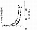
The present invention relates to a kind of method for detecting in real time experimenter's pathogenic bacteria.The method comprises to the experimenter or derives from fluorescence, cold light or the colorimetric substrates of introducing in its biological sample for the pathogenic bacteria β-lactamase, and makes the β-lactamase of experimenter or sample for the active resulting product imaging of substrate.Obtain the wavelength signals by the emission of β-lactamase product, thereby detect the pathogenic bacteria among the experimenter.The present invention relates to a kind of relevant method, it further comprises sets up the 3D reconstruction that transmits, to measure the position of pathogenic bacteria among the experimenter.The present invention relates to another kind of relevant method, it further is included in and transmits than on the large basis of the intensity of the control signal of measuring the pathologic, physiologic situation that real-time diagnosis is relevant with pathogenic bacteria.For example, described fluorescence, cold light or colorimetric substrates are CNIR2, CNIR3, CNIR4, CNIR5, CNIR5-QSY22, CNIR7, CNIR9, CNIR10, CNIR7-TAT, cage fluorescein (caged luciferin), colorimetric reagent or derivatives thereof or analogue.
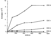
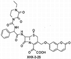
The present invention relates to a kind of relevant method for detecting in real time pathogenic bacteria.The method comprises to be introduced in the experimenter for the substrate of the β-lactamase of pathogenic bacteria or makes the substrate contact of described β-lactamase for pathogenic bacteria come from the experimenter's or the biological sample that obtained by the experimenter surface, and makes the β-lactamase of experimenter or sample for the active resulting product imaging of substrate.Obtain the wavelength signals by the emission of β-lactamase product, thereby detect the pathogenic bacteria among the experimenter.The present invention relates to a kind of relevant method, it further is included in the biological sample and quantizes and distinguish in the step of cells infected and non-infected cells one or both.The present invention relates to another kind of relevant method, it further comprises sets up the 3D reconstruction that transmits, to measure the position of pathogenic bacteria among the experimenter.For example, described substrate can be fluorogene (fluorogenic) substrate CDC-1, CDC-2, CDC-3, CDC-4, CDC-5, CNIR5, CNIR5.2, CNIR5-QSY22, CNIR7, CNIR7-TAT, CNIR9, CNIR10, CNIR800, CNIR800.2, CNIR800-3, XHX2-81, XHX2-91, XHX3-1, XHX3-2, XHX3-26 or XHX3-32, or derivatives thereof or analogue, the substrate that perhaps comprises a kind of illuminating colour or chemical reagent, described illuminating colour or chemical reagent based on β-lactamase their activity are produced color effectively or pH changes.
The invention still further relates to a kind of method for the diagnosis pathologic, physiologic situation relevant with experimenter's pathogenic bacteria.The method is included as the experimenter and uses for fluorogene or the cold light substrate of the β-lactamase of pathogenic bacteria or make derived from described experimenter's biological sample contact pin fluorogene or the cold light substrate to the β-lactamase of pathogenic bacteria, and makes experimenter's β-lactamase for the product imaging of the activity of substrate.Under the emission wavelength of product, measure in real time fluorescence, cold light or colorimetric strength of signal, so that be associated with the diagnosis of pathologic, physiologic situation greater than fluorescence, cold light or the colorimetric strength of signal of the control signal of measuring.The present invention relates to a kind of relevant method, it further comprises the 3D that sets up signal rebuilds, to measure the position of microbial pathogen.The present invention relates to another kind of relevant method, it further comprises one or more therapeutic compounds of using effective treatment pathologic, physiologic situation.The present invention relates to another kind of relevant method, it comprises to the experimenter again uses the fluorogene compound and makes experimenter's re-imaging or the biological sample derived from the experimenter is contacted with described fluorogene substrate; And make experimenter or described biological sample imaging with the validity of monitor treatment compound, so that the reduction that transmits of comparing with diagnostic signal shows the result for the treatment of to the pathologic, physiologic situation.For example, described fluorogene or cold light substrate are CNIR2, CNIR3, CNIR4, CNIR5, CNIR5-QSY22, CNIR7, CNIR9, CNIR10, CNIR7-TAT, cage fluorescein, colorimetric reagent or derivatives thereof or analogue.
The present invention relates to a kind of relevant method for the diagnosis pathologic, physiologic situation relevant with experimenter's pathogenic bacteria.The method is included as the experimenter and uses for the substrate of the β-lactamase of pathogenic bacteria or make its contact derived from experimenter's biological sample, and makes experimenter's β-lactamase for the product imaging of the activity of substrate.Real-time measure signal intensity (for example, fluorescence, cold light or than chrominance signal) under the emission wavelength of product; Wherein the strength of signal greater than the control signal of measuring is associated with the diagnosis of pathologic, physiologic situation.The present invention relates to a kind of relevant method, it further is included in the biological sample and quantizes and distinguish in the step of cells infected and non-infected cells one or both.The present invention relates to another kind of relevant method, it further comprises the 3D that sets up signal rebuilds, to measure the position of microbial pathogen.Especially, described substrate can be fluorogene substrate CDC-1, CDC-2, CDC-3, CDC-4, CDC-5, CNIR5, CNIR5.2, CNIR5-QSY22, CNIR7, CNIR7-TAT, CNIR9, CNIR10, CNIR800, CNIR800.2, CNIR800-3, XHX2-81, XHX2-91, XHX3-1, XHX3-2, XHX3-26 or XHX3-32, or derivatives thereof or analogue, perhaps comprise a kind of illuminating colour or chemical reagent, described illuminating colour or chemical reagent based on β-lactamase their activity are produced color effectively or pH changes.
The invention further relates to a kind of diagnostic method for detection of mycobacterium infection among the experimenter.Described method comprises from the experimenter and obtains biological sample, and described biological sample is contacted with the mycobacterium β-lactamase.Make described biological sample imaging detect β-lactamase for the product of the activity of substrate, and under the emission wavelength of product measure signal intensity, wherein the strength of signal greater than the control signal of measuring shows the existence that mycobacterium infects.The present invention relates to a kind of relevant method, it further is included in the biological sample and quantizes and distinguish in the step of cells infected and non-infected cells one or both.The present invention relates to another kind of relevant method, it further comprises these method steps one or many of repetition, the treatment validity of the treatment plan that is applied to the experimenter that detects based on mycobacterium with monitoring, wherein compared with the control the reduction of measurement signal is associated with the positive response for the treatment of plan.Described substrate can be fluorogene substrate CDC-1, CDC-2, CDC-3, CDC-4, CDC-5, CNIR5, CNIR5.2, CNIR5-QSY22, CNIR7, CNIR7-TAT, CNIR9, CNIR10, CNIR800, CNIR800.2, CNIR800-3, XHX2-81, XHX2-91, XHX3-1, XHX3-2, XHX3-26 or XHX3-32, or derivatives thereof or analogue, perhaps comprise a kind of illuminating colour or chemical reagent, described illuminating colour or chemical reagent based on β-lactamase their activity are produced color effectively or pH changes.
In addition, the invention further relates to a kind of method for the screening therapeutic compound, described therapeutic compound is effective to treat the pathologic, physiologic situation relevant with the pathogenic bacteria among the experimenter.The method comprises selects the potential therapeutic compound for pathogenic bacteria, the biological sample that makes bacterial cell or comprise described bacterial cell contacts with fluorescence, cold light or colorimetric detection agent, and bacterial cell is contacted with potential therapeutic compound.Under the condition that has and do not exist described potential therapeutic compound, measure by bacterial cell or comprise fluorescence that the biological sample of described bacterial cell produces, cold light or than chrominance signal, in order to compare the result for the treatment of that the reduction of the signal under the condition that has therapeutic compound shows described compound antagonism pathogenic bacteria with the signal under the condition that does not have therapeutic compound.For example, described fluorescence, cold light or colorimetric detection agent are CNIR2, CNIR3, CNIR4, CNIR5, CNIR5-QSY22, CNIR7, CNIR9, CNIR10, CNIR7-TAT, cage fluorescein, colorimetric reagent or derivatives thereof.
The present invention relates to a kind of method for the screening therapeutic compound, described therapeutic compound is effective to treat the pathologic, physiologic situation relevant with pathogenic bacteria among the experimenter.The method comprise be right after above-described, use the fluorogene substrate as the step of detection agent, described fluorogene substrate is CDC-1, CDC-2, CDC-3, CDC-4, CDC-5, CNIR5, CNIR5.2, CNIR5-QSY22, CNIR7, CNIR7-TAT, CNIR9, CNIR10, CNIR800, CNIR800.2, CNIR800-3, XHX2-81, XHX2-91, XHX3-1, XHX3-2, XHX3-26 or XHX3-32, or derivatives thereof or analogue, perhaps comprise a kind of illuminating colour or chemical reagent, described illuminating colour or chemical reagent effectively produce color based on β-lactamase for their activity or pH changes.
In addition, the invention further relates to a kind of method be used to making the pathogenic bacteria imaging.The method comprises makes pathogenic bacteria contact with fluorogene substrate for the β-lactamase of pathogenic bacteria, for the product of β-lactamase for the activity of substrate, send an excitation wavelength to pathogenic bacteria, and obtain emission from fluorescence, the cold light of product or compare chrominance signal.The 3D of the signal that generation is obtained rebuilds, thereby makes the pathogenic bacteria imaging.
In addition, the invention further relates to a kind of substrate of directed toward bacteria β-lactamase, described substrate produces detectable fluorescence, cold light or compares chrominance signal based on the activity of β-lactamase to it.Representative substrate includes but not limited to CDC-1, CDC-2, CDC-3, CDC-4, CDC-5, CNIR5, CNIR5.2, CNIR5-QSY22, CNIR7, CNIR7-TAT, CNIR9, CNIR10, CNIR800, CNIR800.2, CNIR800-3, XHX2-81, XHX2-91, XHX3-1, XHX3-2, XHX3-26 or XHX3-32, or derivatives thereof or analogue, perhaps comprise a kind of illuminating colour or chemical reagent, described illuminating colour or chemical reagent based on β-lactamase their activity are produced color effectively or pH changes.The present invention relates to another kind of relevant substrate, it further comprises particle, microballoon or connected vitamin H.
In addition, the invention further relates to a kind of method for detecting in real time experimenter's pathogenic bacteria.The method comprises to experimenter's introducing uses the isotropic substance relevant with the γ emission to carry out radiolabeled substrate, and wherein said substrate is for β-lactamase or specific other enzyme of pathogenic bacteria or protein.For the γ emission of experimenter's radio-labeling substrate, imaging in the active process on described substrate, and obtain the signal that the gamma-rays by emission produces.The strength of signal that generation produces based on gamma-rays, the 3D of the concentration of radio-labeling in the experimenter rebuilds, thereby detects pathogenic bacteria.The present invention relates to a kind of relevant method, it further comprises based on the detection real-time diagnosis of the pathogenic bacteria pathologic, physiologic situation relevant with pathogenic bacteria.The present invention relates to another kind of relevant method, it further comprises uses one or more therapeutic compounds, and described therapeutic compound is effective to treat the pathologic, physiologic situation.In addition, the present invention relates to another kind of relevant method, it further comprises to the experimenter again uses radiolabeled substrate and makes described experimenter's re-imaging with the validity of monitor treatment compound; The reduction of the γ emission that the γ emission during wherein with diagnosis is compared shows the result for the treatment of to the pathologic, physiologic situation.
In addition, the invention further relates to the radio-labeling substrate for the bacterium β-lactamase that is suitable for PET or SPECT imaging as described herein.
In addition, the invention further relates to a kind of Analytical equipment for visual detection of biological sample pathogenic bacteria.Described Analytical equipment comprises platform, described platform has for the device that receives mixtures incubated, described mixtures incubated comprises biological sample and chromogenic substrate for the β-lactamase relevant with pathogenic bacteria, and described platform has be used to catching and concentrate by the device of β-lactamase for the colored product of the activity generation of substrate, and described device links to each other with described receiving trap liquid.The present invention relates to a relevant invention, it further comprises for the device that only allows colored product from described receiving trap flow further downstream.The present invention relates to a relevant invention, it further is included in the internal contrast in described receiving trap downstream.The present invention relates to another relevant invention, wherein substrate comprises chemical reagent and equipment further comprises the second reagent, in order to produce color by described chemical reagent.The present invention relates to another relevant invention, wherein substrate is connected with vitamin H and equipment further comprises avidin, in order to catch the substrate that is connected with vitamin H.
By the description to following the preferred embodiments of the invention, other and further purpose, feature and advantage will be apparent, only the providing for purposes of this disclosure of described the preferred embodiments of the invention.
Description of drawings
In order to reach and detailed understanding above-mentioned feature of the present invention, advantage and purpose, and more clearly other content that will become, the specific embodiments of the present invention of illustrating in the accompanying drawings by reference can obtain the more concrete description of the present invention of top brief overview.These accompanying drawings form the part of this specification sheets.Yet, it should be noted that accompanying drawing only illustrates the preferred embodiments of the invention, and should not be considered to limit scope of the present invention.
Figure 1A-1C is presented at the BlaC mutant crystallization (Figure 1B) of soaking BlaC mutant crystallization (Figure 1A) before and reservation CNIR4 substrate with CNIR4.Fig. 1 C illustrates by the cefotaxime catalysis of Mtb BlaC and the product that forms by lactam nucleus hydrolysis.
Fig. 2 A-2C be described in by before the beta-lactam enzymic hydrolysis and afterwards CC1 and CC2(Fig. 2 A), CHPQ(Fig. 2 B) and CR2(Fig. 2 C) structure.
Fig. 3 describes the structure of CNIR1, CNIR2, CNIR3 and CNIR4 and the hydrolysis by β-lactamase thereof.
Fig. 4 A-4D describes for the preparation of the synthetic schemes of near infrared substrate CNIR5 (Fig. 4 A), be used for alternative synthetic schemes (Fig. 4 B) extensive, that commercialization prepares CNIR5, fluorescence intensity is to the figure (Fig. 4 C) of CNIR5 wavelength and the structure (Fig. 4 D) of CNIR5-QSY22 under the condition that has and do not exist β-lactamase.
Fig. 5 A-5D describes QSY21(Fig. 5 A), QSY21 disulfonic acid (Fig. 5 B) and the structure of QSY22 disulfonic acid (Fig. 5 C) and the chemosynthesis (Fig. 5 D) of QSY22 disulfonic acid.
Fig. 6 A-6B describes structure (Fig. 6 A) and the chemosynthesis (Fig. 6 B) thereof of CNIR7.
Fig. 7 A-7B describes the A for CNIR9(Fig. 7) and CNIR10(Fig. 7 B) synthetic schemes.
Fig. 8 A-8F describes synthetic schemes and the hydrolysis dynamics of fluorogene substrate CDC-1-5.Fig. 8 A shows synthesizing of CDC-1-4.Fig. 8 B-8C be presented at TEM-1Bla or Mtb BlaC process respectively after probe CDC-1,2,3,4 in the emission at the 455nm place figure to the time.(concentration of substrate: 5mM; PBS(pH=7.4) the TEM-1Bla concentration in is 2nM; PBS(pH=7.4) the Mtb BlaC concentration in is 10nM; Excite in the 400nm place).Fig. 8 D shows synthesizing of CDC-5.Fig. 8 E be presented at β-lactamase process after substrate CDC-1 in the emission at the 455nm place figure (solid line: adopt Mtb BlaC processing to the time; Dotted line: adopt TEM-1Bla to process; Concentration and probe concentration: 5mM; PBS(pH=7.4) the TEM-1Bla concentration in: 20nM; Excite in the 400nm place).Fig. 8 F be presented at β-lactamase process after probe CDC-5 in the emission at the 455nm place figure (solid line: adopt Mtb BlaC processing to the time; Dotted line: adopt TEM-1Bla to process; Concentration and probe concentration: 5mM; PBS(pH=7.4) the Mtb BlaC concentration in: 20nM; Excite in the 400nm place).
Fig. 9 A-9F describes the chemical structure (Fig. 9 A-9E) of XHX2-81, XHX2-91, XHX3-1, XHX3-26 and XHX3-32, and shows bacterial number and use dependency (Fig. 9 F) between the fluorescent signal of XHX2-81, XHX2-91, XHX3-26 and XHX3-32.Excite: 360nm(lxPBS); Emission maximum: 453nm(lxPBS); 10mM, 60mL among the XHX-2-81(DMSO); 100mM, 10mL among the XHX-2-91(DMSO); 20mM, 42mL among the XHX-3-26(DMSO); 100mM, 100mL among the XHX-3-32(DMSO).
Figure 10 A-10B describes the chemosynthesis (Figure 10 A) of Bluco and the purposes (Figure 10 B) that Bluco is used for continuous report bioluminescence assay (SREL) imaging of β-lactamase.
Figure 11 A-11D describes for the preparation of the instead of chemical synthetic route (Figure 11 A-11B) of CNIR and CNIR sample probe and derivative and analogue and so synthetic probe structure, such as CNIR5.2 and CNIR800.2(Figure 11 C) and CNIR800-3(Figure 11 D).
Figure 12 A-12B illustrates the detection of (Figure 12 B) Bla activity among (Figure 12 A) and Mycobacterium tuberculosis in the intestinal bacteria that contain CNIR5.Contrast contains LB medium and CNIR5, and does not contain the intestinal bacteria of conversion.
Figure 13 A-13H describes the kinetics (Figure 13 E-13H) that fluorescence emission spectrum (Figure 13 A-13D) and fluorescent mark mix.Shown the CNIR4(Figure 13 A with (CNIR+Bla) after (CNIR) and the cracking before the TEM-1Bla cracking 10 minutes), CNIR5(Figure 13 B), CNIR9(Figure 13 C) and CNIR10(Figure 13 D) emmission spectrum.Shown the E with CNIR4(Figure 13), CNIR5(Figure 13 F), CNIR9(Figure 13 G) and CNIR10(Figure 13 H) fluorescent mark directly is incorporated into the kinetics in wild-type Mtb and the Mtb BlaC mutant (blaCm).
Figure 14 A-14B describes to contain CNIR4(Figure 14 A) and CNIR5(Figure 14 B) the intestinal bacteria TEM-1 β-lactamase of substrate and the kinetics of Mycobacterium tuberculosis Bla-C β-lactamase.
Figure 15 A-15H is depicted in and contains CNIR4(Figure 15 A, 15E), CNIR5(Figure 15 B, 15F), CNIR9(Figure 15 C, 15G) and CNIR10(Figure 15 D, 15H) medium in Fluorescent dye incorporation kinetics and the distribution proportion wherein (Figure 15 A-15H) of independent Mycobacterium tuberculosis bacterium.
Figure 16 A-16H is depicted in and contains CNIR4(Figure 16 A, 16E), CNIR5(Figure 16 B, 16F), CNIR9(Figure 16 C, 16G) and CNIR10(Figure 16 D, 16H) Fluorescent dye incorporation kinetics (Figure 16 A-16D) and the distribution proportion wherein (Figure 16 E-16H) of the scavenger cell that infects of Mycobacterium tuberculosis bacterium.
Figure 17 describes the fluorescence confocal microscope image, and it shows in the cell of CNIR4 in the scavenger cell of infection due to Mycobacterium tuberculosis and mixes.DAPI painted (blueness) indicates the nucleus of infected cell, and green fluorescence comes from the Mycobacterium tuberculosis of GFP mark and the CNIR4 that red fluorescence comes from cracking.
Figure 18 A-18E describes by with 10
8(the left back section of every mouse), 10
7(left front section), 10
6CNIR4(Figure 18 A of the various concentration of (right front portion)), CNIR5(Figure 18 B), CNIR9(Figure 18 C) and CNIR10(Figure 18 D) the fluorescence of the mouse that comes from m tuberculosis infection of intradermal vaccination.Figure 18 E be equivalent to each compound in the signal of the concentration of each bacterium that be used for to infect to Background.
Figure 19 A-19E comes from by aerosol inoculation at the fluoroscopic image of the mouse of pulmonary infection Mycobacterium tuberculosis with for CNIR4(Figure 19 A), CNIR5(Figure 19 B), CNIR9(Figure 19 C) and CNIR10(Figure 19 D) fluorescent signal measured.In each figure of Figure 19 A-19D, the mouse on the left side does not infect in every flat board, the infection due to Mycobacterium tuberculosis that left several second mouse adopts the blaC gene to undergo mutation, and two mouse on the right are adopted the wild-type infection due to Mycobacterium tuberculosis in every flat board.Three mouse, 24 hours veins before imaging on the right give CNIR4, CNIR5, CNIR9 or CNIR10 in every flat board.Figure 19 E is that the signal of interior each compound of lung areas in the image of back is to the figure of background.
Figure 20 A-20F comes from the mouse by the aerosol infection Mycobacterium tuberculosis and uses substrate CNIR5 in the fluoroscopic image of 1 hour (Figure 20 A), 18 hours (Figure 20 B), 24 hours (Figure 20 C) and (Figure 20 D) imaging in 48 hours.In the rear view of each panel, abdomen view, left view, right view, the left side mouse be do not infect and the right side mouse infect.All mouse are all to adopt the CNIR5 intravenous injection before the time point imaging of mark.Figure 20 F is the figure of the fluorescent signal that obtains of the interested zone of drawing a circle to approve in the head slab that quantizes by Figure 19 A.
Figure 21 A-21B describe to come from by (Figure 21 A) of aerosol infection Mycobacterium tuberculosis and (Figure 21 B) mouse of not infecting and use transillumination (rather than reflection) fluoroscopic image of imaging to reduce background signal.
Figure 22 A-22D is illustrated in the upper CNIR5(7nmol of employing of nude mice that left shoulder has the C6 tumour that heteroplastic wild-type C6 tumour and right shoulder have the transfection of cmv-bla stabilization) the Bla image of expressing.Figure 22 A is presented at overlapping fluorescence and the bright-field image of fixed time point.Figure 22 B shows the drafting figure of the average intensity of each tumour to the time.Figure 22 C shows the tumour of excision and the image of organ.Figure 22 D shows the CC1 analytical results from Bla in the extract of two kinds of tumours.
Figure 23 A-23C is illustrated in the upper CNIR6(7nmol of employing of nude mice that left shoulder has the C6 tumour that heteroplastic wild-type C6 tumour and right shoulder have the transfection of cmv-bla stabilization) the Bla image of expressing.Figure 23 A is the chemical structure of CNIR6.Figure 23 B is presented at overlapping fluorescence and the bright-field image of fixed time point.Figure 23 C shows the drafting figure of the average intensity of each tumour to the time.
After Figure 24 A-24B illustrates 4 hours (Figure 24 A) and 24 hours (Figure 24 B), the bio distribution of 7.5 nmole CNIR5 in various tissues.
Figure 25 A-25B uses intravenous CNIR5 as preparation, infects the mouse (Figure 25 A) of Mycobacterium tuberculosis and the in-vivo image of the control mice (Figure 25 B) of infection not.
Figure 26 A-26C illustrates the SREL detection threshold that uses the CNIR probe.Figure 26 A is a histogram, and it show to use β-lactamase CNIR probe can detect bacterium below 100 with the SREL imaging.Figure 26 B-26C is the live in-vivo image of mouse of (Figure 26 B) that do not infect or (Figure 26 C) that infects Mycobacterium tuberculosis.
Figure 27 A-27E describes the evaluating ability result (Figure 27 A) that CNIR5 detects the tubercule bacillus in the clinical sample, measure the result (Figure 27 B) of tubercule bacillus detection threshold in the sputum sample basis, dependency (Figure 27 C) in the mark-on sputum sample basis between strength of signal and the bacterial number, the comparison (Figure 27 D) of strength of signal and bacterial number among mark-on sputum sample basis and the PBS, and the evaluation of vazadrine+rifampicin treatment mycobacterium, but comprise time (Figure 27 E) of acquisition measurement signal.
Figure 28 describes the structure of IRDye800 series fluorophore.
Figure 29 describes the structure of cefoperazone and proposed CNIR probe.
Figure 30 is the scheme of setting up the small-sized bias storehouse (biased library) of Bluco substrate.
Figure 31 is illustrated in the structure that the novel probe of allyl group connection is contained in the 3' position.
Figure 32 is depicted in the structure that the novel probe of carbamate connection is contained in the 3' position.
The visual detecting system that is used for diagnositc analysis that Figure 33 A-33D describes to utilize the yoke that is connected to latex particle or microballoon to close the chemical reagent (Figure 33 B) of substrate/illuminating colour (Figure 33 A) or generation conjugates/substrate/color and is connected to the chemical reagent (Figure 33 B) of the conjugates/substrate of vitamin H/illuminating colour (Figure 33 C) or generation conjugates/substrate/color.
Embodiment
As used in this specification, " one " can refer to one or many.Such as in these claims use, when " comprising " when being used with word, word " one " can refer to more than one or one.As used herein, " another " or " other " can refer at least the second or more identical or different claimed element or component.In addition, except other had regulation, the term of odd number comprised the situation of plural number, and the term of plural number comprises the situation of odd number.
As used herein, unless offer some clarification on and refer to unique alternatives or alternatives is mutually exclusive, although otherwise the definition of disclosure support refer to unique alternatives and " and/or ", the term "or" in claims all be used in reference to " and/or ".
As used herein, term " contact " refers to any suitable method, described method in vivo or external, in biological sample, make the fluorogene substrate, for example, the fluorogene compound, fluorescence protein, cold light albumen, perhaps than chromoprotein or other colorimetric reagent or derivatives thereof, the radiolabeled substrate contact pathogenic bacteria that perhaps is suitable for PET or SPECT imaging, described pathogenic bacteria is such as but not limited to Mycobacterium tuberculosis (Mycobacterium tuberculosis, Mbt), cow mycobacteria (Mycobacterium bovis, M.bovis), bird mycobacterium (Mycobacterium avium, M.avium), Mycobacterium tuberculosis mixture or bird mycobacterium mixture, perhaps contact Bacteroides (Bacteroides), fusobacterium (Clostridium), streptococcus (Streptococcus), Staphylococcus (Staphylococcus), Rhodopseudomonas (Pseudomonas), hemophilus (Haemophilus), legionella (Legionella), Escherichia (Escherichia), salmonella (Salmonella), species in Shigella (Shigella) or the listeria (Listeria) perhaps contact β-lactamase or specific other enzyme or protein for pathogenic bacteria.In external or stripped, this is by making one or more bacterial cells or β-lactamase or other enzyme or protein be exposed to fluorogene substrate in the suitable media or fluorogene compound or fluorescence, cold light or than in chromoprotein or other colorimetric reagent or derivatives thereof.Described bacterial cell or β-lactamase or other enzyme or protein are obtaining in experimenter's sample.Described bacterial cell can comprise or can not comprise great-hearted sample.Described β-lactamase or other enzyme or protein can contact great-hearted bacterial cell, can extract from bacterial cell with method standard by known, himself may reside in the biological sample, perhaps can comprise by the recombination system of method transfection known and standard to the bacterial cell.Described sample can include but not limited to that hydrothorax or sputum and possibility tool are germy, comprises other body fluid of blood, saliva, urine and ight soil.Alternatively, for external contact, biological sample can obtain from the surface by (for example) erasing, and described surface is such as but not limited to one or more interested zones on instrument, vessel, facility, working-surface, clothes or the human body.The sample that so obtains can be transferred in the suitable medium, is used for coming imaging by the method for as known in the art or standard.For using in the body, as described herein, any known fluorogene substrate of using, that is, the fluorogene compound, fluorescence, cold light or than chromoprotein, other colorimetric reagent or derivatives thereof, or the method for radiolabeled substrate all is fit to.
As used herein, phrase " fluorogene substrate " refers to compound or protein or peptide or other biologically active molecules, it obtains launching or producing by exciting of appropriate wavelength fluorescence or cold light signal in the presence of the enzyme that is fit to product perhaps can generate the product that produces than chrominance signal.Such as but not limited to, at β-lactamase, under the existence of luciferase or beta-galactosidase enzymes or other enzyme, the fluorogene substrate can produce fluorescence, cold light or colorimetric product.
As used herein, phrase " radiolabeled substrate " refers to compound or protein or peptide or other biologically active molecules, it is attached to or is connected to or integrate with short-lived isotropic substance, described isotropic substance emission is used for positron emission tomography (Positron Emission Tomography, PET) positron and be used for the gamma-rays of single photon emission computed tomography (Single Photon Emission Computed Tomography, SPECT).
As used herein, phrase " β-lactamase positive bacteria " refers to nature secretion β-lactamase or obtain the pathogenic bacteria of β-lactamase in pathogenic process.
As used herein, term " experimenter " refers to receives treatment or from wherein obtaining any target of biological sample.Preferably, described experimenter is Mammals, and more preferably, described experimenter is livestock or people.
In one embodiment of the invention, a kind of method for detecting in real time experimenter's pathogenic bacteria is provided, and it is included as the experimenter or uses fluorescence, cold light or colorimetric substrates for the β-lactamase of pathogenic bacteria from described experimenter's biological sample; Make the β-lactamase of described experimenter or sample for product imaging under excitation wavelength of the activity of substrate; And obtain signal at the wavelength place by the emission of β-lactamase product; Thereby detect the pathogenic bacteria among the experimenter.
About this embodiment, described method comprises sets up the 3D reconstruction that transmits, to measure the position of pathogenic bacteria among the experimenter.In another further embodiment, described method is included in and transmits than on the large basis of the intensity of the control signal of measuring the pathologic, physiologic situation that real-time diagnosis is relevant with pathogenic bacteria.The example of pathologic, physiologic situation is tuberculosis.
In certain embodiments of the invention, fluorogenic substrate can be the fluorogene substrate.The example of described fluorogene substrate is CNIR2, CNIR3, CNIR4, CNIR5, CNIR5-QSY22, CNIR7, CNIR9, CNIR10, CNIR7-TAT, cage fluorescein, colorimetric reagent or derivatives thereof.Equally, in all embodiments, described imaging or excitation wavelength and described emission wavelength can be to about 900nm independently from about 300nm.In certain embodiments, described imaging or excitation wavelength are to approximately 730nm and described transmitting can be that about 650nm is to about 800nm from about 540nm.In certain embodiments, the colorimetric indication can be identified by human eye visually by colour-change or apparatus measures, to measure the numerical value of an appointment.In addition, pathogenic bacteria can comprise the bacterial species of Bacteroides, fusobacterium, streptococcus, Staphylococcus, Rhodopseudomonas, hemophilus, legionella, mycobacterium, Escherichia, salmonella, Shigella or listeria.Especially, described pathogenic bacteria can comprise Mycobacterium tuberculosis mixture or bird mycobacterium mixture.
In a relevant embodiment of the present invention, a kind of method be used to making the pathogenic bacteria imaging is provided, it comprises the fluorogene substrate of introducing in the experimenter for the β-lactamase of pathogenic bacteria, perhaps make come from described the experimenter's or the biological sample contact pin that obtained by the surface to the fluorogene substrate of the β-lactamase of pathogenic bacteria; Send an excitation wavelength to pathogenic bacteria and be used for β-lactamase for the product of the activity of substrate; Obtain emission from fluorescence, the cold light of described product or compare chrominance signal; And the 3D reconstruction that produces the signal that obtains, thereby make described pathogenic bacteria imaging.In the each side of this embodiment, described pathogenic bacteria can with aforesaid fluorogene or cold light substrate in vivo or external the contact.Equally, aspect all of this embodiment in, described pathogenic bacteria and described excite with emission wavelength as mentioned above.
In another relevant embodiment, the invention provides a kind of method for detecting in real time pathogenic bacteria, it comprises to the experimenter or comes from the substrate of introducing in described experimenter's the biological sample for the β-lactamase of pathogenic bacteria; The product imaging that the β-lactamase that makes described experimenter or sample obtains for the activity of substrate; And obtain the wavelength signals by the emission of β-lactamase product, thereby detect the pathogenic bacteria among the experimenter.
About this embodiment, described method comprises sets up the 3D reconstruction that transmits, to measure the position of pathogenic bacteria among the experimenter.In another further embodiment, described method can comprise cells infected and the non-infected cells in quantification and the differentiation biological sample.Especially, in this further embodiment, carry out differentiation and/or the quantization step of cells infected by utilizing in flow cytometry, Laser Scanning Confocal Microscope method or the fluorescent spectrometry one or more.
In two embodiments, substrate can be fluorogene substrate CDC-1, CDC-2, CDC-3, CDC-4, CDC-5, CNIR5, CNIR5.2, CNIR5-QSY22, CNIR7, CNIR7-TAT, CNIR9, CNIR10, CNIR800, CNIR800.2, CNIR800-3, XHX2-81, XHX2-91, XHX3-1, XHX3-2, XHX3-26 or XHX3-32, or derivatives thereof or analogue.Alternatively, described substrate can comprise a kind of illuminating colour or chemical reagent, and described illuminating colour or chemical reagent induce color or pH to change effectively.In addition, described substrate can be connected to particle, microballoon or be connected to vitamin H.Equally, in two embodiments, described biological sample can be sputum, hydrothorax, urine, blood, saliva, ight soil or by the wiping experimenter sample that obtains of interested zone with it.The signal that obtains can be fluorescence, cold light or compare chrominance signal.Described pathogenic bacteria, described imaging wavelength and described emission wavelength are as mentioned above.
In another embodiment of the invention, a kind of method for the diagnosis pathologic, physiologic situation relevant with experimenter's pathogenic bacteria is provided, it comprises to the experimenter uses fluorogene or cold light substrate for the β-lactamase of pathogenic bacteria; Make described experimenter's β-lactamase for product imaging under excitation wavelength of the activity of substrate; And under the emission wavelength of product, measure in real time fluorescence, cold light or colorimetric strength of signal; Wherein interrelated greater than the diagnosis of fluorescence, cold light or the colorimetric strength of signal of the control signal of measuring and pathologic, physiologic situation.
About this embodiment, described method comprises the 3D that sets up signal rebuilds, to measure the position of microbial pathogen.In another further embodiment, described method comprises uses one or more therapeutic compounds, and described therapeutic compound is effective to treat the pathologic, physiologic situation.In addition, described method comprises to the experimenter and again uses fluorogene or cold light substrate; And make experimenter's re-imaging with the validity of monitor treatment compound; The reduction that transmits of wherein comparing with diagnostic signal shows the result for the treatment of to the pathologic, physiologic situation.In all embodiments, described pathologic, physiologic situation, pathogenic bacteria, fluorogene substrate and imaging or excite with emission wavelength as mentioned above.
In a relevant embodiment of the present invention, a kind of method for the diagnosis pathologic, physiologic situation relevant with experimenter's pathogenic bacteria is provided, it comprises to the experimenter uses substrate for the β-lactamase of pathogenic bacteria, or makes derived from described experimenter's the biological sample contact pin substrate to the β-lactamase of pathogenic bacteria; Make experimenter's β-lactamase for the product imaging of the activity of substrate; And under the emission wavelength of product real-time measure signal intensity; Wherein interrelated greater than the diagnosis of the strength of signal of the control signal of measuring and pathologic, physiologic situation.About this embodiment, described method comprises sets up 3D rendering, uses the therapeutic compound of the pathologic, physiologic situation that is suitable for diagnosing and again uses aforesaid substrate.In another further embodiment, described method comprises cells infected and the non-infected cells in aforesaid quantification and/or the differentiation biological sample.
In these embodiments, described substrate can be aforesaid fluorogene substrate, illuminating colour or chemical reagent.Equally, described pathologic, physiologic situation can be that tuberculosis and described biological sample can be sputum, hydrothorax, urine, blood, saliva, ight soil or by the wiping experimenter sample that obtains of interested zone with it.The signal of described measurement can be fluorescence, cold light or compare chrominance signal.Described pathogenic bacteria, imaging or excitation wavelength and emission wavelength are as mentioned above.
In another relevant embodiment of the present invention, the method of a kind of diagnosis pathologic, physiologic situation relevant with pathogenic bacteria among the experimenter is provided, and it comprises makes from the sample contact pin of the experimenter's acquisition colorimetric substrates to the β-lactamase of pathogenic bacteria; Wherein β-lactamase causes bore hole visible colour-change for the product of the activity of substrate, and then indication diagnosis.Described substrate can be placed on band (strip), cotton swab (q-tip), background (background) or other visual indicator (indicator).Colour-change can be the bore hole visible, and need not any instrument or come from exciting of external energy source can obtain identifying.
In other another embodiment of the present invention, a kind of diagnostic method that infects for detection of the mycobacterium among the experimenter is provided, it comprises from the experimenter and obtains biological sample; Make the fluorogenic substrate of biological sample contact mycobacterium β-lactamase; Make the β-lactamase of biological sample for the product imaging of the activity of fluorogene substrate; And under the emission wavelength of product measure signal intensity; Wherein the strength of signal greater than the control signal of measuring shows the existence that mycobacterium infects.About this embodiment, described method provides repetition aforesaid method step one or many, the detection of infecting based on mycobacterium with monitoring, be applied to the treatment validity of experimenter's treatment plan; Wherein compared with the control the reduction of fluorescent signal of measurement and the positive response for the treatment of plan is interrelated.In another further embodiment, as mentioned above, described method is included in the biological sample and quantizes and distinguish in the step of cells infected and non-infected cells one or both.
In all respects in two embodiments, described substrate can be fluorogene substrate CDC-1, CDC-2, CDC-3, CDC-4, CDC-5, CNIR5, CNIR5.2, CNIR5-QSY22, CNIR7, CNIR7-TAT, CNIR9, CNIR10, CNIR800, CNIR800.2, CNIR800-3, XHX2-81, XHX2-91, XHX3-1, XHX3-2, XHX3-26 or XHX3-32, or derivatives thereof or analogue.Alternatively, described substrate can comprise a kind of illuminating colour or chemical reagent, and described illuminating colour or chemical reagent induce color or pH to change effectively.In addition, described substrate can be connected to particle, microballoon, or is connected to vitamin H.Equally, described biological sample can be sputum, hydrothorax, urine, blood, saliva, ight soil or by the wiping experimenter sample that obtains of interested zone with it.In addition, described mycobacterium infects and can be caused by Mycobacterium tuberculosis or Mycobacterium tuberculosis mixture or bird mycobacterium or bird mycobacterium mixture.In addition, described measurement signal can be fluorescence, cold light or compare chrominance signal.Described imaging and emission wavelength can be as mentioned above.
In other another embodiment of the present invention, a kind of method for the screening therapeutic compound is provided, described therapeutic compound is effective to treat the pathologic, physiologic situation relevant with pathogenic bacteria among the experimenter.Described method comprises selects the potential therapeutic compound for pathogenic bacteria; Bacterial cell is contacted with fluorescence, cold light or colorimetric detection agent; Bacterial cell is contacted with potential therapeutic compound; And under the condition that has or do not exist described potential therapeutic compound, measure fluorescence, the cold light that is produced by bacterial cell or compare chrominance signal; The reduction of the signal that has therapeutic compound of wherein comparing with the signal that does not have therapeutic compound shows the result for the treatment of of described compound antagonism pathogenic bacteria.In this embodiment, described pathologic, physiologic situation and described pathogenic bacteria are as mentioned above.
In aspect of this embodiment, pathogenic bacteria can be recombinant bacteria, and the step that bacterium is contacted with fluorescence, cold light or colorimetric detection agent comprises uses the expression vector that contains fluorescence, cold light or colorimetric detection agent to transform wild-type bacterium.In this respect, described fluorescence, cold light or colorimetric detection agent can comprise fluorescin.The example of fluorescin has mPlum, mKeima, Mcherry or tdTomato.Equally in this respect, described expression vector can comprise beta-galactosidase gene, and wherein said method further is included under the existence of beta-galactosidase enzymes recombinant bacterial cell is contacted with the fluorophore that is effective to the emitting fluorescence signal.The example of fluorophore has C2FDG, C12RG or DDAOG.In addition, in this respect in, described expression vector can comprise luciferase gene, wherein said method further comprises makes recombinant bacterial cell contact with the D-fluorescein.The example of luciferase is Photinus pyralis LUC, the dried female insect of kowtowing (click beetle red) or rLuc8.
In aspect another of this embodiment, the fluorogene substrate that described fluoroscopic examination agent can be the bacterium β-lactamase.In one embodiment, pathogenic bacteria can with fluorogene substrate CNIR2, CNIR3, CNIR4, CNIR5, CNIR5-QSY22, CNIR7, CNIR9, CNIR10, CNIR7-TAT, cage fluorescein, colorimetric reagent or derivatives thereof body in contact.In another embodiment, pathogenic bacteria can contact in vivo with fluorogene substrate CC1, CC2, CHPQ, CR2, CNIR1 or CNIR6.
In a relevant embodiment of the present invention, a kind of method for the screening therapeutic compound is provided, described therapeutic compound is effective to treat the pathologic, physiologic situation relevant with pathogenic bacteria among the experimenter.Described method comprises selects the potential therapeutic compound for pathogenic bacteria; The biological sample that makes bacterial cell or comprise it contacts with the substrate of bacterium β-lactamase; The biological sample that makes bacterial cell or comprise it contacts with potential therapeutic compound; And under the condition that has or do not exist potential therapeutic compound, measure fluorescence, the cold light that is produced by bacterial cell or compare chrominance signal; The reduction of the signal that has therapeutic compound of wherein comparing with the signal that does not have therapeutic compound shows the result for the treatment of of described compound antagonism pathogenic bacteria.
In aspect of this embodiment, described substrate can be fluorogene substrate CDC-1, CDC-2, CDC-3, CDC-4, CDC-5, CNIR5, CNIR5.2, CNIR5-QSY22, CNIR7, CNIR7-TAT, CNIR9, CNIR10, CNIR800, CNIR800.2, CNIR800-3, XHX2-81, XHX2-91, XHX3-1, XHX3-2, XHX3-26 or XHX3-32, or derivatives thereof or analogue.Alternatively, described substrate can comprise a kind of illuminating colour or chemical reagent, and described illuminating colour or chemical reagent induce color or pH to change effectively.In addition, described substrate can be connected to particle, microballoon, or is connected to vitamin H.Equally, pathogenic bacteria and pathologic, physiologic situation are as mentioned above.In addition, the signal that is produced by bacterial cell can have the wavelength from about 300nm to about 900nm.Especially, the signal that produces can have the wavelength from about 650nm to about 800nm.
In other another embodiment of the present invention, a kind of substrate of directed toward bacteria β-lactamase is provided, described substrate produces detectable fluorescence, cold light or compares chrominance signal based on the activity of β-lactamase for it.In all respects of this embodiment, described substrate can be fluorogene substrate CDC-1, CDC-2, CDC-3, CDC-4, CDC-5, CNIR5, CNIR5.2, CNIR5-QSY22, CNIR7, CNIR7-TAT, CNIR9, CNIR10, CNIR800, CNIR800.2, CNIR800-3, XHX2-81, XHX2-91, XHX3-1, XHX3-2, XHX3-26 or XHX3-32, or derivatives thereof or analogue, perhaps can comprise illuminating colour or chemical reagent, described illuminating colour or chemical reagent effectively produce color based on β-lactamase for their activity or pH changes.Equally, substrate can further comprise connected particle, microballoon or vitamin H.
In other another embodiment of the present invention, a kind of method for detecting in real time experimenter's pathogenic bacteria is provided, it comprises to described experimenter's introducing uses the isotropic substance relevant with the γ emission to carry out radiolabeled substrate; Wherein said substrate is used for β-lactamase or specific other enzyme of pathogenic bacteria or protein; Make experimenter's the γ emission imaging that in the active process for substrate, comes from the radio-labeling substrate; Obtain the signal by the gamma-rays generation of emission; And the 3D that produces based on the concentration of radio-labeling in the experimenter of the strength of signal that is produced by gamma-rays rebuilds, thereby detects pathogenic bacteria.
About this embodiment, described method comprises based on pathogenic bacteria and detects the real-time diagnosis of the pathologic, physiologic situation relevant with pathogenic bacteria.In another further embodiment, described method comprises uses the therapeutic compound that one or more are effective to treat the pathologic, physiologic situation.In other another further embodiment, described method comprises to the experimenter uses radiolabeled substrate again; And make experimenter's re-imaging, to monitor the validity of described therapeutic compound; The reduction of the γ emission that the γ emission during wherein with diagnosis is compared shows the result for the treatment of to the pathologic, physiologic situation.In these further embodiments, described pathologic, physiologic situation can be tuberculosis.
In aspect of all these embodiments, described radio-labeling can be the isotropic substance of emission positron and can come imaging by positron emission tomography (PET).In one aspect of the method, radio-labeling can be directly to launch gamma-ray isotropic substance and can come imaging by single photon emission computed tomography (SPECT).In aspect all of these embodiments, other enzyme or protein can be β-lactamase sample enzymes or in conjunction with the protein of penicillin.Equally, in all embodiments, bacterial species can be as mentioned above.
In other another embodiment of the present invention, provide to be suitable for PET or SPECT imaging, the radiolabeled substrate of directed toward bacteria β-lactamase.In this embodiment, radio-labeling can be fluoro-18, nitrogen-13, oxygen-18, carbon-11, technetium-99m, iodo-123 or indium-111.
In other another embodiment of the present invention, a kind of Analytical equipment of the pathogenic bacteria for visual detection of biological sample is provided, described Analytical equipment comprises platform, described platform has for the device that receives mixtures incubated, described mixtures incubated comprises biological sample and chromogenic substrate for the β-lactamase relevant with pathogenic bacteria, and described platform has be used to catching and concentrate by the device of β-lactamase for the colored product of the activity generation of substrate, and described device links to each other with described receiving trap liquid.
About this embodiment, described Analytical equipment can comprise for the device that only allows colored product from described receiving trap flow further downstream.In another further embodiment, described Analytical equipment can be included in the internal contrast in described receiving trap downstream.In other another further embodiment, described Analytical equipment can comprise the device be used to the fluid that is absorbed in described receiving trap downstream.
In all embodiments, described substrate can comprise a kind of illuminating colour or chemical reagent.In all embodiments, described substrate can be connected to particle or microballoon equally.In aspect of these embodiments, described substrate comprises that chemical reagent and described equipment further comprise the second reagent, in order to produce color by described chemical reagent.In aspect another of these embodiments, described substrate is connected to vitamin H and described equipment further comprises avidin, in order to catch the substrate that is connected with vitamin H.
The invention provides optical imaging system and method for bacteriosis and/or infection.For for real-time analysis in the body of the quantification of bacterium in the lysis and location and antimicrobial drug activity, these system and methods are extremely responsive instruments.Can expect that these systems are effective to bacterial detection pathogenic agent on unicellular level.These in-vivo imagings (IVI) system and method can directly apply to the patient who is in the clinical setting.
The system and method for this paper can be applicable to the bacterial species that nature has the beta-lactam enzymic activity or obtains the beta-lactam enzymic activity.In the case of unrestricted, the example of β-lactamase positive bacteria species has Bacteroides, fusobacterium, streptococcus, Staphylococcus, Rhodopseudomonas, legionella, mycobacterium, hemophilus, Escherichia, salmonella, Shigella or listeria.What consider especially is diagnosis, location and the quantification of mycobacterium (for example, Mycobacterium tuberculosis and cow mycobacteria).Although the advantage of system and method as herein described is that it does not need the engineering of bacterial isolates to be detected, can expect that the method for the expression of raising β-lactamase, activity and/or secretion is to improve the sensitivity that detects.Therefore, can expect, by in any interested bacterial species or bacterial strain, introducing β-lactamase by means of any method that can use, detect the β-lactamase bacterial species to allow it to detect sensitively, described method allows the expression and secretion of the β-lactamase of enough levels.This can by realizing with delivering method standard with known, comprise that use is suitable for to the phage of Mammals delivery vector in external or body.
In-vivo imaging of the present invention system can detect fluorescence, the cold light that is produced by compound or report or than chrominance signal, described compound or report are as the substrate for the beta-lactam enzymic activity.Imaging system is well known in the art and is obtained commercially.For example, report-enzyme fluorescence (SREF) system, continuous report-enzyme cold light (SREL) system or od-ray system can be for detection of the β-lactamase active results continuously.In addition, the signal that obtains can be used for producing the 3D representation (representation) for the mapping bacteria pathogenic agent.For these systems, the those of ordinary skill of imaging field is based on employed compound and/or report signal type and to be detected well selective exitation and emission wavelength.In general, imaging or excitation wavelength and emission wavelength the two all can be from about 300nm to about 900nm.The example of excitation signal can and transmit in the scope of the extremely about 730nm of about 540nm at about 650nm to the scope of about 800nm.Can expect equally, in-vivo imaging of the present invention system can also detect other signal, such as the signal that produces by radiation, perhaps by other β-lactamase for produce any that can detect of the activity of suitable substrate or other detection agent or the signal that can read.
Beta-lactam enzyme substrates of the present invention can be chemical substrate or quantum dot substrate.Be used for using the substrate of SREL or SREF imaging, for example, can be fluorophore, cage fluorescein or other fluorescence, cold light or colorimetric compound, report or other detection agent, and it is the required application signal that offers the best.Allowing well to infiltrate through under the level of any tissue, described substrate has low-down toxicity or does not have toxicity, and has high s/n ratio.Described signal is near infrared, infrared or red signal light, for example, and from about 650nm to about 800nm.
For example, substrate can be fluorogene substrate or quantum dot substrate, and it produces the signal based on the beta-lactam enzymatic lysis in external or the body.The fluorogene substrate can comprise the FRET donor, such as indigo-blue (indocyanine) dyestuff, for example, Cy5, Cy5.5 or CY7 or IRDye800 and FRET quencher are such as cancellation group QSY21, QSY21 stilbene-4,4'-bis-(1-azo-3, 4-dihydroxy-benzene)-2,2'-disulfonate, QSY22 or QSY22 stilbene-4,4'-bis-(1-azo-3, 4-dihydroxy-benzene)-2,2'-disulfonate or IRDyeQC-1.In addition, the fluorogene substrate can comprise full acetylated D-Glucose amine, with the penetrance that improves cell and/or can be connected to little peptide, and (but being not limited to) TAT for example.In addition, described substrate can be modified, with improve its strength of signal, tissue penetration ability, specificity or the ability that distributes well in a organized way.In addition, can expect, be used for tissue, cell or be conducive to improve the detection of sensitivity and bacterial pathogens with other marking method of other compound of these substrates associatings.For example, the fluorogene substrate can comprise (but being not limited to comprise) methyl, methoxyl group or benzyl moiety and/or can contain cis or the trans double bond that is useful on the larger release of leavings group.
Especially, the fluorogene substrate can the bacterial detection cell culture in or the beta-lactam enzymic activity in the bacterial cell of external single cultivation.The example of chemiluminescence gene substrate has CC1, CC2, CHPQ, CR2, CNIR1 or CNIR6.Alternatively, for in-vivo imaging is used, the fluorogene substrate can be CNIR2, CNIR3, CNIR4, CNIR5, CNIR5-QSY22, CNIR7, CNIR9, CNIR10, CNIR7-TAT or CNIR800, or derivatives thereof or analogue perhaps are similar to CNIR5(such as CNIR5.2) or be similar to CNIR800(such as CNIR800.2 or CNIR800-3) the fluorogene substrate.The present invention also is provided for external and fluorogene substrate CDC-1, CDC-2, CDC-3, CDC-4 and CDC-5 in-vivo imaging, or derivatives thereof or analogue, its hydrolysis based on Mtb BlaC discharges the umbelliferone as fluorophore, perhaps provide and be similar to particularly CDC-5 of CDC-1, CDC-2, CDC-3, CDC-4 and CDC-5() the fluorogene compound, such as (but being not limited to) XHX2-81, XHX2-91, XHX3-1, XHX3-2, XHX3-26 or XHX3-32, or derivatives thereof or analogue.These fluorogene substrates are useful in continuous report-enzyme fluorescence (SREF) system.Can expect that described beta-lactam enzyme substrates is effective to the single bacterial cell that detection bodies is outer or body is interior.
Another example that is used for the fluorogene substrate of detection β-lactamase in the body is the cage fluorescein, such as (but being not limited to) Bluco, Bluco2 or Bluco3.This substrate comprises D-fluorescein (substrate of Photinus pyralis LUC (Fluc)) and beta-lactam (substrate of β-lactamase).Beta-lactam cracking by enzyme discharges the D-fluorescein, and it is based on the oxidation of Fluc and luminescence.The cage fluorescein is useful in continuous report-enzyme cold light (SREL) system or other od-ray imaging system.
Fluorescin can be used for bacterial detection pathogenic agent in external and the body equally.Fluorescin (FP) is cloned in the expression vector such as mPlum, mKeima, Mcherry and tdTomato.Interested bacterial pathogens (such as Mycobacterium tuberculosis) transforms with the FP construct.Fluorescent protein expression by bacterium produces the signal that can detect based on imaging.Other imaging system can be utilized the recombinant bacteria that is converted to secrete other enzyme (such as beta-galactosidase enzymes), and it produces fluorescent signal under the existence of fluorophore (for example, C2FDG, C12RG or DDAOG).In addition, other recombination system of other luciferase (as kowtow dried female insect or rLuc8) is expressed in other imaging system utilization, and it produces signal under the existence of substrate (for example, D-fluorescein).
In addition, substrate can comprise painted or visual dyestuff.Painted or visual dyestuff can be (but being not limited to) texas Red, rhodamine, bromine cresols dyestuff (multiple color), cyanine dyes.In addition, substrate can comprise a kind of chemical reagent that produces colour-change based on the beta-lactam enzymic activity, and described colour-change is as being derived from pH colour-change that change or other chemical induction.
Alternatively, can use the imaging system of positron emission tomography (PET) or single photon emission computed tomography (SPECT).Probe can comprise the substrate of β-lactamase, β-lactamase sample enzyme or other similar enzyme or protein for pathogenic bacteria as herein described.PET and SPECT imaging technique are as known in the art.For the PET imaging, the substrate probe can adopt the labelled with radioisotope of emission positron, such as (but being not limited to) fluoro-18, oxygen-18, carbon-11 or nitrogen-13.For the SPECT imaging, the substrate probe can adopt the labelled with radioisotope of emission gamma-radiation, such as (but being not limited to) technetium-99m, iodo-123 or indium-111.Application standard can synthesize PET and SPECT probe with known chemistry and radiosynthesis technology.
Can expect, use small molecules (such as cefoperazone) that the design of probe and specificity are maximized, to set up the model of β-lactamase pocket.Therefore, use this high-throughput small molecules system, substrate can be designed as the peak response that has for diagnostic purpose, and is suitable for producing the signal that the existing imaging device of the usefulness that effectively penetrates deep tissue can detect and avoids cross reaction with other bacterial species.Equally, such sensitivity and special substrate probe are effectively under the level of single bacterium, and can increase by 100 times to 1000 times of the semaphores of its acquisition.Equally, can expect, be different from the β-lactamase sample enzyme of the β-lactamase in the Mycobacterium tuberculosis and can be designed to improve the specificity of probe in conjunction with the protein of penicillin.
System and method as herein described is effective to detect in real time, locate, quantize and measure the vigor of bacterial pathogens.External employing cell cultures or single cultured cells or stripped employing clinical sample or sample use SREL or SREF or use any disclosed imaging system can carry out imaging in subject.The sample of external use can include, but is not limited to biopsy, hydrothorax, sputum and comprise other body fluid of blood, saliva, urine and the ight soil that may contain bacterial pathogens.Therefore, system and method provided herein is effective to diagnose the pathologic, physiologic situation relevant with bacterial pathogens, such as disease or infection.Because can detect low-down level, comprise single bacterium, so diagnosis can be instant and be in the infection point more Zao than existing diagnostic method.System and method as herein described can be used for testing the medical personnel that may face the bacterium infection risk with regularly examination.In addition, these system and methods can be used in the pollution on screening and detecting instrument, vessel, facility, working-surface, clothes or the human body equally.Because methicillin-resistant staphylococcus aureus (methicillin-resistant Staphylococcus aureus, MRSA) infect be present in up among 40% the medical personnel and main infection site be nasal meatus and the hand crack of being caused by excessive cleaning, so the present invention is useful as the screening method for the bacterial pathogens among medical care center and the workman.These system and methods can be used in agricultural and the zoological park application in case of necessity to detect β-lactamase.
Equally, the dependency of strength of signal and bacterial number is in the limited field of existing imaging technique well.Therefore, can the Real Time Monitoring compound, the effect of medicine, pharmaceutical composition or other therapeutical agent.Therefore, system and method described herein provides a kind of high throughput system for the screening antiseptic-germicide.Because the detection of β-lactamase needs the bacterium vigor, so the enzyme level in the presence of one or more therapeutical agents provides the measurement of antimicrobial acivity.The use that is suitable for the substrate of specific bacteria allows the almost instant mensuration of the variation of Quick Measurement beta-lactam enzyme level and therapeutical agent validity.For in the one-shot measurement microwell plate one to the thousands of samples, the flux system is useful.
More particularly, REF described herein system is conducive to use flow cytometer, Laser Scanning Confocal Microscope or fluorescence spectrophotometer, for detection of with many other in vitro methods of Grading of infection cell.In case the beta-lactam enzyme substrates is in the middle of the cell, by the β-lactamase of bacterium secretion in the cell with regard to the cracking beta-lactam nucleus, and then the cell that comes mark to infect with the near-infrared fluorescent look.Use flow cytometer can distinguish and the cell of Grading of infection and the cell of infection not.Equally, use the bacterium of different colours mark, (such as, but not limited to, bacterium and the frozen section of Green Fluorescent Protein), the tissue that adopts fluorescence confocal microscope to come observation and analysis to infect.Especially, in a limiting examples, if utilize the DAPI that produces blueness that nucleus is dyeed, people may see bacterium and the red cracking beta-lactam enzyme substrates of the green that accumulates in the tenuigenin so.In addition and since use the cracking substrate specifically excite the cell that can mark have infected with emission wavelength beta-lactam enzyme substrates, so adopt fluorescence spectrophotometer to come the cell of Grading of infection or the tissue of the infection that homogenizes.
The below provides embodiment and is used for illustrating the purpose of various embodiments of the present invention, and it does not also mean that and limit by any way the present invention.
Bla in the culture in the Mycobacterium tuberculosis detects
Use grows to full cell and the full cell lysates (lysate) of early stage logarithmic phase, detection for Bla in the Mycobacterium tuberculosis, more potential fluorogene substrate compounds and known compound comprise Nitrocefin (Calbiochem), CENTA Bla substrate (Calbiochem), Fluorocillin Green(Molecular Probes), CCF2-AM(Invitrogen) and CCF4-AM(Invitrogen).For all these sample analysis diluents, with the minimum quantity of the bacterium that measure to produce significant signal or the amount of lysate.Before adopting the intact cell analysis and afterwards and carried out titration before for the dissolving of lysate, to measure the quantity of actual employed CFU.Use in 37 ℃ of 96 orifice plates of hatching 15-120 minute, adopts spectrophotometry to estimate in quadruplicate sensitivity and circulation ratio in bacteria culture medium.Originally, use compound with the concentration of manufacturer suggestion, and for CNIR5 for 2nM(namely, be used for in-vivo imaging).In substratum, estimate sensitivity and the different concns of the best compound of property again, with the required Cmin of mensuration peak signal.The contrast that is used for these experiments comprises positive control smegmatis mycobacterium (M.smegmatis) and the Bla(Sigma that is obtained commercially) and negative control be the Mtb blaC mutant (PM638 of shortage Bla, by Rochester University, doctor M.Pavelka provides) (1).Estimated equally the production by the Bla of BCG, because in some cases, BCG is used for the IVI at BL2 place, and wherein imaging device all is ready-made widely.
Estimate the restructuring Bla construct in blaC mutant and the wild-type tubercule bacillus
Two multiple copieies and two single carriers that copy are for the Bla expression of Mtb.Multi-copy vector is based on pJDC89, and described pJDC89 carries the promotor (Phsp60) of the hsp60 that comes from pMV262, and it has been proved to be at the medium level expressing gene.This carrier also carries hygromycin resistance, the polylinker downstream of Phsp60, the replication orgin of colibacillary replication orgin and mycobacterium pAL5000.In order to increase the expression that comes from this carrier, adopt L5 promotor (PL5) to replace Phsp60, it is to be higher than Phsp6050 doubly to 100 times horizontal expression gene.Two promotors are relative composing types, and should express under the condition in most of bodies.Except as otherwise noted, use In-Fusion2.0PCR cloning system (Clontech) to carry out the great majority clone, it allow to use the PCR's that is used for area-of-interest on primer, has the Minimum Area of 15 base pairs of homology, with the fragment Direct Cloning in the construct of any linearity.
Containing the left side in ccdB gene and each promotor downstream and the PCR fragment of right side Gateway recombination sequence by the clone, is Gateway(Invitrogen with the carrier modification of two structures) the donor carrier.Carrying this regional carrier must be maintained in ccdB survivor (Survivor) bacterial strain that allows this zone maintenance; Yet in other coli strain, this zone will be fatal and be used for preventing the maintenance of the non-recombinant vectors of clone's process.These promotors are cloned among the pYUB412 with relevant Gateway zone, it carries hygromycin resistance, colibacillary replication orgin, L5 phage attachment site (attP) and L5 recombinase, so that its attB site in mycobacterium karyomit(e) is integrated, and by the stable mycobacterium maintenance of single copy.Use by Gateway BP and react the primer that (Invitrogen) carries the Gateway recombination sequence, by PCR Mtb Bla is cloned in the middle of each of these carriers.By such as (2) described electroporation, these carriers are transformed in Mtb and the blaC mutant.With compare as the blaC mutant of negative control and the wild-type that contains separately suitable carrier framework, use and analyze described analyzed in vitro and strength of signal for endogenous Bla, estimate the Mtb bacterial strain of gained to detect.
Penetrate although CNIR5 is the height film, can increase the intensity of signal by making Bla target host cell membrane.Described host cell membrane has for the larger surface-area of the independent bacterium of the ratio of report son and promotion near compound.Because the mycobacterium phagosome is not static, itself and several lipid and acceptor reclaim approach (receptor recycling pathway) and interact, and have several markers that reclaim in the endosome that are present in, suitably the protein of target will be by the plasma membrane of mycobacterium phagosome near host cell.Mtb Bla secretes from bacterium by being arranged in its aminoterminal TAT signal, forms to make this protein be oriented to the C-terminal label (tag) of desirable plasma membrane.Glycosyl-phosphatidyl inositol (GPI) anchorin matter (such as the CD14 in the good expression of Macrophage Surface) is positioned to plasma membrane by the signal sequence of C-terminal.
Employing comes from 24 amino acid GPI of C-terminal anchorin matter signal sequence of CD14 and comes from the Bla structure fusion (Bla::GPI) of Mtb.Then use the Gateway system that this fused protein is placed in all four expression systems for Mtb, and it is transformed in wild-type Mtb and the blaC mutant.Use intracellular analyses, for the Bla level on the Macrophage Surface that infects estimate the bacterial strain of the expression Bla::GPI that obtains, as blaC mutant and the original Bla of negative control.Not only test complete infection scavenger cell, and tested the infection scavenger cell that adopts 0.1% Triton dissolving.
The hydrolysis before and the hydrolysis after the substrate fluorescence spectrum
Gather the excitation and emission spectra in the PBS solution that 1mL concentration is 1 μ M.Add the purifying Bla of 10nM in this solution, again gather excitation and emission spectra, until further do not change.It is peak emission wavelength by 690nm(relatively) emissive porwer located estimates the afterwards increase of fluorescence probe signal of Bla hydrolysis.
Vitro enzyme as the probe of Bla substrate is actuated mechanics
The speed that ~ 690nm place fluorescence intensity increases (v) is used as weighing the speed of probe hydrolysis.With the Mtb Bla concentration of 1nM, measuring rate (v) under the different concns of 5,10,20,50,80 μ M.The hydrolysis rate of substrate (1/v) the relatively figure of drafting two reciprocal of concentration of substrate (1/[probe]) is used for estimating being used for the K of the probe of Bla hydrolysis
CatAnd K
mValue.
The biologically stable of substrate
The speed that is increased by ~ 690nm place fluorescence intensity can be estimated the spontaneous hydrolysis speed of substrate under the physiological condition equally.Therefore by estimating at an easy rate the stability of substrate in aqueous buffer solution and serum in the fluorescent quantitation of incubated at room after 1 hour.
Imaging Bla in the culturing cell expresses
Adopt (cmv-bla) of Bla transfection and Jurkat and the C6 glioma cell line test substrate of wild-type, and use disclosed image-forming condition, adopt fluorescent microscope to come imaging (3).
Linear dependence between mRNA level and the NIRF signal
With various ratios (10%, 20%, 40%, 60% and 80% cmv-bla cell), 5x10
5The cell density of/mL mixes Jurkat cell wild-type and cmv-bla.In every kind of cell mixture, hatch after the substrate 30 minutes of 5 μ M, adopt cold PBS to wash each sample, centrifugal and dissolving.Final supernatant liquor is carried out fluorescence measurement.Analyze to quantize the level of mRNA and Bla enzyme with northern.MRNA concentration discloses whether there is linear relationship between the two to the drafting figure of Cy5.5 fluorescence intensity.
Tubercule bacillus beta-lactam Enzyme localization and adjusting in the culture
By inoculation under 0.05 O.D. and grow to the transcribing of qRT-PCR test b la of the whole growth curve of Mtb of stationary phase (O.D.=2).By every day isolation of RNA from the five equilibrium of identical culture estimate transcriptional level, and all cultures are grown in triplicate.As previously described, carry out the qRT-PCR(5 that RNA separates (4) and uses SYBR Green).One or two key point on growth curve is confirmed the level of RNA by Northern marking method, and for 16SrRNA normalization method is carried out in all measurements.Data are compared to the Bla activity measurement of the employing Nitrocefin that uses under the same conditions full bacterium and WCL.
The ability that the test beta-lactam is induced blaC.Existing and not existing under the beta-lactam, analyze rna transcription with the same way as that runs through growth curve.The Pyocianil (it kills the Mtb of Bla feminine gender) of 50,250 and 500 μ g/ml was hatched two hours jointly with the Mtb that grows to early stage logarithmic phase, and the Bla activity in full cell and WCL is measured the transcriptional level of blaC.The Application standard curve quantizes the Bla level, and described typical curve uses the Bla(Sigma that is obtained commercially) make up, and the Mtb blaC mutant of in the same manner growth will be included the negative control as the Bla activity.
Bla in the scavenger cell detects
Basically, with the J774A.1 cell with 1 * 10
4The density of cells/well is inoculated in the 96 hole flat undersides and in 37 ℃ of overnight incubation.With from the 1000 various infection diversity (multiplicity) to 0.001 bacterium/cell, add the single cell suspension of the Mtb that grows to early stage logarithmic phase, and hatched 30 minutes in 37 ℃.Then adopt PBS and the fresh medium that is added with 200 μ g/ml amikacins with the hole washed twice, and hatch 2 hours to kill bacterium outside the born of the same parents in 37 ℃.Then adopt PBS to come washing hole and before the metric measurement signal, in the fresh medium of the test compounds that is added with different concns, hatched 60 to 180 minutes.Before adding compound, adopt 0.1% Triton X-100 to dissolve copy hole (duplicate well), to estimate the effect of host cell penetrance in the measurement that is obtained.
On all time points, four holes of being untreated are used for measuring the quantity of the CFU relevant with the hole.Be proved to be compounds effective for those, come the location of acknowledgement signal by fluorescence microscopy.Carry out in a similar fashion the microscopy analysis, but come signal for locating with 8 pore chamber slides, measure the per-cent of the bacterium with positive signal, and the intensity of the signal for locating of estimating.
Bioassay and pharmacokinetics
Anesthetized mice (three mouse of each time point) is put to death by the cervical vertebra dislocation in different time interval after injection (30 minutes, 240 minutes, 12 hours, 24 hours, 48 hours and 72 hours).Collect blood sample by cardiac puncture, and results are organized (tumour, the heart, kidney, liver, bladder, stomach, brain, pancreas, small intestine and large intestine, lung and spleen) rapidly, to measure near-infrared fluorescent by photofluorometer.Data are expressed as the unit and fluorescence unit (FU) [FU/ (g tissue)] in every gram tissue.
The determination of activity of β-lactamase
Use following scheme to measure Bla enzyme level in the heteroplastic tumour: to adopt cold PBS with the tumour washed twice that obtains; Add the dissolving damping fluid (every gram is organized 4mL) that comes from Promega, and homogenization is organized solution; Make homogenate freezing and thaw 3 times, and by centrifugal collection supernatant liquor; CC1 measures the Bla activity with the fluorogene substrate.RNA extraction scheme by the following Qiagen of coming from Inc confirms the mRNA of Bla in the cmv-bla tumour and carries out the RT-PCR test.These measurements have verified whether the near infrared signal in the cmv-bla cells transfected of observing is relevant with the Bla activity.
The mensuration of Bla RNA expression in vivo
Use is extracted the Bla RNA of expression in vivo for the standard rna extracting method (6) of tubercule bacillus, and moves the qRT-PCR relevant with forming contrast rRNA gene.These measurements provide a kind of means to estimate with the IVI signal level of observing and have compared, Bla the expression level in a organized way.The rna level of having gathered in the crops should be in below the RT-PCR detectability, yet but there is the CFU that can quantize in the tissue, before the RT-PCR that uses phi29 polysaccharase (Fidelity Systems), cDNA is increased, described phi29 polysaccharase has the ability to come DNA amplification with high fidelity with linear mode, allows the true quantification of amplification rear pattern plate level.
Expression, stability and the virulence of Bla bacterial strain in the body
By aerosol, infect eight groups of Balb/c mouse (four every group) with the amount between the 100-1000cfu/ lung.The bacterial isolates that from-80 ℃ of storing solutions, thaws, twice by the 27G syringe needle to produce single-cell suspension liquid and to be used for aerosol infection.Use is carried out aerosol infection at " Madison " box that University of Wisconsin makes up, and described " Madison " box is designed to droplet nuclel directly is delivered to alveolar space (7-10).In being designed to process the ABSL3 facility that has authenticated of mortality tubercule bacillus bacterial strain, adopt the infection of Mtb.The mouse that infects is positioned in the ABSL3 compartment of comparative medicine center (Center for Comparative Medicine) until postmortem.At all time points (the 1st, 14,28 and 72 day) the one group of mouse (totally four) that is used for each bacterial isolates (blaC and WT) is performed an autopsy on sb., active to measure for CFU, rna level and lung and the Bla in the spleen of blaC.The Bla activity of rna transcription level and use Nitrocefin is as described herein.
For two kinds of recombinant bacterial strains that provide safeguard for IVI, tested stability and the virulence effect of restructuring Bla expression in vivo.As mentioned above, by aerosol, infect 12 groups of Balb/c mouse (four every group) with the amount between the 100-1000cfu/ lung.At all time points (the 1st, 14,28 and 72 day) the one group of mouse (totally four) that is used for each bacterial isolates (wild-type, construct 1 and construct 2) is performed an autopsy on sb., with measure CFU, carry out the histopathology check, measure suitable construct existing and lung and spleen in Bla active.Analyze to measure the per-cent of the bacterial population that carries construct with Bla, described Bla analyzes and carries out at least 20 independent bacterium colonies that come from CFU titre plate.The Bla activation analysis carries out estimating the aggregate level of residue Bla at the tissue of homogenization.The Bla activity will be estimated with Nitrocefin as herein described.
Microscopy (intra-vital microscopy) imaging in the living body tissue of use Transplanted cells model
Universal donor Tr, CD8+T cell, monocyte, scavenger cell and dendritic cell be transplanted to adopt in the Syngenic mice that BCG infects, and adopt microscopy (IVM) in the living body tissue of od-ray imaging method (BLI) and image-guidance in the body make these cells through the time distribution imaging.A kind of transgenic mouse lines (wherein luciferase by beta-actin promotor produce) provides the source of tissue and cell, and described tissue and cell will luminous in the non-transgenic animal (11-12).This mouse system (L2G85) demonstrates the bright od-ray that comes from Photinus pyralis LUC (Fluc), and faint GFP fluorescence, so it independent shows strong GFP expression and the kind of fluorescence is mating with one in lymphocyte.Therefore, after the spatial distribution of universal donor stem cell and other cell, BLI, the redistribution that can carry out among the recipient that they represent maybe will be eliminated, and can observe subsequently the cell of detection by the IVM that utilizes GFP.
The L2G85 mouse makes up under the FVB background, so FVB/NJ(Jackson Labs) wild-type mice is used as the recipient for the cell that comes from L2G85, prevents the repulsion of transplanted cells.Adopt in the 20 μ l physiological saline 10
4The BCG nose of CFU infects altogether 80 FVB/NJ mouse interiorly.Put to death four mouse in 24 hours, to measure the initial CFU that infects in the rear lung.After infecting, put to death other four mouse on the 14th day, be used for the CFU that lung and spleen were checked and measured to histopathology.In the 14th day, remaining 72 mouse are divided into 4 groups equally, and have the L2G85Tr that introduces by tail vein injection, cd8 t cell, monocyte, scavenger cell, dendritic cell or acellular (control group).In the 28th, 42 and 56 day, such as (12) description, make six groups of (comprising control group) mouse (four every group) imaging in the presence of the D-fluorescein.
After the imaging, carry out the more detailed inspection of obvious pathology by microscopy (IVM) in the living body tissue that uses optical fiber confocal fluorescence microscopy (Cell-viZio, Mauna Kea).IVM uses the flexibly mini probe that is comprised of thousands of optical fiber.Impose general anesthesia and the little otch by a quickly-healing comes region probed, preventing needs to put to death animal after the operation, and allows visual on the cell levels.
After imaging, put to death control group mice, to measure the CFU in lung and other organ, wherein in the Mice Body of introducing cell, observe signal.Obtain the image of back, belly and two sides, to measure better the source of photo emissions.Hatch flesh tissue by anatomical tissue, in the D-fluorescein and make its imaging under the condition of covering tissue not containing, in the animal of a subset, obtain further to confirm.Tissue in the obvious infection of being useful on fluorescence microscopy carries out detailed pathological examination so that express the transplanted cells of GFP visual and carry out hematoxylin-eosin staining and and acid-fast stain with the bacterium in the appraisement organization and cell.
In the Granuloma formation process for the in-vivo imaging of single bacterium and immunocyte
Use transplantation model, select two kinds of transplanted cells types that allow the optimal visibility of Granuloma formation, visual when being used for survival mice bacterium and host cell.Select three time points, wherein the pathology nearest time point that just becomes as seen, form well and wherein can observe the signal that comes from transplanted cells.Adopt in the 20 μ l physiological saline 10
4The BCG of CFU expresses IVI report (for example tdTomato) nose and infects altogether 32 FVB/NJ mouse interiorly.Four control mice of another one group do not infect.Put to death four experiment mices in 24 hours, to measure the initial CFU that infects in the rear lung.After infecting, put to death other four mouse on the 14th day, be used for the CFU that lung and spleen were checked and measured to histopathology.In the 14th day, remaining 24 mouse are divided into 4 groups equally, and make 12 to contain the visual L2G85 cell that allows by the Granuloma formation of tail vein injection introducing, 12 of group do not contain cell in contrast.At three time points, such as (12) description, make two groups of (contain groups of cells with do not contain groups of cells) mouse (four every group) imagings in the presence of the D-fluorescein.
After the imaging, carry out the more detailed inspection of obvious pathology by microscopy (IVM) in the living body tissue that uses optical fiber confocal fluorescence microscopy (Cell-viZio, Mauna Kea).Impose general anesthesia and come region probed by a little otch.After imaging, put to death control group mice, to measure the CFU in lung and other organ, wherein in the Mice Body of introducing cell, observe signal.Obtain the image of back, belly and two sides, to measure better the source of photo emissions.Hatch flesh tissue by anatomical tissue, in the D-fluorescein and make its imaging under the condition of covering tissue not containing, in the animal of a subset, obtain further to confirm.Use filter assemblies for two transplanted cells in the anatomical tissue and bacterium report subsignal.Carry out detailed pathological examination at all tissues that obviously infect that are used for fluorescence microscopy, so that it is visual and carry out hematoxylin-eosin staining and acid-fast stain with the bacterium in the appraisement organization and cell to express the transplanted cells of GFP and bacterium report subsignal.
Imaging analysis
Process the image that has gathered in use available from the PC computer of the Living Image software of Xenogen Inc.Draw the whole body fluoroscopic image in interested zone (ROI) on the tumour.One of key feature of IVIS imaging system is that it calibrates for the traceable spectral radiation source of a kind of American National Standard and Institute for Research and Technology (NIST).This calibration provides the conversion of CCD photographic camera counting to the lip-deep radiation of experimenter, and described conversion is finished for time and the pixel of imaging by loss (f/stop) and the accounting of considering process eyeglass and aperture.Therefore, the image of gained represents (photons/second/centimeter/surface emissivity) with the physical unit of surface emissivity.The integrated signal (take photons/second as unit) of ROI that comes from mouse, control group mice and the healthy tissues of infection compares between between different mouse and (infects: contrast: the ratio of healthy tissues) use GraphPad Prism3.0 to carry out statistical study (P<0.05, GraphPad software, San Diego, CA).
The crystallization of the enzyme of Mycobacterium tuberculosis BlaC and BlaC mutant
After the crystallization of several months, obtain extraordinary BlaC crystal.Use 0.1M Tris-HCl, pH8.0,20.MNH
4H
2PO
4Crystallization condition prepare cocrystallization with penicillin.These crystal allow the visual of the avtive spot of whole proteins and intermediate, but because the upset (turnover) of crystal itself, initial bound substrates is sightless.In order to overcome this obstacle, Mtb BlaC mutant enzyme is made up by the sudden change in the Glu residue (E166A) that participates in hydrolysis, and it allows the visual of the required specific interaction of the capture of acy-intermediate on enzyme and catalysis.This mutant has adopted fast the crystallisation process of (that is, about fortnight) to come crystallization, and described crystallisation process produces high-quality Mtb BlaC mutant, and this mutant is prepared to absorb (Figure 1A) with substrate.This shows, adopts directly to absorb to spend the night substrate to be incorporated in the crystallization of Mtb BlaC mutant.After in removing to fresh solution, crystal keeps substrate, as among Figure 1B for shown in the situation of CNIR4.Directly absorbing provides a kind of a plurality of substrate to analyze more fast.The BlaC mutant enzyme of crystallization can have been realized the first identified (Fig. 1 C) of the hydrolysis intermediate structure of lead compound (cefotaxime), and described lead compound is useful resolving the BlaC catalyst mechanism in the design that improves substrate compounds.
Be used for the fluorogene substrate that β-lactamase detects: CC1, CC2, CHPQ and CR2
Fluorogene Compound C C1, CC2, CHPQ and CR2 are effective to outside the detection bodies and the activity of the Bla in the single culturing cell.These probes are being not have fluorescence and (Fig. 2 A-2C) become fluorescence after the Bla reaction before the hydrolysis by Bla.Can select as required a series of different fluorescent emission to detect Bla: from the green of the blueness of CC1 and CC2, CHPQ to red CR2).These new fluorogene substrates are easy to preparation less than CCF2, and are easy to use, have the detection for detection of highly sensitive and promotion Bla activity in the diversity biological specimen of Bla activity.
The 3' carbon atom of lactan and the olefin group between the leavings group be inserted with the aerodynamic efficiency that helps improve by the hydrolysis of Bla.For example, for CC1, k
CatValue is 174s
-1But it does not have the k of the analogue of the two keys of insertion
CatValue only is 35s
-1Catalytic efficiency approximately increases by 500.Can expect that this design can as general strategy to create the miscellaneous fluorogene substrate that is used for Bla, comprise the near infrared substrate for the whole animal fluorescence imaging.
Equally, can expect, adopt the IRDye800CW of novel quencher QC-1 and near-infrared fluorescent group can improve probe.In addition, can modify probe based on IRDye by adding sulfonate groups.
CNIR1, CNIR2, CNIR3, CNIR4, CNIR5, CNIR9 and CNIR10
In order to adopt whole body fluorescence imaging method to make the Bla in the living animal be expressed as picture, the fluorogene substrate of near infrared/infrared is useful, because infrared/near infrared light has better tissue penetration and than visible light scattering of light still less, and the oxyphorase of much less absorbs (13).Compound C NIR1, CNIR2, CNIR3, CNIR4, CNIR5, CNIR9 and CNIR10 are the near-infrared fluorescent gene substrates (Fig. 3, Fig. 6 A-6B) that a series of Bla for making culturing cell are expressed as picture.These compounds are useful as the framework that is used for the cell-penetrating near-infrared fluorescent gene substrate of Bla for construction, and can be used in test charge for the impact of the probe operability in the bacterial cell or in the animal.
Report Bla activity is based on FRET (fluorescence resonance energy transfer) (FRET).Probe contains FRET donor and FRET quencher.For in-vivo imaging, fluorophore ideally should have 650nm above emission and hypotoxicity.Indigo dye (Cy5, Cy5.5 and Cy7) has from 650 to 800nm emission, and is used for thousands of patient the report that almost has no side effect.Therefore, CY5 is selected as the FRET donor.Illustrate, quenching group, QSY21(rather than have from 540 to 730nm, peak value is at the fluorescent agent of the wide absorption spectrum at 660nm place itself), be a kind of effective quencher for the Cy5 emission.
In a single day CNIR1 is non-fluorescence basically, but adopts Bla to process, will produce the fluorescigenic product of a kind of height, and its emissive porwer in 660nm wavelength place increases by 57 times (14).Yet CNIR1 itself is not cell-penetrating, thereby can not make the Bla in-vivo imaging.In order to improve the membrane penetrating of CNIR1, CNIR1 and full acetylated D-Glucose amine yoke close, and CNIR3 has good cell-penetrating, and can make the Bla in the single active somatic cell be expressed as picture.Add two sulfonate groups at QSY21, to improve solubleness, obtain CNIR4.
CNIR5 and CNIR6
CNIR1 to CNIR4 all based on Cy5.For the animal in-vivo imaging, because its longer emission wavelength is more preferably Cy5.5.Therefore, adopt Cy5.5 to substitute Cy5 and synthetic CNIR5(Fig. 4 A).Come the purifying final product by HPLC, and characterize (C by mass spectrograph
122H
123N
11O
39S
10The calculated value of quality: 2687.98; Observed value [M+H] by MALDI-MS
+: 2687.68).When being excited, in a single day CNIR5 itself still adopts Bla to process in the faint fluorescence of the wavelength emission of 690nm, and its strength increase is (Fig. 4 D) more than 9 times.In the physiological saline (PBS) of the phosphate buffered of pH7.1, measure the hydrolysis dynamics by Bla: catalytic constant k
Cat=0.62 ± 0.2s
-1, and Michaelis-Menton constant K
m=4.6 ± 1.2 μ M(should be worth by the weighted least-squares match acquisition of hydrolysis rate to the double reciprocal curve of concentration of substrate).Its catalytic efficiency (k
Cat/ k
m) be 1.36x10
5s
-1CNIR5 is very stable in PBS and in the mice serum, and it has 1.75 * 10
-7s
-1Spontaneous hydrolysis speed, that is, even after 12 hours cultivated, almost not observing fluorescence increased.Equally, can synthesize CNIR5(Fig. 4 B by adopting QSY22 to replace QSY21).Should synthetic be very similar to the synthetic of CNIR5, and be no problem.The below will discuss the synthetic of QSY22.CNIR6 is the analogue that CNIR5 does not contain full acidylate D-Glucose amine, and group is useful in contrast.
CNIR5 also can be used for large-scale, commercial use synthetic.The synthetic schemes of describing in Fig. 4 A is not suitable for large-scale synthetic, and this mainly is the unstable because of probe under the alkaline condition.DIPEA (DIPEA) (yoke of quencher and nir dye Cy5.5 and lactan closes necessary organic bases) accelerates the migration of carbon-to-carbon double bond on the beta-lactam nucleus usually, and this produces the isomer of CNIR5.This has significantly increased the difficulty of purge process.In order in synthetic avoiding in early days the sulfide on 6 yuan of rings of lactam compound to be oxidized to the isomerization of sulfoxide and in synthetic late period described sulfoxide to be reduced to sulfide (Fig. 4 C).Close at the yoke of the oxidation of sulfide and quencher and dyestuff and not detect isomerization in the process.
CNIR7
CNIR7 is the modification of CNIR5, improves the sensitivity that it is used for the Bla in-vivo imaging.The quenching group QSY21 stilbene-4,4'-bis-(1-azo-3, 4-dihydroxy-benzene)-2,2'-disulfonate that uses among the CNIR5 has maximum absorption in the 675nm place, but CY5.5 has emission maximum in the 690nm place.Therefore, owing to contain CNIR5, cancellation efficient only has 90%, and this helps the background fluorescence observed to a great extent.At QSY21 and Cy5(CNIR1) FRET centering because better spectra overlapping between QSY21 and the Cy5, cancellation efficient is more than 98%.The quenching group that therefore, can absorb in 690nm is cancellation Cy5.5 and reduce background signal better.It is reported, for QSY21, when indoline is substituted by tetrahydroquinoline, absorb maximum value red shift 14nm.
Therefore, substitute the QSY22 stilbene-4,4'-bis-(1-azo-3, 4-dihydroxy-benzene)-2,2'-disulfonate (Fig. 5 A-5D) that indoline group among the QSY21 synthesizes new texture by adopting tetrahydroquinoline, its maximum absorption is red shift 14nm similarly.Because between the two textural difference only is QSY22 and uses the tetrahydroquinoline that contains hexa-atomic condensed ring, and QSY21 uses and contains five yuan indoles, uses the sulfonation chemistry and will expect identical sulfonation site (contraposition) on the phenyl ring.Therefore, QSY22 stilbene-4,4'-bis-(1-azo-3, 4-dihydroxy-benzene)-2,2'-disulfonate cancellation Cy5.5 and cause lower background signal more effectively.
Secondly, for CNIR5, k
CatValue is approximately 0.6s
-1, it is far smaller than CC1 and CCF2.The two keys that insert between quencher and Cy5.5 should also cause k
CatIncrease.Distance between the 3rd, FRET donor (Cy5.5) and the quencher (QSY22 stilbene-4,4'-bis-(1-azo-3, 4-dihydroxy-benzene)-2,2'-disulfonate) is reduced, to improve energy transfer efficiency.CNIR5 has the linking group of the length that contains halfcystine, is used for mixing of transhipment (transporter).In new CNIR7, described transhipment is connected to other coupling site on the Cy5.5, therefore, no longer needs to comprise long connexon (linker).In addition, the 2-aminothiophenol substitutes the 4-aminothiophenol among the CNIR5, and further shortens the distance between Cy5.5 and the quencher.The final design of NIR substrate (CNIR7) and chemosynthesis thereof are shown in Fig. 6 A-6B.It synthetic can be finished in shorter route and will be easier than CNIR5.
CNIR7 can comprise short cationic peptide (such as the TAT sequence) equally, to replace acetylizad D-Glucose amine.D-amino acid is used for substituting L-amino acid, to avoid the peptide enzymic hydrolysis.Illustrated the plasma membrane that the basic structure territory of the basic structure territory of three grades of spirals (15-16), HIV-1Rev albumen and HTLV-1Rex albumen of homeodomain (homedomain) of short cationic peptide such as feeler foot (Antennapedia) and HIV-1Tat albumen can penetration cell.
CNIR9 and CNIR10
Quencher QSY22 synthetic among Fig. 5 D is connected on the lactam nucleus, to produce the CNIR9 that is described in the synthetic schemes as shown in Figure 7A.CNIR9 demonstrates the very strong fluorescence based on cracking, but under the fracture that does not exist by Bla, demonstrates very weak fluorescence.Describe in the synthetic schemes as shown in Fig. 7 B, adopt shorter bridged group and vitriol still less to synthesize similar compound (CNIR10).
CNIR800 and derivative
CNIR800 is designed to contain new fluorophore (IRDye800), and it fluoresces to improve the sensitivity of REF imaging under longer wavelength (800nm).The longer wavelength penetrate tissue of CNIR800 be better than the Cy5.5 fluorophore of CNIR5, and owing to autofluorescence reduces background, because in the great majority tissue, the wavelength of autofluorescence is below 700nm.CNIR800 demonstrates low-down background fluorescence and adopts afterwards very large difference (25 times) of BlaC cracking.CNIR800.2(Figure 11 C) is the CNIR800 probe that adopts different connexons, shown in connexon synthetic by the alternative method described in the embodiment 5, CNIR800-3(Figure 11 D) itself be the CNIR800 derivative, methoxy substitution is contained in its R2 position at lactam nucleus.In addition, the CNIR800 derivative can comprise the benzyl that is connected in 7-amine on the locational methyl substituted of R2 or the R1 position.
The CDC1-5 substrate
Because Mtb BlaC has the avtive spot larger than TEM-1Bla, reasonably be that larger substituting group may help to improve the specificity that TEM-1Bla upward is used for the fluorogenic substrate of Mtb BlaC on the lactam nucleus.At first estimate the substituent effect on the amine of lactam nucleus.In order to simplify synthetic and to accelerate screening process, fluorescigenic substrate comprises the lactam nucleus of amine-replacement, and the lactam nucleus of described amine-replacement discharges the umbelliferone as fluorophore.Based on the processing of TEM-1Bla or Mtb BlaC, discharge umbelliferone and produce fluorescent signal.Therefore, by monitoring simply the fluorescence intensity of the substrate that discharges based on fluorophore, can obtain the hydrolysis dynamics (table 1) of TEM-1Bla and Mtb BlaC.
As describing among new Fig. 8 A, synthesize fluorogene probe CDC-1 and CDC-2, wherein CDC-2 is the sulfoxide counterpart (counterpart) of CDC-1.Similarly, also prepared and had larger substituent probe CDC-3 and the CDC-4 that is connected in the lactam nucleus amine groups.Show that probe CDC-1 is the preferred probe of TEM-1Bla, it provides the hydrolysis dynamics more faster than Mtb BlaC.Expection has the CDC-3 of large-substituent more and can improve specificity for Mtb BlaC.
By the different time point measurement fluorescence intensity in the presence of TEM-1Bla and Mtb BlaC respectively, adopt luminoscope to measure the hydrolysis dynamics of probe.Surprisingly, shown in Fig. 8 B, in the presence of 2nM TEM-1Bla, substrate CDC-3 demonstrate in addition than the preferred substrate of CDC-1(TEM-1Bla) hydrolysis dynamics faster.It is preferred that these four kinds of probes are apparent TEM-1 because under identical enzyme concn (2nM among the PBS), identical time quantum, adopt Mt Bla process after fluorescence intensity much lower.The enhancing of fluorescence intensity is so low in the presence of 2nM MtbBlaC, to such an extent as to even be difficult to accurate measurement.Then, 10nMMtb BlaC is used for measuring the hydrolysis dynamics (Fig. 8 C) of Mtb BlaC.Unfortunately, little probe CDC-1 provides even has adopted faster the hydrolysis dynamics of Mtb BlaC than larger sized probe CDC-3, and this illustrates that for the BlaC specificity substituent size is not crucial on the amine.Sulfoxide probe CDC-2 and CDC-4 demonstrate respectively than they separately sulfide counterpart CDC-1 and CDC-3 slowly the employing TEM-1Bla of Duoing and the hydrolysis dynamics of Mtb BlaC, specificity is without any significant improvement.
The effect of research direct substitution group on the lactam nucleus of probe.Describe such as this Fig. 8 D, synthesize the substrate CDC-5 that has methoxyl group in the 7-position of lactam nucleus.Adopt aforesaid similar analysis to measure the hydrolysis dynamics of CDC-5 and use CDC-1 in contrast.From CDC-1(Fig. 8 E) different, CDC-5 clearly shows high Mtb BlaC preference (Fig. 8 F).After the TEM-1Bla of 20nM processed 15 minutes in adopting PBS, the fluorescence intensity of probe CDC-5 only has slightly to be increased, yet the fluorescence that the MtbBlaC that adopts same concentrations can detect more than 30 times increases, and this shows the deep replacement impact for lactam nucleus.The fluorescence intensity that adopts Mtb BlaC to process 15 minutes CDC-5 is to adopt more than 10 times of TEM-1Bla processor.Be proved to be, CDC-5 is the preferred fluorogene probe of first Mtb BlaC of observing.Such replacement structure can easily be applicable to CNIR5 sample or CNIR800 or CNIR800 sample, near infrared probe synthetic.
Table 1
Kinetic parameter based on the lactan probe of tonka bean camphor.

Usually, table 1 is illustrated the lactan substrate and is demonstrated the better hydrolysis dynamics than BlaC for TEM-1Bla; Yet when methoxy or ethoxy was introduced on the 7-position carbon of lactam nucleus (R2 among new Fig. 5), this trend was reversed.For example, the k of the probe CDC-OMe of blaC
Cat/ K
mValue is than the k of TEM-1bla
Cat/ K
mBe worth large 1400 times.So slow by the CDC-OEt of TEM-1bla and the hydrolysis of 4-64, to such an extent as to can not be determined.Proton by larger a little group substitute have above TEM-1Bla to BlaC far-reaching influence more optionally.The oxidation of sulfide significantly reduces the two aerodynamic efficiency of TEM-1Bla and BlaC.Compare with larger oxyethyl group, be more preferably methoxyl group, cause the kinetics that greatly reduces because oxyethyl group replaces to compare with methoxy substitution.TEM-1Bla a large replacement, k occur at R1
Cat/ K
mValue descend along with the decline of R1 size (CDC-3〉CDC-1〉4-65).Yet for BlaC, CDC-1 demonstrates maximum k
Cat/ K
mValue.
Other substrate
XHX2-81, XHX2-91, XHX3-1, XHX3-2, XHX3-26 and XHX3-32 are derivative or the analogues of CNIR800 substrate, and it shows the selectivity to mycobacterium BlaC (Fig. 9 A-9E) above TEM-1.The similar of compounds X HX3-32 and CDC-5, and illustrate the following detection threshold of 100 bacteriums and can be low to moderate 10 bacteriums (Fig. 9 F).XHX3-1 comprises the IRDye800 fluorophore and is connected in IRDyeQC-1 quencher for the synthesis of the lactan skeleton of CNIR800, and it gives the detection of Mtb in tissue distribution more fast and the more responsive mouse.Yet, this do not get rid of use based on Cy5.5 for detection of the fluorophore system.XHX3-2 has the leavings group of improvement and mixing of two keys, to increase the release dynamics of phenol group.Can expect that because much bigger allyl group, the configuration of two keys is trans, rather than former cis-configuration.
Cage Bla substrate for the Bla imaging that makes tubercule bacillus
For Bla(Bluco) the structure (Figure 10 A) of cage substrate, comprise D-fluorescein (substrate of Photinus pyralis LUC (Fluc)) and beta-lactam (substrate of Bla).The phenol group of D-fluorescein is vital for its oxidation by Fluc.When this phenol group directly is coupled to the 3' position of cynnematin by ehter bond, resulting conjugates will become the substrate for the difference of Fluc, but still be the substrate for Bla.The open loop of the beta-lactam nucleus by Bla will trigger spontaneous cracked, causes the cracking of 3' position ehter bond, and discharges free D-fluorescein, and it can be oxidized by Fluc in luminous reaction.In order to improve the stability of conjugates, the sulfide on the cynnematin is oxidized to sulfoxide, obtains final structure Bluco.Finish the preparation of Bluco, (Figure 10 B) by the multistep organic synthesis.Because the size of Bluco is more much smaller than the serial probe of CNIR, it can penetrate the cell walls of Mycobacterium tuberculosis better.At this, can utilize simply the replacement of determining of 7 bit aminos to design TB-specificity cage cold light substrate, be used for the SREL imaging of TB Bla.Can synthesize equally the K that use (Bluco3) that Bluco is connected with carbamate with the insertion (Bluco2) by two keys has improvement
Cat
Alternative the synthesizing of CNIR fluorogene substrate
In a kind of alternative synthetic schemes, utilize the method based on module, can synthesize CNIR probe (Figure 11 A).Chemical process before using can prepare unit 1 and unit 2 in a large number.Do not use the coupling of base catalysis, at free halfcystine be incorporated between the mercaptan ester of lactan unit 2 and implement gentle chemotherapy ligation.Under this condition, do not expect isomerization.Can be with high purity separated product 3 in large quantities.Free amine group on 3 is easy to yoke and is bonded to the NIR dyestuff, such as Cy5.5 and NIR800.By the coupling of standard NHS ester mediation, described thiol group is bonded to quencher by the maleimide base group yoke.Because two kinds of couplings are quadratures, they can carry out in one kettle way is synthetic, and come the purifying final product by HPLC.The use (this is isomerized main source) of organic bases (such as N, N-sec.-propyl ethamine (DIPEA)) is eliminated.In addition, after introducing quencher, do not need the deprotection chemical process, this has improved productive rate, decomposes in the deprotection condition of trifluoroacetic acid mediation because observed quencher QC-1.Importantly, only need once final HPLC purifying.
In the alternative synthetic schemes of another kind (Figure 11 B), precursor 1.1 is the intermediates for the synthesis of CNIR5.Free amine group on the intermediate 1.1 can be similarly with NIR dyestuff (such as Cy5.5 and NIR800) and PBS in the quencher reaction.Because avoided the use (isomerized main source) of organic bases (such as DIPEA (DIPEA)), this reaction is clean, and does not need HPLC.Carry out the deprotection of the blocking group on the lactan, and the amino on the 7-position is coupled to the NIR dyestuff (for example, CY5.5 or NIR800) among the PBS.Make final reaction product experience HPLC purifying.Than the scheme shown in Figure 11 A, do not use organic bases in dyestuff and the quencher coupling, in order to be expected in the final product isomerization can not occur.As the chemosynthesis scheme that adopts among Figure 11 A, only need a HPLC purifying, thereby obtain large-tonnage probe.
These chemosynthesis schemes can produce the CNIR probe, such as CNIR5.2 and CNIR800.2(Figure 11 C).Although CNIR800.2 has the slightly different linking groups that is connected to quencher with CNIR5.2, can not expect that the probe attribute will be changed.The probe of CNIR800 derivative or analogue such as (but being not limited to) CNIR800-3, XHX3-1 and XHX3-2, can use this chemical process to synthesize on a large scale equally.
FRET and Fluorescent dye incorporation kinetics for CNIR4, CNIR5, CNIR9 and CNIR10
External FRET: the active detection of Bla of adopting CNIR5 in intestinal bacteria and the Mycobacterium tuberculosis
Detect the ability that CNIR5 detects Bla activity in the bacterium that lives.Adopt amp-R plasmid transformation escherichia coli, and in 30 ℃ of grow overnight.Collecting cell was also used LB medium washed twice before the CNIR5 that adds 500nM.(for example: 640nm), and data are shown in Figure 12 A to gather at regular intervals fluorescence spectrum.When measuring end (t=160 minute), add the solution of purifying Bla with the complete hydrolysis of checking CNIR5.The result shows that CNIR can detect the Bla in the intestinal bacteria.Compare, when using under the same conditions when coming from the fluorogene substrate CCF2/AM of Invitrogen Inc., do not detect the Bla in the intestinal bacteria that live in the LB medium.Figure 12 B illustrates CNIR5 can detect Mycobacterium tuberculosis between 100-1000, and it has dependency good between existing bacterial number and the fluorescent signal.
The FRET spectrum
Figure 13 A-13D is before adopting Bla cracking 10 minutes and afterwards, the FRET emmission spectrum of each probe of CNIR4, CNIR5, CNIR9 and CNIR10.All four probes did not almost all show fluorescence and demonstrate 8.5 times (660nm, CNIR4) of emission maximum, 24 times of (690nm after cracking before the beta-lactam enzymatic lysis, CNIR5), 9.5 the doubly increase of (690nm, CNIR9) and 10 times (690nm, CNIR10).Describe such as Figure 13 E-13H, in these probes each and Mtb's hatches the direct mark that causes bacterium altogether, with 2 times of increases of hatching altogether CNIR4 fluorescence behind the 18h, 3 times of increases of CNIR5 fluorescence, 1.5 times of increases of CNIR9 fluorescence and 2 times of increases of CNIR10.
Adopt CNIR4 and the intestinal bacteria TEM-1 of CNIR5 substrate and the power of Mycobacterium tuberculosis Bla-C
Learn
Table 2 has compared and adopts CNIR4 and CNIR5 as the kinetics (Figure 14 A-14B) of intestinal bacteria TEM-1 and the Mycobacterium tuberculosis Bla-C β-lactamase of substrate.
Measured the fluorescence kinetics that uses these CNIR probes to mix in the Mycobacterium tuberculosis.The substrate that mixes and distribute and be used as (Figure 15 A-15H) in the Mycobacterium tuberculosis independent in the substratum and adopt (Figure 16 A-16H) in the Mycobacterium tuberculosis that scavenger cell infects of CNIR4 and CNIR5 probe.
Table 2

CNIR4 mixing in the Mycobacterium tuberculosis
The fluorescence confocal microscope method is illustrated CNIR4 and is incorporated into the Mycobacterium tuberculosis cell interior central (Figure 17) that infects scavenger cell.DAPI dyeing (blueness) shows the nucleus of the cell that infects, and green fluorescence comes from the Mycobacterium tuberculosis of GFP mark and the CNIR4 that red fluorescence comes from cracking.Note that the fluorescence that comes from CNIR4 sets up in the cell that infects, and the cell that does not infect does not show fluorescence.
The detection of fluorescent signal in the CNIR probe body
Adopt the Mycobacterium tuberculosis intradermal injection infecting mouse of various concentration.Left lower quadrant accepts 10
8Individual bacterium, left upper quadrant accepts 10
7Individual bacterium and right upper quadrant accept 10
6Individual bacterium.In the presence of each of CNIR4, CNIR5, CNIR9 and CNIR10 probe, measure fluorescence (Figure 18 A-18E).CNIR5 demonstrates maximum fluorescent signal and increases along with the increase of inoculum concentration therein, secondly is CNIR10 and CNIR9.CNIR4 does not show the increase of fluorescence.In addition, measure the fluorescence come from CNIR4, CNIR5, CNIR9 and CNIR10 probe in mouse, described mouse has infected the wild-type Mycobacterium tuberculosis or has had the Mycobacterium tuberculosis (Figure 19 A-19D) that has the blaC transgenation in the lung by aerosol inoculation.CNIR10 demonstrates maximum total fluorescence, secondly is CNIR9, CNIR5 and CNIR4(Figure 19 E).
CNIR5 makes the Fluorescent dye incorporation imaging as substrate, and in control group mice with by the aerosol infection Mycobacterium tuberculosis and use in the mouse of substrate CNIR5 imaging, Fluorescent dye incorporation kinetics was mapped to the time.Image (Figure 20 A-20E) with the mouse that obtained to come from control group and infection on the the 1st, 18,24,48 and 96 hour.The peak value that occurred CNIR5 behind the aerosol infection in 48 hours mixes (Figure 20 F).Figure 21 A-21B describes respectively the mouse that does not infect or the fluoroscopic image that passes through the mouse of aerosol infection Mycobacterium tuberculosis, and it uses transillumination (rather than reflectivity) imaging, to reduce background signal.
Embodiment 7
Adopt the in-vivo imaging of CNIR5: the CNIR5 in the mouse tumor model
In the left shoulder of nude mice, inject about 1 * 10
6The C6 rat neurogliocytoma cells, and in the right shoulder of identical nude mice the C6 rat neurogliocytoma cells of employing cmv-bla stable transfection of injection equal amts.When the size of tumour reaches about 6mm, by the CNIR5 of injected in mice 7.0nmol of tail vein under the anesthesia.Scan mouse in the IVIS200 imager, described imager has the Cy5.5 filter assemblies and (excites: 615-665nm, emission: one second acquisition time of different time 695-770nm) and after injection.
Figure 22 A is before the injection and a series of representative image that gathered in 2,4,12,24,48 and 72 hours after the injection.Located in early stage 2 hours afterwards in injection, the cmv-bla tumour demonstrates than the higher fluorescence intensity of wild-type (wt) C6 tumour.Located at 24 hours, contrast reaches maximum 1.6, and begins to drop to about 1.3(Figure 22 B at 48 hours and 72 hours subsequently).When imaging finishes, put to death mouse to collect organ and tumour, be used for exsomatizing imaging and biodistribution research are to confirm imaging data.Figure 22 C collects after CNIR524 hour from putting to death the tumour of mouse and the fluoroscopic image of organ in injection, and this comes from the in-vivo imaging data consistent that the cmv-bla tumour that the cuts off Cy5.5 higher than wtC6 tumour launches with showing.In order to verify the expression of Bla in the cmv-bla tumour, the CC1 that cuts off tumour that comes from the mouse of adopting the CNIR5 injection analyzes (Figure 22 D); The result shows that the cmv-bla tumour has high-caliber expression of enzymes, and the wild-type tumour has the Bla activity hardly.
In order further to show viewed contrast owing to the activation of the CNIR5 by the Bla that expresses in the tumour, the analogue of a CNIR5 of preparation CNIR6(, but do not contain full acetylated D-Glucose amine) in contrast (Figure 23 A).CNIR6 can be by Bla at extracorporeal hydrolysis, and it is the same with CNIR5 effective, but is not cell-penetrating, thereby CNIR6 can not make the Bla in-vivo image.In Figure 23 B-23C, run through during the whole imaging, between cmv-bla tumour and the control tumor without any significant contrast.This clearly illustrates that CNIR5 enters into target cell and activated by Bla.This result shows equally for the D-Glucose amine groups of the CNIR5 importance to the Bla in-vivo imaging.
Bio distribution and the pharmacokinetics of CNIR5 in the rear mouse of intravenous injection inoculation
To Balb/c mouse medium sized vein injection CNIR5.Each is organized mouse and is condemned to death for organ collection and processing.By in each organ through the time fluorescence intensity estimate the existence of CNIR5.Figure 24 A-24B shows respectively the CNIR5 signal of injection rear 4 hours and 24 hours.Observe stable signal in all organs, this explanation CNIR5 in 24 hours is systematic, and during this period of time can significantly not degenerate.
In-vivo imaging contains with the infection due to Mycobacterium tuberculosis in the mouse of Bla with the location
As described in Example 1, by aerosol, infect six groups of Balb/c mouse (four every group) with the amount between the 100-1000cfu/ lung.Four mouse of one group are used in all time point imagings, and are condemned to death and perform an autopsy on sb. four mouse of each another group of time point, are used for the cfu of histopathological study and mensuration lung and spleen.At the 24th hour, 7,14,28 and 72 days, in identical ABSL3 external member, use Xenogen IVIS200 imaging workstation to carry out imaging.But four of the control group of bacterial infection injection detection agent mouse are not used for imaging, come from the not background fluorescence of cracking compound with control.In the lucifuge chamber, adopt isoflurane to come anesthetized animal and imaging under the exciting of 640nm, and catch image in the 690nm place.Use injection 5nmol in the tail cava vein CNIR5(its to be proved to be for IVI be enough).Before the injection compound, obtained image in rear the 1st, 2 and 4 hour with injection.If observe signal at any these time points, in late 24,48 and 72 hours than signal dissipation subsequently, make the animal imaging.
Shown and infected the mouse (Figure 25 A) of wild-type Mycobacterium tuberculosis and the in-vivo image of control mice (Figure 25 B).Before imaging, two kinds of mouse all adopt the intravenous injection of CNIR5 to infect.This image shows that the mouse of infection has the signal that comes from lung.The 3D of signal rebuilds and shows that the average signal position is between lung.Because signal averages out and mouse has two leaf lungs, can expect that this position will be maximum point source (point source).Therefore, Compound C NIR5 can be used in the position of measuring Mycobacterium tuberculosis in the live body Mammals.Xenogen/Caliper IVIS spectrum imaging system is used for catching this image.
Contain the mensuration threshold value that Mycobacterium tuberculosis detects in the mouse of Bla
Adopt the real-time SREL imaging of mouse, β-lactamase CNIR probe can detect 100 or following Mycobacterium tuberculosis bacterium (Figure 26 A).At the intravital mouse that does not infect (in contrast group) upper (Figure 26 B) or infecting that (Figure 26 C) carries out the SREL imaging on the intravital mouse of Mycobacterium tuberculosis.Colour bar is shown in the 620nm place and excites rear in the emission level at 680nm place.Color represents to be derived from the strong signal of the lung that infects Mtb, shows the particular location of infection.Can measure equally the detection threshold for pseudomonas, staphylococcus and Legionnella.
The in-vivo imaging that contains infection due to Mycobacterium tuberculosis in the cavy of Bla
Infecting six groups of cavys (four every group) with above-mentioned same way as for mouse, but following exception is arranged.At first, reach 28 days time point after only test is infected, because the expection cavy begins to demonstrate aobvious mortality ratio at after a while time point.Secondly, (for CNIR5, ~ 100nmol) detection agent with the same serum level that need in the mouse to obtain, is bestowed compound by horizontal metatarsal vein needing in the cavy more than 20 times.In the ABSL3 facility by the aerosol infection cavy and hold it in the compartment until imaging.In the ABSL3 external member, use the IVIS200 imaging workstation, carried out imaging in rear the 24th hour, 7,14 and 28 days in infecting.But four of the control group of bacterial infection injection detection agent mouse are not used for imaging, come from the not background fluorescence of cracking compound with control.
Before imaging, use injection 100nmol in the tail cava vein CNIR5(its to be proved to be for IVI be enough).Before the injection compound, obtained image in rear the 1st, 2 and 4 hour with injection.If observe signal at any these time points, then in late 24,48 and 72 hours than signal dissipation subsequently, make the animal imaging.
Tubercule bacillus in the clinical sample of employing CNIR5 detects
Directly obtain 30 clinical choristas (isolate) from clinical labororatory, half is negative to comprise half positive tuberculosis peace treaty of pact of measuring such as the clinical labororatory of standard test (antiacid smear, antiacid cultivation and the mycolic acid HPLC of directly concentrating).These clinical samples mainly are phlegm (26 samples), but 4 Bronchial washings are also arranged.The sputum sample product are not for inducing (24 samples) and inducing (2 samples).4 bird mycobacterium complex body (MAC) samples are arranged in the positive.Independently check each of these samples in the mode of double blinding in the test at two, described test obtains specificity (〉 94%) and sensitivity (〉 86%) the result of comparison.
Use CNIR5 substrate (Figure 27 A) to estimate clinical sample.Have to a false positive (sa6), but this patient shows clinical disease, but type culture and sputum test are negative.Have to a false negative (sa29), but also be negative by cultivating this patient.Sample sa18, sa20 and sa24 are bird mycobacteriums.Negative sputum sample product also are used to strengthen experiment, with the threshold value of measuring detection (~ 100-1000CFU), and optimization Test condition (Figure 27 B) in advance.
In the independent experiment more than three, in phosphate buffered saline (PBS) (PBS), the clinical sample that obtains with tuberculosis directly obtains similar detection threshold.In the phlegm of strengthening with the tubercule bacillus of dose known amounts, the strength of signal of generation has a very good dependency (R2=0.9) (Figure 27 C) with respect to the bacterial number that exists.The sensitivity that shows this moment is for so that detect the sensitivity of 100 bacteriums, and this is in for the scope that produces the sensitivity required with cultivating comparable diagnostic test.What is interesting is no matter whether use laboratory damping fluid (PBS) and phlegm, almost do not have difference between its reliability and the quantitative property when using native system, this shows that this system is quite powerful (Figure 27 D).
In addition, the ability of the dependency between indication bacillus number and the strength of signal provides the basis of drug susceptibility scheme, and described susceptibility scheme is used for identification vazadrine and rifampicin resistance in 4 to 12 hours.This potentiality are confirmed by analyzing the tuberculosis disease treatment of using substrate CNIR5, and its clear rear 24h for the treatment of that shows is with the difference between internal therapy group and the non-treatment group (Figure 27 E).These data show, use substrate provided herein, can distinguish responsive and bacterium tolerance in 24h.Can expect that the optimization mutation of these substrates will improve diagnostic test and reduce detection threshold.
Adopt the in-vivo imaging of CNIR7: the bio distribution of CNIR7 in the mouse tissue
Before the imaging, estimate the bio distribution of CNIR7 in the mouse tissue in vivo.(with the dosage of 10nmol in the 100 μ L normal saline buffer solutions) intravenous injection CNIR7 in three mouse.Anesthetized mice (each time point, three mouse) is put to death by the cervical vertebra dislocation in different time interval after injection (30 minutes, 240 minutes, 12 hours, 24 hours, 48 hours and 72 hours).Collect blood sample by cardiac puncture, and obtain rapidly tissue (heart, kidney, liver, bladder, stomach, brain, pancreas, small intestine and large intestine, lung and spleen), to measure near-infrared fluorescent by photofluorometer.Data are expressed as the unit and fluorescence unit (FU) [FU/ (gram tissue)] of every gram tissue and indicate the amount of the CNIR7 product that is hydrolyzed in these histoorgans.
Adopt the in-vivo imaging of CNIR7 in the mouse model
For the CNIR7 imaging, in nude mouse, use C6 neurospongioma xenotransplantation body.2% isoflurane that sucks in 100% oxygen by the low rate with 1 liter/min of clock comes anesthetized mice.Adopt the CNIR7 of 10nmol in the PBS damping fluid of 100 μ L to inject horizontal tail vein.Adopt the interior fluorescence imaging system of the petty action object that uses IVIS200 optical CCD system (Xenogen Inc.) to make three mouse imagings.This system is applicable to noclilucence and fluorescence living imaging, can Small rodents of rapid scanning be a single projection, and namely weak point is 1 second fluorescence imaging as far as possible.This system also can use visual complete Software tool.For the NIRF imaging of adopting Cy5.5, use and have excitation filter (640 ± 25nm) and the filter assemblies of emission filter (695-770nm).Use is collected fluoroscopic image for the high monochromatic CCD camera that is equipped with the C-mount camera lens of red light sensitiveness degree.Put to death mouse and be used for biodistribution research.It is active to use neoplasmic tissue sample to estimate Bla.
Bio distribution and the pharmacokinetics of CNIR800 in the rear mouse of intravenous injection inoculation
Adopt the CNIR800 intravenous injection Mice Inoculated of 2.5 μ l/g of three kinds of various dose of 10,20 and 40 μ M.The mouse (each time point, three mouse) of anaesthetizing is put to death by the cervical vertebra dislocation in the 1st, 3,5,10,24,48 hour different time interval after using.Collect blood sample and obtain rapidly tissue (tumour, the heart, kidney, liver, bladder, stomach, brain, pancreas, small intestine and large intestine, lung and spleen) by cardiac puncture, to measure near-infrared fluorescent by photofluorometer.By adding these samples of sodium-hydroxide treatment, be used for all non-activated probes of fluorescence measurement with hydrolysis.Therefore, that can obtain to activate and bio distribution non-activated two kinds of probes, and data are expressed as the unit and fluorescence unit (FU) [FU/ (gram tissue)] of every gram tissue.Therefore, measure the optimum concn of substrate in tissue.
Contain the mensuration threshold value that Mycobacterium tuberculosis detects in the mouse of Bla
Infect the 5-7 female Balb/c mouse in age in week (five every group with a contrast of not infecting) by pulmonary route.Utilize 50,500,5000,50000 and the inoculum size of 500000cfu/ lung.Infect two groups of mouse and one group of not imaging with every kind of inoculum size, but put to death at the 1st day, and lung is coated with processing, to confirm the inoculum size for cfu.In the mouse of other group, use the optimal dose of CNIR800 by intravenous injection, made the mouse imaging after after using, sending with substrate immediately on the the 1st, 3,5,10,24,48 and 72 hour.
For four supplementary condition, by under 2 times low concentration of substrate and 2 times of higher concentration of substrate, being higher than the optimization that the detection threshold measured and 2 times of imaging experiments that are lower than the inoculum size of this threshold value are realized concentration of substrate with 2 times.Show that the inoculum of signal to noise ratio improvement and the concentration of substrate can be used in the detection threshold of again measuring optimization.
The dependency of CNIR800REF imaging and cfu in the course of infection
Mouse is the female Balb/c mouse in 5-7 all ages of keeping low chlorophyll meals (to reduce autofluorescence) and water.Utilize every group of five animals, wherein contrast be do not infect and four just infect.Before imaging, the substrate that all animals received are sent with the 20 μ M CNIR800 of 2.5 μ l/g.Corresponding to (group #x5 group quantity): 1) 100cfu * 3,2) 1000cfu * 1,3) 10000cfu * 6,4) 100000cfu * 1 and 5) dosage of 106cfu * 1 is deposited over lung.The mouse of group 1 was at first imaging in the 7th day.If observe signal, with sacrifice of animal and measure cfu.If do not observe signal, make animal imaging every day, until observe signal, record this sky and measure cfu.Here it is physiology infections relating also is the very first time point that treatment can begin by the REF visible time point that becomes.Other two cover animals imaging in the 28th and 60 day after infection in the group 1.Group 2 is imaging in the 4th day and to process with group 1 identical mode, until observe signal and measure cfu.In group 3, cover animal imaging in the 1st, 3,5,7,14,28 day and measure cfu after infection.In group 4 and group 5, animal was imaging in the 1st day and measure cfu.
For the intravenous injection inoculation, dosage is corresponding to 1) 104cfu * 1,2) 105cfu * 2,3) 106cfu * Isosorbide-5-Nitrae) 107cfu * 1.Process in an identical manner all these groups, and imaging in the 1st, 2,3,4,5,6 and 7 day.In case observe signal, just put to death this group, except organizing the second treated animal in 2, it is to each time point imaging of the 7th day, and puts to death in the end one day.Use the IVIS imaging system, in whole animal with cut off the Fluorescent dye incorporation on the visual sites of infection on the organ level, and adopt photofluorometer and using-system freezing microtome section and fluorescence confocal microscope method in tissue homogenate, to confirm.
Treatment screening by the CNIR800REF imaging
Before processing, use the Madison box with the female Balb/c mouse in 100-1000cfu/ pulmonary infection 5-7 age in week 4 days.Utilize every group of ten animals, two animals that wherein are used in contrast imaging are that do not infect and every group of five animals are used for conventional cfu research.In all experiments, a treated animal is to accept to process, and second group is untreated.Before treatment, make all animals imaging in the 3rd day obtaining baseline, therapeutic treatment is beginning in the 4th day, and carries out imaging after infection on the the 5th, 6,7,14 and 28 day.Abreast, after infection, put to death one group of five animal on the 4th, 7,14 and 28 day, be used for the standard care evaluation.Equally, in the intravenous injection inoculation model of the 106cfu by the tail vein, adopt every day 5,10,25 and INH, RIF and the MOX of 50mg/kg process.Adopt the treatment technology of the INH processing of 25mg/kg to compare for conventional imaging and use.
Embodiment 11
Fluorescin: the potentiality of estimating fluorescin for IVI
Fluorescin (FP) mPlum has the maximum wavelength of 649nm and the Stokes shift (Stokes shift) of pretty good 59nm, this means its not only penetrate tissue reasonably well, and has good signal to noise ratio.Although it is bright not as EGFP, it has similar light stability, and during IVI, its wavelength and Stokes shift will be above remedying this species diversity, although it may not can show as external.Second FP with long wavelength (620nm) is mKeima, and it has even be more better in the Stokes shift at 180nm place than mPlum, and what seldom pay close attention at the 180nm place is overlapping that background will be owing in the excitation wavelength.Yet mKeima has the similar brightness with mPlum, this so that uncertain during IVI FP can show better.Have relatively that another FP of long wavelength (610nm) is mCherry, it is than bright four times of mPlum or mKeima.The Stokes shift of mCherry only has 23nm, so although brightness is larger, signal to noise ratio may remain a problem.FP tdTomato has the shortest wavelength (581nm), but also is the brightest, and it becomes clear 20 times than mPlum and mKeima.
Use Gateway PCR clone, with four kinds of FP(mPlum, mKeima, mCherry and tdTomato) be cloned in the expression vector.Each of these constructs is transformed among the Mtb, and comes in-vitro evaluation with 96 orifice plate analyses.Under standard growth conditions, adopt the Intracellular growth analysis, in substratum, they are estimated.Microscopy by spectrophotometry and use 8 pore chamber slides is estimated all constructs.Studies on Spectrophotometric Determination is estimated maximum excitation wavelength and the best emission wavelength of every kind of construct.EGFP is as the negative control of launching under the long wavelength and independent carrier, to estimate from the impact of the autofluorescence that comes from bacterium and scavenger cell itself.The strength of signal of various carriers and any mutability of stability after microscopy allows evaluation to grow in substratum by the per-cent fluorescence in the calculating bacterial population.
In-vitro evaluation for FP is dull and stereotyped
During processing for the stability in the culture, the efficient of transcribing and translating, detectability and vazadrine/and afterwards signal, the FP construct is estimated.Owing to before observed the variation of strength of signal structural stability in FP transforms separately, initial at least two transformants selecting to have each FP construct are estimated.Then select the single optimum strain for each FP in studying in vivo.
By selective pressure (selection) have and do not exist the growth of lower each bacterial strain and grew 30 days after still keep the mensuration of the percentage of bacteria of fluorescence to estimate cultivating stability.But exist and do not exist lower flat board to dilute to confirm to cultivate stability to carry the per-cent from the bacterium of the selective marker of plasmid in the evaluation culture by suitable microbiotic.Transcribe research with translation efficiency provide understand in depth in each construct promotor whether better function and Codon usage whether affect to the translation of the point that can affect strength of signal.This is to estimate by the RT-PCR from the Mtb that carries each FP construct, with relatively use different promoters and single-or many-copy the folding of carrier induce so that this induces other report of expressing with construct relevant.Irrelevant with report of expressing, this ratio should be comparable.
Use respectively fluorescence intensity and the protein level of spectrophotometry and Western analysis to measure and relatively each strain.Have nothing to do with report of expressing or the rna transcription level of expressing, the ratio of protein and RNA and fluorescent signal is comparable.If some report are translated by less efficiently, the ratio of protein and rna transcription is along with the raising of rna expression level may reduce.Such observation is interpreted as correcting for FP the needs of Codon usage, to promote translation efficiency.Yet this also may be that protein is unstable or because mistake is expressed the result of the isolation (sequestration) in the inclusion body.
Fluorescence by limiting dilution in the culture of estimating parallel preparation is determined detectability.With respect to CFU, and by these data of fluorescent microscope quantitative evaluation with the numerical value confirming to obtain by the fluorescence directly related property with great-hearted bacterium.Estimate the impact that treat the vazadrine by the vazadrine (INH) of adding 1 μ g/ml in culture, described culture has been estimated its CFU and fluorescence in 96 hole forms tests.Camera incubata is set to 37 ℃, with spectrophotometer and by after adding INH immediately with until the various time points of 48h smear to measure real-time CFU and fluorescence for what CFU carried out equal portions.This provides understood in depth for the strength of signal behind the antibiotic therapy of each construct, stability and signal duration.
Stability and the virulence effect of selected restructuring FP
In Study on Virulence, with the parallel comparison with wild-type of all bacterial strains.As described in Example 1, infect 20 groups of Balb/c mouse (four every group) by aerosol with the amount between the 100-1000cfu/ lung.For each bacterial isolates (wild-type, FP1, FP2, FP3, FP4), at all time points (the 1st, 14,28 and 72 day) one group of four mouse is performed an autopsy on sb., to measure cfu, carry out histopathology, measure the existence of suitable construct and the fluorescence level in lung and the spleen.Measure the per-cent of the bacterial population that carries construct by fluorescence microscopy, described fluorescence microscopy carries out at 20 independent bacterium colonies that come from CFU titre plate at least.Measurement homogenizes the fluorescence level of tissue to estimate the aggregate level of residual F P.
By the fluorescin in the mouse of aerosol infection.
Pass through aerosol, employing each bacterial strain between the 100-1000cfu/ lung infects each (four every group) in six groups of Balb/c mouse, and described bacterial strain carries the construct of mPlum, mKeima, mCherry and tdTomato and independent carrier framework (totally three ten groups).As described in Example 1, will thaw for the bacterial strain of aerosol infection.(one group each contains FP to five groups of mouse (four every group), and one group contains independent carrier) be used in all time point imagings, and at each time point, other 5 groups of mouse (four every group) are put to death and carry out the histopathology postmortem, to measure the cfu in lung and the spleen.At the 24th hour, 7,14,28 and 72 days, in the ABSL3 external member of identical use Xenogen IVIS200 imaging workstation, use the best for each FP to excite and launch strainer and carry out imaging.When FP need to use the strainer of different assemblies in IVIS, in each animal groups of using the same filter assembly, make equally the independent imaging of carrier with the control autofluorescence.Therefore, checking is used for each FP of IVI and the sensitivity of this system, because bacterial load (load) will be changed to very high (〉 10 from very low (100cfu/ lung) by metainfective after a while time point in whole experimentation
5The cfu/ lung) changes.The existence of FP causes the use for the independent vehicle Control group of autofluorescence and potential Virulence Difference.
The dried female insect of kowtowing (CBR) for detection of the tubercule bacillus in the substratum
Use the Gateway recombination site introduced, with the CBR gene clone to in described all the four kinds of constructs of Bla.These plasmids allow to come from the expression of L5 and hsp60 promotor.Use has 96-orifice plate and the syringe in the multi-mode microplate reader of luminescence detectivity, the luminous power of more every kind of bacterial strain in the presence of the D-fluorescein in growth medium is to allow to measure flash light emission and lasting signal attenuation kinetics in the adding D-fluorescein process.All experiments all are quadruplicate, and the mensuration of accompanying germy limiting dilution and CFU is to allow to make viable count and signal correction connection.Do not exist lower growth to estimate the stability that makes up in 7 days by selective pressure, then carry out spectrophotometry and fluorescence microscopy.These data are associated measured signal/vigor bacillus with CFU, and use microscope to calculate the per-cent of the bacterium that produces positive signal.In these trials, the growth of the bacterium by carrying this construct is estimated construct to the effect of bacterium vigor for the drafting of the bacterium of carrying separately carrier.
Estimate CBR expression, stability and virulence in the mouse
For two kinds of bacterial strains that provide safeguard for IVI, tested stability and the virulence effect of recombinant C BR.In Study on Virulence, make the parallel comparison with wild-type of all bacterial strains.As described in Example 1, infect 12 groups of Balb/c mouse (four every group) by aerosol with the amount between the 100-1000cfu/ lung.
For each bacterial isolates (wild-type, CBR1 and CBR2), at all time points (the 1st, 14,28 and 72 day) one group of four mouse is performed an autopsy on sb., to measure CFU, carry out histopathology, measure the existence of suitable construct and the fluorescence level in lung and the spleen.Measure the per-cent of the bacterial population that carries construct by fluorescence microscopy, described fluorescence microscopy carries out at 20 independent bacterium colonies that come from CFU titre plate at least.Measurement homogenizes the cold light level of tissue to estimate the aggregate level of residue CBR.
Make CBR expresses in the mouse tubercule bacillus and the imaging of BCG bacterial strain
As described in Example 1, by aerosol, employing each bacterial strain between the 100-1000cfu/ lung infects each (four every group) in six groups of Balb/c mouse, and described bacterial strain carries RLuc8 and independent carrier framework (totally ten two groups).(one group contains RLuc8 to two groups of mouse (four every group), and one group contains independent carrier) be used in all time point imagings, and at each time point, other 2 groups of mouse (four every group) are condemned to death and perform an autopsy on sb., to measure the cfu in lung and the spleen.At the 24th hour, 7,14,28 and 72 days, carry out imaging in the identical ABSL3 external member of using Xenogen IVIS200 imaging workstation.Before imaging, use the tail vein with D-fluorescein (it has been proved to be for IVI the is enough) intravenous injection of 1-5 μ mol.
Before compound injection, obtained image in rear the 1st, 2 and 4 hour with injection.If observe signal at any these time points, in late 24,48 and 72 hours than signal dissipation subsequently, make the animal imaging.In the lucifuge chamber, use Matrix system (Xenogen) to adopt isoflurane (2% isoflurane in 100% oxygen) to come anesthetized mice, and adopt 10 combination of pixels, use from 3 to 5 minutes integrating time to come imaging.This allows the change for CBR effectiveness He this system sensitivity of IVI, because bacterial load will be changed to very high (〉 10 from very low (100cfu/ lung) by metainfective after a while time point in whole experimentation
5The cfu/ lung).The existence of CBR gene causes the use for the independent vehicle Control group of autofluorescence and potential Virulence Difference.
Embodiment 13
Evaluation is for the potentiality of other luciferase system of IVI
Use Gateway PCR clone, the RLuc8 luciferase is cloned in the expression system of described mycobacterium.Construct is introduced among the Mtb, and checked its photoproduction product in the bacteria culture medium that uses full cell.Complete bacterium should produce the light that is comparable to CBR, and so intracellular bacterial system can be compared with the CBR in the mouse.The advantage that Gram-positive and gram-negative bacteria luciferase system all have is the substrate that they produce himself.Use restriction enzyme, these two operons are cloned in the expression system, so that it is removed from its existing carrier, follow by ligation to Gateway adapter and Gateway recombinant clone.Produce the test builds body from the photoproduction of Mtb in the directed toward bacteria substratum.Except will under setting for spectrophotometric cold light, measuring the photoproduction product, as describing for the Bla system, carry out in 96 orifice plates for all analyses of od-ray.Estimate sensitivity by limiting dilution, and the parallel CFU that carries out of all samples is measured, produce in order to can calculate with respect to the photoproduction of CFU.
Use the tubercule bacillus in the luciferase detection scavenger cell
Test secretion and film location are on the impact of uciferase activity in the scavenger cell.Come from Mtb BlaC(BlaSS by adhering to) N-terminal TAT signal sequence and this fusion is placed at the secretion that the identical construct of optimization expression CBR among the Mtb realizes coming from mycobacterium.Come from CBR by analysis, culture filtrate and the full cell of BlaSS::CBR and the independent carrier of expressing the Mtb bacterial strain that grows to early stage logarithmic phase are confirmed secretion.The culture filtrate that comes from this bacterial strain has the photoproduction more much higher than the CBR of expression strain produces, and the full cell that comes from BlaSS::CBR has identical with CBRMtb or lower than CBRMtb photoproduction and produces.It is upper to produce fusion rotein BlaSS::CBR::GPI to be attached to for the C-terminal GPI anchorin that comes from CD14 of Bla BlaSS::CBR.
The bacterial strain of CBR and BlaSS::RLuc8 is expressed in contrast, uses scavenger cell analysis in the cell, produces the Mtb of evaluation expression BlaSS::CBR::GPI for photoproduction.In 96 orifice plates, use the J774A.1 scavenger cell, so that can the bacteria tested titration and the compound of various concentration.As describing for Bla, carry out in quadruplicate in an identical manner all analyses.Before adding the D-fluorescein, adopt 0.1% Triton X-100 dissolving copy hole, to estimate the penetrance effect of the host cell that in measurement, obtains.At all time points, four holes of being untreated are used for the quantity of the test CFU relevant with cell.The interior detection of the cell of CBR may be subject to the impact for the penetrance of the eukaryotic cell of D-fluorescein and mycobacterium vacuole, and therefore, the sensitivity evaluation of directed toward bacteria will be very important in the scavenger cell.Yet the bacteriofluorescein enzyme system is the significant impact that unlikely is subject to bacterial growth in the cell.
Use intracellular analyses, confirm that the photoproduction among each bacteriofluorescein enzyme system and the RLuc8 is produced.Add coelenterazine (coelenterazine) before, adopting 0.1% Triton X-100 dissolving copy hole, to estimate the penetrance effect of the host cell that in measurement, obtains for RLuc8.By proving those constructs of best validity, the location of acknowledge signal.In a similar fashion, still carry out these analyses with 8-pore chamber slide.Microscopy allows the location, has the mensuration of percentage of bacteria of positive signal and the evaluation of signal for locating intensity.
Embodiment 14
Detect by the Bgal for the compound of IVI.
By digestion and the ligation of Restriction Enzyme to the Gateway adapter, foregoing promotor Bgal gene (17) is cloned in the mycobacterium expression vector.These carriers are transferred among the Mtb, are used for the evaluation (18) in the bacteria culture medium of foregoing 96-orifice plate use mycobacterium penetrability fluorescent reagent 5-acetylaminohydroxyphenylarsonic acid fluorescein two-β-D-galactopyranoside (C2FDG).This compound is until just have fluorescence by the Bgal cracking, excites in the 460nm place and launches in the 520nm place.The carrier that produces hyperfluorescenceCeng Yongminggaoyingguang signal is used for making up other fusion, and it allows the secretion of Bgal and the location of host cell.
The importance of Bgal secretion is whether help to measure the mycobacterium penetrance works in the ability of different compound test Bgal.In order to secrete Bgal, come from Mtb BlaC(BlaSS) N-terminal TAT signal sequence be attached, and this fusion is placed in the identical construct of optimum expression Bgal among the Mtb.Come from Bgal by analysis, culture filtrate and the full cell of BlaSS::Bgal and the independent carrier of expressing the Mtb bacterial strain that grows to early stage logarithmic phase are confirmed secretion.It is upper to produce fusion rotein BlaSS::Bgal::GPI to be attached to for the C-terminal GPI anchorin that comes from CD14 of Bla BlaSS::Bgal.
For adopting C2FDG, the amino resorufin two-β of 5-lauroyl-D-galactopyranoside (C12RG) and 9H-(1; 3-two chloro-9; 9-dimethyl acridine-2-ketone-7-yl) all Bgal constructs are estimated in the sensitivity of the fluoroscopic examination of β-D-galactoside (DDAOG).All compounds are available from Molecular Probes, the part of Invitrogen.Because known C2FDG enters and detect the Bgal among the Mtb effectively, this compound provides positive control, although for IVI, its emission wavelength is disadvantageous.C12RG enters well eukaryotic cell and has long emission wavelength (590nm), but similar Compound C 12FDG can not detect the Bgal among the Mtb well, and this shows that it can not cross over bacterial film well.
In order to confirm penetrance and location for the impact of the signal that produces, for all bacterial strains and compound, it is active to measure Bgal in intact cell and full cytolysis thing.Shown that Compound D DAOG is conducive to IVI, because it crosses over eukaryotic cell membrane well, and after by the Bgal cracking, had the longest emission wavelength (660nm).Can expect that for further research, DDAOG will be best compound, it should detect the Bgal activity well.
The expression of Bgal, stability and virulence in the mouse
For two kinds of recombinant bacterial strains that provide safeguard for IVI, tested stability and the virulence effect of restructuring Bgal.In Study on Virulence, make the parallel comparison with wild-type of all bacterial strains.As described in Example 1, infect 12 groups of Balb/c mouse (four every group) by aerosol with the amount between the 100-1000cfu/ lung.For each bacterial isolates (wild-type, Bgal1 and Bgal2), at all time points (the 1st, 14,28 and 72 day) one group of four mouse is performed an autopsy on sb., to measure CFU, carry out histopathology, measure the existence of suitable construct and adopt the lung of C2FDG and the Bgal level in the spleen.Analyze to measure the per-cent of the bacterial population that carries construct by Bgal, described Bgal analyzes to use and is coming from least 20 C2FDG that independent bacterium colony carries out of CFU titre plate.Measurement homogenizes the Bgal level of tissue to estimate the aggregate level of each time point residue Bgal.
Make the tubercule bacillus bacterial strain imaging of expressing Bgal in the mouse
Express the Fluc come from the ACTB promotor owing to come from all cells of L2G85 mouse, come from the scavenger cell of the derived from bone marrow of L2G85 mouse with the Mtb strain infection of expressing Bgal, and with the identical bacterial strain comparison of carrying separately carrier.The scavenger cell that infects the derived from bone marrow that comes from the L2G85 mouse in the mode identical with other Intracellular growth test in the J774A.1 scavenger cell carries out the scavenger cell infection.Before adding Lugal, adopt 0.1% Triton X-100 to dissolve the copy hole, to estimate the effect of the penetrance of host cell in the measurement that obtains.Use 4 holes of being untreated to measure the quantity of the CFU relevant with cell at all time points.By prove the location of the microscopy acknowledgement signal of effective construct for those.Carry out in a similar fashion these analyses, but use 8 pore chamber slides.Microscope can be located, and measures the percentage of bacteria with positive signal and the intensity of estimating signal for locating.Use is for the described same approach of CBR, carries out IVI research in mouse, and different is, use Lugal replace fluorescein for detection of.
Substrate probe design based on the crystal structure model of β-lactamase and other oroteins: BlaC enzyme pocket
Modeling
Use small molecules to the modeling of Mycobacterium tuberculosis β-lactamase (BlaC) pocket, with design and the specificity of improving probe.Micromolecular high flux screening (as in the small molecules storehouse) is for the identification of the compound in conjunction with the avtive spot crack of BlaC, and obtains thus crystalline structure.Synthetic candidate probe, and at vitro test.
β-lactamase sample enzyme and penicillin-binding protein
With two kinds in the Mycobacterium tuberculosis main β-lactamase sample protein (BlaX) and two kinds of main penicillin-binding proteins (PBP) clone, overexpression and purifying.Adopt cefoperazone, penicillin, Ciprofloxacin to measure Km and binding constant for BlaX and PBP.Resolve the crystalline structure of candidate albumen matter and be used for design and have the specific probe that improves the probe activity.
Structure activity relationship between Mycobacterium tuberculosis enzyme and the intestinal bacteria β-lactamase TEM-1
Resolve BlaC with cefoperazone and the crystalline structure of TEM-1.Modeling, design and synthetic probe based on cefoperazone.Candidate probe is used for measuring the Km of BlaC and TEM-1.
Improve REF sensitivity by novel quencher and dyestuff
The aforementioned substrate that is used for the REF imaging successfully has been used for the imaging of mouse tubercule bacillus pulmonary infection, shows that this strategy provides important leverage.Yet, because the detection threshold in the lung〉and 10000 bacteriums, by increasing the sensitivity of REF probe, will be conducive to improve and detect.Recently, LiCor has developed a kind of new dye and quenching group, and its onset in the 800nm scope provides great guarantee for improving compound.About bright 10 times than Cy5.5 of this new dyes (being called as IRDye800CW), and because its long wavelength, the ability that this new dye penetrates mammalian tissues will be far superior to Cy5.5.Design is based on the compound and the quencher that is called the coupling of QC-1 of this dyestuff.Based on the compound of this dyestuff and quencher, allow current REF system shown and improve.Also inquired into two kinds of IRDye800 dyestuffs, IRDye800RS and IRDye800CW(Figure 27), the FRET donor that it is used as living imaging.Both have excitation wavelength is 780nm, and emission wavelength is the identical fluorescence spectrum of 820nm, but they are different from IRDye800CW, carry more sulfonate groups than IRDye800RS.This species diversity may cause bio distribution difference in the body, so both need to explore.One has for the corresponding dyestuff (IRDye QC-1) of the high cancellation efficient of IRDye800 as the FRET acceptor (Figure 28) in the fluorescent probe.As mentioned above, these molecules mixing with to adopt identical NHS ester and the coupling chemical process between the amine to prepare the synthetic method of CNIR5 identical in the probe.At first, the hydrolysis dynamics of these CNIR probes of being made by the IRDye800 dyestuff characterizes by TEM-1Bla and Mtb BlaC, and estimates probe for the in-vivo imaging of Mtb in subcutaneous and the pulmonary infection.
Then the external compound that at first checks based on IRDye800CW carries out studying in the cell and the work of animal model verifies that it is at subcutaneous and pulmonary infection.Utilize tissue slice and on cell levels fluorescence confocal microscope and the intravital microscope of infected tissue, use the IVIS imaging system and make in the whole animal level and in fluorescent agent, confirm this Fluorescent dye incorporation in the Fluorescent dye incorporation imagery of infection site and in tissue homogenate.The combination of these technology is applied on all probes that check in the mouse model of infection, the detailed sign of the characteristic of the label that mixes with the tissue that allow to infect and the probe in the host cell that infects.
Improve the sensitivity of SREL and REF by the structural modification of substrate.
Although existing substrate probe can detect Mtb BlaC activity and make its imaging, its activity for Mtb BlaC is not good.The probe that adopts Mtb BlaC to improve enzyme kinetics will be provided for detecting the higher sensitivity with imaging.The crystalline structure of Mtb BlaC demonstrates the key distinction with other category-A (class A) β-lactamase, and this key distinction is that Mtb BlaC has larger avtive spot pocket.This textural difference explanation design has the possibility for dynamic (dynamical) probe of the improvement of Mtb BlaC.Three kinds of main method are used for improving based on cefoperazone the structure of BlaC probe modification thing, are used for screening limited compound library and modify leavings group.The suitable compound that use to adopt the further sign of analyzed in vitro of bacterium in Mtb, the cell and the mouse infection by subcutaneous and pulmonary route to identify.
Rational method based on the cefoperazone structure.
CNIR5 kinetics by TEM-1Bla and Mtb BlaC:
For TEM-1Bla, kcat=0.33s-1, KM=1.9 μ M, kcat/KM=1.74x105s-1M-1;
For Mtb BlaC, kcat=0.07s-1, KM=5.9 μ M, kcat/KM=1.2x104s-1M-1.
Dynamics data shows that CNIR5 is the preferred substrate of TEM-1Bla, but is not preferred for Mtb BlaC.For the required structural element of specific activity of identification of M tb BlaC, adopt TEM-1Bla and Mtb BlaC to measure the kinetics of some cynnematin beta-lactam antibiotics (cefoperazone, cefoxitin, Kefzol, ceftazime, cefoxitin, Cefamandole, cefotaxime and Cephalexin Monohydrate Micro/Compacted).The result shows that compare with TEM-1Bla, cefoperazone (Figure 28) is the preferred substrate of Mtb BlaC.
For TEM-1Bla, kcat=0.26s-1, KM=262 μ M, kcat/KM=1x103s-1M-1;
For Mtb BlaC, kcat=2.01s-1, KM=76 μ M, kcat/KM=2.6x104s-1M-1.
The kcat/KM value (2.6x10⁴s-1M-1) of Mtb BlaC is than CNIR5(1.2x10⁴s-1M-1) good, but the kcat/KM value (1x10³s-1M-1) of TEM-1Bla compares CNIR5(1.74x10⁵s-1M-1; ) little 100 times.Suppose that optionally the primary structure group is from the huge group that is connected to 7 amine in the cefoperazone to produce this, this seems supported by the discovery of the x-ray structure that comes from Mtb BlaC--and BlaC is at 7 pockets with large bound substrates.
Therefore, the group on 7 of the cefoperazones is incorporated among the CNIR5, and creates the Mtb BlaC probe (Figure 29) of the enzyme kinetics that will demonstrate improvement.At first for following content this probe is carried out vitro test, 1) its stability in damping fluid and in the mice serum, 2) its in the Mtb of purifying and cell Mtb in the presence of kinetics, and 3) its kinetics in the presence of the TEM-1Bla of purifying.Then, compared its membrane penetrating feature of comparing with CNIR5, to estimate and the shown feature that goes out of probe is before compared, whether it demonstrates film transportation comparable or that improve and keeps feature.Then, carry out then adopting the imaging of Mtb by subcutaneous and zooscopy aerosol infection.
For the structure activity relationship (SAR) of understanding better cefoperazone CNIR probe, carry out it in conjunction with the microcomputer modelling of BlaC.Abreast, probe and BlaC cocrystallization are to explain composite structure.The structural information of gained is applied to the probe of a kind of improvement of appropriate design.
The probe of the sensitivity with improvement is analyzed to identify in rapid finite structure storehouse.
After synthetic and test cefoperazone CNIR probe, adopt by the SAR refinement (refinement) of x-ray structure research and attempt storehouse method (library approach) with the parallel selectivity of improving.
Because compare with the CNIR probe, Bluco is much easier in preparation, is used to enzyme kinetics to provide a kind of based on the substrate of Bluco and simply and fast reads (readout).Bluco is used as the small-sized bias storehouse that template makes up a cephalo piperazine keto analog.In order to set up this storehouse and to produce diversity, utilize 8 piperazines 2 that replace, the phenyl glycine methyl esters (B) of 3-diketone (A) and 6 replacements, it is and is obtained commercially.This causes having produced 48 members.
Then with this storehouse and Bluco precursor (C) reaction, to generate 48 final Bluco analogues.Preparing this storehouse by the hydroxy-acid group on the D-fluorescein on the solid phase carrier.Comprise in preparation before the storehouse of all these compounds, carry out each member's microcomputer modelling research based on the available X-ray structure of BlaC, all may be suitable for the avtive spot pocket of BlaC to confirm all members.
Use the cold light microplate reader, carry out the storehouse screening with high throughput test.Before the kinetics screening, the first step is the stability of compound in the screening damping fluid.By estimating kinetics at the more original Bluco substrate of the early stage time point of hatching altogether with as the cold light level of the fluorescein of positive control.Have useful dynamic (dynamical) compound exhibits and go out rapidly hydrolysis and fluorescein release, after adding substrate, produce high-caliber cold light in several minutes; And original Bluco molecule demonstrates the cold light of maximum horizontal after some hours hatch altogether.These researchs provide novel cpd, and it can be as the basis of CNIR and Bluco substrate, demonstrate the kinetics of the improvement of adopting BlaC and are used for the higher sensitivity of optical imagery.
Leavings group for dynamic (dynamical) modification of improving: the allyl group of 3'-position connects
Show that before the insertion of the two keys between the phenolic ether has increased the release dynamics [JACS, 2003,125,11146-11147] of phenol group greatly.For example, for phenol leavings group, k
CatIncrease by 500 to 54s
-1Based on this observation, two keys are inserted in the CNIR probe.For example, for the structure shown in Figure 28, corresponding probe as shown in Figure 31.Although in the example in front, two keys have cis-configuration, in view of much bigger allyl group, being expected at herein, its configuration will be trans.Similarly, the two keys that insert in the Bluco obtain Bluco2, expect that it has than the better kinetics of Bluco (Figure 30).
The carbamate of 3'-position connects
Faster broken and even better sensitivity after the connection of the second type of 3' position provides and has been hydrolyzed.This design utilizes the amino analogue (amino D-fluorescein) of carbamate connection and D-fluorescein.The carbamate connection has been widely used in the prodrug design as good leavings group.The Bla cracking discharges carbamate, and described carbamate resolves into carbonic acid gas and free amino D-fluorescein (for the substrate of luciferase) (Figure 32) subsequently.Similarly, this connection is applied to CNIR probe (Figure 32) equally.
Evaluation by tissue distribution improves SREL and REF sensitivity.
Use the fluorescence of CNIR substrate to carry out the research of tissue distribution, the concentration that exists with mensuration.Because cracking increases fluorescence, by incubation in the presence of BLaC and measure fluorescence and measure the not distribution of cracking substrate, and the substrate by the cracking of direct Evaluating Fluorescence Emission mensuration.Although this method is similar to the existence of substrate in tissue, it is indefinite, because the spontaneous hydrolysis of the existence of the autofluorescence in the tissue samples, potential inhibitor and substrate may affect the data that obtain.More detailed tissue distribution data, the distribution by the radiolabeled probe that checks obtains.With radioiodine such as I-125 mark CNIR5, in order to can follow the tracks of at an easy rate probe distribution in vivo.Aromatic group among the scheme iodate CNIR5 of the tyrosine in the same applying marking protein.The probe of mark is injected into the Mobile state SPECT video picture of going forward side by side in the mouse.In the different timed intervals, put to death mouse and collect organ to calculate radioactivity.Abreast, directly use HPLC, use the soluble fractions that obtains after the postmortem to estimate the probe free fraction.Use cold probe and HPLC to resolve, by Evaluating Fluorescence Emission tissue (total with solubility) homogenate, then flicker detection (scintillation delection) thermal probe part.After new Mtb specific probe is developed and verifies, use them to finish identical experiment, so that the potentiality of understanding them in depth and improving tissue distribution to be provided.
Make to improve SREL and REF sensitivity by beta-galactosidase enzymes.
Because owing to better enzyme kinetics or available substrate, different SREL/REF enzyme systems may have the remarkable advantage above BlaC, so the SREL/REF for adopting Mtb utilizes beta-galactosidase enzymes (lacZ) and fluorescence (DDAOG) or cold light substrate (Lugal).DDAOG and Lugal successfully are used to the imaging that external imaging and Lugal are used for the mouse subcutaneous infection.Although DDAOG has demonstrated in vitro results likely, it not yet carries out interior evaluating.Whether for us, it is the same with Lugal responsive in vivo importantly to measure DDAOG, because the use of fluorogenic substrate will have certain advantage that surpasses the cold light substrate, and the luciferase that described cold light substrate need to be sent with substrate.This system has the similar problem with Bluco.The compound of DDAOG or the modification that improves based on DDAOG may finally be proved to be one of the most responsive system, and there are some colourity report subsystems (colorimetric reporter system), it is used by a large amount of investigators, this will make this system become immediately valuable in tuberculosis colony, and it should be successfully used to make in the living animal mycobacterium tuberculosis infection imaging.
Embodiment 17
Improve the specificity of SREL and REF probe with large lactan.
Be used for the exploitation probe with strategy like the policy class of the probe of developing the sensitivity with improvement, described probe has above the selectivity for Mtb BlaC that is present in the central β-lactamase of other bacterial species.The best characteristics of these β-lactamases are intestinal bacteria TEM-1, and it is used for some dynamic analyses, and have been used as valuable report in eukaryotic system.Be concern to compound with Main Differences on the method that the method that is used for improving sensitivity is compared, have maximum difference aspect the kinetics of described compound between Mtb BlaC and intestinal bacteria TEM-1.Although most of beta-lactams demonstrate the TEM-1 enzyme kinetics better, identify that three kinds of beta-lactams demonstrate the kinetics than the better Mtb BlaC of TEM-1.It is cefoperazone, cefotaxime and cefoxitin.The kinetics of these compounds is significantly different, but cefoperazone demonstrates the Mtb enzyme kinetics between doubly than the fast 10-100 of TEM-1 enzyme, shows that it is for exploitation this enzyme to be had the excellent candidate of specific probe.Make up the CNIR compound based on cefoperazone, use BlaC and the TEM-1 of purifying in the 96 orifice plate forms, as reading, test its specificity by measuring its enzyme kinetics parameter with fluorescence.
Utilize limited structure storehouse to improve the specificity of SREL and REF probe.
The compound library of having developed in embodiment 16 can be used in the specificity of improving SREL and REF probe, pays close attention to specific improved high flux screening rather than enzyme kinetics but use.Basically, synthetic as each compound based on the substrate of Bluco as mentioned above, and in the presence of the BlaC of purifying and TEM-1, assessing compound in high-throughput cold light is analyzed.All compounds are screened, adopt BlaC with evaluation symptom of a trend compound (hit), and adopt TEM-1Bla to identify that those are for the bad substrate of other enzyme.In addition, in water, carry out prescreen in 37 ℃ of stability to all compounds, to guarantee only to select stable compound.Parallel test and all results are expressed as the cold light ratio of BlaC and TEM-1.At first, on molecular level, described molecule demonstrates faster than the BlaC enzyme kinetics more than 10 times after 30 minutes in reaction with threshold setting.Crystalline structure for BlaC and TEM-1 enzyme carries out microcomputer modelling to each compound, to set up reliable structure activity relationship (SAR).The hypothesis that these compounds can be translated into for the CNIR substrate of REF is confirmed first by the activity of comparison based on the cefoperazone probe of CNIR and Bluco.Utilize these data, identified lactams with good specificity is developed further into the REF probe, and when growing in the born of the same parents in scavenger cell and behind subcutaneous and aerosol inoculation, in the course of infection of mouse, estimate the ability of their its vitro detection Mtb whole cell.
The CBR that is used for the intravital mouse imaging estimates
Preliminary study finds that the dried female insect of kowtowing (CBR) luciferase has been brought into play the function as report of the Mtb in external and the tissue culture cells well.Find that CBR is being comparable to Photinus pyralis LUC (FFlux) aspect the threshold value of the signal that produces and vitro detection.Yet in the subcutaneous of mouse and pulmonary infection process, the detection threshold of CBR significantly is better than FFlux.This preliminary observation the possibility of result is owing to the difference of inoculation, on impact in the body of bacterial metabolism or owing to cold light kinetics.In the explication de texte that utilizes CBR as report of Mtb vigor in for lung and subcutaneous infection process, investigate each in these parameters.In the identical animal of using in the different loci subcutaneous vaccination, estimate cold light kinetics and directly compare with FFlux, and uniting the spectral mixing pixel that uses the od-ray signal, to decompose (spectral unmixing) be reliable to illustrate described report.Infect abreast the independent pulmonary infection of estimating in the paired mouse the bacillus of adopting comparable measure.By under the low oxygen condition, investigating the impact on external strength of signal, the potential sensitivity of learning the CBR with interior Hypoxic pathology.Investigate other stress that may run in vivo, such as the existence of low pH and ROS and RNS.
The CBR imaging analysis that is used for the treatment of evaluation
Because the luciferase signal of CBR depends on the existence of ATP, this imaging system provides unique chance to come the Fast Evaluation therapy on the impact of bacteria living.Some subject matters about this system are measurable difference and how to measure MICs with it exactly in the picked up signal how fast.Use this test to measure MICs for Mtb for vazadrine and Rifampin.The MIC that in experiment, measures with OD and based on the analyzing and testing of CFU to MIC compare.Use full Mtb test, in the presence of the antibiotic MIC of 0.5 times, 1 times and 5 times, in the kinetics of the scavenger cell inner cell inner evaluation loss of signal.In case by determining kinetics in the CFU body and compare from the different of vigor, then estimate between the ability that grows bacterium after the treatment and CFU and the cold light whether have good dependency.In case determine dependency between CFU and the cold light for the bacterium of growth in vitro, then investigate in subcutaneous in the mouse and the pulmonary infection process kinetics for the upper cold light impact for the treatment of.Use two kinds of route of inoculation, the bacterium proximity there are differences in lung and the subcutaneous two kinds of environment because be expected at, this may so that the kinetics of the loss of signal also there are differences.These researchs are so that profound understanding uses CBR to be used for estimating rapidly the therapy of mouse.The acute phase that these experiments are conceived to infect, allowing to obtain fast the result, but need to carry out subsequently experiment in the chronic phase of mouse infection, with determine when bacterium may not be when copying with two-forty, whether this system still useful for the evaluation therapy.
The exploitation of the optical imaging system of dual CBR-REF.
The CBR system is favourable, because it will allow reading fast of bacterium vigor, but in some cases, such system may not be best.Be not enough to allow to produce in the situation of maximum light in the bacterial metabolism rate, may be not as so responsive under the metabolism condition of the best based on the system of luciferase.Estimate the impact of therapy and the bacillus in the quantification different tissues with CBR, and use REF to measure its cell position.In order to understand this two potential utilities that system estimates bacterial number in the varying environment in depth, in mouse, investigate the kinetics of CBR and the two loss of signal of REF behind lung's subcutaneous infection.Send based on antibiotic, cold light is reduced immediately, and the REF signal demand reaches 24 hours and loses with observation signal.Sensitivity difference to metabolic activity between CBR and the REF provides potentiality, with the bacterial number of Real-Time Evaluation in conjunction with metabolism state.This is a kind of important system leaved for development, because the germy metabolism state of institute is still unclear in the Mtb course of infection in animal.This imaging system provides first method, by people can by the method directly Real Time Observation each signal exist or not in the presence of transporting in the varying environment of living animal.Prove that probably this ability is particularly important for estimating therapy because when they not in other environment, therapy can sterilization in some environment, for the continuity of preclinical study, this is an important Consideration.
Embodiment 19
Colorimetric and chemical imaging system
Colorimetric analysis
Visual detecting system can utilize illuminating colour as the direct surrogate of fluorophore.The example of visual dyestuff has that Texas is red, rhodamine, bromine cresols dyestuff (multiple color), cyanine dyes etc.Different from fluorescing system, the dyestuff of these types is visual and detectable, and still is attached on the substrate.Therefore, in conjunction with the separating of dyestuff with free be necessary.The surveyed area that free dyestuff marches to subsequently downstream is visual to carry out.These dyestuffs are very little molecules, and therefore, described free dyestuff may be in downstream concentration, so as to obtain necessary, macroscopic sensitivity.
Chemical reagent is analyzed
In visual detecting system, can utilize equally the form of visual chemical reagent.Adopt specific chemical to replace dyestuff, as cause that the pH value changes, or the chemical of the colour-change of other chemical induction.This chemical separates from the conjugate complex compound, marches to downstream another surveyed area, and so far described chemical and another kind of reagent interact, and produce detectable visual signals.This form is different from visual dyestuff, and it can not need concentration method.Obtain the measurement of some amplifications by the character of for example color change reactions (changing such as pH).The second chemical reaction occurs in the downstream of beta-lactam enzyme reaction, and can be separated in the cellulosic mat that for example contains the second dry reagent.
Detected downstream in the visual analyzing of colorimetric and chemical reagent
Visualization system be designed to by, for example, conjugates is connected to latex particle or biology usually keeps substrate (without detection molecules) and/or whole conjugates (detection molecules that contains combination).By conjugates is connected to latex particle, described particle is retained by filtering membrane, only allows free dyestuff to march to detection zone.Alternatively, by vitamin H being connected to conjugates (that is, with the substrate of dyestuff or chemical reagent), material free and combination marches to second area, so far immobilized avidin bound substrates and conjugates.Only have free dyestuff to march to surveyed area.
In these visible diagnosis were analyzed, latex particle can be as the combined and immobilized solid phase of other part, and the use by illuminating colour, and they can be the detector particle equally.Protein or other molecule can for good and all be attached on the microsphere surface that activates by conventional chemical.For example, in diagnositc analysis, conjugates can be connected directly on the suitable latex microsphere of size.Then, this reagent is directly mixed with the sputum sample of purification.After the incubation period that is fit to, mixture is added in the equipment.Hold back latex particle by integrated glass fibre filter, only allow free dyestuff (Figure 33 A) or other chemical (Figure 33 B) to march to downstream surveyed area.The mechanism that liquid is advanced is via the wicking action by water-absorbent film such as nitrocellulose.Catch dyestuff by antibody or other complementary binding partner.
In diagnostic avidin-vitamin H visual detecting system, avidin is fixed on the film downstream, to catch 100% biotinylation substrate.Then, only have free dyestuff or chemical further to flow to surveyed area along band.Surveyed area and the system of introducing above are essentially identical.It is central for visual that dyestuff is trapped in delegation, perhaps can chemical reaction occur the downstream in absorption cell.In fuel system (Figure 33 C) or chemical reagent system (Figure 33 D), substrate/conjugation avidin capture row can be hidden in outside the visual field, to such an extent as to when explaining test result, the final user can't see them.Internal contrast capable (internal control line) is for instruction manual has obtained effective sputum sample, and the explanation test is correctly carried out.Have the some options for the total target of all human sputums.
Quote following reference in the literary composition:
1.Flores?et?al.2005,J?Bacteriol,187:1892-1900.
2.Jacobs?et?al.1991,Methods?Enzymol,204:537-555.
3.Gao?et?al.2003,J.Am.Chem.Soc.125:11146-11147.
4.Cirillo?et?al.1994,Molec.Microbiol.,11:629-639.
5.Lyons?et?al.2004,Tuberculosis(Edinb),84:283-292.
6.Fontan?et?al.2008,Infect.Immun.76:717-725.
7.McMurray,D.N.2001,Trends?Molec.Med.,7:135-137.
8.McMurray,D.N.1994,Guinea?pig?model?of?tuberculosis,p.135-147.In?B.R.Bloom(ed.),Tuberculosis:Pathogenesis,protection?and?control.American?Society?for?Microbiology,Washington,DC
9.Smith,D.W.and?Harding,G.E.1977,Am.J.Pathol.89:273-276.
10.Weigeshaus?et?al.1970,Am.Rev.Respir.Dis.,102:422-429.
11.Cao?et?al.2005,Transplantation,80:134-139.
12.Cao?et?al.2004,Proc?Natl?Acad?Sci?USA,101:221-226.
13.Weissleder,R.2001,nat?Biotechnol,19:316:317.
14.Xing?et?al.2005,J?Am?Chem?Soc,127:4158-4159.
15.Derossi?et?al.1996,J?Biol?Chem,271:18188-18193.
16.Derossi?et?al.1996,J?Biol?Chem,269:10444-10450.
17.Cirillo?et?al.1991,J.Bacteriol.,173:7772-7780.
18.Rowland?et?al.1999,FEMS?Microbiol?Lett,179:317-325.
Any patent of mentioning in this specification sheets or publication represent those skilled in the art's relevant with the present invention water product.With identical degree these patents and publication are incorporated in the literary composition for your guidance, incorporated into particularly and individually in the literary composition for your guidance as the publication that each is independent.
Those skilled in the art are understood that easily the present invention is applicable to realize target well, and obtain to mention and intrinsic result and advantage wherein.Will be evident for a person skilled in the art that in the situation that does not break away from the spirit or scope of the present invention, can make be used to implementing various modifications and variations of the present invention.To those skilled in the art, wherein will occur changing and other purposes, described variation and other purposes are included in by within the defined spiritual scope of the present invention of the scope of claim.
Claims (62)
1. one kind is used for the in real time method of detection experimenter's pathogenic bacteria, and it comprises:
Introduce in the experimenter for the substrate of the β-lactamase of pathogenic bacteria or make the biological sample that comes from the experimenter or the biological sample that obtains from the surface contacts with substrate for the β-lactamase of pathogenic bacteria;
The product imaging that the β-lactamase of experimenter or sample is obtained for the substrate activity; With
Obtain the wavelength signals by the emission of β-lactamase product, thereby detect the pathogenic bacteria among the experimenter.
2. method according to claim 1, it further comprises sets up the 3D that transmits and rebuilds, to measure the position of pathogenic bacteria among the experimenter.
3. method according to claim 1, it further comprises and quantizes and/or distinguish cells infected and non-infected cells in the biological sample.
4. method according to claim 3 is wherein by utilizing in flow cytometry, Laser Scanning Confocal Microscope method or the fluorescent spectrometry one or more to carry out differentiation and/or the quantization step of cells infected.
5. method according to claim 1, wherein said substrate is fluorogene substrate CDC-1, CDC-2, CDC-3, CDC-4, CDC-5, CNIR5, CNIR5.2, CNIR5-QSY22, CNIR7, CNIR7-TAT, CNIR9, CNIR10, CNIR800, CNIR800.2, CNIR800-3, XHX2-81, XHX2-91, XHX3-1, XHX3-2, XHX3-26 or XHX3-32, or derivatives thereof or analogue.
6. method according to claim 1, wherein said substrate comprise illuminating colour or the chemical reagent of effectively inducing color or pH to change.
7. method according to claim 6, wherein said substrate is connected to particle or microballoon or is connected to vitamin H.
8. method according to claim 1, wherein said biological sample are sputum, hydrothorax, urine, blood, saliva, ight soil or the sample that obtains by the upper interested zone of wiping experimenter.
9. method according to claim 1, wherein said pathogenic bacteria is the bacterial species of Bacteroides, fusobacterium, streptococcus, Staphylococcus, Rhodopseudomonas, hemophilus, legionella, mycobacterium, Escherichia, salmonella, Shigella or listeria.
10. method according to claim 9, wherein said pathogenic bacteria comprises Mycobacterium tuberculosis mixture or bird mycobacterium mixture.
11. method according to claim 1, the signal that wherein obtains are fluorescence, cold light or compare chrominance signal.
12. method according to claim 1, wherein imaging wavelength is to approximately 900nm and emission wavelength are to about 900nm from about 300nm from about 300nm.
13. method according to claim 12, wherein said imaging wavelength are that about 540nm is to approximately 730nm and described emission wavelength are that about 650nm is to about 800nm.
14. a method that is used for the diagnosis pathologic, physiologic situation relevant with experimenter's pathogenic bacteria, it comprises:
Use for the substrate of the β-lactamase of pathogenic bacteria or make derived from experimenter's the biological sample contact pin substrate to the β-lactamase of pathogenic bacteria to the experimenter;
Make experimenter's β-lactamase for the product imaging of substrate activity; With
Real-time measure signal intensity under the emission wavelength of product, wherein the strength of signal greater than the control signal of measuring is associated with the diagnosis of pathologic, physiologic situation.
15. method according to claim 14, it further comprises the 3D that sets up signal rebuilds, to determine the position of microbial pathogen.
16. method according to claim 14, it further comprises cells infected and non-infected cells in quantification and/or the differentiation biological sample.
17. method according to claim 16 is wherein by utilizing in flow cytometry, Laser Scanning Confocal Microscope method or the fluorescent spectrometry one or more to carry out differentiation and/or the quantization step of cells infected.
18. method according to claim 14, it further comprises one or more therapeutic compounds of using effective treatment pathologic, physiologic situation.
19. method according to claim 14, it further comprises:
Again use described substrate or make the biological sample derived from the experimenter contact described substrate to the experimenter; With
Make experimenter or described biological sample imaging with the validity of monitor treatment compound; The reduction that transmits of wherein comparing with diagnostic signal shows the result for the treatment of to the pathologic, physiologic situation.
20. method according to claim 14, wherein said pathologic, physiologic situation is tuberculosis.
21. method according to claim 14, wherein said biological sample are sputum, hydrothorax, urine, blood, saliva, ight soil or the sample that obtains by the upper interested zone of wiping experimenter.
22. method according to claim 14, wherein said pathogenic bacteria comprises the bacterial species of Bacteroides, fusobacterium, streptococcus, Staphylococcus, Rhodopseudomonas, hemophilus, legionella, mycobacterium, Escherichia, salmonella, Shigella or listeria.
23. method according to claim 22, wherein said pathogenic bacteria comprise Mycobacterium tuberculosis mixture or bird mycobacterium mixture.
24. method according to claim 14, wherein said substrate is fluorogene substrate CDC-1, CDC-2, CDC-3, CDC-4, CDC-5, CNIR5, CNIR5.2, CNIR5-QSY22, CNIR7, CNIR7-TAT, CNIR9, CNIR10, CNIR800, CNIR800.2, CNIR800-3, XHX2-81, XHX2-91, XHX3-1, XHX3-2, XHX3-26 or XHX3-32, or derivatives thereof or analogue.
25. method according to claim 14, wherein said substrate comprise illuminating colour or the chemical reagent of effectively inducing color or pH to change.
26. method according to claim 25, wherein said substrate are connected to particle or microballoon or are connected to vitamin H.
27. method according to claim 14, the signal that wherein records are fluorescence, cold light or compare chrominance signal.
28. method according to claim 14, wherein imaging wavelength is to approximately 900nm and emission wavelength are to about 900nm from about 300nm from about 300nm.
29. method according to claim 28, wherein said imaging wavelength are that about 540nm is to approximately 730nm and described emission wavelength are to about 800nm from about 650nm.
30. the diagnostic method for detection of mycobacterium infection among the experimenter, it comprises:
Obtain biological sample from the experimenter;
Make the substrate of biological sample contact mycobacterium β-lactamase;
Make the β-lactamase of biological sample for the product imaging of substrate activity; With
Measure signal intensity under the emission wavelength of product, wherein the strength of signal greater than the control signal of measuring shows the existence that mycobacterium infects.
31. method according to claim 30, it further comprises cells infected and non-infected cells in quantification and/or the differentiation biological sample.
32. method according to claim 31 is wherein by utilizing in flow cytometry, Laser Scanning Confocal Microscope method or the fluorescent spectrometry one or more to carry out differentiation and/or the quantization step of cells infected.
33. diagnostic method according to claim 30, it further comprises:
Repeat described method steps one or many, infect the treatment validity of the treatment plan that is applied to the experimenter that detects with monitoring based on mycobacterium; Wherein compared with the control the reduction of measurement signal is associated with the positive response for the treatment of plan.
34. method according to claim 30, wherein said biological specimen are sputum, hydrothorax, urine, blood, saliva, ight soil or the sample that obtains by the upper interested zone of wiping experimenter.
35. infecting by Mycobacterium tuberculosis or Mycobacterium tuberculosis mixture or bird mycobacterium or bird mycobacterium mixture, method according to claim 30, wherein said mycobacterium cause.
36. method according to claim 30, wherein said substrate is fluorogene substrate CDC-1, CDC-2, CDC-3, CDC-4, CDC-5, CNIR5, CNIR5.2, CNIR5-QSY22, CNIR7, CNIR7-TAT, CNIR9, CNIR10, CNIR800, CNIR800.2, CNIR800-3, XHX2-81, XHX2-91, XHX3-1, XHX3-2, XHX3-26 or XHX3-32, or derivatives thereof or analogue.
37. method according to claim 30, wherein said substrate comprise illuminating colour or the chemical reagent of effectively inducing color or pH to change.
38. described method according to claim 37, wherein said substrate is connected to particle or microballoon or is connected to vitamin H.
39. method according to claim 30, the signal that wherein records are fluorescence, cold light or compare chrominance signal.
40. method according to claim 30, wherein imaging wavelength is to approximately 900nm and emission wavelength are to about 900nm from about 300nm from about 300nm.
41. method according to claim 30, wherein said imaging wavelength are to approximately 730nm and described emission wavelength are to about 800nm from about 650nm from about 540nm.
42. a method that is used for the screening therapeutic compound, described therapeutic compound is effective to treat the pathologic, physiologic situation relevant with the pathogenic bacteria among the experimenter, and it comprises:
Selection is for the potential therapeutic compound of pathogenic bacteria;
The biological sample that makes bacterial cell or comprise described bacterial cell contacts its bacterium beta-lactam enzyme substrates;
Make described bacterial cell or comprise the potential therapeutic compound of biological sample contact of described bacterial cell; With
Measure fluorescence, the cold light that is produced by described bacterial cell or compare chrominance signal with not existing under the described potential therapeutic compound in existence; The reduction of the signal that has therapeutic compound of wherein comparing with the signal that does not have therapeutic compound shows the result for the treatment of of described compound antagonism pathogenic bacteria.
43. described method according to claim 42, wherein said substrate is fluorogene substrate CC1, CC2, CHPQ, CR2, CNIR1, CNIR2, CNIR3, CNIR4, CNIR5, CNIR5.2, CNIR5-QSY22, CNIR7, CNIR9, CNIR10, CNIR7-TAT, CNIR800, CNIR800.2, CNIR800-3, CDC-1, CDC-2, CDC-3, CDC-4, CDC-5, XHX2-81, XHX2-91, XHX3-1, XHX3-2, XHX3-26 or XHX3-32, or derivatives thereof or analogue.
44. described method according to claim 42, wherein said substrate comprise illuminating colour or the chemical reagent of effectively inducing color or pH to change.
45. described method according to claim 44, wherein said substrate is connected to particle or microballoon or is connected to vitamin H.
46. described method according to claim 42, wherein said pathogenic bacteria comprises the bacterial species of Bacteroides, fusobacterium, streptococcus, Staphylococcus, Rhodopseudomonas, hemophilus, legionella, mycobacterium, Escherichia, salmonella, Shigella or listeria.
47. described method according to claim 46, wherein said pathogenic bacteria comprises Mycobacterium tuberculosis mixture or bird mycobacterium mixture.
48. described method according to claim 42, wherein said pathologic, physiologic situation is tuberculosis.
49. described method according to claim 42, the signal that is wherein produced by bacterial cell have from about 300nm to the about wavelength of 900nm.
50. described method according to claim 49, the wherein said signal that is produced by bacterial cell have from about 650nm to the about wavelength of 800nm.
51. the substrate of a directed toward bacteria β-lactamase, described substrate produces detectable fluorescence, cold light or compares chrominance signal based on the activity of β-lactamase to it.
52. 1 described substrate according to claim 5, wherein said substrate is fluorogene substrate CDC-1, CDC-2, CDC-3, CDC-4, CDC-5, CNIR5, CNIR5.2, CNIR5-QSY22, CNIR7, CNIR7-TAT, CNIR9, CNIR10, CNIR800, CNIR800.2, CNIR800-3, XHX2-81, XHX2-91, XHX3-1, XHX3-2, XHX3-26 or XHX3-32, or derivatives thereof or analogue.
53. 1 described substrate according to claim 5, wherein said substrate comprises illuminating colour or chemical reagent, and described illuminating colour or chemical reagent based on β-lactamase its activity are produced color effectively or pH changes.
54. 1 described substrate according to claim 5, it further comprises connected particle, microballoon or vitamin H.
55. an Analytical equipment that is used for visual detection of biological sample pathogenic bacteria, it comprises:
Platform, described platform has for the device that receives mixtures incubated, described mixtures incubated comprises biological sample and chromogenic substrate for the β-lactamase relevant with pathogenic bacteria, and described platform has be used to catching and concentrate by the device of β-lactamase for the colored product of the activity generation of substrate, and described device links to each other with described receiving trap liquid.
56. 5 described Analytical equipments according to claim 5, it further comprises for the device that only allows colored product from described receiving trap flow further downstream.
57. 5 described Analytical equipments according to claim 5, it further is included in the internal contrast in described receiving trap downstream.
58. 5 described Analytical equipments according to claim 5, it further comprises the device be used to the fluid that is absorbed in described receiving trap downstream.
59. 5 described Analytical equipments according to claim 5, wherein said substrate comprises illuminating colour or chemical reagent.
60. 5 described Analytical equipments according to claim 5, wherein said substrate is connected to particle or microballoon.
61. 5 described Analytical equipments according to claim 5, wherein said substrate comprises chemical reagent, and described equipment further comprises the second reagent, in order to produce color by described chemical reagent.
62. 5 described Analytical equipments according to claim 5, wherein said substrate is connected with vitamin H, and described equipment further comprises avidin, in order to catch the substrate that is connected with vitamin H.
Applications Claiming Priority (3)
| Application Number | Priority Date | Filing Date | Title |
|---|---|---|---|
| US12/802,340 US20110020240A1 (en) | 2008-08-06 | 2010-06-04 | Use of bacterial beta-lactamase for in vitro diagnostics and in vivo imaging, diagnostics and therapeutics |
| US12/802,340 | 2010-06-04 | ||
| PCT/US2011/001018 WO2011152883A2 (en) | 2010-06-04 | 2011-06-03 | Use of bacterial beta-lactamase for in vitro diagnostics and in vivo imaging, diagnostics and therapeutics |
Publications (1)
| Publication Number | Publication Date |
|---|---|
| CN103038359A true CN103038359A (en) | 2013-04-10 |
Family
ID=45067221
Family Applications (1)
| Application Number | Title | Priority Date | Filing Date |
|---|---|---|---|
| CN2011800335438A Pending CN103038359A (en) | 2010-06-04 | 2011-06-03 | Use of bacterial beta-lactamase for in vitro diagnostics and in vivo imaging, diagnostics and therapeutics |
Country Status (10)
| Country | Link |
|---|---|
| US (1) | US20110020240A1 (en) |
| EP (1) | EP2585611A4 (en) |
| JP (1) | JP2013528386A (en) |
| KR (1) | KR20130132722A (en) |
| CN (1) | CN103038359A (en) |
| AU (1) | AU2011262374A1 (en) |
| CA (1) | CA2801299A1 (en) |
| RU (1) | RU2012157279A (en) |
| WO (1) | WO2011152883A2 (en) |
| ZA (1) | ZA201300050B (en) |
Cited By (3)
| Publication number | Priority date | Publication date | Assignee | Title |
|---|---|---|---|---|
| CN104714006A (en) * | 2015-02-04 | 2015-06-17 | 国家纳米科学中心 | Method for detecting beta-lactamase in dairy produce |
| CN107703130A (en) * | 2017-09-19 | 2018-02-16 | 淮海工学院 | Suitable for the acetes chinensis method for the beta lactamase for detecting Residues in Milk |
| CN110551497A (en) * | 2019-09-15 | 2019-12-10 | 四川农业大学 | Preparation method and application of molecularly imprinted quantum dot phosphorescent probe |
Families Citing this family (7)
| Publication number | Priority date | Publication date | Assignee | Title |
|---|---|---|---|---|
| WO2005071096A2 (en) * | 2004-01-21 | 2005-08-04 | Molecular Probes, Inc. | Derivatives of cephalosporin and clavulanic acid for detecting beta-lacamase in a sample |
| WO2013132058A1 (en) * | 2012-03-08 | 2013-09-12 | Julius-Maximilians-Universität Würzburg | Diagnostic chewing gum for pathogens |
| CN102633742A (en) * | 2012-03-22 | 2012-08-15 | 盛世泰科生物医药技术(苏州)有限公司 | Synthesis method of sodium 2-(6-hydroxy benzo[d]thiazole-2-yl) thiazole-4-carboxylate |
| CN103360385A (en) * | 2012-04-06 | 2013-10-23 | 上海交通大学医学院附属第三人民医院 | Compound and medicament for treating MRSA (Methicillin-resistant Staphylococcus Aureus) infection |
| US9677112B2 (en) * | 2012-05-15 | 2017-06-13 | The Texas A&M University System | β-lactamase substrates and methods of their use for the diagnosis of tuberculosis |
| US11268958B2 (en) | 2012-09-14 | 2022-03-08 | The Johns Hopkins University | Bacteria-specific labeled substrtates as imaging biomarkers to diagnose, locate, and monitor infections |
| CN104697970A (en) * | 2015-03-17 | 2015-06-10 | 华中科技大学同济医学院附属协和医院 | Three-dimensional recombination method for observing proliferation and distribution of cells in carrier |
Citations (1)
| Publication number | Priority date | Publication date | Assignee | Title |
|---|---|---|---|---|
| US20100047172A1 (en) * | 2008-08-06 | 2010-02-25 | Cirillo Jeffrey D | Use of bacterial beta-lactamase for in vitro diagnostics and in vivo imaging, diagnostics and therapeutics |
Family Cites Families (6)
| Publication number | Priority date | Publication date | Assignee | Title |
|---|---|---|---|---|
| US5407957A (en) * | 1990-02-13 | 1995-04-18 | Martek Corporation | Production of docosahexaenoic acid by dinoflagellates |
| US5338843A (en) * | 1992-01-30 | 1994-08-16 | Becton, Dickinson And Company | Fluorogenic and chromogenic β-lactamase substrates |
| US7427680B2 (en) * | 2001-01-12 | 2008-09-23 | The Regents Of The University Of California | Fluorogenic substrates for BETA-lactamase gene expression |
| US6166231A (en) * | 1998-12-15 | 2000-12-26 | Martek Biosciences Corporation | Two phase extraction of oil from biomass |
| SE0302840D0 (en) * | 2003-10-29 | 2003-10-29 | Affibody Ab | Detection method |
| BRPI0615085A2 (en) * | 2005-08-25 | 2011-06-28 | Solix Biofuels Inc | method, apparatus and system for the production of biodiesel from algae |
-
2010
- 2010-06-04 US US12/802,340 patent/US20110020240A1/en not_active Abandoned
-
2011
- 2011-06-03 KR KR1020137000204A patent/KR20130132722A/en not_active Application Discontinuation
- 2011-06-03 RU RU2012157279/10A patent/RU2012157279A/en unknown
- 2011-06-03 AU AU2011262374A patent/AU2011262374A1/en not_active Abandoned
- 2011-06-03 EP EP20110790116 patent/EP2585611A4/en not_active Withdrawn
- 2011-06-03 CA CA2801299A patent/CA2801299A1/en not_active Abandoned
- 2011-06-03 JP JP2013513156A patent/JP2013528386A/en active Pending
- 2011-06-03 WO PCT/US2011/001018 patent/WO2011152883A2/en active Application Filing
- 2011-06-03 CN CN2011800335438A patent/CN103038359A/en active Pending
-
2013
- 2013-01-03 ZA ZA2013/00050A patent/ZA201300050B/en unknown
Patent Citations (1)
| Publication number | Priority date | Publication date | Assignee | Title |
|---|---|---|---|---|
| US20100047172A1 (en) * | 2008-08-06 | 2010-02-25 | Cirillo Jeffrey D | Use of bacterial beta-lactamase for in vitro diagnostics and in vivo imaging, diagnostics and therapeutics |
Non-Patent Citations (1)
| Title |
|---|
| WENZHONG GAO, BENGANG XING, ROGER Y. TSIEN, AND JIANGHONG RAO: "Novel fluorogenic substrates for imaging beta-lactamase gene expression", 《J AM CHEM SOC》, vol. 125, no. 37, 19 August 2003 (2003-08-19), pages 11146 - 11147, XP002390470, DOI: doi:10.1021/ja036126o * |
Cited By (4)
| Publication number | Priority date | Publication date | Assignee | Title |
|---|---|---|---|---|
| CN104714006A (en) * | 2015-02-04 | 2015-06-17 | 国家纳米科学中心 | Method for detecting beta-lactamase in dairy produce |
| CN107703130A (en) * | 2017-09-19 | 2018-02-16 | 淮海工学院 | Suitable for the acetes chinensis method for the beta lactamase for detecting Residues in Milk |
| CN110551497A (en) * | 2019-09-15 | 2019-12-10 | 四川农业大学 | Preparation method and application of molecularly imprinted quantum dot phosphorescent probe |
| CN110551497B (en) * | 2019-09-15 | 2022-06-10 | 四川农业大学 | Preparation method and application of molecularly imprinted quantum dot phosphorescent probe |
Also Published As
| Publication number | Publication date |
|---|---|
| EP2585611A2 (en) | 2013-05-01 |
| RU2012157279A (en) | 2014-07-20 |
| ZA201300050B (en) | 2013-09-25 |
| KR20130132722A (en) | 2013-12-05 |
| US20110020240A1 (en) | 2011-01-27 |
| WO2011152883A3 (en) | 2012-03-29 |
| WO2011152883A2 (en) | 2011-12-08 |
| EP2585611A4 (en) | 2013-12-04 |
| JP2013528386A (en) | 2013-07-11 |
| AU2011262374A1 (en) | 2013-01-24 |
| CA2801299A1 (en) | 2011-12-08 |
Similar Documents
| Publication | Publication Date | Title |
|---|---|---|
| CN103038359A (en) | Use of bacterial beta-lactamase for in vitro diagnostics and in vivo imaging, diagnostics and therapeutics | |
| CN102369440B (en) | Bacterium β-lactamase is diagnosed and the application in in-vivo imaging, Diagnosis and Treat in vitro | |
| Contag et al. | Advances in in vivo bioluminescence imaging of gene expression | |
| US9080176B2 (en) | Multimodality imaging of reporter gene expression using a novel fusion vector in living cells and animals | |
| Contag | In vivo pathology: seeing with molecular specificity and cellular resolution in the living body | |
| US20160376629A1 (en) | Use of Bacterial Beta-Lactamase for In Vitro Diagnostics and In Vivo Imaging, Diagnostics and Therapeutics | |
| JP6754531B2 (en) | Molecular probe for detecting Gram-negative bacteria in vitro and in vivo | |
| Thorne et al. | Using in vivo bioluminescence imaging to shed light on cancer biology | |
| US9138490B2 (en) | Use of bacterial beta-lactamase for in vitro diagnostics and in vivo imaging, diagnostics and therapeutics | |
| CN102892881B (en) | Transgenic non-human animal and uses thereof | |
| Xu et al. | Rational design of genetically encoded reporter genes for optical imaging of apoptosis | |
| CN107206109A (en) | For in vitro and in vivo detection bacterium and/or the fluorescence multi-branched probe of fungi | |
| US9023998B2 (en) | Near infrared fluorogen and fluorescent activating proteins for in vivo imaging and live-cell biosensing | |
| Duca | Characterizing the Utility of a Near Infrared Click Beetle Luciferase for BLI Applications | |
| Lyons et al. | Functional Imaging Using Bioluminescent Reporter Genes in Living Subjects | |
| Shinde et al. | Photoproteins as in Vivo Indicators of Biological Function | |
| Doyle et al. | Revealing biomolecular mechanisms through in vivo bioluminescence imaging | |
| Shinde et al. | In order to develop effective imaging modalities with molecular reporters, consideration should be given to (1) selection of the appropriate probe for assaying the targeted cellular or molecular process throughout the entire study,(2) the sensitivity of detection of small changes in cell function and/or distribution, and (3) the effects of the reporter on the biological process itself. These approaches can | |
| Iglesias et al. | Functional Optical Imaging-based Biosensors |
Legal Events
| Date | Code | Title | Description |
|---|---|---|---|
| C06 | Publication | ||
| PB01 | Publication | ||
| C10 | Entry into substantive examination | ||
| SE01 | Entry into force of request for substantive examination | ||
| C02 | Deemed withdrawal of patent application after publication (patent law 2001) | ||
| WD01 | Invention patent application deemed withdrawn after publication |
Application publication date: 20130410 |