Immunoconjugator comprises antibody, its conjugate and derivative, as therapeutical agent and diagnostic reagent commercial be extremely important.The ordinary method that is used for Antibody Preparation or screening is utilized soluble antigen usually.Yet,,, thereby cause the failure of Antibody Preparation or screening if will change conformational epitope on the antigen from film dissolving antigen for some membrane-bound proteantigen.In addition, a subject matter in immunoblotting and the affinity chromatography is the antibody of will select to have to antigenic medium avidity.This allows to comprise many cross reactivities or viscosity antibody (sticky antibody), thereby causes burden in the step sizing method.Be directly used in Antibody Preparation though will express the cell of film conjugated antigen, but still lack the efficient screening method to detect with the antibody of the anti-cell surface antigen of enrichment high-affinity.
Summary of drawings
Fig. 1 schematically shows the B cell 1 and the cell interaction of using cell inner dye 3 painted expression targets with fluorescence antibody 2 marks.(4: the target/antigen of selection; 5:B cell receptor (BCR)).
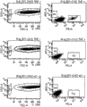
Fig. 2: in conjunction with the FACS chosen process of the rabbit B cell of ESBA903 solubility target.Fig. 2 A: according to forward direction and lateral scattering gate lymphocyte.Fig. 2 B: in the middle of them, select IgG+IgM-cell (may be memory B cell) (adding circle shows).Fig. 2 C: with the high-affinity I gG of ESBA903-PE and the amphophilic cell of ESBA903-PerCP (add circle show) expection coding at ESBA903.Sorting shows the cell of bright fluorescence (not adding circle) in 96 orifice plates.
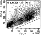
Fig. 3: with the bead of anti-TNF Alpha antibodies (the PE mark) bag quilt Chinese hamster ovary celI (going up picture frame) in conjunction with TNF α-transfection.Chinese hamster ovary celI (middle picture frame) with the contrast bead debond TNF α transfection of anti-CD 19 antibodies (the APC mark) bag quilt.Bead debond wild-type (wt) Chinese hamster ovary celI (following picture frame) with anti-TNF Alpha antibodies (the PE mark) bag quilt.The scatter diagram on the left side (Dot plot) shows forward direction and lateral scattering, and it is the size and the granularity of presentation of events respectively.Single bead (about 3um) colony at P2 by gate.Finally in conjunction with the Chinese hamster ovary celI (about 30um) of bead at P1 by gate.The intermediary scatter diagram shows the P1 incident (Chinese hamster ovary celI) with regard to its PE or APC dyeing.Therefore, will appear in the P3 gate, will appear in the P4 gate with the interactional cell of anti-CD19 bead with the interactional cell of anti-TNF α bead.The statistic data of each sample has been described on the right.
Fig. 4: will mix with the bead of anti-TNF α-PE bag quilt with the bead of anti-CD19-APC bag quilt and the Chinese hamster ovary celI of TNF α-transfection.Chinese hamster ovary celI is by gate (P1), and in the middle of them, is shown in respectively among gate P 3 and the P4 in conjunction with the cell of the bead of the bead of anti-TNF α PE bag quilt or anti-CD19-APC bag quilt.Unconjugated bead is found among the gate P2.
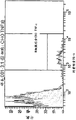
The facs analysis of CHO-TNF α-memory B cell suspension that Fig. 5 a:3 kind is different.Upper left: showed cell suspension forward direction and sidewise scattered scatter diagram.Viable cell comprises large numbers of transgenosis Chinese hamster ovary celIs and a small set of memory B cell, by gate.Lower-left: the scatter diagram that shows APC and FITC fluorescence.Herein, memory B cell (IgG+/IgM-) is by gate.These two scatter diagrams all are identical for whole 3 kinds of samples; Therefore, they only show once.
The histogram of Fig. 5 b:3 sample and colony's layering (population hierarchy): on: the memory B cell of the rabbit of CHO-TNF α cell+ESBA105+ESBA105 immunity; In: the memory B cell of the non-immune rabbit of CHO-TNF α cell+ESBA105+; Down: the memory B cell of the rabbit of CHO-TNF α cell+ESBA105 immunity.On histogram, in conjunction with the memory B cell of Chinese hamster ovary celI by gate.
Fig. 6: by with the facs analysis of the suspension of forming through the lymphocyte of immunity with ESBA105 " bag quilt " TNF α transgenosis Chinese hamster ovary celI blended.Fig. 6 a: showed cell suspension forward direction and sidewise scattered scatter diagram.Viable cell comprises large numbers of transgenosis Chinese hamster ovary celIs and a small set of lymphocyte, by gate.Fig. 6 b: the scatter diagram that shows APC and FITC fluorescence.Herein, memory B cell (IgG+/IgM-) is by gate.Fig. 6 c: show by the histogram of the fluorexon fluorescence of the memory B cell of gate.Sorting is by the colony of gate (in conjunction with the memory B cell of CHO-TNF α-ESBA105 mixture).
Fig. 7: with anti-TNF α I gG bag quilt with the bright-field microscopy figure interactional bead of CHO-TNF α (B220) transgenic cell.
Fig. 8: be bonded to the bright-field microscopy figure (left hurdle) and the fluorescence microscopy figure (right hurdle) of the CHO-TNF α/ESBA105 cell (maxicell) of B cell, described B cell (less cell) has anti-ESBA105 antibody from the teeth outwards.
Describe in detail
The invention provides be used to identify can specificity in conjunction with the immunoconjugator of the cell-surface antigens method of s cFv antibody for example.Method of the present invention generally includes the cells contacting with the expression immunoconjugator of the cell of the antigen expressed of mark and mark, uses the cell of expression immunoconjugator of the cell of cell sorter separation and combination antigen expressed then.These class methods for identify fast and efficiently at be present in integral protein for example the immunoconjugator of the conformational epitope among the GPCR be useful especially.The present invention also provides the nucleic acid that uses the method for the present invention isolating immunoconjugator of identifying and the immunoconjugator of encoding.
In one aspect, the invention provides the method for the immunoconjugator that is used to identify specificity binding purposes cell-surface antigens.This method comprises: but a plurality of cells that may be operably coupled to the expression immunoconjugator of first sorting indicia are provided; But a plurality of cells that may be operably coupled to the antigen expressed of second sorting indicia are provided, and wherein purpose antigen is illustrated on the surface of cell of antigen expressed; Cell and the cells contacting of expressing immunoconjugator with antigen expressed; With use cell sorter (for example, the fluorescence-activated cell sorting device) but from the cell of the one or more specificitys of the cellular segregation of a plurality of expression immunoconjugators in conjunction with the expression immunoconjugator of expressing antigenic cell, wherein but first and second sorting indicia at the individual cells mixture (for example, the mixture that forms between antigen and the B-cell receptor) existence in is indicating the combining of cell of the cell of expressing immunoconjugator and antigen expressed, thereby has identified the antigenic immunoconjugator of binding purposes.
In some embodiments, the cell of the described isolating expression immunoconjugator of clone's property separation.
In certain embodiments, use the cell that the method that well known to a person skilled in the art will the isolating expression immunoconjugator of clone property to experience clonal expansion.
In other embodiments, from the nucleotide sequence of the cellular segregation coding immunoconjugator of expressing immunoconjugator.Can or behind clonal expansion, carry out the separation of nucleotide sequence after the separation of clone's property.Be used to separate the encode proper method of nucleotide sequence of immunoconjugator and comprise PCR, for example unicellular PCR.
In some embodiments, with the mensuration of using the cell experience of the isolating expression immunoconjugator of method of the present invention based on cell, with functional sign immunoconjugator.The suitable mensuration based on cell comprises CELISA.
In some embodiments, immunoconjugator is an antibody.This antibody-like comprises mouse antibodies, rabbit antibody, rabbit sourceization (rabbitized) antibody, chicken antibody, camel antibody, camel sourceization (camelized) antibody, people's antibody, humanized antibody and chimeric antibody.Suitable antibody formation includes but not limited to Fab, Dab, nano antibody (Nanobody) and scFv.
In some embodiments, from exogenous gene expression purpose antigen.In other embodiments, purpose antigen is through genetically engineered antigen.In other embodiments, purpose antigen is integral protein.Suitable integral protein includes but not limited to g protein coupled receptor (GPCR, for example CXCR2) or ionic channel.
In some embodiments, but the mark of first or second sorting is a fluorescent mark.Suitable fluorescent mark includes but not limited to fluorescence protein, antibody/fluorescence conjugate and fluorocyte mark.
In some embodiments, the cell of antigen expressed is yeast or mammalian cell (for example, people's cell).In some embodiments, the cell expressing exogenous antigen of antigen expressed.In some embodiments, with the antigenic cell of expression vector transfection expression.
In some embodiments, the cell of expression immunoconjugator is yeast or mammalian cell.Suitable mammalian cell includes but not limited to the B cell, for example rabbit B cell.In some embodiments, from animal, for example utilize the animal of DNA immunoprophylaxis to separate the B cell through immunity.In some embodiments, the cell of expression immunoconjugator comprises the immunoconjugator of expressing from expression vector.
In one aspect of the method, the invention provides the isolated nucleic acid molecule of coding by the immunoconjugator of method evaluation of the present invention.
In one aspect of the method, the invention provides the method that generation can the antigenic immunoconjugator of binding purposes, the nucleotide sequence that comprises the coding immunoconjugator that will identify by method of the present invention is introduced and is expressed environment so that produce the immunoconjugator of coding.
In one aspect of the method, the invention provides the immunoconjugator that produces by method of the present invention.
In one aspect of the method, the present invention also provides the method for the B cell clone that is used to identify specificity binding purposes cell-surface antigens, and it comprises: with the dna immunization animal of Codocyte surface antigen; Separate the B cell from animal through immunity; But with the first sorting indicia mark B cell; But a plurality of cells that may be operably coupled to the antigen expressed of second sorting indicia are provided, and wherein purpose antigen is illustrated on the surface of cell of antigen expressed; Cell and B cells contacting with antigen expressed; But and use cell sorter from one or more B cells of a plurality of B cellular segregation specificitys in conjunction with the antigenic cell of expression, wherein but the existence of first and second sorting indicia in unicellular mixture indicating the B cell and combining with the cell of antigen expressed, thereby identified the antigenic B cell clone of binding purposes.
Definition
In order more easily to understand the present invention, as some term of giving a definition.Other definition are shown in the whole detailed specification sheets.
Term " antibody " is meant complete antibody and its any Fab (that is, " antigen-binding portion thereof ", " antigen-binding polypeptides " or " immunoconjugator ") or strand." antibody " is meant and comprises by at least two weights (H) chain of disulfide linkage interconnection and glycoprotein or its antigen-binding portion thereof of two light (L) chains.Each heavy chain comprises variable region of heavy chain (being abbreviated as VH in this article) and CH.CH comprises 3 structural domains, CH1, CH2 and CH3.Each light chain comprises variable region of light chain (being abbreviated as VL in this article) and constant region of light chain.Constant region of light chain comprises a structural domain, CL.VH and VL district also can be subdivided into hypervariable region (being called complementary determining region (CDR)), are scattered with conservative zone (being called framework region (FR)) therebetween.Each VH and VL comprise 3 CDR and 4 FR, and it is arranged from aminoterminal to carboxyl terminal with following order: FR1, CDR1, FR2, CDR2, FR3, CDR3, FR4.The variable region of heavy chain and light chain comprises the binding domains with AI.The constant region of antibody can mediate immunoglobulin (Ig) and host tissue or the factor (first composition (Clq) that comprises immune various cell (for example, effector cell) and classical complement system) combination.
Term " chimeric antibody " is meant such antibody molecule, wherein (a) constant region or its part be changed, replace or exchange in case antigen-binding site (variable region) is connected to the constant region with different or kind, effector function and/or species of changing or be connected to give the chimeric antibody new features diverse molecule for example, enzyme, toxin, hormone, somatomedin, medicine etc.; Or (b) with variable region change, replacement or exchange variable region or its part with antigen-specific different or that change.
" antigen-binding portion thereof " of term antibody (or abbreviation " antibody moiety ") is meant maintenance specificity conjugated antigen (for example, one or more fragments of ability TNF) of antibody.Shown that the fragment that can utilize full length antibody carries out the antigen combined function of antibody.The example of the binding fragment that comprises in " antigen-binding portion thereof " of term antibody comprises (i) Fab fragment, the unit price fragment of being made up of VL, VH, CL and CH1 structural domain; (ii) F (ab ')
2Fragment comprises the segmental divalence fragment of two Fab by the disulfide bridge connects on the hinge area, (iii)) the Fd fragment of being made up of VH and CH1 structural domain; (iv) by the VH of the single armed of antibody and the Fv fragment that the VL structural domain is formed; (v) single structure territory dab fragment (people such as Ward, (1989) Nature 341:544-546) for example, it is made up of the VH structural domain; (vi) isolating complementary determining region (CDR) or (combination of two or more isolating CDR that vii) can be randomly connect by synthetic linker.In addition, though segmental two structural domain VL of Fv and VH are by the genes encoding that separates, but can use recombination method, connect them by synthetic linker, described synthetic linker makes and it can be produced as wherein the single protein chain of VL and VH district pairing formation monovalent molecule (is called strand Fv (s cFv); Referring to, for example, people such as Bird (1988) Science 242:423-426; With people (1988) Proc.Natl.Acad.Sci USA 85:5879-5883 such as Huston).This type of single-chain antibody is also intended to be included in " antigen-binding portion thereof " of term antibody.Use routine techniques well known by persons skilled in the art to obtain this type of antibody fragment, and with screen fragment for the identical mode of the mode of complete antibody with regard to instrumentality.Can produce antigen-binding portion thereof by recombinant DNA technology or by enzymatic or the complete immunoglobulin (Ig) of chemistry fracture.Antibody can be the antibody of different isotype, for example, and IgG (for example, IgG1, IgG2, IgG3 or IgG4 subclass), IgA1, IgA2, IgD, IgE or IgM antibody.
Term " immunoconjugator " is meant molecule, and described molecule comprises all or part of antigen binding site of antibody, all or part of of heavy chain and/or light chain variable structural domain for example, and immunoconjugator can the specific recognition target antigen like this.The non-limiting example of immunoconjugator comprises total length immunoglobulin molecules and scFv, and antibody fragment, includes but not limited to (i) Fab fragment, the unit price fragment of being made up of VL, VH, CL and CH1 structural domain; (ii) F (ab ')
2Fragment, comprise the segmental divalence fragment of two Fab by the disulfide bridge connects on the hinge area, (iii) Fab ' fragment, its be basically Fab with part of hinge area (referring to, Fundamental Immunology (Paul edits, 3.sup.rd ed.1993); The (iv) Fd fragment of forming by VH and CH1 structural domain; (v) comprise the VL of single armed of antibody and the Fv fragment of VH structural domain; (v i) single domain antibody is Dab fragment (people such as Ward for example, (1989) Nature 341:544-546), it is made up of VH or VL structural domain, Camelid is (referring to Hamers-Casterman, Deng the people, people such as Nature 363:446-448 (1993) and Dumoulin, Protein Science11:500-515 (2002)) or Shark antibody (for example, shark Ig-NARs
); (vii) nano antibody (nanobody) comprises the variable region of heavy chain of single variable domains and two constant domain.
As used herein, term " functional property " is nature of production or the therapeutic efficiency in order to improve polypeptide for example, its raising (for example with respect to conventional polypeptide) is the character of polypeptide (for example, immunoconjugator) expectation and/or favourable to those skilled in the art.In one embodiment, functional property is the stability (for example, thermostability) that improves.In another embodiment, functional property is the solvability (for example, under the cell condition) that improves.In another embodiment, functional property is non-aggregation.In another embodiment, functional property is to express the raising of (for example, in prokaryotic cell prokaryocyte).In another embodiment, functional property is the raising of refolding output behind the inclusion body purification process.In certain embodiments, functional property is not the raising of antigen-binding affinity.
Term " framework " is meant the part that is present in the art-recognized antibody variable region between the more divergent CDR district.This type of framework region is commonly referred to framework 1 to 4 (FR1, FR2, FR 3 and FR4) and support is provided, and described support is supported in 3 CDR that find in heavy chain or the antibody light chain variable region with the three-dimensional space form, so that described CDR can form the antigen mating surface.This type of framework also can be described as support, because they provide support to present more divergent CDR.Other CDR of immunoglobulin superfamily and framework for example ankyrin repeat and fibronectin can be used as antigen binding molecules (also referring to, for example, United States Patent (USP) 6,300,064,6,815,540 and the open case 20040132028 of the U.S.).
Term " epi-position " or " antigenic determinant " are meant on the antigen by immunoglobulin (Ig) or antibodies specific bonded position.Epi-position comprises at least 3,4 with the space conformation of uniqueness usually, 5,6,7,8,9,10,11,12, and 13,14 or 15 amino acid.Referring to, for example, Epitope MappingProtocols in Methods in Molecular Biology, the 66th volume, G.E.Morris, Ed. (1996).
Term " specificity combination ", " selective binding ", " optionally combination " and " combination specifically " are meant the epi-position on the predetermined antigen of antibodies.Usually, antibody is with approximately less than 10
-7M is for example approximately less than 10
-8M, 10
-9M or 10
-10M or littler avidity (KD) combination.
Term " K
D" be meant the dissociation equilibrium constant of specific antibodies-AI.Usually, antibody of the present invention is with less than about 10
-7M is for example less than about 10
-8M, 10
-9M or 10
-10M or littler dissociation equilibrium constant (K
D) conjugated antigen, for example, in the BIACORE instrument, measure as using surface plasma body resonant vibration (SPR) technology.
As used herein, " identity " be meant between two polypeptide, the molecule or two nucleic acid between the sequence of mating.Position in two sequences that compare all (is for example occupied by identical base or amino acid monomer subunit, if the position in each of two dna moleculars is all occupied by VITAMIN B4, or the position in each of two polypeptide is all occupied by Methionin) time, each molecule is same on this position so." percentage ratio identity " between two sequences is to multiply by 100 function by the total matched position number of these two sequences again divided by the position number that compares.For example, if in 10 positions of two sequences 6 couplings are arranged, these two sequences have 60% identity so.For example, dna sequence dna CTGACT and CAGGTT have 50% identity (in 6 positions 3 location matches being arranged altogether).Usually, two sequence alignments are being compared when producing maximum identity.Such comparison can be by using, for example, by computer program for example the Align program (DNAstar, the method for people such as Needleman (1970) J.Mol.Biol.48:443-453 that Inc.) carries out easily provides.Also can use the E.Meyers and W.Miller (the Comput.Appl Biosci. that have been integrated into ALI GN program (version 2 .0), 4:11-17 (1988)) algorithm uses PAM120 weight residue table (weight residue table), 12 notch length point penalty and 4 breach point penalty to measure two percentage ratio identity between the aminoacid sequence.In addition, can use Needleman and Wunsch (JMol Biol.48:444-453 (1970)) algorithm in the GAP program that has been integrated into GCG software package (can on www.gcg.com, obtain), use Blossum 62 matrixes or PAM250 matrix and 16,14,12,10,8,6 or 4 breach weight (gap weight) and 1,2,3,4,5 or 6 length weight to measure two percentage ratio identity between the aminoacid sequence.
" similar " sequence is to have sequence identical and similar amino-acid residue when comparison, and wherein similar residue is the conservative substitution of the corresponding amino-acid residue in the canonical sequence of comparing.In this, " conservative substitution " of the residue in the canonical sequence is to be used on physics or the function and the displacement of carrying out with reference to the residue of residue similar (for example have similar size, shape, electric charge, chemical property, comprise the ability that forms covalent linkage or hydrogen bond etc.) accordingly.Therefore, " modifying " sequence through conservative substitution is and canonical sequence or the different sequence that is to exist one or more conservative substitutions of wild-type sequence." percentage ratio similarity " between two sequences is to comprise by the total coupling residue of two sequences or the position number of conservative substitution to multiply by 100 function again divided by the position number that compares.For example, have 2 to comprise conservative substitution if having in 10 positions of two sequences in 6 couplings and 10 positions, these two sequences have 80% direct analogy so.
Term used herein " conserved sequence modification " means amino acid modified in conjunction with feature that can influence sharply or change the antibody that comprises aminoacid sequence.This type of conserved sequence is modified and is comprised Nucleotide and amino-acid substitution, interpolation and disappearance.For example, can be by for example site-directed mutagenesis and PCR mediated mutagenesis introducing modification of standard technique known in the art.Conservative amino acid replacement comprises the displacement that wherein substitutes amino-acid residue with the amino-acid residue with similar side chain.In this area, defined the family of amino-acid residue with similar side chain.These families comprise (for example having basic side chain, Methionin, arginine and Histidine), acid side-chain (aspartic acid for example, L-glutamic acid), uncharged polar side chain (glycine for example, l-asparagine, glutamine, Serine, Threonine, tyrosine, halfcystine, tryptophane), non-polar sidechain (L-Ala for example, Xie Ansuan, leucine, Isoleucine, proline(Pro), phenylalanine, methionine(Met)), the β branched building block (for example, Threonine, Xie Ansuan, Isoleucine) and aromatic series side chain (for example, tyrosine, phenylalanine, tryptophane, Histidine) amino acid.Therefore, preferably use the non-essential amino acid residue that substitutes prediction from another amino-acid residue of same side chain family.Identify the method do not eliminate antigen bonded Nucleotide and conservative aminoacid substitutions in this area be know (referring to, for example, people such as Brummell, Biochem.32:1180-1187 (1993); People Protein Eng.12 (10): 879-884 (1999) such as Kobayashi; With people Proc.Natl Acad.Sci USA 94:412-417 (1997) such as Burks).
" amino acid consensus sequences " used herein is meant and can uses at least two, the aminoacid sequence that the matrix of preferred aminoacid sequence of more comparing produces (allowing to occur breach in comparison), it makes it possible to determine the highest amino-acid residue of the frequency of occurrences on each position.Consensus sequence is included in the highest amino acid whose sequence of the frequency of occurrences on each position.If two or more amino acid appear on the single position with being equal to, consensus sequence comprises two or all these amino acids so.
Can be on different levels the aminoacid sequence of analysing protein.For example, can be in single residue level, a plurality of residue level, have on a plurality of residue levels etc. of breach and show conservative property or variability.Residue can be showed the conservative property of identical residue or can guard on the kind level.The amino acid examples of types comprises polarity but uncharged R group (Serine, Threonine, l-asparagine and glutamine); Positively charged R group (Methionin, arginine and Histidine); Electronegative R group (L-glutamic acid and aspartic acid); Hydrophobic R group (L-Ala, Isoleucine, leucine, methionine(Met), phenylalanine, tryptophane, Xie Ansuan and tyrosine); And special acid (halfcystine, glycine and proline(Pro)).Other kinds are known to those skilled in the art and but utilization structure assay method or other data define to estimate substitutability.On this meaning, replaceable amino acid can refer to can be at any amino acid of enterprising line replacement in this position and maintenance function conservative property.
Yet the amino acid that should be understood that identical category can have difference to a certain degree on their bio-physical property.For example, should be understood that some hydrophobic R group amino acid (for example, L-Ala) is than other hydrophobic R group amino acid (for example, Xie Ansuan or leucine) more hydrophilic (that is, having higher wetting ability or lower hydrophobicity).Relatively wetting ability or hydrophobicity can use art-recognized method (referring to, for example, people such as Rose, Science, people such as 229:834-838 (1985) and Cornette, J.Mol.Biol, 195:659-685 (1987)) measure.
As used herein, when an aminoacid sequence (for example, the one VH or VL sequence) and one or more other aminoacid sequences are (for example, one or more VH in the database or VL sequence) when comparing, amino acid position in the sequence (for example, a VH or VL sequence) can be compared with " corresponding position " in one or more other aminoacid sequences.As used herein, " corresponding position " expression is when carrying out optimum when comparing to sequence, i.e. equivalent site in the sequence that compares when sequence is compared with the highest percentage ratio identity of acquisition or percentage ratio similarity.
Term " nucleic acid molecule " is meant dna molecular and RNA molecule.Nucleic acid molecule can be strand or two strands, but double-stranded DNA preferably.When nucleic acid and another nucleotide sequence were placed functional relationship, nucleic acid was " being operably connected ".For example, if promotor or enhanser influence transcribing of encoding sequence, promotor or enhanser may be operably coupled to described sequence so.
Term " carrier " is meant the nucleic acid molecule that can transport connected another nucleic acid.One type of carrier is " plasmid ", and it is meant the circular double stranded DNA ring that other DNA section can be connected to wherein.The another kind of type of carrier is a virus vector, wherein other DNA section can be connected in the viral genome.Some carrier can be in the host cell of introducing it self-replicating (bacteria carrier and the additive type Mammals carrier that for example, have the replication orgin of bacterium).Other carriers can be integrated into the genome of host cell after introducing host cell, thereby duplicate (for example, non-add type Mammals carrier) with host genome.
Term " host cell " is meant to the cell of wherein having introduced expression vector.Host cell can comprise bacterium, microorganism, plant or zooblast.The bacterium that is easy to transform comprises the member of enterobacteriaceae (enterobacteriaceae), for example the bacterial strain of intestinal bacteria (Escherichia coli) or Salmonellas (Salmonella); Bacillaceae (Bacillaceae), for example subtilis (Bacillus subtilis); Streptococcus pneumoniae (Pneumococcus); Suis (Streptococcus) and Haemophilus influenzae (Haemophilus influenzae).Suitable microorganism comprises yeast saccharomyces cerevisiae (Saccharomyces cerevisiae) and pichia spp (Pichia pastoris).Suitable animal host's clone comprises CHO (Chinese hamster ovary line) and NS 0 cell.
Term " treatment ", " therapy " and " treatment " are meant therapeutic or the preventive measure of describing herein.The method of " treatment " to the experimenter who has this treatment to need (is for example utilized, suffer from GPCR mediation disease the experimenter or finally can obtain the experimenter of such disease) use antibody of the present invention prevent, to cure, to postpone, to palliate a disease or the severity of recurrent disease or improve its one or more symptoms, or with the survival time that prolongs the experimenter with above desired survival time under the non-existent situation of such treatment.
Term " effective dose " or " effectively dosage (effective dosage) " are meant the amount that is enough to obtain or obtain to small part desired effects.Term " treatment effective dose " is defined as to be enough to cure or to stop to small part has suffered from the disease of patient of disease and the amount of its complication.To depend on the severity of disease to be treated and patient's oneself immune overall status for the effective amount of this purposes.
Term " experimenter " is meant anyone or non-human animal.For example, method and composition of the present invention can be used for treating the experimenter of the disease of suffering from the GPCR mediation.
As used herein, term " rabbit " is meant the animal that belongs to rabbit section.
Term " cell sorter " is meant any device that has isolated cell based on detectable " but sorting " mark.This type of device includes but not limited to the fluorescence-activated cell sorting device.But any cell marking can be used as sorting indicia, includes but not limited to fluorescin, for example green fluorescent protein, antibody/fluorescence (fluor) conjugate and fluorocyte mark fluorescence Calcium ionophore for example.
Term " cell complexes " is meant the cell with the one or more antigen expressed of cell bonded of one or more expression immunoconjugators, and wherein, described combination is by the lip-deep antigen mediation of the cell of antigen expressed.In some embodiments, the cell of antigen expressed is formed with combining by the direct interaction between the immunoconjugator on the cell surface of antigen on the cell surface of antigen expressed and expression immunoconjugator of the cell of expressing immunoconjugator.
Term " separation of clone's property " is meant any method that is used for from cell colony isolated mononuclear cell clone.Appropriate means includes but not limited to that cell is no more than individual cells to the limiting dilution and the transfer of porous plate so that each hole comprises.
The term nucleotide sequence of immunoconjugator " obtain coding " is meant any method that is used to obtain by the nucleotide sequence of the immunoconjugator of the cell expressing of expressing immunoconjugator.Appropriate means includes but not limited to, from separate nucleic acid, pcr amplification and the dna sequencing of the nucleotide sequence of the coding immunoconjugator of the cell of expressing immunoconjugator.In some embodiments, by the nucleotide sequence of PCR from individual cells amplification coding immunoconjugator, i.e. " unicellular PCR ".
Term " exogenous antigen " is meant the antigen of not expressing usually in particular host cell.For example, compare with host cell, exogenous antigen can be from different boundaries, door, guiding principle, order, genus or kind, the human antigen who for example expresses in yeast cell.In addition or alternatively, exogenous antigen can be from mutually of the same race but express the lung specificity antigen of for example expressing inadequately in brain cell in this host cell." exogenous antigen " also refers to undiscovered sudden change antigen in normal cell usually, the cancer specific mutant antigen of for example expressing in pneumonocyte.
Term " through genetically engineered antigen " is meant any antigen that produces by recombinant DNA technology, is included as mosaic or comprises the antigen of point mutation, disappearance and/or insertion.Unless otherwise defined, otherwise whole technology used herein and scientific terminology have with the present invention under the meaning of same meaning of those of skill in the art's common sense.Though method and material similar with material to the method for describing herein or that be equal to can be used for practice or test the present invention, describe appropriate means and material hereinafter.Under the situation of contradiction, be as the criterion with this specification sheets (comprising definition).In addition, material, method and embodiment only be illustrative and to be not intended to be determinate.
In following part different aspect of the present invention is described in more detail.But should be understood that arbitrary combination different embodiments, parameter selection and scope.In addition, depend on specific embodiment, can not use definition, embodiment or the scope of selection.
The invention provides and use FACS to identify and the screening method that separates with the cell of the expression immunoconjugator of the cell adhesion of expression corresponding antigens.In specific embodiment, immunoconjugator is an antibody.
Antigen presentation
The target antigen that is used for Antibody Preparation can be any protein, peptide, Nucleotide, carbohydrate, lipid and solubility or that express on cell surface or other molecules of being incorporated into plasma membrane.Antigen can be natural or synthetic.Preferably, target antigen is protein or peptide.The non-limiting example of target antigen comprises CXCR1, CXCR2, CXCR 3, CXCR4, CXCR6, CCR1, CCR2, CCR 3, CCR4, CCR5, CCR6, CCR8, CFTR, CIC-1, CIC-2, CIC-4, CIC-5, CIC-7, CIC-Ka, CIC-Kb, Bestrophins, TMEM16A, the GABA acceptor, Glycine Receptors, abc transport albumen, NAV1.1, NAV1.2, NAV1.3, NAV1.4, NAV1.5, NAV1.6, NAV1.7, NAV1.8, NAV1.9,1-phosphoric acid sphingosine acceptor (S1P1R), NMDA passage etc.In one embodiment, target antigen is a transmembrane protein.In another embodiment, target antigen is repeatedly a transmembrane protein, for example, and g protein coupled receptor (GPCR), ionic channel etc.
GPCR family has at least 250 members (people FASEB J. such as Strader, 9:745-754,1995; People Annu.Rev.Biochem. such as Strader, 63:101-32,1994).Estimated centesimal people's gene codified GPCR.The GPCR incorporation range is part widely, comprises that photon, atom amine (that is, suprarenin and histamine), peptide are (that is, IL-8) and big glycoprotein hormones (that is parathyroid hormone).When part in conjunction with the time, GPCR is by activating signal transduction pathway in guanine-nucleotide-binding protein (G albumen) regulating cell.Enjoyably, GPCR has functional homologue (homologue) in human cytomegalic inclusion disease virus and simplexvirus, and this prompting may obtain GPCR (people such as Strader, FASEB J., 9:745-754,1995 in the pathogenetic evolutionary process of virus; People Nature such as Arvanitakis, 385:347-350,1997; Murphy, Annu.Rev.Immunol.12:593-633,1994).
The characteristic properties of hitherto known most of GPCR is, 7 bunches of hydrophobic amino acid residues be arranged in primary structure and at it each zone by (across) cytolemma.Described structural domain it is believed that the transmembrane spanning that representative is connected with the C-terminal structural domain by ring in 3 cells, 3 extracellular loop and amino people such as (, Science 289,739-45 (2000)) K.Palczewski.Most of GPCR have single conservative cysteine residues in each of preceding two extracellular loop, its formation it is believed that the disulfide linkage of stabilization function protein structure.Stride the film district for 7 and be named as TM1, TM2, TM3, TM4, TM5, TM6 and TM7.As everyone knows, above-mentioned these structures are very general and corresponding to the aminoacid sequence of zone by film of protein wherein (transmembrane domains (membrane-spanning region) or stride film district (transmembrane region)) and near the aminoacid sequence transmembrane domains high conservative normally in the middle of acceptor in g protein coupled receptor albumen.Therefore, because the medium-altitude homology of GPCR can easily be finished by those skilled in the art with the evaluation of extracellular part in the evaluation of novel GPCR and this type of novel member's the cell.For example, the book of Watson and Arkinstall (1994) (integrating with this paper by reference) provides the sequence of more than 50 kind of GPCR.This book has also been described the accurate residue that comprises each membrane spaning domain of each sequence.
The little ligand-binding site of g protein coupled receptor is put and be it is believed that and comprise near the wetting ability alveole (hydrophilic socket) that is positioned at the cell outer surface and is formed by some g protein coupled receptor membrane spaning domains, and this alveole is surrounded by the hydrophobic residue of g protein coupled receptor.The water-wet side of each g protein coupled receptor transbilayer helix towards inside, forms the polar ligand binding site according to hypothesis.Hinted that TM3 has ligand-binding site and puts in some g protein coupled receptors, for example comprised the TM3 asparagicacid residue.In addition, TM5 Serine, TM6 l-asparagine and TM6 or TM7 phenylalanine or tyrosine also involve the part combination.Peptide hormone acceptor and acceptor with other bigger parts be glycoprotein (LH, FSH, hCG, TSH) and Ca for example
2+The ligand-binding site of/L-glutamic acid/GABA receptoroid is put and may be arranged in extracellular domain and ring.
The critical event that is converted to active acceptor from the non-activity acceptor is to have the transbilayer helix 3 (TM3) of GPCR of 7 transbilayer helixs and part inductive conformational change (U.Gether and the B.K.Kolbilka of 6 (TM6), J.Biol.Chem.273,17979-17982 (1998)).This type of spiral motion changes the interior conformation of encircling of cell of acceptor again to promote the proteic activation of associating heterotrimer G.Mutagenesis research (S.Cotecchia, J.Ostrowski, M.A.Kjelsberg, M.G.Caron and R.J.Lefkowitz, J.Biol.Chem.267,1633-1639 (1992); E.Kostenis, B.R.Conklin and J.Wess, Biochemistry 36,1487-1495 (1997); M.A.Kjelsberg, S.Coteechia, J.Ostrowski, M.G.Caron, and R.J.Lefkowitz, J.Biol.Chem.267,1430-1433 (1992)) prove that the interior ring of the 3rd cell (i3) mediates the coupling between most of acceptor and the G albumen.Shown that also I 3 ring that is expressed as little gene (minigene) directly competes the (L.M.Luttrell that combines to Gq with adrenergic receptor, J.Ostrowski, S.Cotecchia, H.Kendal and R.J.Lefkowitz, Science 259,1453-1457 (1993)) or can be as soluble peptide activated G protein (people such as T.Okamoto, Cell 67,723-730 (1991)) under acellular condition.
Purpose antigen can be the antigen in the endogenous source in the target cell (being also referred to as the cell of antigen expressed sometimes).Alternatively, exogenous molecules can be introduced cell with antigen expressed.Can realize by any method known to those skilled in the art that antigen is to intracellular introducing.In one embodiment, can external be that the polynucleotide of polypeptide insert carrier with coding for antigens, this carrier can further be introduced the target cell that is used to express.Polynucleotide can comprise cDNA sequence, dna sequence dna or other sequences known in the art of target antigen.Carrier can be plasmid, clay, liposome or other natural or vectorettes known in the art.Introducing can be following method: transfection, conversion, infection, the direct microinjection of material, particle gun are sent (biolistic particle delivery), electroporation or additive method known in the art.The target cell of antigen expressed can be an arbitrary cell known in the art, for example comprise, direct cell available from animal, for example cancer cells, non-cancer cells, primary cell etc., or through the cell of molecular modification, for example culturing cell (for example, Chinese hamster ovary (CHO) cell, HEK293 cell etc.), immortalized cells, transfection/cell that infects, T cell etc.Alternatively, target cell can be from non-animal-origin, for example bacterium, insect etc.In one embodiment, the cell of expressing target antigen is a yeast cell, the preferred yeast spheroblast.Alternatively, target " cell " can be artificial cell sample corpusculum or structure, for example liposome, unitary film corpusculum etc.Antigenic expression can be instantaneous in target cell or the cytoid body, promptly, expression will the relatively short time period (for example, from several minutes to a couple of days) after weaken or stop, or it is stable, that is, expression will continue the long relatively time with metastable level (for example, counting for the back at a couple of days or cell).In a preferred embodiment, antibody is expressed on the cell outer surface of the plasma membrane of target cell.In a further preferred embodiment, antigen is the inherent antigen of film or repeatedly strides membrane antigen.In order to arrive its position on plasma membrane, antigen directly can be expressed on these positions, or can after expressing in their tenuigenin, it be migrated to these positions target cell.This migration can be the natural process of target cell or by for example (for example connecting signal/tag molecule before or after antigen presentation, gorky's sorting signals, at the antibody of some film binding molecule etc.), grappling graft (anchoring graft) (for example, glycosylation phosphatidylinositols (GPI) anchor) or the engineered process of carrying out with antigen chemically crosslinked, sudden change antigen or the additive method that causes its migration known in the art.
Express the cell and the immunity of immunoconjugator
In one embodiment, treat that the cell of the expression immunoconjugator selected by the method for describing is a Mammals B cell herein, preferred rabbit B cell.
In preferred embodiments, the B cell comes from the animal with the immunity of purpose target.Can utilize any method well known by persons skilled in the art to implement the immunity of animal.Usually, the lymphoid organ (for example spleen or lymphoglandula) from the animal of immunity separates the B cell.
In a preferred embodiment, carry out the antigenic immunity of purpose by dna immunization/inoculation.Alternatively, the injection cell of expressing target antigen is gone into animal (for example, rabbit, rat, mouse, hamster, sheep, goat, chicken etc.) to carry out immunity.The preferred animal that is used for this immunity step is a rabbit.Dna immunization/inoculation is induced tachysynthesis to reply and is allowed the natural expression of target antigen, and only natural usually expression.Because it does not involve Recombinant Protein Expression or operation, so this method is more efficient and more cost-effective than the routine immunization that uses recombinant protein.In addition, more importantly, the antigen of expression in vivo have the secondary structure identical with target protein in the natural background and even can have the posttranslational modification identical with it, this has improved the identification exactness of antibody of the anti-target antigen of preparation.Exemplary dna immunization is illustrated in Canadian patent application CA2350078 and WO04/087216.Particularly, will encode by particle bombardment and to introduce animal as the dna direct of the polypeptide of target antigen, thereby cause polypeptide to be expressed in animal, this expression causes that the antibody of anti-described polypeptide forms.Form in order to obtain stronger antibody, use the DNA of so-called gene adjuvant (genetic adjuvant) and coded polypeptide simultaneously.This type of gene adjuvant is the express cell factor (for example, GM-CSF, IL-4 and IL-10) and at the plasmid of laboratory animal moderate stimulation humoral immunoresponse(HI).In a preferred embodiment, the antibody at target antigen is expressed as B-cell receptor (BCR) on the cell surface of B cell.In a further preferred embodiment, the cell of expressing antibodies is a memory B cell, it is characterized in that not existing in its surface any IgM and therefore can with conventional B cell differentiation.
In one embodiment, the cell of expressing immunoconjugator is a yeast cell, and immunoconjugator preferably antibody fragment, more preferably scFv.
Use the screening of fluorescence-activated cell sorting (FACS)
After immune step, need be with the membrane-bound antibody and other cellular segregation of expressing non-specific antibody of specificity on the B cell in conjunction with target antigen.In a preferred embodiment, by the Ag-Ab combination, the B cell adhesion of expression specificity antibody is to the target cell of antigen expressed on its plasma membrane.In a further preferred embodiment, other or more heterogeneous mutual effect (for example, identical or different antigen-antibody interaction, chemically crosslinked, ligand-receptor interaction etc.) can take place between B cell and target cell.The B cell can be present in the storehouse of the B cell of expressing different antibodies, or with the combination of other immunocytes, directly from the animal of immunity, from immunity/collect in the library of the B cell of the storehouse of non-immune animal or the expression different antibodies that for example produces by V (D) J gene recombination technology from external engineering method.
Can in any means known in the art, express the separation of the B cell of the antibody that is specific to target antigen.These class methods include but not limited to antigenic elutriation, limiting dilution, affinity purification or utilize the antibody of expressing or the additive method of the feature of the B cell that produces antibody.
In a preferred embodiment, with the cell of label antigen expressed, described label is used for detecting in the future and/or separating adherent B cell.Label can be linking agent, antigen/antibody, small molecules (for example, gsh (GSH), vitamin H/avidin etc.), magnetic-particle, fluorescence labels etc.In a preferred embodiment, use the cell of fluorescin/peptide-labeled antigen expressed.In another embodiment, also with the B cell of the preferred different fluorescin/peptide-labeled generation antibody of label.In another embodiment, can detect the B cell of the cell that adheres to antigen expressed by the fluorescent emission of the fluorescin/peptide of mark on the B cell.In another embodiment, the B cell that utilizes the fluorescent emission of two kinds of different fluorescin/peptides of mark on it to detect to be in the adhesion and the cell of antigen expressed.In a preferred embodiment, can be by on it and the adherent APC fluorescent emission that goes up the fluorescin/peptide of mark detects the B cell of expressing the antibody that is specific to target antigen and further with its B cellular segregation with other antibody of generation.Preferably, this detection can utilize fluorescence-activated cell sorting (FACS) technology to carry out with separating.
Initial curtail word FACS is by Becton Dickinson (Franklin Lakes, NJ) registered trademark and having.As used herein, term FACS can represent the cell sorting based on flow cytometry of arbitrary form.
Fluorescence-activated cell sorting is the flow cytometry of specialized type.It provides based on every next cell ground of the specific light scattering of each cell and fluorescent characteristics the heterogeneous mixture sorting to 2 of biomass cells or the method in more a plurality of container.It is useful scientific instrument, because it provides from the quick, objective of the fluorescent signal of individual cells and the quantitative physical sepn of the cell of record and special concern.
In the FACS of classics system, the cell suspension thing is driven in narrow, quick liquid flow flowing central authorities.Arrange to flow so that there is big interval (comparing with its diameter) in iuntercellular.Vibration mechanism causes and is broken into independent droplet in the stream of cells.Adjustment System is so that make the probability that is present in a droplet above 1 cell lower.Before liquid stream was about to fragment into droplet, liquid flowed the fluorescence measurement station by the purpose fluorescent characteristics of wherein measuring each cell.Placing charged ring just in time, liquid stream fragments on the point of droplet.Based on just now fluorescence intensity measurement value electric charge is placed on the ring, when droplet from stream during fracture opposite charges be trapped on the droplet.Charged then droplet falls is by electrostatic deflection system, and this system makes droplet deflection enter container based on the electric charge of droplet.In some systems, electric charge is directly used in the droplet maintenance of liquid stream and disconnection and the electric charge of liquid stream same-sign.Disconnecting back liquid stream at droplet then replys neutral.
The fluorescent mark that is used for the FACS technology depends on lamp or laser apparatus and the obtainable detector that is used for the fluorescence excitation dyestuff.Obtainable the most commonly laser is blue argon laser (488nm) on single laser machine.The fluorescent mark that can be used for the laser of the type includes but not limited to 1) for green fluorescence the FL1 of mark (usually): FITC, Alexa Fluor 488, GFP, CFSE, CFDA-SE and DyLight 488; 2) for fluorescent orange (FL2 usually): PE and PI; 3) for red fluorescence (FL3 usually): PerCP, PE-Alexa Fluor 700, PE-Cy5 (TRI-COLOR) and PE-Cy5.5; And 4) for Infrared fluorescence (common FL4; In some FACS machines): PE-Alexa Fluor 750 and PE-Cy7.Other laser and its corresponding fluorescent mark include but not limited to 1) red diode laser (635nm): allophycocyanin (APC), APC-Cy7, AlexaFluor 700, Cy5 and Draq-5; With 2) violet laser (405nm): Pacific Orange, Amine Aqua, Pacific Blue, 4 ', 6-diamidino-2-phenylindone (DAPI) and AlexaFluor 405.
In preferred embodiments, with anti--IgG and anti-IgM antibody staining B cell of mark, and have only by anti-IgG antibody but be not preferred by the painted memory B cell of anti-IgM antibody positive.IgG has the avidity higher than IgM usually; Thereby select to express IgG in its surface but not the positive B cell of IgM (this is the feature of memory B cell).For described purpose, preferably use multicolour dyeing, wherein for example be specific to the antibody of IgG and IgM respectively with APC and FITC difference mark.Preferably, go back the target cell of labels targets antigen and/or expression target antigen.In one embodiment, by the cell of expressing target antigen with fluorescent dyeing in the cell target antigen that dyes indirectly.
The invention provides and use FACS screening Blymphocyte repertoire (wherein the B cell can adhere to the cell of expressing target antigen) to identify and further method of separating the B cell of the antibody that produces specificity binding purposes target antigen.Preferably, with fluorescent mark mark B cell, utilize different fluorescent marks to separate the cell of marker expression target antigen.This type of mark can be in the cell, extracellular or be incorporated in the plasma membrane.After immunity and antibody generation, with whole B cytomixis together, make its operation by the FACS system.Have only those B cells that produce the antibody that is specific to target antigen just can adhere to the cell of antigen expressed.Compare with the big interval between other independent cells, the distance of these two cells in liquid stream shortened in their adhesion, thereby causes detectable " double-colored incident (bi-color event) " in the process by scanning laser beam jointly at them.Therefore, can identify the B cell that produces purpose antibody and with its sorting to the collection tube different with other non-specific B cells.
In a further preferred embodiment, if the interaction between the cell of B cell and corresponding antigen expressed causes some change of cell characteristic, for example depolarize, FRET (fluorescence resonance energy transfer) (FRET) etc. can add to the cell that can change like this with more fluorescent mark so.Therefore, the cell that is in the B cell of contact and antigen expressed will produce " three look incidents " incident or comprise even surpass the incident of three kinds of colors simultaneously.
Alternatively, the evaluation of adherent B cell and sorting do not need its fluorescently-labeled existence.In one embodiment, cell-cell interaction causes changing function in any cell.In another embodiment, these changing functions can be used for identifying and further separate the B cell of generation specificity in conjunction with the antibody of target antigen.For example, cell-cell interaction can be in arbitrary cell functional blocking-up or activated receptor signal transduction, thereby cause the cellular change that can detect by the FACS system, Ca for example
2+Discharge variation etc.Therefore, by monitoring this type of detectable changes of function, also can identify and separate purpose B cell.A specific embodiments of functional blocking-up or activated receptor signal transduction comprises the cell incubation of B cell with functional expression GPCR (g protein coupled receptor).Can in mixture, add the Ca of agonist that signals by GPCR to induce GPDR to mediate
2+Flow out from endoplasmic reticulum.If be present in the antibody function blocking-up agonist signal transduction on the B cell, so Ca
2+Flow out and also to be stoped by this cell-cell interaction.Can for example pass through flow cytometry quantitative measurment Ca
2+Flow out.Therefore, has only demonstration Ca
2+The B cell that flows out increase or reduce/target cell aggregate is just by sorting.
The avidity of the antibody that is produced by isolating B cell is measured
In certain embodiments, cultivate the B cell under suitable condition so that antibody-secreting is gone into substratum.The antibody that produces is monoclonal antibody for example.Cultivation can comprise use auxiliary cell line for example the thymoma auxiliary cell line (EL4-B5 for example, referring to people such as Zubler, 1985, J.Immunol., 134 (6): 3662-3668).
Randomly, can carry out extra avidity mensuration, further handle then with assessment by the selectivity of the antibody of isolating B cell generation and the ability of competing with part.This type of mensuration includes but not limited to the mensuration (for example, cell ELISA (CELISA), it is the improved ELISA method that intact cell is used to wrap quilt) based on cell.As discussing among the CA2350078, can described in hereinafter embodiment, carry out CELISA.Carry out the antibody of verification step, resist expressed protein on cell surface but not the antibody of target protein thereby for example get rid of just the specificity of target is produced in conjunction with test.
Alternatively, can directly check with regard to affinity of antibody and identify and isolating purpose B cell, and can be before processing with the cellular segregation of B cell and adherent antigen expressed.
Further handle isolating B cell to be used for production of antibodies
Can further handle evaluation with isolating B cell (its randomly by affine mensuration (for example, CELISA) testing) to produce the purpose immunoconjugator.Can use for example conventional hybridization knurl technology.This technology can comprise for example purifying immunoconjugator of step, illustrates its aminoacid sequence and/or nucleotide sequence.
Alternatively, carry out the sign of wedding agent with its scFv form.In order to carry out this method, can be by RT-PCR from the cell of the sorting of cultivation or the CDR sequence of directly taking out the wedding agent of expressing at the B of sorting cell from individual cells.The combination of the oligonucleotide library that two parts of the framework region of the scFv support that one of them oligonucleotide library coding CDR and second storehouse coding are suitable overlap will allow to produce humanization scFv in a step PCR method.HT order-checking, clone and produce the clonal selection that to allow to carry out based on the performance of the humanization scFv of purifying, but not excretory IgG in the characterize cells culture supernatant.The scFv support (" rabbit sourceization " people FW or rabbit receptor (acceptor) RabTor that are suitable for accepting from the CDR of any rabbit antibody have been identified and have characterized; Referring to WO09/155726, with its complete by reference this paper that incorporates into).Shown that many kinds in some cases even comprise the evidence of the notion of the CDR of disulfide linkage between rabbit specific C DR.
The general declarative description that produces rabbit source antibody is in hereinafter.
The transplanting of immunoconjugator
Can will use the CDR or the antigen binding domain of the immunoconjugator of method evaluation of the present invention to migrate in the receptor antibody framework.This transplanting can for example reduce the immunogenicity of immunoconjugator or improve its functional performance, for example improves thermodynamic stability.
The general method that is used for CDR is migrated to people's receptor framework by Winter at United States Patent (USP) 5,225, open among 539 (with its complete by reference this paper that incorporate into).
Be used for being disclosed in U.S. Provisional Patent Application case 61/075,697 and 61/155,041, with its complete by reference this paper that incorporates into from the specific strategy of rabbit monoclonal antibodies transplanting CDR.These strategies are relevant with the strategy of Winter, are particularly suitable as the general receptor of everyone or inhuman donor antibody but difference is receptor antibody framework.Particularly, the antigen binding site highly compatible that has shown people's strand frame F W1.4 (combination of SEQ ID NO:1 (in WO 03/097697, being called a43) and SEQ ID NO:2 (in WO03/097697, being called KI27)) and rabbit antibody.Therefore, FW1.4 has represented the suitable support that makes up the stable humanization scFv antibody fragment of the transplanting that encircles derived from rabbit.
In addition, found and can come optimization FW1.4 by 5 or 6 residue sites in the heavy chain of displacement FW1.4 and/or by 1 site in the light chain of displacement FW1.4.Therefore, find that surprisingly the ring conformation of many kinds of rabbit CDR can be kept fully among the VH, and does not rely on the sequence of donor framework to a great extent.Guard in rabbit antibody in described 5 or 6 residues in the heavy chain of FW1.4 and described 1 site in the light chain.Infer described 5 or 6 sites the heavy chain and the total residue in described 1 site in the light chain from rabbit Ji Ku (repertoire), be introduced into the sequence of people's receptor framework.Therefore, modified framework 1.4 (being referred to herein as rFW1.4) is with almost rabbit CDR is compatible arbitrarily.Different with rabbit wild-type strand, contain the rFW1.4 good representation of different rabbit CDR and almost completely keep the avidity of original donor rabbits antibody.
Therefore, exemplary immunization wedding agent receptor framework comprises
(i) have at least 70% identity with SEQ ID No.1, preferably at least 75%, 80%, 85%, 90%, the more preferably variable heavy chain framework of at least 95% identity; And/or
(ii) have at least 70% identity with SEQ ID No.2, preferably at least 75%, 80%, 85%, 90%, the more preferably variable light chain framework of at least 95% identity.
In preferred embodiments, variable light chain comprises Threonine (T) on position 87 (AHo numbering).
In preferred embodiments, described immunoconjugator receptor framework comprises:
(i) be selected from the variable heavy chain framework of SEQ ID No.1, SEQ ID No.4 and SEQ ID No.6; And/or
The (ii) variable light chain framework of SEQ ID No.2 or SEQ ID No.9.
In preferred embodiments, by joint the variable heavy chain framework is connected with variable light chain framework.Joint can be any suitable joint, for example comprises the joint of 1 to 4 repeating sequences GGGGS (SEQID No.10), preferred (GGGGS)
4Peptide (SEQ ID No.8), or as disclosed joint among people such as Alfthan (1995) the Protein Eng.8:725-731.
In most preferred embodiment, immunoconjugator receptor framework is to have at least 70%, 75% with SEQ ID.No.3,80%, 85%, 90%, and the more preferably sequence of at least 95% identity.More preferably, immunoconjugator receptor framework comprises or is SEQ ID.No.3.
In another preferred embodiment, immunoconjugator receptor framework is to have at least 70%, 75% with SEQ ID.No.5,80%, 85%, 90%, and the more preferably sequence of at least 95% identity.More preferably, immunoconjugator receptor framework comprises or is SEQ ID.No.5.
In another preferred embodiment, immunoconjugator receptor framework is to have at least 70%, 75% with SEQ ID.No.7,80%, 85%, 90%, and the more preferably sequence of at least 95% identity.More preferably, immunoconjugator receptor framework comprises or is SEQ ID.No.7.
In addition, can utilize the exemplary variable heavy chain framework of SEQ ID No.1, it also comprises one or more amino-acid residues of conformation that common support derives from the CDR of rabbit immunoconjugator.Particularly, described residue is present on the one or more amino acid positions that are selected from 24H, 25H, 56H, 82H, 84H, 89H and 108H (AHo numbering).Proved these position influences CDR conformation, thereby expection is used for sudden change to admit donor CDR.Preferably, described one or more residue is selected from: the Methionin (K) on the Xie Ansuan (V) on the Threonine on the position 24 (T), the position 25, the glycine on the position 56 or L-Ala (G/A), the position 82, the Threonine (T) on the position 84, the Xie Ansuan (V) on the position 89 and the arginine (R) on the position 108 (AHo numbering).
In preferred embodiments, described variable heavy chain framework is or comprises SEQ ID No.4 or SEQ ID No.6.Can with two variable heavy chain frameworks for example with arbitrarily suitable light chain framework combination.
Above disclosed sequence following (the X residue is that CDR inserts the site):
The variable heavy chain framework (a43) of SEQ ID NO.1:FW1.4
EVQLVESGGGLVQPGGSLRLSCAAS(X)
n=1-50 WVRQAPGKGLEWVS(X)
n=1-50RFTISRDNSKNTLYLQMNSLRAEDTAVYYCAK(X)
n=1-50WGQGTLVTVSS
The variable light chain framework (KI27) of SEQ ID NO.2:FW1.4
EIVMTQSPSTLSASVGDRVIITC(X)
n=1-50WYQQKPGKAPKLLIY(X)
n=1-50GVPSRFSGSGSGAEFTLTISSLQPDDFATYYC(X)
n=1-50FGQGTKLT?VLG
The framework of SEQ ID NO.3:FW1.4
EIVMTQSPSTLSASVGDRVIITC(X)
n=150WYQQKPGKAPKLLIY(X)
n=1-50GVPSRFSGSGSGAEFTLTISSLQPDDFATYYC(X)
n=1-50FGQGTKLTVLGGGGGSGGGGSGGGGSGGGGS EVQLVESGGGLVQPGGSLRLSCAAS(X)
n=1-50WVRQAPGKGLEWVS(X)
n=1-50?RFTISRDNSKNTLYLQMNSLRAEDTAVYYCAK(X)
n=1-50?WGQGTLVTVSS
The variable heavy chain framework of SEQ ID NO.4:rFW1.4
EVQLVESGGGLVQPGGSLRLSCTAS(X)
n=1-50
WVRQAPGKGLEWVG(X)
n=1-50
RFTISRDTSKNTVYLQMNSLRAEDTAVYYCAR(X)
n=1-50WGQGTLV?TVSS
The framework of SEQ ID NO.5:rFW1.4
EIVMTQSPSTLSASVGDRVIITC(X)
n=1-50WYQQKPGKAPKLLIY(X)
n=1-50GVPSRFSGSGSGTEFTLTISSLQPDDFATYYC(X)
n=1-50FGQGTKLTVLGGGGGSGGGGSGGGGSGGGGSEVQLVESGGGLVQPGGSLRLSCTAS(X)
n=1-50WVRQAPGKGLEWWG(X)
n=1-50?RFTISRDTSKNTVYLQMNSLRAEDTAVYYCAR(X)
n=1-52?WGQGTLVTVSS
The variable heavy chain framework of SEQ ID NO.6:rFW1.4 (V2)
EVQLVESGGGLVQPGGSLRLSCTVS(X)
n=1-50WVRQAPGKGLEWVG(X)
n=1-50RFTISKDTSKNTVYLQMNSLRAEDTAVYYCAR(X)
n=1-50WGQGTLVTVSS
The framework of SEQ ID NO.7:rFW1.4 (V2)
EIVMTQSPSTLSASVGDRVIITC(X)
n=1-50?WYQQKPGKAPKLLIY(X)
n=1-50GVPSRFSGSGSGTEFTLTISSLQPDDFATYYC(X)
n=1-50 FGQGTKLTVLGGGGGSGGGGSGGGGSGGGGS EVQLVESGGGLVQPGGSLRLSCTVS(X)
n=1-50WVRQAPGKGLEWVG(X)
n=1-50 RFTISKDTSKNTVYLQMNSLRAEDTAVYYCAR(X)
n=1-50WGQGTLVTVSS
SEQ ID NO.8: joint
GGGGS?GGGGSGGGGSGGGGS
SEQ ID NO.9:FW1.4 through the variable light chain framework of metathetical EIVMTQSPSTLSASVGDRVIITC (X)
N=150WYQQKPGKAPKLLIY (X)
N=1-50GVPSRFSGSGSGTEFTLTISSLQPDDFATYYC (X)
N=1-50FGQGTKLTVLG
Therefore, different with the general method of Winter, the frame sequence that is used for humanization method of the present invention needs not to be the frame sequence of the maximal sequence similarity of the sequence of showing inhuman (for example, the rabbit) antibody that is derived from donor CDR.In addition, do not need to transplant the framework residue and support the CDR conformation from donor sequences.
Receptor antibody framework also can comprise one or more stability of describing in the U.S. Provisional Application series case 61/075,692 (with its complete by reference this paper of incorporating into) and strengthen sudden change.Go up the exemplary stability of finding in the heavy chain framework in position 12,103 and 144 (AHo numbering) and strengthen displacement.More preferably, the heavy chain framework comprises the Serine (S) on (a) position 12; (b) Serine (S) on the position 103 or Threonine (T) and/or (c) Serine (S) or the Threonine (T) on the position 144.In addition, stability enhancing amino acid can be present on one or more column positions down of variable light chain framework: 1,3,4,10,47,57,91 and 103 (AHo numberings).More preferably, variable light chain framework comprises L-glutamic acid (E) on the position 1, the Xie Ansuan (V) on the position 3, the leucine (L) on the position 4, the Serine (S) on the position 10; Serine (S) on arginine on the position 47 (R), the position 57, the phenylalanine (F) on the position 91 and/or the Xie Ansuan (V) on the position 103.
Embodiment
In whole embodiment, unless otherwise noted, otherwise use following material and method.
Material and method
Generally speaking, unless otherwise specified, otherwise the routine techniques of chemistry, molecular biology, recombinant DNA technology, immunology (especially for example antibody technique) and the standard technique of polypeptide preparation are used in enforcement of the present invention.Referring to, for example, Sambrook, Fritsch and Maniatis, Molecular Cloning:Cold Spring Harbor Laboratory Press (1989); Antibody Engineering Protocols (Methods in Molecular Biology), 510, Paul, S., Humana Pr (1996); Antibody Engineering:APractical Approach (Practical Approach Series, 169), McCafferty, Ed., Irl Pr (1996); Antibodies:A Laboratory Manual, people such as Harlow, C.S.H.L.Press, Pub. (1999); With Current Protocols inMolecular Biology, people such as eds.Ausubel, John Wiley﹠amp; Sons (1992).
Cell ELISA (CELISA)
The following example has been described the purposes that CELISA is used to analyze the cell of expressing CXCR2:
The Chinese hamster ovary celI of the expressing CXCR2 density with 50,000 cells/well can be seeded in the Half Area plate of 96-hole.Be to spend the night under 37 ℃ behind the incubation, the formaldehyde in PBS with 1% is fixed cell 30 minutes at room temperature.The washed cell layer is 3 times then, at room temperature seals nonspecific binding site with cell culture medium, carries out 1 hour.Then, behind 3 cleaning steps,, add in the hand-hole then supernatant liquor dilution in 1: 1 in substratum.In 3 control wells, the anti-CXCR2 antibody of commercial rabbit is mixed in the supernatant liquor.Then at room temperature with supernatant liquor incubation 1.5 hours on cellular layer.At last, use the goat anti-rabbit igg Fc antibody test rabbit igg that is coupled to HRP.After adding peroxidase substrate (from the Blue POD substrate of Roche), colorimetric reaction takes place, after 25 minutes, stop this reaction with 1M HCl.Measure absorbancy at the 450nm place.
Embodiment 1
Use the B cell screening system of soluble antigen
Illustrate, the screening system of describing among the present invention based on FACS (fluorescence activated cell sorting) can be selected the B cell by its B-cell receptor (BCR) specificity binding purposes target (soluble protein) herein.In the present embodiment, target is the strand VEGF antibody ESBA903 with fluorescence dye (PE and PerCP) mark.Prepare lymphocyte suspension from spleen with the rabbit of reorganization target immunity.Then with cell with the scFv of PE and PerCP mark and with the antibody incubation that is specific to IgG (the APC-mark) or I gM (the FITC mark).The positive B cell of ESBA903 of expressing IgG in its surface but not expressing IgM is selected (Fig. 2) by sorting and in 96 orifice plates.As shown in the picture frame A of Fig. 2, according to forward direction and lateral scattering gate lymphocyte.In the middle of them, I gG+I gM-cell (being likely memory B cell) selected (picture frame B).Expection has scFv-PE and the amphophilic cell coding of the scFv-PerCP high-affinity IgG (picture frame C) at scFv.Sub-elect the demonstration cell of bright fluorescence in 96 orifice plates, the sorting statistic data is listed among the picture frame D.Utilize the thymoma auxiliary cell line (EL4-B5: referring to people such as Zubler, 1985, J.Immunol.134 (6): 3662-3668), selected B cell proliferation, be divided into plasmocyte, then secretory antibody.Measure the avidity of this type of IgG molecule of checking by ELISA and Biacore to target protein.The clone's of 7 selections kinetic parameter is described in the table 1.Be presented at the high binding affinity of low nmole to the picomole scope from these clones in the storehouse of the cell of about 200 sortings.At last, be transplanted on the ESBATech strand framework RabTor (being also referred to as framework rFW1.4) from the mRNA of 6 purpose clone and separate excretory I gG molecules and with CDR.
Table 1: the kinetics value of 7 B cell culture supernatants
| The B cell clone |
ka[Ms
-1]
|
kd[s
-1]
|
K
D [M]
|
| SG2 |
2.91E+06 |
2.95E-04 |
1.01E-10 |
| SE11 |
3.63E+05 |
3.81E-04 |
1.05E-09 |
| 2E-03 |
8.34E+05 |
3.53E-04 |
4.23E-10 |
| 9E-03 |
8.66E+05 |
6.47E-04 |
7.47E-10 |
| 7D-03 |
3.97E+05 |
3.04E-04 |
7.65E-10 |
| 12B-02 |
1.08E+06 |
1.10E-04 |
1.01E-10 |
| 11G-02 |
5.48E+05 |
1.52E-04 |
2.78E-10 |
The sorting statistic data of table 1a: Fig. 2.
| Colony |
The # incident |
The % parent |
The % sum |
| Whole incidents |
100,000 |
#### |
100.0 |
| Lymphocyte |
86,585 |
86.6 |
86.6 |
| Single lymphocyte 1 |
86,013 |
99.3 |
86.0 |
| Single lymphocyte 2 |
85,523 |
99.4 |
85.5 |
| Memory B cell? |
5,450 |
6.4 |
5.4 |
| The cell of sorting |
16 |
0.3 |
0.0 |
| 903-is in conjunction with cell |
160 |
2.9 |
0.2 |
The document of selecting:
People Mutant EL-4thymoma cells polyclonally activatemurine and human Bcells via direct cell interaction.J Immunol.1985 such as Zuber; 134 (6): 3662-3668.
Stride the film target
When target be solubility and when recombinant protein when being obtainable, above-mentioned screening system moves effectively.Yet some purpose targets are repeatedly transmembrane protein (for example, GPCR and ionic channels).The routine immunization that utilizes recombinant protein is improper or impossible under this type of situation.In addition, if antigen is integral protein, can not select based on the FACS that the combination of proteins of the mark of purifying is carried out the B cell so.In order to address these problems, carry out the following improvement of aforesaid method.
1) with DNA but not recombinant protein carries out immunity:
The immunne response to natural antigen is induced fast in DNA inoculation.Because do not need recombinant protein, so this technology one side cost performance is very high, on the other hand, the more important thing is, this method allows the natural expression of inherent mixture of film and/or membrane-spanning proteins.
2) FACS of the B cell of the cell of the inherent target protein of combination expression film selects:
Flow cytometry is measured when cell passes laser beam usually by the individual cells emitted fluorescence.Yet some researchists have used cell counter research cell-cell interaction, for example by cadherin (people such as Panorchan, 2006, J.Cell Science, 119,66-74; Leonget Hibma, 2005, J.Immuno l.Methods, 302,116-124) or integrin (people such as Gawaz, 1991, J.Clin.Invest, 88,1128-1134) Jie Dao adhesion.Yet this type of research does not prove whether this type of cell-cell interaction is kept perfectly in the physical step process of cell sorting.In addition, show that never the combining of target that B-cell receptor and its are present on another cell surface will be strong to being enough to allow to carry out such physical separation.
In order to select in conjunction with striding the B cell of film target, the instantaneous or preferred transfectional cell (for example, CHO or HEK293 cell) stably of the target of available selection maybe can use the cell of expressing the target of selecting natively.With fluorescence dye in the cell (for example fluorexon) this type of target cell of dyeing, with it with memory bone-marrow-derived lymphocyte incubation through the rabbit of immunity.With fluorescent antibody staining bone-marrow-derived lymphocyte in conjunction with the cell surface specificity marker.Therefore, can realize comprising by the interact selection of double-colored " incident " of (referring to Fig. 1) two cells adhering to each other of BCR-target.
Carry out the further processing of these B cells as mentioned above, this for example causes the generation of the monoclonal antibody that exists with IgG or scFv form.In order to assess the avidity of these antibody, carry out CELISA (wherein using intact cell to wrap) by the ELISA of step to target.Utilize this method, can assess the selectivity and the ability of antibody competition part.At last, utilize the oligonucleotide extension method, be cloned into our rabbit source framework (RabTor) by the synthetic CDR of gene the purpose clone.
Read (read-out) of B cell sorting not necessarily is limited to cell-cell interaction, and it also can be used for selecting this interaction to block on function/ability of activated receptor signal transduction.For example, can be with the cell incubation of B cell with functional expression GPCR.Can in mixture, add the agonist that signals by GPCR and induce the Ca of GPCR mediation
2+Outflow from endoplasmic reticulum.If be present in the signal transduction of the antibody function blocking-up agonist on the B cell, so Ca
2+Flow out and therefore also to be blocked by this cell-cell interaction.Can utilize flow cytometry quantitative measurment Ca
2+Flow out.Therefore has only the Ca of demonstration
2+Effusive B cell/the target cell aggregate can be by sorting.
Embodiment 2
With the bead of anti-TNF alpha antibodies bag quilt with express film and combine interactional detection between the Chinese hamster ovary celI of TNF α
Before the B cell screening of beginning, must proof can utilize FACS to select cell-cell interaction (the particularly interaction between the transmembrane protein on BCR and the target cell) at transmembrane protein positively.In order to determine that whether high pressure in the flow cytometer liquid stream interrupts the non-covalent combination between two cells, carries out following experiment.
Will (film that contains sudden change be in conjunction with TNF α, and this TNF α has cutting that stops the TNF alpha ligands and the point mutation that comes off in the TACE cleavage site in conjunction with the Chinese hamster ovary celI (B-220 cell) of TNF α transfection stably with film; Referring to, people J Pharmacol Exp Ther 2002 such as Scallon for example; 301:418-26) with bead incubation with the anti-TNF alpha antibody sandwich of PE-mark.In this situation (set-up), bead simulation memory B cell.As negative control, use the Chinese hamster ovary celI of untransfected and (anti--as CD19) to wrap the bead of quilt with the irrelevant antibody of APC mark.Under stirring condition, after 2 hours, utilize FACS (using the 130um nozzle) analysis of cells-bead suspension in 4 ℃ of following incubations.Fig. 3 shows that the specificity that available FACS detects between the Chinese hamster ovary celI of anti-TNF alpha bead and TNF α-transfection clearly combines.In fact, in this sample (going up picture frame), about 2/3rds bead is in conjunction with cell (585 bonded are unconjugated to 267).On the contrary, in control sample (middle and following picture frame), almost there is not bead in conjunction with Chinese hamster ovary celI.In addition, (anti-TNF alpha-PE and anti--CD19-APC) Chinese hamster ovary celI with TNF α-transfection mixes with two kinds of bead colonies.Fig. 4 shows the anti-TNF alpha bead of making an appointment with half in conjunction with Chinese hamster ovary celI, and most resisting-CD19 bead keeps not combination.Per-cent in conjunction with the bead of cell in each sample is described in the table 2.Therefore, proved and use flow cytometry can specificity to select single B cell by the inherent target protein of its B-cell receptor binding film.
Table 2: in each sample in conjunction with the per-cent of the bead of Chinese hamster ovary celI
The sorting statistic data of the last picture frame of table 2a: Fig. 3; Bead with anti-TNF Alpha antibodies bag quilt combines with the Chinese hamster ovary celI of TNF α transfection
| Colony |
The # incident |
The % parent |
The % sum |
| Whole incidents |
10,000 |
#### |
100.0 |
| P1 |
9,692 |
96.9 |
96.9 |
| P3 |
585 |
6.0 |
5.9 |
| P4 |
1 |
0.0 |
0.0 |
| P2 |
267 |
2.7 |
2.7 |
Table 2b: the sorting statistic data of the middle picture frame of Fig. 3; Chinese hamster ovary celI with the bead debond TNF α transfection of anti-CD 19 antibodies bag quilt
| Colony |
The # incident |
The % parent |
The % sum |
| Whole incidents |
10,000 |
#### |
100.0 |
| P1 |
9,399 |
94.0 |
94.0 |
| P3 |
3 |
0.0 |
0.0 |
| P4 |
6 |
0.1 |
0.1 |
| P2 |
558 |
5.6 |
5.6 |
Table 2c: the sorting statistic data of the following picture frame of Fig. 3; Bead debond CHO wild-type cell with anti-TNF Alpha antibodies bag quilt
| Colony |
The # incident |
The % parent |
The % sum |
| Whole incidents |
10,000 |
#### |
100.0 |
| P1 |
9,001 |
90.0 |
90.0 |
| P3 |
13 |
0.1 |
0.1 |
| P4 |
7 |
0.1 |
0.1 |
| P2 |
811 |
0.1 |
0.1 |
Table 2d: the sorting statistic data of Fig. 4
| Colony |
The # incident |
The % parent |
The % sum |
| Whole incidents |
10,000 |
#### |
100.0 |
| P1 |
9,096 |
91.0 |
91.0 |
| P3 |
401 |
4.4 |
4.0 |
| P4 |
2 |
0.0 |
0.0 |
| P2 |
856 |
8.6 |
8.6 |
Embodiment 3
From the interactional detection between the isolating B cell of the rabbit of anti-TNF Alpha antibodies immunity and the Chinese hamster ovary celI that combines TNF α with the saturated expression film of anti-TNF Alpha antibodies
For the experiment of carrying out describing among Fig. 5, from the spleen of the rabbit of anti-TNF Alpha antibodies (ESBA105, inner (in-house) produces) immunity or from the spleen isolated lymphocytes of non-immune rabbit.They with anti-rabbit igg-APC and anti-rabbit IgM-FITC (AbD serotec) dyeing, are carried out pre-sorting subsequently to obtain pure memory B cell colony (IgG+/IgM-).Abreast, load the Chinese hamster ovary celI (by Dr.P Scheurich, Univ.of Stuttgart donations) of expressing TNF α with 1ug/mL fluorexon-red (Invitrogen) (the tenuigenin dyestuff of fluorescent dye viable cell).Wash these cells then once,, wash again 3 times with PBS at last itself and (or not with, be used for negative control) 100ug/mLESBA105 incubation together.Finally memory B cell is mixed 2 hours (concentration: 3*10 of 4 ℃ of following incubations on swivel plate with Chinese hamster ovary celI with about 1: 10 ratio
7Individual cell/mL).
Prepare following sample:
1) memory B cell of the rabbit of CHO-TNF α cell+ESBA105+ESBA105 immunity
2) memory B cell of the non-immune rabbit of CHO-TNF α cell+ESBA105+
3) memory B cell of the rabbit of CHO-TNF α cell+ESBA105 immunity
Behind 2 hours incubation, use the 70um nozzle, utilize FACS to measure 3 samples.According to the colony's layering that shows among the table 3a, the cell of 5% ESBA105 immunity is in conjunction with " ESBA105-bag quilt " TNF α transgenosis Chinese hamster ovary celI.Compare down, have only 0.5% non-immune B cell in conjunction with these " ESBA105-bag quilt " TNF α transgenosis Chinese hamster ovary celIs (table 3b), and the B cell of 0.6% immunity is not present under the lip-deep situation of Chinese hamster ovary celI in conjunction with (table 3c) at ESBA105.These results show BCR (B-cell receptor) and the specificity of striding between the film target interacts and can detect by FACS.
Table 3a: the sorting statistic data of the last picture frame of Fig. 5 b
| Colony |
The # incident |
The % parent |
The % sum |
| Whole incidents |
50,000 |
#### |
100.0 |
| Viable cell |
43,828 |
87.7 |
87.7 |
| The B cell |
5,162 |
11.8 |
10.3 |
| Adhere to the B cell of CHO |
78 |
1.5 |
0.2 |
Table 3b: the sorting statistic data of the middle picture frame of Fig. 5 b
| Colony |
The # incident |
The % parent |
The % sum |
| Whole incidents |
50,000 |
#### |
100.0 |
| Viable cell |
43,834 |
87.7 |
87.7 |
| The B cell |
4,290 |
9.8 |
8.6 |
| Adhere to the B cell of CHO |
23 |
0.5 |
0.0 |
Table 3c: the sorting statistic data of the following picture frame of Fig. 5 b
| Colony |
The # incident |
The % parent |
The % sum |
| Whole incidents |
50,000 |
#### |
100.0 |
| Viable cell |
42,982 |
86,0 |
86.0 |
| The B cell |
10,150 |
23.6 |
20.3 |
| Adhere to the B cell of CHO |
65 |
0.6 |
0.1 |
Embodiment 4
From the interactional detection between the isolating B cell of the rabbit of ESBA105 immunity and the Chinese hamster ovary celI that combines TNF α with the saturated expression film of ESBA105
In other experiment, do not carry out the pre-sorting of memory B cell.With whole lymphocyte population with painted CHO-TNF α-ESBA105 cell incubation.Prepare the transgenosis Chinese hamster ovary celI as mentioned above.Yet, in the B cell culture medium, breed after the sorting in 96 orifice plates in order to prevent them, before fluorexon dyeing, handle the cell cycle arrest that makes cell by ametycin (M4287-2MG).The lymphocyte of the rabbit of ESBA105 immunity and painted Chinese hamster ovary celI are mixed the (concentration of cell suspension: about 3-10 with 3: 1 ratio
7Individual cell/mL), on swivel plate in 4 ℃ of following incubations 2 hours.After this, pair cell suspension carries out facs analysis, according to the gate of describing among Fig. 6, and the memory B cell that utilizes 1,10 or 100 cells/well sorting in conjunction with CHO-TNF α-ESBA105 as shown in table 3.The cell of sorting is represented 5.5% memory B cell colony respectively, total incident of 0.2%.
In 96 orifice plates, collect the cell of sorting, with it at 37 ℃ and 5%CO
2Under cultivated 13 days.Afterwards, collect culture supernatant, in direct ELISA, test to check existing in conjunction with the I gG of ESBA105.ELI SA result (table 3) shows that the ESBA105 specific antibody can be in many apertures and also be detected in the aperture of the single B cell of sorting therein.The Biacore of these supernatant liquors (GE Healthcare) analyzes (table 4) and confirms that these antibody are really in conjunction with the ESBA105 target.
Embodiment 5
Lymphocytic screening (sorting 27/29) with the rabbit of CXCR2 immunity
With 3 rabbits of CXCR2 expression vector immunity.After the several intradermal is used CXCR2-cDNA, gather serum, with regard to the described serum of test on the CXCR2 cells transfected that exists in of specific antibody.Take out lymph-node cell then, be chilled in 5 five equilibriums each five equilibrium 1.6X 10
7Individual cell is stored in it in liquid nitrogen container.
The five equilibrium that thaws is with the antibody staining that is specific to IgG (the APC-mark) or IgM (the FITC mark).Abreast, handle with mitomycin c express CXCR2 Chinese hamster ovary celI to stop further growth cell killing not, load 1ug/mL fluorexon-red.Then with 10
7The whole cell concn of individual cell/ml mixes two kinds of cell preparations, and lymphocytic abundance is 2 times of Chinese hamster ovary celI of CXCR2-transfection.Under stirring condition gently,, cell suspending liquid is filtered by the 50-um strainer, be carried on the FACS then in 4 ℃ of following incubations 2 hours.The gate that carries out as shown in Figure 6." incident " (being bonded to the memory B cell of the Chinese hamster ovary celI of CXCR2-transfection) of each hole sorting (900 incidents altogether) in 10x 96 orifice plates altogether.The incident of sorting is represented 3.1% memory B cell colony respectively, total cell quantity in 0.035% the sample.
The lymphocyte of selecting was cultivated 21 days in 37 ℃ of incubators.Per 2 to 3 days, the culture supernatant of collection 100uL and replacing were with fresh culture from the hole.In this cultivation period, B cell proliferation, be divided into plasmocyte and secretory antibody.In order to manifest the supernatant liquor that comprises the CXCR2 specific antibody, carry out CELISA.For this reason, the Chinese hamster ovary celI of the expressing CXCR2 density with 50,000 cells/well is seeded on the Half Area plate of 96-hole.Be to spend the night under 37 ℃ behind the incubation, the formaldehyde in PBS with 1% is fixed cell 30 minutes at room temperature.The washed cell layer is 3 times then, at room temperature seals nonspecific binding site 1 hour with cell culture medium.Then, behind 3 cleaning steps,, add in the hand-hole then supernatant liquor dilution in 1: 1 in substratum.In 3 control wells, the anti-CXCR2 antibody of commercial rabbit is mixed in the supernatant liquor.At room temperature with supernatant liquor incubation one and a half hours on cellular layer.At last, use the goat anti-rabbit igg Fc antibody test rabbit igg that is coupled to HRP.After adding peroxidase substrate (from the B1ue POD substrate of Roche), carry out colorimetric reaction, after 25 minutes, stop this reaction with 1M HCl.Measure absorbancy at the 450nm place.
This CELISA causes 1.8% (16/900) hole displaying positive signal.All positives of checking in the 2nd CELISA.Also at other clone: CHO-K1 (wild-type) (being used to show final non-specific binding clone), CHO-people CXCR1 and CHO-mouse CXC R2 (being used to show cross reactivity) test supernatant liquor to closely-related acceptor or species counterpart.At last, directly just testing supernatant liquor among the ELISA with combining of the peptide of forming by 48 n terminal amino acids of CXCR2.The results are shown in the table 5.All the supernatant liquor of selecting all produces the 0D of strong anti-people CXCR2 in CELISA
450Signal.In them some also have the faint positive in the control experiment of using the CHO wild-type cell, this means that they may be with non-specific mode combination.None clone and people CXCR1 or mouse CXC R2 cross reaction.At last, some clones but be not all are bonded to the N-terminal peptide of CXCR2, and this shows and has possible selectable binding site on the CXCR2.In view of intact cell not being fixed on the Bi acore chip, the antibody that therefore at present impossible quantitative measurment is selected is to the avidity of CXCR2 acceptor.Yet the data acquisition of collection gets up to show, uses above-mentioned cell-cell interaction separation system to select to be specific to the antibody of people CXCR2.
Equivalent
According to above stated specification, many improvement of the present invention and alternative embodiment it will be apparent to those skilled in the art that.Therefore, this specification sheets only is interpreted as being to instruct those skilled in the art to carry out best model of the present invention for exemplary and purpose.The details of structure can significantly change and not deviate from spirit of the present invention, and the special purpose of all modifications within the scope of the appended claims that fall is retained right.This invention is intended to be only limited to claims and suitable laws and regulations.
All documents and the analog material quoted in this specification sheets comprise patent, patent application, article, books, paper, academic paper, webpage, figure and/or annex, regardless of the form of this type of document and analog material, all integrate with this paper in full by reference clearly with it.If the document of one or more merging is different with the application with analog material or contradiction, comprise the term of definition, the use of term, the technology of description etc., be as the criterion with the application.