CN100508895C - Biological light measuring apparatus - Google Patents
Biological light measuring apparatus Download PDFInfo
- Publication number
- CN100508895C CN100508895C CNB2004800331988A CN200480033198A CN100508895C CN 100508895 C CN100508895 C CN 100508895C CN B2004800331988 A CNB2004800331988 A CN B2004800331988A CN 200480033198 A CN200480033198 A CN 200480033198A CN 100508895 C CN100508895 C CN 100508895C
- Authority
- CN
- China
- Prior art keywords
- signal
- sub
- waveform
- signals
- living body
- Prior art date
- Legal status (The legal status is an assumption and is not a legal conclusion. Google has not performed a legal analysis and makes no representation as to the accuracy of the status listed.)
- Expired - Fee Related
Links
- 238000005259 measurement Methods 0.000 claims abstract description 52
- 238000004458 analytical method Methods 0.000 claims abstract description 10
- 108010054147 Hemoglobins Proteins 0.000 claims description 39
- 102000001554 Hemoglobins Human genes 0.000 claims description 39
- 230000003287 optical effect Effects 0.000 claims description 36
- 239000000203 mixture Substances 0.000 claims description 35
- 238000000034 method Methods 0.000 claims description 27
- 238000012880 independent component analysis Methods 0.000 claims description 22
- 238000000513 principal component analysis Methods 0.000 claims description 20
- 239000000470 constituent Substances 0.000 claims description 19
- 230000033228 biological regulation Effects 0.000 claims description 16
- 238000000926 separation method Methods 0.000 claims description 13
- 238000006243 chemical reaction Methods 0.000 claims description 8
- 238000001727 in vivo Methods 0.000 claims description 6
- 239000000126 substance Substances 0.000 claims description 5
- 238000001514 detection method Methods 0.000 claims description 4
- 238000005204 segregation Methods 0.000 claims 1
- 238000001914 filtration Methods 0.000 abstract description 4
- 238000003745 diagnosis Methods 0.000 abstract description 2
- 230000008344 brain blood flow Effects 0.000 abstract 2
- 239000011159 matrix material Substances 0.000 description 38
- 239000013307 optical fiber Substances 0.000 description 18
- 239000008280 blood Substances 0.000 description 16
- 210000004369 blood Anatomy 0.000 description 16
- 230000002490 cerebral effect Effects 0.000 description 11
- 230000000875 corresponding effect Effects 0.000 description 8
- 230000004044 response Effects 0.000 description 7
- 238000000605 extraction Methods 0.000 description 5
- 238000005516 engineering process Methods 0.000 description 4
- 239000000284 extract Substances 0.000 description 4
- 230000006870 function Effects 0.000 description 4
- 238000005096 rolling process Methods 0.000 description 4
- 239000000523 sample Substances 0.000 description 4
- 238000003860 storage Methods 0.000 description 4
- 208000014644 Brain disease Diseases 0.000 description 3
- 230000008859 change Effects 0.000 description 3
- 206010015037 epilepsy Diseases 0.000 description 3
- 230000008676 import Effects 0.000 description 3
- 230000008569 process Effects 0.000 description 3
- 230000009471 action Effects 0.000 description 2
- 230000015572 biosynthetic process Effects 0.000 description 2
- 230000017531 blood circulation Effects 0.000 description 2
- 230000002596 correlated effect Effects 0.000 description 2
- 230000009977 dual effect Effects 0.000 description 2
- 238000007689 inspection Methods 0.000 description 2
- 239000004065 semiconductor Substances 0.000 description 2
- 230000000638 stimulation Effects 0.000 description 2
- 230000036962 time dependent Effects 0.000 description 2
- NAWXUBYGYWOOIX-SFHVURJKSA-N (2s)-2-[[4-[2-(2,4-diaminoquinazolin-6-yl)ethyl]benzoyl]amino]-4-methylidenepentanedioic acid Chemical compound C1=CC2=NC(N)=NC(N)=C2C=C1CCC1=CC=C(C(=O)N[C@@H](CC(=C)C(O)=O)C(O)=O)C=C1 NAWXUBYGYWOOIX-SFHVURJKSA-N 0.000 description 1
- 102000018832 Cytochromes Human genes 0.000 description 1
- 108010052832 Cytochromes Proteins 0.000 description 1
- 240000001439 Opuntia Species 0.000 description 1
- 238000010521 absorption reaction Methods 0.000 description 1
- 210000004556 brain Anatomy 0.000 description 1
- 230000003925 brain function Effects 0.000 description 1
- 208000029028 brain injury Diseases 0.000 description 1
- 230000006378 damage Effects 0.000 description 1
- 201000010099 disease Diseases 0.000 description 1
- 208000037265 diseases, disorders, signs and symptoms Diseases 0.000 description 1
- 230000001037 epileptic effect Effects 0.000 description 1
- 230000036541 health Effects 0.000 description 1
- 108010036302 hemoglobin AS Proteins 0.000 description 1
- 230000003902 lesion Effects 0.000 description 1
- 230000014759 maintenance of location Effects 0.000 description 1
- 230000009467 reduction Effects 0.000 description 1
- 230000004936 stimulating effect Effects 0.000 description 1
- 238000006467 substitution reaction Methods 0.000 description 1
- 230000007704 transition Effects 0.000 description 1
Images


Classifications
-
- A—HUMAN NECESSITIES
- A61—MEDICAL OR VETERINARY SCIENCE; HYGIENE
- A61B—DIAGNOSIS; SURGERY; IDENTIFICATION
- A61B5/00—Measuring for diagnostic purposes; Identification of persons
- A61B5/02—Detecting, measuring or recording pulse, heart rate, blood pressure or blood flow; Combined pulse/heart-rate/blood pressure determination; Evaluating a cardiovascular condition not otherwise provided for, e.g. using combinations of techniques provided for in this group with electrocardiography or electroauscultation; Heart catheters for measuring blood pressure
- A61B5/026—Measuring blood flow
- A61B5/0261—Measuring blood flow using optical means, e.g. infrared light
-
- A—HUMAN NECESSITIES
- A61—MEDICAL OR VETERINARY SCIENCE; HYGIENE
- A61B—DIAGNOSIS; SURGERY; IDENTIFICATION
- A61B5/00—Measuring for diagnostic purposes; Identification of persons
- A61B5/40—Detecting, measuring or recording for evaluating the nervous system
- A61B5/4058—Detecting, measuring or recording for evaluating the nervous system for evaluating the central nervous system
- A61B5/4064—Evaluating the brain
-
- A—HUMAN NECESSITIES
- A61—MEDICAL OR VETERINARY SCIENCE; HYGIENE
- A61B—DIAGNOSIS; SURGERY; IDENTIFICATION
- A61B5/00—Measuring for diagnostic purposes; Identification of persons
- A61B5/72—Signal processing specially adapted for physiological signals or for diagnostic purposes
- A61B5/7203—Signal processing specially adapted for physiological signals or for diagnostic purposes for noise prevention, reduction or removal
-
- G06T5/70—
-
- G—PHYSICS
- G06—COMPUTING; CALCULATING OR COUNTING
- G06F—ELECTRIC DIGITAL DATA PROCESSING
- G06F2218/00—Aspects of pattern recognition specially adapted for signal processing
- G06F2218/02—Preprocessing
- G06F2218/04—Denoising
Abstract
A biological light measurement and A biosignal obtained by a biological light measurement, e.g. a local brain blood flow variation signal, are analyzed and a plurality of component signals are extracted and displayed. Out of the component signals, signals except the component signals including a noise are automatically or manually selected. Using the selected signals, the local brain blood flow signal is reconstructed. The reconstructed signal is displayed, subjected to the further analysis of the components and reconstruction in accordance with necessity, and used for analysis of information needed for diagnosis. An outside noise superimposed on the biosignal, particularly, noise that cannot be completely removed by the moving average processing and filtering is reliably removed, thereby obtaining a target signal with high-precision.
Description
Technical field
The present invention relates to use up and measure scattering object, particularly the device of the information of organism inside (optical measurement instrument for living body) particularly relates to the technology of removing with the eclipsed noise signal of local cerebral blood volume variable signal, obtaining necessary local cerebral blood volume variable signal accurately.
Background technology
Optical measurement instrument for living body is to the light of the part of examinee's health irradiation from the wavelength that can see region of ultra-red, detects the light that has reflected from this, thereby measures the device of the blood circulation, blood action attitude, hemoglobin etc. of organism inside.Under the state that gives examinee's stimulation, problem, carry out the words of photo measure, with do not stimulate, the situation of problem relatively, changing can appear in blood circulation, blood action attitude and hemoglobin, this is changed with known change shape comparing, and just can grasp examinee's attribute, feature.
Such optical measurement instrument for living body, not strong to examinee's constraint, can obtain Biont information non-invasively, easily, thereby application clinically is just in practicability.
As the application examples clinically of optical measurement instrument for living body, have that the zone of checking before discriminating as epilepsy focus (epileptic focus), the epilepsy art, language field is distinguished, a judgement of disease of brain etc.Distinguish in the inspection in zone, language field, for example, measure local cerebral blood volume variable signal, compare the zone of the local cerebral blood volume variable signal that is obtained, thereby distinguish zone, language field with respect to the left and right sides head lobe of language stimulation load.This checks that be very important inspection on the meaning that reduces the injury of brain function that the excision of epilepsy lesions position followed, seeking the technology that correct zone is distinguished.
, overlapping signal and the various noise signals that come from the outside such as system noise, the moving signal of body from organism inside are difficult to obtain the high local cerebral blood volume variable signal of precision in the local cerebral blood volume variable signal.
Attempted in the past waiting to remove denoising, but can not remove denoising by these processing fully mostly by the Filtering Processing of record in rolling average processing, for example patent documentation 1.For example, the sort of spike noise that produces of the CH2 of the chart of Fig. 3 becomes to grade and has the characteristic of bandwidth.Remove such noise in order to handle by Filtering Processing, rolling average, just need widen the applicable band of wave filter or strengthen the processing such as applicable time length that rolling average is handled, but in this case, necessary local cerebral blood volume variable signal also can be removed, and this is the problem that exists.
Patent documentation 1: the spy opens communique 2002-No. 177281
Summary of the invention
Invent problem to be solved
To this, the object of the present invention is to provide a kind ofly to be not limited to the characteristic of noise and can not damage as the quantity of information of purpose signal and remove denoising, can obtain the optical measurement instrument for living body of high-precision purpose signal.
The means of dealing with problems
In order to achieve the above object, optical measurement instrument for living body of the present invention possesses: to the device of subject irradiation from the light that can see region of ultra-red; Detect the above-mentioned light of the organism inside of having passed through above-mentioned subject, export the device of the signal corresponding with the light quantity that is detected; Handle the signal processing apparatus of the organism response signal of above-mentioned signal, the above-mentioned subject of generation; And the display device that shown of the organism response signal that said signal processing device is generated, it is characterized in that said signal processing device possesses: the signal separator that above-mentioned organism response signal is separated into a plurality of one-tenth sub-signals; And reconstruct device with the signal that the one-tenth sub-signal of the regulation of removing the one-tenth sub-signal that comprises noise in above-mentioned a plurality of one-tenth sub-signals reconstructs the organism response signal.
Also have, optical measurement instrument for living body of the present invention has: shine from the device of the light that can see region of ultra-red to a plurality of local of subject; The light, output that detects the organism inside of being shone and passed through subject from above-mentioned a plurality of places is local and detect the detection device of the measuring-signal on local determined a plurality of measurement points in irradiation; Processing is relevant with above-mentioned a plurality of measurement points and represent the signal processing apparatus of the waveform that tested substance in vivo changes from measuring-signal, the generation of above-mentioned detection device; And the display device that shows the result (above-mentioned waveform etc.) of said signal processing device, it is characterized in that said signal processing device possesses: the signal separator that above-mentioned waveform is separated into a plurality of composition waveforms; And reconstruct device with the signal that the composition waveform of the regulation in above-mentioned a plurality of composition waveforms reconstructs the waveform of representing that above-mentioned tested substance in vivo changes.
One-tenth sub-signal after the separation and reconstruct after signal be presented in the display device respectively.
In optical measurement instrument for living body of the present invention, preferably, signal processing apparatus possesses 2 kinds of signal separators at least.For example, at least one in the signal separator carried out principal component analysis to the organism response signal, is separated into a plurality of one-tenth sub-signals.Perhaps, the organism response signal is carried out independent component analysis, be separated into a plurality of one-tenth sub-signals.
Also have, in optical measurement instrument for living body of the present invention, preferably, signal processing apparatus possesses the signal selecting of selecting signal to reconstruct the one-tenth sub-signal of the used regulation of device.Signal selecting, for example, based on becoming the sub-signal and the correlation of the reference signal that has preestablished to select the sub-signal of stipulating that becomes.Perhaps, select the one-tenth sub-signal stipulated based on the standard deviation of the differentiated waveform that becomes sub-signal.Perhaps the two is selected the one-tenth sub-signal stipulated as benchmark.
Optical measurement instrument for living body of the present invention, preferably, signal processing apparatus has and is used to allow the user select signal to reconstruct the user's interface device of the one-tenth sub-signal of the used regulation of device.For example, in display device, show a plurality of main constituent waveforms or independent element waveform and the choice box of selecting these main constituent waveforms or independent element waveform.
Also have, for example, in display device, show the correlation frame of importing above-mentioned correlation and/or the standard deviation frame of importing above-mentioned standard deviation.
Noise removing method of the present invention be to subject the check point irradiates light, from detect the biological light measuring signal that the light that passed through above-mentioned check point obtains, remove the method for denoising, it is characterized in that comprising: above-mentioned biological light measuring signal is carried out component analysis, is separated into the step of a plurality of one-tenth sub-signals; And the step that reconstructs the biological light measuring signal with the one-tenth sub-signal of the regulation in a plurality of one-tenth sub-signals after the above-mentioned separation.
Be separated into the step of a plurality of one-tenth sub-signals, for example, comprise a plurality of one-tenth sub-signals are carried out the step of principal component analysis, a plurality of one-tenth sub-signals carried out the step of independent component analysis.These steps can only be carried out a step, also can two steps also use.
Also have, noise removing method of the present invention also can be carried out the step and the reconstituted step that are separated into a plurality of one-tenth sub-signals for the biological light measuring signal after being reconstructed by above-mentioned reconstituted step.
Noise removing method of the present invention preferably, comprises the step of selecting the one-tenth sub-signal of regulation a plurality of one-tenth sub-signals after separating.This step is for example to use into the correlation of sub-signal and the reference signal that has preestablished and/or become the standard deviation of the differentiated waveform of sub-signal to select the one-tenth sub-signal of stipulating.
The specific embodiment
The following embodiment that optical measurement instrument for living body of the present invention is described with reference to accompanying drawing.
In addition, in the following description, the object of biological light measuring is hemoglobin content (comprising oxygenated haemoglobin, deoxygenated hemoglobin, total hemoglobin), but, optical measurement instrument for living body of the present invention not only can be hemoglobin as object, also can be the biological substance in vivos such as cytochrome of absorption are arranged as object at near-infrared.
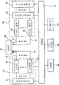
Fig. 1 is the figure that expression is suitable for the summary formation of optical measurement instrument for living body of the present invention.This optical measurement instrument for living body mainly possesses: to the light source portion 10 of organism irradiation near infrared light; The light that organism has been passed through in measurement (comprises the light that seen through organism inside and the light that reflects of portion in vivo.Hereinafter referred to as passing through light), be converted into the photo measure portion 20 of the signal of telecommunication; And calculate local cerebral blood volume variable signal as Biont information according to signal from photo measure portion 20, and specifically be the blood hemoglobin concentration change, the signal processing part 30 of display result.And this optical measurement instrument for living body possesses in order to make the measuring position of guiding from the preceding end in contact examinee 9 of the optical fiber 13 of the light of light source portion 10, and makes the installing component 40 of fixing these optical fiber 13,21 front ends to 20 guiding of photo measure portion from the preceding end in contact examinee's 9 of the optical fiber that passes through light 21 of subject 9 measuring position.This installing component 40 is called measuring probe altogether with the optical fiber front end.
Probe 40 be with irradiation with the optical fiber front end with detect with the optical fiber front end and replace arrangement and on matrix, disposed the socket that optical fiber is connected usefulness and form, adopt the matrix of all sizes such as 3 * 3,4 * 4 according to check point.By detect with optical fiber detect only by from this detect with for example 4 irradiations of optical fiber adjacency with fiber-optic illuminated, see through that light behind the organism mixes, select different modulation signal by these irradiations with optical fiber by synchronizing amplifier 23, thereby can obtain to detect the information of using the point (measurement point) between the optical fiber front end with the irradiation of optical fiber front end and adjacency.These measurement points are corresponding with the passage (channels) that synchronizing amplifier 23 detects, and for example for the probe of 3 * 3 matrixes, rayed position and the measurement point that detects between the position are 12, can carry out the photo measure of 12 passages.
Processing signals handling part 30 is by to installing the control part 31 that integral body controls and be connected with photo measure portion 20, the voltage signal that processing is sent here from photo measure portion 20 (digital signal), converting the signal of expression Biont information to, specifically is local cerebral blood volume variable signal, the generation map of receptor that converts the hemoglobin concentration of expression measuring point to.Storage part 32 and the input and output portion 33 in addition that follow such signal processing part 30 to be provided with, data after these storage part 32 storages are sent digital signal here and handled from photo measure portion 20, this input and output portion 33 possesses the display device (monitor) of the result in the shows signal handling part 30 and the input equipment of indicating to necessity of control part 31 input measurements, signal processing aspect.
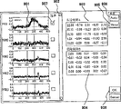
In the optical measurement instrument for living body of such formation, carry out biological light measuring in the following way: by irradiation with optical fiber 13 from 40 light that shone with different frequency modulation(PFM)s of popping one's head in, and after seeing through organism, be converted to the signal of telecommunication by detecting the light that is guided with optical fiber 21 by each photodiode, as irradiation position and the measurement point that detects the intermediate point of position it is detected at each by synchronizing amplifier 23, and acquisition is converted to the hemoglobin variable signal of the blood hemoglobin concentration of measuring point.The hemoglobin variable signal of each measurement point that measures carries out various analyses by signal processing part 30, and its result is presented on the monitor of input and output portion 33.
The situation map of the side head lobe about the examinee is measured in Fig. 2 expression.In illustrated embodiment, about measuring point 201,202 carried out 12 passages (left Ch1-Ch12, right Ch13 one Ch24) respectively, amount to the measurement of 24 passages.Under the state of the problem of stipulating, measure, obtain difference with the state that does not give problem as the hemoglobin variable signal.The hemoglobin variable signal that every passage obtained for example, as shown in Figure 3, shows by every passage as chart 301.In chart 301, transverse axis express time axle, the longitudinal axis represent that mMmm (millimole millimeter unit: the mole number that runs into), for example represented by 2 straight lines of its starting and ending of expression by the time that problem is carried out in the measurement of millimeter unit is long.Before and after it is the stable region that does not give to stimulate.
On the hemoglobin variable signal that measures like this, except signal as the reaction of original organism, also overlapping various noises.Signal processing part 30 is implemented noise separation to these hemoglobin variable signals and is handled, and assigns to reconstruct (reduction) hemoglobin variable signal with the one-tenth of having removed noise.
Below explanation is handled by the noise separation of signal processing part 30 execution.An embodiment of the signal processing order of signal processing part has been shown among Fig. 4, and Fig. 5 represents an embodiment of noise separation processing order.
Noise separation is handled and is mainly comprised following treatment step: the processing 401 that the hemoglobin variable signal is separated into various compositions and demonstration; Remove the given composition in the shown composition or select given composition as the processing 402 that is used for reconstituted composition as noise contribution; And with remove after the noise contribution composition or as being used to reconstruct composition and selected one-tenth assigns to reconstruct the processing 403 of hemoglobin variable signal.In optical measurement instrument for living body, the hemoglobin variable signal is shown as shown in Figure 3, time dependent waveform, and again these waveforms are analyzed as required, carried out the judgement (handling 406) of disease of brain etc. according to the feature of the response wave shape of the discriminating at the position that problem is reacted most, subject.
In the present embodiment, the hemoglobin variable signal is being separated in the step 401 of various compositions, is adopting principal component analysis and independent component analysis, selecting wherein a kind of mode or dual mode combined to analyze.
The data that the hemoglobin variable signal that principal component analysis is not considered to be measured to what kind of key element is made of, do not consider they become measurement result according to what kind of transfer function, but disperse maximization to extract the method that statistics goes up incoherent signal by making.On the other hand, independent component analysis be supposition hemoglobin variable signal be with from the variable signal of various brains position and outer signals in relevant mode linear bonded result, try to achieve its transfer function, extract on the probability density the independently method of signal.Can be according to the characteristic of noise, select wherein a kind of mode or dual mode is combined, thereby can carry out high-precision analysis.For example, be under the situation of representational type noise in several signal sources, independent component analysis is effective.
This principal component analysis and independent component analysis are the component analyzing methods that algorithm has been established in the field that multivariate is resolved, in the optical measurement instrument for living body of present embodiment, the software that is used for carrying out these algorithms is installed in signal processing part 30 as program, by the user interface (GUI) of input and output portion 33 by optionally or the execution that combines.
On the monitor of input and output portion 33, as shown in Figure 3, show the hemoglobin signal that shows the signal that measures by photo measure portion 20 with the blood hemoglobin scale by every passage, and show the button 302 (CalcPCA) of selecting principal component analysis and the button 303 (CalcICA) of selecting independent component analysis.On this picture 300, select arbitrary processing, just carry out principal component analysis (S110-S114) or independent component analysis (S120-S122) (S102) according to order shown in Figure 5.
Under the situation of having selected principal component analysis, according to the local cerebral blood volume variable signal f that in step S101, measures
m(n), produce square matrix X
m(m) (S110).
That is, produce as local cerebral blood volume variable signal f
m(n) (m is a channel position, and M is maximum port number, the 1 ≦ m ≦ M of measuring.N is the timeliness (Longitude Time corresponding with the transverse axis of chart 301) data sequence number, N is a maximum measuring time, 1 ≦ n ≦ N.) the matrix F (m is the row sequence number, and n is the matrix of row sequence number) of set, according to matrix F and this transpose of a matrix matrix F
T, produce square matrix X (matrix of M * M) by following formula (1).
X=F·F
T (1)
Herein from the transposed matrix F of right side convolution matrix F
T, just can obtain the average of biological light measuring to being correlated with of timeliness data direction, from left side convolution, just can obtain the average of biological light measuring to measuring being correlated with of channel direction.In the present embodiment, the feature in each space (passage) by coming extracting time information (timeliness data direction) from the convolution on the right side.
Secondly, for above-mentioned square matrix X, try to achieve characteristic value λ
kMatrix Λ (step S111), calculate eigenvector W and main constituent waveform (step S112) thus.Characteristic value be eigenvector [several times " value, when certain vector W be multiply by certain matrix X be Λ doubly, Λ is a characteristic value, W is an eigenvector, is represented by following formula (2), (3).
XW=ΛW (2)
Λ=W
-1XW (3)
There are M characteristic value, eigenvector in the matrix of M dimension.At M the characteristic value λ that quilt is calculated
k(1 ≦ k ≦ M) is matrix (characteristic value matrix) Λ of diagonal angle composition by big sequence arrangement, substitution characteristic value Λ=λ respectively in formula (2)
1Situation under, characteristic value Λ=λ
2Situation under ... characteristic value Λ=λ
MSituation under, separate following formula (4), thereby calculate eigenvector W.
(X—Λ)W=0 (4)
Inherent vector W is by M eigenvector w
k(m) (w
1(1)~w
M(M), 1 ≦ k ≦ M, the matrix of M * M that 1 ≦ m ≦ M) forms, the k main constituent waveform Y of matrix X
kTry to achieve by formula (5).
Y
k=w
k TX (5)
Replace X with original signal F herein, thereby try to achieve the main constituent waveform C of signal F
k(k represents it is the k composition, 1 ≦ k ≦ M).In view of the above, for the set F of signal, try to achieve the main constituent waveform till the 1st~the M.
C
k=w
k TF (6)
The main constituent waveform C that calculates like this
1~C
kOn the monitor of input and output portion 33 with eigenvector w
k(m) be shown together.Fig. 6 represents to show an example of (GUI).As shown in the figure, in the left side of picture, from top the 1st main constituent waveform 601C that shows in order
1(t), the 2nd main constituent waveform C
2(t) ..., show eigenvector 602w for each composition waveform on the right side
1(m), w
2(m) ...Eigenvector is represented the weight (having frequency) to each passage of the main constituent waveform of correspondence.Be shown in the drawings till the 5th composition waveform, but, just can see whole main constituent waveform of being calculated by pulling scroll bar 603.
Secondly, in the main constituent waveform after separating like this, remove noise waveform, reconstruct signal (S113, Fig. 4: handle 402,403).In the present embodiment, can select reconstructing of signal handled or manual handle automatically.
In demonstration shown in Figure 6 on the picture 600 of main constituent waveform and eigenvector, the words of the button 604 (Manual) of manual handle are selected in operation, just can carry out choose (input) to the choice box 606 (Select) that is used to select the main constituent waveform, so the user selects waveform arbitrarily by choosing choice box 606, is used for signal and reconstructs.Following trial and error method is adopted in the selection of composition waveform: remove according to experience to it seems it is the waveform of noise, it seems that use be that the waveform of major part of signal component reconstructs signal, for the composition waveform that neither is, or get or abandon, confirm the waveform that reconstructs after this.At this moment, can be with reference to the demonstration of eigenvector.
The button 605 (Auto) handled is automatically selected in operation, the selection reference picture of the composition waveform that uses in will the display setting waveform reconstructing.Fig. 7 represents an example of selection reference setting picture.In illustrated embodiment, as system of selection 2 kinds of methods are arranged: (1) selects the waveform relevant to heavens with reference waveform 701, and (2) select the little waveform of standard deviation of differentiated waveform, selection wherein a kind of method of use or with the use that combines of two kinds of methods.
As reference waveform 701, for example, use the waveform of learning according to experience, as the signal (problem correlation signal or representation signal) that is illustrated in the feature that the organism corresponding with the problem that gives the examinee reacts when measuring.In illustrated embodiment, having shown becomes basic trapezoid reference waveform 701, and the user can be according at that time measuring condition and the waveform that is measured to and suitably with its distortion.Input value in time delay input frame 704 for example, thus upwards trapezoidal in the time of being imported, formed.Also have, can move two ordinates 702,703 that expression has added the time (stimulating interval) of problem, setting stimulates interval interval, stimulates the stable region of interval front and back.Produce like this after the reference waveform 701, in identification correlation frame 705, import correlation, operation OK button 707, the correlation of its reference waveform 701 is just selected more than or equal to the composition waveform of the correlation of being imported.
For the stable region that stimulates interval front and back, calculate the standard deviation of differentiated waveform.Stable region was the little interval of variation (differential) of hemoglobin variable signal originally, and was big at the standard deviation of this interval differentiated waveform, just judged it is noise contribution.Particularly, the digital little composition waveform of having set in the choice criteria deviation ratio standard deviation frame 706 is used for signal and reconstructs.Above-mentioned two kinds of methods can be only with wherein any one, also can two kinds of methods and use.
Selection component waveform so automatically or manually, signal processing part 30 just reconstructs signal (reconstructing) (S114, Fig. 4: 403) with it.For example, if selected the 1st composition waveform C
1To k composition waveform C
k, just according to the characteristic value w corresponding with each composition waveform
i, reconstruct the signal H of each passage by following formula (7)
m(m is a channel position, 1 ≦ m ≦ M).
[equation 1]
Signal after reconstructing is displayed on (Fig. 5: S130) on the monitor of input and output portion 33.Expression shows example among Fig. 8.In this example, the display frame (Fig. 3) of the display frame of the signal 801 after reconstructing and original signal is same to produce.Signal after the user is seeing and reconstructing if desired, can repeat above-mentioned processing 401~403 (Fig. 4: 405) again.In the case, carry out principal component analysis (S110~S114) or independent component analysis (S120~S122) with the signal after reconstructing.Also have, as mentioned above, under the situation of manual selection component waveform, can see picture shown reconstruct signal, be back into the selection step 402 of branch waveform, repeat to attempt (404).
Secondly the situation of noise separation processing 402 is carried out in explanation with independent component analysis.In having shown the picture 300 (Fig. 3) of hemoglobin variable signal, the button (CalcICA) 303 of independent component analysis is selected in operation, with regard to the step S120~S122 of execution graph 5.
In independent component analysis, as the formula (8), suppose this signal F (f
m(n) matrix) mixing by the signal of the individual signal source vector S of M ' (n) is constituted, and calculates as the independent signal and the hybrid matrix A thereof of the composition of signal F and the extraction matrix B (S120) that is used to be reduced into sub-signal.
F(n)=AS(n) (8)
(in the formula, suppose that n is the timeliness data sequence number, S (n) is by S (n)=(S
1(n), S
2(n) ... S
M '(n)) vector of Biao Daing, each composition S
M '(n) (1 ≦ m ′ ≦ M ') is mutually independently.A is hybrid matrix (transition matrix).)
Independent component analysis be the probability distribution that do not have a S (n) with hybrid matrix A relevant priori (transcendental) information, and only use the signal F (n) that measures to try to achieve the problem of the Y (n) that determines by following formula (9), if M ≧ M ', then separate existence, extract matrix B (M * M ' real number matrix) and exist.
Y(n)=BX(n) (9)
In the present embodiment, consider the situation of M=M ', under each composition of S (n) is independently prerequisite, calculate B among B * A=I (I is a unit matrix), try to achieve Y (n) by (9) with iterative method.
The independent element waveform Y that calculates like this
1~Y
MOn the monitor of input and output portion 33, be shown with hybrid matrix A, extraction matrix B.Fig. 9 represents to show an example of (GUI).As shown in the figure, in the left side of picture, from top the 1st independent element waveform 901Y that shows in order
1(t), the 2nd independent element waveform Y
2(t) ..., the weight (having frequency) under right side demonstration and the situation of mixing each composition waveform is hybrid matrix A903 accordingly, and extracts matrix B 904 accordingly with the weight under the situation about extracting.In addition, simple hybrid matrix A that shows and extraction matrix B are littler than actual size among the figure.In the case, also can see whole independent element waveforms of being calculated by pulling scroll bar 902.
Secondly, in the independent element waveform after separating like this, the waveform of the regulation of noise waveform is removed in selection, reconstructs signal.Also the same in the case with the related signal separation process of principal component analysis, can select reconstructing of signal handled or manual handle (S121) automatically.Shown in Figure 9, shown in the picture of independent element waveform, the words of the button 905 (Manual) of manual handle are selected in operation, just can carry out choosing to choice box 907 (Select) that be used to select the independent element waveform, so the user selects waveform arbitrarily by choosing choice box 907, is used for signal and reconstructs.This selection reference is the same with the selection of main constituent waveform, and attempts repeatedly as required.
Also have, the words of the button 906 (Auto) of processing are automatically selected in operation, the same with the situation of principal component analysis, will show and be used for setting the picture (Fig. 7) that waveform reconstructs the selection reference of the composition waveform that uses, thereby carry out the setting of reference waveform, the input of necessary parameter like this, carry out signal processing part 30 related automatic composition waveforms and select.
Selection component waveform so automatically or manually, signal processing part 30 just reconstructs signal (S122) with it.For example, if selected the 1st composition waveform Y
1To k composition waveform Y
k,, reconstruct the signal R of each passage by following formula (10) just according to the value of the extraction matrix B i corresponding with each composition waveform
m(m is a channel position, 1 ≦ m ≦ M).
[equation 2]
Signal after reconstructing is displayed on the monitor of input and output portion 33.Signal after the user is seeing and reconstructing if desired, can repeat above-mentioned processing 401~403 (405) again.Also have, manually selecting to handle 402,403 (404) as required and repeatedly under the situation of representation signal.
As mentioned above, principal component analysis is to extract the method that statistics goes up incoherent signal, and independent component analysis is to extract on the probability density the independently method of signal, and segregative method is different with the characteristic of noise.In the present invention, can be according to the characteristic of noise, select for example principal component analysis or independent component analysis, it is carried out 1 time or carries out repeatedly repeatedly, also can principal component analysis and independent component analysis and using, just various noises can be removed effectively thus, more accurate purpose signal can be obtained.
Remove signal behind the denoising and not only be shown as shown in Figure 8 time dependent waveform, can also carry out signal processing and generate clinically effectively information (handling 406).For example, can from the correlation circumstance of problem correlation signal, differentiate the position (the language field is distinguished) of the easiest reaction, carry out the diagnosis of disease etc. according to correlation circumstance with the various disease of brain patients' that stored problem correlation signal.
More than illustrated with principal component analysis and independent component analysis and carried out the embodiment that noise separation is handled, but, the invention is not restricted to above-mentioned embodiment, for example, can the known noise separation technology of appropriate combination (Filtering Processing, rolling average is handled).Also have, the GUI that is used to carry out the selection of selection that noise separation handles, composition waveform also is not limited to illustrated thing, but can suitably change.For example, can not be to select, but display menu come the selection component analytical method by push-botton operation, repeatedly carrying out repeatedly under the situation of component analysis, also can import the method and the order thereof of component analysis.
Industrial applicibility
According to the present invention, to through measured hemoglobin variable signal of biological light measuring etc. Measuring-signal carries out the noise separating treatment by constituent analysis, thereby can be not limited to noise Characteristic and effectively except denoising, can obtain high-precision purpose signal. Also have, according to this The optical measurement instrument for living body of invention, the process that can come pilot signal to process by user interface, And the condition that can come setting signal to process according to examinee's individual difference, measuring condition etc., Thereby can flexibly reproduce and the immediate signal of original signal.
Description of drawings
[Fig. 1] expression is suitable for the figure of the whole summary of optical measurement instrument for living body of the present invention;
The figure of the relation of [Fig. 2] expression measuring point and measurement point (passage);
[Fig. 3] expression is by the figure of the hemoglobin variable signal of each passage that measurement obtained of Fig. 2;
The figure of the order of the signal processing that [Fig. 4] expression optical measurement instrument for living body of the present invention is carried out;
The figure of the order that the noise separation that [Fig. 5] expression optical measurement instrument for living body of the present invention is carried out is handled;
[Fig. 6] expression shows the figure of an example of the display frame of the main constituent waveform that separated by principal component analysis and eigenvector;
The figure of the example of the GUI under the situation of the automatic selection component waveform of [Fig. 7] expression;
The figure of the hemoglobin variable signal after [Fig. 8] expression reconstructs;
[Fig. 9] expression shows the figure of an example of the display frame of the composition waveform (independent signal) that separated by independent component analysis and hybrid matrix/extraction matrix.
The explanation of Reference numeral
10 ... light source portion, 20 ... photo measure portion, 30 ... signal processing part, 31 ... control part, 32 ... storage part, 33 ... input and output portion, 40 ... probe
Claims (16)
1. an optical measurement instrument for living body possesses: shine from the device of the light that can see region of ultra-red to subject; Detect the described light of the organism inside of having passed through described subject, the device of the output signal corresponding with the light quantity that is detected; Handle the signal processing apparatus of the hemoglobin variable signal of described signal, the described subject of generation; And the display device that shown of the hemoglobin variable signal that described signal processing apparatus is generated, it is characterized in that,
Described signal processing apparatus possesses: the signal separator that described hemoglobin variable signal is separated into a plurality of one-tenth sub-signals; Select the signal selecting of the one-tenth sub-signal of regulation based on the correlation between the reference signal of the feature of described a plurality of one-tenth sub-signals and expression organism reaction; And the signal that the one-tenth sub-signal that utilizes selected regulation reconstructs the hemoglobin variable signal reconstructs device.
2. optical measurement instrument for living body according to claim 1 is characterized in that, the one-tenth sub-signal after the separation and reconstruct after signal be presented at respectively in the described display device.
3. optical measurement instrument for living body according to claim 1 and 2 is characterized in that, described signal processing apparatus possesses two kinds of signal separators at least.
4. optical measurement instrument for living body according to claim 1 is characterized in that at least one in the described signal separator carried out principal component analysis to the hemoglobin variable signal, is separated into a plurality of one-tenth sub-signals.
5. according to claim 1 or 4 described optical measurement instrument for living body, it is characterized in that at least one in the described signal separator carried out independent component analysis to the hemoglobin variable signal, be separated into a plurality of one-tenth sub-signals.
6. optical measurement instrument for living body according to claim 1, it is characterized in that, described signal separator possesses principal component analysis device and independent component analysis device, and possesses the side that selects in described principal component analysis device and the independent component analysis device or two sides' segregation apparatus selecting arrangement.
7. optical measurement instrument for living body according to claim 1, it is characterized in that described signal selecting is selected the one-tenth sub-signal of described regulation based on the standard deviation of the differentiated waveform of correlation between the reference signal of the feature of described one-tenth sub-signal and the reaction of expression organism and described one-tenth sub-signal.
8. optical measurement instrument for living body according to claim 1 is characterized in that, described signal processing apparatus possesses and is used to allow the user select described signal to reconstruct the user's interface device of the one-tenth sub-signal of the used regulation of device.
9. optical measurement instrument for living body according to claim 6 is characterized in that, shows the choice box of a plurality of main constituent waveforms or independent element waveform and described main constituent waveform of selection or independent element waveform in described display device.
10. optical measurement instrument for living body according to claim 7 is characterized in that, shows the correlation frame of importing described correlation and/or the standard deviation frame of importing described standard deviation in described display device.
11. an optical measurement instrument for living body has: shine from the device of the light that can see region of ultra-red to a plurality of local of subject; The light, output that detects the organism inside of being shone and passed through subject from described a plurality of places is local and detect the detection device of the measuring-signal on local determined a plurality of measurement points in irradiation; Processing from the measuring-signal of described detection device, generate the signal processing apparatus of the waveform that the tested substance in vivo of the described a plurality of measurement points of expression changes; And the display device that shows the result of described signal processing apparatus, it is characterized in that,
Described signal processing apparatus possesses: the signal separator that described waveform is separated into a plurality of composition waveforms; Select the signal selecting of the one-tenth sub-signal of regulation based on the correlation between the reference signal of the feature of described a plurality of one-tenth sub-signals and expression organism reaction; And the signal that the one-tenth sub-signal that utilizes selected regulation reconstructs the waveform that the described tested substance in vivo of expression changes reconstructs device.
12. a noise removing method to the check point irradiates light of subject, removes denoising from detect the hemoglobin variable signal that the light that passed through described check point obtains, it is characterized in that this method comprises:
Described hemoglobin variable signal is carried out component analysis, is separated into the step of a plurality of one-tenth sub-signals;
Select the step of the one-tenth sub-signal of regulation based on the correlation between the reference signal of the feature of described a plurality of one-tenth sub-signals and expression organism reaction; And
Utilize the one-tenth sub-signal of selected regulation to reconstruct the step of hemoglobin variable signal.
13. noise removing method according to claim 12 is characterized in that, the described step that is separated into a plurality of one-tenth sub-signals comprises the step of described a plurality of one-tenth sub-signals being carried out principal component analysis.
14., it is characterized in that the described step that is separated into a plurality of one-tenth sub-signals comprises the step of described a plurality of one-tenth sub-signals being carried out independent component analysis according to claim 12 or 13 described noise removing methods.
15. noise removing method according to claim 12 is characterized in that, for the hemoglobin variable signal after being reconstructed by described reconstituted step, carries out described step and the reconstituted step that is separated into a plurality of one-tenth sub-signals.
16. noise removing method according to claim 12, it is characterized in that the described step of selecting is with described one-tenth sub-signal and represents that correlation and the described standard deviation that becomes the differentiated waveform of sub-signal between the reference signal of feature of organism reaction select the one-tenth sub-signal of described regulation.
Applications Claiming Priority (2)
| Application Number | Priority Date | Filing Date | Title |
|---|---|---|---|
| JP381910/2003 | 2003-11-12 | ||
| JP2003381910A JP4474145B2 (en) | 2003-11-12 | 2003-11-12 | Optical measuring device |
Publications (2)
| Publication Number | Publication Date |
|---|---|
| CN1878505A CN1878505A (en) | 2006-12-13 |
| CN100508895C true CN100508895C (en) | 2009-07-08 |
Family
ID=34587239
Family Applications (1)
| Application Number | Title | Priority Date | Filing Date |
|---|---|---|---|
| CNB2004800331988A Expired - Fee Related CN100508895C (en) | 2003-11-12 | 2004-11-01 | Biological light measuring apparatus |
Country Status (6)
| Country | Link |
|---|---|
| US (1) | US8019399B2 (en) |
| EP (1) | EP1685801B1 (en) |
| JP (1) | JP4474145B2 (en) |
| CN (1) | CN100508895C (en) |
| DE (1) | DE602004032255D1 (en) |
| WO (1) | WO2005046483A1 (en) |
Cited By (1)
| Publication number | Priority date | Publication date | Assignee | Title |
|---|---|---|---|---|
| CN102141543A (en) * | 2010-12-28 | 2011-08-03 | 天津大学 | Method and device for detecting quality of laser welding based on microphone arrays |
Families Citing this family (47)
| Publication number | Priority date | Publication date | Assignee | Title |
|---|---|---|---|---|
| US7758503B2 (en) * | 1997-01-27 | 2010-07-20 | Lynn Lawrence A | Microprocessor system for the analysis of physiologic and financial datasets |
| US9042952B2 (en) * | 1997-01-27 | 2015-05-26 | Lawrence A. Lynn | System and method for automatic detection of a plurality of SPO2 time series pattern types |
| US8932227B2 (en) | 2000-07-28 | 2015-01-13 | Lawrence A. Lynn | System and method for CO2 and oximetry integration |
| US20060161071A1 (en) | 1997-01-27 | 2006-07-20 | Lynn Lawrence A | Time series objectification system and method |
| US20070191697A1 (en) | 2006-02-10 | 2007-08-16 | Lynn Lawrence A | System and method for SPO2 instability detection and quantification |
| US9053222B2 (en) | 2002-05-17 | 2015-06-09 | Lawrence A. Lynn | Patient safety processor |
| US20060195041A1 (en) | 2002-05-17 | 2006-08-31 | Lynn Lawrence A | Centralized hospital monitoring system for automatically detecting upper airway instability and for preventing and aborting adverse drug reactions |
| US20090281838A1 (en) | 2008-05-07 | 2009-11-12 | Lawrence A. Lynn | Medical failure pattern search engine |
| EP1852101B1 (en) * | 2005-02-16 | 2015-09-02 | Hitachi Medical Corporation | Biophoton measuring instrument |
| JP4631510B2 (en) * | 2005-03-31 | 2011-02-16 | 株式会社島津製作所 | Brain function information monitoring device |
| JP4561524B2 (en) * | 2005-08-08 | 2010-10-13 | 株式会社島津製作所 | Signal analysis apparatus and signal analysis method |
| ES2276609B1 (en) * | 2005-09-27 | 2008-06-16 | Universidad Politecnica De Valencia | APPARATUS AND METHOD OF OBTAINING INFORMATION CONCERNING CEREBRAL HEMODINAMICS. |
| US7668579B2 (en) | 2006-02-10 | 2010-02-23 | Lynn Lawrence A | System and method for the detection of physiologic response to stimulation |
| JP2007289224A (en) * | 2006-04-21 | 2007-11-08 | Hitachi Ltd | Living body measurement system and method |
| FR2902307B1 (en) * | 2006-06-14 | 2008-08-29 | Quidd Sas | OPTICAL IMAGING DEVICE |
| JP5248758B2 (en) * | 2006-09-08 | 2013-07-31 | 株式会社島津製作所 | Optical measuring device |
| US8064975B2 (en) | 2006-09-20 | 2011-11-22 | Nellcor Puritan Bennett Llc | System and method for probability based determination of estimated oxygen saturation |
| JP5071767B2 (en) * | 2006-12-08 | 2012-11-14 | 学校法人日本大学 | Biological tissue blood flow measurement device |
| JP5034477B2 (en) | 2006-12-15 | 2012-09-26 | 株式会社日立製作所 | Biological light measurement device |
| US20100324398A1 (en) * | 2007-05-11 | 2010-12-23 | Jung Tzyy-Ping | Non-invasive characterization of a physiological parameter |
| JP4887320B2 (en) * | 2008-03-19 | 2012-02-29 | 株式会社沖データ | Image processing device |
| JP4518189B2 (en) * | 2008-05-28 | 2010-08-04 | ソニー株式会社 | Information processing apparatus and method, program, and recording medium |
| WO2009148042A1 (en) * | 2008-06-05 | 2009-12-10 | 株式会社 日立メディコ | Living body optical measurement device and image display program |
| US8077297B2 (en) | 2008-06-30 | 2011-12-13 | Nellcor Puritan Bennett Ireland | Methods and systems for discriminating bands in scalograms |
| US8295567B2 (en) | 2008-06-30 | 2012-10-23 | Nellcor Puritan Bennett Ireland | Systems and methods for ridge selection in scalograms of signals |
| US8827917B2 (en) * | 2008-06-30 | 2014-09-09 | Nelleor Puritan Bennett Ireland | Systems and methods for artifact detection in signals |
| US8968193B2 (en) | 2008-09-30 | 2015-03-03 | Covidien Lp | System and method for enabling a research mode on physiological monitors |
| JP5679629B2 (en) * | 2008-12-25 | 2015-03-04 | 株式会社島津製作所 | Optical brain function measuring device |
| US20120010484A1 (en) * | 2009-03-19 | 2012-01-12 | Shimadzu Corporation | Photobiological measuring device and analyzing method |
| US9402554B2 (en) | 2011-09-23 | 2016-08-02 | Nellcor Puritan Bennett Ireland | Systems and methods for determining respiration information from a photoplethysmograph |
| US9675274B2 (en) | 2011-09-23 | 2017-06-13 | Nellcor Puritan Bennett Ireland | Systems and methods for determining respiration information from a photoplethysmograph |
| US9693709B2 (en) | 2011-09-23 | 2017-07-04 | Nellcot Puritan Bennett Ireland | Systems and methods for determining respiration information from a photoplethysmograph |
| US9119597B2 (en) | 2011-09-23 | 2015-09-01 | Nellcor Puritan Bennett Ireland | Systems and methods for determining respiration information from a photoplethysmograph |
| US9693736B2 (en) | 2011-11-30 | 2017-07-04 | Nellcor Puritan Bennett Ireland | Systems and methods for determining respiration information using historical distribution |
| JP6036122B2 (en) * | 2012-02-02 | 2016-11-30 | セイコーエプソン株式会社 | Pulse wave measuring device and program |
| JP5909388B2 (en) * | 2012-03-09 | 2016-04-26 | 株式会社日立製作所 | Biological light measurement device |
| CN104507396B (en) * | 2012-07-27 | 2016-09-14 | 株式会社岛津制作所 | Photo bio measurement apparatus and the analysis method of use photo bio measurement apparatus |
| CN104768473A (en) * | 2012-11-08 | 2015-07-08 | 株式会社岛津制作所 | Photobiological measurement device |
| CN102973279B (en) * | 2012-12-18 | 2014-09-17 | 哈尔滨工业大学 | Near-infrared brain-machine interface signal detection method integrating independent component analysis |
| US10022068B2 (en) | 2013-10-28 | 2018-07-17 | Covidien Lp | Systems and methods for detecting held breath events |
| JPWO2015141423A1 (en) * | 2014-03-18 | 2017-04-06 | 株式会社日立製作所 | Biological light measurement device and biological light measurement method |
| JP6189486B2 (en) * | 2015-06-12 | 2017-08-30 | ダイキン工業株式会社 | Brain activity estimation device |
| JP1554806S (en) * | 2015-09-24 | 2016-07-25 | ||
| JP6996095B2 (en) * | 2017-03-17 | 2022-01-17 | 株式会社リコー | Information display devices, biological signal measurement systems and programs |
| JPWO2018194056A1 (en) | 2017-04-18 | 2020-02-27 | 興和株式会社 | Information processing method, information processing apparatus, and program for calculating blood absorption spectrum |
| US11141083B2 (en) | 2017-11-29 | 2021-10-12 | Samsung Electronics Co., Ltd. | System and method for obtaining blood glucose concentration using temporal independent component analysis (ICA) |
| WO2022070802A1 (en) * | 2020-09-30 | 2022-04-07 | 住友理工株式会社 | Biological information measuring device |
Family Cites Families (14)
| Publication number | Priority date | Publication date | Assignee | Title |
|---|---|---|---|---|
| US4596254A (en) * | 1984-12-18 | 1986-06-24 | Tsi Research Associates Limited Partnership | Laser Doppler flow monitor |
| JP4281135B2 (en) | 1998-11-30 | 2009-06-17 | 三菱電機株式会社 | Image quality improving method and image quality improving apparatus |
| JP4068763B2 (en) * | 1999-06-21 | 2008-03-26 | 株式会社島津製作所 | Biological signal measuring device |
| US6264591B1 (en) * | 1999-07-27 | 2001-07-24 | Philip Morris Incorporated | Plug combiner inspection system and method |
| JP4097874B2 (en) | 2000-03-06 | 2008-06-11 | 富士フイルム株式会社 | Image compression method and image compression apparatus for multispectral image |
| JP4097522B2 (en) * | 2000-10-16 | 2008-06-11 | 株式会社日立メディコ | Biological light measurement device |
| JP3725418B2 (en) | 2000-11-01 | 2005-12-14 | インターナショナル・ビジネス・マシーンズ・コーポレーション | Signal separation method, image processing apparatus, and storage medium for restoring multidimensional signal from image data mixed with a plurality of signals |
| JP4651186B2 (en) | 2000-12-11 | 2011-03-16 | 株式会社日立メディコ | Biological light measurement device |
| US6701170B2 (en) * | 2001-11-02 | 2004-03-02 | Nellcor Puritan Bennett Incorporated | Blind source separation of pulse oximetry signals |
| US6829501B2 (en) * | 2001-12-20 | 2004-12-07 | Ge Medical Systems Information Technologies, Inc. | Patient monitor and method with non-invasive cardiac output monitoring |
| JP4164386B2 (en) * | 2003-02-28 | 2008-10-15 | 株式会社日立製作所 | Biological light measurement device |
| JP2004299814A (en) | 2003-03-28 | 2004-10-28 | Takasago Thermal Eng Co Ltd | Method and apparatus for manufacturing static-eliminated insulating substrate |
| US7025728B2 (en) * | 2003-06-30 | 2006-04-11 | Nihon Kohden Corporation | Method for reducing noise, and pulse photometer using the method |
| US7027920B2 (en) | 2003-07-18 | 2006-04-11 | Visteon Global Technologies, Inc. | Low-speed collision avoidance system |
-
2003
- 2003-11-12 JP JP2003381910A patent/JP4474145B2/en not_active Expired - Lifetime
-
2004
- 2004-11-01 US US10/578,725 patent/US8019399B2/en active Active
- 2004-11-01 DE DE200460032255 patent/DE602004032255D1/en active Active
- 2004-11-01 CN CNB2004800331988A patent/CN100508895C/en not_active Expired - Fee Related
- 2004-11-01 WO PCT/JP2004/016226 patent/WO2005046483A1/en active Application Filing
- 2004-11-01 EP EP20040799432 patent/EP1685801B1/en not_active Not-in-force
Cited By (2)
| Publication number | Priority date | Publication date | Assignee | Title |
|---|---|---|---|---|
| CN102141543A (en) * | 2010-12-28 | 2011-08-03 | 天津大学 | Method and device for detecting quality of laser welding based on microphone arrays |
| CN102141543B (en) * | 2010-12-28 | 2012-10-24 | 天津大学 | Method and device for detecting quality of laser welding based on microphone arrays |
Also Published As
| Publication number | Publication date |
|---|---|
| WO2005046483A1 (en) | 2005-05-26 |
| DE602004032255D1 (en) | 2011-05-26 |
| JP2005143609A (en) | 2005-06-09 |
| US8019399B2 (en) | 2011-09-13 |
| EP1685801A1 (en) | 2006-08-02 |
| US20070142719A1 (en) | 2007-06-21 |
| EP1685801A4 (en) | 2009-03-04 |
| CN1878505A (en) | 2006-12-13 |
| JP4474145B2 (en) | 2010-06-02 |
| EP1685801B1 (en) | 2011-04-13 |
Similar Documents
| Publication | Publication Date | Title |
|---|---|---|
| CN100508895C (en) | Biological light measuring apparatus | |
| US6701170B2 (en) | Blind source separation of pulse oximetry signals | |
| US7186217B2 (en) | Biological photometer | |
| AU715201B2 (en) | Methods of minimizing scattering and improving tissue sampling in non-invasive testing and imaging | |
| US6694159B2 (en) | Choice of wavelengths for multiwavelength optical imaging | |
| US20040267140A1 (en) | Method for reducing noise, and pulse photometer using the method | |
| US20080146901A1 (en) | Optical measurement instrument for living body | |
| WO2006025504A1 (en) | Sensitive state judging method | |
| JP4097522B2 (en) | Biological light measurement device | |
| JP2006095266A5 (en) | ||
| WO2005096951A1 (en) | Biophotonic measuring apparatus | |
| DE19840452A1 (en) | Method and device for the non-invasive measurement of concentrations of blood components | |
| US20080144004A1 (en) | Optical Spectrophotometer | |
| CN107080543A (en) | A kind of real-time Cerebral cortex blood oxygen signal harvester of new near-infrared | |
| CN104284629B (en) | Photo bio bulk measurement system and using method thereof | |
| US20110245636A1 (en) | Multi-Wavelength Photon Density Wave System Using An Optical Switch | |
| US7330745B2 (en) | Living body photometric device | |
| JP3801172B2 (en) | Biological light measurement device | |
| CN104869913B (en) | Biological optical measurement device and signal separation method for same | |
| Campbell | Improving the Accuracy and Precision of Frequency Domain and Hybrid Broadband Diffuse Optical Imaging | |
| CN115005775A (en) | Method and device for correcting artifacts of near-infrared signal data and storage medium | |
| CN116895336A (en) | Brain oxygen signal processing method based on complete set empirical mode decomposition | |
| ZUCCHELLI et al. | Development and applications of a medical device based on time domain functional Near Infrared Spectroscopy for brain imaging |
Legal Events
| Date | Code | Title | Description |
|---|---|---|---|
| C06 | Publication | ||
| PB01 | Publication | ||
| C10 | Entry into substantive examination | ||
| SE01 | Entry into force of request for substantive examination | ||
| C14 | Grant of patent or utility model | ||
| GR01 | Patent grant | ||
| CF01 | Termination of patent right due to non-payment of annual fee |
Granted publication date: 20090708 Termination date: 20161101 |
|
| CF01 | Termination of patent right due to non-payment of annual fee |