BR112014002576B1 - FAB FRAGMENT OF HUMAN ANTI-NGF ANTIBODY, METHOD TO PRODUCE THE SAME AND EXPRESSION VECTOR COMPRISING A POLYNUCLEOTIDE THAT ENCODES SAID FAB FRAGMENT' - Google Patents
FAB FRAGMENT OF HUMAN ANTI-NGF ANTIBODY, METHOD TO PRODUCE THE SAME AND EXPRESSION VECTOR COMPRISING A POLYNUCLEOTIDE THAT ENCODES SAID FAB FRAGMENT' Download PDFInfo
- Publication number
- BR112014002576B1 BR112014002576B1 BR112014002576-2A BR112014002576A BR112014002576B1 BR 112014002576 B1 BR112014002576 B1 BR 112014002576B1 BR 112014002576 A BR112014002576 A BR 112014002576A BR 112014002576 B1 BR112014002576 B1 BR 112014002576B1
- Authority
- BR
- Brazil
- Prior art keywords
- fab
- fragment
- antibody
- human
- ngf
- Prior art date
Links
Images
Classifications
-
- C—CHEMISTRY; METALLURGY
- C07—ORGANIC CHEMISTRY
- C07K—PEPTIDES
- C07K16/00—Immunoglobulins [IGs], e.g. monoclonal or polyclonal antibodies
- C07K16/18—Immunoglobulins [IGs], e.g. monoclonal or polyclonal antibodies against material from animals or humans
- C07K16/22—Immunoglobulins [IGs], e.g. monoclonal or polyclonal antibodies against material from animals or humans against growth factors ; against growth regulators
-
- A—HUMAN NECESSITIES
- A61—MEDICAL OR VETERINARY SCIENCE; HYGIENE
- A61K—PREPARATIONS FOR MEDICAL, DENTAL OR TOILETRY PURPOSES
- A61K47/00—Medicinal preparations characterised by the non-active ingredients used, e.g. carriers or inert additives; Targeting or modifying agents chemically bound to the active ingredient
- A61K47/50—Medicinal preparations characterised by the non-active ingredients used, e.g. carriers or inert additives; Targeting or modifying agents chemically bound to the active ingredient the non-active ingredient being chemically bound to the active ingredient, e.g. polymer-drug conjugates
- A61K47/51—Medicinal preparations characterised by the non-active ingredients used, e.g. carriers or inert additives; Targeting or modifying agents chemically bound to the active ingredient the non-active ingredient being chemically bound to the active ingredient, e.g. polymer-drug conjugates the non-active ingredient being a modifying agent
- A61K47/56—Medicinal preparations characterised by the non-active ingredients used, e.g. carriers or inert additives; Targeting or modifying agents chemically bound to the active ingredient the non-active ingredient being chemically bound to the active ingredient, e.g. polymer-drug conjugates the non-active ingredient being a modifying agent the modifying agent being an organic macromolecular compound, e.g. an oligomeric, polymeric or dendrimeric molecule
- A61K47/59—Medicinal preparations characterised by the non-active ingredients used, e.g. carriers or inert additives; Targeting or modifying agents chemically bound to the active ingredient the non-active ingredient being chemically bound to the active ingredient, e.g. polymer-drug conjugates the non-active ingredient being a modifying agent the modifying agent being an organic macromolecular compound, e.g. an oligomeric, polymeric or dendrimeric molecule obtained otherwise than by reactions only involving carbon-to-carbon unsaturated bonds, e.g. polyureas or polyurethanes
- A61K47/60—Medicinal preparations characterised by the non-active ingredients used, e.g. carriers or inert additives; Targeting or modifying agents chemically bound to the active ingredient the non-active ingredient being chemically bound to the active ingredient, e.g. polymer-drug conjugates the non-active ingredient being a modifying agent the modifying agent being an organic macromolecular compound, e.g. an oligomeric, polymeric or dendrimeric molecule obtained otherwise than by reactions only involving carbon-to-carbon unsaturated bonds, e.g. polyureas or polyurethanes the organic macromolecular compound being a polyoxyalkylene oligomer, polymer or dendrimer, e.g. PEG, PPG, PEO or polyglycerol
-
- A—HUMAN NECESSITIES
- A61—MEDICAL OR VETERINARY SCIENCE; HYGIENE
- A61K—PREPARATIONS FOR MEDICAL, DENTAL OR TOILETRY PURPOSES
- A61K39/00—Medicinal preparations containing antigens or antibodies
- A61K2039/505—Medicinal preparations containing antigens or antibodies comprising antibodies
-
- C—CHEMISTRY; METALLURGY
- C07—ORGANIC CHEMISTRY
- C07K—PEPTIDES
- C07K2317/00—Immunoglobulins specific features
- C07K2317/20—Immunoglobulins specific features characterized by taxonomic origin
- C07K2317/21—Immunoglobulins specific features characterized by taxonomic origin from primates, e.g. man
-
- C—CHEMISTRY; METALLURGY
- C07—ORGANIC CHEMISTRY
- C07K—PEPTIDES
- C07K2317/00—Immunoglobulins specific features
- C07K2317/20—Immunoglobulins specific features characterized by taxonomic origin
- C07K2317/24—Immunoglobulins specific features characterized by taxonomic origin containing regions, domains or residues from different species, e.g. chimeric, humanized or veneered
-
- C—CHEMISTRY; METALLURGY
- C07—ORGANIC CHEMISTRY
- C07K—PEPTIDES
- C07K2317/00—Immunoglobulins specific features
- C07K2317/30—Immunoglobulins specific features characterized by aspects of specificity or valency
- C07K2317/33—Crossreactivity, e.g. for species or epitope, or lack of said crossreactivity
-
- C—CHEMISTRY; METALLURGY
- C07—ORGANIC CHEMISTRY
- C07K—PEPTIDES
- C07K2317/00—Immunoglobulins specific features
- C07K2317/50—Immunoglobulins specific features characterized by immunoglobulin fragments
- C07K2317/51—Complete heavy chain or Fd fragment, i.e. VH + CH1
-
- C—CHEMISTRY; METALLURGY
- C07—ORGANIC CHEMISTRY
- C07K—PEPTIDES
- C07K2317/00—Immunoglobulins specific features
- C07K2317/50—Immunoglobulins specific features characterized by immunoglobulin fragments
- C07K2317/515—Complete light chain, i.e. VL + CL
-
- C—CHEMISTRY; METALLURGY
- C07—ORGANIC CHEMISTRY
- C07K—PEPTIDES
- C07K2317/00—Immunoglobulins specific features
- C07K2317/50—Immunoglobulins specific features characterized by immunoglobulin fragments
- C07K2317/55—Fab or Fab'
-
- C—CHEMISTRY; METALLURGY
- C07—ORGANIC CHEMISTRY
- C07K—PEPTIDES
- C07K2317/00—Immunoglobulins specific features
- C07K2317/50—Immunoglobulins specific features characterized by immunoglobulin fragments
- C07K2317/56—Immunoglobulins specific features characterized by immunoglobulin fragments variable (Fv) region, i.e. VH and/or VL
- C07K2317/565—Complementarity determining region [CDR]
-
- C—CHEMISTRY; METALLURGY
- C07—ORGANIC CHEMISTRY
- C07K—PEPTIDES
- C07K2317/00—Immunoglobulins specific features
- C07K2317/70—Immunoglobulins specific features characterized by effect upon binding to a cell or to an antigen
- C07K2317/76—Antagonist effect on antigen, e.g. neutralization or inhibition of binding
-
- C—CHEMISTRY; METALLURGY
- C07—ORGANIC CHEMISTRY
- C07K—PEPTIDES
- C07K2317/00—Immunoglobulins specific features
- C07K2317/90—Immunoglobulins specific features characterized by (pharmaco)kinetic aspects or by stability of the immunoglobulin
- C07K2317/94—Stability, e.g. half-life, pH, temperature or enzyme-resistance
Abstract
ANTICORPO ANTI-NGF HUMANO A presente invenção se refere a um anticorpo anti-NGF humano ou a um fragmento de ligação a antígeno do mesmo que é excelente em segurança ao reduzir o risco de efeitos colaterais tais como efeitos em um feto e formação de trombo ao mesmo tempo em que mantém alta atividade de neutralização e fornece meios para evitar ou tratar várias doenças nas quais o NGF humano está envolvido na formação de condições patológicas, usando o anti corpo ou o fragmento de ligação de anticorpo do mesmo. Meios para Resolução: um fragmento Fab? de anticorpo anti-NGF compreendendo uma região variável de cadeia pesada que consiste em uma sequência de aminoácidos apresentada pela SEQ ID NO:6 e uma região variável de cadeia leve que consiste em uma sequência de aminoácidos apresentada pela SEQ ID NO:4.ANTI-HUMAN ANTI-NGF ANTIBODY The present invention relates to an anti-human NGF antibody or an antigen-binding fragment thereof which is excellent in safety by reducing the risk of side effects such as effects on a fetus and thrombus formation upon while maintaining high neutralizing activity and providing means to prevent or treat various diseases in which human NGF is involved in the formation of pathological conditions, using the antibody or antibody-binding fragment thereof. Means for Resolution: a Fab Fragment? of anti-NGF antibody comprising a heavy chain variable region consisting of an amino acid sequence shown by SEQ ID NO:6 and a light chain variable region consisting of an amino acid sequence shown by SEQ ID NO:4.
Description
[001] A presente invenção se refere a um novo anticorpo anti-NGF humano. Mais especificamente, a presente invenção se refere a um fragmento Fab’ de um anticorpo anti-NGF humano.[001] The present invention relates to a novel anti-human NGF antibody. More specifically, the present invention relates to a Fab' fragment of an anti-human NGF antibody.
[002] Um fator de desenvolvimento neural (NGF) é um dos fatores humorais chamado em geral “fatores neurotróficos”, e desempenha um importante papel na geração e diferenciação de neurônios e na manutenção das funções de neurônios no corpo. As receptores NGF, um receptor de trkA de alta afinidade (receptor-tipo tirosina quinAse) e um receptor p75NTR de baixa afinidade são conhecidos. Há um relatório reportando que entre os referidos, o p75NTR se liga a todos os fatores neurotróficos e está envolvido em apoptose no processo de geração neuronal. Entretanto, o papel do p75NTR ainda não foi suficientemente explicado. Nesse meio tempo, é sabido que camundongos com gene inativado dos receptores NGF e trkA expressam o mesmo fenótipo (Documento não Patente 1), e é considerado que a ação fisiológica do NGF é expressa principalmente por meio do receptor trkA.[002] A neural development factor (NGF) is one of the humoral factors commonly called “neurotrophic factors”, and it plays an important role in the generation and differentiation of neurons and in the maintenance of neuron functions in the body. NGF receptors, a high-affinity trkA receptor (tyrosine kinAse receptor-like) and a low-affinity p75NTR receptor are known. There is a report reporting that among these, p75NTR binds to all neurotrophic factors and is involved in apoptosis in the neuronal generation process. However, the role of p75NTR has not been sufficiently explained. In the meantime, it is known that mice with inactivated gene of NGF and trkA receptors express the same phenotype (Non-Patent Document 1), and it is considered that the physiological action of NGF is mainly expressed through the trkA receptor.
[003] Em 1993, houve um relatório reportando que a administração de NGF a ratos induziu dor (Documento não Patente 2), e desde então, houve um relatório reportando que a administração intravenosa de NGF a seres humanos induz mialgia sistêmica e que a administração tópica de NGF exerce um efeito sistêmico e induz hiperpatia e alodínia em um campo de injeção (Documento não Patente 3). Adicionalmente, há um relatório reportando que um camundongo com gene inativado do receptor trkA mostra analgesia (Documento não Patente 4), então é considerado que NGF é uma molécula profundamente envolvida na expressão da dor. Observando a correlação entre NGF e a condição patológica da dor humana, foi demonstrado que a expressão de NGF/trkA é acelerada em cartilagens articulares com osteoartrite (OA) (Documento não Patente 6) e que o nível de NGF é aumentado em pacientes com artrite reumatóide (Documento não Patente 7) ou cistite intersticial (Documento não Patente 8).[003] In 1993, there was a report reporting that administration of NGF to rats induced pain (Non-Patent Document 2), and since then, there has been a report reporting that intravenous administration of NGF to humans induces systemic myalgia and that administration Topical NGF exerts a systemic effect and induces hyperpathy and allodynia in an injection field (Non-Patent Document 3). Additionally, there is a report reporting that a mouse with inactivated trkA receptor gene shows analgesia (Non-Patent Document 4), so NGF is considered to be a molecule deeply involved in pain expression. Observing the correlation between NGF and the pathological condition of human pain, it was shown that the expression of NGF/trkA is accelerated in articular cartilages with osteoarthritis (OA) (Non-Patent Document 6) and that the level of NGF is increased in patients with arthritis rheumatoid disease (Non-Patent Document 7) or interstitial cystitis (Non-Patent Document 8).
[004] A partir dos fatos acima, é esperado que se um anticorpo monoclonal que especificamente se liga a NGF e tem uma atividade inibitória contra a ação de NGF pode ser desenvolvido, e o referido será útil para tratar, evitar, e diagnosticar várias doenças incluindo dor relativa a NGF.[004] From the above facts, it is expected that if a monoclonal antibody that specifically binds to NGF and has an inhibitory activity against the action of NGF can be developed, the aforementioned will be useful for treating, preventing, and diagnosing various diseases. including NGF-related pain.
[005] Os anticorpos anti-NGF humanos que foram clinicamente desenvolvidos até o presente, tanezumab (Documento de Patente 1) e PG110 (Documento de Patente 2) como anticorpos anti-NGF humanos humanizados, e REGN475 (Documento de Patente 3), fulranumab (Documento de Patente 4), e MEDI-578 (Documento de Patente 5) como anticorpos anti-NGF humanos completamente humanos foram reportados. Entre os referidos, tanezumab está sendo mais vivamente desenvolvido por prioridade, e há um relatório reportando que de acordo com os resultados de ensaios clínicos, o referido anticorpo exerce um efeito analgésico potente e extensivo na dor tal como artralgia acompanhada por osteoartrite, dor nas costas crônica, e cistalgia acompanhada por cistite intersticial (Documentos Não Patente 9 a 11).[005] The human anti-NGF antibodies that have been clinically developed to date, tanezumab (Patent Document 1) and PG110 (Patent Document 2) as humanized human anti-NGF antibodies, and REGN475 (Patent Document 3), fulranumab (Patent Document 4), and MEDI-578 (Patent Document 5) as fully human anti-NGF human antibodies have been reported. Among the aforementioned, tanezumab is being most vigorously developed by priority, and there is a report reporting that according to the results of clinical trials, said antibody exerts a potent and extensive analgesic effect on pain such as arthralgia accompanied by osteoarthritis, back pain chronic disease, and cystalgia accompanied by interstitial cystitis (Non-Patent Documents 9 to 11).
[006] Em geral, os fatores principais que determinam uma dose eficaz de um fármaco de anticorpo, a atividade neutralizante de um anticorpo contra um antígeno e a quantidade de antígenos presente no corpo são exemplificados. Ao se aprimorar a atividade neutralizante se leva a uma redução da dose, e consequentemente, isso pode ser mencionado como uma melhora bastante útil que leva a uma redução nos encargos financeiros conduzindo a uma redução dos encargos financeiros dos pacientes e dos custos médicos. Se a redução na dose pode ser realizada, a administração subcutânea pode também ser realizada. A administração subcutânea tem uma grande vantagem que um paciente pode realizar auto-injeção em casa se determinadas condições forem satisfeitas. Adicionalmente, embora o fármaco de anticorpo seja em geral administrado via gotas por um determinado tempo em muitos casos em administração intravenosa, o fármaco pode ser administrado as um bolus na administração subcutânea, que é outra vantagem. Não só o médico mas também o paciente pode selecionar a preparação para administração intravenosa e a preparação para administração subcutânea, e isso é um fator desejável. Entretanto, na administração subcutânea, a dose que pode ser dada por administração é tão pequena quanto cerca de 1 mL em geral, de modo que uma quantidade suficiente de anticorpos precisa ser incluída na dose de modo a expressar a eficácia do fármaco. Adicionalmente, diferente da administração intravenosa, a biodisponibilidade precisa ser considerada para a administração subcutânea. Ou seja, de modo a realizar a preparação para a administração subcutânea, é necessário se preparar um anticorpo que exibe excelente solubilidade e expressa uma suficiente eficácia de fármaco mesmo em pequena dose. Assim sendo, se um anticorpo que tem uma maior atividade de neutralização contra NGF comparado aos anticorpos na técnica relacionada é obtido, o mesmo será útil para tratar doenças relacionadas a NGF e para aprimorar a conveniência do tratamento.[006] In general, the main factors that determine an effective dose of an antibody drug, the neutralizing activity of an antibody against an antigen, and the amount of antigen present in the body are exemplified. Improving the neutralizing activity leads to a dose reduction, and consequently, this can be mentioned as a very useful improvement that leads to a reduction in financial burden leading to a reduction in patient financial burden and medical costs. If dose reduction can be performed, subcutaneous administration can also be performed. Subcutaneous administration has a great advantage that a patient can self-inject at home if certain conditions are met. Additionally, although the antibody drug is generally administered via drops over a period of time in many cases in intravenous administration, the drug can be administered as a bolus in subcutaneous administration, which is another advantage. Not only the physician but also the patient can select the preparation for intravenous administration and the preparation for subcutaneous administration, and this is a desirable factor. However, in subcutaneous administration, the dose that can be given per administration is as small as about 1 mL in general, so a sufficient amount of antibodies needs to be included in the dose in order to express the drug's efficacy. Additionally, unlike intravenous administration, bioavailability needs to be considered for subcutaneous administration. That is, in order to carry out the preparation for subcutaneous administration, it is necessary to prepare an antibody that exhibits excellent solubility and expresses sufficient drug efficacy even in a small dose. Therefore, if an antibody that has a greater neutralizing activity against NGF compared to antibodies in the related art is obtained, it will be useful to treat NGF-related diseases and to improve the desirability of treatment.
[007] Como descrito acima, embora NGF seja um fator importante para o desenvolvimento de neurônios, a realização de um exame suficiente em termos de segurança é necessária no desenvolvimento de fármacos médicos que inibam a função de NGF. Particularmente, como um dos itens que devem ser examinados em termos de segurança, os efeitos em um feto são exemplificados. Com relação à inibição funcional de NGF, houve relatos reportando que a mutação de NGF é a causa de analgesia congênita (Documento não Patente 5), e que em um experimento animal, quando uma porca da guiné prenha é incentivada a produzir autoanticorpo para NGF de modo a inibir NGF no corpo, o novo porco da guiné mostra sintomas de analgesia (Documento não Patente 12). Adicionalmente, em um ensaio usando camundongos com deficiência de NGF- ou trkA-, foi demonstrado que a ação de deficiência de NGF inibe o desenvolvimento dos neurônios do nervos sensórios e nervos simpáticos em um embrião (Documentos Não Patente 4 e 13). A partir dos referidos resultados, é entendido que NGF é um fator essencial de neurodesenvolvimento no estágio precoce de desenvolvimento. As doenças relacionadas a NGF também incluem doenças que mulheres em idade de gravidez sofrem um alto grau de cistite intersticial (metade ou mais dos pacientes têm 44 anos de idade ou são mais jovens, e 90% dos pacientes são mulheres (Documento não Patente 14)), dor nas costas crônica (uma media de idade de 40 a 50, e mais de 50% dos pacientes são mulheres (Documentos Não Patente 15 a 17)), e enxaqueca (uma idade de pico de início varia a partir de 15 a 40 anos, e 80% de pacientes são mulheres (Documento não Patente 18)). Nessa situação, no desenvolvimento de anticorpo anti-NGF como um fármaco médico, é muito importante se evitar o risco de efeitos colaterais em um feto em mulheres grávidas.[007] As described above, although NGF is an important factor in neuron development, sufficient safety screening is required in the development of medical drugs that inhibit NGF function. Particularly, as one of the items that must be examined in terms of safety, the effects on a fetus are exemplified. Regarding the functional inhibition of NGF, there have been reports reporting that the NGF mutation is the cause of congenital analgesia (Non-Patent Document 5), and that in an animal experiment, when a pregnant guinea sow is encouraged to produce autoantibody to NGF from In order to inhibit NGF in the body, the new guinea pig shows symptoms of analgesia (Non-Patent Document 12). Additionally, in a trial using NGF- or trkA-deficient mice, it was shown that the action of NGF deficiency inhibits the development of sensory nerve neurons and sympathetic nerves in an embryo (Non-Patent Documents 4 and 13). From the aforementioned results, it is understood that NGF is an essential neurodevelopmental factor in the early stage of development. NGF-related diseases also include diseases in which women of childbearing age suffer a high degree of interstitial cystitis (half or more of patients are 44 years of age or younger, and 90% of patients are women (Non-Patent Document 14) ), chronic back pain (a mean age of 40 to 50, and more than 50% of patients are female (Non-Patent Documents 15 to 17)), and migraine (a peak age of onset ranges from 15 to 40 years, and 80% of patients are women (Non-Patent Document 18)). In this situation, in developing anti-NGF antibody as a medical drug, it is very important to avoid the risk of side effects on a fetus in pregnant women.
[008] Um outro fator de risco em um caso de desenvolvimento do anticorpo anti-NGF como um fármaco médico, a formação de um imunocomplexo (IC) é exemplificada. O imunocomplexo que é uma combinação de um antígeno e um anticorpo é em geral tratado em um sistema de reticuloendotelial tal como o baço ou o fígado. Entretanto, foi reportado que quando a condição patológica tal como a anormalidade imune é causada ou quando o tamanho do IC formado IC é grande, o IC perde solubilidade, que se refere ao aumento do risco de formação de trombo e ao início de nefrite causada pelo acúmulo glomerular do IC. Embora IgG seja um anticorpo bivalente, quando um antígeno é polivalente, o IC pode ter vários tamanhos em virtude da formação de treliça. O tamanho do IC depende da quantidade de um anticorpo e um antígeno e da relação entre os mesmos, da afinidade de um anticorpo, e similar. Por exemplo, um anticorpo anti-VEGF bevacizumab (nome do produto: Avastin) é um anticorpo IgG1, e há um relatório reportando que o referido anticorpo forma um IC por se ligar a um dímero VEGF e induz a formação de trombo. Especificamente, quando Avastin e VEGF são administrados a um receptor Tg de camundongo de FCYRIIO. humano, a formação de um trombo de artéria pulmonar é observado (Documento não Patente 19). Adicionalmente, há um relatório reportando que um trombo arterial é formado a um coeficiente mais elevado em pacientes com câncer metastático que recebem tratamento de quimioterapia com Avastina, comparado ao grupo de placebo recebendo apenas quimioterapia (Documento não Patente 20). Uma vez que NGF também forma um dímero no corpo para exercer atividade fisiológica, é desejável se aprimorar adicionalmente a segurança ao se evitar o risco de formação de IC no desenvolvimento de um fármaco médico do anticorpo anti-NGF.[008] Another risk factor in a case of anti-NGF antibody development as a medical drug, the formation of an immune complex (IC) is exemplified. The immune complex which is a combination of an antigen and an antibody is usually treated in a reticuloendothelial system such as the spleen or liver. However, it has been reported that when the pathological condition such as immune abnormality is caused or when the size of the IC formed IC is large, the IC loses solubility, which refers to the increased risk of thrombus formation and the onset of nephritis caused by the glomerular accumulation of IC. Although IgG is a bivalent antibody, when an antigen is polyvalent, the IC can be of various sizes due to lattice formation. The size of the IC depends on the amount of an antibody and an antigen and the relationship between them, the affinity of an antibody, and the like. For example, an anti-VEGF antibody bevacizumab (product name: Avastin) is an IgG1 antibody, and there is a report reporting that said antibody forms an IC by binding to a VEGF dimer and inducing thrombus formation. Specifically, when Avastin and VEGF are administered to a mouse Tg receptor of FCYRIIO. human, the formation of a pulmonary artery thrombus is observed (Non-Patent Document 19). Additionally, there is a report reporting that an arterial thrombus is formed at a higher rate in patients with metastatic cancer receiving chemotherapy treatment with Avastine compared to the placebo group receiving chemotherapy alone (Non-Patent Document 20). Since NGF also forms a dimer in the body to exert physiological activity, it is desirable to further improve safety by avoiding the risk of IC formation in the development of an anti-NGF antibody medical drug.
[009] Pelas razões acima, para tratar ou evitar várias doenças relacionadas a NGF, é muito importante se obter um anticorpo anti-NGF que seja excelente em segurança ao se reduzir o risco de efeitos colaterais tais como os efeitos em um feto e formação de trombo ao mesmo tempo em que se mantém uma alta atividade de neutralização. Técnica Relacionada Documento de Patente: [Documento de Patente 1] WO2004/058184 [Documento de Patente 2] WO2005/061540 [Documento de Patente 3] WO2009/023540 [Documento de Patente 4] WO2005/019266 [Documento de Patente 5] WO2006/077441 Documento não Patentário: [Documento não Patente 1] Conover JC, et al, Rev Neurosci. 1997, 8:13-27. [Documento não Patente 8] Lowe EM, et al, Br J Urol. 1997, 79:572-7. [Documento não Patente 9] Lane NE, et al, N Engl J Med. 2010, 363:1521- 31. [Documento não Patente 10] Evans RJ, et al, J Urol. 2011, 185:1716-21. [Documento não Patente 11] Katz N, et al, Dor. 2011, em press [Documento não Patente 12] Johnson EM Jr, et al, Science. 1980, 210:9168. [Documento não Patente 13] Crowley C, et al, Célula. 1994, 76:1001-11. [Documento não Patente 14] Payne CK, et al, J Urol. 2007, 177:2042-9. [Documento não Patente 15] Manchikanti L, et al, Dor Physician. 2010, 13:E279-92. [Documento não Patente 16] Wilkens P, et al, JAMA. 2010, 304:45-52. [Documento não Patente 17] Buynak R, et al, Expert Opin Pharmacoor. 2010, 11:1787-804. [Documento não Patente 18] Sakai F, et al, Cephalalgia. 1997, 17:15-22. [Documento não Patente 19] Meyer T, et al, J Thromb Haemost. 2009, 7:17181. [Documento não Patente 20] Scappaticci FA, et al, J Natl Cancer Inst. 2007, 99:1232-9.[009] For the above reasons, to treat or prevent various NGF-related diseases, it is very important to obtain an anti-NGF antibody that is excellent in safety in reducing the risk of side effects such as effects on a fetus and formation of blood vessels. thrombus while maintaining a high neutralization activity. Related Art Patent Document: [Patent Document 1] WO2004/058184 [Patent Document 2] WO2005/061540 [Patent Document 3] WO2009/023540 [Patent Document 4] WO2005/019266 [Patent Document 5] WO2006/ 077441 Non-Patent Document: [Non-Patent Document 1] Conover JC, et al, Rev Neurosci. 1997, 8:13-27. [Non-Patent Document 8] Lowe EM, et al, Br J Urol. 1997, 79:572-7. [Non-Patent Document 9] Lane NE, et al, N Engl J Med. 2010, 363:1521-31. [Non-Patent Document 10] Evans RJ, et al, J Urol. 2011, 185:1716-21. [Non-Patent Document 11] Katz N, et al, Dor. 2011, in press [Non-Patent Document 12] Johnson EM Jr, et al, Science. 1980, 210:9168. [Non-Patent Document 13] Crowley C, et al, Cell. 1994, 76:1001-11. [Non-Patent Document 14] Payne CK, et al, J Urol. 2007, 177:2042-9. [Non-Patent Document 15] Manchikanti L, et al, Dor Physician. 2010, 13:E279-92. [Non-Patent Document 16] Wilkens P, et al, JAMA. 2010, 304:45-52. [Non-Patent Document 17] Buynak R, et al, Expert Opin Pharmacoor. 2010, 11:1787-804. [Non-Patent Document 18] Sakai F, et al, Cephalalgia. 1997, 17:15-22. [Non-Patent Document 19] Meyer T, et al, J Thromb Haemost. 2009, 7:17181. [Non-Patent Document 20] Scappaticci FA, et al, J Natl Cancer Inst. 2007, 99:1232-9.
[0010] Um objetivo da presente invenção é fornecer um anticorpo anti-NGF humano ou um fragmento de ligação a antígeno do mesmo que é excelente em segurança ao se reduzir o risco de efeitos colaterais tal as efeitos em um feto e formação de trombo ao mesmo tempo em que mantém alta atividade de neutralização.[0010] An object of the present invention is to provide an anti-human NGF antibody or an antigen-binding fragment thereof that is excellent in safety by reducing the risk of side effects such as effects on a fetus and thrombus formation at the same time. while maintaining high neutralization activity.
[0011] A presente invenção inclui a invenção a seguir como substâncias médicas ou industrialmente úteis. [1] Um fragmento Fab’ de anticorpo anti-NGF humano compreendendo: uma região variável de cadeia pesada que consiste em uma sequência de aminoácidos mostrada por SEQ ID NO:6; e uma região variável de cadeia leve que consiste em uma sequência de aminoácidos mostrada por SEQ ID NO:4 [2] O fragmento Fab’ de acordo com [1], em que a região constante de cadeia pesada do fragmento Fab’ é uma região constante de IgYl humana. [3] O fragmento Fab’ de acordo com [1], em que a região constante de cadeia leve do fragmento Fab’ é a região constante de uma IgK humana. [4] O fragmento Fab’ de acordo com [1], em que a região constante de cadeia pesada do fragmento Fab’ é a região constante de IgY 1 humana, e a região constante de cadeia leve do fragmento Fab’ é a região constante de humana IgK humana. [5] O fragmento Fab’ de acordo com [1], compreendendo: um fragmento de cadeia pesada que consiste em uma sequência de aminoácido mostrada por SEQ ID NO:10, SEQ ID NO:14, ou SEQ ID NO:16; e uma cadeia leve que consiste em uma sequência de aminoácidos mostrada por SEQ ID NO:12. [6] O fragmento Fab’ de acordo com qualquer um de [1] a [5], em que o fragmento Fab’ é conjugado a polietilenoglicol. [7] Um polinucleotídeo compreendendo a sequência que codifica o fragmento de cadeia pesada do Fragmento Fab’ de acordo com qualquer um de [1] a [6]. [8] Um polinucleotídeo compreendendo a sequência que codifica a cadeia leve do fragmento Fab’ de acordo com qualquer um de [1] a [6]. [9] Um vetor de expressão compreendendo o polinucleotídeo de acordo com [7] e/ou [8]. [10] Uma célula hospedeira transformada com o vetor de expressão de acordo com [9]. [11] A célula hospedeira de acordo com [10], que é selecionada a partir de um grupo que consiste no a seguir (a) e (b), (a) uma célula hospedeira transformada com um vetor de expressão compreendendo um polinucleotídeo compreendendo a sequência que codifica o fragmento de cadeia pesada do fragmento Fab’ de acordo com qualquer um de [1] a [6] e um polinucleotídeo compreendendo a sequência que codifica a cadeia leve do fragmento Fab’; e (b) uma célula hospedeira transformada com um vetor de expressão compreendendo um polinucleotídeo compreendendo a sequência que codifica o fragmento de cadeia pesada do fragmento Fab’ de acordo com qualquer um de [1] a [6] e com um vetor de expressão compreendendo um polinucleotídeo compreendendo a sequência que codifica a cadeia leve do fragmento Fab’. [12] Um método de produzir o fragmento Fab’ de anticorpo anti-NGF humano de acordo com qualquer um de [1] a [6], compreendendo expressar um fragmento Fab’ de anticorpo anti-NGF humano por cultivar a célula hospedeira de acordo com [10] ou [11]. [13] Um agente para tratar dor, que compreende o fragmento Fab’ de acordo com qualquer um de [1] a [6]. [14] O agente para tratar dor de acordo com [13], em que a dor é dor de osteoartrite. [15] Um método para evitar ou tratar dor, compreendendo administrar o fragmento Fab’ de acordo com qualquer um de [1] a [6]. [16] O método de acordo com [15], em que a dor é dor de osteoartrite. [17] O fragmento Fab’ de acordo com qualquer um de [1] a [6] para uso em evitar ou tratar dor. [18] O fragmento Fab’ de acordo com [17], em que a dor é dor de osteoartrite.[0011] The present invention includes the following invention as medically or industrially useful substances. [1] A human anti-NGF antibody Fab' fragment comprising: a heavy chain variable region consisting of an amino acid sequence shown by SEQ ID NO:6; and a light chain variable region consisting of an amino acid sequence shown by SEQ ID NO:4 [2] The Fab' fragment according to [1], wherein the heavy chain constant region of the Fab' fragment is a region human IgYl constant. [3] The Fab' fragment according to [1], wherein the light chain constant region of the Fab' fragment is the constant region of a human IgK. [4] The Fab' fragment according to [1], wherein the heavy chain constant region of the Fab' fragment is the human IgY 1 constant region, and the light chain constant region of the Fab' fragment is the constant region of human human IgK. [5] The Fab' fragment according to [1], comprising: a heavy chain fragment consisting of an amino acid sequence shown by SEQ ID NO:10, SEQ ID NO:14, or SEQ ID NO:16; and a light chain consisting of an amino acid sequence shown by SEQ ID NO:12. [6] The Fab' fragment according to any one of [1] to [5], wherein the Fab' fragment is conjugated to polyethylene glycol. [7] A polynucleotide comprising the sequence encoding the heavy chain fragment of the Fab' Fragment according to any one of [1] to [6]. [8] A polynucleotide comprising the sequence encoding the light chain of the Fab' fragment according to any one of [1] to [6]. [9] An expression vector comprising the polynucleotide according to [7] and/or [8]. [10] A host cell transformed with the expression vector according to [9]. [11] The host cell according to [10], which is selected from the group consisting of the following (a) and (b), (a) a host cell transformed with an expression vector comprising a polynucleotide comprising the sequence encoding the heavy chain fragment of the Fab' fragment according to any one of [1] to [6] and a polynucleotide comprising the sequence encoding the light chain of the Fab' fragment; and (b) a host cell transformed with an expression vector comprising a polynucleotide comprising the sequence encoding the heavy chain fragment of the Fab' fragment according to any one of [1] to [6] and with an expression vector comprising a polynucleotide comprising the sequence encoding the light chain of the Fab' fragment. [12] A method of producing the anti-human NGF antibody Fab' fragment according to any one of [1] to [6], comprising expressing an anti-human NGF antibody Fab' fragment by culturing the host cell according to with [10] or [11]. [13] An agent for treating pain, comprising the Fab' fragment according to any one of [1] to [6]. [14] The agent to treat pain according to [13], wherein the pain is osteoarthritis pain. [15] A method of preventing or treating pain, comprising administering the Fab' fragment according to any one of [1] to [6]. [16] The method according to [15] where the pain is osteoarthritis pain. [17] The Fab' fragment according to any one of [1] to [6] for use in preventing or treating pain. [18] The Fab' fragment according to [17], where the pain is osteoarthritis pain.
[0012] O fragmento Fab’ de anticorpo anti-NGF humano da presente invenção é útil para evitar ou tratar várias doenças nas quais a NGF humana está envolvida na formação de condições patológicas. Em virtude de sua alta atividade de neutralização, o fragmento Fab’ de anticorpo anti-NGF humano da presente invenção traz um excelente aprimoramento em aplicações clínicas, tais como a redução na dose, ampliação dos intervalos de administração, e aprimoramento do método de administração (por exemplo, a injeção subcutânea). Adicionalmente, ao se reduzir o risco de efeitos colaterais tal como os efeitos em um feto e formação de trombo, o fragmento Fab’ de anticorpo anti-NGF humano da presente invenção é significantemente excelente em termos de segurança e contribui enormemente para a prevenção ou o tratamento de várias doenças nas quais a NGF humana está envolvida na formação de condições patológicas.[0012] The anti-human NGF antibody Fab' fragment of the present invention is useful for preventing or treating various diseases in which human NGF is involved in the formation of pathological conditions. By virtue of its high neutralizing activity, the human anti-NGF antibody Fab' fragment of the present invention brings excellent improvement in clinical applications, such as dose reduction, widening of administration intervals, and improvement of the administration method ( e.g. subcutaneous injection). Additionally, by reducing the risk of side effects such as effects on a fetus and thrombus formation, the human anti-NGF antibody Fab' fragment of the present invention is significantly excellent in terms of safety and contributes greatly to the prevention or treatment of various diseases in which human NGF is involved in the formation of pathological conditions.
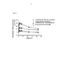
[0013] A figura 1 mostra a mudança temporal na quantidade de um anticorpo retido na sola de um modelo de camundongo de artrite induzida por colágeno. Descrição das Modalidades Aqui abaixo, a presente invenção será descrita em detalhes.[0013] Figure 1 shows the temporal change in the amount of an antibody retained in the sole of a mouse model of collagen-induced arthritis. Description of Embodiments Here below, the present invention will be described in detail.
[0014] Os presentes inventores repetiram o exame criativo para preparar um anticorpo anti-NGF humano ou um fragmento de ligação a antígeno do mesmo. Como um resultado, eles tiveram sucesso na preparação de um fragmento Fab’ de anticorpo anti-NGF humano que é excelente em segurança ao se reduzir o risco de efeitos colaterais tal como os efeitos em um feto e formação de trombo ao mesmo tempo em que mantém alta atividade de neutralização.[0014] The present inventors repeated the creative examination to prepare an anti-human NGF antibody or an antigen-binding fragment thereof. As a result, they have succeeded in preparing a Fab' fragment of an anti-NGF antibody that is excellent in safety in reducing the risk of side effects such as effects on a fetus and thrombus formation while maintaining high neutralizing activity.
[0015] A estrutura básica de uma molécula de anticorpo é comum entre as respectivas classes de anticorpo e é constituída com uma cadeia pesada tendo um peso molecular de 50000 a 70000 e uma cadeia leve tendo um peso molecular de 20000 a 30000. A cadeia pesada em geral consiste em uma cadeia de polipeptídeo incluindo cerca de 440 aminoácidos, e cada classe tem as suas estruturas características. As cadeias pesadas são chamadas de cadeias Y, μ, α, δ, e ε que correspondem a IgG, IgM, IgA, IgD, e IgE. Adicionalmente, IgG tem subclasses tais como IgG1, IgG2, IgG3, e IgG4, e as referidas cadeias são chamadas y1, y2, y3, e y4 respectivamente. Uma cadeia leve em geral consiste em uma cadeia de polipeptídeo incluindo cerca de 220 aminoácidos, e dois tipos de cadeia leve incluindo uma tipo-L e uma tipo-K são conhecidas, as quais são chamadas cadeias X e K respectivamente. Em relação à constituição do peptídeo da estrutura básica de uma molécula de anticorpo, duas cadeias homólogas pesadas e duas cadeias homólogas leves são ligadas através de ligações dissulfeto (S-S ligaçãos) e ligações não covalentes ligaçãos, e o peso molecular do mesmo é 150000 a 190000. Os dois tipos de cadeias leves podem ser emparelhados a qualquer cadeia pesada. Cada molécula de anticorpo sempre consiste em duas cadeias leves idênticas e duas cadeias pesadas idênticas.[0015] The basic structure of an antibody molecule is common among the respective antibody classes and consists of a heavy chain having a molecular weight of 50000 to 70000 and a light chain having a molecular weight of 20000 to 30000. The heavy chain in general it consists of a polypeptide chain comprising about 440 amino acids, and each class has its characteristic structures. The heavy chains are called Y, μ, α, δ, and ε chains which correspond to IgG, IgM, IgA, IgD, and IgE. Additionally, IgG has subclasses such as IgG1, IgG2, IgG3, and IgG4, and said chains are called y1, y2, y3, and y4 respectively. A light chain generally consists of a polypeptide chain comprising about 220 amino acids, and two types of light chain including an L-type and a K-type are known, which are called X and K chains respectively. Regarding the peptide constitution of the basic structure of an antibody molecule, two homologous heavy chains and two homologous light chains are linked through disulfide bonds (S-S bonds) and non-covalent bonds, and the molecular weight of the same is 150000 to 190000 The two types of light chains can be paired with any heavy chain. Each antibody molecule always consists of two identical light chains and two identical heavy chains.
[0016] Existem quatro ligações intracadeia S-S em uma cadeia pesada (cinco ligações para cadeias μ e ε) e duas em uma cadeia leve. Um anel é formado por 100 a 110 resíduos aminoácidos, e esta estrutura estérica é similar entre os respectivos anéis e é chamada de uma unidade estrutural ou um domínio. Para ambas as cadeias leve ou pesada, a sequência de aminoácido do domínio posicionado no terminal N do mesmo não é constante, até mesmo na referência padrão a partir da mesma classe (subclasse) das mesmas espécies animais, e esse domínio é chamado de região variável. Cada um dos domínios é chamado uma região variável de cadeia pesada (VH) e uma região variável de cadeia leve (VL) respectivamente. Desde que a sequência de aminoácido do lado C terminal a partir do domínio seja quase constante em cada classe ou subclasse, essa região é chamada de região constante, e cada um dos domínios é descrito as CH1, CH2, CH3 e CL, respectivamente.[0016] There are four S-S intrachain bonds in a heavy chain (five bonds for μ and ε chains) and two in a light chain. A ring is formed by 100 to 110 amino acid residues, and this steric structure is similar between the respective rings and is called a structural unit or a domain. For both light and heavy chains, the amino acid sequence of the domain positioned at the N-terminus of the same is not constant, even in the standard reference from the same class (subclass) of the same animal species, and this domain is called a variable region. . Each of the domains is called a heavy chain variable region (VH) and a light chain variable region (VL) respectively. Since the C-terminal amino acid sequence from the domain is nearly constant in each class or subclass, this region is called a constant region, and each of the domains is described as CH1, CH2, CH3, and CL, respectively.
[0017] O campo determinante antigênico de um anticorpo é constituído com VH e VL, e a especificidade de ligação depende da sequência de aminoácidos deste campo. Por outro lado, as atividades biológicas tais como a ligação aos complementos ou a várias células reflete as diferenças na estrutura de região constante entre as várias classes de Ig. Sabe-se que a diversidade nas regiões variáveis da cadeia pesada e da cadeia leve é primordialmente limitada a três pequenas regiões hiperdiversificadas presentes em ambas cadeias, e as referidas regiões são chamadas complementariamente de regiões determinantes (CDRs; CDR1, CDR2 e CDR3 iniciando-se a partir do lado terminal N). A parte restante da região variável é chamada de região de estrutura (FR) e é relativamente constante.[0017] The antigenic determinant field of an antibody consists of VH and VL, and the binding specificity depends on the amino acid sequence of this field. On the other hand, biological activities such as binding to complements or to various cells reflect differences in constant region structure between the various classes of Ig. It is known that the diversity in the variable regions of the heavy chain and the light chain is primarily limited to three small hyperdiversified regions present in both chains, and these regions are called complementary determinant regions (CDRs; CDR1, CDR2 and CDR3 starting from from the N-terminal side). The remaining part of the variable region is called the framework region (FR) and is relatively constant.
[0018] A região entre o domínio CH1 e o domínio CH2 da região constante de cadeia pesada de um anticorpo é chamada de uma região articulada. Essa região inclui diversos resíduos de prolina e tem uma pluralidade de ligações intracadeia S-S em conexão a duas cadeias pesadas. Por exemplo, cada região de articulação humana IgG1, IgG2, IgG3, e IgG4 inclui 2, 4, 11, e 2 resíduos de cisteína respectivamente o que constitui as ligações de intracadeias pesadas S-S. A região de articulação é uma região altamente sensível a uma enzima proteolítica tal como a papaína ou pepsina. Quando um anticorpo é digerido com papaína, sua cadeia pesada é clivada em uma posição mais próxima ao lado do terminal N do que da região de articulação da ligação da intracadeia pesada S-S, na qual o anticorpo é dividido em dois fragmentos Fab e um fragmento Fc. O fragmento Fab é constituído por um fragmento de cadeia leve e um fragmento de cadeia pesada incluindo uma região variável de cadeia pesada (VH), um domínio CH1, e uma porção da região de articulação. Quando um anticorpo é digerido com pepsina, sua cadeia pesada é clivada em uma posição mais próxima ao lado terminal C do que da ligação de região articulada da intracadeia S-S, na qual os fragmentos F(ab)2 são gerados. O fragmento F(ab)2 é um fragmento tendo uma estrutura dimérica na qual dois fragmentos Fab ligam-se um ao outro através da ligação da intracadeia pesada S-S na região de articulação. O fragmento Fab é constituído de uma cadeia leve e um fragmento de uma cadeia pesada incluindo uma região variável de cadeia pesada (VH), um domínio CH1, e uma porção da região de articulação. Os resíduos de Cisteína que constituem a ligação da intracadeia pesada S-S são incluídos na porção da região de articulação. Todos os fragmentos Fab, fragmentos F(ab’)2, e fragmento Fab’ incluem uma região variável e têm atividade de ligação antígeno.[0018] The region between the CH1 domain and the CH2 domain of the heavy chain constant region of an antibody is called a hinge region. This region includes several proline residues and has a plurality of S-S intrachain bonds in connection with two heavy chains. For example, each IgG1, IgG2, IgG3, and IgG4 human hinge region includes 2, 4, 11, and 2 cysteine residues respectively which constitute S-S intra-heavy chain bonds. The hinge region is a region highly sensitive to a proteolytic enzyme such as papain or pepsin. When an antibody is digested with papain, its heavy chain is cleaved closer to the N-terminal side than the S-S intra heavy chain binding hinge region, in which the antibody is split into two Fab fragments and an Fc fragment. . The Fab fragment is comprised of a light chain fragment and a heavy chain fragment including a heavy chain variable region (VH), a CH1 domain, and a hinge region portion. When an antibody is digested with pepsin, its heavy chain is cleaved closer to the C-terminal side than the hinge region of the S-S intrachain, in which F(ab)2 fragments are generated. The F(ab) 2 fragment is a fragment having a dimeric structure in which two Fab fragments bind to each other through S-S intra heavy chain binding at the hinge region. The Fab fragment is made up of a light chain and a fragment of a heavy chain including a heavy chain variable region (VH), a CH1 domain, and a hinge region portion. Cysteine residues constituting the S-S intra heavy chain linkage are included in the hinge region portion. All Fab fragments, F(ab')2 fragments, and Fab' fragment all include a variable region and have antigen binding activity.
[0019] O fragmento Fab’ de anticorpo anti-NGF humano da presente invenção que os presentes inventores prepararam com sucesso é um fragmento Fab’ tendo as seguintes características.[0019] The Fab' fragment of anti-human NGF antibody of the present invention which the present inventors have successfully prepared is a Fab' fragment having the following characteristics.
[0020] O fragmento Fab’ de anticorpo anti-NGF humano compreende uma região variável de cadeia pesada que consiste em uma sequência de aminoácidos mostrada por SEQ ID NO:6 e uma região variável de cadeia leve que consiste em uma sequência de aminoácidos mostrada por SEQ ID NO:4.[0020] The human anti-NGF antibody Fab' fragment comprises a heavy chain variable region consisting of an amino acid sequence shown by SEQ ID NO:6 and a light chain variable region consisting of an amino acid sequence shown by SEQ ID NO:4.
[0021] Especificamente, os presentes inventores construíram anticorpos usando a tecnologia de desenvolvimento de anticorpo humano monoclonal, o camundongo “VelocImune” [tecnologia de anticorpo VelocImune; Regeneron Inc. (U.S. Patent No. 6596541)], e rastrearam os anticorpos usando ensaios para várias atividades biológicas e propriedades físicas, conseguindo desta forma identificar o fragmento Fab’ de anticorpo anti-NGF humano da presente invenção. Na tecnologia VelocImune, os camundongos transgênicos nos quais as regiões variáveis das cadeias pesada e leve de imunoglobulina endógena são substituídas por regiões variáveis são desafiados com o antígeno de interesse (por exemplo, humana βNGF), e células linfáticas são recuperadas a partir dos camundongos que expressam anticorpos. As células linfáticas são fundidas com as células de mieloma de camundongo para preparar hibridomas. As células de hibridoma são rastreadas para identificar as células de hibridoma que produzem aqueles anticorpos que especificamente ligam-se ao antígeno de interesse. Os anticorpos que são produzidos aqui são anticorpos tendo as regiões variáveis de anticorpos humanos e as constantes regiões de anticorpos de camundongo (também chamados de anticorpos quiméricos). Então, se o anticorpo que se liga especificamente ao antígeno de interesse e tem uma atividade desejada de neutralização é identificado, o DNAs que codifica as regiões variáveis da cadeia pesada e da cadeia leve do anticorpo são isolados a partir das células de hibridoma e ligados ao DNAs codificando as constantes regiões da cadeia pesada e da cadeia leve de uma almejada classe de anticorpo humano. O gene resultante codificando a cadeia pesada e a cadeia leve do anticorpo é mostrada em células (e.g., células CHO) para produzir uma molécula de anticorpo. A cadeia pesada e a cadeia leve do anticorpo produzidas pelo método acima são a cadeia pesada e a cadeia leve de um anticorpo “totalmente humano” derivado a partir de um gene de imunoglobulina humano.[0021] Specifically, the present inventors have constructed antibodies using human monoclonal antibody development technology, the mouse "VelocImune" [VelocImune antibody technology; Regeneron Inc. (U.S. Patent No. 6,596,541)], and screened the antibodies using assays for various biological activities and physical properties, thereby managing to identify the Fab' fragment of human anti-NGF antibody of the present invention. In VelocImune technology, transgenic mice in which the variable regions of the endogenous immunoglobulin heavy and light chains are replaced by variable regions are challenged with the antigen of interest (e.g., human βNGF), and lymph cells are recovered from the mice that express antibodies. Lymphatic cells are fused with mouse myeloma cells to prepare hybridomas. Hybridoma cells are screened to identify hybridoma cells that produce those antibodies that specifically bind to the antigen of interest. The antibodies that are produced here are antibodies having the variable regions of human antibodies and the constant regions of mouse antibodies (also called chimeric antibodies). Then, if the antibody that specifically binds to the antigen of interest and has a desired neutralizing activity is identified, the DNAs encoding the heavy chain and light chain variable regions of the antibody are isolated from the hybridoma cells and ligated to the antibody. DNAs encoding the constant heavy and light chain regions of a targeted class of human antibody. The resulting gene encoding the antibody heavy chain and light chain is shown in cells (e.g., CHO cells) to produce an antibody molecule. The antibody heavy chain and light chain produced by the above method are the heavy chain and light chain of a "fully human" antibody derived from a human immunoglobulin gene.
[0022] O fragmento Fab’ de anticorpo anti-NGF humano da presente invenção pode ser facilmente preparado por aqueles peritos nesta ciência nas bases das informações em sequência na região variável da cadeia pesada e da cadeia leve das mesmas aqui mostradas, usando um método usualmente conhecido nesta ciência. Preferivelmente, o fragmento Fab’ de anticorpo anti-NGF humano da presente invenção pode ser preparado como um fragmento Fab de anticorpo totalmente humano pela ligação da região variável de cadeia pesada e região variável de cadeia leve do mesmo a uma parte da região constante de cadeia pesada (a qual inclui o domínio CH1 e uma parte da região de articulação da região incluindo região de articulação de cisteína) e região constante de cadeia leve de um anticorpo humano, respectivamente. Especificamente, um fragmento de gene de uma região variável de cadeia pesada tendo por bAse uma sequência que codifica o aminoácido da região variável de cadeia pesada do fragmento Fab’ da presente invenção (SEQ ID NO:6), e um fragmento de gene de uma região variável de cadeia leve tendo por base uma sequência que codifica o aminoácido da região variável da cadeia leve do fragmento Fab’ da presente invenção (SEQ ID NO:4) são preparados. Então, os genes da região variável da cadeia pesada e da cadeia leve são ligados a cada gene da parte da região constante de cadeia pesada e da região constante de cadeia leve em uma apropriada classe de anticorpo humano para preparar um gene de fragmento Fab de anticorpo totalmente humano. Posteriormente, este gene é ligado a um vetor apropriado de expressão e introduzido em uma célula cultivada. Finalmente, esta célula cultivada é cultivada, permitindo que um fragmento monoclonal Fab’ possa ser obtido a partir de uma cultura flutuante.[0022] The human anti-NGF antibody Fab' fragment of the present invention can be readily prepared by those skilled in the art on the basis of sequence information in the heavy chain and light chain variable region thereof shown herein, using a method usually known in this science. Preferably, the anti-human NGF antibody Fab' fragment of the present invention can be prepared as a fully human antibody Fab fragment by ligating the heavy chain variable region and light chain variable region thereof to a part of the chain constant region. heavy (which includes the CH1 domain and a part of the hinge region including the cysteine hinge region) and light chain constant region of a human antibody, respectively. Specifically, a gene fragment of a heavy chain variable region having as a bAse a sequence encoding the heavy chain variable region amino acid of the Fab' fragment of the present invention (SEQ ID NO:6), and a gene fragment of a light chain variable region based on a sequence encoding the light chain variable region amino acid of the Fab' fragment of the present invention (SEQ ID NO:4) are prepared. Then, the heavy chain and light chain variable region genes are ligated to each heavy chain constant region part and light chain constant region part gene in an appropriate class of human antibody to prepare an antibody Fab fragment gene. fully human. Subsequently, this gene is linked to an appropriate expression vector and introduced into a cultured cell. Finally, this cultured cell is cultured, allowing a monoclonal Fab' fragment to be obtained from a floating culture.
[0023] Os fragmentos de gene que codificam os aminoácidos da região variável da cadeia pesada e da cadeia leve do fragmento Fab’ da presente invenção podem ser sintetizados usando um método de síntese de genes conhecidos nesta ciência, nas bases de, por exemplo, sequências de base designadas baseadas na sequência de aminoácidos da região variável da cadeia pesada e da região variável da cadeia leve. Exemplos deste método de síntese de genes incluem vários métodos conhecidos daqueles peritos nesta ciência, tal como o método de síntese de gene de anticorpo descrito em WO90/07861.[0023] The gene fragments encoding the heavy chain and light chain variable region amino acids of the Fab' fragment of the present invention can be synthesized using a gene synthesis method known in the art, on the basis of, for example, sequences named bases based on the amino acid sequence of the heavy chain variable region and the light chain variable region. Examples of this gene synthesis method include various methods known to those skilled in the art, such as the antibody gene synthesis method described in WO90/07861.
[0024] Então, os fragmentos de gene da região variável descritos acima são ligados ao gene de região constante de um anticorpo humano para preparar um gene de fragmento Fab’ totalmente humano. Embora qualquer subclasse da região constante (por exemplo, a região constante de uma cadeia pesada tal como a cadeia Yl, Y2, Y3 ou Y4, ou a região constante de uma cadeia leve tal como À ou K cadeia) possa ser escolhida como a região constante de anticorpo humano, humano IgYl como a região constante de cadeia pesada, e humano Igk como a região constante de cadeia leve, pode preferivelmente ser usado.[0024] Then, the variable region gene fragments described above are ligated into the constant region gene of a human antibody to prepare a fully human Fab' fragment gene. While any subclass of the constant region (e.g., the constant region of a heavy chain such as the Y1, Y2, Y3, or Y4 chain, or the constant region of a light chain such as the A or K chain) can be chosen as the region human antibody constant, human IgY1 as the heavy chain constant region, and human Igk as the light chain constant region, can preferably be used.
[0025] Posterior à preparação deste fragmento Fab de anticorpo de gene totalmente humano, a introdução de gene em um vetor de expressão, a introdução do vetor de expressão em células cultivadas, o cultivo de células cultivadas, a purificação do fragmento Fab’ e similar podem ser executados usando vários métodos conhecidos nesta ciência.[0025] Subsequent to the preparation of this fully human gene antibody Fab fragment, the introduction of the gene into an expression vector, the introduction of the expression vector into cultured cells, the cultivation of cultured cells, the purification of the Fab' fragment and the like can be performed using various methods known in this science.
[0026] Exemplos do vetor de expressão que está ligado ao gene assim obtido incluem o vetor GS pEE6.4 ou pEE12.4 (Lonza Biologics), mas não são especificamente limitados, de forma que possam expressar tal gene de anticorpo. Também, pode ser usado um vetor de expressão já tendo um gene de região constante de forma humana Ig tal como AG-Y1 ou AG-k (por exemplo, ver WO94/20632).[0026] Examples of the expression vector that is linked to the gene thus obtained include the GS vector pEE6.4 or pEE12.4 (Lonza Biologics), but are not specifically limited so that they can express such an antibody gene. Also, an expression vector already having a human form Ig constant region gene such as AG-Y1 or AG-k (for example, see WO94/20632) can be used.
[0027] O vetor de expressão acima descrito é introduzido nas células cultivadas por, por exemplo, um método de fosfato de cálcio ou um método de eletroporação e similares.[0027] The above-described expression vector is introduced into cultured cells by, for example, a calcium phosphate method or an electroporation method and the like.
[0028] Exemplos de células cultivadas nas quais o vetor de expressão é introduzido incluem células cultivadas tais como as células CHO-K1SV, células CHO- DG44 e 293 células, e as referidas células podem ser cultivadas por um método convencional.[0028] Examples of cultured cells into which the expression vector is introduced include cultured cells such as CHO-K1SV cells, CHO-DG44 cells and 293 cells, and said cells may be cultured by a conventional method.
[0029] O fragmento Fab’ acumulado na cultura flutuante após cultivar o descrito acima pode ser purificado por vários tipos de cromatografia de coluna. Por exemplo, é possível usar cromatografia de coluna usando Kappaselect ou similar.[0029] The Fab' fragment accumulated in the floating culture after culturing the above can be purified by various types of column chromatography. For example, it is possible to use column chromatography using Kappaselect or similar.
[0030] O fragmento Fab’ da presente invenção pode ser preparado usando um método de expressão recombinante as descrito acima. Entretanto, o fragmento Fab’ pode ser preparado através da realização da digestão de pepsina após a preparação de um anticorpo inteiro primeiro, e tratar o obtido fragmento F(ab’)2 com um agente redutor tal como o 2-mercaptoetanol.[0030] The Fab' fragment of the present invention can be prepared using a recombinant expression method as described above. Meanwhile, the Fab' fragment can be prepared by performing pepsin digestion after preparing a full-length antibody first, and treating the obtained F(ab') 2 fragment with a reducing agent such as 2-mercaptoethanol.
[0031] Preferivelmente, o fragmento Fab’ de anticorpo anti-NGF humano da presente invenção pode ser facilmente obtido através da sintetização do DNA compreendendo uma sequência de base codificando a sequência de aminoácidos da região variável da cadeia pesada mostrada por SEQ ID NO:6 e DNA compreendendo a sequência de base codificando a sequência de aminoácidos da região variável da cadeia leve mostrada por SEQ ID NO:4, e ligando o DNAs a uma classe adequada de genes de anticorpo humano, preferivelmente um gene da região constante IgYl humano para a cadeia pesada e um gene da região constante IgK humano para a cadeia leve, para construir um gene de fragmento Fab de anticorpo totalmente humano usando um método conhecido nesta ciência, e introduzindo o gene em um vetor de expressão, introduzindo o vetor de expressão em uma célula cultivada, cultivando a célula cultivada, e purificando um fragmento Fab’ colhido a partir da célula cultivada usando vários métodos conhecidos nesta ciência. Preferivelmente, o DNA compreendendo a sequência de base de aminoácidos codificando a região variável da cadeia pesada mostradas por SEQ ID NO:6 compreende as sequências de base mostradas por SEQ ID NO:5. Preferivelmente, o DNA compreendendo a sequência de base codificando a sequência de aminoácidos da região variável da cadeia leve mostradas por SEQ ID NO:4 compreende as sequências de base mostradas por SEQ ID NO:3.[0031] Preferably, the Fab' fragment of anti-human NGF antibody of the present invention can be readily obtained by synthesizing DNA comprising a base sequence encoding the heavy chain variable region amino acid sequence shown by SEQ ID NO:6 and DNA comprising the base sequence encoding the light chain variable region amino acid sequence shown by SEQ ID NO:4, and linking the DNAs to a suitable class of human antibody genes, preferably a human IgY1 constant region gene for the heavy chain and a human IgK constant region gene for the light chain, to construct a fully human antibody Fab fragment gene using a method known in the art, and introducing the gene into an expression vector, introducing the expression vector into a cultured cell, culturing the cultured cell, and purifying a Fab' fragment harvested from the cultured cell using various methods known in the art. Preferably, the DNA comprising the base sequence of amino acids encoding the heavy chain variable region shown by SEQ ID NO:6 comprises the base sequences shown by SEQ ID NO:5. Preferably, the DNA comprising the base sequence encoding the light chain variable region amino acid sequence shown by SEQ ID NO:4 comprises the base sequences shown by SEQ ID NO:3.
[0032] Na presente especificação, o “fragmento Fab” se refere a um fragmento de anticorpo monovalente constituído com um fragmento da cadeia leve e da cadeia pesada incluindo uma região variável de cadeia pesada (VH), um domínio CH1, e uma porção da região de articulação. Na porção da região de articulação, pelo menos um resíduo de cisteína é incluído (também chamada a “a cisteína da região de articulação” na presente especificação) diferente dos resíduos de cisteína constituindo a ligação S-S, entre a cadeia pesada e a cadeia leve. A cisteína da região de articulação pode ser usada como um campo de modificação de polietilenoglicol descrito posteriormente. O número de cisteínas da região de articulação no fragmento Fab’ é variável dentro de uma gama e a partir de 1 a diversos resíduos de cisteína dependendo da classe de um anticorpo usado, e é facilmente ajustável por um perito qualificado nesta ciência. Por exemplo, quando um fragmento Fab’ de uma classe humana IgG1 (em geral tendo duas cisteínas da região de articulação na região de articulação) é preparado, o codão de terminação é inserido entre o campo de codificação da cisteína da primeira região de articulação e o campo de codificação da segunda cisteína da região de articulação na região de articulação da cadeia pesada, onde um fragmento Fab’ pode ser preparado tendo duas cisteínas da região de articulação na região de articulação. Adicionalmente, se um codão de terminação é inserido após o campo de codificação da cisteína da segunda região de articulação, um fragmento Fab’ pode ser preparado tendo duas cisteínas de região de articulação na região de articulação.[0032] In the present specification, the "Fab fragment" refers to a monovalent antibody fragment comprised of a light chain and heavy chain fragment including a heavy chain variable region (VH), a CH1 domain, and a portion of the articulation region. In the hinge region portion, at least one cysteine residue is included (also called "the hinge region cysteine" in the present specification) other than the cysteine residues constituting the S-S bond between the heavy chain and the light chain. Joint region cysteine can be used as a polyethylene glycol modification field described later. The number of hinge region cysteines in the Fab' fragment is variable within a range and from 1 to several cysteine residues depending on the class of an antibody used, and is easily adjustable by one skilled in the art. For example, when a Fab' fragment of a human IgG1 class (generally having two hinge region cysteines in the hinge region) is prepared, the stop codon is inserted between the cysteine coding field of the first hinge region and the second hinge region cystein coding field in the hinge region of the heavy chain, where a Fab' fragment can be prepared having two hinge region cysteines in the hinge region. Additionally, if a stop codon is inserted after the cysteine coding field of the second hinge region, a Fab' fragment can be prepared having two hinge region cysteines in the hinge region.
[0033] O fragmento preferível de cadeia pesada do fragmento Fab’ de anticorpo anti-NGF humano da presente invenção, compreendendo a região variável da cadeia pesada que consiste da sequência de aminoácidos mostrada por SEQ ID NO:6 e uma porção da região constante de IgYl humana, é um fragmento de cadeia pesada que consiste da sequência de aminoácidos mostrada por SEQ ID NO:10, SEQ ID NO:14, ou SEQ ID NO:16. Preferivelmente, o DNA compreendendo a sequência de base que codifica o fragmento de cadeia pesada do fragmento Fab’ de anticorpo anti- NGF humano que consiste da sequência de aminoácidos mostrada por SEQ ID NO:10, SEQ ID NO:14, ou SEQ ID NO:16 compreende a sequência de base mostrada por SEQ ID NO:9, SEQ ID NO:13, ou SEQ ID NO:15. A cadeia leve preferível do fragmento Fab’ de anticorpo anti-NGF humano da presente invenção, compreendendo a região variável da cadeia leve que consiste da sequência de aminoácidos mostrada por SEQ ID NO:4 e de uma região constante de IgK humana, é uma cadeia leve que consiste da sequência de aminoácidos mostrada por SEQ ID NO:12. Preferivelmente, o DNA compreendendo a sequência de base que codifica a cadeia leve do fragmento Fab’ de anticorpo anti-NGF humano que consiste na sequência de aminoácidos mostrada por SEQ ID NO:12 compreende a sequência de base mostrada por SEQ ID NO:11.[0033] The preferred heavy chain fragment of the Fab' fragment of human anti-NGF antibody of the present invention, comprising the heavy chain variable region consisting of the amino acid sequence shown by SEQ ID NO:6 and a portion of the constant region of Human IgY1, is a heavy chain fragment consisting of the amino acid sequence shown by SEQ ID NO:10, SEQ ID NO:14, or SEQ ID NO:16. Preferably, the DNA comprising the base sequence encoding the heavy chain fragment of the human anti-NGF antibody Fab' fragment consisting of the amino acid sequence shown by SEQ ID NO:10, SEQ ID NO:14, or SEQ ID NO :16 comprises the base sequence shown by SEQ ID NO:9, SEQ ID NO:13, or SEQ ID NO:15. The preferred light chain of the human anti-NGF antibody Fab' fragment of the present invention, comprising the light chain variable region consisting of the amino acid sequence shown by SEQ ID NO:4 and a human IgK constant region, is a light consisting of the amino acid sequence shown by SEQ ID NO:12. Preferably, the DNA comprising the base sequence encoding the light chain of the human anti-NGF antibody Fab' fragment consisting of the amino acid sequence shown by SEQ ID NO:12 comprises the base sequence shown by SEQ ID NO:11.
[0034] Como um fragmento Fab’ preferível de anticorpo anti-NGF humano da presente invenção que compreende o fragmento de cadeia pesada que consiste da sequência de aminoácidos mostrada por SEQ ID NO:10 e a cadeia leve que consiste da sequência de aminoácidos mostrada por SEQ ID NO:12, um 1-15(N52D) anticorpo de fragmento Fab’ totalmente humano é exemplificado descrito posteriormente em exemplos. Como um fragmento Fab’ de anticorpo anti-NGF humano preferível da presente invenção que compreende o fragmento de cadeia pesada que consiste da sequência de aminoácidos mostrada por SEQ ID NO:14 e a cadeia leve que consiste da sequência de aminoácidos mostrada por SEQ ID NO:12, um anticorpo totalmente humano de fragmento Fab’ 1-15 (N52D-A) descrito posteriormente em exemplos é exemplificado. Como um fragmento Fab’ de anticorpo anti-NGF humano preferível da presente invenção que compreende o fragmento de cadeia pesada que consiste da sequência de aminoácidos mostrada por SEQ ID NO:16 e a cadeia leve que consiste da sequência de aminoácidos mostrada por SEQ ID NO:12, um anticorpo de Fragmento Fab’ totalmente humano 1-15(N52D-P) descrito posteriormente em exemplos é exemplificado.[0034] As a preferable Fab' fragment of anti-human NGF antibody of the present invention which comprises the heavy chain fragment consisting of the amino acid sequence shown by SEQ ID NO:10 and the light chain consisting of the amino acid sequence shown by SEQ ID NO:12, a fully human 1-15(N52D) Fab' fragment antibody is exemplified described later in examples. As a preferred anti-human NGF antibody Fab' fragment of the present invention which comprises the heavy chain fragment consisting of the amino acid sequence shown by SEQ ID NO:14 and the light chain consisting of the amino acid sequence shown by SEQ ID NO: :12, a fully human Fab' 1-15 fragment antibody (N52D-A) described later in examples is exemplified. As a preferred anti-human NGF antibody Fab' fragment of the present invention which comprises the heavy chain fragment consisting of the amino acid sequence shown by SEQ ID NO:16 and the light chain consisting of the amino acid sequence shown by SEQ ID NO :12, a fully human Fab' Fragment antibody 1-15(N52D-P) described later in examples is exemplified.
[0035] A presente invenção também compreende um fragmento Fab’ de anticorpo anti-NGF humano que compreende a região variável da cadeia pesada compreendendo CDR1 que consiste da sequência de aminoácidos em posição a partir de 31 a 35 de SEQ ID NO: 6, CDR2 que consiste de sequência de aminoácidos em posição a partir de 50 a 65 de SEQ ID NO: 6, e CDR3 que consiste de sequência de aminoácidos em posição a partir de 98 a 110 de SEQ ID NO: 6, e a região variável da cadeia leve compreendendo CDR1 que consiste de sequência de aminoácidos em posição a partir de 24 a 39 de SEQ ID NO: 4, CDR2 que consiste de sequência de aminoácidos em posição a partir de 55 a 61 de SEQ ID NO: 4, e CDR3 que consiste de sequência de aminoácidos em posição a partir de 94 a 102 de SEQ ID NO: 4. O fragmento Fab’ pode ser também preparado por peritos nesta ciência de acordo com os procedimentos tal como aqueles descritos acima.[0035] The present invention also comprises an anti-human NGF antibody Fab' fragment comprising the heavy chain variable region comprising CDR1 consisting of the amino acid sequence at position from 31 to 35 of SEQ ID NO: 6, CDR2 consisting of amino acid sequence at position from 50 to 65 of SEQ ID NO: 6, and CDR3 consisting of amino acid sequence at position from 98 to 110 of SEQ ID NO: 6, and the chain variable region light comprising CDR1 consisting of amino acid sequence at position from 24 to 39 of SEQ ID NO: 4, CDR2 consisting of amino acid sequence at position from 55 to 61 of SEQ ID NO: 4, and CDR3 consisting of sequence of amino acids at position from 94 to 102 of SEQ ID NO: 4. The Fab' fragment can also be prepared by those skilled in the art according to procedures such as those described above.
[0036] O fragmento Fab’ de anticorpo anti-NGF humano da presente invenção pode ser modificado sendo conjugado a polietilenoglicol (PEG) através da cisteína da região de articulação do mesmo. O PEG pode ser conjugado ao fragmento Fab’ usando métodos conhecidos nesta ciência (por exemplo, EP0948544). Na presente invenção, o PEG linear ou ramificado tendo um peso arbitrário molecular médio ou ou um seu derivado, é utilizável, o qual pode ser facilmente selecionado por um perito nesta ciência de acordo com o uso pretendido. Por exemplo, em um tecido tumoral ou em um período de resposta inflamatória, a permeabilidade vascular é notadamente melhorada em comparação com um tecido normal, assim substâncias que atingem o tecido tendem a vazar para fora do vaso sanguíneo e se acumulam no tumor ou no tecido inflamatório (efeito EPR). Também se sabe que uma substância de baixo peso molecular é facilmente reabsorvida para dentro dos vasos sanguíneos e que uma substância de alto peso molecular não é facilmente reabsorvida. Portanto, de modo a melhorar a retenção do fragmento Fab’ em um tecido lesionado, o PEG tendo uma média de alto peso molecular (por exemplo, cerca de 40000 Da) pode ser conjugado a este fragmento. Quando se deseja que o fragmento Fab’ seja rapidamente excretado para fora do corpo, o PEG tendo uma média de baixo peso molecular (por exemplo, cerca de 10000 Da) pode ser conjugado a este fragmento. Adicionalmente, de modo a facilitar a ligação do PEG a uma cisteína da região de articulação, um derivado do PEG pode ser usado. Por exemplo, como descrito posteriormente em exemplos, é possível usar um derivado do PEG para o qual um grupo tiol-reativo, tal como maleimida foi ligado e ligar a um grupo tiol da cisteína da região de articulação para o grupo maleimida através de uma ligação covalente. Em geral, o peso molecular médio do PEG é alcançado a partir de cerca de 500 Da a cerca de 50000 Da, preferivelmente alcançado a partir de cerca de 5000 Da a cerca de 40000 Da e mais preferivelmente alcançado a partir de cerca de 10000 Da a cerca de 40000 Da.[0036] The anti-human NGF antibody Fab' fragment of the present invention can be modified by being conjugated to polyethylene glycol (PEG) through the cysteine of the hinge region thereof. The PEG can be conjugated to the Fab' fragment using methods known in the art (e.g. EP0948544). In the present invention, linear or branched PEG having an arbitrary weight average molecular weight or a derivative thereof is usable, which can be easily selected by one skilled in the art according to the intended use. For example, in a tumor tissue or in a period of inflammatory response, vascular permeability is markedly improved compared to normal tissue, so substances that reach the tissue tend to leak out of the blood vessel and accumulate in the tumor or tissue. inflammation (RPE effect). It is also known that a low molecular weight substance is easily reabsorbed into blood vessels and that a high molecular weight substance is not easily reabsorbed. Therefore, in order to improve retention of the Fab' fragment in an injured tissue, PEG having a high molecular weight average (e.g. about 40000 Da) can be conjugated to this fragment. When the Fab' fragment is desired to be rapidly excreted out of the body, PEG having a low average molecular weight (e.g., about 10,000 Da) can be conjugated to this fragment. Additionally, in order to facilitate the binding of the PEG to a cysteine of the hinge region, a derivative of the PEG can be used. For example, as described later in examples, it is possible to use a derivative of PEG to which a thiol-reactive group such as maleimide has been attached and link to a thiol group from the cysteine hinge region to the maleimide group via a linkage. covalent. In general, the average molecular weight of the PEG is from about 500 Da to about 50,000 Da, preferably from about 5000 Da to about 40,000 Da, and most preferably from about 10,000 Da to about 40000 Da.
[0037] O fragmento Fab’ de anticorpo anti-NGF humano da presente invenção se liga ao NGF humano. Como o método de medir a atividade de ligação do obtido fragmento Fab’ de anticorpo anti-NGF humano para o NGF humano, há um método tal como ELISA ou FACS. Por exemplo, quando ELISA é usado, o βNGF humano é imobilizado em uma placa de ELISA, o fragmento Fab’ é adicionado de forma a causar uma reação, e então um anticorpo secundário tal como um anticorpo anti-kappa marcado com uma enzima tal como peroxidade de rábano (HRP) é permitida a reação com a mistura própria. Após a placa ser lavada, a atividade é medida usando um reagente (por exemplo, um reagente cromogênico de TMB em um caso de rotulagem HRP) detectando a atividade, identificando, assim, a ligação do anticorpo secundário. Adicionalmente, o fragmento Fab’ de anticorpo anti-NGF humano da presente invenção também inclui um fragmento Fab’ que se liga ao NGF derivado a partir de outro animal (por exemplo, um camundongo NGF) assim como o NGF humano, assim a atividade de ligação com respeito à tal proteína pode ser medida.[0037] The anti-human NGF antibody Fab' fragment of the present invention binds to human NGF. As the method of measuring the binding activity of the obtained Fab' fragment of anti-human NGF antibody to human NGF, there is a method such as ELISA or FACS. For example, when ELISA is used, human βNGF is immobilized on an ELISA plate, the Fab' fragment is added in order to cause a reaction, and then a secondary antibody such as an anti-kappa antibody labeled with an enzyme such as horseradish peroxide (HRP) is allowed to react with the mixture itself. After the plate is washed, the activity is measured using a reagent (eg, a TMB chromogenic reagent in an HRP labeling case) detecting the activity, thereby identifying secondary antibody binding. Additionally, the human anti-NGF antibody Fab' fragment of the present invention also includes a Fab' fragment that binds to NGF derived from another animal (e.g., a mouse NGF) as well as human NGF, as well as the activity of binding with respect to such a protein can be measured.
[0038] O fragmento Fab’ de anticorpo anti-NGF humano da presente invenção tem uma atividade de neutralização com respeito ao NGF humano. Quando usado na presente especificação, o termo “atividade de neutralização” do fragmento Fab’ de anticorpo anti-NGF humano refere-se a uma atividade que inibe qualquer atividade biológica resultante a partir do NGF por se ligar ao NGF, e a atividade neutralizante pode ser avaliada usando um ou uma pluralidade de atividades biológicas de NGF como um índice. Exemplos de tal atividade de neutralização incluem a atividade inibidora contra a ligação de NGF à trkA que é o receptor de NGF, a atividade inibitória contra de cálcio intracelular mediado por um sinal de NGF-trkA, e a atividade inibidora contra o sinal de sobrevivência celular do dependente NGF. A atividade de neutralização pode ser avaliada usando métodos descritos posteriormente em exemplos.[0038] The anti-human NGF antibody Fab' fragment of the present invention has a neutralizing activity with respect to human NGF. When used in the present specification, the term "neutralizing activity" of the Fab' fragment of anti-human NGF antibody refers to an activity that inhibits any biological activity resulting from NGF by binding to NGF, and the neutralizing activity may be evaluated using one or a plurality of NGF biological activities as an index. Examples of such neutralizing activity include inhibitory activity against NGF binding to trkA which is the NGF receptor, inhibitory activity against intracellular calcium mediated by an NGF-trkA signal, and inhibitory activity against the cell survival signal. of the NGF dependent. Neutralizing activity can be evaluated using methods described later in examples.
[0039] De modo a avaliar mais especificamente o efeito do fragmento Fab’ de anticorpo anti-NGF humano da presente invenção, pode-se realizar um ensaio ao vivo. Por exemplo, como descrito posteriormente em exemplos, a eficácia do fármaco ao vivo do fragmento Fab’ pode ser avaliada por um efeito de ensaio analgésico ou similar que usa um modelo de artrite em camundongo. Também é possível avaliar o efeito de retenção no tecido lesionado usando um ensaio para distribuição apropriada dentro da lesão.[0039] In order to more specifically assess the effect of the Fab' fragment of anti-human NGF antibody of the present invention, a live assay can be performed. For example, as described later in examples, the efficacy of the Fab' fragment in vivo drug can be evaluated by an analgesic effect or similar assay using a mouse model of arthritis. It is also possible to assess the retention effect on injured tissue using an assay for proper distribution within the lesion.
[0040] Posteriormente, o fragmento Fab’ de anticorpo anti-NGF humano da presente invenção pode também ser avaliado em termos do risco de efeitos colaterais. Por exemplo, como descrito posteriormente em exemplos, usando um ensaio de transferência placentária realizado após a administração do fragmento Fab’ em animais grávidas, é possível avaliar a possibilidade na qual o fragmento Fab’ de anticorpo anti-NGF humano da presente invenção pode exercer efeitos em um feto. Adicionalmente, como descrito posteriormente em exemplos, o tamanho de um imunocomplexo (IC) formado entre o fragmento Fab’ de anticorpo anti-NGF humano da presente invenção e o NGF é medido através de um ensaio para a formação de IC com NGF, de modo que a possibilidade da indução da formação de trombos possa ser avaliada.[0040] Thereafter, the Fab' fragment of anti-human NGF antibody of the present invention can also be evaluated in terms of the risk of side effects. For example, as described later in examples, using a placental transfer assay performed after the administration of the Fab' fragment to pregnant animals, it is possible to assess the possibility in which the Fab' fragment of anti-human NGF antibody of the present invention may exert effects. in a fetus. Additionally, as described later in examples, the size of an immunocomplex (IC) formed between the Fab' fragment of human anti-NGF antibody of the present invention and NGF is measured by an assay for IC formation with NGF, so that the possibility of inducing thrombus formation can be evaluated.
[0041] Adicionalmente, como o método de avaliação de vários tipos de estabilidade (por exemplo, estabilidade termal, estabilidade de armazenamento de longo prazo, e estabilidade de alta concentração) do fragmento Fab’ de anticorpo anti- NGF humano da presente invenção, são exemplificados um método usando a calorimetria de escaneamento diferencial e um método que meça a formação de agregados durante a armazenagem.[0041] Additionally, as the method of evaluating various types of stability (e.g., thermal stability, long-term storage stability, and high concentration stability) of the Fab' fragment of human anti-NGF antibody of the present invention are exemplified a method using differential scanning calorimetry and a method that measures aggregate formation during storage.
[0042] O fragmento Fab’ de anticorpo anti-NGF humano da presente invenção é opcionalmente purificado e então formulado de acordo com métodos comuns, e pode ser usado para tratar dor tais como a dor de osteoartrite (OA dor), dor reumática, dor de câncer, dor neuropática, dor lombar crônica, dor pós-operatória, neuralgia pós- herpética, neuropatia diabética dolorosa, dor de fratura, e síndrome da bexiga dolorosa e doenças nas quais o NGF está envolvido na formação de condições patológicas, tais como a cistite intersticial, a pancreatite aguda, a pancreatite crônica e a endometriose.[0042] The human anti-NGF antibody Fab' fragment of the present invention is optionally purified and then formulated according to common methods, and can be used to treat pain such as osteoarthritis pain (OA pain), rheumatic pain, pain of cancer, neuropathic pain, chronic low back pain, postoperative pain, post-herpetic neuralgia, painful diabetic neuropathy, fracture pain, and painful bladder syndrome and diseases in which NGF is involved in the formation of pathological conditions, such as interstitial cystitis, acute pancreatitis, chronic pancreatitis and endometriosis.
[0043] O fragmento Fab’ de anticorpo anti-NGF humano da presente invenção pode ser usado preferivelmente como um agente para tratar dor e mais preferencialmente como um agente para tratar dor de osteoartrite. Exemplos da formulação deste agente de tratamento e similares incluem formulações parentéricas tais como agentes injetáveis e de infusão, que são preferivelmente administradas por meio intravenoso, subcutâneo e similares. No processo de formulação, transportadores ou aditivos que correspondem a estas formulações podem ser usados dentro de uma gama farmaceuticamente aceitável.[0043] The anti-human NGF antibody Fab' fragment of the present invention can be used preferably as an agent to treat pain and more preferably as an agent to treat osteoarthritis pain. Examples of the formulation of this treatment agent and the like include parenteral formulations such as injectable and infusion agents, which are preferably administered intravenously, subcutaneously, and the like. In the formulation process, carriers or additives corresponding to these formulations can be used within a pharmaceutically acceptable range.
[0044] A quantidade de inventivo de fragmento Fab’ de anticorpo anti-NGF humano adicionada na formulação acima descrita varia dependendo da seriedade do sintoma do paciente ou da idade, da forma da dose da formulação usada ou da titulação da ligação do anticorpo e similares; por exemplo, cerca de 0.001 mg/kg a 100 mg/kg do anticorpo pode ser usado.[0044] The amount of inventive anti-human NGF antibody Fab' fragment added in the above-described formulation varies depending on the severity of the patient's symptom or age, the dose form of the formulation used or the antibody binding titration and the like ; for example, about 0.001 mg/kg to 100 mg/kg of the antibody can be used.
[0045] A presente invenção também proporciona um polinucleotídeo compreendendo uma sequência codificando o fragmento Fab’ de anticorpo anti-NGF humano da presente invenção, e um vetor de expressão compreendendo o mesmo. A presente invenção também proporciona um polinucleotídeo compreendendo a sequência codificando a região variável da cadeia pesada do fragmento Fab’ de anticorpo anti-NGF humano da presente invenção, e um polinucleotídeo compreendendo a sequência codificando a região variável da cadeia leve do fragmento Fab’ de anticorpo anti-NGF humano da presente invenção, e vetor de expressão compreendendo cada um ou ambos. O vetor de expressão da presente invenção não é especificamente limitado, contanto que possa expressar um gene que codifica o fragmento Fab’ da presente invenção ou sua região variável de cadeia pesada e/ou região variável de cadeia leve em várias células hospedeiras de células procariotas e/ou células eucariotas, e produzir os citados polipeptídeos. Exemplos do mesmo incluem vetores de plasmídeo, vetores virais (por exemplo, adenovírus, retrovírus) e similares. Preferivelmente, o vetor de expressão da presente invenção compreende um polinucleotídeo compreendendo ou uma sequência codificando o fragmento da cadeia pesada ou o fragmento da cadeia leve do acima descrito fragmento Fab’ da presente invenção, ou um polinucleotídeo compreendendo ambos a sequência codificando o fragmento da cadeia pesada do fragmento Fab’ da presente invenção e um polinucleotídeo compreendendo uma sequência codificando a cadeia leve do fragmento Fab’ da presente invenção.[0045] The present invention also provides a polynucleotide comprising a sequence encoding the Fab' fragment of human anti-NGF antibody of the present invention, and an expression vector comprising the same. The present invention also provides a polynucleotide comprising the sequence encoding the heavy chain variable region of the anti-human NGF antibody Fab' fragment of the present invention, and a polynucleotide comprising the sequence encoding the light chain variable region of the antibody Fab' fragment. anti-human NGF of the present invention, and expression vector comprising each or both. The expression vector of the present invention is not specifically limited as long as it can express a gene encoding the Fab' fragment of the present invention or its heavy chain variable region and/or light chain variable region in various prokaryotic cell host cells and /or eukaryotic cells, and produce said polypeptides. Examples thereof include plasmid vectors, viral vectors (e.g. adenovirus, retrovirus) and the like. Preferably, the expression vector of the present invention comprises a polynucleotide comprising either a sequence encoding the heavy chain fragment or the light chain fragment of the above-described Fab' fragment of the present invention, or a polynucleotide comprising both the sequence encoding the chain fragment. Fab' fragment of the present invention and a polynucleotide comprising a sequence encoding the light chain of the Fab' fragment of the present invention.
[0046] O vetor de expressão da presente invenção pode incluir um gene que codifica o fragmento Fab’ de anticorpo anti-NGF humano da presente invenção, ou um gene que codifica a região variável da cadeia pesada e/ou a região variável da cadeia leve do fragmento Fab’ de anticorpo anti-NGF humano da presente invenção, e um promotor operacionalmente ligado ao gene. Exemplos de um promotor para expressar um gene codificando o fragmento Fab’ da presente invenção ou sua região variável da cadeia pesada e/ou sua região variável da cadeia leve em uma bactéria incluem o promotor Trp, um promotor lac, um promotor recA, um promotor XPL, um promotor lpp, um promotor tac e similares, quando o hospedeira é uma bactéria do gênero Escherichia. Exemplos de um promotor para expressão em levedura incluem um promotor PH05, um promotor PGK, um promotor GAP e um promotor ADH, e alguns exemplos de um promotor para expressão no gênero Bacillus incluem promotor SL01, promotor SP02, promotor penP e similares. Quando a hospedeira é uma célula eucariótica tal como uma célula de mamífero, os exemplos do promotor incluem o promotor de SV40-derivada, promotor de retrovírus, promotor de choque térmico e similares.[0046] The expression vector of the present invention may include a gene encoding the Fab' fragment of human anti-NGF antibody of the present invention, or a gene encoding the heavy chain variable region and/or the light chain variable region. of the human anti-NGF antibody Fab' fragment of the present invention, and a promoter operably linked to the gene. Examples of a promoter for expressing a gene encoding the Fab' fragment of the present invention or its heavy chain variable region and/or its light chain variable region in a bacterium include the Trp promoter, a lac promoter, a recA promoter, a promoter XPL, an lpp promoter, a tac promoter, and the like, when the host is a bacterium of the genus Escherichia. Examples of a promoter for expression in yeast include a PH05 promoter, a PGK promoter, a GAP promoter and an ADH promoter, and some examples of a promoter for expression in the genus Bacillus include SL01 promoter, SP02 promoter, penP promoter and the like. When the host is a eukaryotic cell such as a mammalian cell, examples of the promoter include the SV40-derived promoter, retrovirus promoter, heat shock promoter and the like.
[0047] Quando uma bactéria, particularmente a Escherichia coli, é usada como a célula hospedeira, o vetor de expressão da presente invenção pode ainda incluir um codão de iniciação, um codão de terminação, uma região terminadora e uma unidade reaplicável. Quando levedura, uma célula animal ou célula de inseto é usada como a hospedeira, o vetor de expressão da presente invenção pode incluir um codão de iniciação e um codão de terminação. Neste caso, pode incluir uma sequência de facilitador, em regiões de não codificação no lado 5’ e no lado 3’ de um gene que codifica o fragmento Fab’ da presente invenção ou a região variável da cadeia pesada ou a região variável da cadeia leve do mesmo, uma sequência de sinal de secreção, uma junção de reparação, uma região de poliadenilação, uma unidade reaplicável ou similares. Além disso, o mesmo pode incluir um marcador de seleção que é de uso comum (por exemplo, gene de resistência à tetraciclina, gene de resistência à ampicilina, gene de resistência à canamicina, gene de resistência à neomicina, gene de redutase diidrofóico ácido) de acordo com o uso pretendido .[0047] When a bacterium, particularly Escherichia coli, is used as the host cell, the expression vector of the present invention may further include an initiation codon, a termination codon, a terminator region, and a reapplicable unit. When yeast, an animal cell or insect cell is used as the host, the expression vector of the present invention may include an initiation codon and a termination codon. In this case, it may include an enhancer sequence, in non-coding regions on the 5' side and on the 3' side of a gene encoding the Fab' fragment of the present invention or the heavy chain variable region or the light chain variable region thereof, a secretion signal sequence, a repair junction, a polyadenylation region, a reapplicable unit, or the like. In addition, it may include a selection marker that is in common use (e.g. tetracycline resistance gene, ampicillin resistance gene, kanamycin resistance gene, neomycin resistance gene, dihydrophoic acid reductase gene) according to the intended use.
[0048] A presente invenção também proporciona um transformante introduzido com o gene codificando o fragmento Fab’ de anticorpo anti-NGF humano da presente invenção ou o gene codificando a região variável da cadeia pesada e/ou a região variável da cadeia leve do fragmento Fab’ de anticorpo anti-NGF humano da presente invenção. Tal transformante pode ser preparado, por exemplo, pela transformação de uma célula hospedeira com o vetor de expressão da presente invenção. Uma célula hospedeira que é usada para preparar o transformante não é especificamente limitada, contanto que seja adequada para o vetor de expressão acima mencionado e é transformável; exemplos do mesmo incluem várias células tal como as células naturais ou as linhagens de células estabelecidas artificialmente normalmente sendo usadas no campo técnico da presente invenção (por exemplo, bactéria (bactéria do gene Escherichia, bactéria do gene Bacillus), leveduras (o gene Saccharomyces, o gene Pichia e similares), células animais ou células de insetos (por exemplo, Sf9) e similares). A transformação pode ser realizada por qualquer método conhecido.The present invention also provides a transformant introduced with the gene encoding the Fab' fragment of human anti-NGF antibody of the present invention or the gene encoding the heavy chain variable region and/or the light chain variable region of the Fab fragment. of anti-human NGF antibody of the present invention. Such a transformant can be prepared, for example, by transforming a host cell with the expression vector of the present invention. A host cell that is used to prepare the transformant is not specifically limited, as long as it is suitable for the aforementioned expression vector and is transformable; examples thereof include various cells such as natural cells or artificially established cell lines commonly being used in the technical field of the present invention (e.g. bacteria (Escherichia gene bacteria, Bacillus gene bacteria), yeast (the Saccharomyces gene, the Pichia gene and the like), animal cells or insect cells (e.g. Sf9) and the like). The transformation can be performed by any known method.
[0049] Preferivelmente, o transformante da presente invenção é ou uma célula hospedeira transformada com um vetor de expressão incluindo um polinucleotídeo compreendendo a sequência codificando a região variável da cadeia pesada do fragmento Fab’ da presente invenção e um polinucleotídeo compreendendo a sequência codificando a região variável da cadeia leve do fragmento Fab', ou uma célula hospedeira transformada com um vetor de expressão incluindo um polinucleotídeo compreendendo a sequência codificando a região variável da cadeia pesada do fragmento Fab’ da presente invenção e um vetor de expressão incluindo um polinucleotídeo compreendendo a sequência codificando a região variável da cadeia leve do fragmento Fab’. Mais preferivelmente, o transformante da presente invenção é ou uma célula hospedeira transformada com um vetor de expressão incluindo um polinucleotídeo incluindo a sequência codificando o fragmento da cadeia pesada do acima-descrito fragmento Fab’ da presente invenção e um polinucleotídeo incluindo a sequência codificando a cadeia leve do fragmento Fab’, ou uma célula hospedeira transformada com um vetor de expressão incluindo um polinucleotídeo compreendendo a sequência codificando o fragmento da cadeia pesada do acima- mencionado Fab’ da presente invenção e um vetor de expressão incluindo um polinucleotídeo compreendendo a sequência codificando a cadeia leve do fragmento Fab’.[0049] Preferably, the transformant of the present invention is either a host cell transformed with an expression vector comprising a polynucleotide comprising the sequence encoding the heavy chain variable region of the Fab' fragment of the present invention and a polynucleotide comprising the sequence encoding the region Fab' fragment light chain variable, or a host cell transformed with an expression vector including a polynucleotide comprising the sequence encoding the Fab' fragment heavy chain variable region of the present invention and an expression vector including a polynucleotide comprising the sequence encoding the light chain variable region of the Fab' fragment. More preferably, the transformant of the present invention is either a host cell transformed with an expression vector including a polynucleotide including the sequence encoding the heavy chain fragment of the above-described Fab' fragment of the present invention and a polynucleotide including the sequence encoding the chain. light of the Fab' fragment, or a host cell transformed with an expression vector including a polynucleotide comprising the sequence encoding the heavy chain fragment of the aforementioned Fab' of the present invention and an expression vector including a polynucleotide comprising the sequence encoding the light chain of the Fab' fragment.
[0050] A presente invenção também proporciona um método para produzir o fragmento Fab’ de anticorpo anti-NGF humano da presente invenção, incluindo expressar em uma célula hospedeira o gene codificando o fragmento Fab’ de anticorpo anti-NGF humano da presente invenção ou o gene codificando a região variável da cadeia pesada e/ou a região variável da cadeia leve do fragmento Fab’ de anticorpo anti-NGF humano da presente invenção, ou seja, usando tal transformante. Preferivelmente, a célula hospedeira que é usada no método acima é uma célula hospedeira transformada com o acima descrito vetor de expressão da presente invenção, e pode separadamente ou simultaneamente incluir um polinucleotídeo compreendendo a sequência codificando a região variável da cadeia pesada do fragmento Fab’ da presente invenção e um polinucleotídeo compreendendo a sequência codificando a região variável da cadeia leve do fragmento Fab’.[0050] The present invention also provides a method for producing the Fab' fragment of anti-human NGF antibody of the present invention, including expressing in a host cell the gene encoding the Fab' fragment of anti-human NGF antibody of the present invention or the gene encoding the heavy chain variable region and/or the light chain variable region of the Fab' fragment of the human anti-NGF antibody of the present invention, i.e., using such a transformant. Preferably, the host cell that is used in the above method is a host cell transformed with the above-described expression vector of the present invention, and may separately or simultaneously include a polynucleotide comprising the sequence encoding the heavy chain variable region of the Fab' fragment of the present invention and a polynucleotide comprising the sequence encoding the light chain variable region of the Fab' fragment.
[0051] Quando produzir o fragmento Fab’ de anticorpo anti-NGF humano da presente invenção, o transformante pode ser cultivado em um meio nutriente. O meio nutriente preferivelmente contém uma fonte de carbono e uma fonte de nitrogênio inorgânico ou uma fonte de nitrogênio orgânico, que são necessárias para o desenvolvimento do transformante. Exemplos de fonte de carbono incluem glucose, dextrano, amido solúvel, sacarose e similares; exemplos de fonte de nitrogênio inorgânico ou fonte de nitrogênio orgânico incluem sais de amônia e nitratos, aminoácidos, licor de milho, peptona, caseína, extrato de carne, bolo de soja, extrato de batata e similares. Se desejado outros nutrientes (por exemplo, sais inorgânicos (por exemplo, cloreto de cálcio, fosfato de sódio, cloreto de magnésio), vitaminas, antibióticos (por exemplo, tetraciclina, neomicina, ampicillina, canamicina e similares) e similares) podem ser contidos.[0051] When producing the Fab' fragment of anti-human NGF antibody of the present invention, the transformant can be cultured in a nutrient medium. The nutrient medium preferably contains a source of carbon and a source of inorganic nitrogen or a source of organic nitrogen, which are necessary for the development of the transformant. Examples of carbon source include glucose, dextran, soluble starch, sucrose and the like; examples of inorganic nitrogen source or organic nitrogen source include ammonium salts and nitrates, amino acids, corn liquor, peptone, casein, meat extract, soy cake, potato extract and the like. If desired, other nutrients (e.g., inorganic salts (e.g., calcium chloride, sodium phosphate, magnesium chloride), vitamins, antibiotics (e.g., tetracycline, neomycin, ampicillin, kanamycin, and the like) and the like) can be contained. .
[0052] A cultura do transformante é realizada por um método conhecido por si. As condições da cultura, por exemplo, temperatura, pH do meio, e tempo da cultura são adequadamente selecionadas. Por exemplo, quando o hospedeiro é uma célula animal, um meio MEM contendo cerca de 5% a 20% soro fetal bovino (Science, Vol.122, p.501, 1952), meio DMEM (Virology, Vol.8, p.396, 1959), meio RPMI1640 meio (J. Am. Med. Assoc., Vol.199, p.519, 1967), meio 199 (Proc. Soc. Exp. Biol. Med., Vol.73, p.1, 1950) e similares podem ser usados como meio. O pH do meio é preferivelmente cerca de 6 a 8, a cultura é normalmente realizada a cerca de 30°C a 40°C por cerca de 15 a 72 horas, e a aeração ou agitação podem ser realizados quando necessário. Quando o hospedeiro é uma célula de inseto, por exemplo, o meio de Grace incluindo soro fetal bovino (Proc. Natl. Acad. Sci. USA, Vol.82, p.8404, 1985) e similar pode ser mencionado, e o pH do mesmo é preferivelmente cerca de 5 a 8. O cultivo é normalmente realizado a cerca de 20°C a 40°C por 15 a 100 horas, e a aeração ou agitação podem ser realizadas quando necessário. Quando o hospedeiro é uma bactéria, um actinomicetos, leveduras, ou fungos filamentosos, por exemplo, um meio líquido incluindo fontes de nutrientes acima-descritas é apropriado. Um meio tendo o pH de 5 a 8 é preferível. Quando o hospedeiro é E. coli, exemplos preferidos do meio incluem o meio LB, o meio M9 (Miller et al., Exp. Mol. Genet, Cold Spring Harbor Laboratory, p.431, 1972) e similar. Neste caso, o cultivo pode ser normalmente realizado a 14°C a 43°C por cerca de 3 a 24 horas, enquanto é realizada a aeração ou agitação quando necessário. Quando o hospedeiro é uma bactéria de gene Bacillus, o cultivo pode ser normalmente realizado a 30°C a 40°C por cerca de 16 a 96 horas, enquanto a aeração ou agitação são realizadas quando necessário. Quando o hospedeiro é levedura, exemplos do meio incluem o meio mínimo de Burkholder (Bostian, Proc. Natl. Acad. Sci. USA, Vol.77, p.4505, 1980), e o pH do meio é de preferência de 5 a 8. O cultivo é normalmente realizado a cerca de 20°C a 35°C por cerca de 14 a 144 horas, e a aeração ou agitação podem ser realizadas quando necessário.[0052] The culture of the transformant is carried out by a method known per se. Culture conditions, eg temperature, medium pH, and culture time are appropriately selected. For example, when the host is an animal cell, a MEM medium containing about 5% to 20% fetal bovine serum (Science, Vol.122, p.501, 1952), DMEM medium (Virology, Vol.8, p. 396, 1959), RPMI1640 medium (J. Am. Med. Assoc., Vol.199, p.519, 1967), medium 199 (Proc. Soc. Exp. Biol. Med., Vol.73, p.1 , 1950) and the like can be used as a medium. The pH of the medium is preferably about 6 to 8, culturing is normally carried out at about 30°C to 40°C for about 15 to 72 hours, and aeration or agitation can be carried out as necessary. When the host is an insect cell, for example, Grace's medium including fetal bovine serum (Proc. Natl. Acad. Sci. USA, Vol.82, p.8404, 1985) and the like may be mentioned, and the pH thereof is preferably about 5 to 8. Cultivation is normally carried out at about 20°C to 40°C for 15 to 100 hours, and aeration or agitation may be carried out as necessary. When the host is a bacterium, an actinomycete, yeast, or filamentous fungi, for example, a liquid medium including the above-described nutrient sources is appropriate. A medium having a pH of 5 to 8 is preferred. When the host is E. coli, preferred examples of the medium include LB medium, M9 medium (Miller et al., Exp. Mol. Genet, Cold Spring Harbor Laboratory, p.431, 1972) and the like. In this case, cultivation can normally be carried out at 14°C to 43°C for about 3 to 24 hours, while aeration or agitation is carried out as necessary. When the host is a Bacillus gene bacterium, cultivation can normally be carried out at 30°C to 40°C for about 16 to 96 hours, while aeration or agitation is carried out as necessary. When the host is yeast, examples of the medium include Burkholder minimal medium (Bostian, Proc. Natl. Acad. Sci. USA, Vol.77, p.4505, 1980), and the pH of the medium is preferably from 5 to 8. Cultivation is normally carried out at about 20°C to 35°C for about 14 to 144 hours, and aeration or agitation can be carried out as necessary.
[0053] O fragmento Fab’ de anticorpo anti-NGF humano da presente invenção pode ser recuperado, preferivelmente isolado e purificado, a partir de um transformante cultivado conforme descrito acima. Exemplos do método de isolamento e purificação incluem métodos baseados em diferenças em solubilidade, tal como precipitação por salificação e por solvente; métodos baseados em diferenças de peso molecular, tal como diálise, ultrafiltragem, filtragem de gel, e electroforese em gel de poliacrilamida com dodecilsulfato de sódio; métodos baseados em diferenças em carga elétrica, tal como cromatografia de troca iónica e cromatografia de apatita hidroxilo; métodos com base na afinidade específica, tal como cromatografia de afinidade; métodos com base em diferenças em hidrofobicidade, tais como a cromatografia de fase reversa líquida de alto desempenho; métodos baseados em diferenças em pontos isoelétricos, tais como focagem isoelétrica; e similares.[0053] The anti-human NGF antibody Fab' fragment of the present invention can be recovered, preferably isolated and purified, from a transformant cultured as described above. Examples of the isolation and purification method include methods based on differences in solubility, such as salification and solvent precipitation; methods based on molecular weight differences, such as dialysis, ultrafiltration, gel filtration, and sodium dodecyl sulfate polyacrylamide gel electrophoresis; methods based on differences in electrical charge, such as ion exchange chromatography and hydroxyl apatite chromatography; methods based on specific affinity, such as affinity chromatography; methods based on differences in hydrophobicity, such as high performance liquid reversed phase chromatography; methods based on differences in isoelectric points, such as isoelectric focusing; and the like.
[0054] Embora a presente invenção tenha sido em geral descrita acima, os exemplos específicos são fornecidos aqui somente para uma melhor compreensão da presente invenção. Os referidos exemplos são com objetivos ilustrativos somente e não limitam o escopo da presente invenção.[0054] While the present invention has been generally described above, specific examples are provided here only for a better understanding of the present invention. Said examples are for illustrative purposes only and do not limit the scope of the present invention.
[0055] Em etapas usando um conjunto comercialmente disponível ou reagente, as experiências foram realizadas de acordo com os protocolos anexos a menos que especificado de outra forma.[0055] Stepwise using a commercially available kit or reagent, experiments were performed according to the attached protocols unless otherwise specified.
[0056] Um anticorpo anti-NGF humano foi obtido por imunização dos camundongos VelocImune. De modo a aumentar a diversidade do anticorpo obtido, os presentes inventores examinaram uma pluralidade de métodos de imunização, vias de administração, adjuvantes, períodos de imunização, e similares. Ao usar o humana βNGF (R&D SistemAs, Inc.) como um imunogênico, os presentes inventores examinaram um método de imunização nas quais o βNGF humano é usado para imunização misturando-o com um adjuvante após dissolução, e um método de imunização no qual o humana βNGF é usado misturando-o com adjuvante após desnaturação térmica (tratar a 80°C por 10 minutos na solução 0.5% SDS). Como via de administração, administração plantar e administração intraperitoneal foram examinadas. Como o adjuvante, TiterMax Ouro (CytRx Corporation), adjuvante completo de Freund (Sigma), adjuvante incompleto de Freund’s (Sigma), e adjuvante RIBI (Corixa Corporation) foram examinados. Como um imunoativador a ser adicionado, oligonucleotídeo CpG e gel de fosfato de alumínio (BRENNTAG) foram examinados. Como período de imunização, 3 a 14 semanas foram examinadas. Após os animais serem imunizados diversas vezes, sangues foram coletados a partir da veia caudal dos camundongos, e a titulação foi monitorada. Desse modo, os camundongos VelocImune produzindo um anticorpo de ligação para o NGF humano foram selecionados.[0056] An anti-human NGF antibody was obtained by immunizing VelocImune mice. In order to increase the diversity of the antibody obtained, the present inventors have examined a plurality of immunization methods, routes of administration, adjuvants, immunization periods, and the like. Using human βNGF (R&D SistemAs, Inc.) as an immunogen, the present inventors examined an immunization method in which human βNGF is used for immunization by mixing it with an adjuvant after dissolution, and an immunization method in which the human βNGF is used for immunization by mixing it with an adjuvant after dissolution. Human βNGF is used by mixing it with adjuvant after thermal denaturation (treat at 80°C for 10 minutes in 0.5% SDS solution). As a route of administration, plantar administration and intraperitoneal administration were examined. As the adjuvant, TiterMax Gold (CytRx Corporation), complete Freund's adjuvant (Sigma), incomplete Freund's adjuvant (Sigma), and RIBI adjuvant (Corixa Corporation) were examined. As an immunoactivator to be added, CpG oligonucleotide and aluminum phosphate gel (BRENNTAG) were examined. As an immunization period, 3 to 14 weeks were examined. After the animals were immunized several times, blood was collected from the tail vein of the mice, and the titer was monitored. In this way, VelocImune mice producing a binding antibody to human NGF were selected.
[0057] A titulação foi medida usando o seguinte método padrão ELISA. O βNGF humano foi adicionado a uma placa Maxisorp 384 (Nunc) a 10 ng/cavidades e imobilizado sendo incubado no período noturno a 4°C. No dia seguinte, a placa foi lavada uma vez com uma solução para lavar (TBST: um tampão tris (TBS) contendo 0.05% Tween-20), um agente bloqueador (TBST contendo 20% bloqueador Um (Nacalai Tesque, Inc.)) foi então adicionado a isso, e a placa foi colocada em pé a uma temperatura média por uma hora. Após a placa ser lavada uma vez com a solução de lavagem TBST, o sangue coletado foi seriamente diluído e adicionado a uma placa. Após uma hora de incubação em temperatura média, a placa foi lavada três vezes com a solução de lavagem TBST, e um HRP de cabra anti-camundongo Ig anticorpo (Zymed) que foi diluído 2000 vezes com a solução de lavagem TBST contendo 5% de bloqueio Um que lhe foi adicionado. Após uma hora de incubação em temperatura média, a placa foi lavada três vezes com a solução de lavagem TBST. A placa foi complementada com um reagente cromogênico TMB (SUMITOMO BAKELITE CO., LTD) e colocada em pé a uma temperatura média por 10 minutos, uma solução de terminação (2 mol/L ácido sulfúrico) foi-lhe então adicionado de modo a parar a reação, e uma absorção a 450 nm foi medida.[0057] The titer was measured using the following standard ELISA method. Human βNGF was added to a Maxisorp 384 plate (Nunc) at 10 ng/well and immobilized by being incubated overnight at 4°C. The next day, the plate was washed once with a wash solution (TBST: a tris buffer (TBS) containing 0.05% Tween-20), a blocking agent (TBST containing 20% blocking A (Nacalai Tesque, Inc.)) was then added to this, and the plate was left standing at a medium temperature for one hour. After the plate was washed once with the TBST wash solution, the collected blood was seriously diluted and added to a plate. After one hour of incubation at medium temperature, the plate was washed three times with the TBST wash solution, and an HRP goat anti-mouse Ig antibody (Zymed) that was diluted 2000-fold with the TBST wash solution containing 5% One lock that has been added to it. After one hour of incubation at medium temperature, the plate was washed three times with the TBST wash solution. The plate was supplemented with a TMB chromogenic reagent (SUMITOMO BAKELITE CO., LTD) and allowed to stand at a medium temperature for 10 minutes, a termination solution (2 mol/L sulfuric acid) was then added to stop the reaction, and an absorption at 450 nm was measured.
[0058] Os camundongos selecionados pela confirmação do aumento da titulação em anticorpo foram finalmente imunizados (administração de um antígeno intravenosa ou intraperitoneal). O baço, o linfonodo, ou similar dos camundongos imunizados foi extraído de acordo com um método normal de modo a coletar linfócitos, e os linfócitos foram fundidos com células de mieloma de camundongo SP2/0, preparando-se assim um hibridoma. O hibridoma foi submetido a limitar a diluição e o monoclonal, e o anticorpo foi purificado a partir do flutuante usando uma coluna de proteína A ou proteína G (GE Healthcare Japan).[0058] Mice selected by confirmation of increased antibody titer were finally immunized (intravenous or intraperitoneal administration of an antigen). The spleen, lymph node, or the like of the immunized mice was extracted according to a normal method in order to collect lymphocytes, and the lymphocytes were fused with SP2/0 mouse myeloma cells, thus preparing a hybridoma. The hybridoma was subjected to dilution limiting and the monoclonal, and the antibody was purified from the supernatant using a protein A or protein G column (GE Healthcare Japan).
[0059] O βNGF humano (R&D Sistemas, Inc.) foi-lhe permitido reagir com EZ- LINK 5-(biotinamido) pentilamina (Pierce) a uma temperatura média por 30 minutos em um local escuro para realizar a rotulagem da biotina, e o excesso da biotina foi removido usando uma coluna de dessalinização, obtendo assim a biotina humana βNGF rotulada. Nos seguintes exemplos 6 e 7, a biotina humana βNGF rotulada foi confirmada de modo a ter a mesma atividade biológica como a do original βNGF humano.[0059] Human βNGF (R&D Systems, Inc.) was allowed to react with EZ-LINK 5-(biotinamido) pentylamine (Pierce) at medium temperature for 30 minutes in a dark place to perform biotin labeling, and excess biotin was removed using a desalting column, thus obtaining labeled human biotin βNGF. In the following examples 6 and 7, the labeled human biotin βNGF was confirmed to have the same biological activity as the original human βNGF.
[0060] A atividade inibitória foi medida pelo seguinte método. O humano trkA (R&D Sistems, Inc.) foi adicionado a uma placa branca Maxisorp 384 (Nunc) a 60 ng/cavidades e imobilizado sendo incubado durante a noite a 4°C. No dia seguinte, a placa foi lavada uma vez com a solução de lavagem TBST, um agente bloqueador (TBST contendo 20% Blocking Um (Nacalai Tesque, Inc.)) foi então ali adicionado, e a placa foi colocada em pé a uma temperatura média por uma hora. Subsequentemente, uma mistura obtida misturando a biotina humana rotulada βNGF (0.2 μg/ml) preparada como acima com o anticorpo preparado no exemplo 2 foi adicionada a uma placa imobilizada trkA tendo sofrido bloqueamento. Após uma hora de incubação à temperatura ambiente, a placa foi lavada três vezes com a solução de lavagem TBST, e rotulada com fosfatase alcalina estreptavidina (Pierce) sendo adicionado a isso. Após uma hora de incubação à temperatura média, a placa foi lavada três vezes com a solução de lavagem TBST e então complementada com APU4 (BioFx) que é o reagente detector de quimiluminescência, e a quantidade de quimiluminescência foi medida por um contador EnVision (PerkinElmer Co., Ltd.).[0060] Inhibitory activity was measured by the following method. Human trkA (R&D Sistems, Inc.) was added to a white Maxisorp 384 plate (Nunc) at 60 ng/well and immobilized by being incubated overnight at 4°C. The next day, the plate was washed once with the TBST wash solution, a blocking agent (TBST containing 20% Blocking Um (Nacalai Tesque, Inc.)) was then added thereto, and the plate was allowed to stand at a temperature average for one hour. Subsequently, a mixture obtained by mixing the human biotin labeled βNGF (0.2 μg/ml) prepared as above with the antibody prepared in example 2 was added to an immobilized trkA plate having undergone blocking. After one hour of incubation at room temperature, the plate was washed three times with the TBST wash solution, and labeled with streptavidin alkaline phosphatase (Pierce) being added thereto. After one hour of incubation at medium temperature, the plate was washed three times with the TBST wash solution and then supplemented with APU4 (BioFx) which is the chemiluminescence detector reagent, and the amount of chemiluminescence was measured by an EnVision counter (PerkinElmer Co., Ltd.).
[0061] Quando um anticorpo tem reatividade cruzada com respeito ao camundongo βNGF, é possível realizar a avaliação da eficácia do fármaco em um modelo de patologia no camundongo usando o anticorpo. Consequentemente, um camundongo de biotina rotulado βNGF foi preparado no método do exemplo 3 usando um camundongo βNGF (R&D Sistems, Inc.), de modo que a reatividade cruzada do anticorpo do camundongo βNGF foi avaliada.[0061] When an antibody is cross-reactive with respect to mouse βNGF, it is possible to perform the evaluation of the drug's efficacy in a mouse model of pathology using the antibody. Consequently, a βNGF-labeled biotin mouse was prepared in the method of example 3 using a βNGF mouse (R&D Sistems, Inc.), so that the cross-reactivity of the βNGF mouse antibody was evaluated.
[0062] A especificidade da ligação do anticorpo ao NGF foi avaliada usando o método ELISA descrito no Exemplo 1. Especificamente, NT-3 foi usado como uma molécula família mostrando a homologia mais elevada para NGF. O humano NT-3 (PeproTech) foi adicionado a uma placa no método de exemplo 1 a 20 ng/cavidades e imobilizado na placa, permitindo dessa forma uma performance de avaliação.[0062] The specificity of antibody binding to NGF was assessed using the ELISA method described in Example 1. Specifically, NT-3 was used as a family molecule showing the highest homology to NGF. Human NT-3 (PeproTech) was added to a plate in the example method 1 at 20 ng/well and immobilized on the plate, thereby allowing an evaluation performance.
[0063] A atividade inibitória do anticorpo contra a sinalização NGF-trkA foi avaliada. NGF aumenta a concentração do cálcio intracelular (Ca2+) via trkA como o receptor NGF. Em geral, a mudança na concentração de Ca2+ pode ser avaliada em presença de um indicador de cálcio usando um sistema de medida de uma concentração do cálcio intracelular (Ca2+) (FLIPR; Molecular Devices, LLC.).[0063] The inhibitory activity of the antibody against NGF-trkA signaling was evaluated. NGF increases intracellular calcium (Ca2+) concentration via trkA as the NGF receptor. In general, the change in Ca2+ concentration can be evaluated in the presence of a calcium indicator using an intracellular calcium (Ca2+) concentration measurement system (FLIPR; Molecular Devices, LLC.).
[0064] A atividade inibitória foi medida pelo seguinte método. As células HEK293 (WO2009/054468) causadas para expressar de forma estável o humano trkA foram dispensadas na placa revestida de 96-cavidades com poli-D-lisina (Becton, Dickinson e Company, Japan) a 2x10⁴ células/cavidades no dia anterior ao experimento e ao cultivo durante a noite. No dia seguinte, o meio de cultivo foi recolocado com o meio de cultura DMEM (contendo 3.6 mM de hidróxido de sódio (NaOH) e 2.5 mM de probenecida (Sigma)) contendo um indicador de cálcio (Fluo4- AM; Dojindo) e colocado de pé a 37°C por 30 minutos. Por conseguinte, as células foram lavadas duas vezes com a solução de lavagem (solução de sal balanceada Hank’s) (HBSS) (20 mM 2-[4-(2-hidroxietil)-1-piperazinil] ácido sulfônico etano (HEPES), 3.6 mM de hidróxido de sódio, 2.5 mM de probenecida (Sigma), e 0.1% de soro de albumina bovina), e o meio de cultivo foi recolocado com essa solução de lavagem a 150 μl/cavidades. A placa de célula foi colocada no FLIPR. Através da operação do FLIPR, uma solução misturada do anticorpo obtido no exemplo 2 e do NGF foi adicionada à placa a 50 μl/cavidades (concentração de NGF final de 100 ng/ml), e a alteração na concentração intracelular Ca2+ foi medida. A diferença entre os valores máximo e mínimo da alteração na concentração intracelular Ca2+ foi calculada e armazenada como dado de medida.[0064] Inhibitory activity was measured by the following method. HEK293 cells (WO2009/054468) caused to stably express human trkA were dispensed into the 96-well poly-D-lysine coated plate (Becton, Dickinson and Company, Japan) at 2x10 4 cells/well the day before experiment and overnight cultivation. The following day, the culture medium was replaced with DMEM culture medium (containing 3.6 mM sodium hydroxide (NaOH) and 2.5 mM probenecid (Sigma)) containing a calcium indicator (Fluo4-AM; Dojindo) and placed standing at 37°C for 30 minutes. Therefore, the cells were washed twice with the wash solution (Hank's Balanced Salt Solution) (HBSS) (20 mM 2-[4-(2-hydroxyethyl)-1-piperazinyl] ethane sulfonic acid (HEPES), 3.6 mM sodium hydroxide, 2.5 mM probenecid (Sigma), and 0.1% bovine serum albumin), and the culture medium was replaced with this wash solution at 150 μl/well. The cell plate was placed in the FLIPR. By operating the FLIPR, a mixed solution of the antibody obtained in example 2 and the NGF was added to the plate at 50 µl/well (final NGF concentration 100 ng/ml), and the change in intracellular Ca2+ concentration was measured. The difference between the maximum and minimum values of the change in intracellular Ca2+ concentration was calculated and stored as measurement data.
[0065] Quando as células PC12 naturalmente expressam os receptores trkA e p75 são cultivadas em uma condição sem soro, NGF permite que as células sobrevivam por diversos dias. Pelo método seguinte, a atividade inibitória do anticorpo de sinalização de sobrevivência de célula NGF-dependente foi avaliada.[0065] When PC12 cells naturally expressing trkA and p75 receptors are cultured in a serum-free condition, NGF allows the cells to survive for several days. By the following method, the inhibitory activity of the NGF-dependent cell survival signaling antibody was evaluated.
[0066] As células PC12 foram semeadas em uma placa revestida de colágeno de 96 cavidades (ASAHI TECHNO CO., LTD.) a 1x10⁴ células/cavidades e incubadas pelo período noturno em um meio de cultivo F12K (Invitrogen) contendo 2.5% de soro bovino fetal e 15% de soro de cavalo inativo (Invitrogen) a 37°C sob 5% CO2. No dia seguinte, o meio de cultivo foi recolocado com somente F12K na condição sem soro. Após uma hora, o anticorpo e o βNGF humano (concentração final de 50 ng/ml) foram ali adicionados, seguidos por um cultivo de 72 horas. Subsequentemente, a solução do cultivo foi removida por um aspirador, e a viabilidade da célula foi medida usando um reagente (CellTiter Glo; Promega Corporation) quantificando o ATP endógeno das células.[0066] PC12 cells were seeded onto a 96-well collagen coated plate (ASAHI TECHNO CO., LTD.) at 1x10 4 cells/well and incubated overnight in an F12K culture medium (Invitrogen) containing 2.5% serum fetal bovine and 15% inactive horse serum (Invitrogen) at 37°C under 5% CO2. The following day, the culture medium was replaced with only F12K in the serum-free condition. After one hour, the antibody and human βNGF (final concentration 50 ng/ml) were added thereto, followed by a 72 hour cultivation. Subsequently, the culture solution was removed by an aspirator, and cell viability was measured using a reagent (CellTiter Glo; Promega Corporation) quantifying the cells' endogenous ATP.
[0067] A enzima digestiva ligada ao gel de papaína foi adicionada a 1 mg/ml do anticorpo usando um conjunto de preparação de Fab (Pierce), seguido por um tratamento a 37°C por 3 horas. A solução de reação tratada foi adicionada a uma coluna de proteína G (GE Healthcare Japan), clivada Fc de não reagente Igc foram removidos por serem adsorvidos na coluna, e a fração de fluido foi recolhida, obtendo- se assim fragmentos Fab. Os fragmentos Fab obtidos foram avaliados pelos ensaios descritos em exemplos 3, 6, e 7.[0067] The papain gel-bound digestive enzyme was added to 1 mg/ml of the antibody using a Fab preparation kit (Pierce), followed by a treatment at 37°C for 3 hours. The treated reaction solution was added to a protein G column (GE Healthcare Japan), cleaved Fc from non-reactive Igc were removed by being adsorbed onto the column, and the eluate fraction was collected, thereby obtaining Fab fragments. The Fab fragments obtained were evaluated by the assays described in Examples 3, 6, and 7.
[0068] Como um resultado da avaliação de Exemplos 3 a 8, foi confirmado que o anticorpo chamado 1-15 (anticorpo quimérico) tinha alta atividade de neutralização, espécies de reatividade cruzada, e especificidade de ligação e mantinha alta atividade de neutralização mesmo que esse anticorpo seja em forma de um fragmento de anticorpo monovalente.[0068] As a result of the evaluation of Examples 3 to 8, it was confirmed that the antibody named 1-15 (chimeric antibody) had high neutralizing activity, cross-reactive species, and binding specificity and maintained high neutralizing activity even though that antibody is in the form of a monovalent antibody fragment.
[0069] Para o anticorpo identificado 1-15, os presentes inventores clonaram genes codificando as cadeias pesadas e cadeias leves do anticorpo a partir de hibridomas. Especificamente, um clone de hibridoma foi preparado em uma quantidade de 1x10⁵ ou mais e suspenso em tampão RLT que está incluído em RNeAsy Mini Conjunto (QIAGEN), e em seguida as células foram trituradas com QIAshredder (QIAGEN). Subsequentemente, o RNA foi extraído de acordo com o protocolo, e usando o RNA extraído como um gabarito, cDNA foi sintetizado usando um conjunto de amplificação do DNA (SMARTer RACE cDNA Amplification Kit; Clontech). PCR foi realizado usando o obtido cDNA, alongando assim e amplificando a região variável das cadeia pesadas e cadeia leves. A análise de sequência foi realizada diretamente nos produtos PCR usando um sequenciador (ABI PRISM 3100; Applied Biosystems). Adicionalmente, os produtos PCR foram recombinados com um produto vetor de subclonagem PCR tal como pCR3.1-TOPO (Invitrogen), seguido pela análise da sequência do gene, desse modo determinando a sequência.[0069] For the antibody identified 1-15, the present inventors cloned genes encoding the heavy chains and light chains of the antibody from hybridomas. Specifically, a hybridoma clone was prepared in an amount of 1x10 5 or greater and suspended in RLT buffer that is included in RNeAsy Mini Kit (QIAGEN), and then the cells were triturated with QIAshredder (QIAGEN). Subsequently, RNA was extracted according to the protocol, and using the extracted RNA as a template, cDNA was synthesized using a DNA amplification kit (SMARTer RACE cDNA Amplification Kit; Clontech). PCR was performed using the obtained cDNA, thus lengthening and amplifying the variable region of the heavy and light chains. Sequence analysis was performed directly on the PCR products using a sequencer (ABI PRISM 3100; Applied Biosystems). Additionally, the PCR products were recombined with a PCR subcloning vector product such as pCR3.1-TOPO (Invitrogen), followed by sequence analysis of the gene, thereby determining the sequence.
[0070] A sequência de base determinada da região variável da cadeia pesada do anticorpo 1-15 é mostrada por SEQ ID NO:1, e a sequência de aminoácidos do mesmo é mostrada por SEQ ID NO:2. Adicionalmente, a sequência de base da região variável da cadeia leve do anticorpo é mostrada por SEQ ID NO:3, e a sequência de aminoácidos do mesmo é mostrada por SEQ ID NO:4. O CDR1, CDR2 e CDR3 da região variável da cadeia pesada do anticorpo 1-15 é a região de posição a partir de 31 a 35, 50 a 65, e 95 a 102 da região variável da cadeia pesada baseada em numeração Kabat, respectivamente, a qual consiste da sequência de aminoácidos em posição a partir de 31 a 35, 50 a 65, e 98 a 110 de SEQ ID NO:2, respectivamente. O CDR1, CDR2 e CDR3 da região variável da cadeia leve do anticorpo 1-15 é a região de posição a partir de 24 a 34, 50 a 56, e 89 a 97 da região variável da cadeia leve baseada em numeração Kabat, respectivamente, a qual consiste da sequência de aminoácidos em posição a partir de 24 a 39, 55 a 61, e 94 a 102 de SEQ ID NO:4, respectivamente.[0070] The determined base sequence of the heavy chain variable region of antibody 1-15 is shown by SEQ ID NO:1, and the amino acid sequence thereof is shown by SEQ ID NO:2. Additionally, the base sequence of the antibody light chain variable region is shown by SEQ ID NO:3, and the amino acid sequence thereof is shown by SEQ ID NO:4. The CDR1, CDR2, and CDR3 of the antibody heavy chain variable region 1-15 is the position region from 31 to 35, 50 to 65, and 95 to 102 of the heavy chain variable region based on Kabat numbering, respectively, which consists of the amino acid sequence at position from 31 to 35, 50 to 65, and 98 to 110 of SEQ ID NO:2, respectively. The CDR1, CDR2, and CDR3 of the antibody light chain variable region 1-15 is the region from
[0071] O aminoácido (SEQ ID NO:2) da região variável da cadeia pesada do anticorpo 1-15 descrito acima inclui uma sequência de motivo de glicosilação N-tipo como N-X-(T/S). Especificamente, na região variável da cadeia pesada mostrada por SEQ ID NO:2, Asn (N52) em posição 52 baseada em numeração Kabat corresponde ao local de glicosilação. Sabe-se que embora a glicosilação de um anticorpo ocorre durante o cultivo da célula se o anticorpo tem local de glicosilação, a glicosilação depende das condições do cultivo ou do hospedeiro expressando o anticorpo. Em outras palavras, mesmo entre as mesmas células produtoras de anticorpo estabelecidas, o grau de glicosilação é propenso a variar com as condições de cultivo (tal como o meio de cultivo e a densidade da célula), o que leva a uma possibilidade que pode ser difícil de obter fármacos de anticorpo tendo qualidade uniforme. Portanto, os presentes inventores prepararam 1-15(N52D) que foi obtido pela introdução da mutação a N52 na região variável da cadeia pesada do anticorpo 1-15.[0071] The amino acid (SEQ ID NO:2) of the antibody heavy chain variable region 1-15 described above includes an N-type glycosylation motif sequence such as N-X-(T/S). Specifically, in the heavy chain variable region shown by SEQ ID NO:2, Asn (N52) at position 52 based on Kabat numbering corresponds to the glycosylation site. It is known that although glycosylation of an antibody occurs during cell culture if the antibody has a glycosylation site, glycosylation depends on the conditions of the culture or the host expressing the antibody. In other words, even among the same established antibody-producing cells, the degree of glycosylation is likely to vary with culturing conditions (such as the culturing medium and cell density), which leads to a possibility that may be difficult to obtain antibody drugs having uniform quality. Therefore, the present inventors prepared 1-15(N52D) which was obtained by introducing the mutation to N52 in the variable region of the heavy chain of the 1-15 antibody.
[0072] A sequência de base da região variável da cadeia pesada do preparado 1-15(N52D) é mostrada por SEQ ID NO:5, e a sequência de aminoácidos do mesmo é mostrada por SEQ ID NO:6. O CDR1, CDR2 e CDR3 da região variável da cadeia pesada do anticorpo 1-15(N52D) é uma região de posição a partir de 31 a 35, 50 a 65, e 95 a 102 da região variável da cadeia pesada baseada em numeração Kabat, respectivamente, que consiste da sequência de aminoácidos em posição a partir de 31 a 35, 50 a 65, e 98 a 110 de SEQ ID NO:6, respectivamente.[0072] The base sequence of the heavy chain variable region of preparation 1-15(N52D) is shown by SEQ ID NO:5, and the amino acid sequence thereof is shown by SEQ ID NO:6. The CDR1, CDR2, and CDR3 of antibody heavy chain variable region 1-15(N52D) is a region from position 31 to 35, 50 to 65, and 95 to 102 of the heavy chain variable region based on Kabat numbering. , respectively, which consists of the amino acid sequence at position from 31 to 35, 50 to 65, and 98 to 110 of SEQ ID NO:6, respectively.
[0073] Usando as regiões variáveis da cadeia pesada de 1-15 e 1-15(N52D) descritas acima e a região variável de cadeia leve de 1-15, os respectivos anticorpos de fragmento Fab foram preparados.[0073] Using the heavy chain variable regions 1-15 and 1-15(N52D) described above and the light chain variable region 1-15, the respective Fab fragment antibodies were prepared.
[0074] Uma sequência de sinal foi ligada ao lado 5’ da região variável dos respectivos genes da cadeia pesada de 1-15 e 1-15(N52D), e o gene constante da região de humano IgYl (Man Sung Co. et al., (1992) J Immunol. Vol. 148(4):1149- 1154) foi ligado ao lado 3’ do mesmo respectivamente. Este gene de fragmento de cadeia pesada foi inserido no vetor GS pEE6.4 (Lonza Biologics). Nesse momento, de modo a expressar os genes como um fragmento Fab’, um codão de terminação foi inserido após o codão de Cys em posição 226 (que corresponde a Cys em posição 230 na sequência de aminoácidos de SEQ ID NO:8 e SEQ ID NO:10 descrito posteriormente) baseado no índice dos EU no gene da região constante de cadeia pesada. Adicionalmente, um sinal de sequência foi ligado ao lado 5’ do gene da região variável da cadeia leve de 1-15, e o gene da região constante da cadeia humana K (Man Sung Co. et al., descrito acima) foi ligado ao lado 3’ do mesmo respectivamente. Este gene da cadeia leve foi inserido no vetor GS pEE12.4 (Lonza Biologics).[0074] A signal sequence was ligated to the 5' side of the variable region of the respective heavy chain genes 1-15 and 1-15(N52D), and the constant gene of the human IgY1 region (Man Sung Co. et al. ., (1992) J Immunol. Vol. 148(4):1149-1154) was attached to the 3' side thereof respectively. This heavy chain fragment gene was inserted into the GS vector pEE6.4 (Lonza Biologics). At that time, in order to express the genes as a Fab' fragment, a stop codon was inserted after the Cys codon at position 226 (which corresponds to Cys at position 230 in the amino acid sequence of SEQ ID NO:8 and SEQ ID NO:10 described later) based on the US index in the heavy chain constant region gene. In addition, a sequence signal was ligated to the 5' side of the 1-15 light chain variable region gene, and the human K chain constant region gene (Man Sung Co. et al., described above) was ligated to the human K chain constant region gene (Man Sung Co. et al., described above). 3' side of the same respectively. This light chain gene was inserted into the GS vector pEE12.4 (Lonza Biologics).
[0075] O fragmento Fab’ foi expresso em duas maneiras incluindo expressão transiente e expressão constante. Na expressão transiente, as células FreeStyle 293 (Invitrogen) cultivadas em FreeStyle 293 Meio de Expressão (Invitrogen) a cerca de 1,000,000 de células/mL foram transfectadas com o vector acima-descrito GS do fragmento de cadeia pesada e de cadeia leve usando 293fectin (Invitrogen), seguido pelo cultivo por sete dias. Na expressão constante, ambos os vetores GS descritos acima foram clivados com restrição de enzimas NotI e PvuI, seguidos por ligadura usando um conjunto de ligadura de DNA (TAKARA BIO INC), construindo-se um vetor de GS em que os genes de ambos os fragmentos de cadeia pesada e de cadeia leve foram inseridos. Esse vetor de expressão codifica o fragmento de cadeia pesada, de cadeia leve, e de glutamina sintetase e foi expressa por ser transfectado para células CHO-K1-SV. Após os vetores serem expressos em respectivas maneiras, o cultivo flutuante foi purificado usando KappAselect (GE Healthcare Japan), obtendo assim o respectivo fragmento Fab’s.[0075] The Fab' fragment was expressed in two ways including transient expression and constant expression. In transient expression, FreeStyle 293 cells (Invitrogen) grown in FreeStyle 293 Expression Medium (Invitrogen) at about 1,000,000 cells/ml were transfected with the above-described GS vector of the heavy and light chain fragment using 293fectin ( Invitrogen), followed by cultivation for seven days. In constant expression, both GS vectors described above were cleaved with restriction of NotI and PvuI enzymes, followed by ligation using a DNA ligation kit (TAKARA BIO INC), constructing a GS vector in which the genes of both heavy chain and light chain fragments were inserted. This expression vector encodes the heavy chain, light chain, and glutamine synthetase fragment and was expressed by being transfected into CHO-K1-SV cells. After the vectors were expressed in their respective ways, the floating culture was purified using KappAselect (GE Healthcare Japan), thus obtaining the respective Fab's fragment.
[0076] A sequência de base do fragmento de cadeia pesada do preparado totalmente humano 1-15 do anticorpo do fragmento Fab’ (também citado como 1-15- Fab’) é mostrada por SEQ ID NO:7, e a sequência de aminoácidos do mesmo é mostrada por SEQ ID NO:8 respectivamente.[0076] The base sequence of the heavy chain fragment of the fully human preparation 1-15 of the Fab' fragment antibody (also referred to as 1-15-Fab') is shown by SEQ ID NO:7, and the amino acid sequence thereof is shown by SEQ ID NO:8 respectively.
[0077] A sequência de base do fragmento de cadeia pesada do preparado totalmente humano 1-15(N52D) do anticorpo do fragmento Fab’ (também citado como 1-15(N52D)-Fab’) é mostrada por SEQ ID NO:9, e a sequência de aminoácidos do mesmo é mostrada por SEQ ID NO:10 respectivamente.[0077] The base sequence of the heavy chain fragment of the fully human prepared 1-15(N52D) of the Fab' fragment antibody (also cited as 1-15(N52D)-Fab') is shown by SEQ ID NO:9 , and the amino acid sequence thereof is shown by SEQ ID NO:10 respectively.
[0078] A cadeia leve dos respectivos fragmentos Fab’s são os mesmos e a sequência de base do mesmo é mostrada por SEQ ID NO:11, e a sequência do aminoácidos do mesmo é mostrada por SEQ ID NO:12 respectivamente.[0078] The light chain of the respective Fab's fragments are the same and the base sequence thereof is shown by SEQ ID NO:11, and the amino acid sequence thereof is shown by SEQ ID NO:12 respectively.
[0079] O 1-15-Fab’ e o 1-15(N52D)-Fab’ obtido em Exemplo 11 foram avaliados por ensaios descritos em exemplos 3 e 6. No ensaio do exemplo 3, IC50 do 1-15-Fab’ e o 1-15(N52D)-Fab‘ foi 0.17 μg/ml e 0.18 μg/ml respectivamente. No ensaio de exemplo 6, IC50 do 1-15-Fab’ e o 1-15(N52D)-Fab‘ foi 0.021 μg/ml e 0.018 μg/ml respectivamente. A partir desses resultados, foi confirmado que a atividade neutralizante do 1-15(N52D)-Fab’ foi mantida em quase o mesmo grau como o não modificado 1-15-Fab’, e que a atividade neutralizante não foi influenciada mesmo se a mutação foi introduzida.[0079] The 1-15-Fab' and 1-15(N52D)-Fab' obtained in Example 11 were evaluated by assays described in Examples 3 and 6. In the assay of example 3, IC50 of 1-15-Fab' and 1-15(N52D)-Fab' was 0.17 μg/ml and 0.18 μg/ml respectively. In assay example 6, IC50 of 1-15-Fab' and 1-15(N52D)-Fab' was 0.021 µg/ml and 0.018 µg/ml respectively. From these results, it was confirmed that the neutralizing activity of 1-15(N52D)-Fab' was maintained to almost the same degree as the unmodified 1-15-Fab', and that the neutralizing activity was not influenced even if the mutation was introduced.
[0080] Adicionalmente, os respectivos fragmentos Fab’s foram expressos pela expressão constante, e a quantidade de anticorpo produzido no cultivo flutuante de um conjunto de células de expressão estável foi medido. Como resultado, as concentrações das respectivas culturas flutuantes de 1-15-Fab’ e o 1-15(N52D)-Fab’ foram 86 mg/L e 106 mg/L respectivamente, que mostraram que o 1-15(N52D)-Fab’ é um anticorpo produzido na mais elevada quantidade do que o 1-15-Fab’ não modificado.[0080] In addition, the respective Fab's fragments were expressed by constant expression, and the amount of antibody produced in the floating culture of a pool of stable expression cells was measured. As a result, the concentrations of the respective floating cultures of 1-15-Fab' and 1-15(N52D)-Fab' were 86 mg/L and 106 mg/L respectively, which showed that 1-15(N52D)- Fab' is an antibody produced in higher amounts than unmodified 1-15-Fab'.
[0081] A seguir, os presentes inventores introduziram o PEG para o 1- 15(N52D)-Fab’. Após ser purificado por KappAselect, o fragmento Fab’ foi submetido a uma reação de redução usando cloridrato de TCEP (Tris(2-carboxiethil)fosfina HCl), assim o fragmento Fab’ foi produzido dentro de uma estrutura PEGuilatável.[0081] Next, the present inventors introduced the PEG for 1-15(N52D)-Fab'. After being purified by KappAselect, the Fab' fragment was subjected to a reduction reaction using TCEP hydrochloride (Tris(2-carboxyethyl)phosphine HCl), thus the Fab' fragment was produced within a PEGylatable structure.
[0082] Especificamente, TCEP foi adicionado a uma solução de fragmento Fab’ da qual a concentração foi ajustada para 1.2 mg/ml por 20 mM de tampão de fosfato de sódio (pH 6.8), de tal modo que o TCEP tornou-se 1 mM, seguido por uma reação a 37°C por 2 horas, e então o resultante foi diluído com 20 mM de tampão de acetato de sódio (pH 5.0) para ajustar o pH. Esta solução foi adsorvida em uma resina de permuta catiônica (SP-5PW; TOSOH CORPORATION) e sujeita a eluição gradiente, e o pico principal foi recolhido. O fragmento Fab’ obtido foi diluído com um tampão de fosfato de sódio de 20 mM (pH 6.8) de modo a obter um rendimento de 1 mg/ml, o pH foi ajustado para 6.8, e então a solução foi colocada de pé a 4°C por uma noite ou mais de modo a ser naturalmente oxidado. 40 kDa PEG (SUNBRIGHT GL2- 400MA; NOF CORPORATION) foi adicionado a uma solução para render uma concentração final de 0.1 mM, e a solução foi colocada de pé a uma temperatura média por 2 horas e então a 4°C durante a noite. Tendo um grupo de maleimida no terminal do mesmo, este PEG rapidamente reage com Cys (C226 baseado no índice dos EU; Cys a uma posição 230 de SEQ ID NO:10) do terminal carboxilo do fragmento de cadeia pesada. A solução foi diluída com um tampão de acetato de sódio de 20 mM (pH 4.5) para ajustar o pH e então adsorvido novamente em uma resina de permuta catiônica (SP-5PW; TOSOH CORPORATION), o resultante foi submetido a eluição em gradiente NaCl, e o pico principal foi coletado. O fragmento Fab’ PEGuilado resultante foi purificado. Este PEGuilado 1-15(N52D)-Fab’ é também chamado 1- 15(N52D)-Fab’-PEG.[0082] Specifically, TCEP was added to a Fab' fragment solution of which the concentration was adjusted to 1.2 mg/ml by 20 mM sodium phosphate buffer (pH 6.8), such that TCEP became 1 mM, followed by a reaction at 37°C for 2 hours, then the resultant was diluted with 20 mM sodium acetate buffer (pH 5.0) to adjust the pH. This solution was adsorbed onto a cation exchange resin (SP-5PW; TOSOH CORPORATION) and subjected to gradient elution, and the main peak was collected. The Fab' fragment obtained was diluted with a 20 mM sodium phosphate buffer (pH 6.8) in order to obtain a yield of 1 mg/ml, the pH was adjusted to 6.8, and then the solution was allowed to stand at 4 °C overnight or longer so that it is naturally oxidized. 40 kDa PEG (SUNBRIGHT GL2-400MA; NOF CORPORATION) was added to a solution to yield a final concentration of 0.1 mM, and the solution was allowed to stand at medium temperature for 2 hours and then at 4°C overnight. Having a maleimide group at its terminus, this PEG readily reacts with Cys (C226 based on US index; Cys at position 230 of SEQ ID NO:10) of the carboxyl terminus of the heavy chain fragment. The solution was diluted with a 20 mM sodium acetate buffer (pH 4.5) to adjust the pH and then adsorbed back onto a cation exchange resin (SP-5PW; TOSOH CORPORATION), the resultant was subjected to NaCl gradient elution. , and the main peak was collected. The resulting PEGylated Fab' fragment was purified. This PEGylated 1-15(N52D)-Fab' is also called 1-15(N52D)-Fab'-PEG.
[0083] A atividade neutralizante do não-PEGuilado e PEGuilado 1-15(N52D)- Fab’s foi avaliada pelo método mostrado em exemplo 3. Como resultado, enquanto IC50 do 1-15(N52D)-Fab’ foi 0.15 μg/ml, IC50 do 1-15(N52D)-Fab’-PEG foi 0.12 μg/ml (em termos de concentração de fragmento Fab’), foi confirmado que a atividade neutralizante do 1-15(N52D)-Fab’ não foi influenciada mesmo se o PEG foi adicionado.[0083] The neutralizing activity of non-PEGylated and PEGylated 1-15(N52D)-Fab's was evaluated by the method shown in example 3. As a result, while IC50 of 1-15(N52D)-Fab' was 0.15 μg/ml, IC50 of 1-15(N52D)-Fab'-PEG was 0.12 μg/ml (in terms of Fab' fragment concentration), it was confirmed that the neutralizing activity of 1-15(N52D)-Fab' was not influenced even if the PEG was added.
[0084] Adicionalmente, usando o método de exemplo 6, o 1-15(N52D)-Fab’- PEG foi comparado ao anticorpo anti-NGF humano tanezumab da ciência anterior, em termos da atividade neutralizante com respeito a NGFs humano e de camundongos. Como resultado, enquanto IC50 do 1-15(N52D)-Fab’-PEG foi 0.051 μg/ml para o NGF humano e 0.069 μg/ml para o camundongo NGF, IC50 do tanezumab foi 0.17 μg/ml para o NGF humano e 0.23 μg/ml para o camundongo NGF. Portanto, foi confirmado que a atividade neutralizante do 1-15(N52D)-Fab’-PEG foi cerca de 3.3 vezes mais forte do que o tanezumab, com respeito a qualquer NGFs humano e de camundongo.[0084] Additionally, using method example 6, 1-15(N52D)-Fab'-PEG was compared to prior science anti-human NGF antibody tanezumab in terms of neutralizing activity with respect to human and mouse NGFs . As a result, while IC50 of 1-15(N52D)-Fab'-PEG was 0.051 μg/ml for human NGF and 0.069 μg/ml for mouse NGF, IC50 of tanezumab was 0.17 μg/ml for human NGF and 0.23 μg/ml for the NGF mouse. Therefore, it was confirmed that the neutralizing activity of 1-15(N52D)-Fab'-PEG was about 3.3 times stronger than that of tanezumab, with respect to either human and mouse NGFs.
[0085] Os presentes inventores avaliaram um efeito analgésico acima de 1- 15(N52D)-Fab’-PEG em um modelo de camundongo com artrite induzida por adjuvante.[0085] The present inventors evaluated an analgesic effect above 1-15(N52D)-Fab'-PEG in a mouse model of adjuvant-induced arthritis.
[0086] O 1-15(N52D)-Fab’-PEG foi administrado intravenosamente (0.03 mg/kg, 0.1 mg/kg, e 0.3 mg/kg; a dose foi 10 mL/kg) para camundongos, e 1 mg/mL adjuvante completo Freund’s (Sigma) foi administrado em uma quantidade de 25 μL para o hindlimb patelar para induzir a dor. 24 horas após a indução da dor, um comportamento de criação por 20 minutos foi medido. Especificamente, pelo uso de SUPERMEX o sistema de monitoramento de atividade espontânea (Muromachi Kikai Co., Ltd.), o número de vezes de comportamento de criação espontâneo dos camundongos foi automaticamente medido por 20 minutos usando um sensor de raio infravermelho (Matson et al., JPET 320:194-201, 2007). Como controle comparativo, um anticorpo tanezumab foi usado. Como resultado, enquanto a administração intravenosa de o tanezumab produziu um efeito analgésico de ED50=0.27 mg/kg, o 1-15(N52D)-Fab’-PEG produziu um efeito analgésico de ED50=0.11 mg/kg que demonstrou uma eficácia maior por cerca de 3 vezes.[0086] 1-15(N52D)-Fab'-PEG was administered intravenously (0.03 mg/kg, 0.1 mg/kg, and 0.3 mg/kg; dose was 10 mL/kg) to mice, and 1 mg/kg mL Freund's complete adjuvant (Sigma) was administered in an amount of 25 μL to the patellar hindlimb to induce pain. 24 hours after pain induction, a rearing behavior for 20 minutes was measured. Specifically, by using the SUPERMEX spontaneous activity monitoring system (Muromachi Kikai Co., Ltd.), the number of times of spontaneous breeding behavior of the mice was automatically measured for 20 minutes using an infrared ray sensor (Matson et al. ., JPET 320:194-201, 2007). As a comparative control, a tanezumab antibody was used. As a result, while intravenous administration of tanezumab produced an analgesic effect of ED50=0.27 mg/kg, 1-15(N52D)-Fab'-PEG produced an analgesic effect of ED50=0.11 mg/kg which demonstrated greater efficacy. for about 3 times.
[0087] O 1-15(N52D)-Fab’-PEG ou o tanezumab foi intravenosamente administrada (100 mg/kg, o dose foi 10 mL/kg) em ratos fêmeas no décima sétimo dia de gravidez. Três dias depois, a concentração de anticorpo no sangue da mãe e do feto foi medida.[0087] 1-15(N52D)-Fab'-PEG or tanezumab was intravenously administered (100 mg/kg, dose was 10 ml/kg) to female rats on the seventeenth day of pregnancy. Three days later, the antibody concentration in the blood of the mother and fetus was measured.
[0088] A concentração de anticorpo foi medida da seguinte maneira. O βNGF humano (R&D Sistems, Inc.) foi adicionado a uma placa MULTI-ARRAY (Padrão) placa 96 (Meso Scale Discovery) a 25 ng/cavidades e imobilizado sendo colocado de pé a uma temperatura média por uma hora. A placa foi lavada três vezes com a solução de lavagem TBST e um agente bloqueador (1% caseína TBS; Ormo Fisher) foi adicionado e colocado de pé a uma temperatura média por uma hora. Subsequentemente, uma amostra de sangue obtida pela diluição de sangue coletado ao longo do tempo foi adicionada à placa βNGF humano imobilizado tendo sofrido bloqueio. Após a mistura ter reagido à temperatura média por 60 minutos sob agitação, a placa foi lavada três vezes com a solução de lavagem TBST, e seguida por um anticorpo anti-Kappa humano rotulado com biotina (Immuno Biological Laboratories Co., Ltd.) foi adicionado a isso. Após a mistura ter reagido à temperatura média por 60 minutos sob agitação, a placa foi lavada três vezes com a solução de lavagem TBST, e ULFO-TAG-rotulado estreptavidina (Meso Scale Discovery) foi adicionado a isso. Após a mistura ter reagido à temperatura média por 60 minutos sob agitação, a placa foi lavada três vezes com a solução de lavagem TBST, o tampão Read T (Meso Scale Discovery) foi adicionado a isso, e a quantidade de luminescência eletroquímica foi medida com SECTOR Imager 6000 (Meso Scale Discovery).[0088] The antibody concentration was measured as follows. Human βNGF (R&D Sistems, Inc.) was added to a MULTI-ARRAY plate (Standard) plate 96 (Meso Scale Discovery) at 25 ng/well and immobilized by standing at medium temperature for one hour. The plate was washed three times with the TBST wash solution and a blocking agent (1% TBS casein; Ormo Fisher) was added and allowed to stand at medium temperature for one hour. Subsequently, a blood sample obtained by diluting blood collected over time was added to the plate immobilized human βNGF having been blocked. After the mixture had reacted at medium temperature for 60 minutes under agitation, the plate was washed three times with the TBST wash solution, followed by a biotin-labeled anti-human Kappa antibody (Immuno Biological Laboratories Co., Ltd.) was added. added to that. After the mixture had reacted at medium temperature for 60 minutes under stirring, the plate was washed three times with the TBST wash solution, and ULFO-TAG-labeled streptavidin (Meso Scale Discovery) was added thereto. After the mixture had reacted at medium temperature for 60 minutes under stirring, the plate was washed three times with the TBST wash solution, Read T buffer (Meso Scale Discovery) was added thereto, and the amount of electrochemical luminescence was measured with SECTOR Imager 6000 (Meso Scale Discovery).
[0089] Este ensaio foi realizado em três ratos mães. Três dias posteriormente, a concentração de anticorpo do 1-15(N52D)-Fab’-PEG e o tanezumab no sangue dos ratos mães foi 12.1 μg/ml e 7.1 μg/ml em média respectivamente. Enquanto isso, com relação à concentração do anticorpo no sangue de 3 fetos extraídos a partir de cada rato mãe (9 fetos no total), enquanto a concentração de 1-15(N52D)-Fab’-PEG no sangue foi 0.01 μg/ml (limite de quantidade) ou menos em todos os fetos, a concentração de tanezumab no sangue foi 5.39 μg/ml em média. Ou seja, enquanto o tanezumab foi transferido para o feto a uma taxa de 75.9%, o 1-15(N52D)-Fab’-PEG foi transferido para o feto a uma taxa de 0.08% (limite de detecção) ou menos. Os referidos resultados sugeridos que o 1-15(N52D)-Fab’-PEG é um agente médico que é excelente em segurança evitando o risco de efeitos colaterais causados no feto em virtude da inibição de NGF.[0089] This assay was performed on three mother rats. Three days later, the antibody concentration of 1-15(N52D)-Fab'-PEG and tanezumab in the blood of mother mice was 12.1 μg/ml and 7.1 μg/ml on average respectively. Meanwhile, regarding the antibody concentration in the blood of 3 fetuses extracted from each mother mouse (9 fetuses in total), while the concentration of 1-15(N52D)-Fab'-PEG in the blood was 0.01 μg/ml (limit amount) or less in all fetuses, the blood concentration of tanezumab was 5.39 μg/ml on average. That is, while tanezumab was transferred to the fetus at a rate of 75.9%, 1-15(N52D)-Fab'-PEG was transferred to the fetus at a rate of 0.08% (limit of detection) or less. Said results suggested that 1-15(N52D)-Fab'-PEG is a medical agent that is excellent in safety avoiding the risk of side effects caused in the fetus by virtue of NGF inhibition.
[0090] Caso o 1-15(N52D)-Fab’-PEG forme ou não um IC, ou qualquer que seja o tamanho que o IC formado seja avaliado. Especificamente, 1 mg/ml do 1- 15(N52D)-Fab’-PEG foi misturado com o βNGF humano (R&D Sistems, Inc.) a uma razão molar de 1:1, seguido por incubação à temperatura média por 3 horas, formando assim um IC. O tamanho da partícula e a distribuição do IC nesta solução de reação foram medidos usando Zetasizer Nano (Malvern) como um instrumento medindo a difusão dinâmica leve. Para análise, um ZetAsizer v6.01 (Malvern) foi usado, e o tamanho da partícula foi indicado por um valor (d. nm) analisado em termos de intensidade (%).[0090] Whether or not the 1-15(N52D)-Fab'-PEG forms an IC, or whatever size the formed IC is evaluated. Specifically, 1 mg/ml of 1-15(N52D)-Fab'-PEG was mixed with human βNGF (R&D Systems, Inc.) at a 1:1 molar ratio, followed by incubation at medium temperature for 3 hours, thus forming an IC. Particle size and IC distribution in this reaction solution were measured using Zetasizer Nano (Malvern) as an instrument measuring light dynamic diffusion. For analysis, a ZetAsizer v6.01 (Malvern) was used, and the particle size was indicated by a value (d.nm) analyzed in terms of intensity (%).
[0091] Os tamanhos de partículas medidos são mostrados na Tabela 1. Nesta experiência, o tamanho da partícula de NGF foi apenas de 6.2 nm em média. No caso de somente o tanezumab, um tamanho de pico foi mostrado em 11.7 nm. Quando o IC formado pela incubação do tanezumab e o NGF foi medido, o tamanho de pico deslocou-se a 91.3 nm. Por outro lado, quando um anticorpo não ligado ao NGF foi usado como um anticorpo de controle, o tamanho de pico foi ainda 11.7 nm. Em consideração à mudança de espessura, foi assumido que cada tanezumab e NGF tornaram-se uma macromolécula como uma combinação da pluralidade de moléculas, formando assim um IC de grande porte. Contrário a isso, quando formação de IC de 1-15(N52D)-Fab’-PEG e o NGF foi medido, o tamanho de pico deslocou-se a partir de 18.1 nm a 24.4 nm. Em consideração à espessura do deslocamento, este resultado refletiu somente a ligação um a um e sugeriu que formação de treliça não ocorresse em o 1-15(N52D)-Fab’-PEG.
[0092] Uma emulsão incluindo colágeno (colágeno bovino derivado de articulação tipo 2, 10 mg/mL; oficina técnica de colágeno) e um adjuvante completo Freund’s (0.5 mg/mL; DIFCO) a uma taxa de 1:1 foi subcutaneamente administrada a uma articulação de tornozelo de camundongo macho DBA/1, preparando assim modelos de artrite induzidas por colágeno. Quatro semanas após a indução de artrite, a emulsão foi administrada novamente para causar artrite. O grau de desenvolvimento (pontuação e tamanho do edema) da artrite em hindlimbs foi observado em grupos de camundongos. Rotulagem fluorescente foi realizada em soluções de PBS de 1 mg/mL do 1-15(N52D)-Fab’-PEG e o tanezumab usando SAIVITM Rapid Anticorp Labeling Kit, Alexa Fluor (marca registrada) 680 (Life Technologies Corporation). Cada solução foi administrada na veia caudal a 2 mg/kg (N=4). A fluorescência acumulada na pata inchada foi analisada por 50 horas a partir de uma hora após a administração por uso de um IVIS Spectrum (Caliper/Xenogen), e a intensidade da fluorescência foi indicada como valores numéricos.[0092] An emulsion including collagen (type 2 joint-derived bovine collagen, 10 mg/mL; collagen technical workshop) and a Freund's complete adjuvant (0.5 mg/mL; DIFCO) at a ratio of 1:1 was subcutaneously administered to a male DBA/1 mouse ankle joint, thus preparing collagen-induced arthritis models. Four weeks after the induction of arthritis, the emulsion was given again to cause arthritis. The degree of development (score and size of edema) of arthritis in hindlimbs was observed in groups of mice. Fluorescent labeling was performed on 1 mg/mL PBS solutions of 1-15(N52D)-Fab'-PEG and tanezumab using SAIVITM Rapid Anticorp Labeling Kit, Alexa Fluor (trademark) 680 (Life Technologies Corporation). Each solution was administered into the tail vein at 2 mg/kg (N=4). Fluorescence accumulated in the swollen paw was analyzed for 50 hours from one hour after administration using an IVIS Spectrum (Caliper/Xenogen), and fluorescence intensity was indicated as numerical values.
[0093] A figura 1 mostra mudança temporal da quantidade de anticorpo retido na sola. O 1-15(N52D)-Fab’-PEG mostrou de forma mais clara o efeito de retenção no tecido lesionado comparado ao tanezumab, e esse efeito durou 48 horas. A partir deste resultado, o 1-15(N52D)-Fab’-PEG é considerado de exercer eficientemente um efeito analgésico, e espera-se que este anticorpo seja capaz de exercer um efeito analgésico igual ou mais forte do que a força da eficácia do fármaco com uma dose pequena. Também espera-se que o 1-15(N52D)-Fab’-PEG pode ser um agente médico excelente em segurança desde que esse anticorpo seja seletivamente acumulado no campo lesionado.[0093] Figure 1 shows temporal change in the amount of antibody retained in the sole. 1-15(N52D)-Fab'-PEG more clearly showed the retention effect on injured tissue compared to tanezumab, and this effect lasted for 48 hours. From this result, 1-15(N52D)-Fab'-PEG is considered to efficiently exert an analgesic effect, and this antibody is expected to be able to exert an analgesic effect equal to or stronger than the strength of efficacy. of the drug with a small dose. It is also expected that 1-15(N52D)-Fab'-PEG may be an excellent medical agent in safety since this antibody is selectively accumulated in the injured field.
[0094] De modo a aprimorar a eficiência da introdução do PEG no 1- 15(N52D)-Fab’, os presentes inventores prepararam o fragmento Fab’s que foi obtido pela adição de duas alaninas (A) ou prolinas (P) após o resíduo de Cys no terminal carboxilo do fragmento de cadeia pesada e expressão e purificação executados. O mesmo método como no exemplo 11 foi usado para preparar os referidos fragmento Fab’s. Neste método, o codão de duas alaninas ou prolinas foi inserido após o codão do resíduo do Cys do terminal carboxilo do fragmento de cadeia pesada do 1- 15(N52D)-Fab’, e um codão de terminação foi inserido após esse codão.[0094] In order to improve the efficiency of introducing PEG into 1-15(N52D)-Fab', the present inventors prepared the Fab's fragment which was obtained by adding two alanines (A) or prolines (P) after the residue of Cys at the carboxyl terminus of the heavy chain fragment and expression and purification performed. The same method as in example 11 was used to prepare said Fab's fragment. In this method, the codon for two alanines or prolines was inserted after the carboxyl-terminal Cys residue codon of the 1-15(N52D)-Fab' heavy chain fragment, and a stop codon was inserted after that codon.
[0095] A sequência de base do fragmento de cadeia pesada de alanina adicionada 1-15(N52D)-Fab’ (o anticorpo totalmente humano 1-15(N52D-A) de fragmento Fab’; também citado como um 1-15(N52D-A)-Fab’) é mostrado por SEQ ID NO:13, e a sequência de aminoácidos do mesmo é mostrada por SEQ ID NO:14 respectivamente. A sequência de base do fragmento de cadeia pesada de prolina adicionada 1-15(N52D)-Fab’ (o anticorpo totalmente humano 1-15(N52D-P) de fragmento Fab’; também citado como um 1-15(N52D-P)-Fab’) é mostrado por SEQ ID NO:15, e a sequência de aminoácidos do mesmo é mostrada por SEQ ID NO:16 respectivamente. A cadeia leve do respectivo fragmento Fab’s é a mesma como a cadeia leve do 1-15(N52D)-Fab’, e a sequência de base e a sequência de aminoácidos do mesmo são mostradas por SEQ ID NO:11 e SEQ ID NO:12 respectivamente.[0095] The base sequence of the added alanine heavy chain fragment 1-15(N52D)-Fab' (the fully human antibody 1-15(N52D-A) Fab' fragment; also cited as a 1-15( N52D-A)-Fab') is shown by SEQ ID NO:13, and the amino acid sequence thereof is shown by SEQ ID NO:14 respectively. The base sequence of the added proline heavy chain fragment 1-15(N52D)-Fab' (the fully human antibody 1-15(N52D-P) Fab' fragment; also cited as a 1-15(N52D-P) )-Fab') is shown by SEQ ID NO:15, and the amino acid sequence thereof is shown by SEQ ID NO:16 respectively. The light chain of the respective Fab's fragment is the same as the light chain of 1-15(N52D)-Fab', and the base sequence and amino acid sequence thereof are shown by SEQ ID NO:11 and SEQ ID NO: 12 respectively.
[0096] 40 kDa PEG foi conjugado ao 1-15(N52D-A)-Fab’ da mesma maneira como no exemplo 13, obtendo assim PEGuilado 1-15(N52D-A)-Fab’ (em abaixo, também citado como 1-15(N52D-A)-Fab’-PEG).[0096] 40 kDa PEG was conjugated to 1-15(N52D-A)-Fab' in the same manner as in example 13, thus obtaining PEGylated 1-15(N52D-A)-Fab' (in below, also cited as 1 -15(N52D-A)-Fab'-PEG).
[0097] A atividade neutralizante do 1-15(N52D-A)-Fab’-PEG foi avaliada no método descrito no exemplo 3. Como resultado, enquanto o IC50 do 1-15(N52D)- Fab’-PEG foi 0.081+0.034 μg/ml, IC50 do 1-15(N52D-A)-Fab’-PEG foi 0.074+0.021 μg/ml. Adicionalmente, IC50 do tanezumab neste momento foi 0.410+0.099 μg/ml.[0097] The neutralizing activity of 1-15(N52D-A)-Fab'-PEG was evaluated in the method described in example 3. As a result, while the IC50 of 1-15(N52D)-Fab'-PEG was 0.081+ 0.034 µg/ml, IC50 of 1-15(N52D-A)-Fab'-PEG was 0.074+0.021 µg/ml. Additionally, IC50 of tanezumab at this time was 0.410+0.099 μg/ml.
[0098] Em seguida, a atividade neutralizante foi comparada usando o método descrito no exemplo 6. Como resultado, enquanto o IC50 do 1-15(N52D)-Fab’-PEG foi 0.061+0.011 μg/ml por NGF humano, IC50 do 1-15(N52D-A)-Fab’-PEG foi 0.064+0.028 μg/ml.[0098] Then the neutralizing activity was compared using the method described in example 6. As a result, while the IC50 of 1-15(N52D)-Fab'-PEG was 0.061+0.011 μg/ml per human NGF, IC50 of the 1-15(N52D-A)-Fab'-PEG was 0.064+0.028 µg/ml.
[0099] Adicionalmente, o efeito analgésico o modelo de artrite induzida por adjuvante foi avaliado usando o método descrito em exemplo 14. Como um resultado, o 1-15(N52D-A)-Fab’-PEG mostrou o efeito analgésico com relação ao modelo de artrite.[0099] Additionally, the analgesic effect of the adjuvant-induced arthritis model was evaluated using the method described in example 14. As a result, 1-15(N52D-A)-Fab'-PEG showed analgesic effect with respect to arthritis model.
[00100] A partir dos resultados acima, foi confirmado que mesmo se duas alaninas são adicionadas após o resíduo de Cys do terminal carboxilo, a atividade neutralizante e atividade farmacológica não são influenciadas.[00100] From the above results, it was confirmed that even if two alanines are added after the carboxyl-terminal Cys residue, the neutralizing activity and pharmacological activity are not influenced.
[00101] Termodinâmica em ligação de 1-15(N52D-A)-Fab’-PEG e tanezumab a um antígeno NGF foi examinado por calorimetria de titulação isotérmica (ITC) (Scappaticci FA, J Natl Cancer Inst. 2007, 99:1232-9. Velazquez-Compoy, A., et al, Curr Protoc Célula Biol. 2004, Chapter 17, Unit 17-18). A medida completa foi realizada usando Auto-iTC 200 manufaturado por sistemas de saúde GE. Durante a experiência, o ensaio foi realizado na seguinte concentração de modo a avaliar a ligação de um fragmento Fab’ monovalente para uma molécula de antígeno, e o ensaio completo foi realizado na solução PBS. Especificamente, 44 μM de βNGF humano (R&D Sistems, Inc.) contido em uma seringa de titulação foi intitulada para células de calorímetro preenchidas com uma amostra de anticorpo (3 μM de 1-15(N52D-A)-Fab’-PEG ou 1.5 μM de tanezumab) em 1.4 μL por 30 vezes, e a quantidade de calor assim produzido foi detectada. O dado obtido foi analisado por um único campo de modelo de ligação usando um software anexo a um instrumento, assim a afinidade de ligação (Kd), uma taxa de ligação (n), ligação sem energia (ΔG), entalpia de ligação (ΔH), e entropia de ligação (-TΔS) associadas com a ligação de antígeno-anticorpo foram estimados. Os resultados são mostrados no Tabela 2.[00101] Thermodynamics in binding of 1-15(N52D-A)-Fab'-PEG and tanezumab to an NGF antigen was examined by isothermal titration calorimetry (ITC) ( Scappaticci FA, J Natl Cancer Inst. 2007, 99:1232 -9. Velazquez-Compoy, A., et al, Curr Protoc Cell Biol. 2004, Chapter 17, Unit 17-18). The complete measurement was performed using Auto-iTC 200 manufactured by GE Health Systems. During the experiment, the assay was performed at the following concentration in order to assess the binding of a monovalent Fab' fragment to an antigen molecule, and the entire assay was performed in the PBS solution. Specifically, 44 μM of human βNGF (R&D Sistems, Inc.) contained in a titration syringe was titrated to calorimeter cells filled with an antibody sample (3 μM of 1-15(N52D-A)-Fab'-PEG or 1.5 μM of tanezumab) in 1.4 μL for 30 times, and the amount of heat thus produced was detected. The data obtained was analyzed by a single binding model field using software attached to an instrument, thus binding affinity (Kd), binding rate (n), binding without energy (ΔG), enthalpy of binding (ΔH ), and binding entropy (-TΔS) associated with antigen-antibody binding were estimated. The results are shown in Table 2.
[00102] Como um resultado, enquanto o valor de Kd de tanezumab foi 20.41 nM, o valor de Kd de 1-15(N52D-A)-Fab’-PEG foi 1.49 nM, o que mostrou que a ligação de afinidade de 1-15(N52D-A)-Fab’-PEG foi 10 vezes mais forte ou mais do que a de tanezumab (Tabela 2).
[00103] O 1-15(N52D-A)-Fab’ preparado em exemplo 18 foi conjugado a 5 kDa PEG ou 10 kDa PEG usando o procedimento similar ao do exemplo 13. Especificamente, a solução do fragmento Fab’ preparado usando um tampão 20 mM Tris-HCl (pH 7.4) foi submetida ao tratamento de redução usando TCEP. Então, o fragmento Fab’ foi coletado usando uma coluna de desalinização. PEG (SUNBRIGHT GL2-50MA ou SUNBRIGHT GL2-100MA; NOF CORPORATION) foi adicionado ao fragmento Fab’ obtido, e a solução foi colocada em pé a 4°C por uma noite. Os fragmentos Fab 1-15(N52D-A)- conjugados a 5 kDa PEG ou a 10 kDa PEG que foram obtidos dessa forma são chamados 1-15(N52D-A)-Fab’-5kPEG e 1-15(N52D-A)-Fab’- 10kPEG, respectivamente.[00103] The 1-15(N52D-A)-Fab' prepared in example 18 was conjugated to 5 kDa PEG or 10 kDa PEG using the procedure similar to example 13. Specifically, the Fab' fragment solution prepared using a
[00104] Por conseguinte usando o método mostrado em exemplo 6, os respectivos fragmentos Fab PEGuilado foram comparados com cada um em termos da atividade neutralizante. Como um controle comparativo, o 1-15(N52D-A)-Fab’-PEG foi usado (conjugado a 40 kDa PEG; abaixo, também citado como 1-15(N52D-A)-Fab’- 40kPEG) preparado no exemplo 19. Nesse momento, o ensaio foi realizado em uma concentração final de NGF de 50 ng/ml. Como um resultado, IC50 do 1-15(N52D-A)- Fab’-5kPEG, 1-15(N52D-A)-Fab’-10kPEG, e 1-15(N52D-A)-Fab’-40kPEG foram 0.030 μg/ml, 0.028 μg/ml, e 0.023 μg/ml, respectivamente. A partir dos resultados referidos, foi compreendido que a PEG tamanho entre de 5 kDa a 40 kDa não influenciaram a atividade neutralizante de fragmento Fab’s.[00104] Therefore using the method shown in example 6, the respective PEGylated Fab fragments were compared to each other in terms of neutralizing activity. As a comparative control, 1-15(N52D-A)-Fab'-PEG was used (conjugated to 40 kDa PEG; below, also cited as 1-15(N52D-A)-Fab'-40kPEG) prepared in the example 19. At this time, the assay was performed at a final NGF concentration of 50 ng/ml. As a result, IC50's of 1-15(N52D-A)-Fab'-5kPEG, 1-15(N52D-A)-Fab'-10kPEG, and 1-15(N52D-A)-Fab'-40kPEG were 0.030 μg/ml, 0.028 μg/ml, and 0.023 μg/ml, respectively. From the above results, it was understood that the PEG size between 5 kDa to 40 kDa did not influence the neutralizing activity of Fab's fragment.
[00105] A avaliação do camundongo PK foi realizada por vários tipos de PEGuilado 1-15(N52D-A)-Fab’. Especificamente, 0.3 mg/kg de vários tipos de PEGuilado 1-15(N52D-A)-Fab’ foram administradas intravenosamente, e sangue foi coletado 1, 4, 8, 12, 24, 48, 72, 96, e 168 horas após a administração. A quantidade de anticorpo testado no sangue obtido foi medida usando o sanduiche ELISA. Especificamente, o anticorpo testado foi adicionado à placa MSD (Meso Scale Discovery) cujo NGF foi imobilizado. O anticorpo ligado a uma placa foi reconhecido por uma biotina rotulada de anticorpo anti-Kappa humano, o qual foi então detectado por estreptavidina SULFO-TAG rotulada. A concentração do anticorpo no sangue foi calculada criando uma curva de calibração usando os padrões respectivos. A partir da concentração calculada do anticorpo no sangue, o anticorpo de meia vida no sangue (T1/2: hora) foi calculado. Como resultado, T1/2 de o 1-15(N52D-A)-Fab’-5kPEG, 1- 15(N52D-A)-Fab’-10kPEG, e 1-15(N52D-A)-Fab’-40kPEG foram 13.8+2.2 horas 17.7+0.4 horas, e 39.2+3.7 horas, respectivamente.[00105] The PK mouse evaluation was performed by various types of PEGylated 1-15(N52D-A)-Fab'. Specifically, 0.3 mg/kg of various types of PEGylated 1-15(N52D-A)-Fab' were administered intravenously, and blood was collected 1, 4, 8, 12, 24, 48, 72, 96, and 168 hours later. the administration. The amount of antibody tested in the blood obtained was measured using the sandwich ELISA. Specifically, the tested antibody was added to the MSD (Meso Scale Discovery) plate on which NGF was immobilized. Antibody bound to a plate was recognized by a biotin labeled anti-human Kappa antibody, which was then detected by labeled SULFO-TAG streptavidin. The concentration of antibody in the blood was calculated by creating a calibration curve using the respective standards. From the calculated concentration of the antibody in the blood, the half-life of the antibody in the blood (T1/2: hour) was calculated. As a result, T1/2 of the 1-15(N52D-A)-Fab'-5kPEG, 1-15(N52D-A)-Fab'-10kPEG, and 1-15(N52D-A)-Fab'-40kPEG it was 13.8+2.2 hours, 17.7+0.4 hours, and 39.2+3.7 hours, respectively.
[00106] Usando um modelo de incisão pós plantar de rato (Brennan et al, Current Protocols em Pharmacology 2004; 5.34.1-5.34.8) que é considerada por refletir dor pós operatória em prática clínica, o efeito analgésico do 1-15(N52D-A)- Fab’-5kPEG e o 1-15(N52D-A)-Fab’-10kPEG em dor pós operatória foi avaliado.[00106] Using a rat postplantar incision model (Brennan et al, Current Protocols in Pharmacology 2004; 5.34.1-5.34.8) which is considered to reflect postoperative pain in clinical practice, the analgesic effect of 1-15 (N52D-A)-Fab'-5kPEG and 1-15(N52D-A)-Fab'-10kPEG in postoperative pain was evaluated.
[00107] Especificamente, 8 ratos foram designados para cada grupo, e o 1- 15(N52D-A)-Fab’-5kPEG ou -10kPEG foi administrada intravenosamente nos ratos (0.1 mg/kg, 0.3 mg/kg, e 1 mg/kg, a dose foi 1 mL/kg). Portanto, em um único hindlimb direito, uma incisão direta foi produzida a qual foi estendida 10 mm na direção dos dedos dos pés a partir de um ponto inicial em posição distante 5 mm a partir do final do calcanhar, e então suturas nos colchões foram imediatamente produzidas com uma linha de nylon em dois campos, induzindo dessa forma a dor. Limiares de dor ao redor do local da operação foram medidos após 5 horas e o primeiro, segundo, terceiro, quarto, e quinto dias após a indução da dor. Para medir, um anestesiometro de plantar Dynamic manufaturado por Ugo Basile foi usado para a medição da pressão à qual os ratos mostraram comportamento que evitava quando a pressão foi aplicada na sola. Como um controle comparativo, o anticorpo tanezumab foi usado.[00107] Specifically, 8 mice were assigned to each group, and the 1-15(N52D-A)-Fab'-5kPEG or -10kPEG was administered intravenously to the mice (0.1 mg/kg, 0.3 mg/kg, and 1 mg /kg, the dose was 1 mL/kg). Therefore, in a single right hindlimb, a direct incision was produced which was extended 10 mm towards the toes from a starting point in a position 5 mm distant from the heel end, and then sutures on the mattresses were immediately performed. produced with a nylon line in two fields, thus inducing pain. Pain thresholds around the operative site were measured after 5 hours and the first, second, third, fourth, and fifth days after pain induction. To measure, a Dynamic plantar anesthesiometer manufactured by Ugo Basile was used to measure the pressure at which the rats showed avoidance behavior when pressure was applied to the sole. As a comparative control, the tanezumab antibody was used.
[00108] Como resultado, enquanto a administração intravenosa de tanezumab resultava em um efeito analgésico de ED50 = 0.26 mg/kg no dia pós operatório 1, ambos 1-15(N52D-A)-Fab’-5kPEG e -10kPEG exerceram um efeito analgésico de ED50 = 0.15 mg/kg o qual foi cerca de duas vezes eficaz. Adicionalmente, um efeito analgésico significativo de 1-15(N52D-A)-Fab’-5kPEG e -10kPEG foi ainda observado nos dias 3 e 4 de pós operatório, respectivamente.[00108] As a result, while intravenous administration of tanezumab resulted in an analgesic effect of ED50 = 0.26 mg/kg on postoperative day 1, both 1-15(N52D-A)-Fab'-5kPEG and -10kPEG exerted an effect. analgesic of ED50 = 0.15 mg/kg which was about twice as effective. Additionally, a significant analgesic effect of 1-15(N52D-A)-Fab'-5kPEG and -10kPEG was further observed on postoperative days 3 and 4, respectively.
[00109] O 1-15(N52D-A)-Fab’-40kPEG foi dissolvido em 1 mg/ml e 10 mg/ml sob condições de pH 5, pH 6, pH 7.4, e pH 9. Cada uma dessas soluções foram colocadas sob uma condição de 50°C de modo a avaliar a estabilidade de agregação observada após 2 semanas. Pata avaliar a propriedade da agregação, tamanho de exclusão de cromatografia foi realizado usando Agilent 1100 manufaturado por Agilent. Como condições de medida, 0.1 M de fosfato de sódio contendo 0.2 M de arginina (pH 6.8) foi usado como um tampão de fase móvel, e TSK gel Super Sw3000 (TOSOH, 2.0 mm IDx300 mm) foi usado como uma coluna. O comprimento de onda de detecção foi de 280 nm. No ensaio de 1 mg/ml, tanezumab foi usado como anticorpo comparativo, e os resultados são mostrados na Tabela 3. No ensaio de 10 mg/ml, tanezumab e REGN 475 foram usados como anticorpos comparativos, e os resultados são mostrados na Tabela 4.[00109] 1-15(N52D-A)-Fab'-40kPEG was dissolved in 1 mg/ml and 10 mg/ml under conditions of pH 5, pH 6, pH 7.4, and pH 9. Each of these solutions were placed under a condition of 50°C in order to assess the stability of aggregation observed after 2 weeks. To assess the aggregation property, size exclusion chromatography was performed using Agilent 1100 manufactured by Agilent. As measurement conditions, 0.1 M sodium phosphate containing 0.2 M arginine (pH 6.8) was used as a mobile phase buffer, and TSK Super Sw3000 gel (TOSOH, 2.0 mm IDx300 mm) was used as a column. The detection wavelength was 280 nm. In the 1 mg/ml assay, tanezumab was used as the comparative antibody, and the results are shown in Table 3. In the 10 mg/ml assay, tanezumab and REGN 475 were used as the comparative antibodies, and the results are shown in Table 4 .
[00110] Como resultado, para tanezumab e REGN 475, o crescimento marcado na quantidade de agregados produzidos foi observado após duas semanas. Contrário a isso, os agregados 1-15(N52D-A)-Fab’-40kPEG, quase não foram detectados. Este resultado sugere que o PEGuilado 1-15(N52D-A)-Fab’ é quase como ser um fármaco tendo excelente estabilidade de armazenamento.


[00111] O anticorpo anti-NGF humano, mais especificamente, o fragmento Fab’ de anticorpo anti-NGF humano da presente invenção é útil para evitar ou tratar várias doenças nas quais o NGF humano está envolvido na formação de condições patológicas.[00111] The anti-human NGF antibody, more specifically, the anti-human NGF antibody Fab' fragment of the present invention is useful for preventing or treating various diseases in which human NGF is involved in the formation of pathological conditions.
Claims (6)
Applications Claiming Priority (5)
| Application Number | Priority Date | Filing Date | Title |
|---|---|---|---|
| JP2011176209 | 2011-08-11 | ||
| JP2011-176209 | 2011-08-11 | ||
| JP2011-269215 | 2011-12-08 | ||
| JP2011269215 | 2011-12-08 | ||
| PCT/JP2012/070433 WO2013022083A1 (en) | 2011-08-11 | 2012-08-10 | Novel anti-human ngf antibody |
Publications (2)
| Publication Number | Publication Date |
|---|---|
| BR112014002576A2 BR112014002576A2 (en) | 2021-01-12 |
| BR112014002576B1 true BR112014002576B1 (en) | 2022-08-23 |
Family
ID=47668585
Family Applications (1)
| Application Number | Title | Priority Date | Filing Date |
|---|---|---|---|
| BR112014002576-2A BR112014002576B1 (en) | 2011-08-11 | 2012-08-10 | FAB FRAGMENT OF HUMAN ANTI-NGF ANTIBODY, METHOD TO PRODUCE THE SAME AND EXPRESSION VECTOR COMPRISING A POLYNUCLEOTIDE THAT ENCODES SAID FAB FRAGMENT' |
Country Status (28)
| Country | Link |
|---|---|
| US (2) | US8986952B2 (en) |
| EP (1) | EP2743348B1 (en) |
| JP (1) | JP5376095B2 (en) |
| KR (1) | KR101999867B1 (en) |
| CN (1) | CN103748222B (en) |
| AR (1) | AR087506A1 (en) |
| AU (1) | AU2012293161B2 (en) |
| BR (1) | BR112014002576B1 (en) |
| CA (1) | CA2841181C (en) |
| CY (1) | CY1119914T1 (en) |
| DK (1) | DK2743348T3 (en) |
| EA (1) | EA024292B1 (en) |
| ES (1) | ES2663968T3 (en) |
| HR (1) | HRP20180155T1 (en) |
| HU (1) | HUE036079T2 (en) |
| IL (1) | IL230797A (en) |
| IN (1) | IN2014CN00614A (en) |
| LT (1) | LT2743348T (en) |
| ME (1) | ME02944B (en) |
| MX (1) | MX351273B (en) |
| NO (1) | NO2743348T3 (en) |
| PL (1) | PL2743348T3 (en) |
| PT (1) | PT2743348T (en) |
| RS (1) | RS56876B1 (en) |
| SI (1) | SI2743348T1 (en) |
| TW (1) | TWI554519B (en) |
| WO (1) | WO2013022083A1 (en) |
| ZA (1) | ZA201400741B (en) |
Families Citing this family (12)
| Publication number | Priority date | Publication date | Assignee | Title |
|---|---|---|---|---|
| US9539324B2 (en) | 2010-12-01 | 2017-01-10 | Alderbio Holdings, Llc | Methods of preventing inflammation and treating pain using anti-NGF compositions |
| KR20190112175A (en) | 2010-12-01 | 2019-10-02 | 앨더바이오 홀딩스 엘엘씨 | Anti-ngf compositions and use thereof |
| US11214610B2 (en) | 2010-12-01 | 2022-01-04 | H. Lundbeck A/S | High-purity production of multi-subunit proteins such as antibodies in transformed microbes such as Pichia pastoris |
| US9067988B2 (en) | 2010-12-01 | 2015-06-30 | Alderbio Holdings Llc | Methods of preventing or treating pain using anti-NGF antibodies |
| US9884909B2 (en) | 2010-12-01 | 2018-02-06 | Alderbio Holdings Llc | Anti-NGF compositions and use thereof |
| US9078878B2 (en) | 2010-12-01 | 2015-07-14 | Alderbio Holdings Llc | Anti-NGF antibodies that selectively inhibit the association of NGF with TrkA, without affecting the association of NGF with p75 |
| US9127055B2 (en) | 2013-02-08 | 2015-09-08 | Astellas Pharma Inc. | Method of treating pain with anti-human NGF antibody |
| JP6135161B2 (en) * | 2013-02-08 | 2017-05-31 | アステラス製薬株式会社 | Novel anti-human NGF antibody |
| RU2729825C2 (en) * | 2015-05-22 | 2020-08-12 | Астеллас Фарма Инк. | New fab-fragment of human ngf antibody |
| SG11201808119UA (en) * | 2016-03-25 | 2018-10-30 | Astellas Pharma Inc | Pharmaceutical composition comprising pegylated fab' fragment of anti-human ngf antibody |
| CN114751982B (en) * | 2017-12-15 | 2023-06-23 | 安源医药科技(上海)有限公司 | Anti-human NGF antibodies, methods of making and uses thereof |
| EP3795176A4 (en) | 2018-05-15 | 2022-01-12 | Astellas Pharma Inc. | Pharmaceutical composition for suppressing atrial fibrillation having as active ingredient anti-human ngf antibody or antigen-binding fragment thereof |
Family Cites Families (14)
| Publication number | Priority date | Publication date | Assignee | Title |
|---|---|---|---|---|
| IL162181A (en) | 1988-12-28 | 2006-04-10 | Pdl Biopharma Inc | A method of producing humanized immunoglubulin, and polynucleotides encoding the same |
| ATE256751T1 (en) * | 1993-03-11 | 2004-01-15 | Chemo Sero Therapeut Res Inst | MONOCLONAL ANTI-HIV ANTIBODIES |
| GB9625640D0 (en) | 1996-12-10 | 1997-01-29 | Celltech Therapeutics Ltd | Biological products |
| US6596541B2 (en) | 2000-10-31 | 2003-07-22 | Regeneron Pharmaceuticals, Inc. | Methods of modifying eukaryotic cells |
| CA2448956C (en) | 2001-05-30 | 2017-10-03 | Genentech, Inc. | Anti-ngf antibodies for the treatment of various disorders |
| EP1575517B1 (en) | 2002-12-24 | 2012-04-11 | Rinat Neuroscience Corp. | Anti-ngf antibodies and methods using same |
| US7569364B2 (en) | 2002-12-24 | 2009-08-04 | Pfizer Inc. | Anti-NGF antibodies and methods using same |
| TWI503328B (en) | 2003-07-15 | 2015-10-11 | Amgen Inc | Human anti-ngf neutralizing antibodies as selective ngf pathway inhibitors |
| ITRM20030601A1 (en) | 2003-12-24 | 2005-06-25 | Lay Line Genomics Spa | METHOD FOR THE HUMANIZATION OF HUMANIZED ANTIBODIES AND ANTIBODIES WITH IT OBTAINED. |
| BRPI0606933B8 (en) | 2005-01-24 | 2021-05-25 | Cambridge Antibody Tech Limited | isolated specific binding member for nerve growth factor, isolated antibody molecule, composition and use of a specific binding member |
| JP5632744B2 (en) * | 2007-08-10 | 2014-11-26 | リジェネロン・ファーマシューティカルズ・インコーポレイテッド | High affinity human antibodies to human nerve growth factor |
| US8309088B2 (en) | 2007-08-10 | 2012-11-13 | Regeneron Pharmaceuticals, Inc. | Method of treating osteoarthritis with an antibody to NGF |
| CA2703106C (en) | 2007-10-24 | 2015-12-01 | Astellas Pharma Inc. | Azolecarboxamide derivatives as trka inhibitors |
| ES2514319T3 (en) | 2009-05-04 | 2014-10-28 | Abbvie Research B.V. | Antibodies against nerve growth factor (NGF) with improved in vivo stability |
-
2012
- 2012-08-10 WO PCT/JP2012/070433 patent/WO2013022083A1/en active Application Filing
- 2012-08-10 TW TW101128963A patent/TWI554519B/en active
- 2012-08-10 LT LTEP12821758.5T patent/LT2743348T/en unknown
- 2012-08-10 US US13/879,267 patent/US8986952B2/en active Active
- 2012-08-10 IN IN614CHN2014 patent/IN2014CN00614A/en unknown
- 2012-08-10 AR ARP120102927 patent/AR087506A1/en active IP Right Grant
- 2012-08-10 AU AU2012293161A patent/AU2012293161B2/en not_active Ceased
- 2012-08-10 SI SI201231222T patent/SI2743348T1/en unknown
- 2012-08-10 JP JP2013525474A patent/JP5376095B2/en active Active
- 2012-08-10 MX MX2014001627A patent/MX351273B/en active IP Right Grant
- 2012-08-10 EA EA201490433A patent/EA024292B1/en not_active IP Right Cessation
- 2012-08-10 EP EP12821758.5A patent/EP2743348B1/en active Active
- 2012-08-10 CA CA2841181A patent/CA2841181C/en active Active
- 2012-08-10 DK DK12821758.5T patent/DK2743348T3/en active
- 2012-08-10 PT PT128217585T patent/PT2743348T/en unknown
- 2012-08-10 BR BR112014002576-2A patent/BR112014002576B1/en active IP Right Grant
- 2012-08-10 CN CN201280039293.3A patent/CN103748222B/en active Active
- 2012-08-10 ES ES12821758.5T patent/ES2663968T3/en active Active
- 2012-08-10 PL PL12821758T patent/PL2743348T3/en unknown
- 2012-08-10 ME MEP-2018-28A patent/ME02944B/en unknown
- 2012-08-10 NO NO12821758A patent/NO2743348T3/no unknown
- 2012-08-10 KR KR1020147003155A patent/KR101999867B1/en active IP Right Grant
- 2012-08-10 RS RS20180173A patent/RS56876B1/en unknown
- 2012-08-10 HU HUE12821758A patent/HUE036079T2/en unknown
-
2014
- 2014-01-30 ZA ZA2014/00741A patent/ZA201400741B/en unknown
- 2014-02-03 IL IL230797A patent/IL230797A/en active IP Right Grant
-
2015
- 2015-01-14 US US14/596,874 patent/US20150218265A1/en not_active Abandoned
-
2018
- 2018-01-26 HR HRP20180155TT patent/HRP20180155T1/en unknown
- 2018-02-14 CY CY20181100180T patent/CY1119914T1/en unknown
Also Published As
Similar Documents
| Publication | Publication Date | Title |
|---|---|---|
| BR112014002576B1 (en) | FAB FRAGMENT OF HUMAN ANTI-NGF ANTIBODY, METHOD TO PRODUCE THE SAME AND EXPRESSION VECTOR COMPRISING A POLYNUCLEOTIDE THAT ENCODES SAID FAB FRAGMENT' | |
| US9127055B2 (en) | Method of treating pain with anti-human NGF antibody | |
| ES2633810T3 (en) | Human antigen binding proteins that bind to beta-Klotho, FGF receptors and complexes thereof | |
| BR112021001201A2 (en) | anti-tigit antibody and use of it | |
| EA015589B1 (en) | Anti-myostatin antibodies and the use thereof | |
| CN106573980A (en) | Anti-axl antibodies | |
| MX2010009185A (en) | Antibodies directed to angiopoietin-1 and angiopoietin-2 and use thereof. | |
| BRPI0915660A2 (en) | fgf-r4 receptor specific antagonists | |
| CN106573979A (en) | Anti-axl antibodies | |
| JP2016521688A (en) | Designed ankyrin repeat protein that binds to hepatocyte growth factor | |
| CN106132442A (en) | Plasma kallikrein associated proteins and its purposes in treatment hereditary angioedema | |
| TWI685502B (en) | Novel Anti-Human Tie2 Antibody | |
| BRPI0711796A2 (en) | human and humanized anti-alpha5beta1 integrin function blocking antibodies with reduced immunogenicity | |
| BR112020009707A2 (en) | antibodies to a-synuclein and uses thereof | |
| BR112020015961A2 (en) | GREMLIN-1 INHIBITOR FOR TREATING A BONE FRACTURE OR BONE DEFECT | |
| ES2665851T3 (en) | New human anti-CTGF antibody | |
| JP6700620B2 (en) | Novel anti-human NGF antibody Fab fragment | |
| JP6135161B2 (en) | Novel anti-human NGF antibody | |
| BRPI0414174B1 (en) | Antigen-binding antibodies or fragments of the same use thereof, polynucleotide, vector, expression system, and pharmaceutical composition. | |
| BR112021005670A2 (en) | tetrameric her2 binding polypeptides | |
| CN107614524A (en) | Treatment | |
| BR112021008424A2 (en) | HUMAN ANTI-FN14 ANTIBODY | |
| Hu et al. | Generation of a mouse monoclonal antibody recognizing both the native and denatured forms of human VEGF | |
| EP3253782A1 (en) | Multimeric compounds of a kringle domain from the hepatocyte growth factor / scatter factor (hgf/sf) |
Legal Events
| Date | Code | Title | Description |
|---|---|---|---|
| B07D | Technical examination (opinion) related to article 229 of industrial property law [chapter 7.4 patent gazette] |
Free format text: DE ACORDO COM O ARTIGO 229-C DA LEI NO 10196/2001, QUE MODIFICOU A LEI NO 9279/96, A CONCESSAO DA PATENTE ESTA CONDICIONADA A ANUENCIA PREVIA DA ANVISA. CONSIDERANDO A APROVACAO DOS TERMOS DO PARECER NO 337/PGF/EA/2010, BEM COMO A PORTARIA INTERMINISTERIAL NO 1065 DE 24/05/2012, ENCAMINHA-SE O PRESENTE PEDIDO PARA AS PROVIDENCIAS CABIVEIS. |
|
| B06F | Objections, documents and/or translations needed after an examination request according [chapter 6.6 patent gazette] | ||
| B07E | Notification of approval relating to section 229 industrial property law [chapter 7.5 patent gazette] | ||
| B06U | Preliminary requirement: requests with searches performed by other patent offices: procedure suspended [chapter 6.21 patent gazette] | ||
| B350 | Update of information on the portal [chapter 15.35 patent gazette] | ||
| B09A | Decision: intention to grant [chapter 9.1 patent gazette] | ||
| B16A | Patent or certificate of addition of invention granted [chapter 16.1 patent gazette] |
Free format text: PRAZO DE VALIDADE: 20 (VINTE) ANOS CONTADOS A PARTIR DE 10/08/2012, OBSERVADAS AS CONDICOES LEGAIS. PATENTE CONCEDIDA CONFORME ADI 5.529/DF, QUE DETERMINA A ALTERACAO DO PRAZO DE CONCESSAO. |