WO2013144672A1 - Method and kit for the classification and prognosis of wounds - Google Patents
Method and kit for the classification and prognosis of wounds Download PDFInfo
- Publication number
- WO2013144672A1 WO2013144672A1 PCT/IB2012/000906 IB2012000906W WO2013144672A1 WO 2013144672 A1 WO2013144672 A1 WO 2013144672A1 IB 2012000906 W IB2012000906 W IB 2012000906W WO 2013144672 A1 WO2013144672 A1 WO 2013144672A1
- Authority
- WO
- WIPO (PCT)
- Prior art keywords
- expression
- mammalian
- wound
- dermal fibroblasts
- healing
- Prior art date
Links
Classifications
-
- C—CHEMISTRY; METALLURGY
- C12—BIOCHEMISTRY; BEER; SPIRITS; WINE; VINEGAR; MICROBIOLOGY; ENZYMOLOGY; MUTATION OR GENETIC ENGINEERING
- C12Q—MEASURING OR TESTING PROCESSES INVOLVING ENZYMES, NUCLEIC ACIDS OR MICROORGANISMS; COMPOSITIONS OR TEST PAPERS THEREFOR; PROCESSES OF PREPARING SUCH COMPOSITIONS; CONDITION-RESPONSIVE CONTROL IN MICROBIOLOGICAL OR ENZYMOLOGICAL PROCESSES
- C12Q1/00—Measuring or testing processes involving enzymes, nucleic acids or microorganisms; Compositions therefor; Processes of preparing such compositions
- C12Q1/68—Measuring or testing processes involving enzymes, nucleic acids or microorganisms; Compositions therefor; Processes of preparing such compositions involving nucleic acids
- C12Q1/6876—Nucleic acid products used in the analysis of nucleic acids, e.g. primers or probes
- C12Q1/6883—Nucleic acid products used in the analysis of nucleic acids, e.g. primers or probes for diseases caused by alterations of genetic material
-
- A—HUMAN NECESSITIES
- A61—MEDICAL OR VETERINARY SCIENCE; HYGIENE
- A61P—SPECIFIC THERAPEUTIC ACTIVITY OF CHEMICAL COMPOUNDS OR MEDICINAL PREPARATIONS
- A61P17/00—Drugs for dermatological disorders
- A61P17/02—Drugs for dermatological disorders for treating wounds, ulcers, burns, scars, keloids, or the like
-
- A—HUMAN NECESSITIES
- A61—MEDICAL OR VETERINARY SCIENCE; HYGIENE
- A61P—SPECIFIC THERAPEUTIC ACTIVITY OF CHEMICAL COMPOUNDS OR MEDICINAL PREPARATIONS
- A61P43/00—Drugs for specific purposes, not provided for in groups A61P1/00-A61P41/00
-
- A—HUMAN NECESSITIES
- A61—MEDICAL OR VETERINARY SCIENCE; HYGIENE
- A61P—SPECIFIC THERAPEUTIC ACTIVITY OF CHEMICAL COMPOUNDS OR MEDICINAL PREPARATIONS
- A61P9/00—Drugs for disorders of the cardiovascular system
- A61P9/14—Vasoprotectives; Antihaemorrhoidals; Drugs for varicose therapy; Capillary stabilisers
-
- G—PHYSICS
- G01—MEASURING; TESTING
- G01N—INVESTIGATING OR ANALYSING MATERIALS BY DETERMINING THEIR CHEMICAL OR PHYSICAL PROPERTIES
- G01N33/00—Investigating or analysing materials by specific methods not covered by groups G01N1/00 - G01N31/00
- G01N33/48—Biological material, e.g. blood, urine; Haemocytometers
- G01N33/50—Chemical analysis of biological material, e.g. blood, urine; Testing involving biospecific ligand binding methods; Immunological testing
- G01N33/68—Chemical analysis of biological material, e.g. blood, urine; Testing involving biospecific ligand binding methods; Immunological testing involving proteins, peptides or amino acids
- G01N33/6893—Chemical analysis of biological material, e.g. blood, urine; Testing involving biospecific ligand binding methods; Immunological testing involving proteins, peptides or amino acids related to diseases not provided for elsewhere
-
- C—CHEMISTRY; METALLURGY
- C12—BIOCHEMISTRY; BEER; SPIRITS; WINE; VINEGAR; MICROBIOLOGY; ENZYMOLOGY; MUTATION OR GENETIC ENGINEERING
- C12Q—MEASURING OR TESTING PROCESSES INVOLVING ENZYMES, NUCLEIC ACIDS OR MICROORGANISMS; COMPOSITIONS OR TEST PAPERS THEREFOR; PROCESSES OF PREPARING SUCH COMPOSITIONS; CONDITION-RESPONSIVE CONTROL IN MICROBIOLOGICAL OR ENZYMOLOGICAL PROCESSES
- C12Q2600/00—Oligonucleotides characterized by their use
- C12Q2600/112—Disease subtyping, staging or classification
-
- C—CHEMISTRY; METALLURGY
- C12—BIOCHEMISTRY; BEER; SPIRITS; WINE; VINEGAR; MICROBIOLOGY; ENZYMOLOGY; MUTATION OR GENETIC ENGINEERING
- C12Q—MEASURING OR TESTING PROCESSES INVOLVING ENZYMES, NUCLEIC ACIDS OR MICROORGANISMS; COMPOSITIONS OR TEST PAPERS THEREFOR; PROCESSES OF PREPARING SUCH COMPOSITIONS; CONDITION-RESPONSIVE CONTROL IN MICROBIOLOGICAL OR ENZYMOLOGICAL PROCESSES
- C12Q2600/00—Oligonucleotides characterized by their use
- C12Q2600/118—Prognosis of disease development
-
- C—CHEMISTRY; METALLURGY
- C12—BIOCHEMISTRY; BEER; SPIRITS; WINE; VINEGAR; MICROBIOLOGY; ENZYMOLOGY; MUTATION OR GENETIC ENGINEERING
- C12Q—MEASURING OR TESTING PROCESSES INVOLVING ENZYMES, NUCLEIC ACIDS OR MICROORGANISMS; COMPOSITIONS OR TEST PAPERS THEREFOR; PROCESSES OF PREPARING SUCH COMPOSITIONS; CONDITION-RESPONSIVE CONTROL IN MICROBIOLOGICAL OR ENZYMOLOGICAL PROCESSES
- C12Q2600/00—Oligonucleotides characterized by their use
- C12Q2600/158—Expression markers
-
- C—CHEMISTRY; METALLURGY
- C12—BIOCHEMISTRY; BEER; SPIRITS; WINE; VINEGAR; MICROBIOLOGY; ENZYMOLOGY; MUTATION OR GENETIC ENGINEERING
- C12Q—MEASURING OR TESTING PROCESSES INVOLVING ENZYMES, NUCLEIC ACIDS OR MICROORGANISMS; COMPOSITIONS OR TEST PAPERS THEREFOR; PROCESSES OF PREPARING SUCH COMPOSITIONS; CONDITION-RESPONSIVE CONTROL IN MICROBIOLOGICAL OR ENZYMOLOGICAL PROCESSES
- C12Q2600/00—Oligonucleotides characterized by their use
- C12Q2600/178—Oligonucleotides characterized by their use miRNA, siRNA or ncRNA
-
- G—PHYSICS
- G01—MEASURING; TESTING
- G01N—INVESTIGATING OR ANALYSING MATERIALS BY DETERMINING THEIR CHEMICAL OR PHYSICAL PROPERTIES
- G01N2800/00—Detection or diagnosis of diseases
- G01N2800/20—Dermatological disorders
-
- G—PHYSICS
- G01—MEASURING; TESTING
- G01N—INVESTIGATING OR ANALYSING MATERIALS BY DETERMINING THEIR CHEMICAL OR PHYSICAL PROPERTIES
- G01N2800/00—Detection or diagnosis of diseases
- G01N2800/52—Predicting or monitoring the response to treatment, e.g. for selection of therapy based on assay results in personalised medicine; Prognosis
-
- G—PHYSICS
- G01—MEASURING; TESTING
- G01N—INVESTIGATING OR ANALYSING MATERIALS BY DETERMINING THEIR CHEMICAL OR PHYSICAL PROPERTIES
- G01N2800/00—Detection or diagnosis of diseases
- G01N2800/56—Staging of a disease; Further complications associated with the disease
Definitions
- the method of diagnosis or prognosis of a non-healing or chronic wound tissue furthermore comprises another step of determining the levels of expression of genes encoding different molecular markers in a sample of a wound from a mammalian, wherein said genes are defined as follows:
- a microarray comprising or consisting of any one or more of the aforementioned sets of probes.
- the kit according to the invention may use an apparatus such as the Ion Proton Sequencer of Life Technologies.
- Table 2 Gene signature list for the non-healing or chronic wounds
- NHDF myofibroblasts involved in generating hypertrophic or keloid scars
- NHDF NHDF were cultivated on collagen coated culture plates in DMEM-F12 (Invitrogen), supplemented with 10% FCS (Invitrogen), 5Mg/ml_ of insulin and Ing/mL of b-FGF (PromoKine) and 10 ng/mL of TGF- ⁇ (Promocell), as TGF- ⁇ is known to induce the expression of aSMA in fibroblasts (Desmouliere A, Geinoz A, Gabbiani F, Gabbiani G (1993) Transforming growth factor-beta 1 induces alpha-smooth muscle actin expression in granulation tissue myofibroblasts and in quiescent and growing cultured fibroblasts. J Cell Biol, 1993 jul, 122(1): 103-111).
- This aSMA expression was assessed by RT-qPCR (mRNA levels) and by Western Blot (protein).
- CTGF Increase in non-healing or chronic wounds
- ITGA1 Decrease in non-healing or chronic wounds
- RRM2 Increase in non-healing or chronic wounds
- SERPINA9 Increase in non-healing or chronic wounds
- NR4A3 Decrease in fibrosis/hypertrophic scar or keloid
Abstract
Method and Kit for the classification and prognosis of wounds The invention relates to methods of diagnosis or prognosis of a nonhealing or chronic wound tissue or a wound tissue developing an abnormal scar, such as a fibrosis, a hyperthrophic scar or a keloid, comprising the step of determining the levels of expression of genes encoding different molecular markers in a sample of a wound from a mammalian, wherein different genes or miRNA markers are studied.
Description
Method and Kit for the classification and prognosis of wounds
Field of the invention
The present invention relates to a method and kit for the classification and prognosis of wounds of mammalian, in particular in human. The method defines a molecular signature that enables one to characterize a pathological wound healing such as chronic or non-healing wound or abnormal scar, such as fibrosis (hypertrophic scars or keloids).
Background of the invention
The natural wound healing is divided into three sequential phases; each phase is characterized by specific cellular activities: the inflammatory phase, the proliferative phase and the remodeling phase.
The first phase, called the inflammatory phase, begins minutes after injury. The blood vessels rupture induces the clot formation, composed mainly of fibrin and fibronectin. The clot fills partially the lesion and allows the migration of the inflammatory cells within the lesion. The inflammatory cells are recruited to debride the wound. Platelets secrete factors, such as growth factors or cytokines, which induce the recruitment of cells implicated in the wound healing (inflammatory cells such as neutrophils and macrophages, fibroblasts and endothelial cells).
The second phase is called the proliferative phase and corresponds to the development of the granulation tissue. Fibroblasts migrate into the wound area, proliferate and form a new provisional extracellular matrix by secreting extracellular matrix (ECM) proteins. Then endothelial cells migrate to promote the neovascularization or angiogenesis of the lesion. Inside the granulation tissue, fibroblasts activate and differentiate into myofibroblasts, presenting contractile properties thanks to their expression of alpha-smooth muscle actin (similar to that in smooth muscle cells). Myofibroblasts have a key role in wound healing as they provide the contraction of the wound. Finally, keratinocytes migrate from the wound edge, proliferate and differentiate to reconstitute the epidermis.
The last phase of the wound healing process appears after the wound closure. It corresponds to the remodeling of the granulation tissue. The granulation tissue is reorganized, type III collagen is replaced by type I collagen, as normal dermis is principally composed of type I collagen. During this phase, myofibroblasts in excess are eliminated by apoptosis. The last phase of the wound healing is long. One year after injury, the scar is remodeled; it gets less red and thinner.
However, this process is not only complex but fragile; it is susceptible to interruption or failure leading to the formation of chronic or non-healing wounds or formation of abnormal scars. Factors which may contribute to this include diseases (such as diabetes, venous or arterial disease), age, infection or tissue localization.
Chronic or non-healing wounds
Chronic wounds are a worldwide health problem, in part due to a lack of adequate methods of treatment. In 2010, more than 7 million people worldwide suffered from chronic wounds, and the projected annual increase is at least 10 percent.
Chronic wounds are sometimes non-healing wounds. Common types of chronic wounds include, but are not limited to, venous leg ulcers, diabetic foot ulcers, decubitus ulcers, arterial leg ulcers, those of mixed etiology (venous and arterial) or those with no known etiology. We can also find acute wounds that become chronic as they do not heal correctly.
Non-healing wounds or chronic wounds are a challenge for the patient, the health care professional, and the health care system. They significantly impair the quality of life for millions of people and impart burden on society in terms of lost productivity and health care money.
Wound healing is a dynamic pathway that leads to the restoration of tissue integrity and functions. A chronic wound or non-healing wound develops when the normal reparative process is disturbed. By understanding the biology of wound healing, the physician can optimize the wound healing by choosing the adequate treatment.
In chronic or non-healing wounds, the natural healing process is altered, and thus healing is prolonged, incomplete and sometimes wounds never close. A chronic wound occurs when some factor causes the disruption of the normal inflammatory and proliferative phases. Thus, there is a need for a reliable method for diagnosing a non-healing or chronic wound in order to adapt the best treatment to provide the wound closure and healing. There is also a need for a better care of the wound and for the reduction of the deadline of said care.
Fibrosis, hypertrophic scars and keloids
Hypertrophic, keloid or fibrous scars result from abnormal wound healing. These scars are characterized by an excessive deposit of ECM proteins, especially collagen. In these abnormal wounds, granulation tissue is hyper proliferative, due to an excess of myofibroblasts (Armour A, Scott PG, Tredget EE. Wound Repair Regen. 2007 Sep- Oct;15 Suppl 1:S6-17. Review. Erratum in: Wound Repair Regen. 2008 Jul- Aug;16(4):582).
In normal wound healing, fibroblasts get activated, and then differentiate into myofibroblasts presenting contractile properties thanks to their expression of alpha- smooth muscle actin (aSMA). Myofibroblasts are responsible for the deposit of extra cellular matrix and for the wound closure by moving closer the wound edges. In hypertrophic scar, keloid or fibrous wound healing, the activity of myofibroblasts persists and leads to tissue deformation, which is particularly evident, for example, in hypertrophic scars developed after burn injury.
Hypertrophic and keloid scars are characterized by deposit of excessive amounts of collagen leading to a raised scar (more intense in keloids than in hypertrophic scars). They are formed most often at the sites of pimples, body piercings, cuts and burns.
Some hypertrophic scars are non-functional scars as they limit the function of the skin where they developed. They generate a loss of mobility of the scar zone and the neighboring zones, which can completely limit the movements (for example, elbow and mobility of the arm). They are mostly the result of burns of specific anatomical zones.
In some pathological diseases or specific anatomic localizations, early diagnosis of the potential onset of a wound may help to prevent the development of a wound. In the situation where a wound has already developed, knowledge of the diagnosis or prognosis of a wound may enable patients to receive maximum benefit from therapy. Thus, the treatment of the wound is especially adapted to the wound in its early stage if it presents a risk of developing an abnormal scar or failing to heal correctly.
It would be beneficial to know which patients are susceptible to develop chronic or non-healing wounds and furthermore, if a chronic wound does develop, how likely the patient is to respond to a specific therapy. Thus a critical objective is to identify a diagnostic or prognostic method for chronic or non-healing wounds, so as to provide earlier and improved choices of treatment.
Although some common clinical/pathological factors may assist in pre-judging if a wound may be "healing" or "non-healing", or if an acute wound may become chronic, there is no specific laboratory test(s) to distinguish among wound types. Woundcheck status® commercialized by Systagenix enables one to measure proteases activity but is not specific enough to distinguish between the chronic wounds that could heal quicker and better than other chronic wounds that could not heal.
For example, present techniques, such as wound clinical observation, fail to predict chronic or non-healing wound development and are insufficient to categorize patients with the healing outcome of the wound. If patients at risk of developing a chronic or non-healing wound could be identified, suitable preventive measures or treatments could be used in a targeted program of potentially great effectiveness.
For fibrosis, hypertrophic scars or keloids, there are no techniques available for the prediction of these skin disorders. It is known that some phototypes or tissue localization, such as joints, are more likely to develop keloids or hypertrophic scars, but no reliable prognosis or diagnosis method is known.
WO 2011/033249 discloses a method and kit for the classification and prognosis of wounds based on molecular markers or genes.
However, there remains a need in the art for a method for the early diagnosis or prognosis of wound fate. In particular, there is a need for a sensitive and reliable method of diagnosing or prognosing of chronic or non-healing wounds such as, but not limited to, venous leg ulcers, diabetic foot ulcers, decubitus ulcers, and arterial leg ulcers, non-healing acute wounds, or the development of fibrosis, hypertrophic scars or keloids.
It is therefore an object of the present invention to provide a method of diagnosis or prognosis of the outcome of the wound.
In a first aspect of the invention, a method of diagnosis or prognosis of a nonhealing or chronic wound tissue is provided, said method comprising the step of determining the levels of expression of genes encoding different molecular markers in the wound from a mammalian, wherein said genes are defined as follows:
- at least one of the following genes show decreased expression when compared with the expression in normal dermal fibroblasts of said mammalian:
ACTC1, ADAMTS7, CFB, COMP, ECM2, EDIL3, EFHD1, FOLR1, ITGA11, KIT, LBH, LGR5, MFAP5, NR4A3, OMD, PALM, PHACTR3, PI 16, PPARG, PTH1R, PTX3, RCAN2, RSPOl, SPON2, TAGLN, TMEM37, TMSB4Y, TXNIP and WFDC1,
- or at least the following miRNA show decreased expression when compared with the expression in normal dermal fibroblasts of said mammalian:
AC084368.1,
- or at least one of the following genes shows a normal expression when compared with the expression in normal dermal fibroblasts of said mammalian:
CNN1 and KRT16,
- or at least one of the following genes show increased expression when compared with the expression in normal dermal fibroblasts of said mammalian:
AMIG02, CCL11, CDC45L, CSF2, CSF3, FOXS1, GOS2, IF44L, INHBA, KPRP, LCP1, LPAR3, MICAL2, MT1F, MT1M, POLQ, POU2F2, RRM2, SERPINA9, SOX9, STC1, TFIP2 and UCN2,
- or at least one of the following miRNA show increased expression when compared with the expression in normal dermal fibroblasts of said mammalian:
MIR147B and MIR1181.
In a preferred embodiment of the invention, the method of diagnosis or prognosis of a non-healing or chronic wound tissue also comprises the step of determining the levels of expression of genes encoding different molecular markers in a sample of a wound from a mammalian, wherein said genes are defined as follows:
- at least one of the following genes show decreased expression when compared with the expression in normal dermal fibroblasts of said mammalian:
ACTCl, ADAMTS7, COMP, ECM2, EDIL3, EFHDl, FOLRl, ITGAll, LBH, LGR5, MFAP5, OMD, PALM, PHACTR3, PI16, PTH1R, RSPOl, SPON2, TAGLN, TMEM37, TMSB4Y, TXNIP and WFDC1,
- or at least the following miRNA show decreased expression when compared with the expression in normal dermal fibroblasts of said mammalian:
AC084368.1,
- or at least one of the following genes shows a normal expression when compared with the expression in normal dermal fibroblasts of said mammalian:
CNN1 and KRT16,
- or at least one of the following genes show increased expression when compared with the expression in normal dermal fibroblasts of said mammalian:
CCL11, CDC45L, CSF2, CSF3, GOS2, IF44L, KPRP, LCP1, LPAR3, MT1F, MT1M, POLQ, RRM2, SERPINA9, STC1 and TFIP2,
- or at least one of the following miRNA show increased expression when compared with the expression in normal dermal fibroblasts of said mammalian:
MIR147B and MIR1181.
In a specific embodiment of the invention, the method of diagnosis or prognosis of a non-healing or chronic wound tissue furthermore comprises another step of determining the levels of expression of genes encoding different molecular markers in a sample of a wound from a mammalian, wherein said genes are defined as follows:
- at least one of the following genes show decreased expression when compared with the expression in normal dermal fibroblasts of said mammalian:
ACTA2, APOD, FGF9, ID4, POSTN and SMAD3,
- or at least one of the following genes show increased expression when compared with the expression in normal dermal fibroblasts of said mammalian:
CXCL1, CXCL5, CXCL6, MMP10, MMP3, SERPINB2, SPHK1, HALPN1 and CTGF.
In another specific embodiment of the invention, the method of diagnosis or prognosis of a non-healing or chronic wound tissue furthermore comprises another step of determining the levels of expression of genes encoding different molecular markers in a sample of a wound from a mammalian, wherein said genes are defined as follows:
- at least one of the following genes show decreased expression when compared with the expression in normal dermal fibroblasts of said mammalian:
ACTA2, FGF9, ID4 and POSTN,
- or at least one of the following genes show increased expression when compared with the expression in normal dermal fibroblasts of said mammalian:
CXCL1, CXCL5, CXCL6, MMP10, MMP3 and SERPINB2.
By "chronic wound" or "chronic wound tissue" or "non-healing wound", it is meant, for example, a disorder chosen from venous leg ulcers, diabetic foot ulcers, decubitus ulcers and arterial leg ulcers or non-healing acute wounds or non-healing wounds.
In a second aspect of the invention, a method of diagnosis or prognosis of a wound tissue developing an abnormal scar, such as a fibrosis, a hypertrophic scar or a keloid is provided, said method comprising the step of determining the levels of expression of genes encoding different molecular markers in a sample of a wound from a mammalian, wherein said genes are defined as follows:
- at least one of the following genes show decreased expression when compared with the expression in normal dermal fibroblasts of said mammalian:
APOD, CFB, CXCL1, KIT, NR4A3, PPARG, PTX3, RCAN2, STC1 and TFPI2,
- or at least one of the following genes show increased expression when compared with the expression in normal dermal fibroblasts of said mammalian:
ACTC1, AMIGO2, CNN1, COMP, CTGF, EDIL3, EFHD1, FOXS1, HAPLN1, INHBA, ITGA11, KRT16, MICAL2, PI 16, POSTN, POU2F2, SOX9, SPHK1, TAGLN and UCN2.
In a preferred embodiment of the invention, a method of diagnosis or prognosis of a wound tissue developing an abnormal scar, such as a fibrosis, a hypertrophic scar or a keloid is provided, said method comprising the step of determining the levels of expression of genes encoding different molecular markers in a sample of a wound from a mammalian, wherein said genes are defined as follows:
- at least one of the following genes show decreased expression when compared with the expression in normal dermal fibroblasts of said mammalian:
CXCL1, STC1 and TFPI2,
- or at least one of the following genes show increased expression when compared with the expression in normal dermal fibroblasts of said mammalian:
ACTC1, CNN1, COMP, EDIL3, EFHD1, ITGA11, KRT16, PI 16, POSTN and TAGLN.
The full identity of the genes according to the invention is available on the NCBI database (http://www.ncbj.njm.nih.gov/), or is well known to those skilled in the art.
In a preferred aspect of the invention, the wound tissue is a human tissue, and the normal dermal fibroblasts are Normal Human Dermal Fibroblasts (NHDF).
In a preferred aspect of the invention, the normal dermal fibroblasts arise from the healthy skin of the said mammalian, and preferably the normal dermal fibroblasts arise from the healthy skin of the same animal or individual.
The term "determining the levels of expression of genes" as used above means qualitative and/or quantitative detection (measuring levels) with reference to a control. Typically the determination of the levels of expression of genes may be measured for example by RT-PCR performed on the sample or in situ hybridization or high-throughput sequencing, such as Lynx Therapeutics' Massively Parallel Signature Sequencing (MPSS), Polony sequencing, 454 pyrosequencing, Illumina (Solexa) sequencing, SOLiD sequencing, Ion semiconductor sequencing, DNA nanoball sequencing, Helioscope® single molecule sequencing, Single Molecule real time (RNAP), Single Molecule SMRT® sequencing, Nanopore DNA sequencing, VisiGen Biotechnologies approach.
Typically, said determination comprises contacting the sample with selective reagents such as probes, primers or ligands, and thereby detecting the presence, or measuring the amount of nucleic acids of interest originally present in the sample. Contacting may be performed in any suitable device, such as a plate, microtiter dish, test tube, well, glass or column. In specific embodiments, the contacting is performed on a substrate coated with the reagent, such as a nucleic acid array or a specific ligand array. The substrate may be a solid or semi-solid substrate such as any suitable support comprising glass, plastic, nylon, paper, metal, polymers and the like. The substrate may be of various forms and sizes, such as a slide, a membrane, a bead, a column or a gel. The contacting may be made under any condition suitable for a detectable complex, such as a nucleic acid hybrid, to be formed between the reagent and the nucleic acids of the sample.
In a particular embodiment, the determination of the levels of expression of genes may be determined by quantifying the RNA of said genes. Said RNA are preferably chosen from mRNA and miRNA. Preferably, said RNA are mRNA.
Methods for measuring the quantity of mRNA are well known in the art. For example the nucleic acid contained in the samples (e.g., cell or tissue prepared from the patient) is first extracted according to standard methods, for example using lytic enzymes or chemical solutions or extracted by nucleic-acid-binding resins following the manufacturer's instructions. The extracted mRNA may be then detected by hybridization (e. g., Northern blot analysis).
Alternatively, the extracted mRNA may be subjected to coupled reverse transcription and amplification, such as reverse transcription and amplification by polymerase chain reaction (RT-PCR), using specific oligonucleotide primers that enable amplification of a region in the target gene. Preferably quantitative or semiquantitative RT-PCR is used. Real-time quantitative or semi-quantitative RT-PCR is particularly advantageous. Extracted mRNA may be reverse-transcripted and amplified, after which amplified sequences may be detected by hybridization with a suitable probe or by direct sequencing, or high-throughput sequencing or any other appropriate method known in the art.
Other methods of amplification include ligase chain reaction (LCR), transcription- mediated amplification (TMA), strand displacement amplification (SDA) and nucleic acid sequence based amplification (NASBA).
Nucleic acids having at least 10 nucleotides and exhibiting sequence complementarity or homology to the RNA of interest herein find utility as hybridization probes or amplification primers. It is understood that such nucleic acids need not be identical, but are typically at least about 80% identical to the homologous region of comparable size, more preferably at least 85% identical and even more preferably at least 90%, preferably at least 95% identical. In certain embodiments, it will be advantageous to use nucleic acids in combination with appropriate means, such as a detectable label, for detecting hybridization. A wide variety of appropriate indicators are known in the art including, fluorescent, radioactive, enzymatic or other ligands (e. g. avidin/biotin).
Probes typically comprise single-stranded nucleic acids of between 10 to 1000 nucleotides in length, for instance of between 10 and 800, more preferably of
between 15 and 700, typically of between 20 and 500. Primers typically are shorter single-stranded nucleic acids, of between 10 to 25 nucleotides in length, designed to perfectly or almost perfectly match a nucleic acid of interest, to be amplified. The probes and primers are "specific" to the nucleic acids they hybridize to, i.e. they preferably hybridize under high stringency hybridization conditions (corresponding to the highest melting temperature Tm, e.g., 50 % formamide, 3x, 5x or 6x SCC. SCC is a 0.15 M NaCI, 0.015 M Na-citrate).
In the method of the invention, the presence of RNA, preferably total RNA, and more preferably the amount of mRNA, is assayed in the examined samples of wound tissue. All the techniques available for measuring RNA content can be used. Said techniques may include Northern blot, quantitative polymerase chain reaction, NanoString Technologies, microarray technology, or Serial Analysis of Gene expression (SAGE). In the present invention, high-throughput sequencing, such as Lynx Therapeutics' Massively Parallel Signature Sequencing (MPSS), Polony sequencing, 454 pyrosequencing, Illumina (Solexa) sequencing, SOLiD sequencing, Ion semiconductor sequencing, DNA nanoball sequencing, Helioscope® single molecule sequencing, Single Molecule real time (RNAP), Single Molecule SMRT® sequencing, Nanopore DNA sequencing, VisiGen Biotechnologies approach can also be used.
In an alternative embodiment of the invention, the determination of the levels of expression of genes in the sample may also be performed by quantifying the corresponding encoded proteins, except for the miRNA expression. All the techniques available for measuring protein content can be used. This may be made by using antibodies.
Such methods comprise contacting a sample with a binding partner capable of selectively interacting with the target protein present in the sample. The binding partner is generally an antibody that may be polyclonal or monoclonal, preferably monoclonal.
The presence of the protein can be detected using standard electrophoretic and immunodiagnostic techniques, including immunoassays such as competition, direct reaction, or sandwich type assays. Such assays include, but are not limited to, Western blots; agglutination tests; enzyme-labeled and mediated immunoassays, such as ELISAs; biotin/avidin type assays; radioimmunoassays; Immunoelectrophoresis; immunoprecipitation, etc. The reactions generally include revealing labels such as fluorescent, chemiluminescent, radioactive, enzymatic labels or dye molecules, or other methods for detecting the formation of a complex between the antigen and the antibody or antibodies reacted therewith.
The aforementioned assays generally involve separation of unbound protein in a liquid phase from a solid phase support to which antigen-antibody complexes are bound. Solid supports which can be used in the practice of the invention include substrates such as nitrocellulose (e. g., in membrane or microtiter well form); polyvinylchloride (e. g., sheets or microtiter wells); polystyrene latex (e.g., beads or microtiter plates); polyvinylidine fluoride; diazotized paper; nylon membranes; activated beads, magnetically responsive beads, and the like.
More particularly, an ELISA method can be used, wherein the wells of a microtiter plate are coated with a set of antibodies against the proteins to be tested. A sample containing or suspected of containing the marker protein is then added to the coated wells. After a period of incubation sufficient to allow the formation of antibody- antigen complexes, the plate(s) can be washed to remove unbound moieties and a detectably labelled secondary binding molecule is added. The secondary binding molecule is allowed to react with any captured sample marker protein, the plate is washed and the presence of the secondary binding molecule is detected using methods well known in the art.
In an another aspect of the invention, there is provided a kit for performing any one or more of the aforementioned methods, wherein said kit comprises probes to detect and quantify the expression level of at least one target gene.
By "probes", it is meant single-stranded nucleic acids of between 10 to 1000 nucleotides in length, for instance of between 10 and 800, more preferably of
between 15 and 700, typically of between 20 and 500, which hybridize with the target gene under high stringency hybridization conditions (corresponding to the highest melting temperature Tm, e.g., 50 % formamide, 3x, 5x or 6x SCC. SCC is a 0.15 M NaCI, 0.015 M Na-citrate).
According to a further aspect of the invention, there is provided a kit for performing any one of the aforementioned methods wherein said kit comprises:
(1) A plurality of probes for detecting and quantifying the expression level of all the genes specified in table 1,
(2) Optionally, reagents and instructions pertaining to the use of said probes.
In yet a further preferred aspect of the invention there is provided a kit for determining the prognosis of mammalian wound tissue which comprises:
(1) A plurality of probes for detecting and quantifying the expression level of at least one RNA or protein of each one of the genes of table 1,
(2) Optionally, reagents and instructions pertaining to the use of said probes.
Ideally, the instructions describe how to determine the expression level of each of said genes.
According to a further aspect of the invention there is provided a microarray comprising or consisting of any one or more of the aforementioned sets of probes. The kit according to the invention may use an apparatus such as the Ion Proton Sequencer of Life Technologies.
In another aspect of the invention, there is provided a kit for determining wound type in a patient, said kit comprising at least two microarrays, each comprising a plurality of probes for detecting and quantifying the expression level of all the genes specified in one of the above methods.
In a further aspect of the invention, there is provided a method for treating a wound which comprises the step of performing any one or more of the aforementioned methods for determining the classification or prognosis of wound tissue in order to identify whether said wound tissue is chronic or non-healing or said wound tissue will
develop a fibrosis or become a hypertrophic scar or a keloid or not and selecting an appropriate treatment based on the classification or prognosis of the wound tissue.
In another aspect of the invention, there is provided a therapy consisting in increasing the expression of PI16 in chronic or non-healing wound condition or in decreasing the expression of PI16 in fibrosis, hypertrophic scar or keloid.
Role of fibroblasts in wound healing
Fibroblasts are implicated in the process of wound healing, this involves several steps of differentiation from a quiescent fibroblast to a mobilized fibroblast that will transform into a myofibroblast and finally enter apoptosis. In chronic or non-healing wounds, this process is misregulated and fibroblasts fail to undertake the myofibroblast differentiation and are found in the wound as unfunctional fibroblasts, called pseudo senescent fibroblasts (Telgenhoff D, Shroot B (2005) Cellular senescence mechanisms in chronic wound healing. Cell Death Differ 12: 695-698). The aim of the present invention is to map, at the whole genome scale, the different genes that will be activated or deactivated during this process, and thus providing a molecular signature of chronic or non-healing wounds.
Human fibroblasts have the ability to enter into a physiological process named senescence, which permits a limited replicative cell cycle and thus avoids loss of genetic information. It usually occurs when a cell has already conducted several rounds of replication (called replicative senescence and dependent from telomere length), but can also occur in response to environmental stress (Muller M (2009) Cellular senescence: molecular mechanisms, in vivo significance, and redox considerations. Antioxid Redox Signal 11: 59-98). Senescence cells are arrested in cell cycle but maintain metabolic activity (Telgenhoff D, Shroot B (2005) Cellular senescence mechanisms in chronic wound healing. Cell Death Differ 12: 695-698).
Fibroblasts of chronic wounds lose some of their functionalities, and more particularly, they lose part or all of their replicative function (Telgenhoff D, Shroot B (2005) Cellular senescence mechanisms in chronic wound healing. Ceil Death Differ
12: 695-698). In a wound, human fibroblasts are also associated with an up- regulation of APA-1, a protein which induces matrix remodeling, demonstrating that pseudo-senescence fibroblast phenotype was not induced by telomere attrition (Benanti JA, Williams DK, Robinson KL, Ozer HL, Galloway DA (2002) Induction of extracellular matrix-remodeling genes by the senescence-associated protein APA-1. Mol Cell Biol 22: 7385-7397). Thus, senescent fibroblasts in chronic wounds would appear more particularly due to a chronic inflammation than to a telomere shortening (Telgenhoff D, Shroot B (2005) Cellular senescence mechanisms in chronic wound healing. Cell Death Differ 12: 695-698).
Some biological markers, such as TAGLN, are described in the prior art (Thweatt R, Lumpkin CK, Jr., Goldstein S (1992) A novel gene encoding a smooth muscle protein is overexpressed in senescent human fibroblasts. Biochem Biophys Res Commun 187: 1-7). In this publication, gene expression is increased in senescent cells whereas in the chronic or non-healing wound model used by the inventors, the gene expression of TAGLN is decreased when compared with normal fibroblast gene expression.
In (Yoon IK, Kim HK, Kim YK, Song IH, Kim W, Kim S, Baek SH, Kim JH, Kim JR (2004) Exploration of replicative senescence-associated genes in human dermal fibroblasts by cDNA microarray technology. Exp Gerontol 39: 1369-1378), GOS2 is described as being overexpressed in prepuce fibroblast senescent cells. However, in this article, the fibroblasts are in replicative senescence, obtained after more than twenty doubling population whereas in the present invention, as the inventors work on a chronic or non-healing wound model, the fibroblasts are pseudo-senescent and not in replicative senescence.
Dermal fibroblast is a good experimental material since human cells can be obtained from different donors. By the way fibroblasts represent the key cells in wound healing, as they secrete the ECM proteins and differentiate in myofibroblasts that lead to the wound contraction.
Some biological markers are already described on fibroblasts from different tissues: for example, CCL11 in lung (Puxeddu I, Bader R, Piliponsky AM, Reich R, Levi-
Schaffer F, Berkman N (2006), The CC chemokine eotaxin/CCLll has a selective profibrogenic effect on human lung fibroblasts, J Allergy Clin Immunol 117: 103-110) or TFIP2 in synovial fibroblasts (Scaife S, Brown R, Kellie S, Filer A, Martin S, Thomas AM, Bradfield PF, Amft N, Salmon M, Buckley CD (2004) Detection of differentially expressed genes in synovial fibroblasts by restriction fragment differential display. Rheumatology {Oxfor ) 43: 1346-1352).
Fibroblasts are the main cells of connective (or mesenchymal) tissues, in which cells are surrounded by extracellular matrix (contrary to epithelium where they are jointed together). Connective tissues represent a wide variety of physical structures and different functions: tendons, cartilage, bone, dermis, cornea, etc... As organs and tissues have specific functions (for example, skin functions are protection, sensation and heat regulation), connective tissues constituting these tissues and organs have also precise functions provided by specific cell types. In many types of connective tissues, the matrix-secreting cells are called fibroblasts. Each connective tissue has its own type of fibroblasts, as they secreted specific ECM proteins and fibers composing the connective tissue. For example, fibroblasts of papillar or reticular dermis secrete collagen I, III and V, XIV, elastic fibers, perlecan or SPARC. On the contrary, types III, IX, X collagens are found associated with aggregan and dermatan sulfate in tendons. It is thus impossible to compare dermal fibroblasts to other fibroblasts, such as lung or cardiac fibroblasts, as they present specific characteristics and functions.
The legends of the figures are the following:
Figure 1: Schematic representation of the experiments performed with Human Normal Dermal Fibroblasts
Figure 2: levels of aSMA mRNA determined by quantitative RT-PCR in the different experiments
Figure 3: aSMA and tubulin expression determined by Western-Blot in the different experiments
Figure 4: Definition of the Lists I, II, III
Table 1: List of genes encoding the molecular markers of the present invention
Table 2: Gene signature list for the non-healing or chronic wounds
Table 3: Gene signature list for the fibrosis, hypertrophic or keloid
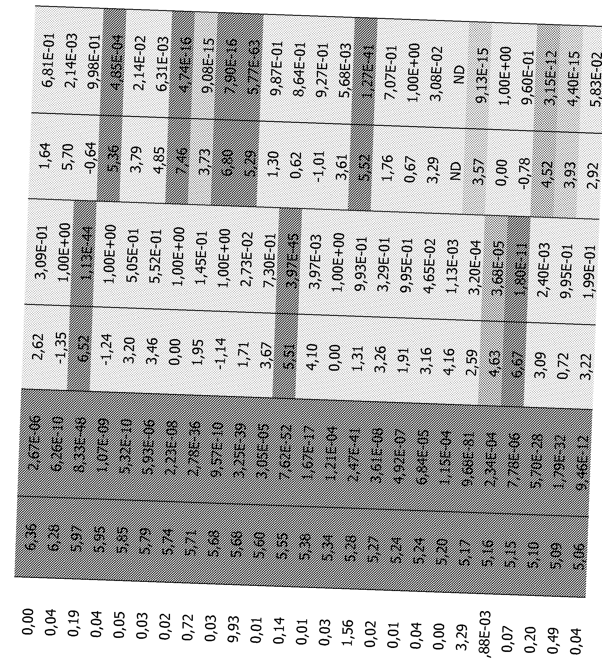
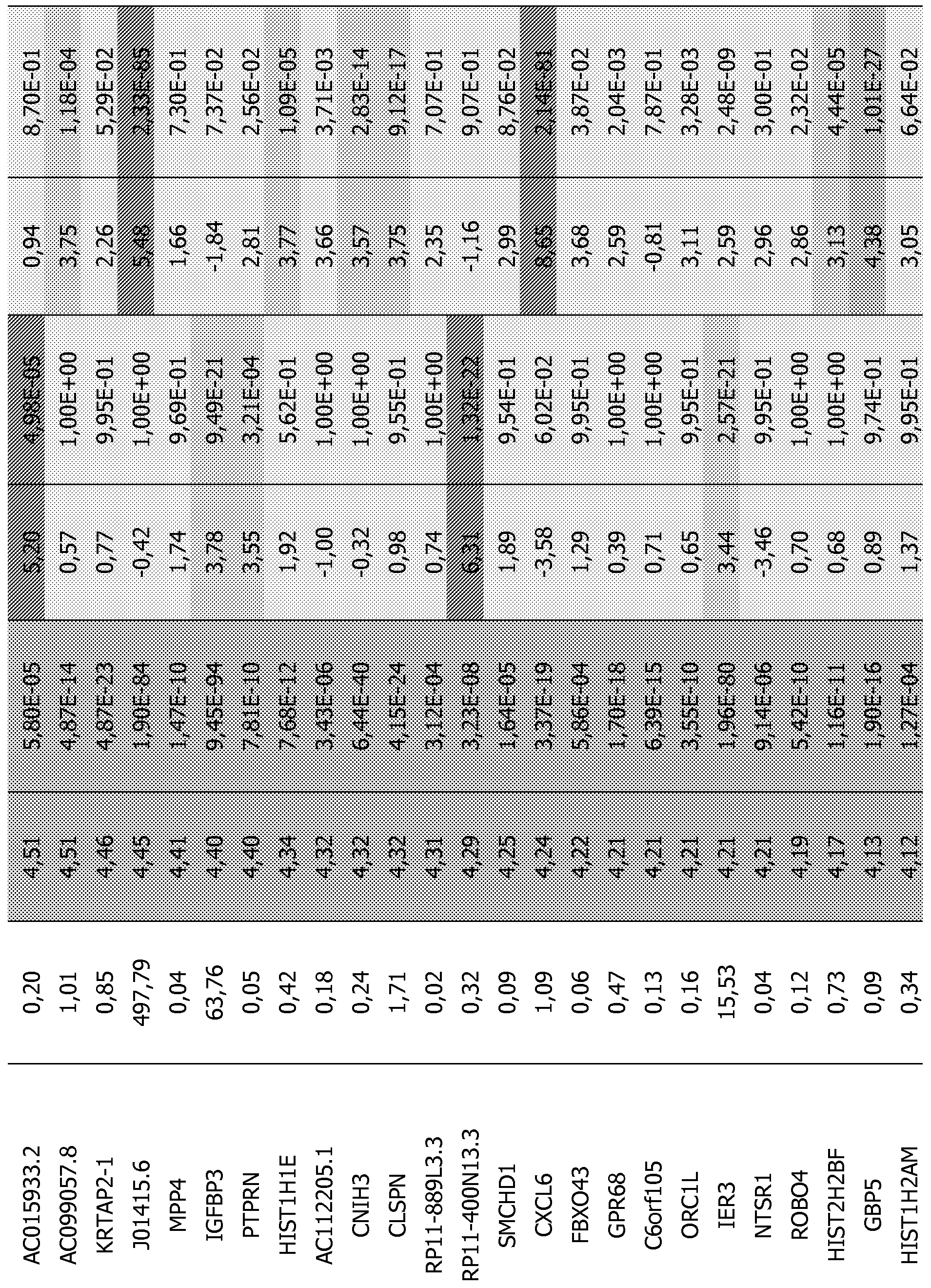
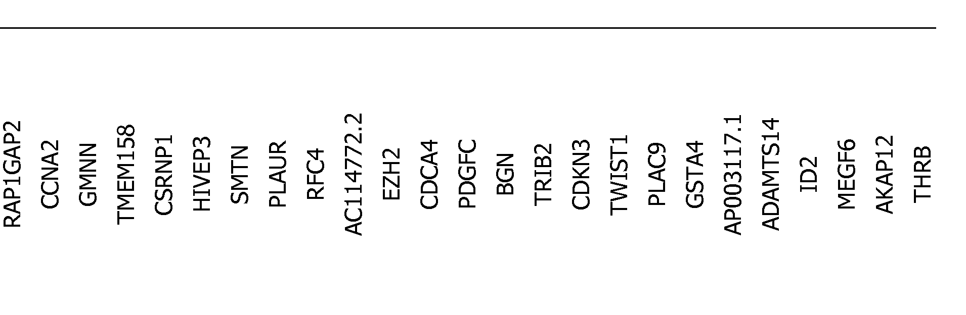
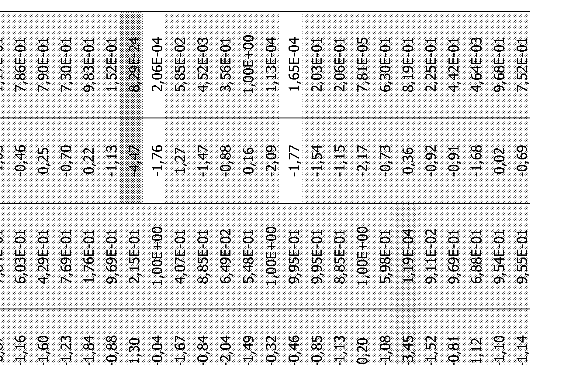
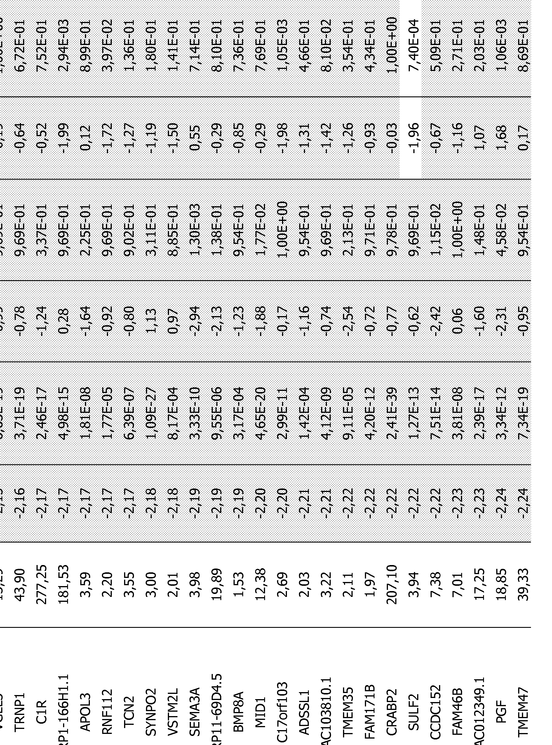
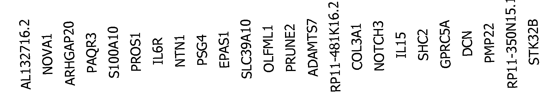
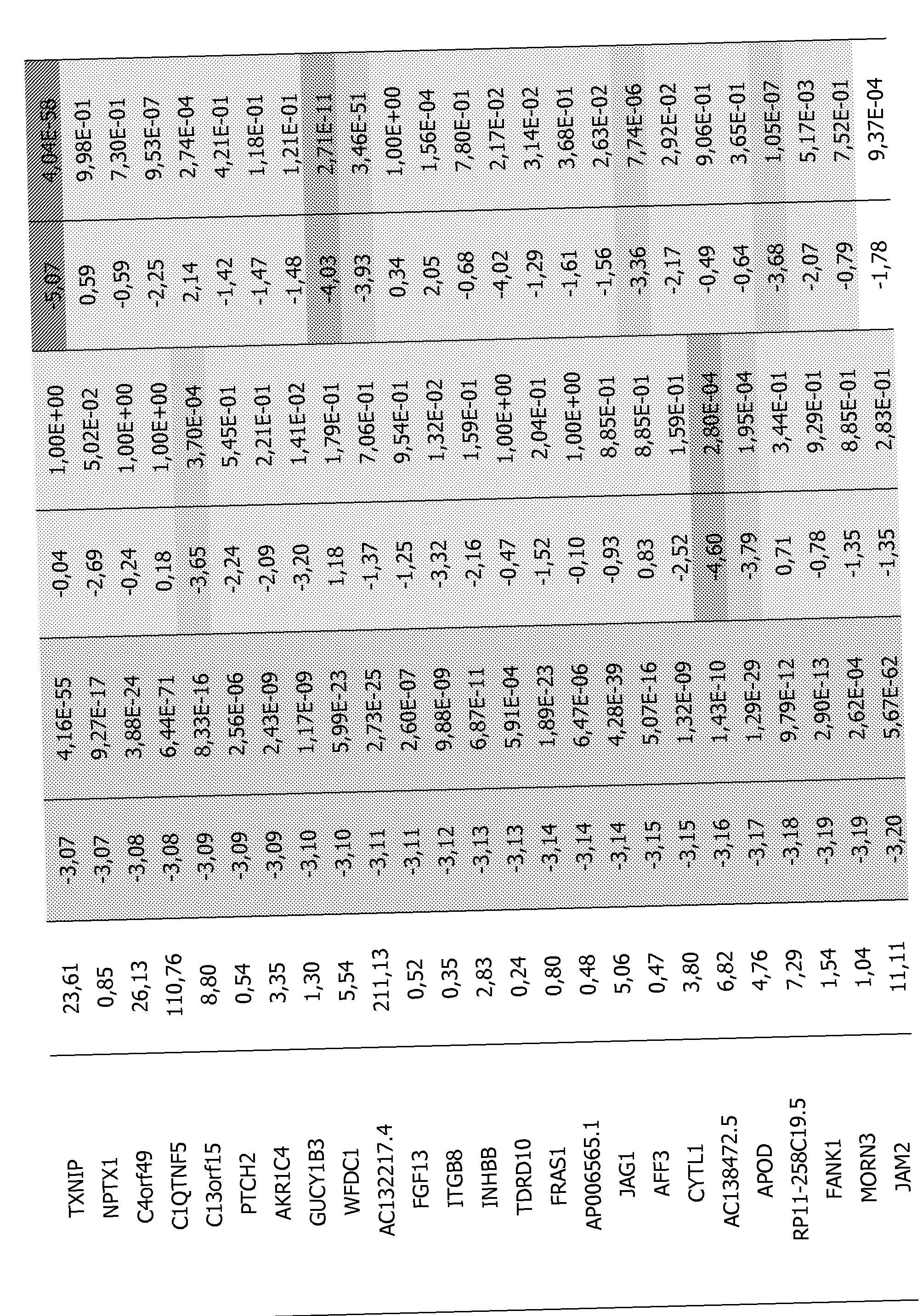
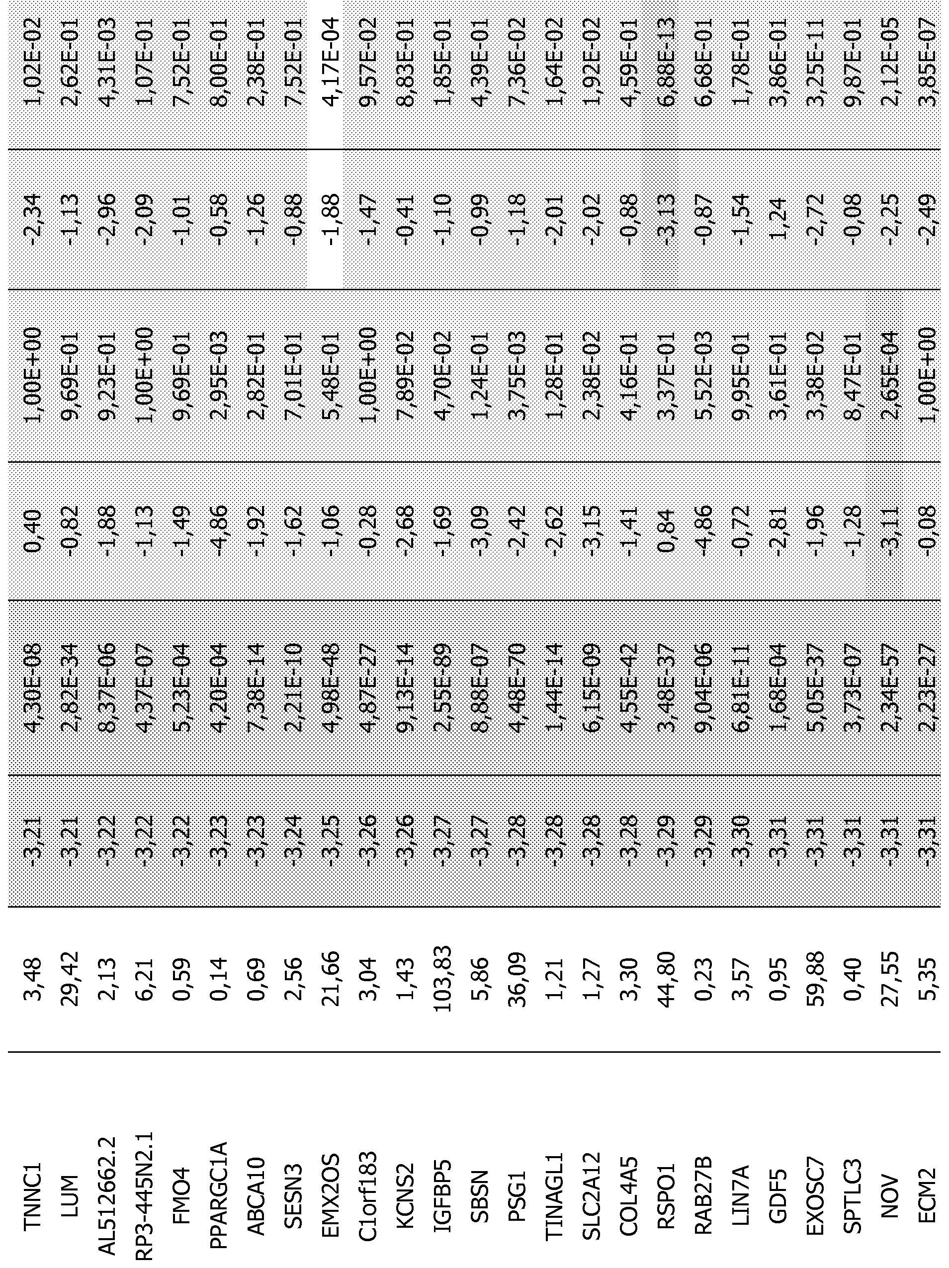
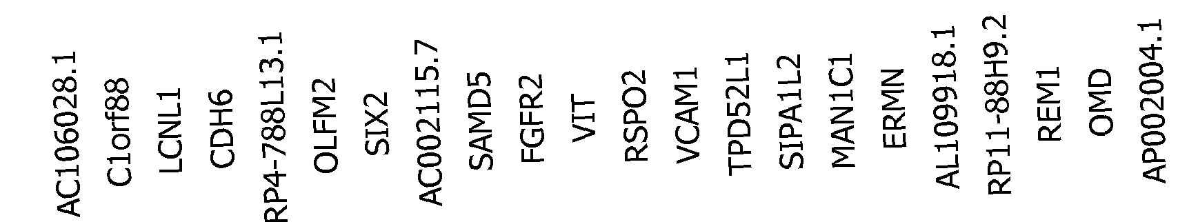
Table 4: List of all genes transcripts identified in all the experiments performed (lists I, II and III).
Example
In response to a lesion, fibroblasts migrate into the wound where they differentiate into contractile myofibroblasts that will finally enter into apoptosis during the remodelling phase. This differentiation process can be studied ex-vivo in environmentally controlled tissue culture conditions, and therefore the timely controlled succession of different gene expression patterns can be addressed.
Materials and methods
Establishment of an ex vivo model of chronic wounds
An ex vivo model of chronic or non-healing wounds was established by adding exudates from chronic wounds on cultured fibroblasts in order to reproduce the pathological state. Then, the gene expression was studied to define a molecular signature of chronic or non-healing wounds.
NHDF, isolated from human explants, were purchased from Promocell. NHDF were cultivated in DMEM-F12 (Invitrogen), supplemented with 10% FCS (Invitrogen, 5Mg/mL of insulin and Ing/mL of b-FGF (PromoKine)).
To collect exudates, four patients with mixed ulcers were recruited (mean age, 76 years; range 57-88 years). For patient selection, it was decided to exclude any other comorbidity factor potentially involved in wound etiology: diabetes, peripheral arterial diseases, malnutrition. Exudates were collected from negative pressure therapy. All the exudates were centrifuged at 1,500 x g for 3 minutes to remove cell debris. The
supernatant was filtered and store at -80°C until use. Aliquots were used to determine protein concentration according to BCA method (Sigma).
For experiments, cells were deprived of insulin and b-FGF during 48 hours. Then, the cells were cultivated on collagen coated culture plates in DMEM-F12, supplemented with 10% FCS, lOng/mL of TGF-βΙ (Promocell) for 4 days. Four points were tested in order to appreciate the effect of exudate on fibroblast differentiation: untreated cells (T-E-), fibroblasts treated with TGF-βΙ (T+E-), cells treated with exudate (T-E+) and finally fibroblasts treated with TGF-βΙ and exudate at the same time (T+E+).
Establishment of an ex vivo model for fibrosis or hypertrophic scar
Myofibroblasts represent the key players in the physiological reconstruction of skin after injury and in generating the pathological tissue deformations that characterize fibrosis such as hypertrophic scars (Desmouliere A, Chaponnier C, Gabbiani G (2005) Tissue repair, contraction, and the myofibroblast. Wound Repair Regen 13: 7-12).
To study the myofibroblasts involved in generating hypertrophic or keloid scars, NHDF were cultivated on collagen coated culture plates in DMEM-F12 (Invitrogen), supplemented with 10% FCS (Invitrogen), 5Mg/ml_ of insulin and Ing/mL of b-FGF (PromoKine) and 10 ng/mL of TGF-βΙ (Promocell), as TGF-βΙ is known to induce the expression of aSMA in fibroblasts (Desmouliere A, Geinoz A, Gabbiani F, Gabbiani G (1993) Transforming growth factor-beta 1 induces alpha-smooth muscle actin expression in granulation tissue myofibroblasts and in quiescent and growing cultured fibroblasts. J Cell Biol, 1993 jul, 122(1): 103-111).
The efficiency of fibroblast differentiation was estimated by analyzing the expression of the myofibroblast marker alpha smooth muscle actin (aSMA).
This aSMA expression was assessed by RT-qPCR (mRNA levels) and by Western Blot (protein).
Western Blotting assay
Total proteins were extracted by scratching the cells with lysis buffer (TRIS, NaCI, NP40, EDTA, IMDTT) and incubated 30 min in ice. To remove cell debris, the samples were centrifuged at 13,000 x g for 10 min at 4°C and store at -20°C until use. Protein concentration was determined according to BCA method (Sigma). Equal amounts of total protein (20Mg) were loaded to NuPAGE 10% BIS-Tris gel (Invitrogen), separated by migration at 150 V, and transferred to nitrocellulose membrane (Whatman) 1 hour at 30 V. Then, membranes were stained for a-SMA (Abeam) and tubulin (Abeam). Incubations were followed by secondary antibodies goat anti-rabbit IgG and goat anti-mouse IgG, respectively, conjugated with horseradish-peroxidase (HRP) (Promega). Signals were detected by ECL chemiluminescence using UptiLight HS WB Substrate (Uptima, Interchim). Bands were digitized with a scanner and the ratio between all bands density of the same blot was calculated by software (ImageJ 1.43u, 64-bit). Relative a-SMA expression was normalized to the respective value for tubulin.
Total RNA Sample Preparation
After four days of experiment, treated fibroblasts were detached with TRIzol Reagent (Invitrogen) and stored at -80°C. Then RNA was purified using chloroform and precipitated by isopropanol. Total RNA was quantified on the NanoDrop 2000c Spectrophotometer (Thermo Scientific). Reverse transcription of 500 ng total RNA to cDNA was done with oligot dT (Invitrogen) using Superscript III RT (Invitrogen) and RNAse OUT (Invitrogen). The cDNA was store at -20°C.
Quantitative real-time RT-PCR
Quantitative real-time PCR (RT-qPCR) was done using 5μΙ_ of 1:20 diluted cDNA on the LightCycler480 system (Roche) using Maxima SYBR Green qPCR Master Mix (Fermentas). Forward and reverse primers were designed by Eurofins (MWG, aSMA forward: CTGTTTTCCCATCCATTGTG (SEQ ID NO: l), aSMA reverse: CCATGTTCTATCGGGTACTT (SEQ ID NO:2)) and a ΙΟΟμΜ stock was stored at -20°C. Forward and reverse primer pairs were used for each RT-qPCR reaction. The cycling conditions were as follows : an initial 95°C for 10 minutes, followed by 45 cycles of 95°C for 15 sec, 58°C for 30 sec, 72°C for 20 sec. LightCycler 480 SW 1.5 was used
to evaluate the TM curves, to determine the Cp and to approximate the relative concentration for each amplification reaction.
Results
For the chronic or non-healing wound model, chronic wound exudates were added to cell cultures (50uMg/ml_ of total proteins of exudate). The experiments that were performed are depicted in Figure 1: cells were either not treated (T-E-), either treated with TGF-β alone (T+E-), exudate alone (T-E+) or TGF-β and exudate (T+E+) for 4 days. The assays described previously were used to assess the level of differentiation. Chronic wound exudates decrease the expression of aSMA (mRNA and protein, figures 2 and 3). This indicated that chronic wound exudates clearly inhibit fibroblast differentiation. This is correlated to the fact that in chronic wounds, we can find unfunctional fibroblasts, also called pseudo-senescent fibroblasts.
In order to analyze the genes expressed upon different treatments of the fibroblasts in a chronic or non-healing wound model or fibrosis, hypertrophic scar model, mRNA deep sequencing was realized.
Total RNA was extracted by TRIzol Equal amounts of total RNA of the different treated cells (5 to 6 Mg) were precipitated by absolute ethanol, supplemented by sodium acetate for RNA sequencing.
The mRNA sequencing was performed by Fastens SA (Switzerland). RNA was sent as total RNA, after two rounds of polyA purification, the Reverse transcription and the cDNA libraries were done. The sequencing was performed on a HiSeq2000 (Illumina).
One gene can contain different isoforms, and some isoforms can have one or more exons in common. Unfortunately, when the number of reads present in each isoforms is counted and fused in gene entities, sometimes the same reads may be counted several times and thus biases the analyses for genes with numerous isoforms. To solve this problem, it was decided to create a fictive transcript for each gene corresponding to the maximal portion of exon coverage, and to count the
number of reads present in these entities. After the normalization and analysis of differential expression steps, only genes showing a differential expression associated with an adjusted p-value of 1.10 3 or less were retained. A supplementary filter on the logFC (Fold Change) to study complete lists (the absolute value of logFC has to be superior or equal to 2) was applied.
Pathologic wound healing analysis: chronic or non-healing wounds or fibrosis/hypertrophic scar/keloid
The aim of the invention was to know if genes are differentially expressed between two conditions, in order to determine if the wound is a chronic or non-healing wound or not, or if the wound will turn into fibrosis/hypertrophic scar or keloid.
The abundance of gene transcripts between two conditions was compared, the T-E- point (normal dermal fibroblasts) being the reference. 3 lists (as defined in Figure 4) were considered: List I compared between T+E- and T-E-, List II compared T-E+ and T-E- and List III compared T+E+ and T-E-. The comparison of the lists II and III with normal dermal fibroblasts provides the list of genes affected by chronic exudate during the process of differentiation, in fact genes affected in the pathological situation found in chronic wound healing. Thus, lists II and III represent the chronic or non-healing wound situation. List I represents the fibrosis or hypertrophic scar situation.
With the p value adjusted and the Log FC filters determined, 171 genes were identified as differentially expressed in list I, 409 genes in list II and 1006 genes in list III.
Some genes, thanks to their high increased or decreased expression, are of particular interest. For example, the expression of PI 16 is largely decreased in non-healing or chronic wounds and increased in fibrosis/hypertrophic scar or keloid model. Thus, PI16 is a favorite candidate for therapy. The present invention also directed to a therapy consisting in increasing its expression in chronic or non-healing wound condition or in decreasing its expression in fibrosis, hypertrophic scar or keloid.
Table 1
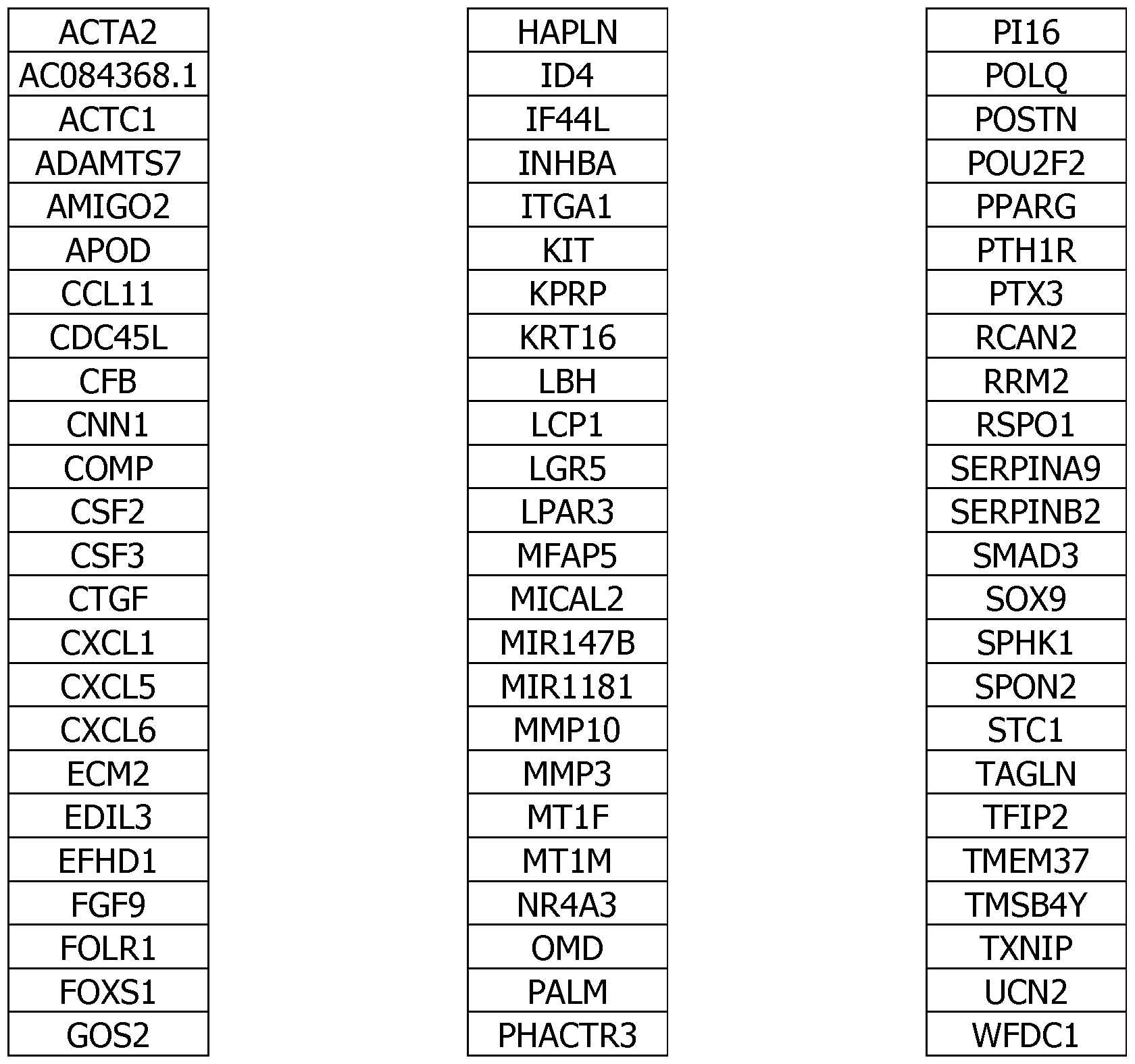
Table 2:
AC084368.1 Decrease in non-healing or chronic wounds
ACTA2 Decrease in non-healing or chronic wounds
ACTC1 Decrease in non-healing or chronic wounds
ADAMTS7 Decrease in non-healing or chronic wounds
AMIG02 Increase in non-healing or chronic wounds
APOD Decrease in non-healing or chronic wounds
CCL11 Increase in non-healing or chronic wounds
CDC45L Increase in non-healing or chronic wounds
CFB Decrease in non-healing or chronic wounds
CNN1 Normal in non-healing or chronic wounds
COMP Decrease in non-healing or chronic wounds
CSF2 Increase in non-healing or chronic wounds
CSF3 Increase in non-healing or chronic wounds
CTGF Increase in non-healing or chronic wounds
CXCL1 Increase in non-healing or chronic wounds
CXCL5 Increase in non-healing or chronic wounds
CXCL6 Increase in non-healing or chronic wounds
ECM2 Decrease in non-healing or chronic wounds
EDIL3 Decrease in non-healing or chronic wounds
EFHD1 Decrease in non-healing or chronic wounds
FGF9 Decrease in non-healing or chronic wounds
FOLR1 Decrease in non-healing or chronic wounds
FOXS1 Increase in non-healing or chronic wounds
GOS2 Increase in non-healing or chronic wounds
HAPLN Increase in non-healing or chronic wounds
ID4 Decrease in non-healing or chronic wounds
IF44L Increase in non-healing or chronic wounds
INHBA Increase in non-healing or chronic wounds
ITGA1 Decrease in non-healing or chronic wounds
KIT Decrease in non-healing or chronic wounds
KPRP Increase in non-healing or chronic wounds
KRT16 Normal in non-healing or chronic wounds
LBH Decrease in non-healing or chronic wounds
LCP1 Increase in non-healing or chronic wounds
LGR5 Decrease in non-healing or chronic wounds
LPAR3 Increase in non-healing or chronic wounds
MFAP5 Decrease in non-healing or chronic wounds
MICAL2 Increase in non-healing or chronic wounds
MIR147B Increase in non-healing or chronic wounds
MIR1181 Increase in non-healing or chronic wounds
MMP10 Increase in non-healing or chronic wounds
MMP3 Increase in non-healing or chronic wounds
MT1F Increase in non-healing or chronic wounds
MT1M Increase in non-healing or chronic wounds
NR4A3 Decrease in non-healing or chronic wounds
OMD Decrease in non-healing or chronic wounds
PALM Decrease in non-healing or chronic wounds
PHACTR3 Decrease in non-healing or chronic wounds
PI16 Decrease in non-healing or chronic wounds
POLQ Increase in non-healing or chronic wounds
POSTN Decrease in non-healing or chronic wounds
POU2F2 Increase in non-healing or chronic wounds
PPARG Decrease in non-healing or chronic wounds
PTH1R Decrease in non-healing or chronic wounds
PTX3 Decrease in non-healing or chronic wounds
RCAN2 Decrease in non-healing or chronic wounds
RRM2 Increase in non-healing or chronic wounds
RSPOl Decrease in non-healing or chronic wounds
SERPINA9 Increase in non-healing or chronic wounds
SERPINB2 Increase in non-healing or chronic wounds
SMAD3 Decrease in non-healing or chronic wounds
SOX9 Increase in non-healing or chronic wounds
SPHK1 Increase in non-healing or chronic wounds
SPON2 Decrease in non-healing or chronic wounds
STC1 Increase in non-healing or chronic wounds
TAGLN Decrease in non-healing or chronic wounds
TFIP2 Increase in non-healing or chronic wounds
TMEM37 Decrease in non-healing or chronic wounds
TMSB4Y Decrease in non-healing or chronic wounds
TXNIP Decrease in non-healing or chronic wounds
UCN2 Increase in non-healing or chronic wounds
WFDC1 Decrease in non-healing or chronic wounds
Table 3:
ACTA2 Increase in fibrosis/hypertrophic scar or keloid
ACTCl Increase in fibrosis/hypertrophic scar or keloid
AMIG02 Increase in fibrosis/hypertrophic scar or keloid
APOD Decrease in fibrosis/hypertrophic scar or keloid
CFB Decrease in fibrosis/hypertrophic scar or keloid
CNN1 Increase in fibrosis/hypertrophic scar or keloid
COMP Increase in fibrosis/hypertrophic scar or keloid
CXCL1 Decrease in fibrosis/hypertrophic scar or keloid
EDIL3 Increase in fibrosis/hypertrophic scar or keloid
EFHD1 Increase in fibrosis/hypertrophic scar or keloid
FOXS1 Increase in fibrosis/hypertrophic scar or keloid
HAPLN1 Increase in fibrosis/hypertrophic scar or keloid
INHBA Increase in fibrosis/hypertrophic scar or keloid
ITGA11 Increase in fibrosis/hypertrophic scar or keloid
KIT Decrease in fibrosis/hypertrophic scar or keloid
KRT16 Increase in fibrosis/hypertrophic scar or keloid
MICAL2 Increase in fibrosis/hypertrophic scar or keloid
NR4A3 Decrease in fibrosis/hypertrophic scar or keloid
PI16 Increase in fibrosis/hypertrophic scar or keloid
POSTN Increase in fibrosis/hypertrophic scar or keloid
POU2F2 Increase in fibrosis/hypertrophic scar or keloid
PPARG Decrease in fibrosis/hypertrophic scar or keloid
PTX3 Decrease in fibrosis/hypertrophic scar or keloid
RCAN2 Decrease in fibrosis/hypertrophic scar or keloid
SMAD3 Decrease in fibrosis/hypertrophic scar or keloid
SOX9 Increase in fibrosis/hypertrophic scar or keloid
SPHK1 Increase in fibrosis/hypertrophic scar or keloid
STC1 Decrease in fibrosis/hypertrophic scar or keloid
TAGLN Increase in fibrosis/hypertrophic scar or keloid
TFPI2 Decrease in fibrosis/hypertrophic scar or keloid
UCN2 Increase in fibrosis/hypertrophic scar or keloid
Table 4:
28

29


«! α3 !θ «ί οΟ «! (Χ) «> α5 θθ !θ Ν Ν Ν Ν Ν Ν Ν Ν Ν (Ο Λ
O 2013/144672 34


38

co o cn in co cn fM in cn
LO I V.) s in 00
00__ r ro v
oo ^ us ΓΜ co o °¾. fM^
Μ CO Ln in fM"
fM o ° ^ P. Γ
^O" fM~ fM
( o" O CM © o


cn cn cn cn si ennis ¾ c ctnnn¾ c cmnn cn cn cn m


■sT
Ln r Ln r Ln oO Ln ro ro Ln ^ o ^ ^ rsj rsj oo ro oo r i-i ^r TH oo cn o o o »D i— I i rsj i— I M o o IN H" o" H H ιη" m" IN" in od H IN O"
m
E 1 CO ϊ 33 Q. Ά S 8 < £ ί h- P s H ≠ m u ° e ^ S- g § 2 § S u § 3 P
< CL D-

fM CO
CD VD
LU Q Q i—l fM <
Q Ln < -H L <n Ln Ln < Ln U I- 2 X ΓΜ ΓΜ t Q ΓΜ fM "sf
00 Q m
— Q vO u_ G
oo v Q u Lu o < LU Ln
LU ' u O Q u
< Q < Q CO L_ CO X LU
X U 3 < «Λ Q O Q CO
u 2 . Q


45

in σι o U3 00
ΓΜ 00 U3
in oo^ ΓΜ CM 00 rM
U3 CO CO CM ID ■sT O
■i in o
<J2 CO
o no" σ¾ ro
ΓΜ CM ΓΜ ΓΜ ro
O ro m ro CM o ro -i rM
in CM CM
LO
LO rM rM LO ΓΜ
ΓΜ ΓΜ U3 m m
DQ <
!— ! T >< < in oo < ro S rM o
< Q. ∑ 00 L0 < rM oo
ro
O L0 CO rM U
_i U
< ∑ < u o L0 _i r^ ^
L0

cn rg 00 r <g
9 ΙΛ __ LO U X 00 ro (J rg _ rg
u_ m i£> o .
to UJ X Q L0 U X
I- oo 2 cn X < Q. m < ∑ Q u L0 L0
< rg u_ t: cn ro
LO Έ. U
LO _i in
LO N O
a.
Q.
0,77 9,69E-01 1,04 3,59E-01
0,54 9,69E-01 1,26 3,27E-02
0,31 9,95E-01 1,19 1,02E-01
-0,31 9,95E-01 2,21 l,38E-03
1,68 3,93E-02 0,21 9,12E-01
0,62 9,95E-01 0,81 7,22E-01
0,80 8,29E-01 0,58 6,89E-01
0,51 9,95E-01 0,56 6,74E-01
0,24 l,00E+00 1,34 5,27E-02
0,20 l,00E+00 1,94 9,26E-03
0,45 9,95E-01 1,47 4,02E-02
0,49 9,95E-01 0,88 5,52E-01
1,10 5,98E-01 0,12 9,94E-01
2,70 2,96E-09 -1,69 7,42E-03
2,78 5,14E-08 -0,83 7,52E-01
0,33 9,95E-01 1,48 l,06E-02
-1,20 7,54E-01 -0,72 6,41E-01
-0,41 l,00E+00 -1,49 l,10E-02
-0,58 9,79E-01 -0,96 2,79E-01
-0,54 9,95E-01 -1,26 9,10E-02
-1,73 6,76E-01 0,31 8,83E-01
-0,51 9,95E-01 -2,16 2,32E-06
1,08 4,37E-01 -2,81 2,18E-10
-1,82 4,21E-02 -0,60 7,44E-01
 -1,40 3,51E-01 -0,29 8,19E-01
-1,44 8,47E-01 -0,65 7,21E-01
-0,78 9,69E-01 -1,50 3,77E-02
-1,30 6,77E-01 0,39 8,83E-01
-0,66 9,94E-01 -0,99 3,46E-01
-1,30 8,85E-01 -0,31 9,10E-01
-1,03 9,50E-01 -1,18 1,40E-01
-0,25 l,00E+00 -2,10 l,00E-03
-0,86 8,85E-01 -0,90 5,23E-01
-0,02 l,00E+00 -1,05 2,95E-01
-1,56 8,01E-01 -0,11 9,82E-01
-0,76 6,49E-01 -0,80 2,43E-01
-0,93 9,69E-01 -0,77 7,30E-01
-2,05 4,61E-02 -1,56 2,66E-02
-0,93 9,69E-01 -0,78 7,30E-01
-1,95 5,65E-02 0,16 9,38E-01
-1,78 2,89E-01 0,28 8,77E-01
-1,89 1,21E-01 -0,23 9,47E-01
-0,36 9,95E-01 -1,25 2,88E-01
-0,57 9,95E-01 -1,15 1,35E-01
-0,38 9,69E-01 -1,79 4,48E-04
-1,14 7,69E-01 -1,22 1,72E-01
0,05 l,00E+00 -2,16 3,15E-04
0,05 l,00E+00 -1,20 3,52E-01
-0,82 9,73E-01 -0,67 6,83E-01
 0,17 9,95E-01 -2,13 2,42E-02


O oo cn o
in in
O cn cn oo o cn
cn ■sT m oo
cn o cn" r cn KO cn m
cn cn" oo" o <J2 ■sT o cn
CO i£> oo o
Λ *o cn" cn" cn Ln
cn cn cn


rsT rsT rvT rvT rsT rsT rsT rsT rsT ro" ro ro" ro"" ro" ro ro" ro" ro" 0 ro" ro" ro" ro ro"
1
VD


rsj rsi rsj

WO 2013/144672
63


WO 2013/144672
64


X


Claims
1, A method of diagnosis or prognosis of a non-healing or chronic wound tissue comprising the step of determining the levels of expression of genes encoding different molecular markers in a sample of a wound from a mammalian, wherein;
- at least one of the following genes shows decreased expression when compared with the expression in normal dermal fibroblasts of said mammalian;
ACTCl, ADAMTS7, CFB, COMP, ECM2, EDIL3, EFHD1, FOLRlf ITGA11, KIT, LBH, LGR5, MFAP5, NR4A3, OMD, PALM, PHACTR3, PI16, PPARG, PTHIR, PTX3, RCAN2, RSPOl, SPON2, TAGLN, TMEM37, TMSB4Y, TXNIP and WFDC1,
- or at least the following miRNA shows decreased expression when compared with the expression in normal dermal fibroblasts of said mammalian;
AC084368.1,
- or at least one of the following genes shows a normal expression when compared with the expression in normal dermal fibroblasts of said mammalian:
C 1 and KRT16,
- or at least one of the following genes shows increased expression when compared with the expression in normal dermal fibroblasts of said mammalian:
AMIG02, CCL11, CDC45L, CSF2, CSF3, FOXSl, GOS2, IF44L, INHBA, KPRP, LCP1, L AR3, MICAL2, MT1F, MTIM, POLQ, POU2F2, RRM2, SERPINA9, SOX9, STC1, TFIP2 and UCN2, - or at least one of the following miRNA shows increased expression when compared with the expression in normal dermal fibroblasts of said mammalian:
MIR147B and MIRU81.
2. A method of diagnosis or prognosis of a non-healing or chronic wound tissue according to claim 1 comprising the step of determining the levels of expression of genes encoding different molecular markers in a sample of a wound from a mammalian, wherein:
- at least one of the following genes show decreased expression when compared with the expression in normal dermal fibroblasts of said rnarnrnslisn*
ACTC1, ADAMTS7, CO P, ECM2, EDIL3, EFHD1, FOLR1, ITGA11, LBH, LGR5, MFAP5, OMD, PALM, PHACTR3, PI16, PTH1R, RSPOl, SPON2, TAGLN, TMEM37, TMSB4Y, TXNIP and WFDC1,
- or at least the following miRNA show decreased expression when compared with the expression in normal dermal fibroblasts of said mammalian:
AC084368.1,
- or at least one of the following genes shows a normal expression when compared to the expression in normal dermal fibroblasts of said mammalian: C1MN1 and KRT16,
- or at least one of the following genes show increased expression when compared with the expression in normal dermal fibroblasts of said mammalian:
CCL11, CDC45L, CSF2, CSF3, GOS2, IF44L, KPRP, LCPl, LPAR3, MT1F, MTIM, POLQ, RRM2, SERPINA9, STC1 and TFIP2, - or at least one of the following mi NA show increased expression when compared with the expression in norma! dermal fibroblasts of said mammalian:
MIR147B and MIR1181.
3. A method according to claim 1 or 2, furthermore comprising another step of determining the levels of expression of genes encoding different molecular markers in a sample of a wound from a mammalian, wherein:
- at least one of the following genes shows decreased expression when compared with the expression in normal dermal fibroblasts of said mammalian:
ACTA2, APOD, FGF9r I EM, POSTN and SMAm,
- or at least one of the following genes show increased expression when compared with the expression in normal dermal fibroblasts of said mammalian:
CXCLl, CXCL5, CXCL6, MMP10, MMP3, SERPINB2, SPHK1, HALPNl and CTGF,
4. A method according to any one of the preceding claims, furthermore comprising another step of determining the levels of expression of genes encoding different molecular markers in a sample of a wound from a mammalian, wherein:
- at least one of the following genes show decreased expression when compared with the expression in normal dermal fibroblasts of said mammalian:
ACTA2, FGF9, ID4 and POSTN,
- or at least one of the following genes show increased expression when compared with the expression in normal dermal fibroblasts of said mammalian:
CXCLl, CXCL5, CXCL6, MMP10, MMP3 and SERPINB2,
5. A method of diagnosis or prognosis of a wound tissue developing an abnormal scar, such as a fibrosis, a hypertrophic scar or a keloid, comprising the step of determining the levels of expression of genes encoding different molecular markers in a sample of a wound from a mammalian, wherein said genes are defined as follows ;
- at least one of the following genes show decreased expression when compared with the expression in normal dermal fibroblasts of said mammalian:
APOD, CFB, CXCL1, KIT, N 4A3, PPARG, PTX3, RCAN2, STC1 and TFPI2,
- or at least one of the following genes show increased expression when compared with the expression in normal dermal fibroblasts of said mammalian:
ACTCl, AMIG02, CNNl, COMP, CTGF, EDIL3, EFHDl, FOXS1, HAPLN1, INHBA, ITGAil, KRT16, MICAL2, PI 16, POSTN, POU2F2, SOX9, SPHKl, TAGLN and UCN2.
6, A method of diagnosis or prognosis according to claim 5 wherein said genes are defined as follows :
- at least one of the following genes show decreased expression when compared with the expression in normal dermal fibroblasts of said mammalian:
CXCLl, STC1 and TFPI2,
- or at least one of the following genes show increased expression when compared with the expression in normal dermal fibroblasts of said mammalian:
ACTCl, CNNl, COMP, EDIL3, EFHDl, UGAll, KRT16, ΡΠ6, POSTN and TAGLN.
7. A method according to any preceding claims, wherein said wound tissue is human wound tissue, and normal derma! fibroblasts are Normal Human Dermal Fibroblasts (NHDF).
8. A method according to any preceding claims, wherein the normal dermal fibroblasts arise from the healthy skin of the said mammalian and more preferably the wound tissue and the norma! dermal fibroblasts arise from the same animal or individual.
9. A method according to any preceding claims, wherein the said levels of expression of genes are determined by quantifying the corresponding RNA.
10. A method according to claim 9, wherein said RNA is chosen from mRNA and mi RNA.
11. A method according to any one of claims 1-8, wherein the said levels of expression of genes are determined by quantifying the corresponding encoded proteins, except for the miRNA expression.
12. A method according to claim 11, wherein said proteins are measured by using antibodies.
13. A kit for performing any one or more of the aforementioned methods according to any one of claims 1-12, wherein said kit comprises:
(1) A plurality of probes for detecting and quantifying the expression levels of all the genes specified in table 1,
(2) Optionally, reagents and instructions pertaining to the use of said probes.
14. A kit for determining the prognosis of mammalian wound which comprises:
(1) A plurality of probes for detecting and quantifying the expression level of at least one RNA or protein of each one of the genes of table 1, (2) Optionally, reagents and instructions pertaining to the use of said probes.
15. A microarray consisting of any one or more of the sets of probes in claims 13-14,
16. A kit for determining a wound type in a patient, comprising: at least two microarrays comprising a plurality of probes for detecting and quantifying the expression levels of all the genes specified in any one of claims 1 to 12,
17. A method for treating a wound which comprises the step of performing any one or more of the methods according to claim 1-12 for determining the classification or prognosis of wound tissue in order to identify whether said wound tissue is chronic or non-healing or will become an abnormal scar, such as a fibrosis, a hypertrophic scar or a keloid or not and selecting an appropriate treatment based on the classification or prognosis of the wound tissue.
18. A therapy consisting in increasing the expression of PI16 in chronic or non-healing wound condition or in decreasing the expression of PI16 in fibrosis, hypertrophic scar or keloid.
Priority Applications (14)
| Application Number | Priority Date | Filing Date | Title |
|---|---|---|---|
| PCT/IB2012/000906 WO2013144672A1 (en) | 2012-03-30 | 2012-03-30 | Method and kit for the classification and prognosis of wounds |
| EP13713192.6A EP2831267A1 (en) | 2012-03-30 | 2013-03-29 | Method and kit for the classification and prognosis of tissue or organ fibrosis in a reparative or reactive process |
| US14/389,420 US20150126447A1 (en) | 2012-03-30 | 2013-03-29 | Method and kit for the classification and prognosis of wounds |
| CA2868606A CA2868606A1 (en) | 2012-03-30 | 2013-03-29 | Method and kit for the classification and prognosis of wounds |
| PCT/EP2013/056831 WO2013144349A1 (en) | 2012-03-30 | 2013-03-29 | Method and kit for the classification and prognosis of tissue or organ fibrosis in a reparative or reactive process |
| PCT/EP2013/056830 WO2013144348A1 (en) | 2012-03-30 | 2013-03-29 | Method and kit for the classification and prognosis of wounds |
| JP2015502377A JP6211052B2 (en) | 2012-03-30 | 2013-03-29 | Methods and kits for classification and prognosis of wounds |
| BR112014024129A BR112014024129A2 (en) | 2012-03-30 | 2013-03-29 | METHOD AND KIT FOR CLASSIFICATION AND PROGNOSIS OF WOUNDS |
| EP18178027.1A EP3421615A1 (en) | 2012-03-30 | 2013-03-29 | Method and kit for the classification and prognosis of tissue or organ in a reparative or reactive process |
| EP13713867.3A EP2831269A1 (en) | 2012-03-30 | 2013-03-29 | Method and kit for the classification and prognosis of wounds |
| CN201380024589.2A CN104540963B (en) | 2012-03-30 | 2013-03-29 | For the method and kit classified with prognosis wound |
| US14/389,431 US20150087527A1 (en) | 2012-03-30 | 2013-03-29 | Method and kit for the classification and prognosis of tissue or organ fibrosis in a reparative or reactive process |
| EP18178025.5A EP3421614A1 (en) | 2012-03-30 | 2013-03-29 | Method and kit for the classification and prognosis of wounds |
| JP2017081862A JP2017195878A (en) | 2012-03-30 | 2017-04-18 | Method and kit for classification and prognosis of wound |
Applications Claiming Priority (1)
| Application Number | Priority Date | Filing Date | Title |
|---|---|---|---|
| PCT/IB2012/000906 WO2013144672A1 (en) | 2012-03-30 | 2012-03-30 | Method and kit for the classification and prognosis of wounds |
Related Child Applications (1)
| Application Number | Title | Priority Date | Filing Date |
|---|---|---|---|
| US14/389,431 Continuation-In-Part US20150087527A1 (en) | 2012-03-30 | 2013-03-29 | Method and kit for the classification and prognosis of tissue or organ fibrosis in a reparative or reactive process |
Publications (1)
| Publication Number | Publication Date |
|---|---|
| WO2013144672A1 true WO2013144672A1 (en) | 2013-10-03 |
Family
ID=48040243
Family Applications (3)
| Application Number | Title | Priority Date | Filing Date |
|---|---|---|---|
| PCT/IB2012/000906 WO2013144672A1 (en) | 2012-03-30 | 2012-03-30 | Method and kit for the classification and prognosis of wounds |
| PCT/EP2013/056831 WO2013144349A1 (en) | 2012-03-30 | 2013-03-29 | Method and kit for the classification and prognosis of tissue or organ fibrosis in a reparative or reactive process |
| PCT/EP2013/056830 WO2013144348A1 (en) | 2012-03-30 | 2013-03-29 | Method and kit for the classification and prognosis of wounds |
Family Applications After (2)
| Application Number | Title | Priority Date | Filing Date |
|---|---|---|---|
| PCT/EP2013/056831 WO2013144349A1 (en) | 2012-03-30 | 2013-03-29 | Method and kit for the classification and prognosis of tissue or organ fibrosis in a reparative or reactive process |
| PCT/EP2013/056830 WO2013144348A1 (en) | 2012-03-30 | 2013-03-29 | Method and kit for the classification and prognosis of wounds |
Country Status (7)
| Country | Link |
|---|---|
| US (2) | US20150126447A1 (en) |
| EP (4) | EP3421615A1 (en) |
| JP (2) | JP6211052B2 (en) |
| CN (1) | CN104540963B (en) |
| BR (1) | BR112014024129A2 (en) |
| CA (1) | CA2868606A1 (en) |
| WO (3) | WO2013144672A1 (en) |
Cited By (4)
| Publication number | Priority date | Publication date | Assignee | Title |
|---|---|---|---|---|
| CN105853421A (en) * | 2015-01-22 | 2016-08-17 | 北京大学 | Novel application of FoxO1 selective inhibitor AS1842856 |
| CN106512102A (en) * | 2016-11-03 | 2017-03-22 | 武汉枫霖科技有限公司 | Method for promoting cartilage tissue differentiation of mesenchymal stem cells |
| JP2017528523A (en) * | 2014-09-16 | 2017-09-28 | オンコメッド ファーマシューティカルズ インコーポレイテッド | Treatment of fibrotic diseases |
| US20220080044A1 (en) * | 2020-09-14 | 2022-03-17 | National Yang-Ming University | Method for preventing or treating peripheral arterial occlusive disease |
Families Citing this family (8)
| Publication number | Priority date | Publication date | Assignee | Title |
|---|---|---|---|---|
| KR101589285B1 (en) * | 2014-09-23 | 2016-01-27 | 한국생명공학연구원 | Paralemmin as a marker of cardiovascular disorders and cardiovascular disorders diagnostic kit using thereof |
| WO2016205259A1 (en) * | 2015-06-14 | 2016-12-22 | Hoke Glenn Dale | Methods of diagnosing and treating wounds |
| CN107817342B (en) * | 2016-09-14 | 2019-06-25 | 北京大学 | Application of the cartilage oligo-substrate protein as marker in diagnosis abdominal aneurvsm or artery dissection |
| EP3639021A4 (en) | 2017-06-14 | 2021-06-09 | McMaster University | Biomarkers for wound healing |
| TWI774059B (en) * | 2020-09-14 | 2022-08-11 | 國立陽明大學 | Use of cxcl5 neutralizing antibody in the manufacture of a medicament for preventing or treating peripheral arterial occlusive disease |
| RU2761893C1 (en) * | 2021-01-25 | 2021-12-13 | Федеральное государственное бюджетное образовательное учреждение высшего образования Иркутский государственный медицинский университет Министерства здравоохранения Российской Федерации | Method for predicting the result of laser treatment of a hypertrophic scar in women |
| CA3228288A1 (en) * | 2021-08-03 | 2023-02-09 | Chung Ang University Industry Academic Cooperation Foundation | Composition for preventing or treating fibrotic diseases, comprising hapln1 |
| WO2023042309A1 (en) | 2021-09-15 | 2023-03-23 | ファナック株式会社 | Robot simulation device |
Citations (5)
| Publication number | Priority date | Publication date | Assignee | Title |
|---|---|---|---|---|
| WO2008110356A2 (en) * | 2007-03-12 | 2008-09-18 | Robert Frost | Protein pi 16 secreted from the heart and uses thereof |
| US20090220488A1 (en) * | 2005-08-31 | 2009-09-03 | Humphrey Gardner | Evaluating and treating scleroderma |
| US20090305279A1 (en) * | 2006-08-31 | 2009-12-10 | Renovo Limited | Method of diagnosis |
| WO2011006214A1 (en) * | 2009-07-16 | 2011-01-20 | Peter Maccallum Cancer Institute | Method of detecting radiation exposure and adverse toxicity thereto |
| WO2011033249A1 (en) * | 2009-09-15 | 2011-03-24 | University College Cardiff Consultants Limited | Method and kit for the classification and prognosis of wounds |
Family Cites Families (10)
| Publication number | Priority date | Publication date | Assignee | Title |
|---|---|---|---|---|
| AU2001288390A1 (en) * | 2000-08-25 | 2002-03-04 | Cor Therapeutics, Inc. | Proteins and nucleic acids encoding same |
| US8034553B2 (en) * | 2004-06-24 | 2011-10-11 | Kimberly-Clark Worldwide, Inc. | Biomarkers for wound healing |
| WO2006042197A2 (en) * | 2004-10-11 | 2006-04-20 | The Board Of Trustees Of The Leland Standford Junior University | Use of del-1 in hair, bone and cartilage regeneration |
| WO2008124172A1 (en) * | 2007-04-10 | 2008-10-16 | The Board Of Regents, The University Of Texas System | Combination therapy for chronic dermal ulcers |
| US20100273666A1 (en) * | 2007-12-13 | 2010-10-28 | Bernatchez Stephanie F | Methods of analyzing wound samples |
| WO2010042903A1 (en) * | 2008-10-09 | 2010-04-15 | Alfagene Bioscience, Inc | Use and identification of biomarkers for gastrointestinal diseases |
| US20110295782A1 (en) * | 2008-10-15 | 2011-12-01 | Alexander Stojadinovic | Clinical Decision Model |
| WO2010051550A1 (en) * | 2008-10-31 | 2010-05-06 | University Of Rochester | Methods of diagnosing and treating fibrosis |
| US20100204058A1 (en) * | 2009-01-28 | 2010-08-12 | Howard Yuan-Hao Chang | Profiling for Determination of Response to Treatment for Inflammatory Disease |
| MX2011009967A (en) * | 2009-03-26 | 2012-01-25 | Ford Henry Health System | Methods for improving neurological outcome after neural injury and neurodegenerative disease. |
-
2012
- 2012-03-30 WO PCT/IB2012/000906 patent/WO2013144672A1/en active Application Filing
-
2013
- 2013-03-29 EP EP18178027.1A patent/EP3421615A1/en not_active Withdrawn
- 2013-03-29 WO PCT/EP2013/056831 patent/WO2013144349A1/en active Application Filing
- 2013-03-29 CN CN201380024589.2A patent/CN104540963B/en not_active Expired - Fee Related
- 2013-03-29 EP EP13713192.6A patent/EP2831267A1/en not_active Ceased
- 2013-03-29 US US14/389,420 patent/US20150126447A1/en not_active Abandoned
- 2013-03-29 JP JP2015502377A patent/JP6211052B2/en not_active Expired - Fee Related
- 2013-03-29 EP EP13713867.3A patent/EP2831269A1/en not_active Ceased
- 2013-03-29 CA CA2868606A patent/CA2868606A1/en not_active Abandoned
- 2013-03-29 WO PCT/EP2013/056830 patent/WO2013144348A1/en active Application Filing
- 2013-03-29 US US14/389,431 patent/US20150087527A1/en not_active Abandoned
- 2013-03-29 EP EP18178025.5A patent/EP3421614A1/en not_active Withdrawn
- 2013-03-29 BR BR112014024129A patent/BR112014024129A2/en not_active IP Right Cessation
-
2017
- 2017-04-18 JP JP2017081862A patent/JP2017195878A/en active Pending
Patent Citations (5)
| Publication number | Priority date | Publication date | Assignee | Title |
|---|---|---|---|---|
| US20090220488A1 (en) * | 2005-08-31 | 2009-09-03 | Humphrey Gardner | Evaluating and treating scleroderma |
| US20090305279A1 (en) * | 2006-08-31 | 2009-12-10 | Renovo Limited | Method of diagnosis |
| WO2008110356A2 (en) * | 2007-03-12 | 2008-09-18 | Robert Frost | Protein pi 16 secreted from the heart and uses thereof |
| WO2011006214A1 (en) * | 2009-07-16 | 2011-01-20 | Peter Maccallum Cancer Institute | Method of detecting radiation exposure and adverse toxicity thereto |
| WO2011033249A1 (en) * | 2009-09-15 | 2011-03-24 | University College Cardiff Consultants Limited | Method and kit for the classification and prognosis of wounds |
Non-Patent Citations (10)
| Title |
|---|
| "GeneChip Human Genome U133 Set", INTERNET CITATION, 26 February 2003 (2003-02-26), XP002232760, Retrieved from the Internet <URL:http://www.affymetrix.com/support/technical/datasheets/hgu133_datasheet.pdf> [retrieved on 20030226] * |
| BRIAN C. WULFF ET AL: "Novel differences in the expression of inflammation-associated genes between mid- and late-gestational dermal fibroblasts", WOUND REPAIR AND REGENERATION, 5 November 2012 (2012-11-05), pages 1 - 10, XP055045115, ISSN: 1067-1927, DOI: 10.1111/j.1524-475X.2012.00860.x * |
| BYUNG-CHUL KIM ET AL: "Fibroblasts from chronic wounds show altered TGF-?-signaling and decreased TGF-? Type II Receptor expression", JOURNAL OF CELLULAR PHYSIOLOGY, vol. 195, no. 3, 1 June 2003 (2003-06-01), pages 331 - 336, XP055045109, ISSN: 0021-9541, DOI: 10.1002/jcp.10301 * |
| CHA ET AL: "Fibroblasts from non-healing human chronic wounds show decreased expression of betaig-h3, a TGF-beta inducible protein", JOURNAL OF DERMATOLOGICAL SCIENCE, ELSEVIER SCIENCE PUBLISHERS, SHANNON, IE, vol. 50, no. 1, 19 December 2007 (2007-12-19), pages 15 - 23, XP022487263, ISSN: 0923-1811, DOI: 10.1016/J.JDERMSCI.2007.10.010 * |
| CHEN LIN ET AL: "Positional differences in the wound transcriptome of skin and oral mucosa.", BMC GENOMICS 2010, vol. 11, 2010, pages 471, XP021072768, ISSN: 1471-2164 * |
| COOPER LISA ET AL: "Wound healing and inflammation genes revealed by array analysis of 'macrophageless' PU.1 null mice", GENOME BIOLOGY, BIOMED CENTRAL LTD., LONDON, GB, vol. 6, no. 1, 23 December 2004 (2004-12-23), pages R5, XP021012937, ISSN: 1465-6906, DOI: 10.1186/GB-2004-6-1-R5 * |
| SABIOSCIENCES: "Human wound healing PCR array", 19 December 2010 (2010-12-19), pages 1 - 5, XP002687677, Retrieved from the Internet <URL:http://web.archive.org/web/20101219235107/http://sabiosciences.com/rt_pcr_product/HTML/PAHS-121A.html> [retrieved on 20121120] * |
| SHIH BARBARA ET AL: "Identification of novel keloid biomarkers through profiling of tissue biopsies versus cell cultures in keloid margin specimens compared to adjacent normal skin.", EPLASTY 2010, vol. 10, 2010, pages e24, XP002687680, ISSN: 1937-5719 * |
| SMITH JOAN C ET AL: "Gene profiling of keloid fibroblasts shows altered expression in multiple fibrosis-associated pathways.", THE JOURNAL OF INVESTIGATIVE DERMATOLOGY MAY 2008, vol. 128, no. 5, May 2008 (2008-05-01), pages 1298 - 1310, XP002687679, ISSN: 1523-1747 * |
| TSOU RAYMOND ET AL: "Analysis of hypertrophic and normal scar gene expression with cDNA microarrays", JOURNAL OF BURN CARE AND REHABILITATION, MOSBY- YEARBOOK INC, US, vol. 21, no. 6, 1 November 2000 (2000-11-01), pages 541 - 550, XP009131413, ISSN: 0273-8481, DOI: 10.1097/00004630-200021060-00012 * |
Cited By (4)
| Publication number | Priority date | Publication date | Assignee | Title |
|---|---|---|---|---|
| JP2017528523A (en) * | 2014-09-16 | 2017-09-28 | オンコメッド ファーマシューティカルズ インコーポレイテッド | Treatment of fibrotic diseases |
| CN105853421A (en) * | 2015-01-22 | 2016-08-17 | 北京大学 | Novel application of FoxO1 selective inhibitor AS1842856 |
| CN106512102A (en) * | 2016-11-03 | 2017-03-22 | 武汉枫霖科技有限公司 | Method for promoting cartilage tissue differentiation of mesenchymal stem cells |
| US20220080044A1 (en) * | 2020-09-14 | 2022-03-17 | National Yang-Ming University | Method for preventing or treating peripheral arterial occlusive disease |
Also Published As
| Publication number | Publication date |
|---|---|
| CA2868606A1 (en) | 2013-10-03 |
| JP2015513898A (en) | 2015-05-18 |
| CN104540963B (en) | 2017-07-25 |
| BR112014024129A2 (en) | 2017-08-22 |
| WO2013144349A1 (en) | 2013-10-03 |
| JP6211052B2 (en) | 2017-10-11 |
| EP3421615A1 (en) | 2019-01-02 |
| CN104540963A (en) | 2015-04-22 |
| EP3421614A1 (en) | 2019-01-02 |
| WO2013144348A1 (en) | 2013-10-03 |
| US20150126447A1 (en) | 2015-05-07 |
| JP2017195878A (en) | 2017-11-02 |
| US20150087527A1 (en) | 2015-03-26 |
| EP2831269A1 (en) | 2015-02-04 |
| EP2831267A1 (en) | 2015-02-04 |
Similar Documents
| Publication | Publication Date | Title |
|---|---|---|
| WO2013144672A1 (en) | Method and kit for the classification and prognosis of wounds | |
| JP5634360B2 (en) | Prognosis method and kit for breast cancer | |
| KR102464372B1 (en) | Methods of diagnosing inflammatory bowel disease through rnaset2 | |
| US20080038746A1 (en) | Methods and compositions for diagnosing and monitoring the status of transplant rejection and immune disorders | |
| WO2006119343A1 (en) | Determination of eosinophilic esophagitis | |
| Wang et al. | The diagnostic value of microRNA-4787-5p and microRNA-4306 in patients with acute aortic dissection | |
| EP2315847A1 (en) | Method for determining a predisposition to basal cell carcinoma and for screening treatments thereof | |
| CA2653071A1 (en) | Methods for diagnosing and treating graft rejection and inflammatory conditions | |
| WO2019143828A2 (en) | Biomarkers of cardiovascular status and uses therof | |
| CN114568028A (en) | Genetic variants for diagnosing alzheimer's disease | |
| JP2019187413A (en) | Methods for detecting lupus nephritis or predicting its risk and applications thereof | |
| CA2768339A1 (en) | Processes and methods for diagnosis of alzheimer's disease | |
| KR102384992B1 (en) | Age-specific biomarker of a patient with colorectal cancer and use thereof | |
| EP2716767A1 (en) | Method for determining the prognosis of pancreatic cancer | |
| KR102010899B1 (en) | Method for providing the information for predicting or diagnosing of inflammatory bowel disease using single nucleotide polymorphism to be identified from next generation sequencing screening | |
| US20240125766A1 (en) | Method for determining suitability of a subject to anti tnf alpha therapy | |
| JP2021530964A (en) | Diagnostic blood test | |
| KR101046341B1 (en) | TMM5 Gene Polymorphism Marker for Stroke Prediction and Stroke Prediction Method Using the Same | |
| KR102072563B1 (en) | Biomarker comprising dihydropyrimidinase-related protein 2 for diagnosing internet gaming disorder and uses thereof | |
| KR102010897B1 (en) | Method for providing the information for predicting or diagnosing of inflammatory bowel disease using single nucleotide polymorphism to be identified from next generation sequencing screening | |
| CN106520970A (en) | Marker for diagnosing cerebral apoplexy | |
| JP2022022709A (en) | miRNA DIAGNOSIS BIOMARKER FOR SEVERE DRUG ERUPTION | |
| WO2014173986A2 (en) | Methods for diagnosing and monitoring the response to treatment of hepatocellular carcinoma |
Legal Events
| Date | Code | Title | Description |
|---|---|---|---|
| 121 | Ep: the epo has been informed by wipo that ep was designated in this application |
Ref document number: 12723742 Country of ref document: EP Kind code of ref document: A1 |
|
| NENP | Non-entry into the national phase |
Ref country code: DE |
|
| 122 | Ep: pct application non-entry in european phase |
Ref document number: 12723742 Country of ref document: EP Kind code of ref document: A1 |