US10390913B2 - Diagnostic intraoral scanning - Google Patents
Diagnostic intraoral scanning Download PDFInfo
- Publication number
- US10390913B2 US10390913B2 US16/258,516 US201916258516A US10390913B2 US 10390913 B2 US10390913 B2 US 10390913B2 US 201916258516 A US201916258516 A US 201916258516A US 10390913 B2 US10390913 B2 US 10390913B2
- Authority
- US
- United States
- Prior art keywords
- patient
- dental arch
- model
- viewing window
- relative
- Prior art date
- Legal status (The legal status is an assumption and is not a legal conclusion. Google has not performed a legal analysis and makes no representation as to the accuracy of the status listed.)
- Active
Links
- 210000002455 dental arch Anatomy 0.000 claims abstract description 487
- 238000000034 method Methods 0.000 claims abstract description 269
- 238000003384 imaging method Methods 0.000 claims description 99
- 230000008859 change Effects 0.000 claims description 42
- 230000003595 spectral effect Effects 0.000 claims description 13
- 238000012935 Averaging Methods 0.000 claims description 6
- 210000003298 dental enamel Anatomy 0.000 abstract description 93
- 210000004268 dentin Anatomy 0.000 abstract description 46
- 210000000515 tooth Anatomy 0.000 description 293
- 230000000875 corresponding effect Effects 0.000 description 57
- 230000000149 penetrating effect Effects 0.000 description 42
- 208000002925 dental caries Diseases 0.000 description 36
- 238000011282 treatment Methods 0.000 description 32
- 238000004458 analytical method Methods 0.000 description 18
- 238000001514 detection method Methods 0.000 description 18
- 230000011218 segmentation Effects 0.000 description 16
- 238000005286 illumination Methods 0.000 description 15
- 210000000214 mouth Anatomy 0.000 description 15
- 210000004195 gingiva Anatomy 0.000 description 13
- 230000035515 penetration Effects 0.000 description 13
- 238000005553 drilling Methods 0.000 description 12
- 230000008569 process Effects 0.000 description 12
- 238000012800 visualization Methods 0.000 description 12
- 210000004262 dental pulp cavity Anatomy 0.000 description 10
- 230000003902 lesion Effects 0.000 description 10
- 238000010521 absorption reaction Methods 0.000 description 9
- 239000002872 contrast media Substances 0.000 description 9
- 238000013507 mapping Methods 0.000 description 9
- 239000000463 material Substances 0.000 description 9
- 238000012544 monitoring process Methods 0.000 description 9
- 230000003287 optical effect Effects 0.000 description 9
- 210000001519 tissue Anatomy 0.000 description 9
- 238000000227 grinding Methods 0.000 description 8
- 230000010287 polarization Effects 0.000 description 8
- 239000003795 chemical substances by application Substances 0.000 description 7
- 238000004140 cleaning Methods 0.000 description 7
- 230000007547 defect Effects 0.000 description 7
- 208000006558 Dental Calculus Diseases 0.000 description 6
- 201000002170 dentin sensitivity Diseases 0.000 description 6
- 238000013461 design Methods 0.000 description 6
- 238000010801 machine learning Methods 0.000 description 6
- 238000004519 manufacturing process Methods 0.000 description 6
- 239000000203 mixture Substances 0.000 description 6
- 230000036347 tooth sensitivity Effects 0.000 description 6
- 230000008901 benefit Effects 0.000 description 5
- 238000007408 cone-beam computed tomography Methods 0.000 description 5
- 230000002596 correlated effect Effects 0.000 description 5
- 210000004513 dentition Anatomy 0.000 description 5
- 238000011161 development Methods 0.000 description 5
- 230000018109 developmental process Effects 0.000 description 5
- 230000005865 ionizing radiation Effects 0.000 description 5
- 210000001847 jaw Anatomy 0.000 description 5
- 210000003254 palate Anatomy 0.000 description 5
- 238000012552 review Methods 0.000 description 5
- 230000036346 tooth eruption Effects 0.000 description 5
- 230000003321 amplification Effects 0.000 description 4
- 230000001680 brushing effect Effects 0.000 description 4
- 230000001276 controlling effect Effects 0.000 description 4
- 230000000694 effects Effects 0.000 description 4
- 201000005562 gingival recession Diseases 0.000 description 4
- 239000003550 marker Substances 0.000 description 4
- 238000003199 nucleic acid amplification method Methods 0.000 description 4
- 238000002360 preparation method Methods 0.000 description 4
- 201000002859 sleep apnea Diseases 0.000 description 4
- 210000004872 soft tissue Anatomy 0.000 description 4
- 239000003086 colorant Substances 0.000 description 3
- 238000004891 communication Methods 0.000 description 3
- 238000011960 computer-aided design Methods 0.000 description 3
- 239000004053 dental implant Substances 0.000 description 3
- 238000002405 diagnostic procedure Methods 0.000 description 3
- 238000002845 discoloration Methods 0.000 description 3
- 238000011049 filling Methods 0.000 description 3
- 230000033001 locomotion Effects 0.000 description 3
- 230000004044 response Effects 0.000 description 3
- 239000000523 sample Substances 0.000 description 3
- 230000009466 transformation Effects 0.000 description 3
- 230000007704 transition Effects 0.000 description 3
- 238000002604 ultrasonography Methods 0.000 description 3
- 208000032841 Bulimia Diseases 0.000 description 2
- 206010006550 Bulimia nervosa Diseases 0.000 description 2
- 206010061274 Malocclusion Diseases 0.000 description 2
- 241001465754 Metazoa Species 0.000 description 2
- 208000002193 Pain Diseases 0.000 description 2
- 208000008117 Salivary Gland Calculi Diseases 0.000 description 2
- 230000003190 augmentative effect Effects 0.000 description 2
- 230000009286 beneficial effect Effects 0.000 description 2
- 230000015572 biosynthetic process Effects 0.000 description 2
- 238000004040 coloring Methods 0.000 description 2
- 238000004590 computer program Methods 0.000 description 2
- 208000037265 diseases, disorders, signs and symptoms Diseases 0.000 description 2
- 208000035475 disorder Diseases 0.000 description 2
- 238000009826 distribution Methods 0.000 description 2
- 230000003628 erosive effect Effects 0.000 description 2
- 238000005755 formation reaction Methods 0.000 description 2
- 208000021302 gastroesophageal reflux disease Diseases 0.000 description 2
- 230000001788 irregular Effects 0.000 description 2
- 230000000670 limiting effect Effects 0.000 description 2
- 238000005259 measurement Methods 0.000 description 2
- 238000012986 modification Methods 0.000 description 2
- 230000004048 modification Effects 0.000 description 2
- 238000012014 optical coherence tomography Methods 0.000 description 2
- 230000008520 organization Effects 0.000 description 2
- 238000002203 pretreatment Methods 0.000 description 2
- 238000012545 processing Methods 0.000 description 2
- 238000013442 quality metrics Methods 0.000 description 2
- 230000002441 reversible effect Effects 0.000 description 2
- 210000001581 salivary duct Anatomy 0.000 description 2
- 238000003860 storage Methods 0.000 description 2
- 238000010146 3D printing Methods 0.000 description 1
- 102000007325 Amelogenin Human genes 0.000 description 1
- 108010007570 Amelogenin Proteins 0.000 description 1
- 241000894006 Bacteria Species 0.000 description 1
- 208000004434 Calcinosis Diseases 0.000 description 1
- 208000005335 Dentin Dysplasia Diseases 0.000 description 1
- 206010058314 Dysplasia Diseases 0.000 description 1
- 206010061218 Inflammation Diseases 0.000 description 1
- 241000276420 Lophius piscatorius Species 0.000 description 1
- 208000005358 Tooth Attrition Diseases 0.000 description 1
- 208000008312 Tooth Loss Diseases 0.000 description 1
- 239000002253 acid Substances 0.000 description 1
- 230000006978 adaptation Effects 0.000 description 1
- 230000005540 biological transmission Effects 0.000 description 1
- 230000004397 blinking Effects 0.000 description 1
- 210000000988 bone and bone Anatomy 0.000 description 1
- 230000002308 calcification Effects 0.000 description 1
- 238000001444 catalytic combustion detection Methods 0.000 description 1
- 239000004568 cement Substances 0.000 description 1
- 238000010990 cephalometric method Methods 0.000 description 1
- 239000000919 ceramic Substances 0.000 description 1
- 238000012512 characterization method Methods 0.000 description 1
- 230000001055 chewing effect Effects 0.000 description 1
- 230000001684 chronic effect Effects 0.000 description 1
- 238000007621 cluster analysis Methods 0.000 description 1
- 238000000576 coating method Methods 0.000 description 1
- 239000002131 composite material Substances 0.000 description 1
- 230000006835 compression Effects 0.000 description 1
- 238000007906 compression Methods 0.000 description 1
- 238000012790 confirmation Methods 0.000 description 1
- 238000005336 cracking Methods 0.000 description 1
- -1 crowns (e.g. Substances 0.000 description 1
- 238000005520 cutting process Methods 0.000 description 1
- 230000001351 cycling effect Effects 0.000 description 1
- 238000000354 decomposition reaction Methods 0.000 description 1
- 238000005516 engineering process Methods 0.000 description 1
- 238000011156 evaluation Methods 0.000 description 1
- 230000007717 exclusion Effects 0.000 description 1
- 239000000835 fiber Substances 0.000 description 1
- 239000000945 filler Substances 0.000 description 1
- 238000001914 filtration Methods 0.000 description 1
- 238000002073 fluorescence micrograph Methods 0.000 description 1
- 208000024693 gingival disease Diseases 0.000 description 1
- 210000004907 gland Anatomy 0.000 description 1
- 230000036541 health Effects 0.000 description 1
- 229910052588 hydroxylapatite Inorganic materials 0.000 description 1
- 230000008676 import Effects 0.000 description 1
- 230000006872 improvement Effects 0.000 description 1
- 238000010348 incorporation Methods 0.000 description 1
- 208000015181 infectious disease Diseases 0.000 description 1
- 230000004054 inflammatory process Effects 0.000 description 1
- 230000002452 interceptive effect Effects 0.000 description 1
- 230000004807 localization Effects 0.000 description 1
- 238000002595 magnetic resonance imaging Methods 0.000 description 1
- 238000002156 mixing Methods 0.000 description 1
- 238000003333 near-infrared imaging Methods 0.000 description 1
- 210000005036 nerve Anatomy 0.000 description 1
- XYJRXVWERLGGKC-UHFFFAOYSA-D pentacalcium;hydroxide;triphosphate Chemical compound [OH-].[Ca+2].[Ca+2].[Ca+2].[Ca+2].[Ca+2].[O-]P([O-])([O-])=O.[O-]P([O-])([O-])=O.[O-]P([O-])([O-])=O XYJRXVWERLGGKC-UHFFFAOYSA-D 0.000 description 1
- 238000012805 post-processing Methods 0.000 description 1
- 230000003449 preventive effect Effects 0.000 description 1
- 238000007639 printing Methods 0.000 description 1
- 238000004393 prognosis Methods 0.000 description 1
- 238000011002 quantification Methods 0.000 description 1
- 230000011514 reflex Effects 0.000 description 1
- 230000002787 reinforcement Effects 0.000 description 1
- 238000009877 rendering Methods 0.000 description 1
- 230000008439 repair process Effects 0.000 description 1
- 230000003252 repetitive effect Effects 0.000 description 1
- 238000012502 risk assessment Methods 0.000 description 1
- 210000003296 saliva Anatomy 0.000 description 1
- 208000009229 salivary duct calculi Diseases 0.000 description 1
- 210000003079 salivary gland Anatomy 0.000 description 1
- 239000000565 sealant Substances 0.000 description 1
- 230000007958 sleep Effects 0.000 description 1
- 238000001228 spectrum Methods 0.000 description 1
- 238000006467 substitution reaction Methods 0.000 description 1
- 239000003826 tablet Substances 0.000 description 1
- 230000008685 targeting Effects 0.000 description 1
- 238000002560 therapeutic procedure Methods 0.000 description 1
- 208000004371 toothache Diseases 0.000 description 1
- 238000012876 topography Methods 0.000 description 1
- 238000013519 translation Methods 0.000 description 1
- 230000001960 triggered effect Effects 0.000 description 1
- 230000000007 visual effect Effects 0.000 description 1
- 238000007794 visualization technique Methods 0.000 description 1
- XLYOFNOQVPJJNP-UHFFFAOYSA-N water Substances O XLYOFNOQVPJJNP-UHFFFAOYSA-N 0.000 description 1
Images


Classifications
-
- A—HUMAN NECESSITIES
- A61—MEDICAL OR VETERINARY SCIENCE; HYGIENE
- A61B—DIAGNOSIS; SURGERY; IDENTIFICATION
- A61B1/00—Instruments for performing medical examinations of the interior of cavities or tubes of the body by visual or photographical inspection, e.g. endoscopes; Illuminating arrangements therefor
- A61B1/00002—Operational features of endoscopes
- A61B1/00004—Operational features of endoscopes characterised by electronic signal processing
- A61B1/00009—Operational features of endoscopes characterised by electronic signal processing of image signals during a use of endoscope
-
- A—HUMAN NECESSITIES
- A61—MEDICAL OR VETERINARY SCIENCE; HYGIENE
- A61C—DENTISTRY; APPARATUS OR METHODS FOR ORAL OR DENTAL HYGIENE
- A61C9/00—Impression cups, i.e. impression trays; Impression methods
- A61C9/004—Means or methods for taking digitized impressions
- A61C9/0046—Data acquisition means or methods
- A61C9/0053—Optical means or methods, e.g. scanning the teeth by a laser or light beam
-
- A—HUMAN NECESSITIES
- A61—MEDICAL OR VETERINARY SCIENCE; HYGIENE
- A61B—DIAGNOSIS; SURGERY; IDENTIFICATION
- A61B1/00—Instruments for performing medical examinations of the interior of cavities or tubes of the body by visual or photographical inspection, e.g. endoscopes; Illuminating arrangements therefor
- A61B1/00002—Operational features of endoscopes
- A61B1/00004—Operational features of endoscopes characterised by electronic signal processing
- A61B1/00009—Operational features of endoscopes characterised by electronic signal processing of image signals during a use of endoscope
- A61B1/000094—Operational features of endoscopes characterised by electronic signal processing of image signals during a use of endoscope extracting biological structures
-
- A—HUMAN NECESSITIES
- A61—MEDICAL OR VETERINARY SCIENCE; HYGIENE
- A61B—DIAGNOSIS; SURGERY; IDENTIFICATION
- A61B1/00—Instruments for performing medical examinations of the interior of cavities or tubes of the body by visual or photographical inspection, e.g. endoscopes; Illuminating arrangements therefor
- A61B1/00002—Operational features of endoscopes
- A61B1/00004—Operational features of endoscopes characterised by electronic signal processing
- A61B1/00009—Operational features of endoscopes characterised by electronic signal processing of image signals during a use of endoscope
- A61B1/000096—Operational features of endoscopes characterised by electronic signal processing of image signals during a use of endoscope using artificial intelligence
-
- A—HUMAN NECESSITIES
- A61—MEDICAL OR VETERINARY SCIENCE; HYGIENE
- A61B—DIAGNOSIS; SURGERY; IDENTIFICATION
- A61B1/00—Instruments for performing medical examinations of the interior of cavities or tubes of the body by visual or photographical inspection, e.g. endoscopes; Illuminating arrangements therefor
- A61B1/00163—Optical arrangements
- A61B1/00172—Optical arrangements with means for scanning
-
- A—HUMAN NECESSITIES
- A61—MEDICAL OR VETERINARY SCIENCE; HYGIENE
- A61B—DIAGNOSIS; SURGERY; IDENTIFICATION
- A61B1/00—Instruments for performing medical examinations of the interior of cavities or tubes of the body by visual or photographical inspection, e.g. endoscopes; Illuminating arrangements therefor
- A61B1/00163—Optical arrangements
- A61B1/00186—Optical arrangements with imaging filters
-
- A—HUMAN NECESSITIES
- A61—MEDICAL OR VETERINARY SCIENCE; HYGIENE
- A61B—DIAGNOSIS; SURGERY; IDENTIFICATION
- A61B1/00—Instruments for performing medical examinations of the interior of cavities or tubes of the body by visual or photographical inspection, e.g. endoscopes; Illuminating arrangements therefor
- A61B1/00163—Optical arrangements
- A61B1/00194—Optical arrangements adapted for three-dimensional imaging
-
- A—HUMAN NECESSITIES
- A61—MEDICAL OR VETERINARY SCIENCE; HYGIENE
- A61B—DIAGNOSIS; SURGERY; IDENTIFICATION
- A61B1/00—Instruments for performing medical examinations of the interior of cavities or tubes of the body by visual or photographical inspection, e.g. endoscopes; Illuminating arrangements therefor
- A61B1/06—Instruments for performing medical examinations of the interior of cavities or tubes of the body by visual or photographical inspection, e.g. endoscopes; Illuminating arrangements therefor with illuminating arrangements
- A61B1/0638—Instruments for performing medical examinations of the interior of cavities or tubes of the body by visual or photographical inspection, e.g. endoscopes; Illuminating arrangements therefor with illuminating arrangements providing two or more wavelengths
-
- A—HUMAN NECESSITIES
- A61—MEDICAL OR VETERINARY SCIENCE; HYGIENE
- A61B—DIAGNOSIS; SURGERY; IDENTIFICATION
- A61B1/00—Instruments for performing medical examinations of the interior of cavities or tubes of the body by visual or photographical inspection, e.g. endoscopes; Illuminating arrangements therefor
- A61B1/06—Instruments for performing medical examinations of the interior of cavities or tubes of the body by visual or photographical inspection, e.g. endoscopes; Illuminating arrangements therefor with illuminating arrangements
- A61B1/0646—Instruments for performing medical examinations of the interior of cavities or tubes of the body by visual or photographical inspection, e.g. endoscopes; Illuminating arrangements therefor with illuminating arrangements with illumination filters
-
- A—HUMAN NECESSITIES
- A61—MEDICAL OR VETERINARY SCIENCE; HYGIENE
- A61B—DIAGNOSIS; SURGERY; IDENTIFICATION
- A61B1/00—Instruments for performing medical examinations of the interior of cavities or tubes of the body by visual or photographical inspection, e.g. endoscopes; Illuminating arrangements therefor
- A61B1/24—Instruments for performing medical examinations of the interior of cavities or tubes of the body by visual or photographical inspection, e.g. endoscopes; Illuminating arrangements therefor for the mouth, i.e. stomatoscopes, e.g. with tongue depressors; Instruments for opening or keeping open the mouth
-
- A—HUMAN NECESSITIES
- A61—MEDICAL OR VETERINARY SCIENCE; HYGIENE
- A61B—DIAGNOSIS; SURGERY; IDENTIFICATION
- A61B5/00—Measuring for diagnostic purposes; Identification of persons
- A61B5/0033—Features or image-related aspects of imaging apparatus classified in A61B5/00, e.g. for MRI, optical tomography or impedance tomography apparatus; arrangements of imaging apparatus in a room
-
- A—HUMAN NECESSITIES
- A61—MEDICAL OR VETERINARY SCIENCE; HYGIENE
- A61B—DIAGNOSIS; SURGERY; IDENTIFICATION
- A61B5/00—Measuring for diagnostic purposes; Identification of persons
- A61B5/0059—Measuring for diagnostic purposes; Identification of persons using light, e.g. diagnosis by transillumination, diascopy, fluorescence
- A61B5/0082—Measuring for diagnostic purposes; Identification of persons using light, e.g. diagnosis by transillumination, diascopy, fluorescence adapted for particular medical purposes
- A61B5/0084—Measuring for diagnostic purposes; Identification of persons using light, e.g. diagnosis by transillumination, diascopy, fluorescence adapted for particular medical purposes for introduction into the body, e.g. by catheters
- A61B5/0086—Measuring for diagnostic purposes; Identification of persons using light, e.g. diagnosis by transillumination, diascopy, fluorescence adapted for particular medical purposes for introduction into the body, e.g. by catheters using infrared radiation
-
- A—HUMAN NECESSITIES
- A61—MEDICAL OR VETERINARY SCIENCE; HYGIENE
- A61B—DIAGNOSIS; SURGERY; IDENTIFICATION
- A61B5/00—Measuring for diagnostic purposes; Identification of persons
- A61B5/0059—Measuring for diagnostic purposes; Identification of persons using light, e.g. diagnosis by transillumination, diascopy, fluorescence
- A61B5/0082—Measuring for diagnostic purposes; Identification of persons using light, e.g. diagnosis by transillumination, diascopy, fluorescence adapted for particular medical purposes
- A61B5/0088—Measuring for diagnostic purposes; Identification of persons using light, e.g. diagnosis by transillumination, diascopy, fluorescence adapted for particular medical purposes for oral or dental tissue
-
- A61B6/512—
-
- A—HUMAN NECESSITIES
- A61—MEDICAL OR VETERINARY SCIENCE; HYGIENE
- A61C—DENTISTRY; APPARATUS OR METHODS FOR ORAL OR DENTAL HYGIENE
- A61C13/00—Dental prostheses; Making same
- A61C13/0003—Making bridge-work, inlays, implants or the like
- A61C13/0004—Computer-assisted sizing or machining of dental prostheses
-
- A—HUMAN NECESSITIES
- A61—MEDICAL OR VETERINARY SCIENCE; HYGIENE
- A61C—DENTISTRY; APPARATUS OR METHODS FOR ORAL OR DENTAL HYGIENE
- A61C5/00—Filling or capping teeth
- A61C5/50—Implements for filling root canals; Methods or instruments for medication of tooth nerve channels
-
- A—HUMAN NECESSITIES
- A61—MEDICAL OR VETERINARY SCIENCE; HYGIENE
- A61C—DENTISTRY; APPARATUS OR METHODS FOR ORAL OR DENTAL HYGIENE
- A61C7/00—Orthodontics, i.e. obtaining or maintaining the desired position of teeth, e.g. by straightening, evening, regulating, separating, or by correcting malocclusions
- A61C7/002—Orthodontic computer assisted systems
-
- G—PHYSICS
- G06—COMPUTING; CALCULATING OR COUNTING
- G06T—IMAGE DATA PROCESSING OR GENERATION, IN GENERAL
- G06T19/00—Manipulating 3D models or images for computer graphics
-
- A—HUMAN NECESSITIES
- A61—MEDICAL OR VETERINARY SCIENCE; HYGIENE
- A61B—DIAGNOSIS; SURGERY; IDENTIFICATION
- A61B5/00—Measuring for diagnostic purposes; Identification of persons
- A61B5/0059—Measuring for diagnostic purposes; Identification of persons using light, e.g. diagnosis by transillumination, diascopy, fluorescence
- A61B5/0062—Arrangements for scanning
- A61B5/0066—Optical coherence imaging
-
- A—HUMAN NECESSITIES
- A61—MEDICAL OR VETERINARY SCIENCE; HYGIENE
- A61B—DIAGNOSIS; SURGERY; IDENTIFICATION
- A61B5/00—Measuring for diagnostic purposes; Identification of persons
- A61B5/45—For evaluating or diagnosing the musculoskeletal system or teeth
- A61B5/4538—Evaluating a particular part of the muscoloskeletal system or a particular medical condition
- A61B5/4542—Evaluating the mouth, e.g. the jaw
- A61B5/4547—Evaluating teeth
-
- A—HUMAN NECESSITIES
- A61—MEDICAL OR VETERINARY SCIENCE; HYGIENE
- A61B—DIAGNOSIS; SURGERY; IDENTIFICATION
- A61B6/00—Apparatus for radiation diagnosis, e.g. combined with radiation therapy equipment
- A61B6/14—Applications or adaptations for dentistry
- A61B6/145—Applications or adaptations for dentistry by intraoral means
-
- A—HUMAN NECESSITIES
- A61—MEDICAL OR VETERINARY SCIENCE; HYGIENE
- A61C—DENTISTRY; APPARATUS OR METHODS FOR ORAL OR DENTAL HYGIENE
- A61C13/00—Dental prostheses; Making same
- A61C13/0003—Making bridge-work, inlays, implants or the like
- A61C13/0006—Production methods
- A61C13/0019—Production methods using three dimensional printing
-
- A—HUMAN NECESSITIES
- A61—MEDICAL OR VETERINARY SCIENCE; HYGIENE
- A61C—DENTISTRY; APPARATUS OR METHODS FOR ORAL OR DENTAL HYGIENE
- A61C13/00—Dental prostheses; Making same
- A61C13/34—Making or working of models, e.g. preliminary castings, trial dentures; Dowel pins [4]
-
- G—PHYSICS
- G06—COMPUTING; CALCULATING OR COUNTING
- G06T—IMAGE DATA PROCESSING OR GENERATION, IN GENERAL
- G06T2200/00—Indexing scheme for image data processing or generation, in general
- G06T2200/24—Indexing scheme for image data processing or generation, in general involving graphical user interfaces [GUIs]
-
- G—PHYSICS
- G06—COMPUTING; CALCULATING OR COUNTING
- G06T—IMAGE DATA PROCESSING OR GENERATION, IN GENERAL
- G06T2207/00—Indexing scheme for image analysis or image enhancement
- G06T2207/30—Subject of image; Context of image processing
- G06T2207/30004—Biomedical image processing
- G06T2207/30036—Dental; Teeth
-
- G—PHYSICS
- G06—COMPUTING; CALCULATING OR COUNTING
- G06T—IMAGE DATA PROCESSING OR GENERATION, IN GENERAL
- G06T2210/00—Indexing scheme for image generation or computer graphics
- G06T2210/41—Medical
Definitions
- ionizing radiation e.g., X-rays
- X-Ray bitewing radiograms are often used to provide non-quantitative images into the teeth.
- images are typically limited in their ability to show features and may involve a lengthy and expensive procedure to take.
- Some intraoral features such as soft tissues, plaque and soft calculus may not be easily visualized via x-ray because of their low density.
- Other techniques such as cone beam computed tomography (CBCT) may provide tomographic images, but still require ionizing radiation.
- CBCT cone beam computed tomography
- a 3D volumetric model may include surface (e.g., color) information as well as information on internal structure, such as near-infrared (near-IR) transparency values for internal structures including enamel and dentin.
- the 3D volumetric scan may include or be derived from one or more other scanning modalities, including, but not limited to: optical coherence tomography (OCT), ultrasound (US), magnetic resonance imaging (MRI), X-ray, etc.
- methods and user interfaces for displaying and manipulating e.g., sectioning, marking, selecting sub-regions, etc.
- methods and apparatuses for displaying images from 3D volumetric models including methods for generating sections though the 3D volumetric model, methods for showing both surface and internal structures, and methods for generating easy to interpret images from the 3D volumetric models, such as pseudo-x-ray images.
- the regions of the volumetric model may correspond to one or more voxels, including contiguous voxel regions. These regions may be referred to herein as volumetric regions.
- dental tools that include 3D volumetric scanning, or that may be operated in conjunction with 3D volumetric models (including robotic or automated control using 3D volumetric models).
- Methods of diagnosing one or more conditions (e.g., dental conditions) using a 3D volumetric model, and particularly using 3D volumetric models over time are also described.
- a method of displaying images from a three-dimensional (3D) volumetric model of a patient's dental arch comprising: collecting the 3D volumetric model of the patient's dental arch, wherein the 3D volumetric model includes surface color and shade values and near-infrared (near-IR) transparency values for internal structures within the dental arch; selecting, by a user, an orientation of a view of the 3D volumetric model to display; generating a two-dimensional (2D) view into the 3D volumetric using the selected orientation, including the patient's dental arch including a weighted portion of the surface color values and a weighted portion of the near-IR transparency of the internal structures; and displaying the 2D view.
- a 3D model including a volumetric 3D model
- the method may include: receiving the 3D volumetric model of the patient's dental arch, wherein the 3D volumetric model includes surface color values and near-infrared (near-IR) transparency values for internal structures within the dental arch; generating a two-dimensional (2D) view through the 3D volumetric model including the patient's dental arch including both surface color values and the near-IR transparency of the internal structures.
- a 3D model including a volumetric 3D model
- the methods described herein may generate one or more voxel views in which each voxel may have a color (or hue) that corresponds to its density and/or translucently.
- an of the methods and apparatuses described herein may generate a 3D color map of all or some of the voxels of the 3D model (and display one or more 2D images derived from the 3D color view, such a sections, slices, projections, perspective views, transparent-views in which all or some of the 3D model is rendered transparent, etc.).
- flagged regions e.g., regions corresponding to one or more irregular regions, and/or regions, e.g., voxels that have changed over time, regions/voxels that should be removed, regions/voxels suspected to be problematic and etc.
- regions e.g., regions corresponding to one or more irregular regions, and/or regions, e.g., voxels that have changed over time, regions/voxels that should be removed, regions/voxels suspected to be problematic and etc.
- regions may be displayed as a 3D and/or 2D view.
- Generating the two-dimensional (2D) view through the 3D volumetric may include: including in the 2D view, a weighted portion of the surface color values and a weighted portion of the near-IR transparency of the internal structures.
- the near-IR transparency may be based on or otherwise calculated from near IR scattering or absorption of the material.
- the weighted portion of the surface color values may comprise a percentage of the full value of the surface color values
- the weighted portion of the near-IR transparency of the internal structures comprises a percentage of the full value of the near-IR transparency of the internal structures, wherein the percentage of the full value of the surface color values and the percentage of the full value of the near-IR transparency of the internal structures adds up to 100%.
- the method also includes adjusting, by a user, or in response to user input, the weighted portion of the surface color values and/or the near-IR transparency of the internal structures.
- Any of these methods may include the step of scanning the patient's dental arch with an intraoral scanner.
- Generating the 2D view may comprise sectioning the 3D volumetric model in a plane through the 3D volumetric model.
- the user may select a section though the 3D volumetric model to display, and/or an orientation of the 2D view.
- a method of displaying images from a three-dimensional (3D) volumetric model of a patient's dental arch may include: receiving the 3D volumetric model of the patient's dental arch, wherein the 3D volumetric model includes surface color values and near-infrared (near-IR) transparency values for internal structures within the dental arch; selecting, by a user or in response to user input, a section though the 3D volumetric model to display; generating a two-dimensional (2D) view through the 3D volumetric using the selected section, including the patient's dental arch, and possibly also including a weighted portion of the surface color values and a weighted portion of the near-IR transparency of the internal structures; and displaying the 2D view.
- 3D volumetric model includes surface color values and near-infrared (near-IR) transparency values for internal structures within the dental arch
- near-IR near-infrared
- a method of displaying images from a three-dimensional (3D) volumetric model of a patient's dental arch may include: collecting the 3D volumetric model of the patient's dental arch, wherein the 3D volumetric model includes surface values and near-infrared (near-IR) transparency values for internal structures within the dental arch; generating a two-dimensional (2D) view into the 3D volumetric model including the patient's dental arch including both surface values and the near-IR transparency of the internal structures; and displaying the 2D view.
- 3D volumetric model includes surface values and near-infrared (near-IR) transparency values for internal structures within the dental arch
- 2D two-dimensional
- a method of tracking a region of a patient's dental arch over time may include: receiving a first three-dimensional (3D) volumetric model of the patient's dental arch, wherein the 3D volumetric model includes surface color values and near-infrared (near-IR) transparency values for internal structures within the dental arch; identifying a region within the 3D volumetric model to be marked; flagging the identified region; and displaying one or more images of the 3D volumetric model indicating the marked region.
- 3D three-dimensional
- a method of tracking a region of a patient's dental arch over time comprising: collecting a first three-dimensional (3D) volumetric model of the patient's dental arch, wherein the 3D volumetric model includes surface values and near-infrared (near-IR) transparency values for internal structures within the dental arch; identifying a region of the 3D volumetric model; flagging the identified region; collecting a second 3D volumetric model of the patient's dental arch; and displaying one or more images marking, on the one or more images, a difference between the first 3D volumetric model and the second 3D volumetric model at the flagged region.
- 3D three-dimensional
- Identifying the region may comprise automatically identifying using a processor.
- automatically identifying may comprise identifying a region having a possible defects including: cracks and caries. Identifying the region having a possible defect may comprise comparing a near-IR transparency value of a region within the 3D model to a threshold value.
- Automatically identifying may comprise identifying a surface color value outside of a threshold range.
- Automatically identifying may comprise segmenting the 3D volumetric model to identify enamel regions and identifying regions having enamel thicknesses below a threshold value.
- Flagging the identified region may comprise automatically flagging the identified regions. Flagging the identified region may comprise manually confirming the identified region for flagging.
- Any of these methods may include receiving a second 3D volumetric model of the patient's dental arch and displaying a difference between the first 3D volumetric model and the second 3D volumetric model at the marked region.
- any of these methods may include pre-scanning or re-scanning the patient's dental arch wherein the flagged region is scanned at a higher resolution or in other scanning modalities than un-flagged regions.
- a method of tracking a region of a patient's dental arch over time may include: receiving a first three-dimensional (3D) volumetric model of the patient's dental arch, wherein the 3D volumetric model includes surface color values and near-infrared (near-IR) transparency values for internal structures within the dental arch; identifying, using an automatic process, a region within the 3D volumetric model to be marked; flagging the identified regions; receiving a second 3D volumetric model of the patient's dental arch; and displaying a difference between the first 3D volumetric model and the second 3D volumetric model at the marked region.
- the second 3D volumetric model of the patient's dental arch may be from a scan of the patient at a subsequent visit to the dental practitioner's office at a later date.
- a method of tracking a region of a patient's dental arch over time may include: collecting a first three-dimensional (3D) volumetric model of the patient's dental arch taken at a first time, wherein the 3D volumetric model includes surface color values and near-infrared (near-IR) transparency values for internal structures within the dental arch; identifying, using an automatic process, a region within the 3D volumetric model to be flagged; flagging the identified regions; collecting a second 3D volumetric model of the patient's dental arch taken at a separate time; and displaying a difference between the first 3D volumetric model and the second 3D volumetric model at the flagged region.
- 3D three-dimensional
- a method may include: receiving the 3D volumetric model of the patient's dental arch, wherein the 3D volumetric model includes near-infrared (near-IR) transparency values for internal structures within the dental arch; generating a two-dimensional (2D) view through the 3D volumetric including the patient's dental arch including the near-IR transparency of the internal structures; mapping the near-IR transparency of the internal structures in the 2D view to a pseudo-X-ray density in which the near-IR transparency values are inverted in value; and displaying the mapped pseudo-X-ray density.
- Generating the 2D view may comprise sectioning the 3D volumetric model in a plane through the 3D volumetric model.
- the 3D volumetric model may include surface information.
- a method of displaying pseudo x-ray images from a three-dimensional (3D) volumetric model of a patient's dental arch may include: collecting the 3D volumetric model of the patient's dental arch, wherein the 3D volumetric model includes near-infrared (near-IR) transparency values for internal structures within the dental arch; generating a two-dimensional (2D) view into the 3D volumetric model including the patient's dental arch including the near-IR transparency of the internal structures; mapping the near-IR transparency of the internal structures in the 2D view to a pseudo-X-ray density in which the pseudo-X-ray density values in the 2D view are based on the near-IR transparency values that are inverted in value; and displaying the mapped pseudo-X-ray density.
- near-IR near-infrared
- Any of these methods may include identifying a sub-region from the 3D volumetric model prior to generating the 2D view, wherein the 2D view comprises a 2D view of the identified sub-region.
- the method may also include segmenting the 3D volumetric model into a plurality of teeth, wherein generating the 2D view may comprise a 2D view including just one of the identified teeth.
- Mapping the near-IR transparency may include inverting the near-IR transparency values so that enamel within the 2D view is brighter than dentin within the 2D view.
- a method of displaying pseudo x-ray images from a three-dimensional (3D) volumetric model of a patient's dental arch may include: receiving the 3D volumetric model of the patient's dental arch, wherein the 3D volumetric model includes surface features and near-infrared (near-IR) transparency values for internal structures within the dental arch in which enamel is more transparent than dentin; generating a two-dimensional (2D) view through the 3D volumetric including the patient's dental arch including the near-IR transparency of the internal structures including dentin and enamel; mapping the near-IR transparency of the internal structures in the 2D view to a pseudo-X-ray density in which the near-IR transparency values are inverted in value so that the enamel is brighter than the dentin; and displaying the mapped pseudo-X-ray density.
- a method of displaying pseudo x-ray images from a three-dimensional (3D) volumetric model of a patient's dental arch may include: collecting the 3D volumetric model of the patient's dental arch, wherein the 3D volumetric model includes surface features and near-infrared (near-IR) transparency values for internal structures within the dental arch in which enamel is more transparent than dentin; generating a two-dimensional (2D) view into the 3D volumetric including the patient's dental arch including the near-IR transparency of the internal structures including dentin and enamel; mapping the near-IR transparency of the internal structures in the 2D view to a pseudo-X-ray density in which the near-IR transparency values are inverted in value so that the enamel is brighter than the dentin; and displaying the mapped pseudo-X-ray density.
- any of these methods and apparatuses may operate on a data set that includes both a 3D model of the patient's dental arch, or in some variations, both of the patient's dental arches.
- the 3D model may be, but is not limited to, a 3D volumetric model; in some variation the 3D model is a 3D surface model of the arch.
- This data set may also include a plurality of images of the dental arch, taken from different positions relative to the dental arch, such as different angles between the plane of the image and the dental arch and different sub-regions of the dental arch. Some of these images may be taken from the occlusal surface, some from the gingival side, and some from the lingual side. In some variations the images may be the same (or a subset of) the images used to form the 3D model of the teeth.
- the data set may include multiple images taken from the same, or nearly the same, region of the dental arch and angle relative to the dental arch.
- the data set may include sets of two or more images (e.g., pairs of images) each taken at approximately the same region of the dental arch and at the same angle relative to the dental arch but using different imaging techniques (e.g., different imaging techniques, such as visible light, IR/near-IR, florescence, X-ray, ultrasound, etc.).
- different imaging techniques such as visible light, IR/near-IR, florescence, X-ray, ultrasound, etc.
- a method may include: displaying a three-dimensional (3D) model of a patient's dental arch; displaying a viewing window over at least a portion of the 3D model of the patient's dental arch; allowing a user to change a relative position between the viewing window and the 3D model of the patient's dental arch; and continuously, as the user changes the relative positions between the viewing window and the 3D model of the patient's dental arch: identifying, from both the 3D model of the patient's dental arch and a plurality of images of a patient's dental arch taken from different angles and positions relative to the patient's dental arch, an image taken at an angle and position that approximates a relative angle and position between the viewing window relative and the 3D model of the patient's dental arch; and displaying the identified image taken at the angle and position that approximates the angle and position between the viewing window relative to the 3D model of the patient's dental arch.
- a data set may include the 3D model of the patient's dental arch and a plurality of images of a patient's dental arch taken from different angles and positions relative to the patient's dental arch.
- a data set may also or alternatively includes metadata associated with each (or each set) of the figures indicating the angle and/or region of the dental arch at which the image was taken. Additional metadata may be included (e.g., indicating a distance from the dental arch, indicating exposure time, indicating that the image is an average of other images, a quality metric for the image, etc.).
- a 3D model e.g., surface 3D model
- volumetric model of the patient's teeth
- displaying a three-dimensional (3D) model of a patient's dental arch displaying a viewing window over a portion of the 3D model of the patient's dental arch; allowing a user to change a relative position between the viewing window and the 3D model of the patient's dental arch, including one or more of: an angle between a plane of the viewing window and the patient's dental arch, and a portion of the dental arch adjacent to the viewing window; and continuously, as the user changes the relative positions between the viewing window and the 3D model of the patient's dental arch: identifying, from both the 3D model of the patient's dental arch and a plurality of images of a patient's dental arch (e.g., in some variations from a data set comprising both the 3D model of the patient's dental arch and a plurality of images of a patient's dental arch), wherein each image is taken from a different angle and position relative to the patient's dental arch, an image taken at an angle and position that approximates the
- a method may include: displaying a three-dimensional (3D) model of a patient's dental arch; displaying a viewing window over a portion of the 3D model of the patient's dental arch; allowing a user to change a relative position between the viewing window and the 3D model of the patient's dental arch, including one or more of: an angle between the patient's dental arch relative and a plane of the viewing window, and a portion of the dental arch adjacent to the viewing window; and continuously, as the user changes the relative position between the viewing window and the 3D model of the patient's dental arch: identifying, from both the 3D model of the patient's dental arch and a plurality of pairs of images of a patient's dental arch (e.g., optionally from a data set comprising both the 3D model of the patient's dental arch and a plurality of images of a patient's dental arch), wherein each pair of the plurality of pairs includes a first imaging wavelength and a second imaging wavelength each taken at the same angle and position relative to the
- the methods and apparatuses described herein can be used with a 3D model that is a surface model or any representation of the patient's dental arch(s). It may be, but does not have to be, a 3D volumetric model of the patient's teeth, e.g., constructed from images (e.g., the plurality of images of a patient's dental arch taken from different angles and positions relative to the patient's dental arch).
- the model may be representative of the patient's actual dentition, abstracted from the patient's dentition, or generic.
- a method may include: displaying a three-dimensional (3D) model of a patient's dental arch; displaying a viewing window over a portion of the 3D model of the patient's dental arch; allowing a user to change a relative position between the viewing window and the 3D model of the patient's dental arch, including one or more of: an angle between the viewing window and the patient's dental arch, and a portion of the dental arch adjacent to the viewing window; and continuously, as the user changes the relative positions between the viewing window and the 3D model of the patient's dental arch: identifying, both the 3D model of the patient's dental arch and a plurality of near-IR images of a patient's dental arch (e.g., from a data set comprising both the 3D model of the patient's dental arch and a plurality of images of a patient's dental arch), wherein each near-IR image is taken from a different angle and position relative to the patient's dental arch, a near-IR image taken at an angle and position that
- the images may be images taken with a penetrating modality, such as with a near-IR.
- a penetrating modality such as with a near-IR.
- described herein are methods including: displaying a three-dimensional (3D) model of a patient's dental arch; displaying a viewing window over a portion of the 3D model of the patient's dental arch; allowing a user to change a relative position between the viewing window and the 3D model of the patient's dental arch, including one or more of: an angle between the viewing window and the patient's dental arch, and a portion of the dental arch adjacent to the viewing window; and continuously, as the user changes the relative positions between the viewing window and the 3D model of the patient's dental arch: identifying, from a data set comprising both the 3D model of the patient's dental arch and a plurality of near-IR images of a patient's dental arch, wherein each near-IR image is taken from a different angle and position relative to the patient's dental arch, a near
- any of these methods may also include identifying and displaying multiple images taken at the same angle and position relative to the dental arch.
- the images may be both a visible light image and a penetrative image (such as an IR/near-IR image, etc.).
- methods comprising: displaying a three-dimensional (3D) model of a patient's dental arch; displaying a viewing window over a portion of the 3D model of the patient's dental arch; allowing a user to change a relative position between the viewing window and the 3D model of the patient's dental arch, including one or more of: an angle between the patient's dental arch relative and a plane of the viewing window, and a portion of the dental arch adjacent to the viewing window; and continuously, as the user changes the relative position between the viewing window and the 3D model of the patient's dental arch: identifying, from a data set comprising both the 3D model of the patient's dental arch and a plurality of pairs of images of a patient's dental arch, wherein each pair of the plurality of pairs includes a first imaging wavelength and a second imaging wavelength each taken at the same angle and position relative to the patient's dental arch, a pair of images taken at an angle and position that approximate the angle and position of the viewing window relative to the displayed 3D
- identifying may comprise determining a plurality images that approximate the relative angle and position between the viewing window relative and the 3D model of the patient's dental arch and averaging the plurality to form the identified image. For example, there may be multiple images in the data set taken at approximately (e.g., within +/ ⁇ 0.1%, 0.5%, 1%, 2%, 3%, 4%, 5%, 7%, 10%, 15%, 20%, etc.) of the same angle and approximately (e.g., within +/ ⁇ 0.1%, 0.5%, 1%, 2%, 3%, 4%, 5%, 7%, 10%, 15%, 20%, etc.) of the same region of the dental arch; these similar images may be combined to form an average image that may be better than the individual images.
- identifying one or more images taken at an angle and position that approximates the relative angle and position between the viewing window relative and the 3D model of the patient's dental arch may be identifying within an acceptable spatial range. For example, an image that was taken at between +/ ⁇ a few degrees of the same angle (e.g., +/ ⁇ 0.1 degree, 0.2 degree, 0.3 degrees, 0.4 degrees, 0.5 degrees, 0.6 degrees, 1 degree, 1.2 degrees, 1.5 degrees, 1.7 degrees, 1.8 degrees, 2 degrees, 2.2 degrees, 2.5 degrees, 3 degrees, 3.2 degrees, 3.5 degrees, 4 degrees, 5 degrees, etc.) as the plane of the viewing widow and within +/ ⁇ a range of distance of the dental arch region over which the viewing window is positioned (e.g., +/ ⁇ 0.1 mm, 0.2 mm, 0.3 mm, 0.4 mm, 0.5 mm, 0.6 mm, 0.7 mm, 0.8 mm, 0.9 mm, 1 mm, 1.1 mm, 1.2 mm, 1.5 mm, 1.7 mm,
- Any of these methods may include receiving, in a processor, the data set.
- the data set may be received directly from an intraoral scanner, and/or stored and retrieved.
- the data set may be transmitted and received by the processor, in some variations the processor may read the data set from a memory (e.g., a data store) connected to the processor.
- any of these methods may include displaying the viewing window over a portion of the 3D model of the patient's dental arch.
- the viewing window may be any shape or size, such as a circle, oval, triangle, rectangle, or other polygon.
- the viewing window may be a loop through which the portion of the 3D model of the patient's dental arch may be viewed.
- the viewing angle may allow the dental arch to be visualized through at least a portion of the viewing window.
- the viewing window may be smaller than the dental arch. In some variations the viewing window may be made larger or smaller by the user.
- these methods may include displaying via a user interface.
- the user interface may display on a screen or screens the dental arch 3D model, the viewing window, and/or the image(s) corresponding to the view thorough the viewing window of the dental arch.
- the user may (e.g., by manipulating the user interface, e.g., via a control such as a mouse, keyboard, touchscreen, etc.) move the viewing window and dental arch independently.
- This movement, and the image(s) determined to correspond to the image though the viewing window of the region and angle of the viewing window relative to the dental arch may be displayed in real time, as the user moves the viewing window and/or dental arch relative to each other.
- allowing the user to change the relative position between the viewing window and the 3D model of the patient's dental arch may include separately controlling the angle and/or rotation of the 3D model of a patient's dental arch and the portion of the dental arch adjacent to the viewing window.
- allowing the user to change the relative position between the viewing window and the 3D model of the patient's dental arch may comprise allowing the user to move the viewing window over the 3D model of the dental arch.
- any of the images identified to as taken from an angle and position corresponding to the angle and position of the viewing window as it is moved over and/or around the dental arch (or as the dental arch is moved relative to the viewing window) may be any one or more modalities.
- identifying an image that approximates the relative angle and position between the viewing window relative and the 3D model of the patient's dental arch may include identifying one of: a visible light image, an infrared image, and a florescent image.
- Displaying the identified image(s) that approximates the angle and position of the viewing window relative to the displayed 3D model may comprise displaying the identified image in a window adjacent or overlapping with the display of the 3D model of the patient's dental arch.
- the images may be displayed on a screen alongside the 3D model of the dental arch; a the user moves the dental arch and/or imaging window, the image(s) may be shown in one or more windows changing in real time or near real-time to reflect the relative position of the 3D model of the dental arch and the viewing window.
- non-transitory, machine-readable tangible medium storing instructions for causing one or more machines to execute operations for performing any of the methods described herein, including virtually reviewing a patient's dental arch.
- a non-transitory, machine-readable tangible medium may store instructions for causing one or more machines to execute operations for virtually reviewing a patient's dental arch including: displaying a three-dimensional (3D) model of a patient's dental arch; displaying a viewing window over a portion of the 3D model of the patient's dental arch; allowing a user to change a relative position between the viewing window and the 3D model of the patient's dental arch, including one or more of: an angle between the viewing window and the patient's dental arch, and a portion of the dental arch adjacent to the viewing window; and continuously, as the user changes the relative positions between the viewing window and the 3D model of the patient's dental arch: identifying, from a data set comprising both the 3D model of the patient's dental arch and a plurality of images of a
- a non-transitory, machine-readable tangible medium storing instructions for causing one or more machines to execute operations for virtually reviewing a patient's dental arch, comprising: displaying a three-dimensional (3D) model of a patient's dental arch; displaying a viewing window over a portion of the 3D model of the patient's dental arch; allowing a user to change a relative position between the viewing window and the 3D model of the patient's dental arch, including one or more of: an angle between the viewing window and the patient's dental arch, and a portion of the dental arch adjacent to the viewing window; and continuously, as the user changes the relative positions between the viewing window and the 3D model of the patient's dental arch: identifying, from a data set comprising both the 3D model of the patient's dental arch and a plurality of images of a patient's dental arch, wherein each image is taken from a different angle and position relative to the patient's dental arch, an image taken at an angle and position that approximates the relative angle and position between the viewing window
- an intraoral scanning system may include a hand-held wand having at least one image sensor and a light source configured to emit light at a spectral range within near-infrared (near-IR) range of wavelengths; a display output (e.g., a visual output such as a monitor, screen, virtual reality interface/augmented reality interface, etc.); a user input device (e.g., any control for receiving and transmitting user input, such as, but not limited to: a keyboard, button, joystick, touchscreen, etc.
- a hand-held wand having at least one image sensor and a light source configured to emit light at a spectral range within near-infrared (near-IR) range of wavelengths
- a display output e.g., a visual output such as a monitor, screen, virtual reality interface/augmented reality interface, etc.
- a user input device e.g., any control for receiving and transmitting user input, such as, but not limited to: a keyboard, button, joystick, touchscreen
- the display output and the user input device may be the same touchscreen); and one or more processors operably connected to the hand-held wand, display and user input device, the one or more processors configured to: display a three-dimensional (3D) model of a patient's dental arch on the display output; display a viewing window over a portion of the 3D model of the patient's dental arch on the display output; change a relative position between the viewing window and the 3D model of the patient's dental arch based on input from the user input device; identify, from both the 3D model of the patient's dental arch and a plurality of images of the patient's dental arch taken from different angles and positions relative to the patient's dental arch, a near-infrared (near-IR) image taken at an angle and position that approximates a relative angle and position between the viewing window relative and the 3D model of the patient's dental arch; and display the identified near-IR image taken at the angle and position that approximates the angle and position between the viewing window relative to the 3D model of the patient'
- the one or more processors of the intraoral scanning system may be configured to receive the plurality of images of the patient's dental arch taken from different angles and positions relative to the patient's dental arch.
- the images may be taken by the image sensor(s) on the hand-held wand and transmitted to the one or more processors and/or stored in a memory that is accessed by the one or more processors.
- the system may also include a controller coordinating the activity of the one or more processors, the wand, and the display output (and user input device).
- the controller may display the images and/or a 3D model constructed from the images as a user operates the hand-held want to take images at different locations and/or angles relative to the patient's dental arch(es).
- the one or more processors may be configured to continuously identify the near-IR image and display the identified near-IR image as the user changes the relative positions between the viewing window and the 3D model of the patient's dental arch.
- the one or more processors may determine and display a near-IR image of the patient's teeth that most closely approximates the relative positions between the viewing window and the 3D model of the patient's dental arch.
- the near-IR image is either one of the images taken by the hand-held wand or an average of the images taken by the hand-held wand.
- Any of the apparatuses (e.g., intraoral scanning systems) described herein may also determine and/or store the positions and/or orientation of the hand-held wand as it is being operated, and this information may be stored with the image(s) taken from this position.
- the hand-held wand may include one or more accelerometers.
- the one or more processors may be configured to identify the near-IR image taken at an angle and position that approximates a relative angle and position between the viewing window relative and the 3D model of the patient's dental arch by determining a plurality images that approximate the relative angle and position between the viewing window relative and the 3D model of the patient's dental arch and averaging the plurality to form the identified near-IR image.
- the one or more processors may be configured to change, on the display output, the relative position between the viewing window and the 3D model of the patient's dental arch based on input from the user input device.
- the one or more processor may be configured to change, based on user input into user input device, one or more of: an angle between a plane of the viewing window and the patient's dental arch, and a portion of the dental arch adjacent to the viewing window (e.g., in some variations, visible through the viewing window).
- the viewing window may be a loop (e.g., circular, oval, square, etc.) through which the 3D model is visible).
- the one or more processors may be configured to display the viewing window over a portion of the 3D model of the patient's dental arch comprises displaying as a loop through which the portion of the 3D model of the patient's dental arch may be viewed.
- the viewing window may be moved and positioned over (including changing which side of the dental arch (buccal, occlusal, lingual, or between these, including moving in x, y, z and/or in rotation, e.g., pitch, roll, yaw) the viewing window is positioned over and/or the 3D model of the patient's teeth may be moved (e.g., rotating in pitch, yaw, roll, moving in x, y, z, etc.).
- the one or more processors may be configured to change the relative position between the viewing window and the 3D model of the patient's dental arch based on input from the user input device by changing one or more of: the angle of the 3D model of a patient's dental arch relative to the viewing window (which is equivalent to the angle of the viewing window relative to the 3D model of the patient's dental arch), the rotation of the 3D model of a patient's dental arch relative to the viewing window (which is equivalent to the rotation of the viewing window relative to the 3D model of a patient's dental arch), and the portion of the dental arch adjacent to the viewing window (e.g., the portion of the 3D model visible through the viewing window).
- the one or more processors may be configured to change the relative position between the viewing window and the 3D model of the patient's dental arch based on input from the user input device by changing the position of the viewing window over the 3D model of the dental arch.
- the one or more processors may be configured to identify from both the 3D model of the patient's dental arch and the plurality of images of the patient's dental arch taken from different angles and positions relative to the patient's dental arch, a second image that approximates the relative angle and position between the viewing window relative and the 3D model of the patient's dental arch that is one or more of: a visible light image and a florescent image; and wherein the one or more processors is configured to display the second image concurrently with the near-IR image.
- a dental diagnostic method may include: identifying a dental feature in a first record, the first record comprising a plurality of images of a patient's dental arch taken first imaging modality; correlating the first record with a model of the patient's dental arch; identifying, using the model of the patient's dental arch, a region of the dental arch corresponding to the dental feature in one or more different records, wherein each record of the one or more different records is taken with a different imaging modality than the first imaging modality and wherein each of the one or more different records is correlated with the model of the patient's dental arch; determining a confidence score for the dental feature based on the identified regions corresponding to the dental feature in the one or more different records; and displaying the dental feature when the confidence score for the dental feature is above a threshold.
- a dental diagnostic method may include: identifying a dental feature in a first record, the first record comprising a plurality of images of a patient's dental arch taken first imaging modality; correlating the first record with a three-dimensional (3D) volumetric model of the patient's dental arch; flagging the dental feature on the 3D volumetric model; identifying, using the model of the patient's dental arch, a region of the dental arch corresponding to the dental feature in one or more different records, wherein each record of the one or more different records is taken with a different imaging modality than the first imaging modality and wherein each of the one or more different records is correlated with the model of the patient's dental arch; determining or adjusting a confidence score for the dental feature based on the identified regions corresponding to the dental feature in the one or more different records; and displaying the dental feature and an indicator of the confidence score for the dental feature when the confidence score for the dental feature is above a threshold.
- 3D three-dimensional
- the dental feature may comprise one or more of: cracks, gum recess, tartar, enamel thickness, pits, caries, pits, fissures, evidence of grinding, and interproximal voids.
- Displaying may comprise displaying the dental feature and an indicator of the confidence score for the dental feature.
- Correlating the first record with the model of the patient's dental arch may comprise correlating the first record with a three-dimensional (3D) volumetric model of the patient's dental arch. Any of these methods (or systems for performing them) may include flagging the dental feature on the model of the patient's dental arch, and/or collecting the dental feature, including the location of the dental feature, and one or more of: the type of dental feature and a confidence score for the dental feature.
- Determining the confidence score may comprise adjusting the confidence score for the dental feature based on the identified regions corresponding to the dental feature in the one or more different records.
- identifying the dental feature may comprise automatically identifying the dental feature.
- a dental diagnostic method may include: identifying one or more actionable dental features from one or more records of a plurality of records, wherein each record comprises a plurality of images of a patient's dental arch each taken using an imaging modality, further wherein each record of the plurality of records is taken at a different imaging modality; mapping the actionable dental feature to a corresponding region of the one or more records; recording the one or more actionable dental features, including recording a location of the actionable dental feature; adjusting or determining a confidence score for the one or more actionable dental features based on the corresponding region of the one or more records; and displaying the one or more actionable dental features when the confidence score of the one or more actionable dental features is above a threshold.
- the one or more actionable dental feature comprises one or more of: cracks, gum recess, tartar, enamel thickness, pits, caries, pits, fissures, evidence of grinding, and interproximal voids.
- Displaying may comprise displaying the one or more actionable dental features and an indicator of the confidence score for the dental feature.
- Mapping the actionable dental feature to the corresponding region of the one or more records may comprise correlating the first record with a three-dimensional (3D) volumetric model of the patient's dental arch.
- Recording the one or more actionable dental features may comprise marking the dental feature on the 3D volumetric model of the patient's dental arch. Identifying the dental feature may comprise automatically identifying the dental feature.
- a system may include: one or more processors; and a memory coupled to the one or more processors, the memory configured to store computer-program instructions, that, when executed by the one or more processors, perform a computer-implemented method comprising: identifying a dental feature in a first record, the first record comprising a plurality of images of a patient's dental arch taken first imaging modality; correlating the first record with a model of the patient's dental arch; identifying, using the model of the patient's dental arch, a region of the dental arch corresponding to the dental feature in one or more different records, wherein each record of the one or more different records is taken with a different imaging modality than the first imaging modality and wherein each of the one or more different records is correlated with the model of the patient's dental arch; determining a confidence score for the dental feature based on the identified regions corresponding to the dental feature in the one or more different records; and displaying the dental feature when
- a method of tracking a dental feature across different imaging modalities may include: collecting a first three-dimensional (3D) volumetric model of the patient's dental arch, wherein the 3D volumetric model of the patient's dental arch includes surface values and internal structures within the dental arch; identifying a region of the patient's dental arch from a first record of a plurality of records, wherein each record comprises a plurality of images of a patient's dental arch each taken using an imaging modality, further wherein each record of the plurality of records is taken at a different imaging modality; flagging the identified region in a corresponding region of the 3D volumetric model of the patient's dental arch; correlating the flagged region with each of records of the plurality of records by correlating the 3D volumetric model of the patient's dental arch with each of the records of the
- Saving, displaying and/or transmitting may comprise displaying the regions of the patient's dental arch. Any of these methods may include flagging the dental feature on the 3D volumetric model. Identifying the region of the patient's dental arch may comprise automatically identifying the region of the patient's dental arch.
- a system for tracking one or more regions (e.g., tagged or flagged regions) across different imaging modalities and/or over time may include: one or more processors; a memory coupled to the one or more processors, the memory configured to store computer-program instructions, that, when executed by the one or more processors, perform a computer-implemented method comprising: collecting a first three-dimensional (3D) volumetric model of the patient's dental arch, wherein the 3D volumetric model of the patient's dental arch includes surface values and internal structures within the dental arch; identifying a region of the patient's dental arch from a first record of a plurality of records, wherein each record comprises a plurality of images of a patient's dental arch each taken using an imaging modality, further wherein each record of the plurality of records is taken at a different imaging modality; flagging the identified region in a corresponding region of the 3D volumetric model of the patient's dental arch; correlating the flagged region with each of records of the plurality of records by correlating the
- FIG. 1A illustrates one example of a 3D (color) intraoral scanner that may be adapted for used as described herein to generate a model of subject's teeth having both surface and internal features.
- FIG. 1B schematically illustrates an example of an intraoral scanner configured to generate a model of subject's teeth having both surface and internal features.
- FIG. 2 shows a schematic of an intraoral scanner configured to do both surface scanning (e.g., visible light, non-penetrative) and penetrative scanning using a near infra-red (IR) wavelength.
- the scanner includes a polarizer and filters to block near-IR light reflected off the surface of the tooth while still collecting near-IR light reflected from internal structures.
- FIG. 3 is an example of a method of scanning teeth with an intraoral scanner to identify internal structures using a penetrative wavelength (e.g., IR and/or near-IR).
- a penetrative wavelength e.g., IR and/or near-IR
- FIG. 4 illustrates one method of generating internal structure (or pseudo x-ray) images from a volumetric data.
- FIGS. 5A and 5B illustrate virtual sections from a volumetric model of the teeth. These virtual sections may be annotated, colored/pseudo-colored, or textured, to show internal features or properties of the teeth.
- virtual section is pseudo-colored to show enamel; in FIG. 5B , the virtual section is pseudo-colored to show dentin.
- FIG. 6 illustrates one method of marking (e.g., flagging) a volumetric model of a patient's teeth, and/or using the marked regions.
- FIG. 7 is a comparison between a typical computer-aided design/computer-aided manufacturing (CAD/CAM) method for dentistry, and a method implementing the 3D volumetric scanning and modeling as described herein.
- CAD/CAM computer-aided design/computer-aided manufacturing
- FIG. 8A is an example of a display tracking gingival recession over time in using a 3D volumetric model as described herein.
- FIG. 8B shows an enlarged view of region B in FIG. 8A showing the later time.
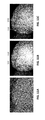
- FIGS. 9A-9G illustrate one method of displaying volumetric information from a patient's teeth.
- FIG. 9A show an example of a 3D volumetric model of a patient's upper jaw (showing teeth and gingiva), from a top view.
- FIG. 9B shows the same 3D volumetric model, showing the internal features, including the more transparent enamel and the less transparent dentin.
- the 3D volumetric model may be manipulated to show more or less of the surface and/or internal structures.
- FIGS. 9C-9G illustrate progressively more transparent views or a region (“C”) of the 3D volumetric model of FIG. 9A .
- FIG. 9C show a 2D image extracted from a region of the 3D volumetric model showing just the outer surface of the teeth (e.g., 100% of the color/outer surface image, 0% near-IR/internal volume).
- FIG. 9D shows the same region as FIG. 9C , combining the outer surface (color) image and the internal (near-IR based) image (e.g., 75% of the color/outer surface image, 25% near-IR/internal volume).
- FIG. 9E shows the same region as FIG. 9C , combining the outer surface (color) image and the internal (near-IR based) image (e.g., 50% of the color/outer surface image, 50% near-IR/internal volume).
- FIG. 9F shows the same region as FIG.
- FIG. 9C shows the same region as FIG. 9C showing just the internal (near IR based) image of the teeth (e.g., 0% of the color/outer surface image, 1000% near-IR/internal volume).
- FIG. 10A illustrates an example of a user interface for analysis and/or display of a 3D volumetric model of a patient's teeth, showing a top view of the upper arch, tools that may be used to manipulate the view(s), and two enlarged views showing the outer surface of an enlarged region of the tooth (on the left) and the same view showing internal features of the tooth (showing dentin and enamel within the tooth).
- FIG. 10B show the user interface of FIG. 10A in which a region of the teeth has been marked/flagged as described herein.
- FIGS. 11A-11C illustrate another example of a method of displaying 3D volumetric image information by mixing it with surface (non-penetrative) information.
- FIG. 11A shows a visible light image of a region of a patient's dental arch taken with a scanner that is also configured to take penetrative (near-IR) scans).
- FIG. 11B show a volumetric model of the reconstructed 3D volumetric model of a patient's tooth showing internal dentin and enamel. Features not visible on the surface scan are apparent in the volumetric scan, including a caries and a bubbled region within the enamel.
- FIG. 11C shows a hybrid image in which the 3D volumetric image has been combined with the surface scan, showing both surface and internal structures, including the carries and the bubbled region.
- FIG. 12 is an example of a method for allowing a user to virtually scan a patient's dental arch. This method may be performed in real time or near real time.
- FIG. 13 is a schematic illustration of a data structure including a 3D model of a patient's dental arch(s) and associated 2D images taken (e.g., via intraoral scanner) of the dental arch at a large number of positions around the dental arch.
- FIG. 14A is an example of a user interface allowing the user to virtually scan over the 3D model of the dental arch, showing corresponding light and near-IR (e.g., external and internal) regions in detail as the user scans over the 3D dental arch; the user may use one or more tools to move the dental arch (e.g., rotate, translate, etc.) and/or the viewing window; the corresponding light and near-IR images may continuously or near-continuously update as the position of the viewing window and dental arch change.
- a pair of imaging windows are shown adjacent to the view of the 3D model of the dental arch.
- FIG. 14B is an alternative display showing a single large image window over or adjacent to the 3D image of the dental arch.
- the image window show a light image of the corresponding region of the dental arch.
- FIG. 14C is an alternative display showing a single large image window over or adjacent to the 3D image of the dental arch.
- the image window show a near-IR image of the corresponding region of the dental arch.
- FIG. 15A is similar to FIG. 14A , showing an example of a 3D model of the outer surface of a dental arch, and a viewing window relative to the dental arch.
- a pair of image display windows are adjacent to the 3D model of the dental arch. The user may move the viewing window over the dental arch (and/or may move the dental arch relative to the viewing window, changing the image(s) shown in the two display windows.
- the upper display window shows a near-IR image corresponding to the dental arch at the position and angle of the plane of the viewing window; the bottom display window shows a corresponding light image (which may be in color).
- FIG. 15B shows another image of the dental arch shown in FIG. 15A , with the dental arch rotated lingually relative to the viewing window; the corresponding near-IR images (upper right) and visible light (lower right) adjacent to the 3D model of the arch are updated to show the slightly rotated view, allowing the user to virtually scan the dental arch and show both external and internal views in real (or near-real) time.
- FIG. 16A is another example of a method of showing a 3D model of a dental arch (shown as the lower arch in this example, e.g., by selecting the lower arch display control in the upper left of the user interface) and showing focused views of near-IR and visible light images corresponding to the viewing window region that may be moved over/across, and around (lingual-occlusal-buccal) the model of the patient's arch.
- FIG. 16B shows an example of a single window (an enlarged near-IR view into the teeth of the region corresponding to the viewing window loop) similar to FIG. 16A .
- FIG. 16C shows an example of a single window (an enlarged visible light view into the teeth of the region corresponding to the viewing window loop) similar to FIG. 16A .
- FIG. 17 schematically illustrates one example of a method for automatically or semi-automatically identify, confirm and/or characterize one or more actionable dental features that may benefit from detection and/or treatment.
- Described herein are methods and apparatuses (e.g., devices and systems) that apply scans of both external and/or internal structures of teeth. These methods and apparatuses may generate and/or manipulate a model of a subject's oral cavity (e.g., teeth, jaw, palate, gingiva, etc.) that may include both surface topography and internal features (e.g., dentin, dental filling materials (including bases and linings), cracks and/or caries).
- a subject's oral cavity e.g., teeth, jaw, palate, gingiva, etc.
- surface topography and internal features e.g., dentin, dental filling materials (including bases and linings), cracks and/or caries).
- Apparatuses for performing both surface and penetrative scanning of the teeth may include intraoral scanners for scanning into or around a subject's oral cavity and that are equipped with a light source or light sources that can illuminate in two or more spectral ranges: a surface-feature illuminating spectral range (e.g., visible light) and a penetrative spectral range (e.g. IR range, and particularly “near-IR,” including but not limited to 850 nm).
- a surface-feature illuminating spectral range e.g., visible light
- a penetrative spectral range e.g. IR range, and particularly “near-IR,” including but not limited to 850 nm.
- the scanning apparatus may also include one or more sensors for detecting the emitted light and one or more processors for controlling operation of the scanning and for analyzing the received light from both the first spectral range and the second spectral range to generate a model of the subject's teeth including the surface of the teeth and features within the teeth, including within the enamel (and/or enamel-like restorations) and dentin.
- the generated mode may be a 3D volumetric model or a panoramic image.
- a volumetric model may include a virtual representation of an object in three dimensions in which internal regions (structures, etc.) are arranged within the volume in three physical dimensions in proportion and relative relation to the other internal and surface features of the object which is being modeled.
- a volumetric representation of a tooth may include the outer surface as well as internal structures within the tooth (beneath the tooth surface) proportionately arranged relative to the tooth, so that a section through the volumetric model would substantially correspond to a section through the tooth, showing position and size of internal structures; a volumetric model may be section from any (e.g., arbitrary) direction and correspond to equivalent sections through the object being modeled.
- a volumetric model may be electronic or physical.
- a physical volumetric model may be formed, e.g., by 3D printing, or the like.
- the volumetric models described herein may extend into the volume completely (e.g., through the entire volume, e.g., the volume of the teeth) or partially (e.g., into the volume being modeled for some minimum depth, e.g., 2 mm, 3 mm, 4 mm, 5 mm, 6 mm, 7 mm, 8 mm, 9 mm, 10 mm, 12 mm, etc.).
- Non-ionizing methods of imaging and/or detecting internal structures may be used, such as taking images using a penetrating wavelength to view structures within the teeth by illuminating them using one or more penetrative spectral ranges (wavelengths), including using trans-illumination (e.g., illuminating from one side and capturing light from the opposite side after passing through the object), and/or small-angle penetration imaging (e.g., reflective imaging, capturing light that has been reflected/scattered from internal structures when illuminating with a penetrating wavelength).
- trans-illumination e.g., illuminating from one side and capturing light from the opposite side after passing through the object
- small-angle penetration imaging e.g., reflective imaging, capturing light that has been reflected/scattered from internal structures when illuminating with a penetrating wavelength.
- multiple penetration images may be taken from the same relative position.
- traditional penetration imaging techniques e.g., trans-illumination
- the angle between the light emitter illumination direction and the detector (e.g., camera) view angle is 90 degrees or 180 degrees
- the angle is much smaller (e.g., between 0 degrees and 25 degrees, between 0 degrees and 20 degrees, between 0 degrees and 15 degrees, between 0 degrees and 10 degrees, etc.).
- Smaller angles may be particularly beneficial because the illumination (light source) and sensing (detector(s), e.g., camera(s), etc.) may be closer to each other, and may provide a scanning wand for the intraoral scanner that can be more easily positioned and moved around a subject's teeth.
- These small-angle penetration images and imaging techniques may also be referred to herein as reflective illumination and/or imaging, or as reflective/scattering imaging.
- penetrating imaging may refer to any appropriate type of penetrating imaging unless otherwise specified, including trans-illumination, small-angle penetration imaging, etc.
- small angles may also result in direct reflection from the surface of the object (e.g., teeth), which may obscure internal structures.
- the methods and apparatuses described here are particularly effective in combining a 3D surface model of the tooth or teeth with the imaged internal features such as lesions (caries, cracks, etc.) that may be detected by the use of penetration imaging by using an intraoral scanner that is adapted for separate but concurrent (or nearly-concurrent) detection of both the surface and internal features.
- Combining surface scanning and the penetration imaging may be performed by alternating or switching between these different modalities in a manner that allows the use of the same coordinate system for the two.
- both surface and penetrative scanning may be simultaneously viewed, for example, by selectively filtering the wavelengths imaged to separate the IR (near-IR) light from the visible light.
- the 3D surface data may therefore provide important reference and angle information for the internal structures, and may allow the interpretation and analysis of the penetrating images that may otherwise be difficult or impossible to interpret.
- the penetrative scans described herein may be collected from, for example, an intraoral scanner such as the one illustrated in FIGS. 1A-1B for generating a three-dimensional (3D) model of a subject's intraoral region (e.g., tooth or teeth, gums, jaw, etc.) which may include internal features of the teeth and may also include a model of the surface, and methods of using such scanners.
- an intraoral scanner such as the one illustrated in FIGS. 1A-1B for generating a three-dimensional (3D) model of a subject's intraoral region (e.g., tooth or teeth, gums, jaw, etc.) which may include internal features of the teeth and may also include a model of the surface, and methods of using such scanners.
- a subject's intraoral region e.g., tooth or teeth, gums, jaw, etc.
- IR penetrative scanning
- an exemplary intraoral scanner 101 may be configured or adapted to generate 3D models having both surface and internal features, or just internal (penetrative) scans.
- an exemplary intraoral scanner may include a handle or wand 103 that can be hand-held by an operator (e.g., dentist, dental hygienist, technician, etc.) and moved over a subject's tooth or teeth to scan both surface and internal structures.
- the wand may include one or more sensors 105 (e.g., cameras such as CMOS, CCDs, detectors, etc.) and one or more light sources 109 , 110 , 111 .
- sensors 105 e.g., cameras such as CMOS, CCDs, detectors, etc.
- first light source 109 configured to emit light in a first spectral range for detection of surface features (e.g., visible light, monochromatic visible light, etc.; this light does not have to be visible light)
- second color light source e.g., white light between 400-700 nm, e.g., approximately 400-600 nm
- third light source 111 configured to emit light in a second spectral range for detection of internal features within the tooth (e.g., by trans-illumination, small-angle penetration imaging, laser florescence, etc., which may generically be referred to as penetration imaging, e.g., in the near-IR).
- penetration imaging e.g., in the near-IR
- a selectable light source may be used.
- the light source may be any appropriate light source, including LED, fiber optic, etc.
- the wand 103 may include one or more controls (buttons, switching, dials, touchscreens, etc.) to aid in control (e.g., turning the wand on/of, etc.); alternatively or additionally, one or more controls, not shown, may be present on other parts of the intraoral scanner, such as a foot petal, keyboard, console, touchscreen, etc.
- any appropriate light source may be used, in particular, light sources matched to the mode being detected.
- any of these apparatuses may include a visible light source or other (including non-visible) light source for surface detection (e.g., at or around 680 nm, or other appropriate wavelengths).
- a color light source typically a visible light source (e.g., “white light” source of light) for color imaging may also be included.
- a penetrating light source for penetration imaging e.g., infrared, such as specifically near infrared light source
- penetration imaging e.g., infrared, such as specifically near infrared light source
- the intraoral scanner 101 may also include one or more processors, including linked processors or remote processors, for both controlling the wand 103 operation, including coordinating the scanning and in reviewing and processing the scanning and generation of the 3D model including surface and internal features.
- the one or more processors 113 may include or may be coupled with a memory 115 for storing scanned data (surface data, internal feature data, etc.).
- Communications circuitry 117 including wireless or wired communications circuitry may also be included for communicating with components of the system (including the wand) or external components, including external processors.
- the system may be configured to send and receive scans or 3D models.
- One or more additional outputs 119 may also be included for outputting or presenting information, including display screens, printers, etc.
- inputs 121 buttons, touchscreens, etc.
- the apparatus may allow or request user input for controlling scanning and other operations.
- FIG. 2 illustrates one example of a scanner that may be used.
- the scanner shown may be used as part of a system such as the system shown in FIGS. 1A-1B .
- FIG. 2 shows a schematic of intraoral scanner configured to do both surface scanning (e.g., visible light, non-penetrative) and penetrative scanning using a near infra-red (NIR) wavelength (at 850 nm in this example).
- NIR near infra-red
- the scanner includes a near-IR illumination light 289 and a first polarizer 281 and a second polarizer 283 in front of the image sensor 285 to block near-IR light reflected off the surface of the tooth 290 (P-polarization light) while still collecting near-IR light scattered from internal tooth structures/regions (S-polarization light).
- the NIR light illuminates the tooth in P-polarization, and specular light reflected from the surface of the tooth, e.g., the enamel, is reflected with specular reflection hence its P-polarization state is conserved.
- Near-IR light penetrating the internal tooth features, such as the dentin, is scattered resulting in random polarization (S and P).
- the wavelength selective quarter waveplate 293 does not modify the polarization of the near-IR light (e.g., it leaves the polarization state of the near-IR light being delivered unchanged) but changes the polarization of the returning scan light from P to S such that only surface reflection are captured in the scan wavelength.
- the returning near-IR light having a mixture of S and P polarizations, is first filtered through the polarization beam splitter (PBS) 294 and polarizing filter 283 such that only the S-polarization is transmitted to the image sensor.
- PBS polarization beam splitter
- polarizing filter 283 such that only the S-polarization is transmitted to the image sensor.
- specular light having the original p-polarization
- Other intraoral scanner configurations with or without polarization filters such as those shown in FIG. 2 may be used as part of the probe.
- the surface scan may be performed by illuminating the surface (using the scanner illumination unit 297 ), illuminating in p-polarization, and the polarization may be reversed by the wavelength-selective quarter waveplate 293 (transmitting S-polarization light to the image sensor).
- penetrative scanning techniques may be used or incorporated into the apparatuses described herein for performing scans that to detect internal structures using a penetrative wavelength or a spectral range of penetrative wavelengths, including, but not limited, to trans-illumination and small-angle penetration imaging, both of which detect the passage of penetrative wavelengths of light from or through the tissue (e.g., from or through a tooth or teeth).
- these apparatuses and techniques may be used to scan intraoral components such as a tooth or one or more teeth, gingiva, palate, etc. and used to generate a model of the scanned area. These models may be generated in real time or after scanning.
- 3D volumetric models of the teeth may be referred to as 3D volumetric models of the teeth, but may include other regions of the jaw, including the palate, gingiva and teeth.
- the methods and apparatuses described herein typically relate to 3D volumetric models, the techniques and methods described herein may also be used in some instance with 3D surface models.
- the surface model information is typically part of the 3D volumetric model.
- FIG. 3 illustrates an example of a data flow for scanning teeth with an intraoral scanner to build a 3D model including internal structures.
- the exemplary method shown may include three parts.
- the teeth may be scanned with an intraoral scanner 1701 (or any other scanner) configured to provide penetrative scans into the teeth using an optical (e.g., IR, near IR, etc.) wavelength or range of wavelengths. Any of these scanners may also concurrently scan to determine a surface features (e.g., via one or more non-penetrative wavelengths), color, etc., as described above.
- an optical e.g., IR, near IR, etc.
- Any of these scanners may also concurrently scan to determine a surface features (e.g., via one or more non-penetrative wavelengths), color, etc., as described above.
- a plurality of penetrative scans 1703 , 1703 ′ may be taken, and the position of the sensor (e.g., camera) 1705 , 1705 ′ (e.g., x,y,z position and/or pitch, roll, yaw angles) may be determined and/or recorded for each penetrative image.
- the surface of the teeth may also and concurrently be imaged, and a 3D surface model of the teeth 1707 determined, as described above.
- the patient's teeth may be scanned, for example, with an intraoral 3D scanner 1702 that is capable of imaging the inner teeth structure using, for example, near infra-red imaging.
- the location and orientation of the camera may be determined, in part, from the 3D scanning data and/or the 3D teeth surface model 1707 .
- the penetrative images may be segmented 1711 .
- segmentation may be done in one of two ways.
- the images On the inner teeth structure images, the images may be segmented using contour finding 1713 , 1713 ′.
- Machine learning methods may be applied to further automate this process.
- near images where their camera position is close
- the method may also include projecting pixels from the inner teeth images back to the teeth and calculating a density map of inner teeth reflection coefficient. Enclosing surfaces of different segments may be found or estimated by using iso-surfaces or thresholds of the density map and/or by machine learning methods.
- segmenting the images and projecting the segments back to a model (such as the 3D surface model, e.g., projecting back to the world), may be used to find a segment by the intersection of the segment projections and the teeth surface.
- the results may be displayed 1717 , transmitted and/or stored.
- the results may be displayed by the scanning system during the intraoral scanning procedure.
- the results may be shown by images with enclosing contours for different segments, a 3D density map, etc.
- a density map 1715 representing the dentin beneath the enamel on the outer surface, is shown.
- This image may be color coded to show different segments.
- internal segments (structures) are shown within the 3D surface model (which is shown transparent); not all teeth have been scanned with penetrative images, thus, only some are shown.
- Alternative views, sections, slices, projections or the like may be provided.
- the example image includes artifacts that are present outside of the teeth 1716 ; these may be removed or trimmed, based on the surface model 1718 .
- a segment may mark each pixel on the image.
- Internal structures such as dentin, enamel, cracks, lesions, etc. may be automatically determined by segmentation, and may be identified manually or automatically (e.g., based on machine learning of the 3D structure, etc.). Segments may be displayed separately or together (e.g., in different colors, densities, etc.) with or without the surface model (e.g., the 3D surface model).
- the patient is initially scanned with a 3D scanner capable of both surface scanning and penetrative scanning (e.g., near IR imaging), and the orientation and/or position of the camera is known (based on the position and/or orientation of the wand and/or the surface scans).
- This position and orientation may be relative to the tooth surface.
- the method and apparatus may therefore have an estimate of the camera position (where it is located, e.g., x,y,z position of the camera, and its rotational position).
- penetrative images e.g., near IR or IR images
- penetrative images may be segmented automatically.
- volumetric data from the intraoral cavity may identify features and information from teeth that were previously difficult or impossible to identify from non-volumetric scanning. However, it may be difficult or non-intuitive for a dental practitioner (and/or patient) to analyze three-dimensional volumetric information. Described herein are methods and apparatuses for viewing and interpreting 3D, volumetric data of a patient's oral cavity.
- FIGS. 9A-9G illustrate one example of a method for displaying 3D volumetric data.
- FIG. 9A shows a surface model (which may be a surface model portion of a volumetric model) from a top view of an upper arch, in which external features are visible (e.g., surface features). This view is similar to the surface scan view which may be in color (e.g., taken by visible light). Internal structures, which are present within the model beneath the external surface of the scan, are not readily visible in FIG. 9A .
- FIG. 9B the internal structures are shown based on their relative transparency to near-IR light. In FIG. 9B , the enamel is more transparent (and is shown as more transparent) than the dentin, which is shown as less transparent.
- FIGS. 9B-9F illustrate a transition between the surface view of FIG. 9A and the penetrative, internal 3D view of FIG. 9B for a sub region (circled region “C”) shown.
- a user display may be provided in which the relative surface vs. internal views may be altered to provide a sense of internal structures within the dental arch relative to surface structures.
- an animated view cycling through image such as FIGS. 9C-9G may be provided.
- the user may slide a slider 903 toggling between the surface and internal views. The transition between these two views (which may be made from any angle, may help the user and/or patient to see beneath the surface of the teeth, to visually assess the rich internal data.
- the 3D volumetric model may be manipulated to show any view, including cross-sectional views, showing internal structures and/or surface features.
- FIG. 9A-9G the top view is shown.
- FIGS. 9C-9G illustrate progressively more transparent views or a region (“C”) of the 3D volumetric model of FIG. 9A in which progressively large percentages (from 0% to 100%) of the internal view of FIG. 9B is shown for region C, while progressively less of the surface view (from 100% to 0%) is shown.
- FIGS. 11A-11C illustrate another example, showing a hybrid image that (like FIG. 9E combines and mixes both surface image scanning (e.g., a visible light scan, as shown in FIG. 11A ) with a volumetric model taken using a penetrative (e.g., near-IR) wavelength, as shown in FIG. 11B .
- a carries region 1103 is apparent, which is not visible in FIG. 11A .
- a bubbled region of the enamel 1105 is visible in FIG. 11B but is not visible in FIG. 11A .
- FIG. 11C shows a hybrid image of the 3D volumetric model and surface model (surface image), in which both of these structures, the carries and the bubbled region, are visible.
- a patient's oral cavity e.g., teeth, gingiva, palate, etc.
- the data may be presented as one or a series of x-ray type images, similar from what would be produced by dental x-rays.
- FIG. 4 illustrates one method of generating x-ray (or pseudo x-ray) images from a volumetric data set taken as described above, e.g., using penetrative light (e.g., near-IR) wavelength(s).
- penetrative light e.g., near-IR
- a method of displaying 3D volumetric images of a patient's oral cavity may include receiving the 3D volumetric data 401 , e.g., from a scan as described above, either directly or from a stored digital scan, etc.
- individual teeth or groups of teeth may be identified from the volumetric data 403 .
- the teeth may be identified automatically (e.g., by a segmenting the volume, by machine learning, etc.) or manually. Alternatively, the entire volume may be used. Pseudo-x-ray images may then be generated from the volume or sub-sets of the volume corresponding to individual teeth 405 .
- an image of the volume may be taken from the ‘front’ of the tooth or teeth, in which the transparency of the enamel (and/or enamel-like restorations), dentin and other features are kept from the volumetric data.
- This volumetric data may be based on the absorption coefficients of the material within the oral cavity for the penetrating wavelength of light used.
- a projection through the volumetric data may be generated for a fixed direction from the volumetric data to get an image similar to an X-ray, but, in some variations, inverted and showing the density of the dentin (highly absorbing) as “darker” than the density of the enamel (less absorbing and therefore more transparent); caries may also show up as more absorbing (darker) regions.
- the image may therefore be inverted to resemble an x-ray image in which more dense regions are lighter (e.g., brighter).
- pseudo x-ray images may be generated from the same positions as standard dental x-rays and presented to the user. For example, a panel of pseudo x-ray images may be generated from the volumetric model for each of the patient's teeth. Although the penetration of the wavelength of the light (e.g., near IR light) may not be as deep as with traditional x-rays, images generated in this manner may provide a comparable proxy for an x-ray, particularly in the crown and mid-tooth regions above the gingiva.
- the wavelength of the light e.g., near IR light
- images of the teeth may be generated from the volumetric data in which the image is simplified by pseudo coloring the volumetric data to highlight certain regions. For example, regions that have been previously marked or flagged (as will be described in greater detail, below) may be colored in red, while the enamel may be shown as a more natural white or slightly off-white color.
- enamel-like materials e.g., from fillings, etc.
- the methods and/or apparatuses may display the teeth in sections through the dental arch.
- the individual teeth or groups of teeth may be shown separately and/or labeled (e.g., by standard naming/numbering convention). This may be shown in addition or instead of other displays.
- the teeth and/or internal structures may be pseudo-colored or projecting on to a color image may be used.
- FIGS. 5A and 5B illustrate virtual sections taken through a volumetric model of a patient's teeth generated from an intraoral scan that included near-IR information, and described above.
- the cross-sectional view may be generated automatically or manually, e.g., by the user, to display regions of interest within teeth, including enamel.
- the cross-section may show both density sectioning and/or surface sectioning. These images may be pseudo-colored to show different regions, including outer surfaces, enamel, dentin, etc.
- Internal structures, e.g., within the enamel and/or dentin may reflect the effect of the near-IR light within the teeth, such as the absorption and/or reflection of light at one or more near-IR/visible wavelengths within the teeth.
- the section is pseudo-colored with a heat map to show internal features, and a key may be provided, as shown.
- 2D projections of the teeth may be generated from the volumetric information, showing one or more features on the tooth and/or teeth.
- additional features including lesions (e.g., caries/cavities, cracks, wearing, plaque build-up, etc.) may be displayed as well, and may be indicated by color, texture, etc.
- lesions e.g., caries/cavities, cracks, wearing, plaque build-up, etc.
- other embodiments may display the 2D section by itself to provide a cross-sectional view of the tooth/teeth similar to a view provided by 2D x-ray images.
- any of the methods and apparatuses for performing them described herein may include displaying one or more (or a continuous) sections through a 3D model of the patient's dental arch, and preferably a 3D volumetric model.
- a method of displaying images from a three-dimensional (3D) volumetric model of a patient's dental arch may include: collecting the 3D volumetric model of the patient's dental arch, wherein the 3D volumetric model includes near-infrared (near-IR) transparency values for internal structures within the dental arch; generating a two-dimensional (2D) view into the 3D volumetric model including the patient's dental arch including the near-IR transparency of the internal structures; and displaying the 2D view.
- the method may optionally (but not necessarily) include scanning the patient's dental arch with an intraoral scanner.
- Generating the 2D view may comprises sectioning the 3D volumetric model in a plane through the 3D volumetric model.
- the user may select the plane's location and/or orientation, and my do this in a continuous manner.
- any of these methods may include selecting, by a user, a section though the 3D volumetric model to display, wherein selecting comprises continuously selecting sections through the 3D volumetric model as the user scans through the 3D model and continuously displaying the 2D views corresponding to each section.
- Generating the 2D view may comprises selecting, by a user, an orientation of the 2D view.
- a method of displaying images from a three-dimensional (3D) volumetric model of a patient's dental arch may include: collecting the 3D volumetric model of the patient's dental arch, wherein the 3D volumetric model includes surface values and near-infrared (near-IR) transparency values for internal structures within the dental arch; generating a two-dimensional (2D) view into the 3D volumetric model including the patient's dental arch including both surface values and the near-IR transparency of the internal structures; and displaying the 2D view.
- the surface values may comprise surface color values.
- the surface relative to the internal (volumetric) structures may be weighted.
- generating the two-dimensional (2D) view through the 3D volumetric may also include including in the 2D view a weighted portion of the surface values and a weighted portion of the near-IR transparency of the internal structures.
- the weighted portion of the surface values may include a percentage of the full value of the surface values
- the weighted portion of the near-IR transparency of the internal structures comprises a percentage of the full value of the near-IR transparency of the internal structures, wherein the percentage of the full value of the surface values and the percentage of the full value of the near-IR transparency of the internal structures adds up to 100%.
- the user may adjust the weighted portion of one or more of the surface values and the near-IR transparency of the internal structures.
- a method of displaying images from a three-dimensional (3D) volumetric model of a patient's dental arch may include: collecting the 3D volumetric model of the patient's dental arch, wherein the 3D volumetric model includes surface color values and near-infrared (near-IR) transparency values for internal structures within the dental arch; selecting, by a user, an orientation of a view of the 3D volumetric model to display; generating a two-dimensional (2D) view into the 3D volumetric using the selected orientation, including the patient's dental arch including a weighted portion of the surface color values and a weighted portion of the near-IR transparency of the internal structures; and displaying the 2D view.
- volumetric measurements of one or more lesions may be provided (selectably or automatically) including dimensions (peak or mean length, depth, width, etc.), volume, etc. This may be performed by manually or automatically segmenting the volumetric model to define the regions of interest, including either or both tooth features (enamel, dentin, etc.) and/or irregularities (e.g., caries, cracks, etc.). Any appropriate segmentation technique may be used, such as but not limited to: mesh segmentation (mesh decomposition), polyhedral segmentation, skeletonization, etc. Once the volume has been segmented, these regions may be separately or collectively displayed and/or measured. As will be described below, they may also be marked/flagged and used for further analysis, display and modification of the scanning methods and systems.
- a summary report may be generated or created and displayed for the user and/or patient from the volumetric data.
- summary data may be projected onto a model of the patient's teeth.
- the model may also be simplified, so that the enamel is opaque, but marked or selected internal features (including automatically selected internal features) are shown in red or some other contrasting color (and/or flashing, blinking, etc.) within the tooth.
- caries may be shown in this manner.
- the summary report may be automatically entered into a patient chart.
- any of the images, including the volumetric images, may be animated. For example, virtual sections through the patient's teeth, showing a scanning or traveling cross-section through the patient's dentition may be shown, in some cases with a 3D model showing one or more cutting axes through the volume.
- the user interface may allow the user to section in one or more planes, showing both external and internal features based on the volumetric scan.
- the apparatuses described herein may generate separate view for the user (e.g., physician, dentist, orthodontist, etc.) than the patient.
- the user may be provided with a ‘clinical view’ that may include information not present on a separate ‘patient view.’
- the clinical view may be more technical, and may in some cases be closer to the raw images from the volumetric data.
- the user may select which layers of information to include in the patient view, which may then be presented to the user during or after the scanning or review of the dental scanning.
- Patient educational materials may be appended to the patient view.
- the user display of volumetric data may include an overlay of the volumetric data in which pseudo coloring of the 3D components within the volumetric data is shown.
- pseudo coloring of the 3D components within the volumetric data may be shown.
- 2D color data and 3D near-IR data may be shown, including transitions between the two.
- the volumetric information may be annotated (e.g., marked, labeled, etc.) either automatically, manually, or semi-automatically, and this annotation may be displayed.
- annotations may be used both to annotate future additional scans, and to modify how future scans of the same patient are taken and displayed.
- An annotation may be, for example, a marker or flag on a region of interest. Regions of interest may correspond to specific regions in which one or more features (cracks, caries, thinning of enamel, buildup of plaque or calculus, etc.) have been observed. Alternatively or additionally, regions of interest may be regions in which there is a change over time, e.g., from one scan to another scan.
- any of these methods may include placing one or more markers on the volumetric model of the patient's teeth.
- Markers e.g., flags, pins, etc.
- Markers may be used to focus attention and/or processing by the system on one or more specific regions of the volumetric model for display, and/or for later follow-up (e.g., in future scans). Markers may modify the manner in which the later scans are taken, e.g., taking future scans of marked regions with greater detail (e.g., higher resolution, different wavelengths, greater scanning frequency or reputations, etc.). Marked regions may be displayed over time to show changes in the marked regions.
- a user can mark a digital representation of the patient's teeth (or the patient's actual teeth) with a marker (e.g., a pin, flag, etc.) which can be annotated (e.g., can have notes associated with it).
- This marker may then be used to track over time between different scans. Later scans can be marked in the corresponding location, the later scan can be modified based on the marked regions. These marked regions may be scanned in greater detail, and analytics may be automatically performed and/or displayed, measuring and/or indicating a change compared to one or more earlier scans.
- any of the systems described herein may track one or more marked regions from previous scans and give feedback during and/or after a new scan, providing additional detail. This can be done for both surface and/or volumetric information, particularly on the properties of the enamel, and/or by comparison to the enamel, the outer surface of the teeth/tooth, and/or the dentin.
- one or more annotation markers from an earlier scan may modify a subsequent scanning of the same patient.
- the user Before scanning, the user may enter an identifier of the patient being scanned (alternatively the system may automatically identify the patient based on a database of earlier scans). The system may automatically annotate the new scan based on the prior scan annotations.
- the later scans may be automatically annotated by the system by identifying differences between the prior scan(s) and the current scan. For example, regions showing a change above a threshold compared to the earlier scan may be flagged and presented to the user.
- the annotation may be done without user oversight (fully automatic) or may be done with some user oversight, for example, by flagging it and indicating to the user why it was flagged, then allowing the user to keep, modify or reject the marking.
- Reasons for automatically marking the teeth may include a change in the enamel thickness, a change in the surface smoothness, a change in the relative ratio of enamel vs. dentin in a tooth, a change in the position of the tooth (e.g., occlusion), etc.
- the system may also include a change in structures external to the natural tooth, such as increase or decrease in plaque or calculus buildup, or changes to the gingival structures surrounding the tooth, Thus, if the system detects one or more of these conditions, it may automatically flag the relevant region in the volumetric model.
- Later scans may be dynamically modified by the flags from earlier scans or by a detection of a change in a region (even unmarked regions) compared to earlier scans.
- the scanning parameters may be modified to scan at higher resolution (e.g., changing the scan dwell time, requiring the user to scan this region multiple times, etc.), changing the wavelength used for the scanning, etc.
- FIG. 6 illustrates a method of automatically selecting a region for marking and/or using the selected regions.
- a first volumetric model of the patient's teeth is generated from a scan of the patient's teeth 601 .
- the volumetric model may be generated using any appropriate method, including those described above and discussed in U.S. patent application Ser. No. 15/662,234, filed Jul. 27, 2017, titled “INTRAORAL SCANNER WITH DENTAL DIAGNOSTICS CAPABILITIES”, incorporated by reference in its entirety.
- the first volumetric model may be stored (digitally stored) as part of the patient's dental record.
- the first volumetric model may concurrently or subsequently (immediately or some time thereafter) be analyzed (e.g., by an apparatus, including an apparatus having a processor that is configured to operate as described herein) to identify any regions that should be flagged 603 .
- This analysis may therefore be performed automatically, and may examine one or more properties of the patient's teeth from the scan.
- Automatic or semi-automatic, etc., automatic but with manual assistance to verify/confirm
- the apparatus may examine the digital model to identify possible defects in the patient's teeth such as (but not limited to): cracks, caries, voids, changes in bite relationship, malocclusions, etc. This may include identifying regions in which there is an optical (e.g., near-IR) contrast near the surface of the tooth indicating a possible crack, caries, occlusion, etc. 605 . Regions that are less transparent (e.g., more absorptive) in the near-IR wavelengths than the rest of the enamel, that are closer to the surface (generally or within specific near-IR wavelengths), may correspond to defects. Alternatively or additionally, surface properties of the teeth may be examined and flagged if they are outside of a threshold 607 .
- optical e.g., near-IR
- regions of the surface of the teeth in which the tooth surface is rough may be flagged.
- Other surface properties may also be analyzed and used to determine if the region should be marked or presented to the user to confirm marking, including discoloration (based on a color or white-light/surface scan), gingival position (relative to the outer surface of the tooth), etc.
- the distribution and size of the patient's enamel may also be examined 609 .
- the enamel thickness may be determined from the optical properties (e.g., comparing absorption/reflectance properties). Regions of putative enamel that are below a threshold thickness, or having a ratio of thickness to tooth dimension (e.g., diameter, width, etc.) below a threshold may be marked or presented to the user to confirm marking.
- the use may also manually flag one or more regions of the volumetric model of the patient's teeth 615 . If the automatic analysis of the volumetric model automatically flags the identified volumetric model the user's manually added regions may be added. In some variations, the user may be prompted to flag the regions identified and suggested by the automatic analysis. These regions may be marked and an indication of the reason(s) for their being identified may be provided (e.g., irregular enamel, potential crack, potential caries, potential thinning of the enamel, etc.). In general, the inner boundary (the boundary within the volume of the tooth, for example) may be defined in any of the methods and apparatuses described herein.
- the methods and apparatuses described herein may be used to the entire layer (e.g., layer of enamel, region of the inner structure of the tooth) may be identified and used for qualitative and/or quantitative information.
- the method flagged regions may then be displayed on a digital model of the patient's teeth 617 .
- the display may emphasize the flagged regions, e.g., by providing a color, animation (e.g., flashing), icon (e.g., arrow, flag, etc.), or the like, including combinations of these.
- the display may also show enlarged views of any of these.
- the user may modify the display, e.g., rotating, sectioning, enlarging, etc., the flagged region.
- the flagged regions may be enlarged on the display by default.
- An index or key of the flagged regions may be provided, and may be displayed and/or stored with the digital volumetric model of the patient's teeth.
- the method may include using the flagged regions to modify the future scans, as mentioned above.
- the method may include scanning (“rescanning”) using the flagged regions, after some interim time period of between a first time (e.g., about one day, one week, one month, 2 months, 3 months, 4 months, 5 months, 6 months, 7 months, 8 months, 9 months, 10 months, 11 months, 1 year, 1.2 years, 1.5 years, 2 years, etc.) and a second time (e.g., about one month, 2 months, 3 months, 4 months, 5 months, 6 months, 7 months, 8 months, 9 months, 10 months, 11 months, 1 year, 1.5 years, 2 years, 3 years, 4 years, 5 years, 6 years, 7 years, 8 years, 9 years, 10 years, 11 years, 12 years, 13 years, 14 years, 15 years or more, etc.), or longer than the second time period.
- a first time e.g., about one day, one week, one month, 2 months, 3 months, 4 months, 5 months, 6 months
- the flagged regions may be used to modify the scan by increasing the resolution of the scanned regions during the scan, e.g., by increasing the scan rate, increasing the dwell time in this region, scanning the region in additional wavelengths, scanning this region multiple times, etc.
- the scanning apparatus may inform the user to adjust the scanning (e.g., moving the wand of an intraoral scanner more slowly in these regions, moving the scanner back over these regions multiple times, etc.) and/or it may automatically adjust the scanning parameters during operation.
- the scanning apparatus may therefore receive the key or index of flagged regions and/or a marked (flagged) version of the patient's earlier intraoral scan(s). Prior to scanning the scanning apparatus, the user may indicate the identity of the patient being scanned, and this may be used to look up the earlier scan(s).
- the apparatus may identify the patient based on the current scan to identify (or confirm the identity) of the patient, to verify or recall the earlier annotated (flagged) scan.
- the second or subsequent scans may be taken without using the earlier flagged regions.
- the method or apparatus configured to perform the method may then compare the flagged regions from the subsequent scan with the corresponding regions from the earlier scan(s) 621 .
- the volumetric model from the subsequent scan may automatically analyzed to identify any regions of the new (subsequent) scan that can/should be marked/flagged (e.g., repeating the earlier automatic or semi-automatic analysis steps 603 - 615 ) 621 . Newly identified regions from the subsequent scan may be compared to the previously un-flagged corresponding regions in the earlier volumetric model(s).
- the flagged regions may be analyzed over time 623 .
- Specific sub-regions from the volumetric model including the flagged regions may be generated for display and analysis.
- the results may be output 625 .
- these regions may be displayed to the user along with descriptive, analytic information about the scanned region. These regions may also be marked to shows changes over time.
- the data may be displayed in an animation, for example, showing changes over time.
- the images may be displayed as a time-lapse image (video, loop, etc.), showing changes. Time-lapse images may show the change in the internal and/or external structure over time. Sections (pseudo-sections generated from the volumetric model(s)) may be used to show changes. Color, texture, patterns, and any other highlighting visualization technique may be used.
- these regions (and any accompanying analysis) may be output in other appropriate ways, including digitally outputting (e.g., the patient's dental record), printing a description of the flagged region(s), etc.
- any of the methods of tracking a region of a patient's dental arch described herein may include tracking over time and/or across different imaging modalities (e.g., records) as described in greater detail below. Further, any of these methods may be automated and/or may include automated agents, for example, for identifying one or more regions of interest (e.g., features, defects, including actionable dental features), including for scoring them and/or automating identification, scoring and/or display of such regions. Any of these methods may also include any of the display methods or agents (e.g., for displaying sections, displaying internal structures, for displaying virtual x-rays, for displaying across imaging modalities, etc.).
- regions of interest e.g., features, defects, including actionable dental features
- Any of these methods may also include any of the display methods or agents (e.g., for displaying sections, displaying internal structures, for displaying virtual x-rays, for displaying across imaging modalities, etc.).
- a method of tracking a region of a patient's dental arch over time may include: collecting a first three-dimensional (3D) volumetric model of the patient's dental arch, wherein the 3D volumetric model includes surface values and near-infrared (near-IR) transparency values for internal structures within the dental arch; identifying a region of the 3D volumetric model; flagging the identified region; collecting a second 3D volumetric model of the patient's dental arch; and displaying one or more images marking, on the one or more images, a difference between the first 3D volumetric model and the second 3D volumetric model at the flagged region.
- 3D three-dimensional
- Any of these methods may also include tracking and/or comparing across different records (e.g., different imaging modalities), so that identifying comprises identifying a region of the patient's dental arch from a first record of a plurality of records, wherein each record comprises a plurality of images of a patient's dental arch each taken using an imaging modality, further wherein each record of the plurality of records is taken at a different imaging modality, further wherein flagging comprises flagging the identified region in a corresponding region of the 3D volumetric model of the patient's dental arch.
- different records e.g., different imaging modalities
- the method and apparatuses for performing them may also include correlating the flagged region with each of records of the plurality of records by correlating the 3D volumetric model of the patient's dental arch with each of the records of the plurality of records.
- the correlation may be used to weight or grade the identified region to determine if it corresponds to a region of interest (e.g., a feature, a defect, including actionable dental features, etc.).
- the surface values may comprise surface color values.
- These methods may be used with stored data and/or with data collected in real time (e.g., thus the method may optionally but not necessarily collect a three-dimensional (3D) volumetric model by scanning the patient's dental arch to generate the 3D volumetric model.
- Identifying the region may comprise comprises automatically identifying using a processor.
- automatically identifying may comprise identifying a surface color value outside of a threshold range.
- Automatically identifying may comprise segmenting the 3D volumetric model to identify enamel regions and identifying regions having enamel thicknesses below a threshold value.
- Flagging the identified region may comprise automatically flagging the identified regions or manually confirming the identified region for flagging.
- the method may include re-scanning the patient's dental arch wherein the flagged region is scanned at a higher resolution than un-flagged regions.
- a method of tracking a region of a patient's dental arch over time may include: collecting a first three-dimensional (3D) volumetric model of the patient's dental arch taken at a first time, wherein the 3D volumetric model includes surface color values and near-infrared (near-IR) transparency values for internal structures within the dental arch; identifying, using an automatic process, a region within the 3D volumetric model to be flagged from a first record of a plurality of records, wherein each record comprises a plurality of images of a patient's dental arch each taken using an imaging modality, further wherein each record of the plurality of records is taken at a different imaging modality; flagging the identified regions; correlating the flagged region with each of the records of the plurality of records by correlating the 3D volumetric model of the patient's dental arch with each of the records of the plurality of records; collecting a second 3D volumetric model of the patient's dental arch taken at a separate time; and displaying a difference between the first 3D volumetric model and
- a method of tracking a dental feature across different imaging modalities comprising: collecting a first three-dimensional (3D) volumetric model of the patient's dental arch, wherein the 3D volumetric model of the patient's dental arch includes surface values and internal structures within the dental arch; identifying a region of the patient's dental arch from a first record of a plurality of records, wherein each record comprises a plurality of images of a patient's dental arch each taken using an imaging modality, further wherein each record of the plurality of records is taken at a different imaging modality; flagging the identified region in a corresponding region of the 3D volumetric model of the patient's dental arch; correlating the flagged region with each of the records of the plurality of records by correlating the 3D volumetric model of the patient's dental arch with each of the records of the plurality of records; and saving, displaying and/or transmitting images including the region of the patient's dental arch. Any of these methods may include tracking over time as well
- FIGS. 10A and 10B illustrate a user interface showing marking of a region of interest from a 3D volumetric scan of a patient's oral cavity.
- the user interface includes an image of the internal features 1001 (e.g., based on the near-IR absorption of the teeth), similar to FIG. 9B , discussed above.
- This view may be manipulated by user controls 1015 , including sectioning tools, rotation, moving tools, etc.
- two upper windows show a surface view 1003 and a volumetric (internal) view 1005 corresponding to the same region. This region may be selected.
- FIG. 10B shows the same features of FIG. 10A , but with a region marked or flagged 1011 .
- the identification of the region to be flagged may be automatic or manual, or semi-automatic (e.g., confirmed by the user), and may be chosen to select a region for later monitoring.
- the region may correspond, for example, to possible caries.
- Monitoring of one or more internal regions of the teeth over time using the volumetric models of the patient's teeth taken with the devices described herein may be particularly helpful for predicting dental problems, including caries, cracks, tooth loss, gum recession, and the like.
- these methods and apparatuses may help the user (e.g., dentist, dental technician, orthodontist, etc.) inform and educate a patient so that the patient may take recommended treatments prior to developing more serious problems.
- pre-cavitated caries are difficult to identify with current imaging techniques, and it may be particularly difficult to convince a patient to treat even when identified, since they typically present without pain.
- early stage treatment may be critical to avoiding more complicated and dangerous procedures later.
- Dental caries are one type of problem that may be identified with the methods and apparatuses described herein.
- caries may be identified from 3D volumetric models (such as those described herein) that penetrate into teeth using light (e.g., near-IR), one type of non-ionizing radiation.
- light e.g., near-IR
- the absorption coefficients of the internal regions of the teeth may indicate distinctions between dentin and enamel, and may reveal internal structures and flaws, including cracks, caries and the like.
- regions of enamel that are less transparent than expected in the near-IR wavelengths may (e.g., having different IR optical properties), and particularly those that appear to extend to the surface of the tooth in the volumetric model may be identified manually or automatically as cavities or caries.
- Other irregularities in the enamel and/or dentin e.g., based on the internal features of the teeth from the volumetric model may be identified and may be characteristic of a problem in the teeth.
- the techniques described herein may be used for prognosis of dental issues such as caries.
- any of the apparatuses and methods described herein may include improved methods for displaying of internal tooth features using the one or more volumetric models of a patient's teeth.
- the methods and apparatuses described herein may be used to generate an estimate of enamel thickness for one or more of the patient's teeth. These estimates may be visually displayed, showing the outer surface of the teeth or a particular tooth, and may also show internal structures, including in sectional views or 3D internal views showing, e.g., the enamel, including the thickness of the enamel.
- This information may be used clinically to determine the need for, to help design and to help apply dental prosthetics, including veneers, crowns, and the like.
- Any of the methods and apparatuses described herein may be used, for example, to help prepare design a dental implant for a particular tooth or teeth.
- the method and apparatuses described herein may also be used for the detection and visualization (including quantification) of plaque and calculus on the patient's teeth.
- Any of the intraoral scanners described herein may be used to detect plaque or calculus on the patient's teeth by using florescence imaging in addition to other imaging/scanning modalities including penetrative (e.g., near-IR) imaging.
- florescence imaging in addition to other imaging/scanning modalities including penetrative (e.g., near-IR) imaging.
- intraoral scanner may be cycled between different imaging modalities such as between white light and near-IR, including additional modalities such as florescence (e.g., laser florescence, etc.).
- plaque and calculus may be detected and may be displayed and highlighted as described above, and may be used before, during or after treatment.
- a dental technician e.g., dental hygienist
- data on plaque and calculus may also be used by any of the apparatuses described herein to determine and provide predictive models that may indicate plaque and calculus (e.g., tartar) generation rate and/or locations.
- plaque and calculus may be identified at least in part using florescence information. It has been observed that plaque may fluoresce under blue light (e.g., around about 405 nm). Any of the intraoral scanners described herein may include fluorescence information from which information about plaque and calculus may be used, and incorporated into a 3D model of the patient's teeth. For example, plaque and/or calculus may be visually displayed as a color and/or texture on the 3D model of the patient's teeth.
- a fluorescence signal can be obtained from an intraoral scanner using a dichroic filter having a large aperture amplification of fluorescence signal. This amplification may emphasize the fluorescence, thus enabling the detection, visualization and segmentation of plaque and calculus regions using RGB illumination, sensor and image.
- the apparatus may include a florescence source (e.g., an LED emitting at 405 nm) and corresponding filter(s) for detection of plaque and/or calculus. This may be integrated in the intraoral scanner, or added (e.g., as a sleeve, accessory, etc.) to be used with the scanner.
- the plaque and calculus may have a different absorption/reflection than enamel. This may allow the calculus and/or plaque to be differentiated from the enamel in the volumetric model. Further, the volumetric model may be used to detect material on the teeth, including calculus and plaque based on the surface smoothness and geometry. In variations in which calculus and/or plaque are not transparent to the near-IR frequencies used, the apparatus may differentiate calculus and/or plaque from the enamel using the volumetric model. Thus, the calculus and/or plaque may be segmented and differentiated from enamel.
- an intra-oral scanner to detect plaque and/or calculus may provide quantitative information and digital modeling. This may allow monitoring and comparison of plaque/calculus over time based on registration to 3D model, including real-time registration and/or display.
- the accurate registration between different scanning modalities may enable the apparatuses to define the borders of the calculus and/or plaque and may permit the apparatus to determine the volume/thickness in high resolution, allowing for both measuring the precise current situation and comparison/tracking relative to previous scans.
- RGB images may show emphasized signal of fluorescent surfaces, specifically plaque and calculus regions, due to specific characteristic of color and brightness of such surfaces, as mentioned.
- the image of the outer surface (and in some cases the volumetric model) of the teeth may show regions having optical properties (florescence, brightness, color, etc.) indicative of calculus and/or plaque.
- this emphasized signal may result from the spectral illumination that creates no reflection in visible light, but creates a significant fluorescence signal from plaque and calculus.
- typical RGB illumination using a common RGB sensor
- This amplification can be achieved by, as a non-limiting example, a large aperture that enables IR signals to pass, and small aperture that enables the regular RGB (visible) spectrum to pass.
- This combination may produce color images with extra emphasis on fluorescent surfaces.
- Such fluorescence may manifest in characteristic colors and brightness of the desired regions indicating calculus and/or plaque on the teeth.
- RGB images may be take that include florescence signals (e.g., at a wavelength in which plaque or calculus fluoresces)
- segmentation of the fluorescent regions may be performed on the image.
- the fluorescent region may be registered with the 3D model (including the volumetric and/or just surface model) of the patient's teeth. This may result in a definition of relevant plaque and calculus regions on the final 3D model, which may further allow for definition of these regions, such as the borders of the calculus on the teeth, as well as 3D surface & thickness of the plaque.
- regions on the 3D model may be compared with previous/future scans of the same patient, which may show the development of calculus over time, and the effect of the calculus on the patient's teeth.
- the apparatus may automatically or semi-automatically mark (e.g., flag) these regions for monitoring.
- the size and shape of calculus for each tooth may be monitored.
- the thickness/depth of calculus may be compared with previous scans. Any of this information may be provided quantitatively and/or qualitatively, as discussed above.
- the thickness/depth of calculus may be compared with previous scans of clean teeth (including one or more earlier scans following cleaning by a dental professional). This may provide an estimate of the thickness of the calculus in later scans.
- measurements of the changes in the plaque, and particularly the calculus over time may be made, and this data may be used to monitor plaque and calculus progression on the patient's teeth, and may provide well as visualization of the development.
- the monitoring and visualization of the patient's teeth using the methods and apparatuses described herein may be used as part of a dental and/or orthodontic treatment planning.
- monitoring of calculus and plaque may be used to treatments including teeth cleaning. Scans may be performed prior to cleaning, during cleaning and/or after cleaning to provide guidance to the dental practitioner as to what regions to emphasize, focus on, or return to. Other treatments (coatings, caps, etc.) may be proposed based on the progression of plaque and/or calculus over time.
- monitoring of any other feature or region of interest including, e.g., caries, cracks, etc., as described above, may also provide treatment planning information.
- a digital model e.g., surface and/or volumetric model
- the modified model(s) used to design an orthodontic appliance or treatment plan.
- a user may digitally remove plaque and/or calculus from a volumetric scan taken prior or during a treatment.
- the modified scan may be used to guide treatment, including further cleaning of the teeth, as necessary and to form or modify an appliance, so that the appliance (e.g., a dental aligner) may fit better.
- the intraoral scanners and volumetric modeling described herein may be used and/or combined with other dental tools (drills, probes, etc.).
- the combined tool may provide numerous advantages.
- a dental drill and an intraoral scanner may be combined; e.g., incorporating a laser drill or laser-accelerated water drill into an intraoral scanner.
- This combination may allow the dental professional using the tool to directly visualize the tooth as and before it is drilled, providing real-time feedback to the user.
- near-IR light may be applied to the probe head of the drill (e.g., laser drill) to provide imaging into the tooth, which will allow direct forward-looking imaging prior and/or during drilling.
- the enamel and dentin in the direct path of the drill may be imaged.
- the density information can be used to inform the clinician when they have reached the dentin layer of a tooth or a certain depth inside the dentin, or when diseased regions have been removed.
- the density information can be used to provide haptic feedback to the operator, since tactile feedback is much more limited when using a dental laser versus a traditional headpiece.
- the methods and apparatuses including intraoral scanners and volumetric modeling as described herein may also be integrated into computer-aided design/computer-aided manufacturing technology for dentistry, as described in FIG. 7 .
- dental implants such as crowns (e.g., ceramic crowns) may be fabricated for an individual patient using computer-aided design and computer-aided manufacturing (CAD/CAM) apparatuses and procedures.
- CAD/CAM laboratory manufacturing (“current workflow” in FIG. 7 ) may include pre-treatment scanning of the patient's teeth 701 or an impression of the patient's teeth, such as a caries-free scan of the jaws. The teeth may then be prepared for the crown 703 , and then re-scanned 705 and evaluated 707 .
- the crown may be made using CAD/CAM.
- the software for the CAD/CAM may receive the scanned information from the scanner and may process it for use in forming the design and performing the manufacture.
- the use of CAD/CAM software may provide restorations comparable to conventional restorations in all aspects, including in aesthetics, however the current methodologies may require repeated steps for evaluating and preparing the tooth, as shown by FIG. 7 , and typically require the user to perform these steps manually.
- this method may integrate the 3D volumetric modeling described herein to simplify and improve CAD/CAM of a patient's teeth.
- the preparation may be digitally designed, and this process may be automated (fully or semi-fully, so that the user may approve and/or modify the process).
- the pre-treatment scan 711 may be performed using an intraoral scanner that directly communicates with the CAD/CAM apparatus, or the intraoral scanner may include CAD/CAM capabilities.
- the tooth preparation 713 may be fully digitally designed based on the scan performed, and the scanner may guide the preparation of the tooth 715 . This may be done in real time with direct feedback and/or guidance from the apparatus, which may integrate the scanner.
- the scanner may then be used to evaluate the preparation 717 , on in some cases this step may be integrated fully into the guided prep step 715 , therefore removing the need for the post-prep evaluation.
- CAD/CAM may be used to prepare the crown (or other dental appliance) for the correctly prepared tooth 719 .
- the methods and apparatuses for 3D volumetric modeling of the patient's oral cavity may also be used to modify a root canal procedure.
- root canal procedures require numerous x-rays to provide images into the teeth prior to, during, and/or after the procedure.
- the methods and apparatuses described herein may remove or reduce the requirement for x-rays in the specific example of root canal procedures.
- an intraoral scanner including penetrative wavelengths e.g., near-IR
- penetrative wavelengths e.g., near-IR
- a tooth may be prepared for the root canal by, for example, drilling a hole through the crown into the tooth.
- the hole may be drilled with the guidance of the volumetric imaging described herein either during or interposed with the drilling.
- a tooth e.g., molar
- An intraoral scanner including near-IR may be used to image the tooth, including imaging through the hole that has been drilled into the tooth, to visualize into the pulp chamber.
- the scanner may be oriented, automatically or manually, to image down into the chamber, which may allow visualization of the roots within the chamber.
- the initial drilling into the teeth may be limited to penetrate the enamel and expose the inner chamber, and visualizing into the chamber so that regions having different optical properties (at any wavelength, including in particular the near-IR wavelengths) may penetrate into the chamber despite calcifications and/or infection, to allow imaging of the roots from within the tooth itself.
- the nerve chambers of the root may be identified as being more or less dense than the surrounding regions within the dentin and enamel.
- the intraoral scanner may visualize through the drilled opening to provide additional volumetric information, including the locations, curvature, and trajectory of the tooth root. Detection of hidden canals and accessory canals may be facilitated by this additional visualization information. This information may then be used to guide the therapy.
- the method may include taking, using an intraoral scanner as described herein, a 3D volumetric model of the patient's teeth either before or after drilling to form an opening into the target tooth (e.g., for which a root canal will be performed).
- the drilling may be done with or without guidance from an intraoral scanner, as described above.
- the inner chamber of the tooth may be visualized using the intraoral scanner, e.g., through an opening drilled from the crown of the tooth.
- the apparatus may then determine the locations of the horns of the pulp chamber for the tooth. Any of the methods described herein may be used in combination with x-ray information.
- Treatment planning may be performed by the apparatus to determine the shape and/or location of the pulp horns, pulp chamber, and roots to map out a treatment plan for drilling/tissue removal that avoids overthinking or breaching the lateral sides of the tooth.
- This treatment plan may then be used to guide the user in drilling on the teeth, and/or for automating the drilling.
- the drill may be directly guided by imaging, e.g., using the hybrid drill/intraoral scanner described above. Alternatively or additionally, robotic assistance may be provided using the treatment plan.
- the procedure may be performed manually, and the drilling may be done in small increments, with visualization between drilling steps to confirm the treatment path, and avoid over-drilling, as well as confirming that the entire region has been drilled and infected pulp removed. Additional visualization (including using a contrasting agent) may be used.
- any of the methods described herein, including the root canal methods described above, may be used with one or more contrasting agents during imaging.
- contrasting agent may include material applied to the outside of the tooth (or into a hole or opening in the tooth, including holes drilled into the tooth).
- Contrast agents that absorb or reflect in the near-IR, or other wavelengths use by the intraoral scanner may be used.
- contrast agents may be used that are distinguishable at some of the imaged wavelengths, but not all of them, to provide differential imaging.
- contrast agents may be visualizable under white light, but not near-IR; alternatively, a contrast agent may be visualizable under near-IR but not white light, or under some wavelengths of near-IR but not others for which images are taken.
- Contrast agents that preferably attach mix or coat with one or more targets within the teeth or oral cavity may be used.
- a contrast agent that selectively binds to one or more of: bacteria, plaque, calculus, gingiva, pulp, etc. may be used.
- the contrast agent may be applied to the teeth/oral cavity, rinsed, then visualized (or visualized without rinsing).
- a contrast agent that absorbs IR light may be used for inclusion as part of, or mixed in with a material forming, e.g., a dental implant (e.g., to fill a cavity, cap a tooth, fill a root canal, etc.,) to create an IR contrasting filler material that may be easily visualized when scanning as described herein.
- a gum recession may be monitored and/or quantified, and may be observed over time using these methods and apparatuses.
- the methods and apparatuses described herein may also or alternatively detect the effect on the teeth, including recession of the bone due to plaque and calculus. Diseased regions, may be visualized directly.
- a contrast agent may be used to provide additional contrast for the intraoral scanner to detect diseased regions. Scanning of the surface of the gingiva may identify inflamed and/or discolored regions that may be indicative of gum disease. This information may be combined with the 3D volumetric modeling of the teeth, including the location of plaque and/or calculus, as discussed above.
- FIGS. 8A and 8B illustrate an example of a monitoring, over time, gum (gingival) recession.
- the display may show the 3D model of the teeth and a comparison between the original scan, and a subsequent scan, taken 2-3 years later.
- the two scans have been aligned and compared, and differences shown by a color indicator, e.g., a heat map.
- darker colors which may be shown in color, e.g., red
- FIG. 8 illustrates primarily surface features (e.g., gingiva position), volumetric information may be used to generate this information, e.g., showing changes in gingiva thickness and/or vascularization, enamel thickness, etc.
- these methods and apparatuses may be used by the dental professional to rate, rank or quantify the removal of plaque and/or calculus, either immediately following a treatment, or over time. This may provide a metric against which treatments may be judged.
- the scan information may also be used to provide information to the patients, including a map or guide for home treatment, including which areas to focus on brushing, flossing, etc.
- the guide may include one or more images from the 3D volumetric model, for example.
- Guidance information about what teeth or oral cavity regions to focus home dental care (e.g., brushing) on may be provided to an electronic toothbrush that may also help guide the patient in brushing based on identified regions.
- the methods and apparatuses described herein may also be used with patient's already having a dental appliance installed on the teeth, including braces, bridges, and the like.
- the patient may include 3D representations of the dental appliance, and may provide information to help design or modify future dental devices (e.g., retainers, aligners, braces, etc.).
- the methods and apparatuses described herein may be use to provide very accurate volumetric and surface information about the patient's teeth that may be useful for treatment planning of any type of dental treatment.
- the methods and apparatuses described herein may be useful for treatment planning of an appliance, such as an aligner or retainer, that is optimally worn in close proximity to the patient's teeth.
- an appliance such as an aligner or retainer
- a method and/or apparatus that includes a 3D volumetric scan of the patient's teeth may be used to subtract out or remove from the 3D model of the teeth, any plaque, calculus and/or food debris that might be present at the time of the 3D scan.
- the volumetric information may be used with a virtual representation of an aligner, retainer, night guard, or other device, and the fit improved prior to fabricating, applying or wearing the apparatus.
- the gingival tissue surrounding the teeth may also be identified and characterized with greater accuracy, so that the junction between the inner contour and the tooth surface can be identified.
- the shape of the tooth surface beneath the gingival tissue can be accurately characterized so that predictive models of tooth movement can have more accurate representations of the teeth as portions of the teeth not initially visible become gradually exposed as the teeth align.
- some parts of the teeth may be initially obscured by the gingival tissue, but as the teeth straighten, the gingival tissue migrates, and the previously-covered regions are exposed.
- the methods and apparatuses described herein may also be sued to detect, diagnose, and/or treat disorders of the oral cavity.
- the 3D volumetric scanning and modeling methods and apparatuses described herein may be used to detect and/or treat salivary stones (e.g., plugging of the salivary ducts).
- salivary stones e.g., plugging of the salivary ducts.
- These glands which may be located near the molars and under the patient's tongue, may be scanned using the intraoral scanners described herein. These scans may penetrate the soft tissue and may detect the hard, stone-like formations (i.e., sialoliths, salivary-gland stones, or duct stones) that are calcified structures that may form inside a salivary gland or duct and block the flow of saliva into the mouth.
- the methods and apparatuses described herein may be used to identify these structures and/or may guide and/or confirm removal of these stones.
- the methods and apparatuses described herein may also or alternatively be used to identify and manipulate regions that have already been modified. For example, fillings, attachments (for attaching an angler, braces, etc.), braces, retainers, etc. and any other structures may be identified within the volumetric model and/or displayed. For example regions of enamel and/or enamel-like restorations may be displayed differently in the volumetric model. These regions will typically have different optical properties, including different scattering/absorption of the near-IR (and in some cases visible light) compared to each other and/or other regions of the oral cavity, including the dentin.
- Such regions may be manually, automatically or semi-automatically identified, and may be segmented and/or separately manipulated.
- these regions e.g., attachments/cement, etc. for an aligner or other appliance
- these regions may be identified for removal by the dental practitioner, and the 3D volumetric model or data (images) taken from it may be provided to guide such treatment. They may also or alternatively be digitally subtracted to provide a better fit for a new appliance once removed. A subtracted view may also or alternatively be provided to a patient.
- an artificial dental structure or modification may be determined using the volumetric model(s) described herein.
- a volumetric model may include internal detail of an artificial dental structure, such as the structural detail within a filling, bond, etc., or the interface between the natural tooth (enamel, dentin, etc.), and this information may be presented or shown to the user in detail to allow an assessment (or to allow automatic assessment) of the condition of such artificial dental structures. This may facilitate their removal, repair and/or replacement.
- the 3D volumetric models of the teeth may also be used as a diagnostic or detection tool for future tooth sensitivity.
- an abfraction is a form of non-carious tooth tissue loss that typically occurs along the gingival margin.
- the abfraction lesion may be a mechanical loss of tooth structure that is not caused by tooth decay that may occur in both the dentin and enamel of the tooth. These are believed to be caused by repetitive stress cycles from the patient's occlusion, and exacerbated by aggressive brushing.
- the 3D volumetric models of the teeth enhanced by density analysis of the enamel and dentin near the gingival line may provide an early indicator of these lesions.
- an apparatus may examine the volumetric model to identify the initial stages of formation for these crescent-shaped lesions. Multiple 3D volumetric models taken over time may indicate the rate of progression of these lesions.
- a system may be configured to automatically or manually identify them; as described above, they may be automatically or semi-automatically flagged.
- the apparatus and methods may identify and alter the user that such a “hotspot” leading the future tooth sensitivity may be occurring, and may provide for treatment plans to slow, stop or reverse the progression of the lesion. Tooth sensitivity can result from these small fractures and the exposed dentin. Detection may be triggered by identifying the characteristic crescent shape that develops in the more mature lesions, however earlier detection may be made by identifying regions of thinning in the enamel and/or dentin (e.g., near the gingival line), which may progress over time. The apparatuses and methods may flag and/or assign risk based on the actual thickness and/or the progression of changes in the thickness.
- the methods and apparatuses described herein may also be used to detect the development of acid reflux, based in part on characteristic wear patterns, and/or changes (e.g., over time) in the enamel thickness of the patient.
- acid reflex while a patient sleeps may result in the gradual erosion of the patient's teeth in a characteristic pattern (e.g., from the back of the teeth, on the lingual side.
- a characteristic pattern e.g., from the back of the teeth, on the lingual side.
- bulimia e.g., from the back of the teeth, on the lingual side.
- the volumetric models of the patient's teeth taken, e.g., by near-IR may provide an accurate mapping of the enamel density and thicknesses of all of the patient's teeth.
- a method of detecting acid reflux may include detecting (including detecting over time) characteristic thinning of the enamel of the patient's teeth in the rear, lingual region.
- the more proximal, lingual region of the teeth may have an unusually thinner (or thinning) enamel thickness, compared to more anterior (forward) regions on the opposite, buccal, side of the patient's teeth.
- the methods and apparatuses described herein may also be used to detect thin enamel regions from occlusal wear due to chronic grinding of the patient's teeth and/or predict tooth sensitivity that may result from this grinding.
- 3D volumetric models of the patient's teeth may show a snapshot of the occlusal thickness of the patient's enamel and the proximity of the dentin to the occlusal surface. Further, multiple scans taken over time may show the loss of enamel in the occlusal surface. This was mentioned above as one indicator that may be automatically, manually or semi-automatically marked or flagged. For example, a flag can be set whenever a region >0.5 mm 2 develops within 0.5 mm of dentin, and the regions of the digital model highlighted.
- any regions which satisfy the flag criteria may be visualized and/or monitored.
- an estimate of the wear rate over time may be provided, along with proximity to dentin regions, and thus an estimate or prediction of the tooth sensitivity or pain may be made.
- Grinding of teeth may also be an indicator of other issued, including sleep apnea.
- sleep apnea may also be detected from 3D volumetric models of the patient's teeth, particularly over time. Many patients with sleep apnea grind their teeth (e.g., in a forward and/or side to side motion), which may result in a pattern of erosion of the teeth.
- the methods and apparatuses described herein may be used to help diagnose or confirm sleep apnea.
- any of the methods and apparatuses described herein may be used with non-human patients.
- any of the methods and apparatuses described herein may be used as with veterinary patient's (e.g., animals) to determine, for example, the state of the animals teeth, including wear on the teeth.
- the methods and apparatuses described herein may also be used to provide an estimate of risk for the patient in developing fractures in the teeth, and/or the development of tooth sensitivity.
- a the 3D volumetric models of the teeth described herein may be used to identify malocclusions in the teeth and resulting wear and/or cracking of the teeth, based on the mechanical estimates of the tooth thickness and wear pattern.
- Functional information such as chewing pattern and articulation forces may also be integrated into the assessment.
- Wear patterns may be identified and shown as ‘hotspots’ for example on images generated from the 3D representation of the patient's teeth. This may be displayed to the patient as information, including as information warning of potential risks. High risk regions may be identified to the patient along with an explanation of the potential risk.
- the methods and apparatuses, and particularly the monitoring and comparison, over time, of 3D volumetric models including information about the internal structures of the teeth may be used to identify, monitor, diagnose, and guide treatment of a variety of disorders in addition to those mentioned above.
- dentin dysplasia e.g., enamel and dentin distribution within the teeth
- These methods also allow the identification of multiple different types of enamel within the patient's teeth, including regions having different amounts hydroxyapatite, amelogenins and/or enamelins, or differently organized regions of these, including regions that are homogenous or non-homogeneous, and that may have different optical properties for the near-IR wavelengths used for imaging.
- the methods and apparatuses described herein may allow a user to virtually scan a patient's dental arch.
- a 3D model of the patent's dental arch(s) which may be volumetric, surface, or both (or in some variations an abstracted or generic model), may be used in conjunction with images taken, e.g., using an intraoral scanner, from various positions around the dental arch. These images may be images that were used to generate the 3D model of the dental arch. The images may be tagged and/or arranged in the data structure to indicate their corresponding position or region or angle relative to the 3D dental arch model.
- the 3D model and the images taken may be maintained as a data structure, however it is not necessary that the 3D model be included with the images as a single data structure.
- FIG. 13 is an example of a data structure that includes one or more dental arch models 1305 as well as a plurality (e.g., greater than 50, greater than 100, greater than 200, greater than 500, greater than 750, greater than 1000, greater than 10,000, etc.) of one or more (e.g., sets) of images taken from positions around the patient's dental arch.
- a plurality e.g., greater than 50, greater than 100, greater than 200, greater than 500, greater than 750, greater than 1000, greater than 10,000, etc.
- images 1301 may be shown and may share positional information.
- the positional information typically includes the region of the dental arch (e.g., in x, y, z coordinates, such as the coordinate of a center point of the image relative to the dental arch) from which the image was taken, as well as the angle (e.g., roll, pitch and/or yaw, or radial coordinates, etc.) relative to the plane of the dental arch (“positional info” 1301 ).
- the scans may be composites of multiple scans (e.g., averages, blends, etc.) that are combined and stored in the data structure.
- the 3D model may be formed by virtually “stitching” the scans together to form the 3D model.
- the data structure may be stored in a compressed configuration; although it may contain a large amount of data, the compression and organization of the data structure may allow it to be manipulated for display.
- FIG. 12 illustrates one method of interactively displaying a 3D model of a patient's dental arch using a data structure such as the one schematically shown in FIG. 13 .
- the method includes displaying the 3D model of the patient's dental arch 1201 and displaying on the 3D model a viewing window 1203 .
- the user may then be allowed to continuously move the two (e.g., either or both the viewing window and the 3D model) so that the teeth of the dental arch may be virtually viewed “though” the viewing window in greater detail in a nearby view 1205 .
- the angle of the viewing window as well as the location of the viewing window along the dental arch may be changed by the user, e.g., moving continuously over and/or around the 3D model of the dental arch 1207 .
- a corresponding image such as a near-IR image, taken at a position relative to the dental arch corresponding to the position of the viewing window, may be identified from the data structure/data set (e.g., FIG. 13 ) 1209 .
- the corresponding image(s) may then be displayed 1211 , and this process may be iteratively repeated as the viewing window is moved over and along the 3D dental arch model.
- the data structure may be configured or arranged topographically or in an indexed topographic manner; thus images of adjacent regions may be linked or ordered in the data structure, simplifying the method.
- FIGS. 14A-16C illustrate examples of one variation of a user interface that may allow the user to virtually scan a 3D model of the dental arch, showing corresponding images (e.g., near-IR images) as described in FIG. 12 .
- the near-IR images may be viewed by the user to identify manually (or in some variations automatically) identify one or more structures/defects and/or actionable dental features, including dental caries, cracks, wear, etc.
- the display of corresponding 3D dental arch model and visible light images of the same regions may both give perspective and allow for immediate comparison with the patient's teeth, simplifying and powerfully augmenting dental analysis.
- the display is shown as a user interface 1400 including a dental arch model 1403 (3D dental arch model) reconstructed form scans of the patient's teeth and stored, along with many or all of these scans, in a data structure.
- a dental arch model 1403 3D dental arch model
- the 3D dental arch model it is not necessary, but may be helpful, for the 3D dental arch model to be included in the data structure with the plurality of images.
- the 3D dental arch model in this example is constructed from the scans of the patient's teeth, however, is should be clear that the 3D dental arch model may be non-representative, and yet may be used to select the 2D views to be displayed, as described herein.
- a viewing window 1401 may be moved over or along the 3D model of the dental arch; as the viewing window is moved, each of two image displays 1405 , 1407 are updated with images corresponding to the position (both the region of the dental arch and the angle of the dental arch relative to the plane of the viewing window.
- the first (upper) image 1405 is a near-IR image and a corresponding (taken at the same approximate time/location) visible light (e.g., color) image is shown in the bottom image 1407 .
- FIGS. 14B and 14C show just a single image each; in FIG. 14B an enlarged near-IR display image is shown, while in FIG. 14C a single, enlarged visible light display image of the region corresponding to the imaging window view is shown.
- the user interface may also include tools 1409 for manipulating the display (e.g., rotating, moving the dental arch and/or viewing window, modifying, marking, etc., the images and/or 3D model, saving, opening/recalling images, etc.
- tools 1409 for manipulating the display e.g., rotating, moving the dental arch and/or viewing window, modifying, marking, etc., the images and/or 3D model, saving, opening/recalling images, etc.
- FIGS. 15A-15B illustrate an example of moving the viewing window over the teeth and changing/updating the corresponding images.
- FIG. 15A shows the image of the dental arch with corresponding near-IR and light images as “seen” through the viewing window at a middle region of the dental arch.
- FIG. 15B the dental arch has been rotated by the user (or alternatively, the viewing window has been rotated relative to the dental arch lingually) so that the viewing window is slightly lingually positioned relative to FIG. 15A ; the corresponding views (near IR and visible light) have been updated in real time to show this change of the relative position of the viewing window.
- FIGS. 16A-16C shows an example of a 3D model of a patient's lower arch similar to the view shown in FIG. 14A-14C .
- the display images may change virtually continuously, so that they may update in real or near-real time.
- the user may identify features in the near-IR image(s), including densities changes in the region of normally IR-transparent enamel, which may indicate carries, cracks, or wearing in the enamel.
- the intraoral scanning system shown in FIGS. 1A-1B may be configured as an intraoral scanning system.
- the intraoral scanning system 101 includes a hand-held wand 103 having at least one image sensor and a light source configured to emit light at a spectral range within near-infrared (near-IR) range of wavelengths, and a display output (screen 102 ).
- the screen may be a touchscreen acting as a user input device, or the system may include a separate user input device (e.g., keyboard, touchpad, joystick, mouse, track ball, etc.).
- the system may also include one or more processors that are operably connected to the hand-held wand, display and user input device.
- the one or more processors may include circuitry and/or software and/or firmware configured to: display a three-dimensional (3D) model of a patient's dental arch on the display output; display a viewing window over a portion of the 3D model of the patient's dental arch on the display output; change a relative position between the viewing window and the 3D model of the patient's dental arch based on input from the user input device; identify, from both the 3D model of the patient's dental arch and a plurality of images of the patient's dental arch taken from different angles and positions relative to the patient's dental arch, a near-infrared (near-IR) image taken at an angle and position that approximates a relative angle and position between the viewing window relative and the 3D model of the patient's dental arch; and display the identified near-IR image taken at the angle and position that approximates the angle and position between the viewing window relative to the 3D model of the patient's dental arch (as shown in FIGS. 14A-16C ).
- 3D three-dimensional
- Also described herein are methods and apparatuses e.g., systems, including software
- the 3D models including but not limited to the volumetric 3D models, of all or a portion of a patient's dental arch to automatically or semi-automatically identify, confirm and/or characterize one or more dental feature.
- these methods and apparatuses may be configured to identify, confirm, and/or characterize one or more actionable dental features that may benefit from detection and/or treatment.
- Actionable dental features may include, but are not limited to cracks, gum recess, tartar, hard tissue and soft tissue oral conditions, etc. Enamel thickness may be another actionable dental feature.
- the methods and apparatuses described herein may automatically map enamel thickness (e.g., apply color map where enamel is lower than x microns thick, where x may be preset and/or user adjustable). Areas of thin enamel are potential areas where caries may exist. Other potential actionable dental features may include discoloration (e.g., discontinuities in color), pits, fissures, evidence of grinding (thinning, including thinning over time), interproximal voids, etc., or any other similar feature that may be indicate or suggestive of where caries are likely to form.
- discoloration e.g., discontinuities in color
- pits e.g., fissures, evidence of grinding (thinning, including thinning over time)
- interproximal voids e.g., interproximal voids, etc., or any other similar feature that may be indicate or suggestive of where caries are likely to form.
- any of the methods and apparatuses described herein may use multiple different images or sets of images of the patient's teeth taken with different imaging modalities are used to detect, analyze and/or characterize dental features, and particularly actionable dental features.
- the multiple different images or sets of images of the patient's teeth taken with different imaging modalities may each be referred to as a “record”.
- Each record may be a different imaging modality, such as dental cone beam computed tomography (CBCT) scanning, three dimensional (3D) intra-oral scanning, color scanning (one or more of: 3D color scanning, surface color scanning, etc.), two-dimensional (2D) color scanning, near-IR scanning (including, but not limited to one or more of: volumetric near-IR imaging, trans illumination and/or reflective scanning), X-ray (including, but not limited to: cephalometric analysis x-ray scanning, panoramic x-ray scanning, etc.), etc., and may include text or graphic chart information of the patient.
- CBCT dental cone beam computed tomography
- 3D three dimensional
- 3D intra-oral scanning
- color scanning one or more of: 3D color scanning, surface color scanning, etc.
- 2D two-dimensional
- near-IR scanning including, but not limited to one or more of: volumetric near-IR imaging, trans illumination and/or reflective scanning
- X-ray including, but not limited to: cephalometric analysis x-ray
- each record may initially be processed independently.
- One or more dental features, and in particular, one or more actionable dental features may be identified by this initial scan.
- a single record (e.g., a single imaging modality) may be used first to identify the one or more actionable dental features, or all of the records, or a subset of the records may be initially processed to identify the one or more actionable dental features.
- the initial identification of the one or more actionable dental features may be performed manually or automatically or semi-manually.
- one or more actionable dental features may be identified automatically; a system as described herein may review the record (including the one or more images of the patient's teeth) to flag or identify regions having a characteristic associated with an actionable dental feature.
- a system may be trained, using machine learning techniques such as supervised learning techniques (e.g., classification, regression, similarity, etc.), unsupervised learning techniques (e.g., density estimation, cluster analysis, etc.), reinforcement learning (e.g., Markov Decision Process techniques, etc.), representation learning techniques and/or principle component analysis, etc., to identify/flag a region of a particular scan in a specified modality that is associated (even loosely associated with) an actionable dental characteristic.
- a user may manually review one or more records (each in a particular imaging modality) and may flag or identify regions suspected to show an actionable dental characteristic.
- the system may initially flag one or more regions from a record that the user may then review and confirm/reject.
- the one or more regions may be flagged and/or stored in a collection of potential actionable dental features.
- the location may be relative to (e.g., the location on) the originating record, or relative to a reference model (such as the 3D volumetric model, as will be described in greater detail below).
- the collection e.g., array, data structure, file, etc.
- the collection may also include one or more of the type of potential actionable dental features, the extent of the potential actionable dental features, a grade and/or degree of the potential actionable dental features, the originating record and/or the imaging modality of the originating record, etc.
- the data structure may be integrated into the originating record (or a copy thereof) and may modify the image(s) of the originating record, e.g., by include a flag or marker at the location of the identified potential actionable dental features and/or any meta text such as the grade and/or degree, etc.
- the grade and/or degree may refer to the confidence level or score for the potential actionable dental feature, including the confidence level or score that the identified potential actionable dental features is likely ‘real’.
- This initial identification process to identify potential actionable dental features may be performed across multiple records, or it may be limited to a subset of the records (e.g., including just to one of the records), as mentioned above. In some variations the process may be iteratively performed.
- one or more potential actionable dental features may be cross-referenced to the other one or more records that use(s) other imaging modalities.
- the locations of the one or more potential actionable dental features may be examined in particular detail to determine if the same potential actionable dental feature is apparent on these one or more other record.
- the entire additional record(s) may be examined during this confirmation portion of the procedure, and any additional potential actionable dental features from the additional one or more records may be likewise flagged as a potential actionable dental feature and the same region of the dental arch may be examined for these other potential actionable dental features (including returning back to records that have already been reviewed, such as the first or originating record).
- Comparison across other records may be guided by translating the locations of the dental features (including but not limited to the potential actionable dental features) between the different records.
- the 3D model of the dental arch may therefore act as a key to translate the locations of the one or more potential actionable dental features and may allow rapid and efficient comparisons between the different records, e.g., different imaging modalities.
- a correlation between each of the different records and, in particular, a correlation between all or some of the different records and a 3D model (e.g., a 3D volumetric model) of the dental arch may be established either before or after the initial scan for potential actionable dental features.
- a 3D model e.g., a 3D volumetric model
- Any method of correlating a records and other records and/or a 3D model of the patient's dental arch (or a portion of the dental arch) may be used.
- one or more easily recognizable features e.g., tooth edge, shape, segmentation, etc.
- landmarks may translate between the one or more records and/or the one or more records and the 3D model of the patient's dental arch.
- a translational dataset may be created that includes a transformation between the records and/or between each record and a 3D volumetric model of the patient's dental arch.
- a 3D volumetric model of all or a portion of the dental arch may include transformation information for each of the one or more records allowing transformation of the image(s) of the one or more records, such as an estimate of the distance and/or orientation of the imaging modality relative to the record image(s). This allows both forward and reverse translation of position between each record and the 3D model (e.g. volumetric model).
- a translational dataset may include a 3D model and the translational information of each record, so that a portion or region of a record image (or images) may be projected onto the 3D (translational) model, and the same region then back projected onto a second (or more) record taken with another imaging modality so that the same region may be examined.
- the process may begin with the collection of all the records and/or an automatic, manual or semi-automatic registration between all the records. For example, the identification of individual teeth, palate, gingiva, etc. regions, may be used to cross-correlate between the different imaging modalities and/or the 3D model.
- a record including x-ray images may be correlated with a 3D volumetric model of the patient's teeth by solving (manually or automatically) for the position and/or orientation of the x-ray camera taking the x-ray images corresponding to the record.
- the volumetric model may be used to determine and/or confirm the location and/or orientation of imaging source for each record.
- the record include explicit (e.g., recorded) information about the position and/or orientation and/or imaging parameters used to take the image(s); alternatively or additionally, this information may be derived.
- a pseudo-x-ray image may be generated and compared to an actual x-ray image of the record.
- the system or method may then determine if the same potential actionable dental features is present in this other record. If present, the score (e.g. confidence score, showing the likelihood that the potential actionable dental features is real) may be adjusted, e.g., increased if the same or a similar potential actionable dental feature is present.
- the absence of a potential actionable dental features may result in adjusting the confidence score. For example, the absence of surface features that are not typically detectable by X-rays, such as discoloration, plaque, gum recession, etc., may not result in lowering the confidence score of the one or more potential actionable dental features. The more occurrences of finding a potential actionable dental features a corresponding location between different records (therefore in different modalities), the more likely that the potential actionable dental features really exists.
- the region may be examined manually, automatically or semi-automatically, similar to the original identification techniques discussed above. For example, a region of an additional record corresponding to the location of a potential actionable dental feature in another record may be examined automatically to identify features correlated with the type of potential actionable dental feature.
- the system may be trained to recognize the potential actionable dental feature in the imaging modality of the additional record and may provide a score indicating the likelihood that the potential actionable dental feature is present in this location.
- a user e.g., technician, dental professional, etc.
- the final confidence value determined for each potential actionable dental feature may be used by the system: stored, transmitted and/or displayed.
- the potential actionable dental feature(s) may be presented to a dental practitioner in any appropriate manner, including in a list, on a display, such as on 3D model of the dental arch (including the translational 3D dental model) marked, etc.
- the system may output a display highlights by color, shape, etc. the location of any or all of the potential actionable dental features that are above a threshold confidence level (so likely to be ‘real’); the display may also include one or more views (from the one or more records) of the potential actionable dental feature.
- the user may set of adjust the threshold confidence level, including on the fly (e.g., making the threshold more or less stringent and showing the addition or removal of potential actionable dental features in response.
- FIG. 17 illustrates one example of a method 1700 for characterizing dental features across different imaging modalities as just discussed.
- the method (or a system configured to perform it) may identify one or more actionable dental features from one or more records (e.g., one or more images or sets of images of the patient's teeth taken with different imaging modalities) 1701 .
- the one or more actionable dental features may be identified by an agent or engine that is configured to automatically detect one or more actionable dental features.
- a system performing the method of FIG. 17 may include an actionable dental feature analysis engine, or may include multiple actionable dental feature analysis engines each configured to identify one or more types of actionable dental features or one or more types of imaging modality.
- the engine may be part of a computer system.
- an engine includes one or more processors or a portion thereof.
- a portion of one or more processors can include some portion of hardware less than all of the hardware comprising any given one or more processors, such as a subset of registers, the portion of the processor dedicated to one or more threads of a multi-threaded processor, a time slice during which the processor is wholly or partially dedicated to carrying out part of the engine's functionality, or the like.
- a first engine and a second engine can have one or more dedicated processors or a first engine and a second engine can share one or more processors with one another or other engines.
- an engine can be centralized or its functionality distributed.
- An engine can include hardware, firmware, or software embodied in a computer-readable medium for execution by the processor.
- the processor transforms data into new data using implemented data structures and methods, such as is described with reference to the figures herein.
- the engines described herein, or the engines through which the systems and devices described herein can be implemented, can be cloud-based engines.
- a cloud-based engine is an engine that can run applications and/or functionalities using a cloud-based computing system. All or portions of the applications and/or functionalities can be distributed across multiple computing devices, and need not be restricted to only one computing device.
- the cloud-based engines can execute functionalities and/or modules that end users access through a web browser or container application without having the functionalities and/or modules installed locally on the end-users' computing devices.
- the one or more actionable dental features may be identified from one or more records manually or semi-manually.
- an actionable dental feature analysis engine may initially identify one or more actionable dental features that may then be verified or vetted by a user (e.g., dental technician).
- Each actionable dental feature identified may then be flagged and/or recorded, e.g., in a collection of potential actionable dental features 1703 .
- a collection of potential actionable dental features may be part of a data structure. Adding the potential actionable dental feature(s) to a collection (e.g., data structure) may include recording a location of the actionable dental feature (e.g., on the originating record) and/or one or more of: type of actionable dental features, grade/degree of confidence of the actionable dental feature, etc.
- a data structure (which may be included as part of a datastore) is intended to include repositories having any applicable organization of data, including tables, comma-separated values (CSV) files, traditional databases (e.g., SQL), or other applicable known or convenient organizational formats.
- Datastores can be implemented, for example, as software embodied in a physical computer-readable medium on a specific-purpose machine, in firmware, in hardware, in a combination thereof, or in an applicable known or convenient device or system.
- Datastore-associated components such as database interfaces, can be considered “part of” a datastore, part of some other system component, or a combination thereof, though the physical location and other characteristics of datastore-associated components is not critical for an understanding of the techniques described herein.
- a data structure may be associated with a particular way of storing and organizing data in a computer so that it can be used efficiently within a given context.
- Data structures are generally based on the ability of a computer to fetch and store data at any place in its memory, specified by an address, a bit string that can be itself stored in memory and manipulated by the program. Thus, some data structures are based on computing the addresses of data items with arithmetic operations; while other data structures are based on storing addresses of data items within the structure itself. Many data structures use both principles, sometimes combined in non-trivial ways.
- the implementation of a data structure usually entails writing a set of procedures that create and manipulate instances of that structure.
- the datastores, described herein, can be cloud-based datastores.
- a cloud-based datastore is a datastore that is compatible with cloud-based computing systems and engines.
- the identified “putative” actionable dental features may be mapped to corresponding physical locations in one or more other records 1705 . As discussed above, in some variations this may be done using the 3D volumetric model, which may translate between the various different types of records (having different imaging modalities), including projecting a first record onto the 3D model and then back onto a second region.
- the same corresponding regions in other records may be reviewed to determine if the potential actionable dental feature is present or suggested in the additional record(s).
- the method may simply collect all of the different corresponding regions for storage, transmission and/or presentation to a user (e.g., dental professional), e.g., optionally stopping here and allowing the user to review these flagged region from multiple different imaging modalities (records) in parallel.
- the potential actionable dental feature may be shown for all corresponding views in a side-by-side (e.g., tiled) or sequential view(s).
- the method and/or system may automatically or semi-automatically adjust a confidence score for each of the potential actionable dental features identified.
- the system may determine if the additional records indicate that the potential actionable dental feature is more likely to be present or less likely to be present and may adjust (or determine) the confidence score for each of the potential actionable dental features, based on the appearance at the corresponding location in the additional record(s) 1707 .
- the adjusted confidence levels may then be used to narrow down the potential actionable dental features.
- the method or system may then filter and/or apply a threshold based on the adjusted confidence level for each potential actionable dental feature 1709 .
- the threshold may be fixed (e.g., confidence level of greater than x, where x is a numeric value intermediate between zero confidence and 1 (absolute confidence), e.g., 0.1, 0.2, 0.3, 0.4, 0.5, 0.6, 0.7, 0.8, etc.
- the threshold value may be manually adjusted by the user and/or may be based on one or more features of the records, such as a quality metric specific to each of the records, etc.
- Potential actionable dental feature having a confidence level that is above the threshold value may then be stored, presented and/or transmitted 1711 .
- a user may be presented with a final list and/or display (e.g., using the 3D model) including the flagged potential actionable dental features.
- a data structure may include a 3D volumetric model and all or some of the associated 2D images that were used to construct it, as described above.
- the data structure may include additional records, such as images taken by X-ray (e.g., panoramic), and/or CBCT, etc.
- Metadata e.g., information, including textual information, about the patient and/or images may also be included, including optionally patient chart information from the patient's health/dental records.
- any identified potential actionable dental features e.g., findings identified from the records
- the potential actionable dental features may be used to search/find/mark on the other records.
- the 2D images that provided information may be marked to indicate the significance to the 3D model.
- 2D images may be marked as less relevant or more relevant.
- the collection of potential actionable dental features, including their confidence level based on their presence in multiple records may be included as part of the same data structure including the 3D model, or it may be separate.
- the 3D model may be directly marked (flagged, coded, etc.) to include the potential actionable dental features.
- the data structure may be a compilation of all of the different records.
- the combined/compiled data structure may be referred to as a marked data structure or an actionable dental feature data structure.
- any of the records may be used/scanned to identify the potential actionable dental features.
- the method or system may then search the corresponding area of the dental arch on all or some of the other records and conclude if there is a finding.
- the method or apparatus may update the images on all or some of the records (and/or in the combined data structure) based on the analysis described herein.
- tooth segmentation may be used on all or some of the records and/or the 3D model to enhance performance and usability. Tooth segmentation may be added prior to volumetric modeling to assist and improve volumetric results and model quality. For example, the volumetric 3D model may uses the information of segmentation to potentially enhance performance as additional surface 3D information is added. The segmentation information may also assist in segmenting enamel-dentin-lesions to improve auto detection and suspicious areas marking (e.g., including but not limited to when using an automatic agent to identify potential actionable dental features). Alternatively or additionally, tooth segmentation may be added to the volumetric modeling post-processing to assist in segmenting enamel-dentin-lesions to improve auto detection and suspicious areas marking.
- segmentation may also or alternatively help with correlating the structures between different imaging modalities, including registering findings on volumetric with other modalities to provide cross-modality visualization. Tooth segmentation may be used to improve records and cross-modality visualization of clinical findings and annotations
- the confidence level indicated may be a quantitative and/or qualitative index.
- a quantitative confidence level “score” may be provided (e.g., using a number between, for example, 0-100, 0 to 1.0, -100 to 100, or scaled to any range of numeric values).
- Qualitative indexes may include “high, medium high, medium, medium low, low”, etc. Both qualitative and quantitative confidence levels may be used.
- a rating system for the confidence level based on the multiple records as described herein may be impactful for insurance claims and/or patient communication.
- the morphology of the dental arch may be used to help identify the likely areas of interest or potential issues.
- the 3D model may be used and/or modified as described herein in order to include the regions of potential actionable dental features.
- a modified 3D model may act as a map that visually indicates areas of areas for risk assessment; this may be used, for example, to guide treatment of the patient, including to promote use of sealants, orthodontic treatment or night guards, etc.
- the modified 3D model may be used to guide a user when additional scans are needed (e.g., when there is a low number of scans in the risk areas).
- a modified 3D model may include a 3D (e.g., volumetric and/or surface) model that has been marked to indicate the locations and/or type and/or confidence level of potential actionable dental features.
- a 3D model e.g., volumetric and/or surface
- the use of additional data sources to guide users to capture potential areas of interest may help confirm findings of potential actionable dental features.
- the results, including a modified 3D model may help guide the user in scanning or re-scanning (at a future time) the user's dentition. For example, historical scans can be used as a targeting map while scanning (and to confirm adequate coverage in those areas).
- tooth segmentation may be used to generate a tooth chart map (e.g., from the 3D volumetric model) that can be used for follow up and auto import into dental practice management software (DPMS).
- DPMS dental practice management software
- individual records may be lined to match a specified problem to a tooth map.
- Any of the methods (including user interfaces) described herein may be implemented as software, hardware or firmware, and may be described as a non-transitory computer-readable storage medium storing a set of instructions capable of being executed by a processor (e.g., computer, tablet, smartphone, etc.), that when executed by the processor causes the processor to control perform any of the steps, including but not limited to: displaying, communicating with the user, analyzing, modifying parameters (including timing, frequency, intensity, etc.), determining, alerting, or the like.
- a processor e.g., computer, tablet, smartphone, etc.
- references to a structure or feature that is disposed “adjacent” another feature may have portions that overlap or underlie the adjacent feature.
- spatially relative terms such as “under”, “below”, “lower”, “over”, “upper” and the like, may be used herein for ease of description to describe one element or feature's relationship to another element(s) or feature(s) as illustrated in the figures. It will be understood that the spatially relative terms are intended to encompass different orientations of the device in use or operation in addition to the orientation depicted in the figures. For example, if a device in the figures is inverted, elements described as “under” or “beneath” other elements or features would then be oriented “over” the other elements or features. Thus, the exemplary term “under” can encompass both an orientation of over and under.
- the device may be otherwise oriented (rotated 90 degrees or at other orientations) and the spatially relative descriptors used herein interpreted accordingly.
- the terms “upwardly”, “downwardly”, “vertical”, “horizontal” and the like are used herein for the purpose of explanation only unless specifically indicated otherwise.
- first and second may be used herein to describe various features/elements (including steps), these features/elements should not be limited by these terms, unless the context indicates otherwise. These terms may be used to distinguish one feature/element from another feature/element. Thus, a first feature/element discussed below could be termed a second feature/element, and similarly, a second feature/element discussed below could be termed a first feature/element without departing from the teachings of the present invention.
- any of the apparatuses and methods described herein should be understood to be inclusive, but all or a sub-set of the components and/or steps may alternatively be exclusive, and may be expressed as “consisting of” or alternatively “consisting essentially of” the various components, steps, sub-components or sub-steps.
- a numeric value may have a value that is +/ ⁇ 0.1% of the stated value (or range of values), +/ ⁇ 1% of the stated value (or range of values), +/ ⁇ 2% of the stated value (or range of values), +/ ⁇ 5% of the stated value (or range of values), +/ ⁇ 10% of the stated value (or range of values), etc.
- Any numerical values given herein should also be understood to include about or approximately that value, unless the context indicates otherwise. For example, if the value “10” is disclosed, then “about 10” is also disclosed. Any numerical range recited herein is intended to include all sub-ranges subsumed therein.
Abstract
Description
Claims (29)
Priority Applications (7)
| Application Number | Priority Date | Filing Date | Title |
|---|---|---|---|
| US16/258,516 US10390913B2 (en) | 2018-01-26 | 2019-01-25 | Diagnostic intraoral scanning |
| US16/410,949 US10507087B2 (en) | 2016-07-27 | 2019-05-13 | Methods and apparatuses for forming a three-dimensional volumetric model of a subject's teeth |
| US16/706,461 US11357603B2 (en) | 2016-07-27 | 2019-12-06 | Methods and apparatuses for forming a three-dimensional volumetric model of a subject's teeth |
| US16/814,906 US10888400B2 (en) | 2016-07-27 | 2020-03-10 | Methods and apparatuses for forming a three-dimensional volumetric model of a subject's teeth |
| US17/146,474 US11628046B2 (en) | 2016-07-27 | 2021-01-11 | Methods and apparatuses for forming a model of a subject's teeth |
| US18/295,254 US11883259B2 (en) | 2016-07-27 | 2023-04-03 | Intraoral scanning apparatus |
| US18/536,966 US20240115361A1 (en) | 2016-07-27 | 2023-12-12 | Intraoral scanning apparatus |
Applications Claiming Priority (3)
| Application Number | Priority Date | Filing Date | Title |
|---|---|---|---|
| US201862622798P | 2018-01-26 | 2018-01-26 | |
| US201862758503P | 2018-11-09 | 2018-11-09 | |
| US16/258,516 US10390913B2 (en) | 2018-01-26 | 2019-01-25 | Diagnostic intraoral scanning |
Related Parent Applications (1)
| Application Number | Title | Priority Date | Filing Date |
|---|---|---|---|
| US15/662,250 Continuation-In-Part US10380212B2 (en) | 2016-07-27 | 2017-07-27 | Methods and apparatuses for forming a three-dimensional volumetric model of a subject's teeth |
Related Child Applications (2)
| Application Number | Title | Priority Date | Filing Date |
|---|---|---|---|
| US15/662,250 Continuation-In-Part US10380212B2 (en) | 2016-07-27 | 2017-07-27 | Methods and apparatuses for forming a three-dimensional volumetric model of a subject's teeth |
| US16/410,949 Continuation-In-Part US10507087B2 (en) | 2016-07-27 | 2019-05-13 | Methods and apparatuses for forming a three-dimensional volumetric model of a subject's teeth |
Publications (2)
| Publication Number | Publication Date |
|---|---|
| US20190231490A1 US20190231490A1 (en) | 2019-08-01 |
| US10390913B2 true US10390913B2 (en) | 2019-08-27 |
Family
ID=65529776
Family Applications (6)
| Application Number | Title | Priority Date | Filing Date |
|---|---|---|---|
| US16/258,523 Active 2039-04-25 US10813727B2 (en) | 2018-01-26 | 2019-01-25 | Diagnostic intraoral tracking |
| US16/258,527 Active US11013581B2 (en) | 2018-01-26 | 2019-01-25 | Diagnostic intraoral methods and apparatuses |
| US16/258,516 Active US10390913B2 (en) | 2016-07-27 | 2019-01-25 | Diagnostic intraoral scanning |
| US17/209,166 Active 2039-05-14 US11759295B2 (en) | 2018-01-26 | 2021-03-22 | Diagnostic intraoral methods and apparatuses |
| US18/295,257 Active US11911240B2 (en) | 2018-01-26 | 2023-04-03 | Intraoral scanning systems |
| US18/365,201 Pending US20230372069A1 (en) | 2018-01-26 | 2023-08-03 | Diagnostic intraoral methods and apparatuses |
Family Applications Before (2)
| Application Number | Title | Priority Date | Filing Date |
|---|---|---|---|
| US16/258,523 Active 2039-04-25 US10813727B2 (en) | 2018-01-26 | 2019-01-25 | Diagnostic intraoral tracking |
| US16/258,527 Active US11013581B2 (en) | 2018-01-26 | 2019-01-25 | Diagnostic intraoral methods and apparatuses |
Family Applications After (3)
| Application Number | Title | Priority Date | Filing Date |
|---|---|---|---|
| US17/209,166 Active 2039-05-14 US11759295B2 (en) | 2018-01-26 | 2021-03-22 | Diagnostic intraoral methods and apparatuses |
| US18/295,257 Active US11911240B2 (en) | 2018-01-26 | 2023-04-03 | Intraoral scanning systems |
| US18/365,201 Pending US20230372069A1 (en) | 2018-01-26 | 2023-08-03 | Diagnostic intraoral methods and apparatuses |
Country Status (9)
| Country | Link |
|---|---|
| US (6) | US10813727B2 (en) |
| EP (2) | EP4056145A1 (en) |
| JP (1) | JP7427595B2 (en) |
| KR (1) | KR20200115580A (en) |
| CN (2) | CN114587237A (en) |
| AU (1) | AU2019212649A1 (en) |
| CA (1) | CA3086553A1 (en) |
| ES (1) | ES2907213T3 (en) |
| WO (1) | WO2019147984A1 (en) |
Cited By (46)
| Publication number | Priority date | Publication date | Assignee | Title |
|---|---|---|---|---|
| CN110522516A (en) * | 2019-09-23 | 2019-12-03 | 杭州师范大学 | A kind of multi-level interactive visual method for surgical navigational |
| US20200004402A1 (en) * | 2018-06-29 | 2020-01-02 | Align Technology, Inc. | Visualization of teeth |
| CN111166517A (en) * | 2020-02-24 | 2020-05-19 | 安徽医科大学第一附属医院 | Gingival retraction device for oral restoration |
| US10888400B2 (en) | 2016-07-27 | 2021-01-12 | Align Technology, Inc. | Methods and apparatuses for forming a three-dimensional volumetric model of a subject's teeth |
| US10888395B2 (en) * | 2016-10-28 | 2021-01-12 | Align Technology, Inc. | Mold and aligner with cut line markings |
| US11116605B2 (en) * | 2017-08-15 | 2021-09-14 | Align Technology, Inc. | Buccal corridor assessment and computation |
| US11158046B2 (en) * | 2020-01-24 | 2021-10-26 | Overjet, Inc. | Estimating measurements of craniofacial structures in dental radiographs |
| US11185233B2 (en) | 2017-07-27 | 2021-11-30 | Align Technology, Inc. | Methods and systems for imaging orthodontic aligners |
| US11191618B1 (en) | 2021-01-06 | 2021-12-07 | Arkimos Ltd | Systems and methods for forming a dental appliance |
| US11202574B2 (en) | 2014-07-07 | 2021-12-21 | Align Technology, Inc. | Apparatus for dental imaging |
| US11246490B2 (en) * | 2012-06-27 | 2022-02-15 | 3Shape A/S | 3D intraoral scanner measuring fluorescence |
| US11321817B2 (en) | 2012-11-01 | 2022-05-03 | Align Technology, Inc. | Motion compensation in a three dimensional scan |
| US11361524B1 (en) | 2021-03-25 | 2022-06-14 | Dentsply Sirona Inc. | Single frame control view |
| US11357404B2 (en) | 2017-01-12 | 2022-06-14 | Align Technology, Inc. | Component for compact dental scanning apparatus |
| US11406479B2 (en) | 2013-08-01 | 2022-08-09 | Align Technology, Inc. | Methods and systems for generating color images of intraoral cavities |
| US11419701B2 (en) | 2017-12-29 | 2022-08-23 | Align Technology, Inc. | Augmented reality enhancements for dental practitioners |
| US11439483B2 (en) | 2014-08-15 | 2022-09-13 | Align Technology, Inc. | Intraoral scanner that compensates for non-fixed magnification |
| US11468568B2 (en) | 2019-03-08 | 2022-10-11 | Align Technology, Inc. | Color-based foreign object filtering for intraoral scanning |
| US11478132B2 (en) | 2019-04-05 | 2022-10-25 | Align Technology. Inc. | Intraoral scanner sleeve authentication and identification |
| US11563929B2 (en) | 2019-06-24 | 2023-01-24 | Align Technology, Inc. | Intraoral 3D scanner employing multiple miniature cameras and multiple miniature pattern projectors |
| US11559377B2 (en) | 2016-12-16 | 2023-01-24 | Align Technology, Inc. | Augmented reality enhancements for dental practitioners |
| US11564777B2 (en) | 2018-04-11 | 2023-01-31 | Align Technology, Inc. | Releasable palatal expanders |
| US11589956B2 (en) | 2018-01-26 | 2023-02-28 | Align Technology, Inc. | Visual prosthetic and orthodontic treatment planning |
| US11612455B2 (en) | 2016-06-17 | 2023-03-28 | Align Technology, Inc. | Orthodontic appliance performance monitor |
| US11612326B2 (en) | 2012-09-28 | 2023-03-28 | Align Technology, Inc. | Estimating a surface texture of a tooth |
| US11638629B2 (en) | 2014-09-19 | 2023-05-02 | Align Technology, Inc. | Arch expanding appliance |
| US11707238B2 (en) | 2019-09-10 | 2023-07-25 | Align Technology, Inc. | Dental panoramic views |
| USRE49605E1 (en) | 2015-03-06 | 2023-08-15 | Align Technology, Inc. | Automatic selection and locking of intraoral images |
| US11723758B2 (en) | 2014-08-19 | 2023-08-15 | Align Technology, Inc. | Intraoral scanning system with visual indicators that facilitate scanning |
| US11737852B2 (en) | 2008-03-25 | 2023-08-29 | Align Technology, Inc. | Computer-implemented method of smoothing a shape of a tooth model |
| US11744677B2 (en) | 2014-09-19 | 2023-09-05 | Align Technology, Inc. | Arch adjustment appliance |
| US11759277B2 (en) | 2019-07-29 | 2023-09-19 | Align Technology, Inc. | Full-scanner barrier for an intra-oral device |
| US11759295B2 (en) | 2018-01-26 | 2023-09-19 | Align Technology, Inc. | Diagnostic intraoral methods and apparatuses |
| US11779444B2 (en) | 2012-12-19 | 2023-10-10 | Align Technology, Inc. | Methods and systems for dental procedures |
| US11793611B2 (en) | 2014-07-03 | 2023-10-24 | Align Technology, Inc. | Apparatus for measuring surface topography of a patient's teeth |
| US11806210B2 (en) | 2020-10-12 | 2023-11-07 | Align Technology, Inc. | Method for sub-gingival intraoral scanning |
| US11826225B2 (en) | 2018-06-22 | 2023-11-28 | Align Technology, Inc. | Intraoral 3D scanner employing light projectors with pattern generating optical elements |
| US11844153B2 (en) | 2014-02-27 | 2023-12-12 | Align Technology, Inc. | Intraoral scanning device with defogging element and protective sleeve |
| US11843222B2 (en) | 2014-08-27 | 2023-12-12 | Align Technology, Inc. | Guidance for intraoral scanning |
| US11903794B2 (en) | 2015-03-12 | 2024-02-20 | Align Technology, Inc. | Digital dental tray for dental arch model generation |
| US11903788B2 (en) | 2016-06-30 | 2024-02-20 | Align Technology, Inc. | Multifaceted registration of intraoral scans |
| US11928832B2 (en) | 2019-05-02 | 2024-03-12 | Align Technology, Inc. | Intraoral scanning system that removes excessive materials from intraoral scans |
| US11937996B2 (en) | 2019-11-05 | 2024-03-26 | Align Technology, Inc. | Face capture and intraoral scanner and methods of use |
| US11937991B2 (en) | 2018-03-27 | 2024-03-26 | Align Technology, Inc. | Dental attachment placement structure |
| US11954262B2 (en) | 2015-03-06 | 2024-04-09 | Align Technology, Inc. | Overlay for intraoral scanning system user interface |
| US11963846B2 (en) | 2021-07-12 | 2024-04-23 | Overjet, Inc. | Systems and methods for integrity analysis of clinical data |
Families Citing this family (50)
| Publication number | Priority date | Publication date | Assignee | Title |
|---|---|---|---|---|
| US20200305702A1 (en) * | 2017-09-29 | 2020-10-01 | J. Morita Mfg. Corp. | Dental observation device and method for displaying dental image |
| DE102017124580B3 (en) * | 2017-10-20 | 2019-01-31 | Sicat Gmbh & Co. Kg | Method for detecting and visualizing tooth positions under the influence of biting forces |
| US10878954B2 (en) * | 2018-03-26 | 2020-12-29 | Digibrain4, Inc. | Dento-craniofacial clinical cognitive diagnosis and treatment system and method |
| US11883252B2 (en) * | 2018-03-28 | 2024-01-30 | Koninklijke Philips N.V. | Systems and methods for measuring an oral arch geometry and selecting a mouthpiece based on oral arch measurements |
| FR3083903B1 (en) * | 2018-07-13 | 2020-10-09 | Dental Monitoring | PROCESS FOR CUTTING A MODEL OF A DENTAL ARCH |
| US11464467B2 (en) | 2018-10-30 | 2022-10-11 | Dgnct Llc | Automated tooth localization, enumeration, and diagnostic system and method |
| CN109688389A (en) * | 2018-11-23 | 2019-04-26 | 苏州佳世达电通有限公司 | Imaging device and parameter regulation means |
| US11488062B1 (en) * | 2018-12-30 | 2022-11-01 | Perimetrics, Inc. | Determination of structural characteristics of an object |
| US11478334B2 (en) * | 2019-01-03 | 2022-10-25 | Align Technology, Inc. | Systems and methods for nonlinear tooth modeling |
| CN109864829A (en) * | 2019-01-28 | 2019-06-11 | 苏州佳世达光电有限公司 | Scanning system and scan method |
| WO2020160461A1 (en) * | 2019-02-01 | 2020-08-06 | Jang Andrew Timothy | Dental imaging mixed reality system |
| WO2020161301A1 (en) * | 2019-02-07 | 2020-08-13 | Trophy Sas | Method and apparatus for training automatic tooth charting systems |
| US11382517B2 (en) * | 2019-03-11 | 2022-07-12 | D4D Technologies, Llc | Intra-oral scanning device with integrated optical coherence tomography (OCT) |
| JP6936826B2 (en) * | 2019-03-18 | 2021-09-22 | 株式会社モリタ製作所 | Image processing equipment, display system, image processing method, and image processing program |
| US11348237B2 (en) * | 2019-05-16 | 2022-05-31 | Retrace Labs | Artificial intelligence architecture for identification of periodontal features |
| US11398013B2 (en) * | 2019-10-18 | 2022-07-26 | Retrace Labs | Generative adversarial network for dental image super-resolution, image sharpening, and denoising |
| US11276151B2 (en) | 2019-06-27 | 2022-03-15 | Retrace Labs | Inpainting dental images with missing anatomy |
| US11366985B2 (en) * | 2020-05-15 | 2022-06-21 | Retrace Labs | Dental image quality prediction platform using domain specific artificial intelligence |
| US11357604B2 (en) * | 2020-05-15 | 2022-06-14 | Retrace Labs | Artificial intelligence platform for determining dental readiness |
| US11367188B2 (en) * | 2019-10-18 | 2022-06-21 | Retrace Labs | Dental image synthesis using generative adversarial networks with semantic activation blocks |
| US20200411167A1 (en) * | 2019-06-27 | 2020-12-31 | Retrace Labs | Automated Dental Patient Identification And Duplicate Content Extraction Using Adversarial Learning |
| US11311247B2 (en) * | 2019-06-27 | 2022-04-26 | Retrace Labs | System and methods for restorative dentistry treatment planning using adversarial learning |
| US20210052146A1 (en) * | 2019-08-19 | 2021-02-25 | Covidien Lp | Systems and methods for selectively varying resolutions |
| TWI704337B (en) | 2019-08-30 | 2020-09-11 | 廣達電腦股份有限公司 | Method and device for distinguishing plaque and calculus |
| CN110584943A (en) * | 2019-09-29 | 2019-12-20 | 中国人民解放军陆军军医大学第一附属医院 | Dental comprehensive treatment chair with laser-assisted distance measurement and depth setting functions |
| CN112773536A (en) * | 2019-11-07 | 2021-05-11 | 胜智会科技顾问股份有限公司 | Image scanning, displaying and illuminating system without chromatic aberration |
| EP3827745A1 (en) * | 2019-11-27 | 2021-06-02 | Siemens Healthcare GmbH | Method and device for producing a dental chart |
| JP7248599B2 (en) * | 2020-01-15 | 2023-03-29 | 株式会社モリタ製作所 | Information processing device and data processing method |
| TWI712396B (en) * | 2020-01-16 | 2020-12-11 | 中國醫藥大學 | Method and system of repairing oral defect model |
| US11576631B1 (en) * | 2020-02-15 | 2023-02-14 | Medlab Media Group SL | System and method for generating a virtual mathematical model of the dental (stomatognathic) system |
| US20230142509A1 (en) * | 2020-04-01 | 2023-05-11 | Marc Lemchen | System and Method for Removing Brackets from Customized Orthodontic Appliances and Products |
| FR3111067A1 (en) * | 2020-06-06 | 2021-12-10 | Olivier Querbes | Optical impression of a patient's dental arch |
| KR102463389B1 (en) * | 2020-08-06 | 2022-11-07 | 주식회사 메디트 | Obtaining System of Reliability Data Using Multi-Angle Scanning And Method Thereof |
| KR102431052B1 (en) * | 2020-08-18 | 2022-08-11 | 오스템임플란트 주식회사 | Manipulator picking method and dental image processing device for same |
| EP3975129A1 (en) * | 2020-09-25 | 2022-03-30 | Medit Corp. | Image processing method providing a reliability representation mode and apparatus using the same |
| US11633108B2 (en) * | 2020-10-15 | 2023-04-25 | Sean M. Langton | Trans-illuminative intraoral diagnostic lighting system and method of using |
| US20230410961A1 (en) * | 2020-11-03 | 2023-12-21 | 3M Innovative Properties Company | Subscription-based oral health network |
| US20220189611A1 (en) * | 2020-12-11 | 2022-06-16 | Align Technology, Inc. | Noninvasive multimodal oral assessment and disease diagnoses apparatus and method |
| JP2023554670A (en) * | 2020-12-22 | 2023-12-28 | グラクソスミスクライン・コンシューマー・ヘルスケア・ホールディングス・ユーエス・リミテッド・ライアビリティ・カンパニー | System and method for dental image acquisition and recognition of early enamel erosion in teeth |
| WO2022147160A1 (en) * | 2020-12-30 | 2022-07-07 | Align Technology, Inc. | Dental diagnostics hub |
| CN112837408A (en) * | 2021-02-02 | 2021-05-25 | 广州市帕菲克义齿科技有限公司 | Zirconia all-ceramic data processing method and system based on big data |
| US20240129447A1 (en) * | 2021-02-26 | 2024-04-18 | Medit Corp. | Data processing device and data processing method |
| KR102621270B1 (en) | 2021-03-26 | 2024-01-05 | 주식회사 메디트 | Data processing method and date processing apparatus using thereof |
| US20220309688A1 (en) | 2021-03-26 | 2022-09-29 | Medit Corp. | Data processing method and data processing apparatus using the same |
| US20220378549A1 (en) * | 2021-06-01 | 2022-12-01 | Align Technology, Inc. | Automated management of clinical modifications to treatment plans using three-dimensional controls |
| CN113361409A (en) * | 2021-06-07 | 2021-09-07 | 黑蜂智造(深圳)科技有限公司 | Tooth image data processing method and device, electronic equipment and readable storage medium |
| WO2023133368A1 (en) * | 2022-01-04 | 2023-07-13 | The Procter & Gamble Company | Oral care based digital imaging systems and methods for evaluating an oral care product |
| KR102642755B1 (en) * | 2022-02-07 | 2024-03-04 | 박현종 | System for providing 3dimension scan data of geographic features |
| WO2023244809A1 (en) * | 2022-06-17 | 2023-12-21 | Mars, Incorporated | System, method, and apparatus for dental pathology detection on x-ray images in veterinary ecosystems |
| BE1030844B1 (en) * | 2022-09-05 | 2024-04-02 | Millnau | Method for obtaining a digitized representation of a patient's jaw |
Citations (1002)
| Publication number | Priority date | Publication date | Assignee | Title |
|---|---|---|---|---|
| US2171695A (en) | 1939-02-08 | 1939-09-05 | Robert N Harper | Method and apparatus for procuring impressions of the surfaces of the anterior teeth or dental work |
| US2194790A (en) | 1937-04-27 | 1940-03-26 | Gluck Jeno | Artificial tooth and crown of tooth |
| US2467432A (en) | 1943-07-23 | 1949-04-19 | Harold D Kesling | Method of making orthodontic appliances and of positioning teeth |
| US2531222A (en) | 1946-11-25 | 1950-11-21 | Harold D Kesling | Tooth positioning appliance |
| US3089487A (en) | 1960-10-20 | 1963-05-14 | Enicks | Protective mouthpiece |
| US3178820A (en) | 1962-02-19 | 1965-04-20 | Harold D Kesling | Intra-oral tooth positioner |
| US3211143A (en) | 1962-04-30 | 1965-10-12 | Marc E Grossberg | Mouth protector |
| US3379193A (en) | 1965-10-11 | 1968-04-23 | Richard P. Monaghan | Method of forming and using teeth covers |
| US3385291A (en) | 1965-12-20 | 1968-05-28 | Leonard G. Martin | Dental device |
| US3407500A (en) | 1966-05-06 | 1968-10-29 | Peter C. Kesling | Tooth positioner |
| US3478742A (en) | 1967-10-23 | 1969-11-18 | Edward H Bohlmann | Orthodontic multipurpose repositioner and oral habit conditioner |
| US3496936A (en) | 1967-10-31 | 1970-02-24 | Kenneth W Gores | Mouthguard |
| US3533163A (en) | 1969-08-18 | 1970-10-13 | Stanley Kirschenbaum | Orthodontia device |
| US3556093A (en) | 1968-10-18 | 1971-01-19 | Micro Wave Voice Corp | Speech aid device |
| US3600808A (en) | 1970-01-22 | 1971-08-24 | James Jackson Reeve | Anterior root-torquing auxiliary wire |
| US3660900A (en) | 1969-11-10 | 1972-05-09 | Lawrence F Andrews | Method and apparatus for improved orthodontic bracket and arch wire technique |
| US3683502A (en) | 1970-09-14 | 1972-08-15 | Melvin Wallshein | Orthodontic systems |
| US3724075A (en) | 1971-03-08 | 1973-04-03 | P Kesling | Universal tooth positioning and retaining appliance |
| US3738005A (en) | 1972-03-22 | 1973-06-12 | M Cohen | Method and apparatus for applying orthodontic brackets and the like |
| US3860803A (en) | 1970-08-24 | 1975-01-14 | Diecomp Inc | Automatic method and apparatus for fabricating progressive dies |
| US3885310A (en) | 1972-11-01 | 1975-05-27 | Michael E Northcutt | Orthodontic appliance |
| US3916526A (en) | 1973-05-10 | 1975-11-04 | Fred Frank Schudy | Method and apparatus for orthodontic treatment |
| US3922786A (en) | 1974-01-30 | 1975-12-02 | Joseph L Lavin | Method and apparatus for forming and fitting orthodontic appliances |
| US3949477A (en) | 1974-11-18 | 1976-04-13 | Morton Cohen | Orthodontic method and apparatus |
| US3950851A (en) | 1975-03-05 | 1976-04-20 | Bergersen Earl Olaf | Orthodontic positioner and method for improving retention of tooth alignment therewith |
| US3955282A (en) | 1975-03-03 | 1976-05-11 | Mcnall Earl G | Process of mounting orthodontic bracket to tooth |
| US3983628A (en) | 1975-01-24 | 1976-10-05 | Raul Acevedo | Dental articulator, new bite registration guide, and diagnostic procedure associated with stereodont orthodontic study model |
| US4014096A (en) | 1975-03-25 | 1977-03-29 | Dellinger Eugene L | Method and apparatus for orthodontic treatment |
| US4055895A (en) | 1976-03-29 | 1977-11-01 | Professional Positioners, Inc. | Intra-oral tooth positioner and process for production thereof |
| DE2749802A1 (en) | 1976-11-05 | 1978-05-11 | Suyehiro Hito | DENTAL TREATMENT DEVICE AND ITS MANUFACTURING PROCESS |
| US4117596A (en) | 1977-07-06 | 1978-10-03 | Melvin Wallshein | Apparatus and method for mounting orthodontic appliances to teeth |
| US4134208A (en) | 1976-09-22 | 1979-01-16 | Lawrence Pearlman | Adjustable positioning means for orthodontic brackets |
| US4139944A (en) | 1975-10-28 | 1979-02-20 | Bergersen Earl Olaf | Orthodontic appliance and method of using same during mixed dentition stage |
| US4179811A (en) | 1974-01-29 | 1979-12-25 | Rolf Hinz | Frontal mouth plate for orthodontic prophylaxis and early treatment of dental anomalies |
| US4183141A (en) | 1977-12-05 | 1980-01-15 | Dellinger Eugene L | Method and apparatus for treating malocclusion |
| US4195046A (en) | 1978-05-04 | 1980-03-25 | Kesling Peter C | Method for molding air holes into a tooth positioning and retaining appliance |
| US4253828A (en) | 1979-04-09 | 1981-03-03 | Coles Donna C | Orthodontic appliance |
| US4255138A (en) | 1979-08-31 | 1981-03-10 | Frohn Hermann Josef | Apparatus for use in maxillary orthopaedics |
| US4299568A (en) | 1980-06-13 | 1981-11-10 | Crowley John A | Orthodontic appliance |
| US4324546A (en) | 1979-09-12 | 1982-04-13 | Paul Heitlinger | Method for the manufacture of dentures and device for carrying out the method |
| US4324547A (en) | 1978-09-16 | 1982-04-13 | Vishay Intertechnology, Inc. | Dentistry technique |
| US4348178A (en) | 1977-01-03 | 1982-09-07 | Kurz Craven H | Vibrational orthodontic appliance |
| US4419992A (en) | 1981-02-12 | 1983-12-13 | Chorbajian Peter M | Occlusal splints and the method of manufacturing the same |
| US4433960A (en) | 1981-07-13 | 1984-02-28 | Garito Jon C | Extracoronal dental splint and splinting method |
| US4433956A (en) | 1981-07-27 | 1984-02-28 | Witzig John W | Orthopedic corrector and method of correction of Class II malocclusion |
| US4439154A (en) | 1981-07-13 | 1984-03-27 | Mayclin Thomas J | Permanent identification method for removable dental prosthesis and structure |
| US4449928A (en) | 1981-11-09 | 1984-05-22 | Hawe-Neos Dental Dr. H. V. Weissenfluh Sa | Universal cervical matrix for dental use |
| US4478580A (en) | 1982-02-05 | 1984-10-23 | Barrut Luc P | Process and apparatus for treating teeth |
| US4500294A (en) | 1983-10-03 | 1985-02-19 | Epic International Corporation | Method and device for detecting dental cavities |
| US4519386A (en) | 1983-07-29 | 1985-05-28 | Sullivan Ashley H | Mouth splint |
| US4526540A (en) | 1983-12-19 | 1985-07-02 | Dellinger Eugene L | Orthodontic apparatus and method for treating malocclusion |
| US4553936A (en) | 1984-07-31 | 1985-11-19 | Dentsply Research & Development Corp. | Dental impression tray and method of use |
| DE3526198A1 (en) | 1984-07-26 | 1986-02-13 | Renfert GmbH & Co, 7700 Singen | Dental prosthetic part and method for its production |
| US4575330A (en) | 1984-08-08 | 1986-03-11 | Uvp, Inc. | Apparatus for production of three-dimensional objects by stereolithography |
| US4575805A (en) | 1980-12-24 | 1986-03-11 | Moermann Werner H | Method and apparatus for the fabrication of custom-shaped implants |
| US4591341A (en) | 1984-10-03 | 1986-05-27 | Andrews Lawrence F | Orthodontic positioner and method of manufacturing same |
| US4608021A (en) | 1985-08-19 | 1986-08-26 | Barrett Ronald A | Method and apparatus for dental restoration using light curable restoratives |
| US4609349A (en) | 1984-09-24 | 1986-09-02 | Cain Steve B | Active removable orthodontic appliance and method of straightening teeth |
| US4611288A (en) | 1982-04-14 | 1986-09-09 | Francois Duret | Apparatus for taking odontological or medical impressions |
| US4629424A (en) | 1984-08-30 | 1986-12-16 | Integrated Ionics, Inc. | Intraoral ambient sensing device |
| US4638145A (en) | 1984-11-20 | 1987-01-20 | Mitsubishi Denki Kabushiki Kaisha | Laser machining apparatus |
| US4656860A (en) | 1984-04-19 | 1987-04-14 | Wolfgang Orthuber | Dental apparatus for bending and twisting wire pieces |
| US4663720A (en) | 1984-02-21 | 1987-05-05 | Francois Duret | Method of and apparatus for making a prosthesis, especially a dental prosthesis |
| US4664626A (en) | 1985-03-19 | 1987-05-12 | Kesling Peter C | System for automatically preventing overtipping and/or overuprighting in the begg technique |
| US4665621A (en) | 1986-03-31 | 1987-05-19 | Jerome B. Ackerman | Measuring probe |
| US4676747A (en) | 1986-08-06 | 1987-06-30 | Tp Orthodontics, Inc. | Torquing auxiliary |
| US4755139A (en) | 1987-01-29 | 1988-07-05 | Great Lakes Orthodontics, Ltd. | Orthodontic anchor appliance and method for teeth positioning and method of constructing the appliance |
| US4757824A (en) | 1985-08-21 | 1988-07-19 | Kontron Holding A.G. | Method and apparatus for monitoring respiration |
| US4763791A (en) | 1985-06-06 | 1988-08-16 | Excel Dental Studios, Inc. | Dental impression supply kit |
| US4764111A (en) | 1985-11-21 | 1988-08-16 | Knierim Rupert W | Reminder and enforcer apparatus |
| US4790752A (en) | 1987-01-30 | 1988-12-13 | Poly-Optical Products, Inc. | Illuminated dental impression tray |
| US4793803A (en) | 1987-10-08 | 1988-12-27 | Martz Martin G | Removable tooth positioning appliance and method |
| US4798534A (en) | 1984-08-03 | 1989-01-17 | Great Lakes Orthodontic Laboratories Inc. | Method of making a dental appliance |
| US4830612A (en) | 1987-05-26 | 1989-05-16 | Bergersen Earl Olaf | Deciduous dentition treatment appliance and orthodontic method |
| US4836778A (en) | 1987-05-26 | 1989-06-06 | Vexcel Corporation | Mandibular motion monitoring system |
| US4837732A (en) | 1986-06-24 | 1989-06-06 | Marco Brandestini | Method and apparatus for the three-dimensional registration and display of prepared teeth |
| US4850864A (en) | 1987-03-30 | 1989-07-25 | Diamond Michael K | Bracket placing instrument |
| US4850865A (en) | 1987-04-30 | 1989-07-25 | Napolitano John R | Orthodontic method and apparatus |
| US4856991A (en) | 1987-05-05 | 1989-08-15 | Great Lakes Orthodontics, Ltd. | Orthodontic finishing positioner and method of construction |
| US4877398A (en) | 1987-04-16 | 1989-10-31 | Tp Orthodontics, Inc. | Bracket for permitting tipping and limiting uprighting |
| US4880380A (en) | 1987-10-13 | 1989-11-14 | Martz Martin G | Orthodonture appliance which may be manually installed and removed by the patient |
| US4886451A (en) | 1986-09-25 | 1989-12-12 | Gac International, Inc. | Orthodontic appliances for dental arch expansion |
| US4889238A (en) | 1989-04-03 | 1989-12-26 | The Procter & Gamble Company | Medicament package for increasing compliance with complex therapeutic regimens |
| US4890608A (en) | 1985-06-20 | 1990-01-02 | E. R. Squibb And Sons, Inc. | Attachment assembly for use on the human skin |
| JPH028359A (en) | 1988-06-25 | 1990-01-11 | Kubota Ltd | Sprayed deposit having wear resistance and lipophilic and hydrophobic properties and thermal spraying material |
| US4932866A (en) | 1987-08-11 | 1990-06-12 | Guis Marinus B | Method of applying an orthodontic aid |
| US4935635A (en) | 1988-12-09 | 1990-06-19 | Harra Dale G O | System for measuring objects in three dimensions |
| US4936862A (en) | 1986-05-30 | 1990-06-26 | Walker Peter S | Method of designing and manufacturing a human joint prosthesis |
| US4937928A (en) | 1987-10-07 | 1990-07-03 | Elephant Edelmetaal B.V. | Method of making a dental crown for a dental preparation by means of a CAD-CAM system |
| US4941826A (en) | 1988-06-09 | 1990-07-17 | William Loran | Apparatus for indirect dental machining |
| US4952928A (en) | 1988-08-29 | 1990-08-28 | B. I. Incorporated | Adaptable electronic monitoring and identification system |
| US4964770A (en) | 1987-07-16 | 1990-10-23 | Hans Steinbichler | Process of making artificial teeth |
| US4975052A (en) | 1989-04-18 | 1990-12-04 | William Spencer | Orthodontic appliance for reducing tooth rotation |
| US4983334A (en) | 1986-08-28 | 1991-01-08 | Loren S. Adell | Method of making an orthodontic appliance |
| US4997369A (en) | 1988-12-30 | 1991-03-05 | Michael Knopfmacher | Apparatus for digitizing a three-dimensional surface |
| US5002485A (en) | 1989-10-02 | 1991-03-26 | Aagesen Larry K | Orthopedic appliance |
| WO1991004713A1 (en) | 1989-09-26 | 1991-04-18 | Jourda Gerard | Device for tracing a removable partial dental plate |
| US5011405A (en) | 1989-01-24 | 1991-04-30 | Dolphin Imaging Systems | Method for determining orthodontic bracket placement |
| US5015183A (en) | 1989-08-07 | 1991-05-14 | Fenick Thomas J | Locating device and method of placing a tooth implant |
| US5017133A (en) | 1989-06-20 | 1991-05-21 | Gac International, Inc. | Orthodontic archwire |
| EP0428152A1 (en) | 1989-11-15 | 1991-05-22 | Earl O. Dr. Bergersen | Muscular expansion oral shield appliance |
| US5018969A (en) | 1989-09-25 | 1991-05-28 | Ormco Corporation | Braided elastic wire, with memory, for braces |
| US5027281A (en) | 1989-06-09 | 1991-06-25 | Regents Of The University Of Minnesota | Method and apparatus for scanning and recording of coordinates describing three dimensional objects of complex and unique geometry |
| US5055039A (en) | 1988-10-06 | 1991-10-08 | Great Lakes Orthodontics, Ltd. | Orthodontic positioner and methods of making and using same |
| US5061839A (en) | 1990-03-06 | 1991-10-29 | Mitsubishi Denki K.K. | Laser beam machining apparatus |
| US5083919A (en) | 1991-03-21 | 1992-01-28 | Quach Thanh D | Retainer and method of making |
| WO1992003102A1 (en) | 1990-08-16 | 1992-03-05 | John Fung | Dental crown |
| US5094614A (en) | 1991-03-08 | 1992-03-10 | Wildman Alexander J | Miniature self-locking labial bracket |
| US5100316A (en) | 1988-09-26 | 1992-03-31 | Wildman Alexander J | Orthodontic archwire shaping method |
| US5103838A (en) | 1990-02-09 | 1992-04-14 | Yousif Edward N | Dental night guard |
| US5114339A (en) | 1990-12-18 | 1992-05-19 | Orthodontie Research B.V. | Method of applying an orthodontic aid, and mold therefor |
| US5121333A (en) | 1989-06-09 | 1992-06-09 | Regents Of The University Of Minnesota | Method and apparatus for manipulating computer-based representations of objects of complex and unique geometry |
| EP0490848A2 (en) | 1990-12-12 | 1992-06-17 | Nobelpharma AB | A procedure and apparatus for producing individually designed, three-dimensional bodies usable as tooth replacements, prostheses, etc. |
| US5123425A (en) | 1990-09-06 | 1992-06-23 | Edentec | Obstructive sleep apnea collar |
| US5128870A (en) | 1989-06-09 | 1992-07-07 | Regents Of The University Of Minnesota | Automated high-precision fabrication of objects of complex and unique geometry |
| US5130064A (en) | 1988-04-18 | 1992-07-14 | 3D Systems, Inc. | Method of making a three dimensional object by stereolithography |
| US5131844A (en) | 1991-04-08 | 1992-07-21 | Foster-Miller, Inc. | Contact digitizer, particularly for dental applications |
| US5131843A (en) | 1991-05-06 | 1992-07-21 | Ormco Corporation | Orthodontic archwire |
| US5139419A (en) | 1990-01-19 | 1992-08-18 | Ormco Corporation | Method of forming an orthodontic brace |
| US5145364A (en) | 1991-05-15 | 1992-09-08 | M-B Orthodontics, Inc. | Removable orthodontic appliance |
| US5176517A (en) | 1991-10-24 | 1993-01-05 | Tru-Tain, Inc. | Dental undercut application device and method of use |
| US5194003A (en) | 1988-06-13 | 1993-03-16 | Transpharm Group, Inc. | Removable device for delivering beneficial agents orally |
| US5204670A (en) | 1988-08-29 | 1993-04-20 | B. I. Incorporated | Adaptable electric monitoring and identification system |
| EP0541500A1 (en) | 1991-11-01 | 1993-05-12 | Nobelpharma AB | Scanning device |
| US5222499A (en) | 1989-11-15 | 1993-06-29 | Allen George S | Method and apparatus for imaging the anatomy |
| US5224049A (en) | 1990-04-10 | 1993-06-29 | Mushabac David R | Method, system and mold assembly for use in preparing a dental prosthesis |
| US5238404A (en) | 1992-04-27 | 1993-08-24 | Ormco Corporation | Orthodontic brace for positioning teeth |
| US5242304A (en) | 1991-10-24 | 1993-09-07 | Tru-Tain, Inc. | Dental appliance including surface-mounted undercuts and method of manufacture |
| DE4207169A1 (en) | 1992-03-06 | 1993-09-09 | Siemens Solar Gmbh | Laser operations on a workpiece with uneven surface - where distances between workpiece and laser are measured during intervals between working pulses |
| US5245592A (en) | 1989-07-18 | 1993-09-14 | Hermann-Josef Frohn | Wearing time measuring device for a removable medical apparatus |
| US5273429A (en) | 1992-04-03 | 1993-12-28 | Foster-Miller, Inc. | Method and apparatus for modeling a dental prosthesis |
| US5278756A (en) | 1989-01-24 | 1994-01-11 | Dolphin Imaging Systems | Method and apparatus for generating cephalometric images |
| US5306144A (en) | 1992-01-14 | 1994-04-26 | Kaltenbach & Voigt Gmbh & Co. | Device for detecting dental caries |
| WO1994010935A1 (en) | 1992-11-09 | 1994-05-26 | Ormco Corporation | Custom orthodontic appliance forming method and apparatus |
| US5328362A (en) | 1992-03-11 | 1994-07-12 | Watson Sherman L | Soft resilient interocclusal dental appliance, method of forming same and composition for same |
| US5335657A (en) | 1991-05-03 | 1994-08-09 | Cyberonics, Inc. | Therapeutic treatment of sleep disorder by nerve stimulation |
| US5338198A (en) | 1993-11-22 | 1994-08-16 | Dacim Laboratory Inc. | Dental modeling simulator |
| US5340309A (en) | 1990-09-06 | 1994-08-23 | Robertson James G | Apparatus and method for recording jaw motion |
| US5342202A (en) | 1992-07-06 | 1994-08-30 | Deshayes Marie Josephe | Method for modelling cranio-facial architecture |
| US5344315A (en) | 1993-12-02 | 1994-09-06 | Hamilton Ortho Inc. | Multi-strand orthodontic arch wires and methods for use thereof |
| US5368478A (en) | 1990-01-19 | 1994-11-29 | Ormco Corporation | Method for forming jigs for custom placement of orthodontic appliances on teeth |
| US5372502A (en) | 1988-09-02 | 1994-12-13 | Kaltenbach & Voight Gmbh & Co. | Optical probe and method for the three-dimensional surveying of teeth |
| USD354355S (en) | 1993-07-14 | 1995-01-10 | Ormco Corporation | Lingual orthodontic pendulum appliance |
| US5382164A (en) | 1993-07-27 | 1995-01-17 | Stern; Sylvan S. | Method for making dental restorations and the dental restoration made thereby |
| US5395238A (en) | 1990-01-19 | 1995-03-07 | Ormco Corporation | Method of forming orthodontic brace |
| US5415542A (en) | 1994-03-01 | 1995-05-16 | Tp Orthodontics, Inc. | Orthodontic finishing appliance |
| US5431562A (en) | 1990-01-19 | 1995-07-11 | Ormco Corporation | Method and apparatus for designing and forming a custom orthodontic appliance and for the straightening of teeth therewith |
| US5440326A (en) | 1990-03-21 | 1995-08-08 | Gyration, Inc. | Gyroscopic pointer |
| US5447432A (en) | 1990-01-19 | 1995-09-05 | Ormco Corporation | Custom orthodontic archwire forming method and apparatus |
| US5452219A (en) | 1990-06-11 | 1995-09-19 | Dentsply Research & Development Corp. | Method of making a tooth mold |
| US5454717A (en) | 1990-01-19 | 1995-10-03 | Ormco Corporation | Custom orthodontic brackets and bracket forming method and apparatus |
| US5456600A (en) | 1992-11-09 | 1995-10-10 | Ormco Corporation | Coordinated orthodontic archwires and method of making same |
| US5474448A (en) | 1990-01-19 | 1995-12-12 | Ormco Corporation | Low profile orthodontic appliance |
| US5487662A (en) | 1994-03-22 | 1996-01-30 | Minnesota Mining And Manufacturing Company | Dental impression tray for photocurable impression material |
| US5499633A (en) | 1993-12-17 | 1996-03-19 | Fenton; Douglas F. | Anti-snoring device with adjustable upper and lower relational members |
| US5522725A (en) | 1994-10-14 | 1996-06-04 | Minnesota Mining And Manufacturing Company | Method of enhancing the bond strength of an orthodontic appliance to a tooth |
| EP0714632A1 (en) | 1994-11-30 | 1996-06-05 | Manfred Dr. Pfeiffer | Device for the acquisition of image data from the oral region to be used, in particular for dental diagnosis |
| US5528735A (en) | 1993-03-23 | 1996-06-18 | Silicon Graphics Inc. | Method and apparatus for displaying data within a three-dimensional information landscape |
| US5533895A (en) | 1990-01-19 | 1996-07-09 | Ormco Corporation | Orthodontic appliance and group standardized brackets therefor and methods of making, assembling and using appliance to straighten teeth |
| US5540732A (en) | 1994-09-21 | 1996-07-30 | Medtronic, Inc. | Method and apparatus for impedance detecting and treating obstructive airway disorders |
| US5542842A (en) | 1992-11-09 | 1996-08-06 | Ormco Corporation | Bracket placement jig assembly and method of placing orthodontic brackets on teeth therewith |
| US5543780A (en) | 1995-06-16 | 1996-08-06 | Secure Care Products, Inc. | Monitoring tag with removal detection |
| WO1996023452A1 (en) | 1995-02-03 | 1996-08-08 | Pavel Danielovich Mailian | Orthodontic appliance for use in the treatment of distal occlusion |
| US5549476A (en) | 1995-03-27 | 1996-08-27 | Stern; Sylvan S. | Method for making dental restorations and the dental restoration made thereby |
| EP0731673A1 (en) | 1994-10-04 | 1996-09-18 | Nobel Biocare AB (reg. no. 556002-0231) | Method and device for a product intended to be introduced into the human body, and scanning device for a model of the product |
| US5562448A (en) | 1990-04-10 | 1996-10-08 | Mushabac; David R. | Method for facilitating dental diagnosis and treatment |
| US5570182A (en) | 1994-05-27 | 1996-10-29 | Regents Of The University Of California | Method for detection of dental caries and periodontal disease using optical imaging |
| US5583977A (en) | 1993-10-21 | 1996-12-10 | Taligent, Inc. | Object-oriented curve manipulation system |
| US5587912A (en) | 1993-07-12 | 1996-12-24 | Nobelpharma Ab | Computer aided processing of three-dimensional object and apparatus therefor |
| US5588098A (en) | 1991-11-22 | 1996-12-24 | Apple Computer, Inc. | Method and apparatus for direct manipulation of 3-D objects on computer displays |
| JPH0919443A (en) | 1995-07-04 | 1997-01-21 | Nissan Syst Kaihatsu:Kk | Device for designing and manufacturing dental prosthesis |
| US5605459A (en) | 1995-04-14 | 1997-02-25 | Unisn Incorporated | Method of and apparatus for making a dental set-up model |
| US5607305A (en) | 1993-07-12 | 1997-03-04 | Nobelpharma Ab | Process and device for production of three-dimensional dental bodies |
| US5614075A (en) | 1993-10-01 | 1997-03-25 | Andre Sr.; Larry E. | Method of incremental object fabrication |
| US5621648A (en) | 1994-08-02 | 1997-04-15 | Crump; Craig D. | Apparatus and method for creating three-dimensional modeling data from an object |
| US5626537A (en) | 1993-11-23 | 1997-05-06 | Danyo; J. Joseph | Exercise compliance apparatus and method |
| EP0774933A1 (en) | 1993-05-27 | 1997-05-28 | Sandvik Aktiebolag | Method of manufacturing ceramic tooth restorations |
| US5645420A (en) | 1993-07-12 | 1997-07-08 | Ortho-Tain, Inc. | Multi-racial preformed orthodontic treatment applicance |
| US5645421A (en) | 1995-04-28 | 1997-07-08 | Great Lakes Orthodontics Ltd. | Orthodontic appliance debonder |
| US5651671A (en) | 1995-05-12 | 1997-07-29 | Seay; William J. | Orthodontia analytical system |
| US5655653A (en) | 1995-07-11 | 1997-08-12 | Minnesota Mining And Manufacturing Company | Pouch for orthodontic appliance |
| US5659420A (en) | 1993-09-30 | 1997-08-19 | Kabushiki Kaisha Komatsu Seisakusho | Confocal optical apparatus |
| US5683244A (en) | 1995-07-10 | 1997-11-04 | Truax; Lloyd H. | Dental appliance to correct malocclusion |
| US5691539A (en) | 1995-04-22 | 1997-11-25 | Pfeiffer; Manfred | Intraoral sensing device to be placed into the mouth of a patient for producing tooth and jaw images |
| US5692894A (en) | 1996-04-08 | 1997-12-02 | Raintree Essix, Inc. | Thermoformed plastic dental retainer and method of construction |
| US5711665A (en) | 1995-12-19 | 1998-01-27 | Minnesota Mining & Manufacturing | Method and apparatus for bonding orthodontic brackets to teeth |
| US5711666A (en) | 1996-10-22 | 1998-01-27 | Hanson; G. Herbert | Self-ligating orthodontic brackets |
| US5725378A (en) | 1996-08-16 | 1998-03-10 | Wang; Hong-Chi | Artificial tooth assembly |
| US5725376A (en) | 1996-02-27 | 1998-03-10 | Poirier; Michel | Methods for manufacturing a dental implant drill guide and a dental implant superstructure |
| US5730151A (en) | 1995-06-30 | 1998-03-24 | John D. Summer | Tooth contact sensing apparatus and method |
| US5737084A (en) | 1995-09-29 | 1998-04-07 | Takaoka Electric Mtg. Co., Ltd. | Three-dimensional shape measuring apparatus |
| US5740267A (en) | 1992-05-29 | 1998-04-14 | Echerer; Scott J. | Radiographic image enhancement comparison and storage requirement reduction system |
| US5742700A (en) | 1995-08-10 | 1998-04-21 | Logicon, Inc. | Quantitative dental caries detection system and method |
| US5769631A (en) | 1995-09-11 | 1998-06-23 | Williams; Michael O. | Orthodontic device |
| US5774425A (en) | 1996-11-15 | 1998-06-30 | The University Of British Columbia | Time monitoring appliance |
| WO1998032394A1 (en) | 1997-01-28 | 1998-07-30 | Bruce Willard Hultgren | Dental scanning method and apparatus |
| US5790242A (en) | 1995-07-31 | 1998-08-04 | Robotic Vision Systems, Inc. | Chromatic optical ranging sensor |
| US5799100A (en) | 1996-06-03 | 1998-08-25 | University Of South Florida | Computer-assisted method and apparatus for analysis of x-ray images using wavelet transforms |
| US5800162A (en) | 1995-12-01 | 1998-09-01 | Injex Corporation | Dental-care device |
| US5800174A (en) | 1994-02-18 | 1998-09-01 | Nobel Biocare Ab | Method using an articulator and computer to represent an individual's bite |
| US5813854A (en) | 1997-04-07 | 1998-09-29 | Nikodem; Stephen G. | Orthodontic brace installation device |
| US5816800A (en) | 1995-10-26 | 1998-10-06 | Ortho Organizers, Inc. | Palatal arch expander assembly and method of adjusting |
| US5818587A (en) | 1995-11-16 | 1998-10-06 | Biophotonics Information Laboratories Ltd. | Image measuring apparatus |
| WO1998044865A1 (en) | 1997-04-10 | 1998-10-15 | Nobel Biocare Ab (Publ) | Arrangement and system for production of dental products and transmission of information |
| US5823778A (en) | 1996-06-14 | 1998-10-20 | The United States Of America As Represented By The Secretary Of The Air Force | Imaging method for fabricating dental devices |
| US5848115A (en) | 1997-05-02 | 1998-12-08 | General Electric Company | Computed tomography metrology |
| US5857853A (en) | 1993-07-26 | 1999-01-12 | Nobel Biocare Ab | Method of manufacturing a prosthesis to be fixed to implants in the jawbone of a patient, and a system for manufacturing such prostheses |
| US5866058A (en) | 1997-05-29 | 1999-02-02 | Stratasys Inc. | Method for rapid prototyping of solid models |
| US5876199A (en) | 1997-08-28 | 1999-03-02 | Ortho-Tain, Inc. | Appliance adapted to fit many mouth and tooth sizes for orthodontic correction and other uses |
| US5879158A (en) | 1997-05-20 | 1999-03-09 | Doyle; Walter A. | Orthodontic bracketing system and method therefor |
| US5880961A (en) | 1994-08-02 | 1999-03-09 | Crump; Craig D. | Appararus and method for creating three-dimensional modeling data from an object |
| US5882192A (en) | 1997-10-30 | 1999-03-16 | Ortho-Tain, Inc. | Computerized orthodontic diagnosis and appliance dispenser |
| US5886702A (en) | 1996-10-16 | 1999-03-23 | Real-Time Geometry Corporation | System and method for computer modeling of 3D objects or surfaces by mesh constructions having optimal quality characteristics and dynamic resolution capabilities |
| US5890896A (en) | 1996-03-15 | 1999-04-06 | Padial; Jose Jesus Castro | Device for the application of an impression material to the occlusal surface of teeth |
| US5904479A (en) | 1992-11-12 | 1999-05-18 | Staples; Jeffrey J. | Orthodontic palate expander apparatus |
| US5934288A (en) | 1998-04-23 | 1999-08-10 | General Electric Company | Method and apparatus for displaying 3D ultrasound data using three modes of operation |
| US5957686A (en) | 1997-04-29 | 1999-09-28 | Anthony; Wayne L. | Incisor block |
| US5964587A (en) | 1998-09-16 | 1999-10-12 | Sato; Mikio | Bite control point and a method to form a projection on tooth surface |
| US5971754A (en) | 1998-07-30 | 1999-10-26 | Sondhi; Anoop | Indirect bonding method and adhesive for orthodontic treatment |
| US5975906A (en) | 1997-09-16 | 1999-11-02 | Knutson; Eric J. | Soft polymer dental strip and process therefor |
| US5975893A (en) | 1997-06-20 | 1999-11-02 | Align Technology, Inc. | Method and system for incrementally moving teeth |
| US5980246A (en) | 1995-09-27 | 1999-11-09 | The University Of Washington | Orthodontics headgear compliance monitor |
| US5989023A (en) | 1998-12-31 | 1999-11-23 | John D. Summer | Intraoral jaw tracking device |
| US6002706A (en) | 1997-12-30 | 1999-12-14 | General Electric Company | Method and apparatus for controlling the size of a laser beam |
| US6018713A (en) | 1997-04-09 | 2000-01-25 | Coli; Robert D. | Integrated system and method for ordering and cumulative results reporting of medical tests |
| US6044309A (en) | 1996-11-07 | 2000-03-28 | Kabushiki Kaisha F A Labo | Three-dimensional machining method and recording medium stored with a three-dimensional machining control program |
| US6049743A (en) | 1996-09-06 | 2000-04-11 | Technology Research Association Of Medical And Welfare Appartus | Method of designing dental prosthesis model and computer program product therefor |
| US6053731A (en) | 1997-03-07 | 2000-04-25 | Kaltenbach & Voigt Gmbh & Co. | Device for the recognition of caries, plaque or bacterial infection of teeth |
| US6068482A (en) | 1996-10-04 | 2000-05-30 | Snow; Michael Desmond | Method for creation and utilization of individualized 3-dimensional teeth models |
| US6070140A (en) | 1995-06-05 | 2000-05-30 | Tran; Bao Q. | Speech recognizer |
| US6099303A (en) | 1999-02-01 | 2000-08-08 | University Of Florida | Force recording orthodontic appliance |
| US6099314A (en) | 1995-07-21 | 2000-08-08 | Cadent Ltd. | Method and system for acquiring three-dimensional teeth image |
| US6123544A (en) | 1998-12-18 | 2000-09-26 | 3M Innovative Properties Company | Method and apparatus for precise bond placement of orthodontic appliances |
| US6154676A (en) | 1995-08-18 | 2000-11-28 | Levine; Robert A. | Internal monitoring and behavior control system |
| US6152731A (en) | 1997-09-22 | 2000-11-28 | 3M Innovative Properties Company | Methods for use in dental articulation |
| JP2000339468A (en) | 1999-05-31 | 2000-12-08 | Minolta Co Ltd | Method and device for positioning three-dimensional data |
| US6183248B1 (en) | 1998-11-30 | 2001-02-06 | Muhammad Chishti | System and method for releasing tooth positioning appliances |
| US6183249B1 (en) | 1999-07-29 | 2001-02-06 | 3M Innovative Properties Company | Release substrate for adhesive precoated orthodontic appliances |
| WO2001008592A1 (en) | 1999-07-29 | 2001-02-08 | Afsahi Steven P | Bleaching orthodontic retainer |
| US6186780B1 (en) | 1998-06-04 | 2001-02-13 | Kaltenbach & Voigt Gmbh & Co. | Method and device for the recognition of caries, plaque, concretions or bacterial infection on teeth |
| US6190165B1 (en) | 1999-03-23 | 2001-02-20 | Ormco Corporation | Plastic orthodontic appliance having mechanical bonding base and method of making same |
| US6200133B1 (en) | 1996-08-05 | 2001-03-13 | Big Picture, Inc. | Adjustable customizable dental appliance with triple composite structure |
| US6201880B1 (en) | 1996-12-31 | 2001-03-13 | Electro-Optical Sciences | Method and apparatus for electronically imaging a tooth through transillumination by light |
| US6212435B1 (en) | 1998-11-13 | 2001-04-03 | Respironics, Inc. | Intraoral electromuscular stimulation device and method |
| US6230142B1 (en) | 1997-12-24 | 2001-05-08 | Homeopt, Llc | Health care data manipulation and analysis system |
| US6227850B1 (en) | 1999-05-13 | 2001-05-08 | Align Technology, Inc. | Teeth viewing system |
| US6231338B1 (en) | 1999-05-10 | 2001-05-15 | Inspektor Research Systems B.V. | Method and apparatus for the detection of carious activity of a carious lesion in a tooth |
| US6239705B1 (en) | 2000-04-19 | 2001-05-29 | Jeffrey Glen | Intra oral electronic tracking device |
| US6243601B1 (en) | 1998-09-18 | 2001-06-05 | Abund Ottokar Wist | Transillumination imaging instrumentation with scattered light discrimination |
| US6263234B1 (en) | 1996-10-01 | 2001-07-17 | Leica Microsystems Heidelberg Gmbh | Confocal surface-measuring device |
| US6283761B1 (en) | 1992-09-08 | 2001-09-04 | Raymond Anthony Joao | Apparatus and method for processing and/or for providing healthcare information and/or healthcare-related information |
| US6299438B1 (en) | 1997-09-30 | 2001-10-09 | Implant Sciences Corporation | Orthodontic articles having a low-friction coating |
| US20010032100A1 (en) | 1999-12-23 | 2001-10-18 | Khalid Mahmud | Dynamic remotely accessible medical record |
| US6309215B1 (en) | 1997-06-20 | 2001-10-30 | Align Technology Inc. | Attachment devices and method for a dental applicance |
| US6313432B1 (en) | 1997-06-20 | 2001-11-06 | Tanaka Engineering Works, Ltd. | Laser cutting method and laser cutter |
| US20010038705A1 (en) | 1999-03-08 | 2001-11-08 | Orametrix, Inc. | Scanning system and calibration method for capturing precise three-dimensional information of objects |
| US6315553B1 (en) | 1999-11-30 | 2001-11-13 | Orametrix, Inc. | Method and apparatus for site treatment of an orthodontic patient |
| US20010041320A1 (en) | 2000-04-25 | 2001-11-15 | Loc Phan | Systems and methods for varying elastic modulus appliances |
| US6328745B1 (en) | 1998-11-24 | 2001-12-11 | The Trustees Of Columbia University In The City Of New York | Palate expander |
| US6332774B1 (en) | 1999-03-18 | 2001-12-25 | Chikami Miltec Inc. | Retainer wire and tooth row retaining device using retainer wire |
| US20020004727A1 (en) | 2000-07-03 | 2002-01-10 | Knaus William A. | Broadband computer-based networked systems for control and management of medical records |
| US20020007284A1 (en) | 1999-12-01 | 2002-01-17 | Schurenberg Kurt B. | System and method for implementing a global master patient index |
| US20020010568A1 (en) | 1999-11-30 | 2002-01-24 | Rudger Rubbert | Orthodontic treatment planning with user-specified simulation of tooth movement |
| US20020015934A1 (en) | 1999-11-30 | 2002-02-07 | Rudger Rubbert | Interactive orthodontic care system based on intra-oral scanning of teeth |
| US6350120B1 (en) | 1999-11-30 | 2002-02-26 | Orametrix, Inc. | Method and apparatus for designing an orthodontic apparatus to provide tooth movement |
| US20020026105A1 (en) | 2000-08-30 | 2002-02-28 | Healtheheart, Inc. | Patient analysis and risk reduction system and associated methods including the use of patient monitored data |
| US20020025503A1 (en) | 1999-12-29 | 2002-02-28 | Eric Chapoulaud | Custom orthodontic appliance forming method and apparatus |
| WO2002017776A2 (en) | 2000-08-31 | 2002-03-07 | Smith & Nephew Plc | Rehabilitation device |
| US20020035572A1 (en) | 2000-09-20 | 2002-03-21 | Sunao Takatori | System and method for introducing a medical facility |
| US6364660B1 (en) | 2000-10-25 | 2002-04-02 | Duane Milford Durbin | Method and system for imaging and modeling dental structures |
| US6382975B1 (en) | 1997-02-26 | 2002-05-07 | Technique D'usinage Sinlab Inc. | Manufacturing a dental implant drill guide and a dental implant superstructure |
| US6386878B1 (en) | 2000-08-16 | 2002-05-14 | Align Technology, Inc. | Systems and methods for removing gingiva from teeth |
| US6394802B1 (en) | 1996-03-06 | 2002-05-28 | Rainer Hahn | Device for producing tooth impressions and impression tray therefor |
| US20020064759A1 (en) | 2000-11-30 | 2002-05-30 | Durbin Duane Milford | Method and system for viewing, altering and archiving digital models of dental structures and computer integrated manufacturing of physical models of dental structures |
| US20020064752A1 (en) | 2000-11-30 | 2002-05-30 | Durbin Duane Milford | Method and system for imaging and modeling dental structures |
| US6402707B1 (en) | 2000-06-28 | 2002-06-11 | Denupp Corporation Bvi | Method and system for real time intra-orally acquiring and registering three-dimensional measurements and images of intra-oral objects and features |
| US6402510B1 (en) | 1998-04-23 | 2002-06-11 | Michael O. Williams | Buccal tipping-resistant mandibular and maxillary arch expander |
| US6406292B1 (en) | 1999-05-13 | 2002-06-18 | Align Technology, Inc. | System for determining final position of teeth |
| US6405729B1 (en) | 2000-04-05 | 2002-06-18 | W. Keith Thornton | Oral appliance for improving breathing and method of constructing same |
| US6409504B1 (en) | 1997-06-20 | 2002-06-25 | Align Technology, Inc. | Manipulating a digital dentition model to form models of individual dentition components |
| US6414708B1 (en) | 1997-02-03 | 2002-07-02 | Dentop Systems, Ltd. | Video system for three dimensional imaging and photogrammetry |
| US6414264B1 (en) | 1997-11-08 | 2002-07-02 | Lts Lohmann Therapie-Systeme Ag | Laser beam cutting method for cutting laminates for applying to the skin |
| US6413086B1 (en) | 2001-08-30 | 2002-07-02 | William R. Womack | Interproximal gauge and method for determining a width of a gap between adjacent teeth |
| US20020087551A1 (en) | 2000-11-01 | 2002-07-04 | Hickey Matthew W. | Automatic data transmission in response to content of electronic forms satisfying criteria |
| KR20020062793A (en) | 2001-01-25 | 2002-07-31 | 가부시끼가이샤 한도따이 센단 테크놀로지스 | Substrate Washing Apparatus and Washing Method |
| US20020107853A1 (en) | 2000-07-26 | 2002-08-08 | Recommind Inc. | System and method for personalized search, information filtering, and for generating recommendations utilizing statistical latent class models |
| WO2002062252A1 (en) | 2001-02-08 | 2002-08-15 | Eric Dutertre | Orthodontic device for positioning and holding an individual's teeth in pre-determined positions |
| US6436058B1 (en) | 2000-06-15 | 2002-08-20 | Dj Orthopedics, Llc | System and method for implementing rehabilitation protocols for an orthopedic restraining device |
| US6435871B1 (en) | 2001-04-23 | 2002-08-20 | Donal P. Inman | Orthodontic appliance and method |
| US6450807B1 (en) | 1997-06-20 | 2002-09-17 | Align Technology, Inc. | System and method for positioning teeth |
| US6450167B1 (en) | 1995-07-31 | 2002-09-17 | Michel David | Intraoral device |
| US6462301B1 (en) | 1999-09-15 | 2002-10-08 | W. A. Whitney Co. | Heavy-duty plate laser with linear motors for X and Y position control |
| US6470338B1 (en) | 1999-07-09 | 2002-10-22 | Lawyershomepagenetwork, Inc. | Computerized system and method for assisting potential clients to identify and appropriate provider for professional services |
| US6471512B1 (en) | 1999-11-30 | 2002-10-29 | Ora Metrix, Inc. | Method and apparatus for determining and monitoring orthodontic treatment |
| US6471970B1 (en) | 1998-04-30 | 2002-10-29 | Ucb, S.A. | Use of pharmaceutical compositions capable of being gelled in periodontology |
| US6471511B1 (en) | 1997-06-20 | 2002-10-29 | Align Technology, Inc. | Defining tooth-moving appliances computationally |
| US6482002B2 (en) | 2001-03-05 | 2002-11-19 | 3M Innovative Properties Company | Orthodontic appliance providing enhanced adhesive cure |
| US6482298B1 (en) | 2000-09-27 | 2002-11-19 | International Business Machines Corporation | Apparatus for electroplating alloy films |
| WO2002095475A1 (en) | 2001-05-21 | 2002-11-28 | Sciences Et Techniques Industrielles De La Lumiere | Method and device for measurement by extended chromatism confocal imaging |
| US20020188478A1 (en) | 2000-03-24 | 2002-12-12 | Joe Breeland | Health-care systems and methods |
| US6496816B1 (en) | 1997-12-04 | 2002-12-17 | Microsoft Corporation | Collaborative filtering with mixtures of bayesian networks |
| US6496814B1 (en) | 2000-07-19 | 2002-12-17 | International Business Machines Corporation | Method and system for integrating spatial analysis, and scheduling to efficiently schedule and monitor infrastructure maintenance |
| US20020192617A1 (en) | 2000-04-25 | 2002-12-19 | Align Technology, Inc. | Embedded features and methods of a dental appliance |
| US6499026B1 (en) | 1997-06-02 | 2002-12-24 | Aurigin Systems, Inc. | Using hyperbolic trees to visualize data generated by patent-centric and group-oriented data processing |
| US6499995B1 (en) | 2000-10-04 | 2002-12-31 | Dann A. Schwartz | Phosphorescent dental appliance and method of construction |
| US20030000927A1 (en) | 2001-06-22 | 2003-01-02 | Nippei Toyama Corporation | Laser beam machining apparatus and laser beam machining method |
| US20030009252A1 (en) | 2000-02-17 | 2003-01-09 | Align Technology, Inc. | Efficient data representation of teeth model |
| US6507832B1 (en) | 1994-02-15 | 2003-01-14 | R.R. Donnelley & Sons Company | Using ink temperature gain to identify causes of web breaks in a printing system |
| WO2003003932A2 (en) | 2001-07-02 | 2003-01-16 | Deluke Anthony G | Contoured palatal expander |
| US20030019848A1 (en) | 2001-03-16 | 2003-01-30 | Antonios Nicholas | Apparatus for laser cutting a workpiece |
| US20030021453A1 (en) | 2000-04-28 | 2003-01-30 | Thomas Weise | Method and apparatus for registering a known digital object to scanned 3-D model |
| US6516288B2 (en) | 1999-12-22 | 2003-02-04 | Curtis A. Bagne | Method and system to construct action coordination profiles |
| US6514074B1 (en) | 1999-05-14 | 2003-02-04 | Align Technology, Inc. | Digitally modeling the deformation of gingival |
| US6515593B1 (en) | 1995-02-15 | 2003-02-04 | Izex Technologies, Inc. | Communication system for an instrumented orthopedic restraining device and method therefor |
| US6516805B1 (en) | 1993-09-29 | 2003-02-11 | W. Keith Thornton | Apparatus for prevention of snoring and improved breathing during sleep |
| US6520772B2 (en) | 1998-04-23 | 2003-02-18 | Michael O. Williams | Bimaxillary jaw expanding appliance |
| US6523009B1 (en) | 1999-11-06 | 2003-02-18 | Bobbi L. Wilkins | Individualized patient electronic medical records system |
| US6523019B1 (en) | 1999-09-21 | 2003-02-18 | Choicemaker Technologies, Inc. | Probabilistic record linkage model derived from training data |
| US20030035061A1 (en) | 2001-08-13 | 2003-02-20 | Olympus Optical Co., Ltd. | Shape extraction system and 3-D (three dimension) information acquisition system using the same |
| US6524101B1 (en) | 2000-04-25 | 2003-02-25 | Align Technology, Inc. | System and methods for varying elastic modulus appliances |
| US6526168B1 (en) | 1998-03-19 | 2003-02-25 | The Regents Of The University Of California | Visual neural classifier |
| US6526982B1 (en) | 2001-06-15 | 2003-03-04 | Patrick J. Strong | Anti-snoring device and method of making same |
| US6529902B1 (en) | 1999-11-08 | 2003-03-04 | International Business Machines Corporation | Method and system for off-line detection of textual topical changes and topic identification via likelihood based methods for improved language modeling |
| US6532455B1 (en) | 1999-12-22 | 2003-03-11 | Sequoia Software Corporation | Method and system for content-based document security, routing, and action execution |
| US6535865B1 (en) | 1999-07-14 | 2003-03-18 | Hewlett Packard Company | Automated diagnosis of printer systems using Bayesian networks |
| US20030057192A1 (en) | 2001-02-09 | 2003-03-27 | Imra America, Inc. | Method of laser controlled material processing |
| US6542903B2 (en) | 1999-07-23 | 2003-04-01 | Merck & Co., Inc. | Text influenced molecular indexing system and computer-implemented and/or computer-assisted method for same |
| US6542881B1 (en) | 2000-08-03 | 2003-04-01 | Wizsoft Inc. | System and method for revealing necessary and sufficient conditions for database analysis |
| US6540512B1 (en) | 1999-11-30 | 2003-04-01 | Orametrix, Inc. | Method and apparatus for treating an orthodontic patient |
| US6540707B1 (en) | 1997-03-24 | 2003-04-01 | Izex Technologies, Inc. | Orthoses |
| US6542593B1 (en) | 1999-06-02 | 2003-04-01 | Accenture Llp | Rules database server in a hybrid communication system architecture |
| US6542894B1 (en) | 1998-12-09 | 2003-04-01 | Unica Technologies, Inc. | Execution of multiple models using data segmentation |
| US20030068598A1 (en) | 2001-10-10 | 2003-04-10 | Pekka Vallittu | Dental or medical device |
| US6551243B2 (en) | 2001-01-24 | 2003-04-22 | Siemens Medical Solutions Health Services Corporation | System and user interface for use in providing medical information and health care delivery support |
| US6556977B1 (en) | 1997-08-14 | 2003-04-29 | Adeza Biomedical Corporation | Methods for selecting, developing and improving diagnostic tests for pregnancy-related conditions |
| US6556659B1 (en) | 1999-06-02 | 2003-04-29 | Accenture Llp | Service level management in a hybrid network architecture |
| US6554837B1 (en) | 1998-06-29 | 2003-04-29 | Plus Endoprothetik Ag | Device and method for inserting a prosthetic knee |
| US6560592B1 (en) | 1998-03-19 | 2003-05-06 | Micro Data Base Systems, Inc. | Multi-model computer database storage system with integrated rule engine |
| US6564209B1 (en) | 2000-03-08 | 2003-05-13 | Accenture Llp | Knowledge management tool for providing abstracts of information |
| US6567814B1 (en) | 1998-08-26 | 2003-05-20 | Thinkanalytics Ltd | Method and apparatus for knowledge discovery in databases |
| US20030095697A1 (en) | 2000-11-22 | 2003-05-22 | Wood Susan A. | Graphical user interface for display of anatomical information |
| US6571227B1 (en) | 1996-11-04 | 2003-05-27 | 3-Dimensional Pharmaceuticals, Inc. | Method, system and computer program product for non-linear mapping of multi-dimensional data |
| US6572372B1 (en) | 2000-04-25 | 2003-06-03 | Align Technology, Inc. | Embedded features and methods of a dental appliance |
| US6574561B2 (en) | 2001-03-30 | 2003-06-03 | The University Of North Florida | Emergency management system |
| US6573998B2 (en) | 1997-11-06 | 2003-06-03 | Cynovad, Inc. | Optoelectronic system using spatiochromatic triangulation |
| US20030103060A1 (en) | 2001-11-30 | 2003-06-05 | Anderson Jeff M. | Image editing via grid elements |
| US6578003B1 (en) | 1997-07-31 | 2003-06-10 | Schering Corporation | Method and apparatus for improving patient compliance with prescriptions |
| US6580948B2 (en) | 2000-04-25 | 2003-06-17 | Medtronic, Inc. | Interface devices for instruments in communication with implantable medical devices |
| US20030120517A1 (en) | 2001-12-07 | 2003-06-26 | Masataka Eida | Dialog data recording method |
| US6587529B1 (en) | 1999-02-25 | 2003-07-01 | Texas Instruments Incorporated | Phase detector architecture for phase error estimating and zero phase restarting |
| US6587828B1 (en) | 1999-11-30 | 2003-07-01 | Ora Metrix, Inc. | Method and apparatus for automated generation of a patient treatment plan |
| US6594539B1 (en) | 1999-03-29 | 2003-07-15 | Genex Technologies, Inc. | Three-dimensional dental imaging method and apparatus having a reflective member |
| US6592368B1 (en) | 2000-05-01 | 2003-07-15 | Arthur K. Weathers, Jr. | Dental cortical plate alignment platform |
| US6597934B1 (en) | 2000-11-06 | 2003-07-22 | Inspektor Research Systems B.V. | Diagnostic image capture |
| US6598043B1 (en) | 1999-10-04 | 2003-07-22 | Jarg Corporation | Classification of information sources using graph structures |
| US6595342B1 (en) | 2000-12-07 | 2003-07-22 | Sony Corporation | Method and apparatus for a biometrically-secured self-service kiosk system for guaranteed product delivery and return |
| US20030139834A1 (en) | 2000-02-17 | 2003-07-24 | Align Technology, Inc. | Efficient data representation of teeth model |
| US6599250B2 (en) | 2000-03-17 | 2003-07-29 | Medtronic, Inc. | Heart failure monitor quicklook summary for patient management systems |
| US20030144886A1 (en) | 2002-01-29 | 2003-07-31 | Taira Rick K. | Method and system for generating textual medical reports |
| US6602070B2 (en) | 1999-05-13 | 2003-08-05 | Align Technology, Inc. | Systems and methods for dental treatment planning |
| US6604527B1 (en) | 1998-07-06 | 2003-08-12 | Richard George Palmisano | Mandibular advancement device |
| US6606744B1 (en) | 1999-11-22 | 2003-08-12 | Accenture, Llp | Providing collaborative installation management in a network-based supply chain environment |
| US6607382B1 (en) | 2000-09-21 | 2003-08-19 | Align Technology, Inc. | Methods and systems for concurrent tooth repositioning and substance delivery |
| US6611867B1 (en) | 1999-08-31 | 2003-08-26 | Accenture Llp | System, method and article of manufacture for implementing a hybrid network |
| US6611783B2 (en) | 2000-01-07 | 2003-08-26 | Nocwatch, Inc. | Attitude indicator and activity monitoring device |
| US6613001B1 (en) | 2000-08-14 | 2003-09-02 | Jeffrey B. Dworkin | Intraoral appliance, monitoring device and method of treating patient |
| JP2003245289A (en) | 2002-02-22 | 2003-09-02 | Univ Nihon | Dental implant operation support apparatus |
| US6615158B2 (en) | 2001-06-25 | 2003-09-02 | National Instruments Corporation | System and method for analyzing a surface by mapping sample points onto the surface and sampling the surface at the mapped points |
| US6616579B1 (en) | 1999-06-14 | 2003-09-09 | Sensorpad Systems, Inc. | Apparatus for isometric exercise |
| US6616447B1 (en) | 2000-11-15 | 2003-09-09 | Biolase Technology, Inc. | Device for dental care and whitening |
| US20030172043A1 (en) | 1998-05-01 | 2003-09-11 | Isabelle Guyon | Methods of identifying patterns in biological systems and uses thereof |
| US6621491B1 (en) | 2000-04-27 | 2003-09-16 | Align Technology, Inc. | Systems and methods for integrating 3D diagnostic data |
| US6623698B2 (en) | 2001-03-12 | 2003-09-23 | Youti Kuo | Saliva-monitoring biosensor electrical toothbrush |
| US6624752B2 (en) | 1999-11-15 | 2003-09-23 | Bluetags A/S | Object detection system |
| US6626669B2 (en) | 2000-02-18 | 2003-09-30 | Peter J Zegarelli | Oral therapeutic delivery appliance |
| US6626569B2 (en) | 2001-05-30 | 2003-09-30 | The Research Foundation Of Suny | Quality assurance system for a medical linear accelerator |
| US6626180B1 (en) | 2000-09-08 | 2003-09-30 | Bite Tech, Inc. | Quadruple composite performance enhancing dental appliance |
| US20030190575A1 (en) | 2002-04-03 | 2003-10-09 | Hilliard Jack Keith | Orthodontic aligner auxiliary system |
| US6633772B2 (en) | 2000-08-18 | 2003-10-14 | Cygnus, Inc. | Formulation and manipulation of databases of analyte and associated values |
| US20030192867A1 (en) | 2002-04-16 | 2003-10-16 | Tsunehiko Yamazaki | Three dimensional linear machining apparatus |
| US6640128B2 (en) | 2000-12-19 | 2003-10-28 | Brainlab Ag | Method and device for the navigation-assisted dental treatment |
| US6643646B2 (en) | 2001-03-01 | 2003-11-04 | Hitachi, Ltd. | Analysis of massive data accumulations using patient rule induction method and on-line analytical processing |
| US20030207224A1 (en) | 2002-05-01 | 2003-11-06 | Lotte Brian Walter | Patient specific bite positioner |
| US6647383B1 (en) | 2000-09-01 | 2003-11-11 | Lucent Technologies Inc. | System and method for providing interactive dialogue and iterative search functions to find information |
| US6650944B2 (en) | 2000-02-23 | 2003-11-18 | Medtronic, Inc. | Follow-up monitoring method and system for implantable medical devices |
| US20030215764A1 (en) | 2002-05-02 | 2003-11-20 | Avi Kopelman | Method and system for assessing the outcome of an orthodontic treatment |
| US20030224314A1 (en) | 2002-05-30 | 2003-12-04 | Bergersen Earl O. | Dental appliance having a duplicated tooth area and/or a predicted tooth area and a method for correcting the position of the teeth of a patient |
| US20030224311A1 (en) | 2002-05-31 | 2003-12-04 | Cronauer Edward A. | Orthodontic appliance with embedded wire for moving teeth and method |
| US20030224313A1 (en) | 2002-05-30 | 2003-12-04 | Bergersen Earl O. | Dental appliances having attachable upper and lower halves and systems and methods for treating malocclusions |
| US6671818B1 (en) | 1999-11-22 | 2003-12-30 | Accenture Llp | Problem isolation through translating and filtering events into a standard object format in a network based supply chain |
| US20040002873A1 (en) | 1999-11-30 | 2004-01-01 | Orametrix, Inc. | Method and apparatus for automated generation of a patient treatment plan |
| US6675104B2 (en) | 2000-11-16 | 2004-01-06 | Ciphergen Biosystems, Inc. | Method for analyzing mass spectra |
| US20040009449A1 (en) | 2002-07-09 | 2004-01-15 | James Mah | Removable expansion appliance |
| US20040013994A1 (en) | 2002-07-03 | 2004-01-22 | University Of Connecticut | Advanced thermoplastics for orthodontics |
| US20040019262A1 (en) | 2000-01-26 | 2004-01-29 | Perelgut Michael D. | 3 dimensional imaging of hard structure without the use of ionizing radiation |
| US6689055B1 (en) | 1999-12-31 | 2004-02-10 | Ge Medical Systems Global Technology Company, Llc | Method and apparatus for acquisition and analysis of non-imaging data collected during ultrasound exam |
| US6691110B2 (en) | 2001-03-22 | 2004-02-10 | International Business Machines Corporation | System and method for discovering patterns with noise |
| US6690761B2 (en) | 2000-10-11 | 2004-02-10 | Imaging Therapeutics, Inc. | Methods and devices for analysis of X-ray images |
| US20040029078A1 (en) | 2002-01-22 | 2004-02-12 | Marshall Michael Craig | Method and apparatus using a scanned image for marking bracket locations |
| US6694234B2 (en) | 2000-10-06 | 2004-02-17 | Gmac Insurance Company | Customer service automation systems and methods |
| US6697164B1 (en) | 1998-08-05 | 2004-02-24 | Cadent Ltd. | Imaging a three-dimensional structure by confocal focussing an array of light beams |
| US6697793B2 (en) | 2001-03-02 | 2004-02-24 | The United States Of America As Represented By The Administrator Of The National Aeronautics And Space Administration | System, method and apparatus for generating phrases from a database |
| US20040038168A1 (en) | 2002-08-22 | 2004-02-26 | Align Technology, Inc. | Systems and methods for treatment analysis by teeth matching |
| US6702765B2 (en) | 2001-10-18 | 2004-03-09 | Wisconsin Alumni Research Foundation | Apparatus for measuring tongue/hard palate contact pressure |
| US6702804B1 (en) | 1999-10-04 | 2004-03-09 | Stereotaxis, Inc. | Method for safely and efficiently navigating magnetic devices in the body |
| US6705863B2 (en) | 1997-06-20 | 2004-03-16 | Align Technology, Inc. | Attachment devices and methods for a dental appliance |
| US20040054304A1 (en) | 2002-09-13 | 2004-03-18 | 3M Innovative Properties Company And A Completed | Method of determining the long axis of an object |
| US20040054358A1 (en) | 2002-03-28 | 2004-03-18 | Cox Ian G. | System and method for predictive ophthalmic correction |
| US20040058295A1 (en) | 2002-09-20 | 2004-03-25 | Bergersen Earl O. | System of dental appliances having various sizes and types and a method for treating malocclusions of patients of various ages without adjustments or appointments |
| US20040068199A1 (en) | 2000-10-20 | 2004-04-08 | The Trustees Of The University Of Pennsylvania | Unified probabilistic framework for predicting and detecting seizure onsets in the brain and multitherapeutic device |
| US20040078222A1 (en) | 2002-03-08 | 2004-04-22 | Mansoor Khan | Method and system for providing medical health care services |
| US20040080621A1 (en) | 2002-08-26 | 2004-04-29 | Fisher Ken Scott | Electronic camera with onboard image manipulator |
| US6729876B2 (en) | 1999-05-13 | 2004-05-04 | Align Technology, Inc. | Tooth path treatment plan |
| US6733289B2 (en) | 2001-07-30 | 2004-05-11 | 3M Innovative Properties Company | Method and apparatus for selecting a prescription for an orthodontic brace |
| US20040094165A1 (en) | 2002-11-14 | 2004-05-20 | Cook William A. | Polyethylene dental appliance and mouthguard with tactifier resin |
| US6739869B1 (en) | 1997-12-30 | 2004-05-25 | Cadent Ltd. | Virtual orthodontic treatment |
| US6744932B1 (en) | 2000-04-28 | 2004-06-01 | Orametrix, Inc. | System and method for mapping a surface |
| US20040107118A1 (en) | 2002-11-27 | 2004-06-03 | Hugh Harnsberger | Electronic clinical reference and education system and method of use |
| US6749414B1 (en) | 2001-04-30 | 2004-06-15 | Stratasys, Inc. | Extrusion apparatus for three-dimensional modeling |
| US20040133083A1 (en) | 2002-11-13 | 2004-07-08 | Siemens Corporate Research Inc. | System and method for real-time feature sensitivity analysis based on contextual information |
| US6772026B2 (en) | 2000-04-05 | 2004-08-03 | Therics, Inc. | System and method for rapidly customizing design, manufacture and/or selection of biomedical devices |
| US20040152036A1 (en) | 2002-09-10 | 2004-08-05 | Amir Abolfathi | Architecture for treating teeth |
| US20040158194A1 (en) | 2003-02-06 | 2004-08-12 | Wolff Andy And Beiski Ben Z. | Oral devices and methods for controlled drug release |
| US20040167646A1 (en) | 2001-03-23 | 2004-08-26 | Thomas Jelonek | Methods for dental restoration |
| US20040166463A1 (en) | 2003-02-26 | 2004-08-26 | Align Technology, Inc. | Systems and methods for combination treatments of dental patients |
| US6790036B2 (en) | 2002-05-23 | 2004-09-14 | Neil John Graham | Orthodontic tooth repositioner |
| US20040193036A1 (en) | 2003-03-12 | 2004-09-30 | Zhou Xiang Sean | System and method for performing probabilistic classification and decision support using multidimensional medical image databases |
| US20040197728A1 (en) | 2002-09-10 | 2004-10-07 | Amir Abolfathi | Architecture for treating teeth |
| US6802713B1 (en) | 1998-10-08 | 2004-10-12 | Align Technology, Inc. | Defining tooth-moving appliances computationally |
| US20040214128A1 (en) | 1999-11-30 | 2004-10-28 | Rohit Sachdeva | Virtual bracket placement and evaluation |
| US20040219479A1 (en) | 2003-05-02 | 2004-11-04 | Malin Leo J. | Tool for installing a dental implant |
| US20040220691A1 (en) | 2003-05-02 | 2004-11-04 | Andrew Hofmeister | Method and apparatus for constructing crowns, bridges and implants for dental use |
| US6814574B2 (en) | 2002-11-06 | 2004-11-09 | Align Technology, Inc. | Dental pliers for forming and removing bumps on appliances |
| US20040229185A1 (en) | 2003-02-26 | 2004-11-18 | Align Technology, Inc. | Systems and methods for fabricating a dental template with a 3-D object placement |
| US6830450B2 (en) | 2002-04-18 | 2004-12-14 | Align Technology, Inc. | Systems and methods for improved engagement between aligners and teeth |
| US6832912B2 (en) | 2000-09-22 | 2004-12-21 | The Board Of Trustees Of The University Of Illinois | Method of realigning teeth utilizing cyclical forces |
| US6832914B1 (en) | 1997-05-15 | 2004-12-21 | Bonnet Maiwenn | Preform allowing the production of personalized orthondontic apparatuses following deformation, the apparatuses obtained and the process for their production |
| US20040259049A1 (en) | 2003-06-17 | 2004-12-23 | Avi Kopelman | Method and system for selecting orthodontic appliances |
| US20050003318A1 (en) | 2001-07-02 | 2005-01-06 | Young-Kyu Choi | Orthodontic appliance by using a shape memory polymer |
| US6845175B2 (en) | 1998-11-01 | 2005-01-18 | Cadent Ltd. | Dental image processing method and system |
| US20050023356A1 (en) | 2003-07-29 | 2005-02-03 | Microvision, Inc., A Corporation Of The State Of Washington | Method and apparatus for illuminating a field-of-view and capturing an image |
| US20050031196A1 (en) | 2003-08-07 | 2005-02-10 | Baback Moghaddam | Method for determining optimal viewpoints for 3D face modeling and face recognition |
| US20050037312A1 (en) | 2003-06-20 | 2005-02-17 | Aso International, Inc | Orthodontic retainer |
| US20050038669A1 (en) | 2003-05-02 | 2005-02-17 | Orametrix, Inc. | Interactive unified workstation for benchmarking and care planning |
| US20050048433A1 (en) | 2003-08-29 | 2005-03-03 | Hilliard Jack Keith | Automated method for producing improved orthodontic aligners |
| US20050074717A1 (en) | 2003-10-03 | 2005-04-07 | 3M Innovative Properties Company | Method and apparatus for bonding orthodontic appliances to teeth |
| US6885464B1 (en) | 1998-06-30 | 2005-04-26 | Sirona Dental Systems Gmbh | 3-D camera for recording surface structures, in particular for dental purposes |
| US20050089822A1 (en) | 2003-10-23 | 2005-04-28 | Geng Z. J. | Dental computer-aided design (CAD) methods and systems |
| US6890285B2 (en) | 2001-10-01 | 2005-05-10 | Tariq Rahman | Brace compliance monitor |
| US20050100333A1 (en) | 2003-11-10 | 2005-05-12 | Ivoclar Vivadent Ag | Intra-oral camera and a method for using same |
| US20050108052A1 (en) | 2003-11-03 | 2005-05-19 | Omaboe Nortey J. | Proces for diagnosic system and method applying artificial intelligence techniques to a patient medical record and that combines customer relationship management (CRM) and enterprise resource planning (ERP) software in a revolutionary way to provide a unique-and uniquely powerful and easy-to-use-tool to manage veterinary or human medical clinics and hospitals |
| US20050131738A1 (en) | 2002-05-15 | 2005-06-16 | Morris Tommy J. | System and method for handling medical information |
| US20050144150A1 (en) | 2003-12-30 | 2005-06-30 | Shankar Ramamurthy | Remote process capture, identification, cataloging and modeling |
| US20050171594A1 (en) | 1998-12-31 | 2005-08-04 | Angiotech International Ag | Stent grafts with bioactive coatings |
| US20050171630A1 (en) | 2001-03-29 | 2005-08-04 | Lasx Industries, Inc. | Controller for a laser using predictive models of materials processing |
| CN1655733A (en) | 2002-05-28 | 2005-08-17 | 奥尔托-泰恩公司 | Orthodontic appliance based on predicted sizes and shapes of unerrupted teeth, system and method |
| US20050181333A1 (en) | 2002-05-08 | 2005-08-18 | Naim Karazivan | System and method for detecting dental caries |
| US20050186524A1 (en) | 2004-02-24 | 2005-08-25 | Align Technology, Inc. | Arch expander |
| US20050186526A1 (en) | 2002-11-01 | 2005-08-25 | Bas Medical, Inc. | Methods and systems for enabling and stabilizing tooth movement |
| FR2867377A1 (en) | 2004-03-11 | 2005-09-16 | Vetea Miklus | A support for the simultaneous positioning of a number of orthodontic attachments comprises an incorporated thread which may be pulled to cut the support and facilitate its removal |
| US20050216314A1 (en) | 2004-03-26 | 2005-09-29 | Andrew Secor | System supporting exchange of medical data and images between different executable applications |
| US6951254B2 (en) | 2000-02-03 | 2005-10-04 | Ecompost Pty Ltd. | Soil manipulating tool |
| US20050233276A1 (en) | 2004-04-15 | 2005-10-20 | Avi Kopelman | Dental targetting device and method |
| US20050239013A1 (en) | 2002-09-17 | 2005-10-27 | Orametrix, Inc. | Tooth templates for bracket positioning and other uses |
| US20050244781A1 (en) | 2004-04-29 | 2005-11-03 | Norbert Abels | Orthodontic treatment method for concurrent correction of multiple conditions |
| US20050271996A1 (en) | 2001-04-13 | 2005-12-08 | Orametrix, Inc. | Method and system for comprehensive evaluation of orthodontic care using unified workstation |
| US6978268B2 (en) | 2002-03-16 | 2005-12-20 | Siemens Medical Solutions Health Services Corporation | Healthcare organization central record and record identifier management system |
| US6976841B1 (en) | 2002-02-21 | 2005-12-20 | Nova Ranger, Inc. | Intra oral dental irradiation device for material curing and dental imaging |
| US6984128B2 (en) | 2002-11-01 | 2006-01-10 | Bas Medical, Inc. | Methods for enabling and stabilizing tooth movement |
| JP2006043121A (en) | 2004-08-04 | 2006-02-16 | Isao Matsuno | Bracket attaching tool and attaching tool with bracket for orthodontic appliance |
| US20060056670A1 (en) | 2004-09-15 | 2006-03-16 | General Electric Company | Systems, methods and apparatus for image quality analysis |
| US20060057533A1 (en) | 2002-01-16 | 2006-03-16 | Mcgann Benson D | Orthodontic appliances systems methods and tools |
| US7016952B2 (en) | 2002-01-24 | 2006-03-21 | Ge Medical Technology Services, Inc. | System and method for universal remote access and display of diagnostic images for service delivery |
| US20060063135A1 (en) | 2002-11-11 | 2006-03-23 | Albert Mehl | Method for producing denture parts or for tooth restoration using electronic dental representations |
| US7020963B2 (en) | 2003-05-02 | 2006-04-04 | 3M Innovative Properties Company | Method and apparatus for indirect bonding of orthodontic appliances |
| US20060078842A1 (en) | 2000-04-19 | 2006-04-13 | Rohit Sachdeva | Virtual bracket library and uses thereof in orthodontic treatment planning |
| US20060084024A1 (en) | 2004-10-14 | 2006-04-20 | Farrell Christopher J | Oral appliance |
| US7036514B2 (en) | 2002-03-05 | 2006-05-02 | Janise E. Heck | Intra-oral cavity surgical device |
| US20060093982A1 (en) | 2004-11-02 | 2006-05-04 | Huafeng Wen | Method and apparatus for manufacturing and constructing a dental aligner |
| US7040896B2 (en) | 2000-08-16 | 2006-05-09 | Align Technology, Inc. | Systems and methods for removing gingiva from computer tooth models |
| US20060099546A1 (en) | 2004-11-10 | 2006-05-11 | Bergersen Earl O | Upper and a lower single preformed and/or customized appliance and a method for attaching the appliance to a first area of a dentition and moving teeth at a second area of the dentition |
| US20060099545A1 (en) | 2004-11-08 | 2006-05-11 | 3M Innovative Properties Company | Methods of orthodontic treatment |
| US20060098007A1 (en) | 2003-01-15 | 2006-05-11 | Koninklijke Philips Electronics N.V. | Image processing method for automatic adaptation of 3-d deformable model onto a substantially tubular surface of a 3-d object |
| US20060110698A1 (en) | 2004-10-08 | 2006-05-25 | Robson Farrand C | Dental orthotic devices and methods for management of impaired oral functions and resultant indications |
| US20060111631A1 (en) | 2004-11-19 | 2006-05-25 | Kelliher Timothy P | System, apparatus and method for forensic facial approximation |
| US20060115785A1 (en) | 2004-11-30 | 2006-06-01 | Chunhua Li | Systems and methods for intra-oral drug delivery |
| US20060137813A1 (en) | 2004-12-29 | 2006-06-29 | Robrecht Michael J | Registered lamination of webs using laser cutting |
| US20060147872A1 (en) | 2002-09-26 | 2006-07-06 | Andreiko Craig A | Custom orthodontic appliance system and method |
| US20060154207A1 (en) | 2005-01-13 | 2006-07-13 | Kuo Eric E | Template for veneer application |
| US20060154198A1 (en) | 2005-01-11 | 2006-07-13 | Duane Durbin | 3D dental scanner |
| US20060173715A1 (en) | 2005-02-01 | 2006-08-03 | Hao Wang | Health information system and method |
| US20060188848A1 (en) | 2001-09-28 | 2006-08-24 | Align Technology, Inc. | Method and kits for forming pontics in polymeric shell aligners |
| US20060188834A1 (en) | 2005-02-18 | 2006-08-24 | Hilliard Jack K | Method for creating features in orthodontic aligners |
| US20060199153A1 (en) | 2005-03-07 | 2006-09-07 | Liu Frank Z | Producing wrinkled dental aligner for dental treatment |
| US7106233B2 (en) | 2003-01-30 | 2006-09-12 | Delphi Technologies, Inc. | Integrated galvanomagnetic sensor array system |
| WO2006096558A2 (en) | 2005-03-07 | 2006-09-14 | Orthoclear Holdings, Inc. | Variations of dental aligners |
| US20060204078A1 (en) | 2005-03-08 | 2006-09-14 | Ulrich Orth | Method for obtaining a position match of 3D data sets in a dental CAD/CAM system |
| US7112065B2 (en) | 2002-07-22 | 2006-09-26 | Cadent Ltd. | Method for defining a finish line of a dental prosthesis |
| WO2006100700A1 (en) | 2005-03-24 | 2006-09-28 | Vincenzo De Dominicis | Device for simulating the effects of an orthodontic appliance on a virtual model dentition |
| US20060223342A1 (en) | 2005-04-04 | 2006-10-05 | Joachim Borst | Carrier element for connection terminal |
| US20060223022A1 (en) | 2005-03-31 | 2006-10-05 | Frederick Solomon | System and method for improved control of tooth movement with elastic repositioning appliances |
| US20060223032A1 (en) | 2003-08-08 | 2006-10-05 | Daniel Fried | Near-infrared transillumination for the imaging of early dental decay |
| US7121825B2 (en) | 1998-11-30 | 2006-10-17 | Align Technology, Inc. | Tooth positioning appliances and systems |
| US20060234179A1 (en) | 2005-04-15 | 2006-10-19 | Huafeng Wen | Dental aligner devices having snap-on connectors |
| US20060257815A1 (en) | 2005-04-05 | 2006-11-16 | Vincenzo De Dominicis | Device for simulating the effects of an orthodontic appliance |
| US7138640B1 (en) | 2002-10-17 | 2006-11-21 | Kla-Tencor Technologies, Corporation | Method and apparatus for protecting surfaces of optical components |
| US7137812B2 (en) | 2003-10-03 | 2006-11-21 | 3M Innovative Properties Company | Apparatus for indirect bonding of orthodontic appliances and method of making the same |
| US7142312B2 (en) | 2002-12-31 | 2006-11-28 | D4D Technologies, Llc | Laser digitizer system for dental applications |
| US7140877B2 (en) | 2002-01-10 | 2006-11-28 | Align Technology, Inc. | System and method for positioning teeth |
| US20060277075A1 (en) | 2005-06-07 | 2006-12-07 | Salwan Angadbir S | Physician to patient network system for real-time electronic communications & transfer of patient health information |
| US20060275731A1 (en) | 2005-04-29 | 2006-12-07 | Orthoclear Holdings, Inc. | Treatment of teeth by aligners |
| US20060275729A1 (en) | 2005-06-01 | 2006-12-07 | Peter Fornoff | Carrier for a fastening element to be fabricated and a method for production thereof, particularly as an orthodontic bracket |
| US20060275736A1 (en) | 2005-04-22 | 2006-12-07 | Orthoclear Holdings, Inc. | Computer aided orthodontic treatment planning |
| WO2006133548A1 (en) | 2005-06-13 | 2006-12-21 | Braebon Medical Corporation | Sleep disorder monitoring and diagnostic system |
| US7155373B2 (en) | 2002-02-22 | 2006-12-26 | 3M Innovative Properties Company | Selection of orthodontic brackets |
| US20060290693A1 (en) | 2005-06-22 | 2006-12-28 | Microsoft Corporation | Large mesh deformation using the volumetric graph laplacian |
| US7156655B2 (en) | 2001-04-13 | 2007-01-02 | Orametrix, Inc. | Method and system for comprehensive evaluation of orthodontic treatment using unified workstation |
| US7156661B2 (en) | 2002-08-22 | 2007-01-02 | Align Technology, Inc. | Systems and methods for treatment analysis by teeth matching |
| US20070031775A1 (en) | 2002-05-28 | 2007-02-08 | Ormco Corporation | Custom orthodontic bracket placement jig and jig making method and apparatus |
| WO2007019709A2 (en) | 2005-08-19 | 2007-02-22 | 9104-6516 Quebec Inc. | Dental analysis method and system |
| US7184150B2 (en) | 2003-03-24 | 2007-02-27 | D4D Technologies, Llc | Laser digitizer system for dental applications |
| US20070046865A1 (en) | 2005-08-26 | 2007-03-01 | Konica Minolta Opto Inc. | Retardation film, manufacturing method thereof, polarizing plate and liquid crystal display apparatus |
| US20070054237A1 (en) | 2005-09-02 | 2007-03-08 | Gerd Neuschafer | Apparatus for Producing Dental Impressions |
| US20070053048A1 (en) | 2003-07-01 | 2007-03-08 | Anil Kumar | Polirizing, photochromic devices and methods of making the same |
| US7191451B2 (en) | 2000-12-27 | 2007-03-13 | Fujitsu Limited | Medical system with a management software, database, and a network interface to protect patient information from unauthorized personnel |
| US7192273B2 (en) | 2003-08-07 | 2007-03-20 | Mcsurdy Jr David W | System and method for palatal expansion |
| DE102005043627A1 (en) | 2005-09-13 | 2007-03-29 | Fraunhofer-Gesellschaft zur Förderung der angewandten Forschung e.V. | Optical sensor for measuring distance and color of object, has lens detecting light reflected by surface of object, where light is focusable on receivers to detect distance dependent wavelength spectrum and spectral reflection, respectively |
| US20070087302A1 (en) | 2002-12-31 | 2007-04-19 | Reising Brian C | Orthodontic bracket and method of attaching orthodontic brackets to teeth |
| US20070087300A1 (en) | 2005-07-20 | 2007-04-19 | Willison Brian D | Invisible spring aligner |
| US20070106138A1 (en) | 2005-05-26 | 2007-05-10 | Beiski Ben Z | Intraoral apparatus for non-invasive blood and saliva monitoring & sensing |
| US7217131B2 (en) | 2004-11-26 | 2007-05-15 | Vuillemot William C | Method for dental restoration and kit |
| US7220122B2 (en) | 2000-12-13 | 2007-05-22 | Align Technology, Inc. | Systems and methods for positioning teeth |
| US7220124B2 (en) | 2002-10-03 | 2007-05-22 | Cadent Ltd. | Method for preparing a physical plaster model |
| US20070128574A1 (en) | 2004-02-27 | 2007-06-07 | Align Technology, Inc. | Method and system for providing dynamic orthodontic assessment and treatment profiles |
| JP2007151614A (en) | 2005-11-30 | 2007-06-21 | Imagunooshisu Kk | Medical imaging marker |
| US20070141526A1 (en) | 2005-12-20 | 2007-06-21 | Eisenberg Peter M | User interface having cross section control tool for digital orthodontics |
| US20070143135A1 (en) | 2005-12-16 | 2007-06-21 | Group Health Plan, Inc., D/B/A Healthpartners | Method and computer program product for measuring and utilizing efficiency of medical resource and services providers |
| US7234937B2 (en) | 1999-11-30 | 2007-06-26 | Orametrix, Inc. | Unified workstation for virtual craniofacial diagnosis, treatment planning and therapeutics |
| WO2007071341A1 (en) | 2005-12-20 | 2007-06-28 | Livio Campana | Machine tool for laser cutting of sheet and pipe materials using a optical fibre for transmitting the laser beam |
| US7241142B2 (en) | 2004-03-19 | 2007-07-10 | Align Technology, Inc. | Root-based tooth moving sequencing |
| US7244230B2 (en) | 2002-11-08 | 2007-07-17 | Siemens Medical Solutions Usa, Inc. | Computer aided diagnostic assistance for medical imaging |
| US7245753B2 (en) | 2003-06-26 | 2007-07-17 | Carestream Health, Inc. | Method for determining dental alignment using radiographs |
| US20070168152A1 (en) | 2004-02-27 | 2007-07-19 | Align Technology, Inc. | System and method for facilitating automated dental measurements and diagnostics |
| US20070172291A1 (en) | 2006-01-19 | 2007-07-26 | Seiko Epson Corporation | Device for producing tags for patient wristbands, a system for producing tags for patient wristbands, and a print medium |
| US20070172112A1 (en) | 2006-01-20 | 2007-07-26 | Paley Eric B | Visual feedback of 3d scan parameters |
| US20070178420A1 (en) | 2003-07-07 | 2007-08-02 | Lm-Instruments Oy | Orthodontic activator |
| US20070185732A1 (en) | 2006-02-08 | 2007-08-09 | Hicks David G | Internet system for connecting healthcare providers and patients |
| US20070184402A1 (en) | 2006-01-03 | 2007-08-09 | Dmitri Boutoussov | Caries detection using real-time imaging and multiple excitation frequencies |
| US20070183633A1 (en) | 2004-03-24 | 2007-08-09 | Andre Hoffmann | Identification, verification, and recognition method and system |
| US7257136B2 (en) | 2004-09-21 | 2007-08-14 | Fanuc Ltd | Laser apparatus |
| US20070192137A1 (en) | 2006-02-01 | 2007-08-16 | Ombrellaro Mark P | Access control in an electronic medical record system |
| US20070199929A1 (en) | 2004-03-09 | 2007-08-30 | Peter Rippl | Method For Laser Machining And Laser Device With Laser Power Controlled As a Function Of The Laser Motion |
| US20070215582A1 (en) | 2006-03-14 | 2007-09-20 | Josef Roeper | Method for production of a multi-layer analysis element |
| US20070239488A1 (en) | 2006-04-05 | 2007-10-11 | Derosso Robert | Computerized dental patient record |
| JP2007260158A (en) | 2006-03-28 | 2007-10-11 | Matsushita Electric Works Ltd | Orthodontic appliance |
| US20070238065A1 (en) | 2004-02-27 | 2007-10-11 | Align Technology, Inc. | Method and System for Providing Dynamic Orthodontic Assessment and Treatment Profiles |
| US7286954B2 (en) | 2005-03-03 | 2007-10-23 | Cadent Ltd. | System and method for scanning an intraoral cavity |
| US7292759B2 (en) | 2005-06-07 | 2007-11-06 | Biolase Technology, Inc. | Contra-angle rotating handpiece having tactile-feedback tip ferrule |
| US20070263226A1 (en) | 2006-05-15 | 2007-11-15 | Eastman Kodak Company | Tissue imaging system |
| US7302842B2 (en) | 2000-08-18 | 2007-12-04 | Cameron International Corporation | Method and device for measuring a path that has been covered |
| JP2007537824A (en) | 2004-05-17 | 2007-12-27 | ライト フォース オーソドンティクス インコーポレーテッド | Force-responsive orthodontic bracket and system and method using force-responsive orthodontic bracket |
| US20080013727A1 (en) | 2006-06-30 | 2008-01-17 | Noboru Uemura | Image processing apparatus and image processing method |
| US20080020350A1 (en) | 2006-07-19 | 2008-01-24 | Aligh Technology, Inc. | System and method for three-dimensional complete tooth modeling |
| US7328706B2 (en) | 2002-05-06 | 2008-02-12 | Dynamic Mouth Devices Llc | Therapeutic and protective dental device useful as an intra-oral delivery system |
| US7329122B1 (en) | 2004-11-30 | 2008-02-12 | Michael Glenn Scott | Accelerated orthodontic apparatus and method |
| US20080045053A1 (en) | 2006-06-21 | 2008-02-21 | Mc Technology Gmbh | Contact element for connecting an electrical conductor |
| US7338327B2 (en) | 2006-01-25 | 2008-03-04 | Mc Technology Gmbh | Coupling |
| US20080057467A1 (en) | 2005-11-17 | 2008-03-06 | Gittelson Glenn L | Dental Implant Surgical Guide |
| US20080057461A1 (en) | 2006-08-30 | 2008-03-06 | Align Technology, Inc. | System and method for modeling and application of interproximal reduction of teeth |
| US20080057479A1 (en) | 2004-10-14 | 2008-03-06 | Malcolm Grenness | Optical composition for impressions or replicas of small objects |
| US20080059238A1 (en) | 2006-09-01 | 2008-03-06 | Athenahealth, Inc. | Medical image annotation |
| US20080062429A1 (en) | 2006-09-12 | 2008-03-13 | Rongguang Liang | Low coherence dental oct imaging |
| JP2008067732A (en) | 2006-09-12 | 2008-03-27 | Kiyoshi Yasumuro | Oral muscle training implement |
| USD565509S1 (en) | 2006-11-28 | 2008-04-01 | Mc Technology Gmbh | Terminal |
| US7351116B2 (en) | 2005-03-23 | 2008-04-01 | Mc Technology Gmbh | Connecting element |
| US7357637B2 (en) | 2005-03-25 | 2008-04-15 | Snap On Smile Llc | Dental appliance |
| US20080090208A1 (en) | 2006-10-16 | 2008-04-17 | Natural Dental Implants Gmbh | Customized dental prosthesis for periodontal- or osseointegration, and related systems and methods |
| US20080094389A1 (en) | 2004-05-18 | 2008-04-24 | Koninklijke Philips Electronics, N.V. | Image Processing System for Automatic Segmentation of a 3-D Tree-Like Tubular Surface of an Object, Using 3-D Deformable Mesh Models |
| US20080118886A1 (en) | 2006-11-21 | 2008-05-22 | Rongguang Liang | Apparatus for dental oct imaging |
| US20080115791A1 (en) | 2006-11-21 | 2008-05-22 | Andres Heine | Mandibular Advancement Mouthpiece, An Intraoccusal Removable Improved Device For Eliminating Or Reducing Snoring |
| US20080141534A1 (en) | 2006-12-19 | 2008-06-19 | Jack Keith Hilliard | Robotic system for forming features in orthodontic aligners |
| JP2008523370A (en) | 2004-12-09 | 2008-07-03 | シロナ・デンタル・システムズ・ゲゼルシャフト・ミット・ベシュレンクテル・ハフツング | Measuring apparatus and method based on basic principle of confocal microscope system |
| EP1941843A2 (en) | 2004-06-17 | 2008-07-09 | Cadent Ltd. | Method and apparatus for colour imaging a three-dimensional structure |
| US20080171934A1 (en) | 2007-01-12 | 2008-07-17 | Medtronic Vascular, Inc. | Vessel Position and Configuration Imaging Apparatus and Methods |
| US20080169122A1 (en) | 2005-03-11 | 2008-07-17 | Kinya Shiraishi | Electrically Conductive Ink, Electrically Conductive Circuit, and Non-Contact-Type Medium |
| US20080176448A1 (en) | 2007-01-15 | 2008-07-24 | Mc Technology Gmbh | Plug-in connector |
| WO2008115654A1 (en) | 2007-03-19 | 2008-09-25 | Align Technology, Inc. | Active attachments for interacting with a polymeric shell dental appliance |
| US20080233530A1 (en) | 2007-03-22 | 2008-09-25 | 3M Innovative Properties Company | Methods and apparatus for bonding orthodontic appliances using photocurable adhesive material |
| US20080242144A1 (en) | 2007-03-28 | 2008-10-02 | Mc Technology Gmbh | Device for holding at least two connecting cables |
| US20080254403A1 (en) | 2007-04-10 | 2008-10-16 | Jack Keith Hilliard | System for cnc-machining fixtures to set orthodontic archwires |
| US20080268400A1 (en) | 1998-10-08 | 2008-10-30 | Align Technology, Inc. | Dental appliance reinforcement |
| US7450231B2 (en) | 2005-11-04 | 2008-11-11 | J.A. Woollam Co., Inc. | Deviation angle self compensating substantially achromatic retarder |
| EP1989764A1 (en) | 2006-03-02 | 2008-11-12 | MC Technology GmbH | Plug for shielded data cables |
| JP4184427B1 (en) | 2008-05-19 | 2008-11-19 | 照子 筒井 | Orthodontic treatment aligner |
| US7458810B2 (en) | 2002-09-20 | 2008-12-02 | Bergersen Earl O | Dental appliance having an altered vertical thickness between an upper shell and a lower shell with an integrated hinging mechanism to attach an upper shell and a lower shell and a system and a method for treating malocclusions |
| US7460230B2 (en) | 2005-11-04 | 2008-12-02 | J.A. Woollam Co., Inc. | Deviation angle self compensating substantially achromatic retarder |
| US7463929B2 (en) | 2003-11-21 | 2008-12-09 | Simmons John C | Intelligent assisted control of living bodies |
| US7462076B2 (en) | 2006-04-05 | 2008-12-09 | Mc Technology Gmbh | Terminal block for connecting electrical conductors |
| JP2009000412A (en) | 2007-06-25 | 2009-01-08 | Nippon Dental Support:Kk | Dental brace |
| US7476100B2 (en) | 2005-05-17 | 2009-01-13 | Align Technology, Inc. | Guide apparatus and methods for making tooth positioning appliances |
| US20090030290A1 (en) | 2007-07-23 | 2009-01-29 | Kozuch Michael J | Method and apparatus for automated differentiated diagnosis of illness |
| US20090030347A1 (en) | 2007-07-27 | 2009-01-29 | Heng Cao | Orthodontic force measurement system |
| JP2009018173A (en) | 2008-08-11 | 2009-01-29 | J Morita Tokyo Mfg Corp | Probe for dental optical diagnostic apparatus |
| WO2009016645A2 (en) | 2007-07-31 | 2009-02-05 | Alp Multitech Pvt. Ltd. | Method and apparatus for sheet metal cutting by fiber laser with liner motor |
| US20090040740A1 (en) | 2007-08-09 | 2009-02-12 | Mc Technology Gmbh | Wiring circuit board |
| US20090061381A1 (en) | 2007-09-05 | 2009-03-05 | Duane Milford Durbin | Systems and methods for 3D previewing |
| US20090061379A1 (en) | 2006-03-28 | 2009-03-05 | Matsushita Electric Works, Ltd. | Orthodontic Appliance |
| US7500851B2 (en) | 2006-07-01 | 2009-03-10 | Williams Michael O | Maxillary arch expander unbanded to teeth |
| US20090075228A1 (en) | 2007-09-18 | 2009-03-19 | Olympus Corporation | Dental observation apparatus |
| US20090087050A1 (en) | 2007-08-16 | 2009-04-02 | Michael Gandyra | Device for determining the 3D coordinates of an object, in particular of a tooth |
| US20090098502A1 (en) | 2006-02-28 | 2009-04-16 | Ormco Corporation | Software and Methods for Dental Treatment Planning |
| US20090099445A1 (en) | 2005-09-28 | 2009-04-16 | Thorsten Burger | Tracking surgical items |
| US20090105523A1 (en) | 2007-10-18 | 2009-04-23 | Sonitus Medical, Inc. | Systems and methods for compliance monitoring |
| US20090103579A1 (en) | 2007-10-23 | 2009-04-23 | Fujitsu Limited | Processing apparatus and method of processing and method of making leaf spring |
| US20090130620A1 (en) | 2007-11-19 | 2009-05-21 | Mohamadreza Yazdi | Bone supported palatal expansion appliance |
| US20090136890A1 (en) | 2006-05-04 | 2009-05-28 | Orapix Co., Ltd. | Bracket for revising a set of teeth, bracket positioning jig, system for revising a set of teeth using the same, and method of making the jig |
| US20090136893A1 (en) | 2007-11-26 | 2009-05-28 | Peter John Zegarelli | Oral appliance for delivering a medicament |
| US7544103B2 (en) | 2006-04-05 | 2009-06-09 | Mc Technology Gmbh | Terminal block for connecting electrical conductors |
| US20090148809A1 (en) | 2007-12-06 | 2009-06-11 | Align Technology, Inc. | System and method for improved dental geometry representation |
| USD594413S1 (en) | 2006-10-11 | 2009-06-16 | Mc Technology Gmbh | Mounting frame for connectors of data telecommunication cables |
| KR20090065778A (en) | 2007-12-18 | 2009-06-23 | 주식회사 엘티에스 | Cutting method for double coated tape using laser |
| US7553157B2 (en) | 2004-09-24 | 2009-06-30 | Align Technology, Inc. | Systems and methods for dental appliance compliance indication |
| US20090170050A1 (en) | 2007-12-31 | 2009-07-02 | Marcus Jeffrey R | Intermaxillary fixation device and method of using same |
| WO2009085752A2 (en) | 2007-12-21 | 2009-07-09 | 3M Innovative Properties Company | Orthodontic treatment monitoring based on reduced images |
| US7561273B2 (en) | 2005-05-17 | 2009-07-14 | Micro-Epsilon Messtechnik Gmbh & Co. Kg | Device and method for measurement of surfaces |
| US20090181346A1 (en) | 2006-05-08 | 2009-07-16 | Sirona Dental Systems Gmbh | Method, blank, assortment of blanks, dental database and blank database comprising prefabricated partial end surfaces of partial dentures |
| WO2009089129A1 (en) | 2008-01-04 | 2009-07-16 | 3M Innovative Properties Company | Global camera path optimization |
| US7577284B2 (en) | 2006-04-21 | 2009-08-18 | Carestream Health, Inc. | Optical detection of dental caries |
| US20090210032A1 (en) | 2004-07-23 | 2009-08-20 | Ben Zion Beiski | Manufacturing methods, testing methods, and testers for intra-oral electronically embedded devices |
| US20090218514A1 (en) | 2004-12-10 | 2009-09-03 | Koninklijke Philips Electronics, N.V. | Multi-spot investigation apparatus |
| JP2009205330A (en) | 2008-02-27 | 2009-09-10 | Nec Corp | Portable telephone device, dental care system, dental care method, dental care program and program recording medium |
| US7596253B2 (en) | 2005-10-31 | 2009-09-29 | Carestream Health, Inc. | Method and apparatus for detection of caries |
| US20090246726A1 (en) | 2008-03-25 | 2009-10-01 | Align Technology, Inc. | Reconstruction of non-visible part of tooth |
| US7597594B2 (en) | 2007-01-24 | 2009-10-06 | Mc Technology Gmbh | Electrical connecting terminal |
| FR2930334A1 (en) | 2008-04-22 | 2009-10-23 | Nanotec Solution Soc Civ Ile | Optical analysis device i.e. chromatic confocal microscopy, for object, has forward and backward optical fibers conveying emitted and collected lights towards separation and analysis units, where emitted light is divergent at separator unit |
| US7609875B2 (en) | 2005-05-27 | 2009-10-27 | Orametrix, Inc. | Scanner system and method for mapping surface of three-dimensional object |
| USD603796S1 (en) | 2006-09-28 | 2009-11-10 | Mc Technology Gmbh | Connector |
| US7616319B1 (en) | 1995-09-20 | 2009-11-10 | James D. Welch | Spectroscopic ellipsometer and polarimeter systems |
| US20090281433A1 (en) | 2008-05-07 | 2009-11-12 | Sonitus Medical, Inc. | Systems and methods for pulmonary monitoring and treatment |
| US20090286195A1 (en) | 2006-05-02 | 2009-11-19 | Sears Robert S | Force-responsive orthodontic brackets and systems and methods which use the same |
| US7626705B2 (en) | 2007-03-30 | 2009-12-01 | Mitutoyo Corporation | Chromatic sensor lens configuration |
| WO2009146789A1 (en) | 2008-06-03 | 2009-12-10 | Mc Technology Gmbh | Connecting element to produce an electrically conducting connection between two terminals |
| US20090305540A1 (en) | 2008-06-09 | 2009-12-10 | Mc Technology Gmbh | Multi-polar connecting terminal |
| WO2009146788A1 (en) | 2008-06-03 | 2009-12-10 | Mc Technology Gmbh | Pressure piece for a connection clamp |
| US7633625B1 (en) | 1995-09-20 | 2009-12-15 | J.A. Woollam Co., Inc. | Spectroscopic ellipsometer and polarimeter systems |
| US20090317757A1 (en) | 2008-06-24 | 2009-12-24 | Marc Lemchen | Method for Using Radio Frequency Identification Microchips in Orthodontic Brackets |
| US7637262B2 (en) | 2006-06-12 | 2009-12-29 | Bailey Dennis R | Anti-retrusion oral appliance |
| US20100015565A1 (en) | 2008-06-13 | 2010-01-21 | Roberto J. Carrillo Gonzalez | Orthodontic Devices |
| JP2010017726A (en) | 2008-07-08 | 2010-01-28 | Dainippon Printing Co Ltd | Laser cut apparatus |
| US20100019170A1 (en) | 2008-07-24 | 2010-01-28 | Hart Douglas P | Three-dimensional imaging using a fluorescent medium |
| US7668355B2 (en) | 2006-08-31 | 2010-02-23 | Carestream Health, Inc. | Method for detection of caries |
| US20100045902A1 (en) | 2006-12-27 | 2010-02-25 | Teijin Limited | Polarizing element and liquid crystal display device |
| US7670179B2 (en) | 2005-03-23 | 2010-03-02 | Mc Technology Gmbh | Connecting element for shielded USOC RJ telecommunication and/or data cables having metal housing |
| US20100068676A1 (en) | 2008-09-16 | 2010-03-18 | David Mason | Dental condition evaluation and treatment |
| US20100086890A1 (en) | 2008-10-08 | 2010-04-08 | Align Technology, Inc. | Dental positioning appliance having metallic portion |
| US7695327B2 (en) | 2007-01-24 | 2010-04-13 | Mc Technology Gmbh | Contact element |
| US7698068B2 (en) | 2004-06-17 | 2010-04-13 | Cadent Ltd. | Method for providing data associated with the intraoral cavity |
| US7711447B2 (en) | 2006-10-20 | 2010-05-04 | Align Technology, Inc. | System and method for automated generating of a cutting curve on a surface |
| US7728848B2 (en) | 2000-03-28 | 2010-06-01 | DG FastChannel, Inc. | Tools for 3D mesh and texture manipulation |
| US7731508B2 (en) | 2007-08-09 | 2010-06-08 | Mc Technology Gmbh | Adapter board, socket, and device for connecting an electronic controller to the connection leads thereof |
| US20100142789A1 (en) | 2008-12-05 | 2010-06-10 | Siemens Corporation | Method for correcting motion artifacts in magnetic resonance images |
| US20100145664A1 (en) | 2006-05-04 | 2010-06-10 | Hultgren Bruce W | Dental modeling system and method |
| US20100145898A1 (en) | 2007-04-18 | 2010-06-10 | Katja Malfliet | Computer-assisted creation of a custom tooth set-up using facial analysis |
| US7735217B2 (en) | 2006-06-28 | 2010-06-15 | Mc Technology Gmbh | Device for assembling pins onto a circuit board background |
| US20100152599A1 (en) | 2008-09-15 | 2010-06-17 | Duhamel James Brian | Oral appliance compliance monitoring system |
| US7740476B2 (en) | 2001-04-13 | 2010-06-22 | Orametrix, Inc. | Method and workstation for generating virtual tooth models from three-dimensional tooth data |
| US7746339B2 (en) | 2006-07-14 | 2010-06-29 | Align Technology, Inc. | System and method for automatic detection of dental features |
| USD618619S1 (en) | 2008-01-30 | 2010-06-29 | Mc Technology Gmbh | Connecting terminal |
| US20100165275A1 (en) | 2007-06-15 | 2010-07-01 | Michinori Tsukamoto | Optical element, display device, and optical device |
| US20100167225A1 (en) | 2008-12-30 | 2010-07-01 | Align Technology, Inc. | Method and system for dental visualization |
| US20100193482A1 (en) | 2009-02-03 | 2010-08-05 | Abbott Cardiovascular Systems Inc. | laser cutting system |
| US7780460B2 (en) | 2007-09-11 | 2010-08-24 | Mc Technology Gmbh | Connecting terminal |
| US20100216085A1 (en) | 2009-02-24 | 2010-08-26 | Cadent Ltd. | Method, system and model for indirect bonding |
| US7787132B2 (en) | 2006-02-08 | 2010-08-31 | Sirona Dental Systems Gmbh | Method and arrangement for a rapid and robust chromatic confocal 3D measurement technique |
| US7791810B2 (en) | 2007-12-21 | 2010-09-07 | Microvision, Inc. | Scanned beam display having high uniformity and diminished coherent artifacts |
| US7796243B2 (en) | 2004-06-09 | 2010-09-14 | National Research Council Of Canada | Detection and monitoring of changes in mineralized tissues or calcified deposits by optical coherence tomography and Raman spectroscopy |
| US20100231577A1 (en) | 2007-10-18 | 2010-09-16 | Mc Technology Co., Ltd. | Output voltage amplifier and driving device of liquid crystal display using the same |
| US7806687B2 (en) | 2005-08-19 | 2010-10-05 | National University Corporation Okayama University | Occlusal state correction-supporting apparatus, program, and recording medium |
| US7806727B2 (en) | 2007-04-17 | 2010-10-05 | Mc Technology Gmbh | Plug connector |
| US7813787B2 (en) | 2002-07-31 | 2010-10-12 | Inspektor Research Systems Bv | Dental implement and method for tooth surface inspection |
| US20100268515A1 (en) | 2009-02-09 | 2010-10-21 | Eads Deutschland Gmbh | Method of visualizing geometric uncertainties |
| US20100268363A1 (en) | 2007-11-28 | 2010-10-21 | Naimul Karim | Digitally-machined smc dental articles |
| WO2010123892A2 (en) | 2009-04-20 | 2010-10-28 | D4D Technologies, Llc | Swept source optical coherence tomography (oct) method and system |
| US20100280798A1 (en) | 2007-12-21 | 2010-11-04 | Materialise Dental N.V. | Tooth movement system and method |
| US20100281370A1 (en) | 2007-06-29 | 2010-11-04 | Janos Rohaly | Video-assisted margin marking for dental models |
| US7828601B2 (en) | 2008-06-03 | 2010-11-09 | Mc Technology Gmbh | Data transmission connector system with free floating potential ring |
| US20100303316A1 (en) | 2009-05-27 | 2010-12-02 | James R. Glidewell Dental Ceramics, Inc. | Mehod of designing and fabricating patient-specific restorations from intra-oral scanning of a digital impression |
| US7845969B2 (en) | 2008-04-23 | 2010-12-07 | Mc Technology Gmbh | Contact element for a connecting terminal, connecting terminal, and plug link for a contact element |
| US20100312484A1 (en) | 2009-06-05 | 2010-12-09 | Duhamel James B | System for monitoring of and managing compliance with treatment for obstructive sleep apnea using oral appliance therapy and method therfor |
| US20100327461A1 (en) | 2009-06-26 | 2010-12-30 | Vertical Circuits, Inc. | Electrical interconnect for die stacked in zig-zag configuration |
| US7862336B2 (en) | 2004-11-26 | 2011-01-04 | Cadent Ltd. | Method and system for providing feedback data useful in prosthodontic procedures associated with the intra oral cavity |
| US20110007920A1 (en) | 2009-07-13 | 2011-01-13 | Sonitus Medical, Inc. | Intra-oral brackets for transmitting vibrations |
| US7872760B2 (en) | 2004-10-29 | 2011-01-18 | Degudent Gmbh | Method and device for detecting the contour data and/or optical characteristics of a three-dimensional semi-transparent object |
| US20110012901A1 (en) | 2009-06-29 | 2011-01-20 | Anton Kaplanyan | Method, computer graphics image rendering system and computer-readable data storage medium for computing of indirect illumination in a computer graphics image of a scene |
| US7878801B2 (en) | 2000-09-21 | 2011-02-01 | Align Technology, Inc. | Systems and methods for dental appliance compliance indication |
| US7880751B2 (en) | 2004-02-27 | 2011-02-01 | Align Technology, Inc. | Method and system for providing dynamic orthodontic assessment and treatment profiles |
| US7878805B2 (en) | 2007-05-25 | 2011-02-01 | Align Technology, Inc. | Tabbed dental appliance |
| US7892474B2 (en) | 2006-11-15 | 2011-02-22 | Envisiontec Gmbh | Continuous generative process for producing a three-dimensional object |
| DE202010017014U1 (en) | 2010-12-23 | 2011-02-24 | Köklü, Saduman Oguzhan, Dr. | Orthodontic apparatus |
| US20110045428A1 (en) | 2009-08-21 | 2011-02-24 | Anatoliy Boltunov | Digital dental modeling |
| US7904308B2 (en) | 2006-04-18 | 2011-03-08 | Align Technology, Inc. | Method and system for providing indexing and cataloguing of orthodontic related treatment profiles and options |
| US20110056350A1 (en) | 2009-09-04 | 2011-03-10 | Gale David C | Method to prevent stent damage caused by laser cutting |
| US7907280B2 (en) | 2005-11-04 | 2011-03-15 | J.A. Woollam Co., Inc. | Method of constructing a deviation angle self compensating substantially achromatic retarder to compensate beam traslation |
| US20110065060A1 (en) | 2009-08-11 | 2011-03-17 | Teixeira Cristina C | Orthodontic methods and devices |
| US20110081625A1 (en) | 2009-10-06 | 2011-04-07 | China Medical University | Denture |
| CN102017658A (en) | 2008-03-04 | 2011-04-13 | 索尼图斯医疗公司 | Dental bone conduction hearing appliance |
| US7930189B2 (en) | 2004-02-27 | 2011-04-19 | Align Technology, Inc. | Method and system for providing dynamic orthodontic assessment and treatment profiles |
| US7929151B2 (en) | 2008-01-11 | 2011-04-19 | Carestream Health, Inc. | Intra-oral camera for diagnostic and cosmetic imaging |
| US20110091832A1 (en) | 2008-06-26 | 2011-04-21 | Sung Kim | Rapid prototyped transfer tray for orthodontic appliances |
| US20110104630A1 (en) | 2009-11-02 | 2011-05-05 | Align Technology, Inc. | Generating a dynamic three-dimensional occlusogram |
| US20110102566A1 (en) | 2008-04-25 | 2011-05-05 | Christian Zakian | Dental imaging and apparatus therefor |
| US20110102549A1 (en) | 2008-03-21 | 2011-05-05 | Atsushi Takahashi | Three-dimensional digital magnifier operation supporting system |
| JP2011087733A (en) | 2009-10-22 | 2011-05-06 | Panasonic Corp | Intraoral measurement device |
| US7947508B2 (en) | 2004-11-30 | 2011-05-24 | Align Technology, Inc. | Systems and methods for intra-oral diagnosis |
| US20110136072A1 (en) | 2009-12-08 | 2011-06-09 | Aligan Technology, Inc. | Tactile objects for orthodontics, systems and methods |
| US20110136090A1 (en) | 2008-07-07 | 2011-06-09 | Drsk Development Ab | Method and a device for practicing dental treatments |
| US7959308B2 (en) | 2005-11-21 | 2011-06-14 | Microvision, Inc. | Substrate-guided display with improved image quality |
| EP2332221A2 (en) | 2008-10-06 | 2011-06-15 | MC Technology GmbH | Plug connector socket for data and communication technology |
| US20110143300A1 (en) | 2009-12-11 | 2011-06-16 | Villaalba Raul Horacio | Dental device for correcting upper jaw transversal micrognathism in adult patients and procedure for building the device and a surgical |
| US20110143673A1 (en) | 2008-08-06 | 2011-06-16 | Direct-Beam Inc. | Automatic positioning of diversity antenna array |
| US7963766B2 (en) | 2002-05-31 | 2011-06-21 | Cronauer Edward A | Orthodontic appliance with embedded wire for moving teeth |
| US20110159452A1 (en) | 2009-12-31 | 2011-06-30 | Cheng-Ho Huang | Positioning method for orthodontic appliance and structure thereof |
| US7987099B2 (en) | 2004-02-27 | 2011-07-26 | Align Technology, Inc. | Dental data mining |
| US7986415B2 (en) | 2007-01-31 | 2011-07-26 | Sirona Dental Systems Gmbh | Apparatus and method for optical 3D measurement |
| US7991485B2 (en) | 2000-03-10 | 2011-08-02 | Zakim David S | System and method for obtaining, processing and evaluating patient information for diagnosing disease and selecting treatment |
| US8017891B2 (en) | 2005-12-23 | 2011-09-13 | Donald Nevin | Internal heater for thermoform plastic sheet |
| US20110220623A1 (en) | 2008-09-11 | 2011-09-15 | Bystronic Laser Ag | Laser cutting system for cutting a workpiece with a laser beam at a variable cutting speed |
| US8027709B2 (en) | 1999-07-23 | 2011-09-27 | Teraview Limited | Radiation probe and detecting tooth decay |
| US8026916B2 (en) | 2004-12-14 | 2011-09-27 | Align Technology, Inc. | Image-based viewing system |
| US20110235045A1 (en) | 2008-10-10 | 2011-09-29 | Universitaet Stuttgart | Method and apparatus for interferometry |
| US8038444B2 (en) | 2006-08-30 | 2011-10-18 | Align Technology, Inc. | Automated treatment staging for teeth |
| US8045772B2 (en) | 2008-05-02 | 2011-10-25 | Eiko Kosuge | System and method for comparing dental X-ray images |
| US20110269092A1 (en) | 2010-04-30 | 2011-11-03 | Align Technology, Inc. | Reinforced aligner hooks |
| US8054556B2 (en) | 2009-03-13 | 2011-11-08 | Young Optics Inc. | Lens |
| US8070490B1 (en) | 2010-04-29 | 2011-12-06 | Danville Materials, Inc | Teeth separating apparatus |
| US8077949B2 (en) | 2006-10-13 | 2011-12-13 | Carestream Health, Inc. | Apparatus for caries detection |
| US8083556B2 (en) | 2008-06-05 | 2011-12-27 | Mc Technology Gmbh | Single terminal |
| US20110316994A1 (en) | 2010-06-24 | 2011-12-29 | Lemchen Marc S | Exam-cam robotic systems and methods |
| US8092215B2 (en) | 2008-05-23 | 2012-01-10 | Align Technology, Inc. | Smile designer |
| US8099268B2 (en) | 2007-05-25 | 2012-01-17 | Align Technology, Inc. | Tooth modeling |
| WO2012007003A1 (en) | 2010-07-12 | 2012-01-19 | 3Shape A/S | 3D modeling of an object using textural features |
| USD652799S1 (en) | 2010-11-05 | 2012-01-24 | Mc Technology Gmbh | Wiring outlet module |
| US20120029883A1 (en) | 2010-07-30 | 2012-02-02 | Straumann Holding Ag | Computer-implemented method for virtually modifying a digital model of a dental restoration and a computer-readable medium |
| US20120040311A1 (en) | 2009-03-20 | 2012-02-16 | Nobel Biocare Services Ag | System and method for aligning virtual dental models |
| US8118592B2 (en) | 2007-11-21 | 2012-02-21 | Joseph Tortorici | Hybrid orthodontic appliance |
| US8126025B2 (en) | 2007-01-22 | 2012-02-28 | Seiko Epson Corporation | Laser light source apparatus, and monitoring apparatus and image display apparatus using the same |
| JP2012045143A (en) | 2010-08-26 | 2012-03-08 | Yoshinori Sato | Mouthpiece for strengthening muscle around oral cavity |
| US20120064477A1 (en) | 2009-01-15 | 2012-03-15 | Medentic S.A. | Impression tray, and method for capturing structures, arrangements or shapes, in particular in the mouth or human body |
| US8144954B2 (en) | 2007-11-08 | 2012-03-27 | D4D Technologies, Llc | Lighting compensated dynamic texture mapping of 3-D models |
| EP2437027A2 (en) | 2010-10-03 | 2012-04-04 | Confovis GmbH | Device and method for three dimensional optical mapping of a sample |
| US20120081786A1 (en) | 2010-09-30 | 2012-04-05 | Panasonic Corporation | Laser speckle reduction element |
| US20120086681A1 (en) | 2010-10-11 | 2012-04-12 | Mc Technology Co., Ltd. | Driving apparatus and display divice including the same |
| US8160334B2 (en) | 2007-11-15 | 2012-04-17 | Sirona Dental Systems Gmbh | Method for optical measurement of objects using a triangulation method |
| EP2447754A1 (en) | 2010-10-27 | 2012-05-02 | Samsung Electronics Co., Ltd. | Optical measuring apparatuses |
| US8172569B2 (en) | 2008-06-12 | 2012-05-08 | Align Technology, Inc. | Dental appliance |
| US20120115107A1 (en) | 2010-11-04 | 2012-05-10 | Adams Bruce W | System and method for automated manufacturing of dental orthotics |
| WO2012064684A2 (en) | 2010-11-10 | 2012-05-18 | Kent Moore | Compliance monitoring system for oral appliance wear |
| US20120129117A1 (en) | 2008-11-21 | 2012-05-24 | Ortho-Pro-Teknica Limited | Orthodontic teeth positioning appliances |
| WO2012074304A2 (en) | 2010-12-01 | 2012-06-07 | 엠씨테크놀로지(주) | Led lighting system |
| US20120147912A1 (en) | 2009-08-20 | 2012-06-14 | Koninklijke Philips Electronics N.V. | Vertical cavity surface emitting laser device with angular-selective feedback |
| WO2012078980A2 (en) | 2010-12-09 | 2012-06-14 | President And Fellows Of Harvard College | Compositions, methods and kits for remineralization and inhibition of dental caries in teeth |
| US8201560B2 (en) | 2006-09-19 | 2012-06-19 | Dembro Jay L | Flexible dental appliance |
| WO2012083968A1 (en) | 2010-12-21 | 2012-06-28 | 3Shape A/S | Motion blur compensation |
| US20120172678A1 (en) | 2010-12-30 | 2012-07-05 | Logan Robert J | Systems and methods for monitoring and processing biometric data |
| US8215312B2 (en) | 2009-07-07 | 2012-07-10 | Charles Garabadian | Therapeutic mouthpiece for treating sleep disorders |
| US8240018B2 (en) | 2008-01-25 | 2012-08-14 | Mc Technology Gmbh | Device for pulling out a terminal clamp |
| US8275180B2 (en) | 2007-08-02 | 2012-09-25 | Align Technology, Inc. | Mapping abnormal dental references |
| US8279450B2 (en) | 2009-01-28 | 2012-10-02 | Panasonic Corporation | Intra-oral measurement device and intra-oral measurement system |
| WO2012140021A2 (en) | 2011-04-10 | 2012-10-18 | 3Shape A/S | Modeling and manufacturing orthodontic appliances |
| US8294657B2 (en) | 2007-06-27 | 2012-10-23 | Mc Technology, Co., Ltd. | Liquid crystal display, driving apparatus, digital-analog converter and output voltage amplifier thereof |
| US8292617B2 (en) | 2009-03-19 | 2012-10-23 | Align Technology, Inc. | Dental wire attachment |
| US8297286B2 (en) | 2008-10-20 | 2012-10-30 | Rampup, Llc | Physical rehabilitation and training aid: method of using musculoskeletal repositioning device |
| US8296952B2 (en) | 2007-11-01 | 2012-10-30 | Greenberg Surgical Technologies, Llc | Orthodontic treatment aligners based on CT data |
| US8306608B2 (en) | 2005-07-18 | 2012-11-06 | Quantum Dental Technologies Inc. | Method and apparatus using infrared photothermal radiometry (PTR) and modulated laser luminescence (LUM) for diagnostics of defects in teeth |
| US20120281293A1 (en) | 2009-08-20 | 2012-11-08 | Koninklijke Philips Electronics N.V. | Laser device with configurable intensity distribution |
| US8314764B2 (en) | 2007-06-29 | 2012-11-20 | Mc Technology Co., Ltd. | Voltage amplifier and driving device of display device using the voltage amplifier |
| US20120295216A1 (en) | 2011-05-20 | 2012-11-22 | Beam Technologies, Llc | Diagnostic Oral Health Care Implement and System |
| US8332015B2 (en) | 2007-09-26 | 2012-12-11 | Degudent Gmbh | Method for measuring a body having at least one semitransparent layer |
| US20120322025A1 (en) | 2010-02-16 | 2012-12-20 | Japan Health Sciences Foundation | Method for forming dental coating and dental cad/cam device |
| DE102011051443A1 (en) | 2011-06-29 | 2013-01-03 | Kai Beermann | Intraoral device i.e. snoring therapy apparatus, for treatment of snoring/obstructive sleep apnea of sportsman, has rail designed as upper jaw rail, and contact element arranged at rail in region of incisors and canines in stretcher state |
| JP2013007645A (en) | 2011-06-24 | 2013-01-10 | Nikon Corp | Measuring apparatus, shape measuring apparatus, shape measurement method and structure manufacturing method |
| US8354588B2 (en) | 2008-11-04 | 2013-01-15 | Mc Technology Gmbh | Covering for a separable connection |
| US20130029284A1 (en) | 2009-07-10 | 2013-01-31 | Teasdale Russell C | Systems and methods for orthodontic devices |
| US8366479B2 (en) | 2010-09-27 | 2013-02-05 | Mc Technology Gmbh | Feeder clamp |
| US20130081272A1 (en) | 2010-06-17 | 2013-04-04 | 3M Innovative Properties Company | Methods of making multi-chromatic dental appliances |
| US20130089828A1 (en) | 2011-10-10 | 2013-04-11 | Align Technology, Inc. | Automatic placement of precision cuts |
| US20130095446A1 (en) | 2011-10-12 | 2013-04-18 | Ormco Corporation | Direct Manufacture of Orthodontic Aligner Appliance |
| US20130103176A1 (en) | 2003-04-03 | 2013-04-25 | Cadent Ltd. | Method and system for fabricating a dental coping, and a coping fabricated thereby |
| WO2013058879A2 (en) | 2011-09-02 | 2013-04-25 | The Regents Of The University Of California | Microneedle arrays for biosensing and drug delivery |
| US20130110469A1 (en) | 2010-07-19 | 2013-05-02 | Avi Kopelman | Methods and systems for creating and interacting with three dimensional virtual models |
| EP2596553A1 (en) | 2011-04-28 | 2013-05-29 | MCQ TECH GmbH | Shield contact spring |
| KR101266966B1 (en) | 2013-03-14 | 2013-05-30 | 이지윤 | Self-diagnosis artificial tooth assembly |
| US20130150689A1 (en) | 2011-12-09 | 2013-06-13 | Micropen Technologies Corporation | Device for sensing a target chemical and method of its making |
| US8465280B2 (en) | 2001-04-13 | 2013-06-18 | Orametrix, Inc. | Method and system for integrated orthodontic treatment planning using unified workstation |
| US20130163627A1 (en) | 2011-12-24 | 2013-06-27 | Princeton Optronics | Laser Illuminator System |
| US8477320B2 (en) | 2009-05-15 | 2013-07-02 | Degudent Gmbh | Method and measuring arrangement for the three-dimensional measurement of an object |
| EP2612300A1 (en) | 2010-09-01 | 2013-07-10 | Sirona Dental Systems GmbH | Method for creating an image from a 3d volume |
| US8488113B2 (en) | 2008-04-03 | 2013-07-16 | Sirona Dental Systems Gmbh | Device and method for optical 3D measurement and for color measurement |
| US20130201488A1 (en) | 2012-02-03 | 2013-08-08 | Takaoka Electric Mfg. Co., Ltd. | Three-dimensional shape measuring apparatus |
| US20130209952A1 (en) | 2012-02-15 | 2013-08-15 | Eric Kuo | Orthodontic appliances that accommodate incremental and continuous tooth movement, systems and methods |
| US8520925B2 (en) | 2010-06-08 | 2013-08-27 | François Duret | Device for taking three-dimensional and temporal optical imprints in color |
| US8520922B2 (en) | 2009-01-20 | 2013-08-27 | Carestream Health, Inc. | Method and apparatus for detection of caries |
| US20130235165A1 (en) | 2010-09-03 | 2013-09-12 | California Institute Of Technology | Three-dimensional imaging system |
| US20130252195A1 (en) | 2012-03-26 | 2013-09-26 | President And Fellows Of Harvard College | Orthodontic expander system and method |
| JP2013192865A (en) | 2012-03-22 | 2013-09-30 | Terumo Corp | Mouthpiece and training device using the same |
| US20130266326A1 (en) | 2009-02-17 | 2013-10-10 | Trilumina Corporation | Microlenses for Multibeam Arrays of Optoelectronic Devices for High Frequency Operation |
| US8556625B2 (en) | 2005-12-08 | 2013-10-15 | Luxray Dental Systems, Inc. | Infrared dental imaging |
| US20130280671A1 (en) | 2012-04-19 | 2013-10-24 | Biolux Research Ltd. | Intra-oral light therapy apparatuses and methods for their use |
| US20130278396A1 (en) | 2012-03-19 | 2013-10-24 | Dustin Ryan Kimmel | Intraoral Communications and Processing Device |
| US8570530B2 (en) | 2009-06-03 | 2013-10-29 | Carestream Health, Inc. | Apparatus for dental surface shape and shade imaging |
| US20130286174A1 (en) | 2011-01-11 | 2013-10-31 | Kabushiki Kaisya Advance | Intraoral video camera and display system |
| US8577212B2 (en) | 2009-02-23 | 2013-11-05 | Sirona Dental Systems Gmbh | Handheld dental camera and method for carrying out optical 3D measurement |
| US8573224B2 (en) | 2009-10-16 | 2013-11-05 | Airway Technologies, Llc | Custom-molded oral appliance and method of forming |
| US20130293824A1 (en) | 2010-10-08 | 2013-11-07 | Fujifilm Corporation | Method for producing optical film, optical film produced by the method, and polarizing plate and image-forming display device having the film |
| US20130323674A1 (en) | 2012-06-01 | 2013-12-05 | Sony Corporation | Dental apparatus, image acquisition method, and information processing apparatus |
| US20130323664A1 (en) | 2012-06-04 | 2013-12-05 | Justin Parker | Orthodontic Appliance Anchoring Method and Apparatus |
| US20130323671A1 (en) | 2012-05-30 | 2013-12-05 | Ormco Corporation | Spectral filter for an intra-oral imaging system |
| US8601925B1 (en) | 2011-07-01 | 2013-12-10 | Rangel Coto | Dental prosthesis mold shaping apparatus |
| US20130337412A1 (en) | 2010-11-17 | 2013-12-19 | Kod, Inc. | Tooth preparation guide device and method of preparing tooth for dental prosthesis |
| US8650586B2 (en) | 2005-03-17 | 2014-02-11 | The Nielsen Company (Us), Llc | Methods and apparatus for using audience member behavior information to determine compliance with audience measurement system usage requirements |
| US20140061974A1 (en) | 2012-08-29 | 2014-03-06 | Kenneth Tyler | Method and apparatus for continuous composite three-dimensional printing |
| US8675706B2 (en) | 2011-12-24 | 2014-03-18 | Princeton Optronics Inc. | Optical illuminator |
| US20140081091A1 (en) | 2008-03-03 | 2014-03-20 | Sonitus Medical, Inc. | Systems and methods to provide communication and monitoring of user status |
| US20140093160A1 (en) | 2012-10-01 | 2014-04-03 | Fatih Porikli | 3D Object Tracking in Multiple 2D Sequences |
| ES2455066A1 (en) | 2012-09-12 | 2014-04-14 | Smysecret España, S.L. | Apparatus for orthodontics |
| US20140106289A1 (en) | 2012-10-15 | 2014-04-17 | Jeffrey T. Kozlowski | Sequential Modules for Computer Aided Bracketing Systems and Associated Methods and Devices |
| US20140122027A1 (en) | 2012-10-31 | 2014-05-01 | Ormco Corporation | Method, system, and computer program product to perform digital orthodontics at one or more sites |
| WO2014068107A1 (en) | 2012-11-02 | 2014-05-08 | Carsten Dursteler | Method and device for cosmetic dental analysis |
| US8723029B2 (en) | 2010-09-15 | 2014-05-13 | Mc Technology Gmbh | Electric cable device for fixing electric cable wires, connecting contact and method of manufacturing an electric cable |
| US8738394B2 (en) | 2007-11-08 | 2014-05-27 | Eric E. Kuo | Clinical data file |
| US8743923B2 (en) | 2012-01-31 | 2014-06-03 | Flir Systems Inc. | Multi-wavelength VCSEL array to reduce speckle |
| WO2014091865A1 (en) | 2012-12-12 | 2014-06-19 | オムロン株式会社 | Method and device for measuring displacement |
| CN103889364A (en) | 2011-09-28 | 2014-06-25 | 奥姆科公司 | Orthodontic appliance |
| US20140178829A1 (en) | 2011-08-17 | 2014-06-26 | Tae-Weon Kim | Clear aligner and manufacturing method thereof, traction device for clear aligner, and aid for traction device for clear aligner |
| US8767270B2 (en) | 2011-08-24 | 2014-07-01 | Palo Alto Research Center Incorporated | Single-pass imaging apparatus with image data scrolling for improved resolution contrast and exposure extent |
| US8768016B2 (en) | 2009-06-19 | 2014-07-01 | Carestream Health, Inc. | Method for quantifying caries |
| US20140186794A1 (en) | 2011-04-07 | 2014-07-03 | 3Shape A/S | 3d system and method for guiding objects |
| US8771149B2 (en) | 2001-10-01 | 2014-07-08 | The Nemours Foundation | Compliance monitor and method for a medical device |
| US20140272774A1 (en) | 2013-03-14 | 2014-09-18 | Ormco Corporation | Scanning sequence for an intra-oral imaging system |
| US20140265034A1 (en) | 2013-03-12 | 2014-09-18 | Orange Maker LLC | 3d printing using spiral buildup |
| US8839476B2 (en) | 2011-08-24 | 2014-09-23 | Omron Healthcare Co., Ltd. | Oral care apparatus applied to the removal of dental plaque |
| US8843381B2 (en) | 2006-04-18 | 2014-09-23 | Align Technology, Inc. | Automated method and system for case matching assessment based on geometrical evaluation of stages in treatment plan |
| US20140294273A1 (en) | 2011-08-31 | 2014-10-02 | Maxime Jaisson | Method for designing an orthodontic appliance |
| US8856053B2 (en) | 2010-03-17 | 2014-10-07 | ClearCorrect Holdings, Inc. | Methods and systems for employing artificial intelligence in automated orthodontic diagnosis and treatment planning |
| US20140313299A1 (en) | 2013-04-18 | 2014-10-23 | DüRR DENTAL AG | Dental Camera For Detecting Caries |
| US8874452B2 (en) | 2004-02-27 | 2014-10-28 | Align Technology, Inc. | Method and system for providing dynamic orthodontic assessment and treatment profiles |
| US8878905B2 (en) | 2009-06-17 | 2014-11-04 | 3Shape A/S | Focus scanning apparatus |
| US20140329194A1 (en) | 2013-05-05 | 2014-11-06 | Rohit Sachdeva | Orthodontic treatment planning using biological constraints |
| US20140342301A1 (en) | 2013-03-06 | 2014-11-20 | J. Morita Manufacturing Corporation | Dental image display device, dental surgical operation device, and dental image display method |
| US20140350354A1 (en) | 2013-05-24 | 2014-11-27 | 12th Man Technologies, Inc. | Oral Appliance Monitor and Method of Using the Same |
| US8899976B2 (en) | 2004-09-24 | 2014-12-02 | Align Technology, Inc. | Release agent receptacle |
| US20140363778A1 (en) | 2013-06-05 | 2014-12-11 | Justin Parker | Orthodontic FFM Resin Rope Appliance |
| US20150002649A1 (en) | 2012-02-06 | 2015-01-01 | A.Tron3D Gmbh | Device for detecting the three-dimensional geometry of objects and method for the operation thereof |
| CN204092220U (en) | 2014-08-07 | 2015-01-14 | 沈刚 | A kind of orthotic device irritating Mandibular deviation, suppress upper jaw growth |
| US8936463B2 (en) | 2008-11-24 | 2015-01-20 | Align Technology, Inc. | Dental appliance with simulated teeth and method for making |
| US20150021210A1 (en) | 2013-07-18 | 2015-01-22 | Tp Orthodontics, Inc. | Pod for shipping prepasted orthodontic appliances |
| US8948482B2 (en) | 2012-11-01 | 2015-02-03 | Align Technology, Inc. | Motion compensation in a three dimensional scan |
| WO2015015289A2 (en) | 2013-08-01 | 2015-02-05 | Align Technology, Inc. | Methods and systems for generating color images |
| US8956058B2 (en) | 2009-05-07 | 2015-02-17 | Mc Technology Gmbh | Device for releasing a transceiver fixed in a housing via a connection from the housing |
| EP2848229A1 (en) | 2013-09-17 | 2015-03-18 | Ortho-Tain, Inc. | Oral appliance for correcting class III problems of mandibular prognathism |
| US20150079531A1 (en) | 2012-03-19 | 2015-03-19 | Gernot Heine | Apparatus for temporomandibular joint-related corrections of tooth position |
| US8992216B2 (en) | 2005-08-29 | 2015-03-31 | Dentsply Canada Ltd. | Interproximal tooth defects detection |
| US20150094564A1 (en) | 2012-05-03 | 2015-04-02 | University Of Pittsburgh - Of The Commonwealth System Of Higher Education | Intelligent algorithms for tracking three-dimensional skeletal movement from radiographic image sequences |
| US20150097316A1 (en) | 2013-02-12 | 2015-04-09 | Carbon3D, Inc. | Method and apparatus for three-dimensional fabrication with feed through carrier |
| WO2015063032A1 (en) | 2013-10-28 | 2015-05-07 | 3Shape A/S | Method for applying design guides |
| US20150140502A1 (en) | 2013-10-22 | 2015-05-21 | Biolux Research Ltd. | Intra-oral light-therapy apparatuses and methods for their use |
| US9039418B1 (en) | 1996-09-02 | 2015-05-26 | Orametrix, Inc. | Method and device for carrying out optical pick up |
| US20150150501A1 (en) | 2013-12-02 | 2015-06-04 | United Sciences, Llc | Sleep disorder appliance compliance |
| US20150164335A1 (en) | 2012-06-27 | 2015-06-18 | 3Shape A/S | 3d intraoral scanner measuring fluorescence |
| US20150173856A1 (en) | 2007-03-14 | 2015-06-25 | Orthoaccel Technologies Inc. | Intra-oral vibrating othodontic devices |
| US20150182303A1 (en) | 2011-05-15 | 2015-07-02 | Orametrix, Inc. | Orthodontic treatment planning using lip tracer |
| US9084535B2 (en) | 2007-03-30 | 2015-07-21 | King's College London | Non-ionizing imager |
| WO2015112638A1 (en) | 2014-01-21 | 2015-07-30 | The Regents Of The University Of California | Salivary biosensors and biofuel cells |
| US20150216626A1 (en) | 2014-02-03 | 2015-08-06 | N. Daniel Ranjbar | Method For Placement Of Dental Attachments For Use With Dental Aligners |
| US9108338B2 (en) | 2011-04-13 | 2015-08-18 | Align Technology, Inc. | Methods and systems for thermal forming an object |
| US20150230885A1 (en) | 2012-02-15 | 2015-08-20 | Stellenbosch University | Orthodontic apparatus |
| US20150238280A1 (en) | 2014-02-21 | 2015-08-27 | Align Technology, Inc. | Dental appliance with repositioning jaw elements |
| US20150238283A1 (en) | 2014-02-21 | 2015-08-27 | Align Technology, Inc. | Treatment plan specific bite adjustment structures |
| US9144512B2 (en) | 2011-05-19 | 2015-09-29 | W. R. Wagner Family Limited Partnership | Oral devices, kits, and methods for reducing sleep apnea, snoring, and/or nasal drainage |
| US20150325044A1 (en) | 2014-05-09 | 2015-11-12 | Adornably, Inc. | Systems and methods for three-dimensional model texturing |
| US20150320320A1 (en) | 2014-05-07 | 2015-11-12 | Align Technology, Inc. | Identification of areas of interest during intraoral scans |
| WO2015176004A1 (en) | 2014-05-15 | 2015-11-19 | Yunoh Jung | Dental crown having a chip integrated inside open space and method of manufacture |
| US9192305B2 (en) | 2012-09-28 | 2015-11-24 | Align Technology, Inc. | Estimating a surface texture of a tooth |
| US20150338209A1 (en) | 2014-05-23 | 2015-11-26 | Voco Gmbh | Apparatus for detecting a 3d structure of an object |
| US9204952B2 (en) | 2012-10-30 | 2015-12-08 | Smart Optics Sensortechnik Gmbh | Method for optical 3D measurement of teeth with reduced point spread function |
| US20150351638A1 (en) | 2013-01-23 | 2015-12-10 | Aldo Amato | Procedure for dental aesthetic analysis of the smile area and for facilitating the identification of dental aesthetic treatments |
| US20150374469A1 (en) | 2014-06-30 | 2015-12-31 | Nihon Kohden Corporation | Oral cavity measuring device, occlusion evaluation system and non-transitory computer readable medium |
| WO2016004415A1 (en) | 2014-07-02 | 2016-01-07 | Selane Products, Inc. | Sleep apnea oral appliance for use during orthodontic treatment |
| US20160003610A1 (en) | 2014-07-03 | 2016-01-07 | Cadent Ltd. | Confocal surface topography measurement with fixed focal positions |
| US20160000332A1 (en) | 2014-07-07 | 2016-01-07 | Align Technology, Inc. | Apparatus for dental confocal imaging |
| US9242118B2 (en) | 2010-12-08 | 2016-01-26 | Biolux Research Ltd. | Methods useful for remodeling maxillofacial bone using light therapy and a functional appliance |
| US9241774B2 (en) | 2010-04-30 | 2016-01-26 | Align Technology, Inc. | Patterned dental positioning appliance |
| US20160022185A1 (en) | 2013-03-11 | 2016-01-28 | The University Of Toledo | A Biosensor Device to Target Analytes in Situ, in Vivo, and/or in Real Time, and Methods of Making and Using the Same |
| US20160042509A1 (en) | 2012-12-14 | 2016-02-11 | Ormco Corporation | Integration of intra-oral imagery and volumetric imagery |
| US9261358B2 (en) | 2014-07-03 | 2016-02-16 | Align Technology, Inc. | Chromatic confocal system |
| US20160051345A1 (en) | 2014-08-19 | 2016-02-25 | Align Technology, Inc. | Viewfinder with real-time tracking for intraoral scanning |
| US20160064898A1 (en) | 2014-08-27 | 2016-03-03 | Align Technology, Inc. | Vcsel based low coherence emitter for confocal 3d scanner |
| US20160067013A1 (en) | 2008-11-20 | 2016-03-10 | Align Technology, Inc. | Orthodontic systems and methods including parametric attachments |
| WO2016042393A1 (en) | 2014-09-19 | 2016-03-24 | Align Technology, Inc. | Dental appliance with repositioning jaw elements |
| US20160081768A1 (en) | 2014-09-19 | 2016-03-24 | Align Technology, Inc. | Arch expanding appliance |
| US20160081769A1 (en) | 2014-09-19 | 2016-03-24 | Align Technology, Inc. | Arch adjustment appliance |
| US20160095668A1 (en) | 2010-04-30 | 2016-04-07 | Align Technology, Inc. | Individualized orthodontic treatment index |
| US20160100924A1 (en) | 2014-10-14 | 2016-04-14 | Wesley Wilson | Dental appliance case |
| KR20160041632A (en) | 2014-10-08 | 2016-04-18 | 주식회사 굿닥터스 | Multi-camera for medical treatment |
| CN105496575A (en) | 2015-12-31 | 2016-04-20 | 上海交通大学医学院附属第九人民医院 | Bracket positioning frame and manufacturing method of bracket positioning frame |
| WO2016061279A1 (en) | 2014-10-14 | 2016-04-21 | Techderm, Llc | Dental appliance having sensing capabilities |
| US9336336B2 (en) | 2010-06-29 | 2016-05-10 | 3Shape A/S | 2D image arrangement |
| US20160135924A1 (en) | 2014-11-13 | 2016-05-19 | Align Technology, Inc. | Dental appliance with cavity for an unerupted or erupting tooth |
| US20160135925A1 (en) | 2014-11-13 | 2016-05-19 | Align Technology, Inc. | Method for tracking, predicting, and proactively correcting malocclusion and related issues |
| US9351810B2 (en) | 2013-08-26 | 2016-05-31 | Won Moon | Maxillary skeletal expander using mini-screw |
| WO2016084066A1 (en) | 2014-11-27 | 2016-06-02 | A. B. Imaging Solutions Ltd | Intraoral 3d scanner |
| US20160163115A1 (en) | 2014-12-08 | 2016-06-09 | Align Technology, Inc. | Intraoral scanning using ultrasound and optical scan data |
| DE102014225457A1 (en) | 2014-12-10 | 2016-06-16 | Deutsche Telekom Ag | Apparatus for connecting orthodontic appliances to communications networks, orthodontic appliance, system and method |
| KR20160071127A (en) | 2014-12-11 | 2016-06-21 | 주식회사 디오코 | Method for automatically moving object in simulation system and simulation system applying the same |
| WO2016099471A1 (en) | 2014-12-17 | 2016-06-23 | Carestream Health, Inc. | Intra-oral 3-d fluorescence imaging |
| US9375300B2 (en) | 2012-02-02 | 2016-06-28 | Align Technology, Inc. | Identifying forces on a tooth |
| WO2016113745A1 (en) | 2015-01-18 | 2016-07-21 | Dentlytec G.P.L. Ltd | System, device, and method for dental intraoral scanning |
| WO2016116874A1 (en) | 2015-01-20 | 2016-07-28 | Raghavan Sreevatsan | Smart orthodontic bracket |
| US20160217708A1 (en) | 2015-01-27 | 2016-07-28 | Align Technology Inc. | Training method and system for oral-cavity-imaging-and-modeling equipment |
| US9403238B2 (en) | 2011-09-21 | 2016-08-02 | Align Technology, Inc. | Laser cutting |
| US20160220105A1 (en) | 2015-02-03 | 2016-08-04 | Francois Duret | Device for viewing an interior of a mouth |
| US20160225151A1 (en) | 2015-01-30 | 2016-08-04 | Dental Imaging Technologies Corporation | Intra-oral image acquisition alignment |
| US20160220200A1 (en) | 2015-01-30 | 2016-08-04 | Dental Imaging Technologies Corporation | Dental variation tracking and prediction |
| US9408743B1 (en) | 2011-05-19 | 2016-08-09 | W.R. Wagner Family Limited Partnership | Oral devices |
| US20160228213A1 (en) | 2013-10-15 | 2016-08-11 | Marcus Alexander TOD | Orthodontic treatments |
| US9414897B2 (en) | 2012-05-22 | 2016-08-16 | Align Technology, Inc. | Adjustment of tooth position in a virtual dental model |
| US20160246936A1 (en) | 2011-02-16 | 2016-08-25 | Justin Kahn | Systems and methods for network-based counseling |
| US20160242871A1 (en) | 2015-02-23 | 2016-08-25 | Align Technology, Inc. | Primer aligner stages for lag issue resolution in low-stage clear aligner treatments |
| US9433476B2 (en) | 2012-03-01 | 2016-09-06 | Align Technology, Inc. | Interproximal reduction planning |
| US9444981B2 (en) | 2011-07-26 | 2016-09-13 | Seikowave, Inc. | Portable structured light measurement module/apparatus with pattern shifting device incorporating a fixed-pattern optic for illuminating a subject-under-test |
| US9439568B2 (en) | 2014-07-03 | 2016-09-13 | Align Technology, Inc. | Apparatus and method for measuring surface topography optically |
| US9463287B1 (en) | 2004-09-20 | 2016-10-11 | Bing Innovations, Llc | Controlling usage of replaceable tool ends |
| CN105997274A (en) | 2015-03-26 | 2016-10-12 | 洪澄祥 | Tooth jaw orthotic device |
| US20160296303A1 (en) | 2012-05-23 | 2016-10-13 | Justin Parker | Orthodontic System Anchoring Method and Apparatus |
| US20160328843A1 (en) | 2015-03-09 | 2016-11-10 | D4D Technologies, Llc | Real-time detail highlighting on 3D models |
| KR101675089B1 (en) | 2016-07-13 | 2016-11-11 | 이노범 | Integrated-Type Bracket Fabricating Method Using the 3D Printer |
| US9492243B2 (en) | 2008-05-23 | 2016-11-15 | Align Technology, Inc. | Dental implant positioning |
| US9500635B2 (en) | 2012-12-31 | 2016-11-22 | Omni Medsci, Inc. | Short-wave infrared super-continuum lasers for early detection of dental caries |
| US9506808B2 (en) | 2010-05-13 | 2016-11-29 | Quantum Dental Technologies, Inc. | Handpiece with integrated optical system for photothermal radiometry and luminescence measurements |
| US20160346063A1 (en) | 2015-06-01 | 2016-12-01 | Orthodontec Inc. | System for producing a one-piece orthodontic jig and brackets |
| US9510918B2 (en) | 2012-05-02 | 2016-12-06 | Cogent Design, Inc. | Systems and methods for consolidated management and distribution of orthodontic care data, including an interactive three-dimensional tooth chart model |
| WO2016200177A1 (en) | 2015-06-09 | 2016-12-15 | (주)시원 | Drilling guide device and method |
| US20160367188A1 (en) | 2015-06-17 | 2016-12-22 | Bela Malik | Oral sensor alerting and communication system and developers' tool kit |
| US20170007368A1 (en) | 2015-07-07 | 2017-01-12 | Align Technology, Inc. | Direct fabrication of attachment templates with adhesive |
| US20170007365A1 (en) | 2015-07-07 | 2017-01-12 | Align Technology, Inc. | Direct fabrication of aligners with interproximal force coupling |
| US20170007366A1 (en) | 2015-07-07 | 2017-01-12 | Align Technology, Inc. | Direct fabrication of aligners for arch expansion |
| US9545331B2 (en) | 2013-06-02 | 2017-01-17 | Petruska, Llc | Incremental and/or successive adjustable mandibular advancement device for preventing and treatment of snoring and obstructive sleep apnea |
| JP2017035173A (en) | 2015-08-07 | 2017-02-16 | 株式会社吉田製作所 | Tooth analyzing device and program thereof |
| US20170049326A1 (en) | 2015-08-21 | 2017-02-23 | Robert R. Alfano | Second, third and fourth near-infrared spectral windows for deep optical imaging of tissue with less scattering |
| US20170049311A1 (en) | 2015-08-20 | 2017-02-23 | Align Technology Inc. | Photograph-based assessment of dental treatments and procedures |
| US9584771B2 (en) | 2011-04-05 | 2017-02-28 | Andreas Mandelis | Systems and methods for thermophotonic dynamic imaging |
| US20170056131A1 (en) | 2015-08-31 | 2017-03-02 | Ormco Corporation | Orthodontic aligners and devices, methods, systems, and computer programs utilizing same |
| US20170086943A1 (en) | 2010-03-17 | 2017-03-30 | ClearCorrect Holdings, Inc. | Methods and Systems for Employing Artificial Intelligence in Automated Orthodontic Diagnosis and Treatment Planning |
| US20170100209A1 (en) | 2015-10-07 | 2017-04-13 | uLab Systems, Inc. | Three-dimensional printed dental appliances using lattices |
| US20170100214A1 (en) | 2015-10-07 | 2017-04-13 | uLab Systems, Inc. | Orthodontic planning systems |
| US20170100213A1 (en) | 2004-02-27 | 2017-04-13 | Align Technology, Inc. | Method and system for providing dynamic orthodontic assessment and treatment profiles |
| US20170105815A1 (en) | 2009-11-02 | 2017-04-20 | Align Technology, Inc. | Generating a dynamic three-dimensional occlusogram |
| US20170135793A1 (en) | 2015-11-12 | 2017-05-18 | Align Technology, Inc. | Dental attachment formation structures |
| US20170135792A1 (en) | 2015-11-12 | 2017-05-18 | Align Technology, Inc. | Dental attachment formation structure |
| US9675430B2 (en) | 2014-08-15 | 2017-06-13 | Align Technology, Inc. | Confocal imaging apparatus with curved focal surface |
| US20170165032A1 (en) | 2015-12-09 | 2017-06-15 | Align Technology, Inc. | Dental attachment placement structure |
| US9693839B2 (en) | 2014-07-17 | 2017-07-04 | Align Technology, Inc. | Probe head and apparatus for intraoral confocal imaging using polarization-retarding coatings |
| US20170215739A1 (en) | 2014-08-26 | 2017-08-03 | Canon Kabushiki Kaisha | Object information acquiring apparatus and processing method |
| US9744006B2 (en) | 2015-08-06 | 2017-08-29 | Gregory K. Ross | Oral apparatuses and methods for mandibular jaw manipulation |
| US20170251954A1 (en) | 2016-03-02 | 2017-09-07 | Dror Ortho Design LTD (Aerodentis) | Orthodontic system with tooth movement and position measuring, monitoring, and control |
| WO2017182654A1 (en) | 2016-04-22 | 2017-10-26 | Dental Monitoring | Dentition control method |
| US20170319296A1 (en) | 2015-12-09 | 2017-11-09 | Align Technology, Inc. | Dental attachment placement structure |
| US20170319054A1 (en) | 2013-03-14 | 2017-11-09 | Jan Erich Sommers | Method for spatial 3d stereoscopic intraoral camera |
| US20170325690A1 (en) | 2014-10-27 | 2017-11-16 | Dental Monitoring | Monitoring of dentition |
| US20170340411A1 (en) | 2016-05-30 | 2017-11-30 | David Akselrod | Backscatter device-based dental imaging apparatus |
| US20180000563A1 (en) | 2016-06-17 | 2018-01-04 | Yaser Shanjani | Intraoral appliances with sensing |
| US20180000565A1 (en) | 2016-06-17 | 2018-01-04 | Yaser Shanjani | Orthodontic appliance performance monitor |
| US9861451B1 (en) | 2013-04-04 | 2018-01-09 | Elliot Davis | Combination orthodontic and periodontal; orthodontic and implant; and orthodontic and temperomandibular joint dysfunction and orthodontic orthognathic treatment |
| US20180028064A1 (en) | 2016-07-27 | 2018-02-01 | Gilad ELBAZ | Methods and apparatuses for forming a three-dimensional volumetric model of a subject's teeth |
| US20180071054A1 (en) | 2016-09-14 | 2018-03-15 | Dang Ha | Apparatus and method for correcting orthodontic malocclusions |
| US20180085059A1 (en) | 2016-09-29 | 2018-03-29 | Jin Kyun LEE | Wearable apparatus attaching on tooth and the sensing device fixing at tooth |
| WO2018057547A1 (en) | 2016-09-21 | 2018-03-29 | Gardner William Graham | Dental appliance for use as an aligner and as a tool to reduce temporomandibular dysfunction |
| US9936186B2 (en) | 2012-06-05 | 2018-04-03 | A.Tron3D Gmbh | Method for continuation of image capture for acquiring three-dimensional geometries of objects |
| US20180125610A1 (en) | 2016-11-04 | 2018-05-10 | Align Technology, Inc. | Methods and apparatuses for dental images |
| US20180153649A1 (en) | 2016-12-02 | 2018-06-07 | Align Technology, Inc. | Methods and apparatuses for customizing a rapid palatal expander |
| US20180153648A1 (en) | 2016-12-02 | 2018-06-07 | Align Technology, Inc. | Palatal expanders and methods of expanding a palate |
| US20180153733A1 (en) | 2016-12-02 | 2018-06-07 | Align Technology, Inc. | Dental appliance features for speech enhancement |
| US20180168788A1 (en) | 2016-12-16 | 2018-06-21 | Align Technology, Inc. | Dental appliance etch template |
| US20180192877A1 (en) | 2017-01-12 | 2018-07-12 | Align Technology, Inc. | Compact confocal dental scanning apparatus |
| US20180280118A1 (en) | 2017-03-27 | 2018-10-04 | Align Technology, Inc. | Apparatuses and methods assisting in dental therapies |
| US20180284727A1 (en) | 2017-03-31 | 2018-10-04 | Align Technology, Inc. | Orthodontic appliances including at least partially un-erupted teeth and method of forming them |
| US20180353264A1 (en) | 2017-06-09 | 2018-12-13 | Align Technology, Inc. | Palatal expander with skeletal anchorage devices |
| WO2018232113A1 (en) | 2017-06-14 | 2018-12-20 | Align Technology, Inc. | Dental attachment placement structure |
| US20180360567A1 (en) | 2017-06-16 | 2018-12-20 | Align Technology, Inc. | Automatic detection of tooth type and eruption status |
| US10159541B2 (en) | 2015-11-09 | 2018-12-25 | Naif Bindayel | Orthodontic systems |
| US20180368944A1 (en) | 2016-12-02 | 2018-12-27 | Align Technology, Inc. | Force control, stop mechanism, regulating structure of removable arch adjustment appliance |
| US20180368961A1 (en) | 2017-06-26 | 2018-12-27 | Align Technology, Inc. | Biosensor performance indicator for intraoral appliances |
| US20190019187A1 (en) | 2017-07-17 | 2019-01-17 | Align Technology, Inc. | Method and apparatuses for interactive ordering of dental aligners |
| US20190021817A1 (en) | 2017-07-21 | 2019-01-24 | Align Technology, Inc. | Palatal contour anchorage |
| US20190026599A1 (en) | 2017-07-21 | 2019-01-24 | Dental Monitoring | Method for analyzing an image of a dental arch |
| US20190029784A1 (en) | 2017-07-27 | 2019-01-31 | Align Technology, Inc. | Tooth shading, transparency and glazing |
| US20190029522A1 (en) | 2017-07-27 | 2019-01-31 | Align Technology, Inc. | Optical coherence tomography for orthodontic aligners |
| US20190046297A1 (en) | 2017-08-11 | 2019-02-14 | Align Technology, Inc. | Devices and systems for creation of attachments for use with dental appliances and changeable shaped attachments |
| US20190069975A1 (en) | 2017-08-17 | 2019-03-07 | Align Technology, Inc. | Dental appliance compliance monitoring |
| US10231801B2 (en) | 2007-02-28 | 2019-03-19 | Align Technology, Inc. | Tracking teeth movement correction |
| US20190090983A1 (en) | 2015-06-09 | 2019-03-28 | Align Technology, Inc. | Dental appliance binding structure |
| US10258432B2 (en) | 2014-08-22 | 2019-04-16 | Align Technology, Inc. | Attachment structure |
Family Cites Families (182)
| Publication number | Priority date | Publication date | Assignee | Title |
|---|---|---|---|---|
| US2835628A (en) | 1957-04-01 | 1958-05-20 | Jacob A Saffir | Means for treating teeth |
| US3092907A (en) | 1960-02-12 | 1963-06-11 | Harry A Traiger | Intraoral traction apparatus |
| US3503127A (en) | 1968-12-24 | 1970-03-31 | Simon Kasdin | Denture with a receptacle therein |
| US3797115A (en) | 1972-06-07 | 1974-03-19 | E Silverman | Orthodontic element |
| US4450150A (en) | 1973-05-17 | 1984-05-22 | Arthur D. Little, Inc. | Biodegradable, implantable drug delivery depots, and method for preparing and using the same |
| US3813781A (en) | 1973-09-04 | 1974-06-04 | A Forgione | Bruxism monitoring device |
| US4039653A (en) | 1974-01-23 | 1977-08-02 | Defoney, Brenman, Mayes & Baron | Long-acting articles for oral delivery and process |
| DE2534368A1 (en) | 1975-08-01 | 1977-02-17 | Scheu Dental | BRACKET FOR ORTHODONTIC TEETH AND PROCEDURE FOR LABORATORY PREPARATION FOR FITTING THEM |
| US4129946A (en) | 1977-02-17 | 1978-12-19 | Unitek Corporation | Dental crown form |
| US4179812A (en) | 1977-09-06 | 1979-12-25 | White Velton C | Dental bracket bonding agent and method of use |
| US4204325A (en) | 1978-05-12 | 1980-05-27 | Arroyo Research and Consulting Company | Orthodontic bracket bonding system |
| IT1126508B (en) | 1979-12-07 | 1986-05-21 | Giovanni Drisaldi | POSITIONING AND PROTECTION DEVICE FOR AN ORTHODONTIC ATTACK |
| US4278087A (en) | 1980-04-28 | 1981-07-14 | Alza Corporation | Device with integrated operations for controlling release of agent |
| US4368040A (en) | 1981-06-01 | 1983-01-11 | Ipco Corporation | Dental impression tray for forming a dental prosthesis in situ |
| US4505672A (en) | 1983-11-14 | 1985-03-19 | Kurz Craven H | Two-piece gnathologic orthodontic positioner |
| US4818542A (en) | 1983-11-14 | 1989-04-04 | The University Of Kentucky Research Foundation | Porous microspheres for drug delivery and methods for making same |
| US4741700A (en) | 1986-07-16 | 1988-05-03 | Barabe David J | Dental breath freshening device |
| US4861268A (en) | 1988-06-13 | 1989-08-29 | Transpharm Group | Tooth-anchored beneficial agent delivery device |
| US5049077A (en) | 1989-03-20 | 1991-09-17 | Johnson & Johnson Consumer Products, Inc. | Intraoral medication releasing system |
| US5348154A (en) | 1989-05-10 | 1994-09-20 | Minnesota Mining And Manufacturing Company | Packaging curable materials |
| US5356291A (en) | 1989-07-03 | 1994-10-18 | Dunhall Pharmaceuticals, Inc. | Treatment of a tooth |
| US4968251A (en) | 1989-07-03 | 1990-11-06 | Darnell Daniel H | Treatment of a tooth |
| US4971557A (en) | 1989-10-17 | 1990-11-20 | Johnson & Johnson Consumer Products, Inc. | Cheek retractor |
| EP0434334B1 (en) | 1989-12-21 | 1994-02-02 | Minnesota Mining And Manufacturing Company | Dental compositions, a method of making shaped dental articals via photoiniferter polymerization of the dental compositions, and shaped dental articles produced thereby |
| US5324186A (en) | 1993-02-18 | 1994-06-28 | Douglas R. Held | Apparatus for molding a dental prosthesis |
| US5690486A (en) | 1995-07-28 | 1997-11-25 | Dentalase Corporation | Dental tooth color detector apparatus and method |
| FI955389A0 (en) | 1995-11-09 | 1995-11-09 | Antti Sakari Aaltonen | Prophylactic prophylactic preparations and administration of breast cancer pathogens |
| US5911576A (en) | 1997-01-15 | 1999-06-15 | Bruxcare, L.L.C. | Measurement device for quantifying the severity of bruxism |
| US6353040B1 (en) | 1997-04-02 | 2002-03-05 | Dentsply Research & Development Corp. | Dental composite restorative material and method of restoring a tooth |
| IL120867A0 (en) | 1997-05-20 | 1997-09-30 | Cadent Ltd | Computer user interface for orthodontic use |
| IL120892A (en) | 1997-05-22 | 2000-08-31 | Cadent Ltd | Method for obtaining a dental occlusion map |
| AU744385B2 (en) | 1997-06-20 | 2002-02-21 | Align Technology, Inc. | Method and system for incrementally moving teeth |
| US6334772B1 (en) | 1997-09-30 | 2002-01-01 | Cadent Ltd. | Placing an orthodontic element on a tooth surface |
| US7507088B2 (en) | 1997-09-30 | 2009-03-24 | Cadent Ltd. | Method for comparing orthodontic element placement |
| CA2256270C (en) | 1997-12-18 | 2007-07-10 | Sun Medical Co., Ltd. | Dental adhesive kit |
| WO2000018191A1 (en) | 1998-09-18 | 2000-03-30 | Marc Seghatol | Microwave polymerization system for dentistry |
| AR027482A1 (en) | 1998-10-08 | 2003-04-02 | Align Technology Inc | TEETH VISUALIZATION SYSTEM. |
| ES2579776T3 (en) | 1998-10-08 | 2016-08-16 | Align Technology, Inc. | AUTOMATED DEVELOPMENT BY COMPUTER OF AN ORTHODONIC TREATMENT PLAN AND APPARATUS |
| US6102701A (en) | 1999-01-21 | 2000-08-15 | Engeron; Michael G. | Retractor apparatus |
| CA2278108C (en) | 1999-07-20 | 2008-01-29 | The University Of Western Ontario | Three-dimensional measurement method and apparatus |
| US6120287A (en) | 1999-08-06 | 2000-09-19 | Advanced Research And Technology Institute, Inc. | Apparatus and method for measuring orthodontic force applied by an orthodontic appliance |
| US6213767B1 (en) | 1999-08-19 | 2001-04-10 | Ormco Corporation | Individual dose adhesive delivery and orthodontic appliance system |
| EP1103435B1 (en) | 1999-11-24 | 2005-06-08 | Federal-Mogul S.A. | Windscreen wiper device |
| US6736638B1 (en) | 2000-04-19 | 2004-05-18 | Orametrix, Inc. | Method and apparatus for orthodontic appliance optimization |
| US7373286B2 (en) | 2000-02-17 | 2008-05-13 | Align Technology, Inc. | Efficient data representation of teeth model |
| AU5160501A (en) | 2000-04-19 | 2001-11-20 | Orametrix Inc | Method and system for placement of orthodontic apparatus |
| KR100393555B1 (en) | 2000-07-12 | 2003-08-09 | 삼성전기주식회사 | Brushless motor of single phase |
| DE60139209D1 (en) | 2000-08-30 | 2009-08-20 | Nobel Biocare Ab | IMPRESSION CAP |
| ES2712730T3 (en) | 2000-09-21 | 2019-05-14 | Align Technology Inc | System of oral administration of substances simultaneously with the realignment of the teeth |
| AT409216B (en) | 2001-01-23 | 2002-06-25 | Grabner Gerald Dr | Method for monitoring the wearing of corrective devices |
| US7556646B2 (en) | 2001-09-13 | 2009-07-07 | Edwards Lifesciences Corporation | Methods and apparatuses for deploying minimally-invasive heart valves |
| US9421074B2 (en) | 2001-04-13 | 2016-08-23 | Orametrix, Inc. | Unified three dimensional virtual craniofacial and dentition model and uses thereof |
| US6843370B2 (en) | 2001-06-20 | 2005-01-18 | American Orthodontics | Package for prepasted orthodontic bracket |
| US6739870B2 (en) | 2001-09-26 | 2004-05-25 | 3M Innovative Properties Company | Use of finite element analysis for orthodontic mechanics and appliance selection |
| US20030101079A1 (en) | 2001-11-27 | 2003-05-29 | 3M Innovative Properties Company | Method for the enhancement of orthodontic treatments |
| US7545372B2 (en) | 2002-01-14 | 2009-06-09 | Cadent Ltd. | Method and system for imaging a patient's teeth arrangement |
| DE60326951D1 (en) | 2002-05-31 | 2009-05-14 | Ormco Corp | CUSTOM ORTHODONTIC TREATMENT WITH COMPONENTS REMOVED FROM AN INVENTORY |
| US7255558B2 (en) | 2002-06-18 | 2007-08-14 | Cadent, Ltd. | Dental imaging instrument having air stream auxiliary |
| US6979196B2 (en) | 2002-06-21 | 2005-12-27 | Align Technology, Inc. | Systems and methods for automated bite-setting of tooth models |
| US6988893B2 (en) | 2002-09-13 | 2006-01-24 | Medical College Of Georgia Research Institute Inc. | Disposable photographic cheek retraction apparatus and method of using the same |
| US7294141B2 (en) | 2002-09-20 | 2007-11-13 | Ortho-Tain, Inc. | Pacifier, a system and a method for maintaining proper dentitions |
| US7597556B1 (en) | 2003-01-28 | 2009-10-06 | Harrison Iii Louie V | Dental composite delivery system and method |
| US7985414B2 (en) | 2003-02-04 | 2011-07-26 | Warsaw Orthopedic, Inc. | Polyurethanes for osteoimplants |
| US20040166462A1 (en) | 2003-02-26 | 2004-08-26 | Align Technology, Inc. | Systems and methods for fabricating a dental template |
| US7361018B2 (en) | 2003-05-02 | 2008-04-22 | Orametrix, Inc. | Method and system for enhanced orthodontic treatment planning |
| FR2856546B1 (en) | 2003-06-17 | 2005-11-04 | Centre Nat Rech Scient | METHOD AND DEVICE FOR ACQUIRING AND PROCESSING IMAGES OF AN OBJECT SUCH AS A TOOTH |
| US7030383B2 (en) | 2003-08-04 | 2006-04-18 | Cadent Ltd. | Speckle reduction method and apparatus |
| US20050038181A1 (en) | 2003-08-15 | 2005-02-17 | Colgate-Palmolive Company | Silicone polymer based liquid tooth whitening composition |
| US20050040551A1 (en) | 2003-08-19 | 2005-02-24 | Biegler Robert M. | Hardenable dental article and method of manufacturing the same |
| US20050042577A1 (en) | 2003-08-19 | 2005-02-24 | Kvitrud James R. | Dental crown forms and methods |
| US7202466B2 (en) | 2003-08-25 | 2007-04-10 | Cadent Ltd. | Apparatus and method for providing high intensity non-coherent light and for speckle reduction |
| US8371848B2 (en) | 2003-12-08 | 2013-02-12 | J. Morita Manufacturing Corporation | Dental diagnostic and treatment apparatus |
| US7354270B2 (en) | 2003-12-22 | 2008-04-08 | Align Technology, Inc. | Surgical dental appliance |
| US7970627B2 (en) | 2004-02-27 | 2011-06-28 | Align Technology, Inc. | Method and system for providing dynamic orthodontic assessment and treatment profiles |
| US7637740B2 (en) | 2004-02-27 | 2009-12-29 | Align Technology, Inc. | Systems and methods for temporally staging teeth |
| US6983752B2 (en) | 2004-03-11 | 2006-01-10 | Sleep Sound Services Zzz | Dental appliance for the treatment of sleep disorders |
| PT1871276E (en) | 2004-03-25 | 2012-08-03 | Dror Ortho Design Ltd | Orthodontic appliance |
| US20050244791A1 (en) | 2004-04-29 | 2005-11-03 | Align Technology, Inc. | Interproximal reduction treatment planning |
| US7324661B2 (en) | 2004-04-30 | 2008-01-29 | Colgate-Palmolive Company | Computer-implemented system and method for automated and highly accurate plaque analysis, reporting, and visualization |
| WO2005114183A1 (en) | 2004-05-21 | 2005-12-01 | Kafai El-Khorassani Hossein | Orthodontic indicator |
| US20100196837A1 (en) | 2004-10-14 | 2010-08-05 | Christopher John Farrell | Orthodontic appliance |
| US7869983B2 (en) | 2004-11-17 | 2011-01-11 | 3M Innovative Properties Company | Computing final occlusion with respect to torque loss in a three-dimensional virtual orthodontic system |
| US20060115782A1 (en) | 2004-11-30 | 2006-06-01 | Chunhua Li | Systems and methods for coating a dental appliance |
| WO2006088898A2 (en) | 2005-02-16 | 2006-08-24 | Dentsply International Inc. | Plastic sheets for thermoforming dental products |
| US20060183082A1 (en) | 2005-02-17 | 2006-08-17 | D4D Technologies, Lp | Method and computer system for creating a dental restoration model |
| US7972134B2 (en) | 2005-04-01 | 2011-07-05 | 3M Innovative Properties Company | Response surfaces for orthodontic treatment planning |
| JP4801668B2 (en) | 2005-05-10 | 2011-10-26 | 三菱樹脂株式会社 | Laminated film for metal coating, Laminated film for metal coating for screen board |
| US7641473B2 (en) | 2005-05-20 | 2010-01-05 | Orametrix, Inc. | Method and apparatus for digitally evaluating insertion quality of customized orthodontic arch wire |
| US8029277B2 (en) | 2005-05-20 | 2011-10-04 | Orametrix, Inc. | Method and system for measuring tooth displacements on a virtual three-dimensional model |
| US8414293B2 (en) | 2005-06-28 | 2013-04-09 | Colgate-Palmolive Company | Carrier strip for application to oral surfaces and related methods |
| US20070141525A1 (en) | 2005-12-16 | 2007-06-21 | Cinader Jr David K | Registering banded appliances for digital orthodontics treatment planning |
| US20070218422A1 (en) | 2006-02-24 | 2007-09-20 | Ehrenfeld Richard J | Adjustable cheek retractor with saliva extracting tongue guard |
| WO2007103377A2 (en) | 2006-03-06 | 2007-09-13 | Klinger Advanced Aesthetics Inc. | Systems and methods using a dynamic database to provide aesthetic improvement procedures |
| KR100865079B1 (en) | 2006-05-04 | 2008-10-24 | 주식회사 오라픽스 | Bracket Positioning Jig for A Set of Teeth and Method of Making the Same |
| US7841464B2 (en) | 2006-06-21 | 2010-11-30 | 3M Innovative Properties Company | Packaged orthodontic appliance with user-applied adhesive |
| WO2008033893A1 (en) | 2006-09-13 | 2008-03-20 | 3M Innovative Properties Company | Preformed malleable multilayer dental articles |
| ATE547079T1 (en) | 2006-11-07 | 2012-03-15 | Be Well Ip Pty Ltd | DEVICE FOR PROVIDING THE LOWER JAW |
| US20080118882A1 (en) | 2006-11-17 | 2008-05-22 | Li-Hung Su | Moisture resistant pressure sensitive adhesives for orthodontic applications |
| US8401826B2 (en) | 2006-12-22 | 2013-03-19 | Align Technology, Inc. | System and method for representation, modeling and application of three-dimensional digital pontics |
| JP2010514520A (en) | 2006-12-27 | 2010-05-06 | レムチェン,マーク | Method and system for determining and applying orthodontic force depending on bone density measurements |
| US7916911B2 (en) | 2007-02-26 | 2011-03-29 | Align Technology, Inc. | System and method for digital tooth imaging |
| US20090029310A1 (en) | 2007-02-26 | 2009-01-29 | Clearcorrect, Inc. | Dental models and series of dental models, and methods and apparatus for making and using same |
| US8075306B2 (en) | 2007-06-08 | 2011-12-13 | Align Technology, Inc. | System and method for detecting deviations during the course of an orthodontic treatment to gradually reposition teeth |
| US20080306724A1 (en) | 2007-06-08 | 2008-12-11 | Align Technology, Inc. | Treatment planning and progress tracking systems and methods |
| US7543511B2 (en) | 2007-07-27 | 2009-06-09 | Align Technology Inc. | Concurrently measuring a force exerted upon each of a plurality of teeth |
| AT506257B1 (en) | 2007-09-21 | 2012-04-15 | Weinlaender Michael Dr | DEVICE FOR PRODUCING PHOTOGRAPHIC PHOTOGRAPHIC RECORDINGS |
| EP2224872B1 (en) | 2007-11-29 | 2019-01-16 | 3M Innovative Properties Company | Methods and apparatus for applying dental sealant to an orthodontic patient s teeth |
| US8899977B2 (en) | 2008-01-29 | 2014-12-02 | Align Technology, Inc. | Orthodontic repositioning appliances having improved geometry, methods and systems |
| US8439672B2 (en) | 2008-01-29 | 2013-05-14 | Align Technology, Inc. | Method and system for optimizing dental aligner geometry |
| WO2009140582A2 (en) | 2008-05-16 | 2009-11-19 | Geodigm Corporation | Method and apparatus for combining 3d dental scans with other 3d data sets |
| WO2010001401A1 (en) | 2008-07-03 | 2010-01-07 | Cadent Ltd. | Method, apparatus and system for use in dental procedures |
| US8517726B2 (en) | 2008-09-12 | 2013-08-27 | Align Technology, Inc. | Dental appliance with resilient portion |
| CL2008003661A1 (en) | 2008-12-10 | 2010-12-10 | Aplik S A | Method and device for quantitatively determining the surface optical characteristics of a reference object composed of a plurality of optically differentiable layers. |
| EP2395903B1 (en) | 2009-02-12 | 2016-04-20 | Kenneth H. Lawrence | Illuminated dental retractor |
| US8765031B2 (en) | 2009-08-13 | 2014-07-01 | Align Technology, Inc. | Method of forming a dental appliance |
| JP6050114B2 (en) | 2009-08-21 | 2016-12-21 | デンツプリー・インターナショナル・インコーポレーテッド | Manufacture of orthodontic aligner by overlay method |
| DE102010002206B4 (en) | 2010-02-22 | 2015-11-26 | Sirona Dental Systems Gmbh | Bracket system and method for planning and positioning a bracket system for correcting misaligned teeth |
| DK3583910T3 (en) * | 2010-02-25 | 2022-09-05 | 3Shape As | DYNAMIC VIRTUAL ARTICULATOR |
| US8753114B2 (en) | 2010-02-26 | 2014-06-17 | William C. Vuillemot | Method for dental restoration and related kit |
| US8366445B2 (en) | 2010-02-26 | 2013-02-05 | Vuillemot William C | Method for dental restoration and related kit |
| US8244028B2 (en) | 2010-04-30 | 2012-08-14 | Align Technology, Inc. | Virtual cephalometric imaging |
| WO2011156806A1 (en) | 2010-06-11 | 2011-12-15 | Karl Edison Hegyi | Stents and method for dental restoration using same |
| US20120062557A1 (en) * | 2010-09-10 | 2012-03-15 | Dimensional Photonics International, Inc. | Systems and methods for processing and displaying intra-oral measurement data |
| JP2012147908A (en) | 2011-01-18 | 2012-08-09 | Olympus Corp | Dental observation device |
| US8897902B2 (en) | 2011-02-18 | 2014-11-25 | 3M Innovative Properties Company | Orthodontic digital setups |
| FR2977469B1 (en) | 2011-07-08 | 2013-08-02 | Francois Duret | THREE-DIMENSIONAL MEASURING DEVICE USED IN THE DENTAL FIELD |
| US20130204599A1 (en) | 2012-02-02 | 2013-08-08 | Align Technology, Inc. | Virtually testing force placed on a tooth |
| NZ720943A (en) | 2012-02-14 | 2018-01-26 | Ronald G Presswood | System for preparing a dental appliance |
| US9220580B2 (en) | 2012-03-01 | 2015-12-29 | Align Technology, Inc. | Determining a dental treatment difficulty |
| US20130330684A1 (en) | 2012-06-06 | 2013-12-12 | Ormco Corporation | Multifunction wand for an intra-oral imaging system |
| US8992224B2 (en) | 2012-08-15 | 2015-03-31 | Kerrhawe Sa | Scanning polymerization of dental material |
| US9717402B2 (en) | 2012-11-21 | 2017-08-01 | Align Technology, Inc. | Confocal imaging using astigmatism |
| DE202012011899U1 (en) | 2012-12-12 | 2013-01-17 | Promedia A. Ahnfeldt Gmbh | Apparatus for orthodontic correction treatment, in particular for correcting the malalignment of molars |
| US10098714B2 (en) | 2012-12-19 | 2018-10-16 | Align Technology, Inc. | Apparatus and method for optically scanning an object in registration with a reference pattern |
| US9668829B2 (en) | 2012-12-19 | 2017-06-06 | Align Technology, Inc. | Methods and systems for dental procedures |
| CN105142495B (en) | 2013-03-15 | 2018-07-17 | 厄耳他拉登脱产品股份有限公司 | Cheek pull device and method |
| US9972083B2 (en) | 2013-04-22 | 2018-05-15 | Carestream Dental Technology Topco Limited | Detection of tooth fractures in CBCT image |
| US9675427B2 (en) | 2013-06-07 | 2017-06-13 | Align Technology, Inc. | Adjusting a tooth position |
| US9860520B2 (en) | 2013-07-23 | 2018-01-02 | Sirona Dental Systems Gmbh | Method, system, apparatus, and computer program for 3D acquisition and caries detection |
| US10111714B2 (en) | 2014-01-27 | 2018-10-30 | Align Technology, Inc. | Adhesive objects for improving image registration of intraoral images |
| ES2542577B1 (en) | 2014-02-06 | 2016-09-12 | Biotechnology Institute, I Mas D, S.L. | Mandibular advancement device, and method and preparation kit of said device |
| WO2015123759A1 (en) | 2014-02-21 | 2015-08-27 | Trispera Dental Inc. | Augmented reality dental design method and system |
| US10111581B2 (en) | 2014-02-27 | 2018-10-30 | Align Technology, Inc. | Thermal defogging system and method |
| WO2015178962A1 (en) | 2014-05-23 | 2015-11-26 | Apollo Oral Scanner, LLC | Novel dental scanner device and system and methods of use |
| JP5781667B1 (en) * | 2014-05-28 | 2015-09-24 | 株式会社モリタ製作所 | Root canal therapy device |
| US9431887B2 (en) | 2014-06-06 | 2016-08-30 | Align Technology, Inc. | Lens positioning system |
| US9491863B2 (en) | 2014-06-26 | 2016-11-08 | Align Technology, Inc. | Mounting system that maintains stability of optics as temperature changes |
| ES2769636T3 (en) | 2014-08-27 | 2020-06-26 | Carestream Dental Tech Topco Ltd | Automatic 3-D surface coating |
| USD742518S1 (en) | 2014-09-18 | 2015-11-03 | Align Technology, Inc. | Intraoral scanner wand |
| USD760901S1 (en) | 2014-09-18 | 2016-07-05 | Align Technology, Inc. | Intraoral scanner wand |
| US10076389B2 (en) | 2015-02-13 | 2018-09-18 | Align Technology, Inc. | Three-dimensional tooth modeling using a two-dimensional x-ray image |
| US10108269B2 (en) | 2015-03-06 | 2018-10-23 | Align Technology, Inc. | Intraoral scanner with touch sensitive input |
| USD768861S1 (en) | 2015-03-06 | 2016-10-11 | Align Technology, Inc. | Combined desktop display and computer for intraoral scanner |
| US9451873B1 (en) | 2015-03-06 | 2016-09-27 | Align Technology, Inc. | Automatic selection and locking of intraoral images |
| USD771817S1 (en) | 2015-03-06 | 2016-11-15 | Align Technology, Inc. | Combined cart-mounted display and computer for intraoral scanner |
| US9844426B2 (en) | 2015-03-12 | 2017-12-19 | Align Technology, Inc. | Digital dental tray |
| KR101705199B1 (en) | 2015-05-12 | 2017-02-09 | 주식회사 코어라인소프트 | System and method for simulation of repair operation of anterior cruciate ligament using medical images |
| USD774193S1 (en) | 2015-08-24 | 2016-12-13 | Align Technology, Inc. | Sleeve for an intraoral scanner |
| CN106798540B (en) * | 2016-06-29 | 2019-09-10 | 郑洪� | Oral cavity check device, handheld component and endoscope |
| US10136972B2 (en) | 2016-06-30 | 2018-11-27 | Align Technology, Inc. | Historical scan reference for intraoral scans |
| US10507087B2 (en) | 2016-07-27 | 2019-12-17 | Align Technology, Inc. | Methods and apparatuses for forming a three-dimensional volumetric model of a subject's teeth |
| WO2018031003A1 (en) | 2016-08-10 | 2018-02-15 | Carestream Health, Inc. | Automatic intraoral 3d scanner with low coherence ranging |
| US11045290B2 (en) * | 2016-08-15 | 2021-06-29 | Trophy | Dynamic dental arch map |
| US10888399B2 (en) | 2016-12-16 | 2021-01-12 | Align Technology, Inc. | Augmented reality enhancements for dental practitioners |
| US10779718B2 (en) | 2017-02-13 | 2020-09-22 | Align Technology, Inc. | Cheek retractor and mobile device holder |
| US10499793B2 (en) | 2017-02-17 | 2019-12-10 | Align Technology, Inc. | Longitudinal analysis and visualization under limited accuracy system |
| US10708574B2 (en) | 2017-06-15 | 2020-07-07 | Align Technology, Inc. | Three dimensional imaging apparatus with color sensor |
| US11760026B2 (en) * | 2017-10-10 | 2023-09-19 | Biomechanics Consulting and Research, LLC | Instrumented intra-oral appliance computationally designed for optimized fitting and functionality |
| WO2019084326A1 (en) | 2017-10-27 | 2019-05-02 | Align Technology, Inc. | Alternative bite adjustment structures |
| US10980613B2 (en) | 2017-12-29 | 2021-04-20 | Align Technology, Inc. | Augmented reality enhancements for dental practitioners |
| ES2907213T3 (en) | 2018-01-26 | 2022-04-22 | Align Technology Inc | Diagnostic intraoral scanning and tracking |
| CN114948303A (en) | 2018-01-26 | 2022-08-30 | 阿莱恩技术有限公司 | Visual restorative and orthodontic treatment plan |
| US10779917B2 (en) * | 2018-02-20 | 2020-09-22 | Ivoclar Vivadent Ag | Computer implemented method for modifying a digital three-dimensional model of a dentition |
| US11096765B2 (en) | 2018-06-22 | 2021-08-24 | Align Technology, Inc. | Light field intraoral 3D scanner with structured light illumination |
| US11367192B2 (en) | 2019-03-08 | 2022-06-21 | Align Technology, Inc. | Foreign object filtering for intraoral scanning |
| EP3946012A1 (en) | 2019-04-05 | 2022-02-09 | Align Technology, Inc. | Intraoral scanner sleeve authentication and identification |
| US11238586B2 (en) | 2019-05-02 | 2022-02-01 | Align Technology, Inc. | Excess material removal using machine learning |
| US11455727B2 (en) | 2019-05-02 | 2022-09-27 | Align Technology, Inc. | Method and apparatus for excessive materials removal from intraoral scans |
| US11563929B2 (en) | 2019-06-24 | 2023-01-24 | Align Technology, Inc. | Intraoral 3D scanner employing multiple miniature cameras and multiple miniature pattern projectors |
| AU2020319629A1 (en) | 2019-07-29 | 2022-02-10 | Align Technology, Inc. | Full-scanner barrier for an intra-oral device |
| US20210059796A1 (en) | 2019-09-04 | 2021-03-04 | Align Technology, Inc. | Automated detection, generation and/or correction of dental features in digital models |
| CN114364336A (en) | 2019-09-10 | 2022-04-15 | 阿莱恩技术有限公司 | Dental panoramic view |
-
2019
- 2019-01-25 ES ES19707522T patent/ES2907213T3/en active Active
- 2019-01-25 KR KR1020207024547A patent/KR20200115580A/en active Search and Examination
- 2019-01-25 US US16/258,523 patent/US10813727B2/en active Active
- 2019-01-25 WO PCT/US2019/015219 patent/WO2019147984A1/en unknown
- 2019-01-25 AU AU2019212649A patent/AU2019212649A1/en active Pending
- 2019-01-25 CA CA3086553A patent/CA3086553A1/en active Pending
- 2019-01-25 CN CN202210307222.2A patent/CN114587237A/en active Pending
- 2019-01-25 JP JP2020540595A patent/JP7427595B2/en active Active
- 2019-01-25 US US16/258,527 patent/US11013581B2/en active Active
- 2019-01-25 CN CN201980010348.XA patent/CN111655191B/en active Active
- 2019-01-25 US US16/258,516 patent/US10390913B2/en active Active
- 2019-01-25 EP EP22150711.4A patent/EP4056145A1/en active Pending
- 2019-01-25 EP EP19707522.9A patent/EP3743010B1/en active Active
-
2021
- 2021-03-22 US US17/209,166 patent/US11759295B2/en active Active
-
2023
- 2023-04-03 US US18/295,257 patent/US11911240B2/en active Active
- 2023-08-03 US US18/365,201 patent/US20230372069A1/en active Pending
Patent Citations (1099)
| Publication number | Priority date | Publication date | Assignee | Title |
|---|---|---|---|---|
| US2194790A (en) | 1937-04-27 | 1940-03-26 | Gluck Jeno | Artificial tooth and crown of tooth |
| US2171695A (en) | 1939-02-08 | 1939-09-05 | Robert N Harper | Method and apparatus for procuring impressions of the surfaces of the anterior teeth or dental work |
| US2467432A (en) | 1943-07-23 | 1949-04-19 | Harold D Kesling | Method of making orthodontic appliances and of positioning teeth |
| US2531222A (en) | 1946-11-25 | 1950-11-21 | Harold D Kesling | Tooth positioning appliance |
| US3089487A (en) | 1960-10-20 | 1963-05-14 | Enicks | Protective mouthpiece |
| US3178820A (en) | 1962-02-19 | 1965-04-20 | Harold D Kesling | Intra-oral tooth positioner |
| US3211143A (en) | 1962-04-30 | 1965-10-12 | Marc E Grossberg | Mouth protector |
| US3379193A (en) | 1965-10-11 | 1968-04-23 | Richard P. Monaghan | Method of forming and using teeth covers |
| US3385291A (en) | 1965-12-20 | 1968-05-28 | Leonard G. Martin | Dental device |
| US3407500A (en) | 1966-05-06 | 1968-10-29 | Peter C. Kesling | Tooth positioner |
| US3478742A (en) | 1967-10-23 | 1969-11-18 | Edward H Bohlmann | Orthodontic multipurpose repositioner and oral habit conditioner |
| US3496936A (en) | 1967-10-31 | 1970-02-24 | Kenneth W Gores | Mouthguard |
| US3556093A (en) | 1968-10-18 | 1971-01-19 | Micro Wave Voice Corp | Speech aid device |
| US3533163A (en) | 1969-08-18 | 1970-10-13 | Stanley Kirschenbaum | Orthodontia device |
| US3660900A (en) | 1969-11-10 | 1972-05-09 | Lawrence F Andrews | Method and apparatus for improved orthodontic bracket and arch wire technique |
| US3600808A (en) | 1970-01-22 | 1971-08-24 | James Jackson Reeve | Anterior root-torquing auxiliary wire |
| US3860803A (en) | 1970-08-24 | 1975-01-14 | Diecomp Inc | Automatic method and apparatus for fabricating progressive dies |
| US3683502A (en) | 1970-09-14 | 1972-08-15 | Melvin Wallshein | Orthodontic systems |
| US3724075A (en) | 1971-03-08 | 1973-04-03 | P Kesling | Universal tooth positioning and retaining appliance |
| US3738005A (en) | 1972-03-22 | 1973-06-12 | M Cohen | Method and apparatus for applying orthodontic brackets and the like |
| US3885310A (en) | 1972-11-01 | 1975-05-27 | Michael E Northcutt | Orthodontic appliance |
| US3916526A (en) | 1973-05-10 | 1975-11-04 | Fred Frank Schudy | Method and apparatus for orthodontic treatment |
| US4179811A (en) | 1974-01-29 | 1979-12-25 | Rolf Hinz | Frontal mouth plate for orthodontic prophylaxis and early treatment of dental anomalies |
| US3922786A (en) | 1974-01-30 | 1975-12-02 | Joseph L Lavin | Method and apparatus for forming and fitting orthodontic appliances |
| US3949477A (en) | 1974-11-18 | 1976-04-13 | Morton Cohen | Orthodontic method and apparatus |
| US3983628A (en) | 1975-01-24 | 1976-10-05 | Raul Acevedo | Dental articulator, new bite registration guide, and diagnostic procedure associated with stereodont orthodontic study model |
| US3955282A (en) | 1975-03-03 | 1976-05-11 | Mcnall Earl G | Process of mounting orthodontic bracket to tooth |
| US3950851A (en) | 1975-03-05 | 1976-04-20 | Bergersen Earl Olaf | Orthodontic positioner and method for improving retention of tooth alignment therewith |
| US4014096A (en) | 1975-03-25 | 1977-03-29 | Dellinger Eugene L | Method and apparatus for orthodontic treatment |
| US4139944A (en) | 1975-10-28 | 1979-02-20 | Bergersen Earl Olaf | Orthodontic appliance and method of using same during mixed dentition stage |
| US4055895A (en) | 1976-03-29 | 1977-11-01 | Professional Positioners, Inc. | Intra-oral tooth positioner and process for production thereof |
| US4134208A (en) | 1976-09-22 | 1979-01-16 | Lawrence Pearlman | Adjustable positioning means for orthodontic brackets |
| CA1121955A (en) | 1976-11-05 | 1982-04-20 | Osamu Yoshii | Orthodontic treating device and method of manufacturing same |
| AU517102B2 (en) | 1976-11-05 | 1981-07-09 | Suyehiro, Hito | Orthodontic treating device j |
| FR2369828A1 (en) | 1976-11-05 | 1978-06-02 | Suyehiro Hito | ORTHODONTIC TREATMENT DEVICE AND MANUFACTURING METHOD |
| AU3031677A (en) | 1976-11-05 | 1979-05-10 | Suyehiro, Hito | Orthodontic treating device j |
| GB1550777A (en) | 1976-11-05 | 1979-08-22 | Suyehiro H | Orthodontic treating device and method of manufacturing same |
| JPS5358191A (en) | 1976-11-05 | 1978-05-25 | Osamu Yoshii | Method of producing dental correction treating instrument using silicon resin material |
| ES463897A1 (en) | 1976-11-05 | 1980-01-01 | Hito Suyehiro D | Orthodontic treating device and method of manufacturing same |
| US4505673A (en) | 1976-11-05 | 1985-03-19 | Hito Suyehiro | Orthodontic treating device and method of manufacturing same |
| DE2749802A1 (en) | 1976-11-05 | 1978-05-11 | Suyehiro Hito | DENTAL TREATMENT DEVICE AND ITS MANUFACTURING PROCESS |
| US4348178A (en) | 1977-01-03 | 1982-09-07 | Kurz Craven H | Vibrational orthodontic appliance |
| US4117596A (en) | 1977-07-06 | 1978-10-03 | Melvin Wallshein | Apparatus and method for mounting orthodontic appliances to teeth |
| US4183141A (en) | 1977-12-05 | 1980-01-15 | Dellinger Eugene L | Method and apparatus for treating malocclusion |
| US4195046A (en) | 1978-05-04 | 1980-03-25 | Kesling Peter C | Method for molding air holes into a tooth positioning and retaining appliance |
| US4324547A (en) | 1978-09-16 | 1982-04-13 | Vishay Intertechnology, Inc. | Dentistry technique |
| US4253828A (en) | 1979-04-09 | 1981-03-03 | Coles Donna C | Orthodontic appliance |
| US4255138A (en) | 1979-08-31 | 1981-03-10 | Frohn Hermann Josef | Apparatus for use in maxillary orthopaedics |
| US4324546A (en) | 1979-09-12 | 1982-04-13 | Paul Heitlinger | Method for the manufacture of dentures and device for carrying out the method |
| US4299568A (en) | 1980-06-13 | 1981-11-10 | Crowley John A | Orthodontic appliance |
| US4575805A (en) | 1980-12-24 | 1986-03-11 | Moermann Werner H | Method and apparatus for the fabrication of custom-shaped implants |
| US4419992A (en) | 1981-02-12 | 1983-12-13 | Chorbajian Peter M | Occlusal splints and the method of manufacturing the same |
| US4433960A (en) | 1981-07-13 | 1984-02-28 | Garito Jon C | Extracoronal dental splint and splinting method |
| US4439154A (en) | 1981-07-13 | 1984-03-27 | Mayclin Thomas J | Permanent identification method for removable dental prosthesis and structure |
| US4433956A (en) | 1981-07-27 | 1984-02-28 | Witzig John W | Orthopedic corrector and method of correction of Class II malocclusion |
| US4449928A (en) | 1981-11-09 | 1984-05-22 | Hawe-Neos Dental Dr. H. V. Weissenfluh Sa | Universal cervical matrix for dental use |
| US4478580A (en) | 1982-02-05 | 1984-10-23 | Barrut Luc P | Process and apparatus for treating teeth |
| US4611288A (en) | 1982-04-14 | 1986-09-09 | Francois Duret | Apparatus for taking odontological or medical impressions |
| US4519386A (en) | 1983-07-29 | 1985-05-28 | Sullivan Ashley H | Mouth splint |
| US4500294A (en) | 1983-10-03 | 1985-02-19 | Epic International Corporation | Method and device for detecting dental cavities |
| US4526540A (en) | 1983-12-19 | 1985-07-02 | Dellinger Eugene L | Orthodontic apparatus and method for treating malocclusion |
| US4663720A (en) | 1984-02-21 | 1987-05-05 | Francois Duret | Method of and apparatus for making a prosthesis, especially a dental prosthesis |
| US4656860A (en) | 1984-04-19 | 1987-04-14 | Wolfgang Orthuber | Dental apparatus for bending and twisting wire pieces |
| DE3526198A1 (en) | 1984-07-26 | 1986-02-13 | Renfert GmbH & Co, 7700 Singen | Dental prosthetic part and method for its production |
| US4553936A (en) | 1984-07-31 | 1985-11-19 | Dentsply Research & Development Corp. | Dental impression tray and method of use |
| US4798534A (en) | 1984-08-03 | 1989-01-17 | Great Lakes Orthodontic Laboratories Inc. | Method of making a dental appliance |
| US4575330A (en) | 1984-08-08 | 1986-03-11 | Uvp, Inc. | Apparatus for production of three-dimensional objects by stereolithography |
| US4575330B1 (en) | 1984-08-08 | 1989-12-19 | ||
| US4629424A (en) | 1984-08-30 | 1986-12-16 | Integrated Ionics, Inc. | Intraoral ambient sensing device |
| US4609349A (en) | 1984-09-24 | 1986-09-02 | Cain Steve B | Active removable orthodontic appliance and method of straightening teeth |
| US4591341A (en) | 1984-10-03 | 1986-05-27 | Andrews Lawrence F | Orthodontic positioner and method of manufacturing same |
| US4638145A (en) | 1984-11-20 | 1987-01-20 | Mitsubishi Denki Kabushiki Kaisha | Laser machining apparatus |
| US4664626A (en) | 1985-03-19 | 1987-05-12 | Kesling Peter C | System for automatically preventing overtipping and/or overuprighting in the begg technique |
| US4763791A (en) | 1985-06-06 | 1988-08-16 | Excel Dental Studios, Inc. | Dental impression supply kit |
| US4890608A (en) | 1985-06-20 | 1990-01-02 | E. R. Squibb And Sons, Inc. | Attachment assembly for use on the human skin |
| US4608021A (en) | 1985-08-19 | 1986-08-26 | Barrett Ronald A | Method and apparatus for dental restoration using light curable restoratives |
| US4757824A (en) | 1985-08-21 | 1988-07-19 | Kontron Holding A.G. | Method and apparatus for monitoring respiration |
| US4764111A (en) | 1985-11-21 | 1988-08-16 | Knierim Rupert W | Reminder and enforcer apparatus |
| US4665621A (en) | 1986-03-31 | 1987-05-19 | Jerome B. Ackerman | Measuring probe |
| US4936862A (en) | 1986-05-30 | 1990-06-26 | Walker Peter S | Method of designing and manufacturing a human joint prosthesis |
| US4837732A (en) | 1986-06-24 | 1989-06-06 | Marco Brandestini | Method and apparatus for the three-dimensional registration and display of prepared teeth |
| US4676747A (en) | 1986-08-06 | 1987-06-30 | Tp Orthodontics, Inc. | Torquing auxiliary |
| US4983334A (en) | 1986-08-28 | 1991-01-08 | Loren S. Adell | Method of making an orthodontic appliance |
| US4886451A (en) | 1986-09-25 | 1989-12-12 | Gac International, Inc. | Orthodontic appliances for dental arch expansion |
| US4755139A (en) | 1987-01-29 | 1988-07-05 | Great Lakes Orthodontics, Ltd. | Orthodontic anchor appliance and method for teeth positioning and method of constructing the appliance |
| US4790752A (en) | 1987-01-30 | 1988-12-13 | Poly-Optical Products, Inc. | Illuminated dental impression tray |
| US4850864A (en) | 1987-03-30 | 1989-07-25 | Diamond Michael K | Bracket placing instrument |
| US4877398A (en) | 1987-04-16 | 1989-10-31 | Tp Orthodontics, Inc. | Bracket for permitting tipping and limiting uprighting |
| US4850865A (en) | 1987-04-30 | 1989-07-25 | Napolitano John R | Orthodontic method and apparatus |
| US5035613A (en) | 1987-05-05 | 1991-07-30 | Great Lakes Orthodontics, Ltd. | Orthodontic finishing positioner and method of construction |
| US4856991A (en) | 1987-05-05 | 1989-08-15 | Great Lakes Orthodontics, Ltd. | Orthodontic finishing positioner and method of construction |
| US4836778A (en) | 1987-05-26 | 1989-06-06 | Vexcel Corporation | Mandibular motion monitoring system |
| US4830612A (en) | 1987-05-26 | 1989-05-16 | Bergersen Earl Olaf | Deciduous dentition treatment appliance and orthodontic method |
| US4964770A (en) | 1987-07-16 | 1990-10-23 | Hans Steinbichler | Process of making artificial teeth |
| US4932866A (en) | 1987-08-11 | 1990-06-12 | Guis Marinus B | Method of applying an orthodontic aid |
| US4937928A (en) | 1987-10-07 | 1990-07-03 | Elephant Edelmetaal B.V. | Method of making a dental crown for a dental preparation by means of a CAD-CAM system |
| US4793803A (en) | 1987-10-08 | 1988-12-27 | Martz Martin G | Removable tooth positioning appliance and method |
| US4880380A (en) | 1987-10-13 | 1989-11-14 | Martz Martin G | Orthodonture appliance which may be manually installed and removed by the patient |
| US5130064A (en) | 1988-04-18 | 1992-07-14 | 3D Systems, Inc. | Method of making a three dimensional object by stereolithography |
| US4941826A (en) | 1988-06-09 | 1990-07-17 | William Loran | Apparatus for indirect dental machining |
| US5194003A (en) | 1988-06-13 | 1993-03-16 | Transpharm Group, Inc. | Removable device for delivering beneficial agents orally |
| JPH028359A (en) | 1988-06-25 | 1990-01-11 | Kubota Ltd | Sprayed deposit having wear resistance and lipophilic and hydrophobic properties and thermal spraying material |
| US4952928A (en) | 1988-08-29 | 1990-08-28 | B. I. Incorporated | Adaptable electronic monitoring and identification system |
| US5204670A (en) | 1988-08-29 | 1993-04-20 | B. I. Incorporated | Adaptable electric monitoring and identification system |
| US5372502A (en) | 1988-09-02 | 1994-12-13 | Kaltenbach & Voight Gmbh & Co. | Optical probe and method for the three-dimensional surveying of teeth |
| US5100316A (en) | 1988-09-26 | 1992-03-31 | Wildman Alexander J | Orthodontic archwire shaping method |
| US5055039A (en) | 1988-10-06 | 1991-10-08 | Great Lakes Orthodontics, Ltd. | Orthodontic positioner and methods of making and using same |
| US4935635A (en) | 1988-12-09 | 1990-06-19 | Harra Dale G O | System for measuring objects in three dimensions |
| US4997369A (en) | 1988-12-30 | 1991-03-05 | Michael Knopfmacher | Apparatus for digitizing a three-dimensional surface |
| USRE35169E (en) | 1989-01-24 | 1996-03-05 | Ormco Corporation | Method for determining orthodontic bracket placement |
| US5278756A (en) | 1989-01-24 | 1994-01-11 | Dolphin Imaging Systems | Method and apparatus for generating cephalometric images |
| US5011405A (en) | 1989-01-24 | 1991-04-30 | Dolphin Imaging Systems | Method for determining orthodontic bracket placement |
| US4889238A (en) | 1989-04-03 | 1989-12-26 | The Procter & Gamble Company | Medicament package for increasing compliance with complex therapeutic regimens |
| US4975052A (en) | 1989-04-18 | 1990-12-04 | William Spencer | Orthodontic appliance for reducing tooth rotation |
| US5128870A (en) | 1989-06-09 | 1992-07-07 | Regents Of The University Of Minnesota | Automated high-precision fabrication of objects of complex and unique geometry |
| US5027281A (en) | 1989-06-09 | 1991-06-25 | Regents Of The University Of Minnesota | Method and apparatus for scanning and recording of coordinates describing three dimensional objects of complex and unique geometry |
| US5121333A (en) | 1989-06-09 | 1992-06-09 | Regents Of The University Of Minnesota | Method and apparatus for manipulating computer-based representations of objects of complex and unique geometry |
| US5017133A (en) | 1989-06-20 | 1991-05-21 | Gac International, Inc. | Orthodontic archwire |
| US5245592A (en) | 1989-07-18 | 1993-09-14 | Hermann-Josef Frohn | Wearing time measuring device for a removable medical apparatus |
| US5015183A (en) | 1989-08-07 | 1991-05-14 | Fenick Thomas J | Locating device and method of placing a tooth implant |
| US5018969A (en) | 1989-09-25 | 1991-05-28 | Ormco Corporation | Braided elastic wire, with memory, for braces |
| WO1991004713A1 (en) | 1989-09-26 | 1991-04-18 | Jourda Gerard | Device for tracing a removable partial dental plate |
| US5002485A (en) | 1989-10-02 | 1991-03-26 | Aagesen Larry K | Orthopedic appliance |
| US5037295A (en) | 1989-11-15 | 1991-08-06 | Bergersen Earl Olaf | Muscular expansion oral shield appliance |
| EP0428152A1 (en) | 1989-11-15 | 1991-05-22 | Earl O. Dr. Bergersen | Muscular expansion oral shield appliance |
| US5222499A (en) | 1989-11-15 | 1993-06-29 | Allen George S | Method and apparatus for imaging the anatomy |
| US5454717A (en) | 1990-01-19 | 1995-10-03 | Ormco Corporation | Custom orthodontic brackets and bracket forming method and apparatus |
| US5431562A (en) | 1990-01-19 | 1995-07-11 | Ormco Corporation | Method and apparatus for designing and forming a custom orthodontic appliance and for the straightening of teeth therewith |
| US5139419A (en) | 1990-01-19 | 1992-08-18 | Ormco Corporation | Method of forming an orthodontic brace |
| US5395238A (en) | 1990-01-19 | 1995-03-07 | Ormco Corporation | Method of forming orthodontic brace |
| US5447432A (en) | 1990-01-19 | 1995-09-05 | Ormco Corporation | Custom orthodontic archwire forming method and apparatus |
| US5474448A (en) | 1990-01-19 | 1995-12-12 | Ormco Corporation | Low profile orthodontic appliance |
| US5368478A (en) | 1990-01-19 | 1994-11-29 | Ormco Corporation | Method for forming jigs for custom placement of orthodontic appliances on teeth |
| US5533895A (en) | 1990-01-19 | 1996-07-09 | Ormco Corporation | Orthodontic appliance and group standardized brackets therefor and methods of making, assembling and using appliance to straighten teeth |
| US5103838A (en) | 1990-02-09 | 1992-04-14 | Yousif Edward N | Dental night guard |
| US5061839A (en) | 1990-03-06 | 1991-10-29 | Mitsubishi Denki K.K. | Laser beam machining apparatus |
| US5440326A (en) | 1990-03-21 | 1995-08-08 | Gyration, Inc. | Gyroscopic pointer |
| US5224049A (en) | 1990-04-10 | 1993-06-29 | Mushabac David R | Method, system and mold assembly for use in preparing a dental prosthesis |
| US5562448A (en) | 1990-04-10 | 1996-10-08 | Mushabac; David R. | Method for facilitating dental diagnosis and treatment |
| US5452219A (en) | 1990-06-11 | 1995-09-19 | Dentsply Research & Development Corp. | Method of making a tooth mold |
| WO1992003102A1 (en) | 1990-08-16 | 1992-03-05 | John Fung | Dental crown |
| US5314335A (en) | 1990-08-16 | 1994-05-24 | John Fung | Dental crown |
| US5123425A (en) | 1990-09-06 | 1992-06-23 | Edentec | Obstructive sleep apnea collar |
| US5340309A (en) | 1990-09-06 | 1994-08-23 | Robertson James G | Apparatus and method for recording jaw motion |
| US5440496A (en) | 1990-12-12 | 1995-08-08 | Nobelpharma Ab | Procedure and apparatus for producing individually designed, three-dimensional bodies usable as tooth replacements, prostheses, etc. |
| EP0490848A2 (en) | 1990-12-12 | 1992-06-17 | Nobelpharma AB | A procedure and apparatus for producing individually designed, three-dimensional bodies usable as tooth replacements, prostheses, etc. |
| US5114339A (en) | 1990-12-18 | 1992-05-19 | Orthodontie Research B.V. | Method of applying an orthodontic aid, and mold therefor |
| US5094614A (en) | 1991-03-08 | 1992-03-10 | Wildman Alexander J | Miniature self-locking labial bracket |
| US5083919A (en) | 1991-03-21 | 1992-01-28 | Quach Thanh D | Retainer and method of making |
| US5131844A (en) | 1991-04-08 | 1992-07-21 | Foster-Miller, Inc. | Contact digitizer, particularly for dental applications |
| US5335657A (en) | 1991-05-03 | 1994-08-09 | Cyberonics, Inc. | Therapeutic treatment of sleep disorder by nerve stimulation |
| US5131843A (en) | 1991-05-06 | 1992-07-21 | Ormco Corporation | Orthodontic archwire |
| US5145364A (en) | 1991-05-15 | 1992-09-08 | M-B Orthodontics, Inc. | Removable orthodontic appliance |
| US5242304A (en) | 1991-10-24 | 1993-09-07 | Tru-Tain, Inc. | Dental appliance including surface-mounted undercuts and method of manufacture |
| US5176517A (en) | 1991-10-24 | 1993-01-05 | Tru-Tain, Inc. | Dental undercut application device and method of use |
| EP0541500A1 (en) | 1991-11-01 | 1993-05-12 | Nobelpharma AB | Scanning device |
| US5588098A (en) | 1991-11-22 | 1996-12-24 | Apple Computer, Inc. | Method and apparatus for direct manipulation of 3-D objects on computer displays |
| US5306144A (en) | 1992-01-14 | 1994-04-26 | Kaltenbach & Voigt Gmbh & Co. | Device for detecting dental caries |
| DE4207169A1 (en) | 1992-03-06 | 1993-09-09 | Siemens Solar Gmbh | Laser operations on a workpiece with uneven surface - where distances between workpiece and laser are measured during intervals between working pulses |
| US5328362A (en) | 1992-03-11 | 1994-07-12 | Watson Sherman L | Soft resilient interocclusal dental appliance, method of forming same and composition for same |
| US5273429A (en) | 1992-04-03 | 1993-12-28 | Foster-Miller, Inc. | Method and apparatus for modeling a dental prosthesis |
| US5238404A (en) | 1992-04-27 | 1993-08-24 | Ormco Corporation | Orthodontic brace for positioning teeth |
| US5740267A (en) | 1992-05-29 | 1998-04-14 | Echerer; Scott J. | Radiographic image enhancement comparison and storage requirement reduction system |
| US5342202A (en) | 1992-07-06 | 1994-08-30 | Deshayes Marie Josephe | Method for modelling cranio-facial architecture |
| US6283761B1 (en) | 1992-09-08 | 2001-09-04 | Raymond Anthony Joao | Apparatus and method for processing and/or for providing healthcare information and/or healthcare-related information |
| JPH08508174A (en) | 1992-11-09 | 1996-09-03 | オルムコ コーポレイション | Custom orthodontic appliance forming method and apparatus |
| DE69327661T2 (en) | 1992-11-09 | 2000-07-20 | Ormco Corp | METHOD AND DEVICE FOR MANUFACTURING INDIVIDUALLY ADAPTED ORTHODONTIC DEVICES |
| US5683243A (en) | 1992-11-09 | 1997-11-04 | Ormco Corporation | Custom orthodontic appliance forming apparatus |
| US5542842A (en) | 1992-11-09 | 1996-08-06 | Ormco Corporation | Bracket placement jig assembly and method of placing orthodontic brackets on teeth therewith |
| WO1994010935A1 (en) | 1992-11-09 | 1994-05-26 | Ormco Corporation | Custom orthodontic appliance forming method and apparatus |
| US5456600A (en) | 1992-11-09 | 1995-10-10 | Ormco Corporation | Coordinated orthodontic archwires and method of making same |
| US5904479A (en) | 1992-11-12 | 1999-05-18 | Staples; Jeffrey J. | Orthodontic palate expander apparatus |
| US5528735A (en) | 1993-03-23 | 1996-06-18 | Silicon Graphics Inc. | Method and apparatus for displaying data within a three-dimensional information landscape |
| EP0774933A1 (en) | 1993-05-27 | 1997-05-28 | Sandvik Aktiebolag | Method of manufacturing ceramic tooth restorations |
| US5880962A (en) | 1993-07-12 | 1999-03-09 | Nobel Biocare Ab | Computer aided processing of three-dimensional object and apparatus thereof |
| US5645420A (en) | 1993-07-12 | 1997-07-08 | Ortho-Tain, Inc. | Multi-racial preformed orthodontic treatment applicance |
| US5607305A (en) | 1993-07-12 | 1997-03-04 | Nobelpharma Ab | Process and device for production of three-dimensional dental bodies |
| US5587912A (en) | 1993-07-12 | 1996-12-24 | Nobelpharma Ab | Computer aided processing of three-dimensional object and apparatus therefor |
| USD354355S (en) | 1993-07-14 | 1995-01-10 | Ormco Corporation | Lingual orthodontic pendulum appliance |
| US5857853A (en) | 1993-07-26 | 1999-01-12 | Nobel Biocare Ab | Method of manufacturing a prosthesis to be fixed to implants in the jawbone of a patient, and a system for manufacturing such prostheses |
| US5382164A (en) | 1993-07-27 | 1995-01-17 | Stern; Sylvan S. | Method for making dental restorations and the dental restoration made thereby |
| US6516805B1 (en) | 1993-09-29 | 2003-02-11 | W. Keith Thornton | Apparatus for prevention of snoring and improved breathing during sleep |
| US5659420A (en) | 1993-09-30 | 1997-08-19 | Kabushiki Kaisha Komatsu Seisakusho | Confocal optical apparatus |
| US5614075A (en) | 1993-10-01 | 1997-03-25 | Andre Sr.; Larry E. | Method of incremental object fabrication |
| US5583977A (en) | 1993-10-21 | 1996-12-10 | Taligent, Inc. | Object-oriented curve manipulation system |
| US5338198A (en) | 1993-11-22 | 1994-08-16 | Dacim Laboratory Inc. | Dental modeling simulator |
| US5626537A (en) | 1993-11-23 | 1997-05-06 | Danyo; J. Joseph | Exercise compliance apparatus and method |
| US5344315A (en) | 1993-12-02 | 1994-09-06 | Hamilton Ortho Inc. | Multi-strand orthodontic arch wires and methods for use thereof |
| US5499633A (en) | 1993-12-17 | 1996-03-19 | Fenton; Douglas F. | Anti-snoring device with adjustable upper and lower relational members |
| US6507832B1 (en) | 1994-02-15 | 2003-01-14 | R.R. Donnelley & Sons Company | Using ink temperature gain to identify causes of web breaks in a printing system |
| US5800174A (en) | 1994-02-18 | 1998-09-01 | Nobel Biocare Ab | Method using an articulator and computer to represent an individual's bite |
| US5415542A (en) | 1994-03-01 | 1995-05-16 | Tp Orthodontics, Inc. | Orthodontic finishing appliance |
| US5487662A (en) | 1994-03-22 | 1996-01-30 | Minnesota Mining And Manufacturing Company | Dental impression tray for photocurable impression material |
| US5570182A (en) | 1994-05-27 | 1996-10-29 | Regents Of The University Of California | Method for detection of dental caries and periodontal disease using optical imaging |
| US5880961A (en) | 1994-08-02 | 1999-03-09 | Crump; Craig D. | Appararus and method for creating three-dimensional modeling data from an object |
| US5621648A (en) | 1994-08-02 | 1997-04-15 | Crump; Craig D. | Apparatus and method for creating three-dimensional modeling data from an object |
| US5540732A (en) | 1994-09-21 | 1996-07-30 | Medtronic, Inc. | Method and apparatus for impedance detecting and treating obstructive airway disorders |
| EP0731673A1 (en) | 1994-10-04 | 1996-09-18 | Nobel Biocare AB (reg. no. 556002-0231) | Method and device for a product intended to be introduced into the human body, and scanning device for a model of the product |
| US5522725A (en) | 1994-10-14 | 1996-06-04 | Minnesota Mining And Manufacturing Company | Method of enhancing the bond strength of an orthodontic appliance to a tooth |
| EP0714632A1 (en) | 1994-11-30 | 1996-06-05 | Manfred Dr. Pfeiffer | Device for the acquisition of image data from the oral region to be used, in particular for dental diagnosis |
| WO1996023452A1 (en) | 1995-02-03 | 1996-08-08 | Pavel Danielovich Mailian | Orthodontic appliance for use in the treatment of distal occlusion |
| US6515593B1 (en) | 1995-02-15 | 2003-02-04 | Izex Technologies, Inc. | Communication system for an instrumented orthopedic restraining device and method therefor |
| US5549476A (en) | 1995-03-27 | 1996-08-27 | Stern; Sylvan S. | Method for making dental restorations and the dental restoration made thereby |
| US5605459A (en) | 1995-04-14 | 1997-02-25 | Unisn Incorporated | Method of and apparatus for making a dental set-up model |
| US5691539A (en) | 1995-04-22 | 1997-11-25 | Pfeiffer; Manfred | Intraoral sensing device to be placed into the mouth of a patient for producing tooth and jaw images |
| US5645421A (en) | 1995-04-28 | 1997-07-08 | Great Lakes Orthodontics Ltd. | Orthodontic appliance debonder |
| US5651671A (en) | 1995-05-12 | 1997-07-29 | Seay; William J. | Orthodontia analytical system |
| US6070140A (en) | 1995-06-05 | 2000-05-30 | Tran; Bao Q. | Speech recognizer |
| US5543780A (en) | 1995-06-16 | 1996-08-06 | Secure Care Products, Inc. | Monitoring tag with removal detection |
| US5730151A (en) | 1995-06-30 | 1998-03-24 | John D. Summer | Tooth contact sensing apparatus and method |
| JPH0919443A (en) | 1995-07-04 | 1997-01-21 | Nissan Syst Kaihatsu:Kk | Device for designing and manufacturing dental prosthesis |
| US5683244A (en) | 1995-07-10 | 1997-11-04 | Truax; Lloyd H. | Dental appliance to correct malocclusion |
| US5655653A (en) | 1995-07-11 | 1997-08-12 | Minnesota Mining And Manufacturing Company | Pouch for orthodontic appliance |
| US6099314A (en) | 1995-07-21 | 2000-08-08 | Cadent Ltd. | Method and system for acquiring three-dimensional teeth image |
| US6450167B1 (en) | 1995-07-31 | 2002-09-17 | Michel David | Intraoral device |
| US5790242A (en) | 1995-07-31 | 1998-08-04 | Robotic Vision Systems, Inc. | Chromatic optical ranging sensor |
| US5742700A (en) | 1995-08-10 | 1998-04-21 | Logicon, Inc. | Quantitative dental caries detection system and method |
| US6334073B1 (en) | 1995-08-18 | 2001-12-25 | Robert A. Levine | Internal monitoring and behavior control system |
| US6154676A (en) | 1995-08-18 | 2000-11-28 | Levine; Robert A. | Internal monitoring and behavior control system |
| US5769631A (en) | 1995-09-11 | 1998-06-23 | Williams; Michael O. | Orthodontic device |
| US7616319B1 (en) | 1995-09-20 | 2009-11-10 | James D. Welch | Spectroscopic ellipsometer and polarimeter systems |
| US7633625B1 (en) | 1995-09-20 | 2009-12-15 | J.A. Woollam Co., Inc. | Spectroscopic ellipsometer and polarimeter systems |
| US5980246A (en) | 1995-09-27 | 1999-11-09 | The University Of Washington | Orthodontics headgear compliance monitor |
| US5737084A (en) | 1995-09-29 | 1998-04-07 | Takaoka Electric Mtg. Co., Ltd. | Three-dimensional shape measuring apparatus |
| US5816800A (en) | 1995-10-26 | 1998-10-06 | Ortho Organizers, Inc. | Palatal arch expander assembly and method of adjusting |
| US5818587A (en) | 1995-11-16 | 1998-10-06 | Biophotonics Information Laboratories Ltd. | Image measuring apparatus |
| US5800162A (en) | 1995-12-01 | 1998-09-01 | Injex Corporation | Dental-care device |
| US5711665A (en) | 1995-12-19 | 1998-01-27 | Minnesota Mining & Manufacturing | Method and apparatus for bonding orthodontic brackets to teeth |
| US6678669B2 (en) | 1996-02-09 | 2004-01-13 | Adeza Biomedical Corporation | Method for selecting medical and biochemical diagnostic tests using neural network-related applications |
| US5725376A (en) | 1996-02-27 | 1998-03-10 | Poirier; Michel | Methods for manufacturing a dental implant drill guide and a dental implant superstructure |
| US6394802B1 (en) | 1996-03-06 | 2002-05-28 | Rainer Hahn | Device for producing tooth impressions and impression tray therefor |
| US5890896A (en) | 1996-03-15 | 1999-04-06 | Padial; Jose Jesus Castro | Device for the application of an impression material to the occlusal surface of teeth |
| US5692894A (en) | 1996-04-08 | 1997-12-02 | Raintree Essix, Inc. | Thermoformed plastic dental retainer and method of construction |
| US5799100A (en) | 1996-06-03 | 1998-08-25 | University Of South Florida | Computer-assisted method and apparatus for analysis of x-ray images using wavelet transforms |
| US5823778A (en) | 1996-06-14 | 1998-10-20 | The United States Of America As Represented By The Secretary Of The Air Force | Imaging method for fabricating dental devices |
| US6200133B1 (en) | 1996-08-05 | 2001-03-13 | Big Picture, Inc. | Adjustable customizable dental appliance with triple composite structure |
| US5725378A (en) | 1996-08-16 | 1998-03-10 | Wang; Hong-Chi | Artificial tooth assembly |
| US9039418B1 (en) | 1996-09-02 | 2015-05-26 | Orametrix, Inc. | Method and device for carrying out optical pick up |
| US6049743A (en) | 1996-09-06 | 2000-04-11 | Technology Research Association Of Medical And Welfare Appartus | Method of designing dental prosthesis model and computer program product therefor |
| US6263234B1 (en) | 1996-10-01 | 2001-07-17 | Leica Microsystems Heidelberg Gmbh | Confocal surface-measuring device |
| US6068482A (en) | 1996-10-04 | 2000-05-30 | Snow; Michael Desmond | Method for creation and utilization of individualized 3-dimensional teeth models |
| US5886702A (en) | 1996-10-16 | 1999-03-23 | Real-Time Geometry Corporation | System and method for computer modeling of 3D objects or surfaces by mesh constructions having optimal quality characteristics and dynamic resolution capabilities |
| US5711666A (en) | 1996-10-22 | 1998-01-27 | Hanson; G. Herbert | Self-ligating orthodontic brackets |
| US6571227B1 (en) | 1996-11-04 | 2003-05-27 | 3-Dimensional Pharmaceuticals, Inc. | Method, system and computer program product for non-linear mapping of multi-dimensional data |
| US6044309A (en) | 1996-11-07 | 2000-03-28 | Kabushiki Kaisha F A Labo | Three-dimensional machining method and recording medium stored with a three-dimensional machining control program |
| US5774425A (en) | 1996-11-15 | 1998-06-30 | The University Of British Columbia | Time monitoring appliance |
| US6201880B1 (en) | 1996-12-31 | 2001-03-13 | Electro-Optical Sciences | Method and apparatus for electronically imaging a tooth through transillumination by light |
| US6217334B1 (en) | 1997-01-28 | 2001-04-17 | Iris Development Corporation | Dental scanning method and apparatus |
| WO1998032394A1 (en) | 1997-01-28 | 1998-07-30 | Bruce Willard Hultgren | Dental scanning method and apparatus |
| US6414708B1 (en) | 1997-02-03 | 2002-07-02 | Dentop Systems, Ltd. | Video system for three dimensional imaging and photogrammetry |
| US6382975B1 (en) | 1997-02-26 | 2002-05-07 | Technique D'usinage Sinlab Inc. | Manufacturing a dental implant drill guide and a dental implant superstructure |
| US6053731A (en) | 1997-03-07 | 2000-04-25 | Kaltenbach & Voigt Gmbh & Co. | Device for the recognition of caries, plaque or bacterial infection of teeth |
| US6540707B1 (en) | 1997-03-24 | 2003-04-01 | Izex Technologies, Inc. | Orthoses |
| US5813854A (en) | 1997-04-07 | 1998-09-29 | Nikodem; Stephen G. | Orthodontic brace installation device |
| US6018713A (en) | 1997-04-09 | 2000-01-25 | Coli; Robert D. | Integrated system and method for ordering and cumulative results reporting of medical tests |
| WO1998044865A1 (en) | 1997-04-10 | 1998-10-15 | Nobel Biocare Ab (Publ) | Arrangement and system for production of dental products and transmission of information |
| US5957686A (en) | 1997-04-29 | 1999-09-28 | Anthony; Wayne L. | Incisor block |
| US5848115A (en) | 1997-05-02 | 1998-12-08 | General Electric Company | Computed tomography metrology |
| US6832914B1 (en) | 1997-05-15 | 2004-12-21 | Bonnet Maiwenn | Preform allowing the production of personalized orthondontic apparatuses following deformation, the apparatuses obtained and the process for their production |
| US5879158A (en) | 1997-05-20 | 1999-03-09 | Doyle; Walter A. | Orthodontic bracketing system and method therefor |
| US5866058A (en) | 1997-05-29 | 1999-02-02 | Stratasys Inc. | Method for rapid prototyping of solid models |
| US6499026B1 (en) | 1997-06-02 | 2002-12-24 | Aurigin Systems, Inc. | Using hyperbolic trees to visualize data generated by patent-centric and group-oriented data processing |
| US6471511B1 (en) | 1997-06-20 | 2002-10-29 | Align Technology, Inc. | Defining tooth-moving appliances computationally |
| US6450807B1 (en) | 1997-06-20 | 2002-09-17 | Align Technology, Inc. | System and method for positioning teeth |
| US20010002310A1 (en) | 1997-06-20 | 2001-05-31 | Align Technology, Inc. | Clinician review of an orthodontic treatment plan and appliance |
| US6409504B1 (en) | 1997-06-20 | 2002-06-25 | Align Technology, Inc. | Manipulating a digital dentition model to form models of individual dentition components |
| US6682346B2 (en) | 1997-06-20 | 2004-01-27 | Align Technology, Inc. | Defining tooth-moving appliances computationally |
| US5975893A (en) | 1997-06-20 | 1999-11-02 | Align Technology, Inc. | Method and system for incrementally moving teeth |
| US6705863B2 (en) | 1997-06-20 | 2004-03-16 | Align Technology, Inc. | Attachment devices and methods for a dental appliance |
| US7134874B2 (en) | 1997-06-20 | 2006-11-14 | Align Technology, Inc. | Computer automated development of an orthodontic treatment plan and appliance |
| US6309215B1 (en) | 1997-06-20 | 2001-10-30 | Align Technology Inc. | Attachment devices and method for a dental applicance |
| US6313432B1 (en) | 1997-06-20 | 2001-11-06 | Tanaka Engineering Works, Ltd. | Laser cutting method and laser cutter |
| US6578003B1 (en) | 1997-07-31 | 2003-06-10 | Schering Corporation | Method and apparatus for improving patient compliance with prescriptions |
| US6556977B1 (en) | 1997-08-14 | 2003-04-29 | Adeza Biomedical Corporation | Methods for selecting, developing and improving diagnostic tests for pregnancy-related conditions |
| US5876199A (en) | 1997-08-28 | 1999-03-02 | Ortho-Tain, Inc. | Appliance adapted to fit many mouth and tooth sizes for orthodontic correction and other uses |
| US5975906A (en) | 1997-09-16 | 1999-11-02 | Knutson; Eric J. | Soft polymer dental strip and process therefor |
| US6152731A (en) | 1997-09-22 | 2000-11-28 | 3M Innovative Properties Company | Methods for use in dental articulation |
| US6299438B1 (en) | 1997-09-30 | 2001-10-09 | Implant Sciences Corporation | Orthodontic articles having a low-friction coating |
| US5882192A (en) | 1997-10-30 | 1999-03-16 | Ortho-Tain, Inc. | Computerized orthodontic diagnosis and appliance dispenser |
| US6573998B2 (en) | 1997-11-06 | 2003-06-03 | Cynovad, Inc. | Optoelectronic system using spatiochromatic triangulation |
| US6414264B1 (en) | 1997-11-08 | 2002-07-02 | Lts Lohmann Therapie-Systeme Ag | Laser beam cutting method for cutting laminates for applying to the skin |
| US6496816B1 (en) | 1997-12-04 | 2002-12-17 | Microsoft Corporation | Collaborative filtering with mixtures of bayesian networks |
| US6529891B1 (en) | 1997-12-04 | 2003-03-04 | Microsoft Corporation | Automatic determination of the number of clusters by mixtures of bayesian networks |
| US6230142B1 (en) | 1997-12-24 | 2001-05-08 | Homeopt, Llc | Health care data manipulation and analysis system |
| US6739869B1 (en) | 1997-12-30 | 2004-05-25 | Cadent Ltd. | Virtual orthodontic treatment |
| US6002706A (en) | 1997-12-30 | 1999-12-14 | General Electric Company | Method and apparatus for controlling the size of a laser beam |
| US6526168B1 (en) | 1998-03-19 | 2003-02-25 | The Regents Of The University Of California | Visual neural classifier |
| US6560592B1 (en) | 1998-03-19 | 2003-05-06 | Micro Data Base Systems, Inc. | Multi-model computer database storage system with integrated rule engine |
| US6520772B2 (en) | 1998-04-23 | 2003-02-18 | Michael O. Williams | Bimaxillary jaw expanding appliance |
| US6402510B1 (en) | 1998-04-23 | 2002-06-11 | Michael O. Williams | Buccal tipping-resistant mandibular and maxillary arch expander |
| US5934288A (en) | 1998-04-23 | 1999-08-10 | General Electric Company | Method and apparatus for displaying 3D ultrasound data using three modes of operation |
| US6471970B1 (en) | 1998-04-30 | 2002-10-29 | Ucb, S.A. | Use of pharmaceutical compositions capable of being gelled in periodontology |
| US20030172043A1 (en) | 1998-05-01 | 2003-09-11 | Isabelle Guyon | Methods of identifying patterns in biological systems and uses thereof |
| US6186780B1 (en) | 1998-06-04 | 2001-02-13 | Kaltenbach & Voigt Gmbh & Co. | Method and device for the recognition of caries, plaque, concretions or bacterial infection on teeth |
| US6554837B1 (en) | 1998-06-29 | 2003-04-29 | Plus Endoprothetik Ag | Device and method for inserting a prosthetic knee |
| US6885464B1 (en) | 1998-06-30 | 2005-04-26 | Sirona Dental Systems Gmbh | 3-D camera for recording surface structures, in particular for dental purposes |
| US6604527B1 (en) | 1998-07-06 | 2003-08-12 | Richard George Palmisano | Mandibular advancement device |
| US5971754A (en) | 1998-07-30 | 1999-10-26 | Sondhi; Anoop | Indirect bonding method and adhesive for orthodontic treatment |
| US6697164B1 (en) | 1998-08-05 | 2004-02-24 | Cadent Ltd. | Imaging a three-dimensional structure by confocal focussing an array of light beams |
| US6567814B1 (en) | 1998-08-26 | 2003-05-20 | Thinkanalytics Ltd | Method and apparatus for knowledge discovery in databases |
| US5964587A (en) | 1998-09-16 | 1999-10-12 | Sato; Mikio | Bite control point and a method to form a projection on tooth surface |
| US6243601B1 (en) | 1998-09-18 | 2001-06-05 | Abund Ottokar Wist | Transillumination imaging instrumentation with scattered light discrimination |
| US7874837B2 (en) | 1998-10-08 | 2011-01-25 | Align Technology, Inc. | Defining tooth-moving appliances computationally |
| US20080268400A1 (en) | 1998-10-08 | 2008-10-30 | Align Technology, Inc. | Dental appliance reinforcement |
| US6802713B1 (en) | 1998-10-08 | 2004-10-12 | Align Technology, Inc. | Defining tooth-moving appliances computationally |
| US7320592B2 (en) | 1998-10-08 | 2008-01-22 | Align Technology, Inc. | Defining tooth-moving appliances computationally |
| US6845175B2 (en) | 1998-11-01 | 2005-01-18 | Cadent Ltd. | Dental image processing method and system |
| US6212435B1 (en) | 1998-11-13 | 2001-04-03 | Respironics, Inc. | Intraoral electromuscular stimulation device and method |
| US6328745B1 (en) | 1998-11-24 | 2001-12-11 | The Trustees Of Columbia University In The City Of New York | Palate expander |
| US7121825B2 (en) | 1998-11-30 | 2006-10-17 | Align Technology, Inc. | Tooth positioning appliances and systems |
| US6183248B1 (en) | 1998-11-30 | 2001-02-06 | Muhammad Chishti | System and method for releasing tooth positioning appliances |
| US6542894B1 (en) | 1998-12-09 | 2003-04-01 | Unica Technologies, Inc. | Execution of multiple models using data segmentation |
| US6123544A (en) | 1998-12-18 | 2000-09-26 | 3M Innovative Properties Company | Method and apparatus for precise bond placement of orthodontic appliances |
| US5989023A (en) | 1998-12-31 | 1999-11-23 | John D. Summer | Intraoral jaw tracking device |
| US20050171594A1 (en) | 1998-12-31 | 2005-08-04 | Angiotech International Ag | Stent grafts with bioactive coatings |
| US6099303A (en) | 1999-02-01 | 2000-08-08 | University Of Florida | Force recording orthodontic appliance |
| US6587529B1 (en) | 1999-02-25 | 2003-07-01 | Texas Instruments Incorporated | Phase detector architecture for phase error estimating and zero phase restarting |
| US20010038705A1 (en) | 1999-03-08 | 2001-11-08 | Orametrix, Inc. | Scanning system and calibration method for capturing precise three-dimensional information of objects |
| US6332774B1 (en) | 1999-03-18 | 2001-12-25 | Chikami Miltec Inc. | Retainer wire and tooth row retaining device using retainer wire |
| US6190165B1 (en) | 1999-03-23 | 2001-02-20 | Ormco Corporation | Plastic orthodontic appliance having mechanical bonding base and method of making same |
| US6594539B1 (en) | 1999-03-29 | 2003-07-15 | Genex Technologies, Inc. | Three-dimensional dental imaging method and apparatus having a reflective member |
| US6231338B1 (en) | 1999-05-10 | 2001-05-15 | Inspektor Research Systems B.V. | Method and apparatus for the detection of carious activity of a carious lesion in a tooth |
| US6729876B2 (en) | 1999-05-13 | 2004-05-04 | Align Technology, Inc. | Tooth path treatment plan |
| US7435083B2 (en) | 1999-05-13 | 2008-10-14 | Align Technology, Inc. | Tooth path treatment plan |
| US6602070B2 (en) | 1999-05-13 | 2003-08-05 | Align Technology, Inc. | Systems and methods for dental treatment planning |
| US6406292B1 (en) | 1999-05-13 | 2002-06-18 | Align Technology, Inc. | System for determining final position of teeth |
| US6685469B2 (en) | 1999-05-13 | 2004-02-03 | Align Technology, Inc. | System for determining final position of teeth |
| US6227850B1 (en) | 1999-05-13 | 2001-05-08 | Align Technology, Inc. | Teeth viewing system |
| US6514074B1 (en) | 1999-05-14 | 2003-02-04 | Align Technology, Inc. | Digitally modeling the deformation of gingival |
| JP2000339468A (en) | 1999-05-31 | 2000-12-08 | Minolta Co Ltd | Method and device for positioning three-dimensional data |
| US6542593B1 (en) | 1999-06-02 | 2003-04-01 | Accenture Llp | Rules database server in a hybrid communication system architecture |
| US6556659B1 (en) | 1999-06-02 | 2003-04-29 | Accenture Llp | Service level management in a hybrid network architecture |
| US6616579B1 (en) | 1999-06-14 | 2003-09-09 | Sensorpad Systems, Inc. | Apparatus for isometric exercise |
| US6470338B1 (en) | 1999-07-09 | 2002-10-22 | Lawyershomepagenetwork, Inc. | Computerized system and method for assisting potential clients to identify and appropriate provider for professional services |
| US6535865B1 (en) | 1999-07-14 | 2003-03-18 | Hewlett Packard Company | Automated diagnosis of printer systems using Bayesian networks |
| US6542903B2 (en) | 1999-07-23 | 2003-04-01 | Merck & Co., Inc. | Text influenced molecular indexing system and computer-implemented and/or computer-assisted method for same |
| US8027709B2 (en) | 1999-07-23 | 2011-09-27 | Teraview Limited | Radiation probe and detecting tooth decay |
| WO2001008592A1 (en) | 1999-07-29 | 2001-02-08 | Afsahi Steven P | Bleaching orthodontic retainer |
| US6183249B1 (en) | 1999-07-29 | 2001-02-06 | 3M Innovative Properties Company | Release substrate for adhesive precoated orthodontic appliances |
| US6611867B1 (en) | 1999-08-31 | 2003-08-26 | Accenture Llp | System, method and article of manufacture for implementing a hybrid network |
| US6462301B1 (en) | 1999-09-15 | 2002-10-08 | W. A. Whitney Co. | Heavy-duty plate laser with linear motors for X and Y position control |
| US6523019B1 (en) | 1999-09-21 | 2003-02-18 | Choicemaker Technologies, Inc. | Probabilistic record linkage model derived from training data |
| US6702804B1 (en) | 1999-10-04 | 2004-03-09 | Stereotaxis, Inc. | Method for safely and efficiently navigating magnetic devices in the body |
| US6598043B1 (en) | 1999-10-04 | 2003-07-22 | Jarg Corporation | Classification of information sources using graph structures |
| US6523009B1 (en) | 1999-11-06 | 2003-02-18 | Bobbi L. Wilkins | Individualized patient electronic medical records system |
| US6529902B1 (en) | 1999-11-08 | 2003-03-04 | International Business Machines Corporation | Method and system for off-line detection of textual topical changes and topic identification via likelihood based methods for improved language modeling |
| US6624752B2 (en) | 1999-11-15 | 2003-09-23 | Bluetags A/S | Object detection system |
| US6671818B1 (en) | 1999-11-22 | 2003-12-30 | Accenture Llp | Problem isolation through translating and filtering events into a standard object format in a network based supply chain |
| US6606744B1 (en) | 1999-11-22 | 2003-08-12 | Accenture, Llp | Providing collaborative installation management in a network-based supply chain environment |
| US20020010568A1 (en) | 1999-11-30 | 2002-01-24 | Rudger Rubbert | Orthodontic treatment planning with user-specified simulation of tooth movement |
| US20020015934A1 (en) | 1999-11-30 | 2002-02-07 | Rudger Rubbert | Interactive orthodontic care system based on intra-oral scanning of teeth |
| US20040214128A1 (en) | 1999-11-30 | 2004-10-28 | Rohit Sachdeva | Virtual bracket placement and evaluation |
| US6540512B1 (en) | 1999-11-30 | 2003-04-01 | Orametrix, Inc. | Method and apparatus for treating an orthodontic patient |
| US6471512B1 (en) | 1999-11-30 | 2002-10-29 | Ora Metrix, Inc. | Method and apparatus for determining and monitoring orthodontic treatment |
| US7234937B2 (en) | 1999-11-30 | 2007-06-26 | Orametrix, Inc. | Unified workstation for virtual craniofacial diagnosis, treatment planning and therapeutics |
| US6587828B1 (en) | 1999-11-30 | 2003-07-01 | Ora Metrix, Inc. | Method and apparatus for automated generation of a patient treatment plan |
| US20040002873A1 (en) | 1999-11-30 | 2004-01-01 | Orametrix, Inc. | Method and apparatus for automated generation of a patient treatment plan |
| US6315553B1 (en) | 1999-11-30 | 2001-11-13 | Orametrix, Inc. | Method and apparatus for site treatment of an orthodontic patient |
| US6350120B1 (en) | 1999-11-30 | 2002-02-26 | Orametrix, Inc. | Method and apparatus for designing an orthodontic apparatus to provide tooth movement |
| US20020007284A1 (en) | 1999-12-01 | 2002-01-17 | Schurenberg Kurt B. | System and method for implementing a global master patient index |
| US6532455B1 (en) | 1999-12-22 | 2003-03-11 | Sequoia Software Corporation | Method and system for content-based document security, routing, and action execution |
| US6516288B2 (en) | 1999-12-22 | 2003-02-04 | Curtis A. Bagne | Method and system to construct action coordination profiles |
| US20010032100A1 (en) | 1999-12-23 | 2001-10-18 | Khalid Mahmud | Dynamic remotely accessible medical record |
| US20020028417A1 (en) | 1999-12-29 | 2002-03-07 | Eric Chapoulaud | Custom orthodontic appliance forming method and apparatus |
| US20020025503A1 (en) | 1999-12-29 | 2002-02-28 | Eric Chapoulaud | Custom orthodontic appliance forming method and apparatus |
| US6689055B1 (en) | 1999-12-31 | 2004-02-10 | Ge Medical Systems Global Technology Company, Llc | Method and apparatus for acquisition and analysis of non-imaging data collected during ultrasound exam |
| US6611783B2 (en) | 2000-01-07 | 2003-08-26 | Nocwatch, Inc. | Attitude indicator and activity monitoring device |
| US20040019262A1 (en) | 2000-01-26 | 2004-01-29 | Perelgut Michael D. | 3 dimensional imaging of hard structure without the use of ionizing radiation |
| US6951254B2 (en) | 2000-02-03 | 2005-10-04 | Ecompost Pty Ltd. | Soil manipulating tool |
| US20030139834A1 (en) | 2000-02-17 | 2003-07-24 | Align Technology, Inc. | Efficient data representation of teeth model |
| US20030009252A1 (en) | 2000-02-17 | 2003-01-09 | Align Technology, Inc. | Efficient data representation of teeth model |
| US6626669B2 (en) | 2000-02-18 | 2003-09-30 | Peter J Zegarelli | Oral therapeutic delivery appliance |
| US6650944B2 (en) | 2000-02-23 | 2003-11-18 | Medtronic, Inc. | Follow-up monitoring method and system for implantable medical devices |
| US6564209B1 (en) | 2000-03-08 | 2003-05-13 | Accenture Llp | Knowledge management tool for providing abstracts of information |
| US7991485B2 (en) | 2000-03-10 | 2011-08-02 | Zakim David S | System and method for obtaining, processing and evaluating patient information for diagnosing disease and selecting treatment |
| US6599250B2 (en) | 2000-03-17 | 2003-07-29 | Medtronic, Inc. | Heart failure monitor quicklook summary for patient management systems |
| US20020188478A1 (en) | 2000-03-24 | 2002-12-12 | Joe Breeland | Health-care systems and methods |
| US7728848B2 (en) | 2000-03-28 | 2010-06-01 | DG FastChannel, Inc. | Tools for 3D mesh and texture manipulation |
| US6772026B2 (en) | 2000-04-05 | 2004-08-03 | Therics, Inc. | System and method for rapidly customizing design, manufacture and/or selection of biomedical devices |
| US6405729B1 (en) | 2000-04-05 | 2002-06-18 | W. Keith Thornton | Oral appliance for improving breathing and method of constructing same |
| US6239705B1 (en) | 2000-04-19 | 2001-05-29 | Jeffrey Glen | Intra oral electronic tracking device |
| US20060078842A1 (en) | 2000-04-19 | 2006-04-13 | Rohit Sachdeva | Virtual bracket library and uses thereof in orthodontic treatment planning |
| US20020192617A1 (en) | 2000-04-25 | 2002-12-19 | Align Technology, Inc. | Embedded features and methods of a dental appliance |
| US20070231765A1 (en) | 2000-04-25 | 2007-10-04 | Align Technology, Inc. | Embedded features and methods of a dental appliance |
| US6524101B1 (en) | 2000-04-25 | 2003-02-25 | Align Technology, Inc. | System and methods for varying elastic modulus appliances |
| US20040170941A1 (en) | 2000-04-25 | 2004-09-02 | Align Technology, Inc. | Embedded features and methods of a dental appliance |
| US6572372B1 (en) | 2000-04-25 | 2003-06-03 | Align Technology, Inc. | Embedded features and methods of a dental appliance |
| US6580948B2 (en) | 2000-04-25 | 2003-06-17 | Medtronic, Inc. | Interface devices for instruments in communication with implantable medical devices |
| US20010041320A1 (en) | 2000-04-25 | 2001-11-15 | Loc Phan | Systems and methods for varying elastic modulus appliances |
| US6621491B1 (en) | 2000-04-27 | 2003-09-16 | Align Technology, Inc. | Systems and methods for integrating 3D diagnostic data |
| US6744932B1 (en) | 2000-04-28 | 2004-06-01 | Orametrix, Inc. | System and method for mapping a surface |
| US20030021453A1 (en) | 2000-04-28 | 2003-01-30 | Thomas Weise | Method and apparatus for registering a known digital object to scanned 3-D model |
| US6592368B1 (en) | 2000-05-01 | 2003-07-15 | Arthur K. Weathers, Jr. | Dental cortical plate alignment platform |
| US6436058B1 (en) | 2000-06-15 | 2002-08-20 | Dj Orthopedics, Llc | System and method for implementing rehabilitation protocols for an orthopedic restraining device |
| US6402707B1 (en) | 2000-06-28 | 2002-06-11 | Denupp Corporation Bvi | Method and system for real time intra-orally acquiring and registering three-dimensional measurements and images of intra-oral objects and features |
| US20020004727A1 (en) | 2000-07-03 | 2002-01-10 | Knaus William A. | Broadband computer-based networked systems for control and management of medical records |
| US6496814B1 (en) | 2000-07-19 | 2002-12-17 | International Business Machines Corporation | Method and system for integrating spatial analysis, and scheduling to efficiently schedule and monitor infrastructure maintenance |
| US20020107853A1 (en) | 2000-07-26 | 2002-08-08 | Recommind Inc. | System and method for personalized search, information filtering, and for generating recommendations utilizing statistical latent class models |
| US6542881B1 (en) | 2000-08-03 | 2003-04-01 | Wizsoft Inc. | System and method for revealing necessary and sufficient conditions for database analysis |
| US6613001B1 (en) | 2000-08-14 | 2003-09-02 | Jeffrey B. Dworkin | Intraoral appliance, monitoring device and method of treating patient |
| US6386878B1 (en) | 2000-08-16 | 2002-05-14 | Align Technology, Inc. | Systems and methods for removing gingiva from teeth |
| US7040896B2 (en) | 2000-08-16 | 2006-05-09 | Align Technology, Inc. | Systems and methods for removing gingiva from computer tooth models |
| US7302842B2 (en) | 2000-08-18 | 2007-12-04 | Cameron International Corporation | Method and device for measuring a path that has been covered |
| US6633772B2 (en) | 2000-08-18 | 2003-10-14 | Cygnus, Inc. | Formulation and manipulation of databases of analyte and associated values |
| US20020026105A1 (en) | 2000-08-30 | 2002-02-28 | Healtheheart, Inc. | Patient analysis and risk reduction system and associated methods including the use of patient monitored data |
| WO2002017776A2 (en) | 2000-08-31 | 2002-03-07 | Smith & Nephew Plc | Rehabilitation device |
| US6647383B1 (en) | 2000-09-01 | 2003-11-11 | Lucent Technologies Inc. | System and method for providing interactive dialogue and iterative search functions to find information |
| US6626180B1 (en) | 2000-09-08 | 2003-09-30 | Bite Tech, Inc. | Quadruple composite performance enhancing dental appliance |
| US20020035572A1 (en) | 2000-09-20 | 2002-03-21 | Sunao Takatori | System and method for introducing a medical facility |
| US6607382B1 (en) | 2000-09-21 | 2003-08-19 | Align Technology, Inc. | Methods and systems for concurrent tooth repositioning and substance delivery |
| US7878801B2 (en) | 2000-09-21 | 2011-02-01 | Align Technology, Inc. | Systems and methods for dental appliance compliance indication |
| US6832912B2 (en) | 2000-09-22 | 2004-12-21 | The Board Of Trustees Of The University Of Illinois | Method of realigning teeth utilizing cyclical forces |
| US6482298B1 (en) | 2000-09-27 | 2002-11-19 | International Business Machines Corporation | Apparatus for electroplating alloy films |
| US6499995B1 (en) | 2000-10-04 | 2002-12-31 | Dann A. Schwartz | Phosphorescent dental appliance and method of construction |
| US6694234B2 (en) | 2000-10-06 | 2004-02-17 | Gmac Insurance Company | Customer service automation systems and methods |
| US6690761B2 (en) | 2000-10-11 | 2004-02-10 | Imaging Therapeutics, Inc. | Methods and devices for analysis of X-ray images |
| US20040068199A1 (en) | 2000-10-20 | 2004-04-08 | The Trustees Of The University Of Pennsylvania | Unified probabilistic framework for predicting and detecting seizure onsets in the brain and multitherapeutic device |
| US6364660B1 (en) | 2000-10-25 | 2002-04-02 | Duane Milford Durbin | Method and system for imaging and modeling dental structures |
| US20020087551A1 (en) | 2000-11-01 | 2002-07-04 | Hickey Matthew W. | Automatic data transmission in response to content of electronic forms satisfying criteria |
| US6597934B1 (en) | 2000-11-06 | 2003-07-22 | Inspektor Research Systems B.V. | Diagnostic image capture |
| US6616447B1 (en) | 2000-11-15 | 2003-09-09 | Biolase Technology, Inc. | Device for dental care and whitening |
| US6675104B2 (en) | 2000-11-16 | 2004-01-06 | Ciphergen Biosystems, Inc. | Method for analyzing mass spectra |
| US20030095697A1 (en) | 2000-11-22 | 2003-05-22 | Wood Susan A. | Graphical user interface for display of anatomical information |
| US20020064759A1 (en) | 2000-11-30 | 2002-05-30 | Durbin Duane Milford | Method and system for viewing, altering and archiving digital models of dental structures and computer integrated manufacturing of physical models of dental structures |
| US20020064752A1 (en) | 2000-11-30 | 2002-05-30 | Durbin Duane Milford | Method and system for imaging and modeling dental structures |
| US6595342B1 (en) | 2000-12-07 | 2003-07-22 | Sony Corporation | Method and apparatus for a biometrically-secured self-service kiosk system for guaranteed product delivery and return |
| US7220122B2 (en) | 2000-12-13 | 2007-05-22 | Align Technology, Inc. | Systems and methods for positioning teeth |
| US6640128B2 (en) | 2000-12-19 | 2003-10-28 | Brainlab Ag | Method and device for the navigation-assisted dental treatment |
| US7191451B2 (en) | 2000-12-27 | 2007-03-13 | Fujitsu Limited | Medical system with a management software, database, and a network interface to protect patient information from unauthorized personnel |
| US6551243B2 (en) | 2001-01-24 | 2003-04-22 | Siemens Medical Solutions Health Services Corporation | System and user interface for use in providing medical information and health care delivery support |
| KR20020062793A (en) | 2001-01-25 | 2002-07-31 | 가부시끼가이샤 한도따이 센단 테크놀로지스 | Substrate Washing Apparatus and Washing Method |
| WO2002062252A1 (en) | 2001-02-08 | 2002-08-15 | Eric Dutertre | Orthodontic device for positioning and holding an individual's teeth in pre-determined positions |
| US20030057192A1 (en) | 2001-02-09 | 2003-03-27 | Imra America, Inc. | Method of laser controlled material processing |
| US6643646B2 (en) | 2001-03-01 | 2003-11-04 | Hitachi, Ltd. | Analysis of massive data accumulations using patient rule induction method and on-line analytical processing |
| US6697793B2 (en) | 2001-03-02 | 2004-02-24 | The United States Of America As Represented By The Administrator Of The National Aeronautics And Space Administration | System, method and apparatus for generating phrases from a database |
| US6482002B2 (en) | 2001-03-05 | 2002-11-19 | 3M Innovative Properties Company | Orthodontic appliance providing enhanced adhesive cure |
| US6623698B2 (en) | 2001-03-12 | 2003-09-23 | Youti Kuo | Saliva-monitoring biosensor electrical toothbrush |
| US20030019848A1 (en) | 2001-03-16 | 2003-01-30 | Antonios Nicholas | Apparatus for laser cutting a workpiece |
| US6691110B2 (en) | 2001-03-22 | 2004-02-10 | International Business Machines Corporation | System and method for discovering patterns with noise |
| US20040167646A1 (en) | 2001-03-23 | 2004-08-26 | Thomas Jelonek | Methods for dental restoration |
| US20050171630A1 (en) | 2001-03-29 | 2005-08-04 | Lasx Industries, Inc. | Controller for a laser using predictive models of materials processing |
| US6574561B2 (en) | 2001-03-30 | 2003-06-03 | The University Of North Florida | Emergency management system |
| US8465280B2 (en) | 2001-04-13 | 2013-06-18 | Orametrix, Inc. | Method and system for integrated orthodontic treatment planning using unified workstation |
| US20050271996A1 (en) | 2001-04-13 | 2005-12-08 | Orametrix, Inc. | Method and system for comprehensive evaluation of orthodontic care using unified workstation |
| US7740476B2 (en) | 2001-04-13 | 2010-06-22 | Orametrix, Inc. | Method and workstation for generating virtual tooth models from three-dimensional tooth data |
| US7156655B2 (en) | 2001-04-13 | 2007-01-02 | Orametrix, Inc. | Method and system for comprehensive evaluation of orthodontic treatment using unified workstation |
| US6435871B1 (en) | 2001-04-23 | 2002-08-20 | Donal P. Inman | Orthodontic appliance and method |
| US6749414B1 (en) | 2001-04-30 | 2004-06-15 | Stratasys, Inc. | Extrusion apparatus for three-dimensional modeling |
| WO2002095475A1 (en) | 2001-05-21 | 2002-11-28 | Sciences Et Techniques Industrielles De La Lumiere | Method and device for measurement by extended chromatism confocal imaging |
| US6626569B2 (en) | 2001-05-30 | 2003-09-30 | The Research Foundation Of Suny | Quality assurance system for a medical linear accelerator |
| US6526982B1 (en) | 2001-06-15 | 2003-03-04 | Patrick J. Strong | Anti-snoring device and method of making same |
| US20030000927A1 (en) | 2001-06-22 | 2003-01-02 | Nippei Toyama Corporation | Laser beam machining apparatus and laser beam machining method |
| US6615158B2 (en) | 2001-06-25 | 2003-09-02 | National Instruments Corporation | System and method for analyzing a surface by mapping sample points onto the surface and sampling the surface at the mapped points |
| US20030049581A1 (en) | 2001-07-02 | 2003-03-13 | Deluke Anthony G. | Contoured palatal expander |
| US20050003318A1 (en) | 2001-07-02 | 2005-01-06 | Young-Kyu Choi | Orthodontic appliance by using a shape memory polymer |
| WO2003003932A2 (en) | 2001-07-02 | 2003-01-16 | Deluke Anthony G | Contoured palatal expander |
| US6733289B2 (en) | 2001-07-30 | 2004-05-11 | 3M Innovative Properties Company | Method and apparatus for selecting a prescription for an orthodontic brace |
| US20030035061A1 (en) | 2001-08-13 | 2003-02-20 | Olympus Optical Co., Ltd. | Shape extraction system and 3-D (three dimension) information acquisition system using the same |
| US6413086B1 (en) | 2001-08-30 | 2002-07-02 | William R. Womack | Interproximal gauge and method for determining a width of a gap between adjacent teeth |
| US20060194163A1 (en) | 2001-09-28 | 2006-08-31 | Align Technology, Inc. | Method and kits for forming pontics in polymeric shell aligners |
| US20060188848A1 (en) | 2001-09-28 | 2006-08-24 | Align Technology, Inc. | Method and kits for forming pontics in polymeric shell aligners |
| US8771149B2 (en) | 2001-10-01 | 2014-07-08 | The Nemours Foundation | Compliance monitor and method for a medical device |
| US7166063B2 (en) | 2001-10-01 | 2007-01-23 | The Nemours Foundation | Brace compliance monitor |
| US7632216B2 (en) | 2001-10-01 | 2009-12-15 | The Nemours Foundation | Brace compliance monitor |
| US6890285B2 (en) | 2001-10-01 | 2005-05-10 | Tariq Rahman | Brace compliance monitor |
| US20030068598A1 (en) | 2001-10-10 | 2003-04-10 | Pekka Vallittu | Dental or medical device |
| US6702765B2 (en) | 2001-10-18 | 2004-03-09 | Wisconsin Alumni Research Foundation | Apparatus for measuring tongue/hard palate contact pressure |
| US20030103060A1 (en) | 2001-11-30 | 2003-06-05 | Anderson Jeff M. | Image editing via grid elements |
| US20030120517A1 (en) | 2001-12-07 | 2003-06-26 | Masataka Eida | Dialog data recording method |
| US7140877B2 (en) | 2002-01-10 | 2006-11-28 | Align Technology, Inc. | System and method for positioning teeth |
| US20060057533A1 (en) | 2002-01-16 | 2006-03-16 | Mcgann Benson D | Orthodontic appliances systems methods and tools |
| US20040029078A1 (en) | 2002-01-22 | 2004-02-12 | Marshall Michael Craig | Method and apparatus using a scanned image for marking bracket locations |
| US7016952B2 (en) | 2002-01-24 | 2006-03-21 | Ge Medical Technology Services, Inc. | System and method for universal remote access and display of diagnostic images for service delivery |
| US20030144886A1 (en) | 2002-01-29 | 2003-07-31 | Taira Rick K. | Method and system for generating textual medical reports |
| US6976841B1 (en) | 2002-02-21 | 2005-12-20 | Nova Ranger, Inc. | Intra oral dental irradiation device for material curing and dental imaging |
| US7155373B2 (en) | 2002-02-22 | 2006-12-26 | 3M Innovative Properties Company | Selection of orthodontic brackets |
| JP2003245289A (en) | 2002-02-22 | 2003-09-02 | Univ Nihon | Dental implant operation support apparatus |
| US7036514B2 (en) | 2002-03-05 | 2006-05-02 | Janise E. Heck | Intra-oral cavity surgical device |
| US20040078222A1 (en) | 2002-03-08 | 2004-04-22 | Mansoor Khan | Method and system for providing medical health care services |
| US6978268B2 (en) | 2002-03-16 | 2005-12-20 | Siemens Medical Solutions Health Services Corporation | Healthcare organization central record and record identifier management system |
| US20040054358A1 (en) | 2002-03-28 | 2004-03-18 | Cox Ian G. | System and method for predictive ophthalmic correction |
| US20030190575A1 (en) | 2002-04-03 | 2003-10-09 | Hilliard Jack Keith | Orthodontic aligner auxiliary system |
| US20030192867A1 (en) | 2002-04-16 | 2003-10-16 | Tsunehiko Yamazaki | Three dimensional linear machining apparatus |
| US6830450B2 (en) | 2002-04-18 | 2004-12-14 | Align Technology, Inc. | Systems and methods for improved engagement between aligners and teeth |
| US20030207224A1 (en) | 2002-05-01 | 2003-11-06 | Lotte Brian Walter | Patient specific bite positioner |
| US20030215764A1 (en) | 2002-05-02 | 2003-11-20 | Avi Kopelman | Method and system for assessing the outcome of an orthodontic treatment |
| US7328706B2 (en) | 2002-05-06 | 2008-02-12 | Dynamic Mouth Devices Llc | Therapeutic and protective dental device useful as an intra-oral delivery system |
| US20050181333A1 (en) | 2002-05-08 | 2005-08-18 | Naim Karazivan | System and method for detecting dental caries |
| US20050131738A1 (en) | 2002-05-15 | 2005-06-16 | Morris Tommy J. | System and method for handling medical information |
| US6790036B2 (en) | 2002-05-23 | 2004-09-14 | Neil John Graham | Orthodontic tooth repositioner |
| US20070031775A1 (en) | 2002-05-28 | 2007-02-08 | Ormco Corporation | Custom orthodontic bracket placement jig and jig making method and apparatus |
| CN1655733A (en) | 2002-05-28 | 2005-08-17 | 奥尔托-泰恩公司 | Orthodontic appliance based on predicted sizes and shapes of unerrupted teeth, system and method |
| US8870566B2 (en) | 2002-05-28 | 2014-10-28 | Ortho-Tain, Inc. | Orthodontic appliance, a system and a method for automatically straightening teeth |
| JP2005527320A (en) | 2002-05-30 | 2005-09-15 | オルソ−テイン インコーポレイテッド | Orthodontic apparatus and correction method for correcting tooth alignment of a patient having a duplicate tooth region and / or a predicted tooth region |
| US20030224314A1 (en) | 2002-05-30 | 2003-12-04 | Bergersen Earl O. | Dental appliance having a duplicated tooth area and/or a predicted tooth area and a method for correcting the position of the teeth of a patient |
| US20030224313A1 (en) | 2002-05-30 | 2003-12-04 | Bergersen Earl O. | Dental appliances having attachable upper and lower halves and systems and methods for treating malocclusions |
| CN1655732A (en) | 2002-05-30 | 2005-08-17 | 奥尔托-泰恩公司 | Dental appliance having a duplicated tooth area and/or a predicted tooth area and a method for correcting the position of the teeth of a patient |
| JP2005527321A (en) | 2002-05-30 | 2005-09-15 | オルソ−テイン インコーポレイテッド | Dental instrument having attachable upper and lower parts, and system and method for treating occlusal abnormalities |
| US7963766B2 (en) | 2002-05-31 | 2011-06-21 | Cronauer Edward A | Orthodontic appliance with embedded wire for moving teeth |
| US20030224311A1 (en) | 2002-05-31 | 2003-12-04 | Cronauer Edward A. | Orthodontic appliance with embedded wire for moving teeth and method |
| US20040013994A1 (en) | 2002-07-03 | 2004-01-22 | University Of Connecticut | Advanced thermoplastics for orthodontics |
| US20040009449A1 (en) | 2002-07-09 | 2004-01-15 | James Mah | Removable expansion appliance |
| US7112065B2 (en) | 2002-07-22 | 2006-09-26 | Cadent Ltd. | Method for defining a finish line of a dental prosthesis |
| US7813787B2 (en) | 2002-07-31 | 2010-10-12 | Inspektor Research Systems Bv | Dental implement and method for tooth surface inspection |
| US7156661B2 (en) | 2002-08-22 | 2007-01-02 | Align Technology, Inc. | Systems and methods for treatment analysis by teeth matching |
| US20040038168A1 (en) | 2002-08-22 | 2004-02-26 | Align Technology, Inc. | Systems and methods for treatment analysis by teeth matching |
| US20040080621A1 (en) | 2002-08-26 | 2004-04-29 | Fisher Ken Scott | Electronic camera with onboard image manipulator |
| US20040152036A1 (en) | 2002-09-10 | 2004-08-05 | Amir Abolfathi | Architecture for treating teeth |
| US20040197728A1 (en) | 2002-09-10 | 2004-10-07 | Amir Abolfathi | Architecture for treating teeth |
| US20040054304A1 (en) | 2002-09-13 | 2004-03-18 | 3M Innovative Properties Company And A Completed | Method of determining the long axis of an object |
| US20050239013A1 (en) | 2002-09-17 | 2005-10-27 | Orametrix, Inc. | Tooth templates for bracket positioning and other uses |
| US20040058295A1 (en) | 2002-09-20 | 2004-03-25 | Bergersen Earl O. | System of dental appliances having various sizes and types and a method for treating malocclusions of patients of various ages without adjustments or appointments |
| US7458810B2 (en) | 2002-09-20 | 2008-12-02 | Bergersen Earl O | Dental appliance having an altered vertical thickness between an upper shell and a lower shell with an integrated hinging mechanism to attach an upper shell and a lower shell and a system and a method for treating malocclusions |
| US20060147872A1 (en) | 2002-09-26 | 2006-07-06 | Andreiko Craig A | Custom orthodontic appliance system and method |
| US7220124B2 (en) | 2002-10-03 | 2007-05-22 | Cadent Ltd. | Method for preparing a physical plaster model |
| US7138640B1 (en) | 2002-10-17 | 2006-11-21 | Kla-Tencor Technologies, Corporation | Method and apparatus for protecting surfaces of optical components |
| US6984128B2 (en) | 2002-11-01 | 2006-01-10 | Bas Medical, Inc. | Methods for enabling and stabilizing tooth movement |
| US20050186526A1 (en) | 2002-11-01 | 2005-08-25 | Bas Medical, Inc. | Methods and systems for enabling and stabilizing tooth movement |
| US6814574B2 (en) | 2002-11-06 | 2004-11-09 | Align Technology, Inc. | Dental pliers for forming and removing bumps on appliances |
| US7244230B2 (en) | 2002-11-08 | 2007-07-17 | Siemens Medical Solutions Usa, Inc. | Computer aided diagnostic assistance for medical imaging |
| US20060063135A1 (en) | 2002-11-11 | 2006-03-23 | Albert Mehl | Method for producing denture parts or for tooth restoration using electronic dental representations |
| US20040133083A1 (en) | 2002-11-13 | 2004-07-08 | Siemens Corporate Research Inc. | System and method for real-time feature sensitivity analysis based on contextual information |
| US20040094165A1 (en) | 2002-11-14 | 2004-05-20 | Cook William A. | Polyethylene dental appliance and mouthguard with tactifier resin |
| US20040107118A1 (en) | 2002-11-27 | 2004-06-03 | Hugh Harnsberger | Electronic clinical reference and education system and method of use |
| US20070087302A1 (en) | 2002-12-31 | 2007-04-19 | Reising Brian C | Orthodontic bracket and method of attaching orthodontic brackets to teeth |
| US7142312B2 (en) | 2002-12-31 | 2006-11-28 | D4D Technologies, Llc | Laser digitizer system for dental applications |
| US20060098007A1 (en) | 2003-01-15 | 2006-05-11 | Koninklijke Philips Electronics N.V. | Image processing method for automatic adaptation of 3-d deformable model onto a substantially tubular surface of a 3-d object |
| US7106233B2 (en) | 2003-01-30 | 2006-09-12 | Delphi Technologies, Inc. | Integrated galvanomagnetic sensor array system |
| US20040158194A1 (en) | 2003-02-06 | 2004-08-12 | Wolff Andy And Beiski Ben Z. | Oral devices and methods for controlled drug release |
| US20040229185A1 (en) | 2003-02-26 | 2004-11-18 | Align Technology, Inc. | Systems and methods for fabricating a dental template with a 3-D object placement |
| US20040166463A1 (en) | 2003-02-26 | 2004-08-26 | Align Technology, Inc. | Systems and methods for combination treatments of dental patients |
| US20040193036A1 (en) | 2003-03-12 | 2004-09-30 | Zhou Xiang Sean | System and method for performing probabilistic classification and decision support using multidimensional medical image databases |
| US7184150B2 (en) | 2003-03-24 | 2007-02-27 | D4D Technologies, Llc | Laser digitizer system for dental applications |
| US20130103176A1 (en) | 2003-04-03 | 2013-04-25 | Cadent Ltd. | Method and system for fabricating a dental coping, and a coping fabricated thereby |
| US20040220691A1 (en) | 2003-05-02 | 2004-11-04 | Andrew Hofmeister | Method and apparatus for constructing crowns, bridges and implants for dental use |
| US20050038669A1 (en) | 2003-05-02 | 2005-02-17 | Orametrix, Inc. | Interactive unified workstation for benchmarking and care planning |
| US7020963B2 (en) | 2003-05-02 | 2006-04-04 | 3M Innovative Properties Company | Method and apparatus for indirect bonding of orthodontic appliances |
| US20040219479A1 (en) | 2003-05-02 | 2004-11-04 | Malin Leo J. | Tool for installing a dental implant |
| US20040259049A1 (en) | 2003-06-17 | 2004-12-23 | Avi Kopelman | Method and system for selecting orthodontic appliances |
| US20050037312A1 (en) | 2003-06-20 | 2005-02-17 | Aso International, Inc | Orthodontic retainer |
| US7245753B2 (en) | 2003-06-26 | 2007-07-17 | Carestream Health, Inc. | Method for determining dental alignment using radiographs |
| US20070053048A1 (en) | 2003-07-01 | 2007-03-08 | Anil Kumar | Polirizing, photochromic devices and methods of making the same |
| US20070178420A1 (en) | 2003-07-07 | 2007-08-02 | Lm-Instruments Oy | Orthodontic activator |
| US20050023356A1 (en) | 2003-07-29 | 2005-02-03 | Microvision, Inc., A Corporation Of The State Of Washington | Method and apparatus for illuminating a field-of-view and capturing an image |
| US20050031196A1 (en) | 2003-08-07 | 2005-02-10 | Baback Moghaddam | Method for determining optimal viewpoints for 3D face modeling and face recognition |
| US7874836B2 (en) | 2003-08-07 | 2011-01-25 | Mcsurdy Jr David W | System and method for palatal expansion |
| US7192273B2 (en) | 2003-08-07 | 2007-03-20 | Mcsurdy Jr David W | System and method for palatal expansion |
| US20060223032A1 (en) | 2003-08-08 | 2006-10-05 | Daniel Fried | Near-infrared transillumination for the imaging of early dental decay |
| US20050048433A1 (en) | 2003-08-29 | 2005-03-03 | Hilliard Jack Keith | Automated method for producing improved orthodontic aligners |
| US7137812B2 (en) | 2003-10-03 | 2006-11-21 | 3M Innovative Properties Company | Apparatus for indirect bonding of orthodontic appliances and method of making the same |
| US20050074717A1 (en) | 2003-10-03 | 2005-04-07 | 3M Innovative Properties Company | Method and apparatus for bonding orthodontic appliances to teeth |
| US20050089822A1 (en) | 2003-10-23 | 2005-04-28 | Geng Z. J. | Dental computer-aided design (CAD) methods and systems |
| US20050108052A1 (en) | 2003-11-03 | 2005-05-19 | Omaboe Nortey J. | Proces for diagnosic system and method applying artificial intelligence techniques to a patient medical record and that combines customer relationship management (CRM) and enterprise resource planning (ERP) software in a revolutionary way to provide a unique-and uniquely powerful and easy-to-use-tool to manage veterinary or human medical clinics and hospitals |
| US20050100333A1 (en) | 2003-11-10 | 2005-05-12 | Ivoclar Vivadent Ag | Intra-oral camera and a method for using same |
| US7463929B2 (en) | 2003-11-21 | 2008-12-09 | Simmons John C | Intelligent assisted control of living bodies |
| US20050144150A1 (en) | 2003-12-30 | 2005-06-30 | Shankar Ramamurthy | Remote process capture, identification, cataloging and modeling |
| US20050186524A1 (en) | 2004-02-24 | 2005-08-25 | Align Technology, Inc. | Arch expander |
| US20070128574A1 (en) | 2004-02-27 | 2007-06-07 | Align Technology, Inc. | Method and system for providing dynamic orthodontic assessment and treatment profiles |
| US7987099B2 (en) | 2004-02-27 | 2011-07-26 | Align Technology, Inc. | Dental data mining |
| US20170100213A1 (en) | 2004-02-27 | 2017-04-13 | Align Technology, Inc. | Method and system for providing dynamic orthodontic assessment and treatment profiles |
| US7880751B2 (en) | 2004-02-27 | 2011-02-01 | Align Technology, Inc. | Method and system for providing dynamic orthodontic assessment and treatment profiles |
| US20070168152A1 (en) | 2004-02-27 | 2007-07-19 | Align Technology, Inc. | System and method for facilitating automated dental measurements and diagnostics |
| US7930189B2 (en) | 2004-02-27 | 2011-04-19 | Align Technology, Inc. | Method and system for providing dynamic orthodontic assessment and treatment profiles |
| US20070238065A1 (en) | 2004-02-27 | 2007-10-11 | Align Technology, Inc. | Method and System for Providing Dynamic Orthodontic Assessment and Treatment Profiles |
| US8099305B2 (en) | 2004-02-27 | 2012-01-17 | Align Technology, Inc. | Dental data mining |
| US8874452B2 (en) | 2004-02-27 | 2014-10-28 | Align Technology, Inc. | Method and system for providing dynamic orthodontic assessment and treatment profiles |
| US20170100212A1 (en) | 2004-02-27 | 2017-04-13 | Align Technology, Inc. | Method and system for providing dynamic orthodontic assessment and treatment profiles |
| US20150132708A1 (en) | 2004-02-27 | 2015-05-14 | Align Technology, Inc. | Method and system for providing dynamic orthodontic assessment and treatment profiles |
| US20070199929A1 (en) | 2004-03-09 | 2007-08-30 | Peter Rippl | Method For Laser Machining And Laser Device With Laser Power Controlled As a Function Of The Laser Motion |
| FR2867377A1 (en) | 2004-03-11 | 2005-09-16 | Vetea Miklus | A support for the simultaneous positioning of a number of orthodontic attachments comprises an incorporated thread which may be pulled to cut the support and facilitate its removal |
| US7241142B2 (en) | 2004-03-19 | 2007-07-10 | Align Technology, Inc. | Root-based tooth moving sequencing |
| US20070183633A1 (en) | 2004-03-24 | 2007-08-09 | Andre Hoffmann | Identification, verification, and recognition method and system |
| US20050216314A1 (en) | 2004-03-26 | 2005-09-29 | Andrew Secor | System supporting exchange of medical data and images between different executable applications |
| US20050233276A1 (en) | 2004-04-15 | 2005-10-20 | Avi Kopelman | Dental targetting device and method |
| US20050244781A1 (en) | 2004-04-29 | 2005-11-03 | Norbert Abels | Orthodontic treatment method for concurrent correction of multiple conditions |
| JP2007537824A (en) | 2004-05-17 | 2007-12-27 | ライト フォース オーソドンティクス インコーポレーテッド | Force-responsive orthodontic bracket and system and method using force-responsive orthodontic bracket |
| US20080094389A1 (en) | 2004-05-18 | 2008-04-24 | Koninklijke Philips Electronics, N.V. | Image Processing System for Automatic Segmentation of a 3-D Tree-Like Tubular Surface of an Object, Using 3-D Deformable Mesh Models |
| US7796243B2 (en) | 2004-06-09 | 2010-09-14 | National Research Council Of Canada | Detection and monitoring of changes in mineralized tissues or calcified deposits by optical coherence tomography and Raman spectroscopy |
| EP1941843A2 (en) | 2004-06-17 | 2008-07-09 | Cadent Ltd. | Method and apparatus for colour imaging a three-dimensional structure |
| US7724378B2 (en) | 2004-06-17 | 2010-05-25 | Cadent Ltd. | Method and apparatus for colour imaging a three-dimensional structure |
| US7698068B2 (en) | 2004-06-17 | 2010-04-13 | Cadent Ltd. | Method for providing data associated with the intraoral cavity |
| US20090210032A1 (en) | 2004-07-23 | 2009-08-20 | Ben Zion Beiski | Manufacturing methods, testing methods, and testers for intra-oral electronically embedded devices |
| JP2006043121A (en) | 2004-08-04 | 2006-02-16 | Isao Matsuno | Bracket attaching tool and attaching tool with bracket for orthodontic appliance |
| US20060056670A1 (en) | 2004-09-15 | 2006-03-16 | General Electric Company | Systems, methods and apparatus for image quality analysis |
| US9463287B1 (en) | 2004-09-20 | 2016-10-11 | Bing Innovations, Llc | Controlling usage of replaceable tool ends |
| US7257136B2 (en) | 2004-09-21 | 2007-08-14 | Fanuc Ltd | Laser apparatus |
| US7824180B2 (en) | 2004-09-24 | 2010-11-02 | Align Technology, Inc. | Systems and methods for dental appliance compliance indication |
| US7854609B2 (en) | 2004-09-24 | 2010-12-21 | Align Technology, Inc. | Dental appliance wear indication |
| US8899976B2 (en) | 2004-09-24 | 2014-12-02 | Align Technology, Inc. | Release agent receptacle |
| US7553157B2 (en) | 2004-09-24 | 2009-06-30 | Align Technology, Inc. | Systems and methods for dental appliance compliance indication |
| US20060110698A1 (en) | 2004-10-08 | 2006-05-25 | Robson Farrand C | Dental orthotic devices and methods for management of impaired oral functions and resultant indications |
| US20060084024A1 (en) | 2004-10-14 | 2006-04-20 | Farrell Christopher J | Oral appliance |
| US20080057479A1 (en) | 2004-10-14 | 2008-03-06 | Malcolm Grenness | Optical composition for impressions or replicas of small objects |
| US7872760B2 (en) | 2004-10-29 | 2011-01-18 | Degudent Gmbh | Method and device for detecting the contour data and/or optical characteristics of a three-dimensional semi-transparent object |
| US20060093982A1 (en) | 2004-11-02 | 2006-05-04 | Huafeng Wen | Method and apparatus for manufacturing and constructing a dental aligner |
| US20060099545A1 (en) | 2004-11-08 | 2006-05-11 | 3M Innovative Properties Company | Methods of orthodontic treatment |
| US20060099546A1 (en) | 2004-11-10 | 2006-05-11 | Bergersen Earl O | Upper and a lower single preformed and/or customized appliance and a method for attaching the appliance to a first area of a dentition and moving teeth at a second area of the dentition |
| US20060111631A1 (en) | 2004-11-19 | 2006-05-25 | Kelliher Timothy P | System, apparatus and method for forensic facial approximation |
| US7862336B2 (en) | 2004-11-26 | 2011-01-04 | Cadent Ltd. | Method and system for providing feedback data useful in prosthodontic procedures associated with the intra oral cavity |
| US7217131B2 (en) | 2004-11-26 | 2007-05-15 | Vuillemot William C | Method for dental restoration and kit |
| US7329122B1 (en) | 2004-11-30 | 2008-02-12 | Michael Glenn Scott | Accelerated orthodontic apparatus and method |
| US7947508B2 (en) | 2004-11-30 | 2011-05-24 | Align Technology, Inc. | Systems and methods for intra-oral diagnosis |
| US20060115785A1 (en) | 2004-11-30 | 2006-06-01 | Chunhua Li | Systems and methods for intra-oral drug delivery |
| JP2008523370A (en) | 2004-12-09 | 2008-07-03 | シロナ・デンタル・システムズ・ゲゼルシャフト・ミット・ベシュレンクテル・ハフツング | Measuring apparatus and method based on basic principle of confocal microscope system |
| US20090218514A1 (en) | 2004-12-10 | 2009-09-03 | Koninklijke Philips Electronics, N.V. | Multi-spot investigation apparatus |
| US8026916B2 (en) | 2004-12-14 | 2011-09-27 | Align Technology, Inc. | Image-based viewing system |
| US20060137813A1 (en) | 2004-12-29 | 2006-06-29 | Robrecht Michael J | Registered lamination of webs using laser cutting |
| US20060154198A1 (en) | 2005-01-11 | 2006-07-13 | Duane Durbin | 3D dental scanner |
| US20060154207A1 (en) | 2005-01-13 | 2006-07-13 | Kuo Eric E | Template for veneer application |
| US20060173715A1 (en) | 2005-02-01 | 2006-08-03 | Hao Wang | Health information system and method |
| US20060188834A1 (en) | 2005-02-18 | 2006-08-24 | Hilliard Jack K | Method for creating features in orthodontic aligners |
| US7286954B2 (en) | 2005-03-03 | 2007-10-23 | Cadent Ltd. | System and method for scanning an intraoral cavity |
| US20060199153A1 (en) | 2005-03-07 | 2006-09-07 | Liu Frank Z | Producing wrinkled dental aligner for dental treatment |
| WO2006096558A2 (en) | 2005-03-07 | 2006-09-14 | Orthoclear Holdings, Inc. | Variations of dental aligners |
| US20060204078A1 (en) | 2005-03-08 | 2006-09-14 | Ulrich Orth | Method for obtaining a position match of 3D data sets in a dental CAD/CAM system |
| US20080169122A1 (en) | 2005-03-11 | 2008-07-17 | Kinya Shiraishi | Electrically Conductive Ink, Electrically Conductive Circuit, and Non-Contact-Type Medium |
| US8650586B2 (en) | 2005-03-17 | 2014-02-11 | The Nielsen Company (Us), Llc | Methods and apparatus for using audience member behavior information to determine compliance with audience measurement system usage requirements |
| US7351116B2 (en) | 2005-03-23 | 2008-04-01 | Mc Technology Gmbh | Connecting element |
| US7670179B2 (en) | 2005-03-23 | 2010-03-02 | Mc Technology Gmbh | Connecting element for shielded USOC RJ telecommunication and/or data cables having metal housing |
| WO2006100700A1 (en) | 2005-03-24 | 2006-09-28 | Vincenzo De Dominicis | Device for simulating the effects of an orthodontic appliance on a virtual model dentition |
| US7357637B2 (en) | 2005-03-25 | 2008-04-15 | Snap On Smile Llc | Dental appliance |
| US20060223022A1 (en) | 2005-03-31 | 2006-10-05 | Frederick Solomon | System and method for improved control of tooth movement with elastic repositioning appliances |
| US20060223342A1 (en) | 2005-04-04 | 2006-10-05 | Joachim Borst | Carrier element for connection terminal |
| US20060257815A1 (en) | 2005-04-05 | 2006-11-16 | Vincenzo De Dominicis | Device for simulating the effects of an orthodontic appliance |
| US20060234179A1 (en) | 2005-04-15 | 2006-10-19 | Huafeng Wen | Dental aligner devices having snap-on connectors |
| US20060275736A1 (en) | 2005-04-22 | 2006-12-07 | Orthoclear Holdings, Inc. | Computer aided orthodontic treatment planning |
| US20060275731A1 (en) | 2005-04-29 | 2006-12-07 | Orthoclear Holdings, Inc. | Treatment of teeth by aligners |
| US7561273B2 (en) | 2005-05-17 | 2009-07-14 | Micro-Epsilon Messtechnik Gmbh & Co. Kg | Device and method for measurement of surfaces |
| US7476100B2 (en) | 2005-05-17 | 2009-01-13 | Align Technology, Inc. | Guide apparatus and methods for making tooth positioning appliances |
| US20070106138A1 (en) | 2005-05-26 | 2007-05-10 | Beiski Ben Z | Intraoral apparatus for non-invasive blood and saliva monitoring & sensing |
| US7609875B2 (en) | 2005-05-27 | 2009-10-27 | Orametrix, Inc. | Scanner system and method for mapping surface of three-dimensional object |
| US20060275729A1 (en) | 2005-06-01 | 2006-12-07 | Peter Fornoff | Carrier for a fastening element to be fabricated and a method for production thereof, particularly as an orthodontic bracket |
| US7292759B2 (en) | 2005-06-07 | 2007-11-06 | Biolase Technology, Inc. | Contra-angle rotating handpiece having tactile-feedback tip ferrule |
| US20060277075A1 (en) | 2005-06-07 | 2006-12-07 | Salwan Angadbir S | Physician to patient network system for real-time electronic communications & transfer of patient health information |
| WO2006133548A1 (en) | 2005-06-13 | 2006-12-21 | Braebon Medical Corporation | Sleep disorder monitoring and diagnostic system |
| US20060290693A1 (en) | 2005-06-22 | 2006-12-28 | Microsoft Corporation | Large mesh deformation using the volumetric graph laplacian |
| US8306608B2 (en) | 2005-07-18 | 2012-11-06 | Quantum Dental Technologies Inc. | Method and apparatus using infrared photothermal radiometry (PTR) and modulated laser luminescence (LUM) for diagnostics of defects in teeth |
| US20070087300A1 (en) | 2005-07-20 | 2007-04-19 | Willison Brian D | Invisible spring aligner |
| WO2007019709A2 (en) | 2005-08-19 | 2007-02-22 | 9104-6516 Quebec Inc. | Dental analysis method and system |
| US7806687B2 (en) | 2005-08-19 | 2010-10-05 | National University Corporation Okayama University | Occlusal state correction-supporting apparatus, program, and recording medium |
| US20070046865A1 (en) | 2005-08-26 | 2007-03-01 | Konica Minolta Opto Inc. | Retardation film, manufacturing method thereof, polarizing plate and liquid crystal display apparatus |
| US8992216B2 (en) | 2005-08-29 | 2015-03-31 | Dentsply Canada Ltd. | Interproximal tooth defects detection |
| US20070054237A1 (en) | 2005-09-02 | 2007-03-08 | Gerd Neuschafer | Apparatus for Producing Dental Impressions |
| DE102005043627A1 (en) | 2005-09-13 | 2007-03-29 | Fraunhofer-Gesellschaft zur Förderung der angewandten Forschung e.V. | Optical sensor for measuring distance and color of object, has lens detecting light reflected by surface of object, where light is focusable on receivers to detect distance dependent wavelength spectrum and spectral reflection, respectively |
| US20090099445A1 (en) | 2005-09-28 | 2009-04-16 | Thorsten Burger | Tracking surgical items |
| US7596253B2 (en) | 2005-10-31 | 2009-09-29 | Carestream Health, Inc. | Method and apparatus for detection of caries |
| US7450231B2 (en) | 2005-11-04 | 2008-11-11 | J.A. Woollam Co., Inc. | Deviation angle self compensating substantially achromatic retarder |
| US7907280B2 (en) | 2005-11-04 | 2011-03-15 | J.A. Woollam Co., Inc. | Method of constructing a deviation angle self compensating substantially achromatic retarder to compensate beam traslation |
| US7460230B2 (en) | 2005-11-04 | 2008-12-02 | J.A. Woollam Co., Inc. | Deviation angle self compensating substantially achromatic retarder |
| US20080057467A1 (en) | 2005-11-17 | 2008-03-06 | Gittelson Glenn L | Dental Implant Surgical Guide |
| US7959308B2 (en) | 2005-11-21 | 2011-06-14 | Microvision, Inc. | Substrate-guided display with improved image quality |
| JP2007151614A (en) | 2005-11-30 | 2007-06-21 | Imagunooshisu Kk | Medical imaging marker |
| US8556625B2 (en) | 2005-12-08 | 2013-10-15 | Luxray Dental Systems, Inc. | Infrared dental imaging |
| US20070143135A1 (en) | 2005-12-16 | 2007-06-21 | Group Health Plan, Inc., D/B/A Healthpartners | Method and computer program product for measuring and utilizing efficiency of medical resource and services providers |
| US20070141526A1 (en) | 2005-12-20 | 2007-06-21 | Eisenberg Peter M | User interface having cross section control tool for digital orthodontics |
| WO2007071341A1 (en) | 2005-12-20 | 2007-06-28 | Livio Campana | Machine tool for laser cutting of sheet and pipe materials using a optical fibre for transmitting the laser beam |
| US8017891B2 (en) | 2005-12-23 | 2011-09-13 | Donald Nevin | Internal heater for thermoform plastic sheet |
| US20070184402A1 (en) | 2006-01-03 | 2007-08-09 | Dmitri Boutoussov | Caries detection using real-time imaging and multiple excitation frequencies |
| US20070172291A1 (en) | 2006-01-19 | 2007-07-26 | Seiko Epson Corporation | Device for producing tags for patient wristbands, a system for producing tags for patient wristbands, and a print medium |
| US20070172112A1 (en) | 2006-01-20 | 2007-07-26 | Paley Eric B | Visual feedback of 3d scan parameters |
| US20090298017A1 (en) | 2006-01-20 | 2009-12-03 | 3M Innivative Properties Company | Digital dentistry |
| US7338327B2 (en) | 2006-01-25 | 2008-03-04 | Mc Technology Gmbh | Coupling |
| US20070192137A1 (en) | 2006-02-01 | 2007-08-16 | Ombrellaro Mark P | Access control in an electronic medical record system |
| US20070185732A1 (en) | 2006-02-08 | 2007-08-09 | Hicks David G | Internet system for connecting healthcare providers and patients |
| US7787132B2 (en) | 2006-02-08 | 2010-08-31 | Sirona Dental Systems Gmbh | Method and arrangement for a rapid and robust chromatic confocal 3D measurement technique |
| US20090098502A1 (en) | 2006-02-28 | 2009-04-16 | Ormco Corporation | Software and Methods for Dental Treatment Planning |
| EP1989764A1 (en) | 2006-03-02 | 2008-11-12 | MC Technology GmbH | Plug for shielded data cables |
| US7874849B2 (en) | 2006-03-02 | 2011-01-25 | Mc Technology Gmbh | Plug for shielded data cables |
| US20070215582A1 (en) | 2006-03-14 | 2007-09-20 | Josef Roeper | Method for production of a multi-layer analysis element |
| JP2007260158A (en) | 2006-03-28 | 2007-10-11 | Matsushita Electric Works Ltd | Orthodontic appliance |
| US20090061379A1 (en) | 2006-03-28 | 2009-03-05 | Matsushita Electric Works, Ltd. | Orthodontic Appliance |
| US20070239488A1 (en) | 2006-04-05 | 2007-10-11 | Derosso Robert | Computerized dental patient record |
| US7462076B2 (en) | 2006-04-05 | 2008-12-09 | Mc Technology Gmbh | Terminal block for connecting electrical conductors |
| US7544103B2 (en) | 2006-04-05 | 2009-06-09 | Mc Technology Gmbh | Terminal block for connecting electrical conductors |
| US8095383B2 (en) | 2006-04-18 | 2012-01-10 | Align Technology, Inc. | Method and system for providing indexing and cataloguing of orthodontic related treatment profiles and options |
| US7904308B2 (en) | 2006-04-18 | 2011-03-08 | Align Technology, Inc. | Method and system for providing indexing and cataloguing of orthodontic related treatment profiles and options |
| US20120166213A1 (en) | 2006-04-18 | 2012-06-28 | Align Technology, Inc. | Method and system for providing indexing and cataloguing of orthodontic related treatment profiles and options |
| US8843381B2 (en) | 2006-04-18 | 2014-09-23 | Align Technology, Inc. | Automated method and system for case matching assessment based on geometrical evaluation of stages in treatment plan |
| US20140136222A1 (en) | 2006-04-18 | 2014-05-15 | Align Technology, Inc. | Method and system for providing indexing and cataloguing of orthodontic related treatment profiles and options |
| US7577284B2 (en) | 2006-04-21 | 2009-08-18 | Carestream Health, Inc. | Optical detection of dental caries |
| US20090286195A1 (en) | 2006-05-02 | 2009-11-19 | Sears Robert S | Force-responsive orthodontic brackets and systems and methods which use the same |
| US20100145664A1 (en) | 2006-05-04 | 2010-06-10 | Hultgren Bruce W | Dental modeling system and method |
| US20090136890A1 (en) | 2006-05-04 | 2009-05-28 | Orapix Co., Ltd. | Bracket for revising a set of teeth, bracket positioning jig, system for revising a set of teeth using the same, and method of making the jig |
| US20090181346A1 (en) | 2006-05-08 | 2009-07-16 | Sirona Dental Systems Gmbh | Method, blank, assortment of blanks, dental database and blank database comprising prefabricated partial end surfaces of partial dentures |
| US20070263226A1 (en) | 2006-05-15 | 2007-11-15 | Eastman Kodak Company | Tissue imaging system |
| US7637262B2 (en) | 2006-06-12 | 2009-12-29 | Bailey Dennis R | Anti-retrusion oral appliance |
| US20080045053A1 (en) | 2006-06-21 | 2008-02-21 | Mc Technology Gmbh | Contact element for connecting an electrical conductor |
| US7735217B2 (en) | 2006-06-28 | 2010-06-15 | Mc Technology Gmbh | Device for assembling pins onto a circuit board background |
| US20080013727A1 (en) | 2006-06-30 | 2008-01-17 | Noboru Uemura | Image processing apparatus and image processing method |
| US7500851B2 (en) | 2006-07-01 | 2009-03-10 | Williams Michael O | Maxillary arch expander unbanded to teeth |
| US7746339B2 (en) | 2006-07-14 | 2010-06-29 | Align Technology, Inc. | System and method for automatic detection of dental features |
| US20080020350A1 (en) | 2006-07-19 | 2008-01-24 | Aligh Technology, Inc. | System and method for three-dimensional complete tooth modeling |
| US8038444B2 (en) | 2006-08-30 | 2011-10-18 | Align Technology, Inc. | Automated treatment staging for teeth |
| US20080057461A1 (en) | 2006-08-30 | 2008-03-06 | Align Technology, Inc. | System and method for modeling and application of interproximal reduction of teeth |
| US7668355B2 (en) | 2006-08-31 | 2010-02-23 | Carestream Health, Inc. | Method for detection of caries |
| US20080059238A1 (en) | 2006-09-01 | 2008-03-06 | Athenahealth, Inc. | Medical image annotation |
| JP2008067732A (en) | 2006-09-12 | 2008-03-27 | Kiyoshi Yasumuro | Oral muscle training implement |
| US20080062429A1 (en) | 2006-09-12 | 2008-03-13 | Rongguang Liang | Low coherence dental oct imaging |
| US8201560B2 (en) | 2006-09-19 | 2012-06-19 | Dembro Jay L | Flexible dental appliance |
| USD603796S1 (en) | 2006-09-28 | 2009-11-10 | Mc Technology Gmbh | Connector |
| USD594413S1 (en) | 2006-10-11 | 2009-06-16 | Mc Technology Gmbh | Mounting frame for connectors of data telecommunication cables |
| US8077949B2 (en) | 2006-10-13 | 2011-12-13 | Carestream Health, Inc. | Apparatus for caries detection |
| US20080090208A1 (en) | 2006-10-16 | 2008-04-17 | Natural Dental Implants Gmbh | Customized dental prosthesis for periodontal- or osseointegration, and related systems and methods |
| US7711447B2 (en) | 2006-10-20 | 2010-05-04 | Align Technology, Inc. | System and method for automated generating of a cutting curve on a surface |
| US7892474B2 (en) | 2006-11-15 | 2011-02-22 | Envisiontec Gmbh | Continuous generative process for producing a three-dimensional object |
| US20080118886A1 (en) | 2006-11-21 | 2008-05-22 | Rongguang Liang | Apparatus for dental oct imaging |
| US20080115791A1 (en) | 2006-11-21 | 2008-05-22 | Andres Heine | Mandibular Advancement Mouthpiece, An Intraoccusal Removable Improved Device For Eliminating Or Reducing Snoring |
| USD565509S1 (en) | 2006-11-28 | 2008-04-01 | Mc Technology Gmbh | Terminal |
| US20080141534A1 (en) | 2006-12-19 | 2008-06-19 | Jack Keith Hilliard | Robotic system for forming features in orthodontic aligners |
| US20100045902A1 (en) | 2006-12-27 | 2010-02-25 | Teijin Limited | Polarizing element and liquid crystal display device |
| US20080171934A1 (en) | 2007-01-12 | 2008-07-17 | Medtronic Vascular, Inc. | Vessel Position and Configuration Imaging Apparatus and Methods |
| US20080176448A1 (en) | 2007-01-15 | 2008-07-24 | Mc Technology Gmbh | Plug-in connector |
| US8126025B2 (en) | 2007-01-22 | 2012-02-28 | Seiko Epson Corporation | Laser light source apparatus, and monitoring apparatus and image display apparatus using the same |
| US7597594B2 (en) | 2007-01-24 | 2009-10-06 | Mc Technology Gmbh | Electrical connecting terminal |
| US7695327B2 (en) | 2007-01-24 | 2010-04-13 | Mc Technology Gmbh | Contact element |
| US7986415B2 (en) | 2007-01-31 | 2011-07-26 | Sirona Dental Systems Gmbh | Apparatus and method for optical 3D measurement |
| US10231801B2 (en) | 2007-02-28 | 2019-03-19 | Align Technology, Inc. | Tracking teeth movement correction |
| US20150173856A1 (en) | 2007-03-14 | 2015-06-25 | Orthoaccel Technologies Inc. | Intra-oral vibrating othodontic devices |
| WO2008115654A1 (en) | 2007-03-19 | 2008-09-25 | Align Technology, Inc. | Active attachments for interacting with a polymeric shell dental appliance |
| US20080233530A1 (en) | 2007-03-22 | 2008-09-25 | 3M Innovative Properties Company | Methods and apparatus for bonding orthodontic appliances using photocurable adhesive material |
| US20080242144A1 (en) | 2007-03-28 | 2008-10-02 | Mc Technology Gmbh | Device for holding at least two connecting cables |
| US7626705B2 (en) | 2007-03-30 | 2009-12-01 | Mitutoyo Corporation | Chromatic sensor lens configuration |
| US9084535B2 (en) | 2007-03-30 | 2015-07-21 | King's College London | Non-ionizing imager |
| US20080254403A1 (en) | 2007-04-10 | 2008-10-16 | Jack Keith Hilliard | System for cnc-machining fixtures to set orthodontic archwires |
| US7806727B2 (en) | 2007-04-17 | 2010-10-05 | Mc Technology Gmbh | Plug connector |
| US20100145898A1 (en) | 2007-04-18 | 2010-06-10 | Katja Malfliet | Computer-assisted creation of a custom tooth set-up using facial analysis |
| US20190076216A1 (en) | 2007-05-25 | 2019-03-14 | Align Technology, Inc. | Tabbed dental appliance |
| US9004915B2 (en) | 2007-05-25 | 2015-04-14 | Align Technology, Inc. | Tabbed dental appliance |
| US8099268B2 (en) | 2007-05-25 | 2012-01-17 | Align Technology, Inc. | Tooth modeling |
| US10123853B2 (en) | 2007-05-25 | 2018-11-13 | Align Technology, Inc. | Tabbed dental appliance |
| US7878805B2 (en) | 2007-05-25 | 2011-02-01 | Align Technology, Inc. | Tabbed dental appliance |
| US9844421B2 (en) | 2007-05-25 | 2017-12-19 | Align Technology, Inc. | Tabbed dental appliance |
| US20100165275A1 (en) | 2007-06-15 | 2010-07-01 | Michinori Tsukamoto | Optical element, display device, and optical device |
| JP2009000412A (en) | 2007-06-25 | 2009-01-08 | Nippon Dental Support:Kk | Dental brace |
| US8294657B2 (en) | 2007-06-27 | 2012-10-23 | Mc Technology, Co., Ltd. | Liquid crystal display, driving apparatus, digital-analog converter and output voltage amplifier thereof |
| US20100281370A1 (en) | 2007-06-29 | 2010-11-04 | Janos Rohaly | Video-assisted margin marking for dental models |
| US8314764B2 (en) | 2007-06-29 | 2012-11-20 | Mc Technology Co., Ltd. | Voltage amplifier and driving device of display device using the voltage amplifier |
| US20090030290A1 (en) | 2007-07-23 | 2009-01-29 | Kozuch Michael J | Method and apparatus for automated differentiated diagnosis of illness |
| US20090030347A1 (en) | 2007-07-27 | 2009-01-29 | Heng Cao | Orthodontic force measurement system |
| WO2009016645A2 (en) | 2007-07-31 | 2009-02-05 | Alp Multitech Pvt. Ltd. | Method and apparatus for sheet metal cutting by fiber laser with liner motor |
| US8275180B2 (en) | 2007-08-02 | 2012-09-25 | Align Technology, Inc. | Mapping abnormal dental references |
| US7731508B2 (en) | 2007-08-09 | 2010-06-08 | Mc Technology Gmbh | Adapter board, socket, and device for connecting an electronic controller to the connection leads thereof |
| US20090040740A1 (en) | 2007-08-09 | 2009-02-12 | Mc Technology Gmbh | Wiring circuit board |
| JP2009078133A (en) | 2007-08-16 | 2009-04-16 | Steinbichler Optotechnik Gmbh | Device for determining 3d coordinates of object, in particular of tooth |
| US20090087050A1 (en) | 2007-08-16 | 2009-04-02 | Michael Gandyra | Device for determining the 3D coordinates of an object, in particular of a tooth |
| US20090061381A1 (en) | 2007-09-05 | 2009-03-05 | Duane Milford Durbin | Systems and methods for 3D previewing |
| US7780460B2 (en) | 2007-09-11 | 2010-08-24 | Mc Technology Gmbh | Connecting terminal |
| US20090075228A1 (en) | 2007-09-18 | 2009-03-19 | Olympus Corporation | Dental observation apparatus |
| US8332015B2 (en) | 2007-09-26 | 2012-12-11 | Degudent Gmbh | Method for measuring a body having at least one semitransparent layer |
| US20100231577A1 (en) | 2007-10-18 | 2010-09-16 | Mc Technology Co., Ltd. | Output voltage amplifier and driving device of liquid crystal display using the same |
| US20090105523A1 (en) | 2007-10-18 | 2009-04-23 | Sonitus Medical, Inc. | Systems and methods for compliance monitoring |
| JP2009101386A (en) | 2007-10-23 | 2009-05-14 | Fujitsu Ltd | Processing apparatus, processing method, and method of making flat spring |
| US20090103579A1 (en) | 2007-10-23 | 2009-04-23 | Fujitsu Limited | Processing apparatus and method of processing and method of making leaf spring |
| US8296952B2 (en) | 2007-11-01 | 2012-10-30 | Greenberg Surgical Technologies, Llc | Orthodontic treatment aligners based on CT data |
| US8144954B2 (en) | 2007-11-08 | 2012-03-27 | D4D Technologies, Llc | Lighting compensated dynamic texture mapping of 3-D models |
| US20140280376A1 (en) | 2007-11-08 | 2014-09-18 | Align Technology, Inc. | Personal data file |
| US8738394B2 (en) | 2007-11-08 | 2014-05-27 | Eric E. Kuo | Clinical data file |
| US8160334B2 (en) | 2007-11-15 | 2012-04-17 | Sirona Dental Systems Gmbh | Method for optical measurement of objects using a triangulation method |
| US20090130620A1 (en) | 2007-11-19 | 2009-05-21 | Mohamadreza Yazdi | Bone supported palatal expansion appliance |
| US8118592B2 (en) | 2007-11-21 | 2012-02-21 | Joseph Tortorici | Hybrid orthodontic appliance |
| US20090136893A1 (en) | 2007-11-26 | 2009-05-28 | Peter John Zegarelli | Oral appliance for delivering a medicament |
| US20100268363A1 (en) | 2007-11-28 | 2010-10-21 | Naimul Karim | Digitally-machined smc dental articles |
| US20090148809A1 (en) | 2007-12-06 | 2009-06-11 | Align Technology, Inc. | System and method for improved dental geometry representation |
| KR20090065778A (en) | 2007-12-18 | 2009-06-23 | 주식회사 엘티에스 | Cutting method for double coated tape using laser |
| US20100280798A1 (en) | 2007-12-21 | 2010-11-04 | Materialise Dental N.V. | Tooth movement system and method |
| WO2009085752A2 (en) | 2007-12-21 | 2009-07-09 | 3M Innovative Properties Company | Orthodontic treatment monitoring based on reduced images |
| US7791810B2 (en) | 2007-12-21 | 2010-09-07 | Microvision, Inc. | Scanned beam display having high uniformity and diminished coherent artifacts |
| US20090170050A1 (en) | 2007-12-31 | 2009-07-02 | Marcus Jeffrey R | Intermaxillary fixation device and method of using same |
| US20110164810A1 (en) | 2008-01-04 | 2011-07-07 | Tong Zang | Image signatures for use in motion-based three-dimensional reconstruction |
| WO2009089129A1 (en) | 2008-01-04 | 2009-07-16 | 3M Innovative Properties Company | Global camera path optimization |
| US7929151B2 (en) | 2008-01-11 | 2011-04-19 | Carestream Health, Inc. | Intra-oral camera for diagnostic and cosmetic imaging |
| US8240018B2 (en) | 2008-01-25 | 2012-08-14 | Mc Technology Gmbh | Device for pulling out a terminal clamp |
| USD618619S1 (en) | 2008-01-30 | 2010-06-29 | Mc Technology Gmbh | Connecting terminal |
| JP2009205330A (en) | 2008-02-27 | 2009-09-10 | Nec Corp | Portable telephone device, dental care system, dental care method, dental care program and program recording medium |
| US20140081091A1 (en) | 2008-03-03 | 2014-03-20 | Sonitus Medical, Inc. | Systems and methods to provide communication and monitoring of user status |
| US8433083B2 (en) | 2008-03-04 | 2013-04-30 | Sonitus Medical, Inc. | Dental bone conduction hearing appliance |
| CN102017658A (en) | 2008-03-04 | 2011-04-13 | 索尼图斯医疗公司 | Dental bone conduction hearing appliance |
| US20110102549A1 (en) | 2008-03-21 | 2011-05-05 | Atsushi Takahashi | Three-dimensional digital magnifier operation supporting system |
| US20090246726A1 (en) | 2008-03-25 | 2009-10-01 | Align Technology, Inc. | Reconstruction of non-visible part of tooth |
| US8639477B2 (en) | 2008-03-25 | 2014-01-28 | Align Technology, Inc. | Reconstruction of non-visible part of tooth |
| US20140142902A1 (en) | 2008-03-25 | 2014-05-22 | Align Technology, Inc. | Reconstruction of non-visible part of tooth |
| US8488113B2 (en) | 2008-04-03 | 2013-07-16 | Sirona Dental Systems Gmbh | Device and method for optical 3D measurement and for color measurement |
| FR2930334A1 (en) | 2008-04-22 | 2009-10-23 | Nanotec Solution Soc Civ Ile | Optical analysis device i.e. chromatic confocal microscopy, for object, has forward and backward optical fibers conveying emitted and collected lights towards separation and analysis units, where emitted light is divergent at separator unit |
| US7845969B2 (en) | 2008-04-23 | 2010-12-07 | Mc Technology Gmbh | Contact element for a connecting terminal, connecting terminal, and plug link for a contact element |
| US20110102566A1 (en) | 2008-04-25 | 2011-05-05 | Christian Zakian | Dental imaging and apparatus therefor |
| US8045772B2 (en) | 2008-05-02 | 2011-10-25 | Eiko Kosuge | System and method for comparing dental X-ray images |
| US20090281433A1 (en) | 2008-05-07 | 2009-11-12 | Sonitus Medical, Inc. | Systems and methods for pulmonary monitoring and treatment |
| JP4184427B1 (en) | 2008-05-19 | 2008-11-19 | 照子 筒井 | Orthodontic treatment aligner |
| US8092215B2 (en) | 2008-05-23 | 2012-01-10 | Align Technology, Inc. | Smile designer |
| US20170020633A1 (en) | 2008-05-23 | 2017-01-26 | Align Technology, Inc. | Smile designer |
| US9492243B2 (en) | 2008-05-23 | 2016-11-15 | Align Technology, Inc. | Dental implant positioning |
| US20170071705A1 (en) | 2008-05-23 | 2017-03-16 | Align Technology, Inc | Dental implant positioning |
| US8545221B2 (en) | 2008-05-23 | 2013-10-01 | Align Technology, Inc. | Smile designer |
| US9566132B2 (en) | 2008-05-23 | 2017-02-14 | Align Technology, Inc. | Smile designer |
| US7828601B2 (en) | 2008-06-03 | 2010-11-09 | Mc Technology Gmbh | Data transmission connector system with free floating potential ring |
| WO2009146789A1 (en) | 2008-06-03 | 2009-12-10 | Mc Technology Gmbh | Connecting element to produce an electrically conducting connection between two terminals |
| WO2009146788A1 (en) | 2008-06-03 | 2009-12-10 | Mc Technology Gmbh | Pressure piece for a connection clamp |
| US8083556B2 (en) | 2008-06-05 | 2011-12-27 | Mc Technology Gmbh | Single terminal |
| US20090305540A1 (en) | 2008-06-09 | 2009-12-10 | Mc Technology Gmbh | Multi-polar connecting terminal |
| US8523565B2 (en) | 2008-06-12 | 2013-09-03 | Align Technology, Inc. | Dental appliance |
| US9084657B2 (en) | 2008-06-12 | 2015-07-21 | Align Technology, Inc. | Dental appliance |
| US8172569B2 (en) | 2008-06-12 | 2012-05-08 | Align Technology, Inc. | Dental appliance |
| US20150320532A1 (en) | 2008-06-12 | 2015-11-12 | Align Technology, Inc. | Dental appliance |
| US20100015565A1 (en) | 2008-06-13 | 2010-01-21 | Roberto J. Carrillo Gonzalez | Orthodontic Devices |
| US20090317757A1 (en) | 2008-06-24 | 2009-12-24 | Marc Lemchen | Method for Using Radio Frequency Identification Microchips in Orthodontic Brackets |
| US20110091832A1 (en) | 2008-06-26 | 2011-04-21 | Sung Kim | Rapid prototyped transfer tray for orthodontic appliances |
| US20110136090A1 (en) | 2008-07-07 | 2011-06-09 | Drsk Development Ab | Method and a device for practicing dental treatments |
| JP2010017726A (en) | 2008-07-08 | 2010-01-28 | Dainippon Printing Co Ltd | Laser cut apparatus |
| US20100019170A1 (en) | 2008-07-24 | 2010-01-28 | Hart Douglas P | Three-dimensional imaging using a fluorescent medium |
| US20110143673A1 (en) | 2008-08-06 | 2011-06-16 | Direct-Beam Inc. | Automatic positioning of diversity antenna array |
| JP2009018173A (en) | 2008-08-11 | 2009-01-29 | J Morita Tokyo Mfg Corp | Probe for dental optical diagnostic apparatus |
| US20110220623A1 (en) | 2008-09-11 | 2011-09-15 | Bystronic Laser Ag | Laser cutting system for cutting a workpiece with a laser beam at a variable cutting speed |
| US20100152599A1 (en) | 2008-09-15 | 2010-06-17 | Duhamel James Brian | Oral appliance compliance monitoring system |
| US20100068676A1 (en) | 2008-09-16 | 2010-03-18 | David Mason | Dental condition evaluation and treatment |
| EP2332221A2 (en) | 2008-10-06 | 2011-06-15 | MC Technology GmbH | Plug connector socket for data and communication technology |
| US20100086890A1 (en) | 2008-10-08 | 2010-04-08 | Align Technology, Inc. | Dental positioning appliance having metallic portion |
| US20180071055A1 (en) | 2008-10-08 | 2018-03-15 | Align Technology, Inc. | Dental positioning appliance having metallic portion |
| US9820829B2 (en) | 2008-10-08 | 2017-11-21 | Align Technology, Inc. | Dental positioning appliance having metallic portion |
| US8944812B2 (en) | 2008-10-08 | 2015-02-03 | Align Technology, Inc. | Dental positioning appliance having metallic portion |
| US20110235045A1 (en) | 2008-10-10 | 2011-09-29 | Universitaet Stuttgart | Method and apparatus for interferometry |
| US8297286B2 (en) | 2008-10-20 | 2012-10-30 | Rampup, Llc | Physical rehabilitation and training aid: method of using musculoskeletal repositioning device |
| US8354588B2 (en) | 2008-11-04 | 2013-01-15 | Mc Technology Gmbh | Covering for a separable connection |
| US20160067013A1 (en) | 2008-11-20 | 2016-03-10 | Align Technology, Inc. | Orthodontic systems and methods including parametric attachments |
| US20120129117A1 (en) | 2008-11-21 | 2012-05-24 | Ortho-Pro-Teknica Limited | Orthodontic teeth positioning appliances |
| US8936463B2 (en) | 2008-11-24 | 2015-01-20 | Align Technology, Inc. | Dental appliance with simulated teeth and method for making |
| US20100142789A1 (en) | 2008-12-05 | 2010-06-10 | Siemens Corporation | Method for correcting motion artifacts in magnetic resonance images |
| US20100167225A1 (en) | 2008-12-30 | 2010-07-01 | Align Technology, Inc. | Method and system for dental visualization |
| US20120064477A1 (en) | 2009-01-15 | 2012-03-15 | Medentic S.A. | Impression tray, and method for capturing structures, arrangements or shapes, in particular in the mouth or human body |
| US8520922B2 (en) | 2009-01-20 | 2013-08-27 | Carestream Health, Inc. | Method and apparatus for detection of caries |
| US8279450B2 (en) | 2009-01-28 | 2012-10-02 | Panasonic Corporation | Intra-oral measurement device and intra-oral measurement system |
| US20100193482A1 (en) | 2009-02-03 | 2010-08-05 | Abbott Cardiovascular Systems Inc. | laser cutting system |
| US20100268515A1 (en) | 2009-02-09 | 2010-10-21 | Eads Deutschland Gmbh | Method of visualizing geometric uncertainties |
| US20130266326A1 (en) | 2009-02-17 | 2013-10-10 | Trilumina Corporation | Microlenses for Multibeam Arrays of Optoelectronic Devices for High Frequency Operation |
| US8577212B2 (en) | 2009-02-23 | 2013-11-05 | Sirona Dental Systems Gmbh | Handheld dental camera and method for carrying out optical 3D measurement |
| US20100216085A1 (en) | 2009-02-24 | 2010-08-26 | Cadent Ltd. | Method, system and model for indirect bonding |
| US8054556B2 (en) | 2009-03-13 | 2011-11-08 | Young Optics Inc. | Lens |
| US10172693B2 (en) | 2009-03-19 | 2019-01-08 | Align Technology, Inc. | Dental wire attachment |
| US9277972B2 (en) | 2009-03-19 | 2016-03-08 | Align Technology, Inc. | Dental wire attachment |
| US8292617B2 (en) | 2009-03-19 | 2012-10-23 | Align Technology, Inc. | Dental wire attachment |
| US20120040311A1 (en) | 2009-03-20 | 2012-02-16 | Nobel Biocare Services Ag | System and method for aligning virtual dental models |
| WO2010123892A2 (en) | 2009-04-20 | 2010-10-28 | D4D Technologies, Llc | Swept source optical coherence tomography (oct) method and system |
| US8956058B2 (en) | 2009-05-07 | 2015-02-17 | Mc Technology Gmbh | Device for releasing a transceiver fixed in a housing via a connection from the housing |
| US8477320B2 (en) | 2009-05-15 | 2013-07-02 | Degudent Gmbh | Method and measuring arrangement for the three-dimensional measurement of an object |
| US20100303316A1 (en) | 2009-05-27 | 2010-12-02 | James R. Glidewell Dental Ceramics, Inc. | Mehod of designing and fabricating patient-specific restorations from intra-oral scanning of a digital impression |
| US8570530B2 (en) | 2009-06-03 | 2013-10-29 | Carestream Health, Inc. | Apparatus for dental surface shape and shade imaging |
| US20100312484A1 (en) | 2009-06-05 | 2010-12-09 | Duhamel James B | System for monitoring of and managing compliance with treatment for obstructive sleep apnea using oral appliance therapy and method therfor |
| US8878905B2 (en) | 2009-06-17 | 2014-11-04 | 3Shape A/S | Focus scanning apparatus |
| US8768016B2 (en) | 2009-06-19 | 2014-07-01 | Carestream Health, Inc. | Method for quantifying caries |
| US20100327461A1 (en) | 2009-06-26 | 2010-12-30 | Vertical Circuits, Inc. | Electrical interconnect for die stacked in zig-zag configuration |
| US20110012901A1 (en) | 2009-06-29 | 2011-01-20 | Anton Kaplanyan | Method, computer graphics image rendering system and computer-readable data storage medium for computing of indirect illumination in a computer graphics image of a scene |
| US8215312B2 (en) | 2009-07-07 | 2012-07-10 | Charles Garabadian | Therapeutic mouthpiece for treating sleep disorders |
| US20130029284A1 (en) | 2009-07-10 | 2013-01-31 | Teasdale Russell C | Systems and methods for orthodontic devices |
| US20110007920A1 (en) | 2009-07-13 | 2011-01-13 | Sonitus Medical, Inc. | Intra-oral brackets for transmitting vibrations |
| US20110065060A1 (en) | 2009-08-11 | 2011-03-17 | Teixeira Cristina C | Orthodontic methods and devices |
| US20120281293A1 (en) | 2009-08-20 | 2012-11-08 | Koninklijke Philips Electronics N.V. | Laser device with configurable intensity distribution |
| US20120147912A1 (en) | 2009-08-20 | 2012-06-14 | Koninklijke Philips Electronics N.V. | Vertical cavity surface emitting laser device with angular-selective feedback |
| US20110045428A1 (en) | 2009-08-21 | 2011-02-24 | Anatoliy Boltunov | Digital dental modeling |
| US20110056350A1 (en) | 2009-09-04 | 2011-03-10 | Gale David C | Method to prevent stent damage caused by laser cutting |
| US20110081625A1 (en) | 2009-10-06 | 2011-04-07 | China Medical University | Denture |
| US8573224B2 (en) | 2009-10-16 | 2013-11-05 | Airway Technologies, Llc | Custom-molded oral appliance and method of forming |
| JP2011087733A (en) | 2009-10-22 | 2011-05-06 | Panasonic Corp | Intraoral measurement device |
| US20170105815A1 (en) | 2009-11-02 | 2017-04-20 | Align Technology, Inc. | Generating a dynamic three-dimensional occlusogram |
| US20110104630A1 (en) | 2009-11-02 | 2011-05-05 | Align Technology, Inc. | Generating a dynamic three-dimensional occlusogram |
| US20110136072A1 (en) | 2009-12-08 | 2011-06-09 | Aligan Technology, Inc. | Tactile objects for orthodontics, systems and methods |
| US20110143300A1 (en) | 2009-12-11 | 2011-06-16 | Villaalba Raul Horacio | Dental device for correcting upper jaw transversal micrognathism in adult patients and procedure for building the device and a surgical |
| US20110159452A1 (en) | 2009-12-31 | 2011-06-30 | Cheng-Ho Huang | Positioning method for orthodontic appliance and structure thereof |
| US20120322025A1 (en) | 2010-02-16 | 2012-12-20 | Japan Health Sciences Foundation | Method for forming dental coating and dental cad/cam device |
| US8856053B2 (en) | 2010-03-17 | 2014-10-07 | ClearCorrect Holdings, Inc. | Methods and systems for employing artificial intelligence in automated orthodontic diagnosis and treatment planning |
| US20170086943A1 (en) | 2010-03-17 | 2017-03-30 | ClearCorrect Holdings, Inc. | Methods and Systems for Employing Artificial Intelligence in Automated Orthodontic Diagnosis and Treatment Planning |
| US8070490B1 (en) | 2010-04-29 | 2011-12-06 | Danville Materials, Inc | Teeth separating apparatus |
| US20110269092A1 (en) | 2010-04-30 | 2011-11-03 | Align Technology, Inc. | Reinforced aligner hooks |
| US9241774B2 (en) | 2010-04-30 | 2016-01-26 | Align Technology, Inc. | Patterned dental positioning appliance |
| US20160095668A1 (en) | 2010-04-30 | 2016-04-07 | Align Technology, Inc. | Individualized orthodontic treatment index |
| US20160120621A1 (en) | 2010-04-30 | 2016-05-05 | Align Technology, Inc. | Patterned dental positioning appliance |
| US9506808B2 (en) | 2010-05-13 | 2016-11-29 | Quantum Dental Technologies, Inc. | Handpiece with integrated optical system for photothermal radiometry and luminescence measurements |
| US8520925B2 (en) | 2010-06-08 | 2013-08-27 | François Duret | Device for taking three-dimensional and temporal optical imprints in color |
| US20130081272A1 (en) | 2010-06-17 | 2013-04-04 | 3M Innovative Properties Company | Methods of making multi-chromatic dental appliances |
| US20110316994A1 (en) | 2010-06-24 | 2011-12-29 | Lemchen Marc S | Exam-cam robotic systems and methods |
| US9336336B2 (en) | 2010-06-29 | 2016-05-10 | 3Shape A/S | 2D image arrangement |
| WO2012007003A1 (en) | 2010-07-12 | 2012-01-19 | 3Shape A/S | 3D modeling of an object using textural features |
| US20130110469A1 (en) | 2010-07-19 | 2013-05-02 | Avi Kopelman | Methods and systems for creating and interacting with three dimensional virtual models |
| US20120029883A1 (en) | 2010-07-30 | 2012-02-02 | Straumann Holding Ag | Computer-implemented method for virtually modifying a digital model of a dental restoration and a computer-readable medium |
| JP2012045143A (en) | 2010-08-26 | 2012-03-08 | Yoshinori Sato | Mouthpiece for strengthening muscle around oral cavity |
| EP2612300A1 (en) | 2010-09-01 | 2013-07-10 | Sirona Dental Systems GmbH | Method for creating an image from a 3d volume |
| US20130235165A1 (en) | 2010-09-03 | 2013-09-12 | California Institute Of Technology | Three-dimensional imaging system |
| US8723029B2 (en) | 2010-09-15 | 2014-05-13 | Mc Technology Gmbh | Electric cable device for fixing electric cable wires, connecting contact and method of manufacturing an electric cable |
| US8366479B2 (en) | 2010-09-27 | 2013-02-05 | Mc Technology Gmbh | Feeder clamp |
| US20120081786A1 (en) | 2010-09-30 | 2012-04-05 | Panasonic Corporation | Laser speckle reduction element |
| EP2437027A2 (en) | 2010-10-03 | 2012-04-04 | Confovis GmbH | Device and method for three dimensional optical mapping of a sample |
| US20130293824A1 (en) | 2010-10-08 | 2013-11-07 | Fujifilm Corporation | Method for producing optical film, optical film produced by the method, and polarizing plate and image-forming display device having the film |
| US20120086681A1 (en) | 2010-10-11 | 2012-04-12 | Mc Technology Co., Ltd. | Driving apparatus and display divice including the same |
| EP2447754A1 (en) | 2010-10-27 | 2012-05-02 | Samsung Electronics Co., Ltd. | Optical measuring apparatuses |
| US20120115107A1 (en) | 2010-11-04 | 2012-05-10 | Adams Bruce W | System and method for automated manufacturing of dental orthotics |
| USD652799S1 (en) | 2010-11-05 | 2012-01-24 | Mc Technology Gmbh | Wiring outlet module |
| WO2012064684A2 (en) | 2010-11-10 | 2012-05-18 | Kent Moore | Compliance monitoring system for oral appliance wear |
| US20130337412A1 (en) | 2010-11-17 | 2013-12-19 | Kod, Inc. | Tooth preparation guide device and method of preparing tooth for dental prosthesis |
| WO2012074304A2 (en) | 2010-12-01 | 2012-06-07 | 엠씨테크놀로지(주) | Led lighting system |
| US9242118B2 (en) | 2010-12-08 | 2016-01-26 | Biolux Research Ltd. | Methods useful for remodeling maxillofacial bone using light therapy and a functional appliance |
| WO2012078980A2 (en) | 2010-12-09 | 2012-06-14 | President And Fellows Of Harvard College | Compositions, methods and kits for remineralization and inhibition of dental caries in teeth |
| WO2012083968A1 (en) | 2010-12-21 | 2012-06-28 | 3Shape A/S | Motion blur compensation |
| DE202010017014U1 (en) | 2010-12-23 | 2011-02-24 | Köklü, Saduman Oguzhan, Dr. | Orthodontic apparatus |
| US20120172678A1 (en) | 2010-12-30 | 2012-07-05 | Logan Robert J | Systems and methods for monitoring and processing biometric data |
| US20150306486A1 (en) | 2010-12-30 | 2015-10-29 | Robert J. Logan | Method to Prevent Harm to Athletes from Overexertion |
| US20130286174A1 (en) | 2011-01-11 | 2013-10-31 | Kabushiki Kaisya Advance | Intraoral video camera and display system |
| US20160246936A1 (en) | 2011-02-16 | 2016-08-25 | Justin Kahn | Systems and methods for network-based counseling |
| US9584771B2 (en) | 2011-04-05 | 2017-02-28 | Andreas Mandelis | Systems and methods for thermophotonic dynamic imaging |
| US20140186794A1 (en) | 2011-04-07 | 2014-07-03 | 3Shape A/S | 3d system and method for guiding objects |
| WO2012140021A2 (en) | 2011-04-10 | 2012-10-18 | 3Shape A/S | Modeling and manufacturing orthodontic appliances |
| US9108338B2 (en) | 2011-04-13 | 2015-08-18 | Align Technology, Inc. | Methods and systems for thermal forming an object |
| US9022792B2 (en) | 2011-04-28 | 2015-05-05 | MCQ TECH GmbH | Shield contact spring |
| EP2596553A1 (en) | 2011-04-28 | 2013-05-29 | MCQ TECH GmbH | Shield contact spring |
| US20150182303A1 (en) | 2011-05-15 | 2015-07-02 | Orametrix, Inc. | Orthodontic treatment planning using lip tracer |
| US9144512B2 (en) | 2011-05-19 | 2015-09-29 | W. R. Wagner Family Limited Partnership | Oral devices, kits, and methods for reducing sleep apnea, snoring, and/or nasal drainage |
| US9408743B1 (en) | 2011-05-19 | 2016-08-09 | W.R. Wagner Family Limited Partnership | Oral devices |
| US20120295216A1 (en) | 2011-05-20 | 2012-11-22 | Beam Technologies, Llc | Diagnostic Oral Health Care Implement and System |
| JP2013007645A (en) | 2011-06-24 | 2013-01-10 | Nikon Corp | Measuring apparatus, shape measuring apparatus, shape measurement method and structure manufacturing method |
| DE102011051443A1 (en) | 2011-06-29 | 2013-01-03 | Kai Beermann | Intraoral device i.e. snoring therapy apparatus, for treatment of snoring/obstructive sleep apnea of sportsman, has rail designed as upper jaw rail, and contact element arranged at rail in region of incisors and canines in stretcher state |
| US8601925B1 (en) | 2011-07-01 | 2013-12-10 | Rangel Coto | Dental prosthesis mold shaping apparatus |
| US9444981B2 (en) | 2011-07-26 | 2016-09-13 | Seikowave, Inc. | Portable structured light measurement module/apparatus with pattern shifting device incorporating a fixed-pattern optic for illuminating a subject-under-test |
| US20140178829A1 (en) | 2011-08-17 | 2014-06-26 | Tae-Weon Kim | Clear aligner and manufacturing method thereof, traction device for clear aligner, and aid for traction device for clear aligner |
| US8839476B2 (en) | 2011-08-24 | 2014-09-23 | Omron Healthcare Co., Ltd. | Oral care apparatus applied to the removal of dental plaque |
| US8767270B2 (en) | 2011-08-24 | 2014-07-01 | Palo Alto Research Center Incorporated | Single-pass imaging apparatus with image data scrolling for improved resolution contrast and exposure extent |
| US20140294273A1 (en) | 2011-08-31 | 2014-10-02 | Maxime Jaisson | Method for designing an orthodontic appliance |
| WO2013058879A2 (en) | 2011-09-02 | 2013-04-25 | The Regents Of The University Of California | Microneedle arrays for biosensing and drug delivery |
| US9403238B2 (en) | 2011-09-21 | 2016-08-02 | Align Technology, Inc. | Laser cutting |
| US10195690B2 (en) | 2011-09-21 | 2019-02-05 | Align Technology, Inc. | Laser cutting |
| CN103889364A (en) | 2011-09-28 | 2014-06-25 | 奥姆科公司 | Orthodontic appliance |
| US20130089828A1 (en) | 2011-10-10 | 2013-04-11 | Align Technology, Inc. | Automatic placement of precision cuts |
| US20130095446A1 (en) | 2011-10-12 | 2013-04-18 | Ormco Corporation | Direct Manufacture of Orthodontic Aligner Appliance |
| US20130150689A1 (en) | 2011-12-09 | 2013-06-13 | Micropen Technologies Corporation | Device for sensing a target chemical and method of its making |
| US8675706B2 (en) | 2011-12-24 | 2014-03-18 | Princeton Optronics Inc. | Optical illuminator |
| US20130163627A1 (en) | 2011-12-24 | 2013-06-27 | Princeton Optronics | Laser Illuminator System |
| US8743923B2 (en) | 2012-01-31 | 2014-06-03 | Flir Systems Inc. | Multi-wavelength VCSEL array to reduce speckle |
| US9375300B2 (en) | 2012-02-02 | 2016-06-28 | Align Technology, Inc. | Identifying forces on a tooth |
| US20130201488A1 (en) | 2012-02-03 | 2013-08-08 | Takaoka Electric Mfg. Co., Ltd. | Three-dimensional shape measuring apparatus |
| US20150002649A1 (en) | 2012-02-06 | 2015-01-01 | A.Tron3D Gmbh | Device for detecting the three-dimensional geometry of objects and method for the operation thereof |
| US20160287358A1 (en) | 2012-02-06 | 2016-10-06 | A.Tron3D Gmbh | Device for detecting the three-dimensional geometry of objects and method for the operation thereof |
| US20130209952A1 (en) | 2012-02-15 | 2013-08-15 | Eric Kuo | Orthodontic appliances that accommodate incremental and continuous tooth movement, systems and methods |
| US20150230885A1 (en) | 2012-02-15 | 2015-08-20 | Stellenbosch University | Orthodontic apparatus |
| US9433476B2 (en) | 2012-03-01 | 2016-09-06 | Align Technology, Inc. | Interproximal reduction planning |
| US20130278396A1 (en) | 2012-03-19 | 2013-10-24 | Dustin Ryan Kimmel | Intraoral Communications and Processing Device |
| US20150079531A1 (en) | 2012-03-19 | 2015-03-19 | Gernot Heine | Apparatus for temporomandibular joint-related corrections of tooth position |
| JP2013192865A (en) | 2012-03-22 | 2013-09-30 | Terumo Corp | Mouthpiece and training device using the same |
| US20130252195A1 (en) | 2012-03-26 | 2013-09-26 | President And Fellows Of Harvard College | Orthodontic expander system and method |
| US20130280671A1 (en) | 2012-04-19 | 2013-10-24 | Biolux Research Ltd. | Intra-oral light therapy apparatuses and methods for their use |
| US9510918B2 (en) | 2012-05-02 | 2016-12-06 | Cogent Design, Inc. | Systems and methods for consolidated management and distribution of orthodontic care data, including an interactive three-dimensional tooth chart model |
| US20150094564A1 (en) | 2012-05-03 | 2015-04-02 | University Of Pittsburgh - Of The Commonwealth System Of Higher Education | Intelligent algorithms for tracking three-dimensional skeletal movement from radiographic image sequences |
| US20160338799A1 (en) | 2012-05-22 | 2016-11-24 | Align Technology, Inc. | Adjustment of tooth position in a virtual dental model |
| US9414897B2 (en) | 2012-05-22 | 2016-08-16 | Align Technology, Inc. | Adjustment of tooth position in a virtual dental model |
| US20160296303A1 (en) | 2012-05-23 | 2016-10-13 | Justin Parker | Orthodontic System Anchoring Method and Apparatus |
| US20130323671A1 (en) | 2012-05-30 | 2013-12-05 | Ormco Corporation | Spectral filter for an intra-oral imaging system |
| US20130323674A1 (en) | 2012-06-01 | 2013-12-05 | Sony Corporation | Dental apparatus, image acquisition method, and information processing apparatus |
| US20130323664A1 (en) | 2012-06-04 | 2013-12-05 | Justin Parker | Orthodontic Appliance Anchoring Method and Apparatus |
| US9936186B2 (en) | 2012-06-05 | 2018-04-03 | A.Tron3D Gmbh | Method for continuation of image capture for acquiring three-dimensional geometries of objects |
| US20150164335A1 (en) | 2012-06-27 | 2015-06-18 | 3Shape A/S | 3d intraoral scanner measuring fluorescence |
| US20140061974A1 (en) | 2012-08-29 | 2014-03-06 | Kenneth Tyler | Method and apparatus for continuous composite three-dimensional printing |
| ES2455066A1 (en) | 2012-09-12 | 2014-04-14 | Smysecret España, S.L. | Apparatus for orthodontics |
| US9192305B2 (en) | 2012-09-28 | 2015-11-24 | Align Technology, Inc. | Estimating a surface texture of a tooth |
| US10238472B2 (en) | 2012-09-28 | 2019-03-26 | Align Technology, Inc. | Estimating a surface texture of a tooth |
| US20140093160A1 (en) | 2012-10-01 | 2014-04-03 | Fatih Porikli | 3D Object Tracking in Multiple 2D Sequences |
| US20140106289A1 (en) | 2012-10-15 | 2014-04-17 | Jeffrey T. Kozlowski | Sequential Modules for Computer Aided Bracketing Systems and Associated Methods and Devices |
| US9204952B2 (en) | 2012-10-30 | 2015-12-08 | Smart Optics Sensortechnik Gmbh | Method for optical 3D measurement of teeth with reduced point spread function |
| US20140122027A1 (en) | 2012-10-31 | 2014-05-01 | Ormco Corporation | Method, system, and computer program product to perform digital orthodontics at one or more sites |
| US8948482B2 (en) | 2012-11-01 | 2015-02-03 | Align Technology, Inc. | Motion compensation in a three dimensional scan |
| US9830688B2 (en) | 2012-11-01 | 2017-11-28 | Align Technology, Inc. | Motion compensation in a three dimensional scan |
| US9214014B2 (en) | 2012-11-01 | 2015-12-15 | Align Technology, Inc. | Motion compensation in a three dimensional scan |
| US10275862B2 (en) | 2012-11-01 | 2019-04-30 | Align Technology, Inc. | Motion compensation in a three dimensional scan |
| US9589329B2 (en) | 2012-11-01 | 2017-03-07 | Align Technology, Inc. | Motion compensation in a three dimensional scan |
| WO2014068107A1 (en) | 2012-11-02 | 2014-05-08 | Carsten Dursteler | Method and device for cosmetic dental analysis |
| WO2014091865A1 (en) | 2012-12-12 | 2014-06-19 | オムロン株式会社 | Method and device for measuring displacement |
| US20160042509A1 (en) | 2012-12-14 | 2016-02-11 | Ormco Corporation | Integration of intra-oral imagery and volumetric imagery |
| US9500635B2 (en) | 2012-12-31 | 2016-11-22 | Omni Medsci, Inc. | Short-wave infrared super-continuum lasers for early detection of dental caries |
| US20150351638A1 (en) | 2013-01-23 | 2015-12-10 | Aldo Amato | Procedure for dental aesthetic analysis of the smile area and for facilitating the identification of dental aesthetic treatments |
| US20150102532A1 (en) | 2013-02-12 | 2015-04-16 | Carbon3D, Inc. | Method and apparatus for three-dimensional fabrication |
| US20150097315A1 (en) | 2013-02-12 | 2015-04-09 | Carbon3D, Inc. | Continuous liquid interphase printing |
| US20150097316A1 (en) | 2013-02-12 | 2015-04-09 | Carbon3D, Inc. | Method and apparatus for three-dimensional fabrication with feed through carrier |
| US20140342301A1 (en) | 2013-03-06 | 2014-11-20 | J. Morita Manufacturing Corporation | Dental image display device, dental surgical operation device, and dental image display method |
| US20160022185A1 (en) | 2013-03-11 | 2016-01-28 | The University Of Toledo | A Biosensor Device to Target Analytes in Situ, in Vivo, and/or in Real Time, and Methods of Making and Using the Same |
| US20140265034A1 (en) | 2013-03-12 | 2014-09-18 | Orange Maker LLC | 3d printing using spiral buildup |
| US20140272774A1 (en) | 2013-03-14 | 2014-09-18 | Ormco Corporation | Scanning sequence for an intra-oral imaging system |
| US20170319054A1 (en) | 2013-03-14 | 2017-11-09 | Jan Erich Sommers | Method for spatial 3d stereoscopic intraoral camera |
| KR101266966B1 (en) | 2013-03-14 | 2013-05-30 | 이지윤 | Self-diagnosis artificial tooth assembly |
| US9861451B1 (en) | 2013-04-04 | 2018-01-09 | Elliot Davis | Combination orthodontic and periodontal; orthodontic and implant; and orthodontic and temperomandibular joint dysfunction and orthodontic orthognathic treatment |
| US20140313299A1 (en) | 2013-04-18 | 2014-10-23 | DüRR DENTAL AG | Dental Camera For Detecting Caries |
| US20140329194A1 (en) | 2013-05-05 | 2014-11-06 | Rohit Sachdeva | Orthodontic treatment planning using biological constraints |
| US20140350354A1 (en) | 2013-05-24 | 2014-11-27 | 12th Man Technologies, Inc. | Oral Appliance Monitor and Method of Using the Same |
| US9545331B2 (en) | 2013-06-02 | 2017-01-17 | Petruska, Llc | Incremental and/or successive adjustable mandibular advancement device for preventing and treatment of snoring and obstructive sleep apnea |
| US20140363778A1 (en) | 2013-06-05 | 2014-12-11 | Justin Parker | Orthodontic FFM Resin Rope Appliance |
| US20150021210A1 (en) | 2013-07-18 | 2015-01-22 | Tp Orthodontics, Inc. | Pod for shipping prepasted orthodontic appliances |
| WO2015015289A2 (en) | 2013-08-01 | 2015-02-05 | Align Technology, Inc. | Methods and systems for generating color images |
| US9351810B2 (en) | 2013-08-26 | 2016-05-31 | Won Moon | Maxillary skeletal expander using mini-screw |
| EP2848229A1 (en) | 2013-09-17 | 2015-03-18 | Ortho-Tain, Inc. | Oral appliance for correcting class III problems of mandibular prognathism |
| US20160228213A1 (en) | 2013-10-15 | 2016-08-11 | Marcus Alexander TOD | Orthodontic treatments |
| US20150140502A1 (en) | 2013-10-22 | 2015-05-21 | Biolux Research Ltd. | Intra-oral light-therapy apparatuses and methods for their use |
| WO2015063032A1 (en) | 2013-10-28 | 2015-05-07 | 3Shape A/S | Method for applying design guides |
| US20150150501A1 (en) | 2013-12-02 | 2015-06-04 | United Sciences, Llc | Sleep disorder appliance compliance |
| WO2015112638A1 (en) | 2014-01-21 | 2015-07-30 | The Regents Of The University Of California | Salivary biosensors and biofuel cells |
| US20150216626A1 (en) | 2014-02-03 | 2015-08-06 | N. Daniel Ranjbar | Method For Placement Of Dental Attachments For Use With Dental Aligners |
| US20150238280A1 (en) | 2014-02-21 | 2015-08-27 | Align Technology, Inc. | Dental appliance with repositioning jaw elements |
| US20150238283A1 (en) | 2014-02-21 | 2015-08-27 | Align Technology, Inc. | Treatment plan specific bite adjustment structures |
| US20150320320A1 (en) | 2014-05-07 | 2015-11-12 | Align Technology, Inc. | Identification of areas of interest during intraoral scans |
| US20150325044A1 (en) | 2014-05-09 | 2015-11-12 | Adornably, Inc. | Systems and methods for three-dimensional model texturing |
| WO2015176004A1 (en) | 2014-05-15 | 2015-11-19 | Yunoh Jung | Dental crown having a chip integrated inside open space and method of manufacture |
| US20150338209A1 (en) | 2014-05-23 | 2015-11-26 | Voco Gmbh | Apparatus for detecting a 3d structure of an object |
| US20150374469A1 (en) | 2014-06-30 | 2015-12-31 | Nihon Kohden Corporation | Oral cavity measuring device, occlusion evaluation system and non-transitory computer readable medium |
| WO2016004415A1 (en) | 2014-07-02 | 2016-01-07 | Selane Products, Inc. | Sleep apnea oral appliance for use during orthodontic treatment |
| US9439568B2 (en) | 2014-07-03 | 2016-09-13 | Align Technology, Inc. | Apparatus and method for measuring surface topography optically |
| US20160003610A1 (en) | 2014-07-03 | 2016-01-07 | Cadent Ltd. | Confocal surface topography measurement with fixed focal positions |
| US9261358B2 (en) | 2014-07-03 | 2016-02-16 | Align Technology, Inc. | Chromatic confocal system |
| US20160000332A1 (en) | 2014-07-07 | 2016-01-07 | Align Technology, Inc. | Apparatus for dental confocal imaging |
| US9693839B2 (en) | 2014-07-17 | 2017-07-04 | Align Technology, Inc. | Probe head and apparatus for intraoral confocal imaging using polarization-retarding coatings |
| CN204092220U (en) | 2014-08-07 | 2015-01-14 | 沈刚 | A kind of orthotic device irritating Mandibular deviation, suppress upper jaw growth |
| US9675430B2 (en) | 2014-08-15 | 2017-06-13 | Align Technology, Inc. | Confocal imaging apparatus with curved focal surface |
| US20170265970A1 (en) | 2014-08-15 | 2017-09-21 | Align Technology, Inc. | Field curvature model for confocal imaging apparatus with curved focal surface |
| US20160051345A1 (en) | 2014-08-19 | 2016-02-25 | Align Technology, Inc. | Viewfinder with real-time tracking for intraoral scanning |
| US10258432B2 (en) | 2014-08-22 | 2019-04-16 | Align Technology, Inc. | Attachment structure |
| US20170215739A1 (en) | 2014-08-26 | 2017-08-03 | Canon Kabushiki Kaisha | Object information acquiring apparatus and processing method |
| US20160064898A1 (en) | 2014-08-27 | 2016-03-03 | Align Technology, Inc. | Vcsel based low coherence emitter for confocal 3d scanner |
| US20180055602A1 (en) | 2014-09-19 | 2018-03-01 | Align Technology, Inc. | Arch expanding appliance |
| US20160081769A1 (en) | 2014-09-19 | 2016-03-24 | Align Technology, Inc. | Arch adjustment appliance |
| US20160081768A1 (en) | 2014-09-19 | 2016-03-24 | Align Technology, Inc. | Arch expanding appliance |
| WO2016042393A1 (en) | 2014-09-19 | 2016-03-24 | Align Technology, Inc. | Dental appliance with repositioning jaw elements |
| US20170156821A1 (en) | 2014-09-19 | 2017-06-08 | Align Technology, Inc. | Arch expanding appliance |
| KR20160041632A (en) | 2014-10-08 | 2016-04-18 | 주식회사 굿닥터스 | Multi-camera for medical treatment |
| WO2016061279A1 (en) | 2014-10-14 | 2016-04-21 | Techderm, Llc | Dental appliance having sensing capabilities |
| US20160100924A1 (en) | 2014-10-14 | 2016-04-14 | Wesley Wilson | Dental appliance case |
| US20170325690A1 (en) | 2014-10-27 | 2017-11-16 | Dental Monitoring | Monitoring of dentition |
| US20160135925A1 (en) | 2014-11-13 | 2016-05-19 | Align Technology, Inc. | Method for tracking, predicting, and proactively correcting malocclusion and related issues |
| US20160135924A1 (en) | 2014-11-13 | 2016-05-19 | Align Technology, Inc. | Dental appliance with cavity for an unerupted or erupting tooth |
| WO2016084066A1 (en) | 2014-11-27 | 2016-06-02 | A. B. Imaging Solutions Ltd | Intraoral 3d scanner |
| US20160163115A1 (en) | 2014-12-08 | 2016-06-09 | Align Technology, Inc. | Intraoral scanning using ultrasound and optical scan data |
| DE102014225457A1 (en) | 2014-12-10 | 2016-06-16 | Deutsche Telekom Ag | Apparatus for connecting orthodontic appliances to communications networks, orthodontic appliance, system and method |
| KR20160071127A (en) | 2014-12-11 | 2016-06-21 | 주식회사 디오코 | Method for automatically moving object in simulation system and simulation system applying the same |
| WO2016099471A1 (en) | 2014-12-17 | 2016-06-23 | Carestream Health, Inc. | Intra-oral 3-d fluorescence imaging |
| WO2016113745A1 (en) | 2015-01-18 | 2016-07-21 | Dentlytec G.P.L. Ltd | System, device, and method for dental intraoral scanning |
| WO2016116874A1 (en) | 2015-01-20 | 2016-07-28 | Raghavan Sreevatsan | Smart orthodontic bracket |
| US20160217708A1 (en) | 2015-01-27 | 2016-07-28 | Align Technology Inc. | Training method and system for oral-cavity-imaging-and-modeling equipment |
| US20160220200A1 (en) | 2015-01-30 | 2016-08-04 | Dental Imaging Technologies Corporation | Dental variation tracking and prediction |
| US20160225151A1 (en) | 2015-01-30 | 2016-08-04 | Dental Imaging Technologies Corporation | Intra-oral image acquisition alignment |
| US20160220105A1 (en) | 2015-02-03 | 2016-08-04 | Francois Duret | Device for viewing an interior of a mouth |
| US20160242871A1 (en) | 2015-02-23 | 2016-08-25 | Align Technology, Inc. | Primer aligner stages for lag issue resolution in low-stage clear aligner treatments |
| US20160328843A1 (en) | 2015-03-09 | 2016-11-10 | D4D Technologies, Llc | Real-time detail highlighting on 3D models |
| CN105997274A (en) | 2015-03-26 | 2016-10-12 | 洪澄祥 | Tooth jaw orthotic device |
| US20160346063A1 (en) | 2015-06-01 | 2016-12-01 | Orthodontec Inc. | System for producing a one-piece orthodontic jig and brackets |
| WO2016200177A1 (en) | 2015-06-09 | 2016-12-15 | (주)시원 | Drilling guide device and method |
| US20190090983A1 (en) | 2015-06-09 | 2019-03-28 | Align Technology, Inc. | Dental appliance binding structure |
| US20160367188A1 (en) | 2015-06-17 | 2016-12-22 | Bela Malik | Oral sensor alerting and communication system and developers' tool kit |
| US20170007367A1 (en) | 2015-07-07 | 2017-01-12 | Align Technology, Inc. | Direct fabrication of aligners for palate expansion and other applications |
| WO2017006176A1 (en) | 2015-07-07 | 2017-01-12 | Align Technology, Inc. | Direct fabrication of aligners for palate and arch expansion |
| US20170007366A1 (en) | 2015-07-07 | 2017-01-12 | Align Technology, Inc. | Direct fabrication of aligners for arch expansion |
| US20170007368A1 (en) | 2015-07-07 | 2017-01-12 | Align Technology, Inc. | Direct fabrication of attachment templates with adhesive |
| US20170007365A1 (en) | 2015-07-07 | 2017-01-12 | Align Technology, Inc. | Direct fabrication of aligners with interproximal force coupling |
| US9744006B2 (en) | 2015-08-06 | 2017-08-29 | Gregory K. Ross | Oral apparatuses and methods for mandibular jaw manipulation |
| JP2017035173A (en) | 2015-08-07 | 2017-02-16 | 株式会社吉田製作所 | Tooth analyzing device and program thereof |
| US20170049311A1 (en) | 2015-08-20 | 2017-02-23 | Align Technology Inc. | Photograph-based assessment of dental treatments and procedures |
| US20170049326A1 (en) | 2015-08-21 | 2017-02-23 | Robert R. Alfano | Second, third and fourth near-infrared spectral windows for deep optical imaging of tissue with less scattering |
| US20170056131A1 (en) | 2015-08-31 | 2017-03-02 | Ormco Corporation | Orthodontic aligners and devices, methods, systems, and computer programs utilizing same |
| US20170100214A1 (en) | 2015-10-07 | 2017-04-13 | uLab Systems, Inc. | Orthodontic planning systems |
| US20170100209A1 (en) | 2015-10-07 | 2017-04-13 | uLab Systems, Inc. | Three-dimensional printed dental appliances using lattices |
| US10159541B2 (en) | 2015-11-09 | 2018-12-25 | Naif Bindayel | Orthodontic systems |
| US20170135793A1 (en) | 2015-11-12 | 2017-05-18 | Align Technology, Inc. | Dental attachment formation structures |
| US20170135792A1 (en) | 2015-11-12 | 2017-05-18 | Align Technology, Inc. | Dental attachment formation structure |
| US20170165032A1 (en) | 2015-12-09 | 2017-06-15 | Align Technology, Inc. | Dental attachment placement structure |
| US20170319296A1 (en) | 2015-12-09 | 2017-11-09 | Align Technology, Inc. | Dental attachment placement structure |
| CN105496575A (en) | 2015-12-31 | 2016-04-20 | 上海交通大学医学院附属第九人民医院 | Bracket positioning frame and manufacturing method of bracket positioning frame |
| US20170251954A1 (en) | 2016-03-02 | 2017-09-07 | Dror Ortho Design LTD (Aerodentis) | Orthodontic system with tooth movement and position measuring, monitoring, and control |
| WO2017182654A1 (en) | 2016-04-22 | 2017-10-26 | Dental Monitoring | Dentition control method |
| US20170340411A1 (en) | 2016-05-30 | 2017-11-30 | David Akselrod | Backscatter device-based dental imaging apparatus |
| US20180000565A1 (en) | 2016-06-17 | 2018-01-04 | Yaser Shanjani | Orthodontic appliance performance monitor |
| US20180000563A1 (en) | 2016-06-17 | 2018-01-04 | Yaser Shanjani | Intraoral appliances with sensing |
| KR101675089B1 (en) | 2016-07-13 | 2016-11-11 | 이노범 | Integrated-Type Bracket Fabricating Method Using the 3D Printer |
| US10123706B2 (en) | 2016-07-27 | 2018-11-13 | Align Technology, Inc. | Intraoral scanner with dental diagnostics capabilities |
| US20180028064A1 (en) | 2016-07-27 | 2018-02-01 | Gilad ELBAZ | Methods and apparatuses for forming a three-dimensional volumetric model of a subject's teeth |
| US20180028065A1 (en) | 2016-07-27 | 2018-02-01 | Align Technology, Inc. | Methods for dental diagnostics |
| US20180071054A1 (en) | 2016-09-14 | 2018-03-15 | Dang Ha | Apparatus and method for correcting orthodontic malocclusions |
| WO2018057547A1 (en) | 2016-09-21 | 2018-03-29 | Gardner William Graham | Dental appliance for use as an aligner and as a tool to reduce temporomandibular dysfunction |
| US20180085059A1 (en) | 2016-09-29 | 2018-03-29 | Jin Kyun LEE | Wearable apparatus attaching on tooth and the sensing device fixing at tooth |
| US20180125610A1 (en) | 2016-11-04 | 2018-05-10 | Align Technology, Inc. | Methods and apparatuses for dental images |
| WO2018085718A2 (en) | 2016-11-04 | 2018-05-11 | Align Technology, Inc. | Methods and apparatuses for dental images |
| US20180153733A1 (en) | 2016-12-02 | 2018-06-07 | Align Technology, Inc. | Dental appliance features for speech enhancement |
| US20180153648A1 (en) | 2016-12-02 | 2018-06-07 | Align Technology, Inc. | Palatal expanders and methods of expanding a palate |
| US20180368944A1 (en) | 2016-12-02 | 2018-12-27 | Align Technology, Inc. | Force control, stop mechanism, regulating structure of removable arch adjustment appliance |
| US20180153649A1 (en) | 2016-12-02 | 2018-06-07 | Align Technology, Inc. | Methods and apparatuses for customizing a rapid palatal expander |
| US20180168788A1 (en) | 2016-12-16 | 2018-06-21 | Align Technology, Inc. | Dental appliance etch template |
| US20180192877A1 (en) | 2017-01-12 | 2018-07-12 | Align Technology, Inc. | Compact confocal dental scanning apparatus |
| US20180280118A1 (en) | 2017-03-27 | 2018-10-04 | Align Technology, Inc. | Apparatuses and methods assisting in dental therapies |
| US20180284727A1 (en) | 2017-03-31 | 2018-10-04 | Align Technology, Inc. | Orthodontic appliances including at least partially un-erupted teeth and method of forming them |
| US20180353264A1 (en) | 2017-06-09 | 2018-12-13 | Align Technology, Inc. | Palatal expander with skeletal anchorage devices |
| WO2018232113A1 (en) | 2017-06-14 | 2018-12-20 | Align Technology, Inc. | Dental attachment placement structure |
| US20180360567A1 (en) | 2017-06-16 | 2018-12-20 | Align Technology, Inc. | Automatic detection of tooth type and eruption status |
| US20180368961A1 (en) | 2017-06-26 | 2018-12-27 | Align Technology, Inc. | Biosensor performance indicator for intraoral appliances |
| US20190019187A1 (en) | 2017-07-17 | 2019-01-17 | Align Technology, Inc. | Method and apparatuses for interactive ordering of dental aligners |
| WO2019018784A1 (en) | 2017-07-21 | 2019-01-24 | Align Technology, Inc. | Palatal contour anchorage |
| US20190026599A1 (en) | 2017-07-21 | 2019-01-24 | Dental Monitoring | Method for analyzing an image of a dental arch |
| US20190021817A1 (en) | 2017-07-21 | 2019-01-24 | Align Technology, Inc. | Palatal contour anchorage |
| US20190029522A1 (en) | 2017-07-27 | 2019-01-31 | Align Technology, Inc. | Optical coherence tomography for orthodontic aligners |
| US20190029784A1 (en) | 2017-07-27 | 2019-01-31 | Align Technology, Inc. | Tooth shading, transparency and glazing |
| US20190046297A1 (en) | 2017-08-11 | 2019-02-14 | Align Technology, Inc. | Devices and systems for creation of attachments for use with dental appliances and changeable shaped attachments |
| US20190069975A1 (en) | 2017-08-17 | 2019-03-07 | Align Technology, Inc. | Dental appliance compliance monitoring |
Non-Patent Citations (274)
| Title |
|---|
| "DICOM to Surgical Guides", published on youtube @https://youtu.be/47KtOmCEFQk Apr. 4, 2016 (Year: 2016). * |
| AADR. American Association for Dental Research; Summary of Activities; Los Angeles, CA; p. 195; Mar. 20-23,(year of pub. sufficiently earlier than effective US filing date and any foreign priority date) 1980. |
| Akopov et al.; U.S. Appl. No. 16/178,491 entitled "Automatic treatment planning," filed Nov. 1, 2018. |
| Alcaniz et aL; An Advanced System for the Simulation and Planning of Orthodontic Treatments; Karl Heinz Hohne and Ron Kikinis (eds.); Visualization in Biomedical Computing, 4th Intl. Conf, VBC '96, Hamburg, Germany; Springer-Verlag; pp. 511-520; Sep. 22-25, 1996. |
| Alexander et al.; The DigiGraph Work Station Part 2 Clinical Management; J. Clin. Orthod.; pp. 402-407; (Author Manuscript); Jul. 1990. |
| Align Technology; Align technology announces new teen solution with introduction of invisalign teen with mandibular advancement; 2 pages; retrieved from the internet (http://investor.aligntech.com/static-files/eb4fa6bb-3e62-404f-b74d-32059366a01b); Mar. 6, 2017. |
| Allesee Orthodontic Appliance: Important Tip About Wearing the Red White & Blue Active Clear Retainer System; Allesee Orthodontic Appliances-Pro Lab; 1 page; (year of pub. sufficiently earlier than effective US filing date and any foreign priority date); 1998. |
| Allesee Orthodontic Appliance: Important Tip About Wearing the Red White & Blue Active Clear Retainer System; Allesee Orthodontic Appliances—Pro Lab; 1 page; (year of pub. sufficiently earlier than effective US filing date and any foreign priority date); 1998. |
| Allesee Orthodontic Appliances: DuraClearTM; Product information; 1 page; (year of pub. sufficiently earlier than effective US filing date and any foreign priority date) 1997. |
| Allesee Orthodontic Appliances; The Choice is Clear: Red, White & Blue . . . The Simple, Affordable, No-Braces Treatment; ( product information for doctors); retrieved from the internet (http://ormco.com/aoa/appliancesservices/RWB/doctorhtml); 5 pages on May 19, 2003. |
| Allesee Orthodontic Appliances; The Choice is Clear: Red, White & Blue . . . The Simple, Affordable, No-Braces Treatment; (product information), 6 pages; (year of pub. sufficiently earlier than effective US filing date and any foreign priority date) 2003. |
| Allesee Orthodontic Appliances; The Choice is Clear: Red, White & Blue . . . The Simple, Affordable, No-Braces Treatment;(Patient Information); retrieved from the Internet (http://ormco.com/aoa/appliancesservices/RWB/patients.html); 2 pages on May 19, 2003. |
| Allesee Orthodontic Appliances; The Red, White & Blue Way to Improve Your Smile; (information for patients), 2 pages; (year of pub. sufficiently earlier than effective US filing date and any foreign priority date) 1992. |
| Allesee Orthodontic Appliances; You may be a candidate for this invisible no-braces treatment; product information for patients; 2 pages; (year of pub. sufficiently earlier than effective US fililing date and any foreign priority date) 2002. |
| Altschuler et al.; Analysis of 3-D Data for Comparative 3-D Serial Growth Pattern Studies of Oral-Facial Structures; AADR Abstracts, Program and Abstracts of Papers, 57th General Session, IADR Annual Session, Mar. 29, 1979-Apr. 1, 1979, New Orleans Marriot; Journal of Dental Research; vol. 58, Special Issue A, p. 221; Jan. 1979. |
| Altschuler et al.; Laser Electro-Optic System for Rapid Three-Dimensional (3D) Topographic Mapping of Surfaces; Optical Engineering; 20(6); pp. 953-961; Dec. 1981. |
| Altschuler et al.; Measuring Surfaces Space-Coded by a Laser-Projected Dot Matrix; SPIE Imaging q Applications for Automated Industrial Inspection and Assembly; vol. 182; pp. 187-191; Oct. 10, 1979. |
| Altschuler; 3D Mapping of Maxillo-Facial Prosthesis; AADR Abstract #607; 2 pages total, (year of pub. sufficiently earlier than effective US filing date and any foreign priority date) 1980. |
| Alves et al.; New trends in food allergens detection: toward biosensing strategies; Critical Reviews in Food Science and Nutrition; 56(14); pp. 2304-2319; doi: 10.1080/10408398.2013.831026; Oct. 2016. |
| Andersson et al.; Clinical Results with Titanium Crowns Fabricated with Machine Duplication and Spark Erosion; Acta Odontologica Scandinavica; 47(5); pp. 279-286; Oct. 1989. |
| Andrews, The Six Keys to Optimal Occlusion Straight Wire, Chapter 3, L.A. Wells; pp. 13-24; (year of pub. sufficiently earlier than effective US filing date and any foreign priority date) 1989. |
| Arakawa et al; Mouthguard biosensor with telemetry system for monitoring of saliva glucose: A novel cavitas sensor; Biosensors and Bioelectronics; 84; pp. 106-111; Oct. 2016. |
| Arnone et al.; U.S. Appl. No. 16/235,449 entitled "Method and system for providing indexing and cataloguing of orthodontic related treatment profiles and options," filed Dec. 28, 2018. |
| Bandodkar et al.; All-printed magnetically self-healing electrochemical devices; Science Advances; 2(11); 11 pages; e1601465; Nov. 2016. |
| Bandodkar et al.; Self-healing inks for autonomous repair of printable electrochemical devices; Advanced Electronic Materials; 1(12); 5 pages; 1500289; Dec. 2015. |
| Bandodkar et al.; Wearable biofuel cells: a review; Electroanalysis; 28(6); pp. 1188-1200; Jun. 2016. |
| Bandodkar et al.; Wearable chemical sensors: present challenges and future prospects; Acs Sensors; 1(5); pp. 464-482; May 11, 2016. |
| Barone et al.; Creation of 3D multi-body orthodontic models by using independent imaging sensors; Sensors; 13(2); pp. 2033-2050; Feb. 5, 2013. |
| Bartels et al.; An Introduction to Splines for Use in Computer Graphics and Geometric Modeling; Morgan Kaufmann Publishers; pp. 422-425 Jan. 1, 1987. |
| Baumrind et al, "Mapping the Skull in 3-D," reprinted from J. Calif. Dent. Assoc, 48(2), 11 pages; (year of pub. sufficiently earlier than effective US filing date and any foreign priority date) Fall Issue 1972. |
| Baumrind et al.; A Stereophotogrammetric System for the Detection of Prosthesis Loosening in Total Hip Arthroplasty; NATO Symposium on Applications of Human Biostereometrics; SPIE; vol. 166; pp. 112-123; Jul. 9-13, 1978. |
| Baumrind; A System for Cranio facial Mapping Through the Integration of Data from Stereo X-Ray Films and Stereo Photographs; an invited paper submitted to the 1975 American Society of Photogram Symposium on Close-Range Photogram Systems; University of Illinois; pp. 142-166; Aug. 26-30, 1975. |
| Baumrind; Integrated Three-Dimensional Craniofacial Mapping: Background, Principles, and Perspectives; Seminars in Orthodontics; 7(4); pp. 223-232; Dec. 2001. |
| beautyworlds.com; Virtual plastic surgery-beautysurge.com announces launch of cosmetic surgery digital imaging services; 5 pages; retrieved from the internet (http://www.beautyworlds.com/cosmossurgdigitalimagning.htm); Mar. 2004. |
| beautyworlds.com; Virtual plastic surgery—beautysurge.com announces launch of cosmetic surgery digital imaging services; 5 pages; retrieved from the internet (http://www.beautyworlds.com/cosmossurgdigitalimagning.htm); Mar. 2004. |
| Begole et al.; A Computer System for the Analysis of Dental Casts; The Angle Orthodontist; 51(3); pp. 252-258; Jul. 1981. |
| Bernard et al; Computerized Diagnosis in Orthodontics for Epidemiological Studies: A ProgressReport; (Abstract Only), J. Dental Res. Special Issue, vol. 67, p. 169, paper presented at International Association for Dental Research 66th General Session, Montreal Canada; Mar. 9-13, 1988. |
| Bhatia et al.; A Computer-Aided Design for Orthognathic Surgery; British Journal of Oral and Maxillofacial Surgery; 22(4); pp. 237-253; Aug. 1, 1984. |
| Biggerstaff et al.; Computerized Analysis of Occlusion in the Postcanine Dentition; American Journal of Orthodontics; 61(3); pp. 245-254; Mar. 1972. |
| Biggerstaff; Computerized Diagnostic Setups and Simulations; Angle Orthodontist; 40(I); pp. 28-36; Jan. 1970. |
| Biostar Operation & Training Manual. Great Lakes Orthodontics, Ltd. 199 Fire Tower Drive,Tonawanda, New York. 14150-5890, 20 pages; (year of pub. sufficiently earlier than effective US filing date and any foreign priority date) 1990. |
| Blu et al.; Linear interpolation revitalized; IEEE Transactions on Image Processing; 13(5); pp. 710-719; May 2004. |
| Bourke, Coordinate System Transformation; 1 page; retrived from the internet (http://astronomy.swin.edu.au/ pbourke/prolection/coords) on Nov. 5, 2004; Jun. 1996. |
| Boyd et al.; Three Dimensional Diagnosis and Orthodontic Treatment of Complex Malocclusions With the Invisalipn Appliance; Seminars in Orthodontics; 7(4); pp. 274-293; Dec. 2001. |
| Brandestini et al.; Computer Machined Ceramic Inlays: In Vitro Marginal Adaptation; J. Dent. Res. Special Issue; (Abstract 305); vol. 64; p. 208; (year of pub. sufficiently earlier than effective US filing date and any foreign priority date) 1985. |
| Brandt et al.; U.S. Appl. No. 16/235,490 entitled "Dental wire attachment," filed Dec. 28, 2018. |
| Brook et al.; An Image Analysis System for the Determination of Tooth Dimensions from Study Casts: Comparison with Manual Measurements of Mesio-distal Diameter; Journal of Dental Research; 65(3); pp. 428-431; Mar. 1986. |
| Burstone et al.; Precision Adjustment of the Transpalatal Lingual Arch: Computer Arch Form Predetermination; American Journal of Orthodontics; 79(2);pp. 115-133; Feb. 1981. |
| Burstone; Dr. Charles J. Burstone on The Uses of the Computer in Orthodontic Practice (Part 1); Journal of Clinical Orthodontics; 13(7); pp. 442-453; (interview); Jul. 1979. |
| Burstone; Dr. Charles J. Burstone on The Uses of the Computer in Orthodontic Practice (Part 2); journal of Clinical Orthodontics; 13(8); pp. 539-551 (interview); Aug. 1979. |
| Cadent Inc.; OrthoCAD ABO user guide; 38 pages; Dec. 21, 2005. |
| Cadent Inc.; Reviewing and modifying an orthoCAD case; 4 pages; Feb. 14, 2005. |
| Cardinal Industrial Finishes; Powder Coatings; 6 pages; retrieved from the internet (http://www.cardinalpaint.com) on Aug. 25, 2000. |
| Carnaghan, An Alternative to Holograms for the Portrayal of Human Teeth; 4th Int'l. Conf. on Holographic Systems, Components and Applications; pp. 228-231; Sep. 15, 1993. |
| Chaconas et al,; The DigiGraph Work Station, Part 1, Basic Concepts; Journal of Clinical Orthodontics; 24(6); pp. 360-367; (Author Manuscript); Jun. 1990. |
| Chafetz et al.; Subsidence of the Femoral Prosthesis, A Stereophotogrammetric Evaluation; Clinical Orthopaedics and Related Research; No. 201; pp. 60-67; Dec. 1985. |
| Chiappone; Constructing the Gnathologic Setup and Positioner; Journal of Clinical Orthodontics; 14(2); pp. 121-133; Feb. 1980. |
| Chishti et al.; U.S. Appl. No. 60/050,342 entitled "Procedure for moving teeth using a seires of retainers," filed Jun. 20, 1997. |
| Cottingham; Gnathologic Clear Plastic Positioner; American Journal of Orthodontics; 55(1); pp. 23-31; Jan. 1969. |
| Crawford; CAD/CAM in the Dental Office: Does It Work?; Canadian Dental Journal; 57(2); pp. 121-123 Feb. 1991. |
| Crawford; Computers in Dentistry: Part 1: CAD/CAM: The Computer Moves Chairside, Part 2: F. Duret ´ A Man With A Vision, Part 3: The Computer Gives New Vision-Literally, Part 4: Bytes 'N Bites The Computer Moves From The Front Desk To The Operatory; Canadian Dental Journal; 54(9); pp. 661-666 Sep. 1988. |
| Crawford; Computers in Dentistry: Part 1: CAD/CAM: The Computer Moves Chairside, Part 2: F. Duret ´ A Man With A Vision, Part 3: The Computer Gives New Vision—Literally, Part 4: Bytes 'N Bites The Computer Moves From The Front Desk To The Operatory; Canadian Dental Journal; 54(9); pp. 661-666 Sep. 1988. |
| Crooks; CAD/CAM Comes to USC; USC Dentistry; pp. 14-17; (year of pub. sufficiently earlier than effective US filing date and any foreign priority date) Spring 1990. |
| CSI Computerized Scanning and Imaging Facility; What is a maximum/minimum intensity projection (MIP/MinIP); 1 page; retrived from the internet (http://csi.whoi.edu/content/what-maximumminimum-intensity-projection-mipminip); Jan. 4, 2010. |
| Culp; U.S. Appl. No. 16/236,220 entitled "Laser cutting," filed Dec. 28, 2018. |
| Culp; U.S. Appl. No. 16/265,287 entitled "Laser cutting," filed Feb. 1, 2019. |
| Cureton; Correcting Malaligned Mandibular Incisors with Removable Retainers; Journal of Clinical Orthodontics; 30(7); pp. 390-395; Jul. 1996. |
| Curry et al.; Integrated Three-Dimensional Craniofacial Mapping at the Craniofacial Research InstrumentationLaboratory/University of the Pacific; Seminars in Orthodontics; 7(4); pp. 258-265; Dec. 2001. |
| Cutting et al.; Three-Dimensional Computer-Assisted Design of Craniofacial Surgical Procedures: Optimization and Interaction with Cephalometric and CT-Based Models; Plastic and Reconstructive Surgery; 77(6); pp. 877-885; Jun. 1986. |
| DCS Dental AG; The CAD/CAM ‘DCS Titan System’ for Production of Crowns/Bridges; DSC Production; pp. 1-7; Jan. 1992. |
| DCS Dental AG; The CAD/CAM 'DCS Titan System' for Production of Crowns/Bridges; DSC Production; pp. 1-7; Jan. 1992. |
| DeFranco et al.; Three-Dimensional Large Displacement Analysis of Orthodontic Appliances; Journal of Biomechanics; 9(12); pp. 793-801; Jan. 1976. |
| Dental Institute University of Zurich Switzerland; Program for International Symposium on Computer Restorations: State of the Art of the CEREC-Method; 2 pages; May 1991. |
| Dentalwings; I series dental impression scanner; 8 pages; retrieved from the internet (https://web.archive.org/web/20160502145908/http://www.dentalwings.com/products/scan-and-design-systems/iseries/) ; available as of May 2, 2016. |
| Dentalwings; Intraoral scanner; 7 pages; retrieved from the internet (https://web.archive.org/web/20160422114335/http://www.dentalwings.com/products/intraoral-scanner/); available as of Apr. 4, 2016. |
| Dentrac Corporation; Dentrac document; pp. 4-13; (year of pub. sufficiently earlier than effective US filing date and any foreign priority date) 1992. |
| Dentrix; Dentrix G3, new ffeatures; 2 pages; retrieved from the internet (http://www.dentrix.com/g3/new_features/index.asp); on Jun. 6, 2008. |
| Dent-x; Dentsim . . . Dent-x's virtual reality 3-D training simulator . . . A revolution in dental education; 6 pages; retrieved from the internet (http://www.dent-x.com/DentSim.htm); on Sep. 24, 1998. |
| Di Giacomo et al.; Clinical application of sterolithographic surgical guides for implant placement: Preliminary results; Journal Periodontolgy; 76(4); pp. 503-507; Apr. 2005. |
| Di Muzio et al.; Minimum intensity projection (MinIP); 6 ppages; retrieved from the internet (https://radiopaedia.org/articles/minimum-intensity-projection-minip) on Sep. 6, 2018. |
| Doruk et al.; The role of the headgear timer in extraoral co-operation; European Journal of Orthodontics; 26; pp. 289-291; Jun. 1, 2004. |
| Doyle; Digital Dentistry; Computer Graphics World; pp. 50-52 andp. 54; Oct. 2000. |
| Dummer et al.; Computed Radiography Imaging Based on High-Density 670 nm VCSEL Arrays; International Society for Optics and Photonics; vol. 7557; p. 75570H; 7 pages; (Author Manuscript); Feb. 24, 2010. |
| Duret et al.; CAD/CAM Imaging in Dentistry; Current Opinion in Dentistry; 1(2); pp. 150-154; Apr. 1991. |
| Duret et al; CAD-CAM in Dentistry; Journal of the American Dental Association; 117(6); pp. 715-720; Nov. 1988. |
| Duret; The Dental CAD/CAM, General Description of the Project; Hennson International Product Brochure, 18 pages; Jan. 1986. |
| Duret; Vers Une Prosthese Informatisee; Tonus; 75(15); pp. 55-57; (English translation attached); 23 pages; Nov. 15, 1985. |
| Economides; The Microcomputer in the Orthodontic Office; Journal of Clinical Orthodontics; 13(11); pp. 767-772; Nov. 1979. |
| Elbaz et al.; U.S. Appl. No. 16/188,262 entitled "Intraoral scanner with dental diagnostics capabilities," filed Nov. 12, 2018. |
| Elbaz et al.; U.S. Appl. No. 16/198,488 entitled "Intraoral scanner with dental diagnostics capabilities," filed Nov. 21, 2018. |
| Ellias et al.; Proteomic analysis of saliva identifies potential biomarkers for orthodontic tooth movement; The Scientific World Journal; vol. 2012; Article ID 647240; dio:10.1100/2012/647240; 7 pages; (year of pub. sufficiently earlier than effective US filing date and any foreign priority date) 2012. |
| Elsasser; Some Observations on the History and Uses of the Kesling Positioner; American Journal of Orthodontics; 36(5); pp. 368-374; May 1, 1950. |
| English translation of Japanese Laid-Open Publication No. 63-11148 to inventor T. Ozukuri (Laid-Open on Jan. 18, 1998) pp. 1-7. |
| Faber et al.; Computerized Interactive Orthodontic Treatment Planning; American Journal of Orthodontics; 73(1); pp. 36-46; Jan. 1978. |
| Farooq et al.; Relationship between tooth dimensions and malocclusion; JPMA: The Journal of the Pakistan Medical Association; 64(6); pp. 670-674; Jun. 2014. |
| Felton et al.; A Computerized Analysis of the Shape and Stability of Mandibular Arch Form; American Journal of Orthodontics and Dentofacial Orthopedics; 92(6); pp. 478-483; Dec. 1987. |
| Florez-Moreno; Time-related changes in salivary levels of the osteotropic factors sRANKL and OPG through orthodontic tooth movement; American Journal of Orthodontics and Dentofacial Orthopedics; 143(1); pp. 92-100; Jan. 2013. |
| Friede et al.; Accuracy of Cephalometric Prediction in Orthognathic Surgery; Journal of Oral and Maxillofacial Surgery; 45(9); pp. 754-760; Sep. 1987. |
| Friedrich et al; Measuring system for in vivo recording of force systems in orthodontic treatment-concept and analysis of accuracy; J. Biomech.; 32(1); pp. 81-85; (Abstract Only) Jan. 1999. |
| Futterling et al.; Automated Finite Element Modeling of a Human Mandible with Dental Implants; JS WSCG '98-Conference Program; 8 pages; retrieved from the Internet (https://dspace5.zcu.cz/bitstream/11025/15851/1/Strasser_98.pdf); on Aug. 21, 2018. |
| Futterling et al.; Automated Finite Element Modeling of a Human Mandible with Dental Implants; JS WSCG '98—Conference Program; 8 pages; retrieved from the Internet (https://dspace5.zcu.cz/bitstream/11025/15851/1/Strasser_98.pdf); on Aug. 21, 2018. |
| Gansky; Dental data mining: potential pitfalls and practical issues; Advances in Dental Research; 17(1); pp. 109-114; Dec. 2003. |
| Gao et al.; 3-D element Generation for Multi-Connected Complex Dental and Mandibular Structure; IEEE Proceedings International Workshop in Medical Imaging and Augmented Reality; pp. 267-271; Jun. 12, 2001. |
| Geomagic; Dental reconstruction; 1 page; retrieved from the Internet (http://geomagic.com/en/solutions/industry/detal_desc.php) on Jun. 6, 2008. |
| Gim-Alldent Deutschland, "Das DUX System: Die Technik," 3 pages; (English Translation Included); (year of pub. sufficiently earlier than effective US filing date and any foreign priority date); 2002. |
| Gottleib et al.; JCO Interviews Dr. James A. McNamura, Jr., on the Frankel Appliance: Part 2: Clinical 1-1 Management; Journal of Clinical Orthodontics; 16(6); pp. 390-407; retrieved from the internet (http://www.jco-online.com/archive/print_article.asp?Year=1982&Month=06&ArticleNum+); 21 pages; Jun. 1982. |
| gpsdentaire.com; Get a realistic smile simulation in 4 steps with GPS; a smile management software; 10 pages; retrieved from the internet (http://www.gpsdentaire.com/en/preview/) on Jun. 6, 2008. |
| Grayson; New Methods for Three Dimensional Analysis of Craniofacial Deformity, Symposium: Computerized Facial Imaging in Oral and Maxillofacial Surgery; American Association of Oral and Maxillofacial Surgeons; 48(8) suppl 1; pp. 5-6; Sep. 13, 1990. |
| Grest, Daniel; Marker-Free Human Motion Capture in Dynamic Cluttered Environments from a Single View-Point, PhD Thesis; 171 pages; Dec. 2007. |
| Grove et al.; U.S. Appl. No. 15/726,243 entitled "Interproximal reduction templates," filed Oct. 5, 2017. |
| Guess et al.; Computer Treatment Estimates in Orthodontics and Orthognathic Surgery; Journal of Clinical Orthodontics; 23(4); pp. 262-268; 11 pages; (Author Manuscript); Apr. 1989. |
| Heaven et al.; Computer-Based Image Analysis of Artificial Root Surface Caries; Abstracts of Papers #2094; Journal of Dental Research; 70:528; (Abstract Only); Apr. 17-21, 1991. |
| Highbeam Research; Simulating stress put on jaw. (ANSYS Inc.'s finite element analysis software); 2 pages; retrieved from the Internet (http://static.highbeam.eom/t/toolingampproduction/november011996/simulatingstressp utonfa . . . ); on Nov. 5, 2004. |
| Hikage; Integrated Orthodontic Management System for Virtual Three-Dimensional Computer Graphic Simulation and Optical Video Image Database for Diagnosis and Treatment Planning; Journal of Japan KA Orthodontic Society; 46(2); pp. 248-269; 56 pages; (English Translation Included); Feb. 1987. |
| Hoffmann et al.; Role of Cephalometry for Planning of Jaw Orthopedics and Jaw Surgery Procedures; Informatbnen, pp. 375-396; (English Abstract Included); Mar. 1991. |
| Hojjatie et al.; Three-Dimensional Finite Element Analysis of Glass-Ceramic Dental Crowns; Journal of Biomechanics; 23(11); pp. 1157-1166; Jan. 1990. |
| Huckins; CAD-CAM Generated Mandibular Model Prototype from MRI Data; AAOMS, p. 96; (Abstract Only); (year of pub. sufficiently earlier than effective US filing date and any foreign priority date) 1999. |
| Imani et al.; A wearable chemical-electrophysiological hybrid biosensing system for real-time health and fitness monitoring; Nature Communications; 7; 11650. doi 1038/ncomms11650; 7 pages; May 23, 2016. |
| Invisalign; You were made to move. There's never been a better time to straighten your teeth with the most advanced clear aligner in the world'; Product webpage; 2 pages; retrieved from the internet (www.invisalign.com/) on Dec. 28, 2017. |
| JCO Interviews; Craig Andreiko , DDS, MS on the Elan and Orthos Systems; Interview by Dr. Larry W. White; Journal of Clinical Orthodontics; 28(8); pp. 459-468; 14 pages; (Author Manuscript); Aug. 1994. |
| JCO Interviews; Dr. Homer W. Phillips on Computers in Orthodontic Practice, Part 2; Journal of Clinical Orthodontics; 17(12); pp. 819-831; 19 pages; (Author Manuscript); Dec. 1983. |
| Jeerapan et al.; Stretchable biofuel cells as wearable textile-based self-powered sensors; Journal of Materials Chemistry A; 4(47); pp. 18342-18353; Dec. 21, 2016. |
| Jerrold; The Problem, Electronic Data Transmission and the Law; American Journal of Orthodontics and Dentofacial Orthopedics; 113(4); pp. 478-479; 5 pages; (Author Manuscript); Apr. 1998. |
| Jia et al.; Epidermal biofuel cells: energy harvesting from human perspiration; Angewandle Chemie International Edition; 52(28); pp. 7233-7236; Jul. 8, 2013. |
| Jia et al.; Wearable textile biofuel cells for powering electronics; Journal of Materials Chemistry A; 2(43); pp. 18184-18189; Oct. 14, 2014. |
| Jones et al.; An Assessment of the Fit of a Parabolic Curve to Pre- and Post-Treatment Dental Arches; British Journal of Orthodontics; 16(2); pp. 85-93; May 1989. |
| Kamada et.al.; Case Reports On Tooth Positioners Using LTV Vinyl Silicone Rubber; J. Nihon University School of Dentistry; 26(1); pp. 11-29; (year of pub. sufficiently earlier than effective US filing date and any foreign priority date) 1984. |
| Kamada et.al.; Construction of Tooth Positioners with LTV Vinyl Silicone Rubber and Some Case KJ Reports; J. Nihon University School of Dentistry; 24(1); pp. 1-27; (year of pub. sufficiently earlier than effective US filing date and any foreign priority date) 1982. |
| Kanazawa et al.; Three-Dimensional Measurements of the Occlusal Surfaces of Upper Molars in a Dutch Population; Journal of Dental Research; 63(11); pp. 1298-1301; Nov. 1984. |
| Karaman et al.; A practical method of fabricating a lingual retainer; Am. Journal of Orthodontic and Dentofacial Orthopedics; 124(3); pp. 327-330; Sep. 2003. |
| Kesling et al.; The Philosophy of the Tooth Positioning Appliance; American Journal of Orthodontics and Oral surgery; 31(6); pp. 297-304; Jun. 1945. |
| Kesling; Coordinating the Predetermined Pattern and Tooth Positioner with Conventional Treatment; American Journal of Orthodontics and Oral Surgery; 32(5); pp. 285-293; May 1946. |
| Kim et al.; A wearable fingernail chemical sensing platform: pH sensing at your fingertips; Talanta; 150; pp. 622-628; Apr. 2016. |
| Kim et al.; Advanced materials for printed wearable electrochemical devices: A review; Advanced Electronic Materials; 3(1); 15 pages; 1600260; Jan. 2017. |
| Kim et al.; Noninvasive alcohol monitoring using a wearable tatto-based iontophoretic-biosensing system; Acs Sensors; 1(8); pp. 1011-1019; Jul. 22, 2016. |
| Kim et al.; Non-invasive mouthguard biosensor for continuous salivary monitoring of metabolites; Analyst; 139(7); pp. 1632-1636; Apr. 7, 2014. |
| Kim et al.; Wearable salivary uric acid mouthguard biosensor with integrated wireless electronics; Biosensors and Bioelectronics; 74; pp. 1061-1068; 19 pages; (Author Manuscript); Dec. 2015. |
| Kleeman et al.; The Speed Positioner; J. Clin. Orthod.; 30(12); pp. 673-680; Dec. 1996. |
| Kochanek; Interpolating Splines with Local Tension, Continuity and Bias Control; Computer Graphics; 18(3); pp. 33-41; Jan. 1, 1984. |
| Kopelman et al.; U.S. Appl. No. 16/152,281 entitled "Intraoral appliances for sampling soft-tissue," filed Oct. 4, 2018. |
| Kopleman et al., U.S. Appl. No. 16/220,381 entitled "Closed loop adaptive orthodontic treatment methods and apparatuses," filed Dec. 14, 2018. |
| Kou; U.S. Appl. No. 16/270,891 entitled "Personal data file," filed Feb. 8, 2019. |
| Kumar et al.; All-printed, stretchable Zn-Ag2o rechargeable battery via, hyperelastic binder for self-powering wearable electronics; Advanced Energy Materials; 7(8); 8 pages; 1602096; Apr. 2017. |
| Kumar et al.; All-printed, stretchable Zn—Ag2o rechargeable battery via, hyperelastic binder for self-powering wearable electronics; Advanced Energy Materials; 7(8); 8 pages; 1602096; Apr. 2017. |
| Kumar et al.; Biomarkers in orthodontic tooth movement; Journal of Pharmacy Bioallied Sciences; 7(Suppl 2); pp. S325-S330; 12 pages; (Author Manuscript); Aug. 2015. |
| Kumar et al.; Rapid maxillary expansion: A unique treatment modality in dentistry; J. Clin. Diagn. Res.; 5(4); pp. 906-911; Aug. 2011. |
| Kunii et al.; Articulation Simulation for an Intelligent Dental Care System; Displays; 15(3); pp. 181-188; Jul. 1994. |
| Kuroda et al.; Three-Dimensional Dental Cast Analyzing System Using Laser Scanning; American Journal of Orthodontics and Dentofacial Orthopedics; 110(4); pp. 365-369; Oct. 1996. |
| Laurendeau et al.; A Computer-Vision Technique for the Acquisition and Processing of 3-D Profiles of 7 Dental Imprints: An Application in Orthodontics; IEEE Transactions on Medical Imaging; 10(3); pp. 453-461; Sep. 1991. |
| Leinfelder et al.; A New Method for Generating Ceramic Restorations: a CAD-CAM System; Journal of the American Dental Association; 118(6); pp. 703-707; Jun. 1989. |
| Li et al.; U.S. Appl. No. 16/171,159 entitled "Alternative bite adjustment structures," filed Oct. 25, 2018. |
| Manetti et al.; Computer-Aided Cefalometry and New Mechanics in Orthodontics; Fortschr Kieferorthop; 44; pp. 370-376; 8 pages; (English Article Summary Included); (year of pub. sufficiently earlier than effective US filing date and any foreign priority date) 1983. |
| Mantzikos et al.; Case report: Forced eruption and implant site development; The Angle Orthodontist; 68(2); pp. 179-186; Apr. 1998. |
| Mason et al.; U.S. Appl. No. 16/374,648 entitled "Dental condition evaluation and treatment," filed Apr. 3, 2019. |
| McCann; Inside the ADA; J. Amer. Dent. Assoc, 118:286-294; Mar. 1989. |
| McNamara et al.; Invisible Retainers; J. Clin Orthod.; pp. 570-578; 11 pages; (Author Manuscript); Aug. 1985. |
| McNamara et al.; Orthodontic and Orthopedic Treatment in the Mixed Dentition; Needham Press; pp. 347-353; Jan. 1993. |
| Methot; Get the picture with a gps for smile design in 3 steps; Spectrum; 5(4); pp. 100-105; (year of pub. sufficiently earlier than effective US filed and any foreign priority date) 2006. |
| Moermann et al, Computer Machined Adhesive Porcelain Inlays: Margin Adaptation after Fatigue Stress; IADR Abstract 339; J. Dent. Res.; 66(a):763; (Abstract Only); (year of pub. sufficiently earlier than effective US filing date and any foreign priority date) 1987. |
| Moles; Correcting Mild Malalignments-As Easy As One, Two, Three; AOA/PRO Corner; 11(2); 2 pages; (year of pub. sufficiently earlier than effective US filing date and any foreign priority date) 2002. |
| Moles; Correcting Mild Malalignments—As Easy As One, Two, Three; AOA/PRO Corner; 11(2); 2 pages; (year of pub. sufficiently earlier than effective US filing date and any foreign priority date) 2002. |
| Mormann et al.; Marginale Adaptation von adhasuven Porzellaninlays in vitro; Separatdruck aus:Schweiz. Mschr. Zahnmed.; 95; pp. 1118-1129; 8 pages; (Machine Translated English Abstract); (year of pub. sufficiently earlier than effective US filing date and any foreign priority date); 1985. |
| Morton et al.; U.S. Appl. No. 16/177,067 entitled "Dental appliance having selective occlusal loading and controlled intercuspation," filed Oct. 31, 2018. |
| Nahoum; The Vacuum Formed Dental Contour Appliance; N. Y. State Dent. J.; 30(9); pp. 385-390; Nov. 1964. |
| Nash; CEREC CAD/CAM Inlays: Aesthetics and Durability in a Single Appointment; Dentistry Today; 9(8); pp. 20, 22-23 and 54; Oct. 1990. |
| Nedelcu et al.; "Scanning Accuracy And Precision In 4 Intraoral Scanners: An In Vitro Comparison Based On 3-Dimensional Analysis"; J. Prosthet. Dent.; 112(6); pp. 1461-1471; Dec. 2014. |
| Newcombe; DTAM: Dense tracking and mapping in real-time; 8 pages; retrieved from the internet (http://www.doc.ic.ac.uk/?ajd/Publications/newcombe_etal_iccv2011.pdf; on Dec. 2011. |
| Nishiyama et al.; A New Construction of Tooth Repositioner by LTV Vinyl Silicone Rubber; The Journal of Nihon University School of Dentistry; 19(2); pp. 93-102 (year of pub. sufficiently earlier than effective US filing date and any foreign priority date) 1977. |
| Nyukhtikov et al.; U.S. Appl. No. 15/998,883 entitled "Buccal corridor assessment and computation," filed Aug. 15, 2018. |
| Ogawa et al.; Mapping, profiling and clustering of pressure pain threshold (PPT) in edentulous oral muscosa; Journal of Dentistry; 32(3); pp. 219-228; Mar. 2004. |
| Ogimoto et al.; Pressure-pain threshold determination in the oral mucosa; Journal of Oral Rehabilitation; 29(7); pp. 620-626; Jul. 2002. |
| O'Leary et al.; U.S. Appl. No. 16/195,701 entitled "Orthodontic retainers," filed Nov. 19, 2018. |
| ormco.com; Increasing clinical performance with 3D interactive treatment planning and patient-specific appliances; 8 pages; retrieved from the internet (http://www.konsident.com/wp-content/files_mf/1295385693http_ormco.com_index_cmsfilesystemaction_fileOrmcoPDF whitepapers.pdf) on Feb. 27, 2019. |
| OrthoCAD downloads; retrieved Jun. 27, 2012 from the internet (www.orthocad.com/download/downloads.asp); 2 pages; Feb. 14, 2005. |
| Page et al.; Validity and accuracy of a risk calculator in predicting periodontal disease; Journal of the American Dental Association; 133(5); pp. 569-576; May 2002. |
| Parrilla et al.; A textile-based stretchable multi-ion potentiometric sensor; Advanced Healthcare Materials; 5(9); pp. 996-1001; May 2016. |
| Patterson Dental; Cosmetic imaging; 2 pages retrieved from the internet (http://patterson.eaglesoft.net/cnt_di_cosimg.html) on Jun. 6, 2008. |
| Paul et al.; Digital Documentation of Individual Human Jaw and Tooth Forms for Applications in Orthodontics; Oral Surgery and Forensic Medicine Proc. of the 24th Annual Conf. of the IEEE Industrial Electronics Society (IECON '98); vol. 4; pp. 2415-2418; Sep. 4, 1998. |
| Pinkham; Foolish Concept Propels Technology; Dentist, 3 pages , Jan./Feb. 1989. |
| Pinkham; Inventor's CAD/CAM May Transform Dentistry; Dentist; pp. 1 and 35, Sep. 1990. |
| Ponitz; Invisible retainers; Am. J. Orthod.; 59(3); pp. 266-272; Mar. 1971. |
| Procera Research Projects; Procera Research Projects 1993 ´ Abstract Collection; 23 pages; (year of pub. sufficiently earlier than effective US filing date and any foreign priority date) 1993. |
| Proffit et al.; The first stage of comprehensive treatment alignment and leveling; Contemporary Orthodontics, 3rd Ed.; Chapter 16; Mosby Inc.; pp. 534-537; (year of pub. sufficiently earlier than effective US filing date and any foreign priority date) 2000. |
| Proffit et al.; The first stage of comprehensive treatment: alignment and leveling; Contemporary Orthodontics; (Second Ed.); Chapter 15, MosbyYear Book; St. Louis, Missouri; pp. 470-533 Oct. 1993. |
| Raintree Essix & Ars Materials, Inc., Raintree Essix, Technical Magazine Table of contents and Essix Appliances, 7 pages; retrieved from the internet (http://www.essix.com/magazine/defaulthtml) on Aug. 13, 1997. |
| Redmond et al.; Clinical Implications of Digital Orthodontics; American Journal of Orthodontics and Dentofacial Orthopedics; 117(2); pp. 240-242; Feb. 2000. |
| Rekow et al.; CAD/CAM for Dental Restorations-Some of the Curious Challenges; IEEE Transactions on Biomedical Engineering; 38(4); pp. 314-318; Apr. 1991. |
| Rekow et al.; CAD/CAM for Dental Restorations—Some of the Curious Challenges; IEEE Transactions on Biomedical Engineering; 38(4); pp. 314-318; Apr. 1991. |
| Rekow et al.; Comparison of Three Data Acquisition Techniques for 3-D Tooth Surface Mapping; Annual International Conference of the IEEE Engineering in Medicine and Biology Society; 13(1); pp. 344-345 (year of pub. sufficiently earlier than effective US filing date and any foreign priority date) 1991. |
| Rekow; A Review of the Developments in Dental CAD/CAM Systems; Current Opinion in Dentistry; 2; pp. 25-33; Jun. 1992. |
| Rekow; CAD/CAM in Dentistry: A Historical Perspective and View of the Future; Journal Canadian Dental Association; 58(4); pp. 283, 287-288; Apr. 1992. |
| Rekow; Computer-Aided Design and Manufacturing in Dentistry: A Review of the State of the Art; Journal of Prosthetic Dentistry; 58(4); pp. 512-516; Dec. 1987. |
| Rekow; Dental CAD-CAM Systems: What is the State of the Art?; The Journal of the American Dental Association; 122(12); pp. 43-48; Dec. 1991. |
| Rekow; Feasibility of an Automated System for Production of Dental Restorations, Ph.D. Thesis; Univ. of Minnesota, 250 pages, Nov. 1988. |
| Richmond et al.; The Development of a 3D Cast Analysis System; British Journal of Orthodontics; 13(1); pp. 53-54; Jan. 1986. |
| Richmond et al.; The Development of the PAR Index (Peer Assessment Rating): Reliability and Validity.; The European Journal of Orthodontics; 14(2); pp. 125-139; Apr. 1992. |
| Richmond; Recording The Dental Cast in Three Dimensions; American Journal of Orthodontics and Dentofacial Orthopedics; 92(3); pp. 199-206; Sep. 1987. |
| Rose et al.; The role of orthodontics in implant dentistry; British Dental Journal; 201(12); pp. 753-764; Dec. 23, 2006. |
| Rubin et al.; Stress analysis of the human tooth using a three-dimensional finite element model; Journal of Dental Research; 62(2); pp. 82-86; Feb. 1983. |
| Rudge; Dental Arch Analysis: Arch Form, A Review of the Literature; The European Journal of Orthodontics; 3(4); pp. 279-284; Jan. 1981. |
| Sabina et al., U.S. Appl. No. 16/258,523 entitled "Diagnostic intraoral tracking" filed Jan. 25, 2019. |
| Sabina et al., U.S. Appl. No. 16/258,527 entitled "Diagnostic intraoral methods and apparatuses" filed Jan. 25, 2019. |
| Sahm et al.; "Micro-Electronic Monitoring Of Functional Appliance Wear"; Eur J Orthod.; 12(3); pp. 297-301; Aug. 1990. |
| Sahm; Presentation of a wear timer for the clarification of scientific questions in orthodontic orthopedics; Fortschritte der Kieferorthopadie; 51 (4); pp. 243-247; (Translation Included) Jul. 1990. |
| Sakuda et al.; Integrated Information-Processing System In Clinical Orthodontics: An Approach with Use of a Computer Network System; American Journal of Orthodontics and Dentofacial Orthopedics; 101(3); pp. 210-220; 20 pages; (Author Manuscript) Mar. 1992. |
| Sarment et al.; Accuracy of implant placement with a sterolithographic surgical guide; journal of Oral and Maxillofacial Implants; 118(4); pp. 571-577; Jul. 2003. |
| Schafer et al.; "Quantifying patient adherence during active orthodontic treatment with removable appliances using microelectronic wear-time documentation"; Eur J Orthod.; 37(1)pp. 1-8; doi:10.1093/ejo/cju012; Jul. 3, 2014. |
| Schellhas et al.; Three-Dimensional Computed Tomography in Maxillofacial Surgical Planning; Archives of Otolaryngology-Head and Neck Surgery; 114(4); pp. 438-442; Apr. 1988. |
| Schellhas et al.; Three-Dimensional Computed Tomography in Maxillofacial Surgical Planning; Archives of Otolaryngology—Head and Neck Surgery; 114(4); pp. 438-442; Apr. 1988. |
| Schroeder et al; Eds. The Visual Toolkit, Prentice Hall PTR, New Jersey; Chapters 6, 8 & 9, (pp. 153-210,309-354, and 355-428; (year of pub. sufficiently earlier than effective US filing date and any foreign priority date) 1998. |
| Shanjani et al., U.S. Appl. No. 16/206,894 entitled "Sensors for monitoring oral appliances," filed Nov. 28, 2019. |
| Shanjani et al., U.S. Appl. No. 16/231,906 entitled "Augmented reality enhancements for dental practitioners." filed Dec. 24, 2018. |
| Shilliday; Minimizing finishing problems with the mini-positioner; American Journal of Orthodontics; 59(6); pp. 596-599; Jun. 1971. |
| Shimada et al.; Application of optical coherence tomography (OCT) for diagnosis of caries, cracks, and defects of restorations; Current Oral Health Reports; 2(2); pp. 73-80; Jun. 2015. |
| Siemens; CEREC-Computer-Reconstruction, High Tech in der Zahnmedizin; 15 pagesl; (Includes Machine Translation); (year of pub. sufficiently earlier than effective US filing date and any foreign priority date); 2004. |
| Siemens; CEREC—Computer-Reconstruction, High Tech in der Zahnmedizin; 15 pagesl; (Includes Machine Translation); (year of pub. sufficiently earlier than effective US filing date and any foreign priority date); 2004. |
| Sinclair; The Readers' Corner; Journal of Clinical Orthodontics; 26(6); pp. 369-372; 5 pages; retrived from the internet (http://www.jco-online.com/archive/print_article.asp?Year=1992&Month=06&ArticleNum=); Jun. 1992. |
| Sirona Dental Systems GmbH, CEREC 3D, Manuel utiiisateur, Version 2.0X (in French); 114 pages; (English translation of table of contents included); (year of pub. sufficiently earlier than effective US filing date and any foreign priority date); 2003. |
| Smalley; Implants for tooth movement: Determining implant location and orientation: Journal of Esthetic and Restorative Dentistry; 7(2); pp. 62-72; Mar. 1995. |
| Smart Technology; Smile library II; 1 page; retrieved from the internet (http://smart-technology.net/) on Jun. 6, 2008. |
| Smile-Vision_The smile-vision cosmetic imaging system; 2 pages; retrieved from the internet (http://www.smile-vision.net/cos_imaging.php) on Jun. 6, 2008. |
| Sobral De Agular et al.; The gingival crevicular fluid as a source of biomarkers to enhance efficiency of orthodontic and functional treatment of growing patients; Bio. Med. Research International; vol. 2017; 7 pages; Article ID 3257235; (year of pub. sufficiently earlier than effective US filed and any foreign priority date) 2017. |
| Stoll et al.; Computer-aided Technologies in Dentistry; Dtsch Zahna'rztl Z 45, pp. 314-322; (English Abstract Included); (year of pub. sufficiently earlier than effective US filing date and any foreign priority date) 1990. |
| Sturman; Interactive Keyframe Animation of 3-D Articulated Models; Proceedings Graphics Interface '84; vol. 86; pp. 35-40; May-Jun. 1984. |
| Szeliski; Introduction to computer vision: Structure from motion; 64 pages; retrieved from the internet (http://robots.stanford.edu/cs223b05/notes/CS%20223-B%20L10%structurefrommotion1b.ppt, on Feb. 3, 2005. |
| The American Heritage, Stedman's Medical Dictionary; Gingiva; 3 pages; retrieved from the interent (http://reference.com/search/search?q=gingiva) on Nov. 5, 2004. |
| The Dental Company Sirona: Cerc omnicam and cerec bluecam brochure: The first choice in every case; 8 pages; (year of pub. sufficiently earlier than effective US filing date and any foreign priority date) 2014. |
| Thera Mon; "Microsensor"; 2 pages; retrieved from the internet (www.english.thera-mon.com/the-product/transponder/index.html); on Sep. 19, 2016. |
| Thorlabs; Pellin broca prisms; 1 page; retrieved from the internet (www.thorlabs.com); Nov. 30, 2012. |
| Tiziani et al.; Confocal principle for macro and microscopic surface and defect analysis; Optical Engineering; 39(1); pp. 32-39; Jan. 1, 2000. |
| Truax; Truax Clasp-Less(TM) Appliance System; The Functional Orthodontist; 9(5); pp. 22-24, 26-8; Sep.-Oct. 1992. |
| TRU-TATN Orthodontic & Dental Supplies, Product Brochure, Rochester, Minnesota 55902, 16 pages; (year of pub. sufficiently earlier than effective US filing date and any foreign priority date) 1996. |
| U.S. Department of Commerce, National Technical Information Service, Holodontography: An Introduction to Dental Laser Holography; School of Aerospace Medicine Brooks AFB Tex; Mar. 1973, 40 pages; Mar. 1973. |
| U.S. Department of Commerce, National Technical Information Service; Automated Crown Replication Using Solid Photography SM; Solid Photography Inc., Melville NY,; 20 pages; Oct. 1977. |
| US 8,553,966 B1, 10/2013, Alpern et al. (withdrawn) |
| Vadapalli; Minimum intensity projection (MinIP) is a data visualization; 7 pages; retrieved from the internet (https://prezi.com/tdmttnmv2knw/minimum-intensity-projection-minip-is-a-data-visualization/) on Sep. 6, 2018. |
| Van Der Linden et al.; Three-Dimensional Analysis of Dental Casts by Means of the Optocom; Journal of Dental Research; 51(4); p. 1100; Jul.-Aug. 1972. |
| Van Der Linden; A New Method to Determine Tooth Positions and Dental Arch Dimensions; Journal of Dental Research; 51(4); p. 1104; Jul.-Aug. 1972. |
| Van Der Zel; Ceramic-Fused-to-Metal Restorations with a New CAD/CAM System; Quintessence International; 24(A); pp. 769-778; (year of pub. sufficiently earlier than effective US filing date and any foreign priority date); 1993. |
| Van Hilsen et al.; Comparing potential early caries assessment methods for teledentistry; BMC Oral Health; 13(16); doi: 10.1186/1472-6831-13-16; 9 pages; Mar. 2013. |
| Varady et al.; Reverse Engineering Of Geometric Models´An Introduction; Computer-Aided Design; 29(4); pp. 255-268; 20 pages; (Author Manuscript); Apr. 1997. |
| Verstreken et al.; An Image-Guided Planning System for Endosseous Oral Implants; IEEE Transactions on Medical Imaging; 17(5); pp. 842-852; Oct. 1998. |
| Vevin et al.; Pose estimation of teeth through crown-shape matching; In Medical Imaging: Image Processing of International Society of Optics and Photonics; vol. 4684; pp. 955-965; May 9, 2002. |
| Virtual Orthodontics; Our innovative software; 2 pages; (http://www.virtualorthodontics.com/innovativesoftware.html); retrieved from the internet (https://web.archive.org/web/20070518085145/http://www.virtualorthodontics.com/innovativesoftware.html); (year of pub. sufficiently earlier than effective US filed and any foreign priority date) 2005. |
| Warunek et al.; Physical and Mechanical Properties of Elastomers in Orthodonic Positioners; American Journal of Orthodontics and Dentofacial Orthopedics; 95(5); pp. 388-400; 21 pages; (Author Manuscript); May 1989. |
| Warunek et.al.; Clinical Use of Silicone Elastomer Applicances; JCO; 23(10); pp. 694-700; Oct. 1989. |
| Watson et al.; Pressures recorded at te denture base-mucosal surface interface in complete denture wearers; Journal of Oral Rehabilitation 14(6); pp. 575-589; Nov. 1987. |
| Wells; Application of the Positioner Appliance in Orthodontic Treatment; American Journal of Orthodontics; 58(4); pp. 351-366; Oct. 1970. |
| Wikipedia; Palatal expansion; 3 pages; retrieved from the internet (https://en.wikipedia.org/wiki/Palatal_expansion) on Mar. 5, 2018. |
| Williams; Dentistry and CAD/CAM: Another French Revolution; J. Dent. Practice Admin.; 4(1); pp. 2-5 Jan./Mar. 1987. |
| Williams; The Switzerland and Minnesota Developments in CAD/CAM; Journal of Dental Practice Administration; 4(2); pp. 50-55; Apr./Jun. 1987. |
| Windmiller et al.; Wearable electrochemical sensors and biosensors: a review; Electroanalysis; 25(1); pp. 29-46; Jan. 2013. |
| Wireless Sensor Networks Magazine; Embedded Teeth for Oral Activity Recognition; 2 pages; retrieved on Sep. 19, 2016 from the internet (www.wsnmagazine.com/embedded-teeth/); Jul. 29, 2013. |
| Wishan; New Advances in Personal Computer Applications for Cephalometric Analysis, Growth Prediction, Surgical Treatment Planning and Imaging Processing; Symposium: Computerized Facial Imaging in Oral and Maxilofacial Surgery; p. 5; Presented on Sep. 13, 1990. |
| Witt et al.; The wear-timing measuring device in orthodontics-cui bono? Reflections on the state-of-the-art in wear-timing measurement and compliance research in orthodontics; Fortschr Kieferorthop.; 52(3); pp. 117-125; (Translation Included) Jun. 1991. |
| Wolf; Three-dimensional structure determination of semi-transparent objects from holographic data; Optics Communications; 1(4); pp. 153-156; Sep. 1969. |
| Wong et al.; Computer-aided design/computer-aided manufacturing surgical guidance for placement of dental implants: Case report; Implant Dentistry; 16(2); pp. 123-130; Sep. 2007. |
| Wong et al.; The uses of orthodontic study models in diagnosis and treatment planning; Hong Knog Dental Journal; 3(2); pp. 107-115; Dec. 2006. |
| WSCG'98-Conference Program, The Sixth International Conference in Central Europe on Computer Graphics and Visualization '98; pp. 1-7; retrieved from the Internet on Nov. 5, 2004, (http://wscg.zcu.cz/wscg98/wscg98.htm); Feb. 9-13, 1998. |
| WSCG'98—Conference Program, The Sixth International Conference in Central Europe on Computer Graphics and Visualization '98; pp. 1-7; retrieved from the Internet on Nov. 5, 2004, (http://wscg.zcu.cz/wscg98/wscg98.htm); Feb. 9-13, 1998. |
| Xia et al.; Three-Dimensional Virtual-Reality Surgical Planning and Soft-Tissue Prediction for Orthognathic Surgery; IEEE Transactions on Information Technology in Biomedicine; 5(2); pp. 97-107; Jun. 2001. |
| Yaltara Software; Visual planner; 1 page; retrieved from the internet (http://yaltara.corri/vp/) on Jun. 6, 2008. |
| Yamada et al.; Simulation of fan-beam type optical computed-tomography imaging of strongly scattering and weakly absorbing media; Applied Optics; 32(25); pp. 4808-4814; Sep. 1, 1993. |
| Yamamoto et al.; Optical Measurement of Dental Cast Profile and Application to Analysis of Three-Dimensional Tooth Movement in Orthodontics; Front. Med. Biol. Eng., 1(2); pp. 119-130; (year of pub. sufficiently earlier than effective US filing date and any foreign priority date); 1988. |
| Yamamoto et al.; Three-Dimensional Measurement of Dental Cast Profiles and Its Applications to Orthodontics; Conf. Proc. IEEE Eng. Med. Biol. Soc.; 12(5); pp. 2052-2053; Nov. 1990. |
| Yamany et al.; A System for Human Jaw Modeling Using Intra-Oral Images; Proc. of the 20th Annual Conf. of the IEEE Engineering in Medicine and Biology Society; vol. 2; pp. 563-566; Oct. 1998. |
| Yoshii; Research on a New Orthodontic Appliance: The Dynamic Positioner (D.P.); 111. The General Concept of the D.P. Method and Its Therapeutic Effect, Part 1, Dental and Functional Reversed Occlusion Case Reports; Nippon Dental Review; 457; pp. 146-164; 43 pages; (Author Manuscript); Nov. 1980. |
| Yoshii; Research on a New Orthodontic Appliance: The Dynamic Positioner (D.P.); I. The D.P. Concept and Implementation of Transparent Silicone Resin (Orthocon); Nippon Dental Review; 452; pp. 61-74; 32 pages; (Author Manuscript); Jun. 1980. |
| Yoshii; Research on a New Orthodontic Appliance: The Dynamic Positioner (D.P.); II. The D.P. Manufacturing Procedure and Clinical Applications; Nippon Dental Review; 454; pp. 107-130; 48 pages; (Author Manuscript); Aug. 1980. |
| Yoshii; Research on a New Orthodontic Appliance: The Dynamic Positioner (D.P.); III-The General Concept of the D.P. Method and Its Therapeutic Effect, Part 2. Skeletal Reversed Occlusion Case Reports; Nippon Dental Review; 458; pp. 112-129; 40 pages; (Author Manuscript); Dec. 1980. |
| Yoshii; Research on a New Orthodontic Appliance: The Dynamic Positioner (D.P.); III—The General Concept of the D.P. Method and Its Therapeutic Effect, Part 2. Skeletal Reversed Occlusion Case Reports; Nippon Dental Review; 458; pp. 112-129; 40 pages; (Author Manuscript); Dec. 1980. |
| Zhang et al.; Visual speech features extraction for improved speech recognition; 2002 IEEE International conference on Acoustics, Speech and Signal Processing; vol. 2; 4 pages; May 13-17, 2002. |
| Zhou et al.; Biofuel cells for self-powered electrochemical biosensing and logic biosensing: A review; Electroanalysis; 24(2); pp. 197-209; Feb. 2012. |
| Zhou et al.; Bio-logic analysis of injury biomarker patterns in human serum samples; Talanta; 83(3); pp. 955-959; Jan. 15, 2011. |
Cited By (58)
| Publication number | Priority date | Publication date | Assignee | Title |
|---|---|---|---|---|
| US11737852B2 (en) | 2008-03-25 | 2023-08-29 | Align Technology, Inc. | Computer-implemented method of smoothing a shape of a tooth model |
| US11246490B2 (en) * | 2012-06-27 | 2022-02-15 | 3Shape A/S | 3D intraoral scanner measuring fluorescence |
| US11647909B2 (en) * | 2012-06-27 | 2023-05-16 | 3Shape A/S | 3D intraoral scanner measuring fluorescence |
| US11612326B2 (en) | 2012-09-28 | 2023-03-28 | Align Technology, Inc. | Estimating a surface texture of a tooth |
| US11321817B2 (en) | 2012-11-01 | 2022-05-03 | Align Technology, Inc. | Motion compensation in a three dimensional scan |
| US11779444B2 (en) | 2012-12-19 | 2023-10-10 | Align Technology, Inc. | Methods and systems for dental procedures |
| US11406479B2 (en) | 2013-08-01 | 2022-08-09 | Align Technology, Inc. | Methods and systems for generating color images of intraoral cavities |
| US11844153B2 (en) | 2014-02-27 | 2023-12-12 | Align Technology, Inc. | Intraoral scanning device with defogging element and protective sleeve |
| US11793611B2 (en) | 2014-07-03 | 2023-10-24 | Align Technology, Inc. | Apparatus for measuring surface topography of a patient's teeth |
| US11202574B2 (en) | 2014-07-07 | 2021-12-21 | Align Technology, Inc. | Apparatus for dental imaging |
| US11638525B2 (en) | 2014-07-07 | 2023-05-02 | Align Technology, Inc. | Intraoral scanning apparatus with decreased ray footprint |
| US11439483B2 (en) | 2014-08-15 | 2022-09-13 | Align Technology, Inc. | Intraoral scanner that compensates for non-fixed magnification |
| US11723758B2 (en) | 2014-08-19 | 2023-08-15 | Align Technology, Inc. | Intraoral scanning system with visual indicators that facilitate scanning |
| US11843222B2 (en) | 2014-08-27 | 2023-12-12 | Align Technology, Inc. | Guidance for intraoral scanning |
| US11744677B2 (en) | 2014-09-19 | 2023-09-05 | Align Technology, Inc. | Arch adjustment appliance |
| US11638629B2 (en) | 2014-09-19 | 2023-05-02 | Align Technology, Inc. | Arch expanding appliance |
| USRE49605E1 (en) | 2015-03-06 | 2023-08-15 | Align Technology, Inc. | Automatic selection and locking of intraoral images |
| US11954262B2 (en) | 2015-03-06 | 2024-04-09 | Align Technology, Inc. | Overlay for intraoral scanning system user interface |
| US11903794B2 (en) | 2015-03-12 | 2024-02-20 | Align Technology, Inc. | Digital dental tray for dental arch model generation |
| US11612455B2 (en) | 2016-06-17 | 2023-03-28 | Align Technology, Inc. | Orthodontic appliance performance monitor |
| US11903788B2 (en) | 2016-06-30 | 2024-02-20 | Align Technology, Inc. | Multifaceted registration of intraoral scans |
| US11628046B2 (en) | 2016-07-27 | 2023-04-18 | Align Technology, Inc. | Methods and apparatuses for forming a model of a subject's teeth |
| US11357603B2 (en) | 2016-07-27 | 2022-06-14 | Align Technology, Inc. | Methods and apparatuses for forming a three-dimensional volumetric model of a subject's teeth |
| US10888400B2 (en) | 2016-07-27 | 2021-01-12 | Align Technology, Inc. | Methods and apparatuses for forming a three-dimensional volumetric model of a subject's teeth |
| US10888395B2 (en) * | 2016-10-28 | 2021-01-12 | Align Technology, Inc. | Mold and aligner with cut line markings |
| US20210113303A1 (en) * | 2016-10-28 | 2021-04-22 | Align Technology, Inc. | Mold and aligner with features showing cut line |
| US11648087B2 (en) * | 2016-10-28 | 2023-05-16 | Align Technology, Inc. | Mold and aligner with features showing cut line |
| US11559377B2 (en) | 2016-12-16 | 2023-01-24 | Align Technology, Inc. | Augmented reality enhancements for dental practitioners |
| US11357404B2 (en) | 2017-01-12 | 2022-06-14 | Align Technology, Inc. | Component for compact dental scanning apparatus |
| US11712164B2 (en) | 2017-01-12 | 2023-08-01 | Align Technology, Inc. | Intraoral scanner with moveable opto-mechanical module |
| US11185233B2 (en) | 2017-07-27 | 2021-11-30 | Align Technology, Inc. | Methods and systems for imaging orthodontic aligners |
| US11116605B2 (en) * | 2017-08-15 | 2021-09-14 | Align Technology, Inc. | Buccal corridor assessment and computation |
| US11419701B2 (en) | 2017-12-29 | 2022-08-23 | Align Technology, Inc. | Augmented reality enhancements for dental practitioners |
| US11589956B2 (en) | 2018-01-26 | 2023-02-28 | Align Technology, Inc. | Visual prosthetic and orthodontic treatment planning |
| US11759295B2 (en) | 2018-01-26 | 2023-09-19 | Align Technology, Inc. | Diagnostic intraoral methods and apparatuses |
| US11937991B2 (en) | 2018-03-27 | 2024-03-26 | Align Technology, Inc. | Dental attachment placement structure |
| US11564777B2 (en) | 2018-04-11 | 2023-01-31 | Align Technology, Inc. | Releasable palatal expanders |
| US11826225B2 (en) | 2018-06-22 | 2023-11-28 | Align Technology, Inc. | Intraoral 3D scanner employing light projectors with pattern generating optical elements |
| US10996813B2 (en) * | 2018-06-29 | 2021-05-04 | Align Technology, Inc. | Digital treatment planning by modeling inter-arch collisions |
| US20200004402A1 (en) * | 2018-06-29 | 2020-01-02 | Align Technology, Inc. | Visualization of teeth |
| US11744682B2 (en) | 2019-03-08 | 2023-09-05 | Align Technology, Inc. | Method and device for digital scan body alignment |
| US11468568B2 (en) | 2019-03-08 | 2022-10-11 | Align Technology, Inc. | Color-based foreign object filtering for intraoral scanning |
| US11478132B2 (en) | 2019-04-05 | 2022-10-25 | Align Technology. Inc. | Intraoral scanner sleeve authentication and identification |
| US11928832B2 (en) | 2019-05-02 | 2024-03-12 | Align Technology, Inc. | Intraoral scanning system that removes excessive materials from intraoral scans |
| US11563929B2 (en) | 2019-06-24 | 2023-01-24 | Align Technology, Inc. | Intraoral 3D scanner employing multiple miniature cameras and multiple miniature pattern projectors |
| US11759277B2 (en) | 2019-07-29 | 2023-09-19 | Align Technology, Inc. | Full-scanner barrier for an intra-oral device |
| US11707238B2 (en) | 2019-09-10 | 2023-07-25 | Align Technology, Inc. | Dental panoramic views |
| CN110522516A (en) * | 2019-09-23 | 2019-12-03 | 杭州师范大学 | A kind of multi-level interactive visual method for surgical navigational |
| CN110522516B (en) * | 2019-09-23 | 2021-02-02 | 杭州师范大学 | Multi-level interactive visualization method for surgical navigation |
| US11937996B2 (en) | 2019-11-05 | 2024-03-26 | Align Technology, Inc. | Face capture and intraoral scanner and methods of use |
| US11158046B2 (en) * | 2020-01-24 | 2021-10-26 | Overjet, Inc. | Estimating measurements of craniofacial structures in dental radiographs |
| CN111166517A (en) * | 2020-02-24 | 2020-05-19 | 安徽医科大学第一附属医院 | Gingival retraction device for oral restoration |
| CN111166517B (en) * | 2020-02-24 | 2021-04-20 | 安徽医科大学第一附属医院 | Gingival retraction device for oral restoration |
| US11806210B2 (en) | 2020-10-12 | 2023-11-07 | Align Technology, Inc. | Method for sub-gingival intraoral scanning |
| US11653999B2 (en) | 2021-01-06 | 2023-05-23 | Arkimos Ltd | Systems and methods for forming a dental appliance |
| US11191618B1 (en) | 2021-01-06 | 2021-12-07 | Arkimos Ltd | Systems and methods for forming a dental appliance |
| US11361524B1 (en) | 2021-03-25 | 2022-06-14 | Dentsply Sirona Inc. | Single frame control view |
| US11963846B2 (en) | 2021-07-12 | 2024-04-23 | Overjet, Inc. | Systems and methods for integrity analysis of clinical data |
Also Published As
| Publication number | Publication date |
|---|---|
| KR20200115580A (en) | 2020-10-07 |
| JP7427595B2 (en) | 2024-02-05 |
| EP3743010B1 (en) | 2022-01-12 |
| WO2019147984A1 (en) | 2019-08-01 |
| CN111655191A (en) | 2020-09-11 |
| EP4056145A1 (en) | 2022-09-14 |
| US20210205054A1 (en) | 2021-07-08 |
| US20190231492A1 (en) | 2019-08-01 |
| JP2021511160A (en) | 2021-05-06 |
| US20190231491A1 (en) | 2019-08-01 |
| AU2019212649A1 (en) | 2020-07-02 |
| EP3743010A1 (en) | 2020-12-02 |
| ES2907213T3 (en) | 2022-04-22 |
| US20230240820A1 (en) | 2023-08-03 |
| US20230372069A1 (en) | 2023-11-23 |
| US11013581B2 (en) | 2021-05-25 |
| CN111655191B (en) | 2022-04-08 |
| US10813727B2 (en) | 2020-10-27 |
| US20190231490A1 (en) | 2019-08-01 |
| US11759295B2 (en) | 2023-09-19 |
| CA3086553A1 (en) | 2019-08-01 |
| US11911240B2 (en) | 2024-02-27 |
| CN114587237A (en) | 2022-06-07 |
Similar Documents
| Publication | Publication Date | Title |
|---|---|---|
| US11911240B2 (en) | Intraoral scanning systems | |
| US11883259B2 (en) | Intraoral scanning apparatus | |
| US11185233B2 (en) | Methods and systems for imaging orthodontic aligners | |
| KR102443088B1 (en) | Intraoral scanner with dental diagnostics | |
| US20220189611A1 (en) | Noninvasive multimodal oral assessment and disease diagnoses apparatus and method | |
| JP2024056721A (en) | Intraoral scanning and tracking for diagnosis |
Legal Events
| Date | Code | Title | Description |
|---|---|---|---|
| FEPP | Fee payment procedure |
Free format text: ENTITY STATUS SET TO UNDISCOUNTED (ORIGINAL EVENT CODE: BIG.); ENTITY STATUS OF PATENT OWNER: LARGE ENTITY |
|
| AS | Assignment |
Owner name: ALIGN TECHNOLOGY, INC., CALIFORNIA Free format text: ASSIGNMENT OF ASSIGNORS INTEREST;ASSIGNORS:SABINA, MICHAEL;KOPELMAN, AVI;KUO, ERIC;AND OTHERS;SIGNING DATES FROM 20190212 TO 20190305;REEL/FRAME:048876/0427 |
|
| STCF | Information on status: patent grant |
Free format text: PATENTED CASE |
|
| MAFP | Maintenance fee payment |
Free format text: PAYMENT OF MAINTENANCE FEE, 4TH YEAR, LARGE ENTITY (ORIGINAL EVENT CODE: M1551); ENTITY STATUS OF PATENT OWNER: LARGE ENTITY Year of fee payment: 4 |