RU2606852C2 - Method for detection of proteins - Google Patents
Method for detection of proteins Download PDFInfo
- Publication number
- RU2606852C2 RU2606852C2 RU2013137419A RU2013137419A RU2606852C2 RU 2606852 C2 RU2606852 C2 RU 2606852C2 RU 2013137419 A RU2013137419 A RU 2013137419A RU 2013137419 A RU2013137419 A RU 2013137419A RU 2606852 C2 RU2606852 C2 RU 2606852C2
- Authority
- RU
- Russia
- Prior art keywords
- antibody
- fragment
- linker
- nanostructured microelectrode
- protein analyte
- Prior art date
Links
Images


Classifications
-
- G—PHYSICS
- G01—MEASURING; TESTING
- G01N—INVESTIGATING OR ANALYSING MATERIALS BY DETERMINING THEIR CHEMICAL OR PHYSICAL PROPERTIES
- G01N33/00—Investigating or analysing materials by specific methods not covered by groups G01N1/00 - G01N31/00
- G01N33/48—Biological material, e.g. blood, urine; Haemocytometers
- G01N33/483—Physical analysis of biological material
-
- G—PHYSICS
- G01—MEASURING; TESTING
- G01N—INVESTIGATING OR ANALYSING MATERIALS BY DETERMINING THEIR CHEMICAL OR PHYSICAL PROPERTIES
- G01N33/00—Investigating or analysing materials by specific methods not covered by groups G01N1/00 - G01N31/00
- G01N33/48—Biological material, e.g. blood, urine; Haemocytometers
- G01N33/50—Chemical analysis of biological material, e.g. blood, urine; Testing involving biospecific ligand binding methods; Immunological testing
- G01N33/53—Immunoassay; Biospecific binding assay; Materials therefor
- G01N33/543—Immunoassay; Biospecific binding assay; Materials therefor with an insoluble carrier for immobilising immunochemicals
- G01N33/54366—Apparatus specially adapted for solid-phase testing
- G01N33/54373—Apparatus specially adapted for solid-phase testing involving physiochemical end-point determination, e.g. wave-guides, FETS, gratings
- G01N33/5438—Electrodes
-
- G—PHYSICS
- G01—MEASURING; TESTING
- G01N—INVESTIGATING OR ANALYSING MATERIALS BY DETERMINING THEIR CHEMICAL OR PHYSICAL PROPERTIES
- G01N27/00—Investigating or analysing materials by the use of electric, electrochemical, or magnetic means
- G01N27/26—Investigating or analysing materials by the use of electric, electrochemical, or magnetic means by investigating electrochemical variables; by using electrolysis or electrophoresis
- G01N27/28—Electrolytic cell components
- G01N27/30—Electrodes, e.g. test electrodes; Half-cells
- G01N27/327—Biochemical electrodes, e.g. electrical or mechanical details for in vitro measurements
- G01N27/3275—Sensing specific biomolecules, e.g. nucleic acid strands, based on an electrode surface reaction
- G01N27/3277—Sensing specific biomolecules, e.g. nucleic acid strands, based on an electrode surface reaction being a redox reaction, e.g. detection by cyclic voltammetry
-
- G—PHYSICS
- G01—MEASURING; TESTING
- G01N—INVESTIGATING OR ANALYSING MATERIALS BY DETERMINING THEIR CHEMICAL OR PHYSICAL PROPERTIES
- G01N33/00—Investigating or analysing materials by specific methods not covered by groups G01N1/00 - G01N31/00
- G01N33/48—Biological material, e.g. blood, urine; Haemocytometers
- G01N33/50—Chemical analysis of biological material, e.g. blood, urine; Testing involving biospecific ligand binding methods; Immunological testing
- G01N33/53—Immunoassay; Biospecific binding assay; Materials therefor
- G01N33/543—Immunoassay; Biospecific binding assay; Materials therefor with an insoluble carrier for immobilising immunochemicals
- G01N33/54353—Immunoassay; Biospecific binding assay; Materials therefor with an insoluble carrier for immobilising immunochemicals with ligand attached to the carrier via a chemical coupling agent
-
- G—PHYSICS
- G01—MEASURING; TESTING
- G01N—INVESTIGATING OR ANALYSING MATERIALS BY DETERMINING THEIR CHEMICAL OR PHYSICAL PROPERTIES
- G01N33/00—Investigating or analysing materials by specific methods not covered by groups G01N1/00 - G01N31/00
- G01N33/48—Biological material, e.g. blood, urine; Haemocytometers
- G01N33/50—Chemical analysis of biological material, e.g. blood, urine; Testing involving biospecific ligand binding methods; Immunological testing
- G01N33/53—Immunoassay; Biospecific binding assay; Materials therefor
Landscapes
- Health & Medical Sciences (AREA)
- Life Sciences & Earth Sciences (AREA)
- Immunology (AREA)
- Engineering & Computer Science (AREA)
- Chemical & Material Sciences (AREA)
- Molecular Biology (AREA)
- Biomedical Technology (AREA)
- Physics & Mathematics (AREA)
- Urology & Nephrology (AREA)
- Hematology (AREA)
- Biochemistry (AREA)
- General Health & Medical Sciences (AREA)
- General Physics & Mathematics (AREA)
- Analytical Chemistry (AREA)
- Pathology (AREA)
- Food Science & Technology (AREA)
- Medicinal Chemistry (AREA)
- Microbiology (AREA)
- Biotechnology (AREA)
- Cell Biology (AREA)
- Electrochemistry (AREA)
- Chemical Kinetics & Catalysis (AREA)
- Spectroscopy & Molecular Physics (AREA)
- Biophysics (AREA)
- Peptides Or Proteins (AREA)
- Apparatus Associated With Microorganisms And Enzymes (AREA)
- Investigating Or Analysing Biological Materials (AREA)
- Measuring Or Testing Involving Enzymes Or Micro-Organisms (AREA)
- Preparation Of Compounds By Using Micro-Organisms (AREA)
Abstract
Description
ПЕРЕКРЕСТНАЯ ССЫЛКАCROSS REFERENCE
Настоящая заявка притязает на приоритет на основании предварительной заявки на выдачу патента США № 61/431786, поданной 11 января 2011, указанная заявка включена в настоящее описание в виде ссылки в полном объеме.This application claims priority on the basis of a provisional application for the grant of US patent No. 61/431786, filed January 11, 2011, this application is incorporated into this description by reference in full.
УРОВЕНЬ ТЕХНИКИBACKGROUND
Разработка платформ для чувствительного и простого измерения уровней белков в клинических образцах является важной целью, которая может способствовать более широкому применению биомаркеров в диагностике заболеваний. Чтобы дать полезную информацию, схемы детекции должны иметь высокие уровни специфичности, низкие пределы обнаружения и высокую эффективность в случае биологических жидкостей, подобных крови и сыворотке. С учетом проявления признаков множества белков в случае злокачественной опухоли и других заболеваний, мультиплексирование также является важной характеристикой. При включении внутренних и внешних контролей и калибровочных маркеров - важных для разработки точных диагностических анализов - также требуется мультиплексирование.The development of platforms for sensitive and simple measurement of protein levels in clinical samples is an important goal that can contribute to the wider use of biomarkers in the diagnosis of diseases. To provide useful information, detection schemes should have high levels of specificity, low detection limits and high efficacy in the case of biological fluids like blood and serum. Given the manifestation of signs of multiple proteins in the case of a malignant tumor and other diseases, multiplexing is also an important characteristic. When internal and external controls and calibration markers, important for developing accurate diagnostic tests, are included, multiplexing is also required.
Разрабатывается множество высокоэффективных платформ для детекции белков, и в случае многих наиболее специфичных и чувствительных платформ используют микро- и наноматериалы для схем индикации. Наночастицы со штрихкодом, нанопроволочные транзисторы, меченные ферментами шарики и микрожидкостные иммуноанализы, используемые с выдачей электрохимических данных, являются перспективными для разработки анализаторов биомаркеров. Однако остаются проблемы, относящиеся к разработке простых систем анализа, которые являются рентабельными и достаточно надежными для клинического применения.Many highly efficient platforms for detecting proteins are being developed, and in the case of many of the most specific and sensitive platforms, micro- and nanomaterials are used for display schemes. Barcode nanoparticles, nanowire transistors, enzyme-labeled beads, and microfluidic immunoassays used to provide electrochemical data are promising for the development of biomarker analyzers. However, problems remain related to the development of simple analysis systems that are cost-effective and sufficiently reliable for clinical use.
СУЩНОСТЬ ИЗОБРЕТЕНИЯSUMMARY OF THE INVENTION
В настоящем изобретении предлагаются системы детекции для электрохимического выявления белкового аналита. В одном аспекте системы детекции включают в себя электрод, содержащий линкер на своей поверхности, при этом линкер присоединен к антителу или его фрагменту, способным связывать белковый аналит; и редокс-репортер.The present invention provides detection systems for electrochemically detecting a protein analyte. In one aspect, the detection systems include an electrode containing a linker on its surface, wherein the linker is attached to an antibody or fragment thereof capable of binding a protein analyte; and redox reporter.
В некоторых вариантах систем, предлагаемых в настоящем изобретении, линкер содержит функциональную группу, способную к прямому или опосредованному спариванию с антителом или его фрагментом. В других вариантах линкер содержит функциональную аминогруппу. В других вариантах линкер содержит функциональную группу карбоновой кислоты. В следующих вариантах линкер представляет собой цистамин, цистеамин, меркаптопропионовую кислоту или 4-аминотиофенол. В следующих вариантах линкер присоединен к антителу или его фрагменту через второй линкер. В некоторых случаях вторым линкером является глутаральдегид или формальдегид. В дополнительных вариантах линкер связан с несколькими копиями антитела или его фрагмента.In some embodiments of the systems of the present invention, the linker comprises a functional group capable of direct or indirect pairing with the antibody or fragment thereof. In other embodiments, the linker contains a functional amino group. In other embodiments, the linker comprises a carboxylic acid functional group. In further embodiments, the linker is cystamine, cysteamine, mercaptopropionic acid or 4-aminothiophenol. In further embodiments, the linker is attached to the antibody or fragment thereof via a second linker. In some cases, the second linker is glutaraldehyde or formaldehyde. In further embodiments, the linker is linked to multiple copies of the antibody or fragment thereof.
В некоторых вариантах систем, предлагаемых в настоящем изобретении, антитело или его фрагмент выбраны из группы, состоящей из поликлональной антисыворотки, поликлонального антитела, моноклонального антитела, Fab-фрагмента, Fab'-фрагмента, F(ab')2-фрагмента, Fv-фрагмента, одноцепочечного антитела, CDR-пептида и диантител.In some embodiments of the systems of the invention, the antibody or fragment thereof is selected from the group consisting of a polyclonal antiserum, a polyclonal antibody, a monoclonal antibody, a Fab fragment, a Fab'fragment, an F (ab ') 2 fragment, an Fv fragment , single chain antibodies, CDR peptides, and diantibodies.
В некоторых вариантах систем согласно настоящему изобретению редокс-репортер с электродом способен генерировать электрохимический сигнал в случае приложения потенциала. В других вариантах редокс-репортер генерирует фарадеевский ток. В других вариантах редокс-репортер способен к переносу электронов через границу раздела. В следующих вариантах редокс-репортер представляет собой феррицианид/ферроцианид или ферроцен. В следующих вариантах редокс-репортером является гексахлориридат(IV)/гексахлориридат(III).In some embodiments of the systems of the present invention, an electrode redox reporter is capable of generating an electrochemical signal when potential is applied. In other embodiments, the redox reporter generates a Faraday current. In other embodiments, the redox reporter is capable of electron transfer across the interface. In further embodiments, the redox reporter is ferricyanide / ferrocyanide or ferrocene. In further embodiments, the redox reporter is hexachloridate (IV) / hexachloridate (III).
В некоторых вариантах систем, предлагаемых в настоящем изобретении, электрод сделан из благородного металла. В других вариантах электрод является углеродным. В других вариантах электрод сделан из оксида индия и олова. В следующих вариантах электрод сделан из золота, палладия или платины.In some embodiments of the systems of the present invention, the electrode is made of a noble metal. In other embodiments, the electrode is carbon. In other embodiments, the electrode is made of indium oxide and tin. In the following embodiments, the electrode is made of gold, palladium or platinum.
В некоторых вариантах систем, предлагаемых в настоящем изобретении, электрод представляет собой микроэлектрод. В некоторых вариантах систем, предлагаемых в настоящем изобретении, электрод представляет собой наноструктурированный микроэлектрод. В некоторых вариантах электрод имеет размер меньше чем примерно 500 микрон. В других вариантах электрод имеет размер меньше чем примерно 250 микрон. В следующих вариантах электрод имеет размер меньше чем примерно 100 микрон. В других вариантах электрод имеет размер примерно от 5 до примерно 50 микрон. В следующих вариантах электрод имеет размер меньше чем примерно 10 микрон. В дополнительных вариантах электрод находится на чипе, изготовленном микротехнологическими способами. В следующих вариантах присутствует множество электродов, расположенных в определенном порядке на подложке.In some embodiments of the systems of the present invention, the electrode is a microelectrode. In some embodiments of the systems of the present invention, the electrode is a nanostructured microelectrode. In some embodiments, the electrode has a size of less than about 500 microns. In other embodiments, the electrode has a size of less than about 250 microns. In further embodiments, the electrode has a size of less than about 100 microns. In other embodiments, the electrode has a size of from about 5 to about 50 microns. In further embodiments, the electrode has a size of less than about 10 microns. In additional embodiments, the electrode is on a chip manufactured by microtechnological methods. In the following embodiments, there are many electrodes arranged in a specific order on the substrate.
В некоторых вариантах систем, предлагаемых в настоящем изобретении, белковый аналит является биомаркером заболевания, расстройства или состояния. В некоторых случаях биомаркер является биомаркером злокачественной опухоли. В некоторых случаях биомаркер выбран из группы, состоящей из BRCA1, BRCA1, Her2/neu, альфа-фетопротеина, бета-2-микроглобулина, антигена опухоли мочевого пузыря, ракового антигена 15-3, ракового антигена 19-9, хорионического гонадотропина человека, ракового антигена 72-4, ракового антигена 125 (CA-125), кальцитонина, карциноэмбрионального антигена, EGFR, рецепторов эстрогена, рецепторов прогестерона, моноклональных иммуноглобулинов, нейрон-специфичной енолазы, NMP22, тиреоглобулина, рецепторов прогестерона, специфического антигена предстательной железы (PSA), специфического мембранного антигена предстательной железы, простатической кислой фосфатазы, S-100 и TA-90, или их части, варианта или фрагмента. В следующих случаях биомаркер является биомаркером инфекций, вызванных бактериями Staphylococcus или Streptococcus.In some embodiments of the systems of the invention, the protein analyte is a biomarker of a disease, disorder or condition. In some cases, the biomarker is a malignant tumor biomarker. In some cases, the biomarker is selected from the group consisting of BRCA1, BRCA1, Her2 / neu, alpha-fetoprotein, beta-2-microglobulin, bladder tumor antigen, cancer antigen 15-3, cancer antigen 19-9, human chorionic gonadotropin, cancer antigen 72-4, cancer antigen 125 (CA-125), calcitonin, carcinoembryonic antigen, EGFR, estrogen receptors, progesterone receptors, monoclonal immunoglobulins, neuron-specific enolase, NMP22, thyroglobulin, progesterone receptors, specific prostate gland (specific antigen) special a prostate gland membrane antigen, prostatic acid phosphatase, S-100 and TA-90, or a part, variant or fragment thereof. In the following cases, the biomarker is a biomarker of infections caused by the bacteria Staphylococcus or Streptococcus.
В настоящем изобретении также предлагаются способы электрохимической детекции белкового аналита. В одном аспекте способы включают в себя осуществление контакта электрода, содержащего линкер на своей поверхности, где линкер присоединен к антителу или его фрагменту, способным связывать белковый аналит, с образцом и редокс-репортером; измерение электрохимического сигнала, генерируемого меченным антителом электродом и редокс-репортером в случае прикладывания потенциала; и сравнение электрохимического сигнала с сигналом контрольного образца, не содержащего белкового аналита; где изменение регистрируемого сигнала по сравнению с сигналом контрольного образца, не содержащего белкового аналита, является показателем присутствия белкового аналита в образце.The present invention also provides methods for electrochemical detection of a protein analyte. In one aspect, the methods include contacting an electrode containing a linker on its surface, wherein the linker is attached to an antibody or fragment thereof capable of binding a protein analyte to a sample and a redox reporter; measuring an electrochemical signal generated by an antibody-labeled electrode and a redox reporter in case of potential application; and comparing the electrochemical signal with the signal of a control sample containing no protein analyte; where the change in the recorded signal compared to the signal of the control sample containing no protein analyte is an indicator of the presence of protein analyte in the sample.
В некоторых вариантах способов, предлагаемых в настоящем изобретении, линкер содержит функциональную группу, способную к прямому или опосредованному спариванию с антителом или его фрагментом. В других вариантах линкер содержит функциональную аминогруппу. В других вариантах линкер содержит функциональную группу карбоновой кислоты. В следующих вариантах линкер представляет собой цистамин, цистеамин, меркаптопропионовую кислоту или 4-аминотиофенол. В следующих вариантах линкер присоединен к антителу или его фрагменту через второй линкер. В некоторых случаях вторым линкером является глутаральдегид или формальдегид. В дополнительных вариантах линкер связан с множеством копий антитела или его фрагмента.In some embodiments of the methods proposed in the present invention, the linker contains a functional group capable of direct or indirect pairing with the antibody or its fragment. In other embodiments, the linker contains a functional amino group. In other embodiments, the linker comprises a carboxylic acid functional group. In further embodiments, the linker is cystamine, cysteamine, mercaptopropionic acid or 4-aminothiophenol. In further embodiments, the linker is attached to the antibody or fragment thereof via a second linker. In some cases, the second linker is glutaraldehyde or formaldehyde. In further embodiments, the linker is linked to multiple copies of the antibody or fragment thereof.
В некоторых вариантах способов, предлагаемых в настоящем изобретении, антитело или его фрагмент выбраны из группы, состоящей из поликлональной антисыворотки, поликлонального антитела, моноклонального антитела, Fab-фрагмента, Fab'-фрагмента, F(ab')2-фрагмента, Fv-фрагмента, одноцепочечного антитела, CDR-пептида и диантител.In some embodiments of the methods of the present invention, the antibody or fragment thereof is selected from the group consisting of a polyclonal antiserum, a polyclonal antibody, a monoclonal antibody, a Fab fragment, a Fab'fragment, an F (ab ') 2 fragment, an Fv fragment , single chain antibodies, CDR peptides, and diantibodies.
В некоторых вариантах систем согласно изобретению редокс-репортер генерирует фарадеевский ток. В других вариантах редокс-репортер способен к переносу электронов через границу раздела. В следующих вариантах редокс-репортером является феррицианид/ферроцианид или ферроцен. В следующих вариантах редокс-репортером является гексахлориридат(IV)/гексахлориридат(III).In some embodiments of the systems of the invention, the redox reporter generates a Faraday current. In other embodiments, the redox reporter is capable of electron transfer across the interface. In further embodiments, the redox reporter is ferricyanide / ferrocyanide or ferrocene. In further embodiments, the redox reporter is hexachloridate (IV) / hexachloridate (III).
В некоторых вариантах способов, предлагаемых в настоящем изобретении, электрод сделан из благородного металла. В других вариантах электрод является углеродным. В других вариантах электрод сделан из оксида индия и олова. В следующих вариантах электрод сделан из золота, палладия или платины.In some embodiments of the methods of the present invention, the electrode is made of a noble metal. In other embodiments, the electrode is carbon. In other embodiments, the electrode is made of indium oxide and tin. In the following embodiments, the electrode is made of gold, palladium or platinum.
В некоторых вариантах способов, предлагаемых в настоящем изобретении, электрод представляет собой наноструктурированный микроэлектрод. В других вариантах электрод имеет размер меньше чем примерно 100 микрон. В еще других вариантах электрод имеет размер примерно от 5 до примерно 50 микрон. В следующих вариантах электрод имеет размер меньше чем примерно 10 микрон. В дополнительных вариантах электрод находится на чипе, изготовленном микротехнологическими способами.In some embodiments of the methods of the present invention, the electrode is a nanostructured microelectrode. In other embodiments, the electrode has a size of less than about 100 microns. In yet other embodiments, the electrode has a size of from about 5 to about 50 microns. In further embodiments, the electrode has a size of less than about 10 microns. In additional embodiments, the electrode is on a chip manufactured by microtechnological methods.
В некоторых вариантах способов, предлагаемых в настоящем изобретении, белковый аналит является биомаркером заболевания, расстройства или состояния. В некоторых случаях биомаркер является биомаркером злокачественной опухоли. В некоторых случаях биомаркер выбран из группы, состоящей из BRCA1, BRCA1, Her2/neu, альфа-фетопротеина, бета-2 микроглобулина, антигена опухоли мочевого пузыря, ракового антигена 15-3, ракового антигена 19-9, хорионического гонадотропина человека, ракового антигена 72-4, ракового антигена 125 (CA-125), кальцитонина, карциноэмбрионального антигена, EGFR, рецепторов эстрогена, рецепторов прогестерона, моноклональных иммуноглобулинов, нейрон-специфичной енолазы, NMP22, тиреоглобулина, рецепторов прогестерона, специфического антигена предстательной железы (PSA), специфического мембранного антигена предстательной железы, простатической кислой фосфатазы, S-100 и TA-90 или их части, варианта или фрагмента. В следующих случаях биомаркер является биомаркером инфекций, вызванных бактериями Staphylococcus или Streptococcus.In some embodiments of the methods of the invention, the protein analyte is a biomarker of a disease, disorder or condition. In some cases, the biomarker is a malignant tumor biomarker. In some cases, the biomarker is selected from the group consisting of BRCA1, BRCA1, Her2 / neu, alpha-fetoprotein, beta-2 microglobulin, bladder tumor antigen, cancer antigen 15-3, cancer antigen 19-9, human chorionic gonadotropin, cancer antigen 72-4, cancer antigen 125 (CA-125), calcitonin, carcinoembryonic antigen, EGFR, estrogen receptors, progesterone receptors, monoclonal immunoglobulins, neuron-specific enolase, NMP22, thyroglobulin, progesterone receptors, specific prostate antigen PSA (prostate specific antigen) ( Prostate glandular membrane antigen, prostatic acid phosphatase, S-100 and TA-90, or a part, variant or fragment thereof. In the following cases, the biomarker is a biomarker of infections caused by the bacteria Staphylococcus or Streptococcus.
Также в настоящем изобретении предлагаются способы мультиплексной электрохимической детекции множества белковых аналитов. В одном аспекте способы включают в себя осуществление контакта первого электрода, содержащего линкер на своей поверхности, где линкер связан с первым антителом или его фрагментом, способным связывать белковый аналит, с образцом и редокс-репортером; измерение первого электрохимического сигнала, генерируемого первым меченным антителом электродом и редокс-репортером в случае приложения потенциала; осуществление контакта второго электрода, содержащего линкер на своей поверхности, где линкер связан со вторым антителом или его фрагментом, способным связывать белковый аналит, с образцом и редокс-репортером; измерение второго электрохимического сигнала, генерируемого вторым меченным антителом электродом и редокс-репортером в случае приложения потенциала; и сравнение первого и второго электрохимических сигналов с соответствующими сигналами, генерируемыми первым и вторым меченным антителом электродом в контрольном образце, не содержащем белкового аналита; где изменение первого и второго регистрируемых электрохимических сигналов по сравнению с соответствующими сигналами контрольного образца, не содержащего белкового аналита, является показателем присутствия белкового аналита в образце.The present invention also provides methods for multiplex electrochemical detection of multiple protein analytes. In one aspect, the methods include contacting a first electrode comprising a linker on its surface, wherein the linker is coupled to a first antibody or fragment thereof capable of binding a protein analyte to a sample and a redox reporter; measuring the first electrochemical signal generated by the first antibody-labeled electrode and the redox reporter in case of potential application; contacting a second electrode containing a linker on its surface, where the linker is coupled to a second antibody or fragment thereof capable of binding a protein analyte to a sample and a redox reporter; measuring a second electrochemical signal generated by a second antibody-labeled electrode and a redox reporter in case of potential application; and comparing the first and second electrochemical signals with the corresponding signals generated by the first and second antibody-labeled electrode in a control sample containing no protein analyte; where the change in the first and second recorded electrochemical signals compared with the corresponding signals of the control sample containing no protein analyte, is an indicator of the presence of protein analyte in the sample.
В некоторых вариантах способов, предлагаемых в настоящем изобретении, первый и второй электроды находятся на чипе, изготовленном микротехнологическими способами. В других вариантах первый и второй электроды находятся на разных чипах, изготовленных микротехнологическими способами.In some embodiments of the methods proposed in the present invention, the first and second electrodes are on a chip manufactured by microtechnological methods. In other embodiments, the first and second electrodes are on different chips manufactured by microtechnological methods.
В некоторых вариантах способов, предлагаемых в настоящем изобретении, второй меченный антителом электрод является эталонным контролем для первого меченного антителом электрода. В других вариантах второй меченный антителом электрод регистрирует наиболее часто встречающийся белок сыворотки.In some embodiments of the methods of the present invention, the second antibody-labeled electrode is a reference for the first antibody-labeled electrode. In other embodiments, a second antibody-labeled electrode records the most common serum protein.
В настоящем изобретении также предлагаются способы мониторинга прогрессирования или ответа субъекта, имеющего злокачественную опухоль. В одном аспекте способы включают получение биологического образца от субъекта; осуществление контакта электрода, содержащего линкер на своей поверхности, при этом линкер присоединен к антителу или его фрагменту, способным связывать белковый аналит, с образцом и редокс-репортером, при этом антитело или его фрагмент связывает белковый аналит; измерение электрохимического сигнала, генерируемого меченным антителом электродом и редокс-репортером в случае приложения потенциала; и сравнение электрохимического сигнала с сигналом контрольного образца, не содержащего белкового аналита; где изменение регистрируемого сигнала по сравнению с сигналом контрольного образца, не содержащего белкового аналита, является показателем присутствия белкового аналита в образце.The present invention also provides methods for monitoring the progression or response of a subject having a malignant tumor. In one aspect, the methods include obtaining a biological sample from a subject; contacting an electrode containing a linker on its surface, wherein the linker is attached to an antibody or fragment thereof capable of binding a protein analyte to a sample and a redox reporter, wherein the antibody or fragment thereof binds a protein analyte; measuring an electrochemical signal generated by an antibody-labeled electrode and a redox reporter in case of potential application; and comparing the electrochemical signal with the signal of a control sample containing no protein analyte; where the change in the recorded signal compared to the signal of the control sample containing no protein analyte is an indicator of the presence of protein analyte in the sample.
В некоторых вариантах способов, предлагаемых в настоящем изобретении, белковый аналит является биомаркером заболевания, расстройства или состояния. В некоторых случаях биомаркер является биомаркером злокачественной опухоли. В некоторых случаях биомаркер выбран из группы, состоящей из BRCA1, BRCA1, Her2/neu, альфа-фетопротеина, бета-2-микроглобулина, антигена опухоли мочевого пузыря, ракового антигена 15-3, ракового антигена 19-9, хорионического гонадотропина человека, ракового антигена 72-4, ракового антигена 125 (CA-125), кальцитонина, карциноэмбрионального антигена, EGFR, рецепторов эстрогена, рецепторов прогестерона, моноклональных иммуноглобулинов, нейрон-специфичной енолазы, NMP22, тиреоглобулина, рецепторов прогестерона, специфического антигена предстательной железы (PSA), специфического мембранного антигена предстательной железы, простатической кислой фосфатазы, S-100 и TA-90 или их части, варианта или его фрагмента. В следующих случаях биомаркер является биомаркером инфекций, вызванных бактериями Staphylococcus или Streptococcus.In some embodiments of the methods of the invention, the protein analyte is a biomarker of a disease, disorder or condition. In some cases, the biomarker is a malignant tumor biomarker. In some cases, the biomarker is selected from the group consisting of BRCA1, BRCA1, Her2 / neu, alpha-fetoprotein, beta-2-microglobulin, bladder tumor antigen, cancer antigen 15-3, cancer antigen 19-9, human chorionic gonadotropin, cancer antigen 72-4, cancer antigen 125 (CA-125), calcitonin, carcinoembryonic antigen, EGFR, estrogen receptors, progesterone receptors, monoclonal immunoglobulins, neuron-specific enolase, NMP22, thyroglobulin, progesterone receptors, specific prostate gland (specific antigen) special the prostate gland membrane antigen, prostatic acid phosphatase, S-100 and TA-90, or a part thereof, variant or fragment thereof. In the following cases, the biomarker is a biomarker of infections caused by the bacteria Staphylococcus or Streptococcus.
В настоящем изобретении также предлагаются наборы для электрохимической детекции белкового аналита. В одном аспекте наборы содержат электрод, содержащий линкер на своей поверхности, где линкер присоединен к антителу или его фрагменту, способным связывать белковый аналит; и редокс-репортер, способный генерировать электрохимический сигнал при взаимодействии с электродом в случае приложения потенциала.The present invention also provides kits for electrochemical detection of a protein analyte. In one aspect, the kits comprise an electrode comprising a linker on its surface, wherein the linker is attached to an antibody or fragment thereof capable of binding a protein analyte; and a redox reporter capable of generating an electrochemical signal when interacting with an electrode in case of potential application.
ВКЛЮЧЕНИЕ В ВИДЕ ССЫЛКИTURNING ON LINKS
Все публикации, патенты и заявки на выдачу патентов, упоминаемые в настоящем описании, включены в настоящую публикацию в виде ссылки в той же степени, как и в случае, когда отдельная публикация, патент или заявка на выдачу патента специально и по отдельности включена в виде ссылки.All publications, patents, and patent applications referred to in this description are incorporated into this publication by reference to the same extent as when a separate publication, patent, or patent application is expressly and individually incorporated by reference. .
КРАТКОЕ ОПИСАНИЕ ЧЕРТЕЖЕЙBRIEF DESCRIPTION OF THE DRAWINGS
Новые отличительные признаки изобретения конкретно указаны в прилагаемой формуле изобретения. Лучше понять отличительные признаки и преимущества настоящего изобретения можно будет при обращении к следующему подробному описанию, где приведены иллюстративные варианты, в которых использованы принципы изобретения, и к сопровождающим чертежам, на которых:New features of the invention are specifically indicated in the attached claims. It will be possible to better understand the distinguishing features and advantages of the present invention by referring to the following detailed description, which shows illustrative options in which the principles of the invention are used, and to the accompanying drawings, in which:
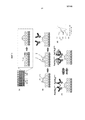
Фиг.1a. Фотография (слева) сенсорного чипа для мультиплексного анализа, показывающая изготовленный микротехнологическими способами чип, который характеризуется отверстиями размером 5 мкм для электрохимического отложения электродов, и иллюстрация (в середине) выемки. Определенную картину наносят золотом (Au) посредством осаждения на силиконовую пластину, используя обычную фотолитографию, и затем покрывают слоем SiO2; затем вытравливают отверстия размером 5 мкм через этот верхний слой, чтобы обнажить круглый участок Au. Схематичная иллюстрация создания Au-электродов посредством электроосаждения Au (справа).Figa. Photo (left) of a sensor chip for multiplex analysis, showing a chip made by microtechnological methods, which is characterized by holes of 5 μm in size for electrochemical deposition of electrodes, and an illustration (in the middle) of a recess. A specific pattern is applied with gold (Au) by deposition on a silicone plate using conventional photolithography, and then coated with a layer of SiO 2 ; 5 μm holes are then etched through this top layer to expose the round Au portion. Schematic illustration of the creation of Au electrodes by electrodeposition of Au (right).
Фиг.1b. Схематичная функционализация электрода; (слева) линкер из цистамина образуют на Au-структуре; (в середине) взаимодействие с бифункциональным линкером глутаральдегидом для введения альдегидных групп на поверхность сенсора; (справа) добавление анти-CA-125-антитела или антитела против сывороточного альбумина человека (HSA) для получения модифицированных антителом электродных сенсоров.Fig.1b. Schematic functionalization of the electrode; (left) a cystamine linker is formed on an Au structure; (in the middle) interaction with a bifunctional glutaraldehyde linker to introduce aldehyde groups onto the surface of the sensor; (right) adding an anti-CA-125 antibody or anti-human serum albumin (HSA) antibody to produce antibody-modified electrode sensors.
Фиг.1c. Схема электрохимической детекции антигена CA-125. Связывание антиген-антитело препятствует реакции переноса электронов через границу раздела [Fe(CN)6]3-/4-.Fig.1c. Scheme of electrochemical detection of antigen CA-125. Antigen-antibody binding inhibits the electron transfer reaction across the [Fe (CN) 6 ] 3- / 4- interface.
Фиг.1d. Дифференциальная импульсная вольтамперометрия (DPV), показывающая снижение сигнала после инкубации CA-125 (10 ед./мл в сыворотке) в течение 40 минут.Fig.1d. Differential pulse voltammetry (DPV), showing a decrease in signal after incubation of CA-125 (10 units / ml in serum) for 40 minutes.
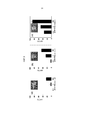
Фиг.2. СЭМ-изображения и характеристические циклические вольтаммограммы для трех сенсорных Au-электродов разного размера. СЭМ-изображения: (a) 100-микронного сенсора. Такая структура была изготовлена с использованием постояннотоковой вольтамперометрии в случае приложенного потенциала 0 мВ в течение 200 секунд, (c) 30-микронного сенсора. Такая структура была изготовлена с использованием постояннотоковой вольтамперометрии в случае приложенного потенциала 150 мВ в течение 200 секунд, и (e) 8-микронного сенсора. Такая структура была изготовлена с использованием хронопотенциометрии в случае прилагаемого тока 30 нА в течение 50 секунд. Характеристические циклические вольтаммограммы трех сенсоров получали в 10 мМ растворе фосфатного буфера, содержащем 2,5 мМ [Fe(CN)6]3-/4- и 0,1 М KCl, со скоростью сканирования 100 мВ/сек в случае сенсоров размером (b) 100 микрон, (d) 30 микрон и (f) 8 микрон. Вставка на фиг.2(f) показывает увеличенное изображение циклической вольтаммограммы. (g) Емкостный ток в случае трех сенсоров, отражающий площадь поверхности для каждого размера сенсора. Циклические вольтаммограммы получали в 10 мМ растворе фосфатного буфера, содержащем 0,1 М KCl, со скоростью сканирования 100 мВ/сек для сенсоров размером (внешняя кривая) 100 микрон, (средняя кривая) 30 микрон и (внутренняя кривая) 8 микрон.Figure 2. SEM images and characteristic cyclic voltammograms for three sensor Au electrodes of different sizes. SEM images: (a) 100 micron sensor. Such a structure was fabricated using constant current voltammetry in the case of an applied potential of 0 mV for 200 seconds, (c) a 30 micron sensor. Such a structure was fabricated using constant current voltammetry for an applied potential of 150 mV for 200 seconds, and (e) an 8 micron sensor. Such a structure was fabricated using chronopotentiometry in the case of an applied current of 30 nA for 50 seconds. The characteristic cyclic voltammograms of the three sensors were obtained in a 10 mM phosphate buffer solution containing 2.5 mM [Fe (CN) 6 ] 3- / 4- and 0.1 M KCl, with a scanning speed of 100 mV / s for sensors of size (b ) 100 microns, (d) 30 microns and (f) 8 microns. The insert in FIG. 2 (f) shows an enlarged image of a cyclic voltammogram. (g) Capacitive current in the case of three sensors, reflecting the surface area for each sensor size. Cyclic voltammograms were obtained in a 10 mM phosphate buffer solution containing 0.1 M KCl, with a scanning speed of 100 mV / s for sensors with a size (external curve) of 100 microns, (middle curve) 30 microns and (internal curve) 8 microns.
Фиг.3. Сравнение чувствительности и пределы обнаружения иммуносенсоров, созданных с использованием трех Au-структур разного размера. ΔΔ(I%) с изменением концентрации CA-125 в PBS получали с использованием (a) 100-микронного сенсора, (b) 30-микронного сенсора и (c) 8-микронного сенсора.Figure 3. Comparison of sensitivity and detection limits of immunosensors created using three Au structures of different sizes. ΔΔ (I%) with a change in the concentration of CA-125 in PBS was obtained using (a) a 100 micron sensor, (b) a 30 micron sensor and (c) an 8 micron sensor.
Фиг.4. Изменение тока сенсоров с размером (a) 100 микрон и (b) 8 микрон до (серый столбик) и после (черный столбик) инкубации с разными концентрациями CA-125 в PBS.Figure 4. Change in sensor current with sizes (a) 100 microns and (b) 8 microns before (gray bar) and after (black bar) incubation with different concentrations of CA-125 in PBS.
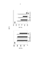
Фиг.5. Детекция CA-125 в сыворотке и цельной крови. (a) Одновременная детекция CA-125 и HSA в образцах сыворотки, в которые вносили аналит. Образец содержал неразбавленную сыворотку. Данные получали с использованием только сыворотки и сыворотки, в которую вносили разные концентрации CA-125. Серыми столбиками показаны данные, полученные в случае модифицированных анти-CA-125-антителом иммуносенсоров, и черные столбики представляют данные, полученные в случае модифицированных анти-HSA-антителом иммуносенсоров. Размер сенсора составлял 8 микрон. (b) Детекция CA-125 в цельной крови. Образцы содержали неразбавленную необработанную кровь, и концентрации CA-125 указаны.Figure 5. Detection of CA-125 in serum and whole blood. (a) Simultaneous detection of CA-125 and HSA in serum samples into which analyte was added. The sample contained undiluted serum. Data were obtained using only serum and serum into which different concentrations of CA-125 were added. The gray bars indicate the data obtained in the case of modified anti-CA-125 antibody immunosensors, and the black bars represent the data obtained in the case of modified anti-HSA antibody immunosensors. The sensor size was 8 microns. (b) Detection of CA-125 in whole blood. Samples contained undiluted untreated blood, and CA-125 concentrations are indicated.
ПОДРОБНОЕ ОПИСАНИЕ ИЗОБРЕТЕНИЯDETAILED DESCRIPTION OF THE INVENTION
I. Системы и способы электрохимической детекцииI. Systems and methods for electrochemical detection
В настоящем изобретении предлагаются системы и способы электрохимической детекции аналита-мишени в образце. Присутствие аналита выявляют по изменению электрокаталитического сигнала. Использование такого считывания электрических сигналов обеспечивает способ, который является недорогим, чрезвычайно чувствительным, простым с точки зрения миниатюризации и удобным для автоматизации.The present invention provides systems and methods for electrochemical detection of a target analyte in a sample. The presence of the analyte is detected by a change in the electrocatalytic signal. The use of such an electrical signal sensing provides a method that is inexpensive, extremely sensitive, simple in terms of miniaturization and convenient for automation.
В одном аспекте систем и способов детекции, описанных в настоящей публикации, предлагается электрод, где электрод содержит линкер и где линкер присоединен к антителу или его фрагменту. Антитело или его фрагмент способны связываться с аналитом-мишенью, таким как белок. Электрод находится в присутствии редокс-репортера.In one aspect of the detection systems and methods described herein, an electrode is provided wherein the electrode comprises a linker and where the linker is attached to an antibody or fragment thereof. An antibody or fragment thereof is capable of binding to a target analyte, such as a protein. The electrode is in the presence of a redox reporter.
Редокс-репортеры, подходящие для применения в системах и способах, описанных в настоящей публикации, способны генерировать электрический сигнал (например, фарадеевский ток) при взаимодействии с электродом в случае приложения потенциала. Можно использовать любой редокс-репортер, который генерирует фарадеевский ток или способен к переносу электронов через границу раздела в присутствии электрода. Неограничивающие редокс-репортеры включают без ограничения небольшие активные в окислительно-восстановительных реакциях группы, такие как феррицианид/ферроцианид, ферроцен и гексахлориридат(IV)/гексахлориридат(III). В системах детекции используют редокс-репортеры для генерации электрических сигналов исходного уровня при взаимодействии с электродом. Когда присутствует аналит-мишень, который связывается с антителом или его фрагментом, электрический сигнал ослабляется. Предполагают, что ослабление сигнала является следствием того, что аналит-мишень блокирует эффективный доступ редокс-репортера к поверхности электрода. Другими словами, связывание антитело-аналит препятствует переносу электронов через границу раздела. Смотри, только в качестве примера, фиг.1c и 1d.Redox reporters suitable for use in the systems and methods described in this publication are capable of generating an electrical signal (e.g., a Faraday current) when interacting with an electrode when a potential is applied. You can use any redox reporter that generates a Faraday current or is capable of transferring electrons across the interface in the presence of an electrode. Non-limiting redox reporters include, but are not limited to, small redox active groups, such as ferricyanide / ferrocyanide, ferrocene, and hexachloridate (IV) / hexachloridate (III). In detection systems, redox reporters are used to generate electrical signals of the initial level when interacting with the electrode. When a target analyte is present that binds to the antibody or its fragment, the electrical signal is attenuated. It is assumed that the attenuation of the signal is a consequence of the fact that the analyte target blocks the effective access of the redox reporter to the electrode surface. In other words, antibody-analyte binding interferes with electron transfer across the interface. See, by way of example only, FIGS. 1c and 1d.
В одном аспекте изменения сигнала, соответствующие связыванию аналита-мишени с антителом, вычисляют в виде изменения фарадеевского тока в процентах:In one aspect, signal changes corresponding to the binding of the target analyte to the antibody are calculated as percent changes in Faraday current:
ΔI%={(средний I0)-(средний Ic)}/средний I0 × 100ΔI% = {(average I 0 ) - (average I c )} / average I 0 × 100
где средний I0 = средний ток при нулевой концентрации мишени, средний Ic = средний ток при любой концентрации мишени). В некоторых вариантах изменение сигнала составляет, по меньшей мере, примерно 10%, по меньшей мере, примерно 15%, примерно 25%, примерно 30%, примерно 40%, примерно 50%, примерно 65%, примерно 75%, примерно 85%, примерно 90%, примерно 95%, больше чем примерно 100%, примерно в два раза, примерно в десять раз, примерно в пятьдесят раз или больше. В некоторых случаях изменение сигнала показывает, что аналит связан с антителом. В случае изменения фарадеевского тока системы регистрации и способы, описанные в настоящей публикации, применяют в одном аспекте для определения присутствия аналита-мишени.where average I 0 = average current at zero target concentration, average I c = average current at any target concentration). In some embodiments, the signal change is at least about 10%, at least about 15%, about 25%, about 30%, about 40%, about 50%, about 65%, about 75%, about 85% , about 90%, about 95%, more than about 100%, about two times, about ten times, about fifty times or more. In some cases, a change in signal indicates that the analyte is bound to the antibody. In the event of a change in the Faraday current, the registration systems and methods described in this publication are used in one aspect to determine the presence of a target analyte.
В другом аспекте системы и способы детекции, описанные в настоящей публикации, применяют для определения концентрации аналита-мишени в образце. В некоторых вариантах определение осуществляют посредством калибровки системы детекции с использованием стандартов с известной концентрацией аналита-мишени. Например, ряд положительных контрольных образцов, каждый из которых имеет конкретную концентрацию аналита, используют для определения изменения фарадеевского тока в процентах с целью определения неизвестного количества аналита в тестируемом образце. Диапазоны детекции в случае систем и способы детекции зависят от антитела, аналита и их связывающей способности, а также используемого редокс-репортера. В некоторых вариантах системы и способы детекции, описанные в настоящей публикации, выявляют концентрации аналита примерно 500 фемтомолярные (фМ) или примерно 100 пг/мл или ниже.In another aspect, the detection systems and methods described herein are used to determine the concentration of the target analyte in a sample. In some embodiments, the determination is made by calibrating the detection system using standards with a known concentration of the target analyte. For example, a number of positive control samples, each of which has a specific analyte concentration, are used to determine the percentage change in the Faraday current in order to determine the unknown amount of analyte in the test sample. The detection ranges in the case of systems and methods of detection depend on the antibody, analyte and their binding ability, as well as the redox reporter used. In some embodiments, the detection systems and methods described herein detect analyte concentrations of about 500 femtomolar (fM) or about 100 pg / ml or lower.
В другом аспекте системы и способы детекции, описанные в настоящей публикации, являются мультиплексными для обнаружения и/или определения концентрации множества аналитов-мишеней. В некоторых вариантах мультиплексные системы и способы включают в себя, по меньшей мере, два электрода, каждый из которых содержит линкер, при этом с линкерами связаны разные антитела. В некоторых случаях в мультиплексной системе используют два, три, четыре, пять, шесть, семь, восемь, девять или десять электродов, каждый из которых содержит линкер, при этом с линкерами связаны разные антитела. В некоторых вариантах в мультиплексной системе используют, по меньшей мере, три, по меньшей мере, пять, по меньшей мере, десять, по меньшей мере, пятнадцать, по меньшей мере, двадцать, по меньшей мере, тридцать, по меньшей мере, сорок или, по меньшей мере, пятьдесят или больше электродов, каждый из которых содержит линкер, при этом с линкерами связаны разные антитела. Альтернативно, более одного электрода могут содержать одно и то же антитело или класс антител; например, можно использовать повторы четырех электродов, при этом группа содержит одно из трех отдельных антител, в содержащей двенадцать электродов мультиплексной системе. Кроме того, электрод может содержать больше одного антитела или класса антител; например, на отдельном электроде можно сочетать больше одного антитела, при этом каждое антитело узнает конкретную область белка или аналита. В некоторых случаях, детекция может происходить только тогда, когда белок или аналит связывается со всеми антителами, связанными с электродом. В некоторых случаях детекция будет происходить, только когда белок или аналит связывает, по меньшей мере, одну из групп антител, связанных с электродом.In another aspect, the detection systems and methods described herein are multiplexed to detect and / or determine the concentration of multiple target analytes. In some embodiments, multiplex systems and methods include at least two electrodes, each of which contains a linker, wherein different antibodies are coupled to the linkers. In some cases, in a multiplex system, two, three, four, five, six, seven, eight, nine, or ten electrodes are used, each of which contains a linker, and different antibodies are connected to the linkers. In some embodiments, the multiplex system uses at least three, at least five, at least ten, at least fifteen, at least twenty, at least thirty, at least forty, or at least fifty or more electrodes, each of which contains a linker, while different antibodies are connected to the linkers. Alternatively, more than one electrode may contain the same antibody or antibody class; for example, you can use repeats of four electrodes, while the group contains one of three separate antibodies in a multiplex system containing twelve electrodes. In addition, the electrode may contain more than one antibody or class of antibodies; for example, more than one antibody can be combined on a single electrode, with each antibody recognizing a specific region of a protein or analyte. In some cases, detection can only occur when the protein or analyte binds to all antibodies bound to the electrode. In some cases, detection will occur only when the protein or analyte binds at least one of the antibody groups associated with the electrode.
Мультиплексирование позволяет одновременно выявлять большое разнообразие аналитов, таким образом создавая «панель аналитов». Примерные панели аналитов могут содержать аналиты, связанные с расстройством, заболеванием или состоянием, например биомаркеры, связанные с определенной злокачественной опухолью. Мультиплексирование также обеспечивает более высокую чувствительность к аналиту, например разные антитела, которые связываются с одним и тем же аналитом-мишенью через одни и те же или разные эпитопы. Использование разных антител, такое как, например, использование поликлонального и моноклонального антител, мишенью которых является один и тот же аналит, позволяет сделать систему детекции более надежной и чувствительной, чем одноплексная система, в которой используют только один тип антитела для обнаружения аналита. Кроме того, мультиплексирование обеспечивает возможность внутренней калибровки системы с целью уменьшения ложно-положительных и отрицательных результатов. Например, анализ аналита-мишени можно осуществлять параллельно с анализом аналита, который, как известно, является стабильным, таким как широко представленный в сыворотке белок.Multiplexing allows you to simultaneously identify a wide variety of analytes, thereby creating a "panel of analytes." Exemplary analyte panels may contain analytes associated with a disorder, disease, or condition, for example, biomarkers associated with a particular malignant tumor. Multiplexing also provides higher analyte sensitivity, for example, different antibodies that bind to the same target analyte through the same or different epitopes. The use of different antibodies, such as, for example, the use of polyclonal and monoclonal antibodies, the target of which is the same analyte, makes the detection system more reliable and sensitive than a single-plex system, in which only one type of antibody is used to detect the analyte. In addition, multiplexing enables internal calibration of the system in order to reduce false positive and negative results. For example, the analysis of the target analyte can be carried out in parallel with the analysis of the analyte, which is known to be stable, such as a protein widely represented in serum.
В другом аспекте системы и способы детекции, описанные в настоящей публикации, применяют для выявления и диагностики расстройства, заболевания или состояния или для мониторинга прогрессирования или ответа расстройства, заболевания или состояния. В некоторых вариантах получают образец от пациента или субъекта и применяют системы и способы детекции для выявления присутствия и/или определения концентрации аналита-мишени, ассоциированного с расстройством, заболеванием или состоянием. Примеры расстройств, заболеваний или состояний включают злокачественные опухоли (например, злокачественные опухоли молочной железы, яичника, предстательной железы, поджелудочной железы, прямой и ободочной кишки, мочевого пузыря и тому подобные), инфекционные болезни (например, инфекции бактериями Staphylococcus или Streptococcus, MRSA, VISA, вирусные инфекции, грибковые инфекции и тому подобные), аутоиммунные заболевания (например, диффузный токсический зоб, волчанку, артрит, синдром Гудпасчера и тому подобные), метаболические заболевания и расстройства (например, метаболический синдром, резистентность к инсулину, диабет типа I и II, болезнь Крона, синдром раздраженной кишки и тому подобные), ВИЧ/СПИД, генетические заболевания и состояния, ассоциированные с терапевтическими лекарственными средствами или токсикологическими материалами. В некоторых вариантах определяют тяжесть или стадии расстройства, заболевания или состояния благодаря определению концентрации аналита-мишени, при этом разные концентрации свидетельствуют о тяжести или стадии. Подобным образом в других вариантах определяют прогрессирование или ответ расстройства, заболевания или состояния благодаря определению концентрации аналита-мишени в разных временных точках. Терапевтически эффективное фармакологическое лечение, терапия или схема в некоторых вариантах также могут быть определены в результате выявления концентрации аналита-мишени в разных временных точках.In another aspect, the detection systems and methods described herein are used to detect and diagnose a disorder, disease, or condition, or to monitor the progression or response of a disorder, disease, or condition. In some embodiments, a sample is obtained from a patient or subject and detection systems and methods are used to detect the presence and / or determination of the concentration of the target analyte associated with the disorder, disease, or condition. Examples of disorders, diseases or conditions include malignant tumors (e.g., malignant tumors of the breast, ovary, prostate, pancreas, rectum, colon, bladder and the like), infectious diseases (e.g. infections with bacteria Staphylococcus or Streptococcus, MRSA, VISA, viral infections, fungal infections and the like), autoimmune diseases (e.g., diffuse toxic goiter, lupus, arthritis, Goodpasture syndrome and the like), metabolic diseases and disorders ( e.g. metabolic syndrome, insulin resistance, type I and II diabetes, Crohn’s disease, irritable bowel syndrome and the like), HIV / AIDS, genetic diseases and conditions associated with therapeutic drugs or toxicological materials. In some embodiments, the severity or stage of the disorder, disease, or condition is determined by determining the concentration of the target analyte, with different concentrations indicating severity or stage. Similarly, in other embodiments, the progression or response of the disorder, disease, or condition is determined by determining the concentration of the target analyte at different time points. A therapeutically effective pharmacological treatment, therapy, or regimen in some embodiments can also be determined by detecting the concentration of the target analyte at different time points.
II. ЭлектродыII. Electrodes
Электроды для систем и способов детекции, описанных в настоящей публикации, представляют собой любые электропроводящие материалы, обладающие свойствами, обеспечивающими возможность присутствия линкеров на поверхности электродов. Электроды обладают способностью к переносу электронов к редокс-репортеру или от него и обычно соединены с устройством электронного управления и детекции. В общем, благородные металлы, такие как Ag, Au, Ir, Os, Pd, Pt, Rh, Ru и другие представители такого семейства являются подходящими материалами для электродов. Благородные металлы обладают подходящими свойствами, включая стабильность и устойчивость к окислению, могут быть обработаны разными способами, такими как электроосаждение, и связываются с молекулами, содержащими тиолы и дисульфиды, таким образом обеспечивая возможность связывания указанных молекул. Также могут быть использованы другие материалы, такие как азотсодержащие проводящие соединения (например, WN, TiN, TaN) или материалы на основе силикона/диоксида кремния, такие как силан или силоксан. В некоторых вариантах электрод состоит из золота, палладия или платины. В других вариантах электрод является углеродным. В следующих вариантах электрод состоит из оксида индия и олова.The electrodes for the detection systems and methods described in this publication are any electrically conductive materials having properties that allow the presence of linkers on the surface of the electrodes. The electrodes have the ability to transfer electrons to or from the redox reporter and are usually connected to an electronic control and detection device. In general, noble metals such as Ag, Au, Ir, Os, Pd, Pt, Rh, Ru and other members of this family are suitable materials for electrodes. The noble metals possess suitable properties, including stability and oxidation stability, can be processed in various ways, such as electrodeposition, and bind to molecules containing thiols and disulfides, thus allowing the binding of these molecules. Other materials, such as nitrogen-containing conductive compounds (e.g., WN, TiN, TaN), or silicone / silica-based materials, such as silane or siloxane, can also be used. In some embodiments, the electrode consists of gold, palladium or platinum. In other embodiments, the electrode is carbon. In the following embodiments, the electrode consists of indium oxide and tin.
В некоторых вариантах электрод является микроэлектродом. В других вариантах микроэлектрод представляет собой наноструктурированный микроэлектрод («NME»). NME представляют собой микроэлектроды, характерной особенностью которых являются наноструктурированные поверхности. Нанотекстурирование поверхности или наноструктуры увеличивают площадь поверхности электрода, обеспечивая более высокую чувствительность, в частности, в случае применения биосенсоров. Производство NME может быть осуществлено с использованием электроосаждения. Варьируя параметры, такие как время осаждения, напряжение осаждения, тип фонового электролита и источники ионов металлов, можно создать NME различного размера, строения и состава. В некоторых случаях NME имеют дендритную структуру. Сложность дендритной структуры достигается за счет варьирования вышеуказанных параметров электроосаждения. Примеры NME для применения в системах и способах, описанных в настоящей публикации, приведены в международной заявке на выдачу патента с регистрационным № PCT/CA2009/001212 (опубликованной с номером WO/2010/025547), которая включена в виде ссылки в полном объеме.In some embodiments, the electrode is a microelectrode. In other embodiments, the microelectrode is a nanostructured microelectrode ("NME"). NMEs are microelectrodes, a characteristic feature of which are nanostructured surfaces. Nanotexture surface or nanostructures increase the surface area of the electrode, providing higher sensitivity, in particular, in the case of the use of biosensors. NME production can be carried out using electrodeposition. By varying parameters such as deposition time, deposition voltage, background electrolyte type and metal ion sources, NMEs of various sizes, structures and compositions can be created. In some cases, NMEs have a dendritic structure. The complexity of the dendritic structure is achieved by varying the above parameters of electrodeposition. Examples of NME for use in the systems and methods described in this publication are given in the international patent application with registration number PCT / CA2009 / 001212 (published with the number WO / 2010/025547), which is incorporated by reference in full.
Другие структуры электродов также можно использовать в системах и способах детекции, описанных в настоящей публикации, включая плоские поверхности, проволоки, трубки, конусы и частицы. Коммерчески доступные макро- и микроэлектроды также подходят для вариантов осуществления изобретения, описанных в настоящей публикации.Other electrode structures can also be used in the detection systems and methods described in this publication, including flat surfaces, wires, tubes, cones, and particles. Commercially available macro- and microelectrodes are also suitable for the embodiments of the invention described in this publication.
Электроды имеют размеры, например, примерно от 0,0001 до примерно 5000 микрон в длину или в диаметре; примерно от 0,0001 до примерно 2000 микрон в длину или в диаметре; примерно от 0,001 до примерно 250 микрон; примерно от 0,01 до примерно 200 микрон; примерно от 0,1 до примерно 100 микрон; примерно от 1 до примерно 50 микрон; примерно от 10 до примерно 30 микрон в длину, или меньше, чем примерно 10 микрон в длину или в диаметре. В некоторых вариантах электроды имеют размеры примерно 100 микрон, примерно 30 микрон, примерно 10 микрон или примерно 5 микрон в длину или в диаметре. В следующих вариантах электроды имеют размеры примерно 8 микрон.The electrodes are sized, for example, from about 0.0001 to about 5000 microns in length or in diameter; from about 0.0001 to about 2000 microns in length or in diameter; from about 0.001 to about 250 microns; from about 0.01 to about 200 microns; from about 0.1 to about 100 microns; from about 1 to about 50 microns; from about 10 to about 30 microns in length, or less than about 10 microns in length or in diameter. In some embodiments, the electrodes are about 100 microns, about 30 microns, about 10 microns, or about 5 microns in length or diameter. In further embodiments, the electrodes are about 8 microns in size.
В некоторых вариантах системы и способы детекции, описанные в настоящей публикации, содержат один электрод для детекции. В других вариантах используют множество электродов. В некоторых вариантах применение множества электродов можно использовать параллельно для обнаружения аналита-мишени посредством одного типа антитела, связанного с каждым электродом. Альтернативно в других вариантах используют множество электродов для мультиплексирования, как описано ранее. Множество электродов можно компоновать в виде матриц с высокой или низкой плотностью. Пример 8-электродной матрицы на изготовленном микротехнологическими способами чипе для мультиплексного применения изображен на фиг.1a.In some embodiments, the detection systems and methods described herein contain one electrode for detection. In other embodiments, multiple electrodes are used. In some embodiments, the use of multiple electrodes can be used in parallel to detect the target analyte through one type of antibody associated with each electrode. Alternatively, in other embodiments, multiple electrodes are used for multiplexing as previously described. Many electrodes can be arranged in the form of matrices with high or low density. An example of an 8-electrode array on a chip fabricated by microtechnological methods for multiplex application is shown in Fig. 1a.
В следующих вариантах электрод расположен на подложке. Подложка может содержать широкий диапазон материалов: биологических, небиологических, органических, неорганических, либо любое их сочетание. Например, подложка может представлять собой полимеризованную пленку Лангмюра-Блоджета, функционализированное стекло, Si, Ge, GaAs, GaP, SiO2, SiN4, модифицированный силикон или любой из широкого множества гелей или полимеров, таких как (поли)тетрафторэтилен, (поли)винилидендифторид, полистирол, поперечно сшитый полистирол, полиакриловая, полимолочная кислота, полигликолевая кислота, сополимер(лактид-гликолид), полиангидриды, поли(метилметакрилат), сополимер (этилен-винилацетат), полисилоксаны, полимерный диоксид кремния, латексы, полимеры декстрана, эпоксиды, поликарбонаты или их сочетания.In the following embodiments, the electrode is located on the substrate. The substrate may contain a wide range of materials: biological, non-biological, organic, inorganic, or any combination thereof. For example, the substrate may be a Langmuir-Blodget polymerized film, functionalized glass, Si, Ge, GaAs, GaP, SiO 2 , SiN 4 , modified silicone, or any of a wide variety of gels or polymers such as (poly) tetrafluoroethylene, (poly) vinylidene difluoride, polystyrene, cross-linked polystyrene, polyacrylic, polylactic acid, polyglycolic acid, copolymer (lactide-glycolide), polyanhydrides, poly (methyl methacrylate), copolymer (ethylene-vinyl acetate), polysiloxanes, polymeric silicon dioxide, latexes, polymers ana, epoxides, polycarbonates, or combinations thereof.
Подложки могут представлять собой плоские кристаллические подложки, такие как подложки на основе диоксида кремния (например, стекло, кварц и или тому подобные), или кристаллические подложки, используемые, например, в производстве полупроводников и микропроцессоров, такие как силикон, арсенид галлия, GaN, допированный индием, и тому подобные. Аэрогели на основе диоксида кремния также можно использовать в качестве подложек и могут быть получены любыми известными способами. Подложки на основе аэрогелей можно использовать в виде отдельно стоящих подложек или в виде покрытия поверхности подложки из другого материала.The substrates can be flat crystalline substrates, such as silica-based substrates (e.g. glass, quartz and the like), or crystalline substrates used, for example, in the manufacture of semiconductors and microprocessors, such as silicone, gallium arsenide, GaN, doped with indium, and the like. Silicon dioxide aerogels can also be used as substrates and can be prepared by any known method. Airgel-based substrates can be used as stand-alone substrates or as a surface coating of a substrate of another material.
Подложка может принимать любую форму и обычно представляет собой пластину, предметное стекло, шарик, гранулу, диск, частицу, микрочастицу, наночастицу, нить, осадок, необязательно пористый гель, листы, трубку, сферу, контейнер, капилляр, площадку, тонкий слой, пленку, чип, многолуночный планшет или чашку, оптическое волокно и т.д. Подложка может быть любой формы, которая является жесткой или полужесткой. Подложка может содержать повышенные или пониженные области, на которых располагается компонент анализа. Поверхность подложки может быть вытравлена с использованием хорошо известных способов для получения требуемых характеристик поверхности, например желобки, v-бороздки, мезаструктуры или тому подобные. Подложка может принимать форму фотодиода, оптоэлектронного датчика, такого как оптоэлектронный полупроводниковый чип или оптоэлектронный тонкопленочный полупроводник, или биочипа. Положение(положения) электрода(ов) на подложке может быть адресным; адресация может быть осуществлена в формах с высокой плотностью, и положение(положения) может быть микроадресным или наноадресным. В некоторых вариантах электрод(ы) находится на полученном посредством микропроизводства чипе.The substrate can take any form and is usually a plate, a glass slide, a ball, a granule, a disk, a particle, a microparticle, a nanoparticle, a thread, a precipitate, optionally a porous gel, sheets, a tube, a sphere, a container, a capillary, a pad, a thin layer, a film , chip, multi-well tablet or cup, optical fiber, etc. The substrate may be of any shape that is rigid or semi-rigid. The substrate may contain increased or decreased areas on which the analysis component is located. The surface of the substrate can be etched using well-known methods to obtain the desired surface characteristics, for example grooves, v-grooves, mesastructures or the like. The substrate may take the form of a photodiode, an optoelectronic sensor, such as an optoelectronic semiconductor chip or an optoelectronic thin film semiconductor, or a biochip. The position (s) of the electrode (s) on the substrate may be addressable; addressing may be in high density forms, and the position (s) may be micro-address or nano-address. In some embodiments, the electrode (s) are on a micro-chip.
Поверхности на подложке могут состоять из того же материала, что и подложка, или могут быть сделаны из другого материала и могут быть связаны с подложкой химическим или физическим способом. Такие связанные поверхности могут состоять из любого из широкого множества материалов, например из полимеров, пластиков, смол, полисахаридов, диоксида кремния или материалов на основе диоксида кремния, угля, металлов, неорганических стекол, мембран или любого из перечисленных выше материалов подложки.The surfaces on the substrate may consist of the same material as the substrate, or may be made of another material and may be bonded to the substrate chemically or physically. Such bonded surfaces may consist of any of a wide variety of materials, for example, polymers, plastics, resins, polysaccharides, silica, or materials based on silica, coal, metals, inorganic glasses, membranes, or any of the above substrate materials.
Подложка и/или ее поверхность обычно устойчивы или обработаны так, чтобы они были устойчивыми к условиям, которые на них действуют при применении, и необязательно могут быть обработаны так, чтобы удалить любой устойчивый материал после воздействия таких условий.The substrate and / or its surface is usually stable or processed so that they are resistant to the conditions that affect them when applied, and can optionally be processed so as to remove any resistant material after exposure to such conditions.
III. ЛинкерыIII. Linkers
В одном аспекте электрод содержит линкер на поверхности электрода. Линкеры в некоторых вариантах могут образоваться в случае абсорбции молекул линкера и оказываются организованными в виде слоя молекул на поверхности. Линкеры, подходящие для применения в случае электродов, раскрытых в настоящем описании, имеют «группу головки», которая прочно хемосорбируется на металлах (например, тиолы и дисульфиды), и хвост с функциональной группой (например, -OH, -NH2, -COOH, -CO, -OCH3, -NHNH2, -биотином, -NHS (активный по отношению к аминам N-гидроксисукцинимид)). Примеры линкеров включают одноцепочечные или имеющие разветвленные цепи алкилтиолы с функциональной группой. Другие линкерные молекулы включают ароматические тиолы, такие как тиофенол с функциональной группой. Подходящие линкерные молекулы включают любую молекулу с функциональной группой, которая может прямо или опосредованно спариваться с антителом. Примеры линкерных молекул включают без ограничения цистамин, цистеамин, меркаптопропионовую кислоту или 4-аминотиофенол. В некоторых вариантах линкером является цистамин.In one aspect, the electrode comprises a linker on the surface of the electrode. Linkers in some embodiments can be formed in the case of absorption of the linker molecules and are organized as a layer of molecules on the surface. Linkers suitable for use with the electrodes disclosed herein have a “head group” that is strongly chemisorbed on metals (eg, thiols and disulfides) and a tail with a functional group (eg, —OH, —NH 2 , —COOH , -CO, -OCH 3 , -NHNH 2 , -biotin, -NHS (N-hydroxysuccinimide active in relation to amines)). Examples of linkers include single-chain or branched functional alkylthiols. Other linker molecules include aromatic thiols, such as functional thiophenol. Suitable linker molecules include any molecule with a functional group that can directly or indirectly mate with an antibody. Examples of linker molecules include, but are not limited to, cystamine, cysteamine, mercaptopropionic acid, or 4-aminothiophenol. In some embodiments, the linker is cystamine.
Линкеры образуются на поверхности электрода, когда электрод погружают в раствор линкерной молекулы. Обычные концентрации составляют примерно 0,01 мМ, 0,05 мМ, 0,1 мМ, 0,5 мМ, 1 мМ, примерно 2 мМ, примерно 5 мМ, примерно 10 мМ, примерно 20 мМ или примерно 50 мМ или больше линкерной молекулы в водном или этанольном растворе. Погружение осуществляют в течение периода времени в диапазоне от нескольких часов до нескольких суток. В некоторых вариантах погружение продолжается примерно 4 часа, примерно 8 часов, примерно 16 часов, примерно 24 часа, примерно 2 суток, примерно 5 суток или примерно 7 суток. В некоторых вариантах погружение осуществляют при комнатной температуре. В других вариантах погружение осуществляют при температуре выше комнатной температуры. В следующих вариантах погружение осуществляют при температуре ниже, чем комнатная температура.Linkers are formed on the surface of the electrode when the electrode is immersed in a solution of a linker molecule. Typical concentrations are about 0.01 mm, 0.05 mm, 0.1 mm, 0.5 mm, 1 mm, about 2 mm, about 5 mm, about 10 mm, about 20 mm, or about 50 mm or more linker molecule in aqueous or ethanol solution. Diving is carried out over a period of time in the range from several hours to several days. In some embodiments, the dive lasts about 4 hours, about 8 hours, about 16 hours, about 24 hours, about 2 days, about 5 days, or about 7 days. In some embodiments, immersion is carried out at room temperature. In other embodiments, immersion is carried out at a temperature above room temperature. In the following embodiments, immersion is carried out at a temperature lower than room temperature.
Линкеры могут быть прямо или опосредованно спарены с антителом любым известным способом. Прямое спаривание в некоторых вариантах может быть осуществлено через функциональные группы линкерных молекул, такие как функциональные группы -CO, которые могут взаимодействовать и связываться с антителами. Альтернативно в других вариантах второй линкер или спейсер может быть конъюгирован с функциональной группой, посредством которой второй линкер или спейсер может связываться с антителом. Например, линкерные молекулы с функциональными группами -NH могут взаимодействовать с таким линкером, как глутаральдегид или формальдегид, которые в свою очередь могут связываться с антителами. В следующих вариантах антитело может быть дериватизовано таким образом, чтобы взаимодействовать с функциональной группой. Например, меченное авидином антитело может связываться с биотиновой функциональной группой линкера. Указанные и другие примеры прямого и опосредованного связывания входят в объем вариантов осуществления изобретения, описанных в настоящей публикации.Linkers can be directly or indirectly paired with the antibody in any known manner. Direct pairing in some embodiments can be carried out through functional groups of linker molecules, such as functional groups —CO, which can interact and bind to antibodies. Alternatively, in other embodiments, the second linker or spacer may be conjugated to a functional group by which the second linker or spacer may bind to the antibody. For example, linker molecules with -NH functional groups can interact with a linker such as glutaraldehyde or formaldehyde, which in turn can bind to antibodies. In the following embodiments, the antibody can be derivatized in such a way as to interact with a functional group. For example, an avidin-labeled antibody may bind to the linker biotin functional group. These and other examples of direct and indirect binding are included in the scope of embodiments of the invention described in this publication.
IV. АнтителаIV. Antibodies
В одном аспекте антитело используют для определения количества и/или концентрации аналита-мишени. Антитела, относящиеся к семейству белков плазмы, называемых иммуноглобулинами, основной строительный блок которых, укладка или домен иммуноглобулина, используется в различных формах во многих молекулах иммунной системы и других системах биологического распознавания. Типичный иммуноглобулин имеет четыре полипептидных цепи, содержащих антигенсвязывающую область, известную как вариабельная область, и невариабельную область, известную как константная область. Антитело, которое подходит для использования в вариантах осуществления изобретения, описанных в настоящей публикации, может быть в любой из множества форм, включая сыворотку, полный иммуноглобулин, фрагмент антитела, такой как Fv, Fab и сходные фрагменты, одноцепочечное антитело, которое включает в себя определяющие комплементарность области (CDR) вариабельного домена, и тому подобные формы, которые подпадают под широкий термин «антитело» в используемом в настоящем описании смысле. Варианты осуществления изобретения, раскрытые в настоящем описании, предполагают использованием антитела с любой специфичностью, поликлонального или моноклонального, и не ограничены антителами, которые узнают и иммунологически взаимодействуют с конкретным антигеном. В некоторых вариантах, раскрытых в настоящем описании, в контексте как способов терапии, так и способов скрининга используют антитело или его фрагмент, который является иммуноспецифичным по отношению к аналиту-мишени.In one aspect, the antibody is used to determine the amount and / or concentration of the target analyte. Antibodies belonging to the family of plasma proteins called immunoglobulins, the main building block of which, the laying or domain of the immunoglobulin, are used in various forms in many molecules of the immune system and other biological recognition systems. A typical immunoglobulin has four polypeptide chains containing an antigen binding region known as a variable region and a non-variable region known as a constant region. An antibody that is suitable for use in the embodiments of the invention described in this publication may be in any of a variety of forms, including serum, full immunoglobulin, an antibody fragment such as Fv, Fab, and similar fragments, a single chain antibody that includes defining the complementarity of the region (CDR) of the variable domain, and the like, which fall under the broad term "antibody" as used in the present description. Embodiments of the invention disclosed herein involve the use of antibodies of any specificity, polyclonal or monoclonal, and are not limited to antibodies that recognize and immunologically interact with a particular antigen. In some embodiments disclosed herein, in the context of both therapies and screening methods, an antibody or fragment thereof is used that is immunospecific with respect to the target analyte.
Термин «моноклональное антитело» в используемом в настоящем описании смысле относится к антителу, полученному из популяции по существу гомогенных антител, т.е. отдельные антитела, составляющие популяцию, являются идентичными, за исключением возможных встречающихся в природе мутаций, которые могут присутствовать в минорных количествах. Моноклональные антитела являются высоко специфичными, будучи направленными против одного антигенного участка. Кроме того, в отличие от обычных препаратов поликлональных антител, которые обычно содержат разные антитела, направленные против разных детерминант (эпитопов), каждое моноклональное антитело направлено против одной детерминанты на антигене. Моноклональные антитела согласно изобретению, в частности, включают «химерные» антитела (иммуноглобулины), в которых часть тяжелой и/или легкой цепи идентична или гомологична соответствующим последовательностям в антителах, полученных от конкретного вида, или относящихся к конкретному классу или подклассу антител, при этом остальная часть цепи(цепей) идентична или гомологична соответствующим последовательностям в антителах, полученных от другого вида или относящихся к другому классу или подклассу антител, а также фрагменты таких антител, при условии, что они проявляют требуемую биологическую активность. Смотри патент США № 4816567 и публикацию Morrison с соавторами Proc. Natl. Acad. Sci. USA 81, 6851-6855 (1984).The term "monoclonal antibody," as used herein, refers to an antibody obtained from a population of substantially homogeneous antibodies, i.e. the individual antibodies that make up the population are identical, with the exception of possible naturally occurring mutations that may be present in minor amounts. Monoclonal antibodies are highly specific, being directed against a single antigenic site. In addition, unlike conventional preparations of polyclonal antibodies, which usually contain different antibodies directed against different determinants (epitopes), each monoclonal antibody is directed against one determinant on the antigen. Monoclonal antibodies according to the invention, in particular, include “chimeric” antibodies (immunoglobulins) in which a part of the heavy and / or light chain is identical or homologous to the corresponding sequences in antibodies obtained from a particular species or belonging to a particular class or subclass of antibodies, the rest of the chain (s) is identical or homologous to the corresponding sequences in antibodies obtained from another species or belonging to another class or subclass of antibodies, as well as fragments of such antibodies bodies, provided that they exhibit the desired biological activity. See US Patent No. 4816567 and Morrison et al. Proc. Natl. Acad. Sci. USA 81, 6851-6855 (1984).
Термин «фрагмент антитела» относится к части полноразмерного антитела, обычно к антигенсвязывающей или вариабельной области. Примеры фрагментов антител включают фрагменты Fab, Fab', F(ab')2 и Fv. Расщепление антител папаином дает два идентичных антигенсвязывающих фрагмента, называемых Fab-фрагментами, каждый из которых имеет один антигенсвязывающий участок, и оставшийся «Fc»-фрагмент, называемый так по его способности легко кристаллизоваться. Обработка пепсином дает F(ab')2-фрагмент, который имеет два антигенсвязывающих фрагмента, которые способны перекрестно связывать антиген, и другой остаточный фрагмент (который называют pFc'). Дополнительные фрагменты могут включать диантитела, линейные антитела, молекулы одноцепочечных антител и полиспецифичные антитела, образованные из фрагментов антител. В используемом в настоящем описании смысле «функциональный фрагмент» в отношении антител относится к фрагментам Fv, F(ab) и F(ab')2.The term “antibody fragment” refers to a part of a full-sized antibody, usually to an antigen binding or variable region. Examples of antibody fragments include Fab, Fab ', F (ab') 2, and Fv fragments. Papain digestion of antibodies produces two identical antigen binding fragments, called Fab fragments, each of which has one antigen binding site, and the remaining “Fc” fragment, so called because of its ability to crystallize easily. Pepsin treatment yields an F (ab ') 2 fragment that has two antigen-binding fragments that are able to cross-bind the antigen and another residual fragment (called pFc'). Additional fragments may include diantibodies, linear antibodies, single chain antibody molecules, and multispecific antibodies formed from antibody fragments. As used herein, a “functional fragment” in relation to antibodies refers to Fv, F (ab) and F (ab ′) 2 fragments.
Фрагменты антител сохраняют некоторую способность избирательно связываться со своим антигеном или рецептором и их определяют следующим образом.Antibody fragments retain some ability to selectively bind to their antigen or receptor and are determined as follows.
(1) Fab представляет собой фрагмент, который содержит моновалентный антигенсвязывающий фрагмент молекулы антитела. Fab-фрагмент может быть получен расщеплением полного антитела ферментом папаином с получением интактной легкой цепи и части одной тяжелой цепи.(1) Fab is a fragment that contains a monovalent antigen binding fragment of an antibody molecule. The Fab fragment can be obtained by cleaving the full antibody with the papain enzyme to give an intact light chain and part of one heavy chain.
(2) Fab' представляет собой фрагмент молекулы антитела, который может быть получен обработкой полного антитела пепсином с последующим восстановлением с получением интактной легкой цепи и части тяжелой цепи. Получают два Fab'-фрагмента на молекулу антитела. Fab'-фрагменты отличаются от Fab-фрагментов добавлением нескольких остатков на карбоксильном конце домена CH1 тяжелой цепи, включая один или несколько цистеинов из шарнирной области антитела.(2) Fab 'is a fragment of an antibody molecule that can be obtained by treating the full antibody with pepsin, followed by reduction to give an intact light chain and part of the heavy chain. Two Fab'fragments per antibody molecule are obtained. Fab'fragments differ from Fab fragments by the addition of several residues at the carboxyl end of the heavy chain CH1 domain, including one or more cysteines from the hinge region of the antibody.
(3) F(ab')2 представляет собой фрагмент антитела, который может быть получен обработкой полного антитела ферментом пепсином без последующего восстановления. F(ab')2 является димером двух Fab'-фрагментов, удерживаемых вместе двумя дисульфидными связями.(3) F (ab ') 2 is an antibody fragment that can be obtained by treating the full antibody with the pepsin enzyme without further reduction. F (ab ') 2 is a dimer of two Fab'-fragments held together by two disulfide bonds.
(4) Fv представляет собой минимальный фрагмент антитела, который содержит полный участок узнавания и связывания антигена. Такая область состоит из димера вариабельного домена одной тяжелой и одной легкой цепи в тесной нековалентной ассоциации (димер VH VL). Область находится в такой конфигурации, что три CDR каждого вариабельного домена взаимодействуют, определяя антигенсвязывающий участок на поверхности димера VH VL. Вместе шесть CDR придают антителу специфичность связывания антигена. Однако даже один вариабельный домен (или половина Fv, содержащая только три CDR, специфичные по отношению к антигену) обладает способностью узнавать и связывать антиген, хотя и с более низкой аффинностью, чем полный участок связывания.(4) Fv is a minimal fragment of an antibody that contains the entire site of antigen recognition and binding. Such a region consists of a dimer of the variable domain of one heavy and one light chain in close non-covalent association (dimer V H V L ). The region is in such a configuration that three CDRs of each variable domain interact to define an antigen-binding region on the surface of the V H V L dimer. Together, six CDRs confer antigen binding specificity to the antibody. However, even one variable domain (or half of an Fv containing only three CDRs specific for an antigen) has the ability to recognize and bind antigen, albeit with a lower affinity than the full binding site.
(5) Одноцепочечное антитело («SCA») определяют как генетически сконструированную молекулу, содержащую вариабельную область легкой цепи, вариабельную область тяжелой цепи, связанные подходящим полипептидным линкером в виде генетически слитой одноцепочечной молекулы. Такие одноцепочечные антитела также называют «одноцепочечным Fv» или «sFv»-фрагментом антитела. В общем, полипептид Fv дополнительно содержит полипептидный линкер между доменами VH и VL, который обеспечивает возможность образования структуры sFv, требуемой для связывания антигена. Обзор данных о sFv смотри в публикации Pluckthun, The Pharmacology of Monoclonal Antibodies, vol. 113, Rosenburg and Moore eds. Springer-Verlag, N.Y., pp. 269 315 (1994).(5) A single chain antibody (“SCA”) is defined as a genetically engineered molecule containing a variable region of a light chain, a variable region of a heavy chain linked by a suitable polypeptide linker in the form of a genetically fused single chain molecule. Such single chain antibodies are also referred to as a “single chain Fv” or “sFv” antibody fragment. In general, the Fv polypeptide further comprises a polypeptide linker between the VH and VL domains, which allows the formation of the sFv structure required for antigen binding. For an overview of sFv data, see Pluckthun, The Pharmacology of Monoclonal Antibodies, vol. 113, Rosenburg and Moore eds. Springer-Verlag, N.Y., pp. 269,315 (1994).
Термин «диантитела» относится к небольшим фрагментам антител с двумя антигенсвязывающими участками, и такие фрагменты содержат вариабельный фрагмент тяжелой цепи (VH), связанный с вариабельным доменом легкой цепи (VL) в одной и той же полипептидной цепи (VH VL). При использовании линкера, который является слишком коротким, чтобы обеспечить возможность спаривания между двумя доменами одной и той же цепи, домены вынуждены спариваться с комплементарными доменами другой цепи, и при этом образуются два антигенсвязывающих участка. Диантитела более полно описаны, например, в EP 404097, WO 93/11161 и в публикации Hollinger с соавторами, Proc. Natl. Acad Sci. USA 90: 6444 6448 (1993).The term "dibodies" refers to small antibody fragments with two antigen-binding sites, and such fragments contain a variable fragment of the heavy chain (VH) associated with the variable domain of the light chain (VL) in the same polypeptide chain (VH VL). When using a linker that is too short to allow pairing between two domains of the same chain, the domains are forced to mate with the complementary domains of the other chain, and two antigen-binding sites are formed. Diantibodies are more fully described, for example, in EP 404097, WO 93/11161 and Hollinger et al., Proc. Natl. Acad Sci. USA 90: 6444 6448 (1993).
Другой формой фрагмента антитела является пептид, кодирующий одну определяющую комплементарность область (CDR). CDR-пептиды («минимальные единицы узнавания») могут быть получены благодаря конструированию генов, кодирующих CDR представляющего интерес антитела. Такие гены получают, например, используя полимеразную цепную реакцию для синтеза вариабельной области с РНК из продуцирующих антитела клеток. Смотри, например, Larrick с соавторами, Methods: a Companion to Methods in Enzymology, Vol. 2, p. 106 (1991).Another form of an antibody fragment is a peptide encoding a single complementarity determining region (CDR). CDR peptides (“minimal recognition units”) can be obtained by constructing genes encoding the CDRs of an antibody of interest. Such genes are obtained, for example, using a polymerase chain reaction for the synthesis of the variable region with RNA from antibody-producing cells. See, for example, Larrick et al., Methods: a Companion to Methods in Enzymology, Vol. 2, p. 106 (1991).
Создание и получение антител осуществляют любым известным способом. Смотри, например, публикации Green с соавторами, Production of Polyclonal Antisera в: Immunochemical Protocols (Manson, ed.), p. 15 (Humana Press); Coligan с соавторами, Production of Polyclonal Antisera in Rabbits, Rats Mice and Hamsters, в: Current Protocols in Immunology, раздел 2.4.1 (1992), которые таким образом включены в виде ссылки, в отношении создания и получения поликлональных антител; Kohler and Milstein, Nature, 256:495 (1975); Coligan с соавторами, разделы 2.5.1 2.6.7; и Harlow с соавторами в: Antibodies: A Laboratory Manual, p. 726 (Cold Spring Harbor Pub. (1988)), которые таким образом включены в виде ссылки, в отношении создания и получения моноклональных антител. Способы создания фрагментов антител могут быть осуществлены в соответствии с протоколами, описанными, например, в публикации Harlow and Lane, Antibodies: A Laboratory Manual, Cold Spring Harbor Laboratory, New York, (1988).The creation and production of antibodies is carried out in any known manner. See, for example, Green et al., Production of Polyclonal Antisera in: Immunochemical Protocols (Manson, ed.), P. 15 (Humana Press); Coligan et al., Production of Polyclonal Antisera in Rabbits, Rats Mice and Hamsters, in: Current Protocols in Immunology, section 2.4.1 (1992), which are hereby incorporated by reference, in relation to the creation and production of polyclonal antibodies; Kohler and Milstein, Nature, 256: 495 (1975); Coligan et al. Sections 2.5.1 2.6.7; and Harlow et al: Antibodies: A Laboratory Manual, p. 726 (Cold Spring Harbor Pub. (1988)), which are hereby incorporated by reference, in relation to the creation and production of monoclonal antibodies. Methods for generating antibody fragments can be carried out in accordance with the protocols described, for example, in Harlow and Lane, Antibodies: A Laboratory Manual, Cold Spring Harbor Laboratory, New York, (1988).
Антитела связывают с линкером любым известным способом. В некоторых вариантах антитело может быть связано непосредственно с выбранной функциональной группой на линкере. Альтернативно в других вариантах антитела могут быть связаны с линкером опосредованно через второй линкер или спейсер.Antibodies bind to the linker in any known manner. In some embodiments, the antibody may be linked directly to a selected functional group on a linker. Alternatively, in other embodiments, the antibodies may be linked to the linker indirectly through a second linker or spacer.
V. Выявляемые аналиты/биомаркеры и патологические состояния/примененияV. Identified analytes / biomarkers and pathological conditions / applications
В одном аспекте аналитом-мишенью является белок. Существует большое количество возможных белковых аналитов-мишеней, которые можно выявлять с использованием вариантов осуществления изобретения, раскрытых в настоящем описании. Под «белком» или грамматическими эквивалентами данного термина в настоящем описании подразумевают белки, олигопептиды и пептиды, производные и аналоги, включая белки, содержащие не встречающиеся в природе аминокислоты и аналоги аминокислот, и структуры пептидомиметиков. Боковые цепи могут быть либо в (R)-, либо в (S)-конфигурации. В некоторых вариантах аминокислоты находятся в (S)- или L-конфигурации.In one aspect, the target analyte is a protein. There are a large number of possible protein target analytes that can be detected using the embodiments of the invention disclosed herein. By “protein” or grammatical equivalents of the term as used herein is meant proteins, oligopeptides and peptides, derivatives and analogs, including proteins containing non-naturally occurring amino acids and amino acid analogs, and peptidomimetic structures. Side chains can be either in (R) - or in (S) -configuration. In some embodiments, the amino acids are in the (S) - or L-configuration.
Подходящие белковые аналиты включает без ограничения (1) иммуноглобулины, в частности IgE, IgG и IgM, и, в частности, терапевтически или диагностически значимые антитела, включая без ограничения, например антитела к альбумину человека, аполипопротеидам (включая аполипопротеид E), хорионическому гонадотропину человека, кортизолу, α-фетопротеину, тироксину, тиреостимулирующему гормону (TSH), антитромбину, антитела к фармацевтическим средствам (включая противоэпилептические лекарственные средства (фенотоин, примидон, карбамазепин, этосуксимид, вальпроевую кислоту и фенобарбитол), кардиоактивные лекарственные средства (дигоксин, лидокаин, прокаинамид и дизопирамид), бронхорасширяющие средства (теофиллин), антибиотики (хлорамфеникол, сульфонамиды), антидепрессанты, иммунодепрессанты, наркотические средства (амфетамин, метамфетамин, каннабиноиды, кокаин и опиаты) и антитела к любым вирусам (включая ортомиксовирусы (например, вирус гриппа), парамиксовирусы (например, респираторно-синцитиальный вирус, вирус эпидемического паротита, вирус кори), аденовирусы, риновирусы, коронавирусы, реовирусы, тогавирусы (например, вирус краснухи), парвовирусы, поксвирусы (например, вирус натуральной оспы, вирус вакцинии), энтеровирусы (например, вирус полиомиелита, вирус Коксаки), вирусы гепатита (включая A, B и C), вирусы герпеса (например, вирус простого герпеса, вирус ветряной оспы, цитомегаловирус, вирус Эпштейна-Барра), ротавирусы, вирусы Норволк, хантавирус, аренавирус, рабдавирус (например, вирус бешенства), ретровирусы (включая ВИЧ, HTLV-I и -II), паповавирусы (например, вирус папилломы), вирусы полиомиелита и пикорнавирусы и тому подобные), и бактериям (включая широкое многообразие представляющих интерес патогенных и непатогенных прокариот, включая Bacillus; Vibrio, например V. cholerae; Escherichia, например энтеротоксигенная E. coli, Shigella, например S. dysenteriae; Salmonella, например S. typhi, Mycobacterium, например M. tuberculosis, M. leprae; Clostridium, например C. botulinum, C. tetani, C. difficile, C. perfringens; Cornyebacterium, например C. diphtheriae; Streptococcus, S. pyogenes, S. pneumoniae, Staphylococcus, например S. aureus; Haemophilus, например, H. influenzae; Neisseria, например N. meningitidis, N. gonorrhoeae; Yersinia, например G. lamblia, Y. pestis, Pseudomonas, например P. aeruginosa, P. putida; Chlamydia, например C. trachomatis; Bordetella, например B. pertussis; Treponema, например T. palladium; и тому подобные); (2) ферменты (и другие белки), включая без ограничения, ферменты, используемые в качестве индикаторов или средства лечения заболевания сердца, включая креатинкиназу, лактатдегидрогеназу, аспартатаминотрансферазу, тропонин T, миоглобин, фибриноген, холестерин, триглицериды, тромбин, тканевой активатор плазминогена (tPA); индикаторы заболевания поджелудочной железы, включая амилазу, липазу, химотрипсин и трипсин; ферменты и белки, являющиеся показателями функции печени, включая холинэстеразу, билирубин и щелочную фосфатазу; альдолазу, простатическую кислую фосфатазу, терминальную дезоксинуклеотидилтрансферазу, и бактериальные и вирусные ферменты, такие как протеаза ВИЧ; (3) гормоны и цитокины (многие из которых служат в качестве лигандов для клеточных рецепторов), такие как эритропоэтин (EPO), тромбопоэтин (TPO), интерлейкины (включая IL-1-IL-17), инсулин, инсулиноподобные факторы роста (включая IGF-1 и -2), эпидермальный фактор роста (EGF), трансформирующие факторы роста (включая TGF-α и TGF-β), гормон роста человека, трансферрин, эпидермальный фактор роста (EGF), липопротеид низкой плотности, липопротеид высокой плотности, лептин, VEGF, PDGF, цилиарный нейротрофический фактор, пролактин, адренокортикотропный гормон (ACTH), кальцитонин, хорионический гонадотропин человека, кортизол, эстрадиол, фолликулостимулирующий гормон (FSH), тиреостимулирующий гормон (TSH), лютеинизирующий гормон (LH), прогестерон, тестостерон; и (4) другие белки (включая α-фетопротеин, карциноэмбриональный антиген CEA).Suitable protein analytes include, but are not limited to (1) immunoglobulins, in particular IgE, IgG and IgM, and, in particular, therapeutically or diagnostically significant antibodies, including, but not limited to, antibodies to human albumin, apolipoproteins (including apolipoprotein E), human chorionic gonadotropin , cortisol, α-fetoprotein, thyroxin, thyroid-stimulating hormone (TSH), antithrombin, antibodies to pharmaceuticals (including antiepileptic drugs (phenothoin, primidone, carbamazepine, ethosuximide, va lproic acid and phenobarbitol), cardioactive drugs (digoxin, lidocaine, procainamide and disopyramides), bronchodilators (theophylline), antibiotics (chloramphenicol, sulfonamides), antidepressants, immunosuppressants, drugs, amphetamines, methamines, methamines and methamines antibodies to any viruses (including orthomyxoviruses (e.g. influenza virus), paramyxoviruses (e.g. respiratory syncytial virus, mumps virus, measles virus), adenoviruses, rhinoviruses, coronaviruses, reoviruses, togaviruses (e.g. rubella virus), parvoviruses, poxviruses (e.g. smallpox virus, vaccine virus), enteroviruses (e.g. polio virus, Coxsackie virus), hepatitis viruses (including A, B and C), herpes viruses (e.g. , herpes simplex virus, chickenpox virus, cytomegalovirus, Epstein-Barr virus), rotaviruses, Norwalk viruses, hantavirus, arenavirus, rhabdavirus (e.g. rabies virus), retroviruses (including HIV, HTLV-I and -II), papovaviruses (e.g. papillomavirus), polio viruses and picornaviruses and the like) and bacteria (including the wide variety of pathogenic and non-pathogenic prokaryotes of interest, including Bacillus; Vibrio, e.g. V. cholerae; Escherichia, for example, enterotoxigenic E. coli, Shigella, for example S. dysenteriae; Salmonella, e.g. S. typhi, Mycobacterium, e.g. M. tuberculosis, M. leprae; Clostridium, e.g. C. botulinum, C. tetani, C. difficile, C. perfringens; Cornyebacterium, e.g. C. diphtheriae; Streptococcus, S. pyogenes, S. pneumoniae, Staphylococcus, e.g. S. aureus; Haemophilus, e.g. H. influenzae; Neisseria, e.g. N. meningitidis, N. gonorrhoeae; Yersinia, e.g. G. lamblia, Y. pestis, Pseudomonas, e.g. P. aeruginosa, P. putida; Chlamydia, e.g. C. trachomatis; Bordetella, e.g. B. pertussis; Treponema, e.g. T. palladium; and the like); (2) enzymes (and other proteins), including without limitation, enzymes used as indicators or agents for treating heart disease, including creatine kinase, lactate dehydrogenase, aspartate aminotransferase, troponin T, myoglobin, fibrinogen, cholesterol, triglycerides, thrombin, tissue plasminogen activator ( tPA); pancreatic disease indicators, including amylase, lipase, chymotrypsin and trypsin; enzymes and proteins that are indicators of liver function, including cholinesterase, bilirubin, and alkaline phosphatase; aldolase, prostatic acid phosphatase, terminal deoxynucleotidyl transferase, and bacterial and viral enzymes such as HIV protease; (3) hormones and cytokines (many of which serve as ligands for cell receptors), such as erythropoietin (EPO), thrombopoietin (TPO), interleukins (including IL-1-IL-17), insulin, insulin-like growth factors (including IGF-1 and -2), epidermal growth factor (EGF), transforming growth factors (including TGF-α and TGF-β), human growth hormone, transferrin, epidermal growth factor (EGF), low density lipoprotein, high density lipoprotein, leptin, VEGF, PDGF, ciliary neurotrophic factor, prolactin, adrenocorticotropic hormone (ACTH), calcitonin, human chorionic gonadotropin, cortisol, estradiol, follicle-stimulating hormone (FSH), thyroid-stimulating hormone (TSH), luteinizing hormone (LH), progesterone, testosterone; and (4) other proteins (including α-fetoprotein, CEA carcinoembryonic antigen).
В некоторых вариантах белковый аналит является биомаркером заболевания, расстройства или состояния. Примеры биомаркеров включают без ограничения, например, PSA, BRCA1, BRCA1, Her2/neu, AFP (α-фетопротеин), B2M (β-2-микроглобулин), BTA (антиген опухоли мочевого пузыря), CA 15-3 (раковый антиген 15-3), CA 19-9 (раковый антиген 19-9), hCG (хорионический гонадотропин человека), CA 72-4 (раковый антиген 72-4), CA-125 (раковый антиген 125), кальцитонин, CEA (карциноэмбриональный антиген), EGFR (Her-1), рецепторы эстрогена, рецепторы прогестерона, моноклональные иммуноглобулины, NSE (нейрон-специфичную енолазу), NMP22, тиреоглобулин, моноклональные иммуноглобулины, NSE (нейрон-специфичную енолазу), рецепторы прогестерона, PSA (специфический антиген предстательной железы), общий и свободный, простатический специфический мембранный антиген (PSMA), простатическую кислую фосфатазу (PAP), S-100 и TA-90, или их часть или вариант или фрагмент. В некоторых случаях биомаркер является биомаркером злокачественной опухоли. В других случаях биомаркер является биомаркером бактериальных инфекций. В следующих случаях биомаркер является биомаркером инфекций, вызванных бактериями Staphylococcus или Streptococcus.In some embodiments, the protein analyte is a biomarker of a disease, disorder or condition. Examples of biomarkers include, but are not limited to, for example, PSA, BRCA1, BRCA1, Her2 / neu, AFP (α-fetoprotein), B2M (β-2-microglobulin), BTA (bladder tumor antigen), CA 15-3 (cancer antigen 15 -3), CA 19-9 (cancer antigen 19-9), hCG (human chorionic gonadotropin), CA 72-4 (cancer antigen 72-4), CA-125 (cancer antigen 125), calcitonin, CEA (carcinoembryonic antigen ), EGFR (Her-1), estrogen receptors, progesterone receptors, monoclonal immunoglobulins, NSE (neuron-specific enolase), NMP22, thyroglobulin, monoclonal immunoglobulins, NSE (neuron-specific enolase), prescription progesterone ora, PSA (prostate specific antigen), total and free, prostatic specific membrane antigen (PSMA), prostatic acid phosphatase (PAP), S-100 and TA-90, or a part or variant or fragment thereof. In some cases, the biomarker is a malignant tumor biomarker. In other cases, the biomarker is a biomarker of bacterial infections. In the following cases, the biomarker is a biomarker of infections caused by the bacteria Staphylococcus or Streptococcus.
VI. ОбразцыVI. Samples
Образцы для систем и способов детекции, описанных в настоящей публикации, могут представлять собой любой материал, предположительно содержащий аналит. В некоторых вариантах образец может представлять собой любой источник биологического материала, который содержит белки, который может быть получен из живого организма прямо или опосредованно, включая клетки, ткань или жидкость, и отложения, оставляемые таким организмом, включая вирусы, микоплазму и органические остатки. Обычно образец получают в виде водной среды или диспергированным в преимущественно водной среде. Неограничивающие примеры образца включают кровь, мочу, сперму, молоко, слюну, слизь, буккальный мазок, вагинальный мазок, ректальный мазок, аспират, пункционную биопсию, срез ткани, полученный, например, при хирургической операции или аутопсии, плазму, сыворотку, спинномозговую жидкость, лимфу, продукты экзокринной секреции кожи, дыхательных путей, кишечника и мочеполового тракта, слезы, слюну, опухоли, органы, образцы компонентов культуры клеток in vitro (включая без ограничения кондиционированную среду, полученную в результате роста клеток в среде для культивирования клеток, предположительно инфицированные вирусом клетки, рекомбинантные клетки и клеточные компоненты), и рекомбинантную библиотеку, содержащую белки, пептиды и тому подобное.Samples for the detection systems and methods described herein may be any material that is suspected of containing analyte. In some embodiments, the sample may be any source of biological material that contains proteins that can be obtained from a living organism directly or indirectly, including cells, tissue or fluid, and deposits left by such an organism, including viruses, mycoplasma and organic residues. Typically, a sample is obtained as an aqueous medium or dispersed in a predominantly aqueous medium. Non-limiting examples of the sample include blood, urine, semen, milk, saliva, mucus, buccal smear, vaginal smear, rectal smear, aspirate, puncture biopsy, tissue section obtained, for example, during surgery or autopsy, plasma, serum, cerebrospinal fluid, lymph, products of exocrine secretion of the skin, respiratory tract, intestines and genitourinary tract, tears, saliva, tumors, organs, samples of in vitro cell culture components (including, without limitation, the conditioned medium obtained as a result of cell growth in cell culture medium, presumably virus-infected cells, recombinant cells and cellular components), and a recombinant library containing proteins, peptides and the like.
Образец может представлять собой положительный контрольный образец, который, как известно, содержит аналит-мишень. Также можно использовать отрицательный контрольный образец, который, как ожидается, хотя и не содержит аналита, предположительно может содержать его (из-за примеси одного или нескольких реагентов) или содержать другой компонент, способный давать ложноположительный результат, и такой образец тестируют, чтобы подтвердить отсутствие примеси аналита-мишени в реагентах, используемых в данном анализе, а также определить, не дает ли данный набор условий анализа ложноположительные результаты (позитивный сигнал даже в отсутствие аналита-мишени в образце).The sample may be a positive control sample, which is known to contain the analyte target. You can also use a negative control sample, which is expected, although it does not contain an analyte, can presumably contain it (due to an impurity of one or more reagents) or contain another component that can give a false positive result, and such a sample is tested to confirm the absence impurities of the target analyte in the reagents used in this analysis, as well as to determine whether the given set of analysis conditions gives false positive results (a positive signal even in the absence of the target analyte in the sample).
Образец может быть разбавлен, растворен, суспендирован, экстрагирован или иным образом обработан для солюбилизации и/или очистки любого присутствующего аналита-мишени или для того, чтобы сделать его доступным для реагентов, которые используют в схеме амплификации, или для реагентов при детекции. Когда образец содержит клетки, клетки могут быть лизированы или пермеабилизованы для высвобождения полинуклеотидов из клеток. Можно использовать буферы для одностадийной пермеабилизации, чтобы лизировать клетки, что даст возможность осуществлять дальнейшие стадии сразу после лизиса, например, полимеразную цепную реакцию.The sample may be diluted, dissolved, suspended, extracted, or otherwise processed to solubilize and / or purify any target analyte present or to make it available for reagents that are used in the amplification scheme, or for reagents for detection. When the sample contains cells, the cells can be lysed or permeabilized to release polynucleotides from the cells. Buffers for one-stage permeabilization can be used to lyse cells, which will make it possible to carry out further steps immediately after lysis, for example, polymerase chain reaction.
VII. Устройства для применения, трехэлектродные системы, способы сканированияVII. Devices for use, three-electrode systems, scanning methods
Перенос электронов обычно инициируют электронно, прилагая, по меньшей мере, первый электрический потенциал к системе, содержащей электрод и редокс-репортер. Точный контроль и варьирование прилагаемого потенциала можно осуществлять, используя потенциостат и либо систему, содержащую три электрода (один эталонный, один для образца (или рабочий) и один противоэлектрод), либо систему, содержащую два электрода (один для образца и один противоэлектрод). В некоторых вариантах потенциостат с системой, содержащей три электрода, используют с эталонным Ag/AgCl-электродом и вспомогательным электродом из платиновой проволоки. Электрические сигналы можно измерять, используя либо циклическую вольтамперометрию, либо дифференциальную импульсную вольтамперометрию. В некоторых случаях электрические сигналы измеряют, используя циклическую вольтамперометрию со скоростью сканирования примерно 50 мВ/сек, примерно 80 мВ/сек, примерно 100 мВ/сек, примерно 120 мВ/сек или примерно 150 мВ/сек. В других случаях электрические сигналы измеряют, используя дифференциальную импульсную вольтамперометрию с шагом потенциала примерно 1-5 мВ или примерно 2-10 мВ, амплитудой импульса примерно 25-50 мВ или примерно 40-75 мВ, импульсом примерно 25-50 мсек или примерно 40-75 мсек и периодом импульсов примерно 10-100 мсек или примерно 25-150 мсек или примерно 50-200 мсек.Electron transfer is usually initiated electronically by applying at least a first electrical potential to a system comprising an electrode and a redox reporter. Exact control and variation of the applied potential can be carried out using a potentiostat and either a system containing three electrodes (one reference, one for the sample (or working) and one counter electrode), or a system containing two electrodes (one for the sample and one counter electrode). In some embodiments, a potentiostat with a three electrode system is used with a reference Ag / AgCl electrode and a platinum wire auxiliary electrode. Electrical signals can be measured using either cyclic voltammetry or differential pulse voltammetry. In some cases, electrical signals are measured using cyclic voltammetry with a scan rate of about 50 mV / s, about 80 mV / s, about 100 mV / s, about 120 mV / s, or about 150 mV / s. In other cases, electrical signals are measured using differential pulse voltammetry with a potential step of about 1-5 mV or about 2-10 mV, a pulse amplitude of about 25-50 mV or about 40-75 mV, a pulse of about 25-50 ms, or about 40- 75 ms and a pulse period of about 10-100 ms or about 25-150 ms or about 50-200 ms.
В других случаях можно использовать другие способы детекции электрохимических потенциалов, включая без ограничения потенциометрические, амперометрические способы, импульсную вольтамперометрию, циклическую вольтамперометрию, способы на основе широкополосного частотного отклика, импедансные или другие электрохимические способы для преобразования выходных сигналов от электрохимически модифицированных электродов в системах и способах, описанных в настоящей публикации.In other cases, other methods for detecting electrochemical potentials can be used, including, without limitation, potentiometric, amperometric methods, pulsed voltammetry, cyclic voltammetry, methods based on a broadband frequency response, impedance or other electrochemical methods for converting output signals from electrochemically modified electrodes in systems and methods, described in this publication.
VIII. НаборыViii. Sets
Также предлагаются наборы, содержащие компоненты для описанных систем и для осуществления описанных способов. В некоторых вариантах набор содержит один или несколько из следующих компонентов, включая электрод, реагенты для образования линкера на поверхности электрода, одно или несколько антител и редокс-репортер. Компоненты набора могут храниться в футляре. Инструкции по применению набора для осуществления описанного способа могут прилагаться в футляре и могут быть представлены в любой фиксированной среде. Инструкции могут находиться в футляре или вне футляра и могут быть отпечатаны на внутренней или внешней поверхности футляра, что делает инструкции удобочитаемыми. Набор может быть в мультиплексной форме для детекции одного или нескольких разных аналитов-мишеней.Kits are also provided containing components for the described systems and for implementing the described methods. In some embodiments, the kit contains one or more of the following components, including an electrode, reagents for forming a linker on the surface of the electrode, one or more antibodies, and a redox reporter. Kit components can be stored in a case. Instructions for using the kit for the implementation of the described method can be attached in a case and can be presented in any fixed environment. Instructions can be in the case or outside the case and can be printed on the inside or outside of the case, which makes the instructions readable. The kit may be in multiplexed form for the detection of one or more different target analytes.
ПримерыExamples
Пример 1: Получение электродов и линкеровExample 1: Obtaining electrodes and linkers
Чипы очищали обработкой ультразвуком в ацетоне в течение 5 минут, промывали изопропиловым спиртом и деионизованной водой в течение 30 секунд и сушили в потоке воздуха. Электроосаждение осуществляют при комнатной температуре; выемки размером 5 мкм на изготовленных электродах используют в качестве рабочих электродов и подвергают контакту с использованием экспонированных контактных площадок. Сенсоры из золота (Au) изготавливают, используя раствор для осаждения, содержащий 20 мМ раствор HAuCl4 и 0,5 М HCl. Au-сенсоры формируют, используя постояннотоковую вольтамперометрию примерно при 0-250 мВ в течение примерно 100-300 секунд. Альтернативно Au-структуры также могут быть образованы с использованием хронопотенциометрии, например, примерно при 15-40 нА в течение примерно 25-60 секунд.The chips were purified by sonication in acetone for 5 minutes, washed with isopropyl alcohol and deionized water for 30 seconds and dried in a stream of air. Electrodeposition is carried out at room temperature; recesses of 5 μm in size on the manufactured electrodes are used as working electrodes and contacted using exposed contact pads. Gold (Au) sensors are made using a precipitation solution containing a 20 mM HAuCl 4 solution and 0.5 M HCl. Au sensors are formed using constant current voltammetry at about 0-250 mV for about 100-300 seconds. Alternatively, Au structures can also be formed using chronopotentiometry, for example, at about 15-40 nA for about 25-60 seconds.
Пример 2: Связывание антител с линкерамиExample 2: Linking antibodies to linkers
Водный раствор, содержащий 1-50 мМ 4-аминотиофенола или меркаптопропионовой кислоты, наносят на 10-20 часов при комнатной температуре на Au-сенсоры. Затем сенсоры промывают деионизованной водой 2-3 раза по 2-4 минуты. Обработанным сенсорам дают возможность взаимодействовать при комнатной температуре с 1,5-3,0% глутаральдегидом в воде в течение 1 часа с последующей промывкой деионизованной водой 2-3 раза по 2-4 минуты. Функционализированные сенсоры затем подвергают взаимодействию с PBS, содержащим 5-25 мкг/мл антитела, при комнатной температуре в течение 1-2 часов. Сенсоры промывают PBS 2-3 раза по 3-6 минут. Не вступившие в реакцию альдегидные группы блокируют PBS, содержащим 1% (масс./об.) бычьего сывороточного альбумина (БСА), в течение 1-2 часов при комнатной температуре. Затем сенсоры промывают 3-4 раза в PBS в течение 4-7 минут.An aqueous solution containing 1-50 mM of 4-aminothiophenol or mercaptopropionic acid is applied for 10-20 hours at room temperature to Au sensors. Then the sensors are washed with deionized water 2-3 times for 2-4 minutes. The treated sensors are allowed to interact at room temperature with 1.5-3.0% glutaraldehyde in water for 1 hour, followed by washing with deionized water 2-3 times for 2-4 minutes. The functionalized sensors are then reacted with PBS containing 5-25 μg / ml antibodies at room temperature for 1-2 hours. Sensors are washed with PBS 2-3 times for 3-6 minutes. Unreacted aldehyde groups block PBS containing 1% (w / v) bovine serum albumin (BSA) for 1-2 hours at room temperature. Then the sensors are washed 3-4 times in PBS for 4-7 minutes.
Пример 3: Детекция биомаркера злокачественной опухоли CA-125Example 3: Malignant Cancer Biomarker Detection CA-125
Разработка платформ для чувствительного и простого измерения уровней белков в клинических образцах является важной целью, которая будет способствовать более широкому применению белковых биомаркеров в диагностике заболеваний. Чтобы дать полезную информацию, схемы детекции должны иметь высокие уровни специфичности, низкие пределы обнаружения и высокую эффективность в случае биологических жидкостей, подобных крови и сыворотке. С учетом проявления признаков множества белков в случае злокачественной опухоли и других заболеваний мультиплексирование также является важной характеристикой. При включении внутренних и внешних контролей и калибровочных маркеров также требуется мультиплексирование.The development of platforms for sensitive and simple measurement of protein levels in clinical samples is an important goal that will contribute to the wider use of protein biomarkers in the diagnosis of diseases. To provide useful information, detection schemes should have high levels of specificity, low detection limits and high efficacy in the case of biological fluids like blood and serum. Given the manifestation of signs of multiple proteins in the case of a malignant tumor and other diseases, multiplexing is also an important characteristic. When turning on internal and external controls and calibration markers, multiplexing is also required.
CA-125 является эпителиальным антигеном, который использовали в качестве маркера для выявления рака яичника. Было разработано несколько анализов для выявления CA-125, но большинство из них не были идеальными либо вследствие отсутствия чувствительности, либо вследствие сложности способа детекции. Коммерчески доступный иммуноанализ CA-125 имеет предел обнаружения 15 единиц/мл, что достаточно для выявления уровней CA-125, которые коррелируют с наличием заболевания (35 единиц/мл), но не позволяет точно исследовать значимость существенно более низких уровней.CA-125 is an epithelial antigen that has been used as a marker for detecting ovarian cancer. Several assays were developed to detect CA-125, but most were not ideal, either due to lack of sensitivity or due to the complexity of the detection method. The commercially available immunoassay CA-125 has a detection limit of 15 units / ml, which is sufficient to detect levels of CA-125 that correlate with the presence of the disease (35 units / ml) but do not allow an accurate study of the significance of significantly lower levels.
Система детекции белков, раскрытая в настоящем описании, была адаптирована для электрохимического обнаружения биомаркера злокачественной опухоли CA-125, обычно присутствующего в злокачественных опухолях яичника, посредством конструирования микроэлектродного сенсора на чипе. Сенсорные чипы позволяют осуществлять сравнение электродных сенсоров разного размера, а также определение имеющегося предела обнаружения вплоть до 0,1 единицы/мл. Регистрацию данных осуществляли в одну стадию, которая включала в себя введение нековалентно связываемой группы редокс-репортера. Описанная система детекции была специфичной в случае анализа CA-125 в сыворотке человека. Мультиплексирование системы позволяло осуществлять анализ биомаркера параллельно с анализом широко представленного в сыворотке белка, сывороточного альбумина человека (HSA), для внутренней калибровки.The protein detection system disclosed herein has been adapted to electrochemically detect a CA-125 malignant tumor biomarker, typically present in ovarian cancers, by constructing a microelectrode sensor on a chip. Sensor chips allow you to compare electrode sensors of different sizes, as well as determine the available detection limit up to 0.1 units / ml. Data recording was carried out in one stage, which included the introduction of a non-covalently linked redox reporter group. The described detection system was specific in the case of analysis of CA-125 in human serum. Multiplexing the system made it possible to carry out a biomarker analysis in parallel with the analysis of a protein widely represented in serum, human serum albumin (HSA), for internal calibration.
Материалы. CA-125 антиген и сыворотку человека от AB-доноров, антитело против сывороточного альбумина человека (HSA), раствор HAuCl4, феррицианид калия (K3[Fe(CN)6]), тригидрат ферроцианида калия (K2[Fe(CN)6⋅3H2O), 50% (масс./масс.) глутаральдегид и цистамин приобретали из Sigma-Aldrich. Анти-CA-125-антитело получали из KalGene Pharmaceuticals Inc., Canada. Ацетон степени чистоты, соответствующей стандарту ACS, и изопропиловый спирт (IPA) получали из EMD (USA); 6 N хлористоводородную кислоту приобретали из VWR (USA). Фосфатно-солевой буфер (PBS, pH 7,4, 1X) получали из Invitrogen. Цельную кровь человека получали из Bioreclaimation (Westbury, NY).Materials CA-125 antigen and human serum from AB donors, anti-human serum albumin antibody (HSA), HAuCl 4 solution, potassium ferricyanide (K 3 [Fe (CN) 6 ]), potassium ferrocyanide trihydrate (K 2 [Fe (CN) 6 ⋅ 3H 2 O), 50% (w / w) glutaraldehyde and cystamine were purchased from Sigma-Aldrich. Anti-CA-125 antibody was obtained from KalGene Pharmaceuticals Inc., Canada. ACS grade acetone and isopropyl alcohol (IPA) were obtained from EMD (USA); 6 N hydrochloric acid was purchased from VWR (USA). Phosphate-buffered saline (PBS, pH 7.4, 1X) was obtained from Invitrogen. Human whole blood was obtained from Bioreclaimation (Westbury, NY).
Изготовление чипа и электрода: чипы изготавливали в Canadian Photonics Fabrication Center. Вкратце, пластины из оксида кремния размером три дюйма (7,5 см) пассивировали, используя тонкий слой термически выращенного диоксида кремния. Слой золота толщиной 350 нм осаждали на чип, используя выпаривание золота под действием электронного пучка. На пленку золота наносят рисунок с использованием стандартной фотолитографии и взрывной литографии. Слой изолирующего диоксида кремния толщиной 500 нм осаждают, используя химическое осаждение из паровой фазы; выемки размером 5 мкм отпечатывают на электродах, используя стандартную фотолитографию, и экспонируют контактные площадки размером 2 мм × 2 мм, используя стандартную фотолитографию. На фиг.1a изображена фотография примерного сенсорного чипа (слева) и типичные слои (справа).Chip and Electrode Fabrication: Chips were manufactured at the Canadian Photonics Fabrication Center. Briefly, three-inch (7.5 cm) silica wafers were passivated using a thin layer of thermally grown silica. A 350 nm thick gold layer was deposited onto a chip using gold electron evaporation. A pattern is applied to a gold film using standard photolithography and explosive lithography. A layer of insulating silica with a thickness of 500 nm is deposited using chemical vapor deposition; recesses of 5 μm in size are imprinted on the electrodes using standard photolithography, and contact pads of 2 mm × 2 mm in size are exposed using standard photolithography. On figa shows a photograph of an exemplary sensor chip (left) and typical layers (right).
Чипы очищали обработкой ультразвуком в ацетоне в течение 5 минут, промывали изопропиловым спиртом и деионизованной водой в течение 30 секунд и сушили в потоке воздуха. Электроосаждение осуществляли при комнатной температуре; выемки размером 5 мкм на изготовленных электродных сенсорах использовали в качестве рабочего электрода и подвергали контакту, используя экспонированные контактные площадки. Получали три разных сенсора на основе золотых электродов, используя раствор для осаждения, содержащий 20 мМ раствор HAuCl4 и 0,5 М HCl, применяя способ, подобный способу, который описан в международной заявке с регистрационным № PCT/CA2009/001212 (опубликованной с номером WO 2010/025547).The chips were purified by sonication in acetone for 5 minutes, washed with isopropyl alcohol and deionized water for 30 seconds and dried in a stream of air. Electrodeposition was carried out at room temperature; 5 μm recesses on fabricated electrode sensors were used as the working electrode and contacted using exposed contact pads. Three different gold electrode-based sensors were prepared using a precipitation solution containing 20 mM HAuCl 4 solution and 0.5 M HCl using a method similar to that described in international application registration number PCT / CA2009 / 001212 (published with the number WO 2010/025547).
Структуры золотых электродов размером 100 микрон и 30 микрон получали, используя постояннотоковую вольтамперометрию при 0 мВ в течение 200 секунд и 150 мВ в течение 200 секунд соответственно; и структуры золотых электродов размером 8 микрон получали, используя хронопотенциометрию при 30 нА в течение 50 секунд. На фиг.2a, 2c и 2e представлены СЭМ-изображения структур золотых электродов размером 100 микрон, 30 микрон и 8 микрон соответственно.Gold electrode structures of 100 microns and 30 microns in size were obtained using constant current voltammetry at 0 mV for 200 seconds and 150 mV for 200 seconds, respectively; and 8 micron gold electrode structures were obtained using chronopotentiometry at 30 nA for 50 seconds. On figa, 2c and 2e presents SEM images of the structures of the gold electrodes of
Определение площади поверхности сенсоров. Площадь поверхности Au-сенсоров вычисляли, интегрируя площадь пика восстановления оксида Au, полученного на циклической вольтаммограмме в 50 мМ H2SO4. При прямом сканировании образуется монослой хемосорбированного кислорода, и затем он восстанавливается при обратном сканировании. Экспериментально определили заряд восстановления в расчете на единицу микроскопической площади, составляющий 500 мкКл/см2. Площадь поверхности вычисляли посредством интегрирования пика восстановления (приблизительно 0,812 В - Ag/AgCl) с получением заряда восстановления и посредством деления полученного результата на 500 см2.Determination of surface area of sensors. The surface area of Au sensors was calculated by integrating the area of the peak of reduction of Au oxide obtained in a cyclic voltammogram in 50 mM H 2 SO 4 . Direct scanning produces a monolayer of chemisorbed oxygen, and then it is restored by reverse scanning. The recovery charge was calculated experimentally per unit of microscopic area of 500 μC / cm 2 . The surface area was calculated by integrating the recovery peak (approximately 0.812 V - Ag / AgCl) to obtain the recovery charge and by dividing the result by 500 cm 2 .
Модификация электродов антителом: водный раствор, содержащий 10 мМ цистамин, наносили на 16 часов при комнатной температуре на сенсоры на основе золотых электродов, чтобы образовался однородный линкер на поверхности электродов. Затем электроды промывали деионизованной водой два раза по 2 минуты. Линкеру давали возможность взаимодействовать при комнатной температуре с 2,5% глутаральдегидом в воде в течение 1 часа с последующей промывкой деионизованной водой в течение 2 минут. Затем функционализированные электроды подвергали взаимодействию с PBS, содержащим 10 мкг/мл анти-CA-125-антитела или антитела против сывороточного альбумина человека (HSA) при комнатной температуре в течение 1 часа. Электроды промывали PBS два раза в течение 5 минут. Не вступившие в реакцию альдегидные группы блокировали PBS, содержащим 1% (масс./об.) бычьего сывороточного альбумина (БСА). Затем электроды промывали три раза в PBS в течение 5 минут. Растворы, содержащие разные концентрации CA-125 в PBS или сыворотке, наносили на модифицированный антителом электрод на 40 минут при 37°C. Электроды промывали PBS перед регистрацией электрохимических данных.Modification of the electrodes with an antibody: an aqueous solution containing 10 mM cystamine was applied for 16 hours at room temperature to sensors based on gold electrodes to form a uniform linker on the surface of the electrodes. Then the electrodes were washed with deionized water twice for 2 minutes. The linker was allowed to interact at room temperature with 2.5% glutaraldehyde in water for 1 hour, followed by washing with deionized water for 2 minutes. The functionalized electrodes were then reacted with PBS containing 10 μg / ml anti-CA-125 antibody or anti-human serum albumin (HSA) antibody at room temperature for 1 hour. The electrodes were washed with PBS twice for 5 minutes. Unreacted aldehyde groups were blocked with PBS containing 1% (w / v) bovine serum albumin (BSA). Then, the electrodes were washed three times in PBS for 5 minutes. Solutions containing different concentrations of CA-125 in PBS or serum were applied to an antibody-modified electrode for 40 minutes at 37 ° C. The electrodes were washed with PBS before recording electrochemical data.
Электрохимический анализ и сканирующая электронная микроскопия (СЭМ): Электрохимические эксперименты осуществляли, используя потенциостат Bioanalytical Systems Epsilon и трехэлектродную систему, отличительной особенностью которой является эталонный электрод Ag/AgCl и вспомогательный электрод из платиновой проволоки. Электрохимические сигналы измеряли в 10 мМ растворе фосфатного буфера (pH 7), содержащем 2,5 мМ K3[Fe(CN)6], 2,5 мМ K2[Fe(CN)6] и 0,1 М KCl. Циклическую вольтамперометрию (CV) осуществляли, используя скорость сканирования 100 мВ/сек и сигналы дифференциальной импульсной вольтамперометрии (DPV) получали с использованием шага потенциала 5 мВ, амплитуды импульса 50 мВ, импульса 50 мсек и периода импульса 100 мсек. Изменения сигнала, соответствующие связыванию белка-мишени с антителом, вычисляли следующим образом: ΔI%={(средний I0)-(средний Ic)}/средний I0 Ч 100 (где средний I0 = средний ток при нулевой концентрации мишени, средний Ic = средний ток при любой концентрации мишени). СЭМ-изображения получали, используя СЭМ Hitachi S-3400.Electrochemical analysis and scanning electron microscopy (SEM): Electrochemical experiments were carried out using a potentiostat Bioanalytical Systems Epsilon and a three-electrode system, the distinguishing feature of which is a reference electrode Ag / AgCl and an auxiliary electrode made of platinum wire. Electrochemical signals were measured in a 10 mM phosphate buffer solution (pH 7) containing 2.5 mM K 3 [Fe (CN) 6 ], 2.5 mM K 2 [Fe (CN) 6 ] and 0.1 M KCl. Cyclic voltammetry (CV) was performed using a scanning speed of 100 mV / s and differential pulse voltammetry (DPV) signals were obtained using a potential step of 5 mV, a pulse amplitude of 50 mV, a pulse of 50 ms, and a pulse period of 100 ms. The signal changes corresponding to the binding of the target protein to the antibody were calculated as follows: ΔI% = {(average I 0 ) - (average I c )} / average I 0 × 100 (where average I 0 = average current at zero concentration of the target, average I c = average current at any concentration of the target). SEM images were obtained using Hitachi S-3400 SEM.
Изготавливали три разных структуры на чипах с использованием разных электрохимических способов и условий. Сенсоры размером 100 микрон (фигура 2a), 30 микрон (фигура 2c) и 8 микрон (фигура 2e) создавали, варьируя условия электроосаждения. Площади поверхности сенсоров измеряли посредством сканирования в серной кислоте и измерения количества образованного оксида и снятого с поверхности, и получили площади, составляющие приблизительно 4×10⁻⁶ см2 (8-микронный сенсор), 31×0-5 см2 (30-микронный сенсор) и 1×10⁻⁴ см2 (100-микронный сенсор). Емкостные токи, измеряемые в буферном растворе (фигура 2g), соответствовали таким значениям. Перед тестированием сенсоров в отношении детекции белков получали циклические вольтаммограммы для каждой структуры в растворе, содержащем ферроцианид и феррицианид (фиг.2b, 2d и 2f). В случае 100-микронного сенсора наблюдали диффузионно-ограниченные токи и в случае 8- и 30-микронных сенсоров наблюдали токи в виде плато, что согласуется с токами, ожидаемыми для микроэлектродов.Three different structures were made on chips using different electrochemical methods and conditions. Sensors of 100 microns (Figure 2a), 30 microns (Figure 2c), and 8 microns (Figure 2e) were created by varying the conditions of electrodeposition. The surface areas of the sensors were measured by scanning in sulfuric acid and measuring the amount of oxide formed and taken off the surface, and areas of approximately 4 × 10 -6 cm 2 (8 micron sensor), 31 × 0 -5 cm 2 (30 micron) were obtained sensor) and 1 × 10 -4 cm 2 (100-micron sensor). Capacitive currents measured in a buffer solution (Figure 2g) corresponded to such values. Before testing the sensors for protein detection, cyclic voltammograms were obtained for each structure in a solution containing ferrocyanide and ferricyanide (Fig. 2b, 2d and 2f). In the case of a 100 micron sensor, diffusion-limited currents were observed, and in the case of 8 and 30 micron sensors, plateau currents were observed, which is consistent with the currents expected for microelectrodes.
Пределы обнаружения для иммуносенсоров, образованных на трех сенсорах разного размера, оценивали с использованием измерения дифференциальных импульсных вольтаммограмм (DPV) растворов [Fe(CN)6]3-/4- до и после инкубации с CA-125 в течение 40 минут. В случае самых больших сенсоров (фигура 3a) требовались концентрации 10 единиц/мл для регистрируемых изменений сигнала. В случае сенсоров промежуточного размера 30 микрон предел обнаружения достигал 1 единицы/мл (фигура 3b). В случае сенсора наименьшего размера 8 микрон можно было выявлять 0,1 единицы/мл CA-125 (фигура 3c). 0,1 единицы/мл антигена CA-125 эквивалентны ~500 фМ CA-125 или 100 пг/мл.The detection limits for immunosensors formed on three sensors of different sizes were evaluated using differential pulse voltammograms (DPV) measurements of [Fe (CN) 6 ] 3- / 4- solutions before and after incubation with CA-125 for 40 minutes. In the case of the largest sensors (Figure 3a), concentrations of 10 units / ml were required for the recorded signal changes. In the case of sensors with an intermediate size of 30 microns, the detection limit reached 1 unit / ml (Figure 3b). In the case of the smallest sensor of 8 microns, 0.1 units / ml CA-125 could be detected (Figure 3c). 0.1 units / ml of CA-125 antigen is equivalent to ~ 500 fM CA-125 or 100 pg / ml.
Чтобы оценить иллюстративную систему регистрации с использованием биологических жидкостей, осуществляли детекцию образцов CA-125 в сыворотке человека. Сыворотка является очень сложной биологической жидкостью, содержащей большие количества белков и других молекул. Сывороточный альбумин человека (HSA) был выбран в качестве внутреннего стандарта в связи с постоянными и высокими концентрациями HSA в сыворотке человека. На мультиплексном сенсорном чипе получали иммуносенсоры для CA-125 и HSA за счет образования слоя анти-CA-125-антитела и анти-HSA-антитела соответственно (фигура 1a). Сыворотку человека, в которую вносили разные концентрации CA-125, наносили на мульплексный иммуносенсор и регистрировали DPV в смешанном растворе ферроцианида и феррицианида. На фиг.5a показаны значения ΔI% (по отношению к сыворотке с 0 единиц/мл CA-125), полученные для сыворотки человека, в которую вносили разные концентрации CA-125. ΔI% в случае сенсоров, модифицированных анти-CA-125-антителом, возрастала с увеличением концентраций CA-125, тогда как значение ΔI% для HSA было по существу постоянным в случае иммуносенсоров, модифицированных анти-HSA-антителом. Детекция CA-125 параллельно с сывороточным белком является удобным способом получения абсолютных измерений биомаркера злокачественной опухоли.In order to evaluate an illustrative registration system using body fluids, human serum CA-125 samples were detected. Serum is a very complex biological fluid containing large amounts of proteins and other molecules. Human serum albumin (HSA) was selected as the internal standard due to the constant and high concentrations of HSA in human serum. Immunosensors for CA-125 and HSA were obtained on a multiplexed sensor chip by forming a layer of anti-CA-125 antibody and anti-HSA antibody, respectively (Figure 1a). Human serum, to which various concentrations of CA-125 were added, was applied to a mulplex immunosensor and DPV was recorded in a mixed solution of ferrocyanide and ferricyanide. Fig. 5a shows ΔI% values (relative to serum from 0 units / ml of CA-125) obtained for human serum into which different concentrations of CA-125 were added. ΔI% in the case of sensors modified with anti-CA-125 antibody increased with increasing concentrations of CA-125, while the ΔI% value for HSA was essentially constant in the case of immunosensors modified with anti-HSA antibody. Detection of CA-125 in parallel with whey protein is a convenient way to obtain absolute measurements of a malignant tumor biomarker.
Эффективность такой сенсорной системы также исследовали на цельной необработанной крови. В образцы крови вносили CA-125 и анализ осуществляли таким же образом, как в исследованиях сыворотки и буфера. Интересно, что получили такой же предел обнаружения, при этом 0,1 ед./мл отчетливо выявляли по сравнению с фоновыми уровнями, как показано на фиг.5b. Такой уровень чувствительности, достигаемый с использованием необработанного образца крови, показывает, что такой простой, не вызывающий трудностей анализ на основе электрохимической детекции белков также является удивительно надежным.The effectiveness of such a sensory system was also investigated on whole untreated blood. CA-125 was added to the blood samples and the analysis was carried out in the same manner as in serum and buffer studies. Interestingly, the same detection limit was obtained, with 0.1 units / ml being clearly detected compared to background levels, as shown in FIG. 5b. This level of sensitivity, achieved using an untreated blood sample, shows that such a simple, straightforward analysis based on electrochemical detection of proteins is also surprisingly reliable.
В следующей таблице показан приблизительный предел обнаружения CA-125 для электродов разного размера:The following table shows the approximate detection limit of CA-125 for electrodes of different sizes:
ССЫЛКИLINKS
Rosi, N. L.; Mirkin, C. A. Chem. Rev. 2005, 105, 1547-1562.Rosi, N. L .; Mirkin, C. A. Chem. Rev. 2005, 105, 1547-1562.
Acharya, G.; Chang, C.-L.; Doomeweerd, D. D.; Vlashi, E.; Henne, W. A.; Hartmann, L. C; Low, P. S.; Savran, C. A. J. Am. Chem. Soc. 2007, 129, 15824-15829.Acharya, G .; Chang, C.-L .; Doomeweerd, D. D .; Vlashi, E .; Henne, W. A .; Hartmann, L. C; Low, P. S .; Savran, C. A. J. Am. Chem. Soc. 2007, 129, 15824-15829.
Nam, J.-M.; Thaxton, C. S.; Mirkin, C. A. Science 2003, 301, 1884-1886.Nam, J.-M .; Thaxton, C. S .; Mirkin, C. A. Science 2003, 301, 1884-1886.
Das, J.; Aziz, M. A.; Yang, H. J. Am. Chem. Soc. 2006, 128, 16022-16023.Das, J .; Aziz, M. A .; Yang, H. J. Am. Chem. Soc. 2006, 128, 16022-16023.
Ho, J.-a. A.; Chang, H.-C; Shih, N.-Y.; Wu, L.-C; Chang, Y.-F.; Chen, C.-C; Chou, C. Anal. Chem. 2010, 82, 5944-5950.Ho, J.-a. A .; Chang, H.-C; Shih, N.-Y .; Wu, L.-C; Chang, Y.-F .; Chen, C.-C; Chou, C. Anal. Chem. 2010, 82, 5944-5950.
Fang, Z.; Kelley, S. O. Anal. Chem. 2009, 81, 612-617.Fang, Z .; Kelley, S. O. Anal. Chem. 2009, 81, 612-617.
Pampalakis, G.; Kelley, S. O. Analyst 2009, 134, 447-449.Pampalakis, G .; Kelley, S. O. Analyst 2009, 134, 447-449.
Roberts, M. A.; Kelley, S. O. J. Am. Chem. Soc. 2007, 129, 11356-11357.Roberts, M. A .; Kelley, S. O. J. Am. Chem. Soc. 2007, 129, 11356-11357.
Gasparac, R.; Taft, B. J.; Lapierre-Devlin, M. A.; Lazareck, A. D.; Xu, J. M.; Kelley, S. O. J. Am. Chem. Soc. 2004, 126, 12270-12271.Gasparac, R .; Taft, B. J .; Lapierre-Devlin, M. A .; Lazareck, A. D .; Xu, J. M .; Kelley, S. O. J. Am. Chem. Soc. 2004, 126, 12270-12271.
Lapierre-Devlin, M. A.; Asher, C. L.; Taft, B. J.; Gasparac, R.; Roberts, M. A.; Kelley, S. O. Nano. Lett. 2005, 5, 1051-1055.Lapierre-Devlin, M. A .; Asher, C. L .; Taft, B. J .; Gasparac, R .; Roberts, M. A .; Kelley, S. O. Nano. Lett. 2005, 5, 1051-1055.
Stoeva, S. I.; Lee, J.-S.; Smith, J. E.; Rosen, S. T.; Mirkin, C. A. J. Am. Chem. Soc. 2006, 128, 8378-8379.Stoeva, S. I .; Lee, J.-S .; Smith, J. E .; Rosen, S. T .; Mirkin, C. A. J. Am. Chem. Soc. 2006, 128, 8378-8379.
Mcalpine, M. C; Ahmad, H.; Wang, D.; Heath, J. R. Nat. Mat. 2007, 6, 379-384.Mcalpine, M. C; Ahmad, H .; Wang, D .; Heath, J. R. Nat. Mat. 2007, 6, 379-384.
Gao, X. P. A.; Zheng, G.; Lieber, C. M. Nano Lett. 2010, 10, 547-552.Gao, X. P. A .; Zheng, G .; Lieber, C. M. Nano Lett. 2010, 10, 547-552.
Rissin, D. M.; Kan, C. W.; Campbell, T. G.; Howes, S. C; Fournier, D. R.; Song, L.; Piech, T.; Patel, P. P.; Chang, L.; Rivnak, A. J.; Ferrell, E. P.; Randall, J. D.; Provuncher, G. K; Walt, D. R.; Duffy, D. C. Nat. Biotech. 2010, 28, 595-600.Rissin, D. M .; Kan, C. W .; Campbell, T. G .; Howes, S. C; Fournier, D. R .; Song, L .; Piech, T .; Patel, P. P .; Chang, L .; Rivnak, A. J .; Ferrell, E. P .; Randall, J. D .; Provuncher, G. K; Walt, D. R .; Duffy, D. C. Nat. Biotech 2010, 28, 595-600.
Rusling, J. F.; Sotzing, G.; Papadimitrakopoulos, F. Bioelectrochemistry 2009, 76, 189-194.Rusling, J. F .; Sotzing, G .; Papadimitrakopoulos, F. Bioelectrochemistry 2009, 76, 189-194.
Gunawardana, C. G.; Kuk, C; Smith, C. R.; Batruch, I.; Soosaipillai, A.; Diamandis, E. P. J. Proteome. Res. 2009, 8, 4705-4713.Gunawardana, C. G .; Kuk, C; Smith, C. R .; Batruch, I .; Soosaipillai, A .; Diamandis, E. P. J. Proteome. Res. 2009, 8, 4705-4713.
Sok, D.; Clarizia, L.-J. A.; Farris, L. R.; McDonald, M. J. Anal. Bioanal. Chem. 2009, 393, 1521-1523.Sok, D .; Clarizia, L.-J. A .; Farris, L. R .; McDonald, M. J. Anal. Bioanal. Chem. 2009, 393, 1521-1523.
Kamp, G. J. V.; Verstraeten, A. A.; Kenemans, P. Eur. J. Obstet. Gynecol. Reprod. Biol. 1993, 49, 99-103.Kamp, G. J. V .; Verstraeten, A. A .; Kenemans, P. Eur. J. Obstet. Gynecol. Reprod. Biol. 1993, 49, 99-103.
Tang, D.; Yuan, R.; Chai, Y. Anal. Chim. Acta 2006, 564, 158-165.Tang, D .; Yuan, R .; Chai, Y. Anal. Chim. Acta 2006, 564, 158-165.
He, Z.; Gao, N.; Jin, W. Anal. Chim. Acta 2003, 497, 75-81.He, Z .; Gao, N .; Jin, W. Anal. Chim. Acta 2003, 497, 75-81.
Mongia, S. K; Rawlins, M. L; Owen, W. E.; Roberts, W. L. Am. J. Clin. Pathol. 2006, 125, 921-927.Mongia, S. K; Rawlins, M. L; Owen, W. E .; Roberts, W. L. Am. J. Clin. Pathol. 2006, 125, 921-927.
Mouffouk, F.; Chishti, Y.; Jin, Q.; Rosa, M. E.; Rivera, M; Dasa, S.; Chen, L. Anal. Biochem. 2008, 372, 140-147.Mouffouk, F .; Chishti, Y .; Jin, Q .; Rosa, M. E .; Rivera, M; Dasa, S .; Chen, L. Anal. Biochem. 2008, 372, 140-147.
Chen, S.; Yuan, R.; Chai, Y.; Xu, Y.; Min, L; Li, Na. Sens. Actuators B 2008, 135, 236-244.Chen, S .; Yuan, R .; Chai, Y .; Xu, Y .; Min, L; Li, Na. Sens. Actuators B 2008, 135, 236-244.
Wang, J.; Ren, J. Electrophoresis 2005, 26, 2402-2408.Wang, J .; Ren, J. Electrophoresis 2005,26,2402-2408.
Fu, X.-H. Electroanalysis 2007, 19, 1831-1839.Fu, X.-H. Electroanalysis 2007, 19, 1831-1839.
Tang, D.; Su, B.; Tang, J.; Ren, J.; Chen, G. Anal. Chem. 2010, 82, 1527-1534.Tang, D .; Su, B .; Tang, J .; Ren, J .; Chen, G. Anal. Chem. 2010, 82, 1527-1534.
Wu, J.; Yan, Y.; Yan, F.; Xu, H. Anal. Chem. 2008, 80, 6072-6077.Wu, J .; Yan, Y .; Yan, F .; Xu, H. Anal. Chem. 2008, 80, 6072-6077.
Liu, FL; Fu, Z.; Yang, Z.; Yan, F.; Xu, H. Anal. Chem. 2008, 80, 5654-5659.Liu, FL; Fu, Z .; Yang, Z .; Yan, F .; Xu, H. Anal. Chem. 2008, 80, 5654-5659.
Soleymani, L.; Fang, Z.; Sargent, E. H.; Kelley, S. O. Nat. Nanotech. 2009, 4, 844-848.Soleymani, L .; Fang, Z .; Sargent, E. H .; Kelley, S. O. Nat. Nanotech. 2009, 4, 844-848.
Soleymani, L.; Fang, Z.; Sun, X.; Yang, H.; Taft, B. J.; Sargent, E. H.; Kelley, S. O. Angew. Chem. Int. Ed. 2009, 48, 8457-8460.Soleymani, L .; Fang, Z .; Sun, X .; Yang, H .; Taft, B. J .; Sargent, E. H .; Kelley, S. O. Angew. Chem. Int. Ed. 2009, 48, 8457-8460.
Yang, H.; Hui, A.; Pampalakis, G.; Soleymani, L.; Liu, F.-F.; Sargent, E. H.; Kelley, S. O. Angew. Chem. Int. Ed. 2009, 48, 8461-8464.Yang, H .; Hui, A .; Pampalakis, G .; Soleymani, L .; Liu, F.-F .; Sargent, E. H .; Kelley, S. O. Angew. Chem. Int. Ed. 2009, 48, 8461-8464.
Fang, Z.; Soleymani, L.; Pampalakis, G.; Yoshimoto, M.; Squire, J. A.; Sargent, E. H.; Kelley, S. O. Acs Nano 2009, 3, 3207-3213.Fang, Z .; Soleymani, L .; Pampalakis, G .; Yoshimoto, M .; Squire, J. A .; Sargent, E. H .; Kelley, S. O. Acs Nano 2009, 3, 3207-3213.
Rand, D. A. J.; Woods, R. J. Electroanal. Chem. 1971, 31, 29.Rand, D. A. J .; Woods, R. J. Electroanal. Chem. 1971, 31, 29.
Хотя в настоящей публикации показаны и описаны предпочтительные варианты осуществления настоящего изобретения, для специалистов в данной области будет очевидно, что такие варианты осуществления представлены только в качестве примера. Специалисты в данной области могут осуществить многочисленные варианты, изменения и замены, не выходя за рамки объема изобретения. Следует понимать, что различные альтернативы вариантам осуществления изобретения, описанным в настоящей публикации, могут быть использованы при практическом осуществлении изобретения. Подразумевается, что следующая далее формула изобретения определяет объем изобретения, и что способы и структуры входят в объем приведенной формулы изобретения, и при этом формула охватывает их эквиваленты.Although preferred embodiments of the present invention are shown and described in this publication, it will be apparent to those skilled in the art that such embodiments are provided by way of example only. Specialists in this field can implement numerous options, changes and replacements, without going beyond the scope of the invention. It should be understood that various alternatives to the embodiments of the invention described in this publication can be used in the practice of the invention. It is implied that the following claims determine the scope of the invention, and that methods and structures are included in the scope of the claims, and the claims cover their equivalents.
Claims (90)
Applications Claiming Priority (3)
| Application Number | Priority Date | Filing Date | Title |
|---|---|---|---|
| US201161431786P | 2011-01-11 | 2011-01-11 | |
| US61/431,786 | 2011-01-11 | ||
| PCT/US2012/020965 WO2012097081A2 (en) | 2011-01-11 | 2012-01-11 | Protein detection method |
Publications (2)
| Publication Number | Publication Date |
|---|---|
| RU2013137419A RU2013137419A (en) | 2015-02-20 |
| RU2606852C2 true RU2606852C2 (en) | 2017-01-10 |
Family
ID=46507650
Family Applications (1)
| Application Number | Title | Priority Date | Filing Date |
|---|---|---|---|
| RU2013137419A RU2606852C2 (en) | 2011-01-11 | 2012-01-11 | Method for detection of proteins |
Country Status (6)
| Country | Link |
|---|---|
| US (4) | US9772329B2 (en) |
| EP (2) | EP3467487A1 (en) |
| JP (3) | JP5989668B2 (en) |
| ES (1) | ES2704685T3 (en) |
| RU (1) | RU2606852C2 (en) |
| WO (1) | WO2012097081A2 (en) |
Cited By (1)
| Publication number | Priority date | Publication date | Assignee | Title |
|---|---|---|---|---|
| RU215954U1 (en) * | 2022-08-25 | 2023-01-11 | Федеральное бюджетное учреждение науки "Государственный научный центр вирусологии и биотехнологии "Вектор" Федеральной службы по надзору в сфере защиты прав потребителей и благополучия человека (ФБУН ГНЦ ВБ "Вектор" Роспотребнадзора) | Biosensor for indication of biological particles |
Families Citing this family (27)
| Publication number | Priority date | Publication date | Assignee | Title |
|---|---|---|---|---|
| CA2735735C (en) | 2008-09-02 | 2016-11-22 | The Governing Council Of The University Of Toronto | Nanostructured microelectrodes and biosensing devices incorporating the same |
| EP3467487A1 (en) | 2011-01-11 | 2019-04-10 | The Governing Council Of The University Of Toronto | Protein detection method |
| WO2014042794A1 (en) * | 2012-09-12 | 2014-03-20 | Xagenic Inc. | Systems, devices, and methods for identifying a disease state in a biological host using internal controls |
| US9931417B2 (en) | 2012-12-13 | 2018-04-03 | Immunomedics, Inc. | Antibody-SN-38 immunoconjugates with a CL2A linker |
| WO2015012904A2 (en) | 2012-12-13 | 2015-01-29 | Immunomedics, Inc. | Antibody-sn-38 immunoconjugates with a cl2a linker |
| WO2014096367A1 (en) * | 2012-12-21 | 2014-06-26 | Servicio Andaluz De Salud | Expression of beta2-microglobulin as a prognostic marker for tumour immune escape and resistance to cancer immunotherapy and a diagnostic biomarker for patient selection for specific gene therapy |
| CA2920584A1 (en) * | 2013-08-07 | 2015-02-12 | Xagenic Inc. | Microchip structure and treatments for electrochemical detection |
| GB201314402D0 (en) | 2013-08-12 | 2013-09-25 | Isis Innovation | Capacitance Spectroscopic Method and Electrode |
| TWI583947B (en) * | 2013-12-16 | 2017-05-21 | 聖高拜塑膠製品公司 | Electrode and method for making an electrode |
| US11009479B2 (en) * | 2014-05-27 | 2021-05-18 | Case Western Reserve University | Systems and methods for the detection of HbA1c |
| US20180252665A1 (en) * | 2014-05-27 | 2018-09-06 | Case Western Reserve University | System and methods for the detection of biomarkers of neurodegenerative disorders |
| EP3218702A1 (en) * | 2014-11-10 | 2017-09-20 | Calera Corporation | Measurement of ion concentration in presence of organics |
| WO2017063074A1 (en) * | 2015-10-14 | 2017-04-20 | University Health Network (Uhn) | Electrochemical assay for a protein analyte |
| JP7379795B2 (en) | 2016-04-27 | 2023-11-15 | イミューノメディクス、インコーポレイテッド | Efficacy of anti-Trop-2-SN-38 antibody-drug conjugate for treating tumors relapsed/resistant to checkpoint inhibitors. |
| US10281424B2 (en) * | 2016-06-27 | 2019-05-07 | Robert Bosch Gmbh | Electrode arrangement with improved electron transfer rates for redox of molecules |
| CN106198675A (en) * | 2016-09-28 | 2016-12-07 | 扬州大学 | The manufacture method of the integrated chip sensor of biological immune electrochemical impedance |
| CN110088609A (en) | 2016-11-30 | 2019-08-02 | 美国圣戈班性能塑料公司 | Electrode and electrode manufacturing method |
| CN110140043A (en) * | 2016-12-09 | 2019-08-16 | 数码传感有限公司 | Electrochemical sensor and its application method |
| RU2758234C2 (en) | 2017-03-27 | 2021-10-26 | Иммьюномедикс, Инк. | TREATMENT OF TRIPLE-NEGATIVE BREAST CANCER CHARACTERIZED BY Trop-2 EXPRESSION USING SATSITUZUMAB GOVITECAN AND Rad51 INHIBITOR |
| US20210102913A1 (en) * | 2017-03-29 | 2021-04-08 | Case Western Reserve University | Compositions and methods of detecting 17 beta-estradiol |
| EP3606964A4 (en) | 2017-04-03 | 2020-12-09 | Immunomedics, Inc. | Subcutaneous administration of antibody-drug conjugates for cancer therapy |
| CN108152483A (en) * | 2017-11-30 | 2018-06-12 | 苏州峰通光电有限公司 | A kind of immune electrochemical analyser |
| US20220317082A1 (en) | 2019-03-20 | 2022-10-06 | Mcmaster University | Differential signal biosensing for detecting an analyte |
| US11697807B2 (en) | 2019-09-30 | 2023-07-11 | Case Western Reserve University | Electrochemical biosensor |
| JP7448202B2 (en) | 2020-06-03 | 2024-03-12 | 国立研究開発法人物質・材料研究機構 | Probe molecule search device and probe molecule search method |
| WO2022246292A1 (en) | 2021-05-21 | 2022-11-24 | PERSOWN, Inc. | Electrochemical diagnostic system and methods of obtaining and using electrochemical diagnostic results |
| US11525799B1 (en) | 2021-05-21 | 2022-12-13 | PERSOWN, Inc. | Electrochemical diagnostic system |
Citations (2)
| Publication number | Priority date | Publication date | Assignee | Title |
|---|---|---|---|---|
| US20080081329A1 (en) * | 2006-09-28 | 2008-04-03 | Elliott C Michael | Electrochemical Detection of Substrates |
| WO2010025547A1 (en) * | 2008-09-02 | 2010-03-11 | The Governing Council Of The University Of Toronto | Nanostructured microelectrodes and biosensing devices incorporating the same |
Family Cites Families (111)
| Publication number | Priority date | Publication date | Assignee | Title |
|---|---|---|---|---|
| US4816567A (en) | 1983-04-08 | 1989-03-28 | Genentech, Inc. | Recombinant immunoglobin preparations |
| US6846654B1 (en) | 1983-11-29 | 2005-01-25 | Igen International, Inc. | Catalytic antibodies as chemical sensors |
| US5269903A (en) | 1987-03-13 | 1993-12-14 | Yoshito Ikariyama | Microbioelectrode and method of fabricating the same |
| DE3920358A1 (en) | 1989-06-22 | 1991-01-17 | Behringwerke Ag | BISPECIFIC AND OLIGO-SPECIFIC, MONO- AND OLIGOVALENT ANTI-BODY CONSTRUCTS, THEIR PRODUCTION AND USE |
| US4976740A (en) | 1989-07-14 | 1990-12-11 | Kleiner Jeffrey B | Anchored femoral dome |
| DE69119312T2 (en) | 1990-07-18 | 1997-01-16 | Abbott Lab | Analyte exchange reagent for use in specific binding assays, devices and sets |
| US5776672A (en) | 1990-09-28 | 1998-07-07 | Kabushiki Kaisha Toshiba | Gene detection method |
| US5238729A (en) | 1991-04-05 | 1993-08-24 | Minnesota Mining And Manufacturing Company | Sensors based on nanosstructured composite films |
| ATE161332T1 (en) | 1991-05-30 | 1998-01-15 | Abbott Lab | DEVICES FOR PERFORMING ION CAPTURE ASSAY |
| EP0519250A3 (en) * | 1991-06-10 | 1993-06-02 | Miles Inc. | Microparticle-labeled binding assay analyzed by microwave spectroscopy |
| CA2122732C (en) | 1991-11-25 | 2008-04-08 | Marc D. Whitlow | Multivalent antigen-binding proteins |
| US5288390A (en) | 1992-03-30 | 1994-02-22 | Sun Company, Inc. (R&M) | Polycyclic aromatic ring cleavage (PARC) process |
| US5312527A (en) | 1992-10-06 | 1994-05-17 | Concordia University | Voltammetric sequence-selective sensor for target polynucleotide sequences |
| IL108726A (en) * | 1994-02-22 | 1999-12-31 | Yissum Res Dev Co | Electrobiochemical method and system for the determination of an analyte which is a member of a recognition pair in a liquid medium and electrodes therefor |
| JPH10509025A (en) | 1994-08-26 | 1998-09-08 | アイジェン, インコーポレイテッド | Biosensor and method for electrogenerated chemiluminescent detection of nucleic acids adsorbed on solid surface |
| US6180346B1 (en) | 1995-06-27 | 2001-01-30 | The Universtiy Of North Carolina At Chapel Hill | Electropolymerizable film, and method of making and use thereof |
| US6361951B1 (en) | 1995-06-27 | 2002-03-26 | The University Of North Carolina At Chapel Hill | Electrochemical detection of nucleic acid hybridization |
| US5968745A (en) | 1995-06-27 | 1999-10-19 | The University Of North Carolina At Chapel Hill | Polymer-electrodes for detecting nucleic acid hybridization and method of use thereof |
| IL116921A (en) * | 1996-01-26 | 2000-11-21 | Yissum Res Dev Co | Electrochemical system for determination of an analyte in a liquid medium |
| EP0927331B1 (en) | 1996-08-08 | 2004-03-31 | William Marsh Rice University | Macroscopically manipulable nanoscale devices made from nanotube assemblies |
| US7381525B1 (en) | 1997-03-07 | 2008-06-03 | Clinical Micro Sensors, Inc. | AC/DC voltage apparatus for detection of nucleic acids |
| US7014992B1 (en) | 1996-11-05 | 2006-03-21 | Clinical Micro Sensors, Inc. | Conductive oligomers attached to electrodes and nucleoside analogs |
| US6096273A (en) | 1996-11-05 | 2000-08-01 | Clinical Micro Sensors | Electrodes linked via conductive oligomers to nucleic acids |
| JP3393361B2 (en) | 1997-03-24 | 2003-04-07 | 国立身体障害者リハビリテーションセンター総長 | Biosensor |
| US6221586B1 (en) | 1997-04-09 | 2001-04-24 | California Institute Of Technology | Electrochemical sensor using intercalative, redox-active moieties |
| US6872527B2 (en) | 1997-04-16 | 2005-03-29 | Xtrana, Inc. | Nucleic acid archiving |
| US6123819A (en) | 1997-11-12 | 2000-09-26 | Protiveris, Inc. | Nanoelectrode arrays |
| GB9801286D0 (en) | 1998-01-21 | 1998-03-18 | Univ Cambridge Tech | Sensor |
| US6761816B1 (en) | 1998-06-23 | 2004-07-13 | Clinical Micro Systems, Inc. | Printed circuit boards with monolayers and capture ligands |
| US6290839B1 (en) | 1998-06-23 | 2001-09-18 | Clinical Micro Sensors, Inc. | Systems for electrophoretic transport and detection of analytes |
| EP1090286A1 (en) | 1998-06-24 | 2001-04-11 | Therasense, Inc. | Multi-sensor array for electrochemical recognition of nucleotide sequences and methods |
| US20030087277A1 (en) | 1998-12-23 | 2003-05-08 | Wolfgang Fritzsche | Means and methods for detection of binding of members of specific binding pairs |
| CA2371938A1 (en) | 1999-01-18 | 2000-07-20 | Gerhard Hartwich | Method for electrochemically detecting nucleic acid-oligomer hybridisation events |
| AU2898500A (en) | 1999-03-02 | 2000-09-21 | Helix Biopharma Corporation | Biosensor device and method |
| AU5646800A (en) | 1999-03-02 | 2000-09-21 | Helix Biopharma Corporation | Card-based biosensor device |
| AU773978B2 (en) | 1999-04-07 | 2004-06-10 | Dennis Michael Connolly | High resolution DNA detection methods and devices |
| US6262825B1 (en) | 1999-08-24 | 2001-07-17 | Napp Systems, Inc. | Apparatus and method for the enhanced imagewise exposure of a photosensitive material |
| AU1602601A (en) | 1999-11-15 | 2001-05-30 | Therasense, Inc. | Polymeric transition metal complexes and uses thereof |
| US6660845B1 (en) | 1999-11-23 | 2003-12-09 | Epoch Biosciences, Inc. | Non-aggregating, non-quenching oligomers comprising nucleotide analogues; methods of synthesis and use thereof |
| EP1246698B1 (en) | 2000-01-06 | 2006-09-20 | Caliper Life Sciences, Inc. | Ultra high throughput sampling and analysis systems and methods |
| US20020019746A1 (en) | 2000-03-16 | 2002-02-14 | Rienhoff Hugh Y. | Aggregating persons with a select profile for further medical characterization |
| US20040005582A1 (en) | 2000-08-10 | 2004-01-08 | Nanobiodynamics, Incorporated | Biospecific desorption microflow systems and methods for studying biospecific interactions and their modulators |
| US7301199B2 (en) | 2000-08-22 | 2007-11-27 | President And Fellows Of Harvard College | Nanoscale wires and related devices |
| US6653625B2 (en) | 2001-03-19 | 2003-11-25 | Gyros Ab | Microfluidic system (MS) |
| US20050118731A1 (en) | 2001-01-08 | 2005-06-02 | Salafsky Joshua S. | Method and apparatus using a surface-selective nonlinear optical technique for detection of probe-target interactions without labels |
| WO2002079514A1 (en) | 2001-01-10 | 2002-10-10 | The Trustees Of Boston College | Dna-bridged carbon nanotube arrays |
| US7189435B2 (en) | 2001-03-14 | 2007-03-13 | University Of Massachusetts | Nanofabrication |
| EP2465943A3 (en) | 2001-03-16 | 2012-10-03 | Kalim Mir | Linear polymer display |
| US20020179457A1 (en) | 2001-05-18 | 2002-12-05 | Adam Heller | Electrochemical method for high-throughput screening of minute quantities of candidate compounds |
| US20030054381A1 (en) | 2001-05-25 | 2003-03-20 | Pfizer Inc. | Genetic polymorphisms in the human neurokinin 1 receptor gene and their uses in diagnosis and treatment of diseases |
| US7044911B2 (en) | 2001-06-29 | 2006-05-16 | Philometron, Inc. | Gateway platform for biological monitoring and delivery of therapeutic compounds |
| WO2003087297A2 (en) | 2001-08-08 | 2003-10-23 | North Carolina State University | Infectious disease microarray |
| US7202028B2 (en) | 2001-09-24 | 2007-04-10 | The University Of North Carolina At Chapel Hill | Methods for the electrochemical detection of multiple target compounds |
| US20030104453A1 (en) | 2001-11-06 | 2003-06-05 | David Pickar | System for pharmacogenetics of adverse drug events |
| US20040002818A1 (en) | 2001-12-21 | 2004-01-01 | Affymetrix, Inc. | Method, system and computer software for providing microarray probe data |
| US20040072263A1 (en) | 2002-04-19 | 2004-04-15 | Baylor College Of Medicine | Quantitative measurement of proteins using genetically-engineered glucose oxidase fusion molecules |
| US20030215864A1 (en) | 2002-04-23 | 2003-11-20 | U.S. Genomics, Inc. | Compositions and methods related to two-arm nucleic acid probes |
| JP2003322653A (en) | 2002-05-07 | 2003-11-14 | Toshiba Corp | Support and carrier for fixing probe |
| US7022287B2 (en) | 2002-05-08 | 2006-04-04 | Sandia National Laboratories | Single particle electrochemical sensors and methods of utilization |
| EP1376606B1 (en) | 2002-06-20 | 2008-12-03 | STMicroelectronics S.r.l. | A molecular memory obtained using DNA strand molecular switches and carbon nanotubes, and manufacturing method thereof |
| AU2003258969A1 (en) | 2002-06-27 | 2004-01-19 | Nanosys Inc. | Planar nanowire based sensor elements, devices, systems and methods for using and making same |
| EP1556506A1 (en) | 2002-09-19 | 2005-07-27 | The Chancellor, Masters And Scholars Of The University Of Oxford | Molecular arrays and single molecule detection |
| US7258978B2 (en) | 2002-11-06 | 2007-08-21 | Geneohm Sciences | Electrochemical method to measure DNA attachment to an electrode surface in the presence of molecular oxygen |
| AU2003291481A1 (en) | 2002-11-11 | 2004-06-03 | Affymetrix, Inc. | Methods for identifying dna copy number changes |
| US7163659B2 (en) | 2002-12-03 | 2007-01-16 | Hewlett-Packard Development Company, L.P. | Free-standing nanowire sensor and method for detecting an analyte in a fluid |
| JP2006519025A (en) | 2003-03-04 | 2006-08-24 | 独立行政法人産業技術総合研究所 | Compositions and methods for immobilizing a substance on a solid support |
| US7972616B2 (en) | 2003-04-17 | 2011-07-05 | Nanosys, Inc. | Medical device applications of nanostructured surfaces |
| ES2335213T3 (en) | 2003-05-13 | 2010-03-23 | The Trustees Of Boston College | ELECTROCATALITIC DETECTION OF HYBRIDIZATION OF NUCLEIC ACIDS. |
| US7741033B2 (en) | 2003-05-13 | 2010-06-22 | Trustees Of Boston College | Electrocatalytic nucleic acid hybridization detection |
| US8425745B2 (en) | 2006-10-06 | 2013-04-23 | Nanomix, Inc. | Electrochemical nanosensors for biomolecule detection |
| CA2545356A1 (en) | 2003-11-10 | 2005-05-26 | Geneohm Sciences, Inc. | Nucleic acid detection method having increased sensitivity |
| US7531306B2 (en) | 2003-11-12 | 2009-05-12 | Geneohm Sciences, Inc. | Nucleic acid hybridization methods |
| JP4497903B2 (en) | 2003-12-02 | 2010-07-07 | 財団法人大阪産業振興機構 | Protein chip and biosensor using the same |
| JP4466109B2 (en) | 2004-02-13 | 2010-05-26 | 富士ゼロックス株式会社 | Micro-insulation-coated metal conductor, method for manufacturing micro-insulation-coated metal conductor, and cell electric signal detection device |
| JP2005241537A (en) * | 2004-02-27 | 2005-09-08 | Matsushita Electric Ind Co Ltd | Detected substance measuring instrument |
| JP2006030027A (en) | 2004-07-16 | 2006-02-02 | Dkk Toa Corp | Sensitivity restoring method of diaphragm type sensor, measuring instrument and electrode regeneration device |
| US7771938B2 (en) | 2004-09-20 | 2010-08-10 | Wisconsin Alumni Research Foundation | Nonlinear spectroscopic methods for identifying and characterizing molecular interactions |
| US20060160100A1 (en) * | 2005-01-19 | 2006-07-20 | Agency For Science, Technology And Research | Enzymatic electrochemical detection assay using protective monolayer and device therefor |
| WO2006094200A2 (en) | 2005-03-03 | 2006-09-08 | The Trustees Of Boston College | Method of obtaining a desired localization for cellular imaging with the use of peptidoconjugates |
| EP1877789A4 (en) | 2005-04-12 | 2010-02-10 | Trustees Boston College | Method for electrocatalytic protein detection |
| EP1907586A2 (en) | 2005-07-15 | 2008-04-09 | Applera Corporation | Detection of nucleic acid amplification |
| CN101268199A (en) | 2005-07-15 | 2008-09-17 | 阿普尔拉股份有限公司 | Detection of nucleic acid amplification |
| JP5356819B2 (en) | 2005-09-20 | 2013-12-04 | アドヴァンディーエックス,インコーポレイテッド | Reagents, methods and kits for fungal classification and antibacterial treatment |
| US20070187840A1 (en) | 2005-11-21 | 2007-08-16 | Dell Acqua-Bellavitis Ludovico | Nanoscale probes for electrophysiological applications |
| JP2007139730A (en) | 2005-11-23 | 2007-06-07 | Japan Science & Technology Agency | Enzyme immobilization sensor |
| WO2007120299A2 (en) | 2005-11-29 | 2007-10-25 | The Regents Of The University Of California | Signal-on architecture for electronic, oligonucleotide-based detectors |
| KR100738093B1 (en) | 2006-01-05 | 2007-07-12 | 삼성전자주식회사 | Portable pH regulating device and method for regulating pH using the same |
| KR100718144B1 (en) | 2006-01-09 | 2007-05-14 | 삼성전자주식회사 | Fet based sensor for detecting ionic material, device for detecting ionic material comprising the same, and method for detecting ionic material using the fet based sensor |
| JP4769939B2 (en) | 2006-01-12 | 2011-09-07 | 国立大学法人九州工業大学 | Microfluidic enzyme sensor |
| US8858561B2 (en) | 2006-06-09 | 2014-10-14 | Blomet Manufacturing, LLC | Patient-specific alignment guide |
| RU2008149521A (en) | 2006-05-16 | 2010-06-27 | Конинклейке Филипс Электроникс Н.В. (Nl) | SE (R) RS EXPLOSION ANALYSIS |
| US8338183B2 (en) | 2006-07-29 | 2012-12-25 | I-Sens, Inc. | Electrochemical determination system of glycated proteins |
| WO2008018833A1 (en) | 2006-08-08 | 2008-02-14 | Agency For Science, Technology And Research | Methods for electrochemical detection/quantification of a nucleic acid |
| SG178773A1 (en) | 2006-11-13 | 2012-03-29 | Ateris Technologies Llc | Pesticide biomarker |
| EP2089418B1 (en) | 2006-12-04 | 2016-11-09 | Ramot at Tel-Aviv University Ltd. | Formation of organic nanostructure array |
| US8349167B2 (en) | 2006-12-14 | 2013-01-08 | Life Technologies Corporation | Methods and apparatus for detecting molecular interactions using FET arrays |
| JP4340298B2 (en) | 2007-03-01 | 2009-10-07 | 株式会社日立ハイテクノロジーズ | Nucleic acid recovery method and nucleic acid recovery apparatus |
| US20080240543A1 (en) | 2007-03-30 | 2008-10-02 | Wolfgang Ernst Gustav Budach | Calibration and normalization method for biosensors |
| US8058155B1 (en) * | 2007-07-30 | 2011-11-15 | University Of South Florida | Integrated nanowires/microelectrode array for biosensing |
| WO2009032901A1 (en) * | 2007-09-04 | 2009-03-12 | University Of Pittsburgh-Of The Commonwealth System Of Higher Education | Biosensors and related methods |
| CN101306794A (en) | 2008-01-30 | 2008-11-19 | 中国科学院上海微系统与信息技术研究所 | Nanometer micro-electrode and making method |
| US20090288960A1 (en) * | 2008-02-12 | 2009-11-26 | Robert Rubin | Rapid Detection of Anti-Chromatin Autoantibodies in Human Serum using a Portable Electrochemical Biosensor |
| CN101978260B (en) | 2008-03-17 | 2014-12-17 | 美迪恩斯生命科技株式会社 | Electric analysis method |
| US8359083B2 (en) * | 2008-04-02 | 2013-01-22 | University Of Utah Research Foundation | Microelectrode array system with integrated reference microelectrodes to reduce detected electrical noise and improve selectivity of activation |
| US8349611B2 (en) | 2009-02-17 | 2013-01-08 | Leversense Llc | Resonant sensors and methods of use thereof for the determination of analytes |
| US20120021426A1 (en) | 2009-03-17 | 2012-01-26 | Nec Corporation | Method of detecting target substance |
| WO2010142037A1 (en) * | 2009-06-08 | 2010-12-16 | The University Of Western Ontario | An electrochemical method and apparatus of identifying the presence of a target |
| TWI453282B (en) * | 2010-09-17 | 2014-09-21 | Univ Nat Taiwan | Linker,impedance biochip,and method of quantitatively detecting target analyte in fluid sample using the biochip |
| EP3467487A1 (en) | 2011-01-11 | 2019-04-10 | The Governing Council Of The University Of Toronto | Protein detection method |
| ES2818586T3 (en) | 2011-02-07 | 2021-04-13 | Governing Council Univ Toronto | Biosonde and methods of using them |
| ES2606154T3 (en) | 2011-11-23 | 2017-03-23 | The Governing Council Of The University Of Toronto | Versatile and sensitive biosensor |
-
2012
- 2012-01-11 EP EP18204439.6A patent/EP3467487A1/en not_active Withdrawn
- 2012-01-11 EP EP12734237.6A patent/EP2663857B1/en active Active
- 2012-01-11 RU RU2013137419A patent/RU2606852C2/en not_active IP Right Cessation
- 2012-01-11 US US13/978,372 patent/US9772329B2/en active Active
- 2012-01-11 JP JP2013549519A patent/JP5989668B2/en active Active
- 2012-01-11 WO PCT/US2012/020965 patent/WO2012097081A2/en active Application Filing
- 2012-01-11 ES ES12734237T patent/ES2704685T3/en active Active
-
2016
- 2016-02-25 JP JP2016033918A patent/JP6345719B2/en active Active
-
2017
- 2017-08-23 US US15/684,218 patent/US20180188244A1/en not_active Abandoned
-
2018
- 2018-04-24 JP JP2018083176A patent/JP6484744B2/en active Active
-
2019
- 2019-03-15 US US16/354,356 patent/US11366110B2/en active Active
-
2022
- 2022-05-19 US US17/748,718 patent/US20220276233A1/en active Pending
Patent Citations (2)
| Publication number | Priority date | Publication date | Assignee | Title |
|---|---|---|---|---|
| US20080081329A1 (en) * | 2006-09-28 | 2008-04-03 | Elliott C Michael | Electrochemical Detection of Substrates |
| WO2010025547A1 (en) * | 2008-09-02 | 2010-03-11 | The Governing Council Of The University Of Toronto | Nanostructured microelectrodes and biosensing devices incorporating the same |
Non-Patent Citations (2)
| Title |
|---|
| ESTEVA FJ., et al., Clinical utility of serum HER2/neu in monitoring and prediction of progression-free survival in metastatic breast cancer patients treated with trastuzumab-based therapies. Breast Cancer Res. 2005;7(4):R436-43. Epub 2005 Apr 8. * |
| ZENG G., et al., Nanostructures and molecular force bases of a highly sensitive capacitive immunosensor. Proteomics. 2005 Nov;5(17):4347-53. * |
Cited By (1)
| Publication number | Priority date | Publication date | Assignee | Title |
|---|---|---|---|---|
| RU215954U1 (en) * | 2022-08-25 | 2023-01-11 | Федеральное бюджетное учреждение науки "Государственный научный центр вирусологии и биотехнологии "Вектор" Федеральной службы по надзору в сфере защиты прав потребителей и благополучия человека (ФБУН ГНЦ ВБ "Вектор" Роспотребнадзора) | Biosensor for indication of biological particles |
Also Published As
| Publication number | Publication date |
|---|---|
| US20220276233A1 (en) | 2022-09-01 |
| JP5989668B2 (en) | 2016-09-07 |
| JP2018138926A (en) | 2018-09-06 |
| WO2012097081A3 (en) | 2012-11-15 |
| US11366110B2 (en) | 2022-06-21 |
| JP6484744B2 (en) | 2019-03-13 |
| WO2012097081A2 (en) | 2012-07-19 |
| JP6345719B2 (en) | 2018-06-20 |
| EP2663857A2 (en) | 2013-11-20 |
| US9772329B2 (en) | 2017-09-26 |
| JP2016106229A (en) | 2016-06-16 |
| JP2014502731A (en) | 2014-02-03 |
| RU2013137419A (en) | 2015-02-20 |
| EP3467487A1 (en) | 2019-04-10 |
| EP2663857B1 (en) | 2018-12-12 |
| US20140005068A1 (en) | 2014-01-02 |
| ES2704685T3 (en) | 2019-03-19 |
| EP2663857A4 (en) | 2015-06-03 |
| US20180188244A1 (en) | 2018-07-05 |
| US20190212334A1 (en) | 2019-07-11 |
Similar Documents
| Publication | Publication Date | Title |
|---|---|---|
| US11366110B2 (en) | Protein detection method | |
| TWI261114B (en) | Field-effect transistor, single electron transistor, and sensor using same | |
| CN111965231B (en) | Semiconductor sensor for virus detection and preparation method and application thereof | |
| Lu et al. | Ultrasensitive detection of dual cancer biomarkers with integrated CMOS-compatible nanowire arrays | |
| Kerman et al. | Label-free electrochemical immunoassay for the detection of human chorionic gonadotropin hormone | |
| Prabhulkar et al. | Amperometric micro-immunosensor for the detection of tumor biomarker | |
| US20100216256A1 (en) | Nanobelt-based sensors and detection methods | |
| Wang et al. | A piezoelectric immunoagglutination assay for Toxoplasma gondii antibodies using gold nanoparticles | |
| Wang et al. | Ultrasensitive detection of carcinoembryonic antigen by a simple label-free immunosensor | |
| Lin et al. | Electrochemical immunosensor for detection of epidermal growth factor reaching lower detection limit: Toward oxidized glutathione as a more efficient blocking reagent for the antibody functionalized silver nanoparticles and antigen interaction | |
| Arya et al. | Electrochemical immunosensor for tumor necrosis factor-alpha detection in undiluted serum | |
| JP4972295B2 (en) | Immunoassay method and biochip | |
| Mayall et al. | Ultrasensitive and label-free detection of the measles virus using an N-heterocyclic carbene-based electrochemical biosensor | |
| Wang et al. | Squamous cell carcinoma biomarker sensing on a strontium oxide-modified interdigitated electrode surface for the diagnosis of cervical cancer | |
| CN112020645A (en) | Biosensor based on porous electrode | |
| Chen et al. | Lab-on-membrane platform coupled with paper spray ionization for analysis of prostate-specific antigen in clinical settings | |
| Pramanick et al. | C-MEMS derived glassy carbon electrodes-based sensitive electrochemical biosensors | |
| US20180299399A1 (en) | Electrochemical assay for a protein analyte | |
| Aydın et al. | A comparison between LP (GMA) and CLP (GMA) polymer composites as an immobilization matrix for biosensing applications: A model immunosensor for IL 1α | |
| Wu et al. | Gold nanoparticle-labeled detection antibodies for use in an enhanced electrochemical immunoassay of hepatitis B surface antigen in human serum | |
| Saify Nabiabad et al. | Fabrication of an immunosensor for early and ultrasensitive determination of human tissue plasminogen activator (tPA) in myocardial infraction and breast cancer patients | |
| Kowalczyk et al. | Ultrasensitive voltammetric detection of SARS-CoV-2 in clinical samples | |
| JPH0285755A (en) | Immune sensor and detection of immune reaction | |
| WO2011149249A2 (en) | Electric biosensor for detecting an infinitesimal sample | |
| Stratmann et al. | A Chemical Lift‐off Process: Removing Non‐Specific Adsorption in an Electrochemical Epstein–Barr Virus Immunoassay |
Legal Events
| Date | Code | Title | Description |
|---|---|---|---|
| FA92 | Acknowledgement of application withdrawn (lack of supplementary materials submitted) |
Effective date: 20160711 |
|
| FZ9A | Application not withdrawn (correction of the notice of withdrawal) |
Effective date: 20160804 |
|
| MM4A | The patent is invalid due to non-payment of fees |
Effective date: 20190112 |