KR20140068062A - Methods of promoting differentiation - Google Patents
Methods of promoting differentiation Download PDFInfo
- Publication number
- KR20140068062A KR20140068062A KR1020147006606A KR20147006606A KR20140068062A KR 20140068062 A KR20140068062 A KR 20140068062A KR 1020147006606 A KR1020147006606 A KR 1020147006606A KR 20147006606 A KR20147006606 A KR 20147006606A KR 20140068062 A KR20140068062 A KR 20140068062A
- Authority
- KR
- South Korea
- Prior art keywords
- antagonist
- usp1
- antibody
- uaf1
- ser
- Prior art date
Links
Images


Classifications
-
- G—PHYSICS
- G01—MEASURING; TESTING
- G01N—INVESTIGATING OR ANALYSING MATERIALS BY DETERMINING THEIR CHEMICAL OR PHYSICAL PROPERTIES
- G01N33/00—Investigating or analysing materials by specific methods not covered by groups G01N1/00 - G01N31/00
- G01N33/48—Biological material, e.g. blood, urine; Haemocytometers
- G01N33/50—Chemical analysis of biological material, e.g. blood, urine; Testing involving biospecific ligand binding methods; Immunological testing
- G01N33/5005—Chemical analysis of biological material, e.g. blood, urine; Testing involving biospecific ligand binding methods; Immunological testing involving human or animal cells
- G01N33/5008—Chemical analysis of biological material, e.g. blood, urine; Testing involving biospecific ligand binding methods; Immunological testing involving human or animal cells for testing or evaluating the effect of chemical or biological compounds, e.g. drugs, cosmetics
- G01N33/502—Chemical analysis of biological material, e.g. blood, urine; Testing involving biospecific ligand binding methods; Immunological testing involving human or animal cells for testing or evaluating the effect of chemical or biological compounds, e.g. drugs, cosmetics for testing non-proliferative effects
-
- G—PHYSICS
- G01—MEASURING; TESTING
- G01N—INVESTIGATING OR ANALYSING MATERIALS BY DETERMINING THEIR CHEMICAL OR PHYSICAL PROPERTIES
- G01N33/00—Investigating or analysing materials by specific methods not covered by groups G01N1/00 - G01N31/00
- G01N33/48—Biological material, e.g. blood, urine; Haemocytometers
- G01N33/50—Chemical analysis of biological material, e.g. blood, urine; Testing involving biospecific ligand binding methods; Immunological testing
- G01N33/5005—Chemical analysis of biological material, e.g. blood, urine; Testing involving biospecific ligand binding methods; Immunological testing involving human or animal cells
- G01N33/5008—Chemical analysis of biological material, e.g. blood, urine; Testing involving biospecific ligand binding methods; Immunological testing involving human or animal cells for testing or evaluating the effect of chemical or biological compounds, e.g. drugs, cosmetics
- G01N33/5011—Chemical analysis of biological material, e.g. blood, urine; Testing involving biospecific ligand binding methods; Immunological testing involving human or animal cells for testing or evaluating the effect of chemical or biological compounds, e.g. drugs, cosmetics for testing antineoplastic activity
-
- A—HUMAN NECESSITIES
- A61—MEDICAL OR VETERINARY SCIENCE; HYGIENE
- A61P—SPECIFIC THERAPEUTIC ACTIVITY OF CHEMICAL COMPOUNDS OR MEDICINAL PREPARATIONS
- A61P43/00—Drugs for specific purposes, not provided for in groups A61P1/00-A61P41/00
-
- G—PHYSICS
- G01—MEASURING; TESTING
- G01N—INVESTIGATING OR ANALYSING MATERIALS BY DETERMINING THEIR CHEMICAL OR PHYSICAL PROPERTIES
- G01N33/00—Investigating or analysing materials by specific methods not covered by groups G01N1/00 - G01N31/00
- G01N33/48—Biological material, e.g. blood, urine; Haemocytometers
- G01N33/50—Chemical analysis of biological material, e.g. blood, urine; Testing involving biospecific ligand binding methods; Immunological testing
- G01N33/5005—Chemical analysis of biological material, e.g. blood, urine; Testing involving biospecific ligand binding methods; Immunological testing involving human or animal cells
- G01N33/5008—Chemical analysis of biological material, e.g. blood, urine; Testing involving biospecific ligand binding methods; Immunological testing involving human or animal cells for testing or evaluating the effect of chemical or biological compounds, e.g. drugs, cosmetics
- G01N33/5044—Chemical analysis of biological material, e.g. blood, urine; Testing involving biospecific ligand binding methods; Immunological testing involving human or animal cells for testing or evaluating the effect of chemical or biological compounds, e.g. drugs, cosmetics involving specific cell types
- G01N33/5073—Stem cells
-
- G—PHYSICS
- G01—MEASURING; TESTING
- G01N—INVESTIGATING OR ANALYSING MATERIALS BY DETERMINING THEIR CHEMICAL OR PHYSICAL PROPERTIES
- G01N2333/00—Assays involving biological materials from specific organisms or of a specific nature
- G01N2333/90—Enzymes; Proenzymes
- G01N2333/914—Hydrolases (3)
- G01N2333/948—Hydrolases (3) acting on peptide bonds (3.4)
Landscapes
- Health & Medical Sciences (AREA)
- Life Sciences & Earth Sciences (AREA)
- Engineering & Computer Science (AREA)
- Biomedical Technology (AREA)
- Immunology (AREA)
- Chemical & Material Sciences (AREA)
- Molecular Biology (AREA)
- Hematology (AREA)
- Urology & Nephrology (AREA)
- Cell Biology (AREA)
- Bioinformatics & Cheminformatics (AREA)
- General Health & Medical Sciences (AREA)
- Medicinal Chemistry (AREA)
- Physics & Mathematics (AREA)
- Microbiology (AREA)
- Biotechnology (AREA)
- Food Science & Technology (AREA)
- Tropical Medicine & Parasitology (AREA)
- Toxicology (AREA)
- Analytical Chemistry (AREA)
- Biochemistry (AREA)
- General Physics & Mathematics (AREA)
- Pathology (AREA)
- Developmental Biology & Embryology (AREA)
- Chemical Kinetics & Catalysis (AREA)
- General Chemical & Material Sciences (AREA)
- Nuclear Medicine, Radiotherapy & Molecular Imaging (AREA)
- Organic Chemistry (AREA)
- Pharmacology & Pharmacy (AREA)
- Animal Behavior & Ethology (AREA)
- Public Health (AREA)
- Veterinary Medicine (AREA)
- Medicines That Contain Protein Lipid Enzymes And Other Medicines (AREA)
- Micro-Organisms Or Cultivation Processes Thereof (AREA)
- Measuring Or Testing Involving Enzymes Or Micro-Organisms (AREA)
- Medicines Containing Material From Animals Or Micro-Organisms (AREA)
- Medicines Containing Antibodies Or Antigens For Use As Internal Diagnostic Agents (AREA)
Abstract
USP1, UAF1 및/또는 ID (예를 들어, ID1, ID2 및/또는 ID3)의 억제에 의해, 세포 운명 변화, 특히 종양 세포의 분화를 촉진하는 방법이 본원에 제공된다.
[대표도]
도 1a, 도 1b, 도 1c, 도 1d Methods of promoting cellular fate changes, particularly tumor cell differentiation, by inhibiting USP1, UAF1 and / or ID (e.g., ID1, ID2 and / or ID3) are provided herein.
[Representative figure]
1A, 1B, 1C, 1D
Description
관련 출원에 대한 상호 참조Cross-reference to related application
본원은 35 USC § 119하에 2011년 9월 15일에 출원된 미국 가출원 번호 61/535,336을 우선권 주장하며, 그의 전체 내용이 참조로 포함된다.This application claims priority to U.S. Provisional Application No. 61 / 535,336, filed on September 15, 2011 under 35 USC § 119, the entire contents of which are incorporated by reference.
서열 목록Sequence List
본 출원은 EFS-웹을 통해 ASCII 포맷으로 제출된 서열 목록을 함유하며, 그의 전체 내용이 본원에 참조로 포함된다. 2012년 9월 14일에 생성된 상기 ASCII 복사본은 P4745R1WO.txt로 명명되고, 크기는 49,096 바이트이다.The present application contains a sequence listing submitted in ASCII format via EFS-Web, the entire contents of which are incorporated herein by reference. The ASCII copy generated on September 14, 2012 is named P4745R1WO.txt, and the size is 49,096 bytes.
발명의 분야Field of invention
USP1, UAF1 및/또는 ID (예를 들어, ID1, ID2 및/또는 ID3)의 억제에 의해 세포 운명 변화, 특히 종양 세포의 분화를 촉진하는 방법이 본원에 제공된다.Methods for promoting cellular fate changes, particularly tumor cell differentiation, by inhibiting USP1, UAF1 and / or ID (e.g., ID1, ID2 and / or ID3) are provided herein.
염기성-헬릭스-루프-헬릭스 (bHLH) 전사 인자는 인간 게놈에서 인식되는 전사 인자의 세번째로 큰 패밀리를 포함하며 (Tupler et al., 2001), E 박스로 지칭되는 결합 DNA 요소를 통한 발생 및 분화에 필수적인 조절제이다 (Massari and Murre, 2000). 클래스 I bHLH 동종이량체는 광범위하게 발현되고, 항증식성 유전자, 예컨대 CDKN1A, CDKN2A 및 CDKN2B의 발현을 촉진한다 (Yokota and Mori, 2002). 클래스 II bHLH 단백질은 보다 제한된 발현을 보여주며, 클래스 I 단백질과 이종이량체를 형성하여 조직-특이적 유전자, 예컨대 IGH@ 및 SP7/오스테릭스(OSTERIX)를 구동한다 (Lassar et al., 1991; Weintraub et al., 1994). 조직-특이적 및 항증식성 유전자의 조합된 유도를 통해, bHLH 전사 인자는 계통 개입의 통합자로서의 역할을 한다.The basic-helix-loop-helix (bHLH) transcription factor contains the third largest family of transcription factors recognized in the human genome (Tupler et al., 2001) (Massari and Murre, 2000). Class I bHLH homodimers are widely expressed and promote the expression of antiproliferative genes such as CDKN1A, CDKN2A and CDKN2B (Yokota and Mori, 2002). The class II bHLH protein shows more limited expression and forms heterodimers with class I proteins to drive tissue-specific genes such as IGH @ and SP7 / OSTERIX (Lassar et al., 1991; Weintraub et al., 1994). Through the combined induction of tissue-specific and antiproliferative genes, bHLH transcription factors act as integrators of systemic intervention.
bHLH 단백질의 DNA 결합은 DNA-결합 단백질의 억제제 또는 ID와의 이종이량체화에 의해 제한된다. ID 패밀리는 중첩되는 공간적 및 시간적 발현 프로파일을 갖는 4가지 구성원, ID1, ID2, ID3 및 ID4로 이루어진다 (Lasorella et al., 2001). 모든 4가지 ID는 다양한 bHLH 단백질에 유사한 친화도로 결합하여 유전자 발현을 조절한다 (Prabhu et al., 1997). ID는 골 형태발생 단백질, 혈소판-유래 성장 인자, 표피 성장 인자를 비롯한 다수의 성장 인자 뿐만 아니라 T 세포 수용체 라이게이션에 의해 전사적으로 유도된다 (Yokota and Mori, 2002). ID1, ID2 및 ID3은 K48-연결된 폴리유비퀴틴화 및 26S 프로테아솜에 의한 후속 분해를 거치지만 ID4는 그렇지 않다. 결과적으로, ID는 대부분의 조직에서 짧은 기간 살아있다 (Bounpheng et al., 1999). 편재적으로 발현된 APC/Cdh1 복합체는 ID 안정성 및 풍부도를 좌우하는 E3 유비퀴틴 리가제이지만 (Lasorella et al., 2006), 일부 정황에서는 ID 단백질이 안정하다.DNA binding of the bHLH protein is limited by the heterodimerization of the DNA-binding protein with an inhibitor or ID. The ID family consists of four members, ID1, ID2, ID3 and ID4, with overlapping spatial and temporal expression profiles (Lasorella et al., 2001). All four IDs bind gene-like affinity to various bHLH proteins to regulate gene expression (Prabhu et al., 1997). ID is induced by T-cell receptor ligation, as well as a number of growth factors including bone morphogenetic protein, platelet-derived growth factor, epidermal growth factor (Yokota and Mori, 2002). ID1, ID2 and ID3 undergo K48-linked polyubiquitination and subsequent degradation by 26S proteasome, while ID4 is not. As a result, IDs are short-lived in most tissues (Bounpheng et al., 1999). The partially expressed APC / Cdh1 complex is an E3 ubiquitin ligase that influences identity stability and abundance (Lasorella et al., 2006), but ID protein is stable in some contexts.
ID는 포유동물 발생에 필수적이며; 2개 이상의 ID 유전자의 파괴는 배아 사망을 발생시킨다 (Lyden et al., 1999). 대조적으로, 트랜스제닉 마우스에서 ID 단백질의 과다발현은 치명적인 악성종양을 발생시킨다 (Kim et al., 1999). 유사하게, 상승된 ID 단백질 수준이 췌장 암종으로부터 신경모세포종에까지 이르는 광범위한 탈분화된 원발성 인간 악성종양에서 관찰된다 (Perk et al., 2005). 조작된 ID-억제 HLH 단백질은 신경모세포종 종양을 분화시키는 것으로 보고되었다 (Ciarapica et al., 2009). ID 단백질이 정상적인 성체 분화된 조직에서는 부족할지라도, 이들은 배아 및 성체 줄기 세포 집단을 비롯한 증식 조직에서 풍부하며, 이는 ID가 "줄기세포성"을 유지할 수 있음을 시사한다 (Yokota and Mori, 2002). 암 줄기 세포 생물학에서 ID 유전자의 역할을 규명하기 위한 보다 많은 작업이 요구된다.ID is essential for mammalian development; Destruction of two or more ID genes results in embryo death (Lyden et al., 1999). In contrast, overexpression of the ID protein in transgenic mice results in a fatal malignancy (Kim et al., 1999). Similarly, elevated ID protein levels are observed in a wide variety of demyelinated primary human malignant tumors ranging from pancreatic carcinoma to neuroblastoma (Perk et al., 2005). The engineered ID-inhibited HLH protein has been reported to differentiate neuroblastoma tumors (Ciarapica et al., 2009). Although ID proteins are lacking in normal adult differentiated tissues, they are abundant in proliferating tissues, including embryonic and adult stem cell populations, suggesting that the ID can retain "stem cell" (Yokota and Mori, 2002). More work is needed to identify the role of ID genes in cancer stem cell biology.
USP1 길항제, UAF1 길항제 및/또는 ID 길항제 (예를 들어, ID1, ID2 및/또는 ID3)를 사용하여 세포 운명 및/또는 세포 주기 정지의 변화를 스크리닝하고/거나 확인하는 방법 및 이를 촉진하는 방법이 본원에 제공된다.Methods of screening and / or identifying changes in cell fate and / or cell cycle arrest using USP1 antagonists, UAF1 antagonists and / or ID antagonists (e.g., ID1, ID2 and / or ID3) Lt; / RTI >
(i) 참조 세포의 세포 운명인 참조 세포 운명을 (ii) USP1 후보 길항제, UAF1 후보 길항제 및/또는 ID 후보 길항제의 존재 하에서의 참조 세포의 세포 운명인 후보 세포 운명과 비교하는 것을 포함하며, 여기서 USP1 후보 길항제는 USP1에 결합하고/거나, UAF1 후보 길항제는 UAF1에 결합하고/거나, ID 후보 길항제는 ID에 결합하는 것이며, 참조 세포 운명과 후보 세포 운명 사이의 세포 운명의 차이는 USP1 후보 길항제 및/또는 ID 후보 길항제가 세포 운명의 변화를 촉진하는 것임을 확인하는 것인, USP1 길항제, UAF1 길항제 및/또는 ID 길항제를 스크리닝하고/거나 확인하는 방법이 본원에 제공된다.(ii) comparing the reference cell fate, which is the cell fate of the reference cell, with a candidate cell fate that is the cell fate of the reference cell in the presence of (ii) a USP1 candidate antagonist, a UAF1 candidate antagonist, and / or an ID candidate antagonist, The candidate antagonist binds to USP1 and / or the UAF1 candidate antagonist binds to UAF1 and / or the ID candidate antagonist binds to the ID, and the difference in cell fate between the reference cell fate and the candidate cell fate is determined using a USP1 candidate antagonist and / Or ID candidate antagonist is to stimulate a change in cell fate is provided herein. Methods for screening and / or identifying USP1 antagonists, UAF1 antagonists and / or ID antagonists are provided herein.
또한, (i) 참조 세포를 USP1 후보 길항제, UAF1 후보 길항제 및/또는 ID 후보 길항제의 존재 하에 접촉시키는 것을 포함하며, 여기서 USP1 후보 길항제는 USP1에 결합하고/거나, UAF1 후보 길항제는 UAF1에 결합하고/거나 ID 후보 길항제는 ID에 결합하는 것이며, 세포 주기 정지는 USP1 후보 길항제 및/또는 ID 후보 길항제가 세포 주기 정지를 유도하는 것임을 확인하는 것인, 세포 주기 정지를 유도하는 USP1 길항제, UAF1 길항제 및/또는 ID 길항제를 스크리닝하고/거나 확인하는 방법이 본원에 제공된다.(I) contacting the reference cell in the presence of a USP1 candidate antagonist, a UAF1 candidate antagonist and / or an ID candidate antagonist, wherein the USP1 candidate antagonist binds to USP1 and / or the UAF1 candidate antagonist binds to UAF1 / RTI > antagonist, a UAF1 antagonist and / or a < RTI ID = 0.0 > antagonist < / RTI > that induces cell cycle arrest, wherein the ID candidate antagonist binds to an ID and the cell cycle arrest verifies that the USP1 candidate antagonist and / or ID candidate antagonist / RTI > and / or ID antagonists are provided herein.
임의의 스크리닝 방법의 일부 실시양태에서, USP1 후보 길항제, UAF1 후보 길항제 및/또는 ID 후보 길항제는 USP1 후보 길항제이다. 임의의 스크리닝 방법의 일부 실시양태에서, USP1 후보 길항제, UAF1 후보 길항제 및/또는 ID 후보 길항제는 ID 후보 길항제이다. 일부 실시양태에서, ID 후보 길항제는 ID1 후보 길항제, ID2 후보 길항제 및/또는 ID3 후보 길항제이다. 임의의 스크리닝 방법의 일부 실시양태에서, USP1 후보 길항제, UAF1 길항제 및/또는 ID 후보 길항제는 UAF1 후보 길항제이다.In some embodiments of any screening method, the USP1 candidate antagonist, the UAF1 candidate antagonist, and / or the ID candidate antagonist are USP1 candidate antagonists. In some embodiments of any screening method, the USP1 candidate antagonist, the UAF1 candidate antagonist, and / or the ID candidate antagonist are ID candidate antagonists. In some embodiments, the ID candidate antagonist is an ID1 candidate antagonist, an ID2 candidate antagonist, and / or an ID3 candidate antagonist. In some embodiments of any screening method, the USP1 candidate antagonist, UAF1 antagonist and / or ID candidate antagonist is a UAF1 candidate antagonist.
임의의 스크리닝 방법의 일부 실시양태에서, 참조 세포 운명은 줄기 세포 운명이다. 일부 실시양태에서, 줄기 세포 운명은 중간엽 줄기 세포 운명이다. 임의의 스크리닝 방법의 일부 실시양태에서, 후보 세포 운명은 골모 세포 운명, 연골 세포 운명 또는 지방 세포 운명이다. 일부 실시양태에서, 후보 세포 운명은 골모 세포 운명이다.In some embodiments of any screening method, the reference cell fate is stem cell fate. In some embodiments, the stem cell fate is mesenchymal stem cell fate. In some embodiments of any screening method, the candidate cell fate is osteocyte fate, cartilage cell fate or fat cell fate. In some embodiments, the candidate cell fate is osteocyte fate.
임의의 스크리닝 방법의 일부 실시양태에서, USP1 후보 길항제, UAF1 후보 길항제 및/또는 ID 후보 길항제는 항체, 결합 폴리펩티드, 결합 소분자 또는 폴리뉴클레오티드이다.In some embodiments of any screening method, the USP1 candidate antagonist, UAF1 candidate antagonist and / or ID candidate antagonist is an antibody, binding polypeptide, binding small molecule or polynucleotide.
또한, 세포를 유효량의 USP1 길항제, UAF1 길항제 및/또는 ID 길항제와 접촉시키는 것을 포함하는, 세포의 세포 운명의 변화를 촉진하는 방법이 본원에 제공된다. 또한, 세포를 유효량의 USP1 길항제, UAF1 길항제 및/또는 ID 길항제와 접촉시키는 것을 포함하는, 세포 주기 정지를 유도하는 방법이 본원에 제공된다. 일부 실시양태에서, 세포는 줄기 세포 운명 (예를 들어, 중간엽 줄기 세포 운명)을 갖는 세포이다.Also provided herein are methods of promoting a change in cellular fate of a cell, comprising contacting the cell with an effective amount of a USP1 antagonist, a UAF1 antagonist, and / or an ID antagonist. Also provided herein are methods of inducing cell cycle arrest, comprising contacting the cell with an effective amount of a USP1 antagonist, a UAF1 antagonist, and / or an ID antagonist. In some embodiments, the cell is a cell having stem cell fate (e.g., mesenchymal stem cell fate).
개체에게 유효량의 USP1 길항제, UAF1 길항제 및/또는 ID 길항제를 투여하는 것을 포함하는, 질환 또는 장애를 치료하는 방법이 본원에 제공된다.Methods of treating a disease or disorder, comprising administering to the subject an effective amount of a USP1 antagonist, a UAF1 antagonist, and / or an ID antagonist are provided herein.
일부 실시양태에서, 개체는 CD90, CD105, CD106, USP1, UAF1 및 ID (예를 들어, ID1, ID2 또는 ID3)로 이루어진 군으로부터 선택된 하나 이상의 유전자의 (예를 들어, 내부 참조물 (예를 들어, CD144)에 비해) 상승된 발현 수준에 기초하여 치료를 위해 선택되거나 또는 개체는 CD90, CD105, CD106, USP1, UAF1 및 ID (예를 들어, ID1, ID2 또는 ID3)로 이루어진 군으로부터 선택된 하나 이상의 유전자의 (예를 들어, 내부 참조물 (예를 들어, CD144)에 비해) 낮은 발현 수준에 기초하여 치료를 위해 선택되지 않는다. 일부 실시양태에서, 개체는 p21, RUNX2, 오스테릭스, SPARC/오스테오넥틴(OSTEONECTIN), SPP1/오스테오폰틴(OSTEOPONTIN), BGLAP/오스테오칼신(OSTEOCALCIN) 및 알칼리성 포스파타제 (ALP)로 이루어진 군으로부터 선택된 하나 이상의 유전자의 (예를 들어, 내부 참조물 (예를 들어, CD144)에 비해) 낮은 발현 수준에 기초하여 치료를 위해 선택되거나 또는 개체는 p21, RUNX2, 오스테릭스, SPARC/오스테오넥틴, SPP1/오스테오폰틴, BGLAP/오스테오칼신 및 알칼리성 포스파타제 (ALP)로 이루어진 군으로부터 선택된 하나 이상의 유전자의 (예를 들어, 내부 참조물 (예를 들어, CD144)에 비해) 상승된 발현 수준에 기초하여 치료를 위해 선택되지 않는다.In some embodiments, the subject is an individual (e.g., an internal reference (e.g., an internal reference) of one or more genes selected from the group consisting of CD90, CD105, CD106, USP1, UAF1 and ID (e.g., ID1, ID2 or ID3) , CD144), or the subject is selected for treatment based on elevated expression levels, or the subject is selected from the group consisting of CD90, CD105, CD106, USP1, UAF1 and ID (e.g., ID1, ID2 or ID3) Are not selected for treatment based on low expression levels of the gene (e. G., Relative to internal reference (e. G., CD144)). In some embodiments, the individual is selected from the group consisting of p21, RUNX2, osteriax, SPARC / osteonectin, SPP1 / OSTEOPONTIN, BGLAP / osteocalcin and alkaline phosphatase RUNX2, austenitic, SPARC / austenotetin, SPP1 / austenitin, and / or the like are selected for treatment based on low expression levels (e.g., compared to internal references (E.g., relative to an internal reference (e.g., CD144)) of one or more genes selected from the group consisting of osteopontin, BGLAP / osteocalcin and alkaline phosphatase (ALP) It is not selected.
일부 실시양태에서, 개체는 p21, RUNX2, 오스테릭스, SPARC/오스테오넥틴, SPP1/오스테오폰틴, BGLAP/오스테오칼신 및 알칼리성 포스파타제 (ALP)로 이루어진 군으로부터 선택된 하나 이상의 유전자의 (예를 들어, 내부 참조물 (예를 들어, CD144)에 비해) (예를 들어, 치료 시작시, 치료 시작 동안 또는 치료 시작 전의 시점으로부터 나중 시점까지) 상승된 발현 수준에 기초하여 치료에 반응할 가능성이 있거나 또는 개체는 p21, RUNX2, 오스테릭스, SPARC/오스테오넥틴, SPP1/오스테오폰틴, BGLAP/오스테오칼신 및 알칼리성 포스파타제 (ALP)로 이루어진 군으로부터 선택된 하나 이상의 유전자의 (예를 들어, 내부 참조물 (예를 들어, CD144)에 비해) (예를 들어, 치료 시작시, 치료 시작 동안 또는 치료 시작 전의 시점으로부터 나중 시점까지) 감소된 발현 수준 또는 유의하지 않은 발현 수준의 변화에 기초하여 치료에 반응하지 않을 가능성이 있다.In some embodiments, the subject is an individual (eg, a subject) of one or more genes selected from the group consisting of p21, RUNX2, austerix, SPARC / austenothelin, SPP1 / osteopontin, BGLAP / osteocalcin and alkaline phosphatase Is likely to respond to treatment based on elevated levels of expression (e.g., relative to a reference (e.g., CD144)) (e.g., from the time of beginning treatment, (E. G., An < / RTI > internal reference (e. G., An < RTI ID = 0.0 > e. ≪ / RTI > reference) of one or more genes selected from the group consisting of p21, RUNX2, austerix, SPARC / austenoctin, SPP1 / osteopontin, BGLAP / osteocalcin and alkaline phosphatase , CD144)) (e.g., from the beginning of treatment, to the start of treatment, or from the point before the start of treatment to a later point in time) There is a possibility that it will not respond to treatment based on changes in the expression level.
임의의 방법의 일부 실시양태에서, USP1 길항제, UAF1 길항제 및/또는 ID 길항제는 세포 주기 정지를 유도한다. 임의의 방법의 일부 실시양태에서, USP1 길항제, UAF1 길항제 및/또는 ID 길항제는 세포 운명의 변화를 촉진할 수 있다.In some embodiments of any method, USP1 antagonist, UAF1 antagonist and / or ID antagonist induces cell cycle arrest. In some embodiments of any method, a USP1 antagonist, a UAF1 antagonist and / or an ID antagonist may facilitate a change in cell fate.
임의의 방법의 일부 실시양태에서, 세포 운명의 변화를 촉진하는 것은 CD90, CD105, CD106, USP1, UAF1 및 ID (예를 들어, ID1, ID2 또는 ID3)로 이루어진 군으로부터 선택된 하나 이상의 유전자의 (예를 들어, 내부 참조물 (예를 들어, CD144)에 비해) 감소된 발현 수준에 의해 나타내어진다. 임의의 방법의 일부 실시양태에서, 세포 운명의 변화를 촉진하는 것은 p21, RUNX2, 오스테릭스, SPARC/오스테오넥틴, SPP1/오스테오폰틴, BGLAP/오스테오칼신 및 알칼리성 포스파타제 (ALP)로 이루어진 군으로부터 선택된 하나 이상의 유전자의 상승된 발현 수준에 의해 나타내어진다. 일부 실시양태에서, 하나 이상의 유전자의 발현 수준이 내부 참조물 (예를 들어, CD144)에 비해 상승된다.In some embodiments of any of the methods, promoting a change in cell fate comprises the step of contacting one or more genes selected from the group consisting of CD90, CD105, CD106, USP1, UAF1 and ID (e.g., ID1, ID2 or ID3) For example, compared to an internal reference (e.g., CD144). In some embodiments of any method, promoting a change in cell fate is selected from the group consisting of p21, RUNX2, Austerix, SPARC / austenectin, SPP1 / austeoponin, BGLAP / osteocalcin and alkaline phosphatase (ALP) Is expressed by the elevated expression level of one or more genes. In some embodiments, the level of expression of one or more genes is elevated relative to an internal reference (e. G., CD144).
임의의 방법의 일부 실시양태에서, 질환 또는 장애는 줄기 세포 운명 (예를 들어, 중간엽 줄기 세포 운명)을 갖는 세포를 포함한다. 임의의 방법의 일부 실시양태에서, 세포는 CD90, CD105, CD106, USP1, UAF1 및 ID (예를 들어, ID1, ID2 또는 ID3)로 이루어진 군으로부터 선택된 하나 이상의 유전자를 발현한다. 일부 실시양태에서, 하나 이상의 유전자의 발현 수준이 내부 참조물 (예를 들어, CD144)에 비해 상승된다. 임의의 방법의 일부 실시양태에서, 세포는 p21, RUNX2, 오스테릭스, SPARC/오스테오넥틴, SPP1/오스테오폰틴, BGLAP/오스테오칼신 및 알칼리성 포스파타제 (ALP)로 이루어진 군으로부터 선택된 하나 이상의 유전자를 유의하게 발현하지 않는다 (예를 들어, 발현하지 않거나 또는 내부 참조물 (예를 들어, CD144)에 비해 낮은 수준으로 발현함).In some embodiments of any method, the disease or disorder comprises a cell having a stem cell fate (e.g., mesenchymal stem cell fate). In some embodiments of any method, the cell expresses one or more genes selected from the group consisting of CD90, CD105, CD106, USP1, UAF1 and ID (e.g., ID1, ID2 or ID3). In some embodiments, the level of expression of one or more genes is elevated relative to an internal reference (e. G., CD144). In some embodiments of any of the methods, the cell expresses at least one gene selected from the group consisting of p21, RUNX2, osteotelx, SPARC / austenectin, SPP1 / osteopontin, BGLAP / osteocalcin and alkaline phosphatase (ALP) (E. G., Not expressed or expressed at a lower level than internal reference (e. G., CD144)).
임의의 방법의 일부 실시양태에서, 질환 또는 장애는 암이다. 일부 실시양태에서, 암은 골육종이다. 일부 실시양태에서, 암은 CD90, CD105, CD106, USP1, UAF1 및 ID (예를 들어, ID1, ID2 또는 ID3)로 이루어진 군으로부터 선택된 하나 이상의 유전자를 발현한다. 일부 실시양태에서, 하나 이상의 유전자의 발현 수준이 내부 참조물 (예를 들어, CD144)에 비해 상승된다.In some embodiments of any method, the disease or disorder is cancer. In some embodiments, the cancer is osteosarcoma. In some embodiments, the cancer expresses one or more genes selected from the group consisting of CD90, CD105, CD106, USP1, UAF1 and ID (e.g., ID1, ID2 or ID3). In some embodiments, the level of expression of one or more genes is elevated relative to an internal reference (e. G., CD144).
임의의 방법의 일부 실시양태에서, USP1 길항제, UAF1 길항제 및/또는 ID 길항제는 USP1 길항제이다. 임의의 방법의 일부 실시양태에서, USP1 길항제, UAF1 길항제 및/또는 ID 길항제는 ID 길항제이다. 일부 실시양태에서, ID 길항제는 ID1 길항제, ID2 길항제 및/또는 ID3 길항제이다. 임의의 방법의 일부 실시양태에서, USP1 길항제, UAF1 길항제 및/또는 ID 길항제는 UAF1 길항제이다.In some embodiments of any method, the USP1 antagonist, the UAF1 antagonist and / or the ID antagonist is a USP1 antagonist. In some embodiments of any of the methods, the USP1 antagonist, the UAF1 antagonist and / or the ID antagonist is an ID antagonist. In some embodiments, the ID antagonist is an ID1 antagonist, an ID2 antagonist, and / or an ID3 antagonist. In some embodiments of any method, the USP1 antagonist, UAF1 antagonist and / or ID antagonist is a UAF1 antagonist.
임의의 방법의 일부 실시양태에서, USP1 길항제, UAF1 길항제 및/또는 ID 길항제는 항체, 결합 폴리펩티드, 결합 소분자 또는 폴리뉴클레오티드이다. 일부 실시양태에서, USP1 길항제, UAF1 길항제 및/또는 ID 길항제는 항체이다. 일부 실시양태에서, 항체는 모노클로날 항체이다. 일부 실시양태에서, 항체는 인간, 인간화 또는 키메라 항체이다. 일부 실시양태에서, 항체는 항체 단편이고, 항체 단편은 USP1, UAF 및/또는 ID에 결합한다.In some embodiments of any method, the USP1 antagonist, UAF1 antagonist and / or ID antagonist is an antibody, binding polypeptide, binding small molecule or polynucleotide. In some embodiments, the USP1 antagonist, UAF1 antagonist and / or ID antagonist is an antibody. In some embodiments, the antibody is a monoclonal antibody. In some embodiments, the antibody is a human, humanized, or chimeric antibody. In some embodiments, the antibody is an antibody fragment, and the antibody fragment binds to USP1, UAF, and / or ID.
특허 또는 출원 파일은 컬러로 제작된 하나 이상의 도면을 포함한다. 컬러 도면(들)을 갖는 본 특허 또는 특허 출원 공개의 사본은 필요한 요금의 청구 및 납부시에 해당 관청에 의해 제공될 것이다.
도 1. USP1은 ID 단백질을 탈유비퀴틴화하고 안정화시킨다. (a) 벡터 단독 (CTL), 야생형 USP1 (WT) 또는 촉매 불활성 USP1 C90S로 형질감염된 293T 세포의 웨스턴 블롯 (WB) 분석. 세포를 지시된 시간 동안 25 mg/ml 시클로헥시미드 (CHX)로 처리하였다 (좌측 패널). ID2를 농도측정법에 의해 정량화하였다 (우측 패널). (b) 293T 세포를 플래그-태그 부착된 ID1, ID2, ID3 또는 IkBa, 및 공벡터 (CTL), 야생형 USP1 또는 USP1 C90S로 공형질감염시켰다. 지시되는 경우에, 세포를 10 mM MG-132로 4시간 동안 처리하였다. (c) HA-태그 부착된 유비퀴틴과 공형질감염된 293T 세포에서의 USP1 또는 USP1 C90S 및 WDR48에 의한 ID2-플래그의 탈유비퀴틴화. (d) USP1-플래그, USP1 C90S-플래그, WDR48-플래그 및 유비퀴틴화된 ID2-플래그를 293T 추출물로부터 개별적으로 친화도 정제하고, 시험관내 탈유비퀴틴화 검정에서 6시간 동안 함께 합하였다. NEM, N-에틸말레이미드.
도 2. ID2-탈유비퀴틴화 효소로서의 USP1의 확인 및 USP1-ID2 결합 인터페이스의 맵핑. (a) 플래그-태그 부착된 데유비퀴티나제 (DUB) 또는 공벡터 (-)로 형질감염된 293T의 웨스턴 블롯 (WB) 분석. 지시되는 경우에, 세포를 10 mM MG-132로 4시간 동안 처리하였다. (b) 플래그-태그 부착된 DUB를 ID2로 공형질감염된 293T 세포로부터 면역침전 (IP)시키고, 10 mM MG-132로 6시간 동안 처리하였다. (c) 293T 세포에서 발현된 USP1 돌연변이체를 면역침전시키고, 공-발현된 ID2에 대해 블롯팅하였다. (d) 야생형 (WT) 또는 돌연변이체 USP1로 형질감염된 293T 세포에서의 내인성 ID2의 웨스턴 블롯 분석.
도 3. USP1은 골육종에서 과다발현되고, ID2 단백질 발현과 상관된다. (a) 정상 및 이환 조직으로부터의 1차 인간 골 생검에서의 USP1 mRNA 발현의 박스 및 휘스커 플롯. (b) 일차 인간 골모세포 및 골육종 종양 샘플에서의 USP1 및 ID2 단백질 발현의 웨스턴 블롯 (WB) 분석. (c 및 d) (b)의 샘플에서의 USP1 (c) 및 ID2 (d) 발현의 RT-PCR 정량화. 막대는 3회 관찰의 평균 ± SD를 나타낸다. (e 및 f) ID2 발현 벡터 (상단 패널) 또는 ID2 shRNA (하단 패널)로 형질감염된 293T 세포 (e) 또는 원발성 인간 골육종 생검 (f)에서의 ID2의 면역조직화학 검출. (g) 원발성 골육종 조직으로부터의 연속 절편에서의 USP1 및 ID2의 면역조직화학 염색. 대조군 염색은 이소형-대조군 항체를 사용하였다.
도 4. USP1은 골육종에서 ID 단백질을 물리적으로 동원하고 안정화시킨다. (a) USP1 또는 대조군 (CTL) shRNA, 플러스 공벡터 (CTL) 또는 shRNA-내성 USP1 (야생형 [WT] 또는 USP1 돌연변이체 C90S)로 공형질감염된 U2-OS 세포의 웨스턴 블롯 (WB) 분석. (b) (a)에서와 같이 처리되고 E 박스-구동된 루시페라제 리포터와 공형질감염된 U2-OS 세포의 루시페라제 활성. 막대는 3회 관찰의 평균 ± SD를 나타낸다. (c) U2-OS 세포를 shRNA로 형질감염시키고, 지시되는 경우에, 10mM MG-132로 4시간 동안 처리하였다. (d) U2-OS 세포를 ID2-플래그, HA-유비퀴틴, 및 CTL 또는 USP1 shRNA와 공형질감염시켰다. 지시되는 경우에, 세포를 10mMMG-132로 4시간 동안 처리하였다. ID2-플래그를 SDS/열-변성 세포 용해물로부터 면역침전시켰다. (e 및 f) USP1 (e) 또는 ID2 (f)를 U2-OS 세포로부터 면역침전시켰다. 대조군 면역침전은 비특이적 IgG를 사용하였다. 별표 (*)는 항-ID2 항체에 의해 인식된 알려지지 않는 동일성 밴드를 나타낸다.
도 5. USP1은 다발성 골육종 세포주에서 ID 단백질을 조절한다. (a) 배양된 일차 인간 골모세포 및 인간 골육종 세포주의 웨스턴 블롯 (WB) 분석. (b) 골육종 세포주를 10 mM MG-132로 4시간 동안 처리하였다. (c) 골육종 세포주를 대조군 (CTL) 또는 USP1 shRNA로 형질감염시켰다. (d) 골육종 세포를 공벡터 또는 WDR48로 형질감염시키거나 또는 10 mM MG-132로 4시간 동안 처리하였다. (e) USP1을 HOS 세포로부터 면역침전시켰다. 대조군 면역침전은 비특이적 IgG를 사용하였다. (f) USP1+/+ (WT) 및 USP1-/- DT40 세포의 분석. (g) WT 및 USP1-/- DT40 세포에서의 USP1 mRNA의 실시간 RT-PCR 정량화. 막대는 3회 관찰의 평균 ± s.d.를 나타낸다. (h) WT 및 USP1-/- DT40 세포를 10mM MG-132로 2시간 동안 처리하였다. (i) USP1-/- DT40 세포를 공벡터 (CTL), USP1 야생형 (WT) 또는 USP1 C90S로 형질감염시키고, USP1-/- DT40 세포와 비교하였다. Un, 비형질감염.
도 6. USP1은 골육종에서 ID 단백질을 통해 세포 주기를 조절한다. (a) 도 4a에서와 같이 처리된 U2-OS 세포의 웨스턴 블롯 (WB) 분석. (b) (a)에서와 같이 처리된 U2-OS 세포의 성장을 배양 5일 후에 조사하였다. (c) (a)에서와 같이 처리된 프로피듐 아이오다이드-염색된 U2-OS 세포의 세포 주기 상태. (d) 지시된 shRNA 및 대조군 또는 CDKN1A/p21 siRNA로 형질감염된 U2-OS 세포. (e) (d)에서와 같이 처리된 세포에서 S기의 세포의 정량화. (f) 지시된 shRNA 및 shRNA-내성 USP1 (shRes USP1), ID1, ID2 및 ID3, 또는 대조군 발현 벡터로 형질감염된 U2-OS 세포. (g) (f)에서와 같이 처리된 U2-OS 세포에서 S기의 세포의 정량화. 막대는 3회 관찰의 평균 ± SD를 나타낸다.
도 7. USP1은 ID 단백질을 통해 증식 및 세포-주기 정지를 조절한다. (a) U2-OS 세포를 3일 동안 대조군 (CTL) 또는 USP1 shRNA로 형질감염시키고, 동등한 밀도로 플레이팅하고, 생존 세포를 다음날 카운팅하였다. (b) shRNA, 및 지시되는 경우에 shRNA-내성 USP1 (야생형 또는 돌연변이체)로 공형질감염된 U2-OS 세포. (c) 세포 주기의 S-기의 (b)에서의 세포의 백분율. (d) 골육종 세포를 shRNA로 형질감염시키고, 세포를 제8일에 조사하였다. (e) (a)에서와 같이 처리되고 프로피듐 아이오다이드 (PI)로 염색된 U2-OS 세포의 DNA 함량. (f) U2-OS 세포를 지시된 shRNA 및 대조군 또는 p21 siRNA로 형질감염시켰다. (g) (f)에서의 세포를 프로피듐 아이오다이드로 염색하고, 유동 세포측정법에 의해 분석하였다. 막대는 S-기의 세포의 평균 백분율을 나타낸다. (h) U2-OS 세포를 지시된 shRNA로 형질감염시켰다. (i-k) (h)에서의 세포를 실시간 RT-PCR (i) 및 PI 염색 후의 유동 세포측정법 (j, k)에 의해 평가하였다. (l) U2-OS 세포를 shRNA 및 대조군 또는 p53 siRNA로 형질감염시켰다. 지시되는 경우에, 세포를 10 mM 에토포시드로 1시간 동안 처리하였다. 막대는 3회 관찰의 평균 ± s.d.를 나타낸다.
도 8. USP1은 골육종에서 줄기 세포 동일성의 유지를 촉진한다. (a) CTL 또는 USP1 shRNA로 형질감염된 U2-OS 세포의 웨스턴 블롯 (WB) 분석. (b) (a)에서의 세포를 염색하고, 형광 현미경검사에 의해 분석하였다. (c) 독시시클린 (DOX)-유도성 shUSP1을 갖는 143B 세포의 이종이식편에서의 USP1 또는 ID2에 대한 면역조직화학 염색. (d) (c)에 기재된 바와 같은 143B 이종이식편의 종양 부피의 정량화. 막대는 10개의 이종이식편의 평균 ± SD를 나타낸다. (e 및 f) (c)에서의 143B 이종이식편으로부터의 USP1, ID2, 오스테오넥틴 (ON), RUNX2 (RX2), 오스테릭스 (OSX) 및 오스테오폰틴 (OP) mRNA 수준 (e) 및 ALP 활성 (f)의 RT-PCR 정량화. 막대는 3회 관찰의 평균 ± SD를 나타낸다. (g) (c)로부터의 대표적 이종이식 종양을 헤마톡실린 및 에오신 (H&E) 또는 트리크롬 염색으로 염색하였다. 축척 막대, 100 mm.
도 9. USP1의 고갈은 줄기 마커의 손실을 유도하고, 골육종 세포주에서 골생성 프로그램을 개시한다. (a) 골육종 세포를 대조군 (CTL), USP1 또는 ID shRNA로 연속적으로 형질감염시켰다. 지시된 중간엽 줄기 세포 마커의 표면 발현을 11일 후에 유동 세포측정법에 의해 결정하였다. (b) (a)에서의 세포를 RUNX2, 오스테릭스 (OSX) 및 오스테오넥틴 유전자 발현에 대해 실시간 RT-PCR로 분석하였다. (c) (a)에서의 세포를 알칼리성 포스파타제 활성에 대해 p-니트로페놀-포스페이트 (pNPP) 절단으로 평가하였다. (d) 독시시클린-유도성 CTL 또는 USP1 shRNA로 형질도입된 143B 세포의 웨스턴 블롯 (WB) 분석. 지시되는 경우에, 세포를 3 mg/ml 독시시클린 (DOX)으로 4일 동안 처리하였다. (e) 독시시클린 처리 5일 후의 143B shUSP1 이종이식 종양의 절편에서 계내 혼성화에 의한 오스테오칼신 유전자 발현의 명시야 및 암시야 현미경검사. 축척 막대, 100 mm. (f) 대조군 및 USP1 shRNA-함유 143B 이종이식 종양에서 USP1 유전자 발현의 실시간 RT-PCR 분석. 막대는 3회 관찰의 평균 ± s.d.를 나타낸다.
도 10. USP1 및 ID는 중간엽 줄기 세포 분화를 조절한다. (a) 골발생 분화 배지 (ODM) 또는 비분화 배지 (Un)에서 성장한 hMSC의 웨스턴 블롯 (WB) 분석. (b) hMSC를 ID2, USP1 야생형 (WT), USP1 C90S 또는 공벡터 (CTL)로 형질도입시키고, ODM에서 9일 동안 배양하였다. (c 및 d) (b)에서의 hMSC를 ALP 활성 (c) 및 오스테오넥틴, RUNX2 및 오스테릭스 mRNA (d)에 대해 평가하였다. 막대는 3회 관찰의 평균 ± SD를 나타낸다. (e) 칼슘 축적을 시각화하기 위해 알리자린 레드로 염색된 (b)에서의 hMSC. 축척 막대, 100 mm. (f) 지시된 일수의 배양 후의 (b)에서의 hMSC의 조사. 막대는 3회 관찰의 평균 ± SD를 나타낸다.
도 11. USP1은 NIH 3T3 세포의 ID-의존성 형질전환을 유도한다. (a) ID2, USP1 야생형 (WT), USP1 C90S 또는 대조군 공벡터로 형질도입된 NIH 3T3 세포의 웨스턴 블롯 (WB) 분석. (b) (a)에서의 세포를 연질 한천에서 성장시키고, 콜로니를 조사하였다. 막대는 3회 관찰의 평균 ± s.d.를 나타낸다. (c) 대조군 (CTL), ID2, USP1 야생형 (WT) 또는 USP1 C90S로 형질도입된 NIH 3T3 세포에 의해 형성된 대표적 콜로니. 축척 막대, 100 mm. (d) (a)에서의 NIH 3T3 세포를 C.B-17 SCID.bg 마우스 (상단 패널) 또는 NCr 누드 마우스 (하단 패널)에 피하로 이식하고, 종양 부피를 모니터링하였다. 데이터 점은 10마리 마우스의 평균 ± s.d.를 나타낸다. (e) 연구 종점에서의 (a)로부터의 C.B-17 SCID.bg (상단 패널) 및 NCr 누드 마우스 (하단 패널). (f) 공벡터 (CTL)- 또는 USP1-형질도입된 NIH 3T3 세포를 대조군 (CTL) 또는 ID shRNA로 순차적으로 형질도입시켰다. (g) (f)에서의 세포를 연질 한천에서 성장시키고, 콜로니를 조사하였다. 막대는 3회 관찰의 평균 ± s.d.를 나타낸다.
도 12. USP1은 정상 골격발생에 요구된다. (a) 12일령 USP1+/+ (WT) 및 USP1-/- 마우스 (상단) 및 대퇴골 (하단)의 마이크로컴퓨터 단층촬영. (b 및 c) (a)에서의 마우스의 평균 골 무기질화 밀도 (BMD) (b) 및 무기질화된 골 부피 (Minz. Vol.) (c). 막대는 각각의 유전자형의 4개의 대퇴골의 평균 ± SD를 나타낸다. (d) E18.5 USP1+/+ (WT) 및 USP1-/- 마우스로부터의 대퇴골 골간단의 웨스턴 블롯 (WB) 분석. (e) E18.5 USP1+/+ (WT) 및 USP1-/- 배아의 혈청의 BALP. 막대는 각각의 유전자형의 4개의 배아의 평균 ± SD를 나타낸다.
도 13. USP1은 정상 마우스 골격발생에 요구된다. (a) USP1의 촉매 시스테인을 코딩하는 엑손 3을 결실시키기 위한 USP1 표적화 전략. 황색 박스는 엑손을 나타낸다. (b) E18.5 USP1+/+ (WT) 및 USP1-/- 배아의 마이크로-컴퓨터 단층촬영. (c) (b)에서의 마우스의 무기질화된 골 부피 (Minz. Vol.). 막대는 각각의 유전자형의 3마리 마우스의 평균 ± s.d.를 나타낸다. (d) P12 USP1+/+ (WT) 및 USP1-/- 대퇴골의 헤마톡실린 및 에오신 (H&E) 염색된 절편. 축척 막대, 100 mm. (e) P12 USP1+/+ (WT) 및 USP1-/- 대퇴골에서 골편의 길이당 유골 면적. 막대는 각각의 유전자형의 3마리 마우스의 평균 ± s.d.를 나타낸다. (f) P12 USP1+/+ (WT) 및 USP1-/- 대퇴골 골간단의 H&E, 트리크롬 및 본 코사(Von Kossa) 염색. 축척 막대, 100 mm. (g) P12 USP1+/+ (WT) 및 USP1-/- 대퇴골에 존재하는 파골세포의 TRAP 표지. 축척 막대, 100 mm. (h) P12 USP1+/+ (WT) 및 USP1-/- 대퇴골 절편에서 TRAP-양성 세포의 조사. (i) E18.5 양수에서 크레아티닌-정규화 데옥시피리디놀린 (DPD) 수준. (j) P12 USP1+/+ (WT) 및 USP1-/- 대퇴골 골간단에서 USP1 및 ID2 발현. 축척 막대, 100 mm.The patent or application file includes one or more drawings made in color. A copy of the present patent or patent application publication with the color drawing (s) will be provided by the appropriate authority upon billing and payment of the required fee.
Figure 1. USP1 deubiquitinates and stabilizes ID proteins. (a) Western blot (WB) analysis of 293T cells transfected with vector alone (CTL), wild-type USP1 (WT) or catalytic inactive USP1 C90S. Cells were treated with 25 mg / ml cycloheximide (CHX) for the indicated time (left panel). ID2 was quantitated by concentration measurement (right panel). (b) 293T cells were co-transfected with flag-tagged ID1, ID2, ID3 or IkBa, and empty vector (CTL), wild-type USP1 or USP1 C90S. When indicated, cells were treated with 10 mM MG-132 for 4 hours. (c) De-ubiquitination of the ID2-flag by USP1 or USP1 C90S and WDR48 in 293T cells co-transfected with HA-tagged ubiquitin. (d) The USP1-flag, the USP1 C90S-flag, the WDR48-flag and the ubiquitinated ID2-flag were separately affinity purified from the 293T extract and pooled together for 6 hours in in vitro ubiquitination assay. NEM, N-ethylmaleimide.
Figure 2. Identification of USP1 as an ID2-degumuchitinase and mapping of the USP1-ID2 binding interface. (a) Western blot (WB) analysis of 293T transfected with flag-tagged deubiquitinase (DUB) or empty vector (-). When indicated, cells were treated with 10 mM MG-132 for 4 hours. (b) Flag-tagged DUB was immunoprecipitated (IP) from 293T cells co-transfected with ID2 and treated with 10 mM MG-132 for 6 hours. (c) USP1 mutants expressed in 293T cells were immunoprecipitated and blotted against co-expressing ID2. (d) Western blot analysis of endogenous ID2 in 293T cells transfected with wild-type (WT) or mutant USP1.
3. USP1 is overexpressed in osteosarcoma and correlated with ID2 protein expression. (a) Box and whisker plots of USP1 mRNA expression in primary human bone biopsies from normal and diseased tissue. (b) Western blot (WB) analysis of USP1 and ID2 protein expression in primary human osteoblast and osteosarcoma tumor samples. (c and d) RT-PCR quantification of USP1 (c) and ID2 (d) expression in the sample of (b). The bars represent the mean ± SD of three observations. (e and f) Immunohistochemical detection of ID2 in 293T cells (e) or primary human osteosarcoma biopsy (f) transfected with ID2 expression vector (top panel) or ID2 shRNA (bottom panel). (g) Immunohistochemical staining of USP1 and ID2 in serial sections from primary osteosarcoma tissue. For control staining, an isotype-control antibody was used.
Figure 4. USP1 physically mobilizes and stabilizes the ID protein in osteosarcoma. (a) Western blot (WB) analysis of U2-OS cells co-transfected with USP1 or control (CTL) shRNA, plus empty vector (CTL) or shRNA-resistant USP1 (wild type [WT] or USP1 mutant C90S). (b) Luciferase activity of U2-OS cells co-transfected with E-box-driven luciferase reporter as treated as in (a). The bars represent the mean ± SD of three observations. (c) U2-OS cells were transfected with shRNA and, when indicated, treated with 10 mM MG-132 for 4 hours. (d) U2-OS cells co-transfected with ID2-flag, HA-ubiquitin, and CTL or USP1 shRNA. When indicated, the cells were treated with 10 mM Mg-132 for 4 hours. The ID2-flag was immunoprecipitated from SDS / heat-denatured cell lysates. (e and f) USP1 (e) or ID2 (f) were immunoprecipitated from U2-OS cells. Non-specific IgG was used for control immunoprecipitation. An asterisk (*) indicates an unknown identity band recognized by the anti-ID2 antibody.
Figure 5. USP1 regulates ID protein in multiple osteosarcoma cell lines. (a) Western blot (WB) analysis of cultured primary human osteoblasts and human osteosarcoma cell lines. (b) Osteosarcoma cell line was treated with 10 mM MG-132 for 4 hours. (c) Osteosarcoma cell lines were transfected with control (CTL) or USP1 shRNA. (d) Osteosarcoma cells were transfected with co-vector or WDR48 or treated with 10 mM MG-132 for 4 hours. (e) USP1 was immunoprecipitated from HOS cells. Non-specific IgG was used for control immunoprecipitation. (f) Analysis of USP1 + / + (WT) and USP1 - / - DT40 cells. (g) Real-time RT-PCR quantification of USP1 mRNA in WT and USP1 - / - DT40 cells. The bars represent the mean ± sd of the three observations. (h) WT and USP1 - / - DT40 cells were treated with 10 mM MG-132 for 2 hours. (i) USP1 - / - DT40 cells were transfected with a co-vector (CTL), USP1 wild type (WT) or USP1 C90S and compared with USP1 - / - DT40 cells. Un, non-traumatic infection.
Figure 6. USP1 regulates cell cycle through ID protein in osteosarcoma. (a) Western blot (WB) analysis of U2-OS cells treated as in FIG. 4A. (b) The growth of U2-OS cells treated as in (a) was examined 5 days after incubation. (c) Cell cycle status of propidium iodide-stained U2-OS cells treated as in (a). (d) U2-OS cells transfected with the indicated shRNA and control or CDKN1A / p21 siRNA. (e) Quantification of S-phase cells in treated cells as in (d). (f) U2-OS cells transfected with the indicated shRNA and shRNA-resistant USP1 (shRes USP1), ID1, ID2 and ID3, or control expression vectors. (g) Quantification of S-phase cells in U2-OS cells treated as in (f). The bars represent the mean ± SD of three observations.
Figure 7. USP1 regulates proliferation and cell-cycle arrest via ID proteins. (a) U2-OS cells were transfected with control (CTL) or USP1 shRNA for 3 days, plated at equivalent density, and viable cells counted the next day. (b) shRNA, and U2-OS cells co-transfected with shRNA-resistant USP1 (wild-type or mutant), if indicated. (c) Percentage of cells in (b) of the S-phase of the cell cycle. (d) Osteosarcoma cells were transfected with shRNA and cells were irradiated at
Figure 8. USP1 promotes the maintenance of stem cell identity in osteosarcoma. (a) Western blot (WB) analysis of U2-OS cells transfected with CTL or USP1 shRNA. (b) Cells in (a) were stained and analyzed by fluorescence microscopy. (c) Immunohistochemical staining for USP1 or ID2 in xenografts of 143B cells with doxycycline (DOX) -induced shUSP1. (d) Quantification of the tumor volume of the 143B xenograft as described in (c). The bars represent the mean ± SD of 10 xenografts. (e) and ALP (e) of the USP1, ID2, austenectin (ON), RUNX2 (RX2), osteotelx (OSX) and osteopontin (OP) mRNA levels from the 143B xenografts in RT-PCR quantification of activity (f). The bars represent the mean ± SD of three observations. (g) Representative xenograft tumors from (c) were stained with hematoxylin and eosin (H & E) or trichrome stain. Scale bar, 100 mm.
9. Depletion of USP1 induces loss of stem markers and initiates a bone formation program in osteosarcoma cell lines. (a) Osteosarcoma cells were serially transfected with control (CTL), USP1 or ID shRNA. Surface expression of the indicated mesenchymal stem cell markers was determined by flow cytometry after 11 days. (b) Cells in (a) were analyzed by real-time RT-PCR for RUNX2, osteotelx (OSX) and austenectin gene expression. (c) The cells in (a) were evaluated for p-nitrophenol-phosphate (pNPP) cleavage for alkaline phosphatase activity. (d) Western blot (WB) analysis of 143B cells transduced with doxycycline-induced CTL or USP1 shRNA. When indicated, cells were treated with 3 mg / ml doxycycline (DOX) for 4 days. (e) Bright field and dark-field microscopic examination of osteocalcin gene expression by in situ hybridization on sections of 143B
Figure 10. USP1 and ID regulate mesenchymal stem cell differentiation. (a) Western blot (WB) analysis of hMSC grown in osteogenic differentiation medium (ODM) or non-differentiating medium (Un). (b) hMSCs were transduced with ID2, USP1 wild type (WT), USP1 C90S or empty vector (CTL) and cultured in ODM for 9 days. (c and d) The hMSCs in (b) were evaluated for ALP activity (c) and austenectin, RUNX2 and osteotel mRNA (d). The bars represent the mean ± SD of three observations. (e) hMSCs in (b) stained with alizarin red to visualize calcium accumulation. Scale bar, 100 mm. (f) Investigations of hMSCs in (b) after the indicated number of days of culture. The bars represent the mean ± SD of three observations.
Figure 11. USP1 induces ID-dependent transformation of NIH 3T3 cells. (a) Western blot (WB) analysis of NIH 3T3 cells transduced with ID2, USP1 wild type (WT), USP1 C90S or control open vector. (b) The cells in (a) were grown in soft agar and colonies were examined. The bars represent the mean ± sd of the three observations. (c) Representative colonies formed by NIH 3T3 cells transduced with control (CTL), ID2, USP1 wild type (WT) or USP1 C90S. Scale bar, 100 mm. (d) NIH 3T3 cells from (a) were transplanted subcutaneously into CB-17 SCID.bg mice (upper panel) or NCr nude mice (lower panel) and tumor volume monitored. The data points represent the mean ± SD of 10 mice. (e) CB-17 SCID.bg (upper panel) and NCr nude mice (lower panel) from (a) at the study endpoint. (f) Blank vector (CTL) - or USP1-transduced NIH 3T3 cells were sequentially transfected with control (CTL) or ID shRNA. (g) Cells in (f) were grown in soft agar and colonies were examined. The bars represent the mean ± sd of the three observations.
12. USP1 is required for normal skeletal development. (a) Microcomputer tomography of 12-day-old USP1 + / + (WT) and USP1 - / - mice (upper) and femur (lower). (b) and (c) The mean bone mineralization density (BMD) (b) and the mineralized bone volume (Minz. The bars represent the mean ± SD of the four femurs of each genotype. (d) Western blot analysis (WB) of femur bone simplicity from E18.5 USP1 + / + (WT) and USP1 - / - mice. (e) BALP of sera from E18.5 USP1 + / + (WT) and USP1 - / - embryos. The bars represent the mean ± SD of four embryos of each genotype.
Figure 13. USP1 is required for normal mouse skeletal development. (a) USP1 targeting strategy to eliminate
I. 정의I. Definition
용어 "유비퀴틴 특이적 펩티다제 1", "탈유비퀴틴화 효소 1" 및 "USP1"은 본원에서 천연 서열 USP1 폴리펩티드, 폴리펩티드 변이체 및 천연 서열 폴리펩티드 및 폴리펩티드 변이체 (본원에서 추가로 정의됨)의 단편을 지칭한다. 본원에 기재된 USP 폴리펩티드는 다양한 공급원으로부터, 예컨대 인간 조직 유형으로부터 또는 또 다른 공급원으로부터 단리되거나, 또는 재조합 또는 합성 방법에 의해 제조된 것일 수 있다.The terms "ubiquitin
"천연 서열 USP1 폴리펩티드"는 자연으로부터 유래된 상응하는 USP1 폴리펩티드와 동일한 아미노산 서열을 갖는 폴리펩티드를 포함한다. 한 실시양태에서, 천연 서열 USP1 폴리펩티드는 서열 1의 아미노산 서열을 포함한다.A "native sequence USP1 polypeptide" comprises a polypeptide having the same amino acid sequence as the corresponding USP1 polypeptide derived from nature. In one embodiment, the native sequence USP1 polypeptide comprises the amino acid sequence of SEQ ID NO: 1.
"USP1 폴리펩티드 변이체" 또는 그의 변이체는 본원에 개시된 바와 같은 임의의 천연 서열 USP1 폴리펩티드 서열과 적어도 약 80%의 아미노산 서열 동일성을 갖는 본원에 정의된 바와 같은 USP1 폴리펩티드, 일반적으로 활성 USP1 폴리펩티드를 의미한다. 이러한 USP1 폴리펩티드 변이체는 예를 들어 1개 이상의 아미노산 잔기가 천연 아미노산 서열의 N- 또는 C-말단에서 첨가 또는 결실된 USP1 폴리펩티드를 포함한다. 통상적으로, USP1 폴리펩티드 변이체는 본원에 개시된 바와 같은 천연 서열 USP1 폴리펩티드 서열에 대해 적어도 약 80%의 아미노산 서열 동일성, 대안적으로 적어도 약 81%, 82%, 83%, 84%, 85%, 86%, 87%, 88%, 89%, 90%, 91%, 92%, 93%, 94%, 95%, 96%, 97%, 98% 또는 99%의 아미노산 서열 동일성을 가질 것이다. 통상적으로, USP1 변이체 폴리펩티드는 적어도 약 10개의 아미노산 길이, 대안적으로 적어도 약 20, 30, 40, 50, 60, 70, 80, 90, 100, 110, 120, 130, 140, 150, 160, 170, 180, 190, 200, 210, 220, 230, 240, 250, 260, 270, 280, 290, 300, 310, 320, 330, 340, 350, 360, 370, 380, 390, 400, 410, 420, 430, 440, 450, 460, 470, 480, 490, 500, 510, 520, 530, 540, 550, 560, 570, 580, 590, 600개의 아미노산 길이 또는 그 초과이다. 임의로, USP1 변이체 폴리펩티드는 천연 USP1 폴리펩티드 서열에 비해 1개 이하의 보존적 아미노산 치환, 대안적으로 천연 USP1 폴리펩티드 서열에 비해 2, 3, 4, 5, 6, 7, 8, 9 또는 10개 이하의 보존적 아미노산 치환을 가질 것이다."USP1 polypeptide variant" or variant thereof refers to a USP1 polypeptide, generally an active USP1 polypeptide, as defined herein having at least about 80% amino acid sequence identity with any native sequence USP1 polypeptide sequence as disclosed herein. Such USP1 polypeptide variants include, for example, USP1 polypeptides wherein one or more amino acid residues are added or deleted at the N- or C-terminus of the native amino acid sequence. Typically, a USP1 polypeptide variant will have at least about 80% amino acid sequence identity to the native sequence USP1 polypeptide sequence as disclosed herein, alternatively at least about 81%, 82%, 83%, 84%, 85%, 86% , 87%, 88%, 89%, 90%, 91%, 92%, 93%, 94%, 95%, 96%, 97%, 98% or 99% amino acid sequence identity. Typically, USP1 variant polypeptides will have at least about 10 amino acid lengths, alternatively at least about 20, 30, 40, 50, 60, 70, 80, 90, 100, 110, 120, 130, 140, 150, 160, 170 , 180, 190, 200, 210, 220, 230, 240, 250, 260, 270, 280, 290, 300, 310, 320, 330, 340, 350, 360, 370, 380, 390, , 430, 440, 450, 460, 470, 480, 490, 500, 510, 520, 530, 540, 550, 560, 570, 580, 590, Optionally, the USP1 mutant polypeptide has less than one conservative amino acid substitution relative to the native USP1 polypeptide sequence, alternatively no more than 2, 3, 4, 5, 6, 7, 8, 9 or 10 Will have conservative amino acid substitutions.
본원에 정의된 용어 "USP1 길항제"는 천연 서열 USP1에 의해 매개되는 생물학적 활성을 부분적으로 또는 완전히 차단, 억제 또는 중화하는 임의의 분자이다. 특정 실시양태에서, 이러한 길항제는 USP1에 결합한다. 한 실시양태에 따르면, 길항제는 폴리펩티드이다. 또 다른 실시양태에 따르면, 길항제는 항-USP1 항체이다. 또 다른 실시양태에 따르면, 길항제는 소분자 길항제이다. 또 다른 실시양태에 따르면, 길항제는 폴리뉴클레오티드 길항제이다.The term "USP1 antagonist" as defined herein is any molecule that partially or completely blocks, inhibits, or neutralizes the biological activity mediated by the native sequence USP1. In certain embodiments, such antagonists bind USP1. According to one embodiment, the antagonist is a polypeptide. According to another embodiment, the antagonist is an anti -USP1 antibody. According to another embodiment, the antagonist is a small molecule antagonist. According to another embodiment, the antagonist is a polynucleotide antagonist.
용어 "WD 반복부 도메인 48," "USP1-연관 인자 1" 및 "UAF1"은 본원에서 천연 서열 UAF1 폴리펩티드, 폴리펩티드 변이체 및 천연 서열 폴리펩티드 및 폴리펩티드 변이체 (본원에서 추가로 정의됨)의 단편을 지칭한다. 본원에 기재된 UAF1 폴리펩티드는 다양한 공급원으로부터, 예컨대 인간 조직 유형으로부터 또는 또 다른 공급원으로부터 단리되거나, 또는 재조합 또는 합성 방법에 의해 제조된 것일 수 있다.The term "WD repeat domain 48," " USP1-associated
"천연 서열 UAF1 폴리펩티드"는 자연으로부터 유래된 상응하는 UAF1 폴리펩티드와 동일한 아미노산 서열을 갖는 폴리펩티드를 포함한다. 한 실시양태에서, 천연 서열 UAF1 폴리펩티드는 서열 40의 아미노산 서열을 포함한다.A "native sequence UAF1 polypeptide" comprises a polypeptide having the same amino acid sequence as the corresponding UAF1 polypeptide derived from nature. In one embodiment, the native sequence UAF1 polypeptide comprises the amino acid sequence of SEQ ID NO: 40.
"UAF1 폴리펩티드 변이체" 또는 그의 변이체는 본원에 개시된 바와 같은 임의의 천연 서열 UAF1 폴리펩티드 서열과 적어도 약 80%의 아미노산 서열 동일성을 갖는 본원에 정의된 바와 같은 UAF1 폴리펩티드, 일반적으로 활성 UAF1 폴리펩티드를 의미한다. 이러한 UAF1 폴리펩티드 변이체는 예를 들어 1개 이상의 아미노산 잔기가 천연 아미노산 서열의 N- 또는 C-말단에서 첨가 또는 결실된 UAF1 폴리펩티드를 포함한다. 통상적으로, UAF1 폴리펩티드 변이체는 본원에 개시된 바와 같은 천연 서열 UAF1 폴리펩티드 서열에 대해 적어도 약 80%의 아미노산 서열 동일성, 대안적으로 적어도 약 81%, 82%, 83%, 84%, 85%, 86%, 87%, 88%, 89%, 90%, 91%, 92%, 93%, 94%, 95%, 96%, 97%, 98% 또는 99%의 아미노산 서열 동일성을 가질 것이다. 통상적으로, UAF1 변이체 폴리펩티드는 적어도 약 10개의 아미노산 길이, 대안적으로 적어도 약 20, 30, 40, 50, 60, 70, 80, 90, 100, 110, 120, 130, 140, 150, 160, 170, 180, 190, 200, 210, 220, 230, 240, 250, 260, 270, 280, 290, 300, 310, 320, 330, 340, 350, 360, 370, 380, 390, 400, 410, 420, 430, 440, 450, 460, 470, 480, 490, 500, 510, 520, 530, 540, 550, 560, 570, 580, 590, 600개의 아미노산 길이 또는 그 초과이다. 임의로, UAF1 변이체 폴리펩티드는 천연 UAF1 폴리펩티드 서열에 비해 1개 이하의 보존적 아미노산 치환, 대안적으로 천연 UAF1 폴리펩티드 서열에 비해 2, 3, 4, 5, 6, 7, 8, 9 또는 10개 이하의 보존적 아미노산 치환을 가질 것이다.A "UAF1 polypeptide variant" or variant thereof refers to a UAF1 polypeptide, generally an active UAF1 polypeptide, as defined herein having at least about 80% amino acid sequence identity with any native sequence UAF1 polypeptide sequence as disclosed herein. Such UAF1 polypeptide variants include, for example, UAF1 polypeptides wherein one or more amino acid residues are added or deleted at the N- or C-terminus of the native amino acid sequence. Typically, a UAF1 polypeptide variant will have at least about 80% amino acid sequence identity to a native sequence UAF1 polypeptide sequence as disclosed herein, alternatively at least about 81%, 82%, 83%, 84%, 85%, 86% , 87%, 88%, 89%, 90%, 91%, 92%, 93%, 94%, 95%, 96%, 97%, 98% or 99% amino acid sequence identity. Typically, the UAF1 variant polypeptide is at least about 10 amino acids in length, alternatively at least about 20, 30, 40, 50, 60, 70, 80, 90, 100, 110, 120, 130, 140, 150, , 180, 190, 200, 210, 220, 230, 240, 250, 260, 270, 280, 290, 300, 310, 320, 330, 340, 350, 360, 370, 380, 390, , 430, 440, 450, 460, 470, 480, 490, 500, 510, 520, 530, 540, 550, 560, 570, 580, 590, Optionally, the UAFl variant polypeptide comprises one or more conservative amino acid substitutions relative to the native UAF1 polypeptide sequence, alternatively no more than 2, 3, 4, 5, 6, 7, 8, 9 or 10 Will have conservative amino acid substitutions.
본원에 정의된 용어 "UAF1 길항제"는 천연 서열 UAF1에 의해 매개되는 생물학적 활성을 부분적으로 또는 완전히 차단, 억제 또는 중화하는 임의의 분자이다. 특정 실시양태에서, 이러한 길항제는 UAF1에 결합한다. 한 실시양태에 따르면, 길항제는 폴리펩티드이다. 또 다른 실시양태에 따르면, 길항제는 항-UAF1 항체이다. 또 다른 실시양태에 따르면, 길항제는 소분자 길항제이다. 또 다른 실시양태에 따르면, 길항제는 폴리뉴클레오티드 길항제이다.The term "UAF1 antagonist" as defined herein is any molecule that partially or completely blocks, inhibits, or neutralizes the biological activity mediated by the native sequence UAF1. In certain embodiments, the antagonist binds to UAF1. According to one embodiment, the antagonist is a polypeptide. According to another embodiment, the antagonist is an anti-UAF1 antibody. According to another embodiment, the antagonist is a small molecule antagonist. According to another embodiment, the antagonist is a polynucleotide antagonist.
용어 "DNA 결합의 억제제" 및 "ID"는 본원에서 천연 서열 ID 폴리펩티드, 폴리펩티드 변이체 및 천연 서열 폴리펩티드 및 폴리펩티드 변이체 (본원에서 추가로 정의됨)의 단편을 지칭한다. 본원에 기재된 ID 폴리펩티드는 다양한 공급원으로부터, 예컨대 인간 조직 유형으로부터 또는 또 다른 공급원으로부터 단리되거나, 또는 재조합 또는 합성 방법에 의해 제조된 것일 수 있다.The term " inhibitor of DNA binding "and" ID "refer herein to fragments of native sequence ID polypeptides, polypeptide variants and native sequence polypeptides and polypeptide variants (further defined herein). The ID polypeptides described herein may be isolated from a variety of sources, such as from a human tissue type or from another source, or produced by recombinant or synthetic methods.
"천연 서열 ID 폴리펩티드"는 자연으로부터 유래된 상응하는 ID 폴리펩티드와 동일한 아미노산 서열을 갖는 폴리펩티드를 포함한다. 임의의 천연 서열 ID 폴리펩티드의 일부 실시양태에서, 천연 서열 ID 폴리펩티드는 서열 2의 천연 서열 ID1 이소형 a 폴리펩티드를 포함한다. 임의의 천연 서열 ID 폴리펩티드의 일부 실시양태에서, 천연 서열 ID 폴리펩티드는 서열 3의 천연 서열 ID1 이소형 b 폴리펩티드를 포함한다. 임의의 천연 서열 ID 폴리펩티드의 일부 실시양태에서, 천연 서열 ID 폴리펩티드는 서열 4의 천연 서열 ID2 폴리펩티드를 포함한다. 임의의 천연 서열 ID 폴리펩티드의 일부 실시양태에서, 천연 서열 ID 폴리펩티드는 서열 5의 천연 서열 ID3 폴리펩티드를 포함한다.A "native sequence ID polypeptide" includes a polypeptide having the same amino acid sequence as the corresponding ID polypeptide derived from nature. In some embodiments of any native sequence ID polypeptide, the native sequence ID polypeptide comprises the native sequence ID1 isoform a polypeptide of SEQ ID NO: 2. In some embodiments of any native sequence ID polypeptide, the native sequence ID polypeptide comprises the native sequence ID1 isoform b polypeptide of SEQ ID NO: 3. In some embodiments of any native sequence ID polypeptide, the native sequence ID polypeptide comprises the native sequence ID2 polypeptide of SEQ ID NO: 4. In some embodiments of any native sequence ID polypeptide, the native sequence ID polypeptide comprises the native sequence ID3 polypeptide of SEQ ID NO: 5.
"ID 폴리펩티드 변이체" 또는 그의 변이체는 본원에 개시된 바와 같은 임의의 천연 서열 ID 폴리펩티드 서열과 적어도 약 80%의 아미노산 서열 동일성을 갖는 본원에 정의된 바와 같은 ID 폴리펩티드, 일반적으로 활성 ID 폴리펩티드를 의미한다. 이러한 ID 폴리펩티드 변이체는 예를 들어 1개 이상의 아미노산 잔기가 천연 아미노산 서열의 N- 또는 C-말단에서 첨가 또는 결실된 ID 폴리펩티드를 포함한다. 통상적으로, ID 폴리펩티드 변이체는 서열 본원에 개시된 바와 같은 천연 서열 ID 폴리펩티드에 대해 적어도 약 80%의 아미노산 서열 동일성, 대안적으로 적어도 약 81%, 82%, 83%, 84%, 85%, 86%, 87%, 88%, 89%, 90%, 91%, 92%, 93%, 94%, 95%, 96%, 97%, 98% 또는 99%의 아미노산 서열 동일성을 가질 것이다. 통상적으로, ID 변이체 폴리펩티드는 적어도 약 10개의 아미노산 길이, 대안적으로 적어도 약 20, 30, 40, 50, 60, 70, 80, 90, 100, 110, 120, 130, 140, 150, 160, 170, 180, 190, 200, 210, 220, 230, 240, 250, 260, 270, 280, 290, 300, 310, 320, 330, 340, 350, 360, 370, 380, 390, 400, 410, 420, 430, 440, 450, 460, 470, 480, 490, 500, 510, 520, 530, 540, 550, 560, 570, 580, 590, 600개의 아미노산 길이 또는 그 초과이다. 임의로, ID 변이체 폴리펩티드는 천연 ID 폴리펩티드 서열에 비해 1개 이하의 보존적 아미노산 치환, 대안적으로 천연 ID 폴리펩티드 서열에 비해 2, 3, 4, 5, 6, 7, 8, 9 또는 10개 이하의 보존적 아미노산 치환을 가질 것이다. 임의의 ID 폴리펩티드 변이체의 일부 실시양태에서, ID 폴리펩티드 변이체는 ID1 폴리펩티드 변이체를 포함한다. 임의의 ID 폴리펩티드 변이체의 일부 실시양태에서, ID 폴리펩티드 변이체는 ID2 폴리펩티드 변이체를 포함한다. 임의의 ID 폴리펩티드 변이체의 일부 실시양태에서, ID 폴리펩티드 변이체는 ID3 폴리펩티드 변이체를 포함한다."ID polypeptide variant" or variant thereof refers to an ID polypeptide, generally an active ID polypeptide, as defined herein having at least about 80% amino acid sequence identity with any native sequence ID polypeptide sequence as disclosed herein. Such ID polypeptide variants include, for example, ID polypeptides wherein one or more amino acid residues are added or deleted at the N- or C-terminus of the native amino acid sequence. Typically, an ID polypeptide variant will have at least about 80% amino acid sequence identity, alternatively at least about 81%, 82%, 83%, 84%, 85%, 86% or more identity to a native sequence ID polypeptide as disclosed herein, , 87%, 88%, 89%, 90%, 91%, 92%, 93%, 94%, 95%, 96%, 97%, 98% or 99% amino acid sequence identity. Typically, ID variant polypeptides will have at least about 10 amino acids in length, alternatively at least about 20, 30, 40, 50, 60, 70, 80, 90, 100, 110, 120, 130, 140, 150, , 180, 190, 200, 210, 220, 230, 240, 250, 260, 270, 280, 290, 300, 310, 320, 330, 340, 350, 360, 370, 380, 390, , 430, 440, 450, 460, 470, 480, 490, 500, 510, 520, 530, 540, 550, 560, 570, 580, 590, Optionally, the ID variant polypeptides may comprise one or more conservative amino acid substitutions relative to the native ID polypeptide sequence, alternatively no more than 2, 3, 4, 5, 6, 7, 8, 9 or 10 Will have conservative amino acid substitutions. In some embodiments of any ID polypeptide variant, the ID polypeptide variant comprises an ID1 polypeptide variant. In some embodiments of any ID polypeptide variant, the ID polypeptide variant comprises an ID2 polypeptide variant. In some embodiments of any ID polypeptide variant, the ID polypeptide variant comprises an ID3 polypeptide variant.
본원에 정의된 용어 "ID 길항제"는 천연 서열 ID에 의해 매개되는 생물학적 활성을 부분적으로 또는 완전히 차단, 억제 또는 중화하는 임의의 분자이다. 특정 실시양태에서, 이러한 길항제는 ID에 결합한다. 한 실시양태에 따르면, 길항제는 폴리펩티드이다. 또 다른 실시양태에 따르면, 길항제는 항-ID 항체이다. 또 다른 실시양태에 따르면, 길항제는 소분자 길항제이다. 또 다른 실시양태에 따르면, 길항제는 폴리뉴클레오티드 길항제이다. 임의의 ID 길항제의 일부 실시양태에서, ID 길항제는 ID1 길항제이다. 임의의 ID 길항제의 일부 실시양태에서, ID 길항제는 ID2 길항제이다. 임의의 ID 길항제의 일부 실시양태에서, ID 길항제는 ID3 길항제이다.The term "ID antagonist" as defined herein is any molecule that partially or completely blocks, inhibits, or neutralizes the biological activity mediated by the native sequence ID. In certain embodiments, the antagonist binds to an ID. According to one embodiment, the antagonist is a polypeptide. According to another embodiment, the antagonist is an anti-ID antibody. According to another embodiment, the antagonist is a small molecule antagonist. According to another embodiment, the antagonist is a polynucleotide antagonist. In some embodiments of any ID antagonist, the ID antagonist is an ID1 antagonist. In some embodiments of any ID antagonist, the ID antagonist is an ID2 antagonist. In some embodiments of any ID antagonist, the ID antagonist is an ID3 antagonist.
"폴리뉴클레오티드" 또는 "핵산"은 본원에서 교환가능하게 사용되며, 임의의 길이의 뉴클레오티드의 중합체를 지칭하고, DNA 및 RNA를 포함한다. 뉴클레오티드는 데옥시리보뉴클레오티드, 리보뉴클레오티드, 변형 뉴클레오티드 또는 염기 및/또는 그의 유사체, 또는 DNA 또는 RNA 폴리머라제에 의해, 또는 합성 반응에 의해 중합체 내로 혼입될 수 있는 임의의 기질일 수 있다. 폴리뉴클레오티드는 변형된 뉴클레오티드, 예컨대 메틸화 뉴클레오티드 및 그의 유사체를 포함할 수 있다. 뉴클레오티드 구조에 대한 변형은 존재하는 경우에 중합체의 어셈블리 이전 또는 이후에 부여될 수 있다. 뉴클레오티드의 서열에 비-뉴클레오티드 성분이 개재될 수 있다. 폴리뉴클레오티드는 합성 후에, 예컨대 표지와의 접합에 의해 추가로 변형될 수 있다. 다른 유형의 변형은 예를 들어 하나 이상의 자연 발생 뉴클레오티드의 유사체로의 "캡" 치환, 뉴클레오티드간 변형, 예컨대 예를 들어 비하전된 연결부 (예를 들어, 메틸 포스포네이트, 포스포트리에스테르, 포스포아미데이트, 카르바메이트 등) 및 하전된 연결부 (예를 들어, 포스포로티오에이트, 포스포로디티오에이트 등)를 갖는 것, 펜던트 모이어티, 예컨대 예를 들어 단백질 (예를 들어, 뉴클레아제, 독소, 항체, 신호 펩티드, 폴리-L-리신 등)을 함유하는 것, 삽입제 (예를 들어, 아크리딘, 프소랄렌 등)을 갖는 것, 킬레이트화제 (예를 들어, 금속, 방사성 금속, 붕소, 산화 금속 등)을 함유하는 것, 알킬화제를 함유하는 것, 변형 연결부 (예를 들어, 알파 아노머 핵산 등)를 갖는 것 뿐만 아니라, 폴리뉴클레오티드(들)의 비변형된 형태를 포함한다. 추가로, 당 내에 통상적으로 존재하는 임의의 히드록실 기가 예를 들어 포스포네이트 기, 포스페이트 기로 대체되거나, 표준 보호기로 보호되거나, 추가의 뉴클레오티드에 대한 추가의 연결이 만들어지도록 활성화되거나, 또는 고체 또는 반고체 지지체에 접합될 수 있다. 5' 및 3' 말단 OH가 인산화되거나 또는 아민 또는 1 내지 20개의 탄소 원자의 유기 캡핑 기 모이어티로 치환될 수 있다. 다른 히드록실이 또한 표준 보호기로 유도체화될 수 있다. 폴리뉴클레오티드는 또한 일반적으로 당업계에 공지된 리보스 또는 데옥시리보스 당의 유사형, 예를 들어 2'-O-메틸-, 2'-O-알릴, 2'-플루오로- 또는 2'-아지도-리보스, 카르보시클릭 당 유사체, α-아노머 당, 에피머 당, 예컨대 아라비노스, 크실로스 또는 릭소스, 피라노스 당, 푸라노스 당, 세도헵툴로스, 비-시클릭 유사체 및 무염기성 뉴클레오시드 유사체, 예컨대 메틸 리보시드를 함유할 수 있다. 1개 이상의 포스포디에스테르 연결이 대안적인 연결 기로 대체될 수 있다. 이들 대안적인 연결기는 포스페이트가 P(O)S ("티오에이트"), P(S)S ("디티오에이트"), (O)NR2 ("아미데이트"), P(O)R, P(O)OR', CO 또는 CH2 ("포름아세탈") (여기서, 각각의 R 또는 R'는 독립적으로 H이거나 또는 임의로 에테르 (-O-) 연결부를 함유하는 치환 또는 비치환 알킬 (1-20 C), 아릴, 알케닐, 시클로알킬, 시클로알케닐 또는 아랄딜임)로 대체되는 실시양태를 포함하지만 이에 제한되지는 않는다. 폴리뉴클레오티드 내의 모든 연결이 동일할 필요는 없다. 상기 기재는 RNA 및 DNA를 비롯하여 본원에서 언급되는 모든 폴리뉴클레오티드에 적용된다."Polynucleotide" or "nucleic acid" are used interchangeably herein and refer to a polymer of nucleotides of any length and include DNA and RNA. The nucleotide may be any substrate that can be incorporated into the polymer by deoxyribonucleotides, ribonucleotides, modified nucleotides or bases and / or analogs thereof, or DNA or RNA polymerases, or by synthetic reactions. Polynucleotides may include modified nucleotides such as methylated nucleotides and analogs thereof. Modifications to the nucleotide structure can be given before or after assembly of the polymer, if present. A non-nucleotide component may be present in the sequence of the nucleotide. The polynucleotide can be further modified after synthesis, for example by conjugation with a label. Other types of modifications include, for example, "cap" substitution of one or more naturally occurring nucleotides into analogs, inter-nucleotide modifications such as, for example, uncharged linkages (e.g., methylphosphonate, phosphotriester, Amide, carbamate, etc.) and charged linkages (e.g., phosphorothioate, phosphorodithioate, etc.), pendant moieties such as, for example, proteins (such as nuclease (For example, acridine, psoralen, etc.), chelating agents (for example, metals, radioactive metals (S), as well as having modified linkages (e. G., Alpha-anomeric nucleic acids, etc.) as well as those containing an alkylating agent . In addition, it is envisioned that any hydroxyl groups typically present in the sugar may be substituted, e.g., with a phosphonate group, a phosphate group, protected with a standard protecting group, activated to create additional linkage to additional nucleotides, Can be bonded to a semi-solid support. The 5 'and 3' terminal OH may be phosphorylated or substituted with an amine or an organic capping group moiety of 1 to 20 carbon atoms. Other hydroxyls may also be derivatized with standard protecting groups. The polynucleotides may also be of the type commonly known in the art as ribose or deoxyribose sugars, such as 2'-O-methyl-, 2'-O-allyl, 2'-fluoro- or 2'-azido -Carbocyclic sugar analogs, alpha -anomer sugars, epimer sugars such as arabinose, xylose or ricin sauce, pyranose sugars, furanos sugars, sedoheptuloses, non-cyclic analogues and non- May contain a < RTI ID = 0.0 > nucleoside, < / RTI > One or more phosphodiester linkages may be substituted with alternative linkers. These alternative linkages are those in which the phosphate is selected from the group consisting of P (O) S ("thioate"), P (S) S ("dithioate"), (O) NR 2 Wherein each R or R 'is independently H or a substituted or unsubstituted alkyl (1 (O) OR', CO or CH 2 -20 C), aryl, alkenyl, cycloalkyl, cycloalkenyl, or araldehyde). Not all connections within a polynucleotide need be identical. The substrate applies to all polynucleotides referred to herein, including RNA and DNA.
본원에 사용된 "올리고뉴클레오티드"는 일반적으로, 반드시는 아니지만, 일반적으로 길이가 뉴클레오티드 약 200개 미만으로 짧은, 일반적으로 단일 가닥의, 일반적으로 합성 폴리뉴클레오티드를 지칭한다. 용어 "올리고뉴클레오티드" 및 "폴리뉴클레오티드"는 상호 배타적이지 않다. 폴리뉴클레오티드에 대한 상기 기재는 올리고뉴클레오티드에 동일하고 완전하게 적용가능하다.As used herein, "oligonucleotide" generally refers to a generally single-stranded, generally synthetic polynucleotide, which is generally, but not necessarily, shorter than about 200 nucleotides in length. The terms "oligonucleotide" and "polynucleotide" are not mutually exclusive. The above description of polynucleotides is equally applicable to oligonucleotides and is fully applicable.
용어 "소분자"는 약 2000 달톤 이하, 바람직하게는 약 500 달톤 이하의 분자량을 갖는 임의의 분자를 지칭한다.The term "small molecule" refers to any molecule having a molecular weight of about 2000 Daltons or less, preferably about 500 Daltons or less.
용어 "숙주 세포", "숙주 세포주" 및 "숙주 세포 배양물"은 교환가능하게 사용되고, 외인성 핵산이 도입된 세포 (이러한 세포의 자손 포함)를 지칭한다. 숙주 세포는 "형질전환체" 및 "형질전환된 세포"를 포함하며, 이는 일차 형질전환된 세포 및 계대배양 횟수와 관계없이 그로부터 유래된 자손을 포함한다. 자손은 모 세포와 핵산 함량이 완전히 동일하지 않을 수 있으나, 돌연변이를 함유할 수 있다. 본래 형질전환된 세포에 대해 스크리닝 또는 선택되는 동일한 기능 또는 생물학적 활성을 갖는 돌연변이체 자손이 본원에 포함된다.The terms "host cell," " host cell strain "and" host cell culture "are used interchangeably and refer to a cell into which an exogenous nucleic acid has been introduced, including the progeny of such a cell. Host cells include "transformants" and "transformed cells" which include progeny derived therefrom, regardless of the number of primary transformed cells and subculture. The offspring may not be completely identical in mother cell and nucleic acid content, but may contain mutations. Mutant descendants having the same function or biological activity screened or selected for the originally transformed cells are included herein.
본원에 사용된 용어 "벡터"는 그 벡터가 연결된 또 다른 핵산을 증식시킬 수 있는 핵산 분자를 지칭한다. 상기 용어는 자기-복제 핵산 구조로서의 벡터 뿐만 아니라 벡터가 그 내부로 도입된 숙주 세포의 게놈 내로 통합되는 벡터를 포함한다. 특정 벡터는 그 벡터가 작동가능하게 연결된 핵산의 발현을 지시할 수 있다. 이러한 벡터는 본원에서 "발현 벡터"로 지칭된다.The term "vector" as used herein refers to a nucleic acid molecule capable of propagating another nucleic acid to which the vector is linked. The term encompasses not only the vector as a self-replicating nucleic acid construct but also a vector into which the vector is integrated into the genome of the host cell into which it is introduced. A particular vector may direct expression of the nucleic acid to which the vector is operatively linked. Such vectors are referred to herein as "expression vectors ".
"단리된" 항체는 그의 자연 환경의 성분에서 분리된 것이다. 일부 실시양태에서, 항체는 예를 들어 전기영동 (예를 들어, SDS-PAGE, 등전 포커싱 (IEF), 모세관 전기영동) 또는 크로마토그래피 (예를 들어, 이온 교환 또는 역상 HPLC)에 의해 결정된 바와 같이 95% 또는 99% 초과의 순도로 정제된다. 항체 순도의 평가 방법의 검토를 위해, 예를 들어 문헌 [Flatman et al., J. Chromatogr. B 848:79-87 (2007)]을 참조한다.An "isolated" antibody is isolated from its natural environment. In some embodiments, the antibody may be conjugated to an antibody, for example, as determined by electrophoresis (e.g., SDS-PAGE, isoelectric focusing (IEF), capillary electrophoresis) or chromatography (e.g., ion exchange or reverse phase HPLC) 95% or more than 99%. For a review of methods of assessing antibody purity, see, for example, Flatman et al., J. Chromatogr. B 848: 79-87 (2007).
"단리된" 핵산은 그의 자연 환경의 성분으로부터 분리된 핵산 분자를 지칭한다. 단리된 핵산은 핵산 분자를 통상적으로 함유하는 세포에 함유되는 핵산 분자를 포함하지만, 핵산 분자는 염색체 외에 또는 그의 천연 염색체 위치와 상이한 염색체 위치에 존재한다.An "isolated" nucleic acid refers to a nucleic acid molecule isolated from a component of its natural environment. Isolated nucleic acids include nucleic acid molecules contained in cells that normally contain nucleic acid molecules, but nucleic acid molecules are present at chromosomal locations other than or at a different chromosomal location than their natural chromosomal location.
본원에서 용어 "항체"는 가장 넓은 의미로 사용되고, 모노클로날 항체, 폴리클로날 항체, 다중특이적 항체 (예를 들어, 이중특이적 항체), 및 원하는 항원-결합 활성을 나타내는 한 항체 단편을 포함하나 이에 제한되지는 않는 다양한 항체 구조를 포함한다.The term "antibody" is used herein in its broadest sense and includes monoclonal antibodies, polyclonal antibodies, multispecific antibodies (e. G., Bispecific antibodies), and antibody fragments But are not limited to, various antibody structures.
용어 "항-USP1 항체" 및 "USP1에 결합하는 항체"는 항체가 USP1의 표적화에서 진단제 및/또는 치료제로서 유용하도록 충분한 친화도로 USP1에 결합할 수 있는 항체를 지칭한다. 한 실시양태에서, 항-USP1 항체의 비관련, 비-USP1 단백질에 대한 결합의 정도는 예를 들어 방사성면역검정 (RIA)에 의해 측정시에, 항체의 USP1에 대한 결합의 약 10% 미만이다. 특정 실시양태에서, 항-USP1 항체는 상이한 종으로부터의 USP1 사이에서 보존된 USP1의 에피토프에 결합한다.The terms "anti -USP1 antibody" and "antibody binding to USP1" refer to antibodies capable of binding USP1 with sufficient affinity for the antibody to be useful as a diagnostic and / or therapeutic agent in the targeting of USP1. In one embodiment, the degree of binding to the non-related, non-USP1 protein of the anti -USP1 antibody is less than about 10% of the binding of the antibody to USP1, for example, as measured by a radioimmunoassay (RIA) . In certain embodiments, the anti -USP1 antibody binds to an epitope of USP1 conserved between USP1 from different species.
용어 "항-ID 항체" 및 "ID에 결합하는 항체"는 항체가 ID의 표적화에서 진단제 및/또는 치료제로서 유용하도록 충분한 친화도로 ID에 결합할 수 있는 항체를 지칭한다. 한 실시양태에서, 항-ID 항체의 비관련, 비-ID 단백질에 대한 결합의 정도는 예를 들어 방사성면역검정 (RIA)에 의해 측정시에, 항체의 ID에 대한 결합의 약 10% 미만이다. 특정 실시양태에서, 항-ID 항체는 상이한 종으로부터의 ID 사이에서 보존된 ID의 에피토프에 결합한다. 임의의 항-ID 항체의 일부 실시양태에서, ID 항체는 항-ID1 항체이다. 임의의 항-ID 항체의 일부 실시양태에서, ID 항체는 항-ID2 항체이다. 임의의 항-ID 항체의 일부 실시양태에서, ID 항체는 항-ID3 항체이다.The terms "anti-ID antibody" and "antibody binding to ID" refer to antibodies capable of binding to the ID with sufficient affinity for the antibody to be useful as a diagnostic and / or therapeutic agent in the targeting of the ID. In one embodiment, the degree of binding to the non-related, non-ID protein of the anti-ID antibody is less than about 10% of the binding to the ID of the antibody, e.g., as measured by a radioimmunoassay (RIA) . In certain embodiments, the anti-ID antibody binds to an epitope of the conserved ID between IDs from different species. In some embodiments of any anti-ID antibody, the ID antibody is an anti-ID1 antibody. In some embodiments of any anti-ID antibody, the ID antibody is an anti-ID2 antibody. In some embodiments of any anti-ID antibody, the ID antibody is an anti-ID3 antibody.
"차단" 항체 또는 "길항제" 항체는 그가 결합하는 항원의 생물학적 활성을 억제하거나 감소시키는 항체이다. 바람직한 차단 항체 또는 길항제 항체는 항원의 생물학적 활성을 실질적으로 또는 완전히 억제한다.An "blocking" antibody or "antagonist" antibody is an antibody that inhibits or reduces the biological activity of the antigen to which it binds. Preferred blocking antibodies or antagonist antibodies substantially or completely inhibit the biological activity of the antigen.
"친화도"는 분자 (예를 들어, 항체)의 단일 결합 부위와 그의 결합 파트너 (예를 들어, 항원) 사이의 비공유 상호작용의 총합의 강도를 지칭한다. 달리 나타내지 않는 한, 본원에 사용된 바와 같이 "결합 친화도"는 결합 쌍의 구성원들 (예를 들어, 항체 및 항원) 사이의 1:1 상호작용을 반영하는 내인성 결합 친화도를 지칭한다. 분자 X의 그의 파트너 Y에 대한 친화도는 일반적으로 해리 상수 (Kd)로 표시될 수 있다. 친화도는 본원에 기재된 방법을 포함하는 당업계에 공지된 통상의 방법으로 측정할 수 있다. 결합 친화도 측정을 위한 구체적인 예시적 및 대표적 실시양태를 하기 기재한다."Affinity" refers to the strength of the sum of non-covalent interactions between a single binding site of a molecule (e.g., an antibody) and its binding partner (e.g., antigen). Unless otherwise indicated, "binding affinity" as used herein refers to an endogenous binding affinity that reflects a 1: 1 interaction between members of a binding pair (e.g., an antibody and an antigen). The affinity of the molecule X for its partner Y can generally be expressed as the dissociation constant (Kd). The affinity can be measured by conventional methods known in the art including the methods described herein. Specific illustrative and representative embodiments for binding affinity measurement are described below.
"친화도 성숙" 항체는 항원에 대한 항체의 친화도를 개선시키는 변경을 갖지 않는 모 항체와 비교하여 하나 이상의 초가변 영역 (HVR)에서 하나 이상의 변경을 갖는 항체를 지칭한다.An "affinity matured" antibody refers to an antibody that has one or more alterations in one or more hypervariable regions (HVRs) as compared to a parental antibody that does not alter the affinity of the antibody to the antigen.
"항체 단편"은 무손상 항체가 결합하는 항원에 결합하는 무손상 항체의 일부를 포함하는, 무손상 항체 이외의 분자를 지칭한다. 항체 단편의 예는 Fv, Fab, Fab', Fab'-SH, F(ab')2; 디아바디; 선형 항체; 단일-쇄 항체 분자 (예를 들어, scFv); 및 항체 단편들로 형성된 다중특이적 항체를 포함하나 이에 제한되지는 않는다."Antibody fragment" refers to a molecule other than an intact antibody, including a portion of an intact antibody that binds to an antigen to which the intact antibody binds. Examples of antibody fragments include Fv, Fab, Fab ', Fab'-SH, F (ab') 2 ; Diabody; Linear antibodies; Single-chain antibody molecules (e. G., ScFv); And multispecific antibodies formed with antibody fragments.
참조 항체와 "동일한 에피토프에 결합하는 항체"는 경쟁 검정에서 참조 항체의 그의 항원에 대한 결합을 50% 이상 차단하고, 반대로 경쟁 검정에서 참조 항체가 항체의 그의 항원에 대한 결합을 50% 이상 차단하는 항체를 지칭한다. 예시적인 경쟁 검정이 본원에 제공된다."Antibody that binds to the same epitope" as the reference antibody blocks more than 50% of the binding of the reference antibody to its antigen in the competition assay and, conversely, in the competition assay, the reference antibody blocks the binding of the antibody to its antigen by more than 50% Lt; / RTI > An exemplary competitive assay is provided herein.
용어 "키메라" 항체는 중쇄 및/또는 경쇄의 일부가 특정한 공급원 또는 종으로부터 유래된 반면, 중쇄 및/또는 경쇄의 나머지가 다른 공급원 또는 종으로부터 유래된 항체를 지칭한다.The term "chimeric" antibody refers to an antibody in which a portion of the heavy and / or light chain is derived from a particular source or species, while the remainder of the heavy chain and / or light chain is derived from another source or species.
항체의 "부류"는 그의 중쇄가 보유하는 불변 도메인 또는 불변 영역의 유형을 지칭한다. 5종의 주요 부류의 항체: IgA, IgD, IgE, IgG 및 IgM이 존재하고, 이들 중 몇몇은 하위부류 (이소형), 예를 들어 IgG1, IgG2, IgG3, IgG4, IgA1 및 IgA2로 추가로 분류될 수 있다. 상이한 부류의 이뮤노글로불린에 상응하는 중쇄 불변 도메인은 각각 α, δ, ε, γ 및 μ로 지칭된다.A "class" of an antibody refers to a type of constant domain or constant region retained by its heavy chain. Of the main class of the five kinds of antibodies: IgA, IgD, IgE, IgG and IgM are present, some of which are a subclass (isotype), such as IgG 1, IgG 2, IgG 3 ,
용어 "전장 항체", "무손상 항체" 및 "전체 항체"는 본원에서 교환가능하게 사용되며, 천연 항체 구조와 실질적으로 유사한 구조를 갖거나 또는 본원에서 정의된 바와 같은 Fc 영역을 함유하는 중쇄를 갖는 항체를 지칭한다.The terms "full-length antibody "," intact antibody "and" whole antibody "are used interchangeably herein and refer to a heavy chain that has a structure substantially similar to that of a native antibody construct or that contains an Fc region as defined herein ≪ / RTI >
본원에 사용된 용어 "모노클로날 항체"는 실질적으로 동종인 항체 집단으로부터 수득된 항체를 지칭하도록 사용되고, 즉 이러한 집단을 구성하는 개별 항체는 일반적으로 소량으로 존재할 수도 있는, 예를 들어 자연 발생 돌연변이를 포함하거나 모노클로날 항체 제제의 생산 동안 생성되는 가능한 변이체 항체를 제외하고는 동일하고/하거나, 동일한 에피토프에 결합한다. 전형적으로 상이한 결정기 (에피토프)에 대해 지시된 상이한 항체를 포함하는 폴리클로날 항체 제제와는 달리, 모노클로날 항체 제제의 각각의 모노클로날 항체는 항원 상의 단일 결정기에 대해 지시된다. 따라서, 수식어 "모노클로날"은 항체의 실질적으로 동종인 집단으로부터 수득된 항체의 특성을 나타내고, 임의의 특정한 방법에 의한 항체 생산을 필요로 하는 것으로 간주되지 않아야 한다. 예를 들어, 본 발명에 따라 사용되는 모노클로날 항체는 하이브리도마 방법, 재조합 DNA 방법, 파지-디스플레이 방법, 및 인간 이뮤노글로불린 로커스의 전부 또는 일부를 함유하는 트랜스제닉 동물을 이용하는 방법을 포함하나 이에 제한되지는 않는 다양한 기술에 의해 제조할 수 있고, 상기 방법 및 모노클로날 항체를 제조하기 위한 다른 예시적인 방법이 본원에 기재된다.As used herein, the term "monoclonal antibody" is used to refer to an antibody obtained from a population of substantially homogeneous antibodies, i.e., the individual antibodies that make up this population generally have a naturally occurring mutation Or bind to the same and / or the same epitope except for possible variant antibodies that are produced during the production of the monoclonal antibody preparation. Unlike polyclonal antibody preparations which typically contain different antibodies directed against different determinants (epitopes), each monoclonal antibody of the monoclonal antibody preparation is directed against a single crystal of the antigen. Thus, the modifier "monoclonal" refers to the characteristics of an antibody obtained from a substantially homogeneous population of antibodies and should not be regarded as requiring the production of antibodies by any particular method. For example, a monoclonal antibody used in accordance with the present invention includes a hybridoma method, a recombinant DNA method, a phage-display method, and a method using transgenic animals containing all or part of a human immunoglobulin locus But not limited to, the methods described herein, and other exemplary methods for producing monoclonal antibodies are described herein.
"인간 항체"는 인간 또는 인간 세포에 의해 생산된 항체의 아미노산 서열에 상응하는 아미노산 서열을 보유하거나, 또는 인간 항체 레퍼토리 또는 다른 인간 항체-코딩 서열을 이용하여 비-인간 공급원으로부터 유래된 항체이다. 인간 항체의 이러한 정의에서 비-인간 항원-결합 잔기를 포함하는 인간화 항체는 명확하게 배제된다.A "human antibody" is an antibody that retains an amino acid sequence corresponding to the amino acid sequence of an antibody produced by a human or human cell, or that is derived from a non-human source using a human antibody repertoire or other human antibody-coding sequence. Humanized antibodies comprising non-human antigen-binding moieties in this definition of human antibodies are expressly excluded.
"인간화" 항체는 비-인간 HVR로부터의 아미노산 잔기 및 인간 FR로부터의 아미노산 잔기를 포함하는 키메라 항체를 지칭한다. 특정 실시양태에서, 인간화 항체는 실질적으로 1개 이상, 전형적으로 2개의 가변 도메인을 모두 포함할 것이며, 여기서 모든 또는 실질적으로 모든 HVR (예를 들어, CDR)은 비-인간 항체의 것에 상응하고, 모든 또는 실질적으로 모든 FR은 인간 항체의 것에 상응한다. 인간화 항체는 임의로 인간 항체로부터 유래된 항체 불변 영역의 적어도 일부를 포함할 수 있다. 항체, 예를 들어 비-인간 항체의 "인간화 형태"는 인간화를 거친 항체를 지칭한다."Humanized" antibody refers to a chimeric antibody comprising an amino acid residue from a non-human HVR and an amino acid residue from a human FR. In certain embodiments, the humanized antibody will comprise substantially more than one, typically two, variable domains, wherein all or substantially all of the HVRs (e. G., CDRs) correspond to those of non- All or substantially all FRs correspond to those of a human antibody. Humanized antibodies may optionally comprise at least a portion of an antibody constant region derived from a human antibody. An antibody, e. G., A "humanized form," of a non-human antibody refers to an antibody that has undergone humanization.
"면역접합체"는 세포독성제를 포함하나 이에 제한되지는 않는 하나 이상의 이종 분자(들)에 접합된 항체이다.An "immunoconjugate" is an antibody conjugated to one or more heterologous molecule (s), including but not limited to cytotoxic agents.
참조 폴리펩티드 서열에 대한 "아미노산 서열 동일성 퍼센트(%)"는 서열을 정렬시키고 필요한 경우에는 최대 서열 동일성 퍼센트 달성을 위해 갭을 도입한 후 임의의 보존적 치환을 서열 동일성의 일부로 간주하지 않으면서 참조 폴리펩티드 서열 내의 아미노산 잔기와 동일한 후보 서열 내의 아미노산 잔기의 백분율로서 정의된다. 아미노산 서열 동일성 퍼센트를 결정하기 위한 정렬은 당업계 기술 범위 내의 다양한 방법, 예를 들어 공개적으로 이용가능한 컴퓨터 소프트웨어, 예컨대 BLAST, BLAST-2, ALIGN 또는 메갈린(Megalign) (DNASTAR) 소프트웨어를 이용하여 달성할 수 있다. 당업자는 비교할 전장 서열에 대한 최대 정렬을 달성하는데 필요한 임의의 알고리즘을 포함하여 서열 정렬에 적절한 파라미터를 정할 수 있다. 그러나, 본원의 목적상, 아미노산 서열 동일성 % 값은 서열 비교 컴퓨터 프로그램 ALIGN-2를 이용하여 생성된다. ALIGN-2 서열 비교 컴퓨터 프로그램은 제넨테크, 인크.(Genentech, Inc.) 소유로서, 소스 코드는 미국 저작권청 (20559 워싱턴 디.씨.)에 사용자 문서로 제출되어 있고, 미국 저작권 등록 번호 TXU510087로 등록되어 있다. ALIGN-2 프로그램은 제넨테크, 인크. (캘리포니아주 사우스 샌프란시스코)를 통해 공개적으로 이용가능하거나, 소스 코드로부터 컴파일링될 수 있다. ALIGN-2 프로그램은 디지털 UNIX V4.0D를 비롯한 UNIX 운영 시스템에서 사용되도록 컴파일링되어야 한다. 모든 서열 비교 파라미터는 ALIGN-2 프로그램에 의해 설정되어 있으며 변하지 않는다."Amino acid sequence identity percent (%)" for a reference polypeptide sequence refers to the number of amino acid residues in the reference polypeptide (s), without aligning the sequence and, if necessary, introducing a gap to achieve maximum sequence identity percent, Is defined as the percentage of amino acid residues in the same candidate sequence as the amino acid residue in the sequence. Alignment to determine percent amino acid sequence identity can be accomplished using a variety of methods within the skill in the art, for example, using publicly available computer software such as BLAST, BLAST-2, ALIGN or Megalign (DNASTAR) software can do. Those skilled in the art will be able to determine appropriate parameters for sequence alignment, including any algorithms necessary to achieve maximum alignment for the full-length sequence to be compared. However, for purposes of this disclosure,% amino acid sequence identity values are generated using the sequence comparison computer program ALIGN-2. The ALIGN-2 sequence comparison computer program is owned by Genentech, Inc. and the source code is submitted as a user's document to the US Copyright Office (Washington, DC 20559), with US copyright registration number TXU510087 It is registered. The ALIGN-2 program was developed by Genentech, Inc. (South San Francisco, Calif.), Or compiled from source code. The ALIGN-2 program must be compiled for use on UNIX operating systems, including Digital UNIX V4.0D. All sequence comparison parameters are set by the ALIGN-2 program and do not change.
ALIGN-2가 아미노산 서열 비교를 위해 사용되는 상황에서, 주어진 아미노산 서열 B에, 주어진 아미노산 서열 B와, 또는 주어진 아미노산 서열 B에 대한 주어진 아미노산 서열 A의 아미노산 서열 동일성 % (대안적으로, 주어진 아미노산 서열 B에, 주어진 아미노산 서열 B와, 또는 주어진 아미노산 서열 B에 대해 특정 아미노산 서열 동일성 %를 갖거나 또는 이를 포함하는 주어진 아미노산 서열 A라는 어구로 기재될 수 있음)는 하기와 같이 계산된다:In a given amino acid sequence B, in a situation where ALIGN-2 is used for amino acid sequence comparison, the amino acid sequence identity of a given amino acid sequence A to a given amino acid sequence B, or to a given amino acid sequence B (alternatively, B may be described by the given amino acid sequence B, or a given amino acid sequence A having a certain amino acid sequence identity to a given amino acid sequence B or comprising the same) is calculated as follows:
X/Y의 분율 x 100Fraction of X /
여기서, X는 서열 정렬 프로그램 ALIGN-2에 의한 A 및 B의 프로그램 정렬시에 상기 프로그램에 의해 동일한 매치로 스코어링된 아미노산 잔기의 수이고, Y는 B의 아미노산 잔기의 전체 수이다. 아미노산 서열 A의 길이가 아미노산 서열 B의 길이와 동일하지 않은 경우에는 B에 대한 A의 아미노산 서열 동일성 %가 A에 대한 B의 아미노산 서열 동일성 %와 동일하지 않을 것임을 이해할 것이다. 달리 구체적으로 언급되지 않는 한, 본원에 사용된 모든 아미노산 서열 동일성 % 값은 ALIGN-2 컴퓨터 프로그램을 이용하여 상기 단락에 기재한 바와 같이 수득한다.Where X is the number of amino acid residues scored in the same match by the program at the time of program alignment of A and B by the sequence alignment program ALIGN-2 and Y is the total number of amino acid residues of B. It will be appreciated that if the length of amino acid sequence A is not equal to the length of amino acid sequence B, then the% amino acid sequence identity of A to B will not be equal to the% amino acid sequence identity of B to A. Unless specifically stated otherwise, all% amino acid sequence identity values used herein are obtained as described in the above paragraph using the ALIGN-2 computer program.
"유효량"은 원하는 치료 또는 예방 결과 달성에 필요한 투여량에서 이러한 기간 동안 유효한 양을 지칭한다."Effective amount" refers to an amount effective for such period of time at the dosage required to achieve the desired therapeutic or prophylactic result.
본 발명의 물질/분자, 효능제 또는 길항제의 "치료 유효량"은 인자, 예컨대, 개체의 질환 상태, 연령, 성별 및 체중, 및 개체에서 원하는 반응을 도출하는 물질/분자, 효능제 또는 길항제의 능력에 따라 달라질 수 있다. 또한 치료 유효량은 물질/분자, 효능제 또는 길항제의 치료상 유익한 효과가 임의의 독성 또는 해로운 효과를 능가하는 양이다. "예방 유효량"은 원하는 예방 결과 달성에 필요한 투여량에서 이러한 기간 동안 유효한 양을 지칭한다. 전형적으로, 반드시는 아니지만, 예방 용량은 질환의 보다 초기 단계 전에 또는 보다 초기 단계에서 대상체에서 사용되기 때문에, 예방 유효량은 치료 유효량보다 적을 것이다.A "therapeutically effective amount" of a substance / molecule, agonist or antagonist of the present invention means the ability of a substance / molecule, agonist or antagonist to elicit a desired response in a subject, such as the disease state, age, sex and weight of the subject, ≪ / RTI > A therapeutically effective amount is also one in which the therapeutically beneficial effect of the substance / molecule, agonist or antagonist exceeds any toxic or detrimental effect. A "prophylactically effective amount" refers to an amount effective for such period of time at the dosage required to achieve the desired prophylactic result. Typically, but not necessarily, the prophylactically effective amount will be less than the therapeutically effective amount, since the prophylactic dose is used in the subject before or at an earlier stage of the disease.
용어 "제약 제제"는 그 안에 함유된 활성 성분의 생물학적 활성이 효과적이도록 하는 형태로 존재하며, 제제가 투여될 대상체에게 허용되지 않는 독성인 추가의 성분을 함유하지 않는 제제를 지칭한다.The term "pharmaceutical formulation" refers to a formulation that exists in a form such that the biological activity of the active ingredients contained therein is effective, and that the formulation does not contain additional ingredients that are unacceptable toxicity to the subject to which it is to be administered.
"제약상 허용되는 담체"는 대상체에게 비독성인, 활성 성분 이외의 제약 제제 내의 성분을 지칭한다. 제약상 허용되는 담체는 완충제, 부형제, 안정화제 또는 보존제를 포함하나 이에 제한되지는 않는다."Pharmaceutically acceptable carrier" refers to a component in a pharmaceutical formulation other than the active ingredient that is non-toxic to the subject. Pharmaceutically acceptable carriers include, but are not limited to, buffers, excipients, stabilizers or preservatives.
본원에 사용된 "치료" (및 "치료하다" 또는 "치료하는"과 같은 그의 문법적 변형)는 치료되는 개체의 자연적 과정을 변경시키려는 임상적 개입을 지칭하고, 임상 병리상태의 예방을 위해 또는 그 과정 동안 수행될 수 있다. 바람직한 치료 효과는 질환의 발생 또는 재발 예방, 증상의 완화, 질환의 임의의 직접적 또는 간접적 병리학적 결과의 축소, 전이의 예방, 질환 질행 속도의 감소, 질환 상태의 개선 또는 완화, 및 완화 또는 개선된 예후를 포함하나 이에 제한되지는 않는다. 일부 실시양태에서, 본 발명의 항체는 질환의 발생을 지연시키거나 또는 질환의 진행을 느리게 하기 위해 사용된다.As used herein, "treatment" (and its grammatical variation, such as "treating" or "treating ") refers to the clinical intervention to alter the natural course of the subject being treated, Can be performed during the process. A preferred therapeutic effect is to prevent the occurrence or recurrence of the disease, alleviate the symptoms, reduce any direct or indirect pathological consequences of the disease, prevent metastasis, reduce the rate of disease progression, improve or alleviate the disease state, Including, but not limited to, prognosis. In some embodiments, the antibodies of the invention are used to delay the onset of the disease or to slow the progression of the disease.
용어 "항암 요법"은 암을 치료하는데 유용한 요법을 지칭한다. 항암 치료제의 예는, 예를 들어 화학요법제, 성장 억제제, 세포독성제, 방사선 요법에 사용되는 작용제, 항혈관신생제, 아폽토시스성 작용제, 항-튜불린 작용제, 및 암을 치료하기 위한 다른 작용제, 항-CD20 항체, 혈소판 유래 성장 인자 억제제 (예를 들어, 글리벡(Gleevec)™ (이마티닙 메실레이트)), COX-2 억제제 (예를 들어, 셀레콕시브), 인터페론, 시토카인, 하기 표적 PDGFR-베타, BlyS, APRIL, BCMA 수용체(들) 중 하나 이상에 결합하는 길항제 (예를 들어, 중화 항체), TRAIL/Apo2, 및 다른 생물활성제 및 유기 화학 작용제 등을 포함하나 이에 제한되지는 않는다. 그의 조합이 또한 본 발명에 포함된다.The term " anti-cancer therapy "refers to therapies useful for treating cancer. Examples of chemotherapeutic agents include, for example, chemotherapeutic agents, growth inhibitors, cytotoxic agents, agents used in radiotherapy, antiangiogenic agents, apoptotic agonists, anti-tubulin agents, and other agents for treating cancer , Anti-CD20 antibodies, platelet derived growth factor inhibitors (e.g., Gleevec ™ (imatinib mesylate)), COX-2 inhibitors (eg, celecoxib), interferons, cytokines, PDGFR- But are not limited to, antagonists (e.g., neutralizing antibodies), TRAIL / Apo2, and other bioactive agents and organic chemical agents that bind to one or more of the Bacillus subtilis, BlyS, APRIL, BCMA receptor Combinations thereof are also included in the present invention.
본원에 사용된 용어 "세포독성제"는 세포의 기능을 억제 또는 방해하고/거나 세포의 파괴를 유발하는 물질을 지칭한다. 이 용어는 방사성 동위원소 (예를 들어, At211, I131, I125, Y90, Re186, Re188, Sm153, Bi212, P32, 및 Lu의 방사성 동위원소), 화학요법제, 예를 들어 메토트렉세이트, 아드리아미신, 빈카 알칼로이드 (빈크리스틴, 빈블라스틴, 에토포시드), 독소루비신, 멜팔란, 미토마이신 C, 클로람부실, 다우노루비신 또는 다른 삽입제, 효소 및 그의 단편, 예컨대 핵산분해 효소, 항생제, 및 독소, 예컨대 소분자 독소 또는 박테리아, 진균, 식물 또는 동물 기원의 효소 활성 독소 (그의 단편 및/또는 변이체 포함), 및 하기 개시되는 다양한 항종양제 또는 항암제를 포함하도록 의도된다. 다른 세포독성제는 하기 기재되어 있다. 종양사멸제는 종양 세포의 파괴를 유발한다.The term "cytotoxic agent" as used herein refers to a substance that inhibits or prevents the function of the cell and / or causes destruction of the cell. The term is intended to include radioactive isotopes (e. G. At 211 , I 131 , I 125 , Y 90 , Re 186 , Re 188 , Sm 153 , Bi 212 , P 32 , and Lu radioisotopes) Such as methotrexate, adriamycin, vinca alkaloids (vincristine, vinblastine, etoposide), doxorubicin, melphalan, mitomycin C, chlorambucil, daunorubicin or other intercalating agents, enzymes and fragments thereof Is intended to encompass nucleic acid degrading enzymes, antibiotics, and toxins such as small molecule toxins or enzymatically active toxins of bacterial, fungal, plant or animal origin (including fragments and / or variants thereof), and various anti-tumor or anti- . Other cytotoxic agents are described below. Tumor killing agents cause destruction of tumor cells.
"화학요법제"는 암의 치료에 유용한 화학적 화합물을 지칭한다. 화학요법제의 예는 알킬화제, 예컨대 티오테파 및 시클로스포스파미드 (시톡산(CYTOXAN)®); 알킬 술포네이트, 예컨대 부술판, 임프로술판 및 피포술판; 아지리딘, 예컨대 벤조도파, 카르보쿠온, 메트우레도파 및 우레도파; 에틸렌이민 및 메틸라멜라민 (알트레타민, 트리에틸렌멜라민, 트리에틸렌포스포라미드, 트리에틸렌티오포스포라미드 및 트리메틸올로멜라민 포함); 아세토게닌 (특히 불라타신 및 불라타시논); 델타-9-테트라히드로칸나비놀 (드로나비놀, 마리놀(MARINOL)®); 베타-라파콘; 라파콜; 콜키신; 베툴린산; 캄프토테신 (합성 유사체 토포테칸 (하이캄틴(HYCAMTIN)®), CPT-11 (이리노테칸, 캄프토사르(CAMPTOSAR)®), 아세틸캄프토테신, 스코폴렉틴 및 9-아미노캄프토테신 포함); 브리오스타틴; 칼리스타틴; CC-1065 (그의 아도젤레신, 카르젤레신 및 비젤레신 합성 유사체 포함); 포도필로톡신; 포도필린산; 테니포시드; 크립토피신 (특히 크립토피신 1 및 크립토피신 8); 돌라스타틴; 두오카르마이신 (합성 유사체 KW-2189 및 CB1-TM1 포함); 엘레우테로빈; 판크라티스타틴; 사르코딕티인; 스폰지스타틴; 질소 머스타드, 예컨대 클로람부실, 클로르나파진, 클로로포스파미드, 에스트라무스틴, 이포스파미드, 메클로레타민, 메클로레타민 옥시드 히드로클로라이드, 멜팔란, 노벰비킨, 페네스테린, 프레드니무스틴, 트로포스파미드, 우라실 머스타드; 니트로소우레아, 예컨대 카르무스틴, 클로로조토신, 포테무스틴, 로무스틴, 니무스틴 및 라니무스틴; 항생제, 예컨대 에네디인 항생제 (예를 들어, 칼리케아미신, 특히 칼리케아미신 감마1I 및 칼리케아미신 오메가I1 (예를 들어, 문헌 [Nicolaou et al., Angew. Chem Intl. Ed. Engl., 33: 183-186 (1994)] 참조); CDP323, 경구 알파-4 인테그린 억제제; 디네미신 (디네미신 A 포함); 에스페라미신; 뿐만 아니라 네오카르지노스타틴 발색단 및 관련 색소단백질 에네디인 항생 발색단), 아클라시노마이신, 악티노마이신, 아우트라마이신, 아자세린, 블레오마이신, 칵티노마이신, 카라비신, 카르미노마이신, 카르지노필린, 크로모마이신, 닥티노마이신, 다우노루비신, 데토루비신, 6-디아조-5-옥소-L-노르류신, 독소루비신 (아드리아미신(ADRIAMYCIN)®, 모르폴리노-독소루비신, 시아노모르폴리노-독소루비신, 2-피롤리노-독소루비신, 독소루비신 HCl 리포솜 주사(독실(DOXIL)®), 리포솜 독소루비신 TLC D-99 (미오세트(MYOCET)®), PEG화 리포솜 독소루비신 (케릭스(CAELYX)®) 및 데옥시독소루비신 포함), 에피루비신, 에소루비신, 이다루비신, 마르셀로마이신, 미토마이신, 예컨대 미토마이신 C, 미코페놀산, 노갈라마이신, 올리보마이신, 페플로마이신, 포르피로마이신, 퓨로마이신, 쿠엘라마이신, 로도루비신, 스트렙토니그린, 스트렙토조신, 투베르시딘, 우베니멕스, 지노스타틴, 조루비신; 항-대사물, 예컨대 메토트렉세이트, 겜시타빈 (겜자르(GEMZAR)®), 테가푸르(유프토랄(UFTORAL)®), 카페시타빈(젤로다(XELODA)®), 에포틸론 및 5-플루오로우라실 (5-FU); 폴산 유사체, 예컨대 데노프테린, 메토트렉세이트, 프테로프테린, 트리메트렉세이트; 퓨린 유사체, 예컨대 플루다라빈, 6-메르캅토푸린, 티아미프린, 티오구아닌; 피리미딘 유사체, 예컨대 안시타빈, 아자시티딘, 6-아자우리딘, 카르모푸르, 시타라빈, 디데옥시우리딘, 독시플루리딘, 에노시타빈, 플록수리딘; 안드로겐, 예컨대 칼루스테론, 드로모스타놀론 프로피오네이트, 에피티오스타놀, 메피티오스탄, 테스토락톤; 항-부신, 예컨대 아미노글루테티미드, 미토탄, 트리로스탄; 폴산 보충제, 예컨대 프롤린산; 아세글라톤; 알도포스파미드 글리코시드; 아미노레불린산; 에닐우라실; 암사크린; 베스트라부실; 비산트렌; 에다트락세이트; 데포파민; 데메콜신; 디아지쿠온; 엘포르니틴; 엘립티늄 아세테이트; 에포틸론; 에토글루시드; 갈륨 니트레이트; 히드록시우레아; 렌티난; 로니다이닌; 메이탄시노이드, 예컨대 메이탄신 및 안사미토신; 미토구아존; 미톡산트론; 모피단몰; 니트라에린; 펜토스타틴; 페나메트; 피라루비신; 로속산트론; 2-에틸히드라지드; 프로카르바진; PSK® 폴리사카라이드 복합체 (JHS 내츄럴 프로덕츠 (JHS Natural Products), 오레곤주 유진); 라족산; 리족신; 시조피란; 스피로게르마늄; 테누아존산; 트리아지쿠온; 2,2',2'-트리클로로트리에틸아민; 트리코테센 (특히 T-2 독소, 베라큐린 A, 로리딘 A 및 안구이딘); 우레탄; 빈데신 (엘디신(ELDISINE)®, 필데신(FILDESIN)®); 다카르바진; 만노무스틴; 미토브로니톨; 미토락톨; 피포브로만; 가시토신; 아라비노시드 ("Ara-C"); 티오테파; 탁소이드, 예를 들어 파클리탁셀 (탁솔(TAXOL)®); 파클리탁셀의 알부민-조작 나노입자 제제 (아브락산(ABRAXANE)™), 및 도세탁셀 (탁소테레(TAXOTERE)®); 클로란부실; 6-티오구아닌; 메르캅토푸린; 메토트렉세이트; 백금 작용제, 예컨대 시스플라틴, 옥살리플라틴 (예를 들어, 엘록사틴(ELOXATIN)®) 및 카르보플라틴; 빈블라스틴 (벨반(VELBAN)®), 빈크리스틴 (온코빈(ONCOVIN)®), 빈데신 (엘디신®, 필데신®), 및 비노렐빈 (나벨빈(NAVELBINE)®)을 포함하여, 튜불린 중합이 미세관을 형성하는 것을 방지하는 빈카; 에토포시드 (VP-16); 이포스파미드; 미톡산트론; 류코보린; 노반트론; 에다트렉세이트; 다우노마이신; 아미노프테린; 이반드로네이트; 토포이소머라제 억제제 RFS 2000; 디플루오로메틸오르니틴 (DMFO); 레티노이드, 예컨대 레티노산 (벡사로텐 (탈그레틴(TARGRETIN)®) 포함); 비스포스포네이트, 예컨대 클로드로네이트 (예를 들어, 보네포스(BONEFOS)® 또는 오스탁(OSTAC)®), 에티드로네이트 (디드로칼(DIDROCAL)®), NE-58095, 졸레드론산/졸레드로네이트 (조메타(ZOMETA)®), 알렌드로네이트 (포사맥스(FOSAMAX)®), 파미드로네이트 (아레디아(AREDIA)®), 틸루드로네이트 (스켈리드(SKELID)®), 또는 리세드로네이트 (악토넬(ACTONEL)®); 트록사시타빈 (1,3-디옥솔란 뉴클레오시드 시토신 유사체); 안티센스 올리고뉴클레오티드, 특히 이상 세포 증식에 관여하는 신호전달 경로에서 유전자의 발현을 억제하는 것, 예컨대 예를 들어 PKC-알파, Raf, H-Ras, 및 표피 성장 인자 수용체 (EGF-R); 백신, 예컨대 테라토프(THERATOPE)® 백신 및 유전자 요법 백신, 예를 들어 알로벡틴(ALLOVECTIN)® 백신, 류벡틴(LEUVECTIN)® 백신, 및 박시드(VAXID)® 백신; 토포이소머라제 1 억제제 (예를 들어, 루르토테칸(LURTOTECAN)®); rmRH (예를 들어, 아바렐릭스(ABARELIX)®); BAY439006 (소라페닙; 바이엘(Bayer)); SU-11248 (수니티닙, 수텐트(SUTENT)®, 화이자(Pfizer)); 페리포신, COX-2 억제제 (예를 들어, 셀레콕시브 또는 에토리콕시브), 프로테오솜 억제제 (예를 들어, PS341); 보르테조밉 (벨케이드(VELCADE)®); CCI-779; 티피파르닙 (R11577); 오라페닙, ABT510; Bcl-2 억제제, 예컨대 오블리메르센 나트륨 (게나센스(GENASENSE)®); 픽산트론; EGFR 억제제 (하기 정의 참조); 티로신 키나제 억제제(하기 정의 참조); 세린-트레오닌 키나제 억제제, 예컨대 라파마이신 (시롤리무스, 라파뮨(RAPAMUNE)®); 파르네실트랜스퍼라제 억제제, 예컨대 로나파르닙 (SCH 6636, 사라사르(SARASAR)™); 및 상기 중 임의의 것의 제약상 허용되는 염, 산 또는 유도체; 뿐만 아니라 상기 중 2종 이상의 조합물, 예컨대 CHOP (시클로포스파미드, 독소루비신, 빈크리스틴 및 프레드니솔론의 조합 요법에 대한 약어) 및 FOLFOX (5-FU 및 류코보린과 조합된 옥살리플라틴 (엘록사틴™)을 이용한 치료 요법에 대한 약어)를 포함한다."Chemotherapeutic agent" refers to a chemical compound useful in the treatment of cancer. Examples of chemotherapeutic agents include alkylating agents such as thiotepa and cyclosporamid (CYTOXAN); Alkyl sulphonates such as benzyl sulphate, impro sulphate, and iposulfan; Aziridine, such as benzodopa, carbobucone, metouredopa and ureidopa; Ethyleneimine and methylramelamine (including althretamine, triethylene melamine, triethylenephosphoramide, triethylenethiophosphoramide and trimethylolmelamine); Acetogenin (especially bulatacin and bulatacinone); Delta-9-tetrahydrocannabinol (doroninol, MARINOL®); Beta-rapacon; Rafacall; Colchicine; Betulinic acid; Camptothecin (including synthetic analogue topotecan (HYCAMTIN), CPT-11 (irinotecan, CAMPTOSAR), acetylcamptothecin, scopolylectin and 9-aminocamptothecin); Bryostatin; Calistatin; CC-1065 (including its adogelesin, carzelesin and non-gelsin analogs); Grape philatoxin; Grapefinal acid; Tenifocide; Cryptophycin (especially cryptophycin 1 and cryptophycin 8); Dolastatin; Doucamoimine (including synthetic analogues KW-2189 and CB1-TM1); Eleuterobin; Pancreatistin; Sarcocticin; Sponge statin; Nitrogen mustards such as chlorambucil, chlorpavidin, chlorophosphamide, estramustine, ifposamide, mechlorethamine, mechlorethamine oxide hydrochloride, melphalan, novivicin, phenesterin, fred Nimustine, troposphamide, uracil mustard; Nitrosoureas such as carmustine, chlorochocosin, potemustine, lomustine, nimustine and ranimustine; Antibiotics such as enedin antibiotics (e.g., calicheamicin, especially calicheamicin gamma 1I and calicheamicin omega I1 (see for example Nicolaou et al., Angew. Chem Int. Ed. Engl. 33: 183-186 (1994)); CDP323, an oral alpha-4 integrin inhibitor; dinemycin (including dinemycin A); esperamicin; as well as neocarzinostatin chromophores and related pigment proteins, ), Acclinomycin, actinomycin, auramycin, azaserine, bleomycin, cactinomycin, carabicin, carminomycin, carzinophilin, chromomycin, dactinomycin, daunorubicin, (Doxorubicin, doxorubicin (ADRIAMYCIN), morpholino-doxorubicin, cyanomorpholino-doxorubicin, 2-pyrrolino-doxorubicin, doxorubicin HCl liposome Injection (DOXIL®), liposomal toxin ruby TLC D-99 (MYOCET), PEGylated liposomal doxorubicin (CAELYX) and deoxydoxorubicin), epirubicin, esorubicin, dirubicin, marcelomycin, mitomycin, For example, mitomycin C, mycophenolic acid, nogalamycin, olibomycin, pelopromycin, porphyromycin, puromycin, coulomycin, rhodorubicin, streptonigin, streptozocin, tubercidin, , Zinostatin, zorubicin; (Eg, methotrexate, gemcitabine (GEMZAR®), tegafur (UFTORAL®), capecitabine (XELODA®), epothilone and 5-fluoro Lowracil (5-FU); Folic acid analogs such as denopterin, methotrexate, proteopterin, trimetrexate; Purine analogs such as fludarabine, 6-mercaptopurine, thiamiprine, thioguanine; Pyrimidine analogs such as ancitabine, azacytidine, 6-azauridine, carmopur, cytarabine, dideoxyuridine, doxifluridine, enocitabine, fluoxuridine; Androgens such as carrousosterone, dromoglomerolone propionate, epithiostanol, meptiostane, testolactone; Anti-adrenals such as aminoglutethimide, mitotan, triostane; Folic acid supplements, such as proline acid; Acetic acid; Aldopospermydoglycoside; Aminolevulinic acid; Enyluracil; Amsacrine; Best La Vucil; Arsenate; Edatroxate; Depopamin; Demechecine; Diaziquone; Elformin; Elliptinium acetate; Epothilone; Etoglucide; Gallium nitrate; Hydroxyurea; Lentinan; Ronidainine; Maytansinoids such as maytansine and ansamitocin; Mitoguazone; Mitoxantrone; Fur monotherapy; Nitraerine; Pentostatin; Phenamate; Pyra rubicin; Rosantanone; 2-ethylhydrazide; Procarbazine; PSK® polysaccharide complex (JHS Natural Products, Eugene, OR); Lauric acid; Liqin; Xanthopyran; Spirogermanium; Tenuazonic acid; Triazicone; 2,2 ', 2'-trichlorotriethylamine; Tricothexene (especially T-2 toxin, veracurein A, loridine A and angiidine); urethane; Vindesine (ELDISINE®, FILDESIN®); Dakar Basin; Mannostin; Mitobronitol; Mitolactol; Pipobroman; Astaxanthin; Arabinoside ("Ara-C"); Thiotepa; Taxoids such as paclitaxel (TAXOL)); Albumin-engineered nanoparticle preparations of paclitaxel (ABRAXANE ™), and docetaxel (TAXOTERE®); Clorane boiling; 6-thioguanine; Mercaptopurine; Methotrexate; Platinum agonists such as cisplatin, oxaliplatin (e. G., ELOXATIN) and carboplatin; Including vinblastine (VELBAN®), vincristine (ONCOVIN®), vindesine (eldicine®, piledin®), and vinorelbine (NAVELBINE®) Vinca which prevents the named polymerization from forming microtubules; Etoposide (VP-16); Iospasmide; Mitoxantrone; Leucovorin; Nobanthrone; Etrexate; Daunomaisin; Aminopterin; Ibandronate; Topoisomerase inhibitors RFS 2000; Difluoromethylornithine (DMFO); Retinoids such as retinoic acid (including bexarotene (TARGRETIN)); Bisphosphonates such as claudronate (e.g., BONEFOS® or OSTAC®), ethidonate (DIDROCAL®), NE-58095, zoledronic acid / zoledronic acid (ZOMETA®), alendronate (FOSAMAX®), pamidronate (AREDIA®), tyluronate (SKELID®), or risedronate (ACTONEL) ®); Trocositabine (1,3-dioxolanucleoside cytosine analog); Antisense oligonucleotides, particularly those that inhibit the expression of genes in signal transduction pathways involved in abnormal cell proliferation, such as PKC-alpha, Raf, H-Ras, and epidermal growth factor receptor (EGF-R); Vaccines such as THERATOPE® and gene therapy vaccines such as ALLOVECTIN® vaccine, LEUVECTIN® vaccine, and VAXID® vaccine; Topoisomerase 1 inhibitors (e.g., LURTOTECAN®); rmRH (e.g., ABARELIX); BAY439006 (Sorafanib; Bayer); SU-11248 (SUNITINIB, SUTENT (R), Pfizer); Peroxisome, a COX-2 inhibitor (e. G., Celecoxib or etoricoxib), a proteosome inhibitor (e. G., PS341); Bortezomib (VELCADE ®); CCI-779; Tififarinib (R11577); Orapenib, ABT510; Bcl-2 inhibitors such as sodium oleylmercaptan (GENASENSE); Gt; EGFR inhibitors (see below definition); Tyrosine kinase inhibitors (see below); Serine-threonine kinase inhibitors such as rapamycin (sirolimus, RAPAMUNE); Parnesyl transferase inhibitors such as ronafarnib (SCH 6636, SARASAR ™); And pharmaceutically acceptable salts, acids or derivatives of any of the foregoing; As well as combinations of two or more of the above, such as CHOP (acronym for combination therapy of cyclophosphamide, doxorubicin, vincristine and prednisolone) and FOLFOX (oxaliphatin (eloxatin) in combination with 5-FU and leucovorin) Abbreviation for treatment therapies used).
본원에 정의된 바와 같은 화학요법제는 암의 성장을 촉진할 수 있는 호르몬의 효과를 조절, 감소, 차단 또는 억제하는 작용을 하는 "항호르몬 작용제" 또는 "내분비 치료제"를 포함한다. 이들은 혼합된 효능제/길항제 프로파일을 갖는 항에스트로겐, 예를 들어 타목시펜 (놀바덱스(NOLVADEX)®), 4-히드록시타목시펜, 토레미펜 (파레스톤(FARESTON)®), 이독시펜, 드롤록시펜, 랄록시펜 (에비스타(EVISTA)®), 트리옥시펜, 케옥시펜, 및 선택적 에스트로겐 수용체 조절제 (SERM), 예컨대 SERM3; 효능제 특성을 갖지 않는 순수한 항에스트로겐, 예컨대 풀베스트란트 (파슬로덱스(FASLODEX)®) 및 EM800 (이러한 작용제는 에스트로겐 수용체 (ER) 이량체화를 차단하고/거나, DNA 결합을 억제하고/거나, ER 턴오버를 증가시키고/거나 ER 수준을 억제할 수 있음); 아로마타제 억제제, 예를 들어 스테로이드성 아로마타제 억제제, 예컨대 포르메스탄 및 엑세메스탄 (아로마신(AROMASIN)®), 및 비스테로이드성 아로마타제 억제제, 예컨대 아나스트라졸 (아리미덱스(ARIMIDEX)®), 레트로졸 (페마라(FEMARA)®) 및 아미노글루테티미드, 및 다른 아로마타제 억제제, 예를 들어 보로졸 (리비소르(RIVISOR)®), 메게스트롤 아세테이트 (메가세(MEGASE)®), 파드로졸 및 4(5)-이미다졸; 황체화 호르몬-방출 호르몬 효능제, 예를 들어 류프롤리드 (루프론(LUPRON)® 및 엘리가드(ELIGARD)®, 고세렐린, 부세렐린 및 트립테렐린; 성 스테로이드, 예를 들어 프로게스틴, 예컨대 메게스트롤 아세테이트 및 메드록시프로게스테론 아세테이트, 에스트로겐, 예컨대 디에틸스틸베스트롤 및 프레마린, 및 안드로겐/레티노이드, 예컨대 플루옥시메스테론, 모든 트랜스레티온산 및 펜레티니드; 오나프리스톤; 항프로게스테론; 에스트로겐 수용체 하향 조절제 (ERD); 항안드로겐, 예컨대 플루타미드, 닐루타미드 및 비칼루타미드; 및 상기 중 임의의 것의 제약상 허용되는 염, 산 또는 유도체; 뿐만 아니라 상기 중 2종 이상의 조합물을 포함하나 이에 제한되지는 않는 호르몬 그 자체일 수 있다.Chemotherapeutic agents as defined herein include "antihormonal agents" or "endocrine agents" that act to modulate, reduce, block or inhibit the effects of hormones that can promote the growth of cancer. These include antiestrogens with mixed agonist / antagonist profiles, such as tamoxifen (NOLVADEX), 4-hydroxy tamoxifen, toremifene (FARESTON), doxifene, , Raloxifene (EVISTA), trioxifen, keoxifen, and selective estrogen receptor modulators (SERM) such as SERM3; Pure estrogen such as Fulvestrant (FASLODEX) and EM800 (these agents block estrogen receptor (ER) dimerization, inhibit DNA binding, and / or inhibit , Increase ER turnover and / or suppress ER level); Aromatase inhibitors, such as steroidal aromatase inhibitors, such as formestan and exemestane (AROMASIN), and nonsteroidal aromatase inhibitors such as anastrazole (ARIMIDEX) (FEMARA®) and aminoglutethimide, and other aromatase inhibitors such as, for example, borozol (RIVISOR®), megestrol acetate (MEGASE®), FARDO® Sol and 4 (5) -imidazole; (LUPRON < (R) > and ELIGARD), goserelin, boserelin and tryptherelin; sex steroids such as progestins, such as progesterone, e. G. Estrogen such as diethylstilbestrol and premarin, and androgens / retinoids such as, for example, fluoxymesterone, all trans-retinoic acid and fenretinide, onapristone, anti-progesterone, estrogen receptor down (ERD), antiandrogens such as flutamide, nilutamide and bicalutamide; and pharmaceutically acceptable salts, acids or derivatives of any of the above, as well as combinations of two or more of the foregoing, It can be the hormone itself, which is not restricted.
본원에 사용된 용어 "전구약물"은 모 약물에 비해 종양 세포에 덜 세포독성이고, 보다 활성의 모 형태로 효소에 의해 활성화되거나 전환될 수 있는, 제약상 활성 물질의 전구체 또는 유도체 형태를 지칭한다. 예를 들어, 문헌 [Wilman, "Prodrugs in Cancer Chemotherapy" Biochemical Society Transactions, 14, pp. 375-382, 615th Meeting Belfast (1986) 및 Stella et al., "Prodrugs: A Chemical Approach to Targeted Drug Delivery," Directed Drug Delivery, Borchardt et al., (ed.), pp. 247-267, Humana Press (1985)]을 참조한다. 본 발명의 전구약물은, 보다 활성의 세포독성 유리 약물로 전환될 수 있는, 포스페이트-함유 전구약물, 티오포스페이트-함유 전구약물, 술페이트-함유 전구약물, 펩티드-함유 전구약물, D-아미노산-개질된 전구약물, 글리코실화 전구약물, β-락탐-함유 전구약물, 임의로 치환된 페녹시아세트아미드-함유 전구약물 또는 임의로 치환된 페닐아세트아미드-함유 전구약물, 5-플루오로시토신 및 다른 5-플루오로우리딘 전구약물을 포함하나 이에 제한되지는 않는다. 본 발명에 사용하기 위한 전구약물 형태로 유도체화될 수 있는 세포독성 약물의 예는 상기 기재된 화학요법제를 포함하나 이에 제한되지는 않는다.The term "prodrug " as used herein refers to a precursor or derivative form of a pharmacologically active substance that is less cytotoxic to tumor cells than the parent drug and can be activated or converted by the enzyme in a more active mode . See, for example, Wilman, "Prodrugs in Cancer Chemotherapy" Biochemical Society Transactions, 14, pp. 375-382, 615th Meeting Belfast (1986) and Stella et al., "Prodrugs: A Chemical Approach to Targeted Drug Delivery," Directed Drug Delivery, Borchardt et al., Ed. 247-267, Humana Press (1985). The prodrugs of the present invention may also be used in combination with a phosphate-containing prodrug, a thiophosphate-containing prodrug, a sulfate-containing prodrug, a peptide-containing prodrug, a D-amino acid- A prodrug, a modified prodrug, a glycosylation prodrug, a beta-lactam-containing prodrug, an optionally substituted phenoxyacetamide-containing prodrug or an optionally substituted phenylacetamide-containing prodrug, 5-fluorocytosine and other 5- But are not limited to, fluorouiridine prodrugs. Examples of cytotoxic drugs that can be derivatized in the form of prodrugs for use in the present invention include, but are not limited to, the chemotherapeutic agents described above.
본원에 사용된 경우에 "성장 억제제"는 세포 (예를 들어, 시험관내 또는 생체내에서 그의 성장이 USP1 발현에 의존하는 세포)의 성장을 억제하는 화합물 또는 조성물을 지칭한다. 성장 억제제의 예는 세포 주기 진행을 (S기 이외의 다른 단계에서) 차단하는 작용제, 예컨대 G1 정지 및 M-기 정지를 유도하는 작용제를 포함한다. 전통적인 M-기 차단제는 빈카 (빈크리스틴 및 빈블라스틴), 탁산, 및 토포이소머라제 II 억제제, 예컨대 독소루비신, 에피루비신, 다우노루비신, 에토포시드 및 블레오마이신을 포함한다. G1을 정지시키는 작용제, 예를 들어 DNA 알킬화제, 예컨대 타목시펜, 프레드니손, 다카르바진, 메클로레타민, 시스플라틴, 메토트렉세이트, 5-플루오로우라실 및 ara-C는 또한 S-기 정지로까지 이어진다. 추가의 정보는 문헌 [The Molecular Basis of Cancer, Mendelsohn and Israel, eds., Chapter 1, entitled "Cell cycle regulation, oncogenes, and antineoplastic drugs" by Murakami et al. (WB Saunders: Philadelphia, 1995)] (특히, p. 13)에서 찾아볼 수 있다. 탁산 (파클리탁셀 및 도세탁셀)은 둘 다 주목으로부터 유래된 항암 약물이다. 유럽 주목으로부터 유래된 도세탁셀 (탁소테레®, 롱-프랑 로러(Rhone-Poulenc Rorer))은 파클리탁셀 (탁솔®, 브리스톨-마이어스 스큅(Bristol-Myers Squibb))의 반합성 유사체이다. 파클리탁셀 및 도세탁셀은 튜불린 이량체로부터의 미세관 어셈블리를 촉진하고, 탈중합을 방지함으로써 미세관을 안정화시켜서 세포에서의 유사분열 억제를 유발한다.As used herein, "growth inhibitory agent" refers to a compound or composition that inhibits the growth of a cell (eg, a cell whose growth depends on USP1 expression in vitro or in vivo). Examples of growth inhibitory agents include agents that block cell cycle progression (at other stages than the S phase), such as agents that induce G1 arrest and M-group arrest. Traditional M-group blocking agents include vinca (vincristine and vinblastine), taxanes, and topoisomerase II inhibitors such as doxorubicin, epirubicin, daunorubicin, etoposide and bleomycin. Agents that stop G1, such as DNA alkylating agents such as tamoxifen, prednisone, dacarbazine, mechlorethamine, cisplatin, methotrexate, 5-fluorouracil and ara-C also lead to S-group stop. Further information can be found in Molecular Basis of Cancer, Mendelsohn and Israel, eds.,
"방사선 요법"은 정상적으로 기능하거나 세포를 파괴하는 능력이 완전히 제한되도록 세포에 대한 충분한 손상을 유도하기 위해 지정된 감마선 또는 베타선을 사용하는 것을 의미한다. 투여량 및 치료의 지속시간을 결정하기 위한 당업계에 공지된 수많은 방법이 있음을 이해할 것이다. 전형적인 치료는 1회 투여로 제공되며, 전형적인 투여량은 1일에 10 내지 200 유닛 (Gray)의 범위이다."Radiation therapy" means the use of a designated gamma ray or beta radiation to induce sufficient damage to the cell to function normally or to completely limit its ability to destroy the cell. It will be appreciated that there are numerous methods known in the art for determining the dosage and duration of treatment. Typical treatment is provided in a single dose, with a typical dose ranging from 10 to 200 units per day (Gray).
"개체" 또는 "대상체"는 포유동물이다. 포유동물은 가축 (예를 들어, 소, 양, 고양이, 개 및 말), 영장류 (예를 들어, 인간 및 비-인간 영장류, 예컨대 원숭이), 토끼, 및 설치류 (예를 들어, 마우스 및 래트)를 포함하나 이에 제한되지는 않는다. 특정 실시양태에서, 개체 또는 대상체는 인간이다.An "individual" or "subject" is a mammal. Mammals include, but are not limited to, domestic animals (e.g., cows, sheep, cats, dogs and horses), primates (e.g., human and non-human primates such as monkeys), rabbits, But is not limited thereto. In certain embodiments, the subject or subject is a human.
용어 "공동으로"는 2종 이상의 치료제의 투여가 적어도 일부의 투여 시간이 중첩되는 경우를 지칭하도록 본원에 사용된다. 따라서, 공동 투여는 1종 이상의 작용제(들)의 투여를 1종 이상의 다른 작용제(들)의 투여를 중단한 후에 계속하는 경우의 투여 요법을 포함한다.The term "jointly" is used herein to refer to the case where administration of two or more therapeutic agents is such that at least some of the administration times overlap. Thus, co-administration includes administration of one or more agents (s) when the administration of one or more other agent (s) is discontinued and then continued.
"감소시키거나 또는 억제하는"은 20%, 30%, 40%, 50%, 60%, 70%, 75%, 80%, 85%, 90%, 95% 또는 그 초과의 전반적 감소를 발생시키는 능력을 의미한다. 감소 또는 억제는 치료할 장애의 증상, 전이물의 존재 또는 크기, 또는 원발성 종양의 크기를 지칭할 수 있다."Reducing or inhibiting" refers to an increase or decrease in the amount of a compound that causes an overall reduction of 20%, 30%, 40%, 50%, 60%, 70%, 75%, 80%, 85%, 90%, 95% It means ability. Reduction or inhibition may refer to the symptoms of the disorder to be treated, the presence or size of the transit, or the size of the primary tumor.
용어 "포장 삽입물"은 적응증, 용법, 투여량, 투여, 조합 요법, 금기사항, 및/또는 이러한 치료 제품의 사용에 대한 경고에 대한 정보를 함유하는, 치료 제품의 상업용 패키지에 통상적으로 포함되는 지침서를 지칭하도록 사용된다.The term "package insert" is intended to encompass a set of instructions that are typically included in the commercial package of a therapeutic product, including information about indications, usage, dosage, administration, combination therapy, contraindications, and / Lt; / RTI >
본원에 기재된 본 발명의 측면 및 실시양태는 측면 및 실시양태로 "로 이루어지는" 및/또는 "로 본질적으로 이루어지는" 것을 포함하는 것으로 이해된다. 본원에 사용된 단수 형태는 달리 나타내지 않는 한 복수 언급대상을 포함한다.It is understood that aspects and embodiments of the invention described herein are " consisting essentially of "and / or" consisting essentially of " The singular forms as used herein include plural referents unless the context clearly dictates otherwise.
II. 방법 및 용도II. Methods and uses
USP1 길항제, UAF1 길항제 및/또는 ID 길항제를 사용하는 방법이 본원에 제공된다. 예를 들어, 세포를 유효량의 USP1 길항제, UAF1 길항제 및/또는 ID 길항제와 접촉시키는 것을 포함하는, 세포의 세포 운명의 변화를 촉진하는 방법이 본원에 제공된다. 또한, 세포를 유효량의 USP1 길항제, UAF1 길항제 및/또는 ID 길항제와 접촉시키는 것을 포함하는, 세포 주기 정지를 유도하는 방법이 본원에 제공된다. 일부 실시양태에서, 세포는 줄기 세포 운명 (예를 들어, 중간엽 줄기 세포 운명)을 갖는 세포이다.Methods using USP1 antagonists, UAF1 antagonists and / or ID antagonists are provided herein. For example, a method is provided herein that promotes a change in the cellular fate of a cell, comprising contacting the cell with an effective amount of a USP1 antagonist, a UAF1 antagonist, and / or an ID antagonist. Also provided herein are methods of inducing cell cycle arrest, comprising contacting the cell with an effective amount of a USP1 antagonist, a UAF1 antagonist, and / or an ID antagonist. In some embodiments, the cell is a cell having stem cell fate (e.g., mesenchymal stem cell fate).
개체에게 유효량의 USP1 길항제, UAF1 길항제 및/또는 ID 길항제를 투여하는 것을 포함하는, 질환 또는 장애를 치료하는 방법이 본원에 제공된다.Methods of treating a disease or disorder, comprising administering to the subject an effective amount of a USP1 antagonist, a UAF1 antagonist, and / or an ID antagonist are provided herein.
개체에게 유효량의 USP1 길항제, UAF1 길항제 및/또는 ID 길항제를 투여하는 것을 포함하는, 골 성장을 유도하는 방법이 본원에 제공된다.Methods of inducing bone growth, comprising administering to an individual an effective amount of a USP1 antagonist, a UAF1 antagonist, and / or an ID antagonist are provided herein.
개체에게 유효량의 USP1 길항제, UAF1 길항제 및/또는 ID 길항제를 투여하는 것을 포함하는, 개체를 화학요법제에 대해 감작화 및/또는 재감작화시키는 방법이 본원에 제공된다.Methods of sensitizing and / or re-sensitizing an individual to a chemotherapeutic agent, including administering an effective amount of a USP1 antagonist, a UAF1 antagonist, and / or an ID antagonist to the subject are provided herein.
개체에게 유효량의 USP1 길항제, UAF1 길항제 및/또는 ID 길항제를 투여하는 것을 포함하는, EMT를 유도하고/거나 촉진하는 방법이 본원에 제공된다.Methods of inducing and / or promoting EMT, comprising administering to the subject an effective amount of a USP1 antagonist, a UAF1 antagonist and / or an ID antagonist are provided herein.
개체에게 유효량의 USP1 길항제, UAF1 길항제 및/또는 ID 길항제를 투여하는 것을 포함하는, 화학요법제에 대해 내성을 갖는 암을 치료하는 방법이 본원에 제공된다.Methods of treating cancers that are resistant to chemotherapeutic agents, including administering an effective amount of a USP1 antagonist, a UAF1 antagonist, and / or an ID antagonist to the subject are provided herein.
일부 실시양태에서, 개체는 CD90, CD105, CD106, USP1, UAF1 및 ID (예를 들어, ID1, ID2 또는 ID3)로 이루어진 군으로부터 선택된 하나 이상의 유전자의 (예를 들어, 참조 값 및/또는 내부 참조물 (예를 들어, CD144)에 비해) 상승된 발현 수준에 기초하여 치료를 위해 선택되거나 또는 개체는 CD90, CD105, CD106, USP1, UAF1 및 ID (예를 들어, ID1, ID2 또는 ID3)로 이루어진 군으로부터 선택된 하나 이상의 유전자의 (예를 들어, 참조 값 및/또는 내부 참조물 (예를 들어, CD144)에 비해) 낮은 발현 수준에 기초하여 치료를 위해 선택되지 않는다. 일부 실시양태에서, 개체는 p21, RUNX2, 오스테릭스, SPARC/오스테오넥틴, SPP1/오스테오폰틴, BGLAP/오스테오칼신 및 알칼리성 포스파타제 (ALP)로 이루어진 군으로부터 선택된 하나 이상의 유전자의 (예를 들어, 참조 값 및/또는 내부 참조물 (예를 들어, CD144)에 비해) 낮은 발현 수준에 기초하여 치료를 위해 선택되거나 또는 개체는 p21, RUNX2, 오스테릭스, SPARC/오스테오넥틴, SPP1/오스테오폰틴, BGLAP/오스테오칼신 및 알칼리성 포스파타제 (ALP)로 이루어진 군으로부터 선택된 하나 이상의 유전자의 (예를 들어, 참조 값 및/또는 내부 참조물 (예를 들어, CD144)에 비해) 상승된 발현 수준에 기초하여 치료를 위해 선택되지 않는다.In some embodiments, an individual is selected from the group consisting of one or more genes selected from the group consisting of CD90, CD105, CD106, USP1, UAF1 and ID (e.g., ID1, ID2 or ID3) (E. G., CD144)) or selected for treatment based on elevated levels of expression (e. G., Against CD144), or wherein the subject is selected from the group consisting of CD90, CD105, CD106, USP1, UAF1 and ID (E. G., Relative to a reference value and / or an internal reference (e. G., CD144)) of one or more genes selected from the group. In some embodiments, the subject is selected from the group consisting of one or more genes selected from the group consisting of p21, RUNX2, austerix, SPARC / austenotetin, SPP1 / austeoponin, BGLAP / osteocalcin and alkaline phosphatase (ALP) Value, and / or an internal reference (e. G., CD144)) or selected for treatment based on a low expression level, such as p21, RUNX2, osteotelia, SPARC / austenotetin, SPP1 / osteopontin, (For example, relative to a reference value and / or an internal reference (e.g., CD144)) of one or more genes selected from the group consisting of BGLAP / osteocalcin and alkaline phosphatase (ALP) Is not selected for.
일부 실시양태에서, 개체는 p21, RUNX2, 오스테릭스, SPARC/오스테오넥틴, SPP1/오스테오폰틴, BGLAP/오스테오칼신 및 알칼리성 포스파타제 (ALP)로 이루어진 군으로부터 선택된 하나 이상의 유전자의 (예를 들어, 참조 값 및/또는 내부 참조물 (예를 들어, CD144)에 비해) (예를 들어, 치료 시작시, 치료 시작 동안 또는 치료 시작 전의 시점으로부터 나중 시점까지) 상승된 발현 수준에 기초하여 치료에 반응할 가능성이 있거나 또는 개체는 p21, RUNX2, 오스테릭스, SPARC/오스테오넥틴, SPP1/오스테오폰틴, BGLAP/오스테오칼신 및 알칼리성 포스파타제 (ALP)로 이루어진 군으로부터 선택된 하나 이상의 유전자의 (예를 들어, 참조 값 및/또는 내부 참조물 (예를 들어, CD144)에 비해) (예를 들어, 치료 시작시, 치료 시작 동안 또는 치료 시작 전의 시점으로부터 나중 시점까지) 감소된 발현 수준 또는 유의하지 않은 발현 수준의 변화에 기초하여 치료에 반응하지 않을 가능성이 있다.In some embodiments, the subject is selected from the group consisting of one or more genes selected from the group consisting of p21, RUNX2, austerix, SPARC / austenotetin, SPP1 / austeoponin, BGLAP / osteocalcin and alkaline phosphatase (ALP) (E.g., relative to a value and / or an internal reference (e.g., CD144)) (e.g., from the time of beginning of treatment, to the beginning of treatment, Or the subject is selected from the group consisting of one or more genes selected from the group consisting of p21, RUNX2, austenitic, SPARC / austenoctin, SPP1 / osteopontin, BGLAP / osteocalcin and alkaline phosphatase (ALP) (E. G., At the beginning of treatment, during or at the beginning of treatment, to a later point in time) than the internal reference (e. G., CD144) On the basis of the level of expression or changes in expression level it did not significantly likely not respond to the treatment.
임의의 방법의 일부 실시양태에서, USP1 길항제, UAF1 길항제 및/또는 ID 길항제는 세포 주기 정지를 유도한다. 임의의 방법의 일부 실시양태에서, USP1 길항제, UAF1 길항제 및/또는 ID 길항제는 세포 운명의 변화를 촉진할 수 있다. 임의의 방법의 일부 실시양태에서, USP1 길항제, UAF1 길항제 및/또는 ID 길항제는 EMT를 촉진하고/거나 유도할 수 있다.In some embodiments of any method, USP1 antagonist, UAF1 antagonist and / or ID antagonist induces cell cycle arrest. In some embodiments of any method, a USP1 antagonist, a UAF1 antagonist and / or an ID antagonist may facilitate a change in cell fate. In some embodiments of any method, USP1 antagonist, UAF1 antagonist and / or ID antagonist can promote and / or induce EMT.
임의의 방법의 일부 실시양태에서, 세포 운명의 변화를 촉진하는 것은 CD90, CD105, CD106, USP1, UAF1 및 ID (예를 들어, ID1, ID2 또는 ID3)로 이루어진 군으로부터 선택된 하나 이상의 유전자의 (예를 들어, 참조 값 및/또는 내부 참조물 (예를 들어, CD144)에 비해) 감소된 발현 수준에 의해 나타내어진다. 임의의 방법의 일부 실시양태에서, 세포 운명의 변화를 촉진하는 것은 p21, RUNX2, 오스테릭스, SPARC/오스테오넥틴, SPP1/오스테오폰틴, BGLAP/오스테오칼신 및 알칼리성 포스파타제 (ALP)로 이루어진 군으로부터 선택된 하나 이상의 유전자의 상승된 발현 수준에 의해 나타내어진다. 일부 실시양태에서, 하나 이상의 유전자의 발현 수준이 내부 참조물 (예를 들어, CD144)에 비해 상승된다.In some embodiments of any of the methods, promoting a change in cell fate comprises the step of contacting one or more genes selected from the group consisting of CD90, CD105, CD106, USP1, UAF1 and ID (e.g., ID1, ID2 or ID3) For example, as compared to a reference value and / or an internal reference (e.g., CD144). In some embodiments of any method, promoting a change in cell fate is selected from the group consisting of p21, RUNX2, Austerix, SPARC / austenectin, SPP1 / austeoponin, BGLAP / osteocalcin and alkaline phosphatase (ALP) Is expressed by the elevated expression level of one or more genes. In some embodiments, the level of expression of one or more genes is elevated relative to an internal reference (e. G., CD144).
임의의 방법의 일부 실시양태에서, 질환 또는 장애는 줄기 세포 운명 (예를 들어, 중간엽 줄기 세포 운명)을 갖는 세포를 포함한다. 임의의 방법의 일부 실시양태에서, 세포는 CD90, CD105, CD106, USP1, UAF1 및 ID (예를 들어, ID1, ID2 또는 ID3)로 이루어진 군으로부터 선택된 하나 이상의 유전자를 발현한다. 일부 실시양태에서, 하나 이상의 유전자의 발현 수준이 내부 참조물 (예를 들어, CD144)에 비해 상승된다. 임의의 방법의 일부 실시양태에서, 세포는 p21, RUNX2, 오스테릭스, SPARC/오스테오넥틴, SPP1/오스테오폰틴, BGLAP/오스테오칼신 및 알칼리성 포스파타제 (ALP)로 이루어진 군으로부터 선택된 하나 이상의 유전자를 유의하게 발현하지 않는다 (예를 들어, 발현하지 않거나 또는 내부 참조물 (예를 들어, CD144)에 비해 낮은 수준으로 발현함).In some embodiments of any method, the disease or disorder comprises a cell having a stem cell fate (e.g., mesenchymal stem cell fate). In some embodiments of any method, the cell expresses one or more genes selected from the group consisting of CD90, CD105, CD106, USP1, UAF1 and ID (e.g., ID1, ID2 or ID3). In some embodiments, the level of expression of one or more genes is elevated relative to an internal reference (e. G., CD144). In some embodiments of any of the methods, the cell expresses at least one gene selected from the group consisting of p21, RUNX2, osteotelx, SPARC / austenectin, SPP1 / osteopontin, BGLAP / osteocalcin and alkaline phosphatase (ALP) (E. G., Not expressed or expressed at a lower level than internal reference (e. G., CD144)).
임의의 방법의 일부 실시양태에서, 질환 또는 장애는 암이다. 암의 예는 암종, 림프종, 모세포종 (수모세포종 및 망막모세포종 포함), 육종 (지방육종 및 활막 세포 육종 포함), 신경내분비 종양 (카르시노이드 종양, 가스트린종 및 도세포암 포함), 중피종, 슈반세포종 (청신경종 포함), 수막종, 선암종, 흑색종, 및 백혈병 또는 림프성 악성종양을 포함하나 이에 제한되지는 않는다. 이러한 암의 보다 특정한 예는 편평세포암 (예를 들어, 상피 편평세포암), 폐암, 예를 들어 소세포 폐암 (SCLC), 비소세포 폐암 (NSCLC), 폐의 선암종 및 폐의 편평세포 암종, 복막암, 간세포성암, 위장암을 비롯한 위의 암 또는 위암, 췌장암, 교모세포종, 자궁경부암, 난소암, 간암, 방광암, 간세포암, 유방암 (전이성 유방암 포함), 결장암, 직장암, 결장직장암, 자궁내막 또는 자궁 암종, 타액선 암종, 신장암 또는 신암, 전립선암, 외음부암, 갑상선암, 간 암종, 항문 암종, 음경 암종, 고환암, 식도암, 담도의 종양 뿐만 아니라 두경부암을 포함한다. 일부 실시양태에서, 암은 골육종이다. 일부 실시양태에서, 암은 유잉 육종이 아니다. 일부 실시양태에서, 암은 유방암이다. 일부 실시양태에서, 암은 유방암이 아니다. 일부 실시양태에서, 암은 CD90, CD105, CD106, USP1, UAF1 및 ID (예를 들어, ID1, ID2 또는 ID3)로 이루어진 군으로부터 선택된 하나 이상의 유전자를 발현한다 (발현하는 것으로 밝혀짐). 일부 실시양태에서, 하나 이상의 유전자의 발현 수준이 내부 참조물 (예를 들어, CD144)에 비해 상승된다. 일부 실시양태에서, 암은 하나 이상의 화학요법제를 사용하는 치료에 대해 불응성이다. 일부 실시양태에서, 암은 화학요법제로 이전에 치료되었다.In some embodiments of any method, the disease or disorder is cancer. Examples of cancer include but are not limited to carcinoma, lymphoma, blastoma (including hydroblastoma and retinoblastoma), sarcoma (including liposarcoma and synovial cell sarcoma), neuroendocrine tumor (including carcinoid tumor, gastrinoma and hepatic cancer) (Including auditory neoplasms), meningiomas, adenocarcinomas, melanomas, and leukemias or lymphoid malignancies. More specific examples of such cancers include squamous cell carcinomas (e.g., epithelial squamous cell carcinoma), lung cancers such as small cell lung cancer (SCLC), non-small cell lung cancer (NSCLC), lung adenocarcinomas and squamous cell carcinoma of the lung, (Including metastatic breast cancer), colorectal cancer, colorectal cancer, colorectal cancer, endometrial cancer, ovarian cancer, ovarian cancer, liver cancer, bladder cancer, hepatocellular carcinoma, breast cancer Uterine carcinoma, salivary carcinoma, renal or neoplastic cancer, prostate cancer, vulvar cancer, thyroid cancer, hepatic carcinoma, anal carcinoma, penile carcinoma, testicular cancer, esophageal carcinoma, biliary tract tumors as well as head and neck cancer. In some embodiments, the cancer is osteosarcoma. In some embodiments, the cancer is not an Ewing sarcoma. In some embodiments, the cancer is breast cancer. In some embodiments, the cancer is not a breast cancer. In some embodiments, the cancer expresses (expresses) one or more genes selected from the group consisting of CD90, CD105, CD106, USP1, UAF1 and ID (e.g., ID1, ID2 or ID3). In some embodiments, the level of expression of one or more genes is elevated relative to an internal reference (e. G., CD144). In some embodiments, the cancer is refractory to treatment with one or more chemotherapeutic agents. In some embodiments, the cancer has previously been treated with a chemotherapeutic agent.
임의의 방법의 일부 실시양태에서, USP1 길항제, UAF1 길항제 및/또는 ID 길항제는 USP1 길항제이다. 임의의 방법의 일부 실시양태에서, USP1 길항제, UAF1 길항제 및/또는 ID 길항제는 ID 길항제이다. 일부 실시양태에서, ID 길항제는 ID1 길항제, ID2 길항제 및/또는 ID3 길항제이다. 임의의 방법의 일부 실시양태에서, USP1 길항제, UAF1 길항제 및/또는 ID 길항제는 UAF1 길항제이다.In some embodiments of any method, the USP1 antagonist, the UAF1 antagonist and / or the ID antagonist is a USP1 antagonist. In some embodiments of any of the methods, the USP1 antagonist, the UAF1 antagonist and / or the ID antagonist is an ID antagonist. In some embodiments, the ID antagonist is an ID1 antagonist, an ID2 antagonist, and / or an ID3 antagonist. In some embodiments of any method, the USP1 antagonist, UAF1 antagonist and / or ID antagonist is a UAF1 antagonist.
임의의 방법의 일부 실시양태에서, USP1 길항제, UAF1 길항제 및/또는 ID 길항제는 항체, 결합 폴리펩티드, 결합 소분자 또는 폴리뉴클레오티드이다. 일부 실시양태에서, USP1 길항제, UAF1 길항제 및/또는 ID 길항제는 항체이다. 일부 실시양태에서, 항체는 모노클로날 항체이다. 일부 실시양태에서, 항체는 인간, 인간화 또는 키메라 항체이다. 일부 실시양태에서, 항체는 항체 단편이고, 항체 단편은 USP1, UAF 및/또는 ID에 결합한다.In some embodiments of any method, the USP1 antagonist, UAF1 antagonist and / or ID antagonist is an antibody, binding polypeptide, binding small molecule or polynucleotide. In some embodiments, the USP1 antagonist, UAF1 antagonist and / or ID antagonist is an antibody. In some embodiments, the antibody is a monoclonal antibody. In some embodiments, the antibody is a human, humanized, or chimeric antibody. In some embodiments, the antibody is an antibody fragment, and the antibody fragment binds to USP1, UAF, and / or ID.
임의의 상기 실시양태에 따른 "개체"는 인간일 수 있다.An "entity" according to any of the above embodiments may be a human.
추가 측면에서, 본 발명은 암을 치료하는 방법을 제공한다. 한 실시양태에서, 방법은 이러한 암을 갖는 개체에게 유효량의 USP1 길항제, UAF1 길항제 및/또는 ID 길항제를 투여하는 것을 포함한다. 한 이러한 실시양태에서, 방법은 개체에게 유효량의 하기 기재된 바와 같은 하나 이상의 추가의 치료제를 투여하는 것을 추가로 포함한다. 임의의 상기 실시양태에 따른 "개체"는 인간일 수 있다.In a further aspect, the present invention provides a method of treating cancer. In one embodiment, the method comprises administering to an individual having such cancer an effective amount of a USP1 antagonist, a UAF1 antagonist and / or an ID antagonist. In one such embodiment, the method further comprises administering to the subject an effective amount of one or more additional therapeutic agents as described below. An "entity" according to any of the above embodiments may be a human.
추가 측면에서, 본 발명은 개체에서 EMT를 유도하고/촉진하거나, 골 성장을 촉진하거나, 세포 증식을 억제하거나, 세포 주기 정지를 촉진하거나 또는 세포 운명의 변화를 촉진하는 방법을 제공한다. 한 실시양태에서, 방법은 개체에게 유효량의 USP1 길항제, UAF1 길항제 및/또는 ID 길항제를 투여하여 EMT를 유도하고/촉진하거나, 골 성장을 촉진하거나, 세포 증식을 억제하거나, 세포 주기 정지를 촉진하거나 또는 세포 운명의 변화를 촉진하는 것을 포함한다. 한 실시양태에서, "개체"는 인간이다. 일부 실시양태에서, 개체는 암을 갖는다. 일부 실시양태에서, 암은 화학요법제를 사용하는 치료에 대해 불응성이거나 내성을 갖는다.In a further aspect, the invention provides a method of inducing / promoting EMT in an individual, promoting bone growth, inhibiting cell proliferation, promoting cell cycle arrest, or promoting a change in cell fate. In one embodiment, the method includes administering to the subject an effective amount of a USP1 antagonist, a UAF1 antagonist and / or an ID antagonist to induce / promote EMT, promote bone growth, inhibit cell proliferation, Or promoting a change in cell fate. In one embodiment, "entity" is a human. In some embodiments, the subject has cancer. In some embodiments, the cancer is refractory or resistant to treatment with a chemotherapeutic agent.
추가 측면에서, 본 발명은 예를 들어 임의의 상기 치료 방법에 사용하기 위한, 본원에 제공된 임의의 USP1 길항제, UAF1 길항제 및/또는 ID 길항제를 포함하는 제약 제제를 제공한다. 한 실시양태에서, 제약 제제는 본원에 제공된 임의의 USP1 길항제, UAF1 길항제 및/또는 ID 길항제 및 제약상 허용되는 담체를 포함한다. 또 다른 실시양태에서, 제약 제제는 본원에 제공된 임의의 USP1 길항제, UAF1 길항제 및/또는 ID 길항제 및 예를 들어 하기 기재된 바와 같은 하나 이상의 추가의 치료제를 포함한다.In a further aspect, the invention provides a pharmaceutical formulation comprising any of the USP1 antagonists, UAF1 antagonists and / or ID antagonists provided herein for use, for example, in any of the above methods of treatment. In one embodiment, the pharmaceutical formulation comprises any of the USP1 antagonists, UAF1 antagonists and / or ID antagonists provided herein and a pharmaceutically acceptable carrier. In another embodiment, the pharmaceutical agent comprises any of the USP1 antagonists, UAF1 antagonists and / or ID antagonists provided herein and one or more additional therapeutic agents such as, for example, those described below.
본 발명의 길항제는 요법에서 단독으로 또는 다른 작용제와 조합하여 사용될 수 있다. 예를 들어, 본 발명의 항체는 하나 이상의 추가의 치료제와 공동-투여될 수 있다. 특정 실시양태에서, 추가의 치료제는 화학요법제이다.Antagonists of the invention may be used in therapy alone or in combination with other agents. For example, an antibody of the invention can be co-administered with one or more additional therapeutic agents. In certain embodiments, the additional therapeutic agent is a chemotherapeutic agent.
상기 언급된 이러한 조합 요법은 조합 투여 (여기서 2종 이상의 치료제가 동일한 또는 개별 제제에 포함됨) 및 개별 투여를 포괄하고, 이 경우에 본 발명의 길항제의 투여는 추가의 치료제 및/또는 아주반트의 투여 전에, 그와 동시에 및/또는 그 후에 일어날 수 있다. 본 발명의 길항제는 또한 방사선 요법과 조합하여 사용될 수 있다.Such a combination therapy mentioned above encompasses a combination administration (wherein two or more therapeutic agents are included in the same or separate agent) and individual administration, wherein administration of the antagonist of the present invention may comprise administration of an additional therapeutic agent and / Before, at the same time and / or after. Antagonists of the invention may also be used in combination with radiation therapy.
본 발명의 길항제 (예를 들어, 항체) (및 임의의 추가의 치료제)는 비경구, 폐내 및 비강내를 포함하여 임의의 적합한 수단에 의해 투여될 수 있고, 원하는 경우에 국부 치료를 위해 병변내 투여될 수 있다. 비경구 주입은 근육내, 정맥내, 동맥내, 복강내 또는 피하 투여를 포함한다. 투여는 임의의 적합한 경로, 예를 들어 부분적으로는 투여가 단기적인지 장기적인지에 따라 정맥내 또는 피하 주사와 같은 주사에 의해 수행될 수 있다. 단일 투여 또는 다양한 시점에 걸친 다중 투여, 볼루스 투여 및 펄스 주입을 포함하나 이에 제한되지는 않는 다양한 투여 스케줄이 본원에서 고려된다.Antagonists (e. G., Antibodies) (and any additional therapeutic agents) of the present invention may be administered by any suitable means, including parenterally, intravenously and intranasally, ≪ / RTI > Parenteral infusions include intramuscular, intravenous, intraarterial, intraperitoneal or subcutaneous administration. Administration may be by injection, such as intravenous or subcutaneous injection, depending on any suitable route, for example partly whether the administration is short-term or long-term. Various dosage schedules are contemplated herein, including, but not limited to, single administration or multiple administration over various time points, bolus administration and pulse injection.
본 발명의 길항체 (예를 들어, 항체)는 우수한 의료 실무와 일치하는 방식으로 제제화, 투약 및 투여될 것이다. 이와 관련하여 고려할 요인은 치료할 특정한 장애, 치료할 특정한 포유동물, 개별 환자의 임상적 상태, 장애의 원인, 작용제의 전달 부위, 투여 방법, 투여 스케줄링, 및 진료의에게 공지된 다른 요인을 포함한다. 항체는 반드시 그럴 필요는 없지만, 임의로 해당 장애를 예방 또는 치료하는데 현재 사용되는 하나 이상의 작용제와 함께 제제화된다. 이러한 다른 작용제의 유효량은 제제에 존재하는 길항제의 양, 장애 또는 치료법의 유형, 및 상기 논의된 다른 요인에 따라 달라진다. 이는 일반적으로 본원에 기재된 바와 동일한 투여량 및 투여 경로로, 또는 본원에 기재된 투여량의 약 1 내지 99%로, 또는 실험적으로/임상적으로 적절한 것으로 결정된 임의의 투여량 및 임의의 경로에 의해 사용된다.Antagonists (e. G., Antibodies) of the invention will be formulated, dosed and administered in a manner consistent with good medical practice. Factors to be considered in this context include the particular disorder being treated, the particular mammal being treated, the clinical condition of the individual patient, the cause of the disorder, the delivery site of the agent, the method of administration, the scheduling of the administration, and other factors known to the clinician. Antibodies need not be, but are optionally formulated with one or more agents currently used to prevent or treat the disorder. The effective amount of such other agents will depend on the amount of antagonist present in the formulation, the type of disorder or treatment, and other factors discussed above. It is generally used by the same dosage and route of administration as described herein, or by about 1 to 99% of the dosages described herein, or by any dose and route determined to be experimentally / clinically appropriate do.
질환의 예방 또는 치료를 위해, 본 발명의 항체의 적절한 투여량 (단독으로, 또는 하나 이상의 다른 추가의 치료제와 조합하여 사용되는 경우)은 치료할 질환의 유형, 항체의 유형, 질환의 중증도 및 경과, 항체가 예방 또는 치료 목적으로 투여되는지의 여부, 선행 요법, 환자의 임상 병력 및 항체에 대한 반응, 및 담당의의 판단에 따라 달라질 것이다. 항체는 한번에 또는 일련의 치료에 걸쳐 환자에게 적합하게 투여된다. 질환의 유형 및 중증도에 따라, 예를 들어 1회 이상의 개별 투여에 의해서든 아니면 연속 주입에 의해서든지 상관없이, 약 1 μg/kg 내지 15 mg/kg (예를 들어, 0.1mg/kg-10mg/kg)의 항체가 환자에게 투여하기 위한 초기 후보 투여량일 수 있다. 한 가지 전형적인 1일 투여량은 상기 언급된 인자에 따라 약 1 μg/kg 내지 100 mg/kg 또는 그 초과의 범위일 수 있다. 수일 이상에 걸친 반복 투여의 경우에, 치료는 일반적으로 상태에 따라 질환 증상의 원하는 억제가 일어날 때까지 지속될 것이다. 한 예시적인 항체 투여량은 약 0.05 mg/kg 내지 약 10 mg/kg 범위일 것이다. 따라서, 약 0.5 mg/kg, 2.0 mg/kg, 4.0 mg/kg 또는 10 mg/kg (또는 그의 임의의 조합) 중 하나 이상의 용량이 환자에게 투여될 수 있다. 이러한 용량은 간헐적으로, 예를 들어 매주 또는 3주마다 투여될 수 있다 (예를 들어, 약 2회 내지 약 20회, 또는 예를 들어 약 6회 용량의 항체가 환자에게 제공되도록 투여됨). 보다 높은 초기 부하 용량을 투여한 후, 1회 이상의 보다 낮은 용량을 투여할 수 있다. 예시적인 투여 요법은 투여를 포함한다. 그러나, 다른 투여 요법이 유용할 수 있다. 이러한 요법의 진행은 통상의 기술 및 검정에 의해 용이하게 모니터링된다.(For use alone or in combination with one or more other therapeutic agents) for the prevention or treatment of disease will depend on the type of disease to be treated, the type of antibody, the severity and course of the disease, Whether the antibody is administered for prophylactic or therapeutic purposes, the prognosis, the clinical history of the patient and the response to the antibody, and the judgment of the clinician. The antibody is suitably administered to the patient at one time or over a series of treatments. Kg to 15 mg / kg (e.g., 0.1 mg / kg-10 mg / kg) depending on the type and severity of the disease, whether by one or more individual doses or by continuous infusion, kg < / RTI > of the antibody may be an initial candidate dose for administration to a patient. One typical daily dosage may range from about 1 [mu] g / kg to 100 mg / kg or more, depending on the factors mentioned above. In the case of repeated administrations over several days, the treatment will generally continue until the desired inhibition of the disease symptoms has occurred, depending on the condition. An exemplary antibody dose will range from about 0.05 mg / kg to about 10 mg / kg. Thus, a dose of at least about 0.5 mg / kg, 2.0 mg / kg, 4.0 mg / kg or 10 mg / kg (or any combination thereof) may be administered to a patient. Such a dose may be administered intermittently, e. G. Every week or every three weeks (e. G., From about 2 to about 20, or for example about 6 doses of the antibody administered to the patient). After administration of a higher initial loading dose, one or more lower doses may be administered. Exemplary dosage regimens include administration. However, other dosage regimens may be useful. The progress of such therapy is readily monitored by conventional techniques and assays.
임의의 상기 제제 또는 치료 방법이 USP1 길항제, UAF1 길항제 및/또는 ID 길항제를 대신하여 또는 이에 더하여 본 발명의 면역접합체를 사용하여 수행될 수 있는 것으로 이해된다.It is understood that any of the above formulations or methods of treatment may be performed using the immunoconjugates of the invention in place of or in addition to USP1 antagonists, UAF1 antagonists and / or ID antagonists.
III. 치료 조성물III. Therapeutic composition
본원에 기재된 방법에 유용한 USP1 길항제, UAF1 길항제 및/또는 ID 길항제 (예를 들어, ID1, ID2 및/또는 ID3)가 본원에 제공된다. 일부 실시양태에서, USP1 길항제, UAF1 길항제 및/또는 ID 길항제 (예를 들어, ID1, ID2 및/또는 ID3)는 항체, 결합 폴리펩티드, 결합 소분자 또는 폴리뉴클레오티드이다.USP1 antagonists, UAF1 antagonists and / or ID antagonists (e.g., ID1, ID2 and / or ID3) useful in the methods described herein are provided herein. In some embodiments, the USP1 antagonist, UAF1 antagonist and / or ID antagonist (e.g., ID1, ID2 and / or ID3) is an antibody, binding polypeptide, binding small molecule or polynucleotide.
A. 항체A. Antibody
한 측면에서, USP1, UAF1 및/또는 ID (예를 들어, ID1, ID2 또는 ID3)에 결합하는 단리된 항체가 본원에 제공된다. 임의의 상기 실시양태에서, 항체는 인간화된다.In one aspect, isolated antibodies that bind to USP1, UAF1, and / or ID (e.g., ID1, ID2, or ID3) are provided herein. In any of the above embodiments, the antibody is humanized.
일부 실시양태에서, 항체는 USP1 길항제이다. 일부 실시양태에서, 항체는 UAF1 길항제이다. 일부 실시양태에서, 항체는 ID1 길항제이다. 일부 실시양태에서, 항체는 ID2 길항제이다. 일부 실시양태에서, 항체는 ID3 길항제이다. 일부 실시양태에서, 항체는 1개 초과의 ID (예를 들어, 2개의 ID, 3개의 ID 또는 4개의 ID)를 억제할 수 있다. 일부 실시양태에서, 항체는 USP1의 UAF1과의 상호작용을 억제한다. 일부 실시양태에서, 항체는 ID의 탈유비퀴틴화를 차단한다. 일부 실시양태에서, 항체는 ID의 bHLH와의 상호작용을 억제한다.In some embodiments, the antibody is a USP1 antagonist. In some embodiments, the antibody is a UAF1 antagonist. In some embodiments, the antibody is an ID1 antagonist. In some embodiments, the antibody is an ID2 antagonist. In some embodiments, the antibody is an ID3 antagonist. In some embodiments, the antibody can inhibit more than one ID (e.g., two IDs, three IDs, or four IDs). In some embodiments, the antibody inhibits the interaction of USP1 with UAF1. In some embodiments, the antibody blocks de-ubiquitination of the ID. In some embodiments, the antibody inhibits the interaction of ID with bHLH.
일부 실시양태에서, 항체는 USP1 길항제이고, USP1 길항제는 그의 전체 내용이 본원에 참조로 포함된 미국 특허 공개 번호 2010/0330599에 개시된 항체이다. 일부 실시양태에서, 항체는 ID1 길항제이고, ID1 길항제는 그의 전체 내용이 본원에 참조로 포함된 미국 특허 번호 7,517,663에 개시된 항체이다. 일부 실시양태에서, 항체는 ID3 길항제이고, ID3 길항제는 그의 전체 내용이 본원에 참조로 포함된 미국 특허 번호 7,629,131에 개시된 항체이다.In some embodiments, the antibody is a USP1 antagonist, and the USP1 antagonist is an antibody disclosed in U.S. Patent Publication No. 2010/0330599, the entire contents of which are incorporated herein by reference. In some embodiments, the antibody is an ID1 antagonist and the ID1 antagonist is an antibody disclosed in U.S. Pat. No. 7,517,663, the entire contents of which are incorporated herein by reference. In some embodiments, the antibody is an ID3 antagonist and the ID3 antagonist is an antibody disclosed in U.S. Pat. No. 7,629,131, the entire contents of which are incorporated herein by reference.
일부 실시양태에서, 항-ID3 항체는 QVLTQTPSPVSAAVGGTVTINCQASQSIYNDNDLAWFQQKPG QPPKLLIYDASTLTSGVPSRFKGSGSGTQFTLTISDLDCDDAATYYCAARYSGNIYGF (서열 41)를 포함하는 가변 경쇄 서열 및/또는 QSVEESGGRLVTPGTPLTLTCTVSGIDLSSYAMSW VRQAPGKGLEWIGVIFPSNNVYYASWAKGRFTISKTSTTVDLKITSPTTEDTATYFCASMGAFDSWGPGTLVTVSSG (서열 42)를 포함하는 가변 중쇄 서열을 포함한다. 일부 실시양태에서, 항-ID3 항체는 AVLTQTPSPVSAAVGGTVSISCQSSQSVWNNNWLSWFQQKPGQPPKLLIY ETSKLESGVPSRFKGSGSGTQFTLTISDVQCDDAATYYCLGGYWTTSDNNVFGGGTEVVVK (서열 43)를 포함하는 가변 경쇄 서열 및/또는 QSVEESGGRLVTPGTPLTLTCTASGFSLSNV YIHWVRQAPGKGLEWIGYISDGDTARYATWAKGRFTISKTSSTTVNLKMTSLTTEDTATYFCARQGFNIWGPGTLVTVSL (서열 44)을 포함하는 가변 중쇄 서열을 포함한다. 일부 실시양태에서, 항-ID3 항체는 AVLTQTPSPVSAAVGGTVTSCQSSQSVYNNNWLSWFQQKSGQPP KLLIYETSKLESGVPSRFKGSGSGTQFTLTIIDVQCDDAATYYCLGGYWTTSDNNIFGGGTEVVVK (서열 45)를 포함하는 가변 경쇄 서열 및/또는 QSVEESGGRLVTPGTPLTLTCTASGFSLSSY YIHWVRQAPGKALEWIGYISDGGTTYYASWAKGRFTISKTSSTTVDLKMTSLTTEDTATYFCARQGFNIWGPGTLVTVSL (서열 46)을 포함하는 가변 중쇄 서열을 포함한다. 일부 실시양태에서, 항-ID3 항체는 AVLTQTPSPVSAAVGGTVSISCQSSQSVWNNNWLSWFQQKPGQPPKLL IYETSKLESGVPSRFKGSGSGTQFTLTISDVQCDDAATYYCLGGYWTTSDNNVFGGGTEVVVK (서열 47)를 포함하는 가변 경쇄 서열 및/또는 QSVEESGGRLVTPGTPLTLTCTASGFSLSNVYIHWVRQAPGKGLEWIGYISDGDTARYATWAKGRFTISKTSSTTVNLKMTSLTTEDTATYFCARQGFNIWGPGTLVTVSL (서열 48)을 포함하는 가변 중쇄 서열을 포함한다. 일부 실시양태에서, 항-ID3 항체는 AVLTQTPSPVSAAVGGTV TISCQSSQSVYNNNWLSWFQQKSGQPPKLLIYETSKLESGVPSRFKGSGSGTQFTLTIIDVQCDDAATYYCLGGYWSTSDNNIFGGGTEVVVK (서열 49)를 포함하는 가변 경쇄 서열 및/또는 QSVEESGGRLVTPGTPLTLTCTASGFSLSSYYIHWVRQAPGKALEWIGYISDGGTTYYASWAKGRFTISKTSSTTVDLKMTSLTTEDTATYFCARQGFNIWGPGTLVTVSL (서열 50)을 포함하는 가변 중쇄 서열을 포함한다.In some embodiments, the anti be -ID3 antibody comprises a variable heavy chain sequence comprising the variable light chain sequence and / or QSVEESGGRLVTPGTPLTLTCTVSGIDLSSYAMSW VRQAPGKGLEWIGVIFPSNNVYYASWAKGRFTISKTSTTVDLKITSPTTEDTATYFCASMGAFDSWGPGTLVTVSSG (SEQ ID NO: 42) containing the QVLTQTPSPVSAAVGGTVTINCQASQSIYNDNDLAWFQQKPG QPPKLLIYDASTLTSGVPSRFKGSGSGTQFTLTISDLDCDDAATYYCAARYSGNIYGF (SEQ ID NO: 41). In some embodiments, the anti be -ID3 antibody comprises a variable heavy chain sequence comprising the variable light chain sequence and / or QSVEESGGRLVTPGTPLTLTCTASGFSLSNV YIHWVRQAPGKGLEWIGYISDGDTARYATWAKGRFTISKTSSTTVNLKMTSLTTEDTATYFCARQGFNIWGPGTLVTVSL (SEQ ID NO: 44) containing the AVLTQTPSPVSAAVGGTVSISCQSSQSVWNNNWLSWFQQKPGQPPKLLIY ETSKLESGVPSRFKGSGSGTQFTLTISDVQCDDAATYYCLGGYWTTSDNNVFGGGTEVVVK (SEQ ID NO: 43). In some embodiments, the anti be -ID3 antibody comprises a variable heavy chain sequence comprising the variable light chain sequence and / or QSVEESGGRLVTPGTPLTLTCTASGFSLSSY YIHWVRQAPGKALEWIGYISDGGTTYYASWAKGRFTISKTSSTTVDLKMTSLTTEDTATYFCARQGFNIWGPGTLVTVSL (SEQ ID NO: 46) containing the AVLTQTPSPVSAAVGGTVTSCQSSQSVYNNNWLSWFQQKSGQPP KLLIYETSKLESGVPSRFKGSGSGTQFTLTIIDVQCDDAATYYCLGGYWTTSDNNIFGGGTEVVVK (SEQ ID NO: 45). In some embodiments, the anti be -ID3 antibody comprises a variable heavy chain sequence comprising the variable light chain sequence and / or QSVEESGGRLVTPGTPLTLTCTASGFSLSNVYIHWVRQAPGKGLEWIGYISDGDTARYATWAKGRFTISKTSSTTVNLKMTSLTTEDTATYFCARQGFNIWGPGTLVTVSL (SEQ ID NO: 48) containing the AVLTQTPSPVSAAVGGTVSISCQSSQSVWNNNWLSWFQQKPGQPPKLL IYETSKLESGVPSRFKGSGSGTQFTLTISDVQCDDAATYYCLGGYWTTSDNNVFGGGTEVVVK (SEQ ID NO: 47). In some embodiments, the anti be -ID3 antibody comprises a variable heavy chain sequence comprising the variable light chain sequence and / or QSVEESGGRLVTPGTPLTLTCTASGFSLSSYYIHWVRQAPGKALEWIGYISDGGTTYYASWAKGRFTISKTSSTTVDLKMTSLTTEDTATYFCARQGFNIWGPGTLVTVSL (SEQ ID NO: 50) containing the AVLTQTPSPVSAAVGGTV TISCQSSQSVYNNNWLSWFQQKSGQPPKLLIYETSKLESGVPSRFKGSGSGTQFTLTIIDVQCDDAATYYCLGGYWSTSDNNIFGGGTEVVVK (SEQ ID NO: 49).
본 발명의 추가 측면에서, 임의의 상기 실시양태에 따른 항-USP1 항체, 항-UAF1 항체 및/또는 항-ID 항체는 키메라, 인간화 또는 인간 항체를 포함하는 모노클로날 항체이다. 한 실시양태에서, 항-USP1 항체, 항-UAF1 항체 및/또는 항-ID 항체는 항체 단편, 예를 들어, Fv, Fab, Fab', scFv, 디아바디, 또는 F(ab')2 단편이다. 또 다른 실시양태에서, 항체는 전장 항체, 예를 들어 무손상 IgG1 항체, 또는 본원에 정의된 바와 같은 다른 항체 부류 또는 이소형이다.In a further aspect of the invention, the anti-USP1 antibody, anti-UAF1 antibody and / or anti-ID antibody according to any of the above embodiments is a monoclonal antibody comprising a chimeric, humanized or human antibody. In one embodiment, the anti -USP1 antibody, anti-UAF1 antibody and / or anti-ID antibody is an antibody fragment, such as an Fv, Fab, Fab ', scFv, diabody, or F (ab') 2 fragment . In another embodiment, the antibody is a full-length antibody, e. G. An intact IgGl antibody, or another antibody class or isotype as defined herein.
추가 측면에서, 임의의 상기 실시양태에 따른 항-USP1 항체, 항-UAF1 항체 및/또는 항-ID 항체는 하기 섹션에 기재된 바와 같은 임의의 특징을 단독으로 또는 조합하여 포함할 수 있다:In a further aspect, an anti -USP1 antibody, an anti-UAF1 antibody and / or an anti-ID antibody according to any of the above embodiments may comprise any feature as described in the following section, singly or in combination:
1. 항체 친화도1. Antibody affinity
특정 실시양태에서, 본원에 제공된 항체는 ≤ 1 μM의 해리 상수 (Kd)를 갖는다. 한 실시양태에서, Kd는 하기 검정에 기재된 바와 같이 관심 항체의 Fab 버전 및 그의 항원을 사용하여 수행된 방사성표지된 항원 결합 검정 (RIA)에 의해 측정된다. 항원에 대한 Fab의 용액 결합 친화도는 비표지 항원의 적정 시리즈의 존재 하에 Fab를 최소 농도의 (125I)-표지된 항원과 평형화시킨 다음, 결합된 항원을 항-Fab 항체-코팅된 플레이트로 포획함으로써 측정된다 (예를 들어, 문헌 [Chen et al., J. Mol. Biol. 293:865-881(1999)] 참조). 검정 조건을 확립하기 위해, 마이크로타이터(MICROTITER)® 멀티-웰 플레이트 (써모 사이언티픽(Thermo Scientific))를 50 mM 탄산나트륨 (pH 9.6) 중의 5 μg/ml의 포획 항-Fab 항체 (카펠 랩스(Cappel Labs))로 밤새 코팅한 후, PBS 중의 2% (w/v) 소 혈청 알부민으로 2 내지 5시간 동안 실온 (대략 23℃)에서 차단하였다. 비-흡착 플레이트 (눈크(Nunc) #269620)에서는 100 pM 또는 26 pM [125I]-항원을 관심 Fab의 연속 희석물과 혼합한다 (예를 들어, 문헌 [Presta et al., Cancer Res. 57:4593-4599 (1997)]의 항-VEGF 항체, Fab-12의 평가와 일치함). 이어서, 관심 Fab를 밤새 인큐베이션하지만; 평형에 도달하는 것을 확실하게 하기 위해 더 오랜 시간 (예를 들어, 약 65시간) 동안 계속 인큐베이션할 수 있다. 이후에, 혼합물을 포획 플레이트로 옮겨 실온에서 (예를 들어, 1시간 동안) 인큐베이션한다. 이어서, 용액을 제거하고, 플레이트를 PBS 중의 0.1% 폴리소르베이트 20 (트윈(TWEEN)-20®)으로 8회 세척하였다. 플레이트를 건조시킬 때, 150 μl/웰의 섬광제 (마이크로신트(MICROSCINT)-20™; 팩커드(Packard))를 첨가하고, 플레이트를 탑카운트(TOPCOUNT)™ 감마 계수기 (팩커드)로 10분 동안 계수한다. 최대 결합의 20% 이하를 제공하는 각 Fab의 농도를 선택하여 경쟁 결합 검정에 사용한다.In certain embodiments, the antibodies provided herein have a dissociation constant (Kd) of? 1 μM. In one embodiment, Kd is measured by a radiolabeled antigen binding assay (RIA) performed using the Fab version of the antibody of interest and its antigen as described in the following assays. The solution binding affinity of the Fab for the antigen was determined by equilibrating the Fab with a minimal concentration of ( 125 I) -labeled antigen in the presence of a titration series of unlabeled antigens and then conjugating the bound antigen to the anti-Fab antibody-coated plate (See, for example, Chen et al., J. Mol. Biol. 293: 865-881 (1999)). To establish assay conditions, a MICROTITER 占 multi-well plate (Thermo Scientific) was incubated with 5 占 퐂 / ml of capture anti-Fab antibody (Capelops (R)) in 50 mM sodium carbonate Cappel Labs) overnight and then blocked with 2% (w / v) bovine serum albumin in PBS for 2 to 5 hours at room temperature (approximately 23 ° C). In a non-adsorptive plate (Nunc # 269620), 100 pM or 26 pM [ 125 I] -antigen is mixed with serial dilutions of the Fab of interest (see, for example, Presta et al., Cancer Res. 57 : 4593-4599 (1997)], an estimate of the anti-VEGF antibody, Fab-12). Then, the Fab of interest is incubated overnight; Can be incubated for a longer period of time (e.g., about 65 hours) to ensure that equilibrium is reached. Thereafter, the mixture is transferred to a capture plate and incubated at room temperature (for example, for 1 hour). The solution was then removed and the plate was washed 8 times with 0.1
또 다른 실시양태에 따르면, Kd는 예를 들어 ~10 반응 단위 (RU)로 고정화된 항원 CM5 칩을 사용하여 25℃에서 비아코어(BIACORE)®-2000 또는 비아코어®-3000 (비아코어, 인크.(BIAcore, Inc.), 뉴저지주 피스카타웨이)을 사용하는 표면 플라즈몬 공명 검정을 사용하여 측정된다. 간략하게, 카르복시메틸화 덱스트란 바이오센서 칩 (CM5, 비아코어, 인크.)을 공급업체의 지침에 따라 N-에틸-N'-(3-디메틸아미노프로필)-카르보디이미드 히드로클로라이드 (EDC) 및 N-히드록시숙신이미드 (NHS)로 활성화시킨다. 항원을 10 mM 아세트산나트륨 (pH 4.8)을 사용하여 5 μg/ml (~0.2 μM)로 희석한 후에 커플링된 단백질 대략 10 반응 단위 (RU)가 달성되도록 5 μl/분의 유량으로 주사한다. 항원 주사 후, 미반응 기를 차단하기 위해 1 M 에탄올아민을 주입한다. 동역학적 측정을 위해, Fab의 2배 연속 희석물 (0.78 nM 내지 500 nM)을 대략 25 μl/분의 유량으로 25℃에서 0.05% 폴리소르베이트 20 (트윈-20™) 계면활성제를 갖는 PBS (PBST) 내에 주사한다. 간단한 일-대-일 랭뮤어(Langmuir) 결합 모델 (비아코어® 평가 소프트웨어 버전 3.2)을 이용하여 회합 및 해리 센서그램을 동시에 피팅시켜 회합률 (kon) 및 해리율 (koff)을 계산한다. 평형 해리 상수 (Kd)는 koff/kon의 비로 계산한다. 예를 들어, 문헌 [Chen et al., J. Mol. Biol. 293:865-881 (1999)]을 참조한다. 상기 표면-플라즈몬 공명 검정에 의한 회합률이 106 M-1 s-1을 초과하는 경우, 회합률은 분광측정계, 예컨대 정지-유동 설치 분광광도계 (아비브 인스트루먼츠(Aviv Instruments)) 또는 교반 큐벳이 장착된 8000-시리즈 SLM-아민코(SLM-AMINCO)™ 분광광도계 (써모스펙트로닉(ThermoSpectronic))에서 측정할 때 증가하는 농도의 항원의 존재 하에 PBS (pH 7.2) 중 20 nM의 항-항원 항체 (Fab 형태)의 25℃에서의 형광 방출 강도 (여기 = 295 nm, 방출 = 340 nm, 16 nm 대역-통과)의 증가 또는 감소를 측정하는 형광 켄칭 기술을 이용하여 결정할 수 있다.According to another embodiment, Kd can be measured at 25 ° C using BIACORE®-2000 or ViaCore®-3000 (Biacore®-3000, manufactured by Biacore, Inc.) using an antigen CM5 chip immobilized, for example, (BIAcore, Inc., Piscataway, NJ) using a surface plasmon resonance assay. Briefly, a carboxymethylated dextran biosensor chip (CM5, Biacore, Inc.) Is mixed with N-ethyl-N '- (3- dimethylaminopropyl) -carbodiimide hydrochloride (EDC) Is activated with N-hydroxysuccinimide (NHS). The antigen is diluted to 5 μg / ml (~0.2 μM) using 10 mM sodium acetate (pH 4.8) and then injected at a flow rate of 5 μl / min to achieve approximately 10 reaction units (RU) of the coupled protein. After injection of the antigen, 1 M ethanolamine is injected to block the unreacted group. For kinetic measurements, two-fold serial dilutions of Fab (0.78 nM to 500 nM) were run at 25 [deg.] C at a flow rate of approximately 25 [mu] l / min in PBS with 0.05% polysorbate 20 (Tween- PBST). The association rate (k on ) and dissociation rate (k off ) are calculated by fitting the association and dissociation sensorgrams simultaneously using a simple one-to-one Langmuir binding model (Biacore® evaluation software version 3.2) . The equilibrium dissociation constant (Kd) is calculated as the ratio of k off / k on . For example, Chen et al., J. Mol. Biol. 293: 865-881 (1999). If the association rate due to the surface-plasmon resonance assay is greater than 10 6 M -1 s -1 , the association rate may be measured using a spectrophotometer, such as a stationary-flow setup spectrophotometer (Aviv Instruments) or a stirred cuvette Antigens antibodies in PBS (pH 7.2) in the presence of increasing concentrations of antigen when measured on an 8000-series SLM-AMINCO ™ spectrophotometer (ThermoSpectronic) Can be determined using a fluorescence quenching technique to measure the increase or decrease of the fluorescence emission intensity (excitation = 295 nm, emission = 340 nm, 16 nm band-pass) at 25 캜.
2. 항체 단편2. Antibody fragments
특정 실시양태에서, 본원에 제공된 항체는 항체 단편이다. 항체 단편은 Fab, Fab', Fab'-SH, F(ab')2, Fv 및 scFv 단편, 및 하기 기재된 다른 단편을 포함하나 이에 제한되지는 않는다. 특정 항체 단편의 검토를 위해, 문헌 [Hudson et al. Nat. Med. 9:129-134 (2003)]을 참조한다. scFv 단편의 검토를 위해, 예를 들어, 문헌 [Pluckthuen, The Pharmacology of Monoclonal Antibodies, vol. 113, Rosenburg and Moore eds., (Springer-Verlag, New York), pp. 269-315 (1994)]을 참조하고; 또한 WO 93/16185; 및 미국 특허 번호 5,571,894 및 5,587,458을 참조한다. 샐비지 수용체 결합 에피토프 잔기를 포함하고 증가된 생체내 반감기를 갖는 Fab 및 F(ab')2 단편의 논의에 대해, 미국 특허 번호 5,869,046을 참조한다.In certain embodiments, the antibody provided herein is an antibody fragment. Antibody fragments include, but are not limited to, Fab, Fab ', Fab'-SH, F (ab') 2 , Fv and scFv fragments, and other fragments described below. For review of specific antibody fragments, see Hudson et al. Nat. Med. 9: 129-134 (2003). For review of scFv fragments, see, for example, Pluckthuen, The Pharmacology of Monoclonal Antibodies, vol. 113, Rosenburg and Moore eds., (Springer-Verlag, New York), pp. 269-315 (1994); WO 93/16185; And U.S. Patent Nos. 5,571,894 and 5,587,458. For a discussion of Fab and F (ab ') 2 fragments that contain salvage receptor binding epitope residues and have increased in vivo half life, see U.S. Pat. No. 5,869,046.
디아바디는 2가 또는 이중특이적일 수 있는 2개의 항원-결합 부위를 갖는 항체 단편이다. 예를 들어, EP 404,097; WO 1993/01161; 문헌 [Hudson et al., Nat. Med. 9:129-134 (2003); 및 Hollinger et al., Proc. Natl. Acad. Sci. USA 90: 6444-6448 (1993)]을 참조한다. 트리아바디 및 테트라바디는 또한 문헌 [Hudson et al., Nat. Med. 9:129-134 (2003)]에 기재되어 있다.Diabodies are antibody fragments with two antigen-binding sites that can be bivalent or bispecific. See, for example, EP 404,097; WO 1993/01161; Hudson et al., Nat. Med. 9: 129-134 (2003); And Hollinger et al., Proc. Natl. Acad. Sci. USA 90: 6444-6448 (1993). Triabodies and tetrabodies are also described in Hudson et al., Nat. Med. 9: 129-134 (2003).
단일-도메인 항체는 항체의 중쇄 가변 도메인의 전부 또는 일부 또는 경쇄 가변 도메인의 전부 또는 일부를 포함하는 항체 단편이다. 특정 실시양태에서, 단일-도메인 항체는 인간 단일-도메인 항체이다 (도만티스, 인크.(Domantis, Inc.), 매사추세츠주 월섬; 예를 들어 미국 특허 번호 6,248,516 B1 참조).A single-domain antibody is an antibody fragment comprising all or part of the heavy chain variable domain of an antibody, or all or part of a light chain variable domain. In certain embodiments, the single-domain antibody is a human single-domain antibody (see Domantis, Inc., Waltham, Mass., E.g., U.S. Patent No. 6,248,516 B1).
항체 단편은 본원에 기재된 바와 같은 무손상 항체의 단백질분해적 소화 뿐만 아니라 재조합 숙주 세포 (예를 들어, 이. 콜라이(E. coli) 또는 파지)에 의한 생산을 포함하나 이에 제한되지는 않는 다양한 기술에 의해 제조될 수 있다.Antibody fragments may be produced by various techniques including, but not limited to, production by recombinant host cells (e. G. E. coli or phage) as well as proteolytic digestion of intact antibodies as described herein ≪ / RTI >
3. 키메라 및 인간화 항체3. Chimeric and humanized antibodies
특정 실시양태에서, 본원에 제공된 항체는 키메라 항체이다. 특정 키메라 항체는 예를 들어 미국 특허 번호 4,816,567; 및 문헌 [Morrison et al., Proc. Natl. Acad. Sci. USA, 81:6851-6855 (1984)]에 기재되어 있다. 한 예에서, 키메라 항체는 비-인간 가변 영역 (예를 들어, 마우스, 래트, 햄스터, 토끼 또는 비-인간 영장류, 예컨대 원숭이로부터 유래된 가변 영역) 및 인간 불변 영역을 포함한다. 추가의 예에서, 키메라 항체는 부류 또는 하위부류가 모 항체의 것으로부터 변화된 "부류 스위칭" 항체이다. 키메라 항체는 그의 항원-결합 단편을 포함한다.In certain embodiments, the antibody provided herein is a chimeric antibody. Certain chimeric antibodies are described, for example, in U.S. Patent Nos. 4,816,567; And Morrison et al., Proc. Natl. Acad. Sci. USA, 81: 6851-6855 (1984). In one example, a chimeric antibody comprises a non-human variable region (e. G., A variable region derived from a mouse, rat, hamster, rabbit or non-human primate, such as a monkey) and a human constant region. In a further example, a chimeric antibody is a "class switching" antibody in which the class or subclass is changed from that of the parent antibody. A chimeric antibody comprises its antigen-binding fragment.
특정 실시양태에서, 키메라 항체는 인간화 항체이다. 전형적으로, 비-인간 항체는 모 비-인간 항체의 특이성 및 친화도를 유지하면서 인간에 대한 면역원성이 감소하도록 인간화된다. 일반적으로, 인간화 항체는 HVR, 예를 들어 CDR (또는 그의 일부)이 비-인간 항체로부터 유래되고, FR (또는 그의 일부)이 인간 항체 서열로부터 유래된 1개 이상의 가변 도메인을 포함한다. 인간화 항체는 또한 임의로 인간 불변 영역의 적어도 일부를 포함할 것이다. 일부 실시양태에서, 인간화 항체의 일부 FR 잔기는, 예를 들어 항체 특이성 또는 친화도를 복원하거나 또는 향상시키기 위해, 비-인간 항체 (예를 들어, HVR 잔기가 유래된 항체)로부터의 상응하는 잔기로 치환된다.In certain embodiments, the chimeric antibody is a humanized antibody. Typically, non-human antibodies are humanized to reduce immunogenicity to humans while maintaining the specificity and affinity of the parent-human antibody. Generally, a humanized antibody comprises one or more variable domains from which an HVR, e.g., a CDR (or portion thereof), is derived from a non-human antibody and FR (or a portion thereof) is derived from a human antibody sequence. The humanized antibody may also optionally comprise at least a portion of a human constant region. In some embodiments, some FR residues of the humanized antibody may be replaced with corresponding residues from a non-human antibody (e. G., An antibody from which the HVR residue is derived), for example to restore or enhance antibody specificity or affinity .
인간화 항체 및 그의 제조 방법은 예를 들어 문헌 [Almagro and Fransson, Front. Biosci. 13:1619-1633 (2008)]에서 검토되었고, 예를 들어 문헌 [Riechmann et al., Nature 332:323-329 (1988); Queen et al., Proc. Nat'l Acad. Sci. USA 86:10029-10033 (1989)]; 미국 특허 번호 5,821,337, 7,527,791, 6,982,321, 및 7,087,409; [Kashmiri et al., Methods 36:25-34 (2005)] (SDR (a-CDR) 그라프팅 기재); [Padlan, Mol. Immunol. 28:489-498 (1991)] ("리서페이싱" 기재); [Dall'Acqua et al., Methods 36:43-60 (2005)] ("FR 셔플링" 기재); 및 [Osbourn et al., Methods 36:61-68 (2005) 및 Klimka et al., Br. J. Cancer, 83:252-260 (2000)] (FR 셔플링에 대한 "가이드 선택" 접근법 기재)에 추가로 기재되어 있다.Humanized antibodies and methods for their preparation are described, for example, in Almagro and Fransson, Front. Biosci. 13: 1619-1633 (2008)), for example in Riechmann et al., Nature 332: 323-329 (1988); Queen et al., Proc. Nat'l Acad. Sci. USA 86: 10029-10033 (1989); U.S. Patent Nos. 5,821,337, 7,527,791, 6,982,321, and 7,087,409; [Kashmiri et al., Methods 36: 25-34 (2005)] (SDR (a-CDR) grafting substrate); [Padlan, Mol. Immunol. 28: 489-498 (1991) (described "rescaling"); [Dall'Acqua et al., Methods 36: 43-60 (2005)] (referred to as "FR shuffling"); And Osbourn et al., Methods 36: 61-68 (2005) and Klimka et al., Br. J. Cancer, 83: 252-260 (2000) (describing a "guide selection" approach to FR shuffling).
인간화에 사용될 수 있는 인간 프레임워크 영역은 "최적-적합" 방법을 사용하여 선택된 프레임워크 영역 (예를 들어, 문헌 [Sims et al. J. Immunol. 151:2296 (1993)] 참조); 경쇄 또는 중쇄 가변 영역의 특정한 하위군의 인간 항체의 컨센서스 서열로부터 유래된 프레임워크 영역 (예를 들어, 문헌 [Carter et al. Proc. Natl. Acad. Sci. USA, 89:4285 (1992); 및 Presta et al. J. Immunol., 151:2623 (1993)] 참조); 인간 성숙 (체세포 성숙) 프레임워크 영역 또는 인간 배선 프레임워크 영역 (예를 들어, 문헌 [Almagro and Fransson, Front. Biosci. 13:1619-1633 (2008)] 참조); 및 FR 라이브러리 스크리닝으로부터 유래된 프레임워크 영역 (예를 들어, 문헌 [Baca et al., J. Biol. Chem. 272:10678-10684 (1997) 및 Rosok et al., J. Biol. Chem. 271:22611-22618 (1996)] 참조)을 포함하나 이에 제한되지는 않는다.The human framework region that can be used for humanization can be selected from framework regions selected using the " best-fit "method (see, for example, Sims et al. J. Immunol. 151: 2296 (1993)); A framework region (e. G., Carter et al. Proc. Natl. Acad. Sci. USA, 89: 4285 (1992)) derived from the consensus sequence of human subclasses of a particular subgroup of light or heavy chain variable regions Presta et al. J. Immunol., 151: 2623 (1993)); Human maturation (somatic cell maturation) framework regions or human wiring framework regions (see, for example, Almagro and Fransson, Front. Biosci. 13: 1619-1633 (2008)); (Baca et al., J. Biol. Chem. 272: 10678-10684 (1997) and Rosok et al., J. Biol. Chem. 271: 22611-22618 (1996)).
4. 인간 항체4. Human Antibody
특정 실시양태에서, 본원에 제공된 항체는 인간 항체이다. 인간 항체는 다양한 당업계의 공지된 기술을 이용하여 생성될 수 있다. 인간 항체는 일반적으로 문헌 [van Dijk and van de Winkel, Curr. Opin. Pharmacol. 5: 368-74 (2001) 및 Lonberg, Curr. Opin. Immunol. 20:450-459 (2008)]에 기재되어 있다.In certain embodiments, the antibody provided herein is a human antibody. Human antibodies can be generated using a variety of techniques known in the art. Human antibodies are generally described in van Dijk and van de Winkel, Curr. Opin. Pharmacol. 5: 368-74 (2001) and Lonberg, Curr. Opin. Immunol. 20: 450-459 (2008).
인간 항체는 항원 접종에 반응하여 인간 가변 영역을 갖는 무손상 인간 항체 또는 무손상 항체를 생산하도록 변형된 트랜스제닉 동물에게 면역원을 투여하여 제조할 수 있다. 이러한 동물은 전형적으로 내인성 이뮤노글로불린 로커스를 대체하거나 또는 염색체외에 존재하거나 동물의 염색체로 무작위적으로 통합된 인간 이뮤노글로불린 로커스의 전부 또는 일부를 함유한다. 이러한 트랜스제닉 마우스에서, 내인성 이뮤노글로불린 로커스는 일반적으로 불활성화된다. 트랜스제닉 동물로부터 인간 항체를 수득하는 방법의 검토를 위해, 문헌 [Lonberg, Nat. Biotech. 23:1117-1125 (2005)]을 참조한다. 또한, 예를 들어 미국 특허 번호 6,075,181 및 6,150,584 (제노마우스(XENOMOUSE)™ 기술 기재); 미국 특허 번호 5,770,429 (HuMab® 기술 기재); 미국 특허 번호 7,041,870 (K-M 마우스(K-M MOUSE)® 기술 기재), 및 미국 특허 출원 공개 번호 US 2007/0061900 (벨로시마우스(VelociMouse)® 기술 기재)을 참조한다. 이러한 동물에 의해 생성된 무손상 항체로부터의 인간 가변 영역은 예를 들어 상이한 인간 불변 영역과 조합시켜 추가로 변형될 수 있다.Human antibodies can be produced by administering an immunogen to a transgenic animal that has been modified to produce an intact human antibody or an intact antibody with a human variable region in response to antigen challenge. Such animals typically contain all or part of a human immunoglobulin locus that replaces the endogenous immunoglobulin locus, or that is extrachromosomally or randomly integrated into the chromosome of the animal. In these transgenic mice, the endogenous immunoglobulin locus is generally inactivated. For a review of methods for obtaining human antibodies from transgenic animals, see Lonberg, Nat. Biotech. 23: 1117-1125 (2005). See also, for example, U.S. Patent Nos. 6,075,181 and 6,150,584 (described in XENOMOUSE) technology; U.S. Patent No. 5,770,429 (described in HuMab® technology); See U.S. Patent No. 7,041,870 (K-M MOUSE) technology description, and U.S. Patent Application Publication No. US 2007/0061900 (VelociMouse® technology description). The human variable region from an intact antibody produced by such an animal may be further modified, for example, in combination with a different human constant region.
인간 항체는 또한 하이브리도마-기반 방법에 의해 제조될 수 있다. 인간 모노클로날 항체의 생산을 위한 인간 골수종 및 마우스-인간 이종골수종 세포주가 기재되어 있다. (예를 들어, 문헌 [Kozbor J. Immunol., 133: 3001 (1984); Brodeur et al., Monoclonal Antibody Production Techniques and Applications, pp. 51-63 (Marcel Dekker, Inc., New York, 1987); 및 Boerner et al., J. Immunol., 147: 86 (1991)] 참조.) 또한, 인간 B-세포 하이브리도마 기술을 통해 생성된 인간 항체가 문헌 [Li et al., Proc. Natl. Acad. Sci. USA, 103:3557-3562 (2006)]에 기재되어 있다. 추가의 방법은, 예를 들어 미국 특허 번호 7,189,826 (하이브리도마 세포주로부터의 모노클로날 인간 IgM 항체 생산 기재) 및 문헌 [Ni, Xiandai Mianyixue, 26(4):265-268 (2006)] (인간-인간 하이브리도마 기재)에 기재된 것을 포함한다. 인간 하이브리도마 기술 (트리오마(Trioma) 기술)은 또한 문헌 [Vollmers and Brandlein, Histology and Histopathology, 20(3):927-937 (2005) 및 Vollmers and Brandlein, Methods and Findings in Experimental and Clinical Pharmacology, 27(3):185-91 (2005)]에 기재되어 있다.Human antibodies can also be prepared by hybridoma-based methods. Human myeloma and mouse-human xenogeneic myeloma cell lines for the production of human monoclonal antibodies are described. (Kozbor J. Immunol., 133: 3001 (1984); Brodeur et al., Monoclonal Antibody Production Techniques and Applications, pp. 51-63 (Marcel Dekker, Inc., New York, 1987); And Boerner et al., J. Immunol., 147: 86 (1991).) Human antibodies produced through human B-cell hybridoma techniques have also been described in Li et al., Proc. Natl. Acad. Sci. USA, 103: 3557-3562 (2006). Additional methods are described, for example, in U.S. Patent No. 7,189,826 (monoclonal human IgM antibody production from a hybridoma cell line) and in Ni, Xiandai Mianyixue, 26 (4): 265-268 (2006) - human hybridoma substrate). Human hybridoma technology (Trioma technology) is also described in Vollmers and Brandlein, Histology and Histopathology, 20 (3): 927-937 (2005) and Vollmers and Brandlein, Methods and Findings in Experimental and Clinical Pharmacology, 27 (3): 185-91 (2005).
인간 항체는 또한 인간-유래 파지 디스플레이 라이브러리로부터 선택된 Fv 클론 가변 도메인 서열을 단리하여 생성될 수 있다. 이어서, 이러한 가변 도메인 서열은 바람직한 인간 불변 도메인과 조합될 수 있다. 항체 라이브러리로부터 인간 항체를 선택하기 위한 기술은 하기 기재된다.Human antibodies can also be generated by isolating Fv clone variable domain sequences selected from a human-derived phage display library. Such variable domain sequences can then be combined with the preferred human constant domains. Techniques for selecting human antibodies from antibody libraries are described below.
5. 라이브러리-유래 항체5. Library-derived antibodies
본 발명의 항체는 원하는 활성을 갖는 항체에 대해 조합 라이브러리를 스크리닝하여 단리될 수 있다. 예를 들어, 파지 디스플레이 라이브러리를 생성하고, 원하는 결합 특성을 갖는 항체에 대하여 이러한 라이브러리를 스크리닝하는 다양한 방법이 당업계에 공지되어 있다. 이러한 방법은 예를 들어 문헌 [Hoogenboom et al., Methods in Molecular Biology 178:1-37 (O'Brien et al., ed., Human Press, Totowa, NJ, 2001)]에서 검토되고, 예를 들어 문헌 [McCafferty et al., Nature 348:552-554; Clackson et al., Nature 352: 624-628 (1991); Marks et al., J. Mol. Biol. 222: 581-597 (1992); Marks and Bradbury, in Methods in Molecular Biology 248:161-175 (Lo, ed., Human Press, Totowa, NJ, 2003); Sidhu et al., J. Mol. Biol. 338(2): 299-310 (2004); Lee et al., J. Mol. Biol. 340(5): 1073-1093 (2004); Fellouse, Proc. Natl. Acad. Sci. USA 101(34): 12467-12472 (2004); 및 Lee et al., J. Immunol. Methods 284(1-2): 119-132(2004)]에 추가로 기재되어 있다.The antibodies of the present invention can be isolated by screening combinatorial libraries for antibodies having the desired activity. Various methods are known in the art for generating phage display libraries, for example, and screening such libraries for antibodies with the desired binding properties. Such a method is discussed, for example, in Hoogenboom et al., Methods in Molecular Biology 178: 1-37 (O'Brien et al., Ed., Human Press, Totowa, NJ, 2001) McCafferty et al., Nature 348: 552-554; Clackson et al., Nature 352: 624-628 (1991); Marks et al., J. Mol. Biol. 222: 581-597 (1992); Marks and Bradbury, in Methods in Molecular Biology 248: 161-175 (Lo, ed., Human Press, Totowa, NJ, 2003); Sidhu et al., J. Mol. Biol. 338 (2): 299-310 (2004); Lee et al., J. Mol. Biol. 340 (5): 1073-1093 (2004); Fellouse, Proc. Natl. Acad. Sci. USA 101 (34): 12467-12472 (2004); And Lee et al., J. Immunol. Methods 284 (1-2): 119-132 (2004).
특정 파지 디스플레이 방법에서, VH 및 VL 유전자의 레퍼토리는 개별적으로 폴리머라제 연쇄 반응 (PCR)에 의해 클로닝되고, 파지 라이브러리에 무작위적으로 재조합되며, 이는 이어서 문헌 [Winter et al., Ann. Rev. Immunol., 12: 433-455 (1994)]에 기재된 바와 같이 항원-결합 파지에 대해 스크리닝될 수 있다. 파지는 전형적으로 항체 단편을 단일-쇄 Fv (scFv) 단편 또는 Fab 단편으로 디스플레이한다. 면역화된 공급원으로부터의 라이브러리는 하이브리도마를 구축할 필요 없이 면역원에 대한 고-친화도 항체를 제공한다. 대안적으로, 나이브 레퍼토리를 클로닝 (예를 들어, 인간으로부터)하여, 문헌 [Griffiths et al., EMBO J, 12: 725-734 (1993)]에 기재된 바와 같이 어떠한 면역화도 없이 광범위한 비-자기 및 또한 자기 항원에 대한 항체의 단일 공급원을 제공할 수 있다. 최종적으로, 문헌 [Hoogenboom and Winter, J. Mol. Biol., 227: 381-388 (1992)]에 기재된 바와 같이, 줄기 세포로부터의 재배열되지 않은 V-유전자 절편을 클로닝하고, 고도로 가변성인 CDR3 영역을 코딩하고 시험관내 재배열이 달성되도록 무작위 서열을 함유하는 PCR 프라이머를 사용함으로써, 나이브 라이브러리를 또한 합성적으로 제조할 수 있다. 인간 항체 파지 라이브러리를 기재하고 있는 특허 공개는 예를 들어 미국 특허 번호 5,750,373, 및 미국 특허 공개 번호 2005/0079574, 2005/0119455, 2005/0266000, 2007/0117126, 2007/0160598, 2007/0237764, 2007/0292936 및 2009/0002360을 포함한다.In a particular phage display method, the repertoires of VH and VL genes are individually cloned by polymerase chain reaction (PCR) and randomly recombined into a phage library, which is described in Winter et al., Ann. Rev. Immunol., 12: 433-455 (1994). Phage typically displays antibody fragments as single-chain Fv (scFv) fragments or Fab fragments. Libraries from immunized sources provide high-affinity antibodies to immunogens without the need to build hybridomas. Alternatively, a naïve repertoire may be cloned (e.g., from a human) to produce a wide variety of non-magnetic and / or non-magnetic immunoglobulins, such as those described in Griffiths et al., EMBO J, 12: 725-734 It can also provide a single source of antibodies to the self antigens. Finally, as described in Hoogenboom and Winter, J. Mol. (SEQ ID NO: 2), cloning V rearranged V-gene fragments from stem cells, coding for highly variable CDR3 regions and in vitro sequencing to achieve in vitro rearrangement, as described in Biol., 227: 381-388 Lt; RTI ID = 0.0 > a < / RTI > PCR primer. Patent publications describing human antibody phage libraries are described, for example, in U.S. Patent Nos. 5,750,373 and U.S. Patent Publication Nos. 2005/0079574, 2005/0119455, 2005/0266000, 2007/0117126, 2007/0160598, 2007/0237764, 2007 / 0292936 and 2009/0002360.
인간 항체 라이브러리로부터 단리된 항체 또는 항체 단편은 본원에서 인간 항체 또는 인간 항체 단편으로 여겨진다.Antibodies or antibody fragments isolated from human antibody libraries are herein considered human antibodies or human antibody fragments.
6. 다중특이적 항체6. Multispecific antibodies
특정 실시양태에서, 본원에 제공된 항체는 다중특이적 항체, 예를 들어 이중특이적 항체이다. 다중특이적 항체는 2개 이상의 상이한 부위에 대해 결합 특이성을 갖는 모노클로날 항체이다. 특정 실시양태에서, 결합 특이성 중 하나는 USP1 또는 ID (예를 들어, ID1, ID2 또는 ID3)에 대한 것이고, 다른 것은 임의의 다른 항원에 대한 것이다. 특정 실시양태에서, 이중특이적 항체는 USP1 또는 ID (예를 들어, ID1, ID2 또는 ID3)의 2개의 상이한 에피토프에 결합할 수 있다. 이중특이적 항체는 또한 USP1 및/또는 ID (예를 들어, ID1, ID2 및/또는 ID3)를 발현하는 세포에 세포독성제를 국재화시키는데 사용될 수 있다. 이중특이적 항체는 전장 항체 또는 항체 단편으로서 제조될 수 있다.In certain embodiments, the antibodies provided herein are multispecific antibodies, e. G. Bispecific antibodies. A multispecific antibody is a monoclonal antibody having binding specificity for two or more different sites. In certain embodiments, one of the binding specificities is for USP1 or ID (e.g., ID1, ID2, or ID3), and the other is for any other antigen. In certain embodiments, bispecific antibodies may bind to two different epitopes of USP1 or ID (e.g., ID1, ID2 or ID3). Bispecific antibodies may also be used to localize cytotoxic agents to cells expressing USP1 and / or ID (e.g., ID1, ID2 and / or ID3). Bispecific antibodies can be produced as whole antibody or antibody fragments.
다중특이적 항체를 제조하기 위한 기술은 상이한 특이성을 갖는 2개의 이뮤노글로불린 중쇄-경쇄 쌍의 재조합 공-발현 (문헌 [Milstein and Cuello, Nature 305: 537 (1983))], WO 93/08829, 및 [Traunecker et al., EMBO J. 10: 3655 (1991)] 참조), 및 "노브-인-홀(knob-in-hole)" 조작 (예를 들어, 미국 특허 번호 5,731,168 참조)을 포함하나 이에 제한되지는 않는다. 다중특이적 항체는 또한 항체 Fc-이종이량체 분자를 제조하기 위한 정전기 스티어링 효과의 조작 (WO 2009/089004A1); 2개 이상의 항체 또는 단편의 가교 (예를 들어, 미국 특허 번호 4,676,980, 및 문헌 [Brennan et al., Science, 229: 81 (1985)] 참조); 이중특이적 항체를 생산하기 위한 류신 지퍼의 사용 (예를 들어, 문헌 [Kostelny et al., J. Immunol., 148(5):1547-1553 (1992)] 참조); 이중특이적 항체 단편의 제조를 위한 "디아바디" 기술의 사용 (예를 들어, 문헌 [Hollinger et al., Proc. Nat'l Acad. Sci. USA, 90:6444-6448 (1993)] 참조); 및 단일-쇄 Fv (sFv) 이량체의 사용 (예를 들어, 문헌 [Gruber et al., J. Immunol., 152:5368 (1994)] 참조); 및 예를 들어 문헌 [Tutt et al. J. Immunol. 147: 60 (1991)]에 기재된 바와 같은 삼중특이적 항체의 제조에 의해 제조될 수 있다Techniques for producing multispecific antibodies include recombinant co-expression of two immunoglobulin heavy-chain pairs with different specificity (Milstein and Cuello, Nature 305: 537 (1983)), WO 93/08829, And "knob-in-hole" manipulations (see, for example, U.S. Patent No. 5,731,168), and [Traunecker et al., EMBO J. 10: 3655 But is not limited thereto. Multispecific antibodies also include manipulating electrostatic steering effects to produce antibody Fc-heterodimeric molecules (WO 2009 / 089004A1); Cross-linking of two or more antibodies or fragments (see, for example, U.S. Patent No. 4,676,980 and Brennan et al., Science, 229: 81 (1985)); The use of leucine zipper to produce bispecific antibodies (see, for example, Kostelny et al., J. Immunol., 148 (5): 1547-1553 (1992)); (See, for example, Hollinger et al., Proc. Nat'l Acad. Sci. USA, 90: 6444-6448 (1993)) for the preparation of bispecific antibody fragments ; And the use of single-chain Fv (sFv) dimers (see, for example, Gruber et al., J. Immunol., 152: 5368 (1994)); And for example, Tutt et al. J. Immunol. 147: 60 (1991)). ≪ RTI ID = 0.0 >
"옥토퍼스 항체"를 포함하여, 3개 이상의 기능적 항원 결합 부위를 갖는 조작된 항체가 또한 본원에 포함된다 (예를 들어, US 2006/0025576A1 참조).Engineered antibodies having three or more functional antigen binding sites, including "octopus antibody ", are also included herein (see, e.g., US 2006 / 0025576A1).
본원의 항체 또는 단편은 또한 USP1 또는 ID (예를 들어, ID1, ID2 또는 ID3) 뿐만 아니라 또 다른 상이한 항원에 결합하는 항원 결합 부위를 포함하는 "이중 작용 FAb" 또는 "DAF"를 포함한다 (예를 들어, 2008/0069820 참조).The antibody or fragment herein also includes a "dual acting FAb" or "DAF" comprising an antigen binding site that binds to USP1 or ID (e.g., ID1, ID2 or ID3) as well as another different antigen For example, 2008/0069820).
7. 항체 변이체7. Antibody variants
a) 글리코실화 변이체a) glycosylation variant
특정 실시양태에서, 본원에 제공된 항체는 항체가 글리코실화되는 정도를 증가시키거나 감소시키도록 변경된다. 항체에 대한 글리코실화 부위의 부가 또는 결실은 1개 이상의 글리코실화 부위가 생성되거나 제거되도록 아미노산 서열을 변경함으로써 편리하게 달성될 수 있다.In certain embodiments, the antibodies provided herein are modified to increase or decrease the extent to which the antibody is glycosylated. Addition or deletion of the glycosylation site to the antibody can be conveniently accomplished by altering the amino acid sequence so that one or more glycosylation sites are created or removed.
항체가 Fc 영역을 포함하는 경우, 이에 부착된 탄수화물이 변경될 수 있다. 포유동물 세포에 의해 생산된 천연 항체는 전형적으로 Fc 영역의 CH2 도메인의 Asn297에의 N-연결에 의해 일반적으로 부착되는 분지형 이중안테나 올리고사카라이드를 포함한다. 예를 들어, 문헌 [Wright et al. TIBTECH 15:26-32 (1997)]을 참조한다. 올리고사카라이드는 다양한 탄수화물, 예를 들어 만노스, N-아세틸 글루코사민 (GlcNAc), 갈락토스 및 시알산 뿐만 아니라 이중안테나 올리고사카라이드 구조의 "줄기" 내의 GlcNAc에 부착된 푸코스를 포함할 수 있다. 일부 실시양태에서, 본 발명의 항체 내의 올리고사카라이드의 변형은 특정 개선된 특성을 갖는 항체 변이체를 제조하기 위해 이루어질 수 있다.Where the antibody comprises an Fc region, the carbohydrate attached thereto may be altered. Natural antibodies produced by mammalian cells typically include a branched double-antenna oligosaccharide that is typically attached by an N-linkage to Asn297 of the CH2 domain of the Fc region. See, for example, Wright et al. TIBTECH 15: 26-32 (1997). Oligosaccharides may include fucose attached to GlcNAc in the "stem" of a dual-antenna oligosaccharide structure as well as various carbohydrates such as mannose, N-acetylglucosamine (GlcNAc), galactose and sialic acid. In some embodiments, modification of the oligosaccharides in the antibodies of the invention can be made to produce antibody variants with certain improved properties.
한 실시양태에서, Fc 영역에 (직접 또는 간접적으로) 부착된 푸코스가 결핍된 탄수화물 구조를 갖는 항체 변이체가 제공된다. 예를 들어, 이러한 항체에서 푸코스의 양은 1% 내지 80%, 1% 내지 65%, 5% 내지 65% 또는 20% 내지 40%일 수 있다. 푸코스의 양은 예를 들어 WO 2008/077546에 기재된 바와 같이 MALDI-TOF 질량 분광측정법에 의해 측정된 Asn 297에 부착된 모든 당구조물 (예를 들어, 복합체, 하이브리드 및 고만노스 구조물)의 합에 비해 Asn297에서 당 쇄 내의 푸코스의 평균적인 양을 계산하여 결정된다. Asn297은 Fc 영역의 약 위치 297 (Fc 영역 잔기의 Eu 넘버링)에 위치한 아스파라긴 잔기를 나타내지만; Asn297은 또한 항체의 부가적 서열 변이에 의해 위치 297의 약 ±3 아미노산 상류 또는 하류, 즉 위치 294 및 300 사이에 위치할 수 있다. 이러한 푸코실화 변이체는 개선된 ADCC 기능을 가질 수 있다. 예를 들어, 미국 특허 공개 번호 US 2003/0157108 (Presta, L.); US 2004/0093621 (교와 핫코 고교 캄파니, 리미티드(Kyowa Hakko Kogyo Co., Ltd))을 참조한다. "탈푸코실화" 또는 "푸코스-결손" 항체 변이체에 대한 문헌의 예는 하기를 포함한다: US 2003/0157108; WO 2000/61739; WO 2001/29246; US 2003/0115614; US 2002/0164328; US 2004/0093621; US 2004/0132140; US 2004/0110704; US 2004/0110282; US 2004/0109865; WO 2003/085119; WO 2003/084570; WO 2005/035586; WO 2005/035778; WO2005/053742; WO2002/031140; 문헌 [Okazaki et al. J. Mol. Biol. 336:1239-1249 (2004); Yamane-Ohnuki et al. Biotech. Bioeng. 87: 614 (2004)]. 탈푸코실화 항체를 생산할 수 있는 세포주의 예는 단백질 푸코실화가 결핍된 Lec13 CHO 세포 (문헌 [Ripka et al. Arch. Biochem. Biophys. 249:533-545 (1986)]; 미국 특허 출원 번호 US 2003/0157108 A1 (Presta, L); 및 WO 2004/056312 A1 (Adams et al., 특히 실시예 11)), 및 녹아웃 세포주, 예컨대 알파-1,6-푸코실트랜스퍼라제 유전자, FUT8, 녹아웃 CHO 세포 (예를 들어, 문헌 [Yamane-Ohnuki et al. Biotech. Bioeng. 87: 614 (2004); Kanda, Y. et al., Biotechnol. Bioeng., 94(4):680-688 (2006)]; 및 WO2003/085107 참조)를 포함한다.In one embodiment, antibody variants having a fucose-deficient carbohydrate structure attached (directly or indirectly) to the Fc region are provided. For example, the amount of fucose in such antibodies may be between 1% and 80%, between 1% and 65%, between 5% and 65%, or between 20% and 40%. The amount of fucose is compared to the sum of all sugar structures (e.g., complexes, hybrids and gonorrhea structures) attached to Asn 297 as measured by MALDI-TOF mass spectroscopy as described, for example, in WO 2008/077546 Is determined by calculating the average amount of fucose in the sugar chains in Asn297. Asn297 represents an asparagine residue located at the weak position 297 of the Fc region (Eu numbering of Fc region residues); Asn297 can also be located upstream or downstream of about +/- 3 amino acids of position 297, i.e., between
이등분된 올리고사카라이드를 갖는 항체 변이체가 추가로 제공되는데, 예를 들어 항체의 Fc 영역에 부착된 이중안테나 올리고사카라이드가 GlcNAc에 의해 이등분된다. 이러한 항체 변이체는 푸코실화가 감소될 수 있고/거나 ADCC 기능이 개선될 수 있다. 이러한 항체 변이체의 예가, 예를 들어 WO 2003/011878 (Jean-Mairet et al.); 미국 특허 번호 6,602,684 (Umana et al.); 및 US 2005/0123546 (Umana et al.)에 기재되어 있다. Fc 영역에 부착된 올리고사카라이드 내에 적어도 1개의 갈락토스 잔기를 갖는 항체 변이체가 또한 제공된다. 이러한 항체 변이체는 개선된 CDC 기능을 가질 수 있다. 이러한 항체 변이체는 예를 들어 WO 1997/30087 (Patel et al.); WO 1998/58964 (Raju, S.); 및 WO 1999/22764 (Raju, S.)에 기재되어 있다.An antibody variant with bisecting oligosaccharides is additionally provided, for example a dual-antenna oligosaccharide attached to the Fc region of an antibody is bisected by GlcNAc. Such antibody variants can reduce fucosylation and / or improve ADCC function. Examples of such antibody variants are described, for example, in WO 2003/011878 (Jean-Mairet et al.); U.S. Patent No. 6,602,684 (Umana et al.); And US 2005/0123546 (Umana et al.). Antibody variants having at least one galactose residue in the oligosaccharide attached to the Fc region are also provided. Such antibody variants may have improved CDC function. Such antibody variants are described, for example, in WO 1997/30087 (Patel et al.); WO 1998/58964 (Raju, S.); And WO 1999/22764 (Raju, S.).
b) Fc 영역 변이체b) Fc region variants
특정 실시양태에서, 하나 이상의 아미노산 변형이 본원에 제공된 항체의 Fc 영역에 도입되어 Fc 영역 변이체가 생성될 수 있다. Fc 영역 변이체는 1개 이상의 아미노산 위치에서 아미노산 변형 (예를 들어, 치환)을 포함하는 인간 Fc 영역 서열 (예를 들어, 인간 IgG1, IgG2, IgG3 또는 IgG4 Fc 영역)을 포함할 수 있다.In certain embodiments, one or more amino acid modifications may be introduced into the Fc region of an antibody provided herein to generate Fc region variants. Fc region variants may comprise a human Fc region sequence (e.g., a human IgG1, IgG2, IgG3, or IgG4 Fc region) comprising an amino acid modification (e.g., substitution) at one or more amino acid positions.
특정 실시양태에서, 본 발명은 모든 이펙터 기능은 아니지만, 일부 이펙터 기능을 보유하고 있으므로, 생체내에서의 항체의 반감기가 중요하긴 하지만 특정의 이펙터 기능 (예컨대, 보체 및 ADCC)이 불필요하거나 해로운 적용에 대한 바람직한 후보가 되는 항체 변이체를 고려한다. CDC 및/또는 ADCC 활성의 감소/고갈을 확인하기 위해 시험관내 및/또는 생체내 세포독성 검정을 수행할 수 있다. 예를 들어, 항체에 FcγR 결합이 결손되어 있지만 (따라서, 아마도 ADCC 활성이 결손될 것임), FcRn 결합 능력은 보유하고 있는 것을 확인하기 위해서 Fc 수용체 (FcR) 결합 검정을 수행할 수 있다. ADCC를 매개하는 일차 세포인 NK 세포는 FcγRIII만을 발현하는 반면에, 단핵구는 FcγRI, FcγRII 및 FcγRIII를 발현한다. 조혈 세포 상의 FcR 발현은 문헌 [Ravetch and Kinet, Annu. Rev. Immunol. 9:457-492 (1991)]의 페이지 464, 표 3에 요약되어 있다. 관심 분자의 ADCC 활성을 평가하기 위한 시험관내 검정의 비제한적 예가 미국 특허 번호 5,500,362 (예를 들어, 문헌 [Hellstrom, I. et al. Proc. Nat'l Acad. Sci. USA 83:7059-7063 (1986)) 및 Hellstrom, I et al., Proc. Nat'l Acad. Sci. USA 82:1499-1502 (1985)] 참조); 5,821,337 (문헌 [Bruggemann, M. et al., J. Exp. Med. 166:1351-1361 (1987)] 참조)에 기재되어 있다. 대안적으로, 비-방사성 검정 방법을 이용할 수 있다 (예를 들어, 유동 세포측정법을 위한 악티(ACTI)™ 비-방사성 세포독성 검정 (셀테크놀로지, 인크.(CellTechnology, Inc.), 캘리포니아주 마운틴 뷰); 및 사이토톡스(CytoTox) 96® 비-방사성 세포독성 검정 (프로메가(Promega), 위스콘신주 매디슨) 참조). 이러한 검정에 유용한 이펙터 세포는 말초 혈액 단핵 세포 (PBMC) 및 자연 킬러 (NK) 세포를 포함한다. 대안적으로 또는 추가로, 관심 분자의 ADCC 활성은 생체내에서, 예를 들어 문헌 [Clynes et al. Proc. Nat'l Acad. Sci. USA 95:652-656 (1998)]에 기재된 바와 같은 동물 모델에서 평가할 수 있다. 또한, C1q 결합 검정을 수행하여, 항체가 C1q에 결합할 수 없고, 따라서 CDC 활성이 결핍되었는지를 확인할 수 있다. 예를 들어, WO 2006/029879 및 WO 2005/100402의 C1q 및 C3c 결합 ELISA를 참조한다. 보체 활성화를 평가하기 위해 CDC 검정을 수행할 수 있다 (예를 들어, 문헌 [Gazzano-Santoro et al., J. Immunol. Methods 202:163 (1996); Cragg, M.S. et al., Blood 101:1045-1052 (2003); 및 Cragg, M.S. and M.J. Glennie, Blood 103:2738-2743 (2004)] 참조). FcRn 결합 및 생체내 제거율/반감기 결정은 또한 당업계에 공지된 방법을 이용하여 수행할 수 있다 (예를 들어, 문헌 [Petkova, S.B. et al., Int'l. Immunol. 18(12):1759-1769 (2006)] 참조).In certain embodiments, the present invention is not all effector functions, but has certain effector functions, so that certain effector functions (e.g., complement and ADCC) may be useful in applications where unnecessary or deleterious applications Consider antibody variants that are the preferred candidate for. In vitro and / or in vivo cytotoxicity assays can be performed to confirm reduction / depletion of CDC and / or ADCC activity. For example, an Fc receptor binding (FcR) binding assay can be performed to confirm that the antibody lacks Fc [gamma] R binding (thus, presumably lacks ADCC activity) and retains FcRn binding capacity. NK cells that are primary cells mediating ADCC express only Fc [gamma] RIIII, whereas monocytes express Fc [gamma] RI, Fc [gamma] RII and Fc [gamma] RIII. FcR expression on hematopoietic cells is described in Ravetch and Kinet, Annu. Rev. Immunol. 9: 457-492 (1991), page 464, Table 3. Non-limiting examples of in vitro assays for assessing ADCC activity of molecules of interest are described in U.S. Patent No. 5,500,362 (e.g., Hellstrom, I. et al. Proc. Nat'l Acad. Sci. USA 83: 7059-7063 1986) and Hellstrom, I et al., Proc. Nat'l Acad. Sci. USA 82: 1499-1502 (1985)); 5,821,337 (Bruggemann, M. et al., J. Exp. Med. 166: 1351-1361 (1987)). Alternatively, non-radioactive assay methods can be used (see, for example, ACTI) non-radioactive cytotoxicity assays for flow cytometry (CellTechnology, Inc., Mountain, CA And CytoTox 96® non-radioactive cytotoxicity assays (Promega, Madison, Wis.)). Effector cells useful for such assays include peripheral blood mononuclear cells (PBMC) and natural killer (NK) cells. Alternatively or additionally, the ADCC activity of the molecule of interest can be determined in vivo, for example, in Clynes et al. Proc. Nat'l Acad. Sci. USA 95: 652-656 (1998). In addition, a C1q binding assay can be performed to confirm that the antibody is unable to bind to C1q and therefore lacks CDC activity. See, for example, the C1q and C3c binding ELISAs of WO 2006/029879 and WO 2005/100402. CDC assays can be performed to assess complement activation (see, for example, Gazzano-Santoro et al., J. Immunol. Methods 202: 163 (1996); Cragg, MS et al., Blood 101: 1045 -1052 (2003); and Cragg, MS and MJ Glennie, Blood 103: 2738-2743 (2004)). FcRn binding and in vivo removal rate / half-life determination can also be performed using methods known in the art (see, for example, Petkova, SB et al., Int'l. Immunol. 18 (12): 1759 -1769 (2006)).
감소된 이펙터 기능을 갖는 항체는 Fc 영역 잔기 238, 265, 269, 270, 297, 327 및 329 중 1개 이상의 치환을 갖는 것을 포함한다 (미국 특허 번호 6,737,056). 이러한 Fc 돌연변이체는 아미노산 위치 265, 269, 270, 297 및 327 중 2개 이상에서 치환을 갖는 Fc 돌연변이체 (잔기 265 및 297의 알라닌으로의 치환을 갖는, 소위 "DANA" Fc 돌연변이체 포함)를 포함한다 (미국 특허 번호 7,332,581).Antibodies with reduced effector function include those having one or more substitutions among Fc region residues 238, 265, 269, 270, 297, 327 and 329 (US Patent No. 6,737,056). Such Fc mutants include Fc mutants having substitutions at two or more of the amino acid positions 265, 269, 270, 297 and 327 (including so-called "DANA" Fc mutants with substitutions with alanine residues 265 and 297) (U.S. Patent No. 7,332,581).
FcR에 대해 개선되거나 또는 감소된 결합을 갖는 특정 항체 변이체가 기재된다. (예를 들어, 미국 특허 번호 6,737,056; WO 2004/056312, 및 문헌 [Shields et al., J. Biol. Chem. 9(2): 6591-6604 (2001)] 참조.) 특정 실시양태에서, 항체 변이체는 ADCC를 개선시키는 1개 이상의 아미노산 치환, 예를 들어 Fc 영역의 위치 298, 333 및/또는 334 (잔기의 EU 넘버링)에서의 치환을 갖는 Fc 영역을 포함한다. 일부 실시양태에서, 예를 들어 미국 특허 번호 6,194,551, WO 99/51642, 및 문헌 [Idusogie et al. J. Immunol. 164: 4178-4184 (2000)]에 기재된 바와 같이, 변경된 (즉, 개선된 또는 감소된) C1q 결합 및/또는 보체 의존성 세포독성 (CDC)을 발생시키는 변경이 Fc 영역에서 만들어진다.Certain antibody variants having improved or decreased binding to FcR are described. See, for example, U.S. Patent No. 6,737,056; WO 2004/056312; and Shields et al., J. Biol. Chem. 9 (2): 6591-6604 (2001) Variants include one or more amino acid substitutions that improve ADCC, e.g., Fc regions with substitutions at positions 298, 333 and / or 334 (EU numbering of residues) of the Fc region. In some embodiments, for example, U.S. Patent No. 6,194,551, WO 99/51642, and Idusogie et al. J. Immunol. 164, 4178-4184 (2000)), alterations that produce altered (i.e., improved or reduced) C1q binding and / or complement dependent cytotoxicity (CDC) are made in the Fc region.
증가된 반감기, 및 모체 IgG를 태아에게 전달하는 것을 담당하는 신생아 Fc 수용체 (FcRn) (문헌 [Guyer et al., J. Immunol. 117:587 (1976) 및 Kim et al., J. Immunol. 24:249 (1994)])에 대한 개선된 결합을 갖는 항체가 US2005/0014934A1 (Hinton et al.)에 기재되어 있다. 이들 항체는 FcRn에 대한 Fc 영역의 결합을 개선하는 1개 이상의 치환을 갖는 Fc 영역을 포함한다. 이러한 Fc 변이체는 Fc 영역 잔기: 238, 256, 265, 272, 286, 303, 305, 307, 311, 312, 317, 340, 356, 360, 362, 376, 378, 380, 382, 413, 424 또는 434 중 1개 이상의 치환, 예를 들어 Fc 영역 잔기 434의 치환을 갖는 것을 포함한다 (미국 특허 번호 7,371,826). Fc 영역 변이체의 다른 예에 관하여 또한 문헌 [Duncan & Winter, Nature 322:738-40 (1988)]; 미국 특허 번호 5,648,260; 미국 특허 번호 5,624,821; 및 WO 94/29351을 참조한다.Newborn Fc receptors (FcRn) (Guyer et al., J. Immunol. 117: 587 (1976) and Kim et al., J. Immunol. 24 : 249 (1994)) are described in US 2005/0014934 A1 (Hinton et al.). These antibodies comprise an Fc region having one or more substitutions that improve the binding of the Fc region to FcRn. Such an Fc variant may be selected from the group consisting of Fc region residues: 238, 256, 265, 272, 286, 303, 305, 307, 311, 312, 317, 340, 356, 360, 362, 376, 378, 380, 382, 413, 434 < / RTI > substitutions, e. G., Substitution of the Fc region residue 434 (U.S. Patent No. 7,371,826). Other examples of Fc region variants are also described in Duncan & Winter, Nature 322: 738-40 (1988); U.S. Patent No. 5,648,260; U.S. Patent No. 5,624,821; And WO 94/29351.
c) 시스테인 조작된 항체 변이체c) Cysteine engineered antibody variants
특정 실시양태에서, 항체의 1개 이상의 잔기를 시스테인 잔기로 치환시킨 시스테인 조작된 항체, 예를 들어 "티오MAb"를 제조하는 것이 바람직할 수 있다. 특정한 실시양태에서, 이와 같이 치환된 잔기는 항체의 접근가능한 부위에서 일어난다. 이들 잔기를 시스테인으로 치환함으로써 반응성 티올 기가 항체의 접근가능한 부위에 배치되게 되고, 이것을 이용하여 항체를 본원에 추가로 기재한 바와 같이 다른 모이어티, 예컨대 약물 모이어티 또는 링커-약물 모이어티에 접합시켜 면역접합체를 생성할 수 있다. 특정 실시양태에서, 하기 잔기 중 임의의 1개 이상은 시스테인으로 치환될 수 있다: 경쇄의 V205 (카바트 넘버링); 중쇄의 A118 (EU 넘버링); 및 중쇄 Fc 영역의 S400 (EU 넘버링). 시스테인 조작된 항체는, 예를 들어 미국 특허 번호 7,521,541에 기재된 바와 같이 생성될 수 있다.In certain embodiments, it may be desirable to prepare a cysteine engineered antibody, e.g., a "thio MAb," in which one or more residues of the antibody have been replaced with a cysteine residue. In certain embodiments, such substituted moieties occur at accessible sites of the antibody. Substituting these residues for cysteine places the reactive thiol group at the accessible site of the antibody, which can be used to conjugate the antibody to another moiety, such as a drug moiety or linker-drug moiety, as further described herein A conjugate can be generated. In certain embodiments, any one or more of the following residues may be substituted with cysteine: V205 (Kabat numbering) of the light chain; A118 of the heavy chain (EU numbering); And S400 (EU numbering) of the heavy chain Fc region. Cysteine engineered antibodies can be generated, for example, as described in U.S. Patent No. 7,521,541.
B. 면역접합체B. Immunoconjugates
하나 이상의 세포독성제, 예컨대 화학요법제 또는 약물, 성장 억제제, 독소 (예를 들어, 단백질 독소, 박테리아, 진균, 식물 또는 동물 기원의 효소 활성 독소 또는 그의 단편) 또는 방사성 동위원소에 접합된 본원의 항-USP1 항체 및/또는 항-ID 항체 (예를 들어, 항-ID1 항체, 항-ID2 항체 또는 항-ID3 항체)를 포함하는 면역접합체가 추가로 본원에 제공된다.(Eg, an enzyme active toxin of bacterial, fungal, plant or animal origin, or a fragment thereof), or a radioactive isotope conjugated to one or more cytotoxic agents such as chemotherapeutic agents or drugs, growth inhibitors, toxins An immunoconjugate comprising an anti -USP1 antibody and / or an anti-ID antibody (eg, an anti-ID1 antibody, an anti-ID2 antibody or an anti-ID3 antibody) is further provided herein.
한 실시양태에서, 면역접합체는 항체-약물 접합체 (ADC)이며, 여기서 항체는 메이탄시노이드 (미국 특허 번호 5,208,020, 5,416,064 및 유럽 특허 EP 0 425 235 B1 참조); 아우리스타틴, 예컨대 모노메틸아우리스타틴 약물 모이어티 DE 및 DF (MMAE 및 MMAF) (미국 특허 번호 5,635,483 및 5,780,588, 및 7,498,298 참조); 돌라스타틴; 칼리케아미신 또는 그의 유도체 (미국 특허 번호 5,712,374, 5,714,586, 5,739,116, 5,767,285, 5,770,701, 5,770,710, 5,773,001, 및 5,877,296; 문헌 [Hinman et al., Cancer Res. 53:3336-3342 (1993); 및 Lode et al., Cancer Res. 58:2925-2928 (1998)] 참조); 안트라시클린, 예컨대 다우노마이신 또는 독소루비신 (문헌 [Kratz et al., Current Med. Chem. 13:477-523 (2006); Jeffrey et al., Bioorganic & Med. Chem. Letters 16:358-362 (2006); Torgov et al., Bioconj. Chem. 16:717-721 (2005); Nagy et al., Proc. Natl. Acad. Sci. USA 97:829-834 (2000); Dubowchik et al., Bioorg. & Med. Chem. Letters 12:1529-1532 (2002); King et al., J. Med. Chem. 45:4336-4343 (2002)]; 및 미국 특허 번호 6,630,579 참조); 메토트렉세이트; 빈데신; 탁산, 예컨대 도세탁셀, 파클리탁셀, 라로탁셀, 테세탁셀 및 오르타탁셀; 트리코테센; 및 CC1065를 포함하나 이에 제한되지는 않는 하나 이상의 약물에 접합된다.In one embodiment, the immunoconjugate is an antibody-drug conjugate (ADC), wherein the antibody is selected from the group consisting of maytansinoids (see U.S. Patent Nos. 5,208,020, 5,416,064 and
또 다른 실시양태에서, 면역접합체는 효소 활성 독소 또는 그의 단편 (디프테리아 A 쇄, 디프테리아 독소의 비결합 활성 단편, 외독소 A 쇄 (슈도모나스 아에루기노사(Pseudomonas aeruginosa)로부터 유래됨), 리신 A 쇄, 아브린 A 쇄, 모데신 A 쇄, 알파-사르신, 알레우리테스 포르디이(Aleurites fordii) 단백질, 디안틴 단백질, 피토라카 아메리카나(Phytolaca americana) 단백질 (PAPI, PAPII 및 PAP-S), 모모르디카 카란티아(momordica charantia) 억제제, 쿠르신, 크로틴, 사파오나리아 오피시날리스(sapaonaria officinalis) 억제제, 겔로닌, 미토겔린, 레스트릭토신, 페노마이신, 에노마이신 및 트리코테센을 포함하나 이에 제한되지는 않음)에 접합된 본원에 기재된 항체를 포함한다.In another embodiment, the immunoconjugate is an enzyme active toxin or a fragment thereof (diphtheria A chain, unbound active fragment of diphtheria toxin, exotoxin A chain (derived from Pseudomonas aeruginosa), lysine A chain, Aleurites fordii protein, Dianthin protein, Phytolaca americana protein (PAPI, PAPII and PAP-S), Momordin A protein, Aleurites fordii protein, But are not limited to, monocarcinoma inhibitors, momordica charantia inhibitors, curcin, crotin, sapaonaria officinalis inhibitors, gelonins, mitogelins, resorcinosine, penomesins, enomycins and tricothecenes Including, but not limited to, the antibody described herein.
또 다른 실시양태에서, 면역접합체는 방사성접합체를 형성하기 위해 방사성 원자에 접합된 본원에 기재된 바와 같은 항체를 포함한다. 다양한 방사성 동위원소가 방사성접합체의 생산을 위해 이용가능하다. 그 예는 At211, I131, I125, Y90, Re186, Re188, Sm153, Bi212, P32, Pb212, 및 Lu의 방사성 동위원소를 포함한다. 방사성접합체는 검출용으로 사용되는 경우에 신티그래피 연구를 위한 방사성 원자, 예를 들어 tc99 또는 I123, 또는 핵 자기 공명 (NMR) 영상화 (또한 자기 공명 영상화, mri로 공지됨)용 스핀 표지, 예컨대 아이오딘-123, 아이오딘-131, 인듐-111, 플루오린-19, 탄소-13, 질소-15, 산소-17, 가돌리늄, 망가니즈 또는 철을 포함할 수 있다.In another embodiment, the immunoconjugate comprises an antibody as described herein conjugated to a radioactive atom to form a radioactive conjugate. A variety of radioactive isotopes are available for the production of radioactive conjugates. Examples include At 211 , I 131 , I 125 , Y 90 , Re 186 , Re 188 , Sm 153 , Bi 212 , P 32 , Pb 212 , and radioactive isotopes of Lu. The radioactive conjugate may be a radioactive atom for a cytotoxic study, such as tc 99 or I 123 , or a spin label for nuclear magnetic resonance (NMR) imaging (also known as magnetic resonance imaging, mri) when used for detection, For example, iodine-123, iodine-131, indium-111, fluorine-19, carbon-13, nitrogen-15, oxygen-17, gadolinium, manganese or iron.
항체 및 세포독성제의 접합체는 다양한 이관능성 단백질 커플링제, 예컨대 N-숙신이미딜-3-(2-피리딜디티오) 프로피오네이트 (SPDP), 숙신이미딜-4-(N-말레이미도메틸) 시클로헥산-1-카르복실레이트 (SMCC), 이미노티올란 (IT), 이미도에스테르의 이관능성 유도체 (예컨대, 디메틸 아디프이미데이트 HCl), 활성 에스테르 (예컨대, 디숙신이미딜 수베레이트), 알데히드 (예컨대, 글루타르알데히드), 비스-아지도 화합물 (예컨대, 비스 (p-아지도벤조일) 헥산디아민), 비스-디아조늄 유도체 (예컨대, 비스-(p-디아조늄벤조일)-에틸렌디아민), 디이소시아네이트 (예컨대, 톨루엔 2,6-디이소시아네이트) 및 비스-활성 플루오린 화합물 (예컨대, 1,5-디플루오로-2,4-디니트로벤젠)을 사용하여 제조할 수 있다. 예를 들어, 리신 면역독소는 문헌 [Vitetta et al., Science 238:1098 (1987)]에 기재된 바와 같이 제조될 수 있다. 탄소-14-표지된 1-이소티오시아네이토벤질-3-메틸디에틸렌 트리아민펜타아세트산 (MX-DTPA)은 항체에 방사성뉴클레오티드를 접합시키기 위한 예시적인 킬레이트화제이다. WO94/11026을 참조한다. 링커는 세포에서 세포독성 약물의 방출을 용이하게 하는 "절단가능한 링커"일 수 있다. 예를 들어, 산-불안정성 링커, 펩티다제-감수성 링커, 광불안정성 링커, 디메틸 링커 또는 디술피드-함유 링커 (문헌 [Chari et al., Cancer Res. 52:127-131 (1992)]; 미국 특허 번호 5,208,020)가 사용될 수 있다.Antibodies and cytotoxic agents may be conjugated to various bifunctional protein coupling agents such as N-succinimidyl-3- (2-pyridyldithio) propionate (SPDP), succinimidyl- Methyl dicyclohexane-1-carboxylate (SMCC), iminothiolane (IT), bifunctional derivatives of imidoesters (such as dimethyladipimidate HCl), active esters (P-diazonium benzoyl) -ethylenediamine (e.g., bis- (p-diazonium benzoyl) hexanediamine), aldehydes (e.g., glutaraldehyde), bis-azido compounds ), Diisocyanates (e.g.,
본원의 면역접합체 또는 ADC는 상업적으로 입수가능한 (예를 들어, 피어스 바이오테크놀로지, 인크.(Pierce Biotechnology, Inc.), 미국 일리노이주 록포드)로부터의) 가교-링커 시약 (BMPS, EMCS, GMBS, HBVS, LC-SMCC, MBS, MPBH, SBAP, SIA, SIAB, SMCC, SMPB, SMPH, 술포-EMCS, 술포-GMBS, 술포-KMUS, 술포-MBS, 술포-SIAB, 술포-SMCC 및 술포-SMPB, 및 SVSB (숙신이미딜-(4-비닐술폰)벤조에이트)를 포함하나 이에 제한되지는 않음)으로 제조된 이러한 접합체를 명백하게 고려하나 이에 제한되지는 않는다.The immunoconjugates or ADCs of the present disclosure may be used in conjunction with cross-linker reagents (BMPS, EMCS, GMBS, and the like) from commercially available (e.g., Pierce Biotechnology, Inc., Rockford, Ill. SMPB, SMPH, Sulfo-EMCS, Sulfo-GMBS, Sulfo-KMUS, Sulfo-MBS, Sulfo-SIB, Sulfo-SMCC and Sulfo-SMPB, , And SVSB (succinimidyl- (4-vinylsulfone) benzoate)). However, the present invention is not limited to these conjugates.
C. 결합 폴리펩티드C. Binding polypeptide
결합 폴리펩티드는 본원에 기재된 바와 같은 USP1, UAF1 및/또는 ID (예를 들어, ID1, ID2 및/또는 ID3)에 결합, 바람직하게는 특이적으로 결합하는 폴리펩티드이다. 결합 폴리펩티드는 공지된 폴리펩티드 합성 방법을 이용하여 화학적으로 합성될 수 있거나 또는 재조합 기술을 이용하여 제조 및 정제될 수 있다. 결합 폴리펩티드는 통상적으로 적어도 약 5개의 아미노산 길이, 대안적으로 적어도 약 6, 7, 8, 9, 10, 11, 12, 13, 14, 15, 16, 17, 18, 19, 20, 21, 22, 23, 24, 25, 26, 27, 28, 29, 30, 31, 32, 33, 34, 35, 36, 37, 38, 39, 40, 41, 42, 43, 44, 45, 46, 47, 48, 49, 50, 51, 52, 53, 54, 55, 56, 57, 58, 59, 60, 61, 62, 63, 64, 65, 66, 67, 68, 69, 70, 71, 72, 73, 74, 75, 76, 77, 78, 79, 80, 81, 82, 83, 84, 85, 86, 87, 88, 89, 90, 91, 92, 93, 94, 95, 96, 97, 98, 99, 또는 100개의 아미노산 길이 또는 그 초과이며, 여기서 이러한 결합 폴리펩티드는 표적, 본원에 기재된 바와 같은 USP1, UAF1 및/또는 ID (예를 들어, ID1, ID2 및/또는 ID3)에 결합, 바람직하게는 특이적으로 결합할 수 있다. 결합 폴리펩티드는 널리 공지된 기술을 이용하여 과도한 실험 없이 확인될 수 있다. 이와 관련하여, 폴리펩티드 표적에 특이적으로 결합할 수 있는 결합 폴리펩티드에 대해 폴리펩티드 라이브러리를 스크리닝하는 기술이 당업계에 널리 공지되어 있는 것으로 주지된다 (예를 들어, 미국 특허 번호 5,556,762, 5,750,373, 4,708,871, 4,833,092, 5,223,409, 5,403,484, 5,571,689, 5,663,143; PCT 공개 번호 WO 84/03506 및 WO84/03564; 문헌 [Geysen et al., Proc. Natl. Acad. Sci. U.S.A., 81:3998-4002 (1984); Geysen et al., Proc. Natl. Acad. Sci. U.S.A., 82:178-182 (1985); Geysen et al., in Synthetic Peptides as Antigens, 130-149 (1986); Geysen et al., J. Immunol. Meth., 102:259-274 (1987); Schoofs et al., J. Immunol., 140:611-616 (1988), Cwirla, S. E. et al. (1990) Proc. Natl. Acad. Sci. USA, 87:6378; Lowman, H.B. et al. (1991) Biochemistry, 30:10832; Clackson, T. et al. (1991) Nature, 352: 624; Marks, J. D. et al. (1991), J. Mol. Biol., 222:581; Kang, A.S. et al. (1991) Proc. Natl. Acad. Sci. USA, 88:8363, 및 Smith, G. P. (1991) Current Opin. Biotechnol., 2:668] 참조).A binding polypeptide is a polypeptide that binds, preferably specifically binds to USP1, UAF1 and / or ID (e.g., ID1, ID2 and / or ID3) as described herein. Binding polypeptides can be chemically synthesized using known polypeptide synthesis methods or can be prepared and purified using recombinant techniques. Binding polypeptides typically have a length of at least about 5 amino acids, alternatively at least about 6, 7, 8, 9, 10, 11, 12, 13, 14, 15, 16, 17, 18, 19, 20, , 23, 24, 25, 26, 27, 28, 29, 30, 31, 32, 33, 34, 35, 36, 37, 38, 39, , 48, 49, 50, 51, 52, 53, 54, 55, 56, 57, 58, 59, 60, 61, 62, 63, 64, 65, 66, 67, 68, 69, 70, 71, 72 , 73, 74, 75, 76, 77, 78, 79, 80, 81, 82, 83, 84, 85, 86, 87, 88, 89, 90, 91, 92, 93, , 98, 99, or 100 amino acids in length or more, wherein such binding polypeptides are conjugated to a target, USP1, UAF1 and / or ID (e.g., ID1, ID2 and / or ID3) And can specifically bind specifically. Binding polypeptides can be identified without undue experimentation using well known techniques. In this regard, it is noted that techniques for screening polypeptide libraries for binding polypeptides that are capable of specifically binding to a polypeptide target are well known in the art (see, for example, U.S. Patent Nos. 5,556,762, 5,750,373, 4,708,871, 4,833,092 , PCT Publication Nos. WO 84/03506 and WO 84/03564, Geysen et al., Proc. Natl. Acad. Sci. USA, 81: 3998-4002 (1984), Geysen et < RTI ID = 0.0 > al Geysen et al., In Synthetic Peptides as Antigens, 130-149 (1986), Geysen et al., J. Immunol. Meth. USA, 87: 259-274 (1987); Schoofs et al., J. Immunol., 140: 611-616 (1988), Cwirla, SE et al. (1990) Proc Natl Acad Sci USA, (1991) Biochemistry, 30: 10832; Clackson, T. et al. (1991) Nature, 352: 624; Marks, JD et al. Kang, AS et al. (1991) Proc. Natl. Acad. Sci. USA, 88: 8363, See 668]): Smith, G. P. (1991) Current Opin Biotechnol, 2...
이와 관련하여, 박테리오파지 (파지) 디스플레이는 표적 폴리펩티드, USP1, UAF1 및/또는 ID (예를 들어, ID1, ID2 및/또는 ID3)에 특이적으로 결합할 수 있는 라이브러리의 구성원(들)을 확인하기 위한 대규모 폴리펩티드 라이브러리의 스크리닝을 허용하는 하나의 널리 공지된 기술이다. 파지 디스플레이는 변이체 폴리펩티드가 박테리오파지 입자의 표면 상의 코트 단백질에 대한 융합 단백질로서 디스플레이되는 기술이다 (문헌 [Scott, J.K. and Smith, G. P. (1990) Science, 249: 386]). 파지 디스플레이의 유용성은, 선택적으로 무작위화된 단백질 변이체 (또는 무작위 클로닝된 cDNA)의 대규모 라이브러리가 표적 분자에 고친화도로 결합하는 서열에 대하여 빠르고 효율적으로 분류될 수 있다는 사실에 있다. 파지 상에서 펩티드 (문헌 [Cwirla, S. E. et al. (1990) Proc. Natl. Acad. Sci. USA, 87:6378]) 또는 단백질 (문헌 [Lowman, H.B. et al. (1991) Biochemistry, 30:10832; Clackson, T. et al. (1991) Nature, 352: 624; Marks, J. D. et al. (1991), J. Mol. Biol., 222:581; Kang, A.S. et al. (1991) Proc. Natl. Acad. Sci. USA, 88:8363]) 라이브러리의 디스플레이가 수백만가지의 폴리펩티드 또는 올리고펩티드를 특이적 결합 특성을 갖는 것에 대해 스크리닝하는데 사용되고 있다 (문헌 [Smith, G. P. (1991) Current Opin. Biotechnol., 2:668]). 무작위 돌연변이체의 파지 라이브러리 분류는 다수의 변이체를 구축하여 증식시키는 전략, 표적 수용체를 사용하여 친화도 정제하는 절차, 및 결합 풍부화의 결과를 평가하는 수단을 필요로 한다. 미국 특허 번호 5,223,409, 5,403,484, 5,571,689, 및 5,663,143.In this regard, the bacteriophage (phage) display can identify the member (s) of the library that can specifically bind to the target polypeptide, USP1, UAF1 and / or ID (e.g., ID1, ID2 and / or ID3) ≪ / RTI > is a well known technique that allows for the screening of large-scale polypeptide libraries. Phage display is a technique in which variant polypeptides are displayed as fusion proteins for coat proteins on the surface of bacteriophage particles (Scott, J.K. and Smith, G. P. (1990) Science, 249: 386). The usefulness of phage display lies in the fact that large libraries of selectively randomized protein variants (or randomly cloned cDNAs) can be sorted quickly and efficiently against sequences that bind to the target molecule with high affinity. (Cwirla, SE et al. (1990) Proc. Natl. Acad. Sci. USA, 87: 6378) or proteins (Lowman, HB et al. (1991) Biochemistry, 30: 10832; Kang, AS et al. (1991) Proc. Natl. Biol., 22: The display of libraries has been used to screen millions of polypeptides or oligopeptides for specific binding properties (Smith, GP (1991) Current Opin. Biotechnol., ≪ RTI ID = 2: 668). The phage library classification of random mutants requires a strategy of constructing and propagating a number of variants, a procedure of affinity purification using a target receptor, and a means of evaluating the outcome of binding enrichment. U.S. Patent Nos. 5,223,409, 5,403,484, 5,571,689, and 5,663,143.
대부분의 파지 디스플레이 방법에서 필라멘트형 파지가 사용되어 왔지만, 람다형 파지 디스플레이 시스템 (WO 95/34683; U.S. 5,627,024), T4 파지 디스플레이 시스템 (문헌 [Ren et al., Gene, 215: 439 (1998); Zhu et al., Cancer Research, 58(15): 3209-3214 (1998); Jiang et al., Infection & Immunity, 65(11): 4770-4777 (1997); Ren et al., Gene, 195(2):303-311 (1997); Ren, Protein Sci., 5: 1833 (1996); Efimov et al., Virus Genes, 10: 173 (1995)]) 및 T7 파지 디스플레이 시스템 (문헌 [Smith and Scott, Methods in Enzymology, 217: 228-257 (1993)]; U.S. 5,766,905)이 또한 공지되어 있다.Filament-type phages have been used in most phage display methods, but a lambda phage display system (WO 95/34683; US 5,627,024), a T4 phage display system (Ren et al., Gene, 215: 439 (1998); Gene et al., Infection and Immunity, 65 (11): 4770-4777 (1997); Ren et al., Gene, 195 (1998); Zhu et al., Cancer Research, 58 (15): 3209-3214 Virus Genes, 10: 173 (1995)) and a T7 phage display system (Smith and Scott, et al., Protein Sci. , Methods in Enzymology, 217: 228-257 (1993)]; US 5,766,905) are also known.
추가의 개선은 선택된 표적 분자와의 결합에 대해 펩티드 라이브러리를 스크리닝하고 이러한 단백질을 바람직한 특성에 대해 스크리닝하는 잠재성을 이용하여 기능적 단백질을 디스플레이하는 디스플레이 시스템의 능력을 증진시킨다. 파지 디스플레이 반응에 대한 조합 반응 장치가 개발되었으며 (WO 98/14277), 파지 디스플레이 라이브러리를 이용하여 이분자 상호작용 (WO 98/20169; WO 98/20159) 및 억제된 나선형 펩티드의 특성 (WO 98/20036)을 분석 및 제어하였다. WO 97/35196은 파지 디스플레이 라이브러리를, 리간드가 표적 분자에 결합할 하나의 용액 및 친화성 리간드가 표적 분자에 결합하지 않을 제2 용액과 접촉시켜 친화성 리간드를 단리함으로써 결합 리간드를 선택적으로 단리하는 방법을 기재한다. WO 97/46251은 친화도 정제된 항체를 사용하여 무작위 파지 디스플레이 라이브러리를 바이오패닝한 후에 결합 파지를 단리하고, 이후에 마이크로플레이트 웰을 사용하는 마이크로패닝 과정에 의해 고친화도 결합 파지를 단리하는 방법을 기재한다. 친화성 태그로서의 스타필로코쿠스 아우레우스(Staphlylococcus aureus) 단백질 A의 용도가 또한 보고되었다 (문헌 [Li et al. (1998) Mol Biotech., 9:187]). WO 97/47314는 파지 디스플레이 라이브러리일 수 있는 조합 라이브러리를 사용하여 효소 특이성을 구별하는 기질 제외 라이브러리의 사용을 기재한다. 파지 디스플레이를 이용하여 세제 중에 사용하기에 적합한 효소를 선택하는 방법은 WO 97/09446에 기재되어 있다. 특이적 결합 단백질을 선택하는 추가의 방법은 미국 특허 번호 5,498,538, 5,432,018, 및 WO 98/15833에 기재되어 있다.A further enhancement enhances the ability of the display system to display functional proteins using the potential to screen peptide libraries for binding to selected target molecules and screen these proteins for desirable characteristics. (WO 98/14277), the phage display interaction library (WO 98/20169; WO 98/20159) and the properties of the inhibited spiral peptide (WO 98/20036 ) Were analyzed and controlled. WO 97/35196 discloses a phage display library in which one ligand binds to a target molecule and one solution is contacted with a second solution in which the affinity ligand will not bind to the target molecule to selectively ligate the ligand by isolating the affinity ligand Method is described. WO 97/46251 discloses a method of biopanning a random phage display library using affinity purified antibodies followed by isolating binding phage and then isolating the high affinity binding phage by micropanning using microplate wells . The use of Staphylococcus aureus protein A as an affinity tag has also been reported (Li et al. (1998) Mol Biotech., 9: 187). WO 97/47314 describes the use of a substrate exclusion library to distinguish enzyme specificity using a combinatorial library, which can be a phage display library. Methods for selecting enzymes suitable for use in detergents using phage display are described in WO 97/09446. Additional methods of selecting specific binding proteins are described in U.S. Patent Nos. 5,498,538, 5,432,018, and WO 98/15833.
펩티드 라이브러리를 생성하고 이러한 라이브러리를 스크리닝하는 방법이 또한 미국 특허 번호 5,723,286, 5,432,018, 5,580,717, 5,427,908, 5,498,530, 5,770,434, 5,734,018, 5,698,426, 5,763,192, 및 5,723,323에 개시되어 있다.Methods for generating peptide libraries and screening such libraries are also disclosed in U.S. Patent Nos. 5,723,286, 5,432,018, 5,580,717, 5,427,908, 5,498,530, 5,770,434, 5,734,018, 5,698,426, 5,763,192, and 5,723,323.
일부 실시양태에서, 결합 폴리펩티드는 USP1 길항제이다. 일부 실시양태에서, 결합 폴리펩티드는 UAF1 길항제이다. 일부 실시양태에서, 결합 폴리펩티드는 ID1 길항제이다. 일부 실시양태에서, 결합 폴리펩티드는 ID2 길항제이다. 일부 실시양태에서, 결합 폴리펩티드는 ID3 길항제이다. 일부 실시양태에서, 결합 폴리펩티드는 1개 초과의 ID (예를 들어, 2개의 ID, 3개의 ID 또는 4개의 ID)를 억제할 수 있다. 일부 실시양태에서, 결합 폴리펩티드는 USP1의 UAF1과의 상호작용을 억제한다. 일부 실시양태에서, 결합 폴리펩티드는 ID의 탈유비퀴틴화를 차단한다. 일부 실시양태에서, 결합 폴리펩티드는 ID의 bHLH와의 상호작용을 억제한다. 일부 실시양태에서, 결합 폴리펩티드는 USP1의 절단을 억제한다.In some embodiments, the binding polypeptide is a USP1 antagonist. In some embodiments, the binding polypeptide is a UAF1 antagonist. In some embodiments, the binding polypeptide is an ID1 antagonist. In some embodiments, the binding polypeptide is an ID2 antagonist. In some embodiments, the binding polypeptide is an ID3 antagonist. In some embodiments, binding polypeptides can inhibit more than one ID (e.g., two IDs, three IDs, or four IDs). In some embodiments, the binding polypeptide inhibits the interaction of USP1 with UAF1. In some embodiments, the binding polypeptide blocks de-ubiquitination of the ID. In some embodiments, the binding polypeptide inhibits the interaction of ID with bHLH. In some embodiments, the binding polypeptide inhibits cleavage of USP1.
일부 실시양태에서, 결합 폴리펩티드는 ID 길항제 및 ID 길항제는 ID 단백질의 세포질로의 전달을 억제하는 폴리펩티드이다. 일부 실시양태에서, 결합 폴리펩티드는 ID 길항제 및 ID 길항제는 ID 단백질을 세포질에 격리하는 폴리펩티드이다. 일부 실시양태에서, 결합 폴리펩티드는 ID 길항제이고, ID 길항제는 적어도 1개의 LIM 도메인을 포함하는 단백질이다. LIM 도메인은 단백질-단백질 상호작용을 매개하는 시스테인-풍부 이중 아연 핑거 모티프이다. 일부 실시양태에서, 결합 폴리펩티드는 ID 길항제 및 ID 길항제는 적어도 1개의 LIM-PDZ 단백질을 포함하는 단백질이다. "LIM-PDZ 단백질 패밀리" 구성원 또는 "LIM-PDZ" 단백질은 그의 PDZ 및 LIM 단백질 도메인에 높은 정도의 아미노산 유사성 (최대 70%의 서열 유사성)을 공유하는 단백질 (및 그의 상동체, 돌연변이체, 변이체)의 자연 발생 그룹을 지칭한다. 패밀리는 지금 7가지 단백질을 함유하며, 이들 각각은 1개의 N-말단 PDZ 도메인에 이어 1개의 C-말단 LIM 도메인 (ALP 서브패밀리; ALP, RIL, CLP-36/hClim1/Elfin, Mystique) 또는 3개의 C-말단 LIM 도메인 (Enigma 서브패밀리; Enigma/LMP-1, ENH, ZASP/Cypher1)을 함유한다 (문헌 [Xia et al., J. Cell Biol., 271: 15934-15941, 1997]). 일부 실시양태에서, 결합 폴리펩티드는 ID 길항제이고, ID 길항제는 enigma 상동체 (ENH) 단백질 또는 그의 단편이다. 예를 들어, 미국 특허 공개 번호 2007/0041944 (그의 전체 내용이 참조로 포함됨)를 참조한다. 일부 실시양태에서, 결합 폴리펩티드는 ID2 길항제이고, ID2 길항제는 그의 ENH 단백질이다. 일부 실시양태에서, ENH 단백질은 아미노산 서열 (서열 51)을 포함한다.In some embodiments, the binding polypeptide is an ID antagonist and the ID antagonist is a polypeptide that inhibits the delivery of the ID protein to the cytoplasm. In some embodiments, the binding polypeptide is an ID antagonist and the ID antagonist is a polypeptide that sequesters the ID protein to the cytoplasm. In some embodiments, the binding polypeptide is an ID antagonist and the ID antagonist is a protein comprising at least one LIM domain. The LIM domain is a cysteine-rich double zinc finger motif that mediates protein-protein interactions. In some embodiments, the binding polypeptide is an ID antagonist and the ID antagonist is a protein comprising at least one LIM-PDZ protein. A "LIM-PDZ protein family" member or "LIM-PDZ" protein is a protein (and its homologues, mutants, variants) that share a high degree of amino acid similarity (up to 70% sequence similarity) ). ≪ / RTI > The family now contains seven proteins, each of which contains one N-terminal PDZ domain followed by one C-terminal LIM domain (ALP subfamily: ALP, RIL, CLP-36 / hClim1 / Elfin, Mystique) (Xia et al., J. Cell Biol., 271: 15934-15941, 1997), which contains the C-terminal LIM domain (Enigma subfamily; Enigma / LMP-1, ENH, ZASP / Cypher1). In some embodiments, the binding polypeptide is an ID antagonist and the ID antagonist is an enigma homolog (ENH) protein or fragment thereof. See, for example, U.S. Patent Publication No. 2007/0041944, which is incorporated by reference in its entirety. In some embodiments, the binding polypeptide is an ID2 antagonist and the ID2 antagonist is an ENH protein thereof. In some embodiments, the ENH protein comprises an amino acid sequence (SEQ ID NO: 51).

일부 실시양태에서, ENH 단백질은 아미노산 서열 (서열 52)을 포함한다.In some embodiments, the ENH protein comprises an amino acid sequence (SEQ ID NO: 52).

일부 실시양태에서, 결합 폴리펩티드는 UAF1 길항제이고, UAF1 길항제는 USF1 WD40 반복부(들), 예를 들어 WD40 반복부 2-4, WD40 반복부 2, WD40 반복부 3, WD40 반복부 4, WD40 반복부 8에 결합하는 폴리펩티드이다.In some embodiments, the binding polypeptide is a UAF1 antagonist and the UAF1 antagonist is a USF1 WD40 repeat (s), such as WD40 repeat 2-4,
D. 결합 소분자D. Combined small molecule
USP1 길항제, UAF1 및/또는 ID 길항제 (예를 들어, ID1 길항제, ID2 길항제 및/또는 ID3 길항제)로서 사용하기 위한 결합 소분자가 본원에 제공된다.Binding small molecules for use as USP1 antagonists, UAF1 and / or ID antagonists (e.g., ID1 antagonists, ID2 antagonists and / or ID3 antagonists) are provided herein.
결합 소분자는 바람직하게는 본원에 기재된 바와 같은 USP1, UAF1 및/또는 ID (예를 들어, ID1, ID2 및/또는 ID3)에 결합, 바람직하게는 특이적으로 결합하는 본원에 정의된 바와 같은 결합 폴리펩티드 또는 항체 이외의 유기 분자이다. 결합 유기 소분자는 공지된 방법론을 이용하여 확인되고 화학적으로 합성될 수 있다 (예를 들어, PCT 공개 번호 WO00/00823 및 WO00/39585 참조). 결합 유기 소분자는 통상적으로 크기가 약 2000 달톤 미만이고, 대안적으로 크기가 약 1500, 750, 500, 250 또는 200 달톤 미만이며, 여기서 본원에 기재된 바와 같은 폴리펩티드에 결합, 바람직하게는 특이적으로 결합할 수 있는 이러한 유기 소분자는 널리 공지된 기술을 이용하여 과도한 실험 없이 확인될 수 있다. 이와 관련하여, 폴리펩티드 표적에 결합할 수 있는 분자에 대해 유기 소분자 라이브러리를 스크리닝하는 기술은 당업계에 널리 공지되어 있는 것으로 주지된다 (예를 들어, PCT 공개 번호 WO00/00823 및 WO00/39585 참조). 결합 유기 소분자는 예를 들어 알데히드, 케톤, 옥심, 히드라존, 세미카르바존, 카르바지드, 1급 아민, 2급 아민, 3급 아민, N-치환된 히드라진, 히드라지드, 알콜, 에테르, 티올, 티오에테르, 디술피드, 카르복실산, 에스테르, 아미드, 우레아, 카르바메이트, 카르보네이트, 케탈, 티오케탈, 아세탈, 티오아세탈, 아릴 할라이드, 아릴 술포네이트, 알킬 할라이드, 알킬 술포네이트, 방향족 화합물, 헤테로시클릭 화합물, 아닐린, 알켄, 알킨, 디올, 아미노 알콜, 옥사졸리딘, 옥사졸린, 티아졸리딘, 티아졸린, 엔아민, 술폰아미드, 에폭시드, 아지리딘, 이소시아네이트, 술포닐 클로라이드, 디아조 화합물, 산 클로라이드 등일 수 있다.The binding small molecule is preferably a binding polypeptide as defined herein that binds, preferably specifically binds to USP1, UAF1 and / or ID (e.g., ID1, ID2 and / or ID3) as described herein Or an organic molecule other than an antibody. Binding organic small molecules can be identified and chemically synthesized using known methodologies (see, for example, PCT Publication Nos. WO 00/00823 and WO 00/39585). Bonded organic small molecules are typically less than about 2000 daltons in size and alternatively less than about 1500, 750, 500, 250, or 200 daltons in size, wherein the conjugates bind, preferably specifically bind to, Such organic small molecules that can be identified can be identified without undue experimentation using well known techniques. In this regard, it is noted that techniques for screening organic small molecule libraries for molecules capable of binding to a polypeptide target are well known in the art (see, for example, PCT Publication Nos. WO 00/00823 and WO 00/39585). The combined organic small molecule can be, for example, an aldehyde, ketone, oxime, hydrazone, semicarbazone, carbazide, primary amine, secondary amine, tertiary amine, N-substituted hydrazine, hydrazide, , Thioether, disulfide, carboxylic acid, ester, amide, urea, carbamate, carbonate, ketal, thioketal, acetal, thioacetal, aryl halide, arylsulfonate, alkyl halide, alkylsulfonate, The present invention relates to a process for the preparation of an aromatic compound, a heterocyclic compound, an aniline, an alkene, an alkyne, a diol, an amino alcohol, an oxazolidine, an oxazoline, a thiazolidine, a thiazoline, an enamine, a sulfonamide, an epoxide, an aziridine, , Diazo compounds, acid chlorides, and the like.
일부 실시양태에서, 결합 소분자는 USP1 길항제이다. 일부 실시양태에서, 결합 소분자는 UAF1 길항제이다. 일부 실시양태에서, 결합 소분자는 ID1 길항제이다. 일부 실시양태에서, 결합 소분자는 ID2 길항제이다. 일부 실시양태에서, 결합 소분자는 ID3 길항제이다. 일부 실시양태에서, 결합 소분자는 1개 초과의 ID (예를 들어, 2개의 ID, 3개의 ID 또는 4개의 ID)를 억제할 수 있다. 일부 실시양태에서, 결합 소분자는 USP1의 UAF1과의 상호작용을 억제한다. 일부 실시양태에서, 결합 소분자는 ID의 탈유비퀴틴화을 차단한다. 일부 실시양태에서, 결합 소분자는 ID의 bHLH와의 상호작용을 억제한다. 일부 실시양태에서, 결합 소분자는 USP1의 절단을 억제한다.In some embodiments, the binding small molecule is a USP1 antagonist. In some embodiments, the binding small molecule is a UAF1 antagonist. In some embodiments, the binding small molecule is an ID1 antagonist. In some embodiments, the binding small molecule is an ID2 antagonist. In some embodiments, the binding small molecule is an ID3 antagonist. In some embodiments, the binding small molecule can inhibit more than one ID (e.g., two IDs, three IDs, or four IDs). In some embodiments, the binding small molecule inhibits the interaction of USP1 with UAF1. In some embodiments, the binding small molecule blocks deuterubiquitylation of the ID. In some embodiments, the binding small molecule inhibits the interaction of the ID with bHLH. In some embodiments, the combined small molecule inhibits cleavage of USP1.
일부 실시양태에서, 결합 소분자는 USP1 길항제 및 USP1 길항제는 유비퀴틴 알데히드이다. 이러한 경우에, USP1 길항제는 본원에 참조로 포함된 문헌 [Hershko et al. (Ubiquitin-aldehyde: a general inhibitor of Ubiquitin-recycling processes. Proc Natl Acad Sci 1987 April; 84(7):1829-33)]에 기재된 바와 같이 USP1 효소와 단단한 복합체를 형성함으로써 작용하는 것으로 생각된다. 유비퀴틴 알데히드는 예를 들어 엔조 라이프 사이언시스(Enzo Life Sciences)로부터 입수가능하다. 일부 실시양태에서, 결합 소분자는 USP1 길항제이고, USP1 길항제는 캄프토테신이다. 캄프토테신은 USP1 및 UAF1 복합체의 형성을 억제하는 것으로 생각된다. 예를 들어, 문헌 [Mura et al. Mol Cell Biol (2011) 31:2462]을 참조한다. 일부 실시양태에서, 결합 소분자는 USP1 길항제이고, USP1 길항제는 NSC 632839 히드로클로라이드 (3,5-비스[(4-메틸페닐)메틸렌]-4-피페리돈 히드로클로라이드; CAS 번호 157654-67-6)(토크리스(Tocris))이다.In some embodiments, the binding small molecule is a USP1 antagonist and the USP1 antagonist is a ubiquitin aldehyde. In such cases, USP1 antagonists may be used as described in Hershko et al. (Ubiquitin-aldehyde: a general inhibitor of Ubiquitin-recycling processes. Proc Natl Acad Sci 1987 April; 84 (7): 1829-33). Ubiquitine aldehydes are available, for example, from Enzo Life Sciences. In some embodiments, the combined small molecule is a USP1 antagonist and the USP1 antagonist is camptothecin. Camptothecin is thought to inhibit the formation of USP1 and UAF1 complexes. See, for example, Mura et al. Mol Cell Biol (2011) 31: 2462. In some embodiments, the combined small molecule is a USP1 antagonist and the USP1 antagonist is NSC 632839 hydrochloride (3,5-bis [(4-methylphenyl) methylene] -4-piperidinone hydrochloride; CAS No. 157654-67-6) Tocris).
일부 실시양태에서, 결합 소분자는 ID 길항제이고, ID 길항제는 1개 초과의 ID (예를 들어, 2개의 ID, 3개의 ID 또는 4개의 ID)를 억제할 수 있다. 일부 실시양태에서, 결합 소분자는 ID 길항제이고, ID 길항제는 ID1 및 ID3을 억제할 수 있다. 일부 실시양태에서, ID1 및 ID3을 억제할 수 있는 ID 길항제는 테트라시클린이다. 그의 전체 내용이 참조로 포함된 미국 특허 공개 번호 2003/0022871은 Id1 및 Id3의 길항제로서의 테트라시클린의 용도를 기재한다. "테트라시클린"은 C22H24N2O8의 기본 화학식 및 [4S-(4I,5aI,5aI,6J,12aI)]-4-(디메틸아미노)-1,4,4a,5,5a,6-11,12a-옥타히드로-3,6,10,12,12a-펜타히드록시-6-메틸-1,11-디옥소-2-나프타센카르복스아미드의 명명법을 갖는 화합물을 지칭한다. 테트라시클린의 구조는 하기 제시된다:In some embodiments, the binding small molecule is an ID antagonist, and the ID antagonist can inhibit more than one ID (e.g., two IDs, three IDs, or four IDs). In some embodiments, the binding small molecule is an ID antagonist, and the ID antagonist can inhibit ID1 and ID3. In some embodiments, the ID antagonist capable of inhibiting ID1 and ID3 is tetracycline. U.S. Patent Publication No. 2003/0022871, the entire contents of which is incorporated by reference, describes the use of tetracyclines as antagonists of Id1 and Id3. "Tetracycline" refers to the basic formula of C 22 H 24 N 2 O 8, and [4S- (4I, 5aI, 5aI, 6J, 12aI)] - 4- (dimethylamino) -1,4,4a, 6-11,12a-octahydro-3,6,10,12,12a-pentahydroxy-6-methyl-1,11-dioxo-2-naphthacencarboxamide. The structure of tetracycline is shown below:

대안적으로, 화합물은 테트라시클린의 유사체 또는 유도체를 포함한다. 테트라시클린의 다수의 유사체 및 유도체는 본원에 기재된 방법에 적용된다. 특정한 실시양태에서, 본원에 적용되는 테트라시클린의 유사체 또는 유도체는 하기를 포함하는 일반적 구조를 갖는다:Alternatively, the compound includes analogs or derivatives of tetracycline. Many analogs and derivatives of tetracycline are applied to the methods described herein. In certain embodiments, analogs or derivatives of tetracycline as applied herein have a general structure comprising:

상기 식에서, R1, R2, R3, R4 및 R5는 동일하거나 상이할 수 있고, H, 저급 알킬 (C1-C4), C1-C4 알콕실, 시클로알킬, 아릴, 또는 헤테로시클릭 고리 구조를 포함한다.Wherein R 1 , R 2 , R 3 , R 4 and R 5 may be the same or different and are selected from the group consisting of H, lower alkyl (C 1 -C 4 ), C 1 -C 4 alkoxyl, cycloalkyl, Or a heterocyclic ring structure.
본원에 적용되는 테트라시클린의 유사체 또는 유도체의 다른 예는 이들의 전체 내용이 본원에 참조로 포함되는 미국 특허 번호 5,589,470; 5,064,821, 5,811,412; 4,089,900; 4,960,913; 4,066,694; 4,060,605; 3,911,111; 및 3,951,962에 기재되어 있다.Other examples of analogs or derivatives of tetracycline as applied herein include U.S. Pat. Nos. 5,589, 470; the entire contents of which are incorporated herein by reference; 5,064,821, 5,811,412; 4,089,900; 4,960,913; 4,066,694; 4,060,605; 3,911,111; And 3,951,962.
E. 길항제 폴리뉴클레오티드E. Antagonist Polynucleotide
폴리뉴클레오티드 길항제가 본원에 제공된다. 폴리뉴클레오티드는 안티센스 핵산 및/또는 리보자임일 수 있다. 안티센스 핵산은 USP1 유전자, 및 UAF1 유전자 및/또는 ID 유전자 (예를 들어, ID1, ID2 및/또는 ID3)의 RNA 전사체의 적어도 일부에 상보적인 서열을 포함한다. 그러나, 절대 상보성은 바람직하기는 하지만 요구되지는 않는다. 본원에 지칭된 "RNA의 적어도 일부에 상보적인" 서열은 RNA와 혼성화하여 안정한 듀플렉스를 형성할 수 있도록 충분한 상보성을 갖는 서열을 의미하고; 따라서 이중 가닥 USP1, UAF1 및/또는 ID 안티센스 핵산의 경우에, 듀플렉스 DNA의 단일 가닥을 시험할 수 있거나 또는 트리플렉스 형성을 검정할 수 있다. 혼성화 능력은 안티센스 핵산의 상보성 정도 및 길이 둘 다에 따라 달라질 것이다. 일반적으로, 혼성화 핵산이 커질수록 USP1, UAF1 및/또는 ID RNA와의 염기 미스매치가 많아지며, 이는 안정한 듀플렉스 (또는 경우에 따라 트리플렉스일 수 있음)를 함유하고 이를 계속 형성할 수 있다. 당업자는 혼성화된 복합체의 융점을 결정하기 위해 표준 절차를 이용하여 허용가능한 미스매치의 정도를 확인할 수 있다.Polynucleotide antagonists are provided herein. The polynucleotide may be an antisense nucleic acid and / or a ribozyme. The antisense nucleic acid comprises a sequence complementary to at least a portion of the USP1 gene, and the RNA transcript of the UAF1 gene and / or the ID gene (e.g., ID1, ID2 and / or ID3). However, absolute complementarity is desirable, but not required. The term "complementary to at least a portion of the RNA" referred to herein means a sequence having sufficient complementarity to hybridize with RNA to form a stable duplex; Thus, in the case of double stranded USP1, UAF1 and / or ID antisense nucleic acids, a single strand of duplex DNA can be tested or triplex formation can be assayed. The ability to hybridize will depend on both the degree and length of complementation of the antisense nucleic acid. Generally, the greater the hybridization nucleic acid, the more base mismatches with USP1, UAF1 and / or ID RNA, which can contain and continue to form stable duplexes (or possibly triplexes). One of ordinary skill in the art can ascertain the extent of acceptable mismatch using standard procedures to determine the melting point of the hybridized complex.
메시지의 5' 말단, 예를 들어 AUG 개시 코돈까지 및 이를 포함하는 5' 비번역 서열에 상보적인 폴리뉴클레오티드는 번역을 억제하는데 가장 효율적으로 작동해야 한다. 그러나, mRNA의 3' 비번역 서열에 상보적인 서열이 또한 mRNA의 번역을 억제하는데 효과적인 것으로 밝혀졌다. 일반적으로, 문헌 [Wagner, R., 1994, Nature 372:333-335]을 참조한다. 따라서, USP1, UAF1 및/또는 ID 유전자의 5'- 또는 3'-비-번역, 비-코딩 영역에 상보적인 올리고뉴클레오티드를 안티센스 접근법에 사용하여 내인성 X mRNA의 번역을 억제할 수 있다. mRNA의 5' 비번역 영역에 상보적인 폴리뉴클레오티드는 AUG 개시 코돈의 보체를 포함해야 한다. mRNA 코딩 영역에 상보적인 안티센스 폴리뉴클레오티드는 번역의 보다 덜 효율적인 억제제이지만, 본 발명에 따라 사용될 수 있다. USp1, UAF1 및/또는 ID mRNA의 5'-, 3'- 또는 코딩 영역에 혼성화되도록 설계되는지 여부에 관계없이, 안티센스 핵산은 적어도 6개의 뉴클레오티드 길이를 가져야 하며, 바람직하게는 6 내지 약 50개 뉴클레오티드 범위의 길이를 갖는 올리고뉴클레오티드이다. 구체적 측면에서, 올리고뉴클레오티드는 적어도 10개의 뉴클레오티드, 적어도 17개의 뉴클레오티드, 적어도 25개의 뉴클레오티드 또는 적어도 50개의 뉴클레오티드이다.Polynucleotides complementary to the 5 ' end of the message, e.g., up to the AUG start codon and the 5 ' untranslated sequence containing it, should work most efficiently to suppress translation. However, sequences complementary to the 3 ' untranslated sequence of mRNA have also been found to be effective in inhibiting translation of mRNA. In general, see Wagner, R., 1994, Nature 372: 333-335. Thus, oligonucleotides complementary to the 5'- or 3'-non-translated, non-coding regions of the USP1, UAF1 and / or ID genes can be used in antisense approaches to inhibit translation of endogenous X mRNA. The polynucleotide complementary to the 5 'untranslated region of the mRNA must contain a complement of the AUG start codon. Antisense polynucleotides complementary to the mRNA coding region are less efficient inhibitors of translation, but can be used in accordance with the present invention. Regardless of whether it is designed to hybridize to the 5'-, 3'- or coding region of USp1, UAF1 and / or ID mRNA, the antisense nucleic acid should have a length of at least 6 nucleotides, preferably 6 to about 50 nucleotides Lt; RTI ID = 0.0 > length. ≪ / RTI > In a specific aspect, the oligonucleotide is at least 10 nucleotides, at least 17 nucleotides, at least 25 nucleotides, or at least 50 nucleotides.
한 실시양태에서, 본 발명의 USP1, UAF1 및/또는 ID 안티센스 핵산은 외인성 서열로부터의 전사에 의해 세포내에서 생산된다. 예를 들어, 벡터 또는 그의 일부가 전사되어, USP1, UAF1 및/또는 ID 유전자의 안티센스 핵산 (RNA)을 생산한다. 이러한 벡터는 USP1, UAF1 및/또는 ID 안티센스 핵산을 코딩하는 서열을 함유할 것이다. 이러한 벡터는 원하는 안티센스 RNA를 생산하도록 전사될 수 있는 한 에피솜을 보유하거나 또는 염색체에 통합될 수 있다. 이러한 벡터는 당업계 표준의 재조합 DNA 기술 방법에 의해 구축될 수 있다. 벡터는 척추동물 세포에서 복제 및 발현을 위해 사용되는, 플라스미드, 바이러스 또는 당업계에 공지된 다른 것일 수 있다. USP1, UAF1 및/또는 ID, 또는 그의 단편을 코딩하는 서열의 발현은 척추동물, 바람직하게는 인간 세포에서 작용하는 것으로 당업계에 공지된 임의의 프로모터에 의한 것일 수 있다. 이러한 프로모터는 유도성 또는 구성적일 수 있다. 이러한 프로모터는 SV40 초기 프로모터 영역 (문헌 [Bernoist and Chambon, Nature 29:304-310 (1981)]), 라우스(Rous) 육종 바이러스의 3' 긴 말단 반복부에 함유된 프로모터 (문헌 [Yamamoto et al., Cell 22:787-797 (1980)]), 헤르페스 티미딘 프로모터 (문헌 [Wagner et al., Proc. Natl. Acad. Sci. U.S.A. 78:1441-1445 (1981)]), 메탈로티오네인 유전자의 조절 서열 (문헌 [Brinster, et al., Nature 296:39-42 (1982)]) 등을 포함하나 이에 제한되지는 않는다.In one embodiment, the USP1, UAF1, and / or ID antisense nucleic acids of the invention are produced intracellularly by transcription from an exogenous sequence. For example, a vector or part thereof is transcribed to produce an antisense nucleic acid (RNA) of USP1, UAF1 and / or ID genes. Such vectors will contain sequences encoding USP1, UAF1, and / or ID antisense nucleic acids. Such vectors may possess episomes or be integrated into chromosomes as long as they can be transcribed to produce the desired antisense RNA. Such vectors may be constructed by recombinant DNA technology methods of the art. The vector may be a plasmid, virus, or others known in the art, used for replication and expression in vertebrate cells. The expression of sequences encoding USP1, UAF1 and / or ID, or fragments thereof, may be by any promoter known in the art to act in vertebrate, preferably human, cells. Such promoters may be inducible or constitutive. These promoters include the SV40 early promoter region (Bernoist and Chambon, Nature 29: 304-310 (1981)), the promoter contained in the 3 'long terminal repeats of Rous sarcoma virus (Yamamoto et al. , Cell 22: 787-797 (1980)), the herpes thymidine promoter (Wagner et al., Proc. Natl. Acad Sci. USA 78: 1441-1445 (1981)), the metallothionein gene (Brinster, et al., Nature 296: 39-42 (1982)), and the like.
길항제 폴리뉴클레오티드는 본원에 개시 및 예시된다.Antagonist polynucleotides are disclosed and exemplified herein.
일부 실시양태에서, 길항제 폴리뉴클레오티드는 USP1 길항제이고, USP1 길항제는 5'-TTGGCAAGTTATGAATTGATA-3' (서열 53) 및/또는 5'-TCGGCAATACTTGCTATCTTA-3'(서열 54)이다. 한 실시양태에서, 길항제 폴리뉴클레오티드는 USP1 길항제이고, USP1 길항제는 5'-ACAGTTCGCTTCTACACAA-3' (서열 55)이다. 예를 들어, 그의 전체 내용이 본원에 참조로 포함된 미국 특허 공개 번호 2010/0330599를 참조한다, .In some embodiments, the antagonist polynucleotide is a USP1 antagonist and the USP1 antagonist is 5'-TTGGCAAGTTATGAATTGATA-3 '(SEQ ID NO: 53) and / or 5'-TCGGCAATACTTGCTATCTTA-3' (SEQ ID NO: 54). In one embodiment, the antagonist polynucleotide is a USP1 antagonist and the USP1 antagonist is 5'-ACAGTTCGCTTCTACACAA-3 '(SEQ ID NO: 55). See, for example, U.S. Patent Publication No. 2010/0330599, the entire content of which is incorporated herein by reference.
일부 실시양태에서, 길항제 폴리뉴클레오티드는 ID2 길항제이고, ID2 길항제는 5'-gcggtgttcatgatttctt-3' (서열 56) 및/또는 5'-caaagcactgtgtgtgggctga-3' (서열 57)이다. 일부 실시양태에서, 길항제 폴리뉴클레오티드는 ID2 길항제이고, ID2 길항제는 이들의 전체 내용이 본원에 참조로 포함된WO1997/005283WO2009/059201 및 WO1997/005283에 개시된다.In some embodiments, the antagonist polynucleotide is an ID2 antagonist and the ID2 antagonist is 5'-gcggtgttcatgatttctt-3 '(SEQ ID NO: 56) and / or 5'-caaagcactgtgtgtgggctct-3' (SEQ ID NO: 57). In some embodiments, the antagonist polynucleotide is an ID2 antagonist and the ID2 antagonist is disclosed in WO 1997/005283 WO 2009/059201 and WO 1997/005283, the entire contents of which are incorporated herein by reference.
일부 실시양태에서, 길항제 폴리뉴클레오티드는 ID1, ID2, ID3 및/또는 ID4 길항제이고, ID1, ID2, ID3 및/또는 ID4 길항제는 그의 전체 내용이 본원에 참조로 포함된 WO2001/066116에 개시된다.In some embodiments, antagonist polynucleotides are ID1, ID2, ID3 and / or ID4 antagonists, and ID1, ID2, ID3 and / or ID4 antagonists are disclosed in WO2001 / 066116, the entire contents of which are incorporated herein by reference.
일부 실시양태에서, 길항제 폴리뉴클레오티드는 UAF1 길항제이고, UAF1 길항제는 5'-CCGGTCGAGACTCTATCATAA-3' (서열 58) 및/또는 5'-CACAAGCAAGATCCATATATA-3'(서열 59)이다. 일부 실시양태에서, 길항제 폴리뉴클레오티드는 UAF1 길항제이고, UAF1 길항제는 5'-CAAGCAAGATCCATATATA-3' (서열 60)이다.In some embodiments, the antagonist polynucleotide is a UAF1 antagonist and the UAF1 antagonist is 5'-CCGGTCGAGACTCTATCATAA-3 '(SEQ ID NO: 58) and / or 5'-CACAAGCAAGATCCATATATA-3' (SEQ ID NO: 59). In some embodiments, the antagonist polynucleotide is a UAF1 antagonist and the UAF1 antagonist is 5'-CAAGCAAGATCCATATATA-3 '(SEQ ID NO: 60).
F. 항체 및 결합 폴리펩티드 변이체F. Antibodies and Binding Polypeptide Variants
특정 실시양태에서, 본원에 제공된 항체 및/또는 결합 폴리펩티드의 아미노산 서열 변이체가 고려된다. 예를 들어, 항체의 결합 친화도 및/또는 다른 생물학적 특성을 개선하는 것이 바람직할 수 있다. 항체 및/또는 결합 폴리펩티드의 아미노산 서열 변이체는 항체 및/또는 결합 폴리펩티드를 코딩하는 뉴클레오티드 서열에 적절한 변경을 도입하거나 펩티드 합성에 의해 제조할 수 있다. 이러한 변형은 예를 들어 항체 및/또는 결합 폴리펩티드의 아미노산 서열 내 잔기의 결실 및/또는 삽입 및/또는 치환을 포함한다. 최종 구축물이 원하는 특성, 예를 들어 표적-결합을 보유하도록, 최종 구축물에 도달하기 위해 결실, 삽입 및 치환의 임의의 조합이 이루어질 수 있다.In certain embodiments, amino acid sequence variants of the antibodies and / or binding polypeptides provided herein are contemplated. For example, it may be desirable to improve the binding affinity and / or other biological properties of the antibody. Amino acid sequence variants of antibodies and / or binding polypeptides may be prepared by introducing appropriate modifications to the nucleotide sequence encoding the antibody and / or binding polypeptide, or by peptide synthesis. Such modifications include, for example, deletion and / or insertion and / or substitution of residues within the amino acid sequence of the antibody and / or binding polypeptide. Any combination of deletions, insertions, and substitutions can be made to arrive at the final construct so that the final construct retains the desired properties, e. G., Target-binding.
특정 실시양태에서, 1개 이상의 아미노산 치환을 갖는 항체 변이체 및/또는 결합 폴리펩티드 변이체가 제공된다. 치환 돌연변이유발을 위한 관심 부위는 HVR 및 FR을 포함한다. 보존적 치환은 "보존적 치환"의 표제 하에 표 1에 제시된다. 보다 더 실질적인 변화는 "예시적인 치환"의 표제 하의 표 1에 아미노산 측쇄 부류에 관하여 하기 추가로 기재된 바와 같이 제공된다. 아미노산 치환은 관심 항체에 도입되고, 생성물은 원하는 활성, 예를 들어 유지/개선된 항원 결합, 감소된 면역원성 또는 개선된 ADCC 또는 CDC에 대해 스크리닝될 수 있다.In certain embodiments, antibody variants and / or binding polypeptide variants having one or more amino acid substitutions are provided. Interest regions for inducing replacement mutations include HVR and FR. Conservative substitutions are shown in Table 1 under the heading "Conservative substitutions ". More substantial changes are provided as further described below with respect to the amino acid side chain classes in Table 1 under the heading "Exemplary Substitutions ". Amino acid substitutions are introduced into the antibody of interest and the product can be screened for the desired activity, e. G., Maintenance / improved antigen binding, reduced immunogenicity or improved ADCC or CDC.

아미노산은 공통적인 측쇄 특성에 따라 분류될 수 있다:Amino acids can be classified according to their common side chain properties:
(1) 소수성: 노르류신, Met, Ala, Val, Leu, Ile;(1) hydrophobicity: norleucine, Met, Ala, Val, Leu, Ile;
(2) 중성 친수성: Cys, Ser, Thr, Asn, Gln;(2) Neutral hydrophilic: Cys, Ser, Thr, Asn, Gln;
(3) 산성: Asp, Glu;(3) Acid: Asp, Glu;
(4) 염기성: His, Lys, Arg;(4) Basicity: His, Lys, Arg;
(5) 쇄 배향에 영향을 미치는 잔기: Gly, Pro;(5) Residues affecting chain orientation: Gly, Pro;
(6) 방향족: Trp, Tyr, Phe.(6) Aromatic: Trp, Tyr, Phe.
비-보존적 치환은 이들 부류 중의 하나의 구성원을 또 다른 부류로 교환하는 것을 수반할 것이다.Non-conservative substitutions will involve exchanging members of one of these classes for another.
치환 변이체의 한 가지 유형은 모 항체 (예를 들어, 인간화 또는 인간 항체)의 1개 이상의 초가변 영역 잔기를 치환하는 것을 포함한다. 일반적으로, 추가 연구를 위해 선택된 생성된 변이체(들)는 모 항체에 비해 특정 생물학적 특성 (예를 들어, 상승된 친화도, 감소된 면역원성)의 변형 (예를 들어, 개선)을 가질 것이고/거나 모 항체의 특정 생물학적 특성을 실질적으로 유지할 것이다. 예시적인 치환 변이체는 예를 들어 본원에 기재된 것과 같은 파지 디스플레이-기반 친화도 성숙 기술을 이용하여 편리하게 생성될 수 있는 친화도 성숙 항체이다. 간략하게, 1개 이상의 HVR 잔기가 돌연변이화되고, 변이체 항체가 파지 상에 디스플레이되고, 특정한 생물학적 활성 (예를 들어, 결합 친화도)에 대해 스크리닝된다.One type of substitutional variant involves substituting one or more hypervariable region residues of a parent antibody (e. G., A humanized or human antibody). Generally, the generated variant (s) selected for further study will have a modification (e. G., Improvement) of specific biological properties (e. G., Increased affinity, reduced immunogenicity) Or substantially retain certain biological properties of the parent antibody. Exemplary substitution variants are affinity matured antibodies that can be conveniently generated using, for example, phage display-based affinity maturation techniques such as those described herein. Briefly, one or more HVR residues are mutated, variant antibodies are displayed on a phage and screened for a particular biological activity (e. G., Binding affinity).
변경 (예를 들어, 치환)은 예를 들어 항체 친화도를 개선하기 위해 HVR에서 이루어질 수 있다. 이러한 변경은 HVR "핫스팟", 즉 체세포 성숙 과정 (예를 들어, 문헌 [Chowdhury, Methods Mol. Biol. 207:179-196 (2008] 참조), 및/또는 SDR (a-CDR) 동안 결합 친화도에 대해 시험된 생성된 변이체 VH 또는 VL로 높은 빈도에서 돌연변이화를 수행하는 코돈에 의해 코딩되는 잔기에서 이루어질 수 있다. 이차 라이브러리로부터의 구축 및 재선택에 의한 친화도 성숙은 예를 들어 문헌 [Hoogenboom et al., Methods in Molecular Biology 178:1-37 (O'Brien et al., ed., Human Press, Totowa, NJ, (2001)]에 기재되어 있다. 친화도 성숙의 일부 실시양태에서에서, 다양성은 임의의 다양한 방법 (예를 들어, 오류-유발 PCR, 쇄 셔플링, 또는 올리고뉴클레오티드-지정된 돌연변이유발)에 의한 성숙을 위해 선택된 가변 유전자로 도입된다. 이어서, 이차 라이브러리를 생성한다. 이어서, 원하는 친화도를 갖는 임의의 항체 변이체를 확인하게 위해 라이브러리를 스크리닝한다. 다양성을 도입하는 다른 방법은 HVR-지정된 접근법과 연관되며, 여기서 여러 HVR 잔기 (예를 들어, 한 번에 4-6개의 잔기)가 무작위화된다. 항원 결합과 연관된 HVR 잔기는 예를 들어 알라닌 스캐닝 돌연변이유발 또는 모델링을 이용하여 구체적으로 확인될 수 있다. 특히, CDR-H3 및 CDR-L3이 종종 표적화된다.Alterations (e. G., Substitutions) can be made in the HVR, for example, to improve antibody affinity. These changes may result in an HVR "hotspot, " a binding affinity during the somatic cell maturation process (see, e.g., Chowdhury, Methods Mol. Biol. 207: 179-196 (2008) In a residue encoded by a codon that performs mutation at a high frequency with the resulting variant VH or VL tested against the nucleotide sequence of SEQ ID NO: 2. Affinity maturation by construction and reselection from secondary libraries is described, for example, in Hoogenboom et al., eds., Human Press, Totowa, NJ, (2001). In some embodiments of affinity maturation, The diversity is introduced as a variable gene selected for maturation by any of a variety of methods (e.g., error-triggered PCR, chain shuffling, or oligonucleotide-directed mutagenesis). Next, a secondary library is generated. Any desired < RTI ID = 0.0 > Screening the library for screening for variants of the organism Another method of introducing diversity is associated with the HVR-designated approach, where multiple HVR residues (e.g., 4-6 residues at a time) are randomized. HVR residues associated with binding can be specifically identified using, for example, alanine scanning mutagenesis or modeling. In particular, CDR-H3 and CDR-L3 are often targeted.
특정 실시양태에서, 치환, 삽입 또는 결실은 이러한 변경이 항원에 결합하는 항체의 능력을 실질적으로 감소시키지 않는 한, 1개 이상의 HVR 내에서 일어날 수 있다. 예를 들어, 결합 친화도를 실질적으로 감소시키지 않는 보존적 변경 (예를 들어, 본원에 제공된 바와 같은 보존적 치환)이 HVR에서 이루어질 수 있다. 이러한 변경은 HVR "핫스팟" 또는 SDR의 외부일 수 있다. 상기에서 제공된 변이체 VH 및 VL 서열의 특정 실시양태에서, 각각의 HVR은 변경되지 않거나, 또는 1, 2 또는 3개 이하의 아미노산 치환을 함유한다.In certain embodiments, substitution, insertion or deletion can occur within one or more HVRs, provided that such alteration does not substantially reduce the ability of the antibody to bind to the antigen. For example, conservative modifications (e. G., Conservative substitutions as provided herein) that do not substantially reduce binding affinity can be made in the HVR. These changes may be outside the HVR "hotspot" or the SDR. In certain embodiments of the variant VH and VL sequences provided above, each HVR is unaltered, or contains no more than 1, 2, or 3 amino acid substitutions.
문헌 [Cunningham and Wells (1989) Science, 244:1081-1085]에 기재된 바와 같이, 돌연변이유발을 위해 표적화될 수 있는 항체의 잔기 또는 영역의 확인에 유용한 방법은 "알라닌 스캐닝 돌연변이유발"이라고 지칭된다. 이 방법에서, 잔기 또는 표적 잔기들의 군이 확인되고 (예를 들어, 하전된 잔기, 예컨대 arg, asp, his, lys 및 glu), 중성 또는 음으로 하전된 아미노산 (예를 들어, 알라닌 또는 폴리알라닌)으로 대체되어 항체와 항원과의 상호작용에 영향을 미치는지의 여부를 결정한다. 추가의 치환은 초기 치환에 대한 기능적 감수성을 입증하는 아미노산 위치에 도입될 수 있다. 대안적으로 또는 추가로, 항체와 항원 사이의 접촉점을 확인하기 위해 항원-항체 복합체의 결정 구조를 분석하는 것이 유익할 수 있다. 이러한 접촉 잔기 및 이웃하는 잔기는 치환을 위한 후보로 표적화되거나 또는 제거될 수 있다. 변이체는 그들이 원하는 특성을 함유하는지의 여부를 결정하기 위해 스크리닝될 수 있다.A useful method for identifying a residue or region of an antibody that can be targeted for mutagenesis, as described in Cunningham and Wells (1989) Science, 244: 1081-1085, is referred to as "alanine scanning mutagenesis. &Quot; In this method, a group of residues or target residues is identified (e.g., charged residues such as arg, asp, his, lys and glu), neutral or negatively charged amino acids (e.g., alanine or polyalanine ) To determine whether it affects the interaction of the antibody with the antigen. Additional substitutions may be introduced at amino acid positions that demonstrate functional susceptibility to initial substitution. Alternatively or additionally, it may be beneficial to analyze the crystal structure of the antigen-antibody complex to determine the contact point between the antibody and the antigen. Such contact residues and neighboring residues can be targeted or removed as candidates for substitution. Variants can be screened to determine whether they contain the desired characteristics.
아미노산 서열 삽입은 길이 범위가 1개의 잔기 내지 100개 이상의 잔기를 함유하는 폴리펩티드에 이르는 아미노- 및/또는 카르복실-말단 융합, 뿐만 아니라 단일 또는 다수 아미노산 잔기의 서열내 삽입을 포함한다. 말단 삽입의 예는 N-말단 메티오닐 잔기를 갖는 항체를 포함한다. 항체 분자의 다른 삽입 변이체는 효소 (예를 들어, ADEPT의 경우) 또는 항체의 혈청 반감기를 증가시키는 폴리펩티드가 상기 항체의 N- 또는 C-말단에 융합된 것을 포함한다.Amino acid sequence insertions include amino- and / or carboxyl-terminal fusions ranging in length ranging from one residue to more than 100 residues in the polypeptide, as well as sequential insertion of single or multiple amino acid residues. Examples of terminal insertions include antibodies having an N-terminal methionyl residue. Other insertional variants of the antibody molecule include those in which an enzyme (e.g., in the case of ADEPT) or a polypeptide that increases the serum half-life of the antibody is fused to the N- or C-terminus of the antibody.
G. 항체 및 결합 폴리펩티드 유도체G. Antibodies and Binding Polypeptide Derivatives
특정 실시양태에서, 본원에 제공된 항체 및/또는 결합 폴리펩티드는 당업계에 공지되고 쉽게 입수가능한 추가의 비단백질성 잔기를 함유하도록 추가로 변형될 수 있다. 항체 및/또는 결합 폴리펩티드의 유도체화에 적합한 모이어티는 수용성 중합체를 포함하나 이에 제한되지는 않는다. 수용성 중합체의 비제한적 예는 폴리에틸렌 글리콜 (PEG), 에틸렌 글리콜/프로필렌 글리콜의 공중합체, 카르복시메틸셀룰로스, 덱스트란, 폴리비닐 알콜, 폴리비닐 피롤리돈, 폴리-1,3-디옥솔란, 폴리-1,3,6-트리옥산, 에틸렌/말레산 무수물 공중합체, 폴리아미노산 (단독중합체 또는 랜덤 공중합체), 및 덱스트란 또는 폴리(n-비닐 피롤리돈)폴리에틸렌 글리콜, 프로프로필렌 글리콜 단독중합체, 폴리프로필렌 옥시드/에틸렌 옥시드 공중합체, 폴리옥시에틸화 폴리올 (예를 들어, 글리세롤), 폴리비닐 알콜, 및 그의 혼합물을 포함하나 이에 제한되지는 않는다. 폴리에틸렌 글리콜 프로피온알데히드가 물에서의 안정성으로 인해 제조에 이점을 가질 수 있다. 중합체는 임의의 분자량을 가질 수 있고, 분지형 또는 비분지형일 수 있다. 항체에 부착된 중합체의 수는 변할 수 있고, 하나 초과의 중합체가 부착될 경우, 중합체들은 동일하거나 상이한 분자일 수 있다. 일반적으로, 유도체화에 사용되는 중합체의 수 및/또는 유형은 개선될 항체 및/또는 결합 폴리펩티드의 특정한 특성 또는 기능, 항체 유도체 및/또는 결합 폴리펩티드 유도체가 규정된 조건 하에 요법에서 사용될 것인지의 여부 등을 포함하나 이에 제한되지는 않는 고려사항을 기반으로 결정될 수 있다.In certain embodiments, the antibodies and / or binding polypeptides provided herein may be further modified to contain additional non-proteinaceous moieties known in the art and readily available. Suitable moieties for the derivatization of antibodies and / or binding polypeptides include, but are not limited to, water soluble polymers. Non-limiting examples of water soluble polymers include polyethylene glycol (PEG), copolymers of ethylene glycol / propylene glycol, carboxymethylcellulose, dextran, polyvinyl alcohol, polyvinylpyrrolidone, poly- Ethylene / maleic anhydride copolymers, polyamino acids (homopolymers or random copolymers), and dextran or poly (n-vinylpyrrolidone) polyethylene glycols, propylene glycol homopolymers, But are not limited to, polypropylene oxide / ethylene oxide copolymers, polyoxyethylated polyols (e.g., glycerol), polyvinyl alcohol, and mixtures thereof. Polyethylene glycol propionaldehyde may have advantages in manufacturing due to its stability in water. The polymer may have any molecular weight and may be branched or unbranched. The number of polymers attached to the antibody can vary, and when more than one polymer is attached, the polymers can be the same or different molecules. In general, the number and / or type of polymer used in the derivatization will depend on the particular characteristics or function of the antibody and / or binding polypeptide to be improved, whether the antibody derivatives and / or binding polypeptide derivatives are to be used in therapy under defined conditions, Based on considerations, including but not limited to: < RTI ID = 0.0 >
또 다른 실시양태에서, 방사선 노출에 의해 선택적으로 가열될 수 있는 비단백질성 모이어티에 대한 항체 및/또는 결합 폴리펩티드의 접합체가 제공된다. 한 실시양태에서, 비단백질성 모이어티는 탄소 나노튜브이다 (문헌 [Kam et al., Proc. Natl. Acad. Sci. USA 102: 11600-11605 (2005)]). 방사선은 임의의 파장일 수 있고, 통상적인 세포에는 해를 끼치지 않지만 항체-비단백질성 모이어티에 근접한 세포는 사멸시키는 온도로 비단백질성 모이어티를 가열하는 파장을 포함하나 이에 제한되지는 않는다.In another embodiment, conjugates of antibodies and / or binding polypeptides to non-proteinaceous moieties that can be selectively heated by radiation exposure are provided. In one embodiment, the non-proteinaceous moiety is a carbon nanotube (Kam et al., Proc. Natl. Acad. Sci. USA 102: 11600-11605 (2005)). The radiation may be of any wavelength, including, but not limited to, wavelengths that heat non-proteinaceous moieties to a temperature that does not harm normal cells but which is close to the antibody-non-proteinaceous moiety.
H. 재조합 방법 및 조성물H. Recombinant methods and compositions
항체 및/또는 결합 폴리펩티드는 예를 들어 미국 특허 번호 4,816,567에 기재된 바와 같은 재조합 방법 및 조성물을 이용하여 생산될 수 있다. 한 실시양태에서, 항-USP1 항체, 항-USP1 항체 또는 항-ID 항체 (예를 들어, 항-ID1 항체, 항-ID2 항체 또는 항-ID3 항체)를 코딩하는 단리된 핵산이 제공된다. 이러한 핵산은 항체의 VL을 포함하는 아미노산 서열 및/또는 VH를 포함하는 아미노산 서열 (예를 들어, 항체의 경쇄 및/또는 중쇄)을 코딩할 수 있다. 추가 실시양태에서, 상기 항체 및/또는 결합 폴리펩티드를 코딩하는 이러한 핵산을 포함하는 하나 이상의 벡터 (예를 들어, 발현 벡터)가 제공된다. 추가 실시양태에서, 이러한 핵산을 포함하는 숙주 세포가 제공된다. 이러한 한 실시양태에서, 숙주 세포는 (1) 항체의 VL을 포함하는 아미노산 서열 및 항체의 VH를 포함하는 아미노산 서열을 코딩하는 핵산을 포함하는 벡터, 또는 (2) 항체의 VL을 포함하는 아미노산 서열을 코딩하는 핵산을 포함하는 제1 벡터 및 항체의 VH를 포함하는 아미노산 서열을 코딩하는 핵산을 포함하는 제2 벡터를 포함한다 (예를 들어, 이로 형질감염됨). 한 실시양태에서, 숙주 세포는 진핵, 예를 들어 차이니즈 햄스터 난소 (CHO) 세포 또는 림프성 세포 (예를 들어, Y0, NS0, Sp20 세포)이다. 한 실시양태에서, 항체, 예컨대 항-USP1 항체, 항-UAF1 항체 및/또는 항-ID 항체 (예를 들어, 항-ID1 항체, 항-ID2 항체 또는 항-ID3 항체) 및/또는 결합 폴리펩티드를 제조하는 방법이 본원에 제공되며, 여기서 상기 방법은 상기 제공된 바와 같은 상기 항체 및/또는 결합 폴리펩티드를 코딩하는 핵산을 포함하는 숙주 세포를 상기 항체 및/또는 결합 폴리펩티드의 발현에 적합한 조건 하에 배양하는 것, 및 임의로 상기 숙주 세포 (또는 숙주 세포 배양 배지)로부터 상기 항체 및/또는 결합 폴리펩티드를 회수하는 것을 포함한다.Antibodies and / or binding polypeptides may be produced using, for example, recombinant methods and compositions as described in U.S. Patent No. 4,816,567. In one embodiment, an isolated nucleic acid encoding an anti -USP1 antibody, an anti -USP1 antibody or an anti-ID antibody (e.g., an anti-ID1 antibody, an anti-ID2 antibody or an anti-ID3 antibody) is provided. Such a nucleic acid may encode an amino acid sequence comprising the VL of the antibody and / or an amino acid sequence comprising the VH (e.g., the light and / or heavy chain of the antibody). In a further embodiment, one or more vectors (e. G., Expression vectors) comprising such nucleic acids encoding the antibody and / or binding polypeptides are provided. In a further embodiment, host cells comprising such nucleic acids are provided. In one such embodiment, the host cell comprises (1) a vector comprising a nucleic acid encoding an amino acid sequence comprising the amino acid sequence comprising the VL of the antibody and the VH of the antibody, or (2) a vector comprising the amino acid sequence comprising the VL of the antibody (E. G., Transfected) a first vector comprising a nucleic acid encoding a VH of the antibody and a second vector comprising a nucleic acid encoding an amino acid sequence comprising the VH of the antibody. In one embodiment, the host cell is eukaryotic, for example Chinese hamster ovary (CHO) cells or lymphoid cells (e.g., Y0, NS0, Sp20 cells). In one embodiment, an antibody, such as an anti-USP1 antibody, an anti-UAF1 antibody and / or an anti-ID antibody (e.g., an anti-ID1 antibody, an anti-ID2 antibody or an anti-ID3 antibody) Wherein the method comprises culturing a host cell comprising a nucleic acid encoding said antibody and / or binding polypeptide as provided above under conditions suitable for expression of said antibody and / or binding polypeptide , And optionally recovering said antibody and / or binding polypeptide from said host cell (or host cell culture medium).
항체, 예컨대 항-USP1 항체, 항-UAF1 항체 및/또는 항-ID 항체 (예를 들어, 항-ID1 항체, 항-ID2 항체 또는 항-ID3 항체) 및/또는 결합 폴리펩티드의 재조합 생산을 위해, 예를 들어 상기 기재된 바와 같은, 항체 및/또는 결합 폴리펩티드를 코딩하는 핵산을 단리하여, 숙주 세포에서 추가의 클로닝 및/또는 발현을 위해 하나 이상의 벡터에 삽입한다. 이러한 핵산은 통상의 절차를 이용하여 용이하게 단리하고 서열분석할 수 있다 (예를 들어, 항체의 중쇄 및 경쇄를 코딩하는 유전자에 특이적으로 결합할 수 있는 올리고뉴클레오티드 프로브를 사용하는 것에 의함).For the recombinant production of antibodies, such as anti-USP1 antibodies, anti-UAF1 antibodies and / or anti-ID antibodies (e.g. anti-IDl antibodies, anti-ID2 antibodies or anti-ID3 antibodies) and / or binding polypeptides, For example, the nucleic acid encoding the antibody and / or binding polypeptide, as described above, is isolated and inserted into one or more vectors for further cloning and / or expression in the host cell. Such nucleic acids can be readily isolated and sequenced using conventional procedures (e. G., By using oligonucleotide probes that are capable of specifically binding to the genes encoding the heavy and light chains of the antibody).
벡터의 클로닝 또는 발현에 적합한 숙주 세포는 본원에 기재된 원핵 또는 진핵 세포를 포함한다. 예를 들어, 특히 글리코실화 및 Fc 이펙터 기능이 필요하지 않을 경우, 항체를 박테리아에서 생산할 수 있다. 박테리아에서의 항체 단편 및 폴리펩티드의 발현에 대해서는, 예를 들어 미국 특허 번호 5,648,237, 5,789,199 및 5,840,523을 참조한다. (또한, 이. 콜라이에서의 항체 단편의 발현이 기재되어 있는 문헌 [Charlton, Methods in Molecular Biology, Vol. 248 (B.K.C. Lo, ed., Humana Press, Totowa, NJ, 2003), pp. 245-254] 참조.) 발현 후에, 가용성 분획에서 박테리아 세포 페이스트로부터 항체를 단리할 수 있고, 추가로 정제할 수 있다.Host cells suitable for cloning or expression of the vector include the prokaryotic or eukaryotic cells described herein. For example, antibodies may be produced in bacteria, especially when glycosylation and Fc effector function is not required. For the expression of antibody fragments and polypeptides in bacteria, see, for example, U.S. Patent Nos. 5,648,237, 5,789,199 and 5,840,523. (See also Charlton, Methods in Molecular Biology, Vol. 248 (BKC Lo, ed., Humana Press, Totowa, NJ, 2003), pp. 245-254, which describes the expression of antibody fragments in E. coli ). After expression, the antibody can be isolated from the bacterial cell paste in a soluble fraction and further purified.
원핵생물 뿐만 아니라, 진핵 미생물, 예컨대 글리코실화 경로가 "인간화"되어 항체를 부분적으로 또는 전체적으로 인간 글리코실화 패턴으로 생산되게 하는, 진균 및 효모 균주를 비롯한 사상 진균 또는 효모가 항체-코딩 벡터의 클로닝 또는 숙주 발현에 적합하다. 문헌 [Gerngross, Nat. Biotech. 22:1409-1414 (2004), 및 Li et al., Nat. Biotech. 24:210-215 (2006)]을 참조한다.In addition to prokaryotes, eukaryotic microbes such as filamentous fungi or yeast, including fungal and yeast strains, which cause the glycosylation pathway to "humanize" to produce antibodies in partial or total human glycosylation patterns, Suitable for host expression. Gerngross, Nat. Biotech. 22: 1409-1414 (2004), and Li et al., Nat. Biotech. 24: 210-215 (2006).
글리코실화 항체 및/또는 글리코실화 결합 폴리펩티드의 발현에 적합한 숙주 세포는 또한 다세포 유기체 (무척추동물 및 척추동물)로부터 유래된다. 무척추동물 세포의 예는 식물 및 곤충 세포를 포함한다. 다수의 바큘로바이러스 균주가 곤충 세포와 함께, 특히 스포도프테라 프루기페르다(Spodoptera frugiperda) 세포를 형질감염시키는데 사용될 수 있는 것으로 확인되어 있다.Suitable host cells for expression of glycosylated antibodies and / or glycosylated binding polypeptides are also derived from multicellular organisms (invertebrates and vertebrates). Examples of invertebrate cells include plant and insect cells. A number of baculovirus strains have been identified that can be used to transfect insect cells, particularly Spodoptera frugiperda cells.
식물 세포 배양물을 또한 숙주로서 이용할 수 있다. 예를 들어, 미국 특허 번호 5,959,177, 6,040,498, 6,420,548, 7,125,978 및 6,417,429 (트랜스제닉 식물에서 항체를 생산하기 위한 플랜티바디스(PLANTIBODIES)™ 기술을 기재함)를 참조한다.Plant cell cultures can also be used as hosts. See, for example, U.S. Patent Nos. 5,959,177, 6,040,498, 6,420,548, 7,125,978 and 6,417,429 (describing PLANTIBODIES ™ technology for producing antibodies in transgenic plants).
척추동물 세포를 또한 숙주로서 이용할 수 있다. 예를 들어, 현탁액 중에서 성장시키는데 적합한 포유동물 세포주가 유용할 수 있다. 유용한 포유동물 숙주 세포주의 다른 예는 SV40에 의해 형질전환된 원숭이 신장 CV1 세포주 (COS-7); 인간 배아 신장 세포주 (예를 들어, 문헌 [Graham et al., J. Gen Virol. 36:59 (1977)]에 기재된 바와 같은 293 또는 293 세포); 새끼 햄스터 신장 세포 (BHK); 마우스 세르톨리 세포 (예를 들어, 문헌 [Mather, Biol. Reprod. 23:243-251 (1980)]에 기재된 바와 같은 TM4 세포); 원숭이 신장 세포 (CV1); 아프리카 녹색 원숭이 신장 세포 (VERO-76); 인간 자궁경부 암종 세포 (HELA); 개 신장 세포 (MDCK); 버팔로 래트 간 세포 (BRL 3A); 인간 폐 세포 (W138); 인간 간 세포 (Hep G2); 마우스 유방 종양 (MMT 060562); 예를 들어 문헌 [Mather et al., Annals N.Y. Acad. Sci. 383:44-68 (1982)]에 기재된 바와 같은 TRI 세포; MRC 5 세포; 및 FS4 세포이다. 다른 유용한 포유동물 숙주 세포주는 차이니즈 햄스터 난소 (CHO) 세포, 예를 들어 DHFR- CHO 세포 (문헌 [Urlaub et al., Proc. Natl. Acad. Sci. USA 77:4216 (1980)]); 및 골수종 세포주, 예컨대 Y0, NS0 및 Sp2/0을 포함한다. 항체 생산에 적합한 특정 포유동물 숙주 세포주의 검토를 위해, 예를 들어 문헌 [Yazaki and Wu, Methods in Molecular Biology, Vol. 248 (B.K.C. Lo, ed., Humana Press, Totowa, NJ), pp. 255-268 (2003)]을 참조한다.Vertebrate cells can also be used as hosts. For example, mammalian cell lines suitable for growing in suspension may be useful. Other examples of useful mammalian host cell lines are the monkey kidney CV1 cell line (COS-7) transformed by SV40; Human embryonic kidney cell lines (e.g., 293 or 293 cells as described in Graham et al., J. Gen Virol. 36:59 (1977)); Fetal hamster kidney cells (BHK); Mouse Sertoli cells (for example, TM4 cells as described in Mather, Biol. Reprod. 23: 243-251 (1980)); Monkey kidney cells (CV1); African green monkey kidney cells (VERO-76); Human cervical carcinoma cells (HELA); Canine kidney cells (MDCK); Buffalo rat liver cells (BRL 3A); Human lung cells (W138); Human liver cells (Hep G2); Mouse breast tumor (MMT 060562); See, e.g., Mather et al., Annals NY Acad. Sci. 383: 44-68 (1982);
기재는 항체- 및 결합 폴리펩티드-코딩 핵산을 함유하는 벡터로 형질전환 또는 형질감염된 세포를 배양함으로써 항체 및/또는 결합 폴리펩티드를 생산하는 것과 주로 관련된다. 물론, 항체 및/또는 결합 폴리펩티드를 제조하기 위해 당업계에 널리 공지된 대안적 방법을 사용할 수 있음이 고려된다. 예를 들어, 적절한 아미노산 서열, 또는 그의 부분을 고체-상 기술을 이용하여 직접 펩티드 합성에 의해 생산할 수 있다 (예를 들어, 문헌 [Stewart et al., Solid-Phase Peptide Synthesis, W.H. Freeman Co., San Francisco, CA (1969); Merrifield, J. Am. Chem. Soc., 85:2149-2154 (1963)] 참조). 시험관내 단백질 합성은 수동 기술 또는 자동화에 의해 수행될 수 있다. 자동화 합성은 예를 들어 어플라이드 바이오시스템즈(Applied Biosystems) 펩티드 합성기 (캘리포니아주 포스터 시티)를 제조업체의 지침에 따라 이용하여 달성될 수 있다. 항체 또는 결합 폴리펩티드의 다양한 부분은 개별적으로 화학적으로 합성되고, 원하는 항체 또는 결합 폴리펩티드를 생산하기 위해 화학적 또는 효소적 방법을 이용하여 조합될 수 있다.The substrate is primarily concerned with the production of antibodies and / or binding polypeptides by culturing cells transformed or transfected with a vector containing the antibody- and binding polypeptide-encoding nucleic acid. Of course, it is contemplated that alternate methods well known in the art may be used to prepare antibodies and / or binding polypeptides. For example, suitable amino acid sequences, or portions thereof, can be produced by direct peptide synthesis using solid-phase techniques (see, for example, Stewart et al., Solid-Phase Peptide Synthesis, WH Freeman Co., San Francisco, CA (1969); Merrifield, J. Am. Chem. Soc., 85: 2149-2154 (1963)). In vitro protein synthesis can be performed by manual techniques or automation. Automated synthesis can be accomplished, for example, using an Applied Biosystems peptide synthesizer (Foster City, CA) according to the manufacturer's instructions. The various portions of the antibody or binding polypeptide may be individually chemically synthesized and combined using chemical or enzymatic methods to produce the desired antibody or binding polypeptide.
항체 및 결합 폴리펩티드의 형태물을 배양 배지로부터 또는 숙주 세포 용해물로부터 회수할 수 있다. 막-결합형인 경우에, 적합한 세제 용액 (예를 들어, 트리톤(Triton)-X 100)을 사용하거나 효소적 절단에 의해 이를 막으로부터 방출시킬 수 있다. 항체 및 결합 폴리펩티드의 발현에 사용되는 세포는 다양한 물리적 또는 화학적 수단, 예컨대 동결-해동 주기, 초음파처리, 기계적 파괴 또는 세포 용해제에 의해 파괴할 수 있다.Antibodies and forms of binding polypeptides can be recovered from the culture medium or from the host cell lysate. In the case of membrane-bound type, it can be released from the membrane using a suitable detergent solution (e.g. Triton-X 100) or by enzymatic cleavage. Cells used for expression of antibodies and binding polypeptides may be destroyed by a variety of physical or chemical means, such as freeze-thaw cycles, sonication, mechanical destruction or cytolysis.
재조합 세포 단백질 또는 폴리펩티드로부터 항체 및 결합 폴리펩티드를 정제하는 것이 바람직할 수 있다. 하기 절차는 예시적인 적합한 정제 절차이다: 이온-교환 칼럼 상의 분획화; 에탄올 침전; 역상 HPLC; 실리카 또는 양이온-교환 수지, 예컨대 DEAE 상의 크로마토그래피; 크로마토포커싱; SDS-PAGE; 황산암모늄 침전; 예를 들어 세파덱스 G-75를 사용하는 겔 여과; 오염물, 예컨대 IgG를 제거하기 위한 단백질 A 세파로스 칼럼; 및 항체 및 결합 폴리펩티드의 에피토프-태그 부착된 형태에 결합하는 금속 킬레이팅 칼럼. 다양한 단백질 정제 방법을 이용할 수 있고, 이러한 방법은 당업계에 공지되어 있고, 예를 들어 문헌 [Deutscher, Methods in Enzymology, 182 (1990); Scopes, Protein Purification: Principles and Practice, Springer-Verlag, New York (1982)]에 기재되어 있다. 선택되는 정제 단계(들)은 예를 들어 이용된 생산 공정의 특성, 및 생산된 특정한 항체 또는 결합 폴리펩티드에 따라 달라질 것이다.It may be desirable to purify antibodies and binding polypeptides from recombinant cell proteins or polypeptides. The following procedure is an exemplary suitable purification procedure: fractionation on an ion-exchange column; Ethanol precipitation; Reversed phase HPLC; Chromatography on silica or on cation-exchange resins, such as DEAE; Chromatographic focusing; SDS-PAGE; Ammonium sulfate precipitation; Gel filtration using, for example, Sephadex G-75; A Protein A Sepharose column to remove contaminants such as IgG; And a metal chelating column that binds to an epitope-tagged form of the antibody and binding polypeptide. A variety of protein purification methods can be used, such methods are well known in the art and are described, for example, in Deutscher, Methods in Enzymology, 182 (1990); Scopes, Protein Purification: Principles and Practice, Springer-Verlag, New York (1982). The purification step (s) selected will depend, for example, on the nature of the production process employed, and on the specific antibody or binding polypeptide produced.
재조합 기술을 이용하는 경우에, 항체는 세포내에서 생산되거나, 주변세포질 공간 내에서 생산되거나, 또는 배지 중으로 직접 분비될 수 있다. 항체가 세포 내에서 생산되는 경우, 제1 단계로서 입자형 잔해인 숙주 세포 또는 용해된 단편을 예를 들어 원심분리 또는 한외여과에 의해 제거한다. 문헌 [Carter et al., Bio/Technology 10:163-167 (1992)]에는 이. 콜라이의 주변세포질 공간에 분비되는 항체를 단리하는 절차가 기재되어 있다. 간략하게, 세포 페이스트를 아세트산나트륨 (pH 3.5), EDTA 및 페닐메틸술포닐플루오라이드 (PMSF)의 존재 하에 약 30분에 걸쳐 해동시킨다. 세포 잔해물은 원심분리에 의해 제거할 수 있다. 항체가 배지 중으로 분비되는 경우에, 일반적으로 이러한 발현 시스템으로부터의 상청액을 상업적으로 입수가능한 단백질 농축 필터, 예를 들어 아미콘(Amicon) 또는 밀리포어 펠리콘(Millipore Pellicon) 한외여과 장치를 사용하여 먼저 농축시킨다. 단백질분해를 억제하기 위해 프로테아제 억제제, 예컨대 PMSF를 임의의 이전 단계에 포함시킬 수 있고, 우발적인 오염물의 성장을 방지하기 위해 항생제를 포함시킬 수 있다.When using recombinant techniques, the antibody can be produced in the cell, produced in the surrounding cytoplasmic space, or secreted directly into the medium. When the antibody is produced intracellularly, the first step is to remove the particulate debris host cells or the lysed fragments, for example, by centrifugation or ultrafiltration. Carter et al., Bio / Technology 10: 163-167 (1992) Procedures for isolating antibodies secreted into the surrounding cytoplasmic space of the cola are described. Briefly, the cell paste is thawed in the presence of sodium acetate (pH 3.5), EDTA and phenylmethylsulfonyl fluoride (PMSF) over about 30 minutes. Cell debris can be removed by centrifugation. In the case where the antibody is secreted into the medium, the supernatant from such an expression system is generally first purified using a commercially available protein concentration filter, for example, an Amicon or Millipore Pellicon ultrafiltration device Lt; / RTI > Protease inhibitors, such as PMSF, may be included in any previous step to inhibit proteolysis, and antibiotics may be included to prevent the growth of contingent contaminants.
세포로부터 제조된 항체 조성물은, 예를 들어 히드록실아파타이트 크로마토그래피, 겔 전기영동, 투석, 및 친화성 크로마토그래피를 이용하여 정제할 수 있으며, 친화성 크로마토그래피가 바람직한 정제 기술이다. 친화성 리간드로서의 단백질 A의 적합성은 항체 내에 존재하는 임의의 이뮤노글로불린 Fc 도메인의 종 및 이소형에 따라 달라진다. 단백질 A를 사용하여 인간 γ1, γ2 또는 γ4 중쇄를 기반으로 하는 항체를 정제할 수 있다 (문헌 [Lindmark et al., J. Immunol. Meth. 62:1-13 (1983)]). 단백질 G는 모든 마우스 이소형 및 인간 γ3에 대해 권장된다 (문헌 [Guss et al., EMBO J. 5:15671575 (1986)]). 친화성 리간드가 부착되는 매트릭스는 가장 흔하게는 아가로스이지만, 다른 매트릭스도 이용가능하다. 기계적으로 안정한 매트릭스, 예컨대 제어된 기공 유리 또는 폴리(스티렌디비닐)벤젠은 아가로스로 달성할 수 있는 것보다 더 빠른 유량 및 더 짧은 가공 시간을 가능하게 한다. 항체가 CH3 도메인을 포함하는 경우에, 베이커본드(Bakerbond) ABX™ 수지 (제이. 티. 베이커(J. T. Baker), 뉴저지주 필립스버그)가 정제에 유용하다. 회수할 항체에 따라, 단백질 정제를 위한 다른 기술, 예컨대 이온-교환 칼럼에서의 분획화, 에탄올 침전, 역상 HPLC, 실리카 상의 크로마토그래피, 헤파린 세파로스(SEPHAROSE)™ 상의 크로마토그래피, 음이온 또는 양이온 교환 수지 (예컨대, 폴리아스파르트산 칼럼) 상의 크로마토그래피, 크로마토포커싱, SDS-PAGE 및 황산암모늄 침전이 이용될 수도 있다.Antibody compositions prepared from cells can be purified using, for example, hydroxylapatite chromatography, gel electrophoresis, dialysis, and affinity chromatography, and affinity chromatography is a preferred purification technique. The suitability of protein A as an affinity ligand depends on the species and isotype of any immunoglobulin Fc domain present in the antibody. Protein A can be used to purify antibodies based on the human? 1,? 2 or? 4 heavy chain (Lindmark et al., J. Immunol. Meth. 62: 1-13 (1983)). Protein G is recommended for all mouse isoforms and human gamma 3 (Guss et al., EMBO J. 5: 15671575 (1986)). The matrix to which the affinity ligand is attached is most commonly an agarose, but other matrices are also available. Mechanically stable matrices, such as controlled pore glass or poly (styrene divinyl) benzene, enable faster flow rates and shorter processing times than can be achieved with agarose. Where the antibody comprises a CH3 domain, a Bakerbond ABX (TM) resin (J. T. Baker, Phillipsburg, NJ) is useful for purification. Depending on the antibody to be recovered, other techniques for protein purification such as fractionation in an ion-exchange column, ethanol precipitation, reverse phase HPLC, chromatography on silica, chromatography on SEPHAROSE ™, anion or cation exchange resin Chromatography, chromatofocusing, SDS-PAGE and ammonium sulfate precipitation on a column (e.g., a polyaspartic acid column) may be used.
임의의 예비 정제 단계(들) 후에, 관심 항체 및 오염물을 포함하는 혼합물을, pH 약 2.5-4.5의 용리 완충제를 사용하는, 바람직하게는 낮은 염 농도 (예를 들어, 약 0-0.25M 염으로부터)에서 수행되는 낮은 pH 소수성 상호작용 크로마토그래피에 적용할 수 있다.After any preliminary purification step (s), the mixture comprising the antibody of interest and the contaminant is diluted, preferably at a low salt concentration (e. G., From about 0-0.25 M salt, using an elution buffer at a pH of about 2.5-4.5 Lt; RTI ID = 0.0 > pH-hydrophobic interaction chromatography < / RTI >
III. 바람직한 기능을 갖는 USP1 길항제, UAF1 길항제 및/또는 Id 길항제를 스크리닝하고/거나 확인하는 방법III. Methods for screening and / or identifying USP1 antagonists, UAF1 antagonists and / or Id antagonists having desirable functions
항체, 결합 폴리펩티드 및/또는 소분자를 생성하는 기술이 상기 기재되어 있다. 당업계에 공지된 다양한 검정에 의해 그의 물리적/화학적 특성 및/또는 생물학적 활성에 대해 확인, 스크리닝 또는 특성화될 수 있는 본원에 제공된 항체, 예컨대 항-USP1 항체, 항-UAF1 항체 및/또는 항-ID 항체 (예를 들어, 항-ID1 항체, 항-ID2 항체 또는 항-ID3 항체), 결합 폴리펩티드 및/또는 결합 소분자를 추가로 선택할 수 있다.Techniques for producing antibodies, binding polypeptides and / or small molecules have been described above. Anti-USP1 antibody, anti-UAF1 antibody, and / or anti-ID < RTI ID = 0.0 > Antibodies (e. G., Anti-IDl antibodies, anti-ID2 antibodies or anti-ID3 antibodies), binding polypeptides and / or binding small molecules may be further selected.
본 발명의 항체, 결합 폴리펩티드 또는 결합 소분자의 성장 억제 효과는 당업계에 공지된 방법에 의해, 예를 들어 USP1, UAF1 및/또는 ID (예를 들어, ID1, ID2 및/또는 ID3)를 내인성으로 또는 각 유전자(들)로의 형질감염 후에 발현하는 세포를 사용하여 평가될 수 있다. 예를 들어, 적절한 종양 세포주, 및 USP1, UAF1 및/또는 ID (예를 들어, ID1, ID2 및/또는 ID3) 폴리펩티드-형질감염된 세포는 다양한 농도의 본 발명의 모노클로날 항체, 결합 폴리펩티드 또는 다른 소분자로 수일 (예를 들어, 2-7일) 동안 처리되고, 크리스탈 바이올렛 또는 MTT로 염색되거나 또는 일부 다른 비색 검정에 의해 분석될 수 있다. 증식을 측정하는 또 다른 방법은 본 발명의 항체, 결합 폴리펩티드 또는 결합 소분자의 존재 또는 부재 하에 처리된 세포에 의한 3H-티미딘 흡수를 비교하는 것일 수 있다. 처리 후에 세포를 수확하고, DNA에 혼입된 방사성의 양을 섬광 계수기에서 정량화한다. 적절한 양성 대조군은 선택된 세포주를 그의 성장을 억제하는 것으로 공지된 성장 억제 항체로 처리하는 것을 포함한다. 종양 세포의 생체내 성장 억제는 당업계에 공지된 다양한 방법으로 결정할 수 있다. 종양 세포는 USP1, UAF1 및/또는 ID (예를 들어, ID1, ID2 및/또는 ID3) 폴리펩티드를 과다발현하는 것일 수 있다. 항체, 결합 폴리펩티드 및/또는 결합 소분자는, 한 실시양태에서는 약 0.5 내지 30 μg/ml의 항체 농도에서, 시험관내 또는 생체내에서 USP1, UAF1 및/또는 ID (예를 들어, ID1, ID2 및/또는 ID3)-발현 종양 세포의 세포 증식을 비처리된 종양 세포에 비해 약 25-100%, 보다 바람직하게는 약 30-100%, 보다 더 바람직하게는 약 50-100% 또는 약 70-100% 억제할 것이다. 성장 억제는 세포 배양물 중 항체 농도 약 0.5 내지 약 30 μg/ml 또는 약 0.5 nM 내지 약 200 nM에서 측정될 수 있고, 여기서 성장 억제는 종양 세포를 항체에 노출시킨지 1-10일 후에 결정된다. 약 1 μg/kg 내지 약 100 mg/kg 체중의 항체 투여가 상기 항체의 1차 투여로부터 약 5 내지 3개월 이내, 바람직하게는 약 5 내지 30일 이내에 종양 크기의 감소 또는 종양 세포 증식의 감소를 발생시키는 경우, 항체는 생체내 성장 억제 효과를 갖는다.The growth inhibitory effect of the antibody, binding polypeptide or binding small molecule of the present invention can be evaluated by a method known in the art, for example, by endogenously converting USP1, UAF1 and / or ID (e.g., ID1, ID2 and / Or cells expressing after transfection with each gene (s). For example, a suitable tumor cell line, and USP1, UAF1 and / or ID (e.g., ID1, ID2 and / or ID3) polypeptide-transfected cells may be used to treat various concentrations of the monoclonal antibody, Treated for several days (e.g., 2-7 days) with small molecules, stained with crystal violet or MTT, or analyzed by some other colorimetric assays. Another method of measuring proliferation may be to compare 3 H-thymidine uptake by treated cells in the presence or absence of an antibody, binding polypeptide or binding small molecule of the invention. After treatment, the cells are harvested and the amount of radioactivity incorporated into the DNA is quantified in a scintillation counter. Suitable positive controls include treating selected cell lines with growth inhibitory antibodies known to inhibit their growth. In vivo growth inhibition of tumor cells can be determined by a variety of methods known in the art. The tumor cells may be overexpressing USP1, UAF1 and / or ID (e.g., ID1, ID2 and / or ID3) polypeptides. UAF1 and / or ID (e.g., ID1, ID2, and / or ID) in vitro or in vivo at an antibody concentration of about 0.5 to 30 μg / ml, in one embodiment, Or ID3) -expressing tumor cells to about 25-100%, more preferably about 30-100%, even more preferably about 50-100%, or about 70-100%, relative to untreated tumor cells, . Growth inhibition may be measured at an antibody concentration of about 0.5 to about 30 μg / ml in cell culture or at about 0.5 nM to about 200 nM, wherein growth inhibition is determined after 1-10 days of exposure of the tumor cells to the antibody . Administration of the antibody at about 1 [mu] g / kg to about 100 mg / kg body weight results in a decrease in tumor size or a decrease in tumor cell proliferation within about 5 to 3 months, preferably within about 5 to 30 days from the first administration of the antibody When generated, the antibody has an in vivo growth inhibitory effect.
탈유비퀴틴화를 억제하는 항체, 결합 폴리펩티드 및/또는 결합 소분자를 선택하기 위해, 데유비퀴티나제 활성 또는 USP1 및/또는 UAF1를 그의 전체 내용이 본원에 참조로 포함된 US2010/0330599 및 US2007/0061907에 개시된 방법에 따라 측정할 수 있다.In order to select antibodies, binding polypeptides and / or small molecules that inhibit deubiquitination, deubiquitinase activity or USP1 and / or UAF1 may be used in combination with the antibodies disclosed in US2010 / 0330599 and US2007 / 0061907, , ≪ / RTI >
세포 사멸을 유도하는 항체, 결합 폴리펩티드 및/또는 결합 소분자를 선택하기 위해, 예를 들어 프로피듐 아이오다이드 (PI), 트리판 블루 또는 7AAD 흡수에 의해 나타나는 바와 같은 막 완전성의 상실을 대조군과 비교하여 평가할 수 있다. PI 흡수 검정은 보체 및 면역 이펙터 세포의 부재 하에 수행될 수 있다. USP1, UAF1 및/또는 ID (예를 들어, ID1, ID2 및/또는 ID3) 폴리펩티드-발현 종양 세포는 배지 단독 또는 적절한 항체 (예를 들어, 약 10μg/ml), 결합 폴리펩티드 또는 결합 소분자를 함유하는 배지와 인큐베이션한다. 세포를 3일의 기간 동안 인큐베이션한다. 각각의 처리 후에, 세포를 세척하고, 세포 덩어리를 제거하기 위해 35 mm의 스트레이너-캡핑된 12 x 75 mm 튜브 (튜브당 1ml, 처리군당 3개의 튜브) 내로 분취한다. 이어서, 튜브에 PI (10μg/ml)를 넣는다. 샘플은 팩스캔(FACSCAN)® 유동 세포측정기와 팩스컨버트(FACSCONVERT)® 셀퀘스트(CellQuest) 소프트웨어 (벡톤 디킨(Becton Dickinson))를 이용하여 분석될 수 있다. PI 흡수에 의해 결정시에 통계적으로 유의한 수준의 세포 사멸을 유도하는 항체, 결합 폴리펩티드 또는 결합 소분자가 세포 사멸-유도 항체, 결합 폴리펩티드 또는 결합 소분자로서 선택될 수 있다.In order to select antibodies, binding polypeptides and / or binding small molecules that induce apoptosis, loss of membrane integrity, such as, for example, by propidium iodide (PI), trypan blue or 7AAD uptake, is compared to a control . PI uptake assays can be performed in the absence of complement and immune effector cells. Expressing tumor cells may be cultured in medium alone or in combination with a suitable antibody (e.g., about 10 [mu] g / ml), binding polypeptides or binding small molecules, such as USP1, UAF1 and / or ID (e.g., ID1, ID2 and / or ID3) polypeptide- Incubate with medium. The cells are incubated for a period of 3 days. After each treatment, cells are washed and aliquoted into 35 mm strainer-capped 12 x 75 mm tubes (1 ml per tube, 3 tubes per treatment group) to remove cell clumps. Then, add PI (10 μg / ml) to the tube. Samples can be analyzed using a FACSCAN flow cytometer and FACSCONVERT ® CellQuest software (Becton Dickinson). An antibody, binding polypeptide or binding small molecule that induces a statistically significant level of apoptosis upon determination by PI uptake may be selected as a cell death-inducing antibody, binding polypeptide or binding small molecule.
관심 항체에 의해 결합된 폴리펩티드 상의 에피토프에 결합하는 항체, 결합 폴리펩티드 및/또는 결합 소분자를 스크리닝하기 위해, 문헌 [Antibodies, A Laboratory Manual, Cold Spring Harbor Laboratory, Ed Harlow and David Lane (1988)]에 기재된 것과 같은 통상적인 교차-차단 검정을 수행할 수 있다. 본 검정을 이용하여 시험 항체, 결합 폴리펩티드 또는 결합 소분자가 기지의 항체와 동일한 부위 또는 에피토프에 결합하지를 결정할 수 있다. 대안적으로 또는 추가로, 에피토프 맵핑을 당업계에 공지된 방법에 의해 수행할 수 있다. 예를 들어, 접촉 잔기를 확인하기 위해 항체 서열을 예컨대 알라닌 스캐닝에 의해 돌연변이유발시킬 수 있다. 돌연변이체 항체는 처음에 적절한 폴딩을 보장하기 위해 폴리클로날 항체와의 결합에 대해 시험된다. 다른 방법에서, 폴리펩티드의 상이한 영역에 상응하는 펩티드를 시험 항체들 또는 시험 항체 및 특성화된 또는 기지의 에피토프를 갖는 항체와의 경쟁 검정에 사용할 수 있다.To screen for antibodies, binding polypeptides and / or binding small molecules that bind to an epitope on a polypeptide bound by the antibody of interest, the antibodies described in Antibodies, A Laboratory Manual, Cold Spring Harbor Laboratory, Ed Harlow and David Lane (1988) Block cross-validation such as < / RTI > This assay can be used to determine whether a test antibody, binding polypeptide, or small molecule binds to the same site or epitope as the known antibody. Alternatively or additionally, epitope mapping can be performed by methods known in the art. For example, an antibody sequence can be mutagenized by, for example, alanine scanning to identify contact residues. Mutant antibodies are initially tested for binding to polyclonal antibodies to ensure proper folding. In another method, peptides corresponding to different regions of the polypeptide can be used for competition assays with test antibodies or antibodies with a test antibody and a known or known epitope.
(i) 참조 세포의 세포 운명인 참조 세포 운명을 (ii) USP1 후보 길항제, UAF1 후보 길항제 및/또는 ID 후보 길항제의 존재 하에서의 참조 세포의 세포 운명인 후보 세포 운명을 비교하는 것을 포함하며, 여기서 USP1 후보 길항제는 USP1에 결합하고/거나, UAF1 후보 길항제는 UAF1에 결합하고/거나 ID 후보 길항제는 ID에 결합하는 것이며, 참조 세포 운명 및 후보 세포 운명 사이의 세포 운명의 차이는 USP1 후보 길항제 및/또는 ID 후보 길항제가 세포 운명의 변화를 촉진하는 것임을 확인하는 것인, 세포 운명의 변화를 촉진하는 USP1 길항제, UAF1 길항제 및/또는 ID 길항제를 스크리닝하고/거나 확인하는 방법이 본원에 제공된다.(ii) a candidate cell fate that is the cell fate of a reference cell in the presence of a USP1 candidate antagonist, a UAF1 candidate antagonist, and / or an ID candidate antagonist, wherein USP1 The candidate antagonist binds to USP1 and / or the UAF1 candidate antagonist binds to UAF1 and / or the ID candidate antagonist binds to the ID, and the difference in cell fate between the reference cell fate and the candidate cell fate is the USP1 candidate antagonist and / or Methods of screening and / or identifying USP1 antagonists, UAF1 antagonists and / or ID antagonists that promote changes in cell fate, which confirms that the ID candidate antagonist promotes a change in cell fate is provided herein.
또한, (i) 참조 세포를 USP1 후보 길항제, UAF1 후보 길항제 및/또는 ID 후보 길항제의 존재 하에 접촉시키는 것을 포함하며, 여기서 USP1 후보 길항제는 USP1에 결합하고/거나, UAF1 후보 길항제는 UAF1에 결합하고/거나 ID 후보 길항제는 ID에 결합하는 것이며, 세포 주기 정지는 USP1 후보 길항제 및/또는 ID 후보 길항제가 세포 주기 정지를 유도하는 것임을 확인하는 것인, 세포 주기 정지를 유도하는 USP1 길항제, UAF1 길항제 및/또는 ID 길항제를 스크리닝하고/거나 확인하는 방법이 본원에 제공된다.(I) contacting the reference cell in the presence of a USP1 candidate antagonist, a UAF1 candidate antagonist and / or an ID candidate antagonist, wherein the USP1 candidate antagonist binds to USP1 and / or the UAF1 candidate antagonist binds to UAF1 / RTI > antagonist, a UAF1 antagonist and / or a < RTI ID = 0.0 > antagonist < / RTI > that induces cell cycle arrest, wherein the ID candidate antagonist binds to an ID and the cell cycle arrest verifies that the USP1 candidate antagonist and / or ID candidate antagonist / RTI > and / or ID antagonists are provided herein.
임의의 스크리닝 방법의 일부 실시양태에서, USP1 후보 길항제, UAF1 후보 길항제 및/또는 ID 후보 길항제는 USP1 후보 길항제이다. 임의의 스크리닝 방법의 일부 실시양태에서, USP1 후보 길항제, UAF1 후보 길항제 및/또는 ID 후보 길항제는 ID 후보 길항제이다. 일부 실시양태에서, ID 후보 길항제는 ID1 후보 길항제, ID2 후보 길항제 및/또는 ID3 후보 길항제이다. 임의의 스크리닝 방법의 일부 실시양태에서, USP1 후보 길항제, UAF1 길항제 및/또는 ID 후보 길항제는 UAF1 후보 길항제이다.In some embodiments of any screening method, the USP1 candidate antagonist, the UAF1 candidate antagonist, and / or the ID candidate antagonist are USP1 candidate antagonists. In some embodiments of any screening method, the USP1 candidate antagonist, the UAF1 candidate antagonist, and / or the ID candidate antagonist are ID candidate antagonists. In some embodiments, the ID candidate antagonist is an ID1 candidate antagonist, an ID2 candidate antagonist, and / or an ID3 candidate antagonist. In some embodiments of any screening method, the USP1 candidate antagonist, UAF1 antagonist and / or ID candidate antagonist is a UAF1 candidate antagonist.
임의의 스크리닝 방법의 일부 실시양태에서, 참조 세포 운명은 줄기 세포 운명이다. 일부 실시양태에서, 줄기 세포 운명은 중간엽 줄기 세포 운명이다. 임의의 스크리닝 방법의 일부 실시양태에서, 후보 세포 운명은 골모 세포 운명, 연골 세포 운명 또는 지방 세포 운명이다. 일부 실시양태에서, 후보 세포 운명은 골모 세포 운명이다.In some embodiments of any screening method, the reference cell fate is stem cell fate. In some embodiments, the stem cell fate is mesenchymal stem cell fate. In some embodiments of any screening method, the candidate cell fate is osteocyte fate, cartilage cell fate or fat cell fate. In some embodiments, the candidate cell fate is osteocyte fate.
임의의 스크리닝 방법의 일부 실시양태에서, USP1 후보 길항제, UAF1 후보 길항제 및/또는 ID 후보 길항제는 항체, 결합 폴리펩티드, 결합 소분자 또는 폴리뉴클레오티드이다.In some embodiments of any screening method, the USP1 candidate antagonist, UAF1 candidate antagonist and / or ID candidate antagonist is an antibody, binding polypeptide, binding small molecule or polynucleotide.
1. 결합 검정 및 다른 검정1. Combination test and other test
한 측면에서, 본 발명의 항체를 예를 들어 ELISA, 웨스턴 블롯 등과 같은 공지된 방법에 의해 그의 항원 결합 활성에 대해 시험한다.In one aspect, the antibodies of the invention are tested for their antigen binding activity by known methods, such as, for example, ELISA, Western blot, and the like.
J. 진단 및 검출을 위한 방법 및 조성물J. Methods and Compositions for Diagnosis and Detection
특정 실시양태에서, 본원에 제공된 임의의 항-USP1 항체, 항-UAF1 항체 및/또는 항-ID 항체 (예를 들어, ID1, ID2 및/또는 ID3)는 생물학적 샘플에서 USP1, UAF1 및/또는 ID (예를 들어, ID1, ID2 및/또는 ID3)의 존재를 검출하는데 유용하다. 특정 실시양태에서, 본원에 제공된 임의의 항-USP1 결합 폴리펩티드, 항-UAF1 결합 폴리펩티드 및/또는 항-ID 결합 폴리펩티드 (예를 들어, ID1, ID2 및/또는 ID3)는 생물학적 샘플에서 USP1, UAF1 및/또는 ID (예를 들어, ID1, ID2 및/또는 ID3)의 존재를 검출하는데 유용하다. 본원에 사용된 용어 "검출"은 정량적 또는 정성적 검출을 포괄한다. 특정 실시양태에서, 생물학적 샘플은 세포 또는 조직, 예컨대 골을 포함한다.In certain embodiments, any of the anti-USP1 antibodies, anti-UAF1 antibodies and / or anti-ID antibodies (e.g., ID1, ID2 and / or ID3) provided herein may be used in USP1, UAF1 and / or ID (E. G., ID1, ID2 and / or ID3). In certain embodiments, any of the anti-USP1 binding polypeptides, anti-UAF1 binding polypeptides and / or anti-ID binding polypeptides (eg, ID1, ID2 and / or ID3) provided herein may be used in biological samples such as USP1, UAF1, / RTI > and / or IDs (e. G., ID1, ID2 and / or ID3). As used herein, the term "detection" encompasses quantitative or qualitative detection. In certain embodiments, the biological sample comprises cells or tissues such as bone.
한 실시양태에서, 진단 또는 검출의 방법에 사용하기 위한 항-USP1 항체, 항-UAF1 항체 및/또는 항-ID 항체 (예를 들어, ID1, ID2 및/또는 ID3)가 제공된다. 추가 측면에서, 생물학적 샘플에서 USP1, UAF1 및/또는 ID (예를 들어, ID1, ID2 및/또는 ID3)의 존재를 검출하는 방법이 제공된다. 추가 측면에서, USP1, UAF1 및/또는 ID (예를 들어, ID1, ID3 및/또는 ID3)로 이루어진 군으로부터 선택된 하나 이상의 유전자의 발현 (예를 들어, 발현 수준)을 결정하는 것을 포함하는, USP1, UAF1 및/또는 ID 길항제를 사용하는 치료에 적합한 개체를 확인하는 방법이 제공된다. 추가 측면에서, CD90, CD105, CD106, USP1, UAF1 및 ID (예를 들어, ID1, ID2 또는 ID3)로 이루어진 군으로부터 선택된 하나 이상의 유전자의 발현 (예를 들어, 발현 수준)을 (예를 들어, 참조 값 및/또는 내부 참조물 (예를 들어, CD144)에 비해) 결정하는 것을 포함하는, USP1, UAF1 및/또는 ID 길항제를 사용하는 치료에 적합한 개체를 확인하는 방법이 제공된다. 일부 실시양태에서, 개체는 CD90, CD105, CD106, USP1, UAF1 및 ID (예를 들어, ID1, ID2 또는 ID3)로 이루어진 군으로부터 선택된 하나 이상의 유전자의 (예를 들어, 참조 값 및/또는 내부 참조물 (예를 들어, CD144)에 비해) 상승된 발현 수준에 기초하여 치료를 위해 선택된다. 일부 실시양태에서, 개체는 CD90, CD105, CD106, USP1, UAF1 및 ID (예를 들어, ID1, ID2 또는 ID3)로 이루어진 군으로부터 선택된 하나 이상의 유전자의 (예를 들어, 참조 값 및/또는 내부 참조물 (예를 들어, CD144)에 비해) 낮은 발현 수준에 기초하여 치료를 위해 선택되지 않는다. 일부 실시양태에서, 개체는 p21, RUNX2, 오스테릭스, SPARC/오스테오넥틴, SPP1/오스테오폰틴, BGLAP/오스테오칼신 및 알칼리성 포스파타제 (ALP)로 이루어진 군으로부터 선택된 하나 이상의 유전자의 (예를 들어, 참조 값 및/또는 내부 참조물 (예를 들어, CD144)에 비해) 낮은 발현 수준에 기초하여 치료를 위해 선택된다. 일부 실시양태에서, 개체는 p21, RUNX2, 오스테릭스, SPARC/오스테오넥틴, SPP1/오스테오폰틴, BGLAP/오스테오칼신 및 알칼리성 포스파타제 (ALP)로 이루어진 군으로부터 선택된 하나 이상의 유전자의 (예를 들어, 참조 값 및/또는 내부 참조물 (예를 들어, CD144)에 비해) 상승된 발현 수준에 기초하여 치료를 위해 선택되지 않는다.In one embodiment, an anti -USP1 antibody, an anti-UAF1 antibody and / or an anti-ID antibody (e.g., ID1, ID2 and / or ID3) is provided for use in a method of diagnosis or detection. In a further aspect, a method is provided for detecting the presence of USP1, UAF1 and / or ID (e.g., ID1, ID2 and / or ID3) in a biological sample. In a further aspect, there is provided a method of detecting the expression (e.g., expression level) of one or more genes selected from the group consisting of USP1, UAF1 and / or ID (e.g., ID1, ID3 and / or ID3) , UAF1, and / or ID antagonists. In a further aspect, the expression (e.g., expression levels) of one or more genes selected from the group consisting of CD90, CD105, CD106, USP1, UAF1 and ID (e.g., ID1, ID2 or ID3) There is provided a method of identifying an individual suitable for treatment using a USP1, UAF1 and / or ID antagonist, comprising determining a reference value and / or an internal reference (e.g., CD144). In some embodiments, an individual is selected from the group consisting of one or more genes selected from the group consisting of CD90, CD105, CD106, USP1, UAF1 and ID (e.g., ID1, ID2 or ID3) (E. G., CD144)) is selected for treatment based on elevated expression levels. In some embodiments, an individual is selected from the group consisting of one or more genes selected from the group consisting of CD90, CD105, CD106, USP1, UAF1 and ID (e.g., ID1, ID2 or ID3) (E. G., CD144)) is not selected for treatment based on low expression levels. In some embodiments, the subject is selected from the group consisting of one or more genes selected from the group consisting of p21, RUNX2, austerix, SPARC / austenotetin, SPP1 / austeoponin, BGLAP / osteocalcin and alkaline phosphatase (ALP) Value and / or internal reference (e. G., CD144)). In some embodiments, the subject is selected from the group consisting of one or more genes selected from the group consisting of p21, RUNX2, austerix, SPARC / austenotetin, SPP1 / austeoponin, BGLAP / osteocalcin and alkaline phosphatase (ALP) Value and / or internal reference (e. G., CD144)).
특정 실시양태에서, 발현은 단백질 발현이다. 특정 실시양태에서, 발현은 폴리뉴클레오티드 발현이다. 특정 실시양태에서, 폴리뉴클레오티드는 DNA이다. 특정 실시양태에서, 폴리뉴클레오티드는 RNA이다.In certain embodiments, the expression is protein expression. In certain embodiments, the expression is polynucleotide expression. In certain embodiments, the polynucleotide is DNA. In certain embodiments, the polynucleotide is RNA.
mRNA 또는 단백질의 발현, 또는 유전자 증폭을 결정하기 위한 다양한 방법은 유전자 발현 프로파일링, 폴리머라제 연쇄 반응 (PCR), 예를 들어 정량적 실시간 PCR (qRT-PCR), RNA-Seq, FISH, 마이크로어레이 분석, 유전자 발현의 연속 분석 (SAGE), 매스어레이, 단백질체학, 면역조직화학 (IHC) 등을 포함하나 이에 제한되지는 않는다. 일부 실시양태에서, 단백질 발현이 정량화된다. 이러한 단백질 분석은, 예를 들어, 환자 종양 샘플에 대해 IHC를 이용하여 수행될 수 있다.Various methods for determining mRNA or protein expression, or gene amplification, include gene expression profiling, polymerase chain reaction (PCR), such as quantitative real time PCR (qRT-PCR), RNA-Seq, FISH, microarray analysis , Continuous analysis of gene expression (SAGE), mass array, proteomics, immunohistochemistry (IHC), and the like. In some embodiments, protein expression is quantified. Such protein analysis can be performed, for example, using IHC for patient tumor samples.
한 측면에서, 바이오마커의 수준은 (a) 샘플 (예컨대, 환자 암 샘플)에 대해 유전자 발현 프로파일링, PCR (예컨대, rtPCR), RNA-seq, 마이크로어레이 분석, SAGE, 매스어레이 기술, 또는 FISH를 수행하는 것; 및 b) 샘플에서 바이오마커의 발현을 결정하는 것을 포함하는 방법을 이용하여 결정된다. 한 측면에서, 바이오마커의 수준은 (a) 항체를 사용하여 샘플 (예컨대, 환자 암 샘플)의 IHC 분석을 수행하는 것; 및 b) 샘플에서 바이오마커의 발현을 결정하는 것을 포함하는 방법을 이용하여 결정된다. 일부 실시양태에서, IHC 염색 강도는 참조 값과 비교하여 결정된다.In one aspect, the level of biomarker is determined by (a) performing gene expression profiling, PCR (e.g., rtPCR), RNA-seq, microarray analysis, SAGE, mass array technology, or FISH ≪ / RTI > And b) determining the expression of the biomarker in the sample. In one aspect, the level of biomarker is determined by (a) performing an IHC analysis of a sample (e.g., a patient cancer sample) using the antibody; And b) determining the expression of the biomarker in the sample. In some embodiments, the IHC staining intensity is determined by comparison with a reference value.
특정 실시양태에서, 방법은 생물학적 샘플을 본원에 기재된 바와 같은 항-USP1 항체, 항-UAF1 항체 및/또는 항-ID 항체 (예를 들어, ID1, ID2 및/또는 ID3)와 항-USP1 항체의 USP1, UAF1 및/또는 ID (예를 들어, ID1, ID2 및/또는 ID3)에 대한 결합을 허용하는 조건 하에 접촉시키는 것, 및 항-USP1 항체, 항-UAF1 항체 및/또는 항-ID 항체 (예를 들어, ID1, ID2 및/또는 ID3) 및 USP1, UAF1 및/또는 ID (예를 들어, ID1, ID2 및/또는 ID3) 사이에 복합체가 형성되는지 여부를 검출하는 것을 포함한다. 이러한 방법은 시험관내 또는 생체내 방법일 수 있다. 한 실시양태에서, 항-USP1 항체를 사용하여, 예를 들어 USP1, UAF1 및/또는 ID (예를 들어, ID1, ID2 및/또는 ID3)가 환자의 선택을 위한 바이오마커인, 항-USP1 항체, 항-UAF1 항체 및/또는 항-ID 항체 (예를 들어, ID1, ID2 및/또는 ID3)를 사용하는 요법에 적격인 대상체를 선택한다.In certain embodiments, the method comprises contacting the biological sample with an anti-USP1 antibody, an anti-UAF1 antibody, and / or an anti-ID antibody (e.g., ID1, ID2 and / or ID3) USP1, UAF1 and / or ID (e.g., ID1, ID2 and / or ID3), and contacting the subject with an anti-USP1 antibody, anti-UAF1 antibody and / or anti- (E.g., ID1, ID2 and / or ID3) and USP1, UAF1 and / or ID (e.g., ID1, ID2 and / or ID3). Such methods may be in vitro or in vivo methods. In one embodiment, an anti -USP1 antibody is used, for example, an anti-USP1 antibody, wherein USP1, UAF1 and / or ID (e.g., ID1, ID2 and / or ID3) is a biomarker for patient selection , An anti-UAF1 antibody and / or an anti-ID antibody (e.g., ID1, ID2 and / or ID3).
특정 실시양태에서, 표지된 항-USP1 항체, 항-UAF1 항체 및/또는 항-ID 항체 (예를 들어, ID1, ID2 및/또는 ID3)가 제공된다. 표지는 직접적으로 검출되는 표지 또는 모이어티 (예컨대, 형광, 발색, 전자-밀집, 화학발광 및 방사성 표지) 뿐만 아니라 간접적으로, 예를 들어 효소적 반응 또는 분자 상호작용을 통해 검출되는 모이어티, 예컨대 효소 또는 리간드를 포함하나 이에 제한되지는 않는다. 예시적인 표지는 방사성동위원소 32P, 14C, 125I, 3H 및 131I, 형광단, 예컨대 희토류 킬레이트 또는 플루오레세인 및 그의 유도체, 로다민 및 그의 유도체, 단실, 움벨리페론, 또는 루시페라제, 예를 들어 반딧불이 루시페라제 및 박테리아 루시페라제 (미국 특허 번호 4,737,456), 루시페린, 2,3-디히드로프탈라진디온, 양고추냉이 퍼옥시다제 (HRP), 알칼리성 포스파타제, β-갈락토시다제, 글루코아밀라제, 리소자임, 사카라이드 옥시다제, 예를 들어 글루코스 옥시다제, 갈락토스 옥시다제, 및 글루코스-6-포스페이트 데히드로게나제, 염료 전구체를 산화시키기 위해 과산화수소를 사용하는 효소, 예컨대 HRP, 락토퍼옥시다제, 또는 마이크로퍼옥시다제와 커플링된 헤테로시클릭 옥시다제, 예컨대 우리카제 및 크산틴 옥시다제, 비오틴/아비딘, 스핀 표지, 박테리오파지 표지, 안정한 자유 라디칼 등을 포함하나 이에 제한되지는 않는다.In certain embodiments, labeled anti -USP1 antibodies, anti-UAF1 antibodies and / or anti-ID antibodies (e.g., ID1, ID2 and / or ID3) are provided. A label may be a moiety detected directly or indirectly through, for example, enzymatic or molecular interactions, as well as directly detectable markers or moieties (e.g., fluorescence, color, electron-dense, chemiluminescent and radioactive labels) But are not limited to, enzymes or ligands. Exemplary labels include radioisotopes 32 P, 14 C, 125 I, 3 H and 131 I, fluorescent moieties such as rare earth chelates or fluorescein and derivatives thereof, rhodamine and derivatives thereof, dansyl, umbelliferone, Beta] -glucosidase inhibitors, such as ferulic acid, such as firefly luciferase and bacterial luciferase (US Patent No. 4,737,456), luciferin, 2,3-dihydropthalazine dione, horseradish peroxidase (HRP), alkaline phosphatase, An enzyme that uses hydrogen peroxide to oxidize a dye precursor, such as, for example, glucose oxidase, galactosoxidase, and glucose-6-phosphate dehydrogenase, such as, for example, glucose oxidase, galactosidase, glucoamylase, lysozyme, saccharide oxidase, HRP, lactoperoxidase, or heterocyclic oxidases coupled with microperoxidases, such as, for example, nuclease and xanthine oxidase, biotin / avidin, spin labels, Including phage cover, stable free radicals, but it is not limited thereto.
K. 제약 제제K. pharmaceutical preparation
본원에 기재된 바와 같은 USP1 길항제, UAF1 길항제 및/또는 ID 길항제 (예를 들어, ID1 길항제, ID2 길항제 또는 ID3 길항제)의 제약 제제는 원하는 정도의 순도를 갖는 상기 항체를 하나 이상의 임의의 제약상 허용되는 담체 (문헌 [Remington's Pharmaceutical Sciences 16th edition, Osol, A. Ed. (1980)])와 혼합하여 동결건조 제제 또는 수용액의 형태로 제조한다. 일부 실시양태에서, USP1 길항제 및/또는 ID 길항제 (예를 들어, ID1 길항제, ID2 길항제, 또는 ID3 길항제)는 결합 소분자, 항체, 결합 폴리펩티드 또는 폴리뉴클레오티드이다. 제약상 허용되는 담체는 사용되는 투여량 및 농도에서 수용자에게 비독성이고, 완충제, 예컨대 포스페이트, 시트레이트, 및 다른 유기 산; 항산화제, 예를 들어 아스코르브산 및 메티오닌; 보존제 (예컨대, 옥타데실디메틸벤질 암모늄 클로라이드; 헥사메토늄 클로라이드; 벤즈알코늄 클로라이드, 벤제토늄 클로라이드; 페놀, 부틸 또는 벤질 알콜; 알킬 파라벤, 예컨대 메틸 또는 프로필 파라벤; 카테콜; 레조르시놀; 시클로헥산올; 3-펜탄올; 및 m-크레졸); 저분자량 (약 10개 미만의 잔기) 폴리펩티드; 단백질, 예컨대 혈청 알부민, 젤라틴 또는 이뮤노글로불린; 친수성 중합체, 예컨대 폴리비닐피롤리돈; 아미노산, 예컨대 글리신, 글루타민, 아스파라긴, 히스티딘, 아르기닌 또는 리신; 모노사카라이드, 디사카라이드 및 다른 탄수화물, 예를 들어 글루코스, 만노스 또는 덱스트린; 킬레이트화제, 예컨대 EDTA; 당, 예컨대 수크로스, 만니톨, 트레할로스 또는 소르비톨; 염 형성 반대이온, 예컨대 나트륨; 금속 착체 (예를 들어, Zn-단백질 착체); 및/또는 비-이온성 계면활성제, 예컨대 폴리에틸렌 글리콜 (PEG)을 포함하나 이에 제한되지는 않는다. 본원에서 예시적인 제약상 허용되는 담체는 간질성 약물 분산액 작용제, 예컨대 가용성 중성-활성 히알루로니다제 당단백질 (sHASEGP), 예를 들어 인간 가용성 PH-20 히알루로니다제 당단백질, 예컨대 rHuPH20 (힐레넥스(HYLENEX)®, 백스터 인터내셔널, 인크.(Baxter International, Inc.))을 추가로 포함한다. rHuPH20을 포함하는, 특정 예시적인 sHASEGP 및 사용 방법은 미국 특허 공개 번호 2005/0260186 및 2006/0104968에 기재되어 있다. 한 측면에서, sHASEGP는 하나 이상의 추가의 글리코사미노글리카나제, 예컨대 콘드로이티나제와 조합된다.A pharmaceutical formulation of a USP1 antagonist, a UAF1 antagonist and / or an ID antagonist (e.g., an ID1 antagonist, an ID2 antagonist or an ID3 antagonist) as described herein can be administered to a subject in need thereof, (Remington ' s Pharmaceutical Sciences 16th edition, Osol, A. Ed. (1980)) to form lyophilized preparations or aqueous solutions. In some embodiments, the USP1 antagonist and / or the ID antagonist (e.g., an ID1 antagonist, an ID2 antagonist, or an ID3 antagonist) is a binding small molecule, an antibody, a binding polypeptide or a polynucleotide. Pharmaceutically acceptable carriers are non-toxic to the recipient at the dosages and concentrations employed and include buffers such as phosphate, citrate, and other organic acids; Antioxidants such as ascorbic acid and methionine; A preservative such as octadecyldimethylbenzylammonium chloride, hexamethonium chloride, benzalkonium chloride, benzethonium chloride, phenol, butyl or benzyl alcohol, alkyl parabens such as methyl or propyl paraben, catechol, resorcinol, cyclohexane 3-pentanol; and m-cresol); Low molecular weight (less than about 10 residues) polypeptides; Proteins such as serum albumin, gelatin or immunoglobulin; Hydrophilic polymers such as polyvinylpyrrolidone; Amino acids such as glycine, glutamine, asparagine, histidine, arginine or lysine; Monosaccharides, disaccharides and other carbohydrates such as glucose, mannose or dextrin; Chelating agents such as EDTA; Sugars such as sucrose, mannitol, trehalose or sorbitol; Salt forming counter ions such as sodium; Metal complexes (e. G., Zn-protein complexes); And / or non-ionic surfactants such as polyethylene glycol (PEG). Exemplary pharmaceutically acceptable carriers herein include interstitial drug dispersion agents such as soluble neutral-active hyaluronidase glycoprotein (sHASEGP), such as human soluble PH-20 hyaluronidase glycoproteins such as rHuPH20 HYLENEX (R), Baxter International, Inc.). Specific illustrative sHASEGPs, including rHuPH20, and methods of use are described in U.S. Patent Nos. 2005/0260186 and 2006/0104968. In one aspect, sHASEGP is combined with one or more additional glycosaminoglycanases, such as a chondroitinase.
예시적인 동결건조 항체 제제는 미국 특허 번호 6,267,958에 기재되어 있다. 수성 항체 제제는 미국 특허 번호 6,171,586 및 WO2006/044908에 기재된 것을 포함하고, 후자의 제제는 히스티딘-아세테이트 완충제를 포함한다.Exemplary lyophilized antibody preparations are described in U.S. Patent No. 6,267,958. Aqueous antibody formulations include those described in U.S. Patent Nos. 6,171,586 and WO2006 / 044908, and the latter formulations include histidine-acetate buffer.
또한, 본원에서 제제는 치료되는 특정한 적응증에 필요한 하나 초과의 활성 성분, 바람직하게는 서로 유해한 영향을 주지 않는 보완적 활성을 갖는 것을 함유할 수 있다. 이러한 활성 성분은 의도된 목적에 유효한 양으로 조합되어 적합하게 존재한다.In addition, the formulations herein may contain more than one active ingredient required for the particular indication being treated, preferably those that have a complementary activity that does not deleteriously affect each other. These active ingredients are suitably present in combination in amounts effective for their intended purpose.
활성 성분은 예를 들어 콜로이드성 약물 전달 시스템 (예를 들어, 리포솜, 알부민 마이크로구체, 마이크로에멀젼, 나노입자 및 나노캡슐)에서 또는 마크로에멀젼에서 코아세르베이션 기술 또는 계면 중합에 의해 제조되는 마이크로캡슐, 예를 들어 각각 히드록시메틸셀룰로스 또는 젤라틴-마이크로캡슐 및 폴리-(메틸메타크릴레이트) 마이크로캡슐 내에 봉입될 수 있다. 이러한 기술은 문헌 [Remington's Pharmaceutical Sciences 16th edition, Osol, A. Ed. (1980)]에 개시되어 있다.The active ingredient may be in the form of, for example, microcapsules prepared by coacervation techniques or interfacial polymerization in a colloidal drug delivery system (e.g., liposomes, albumin microspheres, microemulsions, nanoparticles and nanocapsules) For example, hydroxymethylcellulose or gelatin-microcapsules and poly- (methylmethacrylate) microcapsules, respectively. Such techniques are described in Remington ' s Pharmaceutical Sciences 16th edition, Osol, A. Ed. (1980).
지속-방출 제제를 제조할 수 있다. 지속-방출 제제의 적합한 예는 항체를 함유하는 고체 소수성 중합체의 반투과성 매트릭스를 포함하고, 이 매트릭스는 성형품, 예를 들어 필름 또는 마이크로캡슐의 형태이다.Sustained-release preparations can be prepared. Suitable examples of sustained-release preparations include semipermeable matrices of solid hydrophobic polymers containing the antibody, which are in the form of shaped articles, such as films or microcapsules.
생체내 투여에 사용되는 제제는 일반적으로 멸균된다. 멸균은 예를 들어 멸균 여과 막을 통한 여과에 의해 용이하게 달성할 수 있다.Preparations used for in vivo administration are generally sterilized. Sterilization can be easily accomplished, for example, by filtration through a sterile filtration membrane.
L. 제조품L. Manufactured goods
본 발명의 또 다른 측면에서, 상기 기재된 장애의 치료, 예방 및/또는 진단에 유용한 물질을 함유하는 제조품이 제공된다. 제조품은 용기, 및 용기 상에 있거나 용기와 결합된 라벨 또는 포장 삽입물을 포함한다. 적합한 용기는 예를 들어 병, 바이알, 시린지, IV 용액 백 등을 포함한다. 용기는 다양한 물질, 예컨대 유리 또는 플라스틱으로부터 형성될 수 있다. 용기는 조성물 자체를 수용하거나 또는 상기 조성물을 상태의 치료, 예방 및/또는 진단에 유효한 또 다른 조성물과 조합하여 수용하며, 멸균 접근 포트를 가질 수 있다 (예를 들어, 용기는 피하 주사 바늘로 뚫을 수 있는 마개를 갖는 정맥 주사액 백 또는 바이알일 수 있음). 조성물 내의 하나 이상의 활성제는 본 발명의 항체이다. 라벨 또는 포장 삽입물은 조성물이 선택된 상태를 치료하는데 사용된다는 것을 나타낸다. 또한, 제조품은 (a) 그 내부에, USP1 길항제, UAF1 길항제 및/또는 ID 길항제 (예를 들어, ID1 길항제, ID2 길항제 또는 ID3 길항제)를 포함하는 조성물을 함유하는 제1 용기; 및 (b) 그 내부에, 추가의 세포독성제 또는 다른 치료제를 포함하는 조성물을 함유하는 제2 용기를 포함할 수 있다. 본 발명의 이러한 실시양태에서의 제조품은, 조성물이 특정한 상태를 치료하기 위해 사용될 수 있음을 나타내는 포장 삽입물을 추가로 포함할 수 있다. 대안적으로 또는 추가로, 제조품은 제약상 허용되는 완충제, 예컨대 정박테리아 주사용수 (BWFI), 포스페이트-완충 염수, 링거액 및 덱스트로스 용액을 포함하는 제2 (또는 제3) 용기를 추가로 포함할 수 있다. 이는 다른 완충제, 희석제, 필터, 바늘 및 시린지를 포함하여, 상업적 및 사용자 관점에서 바람직한 다른 물질을 추가로 포함할 수 있다.In another aspect of the invention there is provided an article of manufacture containing a substance useful for the treatment, prevention and / or diagnosis of the disorders described above. The article of manufacture comprises a container, and a label or package insert on or in combination with the container. Suitable containers include, for example, bottles, vials, syringes, IV solution bags and the like. The container may be formed from a variety of materials, such as glass or plastic. The container accepts the composition itself, or accepts the composition in combination with another composition that is effective for the treatment, prevention and / or diagnosis of conditions, and may have a sterile access port (e.g., the container may be pierced with a hypodermic needle Which may be an intravenous infusion bag or vial having a stopper. One or more active agents in the composition are the antibodies of the present invention. The label or package insert indicates that the composition is used to treat the selected condition. The article of manufacture also comprises (a) a first container containing therein a composition comprising a USP1 antagonist, a UAF1 antagonist and / or an ID antagonist (e.g., an ID1 antagonist, an ID2 antagonist or an ID3 antagonist); And (b) a second container containing therein a composition comprising an additional cytotoxic agent or other therapeutic agent. An article of manufacture in this embodiment of the invention may further comprise a package insert that indicates that the composition may be used to treat a particular condition. Alternatively or additionally, the article of manufacture may further comprise a second (or third) container comprising a pharmaceutically acceptable buffer, for example, a bacterial infusion water (BWFI), a phosphate-buffered saline solution, a Ringer's solution and a dextrose solution . This may further include other materials desirable from a commercial and user standpoint, including other buffers, diluents, filters, needles and syringes.
임의의 상기 제조품이 항-USP1 항체, 항-UAF1 항체 및/또는 항-ID 항체 (예를 들어, 항-ID1 항체, 항-ID2 항체 또는 항-ID3 항체)를 대신하여 또는 이에 더하여 본 발명의 면역접합체를 포함할 수 있는 것으로 이해된다.Any of the above products may be used in place of or in addition to an anti-USP1 antibody, an anti-UAF1 antibody and / or an anti-ID antibody (e.g., an anti-ID1 antibody, an anti-ID2 antibody or an anti- ≪ / RTI > immunoconjugate.
실시예Example
다음은 본 발명의 방법 및 조성물의 예이다. 상기 제공된 일반적 설명에 따라 다양한 다른 실시양태를 실시할 수 있는 것으로 이해된다.The following are examples of the methods and compositions of the present invention. It is understood that various other embodiments may be practiced in accordance with the general description provided above.
실시예를 위한 물질 및 방법Materials and Methods for Examples
세포주 및 배양 조건Cell lines and culture conditions
인간 세포주 143B, 293T, HOS, MG-63, SAOS-2, SJSA 및 U2-OS (ATCC)를 10% FBS (시그마(Sigma)), 10 단위/ml 페니실린 및 10 μg/ml 스트렙토마이신 (깁코(Gibco))을 포함하는 DMEM에서 유지하였다. 일차 인간 골모세포 (프로모셀(PromoCell))를 일차 골모세포 배지 (프로모셀)에서 증식시키고, 상기와 같이 보충된 DMEM에서 계대배양하였다. 정상 골수로부터 유래된 일차 인간 중간엽 줄기 세포 (론자(Lonza))를 중간엽 줄기 세포 성장 배지 (론자)에서 계대배양하였다. 골발생 분화 연구를 위해, hMSC를 100 ng/mL BMP-9 (R&D 시스템즈(R&D Systems))가 보충된 골발생 분화 배지 (론자)에서 배양하였다. 원발성 인간 골육종을 사이토믹스, 엘엘씨(CytoMix, LLC), 및 코오퍼레이티브 휴먼 티슈 네트워크(Cooperative Human Tissue Network)로부터 입수하였다. 일차 골모세포를 2-3계대에서 사용하고, RT-PCR에 의해 평가시에 알칼리성 포스파타제, 알리자린 레드 반응성, 및 골모세포-특이적 전사체인 오스테릭스 및 오스테오넥틴의 발현에 의해 검증되었다. 뮤린 NIH-3T3 (ATCC)을 보충된 DMEM에서 배양하였다. 야생형 및 USP1 -/- DT-40 세포는 케이. 파텔(K. Patel)이 현물로 제공하였고, 7% FBS 및 3% 닭 혈청 (깁코)을 갖는 RPMI에서 배양하였다. MG-132 (칼바이오켐(Calbiochem))를 10 μM에서 사용하였다. 시클로헥시미드 (시그마)를 25 μg/ml에서 사용하였다.
발현 벡터Expression vector
USP1 및 돌연변이체 USP1 C90S를 포함하는 인간 데유비퀴티나제에 대한 cDNA를 합성하고 (블루 헤론 바이오테크놀로지(Blue Heron Biotechnology)), 인-프레임 C-말단 플래그 에피토프의 존재 또는 부재 하에 pRK2001에 클로닝하였다. shRNA-내성 USP1은 코돈-보존 부위-지정 돌연변이유발을 통해 생성되었다. ID1, ID2 및 ID3을 저캣(Jurkat)-유래 cDNA로부터 증폭시키고, C-말단 플래그 에피토프와 pRK2001에 인-프레임 클로닝하였다. WDR48을 발현 벡터 (오리젠(Origene))로부터 증폭시키고, pRK2001에 서브클로닝하였다. HA-유비퀴틴을 코딩하는 파지미드가 기재되어 있다 (문헌 [Wertz, et al., Nature 430, 694, 2004]). pMACS, 말단절단된 뮤린 MHC 클래스 I H-2Kk-발현 벡터를 밀테니 바이오텍(Miltenyi Biotec)으로부터 입수하였다. 바이러스 발현 연구를 위해, ID2 및 USP1 변이체를 레트로바이러스 벡터 pQCXIP (클론테크(Clontech)) 또는 레트로바이러스 벡터 pHUSH.렌티.퓨로 (문헌 [David Davis, Genentech, BMC Biotechnol. 2007 Sep 26;7:61])에 클로닝하였다.CDNA for human deubiquitinase containing USP1 and the mutant USP1 C90S was synthesized (Blue Heron Biotechnology) and cloned into pRK2001 with or without an in-frame C-terminal flag epitope . The shRNA-resistant USP1 was generated through codon-conserved site-directed mutagenesis. ID1, ID2 and ID3 were amplified from Jurkat-derived cDNA and in-frame cloned into the C-terminal flag epitope and pRK2001. WDR48 was amplified from an expression vector (Origene) and subcloned into pRK2001. A phagemid encoding HA-ubiquitin is described (Wertz, et al., Nature 430, 694, 2004). pMACS, end-cutting murine MHC class I H-2K k - an expression vector was obtained from wheat'll Biotech (Miltenyi Biotec). For virus expression studies, the ID2 and USP1 variants were cloned into the retroviral vector pQCXIP (Clontech) or the retroviral vector pHUSH. Lentii.furo (David Davis, Genentech, BMC Biotechnol. 2007 Sep 26; ).
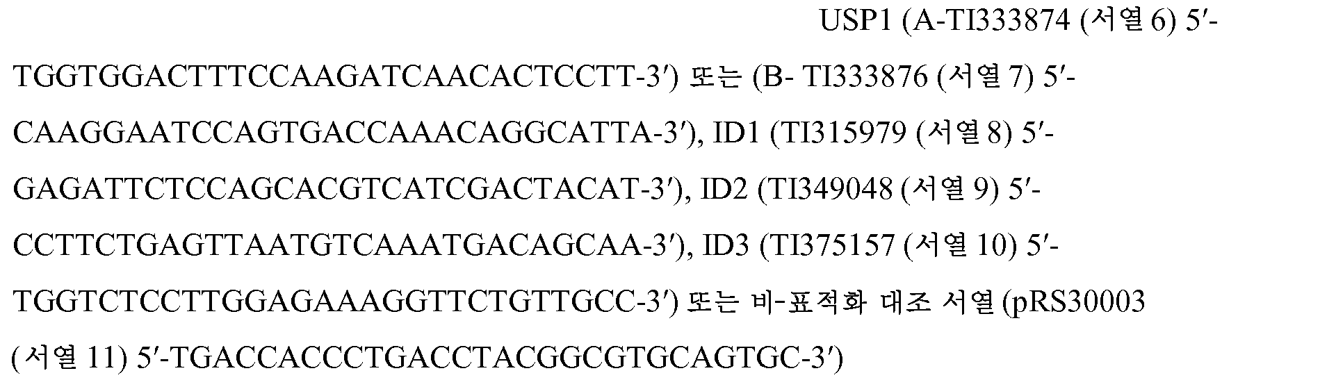

형질감염, 세포 분류, RNA 및 단백질 추출Transfection, cell sorting, RNA and protein extraction
U2-OS, HOS, SJSA, SAOS 또는 MG-63 세포를 10-25% 전면생장률로 성장시키고, 퓨진(FuGENE) 6 (시그마)을 사용하여 지시된 플라스미드로 마커 플라스미드 pMACS와 조합하여 형질감염시켰다. 형질감염된 세포를 MACS-선택 H-2Kk 마이크로비드 (밀테니 바이오텍)로 분류하였다. RNA를 배양된 또는 분류된 형질감염된 세포로부터 퀴아젠(Qiagen) RNeasy 미니 키트로 추출하였다. 단백질을 프로테아제 억제제 칵테일 I 및 포스파타제 억제제 칵테일 1 및 2 (칼바이오켐)가 보충된 NP-40 완충제 (1% NP-40, 120 mM NaCl, 50 mM 트리스, pH = 7.4, 1 mM EDTA)에서 용해를 통해 분류된 또는 배양된 세포로부터 추출하였다. 용해물을 분석 전에 10분 동안 15,000xG에서의 원심분리에 의해 정화하였다. 단백질 내용물을 BCA 단백질 검정 (써모 사이언티픽(Thermo Scientific))에 의해 정규화하였다. 이중 shRNA/siRNA 실험을 위해, 세포를 DNA 발현 벡터로 퓨진을 통해 형질감염시키고, 이후에 대조군 siRNA (센스-5'-AAUUCUCCGAACGUGUCACGU-3' (서열 20)) 또는 p21을 표적화하는 siRNA (5'-CGATGGAACTTCGACTTTGTT-3' (서열 21))로 뉴클레오펙션을 통해 뉴클레오펙션 용액 V (론자) 중 프로그램 X-001을 이용하여 형질감염시켰다. DT-40 세포를 뉴클레오펙션에 의해 뉴클레오펙션 용액 T (론자) 중 프로그램 B-023을 이용하여 형질감염시켰다.U2-OS, HOS, SJSA, SAOS or MG-63 cells were grown to 10-25% full growth rate and transfected with the indicated plasmids in combination with the marker plasmid pMACS using FuGENE 6 (Sigma). The transfected cells were classified as MACS-selective H-2K k microbeads (Miltheny biotech). RNA was extracted from cultured or sorted transfected cells with a Qiagen RNeasy mini kit. Proteins were dissolved in NP-40 buffer (1% NP-40, 120 mM NaCl, 50 mM Tris, pH = 7.4, 1 mM EDTA) supplemented with protease inhibitor cocktail I and
항체, 웨스턴 블롯팅 및 면역침전Antibody, Western blotting and immunoprecipitation
인간 USP1 또는 WDR48의 C-말단 100개의 아미노산에 대한 래트 모노클로날 항체를 생성하여 모노클로날 USP1 항체 5E10 및 WDR48 항체 9F10을 생산하였다. ID1, ID2, ID3, E47 및 p53 (산타 크루즈 바이오테크놀로지(Santa Cruz Biotechnology)), GAPDH (어세이 디자인스(Assay Designs)), 플래그, HA, 튜불린, 및 액틴 (시그마), p21WAF1/CIP1 (셀 시그널링(Cell Signaling)), E-카드헤린 및 N-카드헤린 (BD 트랜스덕션 랩스(BD Transduction Labs)) 및 피브로넥틴 (칼바이오켐)을 인식하는 항체를 상업적 공급원으로부터 수득하였다. 면역침전을 10 μM MG-132의 존재 하에 지시된 항체 및 단백질 A/G 아가로스 비드 (피어스)를 사용하여 수행하였다. 단백질 추출물을 비스-트리스 겔 (인비트로젠(Invitrogen)) 상에서 분리하고, 이뮤노블롯 분석을 위해 0.2 μM 니트로셀룰로스 막 (인비트로젠)으로 전달하였다.A rat monoclonal antibody against the C-
RNA 분석RNA analysis
RNA를 퀴아젠 RNEasy RNA 단리 키트를 이용하여 추출하였다. RNA was extracted using a quiagen RNEasy RNA isolation kit.

발현 프로브 208937_s_at (ID1), 213931_at (ID2), 207826_s_at (ID3), 202412_s_at (USP1), 202284_s_at (p21), 219534_x_at (p57), 236313_at (p15) 및 207039_at (p16)를 사용하여 지시된 인간 골 샘플에서 RNA 발현 수준의 진로직(GeneLogic) 마이크로어레이 분석 (오시뭄 바이오솔루션즈(Ocimum Biosolutions))으로부터 원발성 종양 RNA 데이터를 수득하였다. 샘플을 HGU133P 아피메트릭스 칩에 혼성화시켰다.In the indicated human bone samples using the expression probes 208937_s_at (ID1), 213931_at (ID2), 207826_s_at (ID3), 202412_s_at (USP1), 202284_s_at (p21), 219534_x_at (p57), 236313_at (p15), and 207039_at Primary tumor RNA data was obtained from GeneLogic microarray analysis of RNA expression levels (Ocimum Biosolutions). Samples were hybridized to HGU133P affinity chip.
면역조직화학Immunohistochemistry
포르말린-고정, 파라핀-포매된 조직 절편을 슬라이드에 마운팅하고, 탈파라핀화시키고, dH20로 재수화시켰다. 항원을 회수하기 위해, 샘플을 표적 검색 용액 (다코(Dako)) 중에서 20분 동안 99℃에서 인큐베이션하고, 74℃로 20분 동안 냉각시켰다. 내인성 퍼옥시다제, 아비딘, 비오틴 및 이뮤노글로불린을 아비딘/비오틴 차단 키트 완충제 (벡터 랩스(Vector Labs))에서의 인큐베이션에 이어 3% BSA 중에서 30분 동안 RT에서의 인큐베이션에 의해 켄칭하였다. 켄칭 후에, 샘플을 일차 항체 중에서 60분 동안 RT에서 인큐베이션하고, 다코 세척 완충제에서 2회 세정하고, 벡타스테인(Vectastain) 키트 (벡터 랩스) 완충제 중에서 30분 동안 인큐베이션하였다. 염색을 퍼옥시다제 기질 완충제 (피어스)에서의 인큐베이션에 의해 시각화하였다. 샘플을 메이어(Mayer)의 헤마톡실린으로 대비염색하고, 커버슬립을 마운팅한 후에 영상화하였다. RT = 실온.Formalin-fixed, paraffin-embedded tissue sections were mounted on slides, deparaffinized and rehydrated with dH20. To recover the antigen, the sample was incubated in a target search solution (Dako) for 20 minutes at 99 ° C and cooled to 74 ° C for 20 minutes. Endogenous peroxidase, avidin, biotin and immunoglobulin were incubated in avidin / biotin blocking kit buffer (Vector Labs) followed by incubation in 3% BSA for 30 minutes at RT. After quenching, samples were incubated in RT for 60 minutes in primary antibody, washed twice in Dako washing buffer, and incubated in Vectastain kit (VectorLabs) buffer for 30 minutes. The staining was visualized by incubation in a peroxidase substrate buffer (Pierce). The samples were contrast dyed with Mayer's hematoxylin and imaged after mounting the cover slip. RT = room temperature.
유동 세포측정법Flow cytometry
세포 주기 분석은 형질감염된 세포 상에서 FITC-접합된 H-2Kk 항체 (밀테니 바이오텍)으로의 염색에 이어 70% 에탄올 고정 및 RNAse A (시그마)를 갖는 프로피듐 아이오다이드 (시그마)로의 DNA 표지에 의해 수행하였다 (문헌 [Krishan, et al., J. Cell Biol. 66:188, 1975]). FITC+ 세포의 DNA 함량을 팩스칼리버(FACSCalibur) 유동 세포측정기 (BD 바이오사이언시스)를 이용하여 평가하였다. 데이터를 플로우조(FlowJo) v8.7.3 소프트웨어 (트리 스타, 인크.(Tree Star, Inc.))로 분석하였다. 세포 주기 백분위수를 플로우조 세포 주기 플랫폼으로 정량화하였다.Cell cycle analysis was performed by staining with FITC-conjugated H-2K k antibody (Miltenyi Biotech) on transfected cells followed by DNA labeling with propidium iodide (Sigma) with 70% ethanol fixation and RNAse A (Sigma) (Krishan, et al., J. Cell Biol. 66: 188, 1975). The DNA content of FITC + cells was evaluated using a FACSCalibur flow cytometer (BD Biosciences). Data were analyzed with FlowJo v8.7.3 software (Tree Star, Inc.). Cell cycle percentiles were quantified as flow cytometry platform.
hMSC 마커 발현을 골육종 세포주 및 hMSC 상에서 CD90 (케미콘(Chemicon)), CD105 (R&D 시스템즈), CD106 (서던바이오테크(SouthernBiotech)) 및 CD144 (이바이오사이언스(eBioscience)), 또는 이소형 대조군 (R&D 시스템즈)에 특이적인 PE-접합된 항체로의 염색에 의해 평가하였다. 마커의 기하 평균 발현을 팩스칼리버 유동 세포측정기를 이용하여 정량화하였다.hMSC marker expression was detected on osteosarcoma cell lines and hMSCs using CD90 (Chemicon), CD105 (R & D Systems), CD106 (Southern Biotech) and CD144 (eBioscience), or isotype control Systems) by staining with a PE-conjugated antibody specific. The geometric mean expression of the markers was quantified using a fax caliber flow cytometer.
면역형광 검정Immunofluorescence assay
기재된 바와 같이 벡터로 안정하게 형질도입된 U2-OS 세포를 챔버 슬라이드 상에서 전면생장률로 성장시키고, 3 μg/ml 독시시클린 (클론테크(Clontech))으로 14일 동안 처리하고, PBS 중 1% PFA에 고정하고, E-카드헤린-FITC, N-카드헤린 (BD 트랜스덕션 래보러토리즈(BD transduction laboratories)) 또는 피브로넥틴 (칼바이오켐)으로 프로빙하였다. 연결되지 않은 항체를 염소-항-마우스 FITC (서던 바이오테크 어소시에이츠(Southern Biotech Associates))로 검출하였다. 커버슬립을 프로롱 골드(ProLong Gold) 마운팅 작용제 (인비트로젠)와 함께 마운팅하였다.U2-OS cells stably transduced with the vector as described were grown at full growth rate on a chamber slide and treated for 14 days with 3 μg / ml doxycycline (Clontech) and incubated with 1% PFA in PBS And probed with E-carderin-FITC, N-carderine (BD transduction laboratories) or fibronectin (Calbiochem). Unbound antibodies were detected with goat-anti-mouse FITC (Southern Biotech Associates). The cover slip was mounted with ProLong Gold mounting agent (Invitrogen).
생체내 탈유비퀴틴화 검정In vivo deuterium tyrosine assay
293T 세포를 기재된 바와 같이 형질감염시키고, 10μM MG-132로 30분 동안 처리한 후에 10 μM MG-132 및 10 mM N-에틸말레이미드가 보충된 NP-40 완충제 (시그마)에서 용해시켰다. 정화된 용해물을 1% SDS로 해리시키고, 95℃에서 5분 동안 비등시키고, 이어서 용해 완충제에서 1:20 희석한 후에 M2-아가로스 항-플래그 비드 (시그마)로 면역침전시켰다. 유비퀴틴 수준을 HA 항체로의 이뮤노블롯에 의해 평가하였다.293T cells were transfected as described and lysed in NP-40 buffer (Sigma) supplemented with 10 μM MG-132 and 10 mM N-ethylmaleimide after 30 min of treatment with 10 μM MG-132. The clarified lysate was dissociated with 1% SDS, boiled for 5 minutes at 95 ° C and then immunoprecipitated with M2-agarose anti-flag beads (Sigma) after 1:20 dilution in lysis buffer. Ubiquitin levels were assessed by immunoblotting with HA antibodies.
시험관내 탈유비퀴틴화 검정In vitro ubiquitination assay
293T 세포를 개별 뱃치에서 USP1-플래그, USP1-C90S-플래그 또는 ID2-플래그 및 HA-유비퀴틴으로 형질감염시켰다. 유비퀴틴화된 ID2-플래그를 SDS-비등된 용해물로부터 상기 기재된 바와 같이 면역침전시켰다. USP1 및 USP1 C90S를 NP-40 용해 완충제에서 용해시킨 후에 플래그 M2-아가로스 비드로 면역침전시켰다. 모든 샘플을 500 μg/ml 3x플래그 펩티드 (시그마)로 비드로부터 용리하였다. 샘플을 탈유비퀴틴화 완충제 (20 mM HEPES, 20 mM NaCl, 100 μg/ml BSA, 500 μM EDTA, 1 mM DTT, pH=8.3)에서 재조합하고, 지시된 시간 동안 실온에서 인큐베이션하였다. 유비퀴틴 수준을 HA 항체로의 이뮤노블롯에 의해 평가하였다.293T cells were transfected with the USP1-flag, USP1-C90S-flag or ID2-flag and HA-ubiquitin in individual batches. The ubiquitinated ID2-flag was immunoprecipitated from SDS-boiled lysate as described above. USP1 and USP1 < RTI ID = 0.0 > C90S < / RTI > were dissolved in NP-40 lysis buffer followed by immunoprecipitation with flag M2-agarose beads. All samples were eluted from the beads with 500 [mu] g / ml 3x flag peptide (Sigma). Samples were recombined in deubiquitinating buffer (20 mM HEPES, 20 mM NaCl, 100 [mu] g / ml BSA, 500 [mu] M EDTA, 1 mM DTT, pH = 8.3) and incubated at room temperature for the indicated time. Ubiquitin levels were assessed by immunoblotting with HA antibodies.
골육종 분화 검정Osteosarcoma differentiation assay
U2-OS, HOS 또는 SAOS 세포를 하기와 같이 pRS shUSP1 또는 shCTL로 3회 형질감염시켰다: 세포를 처음에 shUSP1 또는 shCTL로 형질감염시키고, 2일 동안 배양하고, 퓨로마이신으로 3일 동안 선별하였다. 세포를 pMACS 및 shUSP1 또는 shCTL로 재형질감염시키고, 2일 동안 배양하고, 항-H-2Kk 비드 소트 (밀테니)에 의해 분류하고, 1일 동안 배양하고, shUSP1 또는 shCTL로 연속적으로 재형질감염시켰다. 세포를 추가의 3일 동안 배양하고, 골모세포 및 hMSC 마커를 유동 세포측정법, 실시간 RT-PCR 및 ALP 검정에 의해 평가하였다. 143B 세포를 pTRIPZ-기재 유도성 USP1 또는 대조군 shRNA 벡터로 형질도입시키고, 퓨로마이신-내성 세포를 계대배양하였다.U2-OS, HOS or SAOS cells were transfected three times with pRS shUSP1 or shCTL as follows: cells were first transfected with shUSP1 or shCTL, cultured for 2 days, and screened with puromycin for 3 days. Cells were retransfected with pMACS and shUSP1 or shCTL, cultured for 2 days, sorted by anti-H-2K k beads (Miltenyi), cultured for 1 day, and serially resent with shUSP1 or shCTL Infected. Cells were cultured for an additional 3 days and osteoblasts and hMSC markers were evaluated by flow cytometry, real-time RT-PCR and ALP assays. 143B cells were transfected with pTRIPZ-based inducible USP1 or control shRNA vectors and puromycin-resistant cells were subcultured.
p-니트로페놀-포스페이트 절단 검정p-Nitrophenol-phosphate cleavage assay
세포 용해물을 프로테아솜 억제제를 포함하나 포스파타제 억제제는 포함하지 않는 NP-40 완충제를 사용하여 생성하고, 단백질 함량에 대해 정규화하였다. 용해물을 투명-바닥 96-웰 플레이트에서 p-니트로페닐 포스파타제 기질 시스템 용액 (시그마)에 첨가하고, 실온에서 0.5-20시간 동안 인큐베이션하였다. 기지의 양의 4-니트로페놀 (시그마)의 연속 희석액을 참조물로 사용하였다. 활성을 스펙트라맥스(SpectraMax) 190 분광광도계 (몰레큘라 디바이시스(Molecular Devices))로 405 nm에서 OD 흡광도 측정값에 대해 정량화하고, 소프트맥스 프로(SoftMax Pro) v5.3 소프트웨어 (MDS 애널리티컬 테크놀로지스(MDS Analytical Technologies))로 분석하였다. 데이터를 단백질 투입 및 반응 시간에 대해 정규화하였다.Cell lysates were generated using NP-40 buffer containing a proteasome inhibitor but not a phosphatase inhibitor and normalized to protein content. The lysate was added to the p-nitrophenylphosphatase substrate system solution (Sigma) in a clear-bottom 96-well plate and incubated at room temperature for 0.5-20 hours. A serial dilution of a known amount of 4-nitrophenol (Sigma) was used as a reference. Activity was quantified with respect to OD absorbance measurements at 405 nm with a SpectraMax 190 spectrophotometer (Molecular Devices), and the results were analyzed using SoftMax Pro v5.3 software (MDS Analytical Technologies, (MDS Analytical Technologies). Data were normalized for protein loading and reaction time.
알리자린 레드 염색Alizarin red dyeing
8-웰 챔버 슬라이트 상에 플레이팅된 분화된 hMSC를 빙냉 70% 에탄올로 30분 동안 고정하고, 탈이온수로 세척하고, 0.2% 알리자린 레드 염색 (리카 케미컬(Ricca Chemical)), pH 6.4 중에서 30분 동안 인큐베이션하였다. 염색 후에, 세포를 탈이온수 중에서 2회 세정하고, 영상을 얻었다.The differentiated hMSCs plated on 8-well chamber slides were fixed with ice-cold 70% ethanol for 30 minutes, washed with deionized water and washed with 0.2% alizarin red stain (Ricca Chemical), 30 Min. ≪ / RTI > After staining, cells were washed twice in deionized water and images were obtained.
3T3 형질전환 검정3T3 Transfection Assay
뮤린 3T3 섬유모세포를 USP1-플래그, USP1-C90S-플래그, ID2-플래그, 또는 빈 대조 발현 벡터로 형질도입시키고, 다음 2일 동안 발현시키고, 1% 한천 비드 상의 0.5% 저융점 한천을 갖는 FBS 및 페니실린/스트렙토마이신을 포함하는 DMEM에 플레이팅하였다. 세포를 21일 동안 인큐베이션하고, 8개 이상의 세포의 콜로니를 육안 검사에 의해 스코어링하였다. USP1-형질전환된 샘플로부터의 형질전환된 콜로니를 한천으로부터 회수하고, 계대배양하고, 연질 한천에 다시 시딩하여 형질전환을 확인하였다. 강한 콜로니 성장이 모든 계대배양된 샘플에서 관찰되었다 (데이터는 나타내지 않음) .Murine 3T3 fibroblasts were transfected with the USP1-flag, the USP1-C90S-flag, the ID2-flag, or the empty control expression vector and expressed in the following 2 days and FBS with 0.5% low melting agar on 1% agar bead and And plated in DMEM containing penicillin / streptomycin. Cells were incubated for 21 days and colonies of 8 or more cells were scored by visual inspection. Transformed colonies from USP1-transformed samples were recovered from agar, subcultured, and reseeded in a soft agar to confirm transformation. Strong colony growth was observed in all subcultured samples (data not shown).
ID1, ID2, ID3 녹다운 연구를 위해, USP1-형질도입된 3T3 세포를 pTRIPZ-기재 ID1-, ID2 및 ID3- 유도성 shRNA 발현 벡터, 또는 대조군 shRNA 벡터로 형질도입시키고, 3 μg/ml 독시시클린 (클론테크)으로 72시간 처리한 후에 한천 포매시켰다. 3μg/ml 독시시클린은 1% 및 0.5% 한천 둘 다에 포함시켰다.For ID1, ID2, ID3 knockdown studies, USP1-transduced 3T3 cells were transfected with pTRIPZ-based ID1-, ID2 and ID3-inducible shRNA expression vectors, or control shRNA vectors, and 3 μg / ml doxycycline (Clontech) for 72 hours and then embedded in agar. 3 μg / ml doxycycline was included in both 1% and 0.5% agar.
생체내 연구In vivo study
8주령 암컷 NCr 누드 마우스 (타코닉 래보러토리즈(Taconic Laboratories), 뉴욕주 허드슨) 또는 C.B-17 SCID.bg 마우스 (찰스 리버스 래보러토리즈(Charles Rivers Laboratories), 캘리포니아주 홀리스터)의 오른쪽 옆구리 뒷면에 1 x 10⁶개의 뮤린 3T3 섬유모세포 (USP1, USP1-C90S, ID-2 또는 벡터 대조군으로 형질되입됨)를 100 μl의 HBSS의 부피로 피하로 주입하였다. 마우스를 종양 확립 및 성장 뿐만 아니라 체중 변화에 대해 모니터링하였다. 주어진 군의 마우스가 2000 mm3의 평균 종양 부피를 달성하고/거나 접종 후 40일에 도달하였을 경우에, 마우스를 안락사시키고, 해부하여 종양 형성의 존재 또는 부재를 확인하였다.The right side of an 8-week-old female NCr nude mouse (Taconic Laboratories, Hudson, NY) or CB-17 SCID.bg mouse (Charles Rivers Laboratories, Hollister, CA) On the backside, 1 x 10 6 murine 3T3 fibroblasts (injected USP1, USP1-C90S, ID-2 or vector control) were injected subcutaneously in a volume of 100 μl of HBSS. Mice were monitored for weight gain as well as tumor establishment and growth. The mice were euthanized and dissected to confirm the presence or absence of tumor formation when the mice of a given group reached a mean tumor volume of 2000 mm 3 and / or reached 40 days after inoculation.
8-주령 암컷 NCr 누드 마우스의 오른쪽 옆구리 뒷면에 2.5 x 10⁶개의 143B shUSP1 세포를 100μl의 HBSS + 매트리겔의 부피로 피하로 주입하였다. 독시시클린-처리된 마우스에게 5% 수크로스 물 중 독시시클린의 1 mg/mL 용액을 공급하였다. 마우스를 종양 확립 및 성장 뿐만 아니라 체중 변화에 대해 모니터링하였다. 주어진 군의 마우스가 2000 mm3의 평균 종양 부피를 달성하고/거나 접종 후 78일에 도달하였을 경우에, 마우스를 안락사시키고, 해부하여 종양 형성의 존재 또는 부재를 확인하였다.2.5 x 10 6 143B shUSP1 cells were subcutaneously injected into the volume of 100 μl of HBSS + Matrigel in the right side of 8-week-old female NCr nude mice. The doxycycline-treated mice were fed a 1 mg / mL solution of doxycycline in 5% sucrose water. Mice were monitored for weight gain as well as tumor establishment and growth. When the mice of a given group reached a mean tumor volume of 2000 mm 3 and / or reached 78 days after inoculation, the mice were euthanized and dissected to confirm the presence or absence of tumor formation.
USP1USP1 -/-- / - 마우스의 생성 Creation of mouse
USP1 유전자-표적화된 C57BL/6 뮤린 ES 세포를 녹아웃 마우스 프로젝트(Knockout Mouse Project) (KOMP) 리파지토리 (캘리포니아주 데이비스)로부터 입수하였다. 조건 대립유전자를 Cre 레콤비나제로의 전기천공 후에 배반포 주입에 의해 ES 세포에서 결실시켰다. USP1 gene-targeted C57BL / 6 murine ES cells were obtained from the Knockout Mouse Project (KOMP) repository (Davis, Calif.). Conditional alleles were deleted in ES cells by blastocyst injection after electroporation with Cre recombinase.
마이크로-컴퓨터 단층촬영Micro-computerized tomography
d12.5 마우스 새끼, 임신후 18.5일의 배아, 또는 그의 해부된 대퇴골을 하기 파라미터를 사용하는 μCT 40 (스캔코 메디칼(SCANCO Medical), 스위스 바세르도르프) X선 마이크로-컴퓨터 단층촬영 시스템으로 영상화하였다: X선 튜브 에너지 수준 = 대퇴골의 경우에 70 kV 또는 전체 마우스의 경우에 45 kV, 또는 X선 튜브 에너지 수준 = 45 kV, 전류 = 177 μA, 통합 시간 = 300 msec, 대퇴골의 경우에 1000회 투영. 축 영상을 대퇴골 분석의 경우에 12 μm 또는 태아/새끼의 경우에 30 μm의 등방성 해상도에서 수득하였다. 히드록시아파타이트 (HA) 팬텀을 사용하여 X선 흡수를 골 무기질 밀도 (BMD)에 대해 보정하였다. 마이크로-컴퓨터 단층촬영 스캔을 애널라이즈(Analyze) (애널라이즈다이렉트 인크.(AnalyzeDirect Inc.), 미국 켄자스주 레넥사)로 분석하였다. 시상면에서의 최대-강도 투영도 및 3D 표면 투시도를 각각의 샘플에 대해 생성하였다. 스캔 설정에 기초하여, 역치에 이어 침식-팽창이 연부 조직으로부터의 무기질화된 골격을 분할하는데 적용하였다.Imaging with d12.5 mouse pups, 18.5 days post-pregnancy embryos, or their dissected femurs with a μCT 40 (SCANCO Medical, Swiss Basserdorf) X-ray micro-computerized tomography system using the following parameters X-ray tube energy level = 70 kV for the femur or 45 kV for the whole mouse, or X-ray tube energy level = 45 kV, current = 177 μA, integration time = 300 msec, 1000 times for the femur projection. Axis images were obtained at isotropic resolution of 12 μm for femur analysis or 30 μm for fetus / litter. X-ray absorption was corrected for bone mineral density (BMD) using a hydroxyapatite (HA) phantom. Micro-computed tomography scans were analyzed with Analyze (AnalyzeDirect Inc., Lenexa, KY). Maximum-intensity projections in the sagittal plane and 3D surface perspective were generated for each sample. Based on the scan settings, erosion-expansion following the threshold was applied to segment the mineralized skeleton from the soft tissue.
파골세포의 계내 TRAP 염색TRAP staining of osteoclasts in vitro
포르말린 고정, 파라핀 포매된 P12 뮤린 대퇴골로부터의 슬라이드-마운팅된 5 μm 절편을 제조하고, 387A 산 포스파타제, 백혈구 (TRAP) 키트를 제조업체의 지침에 따라 이용하여 TRAP 염색 (시그마)에 적용하였다. 조사 연구를 위해, TRAP-양성 파골세포를 10개의 필드에서 카운팅하였다.Mounted 5 μm slices from formalin-fixed, paraffin-embedded P12 murine femurs were prepared and applied to TRAP staining (Sigma) using 387A acid phosphatase, white blood cell (TRAP) kit according to the manufacturer's instructions. For investigation studies, TRAP-positive osteoclasts were counted in 10 fields.
데옥시피리디놀린 및 크레아티닌 정량화Quantification of deoxypyridinoline and creatinine
양수를 E18.5 마우스로부터 수집하고, 데옥시피리디놀린을 ELISA (TSZ ELISA)에 의해 검출하고, 크레아티닌을 제조업체의 프로토콜에 따른 비색 화학 검정 (R&D 시스템즈)에 의해 검출하였다.Amniotic fluid was collected from E18.5 mice and deoxypyridinoline was detected by ELISA (TSZ ELISA) and creatinine was detected by colorimetric chemistry assays (R & D Systems) according to the manufacturer's protocol.
USP1USP1 은 silver IDID 단백질을 Protein 탈유비퀴틴화하고Deuterium 안정화시킨다 Stabilize
ID 단백질을 안정화시키는 데유비퀴티나제 (DUB)를 확인하기 위해, C-말단 플래그 에피토프를 갖는 94개의 인간 DUB를 293T 세포에서 과다발현시키고, 내인성 ID2 풍부도를 웨스턴 블롯에 의해 평가하였다 (온라인 이용가능한 표 S1 참조). ID2가 프로테아솜 억제제 MG-132로의 처리 후에 축적되기 때문에 293T 세포는 프로테아솜-의존성 방식으로 ID2를 분해한다 (도 2a). 내인성 ID2를 증가시키는 DUB는 USP36, USP33, SENP3, SENP5, USP37, OTUD5, USP9Y, USP45 및 USP1이었다 (도 2a). ID2 발현을 증가시키는 간접적 메카니즘을 배제하기 위해, ID2-상호작용 DUB인 USP1 및 USP33에 집중하였다. (도 2b). 그러나, USP1과 달리, USP33은 ID2에 대한 탈유비퀴틴화 활성이 결손되었다 (데이터는 나타내지 않음).To identify ubiquitinase (DUB) in stabilizing ID proteins, 94 human DUBs with a C-terminal flag epitope were overexpressed in 293T cells and endogenous ID2 abundance was assessed by western blotting See table S1 available). Since ID2 accumulates after treatment with the proteasome inhibitor MG-132, 293T cells degrade ID2 in a proteasome-dependent manner (Figure 2a). DUBs that increase endogenous ID2 were USP36, USP33, SENP3, SENP5, USP37, OTUD5, USP9Y, USP45 and USP1 (Fig. In order to rule out the indirect mechanism of increasing ID2 expression, we focused on the ID2-interacting DUBs, USP1 and USP33. (Fig. 2B). However, unlike USP1, USP33 lacked deubiquitinating activity against ID2 (data not shown).
USP1이 ID2의 반감기를 연장시키는지 여부를 결정하기 위해, 293T 세포를 USP1로 형질감염시키고, 번역 억제제 시클로헥시미드로의 처리 후에 ID2 풍부도를 모니터링하였다. 새로운 단백질 합성의 부재 하에, ID2는 대략 2분의 반감기로 대조군 벡터로 형질감염된 세포로부터 빠르게 제거되었다 (도 1a). 과다발현된 USP1은 ID2의 반감기를 80분 넘게 연장시켰다. USP1의 단백질분해 활성은 촉매 불활성 점 돌연변이체 USP1 C90S (Nijman et al., 2005)가 ID2의 반감기를 증가시키지도 않고 (도 1a) ID2 정상 풍부도를 변경시키지도 않기 때문에 (도 1b) ID2 축적에 필요하였다. 유사한 결과를 ID1 및 ID3에서 수득하였다. USP1은 불안정성 IkBa의 발현을 증진시키지 않았으므로 ID를 특이적으로 표적화하는 것으로 나타났다 (Palombella et al., 1994).To determine whether USP1 extends the half-life of ID2, 293T cells were transfected with USP1 and ID2 abundance was monitored following treatment with the translational inhibitor cycloheximide. In the absence of new protein synthesis, ID2 was rapidly removed from cells transfected with a control vector with a half-life of approximately 2 minutes (Fig. 1A). Overexpressed USP1 extended the half-life of ID2 over 80 minutes. The proteolytic activity of USP1 is required for the accumulation of ID2 (Fig. 1B) since the catalytic inactivity point mutant USP1 C90S (Nijman et al., 2005) does not increase the half-life of ID2 Respectively. Similar results were obtained for ID1 and ID3. USP1 did not promote the expression of unstable IkBa, indicating that it specifically targeted ID (Palombella et al., 1994).
다음에, ID2 유비퀴틴화에 대한 USP1의 효과를 평가하였다. 야생형 USP1은 HA-태그 부착된 유비퀴틴으로 변형된 ID2의 양을 감소시켰으나 USP1 C90S는 그렇지 않았다 (도 1c). 기저 및 USP1-유도된 ID2 탈유비퀴틴화는 둘 다 USP1 보조인자 WDR48의 공발현에 의해 증진되었다 (Cohn et al., 2007) (도 1c). USP1이 ID2를 직접 탈유비퀴틴화하는지 여부를 다루기 위해, 293T 세포로부터 정제된 유비퀴틴화 ID2를 시험관내에서 293T 세포로부터 개별적으로 정제된 야생형 USP1 또는 USP1 C90S와 인큐베이션하였다. 유비퀴틴화 ID2는 야생형 USP1에 의해 감소되었으나 USP1 C90S는 그렇지 않았으며 (도 1d), 이는 탈유비퀴틴화가 함께 용리된 프로테아제의 결과가 아닐 가능성을 나타낸다. ID2 유비퀴틴화의 감소는 또한 N-에틸말레이미드에 감수성이었으며, 이는 시스테인 프로테아제의 관련을 확인하였다 (도 1d). 유비퀴틴화된 ID2가 USP1 기질이라는 점과 일치하게, 결실 돌연변이체 USP1D260-300은 ID2와 잘 상호작용하지 않았으며 (도 2c) ID2 풍부도를 증진시키지 않았다 (도 2d).Next, the effect of USP1 on ID2 ubiquitination was evaluated. Wild-type USP1 reduced the amount of HA-tagged ubiquitin-modified ID2, while USP1 C90S did not (Fig. 1C). Both baseline and USP1-induced ID2 deubiquitination were promoted by coexpression of the USP1 cofactor WDR48 (Cohn et al., 2007) (Fig. 1c). To deal with whether USP1 directly de-ubiquitinated ID2, ubiquitinated ID2 purified from 293T cells was incubated in vitro with 293T cells individually purified wild-type USP1 or USP1 C90S. Ubiquitinated ID2 was reduced by wild-type USP1, while USP1 C90S was not (Fig. 1d), suggesting that deubiquitination may not be the result of proteases eluted together. The reduction in ID2 ubiquitination was also sensitive to N-ethylmaleimide, which confirmed the involvement of cysteine proteases (Fig. 1d). Consistent with the fact that ubiquitinated ID2 is a USP1 substrate, the deletion mutant USP1D260-300 did not interact well with ID2 (Fig. 2c) and did not enhance ID2 abundance (Fig. 2d).
USP1 및 ID2는 원발성 골육종 종양의 하위세트에서 대등하게 과다발현된다USP1 and ID2 are overexpressed equally in a subset of primary osteosarcoma tumors
USP1이 ID 단백질을 탈유비퀴틴화하는 생물학적 맥락을 확인하기 위해, USP1 발현 패턴을 분석하였다. 건강한 및 이환된 인간 조직의 마이크로어레이 분석은 골육종 종양이 건강한 또는 골관절염성 골 생검보다 더 많은 USP1 mRNA를 발현한다는 것을 밝혀내었다 (도 3a). 원발성 인간 골육종 생검의 개별 세트의 웨스턴 블롯팅은 USP1이 3개의 정상 일차 인간 골모세포 샘플에서에 비해 14개의 골육종 중 7개에서 상승된다는 것을 발견하였다 (도 3b). 놀랍게도, 이들 원발성 인간 종양 샘플에서의 ID2 단백질 풍부도는 USP1 풍부도와 크게 상관되었다. 한 이상 샘플은 풍부한 USP1을 함유하였으나 ID2는 거의 함유하지 않았으며 (도 3b, 레인 6), 이는 아마도 USP1 보조인자 WDR48의 양호하지 못한 발현 때문일 것이다. 또 다른 샘플은 풍부한 ID2를 함유하였으나 USP1은 거의 함유하지 않았으며 (도 3b, 레인 16), 이는 감소된 ID2 유비퀴틴화 또는 다른 DUB가 활성임을 반영할 수 있다.The USP1 expression pattern was analyzed to determine the biological context in which USP1 deubiquitinated the ID protein. Microarray analysis of healthy and diseased human tissues revealed that osteosarcoma tumors express more USP1 mRNA than healthy or osteoarthritic bone biopsies (Fig. 3a). Western blotting of a separate set of primary human osteosarcoma biopsies found that USP1 was elevated in 7 of 14 osteosarcomas compared to 3 normal primary human osteoblast samples (Fig. 3B). Surprisingly, ID2 protein abundance in these primary human tumor samples was strongly correlated with USP1 abundance. One or more samples contained abundant USP1 but little ID2 (Fig. 3b, lane 6), presumably due to poor expression of the USP1 cofactor WDR48. Another sample contained abundant ID2 but scarcely contained USP1 (Fig. 3b, lane 16), which may reflect reduced ID2 ubiquitination or other DUB activity.
원발성 골육종에서 USP1 단백질의 양은 USP1 mRNA 풍부도와 크게 상관되었으며 (도 3c), 이는 골육종에서 상승된 USP1이 전사 상향조절 때문임을 시사한다. 대조적으로, ID2 단백질 및 mRNA 수준은 잘 상관되지 않는다 (도 3d). 원발성 골육종에서 USP1 및 ID2의 일치하는 과다발현은 면역조직화학에 의해 확인되었다 (도 3e-3g). 이러한 결과는 USP1이 골육종에서 ID 단백질을 번역후에 변형시킨다는 것을 강하게 시사한다.In primary osteosarcoma, the amount of USP1 protein was strongly correlated with USP1 mRNA abundance (FIG. 3c), suggesting that elevated USP1 in osteosarcoma was due to transcriptional upregulation. In contrast, ID2 protein and mRNA levels are not well correlated (FIG. 3D). Consistent overexpression of USP1 and ID2 in primary osteosarcoma was confirmed by immunohistochemistry (Figs. 3e-3g). These results strongly suggest that USP1 transforms the ID protein after translation in osteosarcoma.
USP1은 골육종에서 ID 단백질을 안정화시킨다USP1 stabilizes ID protein in osteosarcoma
인간 골육종 세포주 및 일차 골모세포에서 USP1 풍부도 및 ID2 안정성을 또한 평가하였다 (도 5a). U2-OS 골육종 세포에서, USP1이 상승되었고, 정상적으로 불안정성 ID2가 안정하였다 (도 5a 및 5b). 2개의 별개의 USP1 shRNA로의 USP1의 녹다운은 ID1, ID2 및 ID3에서 감소를 유발하였으나 ID4에 대해서는 영향을 미치지 않았다 (도 4a). ID1, ID2 및 ID3 mRNA는 감소되지 않았으며, ID 단백질 풍부도의 강하를 원인으로 하는 감소된 전사는 제외하였다 (도 7i). USP1 녹다운 특이성은 shRNA-내성 USP1로 확인되었고, 이는 ID1, ID2 및 ID3을 기저 수준으로 복구시켰다. USP1 촉매 활성은 shRNA-내성 USP1 C90S가 ID 단백질 수준을 복구시키지 않았기 때문에 ID 안정성에 필수적이었다. 유사한 결과가 골육종 세포주 HOS, SAOS 및 SJSA에서 관찰되었다 (도 5c). USP1 녹다운은 MG-63 골육종 세포에서의 ID2 풍부도에 영향을 미치지 않았으며, 이는 아마도 이들 세포가 매우 적은 WDR48을 발현하였기 때문일 것이다 (도 5c). MG-63 세포에서 USP1 활성을 제한하는 WDR48 결핍과 일치하게, 이소성 WDR48은 ID2를 증가시켰다 (도 5d).USP1 richness and ID2 stability were also assessed in human osteosarcoma cell line and primary osteoblast (Fig. 5A). In U2-OS osteosarcoma cells, USP1 was elevated and normally instability ID2 was stable (FIGS. 5A and 5B). The knockdown of USP1 with two distinct USP1 shRNAs caused a decrease in ID1, ID2 and ID3, but did not affect ID4 (Fig. 4A). ID1, ID2 and ID3 mRNAs were not reduced, and reduced transcription due to ID protein abundance was excluded (Fig. 7i). USP1 knockdown specificity was identified as shRNA-resistant USP1, which restored ID1, ID2, and ID3 to baseline levels. USP1 catalytic activity was essential for ID stability since the shRNA-resistant USP1 C90S did not restore ID protein levels. Similar results were observed in osteosarcoma cell lines HOS, SAOS and SJSA (Fig. 5C). USP1 knockdown did not affect the ID2 abundance in MG-63 osteosarcoma cells, presumably because these cells expressed very little WDR48 (Fig. 5c). Consistent with WDR48 deficiency, which limits USP1 activity in MG-63 cells, ectopic WDR48 increased ID2 (Fig. 5d).
USP1 녹다운 및 감소된 ID 1-3이 bHLH 전사 활성을 조절하는지를 결정하기 위해, U2-OS 세포를 E 박스-구동된 루시페라제 리포터 유전자로 형질감염시켰다. USP1 shRNA는 이 리포터의 발현을 대조군 shRNA보다 7- 내지 10-배 증진시켰으며, 이는 ID 단백질이 감소된 경우에 bHLH 단백질의 활성화와 일치한다 (도 4b). shRNA-내성 야생형 USP1은 USP1 녹다운에 의해 유발된 E 박스-구동된 리포터 활성을 억제하였으나 USP1 C90S는 그렇지 않았으며, 이는 USP1 촉매 활성이 bHLH-의존성 전사 뿐만 아니라 ID 단백질 안정화에 요구된다는 것을 확인한다.U2-OS cells were transfected with the E box-driven luciferase reporter gene to determine whether USP1 knockdown and reduced ID1-3 regulated bHLH transcriptional activity. USP1 shRNA promoted the expression of this
내인성 USP1의 녹다운 후의 ID 1-3의 급성 손실은 프로테아솜-매개된 분해를 통한 ID 단백질 탈안정화를 제안하였다. 선행된 바와 같이, 프로테아솜 억제제 MG-132는 U2-OS 세포에서 ID 단백질 풍부도를 스스로 변경시키지 않았으며, 이는 ID가 USP1를 고도로 발현하는 세포에서 내재적으로 안정하다는 것을 시사한다 (도 4c). 그러나, MG-132 처리는 USP1 녹다운 후에 ID 발현을 복구하였으며, 이는 ID 단백질이 USP1 고갈시에 프로테아솜-매개된 제거에 적용된다는 것을 나타낸다. 이러한 시나리오를 유지하면서, MG-132-처리된 U2-OS 세포에서의 USP1 녹다운은 유비퀴틴화된 ID2의 양을 증가시켰다 (도 4d).Acute loss of ID 1-3 after endogenous USP1 knockdown suggested ID protein destabilization via proteasome-mediated degradation. As previously suggested, proteasome inhibitor MG-132 did not alter ID protein abundance by itself in U2-OS cells suggesting that ID is intrinsically stable in cells highly expressing USP1 (Figure 4c) . However, MG-132 treatment restored ID expression after USP1 knockdown, indicating that the ID protein is applied to proteasome-mediated removal at the time of USP1 depletion. In keeping with this scenario, USP1 knockdown in MG-132-treated U2-OS cells increased the amount of ubiquitinated ID2 (Fig. 4d).
다음에, 내인성 USP1은 골육종 세포에서 외인성 ID와 연관되는 것으로 확인되었다. ID2는 U2-OS 세포로부터의 외인성 USP1과 1:1 화학량론으로는 아닐지라도 공동면역침전되었으나 (도 4e), 이는 일시적인 효소-기질 상호작용의 경우에 예상되는 것이었다. 유사한 결과가 HOS 세포에서 얻어졌다 (도 5e). USP1은 또한 ID2와 공동면역침전되었다 (도 4f). 총괄적으로, 이러한 결과는 USP1이 강력한 DUB이며 골육종에서 ID1, ID2 및 ID3에 대한 안정화 인자임을 시사한다.Next, endogenous USP1 was found to be associated with an exogenous ID in osteosarcoma cells. ID2 co-immunoprecipitated with exogenous USP1 from U2-OS cells, although not at a 1: 1 stoichiometry (Fig. 4e), which was expected in the case of transient enzyme-substrate interactions. Similar results were obtained in HOS cells (Figure 5e). USP1 also co-immunoprecipitated with ID2 (Figure 4f). Overall, these results suggest that USP1 is a potent DUB and a stabilizing factor for ID1, ID2 and ID3 in osteosarcoma.
USP1에 의한 ID2 안정화는 골육종의 설정에 제한되지 않았다. USP1-/- DT40 닭 B 세포 (Oestergaard et al., 2007)는 유사한 수준의 ID2 mRNA를 발현함에도 불구하고 (도 5g) 그의 야생형 대응물보다 적은 ID2 단백질을 발현시켰다 (도 5f). ID2를 탈유비퀴틴화하고 안정화시키는 USP1과 일치하게, MG-132로의 프로테아솜 억제는 USP1-/-에서 ID2를 증가시켰으나, 야생형, DT40 세포는 그렇지 않았다 (도 5h). 또한, 야생형 USP1로 재구성된 USP1-/- DT40 세포는 야생형 DT40 세포에 동등한 ID2를 함유하였으나 USP1 C90S는 그렇지 않았다 (도 5i).The stabilization of ID2 by USP1 was not limited to the setting of osteosarcoma. USP1 - / - DT40 chicken B cells (Oestergaard et al., 2007) expressed similar ID2 mRNAs (FIG. 5g) but expressed less ID2 protein than their wild type counterparts (FIG. 5f). In agreement with USP1 de-ubiquitinating and stabilizing ID2, proteasome inhibition to MG-132 increased ID2 in USP1 - / - , whereas wild type, DT40 cells did not (Fig. 5h). In addition, USP1 - / - DT40 cells reconstituted with wild type USP1 contained ID2 equivalent to wild type DT40 cells, whereas USP1 C90S did not (Fig. 5i).
USP1은 골육종에서 p21-매개된 세포-주기 정지를 억제한다USP1 inhibits p21-mediated cell-cycle arrest in osteosarcoma
골육종 세포에서 USP1 결손 및 증가된 bHLH 전사 활성의 한 가지 가능한 결과는 bHLH-조절된 CDKI p21의 유도이다. 실제로, p21은 대조군 shRNA로 형질감염된 세포에 비해 USP1 shRNA로 형질감염된 U2-OS 세포에서 증가하였다 (도 6a). shRNA-내성 야생형 USP1은 p21을 대조군 세포에서 관찰된 수준으로 감소시켰으나 USP1 C90S는 그렇지 않았으며, 이는 이러한 설정에서 녹다운 특이성을 확인한다. 종양 억제제 p53 (CDKN1A의 널리-알려진 유도제)은 USP1 녹다운에 의해 증가하지 않았으며, 이는 증가된 p21이 p53 비의존성임을 시사한다.One possible outcome of USP1 deficiency and increased bHLH transcriptional activity in osteosarcoma cells is the induction of bHLH-regulated CDKI p21. Indeed, p21 was increased in U2-OS cells transfected with USP1 shRNA compared to cells transfected with control shRNA (Fig. 6A). The shRNA-resistant wild-type USP1 reduced p21 to levels observed in control cells, while USP1 C90S did not, confirming the knockdown specificity in this setting. The tumor suppressor p53 (a widely known inducer of CDKN1A) was not increased by USP1 knockdown, suggesting that increased p21 is p53-independent.
p21은 세포 주기 진행의 강력한 억제제이며 (Polyak et al., 1996), 이에 따라 USP1 녹다운 후에 U2-OS 세포의 증식 능력을 평가하였다. 증가된 p21과 일치하게, USP1 녹다운은 U2-OS 세포 증식을 감소시켰다 (도 6b 및 도 7a). shRNA-내성 야생형 USP1은 세포 증식을 회복시켰으나 USP1 C90S와 USP1D260-300은 그렇지 않았으며 (도 7b 및 7c), 이는 USP1 촉매 활성 및 ID 기질 인식이 둘 다 U2-OS 세포 증식을 유지하는데 요구된다는 것을 나타낸다. USP1 녹다운은 HOS, SAOS 및 SJSA에서 유사하게 증식을 감소시켰으나, MG-63 골육종 세포는 그렇지 않았다 (도 7d). USP1 녹다운 후에 U2-OS 세포에서의 DNA 함량의 유동 세포측정 분석은 세포 주기의 G1 및 G2 기의 세포의 중간 정도의 증가와 S기의 세포의 명백한 감소를 보여주었다 (도 6c 및 도 7e). USP1 녹다운 후의 아폽토시스 유도는 두드러지지 않았고; 서브디플로이드 DNA 함량을 갖는 소수의 세포가 세포가 관찰되었고, 아넥신 V로 염색된 세포의 증가가 존재하지 않았으며, 카스파제-3의 증가된 프로세싱이 검출되었다 (도 7e; 데이터는 나타내지 않음). 유의하게, CDKN1A siRNA는 USP1-결손 U2-OS 세포에서 S기 진입을 회복시켰으며 (도 7f 및 7g), 이는 p21이 USP1 녹다운에 의해 유도된 세포-주기 정지에 필수적이라는 것을 나타낸다.p21 is a potent inhibitor of cell cycle progression (Polyak et al., 1996), thus evaluating the proliferative capacity of U2-OS cells after USP1 knock down. Consistent with the increased p21, USP1 knockdown reduced U2-OS cell proliferation (Fig. 6b and Fig. 7a). shRNA-resistant wild-type USP1 restored cell proliferation, whereas USP1 C90S and USP1D260-300 did not (Figs. 7b and 7c), suggesting that both USP1 catalytic activity and ID substrate recognition are required to maintain U2-OS cell proliferation . USP1 knockdown similarly reduced proliferation in HOS, SAOS, and SJSA, whereas MG-63 osteosarcoma cells did not (Fig. 7d). Flow cytometric analysis of DNA content in U2-OS cells after USP1 knockdown showed a moderate increase in cells in the G1 and G2 phase of the cell cycle and a clear decrease in the S phase in the cells (Figs. 6c and 7e). Apoptosis induction after USP1 knockdown was not prominent; Cells were observed in a small number of cells with a subdifluorid DNA content, no increase in cells stained with annexin V, and increased processing of caspase-3 was detected (Figure 7e; data not shown ). Significantly, CDKN1A siRNA restored S-phase entry in USP1-deficient U2-OS cells (Figs. 7f and 7g), indicating that p21 is essential for cell-cycle arrest induced by USP1 knockdown.
USP1은 ID 단백질을 통해 골육종에서 p21 발현 및 세포-주기 정지를 조절한다USP1 regulates p21 expression and cell-cycle arrest in osteosarcoma via ID protein
USP1의 부재 하의 ID 분해가 p21 유도를 일으키는 경우에, ID 단백질의 녹다운은 USP1 녹다운을 표현형 모사할 것이다. ID 1-3의 shRNA 녹다운은 개별적으로 p21 수준을 변경시키지 않았으나, ID1, ID2 및 ID3의 조합된 녹다운은 p21을 USP1 녹다운과 유사하게 증가시켰다 (도 7h). ID 및 USP1-결손 세포는 또한 대등한 수준의 CDKN1A mRNA를 발현시켰다 (도 7i). 이러한 관찰과 일치하게, ID 결손은 USP1 결손과 유사한 세포-주기 정지를 유발하였으며 (도 S3J 및 S3K), 이는 p21 녹다운에 의해 구제되었다 (도 6d 및 6e).If ID degradation in the absence of USP1 causes p21 induction, the knockdown of the ID protein will mimic the USP1 knockdown phenotype. ID 1-3 shRNA knockdown did not alter p21 levels individually, but the combined knockdown of ID1, ID2 and ID3 increased p21 similar to USP1 knockdown (FIG. 7h). ID and USP1-deficient cells also expressed comparable levels of CDKN1A mRNA (Fig. 7i). Consistent with these observations, ID defects induced cell-cycle arrest similar to USP1 deficiency (Fig. S3J and S3K), which was rescued by p21 knockdown (Fig. 6d and 6e).
CDKN1A는 DNA 손상에 반응하여 활성화되는, p53을 비롯한 다수의 전사 인자에 의해 조절된다 (Kastan et al., 1991). p53 녹다운은 U2-OS 세포에서 에토포시드-유도된 p21을 억제하였으나 USP1 녹다운 후에 관찰된 p21 단백질의 증가를 차단하지 않았으며 (도 7l), 이는 p21 유도의 p53-비의존성 메카니즘을 지지한다. USP1은 DNA 복구 동안 PCNA 및 FANCD2를 표적화하는 것으로 보고되었기 때문에 (Nijman et al., 2005; Huang et al., 2006), DNA 손상의 생산이 USP1 녹다운의 결과로서 결정되었다. DNA 손상과 연관된 H2AX 인산화 (Rogakou et al., 1999)는 에토포시드 처리 후에 증가하였으나 USP1 녹다운은 그렇지 않았다 (데이터는 나타내지 않음). 이러한 관찰은 USP1 shRNA가 p53-결손 SAOS 세포를 정지시키는 능력과 함께 (도 7d), 일반적 DNA 손상, 특히 USP1 녹다운 후에 p21 유도에서의 매개체로서의 p53을 배제한다.CDKN1A is regulated by a number of transcription factors, including p53, which are activated in response to DNA damage (Kastan et al., 1991). p53 knockdown inhibited etoposide-induced p21 in U2-OS cells but did not block the increase in p21 protein observed after USP1 knockdown (Fig. 71), which supports the p53-independent mechanism of p21 induction. Since USP1 has been reported to target PCNA and FANCD2 during DNA repair (Nijman et al., 2005; Huang et al., 2006), the production of DNA damage was determined as a result of USP1 knockdown. H2AX phosphorylation associated with DNA damage (Rogakou et al., 1999) was increased after ethoposide treatment but not USP1 knockdown (data not shown). This observation excludes p53 as a mediator in induction of p21 after general DNA damage, particularly USP1 knockdown, with the ability of USP1 shRNA to arrest p53-deficient SAOS cells (Fig. 7d).
USP1은 ID1, ID2 및 ID3의 이소성 발현을 갖는 U2-OS 세포에서 USP1 녹다운의 효과를 구제함으로써 ID를 통한 p21 발현 및 세포 순환을 조절하는 것이 확인되었다. USP1-고갈된 세포에서의 ID 발현은 p21 발현을 억제하였고 (도 6f), 세포-주기 정지를 차단하였다 (도 6g). 결과는 함께 USP1이 골육종에서 ID 단백질 안정화 및 bHLH 전사 활성의 억제를 통해 p21을 억제한다는 것을 입증한다 .USP1 has been shown to regulate p21 expression and cell cycle through ID by relieving the effect of USP1 knockdown in U2-OS cells with ectopic expression of ID1, ID2 and ID3. ID expression in USP1-depleted cells inhibited p21 expression (Fig. 6f) and blocked cell-cycle arrest (Fig. 6g). The results demonstrate that USP1 inhibits p21 by inhibiting ID protein stabilization and bHLH transcriptional activity in osteosarcoma.
USP1 및 ID 단백질은 골육종에서 골발생 개입을 제한한다USP1 and ID proteins limit osteogenesis intervention in osteosarcoma
골육종은 골모세포, 연골세포 및 지방세포의 붕괴된 덩어리로 구성된 이종성 종양이다. 이러한 종양은 모든 3가지 계통을 생성할 수 있는 중간엽 줄기 세포 집단으로부터 발생하는 것으로 생각된다 (Tang et al., 2008). 따라서, 골육종 세포주는 고전적 골모세포 마커, 예컨대 RUNX2, 오스테릭스, SPARC/오스테오넥틴 및 알칼리성 포스파타제 (ALP)를 발현하는데 실패하였다 (Luo et al., 2008). 골육종 세포주는 또한 CD90, CD105 및 CD106을 비롯한 중간엽 줄기 세포의 특징인 표면마커를 발현한다 (Di Fiore et al., 2009). ID가 줄기 세포 유지 및 분화의 조절에서 수행하는 역할에 비추어, 골육종에서 USP1 또는 ID 녹다운에 의한 골모세포 분화의 촉진을 조사하였다. USP1 또는 ID shRNA로 형질감염된 U2-OS 세포는 대조군 세포에 비해 적게 CD105, CD106 및 CD90을 발현한 반면, 모든 세포는 동등한 양의 비관련 표면 마커 CD144를 발현하였다 (도 9a). 유사한 결과가 HOS, SJSA 및 SAOS 세포주에서 관찰되었다. USP1 또는 ID 녹다운은 또한 골모세포 RUNX2, 오스테릭스 및 오스테오넥틴의 발현을 증가시켰으며 (도 9b), ALP 활성을 증가시켰다 (도 9c). U2-OS 세포에서 USP1 녹다운 후에 증가된 E-카드헤린 발현 및 감소된 N-카드헤린 및 피브로넥틴은 악성 상태의 골육종을 수반하는 상피의 중간엽 전이로의 전도를 나타내었다 (도 8a 및 8b) (Thiery et al., 2009). 총괄적으로, 이러한 데이터는 골육종에서 USP1에 의한 ID 단백질 안정화가 정상 골발생 분화 프로그램을 차단한다는 것을 시사한다.Osteosarcoma is a heterogeneous tumor composed of collapsed masses of osteoblasts, chondrocytes, and adipocytes. These tumors are thought to originate from a population of mesenchymal stem cells capable of producing all three lines (Tang et al., 2008). Thus, osteosarcoma cell lines have failed to express classical osteoblast markers such as RUNX2, osteerix, SPARC / austenoctin and alkaline phosphatase (ALP) (Luo et al., 2008). Osteosarcoma cell lines also express surface markers that are characteristic of mesenchymal stem cells, including CD90, CD105 and CD106 (Di Fiore et al., 2009). In the present study, we investigated the stimulation of osteoblast differentiation by USP1 or ID knockdown in osteosarcoma in view of the role of ID in regulation of stem cell maintenance and differentiation. U2-OS cells transfected with USP1 or ID shRNA expressed less CD105, CD106 and CD90 than control cells, whereas all cells expressed an equivalent amount of non-related surface marker CD144 (FIG. 9A). Similar results were observed in HOS, SJSA and SAOS cell lines. USP1 or ID knockdown also increased the expression of osteoblasts RUNX2, osteotels and austenectin (Fig. 9b) and increased ALP activity (Fig. 9c). Increased E-carderine expression and reduced N-carderine and fibronectin after USP1 knockdown in U2-OS cells showed conduction to mesenchymal transition of the epithelium with malignant osteosarcoma (Figs. 8a and 8b) Thiery et al., 2009). Overall, these data suggest that ID protein stabilization by USP1 in osteosarcoma blocks normal bone development differentiation programs.
종양 분화 전략으로서의 USP1 억제의 잠재성을 143B 골육종 이종이식편 모델에서 조사하였다. 독시시클린-유도된 USP1 shRNA는 이종이식편에서 USP1 발현을 억제하였고 ID1 및 ID2를 감소시켰다 (도 8c 및 도 9d). ID3은 이러한 설정에서 검출가능하지 않았다 (데이터는 나타내지 않음). USP1 녹다운은 또한 143B 종양 성장을 감소시키고 (도 8d), 오스테오넥틴, RUNX2, SPP1/오스테오폰틴, 오스테릭스 및 BGLAP/오스테오칼신 발현을 촉진하고 (도 8e 및 도 9e), ALP 활성을 증진시켰다 (도 8f). 주목할만하게, 10개의 USP1-결손 이종이식 종양 중 4개가 계내에서 정체 및 분화를 달성하였으며, 이는 현저하게 변경된 세포 형태 및 원형-골화와 일치하는 무세포 교원성 덩어리의 축적을 나타낸다 (도 8g). 계속 증식하는 종양은 녹다운으로부터의 탈출의 증거를 보여주었으며, 이는 아마도 shRNA의 손실 또는 침묵 때문일 것이다 (도 9f). 이러한 데이터는 USP1의 감소가 골육종에서 골발생 분화 프로그램을 개시하기에 충분하다는 것을 나타낸다.The potential for USP1 inhibition as a tumor differentiation strategy was investigated in the 143B osteosarcoma xenograft model. The doxycycline-induced USP1 shRNA inhibited USP1 expression in xenografts and decreased ID1 and ID2 (Figs. 8c and 9d). ID3 was not detectable at this setting (data not shown). USP1 knockdown also reduced 143B tumor growth (Fig. 8d) and promoted austenectin, RUNX2, SPPl / austeoponin, osteotelia and BGLAP / osteocalcin expression (Figs. 8e and 9e) and enhanced ALP activity (Fig. 8F). Notably, four of the ten USP1-deficient xenograft tumors achieved stasis and differentiation in the system, indicating accumulation of acellular collagenous mass consistent with significantly altered cell morphology and circular-ossification (Fig. 8g). Proliferating tumors showed evidence of escape from the knockdown, probably due to loss or silence of the shRNA (Figure 9f). These data indicate that a decrease in USP1 is sufficient to initiate a bone differentiation program in osteosarcoma.
탈조절된 USP1 발현은 hMSC 분화를 억제한다Deregulated USP1 expression inhibits hMSC differentiation
다음에, ID의 USP1 안정화가 정상 중간엽 줄기 세포 유지에 기여하는지 여부를 결정하였다. USP1은 원발성 hMSC에서 발현되었으나 세포를 골모세포 분화를 우선하는 조건에서 배양하였을 때 점차적으로 감소하였다 (도 10a). 이전에 연구와 일치하게 (Peng et al., 2003), ID1 및 ID2를 일시적으로 유도한 후에, 또한 감소시켰다. ID3은 검출되지 않았다 (데이터는 나타내지 않음). 이들 데이터는, 잘못 조절된 ID 발현이 골발생 분화를 억제한다는 것을 보여주는 연구와 함께 (Peng et al., 2004), USP1 과다발현이 hMSC 분화를 파괴하는지 여부에 대한 조사를 촉진하였다. USP1을 과다발현하고 골발생 분화 배지에서 배양된 hMSC는 비정상적으로 높은 수준의 ID1 및 ID2를 발현하였고 (도 10b), 낮은 ALP 활성을 나타내었고 (도 10c), RUNX2, 오스테릭스 및 오스테오넥틴의 최소 유도를 보여주었고 (도 10d), 알리자린 레드로 잘 염색되지 않았으며, 이는 골모세포 활성의 고전적 마커인 무기질 침착을 나타낸다 (도 10e). 이러한 데이터는 USP1을 과다발현하는 hMSC가 분화하는데 실패하였다는 것을 의미한다. 유사한 분화 결함이 ID2를 과다발현하는 hMSC에서 관찰된 반면, USP1 C90S를 과다발현하는 hMSC는 대조군 세포와 유사하게 분화하였다. 따라서, USP1의 촉매 활성은 필수적이며, ID 안정화는 골발생 분화를 억제하기에 충분하였다.Next, it was determined whether USP1 stabilization of ID contributed to maintenance of normal mesenchymal stem cells. USP1 was expressed in primary hMSCs, but gradually decreased when cells were cultured under conditions that favored osteoblast differentiation (Fig. 10A). Consistent with previous studies (Peng et al., 2003), ID1 and ID2 were also induced after transient induction. ID3 was not detected (data not shown). These data, together with studies showing that misregulated ID expression suppresses osteogenesis differentiation (Peng et al., 2004), have prompted investigations into whether USP1 overexpression destroys hMSC differentiation. HMSCs overexpressing USP1 and cultured in osteogenic differentiation medium exhibited abnormally high levels of ID1 and ID2 (FIG. 10B), low ALP activity (FIG. 10C), and RUNX2, austenitic and austenotetin Showed minimal induction (Fig. 10d) and did not stain well with alizarin red, indicating mineral deposition, a classic marker of osteoblast activity (Fig. 10e). These data indicate that hMSCs overexpressing USP1 failed to differentiate. Similar depletion defects were observed in hMSCs overexpressing ID2, while hMSCs overexpressing USP1 C90S differentiated similarly to control cells. Therefore, the catalytic activity of USP1 is essential, and the ID stabilization is sufficient to inhibit osteogenesis differentiation.
USP1 또는 ID2를 과다발현하는 hMSC는 명백한 분화 실패와 동시에 과량의 골발생 분화 인자의 존재 하에 유의하게 증식하였다 (도 10f). 대조적으로, 대조군 hMSC, 또는 USP1 C90S를 발현하는 것의 증식은 이들이 배양물에서 분화됨에 따라 지연되었다. 총괄적으로, 이러한 관찰은 USP1 또는 ID2의 과다발현이 골모세포 분화를 차단하고, 줄기-유사 특성의 보유를 촉진하며, 세포를 분화 신호에 대해 내성을 갖도록 하기에 충분하다는 것을 시사한다.HMSCs overexpressing USP1 or ID2 significantly proliferated in the presence of excessive bone differentiation factor at the same time as the obvious differentiation failure (Fig. 10f). In contrast, the proliferation of expressing control hMSC, or USP1 C90S, was delayed as they differentiated from the culture. Overall, this observation suggests that overexpression of USP1 or ID2 is sufficient to block osteoblast differentiation, promote retention of stem-like characteristics, and make the cell resistant to differentiation signals.
USP1은 형질전환 및 종양 형성을 촉진한다USP1 promotes transformation and tumor formation
USP1이 중간엽 줄기 세포 분화를 억제하고 골육종 세포주의 증식을 지속하는 능력은 USP1이 세포 형질전환을 촉진할 수도 있다는 것을 시사한다. NIH 3T3 세포를 공벡터, ID2, USP1 또는 USP1 C90S로 안정하게 형질도입시켰다. 야생형 USP1은 ID 1-3의 발현을 증가시켰으며 (도 11a), 연질 한천에서 비부착 세포 증식을 유발하였으며 (도 11b), 이는 종양원성 형질전환의 고전적 특징이다 (Hanahan and Weinberg, 2000). 대조적으로, 공벡터 또는 USP1 C90S를 형질도입시킨 세포는 연질 한천에서 잘 성장하지 않았다 (도 11b 및 11c). 흥미롭게도, USP1은 ID2보다 더 크고 더 많은 콜로니를 생산하였으며 (도 11c), 이는 다중 ID 단백질의 안정화가 ID2 과다발현 단독보다 더 형질전환가능할 수 있음을 시사한다.The ability of USP1 to inhibit mesenchymal stem cell differentiation and to sustain proliferation of osteosarcoma cell lines suggests that USP1 may promote cell transformation. NIH 3T3 cells were stably transduced with an empty vector, ID2, USP1 or USP1 C90S. Wild-type USP1 increased expression of ID 1-3 (Fig. 11A) and induced adherent cell proliferation in soft agar (Fig. 11B), which is a classic feature of tumorigenic transformation (Hanahan and Weinberg, 2000). In contrast, cells that were transfected with empty vector or USP1 C90S did not grow well in soft agar (FIGS. 11b and 11c). Interestingly, USP1 produced larger and more colonies than ID2 (FIG. 11c), suggesting that stabilization of multiple ID proteins may be more transformable than ID2 overexpression alone.
NIH 3T3 세포를 C.B-17 SCID.bg 마우스에 피하로 이식한 경우에 시험관내 관찰을 생체내에서 재현하였다. 대조군 세포 및 USP1 C90S를 발현하는 세포는 측정가능한 종양을 생산하는데 실패한 반면, USP1 또는 ID2를 과다발현하는 세포는 이식후 7일만큼 빨리 측정가능한 종양을 생산하였다 (도 11d). 연구 종점에서 종양의 전체 육안 검사는 USP1 또는 ID2를 과다발현하는 세포가 공격성 악성종양을 생산한다는 것을 확인하였다 (도 11e). 유사한 결과가 NCr 누드 마우스에서 관찰되었다.In vitro studies were reproduced in vivo when NIH 3T3 cells were transplanted subcutaneously into C.B-17 SCID.bg mice. Control cells and cells expressing USP1 C90S failed to produce measurable tumors, while cells overexpressing USP1 or ID2 produced measurable tumors as early as 7 days after transplantation (FIG. 11d). A full visual examination of the tumor at the study end point confirmed that cells overexpressing USP1 or ID2 produce aggressive malignant tumors (FIG. 11E). Similar results were observed in NCr nude mice.
Id1, Id2 및 Id3 shRNA를 갖는 USP1에 의한 NIH 3T3 세포 형질전환에 대한 ID의 기여를 평가하였다. ID 1-3의 억제 (도 11f)는 연질 한천에서 콜로니 형성을 차단하였으며 (도 11g), 이는 ID가 NIH 3T3 세포의 USP1 형질전환에 필수적이라는 것을 나타낸다.The contribution of ID to NIH 3T3 cell transformation by USP1 with Id1, Id2 and Id3 shRNAs was evaluated. Inhibition of ID 1-3 (FIG. 11f) blocked colony formation in soft agar (FIG. 11g), indicating that ID is essential for USP1 transformation of NIH 3T3 cells.
USP1은 골 발생을 조절한다USP1 regulates bone development
USP1 과다발현은 중간엽 전구체의 골모세포 분화를 손상시키는 반면, USP1 손실은 골육종 세포의 골모세포 분화를 유발하므로, 정상 골 발생의 조절에서 USP1의 역할을 USP1 유전자-표적화된 마우스로 평가하였다. P12 USP1-/- 마우스는 두개골 및 장골의 경화에 결합을 갖는 골감소증이었다 (도 12a). 발육부전 흉골은 USP1-/- 새끼에서 치명적인 청색증 호흡 부전에 기여할 가능성이 있다 (Kim et al., 2009). USP1-/- 신생아 및 E18.5 배아에서의 골 무기질 밀도 및 부피는 야생형 한배자손에서보다 훨씬 더 낮았다 (도 12b 및 12c 및 도 13b 및 13c). FANCD2- 또는 PCNA-결손 마우스는 모두 주산기 사망을 나타내었으며 (Parmar et al., 2010; Roa et al., 2008), USP1 결손과 연관된 주산기 사망의 주원인으로서의 이들 USP1 기질의 탈안정화를 배제하였다.USP1 overexpression impairs osteoblast differentiation of the mesenchymal precursor whereas USP1 loss induces osteoblast differentiation of osteosarcoma cells, so USP1 gene-targeted mice were evaluated for the role of USP1 in regulating normal bone development. The P12 USP1 - / - mice were osteopenia with binding to the cranial and iliac cortex (Fig. 12A). The dysmorphic sternum may contribute to fatal cyanotic respiratory failure in USP1 - / - pups (Kim et al., 2009). The bone mineral density and volume in the USP1 - / - neonates and E18.5 embryos were much lower than in the wild-type perennial progeny (Figures 12b and 12c and Figures 13b and 13c). FANCD2- or PCNA-deficient mice all exhibited perinatal mortality (Parmar et al., 2010; Roa et al., 2008), excluding the destabilization of these USP1 substrates as the primary cause of perinatal mortality associated with USP1 deficiency .
USP1-/- 및 USP1+/+ 대퇴골은 유사한 수의 휴지, 전이성, 증식성 및 비후성 연골세포를 함유하였으나, 신생 골 골편 상의 유골의 침착은 감소하였으며, 이는 유골-침착성 골모세포의 감소된 활성을 시사한다 (도 13d-13f). 골모세포 기능의 결합과 일치하게, 전신 골모세포 활성의 마커인 골 알칼리성 포스파타제 (BALP)의 혈청 수준은 USP1-/- E18.5 배아에서 감쇠되었다 (도 12e). USP1 결손은 파골세포 풍부도 또는 활성을 변경시키지 않았으며 (도 13g-13i), USP1-/- 마우스에서 상승된 골 재흡수는 배제하였다. 유의하게, 및 골육종 및 중간엽 줄기 세포 배양물에서의 관찰과 일치하게, USP1-/- 대퇴골 골간단은 그의 야생형 대응물보다 적은 ID1 및 ID2를 함유하였다 (도 12d 및 도 13j). 이들 데이터는 골육종에서 분화를 조절하는 USP1-ID 축이 정상 골격 발생을 재현하였다는 것을 나타낸다.The USP1 - / - and USP1 + / + femurs contained similar numbers of resting, metastatic, proliferative and hypertrophic chondrocytes, but the deposition of osteoid osteoid fragments on the new bone fragments decreased, suggesting reduced activity of osteoid- (Figs. 13D-13F). Consistent with the binding of osteoblast function, serum levels of bone alkaline phosphatase (BALP), a marker of systemic osteoblast activity, were attenuated in USP1 - / - E18.5 embryos (FIG. 12e). The USP1 deletion did not alter osteoclast richness or activity (Fig. 13g-13i) and excluded elevated bone resorption in USP1 - / - mice. Significantly, and consistent with observations in osteosarcoma and mesenchymal stem cell cultures, the USP1 - / - femoral osseousum contained less ID1 and ID2 than its wild type counterparts (FIGS. 12d and 13j). These data indicate that the USP1-ID axis, which regulates differentiation in osteosarcoma, reproduces normal skeletal development.
논의Argument
이들 실험은 USP1이 ID1, ID2 및 ID3을 탈유비퀴틴화하고 안정화시켜, 그의 증가된 풍부도를 발생시킨다는 것을 보여준다. 유의하게, 원발성 골육종 종양의 하위세트에서 상승된 USP1 단백질 및 mRNA는 증가된 ID 단백질 수준과 상관된다. 골육종 세포에서의 USP1 녹다운은 ID 단백질 탈안정화, CDKN1A 코딩 시클린-의존성 키나제 억제제 (CDKI) p21의 p53-비의존성 유도, 및 세포 주기 정지를 유발하였다. 또한, 중간엽 줄기 세포 마커의 발현은 감소되었고, 골발생 분화는 재개되었다. 이들 데이터는 골육종이 급성 전골수구성 백혈병과 같이 분화 요법에 따를 수 있다는 것을 시사한다 (Soignet et al., 1998). USP1 녹다운과 대조적으로, 원발성 인간 중간엽 줄기 세포 (hMSC)의 USP1 과다발현은 ID 단백질 축적을 유발하고, 정상 분화를 방해하였다. 사실상, USP1은 중간엽 세포주의 형질전환을 촉진하였다. 최종적으로, 유전자-표적화된 마우스에서 USP1의 손실은 중증의 골감소증을 유발하였으며, 이는 중간엽 계통의 USP1에 대한 역할과 일치한다. 이러한 결과는 USP1이 종양원성 잠재력을 가지며, 정상 중간엽 줄기 세포 개입 및 분화의 파괴를 통한 종양발생을 촉진한다는 것을 시사한다.These experiments show that USP1 deubiquitinates and stabilizes ID1, ID2 and ID3, resulting in increased abundance thereof. Significantly, elevated USP1 protein and mRNA in a subset of primary osteosarcoma tumors correlate with increased ID protein levels. USP1 knockdown in osteosarcoma cells caused ID protein destabilization, p53-independent induction of CDKN1A-coding cyclin-dependent kinase inhibitor (CDKI) p21, and cell cycle arrest. In addition, the expression of the mesenchymal stem cell markers was decreased, and bone differentiation was resumed. These data suggest that osteosarcoma may follow differentiation therapies such as acute promyelocytic leukemia (Soignet et al., 1998). In contrast to USP1 knockdown, USP1 overexpression of primary human mesenchymal stem cells (hMSCs) caused ID protein accumulation and interfered with normal differentiation. In fact, USP1 promoted the transformation of mesenchymal cell lines. Finally, loss of USP1 in gene-targeted mice resulted in severe osteopenia, consistent with its role in USP1 in the mesenchymal system. These results suggest that USP1 has tumorigenic potential and promotes tumorigenesis through destruction of normal mesenchymal stem cell interactions and differentiation.
특히, 본 연구에서 USP1에 의한 ID 안정화는 인간 골육종의 유의한 분획을 유지하는 것으로 밝혀졌다. USP1은 원발성 골육종 및 골육종 세포주에서 빈번하게 과다발현되었으며 (도 3), ID 단백질의 탈유비퀴틴화에 의해 (도 1 및 4), CDKI p21의 bHLH-의존성 발현을 억제하여 (도 6) 확인되지 않은 세포 증식을 발생시켰다 (도 8). USP1 과다발현은 여러 골육종 세포주의 증식에 필수적일 뿐만 아니라, 정상 중간엽 세포 분화를 방지하는데 충분하였으며, 세포를 줄기-유사 상태로 포획하였다 (도 10). 대조적으로, 골육종 세포주의 USP1 녹다운은 중간엽 줄기 세포 마커의 발현을 감소시켰으며, 골발생 발생 프로그램을 개시하였다 (도 8). 마우스에서 USP1 결손은 정상 골발생을 손상시켰으며, 명백한 골감소증을 발생시켰다 (도 12). 따라서, 과다발현된 USP1이 중간엽 줄기 세포 분화를 방해하며, 이에 의해 악성 중간엽 세포 집단의 발생을 촉진하는 것으로 상정된다.In particular, in this study, USP1 ID stabilization was found to maintain a significant fraction of human osteosarcoma. USP1 was frequently overexpressed in primary osteosarcoma and osteosarcoma cell lines (Fig. 3), inhibited bHLH-dependent expression of CDKI p21 by deubiquitination (Fig. 1 and 4) of ID protein (Fig. 6) Cell proliferation (Fig. 8). USP1 overexpression was not only essential for the proliferation of several osteosarcoma cell lines, but also was sufficient to prevent normal mesenchymal differentiation and the cells were captured in a stem-like state (Fig. 10). In contrast, USP1 knockdown of the osteosarcoma cell line reduced the expression of the mesenchymal stem cell markers and initiated a bone development program (Fig. 8). USP1 deficiency in mice impaired normal bone development and evident osteopenia (FIG. 12). Thus, overexpressed USP1 interferes with mesenchymal stem cell differentiation, thereby promoting the development of a population of malignant mesenchymal cells.
USP1은 골육종에서 과다발현된 ID DUB이다USP1 is an over-expressed ID DUB in osteosarcoma
ID2를 안정화할 수 있는 DUB에 대한 스크린 (도 2)은 USP33이 ID2를 탈유비퀴틴화할 수 없었음에도 (데이터는 나타내지 않음) USP1 및 USP33 둘 다를 확인하였다. ID2에 결합하는 USP33은 프로테아솜에 의한 ID2 인식을 막아 그의 분해를 방지하였다. 스크린에서 ID2 발현을 증진시킨 DUB는 ID2와 상호작용하는 것으로 나타나지 않았으며, 이는 ID2 풍부도에 간접적으로 영향을 미쳐야 한다. 이러한 DUB는 ID 유전자 발현을 상향조절하거나, 유비퀴틴-접합 기작을 방해하거나 또는 달리 프로테아솜 기능을 손상시킬 수 있다. 예를 들어 USP9X는 전사 인자 SMAD4의 탈유비퀴틴화 및 안정화에 의해 ID2 유전자 발현을 상향조절할 수 있다 (Dupont et al., 2009).A screen for DUBs capable of stabilizing ID2 (Figure 2) confirmed both USP1 and USP33, although USP33 was unable to de-ubiquitinate ID2 (data not shown). USP33 binding to ID2 prevented ID2 recognition by the proteasome and prevented its degradation. DUBs that enhanced ID2 expression on the screen did not appear to interact with ID2, which should indirectly affect ID2 abundance. These DUBs can upregulate ID gene expression, interfere with ubiquitin-binding machinery, or otherwise impair proteasome function. For example, USP9X can upregulate ID2 gene expression by deubiquitination and stabilization of the transcription factor SMAD4 (Dupont et al., 2009).
골육종의 하위세트 (도 3)에서 USP1 과다발현을 담당하는 메카니즘은 명확하지 않다. USP1 mRNA 및 단백질 수준은 전사 상향조절을 의미하는 것으로 강하게 상관된다. 주목할만하게, 최근 CGH 분석은 USP1 로커스 1p31.3이 골육종 종양의 26%-57%에서 증폭된다는 것을 발견하였다 (Ozaki et al., 2003; Stock et al., 2000).The mechanism responsible for overexpression of USP1 in a subset of osteosarcoma (Figure 3) is not clear. USP1 mRNA and protein levels are strongly correlated with transcriptional upregulation. Notably, recent CGH analysis has found that USP1 locus 1p31.3 is amplified in 26% -57% of osteosarcoma tumors (Ozaki et al., 2003; Stock et al., 2000).
USP1은 CDKI p21의 ID-매개 저해를 통해 증식을 촉진한다USP1 promotes proliferation through ID-mediated inhibition of CDKI p21
USP1에 의한 ID 단백질 안정화는 골육종에서 bHLH 의존성 p21 발현을 파괴하는 것으로 밝혀졌다 (도 4 및 도 S3). 따라서, USP1 과다발현은 정상 골모세포 분화를 교란시키며, 이는 다중 CDKI의 p53-비의존성 상향조절을 특징으로 한다 (Funato et al., 2001; Kenner et al., 2004; Matsumoto et al., 1998; Yan et al., 1997; Zhang et al., 1997). CDKI 기능은 종종 골육종에서 절충되고; CDKN2A/p16INK4a 및 CDKN2B/p15INK4b 유전자 결실이 통상적이며 (Miller et al., 1996; Nielsen et al., 1998), 프로모터 메틸화로 인한 유전자 불활성화와 같다 (Oh et al., 2006). 대조적으로, CDKI의 표적인 CDK4는 유전자 증폭으로 인해 골육종에서 빈번하게 과다발현된다 (Ozaki et al., 2003). p21의 ID-매개된 전사 저해는 골육종에서 추가의 종양원성 메카니즘을 나타낸다.ID protein stabilization by USP1 was found to disrupt bHLH-dependent p21 expression in osteosarcoma (Figs. 4 and S3). Thus, USP1 overexpression disturbs normal osteoblast differentiation, which is characterized by p53-independent upregulation of multiple CDKIs (Funato et al., 2001; Kenner et al., 2004; Matsumoto et al., 1998; Yan et al., 1997; Zhang et al., 1997). CDKI function is often compromised in osteosarcoma; CDKN2A / p16INK4a and CDKN2B / p15INK4b gene deletions are common (Miller et al., 1996; Nielsen et al., 1998) and are equivalent to gene inactivation due to promoter methylation (Oh et al., 2006). In contrast, CDK4, the target of CDKI, is frequently overexpressed in osteosarcoma due to gene amplification (Ozaki et al., 2003). The ID-mediated transcriptional inhibition of p21 represents an additional oncogenic mechanism in osteosarcoma.
ID 단백질 과다발현은 다양한 인간 암에서 관찰되었으나, 주로 증가된 ID 전사에 기인하였다 (Perk et al., 2005). 예를 들어, ID2는 골육종과 매우 유사한 유골 종양인 유잉 육종에서 EWS-Ets 전위에 의해 전사적으로 상향조절된다 (Nishimori et al., 2002). RB1 유전자의 손상된 카피를 갖는 환자는 골육종의 발생에 대해 강하게 감작화되며 (Friend et al., 1986), RB는 ID2를 격리 및 불활성화시킬 수 있다 (Iavarone et al., 1994; Lasorella et al., 2000). 연구는 ID 단백질, 이어서, CDKI이 골육종에서 탈조절될 수 있는 추가의 메카니즘을 보여준다.ID protein overexpression was observed in a variety of human cancers, but mainly due to increased ID transcription (Perk et al., 2005). For example, ID2 is upregulated globally by EWS-Ets translocation in Ewing's sarcoma, an osteosarcoma very similar to osteosarcoma (Nishimori et al., 2002). Patients with damaged copies of the RB1 gene are strongly screened for the development of osteosarcoma (Friend et al., 1986) and RB can isolate and inactivate ID2 (Iavarone et al., 1994; Lasorella et al. , 2000). The study shows an ID protein, followed by an additional mechanism by which CDKI can be deregulated in osteosarcoma.
ID 단백질은 중간엽 전구체의 골형성 발생을 조정한다ID protein regulates osteogenesis development of mesenchymal precursors
이들 데이터는 또한 정상 골형성 발생에서 ID 단백질과 관련된다. 중간엽 줄기 세포에서 ID2 또는 USP1 과다발현은 골발생 분화를 억제하였고, 중간엽 줄기 세포 특성의 보유를 촉진하였다 (도 10). 이러한 발견은 중간엽 분화에서 ID 단백질의 역할을 설명하는 최근의 연구를 지지한다 (Peng et al., 2004). 흥미롭게도, Id1/Id3 화합물 이형접합 돌연변이체 마우스는 두개관 결함 및 감소된 골모세포 성장을 나타내며 (Maeda et al., 2004), 이는 중간엽 증식 결함을 시사한다. 추가의 Id2 결손이 이른 사망으로 인해 이러한 표현형을 악화시키는지는 알려져 있지 않다. Id 유전자 결실을 중간엽 계통으로 제한하는 것이 유익한 것으로 판명될 수 있다.These data are also related to the ID protein in normal bone formation development. Overexpression of ID2 or USP1 in mesenchymal stem cells inhibited osteogenesis differentiation and promoted retention of mesenchymal stem cell characteristics (Fig. 10). This finding supports a recent study that explains the role of ID proteins in mesenchymal differentiation (Peng et al., 2004). Interestingly, Id1 / Id3 compound heterozygous mutant mice exhibit two round defect and reduced osteoblast growth (Maeda et al., 2004), suggesting mesenchymal proliferation defects. It is not known if additional Id2 defects worsen these phenotypes due to early death. It may prove advantageous to limit Id gene deletion to the mesenchymal system.
USP1-/- 마우스에서 발생한 골감소증은 골육종에서의 USP1 녹다운 및 일차 hMSC에서의 USP1 과다발현에 의해 예상되는 표현형과 일치한다 (도 12 및 도 13). 각각의 설정에서, 이러한 데이터는 USP1-ID 축이 계통 개입을 억제한다는 것을 시사한다. 비의존성 USP1-결손 마우스 품종은 또한 발육부전 및 주산기 사망을 입증하였다 (Kim et al., 2009). 다중 Id 유전자가 결손된 마우스는 배아발생 초기에 사망하며 (Lyden et al., 1999), 이는 추가의 DUB가 초기 발생에서 ID 단백질 안정성을 조절하거나 또는 다른 DUB가 USP1의 부재를 보상할 수 있다는 것을 나타낼 수 있다.Osteopenia in USP1 - / - mice is consistent with the phenotype predicted by overexpression of USP1 in USP1 knockdown and primary hMSCs in osteosarcoma (FIGS. 12 and 13). In each setting, this data suggests that the USP1-ID axis inhibits systematic intervention. Non-dependent USP1-deficient mouse varieties also demonstrated developmental failure and perinatal death (Kim et al., 2009). Mice lacking multiple Id genes die early in embryogenesis (Lyden et al., 1999), suggesting that additional DUBs can regulate ID protein stability in early development or that other DUBs can compensate for the absence of USP1 .
최근 연구는 골발생 동안 ID 1-3에 의해 억제되는 bHLH 단백질이 Hey/Hes 패밀리에 속할 수 있다는 것을 시사한다. Hey1 과다발현은 골모세포 분화를 촉진하는 반면, Hey1 녹다운은 이를 억제하였다 (Sharff et al., 2009). 유사하게, Hes1 과다발현은 골개입을 촉진하였다 (Suh et al., 2008). 다중 bHLH 전사 인자가 병행하여 골모세포 발생을 촉진하는 작용을 하는 것이 가능하다. USP1 및 ID 단백질은 중간엽 줄기 세포의 분화 동안 이용된 bHLH-구동된 개입 신호를 광범위하게 제지하도록 배치될 것이다. 이러한 연구에 기초하여, USP1은 정상 줄기 (라틴어 "카울로(caulo)") 세포 생물학의 파괴를 통해 종양발생을 촉진하는 카울로-종양유전자의 신생 세트에 속하는 것으로 제안된다.Recent studies suggest that the bHLH protein, which is inhibited by ID 1-3 during bone development, may belong to the Hey / Hes family. Hey1 overexpression promotes osteoblast differentiation, whereas Hey1 knockdown inhibits it (Sharff et al., 2009). Similarly, Hesl overexpression promoted bone intervention (Suh et al., 2008). It is possible that multiple bHLH transcription factors act in parallel to promote osteoblastogenesis. The USP1 and ID proteins will be placed to inhibit the bHLH-driven intervention signal used during the differentiation of mesenchymal stem cells extensively. Based on these studies, USP1 is proposed to belong to a novel set of cowlo-oncogene genes that promote tumorigenesis through the destruction of the normal stem (Latin "caulo") cell biology.
이러한 발견의 결과는, 유의한 치료 영향을 나타내는 것으로, USP1 프로테아제 활성의 억제가 악성 골육종에서 분화 프로그램을 도입하여 증식 능력의 급격한 감소 및 형질전환된 표현형으로의 잠재적 전환을 유도할 것이라는 것이다. USP1의 표적화는 FANCD2를 비롯한 모든 USP1 기질에 영향을 미칠 것으로 예상될 것이지만, 이는 정상 p53 체크포인트가 결손된 종양 세포에서 결함이 있는 DNA 복구는 이들을 가교 화학요법제 또는 PARP 억제제에 감작화시킬 것으로 예상되므로 유익할 수 있다 (D'Andrea, 2010). 암의 분화 치료는, 급성 전골수구성 백혈병에 대한 분화 요법으로서 비소의 극적인 성공에 의해 증명된 바와 같이, 이전에 치명적인 암의 유효한 치료를 위한 흥미로운 선택을 제공한다. USP1의 표적화는 골형성 육종에 대해 이러한 기회를 제공할 수 있다.The results of this finding indicate that significant inhibition of USP1 protease activity will result in a dramatic reduction in proliferative capacity and potential conversion to a transgenic phenotype by introducing a differentiation program in malignant osteosarcoma. USP1 targeting would be expected to affect all USP1 substrates, including FANCD2, which suggests that defective DNA repair in tumor cells with normal p53 checkpoint defects would sensitize them to bridging chemotherapeutic agents or PARP inhibitors (D'Andrea, 2010). The differentiation therapy of cancer provides an interesting choice for the effective treatment of previously deadly cancers, as evidenced by the dramatic success of arsenic as a differentiation therapy for acute promyelocytic leukemia. The targeting of USP1 can provide such an opportunity for osteosarcoma.
서열order

참고문헌의 일부 목록Partial list of references


상기 본 발명이 이해를 명확하게 하고자 하는 목적으로 예시 및 실시예로서 어느 정도 상세하게 기재되었지만, 상기 설명 및 실시예가 본 발명의 범위를 제한하는 것으로 해석되어서는 안 된다. 본원에 인용된 모든 특허 및 과학 문헌의 개시내용은 그의 전문이 명백하게 참조로 포함된다.While the foregoing invention has been described in some detail by way of illustration and example for purposes of clarity of understanding, it is to be understood that the description and the examples are not to be construed as limiting the scope of the invention. The disclosures of all patents and scientific publications cited herein are expressly incorporated by reference in their entirety.
SEQUENCE LISTING <110> GENENTECH, INC. ET AL. <120> METHODS OF PROMOTING DIFFERENTIATION <130> P4745R1-WO <140> <141> <150> 61/535,336 <151> 2011-09-15 <160> 60 <170> PatentIn version 3.5 <210> 1 <211> 785 <212> PRT <213> Homo sapiens <400> 1 Met Pro Gly Val Ile Pro Ser Glu Ser Asn Gly Leu Ser Arg Gly Ser 1 5 10 15 Pro Ser Lys Lys Asn Arg Leu Ser Leu Lys Phe Phe Gln Lys Lys Glu 20 25 30 Thr Lys Arg Ala Leu Asp Phe Thr Asp Ser Gln Glu Asn Glu Glu Lys 35 40 45 Ala Ser Glu Tyr Arg Ala Ser Glu Ile Asp Gln Val Val Pro Ala Ala 50 55 60 Gln Ser Ser Pro Ile Asn Cys Glu Lys Arg Glu Asn Leu Leu Pro Phe 65 70 75 80 Val Gly Leu Asn Asn Leu Gly Asn Thr Cys Tyr Leu Asn Ser Ile Leu 85 90 95 Gln Val Leu Tyr Phe Cys Pro Gly Phe Lys Ser Gly Val Lys His Leu 100 105 110 Phe Asn Ile Ile Ser Arg Lys Lys Glu Ala Leu Lys Asp Glu Ala Asn 115 120 125 Gln Lys Asp Lys Gly Asn Cys Lys Glu Asp Ser Leu Ala Ser Tyr Glu 130 135 140 Leu Ile Cys Ser Leu Gln Ser Leu Ile Ile Ser Val Glu Gln Leu Gln 145 150 155 160 Ala Ser Phe Leu Leu Asn Pro Glu Lys Tyr Thr Asp Glu Leu Ala Thr 165 170 175 Gln Pro Arg Arg Leu Leu Asn Thr Leu Arg Glu Leu Asn Pro Met Tyr 180 185 190 Glu Gly Tyr Leu Gln His Asp Ala Gln Glu Val Leu Gln Cys Ile Leu 195 200 205 Gly Asn Ile Gln Glu Thr Cys Gln Leu Leu Lys Lys Glu Glu Val Lys 210 215 220 Asn Val Ala Glu Leu Pro Thr Lys Val Glu Glu Ile Pro His Pro Lys 225 230 235 240 Glu Glu Met Asn Gly Ile Asn Ser Ile Glu Met Asp Ser Met Arg His 245 250 255 Ser Glu Asp Phe Lys Glu Lys Leu Pro Lys Gly Asn Gly Lys Arg Lys 260 265 270 Ser Asp Thr Glu Phe Gly Asn Met Lys Lys Lys Val Lys Leu Ser Lys 275 280 285 Glu His Gln Ser Leu Glu Glu Asn Gln Arg Gln Thr Arg Ser Lys Arg 290 295 300 Lys Ala Thr Ser Asp Thr Leu Glu Ser Pro Pro Lys Ile Ile Pro Lys 305 310 315 320 Tyr Ile Ser Glu Asn Glu Ser Pro Arg Pro Ser Gln Lys Lys Ser Arg 325 330 335 Val Lys Ile Asn Trp Leu Lys Ser Ala Thr Lys Gln Pro Ser Ile Leu 340 345 350 Ser Lys Phe Cys Ser Leu Gly Lys Ile Thr Thr Asn Gln Gly Val Lys 355 360 365 Gly Gln Ser Lys Glu Asn Glu Cys Asp Pro Glu Glu Asp Leu Gly Lys 370 375 380 Cys Glu Ser Asp Asn Thr Thr Asn Gly Cys Gly Leu Glu Ser Pro Gly 385 390 395 400 Asn Thr Val Thr Pro Val Asn Val Asn Glu Val Lys Pro Ile Asn Lys 405 410 415 Gly Glu Glu Gln Ile Gly Phe Glu Leu Val Glu Lys Leu Phe Gln Gly 420 425 430 Gln Leu Val Leu Arg Thr Arg Cys Leu Glu Cys Glu Ser Leu Thr Glu 435 440 445 Arg Arg Glu Asp Phe Gln Asp Ile Ser Val Pro Val Gln Glu Asp Glu 450 455 460 Leu Ser Lys Val Glu Glu Ser Ser Glu Ile Ser Pro Glu Pro Lys Thr 465 470 475 480 Glu Met Lys Thr Leu Arg Trp Ala Ile Ser Gln Phe Ala Ser Val Glu 485 490 495 Arg Ile Val Gly Glu Asp Lys Tyr Phe Cys Glu Asn Cys His His Tyr 500 505 510 Thr Glu Ala Glu Arg Ser Leu Leu Phe Asp Lys Met Pro Glu Val Ile 515 520 525 Thr Ile His Leu Lys Cys Phe Ala Ala Ser Gly Leu Glu Phe Asp Cys 530 535 540 Tyr Gly Gly Gly Leu Ser Lys Ile Asn Thr Pro Leu Leu Thr Pro Leu 545 550 555 560 Lys Leu Ser Leu Glu Glu Trp Ser Thr Lys Pro Thr Asn Asp Ser Tyr 565 570 575 Gly Leu Phe Ala Val Val Met His Ser Gly Ile Thr Ile Ser Ser Gly 580 585 590 His Tyr Thr Ala Ser Val Lys Val Thr Asp Leu Asn Ser Leu Glu Leu 595 600 605 Asp Lys Gly Asn Phe Val Val Asp Gln Met Cys Glu Ile Gly Lys Pro 610 615 620 Glu Pro Leu Asn Glu Glu Glu Ala Arg Gly Val Val Glu Asn Tyr Asn 625 630 635 640 Asp Glu Glu Val Ser Ile Arg Val Gly Gly Asn Thr Gln Pro Ser Lys 645 650 655 Val Leu Asn Lys Lys Asn Val Glu Ala Ile Gly Leu Leu Gly Gly Gln 660 665 670 Lys Ser Lys Ala Asp Tyr Glu Leu Tyr Asn Lys Ala Ser Asn Pro Asp 675 680 685 Lys Val Ala Ser Thr Ala Phe Ala Glu Asn Arg Asn Ser Glu Thr Ser 690 695 700 Asp Thr Thr Gly Thr His Glu Ser Asp Arg Asn Lys Glu Ser Ser Asp 705 710 715 720 Gln Thr Gly Ile Asn Ile Ser Gly Phe Glu Asn Lys Ile Ser Tyr Val 725 730 735 Val Gln Ser Leu Lys Glu Tyr Glu Gly Lys Trp Leu Leu Phe Asp Asp 740 745 750 Ser Glu Val Lys Val Thr Glu Glu Lys Asp Phe Leu Asn Ser Leu Ser 755 760 765 Pro Ser Thr Ser Pro Thr Ser Thr Pro Tyr Leu Leu Phe Tyr Lys Lys 770 775 780 Leu 785 <210> 2 <211> 155 <212> PRT <213> Homo sapiens <400> 2 Met Lys Val Ala Ser Gly Ser Thr Ala Thr Ala Ala Ala Gly Pro Ser 1 5 10 15 Cys Ala Leu Lys Ala Gly Lys Thr Ala Ser Gly Ala Gly Glu Val Val 20 25 30 Arg Cys Leu Ser Glu Gln Ser Val Ala Ile Ser Arg Cys Ala Gly Gly 35 40 45 Ala Gly Ala Arg Leu Pro Ala Leu Leu Asp Glu Gln Gln Val Asn Val 50 55 60 Leu Leu Tyr Asp Met Asn Gly Cys Tyr Ser Arg Leu Lys Glu Leu Val 65 70 75 80 Pro Thr Leu Pro Gln Asn Arg Lys Val Ser Lys Val Glu Ile Leu Gln 85 90 95 His Val Ile Asp Tyr Ile Arg Asp Leu Gln Leu Glu Leu Asn Ser Glu 100 105 110 Ser Glu Val Gly Thr Pro Gly Gly Arg Gly Leu Pro Val Arg Ala Pro 115 120 125 Leu Ser Thr Leu Asn Gly Glu Ile Ser Ala Leu Thr Ala Glu Ala Ala 130 135 140 Cys Val Pro Ala Asp Asp Arg Ile Leu Cys Arg 145 150 155 <210> 3 <211> 149 <212> PRT <213> Homo sapiens <400> 3 Met Lys Val Ala Ser Gly Ser Thr Ala Thr Ala Ala Ala Gly Pro Ser 1 5 10 15 Cys Ala Leu Lys Ala Gly Lys Thr Ala Ser Gly Ala Gly Glu Val Val 20 25 30 Arg Cys Leu Ser Glu Gln Ser Val Ala Ile Ser Arg Cys Ala Gly Gly 35 40 45 Ala Gly Ala Arg Leu Pro Ala Leu Leu Asp Glu Gln Gln Val Asn Val 50 55 60 Leu Leu Tyr Asp Met Asn Gly Cys Tyr Ser Arg Leu Lys Glu Leu Val 65 70 75 80 Pro Thr Leu Pro Gln Asn Arg Lys Val Ser Lys Val Glu Ile Leu Gln 85 90 95 His Val Ile Asp Tyr Ile Arg Asp Leu Gln Leu Glu Leu Asn Ser Glu 100 105 110 Ser Glu Val Gly Thr Pro Gly Gly Arg Gly Leu Pro Val Arg Ala Pro 115 120 125 Leu Ser Thr Leu Asn Gly Glu Ile Ser Ala Leu Thr Ala Glu Val Arg 130 135 140 Ser Arg Ser Asp His 145 <210> 4 <211> 134 <212> PRT <213> Homo sapiens <400> 4 Met Lys Ala Phe Ser Pro Val Arg Ser Val Arg Lys Asn Ser Leu Ser 1 5 10 15 Asp His Ser Leu Gly Ile Ser Arg Ser Lys Thr Pro Val Asp Asp Pro 20 25 30 Met Ser Leu Leu Tyr Asn Met Asn Asp Cys Tyr Ser Lys Leu Lys Glu 35 40 45 Leu Val Pro Ser Ile Pro Gln Asn Lys Lys Val Ser Lys Met Glu Ile 50 55 60 Leu Gln His Val Ile Asp Tyr Ile Leu Asp Leu Gln Ile Ala Leu Asp 65 70 75 80 Ser His Pro Thr Ile Val Ser Leu His His Gln Arg Pro Gly Gln Asn 85 90 95 Gln Ala Ser Arg Thr Pro Leu Thr Thr Leu Asn Thr Asp Ile Ser Ile 100 105 110 Leu Ser Leu Gln Ala Ser Glu Phe Pro Ser Glu Leu Met Ser Asn Asp 115 120 125 Ser Lys Ala Leu Cys Gly 130 <210> 5 <211> 119 <212> PRT <213> Homo sapiens <400> 5 Met Lys Ala Leu Ser Pro Val Arg Gly Cys Tyr Glu Ala Val Cys Cys 1 5 10 15 Leu Ser Glu Arg Ser Leu Ala Ile Ala Arg Gly Arg Gly Lys Gly Pro 20 25 30 Ala Ala Glu Glu Pro Leu Ser Leu Leu Asp Asp Met Asn His Cys Tyr 35 40 45 Ser Arg Leu Arg Glu Leu Val Pro Gly Val Pro Arg Gly Thr Gln Leu 50 55 60 Ser Gln Val Glu Ile Leu Gln Arg Val Ile Asp Tyr Ile Leu Asp Leu 65 70 75 80 Gln Val Val Leu Ala Glu Pro Ala Pro Gly Pro Pro Asp Gly Pro His 85 90 95 Leu Pro Ile Gln Thr Ala Glu Leu Thr Pro Glu Leu Val Ile Ser Asn 100 105 110 Asp Lys Arg Ser Phe Cys His 115 <210> 6 <211> 29 <212> DNA <213> Artificial Sequence <220> <221> source <223> /note="Description of Artificial Sequence: Synthetic oligonucleotide" <400> 6 tggtggactt tccaagatca acactcctt 29 <210> 7 <211> 29 <212> DNA <213> Artificial Sequence <220> <221> source <223> /note="Description of Artificial Sequence: Synthetic oligonucleotide" <400> 7 caaggaatcc agtgaccaaa caggcatta 29 <210> 8 <211> 29 <212> DNA <213> Artificial Sequence <220> <221> source <223> /note="Description of Artificial Sequence: Synthetic oligonucleotide" <400> 8 gagattctcc agcacgtcat cgactacat 29 <210> 9 <211> 29 <212> DNA <213> Artificial Sequence <220> <221> source <223> /note="Description of Artificial Sequence: Synthetic oligonucleotide" <400> 9 ccttctgagt taatgtcaaa tgacagcaa 29 <210> 10 <211> 29 <212> DNA <213> Artificial Sequence <220> <221> source <223> /note="Description of Artificial Sequence: Synthetic oligonucleotide" <400> 10 tggtctcctt ggagaaaggt tctgttgcc 29 <210> 11 <211> 29 <212> DNA <213> Artificial Sequence <220> <221> source <223> /note="Description of Artificial Sequence: Synthetic oligonucleotide" <400> 11 tgaccaccct gacctacggc gtgcagtgc 29 <210> 12 <211> 22 <212> DNA <213> Artificial Sequence <220> <221> source <223> /note="Description of Artificial Sequence: Synthetic oligonucleotide" <400> 12 aggcaatact tgctatctta at 22 <210> 13 <211> 22 <212> DNA <213> Artificial Sequence <220> <221> source <223> /note="Description of Artificial Sequence: Synthetic oligonucleotide" <400> 13 cgcagcacgt catcgactac at 22 <210> 14 <211> 22 <212> DNA <213> Artificial Sequence <220> <221> source <223> /note="Description of Artificial Sequence: Synthetic oligonucleotide" <400> 14 cgcaaagtac tctgtggcta aa 22 <210> 15 <211> 22 <212> DNA <213> Artificial Sequence <220> <221> source <223> /note="Description of Artificial Sequence: Synthetic oligonucleotide" <400> 15 cgcagcacgt catcgattac at 22 <210> 16 <211> 22 <212> DNA <213> Artificial Sequence <220> <221> source <223> /note="Description of Artificial Sequence: Synthetic oligonucleotide" <400> 16 ctgactgcta ctccaagctc aa 22 <210> 17 <211> 22 <212> DNA <213> Artificial Sequence <220> <221> source <223> /note="Description of Artificial Sequence: Synthetic oligonucleotide" <400> 17 cgccctgatt atgaactcta ta 22 <210> 18 <211> 22 <212> DNA <213> Artificial Sequence <220> <221> source <223> /note="Description of Artificial Sequence: Synthetic oligonucleotide" <400> 18 acctgattat gaactctata at 22 <210> 19 <211> 22 <212> DNA <213> Artificial Sequence <220> <221> source <223> /note="Description of Artificial Sequence: Synthetic oligonucleotide" <400> 19 cgccctcttc acttaccctg aa 22 <210> 20 <211> 21 <212> RNA <213> Artificial Sequence <220> <221> source <223> /note="Description of Artificial Sequence: Synthetic oligonucleotide" <400> 20 aauucuccga acgugucacg u 21 <210> 21 <211> 21 <212> DNA <213> Artificial Sequence <220> <221> source <223> /note="Description of Artificial Sequence: Synthetic oligonucleotide" <400> 21 cgatggaact tcgactttgt t 21 <210> 22 <211> 21 <212> DNA <213> Artificial Sequence <220> <221> source <223> /note="Description of Artificial Sequence: Synthetic primer" <400> 22 gccactcagc caaggcgact g 21 <210> 23 <211> 30 <212> DNA <213> Artificial Sequence <220> <221> source <223> /note="Description of Artificial Sequence: Synthetic primer" <400> 23 cagaatgcct catactgtcc atctctatgc 30 <210> 24 <211> 19 <212> DNA <213> Artificial Sequence <220> <221> source <223> /note="Description of Artificial Sequence: Synthetic primer" <400> 24 gagctggtgc ccaccctgc 19 <210> 25 <211> 20 <212> DNA <213> Artificial Sequence <220> <221> source <223> /note="Description of Artificial Sequence: Synthetic primer" <400> 25 gatcgtccgc aggaacgcat 20 <210> 26 <211> 27 <212> DNA <213> Artificial Sequence <220> <221> source <223> /note="Description of Artificial Sequence: Synthetic primer" <400> 26 caagaaggtg agcaagatgg aaatcct 27 <210> 27 <211> 31 <212> DNA <213> Artificial Sequence <220> <221> source <223> /note="Description of Artificial Sequence: Synthetic primer" <400> 27 acagtgcttt gctgtcattt gacattaact c 31 <210> 28 <211> 20 <212> DNA <213> Artificial Sequence <220> <221> source <223> /note="Description of Artificial Sequence: Synthetic primer" <400> 28 gagccgctga gcttgctgga 20 <210> 29 <211> 24 <212> DNA <213> Artificial Sequence <220> <221> source <223> /note="Description of Artificial Sequence: Synthetic primer" <400> 29 atgacaagtt ccggagtgag ctcg 24 <210> 30 <211> 30 <212> DNA <213> Artificial Sequence <220> <221> source <223> /note="Description of Artificial Sequence: Synthetic primer" <400> 30 cttggcctgc ccaagctcta ccttcccacg 30 <210> 31 <211> 30 <212> DNA <213> Artificial Sequence <220> <221> source <223> /note="Description of Artificial Sequence: Synthetic primer" <400> 31 gggcttcctc ttggagaaga tcagccggcg 30 <210> 32 <211> 28 <212> DNA <213> Artificial Sequence <220> <221> source <223> /note="Description of Artificial Sequence: Synthetic primer" <400> 32 atgggactgt ggttactgtc atggcggg 28 <210> 33 <211> 38 <212> DNA <213> Artificial Sequence <220> <221> source <223> /note="Description of Artificial Sequence: Synthetic primer" <400> 33 ctgggttccc gaggtccatc tactgtaact ttaattgc 38 <210> 34 <211> 27 <212> DNA <213> Artificial Sequence <220> <221> source <223> /note="Description of Artificial Sequence: Synthetic primer" <400> 34 ctctccatct gcctggctcc ttgggac 27 <210> 35 <211> 36 <212> DNA <213> Artificial Sequence <220> <221> source <223> /note="Description of Artificial Sequence: Synthetic primer" <400> 35 cctcaggcta tgctaatgat taccctccct tttccc 36 <210> 36 <211> 30 <212> DNA <213> Artificial Sequence <220> <221> source <223> /note="Description of Artificial Sequence: Synthetic primer" <400> 36 gcaccatgag ggcctggatc ttctttctcc 30 <210> 37 <211> 26 <212> DNA <213> Artificial Sequence <220> <221> source <223> /note="Description of Artificial Sequence: Synthetic primer" <400> 37 ggttctggca gggattttcc gccacc 26 <210> 38 <211> 18 <212> DNA <213> Artificial Sequence <220> <221> source <223> /note="Description of Artificial Sequence: Synthetic primer" <400> 38 gtcgacaacg gctccggc 18 <210> 39 <211> 20 <212> DNA <213> Artificial Sequence <220> <221> source <223> /note="Description of Artificial Sequence: Synthetic primer" <400> 39 ggtgtggtgc cagattttct 20 <210> 40 <211> 677 <212> PRT <213> Homo sapiens <400> 40 Met Ala Ala His His Arg Gln Asn Thr Ala Gly Arg Arg Lys Val Gln 1 5 10 15 Val Ser Tyr Val Ile Arg Asp Glu Val Glu Lys Tyr Asn Arg Asn Gly 20 25 30 Val Asn Ala Leu Gln Leu Asp Pro Ala Leu Asn Arg Leu Phe Thr Ala 35 40 45 Gly Arg Asp Ser Ile Ile Arg Ile Trp Ser Val Asn Gln His Lys Gln 50 55 60 Asp Pro Tyr Ile Ala Ser Met Glu His His Thr Asp Trp Val Asn Asp 65 70 75 80 Ile Val Leu Cys Cys Asn Gly Lys Thr Leu Ile Ser Ala Ser Ser Asp 85 90 95 Thr Thr Val Lys Val Trp Asn Ala His Lys Gly Phe Cys Met Ser Thr 100 105 110 Leu Arg Thr His Lys Asp Tyr Val Lys Ala Leu Ala Tyr Ala Lys Asp 115 120 125 Lys Glu Leu Val Ala Ser Ala Gly Leu Asp Arg Gln Ile Phe Leu Trp 130 135 140 Asp Val Asn Thr Leu Thr Ala Leu Thr Ala Ser Asn Asn Thr Val Thr 145 150 155 160 Thr Ser Ser Leu Ser Gly Asn Lys Asp Ser Ile Tyr Ser Leu Ala Met 165 170 175 Asn Gln Leu Gly Thr Ile Ile Val Ser Gly Ser Thr Glu Lys Val Leu 180 185 190 Arg Val Trp Asp Pro Arg Thr Cys Ala Lys Leu Met Lys Leu Lys Gly 195 200 205 His Thr Asp Asn Val Lys Ala Leu Leu Leu Asn Arg Asp Gly Thr Gln 210 215 220 Cys Leu Ser Gly Ser Ser Asp Gly Thr Ile Arg Leu Trp Ser Leu Gly 225 230 235 240 Gln Gln Arg Cys Ile Ala Thr Tyr Arg Val His Asp Glu Gly Val Trp 245 250 255 Ala Leu Gln Val Asn Asp Ala Phe Thr His Val Tyr Ser Gly Gly Arg 260 265 270 Asp Arg Lys Ile Tyr Cys Thr Asp Leu Arg Asn Pro Asp Ile Arg Val 275 280 285 Leu Ile Cys Glu Glu Lys Ala Pro Val Leu Lys Met Glu Leu Asp Arg 290 295 300 Ser Ala Asp Pro Pro Pro Ala Ile Trp Val Ala Thr Thr Lys Ser Thr 305 310 315 320 Val Asn Lys Trp Thr Leu Lys Gly Ile His Asn Phe Arg Ala Ser Gly 325 330 335 Asp Tyr Asp Asn Asp Cys Thr Asn Pro Ile Thr Pro Leu Cys Thr Gln 340 345 350 Pro Asp Gln Val Ile Lys Gly Gly Ala Ser Ile Ile Gln Cys His Ile 355 360 365 Leu Asn Asp Lys Arg His Ile Leu Thr Lys Asp Thr Asn Asn Asn Val 370 375 380 Ala Tyr Trp Asp Val Leu Lys Ala Cys Lys Val Glu Asp Leu Gly Lys 385 390 395 400 Val Asp Phe Glu Asp Glu Ile Lys Lys Arg Phe Lys Met Val Tyr Val 405 410 415 Pro Asn Trp Phe Ser Val Asp Leu Lys Thr Gly Met Leu Thr Ile Thr 420 425 430 Leu Asp Glu Ser Asp Cys Phe Ala Ala Trp Val Ser Ala Lys Asp Ala 435 440 445 Gly Phe Ser Ser Pro Asp Gly Ser Asp Pro Lys Leu Asn Leu Gly Gly 450 455 460 Leu Leu Leu Gln Ala Leu Leu Glu Tyr Trp Pro Arg Thr His Val Asn 465 470 475 480 Pro Met Asp Glu Glu Glu Asn Glu Val Asn His Val Asn Gly Glu Gln 485 490 495 Glu Asn Arg Val Gln Lys Gly Asn Gly Tyr Phe Gln Val Pro Pro His 500 505 510 Thr Pro Val Ile Phe Gly Glu Ala Gly Gly Arg Thr Leu Phe Arg Leu 515 520 525 Leu Cys Arg Asp Ser Gly Gly Glu Thr Glu Ser Met Leu Leu Asn Glu 530 535 540 Thr Val Pro Gln Trp Val Ile Asp Ile Thr Val Asp Lys Asn Met Pro 545 550 555 560 Lys Phe Asn Lys Ile Pro Phe Tyr Leu Gln Pro His Ala Ser Ser Gly 565 570 575 Ala Lys Thr Leu Lys Lys Asp Arg Leu Ser Ala Ser Asp Met Leu Gln 580 585 590 Val Arg Lys Val Met Glu His Val Tyr Glu Lys Ile Ile Asn Leu Asp 595 600 605 Asn Glu Ser Gln Thr Thr Ser Ser Ser Asn Asn Glu Lys Pro Gly Glu 610 615 620 Gln Glu Lys Glu Glu Asp Ile Ala Val Leu Ala Glu Glu Lys Ile Glu 625 630 635 640 Leu Leu Cys Gln Asp Gln Val Leu Asp Pro Asn Met Asp Leu Arg Thr 645 650 655 Val Lys His Phe Ile Trp Lys Ser Gly Gly Asp Leu Thr Leu His Tyr 660 665 670 Arg Gln Lys Ser Thr 675 <210> 41 <211> 100 <212> PRT <213> Artificial Sequence <220> <221> source <223> /note="Description of Artificial Sequence: Synthetic polypeptide" <400> 41 Gln Val Leu Thr Gln Thr Pro Ser Pro Val Ser Ala Ala Val Gly Gly 1 5 10 15 Thr Val Thr Ile Asn Cys Gln Ala Ser Gln Ser Ile Tyr Asn Asp Asn 20 25 30 Asp Leu Ala Trp Phe Gln Gln Lys Pro Gly Gln Pro Pro Lys Leu Leu 35 40 45 Ile Tyr Asp Ala Ser Thr Leu Thr Ser Gly Val Pro Ser Arg Phe Lys 50 55 60 Gly Ser Gly Ser Gly Thr Gln Phe Thr Leu Thr Ile Ser Asp Leu Asp 65 70 75 80 Cys Asp Asp Ala Ala Thr Tyr Tyr Cys Ala Ala Arg Tyr Ser Gly Asn 85 90 95 Ile Tyr Gly Phe 100 <210> 42 <211> 112 <212> PRT <213> Artificial Sequence <220> <221> source <223> /note="Description of Artificial Sequence: Synthetic polypeptide" <400> 42 Gln Ser Val Glu Glu Ser Gly Gly Arg Leu Val Thr Pro Gly Thr Pro 1 5 10 15 Leu Thr Leu Thr Cys Thr Val Ser Gly Ile Asp Leu Ser Ser Tyr Ala 20 25 30 Met Ser Trp Val Arg Gln Ala Pro Gly Lys Gly Leu Glu Trp Ile Gly 35 40 45 Val Ile Phe Pro Ser Asn Asn Val Tyr Tyr Ala Ser Trp Ala Lys Gly 50 55 60 Arg Phe Thr Ile Ser Lys Thr Ser Thr Thr Val Asp Leu Lys Ile Thr 65 70 75 80 Ser Pro Thr Thr Glu Asp Thr Ala Thr Tyr Phe Cys Ala Ser Met Gly 85 90 95 Ala Phe Asp Ser Trp Gly Pro Gly Thr Leu Val Thr Val Ser Ser Gly 100 105 110 <210> 43 <211> 111 <212> PRT <213> Artificial Sequence <220> <221> source <223> /note="Description of Artificial Sequence: Synthetic polypeptide" <400> 43 Ala Val Leu Thr Gln Thr Pro Ser Pro Val Ser Ala Ala Val Gly Gly 1 5 10 15 Thr Val Ser Ile Ser Cys Gln Ser Ser Gln Ser Val Trp Asn Asn Asn 20 25 30 Trp Leu Ser Trp Phe Gln Gln Lys Pro Gly Gln Pro Pro Lys Leu Leu 35 40 45 Ile Tyr Glu Thr Ser Lys Leu Glu Ser Gly Val Pro Ser Arg Phe Lys 50 55 60 Gly Ser Gly Ser Gly Thr Gln Phe Thr Leu Thr Ile Ser Asp Val Gln 65 70 75 80 Cys Asp Asp Ala Ala Thr Tyr Tyr Cys Leu Gly Gly Tyr Trp Thr Thr 85 90 95 Ser Asp Asn Asn Val Phe Gly Gly Gly Thr Glu Val Val Val Lys 100 105 110 <210> 44 <211> 111 <212> PRT <213> Artificial Sequence <220> <221> source <223> /note="Description of Artificial Sequence: Synthetic polypeptide" <400> 44 Gln Ser Val Glu Glu Ser Gly Gly Arg Leu Val Thr Pro Gly Thr Pro 1 5 10 15 Leu Thr Leu Thr Cys Thr Ala Ser Gly Phe Ser Leu Ser Asn Val Tyr 20 25 30 Ile His Trp Val Arg Gln Ala Pro Gly Lys Gly Leu Glu Trp Ile Gly 35 40 45 Tyr Ile Ser Asp Gly Asp Thr Ala Arg Tyr Ala Thr Trp Ala Lys Gly 50 55 60 Arg Phe Thr Ile Ser Lys Thr Ser Ser Thr Thr Val Asn Leu Lys Met 65 70 75 80 Thr Ser Leu Thr Thr Glu Asp Thr Ala Thr Tyr Phe Cys Ala Arg Gln 85 90 95 Gly Phe Asn Ile Trp Gly Pro Gly Thr Leu Val Thr Val Ser Leu 100 105 110 <210> 45 <211> 110 <212> PRT <213> Artificial Sequence <220> <221> source <223> /note="Description of Artificial Sequence: Synthetic polypeptide" <400> 45 Ala Val Leu Thr Gln Thr Pro Ser Pro Val Ser Ala Ala Val Gly Gly 1 5 10 15 Thr Val Thr Ser Cys Gln Ser Ser Gln Ser Val Tyr Asn Asn Asn Trp 20 25 30 Leu Ser Trp Phe Gln Gln Lys Ser Gly Gln Pro Pro Lys Leu Leu Ile 35 40 45 Tyr Glu Thr Ser Lys Leu Glu Ser Gly Val Pro Ser Arg Phe Lys Gly 50 55 60 Ser Gly Ser Gly Thr Gln Phe Thr Leu Thr Ile Ile Asp Val Gln Cys 65 70 75 80 Asp Asp Ala Ala Thr Tyr Tyr Cys Leu Gly Gly Tyr Trp Thr Thr Ser 85 90 95 Asp Asn Asn Ile Phe Gly Gly Gly Thr Glu Val Val Val Lys 100 105 110 <210> 46 <211> 111 <212> PRT <213> Artificial Sequence <220> <221> source <223> /note="Description of Artificial Sequence: Synthetic polypeptide" <400> 46 Gln Ser Val Glu Glu Ser Gly Gly Arg Leu Val Thr Pro Gly Thr Pro 1 5 10 15 Leu Thr Leu Thr Cys Thr Ala Ser Gly Phe Ser Leu Ser Ser Tyr Tyr 20 25 30 Ile His Trp Val Arg Gln Ala Pro Gly Lys Ala Leu Glu Trp Ile Gly 35 40 45 Tyr Ile Ser Asp Gly Gly Thr Thr Tyr Tyr Ala Ser Trp Ala Lys Gly 50 55 60 Arg Phe Thr Ile Ser Lys Thr Ser Ser Thr Thr Val Asp Leu Lys Met 65 70 75 80 Thr Ser Leu Thr Thr Glu Asp Thr Ala Thr Tyr Phe Cys Ala Arg Gln 85 90 95 Gly Phe Asn Ile Trp Gly Pro Gly Thr Leu Val Thr Val Ser Leu 100 105 110 <210> 47 <211> 111 <212> PRT <213> Artificial Sequence <220> <221> source <223> /note="Description of Artificial Sequence: Synthetic polypeptide" <400> 47 Ala Val Leu Thr Gln Thr Pro Ser Pro Val Ser Ala Ala Val Gly Gly 1 5 10 15 Thr Val Ser Ile Ser Cys Gln Ser Ser Gln Ser Val Trp Asn Asn Asn 20 25 30 Trp Leu Ser Trp Phe Gln Gln Lys Pro Gly Gln Pro Pro Lys Leu Leu 35 40 45 Ile Tyr Glu Thr Ser Lys Leu Glu Ser Gly Val Pro Ser Arg Phe Lys 50 55 60 Gly Ser Gly Ser Gly Thr Gln Phe Thr Leu Thr Ile Ser Asp Val Gln 65 70 75 80 Cys Asp Asp Ala Ala Thr Tyr Tyr Cys Leu Gly Gly Tyr Trp Thr Thr 85 90 95 Ser Asp Asn Asn Val Phe Gly Gly Gly Thr Glu Val Val Val Lys 100 105 110 <210> 48 <211> 111 <212> PRT <213> Artificial Sequence <220> <221> source <223> /note="Description of Artificial Sequence: Synthetic polypeptide" <400> 48 Gln Ser Val Glu Glu Ser Gly Gly Arg Leu Val Thr Pro Gly Thr Pro 1 5 10 15 Leu Thr Leu Thr Cys Thr Ala Ser Gly Phe Ser Leu Ser Asn Val Tyr 20 25 30 Ile His Trp Val Arg Gln Ala Pro Gly Lys Gly Leu Glu Trp Ile Gly 35 40 45 Tyr Ile Ser Asp Gly Asp Thr Ala Arg Tyr Ala Thr Trp Ala Lys Gly 50 55 60 Arg Phe Thr Ile Ser Lys Thr Ser Ser Thr Thr Val Asn Leu Lys Met 65 70 75 80 Thr Ser Leu Thr Thr Glu Asp Thr Ala Thr Tyr Phe Cys Ala Arg Gln 85 90 95 Gly Phe Asn Ile Trp Gly Pro Gly Thr Leu Val Thr Val Ser Leu 100 105 110 <210> 49 <211> 111 <212> PRT <213> Artificial Sequence <220> <221> source <223> /note="Description of Artificial Sequence: Synthetic polypeptide" <400> 49 Ala Val Leu Thr Gln Thr Pro Ser Pro Val Ser Ala Ala Val Gly Gly 1 5 10 15 Thr Val Thr Ile Ser Cys Gln Ser Ser Gln Ser Val Tyr Asn Asn Asn 20 25 30 Trp Leu Ser Trp Phe Gln Gln Lys Ser Gly Gln Pro Pro Lys Leu Leu 35 40 45 Ile Tyr Glu Thr Ser Lys Leu Glu Ser Gly Val Pro Ser Arg Phe Lys 50 55 60 Gly Ser Gly Ser Gly Thr Gln Phe Thr Leu Thr Ile Ile Asp Val Gln 65 70 75 80 Cys Asp Asp Ala Ala Thr Tyr Tyr Cys Leu Gly Gly Tyr Trp Ser Thr 85 90 95 Ser Asp Asn Asn Ile Phe Gly Gly Gly Thr Glu Val Val Val Lys 100 105 110 <210> 50 <211> 111 <212> PRT <213> Artificial Sequence <220> <221> source <223> /note="Description of Artificial Sequence: Synthetic polypeptide" <400> 50 Gln Ser Val Glu Glu Ser Gly Gly Arg Leu Val Thr Pro Gly Thr Pro 1 5 10 15 Leu Thr Leu Thr Cys Thr Ala Ser Gly Phe Ser Leu Ser Ser Tyr Tyr 20 25 30 Ile His Trp Val Arg Gln Ala Pro Gly Lys Ala Leu Glu Trp Ile Gly 35 40 45 Tyr Ile Ser Asp Gly Gly Thr Thr Tyr Tyr Ala Ser Trp Ala Lys Gly 50 55 60 Arg Phe Thr Ile Ser Lys Thr Ser Ser Thr Thr Val Asp Leu Lys Met 65 70 75 80 Thr Ser Leu Thr Thr Glu Asp Thr Ala Thr Tyr Phe Cys Ala Arg Gln 85 90 95 Gly Phe Asn Ile Trp Gly Pro Gly Thr Leu Val Thr Val Ser Leu 100 105 110 <210> 51 <211> 596 <212> PRT <213> Artificial Sequence <220> <221> source <223> /note="Description of Artificial Sequence: Synthetic polypeptide" <400> 51 Met Ser Asn Tyr Ser Val Ser Leu Val Gly Pro Ala Pro Trp Gly Phe 1 5 10 15 Arg Leu Gln Gly Gly Lys Asp Phe Asn Met Pro Leu Thr Ile Ser Ser 20 25 30 Leu Lys Asp Gly Gly Lys Ala Ala Gln Ala Asn Val Arg Ile Gly Asp 35 40 45 Val Val Leu Ser Ile Asp Gly Ile Asn Ala Gln Gly Met Thr His Leu 50 55 60 Glu Ala Gln Asn Lys Ile Lys Gly Cys Thr Gly Ser Leu Asn Met Thr 65 70 75 80 Leu Gln Arg Ala Ser Ala Ala Pro Lys Pro Glu Pro Val Pro Val Gln 85 90 95 Lys Gly Glu Pro Lys Glu Val Val Lys Pro Val Pro Ile Thr Ser Pro 100 105 110 Ala Val Ser Lys Val Thr Ser Thr Asn Asn Met Ala Tyr Asn Lys Ala 115 120 125 Pro Arg Pro Phe Gly Ser Val Ser Ser Pro Lys Val Thr Ser Ile Pro 130 135 140 Ser Pro Ser Ser Ala Phe Thr Pro Ala His Ala Thr Thr Ser Ser His 145 150 155 160 Ala Ser Pro Ser Pro Val Ala Ala Val Thr Pro Pro Leu Phe Ala Ala 165 170 175 Ser Gly Leu His Ala Asn Ala Asn Leu Ser Ala Asp Gln Ser Pro Ser 180 185 190 Ala Leu Ser Ala Gly Lys Thr Ala Val Asn Val Pro Arg Gln Pro Thr 195 200 205 Val Thr Ser Val Cys Ser Glu Thr Ser Gln Glu Leu Ala Glu Gly Gln 210 215 220 Arg Arg Gly Ser Gln Gly Asp Ser Lys Gln Gln Asn Gly Pro Pro Arg 225 230 235 240 Lys His Ile Val Glu Arg Tyr Thr Glu Phe Tyr His Val Pro Thr His 245 250 255 Ser Asp Ala Ser Lys Lys Arg Leu Ile Glu Asp Thr Glu Asp Trp Arg 260 265 270 Pro Arg Thr Gly Thr Thr Gln Ser Arg Ser Phe Arg Ile Leu Ala Gln 275 280 285 Ile Thr Gly Thr Glu His Leu Lys Glu Ser Glu Ala Asp Asn Thr Lys 290 295 300 Lys Ala Asn Asn Ser Gln Glu Pro Ser Pro Gln Leu Ala Ser Ser Val 305 310 315 320 Ala Ser Thr Arg Ser Met Pro Glu Ser Leu Asp Ser Pro Thr Ser Gly 325 330 335 Arg Pro Gly Val Thr Ser Leu Thr Thr Ala Ala Ala Phe Lys Pro Val 340 345 350 Gly Ser Thr Gly Val Ile Lys Ser Pro Ser Trp Gln Arg Pro Asn Gln 355 360 365 Gly Val Pro Ser Thr Gly Arg Ile Ser Asn Ser Ala Thr Tyr Ser Gly 370 375 380 Ser Val Ala Pro Ala Asn Ser Ala Leu Gly Gln Thr Gln Pro Ser Asp 385 390 395 400 Gln Asp Thr Leu Val Gln Arg Ala Glu His Ile Pro Ala Gly Lys Arg 405 410 415 Thr Pro Met Cys Ala His Cys Asn Gln Val Ile Arg Gly Pro Phe Leu 420 425 430 Val Ala Leu Gly Lys Ser Trp His Pro Glu Glu Phe Asn Cys Ala His 435 440 445 Cys Lys Asn Thr Met Ala Tyr Ile Gly Phe Val Glu Glu Lys Gly Ala 450 455 460 Leu Tyr Cys Glu Leu Cys Tyr Glu Lys Phe Phe Ala Pro Glu Cys Gly 465 470 475 480 Arg Cys Gln Arg Lys Ile Leu Gly Glu Val Ile Asn Ala Leu Lys Gln 485 490 495 Thr Trp His Val Ser Cys Phe Val Cys Val Ala Cys Gly Lys Pro Ile 500 505 510 Arg Asn Asn Val Phe His Leu Glu Asp Gly Glu Pro Tyr Cys Glu Thr 515 520 525 Asp Tyr Tyr Ala Leu Phe Gly Thr Ile Cys His Gly Cys Glu Phe Pro 530 535 540 Ile Glu Ala Gly Asp Met Phe Leu Glu Ala Leu Gly Tyr Thr Trp His 545 550 555 560 Asp Thr Cys Phe Val Cys Ser Val Cys Cys Glu Ser Leu Glu Gly Gln 565 570 575 Thr Phe Phe Ser Lys Lys Asp Lys Pro Leu Cys Lys Lys His Ala His 580 585 590 Ser Val Asn Phe 595 <210> 52 <211> 487 <212> PRT <213> Artificial Sequence <220> <221> source <223> /note="Description of Artificial Sequence: Synthetic polypeptide" <400> 52 Met Ser Asn Tyr Ser Val Ser Leu Val Gly Pro Ala Pro Trp Gly Phe 1 5 10 15 Arg Leu Gln Gly Gly Lys Asp Phe Asn Met Pro Leu Thr Ile Ser Ser 20 25 30 Leu Lys Asp Gly Gly Lys Ala Ala Gln Ala Asn Val Arg Ile Gly Asp 35 40 45 Val Val Leu Ser Ile Asp Gly Ile Asn Ala Gln Gly Met Thr His Leu 50 55 60 Glu Ala Gln Asn Lys Ile Lys Gly Cys Thr Gly Ser Leu Asn Met Thr 65 70 75 80 Leu Gln Arg Ala Ser Ala Ala Pro Lys Pro Glu Pro Val Pro Val Gln 85 90 95 Lys Pro Thr Val Thr Ser Val Cys Ser Glu Thr Ser Gln Glu Leu Ala 100 105 110 Glu Gly Gln Arg Arg Gly Ser Gln Gly Asp Ser Lys Gln Gln Asn Gly 115 120 125 Pro Pro Arg Lys His Ile Val Glu Arg Tyr Thr Glu Phe Tyr His Val 130 135 140 Pro Thr His Ser Asp Ala Ser Lys Lys Arg Leu Ile Glu Asp Thr Glu 145 150 155 160 Asp Trp Arg Pro Arg Thr Gly Thr Thr Gln Ser Arg Ser Phe Arg Ile 165 170 175 Leu Ala Gln Ile Thr Gly Thr Glu His Leu Lys Glu Ser Glu Ala Asp 180 185 190 Asn Thr Lys Lys Ala Asn Asn Ser Gln Glu Pro Ser Pro Gln Leu Ala 195 200 205 Ser Ser Val Ala Ser Thr Arg Ser Met Pro Glu Ser Leu Asp Ser Pro 210 215 220 Thr Ser Gly Arg Pro Gly Val Thr Ser Leu Thr Thr Ala Ala Ala Phe 225 230 235 240 Lys Pro Val Gly Ser Thr Gly Val Ile Lys Ser Pro Ser Trp Gln Arg 245 250 255 Pro Asn Gln Gly Val Pro Ser Thr Gly Arg Ile Ser Asn Ser Ala Thr 260 265 270 Tyr Ser Gly Ser Val Ala Pro Ala Asn Ser Ala Leu Gly Gln Thr Gln 275 280 285 Pro Ser Asp Gln Asp Thr Leu Val Gln Arg Ala Glu His Ile Pro Ala 290 295 300 Gly Lys Arg Thr Pro Met Cys Ala His Cys Asn Gln Val Ile Arg Gly 305 310 315 320 Pro Phe Leu Val Ala Leu Gly Lys Ser Trp His Pro Glu Glu Phe Asn 325 330 335 Cys Ala His Cys Lys Asn Thr Met Ala Tyr Ile Gly Phe Val Glu Glu 340 345 350 Lys Gly Ala Leu Tyr Cys Glu Leu Cys Tyr Glu Lys Phe Phe Ala Pro 355 360 365 Glu Cys Gly Arg Cys Gln Arg Lys Ile Leu Gly Glu Val Ile Asn Ala 370 375 380 Leu Lys Gln Thr Trp His Val Ser Cys Phe Val Cys Val Ala Cys Gly 385 390 395 400 Lys Pro Ile Arg Asn Asn Val Phe His Leu Glu Asp Gly Glu Pro Tyr 405 410 415 Cys Glu Thr Asp Tyr Tyr Ala Leu Phe Gly Thr Ile Cys His Gly Cys 420 425 430 Glu Phe Pro Ile Glu Ala Gly Asp Met Phe Leu Glu Ala Leu Gly Tyr 435 440 445 Thr Trp His Asp Thr Cys Phe Val Cys Ser Val Cys Cys Glu Ser Leu 450 455 460 Glu Gly Gln Thr Phe Phe Ser Lys Lys Asp Lys Pro Leu Cys Lys Lys 465 470 475 480 His Ala His Ser Val Asn Phe 485 <210> 53 <211> 21 <212> DNA <213> Artificial Sequence <220> <221> source <223> /note="Description of Artificial Sequence: Synthetic oligonucleotide" <400> 53 ttggcaagtt atgaattgat a 21 <210> 54 <211> 21 <212> DNA <213> Artificial Sequence <220> <221> source <223> /note="Description of Artificial Sequence: Synthetic oligonucleotide" <400> 54 tcggcaatac ttgctatctt a 21 <210> 55 <211> 19 <212> DNA <213> Artificial Sequence <220> <221> source <223> /note="Description of Artificial Sequence: Synthetic oligonucleotide" <400> 55 acagttcgct tctacacaa 19 <210> 56 <211> 19 <212> DNA <213> Artificial Sequence <220> <221> source <223> /note="Description of Artificial Sequence: Synthetic oligonucleotide" <400> 56 gcggtgttca tgatttctt 19 <210> 57 <211> 22 <212> DNA <213> Artificial Sequence <220> <221> source <223> /note="Description of Artificial Sequence: Synthetic oligonucleotide" <400> 57 caaagcactg tgtgtgggct ga 22 <210> 58 <211> 21 <212> DNA <213> Artificial Sequence <220> <221> source <223> /note="Description of Artificial Sequence: Synthetic oligonucleotide" <400> 58 ccggtcgaga ctctatcata a 21 <210> 59 <211> 21 <212> DNA <213> Artificial Sequence <220> <221> source <223> /note="Description of Artificial Sequence: Synthetic oligonucleotide" <400> 59 cacaagcaag atccatatat a 21 <210> 60 <211> 19 <212> DNA <213> Artificial Sequence <220> <221> source <223> /note="Description of Artificial Sequence: Synthetic oligonucleotide" <400> 60 caagcaagat ccatatata 19 SEQUENCE LISTING <110> GENENTECH, INC. ET AL. <120> METHODS OF PROMOTING DIFFERENTIATION <130> P4745R1-WO <140> <141> <150> 61 / 535,336 <151> 2011-09-15 <160> 60 <170> PatentIn version 3.5 <210> 1 <211> 785 <212> PRT <213> Homo sapiens <400> 1 Met Pro Gly Val Ile Pro Ser Glu Ser Asn Gly Leu Ser Arg Gly Ser 1 5 10 15 Pro Ser Lys Lys Asn Arg Leu Ser Leu Lys Phe Phe Gln Lys Lys Glu 20 25 30 Thr Lys Arg Ala Leu Asp Phe Thr Asp Ser Gln Glu Asn Glu Glu Lys 35 40 45 Ala Ser Glu Tyr Arg Ala Ser Glu Ile Asp Gln Val Val Ala Ala 50 55 60 Gln Ser Ser Pro Ile Asn Cys Glu Lys Arg Glu Asn Leu Leu Pro Phe 65 70 75 80 Val Gly Leu Asn Asn Leu Gly Asn Thr Cys Tyr Leu Asn Ser Ile Leu 85 90 95 Gln Val Leu Tyr Phe Cys Pro Gly Phe Lys Ser Gly Val Lys His Leu 100 105 110 Phe Asn Ile Ile Ser Arg Lys Lys Glu Ala Leu Lys Asp Glu Ala Asn 115 120 125 Gln Lys Asp Lys Gly Asn Cys Lys Glu Asp Ser Leu Ala Ser Tyr Glu 130 135 140 Leu Ile Cys Ser Leu Gln Ser Leu Ile Ile Ser Val Glu Gln Leu Gln 145 150 155 160 Ala Ser Phe Leu Leu Asn Pro Glu Lys Tyr Thr Asp Glu Leu Ala Thr 165 170 175 Gln Pro Arg Arg Leu Leu Asn Thr Leu Arg Glu Leu Asn Pro Met Tyr 180 185 190 Glu Gly Tyr Leu Gln His Asp Ala Gln Glu Val Leu Gln Cys Ile Leu 195 200 205 Gly Asn Ile Gln Glu Thr Cys Gln Leu Leu Lys Lys Glu Glu Val Lys 210 215 220 Asn Val Ala Glu Leu Pro Thr Lys Val Glu Glu Ile Pro His Pro Lys 225 230 235 240 Glu Glu Met Asn Gly Ile Asn Ser Ile Glu Met Asp Ser Met Arg His 245 250 255 Ser Glu Asp Phe Lys Glu Lys Leu Pro Lys Gly Asn Gly Lys Arg Lys 260 265 270 Ser Asp Thr Glu Phe Gly Asn Met Lys Lys Lys Val Lys Leu Ser Lys 275 280 285 Glu His Gln Ser Leu Glu Glu Asn Gln Arg Gln Thr Arg Ser Ser Arg 290 295 300 Lys Ala Thr Ser Asp Thr Leu Glu Ser Pro Lys Ile Ile Pro Lys 305 310 315 320 Tyr Ile Ser Glu Asn Glu Ser Pro Arg Ser Ser Gln Lys Lys Ser Arg 325 330 335 Val Lys Ile Asn Trp Leu Lys Ser Ala Thr Lys Gln Pro Ser Ile Leu 340 345 350 Ser Lys Phe Cys Ser Leu Gly Lys Ile Thr Thr Asn Gln Gly Val Lys 355 360 365 Gly Gln Ser Lys Glu Asn Glu Cys Asp Pro Glu Glu Asp Leu Gly Lys 370 375 380 Cys Glu Ser Asp Asn Thr Thr Asn Gly Cys Gly Leu Glu Ser Pro Gly 385 390 395 400 Asn Thr Val Thr Pro Val Asn Val Asn Glu Val Lys Pro Ile Asn Lys 405 410 415 Gly Glu Glu Glu Ile Gly Phe Glu Leu Val Glu Lys Leu Phe Gln Gly 420 425 430 Gln Leu Val Leu Arg Thr Arg Cys Leu Glu Cys Glu Ser Leu Thr Glu 435 440 445 Arg Arg Glu Asp Phe Gln Asp Ile Ser Val Pro Val Gln Glu Asp Glu 450 455 460 Leu Ser Lys Val Glu Glu Ser Ser Glu Ile Ser Pro Glu Pro Lys Thr 465 470 475 480 Glu Met Lys Thr Leu Arg Trp Ala Ile Ser Gln Phe Ala Ser Val Glu 485 490 495 Arg Ile Val Gly Glu Asp Lys Tyr Phe Cys Glu Asn Cys His His Tyr 500 505 510 Thr Glu Ala Glu Arg Ser Leu Leu Phe Asp Lys Met Pro Glu Val Ile 515 520 525 Thr Ile His Leu Lys Cys Phe Ala Ala Ser Gly Leu Glu Phe Asp Cys 530 535 540 Tyr Gly Gly Gly Leu Ser Lys Ile Asn Thr Pro Leu Leu Thr Pro Leu 545 550 555 560 Lys Leu Ser Leu Glu Glu Trp Ser Thr Lys Pro Thr Asn Asp Ser Tyr 565 570 575 Gly Leu Phe Ala Val Val Met His Ser Gly Ile Thr Ile Ser Ser Gly 580 585 590 His Tyr Thr Ala Ser Val Lys Val Thr Asp Leu Asn Ser Leu Glu Leu 595 600 605 Asp Lys Gly Asn Phe Val Val Asp Gln Met Cys Glu Ile Gly Lys Pro 610 615 620 Glu Pro Leu Asn Glu Glu Glu Ala Arg Gly Val Val Glu Asn Tyr Asn 625 630 635 640 Asp Glu Glu Val Ser Ile Arg Val Gly Gly Asn Thr Gln Pro Ser Lys 645 650 655 Val Leu Asn Lys Lys Asn Val Glu Ala Ile Gly Leu Leu Gly Gly Gln 660 665 670 Lys Ser Lys Ala Asp Tyr Glu Leu Tyr Asn Lys Ala Ser Asn Pro Asp 675 680 685 Lys Val Ala Ser Thr Ala Phe Ala Glu Asn Arg Asn Ser Glu Thr Ser 690 695 700 Asp Thr Thr Gly Thr His Glu Ser Asp Arg Asn Lys Glu Ser Ser Asp 705 710 715 720 Gln Thr Gly Ile Asn Ile Ser Gly Phe Glu Asn Lys Ile Ser Tyr Val 725 730 735 Val Gln Ser Leu Lys Glu Tyr Glu Gly Lys Trp Leu Leu Phe Asp Asp 740 745 750 Ser Glu Val Lys Val Thr Glu Glu Lys Asp Phe Leu Asn Ser Leu Ser 755 760 765 Pro Ser Thr Ser Pro Thr Ser Thr Pro Tyr Leu Leu Phe Tyr Lys Lys 770 775 780 Leu 785 <210> 2 <211> 155 <212> PRT <213> Homo sapiens <400> 2 Met Lys Val Ala Ser Gly Ser Thr Ala Thr Ala Ala Ala Gly Pro Ser 1 5 10 15 Cys Ala Leu Lys Ala Gly Lys Thr Ala Ser Gly Ala Gly Glu Val Val 20 25 30 Arg Cys Leu Ser Glu Gln Ser Val Ala Ile Ser Arg Cys Ala Gly Gly 35 40 45 Ala Gly Ala Arg Leu Pro Ala Leu Leu Asp Glu Gln Gln Val Asn Val 50 55 60 Leu Leu Tyr Asp Met Asn Gly Cys Tyr Ser Arg Leu Lys Glu Leu Val 65 70 75 80 Pro Thr Leu Pro Gln Asn Arg Lys Val Ser Lys Val Glu Ile Leu Gln 85 90 95 His Val Ile Asp Tyr Ile Arg Asp Leu Gln Leu Glu Leu Asn Ser Glu 100 105 110 Ser Glu Val Gly Thr Pro Gly Gly Arg Gly Leu Pro Val Arg Ala Pro 115 120 125 Leu Ser Thr Leu Asn Gly Glu Ile Ser Ala Leu Thr Ala Glu Ala Ala 130 135 140 Cys Val Pro Ala Asp Asp Arg Ile Leu Cys Arg 145 150 155 <210> 3 <211> 149 <212> PRT <213> Homo sapiens <400> 3 Met Lys Val Ala Ser Gly Ser Thr Ala Thr Ala Ala Ala Gly Pro Ser 1 5 10 15 Cys Ala Leu Lys Ala Gly Lys Thr Ala Ser Gly Ala Gly Glu Val Val 20 25 30 Arg Cys Leu Ser Glu Gln Ser Val Ala Ile Ser Arg Cys Ala Gly Gly 35 40 45 Ala Gly Ala Arg Leu Pro Ala Leu Leu Asp Glu Gln Gln Val Asn Val 50 55 60 Leu Leu Tyr Asp Met Asn Gly Cys Tyr Ser Arg Leu Lys Glu Leu Val 65 70 75 80 Pro Thr Leu Pro Gln Asn Arg Lys Val Ser Lys Val Glu Ile Leu Gln 85 90 95 His Val Ile Asp Tyr Ile Arg Asp Leu Gln Leu Glu Leu Asn Ser Glu 100 105 110 Ser Glu Val Gly Thr Pro Gly Gly Arg Gly Leu Pro Val Arg Ala Pro 115 120 125 Leu Ser Thr Leu Asn Gly Glu Ile Ser Ala Leu Thr Ala Glu Val Arg 130 135 140 Ser Arg Ser Serp His 145 <210> 4 <211> 134 <212> PRT <213> Homo sapiens <400> 4 Met Lys Ala Phe Ser Pro Val Arg Ser Val Arg Lys Asn Ser Leu Ser 1 5 10 15 Asp His Ser Leu Gly Ile Ser Arg Ser Ser Lys Thr Pro Val Asp Asp Pro 20 25 30 Met Ser Leu Leu Tyr Asn Met Asn Asp Cys Tyr Ser Lys Leu Lys Glu 35 40 45 Leu Val Pro Ser Ile Pro Gln Asn Lys Lys Val Ser Lys Met Glu Ile 50 55 60 Leu Gln His Val Ile Asp Tyr Ile Leu Asp Leu Gln Ile Ala Leu Asp 65 70 75 80 Ser His Pro Thr Ile Val Ser Leu His His Gln Arg Pro Gly Gln Asn 85 90 95 Gln Ala Ser Arg Thr Pro Leu Thr Thr Leu Asn Thr Asp Ile Ser Ile 100 105 110 Leu Ser Leu Gln Ala Ser Glu Phe Pro Ser Glu Leu Met Ser Asn Asp 115 120 125 Ser Lys Ala Leu Cys Gly 130 <210> 5 <211> 119 <212> PRT <213> Homo sapiens <400> 5 Met Lys Ala Leu Ser Pro Val Arg Gly Cys Tyr Glu Ala Val Cys Cys 1 5 10 15 Leu Ser Glu Arg Ser Leu Ala Ile Ala Arg Gly Arg Gly Lys Gly Pro 20 25 30 Ala Ala Glu Glu Pro Leu Ser Leu Leu Asp Asp Met Asn His Cys Tyr 35 40 45 Ser Arg Leu Arg Glu Leu Val Pro Gly Val Pro Arg Gly Thr Gln Leu 50 55 60 Ser Gln Val Glu Ile Leu Gln Arg Val Ile Asp Tyr Ile Leu Asp Leu 65 70 75 80 Gln Val Val Leu Ala Glu Pro Ala Pro Gly Pro Pro Asp Gly Pro His 85 90 95 Leu Pro Ile Gln Thr Ala Glu Leu Thr Pro Glu Leu Val Ile Ser Asn 100 105 110 Asp Lys Arg Ser Phe Cys His 115 <210> 6 <211> 29 <212> DNA <213> Artificial Sequence <220> <221> source <223> / note = "Description of Artificial Sequence: Synthetic oligonucleotide " <400> 6 tggtggactt tccaagatca acactcctt 29 <210> 7 <211> 29 <212> DNA <213> Artificial Sequence <220> <221> source <223> / note = "Description of Artificial Sequence: Synthetic oligonucleotide " <400> 7 caaggaatcc agtgaccaaa caggcatta 29 <210> 8 <211> 29 <212> DNA <213> Artificial Sequence <220> <221> source <223> / note = "Description of Artificial Sequence: Synthetic oligonucleotide " <400> 8 gagattctcc agcacgtcat cgactacat 29 <210> 9 <211> 29 <212> DNA <213> Artificial Sequence <220> <221> source <223> / note = "Description of Artificial Sequence: Synthetic oligonucleotide " <400> 9 ccttctgagt taatgtcaaa tgacagcaa 29 <210> 10 <211> 29 <212> DNA <213> Artificial Sequence <220> <221> source <223> / note = "Description of Artificial Sequence: Synthetic oligonucleotide " <400> 10 tggtctcctt ggagaaaggt tctgttgcc 29 <210> 11 <211> 29 <212> DNA <213> Artificial Sequence <220> <221> source <223> / note = "Description of Artificial Sequence: Synthetic oligonucleotide " <400> 11 tgaccaccct gacctacggc gtgcagtgc 29 <210> 12 <211> 22 <212> DNA <213> Artificial Sequence <220> <221> source <223> / note = "Description of Artificial Sequence: Synthetic oligonucleotide " <400> 12 aggcaatact tgctatctta at 22 <210> 13 <211> 22 <212> DNA <213> Artificial Sequence <220> <221> source <223> / note = "Description of Artificial Sequence: Synthetic oligonucleotide " <400> 13 cgcagcacgt catcgactac at 22 <210> 14 <211> 22 <212> DNA <213> Artificial Sequence <220> <221> source <223> / note = "Description of Artificial Sequence: Synthetic oligonucleotide " <400> 14 cgcaaagtac tctgtggctaaa 22 <210> 15 <211> 22 <212> DNA <213> Artificial Sequence <220> <221> source <223> / note = "Description of Artificial Sequence: Synthetic oligonucleotide " <400> 15 cgcagcacgt catcgattac at 22 <210> 16 <211> 22 <212> DNA <213> Artificial Sequence <220> <221> source <223> / note = "Description of Artificial Sequence: Synthetic oligonucleotide " <400> 16 ctgactgcta ctccaagctc aa 22 <210> 17 <211> 22 <212> DNA <213> Artificial Sequence <220> <221> source <223> / note = "Description of Artificial Sequence: Synthetic oligonucleotide " <400> 17 cgccctgatt atgaactcta ta 22 <210> 18 <211> 22 <212> DNA <213> Artificial Sequence <220> <221> source <223> / note = "Description of Artificial Sequence: Synthetic oligonucleotide " <400> 18 acctgattat gaactctata at 22 <210> 19 <211> 22 <212> DNA <213> Artificial Sequence <220> <221> source <223> / note = "Description of Artificial Sequence: Synthetic oligonucleotide " <400> 19 cgccctcttc acttaccctg aa 22 <210> 20 <211> 21 <212> RNA <213> Artificial Sequence <220> <221> source <223> / note = "Description of Artificial Sequence: Synthetic oligonucleotide " <400> 20 aauucuccga acgugucacg u 21 <210> 21 <211> 21 <212> DNA <213> Artificial Sequence <220> <221> source <223> / note = "Description of Artificial Sequence: Synthetic oligonucleotide " <400> 21 cgatggaact tcgactttgt t 21 <210> 22 <211> 21 <212> DNA <213> Artificial Sequence <220> <221> source <223> / note = "Description of Artificial Sequence: Synthetic primer " <400> 22 gccactcagc caaggcgact g 21 <210> 23 <211> 30 <212> DNA <213> Artificial Sequence <220> <221> source <223> / note = "Description of Artificial Sequence: Synthetic primer " <400> 23 cagaatgcct catactgtcc atctctatgc 30 <210> 24 <211> 19 <212> DNA <213> Artificial Sequence <220> <221> source <223> / note = "Description of Artificial Sequence: Synthetic primer " <400> 24 gagctggtgc ccaccctgc 19 <210> 25 <211> 20 <212> DNA <213> Artificial Sequence <220> <221> source <223> / note = "Description of Artificial Sequence: Synthetic primer " <400> 25 gatcgtccgc aggaacgcat 20 <210> 26 <211> 27 <212> DNA <213> Artificial Sequence <220> <221> source <223> / note = "Description of Artificial Sequence: Synthetic primer " <400> 26 caagaaggtg agcaagatgg aaatcct 27 <210> 27 <211> 31 <212> DNA <213> Artificial Sequence <220> <221> source <223> / note = "Description of Artificial Sequence: Synthetic primer " <400> 27 acagtgcttt gctgtcattt gacattaact c 31 <210> 28 <211> 20 <212> DNA <213> Artificial Sequence <220> <221> source <223> / note = "Description of Artificial Sequence: Synthetic primer " <400> 28 gagccgctga gcttgctgga 20 <210> 29 <211> 24 <212> DNA <213> Artificial Sequence <220> <221> source <223> / note = "Description of Artificial Sequence: Synthetic primer " <400> 29 atgacaagtt ccggagtgag ctcg 24 <210> 30 <211> 30 <212> DNA <213> Artificial Sequence <220> <221> source <223> / note = "Description of Artificial Sequence: Synthetic primer " <400> 30 cttggcctgc ccaagctcta ccttcccacg 30 <210> 31 <211> 30 <212> DNA <213> Artificial Sequence <220> <221> source <223> / note = "Description of Artificial Sequence: Synthetic primer " <400> 31 gggcttcctc ttggagaaga tcagccggcg 30 <210> 32 <211> 28 <212> DNA <213> Artificial Sequence <220> <221> source <223> / note = "Description of Artificial Sequence: Synthetic primer " <400> 32 atgggactgt ggttactgtc atggcggg 28 <210> 33 <211> 38 <212> DNA <213> Artificial Sequence <220> <221> source <223> / note = "Description of Artificial Sequence: Synthetic primer " <400> 33 ctgggttccc gaggtccatc tactgtaact ttaattgc 38 <210> 34 <211> 27 <212> DNA <213> Artificial Sequence <220> <221> source <223> / note = "Description of Artificial Sequence: Synthetic primer " <400> 34 ctctccatct gcctggctcc ttgggac 27 <210> 35 <211> 36 <212> DNA <213> Artificial Sequence <220> <221> source <223> / note = "Description of Artificial Sequence: Synthetic primer " <400> 35 cctcaggcta tgctaatgat taccctccct tttccc 36 <210> 36 <211> 30 <212> DNA <213> Artificial Sequence <220> <221> source <223> / note = "Description of Artificial Sequence: Synthetic primer " <400> 36 gcaccatgag ggcctggatc ttctttctcc 30 <210> 37 <211> 26 <212> DNA <213> Artificial Sequence <220> <221> source <223> / note = "Description of Artificial Sequence: Synthetic primer " <400> 37 ggttctggca gggattttcc gccacc 26 <210> 38 <211> 18 <212> DNA <213> Artificial Sequence <220> <221> source <223> / note = "Description of Artificial Sequence: Synthetic primer " <400> 38 gtcgacaacg gctccggc 18 <210> 39 <211> 20 <212> DNA <213> Artificial Sequence <220> <221> source <223> / note = "Description of Artificial Sequence: Synthetic primer " <400> 39 ggtgtggtgc cagattttct 20 <210> 40 <211> 677 <212> PRT <213> Homo sapiens <400> 40 Met Ala Ala His His Arg Gln Asn Thr Ala Gly Arg Arg Lys Val Gln 1 5 10 15 Val Ser Tyr Val Ile Arg Asp Glu Val Glu Lys Tyr Asn Arg Asn Gly 20 25 30 Val Asn Ala Leu Gln Leu Asp Pro Ala Leu Asn Arg Leu Phe Thr Ala 35 40 45 Gly Arg Asp Ser Ile Ile Arg Ile Trp Ser Val Asn Gln His Lys Gln 50 55 60 Asp Pro Tyr Ile Ala Ser Met Glu His His Thr Asp Trp Val Asn Asp 65 70 75 80 Ile Val Leu Cys Cys Asn Gly Lys Thr Leu Ile Ser Ala Ser Ser Asp 85 90 95 Thr Thr Val Lys Val Trp Asn Ala His Lys Gly Phe Cys Met Ser Thr 100 105 110 Leu Arg Thr His Lys Asp Tyr Val Lys Ala Leu Ala Tyr Ala Lys Asp 115 120 125 Lys Glu Leu Val Ala Ser Ala Gly Leu Asp Arg Gln Ile Phe Leu Trp 130 135 140 Asp Val Asn Thr Leu Thr Ala Leu Thr Ala Ser Asn Asn Thr Val Thr 145 150 155 160 Thr Ser Ser Leu Ser Gly Asn Lys Asp Ser Ile Tyr Ser Leu Ala Met 165 170 175 Asn Gln Leu Gly Thr Ile Ile Val Ser Gly Ser Thr Glu Lys Val Leu 180 185 190 Arg Val Trp Asp Pro Arg Thr Cys Ala Lys Leu Met Lys Leu Lys Gly 195 200 205 His Thr Asp Asn Val Lys Ala Leu Leu Leu Asn Arg Asp Gly Thr Gln 210 215 220 Cys Leu Ser Gly Ser Ser Asp Gly Thr Ile Arg Leu Trp Ser Leu Gly 225 230 235 240 Gln Gln Arg Cys Ile Ala Thr Tyr Arg Val His Asp Glu Gly Val Trp 245 250 255 Ala Leu Gln Val Asn Asp Ala Phe Thr His Val Tyr Ser Gly Gly Arg 260 265 270 Asp Arg Lys Ile Tyr Cys Thr Asp Leu Arg Asn Pro Asp Ile Arg Val 275 280 285 Leu Ile Cys Glu Glu Lys Ala Pro Val Leu Lys Met Glu Leu Asp Arg 290 295 300 Ser Ala Asp Pro Pro Ala Ile Trp Val Ala Thr Thr Lys Ser Thr 305 310 315 320 Val Asn Lys Trp Thr Leu Lys Gly Ile His Asn Phe Arg Ala Ser Gly 325 330 335 Asp Tyr Asp Asn Asp Cys Thr Asn Pro Ile Thr Pro Leu Cys Thr Gln 340 345 350 Pro Asp Gln Val Ile Lys Gly Gly Ala Ser Ile Ile Gln Cys His Ile 355 360 365 Leu Asn Asp Lys Arg His Ile Leu Thr Lys Asp Thr Asn Asn Asn Val 370 375 380 Ala Tyr Trp Asp Val Leu Lys Ala Cys Lys Val Glu Asp Leu Gly Lys 385 390 395 400 Val Asp Phe Glu Asp Glu Ile Lys Lys Arg Phe Lys Met Val Tyr Val 405 410 415 Pro Asn Trp Phe Ser Val Asp Leu Lys Thr Gly Met Leu Thr Ile Thr 420 425 430 Leu Asp Glu Ser Asp Cys Phe Ala Ala Trp Val Ser Ala Lys Asp Ala 435 440 445 Gly Phe Ser Ser Pro Asp Gly Ser Asp Pro Lys Leu Asn Leu Gly Gly 450 455 460 Leu Leu Leu Gln Ala Leu Leu Glu Tyr Trp Pro Arg Thr His Val Asn 465 470 475 480 Pro Met Asp Glu Glu Glu Asn Glu Val Asn His Val Asn Gly Glu Gln 485 490 495 Glu Asn Arg Val Gln Lys Gly Asn Gly Tyr Phe Gln Val Pro Pro His 500 505 510 Thr Pro Val Ile Phe Gly Glu Ala Gly Gly Arg Thr Leu Phe Arg Leu 515 520 525 Leu Cys Arg Asp Ser Gly Gly Glu Thr Glu Ser Met Leu Leu Asn Glu 530 535 540 Thr Val Pro Gln Trp Val Ile Asp Ile Thr Val Asp Lys Asn Met Pro 545 550 555 560 Lys Phe Asn Lys Ile Pro Phe Tyr Leu Gln Pro His Ala Ser Ser Gly 565 570 575 Ala Lys Thr Leu Lys Lys Asp Arg Leu Ser Ala Ser Asp Met Leu Gln 580 585 590 Val Arg Lys Val Met Glu His Val Tyr Glu Lys Ile Ile Asn Leu Asp 595 600 605 Asn Glu Ser Gln Thr Thr Ser Ser Asn Asn Glu Lys Pro Gly Glu 610 615 620 Gln Glu Lys Glu Glu Asp Ile Ala Val Leu Ala Glu Glu Lys Ile Glu 625 630 635 640 Leu Leu Cys Gln Asp Gln Val Leu Asp Pro Asn Met Asp Leu Arg Thr 645 650 655 Val Lys His Phe Ile Trp Lys Ser Gly Gly Asp Leu Thr Leu His Tyr 660 665 670 Arg Gln Lys Ser Thr 675 <210> 41 <211> 100 <212> PRT <213> Artificial Sequence <220> <221> source <223> / note = "Description of Artificial Sequence: Synthetic polypeptide " <400> 41 Gln Val Leu Thr Gln Thr Pro Ser Ser Val Ala Val Val Gly Gly 1 5 10 15 Thr Val Thr Ile Asn Cys Gln Ala Ser Gln Ser Ile Tyr Asn Asp Asn 20 25 30 Asp Leu Ala Trp Phe Gln Gln Lys Pro Gly Gln Pro Pro Lys Leu Leu 35 40 45 Ile Tyr Asp Ala Ser Thr Leu Thr Ser Gly Val Ser Ser Arg Phe Lys 50 55 60 Gly Ser Gly Ser Gly Thr Gln Phe Thr Leu Thr Ile Ser Asp Leu Asp 65 70 75 80 Cys Asp Asp Ala Ala Thr Tyr Tyr Cys Ala Ala Arg Tyr Ser Gly Asn 85 90 95 Ile Tyr Gly Phe 100 <210> 42 <211> 112 <212> PRT <213> Artificial Sequence <220> <221> source <223> / note = "Description of Artificial Sequence: Synthetic polypeptide " <400> 42 Gln Ser Val Glu Glu Ser Gly Gly Arg Leu Val Thr Pro Gly Thr Pro 1 5 10 15 Leu Thr Leu Thr Cys Thr Val Ser Gly Ile Asp Leu Ser Ser Tyr Ala 20 25 30 Met Ser Trp Val Arg Gln Ala Pro Gly Lys Gly Leu Glu Trp Ile Gly 35 40 45 Val Ile Phe Pro Ser Asn Asn Val Tyr Tyr Ala Ser Trp Ala Lys Gly 50 55 60 Arg Phe Thr Ile Ser Lys Thr Ser Thr Thr Val Asp Leu Lys Ile Thr 65 70 75 80 Ser Pro Thr Thr Glu Asp Thr Ala Thr Tyr Phe Cys Ala Ser Met Gly 85 90 95 Ala Phe Asp Ser Trp Gly Pro Gly Thr Leu Val Thr Val Ser Ser Gly 100 105 110 <210> 43 <211> 111 <212> PRT <213> Artificial Sequence <220> <221> source <223> / note = "Description of Artificial Sequence: Synthetic polypeptide " <400> 43 Ala Val Leu Thr Gln Thr Pro Ser Ser Val Ala Val Gly Gly 1 5 10 15 Thr Val Ser Ser Ser Ser Gln Ser Ser Gln Ser Val Trp Asn Asn Asn 20 25 30 Trp Leu Ser Trp Phe Gln Gln Lys Pro Gly Gln Pro Pro Lys Leu Leu 35 40 45 Ile Tyr Glu Thr Ser Lys Leu Glu Ser Gly Val Ser Ser Arg Phe Lys 50 55 60 Gly Ser Gly Ser Gly Thr Gln Phe Thr Leu Thr Ile Ser Asp Val Gln 65 70 75 80 Cys Asp Asp Ala Ala Thr Tyr Tyr Cys Leu Gly Gly Tyr Trp Thr Thr 85 90 95 Ser Asp Asn Asn Val Phe Gly Gly Gly Thr Glu Val Val Val Lys 100 105 110 <210> 44 <211> 111 <212> PRT <213> Artificial Sequence <220> <221> source <223> / note = "Description of Artificial Sequence: Synthetic polypeptide " <400> 44 Gln Ser Val Glu Glu Ser Gly Gly Arg Leu Val Thr Pro Gly Thr Pro 1 5 10 15 Leu Thr Leu Thr Cys Thr Ala Ser Gly Phe Ser Leu Ser Asn Val Tyr 20 25 30 Ile His Trp Val Arg Gln Ala Pro Gly Lys Gly Leu Glu Trp Ile Gly 35 40 45 Tyr Ile Ser Asp Gly Asp Thr Ala Arg Tyr Ala Thr Trp Ala Lys Gly 50 55 60 Arg Phe Thr Ile Ser Lys Thr Ser Ser Thr Thr Val Asn Leu Lys Met 65 70 75 80 Thr Ser Leu Thr Thr Glu Asp Thr Ala Thr Tyr Phe Cys Ala Arg Gln 85 90 95 Gly Phe Asn Ile Trp Gly Pro Gly Thr Leu Val Thr Val Ser Leu 100 105 110 <210> 45 <211> 110 <212> PRT <213> Artificial Sequence <220> <221> source <223> / note = "Description of Artificial Sequence: Synthetic polypeptide " <400> 45 Ala Val Leu Thr Gln Thr Pro Ser Ser Val Ala Val Gly Gly 1 5 10 15 Thr Val Thr Ser Cys Gln Ser Ser Gln Ser Val Tyr Asn Asn Asn Trp 20 25 30 Leu Ser Trp Phe Gln Gln Lys Ser Gly Gln Pro Pro Lys Leu Leu Ile 35 40 45 Tyr Glu Thr Ser Lys Leu Glu Ser Gly Val Ser Ser Arg Phe Lys Gly 50 55 60 Ser Gly Ser Gly Thr Gln Phe Thr Leu Thr Ile Ile Asp Val Gln Cys 65 70 75 80 Asp Asp Ala Thr Tyr Tyr Cys Leu Gly Gly Tyr Trp Thr Thr Ser 85 90 95 Asp Asn Ile Phe Gly Gly Gly Thr Glu Val Val Val Lys 100 105 110 <210> 46 <211> 111 <212> PRT <213> Artificial Sequence <220> <221> source <223> / note = "Description of Artificial Sequence: Synthetic polypeptide " <400> 46 Gln Ser Val Glu Glu Ser Gly Gly Arg Leu Val Thr Pro Gly Thr Pro 1 5 10 15 Leu Thr Leu Thr Cys Thr Ala Ser Gly Phe Ser Leu Ser Ser Tyr Tyr 20 25 30 Ile His Trp Val Arg Gln Ala Pro Gly Lys Ala Leu Glu Trp Ile Gly 35 40 45 Tyr Ile Ser Asp Gly Gly Thr Thr Tyr Tyr Ala Ser Trp Ala Lys Gly 50 55 60 Arg Phe Thr Ile Ser Lys Thr Ser Ser Thr Thr Val Asp Leu Lys Met 65 70 75 80 Thr Ser Leu Thr Thr Glu Asp Thr Ala Thr Tyr Phe Cys Ala Arg Gln 85 90 95 Gly Phe Asn Ile Trp Gly Pro Gly Thr Leu Val Thr Val Ser Leu 100 105 110 <210> 47 <211> 111 <212> PRT <213> Artificial Sequence <220> <221> source <223> / note = "Description of Artificial Sequence: Synthetic polypeptide " <400> 47 Ala Val Leu Thr Gln Thr Pro Ser Ser Val Ala Val Gly Gly 1 5 10 15 Thr Val Ser Ser Ser Ser Gln Ser Ser Gln Ser Val Trp Asn Asn Asn 20 25 30 Trp Leu Ser Trp Phe Gln Gln Lys Pro Gly Gln Pro Pro Lys Leu Leu 35 40 45 Ile Tyr Glu Thr Ser Lys Leu Glu Ser Gly Val Ser Ser Arg Phe Lys 50 55 60 Gly Ser Gly Ser Gly Thr Gln Phe Thr Leu Thr Ile Ser Asp Val Gln 65 70 75 80 Cys Asp Asp Ala Ala Thr Tyr Tyr Cys Leu Gly Gly Tyr Trp Thr Thr 85 90 95 Ser Asp Asn Asn Val Phe Gly Gly Gly Thr Glu Val Val Val Lys 100 105 110 <210> 48 <211> 111 <212> PRT <213> Artificial Sequence <220> <221> source <223> / note = "Description of Artificial Sequence: Synthetic polypeptide " <400> 48 Gln Ser Val Glu Glu Ser Gly Gly Arg Leu Val Thr Pro Gly Thr Pro 1 5 10 15 Leu Thr Leu Thr Cys Thr Ala Ser Gly Phe Ser Leu Ser Asn Val Tyr 20 25 30 Ile His Trp Val Arg Gln Ala Pro Gly Lys Gly Leu Glu Trp Ile Gly 35 40 45 Tyr Ile Ser Asp Gly Asp Thr Ala Arg Tyr Ala Thr Trp Ala Lys Gly 50 55 60 Arg Phe Thr Ile Ser Lys Thr Ser Ser Thr Thr Val Asn Leu Lys Met 65 70 75 80 Thr Ser Leu Thr Thr Glu Asp Thr Ala Thr Tyr Phe Cys Ala Arg Gln 85 90 95 Gly Phe Asn Ile Trp Gly Pro Gly Thr Leu Val Thr Val Ser Leu 100 105 110 <210> 49 <211> 111 <212> PRT <213> Artificial Sequence <220> <221> source <223> / note = "Description of Artificial Sequence: Synthetic polypeptide " <400> 49 Ala Val Leu Thr Gln Thr Pro Ser Ser Val Ala Val Gly Gly 1 5 10 15 Thr Val Thr Ile Ser Cys Gln Ser Ser Gln Ser Val Tyr Asn Asn Asn 20 25 30 Trp Leu Ser Trp Phe Gln Gln Lys Ser Gly Gln Pro Pro Lys Leu Leu 35 40 45 Ile Tyr Glu Thr Ser Lys Leu Glu Ser Gly Val Ser Ser Arg Phe Lys 50 55 60 Gly Ser Gly Ser Gly Thr Gln Phe Thr Leu Thr Ile Ile Asp Val Gln 65 70 75 80 Cys Asp Asp Ala Thr Tyr Tyr Cys Leu Gly Gly Tyr Trp Ser Thr 85 90 95 Ser Asp Asn Ile Phe Gly Gly Gly Thr Glu Val Val Val Lys 100 105 110 <210> 50 <211> 111 <212> PRT <213> Artificial Sequence <220> <221> source <223> / note = "Description of Artificial Sequence: Synthetic polypeptide " <400> 50 Gln Ser Val Glu Glu Ser Gly Gly Arg Leu Val Thr Pro Gly Thr Pro 1 5 10 15 Leu Thr Leu Thr Cys Thr Ala Ser Gly Phe Ser Leu Ser Ser Tyr Tyr 20 25 30 Ile His Trp Val Arg Gln Ala Pro Gly Lys Ala Leu Glu Trp Ile Gly 35 40 45 Tyr Ile Ser Asp Gly Gly Thr Thr Tyr Tyr Ala Ser Trp Ala Lys Gly 50 55 60 Arg Phe Thr Ile Ser Lys Thr Ser Ser Thr Thr Val Asp Leu Lys Met 65 70 75 80 Thr Ser Leu Thr Thr Glu Asp Thr Ala Thr Tyr Phe Cys Ala Arg Gln 85 90 95 Gly Phe Asn Ile Trp Gly Pro Gly Thr Leu Val Thr Val Ser Leu 100 105 110 <210> 51 <211> 596 <212> PRT <213> Artificial Sequence <220> <221> source <223> / note = "Description of Artificial Sequence: Synthetic polypeptide " <400> 51 Met Ser Asn Tyr Ser Val Ser Leu Val Gly Pro Ala Pro Trp Gly Phe 1 5 10 15 Arg Leu Gln Gly Gly Lys Asp Phe Asn Met Pro Leu Thr Ile Ser Ser 20 25 30 Leu Lys Asp Gly Gly Lys Ala Ala Gln Ala Asn Val Arg Ile Gly Asp 35 40 45 Val Val Leu Ser Ile Asp Gly Ile Asn Ala Gln Gly Met Thr His Leu 50 55 60 Glu Ala Gln Asn Lys Ile Lys Gly Cys Thr Gly Ser Leu Asn Met Thr 65 70 75 80 Leu Gln Arg Ala Ser Ala Ala Pro Lys Pro Glu Pro Val Pro Val Gln 85 90 95 Lys Gly Glu Pro Lys Glu Val Val Lys Pro Val Pro Ile Thr Ser Pro 100 105 110 Ala Val Ser Lys Val Thr Ser Thr Asn As Met Ala Tyr Asn Lys Ala 115 120 125 Pro Arg Pro Phe Gly Ser Val Ser Ser Pro Lys Val Thr Ser Ile Pro 130 135 140 Ser Ser Ser Ala Phe Thr Pro Ala His Ala Thr Thr Ser Ser His 145 150 155 160 Ala Ser Pro Ala Val Ala Val Ala Ser Pro Ala Val Ala Ala 165 170 175 Ser Gly Leu His Ala Asn Ala Asn Leu Ser Ala Asp Gln Ser Ser Ser 180 185 190 Ala Leu Ser Ala Gly Lys Thr Ala Val Asn Val Pro Arg Gln Pro Thr 195 200 205 Val Thr Ser Val Cys Ser Glu Thr Ser Gln Glu Leu Ala Glu Gly Gln 210 215 220 Arg Arg Gly Ser Gln Gly Asp Ser Lys Gln Gln Asn Gly Pro Pro Arg 225 230 235 240 Lys His Ile Val Glu Arg Tyr Thr Glu Phe Tyr His Val Pro Thr His 245 250 255 Ser Asp Ala Ser Lys Lys Arg Leu Ile Glu Asp Thr Glu Asp Trp Arg 260 265 270 Pro Arg Thr Gln Thr Gln Ser Ser Ser Phe Arg Ile Leu Ala Gln 275 280 285 Ile Thr Gly Thr Glu His Leu Lys Glu Ser Glu Ala Asp Asn Thr Lys 290 295 300 Lys Ala Asn Asn Ser Gln Glu Pro Ser Pro Gln Leu Ala Ser Ser Val 305 310 315 320 Ala Ser Thr Arg Ser Met Pro Glu Ser Leu Asp Ser Pro Thr Ser Gly 325 330 335 Arg Pro Gly Val Thr Ser Leu Thr Thr Ala Ala Ala Phe Lys Pro Val 340 345 350 Gly Ser Thr Gly Val Ile Lys Ser Pro Ser Trp Gln Arg Pro Asn Gln 355 360 365 Gly Val Pro Ser Thr Gly Arg Ile Ser Asn Ser Ala Thr Tyr Ser Gly 370 375 380 Ser Val Ala Pro Ala Asn Ser Ala Leu Gly Gln Thr Gln Pro Ser Asp 385 390 395 400 Gln Asp Thr Leu Val Gln Arg Ala Glu His Ile Pro Ala Gly Lys Arg 405 410 415 Thr Pro Met Cys Ala His Cys Asn Gln Val Ile Arg Gly Pro Phe Leu 420 425 430 Val Ala Leu Gly Lys Ser Trp His Pro Glu Glu Phe Asn Cys Ala His 435 440 445 Cys Lys Asn Thr Met Ala Tyr Ile Gly Phe Val Glu Glu Lys Gly Ala 450 455 460 Leu Tyr Cys Glu Leu Cys Tyr Glu Lys Phe Phe Ala Pro Glu Cys Gly 465 470 475 480 Arg Cys Gln Arg Lys Ile Leu Gly Glu Val Ile Asn Ala Leu Lys Gln 485 490 495 Thr Trp His Val Ser Cys Phe Val Cys Val Ala Cys Gly Lys Pro Ile 500 505 510 Arg Asn Asn Val Phe His Leu Glu Asp Gly Glu Pro Tyr Cys Glu Thr 515 520 525 Asp Tyr Tyr Ala Leu Phe Gly Thr Ile Cys His Gly Cys Glu Phe Pro 530 535 540 Ile Glu Ala Gly Asp Met Phe Leu Glu Ala Leu Gly Tyr Thr Trp His 545 550 555 560 Asp Thr Cys Phe Val Cys Ser Val Cys Cys Glu Ser Leu Glu Gly Gln 565 570 575 Thr Phe Phe Ser Lys Lys Asp Lys Pro Leu Cys Lys Lys His Ala His 580 585 590 Ser Val Asn Phe 595 <210> 52 <211> 487 <212> PRT <213> Artificial Sequence <220> <221> source <223> / note = "Description of Artificial Sequence: Synthetic polypeptide " <400> 52 Met Ser Asn Tyr Ser Val Ser Leu Val Gly Pro Ala Pro Trp Gly Phe 1 5 10 15 Arg Leu Gln Gly Gly Lys Asp Phe Asn Met Pro Leu Thr Ile Ser Ser 20 25 30 Leu Lys Asp Gly Gly Lys Ala Ala Gln Ala Asn Val Arg Ile Gly Asp 35 40 45 Val Val Leu Ser Ile Asp Gly Ile Asn Ala Gln Gly Met Thr His Leu 50 55 60 Glu Ala Gln Asn Lys Ile Lys Gly Cys Thr Gly Ser Leu Asn Met Thr 65 70 75 80 Leu Gln Arg Ala Ser Ala Ala Pro Lys Pro Glu Pro Val Pro Val Gln 85 90 95 Lys Pro Thr Val Thr Ser Val Cys Ser Glu Thr Ser Gln Glu Leu Ala 100 105 110 Glu Gly Gln Arg Arg Gly Ser Gln Gly Asp Ser Lys Gln Gln Asn Gly 115 120 125 Pro Pro Arg Lys His Ile Val Glu Arg Tyr Thr Glu Phe Tyr His Val 130 135 140 Pro Thr His Ser Asp Ala Ser Lys Lys Arg Leu Ile Glu Asp Thr Glu 145 150 155 160 Asp Trp Arg Pro Arg Thr Gly Thr Thr Gln Ser Arg Ser Phe Arg Ile 165 170 175 Leu Ala Gln Ile Thr Gly Thr Glu His Leu Lys Glu Ser Glu Ala Asp 180 185 190 Asn Thr Lys Lys Ala Asn Asn Ser Gln Glu Pro Ser Pro Gln Leu Ala 195 200 205 Ser Ser Val Ala Ser Thr Arg Ser Met Pro Glu Ser Leu Asp Ser Pro 210 215 220 Thr Ser Gly Arg Pro Gly Val Thr Ser Leu Thr Thr Ala Ala Ala Phe 225 230 235 240 Lys Pro Val Gly Ser Thr Gly Val Ile Lys Ser Pro Ser Trp Gln Arg 245 250 255 Pro Asn Gln Gly Val Ser Ser Thr Gly Arg Ile Ser Asn Ser Ala Thr 260 265 270 Tyr Ser Gly Ser Val Ala Pro Ala Asn Ser Ala Leu Gly Gln Thr Gln 275 280 285 Pro Ser Asp Gln Asp Thr Leu Val Gln Arg Ala Glu His Ile Pro Ala 290 295 300 Gly Lys Arg Thr Pro Met Cys Ala His Cys Asn Gln Val Ile Arg Gly 305 310 315 320 Pro Phe Leu Val Ala Leu Gly Lys Ser Trp His Pro Glu Glu Phe Asn 325 330 335 Cys Ala His Cys Lys Asn Thr Met Ala Tyr Ile Gly Phe Val Glu Glu 340 345 350 Lys Gly Ala Leu Tyr Cys Glu Leu Cys Tyr Glu Lys Phe Phe Ala Pro 355 360 365 Glu Cys Gly Arg Cys Gln Arg Lys Ile Leu Gly Glu Val Ile Asn Ala 370 375 380 Leu Lys Gln Thr Trp His Val Ser Cys Phe Val Cys Val Ala Cys Gly 385 390 395 400 Lys Pro Ile Arg Asn Asn Val Phe His Leu Glu Asp Gly Glu Pro Tyr 405 410 415 Cys Glu Thr Asp Tyr Tyr Ala Leu Phe Gly Thr Ile Cys His Gly Cys 420 425 430 Glu Phe Pro Ile Glu Ala Gly Asp Met Phe Leu Glu Ala Leu Gly Tyr 435 440 445 Thr Trp His Asp Thr Cys Phe Val Cys Ser Val Cys Cys Glu Ser Leu 450 455 460 Glu Gly Gln Thr Phe Phe Ser Lys Lys Asp Lys Pro Leu Cys Lys Lys 465 470 475 480 His Ala His Ser Val Asn Phe 485 <210> 53 <211> 21 <212> DNA <213> Artificial Sequence <220> <221> source <223> / note = "Description of Artificial Sequence: Synthetic oligonucleotide " <400> 53 ttggcaagtt atgaattgat a 21 <210> 54 <211> 21 <212> DNA <213> Artificial Sequence <220> <221> source <223> / note = "Description of Artificial Sequence: Synthetic oligonucleotide " <400> 54 tcggcaatac ttgctatctt a 21 <210> 55 <211> 19 <212> DNA <213> Artificial Sequence <220> <221> source <223> / note = "Description of Artificial Sequence: Synthetic oligonucleotide " <400> 55 acagttcgct tctacacaa 19 <210> 56 <211> 19 <212> DNA <213> Artificial Sequence <220> <221> source <223> / note = "Description of Artificial Sequence: Synthetic oligonucleotide " <400> 56 gcggtgttca tgatttctt 19 <210> 57 <211> 22 <212> DNA <213> Artificial Sequence <220> <221> source <223> / note = "Description of Artificial Sequence: Synthetic oligonucleotide " <400> 57 caaagcactg tgtgtgggct ga 22 <210> 58 <211> 21 <212> DNA <213> Artificial Sequence <220> <221> source <223> / note = "Description of Artificial Sequence: Synthetic oligonucleotide " <400> 58 ccggtcgaga ctctatcata a 21 <210> 59 <211> 21 <212> DNA <213> Artificial Sequence <220> <221> source <223> / note = "Description of Artificial Sequence: Synthetic oligonucleotide " <400> 59 cacaagcaag atccatatat a 21 <210> 60 <211> 19 <212> DNA <213> Artificial Sequence <220> <221> source <223> / note = "Description of Artificial Sequence: Synthetic oligonucleotide " <400> 60 caagcaagat ccatatata 19
Claims (39)
Applications Claiming Priority (3)
| Application Number | Priority Date | Filing Date | Title |
|---|---|---|---|
| US201161535336P | 2011-09-15 | 2011-09-15 | |
| US61/535,336 | 2011-09-15 | ||
| PCT/US2012/055539 WO2013040433A1 (en) | 2011-09-15 | 2012-09-14 | Methods of promoting differentiation |
Publications (1)
| Publication Number | Publication Date |
|---|---|
| KR20140068062A true KR20140068062A (en) | 2014-06-05 |
Family
ID=46940629
Family Applications (1)
| Application Number | Title | Priority Date | Filing Date |
|---|---|---|---|
| KR1020147006606A KR20140068062A (en) | 2011-09-15 | 2012-09-14 | Methods of promoting differentiation |
Country Status (10)
| Country | Link |
|---|---|
| US (1) | US20140199327A1 (en) |
| EP (1) | EP2756300A1 (en) |
| JP (1) | JP2014533927A (en) |
| KR (1) | KR20140068062A (en) |
| CN (1) | CN103930781A (en) |
| BR (1) | BR112014005720A2 (en) |
| CA (1) | CA2846083A1 (en) |
| MX (1) | MX2014003094A (en) |
| RU (1) | RU2014109395A (en) |
| WO (1) | WO2013040433A1 (en) |
Families Citing this family (5)
| Publication number | Priority date | Publication date | Assignee | Title |
|---|---|---|---|---|
| US10130654B2 (en) * | 2011-05-24 | 2018-11-20 | Hangli Biosciences Co., Ltd. | Method of inducing osteogensis and promoting osseointegration around an implant |
| EP3258923A4 (en) * | 2015-02-19 | 2019-01-16 | Mayo Foundation For Medical Education And Research | Methods and materials for assessing chemotherapy responsiveness and treating cancer |
| US11413288B2 (en) | 2017-11-01 | 2022-08-16 | Dana-Farber Cancer Institute, Inc. | Methods of treating cancers |
| KR102291649B1 (en) * | 2018-03-23 | 2021-08-20 | 의료법인 성광의료재단 | Markers for predicting or diagnosing stem cell senescence |
| CN111926018B (en) * | 2020-09-04 | 2022-07-12 | 首都医科大学附属北京儿童医院 | Application of substance for reducing USP1 expression in preparation of medicine for treating children T-line acute lymphoblastic leukemia |
Family Cites Families (143)
| Publication number | Priority date | Publication date | Assignee | Title |
|---|---|---|---|---|
| US4066694A (en) | 1971-02-09 | 1978-01-03 | Pfizer Inc. | 4-hydroxy-4-dedimethylamino-tetracyclines |
| US4089900A (en) | 1967-09-13 | 1978-05-16 | Pfizer Inc. | Antimicrobial agents |
| JPS548759B2 (en) | 1973-03-06 | 1979-04-18 | ||
| US4060605A (en) | 1973-09-28 | 1977-11-29 | Ankerfarm, S.P.A. | Water-soluble derivative of 6-deoxy-tetracyclines |
| US3951962A (en) | 1973-11-14 | 1976-04-20 | Yamanouchi Pharmaceutical Co., Ltd. | Novel N-alkenyltetracycline derivatives |
| US5589470A (en) | 1990-02-26 | 1996-12-31 | Trustees Of Tufts College | Reducing tetracycline resistance in living cells |
| US5064821A (en) | 1982-11-18 | 1991-11-12 | Trustees Of Tufts College | Method and compositions for overcoming tetracycline resistance within living cells |
| WO1984003506A1 (en) | 1983-03-08 | 1984-09-13 | Commw Serum Lab Commission | Antigenically active amino acid sequences |
| NZ207394A (en) | 1983-03-08 | 1987-03-06 | Commw Serum Lab Commission | Detecting or determining sequence of amino acids |
| US4708871A (en) | 1983-03-08 | 1987-11-24 | Commonwealth Serum Laboratories Commission | Antigenically active amino acid sequences |
| DE3546806C2 (en) | 1985-03-30 | 1991-03-28 | Marc Genf/Geneve Ch Ballivet | |
| US6492107B1 (en) | 1986-11-20 | 2002-12-10 | Stuart Kauffman | Process for obtaining DNA, RNA, peptides, polypeptides, or protein, by recombinant DNA technique |
| NZ215865A (en) | 1985-04-22 | 1988-10-28 | Commw Serum Lab Commission | Method of determining the active site of a receptor-binding analogue |
| US4737456A (en) | 1985-05-09 | 1988-04-12 | Syntex (U.S.A.) Inc. | Reducing interference in ligand-receptor binding assays |
| US4676980A (en) | 1985-09-23 | 1987-06-30 | The United States Of America As Represented By The Secretary Of The Department Of Health And Human Services | Target specific cross-linked heteroantibodies |
| US6548640B1 (en) | 1986-03-27 | 2003-04-15 | Btg International Limited | Altered antibodies |
| IL85035A0 (en) | 1987-01-08 | 1988-06-30 | Int Genetic Eng | Polynucleotide molecule,a chimeric antibody with specificity for human b cell surface antigen,a process for the preparation and methods utilizing the same |
| JP3101690B2 (en) | 1987-03-18 | 2000-10-23 | エス・ビィ・2・インコーポレイテッド | Modifications of or for denatured antibodies |
| HU198173B (en) | 1987-09-18 | 1989-08-28 | Chinoin Gyogyszer Es Vegyeszet | Process for producing doxycycline |
| US5770701A (en) | 1987-10-30 | 1998-06-23 | American Cyanamid Company | Process for preparing targeted forms of methyltrithio antitumor agents |
| US5606040A (en) | 1987-10-30 | 1997-02-25 | American Cyanamid Company | Antitumor and antibacterial substituted disulfide derivatives prepared from compounds possessing a methyl-trithio group |
| US5266684A (en) | 1988-05-02 | 1993-11-30 | The Reagents Of The University Of California | Peptide mixtures |
| US5571689A (en) | 1988-06-16 | 1996-11-05 | Washington University | Method of N-acylating peptide and proteins with diheteroatom substituted analogs of myristic acid |
| US5663143A (en) | 1988-09-02 | 1997-09-02 | Dyax Corp. | Engineered human-derived kunitz domains that inhibit human neutrophil elastase |
| US5223409A (en) | 1988-09-02 | 1993-06-29 | Protein Engineering Corp. | Directed evolution of novel binding proteins |
| JP2919890B2 (en) | 1988-11-11 | 1999-07-19 | メディカル リサーチ カウンスル | Single domain ligand, receptor consisting of the ligand, method for producing the same, and use of the ligand and the receptor |
| DE3920358A1 (en) | 1989-06-22 | 1991-01-17 | Behringwerke Ag | BISPECIFIC AND OLIGO-SPECIFIC, MONO- AND OLIGOVALENT ANTI-BODY CONSTRUCTS, THEIR PRODUCTION AND USE |
| CA2026147C (en) | 1989-10-25 | 2006-02-07 | Ravi J. Chari | Cytotoxic agents comprising maytansinoids and their therapeutic use |
| US5208020A (en) | 1989-10-25 | 1993-05-04 | Immunogen Inc. | Cytotoxic agents comprising maytansinoids and their therapeutic use |
| US5959177A (en) | 1989-10-27 | 1999-09-28 | The Scripps Research Institute | Transgenic plants expressing assembled secretory antibodies |
| US6150584A (en) | 1990-01-12 | 2000-11-21 | Abgenix, Inc. | Human antibodies derived from immunized xenomice |
| US6075181A (en) | 1990-01-12 | 2000-06-13 | Abgenix, Inc. | Human antibodies derived from immunized xenomice |
| US5498538A (en) | 1990-02-15 | 1996-03-12 | The University Of North Carolina At Chapel Hill | Totally synthetic affinity reagents |
| US5427908A (en) | 1990-05-01 | 1995-06-27 | Affymax Technologies N.V. | Recombinant library screening methods |
| US5723286A (en) | 1990-06-20 | 1998-03-03 | Affymax Technologies N.V. | Peptide library and screening systems |
| US5770429A (en) | 1990-08-29 | 1998-06-23 | Genpharm International, Inc. | Transgenic non-human animals capable of producing heterologous antibodies |
| US5770434A (en) | 1990-09-28 | 1998-06-23 | Ixsys Incorporated | Soluble peptides having constrained, secondary conformation in solution and method of making same |
| US5698426A (en) | 1990-09-28 | 1997-12-16 | Ixsys, Incorporated | Surface expression libraries of heteromeric receptors |
| CA2090860C (en) | 1990-11-21 | 2003-09-16 | Richard A. Houghten | Synthesis of equimolar multiple oligomer mixtures, especially of oligopeptide mixtures |
| DK0564531T3 (en) | 1990-12-03 | 1998-09-28 | Genentech Inc | Enrichment procedure for variant proteins with altered binding properties |
| US5571894A (en) | 1991-02-05 | 1996-11-05 | Ciba-Geigy Corporation | Recombinant antibodies specific for a growth factor receptor |
| CA2103059C (en) | 1991-06-14 | 2005-03-22 | Paul J. Carter | Method for making humanized antibodies |
| GB9114948D0 (en) | 1991-07-11 | 1991-08-28 | Pfizer Ltd | Process for preparing sertraline intermediates |
| WO1993006217A1 (en) | 1991-09-19 | 1993-04-01 | Genentech, Inc. | EXPRESSION IN E. COLI OF ANTIBODY FRAGMENTS HAVING AT LEAST A CYSTEINE PRESENT AS A FREE THIOL, USE FOR THE PRODUCTION OF BIFUNCTIONAL F(ab')2 ANTIBODIES |
| US5587458A (en) | 1991-10-07 | 1996-12-24 | Aronex Pharmaceuticals, Inc. | Anti-erbB-2 antibodies, combinations thereof, and therapeutic and diagnostic uses thereof |
| US5270170A (en) | 1991-10-16 | 1993-12-14 | Affymax Technologies N.V. | Peptide library and screening method |
| WO1993008829A1 (en) | 1991-11-04 | 1993-05-13 | The Regents Of The University Of California | Compositions that mediate killing of hiv-infected cells |
| EP0625200B1 (en) | 1992-02-06 | 2005-05-11 | Chiron Corporation | Biosynthetic binding protein for cancer marker |
| PL174721B1 (en) | 1992-11-13 | 1998-09-30 | Idec Pharma Corp | Monoclonal antibody anty-cd2 |
| US5635483A (en) | 1992-12-03 | 1997-06-03 | Arizona Board Of Regents Acting On Behalf Of Arizona State University | Tumor inhibiting tetrapeptide bearing modified phenethyl amides |
| US5780588A (en) | 1993-01-26 | 1998-07-14 | Arizona Board Of Regents | Elucidation and synthesis of selected pentapeptides |
| WO1994029351A2 (en) | 1993-06-16 | 1994-12-22 | Celltech Limited | Antibodies |
| US5773001A (en) | 1994-06-03 | 1998-06-30 | American Cyanamid Company | Conjugates of methyltrithio antitumor agents and intermediates for their synthesis |
| EP0760012A4 (en) | 1994-06-10 | 1997-07-02 | Symbiotech Inc | Method of detecting compounds utilizing genetically modified lambdoid bacteriophage |
| US5627024A (en) | 1994-08-05 | 1997-05-06 | The Scripps Research Institute | Lambdoid bacteriophage vectors for expression and display of foreign proteins |
| US5789199A (en) | 1994-11-03 | 1998-08-04 | Genentech, Inc. | Process for bacterial production of polypeptides |
| US5731168A (en) | 1995-03-01 | 1998-03-24 | Genentech, Inc. | Method for making heteromultimeric polypeptides |
| US5840523A (en) | 1995-03-01 | 1998-11-24 | Genetech, Inc. | Methods and compositions for secretion of heterologous polypeptides |
| US5869046A (en) | 1995-04-14 | 1999-02-09 | Genentech, Inc. | Altered polypeptides with increased half-life |
| US5714586A (en) | 1995-06-07 | 1998-02-03 | American Cyanamid Company | Methods for the preparation of monomeric calicheamicin derivative/carrier conjugates |
| US5712374A (en) | 1995-06-07 | 1998-01-27 | American Cyanamid Company | Method for the preparation of substantiallly monomeric calicheamicin derivative/carrier conjugates |
| US6267958B1 (en) | 1995-07-27 | 2001-07-31 | Genentech, Inc. | Protein formulation |
| AU6591396A (en) | 1995-08-01 | 1997-02-26 | Sloan-Kettering Institute For Cancer Research | Id as a diagnostic marker in tumor cells |
| WO1997009446A1 (en) | 1995-09-07 | 1997-03-13 | Novo Nordisk A/S | Phage display for detergent enzyme activity |
| GB9603256D0 (en) | 1996-02-16 | 1996-04-17 | Wellcome Found | Antibodies |
| ATE278958T1 (en) | 1996-03-20 | 2004-10-15 | Dyax Corp | CLEANING TISSUE PLASMINOGEN ACTIVATOR (TPA) |
| JP2000512981A (en) | 1996-06-06 | 2000-10-03 | ラ ホヤ ファーマシューティカル カンパニー | aPL immunoreactive peptide, conjugate thereof and method of treatment for aPL antibody-mediated pathology |
| AU735015B2 (en) | 1996-06-10 | 2001-06-28 | Scripps Research Institute, The | Use of substrate subtraction libraries to distinguish enzyme specificities |
| US5766905A (en) | 1996-06-14 | 1998-06-16 | Associated Universities Inc. | Cytoplasmic bacteriophage display system |
| EP0929361A4 (en) | 1996-10-04 | 2000-07-19 | Whatman Inc | Device and method for simultaneous multiple chemical syntheses |
| JP2001512560A (en) | 1996-10-08 | 2001-08-21 | ユー―ビスイス ベスローテン フェンノートシャップ | Methods and means for the selection of peptides and proteins with specific affinity for a target |
| DE69737415T2 (en) | 1996-11-06 | 2007-10-31 | Genentech, Inc., South San Francisco | TIGHTENED, HELIX-FORMING PEPTIDES AND METHODS TO CREATE THEM |
| IL119587A (en) | 1996-11-07 | 2000-12-06 | Univ Ramot | Method of preparing and for obtaining bimolecular interactions |
| IL119586A (en) | 1996-11-07 | 2001-09-13 | Univ Ramot | Discontinuous library of a single biological unit and a method for its preparation |
| US6171586B1 (en) | 1997-06-13 | 2001-01-09 | Genentech, Inc. | Antibody formulation |
| PT994903E (en) | 1997-06-24 | 2005-10-31 | Genentech Inc | METHODS AND COMPOSITIONS FOR GALACTOSILED GLICOPROTEINS |
| US6040498A (en) | 1998-08-11 | 2000-03-21 | North Caroline State University | Genetically engineered duckweed |
| WO1999022764A1 (en) | 1997-10-31 | 1999-05-14 | Genentech, Inc. | Methods and compositions comprising glycoprotein glycoforms |
| US6610833B1 (en) | 1997-11-24 | 2003-08-26 | The Institute For Human Genetics And Biochemistry | Monoclonal human natural antibodies |
| WO1999029888A1 (en) | 1997-12-05 | 1999-06-17 | The Scripps Research Institute | Humanization of murine antibody |
| US6194551B1 (en) | 1998-04-02 | 2001-02-27 | Genentech, Inc. | Polypeptide variants |
| PT1068241E (en) | 1998-04-02 | 2007-11-19 | Genentech Inc | Antibody variants and fragments thereof |
| DK2180007T4 (en) | 1998-04-20 | 2017-11-27 | Roche Glycart Ag | Glycosylation technique for antibodies to enhance antibody-dependent cell cytotoxicity |
| US6335155B1 (en) | 1998-06-26 | 2002-01-01 | Sunesis Pharmaceuticals, Inc. | Methods for rapidly identifying small organic molecule ligands for binding to biological target molecules |
| CA2343963A1 (en) * | 1998-10-02 | 2000-04-13 | The Government Of The United States Of America As Represented By The Secretary, Dept. Of Health And Human Services, The National Institutes Of Health | Apotosis inducing agents and methods |
| JP4210806B2 (en) * | 1998-10-26 | 2009-01-21 | 大塚製薬株式会社 | Ubiquitin-specific protease gene |
| JP2002533726A (en) | 1998-12-28 | 2002-10-08 | サネシス ファーマシューティカルス インコーポレイテッド | Identification of small organic molecule ligands for binding |
| MXPA01007170A (en) | 1999-01-15 | 2002-07-30 | Genentech Inc | Polypeptide variants with altered effector function. |
| US6737056B1 (en) | 1999-01-15 | 2004-05-18 | Genentech, Inc. | Polypeptide variants with altered effector function |
| EP2275540B1 (en) | 1999-04-09 | 2016-03-23 | Kyowa Hakko Kirin Co., Ltd. | Method for controlling the activity of immunologically functional molecule |
| BR0014480A (en) | 1999-10-04 | 2002-06-11 | Medicago Inc | Method for regulating the transcription of foreign genes |
| US7125978B1 (en) | 1999-10-04 | 2006-10-24 | Medicago Inc. | Promoter for regulating expression of foreign genes |
| JP4668498B2 (en) | 1999-10-19 | 2011-04-13 | 協和発酵キリン株式会社 | Method for producing polypeptide |
| JP2003516755A (en) | 1999-12-15 | 2003-05-20 | ジェネンテック・インコーポレーテッド | Shotgun scanning, a combined method for mapping functional protein epitopes |
| AU767394C (en) | 1999-12-29 | 2005-04-21 | Immunogen, Inc. | Cytotoxic agents comprising modified doxorubicins and daunorubicins and their therapeutic use |
| US20030023995A1 (en) | 2002-09-06 | 2003-01-30 | Robert Benezra | Inhibitor of differentiation knockout mammals and methods of use thereof |
| EP1274442A1 (en) | 2000-03-08 | 2003-01-15 | Sloan-Kettering Institute For Cancer Research | Method for modulating tumor growth and metastasis of tumor cells |
| SI2857516T1 (en) | 2000-04-11 | 2017-09-29 | Genentech, Inc. | Multivalent antibodies and uses therefor |
| MXPA03002974A (en) | 2000-10-06 | 2004-05-05 | Kyowa Hakko Kogyo Kk | Cells producing antibody compositions. |
| US6946292B2 (en) | 2000-10-06 | 2005-09-20 | Kyowa Hakko Kogyo Co., Ltd. | Cells producing antibody compositions with increased antibody dependent cytotoxic activity |
| US7064191B2 (en) | 2000-10-06 | 2006-06-20 | Kyowa Hakko Kogyo Co., Ltd. | Process for purifying antibody |
| US6596541B2 (en) | 2000-10-31 | 2003-07-22 | Regeneron Pharmaceuticals, Inc. | Methods of modifying eukaryotic cells |
| ES2295228T3 (en) | 2000-11-30 | 2008-04-16 | Medarex, Inc. | TRANSGROMIC TRANSCROMOSOMIC ROLLERS FOR THE PREPARATION OF HUMAN ANTIBODIES. |
| MXPA04001072A (en) | 2001-08-03 | 2005-02-17 | Glycart Biotechnology Ag | Antibody glycosylation variants having increased antibody-dependent cellular cytotoxicity. |
| BR0213761A (en) | 2001-10-25 | 2005-04-12 | Genentech Inc | Compositions, pharmaceutical preparation, industrialized article, mammalian treatment method, host cell, method for producing a glycoprotein and use of the composition |
| US20040093621A1 (en) | 2001-12-25 | 2004-05-13 | Kyowa Hakko Kogyo Co., Ltd | Antibody composition which specifically binds to CD20 |
| US7691568B2 (en) | 2002-04-09 | 2010-04-06 | Kyowa Hakko Kirin Co., Ltd | Antibody composition-containing medicament |
| AU2003236019A1 (en) | 2002-04-09 | 2003-10-20 | Kyowa Hakko Kirin Co., Ltd. | Drug containing antibody composition appropriate for patient suffering from Fc Gamma RIIIa polymorphism |
| ATE503829T1 (en) | 2002-04-09 | 2011-04-15 | Kyowa Hakko Kirin Co Ltd | CELL WITH REDUCED OR DELETED ACTIVITY OF A PROTEIN INVOLVED IN GDP-FUCOSE TRANSPORT |
| WO2003085107A1 (en) | 2002-04-09 | 2003-10-16 | Kyowa Hakko Kogyo Co., Ltd. | Cells with modified genome |
| CA2481837A1 (en) | 2002-04-09 | 2003-10-16 | Kyowa Hakko Kogyo Co., Ltd. | Production process for antibody composition |
| WO2003085119A1 (en) | 2002-04-09 | 2003-10-16 | Kyowa Hakko Kogyo Co., Ltd. | METHOD OF ENHANCING ACTIVITY OF ANTIBODY COMPOSITION OF BINDING TO FcϜ RECEPTOR IIIa |
| EP1513879B1 (en) | 2002-06-03 | 2018-08-22 | Genentech, Inc. | Synthetic antibody phage libraries |
| US20030022871A1 (en) | 2002-09-06 | 2003-01-30 | Robert Benezra | Method for modulating tumor growth and metastasis of tumor cells |
| US7361740B2 (en) | 2002-10-15 | 2008-04-22 | Pdl Biopharma, Inc. | Alteration of FcRn binding affinities or serum half-lives of antibodies by mutagenesis |
| RS51318B (en) | 2002-12-16 | 2010-12-31 | Genentech Inc. | Immunoglobulin variants and uses thereof |
| CA2510003A1 (en) | 2003-01-16 | 2004-08-05 | Genentech, Inc. | Synthetic antibody phage libraries |
| US20060104968A1 (en) | 2003-03-05 | 2006-05-18 | Halozyme, Inc. | Soluble glycosaminoglycanases and methods of preparing and using soluble glycosaminogly ycanases |
| US7871607B2 (en) | 2003-03-05 | 2011-01-18 | Halozyme, Inc. | Soluble glycosaminoglycanases and methods of preparing and using soluble glycosaminoglycanases |
| CA2542046A1 (en) | 2003-10-08 | 2005-04-21 | Kyowa Hakko Kogyo Co., Ltd. | Fused protein composition |
| EP1705251A4 (en) | 2003-10-09 | 2009-10-28 | Kyowa Hakko Kirin Co Ltd | PROCESS FOR PRODUCING ANTIBODY COMPOSITION BY USING RNA INHIBITING THE FUNCTION OF a1,6-FUCOSYLTRANSFERASE |
| CA2544865C (en) | 2003-11-05 | 2019-07-09 | Glycart Biotechnology Ag | Cd20 antibodies with increased fc receptor binding affinity and effector function |
| SG195524A1 (en) | 2003-11-06 | 2013-12-30 | Seattle Genetics Inc | Monomethylvaline compounds capable of conjugation to ligands |
| WO2005053742A1 (en) | 2003-12-04 | 2005-06-16 | Kyowa Hakko Kogyo Co., Ltd. | Medicine containing antibody composition |
| NZ550217A (en) | 2004-03-31 | 2009-11-27 | Genentech Inc | Humanized anti-TGF-beta antibodies |
| US7785903B2 (en) | 2004-04-09 | 2010-08-31 | Genentech, Inc. | Variable domain library and uses |
| EP2374817B1 (en) | 2004-04-13 | 2017-09-06 | F. Hoffmann-La Roche AG | Anti-P-selectin antibodies |
| EP1792181B1 (en) * | 2004-06-21 | 2010-11-17 | Progenra Inc. | Diagnostic and screening methods and kits associated with proteolytic activity |
| TWI309240B (en) | 2004-09-17 | 2009-05-01 | Hoffmann La Roche | Anti-ox40l antibodies |
| AU2005286607B2 (en) | 2004-09-23 | 2011-01-27 | Genentech, Inc. | Cysteine engineered antibodies and conjugates |
| JO3000B1 (en) | 2004-10-20 | 2016-09-05 | Genentech Inc | Antibody Formulations. |
| US20070041944A1 (en) | 2005-05-05 | 2007-02-22 | The Trustees Of Columbia University In The City Of New York | Treating tumors by ENH dislocation of ID proteins |
| US7517663B2 (en) | 2005-06-16 | 2009-04-14 | Biocheck, Inc. | Rabbit monoclonal antibody against Id1 protein |
| EP2465870A1 (en) | 2005-11-07 | 2012-06-20 | Genentech, Inc. | Binding polypeptides with diversified and consensus VH/VL hypervariable sequences |
| US20070237764A1 (en) | 2005-12-02 | 2007-10-11 | Genentech, Inc. | Binding polypeptides with restricted diversity sequences |
| US7629131B2 (en) | 2006-01-27 | 2009-12-08 | Biocheck, Inc. | Rabbit monoclonal antibodies against mouse/human Id3 proteins |
| EP2016101A2 (en) | 2006-05-09 | 2009-01-21 | Genentech, Inc. | Binding polypeptides with optimized scaffolds |
| WO2007149484A2 (en) | 2006-06-20 | 2007-12-27 | Dana-Farber Cancer Institute | Inhibitors of usp1 deubiquitinating enzyme complex |
| WO2008027236A2 (en) | 2006-08-30 | 2008-03-06 | Genentech, Inc. | Multispecific antibodies |
| US20080226635A1 (en) | 2006-12-22 | 2008-09-18 | Hans Koll | Antibodies against insulin-like growth factor I receptor and uses thereof |
| CN100592373C (en) | 2007-05-25 | 2010-02-24 | 群康科技(深圳)有限公司 | Liquid crystal panel drive device and its drive method |
| WO2009059201A2 (en) | 2007-11-02 | 2009-05-07 | The Board Of Regents Of The University Of Texas System | Id2 as a target in colorectal carcinoma |
| PT2235064E (en) | 2008-01-07 | 2016-03-01 | Amgen Inc | Method for making antibody fc-heterodimeric molecules using electrostatic steering effects |
-
2012
- 2012-09-14 JP JP2014530882A patent/JP2014533927A/en active Pending
- 2012-09-14 WO PCT/US2012/055539 patent/WO2013040433A1/en active Application Filing
- 2012-09-14 RU RU2014109395/10A patent/RU2014109395A/en not_active Application Discontinuation
- 2012-09-14 EP EP12766549.5A patent/EP2756300A1/en not_active Withdrawn
- 2012-09-14 BR BR112014005720A patent/BR112014005720A2/en not_active IP Right Cessation
- 2012-09-14 MX MX2014003094A patent/MX2014003094A/en unknown
- 2012-09-14 KR KR1020147006606A patent/KR20140068062A/en not_active Application Discontinuation
- 2012-09-14 CA CA2846083A patent/CA2846083A1/en not_active Abandoned
- 2012-09-14 CN CN201280055836.0A patent/CN103930781A/en active Pending
-
2014
- 2014-03-14 US US14/211,270 patent/US20140199327A1/en not_active Abandoned
Also Published As
| Publication number | Publication date |
|---|---|
| BR112014005720A2 (en) | 2017-12-12 |
| MX2014003094A (en) | 2014-04-25 |
| RU2014109395A (en) | 2015-10-20 |
| JP2014533927A (en) | 2014-12-18 |
| CN103930781A (en) | 2014-07-16 |
| EP2756300A1 (en) | 2014-07-23 |
| WO2013040433A1 (en) | 2013-03-21 |
| CA2846083A1 (en) | 2013-03-21 |
| US20140199327A1 (en) | 2014-07-17 |
Similar Documents
| Publication | Publication Date | Title |
|---|---|---|
| JP6254087B2 (en) | SCD1 antagonists for treating cancer | |
| US20170342488A1 (en) | Methods of using fgf19 modulators | |
| JP6545959B2 (en) | R-Spondin rearrangement and method of using the same | |
| RU2577986C2 (en) | Antibodies against axl and their application | |
| US20180194844A1 (en) | Methods of treating fgfr3 related conditions | |
| CN105744954B (en) | anti-RSPO 2 and/or anti-RSPO 3 antibodies and uses thereof | |
| KR20140021589A (en) | Anti-fgfr4 antibodies and methods of use | |
| EP2536748B1 (en) | Neuregulin antagonists and use thereof in treating cancer | |
| EP3146071B1 (en) | Mit biomarkers and methods using the same | |
| US20140199327A1 (en) | Methods of promoting differentiation | |
| US20210369841A1 (en) | Anti-rspo3 antibodies and methods of use | |
| US20130217014A1 (en) | Methods of using cdk8 antagonists | |
| JP5819308B2 (en) | Methods and compositions for modulating macrophage stimulating protein hepsin activation | |
| JP2017506072A (en) | Smoothing variants and methods of use thereof |
Legal Events
| Date | Code | Title | Description |
|---|---|---|---|
| WITN | Application deemed withdrawn, e.g. because no request for examination was filed or no examination fee was paid |