KR20110029155A - Differentiation of pluripotent stem cells - Google Patents
Differentiation of pluripotent stem cells Download PDFInfo
- Publication number
- KR20110029155A KR20110029155A KR1020117001981A KR20117001981A KR20110029155A KR 20110029155 A KR20110029155 A KR 20110029155A KR 1020117001981 A KR1020117001981 A KR 1020117001981A KR 20117001981 A KR20117001981 A KR 20117001981A KR 20110029155 A KR20110029155 A KR 20110029155A
- Authority
- KR
- South Korea
- Prior art keywords
- cells
- cat
- compound
- cell
- differentiation
- Prior art date
Links
Images


Classifications
-
- C—CHEMISTRY; METALLURGY
- C12—BIOCHEMISTRY; BEER; SPIRITS; WINE; VINEGAR; MICROBIOLOGY; ENZYMOLOGY; MUTATION OR GENETIC ENGINEERING
- C12N—MICROORGANISMS OR ENZYMES; COMPOSITIONS THEREOF; PROPAGATING, PRESERVING, OR MAINTAINING MICROORGANISMS; MUTATION OR GENETIC ENGINEERING; CULTURE MEDIA
- C12N5/00—Undifferentiated human, animal or plant cells, e.g. cell lines; Tissues; Cultivation or maintenance thereof; Culture media therefor
- C12N5/06—Animal cells or tissues; Human cells or tissues
- C12N5/0602—Vertebrate cells
- C12N5/0603—Embryonic cells ; Embryoid bodies
- C12N5/0606—Pluripotent embryonic cells, e.g. embryonic stem cells [ES]
-
- C—CHEMISTRY; METALLURGY
- C07—ORGANIC CHEMISTRY
- C07K—PEPTIDES
- C07K14/00—Peptides having more than 20 amino acids; Gastrins; Somatostatins; Melanotropins; Derivatives thereof
- C07K14/435—Peptides having more than 20 amino acids; Gastrins; Somatostatins; Melanotropins; Derivatives thereof from animals; from humans
- C07K14/475—Growth factors; Growth regulators
-
- C—CHEMISTRY; METALLURGY
- C12—BIOCHEMISTRY; BEER; SPIRITS; WINE; VINEGAR; MICROBIOLOGY; ENZYMOLOGY; MUTATION OR GENETIC ENGINEERING
- C12N—MICROORGANISMS OR ENZYMES; COMPOSITIONS THEREOF; PROPAGATING, PRESERVING, OR MAINTAINING MICROORGANISMS; MUTATION OR GENETIC ENGINEERING; CULTURE MEDIA
- C12N5/00—Undifferentiated human, animal or plant cells, e.g. cell lines; Tissues; Cultivation or maintenance thereof; Culture media therefor
- C12N5/0018—Culture media for cell or tissue culture
-
- C—CHEMISTRY; METALLURGY
- C12—BIOCHEMISTRY; BEER; SPIRITS; WINE; VINEGAR; MICROBIOLOGY; ENZYMOLOGY; MUTATION OR GENETIC ENGINEERING
- C12N—MICROORGANISMS OR ENZYMES; COMPOSITIONS THEREOF; PROPAGATING, PRESERVING, OR MAINTAINING MICROORGANISMS; MUTATION OR GENETIC ENGINEERING; CULTURE MEDIA
- C12N5/00—Undifferentiated human, animal or plant cells, e.g. cell lines; Tissues; Cultivation or maintenance thereof; Culture media therefor
- C12N5/06—Animal cells or tissues; Human cells or tissues
- C12N5/0602—Vertebrate cells
- C12N5/0676—Pancreatic cells
- C12N5/0678—Stem cells; Progenitor cells; Precursor cells
-
- C—CHEMISTRY; METALLURGY
- C12—BIOCHEMISTRY; BEER; SPIRITS; WINE; VINEGAR; MICROBIOLOGY; ENZYMOLOGY; MUTATION OR GENETIC ENGINEERING
- C12N—MICROORGANISMS OR ENZYMES; COMPOSITIONS THEREOF; PROPAGATING, PRESERVING, OR MAINTAINING MICROORGANISMS; MUTATION OR GENETIC ENGINEERING; CULTURE MEDIA
- C12N5/00—Undifferentiated human, animal or plant cells, e.g. cell lines; Tissues; Cultivation or maintenance thereof; Culture media therefor
- C12N5/06—Animal cells or tissues; Human cells or tissues
- C12N5/0602—Vertebrate cells
- C12N5/0696—Artificially induced pluripotent stem cells, e.g. iPS
-
- C—CHEMISTRY; METALLURGY
- C12—BIOCHEMISTRY; BEER; SPIRITS; WINE; VINEGAR; MICROBIOLOGY; ENZYMOLOGY; MUTATION OR GENETIC ENGINEERING
- C12N—MICROORGANISMS OR ENZYMES; COMPOSITIONS THEREOF; PROPAGATING, PRESERVING, OR MAINTAINING MICROORGANISMS; MUTATION OR GENETIC ENGINEERING; CULTURE MEDIA
- C12N2500/00—Specific components of cell culture medium
- C12N2500/05—Inorganic components
- C12N2500/10—Metals; Metal chelators
- C12N2500/20—Transition metals
- C12N2500/24—Iron; Fe chelators; Transferrin
- C12N2500/25—Insulin-transferrin; Insulin-transferrin-selenium
-
- C—CHEMISTRY; METALLURGY
- C12—BIOCHEMISTRY; BEER; SPIRITS; WINE; VINEGAR; MICROBIOLOGY; ENZYMOLOGY; MUTATION OR GENETIC ENGINEERING
- C12N—MICROORGANISMS OR ENZYMES; COMPOSITIONS THEREOF; PROPAGATING, PRESERVING, OR MAINTAINING MICROORGANISMS; MUTATION OR GENETIC ENGINEERING; CULTURE MEDIA
- C12N2501/00—Active agents used in cell culture processes, e.g. differentation
- C12N2501/10—Growth factors
- C12N2501/11—Epidermal growth factor [EGF]
-
- C—CHEMISTRY; METALLURGY
- C12—BIOCHEMISTRY; BEER; SPIRITS; WINE; VINEGAR; MICROBIOLOGY; ENZYMOLOGY; MUTATION OR GENETIC ENGINEERING
- C12N—MICROORGANISMS OR ENZYMES; COMPOSITIONS THEREOF; PROPAGATING, PRESERVING, OR MAINTAINING MICROORGANISMS; MUTATION OR GENETIC ENGINEERING; CULTURE MEDIA
- C12N2501/00—Active agents used in cell culture processes, e.g. differentation
- C12N2501/10—Growth factors
- C12N2501/115—Basic fibroblast growth factor (bFGF, FGF-2)
-
- C—CHEMISTRY; METALLURGY
- C12—BIOCHEMISTRY; BEER; SPIRITS; WINE; VINEGAR; MICROBIOLOGY; ENZYMOLOGY; MUTATION OR GENETIC ENGINEERING
- C12N—MICROORGANISMS OR ENZYMES; COMPOSITIONS THEREOF; PROPAGATING, PRESERVING, OR MAINTAINING MICROORGANISMS; MUTATION OR GENETIC ENGINEERING; CULTURE MEDIA
- C12N2501/00—Active agents used in cell culture processes, e.g. differentation
- C12N2501/10—Growth factors
- C12N2501/119—Other fibroblast growth factors, e.g. FGF-4, FGF-8, FGF-10
-
- C—CHEMISTRY; METALLURGY
- C12—BIOCHEMISTRY; BEER; SPIRITS; WINE; VINEGAR; MICROBIOLOGY; ENZYMOLOGY; MUTATION OR GENETIC ENGINEERING
- C12N—MICROORGANISMS OR ENZYMES; COMPOSITIONS THEREOF; PROPAGATING, PRESERVING, OR MAINTAINING MICROORGANISMS; MUTATION OR GENETIC ENGINEERING; CULTURE MEDIA
- C12N2501/00—Active agents used in cell culture processes, e.g. differentation
- C12N2501/10—Growth factors
- C12N2501/135—Platelet-derived growth factor [PDGF]
-
- C—CHEMISTRY; METALLURGY
- C12—BIOCHEMISTRY; BEER; SPIRITS; WINE; VINEGAR; MICROBIOLOGY; ENZYMOLOGY; MUTATION OR GENETIC ENGINEERING
- C12N—MICROORGANISMS OR ENZYMES; COMPOSITIONS THEREOF; PROPAGATING, PRESERVING, OR MAINTAINING MICROORGANISMS; MUTATION OR GENETIC ENGINEERING; CULTURE MEDIA
- C12N2501/00—Active agents used in cell culture processes, e.g. differentation
- C12N2501/10—Growth factors
- C12N2501/15—Transforming growth factor beta (TGF-β)
-
- C—CHEMISTRY; METALLURGY
- C12—BIOCHEMISTRY; BEER; SPIRITS; WINE; VINEGAR; MICROBIOLOGY; ENZYMOLOGY; MUTATION OR GENETIC ENGINEERING
- C12N—MICROORGANISMS OR ENZYMES; COMPOSITIONS THEREOF; PROPAGATING, PRESERVING, OR MAINTAINING MICROORGANISMS; MUTATION OR GENETIC ENGINEERING; CULTURE MEDIA
- C12N2501/00—Active agents used in cell culture processes, e.g. differentation
- C12N2501/10—Growth factors
- C12N2501/155—Bone morphogenic proteins [BMP]; Osteogenins; Osteogenic factor; Bone inducing factor
-
- C—CHEMISTRY; METALLURGY
- C12—BIOCHEMISTRY; BEER; SPIRITS; WINE; VINEGAR; MICROBIOLOGY; ENZYMOLOGY; MUTATION OR GENETIC ENGINEERING
- C12N—MICROORGANISMS OR ENZYMES; COMPOSITIONS THEREOF; PROPAGATING, PRESERVING, OR MAINTAINING MICROORGANISMS; MUTATION OR GENETIC ENGINEERING; CULTURE MEDIA
- C12N2501/00—Active agents used in cell culture processes, e.g. differentation
- C12N2501/10—Growth factors
- C12N2501/16—Activin; Inhibin; Mullerian inhibiting substance
-
- C—CHEMISTRY; METALLURGY
- C12—BIOCHEMISTRY; BEER; SPIRITS; WINE; VINEGAR; MICROBIOLOGY; ENZYMOLOGY; MUTATION OR GENETIC ENGINEERING
- C12N—MICROORGANISMS OR ENZYMES; COMPOSITIONS THEREOF; PROPAGATING, PRESERVING, OR MAINTAINING MICROORGANISMS; MUTATION OR GENETIC ENGINEERING; CULTURE MEDIA
- C12N2501/00—Active agents used in cell culture processes, e.g. differentation
- C12N2501/10—Growth factors
- C12N2501/165—Vascular endothelial growth factor [VEGF]
-
- C—CHEMISTRY; METALLURGY
- C12—BIOCHEMISTRY; BEER; SPIRITS; WINE; VINEGAR; MICROBIOLOGY; ENZYMOLOGY; MUTATION OR GENETIC ENGINEERING
- C12N—MICROORGANISMS OR ENZYMES; COMPOSITIONS THEREOF; PROPAGATING, PRESERVING, OR MAINTAINING MICROORGANISMS; MUTATION OR GENETIC ENGINEERING; CULTURE MEDIA
- C12N2501/00—Active agents used in cell culture processes, e.g. differentation
- C12N2501/10—Growth factors
- C12N2501/19—Growth and differentiation factors [GDF]
-
- C—CHEMISTRY; METALLURGY
- C12—BIOCHEMISTRY; BEER; SPIRITS; WINE; VINEGAR; MICROBIOLOGY; ENZYMOLOGY; MUTATION OR GENETIC ENGINEERING
- C12N—MICROORGANISMS OR ENZYMES; COMPOSITIONS THEREOF; PROPAGATING, PRESERVING, OR MAINTAINING MICROORGANISMS; MUTATION OR GENETIC ENGINEERING; CULTURE MEDIA
- C12N2501/00—Active agents used in cell culture processes, e.g. differentation
- C12N2501/30—Hormones
- C12N2501/38—Hormones with nuclear receptors
- C12N2501/385—Hormones with nuclear receptors of the family of the retinoic acid recptor, e.g. RAR, RXR; Peroxisome proliferator-activated receptor [PPAR]
-
- C—CHEMISTRY; METALLURGY
- C12—BIOCHEMISTRY; BEER; SPIRITS; WINE; VINEGAR; MICROBIOLOGY; ENZYMOLOGY; MUTATION OR GENETIC ENGINEERING
- C12N—MICROORGANISMS OR ENZYMES; COMPOSITIONS THEREOF; PROPAGATING, PRESERVING, OR MAINTAINING MICROORGANISMS; MUTATION OR GENETIC ENGINEERING; CULTURE MEDIA
- C12N2501/00—Active agents used in cell culture processes, e.g. differentation
- C12N2501/40—Regulators of development
- C12N2501/41—Hedgehog proteins; Cyclopamine (inhibitor)
-
- C—CHEMISTRY; METALLURGY
- C12—BIOCHEMISTRY; BEER; SPIRITS; WINE; VINEGAR; MICROBIOLOGY; ENZYMOLOGY; MUTATION OR GENETIC ENGINEERING
- C12N—MICROORGANISMS OR ENZYMES; COMPOSITIONS THEREOF; PROPAGATING, PRESERVING, OR MAINTAINING MICROORGANISMS; MUTATION OR GENETIC ENGINEERING; CULTURE MEDIA
- C12N2501/00—Active agents used in cell culture processes, e.g. differentation
- C12N2501/40—Regulators of development
- C12N2501/415—Wnt; Frizzeled
-
- C—CHEMISTRY; METALLURGY
- C12—BIOCHEMISTRY; BEER; SPIRITS; WINE; VINEGAR; MICROBIOLOGY; ENZYMOLOGY; MUTATION OR GENETIC ENGINEERING
- C12N—MICROORGANISMS OR ENZYMES; COMPOSITIONS THEREOF; PROPAGATING, PRESERVING, OR MAINTAINING MICROORGANISMS; MUTATION OR GENETIC ENGINEERING; CULTURE MEDIA
- C12N2501/00—Active agents used in cell culture processes, e.g. differentation
- C12N2501/40—Regulators of development
- C12N2501/42—Notch; Delta; Jagged; Serrate
-
- C—CHEMISTRY; METALLURGY
- C12—BIOCHEMISTRY; BEER; SPIRITS; WINE; VINEGAR; MICROBIOLOGY; ENZYMOLOGY; MUTATION OR GENETIC ENGINEERING
- C12N—MICROORGANISMS OR ENZYMES; COMPOSITIONS THEREOF; PROPAGATING, PRESERVING, OR MAINTAINING MICROORGANISMS; MUTATION OR GENETIC ENGINEERING; CULTURE MEDIA
- C12N2501/00—Active agents used in cell culture processes, e.g. differentation
- C12N2501/70—Enzymes
- C12N2501/72—Transferases (EC 2.)
- C12N2501/727—Kinases (EC 2.7.)
-
- C—CHEMISTRY; METALLURGY
- C12—BIOCHEMISTRY; BEER; SPIRITS; WINE; VINEGAR; MICROBIOLOGY; ENZYMOLOGY; MUTATION OR GENETIC ENGINEERING
- C12N—MICROORGANISMS OR ENZYMES; COMPOSITIONS THEREOF; PROPAGATING, PRESERVING, OR MAINTAINING MICROORGANISMS; MUTATION OR GENETIC ENGINEERING; CULTURE MEDIA
- C12N2501/00—Active agents used in cell culture processes, e.g. differentation
- C12N2501/80—Neurotransmitters; Neurohormones
- C12N2501/845—Gamma amino butyric acid [GABA]
-
- C—CHEMISTRY; METALLURGY
- C12—BIOCHEMISTRY; BEER; SPIRITS; WINE; VINEGAR; MICROBIOLOGY; ENZYMOLOGY; MUTATION OR GENETIC ENGINEERING
- C12N—MICROORGANISMS OR ENZYMES; COMPOSITIONS THEREOF; PROPAGATING, PRESERVING, OR MAINTAINING MICROORGANISMS; MUTATION OR GENETIC ENGINEERING; CULTURE MEDIA
- C12N2501/00—Active agents used in cell culture processes, e.g. differentation
- C12N2501/999—Small molecules not provided for elsewhere
-
- C—CHEMISTRY; METALLURGY
- C12—BIOCHEMISTRY; BEER; SPIRITS; WINE; VINEGAR; MICROBIOLOGY; ENZYMOLOGY; MUTATION OR GENETIC ENGINEERING
- C12N—MICROORGANISMS OR ENZYMES; COMPOSITIONS THEREOF; PROPAGATING, PRESERVING, OR MAINTAINING MICROORGANISMS; MUTATION OR GENETIC ENGINEERING; CULTURE MEDIA
- C12N2506/00—Differentiation of animal cells from one lineage to another; Differentiation of pluripotent cells
- C12N2506/02—Differentiation of animal cells from one lineage to another; Differentiation of pluripotent cells from embryonic cells
-
- C—CHEMISTRY; METALLURGY
- C12—BIOCHEMISTRY; BEER; SPIRITS; WINE; VINEGAR; MICROBIOLOGY; ENZYMOLOGY; MUTATION OR GENETIC ENGINEERING
- C12N—MICROORGANISMS OR ENZYMES; COMPOSITIONS THEREOF; PROPAGATING, PRESERVING, OR MAINTAINING MICROORGANISMS; MUTATION OR GENETIC ENGINEERING; CULTURE MEDIA
- C12N2531/00—Microcarriers
-
- C—CHEMISTRY; METALLURGY
- C12—BIOCHEMISTRY; BEER; SPIRITS; WINE; VINEGAR; MICROBIOLOGY; ENZYMOLOGY; MUTATION OR GENETIC ENGINEERING
- C12N—MICROORGANISMS OR ENZYMES; COMPOSITIONS THEREOF; PROPAGATING, PRESERVING, OR MAINTAINING MICROORGANISMS; MUTATION OR GENETIC ENGINEERING; CULTURE MEDIA
- C12N2533/00—Supports or coatings for cell culture, characterised by material
- C12N2533/50—Proteins
-
- C—CHEMISTRY; METALLURGY
- C12—BIOCHEMISTRY; BEER; SPIRITS; WINE; VINEGAR; MICROBIOLOGY; ENZYMOLOGY; MUTATION OR GENETIC ENGINEERING
- C12N—MICROORGANISMS OR ENZYMES; COMPOSITIONS THEREOF; PROPAGATING, PRESERVING, OR MAINTAINING MICROORGANISMS; MUTATION OR GENETIC ENGINEERING; CULTURE MEDIA
- C12N2533/00—Supports or coatings for cell culture, characterised by material
- C12N2533/90—Substrates of biological origin, e.g. extracellular matrix, decellularised tissue
Abstract
본 발명은 만능 줄기 세포를 분화시키는 방법에 관한 것이다. 특히, 본 발명은 만능 줄기 세포를 완성 내배엽 계통의 특징적인 마커를 발현하는 세포로 분화시키도록 하는 충분한 양의 GDF-8을 포함하는 배지 내에서 만능 줄기 세포를 배양하는 것을 포함하는, 만능 줄기 세포를 완성 내배엽 계통의 특징적인 마커를 발현하는 세포로 분화시키는 방법 및 조성물에 관한 것이다.The present invention relates to a method of differentiating pluripotent stem cells. In particular, the present invention comprises culturing pluripotent stem cells in a medium comprising a sufficient amount of GDF-8 to differentiate pluripotent stem cells into cells expressing markers characteristic of the definitive endoderm lineage. A method and composition for differentiating cells into cells expressing markers characteristic of the definitive endoderm lineage.
Description
본 발명은 2008년 6월 30일에 출원된 출원 일련 번호 제 61/076,900 호, 2008년 6월 30일에 출원된 출원 일련 번호 제 61/076,908 호, 및 2008년 6월 30일에 출원된 출원 일련 번호 제 61/076,915 호를 우선권으로 주장한다.The present invention discloses an application serial number 61 / 076,900 filed June 30, 2008, an application serial number 61 / 076,908 filed June 30, 2008, and an application filed June 30, 2008. Serial number 61 / 076,915 is claimed as priority.
본 발명은 만능 줄기 세포를 분화시키는 방법에 관한 것이다. 특히, 본 발명은 만능 줄기 세포를 완성 내배엽 계통의 특징적인 마커를 발현하는 세포로 분화시키기에 충분한 양의 GDF-8을 포함하는 배지에서 만능 줄기 세포를 배양하는 것을 포함하는, 만능 줄기 세포를 완성 내배엽 계통의 특징적인 마커를 발현하는 세포로 분화시키는 방법 및 조성물에 관한 것이다.The present invention relates to a method of differentiating pluripotent stem cells. In particular, the present invention completes pluripotent stem cells, comprising culturing pluripotent stem cells in a medium comprising an amount of GDF-8 sufficient to differentiate the pluripotent stem cells into cells expressing markers characteristic of the definitive endoderm lineage. A method and composition for differentiating cells expressing markers characteristic of endoderm lineages.
제 1형 당뇨병 및 이식가능한 랑게르한스섬의 부족에 대한 세포 대체 치료법의 발전으로, 생착(engraftment)에 적합한 인슐린 생산 세포, 또는 β 세포의 공급원을 개발하는 데 관심이 집중되어 왔다. 한 가지 접근법은 예를 들어, 배아 줄기 세포와 같은 만능성 줄기 세포로부터 기능성 β 세포를 생성하는 것이다.With the development of cell replacement therapies for
척추동물 배아 발생에서, 만능성 세포는 낭배형성 (gastrulation)으로 알려진 과정에서 삼배엽층 (three germ layers) (외배엽, 중배엽, 및 내배엽)을 포함하는 세포군을 생성한다. 예를 들어, 갑상선, 흉선, 췌장, 소화관, 및 간과 같은 조직은 중간 단계를 통해 내배엽으로부터 발생할 것이다. 이 과정에서 중간 단계는 완성 내배엽의 형성이다. 완성 내배엽 세포는 예를 들어, HNF-3베타, GATA4, MIXL1, CXCR4 및 SOX17과 같은 많은 마커를 발현한다.In vertebrate embryonic development, pluripotent cells produce a population of cells comprising three germ layers (ectoderm, mesoderm, and endodermal) in a process known as gastrulation. For example, tissues such as the thyroid, thymus, pancreas, digestive tract, and liver will develop from the endoderm through an intermediate stage. The intermediate step in this process is the formation of definitive endoderm. Definitive endoderm cells express many markers such as, for example, HNF-3beta, GATA4, MIXL1, CXCR4 and SOX17.
췌장의 형성은 완성 내배엽의 췌장 내배엽으로의 분화로부터 생긴다. 췌장 내배엽의 세포는 췌장-십이지장 호메오박스(homeobox) 유전자, Pdx1을 발현한다. Pdx1의 부재 하에서는, 췌장은 복측 원기(ventral bud) 및 배측 원기(dorsal bud)의 형성 이상으로는 발달하지 못한다. 따라서, Pdx1 발현이 췌장 기관형성에서 중요한 단계를 특징짓는다. 성숙한 췌장은 다른 세포형 중에서도 외분비 조직 및 내분비 조직을 포함한다. 외분비 및 내분비 조직은 췌장 내배엽의 분화로부터 생긴다.The formation of the pancreas results from the differentiation of definitive endoderm into pancreatic endoderm. The cells of the pancreatic endoderm express the pancreatic-duodenal homeobox gene, Pdx1. In the absence of Pdx1, the pancreas does not develop beyond the formation of ventral and dorsal buds. Thus, Pdx1 expression characterizes an important step in pancreatic organogenesis. Mature pancreas includes exocrine and endocrine tissues, among other cell types. Exocrine and endocrine tissues result from the differentiation of pancreatic endoderm.
췌도 세포의 특징을 지닌 세포가 마우스의 배아 세포로부터 유도된 것으로 보고되었다. 예를 들어, 루멜스키(Lumelsky) 등 (문헌[Science 292:1389, 2001])은 생쥐 배아 줄기 세포가 췌도와 유사한 인슐린-분비 구조로 분화한 것을 보고한다. 소리아(Soria) 등(문헌[Diabetes 49:157, 2000])은 생쥐 배아 줄기 세포로부터 유도된 인슐린-분비 세포가 스트렙토조토신-유도 당뇨 생쥐에서 혈당을 정상화시킴을 보고한다.Cells characterized by islet cells have been reported to be derived from mouse embryonic cells. For example, Lumelsky et al. (Science 292: 1389, 2001) report the differentiation of mouse embryonic stem cells into insulin-secreting structures similar to pancreatic islets. Soria et al. (Diabetes 49: 157, 2000) report that insulin-secreting cells derived from mouse embryonic stem cells normalize blood glucose in streptozotocin-induced diabetic mice.
한 예에서, Hori 등(PNAS 99: 16105, 2002)은 마우스 배아 줄기 세포를 포스포이노시타이드(phosphoinositide) 3-카이네이즈의 억제제(LY294002)로 치료하면, β 세포를 닮은 세포를 생성함을 개시한다.In one example, Hori et al. (PNAS 99: 16105, 2002) disclose that treatment of mouse embryonic stem cells with phosphoinositide 3-kinase inhibitors (LY294002) produces cells that resemble β cells. .
다른 예에서는, 블리츠주크(Blyszczuk) 등 (문헌[PNAS 100:998, 2003])은 Pax4를 구성적으로 발현하는 생쥐 배아 줄기 세포로부터 인슐린-생산 세포를 생성하는 것을 보고한다.In another example, Blyszczuk et al. (PNAS 100: 998, 2003) report producing insulin-producing cells from mouse embryonic stem cells constitutively expressing Pax4.
Micallef 등은 레틴산이 Pdx1 양성 췌장 내배엽을 형성하는 배아 줄기 세포의 의무를 조절할 수 있음을 보고한다. 레틴산은 배아에서 낭배형성의 마지막에 해당하는 기간 동안 배아 줄기 세포 분화의 4일째에 배양물에 첨가될 때 Pdx1 발현을 유도하는 데 가장 효과적이다 (문헌[Diabetes 54:301, 2005]).Micallef et al. Report that retinic acid can regulate the duty of embryonic stem cells to form Pdx1-positive pancreatic endoderm. Retinic acid is most effective at inducing Pdx1 expression when added to the culture on
Miyazaki 등은 Pdx1을 과발현하는 마우스 배아 줄기 세포주를 보고한다. 그들의 결과는 외인성 Pdx1 발현이 생성된 분화된 세포에서 인슐린, 소마토스타틴, 글루코키나아제, 뉴로제닌3, P48, Pax6, 및 HNF6 유전자의 발현을 명확히 향상시켰음을 보여준다 (문헌[Diabetes 53: 1030, 2004]).Miyazaki et al. Report a mouse embryonic stem cell line that overexpresses Pdx1. Their results show clearly enhanced expression of insulin, somatostatin, glucokinase,
Skoudy 등은 액티빈 A(많은 TGF-β 슈퍼패밀리)가 마우스 배아 줄기 세포에서 외분비계 췌장 유전자(p48 및 아밀레이즈) 및 내분비계 유전자(Pdx1, 인슐린, 및 글루카곤)의 발현을 상향조절함을 보고한다.Skoudy et al. Reported that activin A (many TGF-β superfamily) upregulates expression of exocrine pancreatic genes (p48 and amylase) and endocrine genes (Pdx1, insulin, and glucagon) in mouse embryonic stem cells. do.
최대 효과는 1 nM 액티빈 A를 사용할 때 관찰되었다. 그들은 또한 인슐린 및 Pdx1 mRNA의 발현 수준이 레틴산에 의해 영향을 받지 않지만, 3 nM FGF7 처리가 Pdx1의 전사체의 수준을 증가시킴을 관찰하였다 (문헌[Biochem. J. 379: 749, 2004]).Maximum effect was observed when using 1 nM Activin A. They also observed that the expression levels of insulin and Pdx1 mRNA were not affected by retinic acid, but 3 nM FGF7 treatment increased the levels of transcripts of Pdx1 (Biochem. J. 379: 749, 2004). .
Shiraki 등은 배아 줄기 세포의 Pdx1 양성 세포로의 분화를 특이적으로 향상시키는 성장 인자들의 효과를 연구하였다. 그들은 TGFβ2가 높은 비율의 Pdx1 양성 세포를 재생적으로 제공함을 관찰하였다(Genes Cells. 2005 June; 10(6): 503-16).Shiraki et al. Studied the effects of growth factors that specifically enhance the differentiation of embryonic stem cells into Pdx1 positive cells. They observed that TGFβ2 regenerated a high percentage of Pdx1 positive cells (Genes Cells. 2005 June; 10 (6): 503-16).
Gordon 등은 Wnt 신호전달의 억제제와 함께 액티빈의 존재 하에 그리고 혈청의 부재 하에 마우스 배아 줄기 세포로부터 브라키어리 [양성]/ HNF-3베타 [양성] 내배엽 세포의 유도를 나타내었다(US 2006/0003446A 1).Gordon et al. Demonstrated the induction of Brachyery [positive] / HNF-3beta [positive] endoderm cells from mouse embryonic stem cells in the presence of activin with inhibitors of Wnt signaling and in the absence of serum (US 2006 / 0003446A). One).
Gordon 등. (PNAS, Vol 103, 페이지 16806, 2006)은: "Wnt 및 TGF-베타/ 노달/ 액티빈 신호전달은 전방부 원시 스트리크의 발생에 필요하였다"라고 언급한다.Gordon et al. (PNAS, Vol 103, page 16806, 2006) states that "Wnt and TGF-beta / nodal / activin signaling was necessary for the generation of anterior primitive streaks."
그러나, 배아 줄기 세포 발생의 마우스 모델은 예를 들어, 인간과 같은 고등 포유류에서의 발생 프로그램을 정확하게 모방하지 않을 수 있다.However, mouse models of embryonic stem cell development may not accurately mimic developmental programs in higher mammals, such as, for example, humans.
Thomson 등은 인간 배반포로부터 배아 줄기 세포를 단리하였다(Science 282:114, 1998). 동시에, Gearhart와 동료들은 태아 생식선 조직으로부터 인간 배아 배(인간 배아 생식, hEG) 세포주를 유도하였다 (문헌[Shamblott et al., Proc. Natl. Acad. Sci. USA 95:13726, 1998]). 백혈병 억제 인자(LIF)와 함께 배양함으로써 간단하게 분화를 방지할 수 있는 마우스 배아 줄기 세포와는 달리, 인간 배아 줄기 세포는 매우 특별한 조건 하에서 유지되어야 한다 (미국 특허 제6,200,806호; 국제특허 공개 WO 99/20741호; 국제특허 공개 WO 01/51616호).Thomson et al. Isolated embryonic stem cells from human blastocysts (Science 282: 114, 1998). At the same time, Gearhart and colleagues derived human embryonic embryonic (hEG) cell lines from fetal gonad tissue (Shamblott et. al ., Proc. Natl. Acad. Sci. USA 95: 13726, 1998]. Unlike mouse embryonic stem cells, which can be prevented simply by culturing with leukemia inhibitory factor (LIF), human embryonic stem cells must be maintained under very specific conditions (US Pat. No. 6,200,806; International Patent Publication WO 99 / 20741; International Patent Publication WO 01/51616).
D'Amour 등은 고농도의 액티빈 및 낮은 혈청의 존재 하에 인간 배아 줄기 세포-유도 완성 내배엽의 풍부해진 배양물의 생성을 기술한다(D'Amour K A et al. 2005). 마우스의 신장 피막하에 이들 세포를 이식하면 일부 내배엽 기관의 특징을 가진 보다 성숙한 세포로의 분화가 야기되었다. 인간 배아 줄기 세포-유래 완성 내배엽 세포는 FGF-10의 첨가 후에 Pdx1 양성 세포로 추가로 분화될 수 있다 (미국 특허 출원 공개 제2005/0266554A1호).D'Amour et al describe the production of enriched cultures of human embryonic stem cell-derived definitive endoderm in the presence of high concentrations of activin and low serum (D'Amour KA et. al . 2005). Transplantation of these cells under the kidney capsule of mice resulted in differentiation into more mature cells, which are characteristic of some endoderm organs. Human embryonic stem cell-derived definitive endoderm cells can be further differentiated into Pdx1 positive cells after addition of FGF-10 (US Patent Application Publication No. 2005 / 0266554A1).
D'Amour 등(Nature Biotechnology--24, 1392-1401 (2006))은: "우리는 인간 배아 줄기(hES) 세포를 췌장 호르몬 인슐린, 글루카곤, 소마토스타틴, 췌장 폴리펩티드 및 그렐린을 합성할 수 있는 내분비 세포로 전환시키는 분화 방법을 개발하였다. 이 방법은 완성 내배엽, 창자관 내배엽, 췌장 내배엽 및 내분비 전구체를 닮은 단계를 통해 세포를 내분비 호르몬을 발현하는 세포로 가게 하는 도중에 생체 내에서 췌장 기관형성을 유도한다"로 언급한다.D'Amour et al. (Nature Biotechnology--24, 1392-1401 (2006)) stated: "We have endocrine cells capable of synthesizing human embryonic stem (hES) cells with pancreatic hormone insulin, glucagon, somatostatin, pancreatic polypeptide and ghrelin. Differentiation methods have been developed that resemble definitive endoderm, gut endoderm, pancreatic endoderm, and endocrine precursors to induce pancreatic organogenesis in vivo during the passage of cells to endocrine hormone-expressing cells. ".
또다른 예에서, Fisk 등은 인간 배아 줄기 세포로부터 췌장 도세포를 생성하는 시스템을 보고한다(US2006/0040387A1). 이 경우에, 분화 경로를 세 단계로 나누었다. 인간 배아 줄기 세포를 먼저 n-부티레이트와 액티빈 A의 조합을 이용하여 내배엽으로 분화시켰다. 이어서, EGF 또는 베타셀룰린과 조합된 노긴(Noggin)과 같은 TGF-β 길항제를 이용하여 세포를 배양하여 Pdx1 양성 세포를 생성하였다. 마지막 분화는 니코틴아미드에 의해 유도되었다.In another example, Fisk et al. Report a system for generating pancreatic islet cells from human embryonic stem cells (US2006 / 0040387A1). In this case, the differentiation pathway was divided into three stages. Human embryonic stem cells were first differentiated into endoderm using a combination of n-butyrate and activin A. The cells were then cultured using TGF-β antagonists such as Noggin in combination with EGF or betacellulin to produce Pdx1 positive cells. Last differentiation was induced by nicotinamide.
한 예에서, Benvenistry 등은: "우리는 PDX1의 과발현이 췌장 풍부해진 유전자의 발현을 향상시켰고, 인슐린 발현의 유도는 단지 생체 내에서 존재하는 추가의 신호를 필요로 할 수 있는 것으로 결론짓는다"고 언급한다(Benvenistry et al, Stem Cells 2006; 24:1923-1930).In one example, Benvenistry et al .: "We conclude that overexpression of PDX1 enhanced the expression of pancreas-enriched genes and that induction of insulin expression may only require additional signals present in vivo." (Benvenistry et al , Stem Cells 2006; 24: 1923-1930).
액티빈 A는 세포 증식 및 분화의 조절, 및 신경 생존의 촉진을 포함한 광범위한 생물학적 활성을 나타내는 TGF-베타 패밀리 구성원이다. 다음, 액티빈 A의 단리 및 정제는 종종 복잡하고 종종 불량한 수율을 초래할 수 있다. 예를 들어, Pangas, S.A. 및 Woodruff, T.K는: "인히빈 및 액티빈은 뇌하수체 FSH 분비의 조절을 포함한 다양한 생리학적 역할을 갖는 단백질 호르몬이다. 형질 성장 인자-β 유전자 패밀리의 다른 구성원과 마찬가지로, 이들은 더 큰 전구체 분자로부터 가공되고, 뿐만 아니라 기능성 이량체로 조립된다. 천연 공급원으로부터 인히빈 및 액티빈을 단리하면 단지 제한된 양의 생물활성 단백질을 생성할 수 있을 뿐이다"라고 언급한다(J. Endocrinol. 172 (2002) 199-210).Activin A is a member of the TGF-beta family that exhibits a wide range of biological activities, including regulation of cell proliferation and differentiation, and promotion of neuronal survival. Next, isolation and purification of activin A can often be complex and often result in poor yields. For example, Pangas, S.A. And Woodruff, TK: "Inhibin and activin are protein hormones with various physiological roles, including the regulation of pituitary FSH secretion. Like other members of the plasma growth factor-β gene family, they are processed from larger precursor molecules. And as well as assembled into functional dimers. Isolation of inhibins and activins from natural sources can only produce a limited amount of bioactive proteins ”(J. Endocrinol. 172 (2002) 199-210). ).
또 다른 예에서, Arai, K. Y. 등은: "액티빈은 형질 성장 인자-β 슈퍼패밀리에 속하는 다기능성 성장 인자이다. 천연 공급원으로부터 액티빈을 단리하는 것은 많은 단계를 필요로 하고 단지 제한된 양을 생성한다. 재조합 제조가 최근 연구에서 사용되어 왔음에도 불구하고, 재조합 액티빈의 정제는 여전히 여러 단계를 필요로 한다."고 언급한다(Protein Expression and Purification 49 (2006) 78-82).In another example, Arai, KY, et al .: "Activin is a multifunctional growth factor belonging to the transforming growth factor-β superfamily. Isolating activin from natural sources requires many steps and produces only limited amounts. Although recombinant preparation has been used in recent studies, purification of recombinant activin still requires several steps. ”(Protein Expression and Purification 49 (2006) 78-82).
따라서, 만능 줄기 세포의 분화를 용이하게 하기 위해서는 액티빈 A의 대용물이 여전히 유의하게 필요하다.Thus, a substitute for activin A is still significantly needed to facilitate differentiation of pluripotent stem cells.
요약summary
한 실시 양태에서, 본 발명은 만능 줄기 세포가 완성 내배엽 계통의 특징적인 마커를 발현하는 세포로 분화되게 하기에 충분한 양의 GDF-8을 포함하는 배지에서 만능 줄기 세포를 배양하는 것을 포함하는, 만능 줄기 세포를 완성 내배엽 계통의 특징적인 마커를 발현하는 세포로 분화시키는 방법을 제공한다.In one embodiment, the invention comprises culturing pluripotent stem cells in a medium comprising a sufficient amount of GDF-8 to allow pluripotent stem cells to differentiate into cells expressing markers characteristic of the definitive endoderm lineage. Methods of differentiating stem cells into cells expressing markers characteristic of the definitive endoderm lineage are provided.
한 실시 양태에서, 충분한 양의 GDF-8을 포함하는 배지는 또한 적어도 하나의 다른 화합물을 함유한다. 한 실시 양태에서, 적어도 하나의 다른 화합물은 아닐린-피리디노트라이아진이다. 대안의 실시 양태에서, 적어도 하나의 다른 화합물은 사이클릭 아닐린-피리디노트라이아진이다.In one embodiment, the medium comprising a sufficient amount of GDF-8 also contains at least one other compound. In one embodiment, the at least one other compound is aniline-pyridinotriazine. In an alternative embodiment, at least one other compound is cyclic aniline-pyridinotriazine.
<도 1>
도 1은 H1 인간 배아 줄기 세포의, 완성 내배엽 계통의 특징적인 마커를 발현하는 세포로의 분화를 보여준다. 분화는 IN 세포 분석기 1000(IN Cell Analyzer 1000)(GE 헬스케어(GE Healthcare))을 사용하여 세포수(패널 A) 및 SOX17 강도(패널 B)를 측정함으로써 결정되었다. 인간 배아 줄기 세포는 20ng/mL Wnt3a + 지시된 농도의 액티빈 A를 함유하는 배지(흑색 막대) 또는 Wnt3a는 없으나 지시된 농도의 액티빈 A는 함유된 배지(백색 막대)로 총 4일 동안 처리되었다.
<도 2>
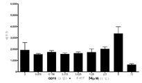
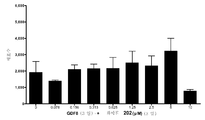
도 2는 인간 배아 줄기 세포주 H1의 세포를 완성 내배엽 계통의 특징적인 마커를 발현하는 세포로 분화시키는데 사용되는 액티빈 A 및 GDF8의 용량 반응 관계를 보여준다. 세포는 검정 제 1 일에 20ng/mL Wnt3a와 함께 제시된 농도의 액티빈 A 또는 GDF8로 총 3일 동안 처리되었다. 분화는 GE 헬스케어 IN 세포 분석기 상에서의 고집적 분석 및 형광 항체 프로브를 사용한 SOX17 강도 측정에 의해 결정되었다.
<도 3>
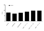
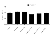
도 3은 실시예 12에 기술된 방법에 따라, 분화 제 1 단계 후의 세포 내 CXCR4의 발현을 보여준다. H1 세포는 제 1 일의 20ng/mL Wnt3a, 또는 모든 3일 동안 2.5μM 화합물 34 또는 2.5μM 화합물 56과 함께, 총 3일 동안 100ng/mL 액티빈 A 또는 200ng/mL GDF-8로 처리되었다. CXCR4 발현은 형광 항체 프로브 및 유세포분석을 사용하여 측정되어, 제시된 양성 세포의 %를 제공하였다.
<도 4>
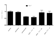
도 4는 실시예 12에 기술된 방법에 따라, 완성 내배엽으로의 3일 간의 분화 후의 SOX17 세포의 발현을 보여준다. H1 세포는 제 1 일의 20ng/mL Wnt3a, 또는 모든 3일 동안 2.5μM 화합물 34 또는 2.5μM 화합물 56과 함께 총 3일 동안 100ng/mL 액티빈 A 또는 200ng/mL GDF-8로 처리되었다. 분화는 GE 헬스케어 IN 세포 분석기 상에서의 고함량 분석 및 형광 항체 프로브를 사용한 SOX17 강도(흑색 막대) 및 결과의 세포수(백색 막대)의 측정에 의해 결정되었다.
<도 5>
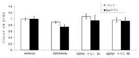
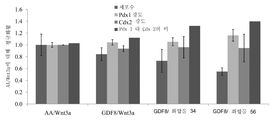
도 5는 실시예 12에 기술된 방법에 따라, 제 3단계의 분화 후 세포 내에서의 PDX1 및 CDX2 단백질의 발현을 보여준다. H1 세포는 제 1 일의 20ng/mL Wnt3a, 또는 모든 3일 동안 2.5μM 화합물 34 또는 2.5μM 화합물 56과 함께 총 3일 동안 100ng/mL 액티빈 A 또는 200ng/mL GDF-8로 처리되었고, 그 후 제 2 및 제 3단계의 분화를 통한 후속한 분화가 이어졌다. 형광 항체 프로브 및 고집적 분석을 사용해 분석된 바와 같이, 단백질 발현 및 세포수는 각각의 처리군에 대해 도시된다. 비교를 위해, 값은 액티빈 A/Wnt3a를 사용한 처리에 대해 정규화된다.
<도 6>
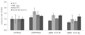
도 6은 실시예 12에 기술된 방법에 따라, 제 4단계의 분화 후 세포 내에서의 PDX1 단백질의 발현(백색 막대) 및 세포수(흑색 막대)를 보여준다. H1 세포는 제 1 일의 20ng/mL Wnt3a, 또는 모든 3일 동안 2.5μM 화합물 34 또는 2.5μM 화합물 56과 함께 총 3일 동안 100ng/mL 액티빈 A 또는 200ng/mL GDF-8로 처리되었고, 그 후 제 2, 제 3 및 제 4단계의 분화를 통한 후속한 분화가 이어졌다. 형광 항체 프로브 및 고집적 분석을 사용해 측정된 바와 같이, 단백질 발현 및 세포수는 각각의 처리군에 대해 도시된다. 비교를 위해, 값은 액티빈 A/Wnt3a를 사용한 처리에 대해 정규화된다.
<도 7>
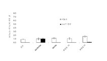
도 7은 실시예 12에 기술된 방법에 따라 분화된 세포 내 세포수, 및 인슐린 및 글루카곤의 단백질 발현을 보여준다. H1 세포는 제 1 일의 20ng/mL Wnt3a, 또는 모든 3일 동안 2.5μM 화합물 34 또는 2.5μM 화합물 56과 함께 총 3일 동안 100ng/mL 액티빈 A 또는 200ng/mL GDF-8로 처리되었고, 그 후 제 2, 제 3, 제 4, 및 제 5단계의 분화를 통한 후속한 분화가 이어졌다. 형광 항체 프로브 및 고집적 분석을 사용해 분석된 바와 같이, 단백질 발현 및 세포수는 각각의 처리군에 대해 도시된다. 비교를 위해, 값은 액티빈 A/Wnt3a를 사용한 처리에 대해 정규화된다.
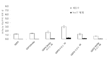
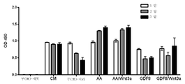
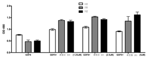
<도 8>
도 8은 실시예 13에 기술된 방법에 따라, 완성 내배엽으로의 분화 후, 인간 배아 줄기 세포 내 SOX17 단백질 발현 및 세포수를 보여준다. H1 세포는 제 1 일의 20ng/mL Wnt3a, 또는 검정의 첫 2일 동안 2.5μM 화합물 34 또는 2.5μM 화합물 56과 함께 총 4일 동안 100ng/mL 액티빈 A 또는 100ng/mL GDF-성장인자로 처리되었다. 형광 항체 프로브 및 고집적 분석을 사용해 분석된 바와 같이, SOX17 단백질 발현(흑색 막대) 및 세포수(백색 막대)는 각각의 처리군에 대해 도시된다. 비교를 위해, 값은 액티빈 A/Wnt3a를 사용한 처리에 대해 정규화된다. 패널 8a는 임의의 성장 인자의 부재(없음), 또는 액티빈 A/Wnt3a 처리(AA/Wnt3a) 또는 개별 시약만으로 처리된 경우의 일련의 분화 조절 조건을 보여준다. 패널 8b는 GDF-3, 단독 또는 Wnt3a, 화합물 34, 또는 화합물 56과의 다중 병용 시의 분화를 보여준다.패널 8c는 GDF-5, 단독 또는 Wnt3a, 화합물 34, 또는 화합물 56과의 다중 병용 시의 분화를 보여준다. 패널 8d는 GDF-8, 단독 또는 Wnt3a, 화합물 34, 또는 화합물 56과의 다중 병용 시의 분화를 보여준다. 패널 8e는 GDF-10, 단독 또는 Wnt3a, 화합물 34, 또는 화합물 56과의 다중 병용 시의 분화를 보여준다. 패널 8f는 GDF-11, 단독 또는 Wnt3a, 화합물 34, 또는 화합물 56과의 다중 병용 시의 분화를 보여준다.패널 8g는 GDF-15, 단독 또는 Wnt3a, 화합물 34, 또는 화합물 56과의 다중 병용 시의 분화를 보여준다.
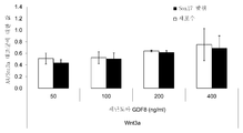
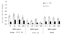
<도 9>
도 9는 실시예 14에 기술된 방법에 따라, 완성 내배엽으로의 분화 후, 인간 배아 줄기 세포 내 SOX17 단백질 발현을 보여준다. H1 세포는 검정 제 1 일 동안 20ng/mL Wnt3a 또는 2.5μM 화합물 34와 병용해서, 100ng/mL의 액티빈 A 또는 제시된 농도의 다양한 성장 인자로 총 3일 동안 처리되었다. 형광 항체 프로브 및 고집적 분석을 사용해 분석된 바와 같이, SOX17 단백질 발현(흑색 막대) 및 세포수(백색 막대)는 각각의 처리군에 대해 도시된다. 비교를 위해, 값은 액티빈 A/Wnt3a를 사용한 처리에 대해 정규화된다. 패널 9a는 Wnt3a 단독, 또는 임의의 성장 인자의 부재(없음) 또는 액티빈 A/Wnt3a 처리와 함께(AA/Wnt3a)인 경우의 일련의 분화 조절 조건을 보여준다. 패널 9b는 20ng/mL Wnt3a와 함께, 제시된 농도의 GDF-8(벤더 퍼프로텍(Vendor PeproTech))을 사용한 분화를 보여준다. 패널 9c는 20ng/mL Wnt3a와 함께, 제시된 농도의 GDF-8(벤더 세넨도아(Vendor Shenendoah))을 사용한 분화를 보여준다. 패널 9d는 Wnt3a 또는 화합물 34와의 다중 병용과 함께, 제시된 농도의 TGFβ1을 사용한 분화를 보여준다. 패널 9e는 Wnt3a 또는 화합물 34와의 다중 병용과 함께, 제시된 농도의 BMP2를 사용한 분화를 보여준다. 패널 9f는 Wnt3a 또는 화합물 34와의 다중 병용과 함께, 제시된 농도의 BMP3을 사용한 분화를 보여준다. 패널 9g는 Wnt3a 또는 화합물 34와의 다중 병용과 함께, 제시된 농도의 BMP4를 사용한 분화를 보여준다.
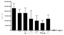
<도 10>
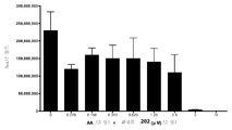
도 10은 실시예 15에 기술된 방법에 따라, 완성 내배엽으로의 분화 후, 인간 배아 줄기 세포에서의 SOX17 단백질 발현을 보여준다. H1 세포는 20ng/mL Wnt3a와 함께 100ng/mL의 액티빈 A 또는 100ng/mL GDF-8을 사용해 다양한 시간대로 노출하면서 총 3일 동안 처리되었다. 형광 항체 프로브 및 고집적 분석으로 측정된 바와 같이, SOX17 단백질 발현은 각각의 처리군에 대한 총 강도값으로 보여지며, 성장 인자 무첨가(무(無)처리), Wnt3a 단독, 액티빈 A 또는 GDF-8 단독, 또는 액티빈 A/Wnt3a 처리 또는 GDF-8/Wnt3a 처리의 분화 조절 조건을 시험하며, 여기서, Wnt3a는 제시된 바와 같이 검정 제 1일 동안만 또는 모든 3일 동안 첨가되었다.
<도 11>
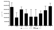
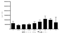
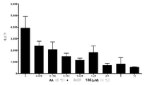
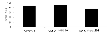
도 11은 실시예 15에 기술된 방법에 따라, 완성 내배엽으로의 분화 후, 인간 배아 줄기 세포에서의 SOX17 단백질 발현을 보여준다. H1 세포를 제시된 농도의 시험 화합물 (화합물 181 (패널 a), 화합물 180 (패널 b), 화합물 19(패널 c), 화합물 202 (패널 d), 화합물 40 (패널 e), 화합물 34 (패널 f), 또는 GSK3 억제제 BIO (패널 g))와 함께 100ng/mL의 액티빈 A로 다양한 시간대로 노출하면서 총 3일 동안 처리하였고, 여기서, 시험 화합물은 검정 제 1 일에만 첨가하였다. 형광 항체 프로브 및 고집적 분석으로 측정된 바와 같이, SOX17에 대한 단백질 발현은 총 강도값에 의해 도시된다.
<도 12>
도 12는 실시예 15에 기술된 방법에 따라, 완성 내배엽으로의 분화 후, 인간 배아 줄기 세포에서의 SOX17 단백질 발현을 보여준다. H1 세포를 제시된 농도의 시험 화합물 (화합물 181 (패널 a), 화합물 180 (패널 b), 화합물 19(패널 c), 화합물 202 (패널 d), 화합물 40 (패널 e), 화합물 34 (패널 f), 또는 GSK3 억제제 BIO (패널 g))와 함께 100ng/mL의 액티빈 A로 다양한 시간대로 노출하면서 총 3일 동안 처리하였고, 여기서, 시험 화합물은 검정 총 3일 동안 첨가하였다. 형광 항체 프로브 및 고집적 분석으로 측정된 바와 같이, SOX17에 대한 단백질 발현은 총 강도값에 의해 도시된다.
<도 13>
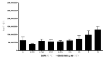
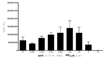
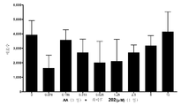
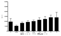
도 13은 실시예 15에 기술된 방법에 따라, 완성 내배엽으로의 분화 후, 인간 배아 줄기 세포에서의 SOX17 단백질 발현을 보여준다. H1 세포를 제시된 농도의 시험 화합물 (화합물 181 (패널 a), 화합물 180 (패널 b), 화합물 19(패널 c), 화합물 202 (패널 d), 화합물 40 (패널 e), 화합물 34 (패널 f), 또는 GSK3 억제제 BIO (패널 g))와 함께 100ng/mL의 GDF-8로 다양한 시간대로 노출하면서 총 3일 동안 처리하였고, 여기서, 시험 화합물은 검정 제 1 일에만 첨가하였다. 형광 항체 프로브 및 고집적 분석으로 측정된 바와 같이, SOX17에 대한 단백질 발현은 총 강도값에 의해 도시된다.
<도 14>
도 14는 실시예 15에 기술된 방법에 따라, 완성 내배엽으로의 분화 후, 인간 배아 줄기 세포에서의 SOX17 단백질 발현을 보여준다. H1 세포를 제시된 농도의 시험 화합물 (화합물 181 (패널 a), 화합물 180 (패널 b), 화합물 19(패널 c), 화합물 202 (패널 d), 화합물 40 (패널 e), 화합물 34 (패널 f), 또는 GSK3 억제제 BIO (패널 g))와 함께 100ng/mL의 GDF-8로 다양한 시간대로 노출하면서 총 3일 동안 처리하였고, 여기서, 시험 화합물은 검정 총 3일 동안 첨가하였다. 형광 항체 프로브 및 고집적 분석으로 측정된 바와 같이, SOX17에 대한 단백질 발현은 총 강도값에 의해 도시된다.
<도 15>
도 15는 실시예 15에 기술된 방법에 따라, 인간 배아 줄기 세포를 완성 내배엽으로 분화시킨 후, 세포수 수율을 보여준다. H1 세포는 20ng/mL Wnt3a와 함께 100ng/mL 액티빈 A 또는 100ng/mL GDF-8로 다양한 시간대로 노출하면서 총 3일 동안 처리하였다. 형광 핵 프로브 및 고집적 분석으로 측정된 바와 같이, 세포수는 각각의 처리군에 대해 보여지고, 성장 인자 무첨가(무처리), Wnt3a 단독, 액티빈 A 또는 GDF-8 단독, 또는 액티빈 A/Wnt3a 처리 또는 GDF-8/Wnt3a 처리가 있는 분화 조절 조건을 시험하며, 여기서, Wnt3a는 보여진 바와 같이 단지 검정 제 1 동안 또는 총 3일 동안 첨가되었다.
<도 16>
도 16은 실시예 15에 기술된 방법에 따라, 인간 배아 줄기 세포를 완성 내배엽으로 분화시킨 후, 세포수 수율을 보여준다. H1 세포를 제시된 농도의 시험 화합물 (화합물 181 (패널 a), 화합물 180 (패널 b), 화합물 19(패널 c), 화합물 202 (패널 d), 화합물 40 (패널 e), 화합물 34 (패널 f), 또는 GSK3 억제제 BIO (패널 g))와 함께 100ng/mL의 액티빈 A로 다양한 시간대로 노출하면서 총 3일 동안 처리하였고, 여기서, 시험 화합물은 검정 제 1 일에만 첨가하였다. 형광 핵 프로브 및 고집적 분석으로 측정된 바와 같이 세포수 수율을 나타낸다.
<도 17>
도 17은 실시예 15에 기술된 방법에 따라, 인간 배아 줄기 세포를 완성 내배엽으로 분화시킨 후, 세포수 수율을 보여준다. H1 세포를 제시된 농도의 시험 화합물 (화합물 181 (패널 a), 화합물 180 (패널 b), 화합물 19(패널 c), 화합물 202 (패널 d), 화합물 40 (패널 e), 화합물 34 (패널 f), 또는 GSK3 억제제 BIO (패널 g))와 함께 100ng/mL의 액티빈 A로 다양한 시간대로 노출하면서 총 3일 동안 처리하였고, 여기서, 시험 화합물은 검정 총 3일 동안 첨가하였다. 형광 핵 프로브 및 고집적 분석으로 측정된 바와 같이 세포수 수율을 나타낸다.
<도 18>
도 18은 실시예 15에 기술된 방법에 따라, 인간 배아 줄기 세포를 완성 내배엽으로 분화시킨 후, 세포수 수율을 보여준다. H1 세포를 제시된 농도의 시험 화합물 (화합물 181 (패널 a), 화합물 180 (패널 b), 화합물 19(패널 c), 화합물 202 (패널 d), 화합물 40 (패널 e), 화합물 34 (패널 f), 또는 GSK3 억제제 BIO (패널 g))와 함께 100ng/mL의 GDF-8로 다양한 시간대로 노출하면서 총 3일 동안 처리하였고, 여기서, 시험 화합물은 검정 제 1 일에만 첨가하였다. 형광 핵 프로브 및 고집적 분석으로 측정된 바와 같이 세포수 수율을 나타낸다.
<도 19>
도 19는 실시예 15에 기술된 방법에 따라, 인간 배아 줄기 세포를 완성 내배엽으로 분화시킨 후, 세포수 수율을 보여준다. H1 세포를 제시된 농도의 시험 화합물 (화합물 181 (패널 a), 화합물 180 (패널 b), 화합물 19(패널 c), 화합물 202 (패널 d), 화합물 40 (패널 e), 화합물 34 (패널 f), 또는 GSK3 억제제 BIO (패널 g))와 함께 100ng/mL의 GDF-8로 다양한 시간대로 노출하면서 총 3일 동안 처리하였고, 여기서, 시험 화합물은 검정 총 3일 동안 첨가하였다. 형광 핵 프로브 및 고집적 분석으로 측정된 바와 같이 세포수 수율을 나타낸다.
<도 20>
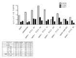
도 20은 실시예 16에서 기술된 방법에 따라 다중 분화 단계 전체에 걸쳐 세포 내 다양한 단백질 마커의 발현을 보여준다. H1 세포를 제 1 일 동안 20ng/mL Wnt3a 또는 제 1 일에만 첨가된 2.5μM 다양한 화합물(화합물 19, 화합물 202, 화합물 40, 또는 GSK3 억제제 BIO)과 함께 총 3일 동안 100ng/mL 액티빈 A 또는 100ng/mL GDF-8로 처리하였다. 도 20, 패널 a는 분화 제 1 단계 후 세포 내에서 완성 내배엽 마커, CXCR4에 대한 FACS 분석을 보여준다. CXCR4 발현은 형광 항체 프로브 및 유세포분석을 이용해 측정하여, 나타낸 바와 같이 양성 세포의 %를 수득하였다. 도 20, 패널 b는 분화 제 1 단계로 인한 정규화된 SOX17 단백질 발현 (흑색 막대) 및 회수된 세포수 (백색 막대)에 대한 고집적 이미지 분석을 보여주며, 제시된 상응하는 처리를 시험한다. 도 20, 패널 c는 분화 단계 5를 통해 처리된 배양물로부터 회수된 상대 세포수에 대한 고집적 이미지 분석을 보여준다. 도 20, 패널 d는 분화 단계 5를 통해 처리된 배양물로부터 글루카곤 단백질 발현에 대한 고집적 이미지 분석을 보여준다. 도 20, 패널 e는 분화 단계 5를 통해 처리된 배양물로부터 인슐린 단백질 발현에 대한 고집적 이미지 분석을 보여준다. 도 20, 패널 f는 분화 단계 5를 통해 처리된 배양물로부터 글루카곤 대 인슐린 발현의 비를 보여준다. 비교를 위해, 패널 b, c, d, e, 및 f에서의 발현값은 단계 1 동안에 액티빈 A 및 Wnt3a를 사용한 대조군 처리에 대해 정규화된다.
<도 21>
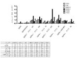
도 21은 실시예 17에서 기술된 방법에 따라 다중 분화 단계 전체를 통해 세포 내에서의 다양한 단백질 및 RT-PCR 마커의 발현을 보여준다. H1 세포는 제 1 일 동안 20ng/mL Wnt3a 또는 제 1 일에만 첨가된 하기 농도의 다양한 화합물(화합물 181, 화합물 180, 화합물 19, 화합물 202, 화합물 40, 화합물 56, 또는 GSK3 억제제 BIO)과 함께, 총 3일 동안 100ng/mL 액티빈 A 또는 100ng/mL GDF-8로 처리하였다. 분화 제 1 단계 후 세포 내에서 완성 내배엽 마커, CXCR4에 대한 FACS 분석을 보여주며, 여기서 처리는 액티빈 A (패널 a) 또는 GDF-8 (패널 b)과 함께 Wnt3a 또는 다양한 화합물을 조합하였다. CXCR4 발현은 형광 항체 프로브 및 유세포분석을 사용하여 측정되어, 제시된 양성 세포의 %를 제공하였다. 도 21의 후속한 패널에서, 다양한 분화 마커에 대한 정규화된 RT-PCR 값은 하기와 같이 분화의 제 1 단계 동안에 액티빈 A 또는 GDF-8을 사용한 각각의 처리와 함께 보여진다: 액티빈 A (패널 c) 또는 GDF-8 (패널 d)을 조합한 처리에 대한 분화의 한 단계의 종료 시의 마커; 액티빈 A (패널 e) 또는 GDF-8 (패널 f)을 조합한 처리에 대한 분화의 3단계의 종료 시의 마커; 액티빈 A (패널 g) 또는 GDF-8 (패널 h)을 조합한 처리에 대한 분화의 4단계의 종료 시의 마커; 액티빈 A (패널 i) 또는 GDF-8 (패널 j)을 조합한 처리에 대한 분화의 5단계의 종료 시의 마커. 5단계의 분화의 종료 시, 고집적 분석을 수행하여, 액티빈 A (패널 k) 또는 GDF-8 (패널 m)을 사용한 분화의 제 1 단계 동안 상응하는 처리에 대한 회수된 세포수를 측정하였다. 고집적 분석을 또한 사용하여, 5단계의 분화의 종료 시에 회수된 세포군 내 글루카곤 및 인슐린 강도를 측정하며, 이는 분화 제 1 단계 동안 액티빈 A (패널 l) 또는 GDF-8 (패널 n)을 사용한 처리에 상응하였다.
<도 22>
도 22는 실시예 18에서 기술된 방법에 따라 처리된 세포 내에서 다양한 단백질 및 RT-PCR 마커의 발현을 보여준다. H1 세포는 제 1 일의 20ng/mL Wnt3a, 또는 제 1 일 동안에만 2.5μM 화합물 40 또는 2.5μM 화합물 202와 함께 총 3일 동안 100ng/mL 액티빈 A 또는 100ng/mL GDF-8로 처리하였다. 도 22, 패널 a는 분화 제 1 단계 후 세포 내에서 완성 내배엽 마커, CXCR4에 대한 FACS 분석을 보여준다. CXCR4 발현은 형광 항체 프로브 및 유세포분석을 사용하여 측정되어, 제시된 양성 세포의 %를 제공하였다. 도 22, 패널 b에서, 제 4 의 분화 단계 후 회수된 세포 내에서 다양한 분화 마커에 대한 정규화된 RT-PCR 값은 분화 제 1 단계 동안 액티빈 A/Wnt3a 또는 GDF-8/화합물 40 또는 GDF-8/화합물 202를 사용한 각각의 처리에 상응하는 것으로 보여진다.
<도 23>
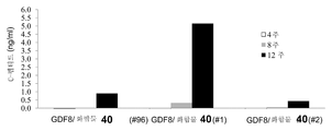
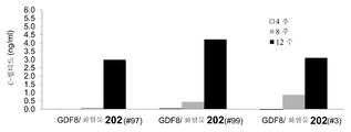
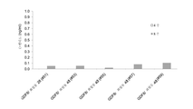
도 23은 실시예 18에서 기술된 바와 같은 분화 프로토콜의 4단계의 종료 시 세포를 수여받은 스키드(SCID)-베이지 마우스에서 검출된 C-펩티드의 수준을 보여준다.
<도 24>
도 24 패널 a는 실시예 19에서 기술된 바와 같은 분화 프로토콜의 1단계의 종료 시 FACS에 의해 측정된 바와 같이, CXCR4의 발현을 보여준다. 패널 b는 실시예 19에서 기술된 바와 같은 분화 프로토콜의 4단계의 종료 시 세포에서의 RT-PCR에 의해 측정된 바와 같이, 다양한 유전자의 발현을 보여준다. 2개의 상이한 실험 복제물이 보여지며(복제물-1 및 복제물-2), 각각은 동일한 처리 프로토콜을 받았다. 패널 c는 시험관내 분화의 제 1단계 동안 GDF-8 및 Wnt3a로 처리된 바와 같은 분화 프로토콜의 4단계의 종료 시 세포를 받은 스키드-베이지 마우스에서 검출된 C-펩티드의 수준을 보여준다. 패널 d는 시험관내 분화 제 1단계 동안 GDF-8 및 화합물 28로 처리된 바와 같은 분화 프로토콜의 4단계의 종료 시 세포를 받은 스키드-베이지 마우스에서 검출된 C-펩티드의 수준을 보여준다.
<도 25>
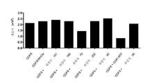
도 25는 실시예 22에서 기술된 바와 같이 본 발명의 방법에 따라 처리된, 마이크로캐리어 비드 상에서 키운 세포로부터의 세포수 (패널 a) 및 CXCR4의 발현 (패널 b)을 보여준다. 세포는 무처리(미분화) 또는 100ng/mL 액티빈 A를 20ng/mL Wnt3a와 함께 조합한 처리(AA/Wnt3a) 또는 보여진 바와 같이 GDF-8을 조합한 다양한 처리를 이용해 사이토덱스3 비드 상에서 키웠다: 50ng/mL GDF-8과 함께 2.5μM 화합물 34 (Cmp 34+8); 또는 50ng/mL GDF-8과 함께 2.5μM 화합물 34 및 50ng/mL PDGF (Cmp 34+8+D); 또는 50ng/mL GDF-8과 함께 2.5μM 화합물 34 및 50ng/mL PDGF 및 50ng/mL VEGF (Cmp 34+8+D+V); 또는 50ng/mL GDF-8과 함께 2.5μM 화합물 34 및 50ng/mL PDGF 및 50ng/mL VEGF 및 20ng/mL 무스치몰 (Cmp 34+8+D+V+M).
<도 26>
도 26은 실시예 23에서 기술된 바와 같이 본 발명의 화합물의 처리 후의 세포의 증식을 보여준다. I를 통한 패널 b는 GDF-8과 조합된 화합물을 사용하고 분화 검정을 시작한 후 제 1 일, 제 2 일, 및 제 3 일째에 MTS OD 판독을 측정하는 처리에 대한 검정 결과를 보여준다.
<도 27>
도 27은 본 발명의 방법에 따라 처리된 마이크로캐리어 비드 상에서 키운 세포로부터의 다양한 단백질 및 유전자의 발현을 보여준다. 패널 a는 실시예 24에서 기술된 분화 프로토콜의 1 단계의 종료 시, 세포에서 FACS에 의해 측정된 바와 같이 CXCR4, CD99, 및 CD9의 양성 발현%를 보여준다. 패널 B는 분화 프로토콜의 3단계를 통해 분화된 제시된 바와 같은 처리로부터 회수된 세포를 보여준다. 패널 C는 단계에서 제시된 바와 같이 처리되고 프로토콜의 3단계를 통해 분화된 세포에서 발현된 다양한 유전자 마커에 대한 ddCT 값을 보여준다.<Figure 1>
1 shows the differentiation of H1 human embryonic stem cells into cells expressing markers characteristic of the definitive endoderm lineage. Differentiation was determined by measuring cell number (panel A) and SOX17 intensity (panel B) using IN Cell Analyzer 1000 (GE Healthcare). Human embryonic stem cells were treated with medium containing 20 ng / mL Wnt3a + indicated concentrations of activin A (black bars) or medium containing no indicated Wnt3a but activin A at the indicated concentrations (white bars) for a total of 4 days It became.
<FIG. 2>
Figure 2 shows the dose response relationship of activin A and GDF8 used to differentiate the cells of human embryonic stem cell line H1 into cells expressing markers characteristic of the definitive endoderm lineage. Cells were treated for 3 days in total with activin A or GDF8 at the concentrations indicated with 20 ng / mL Wnt3a on
3,
3 shows expression of CXCR4 in cells after the first step of differentiation, according to the method described in Example 12. FIG. H1 cells were treated with 100ng / mL Activin A or 200ng / mL GDF-8 for a total of 3 days, with 20ng / mL Wnt3a on
<Figure 4>
4 shows expression of SOX17 cells after 3 days of differentiation into definitive endoderm, according to the method described in Example 12. FIG. H1 cells were treated with 100 ng / mL Activin A or 200 ng / mL GDF-8 for 3 days in total with 20 ng / mL Wnt3a on
<Figure 5>
5 shows expression of PDX1 and CDX2 proteins in cells after the third stage of differentiation, according to the method described in Example 12. FIG. H1 cells were treated with 100ng / mL Activin A or 200ng / mL GDF-8 for 3 days in total with 20ng / mL Wnt3a on
6,
6 shows the expression of PDX1 protein (white bar) and cell number (black bar) in cells after the fourth stage of differentiation, according to the method described in Example 12. H1 cells were treated with 100ng / mL Activin A or 200ng / mL GDF-8 for 3 days in total with 20ng / mL Wnt3a on
<Figure 7>
7 shows the cell number differentiated and protein expression of insulin and glucagon according to the method described in Example 12. H1 cells were treated with 100ng / mL Activin A or 200ng / mL GDF-8 for 3 days in total with 20ng / mL Wnt3a on
<Figure 8>
8 shows SOX17 protein expression and cell number in human embryonic stem cells following differentiation into definitive endoderm, according to the method described in Example 13. FIG. H1 cells were treated with 100ng / mL Activin A or 100ng / mL GDF-growth factor for a total of 4 days with 20ng / mL Wnt3a on
<Figure 9>
9 shows SOX17 protein expression in human embryonic stem cells following differentiation into definitive endoderm, according to the method described in Example 14. FIG. H1 cells were treated with 100 ng / mL activin A or various growth factors at the indicated concentrations for a total of 3 days, in combination with 20 ng / mL Wnt3a or 2.5
<Figure 10>
10 shows SOX17 protein expression in human embryonic stem cells following differentiation into definitive endoderm, according to the method described in Example 15. FIG. H1 cells were treated for a total of 3 days with 20 ng / mL Wnt3a with 100 ng / mL activin A or 100 ng / mL GDF-8 at various time points. As measured by fluorescent antibody probes and high integration assays, SOX17 protein expression is shown as the total intensity value for each treatment group, with no growth factor added (no treatment), Wnt3a alone, activin A or GDF-8 Differentiation control conditions of either alone or activin A / Wnt3a treatment or GDF-8 / Wnt3a treatment were tested, where Wnt3a was added only during the first day of the assay or for all three days as shown.
<Figure 11>
11 shows SOX17 protein expression in human embryonic stem cells following differentiation into definitive endoderm, according to the method described in Example 15. FIG. H1 cells were tested at the indicated concentrations of compound (Compound 181 (Panel a), Compound 180 (Panel b), Compound 19 (Panel c), Compound 202 (Panel d), Compound 40 (Panel e), Compound 34 (Panel f)). , Or GSK3 inhibitor BIO (panel g)) was treated with 100 ng / mL activin A for various time periods for a total of 3 days, where test compounds were added only on
<Figure 12>
12 shows SOX17 protein expression in human embryonic stem cells following differentiation into definitive endoderm, according to the method described in Example 15. FIG. H1 cells were tested at the indicated concentrations of compound (Compound 181 (Panel a), Compound 180 (Panel b), Compound 19 (Panel c), Compound 202 (Panel d), Compound 40 (Panel e), Compound 34 (Panel f)). , Or GSK3 inhibitor BIO (panel g)) with 100 ng / mL activin A for various days of exposure at various times, where test compounds were added for a total of 3 days of assay. As measured by fluorescent antibody probes and high integration assays, protein expression for SOX17 is shown by total intensity values.
Figure 13
FIG. 13 shows SOX17 protein expression in human embryonic stem cells following differentiation into definitive endoderm, according to the method described in Example 15. FIG. H1 cells were tested at the indicated concentrations of compound (Compound 181 (Panel a), Compound 180 (Panel b), Compound 19 (Panel c), Compound 202 (Panel d), Compound 40 (Panel e), Compound 34 (Panel f)). , Or GSK3 inhibitor BIO (panel g)) was treated with 100 ng / mL of GDF-8 for 3 days with varying time zones, where test compounds were added only on
<Figure 14>
14 shows SOX17 protein expression in human embryonic stem cells following differentiation into definitive endoderm, according to the method described in Example 15. FIG. H1 cells were tested at the indicated concentrations of compound (Compound 181 (Panel a), Compound 180 (Panel b), Compound 19 (Panel c), Compound 202 (Panel d), Compound 40 (Panel e), Compound 34 (Panel f)). , Or GSK3 inhibitor BIO (panel g)) was treated with 100 ng / mL of GDF-8 for 3 days with varying time zones, where test compounds were added for a total of 3 days of assay. As measured by fluorescent antibody probes and high integration assays, protein expression for SOX17 is shown by total intensity values.
Figure 15
FIG. 15 shows cell yield after differentiation of human embryonic stem cells into definitive endoderm, according to the method described in Example 15. H1 cells were treated with 20 ng / mL Wnt3a with 100 ng / mL Activin A or 100 ng / mL GDF-8 for various time periods for a total of 3 days. As measured by fluorescence nuclear probe and high density analysis, cell numbers were shown for each treatment group, growth factor free (untreated), Wnt3a alone, activin A or GDF-8 alone, or activin A / Wnt3a Differentiation control conditions with treatment or GDF-8 / Wnt3a treatment were tested, where Wnt3a was added only during assay first or for a total of three days as shown.
<Figure 16>
FIG. 16 shows cell number yield after differentiation of human embryonic stem cells into definitive endoderm, according to the method described in Example 15. H1 cells were tested at the indicated concentrations of compound (Compound 181 (Panel a), Compound 180 (Panel b), Compound 19 (Panel c), Compound 202 (Panel d), Compound 40 (Panel e), Compound 34 (Panel f)). , Or GSK3 inhibitor BIO (panel g)) was treated with 100 ng / mL activin A for various time periods for a total of 3 days, where test compounds were added only on
<Figure 17>
17 shows cell yield after differentiation of human embryonic stem cells into definitive endoderm, according to the method described in Example 15. H1 cells were tested at the indicated concentrations of compound (Compound 181 (Panel a), Compound 180 (Panel b), Compound 19 (Panel c), Compound 202 (Panel d), Compound 40 (Panel e), Compound 34 (Panel f)). , Or GSK3 inhibitor BIO (panel g)) with 100 ng / mL activin A for various days of exposure at various times, where test compounds were added for a total of 3 days of assay. Cell number yields are measured as measured by fluorescent nuclear probes and high integration assays.
<Figure 18>
18 shows cell number yield after differentiation of human embryonic stem cells into definitive endoderm, according to the method described in Example 15. FIG. H1 cells were tested at the indicated concentrations of compound (Compound 181 (Panel a), Compound 180 (Panel b), Compound 19 (Panel c), Compound 202 (Panel d), Compound 40 (Panel e), Compound 34 (Panel f)). , Or GSK3 inhibitor BIO (panel g)) was treated with 100 ng / mL of GDF-8 for 3 days with varying time zones, where test compounds were added only on
<Figure 19>
19 shows cell number yield after differentiation of human embryonic stem cells into definitive endoderm, according to the method described in Example 15. FIG. H1 cells were tested at the indicated concentrations of compound (Compound 181 (Panel a), Compound 180 (Panel b), Compound 19 (Panel c), Compound 202 (Panel d), Compound 40 (Panel e), Compound 34 (Panel f)). , Or GSK3 inhibitor BIO (panel g)) was treated with 100 ng / mL of GDF-8 for 3 days with varying time zones, where test compounds were added for a total of 3 days of assay. Cell number yields are measured as measured by fluorescent nuclear probes and high integration assays.
<Figure 20>
20 shows expression of various protein markers in cells throughout multiple differentiation steps according to the method described in Example 16. FIG. H1 cells were treated with 100 ng / mL Activin A for 3 days in total with 20 ng / mL Wnt3a for
Figure 21
21 shows expression of various proteins and RT-PCR markers in cells throughout multiple differentiation steps according to the method described in Example 17. FIG. H1 cells are combined with various compounds (
<Figure 22>
22 shows expression of various proteins and RT-PCR markers in cells treated according to the method described in Example 18. FIG. H1 cells were treated with 100 ng / mL Activin A or 100 ng / mL GDF-8 for a total of 3 days with 20 ng / mL Wnt3a on
Figure 23
FIG. 23 shows the level of C-peptide detected in skid (SCID) -beige mice awarded cells at the end of
<Figure 24>
24 panel a shows expression of CXCR4 as measured by FACS at the end of
<FIG. 25>
25 shows cell number (panel a) and expression of CXCR4 (panel b) from cells grown on microcarrier beads treated according to the method of the present invention as described in Example 22. Cells were grown on cytodex3 beads using either untreated (undifferentiated) or 100 ng / mL Activin A combined with 20 ng / mL Wnt3a (AA / Wnt3a) or various treatments with GDF-8 as shown: 2.5 μM Compound 34 (
<Figure 26>
Figure 26 shows proliferation of cells after treatment with compounds of the present invention as described in Example 23. Panel b through I shows the results of the assay for the treatment using the compound in combination with GDF-8 and measuring the MTS OD reading on
<Figure 27>
27 shows expression of various proteins and genes from cells grown on microcarrier beads treated according to the methods of the present invention. Panel a shows percent positive expression of CXCR4, CD99, and CD9 as measured by FACS in cells at the end of
개시 내용의 명확함을 위하여, 그리고 제한하지 않고서, 발명을 실시하기 위한 구체적인 내용은 본 발명의 특정한 특성, 실시 형태, 또는 응용을 설명하거나 예시하는 하기 세부 항목으로 나뉘어진다.For clarity of disclosure and without limitation, the specific details for carrying out the invention are divided into the following subsections that describe or illustrate particular features, embodiments, or applications of the invention.
정의Justice
줄기 세포는 단일 세포 레벨에서 자가 재생하고 분화하여 자가 재생 전구 세포, 비재생 전구 세포, 및 최종 분화 세포를 비롯한 자손 세포 (progeny cell)를 생성하는 그의 능력에 의해 규정되는 미분화 세포이다. 줄기 세포는 또한 다수의 배엽층(내배엽, 중배엽 및 외배엽)으로부터 다양한 세포 계통의 기능성 세포로 시험관 내에서 분화하는 그의 능력, 및 이식 후 다수의 배엽층의 조직이 생기게 하며 배반포 내로의 주입 후, 전부는 아니라 하더라도 대부분의 조직에 실질적으로 기여하는 그의 능력을 특징으로 한다.Stem cells are undifferentiated cells defined by their ability to self-renewal and differentiate at the single cell level to produce progeny cells, including self-renewing progenitor cells, non-renewing progenitor cells, and final differentiated cells. Stem cells also give rise to the ability to differentiate in vitro from multiple germ layers (endoderm, mesoderm and ectoderm) into functional cells of various cell lines, and give rise to tissues of multiple germ layers after transplantation and all after injection into blastocysts. If not, he is characterized by his ability to make a substantial contribution to most organizations.
줄기 세포는 그들의 발생 잠재력에 의해 분류된다: (1) 모든 배아 및 배자 외 세포 유형이 생기게 할 수 있음을 의미하는 전능성; (2) 모든 배아 세포 유형이 생기게 할 수 있음을 의미하는 만능성; (3) 세포 계통의 하위세트이지만 모두 특정 조직, 기관 또는 생리학적 시스템 내에 있는 하위세트가 생기게 할 수 있음을 의미하는 만능성(예를 들어, 조혈 줄기 세포(hematopoietic stem cell, HSC)는 HSC(자가-재생), 혈구 세포 제한된 소기능성(oligopotent) 조상세포 및 혈액의 정상 성분인 모든 세포 유형 및 요소(예를 들어, 혈소판)를 포함하는 자손을 생성할 수 있음); (4) 만능성 줄기 세포보다 더 제한된 하위세트의 세포 계통이 될 수 있음을 의미하는 소기능성; 및 (5) 단일 세포 계통(예를 들어, 정자발생 줄기 세포)이 생기게 할 수 있음을 의미하는 단일기능성.Stem cells are classified by their developmental potential: (1) omnipotent, meaning that they can give rise to all embryonic and extraembryonic cell types; (2) pluripotency, meaning that all embryonic cell types can give rise; (3) Pluripotency (eg, hematopoietic stem cells (HSCs), which is a subset of the cell line, meaning that they can all produce subsets within a particular tissue, organ, or physiological system). Self-renewal), hematopoietic cell limited oligopotent progenitor cells and progeny including all cell types and elements (eg, platelets) that are normal components of blood; (4) minority, meaning that it can be a more limited subset of cell lineages than pluripotent stem cells; And (5) monofunctional, meaning that a single cell lineage (eg, spermatogenic stem cells) can be produced.
분화는 특화되지 않은 ("미결정된 (uncommitted)") 또는 덜 특화된 세포가 예를 들어, 신경 세포 또는 근육 세포와 같은 특화된 세포의 특징을 획득하는 과정이다. 분화된 또는 분화 유도된 세포는 세포 계통 내에서 보다 특화된 ("결정된 (committed)") 위치를 차지한 것이다. 분화 과정에 적용될 때, 용어 "결정된"은 분화 경로에서, 통상적인 환경하에서 특정 세포형 또는 세포형의 서브세트로 계속 분화할 것이며, 통상적인 환경하에서 다른 세포형으로 분화할 수 없거나 덜 분화된 세포형으로 돌아갈 수 없는 시점까지 진행한 세포를 말한다. 탈분화는 세포가 세포 계통 내의 덜 특화된 (또는 결정된) 위치로 되돌아가는 과정을 말한다. 본 명세서에 사용되는 바와 같이, 세포 계통은 세포의 유전, 즉, 어느 세포로부터 왔는지 그리고 어떤 세포를 발생시킬 수 있는지를 규정한다. 세포 계통은 세포를 발생과 분화의 유전적 체계 내에 둔다. 계통 특이적 마커는 대상 계통의 세포 표현형과 특이적으로 관련되며, 미결정된 세포가 대상 계통으로 분화하는지를 평가하기 위해 사용될 수 있는 특징을 말한다.Differentiation is the process by which unspecialized ("uncommitted") or less specialized cells acquire the characteristics of specialized cells, such as, for example, neurons or muscle cells. Differentiated or differentiation-induced cells occupy more specialized ("committed") positions within the cell lineage. When applied to the differentiation process, the term "determined" will continue to differentiate into a specific cell type or subset of cell types in the normal environment in the differentiation pathway, and cells that cannot or will not differentiate into other cell types under normal circumstances. It is a cell that has advanced to the point where it cannot return to the type. Dedifferentiation refers to the process by which cells return to less specialized (or determined) positions in the cell lineage. As used herein, a cell line defines the inheritance of a cell, ie which cell it is from and which cells can be generated. The cell lineage puts the cells in the genetic system of development and differentiation. Lineage specific markers are specifically related to the cell phenotype of the lineage of interest and refer to features that can be used to assess whether unidentified cells differentiate into the lineage of interest.
"β-세포 계통"은 전사 인자 PDX-1, 및 하기 전사 인자 중 적어도 하나에 대한 양성 유전자 발현을 갖는 세포를 말한다: NGN3, NKX2.2, NKX6.1, NEUROD, ISL1, HNF-3 베타, MAFA, PAX4, 또는 PAX6. β-세포 계통의 특징적인 마커를 발현하는 세포는 β 세포를 포함한다."β-cell lineage" refers to a cell having positive gene expression for the transcription factor PDX-1 and at least one of the following transcription factors: NGN3, NKX2.2, NKX6.1, NEUROD, ISL1, HNF-3 beta, MAFA, PAX4, or PAX6. Cells expressing markers characteristic of the β-cell lineage include β cells.
본원에서 사용된 바와 같이, "완성 내배엽 계통의 특징적인 마커를 발현하는 세포", 또는 "단계 1 세포", 또는 "단계 1"은 하기 마커 중 적어도 하나를 발현하는 세포를 말한다: SOX17, GATA4, HNF-3 베타, GSC, CER1, 노달, FGF8, 브라키어리, 혼합형 호메오박스 단백질, FGF4 CD48, 에오메소더민(eomesodermin)(EOMES), DKK4, FGF17, GATA6, CXCR4, C-Kit, CD99, 또는 OTX2. 완성 내배엽 계통의 특징적인 마커를 발현하는 세포는 원시선 전구 세포, 원시선 세포, 중내배엽 세포 및 완성 내배엽 세포를 포함한다.As used herein, "cells expressing markers characteristic of a definitive endoderm lineage", or "
본원에서 사용된 바와 같이, "췌장 내배엽 계통의 특징적인 마커를 발현하는 세포"는 하기 마커 중 적어도 하나를 발현하는 세포를 말한다: PDX1, HNF-1 베타, PTF1 알파, HNF6, 또는 HB9. 췌장 내배엽 계통의 특징적인 마커를 발현하는 세포는 췌장 내배엽 세포, 원시 장관(원시 창자관) 세포, 및 후방 전장 세포를 포함한다.As used herein, “cells expressing markers characteristic of the pancreatic endoderm lineage” refers to cells expressing at least one of the following markers: PDX1, HNF-1 beta, PTF1 alpha, HNF6, or HB9. Cells expressing markers characteristic of the pancreatic endoderm lineage include pancreatic endoderm cells, primitive intestinal (raw gut tube) cells, and posterior full length cells.
본원에서 사용된 바와 같이, "췌장 내분비 계통의 특징적인 마커를 발현하는 세포", 또는 "단계 5 세포", 또는 "단계 5"는 하기 마커 중 적어도 하나를 발현하는 세포를 말한다: NGN3, NEUROD, ISL1, PDX1, NKX6.1, PAX4, 또는 PTF-1 알파. 췌장 내분비 계통의 특징을 나타내는 마커를 발현하는 세포는 췌장 내분비 세포, 췌장 호르몬 발현 세포, 및 췌장 호르몬 분비 세포, 및 β-세포 계통의 세포를 포함한다.As used herein, “cells expressing markers characteristic of the pancreatic endocrine lineage”, or “
본원에서 사용된 바와 같이, "완성 내배엽"은 낭배의 외피(epiblast)로 인한 세포의 특징을 갖고 위장관로 및 그의 유도체를 형성하는 세포를 말한다. 완성 내배엽 세포는 하기 마커를 발현한다: HNF-3 베타, GATA4, SOX-17, 세베루스(Cerberus), OTX2, 구세코이드(goosecoid), C-Kit, CD99, 또는 MIXL1.As used herein, "finished endoderm" refers to a cell that has the characteristics of the cells due to the epiblast of the cyst and forms into the gastrointestinal tract and its derivatives. Definitive endoderm cells express the following markers: HNF-3 beta, GATA4, SOX-17, Cerberus, OTX2, goosecoid, C-Kit, CD99, or MIXL1.
본원에서 사용된 바와 같이, "배자외 내배엽"은 하기 마커 중 적어도 하나를 발현하는 세포군을 말한다: SOX7, AFP, 또는 SPARC.As used herein, “ectodermal endoderm” refers to a cell population that expresses at least one of the following markers: SOX7, AFP, or SPARC.
본 명세서에 사용되는 바와 같이, "마커"는 관심있는 세포에서 차등적으로 발현되는 핵산 또는 폴리펩티드 분자이다. 이와 관련하여, 차등 발현은 양성 마커의 레벨 증가 및 음성 마커의 레벨 감소를 의미한다. 마커 핵산 또는 폴리펩티드의 검출가능한 레벨은 다른 세포에 비하여 대상 세포에서 충분히 더 높거나 더 낮아, 대상 세포가 당업계에 알려진 다양한 방법 중 임의의 것을 이용하여 다른 세포로부터 확인되어 구별될 수 있다.As used herein, a "marker" is a nucleic acid or polypeptide molecule that is differentially expressed in the cell of interest. In this regard, differential expression means increasing the level of positive markers and decreasing the level of negative markers. The detectable level of the marker nucleic acid or polypeptide is sufficiently higher or lower in the subject cell compared to the other cells such that the subject cell can be identified and distinguished from the other cell using any of a variety of methods known in the art.
본원에서 사용된 바와 같이, "중내배엽 세포"는 하기 마커 중 적어도 하나를 발현하는 세포를 말한다: CD48, 에오메소더민 (EOMES), SOX17, DKK4, HNF-3 베타, GSC, FGF17, 또는 GATA-6.As used herein, "mesoderm cells" refers to cells expressing at least one of the following markers: CD48, eomethodamine (EOMES), SOX17, DKK4, HNF-3 beta, GSC, FGF17, or GATA- 6.
본 명세서에 사용되는 바와 같이, "췌장 내분비 세포", 또는 ,"췌장 호르몬 발현 세포"는 하기 호르몬 중 적어도 하나를 발현할 수 있는 세포를 말한다: 인슐린, 글루카곤, 소마토스타틴, 및 췌장 폴리펩티드.As used herein, "pancreatic endocrine cell", or, "pancreatic hormone expressing cell" refers to a cell capable of expressing at least one of the following hormones: insulin, glucagon, somatostatin, and pancreatic polypeptide.
본원에서 사용된 바와 같이, "췌장 내배엽 세포", 또는 "단계 4 세포", 또는 "단계 4"는 하기 마커 중 적어도 하나를 발현할 수 있는 세포를 말한다: NGN3, NEUROD, ISL1, PDX1, PAX4, 또는 NKX2.2.As used herein, "pancreatic endoderm cell", or "
본 명세서에 사용되는 바와 같이, "췌장 호르몬 생산 세포"는 하기 호르몬 중 적어도 하나를 생산할 수 있는 세포를 말한다: 인슐린, 글루카곤, 소마토스타틴, 및 췌장 폴리펩티드.As used herein, "pancreatic hormone producing cell" refers to a cell capable of producing at least one of the following hormones: insulin, glucagon, somatostatin, and pancreatic polypeptide.
본원에서 사용된 바와 같이, "췌장 호르몬 분비 세포"는 하기 호르몬 중 적어도 하나를 분비할 수 있는 세포를 말한다: 인슐린, 글루카곤, 소마토스타틴, 및 췌장 폴리펩티드.As used herein, "pancreatic hormone secreting cell" refers to a cell capable of secreting at least one of the following hormones: insulin, glucagon, somatostatin, and pancreatic polypeptide.
본원에서 사용된 바와 같이, "후방부 전장 세포" 또는 "단계 3 세포", 또는 "단계 3"은 하기 마커 중 적어도 하나를 분비할 수 있는 세포를 말한다: PDX1, HNF1, PTF-1 알파, HNF6, HB-9, 또는 PROX-1.As used herein, "back posterior full length cell" or "
본 명세서에 사용되는 바와 같이, "전(pre)-원시선 세포"는 하기 마커 중 적어도 하나를 발현하는 세포를 말한다: 노달, 또는 FGF8.As used herein, "pre-raw cell" refers to a cell expressing at least one of the following markers: nodal, or FGF8.
본원에서 사용된 바와 같이, "원시 창자관 세포" 또는 "단계 2 세포", 또는 "단계 2"는 하기 호르몬 중 적어도 하나를 분비할 수 있는 세포를 말한다: HNF1, HNF-4 알파.As used herein, "raw gut tube cell" or "
본 명세서에 사용되는 바와 같이, "원시선 세포"는 하기 마커 중 적어도 하나를 발현하는 세포를 말한다: 브라키우리, 혼합형 호메오박스 단백질, 또는 FGF4.As used herein, "primary cell" refers to a cell expressing at least one of the following markers: Brachyuri, mixed homeobox protein, or FGF4.
만능 줄기 세포의 단리, 팽창, 및 배양Isolation, Expansion, and Culture of Pluripotent Stem Cells
만능 줄기 세포의 특성화Characterization of Pluripotent Stem Cells
만능 줄기 세포의 만능성은 예를 들어, 세포를 중증 복합 면역결핍증(SCID) 마우스에 주사하고, 형성하는 기형종을 4% 파라포름알데하이드를 사용해 고정한 다음, 3개의 배엽층으로부터 세포 유형의 증거를 조직학적으로 시험함으로써 확인될 수 있다. 대안적으로, 만능성은 배양체 (embryoid body)를 형성하고, 삼배엽층과 관련된 마커들의 존재에 대해 배양체를 평가함으로써 결정될 수 있다.Pluripotency of pluripotent stem cells, for example, can inject cells into severe combined immunodeficiency (SCID) mice, fix teratomas forming with 4% paraformaldehyde, and then establish evidence of cell type from the three germ layers. This can be confirmed by testing academically. Alternatively, pluripotency can be determined by forming an embryoid body and evaluating the culture for the presence of markers associated with the trioderm layer.
번식된 만능 줄기 세포주는 표준 G-밴딩 기술을 이용하여 핵형을 결정하고 상응하는 영장류 종의 공개된 핵형과 비교할 수 있다. "정상 핵형"을 가진 세포를 얻는 것이 필요하며, 이는 세포가 모든 인간 염색체가 존재하며 눈에 띄게 변경되지 않은 정배수체임을 의미한다.Breeded pluripotent stem cell lines can be determined using standard G-banding techniques and compared to the published karyotypes of corresponding primate species. It is necessary to obtain cells with a "normal karyotype", which means that the cells are euploids in which all human chromosomes are present and not noticeably altered.
만능 줄기 세포의 공급원Source of Pluripotent Stem Cells
사용될 수 있는 만능 줄기 세포의 유형은 임신 후 형성된 조직으로부터 유도되는 구축된 만능 세포주를 포함하고, 이러한 조직에는 전-배아 조직 (예를 들어, 배반포), 배아 조직, 또는 전형적으로 본질적으로는 대략 10 내지 12주의 임신 전이 아닌 임신 동안의 임의의 때에 취해진 태아 조직이 포함된다. 비-제한적 예는 구축된 인간 배아 줄기 세포 또는 인간 배아 생식 세포주, 예를 들어, 인간 배아 줄기 세포주 H1, H7, 및 H9 (와이쎌(WiCell))이다. 이러한 세포의 초기 수립 또는 안정화 시에 본 명세서의 조성물의 사용도 고려되며, 이 경우에는 공급원 세포는 공급원 조직으로부터 직접 취한 일차 만능성 세포일 것이다. 영양세포의 부재하에서 이미 배양된 만능성 줄기 세포 집단으로부터 취한 세포가 또한 적합하다. 돌연변이 인간 배아 줄기 세포주, 예를 들어, BG01v (BresaGen (Athens, GA))가 또한 적합하다.Types of pluripotent stem cells that can be used include constructed pluripotent cell lines derived from tissues formed after pregnancy, which include pre-embryonic tissue (eg blastocysts), embryonic tissue, or typically essentially about 10 Fetal tissue taken at any time during pregnancy, but not before 12 weeks of pregnancy. Non-limiting examples are constructed human embryonic stem cells or human embryonic germ cell lines, such as human embryonic stem cell lines H1, H7, and H9 (WiCell). The use of the compositions herein in the initial establishment or stabilization of such cells is also contemplated, in which case the source cells will be primary pluripotent cells taken directly from the source tissue. Also suitable are cells taken from pluripotent stem cell populations already cultured in the absence of feeder cells. Also suitable are mutant human embryonic stem cell lines, for example BG01v (BresaGen (Athens, GA)).
일 실시 형태에서, 인간 배아 줄기 세포는 톰슨등 (미국 특허 제5,843,780호; Science 282:1145, 1998; Curr. Top. Dev. Biol. 38:133 ff., 1998; Proc. Natl. Acad. Sci. U.S.A. 92:7844, 1995]에 기재된 바와 같이 제조된다.In one embodiment, human embryonic stem cells are described in Thompson et al. (US Pat. No. 5,843,780; Science 282: 1145, 1998; Curr. Top. Dev. Biol. 38: 133 ff., 1998; Proc. Natl. Acad. Sci. USA 92: 7844, 1995, which is incorporated herein by reference.
한 실시 양태에서, 만능 줄기 세포는 문헌[Takahashi et al. (세포 131: 1-12, 2007)]에서 기술된 바와 같이 제조된다.In one embodiment, pluripotent stem cells are described in Takahashi et. al . (Cell 131: 1-12, 2007).
만능 줄기 세포의 배양Culture of Pluripotent Stem Cells
일 실시 형태에서, 만능 줄기 세포는 전형적으로 다양한 방식으로 만능 줄기 세포를 지지하는 영양 세포층 상에서 배양된다. 대안적으로, 만능 줄기 세포는 본질적으로 공급원 세포가 없으나 그럼에도 실질적인 분화를 겪지 않으면서 만능 줄기 세포의 증식을 지지하는 배양 시스템 내에서 배양된다. 영양세포가 없는 배양에서 분화 없는 만능 줄기 세포의 성장은 이전에 다른 세포 유형을 이용하여 배양함으로써 조절된 배지를 이용하여 지지된다. 대안적으로, 영양세포가 없는 배양에서 분화 없는 만능 줄기 세포의 성장은 화학적 규명 배지를 이용하여 지지된다.In one embodiment, pluripotent stem cells are typically cultured on feeder cell layers that support pluripotent stem cells in a variety of ways. Alternatively, pluripotent stem cells are cultured in a culture system that essentially supports the proliferation of pluripotent stem cells without source cells but yet undergoing substantial differentiation. Growth of pluripotent stem cells without differentiation in feeder-free cultures is supported using media that has been previously conditioned by culturing with other cell types. Alternatively, the growth of pluripotent stem cells without differentiation in culture without feeder cells is supported using chemically defined media.
만능 줄기 세포는 적합한 배양 기재 상에 도말될 수 있다. 일 실시 형태에서, 적합한 배양 기재는 세포외 매트릭스 성분, 예를 들어, 기저막에서 유래되거나 또는 부착 분자 수용체-리간드 커플링의 일부를 형성할 수 있는 것들이다. 한 실시 양태에서, 적합한 배양 기질은 마트리겔(MATRIGEL)® (벡톤 디켄슨(Becton Dickenson))이다. 마트리겔®은 재구성된 기저막을 형성하기 위해 실온에서 젤화되는 엥겔브레-홀름-스왐(Engelbreth-Holm-Swarm) 종양 세포로부터의 용해성 제제이다.Pluripotent stem cells can be plated on a suitable culture substrate. In one embodiment, suitable culture substrates are those that are derived from extracellular matrix components, eg, the basement membrane, or that can form part of an adhesion molecule receptor-ligand coupling. In one embodiment, a suitable culture substrate is MATRIGEL® (Becton Dickenson). Matrigel® is a soluble agent from Engelbreth-Holm-Swarm tumor cells that gels at room temperature to form a reconstituted basement membrane.
다른 세포외 매트릭스 성분 및 성분 혼합물이 대안으로서 적합하다. 증식되는 세포 유형에 따라, 이것은 라미닌, 피브로넥틴, 프로테오글리칸, 엔탁틴, 헤파란 설페이트 등을 단독으로 또는 다양한 조합으로 포함할 수 있다.Other extracellular matrix components and component mixtures are suitable as an alternative. Depending on the cell type to be proliferated, it may comprise laminin, fibronectin, proteoglycans, entaxin, heparan sulfate, and the like, alone or in various combinations.
만능 줄기 세포는 적합한 분포로 그리고 세포 생존, 번식, 및 바람직한 특징의 보유를 촉진하는 배지의 존재하에서 기재상에 도말될 수 있다. 모든 이들 특성은 시딩 분포에 세심한 주의를 기울여 이익을 얻으며, 당업자에 의해 용이하게 결정될 수 있다.Pluripotent stem cells can be plated on a substrate in a suitable distribution and in the presence of a medium that promotes cell survival, propagation, and retention of desirable characteristics. All these properties benefit from careful attention to the seeding distribution and can be readily determined by one skilled in the art.
적절한 배지는 하기 성분들, 예를 들어, 둘베코 변형 이글 배지 (Dulbecco's modified Eagle's Medium) (DMEM), Gibco # 11965-092; 녹아웃 (Knockout) 둘베코 변형 이글 배지 (KO DMEM), Gibco # 10829-018; 햄 (Ham's) F12/50% DMEM 기본 배지; 200 mM L-글루타민, Gibco # 15039-027; 비필수 아미노산 용액, Gibco 11140-050; β-메르캅토에탄올, 시그마 # M7522; 인간 재조합 염기성 섬유아세포 성장 인자 (bFGF), Gibco # 13256-029로부터 제조될 수 있다.Suitable media include the following components, such as Dulbecco's modified Eagle's Medium (DMEM), Gibco # 11965-092; Knockout Dulbecco's Modified Eagle's Medium (KO DMEM), Gibco # 10829-018; Ham's F12 / 50% DMEM basal medium; 200 mM L-glutamine, Gibco # 15039-027; Non-essential amino acid solution, Gibco 11140-050; β-mercaptoethanol, Sigma # M7522; Human recombinant basic fibroblast growth factor (bFGF), Gibco # 13256-029.
만능 줄기 세포로부터의 췌장 호르몬 생성 세포의 형성Formation of Pancreatic Hormone Producing Cells from Pluripotent Stem Cells
한 실시 양태에서, 본 발명은 하기 단계를 포함하는, 만능 줄기세포로부터 췌장 호르몬 생성 세포를 생성하는 방법을 제공한다:In one embodiment, the invention provides a method of producing pancreatic hormone producing cells from pluripotent stem cells, comprising the following steps:
a. 만능 줄기 세포를 배양하는 단계,a. Culturing pluripotent stem cells,
b. 만능 줄기 세포를 완성 내배엽 계통의 특징적인 마커를 발현하는 세포로 분화시키는 단계,b. Differentiating pluripotent stem cells into cells expressing markers characteristic of the definitive endoderm lineage,
c. 완성 내배엽 계통의 특징적인 마커를 발현하는 세포를 췌장 내배엽 계통의 특징적인 마커를 발현하는 세포로 분화시키는 단계, 및c. Differentiating cells expressing markers characteristic of the definitive endoderm lineage into cells expressing markers characteristic of the pancreatic endoderm lineage, and
d. 췌장 내배엽 계통의 특징적인 마커를 발현하는 세포를 췌장 내분비 계통의 특징적인 마커를 발현하는 세포로 분화시키는 단계.d. Differentiating cells expressing markers characteristic of the pancreatic endoderm lineage to cells expressing markers characteristic of the pancreatic endocrine lineage.
본 발명의 한 측면에서, 췌장 내분비 세포는 췌장 호르몬 생성 세포이다. 대안적인 측면에서, 췌장 내분비 세포는 β-세포 계통의 특징적인 마커를 발현하는 세포이다. β-세포 계통의 특징적인 마커를 발현하는 세포는 PDX1, 및 하기 전사 인자 중 적어도 하나를 발현한다: NGN3, NKX2.2, NKX6.1, NEUROD, ISL1, HNF-3 베타, MAFA, PAX4, 또는 Pax6. 본 발명의 한 측면에서, β-세포 계통의 특징적인 마커를 발현하는 세포는 β-세포이다.In one aspect of the invention, the pancreatic endocrine cells are pancreatic hormone producing cells. In an alternative aspect, pancreatic endocrine cells are cells that express markers characteristic of the β-cell lineage. Cells expressing markers characteristic of the β-cell lineage express PDX1, and at least one of the following transcription factors: NGN3, NKX2.2, NKX6.1, NEUROD, ISL1, HNF-3 beta, MAFA, PAX4, or Pax 6. In one aspect of the invention, the cells expressing markers characteristic of the β-cell lineage are β-cells.
본 발명에 사용하기에 적합한 만능 줄기 세포는 예를 들어, 인간 배아 줄기 세포주 H9 (NIH 코드: WA09), 인간 배아 줄기 세포주 H1 (NIH 코드: WA01), 인간 배아 줄기 세포주 H7 (NIH 코드: WA07), 및 인간 배아 줄기 세포주 SA002 (스웨덴 소재의 셀라르티스(Cellartis))를 포함한다. 만능 세포의 하기 특징적인 마커 중 적어도 하나를 발현하는 세포가 또한 본 발명에 사용하기에 적합하다: ABCG2, 크립토, CD9, FOXD3, 코넥신43, 코넥신45, OCT4, SOX2, 나노그(Nanog), hTERT, UTF-1, ZFP42, SSEA-3, SSEA-4, Tra1-60, 또는 Tra1-81.Pluripotent stem cells suitable for use in the present invention include, for example, human embryonic stem cell line H9 (NIH code: WA09), human embryonic stem cell line H1 (NIH code: WA01), human embryonic stem cell line H7 (NIH code: WA07) And human embryonic stem cell line SA002 (Cellartis, Sweden). Cells expressing at least one of the following characteristic markers of pluripotent cells are also suitable for use in the present invention: ABCG2, Crypto, CD9, FOXD3, Connexin43, Connexin45, OCT4, SOX2, Nanog , hTERT, UTF-1, ZFP42, SSEA-3, SSEA-4, Tra1-60, or Tra1-81.
만능 줄기 세포는 공급원 세포층 상에서 배양될 수 있다. 대안적으로, 만능 줄기 세포는 세포외 기질 상에서 배양될 수 있다. 세포외 기질은 마우스 육종 세포로부터 추출된 용해화된 기저막 제제일 수 있다(BD 바이오사이언스(BD Biosciences)에서 판매, 제품명 마트리겔™). 대안적으로, 세포외 기질은 성장 인자-감소 마트리겔™일 수 있다. 대안적으로, 세포외 기질은 파이브로넥틴일 수 있다. 대안적 실시 형태에서, 만능 줄기 세포는 인간 혈청으로 코팅된 조직 배양 기재 상에서 배양되고 분화된다.Pluripotent stem cells can be cultured on the source cell layer. Alternatively, pluripotent stem cells can be cultured on extracellular matrix. The extracellular matrix can be a lysed basement membrane preparation extracted from mouse sarcoma cells (commercially available from BD Biosciences, trade name Matrigel ™). Alternatively, the extracellular matrix can be Growth Factor-Reducing Matrigel ™. Alternatively, the extracellular matrix may be fibronectin. In alternative embodiments, pluripotent stem cells are cultured and differentiated on tissue culture substrates coated with human serum.
세포외 기질는 조직 배양 기재를 코팅하기 전에 희석될 수 있다. 세포외 매트릭스를 희석하고 조직 배양 기질을 코팅하기 위한 적합한 방법의 예는 문헌[Klei nMan, H.K., et al., Biochemistry 25:312 (1986)], 및 [Hadley, M.A., et al., J.Cell.Biol. 101:1511 (1985)]에서 찾을 수 있다.The extracellular matrix can be diluted before coating the tissue culture substrate. Examples of suitable methods for diluting the extracellular matrix and coating tissue culture substrates are described in Klei nMan, HK, et. al ., Biochemistry 25: 312 (1986), and Hadley, MA, et. al ., J. Cell. Biol. 101: 1511 (1985).
한 실시 양태에서, 세포외 기질은 마트리겔™이다. 한 실시 양태에서, 조직 배양 기질은 1:10 희석에서 마트리겔™으로 코팅된다. 대안의 실시 양태에서, 조직 배양 기질은 1:15 희석에서 마트리겔™으로 코팅된다. 대안의 실시 양태에서, 조직 배양 기질은 1:30 희석에서 마트리겔™으로 코팅된다. 대안의 실시 양태에서, 조직 배양 기질은 1:60 희석에서 마트리겔™으로 코팅된다.In one embodiment, the extracellular matrix is Matrigel ™. In one embodiment, the tissue culture substrate is coated with Matrigel ™ at 1:10 dilution. In an alternative embodiment, the tissue culture substrate is coated with Matrigel ™ at 1:15 dilution. In an alternative embodiment, the tissue culture substrate is coated with Matrigel ™ at 1:30 dilution. In an alternative embodiment, the tissue culture substrate is coated with Matrigel ™ at a 1:60 dilution.
한 실시 양태에서, 세포외 기질은 성장 인자-감소 마트리겔™이다. 한 실시 양태에서, 조직 배양 기질은 1:10 희석에서 성장 인자-감소 마트리겔™으로 코팅된다. 대안의 실시 양태에서, 조직 배양 기질은 1:15 희석에서 성장 인자-감소 마트리겔™으로 코팅된다. 대안의 실시 양태에서, 조직 배양 기질은 1:30 희석에서 성장 인자-감소 마트리겔™으로 코팅된다. 대안의 실시 양태에서, 조직 배양 기질은 1:60 희석에서 성장 인자-감소 마트리겔™으로 코팅된다.In one embodiment, the extracellular matrix is Growth Factor-Reducing Matrigel ™. In one embodiment, the tissue culture substrate is coated with Growth Factor-Reducing Matrigel ™ at 1:10 dilution. In an alternative embodiment, the tissue culture substrate is coated with Growth Factor-Reducing Matrigel ™ at 1:15 dilution. In an alternative embodiment, the tissue culture substrate is coated with Growth Factor-Reducing Matrigel ™ at 1:30 dilution. In an alternative embodiment, the tissue culture substrate is coated with Growth Factor-Reducing Matrigel ™ at 1:60 dilution.
완성 내배엽 계통의 특징적인 마커는 SOX17, GATA4, HNF-3 베타, GSC, CER1, 노달, FGF8, 브라키어리, 혼합형 호메오박스 단백질, FGF4 CD48, 에오메소더민 (EOMES), DKK4, FGF17, GATA6, CXCR4, C-Kit, CD99, 및 OTX2로 이루어진 군으로부터 선택된다. 완성 내배엽 계통의 특징적인 마커들 중 적어도 하나를 발현하는 세포가 본 발명에 사용하기에 적합하다. 본 발명의 한 측면에서, 완성 내배엽 계통의 특징을 나타내는 마커를 발현하는 세포는 원시선 전구 세포이다. 다른 측면에서, 완성 내배엽 계통의 특징을 나타내는 마커를 발현하는 세포는 중내배엽 세포이다. 다른 측면에서, 완성 내배엽 계통의 특징을 나타내는 마커를 발현하는 세포는 완성 내배엽 세포이다.Characteristic markers of the definitive endoderm lineage are SOX17, GATA4, HNF-3 beta, GSC, CER1, nodal, FGF8, Brachyery, mixed homeobox protein, FGF4 CD48, Eomesormin (EOMES), DKK4, FGF17, GATA6, CXCR4, C-Kit, CD99, and OTX2. Cells expressing at least one of the markers characteristic of the definitive endoderm lineage are suitable for use in the present invention. In one aspect of the invention, the cells expressing markers characteristic of the definitive endoderm lineage are primitive progenitor cells. In another aspect, the cells expressing markers characteristic of the definitive endoderm lineage are mesoderm cells. In another aspect, the cells expressing markers characteristic of the definitive endoderm lineage are definitive endoderm cells.
췌장 내배엽 계통의 특징적인 마커는 PDX1, HNF-1 베타, PTF1 알파, HNF6, HB9 및 PROX1로 이루어진 군으로부터 선택된다. 췌장 내배엽 계통의 특징적인 마커들 중 적어도 하나를 발현하는 세포가 본 발명에 사용하기에 적합하다. 본 발명의 한 측면에서, 췌장 내배엽 계통의 특징을 나타내는 마커를 발현하는 세포는 췌장 내배엽 세포이다.Characteristic markers of the pancreatic endoderm lineage are selected from the group consisting of PDX1, HNF-1 beta, PTF1 alpha, HNF6, HB9 and PROX1. Cells expressing at least one of the markers characteristic of the pancreatic endoderm lineage are suitable for use in the present invention. In one aspect of the invention, the cells expressing markers characteristic of the pancreatic endoderm lineage are pancreatic endoderm cells.
췌장 내분비 계통의 특징적인 마커는 NGN3, NEUROD, ISL1, PDX1, NKX6.1, PAX4, 및 PTF-1 알파로 이루어진 군으로부터 선택된다. 일 실시 형태에서, 췌장 내분비 세포는 하기 호르몬 중 적어도 하나를 발현할 수 있다: 인슐린, 글루카곤, 소마토스타틴, 및 췌장 폴리펩티드. 췌장 내분비 계통의 특징을 나타내는 마커 중 적어도 하나를 발현하는 세포가 본 발명에 사용하기에 적합하다. 본 발명의 한 측면에서, 췌장 내분비 계통의 특징을 나타내는 마커를 발현하는 세포는 췌장 내분비 세포이다. 췌장 내분비 세포는 췌장 호르몬 발현 세포일 수 있다. 대안적으로, 췌장 내분비 세포는 췌장 호르몬 분비 세포일 수 있다.Characteristic markers of the pancreatic endocrine lineage are selected from the group consisting of NGN3, NEUROD, ISL1, PDX1, NKX6.1, PAX4, and PTF-1 alpha. In one embodiment, pancreatic endocrine cells can express at least one of the following hormones: insulin, glucagon, somatostatin, and pancreatic polypeptide. Cells expressing at least one of the markers characteristic of the pancreatic endocrine lineage are suitable for use in the present invention. In one aspect of the invention, the cells expressing markers characteristic of the pancreatic endocrine lineage are pancreatic endocrine cells. The pancreatic endocrine cells may be pancreatic hormone expressing cells. Alternatively, pancreatic endocrine cells can be pancreatic hormone secreting cells.
완성 내배엽 계통의 특징적인 Characteristic of the definitive endoderm lineage 마커를Marker 발현하는 세포의 형성 Formation of expressing cells
본 발명의 한 측면에서, 만능 줄기 세포를 완성 내배엽 계통의 특징적인 마커를 발현하는 세포로 분화시키기에 충분한 양의 GDF-8을 포함하는 배지 내에서 만능 줄기 세포를 배양함으로써, 만능 줄기 세포는 완성 내배엽 계통의 특징적인 마커를 발현하는 세포로 분화될 수 있다.In one aspect of the invention, pluripotent stem cells are completed by culturing pluripotent stem cells in a medium comprising an amount of GDF-8 sufficient to differentiate the pluripotent stem cells into cells expressing markers characteristic of the definitive endoderm lineage. Can differentiate into cells expressing markers characteristic of the endoderm lineage.
만능 줄기 세포는 충분한 양의 GDF-8을 함유하는 배지 내에서 약 1일 내지 약 7일 동안 배양될 수 있다. 대안적으로, 만능 줄기 세포는 충분한 양의 GDF-8을 함유하는 배지 내에서 약 1일 내지 약 6일 동안 배양될 수 있다. 대안적으로, 만능 줄기 세포는 충분한 양의 GDF-8을 함유하는 배지 내에서 약 1일 내지 약 5일 동안 배양될 수 있다. 대안적으로, 만능 줄기 세포는 충분한 양의 GDF-8을 함유하는 배지 내에서 약 1일 내지 약 4일 동안 배양될 수 있다. 대안적으로, 만능 줄기 세포는 충분한 양의 GDF-8을 함유하는 배지 내에서 약 1일 내지 약 3일 동안 배양될 수 있다. 대안적으로, 만능 줄기 세포는 충분한 양의 GDF-8을 함유하는 배지 내에서 약 1일 내지 약 2일 동안 배양될 수 있다. 대안적으로, 만능 줄기 세포는 충분한 양의 GDF-8을 함유하는 배지 내에서 약 1일 동안 배양될 수 있다.Pluripotent stem cells may be cultured for about 1 day to about 7 days in a medium containing a sufficient amount of GDF-8. Alternatively, pluripotent stem cells can be cultured for about 1 day to about 6 days in a medium containing a sufficient amount of GDF-8. Alternatively, pluripotent stem cells can be cultured for about 1 day to about 5 days in a medium containing a sufficient amount of GDF-8. Alternatively, pluripotent stem cells can be cultured for about 1 day to about 4 days in a medium containing a sufficient amount of GDF-8. Alternatively, pluripotent stem cells can be cultured for about 1 day to about 3 days in a medium containing a sufficient amount of GDF-8. Alternatively, pluripotent stem cells can be cultured for about 1 day to about 2 days in a medium containing a sufficient amount of GDF-8. Alternatively, pluripotent stem cells can be cultured for about 1 day in a medium containing a sufficient amount of GDF-8.
한 실시 양태에서, GDF-8은 약 5ng/mL 내지 약 500ng/mL의 농도에서 사용된다. 대안의 실시 양태에서, GDF-8은 약 5ng/mL 내지 약 50ng/mL의 농도에서 사용된다. 대안의 실시 양태에서, GDF-8은 약 5ng/mL 내지 약 25ng/mL의 농도에서 사용된다 대안의 실시 양태에서, GDF-8은 약 25ng/mL의 농도에서 사용된다.In one embodiment, GDF-8 is used at a concentration of about 5 ng / mL to about 500 ng / mL. In alternative embodiments, GDF-8 is used at a concentration of about 5 ng / mL to about 50 ng / mL. In an alternative embodiment, GDF-8 is used at a concentration of about 5 ng / mL to about 25 ng / mL. In an alternative embodiment, GDF-8 is used at a concentration of about 25 ng / mL.
한 실시 양태에서, 충분한 양의 GDF-8을 포함하는 배지는 또한 적어도 하나의 다른 인자를 함유한다.한 실시 양태에서, 적어도 하나의 다른 인자는 EGF, FGF4, PDGF-A, PDGF-B, PDGF-C, PDGF-D, VEGF, 무스치몰, PD98059, LY294002, U0124, U0126, 및 나트륨 부티레이트로 이루어진 군으로부터 선택된다.In one embodiment, the medium comprising a sufficient amount of GDF-8 also contains at least one other factor. In one embodiment, at least one other factor is EGF, FGF4, PDGF-A, PDGF-B, PDGF -C, PDGF-D, VEGF, Muschimol, PD98059, LY294002, U0124, U0126, and sodium butyrate.
한 실시 양태에서, EGF는 약 5ng/mL 내지 약 500ng/mL의 농도에서 사용된다. 대안의 실시 양태에서, EGF는 약 5ng/mL 내지 약 50ng/mL의 농도에서 사용된다. 대안의 실시 양태에서, EGF는 약 50ng/mL의 농도에서 사용된다.In one embodiment, EGF is used at a concentration of about 5 ng / mL to about 500 ng / mL. In alternative embodiments, EGF is used at a concentration of about 5 ng / mL to about 50 ng / mL. In alternative embodiments, EGF is used at a concentration of about 50 ng / mL.
한 실시 양태에서, FGF4는 약 5ng/mL 내지 약 500ng/mL의 농도에서 사용된다. 대안의 실시 양태에서, FGF4는 약 5ng/mL 내지 약 50ng/mL의 농도에서 사용된다. 대안의 실시 양태에서, FGF4는 약 50ng/mL의 농도에서 사용된다.In one embodiment, FGF4 is used at a concentration of about 5 ng / mL to about 500 ng / mL. In alternative embodiments, FGF4 is used at a concentration of about 5 ng / mL to about 50 ng / mL. In alternative embodiments, FGF4 is used at a concentration of about 50 ng / mL.
한 실시 양태에서, PDGF-A는 약 5ng/mL 내지 약 500ng/mL의 농도에서 사용된다. 대안의 실시 양태에서, PDGF-A는 약 5ng/mL 내지 약 50ng/mL의 농도에서 사용된다. 대안의 실시 양태에서, PDGF-A는 약 50ng/mL의 농도에서 사용된다.In one embodiment, PDGF-A is used at a concentration of about 5 ng / mL to about 500 ng / mL. In alternative embodiments, PDGF-A is used at a concentration of about 5 ng / mL to about 50 ng / mL. In alternative embodiments, PDGF-A is used at a concentration of about 50 ng / mL.
한 실시 양태에서, PDGF-B는 약 5ng/mL 내지 약 500ng/mL의 농도에서 사용된다 대안의 실시 양태에서, PDGF-B는 약 5ng/mL 내지 약 50ng/mL의 농도에서 사용된다. 대안의 실시 양태에서, PDGF-B는 약 50ng/mL의 농도에서 사용된다.In one embodiment, PDGF-B is used at a concentration of about 5 ng / mL to about 500 ng / mL In an alternative embodiment, PDGF-B is used at a concentration of about 5 ng / mL to about 50 ng / mL. In alternative embodiments, PDGF-B is used at a concentration of about 50 ng / mL.
한 실시 양태에서, PDGF-C는 약 5ng/mL 내지 약 500ng/mL의 농도에서 사용된다. 대안의 실시 양태에서, PDGF-C는 약 5ng/mL 내지 약 50ng/mL의 농도에서 사용된다. 대안의 실시 양태에서, PDGF-C는 약 50ng/mL의 농도에서 사용된다.In one embodiment, PDGF-C is used at a concentration of about 5 ng / mL to about 500 ng / mL. In alternative embodiments, PDGF-C is used at a concentration of about 5 ng / mL to about 50 ng / mL. In alternative embodiments, PDGF-C is used at a concentration of about 50 ng / mL.
한 실시 양태에서, PDGF-D는 약 5ng/mL 내지 약 500ng/mL의 농도에서 사용된다. 대안의 실시 양태에서, PDGF-D는 약 5ng/mL 내지 약 50ng/mL의 농도에서 사용된다. 대안의 실시 양태에서, PDGF-D는 약 50ng/mL의 농도에서 사용된다.In one embodiment, PDGF-D is used at a concentration of about 5 ng / mL to about 500 ng / mL. In alternative embodiments, PDGF-D is used at a concentration of about 5 ng / mL to about 50 ng / mL. In alternative embodiments, PDGF-D is used at a concentration of about 50 ng / mL.
한 실시 양태에서, VEGF는 약 5ng/mL 내지 약 500ng/mL의 농도에서 사용된다. 대안의 실시 양태에서, VEGF는 약 5ng/mL 내지 약 50ng/mL의 농도에서 사용된다. 대안의 실시 양태에서, VEGF는 약 50ng/mL의 농도에서 사용된다.In one embodiment, VEGF is used at a concentration of about 5 ng / mL to about 500 ng / mL. In alternative embodiments, VEGF is used at a concentration of about 5 ng / mL to about 50 ng / mL. In alternative embodiments, VEGF is used at a concentration of about 50 ng / mL.
한 실시 양태에서, 무스치몰은 약 1 μM 내지 약 200 μM의 농도에서 사용된다. 대안의 실시 양태에서, 무스치몰은 약 1 μM 내지 약 20 μM의 농도에서 사용된다. 대안의 실시 양태에서, 무스치몰은 약 20 μM의 농도에서 사용된다.In one embodiment, momuschimol is used at a concentration of about 1 μM to about 200 μM. In an alternate embodiment, momuschimol is used at a concentration of about 1 μM to about 20 μM. In an alternate embodiment, momuschimol is used at a concentration of about 20 μM.
한 실시 양태에서, PD98059는 약 0.1 μM 내지 약 10 μM의 농도에서 사용된다. 대안의 실시 양태에서, PD98059는 약 0.1 μM 내지 약 1 μM의 농도에서 사용된다. 대안의 실시 양태에서, PD98059는 약 1 μM의 농도에서 사용된다.In one embodiment, PD98059 is used at a concentration of about 0.1 μM to about 10 μM. In an alternate embodiment, PD98059 is used at a concentration of about 0.1 μM to about 1 μM. In an alternate embodiment, PD98059 is used at a concentration of about 1 μM.
한 실시 양태에서, LY294002는 약 0.25 μM 내지 약 25 μM의 농도에서 사용된다. 대안의 실시 양태에서, LY294002는 약 0.25 μM 내지 약 2.5 μM의 농도에서 사용된다. 대안의 실시 양태에서, LY294002는 약 2.5 μM의 농도에서 사용된다.In one embodiment, LY294002 is used at a concentration of about 0.25 μM to about 25 μM. In alternative embodiments, LY294002 is used at a concentration of about 0.25 μM to about 2.5 μM. In an alternative embodiment, LY294002 is used at a concentration of about 2.5 μM.
한 실시 양태에서, U0124는 약 0.1 μM 내지 약 10 μM의 농도에서 사용된다. 대안의 실시 양태에서, U0124는 약 0.1 μM 내지 약 1 μM의 농도에서 사용된다. 대안의 실시 양태에서, U0124는 약 1 μM의 농도에서 사용된다.In one embodiment, U0124 is used at a concentration of about 0.1 μM to about 10 μM. In alternative embodiments, U0124 is used at a concentration of about 0.1 μM to about 1 μM. In an alternate embodiment, U0124 is used at a concentration of about 1 μM.
한 실시 양태에서, U0126은 약 0.1 μM 내지 약 10 μM의 농도에서 사용된다. 대안의 실시 양태에서, U0126은 약 0.1 μM 내지 약 1 μM의 농도에서 사용된다. 대안의 실시 양태에서, U0126은 약 1 μM의 농도에서 사용된다.In one embodiment, U0126 is used at a concentration of about 0.1 μM to about 10 μM. In an alternate embodiment, U0126 is used at a concentration of about 0.1 μM to about 1 μM. In an alternate embodiment, U0126 is used at a concentration of about 1 μM.
한 실시 양태에서, 나트륨 부티레이트는 약 0.05 μM 내지 약 5 μM의 농도에서 사용된다. 대안의 실시 양태에서, 나트륨 부티레이트는 약 0.05 μM 내지 약 0.5 μM의 농도에서 사용된다. 대안의 실시 양태에서, 나트륨 부티레이트는 약 0.5 μM의 농도에서 사용된다.In one embodiment, sodium butyrate is used at a concentration of about 0.05 μM to about 5 μM. In an alternative embodiment, sodium butyrate is used at a concentration of about 0.05 μM to about 0.5 μM. In an alternative embodiment, sodium butyrate is used at a concentration of about 0.5 μM.
대안의 실시 양태에서, 적어도 하나의 다른 인자는 아닐린-피리디노트라이아진, 사이클릭 아닐린-피리디노트라이아진, N-{[1-(페닐메틸)아제판-4-일]메틸}-2-피리딘-3-일아세트아미드, 4-{[4-(4-{[2-(피리딘-2-일아미노)에틸]아미노}-1,3,5-트라이아진-2-일)피리딘-2-일]옥시}부탄-1-올, 3-({3-[4-({2-[메틸(피리딘-2-일)아미노]에틸}아미노)-1,3,5-트라이아진-2-일]피리딘-2-일}아미노)프로판-1-올, N~4~-[2-(3-플루오로페닐)에틸]-N~2~-[3-(4-메틸피페라진-1-일)프로필]피리도[2,3-d]피리미딘-2,4-다이아민, 1-메틸-N-[(4-피리딘-3-일-2-{[3-(트라이플루오로메틸)페닐]아미노}-1,3-티아졸-5-일)메틸]피페리딘-4-카르복사미드, 1,1-다이메틸에틸 {2-[4-({5-[3-(3-하이드록시프로필)페닐]-4H-1,2,4-트라이아졸-3-일}아미노)페닐]에틸}카르바메이트, 1,1-다이메틸에틸 {[3-({5-[5-(3-하이드록시프로필)-2-(메틸옥시)페닐]-1,3-옥사졸-2-일}아미노)페닐]메틸}카르바메이트, 1-({5-[6-({4-[(4-메틸피페라진-1-일)설포닐]페닐}아미노)피라진-2-일]티오펜-2-일}메틸)피페리딘-4-올, 1-({4-[6-({4-[(4-메틸피페라진-1-일)설포닐]페닐}아미노)피라진-2-일]티오펜-2-일}메틸)피페리딘-4-카르복사미드, 및 2-{[4-(1-메틸에틸)페닐]아미노}-N-(2-티오펜-2-일에틸)-7,8-다이하이드로피리도[4,3-d]피리미딘-6(5H)-카르복사미드로 이루어진 군으로부터 선택된다.In alternative embodiments, the at least one other factor is aniline-pyridinotriazine, cyclic aniline-pyridinotriazine, N-{[1- (phenylmethyl) azpan-4-yl] methyl} -2- Pyridin-3-ylacetamide, 4-{[4- (4-{[2- (pyridin-2-ylamino) ethyl] amino} -1,3,5-triazin-2-yl) pyridine-2 -Yl] oxy} butan-1-ol, 3-({3- [4-({2- [methyl (pyridin-2-yl) amino] ethyl} amino) -1,3,5-triazine-2 -Yl] pyridin-2-yl} amino) propan-1-ol, N-4 ~-[2- (3-fluorophenyl) ethyl] -N-2 ~-[3- (4-methylpiperazin- 1-yl) propyl] pyrido [2,3-d] pyrimidine-2,4-diamine, 1-methyl-N-[(4-pyridin-3-yl-2-{[3- (trifluoro) Rhomethyl) phenyl] amino} -1,3-thiazol-5-yl) methyl] piperidine-4-carboxamide, 1,1-dimethylethyl {2- [4-({5- [3 -(3-hydroxypropyl) phenyl] -4H-1,2,4-triazol-3-yl} amino) phenyl] ethyl} carbamate, 1,1-dimethylethyl {[3-({5 -[5- (3-hydroxypropyl) -2- (methyloxy ) Phenyl] -1,3-oxazol-2-yl} amino) phenyl] methyl} carbamate, 1-({5- [6-({4-[(4-methylpiperazin-1-yl) Sulfonyl] phenyl} amino) pyrazin-2-yl] thiophen-2-yl} methyl) piperidin-4-ol, 1-({4- [6-({4-[(4-methylpiperazin) -1-yl) sulfonyl] phenyl} amino) pyrazin-2-yl] thiophen-2-yl} methyl) piperidine-4-carboxamide, and 2-{[4- (1-methylethyl) Phenyl] amino} -N- (2-thiophen-2-ylethyl) -7,8-dihydropyrido [4,3-d] pyrimidine-6 (5H) -carboxamide do.
본 발명의 화합물The compound of the present invention
본 발명은 만능 줄기 세포를 완성 내배엽 계통의 특징적인 마커를 발현하는 세포로 분화시킬 수 있는 화합물을 제공한다.The present invention provides compounds capable of differentiating pluripotent stem cells into cells expressing markers characteristic of the definitive endoderm lineage.
한 실시 양태에서, 만능 줄기 세포를 완성 내배엽 계통의 특징적인 마커를 발현하는 세포로 분화시킬 수 있는 화합물은 화학식 (1)의 아닐린-피리디노트라이아진:In one embodiment, the compound capable of differentiating pluripotent stem cells into cells expressing markers characteristic of the definitive endoderm lineage is aniline-pyridinotriazine of formula (1):
[화학식 (1)][Formula (1)]

그의 N-산화물 형태, 약학적으로 허용가능한 부가염 및 입체화학적으로 이성질체성 형태이다:N-oxide forms, pharmaceutically acceptable addition salts and stereochemically isomeric forms thereof:
[여기서, m은 1 내지 4의 정수를 나타내고; n은 1 내지 4의 정수를 나타내고; Z은 N 또는 C를 나타내고;[Wherein m represents an integer of 1 to 4; n represents an integer of 1 to 4; Z represents N or C;
R1 및 R8은 각각 독립적으로 수소, Het14, 시아노, 할로, 하이드록시, C1 - 6알콕시-, C1 - 6알킬-, 모노-또는 다이(C1-4알킬)아미노-카르보닐-, 모노-또는 다이(C1 - 4알킬)아미노-설포닐, 할로로 치환된 C1 - 6알콕시를 나타내거나, R1은 하이드록시 또는 할로로부터 선택되는 하나 또는 가능한 경우 2개 이상의 치환체로 치환된 C1 - 6알킬을 나타내고;R 1 and R 8 are each independently hydrogen, Het 14, cyano, halo, hydroxy, C 1 - 6 alkoxy -, C 1 - 6 alkyl-, mono- or di (C 1-4 alkyl) amino-carbonyl carbonyl -, mono- or di (C 1 - 4 alkyl) amino-sulfonyl, a C 1 is substituted by halo-represents a 6-alkoxy or, R 1 is hydroxy or, if one or possibly selected from halo two or more substituents a substituted C 1 to 6 represents an alkyl;
R2 및 R9는 각각 독립적으로 수소, C1 - 4알킬, C2 - 4알케닐, Het3, Het4-C1 - 4알킬-, Het5-C1-4알킬카르보닐-, 모노-또는 다이(C1-4알킬)아미노-C1 - 4알킬-카르보닐-, 또는 수소, 하이드록시, 아미노 또는 C1 - 4알킬옥시-로부터 선택되는 하나 또는 가능한 경우 2개 이상의 치환체로 임의 치환된 수소를 나타내고;R 2 and R 9 are each independently hydrogen, C 1 - 4 alkyl, C 2 - 4 alkenyl, Het 3, Het 4 -C 1 - 4 alkyl -, Het 5 -C 1-4 alkyl-carbonyl -, mono - or di (C 1-4 alkyl) amino -C 1 - 4 alkyl-carbonyl-optionally with one or two or more substituents if possible is selected from -, or hydrogen, hydroxy, amino or C 1 - 4 alkyloxy Substituted hydrogen;
R3 및 R7은 각각 독립적으로 수소, C1 - 4알킬, Het6, Het7-C1 - 4알킬-, C2 -4알케닐카르보닐(Het8-C1 - 4알킬아미노카르보닐-로 임의 치환됨), C2 - 4알케닐설포닐-, C1 - 4알킬옥시C1- 4알킬-, 또는 수소, 하이드록시, 아미노 또는 C1 - 4알킬옥시-로부터 선택되는 하나 또는 가능한 경우 2개 이상의 치환체로 임의 치환된 페닐을 나타내고;R 3 and R 7 are each independently hydrogen, C 1 - 4 alkyl, Het 6, Het 7 -C 1 - 4 alkyl -, C 2 -4 alkenyl-carbonyl (Het 8 -C 1 - 4 alkyl-amino-carbonyl - being optionally substituted), C 2 - 4 alkenyl nilseol sulfonyl -, C 1 - 4 alkyloxy 1- C 4 alkyl-, or hydrogen, hydroxy, amino or C 1 - 4 alkyl-oxy-one selected from or where possible If phenyl is optionally substituted with two or more substituents;
R4, R5, R6 및 R10는 각각 독립적으로 수소 또는 C1 -4알킬(하이드록시, Het9 또는 C1 - 4알킬옥시로 임의 치환됨)을 나타내고;R4, R5, R6 and R 10 are each independently hydrogen or C 1 -4 alkyl-represents a (hydroxy, Het 9 or C 1 4 alkyl optionally substituted by oxy);
Het1 및 Het2는 각각 독립적으로 피롤리디닐, 피페리디닐, 피페라지닐, 피리디닐, 피리미디닐, 피라지닐, 이미다졸리디닐 또는 피라졸리디닐로부터 선택되는 헤테로사이클을 나타내고, 여기서, 상기 Het1 및 Het2는 아미노, 하이드록시, C1 - 4알킬, 하이드록시-C1 - 4알킬-, 페닐, 페닐-C1 - 4알킬-, C1 - 4알킬-옥시-C1 - 4알킬-모노-또는 다이(C1 - 4알킬) 아미노- 또는 아미노-카르보닐-로 임의로 치환되고;Het 1 and Het 2 each independently represent a heterocycle selected from pyrrolidinyl, piperidinyl, piperazinyl, pyridinyl, pyrimidinyl, pyrazinyl, imidazolidinyl or pyrazolidinyl, wherein Het 1 and Het 2 is amino, hydroxy, C 1 - 4 alkyl, hydroxy, -C 1 - 4 alkyl-phenyl, phenyl -C 1 - 4 alkyl-, C 1 - 4 alkyl-oxy -C 1 - 4 alkyl- mono- or di (C 1 - 4 alkyl) amino-or amino- is optionally substituted with-carbonyl;
Het3 및 Het6은 각각 독립적으로 피롤리디닐 또는 피페리디닐로부터 선택되는 헤테로사이클을 나타내고, 여기서 상기 Het3 및 Het6은 C1 - 4알킬, C3 - 6사이클로알킬, 하이드록시-C1 - 4알킬-, C1 - 4알킬옥시C1 - 4알킬 또는 폴리하이드록시-C1 - 4알킬-로부터 선택되는 하나 또는 가능한 경우 2개 이상의 치환체로 임의로 치환되고;Het 3 and Het 6 represents a heterocycle are each independently selected from pyrrolidinyl or piperidinyl wherein said Het 3 and Het 6 are C 1 - 4 alkyl, C 3 - 6 cycloalkyl, hydroxy -C 1 - 4 alkyl-, C 1 - 4 alkyl-oxy C 1 - 4 alkyl or polyhydroxy -C 1 - 4 alkyl, - when one or possibly selected from optionally substituted with at least two substituents;
Het4, Het7 및 Het9는 각각 독립적으로 모르폴리닐, 피롤리디닐, 피페라지닐 또는 피페리디닐로부터 선택되는 헤테로사이클을 나타내고, 여기서 상기 Het4, Het7 및 Het9는 C1 - 4알킬, C3 - 6사이클로알킬, 하이드록시-C1 - 4알킬-, C1 - 4알킬옥시C1 - 4알킬 또는 폴리하이드록시-C1 - 4알킬-로부터 선택되는 하나 또는 가능한 경우 2개 이상의 치환체로 임의로 치환되고;Het 4, Het 7 and Het 9 represents each independently represent morpholinyl, pyrrolidinyl, piperazinyl or piperazinyl heterocycle selected from piperidinyl, wherein said Het 4, Het 7 and Het 9 are C 1 - 4 alkyl, C 3 - 6 cycloalkyl, hydroxy, -C 1 - 4 alkyl-, C 1 - 4 alkyl-oxy C 1 - 4 alkyl or polyhydroxy -C 1 - 4 alkyl, - when one or where possible two or selected from Optionally substituted with the above substituents;
Het5는 모르폴리닐, 피롤리디닐, 피페라지닐 또는 피펜디닐로부터 선택되는 헤테로사이클을 나타내고, 여기서 상기 Het5는 C1 - 4알킬, C3 - 6사이클로알킬, 하이드록시-C1 - 4알킬-, C1 - 4알킬옥시C1 - 4알킬 또는 폴리하이드록시-C1 - 4알킬-로부터 선택되는 하나 또는 가능한 경우 2개 이상의 치환체로 임의로 치환되고;Het 5 is morpholinyl, pyrrolidinyl, P represents a heterocycle selected from piperazinyl or blood Fendi carbonyl, wherein said Het 5 is C 1 - 4 alkyl, C 3 - 6 cycloalkyl, hydroxy -C 1 - 4 alkyl-, C 1 - 4 alkyl-oxy C 1 - 4 alkyl or polyhydroxy -C 1 - 4 alkyl, - when one or possibly selected from optionally substituted with at least two substituents;
Het10, Het11 및 Het13은 각각 독립적으로 피롤리디닐, 피페리디닐, 피페라지닐, 피리디닐, 피리미디닐, 피라지닐, 이미다졸리디닐 또는 피라졸리디닐로부터 선택되는 헤테로사이클을 나타내고, 여기서 상기 Het10, Het11 및 Het13은 아미노, 하이드록시, C1 - 4알킬, 하이드록시-C1 - 4알킬-, 페닐, 페닐-C1 - 4알킬-, C1 - 4알킬-옥시-C1 - 4알킬-, 아미노-카르보닐- 또는 모노-또는 다이(C1-4알킬)아미노-로부터 선택되는 하나 또는 가능한 경우 2개 이상의 치환체로 임의로 치환되고;Het 10 , Het 11 and Het 13 each independently represent a heterocycle selected from pyrrolidinyl, piperidinyl, piperazinyl, pyridinyl, pyrimidinyl, pyrazinyl, imidazolidinyl or pyrazolidinyl, wherein said Het 10, Het 11 and Het 13 are amino, hydroxy, C 1 - 4 alkyl, hydroxy, -C 1 - 4 alkyl-phenyl, phenyl -C 1 - 4 alkyl-, C 1 - 4 alkyl-oxy -C 1 - 4 alkyl-amino-carbonyl-or mono-or di (C 1-4 alkyl) amino- or if possible one selected from optionally substituted with at least two substituents;
Het12는 피롤리디닐, 피페리디닐, 피페라지닐, 피리디닐, 피리미디닐, 피라지닐, 이미다졸리디닐 또는 피라졸리디닐로부터 선택되는 헤테로사이클을 나타내고, 여기서 상기 Het12는 아미노, 하이드록시, C1 - 4알킬, 하이드록시-C1 - 4알킬-, 페닐, 페닐-C1 - 4알킬-, C1 - 4알킬-옥시-C1 - 4알킬-; 모노-또는 다이(C1-4알킬)아미노- 또는 아미노-카르보닐-로부터 선택되는 하나 또는 가능한 경우 2개 이상의 치환체로 임의로 치환되고;Het 12 represents a heterocycle selected from pyrrolidinyl, piperidinyl, piperazinyl, pyridinyl, pyrimidinyl, pyrazinyl, imidazolidinyl or pyrazolidinyl, wherein Het 12 is amino, hydroxy , C 1 - 4 alkyl, hydroxy, -C 1 - 4 alkyl-phenyl, phenyl -C 1 - 4 alkyl-, C 1 - 4 alkyl-oxy -C 1 - 4 alkyl; Optionally substituted with one or possibly two or more substituents selected from mono- or di (C 1-4 alkyl) amino- or amino-carbonyl-;
Het14는 모르폴리닐; 피롤리디닐; 피페라지닐; 이미다졸릴; 피롤릴; 2,3,4-트라이아자피롤릴; 1,2,3-트라이아졸릴; 피라졸릴; 또는 피페리디닐로부터 선택되는 헤테로사이클을 나타내고, 여기서 상기 Het14는 C1 - 4알킬, C3 - 6사이클로알킬, 하이드록시-C1 - 4알킬-, C1 - 4알킬옥시C1 - 4알킬 또는 폴리하이드록시-C1 - 4알킬-로부터 선택되는 하나 또는 가능한 경우 2개 이상의 치환체로 임의로 치환되고; 특히 Het14는 모르폴리닐; 피롤리디닐; 피롤릴; 2,3,4-트라이아자피롤릴; 피페라지닐 또는 피페리디닐로부터 선택되는 헤테로사이클을 나타내고, 여기서 상기 Het14는 C1 - 4알킬, C3 - 6사이클로알킬, 하이드록시-C1 - 4알킬-, C1 - 4알킬옥시C1 - 4알킬 또는 폴리하이드록시-C1 - 4알킬-로부터 선택되는 하나 또는 가능한 경우 2개 이상의 치환체로 임의로 치환되고; 더욱 특히 Het14는 모르폴리닐; 피롤리디닐; 피페라지닐 또는 피페리디닐로부터 선택되는 헤테로사이클을 나타내고, 여기서 상기 Het14는 C1 - 4알킬, C3 - 6사이클로알킬, 하이드록시-C1 - 4알킬-, C1 - 4알킬옥시C1 - 4알킬 또는 폴리하이드록시-C1 - 4알킬-로부터 선택되는 하나 또는 가능한 경우 2개 이상의 치환체로 임의로 치환된다].Het 14 is morpholinyl; Pyrrolidinyl; Piperazinyl; Imidazolyl; Pyrrolyl; 2,3,4-triazapyrrolyl; 1,2,3-triazolyl; Pyrazolyl; Or it represents a heterocycle selected from piperidinyl, wherein said Het 14 is C 14 alkyl, C 3 - 6 cycloalkyl, hydroxy-alkyl -C 14 -, C 14 alkyloxy C 14 alkyl or polyhydroxy -C 1 - 4 alkyl, - when one or possibly selected from optionally substituted with at least two substituents; In particular Het 14 is morpholinyl; Pyrrolidinyl; Pyrrolyl; 2,3,4-triazapyrrolyl; It represents a heterocycle selected from piperazinyl or piperidinyl wherein said Het 14 is C 14 alkyl, C 3 - 6 cycloalkyl, hydroxy-alkyl -C 14 -, C 14 alkyloxy C 1 - 4 alkyl or polyhydroxy -C 1 - 4 alkyl, - when one or possibly selected from optionally substituted with at least two substituents; More particularly Het 14 is morpholinyl; Pyrrolidinyl; It represents a heterocycle selected from piperazinyl or piperidinyl wherein said Het 14 is C 14 alkyl, C 3 - 6 cycloalkyl, hydroxy-alkyl -C 14 -, C 14 alkyloxy C 1 - 4 alkyl or polyhydroxy -C 1 - 4 alkyl, - when one or possibly selected from optionally substituted with at least two substituents.
한 실시 양태에서, 아닐린-피리디노트라이아진은 화학식 (1)의 화합물이다.In one embodiment, the aniline-pyridinotriazine is a compound of formula (1).
한 실시 양태에서, 아닐린-피리디노트라이아진은 화학식 (2)의 화합물이다.In one embodiment, the aniline-pyridinotriazine is a compound of formula (2).
[화학식 (2)][Formula (2)]

한 실시 양태에서, 아닐린-피리디노트라이아진은 화학식 (3)의 화합물이다.In one embodiment, the aniline-pyridinotriazine is a compound of formula (3).
[화학식 (3)][Formula (3)]

한 실시 양태에서, 아닐린-피리디노트라이아진은 화학식 (4)의 화합물이다.In one embodiment, the aniline-pyridinotriazine is a compound of formula (4).
[화학식 (4)][Formula (4)]

한 실시 양태에서, 아닐린-피리디노트라이아진은 화학식 (5)의 화합물이다.In one embodiment, the aniline-pyridinotriazine is a compound of formula (5).
[화학식 (5)][Formula (5)]
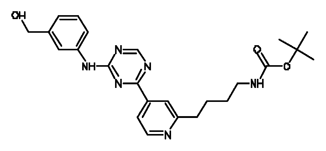
한 실시 양태에서, 아닐린-피리디노트라이아진은 화학식 (6)의 화합물이다.In one embodiment, the aniline-pyridinotriazine is a compound of formula (6).
[화학식 (6)][Formula 6]

한 실시 양태에서, 아닐린-피리디노트라이아진은 화학식 (7)의 화합물이다.In one embodiment, the aniline-pyridinotriazine is a compound of formula (7).
[화학식 (7)][Formula 7]

한 실시 양태에서, 아닐린-피리디노트라이아진은 화학식 (8)의 화합물이다.In one embodiment, the aniline-pyridinotriazine is a compound of formula (8).
[화학식 (8)][Formula 8]

한 실시 양태에서, 아닐린-피리디노트라이아진은 화학식 (9)의 화합물이다.In one embodiment, the aniline-pyridinotriazine is a compound of formula (9).
[화학식 (9)][Formula 9]

한 실시 양태에서, 아닐린-피리디노트라이아진은 화학식 (10)의 화합물이다.In one embodiment, the aniline-pyridinotriazine is a compound of formula (10).
[화학식 (10)][Formula 10]

한 실시 양태에서, 아닐린-피리디노트라이아진은 화학식 (11)의 화합물이다.In one embodiment, the aniline-pyridinotriazine is a compound of formula (11).
[화학식 (11)][Formula 11]

한 실시 양태에서, 아닐린-피리디노트라이아진은 화학식 (12)의 화합물이다.In one embodiment, the aniline-pyridinotriazine is a compound of formula (12).
[화학식 (12)][Formula 12]

: 4-{[4-(4-메틸-3,4-다이하이드로퀴녹살린-1(2H)-일)피리미딘-2-일]아미노}-N-(1-메틸피페리딘-4-일)벤즈아미드. 본원에서 "화합물 11"이라고 한다.: 4-{[4- (4-methyl-3,4-dihydroquinoxalin-1 (2H) -yl) pyrimidin-2-yl] amino} -N- (1-methylpiperidin-4- I) benzamide. Referred to herein as "Compound 11".
한 실시 양태에서, 아닐린-피리디노트라이아진은 화학식 (13)의 화합물이다.In one embodiment, the aniline-pyridinotriazine is a compound of formula (13).
[화학식 (13)][Formula 13]

한 실시 양태에서, 만능 줄기 세포를 완성 내배엽 계통의 특징적인 마커를 발현하는 세포로 분화시킬 수 있는 화합물은 화학식 (14)의 사이클릭 아닐린-피리디노트라이아진:In one embodiment, the compounds capable of differentiating pluripotent stem cells into cells expressing markers characteristic of the definitive endoderm lineage are cyclic aniline-pyridinotriazines of Formula (14):
[화학식 (14)][Formula 14]

그의 N-산화물 형태, 약학적으로 허용가능한 부가염 및 입체화학적으로 이성질체성 형태이다:N-oxide forms, pharmaceutically acceptable addition salts and stereochemically isomeric forms thereof:
[여기서, m은 1 내지 4의 정수를 나타내고; n은 1 내지 4의 정수를 나타내고; Z은 N 또는 C를 나타내고;[Wherein m represents an integer of 1 to 4; n represents an integer of 1 to 4; Z represents N or C;
Y는 -NR2-C1 - 6알킬-CO-NR4-, -C1 - 4알킬-NR9-C1 - 4알킬-, C1 - 6알킬-CO-Het10-, -Het11-CO-C1-6알킬-, -Het12-C1 - 6알킬-, -CO-Het13-C1 - 6알킬-, -CO-NR10-C1 - 6알킬-, -Het1-C1-6알킬-CO-NR5-, 또는 -Het2-CO-NR6-을 나타내고, 여기서, -C1 - 6알킬-연결기(-NR2-C1-6알킬-CO-NR4-또는-Het1-C1 - 6알킬-CO-NR5- 내)는 하이드록시, 메톡시, 아미노카르보닐, 할로, 페닐, 인돌릴, 메틸설파이드, 티올, 하이드록시페닐, 시아노페닐, 아미노 및 하이드록시카르보닐로부터 선택되는 하나 또는 가능한 경우 2개 이상의 치환체로 임의로 치환되고;Y is -NR 2 -C 1 - 6 alkyl, -CO-NR 4 -, -C 1 - 4 alkyl, -NR 9 -C 1 - 4 alkyl -, C 1 - 6 alkyl, -CO-Het 10 -, -Het 11 -CO-C 1-6 alkyl -, -Het 12 -C 1 - 6 alkyl -, -CO-Het 13 -C 1 - 6 alkyl -, -CO-NR 10 -C 1 - 6 alkyl -, -Het 1 -C 1-6 alkyl, -CO-NR 5 -, or -Het 2 -CO-NR 6 - represents the, here, -C 1 - 6 alkyl-linking groups (-NR 2 -C 1-6 alkyl, -CO-NR 4 - or -Het 1 -C 1 - 6 alkyl-NR 5 -CO-I) is hydroxy, methoxy, aminocarbonyl, halo, phenyl, indolyl, methyl sulfide, thiol, hydroxyphenyl, cyanophenyl Optionally substituted with one or possibly two or more substituents selected from amino and hydroxycarbonyl;
X1은 직접 결합, C1 - 4알킬, C1 - 4알킬옥시-, C1 - 4알킬-CO-, C2 - 4알케닐, C2 - 4알키닐, 또는 C1 - 4알킬-NR3-을 나타내고, 여기서 상기 C1 - 4알킬 또는 C2 - 4알케닐은 하나 또는 가능한 경우 2개 이상의 할로 치환체로 임의로 치환되고;X 1 is a direct bond, C 1 - 4 alkyl, C 1 - 4 alkyloxy -, C 1 - 4 alkyl, -CO-, C 2 - 4 alkenyl, C 2 - 4 alkynyl, or C 1 - 4 alkyl- It represents, wherein the C 1 - - NR 3 4 alkyl or C 2 - 4 alkenyl, or, if possible optionally substituted with one or two or more halo substituents;
X2는 직접 결합, C1 - 4알킬, C1 - 4알킬옥시-, C1 - 4알킬-CO-, C2 - 4알케닐, C2 - 4알키닐, 또는 C1 - 4알킬-NR7-을 나타내고, 여기서 상기 C1 - 4알킬 또는 C2 - 4알케닐은 하나 또는 가능한 경우 2개 이상의 할로 치환체로 임의로 치환되고;X 2 is a direct bond, C 1 - 4 alkyl, C 1 - 4 alkyloxy -, C 1 - 4 alkyl, -CO-, C 2 - 4 alkenyl, C 2 - 4 alkynyl, or C 1 - 4 alkyl- NR 7 - represents, in which the C 1 - 4 alkyl or C 2 - 4 alkenyl, or, if possible optionally substituted with one or two or more halo substituents;
R1 및 R8은 각각 독립적으로 수소, Het14, 시아노, 할로, 하이드록시, C1 - 6알콕시-, C1 - 6알킬-, 모노- 또는 다이(C1-4알킬)아미노-카르보닐-, 모노- 또는 다이(C1 - 4알킬)아미노-설포닐, 할로로 치환된 C1 - 6알콕시-를 나타내거나, R1은 하이드록시 또는 할로로부터 선택되는 하나 또는 가능한 경우 2개 이상의 치환체로 치환된 C1 - 6알킬을 나타내고;R 1 and R 8 are each independently hydrogen, Het 14, cyano, halo, hydroxy, C 1 - 6 alkoxy -, C 1 - 6 alkyl-, mono- or di (C 1-4 alkyl) amino-carbonyl carbonyl -, mono- or di (C 1 - 4 alkyl) amino-sulfonyl, substituted by halo C 1 - 6 alkoxy-, or indicate, R 1 is two or more, if one or possibly selected from hydroxy or halo substituted with a substituent C 1 - 6 represents alkyl;
R2 및 R9는 각각 독립적으로 수소, C1 - 4알킬, C2 - 4알케닐, Het3, Het4-C1 - 4알킬-, Het5-C1-4알킬카르보닐-, 모노-또는 다이(C1-4알킬)아미노-C1 - 4알킬-카르보닐-, 또는 수소, 하이드록시, 아미노 또는 C1 - 4알킬옥시-로부터 선택되는 하나 또는 가능한 경우 2개 이상의 치환체로 임의로 치환된 페닐을 나타내고;R 2 and R 9 are each independently hydrogen, C 1 - 4 alkyl, C 2 - 4 alkenyl, Het 3, Het 4 -C 1 - 4 alkyl -, Het 5 -C 1-4 alkyl-carbonyl -, mono - or di (C 1-4 alkyl) amino -C 1 - 4 alkyl-carbonyl-optionally substituted with one or two or more if possible substituents selected from -, or hydrogen, hydroxy, amino or C 1 - 4 alkyloxy Substituted phenyl;
R3 및 R7은 각각 독립적으로 수소, C1 - 4알킬, Het6, Het7-C1 - 4알킬-, C2 -4알케닐카르보닐(Het8-C1 - 4알킬아미노카르보닐-로 임의 치환됨), C2 - 4알케닐설포닐-, C1 - 4알킬옥시C1- 4알킬-, 또는 수소, 하이드록시, 아미노 또는 C1 - 4알킬옥시-로부터 선택되는 하나 또는 가능한 경우 2개 이상의 치환체로 임의로 치환된 페닐을 나타내고;R 3 and R 7 are each independently hydrogen, C 1 - 4 alkyl, Het 6, Het 7 -C 1 - 4 alkyl -, C 2 -4 alkenyl-carbonyl (Het 8 -C 1 - 4 alkyl-amino-carbonyl - being optionally substituted), C 2 - 4 alkenyl nilseol sulfonyl -, C 1 - 4 alkyloxy 1- C 4 alkyl-, or hydrogen, hydroxy, amino or C 1 - 4 alkyl-oxy-one selected from or where possible If phenyl is optionally substituted with two or more substituents;
R4, R5, R6 및 R10은 각각 독립적으로 수소, 또는 하이드록시로 임의 치환된 C1-4알킬, Het9 또는 C1 - 4알킬옥시를 나타내고;R4, R5, R6 and R 10 are each independently hydrogen, or an optionally substituted C 1-4 alkyl with hydroxy, Het 9 or C 1 - 4 denotes an alkyl-oxy;
Het1 및 Het2는 각각 독립적으로 피롤리디닐, 피페리디닐, 피페라지닐, 피리디닐, 피리미디닐, 피라지닐, 이미다졸리디닐 또는 피라졸리디닐로부터 선택되는 헤테로사이클을 나타내고, 여기서 상기 Het1 및 Het2는 아미노, 하이드록시, C1 - 4알킬, 하이드록시-C1 - 4알킬-, 페닐, 페닐-C1 - 4알킬-, C1 - 4알킬-옥시-C1 - 4알킬-모노-또는 다이(C1 - 4알킬) 아미노- 또는 아미노-카르보닐-로 임의로 치환되고;Het 1 and Het 2 each independently represent a heterocycle selected from pyrrolidinyl, piperidinyl, piperazinyl, pyridinyl, pyrimidinyl, pyrazinyl, imidazolidinyl or pyrazolidinyl, wherein the Het 1 and Het 2 is amino, hydroxy, C 1 - 4 alkyl, hydroxy, -C 1 - 4 alkyl-phenyl, phenyl -C 1 - 4 alkyl-, C 1 - 4 alkyl-oxy -C 1 - 4 alkyl - mono- or di (C 1 - 4 alkyl) amino-or amino- is optionally substituted with-carbonyl;
Het3 및 Het6은 각각 독립적으로 피롤리디닐 또는 피페리디닐로부터 선택되는 헤테로사이클을 나타내고, 여기서 상기 Het3 및 Het6은 C1 - 4알킬, C3 - 6사이클로알킬, 하이드록시-C1 - 4알킬-, C1 - 4알킬옥시C1 - 4알킬 또는 폴리하이드록시-C1 - 4알킬-로부터 선택되는 하나 또는 가능한 경우 2개 이상의 치환체로 임의로 치환되고;Het 3 and Het 6 represents a heterocycle are each independently selected from pyrrolidinyl or piperidinyl wherein said Het 3 and Het 6 are C 1 - 4 alkyl, C 3 - 6 cycloalkyl, hydroxy -C 1 - 4 alkyl-, C 1 - 4 alkyl-oxy C 1 - 4 alkyl or polyhydroxy -C 1 - 4 alkyl, - when one or possibly selected from optionally substituted with at least two substituents;
Het4, Het7 및 Het9는 각각 독립적으로 모르폴리닐, 피롤리디닐, 피페라지닐 또는 피페리디닐로부터 선택되는 헤테로사이클을 나타내고, 여기서 상기 Het4, Het7 및 Het9는 C1 - 4알킬, C3 - 6사이클로알킬, 하이드록시-C1 - 4알킬-, C1 - 4알킬옥시C1 - 4알킬 또는 폴리하이드록시-C1 - 4알킬-로부터 선택되는 하나 또는 가능한 경우 2개 이상의 치환체로 임의로 치환되고;Het 4, Het 7 and Het 9 represents each independently represent morpholinyl, pyrrolidinyl, piperazinyl or piperazinyl heterocycle selected from piperidinyl, wherein said Het 4, Het 7 and Het 9 are C 1 - 4 alkyl, C 3 - 6 cycloalkyl, hydroxy, -C 1 - 4 alkyl-, C 1 - 4 alkyl-oxy C 1 - 4 alkyl or polyhydroxy -C 1 - 4 alkyl, - when one or where possible two or selected from Optionally substituted with the above substituents;
Het5는 모르폴리닐, 피롤리디닐, 피페라지닐 또는 피펜디닐로부터 선택되는 헤테로사이클을 나타내고, 여기서 상기 Het5는 C1 - 4알킬, C3 - 6사이클로알킬, 하이드록시-C1 - 4알킬-, C1 - 4알킬옥시C1 - 4알킬 또는 폴리하이드록시-C1 - 4알킬-로부터 선택되는 하나 또는 가능한 경우 2개 이상의 치환체로 임의로 치환되고;Het 5 is morpholinyl, pyrrolidinyl, P represents a heterocycle selected from piperazinyl or blood Fendi carbonyl, wherein said Het 5 is C 1 - 4 alkyl, C 3 - 6 cycloalkyl, hydroxy -C 1 - 4 alkyl-, C 1 - 4 alkyl-oxy C 1 - 4 alkyl or polyhydroxy -C 1 - 4 alkyl, - when one or possibly selected from optionally substituted with at least two substituents;
Het10, Het11 및 Het13은 각각 독립적으로 피롤리디닐, 피페리디닐, 피페라지닐, 피리디닐, 피리미디닐, 피라지닐, 이미다졸리디닐 또는 피라졸리디닐로부터 선택되는 헤테로사이클을 나타내고, 여기서 상기 Het10, Het11 및 Het13은 아미노, 하이드록시, C1 - 4알킬, 하이드록시-C1 - 4알킬-, 페닐, 페닐-C1 - 4알킬-, C1 - 4알킬-옥시-C1 - 4알킬-, 아미노-카르보닐- 또는 모노-또는 다이(C1-4알킬)아미노-로 임의로 치환되고;Het 10 , Het 11 and Het 13 each independently represent a heterocycle selected from pyrrolidinyl, piperidinyl, piperazinyl, pyridinyl, pyrimidinyl, pyrazinyl, imidazolidinyl or pyrazolidinyl, wherein said Het 10, Het 11 and Het 13 are amino, hydroxy, C 1 - 4 alkyl, hydroxy, -C 1 - 4 alkyl-phenyl, phenyl -C 1 - 4 alkyl-, C 1 - 4 alkyl-oxy -C 1 - 4 alkyl-amino-carbonyl-or mono-is optionally substituted by or di (C 1-4 alkyl) amino;
Het12는 피롤리디닐, 피페리디닐, 피페라지닐, 피리디닐, 피리미디닐, 피라지닐, 이미다졸리디닐 또는 피라졸리디닐로부터 선택되는 헤테로사이클을 나타내고, 여기서 상기 Het12는 아미노, 하이드록시, C1 - 4알킬, 하이드록시-C1 - 4알킬-, 페닐, 페닐-C1 - 4알킬-, C1 - 4알킬-옥시-C1 - 4알킬-; 모노-또는 다이(C1-4알킬)아미노- 또는 아미노-카르보닐-로 임의로 치환되고;Het 12 represents a heterocycle selected from pyrrolidinyl, piperidinyl, piperazinyl, pyridinyl, pyrimidinyl, pyrazinyl, imidazolidinyl or pyrazolidinyl, wherein Het 12 is amino, hydroxy , C 1 - 4 alkyl, hydroxy, -C 1 - 4 alkyl-phenyl, phenyl -C 1 - 4 alkyl-, C 1 - 4 alkyl-oxy -C 1 - 4 alkyl; Optionally substituted with mono- or di (C 1-4 alkyl) amino- or amino-carbonyl-;
Het14는 모르폴리닐; 피롤리디닐; 피페라지닐; 이미다졸릴; 피롤릴; 2,3,4-트라이아자피롤릴; 1,2,3-트라이아졸릴; 피라졸릴; 또는 피페리디닐로부터 선택되는 헤테로사이클을 나타내고, 여기서 상기 Het14는 C1 - 4알킬, C3 - 6사이클로알킬, 하이드록시-C1 - 4알킬-, C1 - 4알킬옥시C1 - 4알킬 또는 폴리하이드록시-C1 - 4알킬-로부터 선택되는 하나 또는 가능한 경우 2개 이상의 치환체로 임의로 치환되고; 특히 Het14는 모르폴리닐; 피롤리디닐; 피롤릴; 2,3,4-트라이아자피롤릴; 피페라지닐 또는 피페리디닐로부터 선택되는 헤테로사이클을 나타내고, 여기서 상기 Het14는 C1 - 4알킬, C3 - 6사이클로알킬, 하이드록시-C1 - 4알킬-, C1 - 4알킬옥시C1 - 4알킬 또는 폴리하이드록시-C1 - 4알킬-로부터 선택되는 하나 또는 가능한 경우 2개 이상의 치환체로 임의로 치환되고; 더욱 특히 Het14는 모르폴리닐; 피롤리디닐; 피페라지닐 또는 피페리디닐로부터 선택되는 헤테로사이클을 나타내고, 여기서 상기 Het14는 C1 - 4알킬, C3 - 6사이클로알킬, 하이드록시-C1 - 4알킬-, C1 - 4알킬옥시C1 - 4알킬 또는 폴리하이드록시-C1 - 4알킬-로부터 선택되는 하나 또는 가능한 경우 2개 이상의 치환체로 임의로 치환된다].Het 14 is morpholinyl; Pyrrolidinyl; Piperazinyl; Imidazolyl; Pyrrolyl; 2,3,4-triazapyrrolyl; 1,2,3-triazolyl; Pyrazolyl; Or it represents a heterocycle selected from piperidinyl, wherein said Het 14 is C 14 alkyl, C 3 - 6 cycloalkyl, hydroxy-alkyl -C 14 -, C 14 alkyloxy C 14 alkyl or polyhydroxy -C 1 - 4 alkyl, - when one or possibly selected from optionally substituted with at least two substituents; In particular Het 14 is morpholinyl; Pyrrolidinyl; Pyrrolyl; 2,3,4-triazapyrrolyl; It represents a heterocycle selected from piperazinyl or piperidinyl wherein said Het 14 is C 14 alkyl, C 3 - 6 cycloalkyl, hydroxy-alkyl -C 14 -, C 14 alkyloxy C 1 - 4 alkyl or polyhydroxy -C 1 - 4 alkyl, - when one or possibly selected from optionally substituted with at least two substituents; More particularly Het 14 is morpholinyl; Pyrrolidinyl; It represents a heterocycle selected from piperazinyl or piperidinyl wherein said Het 14 is C 14 alkyl, C 3 - 6 cycloalkyl, hydroxy-alkyl -C 14 -, C 14 alkyloxy C 1 - 4 alkyl or polyhydroxy -C 1 - 4 alkyl, - when one or possibly selected from optionally substituted with at least two substituents.
화학식 (7)의 화합물은 얀센 파마소티카 N.V.(Janssen Pharmaceutica N.V.)에 양도된 WO2007/003525에 개시된다.Compounds of formula (7) are disclosed in WO2007 / 003525 assigned to Janssen Pharmaceutica N.V.
한 실시 양태에서, 사이클릭 아닐린-피리디노트라이아진은 화학식 (14)의 화합물이다.In one embodiment, the cyclic aniline-pyridinotriazine is a compound of formula (14).
한 실시 양태에서, 사이클릭 아닐린-피리디노트라이아진은 화학식 (15)의 화합물이다.In one embodiment, the cyclic aniline-pyridinotriazine is a compound of formula (15).
[화학식 (15)][Formula 15]
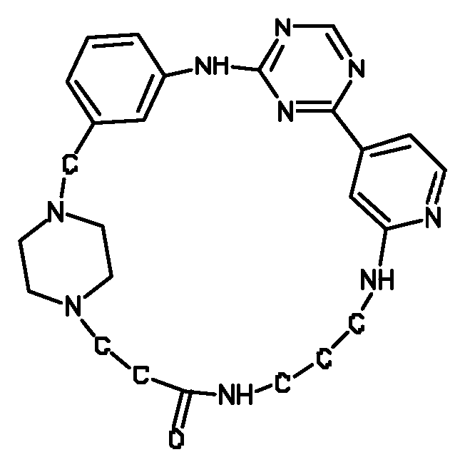
한 실시 양태에서, 사이클릭 아닐린-피리디노트라이아진은 화학식 (16)의 화합물이다.In one embodiment, the cyclic aniline-pyridinotriazine is a compound of formula (16).
[화학식 (16)][Formula 16]

한 실시 양태에서, 사이클릭 아닐린-피리디노트라이아진은 화학식 (17)의 화합물이다.In one embodiment, the cyclic aniline-pyridinotriazine is a compound of formula (17).
[화학식 (17)][Formula 17]

한 실시 양태에서, 사이클릭 아닐린-피리디노트라이아진은 화학식 (18)의 화합물이다.In one embodiment, the cyclic aniline-pyridinotriazine is a compound of formula (18).
[화학식 (18)][Formula 18]

한 실시 양태에서, 사이클릭 아닐린-피리디노트라이아진은 화학식 (19)의 화합물이다.In one embodiment, the cyclic aniline-pyridinotriazine is a compound of formula (19).
[화학식 (19)][Formula 19]

한 실시 양태에서, 사이클릭 아닐린-피리디노트라이아진은 화학식 (20)의 화합물이다.In one embodiment, the cyclic aniline-pyridinotriazine is a compound of formula (20).
[화학식 (20)][Formula 20]

한 실시 양태에서, 사이클릭 아닐린-피리디노트라이아진은 화학식 (21)의 화합물이다.In one embodiment, the cyclic aniline-pyridinotriazine is a compound of formula (21).
[화학식 (21)][Formula 21]

한 실시 양태에서, 사이클릭 아닐린-피리디노트라이아진은 화학식 (22)의 화합물이다.In one embodiment, the cyclic aniline-pyridinotriazine is a compound of formula (22).
[화학식 (22)][Formula 22]

한 실시 양태에서, 사이클릭 아닐린-피리디노트라이아진은 화학식 (23)의 화합물이다.In one embodiment, the cyclic aniline-pyridinotriazine is a compound of formula (23).
[화학식 (23)][Formula 23]

한 실시 양태에서, 사이클릭 아닐린-피리디노트라이아진은 화학식 (24)의 화합물이다.In one embodiment, the cyclic aniline-pyridinotriazine is a compound of formula (24).
[화학식 (24)][Formula 24]

한 실시 양태에서, 사이클릭 아닐린-피리디노트라이아진은 화학식 (25)의 화합물이다.In one embodiment, the cyclic aniline-pyridinotriazine is a compound of formula (25).
[화학식 (25)][Formula 25]

한 실시 양태에서, 사이클릭 아닐린-피리디노트라이아진은 화학식 (26)의 화합물이다.In one embodiment, the cyclic aniline-pyridinotriazine is a compound of formula (26).
[화학식 (26)][Formula 26]

한 실시 양태에서, 사이클릭 아닐린-피리디노트라이아진은 화학식 (27)의 화합물이다.In one embodiment, the cyclic aniline-pyridinotriazine is a compound of formula (27).
[화학식 (27)][Formula 27]

한 실시 양태에서, 사이클릭 아닐린-피리디노트라이아진은 화학식 (28)의 화합물이다.In one embodiment, the cyclic aniline-pyridinotriazine is a compound of formula (28).
[화학식 (28)][Formula 28]

한 실시 양태에서, 사이클릭 아닐린-피리디노트라이아진은 화학식 (29)의 화합물이다.In one embodiment, the cyclic aniline-pyridinotriazine is a compound of formula (29).
[화학식 (29)][Formula 29]

한 실시 양태에서, 사이클릭 아닐린-피리디노트라이아진은 화학식 (30)의 화합물이다.In one embodiment, the cyclic aniline-pyridinotriazine is a compound of formula (30).
[화학식 (30)][Formula 30]

한 실시 양태에서, 사이클릭 아닐린-피리디노트라이아진은 화학식 (31)의 화합물이다.In one embodiment, the cyclic aniline-pyridinotriazine is a compound of formula (31).
[화학식 (31)][Formula 31]
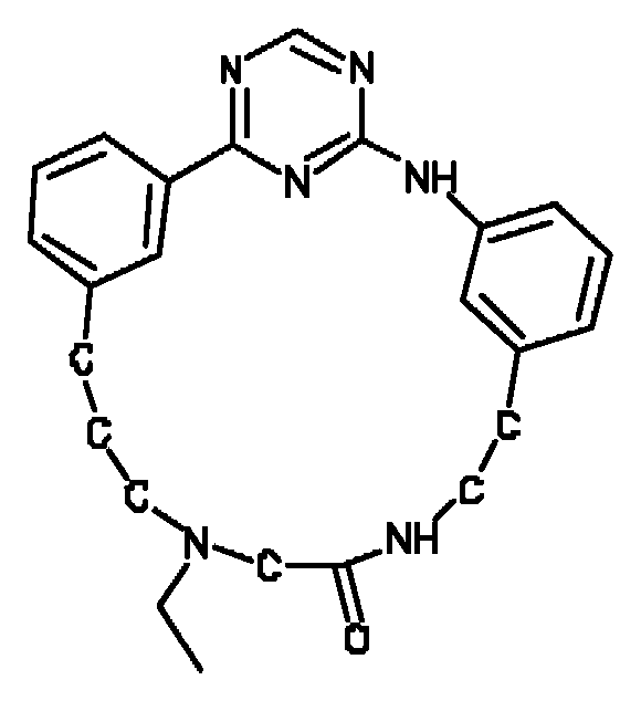
한 실시 양태에서, 사이클릭 아닐린-피리디노트라이아진은 화학식 (32)의 화합물이다.In one embodiment, the cyclic aniline-pyridinotriazine is a compound of formula (32).
[화학식 (32)][Formula 32]

한 실시 양태에서, 사이클릭 아닐린-피리디노트라이아진은 화학식 (33)의 화합물이다.In one embodiment, the cyclic aniline-pyridinotriazine is a compound of formula (33).
[화학식 (33)][Formula 33]

한 실시 양태에서, 사이클릭 아닐린-피리디노트라이아진은 화학식 (34)의 화합물이다.In one embodiment, the cyclic aniline-pyridinotriazine is a compound of formula (34).
[화학식 (34)][Formula 34]

한 실시 양태에서, 사이클릭 아닐린-피리디노트라이아진은 화학식 (35)의 화합물이다.In one embodiment, the cyclic aniline-pyridinotriazine is a compound of formula (35).
[화학식 (35)][Formula 35]
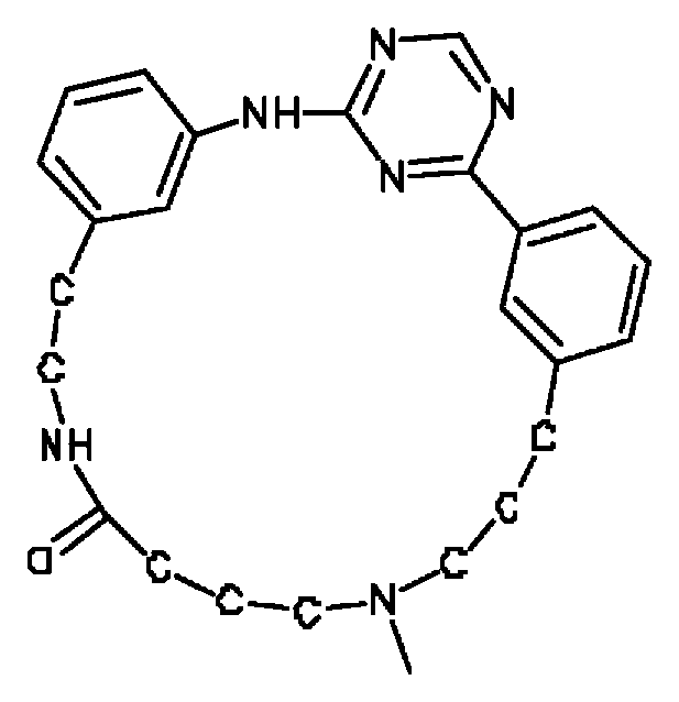
한 실시 양태에서, 사이클릭 아닐린-피리디노트라이아진은 화학식 (36)의 화합물이다.In one embodiment, the cyclic aniline-pyridinotriazine is a compound of formula (36).
[화학식 (36)][Formula 36]

한 실시 양태에서, 사이클릭 아닐린-피리디노트라이아진은 화학식 (37)의 화합물이다.In one embodiment, the cyclic aniline-pyridinotriazine is a compound of formula (37).
[화학식 (37)][Formula 37]

한 실시 양태에서, 사이클릭 아닐린-피리디노트라이아진은 화학식 (38)의 화합물이다.In one embodiment, the cyclic aniline-pyridinotriazine is a compound of formula (38).
[화학식 (38)][Formula 38]
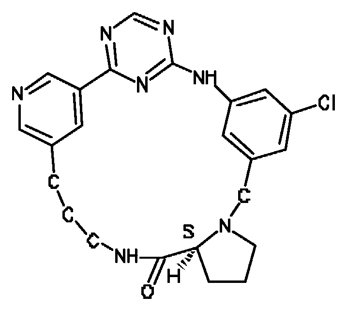
한 실시 양태에서, 사이클릭 아닐린-피리디노트라이아진은 화학식 (39)의 화합물이다.In one embodiment, the cyclic aniline-pyridinotriazine is a compound of formula (39).
[화학식 (39)][Formula 39]

한 실시 양태에서, 사이클릭 아닐린-피리디노트라이아진은 화학식 (40)의 화합물이다.In one embodiment, the cyclic aniline-pyridinotriazine is a compound of formula (40).
[화학식 (40)][Formula 40]

한 실시 양태에서, 사이클릭 아닐린-피리디노트라이아진은 화학식 (41)의 화합물이다.In one embodiment, the cyclic aniline-pyridinotriazine is a compound of formula (41).
[화학식 (41)][Formula 41]
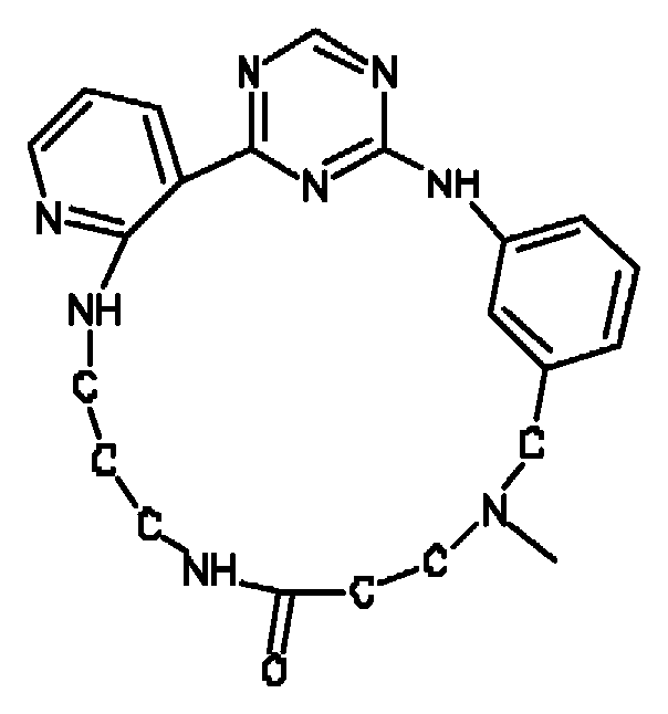
한 실시 양태에서, 사이클릭 아닐린-피리디노트라이아진은 화학식 (42)의 화합물이다.In one embodiment, the cyclic aniline-pyridinotriazine is a compound of formula (42).
[화학식 (42)][Formula 42]

한 실시 양태에서, 사이클릭 아닐린-피리디노트라이아진은 화학식 (43)의 화합물이다.In one embodiment, the cyclic aniline-pyridinotriazine is a compound of formula (43).
[화학식 (43)][Formula 43]

한 실시 양태에서, 사이클릭 아닐린-피리디노트라이아진은 화학식 (44)의 화합물이다.In one embodiment, the cyclic aniline-pyridinotriazine is a compound of formula (44).
[화학식 (44)][Formula 44]
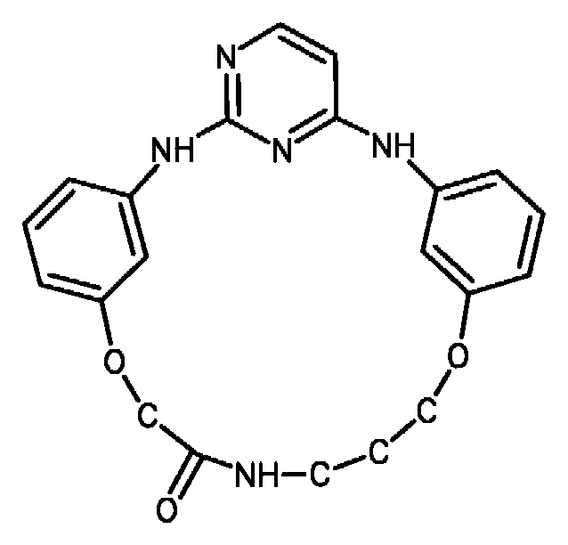
한 실시 양태에서, 사이클릭 아닐린-피리디노트라이아진은 화학식 (45)의 화합물이다.In one embodiment, the cyclic aniline-pyridinotriazine is a compound of formula (45).
[화학식 (45)][Formula 45]

한 실시 양태에서, 사이클릭 아닐린-피리디노트라이아진은 화학식 (46)의 화합물이다.In one embodiment, the cyclic aniline-pyridinotriazine is a compound of formula (46).
[화학식 (46)][Formula 46]
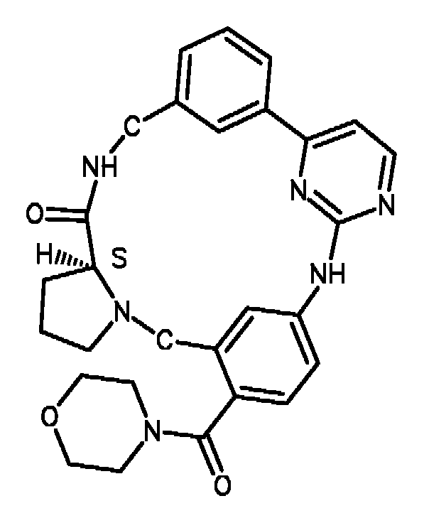
한 실시 양태에서, 사이클릭 아닐린-피리디노트라이아진은 화학식 (47)의 화합물이다.In one embodiment, the cyclic aniline-pyridinotriazine is a compound of formula (47).
[화학식 (47)][Formula 47]

한 실시 양태에서, 적어도 하나의 다른 인자는 화학식 (48)의 화합물이다:In one embodiment, at least one other factor is a compound of Formula (48):
[화학식 (48)][Formula 48]

한 실시 양태에서, 적어도 하나의 다른 인자는 화학식 (49)의 화합물이다:In one embodiment, at least one other factor is a compound of Formula (49):
[화학식 (49)][Formula 49]
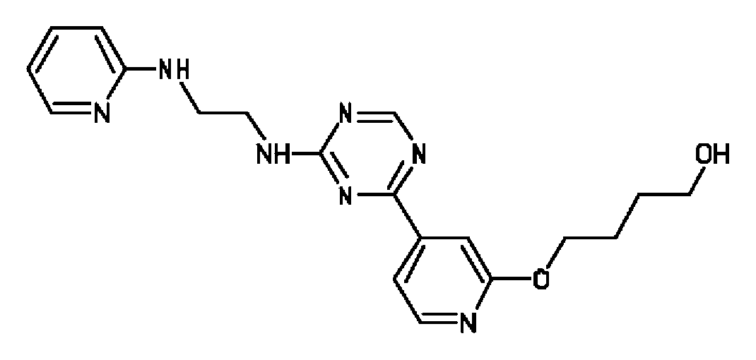
한 실시 양태에서, 적어도 하나의 다른 인자는 화학식 (50)의 화합물이다:In one embodiment, at least one other factor is a compound of Formula (50):
[화학식 (50)][Formula 50]
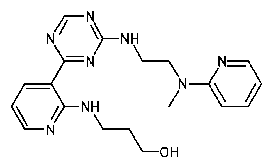
한 실시 양태에서, 적어도 하나의 다른 인자는 화학식 (51)의 화합물이다:In one embodiment, at least one other factor is a compound of Formula (51):
[화학식 (51)][Formula 51]

한 실시 양태에서, 적어도 하나의 다른 인자는 화학식 (52)의 화합물이다:In one embodiment, at least one other factor is a compound of Formula (52):
[화학식 (52)][Formula 52]
한 실시 양태에서, 적어도 하나의 다른 인자는 화학식 (53)의 화합물이다:In one embodiment, at least one other factor is a compound of Formula (53):
[화학식 (53)][Formula 53]

한 실시 양태에서,적어도 하나의 다른 인자는 화학식 (54)의 화합물이다:In one embodiment, at least one other factor is a compound of Formula (54):
[화학식 (54)][Formula 54]
한 실시 양태에서, 적어도 하나의 다른 인자는 화학식 (55)의 화합물이다:In one embodiment, at least one other factor is a compound of Formula (55):
[화학식 (55)][Formula 55]
한 실시 양태에서, 적어도 하나의 다른 인자는 화학식 (56)의 화합물이다:In one embodiment, at least one other factor is a compound of Formula (56):
[화학식 (56)][56]
한 실시 양태에서, 적어도 하나의 다른 인자는 화학식 (57)의 화합물이다:In one embodiment, at least one other factor is a compound of Formula (57):
[화학식 (57)][Formula 57]

한 실시 양태에서, 적어도 하나의 다른 인자는 화학식 (58)의 화합물이다:In one embodiment, at least one other factor is a compound of Formula (58):
[화학식 (58)]Formula (58)]

한 실시 양태에서, 적어도 하나의 다른 인자는 화학식 (59)의 화합물이다:In one embodiment, at least one other factor is a compound of Formula (59):
[화학식 (59)][Formula 59]

완성 내배엽 계통의 특징적인 Characteristic of the definitive endoderm lineage 마커를Marker 발현하는 세포의 검출 Detection of expressing cells
완성 내배엽 계통의 특징을 나타내는 마커를 발현하는 세포의 형성은 특정 프로토콜을 이행하기 전후에 마커의 존재에 대해 시험함으로써 결정될 수 있다. 만능성 줄기 세포는 전형적으로 그러한 마커를 발현하지 않는다. 따라서, 만능성 세포의 분화는 세포가 그들을 발현하기 시작할 때에 검출된다.Formation of cells expressing markers characteristic of the definitive endoderm lineage can be determined by testing for the presence of the marker before and after implementing a particular protocol. Pluripotent stem cells typically do not express such markers. Thus, differentiation of pluripotent cells is detected when the cells begin to express them.
분화 효율은 완성 내배엽 계통의 특징을 나타내는 마커를 발현하는 세포에 의해 발현되는 단백질 마커를 특이적으로 인식하는 제제 (예를 들어, 항체)에 처리 세포 집단을 노출시킴으로써 결정될 수 있다.Differentiation efficiency can be determined by exposing treated cell populations to agents (eg, antibodies) that specifically recognize protein markers expressed by cells expressing markers characteristic of the definitive endoderm lineage.
배양되거나 단리된 세포에서 단백질 및 핵산 마커의 발현을 평가하는 방법은 당업계에서의 표준이다. 이들은 정량적 역전사효소 폴리머라아제 연쇄 반응(RT-PCR), 노던 블롯(Northern blots), 원위치 혼성화(예를 들어, 문헌[Current Protocols in Molecular Biology (Ausubel et al., eds. 2001 supplement)] 참고), 및 면역분석, 예를 들어, 단면(sectioned) 재료의 면역조직화학적 분석, 웨스턴 블롯팅(Western blotting), 및 온전한 상태의 세포에서 접근가능한 마커의 경우, 유세포분석(유세포분석 분석, FACS) (예를 들어, 문헌[Harlow and Lane, Using Antibodies: A Laboratory Manual, New York: Cold Spring Harbor Laboratory Press (1998)] 참고)을 포함한다.Methods for assessing the expression of protein and nucleic acid markers in cultured or isolated cells are standard in the art. These include quantitative reverse transcriptase polymerase chain reaction (RT-PCR), Northern blots, in situ hybridization (eg, Current Protocols in Molecular Biology (Ausubel et. al ., eds. 2001 supplement), and immunoassays, for example immunohistochemical analysis of sectioned materials, Western blotting, and markers accessible in intact cells, for flow cytometry (flow cytometry). Analytical Assay, FACS) (see, eg, Harlow and Lane, Using Antibodies: A Laboratory Manual, New York: Cold Spring Harbor Laboratory Press (1998)).
예를 들어, 만능성 줄기 세포의 특징은 당업자에게 잘 알려져 있으며, 만능성 줄기 세포의 추가 특징은 계속 확인되고 있다. 만능 줄기 세포 마커는 예를 들어, ABCG2, 크립토(cripto), FOXD3, 코넥신43, 코넥신45, OCT4, SOX2, 나노그, hTERT, UTF-1, ZFP42, SSEA-3, SSEA-4, Tra1-60, 또는 Tra1-81 중 하나 이상의 발현을 포함한다.For example, the characteristics of pluripotent stem cells are well known to those skilled in the art, and additional features of pluripotent stem cells continue to be identified. Pluripotent stem cell markers include, for example, ABCG2, cryptoto, FOXD3, connexin43, connexin45, OCT4, SOX2, nanog, hTERT, UTF-1, ZFP42, SSEA-3, SSEA-4, Tra1 -60, or expression of one or more of Tra1-81.
만능성 줄기 세포를 본 발명의 방법으로 처리한 후, 분화된 세포는 완성 내배엽 계통의 특징을 나타내는 마커를 발현하는 세포에 의해 발현된 CXCR4와 같은 단백질 마커를 특이적으로 인식하는 제제 (예를 들어, 항체)에 처리 세포 집단을 노출시켜 정제할 수 있다.After treating pluripotent stem cells with the methods of the present invention, the differentiated cells are formulated to specifically recognize protein markers, such as CXCR4, expressed by cells expressing markers characteristic of the definitive endoderm lineage (e.g., , Antibody) can be purified by exposing the treated cell population.
췌장 내배엽 계통의 특징적인 Characteristic of the pancreatic endoderm lineage 마커를Marker 발현하는 세포의 형성 Formation of expressing cells
완성 내배엽 계통의 특징적인 마커를 발현하는 세포는 당업계의 임의의 방법에 의해 또는 본 발명에서 제안된 임의의 방법에 의해 췌장 내배엽 계통의 특징적인 마커를 발현하는 세포로 분화될 수 있다.Cells expressing markers characteristic of the definitive endoderm lineage can be differentiated into cells expressing markers characteristic of the pancreatic endoderm lineage by any method in the art or by any method proposed herein.
예를 들어, 완성 내배엽 계통의 특징적인 마커를 발현하는 세포는 문헌[D'Amour et al, Nature Biotechnology 24, 1392 - 1401 (2006)]에 개시된 방법에 따라 췌장 내배엽 계통의 특징적인 마커를 발현하는 세포로 분화될 수 있다.For example, cells expressing markers characteristic of the definitive endoderm lineage are described in D'Amour et. al , Nature Biotechnology 24, 1392-1401 (2006), can be differentiated into cells expressing markers characteristic of the pancreatic endoderm lineage.
예를 들어, 완성 내배엽 계통의 특징적인 마커를 발현하는 세포는, 완성 내배엽 계통의 특징적인 마커를 발현하는 세포를 섬유아세포 성장 인자 및 헤지호그 시그널링 경로 억제제 KAAD-사이클로파민으로 처리하고, 이어서 섬유아세포 성장 인자 및 KAAD-사이클로파민을 함유하는 배지를 제거하고, 그 후 세포를 레틴산, 섬유아세포 성장 인자 및 KAAD-사이클로파민을 함유하는 배지에서 배양함으로써, 췌장 내배엽 계통의 특징적인 마커를 발현하는 세포로 추가로 분화된다. 이 방법의 예는 문헌[Nature Biotechnology 24, 1392 - 1401 (2006)]에 개시된다.For example, cells expressing markers characteristic of the definitive endoderm lineage treat cells expressing markers characteristic of the definitive endoderm lineage with fibroblast growth factor and hedgehog signaling pathway inhibitor KAAD-cyclopamine, followed by fibroblasts Cells expressing markers characteristic of the pancreatic endoderm lineage by removing the media containing growth factor and KAAD-cyclopamine and then culturing the cells in media containing retinic acid, fibroblast growth factor and KAAD-cyclopamine Further differentiated into. Examples of this method are disclosed in Nature Biotechnology 24, 1392-1401 (2006).
본 발명의 일 태양에서, 완성 내배엽 계통의 특징적인 마커를 발현하는 세포는, 라이프스캔 인크에 허여된, 미국 특허 출원 제11/736,908호에 개시된 방법에 따라, 완성 내배엽 계통의 특징적인 마커를 발현하는 세포를 레틴산 및 적어도 하나의 섬유아세포 성장 인자로 소정 기간 동안 처리함으로써, 췌장 내배엽 계통의 특징적인 마커를 발현하는 세포로 추가로 분화된다.In one aspect of the invention, cells expressing markers characteristic of the definitive endoderm lineage express the markers characteristic of the definitive endoderm lineage according to the method disclosed in US patent application Ser. No. 11 / 736,908 to LifeScan Inc. By treating the cells with retinic acid and at least one fibroblast growth factor for a period of time, they are further differentiated into cells expressing markers characteristic of the pancreatic endoderm lineage.
본 발명의 일 태양에서, 완성 내배엽 계통의 특징적인 마커를 발현하는 세포는, 라이프스캔 인크에 허여된, 미국 특허 출원 제11/736,908호에 개시된 방법에 따라, 완성 내배엽 계통의 특징적인 마커를 발현하는 세포를 레틴산 및 적어도 하나의 섬유아세포 성장 인자로 소정 기간 동안 처리함으로써, 췌장 내배엽 계통의 특징적인 마커를 발현하는 세포로 추가로 분화된다.In one aspect of the invention, cells expressing markers characteristic of the definitive endoderm lineage express the markers characteristic of the definitive endoderm lineage according to the method disclosed in US patent application Ser. No. 11 / 736,908 to LifeScan Inc. By treating the cells with retinic acid and at least one fibroblast growth factor for a period of time, they are further differentiated into cells expressing markers characteristic of the pancreatic endoderm lineage.
본 발명의 한 측면에서, 완성 내배엽 계통의 특징적인 마커를 발현하는 세포는 추가로, 미국 특허 출원 일련 번호 제 60/990,529 호에서 개시된 방법에 따라 완성 내배엽 계통의 특징적인 마커를 발현하는 세포를 처리함으로써, 췌장 내배엽 계통의 특징적인 마커를 발현하는 세포로 분화될 수 있다.In one aspect of the invention, cells expressing markers characteristic of the definitive endoderm lineage are further treated with cells expressing markers characteristic of the definitive endoderm lineage according to the method disclosed in US Patent Application Serial No. 60 / 990,529. Thus, they can be differentiated into cells expressing markers characteristic of the pancreatic endoderm lineage.
완성 내배엽 계통의 특징적인 마커를 발현하는 세포는 췌장 내배엽 계통의 특징적인 마커를 발현하는 세포의 형성을 향상시킬 수 있는 적어도 하나의 다른 추가 인자로 처리될 수 있다. 대안적으로, 적어도 하나의 다른 추가 인자는 본 발명의 방법에 의해 형성된 췌장 내배엽 계통의 특징적인 마커를 발현하는 세포의 증식을 향상시킬 수 있다. 추가로, 적어도 하나의 다른 추가 인자는 본 발명의 방법에 의해 형성된 췌장 내배엽 계통의 특징적인 마커를 발현하는 세포가 다른 세포 유형을 형성하는 능력을 향상시키거나, 또는 임의의 다른 추가의 분화 단계의 효율을 개선할 수 있다.Cells expressing markers characteristic of the definitive endoderm lineage may be treated with at least one other additional factor capable of enhancing the formation of cells expressing markers characteristic of the pancreatic endoderm lineage. Alternatively, at least one other additional factor may enhance the proliferation of cells expressing markers characteristic of the pancreatic endoderm lineage formed by the methods of the present invention. In addition, at least one other additional factor enhances the ability of cells expressing markers characteristic of the pancreatic endoderm lineage formed by the method of the present invention to form other cell types, or of any other additional differentiation step. The efficiency can be improved.
적어도 하나의 추가의 인자는 예를 들어, 니코틴아미드, TGF-β 패밀리의 구성원(TGF-β1, 2, 및 3 포함), 혈청 알부민, 섬유아세포 성장 인자 패밀리의 구성원, 혈소판-유도 성장 인자-AA, 및 -BB, 혈소판 풍부 혈장, 인슐린 성장 인자 (IGF-I, II), 성장 분화 인자 (예를 들어, GDF-5, -6, -8, -10, -11), 글루카곤 유사 펩티드-I 및 II (GLP-I 및 II), GLP-1 및 GLP-2 미메토바디, 엑센딘-4, 레틴산, 부갑상선 호르몬, 인슐린, 프로게스테론, 아프로티닌, 하이드로코티손, 에탄올아민, 베타 머캅토에탄올, 표피 성장 인자 (EGF), 개스트린 I 및 II, 구리 킬레이트화제, 예를 들어, 트라이에틸렌 펜타민, 포스콜린, Na-부티레이트, 액티빈, 베타셀룰린, ITS, 노긴, 신경돌기 성장 인자, 노달, 발프로산, 트라이코스타틴 A, 나트륨 부티레이트, 간세포 성장 인자 (HGF), 스핑고신-1, VEGF, MG132 (EMD, CA), N2 및 B27 보충제 (Gibco, CA), 스테로이드 알칼로이드, 예를 들어, 사이클로파민 (EMD, CA), 케라틴세포 성장 인자 (KGF), 딕코프 단백질 패밀리, 소 뇌하수체 추출물, 이자 신생-관련 단백질 (INGAP), 인디안 헤지호그, 소닉 헤지호그, 프로테아좀 억제제, 노치 경로 억제제, 소닉 헤지호그 억제제, 또는 그의 조합일 수 있다.At least one additional factor is, for example, nicotinamide, members of the TGF-β family (including TGF-β1, 2, and 3), serum albumin, members of the fibroblast growth factor family, platelet-induced growth factor-AA , And -BB, platelet rich plasma, insulin growth factor (IGF-I, II), growth differentiation factor (eg, GDF-5, -6, -8, -10, -11), glucagon-like peptide-I And II (GLP-I and II), GLP-1 and GLP-2 mimetobodies, exendin-4, retinic acid, parathyroid hormone, insulin, progesterone, aprotinin, hydrocortisone, ethanolamine, beta mercaptoethanol, Epidermal growth factor (EGF), gastrins I and II, copper chelating agents such as triethylene pentamine, phospholine, Na-butyrate, activin, betacellulose, ITS, nogin, neurite growth factor, nodal , Valproic acid, trichostatin A, sodium butyrate, hepatocyte growth factor (HGF), sphingosine-1, VEGF, MG132 (EMD, CA), N2 and B27 supplements (Gibco, CA), steroid alkaloids, for example cyclopamine (EMD, CA), keratinocyte growth factor (KGF), Dirkoff protein family, bovine pituitary extract, interest in neoplasia-related protein (INGAP), Indian Hedgehogs, sonic hedgehogs, proteasome inhibitors, notch pathway inhibitors, sonic hedgehog inhibitors, or combinations thereof.
적어도 하나의 다른 추가 인자는 췌장 세포주, 예를 들어, PANC-1 (ATCC 번호: CRL-1469), CAPAN-1 (ATCC 번호: HTB-79), BxPC-3 (ATCC 번호: CRL-1687), HPAF-II (ATCC 번호: CRL-1997), 간 세포주, 예를 들어, HepG2 (ATCC 번호: HTB-8065), 및 장 세포주, 예를 들어, FHs 74 (ATCC 번호: CCL-241)로부터 얻은 조절된 배지에 의해 공급될 수 있다.At least one other additional factor is a pancreatic cell line, eg, PANC-1 (ATCC No .: CRL-1469), CAPAN-1 (ATCC No .: HTB-79), BxPC-3 (ATCC No .: CRL-1687), Modulations obtained from HPAF-II (ATCC No .: CRL-1997), liver cell lines, eg HepG2 (ATCC No .: HTB-8065), and intestinal cell lines, eg FHs 74 (ATCC No .: CCL-241) Can be supplied by the prepared medium.
췌장 내배엽 계통의 특징을 나타내는 Characteristic of the pancreatic endoderm lineage 마커를Marker 발현하는 세포의 검출 Detection of expressing cells
췌장 내배엽 계통의 특징을 나타내는 마커는 당업자에게 잘 알려져 있으며, 췌장 내배엽 계통의 특징을 나타내는 추가의 마커는 계속 확인되고 있다. 이들 마커는 본 발명에 따라 처리된 세포가 분화하여, 췌장 내배엽 계통의 특징을 나타내는 특성을 획득한 것을 확인하기 위해 사용될 수 있다. 췌장 내배엽 계통 특이적 마커는 예를 들어, Hlxb9, PTF-1a, PDX-1, HNF-6, HNF-1베타와 같은 하나 이상의 전사 인자의 발현을 포함한다.Markers that characterize the pancreatic endoderm lineage are well known to those skilled in the art, and additional markers that characterize the pancreatic endoderm lineage continue to be identified. These markers can be used to confirm that cells treated according to the invention have differentiated to obtain properties that characterize the pancreatic endoderm lineage. Pancreatic endoderm lineage specific markers include, for example, expression of one or more transcription factors such as Hlxb9, PTF-1a, PDX-1, HNF-6, HNF-1beta.
분화 효율은 췌장 내배엽 계통의 특징을 나타내는 마커를 발현하는 세포에 의해 발현되는 단백질 마커를 특이적으로 인식하는 제제 (예를 들어, 항체)에 처리 세포 집단을 노출시킴으로써 결정할 수 있다.Differentiation efficiency can be determined by exposing treated cell populations to agents (eg, antibodies) that specifically recognize protein markers expressed by cells expressing markers characteristic of the pancreatic endoderm lineage.
배양되거나 단리된 세포에서 단백질 및 핵산 마커의 발현을 평가하는 방법은 당업계에서의 표준이다. 이들은 정량적 역전사효소 폴리머라아제 연쇄 반응(RT-PCR), 노던 블롯, 원위치 혼성화(예를 들어, 문헌[Current Protocols in Molecular Biology (Ausubel et al., eds. 2001 supplement)] 참고), 및 면역분석, 예를 들어, 단면 재료의 면역조직화학적 분석, 웨스턴 블롯팅, 및 온전한 상태의 세포에서 접근가능한 마커의 경우, 유세포분석(FACS) (예를 들어, 문헌[Harlow 및 Lane, Using Antibodies: A Laboratory Manual, New York: Cold Spring Harbor Laboratory Press (1998)] 참고)을 포함한다.Methods for assessing the expression of protein and nucleic acid markers in cultured or isolated cells are standard in the art. These include quantitative reverse transcriptase polymerase chain reaction (RT-PCR), northern blots, in situ hybridization (see, eg, Current Protocols in Molecular Biology (Ausubel et. al ., eds. 2001 supplement), and immunoassays, such as immunohistochemical analysis of cross-sectional materials, Western blotting, and markers accessible in intact cells, for example, flow cytometry (FACS) (eg, literature Harlow and Lane, Using Antibodies: A Laboratory Manual, New York: Cold Spring Harbor Laboratory Press (1998).
췌장 내분비 계통의 Of pancreatic endocrine system 마커를Marker 발현하는 세포의 형성 Formation of expressing cells
췌장 내배엽 계통의 특징적인 마커를 발현하는 세포는 당업계의 임의의 방법에 의해 또는 본 발명에서 개시된 임의의 방법에 의해 췌장 내분비 계통의 특징적인 마커를 발현하는 세포로 분화될 수 있다.Cells expressing markers characteristic of the pancreatic endoderm lineage can be differentiated into cells expressing markers characteristic of the pancreatic endocrine lineage by any method in the art or by any of the methods disclosed herein.
예를 들어, 췌장 내분비 계통의 마커를 발현하는 세포는 문헌[D'Amour et al, Nature Biotechnology 24, 1392 - 1401 (2006)]에 개시된 방법에 따라 췌장 내분비 계통의 마커를 발현하는 세포로 분화될 수 있다.For example, cells expressing markers of the pancreatic endocrine lineage may be differentiated into cells expressing markers of the pancreatic endocrine lineage according to the methods disclosed in D'Amour et al , Nature Biotechnology 24, 1392-1401 (2006). Can be.
예를 들어, 췌장 내배엽 계통의 특징적인 마커를 발현하는 세포는 DAPT 및 엑센딘 4를 함유하는 배지에서 췌장 내배엽 계통의 특징적인 마커를 발현하는 세포를 배양하고, 이어서 DAPT와 엑센딘 4를 함유하는 배지를 제거하고, 그 후 엑센딘 1, IGF-1 및 HGF를 함유하는 배지에서 상기 세포를 배양함으로써, 췌장 내분비 계통의 특징적인 마커를 발현하는 세포로 추가로 분화된다. 이 방법의 예는 문헌[Nature Biotechnology 24, 1392 - 1401 (2006)]에 개시된다.For example, cells expressing markers characteristic of the pancreatic endoderm lineage may be cultured cells expressing markers characteristic of the pancreatic endoderm lineage in a medium containing DAPT and
예를 들어, 췌장 내배엽 계통의 특징적인 마커를 발현하는 세포는 엑센딘 4를 함유하는 배지에서 췌장 내배엽 계통의 특징적인 마커를 발현하는 세포를 배양하고, 이어서 엑센딘 4를 함유하는 배지를 제거하고, 그 후 엑센딘 1, IGF-1 및 HGF를 함유하는 배지에서 상기 세포를 배양함으로써, 췌장 내분비 계통의 특징적인 마커를 발현하는 세포로 추가로 분화된다. 이러한 방법의 한 예는 문헌[D' Amour et al, Nature Biotechnology, 2006]에 개시된다.For example, cells expressing markers characteristic of the pancreatic endoderm lineage may be cultured cells expressing markers characteristic of the pancreatic endoderm lineage in a
예를 들어, 췌장 내배엽 계통의 특징적인 마커를 발현하는 세포는 DAPT 및 엑센딘 4를 함유하는 배지에서 췌장 내배엽 계통의 특징적인 마커를 발현하는 세포를 배양함으로써, 췌장 내분비 계통의 특징적인 마커를 발현하는 세포로 추가로 분화된다. 이러한 방법의 한 예는 문헌[D' Amour et al, Nature Biotechnology, 2006]에 개시된다.For example, cells expressing markers characteristic of the pancreatic endoderm lineage express the markers characteristic of the pancreatic endocrine lineage by culturing cells expressing markers characteristic of the pancreatic endoderm lineage in a medium containing DAPT and
예를 들어, 췌장 내배엽 계통의 특징적인 마커를 발현하는 세포는 엑센딘 4를 함유하는 배지에서 췌장 내배엽 계통의 특징적인 마커를 발현하는 세포를 배양함으로써, 췌장 내분비 계통의 특징적인 마커를 발현하는 세포로 추가로 분화된다. 이러한 방법의 한 예는 문헌[D' Amour et al, Nature Biotechnology, 2006]에 개시된다.For example, cells expressing markers characteristic of the pancreatic endoderm lineage are cultured cells expressing markers characteristic of the pancreatic endoderm lineage in a
본 발명의 일 태양에서, 췌장 내배엽 계통의 특징적인 마커를 발현하는 세포는 라이프스캔 인크에 허여된 미국 특허 출원 제11/736,908호에 개시된 방법에 따라 노치 시그널링 경로를 억제하는 인자로 췌장 내배엽 계통의 특징적인 마커를 발현하는 세포를 처리함으로써, 췌장 내분비 계통의 특징적인 마커를 발현하는 세포로 추가로 분화된다.In one aspect of the invention, cells expressing markers characteristic of the pancreatic endoderm lineage are factors that inhibit the Notch signaling pathway in accordance with the method disclosed in US Patent Application No. 11 / 736,908 to LifeScan Inc. By treating cells expressing characteristic markers, they are further differentiated into cells expressing markers characteristic of the pancreatic endocrine lineage.
본 발명의 일 태양에서, 췌장 내배엽 계통의 특징적인 마커를 발현하는 세포는 라이프스캔 인크에 허여된 미국 특허 출원 제11/736,908호에 개시된 방법에 따라 노치 시그널링 경로를 억제하는 인자로 췌장 내배엽 계통의 특징적인 마커를 발현하는 세포를 처리함으로써, 췌장 내분비 계통의 특징적인 마커를 발현하는 세포로 추가로 분화된다.In one aspect of the invention, cells expressing markers characteristic of the pancreatic endoderm lineage are factors that inhibit the Notch signaling pathway in accordance with the method disclosed in US Patent Application No. 11 / 736,908 to LifeScan Inc. By treating cells expressing characteristic markers, they are further differentiated into cells expressing markers characteristic of the pancreatic endocrine lineage.
본 발명의 일 태양에서, 췌장 내배엽 계통의 특징적인 마커를 발현하는 세포는 라이프스캔 인크에 허여된 미국 특허 출원 제11/736,908호에 개시된 방법에 따라 노치 시그널링 경로를 억제하는 인자로 췌장 내배엽 계통의 특징적인 마커를 발현하는 세포를 처리함으로써, 췌장 내분비 계통의 특징적인 마커를 발현하는 세포로 추가로 분화된다.In one aspect of the invention, cells expressing markers characteristic of the pancreatic endoderm lineage are factors that inhibit the Notch signaling pathway in accordance with the method disclosed in US Patent Application No. 11 / 736,908 to LifeScan Inc. By treating cells expressing characteristic markers, they are further differentiated into cells expressing markers characteristic of the pancreatic endocrine lineage.
본 발명의 한 측면에서, 췌장 내배엽 계통의 특징적인 마커를 발현하는 세포는 추가로, 미국 특허 출원 일련 번호 제60/990,529호에서 개시된 방법에 따라 췌장 내배엽 계통의 특징적인 마커를 발현하는 세포를 처리함으로써, 췌장 내분비 계통의 특징적인 마커를 발현하는 세포로 분화된다.In one aspect of the invention, cells expressing markers characteristic of the pancreatic endoderm lineage are further treated with cells expressing markers characteristic of the pancreatic endoderm lineage according to the method disclosed in US Patent Application Serial No. 60 / 990,529. Thereby differentiating into cells expressing markers characteristic of the pancreatic endocrine lineage.
췌장 내배엽 계통의 특징적인 마커를 발현하는 세포는 췌장 내분비 계통의 특징적인 마커를 발현하는 세포의 형성을 향상시킬 수 있는 적어도 하나의 다른 추가 인자로 처리될 수 있다. 대안적으로, 적어도 하나의 다른 추가 인자는 본 발명의 방법에 의해 형성된 췌장 내분비 계통의 특징적인 마커를 발현하는 세포의 증식을 향상시킬 수 있다. 추가로, 적어도 하나의 다른 추가의 인자는 다른 세포 유형을 형성하거나 임의의 다른 추가의 분화 단계의 효능을 향상시키는, 본 발명의 방법에 의해 형성되는 췌장 내분비 계통의 특징적인 마커를 발현하는 세포의 능력을 증진시킬 수 있다.Cells expressing markers characteristic of the pancreatic endoderm lineage may be treated with at least one other additional factor that can enhance the formation of cells expressing markers characteristic of the pancreatic endocrine lineage. Alternatively, at least one other additional factor may enhance the proliferation of cells expressing markers characteristic of the pancreatic endocrine lineage formed by the methods of the invention. In addition, at least one other additional factor may be used to express a marker that is characteristic of the pancreatic endocrine lineage formed by the method of the present invention, which forms another cell type or enhances the efficacy of any other additional differentiation step. Improve your skills.
적어도 하나의 추가의 인자는 예를 들어, 니코틴아미드, TGF-β 패밀리의 구성원(TGF-β1, 2, 및 3 포함), 혈청 알부민, 섬유아세포 성장 인자 패밀리의 구성원, 혈소판-유도 성장 인자-AA, 및 -BB, 혈소판 풍부 혈장, 인슐린 성장 인자 (IGF-I, II), 성장 분화 인자 (예를 들어, GDF-5, -6, -8, -10, -11), 글루카곤 유사 펩티드-I 및 II (GLP-I 및 II), GLP-1 및 GLP-2 미메토바디, 엑센딘-4, 레틴산, 부갑상선 호르몬, 인슐린, 프로게스테론, 아프로티닌, 하이드로코티손, 에탄올아민, 베타 머캅토에탄올, 표피 성장 인자(EGF), 개스트린 I 및 II, 구리 킬레이트화제 예를 들어, 트라이에틸렌 펜타민, 포스콜린, Na-부티레이트, 액티빈, 베타셀룰린, ITS, 노긴, 신경돌기 성장 인자, 노달, 발프로산, 트라이코스타틴 A, 나트륨 부티레이트, 간세포 성장 인자 (HGF), 스핑고신-1, VEGF, MG132 (EMD, CA), N2 및 B27 보충제 (Gibco, CA), 스테로이드 알칼로이드 예를 들어, 사이클로파민 (EMD, CA), 케라틴세포 성장 인자 (KGF), 딕코프(Dickkopf) 단백질 패밀리, 소 뇌하수체 추출물, 이자 신생-관련 단백질 (INGAP), 인디안 헤지호그, 소닉 헤지호그, 프로테아좀 억제제, 노치 경로 억제제, 소닉 헤지호그 억제제, 또는 그의 조합일 수 있다.At least one additional factor is, for example, nicotinamide, members of the TGF-β family (including TGF-β1, 2, and 3), serum albumin, members of the fibroblast growth factor family, platelet-induced growth factor-AA , And -BB, platelet rich plasma, insulin growth factor (IGF-I, II), growth differentiation factor (eg, GDF-5, -6, -8, -10, -11), glucagon-like peptide-I And II (GLP-I and II), GLP-1 and GLP-2 mimetobodies, exendin-4, retinic acid, parathyroid hormone, insulin, progesterone, aprotinin, hydrocortisone, ethanolamine, beta mercaptoethanol, Epidermal growth factor (EGF), gastrin I and II, copper chelating agents such as triethylene pentamine, phospholine, Na-butyrate, activin, betacellulose, ITS, nogin, neurite growth factor, nodal, Valproic acid, trichostatin A, sodium butyrate, hepatocyte growth factor (HGF), sphingosine-1, VEGF, MG132 (EMD, CA), N2 and B 27 supplements (Gibco, CA), steroid alkaloids such as cyclopamine (EMD, CA), keratinocyte growth factor (KGF), Dickkopf protein family, bovine pituitary extract, interest in angiogenesis-related proteins (INGAP) , Indian hedgehog, sonic hedgehog, proteasome inhibitor, notch pathway inhibitor, sonic hedgehog inhibitor, or a combination thereof.
적어도 하나의 다른 추가 인자는 췌장 세포주, 예를 들어, PANC-1 (ATCC 번호: CRL-1469), CAPAN-1 (ATCC 번호: HTB-79), BxPC-3 (ATCC 번호: CRL-1687), HPAF-II (ATCC 번호: CRL-1997), 간 세포주, 예를 들어, HepG2 (ATCC 번호: HTB-8065), 및 장 세포주, 예를 들어, FHs 74 (ATCC 번호: CCL-241)로부터 얻은 조절된 배지에 의해 공급될 수 있다.At least one other additional factor is a pancreatic cell line, eg, PANC-1 (ATCC No .: CRL-1469), CAPAN-1 (ATCC No .: HTB-79), BxPC-3 (ATCC No .: CRL-1687), Modulations obtained from HPAF-II (ATCC No .: CRL-1997), liver cell lines, eg HepG2 (ATCC No .: HTB-8065), and intestinal cell lines, eg FHs 74 (ATCC No .: CCL-241) Can be supplied by the prepared medium.
췌장 내분비 계통의 특징을 나타내는 Characterizing the pancreatic endocrine system 마커를Marker 발현하는 세포의 검출 Detection of expressing cells
췌장 내분비 계통의 세포의 특징을 나타내는 마커는 당업자에게 잘 알려져 있으며, 췌장 내분비 계통의 특징을 나타내는 추가의 마커는 계속 확인되고 있다. 이들 마커는 본 발명에 따라 처리된 세포가 분화하여 췌장 내분비 계통의 특징을 나타내는 특성을 획득한 것을 확인하기 위해 사용될 수 있다. 췌장 내분비 계통 특정 마커는 예를 들어, NGN3, NEURO, 또는 ISL1과 같은 하나 이상의 전사 인자의 발현을 포함한다.Markers that characterize the cells of the pancreatic endocrine lineage are well known to those skilled in the art, and additional markers that characterize the pancreatic endocrine lineage continue to be identified. These markers can be used to confirm that cells treated according to the invention have differentiated to obtain properties that characterize the pancreatic endocrine lineage. Pancreatic endocrine lineage specific markers include, for example, expression of one or more transcription factors such as NGN3, NEURO, or ISL1.
β 세포 계통의 세포의 특징을 나타내는 마커는 당업자에게 잘 알려져 있으며, β 세포 계통의 특징을 나타내는 추가의 마커는 계속 확인되고 있다. 이들 마커는, 본 발명에 따라 처리된 세포가 β-세포 계통의 특징적 특성을 획득하기 위해 분화되었음을 확인하는데 사용될 수 있다. β 세포 계통 특정 특징은 다른 것들 중에서도 예를 들어, PDX1, NKX2.2, NKX6.1, ISL1, PAX6, PAX4, NEUROD, HNF1 베타, HNF6, HNF3 베타, 또는 MAFA와 같은 하나 이상의 전사 인자의 발현을 포함한다. 이들 전사 인자는 내분비 세포의 확인을 위해 당업계에 잘 수립되어 있다. 예를 들어, 문헌 [Edlund (Nature Reviews Genetics 3: 524-632 (2002))]을 참조한다.Markers that characterize cells of the β cell lineage are well known to those skilled in the art, and additional markers that characterize the β cell lineage continue to be identified. These markers can be used to confirm that cells treated according to the invention have differentiated to obtain the characteristic properties of the β-cell lineage. β cell lineage specific characteristics include, among others, the expression of one or more transcription factors such as PDX1, NKX2.2, NKX6.1, ISL1, PAX6, PAX4, NEUROD, HNF1 beta, HNF6, HNF3 beta, or MAFA. Include. These transcription factors are well established in the art for the identification of endocrine cells. See, eg, Edlund (Nature Reviews Genetics 3: 524-632 (2002)).
분화 효율은 췌장 내분비 계통의 특징을 나타내는 마커를 발현하는 세포에 의해 발현되는 단백질 마커를 특이적으로 인식하는 제제 (예를 들어, 항체)에 처리 세포 집단을 노출시킴으로써 결정할 수 있다. 대안적으로, 분화 효율은 β 세포 계통의 특징을 나타내는 마커를 발현하는 세포에 의해 발현되는 단백질 마커를 특이적으로 인식하는 제제 (예를 들어, 항체)에 처리 세포 집단을 노출시킴으로써 결정할 수 있다.Differentiation efficiency can be determined by exposing treated cell populations to agents (eg, antibodies) that specifically recognize protein markers expressed by cells expressing markers characteristic of the pancreatic endocrine lineage. Alternatively, differentiation efficiency can be determined by exposing the treated cell population to an agent (eg, an antibody) that specifically recognizes a protein marker expressed by a cell expressing a marker that is characteristic of the β cell lineage.
배양되거나 단리된 세포에서 단백질 및 핵산 마커의 발현을 평가하는 방법은 당업계에서의 표준이다. 이들은 정량적 역전사효소 폴리머라아제 연쇄 반응(RT-PCR), 노던 블롯, 원위치 혼성화(예를 들어, 문헌[Current Protocols in Molecular Biology (Ausubel et al., eds. 2001 supplement)] 참고), 및 면역분석, 예를 들어, 단면(sectioned) 재료의 면역조직화학적 분석, 웨스턴 블롯팅, 및 온전한 상태의 세포에서 접근가능한 마커의 경우, 유세포분석(FACS) (예를 들어, 문헌[Harlow 및 Lane, Using Antibodies: A Laboratory Manual, New York: Cold Spring Harbor Laboratory Press (1998)] 참고)을 포함한다.Methods for assessing the expression of protein and nucleic acid markers in cultured or isolated cells are standard in the art. These include quantitative reverse transcriptase polymerase chain reaction (RT-PCR), northern blots, in situ hybridization (see, eg, Current Protocols in Molecular Biology (Ausubel et. al ., eds. 2001 supplement), and immunoassays, for example immunohistochemical analysis of sectioned materials, Western blotting, and flow cytometry (FACS) for markers accessible in intact cells (e.g., See, eg, Harlow and Lane, Using Antibodies: A Laboratory Manual, New York: Cold Spring Harbor Laboratory Press (1998).
본 발명의 일 태양에서, 분화 효율은 처리 이후의 주어진 세포 배양물 중 인슐린 양성 세포의 백분율을 측정함으로써 결정된다. 일 실시 형태에서, 본 발명의 방법은 주어진 배양물 중 약 100% 인슐린 양성 세포를 생성한다. 다른 실시 형태에서, 본 발명의 방법은 주어진 배양물 중 약 90% 인슐린 양성 세포를 생성한다. 다른 실시 형태에서, 본 발명의 방법은 주어진 배양물 중 약 80% 인슐린 양성 세포를 생성한다. 다른 실시 형태에서, 본 발명의 방법은 주어진 배양물 중 약 70% 인슐린 양성 세포를 생성한다. 다른 실시 형태에서, 본 발명의 방법은 주어진 배양물 중 약 60% 인슐린 양성 세포를 생성한다. 다른 실시 형태에서, 본 발명의 방법은 주어진 배양물 중 약 50% 인슐린 양성 세포를 생성한다. 다른 실시 형태에서, 본 발명의 방법은 주어진 배양물 중 약 40% 인슐린 양성 세포를 생성한다. 다른 실시 형태에서, 본 발명의 방법은 주어진 배양물 중 약 30% 인슐린 양성 세포를 생성한다. 다른 실시 형태에서, 본 발명의 방법은 주어진 배양물 중 약 20% 인슐린 양성 세포를 생성한다. 다른 실시 형태에서, 본 발명의 방법은 주어진 배양물 중 약 10% 인슐린 양성 세포를 생성한다. 다른 실시 형태에서, 본 발명의 방법은 주어진 배양물 중 약 5% 인슐린 양성 세포를 생성한다.In one aspect of the invention, differentiation efficiency is determined by measuring the percentage of insulin positive cells in a given cell culture after treatment. In one embodiment, the methods of the present invention produce about 100% insulin positive cells in a given culture. In another embodiment, the methods of the present invention produce about 90% insulin positive cells in a given culture. In another embodiment, the methods of the present invention produce about 80% insulin positive cells in a given culture. In another embodiment, the methods of the present invention produce about 70% insulin positive cells in a given culture. In another embodiment, the methods of the present invention produce about 60% insulin positive cells in a given culture. In another embodiment, the methods of the present invention produce about 50% insulin positive cells in a given culture. In another embodiment, the methods of the present invention produce about 40% insulin positive cells in a given culture. In another embodiment, the methods of the present invention produce about 30% insulin positive cells in a given culture. In another embodiment, the methods of the present invention produce about 20% insulin positive cells in a given culture. In another embodiment, the methods of the present invention produce about 10% insulin positive cells in a given culture. In another embodiment, the methods of the present invention produce about 5% insulin positive cells in a given culture.
본 발명의 일 태양에서, 분화 효율은 세포에 의해 분비된 C-펩티드의 양을 측정함으로써 검출되는 바와 같이, 포도당-자극된 인슐린 분비를 측정함으로써 결정된다. 일 실시 형태에서, 본 발명의 방법에 의해 생성된 세포는 약 1000 ng C-펩티드/pg DNA를 생성한다. 다른 실시 형태에서, 본 발명의 방법에 의해 생성된 세포는 약 900 ng C-펩티드/pg DNA를 생성한다. 다른 실시 형태에서, 본 발명의 방법에 의해 생성된 세포는 약 800 ng C-펩티드/pg DNA를 생성한다. 다른 실시 형태에서, 본 발명의 방법에 의해 생성된 세포는 약 700 ng C-펩티드/pg DNA를 생성한다. 다른 실시 형태에서, 본 발명의 방법에 의해 생성된 세포는 약 600 ng C-펩티드/pg DNA를 생성한다. 다른 실시 형태에서, 본 발명의 방법에 의해 생성된 세포는 약 500 ng C-펩티드/pg DNA를 생성한다. 다른 실시 형태에서, 본 발명의 방법에 의해 생성된 세포는 약 400 ng C-펩티드/pg DNA를 생성한다. 다른 실시 형태에서, 본 발명의 방법에 의해 생성된 세포는 약 500 ng C-펩티드/pg DNA를 생성한다. 다른 실시 형태에서, 본 발명의 방법에 의해 생성된 세포는 약 400 ng C-펩티드/pg DNA를 생성한다. 다른 실시 형태에서, 본 발명의 방법에 의해 생성된 세포는 약 300 ng C-펩티드/pg DNA를 생성한다. 다른 실시 형태에서, 본 발명의 방법에 의해 생성된 세포는 약 200 ng C-펩티드/pg DNA를 생성한다. 다른 실시 형태에서, 본 발명의 방법에 의해 생성된 세포는 약 100 ng C-펩티드/pg DNA를 생성한다. 다른 실시 형태에서, 본 발명의 방법에 의해 생성된 세포는 약 90 ng C-펩티드/pg DNA를 생성한다. 다른 실시 형태에서, 본 발명의 방법에 의해 생성된 세포는 약 80 ng C-펩티드/pg DNA를 생성한다. 다른 실시 형태에서, 본 발명의 방법에 의해 생성된 세포는 약 70 ng C-펩티드/pg DNA를 생성한다. 다른 실시 형태에서, 본 발명의 방법에 의해 생성된 세포는 약 60 ng C-펩티드/pg DNA를 생성한다. 다른 실시 형태에서, 본 발명의 방법에 의해 생성된 세포는 약 50 ng C-펩티드/pg DNA를 생성한다. 다른 실시 형태에서, 본 발명의 방법에 의해 생성된 세포는 약 40 ng C-펩티드/pg DNA를 생성한다. 다른 실시 형태에서, 본 발명의 방법에 의해 생성된 세포는 약 30 ng C-펩티드/pg DNA를 생성한다. 다른 실시 형태에서, 본 발명의 방법에 의해 생성된 세포는 약 20 ng C-펩티드/pg DNA를 생성한다. 다른 실시 형태에서, 본 발명의 방법에 의해 생성된 세포는 약 10 ng C-펩티드/pg DNA를 생성한다.In one aspect of the invention, differentiation efficiency is determined by measuring glucose-stimulated insulin secretion, as detected by measuring the amount of C-peptide secreted by the cell. In one embodiment, cells produced by the methods of the present invention produce about 1000 ng C-peptide / pg DNA. In another embodiment, the cells produced by the methods of the present invention produce about 900 ng C-peptide / pg DNA. In another embodiment, the cells produced by the methods of the present invention produce about 800 ng C-peptide / pg DNA. In another embodiment, the cells produced by the methods of the present invention produce about 700 ng C-peptide / pg DNA. In another embodiment, the cells produced by the methods of the present invention produce about 600 ng C-peptide / pg DNA. In another embodiment, the cells produced by the methods of the present invention produce about 500 ng C-peptide / pg DNA. In another embodiment, the cells produced by the methods of the present invention produce about 400 ng C-peptide / pg DNA. In another embodiment, the cells produced by the methods of the present invention produce about 500 ng C-peptide / pg DNA. In another embodiment, the cells produced by the methods of the present invention produce about 400 ng C-peptide / pg DNA. In another embodiment, the cells produced by the methods of the present invention produce about 300 ng C-peptide / pg DNA. In another embodiment, the cells produced by the methods of the present invention produce about 200 ng C-peptide / pg DNA. In another embodiment, the cells produced by the methods of the present invention produce about 100 ng C-peptide / pg DNA. In another embodiment, the cells produced by the methods of the present invention produce about 90 ng C-peptide / pg DNA. In another embodiment, the cells produced by the methods of the present invention produce about 80 ng C-peptide / pg DNA. In another embodiment, the cells produced by the methods of the present invention produce about 70 ng C-peptide / pg DNA. In another embodiment, the cells produced by the methods of the present invention produce about 60 ng C-peptide / pg DNA. In another embodiment, the cells produced by the methods of the present invention produce about 50 ng C-peptide / pg DNA. In another embodiment, the cells produced by the methods of the present invention produce about 40 ng C-peptide / pg DNA. In another embodiment, the cells produced by the methods of the present invention produce about 30 ng C-peptide / pg DNA. In another embodiment, the cells produced by the methods of the present invention produce about 20 ng C-peptide / pg DNA. In another embodiment, the cells produced by the methods of the present invention produce about 10 ng C-peptide / pg DNA.
치료법cure
일 태양에서, 본 발명은 제I형 당뇨병을 앓고 있거나 상기 당뇨병이 발병될 위험이 있는 환자를 치료하는 방법을 제공한다. 이 방법은 만능 줄기 세포를 배양하는 단계, 만능 줄기 세포를 시험관 내에서 β-세포 계통으로 분화시키는 단계, 및 β-세포 계통의 세포를 환자 내로 이식하는 단계를 포함한다.In one aspect, the present invention provides a method of treating a patient suffering from or at risk of developing type I diabetes. This method comprises the steps of culturing pluripotent stem cells, pluripotent stem cells in vitro differentiating into a β-cell lineage, and transplanting cells of the β-cell lineage into the patient.
또 다른 태양에서, 본 발명은 제2형 당뇨병을 앓고 있거나, 상기 당뇨병이 발병될 위험이 있는 환자를 치료하는 방법을 제공한다. 이 방법은 만능 줄기 세포를 배양하는 단계, 배양된 세포를 시험관 내에서 β-세포 계통으로 분화시키는 단계, 및 β-세포 계통의 세포를 환자 내로 이식하는 단계를 포함한다.In another aspect, the present invention provides a method of treating a patient suffering from, or at risk of developing
적절하다면, 환자는 이식된 세포의 생존과 기능을 촉진하는 약학 제제 또는 생물활성제로 추가로 처리될 수 있다. 이러한 제제는 예를 들어, 인슐린, 다른 것들 중에서도, TGF-β1, 2, 및 3을 포함한 TGF-β 패밀리, 골 형성 단백질 (BMP-2, -3, -4, -5, -6, -7, -11, -12, 및 -13), 섬유아세포 성장 인자-1 및 -2, 혈소판-유도 성장 인자-AA, 및 -BB, 혈소판 풍부 혈장, 인슐린 성장 인자 (IGF-I, II) 성장 분화 인자 (예를 들어, GDF-5, -6, -7, -8, -10, -15), 혈관 내피 세포-유도 성장 인자 (VEGF), 플레이오트로핀, 엔도텔린을 포함할 수 있다. 다른 약학적 화합물은 예를 들어, 니코틴아미드, 글루카곤 유사 펩티드-I (GLP-1) 및 II, GLP-1 및 -2 미메티바디, 엑센딘-4, 레틴산, 부갑상선 호르몬, MAPK 억제제, 예를 들어, 미국 공보 2004/0209901 및 미국 공보 2004/0132729에 개시된 화합물을 포함할 수 있다.If appropriate, the patient may be further treated with pharmaceutical agents or bioactive agents that promote the survival and function of the transplanted cells. Such agents include, for example, insulin, among others, the TGF-β family, including TGF-β1, 2, and 3, bone forming proteins (BMP-2, -3, -4, -5, -6, -7 , -11, -12, and -13), fibroblast growth factor-1 and -2, platelet-induced growth factor-AA, and -BB, platelet rich plasma, insulin growth factor (IGF-I, II) growth differentiation Factors (eg, GDF-5, -6, -7, -8, -10, -15), vascular endothelial cell-induced growth factor (VEGF), playotropin, endothelin. Other pharmaceutical compounds include, for example, nicotinamide, glucagon-like peptide-I (GLP-1) and II, GLP-1 and -2 mimetibodies, exendin-4, retinic acid, parathyroid hormone, MAPK inhibitors, eg For example, the compounds may be disclosed in US Publication 2004/0209901 and US Publication 2004/0132729.
만능 줄기 세포는 수령체 내로 이식하기 전에 인슐린-생성 세포로 분화될 수 있다. 특정 실시 양태에서, 만능 줄기 세포는 수여자에게 이식되기 전에 β-세포로 완전히 분화된다. 대안적으로, 만능 줄기 세포는 미분화 또는 부분 분화 상태로 수령체 내로 이식될 수 있다. 추가 분화는 수령체 내에서 일어날 수 있다.Pluripotent stem cells can be differentiated into insulin-producing cells prior to transplantation into the recipient. In certain embodiments, pluripotent stem cells are fully differentiated into β-cells before transplantation into a recipient. Alternatively, pluripotent stem cells can be transplanted into a recipient in an undifferentiated or partially differentiated state. Further differentiation can occur in the recipient.
완성 내배엽 세포 또는 대안적으로, 췌장 내배엽 세포, 또는 대안적으로, β 세포는 분산된 세포로서 이식되거나, 또는 간문맥내로 주입될 수 있는 클러스터로 형성될 수 있다. 대안적으로, 세포는 생체적합성 분해성 중합체성 지지체, 다공성 비분해성 장치로 제공되거나 또는 피막화되어 숙주 면역 반응으로부터 보호될 수 있다. 세포는 수령체에서 적절한 부위 내로 이식될 수 있다. 이식 부위는 예를 들어, 간, 천연 췌장, 신장 피막하 공간, 장막, 복막, 장막하 공간, 장, 위, 또는 피하 주머니를 포함한다.Definitive endoderm cells or, alternatively, pancreatic endoderm cells, or alternatively, β cells can be formed as clusters that can be implanted as dispersed cells or injected into the portal vein. Alternatively, the cells may be provided with a biocompatible degradable polymeric support, porous non-degradable device or encapsulated to protect from a host immune response. The cells can be transplanted into appropriate sites in the recipient. Transplantation sites include, for example, liver, natural pancreas, renal subcapsular space, intestinal membrane, peritoneum, subcapsular space, intestine, stomach, or subcutaneous bag.
추가 분화, 이식된 세포의 생존 또는 활성을 향상시키기 위하여, 성장 인자, 산화방지제 또는 항염증제와 같은 추가 인자를 세포의 투여 전에, 세포의 투여와 동시에, 또는 세포의 투여 후에 투여할 수 있다. 소정의 실시 형태에서, 성장 인자는 생체 내에서 투여된 세포를 분화시키기 위해 사용된다. 이들 인자는 내인성 세포에 의해 분비되어 원위치에서 투여된 세포에 노출될 수 있다. 이식된 세포는 내부 및 외부 투여된 당업계에 알려진 성장 인자의 임의의 조합에 의해 분화되도록 유도될 수 있다.To enhance further differentiation, survival or activity of the transplanted cells, additional factors such as growth factors, antioxidants or anti-inflammatory agents can be administered prior to, concurrent with, or after administration of the cells. In certain embodiments, growth factors are used to differentiate the cells administered in vivo. These factors may be secreted by endogenous cells and exposed to the cells administered in situ. The transplanted cells can be induced to differentiate by any combination of growth factors known in the art, both internally and externally administered.
이식에 사용되는 세포의 양은 환자의 상태 및 치료에 대한 반응을 포함한 많은 다양한 인자에 좌우되고, 당업자에 의해 결정될 수 있다.The amount of cells used for transplantation depends on many different factors, including the condition of the patient and the response to treatment and can be determined by one skilled in the art.
일 태양에서 본 발명은 당뇨병을 앓고 있거나 상기 당뇨병이 발병될 위험이 있는 환자를 치료하는 방법을 제공한다. 이 방법은 만능 줄기 세포를 배양하는 단계, 시험관 내에서 배양된 세포를 β-세포 계통으로 분화시키는 단계, 및 세포를 3차원 지지체 내로 혼입하는 단계를 포함한다. 세포는 환자 내로 이식되기 전에 이 지지체 상에서 시험관 내에서 유지될 수 있다. 대안적으로, 세포를 함유한 지지체는 추가의 시험관 내 배양 없이 환자에게 직접 이식될 수 있다. 지지체에는 이식된 세포의 생존과 기능을 촉진하는 적어도 하나의 약학 제제가 선택적으로 혼입될 수 있다.In one aspect the present invention provides a method of treating a patient suffering from or at risk of developing diabetes. The method includes culturing pluripotent stem cells, differentiating cells cultured in vitro into a β-cell lineage, and incorporating the cells into a three-dimensional support. The cells may be maintained in vitro on this support prior to implantation into the patient. Alternatively, the support containing the cells can be implanted directly into the patient without further in vitro culture. The support may optionally be incorporated with at least one pharmaceutical agent that promotes the survival and function of the transplanted cells.
본 발명의 목적을 위해 사용하기에 적합한 지지체 물질은 조직 복구에 유용한 조직 주형, 도관, 장벽, 및 저장부를 포함한다. 구체적으로, 생물학적 조직을 재구성하거나 재생시키기 위해서뿐만 아니라 조직 성장을 유도하기 위한 주화성 제제를 전달하기 위해 시험관 내 및 생체 내에서 사용된 폼, 스펀지, 젤, 하이드로젤, 직물, 및 부직 구조체 형태의 합성 및 천연 물질이 본 발명의 방법의 실시에서 사용하기에 적합하다. 예를 들어, 미국 특허 제5,770,417호, 미국 특허 제6,022,743호, 미국 특허 제5,567,612호, 미국 특허 제5,759,830호, 미국 특허 제6,626,950호, 미국 특허 제6,534,084호, 미국 특허 제6,306,424호, 미국 특허 제6,365,149호, 미국 특허 제6,599,323호, 미국 특허 제6,656,488호, 미국 특허 출원 공개 제2004/0062753 A1호, 미국 특허 제4,557,264호 및 미국 특허 제6,333,029호에 개시된 물질을 참고한다.Suitable support materials for use for the purposes of the present invention include tissue templates, conduits, barriers, and reservoirs useful for tissue repair. Specifically, in the form of foams, sponges, gels, hydrogels, fabrics, and nonwoven structures used in vitro and in vivo for reconstructing or regenerating biological tissues as well as delivering chemotactic agents for inducing tissue growth. Synthetic and natural materials are suitable for use in the practice of the process of the invention. For example, U.S. Patent 5,770,417, U.S. Patent 6,022,743, U.S. Patent 5,567,612, U.S. Patent 5,759,830, U.S. Patent 6,626,950, U.S. Patent 6,534,084, U.S. Patent 6,306,424, U.S. Patent 6,365,149 See US Pat. No. 6,599,323, US Pat. No. 6,656,488, US Patent Application Publication No. 2004/0062753 A1, US Pat. No. 4,557,264, and US Pat. No. 6,333,029.
약학 제제가 혼입된 지지체를 형성하기 위하여, 약학 제제는 지지체를 형성하기 전에 중합체 용액과 혼합될 수 있다. 대안적으로, 약학 제제는 바람직하게는 약학적 담체의 존재 하에서, 제작된 지지체 상에 코팅될 수 있다.약학 제제는 액체, 미분 고체, 또는 임의의 다른 적절한 물리적 형태로 존재할 수 있다. 대안적으로, 부형제는 약학 제제의 방출 속도를 변경하기 위하여 지지체에 첨가될 수 있다. 대안의 실시 양태에서, 지지체는 예를 들어, 미국 특허 제 6,509,369 호에 개시된 화합물과 같은 항염증 화합물인 적어도 하나의 약학적 화합물과 함께 혼입된다.To form a support incorporating the pharmaceutical formulation, the pharmaceutical formulation may be mixed with the polymer solution prior to forming the support. Alternatively, the pharmaceutical formulation may be coated onto the fabricated support, preferably in the presence of a pharmaceutical carrier. The pharmaceutical formulation may be present in a liquid, finely divided solid, or any other suitable physical form. Alternatively, excipients may be added to the support to alter the release rate of the pharmaceutical formulation. In alternative embodiments, the support is incorporated with at least one pharmaceutical compound that is an anti-inflammatory compound, such as, for example, the compounds disclosed in US Pat. No. 6,509,369.
지지체에는 예를 들어, 미국 특허 제6,793,945호에 개시된 화합물과 같은 항-세포자살 화합물인 적어도 하나의 약학적 화합물이 혼입될 수 있다.The support may incorporate at least one pharmaceutical compound that is, for example, an anti-apoptotic compound, such as the compound disclosed in US Pat. No. 6,793,945.
지지체에는 예를 들어, 미국 특허 제6,331,298호에 개시된 화합물과 같은 섬유증 억제제인 적어도 하나의 약학적 화합물이 또한 혼입될 수 있다.The support may also be incorporated with at least one pharmaceutical compound that is a fibrosis inhibitor, such as, for example, compounds disclosed in US Pat. No. 6,331,298.
지지체에는 또한 예를 들어, 미국 특허 출원 공개 제2004/0220393호 및 미국 특허 출원 공개 제2004/0209901호에 개시된 화합물과 같은 혈관신생을 향상시킬 수 있는 적어도 하나의 약학적 화합물이 혼입될 수 있다.The support may also incorporate at least one pharmaceutical compound capable of enhancing angiogenesis, such as, for example, compounds disclosed in US Patent Application Publication No. 2004/0220393 and US Patent Application Publication No. 2004/0209901.
지지체에는 또한 예를 들어, 미국 특허 출원 공개 제2004/0171623호에 개시된 화합물과 같은 면역억제 화합물인 적어도 하나의 약학적 화합물이 혼입될 수 있다.The support may also incorporate at least one pharmaceutical compound that is an immunosuppressive compound, such as, for example, a compound disclosed in US Patent Application Publication No. 2004/0171623.
지지체는 또한, 예를 들어, 다른 것들 중에서도, TGF-β1, 2, 및 3을 포함한 TGF-β 패밀리 구성원, 골 형성 단백질 (BMP-2, -3, -4, -5, -6, -7, -11, -12, 및 -13), 섬유아세포 성장 인자-1 및 -2, 혈소판-유도 성장 인자-AA, 및 -BB, 혈소판 풍부 혈장, 인슐린 성장 인자 (IGF-I, II) 성장 분화 인자 (예를 들어, GDF-5, -6, -8, -10, -15), 혈관 내피 세포-유도 성장 인자 (VEGF), 플레이오트로핀, 엔도텔린과 같은 성장 인자인 적어도 하나의 약학적 화합물과 함께 혼입될 수 있다. 다른 약학적 화합물은 예를 들어, 니코틴아미드, 저산소증 유도성 인자 1-알파, 글루카곤 유사 펩티드-I(GLP-1), GLP-1 및 GLP-2 미메티바디, 및 II, 엑센딘-4, 노달, 노긴, NGF, 레틴산, 부갑상선 호르몬, 테나신-C, 트로포엘라스틴, 트롬빈-유래 펩티드, 카텔리시딘, 데펜신, 라미닌, 피브로넥틴 및 비트로넥틴과 같은 부착성 세포외 매트릭스 단백질의 세포- 및 헤파린-결합 도메인을 함유한 생물학적 펩티드, MAPK 억제제, 예를 들어, 미국 특허 출원 공개 제2004/0209901호 및 미국 특허 출원 공개 제2004/0132729호에 개시된 화합물을 포함할 수 있다.The support also includes, for example, TGF-β family members, including TGF-β1, 2, and 3, bone forming proteins (BMP-2, -3, -4, -5, -6, -7, among others) , -11, -12, and -13), fibroblast growth factor-1 and -2, platelet-induced growth factor-AA, and -BB, platelet rich plasma, insulin growth factor (IGF-I, II) growth differentiation At least one pharmaceutical agent that is a growth factor such as a factor (eg, GDF-5, -6, -8, -10, -15), vascular endothelial cell-induced growth factor (VEGF), iotropin, endothelin It can be incorporated with any compound. Other pharmaceutical compounds include, for example, nicotinamide, hypoxia inducible factor 1-alpha, glucagon-like peptide-I (GLP-1), GLP-1 and GLP-2 mimetibodies, and II, exendin-4, Cells of adherent extracellular matrix proteins such as nodal, nogin, NGF, retinic acid, parathyroid hormone, tenasin-C, tropoelastin, thrombin-derived peptide, cathelicidine, defensin, laminin, fibronectin and vitronectin- and Biological peptides containing heparin-binding domains, MAPK inhibitors, such as the compounds disclosed in US Patent Application Publication No. 2004/0209901 and US Patent Application Publication No. 2004/0132729.
스캐폴드(scaffold) 내로 본 발명의 세포를 혼입시키는 것은 스캐폴드 상에 세포를 간단히 침적시키는 것에 의해 성취될 수 있다. 세포는 간단한 확산에 의해 스캐폴드 내로 들어갈 수 있다 (문헌[J. Pediatr. Surg. 23 (1 Pt 2): 3-9 (1988)]). 세포 접종의 효율을 향상시키기 위하여 몇몇 다른 접근법이 개발되었다. 예를 들어, 스피너 플라스크(spinner flasks)가 폴리글리콜산 스캐폴드 상에 연골세포를 접종하는 데 사용되었다 (문헌[Biotechnol. Prog. 14(2): 193-202 (1998)]). 세포를 접종하기 위한 다른 접근법은 원심분리를 사용하는 것이며, 이것은 접종된 세포에 대해 최소의 스트레스를 생성하며 접종 효율을 향상시킨다. 예를 들어, Yang 등은 세포 접종 방법을 개발하였다(문헌[J. Biomed. Mater. Res. 55(3): 379-86 (2001)]).Incorporating the cells of the invention into a scaffold can be accomplished by simply depositing the cells on the scaffold. Cells can enter the scaffold by simple diffusion (J. Pediatr. Surg. 23 (1 Pt 2): 3-9 (1988)). Several different approaches have been developed to improve the efficiency of cell inoculation. For example, spinner flasks were used to inoculate chondrocytes on polyglycolic acid scaffolds (Biotechnol. Prog. 14 (2): 193-202 (1998)). Another approach to inoculating cells is to use centrifugation, which creates minimal stress on the inoculated cells and improves inoculation efficiency. For example, Yang et al . Developed cell inoculation methods (J. Biomed. Mater. Res. 55 (3): 379-86 (2001)).
본 발명을 하기 실시예에 의해 추가로 예시하지만 하기 실시예에 의해 한정되지는 않는다.The invention is further illustrated by the following examples, but is not limited by the following examples.
실시예Example
개시 내용의 명확함을 위하여, 그리고 제한하지 않고서, 발명을 실시하기 위한 구체적인 내용은 본 발명의 특정한 특성, 실시 형태, 또는 응용을 설명하거나 예시하는 하기 세부 항목으로 나뉘어진다.For clarity of disclosure and without limitation, the specific details for carrying out the invention are divided into the following subsections that describe or illustrate particular features, embodiments, or applications of the invention.
실시예Example 1 One
인간 배아 줄기 세포 배양Human Embryonic Stem Cell Culture
인간 배아 줄기 세포주 H1, H7 및 H9를 위셀 리서치 인스티튜트, 인크.(WiCell Research Institute, Inc.)(미국 위스콘신주 매디슨)로부터 입수하였으며 공급처인 상기 인스티튜트에 의해 제공된 설명서에 따라 배양하였다. 인간 배아 줄기 세포는 1:30 희석의 감소된 성장 인자 마트리겔™ (BD 바이오사이언스(BD Biosciences); Cat # 356231)로 코팅된 플레이트 상에 접종하고, 8ng/mL bFGF(R&D 시스템즈(R&D Systems); Cat # 233-FB)가 보충된 MEF-조건화된 배지에서 배양하였다. 마트리겔™ 상에서 배양된 세포를 일상적으로, 콜라게네이즈 IV (인비트로겐/깁코(Invitrogen/GIBCO); Cat # 17104-019), 디스페이즈(Dispase)(인비트로겐(Invitrogen); Cat # 17105-041), 또는 리버레이즈 CI 효소(Liberase CI enzyme)(로쉐(Roche); Cat # 11814435001)를 사용해 클러스터로서 계대배양하였다. 일부 경우, 세포를 아쿠테이즈(ACCUTASE) (시그마(Sigma); Cat # A6964)를 사용해 단일 세포로서 계대배양하였다.Human embryonic stem cell lines H1, H7 and H9 were obtained from WiCell Research Institute, Inc. (Madison, Wisconsin) and cultured according to the instructions provided by the Institute, supra. Human embryonic stem cells were seeded on plates coated with reduced growth factor Matrigel ™ (BD Biosciences; Cat # 356231) at a 1:30 dilution and 8 ng / mL bFGF (R & D Systems). (Cat # 233-FB) was incubated in MEF-conditioned medium supplemented with. Cells cultured on Matrigel ™ were routinely treated with collagenase IV (Invitrogen / GIBCO; Cat # 17104-019), Dispase (Invitrogen; Cat # 17105-041 ), Or Liberase CI enzyme (Roche; Cat # 11814435001), were passaged as clusters. In some cases, cells were passaged as single cells using ACCUTASE (Sigma; Cat # A6964).
이들 실시예에서 사용된 인간 배아 줄기 세포를 평균 4일마다 계대배양하면서, 미분화된, 만능 상태에서 유지시켰다. 세포 배양물을 콜라게네이즈 (1 또는 10 mg/mL; 시그마-알드리치) 용액에 37℃에서 10 내지 30분 동안 노출시키고, 이어서 파이펫 팁으로 부드럽게 긁어서 세포 클러스터를 회수함으로써 계대배양하였다. 클러스터를 중력에 의해 침강시킨 다음에, 세정하여, 잔류 콜라게나아제를 제거하였다. 세포 클러스터를 일상적인 유지 배양을 위해 1:3 비로 분리하거나 이후의 검정을 위해 1:1 비로 분리하였다. 모든 인간 ES 세포주를 50미만의 계대배양수로 유지하고, 정상 핵성 표현형 및 마이코플라즈마 오염의 부재에 대해 일상적으로 평가하였다.The human embryonic stem cells used in these examples were passaged every 4 days on average and kept in an undifferentiated, pluripotent state. Cell cultures were passaged by exposure to collagenase (1 or 10 mg / mL; Sigma-Aldrich) solution at 37 ° C. for 10-30 minutes, followed by gentle scraping with a pipette tip to recover cell clusters. The clusters were settled by gravity and then washed to remove residual collagenase. Cell clusters were separated in a 1: 3 ratio for routine maintenance culture or in a 1: 1 ratio for subsequent assays. All human ES cell lines were maintained in subcultures of less than 50 and routinely evaluated for the absence of normal nuclear phenotype and mycoplasma contamination.
실시예Example 2 2
완성 내배엽 계통의 특징적인 Characteristic of the definitive endoderm lineage 마커를Marker 발현하는 세포의 형성에 대한 생물검정법 Bioassay for Formation of Expressing Cells
액티빈 A는 배아 줄기 세포를 완성 내배엽으로 분화시키는 것을 포함해 광범위한 세포 유형에서 분화의 중요한 매개제이다. 인간 배아 줄기 세포를 액티빈 A 및 Wnt3a의 조합물로 처리하는 경우, 완성 내배엽의 대표적인 다양한 유전자가 상향조절된다. 인간 배아 줄기 세포 내에서 이러한 분화를 측정하는 생물검정법은 스크리닝 목적용 96-웰 플레이트에 대한 소형화된 포맷으로 채택되었다. 유효 검사는 액티빈 A 및 Wnt3a 재조합 단백질의 시판의 공급원으로 처리하고, 완성 내배엽의 대표적인 마커로서 간주되는 전사 인자 SOX17의 단백질 발현을 측정하는 것을 이용해 완성되었다.Activin A is an important mediator of differentiation in a wide range of cell types, including the differentiation of embryonic stem cells into definitive endoderm. When human embryonic stem cells are treated with a combination of activin A and Wnt3a, various representative genes of definitive endoderm are upregulated. Bioassays to measure this differentiation in human embryonic stem cells have been adopted in a miniaturized format for 96-well plates for screening purposes. Validity testing was completed using commercial sources of activin A and Wnt3a recombinant proteins and measuring protein expression of the transcription factor SOX17, which is considered a representative marker of definitive endoderm.
살아있는 세포 검정법: 간략하게는, H1 인간 배아 줄기 세포의 클러스터를, 감소된 성장 인자 마트리겔™ (인비트로겐; Cat # 356231) -코팅된 조직 배양 플라스틱 상에서 키웠다. 콜라게네이즈(인비트로겐; Cat # 17104-019) 처리 및 부드러운 긁기를 이용해 세포를 계대배양하고, 잔여 효소를 제거하기 위해 세정하고, 감소된 성장 인자 마트리겔™-코팅된 96-웰 흑색 플레이트(팩커드 뷰플레이트(Packard ViewPlates); 퍼킨 엘머(Perkin Elmer); Cat #6005182) 상에서 1:1(표면적)의 비로 플레이팅하였다. 세포가 클러스터로서 부착되게 놔둔 다음, 1 내지 3일의 기간에 걸쳐 로그상 성장을 회복하도록 하였고, 매일 100㎕을 8ng/mL bFGF(R&D 시스템즈; Cat # 233-FB)가 보충된 마우스 배아 섬유아세포 (MEF) 조건화된 배지 웰 당 공급하였다.Live Cell Assay: Briefly, clusters of H1 human embryonic stem cells, reduced growth factor Matrigel ™ (Invitrogen; Cat # 356231) -Grow on coated tissue culture plastics. Cells are subcultured using collagenase (Invitrogen; Cat # 17104-019) treatment and gentle scraping, washed to remove residual enzymes, and reduced growth factor Matrigel ™ -coated 96-well black plates ( Plated on Packard ViewPlates; Perkin Elmer; Cat # 6005182 at a ratio of 1: 1 (surface area). The cells were allowed to attach as clusters and then allowed to recover log phase growth over a period of 1 to 3 days and 100 μl daily of mouse embryo fibroblasts supplemented with 8 ng / mL bFGF (R & D Systems; Cat # 233-FB) (MEF) was fed per well of conditioned media.
PBS(인비트로겐; Cat # 14190)에서 각각의 플레이트 웰을 2회 세정하고, 이어서 각각의 웰에 DMEM:F12 기본 배지(인비트로겐; Cat # 11330-032) 내 시험 표본의 분취물(100㎕)을 첨가함으로써 검정을 시작하였다. 시험 조건은 총 4-일의 검정 기간에 걸쳐 시험 표본이 든 각 웰로부터 배지를 흡인 및 교환함으로써 격일로 공급하면서, 3중으로 수행하였다. 검정 제 1 일 및 제 2 일에, 검정 웰에 첨가되는 시험 표본을 0.5% FCS(하이클론(HyClone); Cat # SH30070.03) 및 20ng/mL Wnt3a(R&D 시스템즈; Cat # 1324-WN)가 든 DMEM:F12 내에서 희석시켰다. 검정 제 3 일 및 제 4 일에, 검정 웰에 첨가되는 시험 표본을 2% FCS는 있고 Wnt3a가 없는 DMEM:F12 내에서 희석시켰다. 양성 대조군 표본은 검정 전체에 걸쳐서 100ng/mL의 농도로 첨가된 재조합 인간 액티빈 A(퍼프로텍; Cat #120-14) + 제 1 일 및 제 2 일에 Wnt3a (20ng/mL)로 이루어졌다. 음성 대조군 표본에는 액티빈 A 및 Wnt3a 둘다를 사용한 처리를 생략하였다.Wash each plate well twice in PBS (Invitrogen; Cat # 14190), and then aliquot (100 μL) of test specimens in DMEM: F12 basal medium (Invitrogen; Cat # 11330-032) to each well. The assay was started by adding Test conditions were performed in triplicate, feeding every other day by aspirating and exchanging medium from each well containing test specimens over a total 4-day assay period. On
고집적 분석: 4일간의 배양의 종료 시, 검정 플레이트를 PBS(인비트로겐; Cat # 14190)로 2회 세정하고, 4% 파라포름알데하이드(알렉시스 바이오케미칼(Alexis Biochemical); Cat # ALX-350-011)로 실온에서 20분 동안 고정시킨 다음, PBS로 3회 세정하고, 0.5% Triton X-100(시그마; Cat # T8760-2)을 실온에서 20분 동안 침투시켰다. 세포를 PBS로 다시 3회 세정하고, 실온에서 30분 동안 PBS 내 4% 닭 혈청 (인비트로겐; Cat # 16110082)으로 블라킹시켰다. 일차 항체 (염소 항-인간 SOX17; R&D 시스템즈; Cat # AF1924)를 4% 닭 혈청에서 1:100으로 희석시키고, 실온에서 1시간 동안 각각의 웰에 첨가하였다. 알렉사 플루오르 488(Alexa Fluor 488) 공액된 이차 항체 (닭 항-염소 IgG; 멀레큘라 프로브(Molecular Probes); Cat #AZ1467)를 PBS 내에서 1:200으로 희석시키고, PBS로 3회 세정한 후 각각의 표본 웰에 첨가하였다. 핵을 대비염색하기 위해, 4㎍/mL 호크스트 33342 (인비트로겐; Cat # H3570)를 실온에서 10분 동안 첨가하였다. 플레이트를 PBS로 1회 세정하고, 이미지화를 위해 100㎕/웰 PBS에 두었다.Highly Integrated Assay: At the end of 4 days of incubation, assay plates were washed twice with PBS (Invitrogen; Cat # 14190) and 4% paraformaldehyde (Alexis Biochemical; Cat # ALX-350-011 ) Was fixed at room temperature for 20 minutes, then washed three times with PBS, and 0.5% Triton X-100 (Sigma; Cat # T8760-2) was infiltrated at room temperature for 20 minutes. The cells were washed again three times with PBS and blocked with 4% chicken serum (Invitrogen; Cat # 16110082) in PBS for 30 minutes at room temperature. Primary antibody (goat anti-human SOX17; R & D Systems; Cat # AF1924) was diluted 1: 100 in 4% chicken serum and added to each well for 1 hour at room temperature. Alexa Fluor 488 conjugated secondary antibody (chicken anti-goat IgG; Molecular Probes; Cat # AZ1467) was diluted 1: 200 in PBS, washed three times with PBS and then To sample wells. To counterstain nuclei, 4 μg / mL Hawkst 33342 (Invitrogen; Cat # H3570) was added at room temperature for 10 minutes. Plates were washed once with PBS and placed in 100 μl / well PBS for imaging.
이미지화는 호크스트 33342 및 알렉사 플루오르 488을 사용해 염색된 세포에 대한 51008bs 이염색단(dichroic)을 이용하는 IN 세포 분석기 1000(GE 헬스케어)을 사용해 수행하였다. 노출 시간은 양성 대조군 웰로부터 그리고 이차 항체 단독으로 염색된 비처리 음성 대조군 웰로부터 최적화하였다. 웰 당 15개 필드의 이미지를 수득하여, 생물검정 및 후속한 염색 과정 동안의 임의의 세포 상실에 대해 보상하였다. 총 세포수 및 총 SOX17 강도의 측정을 IN 세포 디벨로퍼 툴박스 1.7(IN Cell Developer Toolbox 1.7)(GE 헬스케어(GE Healthcare)) 소프트웨어를 사용해 각각의 웰로부터 수득하였다. 핵의 분할을 회색조 수준(기준선 범위 100-300) 및 핵 크기를 바탕으로 측정하였다. 각각의 복제 데이타 세트에 대하여 평균 및 표준 편차를 계산하였다. 총 SOX17 단백질 발현은 세포 면적으로 곱한 세포의 총 형광으로서 정의된, 총 강도 또는 집적 강도로서 기록하였다. 배경은 200 내지 3500의 회색조 범위의 허용 기준을 바탕으로 제거하였다. 각 웰에 대한 총 강도를 양성 대조군에 대한 평균 총 강도로 나누어서, 총 강도 데이타를 정규화하였다. 각각의 복제 세트에 대한 평균 및 표준 편차에 대하여 정규화 데이타를 계산하였다.Imaging was performed using IN Cell Analyzer 1000 (GE Healthcare) using 51008bs dichroic for cells stained with Hawkst 33342 and Alexa Fluor 488. Exposure time was optimized from positive control wells and from untreated negative control wells stained with secondary antibody alone. Images of 15 fields per well were obtained to compensate for any cell loss during the bioassay and subsequent staining procedures. Measurement of total cell number and total SOX17 intensity was obtained from each well using IN Cell Developer Toolbox 1.7 (GE Healthcare) software. Nuclei splitting was determined based on grayscale levels (baseline range 100-300) and nucleus size. Mean and standard deviation were calculated for each replicate data set. Total SOX17 protein expression was recorded as total intensity or integration intensity, defined as total fluorescence of cells multiplied by cell area. The background was removed based on acceptance criteria in the grayscale range of 200 to 3500. Total intensity data was normalized by dividing the total intensity for each well by the average total intensity for the positive control. Normalization data were calculated for the mean and standard deviation for each replicate set.
도 1은 액티빈 A(퍼프로텍)의 시판의 공급원의 2-배 희석 곡선을 시험하고 세포수 (도 1a) 및 SOX17 강도 (도 1b) 둘 다를 측정하여, 스크리닝 검정법의 유효 검사를 보여준다. SOX17 발현의 유도를 위한 최적의 액티빈 A 효과는 일반적으로 30-50ng/mL의 EC50과 함께 100-200ng/m 범위 내에서 관찰하였다. 검정 제 1 일 및 제 2 일째의 처리에서 Wnt3a를 제하면, 측정가능한 SOX17 발현을 생성하지 못하였다(도 1b, 백색 막대). 액티빈 A의 부재 또한 SOX17 발현을 제공하지 못하였다(도 1b).1 demonstrates the validity of the screening assay by testing a 2-fold dilution curve of a commercial source of Activin A (Perprotec) and measuring both cell number (FIG. 1A) and SOX17 intensity (FIG. 1B). Optimal activin A effects for induction of SOX17 expression were generally observed within the range of 100-200 ng / m with an EC 50 of 30-50 ng / mL. Excluding Wnt3a from treatments on
실시예Example 3 3
일차 스크리닝: Primary screening: 액티빈Activin A의 부재 하에, 인간 배아 줄기 세포를 완성 내배엽 계통의 특징적인 In the absence of A, human embryonic stem cells complete the characteristic endoderm lineage 마커를Marker 발현하는 세포로 분화시키는 데 있어서 본 발명의 화합물의 효과 Effect of compounds of the invention on differentiation into expressing cells
만능 줄기 세포를 완성 내배엽 계통의 특징적인 마커를 발현하는 세포로 분화시키는 것은 다시 말해 다운스트림 기질의 인산화 및 핵 전좌를 유도하고, 결과적으로 특정 표적 유전자의 발현을 조절하는 수용체 카이네이즈를 활성화시키는 일련의 수용체-리간드 상호작용을 통해 매개된다. 일부 세포 유형에서 이들 신호전달 캐스캐이드의 최적의 활성화는 반대의 디폴트 경로의 억제를 필요로 할 수 있다. 다른 경우에, 더 큰 카이네이즈 패밀리의 대안의 구성원을 수반하는 중복(redundant) 경로는 하나 이상의 신호전달 분자에 대해 부분적으로 치환될 수 있다. 다른 경우에, 정준 및 비-정준 경로는 상이한 개시 자극을 사용해 분리될 수 있으나 유사한 기능적 결과를 초래할 수 있다.Differentiation of pluripotent stem cells into cells expressing markers characteristic of the definitive endoderm lineage in turn leads to phosphorylation and nuclear translocation of downstream substrates, resulting in a series of activations of receptor kinase that regulates expression of specific target genes. Mediated through receptor-ligand interactions. Optimal activation of these signaling cascades in some cell types may require the inhibition of opposite default pathways. In other cases, redundant pathways involving alternative members of the larger kinase family may be partially substituted for one or more signaling molecules. In other cases, the canonical and non-canonical pathways can be separated using different initiating stimuli but can result in similar functional results.
세포-기재 기능적 스크린은 특정 세포 반응에 영향을 줄 수 있는 신규 표적 및 방법을 규명하기 위한 하나의 접근법이다. 하나의 매우 강력한 접근법은 일련의 반복적 스크린을 수반하여, 하나의 스크린으로부터의 결과 또는 히트(hit)는 후속한 스크린에 통합된다. 대안적으로, 일련의 상이한 변수는 조합적 경향으로 통합되어(예를 들어, 카이네이즈 억제제와 함께 성장 인자), 세포 분화에 대한 신규 효과를 규명한다. 이 경우, 그의 합성에서 아닐린-피리디노트라이아진, 사이클릭 아닐린-피리디노트라이아진 및 중간체 구조물을 포함하는 소분자의 라이브러리는 인간 배아 줄기 세포의 완성 내배엽 분화 동안에 중요한 특성에 대해, 특히 저 혈청 내 그리고 성장 인자 액티빈 A의 부재 하에서, '제 1' 분화 단계의 종료 시 세포수를 유지 또는 증진시키는 효과에 대해 시험되었다.Cell-based functional screens are one approach to identifying new targets and methods that can affect specific cellular responses. One very powerful approach involves a series of repetitive screens, such that results or hits from one screen are integrated into subsequent screens. Alternatively, a series of different variables are integrated into combinatorial trends (eg, growth factors with kinase inhibitors) to identify new effects on cell differentiation. In this case, libraries of small molecules comprising aniline-pyridinotriazines, cyclic aniline-pyridinotriazines and intermediate structures in their synthesis are important for properties important during definitive endoderm differentiation of human embryonic stem cells, in particular in low serum and In the absence of growth factor activin A, the effect of maintaining or enhancing cell numbers at the end of the 'first' differentiation step was tested.
스크리닝 검정법Screening assay
세포 검정법 접종: 간략하게는, H1 인간 배아 줄기 세포의 클러스터는 감소된 성장 인자 마트리겔™ (인비트로겐; Cat # 356231) -코팅된 조직 배양 플라스틱 상에서 키웠다. 세포를 콜라게네이즈(인비트로겐; Cat # 17104-019) 처리 및 부드러운 긁기를 이용해 계대배양하고, 세정하여 잔여 효소를 제거하고, 감소된 성장 인자 마트리겔™-코팅된 96-웰 흑색 플레이트(팩커드 뷰플레이트; 퍼킨 엘머; Cat #6005182) 상에 100 ㎕/웰의 부피를 사용해 1:1(표면적)의 비로 고르게 분산시키면서 플레이팅하였다. 세포가 클러스터로서 부착되게 놔둔 다음, 1 내지 3일의 기간에 걸쳐 로그상 성장을 회복하도록 하였고, 매일 8ng/mL bFGF(R&D 시스템즈; Cat # 233-FB)가 보충된 MEF 조건화된 배지를 공급하였다. 플레이트를 검정 기간 전체에 걸쳐 가습 박스에서 37℃, 5% CO2로 유지하였다. Cell Assay Inoculation: Briefly, clusters of H1 human embryonic stem cells showed reduced growth factor Matrigel ™. (Invitrogen; Cat # 356231) -Grow on coated tissue culture plastics. Cells are passaged using collagenase (Invitrogen; Cat # 17104-019) treatment and gentle scraping, washed to remove residual enzymes, and reduced growth factor Matrigel ™ -coated 96-well black plates (Packard) Viewplate; Perkin Elmer; Cat # 6005182) were plated with a volume of 100 μl / well, evenly dispersed at a ratio of 1: 1 (surface area). The cells were allowed to attach as clusters and then allowed to recover log phase growth over a period of 1 to 3 days and fed MEF conditioned medium supplemented with 8 ng / mL bFGF (R & D Systems; Cat # 233-FB) daily. . Plates were maintained at 37 ° C., 5% CO 2 in a humidification box throughout the assay period.
화합물의 제조 및 검정: 시험되는 화합물은 96-웰 플레이트 포맷에서 5mM 스탁으로서 이용가능하여 제조하였고, 100% DMSO(시그마; Cat # D2650) 에서 용해시키고, -80℃에 보관하였다. 라이브러리 화합물을 추가로, 50mM HEPES(인비트로겐; Cat # 15630-080), 20% DMSO 내 0.2mM의 중간 농도로 희석시키고, 4℃에서 보관하였다. 시험 조건을 삼중으로 수행하였고, 4일의 검정 기간에 걸쳐 격일로 공급하였다. 일차 스크리닝 검정법은 각 웰로부터 배양 배지를 흡인함으로써 시작되고, 이어서 PBS(인비트로겐; Cat # 14190)에서 3회 세정하여 잔여 성장 인자 및 혈청을 제거하였다. 검정 제 1 일에, 웰 당 200㎕의 시험 부피를 0.5% FCS(하이클론; Cat # SH30070.03) 및 20ng/mL Wnt3a(R&D 시스템즈; Cat # 1324-WN) + 2.5μM 시험 화합물이 보충된 DMEM:F12 기본 배지(인비트로겐; Cat # 11330-032)를 함유하도록 다시 첨가하였다. 검정 제 3 일에, 웰 당 200㎕의 시험 부피를 Wnt3a는 없이 2% FCS + 2.5μM 시험 화합물이 보충된 DMEM:F12 기본 배지를 함유하도록 다시 첨가하였다. 양성 대조군 표본은 FCS가 보충된 동일한 기본 배지를 함유하였고, 이는 제 1 일 및 제 2 일에만 Wnt3a (20ng/mL)를 첨가하는 4일간의 검정 전체를 통해 시험 화합물 대신 100ng/mL 재조합 인간 액티빈 A(퍼프로텍; Cat #120-14)를 사용하였다. 음성 대조군 표본은 FCS가 보충된 DMEM:F12 기본 배지를 함유하였고, 액티빈 A를 생략한 채 제 1 일 및 제 2 일에 Wnt3a를 첨가하였다. Preparation and Assay of Compounds : The compounds tested were made available as 5 mM stocks in 96-well plate format, dissolved in 100% DMSO (Sigma; Cat # D2650) and stored at -80 ° C. The library compound was further diluted to a median concentration of 0.2 mM in 50 mM HEPES (Invitrogen; Cat # 15630-080), 20% DMSO and stored at 4 ° C. Test conditions were performed in triplicate and fed every other day over a four day assay period. The primary screening assay was initiated by aspirating the culture medium from each well, followed by three washes in PBS (Invitrogen; Cat # 14190) to remove residual growth factors and serum. On
고집적 분석: 4일간의 배양의 종료 시, 검정 플레이트를 PBS(인비트로겐; Cat # 14190)로 2회 세정하고, 4% 파라포름알데하이드(알렉시스 바이오케미칼; Cat # ALX-350-011)로 실온에서 20분 동안 고정시킨 다음, PBS로 3회 세정하고, 0.5% Triton X-100(시그마; Cat # T8760-2)을 실온에서 20분 동안 침투시켰다. 세포를 PBS로 다시 3회 세정하고, 실온에서 30분 동안 PBS 내 4% 닭 혈청 (인비트로겐; Cat # 16110082)으로 블라킹시켰다. 일차 항체 (염소 항-인간 SOX17; R&D 시스템즈; Cat # AF1924)를 4% 닭 혈청에서 1:100으로 희석시키고, 실온에서 1시간 동안 각각의 웰에 첨가하였다. 알렉사 플루오르 488 공액된 이차 항체 (닭 항-염소 IgG; 멀레큘라 프로브; Cat # AZ1467)를 PBS 내에서 1:200으로 희석시키고, PBS로 3회 세정한 후 각각의 표본 웰에 첨가하였다. 핵을 대비염색하기 위해, 4㎍/mL 호크스트 33342 (인비트로겐; Cat # H3570)를 실온에서 10분 동안 첨가하였다. 플레이트를 PBS로 1회 세정하고 이미지화를 위해 100㎕/웰 PBS에 두었다. Highly Integrated Assay: At the end of 4 days of incubation, assay plates are washed twice with PBS (Invitrogen; Cat # 14190) and at room temperature with 4% paraformaldehyde (Alexis Biochemical; Cat # ALX-350-011). After fixing for 20 minutes, it was washed three times with PBS and 0.5% Triton X-100 (Sigma; Cat # T8760-2) was infiltrated for 20 minutes at room temperature. The cells were washed again three times with PBS and blocked with 4% chicken serum (Invitrogen; Cat # 16110082) in PBS for 30 minutes at room temperature. Primary antibody (goat anti-human SOX17; R & D Systems; Cat # AF1924) was diluted 1: 100 in 4% chicken serum and added to each well for 1 hour at room temperature. Alexa Fluor 488 conjugated secondary antibody (chicken anti-goat IgG; Mullecula probe; Cat # AZ1467) was diluted 1: 200 in PBS, washed three times with PBS and added to each sample well. To counterstain nuclei, 4 μg / mL Hawkst 33342 (Invitrogen; Cat # H3570) was added at room temperature for 10 minutes. Plates were washed once with PBS and placed in 100 μl / well PBS for imaging.
이미지화는 호크스트 33342 및 알렉사 플루오르 488을 사용해 염색된 세포에 대한 51008bs 이염색단을 이용하는 IN 세포 분석기 1000(GE 헬스케어)을 사용해 수행하였다. 노출 시간은 양성 대조군 웰로부터 그리고 이차 항체 단독으로 염색된 비처리 음성 대조군 웰로부터 최적화하였다. 웰 당 15개 필드의 이미지를 수득하여, 생물검정 및 후속한 염색 과정 동안의 임의의 세포 상실에 대해 보상하였다. 총 세포수 및 총 SOX17 강도의 측정을 IN 세포 디벨로퍼 툴박스 1.7 (GE 헬스케어) 소프트웨어를 사용해 각각의 웰로부터 수득하였다. 핵의 분할을 회색조 수준(기준선 범위 100-300) 및 핵 크기를 바탕으로 측정하였다. 각각의 복제 데이타 세트에 대하여 평균 및 표준 편차를 계산하였다. 총 SOX17 단백질 발현은 세포 면적으로 곱한 세포의 총 형광으로서 정의된, 총 강도 또는 집적 강도로서 기록하였다. 배경은 200 내지 3500의 회색조 범위의 허용 기준을 바탕으로 제거하였다. 각 웰에 대한 총 강도를 양성 대조군에 대한 평균 총 강도로 나누어서, 총 강도 데이타를 정규화하였다. 각각의 복제 세트에 대한 평균 및 표준 편차에 대하여 정규화 데이타를 계산하였다.Imaging was performed using IN Cell Analyzer 1000 (GE Healthcare) using 51008bs dichromophores for cells stained with Hawkst 33342 and Alexa Fluor 488. Exposure time was optimized from positive control wells and from untreated negative control wells stained with secondary antibody alone. Images of 15 fields per well were obtained to compensate for any cell loss during the bioassay and subsequent staining procedures. Measurement of total cell number and total SOX17 intensity was obtained from each well using IN Cell Developer Toolbox 1.7 (GE Healthcare) software. Nuclei splitting was determined based on grayscale levels (baseline range 100-300) and nucleus size. Mean and standard deviation were calculated for each replicate data set. Total SOX17 protein expression was recorded as total intensity or integration intensity, defined as total fluorescence of cells multiplied by cell area. The background was removed based on acceptance criteria in the grayscale range of 200 to 3500. Total intensity data was normalized by dividing the total intensity for each well by the average total intensity for the positive control. Normalization data were calculated for the mean and standard deviation for each replicate set.
도 1은 시험되는 화합물에 대한 일차 스크리닝의 결과를 보여주며, 이는 액티빈 A의 부재 하에 인간 배아 줄기 세포를 완성 내배엽 계통의 특징적인 마커를 발현하는 세포로 분화시키는데 있어서의 그의 효과를 보여준다. 결과는 세포수 및 SOX17 강도 둘 다의 정량적 측정을 포함하고, 여기서, 각각의 데이타점은 삼중 웰로부터 평균을 낸 것이고 각각의 웰에서 동일한 분야를 이용해 각각의 파라미터에 대해 분석되었다. 전사 인자 SOX17의 발현은 완성 내배엽 분화의 지표인 것으로 간주된다. 일차 스크리닝 결과는 8개의 96-웰 스크리닝 플레이트로부터 포착되었다. 플레이트 대 플레이트 가변성은 각각의 플레이트 상에서의 개별의 양성 및 음성 대조군을 포함하여 환산되었다(reduced). 결과를 정규화하고, 양성 대조군의 %로서 표현하였다. 검정 종료 시 세포수의 유지 또는 증폭에 강조를 하였다.1 shows the results of primary screening for the compounds tested, which show their effect in differentiating human embryonic stem cells into cells expressing markers characteristic of the definitive endoderm lineage in the absence of activin A. FIG. The results include quantitative measurements of both cell number and SOX17 intensity, where each data point was averaged from triple wells and analyzed for each parameter using the same field in each well. Expression of the transcription factor SOX17 is considered to be an indicator of definitive endoderm differentiation. Primary screening results were captured from eight 96-well screening plates. Plate to plate variability was reduced including individual positive and negative controls on each plate. Results were normalized and expressed as% of positive control. Emphasis was placed on maintenance or amplification of cell numbers at the end of the assay.
표 2는 27개의 화합물의 하위군 및 일차 스크리닝으로부터의 그의 분석된 결과를 열거하고, 여기서 이들 히트는 스크리닝 검정법에서 액티빈 A의 부재 하에도 불구하고 양성 대조군과 동일하거나 더 양호한 수준에서 세포수를 유지하는 것으로 나타났다.Table 2 lists the subgroups of 27 compounds and their analyzed results from primary screening, where these hits resulted in cell counts at the same or better levels than the positive control despite the absence of activin A in the screening assay. Appeared to maintain.
일부 경우, SOX17 발현을 액티빈 A (예를 들어, 사이클릭 아닐린-피리디노트라이아진 화합물 35 및 화합물 22의 부재 하에 유도하였다.In some cases, SOX17 expression was induced in the absence of activin A (eg, cyclic aniline-pyridinotriazine compound 35 and compound 22).
표 2에 보여진 화합물은 액티빈 A의 부재 하에 인간 배아 줄기 세포를 완성 내배엽 계통의 특징적인 마커를 발현하는 세포로 분화시키는데 있어서의 효과를 추가로 평가하기 위해 선택되었다.The compounds shown in Table 2 were selected to further assess the effect of differentiating human embryonic stem cells into cells expressing markers characteristic of the definitive endoderm lineage in the absence of activin A.
실시예Example 4 4
이차 스크리닝: Secondary Screening: 액티빈Activin A의 부재 하에서 In the absence of A EGFEGF /Of FGF4FGF4 와 함께, 인간 배아 줄기 세포를 완성 내배엽 계통의 특징적인 In combination with, human embryonic stem cells characteristic of endoderm lineages 마커를Marker 발현하는 세포로 분화시키는데 있어서 본 발명의 화합물의 효과 Effect of Compounds of the Invention on Differentiation into Expressing Cells
일정량의 Wnt3a를 사용한 액티빈 A의 적정 곡선은 DE 분화 동안에 적어도 2가지 효과를 보여주었다: 1) 세포수의 유지 또는 세포 상실 예방; 및 2) DE의 마커, 예를 들어, SOX17 발현의 유도(실시예 2). 실시예 3으로부터의 일차 스크리닝은 액티빈 A/Wnt3a 단독의 첨가에 대한 검정에서 유사하거나 향상된 세포수를 유지시킬 수 있는 화합물을 규명하였다. 이차 스크리닝 검정법을 수행하여, 다른 성장 인자, 특히 EGF 및 FGF4와 규명된 화합물의 조합물이 완성 내배엽의 발생에 미치는 효과를 평가하였다.Titration curves of activin A with an amount of Wnt3a showed at least two effects during DE differentiation: 1) maintenance of cell numbers or prevention of cell loss; And 2) induction of markers of DE, eg, SOX17 expression (Example 2). Primary screening from Example 3 identified compounds capable of maintaining similar or improved cell numbers in assays for the addition of activin A / Wnt3a alone. Secondary screening assays were performed to evaluate the effect of other growth factors, especially combinations of EGF and FGF4, and identified compounds on the development of definitive endoderm.
세포 검정법 접종: H1 인간 배아 줄기 세포의 클러스터를 감소된 성장 인자 마트리겔™ (인비트로겐; Cat # 356231) -코팅된 조직 배양 플라스틱 상에서 키웠다. 세포를 콜라게네이즈(인비트로겐; Cat # 17104-019) 처리 및 부드러운 긁기를 이용해 계대배양하고, 세정하여 잔여 효소를 제거하고, 감소된 성장 인자 마트리겔™-코팅된 96-웰 흑색 플레이트(팩커드 뷰플레이트; 퍼킨엘머; Cat #6005182) 상에 100㎕/웰의 부피를 사용해 1:1(표면적)의 비로 고르게 분산시키면서 플레이팅하였다. 세포는 클러스터로서 부착되게 한 다음 1일 내지 3일의 기간에 걸쳐 로그상 성장을 회복하도록 하고, 8ng/mL bFGF(R&D 시스템즈; Cat # 233-FB)가 보충된 MEF-조건화된 배지를 매일 공급하였다. 플레이트를 검정 기간 전체에 걸쳐 가습 박스에서 37℃, 5% CO2로 유지하였다. Cell Assay Inoculation: Clustered H1 Human Embryonic Stem Cells with Reduced Growth Factor Matrigel ™ (Invitrogen; Cat # 356231) -Grow on coated tissue culture plastics. Cells are passaged using collagenase (Invitrogen; Cat # 17104-019) treatment and gentle scraping, washed to remove residual enzymes, and reduced growth factor Matrigel ™ -coated 96-well black plates (Packard) Viewplate; PerkinElmer; Cat # 6005182) were plated with a volume of 100 μl / well evenly dispersed at a ratio of 1: 1 (surface area). Cells are allowed to attach as clusters and then to recover log phase growth over a period of 1 to 3 days and daily feed MEF-conditioned medium supplemented with 8 ng / mL bFGF (R & D Systems; Cat # 233-FB) It was. Plates were maintained at 37 ° C., 5% CO 2 in a humidification box throughout the assay period.
화합물 및 성장 인자의 제제: EGF (R&D 시스템즈; Cat # 236-EG) 및 FGF4 (R&D 시스템즈; Cat #235-F4)에 대한 스탁 농도는 250ng/mL이었고, 각각을 0.1% BSA(시그마; Cat # A7888)가 있는 PBS에서 용해시켰다. 화합물은 96-웰 플레이트 포맷에서 5mM 스탁으로서 이용가능하였고, 100% DMSO(시그마; Cat # D2650) 에서 용해시키고, -80℃에 보관하였다. 화합물을 추가로, 50mM HEPES(인비트로겐; Cat # 15630-080), 20% DMSO 내 0.2mM의 중간 농도로 희석시키고, 4℃에서 보관하였다. 모든 성장 인자 및 억제제는 깊은 웰, 96-웰 폴리프로필렌 플레이트에서 제조하였고, 검정 시작 시에 DMEM:F12 기본 배지에서 5x 중간체 스탁으로 희석시키고, 4℃에 보관하였다. Formulations of Compounds and Growth Factors: The stock concentrations for EGF (R & D Systems; Cat # 236-EG) and FGF4 (R & D Systems; Cat # 235-F4) were 250 ng / mL, each with 0.1% BSA (Sigma; Cat # A7888) in PBS. Compounds were available as 5 mM stock in 96-well plate format, dissolved in 100% DMSO (Sigma; Cat # D2650) and stored at -80 ° C. The compound was further diluted to a median concentration of 0.2 mM in 50 mM HEPES (Invitrogen; Cat # 15630-080), 20% DMSO and stored at 4 ° C. All growth factors and inhibitors were prepared in deep wells, 96-well polypropylene plates, diluted to 5 × intermediate stock in DMEM: F12 basal medium at the beginning of the assay and stored at 4 ° C.
이차 스크리닝 검정을 수행하였고, 삼중으로 시험하고, 4일간의 검정 기간에 걸쳐 격일로 공급하였다.검정은 각 웰로부터 배양 배지를 흡인함으로써 시작되고, 이어서 PBS(인비트로겐; Cat # 14190)에서 3회 세정하여 잔여 성장 인자 및 혈청을 제거하였다. 웰 당 80㎕의 시험 부피는 0.625% FCS(하이클론; Cat # SH30070.03), 25ng/mL Wnt3a(R&D 시스템즈), 및 3.125μM 화합물 + 20㎕ 5x 성장 인자 스탁이 보충된 DMEM:F12 기본 배지(인비트로겐; Cat # 11330-032)를 함유하도록 다시 첨가하여, 검정에서 0.5% FCS, 20ng/mL Wnt3a, 및 2.5μM 화합물 + 50ng/mL EGF 및 50ng/mL FGF4의 최종 농도를 수득하였다. 양성 대조군 웰(100㎕/웰)은 0.5% FCS, 20ng/mL Wnt3a 및 100ng/mL 액티빈 A가 보충된 동일한 기본 배지를 함유하였다. 음성 대조군 웰(100㎕/웰)은 0.5% FCS 및 20ng/mL Wnt3a가 있으나 액티빈 A는 생략된 동일한 기본 배지를 함유하였다.Secondary screening assays were performed, tested in triplicate, and fed every other day over a four day assay period. The assay was initiated by aspirating the culture medium from each well, followed by three times in PBS (Invitrogen; Cat # 14190) Washing removes residual growth factors and serum. 80 μl of test volume per well was DMEM: F12 basal medium supplemented with 0.625% FCS (Hyclone; Cat # SH30070.03), 25 ng / mL Wnt3a (R & D Systems), and 3.125 μM compound + 20
제 3 일에, 웰을 흡인하고, 여기에 80㎕ DMEM:F12 기본 배지를 공급하였는데, 이 배지에는 웰 당 2.5% FCS(하이클론) 및 3.125μM 화합물 + 20㎕ 5x 성장 인자 스탁이 보충되었고, 그리하여 검정에서 2% FCS 및 2.5μM 화합물 (Wnt3a 생략) + 50ng/mL EGF 및 FGF4의 최종 농도를 수득하였다. 양성 대조군 웰(100㎕/웰)은 2% FCS 및 100ng/mL 액티빈 A는 보충되었으나 Wnt3a는 생략된 동일한 기본 배지를 함유하였다. 음성 대조군 웰(100㎕/ well)은 2% FCS는 있으나 액티빈 A 및 Wnt3a 둘 다 생략된 동일한 기본 배지를 함유하였다.On
고집적 분석: 4일간의 배양의 종료 시, 검정 플레이트를 PBS(인비트로겐; Cat # 14190)로 2회 세정하고, 4% 파라포름알데하이드(알렉시스 바이오케미칼; Cat # ALX-350-011)로 실온에서 20분 동안 고정시킨 다음, PBS로 3회 세정하고, 0.5% Triton X-100(시그마; Cat # T8760-2)을 실온에서 20분 동안 침투시켰다. 세포를 PBS로 다시 3회 세정하고, 실온에서 30분 동안 PBS 내 4% 닭 혈청 (인비트로겐; Cat # 16110082)으로 블라킹시켰다. 일차 항체 (염소 항-인간 SOX17; R&D 시스템즈; cat # AF1924)를 4% 닭 혈청에서 1:100으로 희석시키고, 실온에서 1시간 동안 각각의 웰에 첨가하였다. 알렉사 플루오르 488 공액된 이차 항체 (닭 항-염소 IgG; 멀레큘라 프로브; Cat # AZ1467)를 PBS 내에서 1:200으로 희석시키고, PBS로 3회 세정한 후 각각의 표본 웰에 첨가하였다. 핵을 대비염색하기 위해, 4㎍/mL 호크스트 33342 (인비트로겐; Cat # H3570)를 실온에서 10분 동안 첨가하였다. 플레이트를 PBS로 1회 세정하고 이미지화를 위해 100㎕/웰 PBS에 두었다. Highly Integrated Assay: At the end of 4 days of incubation, assay plates are washed twice with PBS (Invitrogen; Cat # 14190) and at room temperature with 4% paraformaldehyde (Alexis Biochemical; Cat # ALX-350-011). After fixing for 20 minutes, it was washed three times with PBS and 0.5% Triton X-100 (Sigma; Cat # T8760-2) was infiltrated for 20 minutes at room temperature. The cells were washed again three times with PBS and blocked with 4% chicken serum (Invitrogen; Cat # 16110082) in PBS for 30 minutes at room temperature. Primary antibody (goat anti-human SOX17; R & D Systems; cat # AF1924) was diluted 1: 100 in 4% chicken serum and added to each well for 1 hour at room temperature. Alexa Fluor 488 conjugated secondary antibody (chicken anti-goat IgG; Mullecula probe; Cat # AZ1467) was diluted 1: 200 in PBS, washed three times with PBS and added to each sample well. To counterstain nuclei, 4 μg / mL Hawkst 33342 (Invitrogen; Cat # H3570) was added at room temperature for 10 minutes. Plates were washed once with PBS and placed in 100 μl / well PBS for imaging.
이미지화는 호크스트 33342 및 알렉사 플루오르 488을 사용해 염색된 세포에 대한 51008bs 이염색단을 이용하는 IN 세포 분석기 1000(GE 헬스케어)을 사용해 수행하였다. 노출 시간은 양성 대조군 웰로부터 그리고 이차 항체 단독으로 염색된 비처리 음성 대조군 웰로부터 최적화하였다. 웰 당 15개 필드의 이미지를 수득하여, 생물검정 및 후속한 염색 과정 동안의 임의의 세포 상실에 대해 보상하였다. 총 세포수 및 총 SOX17 강도의 측정을 IN 세포 디벨로퍼 툴박스 1.7 (GE 헬스케어) 소프트웨어를 사용해 각각의 웰로부터 수득하였다. 핵의 분할을 회색조 수준(기준선 범위 100-300) 및 핵 크기를 바탕으로 측정하였다. 각각의 복제 데이타 세트에 대하여 평균 및 표준 편차를 계산하였다. 총 SOX17 단백질 발현은 세포 면적으로 곱한 세포의 총 형광으로서 정의된, 총 강도 또는 집적 강도로서 기록하였다. 배경은 200 내지 3500의 회색조 범위의 허용 기준을 바탕으로 제거하였다. 각 웰에 대한 총 강도를 양성 대조군에 대한 평균 총 강도로 나누어서, 총 강도 데이타를 정규화하였다. 각각의 복제 세트에 대한 평균 및 표준 편차에 대하여 정규화 데이타를 계산하였다.Imaging was performed using IN Cell Analyzer 1000 (GE Healthcare) using 51008bs dichromophores for cells stained with Hawkst 33342 and Alexa Fluor 488. Exposure time was optimized from positive control wells and from untreated negative control wells stained with secondary antibody alone. Images of 15 fields per well were obtained to compensate for any cell loss during the bioassay and subsequent staining procedures. Measurement of total cell number and total SOX17 intensity was obtained from each well using IN Cell Developer Toolbox 1.7 (GE Healthcare) software. Nuclei splitting was determined based on grayscale levels (baseline range 100-300) and nucleus size. Mean and standard deviation were calculated for each replicate data set. Total SOX17 protein expression was recorded as total intensity or integration intensity, defined as total fluorescence of cells multiplied by cell area. The background was removed based on acceptance criteria in the grayscale range of 200 to 3500. Total intensity data was normalized by dividing the total intensity for each well by the average total intensity for the positive control. Normalization data were calculated for the mean and standard deviation for each replicate set.
표 3A는 표 2에서 보여진 아닐린-피리디노트라이아진 화합물과 함께 시험되는 2개의 성장 인자, EGF 및 FGF 4(각각 50ng/mL)에 대한, 액티빈 A의 부재 하에 인간 배아 줄기 세포를 완성 내배엽 계통의 특징적인 마커를 발현하는 세포로 분화시키는데 있어서 그의 효과에 대한 결과를 보여준다. 결과는 SOX17 발현에 대한 최상의 효과에 대해 감소하는 순서로 랭킹된다. SOX17 발현에 대한 이들 화합물의 효과가 액티빈 A/Wnt3a 양성 대조군에 대해 약한 것으로 간주되었더라도, 이들 화합물 중 일부에 대한 반응은 유의한 것으로 간주되었다. 예를 들어, 화합물의 선택은 추측컨대 세포자살을 예방하거나 세포 주기를 조정함으로써 검정 동안에 웰 당 높은 세포수를 유지하는 것과 관련해 독특한 특성을 갖는 것으로 보인다. 또한, 이들 화합물은 SOX17 발현에 의해 측정된 바와 같이, 보통의 완성 내배엽 분화를 촉진하기 위해 EGF 및 FGF4를 사용한 상승효과를 갖는 것으로 보인다. 가장 강력한 화합물은 표 3b에 열거된다. 이러한 검정에서 EGF 및 FGF4와 함께 시험되는 다른 화합물은 검정에서 SOX17 발현을 유지하는데 효과적이지 않았으나 세포수를 유지할 수 있었다(예를 들어, 화합물 90: 85% 세포수; 2% SOX17 발현).Table 3A shows human embryonic stem cells in the absence of activin A for two growth factors, EGF and FGF 4 (50 ng / mL each) tested with the aniline-pyridinotriazine compounds shown in Table 2 Results are shown for their effect on differentiation into cells expressing markers characteristic of. Results are ranked in decreasing order for the best effect on SOX17 expression. Although the effect of these compounds on SOX17 expression was considered weak for the activin A / Wnt3a positive control, the response to some of these compounds was considered significant. For example, the selection of compounds appears to have unique characteristics, presumably with respect to maintaining high cell counts per well during the assay, either by preventing apoptosis or by adjusting the cell cycle. In addition, these compounds appear to have a synergistic effect using EGF and FGF4 to promote normal definitive endoderm differentiation, as measured by SOX17 expression. The most potent compounds are listed in Table 3b. Other compounds tested with EGF and FGF4 in this assay were not effective at maintaining SOX17 expression in the assay but were able to maintain cell number (eg Compound 90: 85% cell number; 2% SOX17 expression).
실시예Example 5 5
액티빈Activin A의 부재 하에 인간 배아 줄기 세포를 완성 내배엽 계통의 특징적인 Characterization of Endoderm Lineage Completing Human Embryonic Stem Cells in the Absence of A 마커를Marker 발현하는 세포로 분화시키는데 있어서 다른 인자와 조합된 본 발명의 화합물의 효과 Effect of compounds of the present invention in combination with other factors in differentiating into expressing cells
이차 검정법을 수행하여, 완성 내배엽 분화를 조절하는 것으로 문헌에서 알려진 다른 개별 성장 인자 또는 화합물과 조합된 본 발명의 화합물의 효과를 평가하였다.Secondary assays were performed to assess the effect of the compounds of the present invention in combination with other individual growth factors or compounds known in the literature to control definitive endoderm differentiation.
세포 검정법 접종: H1 인간 배아 줄기 세포의 클러스터를 감소된 성장 인자 마트리겔™ (인비트로겐; Cat # 356231) -코팅된 조직 배양 플라스틱 상에서 키웠다. 세포를 콜라게네이즈(인비트로겐; Cat # 17104-019) 처리 및 부드러운 긁기를 이용해 계대배양하고, 세정하여 잔여 효소를 제거하고, 감소된 성장 인자 마트리겔™-코팅된 96-웰 흑색 플레이트(팩커드 뷰플레이트; 퍼킨엘머; Cat #6005182) 상에 100㎕/웰의 부피를 사용해 1:1(표면적)의 비로 고르게 분산시키면서 플레이팅하였다. 세포가 클러스터로서 부착되게 놔둔 다음, 1 내지 3일의 기간에 걸쳐 로그상 성장을 회복하도록 하였고, 매일 8ng/mL bFGF(R&D 시스템즈; Cat # 233-FB)가 보충된 MEF 조건화된 배지를 공급하였다. 플레이트를 검정 기간 전체에 걸쳐 가습 박스에서 37℃, 5% CO2로 유지하였다. Cell Assay Inoculation: Clustered H1 Human Embryonic Stem Cells with Reduced Growth Factor Matrigel ™ (Invitrogen; Cat # 356231) -Grow on coated tissue culture plastics. Cells are passaged using collagenase (Invitrogen; Cat # 17104-019) treatment and gentle scraping, washed to remove residual enzymes, and reduced growth factor Matrigel ™ -coated 96-well black plates (Packard) Viewplate; PerkinElmer; Cat # 6005182) were plated with a volume of 100 μl / well evenly dispersed at a ratio of 1: 1 (surface area). The cells were allowed to attach as clusters and then allowed to recover log phase growth over a period of 1 to 3 days and fed MEF conditioned medium supplemented with 8 ng / mL bFGF (R & D Systems; Cat # 233-FB) daily. . Plates were maintained at 37 ° C., 5% CO 2 in a humidification box throughout the assay period.
화합물 및 성장 인자의 제조: R&D 시스템즈로부터 구매한 성장 인자의 스탁은 EGF (Cat # 236-EG), FGF4 (Cat #235-F4), PDGF-A (Cat #221-AA), PDGF-B (Cat #220-BB), PDGF-C (Cat #1687-CC), PDGF-D (Cat #1159-SB), PDGF-A/B (Cat #222-AB), VEGF (Cat #293-VE), BMP-1 (Cat #1927-ZN)BMP-2 (Cat #355-BM), BMP-4 (Cat #314-BP), BMP-6 (Cat #507-BP), BMP-7 (Cat #222-AB), BMP-2/7 (Cat #3229-BM)이었다. 시험되는 다른 제제는 하기와 같이 구매하였다: BMP-7 (시그마; Cat # B1434), LY294002 (카이만(Cayman); Cat 70920), PD98059, U0126, U0124 (EMD 바이오사이언스(EMD Biosciences); Cat # 453710), 무스치몰(muscimol)(토크리스(Tocris); Cat # 0289), 비우쿨린(biuculline)(토크리스; Cat # 0130), 나트륨 부티레이트(시그마; Cat # B5887). 모든 성장 인자는 0.1% BSA(시그마; Cat # A7888)가 있는 PBS에서 용해시키고 -80℃에서 동결보관하였다. 소분자는 100% DMSO(시그마; Cat # D2650)에서 용해시키고 -80℃에서 동결보관하였다. 화합물은 96-웰 플레이트 포맷에서 5mM 스탁으로서 이용가능하였고, 100% DMSO에서 용해시키고, -80℃에서 보관하였다. 본 발명의 화합물을 추가로, 50mM HEPES(인비트로겐; Cat # 15630-080), 20% DMSO 내 0.2mM의 중간 농도로 희석시키고, 4℃에서 보관하였다. 모든 성장 인자 및 억제제는 깊은 웰, 96-웰 폴리프로필렌 플레이트에서 제조하였고, 검정 시작 시에 DMEM:F12 기본 배지에서 5x 중간체 스탁으로 희석시키고, 4℃에 보관하였다. Preparation of Compounds and Growth Factors: The stocks of growth factors purchased from R & D Systems include EGF (Cat # 236-EG), FGF4 (Cat # 235-F4), PDGF-A (Cat # 221-AA), PDGF-B ( Cat # 220-BB), PDGF-C (Cat # 1687-CC), PDGF-D (Cat # 1159-SB), PDGF-A / B (Cat # 222-AB), VEGF (Cat # 293-VE) , BMP-1 (Cat # 1927-ZN) BMP-2 (Cat # 355-BM), BMP-4 (Cat # 314-BP), BMP-6 (Cat # 507-BP), BMP-7 (Cat # 222-AB), BMP-2 / 7 (Cat # 3229-BM). Other formulations tested were purchased as follows: BMP-7 (Sigma; Cat # B1434), LY294002 (Cayman; Cat 70920), PD98059, U0126, U0124 (EMD Biosciences; Cat # 453710 ), Muscimol (Tocris; Cat # 0289), biuculline (Tocris; Cat # 0130), sodium butyrate (Sigma; Cat # B5887). All growth factors were dissolved in PBS with 0.1% BSA (Sigma; Cat # A7888) and cryopreserved at -80 ° C. Small molecules were dissolved in 100% DMSO (Sigma; Cat # D2650) and stored frozen at -80 ° C. Compounds were available as 5 mM stock in 96-well plate format, dissolved in 100% DMSO and stored at -80 ° C. Compounds of the invention were further diluted to a median concentration of 0.2 mM in 50 mM HEPES (Invitrogen; Cat # 15630-080), 20% DMSO and stored at 4 ° C. All growth factors and inhibitors were prepared in deep wells, 96-well polypropylene plates, diluted to 5 × intermediate stock in DMEM: F12 basal medium at the beginning of the assay and stored at 4 ° C.
이차 스크리닝 검정을 수행하였고, 삼중으로 시험하고, 4일간의 검정 기간에 걸쳐 격일로 공급하였다.검정은 각 웰에서 배양 배지를 흡인함으로써 개시하고, 이어서 PBS에서 3회 세정하여 잔여 성장 인자 및 혈청을 제거하였다. 웰 당 80㎕의 시험 부피는 0.625% FCS(하이클론; Cat # SH30070.03), 25ng/mL Wnt3a(R&D 시스템즈), 및 3.125μM 화합물 + 20㎕ 5x 성장 인자 스탁 또는 소분자가 보충된 DMEM:F12 기본 배지(인비트로겐; Cat # 11330-032)를 함유하도록 다시 첨가하여, 0.5% FCS, 20ng/mL Wnt3a, 및 2.5μM 화합물의 최종 농도를 수득하였다. 모든 잔여 성장 인자는 50ng/mL의 최종 검정 농도에서 시험하였다(EGF, FGF4, PDGF-A, PDGF-B, PDGF-C, PDGF-D, PDGF-A/B, VEGF, BMP-1, BMP-2, BMP-4, BMP-6, BMP-7, BMP-2/7). 시험되는 소분자의 최종 검정 농도는 하기와 같았다: 무스치몰 (20μM), PD98059 (1μM), LY294002 (2.5μM), U0124 (1μM), U0126 (1μM), 나트륨 부티레이트 (0.5mM). 양성 대조군 웰(100㎕/웰)은 0.5% FCS, 20ng/mL Wnt3a 및 100ng/mL 액티빈 A가 보충된 동일한 기본 배지를 함유하였다. 음성 대조군 웰(100㎕/웰)은 0.5% FCS 및 20ng/mL Wnt3a가 있으나 액티빈 A는 생략된 동일한 기본 배지를 함유하였다. Secondary screening assays were performed, tested in triplicate, and fed every other day over a four day assay period. The assay was initiated by aspirating the culture medium from each well, followed by three washes in PBS to remove residual growth factors and serum. Removed. 80 μl of test volume per well was 0.65% FCS (Hyclone; Cat # SH30070.03), 25 ng / mL Wnt3a (R & D Systems), and DMEM: F12 supplemented with 3.125 μM compound + 20
제 3 일에, 웰을 흡인하고, 여기에 80㎕ DMEM:F12 기본 배지를 공급하였으며 이 배지에는 웰 당 2.5% FCS(하이클론) 및 3.125 μM 사이클릭 아닐린-피리디노트라이아진 화합물 + 20㎕ 5x 성장 인자 스탁 또는 소분자가 보충되었으며, 그리하여 모든 잔여 성장 인자 또는 소분자에 대해 제 1 일에 주지된 바와 같이 2% FCS 및 2.5μM 화합물 (Wnt3a 생략)의 최종 농도를 수득하였다. 양성 대조군 웰(100㎕/웰)은 2% FCS 및 100ng/mL 액티빈 A는 보충되었으나 Wnt3a는 생략된 동일한 기본 배지를 함유하였다. 음성 대조군 웰(100㎕/웰)은 2% FCS는 있으나 액티빈 A 및 Wnt3a 둘 다 생략된 동일한 기본 배지를 함유하였다.On
고집적 분석: 4일간의 배양의 종료 시, 검정 플레이트를 PBS(인비트로겐; Cat # 14190)로 2회 세정하고, 4% 파라포름알데하이드(알렉시스 바이오케미칼; Cat # ALX-350-011)로 실온에서 20분 동안 고정시킨 다음, PBS로 3회 세정하고, 0.5% Triton X-100(시그마; Cat # T8760-2)을 실온에서 20분 동안 침투시켰다. 세포를 PBS로 다시 3회 세정하고, 실온에서 30분 동안 PBS 내 4% 닭 혈청 (인비트로겐; Cat # 16110082)으로 블라킹시켰다. 일차 항체 (염소 항-인간 SOX17; R&D 시스템즈; cat # AF1924)를 4% 닭 혈청에서 1:100으로 희석시키고, 실온에서 1시간 동안 각 웰에 첨가하였다. 알렉사 플루오르 488 공액된 이차 항체 (닭 항-염소 IgG; 멀레큘라 프로브; Cat # AZ1467) 를 PBS 내에서 1:200으로 희석시키고, PBS로 3회 세정한 후 각각의 표본 웰에 첨가하였다. 핵을 대비염색하기 위해, 4㎍/mL 호크스트 33342 (인비트로겐; Cat # H3570)를 실온에서 10분 동안 첨가하였다. 플레이트를 PBS로 1회 세정하고 이미지화를 위해 100㎕/웰 PBS에 두었다. Highly Integrated Assay: At the end of 4 days of incubation, assay plates are washed twice with PBS (Invitrogen; Cat # 14190) and at room temperature with 4% paraformaldehyde (Alexis Biochemical; Cat # ALX-350-011). After fixing for 20 minutes, it was washed three times with PBS and 0.5% Triton X-100 (Sigma; Cat # T8760-2) was infiltrated for 20 minutes at room temperature. The cells were washed again three times with PBS and blocked with 4% chicken serum (Invitrogen; Cat # 16110082) in PBS for 30 minutes at room temperature. Primary antibody (goat anti-human SOX17; R & D Systems; cat # AF1924) was diluted 1: 100 in 4% chicken serum and added to each well for 1 hour at room temperature. Alexa Fluor 488 conjugated secondary antibody (chicken anti-goat IgG; Mullecula probe; Cat # AZ1467) was diluted 1: 200 in PBS, washed three times with PBS and added to each sample well. To counterstain nuclei, 4 μg / mL Hawkst 33342 (Invitrogen; Cat # H3570) was added at room temperature for 10 minutes. Plates were washed once with PBS and placed in 100 μl / well PBS for imaging.
이미지화는 호크스트 33342 및 알렉사 플루오르 488을 사용해 염색된 세포에 대한 51008bs 이염색단을 이용하는 IN 세포 분석기 1000(GE 헬스케어)을 사용해 수행하였다. 노출 시간은 양성 대조군 웰로부터 그리고 이차 항체 단독으로 염색된 비처리 음성 대조군 웰로부터 최적화하였다. 웰 당 15개 필드의 이미지를 수득하여, 생물검정 및 후속한 염색 과정 동안의 임의의 세포 상실에 대해 보상하였다. 총 세포수 및 총 SOX17 강도의 측정을 IN 세포 디벨로퍼 툴박스 1.7 (GE 헬스케어) 소프트웨어를 사용해 각각의 웰로부터 수득하였다. 핵의 분할을 회색조 수준(기준선 범위 100-300) 및 핵 크기를 바탕으로 측정하였다. 각각의 복제 데이타 세트에 대하여 평균 및 표준 편차를 계산하였다. 총 SOX17 단백질 발현은 세포 면적으로 곱한 세포의 총 형광으로서 정의된, 총 강도 또는 집적 강도로서 기록하였다. 배경은 200 내지 3500의 회색조 범위의 허용 기준을 바탕으로 제거하였다. 각 웰에 대한 총 강도를 양성 대조군에 대한 평균 총 강도로 나누어서, 총 강도 데이타를 정규화하였다. 각각의 복제 세트에 대한 평균 및 표준 편차에 대하여 정규화 데이타를 계산하였다.Imaging was performed using IN Cell Analyzer 1000 (GE Healthcare) using 51008bs dichromophores for cells stained with Hawkst 33342 and Alexa Fluor 488. Exposure time was optimized from positive control wells and from untreated negative control wells stained with secondary antibody alone. Images of 15 fields per well were obtained to compensate for any cell loss during the bioassay and subsequent staining procedures. Measurement of total cell number and total SOX17 intensity was obtained from each well using IN Cell Developer Toolbox 1.7 (GE Healthcare) software. Nuclei splitting was determined based on grayscale levels (baseline range 100-300) and nucleus size. Mean and standard deviation were calculated for each replicate data set. Total SOX17 protein expression was recorded as total intensity or integration intensity, defined as total fluorescence of cells multiplied by cell area. The background was removed based on acceptance criteria in the grayscale range of 200 to 3500. Total intensity data was normalized by dividing the total intensity for each well by the average total intensity for the positive control. Normalization data were calculated for the mean and standard deviation for each replicate set.
표 4는 본 발명의 혼합물과 개별 성장 인자 또는 다른 소분자를 사용한 처리 후, 인간 배아 줄기 세포를 완성 내배엽 계통의 특징적인 마커를 발현하는 세포로 분화시키는데 대한 결과를 보여준다. 일반적으로, BMP 패밀리의 구성원(BMP-1, BMP-2, BMP-4, BMP-6, BMP-7, BMP-2/7) 은 SOX17 발현을 억제시켰거나 이에 대해 무시할만한 효과를 가졌다. 이러한 검정에서 시험된 소분자 효소 억제제의 대부분에 대해서도 동일하였다(LY294002, PD98059, U0126, U0124, 나트륨 부티레이트). 그러나, PDGF 패밀리 중 일부 구성원(PDGF-A, -AB, -C, 및 -D)은 SOX17 발현의 증가를 제공하였다(액티빈 A/Wnt3a 대조군의 10-25%). SOX17 발현의 유사한 증가를 보여주는 다른 성장 인자는 EGF (34%), VEGF (18%), 및 FGF4 (17%)를 포함하였으며, 그럼에도 FGF4는 세포수 유지를 지지할 수는 없었다. 화합물 35와 함께 시험되는 소분자 무스치몰 (GABAA 수용체 효능제) 또한 SOX17 발현의 적절한 증가를 제공하였고; GABAA 길항제 비쿠쿨린은 SOX17 발현에 어떠한 효과도 없었다. EGF, FGF4, PDGF-A, PDGF-B, PDGF-AB, PDGF-C, 및 PDGF-D 및 무스치몰은 완성 내배엽 분화 동안에 추가의 평가를 위해 선택되었다.Table 4 shows the results for differentiation of human embryonic stem cells into cells expressing markers characteristic of the definitive endoderm lineage after treatment with the inventive mixtures and individual growth factors or other small molecules. In general, members of the BMP family (BMP-1, BMP-2, BMP-4, BMP-6, BMP-7, BMP-2 / 7) inhibited or had negligible effects on SOX17 expression. The same was true for most of the small molecule enzyme inhibitors tested in this assay (LY294002, PD98059, U0126, U0124, sodium butyrate). However, some members of the PDGF family (PDGF-A, -AB, -C, and -D) provided an increase in SOX17 expression (10-25% of the activin A / Wnt3a control). Other growth factors showing a similar increase in SOX17 expression included EGF (34%), VEGF (18%), and FGF4 (17%), though FGF4 could not support cell number maintenance. Small molecule muschimol (GABA A receptor agonist) tested with compound 35 also provided a moderate increase in SOX17 expression; GABA A antagonist bicuculin had no effect on SOX17 expression. EGF, FGF4, PDGF-A, PDGF-B, PDGF-AB, PDGF-C, and PDGF-D and Muschimol were selected for further evaluation during definitive endoderm differentiation.
실시예Example 6 6
액티빈Activin A의 부재 하에 인간 배아 줄기 세포를 완성 내배엽 계통의 특징적인 Characterization of Endoderm Lineage Completing Human Embryonic Stem Cells in the Absence of A 마커를Marker 발현하는 세포로 분화시키는데 있어서 다른 인자와 조합된 본 발명의 화합물의 효과 Effect of compounds of the present invention in combination with other factors in differentiating into expressing cells
완성 내배엽 분화에 대한 다른 개별 제제와 상이한 화합물의 조합물의 효과를 평가하기 위해 이차 검정법을 수행하였다. 이러한 스크린에 선택된 다른 제제는 화합물 17을 사용해 시험되고 표 5에서 언급된 바와 같이, 완성 내배엽 형성에서 적절한 증가를 이미 보여주었다. 이러한 스크린에서, 화합물의 더 광범위한 패널을 단일쌍 비교 또는 풀링된 조합에서 이들 제제를 사용해 평가하였다.Secondary assays were performed to assess the effect of combinations of different compounds with other individual agents on definitive endoderm differentiation. Other formulations selected for this screen have already been tested with
세포 검정법 접종: H1 인간 배아 줄기 세포의 클러스터를, 감소된 성장 인자 마트리겔™ (인비트로겐; Cat # 356231) -코팅된 조직 배양 플라스틱 상에서 키웠다. 콜라게네이즈(인비트로겐; Cat # 17104-019) 처리 및 부드러운 긁기를 이용해 세포를 계대배양하고, 잔여 효소를 제거하기 위해 세정하고, 100μl/웰의 부피를 사용해, 감소된 성장 인자 마트리겔™코팅된 96-웰 흑색 플레이트(팩커드 뷰플레이트; 퍼킨 엘머; Cat #6005182) 상에서 1:1(표면적)의 비로 고르게 분산시키면서 플레이팅하였다. 세포가 클러스터로서 부착되게 놔둔 다음, 1 내지 3일의 기간에 걸쳐 로그상 성장을 회복하도록 하였고, 매일 8ng/mL bFGF(R&D 시스템즈; Cat # 233-FB)가 보충된 MEF-조건화된 배지를 공급하였다. 플레이트를 검정 기간 전체에 걸쳐 습도조절된 상자 내에서 37℃, 5% CO2에서 유지시켰다. Cell Assay Inoculation : Clusters of H1 human embryonic stem cells were grown on reduced growth factor Matrigel ™ (Invitrogen; Cat # 356231) -coated tissue culture plastic. Cells are subcultured using collagenase (Invitrogen; Cat # 17104-019) treatment and gentle scraping, washed to remove residual enzymes, and reduced growth factor Matrigel ™ coating using a volume of 100 μl / well Plates were evenly distributed in a ratio of 1: 1 (surface area) on 96-well black plates (Packard Viewplate; Perkin Elmer; Cat # 6005182). The cells were allowed to attach as clusters and then allowed to recover log phase growth over a period of 1 to 3 days and fed MEF-conditioned medium supplemented with 8 ng / mL bFGF (R & D Systems; Cat # 233-FB) daily. It was. Plates were maintained at 37 ° C., 5% CO 2 in a humidified box throughout the assay period.
화합물 및 성장 인자의 제조: R&D 시스템즈로부터 구매한 성장 인자 스탁은 EGF (Cat # 236-EG), FGF4 (Cat #235-F4), PDGF-A (Cat #221-AA), PDGF-D (Cat #1159-SB), PDGF-A/B (Cat #222-AB), 및 VEGF (Cat #293-VE)였다. 무스치몰은 토크리스(Cat # 0289)로부터 구매하였다. 모든 성장 인자는 PBS에서 0.1% BSA(시그마; Cat # A7888)와 함께 용해시키고 -80℃에서 동결보관하였다. 무스치몰은 100% DMSO(시그마; Cat # D2650)에서 용해시키고 -80℃에서 동결보관하였다. 화합물은 96-웰 플레이트 포맷에서 5mM 스탁으로서 이용가능하였고, 100% DMSO(시그마; Cat # D2650) 에서 용해시키고, -80℃에서 보관하였다. 화합물을 추가로, 50mM HEPES(인비트로겐; Cat # 15630-080), 20% DMSO 내 0.2mM의 중간 농도로 희석시키고, 4℃에서 보관하였다. 모든 성장 인자 및 억제제는 깊은 웰, 96-웰 폴리프로필렌 플레이트에서 제조하였고, 검정 시작 시에 DMEM:F12 기본 배지에서 5x 중간체 스탁으로 희석시키고, 4℃에 보관하였다. Preparation of Compounds and Growth Factors : Growth factor stocks purchased from R & D Systems include EGF (Cat # 236-EG), FGF4 (Cat # 235-F4), PDGF-A (Cat # 221-AA), PDGF-D (Cat # 1159-SB), PDGF-A / B (Cat # 222-AB), and VEGF (Cat # 293-VE). Mouschimol was purchased from Talkless (Cat # 0289). All growth factors were dissolved with 0.1% BSA (Sigma; Cat # A7888) in PBS and cryopreserved at -80 ° C. Mouschimol was dissolved in 100% DMSO (Sigma; Cat # D2650) and cryopreserved at -80 ° C. Compounds were available as 5 mM stock in 96-well plate format, dissolved in 100% DMSO (Sigma; Cat # D2650) and stored at -80 ° C. The compound was further diluted to a median concentration of 0.2 mM in 50 mM HEPES (Invitrogen; Cat # 15630-080), 20% DMSO and stored at 4 ° C. All growth factors and inhibitors were prepared in deep wells, 96-well polypropylene plates, diluted to 5 × intermediate stock in DMEM: F12 basal medium at the beginning of the assay and stored at 4 ° C.
이차 스크리닝 검정을 수행하였고, 삼중으로 시험하고, 4일간의 검정 기간에 걸쳐 격일로 공급하였다.검정은 각 웰의 배양 배지를 흡인함으로써 개시되고 이어서 PBS에서 3회 세정하여 잔여 성장 인자 및 혈청을 제거하였다. 0.625% FCS(하이클론; Cat # SH30070.03), 25ng/mL Wnt3a(R&D 시스템즈), 및 3.125μM 화합물 + 20μl 5x 성장 인자 스탁 또는 소분자가 보충된 DMEM:F12 기본 배지(인비트로겐; Cat # 11330-032)를 함유하도록 웰 당 80μl의 시험 부피를 다시 첨가하여, 0.5% FCS, 20ng/mL Wnt3a, 및 2.5μM의 총 농도를 수득하였다. 모든 잔여 성장 인자는 50ng/mL (EGF, FGF4, PDGF-A, PDGF-A/B, VEGF)의 최종 검정 농도에서 시험하였다. 무스치몰의 최종 검정 농도는 20μM이었다. 양성 대조군 웰(100μl/웰)은 0.5% FCS, 20ng/mL Wnt3a 및 100ng/mL 액티빈 A가 보충된 동일한 기본 배지를 함유하였다. 음성 대조군 웰(100μl/웰)은 0.5% FCS 및 20ng/mL Wnt3a가 있으나 액티빈 A는 생략된 동일한 기본 배지를 함유하였다.Secondary screening assays were performed, tested in triplicate, and fed every other day over a four day assay period. The assay was initiated by aspirating the culture medium of each well followed by three washes in PBS to remove residual growth factors and serum. It was. DMEM: F12 basal medium (Invitrogen; Cat # 11330) supplemented with 0.625% FCS (Hyclone; Cat # SH30070.03), 25 ng / mL Wnt3a (R & D Systems), and 3.125 μM compound + 20
제 3 일에, 웰을 흡인하고 2.5% FCS(하이클론) 및 3.125 μM 화합물 + 20μl 5x 성장 인자 스탁 또는 소분자가 보충된 80μl DMEM:F12 기본 배지를 공급하여, 모든 잔여 성장 인자 또는 소분자에 대해 제 1 일에 언급된 바와 같이 2% FCS 및 2.5μM 화합물 (Wnt3a 생략)의 최종 농도를 수득하였다. 양성 대조군 웰(100μl/웰)은 2% FCS 및 100ng/mL 액티빈 A는 보충되었으나 Wnt3a는 생략된 동일한 기본 배지를 함유하였다. 음성 대조군 웰(100μl/웰)은 2% FCS는 있으나 액티빈 A 및 Wnt3a 둘 다 생략된 동일한 기본 배지를 함유하였다.On
고집적 분석: 4일간의 배양의 종료 시, 검정 플레이트를 PBS(인비트로겐; Cat # 14190)로 2회 세정하고, 4% 파라포름알데하이드(알렉시스 바이오케미칼; Cat # ALX-350-011)로 실온에서 20분 동안 고정시킨 다음, PBS로 3회 세정하고, 0.5% Triton X-100(시그마; Cat # T8760-2)을 실온에서 20분 동안 침투시켰다. 세포를 PBS로 다시 3회 세정하고, 실온에서 30분 동안 PBS 내 4% 닭 혈청 (인비트로겐; Cat # 16110082)으로 블라킹시켰다. 일차 항체 (염소 항-인간 SOX17; R&D 시스템즈; cat # AF1924)를 4% 닭 혈청에서 1:100으로 희석시키고, 실온에서 1시간 동안 각각의 웰에 첨가하였다. 알렉사 플루오르 488 공액된 이차 항체 (닭 항-염소 IgG; 멀레큘라 프로브; Cat # AZ1467)를 PBS 내에서 1:200으로 희석시키고, PBS로 3회 세정한 후 각각의 표본 웰에 첨가하였다. 핵을 대비염색하기 위해, 4μg/mL 호크스트 33342 (인비트로겐; Cat # H3570)를 실온에서 10분 동안 첨가하였다. 플레이트를 PBS로 1회 세정하고 이미지화를 위해 100μl/웰 PBS 에 두었다. Highly Integrated Assay : At the end of 4 days of incubation, assay plates are washed twice with PBS (Invitrogen; Cat # 14190) and at room temperature with 4% paraformaldehyde (Alexis Biochemical; Cat # ALX-350-011). After fixing for 20 minutes, it was washed three times with PBS and 0.5% Triton X-100 (Sigma; Cat # T8760-2) was infiltrated for 20 minutes at room temperature. The cells were washed again three times with PBS and blocked with 4% chicken serum (Invitrogen; Cat # 16110082) in PBS for 30 minutes at room temperature. Primary antibody (goat anti-human SOX17; R & D Systems; cat # AF1924) was diluted 1: 100 in 4% chicken serum and added to each well for 1 hour at room temperature. Alexa Fluor 488 conjugated secondary antibody (chicken anti-goat IgG; Mullecula probe; Cat # AZ1467) was diluted 1: 200 in PBS, washed three times with PBS and added to each sample well. To counterstain nuclei, 4 μg / mL Hawkst 33342 (Invitrogen; Cat # H3570) was added at room temperature for 10 minutes. Plates were washed once with PBS and placed in 100 μl / well PBS for imaging.
이미지화는 호크스트 33342 및 알렉사 플루오르 488을 사용해 염색된 세포에 대한 51008bs 이염색단을 이용하는 IN 세포 분석기 1000(GE 헬스케어)을 사용해 수행하였다. 노출 시간은 양성 대조군 웰로부터 그리고 이차 항체 단독으로 염색된 비처리 음성 대조군 웰로부터 최적화하였다. 웰 당 15개 필드의 이미지를 수득하여, 생물검정 및 후속한 염색 과정 동안의 임의의 세포 상실에 대해 보상하였다. 총 세포수 및 총 SOX17 강도의 측정을 IN 세포 디벨로퍼 툴박스 1.7 (GE 헬스케어) 소프트웨어를 사용해 각각의 웰로부터 수득하였다. 핵의 분할을 회색조 수준(기준선 범위 100-300) 및 핵 크기를 바탕으로 측정하였다. 각각의 복제 데이타 세트에 대하여 평균 및 표준 편차를 계산하였다. 총 SOX17 단백질 발현은 세포 면적으로 곱한 세포의 총 형광으로서 정의된, 총 강도 또는 집적 강도로서 기록하였다. 배경은 200 내지 3500의 회색조 범위의 허용 기준을 바탕으로 제거하였다. 각 웰에 대한 총 강도를 양성 대조군에 대한 평균 총 강도로 나누어서, 총 강도 데이타를 정규화하였다. 각각의 복제 세트에 대한 평균 및 표준 편차에 대하여 정규화 데이타를 계산하였다.Imaging was performed using IN Cell Analyzer 1000 (GE Healthcare) using 51008bs dichromophores for cells stained with Hawkst 33342 and Alexa Fluor 488. Exposure time was optimized from positive control wells and from untreated negative control wells stained with secondary antibody alone. Images of 15 fields per well were obtained to compensate for any cell loss during the bioassay and subsequent staining procedures. Measurement of total cell number and total SOX17 intensity was obtained from each well using IN Cell Developer Toolbox 1.7 (GE Healthcare) software. Nuclei splitting was determined based on grayscale levels (baseline range 100-300) and nucleus size. Mean and standard deviation were calculated for each replicate data set. Total SOX17 protein expression was recorded as total intensity or integration intensity, defined as total fluorescence of cells multiplied by cell area. The background was removed based on acceptance criteria in the grayscale range of 200 to 3500. Total intensity data was normalized by dividing the total intensity for each well by the average total intensity for the positive control. Normalization data were calculated for the mean and standard deviation for each replicate set.
표 5는 성장 인자 및 무스치몰은 있고 액티빈 A는 없이 다양한 조합에서 완성 내배엽 생물검정에서 시험된 히트(표 2)로서 이미 규명된 화합물을 보여준다. 일부 화합물은 시험되는 모든 성장 인자 조합물을 사용한 SOX17 발현에 최소의 또는 약한 효과를 가졌다. 그러나, 일부 화합물은 일부 그러나 모두는 아닌 성장 인자 조합을 사용해 유의한 SOX17 발현을 유도할 수 있었다. 한 화합물 특히, 화합물 34는 이러한 검정에서 세포수 뿐만 아니라 SOX17 발현 둘 다에서 시험되고 매개된 모든 성장 인자 증가와 함께 유의한 상승 반응을 가졌다: 화합물 39 + 1) EGF+FGF4 = 77%의 양성 대조군 반응; 또는 2) EGF+FGF4+PDGF-AB = 68%의 양성 대조군 반응; 또는 3) EGF+FGF4+PDGF-A+VEGF = 31%의 양성 대조군 반응.Table 5 shows the compounds that have already been identified as hits (Table 2) tested in definitive endoderm bioassay in various combinations with growth factor and muschimol and without activin A. Some compounds had minimal or weak effects on SOX17 expression with all growth factor combinations tested. However, some compounds were able to induce significant SOX17 expression using some but not all growth factor combinations. One compound, especially compound 34, had a significant synergistic reaction with all growth factor increases tested and mediated in both SOX17 expression as well as cell number in this assay:
실시예Example 7 7
액티빈Activin A의 부재 하에 인간 배아 줄기 세포를 완성 내배엽 계통의 특징적인 Characterization of Endoderm Lineage Completing Human Embryonic Stem Cells in the Absence of A 마커를Marker 발현하는 세포로 분화시키는데 있어서 다른 인자와 조합된 본 발명의 화합물의 효과 Effect of compounds of the present invention in combination with other factors in differentiating into expressing cells
본 실시예에서, 액티빈 A의 부재 하에 강력한 SOX17 반응을 수득하기 위해, 최상의 사이클릭 아닐린-피리디노트라이아진 화합물, 화합물 34와의 조합에 필요한 성장 인자의 최소수를 분석하려고 노력하였다. 본 실시예에서 또한, 신규 성장 인자, GDF-8을 평가에 추가하였다. 마이오스타틴으로도 알려져 있는 GDF-8은 TGF-β 패밀리의 구성원이고, SMAD 2/3 인산화를 유도하기 위해 액티빈 유형 II 및 TGF-β 유형 I 수용체 (ALK4/5)를 사용하는 것으로 보여졌다.In this example, efforts were made to analyze the minimum number of growth factors required for combination with the best cyclic aniline-pyridinotriazine compound,
세포 검정법 접종: H1 인간 배아 줄기 세포의 클러스터를, 감소된 성장 인자 마트리겔™ (인비트로겐; Cat # 356231) -코팅된 조직 배양 플라스틱 상에서 키웠다. 세포를 콜라게네이즈(인비트로겐; Cat # 17104-019) 처리 및 부드러운 긁기를 이용해 계대배양하고, 세정하여 잔여 효소를 제거하고, 감소된 성장 인자 마트리겔™-코팅된 96-웰 흑색 플레이트(팩커드 뷰플레이트; 퍼킨 엘머; Cat #6005182) 상에 100㎕/웰의 부피를 사용해 1:1(표면적)의 비로 고르게 분산시키면서 플레이팅하였다. 세포가 클러스터로서 부착되게 놔둔 다음, 1 내지 3일의 기간에 걸쳐 로그상 성장을 회복하도록 하였고, 매일 8ng/mL bFGF(R&D 시스템즈; Cat # 233-FB)가 보충된 MEF-조건화된 배지를 공급하였다. 플레이트를 검정 기간 전체에 걸쳐 가습 박스에서 37℃, 5% CO2로 유지하였다. Cell assay inoculation: clusters of H1 human embryonic stem cells, reduced growth factor Matrigel ™ (Invitrogen; Cat # 356231) -Grow on coated tissue culture plastics. Cells are passaged using collagenase (Invitrogen; Cat # 17104-019) treatment and gentle scraping, washed to remove residual enzymes, and reduced growth factor Matrigel ™ -coated 96-well black plates (Packard) Viewplate; Perkin Elmer; Cat # 6005182) were plated with a volume of 100 μl / well, evenly dispersed at a ratio of 1: 1 (surface area). The cells were allowed to attach as clusters and then allowed to recover log phase growth over a period of 1 to 3 days and fed MEF-conditioned medium supplemented with 8 ng / mL bFGF (R & D Systems; Cat # 233-FB) daily. It was. Plates were maintained at 37 ° C., 5% CO 2 in a humidification box throughout the assay period.
화합물 및 성장 인자의 제조: R&D 시스템즈로부터 구매한 성장 인자 스탁은 EGF (Cat # 236-EG), FGF4 (Cat #235-F4), PDGF-A (Cat #221-AA), PDGF-D (Cat #1159-SB), PDGF-A/B (Cat #222-AB), VEGF (Cat #293-VE), 및 GDF-8 (Cat # 788-G8)였다. 무스치몰은 토크리스 (Cat # 0289)로부터 구매하였다. 모든 성장 인자는 0.1% BSA(시그마; Cat # A7888)가 있는 PBS에서 용해시키고 -80℃에서 동결보관하였다. 소분자는 100% DMSO(시그마; Cat # D2650)에서 용해시키고 -80℃에서 동결보관하였다. 사이클릭 아닐린-피리디노트라이아진 화합물은 96-웰 플레이트 포맷에서 5mM 스탁으로서 이용가능하고, 100% DMSO에서 용해시키고, -80℃에서 보관하였다. 화합물 34를 추가로, 50mM HEPES(인비트로겐; Cat # 15630-080), 20% DMSO 내 0.2mM의 중간 농도로 희석시키고, 4℃에서 보관하였다. 모든 성장 인자 및 억제제는 깊은 웰, 96-웰 폴리프로필렌 플레이트에서 제조하였고, 검정 시작 시에 DMEM:F12 기본 배지에서 5x 중간체 스탁으로 희석시키고, 4℃에서 보관하였다. Preparation of Compounds and Growth Factors: Growth factor stocks purchased from R & D Systems include EGF (Cat # 236-EG), FGF4 (Cat # 235-F4), PDGF-A (Cat # 221-AA), PDGF-D (Cat # 1159-SB), PDGF-A / B (Cat # 222-AB), VEGF (Cat # 293-VE), and GDF-8 (Cat # 788-G8). Mouschimol was purchased from Torqueless (Cat # 0289). All growth factors were dissolved in PBS with 0.1% BSA (Sigma; Cat # A7888) and cryopreserved at -80 ° C. Small molecules were dissolved in 100% DMSO (Sigma; Cat # D2650) and stored frozen at -80 ° C. The cyclic aniline-pyridinotriazine compound is available as 5 mM stock in 96-well plate format, dissolved in 100% DMSO and stored at -80 °
이차 스크리닝 검정을 수행하였고, 삼중으로 시험하고, 4일간의 검정 기간에 걸쳐 격일로 공급하였다. 검정은 각 웰의 배양 배지를 흡인함으로써 개시되고 이어서 PBS에서 3회 세정하여 잔여 성장 인자 및 혈청을 제거하였다. 웰 당 80㎕의 시험 부피를 0.625% FCS(하이클론; Cat # SH30070.03), 25ng/mL Wnt3a(R&D 시스템즈), 및 3.125μM 화합물 27 + 20㎕ 5x 성장 인자 스탁 또는 소분자가 보충된 DMEM:F12 기본 배지(인비트로겐; Cat # 11330-032)를 함유하도록 다시 첨가하여, 0.5% FCS, 20ng/mL Wnt3a, 및 2.5μM 화합물 34의 최종 농도를 수득하였다. 모든 잔여 성장 인자는 50ng/mL (EGF, FGF4, PDGF-A, PDGF-A/B, VEGF)의 최종 검정 농도로 시험하였으며, 예외로 GDF-8은 25ng/mL에서 시험하였다. 무스치몰의 최종 검정 농도는 20μM이었다. 양성 대조군 웰(100㎕/웰)은 0.5% FCS, 20ng/mL Wnt3a 및 100ng/mL 액티빈 A가 보충된 동일한 기본 배지를 함유하였다. 음성 대조군 웰(100㎕/웰)은 0.5% FCS 및 20ng/mL Wnt3a가 있으나 액티빈 A는 생략된 동일한 기본 배지를 함유하였다.Secondary screening assays were performed, tested in triplicate, and fed every other day over a four day assay period. The assay was initiated by aspirating the culture medium of each well followed by three washes in PBS to remove residual growth factors and serum. 80 μl of test volume per well was added to DMEM supplemented with 0.625% FCS (Hyclone; Cat # SH30070.03), 25 ng / mL Wnt3a (R & D Systems), and 3.125 μM Compound 27 + 20
제 3 일에, 웰을 흡인하고 80㎕ DMEM:F12 기본 배지를 공급하였으며, 이 배지에는 2.5% FCS(하이클론) 및 3.125 μM 화합물 34 + 20㎕ 5x 성장 인자 스탁 또는 소분자가 웰 당 보충되어, 2% FCS 및 2.5μM 화합물 34(Wnt3a 생략)의 최종 농도를 그리고 모든 잔여 성장 인자 또는 소분자에 대해 제 1 일에 주지된 바와 같이 수득하였다. 양성 대조군 웰(100㎕/웰)은 2% FCS 및 100ng/mL 액티빈 A는 보충되었으나 Wnt3a는 생략된 동일한 기본 배지를 함유하였다. 음성 대조군 웰(100㎕/웰)은 2% FCS는 있으나 액티빈 A 및 Wnt3a 둘 다 생략된 동일한 기본 배지를 함유하였다.On
고집적 분석: 4일간의 배양의 종료 시, 검정 플레이트를 PBS로 2회 세정하고, 4% 파라포름알데하이드(알렉시스 바이오케미칼; Cat # ALX-350-011)로 실온에서 20분 동안 고정시킨 다음, PBS로 3회 세정하고, 0.5% Triton X-100(시그마; Cat # T8760-2)을 실온에서 20분 동안 침투시켰다. 세포를 PBS로 다시 3회 세정하고, 실온에서 30분 동안 PBS 내 4% 닭 혈청 (인비트로겐; Cat # 16110082)으로 블라킹시켰다. 일차 항체 (염소 항-인간 SOX17; R&D 시스템즈; cat # AF1924)를 4% 닭 혈청에서 1:100으로 희석시키고, 실온에서 1시간 동안 각각의 웰에 첨가하였다. 알렉사 플루오르 488 공액된 이차 항체 (닭 항-염소 IgG; 멀레큘라 프로브; Cat # AZ1467)를 PBS 내에서 1:200으로 희석시키고, PBS로 3회 세정한 후 각각의 표본 웰에 첨가하였다. 핵을 대비염색하기 위해, 4㎍/mL 호크스트 33342 (인비트로겐; Cat # H3570)를 실온에서 10분 동안 첨가하였다. 플레이트를 PBS로 1회 세정하고 이미지화를 위해 100㎕/웰 PBS 내에 두었다. Highly Integrated Assay: At the end of 4 days of incubation, the assay plate was washed twice with PBS, fixed with 4% paraformaldehyde (Alexis Biochemical; Cat # ALX-350-011) at room temperature for 20 minutes, then PBS Three times and 0.5% Triton X-100 (Sigma; Cat # T8760-2) was infiltrated for 20 minutes at room temperature. The cells were washed again three times with PBS and blocked with 4% chicken serum (Invitrogen; Cat # 16110082) in PBS for 30 minutes at room temperature. Primary antibody (goat anti-human SOX17; R & D Systems; cat # AF1924) was diluted 1: 100 in 4% chicken serum and added to each well for 1 hour at room temperature. Alexa Fluor 488 conjugated secondary antibody (chicken anti-goat IgG; Mullecula probe; Cat # AZ1467) was diluted 1: 200 in PBS, washed three times with PBS and added to each sample well. To counterstain nuclei, 4 μg / mL Hawkst 33342 (Invitrogen; Cat # H3570) was added at room temperature for 10 minutes. Plates were washed once with PBS and placed in 100 μl / well PBS for imaging.
이미지화는 호크스트 33342 및 알렉사 플루오르 488을 사용해 염색된 세포에 대한 51008bs 이염색단을 이용하는 IN 세포 분석기 1000(GE 헬스케어)을 사용해 수행하였다. 노출 시간은 양성 대조군 웰로부터 그리고 이차 항체 단독으로 염색된 비처리 음성 대조군 웰로부터 최적화하였다. 웰 당 15개 필드의 이미지를 수득하여, 생물검정 및 후속한 염색 과정 동안의 임의의 세포 상실에 대해 보상하였다. 총 세포수 및 총 SOX17 강도의 측정을 IN 세포 디벨로퍼 툴박스 1.7 (GE 헬스케어) 소프트웨어를 사용해 각각의 웰로부터 수득하였다. 핵의 분할을 회색조 수준(기준선 범위 100-300) 및 핵 크기를 바탕으로 측정하였다. 각각의 복제 데이타 세트에 대하여 평균 및 표준 편차를 계산하였다. 총 SOX17 단백질 발현은 세포 면적으로 곱한 세포의 총 형광으로서 정의된, 총 강도 또는 집적 강도로서 기록하였다. 배경은 200 내지 3500의 회색조 범위의 허용 기준을 바탕으로 제거하였다. 각 웰에 대한 총 강도를 양성 대조군에 대한 평균 총 강도로 나누어서, 총 강도 데이타를 정규화하였다. 각각의 복제 세트에 대한 평균 및 표준 편차에 대하여 정규화 데이타를 계산하였다.Imaging was performed using IN Cell Analyzer 1000 (GE Healthcare) using 51008bs dichromophores for cells stained with Hawkst 33342 and Alexa Fluor 488. Exposure time was optimized from positive control wells and from untreated negative control wells stained with secondary antibody alone. Images of 15 fields per well were obtained to compensate for any cell loss during the bioassay and subsequent staining procedures. Measurement of total cell number and total SOX17 intensity was obtained from each well using IN Cell Developer Toolbox 1.7 (GE Healthcare) software. Nuclei splitting was determined based on grayscale levels (baseline range 100-300) and nucleus size. Mean and standard deviation were calculated for each replicate data set. Total SOX17 protein expression was recorded as total intensity or integration intensity, defined as total fluorescence of cells multiplied by cell area. The background was removed based on acceptance criteria in the grayscale range of 200 to 3500. Total intensity data was normalized by dividing the total intensity for each well by the average total intensity for the positive control. Normalization data were calculated for the mean and standard deviation for each replicate set.
표 6은 이러한 검정의 결과를 보여준다. GDF-8이 화합물 34와 함께 존재하는 경우, SOX17 발현의 실질적인 증가가 관찰되었다. 더욱이, 화합물 34와 함께 GDF-8 및 Wnt3a은 100ng/mL 액티빈 A/Wnt3a 처리를 사용해서 보여진 것과 유사한 범위에서 SOX17 발현 (대조군의 88%)을 수득하기에 충분하였다. 성장 인자 GDF-8은 인간 배아 줄기 세포의 완성 내배엽 분화 동안에 액티빈 A에 대한 교체물로서 작용할 수 있는 것으로 보인다.Table 6 shows the results of this test. When GDF-8 is present with
실시예Example 8 8
만능 줄기 세포를 완성 내배엽 계통의 특징적인 Characteristic of Endoderm Lineage Completing Pluripotent Stem Cells 마커를Marker 발현하는 세포로 분화시킬 수 있는 화합물에 대한 추가의 스크리닝 Further screening for compounds capable of differentiating into expressing cells
따라서, 가능한 한 규명된 히트에 대한 화합물 구조를 바탕으로, 유사체 연구를 수행하여, 완성 내배엽 생물검정에서 시험될 추가의 관련 화합물을 발견하였다. 구조 연구를 하여 스크리닝 화합물을 수득하였다. 이러한 검정을 위한 스크리닝 파라미터는 이전의 검정에서 최적의 결과를 제공하였던 인자의 조합, 특히 EGF, FGF, PDGF-A,VEGF, PDGF-D, 무스치몰, 및 GDF-8과 소분자 화합물을 조합하여 이용하도록 디자인되었다.Thus, based on the compound structure for the hits identified as possible, analogue studies were performed to find additional related compounds to be tested in the definitive endoderm bioassay. Structural studies were conducted to yield screening compounds. Screening parameters for these assays can be used in combination with factors that gave optimal results in previous assays, in particular EGF, FGF, PDGF-A, VEGF, PDGF-D, Muschimol, and GDF-8 in combination with small molecule compounds. It is designed to be.
포 검정법 접종: 간략하게는, H1 인간 배아 줄기 세포의 클러스터를, 감소된 성장 인자 마트리겔™ (인비트로겐; Cat # 356231) -코팅된 조직 배양 플라스틱 상에서 키웠다. 세포를 콜라게네이즈(인비트로겐; Cat # 17104-019) 처리 및 부드러운 긁기를 이용해 계대배양하고, 세정하여 잔여 효소를 제거하고, 감소된 성장 인자 마트리겔™-코팅된 96-웰 흑색 플레이트(팩커드 뷰플레이트; 퍼킨 엘머; Cat #6005182) 상에 100 ㎕/웰의 부피를 사용해 1:1(표면적)의 비로 고르게 분산시키면서 플레이팅하였다. 세포가 클러스터로서 부착되게 놔둔 다음, 1 내지 3일의 기간에 걸쳐 로그상 성장을 회복하도록 하였고, 매일 8ng/mL bFGF(R&D 시스템즈; Cat # 233-FB)가 보충된 MEF-조건화된 배지를 공급하였다. 플레이트를 검정 기간 전체에 걸쳐 가습 박스에서 37℃, 5% CO2로 유지하였다. Pho Assay Inoculation: Briefly, clusters of H1 human embryonic stem cells, reduced growth factor Matrigel ™ (Invitrogen; Cat # 356231) -Grow on coated tissue culture plastics. Cells are passaged using collagenase (Invitrogen; Cat # 17104-019) treatment and gentle scraping, washed to remove residual enzymes, and reduced growth factor Matrigel ™ -coated 96-well black plates (Packard) Viewplate; Perkin Elmer; Cat # 6005182) were plated with a volume of 100 μl / well, evenly dispersed at a ratio of 1: 1 (surface area). The cells were allowed to attach as clusters and then allowed to recover log phase growth over a period of 1 to 3 days and fed MEF-conditioned medium supplemented with 8 ng / mL bFGF (R & D Systems; Cat # 233-FB) daily. It was. Plates were maintained at 37 ° C., 5% CO 2 in a humidification box throughout the assay period.
화합물의 제조 및 검정: R&D 시스템즈에서 구매한 성장 인자는 EGF (Cat # 236-EG), FGF4 (Cat #235-F4), PDGF-A (Cat #221-AA), PDGF-D (Cat #1159-SB), PDGF-A/B (Cat #222-AB), VEGF (Cat #293-VE), 및 GDF-8 (Cat # 788-G8)였다. 무스치몰은 토크리스 (Cat # 0289)로부터 구매하였다. 스크리닝은 96-웰 플레이트 포맷에서 5mM 스탁으로서 이용가능하여 제조하였고, 100% DMSO(시그마; Cat # D2650) 에서 용해시키고, -80℃에 보관되는 화합물의 라이브러리를 사용해 수행하였다. 화합물을 추가로, 50mM HEPES(인비트로겐; Cat # 15630-080), 20% DMSO 내 0.2mM의 중간 농도로 희석시키고, 4℃에서 보관하였다. 시험 조건은 단일 웰에서 수행되었고, 4일간의 검정 기간에 걸쳐 격일로 공급되었다. 일차 스크리닝 검정법은 각 웰로부터 배양 배지를 흡인함으로써 시작되고, 이어서 PBS(인비트로겐; Cat # 14190)에서 3회 세정하여 잔여 성장 인자 및 혈청을 제거하였다. 검정 제 1 일에, 웰 당 200㎕의 시험 부피를 0.5% FCS(하이클론; Cat # SH30070.03) 및 20ng/mL Wnt3a(R&D 시스템즈; Cat # 1324-WN) + 2.5μM 화합물이 보충된 DMEM:F12 기본 배지(인비트로겐; Cat # 11330-032)를 함유하도록 다시 첨가하였다. 모든 잔여 성장 인자는 GDF-8이 25ng/mL에서 시험되는 것을 제외하고는, 50ng/mL (EGF, FGF4, PDGF-A, PDGF-A/B, VEGF)의 최종 검정 농도에서 시험하였다. 무스치몰의 최종 검정 농도는 20 μM이었다. 양성 대조군 표본은 0.5% FCS + 20ng/mL Wnt3a 및 100ng/mL 재조합 인간 액티빈 A(퍼프로텍; Cat #120-14)가 보충된 동일한 기본 배지를 함유하였다. 음성 대조군 표본은 0.5% FCS 및 20ng/mL Wnt3a가 보충된 DMEM:F12 기본 배지를 함유하였다. 검정 제 3 일에, 웰 당 200㎕ 의 시험 부피는 Wnt3a는 없고 2% FCS + 2.5μM 화합물이 보충된 DMEM:F12 기본 배지를 함유하도록 다시 첨가되었다. 모든 잔여 성장 인자는 GDF-8이 25ng/mL에서 시험되는 것을 제외하고는, 50ng/mL (EGF, FGF4, PDGF-A, PDGF-A/B, VEGF) 의 최종 검정 농도에서 시험하였다. 무스치몰의 최종 검정 농도는 20 μM이었다. 양성 대조군 표본은 2% FCS 및 100ng/mL 재조합 인간 액티빈 A(퍼프로텍; Cat #120-14)가 보충된 동일한 기본 배지를 함유하였다. 음성 대조군 표본은 2% FCS가 보충된 DMEM:F12 기본 배지를 함유하였다. 양성 대조군 표본은 제 1 일 및 제 2 일에 Wnt3a (20ng/mL)와 함께 4일의 검정 전체에 걸쳐 아닐린-피리디노트라이아진 화합물 대신에 100ng/mL 재조합 인간 액티빈 A(퍼프로텍; Cat #120-14)를 사용하며, FCS가 보충된 동일한 기본 배지를 함유하였다. 음성 대조군 표본은 제 1 일 및 제 2 일에 Wnt3a를 첨가하나 액티빈 A를 사용한 처리는 생략한, FCS가 보충된 DMEM:F12 기본 배지를 함유하였다. Preparation and Assay of Compounds: Growth factors purchased from R & D Systems were EGF (Cat # 236-EG), FGF4 (Cat # 235-F4), PDGF-A (Cat # 221-AA), PDGF-D (Cat # 1159 -SB), PDGF-A / B (Cat # 222-AB), VEGF (Cat # 293-VE), and GDF-8 (Cat # 788-G8). Mouschimol was purchased from Torqueless (Cat # 0289). Screening was made available as 5 mM stock in 96-well plate format, and was performed using a library of compounds dissolved in 100% DMSO (Sigma; Cat # D2650) and stored at -80 ° C. The compound was further diluted to a median concentration of 0.2 mM in 50 mM HEPES (Invitrogen; Cat # 15630-080), 20% DMSO and stored at 4 ° C. Test conditions were performed in a single well and fed every other day over a four day assay period. The primary screening assay was initiated by aspirating the culture medium from each well, followed by three washes in PBS (Invitrogen; Cat # 14190) to remove residual growth factors and serum. On
고집적 분석: 4일간의 배양의 종료 시, 검정 플레이트를 PBS(인비트로겐; Cat # 14190)로 2회 세정하고, 4% 파라포름알데하이드(알렉시스 바이오케미칼; Cat # ALX-350-011)로 실온에서 20분 동안 고정시킨 다음, PBS로 3회 세정하고, 0.5% Triton X-100(시그마; Cat # T8760-2)을 실온에서 20분 동안 침투시켰다. 세포를 PBS로 다시 3회 세정하고, 실온에서 30분 동안 PBS 내 4% 닭 혈청 (인비트로겐; Cat # 16110082)으로 블라킹시켰다. 일차 항체 (염소 항-인간 SOX17; R&D 시스템즈; Cat # AF1924) 를 4% 닭 혈청에서 1:100으로 희석시키고, 실온에서 1시간 동안 각각의 웰에 첨가하였다. 알렉사 플루오르 488 공액된 이차 항체 (닭 항-염소 IgG; 멀레큘라 프로브; Cat # AZ1467)를 PBS 내에서 1:200으로 희석시키고, PBS로 3회 세정한 후 각각의 표본 웰에 첨가하였다. 핵을 대비염색하기 위해, 4㎍/mL 호크스트 33342 (인비트로겐; Cat # H3570)를 실온에서 10분 동안 첨가하였다. 플레이트를 PBS로 1회 세정하고 이미지화를 위해 100㎕/웰 PBS에 두었다. Highly Integrated Assay: At the end of 4 days of incubation, assay plates are washed twice with PBS (Invitrogen; Cat # 14190) and at room temperature with 4% paraformaldehyde (Alexis Biochemical; Cat # ALX-350-011). After fixing for 20 minutes, it was washed three times with PBS and 0.5% Triton X-100 (Sigma; Cat # T8760-2) was infiltrated for 20 minutes at room temperature. The cells were washed again three times with PBS and blocked with 4% chicken serum (Invitrogen; Cat # 16110082) in PBS for 30 minutes at room temperature. Primary antibody (goat anti-human SOX17; R & D Systems; Cat # AF1924) was diluted 1: 100 in 4% chicken serum and added to each well for 1 hour at room temperature. Alexa Fluor 488 conjugated secondary antibody (chicken anti-goat IgG; Mullecula probe; Cat # AZ1467) was diluted 1: 200 in PBS, washed three times with PBS and added to each sample well. To counterstain nuclei, 4 μg / mL Hawkst 33342 (Invitrogen; Cat # H3570) was added at room temperature for 10 minutes. Plates were washed once with PBS and placed in 100 μl / well PBS for imaging.
이미지화는 호크스트 33342 및 알렉사 플루오르 488을 사용해 염색된 세포에 대한 51008bs 이염색단을 이용하는 IN 세포 분석기 1000(GE 헬스케어)을 사용해 수행하였다. 노출 시간은 양성 대조군 웰로부터 그리고 이차 항체 단독으로 염색된 비처리 음성 대조군 웰로부터 최적화하였다. 웰 당 15개 필드의 이미지를 수득하여, 생물검정 및 후속한 염색 과정 동안의 임의의 세포 상실에 대해 보상하였다. 총 세포수 및 총 SOX17 강도의 측정을 IN 세포 디벨로퍼 툴박스 1.7 (GE 헬스케어) 소프트웨어를 사용해 각각의 웰로부터 수득하였다. 핵의 분할을 회색조 수준(기준선 범위 100-300) 및 핵 크기를 바탕으로 측정하였다. 각각의 복제 데이타 세트에 대하여 평균 및 표준 편차를 계산하였다. 전체 Sox17 단백질 발현을 세포 x 세포 면적의 전체 형광성으로서 정의되는 전체 강도 또는 집적 강도로서 기록하였다. 배경은 200 내지 3500의 회색조 범위의 허용 기준을 바탕으로 제거하였다. 각 웰에 대한 총 강도를 양성 대조군에 대한 평균 총 강도로 나누어서, 총 강도 데이타를 정규화하였다. 각각의 복제 세트에 대한 평균 및 표준 편차에 대하여 정규화 데이타를 계산하였다.Imaging was performed using IN Cell Analyzer 1000 (GE Healthcare) using 51008bs dichromophores for cells stained with Hawkst 33342 and Alexa Fluor 488. Exposure time was optimized from positive control wells and from untreated negative control wells stained with secondary antibody alone. Images of 15 fields per well were obtained to compensate for any cell loss during the bioassay and subsequent staining procedures. Measurement of total cell number and total SOX17 intensity was obtained from each well using IN Cell Developer Toolbox 1.7 (GE Healthcare) software. Nuclei splitting was determined based on grayscale levels (baseline range 100-300) and nucleus size. Mean and standard deviation were calculated for each replicate data set. Total Sox17 protein expression was recorded as total intensity or integration intensity, defined as total fluorescence of cell x cell area. The background was removed based on acceptance criteria in the grayscale range of 200 to 3500. Total intensity data was normalized by dividing the total intensity for each well by the average total intensity for the positive control. Normalization data were calculated for the mean and standard deviation for each replicate set.
표 7에서, GDF-8 및 성장 인자/효능제의 조합물(EGF, FGF, PDGF-A, VEGF, PDGF-D, 무스치몰)은 신규 세트의 아닐린-피리디노트라이아진 화합물을 사용해 시험하였다. 이러한 단일 실험에서 2개의 검정 플레이트의 결과를 SOX17 반응에 대해 랭킹한다(액티빈 A 및 Wnt3a로의 양성 대조군 처리의 %로서). 추가의 화합물은 성장 인자/효능제 풀을 사용한 유의한 상승 활성을 보여주는 것으로 규명되었다. 이들 화합물은 액티빈 A의 부재 하에 인간 배아 줄기 세포 분화 동안에 검정 세포수를 유지하고 SOX17 발현을 제공하는 둘 다에서 효과적이었다. 양성 대조군의 25%가 넘는 활성을 갖는 이들 히트의 목록은 표8에 보여진다.In Table 7, the combination of GDF-8 and growth factor / agonist (EGF, FGF, PDGF-A, VEGF, PDGF-D, Muschimol) was tested using a new set of aniline-pyridinotriazine compounds. The results of two assay plates in this single experiment are ranked for SOX17 response (as% of positive control treatment with activin A and Wnt3a). Additional compounds were found to show significant synergistic activity with growth factor / agonist pools. These compounds were effective both in maintaining assay cell numbers and providing SOX17 expression during human embryonic stem cell differentiation in the absence of activin A. A list of these hits with more than 25% activity of the positive control is shown in Table 8.
주목하게는, 초기 일차 스크리닝의 4개의 히트(표 2)는 유사체 라이브러리에서 중복되었다. 이들 화합물 중 2가지는 유사체 스크리닝을 사용한 히트로서 반복되었다(화합물 34 및 화합물 35; 표 8의 상자에서 제시됨); 한 가지는 유사체 스크리닝에서 약한 히트였고, 한 가지 화합물은 반복하지 않았다.Notably, the four hits of the initial primary screening (Table 2) were duplicated in the analog library. Two of these compounds were repeated as hits using analog screening (compounds 34 and 35; shown in the box in Table 8); One was a weak hit in analog screening and one compound was not repeated.
실시예Example 9 9
저농도의 Low concentration 액티빈Activin A의 존재 하에 인간 배아 줄기 세포를 완성 내배엽 계통의 특징적인 Characterization of Endoderm Lineage Completing Human Embryonic Stem Cells in the Presence of A 마커를Marker 발현하는 세포로 분화시키는데 있어서 본 발명의 화합물의 효과 Effect of Compounds of the Invention on Differentiation into Expressing Cells
상기 완성 내배엽 생물검정에서 히트로서 규명되었던 화합물 또한 매우 낮은 용량의 액티빈 A를 사용한 상승 효과를 보여줄 수 있음을 측정하는 것이 중요하였다. 초기 평가는 표 3b에서 언급된 사이클릭 아닐린-피리디노트라이아진 화합물의 짧은 히트 목록을 사용해 수행하였다.It was important to determine that compounds that were identified as hits in the definitive endoderm bioassay could also show synergistic effects with very low doses of activin A. Initial evaluation was performed using a short hit list of cyclic aniline-pyridinotriazine compounds mentioned in Table 3b.
세포 검정법 접종: H1 인간 배아 줄기 세포의 클러스터를, 감소된 성장 인자 마트리겔™ (인비트로겐; Cat # 356231) -코팅된 조직 배양 플라스틱 상에서 키웠다. 세포를 콜라게네이즈(인비트로겐; Cat # 17104-019) 처리 및 부드러운 긁기를 이용해 계대배양하고, 세정하여 잔여 효소를 제거하고, 감소된 성장 인자 마트리겔™-코팅된 96-웰 흑색 플레이트(팩커드 뷰플레이트; 퍼킨 엘머; Cat #6005182) 상에 100㎕/웰의 부피를 사용해 1:1(표면적)의 비로 고르게 분산시키면서 플레이팅하였다. 세포가 클러스터로서 부착되게 놔둔 다음, 1 내지 3일의 기간에 걸쳐 로그상 성장을 회복하도록 하였고, 매일 8ng/mL bFGF(R&D 시스템즈; Cat # 233-FB)가 보충된 MEF-조건화된 배지를 공급하였다. 플레이트를 검정 기간 전체에 걸쳐 가습 박스에서 37℃, 5% CO2로 유지하였다. Cell assay inoculation: clusters of H1 human embryonic stem cells, reduced growth factor Matrigel ™ (Invitrogen; Cat # 356231) -Grow on coated tissue culture plastics. Cells are passaged using collagenase (Invitrogen; Cat # 17104-019) treatment and gentle scraping, washed to remove residual enzymes, and reduced growth factor Matrigel ™ -coated 96-well black plates (Packard) Viewplate; Perkin Elmer; Cat # 6005182) were plated with a volume of 100 μl / well, evenly dispersed at a ratio of 1: 1 (surface area). The cells were allowed to attach as clusters and then allowed to recover log phase growth over a period of 1 to 3 days and fed MEF-conditioned medium supplemented with 8 ng / mL bFGF (R & D Systems; Cat # 233-FB) daily. It was. Plates were maintained at 37 ° C., 5% CO 2 in a humidification box throughout the assay period.
화합물 및 성장 인자의 제조: R&D 시스템즈로부터 구매한 성장 인자 스탁은 EGF (Cat # 236-EG), FGF4 (Cat #235-F4), PDGF-A (Cat #221-AA), PDGF-D (Cat #1159-SB), PDGF-A/B (Cat #222-AB), VEGF (Cat #293-VE), 및 GDF-8 (Cat #788-G8)였다. 액티빈 A는 퍼프로텍 (Cat #)에서 구매하였다. 무스치몰은 토크리스 (Cat # 0289)로부터 구매하였다. 모든 성장 인자는 0.1% BSA(시그마; Cat # A7888)가 있는 PBS에서 용해시키고 -80℃에서 동결보관하였다. 무스치몰은 100% DMSO(시그마; Cat # D2650)에서 용해시키고 -80℃에서 동결보관하였다. 화합물은 96-웰 플레이트 포맷에서 5mM 스탁으로서 이용가능하였고, 100% DMSO(시그마; Cat # D2650) 에서 용해시키고, -80℃에서 보관하였다. 화합물을 추가로, 50mM HEPES(인비트로겐; Cat # 15630-080), 20% DMSO 내 0.2mM의 중간 농도로 희석시키고, 4℃에서 보관하였다. 모든 성장 인자 및 억제제는 깊은 웰, 96-웰 폴리프로필렌 플레이트에서 제조하였고, 검정 시작 시에 DMEM:F12 기본 배지에서 5x 중간체 스탁으로 희석시키고, 4℃에서 보관하였다. Preparation of Compounds and Growth Factors: Growth factor stocks purchased from R & D Systems include EGF (Cat # 236-EG), FGF4 (Cat # 235-F4), PDGF-A (Cat # 221-AA), PDGF-D (Cat # 1159-SB), PDGF-A / B (Cat # 222-AB), VEGF (Cat # 293-VE), and GDF-8 (Cat # 788-G8). Activin A was purchased from Perprotech (Cat #). Mouschimol was purchased from Torqueless (Cat # 0289). All growth factors were dissolved in PBS with 0.1% BSA (Sigma; Cat # A7888) and cryopreserved at -80 ° C. Mouschimol was dissolved in 100% DMSO (Sigma; Cat # D2650) and cryopreserved at -80 ° C. Compounds were available as 5 mM stock in 96-well plate format, dissolved in 100% DMSO (Sigma; Cat # D2650) and stored at -80 ° C. The compound was further diluted to a median concentration of 0.2 mM in 50 mM HEPES (Invitrogen; Cat # 15630-080), 20% DMSO and stored at 4 ° C. All growth factors and inhibitors were prepared in deep wells, 96-well polypropylene plates, diluted to 5 × intermediate stock in DMEM: F12 basal medium at the beginning of the assay and stored at 4 ° C.
이차 스크리닝 검정을 수행하였고, 삼중으로 시험하고, 4일간의 검정 기간에 걸쳐 격일로 공급하였다. 검정은 각 웰의 배양 배지를 흡인함으로써 개시되고 이어서 PBS에서 3회 세정하여 잔여 성장 인자 및 혈청을 제거하였다. 웰 당 80㎕의 시험 부피는 0.625% FCS(하이클론; Cat # SH30070.03), 25ng/mL Wnt3a(R&D 시스템즈), 12.5ng/mL 액티빈 A, 및 3.125μM 화합물 + 20㎕ 5x 성장 인자 스탁 또는 소분자가 보충된 DMEM:F12 기본 배지(인비트로겐; Cat # 11330-032)를 함유하도록 다시 첨가하여, 0.5% FCS, 20ng/mL Wnt3a, 10ng/mL 액티빈 A, 및 2.5μM 화합물의 최종 농도를 수득하였다. 모든 잔여 성장 인자는 50ng/mL (EGF, FGF4, PDGF-A, PDGF-A/B, VEGF)의 최종 검정 농도로 시험하였으며, 예외로 GDF-8은 25ng/mL에서 시험하였다. 무스치몰의 최종 검정 농도는 20μM이었다. 양성 대조군 웰(100㎕/웰)은 0.5% FCS, 20ng/mL Wnt3a 및 10ng/mL (low 용량) 또는 100ng/mL (고용량) 액티빈 A가 보충된 동일한 기본 배지를 함유하였다. 음성 대조군 웰(100㎕/웰)은 0.5% FCS 및 20ng/mL Wnt3a는 보충되었으나 액티빈 A는 생략된 동일한 기본 배지를 함유하였다.Secondary screening assays were performed, tested in triplicate, and fed every other day over a four day assay period. The assay was initiated by aspirating the culture medium of each well followed by three washes in PBS to remove residual growth factors and serum. 80 μl of test volume per well was 0.625% FCS (Hyclone; Cat # SH30070.03), 25 ng / mL Wnt3a (R & D Systems), 12.5 ng / mL Activin A, and 3.125 μM Compound + 20
제 3 일에, 웰을 흡인하고, 80㎕ DMEM:F12 기본 배지를 공급하였고, 이 배지에는 웰 당 2.5% FCS(하이클론), 12.5ng/mL 액티빈 A, 및 3.125 μM 화합물 + 20㎕ 5x 성장 인자 스탁 또는 소분자를 보충하여, 2% FCS, 10ng/mL 액티빈 A, 및 2.5μM 화합물 (Wnt3a 생략)의 최종 농도를 수득하였고, 모든 잔여 성장 인자 또는 소분자에 대해 제 1 일에 언급된 대로였다. 양성 대조군 웰(100㎕/웰)은 2% FCS 및 10ng/mL 또는 100ng/mL 액티빈 A는 보충되었으나 Wnt3a는 생략된 동일한 기본 배지를 함유하였다. 음성 대조군 웰(100㎕/웰)은 2% FCS는 있으나 액티빈 A 및 Wnt3a 둘 다를 생략한 동일한 기본 배지를 함유하였다.On
고집적 분석: 4일간의 배양의 종료 시, 검정 플레이트를 PBS로 2회 세정하고, 4% 파라포름알데하이드(알렉시스 바이오케미칼; Cat # ALX-350-011)로 실온에서 20분 동안 고정시킨 다음, PBS로 3회 세정하고, 0.5% Triton X-100(시그마; Cat # T8760-2)을 실온에서 20분 동안 침투시켰다. 세포를 PBS로 다시 3회 세정하고, 실온에서 30분 동안 PBS 내 4% 닭 혈청 (인비트로겐; Cat # 16110082)으로 블라킹시켰다. 일차 항체 (염소 항-인간 SOX17; R&D 시스템즈; cat # AF1924) 를 4% 닭 혈청에서 1:100으로 희석시키고, 실온에서 1시간 동안 각각의 웰에 첨가하였다. 알렉사 플루오르 488 공액된 이차 항체 (닭 항-염소 IgG; 멀레큘라 프로브; Cat # AZ1467)를 PBS 내에서 1:200으로 희석시키고, PBS로 3회 세정한 후 각각의 표본 웰에 첨가하였다. 핵을 대비염색하기 위해, 4㎍/mL 호크스트 33342 (인비트로겐; Cat # H3570)를 실온에서 10분 동안 첨가하였다. 플레이트를 PBS로 1회 세정하고 이미지화를 위해 100㎕/웰 PBS에 두었다. Highly Integrated Assay: At the end of 4 days of incubation, the assay plate was washed twice with PBS, fixed with 4% paraformaldehyde (Alexis Biochemical; Cat # ALX-350-011) at room temperature for 20 minutes, then PBS Three times and 0.5% Triton X-100 (Sigma; Cat # T8760-2) was infiltrated for 20 minutes at room temperature. The cells were washed again three times with PBS and blocked with 4% chicken serum (Invitrogen; Cat # 16110082) in PBS for 30 minutes at room temperature. Primary antibody (goat anti-human SOX17; R & D Systems; cat # AF1924) was diluted 1: 100 in 4% chicken serum and added to each well for 1 hour at room temperature. Alexa Fluor 488 conjugated secondary antibody (chicken anti-goat IgG; Mullecula probe; Cat # AZ1467) was diluted 1: 200 in PBS, washed three times with PBS and added to each sample well. To counterstain nuclei, 4 μg / mL Hawkst 33342 (Invitrogen; Cat # H3570) was added at room temperature for 10 minutes. Plates were washed once with PBS and placed in 100 μl / well PBS for imaging.
이미지화는 호크스트 33342 및 알렉사 플루오르 488을 사용해 염색된 세포에 대한 51008bs 이염색단을 이용하는 IN 세포 분석기 1000(GE 헬스케어)을 사용해 수행하였다. 노출 시간은 양성 대조군 웰로부터 그리고 이차 항체 단독으로 염색된 비처리 음성 대조군 웰로부터 최적화하였다. 웰 당 15개 필드의 이미지를 수득하여, 생물검정 및 후속한 염색 과정 동안의 임의의 세포 상실에 대해 보상하였다. 총 세포수 및 총 SOX17 강도의 측정을 IN 세포 디벨로퍼 툴박스 1.7 (GE 헬스케어) 소프트웨어를 사용해 각각의 웰로부터 수득하였다. 핵의 분할을 회색조 수준(기준선 범위 100-300) 및 핵 크기를 바탕으로 측정하였다. 각각의 복제 데이타 세트에 대하여 평균 및 표준 편차를 계산하였다. 총 SOX17 단백질 발현은 세포 면적으로 곱한 세포의 총 형광으로서 정의된, 총 강도 또는 집적 강도로서 기록하였다. 배경은 200 내지 3500의 회색조 범위의 허용 기준을 바탕으로 제거하였다. 각 웰에 대한 총 강도를 양성 대조군에 대한 평균 총 강도로 나누어서, 총 강도 데이타를 정규화하였다. 각각의 복제 세트에 대한 평균 및 표준 편차에 대하여 정규화 데이타를 계산하였다.Imaging was performed using IN Cell Analyzer 1000 (GE Healthcare) using 51008bs dichromophores for cells stained with Hawkst 33342 and Alexa Fluor 488. Exposure time was optimized from positive control wells and from untreated negative control wells stained with secondary antibody alone. Images of 15 fields per well were obtained to compensate for any cell loss during the bioassay and subsequent staining procedures. Measurement of total cell number and total SOX17 intensity was obtained from each well using IN Cell Developer Toolbox 1.7 (GE Healthcare) software. Nuclei splitting was determined based on grayscale levels (baseline range 100-300) and nucleus size. Mean and standard deviation were calculated for each replicate data set. Total SOX17 protein expression was recorded as total intensity or integration intensity, defined as total fluorescence of cells multiplied by cell area. The background was removed based on acceptance criteria in the grayscale range of 200 to 3500. Total intensity data was normalized by dividing the total intensity for each well by the average total intensity for the positive control. Normalization data were calculated for the mean and standard deviation for each replicate set.
표 9는 다양한 화합물, 및 저용량의 액티빈 A와 성장인자의 상이한 조합물의 검정으로 인한 결과를 보여준다. 일부 화합물은 다양한 성장 인자를 사용한 강력한 상승 반응을 보여주었다. 다른 경우에, 상승 효과는 더욱 적절하였으나 저용량의 액티빈 A 대조군에 대해 유의하였다. 다른 화합물은 저용량 액티빈 A 대조군에 대한 활성을 갖지 않았다.Table 9 shows the results from assays of various compounds and different combinations of low doses of activin A and growth factors. Some compounds showed strong synergistic reactions with various growth factors. In other cases, the synergistic effect was more appropriate but significant for the low dose activin A control. Other compounds did not have activity against the low dose activin A control.
실시예Example 10 10
액티빈Activin A의 부재 하에 단일 인간 배아 줄기 세포를 완성 내배엽 계통의 특징적인 마커를 발현하는 세포로 분화시키는데 있어서 본 발명의 화합물의 효과 Effect of Compounds of the Invention in Differentiating Single Human Embryonic Stem Cells in the Absence of A into Cells Expressing Markers Characteristic of Definitive Endoderm Lineage
사이클릭 아닐린-피리디노트라이아진 화합물은 또한, 단일 세포에의 효소 처리를 통해 분산된 세포를 사용한 스크리닝 포맷에서 시험하였고, 검정을 위해 단층으로 플레이팅하였다. 검정은 저용량일지라도 성장 인자를 제공할 수 있는 혈청을 제거하기 위한 변화를 만들었다. 종료 시, 기본 배지를 바꾸고, 혈청을 무(無)-지방산 BSA로 교환하였다. 검정은 4일에서 3일로 단축되어서, 결과를 측정하기 위한 더 좁은 기간을 제공하였다. 마지막으로, 검정법은 액티빈 A의 부재 하에 완성 내배엽 분화에 대해 유의하지만 차선의 효과를 보여주었던 2개의 성장 인자, EGF 및 FGF4를 포함하였다.Cyclic aniline-pyridinotriazine compounds were also tested in a screening format using cells dispersed through enzymatic treatment to single cells and plated in monolayers for assays. The assay made changes to remove serum that could provide growth factors even at low doses. At the end, the basal medium was changed and the serum was exchanged for fat-free BSA. The assay was shortened from 4 days to 3 days, providing a narrower period for measuring the results. Finally, the assay included two growth factors, EGF and FGF4, which showed significant but suboptimal effects on definitive endoderm differentiation in the absence of Activin A.
스크리닝 검정법Screening assay
세포 검정법 접종: 간략하게는, H1 인간 배아 줄기 세포의 클러스터를, 감소된 성장 인자 마트리겔™ (인비트로겐; Cat # 356231) -코팅된 조직 배양 플라스틱 상에서 키웠다. 배양물을 10cm2 표면적 당 10mL의 동일 부피를 사용해 37℃에서 5분 동안 아큐테이즈(시그마; Cat # A6964)로 처리한 다음 부드럽게 재현탁시키고, 원심분리에 의해 펠렛화시키고, 계수를 위해 MEF 조건화된 배지에서 재현탁시켰다. 검정법 접종을 위해, 세포를 50,000개 세포/cm2로 감소된 성장 인자 마트리겔™-코팅된 96-웰 흑색 플레이트(팩커드 뷰플레이트; Cat #6005182) 상에 100㎕/웰의 부피를 사용해 플레이팅하였다. 세포가 부착되게 놔둔 다음, 3 내지 5일의 기간에 걸쳐 로그상 성장을 회복하도록 하였고, 매일 8ng/mL bFGF(R&D 시스템즈; Cat # 233-FB)가 보충된 MEF-조건화된 배지를 공급하였다. 플레이트를 검정 기간 전체에 걸쳐 가습 박스에서 37℃, 5% CO2로 유지하였다. Cell Assay Inoculation: Briefly, clusters of H1 human embryonic stem cells were obtained with reduced growth factor Matrigel ™. (Invitrogen; Cat # 356231) -Grow on coated tissue culture plastics. Cultures were treated with accutase (Sigma; Cat # A6964) for 5 minutes at 37 ° C. using an equal volume of 10 mL per 10 cm 2 surface area and then gently resuspended, pelleted by centrifugation, and MEF for counting. Resuspend in conditioned medium. For assay inoculation, cells were plated using a volume of 100 μl / well on growth factor Matrigel ™ -coated 96-well black plates (Packard Viewplate; Cat # 6005182) reduced to 50,000 cells / cm 2 . It was. The cells were allowed to attach and then allowed to recover log phase growth over a period of 3 to 5 days and fed MEF-conditioned medium supplemented with 8 ng / mL bFGF (R & D Systems; Cat # 233-FB) daily. Plates were maintained at 37 ° C., 5% CO 2 in a humidification box throughout the assay period.
화합물의 제조 및 검정: EGF 및 FGF4의 스탁을 96-웰 폴리프로필렌 플레이트(코닝 인코포레이티드(Corning, Inc.); Cat # 3960)에서 제조하였다. 화합물 22는 100% DMSO(시그마; Cat # D2650) 내에서 용해된 5mM 스탁으로서 이용가능하였고, -80℃에 보관하였다. 검정은 각 웰의 배양 배지를 흡인함으로써 개시되고 이어서 PBS에서 3회 세정하여 잔여 성장 인자 및 혈청을 제거하였다. 웰 당 80㎕의 시험 부피는 2.5% 무지방산 BSA (MP Biomedicals LLC; Cat # 152401), 10ng/mL bFGF (퍼프로텍 인코포레이티드 Cat # 100-18B), 25ng/mL Wnt3a(R&D 시스템즈; Cat # 1324-WN) 및 3.125μM 화합물 22 + 20㎕ 5x 성장 인자 스탁이 보충된 RPMI 1640 기본 배지(인비트로겐; Cat # 22400-089)를 함유하도록 첨가되어, 검정에서 2% 무지방산 BSA, 8ng/mL bFGF(퍼프로텍 인코포레이티드 Cat # 100-18B), 20ng/mL Wnt3a, 및 2.5μM 화합물 22의 최종 농도를 수득하였다. 양성 대조군 웰은 2% 무지방산 BSA, 8ng/mL bFGF, 20ng/mL Wnta, 및 100ng/mL 재조합 인간 액티빈 A(퍼프로텍; Cat #120-14)가 보충된 동일한 기본 배지를 함유하였다. 음성 대조군 웰은 2% 무지방산 BSA, 8ng/mL bFGF, 20ng/mL Wnt3a는 보충되었으나 액티빈 A 처리는 생략한 동일한 기본 배지를 함유하였다. Preparation and Assay of Compounds: Stocks of EGF and FGF4 were prepared in 96-well polypropylene plates (Corning, Inc .; Cat # 3960). Compound 22 was available as 5 mM stock dissolved in 100% DMSO (Sigma; Cat # D2650) and stored at -80 ° C. The assay was initiated by aspirating the culture medium of each well followed by three washes in PBS to remove residual growth factors and serum. 80 μl of test volume per well was obtained with 2.5% nonfatty acid BSA (MP Biomedicals LLC; Cat # 152401), 10 ng / mL bFGF (Perprotec Incorporated Cat # 100-18B), 25 ng / mL Wnt3a (R & D Systems; Cat # 1324-WN) and 3.125 μM compound 22 + 20
검정 제 2 일에, 웰을 다시 흡인하고, 웰 당 80㎕로 공급하였고 2.5% 무지방산 BSA, 10ng/mL bFGF, 및 3.125μM 화합물 22 + 20㎕ 5x 성장 인자 스탁이 보충된 RPMI 1640 기본 배지를 함유하도록 다시 첨가하여, 검정에서 2% 무지방산 BSA, 8ng/mL bFGF 및 2.5μM 화합물 22의 최종 농도를 수득하였다. 양성 대조군 웰은 2% 무지방산 BSA, 8ng/mL bFGF 및 100ng/mL 재조합 인간 액티빈 A가 보충된 동일한 기본 배지를 함유하였다. 음성 대조군 표본은 2% 무지방산 BSA, 8ng/mL bFGF는 보충되었으나 액티빈 A 처리는 생략한 동일한 기본 배지를 함유하였다.On
고집적 분석: 4일간의 배양의 종료 시, 검정 플레이트를 PBS로 2회 세정하고, 4% 파라포름알데하이드(알렉시스 바이오케미칼; Cat # ALX-350-011)로 실온에서 20분 동안 고정시킨 다음, PBS로 3회 세정하고, 0.5% Triton X-100(시그마; Cat # T8760-2)을 실온에서 20분 동안 침투시켰다. 세포를 PBS로 다시 3회 세정하고, 실온에서 30분 동안 PBS 내 4% 닭 혈청 (인비트로겐; Cat # 16110082)으로 블라킹시켰다. 일차 항체 (염소 항-인간 SOX17; R&D 시스템즈; cat # AF1924)를 4% 닭 혈청에서 1:100으로 희석시키고, 실온에서 1시간 동안 각각의 웰에 첨가하였다. 알렉사 플루오르 488 공액된 이차 항체 (닭 항-염소 IgG; 멀레큘라 프로브; Cat # AZ1467)를 PBS 내에서 1:200으로 희석시키고, PBS로 3회 세정한 후 각각의 표본 웰에 첨가하였다. 핵을 대비염색하기 위해, 4㎍/mL 호크스트 33342 (인비트로겐; Cat # H3570)를 실온에서 10분 동안 첨가하였다. 플레이트를 PBS로 1회 세정하고 이미지화를 위해 100㎕/웰 PBS에 두었다. Highly Integrated Assay: At the end of 4 days of incubation, the assay plate was washed twice with PBS, fixed with 4% paraformaldehyde (Alexis Biochemical; Cat # ALX-350-011) at room temperature for 20 minutes, then PBS Three times and 0.5% Triton X-100 (Sigma; Cat # T8760-2) was infiltrated for 20 minutes at room temperature. The cells were washed again three times with PBS and blocked with 4% chicken serum (Invitrogen; Cat # 16110082) in PBS for 30 minutes at room temperature. Primary antibody (goat anti-human SOX17; R & D Systems; cat # AF1924) was diluted 1: 100 in 4% chicken serum and added to each well for 1 hour at room temperature. Alexa Fluor 488 conjugated secondary antibody (chicken anti-goat IgG; Mullecula probe; Cat # AZ1467) was diluted 1: 200 in PBS, washed three times with PBS and added to each sample well. To counterstain nuclei, 4 μg / mL Hawkst 33342 (Invitrogen; Cat # H3570) was added at room temperature for 10 minutes. Plates were washed once with PBS and placed in 100 μl / well PBS for imaging.
이미지화는 호크스트 33342 및 알렉사 플루오르 488을 사용해 염색된 세포에 대한 51008bs 이염색단을 이용하는 IN 세포 분석기 1000(GE 헬스케어)을 사용해 수행하였다. 노출 시간은 양성 대조군 웰로부터 그리고 이차 항체 단독으로 염색된 비처리 음성 대조군 웰로부터 최적화하였다. 웰 당 15개 필드의 이미지를 수득하여, 생물검정 및 후속한 염색 과정 동안의 임의의 세포 상실에 대해 보상하였다. 총 세포수 및 총 SOX17 강도의 측정을 IN 세포 디벨로퍼 툴박스 1.7 (GE 헬스케어) 소프트웨어를 사용해 각각의 웰로부터 수득하였다. 핵의 분할을 회색조 수준(기준선 범위 100-300) 및 핵 크기를 바탕으로 측정하였다. 각각의 복제 데이타 세트에 대하여 평균 및 표준 편차를 계산하였다. 총 SOX17 단백질 발현은 세포 면적으로 곱한 세포의 총 형광으로서 정의된, 총 강도 또는 집적 강도로서 기록하였다. 배경은 200 내지 3500의 회색조 범위의 허용 기준을 바탕으로 제거하였다. 각 웰에 대한 총 강도를 양성 대조군에 대한 평균 총 강도로 나누어서, 총 강도 데이타를 정규화하였다. 각각의 복제 세트에 대한 평균 및 표준 편차에 대하여 정규화 데이타를 계산하였다.Imaging was performed using IN Cell Analyzer 1000 (GE Healthcare) using 51008bs dichromophores for cells stained with Hawkst 33342 and Alexa Fluor 488. Exposure time was optimized from positive control wells and from untreated negative control wells stained with secondary antibody alone. Images of 15 fields per well were obtained to compensate for any cell loss during the bioassay and subsequent staining procedures. Measurement of total cell number and total SOX17 intensity was obtained from each well using IN Cell Developer Toolbox 1.7 (GE Healthcare) software. Nuclei splitting was determined based on grayscale levels (baseline range 100-300) and nucleus size. Mean and standard deviation were calculated for each replicate data set. Total SOX17 protein expression was recorded as total intensity or integration intensity, defined as total fluorescence of cells multiplied by cell area. The background was removed based on acceptance criteria in the grayscale range of 200 to 3500. Total intensity data was normalized by dividing the total intensity for each well by the average total intensity for the positive control. Normalization data were calculated for the mean and standard deviation for each replicate set.
표 10은 화합물 34를 사용한 이러한 검정의 결과를 보여준다. 화합물 34는 없고 EGF 및/또는 FGF4 단독이 있는 대조군 표본은 낮은 SOX17 발현을 가졌다. 화합물 34의 첨가는 SOX17 발현의 유의한 증진을 추가하였다.Table 10 shows the results of this assay with
실시예Example 11 11
인간 배아 줄기 세포를 완성 내배엽 계통의 특징적인 Characterization of Endoderm Lineage Completing Human Embryonic Stem Cells 마커를Marker 발현하는 세포로 분화시키는 Differentiate into expressing cells 액티빈Activin A 및 A and GDFGDF -8의 능력의 비교Comparison of the power of -8
이전의 실시예는 GDF-8이 인간 배아 줄기 세포를 완성 내배엽 계통의 특징적인 마커를 발현하는 세포로 분화시키기 위해 액티빈 A를 대체할 수 있음을 보여주었다. 인간 배아 줄기 세포를 완성 내배엽 계통의 특징적인 마커를 발현하는 세포로 분화시키는 GDF-8 및 액티빈 A의 능력에 대해 상대적인 잠재성을 아는 것이 중요하였다. 용량 반응 검정은 배아 줄기 세포 분화 동안의 결과를 비교하기 위해 동일 농도의 각각의 성장 인자를 사용하여 수행하였다.Previous examples showed that GDF-8 can replace activin A to differentiate human embryonic stem cells into cells expressing markers characteristic of the definitive endoderm lineage. It was important to know the relative potential for GDF-8 and Activin A's ability to differentiate human embryonic stem cells into cells expressing markers characteristic of the definitive endoderm lineage. Dose response assays were performed using the same concentration of each growth factor to compare the results during embryonic stem cell differentiation.
검정용 세포의 제조: 인간 배아 줄기 세포의 스탁 배양물(H1 인간 배아 줄기 세포주)을 평균 매 4일째에 계대배양하면서 MEF 조건화된 배지 내 감소된 성장 인자 마트리겔-코팅된 접시 상에서 미분화된, 만능 상태로 유지시켰다. 세포 배양물을 1 mg/mL 디스페이즈(인비트로겐, Cat #: 17105-041)의 용액에 37℃에서 5 내지 7분 동안 노출시키고 이어서 MEF 조건화된 배양 배지로 단층을 헹구고 부드럽게 긁어서 세포 클러스터를 회수함으로써 계대배양하였다. 클러스터를 저속에서 원심분리하여 세포 펠렛을 수합하고 잔여 디스페이즈를 제거하였다. 세포 클러스터를 일상적인 유지 배양을 위해 1:3 또는 1:4 비에서 또는 중간 검정을 위해 1:1 비에서 분리하였다. 모든 인간 배아 줄기 세포주를 50미만의 계대배양수에서 유지하고, 정상 핵성 표현형 및 마이코플라즈마 오염의 부재에 대해 일상적으로 평가하였다. Preparation of Assay Cells: Undifferentiated, pluripotent on reduced growth factor Matrigel-coated dishes in MEF conditioned medium with stock culture of human embryonic stem cells (H1 human embryonic stem cell line) passaged on average every 4 days It was kept in a state. Cell cultures are exposed to a solution of 1 mg / mL disparate (Invitrogen, Cat #: 17105-041) at 37 ° C. for 5-7 minutes, followed by rinsing monolayers with MEF conditioned culture medium and gently scraping to recover cell clusters. By subculture. Clusters were centrifuged at low speed to collect cell pellets and to remove residual disparate. Cell clusters were separated at a 1: 3 or 1: 4 ratio for routine maintenance culture or at a 1: 1 ratio for intermediate assays. All human embryonic stem cell lines were maintained in subcultures of less than 50 and routinely evaluated for the absence of normal nuclear phenotype and mycoplasma contamination.
검정에서 사용되는 세포 클러스터는 8ng/mL bFGF가 보충된 MEF 조건화된 배지에서 고르게 재현탁시켰고, 감소된 성장 인자 마트리겔™-코팅된 96-웰 팩커드 뷰플레이트(퍼킨 엘머; Cat # 6005182) 상에 100㎕/웰의 부피를 사용해 접종하였다. 8ng/mL bFGF가 보충된 MEF 조건화된 배지를 초기 플레이팅 및 증가를 위해 사용하였다. 각각의 웰의 쓰고 난 배지를 흡인하여, 동일 부피의 새로운 배지로 교체함으로써 매일 공급을 행하였다. 플레이트를 검정 기간 전체에 걸쳐 가습 박스에서 37℃, 5% CO2로 유지하였다.Cell clusters used in the assay were evenly resuspended in MEF conditioned medium supplemented with 8 ng / mL bFGF and on reduced growth factor Matrigel ™ -coated 96-well Packard viewplates (Perkin Elmer; Cat # 6005182). Inoculation was performed using a volume of 100 μl / well. MEF conditioned medium supplemented with 8 ng / mL bFGF was used for initial plating and increase. The spent media of each well was aspirated and replaced daily with fresh media of the same volume. Plates were maintained at 37 ° C., 5% CO 2 in a humidification box throughout the assay period.
검정법: 검정은 각 웰에서 배양 배지를 흡인하고 시험 배지의 분취물 (100㎕)을 다시 첨가함으로써 개시하였다. 시험 조건은 총 3일의 검정 기간에 걸쳐 4중으로 수행하였고, 각 웰에서 배지를 흡인하고 신선한 시험 배지로 대체함으로써 제 1 일 및 제 2 일에 공급하였다. 2개의 12-채널 폴리프로필렌 그릇(Argos technologies, Inc, Cat #: B3135)을 사용하여, 상이한 농도의 액티빈 A(퍼프로텍; Cat #120-14) 또는 GDF-8 (R&D 시스템즈, Cat # 788-G8)을 함유하는 시험 배지를 제조하였다.2에서 12로 숫자를 매긴, 각각의 그릇의 채널은 2% 알부민 소 프랙션 V, 무지방산 (FAF BSA)(MP 바이오메디칼스, 인코포레이티드(MP Biomedicals, Inc); Cat # 152401) 및 8ng/mL bFGF (퍼프로텍 인코포레이티드; Cat #: 100-18B), 및 제 1 일에는 첨가되고 제 2 일 및 제 3 일에는 생략되는 20ng/mL Wnt3a(R&D 시스템즈; Cat # 1324-WN/CF)가 보충된 RPMI-1640 배지(인비트로겐; Cat #: 22400)로 구성된 1mL 검정 배지를 함유하였다. 각각의 그릇 중 채널 번호 1은 1600ng/mL 액티빈 A 또는 1600ng/mL GDF-8을 함유하였고, 동일한 검정 배지로 희석되었다. 배지 1mL을 채널 번호 1에서 채널 번호 2로 옮기고 잘 혼합하였다. 새 파이펫 팁을 사용해, 채널 번호 2에서 채널 번호 3으로 배지 1mL을 옮기고, 이어서 완전히 혼합하였다. 동일 과정을 각각의 개별 그릇에 대해 채널 숫자 11을 통해 연속적으로 반복하였다. 각각의 그릇의 채널 숫자 12는 액티빈 A 또는 GDF-8이 없는 배지를 함유하였다. 이를 수행하여, 일련의 2-배 시험 희석액을 만들었고, 이에는 개별 검정 웰에 첨가하기 위한 1.6ng/mL 내지 1600ng/mL 범위의 농도로 액티빈 A 또는 GDF-8이 들어 있었다.Assay : The assay was initiated by aspirating the culture medium from each well and adding back an aliquot (100 μl) of test medium. Test conditions were performed in triplicate over a total of 3 day assay period and fed on
고집적 분석: 3일간의 배양의 종료 시, 검정 플레이트를 PBS(인비트로겐; Cat # 14190)로 1회 세정하고, 4% 파라포름알데하이드(알렉시스 바이오케미칼; Cat # ALX-350-011)로 실온에서 20분 동안 고정시킨 다음, PBS로 3회 세정하고, 0.5% Triton X-100(시그마; Cat # T8760-2)을 실온에서 20분 동안 침투시켰다. 세포를 PBS로 다시 3회 세정하고, 실온에서 30분 동안 PBS 내 4% 닭 혈청 (인비트로겐; Cat # 16110082)으로 블라킹시켰다. 일차 항체 (염소 항-인간 SOX17; R&D 시스템즈; Cat # AF1924)를 4% 닭 혈청에서 1:100으로 희석시키고, 실온에서 2시간 동안 각각의 웰에 첨가하였다. PBS로 3회 세정한 후, 알렉사 플루오르 488 공액된 이차 항체 (닭 항-염소 IgG; 인비트로겐; Cat #A21467)를 PBS 내에서 1:200으로 희석시키고, 각각의 웰에 첨가하였다. 핵을 대비염색하기 위해, 5㎍/mL 호크스트 33342 (인비트로겐; Cat # H3570)를 실온에서 15분 동안 첨가하였다. 플레이트를 PBS로 1회 세정하고 이미지화를 위해 100㎕/웰 PBS에 두었다. Highly Integrated Assay: At the end of 3 days of culture, assay plates are washed once with PBS (Invitrogen; Cat # 14190) and at room temperature with 4% paraformaldehyde (Alexis Biochemical; Cat # ALX-350-011) After fixing for 20 minutes, it was washed three times with PBS and 0.5% Triton X-100 (Sigma; Cat # T8760-2) was infiltrated for 20 minutes at room temperature. The cells were washed again three times with PBS and blocked with 4% chicken serum (Invitrogen; Cat # 16110082) in PBS for 30 minutes at room temperature. Primary antibody (goat anti-human SOX17; R & D Systems; Cat # AF1924) was diluted 1: 100 in 4% chicken serum and added to each well for 2 hours at room temperature. After washing three times with PBS, Alexa Fluor 488 conjugated secondary antibody (chicken anti-goat IgG; Invitrogen; Cat # A21467) was diluted 1: 200 in PBS and added to each well. To counterstain nuclei, 5 μg / mL Hawkst 33342 (Invitrogen; Cat # H3570) was added at room temperature for 15 minutes. Plates were washed once with PBS and placed in 100 μl / well PBS for imaging.
이미지화는 호크스트 33342 및 알렉사 플루오르 488을 사용해 염색된 세포에 대한 51008bs 이염색단을 이용하는 IN 세포 분석기 1000(GE 헬스케어)을 사용해 수행하였다. 이미지는 웰 당 25개 필드에서 수득하였다. 각 웰 내 총 SOX17 강도에 대한 측정은 IN 세포 디벨로퍼 툴박스 1.7 (GE 헬스케어) 소프트웨어를 사용해 수득하였다. 핵의 분할을 회색조 수준(기준선 범위 100-300) 및 핵 크기를 바탕으로 측정하였다. 평균 및 표준 편차는 각각의 4중 데이타 세트에 대해 계산하였다. 총 SOX17 단백질 발현은 세포 면적으로 곱한 세포의 총 형광으로서 정의된, 총 강도 또는 집적 강도로서 기록하였다. 배경은 200 내지 4500의 회색조 범위의 허용 기준을 바탕으로 제거하였다. 총 SOX17 강도 데이타는 그래프패드 프리즘 4.02 (그래프패드 소프트웨어 인코포레이티드, 로 졸라, 캘리포니아주(GraphPad Software, Inc., Lo Jolla, CA)를 사용해 계산하였다. 데이타는 각각의 데이타 세트에서의 최소값 및 최대값을 각각 0% 및 100%로서 정의하도록 정규화되었다. 표 11은 액티빈 A 및 GDF-8 데이타 세트의 각각에 대한 정규화된 값을 보여준다. 2개의 시그마형 용량-반응 곡선은 표 11에 보여진 정규화된 값을 사용해 생성되는 바와 같이 도 2에서 보여진다. 곡선 피트를 지시하는 R2 값은 그래프패드 프리즘을 이용해 계산하고, 액티빈 A에 대해 0.9944 및 GDF-8에 대해 0.9964인 것으로 측정하였다. 그래프패드 프리즘을 이용해, 각각의 성장 인자에 대한 EC50 값을 계산하고, 액티빈 A에 대해 13.9ng/mL 및 GDF-8에 대해 184.8ng/mL인 것으로 측정하였다. 이들 데이타는, 인간 배아 줄기 세포를 완성 내배엽 계통의 특징적인 마커를 발현하는 세포로 분화시키는 것을 유도함에 있어서 GDF-8이 액티빈 A보다 덜 강력함을 지시한다. 그럼에도 불구하고, GDF-8은 액티빈 A 대신에 사용될 수 있고, SOX17 발현에 의해 언급된 바와 같이 특정 농도에서 완성 내배엽 세포의 동일 군을 유도할 수 있다.Imaging was performed using IN Cell Analyzer 1000 (GE Healthcare) using 51008bs dichromophores for cells stained with Hawkst 33342 and Alexa Fluor 488. Images were obtained at 25 fields per well. Measurements for total SOX17 intensity in each well were obtained using the IN Cell Developer Toolbox 1.7 (GE Healthcare) software. Nuclei splitting was determined based on grayscale levels (baseline range 100-300) and nucleus size. Mean and standard deviation were calculated for each quadruple data set. Total SOX17 protein expression was recorded as total intensity or integration intensity, defined as total fluorescence of cells multiplied by cell area. Background was removed based on acceptance criteria in the grayscale range of 200 to 4500. Total SOX17 intensity data was calculated using GraphPad Prism 4.02 (GraphPad Software, Inc., Lo Jolla, CA). Normalized to define the maximum values as 0% and 100%, respectively.Table 11 shows normalized values for each of the Activin A and GDF-8 data sets.Two sigma dose-response curves are shown in Table 11. As shown using the normalized values are shown in Figure 2. The R 2 values indicative of the curve fit were calculated using GraphPad Prism and determined to be 0.9944 for Activin A and 0.9964 for GDF-8. Using GraphPad Prism, EC 50 values for each growth factor were calculated and determined to be 13.9 ng / mL for Activin A and 184.8 ng / mL for GDF-8. GDF-8 is less potent than activin A in inducing differentiation of human embryonic stem cells into cells expressing markers characteristic of the definitive endoderm lineage. Can be used to derive the same group of definitive endoderm cells at specific concentrations as mentioned by SOX17 expression.
실시예Example 12 12
본 발명의 방법에 따라 형성된 완성 내배엽 계통의 특징적인 Characteristic of the definitive endoderm lineage formed according to the method of the invention 마커를Marker 발현하는 세포는 췌장 내분비 계통의 특징적인 Expressing cells are characteristic of the pancreatic endocrine system 마커를Marker 발현하는 세포로 추가로 분화할 수 있다 Can be further differentiated into expressing cells
인간 배아 줄기 세포의 평행군은 화합물 34 또는 화합물 56과 함께 GDF-8을 사용해 완성 내배엽 계통의 특징적인 마커를 발현하는 세포로 분화시켰다. 이후, 단계적 분화 프로토콜을 처리된 세포에 적용하여, 췌장 내배엽 및 내분비 계통으로의 분화를 촉진하였다. 액티빈 A 및 Wnt3a로 처리된 세포로 이루어진 평행 대조군은 단계적 분화 방법 전체에 걸쳐 비교를 위해 유지시켰다. 표본은 다양한 분화 단계의 대표적인 단백질 및 mRNA 생물마커의 출현을 측정하기 위해 매 분화 단계에서 취하였다.Parallel groups of human embryonic stem cells were differentiated into cells expressing markers characteristic of the definitive endoderm lineage using GDF-8 in combination with
검정용 세포의 제조: 인간 배아 줄기 세포 (H1 인간 배아 줄기 세포주)의 스탁 배양물은 평균 매 4일마다 계대배양하면서 MEF 조건화된 배지 내 감소된 성장 인자 마트리겔™-코팅된 접시 상에서 미분화된, 만능 상태로 유지시켰다. 세포 배양물을 1 mg/mL 디스페이즈(인비트로겐, Cat #: 17105-041)의 용액에 37℃에서 5 내지 7분 동안 노출시키고 이어서 MEF 조건화된 배양 배지로 단층을 헹구고 부드럽게 긁어서 세포 클러스터를 회수함으로써 계대배양하였다. 러스터를 저속에서 원심분리하여 세포 펠렛을 수합하고 잔여 디스페이즈를 제거하였다. 세포 클러스터를 일상적인 유지 배양을 위해 1:3 또는 1:4 비에서 또는 중간 검정을 위해 1:1 비에서 분리하였다. 모든 인간 ES 세포주는 50미만의 계대배양수로 유지시키고, 정상 핵형 및 마이코플라즈마의 부재에 대해 일상적으로 평가하였다. Preparation of Assay Cells: Stock cultures of human embryonic stem cells (H1 human embryonic stem cell line) were micronized on reduced growth factor Matrigel ™ -coated dishes in MEF conditioned medium, with passages every 4 days on average. It was kept all-round. Cell cultures are exposed to a solution of 1 mg / mL disparate (Invitrogen, Cat #: 17105-041) at 37 ° C. for 5-7 minutes, followed by rinsing monolayers with MEF conditioned culture medium and gently scraping to recover cell clusters. By subculture. The luster was centrifuged at low speed to collect cell pellets and to remove residual disparate. Cell clusters were separated at a 1: 3 or 1: 4 ratio for routine maintenance culture or at a 1: 1 ratio for intermediate assays. All human ES cell lines were maintained with sub-50 passages and routinely evaluated for normal karyotype and absence of mycoplasma.
세포 클러스터를 8ng/mL bFGF가 보충된 MEF 조건화된 배지에 고르게 재현탁시키고, 감소된 성장 인자 마트리겔™-코팅된 24-웰, 흑색 벽(black wall) 배양 플레이트 (Arctic White; Cat # AWLS-303012) 상에 0.5mL/웰의 부피로 접종하였다. 각각의 웰의 쓰고 난 배지를 흡인하여, 동일 부피의 새로운 배지로 교체함으로써 매일 공급을 행하였다. 플레이트를 검정 기간 전체에 걸쳐 37℃, 5% CO2에서 유지시켰다.Cell clusters are evenly resuspended in MEF conditioned medium supplemented with 8 ng / mL bFGF and reduced growth factor Matrigel ™ -coated 24-well, black wall culture plates (Arctic White; Cat # AWLS- 303012) at a volume of 0.5 mL / well. The spent media of each well was aspirated and replaced daily with fresh media of the same volume. Plates were maintained at 37 ° C., 5% CO 2 throughout the assay period.
검정법: 검정은 각 웰에서 배양 배지를 흡인하고 시험 배지의 분취물(0.5mL)을 다시 첨가함으로써 개시하였다. 분화 제 1단계에 대한 시험 조건은 총 3일에 걸쳐 수행하였고, 각 웰에서 배지를 흡인하고 신선한 시험 배지로 대체함으로써 매일 공급하였다. 검정 제 1 일에, 100ng/mL 액티빈 A(퍼프로텍; Cat #120-14) 또는 200ng/mL GDF-8 (R&D 시스템즈, Cat # 788-G8)을 개별 검정 웰에 첨가하고, 여기서, 각각의 성장 인자는 1% 알부민 소 프랙션 V, 무지방산 (FAF BSA)(MP 바이오메디칼스, 인코포레이티드; Cat # 152401), 1% 프로부민 (밀리포어(Millipore); Cat # 81-068-3) 및 20ng/mL Wnt3a(R&D 시스템즈; Cat # 1324-WN/CF)가 있는 RPMI-1640 배지(인비트로겐; Cat # 22400)로 희석시켰다. 검정 제 2 일에, 100ng/mL 액티빈 A 또는 200ng/mL GDF-8을 Wnt3a는 없고 2% FAF BSA 가 보충된 RPMI-1640 배지로 희석시켰다. GDF-8을 사용한 일부 시험 표본에서, Wnt3a은 2.5μM의 농도로 화합물 34 또는 화합물 56으로 대체되었고, 화합물 34 또는 화합물 56은 총 3일간의 완성 내배엽 분화 동안 매일 첨가하였다. 분화 제 1 단계의 종료 시, 일부 웰의 세포는 완성 내배엽 형성의 마커인 CXCR4의 수준을 평가하기 위한 유세포분석을 위해 수합하였다. 추가의 웰은 분화의 다른 마커를 측정하기 위한 RT-PCR 분석을 위해 수합되었다.Assay : The assay was initiated by aspirating the culture medium from each well and adding back an aliquot (0.5 mL) of test medium. Test conditions for the first stage of differentiation were performed over a total of 3 days and were supplied daily by aspirating the medium from each well and replacing with fresh test medium. On
분화 제 1단계의 종료 시, 각각의 처리군의 평행 웰의 복제물 세트에 추가의 단계적 분화를 시행하였다.제 1 분화 단계 후, 지속적인 배양 및 분화를 수행하는 모든 웰이 동일한 처리를 받았음을 주지하는 것은 중요하다. 이러한 지속적인 분화 프로토콜은 하기에 기술된다.At the end of the first stage of differentiation, further staged differentiation was performed on sets of replicates of the parallel wells of each treatment group. Note that after the first differentiation stage, all wells undergoing continuous culture and differentiation received the same treatment. It is important. This continuous differentiation protocol is described below.
분화 프로토콜의 단계 2는 2일에 걸쳐서 수행하였다. 세포는 각 웰에서 배지를 흡인하고 2% 알부민 소 프랙션 V, 무지방산 (FAF BSA)(MP 바이오메디칼스, 인코포레이티드; Cat # 152401), 50ng/mL FGF7 (퍼프로텍; Cat # 100-19), 및 250nM 사이클로파민 (칼바이오켐; Cat # 239804)을 함유한 DMEM:F12 배지(인비트로겐; Cat # 11330-032)의 새 분취물(0.5mL)로 교체함으로써 매일 공급하였다.
분화 프로토콜의 단계 3은 4일에 걸쳐 수행하였다. 세포는 각 웰에서 배지를 흡인하고 1% B27 (인비트로겐; Cat # 17504-044), 50ng/mL FGF7, 100ng/mL 노긴(R&D 시스템즈; Cat # 3344-NG), 250 nM KAAD-사이클로파민 (칼바이오켐(Calbiochem); Cat # 239804), 및 2 μM 모든-trans 레티논산(RA) (시그마-알드리치; Cat # R2625)이 보충된 DMEM-고포도당(인비트로겐; Cat # 10569)의 새 분취물(0.5mL)로 교체함으로써 매일 공급하였다. 분화 제 3단계의 종료 시, 일부 웰의 세포는 분화 마커를 측정하기 위해 RT-PCR 분석하려고 수합하였다.다른 배양 웰은 췌장 내배엽과 관련된 전사 인자인 Pdx1, 및 초기 내배엽과 관련된 전사 인자인 Cdx2의 단백질 발현 수준에 대한 고집적 이미지 분석을 하였다.
분화 프로토콜의 단계 4는 3일에 걸쳐 수행하였다. 세포는 각 웰에서 배지를 흡인하고 1% B27, 100ng/mL 노긴, 100ng/mL Netrin-4, 1μM DAPT (EMD 바이오사이언스(EMD Biosciences); Cat #565770), 및 1μM Alk 5 억제제 (액소라(Axxora); Cat # ALX-270-445)가 보충된 DMEM-고포도당(인비트로겐; Cat # 10569)의 새 분취물(0.5mL)로 교체함으로써 매일 공급하였다. 분화 제 4단계의 종료 시, 일부 웰의 세포는 분화 마커를 측정하기 위해 RT-PCR 분석하려고 수합하였다. 다른 배양 웰은 Pdx1의 단백질 발현 수준에 대한 고집적 이미지 분석을 하였다.
분화 프로토콜의 단계 5는 1% B27, 및 1μM Alk 5 억제제와 함께 DMEM-고포도당에서 7일에 걸쳐 수행하였다. 모든 날에, 각 웰의 배지를 흡인하고 새 분취물(0.5mL)로 교환하였다. 분화 제 5단계의 종료 시, 일부 웰의 세포는 분화 마커를 측정하기 위해 RT-PCR 분석하려고 수합하였다. 다른 배양 웰은 인슐린 및 글루카곤의 단백질 발현 수준에 대한 고집적 이미지 분석을 하였다.
분화 프로토콜의 단계 6은 1% B27과 함께 DMEM-고포도당에서 7일에 걸쳐 수행하였다. 격일로 각 웰의 배지를 흡인하고 새 분취물(0.5mL)로 교환하였다. 분화 제 6단계의 종료 시, 일부 웰의 세포는 분화 마커를 측정하기 위해 RT-PCR 분석하려고 수합하였다.Step 6 of the differentiation protocol was performed over 7 days in DMEM-high glucose with 1% B27. Every other day the medium in each well was aspirated and replaced with fresh aliquots (0.5 mL). At the end of the sixth stage of differentiation, cells from some wells were harvested for RT-PCR analysis to determine differentiation markers.
FACS 분석: FACS 분석용 세포는 PBS(인비트로겐; Cat # 14040-133) 내 0.5% 인간 감마-글로불린(시그마; Cat# G-4386)의 1:5 용액: BD FACS 염색 완충액 - BSA (BD; Cat #554657)에서 4℃에서 15분 동안 블라킹시켰다. 다음, 세포를 CD9 PE(BD; Cat # 555372), CD99 PE(Caltag; Cat # MHCD9904) 및 CXCR4 APC (R&D 시스템즈; Cat# FAB173A)용 항체로 4℃에서 30분 동안 염색하였다. BD FACS 염색 완충액에서의 일련의 세정 후, 세포를 7-AAD(BD; Cat # 559925)를 사용해 생존성을 알아보기 위해 염색하고, BD FACS어레이 상에서 진행시켰다. PE 및 APC 둘 다에 대한 마우스 IgG1K 아이소타입 대조군 항체를 사용해 양성 세포%를 게이팅하였다. FACS Assay : Cells for FACS analysis were a 1: 5 solution of 0.5% human gamma-globulin (Sigma; Cat # G-4386) in PBS (Invitrogen; Cat # 14040-133): BD FACS staining buffer—BSA (BD; Cat # 554657) and blocked at 4 ° C. for 15 minutes. Cells were then stained with antibodies for CD9 PE (BD; Cat # 555372), CD99 PE (Caltag; Cat # MHCD9904) and CXCR4 APC (R & D Systems; Cat # FAB173A) for 30 minutes at 4 ° C. After a series of washes in BD FACS staining buffer, cells were stained for viability using 7-AAD (BD; Cat # 559925) and run on a BD FACS array. % Positive cells were gated using mouse IgG1K isotype control antibody against both PE and APC.
RT - PCR 분석: 에탄올-함유, 고염 완충액의 존재 하에 실리카-겔 막(Rneasy Mini Kit, Qiagen, CA)에 결합시키고 이어서 세정하여 오염물을 제거함으로써 RNA 표본을 정제하였다. 터보(TURBO) DNA-프리(free) 키트(앰비온, 인크.(Ambion, INC))를 이용하여 RNA를 추가로 정제하고, 이어서 고 품질 RNA를 물에서 용출시켰다. 수율 및 순도는 분광 광도계 상에서 A260 및 A280 판독에 의해 평가하였다. CDNA 복사본은 ABI(ABI, CA) 고용량 cDNA 아키브 키트(archive kit)를 사용해 정제된 RNA로부터 제조하였다. RT - PCR Analysis : RNA samples were purified by binding to silica-gel membranes (Rneasy Mini Kit, Qiagen, Calif.) In the presence of ethanol-containing, high salt buffer, followed by washing to remove contaminants. RNA was further purified using the TURBO DNA-free kit (Ambion, INC), and high quality RNA was then eluted in water. Yield and purity were evaluated by A260 and A280 readings on the spectrophotometer. CDNA copies were prepared from purified RNA using ABI (ABI, CA) high capacity cDNA archive kits.
달리 명시되지 않으면, 모든 시약은 어플라이드 바이오시스템즈(Applied Biosystems)로부터 구매하였다. 실시간 PCR 반응은 ABI 프리즘® 7900 서열 검출 시스템(ABI PRISM® 7900 Sequence Detection System)을 사용해 수행하였다. 택맨® 유니버셜 PCR 마스터 믹스® (ABI, 캘리포니아주)(TAQMAN® UNIVERSAL PCR MASTER MIX® (ABI, CA))는 20 μL의 총 반응 부피 내 20ng의 역전사 RNA와 함께 사용하였다. 각각의 cDNA 샘플을 이중으로 행하여 피펫팅 에러에 대해 보정하였다. 프라이머 및 FAM-표지 택맨®프로브는 200nM의 농도로 사용하였다. 각각의 표적 유전자의 발현 수준은 어플라이드 바이오시스템즈에 의해 이미 개발된 인간 글리세르알데하이드-3-포스페이트 탈수소효소(GAPDH) 내인성 대조군을 사용해 정규화하였다. 프라이머 및 프로브 세트는 표 12에 열거한다. 50℃에서 2분 동안, 이어서 95℃에서 10분 동안 초기 인큐베이션한 후, 표본은 2단계에서 40회 순환시켰다-95℃에서 15초 동안의 변성 단계, 및 이어서 60℃에서 1분 동안의 어닐링/연장 단계. 데이타 분석은 진앰프® 7000 서열 검출 시스템 소프트웨어(GENEAMP®7000 Sequence Detection System software)를 사용해 수행하였다. 각각의 프라이머/프로브 세트에 대하여, 형광 강도가 증폭의 지수 영역의 중간의 특정 값에 도달하는 사이클 수로서 Ct 값을 결정하였다. 상대적인 유전자 발현 수준을 비교 Ct 방법을 이용하여 계산하였다. 간략하게는,각각의 cDNA 표본에 대해, 내인성 대조군 Ct 값을 흥미있는 Ct의 유전자에서 제하여, 델타 Ct값(ΔCt)을 수득하였다. 표적의 정규화된 양은 2-ΔCt로서 계산하였고, 이는 증폭이 100% 효율인 것으로 가정한다. 최종 데이타를 보정 샘플에 대하여 나타내었다.Unless otherwise specified, all reagents were purchased from Applied Biosystems. Real-time PCR reactions were performed using the ABI PRISM® 7900 Sequence Detection System. Taqman® Universal PCR Master Mix® (ABI, Calif.) (TAQMAN® UNIVERSAL PCR MASTER MIX® (ABI, Calif.)) Was used with 20 ng reverse transcript RNA in 20 μL total reaction volume. Each cDNA sample was run in duplicate to correct for pipetting errors. Primers and FAM-labeled TaqMan® probes were used at concentrations of 200 nM. The expression level of each target gene was normalized using human glyceraldehyde-3-phosphate dehydrogenase (GAPDH) endogenous controls already developed by Applied Biosystems. Primers and probe sets are listed in Table 12. After initial incubation at 50 ° C. for 2 minutes, followed by 10 minutes at 95 ° C., the samples were cycled 40 times in two steps—denaturation step at 95 ° C. for 15 seconds, and then annealing for 1 minute at 60 ° C. / Extension step. Data analysis was performed using GENEAMP® 7000 Sequence Detection System software. For each primer / probe set, the Ct value was determined as the number of cycles in which the fluorescence intensity reached a certain value in the middle of the exponential region of amplification. Relative gene expression levels were calculated using the comparative Ct method. Briefly, for each cDNA sample, the endogenous control Ct value was subtracted from the gene of Ct of interest to obtain a delta Ct value (ΔCt). The normalized amount of target was calculated as 2-ΔCt, assuming that the amplification is 100% efficient. Final data is shown for the calibration samples.
고집적 분석: 배양의 종료 시, 검정 플레이트를 PBS(인비트로겐; Cat # 14190)로 1회 세정하고, 4% 파라포름알데하이드(알렉시스 바이오케미칼; Cat # ALX-350-011)로 실온에서 20분 동안 고정시킨 다음, PBS로 3회 세정하고, 0.5% Triton X-100(시그마; Cat # T8760-2)을 실온에서 20분 동안 침투시켰다. 세포를 PBS로 다시 3회 세정하고, 실온에서 30분 동안 PBS 내 4% 닭 혈청 (인비트로겐; Cat # 16110082)으로 블라킹시켰다. 일차 항체 (염소 항-인간 SOX17; R&D 시스템즈; Cat # AF1924) 를 4% 닭 혈청에서 1:100으로 희석시키고, 실온에서 2시간 동안 각각의 웰에 첨가하였다. PBS로 3회 세정한 후, PBS 내에서 1:200으로 희석된 알렉사 플루오르 488 공액된 이차 항체 (닭 항-염소 IgG; 인비트로겐; Cat #A21467)를 각 웰에 첨가하였다. 핵을 대비염색하기 위해, 5㎍/mL 호크스트 33342 (인비트로겐; Cat # H3570)를 실온에서 15분 동안 첨가하였다. 플레이트를 PBS로 1회 세정하고 이미지화를 위해 100㎕/웰 PBS에 두었다. 분석에 사용되는 다른 일차 항체는 1:100 희석 마우스 항-인간 CDX2 (인비트로겐; Cat # 397800), 1:100 희석 염소 항-인간 Pdx1 (산타 크루즈 바이오테크놀로지(Santa Cruz Biotechnology); Cat # SC-14664), 1:200 희석 토끼 항-인간 인슐린 (셀 시그널링(Cell Signaling); Cat # C27C9), 및 1:1500 희석 마우스 항-인간 글루카곤 (시그마-알드리치; Cat # G2654)을 포함하였다. 분석에 사용되는 다른 일차 항체는 1:400 희석 알렉사 플루오르 647 닭 항-마우스 IgG (인비트로겐; Cat # A-21463), 1:200 희석 알렉사 플루오르 488 당나귀 항-염소 IgG (인비트로겐; Cat # A11055), 1:1000 희석 알렉사 플루오르 647 닭 항-토끼 IgG (인비트로겐; Cat # A21443), 및 1:1000 희석 알렉사 플루오르 488 닭 항-마우스 IgG (인비트로겐; Cat # A21200)을 포함하였다. Highly Integrated Assay: At the end of the incubation, the assay plate is washed once with PBS (Invitrogen; Cat # 14190) and for 20 minutes at room temperature with 4% paraformaldehyde (Alexis Biochemical; Cat # ALX-350-011). After fixation, washing with PBS three times, 0.5% Triton X-100 (Sigma; Cat # T8760-2) was infiltrated at room temperature for 20 minutes. The cells were washed again three times with PBS and blocked with 4% chicken serum (Invitrogen; Cat # 16110082) in PBS for 30 minutes at room temperature. Primary antibody (goat anti-human SOX17; R & D Systems; Cat # AF1924) was diluted 1: 100 in 4% chicken serum and added to each well for 2 hours at room temperature. After washing three times with PBS, Alexa Fluor 488 conjugated secondary antibody (chicken anti-goat IgG; Invitrogen; Cat # A21467) diluted 1: 200 in PBS was added to each well. To counterstain nuclei, 5 μg / mL Hawkst 33342 (Invitrogen; Cat # H3570) was added at room temperature for 15 minutes. Plates were washed once with PBS and placed in 100 μl / well PBS for imaging. Other primary antibodies used in the assay were 1: 100 diluted mouse anti-human CDX2 (Invitrogen; Cat # 397800), 1: 100 diluted goat anti-human Pdx1 (Santa Cruz Biotechnology; Cat # SC- 14664), 1: 200 diluted rabbit anti-human insulin (Cell Signaling; Cat # C27C9), and 1: 1500 diluted mouse anti-human glucagon (Sigma-Aldrich; Cat # G2654). Other primary antibodies used in the assay were 1: 400 diluted Alexa Fluor 647 chicken anti-mouse IgG (Invitrogen; Cat # A-21463), 1: 200 diluted Alexa Fluor 488 donkey anti-goat IgG (Invitrogen; Cat # A11055 ), 1: 1000 dilution Alexa Fluor 647 chicken anti-rabbit IgG (Invitrogen; Cat # A21443), and 1: 1000 dilution Alexa Fluor 488 chicken anti-mouse IgG (Invitrogen; Cat # A21200).
이미지화는 호크스트 33342 및 알렉사 플루오르 488을 사용해 염색된 세포에 대한 51008bs 이염색단을 이용하는 IN 세포 분석기 1000(GE 헬스케어)을 사용해 수행하였다. 이미지는 웰 당 25개 필드에서 수득하였다. 총 강도의 측정을 IN 세포 디벨로퍼 툴박스 1.7 (GE 헬스케어) 소프트웨어를 사용해 각각의 웰로부터 수득하였다. 핵의 분할을 회색조 수준(기준선 범위 100-300) 및 핵 크기를 바탕으로 측정하였다. 각각의 복제 데이타 세트에 대하여 평균 및 표준 편차를 계산하였다. 총 단백질 발현은 세포 면적으로 곱한 세포의 총 형광으로서 정의된, 총 강도 또는 집적 강도로서 기록하였다. 배경은 200 내지 4500의 회색조 범위의 허용 기준을 바탕으로 제거하였다. 각 웰에 대한 총 강도를 양성 대조군에 대한 평균 총 강도로 나누어서, 총 강도 데이타를 정규화하였다.Imaging was performed using IN Cell Analyzer 1000 (GE Healthcare) using 51008bs dichromophores for cells stained with Hawkst 33342 and Alexa Fluor 488. Images were obtained at 25 fields per well. Measurement of total intensity was obtained from each well using the IN Cell Developer Toolbox 1.7 (GE Healthcare) software. Nuclei splitting was determined based on grayscale levels (baseline range 100-300) and nucleus size. Mean and standard deviation were calculated for each replicate data set. Total protein expression was recorded as total intensity or integration intensity, defined as total fluorescence of cells multiplied by cell area. Background was removed based on acceptance criteria in the grayscale range of 200 to 4500. Total intensity data was normalized by dividing the total intensity for each well by the average total intensity for the positive control.
대표적인 분화 마커에 대한 PCR 결과는 각각의 분화 단계로부터 수합된 세포에 대한 표 13에서 보여진다. GDF-8 및 Wnt3a 또는 GDF-8 및 화합물 34 또는 화합물 56으로 처리된 표본은 내배엽 및 내분비 분화와 관련된 발현 마커의 유사한, 또는 일부 경우 향상된 발현 수준을 보여주었다.PCR results for representative differentiation markers are shown in Table 13 for cells harvested from each differentiation step. Samples treated with GDF-8 and Wnt3a or GDF-8 and
도 3은 FACS 분석 결과를 보여주며, 이 결과는 분화 제 1단계 후 완성 내배엽 마커인 CXCR4의 발현을 보여준다. 인간 배아 줄기 세포를 GDF-8 및 Wnt3a로 처리하면 액티빈 A 및 Wnt3a를 사용한 처리와 비교해 동일한 %의 CXCR4 양성 세포를 제공하였다. 유사하게는, 인간 배아 줄기 세포를 GDF-8 및 소분자(화합물 34 또는 화합물 56)로 처리하는 것 또한, 동일하거나 약간 더 높은 %의 CXCR4 양성 세포를 제공하였다. 도 4는 3일간의 완성 내배엽으로의 분화 후 인간 배아 줄기 세포에서의 정규화된 SOX17 단백질 발현에 대한 고집적 이미지 분석을 보여준다. GDF-8 + Wnt3a 또는 GDF-8 + 소분자를 사용한 처리군에 대한 발현 수준은 액티빈 A 및 Wnt3a를 사용한 처리와 유사하다.3 shows the results of FACS analysis, which shows the expression of the definitive endoderm marker CXCR4 after the first stage of differentiation. Treatment of human embryonic stem cells with GDF-8 and Wnt3a provided the same% of CXCR4 positive cells compared to treatment with activin A and Wnt3a. Similarly, treating human embryonic stem cells with GDF-8 and small molecules (
도 5는 췌장 내배엽으로의 분화의 제 3단계 후 인간 배아 줄기 세포에서의 정규화된 Pdx1 및 Cdx2 단백질 발현에 대한 고집적 이미지 분석을 보여준다. GDF-8 + Wnt3a 또는 GDF-8 + Wnt3a 또는 GDF-8 + 본 발명의 화합물을 사용한 처리군에 대한 발현 수준은 동일한 수준의 PDX1 및 CDX2를 보여준다. 일부 처리군에서, 분화 후 유지된 세포수는 감소되어, PDX1 발현 세포의 비를 증가시켰다. 유사한 결과는 도 6에서 보여진 바와 같은 분화의 제 4단계 후 모든 처리군에서 동등한 정규화된 PDX1 발현을 보여주는 것으로 수득되었다. 도 7에서, 인슐린 및 글루카곤의 정규화된 단백질 수준이 보여지고, 이는 액티빈 A 및 GDF-8 처리군 간의 동일한 발현을 나타낸다.5 shows highly integrated image analysis of normalized Pdx1 and Cdx2 protein expression in human embryonic stem cells after the third stage of differentiation into pancreatic endoderm. Expression levels for the treatment groups with GDF-8 + Wnt3a or GDF-8 + Wnt3a or GDF-8 + compounds of the invention show the same levels of PDX1 and CDX2. In some treatment groups, the number of cells maintained after differentiation was reduced, increasing the ratio of PDX1 expressing cells. Similar results were obtained showing equivalent normalized PDX1 expression in all treatment groups after the fourth stage of differentiation as shown in FIG. 6. In FIG. 7 normalized protein levels of insulin and glucagon are shown, indicating the same expression between activin A and GDF-8 treated groups.
이러한 수합적인 결과는 Wnt3a 또는 화합물 34 또는 화합물 56과 함께 GDF-8이 완성 내배엽 분화 및 후속한 췌장 내배엽 및 내분비 분화 동안에 액티빈 A 대신일 수 있음을 나타낸다.These collective results indicate that GDF-8, in combination with Wnt3a or
실시예Example 13 13
단백질의 Protein GDFGDF 패밀리의Family 다른 구성원을 사용한 완성 내배엽 계통의 특징적인 Characteristic of the definitive endoderm lineage with other members 마커를Marker 발현하는 세포의 형성 Formation of expressing cells
인간 배아 줄기 세포를 다른 GDF 패밀리 구성원으로 처리한다면 완성 내배엽 계통의 특징적인 마커를 발현하는 세포를 형성할 수 있는지 알아보는 것은 중요하였다. 화합물 34 또는 화합물 56과 조합된 Wnt3a는 6개의 상이한 GDF 성장 인자 [GDF-3, GDF-5, GDF-8, GDF-10, GDF-11, 및 GDF-15]와 함께 인간 배아 줄기 세포 상에서 시험하여, 인간 배아 줄기 세포를 완성 내배엽 계통의 특징적인 마커를 발현하는 세포로 분화시키는 단백질의 GDF 패밀리의 구성원의 능력을 측정하였다. 액티빈 A 및 Wnt3a로 처리된 세포의 평행 대조군은 비교를 위해 유지하였다.It was important to see if human embryonic stem cells could be treated with other GDF family members to form cells expressing markers characteristic of the definitive endoderm lineage. Wnt3a in combination with
검정용 세포의 제조: 인간 배아 줄기 세포 (H1 인간 배아 줄기 세포주)의 스탁 배양물은 평균 매 4일마다 계대배양하면서 MEF 조건화된 배지 내 감소된 성장 인자 마트리겔™ (BD 바이오사이언스; Cat # 356231)-코팅된 접시 상에서 미분화된, 만능 상태로 유지시켰다. 세포 배양물을 1 mg/mL 디스페이즈(인비트로겐; Cat # 17105-041)의 용액에 37℃에서 5 내지 7분 동안 노출시키고 이어서 MEF 조건화된 배양 배지로 단층을 헹구고 부드럽게 긁어서 세포 클러스터를 회수함으로써 계대배양하였다. 클러스터를 저속에서 원심분리하여 세포 펠렛을 수합하고 잔여 디스페이즈를 제거하였다. 세포 클러스터를 일상적인 유지 배양을 위해 1:3 또는 1:4 비에서 또는 중간 검정을 위해 1:1 비에서 분리하였다. 모든 인간 ES 세포주는 50미만의 계대배양수로 유지시키고, 정상 핵형 및 마이코플라즈마의 부재에 대해 일상적으로 평가하였다. Preparation of Assay Cells: A stock culture of human embryonic stem cells (H1 human embryonic stem cell line) was passaged every 4 days on average with reduced growth factor Matrigel ™ in MEF conditioned medium. (BD Bioscience; Cat # 356231) -coated dishes were kept in micronized, pluripotent state. Cell cultures were exposed to a solution of 1 mg / mL disparate (Invitrogen; Cat # 17105-041) at 37 ° C. for 5-7 minutes, followed by rinsing monolayers with MEF conditioned culture medium and gently scraping to recover cell clusters. Passage was subcultured. Clusters were centrifuged at low speed to collect cell pellets and to remove residual disparate. Cell clusters were separated at a 1: 3 or 1: 4 ratio for routine maintenance culture or at a 1: 1 ratio for intermediate assays. All human ES cell lines were maintained with sub-50 passages and routinely evaluated for normal karyotype and absence of mycoplasma.
세포 클러스터를 8ng/mL bFGF가 보충된 MEF 조건화된 배지에 고르게 재현탁시키고, 감소된 성장 인자 마트리겔™-코팅된 96-웰 팩커드 뷰플레이트(퍼킨 엘머; Cat # 6005182) 상에 0.1mL/웰의 부피로 접종하였다. 각각의 웰의 쓰고 난 배지를 흡인하여, 동일 부피의 새로운 배지로 교체함으로써 매일 공급을 행하였다. 플레이트를 검정 기간 전체에 걸쳐 37℃, 5% CO2에서 유지시켰다.The cell clusters are evenly resuspended in MEF conditioned medium supplemented with 8 ng / mL bFGF and 0.1 mL / well on reduced growth factor Matrigel ™ -coated 96-well Packard viewplate (Perkin Elmer; Cat # 6005182) Inoculated at a volume of. The spent media of each well was aspirated and replaced daily with fresh media of the same volume. Plates were maintained at 37 ° C., 5% CO 2 throughout the assay period.
검정법: 검정은 각 웰에서 배양 배지를 흡인하고 시험 배지의 분취물(100㎕)(0.5mL)을 다시 첨가함으로써 개시하였다. 시험 조건은 총 4일의 검정 기간에 걸쳐 3중으로 수행하였고, 각 웰에서 배지를 흡인하고 신선한 시험 배지로 대체함으로써 제 1 일 및 제 3 일에 공급하였다. 다양한 구성원의 GDF 패밀리 단백질을 하기와 같이 시험하기 위해 수득하였다: GDF-3 (퍼프로텍; Cat # 120-22); GDF-5 (데퓨이 오르토페딕스, 인코포레이티드(DePuy Orthopaedics, Inc.), 존슨 앤드 존슨 컴퍼니(Johnson & Johnson company)); GDF-8 (R&D 시스템즈; Cat # 788-G8); GDF-10 (R&D 시스템즈; Cat # 1543-BP); GDF11 (퍼프로텍; Cat # 120-11); GDF-15 (R&D 시스템즈; Cat # 957-GD). 검정 제 1 일에, 모든 웰에 0.5% 태아 소 혈청(하이클론; Cat # SH30070.03)이 보충된 기본 배지 DMEM:F12 배지(인비트로겐; Cat # 11330-032)의 분취물(80㎕) )을 넣었다. 일련의 5개의 상이한 대조군 또는 실험 시험 표본을 제조하여, 액티빈 A 또는 다양한 GDF를 Wnt3a 또는 화합물 34 또는 화합물 56과 함께 평가하였다. 이들 시험 표본은 적절하게 매칭된 검정 웰에 20㎕ 분취물(5x 농축)로 첨가하여, 지시된 최종 검정 조건에서 각 웰에서 100㎕의 최종 검정 부피를 수득하였다. 제 1 세트의 대조군 표본에서, 하기 조건을 시험하였다: 1) 무첨가(즉, 보충적 성장 인자 또는 소분자 없음); 2) 100ng/mL 액티빈 A(퍼프로텍; Cat #120-14)와 함께 20ng/mL Wnt3a(R&D 시스템즈; Cat # 1324-WN/CF); 3) 20ng/mL Wnt3a 단독; 4) 임의의 성장 인자 또는 소분자 없이 화합물 34 단독 (2.5μM) ; 5) 임의의 성장 인자 또는 소분자 없이 화합물 56 단독 (2.5μM). 제 2 세트의 시험 표본에서, 100ng/mL GDF3와 함께 하기 조건을 시험하였다: 1) 무첨가(즉,GDF-3 단독); 2) 20ng/mL Wnt3a; 3) 20ng/mL Wnt3a와 함께 화합물 34(2.5μM); 4) 화합물 34 (2.5μM); 5) 화합물 56 (2.5μM); 및 6) 20ng/mL Wnt3a와 함께 화합물 56 (2.5μM). 제 3 세트의 시험 표본에서, 각각의 6개의 조건을 100ng/mL GDF-5와 조합하였다. 제 4 세트의 시험 표본에서, 각각의 6개의 조건을 100ng/mL GDF-8과 조합하였다. 제 5 세트의 시험 표본에서, 각각의 6개의 조건을 100ng/mL GDF-10과 조합하였다. 제 6 세트의 시험 표본에서, 각각의 6개의 조건을 100ng/mL GDF-11과 조합하였다. 제 7 세트의 시험 표본에서, 각각의 6개의 조건을 100ng/mL GDF-15와 조합하였다. 검정 제 3 일에, 모든 시험 표본을 위한 모든 웰에, Wnt3a 또는 화합물 34 또는 화합물 56이 없이 100ng/mL 액티빈 A 또는 100ng/mL 개별의 GDF 성장 인자를 넣었고 2% FBS가 보충된 DMEM:F12 배지에서 희석시켰다.Assay : The assay was initiated by aspirating the culture medium from each well and adding back an aliquot (100 μl) (0.5 mL) of test medium. Test conditions were performed in triplicate over a total of 4 day assay periods and fed on
고집적 분석: 배양 종료 시, 검정 플레이트를 PBS(인비트로겐; Cat # 14190)로 1회 세정하고, 4% 파라포름알데하이드(알렉시스 바이오케미칼; Cat # ALX-350-011)로 실온에서 20분 동안 고정시킨 다음, PBS로 3회 세정하고, 0.5% Triton X-100(시그마; Cat # T8760-2)을 실온에서 20분 동안 침투시켰다. 세포를 PBS로 다시 3회 세정하고, 실온에서 30분 동안 PBS 내 4% 닭 혈청 (인비트로겐; Cat # 16110082)으로 블라킹시켰다. 일차 항체 (염소 항-인간 SOX17; R&D 시스템즈; Cat # AF1924)를 4% 닭 혈청에서 1:100으로 희석시키고, 실온에서 2시간 동안 각각의 웰에 첨가하였다. PBS로 3회 세정한 후, PBS 내에서 1:200으로 희석된 알렉사 플루오르 488 공액된 이차 항체 (닭 항-염소 IgG; 인비트로겐; Cat #A21467)를 각 웰에 첨가하였다. 핵을 대비염색하기 위해, 5㎍/mL 호크스트 33342 (인비트로겐; Cat # H3570)를 실온에서 15분 동안 첨가하였다. 플레이트를 PBS로 1회 세정하고 이미지화를 위해 100㎕/웰 PBS에 두었다. Highly Integrated Assay: At the end of incubation, assay plates are washed once with PBS (Invitrogen; Cat # 14190) and fixed at room temperature for 20 minutes with 4% paraformaldehyde (Alexis Biochemical; Cat # ALX-350-011). After washing three times with PBS, 0.5% Triton X-100 (Sigma; Cat # T8760-2) was infiltrated at room temperature for 20 minutes. The cells were washed again three times with PBS and blocked with 4% chicken serum (Invitrogen; Cat # 16110082) in PBS for 30 minutes at room temperature. Primary antibody (goat anti-human SOX17; R & D Systems; Cat # AF1924) was diluted 1: 100 in 4% chicken serum and added to each well for 2 hours at room temperature. After washing three times with PBS, Alexa Fluor 488 conjugated secondary antibody (chicken anti-goat IgG; Invitrogen; Cat # A21467) diluted 1: 200 in PBS was added to each well. To counterstain nuclei, 5 μg / mL Hawkst 33342 (Invitrogen; Cat # H3570) was added at room temperature for 15 minutes. Plates were washed once with PBS and placed in 100 μl / well PBS for imaging.
이미지화는 호크스트 33342 및 알렉사 플루오르 488을 사용해 염색된 세포에 대한 51008bs 이염색단을 이용하는 IN 세포 분석기 1000(GE 헬스케어)을 사용해 수행하였다. 이미지는 웰 당 25개 필드에서 수득하였다. 총 강도의 측정을 IN 세포 디벨로퍼 툴박스 1.7 (GE 헬스케어) 소프트웨어를 사용해 각각의 웰로부터 수득하였다. 핵의 분할을 회색조 수준(기준선 범위 100-300) 및 핵 크기를 바탕으로 측정하였다. 각각의 복제 데이타 세트에 대하여 평균 및 표준 편차를 계산하였다. 총 단백질 발현은 세포 면적으로 곱한 세포의 총 형광으로서 정의된, 총 강도 또는 집적 강도로서 기록하였다. 배경은 200 내지 4500의 회색조 범위의 허용 기준을 바탕으로 제거하였다. 각 웰에 대한 총 강도를 양성 대조군에 대한 평균 총 강도로 나누어서, 총 강도 데이타를 정규화하였다.Imaging was performed using IN Cell Analyzer 1000 (GE Healthcare) using 51008bs dichromophores for cells stained with Hawkst 33342 and Alexa Fluor 488. Images were obtained at 25 fields per well. Measurement of total intensity was obtained from each well using the IN Cell Developer Toolbox 1.7 (GE Healthcare) software. Nuclei splitting was determined based on grayscale levels (baseline range 100-300) and nucleus size. Mean and standard deviation were calculated for each replicate data set. Total protein expression was recorded as total intensity or integration intensity, defined as total fluorescence of cells multiplied by cell area. Background was removed based on acceptance criteria in the grayscale range of 200 to 4500. Total intensity data was normalized by dividing the total intensity for each well by the average total intensity for the positive control.
도 8은 4일간의 완성 내배엽으로의 분화 후 인간 배아 줄기 세포에서의 SOX17 단백질 발현을 위한 고집적 이미지 분석을 보여준다. 각 경우에, 결과는 액티빈 A 및 Wnt3a를 사용한 양성 대조군 처리에 대해 정규화된다. 도 8a에서, 단지 양성 대조군 처리만이 SOX17의 유의한 발현을 수득하였고; Wnt3a 단독 또는 화합물 34 또는 화합물 56 단독을 사용한 처리는 SOX17 발현을 유도하는데 실패하였다. 도 8, B 내지 G의 패널에서, 정규화된 SOX17 발현 수준은 개별 처리에서 액티빈 A 대신에 각각의 GDF 성장 인자에 대해 보여진다. GDF-3 (도 8b) 및 GDF-5 (도 8c)는 SOX17의 약한 발현을 본 발명의 화합물 중 하나가 존재하는 시험 표본에서만 유도하였다. GDF10 (도 8d), GDF11 (도 8e) 및 GDF15 (도 8g)는 GDF3 또는 5를 처리했을 때 관찰된 것보다 더 크나 액티빈 A 및 Wnt3a 처리를 사용해 관찰된 것보다 더 작은 유의한 수준의 SOX17 발현을 유도하였다. 일반적으로, SOX17 발현은 GDF-10, GDF-11, 또는 GDF-15가 Wnt3a와 조합되는 경우 무시할만하였으나, 본 발명의 화합물 중 하나와 조합된 경우; 특히 화합물 34와 조합된 경우 향상되었다. 도 8d는 GDF-8이 화합물 34 또는 화합물 56과 조합되는 경우 SOX17의 강력한 유도를 야기한 경우 GDF-8을 사용하는 처리군에 대한 결과를 보여주며, 초과하는 결과가 액티빈 A/Wnt3a 양성 대조군을 사용해 관찰되었다.이러한 실시예의 일부에서, GDF 성장 인자와 조합된 화합물 34 또는 화합물 56의 존재는 또한, 분화 동안에 세포수의 증가를 야기하였다.8 shows highly integrated image analysis for SOX17 protein expression in human embryonic stem cells after 4 days of differentiation into definitive endoderm. In each case, the results are normalized to positive control treatment with Activin A and Wnt3a. In FIG. 8A, only positive control treatment yielded significant expression of SOX17; Treatment with Wnt3a alone or
이러한 수합적인 결과는 GDF-8이 화합물 34 또는 화합물 56과 함께 사용된 경우 시험되는 모든 다른 GDF 패밀리 구성원보다 우수하였고, 완성 내배엽 분화 동안에 액티빈 A 대신에 사용가능하였다.This collective result was superior to all other GDF family members tested when GDF-8 was used in combination with
실시예Example 14 14
TGFTGF 슈퍼패밀리의Superfamily 단백질의 다른 구성원을 사용한 완성 내배엽 계통의 특징적인 Characteristic of the definitive endoderm lineage with other members of the protein 마커를Marker 발현하는 세포의 형성 Formation of expressing cells
인간 배아 줄기 세포를 다른 TGF 슈퍼패밀리 구성원으로 처리하는 것이 완성 내배엽 계통의 특징적인 마커를 발현하는 세포의 형성을 용이하게 할 수 있음을 알아보는 것은 중요하였다. 화합물 34 및 Wnt3a는 TGFβ-1, BMP2, BMP3, 또는 BMP4와 함께 인간 배아 줄기 세포 상에서 시험하여, 인간 배아 줄기 세포를 완성 내배엽 계통의 특징적인 마커를 발현하는 세포로 분화시키는 TGF 슈퍼패밀리 구성원의 능력을 측정하였다. 즉, GDF-8의 2개의 상이한 시판의 공급원은 Wnt3a을 이용해 인간 배아 줄기 세포를 완성 내배엽 계통의 특징적인 마커를 발현하는 세포로 분화시키는 그의 능력에 대해 알아보았다. Wnt3a와 함께 액티빈 A를 사용하는 양성 대조군은 비교를 위해 유지시켰다.It was important to see that treating human embryonic stem cells with other TGF superfamily members can facilitate the formation of cells expressing markers characteristic of the definitive endoderm lineage.
검정용 세포의 제조: 인간 배아 줄기 세포 (H1 인간 배아 줄기 세포주)의 스탁 배양물은 평균 매 4일마다 계대배양하면서 MEF 조건화된 배지 내 감소된 성장 인자 마트리겔™-(BD 바이오사이언스; Cat # 356231)-코팅된 접시 상에서 미분화된, 만능 상태로 유지시켰다. 세포 배양물을 1 mg/mL 디스페이즈(인비트로겐; Cat # 17105-041)의 용액에 37℃에서 5 내지 7분 동안 노출시키고 이어서 MEF 조건화된 배양 배지로 단층을 헹구고 부드럽게 긁어서 세포 클러스터를 회수함으로써 계대배양하였다. 클러스터를 저속에서 원심분리하여 세포 펠렛을 수합하고 잔여 디스페이즈를 제거하였다. 세포 클러스터를 일상적인 유지 배양을 위해 1:3 또는 1:4 비에서 또는 중간 검정을 위해 1:1 비에서 분리하였다. 모든 인간 배아 줄기 세포주는 50미만의 계대배양수로 유지시키고, 정상 핵형 및 마이코플라즈마의 부재에 대해 일상적으로 평가하였다. Preparation of Assay Cells: A stock culture of human embryonic stem cells (H1 human embryonic stem cell line) was passaged every 4 days on average with reduced growth factor Matrigel ™-(BD Bioscience; Cat # in MEF conditioned medium). 356231) -coated dishes were kept in a micronized, universal state. Cell cultures were exposed to a solution of 1 mg / mL disparate (Invitrogen; Cat # 17105-041) at 37 ° C. for 5-7 minutes, followed by rinsing monolayers with MEF conditioned culture medium and gently scraping to recover cell clusters. Passage was subcultured. Clusters were centrifuged at low speed to collect cell pellets and to remove residual disparate. Cell clusters were separated at a 1: 3 or 1: 4 ratio for routine maintenance culture or at a 1: 1 ratio for intermediate assays. All human embryonic stem cell lines were maintained with less than 50 passages and routinely assessed for normal karyotype and absence of mycoplasma.
세포 클러스터를 8ng/mL bFGF가 보충된 MEF 조건화된 배지에 고르게 재현탁시키고, 감소된 성장 인자 마트리겔™-코팅된 96-웰 팩커드 뷰플레이트(퍼킨 엘머; Cat # 6005182) 상에 0.1mL/웰의 부피로 접종하였다. 각각의 웰의 쓰고 난 배지를 흡인하여, 동일 부피의 새로운 배지로 교체함으로써 매일 공급을 행하였다. 플레이트를 검정 기간 전체에 걸쳐 37℃, 5% CO2에서 유지시켰다.The cell clusters are evenly resuspended in MEF conditioned medium supplemented with 8 ng / mL bFGF and 0.1 mL / well on reduced growth factor Matrigel ™ -coated 96-well Packard viewplate (Perkin Elmer; Cat # 6005182) Inoculated at a volume of. The spent media of each well was aspirated and replaced daily with fresh media of the same volume. Plates were maintained at 37 ° C., 5% CO 2 throughout the assay period.
검정법: 검정은 각 웰에서 배양 배지를 흡인하고 시험 배지의 분취물 (100㎕) 을 다시 첨가하여, 개시하였다. 시험 조건은 총 3일의 검정 기간에 걸쳐 3중으로 수행하였고, 각 웰에서 배지를 흡인하고 신선한 시험 배지로 대체함으로써 제 1 일 및 제 2 일에 공급하였다. 다양한 성장 인자 단백질을 하기와 같이 시험에서 수득하였다: BMP-2 (R&D 시스템즈; Cat # 355-BM); BMP-3(R&D 시스템즈; Cat # 113-BP); BMP-4(R&D 시스템즈; Cat # 314-BP); TGFβ-1(R&D 시스템즈; Cat # 240-B); GDF-8 (퍼프로텍; Cat # 120-00); GDF-8(세난도아; Cat #100-22); 및 액티빈 A(퍼프로텍; Cat #120-14). 검정 제 1 일에, 각 웰에 80㎕의 성장 배지[2.5% 알부민 소 프랙션 V, 무지방산(FAF BSA)(MP 바이오메디칼스, 인코포레이티드; Cat # 152401), 및 10ng/mL bFGF를 함유하는 RPMI-1640(인비트로겐; Cat #: 22400)]로 처리하였다. 일부 웰에서, 25ng/mL Wnt3a(R&D 시스템즈; Cat # 1324-WN/CF)를 성장 배지에 첨가하여, 20ng/mL의 최종 검정 농도를 수득하였다. 일부 웰에서, 액티빈 A를 성장 배지에 첨가하여, 100ng/mL의 최종 검정 농도를 수득하였다. 일부 웰에서, 3.125μM 화합물 34를 성장 배지에 첨가하여, 2.5μM의 최종 검정 농도를 수득하였다. 추가의 성장 인자(RPMI-1640에서 희석된 5x 농축)의 용량 적정을 또한 개별 시험 웰에 첨가하여, 모든 처리 조건에 대한 각 웰에서 100㎕의 최종 검정 부피를 수득하였다. 검정 제 2 일에, Wnt3a 및 화합물 34를 검정에서 생략하였다. 모든 웰에 80㎕의 성장 배지[2.5% FAF BSA, 및 10ng/mL bFGF를 함유하는 RPMI-1640] 및 20㎕ 의 각각의 성장 인자 희석액(RPMI-1640에서 희석된 5x 농축)을 제공하였다. 이러한 검정을 위한 비교 대조군은 하기를 포함하였다: 1) 성장 인자 무첨가; 2) Wnt3a 단독; 및 3) 액티빈 A와 함께 Wnt3a. GDF-8의 각 시판의 공급원을 Wnt3a와 함께 시험하였다. BMP 성장 인자뿐만 아니라 TGFβ-1의 각각을 Wnt3a, 화합물 34, 및 화합물 34와 조합된 Wnt3a 둘 다와 함께 시험하였다.Assay : The assay was initiated by aspirating the culture medium from each well and adding an aliquot (100 μl) of test medium again. Test conditions were performed in triplicate over a total of 3 day assay periods and were fed on
고집적 분석: 배양 종료 시, 검정 플레이트를 PBS(인비트로겐; Cat # 14190)로 1회 세정하고, 4% 파라포름알데하이드(알렉시스 바이오케미칼; Cat # ALX-350-011)로 실온에서 20분 동안 고정시킨 다음, PBS로 3회 세정하고, 0.5% Triton X-100(시그마; Cat # T8760-2)을 실온에서 20분 동안 침투시켰다. 세포를 PBS로 다시 3회 세정하고, 실온에서 30분 동안 PBS 내 4% 닭 혈청 (인비트로겐; Cat # 16110082)으로 블라킹시켰다. 일차 항체 (염소 항-인간 SOX17; R&D 시스템즈; Cat # AF1924)를 4% 닭 혈청에서 1:100으로 희석시키고, 실온에서 2시간 동안 각각의 웰에 첨가하였다. PBS로 3회 세정한 후, PBS 내에서 1:200으로 희석된 알렉사 플루오르 488 공액된 이차 항체 (닭 항-염소 IgG; 인비트로겐; Cat #A21467)를 각 웰에 첨가하였다. 핵을 대비염색하기 위해, 5㎍/mL 호크스트 33342 (인비트로겐; Cat # H3570)를 실온에서 15분 동안 첨가하였다. 플레이트를 PBS로 1회 세정하고 이미지화를 위해 100㎕/웰 PBS에 두었다. Highly Integrated Assay: At the end of incubation, assay plates are washed once with PBS (Invitrogen; Cat # 14190) and fixed at room temperature for 20 minutes with 4% paraformaldehyde (Alexis Biochemical; Cat # ALX-350-011). After washing three times with PBS, 0.5% Triton X-100 (Sigma; Cat # T8760-2) was infiltrated at room temperature for 20 minutes. The cells were washed again three times with PBS and blocked with 4% chicken serum (Invitrogen; Cat # 16110082) in PBS for 30 minutes at room temperature. Primary antibody (goat anti-human SOX17; R & D Systems; Cat # AF1924) was diluted 1: 100 in 4% chicken serum and added to each well for 2 hours at room temperature. After washing three times with PBS, Alexa Fluor 488 conjugated secondary antibody (chicken anti-goat IgG; Invitrogen; Cat # A21467) diluted 1: 200 in PBS was added to each well. To counterstain nuclei, 5 μg / mL Hawkst 33342 (Invitrogen; Cat # H3570) was added at room temperature for 15 minutes. Plates were washed once with PBS and placed in 100 μl / well PBS for imaging.
이미지화는 호크스트 33342 및 알렉사 플루오르 488을 사용해 염색된 세포에 대한 51008bs 이염색단을 이용하는 IN 세포 분석기 1000(GE 헬스케어)을 사용해 수행하였다. 이미지는 웰 당 25개 필드에서 수득하였다. 총 강도의 측정을 IN 세포 디벨로퍼 툴박스 1.7 (GE 헬스케어) 소프트웨어를 사용해 각각의 웰로부터 수득하였다. 핵의 분할을 회색조 수준(기준선 범위 100-300) 및 핵 크기를 바탕으로 측정하였다. 각각의 복제 데이타 세트에 대하여 평균 및 표준 편차를 계산하였다. 총 단백질 발현은 세포 면적으로 곱한 세포의 총 형광으로서 정의된, 총 강도 또는 집적 강도로서 기록하였다. 배경은 200 내지 4500의 회색조 범위의 허용 기준을 바탕으로 제거하였다. 각 웰에 대한 총 강도를 양성 대조군에 대한 평균 총 강도로 나누어서, 총 강도 데이타를 정규화하였다.Imaging was performed using IN Cell Analyzer 1000 (GE Healthcare) using 51008bs dichromophores for cells stained with Hawkst 33342 and Alexa Fluor 488. Images were obtained at 25 fields per well. Measurement of total intensity was obtained from each well using the IN Cell Developer Toolbox 1.7 (GE Healthcare) software. Nuclei splitting was determined based on grayscale levels (baseline range 100-300) and nucleus size. Mean and standard deviation were calculated for each replicate data set. Total protein expression was recorded as total intensity or integration intensity, defined as total fluorescence of cells multiplied by cell area. Background was removed based on acceptance criteria in the grayscale range of 200 to 4500. Total intensity data was normalized by dividing the total intensity for each well by the average total intensity for the positive control.
도 9는 3일간의 완성 내배엽으로의 분화 후 인간 배아 줄기 세포에서의 SOX17 단백질 발현에 대한 고집적 이미지 분석을 보여준다. 각 경우, 결과는 액티빈 A와 Wnt3a에 대한 양성 대조군 처리에 대해 정규화된다. 도 9a의 결과는 성장 배지 단독, 또는 Wnt3a 단독을 사용한 처리는 SOX17 발현을 유도하지 못하였고; 단지 액티빈 A의 첨가만이 SOX17의 강력한 발현을 야기하였다. 도 9, 패널 B 및 C에서, GDF-8의 시판의 공급원의 각각에 대한 결과를 도시하고, 이는 2개의 판매 회사 간에 강력함에 있어서 차이를 보여준다. 액티빈 A보다 덜 강력함에도 불구하고, Wnt3a와 함께 GDF-8로 처리된 세포에서 SOX17 발현의 유의한 유도가 존재하였다. 도 9, 패널 D, E, F 및 G에서, 결과는 BMP2, BMP3, BMP4, 및 TGFβ-1을 사용하고, Wnt3a, 또는 화합물 34, 또는 Wnt3a와 화합물 34 둘 다와 조합된 각각의 성장 인자에 대한 용량 적정을 혼입한 완성 내배엽 분화에 대해 보여진다. 일부 처리(예를 들어, BMP2 및 BMP4)가 검정의 종료 시 세포수에 유의한 효과를 가졌더라도, 이들 성장 인자 및 처리 조합물 중 임의로 인한 SOX17 발현의 유도는 Wnt3a 처리 단독과 비교해 약하거나 무시할만하였다.9 shows highly integrated image analysis of SOX17 protein expression in human embryonic stem cells after differentiation into definitive endoderm for 3 days. In each case, the results are normalized to positive control treatments for Activin A and Wnt3a. The results in FIG. 9A indicate that treatment with growth medium alone, or Wnt3a alone, did not induce SOX17 expression; Only addition of activin A resulted in potent expression of SOX17. In Figure 9, Panels B and C, the results for each of the commercial sources of GDF-8 are shown, which shows the difference in strength between the two selling companies. Despite being less potent than activin A, there was a significant induction of SOX17 expression in cells treated with GDF-8 with Wnt3a. In Figure 9, Panels D, E, F, and G, the results were obtained using BMP2, BMP3, BMP4, and TGFβ-1, and for each growth factor in combination with Wnt3a, or
실시예Example 15 15
본 발명의 화합물의 선택과 함께 완성 내배엽 계통의 특징적인 Characteristic of the definitive endoderm lineage with the selection of the compounds of the invention 마커를Marker 발현하는 세포의 형성에 대한 용량 범위 연구 Dose range study for the formation of expressing cells
완성 내배엽 계통의 특징적인 마커를 발현하는 세포의 형성을 매개할 화합물 181, 화합물 180, 화합물 19, 화합물 202, 화합물 40, 및 화합물 34에 대한 최적의 작업 농도를 아는 것이 중요하였다. 종합해서, 나란한 비교는 완성 내배엽 검정법에서 액티빈 A 또는 GDF-8와 조합된 각각의 화합물의 적정에 대해 수행하였다. 마지막으로, 각각의 화합물에 대한 노출 기간은 검정에서 시험되었고, 또한 액티빈 A 또는 GDF-8과 함께 , 검정 제 1 일의 검정에서만 또는 총 3일의 완성 내배엽 형성 전체에 걸쳐 화합물을 첨가하였다.It was important to know the optimal working concentrations for
검정용 세포의 제조: 인간 배아 줄기 세포(H1 인간 배아 줄기 세포주)의 스탁 배양물은 평균 매 4일마다 계대배양하면서 8ng/mL bFGF(퍼프로텍 인코포레이티드; Cat # 100-18B)가 보충된 MEF 조건화된 배지 내 감소된 성장 인자 마트리겔™ (BD 바이오사이언스; Cat # 356231)-코팅된 접시 상에서 미분화된, 만능 상태로 유지시켰다. 세포 배양물을 1 mg/mL 디스페이즈(인비트로겐; Cat # 17105-041)의 용액에 37℃에서 5 내지 7분 동안 노출시키고 이어서 MEF 조건화된 배양 배지로 단층을 헹구고 부드럽게 긁어서 세포 클러스터를 회수함으로써 계대배양하였다. 클러스터를 저속에서 원심분리하여 세포 펠렛을 수합하고 잔여 디스페이즈를 제거하였다. 세포 클러스터를 일상적인 유지 배양을 위해 1:3 또는 1:4 비에서 또는 중간 검정을 위해 1:1 비에서 분리하였다. 모든 인간 배아 줄기 세포주는 50미만의 계대배양수로 유지시키고, 정상 핵형 및 마이코플라즈마의 부재에 대해 일상적으로 평가하였다. Preparation of Assay Cells: Stock cultures of human embryonic stem cells (H1 human embryonic stem cell line) were supplemented with 8 ng / mL bFGF (Perprotec Inc .; Cat # 100-18B) with passages every 4 days on average. Growth factor Matrigel ™ in modified MEF conditioned medium (BD Bioscience; Cat # 356231) -coated dishes were kept in micronized, pluripotent state. Cell cultures were exposed to a solution of 1 mg / mL disparate (Invitrogen; Cat # 17105-041) at 37 ° C. for 5-7 minutes, followed by rinsing monolayers with MEF conditioned culture medium and gently scraping to recover cell clusters. Passage was subcultured. Clusters were centrifuged at low speed to collect cell pellets and to remove residual disparate. Cell clusters were separated at a 1: 3 or 1: 4 ratio for routine maintenance culture or at a 1: 1 ratio for intermediate assays. All human embryonic stem cell lines were maintained with less than 50 passages and routinely assessed for normal karyotype and absence of mycoplasma.
세포 클러스터를 8ng/mL bFGF가 보충된 MEF 조건화된 배지에 고르게 재현탁시키고, 감소된 성장 인자 마트리겔™-코팅된 96-웰 팩커드 뷰플레이트(퍼킨 엘머; Cat # 6005182) 상에 0.1mL/웰의 부피로 접종하였다. 각각의 웰의 쓰고 난 배지를 흡인하여, 동일 부피의 새로운 배지로 교체함으로써 매일 공급을 행하였다. 플레이트를 검정 기간 전체에 걸쳐 37℃, 5% CO2에서 유지시켰다.The cell clusters are evenly resuspended in MEF conditioned medium supplemented with 8 ng / mL bFGF and 0.1 mL / well on reduced growth factor Matrigel ™ -coated 96-well Packard viewplate (Perkin Elmer; Cat # 6005182) Inoculated at a volume of. The spent media of each well was aspirated and replaced daily with fresh media of the same volume. Plates were maintained at 37 ° C., 5% CO 2 throughout the assay period.
검정법: 검정은 각 웰에서 배양 배지를 흡인하고 시험 배지의 분취물(100㎕)을 다시 첨가함으로써 개시하였다. 시험 조건은 총 4일의 검정 기간에 걸쳐 4중으로 수행하였고, 각 웰에서 배지를 흡인하고 신선한 시험 배지로 대체함으로써 매일 공급하였다. 각 웰을 80㎕의 성장 배지[2.5% 알부민 소 프랙션 V, 무지방산 (FAF BSA)(MP 바이오메디칼스, 인코포레이티드; Cat # 152401), 10ng/mL bFGF, 및 추가의 성장 인자 (1.25x 농축)를 함유하는 RPMI-1640(인비트로겐; Cat #: 22400)] 및 20㎕ 의 시험 화합물(RPMI-1640에서 희석된 5x 농축)로 처리하여 웰 당 100ul의 최종 검정 부피를 수득하였다. 이러한 검정에서 시험 화합물은 6가지의 본 발명의 화합물: 화합물 181, 화합물 180, 화합물 19, 화합물 202, 화합물 40, 및 화합물 34, 및 시판의 GSK3i 억제제 BIO (EMD Chemicals, Inc.; Cat # 361550)을 포함하였다. 검정 제 1 일에, 웰을 다양한 대조군 또는 실험 조건으로 처리하였다. 지시된 바와 같은 최종 검정 농도를 사용하여 대조군 조건은 하기와 같았다: 1) 성장 배지 단독; 2) 20ng/mL Wnt3a 단독 R&D 시스템즈; Cat # 1324-WN/CF); 3) 100ng/mL 액티빈 A(퍼프로텍; Cat #120-14); 4) 100ng/mL 액티빈 A 및 20ng/mL Wnt3a; 5) 100ng/mL GDF-8 (R&D 시스템즈, Cat # 788-G8); 6) 100ng/mL GDF-8 및 20ng/mL Wnt3a. 시험 화합물을 2-배로 연속해서 희석시켜, 최종 검정에서 78nM 내지 10μM 범위의 농도를 제공하였다. 실험 시험 표본은 100ng/mL 액티빈 A 또는 100ng/mL GDF-8을 사용한 각각의 개별 화합물 희석 시리즈를 조합하였고, 2개의 처리 세트에는 Wnt3a가 없었다. 검정 제 2 일 및 제 3 일에, 일부 웰은 20ng/mL Wnt3a로 계속해서 처리하거나 액티빈 A 또는 GDF-8와 조합된 시험 화합물과 희석하였다. 다른 웰에서, 액티빈 A 또는 GDF-8 처리는 검정 제 2 일 및 제 3 일에 계속하였으나, Wnt3a 또는 희석된 시험 화합물을 제거하였다.Assay : The assay was initiated by aspirating the culture medium from each well and adding back an aliquot (100 μl) of test medium. Test conditions were performed in triplicate over a total of 4 day assay periods and were supplied daily by aspirating the medium from each well and replacing with fresh test medium. Each well was treated with 80 μl of growth medium [2.5% albumin bovine fraction V, fatty acid free (FAF BSA) (MP Biomedical, Inc .; Cat # 152401), 10 ng / mL bFGF, and additional growth factors ( RPMI-1640 (Invitrogen; Cat #: 22400) containing 1.25 × Concentration) and 20 μl of test compound (5 × Concentration diluted in RPMI-1640) yielded a final assay volume of 100 ul per well. The test compounds in this assay consisted of six compounds of the invention:
고집적 분석: 배양 종료 시, 검정 플레이트를 PBS(인비트로겐; Cat # 14190)로 1회 세정하고, 4% 파라포름알데하이드(알렉시스 바이오케미칼; Cat # ALX-350-011)로 실온에서 20분 동안 고정시킨 다음, PBS로 3회 세정하고, 0.5% Triton X-100(시그마; Cat # T8760-2)을 실온에서 20분 동안 침투시켰다. 세포를 PBS로 다시 3회 세정하고, 실온에서 30분 동안 PBS 내 4% 닭 혈청 (인비트로겐; Cat # 16110082)으로 블라킹시켰다. 일차 항체 (염소 항-인간 SOX17; R&D 시스템즈; Cat # AF1924)를 4% 닭 혈청에서 1:100으로 희석시키고, 실온에서 2시간 동안 각각의 웰에 첨가하였다. PBS로 3회 세정한 후, PBS에서 1:200으로 희석된 알렉사 플루오르 488 공액된 이차 항체 (닭 항-염소 IgG; 인비트로겐; Cat #A21467)를 각 웰에 첨가하였다. 핵을 대비염색하기 위해, 5㎍/mL 호크스트 33342 (인비트로겐; Cat # H3570)를 실온에서 15분 동안 첨가하였다. 플레이트를 PBS로 1회 세정하고 이미지화를 위해 100㎕/웰 PBS에 두었다. Highly Integrated Assay: At the end of incubation, assay plates are washed once with PBS (Invitrogen; Cat # 14190) and fixed at room temperature for 20 minutes with 4% paraformaldehyde (Alexis Biochemical; Cat # ALX-350-011). After washing three times with PBS, 0.5% Triton X-100 (Sigma; Cat # T8760-2) was infiltrated at room temperature for 20 minutes. The cells were washed again three times with PBS and blocked with 4% chicken serum (Invitrogen; Cat # 16110082) in PBS for 30 minutes at room temperature. Primary antibody (goat anti-human SOX17; R & D Systems; Cat # AF1924) was diluted 1: 100 in 4% chicken serum and added to each well for 2 hours at room temperature. After washing three times with PBS, Alexa Fluor 488 conjugated secondary antibody (chicken anti-goat IgG; Invitrogen; Cat # A21467) diluted 1: 200 in PBS was added to each well. To counterstain nuclei, 5 μg / mL Hawkst 33342 (Invitrogen; Cat # H3570) was added at room temperature for 15 minutes. Plates were washed once with PBS and placed in 100 μl / well PBS for imaging.
이미지화는 호크스트 33342 및 알렉사 플루오르 488을 사용해 염색된 세포에 대한 51008bs 이염색단을 이용하는 IN 세포 분석기 1000(GE 헬스케어)을 사용해 수행하였다. 이미지는 웰 당 25개 필드에서 수득하였다. 총 강도의 측정을 IN 세포 디벨로퍼 툴박스 1.7 (GE 헬스케어) 소프트웨어를 사용해 각각의 웰로부터 수득하였다. 핵의 분할을 회색조 수준(기준선 범위 100-300) 및 핵 크기를 바탕으로 측정하였다. 각각의 복제 데이타 세트에 대하여 평균 및 표준 편차를 계산하였다. 총 단백질 발현은 세포 면적으로 곱한 세포의 총 형광으로서 정의된, 총 강도 또는 집적 강도로서 기록하였다. 배경은 200 내지 4500의 회색조 범위의 허용 기준을 바탕으로 제거하였다. 각 웰에 대한 총 강도를 양성 대조군에 대한 평균 총 강도로 나누어서, 총 강도 데이타를 정규화하였다.Imaging was performed using IN Cell Analyzer 1000 (GE Healthcare) using 51008bs dichromophores for cells stained with Hawkst 33342 and Alexa Fluor 488. Images were obtained at 25 fields per well. Measurement of total intensity was obtained from each well using the IN Cell Developer Toolbox 1.7 (GE Healthcare) software. Nuclei splitting was determined based on grayscale levels (baseline range 100-300) and nucleus size. Mean and standard deviation were calculated for each replicate data set. Total protein expression was recorded as total intensity or integration intensity, defined as total fluorescence of cells multiplied by cell area. Background was removed based on acceptance criteria in the grayscale range of 200 to 4500. Total intensity data was normalized by dividing the total intensity for each well by the average total intensity for the positive control.
결과: 고집적 분석 결과는 도 10 - 14에서의 SOX17 발현 및 도 15 - 19에서의 검정의 종료 시의 생성 세포수에 대해 보여진다. 도 10에서, 결과는 액티빈 A 또는 GDF-8을 단독으로 또는 Wnt3a와 조합해서 사용한 대조군 처리로 인한 SOX17 발현에 대해 보여진다. 액티빈 A 처리는 GDF-8 처리로 관찰된 것보다 유의하게 더 높은 SOX17 발현을 초래하였다. 유사하게는, 도 15에서 보여진 바와 같이, Wnt3a가 검정 동안 1일 또는 3일 동안 존재하는가의 여부와 상관없이, 액티빈 A 처리는 GDF-8 처리를 사용해 보여진 것보다 검정의 종료 시 더 높은 수의 세포를 초래하였다. 액티빈 A 처리와 함께 화합물 181, 화합물 180, 화합물 19, 화합물 202, 화합물 40, 또는 화합물 34 중 임의를 첨가하는 것은 화합물이 검정 개시때인 1일 또는 검정의 기간 전체에 걸쳐 3일 동안 존재하는가와는 상관없이 SOX 17 발현을 증진시키거나(도 11 -12) 세포수를 증가시키지 못하였다(도 17-18). 그러나, GDF-8과 함께 화합물 181, 화합물 180, 화합물 19, 화합물 202, 화합물 40, 또는 화합물 34를 사용한 처리는 SOX17 발현을 유의하게 향상시켰고(도 13-14), 또한 검정 종료 시의 세포수를 증진시켰다(도 18-19). 화합물 181, 화합물 180, 화합물 19, 화합물 202, 화합물 40, 또는 화합물 34 및 GDF-8를 조합해서 사용한 경우, 많은 경우에 SOX17 발현 및 세포수의 향상은 액티빈 A 처리를 사용해 관찰된 결과와 동일하였다. GDF-8과 조합된 향상된 분화는 독성이 종종 최고 농도에서 관찰됨에도 불구하고, 많은 화합물에 대해 용량 적정 효과에서 뚜렷하였다. 대부분의 경우, 화합물 및 GDF-8을 사용한 처리로부터의 최적의 유익한 효과는 검정 개시 시, 화합물 노출 제 1 일에만 뚜렷하였다. 일부 경우, 검정 기간 전체에 걸쳐 화합물의 존재는 저하 효과가 전혀 없었거나 약간의 유익한 효과를 가졌다. 이들 수합적인 결과로부터, GDF8 처리와 함께 각각의 화합물에 대한 최적의 작업 농도를 측정하였다.결과는 일반적으로 본 검정에서 시험된 것과 같이 1-10μM 범위에서 특이적인 화합물이었다. Results: Highly integrated assay results are shown for SOX17 expression in FIGS. 10-14 and production cell numbers at the end of the assay in FIGS. 15-19. In FIG. 10, the results are shown for SOX17 expression due to control treatment with activin A or GDF-8 alone or in combination with Wnt3a. Activin A treatment resulted in significantly higher SOX17 expression than that observed with GDF-8 treatment. Similarly, as shown in FIG. 15, regardless of whether Wnt3a was present for one or three days during the assay, activin A treatment had a higher number at the end of the assay than was seen using GDF-8 treatment. Resulted in cells. Adding any of
실시예Example 16 16
본 발명의 방법에 따르지 않고 형성된 완성 내배엽 계통의 특징적인 Characteristic of the definitive endoderm lineage formed without following the method of the present invention 마커를Marker 발현하는 세포는 췌장 내분비 계통의 특징적인 Expressing cells are characteristic of the pancreatic endocrine system 마커를Marker 발현하는 세포로 추가로 분화될 수 있다. Can be further differentiated into expressing cells.
추가의 소분자는 완성 내배엽 분화를 위한 GDF-8과 함께 시험하였다. 이들은 시판의 GSK3 억제제뿐만 아니라 본 발명의 화합물을 포함하였다. 단계식 분화 프로토콜은 다양한 소분자와 함께 GDF-8로 처리된 세포에 적용하였다. 분화의 효능은 췌장 내배엽, 또는 췌장 내분비 계통을 대표하는 생물마커에 대한 유전자 발현에 의해 측정하였다. 액티빈 A 및 Wnt3a로 처리된 세포의 평행 대조군 표본은 단계식 분화 방법 전체를 통틀어서 비교를 위해 유지시켰다.Additional small molecules were tested with GDF-8 for definitive endoderm differentiation. These included the compounds of the invention as well as commercial GSK3 inhibitors. A staged differentiation protocol was applied to cells treated with GDF-8 with various small molecules. The efficacy of differentiation was measured by gene expression for pancreatic endoderm, or biomarkers representing pancreatic endocrine lineages. Parallel control samples of cells treated with Activin A and Wnt3a were maintained for comparison throughout the staged differentiation method.
검정용 세포의 제조: 인간 배아 줄기 세포 (H1 인간 배아 줄기 세포주)의 스탁 배양물은 평균 매 4일마다 계대배양하면서 MEF 조건화된 배지 내 감소된 성장 인자 마트리겔™ (BD 바이오사이언스; Cat # 356231)-코팅된 접시 상에서 미분화된, 만능 상태로 유지시켰다. 세포 배양물을 1 mg/mL 디스페이즈(인비트로겐, Cat #: 17105-041)의 용액에 37℃에서 5 내지 7분 동안 노출시키고 이어서 MEF 조건화된 배양 배지로 단층을 헹구고 부드럽게 긁어서 세포 클러스터를 회수함으로써 계대배양하였다. 클러스터를 저속에서 원심분리하여 세포 펠렛을 수합하고 잔여디스페이즈를 제거하였다. 세포 클러스터를 일상적인 유지 배양을 위해 1:3 또는 1:4 비에서 또는 중간 검정을 위해 1:1 비에서 분리하였다. 모든 인간 배아 줄기 세포주는 50미만의 계대배양수로 유지시키고, 정상 핵형 및 마이코플라즈마의 부재에 대해 일상적으로 평가하였다. Preparation of Assay Cells: A stock culture of human embryonic stem cells (H1 human embryonic stem cell line) was passaged every 4 days on average with reduced Growth Factor Matrigel ™ (BD Bioscience; Cat # 356231 in MEF conditioned medium). Maintained micronized, pluripotent on) -coated dishes. Cell cultures are exposed to a solution of 1 mg / mL disparate (Invitrogen, Cat #: 17105-041) at 37 ° C. for 5-7 minutes, followed by rinsing monolayers with MEF conditioned culture medium and gently scraping to recover cell clusters. By subculture. The clusters were centrifuged at low speed to collect cell pellets and remove residual disparate. Cell clusters were separated at a 1: 3 or 1: 4 ratio for routine maintenance culture or at a 1: 1 ratio for intermediate assays. All human embryonic stem cell lines were maintained with less than 50 passages and routinely assessed for normal karyotype and absence of mycoplasma.
세포 클러스터를 8ng/mL bFGF가 보충된 MEF 조건화된 배지에 고르게 재현탁시키고, 감소된 성장 인자 마트리겔-코팅된 24-웰, 흑색 벽 배양 플레이트(Arctic White; Cat # AWLS-303012) 상에 0.5mL/웰의 부피로 플레이팅하였다. 각각의 웰의 쓰고 난 배지를 흡인하여, 동일 부피의 새로운 배지로 교체함으로써 매일 공급을 행하였다. 플레이트를 검정 기간 전체에 걸쳐 37℃, 5% CO2에서 유지시켰다.Cell clusters are evenly resuspended in MEF conditioned medium supplemented with 8 ng / mL bFGF and 0.5 on reduced growth factor Matrigel-coated 24-well, black wall culture plate (Arctic White; Cat # AWLS-303012). Plate at a volume of mL / well. The spent media of each well was aspirated and replaced daily with fresh media of the same volume. Plates were maintained at 37 ° C., 5% CO 2 throughout the assay period.
검정법: 검정은 각 웰에서 배양 배지를 흡인하고 시험 배지의 분취물(0.5mL)을 다시 첨가하여 개시하였다. 분화의 제 1 단계 동안의 시험 조건은 총 3일의 기간에 걸쳐 수행하였고, 각 웰에서 배지를 흡인하고 신선한 시험 배지로 대체함으로써 매일 공급하였다. 검정 제 1 일에, 100ng/mL 액티빈 A(퍼프로텍; Cat #120-14) 또는 100ng/mL GDF-8 (R&D 시스템즈, Cat # 788-G8)을 개별 검정 웰에 첨가하고, 여기서, 각각의 성장 인자는 2% 알부민 소 프랙션 V, 무지방산 (FAF BSA) (프롤리앙트 인코포레이티드(Proliant Inc.) Cat #: SKU 68700), 및 20ng/mL Wnt3a(R&D 시스템즈; Cat # 1324-WN/CF)와 함께 RPMI-1640 배지(인비트로겐; Cat # 22400)로 희석시켰다. 검정 제 2 일에, 100ng/mL 액티빈 A 또는 100ng/mL GDF-8을 Wnt3a가 없는 2% FAF BSA가 보충된 RPMI-1640 배지로 희석시켰다. GDF-8을 사용하는 일부 시험 표본에서, Wnt3a를 소분자 화합물로 대체하고, 완성 내배엽 분화의 제 1 일에만 첨가하였다. 이들 소분자는 화합물 19 (검정에서 2.5μM), 화합물 202 (검정에서 2.5μM), 화합물 40 (검정에서 2.5μM), 또는 시판의 GSK3 억제제 BIO (검정에서 0.5μM) (EMD 케미칼스, 인코포레이티드(EMD Chemicals, Inc.); Cat # 361550)를 포함하였다. 분화 제 1 단계의 종료 시, 일부 웰의 세포는 완성 내배엽 형성의 마커인 CXCR4의 수준을 평가하기 위한 유세포분석을 위해 수합되었다. 추가의 웰은 분화의 다른 마커를 측정하기 위한 RT-PCR 분석을 위해 수합되었다.Assay : The assay was initiated by aspirating the culture medium from each well and adding back an aliquot (0.5 mL) of test medium. Test conditions during the first stage of differentiation were performed over a total period of 3 days and were fed daily by aspirating the medium from each well and replacing with fresh test medium. On
완성 내배엽 분화의 제 1 단계의 종료 시, 각각의 처리군의 평행 웰의 복제물 세트에 추가의 단계식 분화를 하였다. 제 1 분화 단계 후, 후속한 배양 및 분화를 받는 모든 웰에 동일한 처리를 한 것을 주지하는 것이 중요하다. 이러한 지속적인 분화 프로토콜은 하기에 기술된다.At the end of the first stage of definitive endoderm differentiation, further staged differentiation was made to the replicate sets of parallel wells of each treatment group. It is important to note that after the first differentiation step, all wells subjected to subsequent incubation and differentiation were subjected to the same treatment. This continuous differentiation protocol is described below.
분화 프로토콜의 단계 2는 2일에 걸쳐 수행하였다. 각각의 웰에서 배지를 흡인하고 2% FAF BSA, 50ng/mL FGF7 (퍼프로텍; Cat # 100-19), 및 250nM 사이클로파민-KAAD (칼바이오켐; Cat # 239804)를 함유하는 DMEM:F12 배지(인비트로겐; Cat # 11330-032)의 새 분취물(0.5mL)로 교환함으로써 세포에 매일 공급하였다.
분화 프로토콜의 단계 3을 7일에 걸쳐 수행하였다. 각 웰로부터 배지를 흡인하고, 0.1% 알부맥스 (인비트로겐; Cat #: 11020-021), 0.5x 인슐린-트랜스페린-셀레늄 (ITS-X; 인비트로겐; Cat # 51500056), 50ng/mL FGF7, 100ng/mL 노긴(R&D 시스템즈; Cat # 3344-NG), 250 nM KAAD-사이클로파민, 및 2 μM 모든-trans 레틴산(RA) (시그마-알드리치; Cat # R2625)이 보충된 DMEM-고 포도당(인비트로겐; Cat # 10569)의 새 분취물(0.5mL)로 교환함으로써 세포에 매일 공급하였다. 분화 제 3단계의 종료 시, 일부 웰의 세포는 분화의 마커를 측정하기 위한 RT-PCR에 의한 분석을 위해 수합되었다. 다른 배양 웰에 Pdx1, 및 Cdx2의 단백질 발현 수준에 대한 고집적 이미지 분석을 하였다.
분화 프로토콜의 단계 4를 3일에 걸쳐 수행하였다. 각 웰로부터 배지를 흡인하고, 0.1% 알부맥스, 0.5x 인슐린-트랜스페린-셀레늄, 100ng/mL 노긴, 및 1μM Alk 5 억제제 (액소라; Cat # ALX-270-445)가 보충된 DMEM-고 포도당의 새 분취물(0.5mL)로 교환함으로써 세포에 매일 공급하였다. 분화의 제 4단계의 종료 시, 일부 웰의 세포는 분화의 마커를 측정하기 위한 RT-PCR에 의한 분석을 위해 수합되었다. 다른 배양 웰에 Pdx1의 단백질 발현 수준을 위한 고집적 이미지 분석을 하였다.
분화 프로토콜의 단계 5는 0.1% 알부맥스, 0.5x 인슐린-트랜스페린-셀레늄, 및 1μM Alk 5 억제제가 있는 DMEM-고포도당에서 7일에 걸쳐 수행하였다. 각 웰에서 배지를 흡인하고 모든 날에 새 분취물(0.5mL)로 교환하였다. 분화의 제 5단계의 종료 시, 일부 웰의 세포는 분화의 마커를 측정하기 위한 RT-PCR에 의한 분석을 위해 수합되었다. 다른 배양 웰은 인슐린 및 글루카곤의 단백질 발현 수준에 대한 고집적 이미지 분석을 하였다.
FACS 분석: FACS 분석용 세포는 PBS(인비트로겐; Cat # 14040-133) 내 0.5% 인간 감마-글로불린(시그마; Cat# G-4386)의 1:5 용액: BD FACS 염색 완충액 - BSA (BD; Cat #554657)에서 4℃에서 15분 동안 블라킹시켰다. 다음, 세포를 CD9 PE(BD; Cat # 555372), CD99 PE (칼택(Caltag); Cat # MHCD9904) 및 CXCR4 APC (R&D 시스템즈; Cat# FAB173A)에 대한 항체로 4℃에서 30분 동안 염색하였다. BD FACS 염색 완충제 내에서의 일련의 세정 후, 세포를 7-AAD(BD; Cat # 559925)로 생존성에 대해 염색하고, BD FACS어레이 상에서 진행시켰다. PE 및 APC 둘다에 대한 마우스 IgG1K 아이소타입 대조군 항체를 사용해 양성 세포%를 게이팅하였다. FACS Assay : Cells for FACS analysis were a 1: 5 solution of 0.5% human gamma-globulin (Sigma; Cat # G-4386) in PBS (Invitrogen; Cat # 14040-133): BD FACS staining buffer—BSA (BD; Cat # 554657) and blocked at 4 ° C. for 15 minutes. Cells were then stained for 30 minutes at 4 ° C. with antibodies against CD9 PE (BD; Cat # 555372), CD99 PE (Caltag; Cat # MHCD9904) and CXCR4 APC (R & D Systems; Cat # FAB173A). After a series of washes in BD FACS staining buffer, cells were stained for viability with 7-AAD (BD; Cat # 559925) and run on a BD FACSarray. % Positive cells were gated using mouse IgG1K isotype control antibody against both PE and APC.
RT - PCR 분석: 에탄올-함유, 고염 완충액의 존재 하에 실리카-겔 막(Rneasy Mini Kit, Qiagen, CA)에 결합시키고 이어서 세정하여 오염물을 제거함으로써 RNA 표본을 정제하였다. 터보(TURBO) DNA-프리(free) 키트(앰비온, 인크.(Ambion, INC))를 이용하여 RNA를 추가로 정제하고, 이어서 고 품질 RNA를 물에서 용출시켰다. 수율 및 순도는 분광 광도계 상에서 A260 및 A280 판독에 의해 평가하였다. CDNA 복사본은 ABI(ABI, CA) 고용량 cDNA 아키브 키트(archive kit)를 사용해 정제된 RNA로부터 제조하였다. RT - PCR Analysis : RNA samples were purified by binding to silica-gel membranes (Rneasy Mini Kit, Qiagen, Calif.) In the presence of ethanol-containing, high salt buffer, followed by washing to remove contaminants. RNA was further purified using the TURBO DNA-free kit (Ambion, INC), and high quality RNA was then eluted in water. Yield and purity were evaluated by A260 and A280 readings on the spectrophotometer. CDNA copies were prepared from purified RNA using ABI (ABI, CA) high capacity cDNA archive kits.
달리 명시되지 않으면, 모든 시약은 어플라이드 바이오시스템즈로부터 구매하였다. 실시간 PCR 반응은 ABI 프리즘® 7900 서열 검출 시스템(ABI PRISM® 7900 Sequence Detection System)을 사용해 수행하였다. 택맨® 유니버셜 PCR 마스터 믹스® (ABI, 캘리포니아주)(TAQMAN® UNIVERSAL PCR MASTER MIX® (ABI, CA))는 20 μL의 총 반응 부피 내 20ng의 역전사 RNA와 함께 사용하였다. 각각의 cDNA 샘플을 이중으로 행하여 피펫팅 에러에 대해 보정하였다. 프라이머 및 FAM-표지 택맨®프로브는 200nM의 농도로 사용하였다. 각각의 표적 유전자의 발현 수준은 어플라이드 바이오시스템즈에 의해 이미 개발된 인간 글리세르알데하이드-3-포스페이트 탈수소효소(GAPDH) 내인성 대조군을 사용해 정규화하였다. 프라이머 및 프로브 세트는 표 12에 열거한다. 50℃에서 2분 동안, 이어서 95℃에서 10분 동안 초기 인큐베이션한 후, 표본은 2단계에서 40회 순환시켰다-95℃에서 15초 동안의 변성 단계, 및 이어서 60℃에서 1분 동안의 어닐링/연장 단계. 데이타 분석은 진앰프® 7000 서열 검출 시스템 소프트웨어(GENEAMP®7000 Sequence Detection System software)를 사용해 수행하였다. 각각의 프라이머/프로브 세트에 대하여, 형광 강도가 증폭의 지수 영역의 중간의 특정 값에 도달하는 사이클 수로서 Ct 값을 결정하였다. 상대적인 유전자 발현 수준을 비교 Ct 방법을 이용하여 계산하였다. 간략하게는, 각각의 cDNA 표본에 대해, 내인성 대조군 Ct 값을 흥미있는 Ct의 유전자에서 제하여, 델타 Ct값(ΔCt)을 수득하였다. 표적의 정규화된 양은 2-ΔCt로서 계산하였고, 이는 증폭이 100% 효율인 것으로 가정한다. 최종 데이타를 보정 샘플에 대하여 나타내었다.Unless otherwise specified, all reagents were purchased from Applied Biosystems. Real-time PCR reactions were performed using the ABI PRISM® 7900 Sequence Detection System. Taqman® Universal PCR Master Mix® (ABI, Calif.) (TAQMAN® UNIVERSAL PCR MASTER MIX® (ABI, Calif.)) Was used with 20 ng reverse transcript RNA in 20 μL total reaction volume. Each cDNA sample was run in duplicate to correct for pipetting errors. Primers and FAM-labeled TaqMan® probes were used at concentrations of 200 nM. The expression level of each target gene was normalized using human glyceraldehyde-3-phosphate dehydrogenase (GAPDH) endogenous controls already developed by Applied Biosystems. Primers and probe sets are listed in Table 12. After initial incubation at 50 ° C. for 2 minutes, followed by 10 minutes at 95 ° C., the samples were circulated 40 times in two steps—denaturation step at 95 ° C. for 15 seconds, and then annealing for 1 minute at 60 ° C. / Extension step. Data analysis was performed using GENEAMP® 7000 Sequence Detection System software. For each primer / probe set, the Ct value was determined as the number of cycles in which the fluorescence intensity reached a certain value in the middle of the exponential region of amplification. Relative gene expression levels were calculated using the comparative Ct method. Briefly, for each cDNA sample, the endogenous control Ct value was subtracted from the gene of Ct of interest to obtain a delta Ct value (ΔCt). The normalized amount of target was calculated as 2-ΔCt, assuming that the amplification is 100% efficient. Final data is shown for the calibration samples.
고집적 분석: 배양의 종료 시, 검정 플레이트를 PBS(인비트로겐; Cat # 14190)로 1회 세정하고, 4% 파라포름알데하이드(알렉시스 바이오케미칼; Cat # ALX-350-011)로 실온에서 20분 동안 고정시킨 다음, PBS로 3회 세정하고, 0.5% Triton X-100(시그마; Cat # T8760-2)을 실온에서 20분 동안 침투시켰다. 세포를 PBS로 다시 3회 세정하고, 실온에서 30분 동안 PBS 내 4% 닭 혈청 (인비트로겐; Cat # 16110082)으로 블라킹시켰다. 일차 항체 (염소 항-인간 SOX17; R&D 시스템즈; Cat # AF1924)를 4% 닭 혈청에서 1:100으로 희석시키고, 실온에서 2시간 동안 각각의 웰에 첨가하였다. PBS로 3회 세정한 후, PBS 내에서 1:200으로 희석된 알렉사 플루오르 488 공액된 이차 항체 (닭 항-염소 IgG; 인비트로겐; Cat # A21467)를 각 웰에 첨가하였다. 핵을 대비염색하기 위해, 5㎍/mL 호크스트 33342 (인비트로겐; Cat # H3570)를 실온에서 15분 동안 첨가하였다. 플레이트를 PBS로 1회 세정하고 이미지화를 위해 100㎕/웰 PBS에 두었다. 분석에 사용되는 다른 일차 항체는 1:200 희석 토끼 항-인간 인슐린 (세포 신호전달; Cat # C27C9), 및 1:1500 희석 마우스 항-인간 글루카곤 (시그마-알드리치; Cat # G2654)을 포함하였다. 분석에 사용되는 이차 항체는 1:1000 희석 알렉사 플루오르 647 닭 항-토끼 IgG (인비트로겐; Cat # A21443), 및 1:1000 희석 알렉사 플루오르 488 닭 항-마우스 IgG (인비트로겐; Cat # A21200)을 포함하였다. Highly Integrated Assay: At the end of the incubation, the assay plate is washed once with PBS (Invitrogen; Cat # 14190) and for 20 minutes at room temperature with 4% paraformaldehyde (Alexis Biochemical; Cat # ALX-350-011). After fixation, washing with PBS three times, 0.5% Triton X-100 (Sigma; Cat # T8760-2) was infiltrated at room temperature for 20 minutes. The cells were washed again three times with PBS and blocked with 4% chicken serum (Invitrogen; Cat # 16110082) in PBS for 30 minutes at room temperature. Primary antibody (goat anti-human SOX17; R & D Systems; Cat # AF1924) was diluted 1: 100 in 4% chicken serum and added to each well for 2 hours at room temperature. After washing three times with PBS, Alexa Fluor 488 conjugated secondary antibody (chicken anti-goat IgG; Invitrogen; Cat # A21467) diluted 1: 200 in PBS was added to each well. To counterstain nuclei, 5 μg / mL Hawkst 33342 (Invitrogen; Cat # H3570) was added at room temperature for 15 minutes. Plates were washed once with PBS and placed in 100 μl / well PBS for imaging. Other primary antibodies used in the assay included 1: 200 diluted rabbit anti-human insulin (cell signaling; Cat # C27C9), and 1: 1500 diluted mouse anti-human glucagon (Sigma-Aldrich; Cat # G2654). Secondary antibodies used in the assay consisted of 1: 1000 diluted Alexa Fluor 647 chicken anti-rabbit IgG (Invitrogen; Cat # A21443), and 1: 1000 diluted Alexa Fluor 488 chicken anti-mouse IgG (Invitrogen; Cat # A21200). Included.
이미지화는 호크스트 33342 및 알렉사 플루오르 488을 사용해 염색된 세포에 대한 51008bs 이염색단을 이용하는 IN 세포 분석기 1000(GE 헬스케어)을 사용해 수행하였다. 이미지는 웰 당 25개 필드에서 수득하였다. 총 강도의 측정을 IN 세포 디벨로퍼 툴박스 1.7 (GE 헬스케어) 소프트웨어를 사용해 각각의 웰로부터 수득하였다. 핵의 분할을 회색조 수준(기준선 범위 100-300) 및 핵 크기를 바탕으로 측정하였다. 각각의 복제 데이타 세트에 대하여 평균 및 표준 편차를 계산하였다. 총 단백질 발현은 세포 면적으로 곱한 세포의 총 형광으로서 정의된, 총 강도 또는 집적 강도로서 기록하였다. 배경은 200 내지 4500의 회색조 범위의 허용 기준을 바탕으로 제거하였다. 각 웰에 대한 총 강도를 양성 대조군에 대한 평균 총 강도로 나누어서, 총 강도 데이타를 정규화하였다.Imaging was performed using IN Cell Analyzer 1000 (GE Healthcare) using 51008bs dichromophores for cells stained with Hawkst 33342 and Alexa Fluor 488. Images were obtained at 25 fields per well. Measurement of total intensity was obtained from each well using the IN Cell Developer Toolbox 1.7 (GE Healthcare) software. Nuclei splitting was determined based on grayscale levels (baseline range 100-300) and nucleus size. Mean and standard deviation were calculated for each replicate data set. Total protein expression was recorded as total intensity or integration intensity, defined as total fluorescence of cells multiplied by cell area. Background was removed based on acceptance criteria in the grayscale range of 200 to 4500. Total intensity data was normalized by dividing the total intensity for each well by the average total intensity for the positive control.
대표적인 분화 마커에 대한 PCR 결과는 각각의 분화 단계로부터 수합된 세포에 대한 표 14에서 보여진다. GDF-8 및 Wnt3a으로 또는 GDF-8 및 소분자로 처리된 표본은 내배엽 및 내분비 분화와 관련된 마커의 유사한 발현 수준을 보여주었다.PCR results for representative differentiation markers are shown in Table 14 for cells harvested from each differentiation step. Samples treated with GDF-8 and Wnt3a or with GDF-8 and small molecules showed similar expression levels of markers associated with endoderm and endocrine differentiation.
도 20, 패널 A는 분화 제 1 단계 후 완성 내배엽 마커인 CXCR4의 FACS 분석을 보여준다. 인간 배아 줄기 세포를 GDF-8 및 Wnt3a로 처리하면 액티빈 A 및 Wnt3a를 사용한 처리와 비교해 유사한 %의 CXCR4 양성 세포를 수득하였다. 인간 배아 줄기 세포를 GDF-8 및 본 발명의 화합물(화합물 19, 화합물 202, 화합물 40, 또는 GSK3 억제제 IX BIO)로 처리하는 것 또한, 동일하거나 약간 더 높은 %의 CXC4 양성 세포를 수득하였다. 도 20, 패널 B는 3일간의 완성 내배엽으로의 분화 후 인간 배아 줄기 세포에서 정규화된 SOX17 단백질 발현에 대한 고집적 이미지 분석을 보여준다. 일부 경우, GDF-8을 사용한 처리는 분화 제 1 단계의 종료 시 더 낮은 세포수를 초래하였다. 그러나, Wnt3a 또는 소분자 억제제와 조합된 GDF-8 처리는 완성 내배엽의 마커인 SOX17의 발현을 분명하게 유도하였다. 한 경우, GDF-8 및 화합물 40을 사용한 처리는 액티빈 A 및 Wnt3a를 사용한 처리와 동일한 배양에서 세포수 및 SOX17 발현을 제공하였다.20, Panel A shows FACS analysis of the definitive endoderm marker CXCR4 after the first stage of differentiation. Treatment of human embryonic stem cells with GDF-8 and Wnt3a yielded similar% CXCR4 positive cells compared to treatment with activin A and Wnt3a. Treatment of human embryonic stem cells with GDF-8 and the compounds of the present invention (
도 20, 패널 C는 분화 단계 5를 통해 처리된 배양물로부터 회수된 상대 세포수에 대한 고집적 이미지 분석을 보여준다. 단계 1의 종료에서 더 일찍 관찰된 바와 같이, 일부 처리는 액티빈 A 및 Wnt3a를 사용한 처리에 대한 세포 회수의 하락을 야기하였다. 이러한 세포수의 감소는 GDF-8을 GSK3 억제제 BIO와 함께 사용하고 또한 GDF-8을 화합물 19와 함께 사용한 처리군에서 특이적으로 나타났다. 추가의 GDF-8 처리군은 액티빈 A 및 Wnt3a를 사용한 처리와 유사한 세포 회수를 가졌다. 도 20, 패널 D-F에서, 인슐린 및 글루카곤의 정규화된 단백질 수준은 각각의 처리군에 대해 그의 개별 비에 따라 보여진다. 인슐린 및 글루카곤의 유사한 수준은 액티빈 A 및 Wnt3a를 사용한 처리에 대해 GDF-8 처리의 각각을 사용해 수득되었고, 이는 Wnt3a 또는 소분자와 함께 GDF-8은 완성 내배엽 분화 및 후속한 췌장 내배엽 및 내분비 분화 동안에 액티빈 A를 대신할 수 있다.FIG. 20, Panel C shows a high density image analysis of the relative cell numbers recovered from cultures treated via
실시예Example 17 17
GDFGDF -8 및 본 발명의 화합물을 사용하여 형성된 완성 내배엽 계통의 특징적인 -8 and characteristic of definitive endoderm lineages formed using the compounds of the invention 마커를Marker 발현하는 세포는 췌장 내분비 계통의 특징적인 Expressing cells are characteristic of the pancreatic endocrine system 마커를Marker 발현하는 세포로 추가로 분화시킬 수 있다 Can be further differentiated into expressing cells
추가의 소분자는 완성 내배엽 분화를 위해 GDF-8 및 액티빈 A와 함께 시험되었다. 이들은 시판의 GSK3 억제제뿐만 아니라 본 발명의 화합물을 포함하였다. 단계식 분화 프로토콜은 다양한 소분자와 함께 GDF-8로 처리된 세포에 적용하였다. 분화의 효능은 췌장 내배엽, 및 췌장 내분비 계통을 대표하는 생물마커에 대한 유전자 발현에 의해 측정하였다. 액티빈 A 및 Wnt3a로 처리된 세포의 평행 대조군 표본은 단계식 분화 방법 전체를 통틀어서 비교를 위해 유지시켰다.Additional small molecules were tested with GDF-8 and Activin A for definitive endoderm differentiation. These included the compounds of the invention as well as commercial GSK3 inhibitors. A staged differentiation protocol was applied to cells treated with GDF-8 with various small molecules. The efficacy of differentiation was measured by gene expression for pancreatic endoderm, and biomarkers representing pancreatic endocrine lineage. Parallel control samples of cells treated with Activin A and Wnt3a were maintained for comparison throughout the staged differentiation method.
검정용 세포의 제조: 인간 배아 줄기 세포 (H1 인간 배아 줄기 세포주)의 스탁 배양물은 평균 매 4일마다 계대배양하면서 8ng/mL bFGF (퍼프로텍 인코포레이티드; Cat # 100-18B)가 보충된 MEF 조건화된 배지 내 감소된 성장 인자 마트리겔™ (BD 바이오사이언스; Cat # 356231)-코팅된 접시 상에서 미분화된, 만능 상태로 유지시켰다. 세포 배양물을 1 mg/mL 디스페이즈(인비트로겐; Cat # 17105-041)의 용액에 37℃에서 5 내지 7분 동안 노출시키고 이어서 MEF 조건화된 배양 배지로 단층을 헹구고 부드럽게 긁어서 세포 클러스터를 회수함으로써 계대배양하였다. 클러스터를 저속에서 원심분리하여 세포 펠렛을 수합하고 잔여디스페이즈를 제거하였다. 세포 클러스터를 일상적인 유지 배양을 위해 1:3 또는 1:4 비에서 또는 중간 검정을 위해 1:1 비에서 분리하였다. 모든 인간 ES 세포주는 50미만의 계대배양수로 유지시키고, 정상 핵형 및 마이코플라즈마의 부재에 대해 일상적으로 평가하였다. Preparation of Assay Cells: Stock cultures of human embryonic stem cells (H1 human embryonic stem cell line) were supplemented with 8 ng / mL bFGF (Perprotec Inc .; Cat # 100-18B) with passages every 4 days on average. Reduced growth factor Matrigel ™ (BD Bioscience; Cat # 356231) -coated dishes in pre-conditioned MEF conditioned media were kept in micronized, pluripotent state. Cell cultures were exposed to a solution of 1 mg / mL disparate (Invitrogen; Cat # 17105-041) at 37 ° C. for 5-7 minutes, followed by rinsing monolayers with MEF conditioned culture medium and gently scraping to recover cell clusters. Passage was subcultured. The clusters were centrifuged at low speed to collect cell pellets and remove residual disparate. Cell clusters were separated at a 1: 3 or 1: 4 ratio for routine maintenance culture or at a 1: 1 ratio for intermediate assays. All human ES cell lines were maintained with sub-50 passages and routinely evaluated for normal karyotype and absence of mycoplasma.
세포 클러스터를 8ng/mL bFGF가 보충된 MEF 조건화된 배지에 고르게 재현탁시키고, 감소된 성장 인자 마트리겔™-코팅된 24-웰, 흑색 벽 배양 플레이트 (Arctic White; Cat # AWLS-303012)에서 0.5mL/웰의 부피로 배양하였다. 각각의 웰의 쓰고 난 배지를 흡인하여, 동일 부피의 새로운 배지로 교체함으로써 매일 공급을 행하였다. 플레이트를 검정 기간 전체에 걸쳐 37℃, 5% CO2에서 유지시켰다.Cell clusters are evenly resuspended in MEF conditioned medium supplemented with 8 ng / mL bFGF and 0.5 in reduced growth factor Matrigel ™ -coated 24-well, black wall culture plates (Arctic White; Cat # AWLS-303012). Incubate at a volume of mL / well. The spent media of each well was aspirated and replaced daily with fresh media of the same volume. Plates were maintained at 37 ° C., 5% CO 2 throughout the assay period.
검정법: 검정은 각 웰에서 배양 배지를 흡인하고 시험 배지의 분취물(0.5mL)을 다시 첨가하여 개시하였다. 분화 제 1 단계에 대한 시험 조건은 총 3일의 검정 기간에 걸쳐 수행하였고, 각 웰에서 배지를 흡인하고 신선한 시험 배지로 대체함으로써 매일 공급하였다. 검정 제 1 일에, 100ng/mL 액티빈 A(퍼프로텍; Cat #120-14) 또는 100ng/mL GDF-8 (R&D 시스템즈, Cat # 788-G8)을 개별 검정 웰에 첨가하고, 여기서, 각각의 성장 인자는 2% 알부민 소 프랙션 V, 무지방산 (FAF BSA)(MP 바이오메디칼스, 인코포레이티드; Cat # 152401)이 있는 RPMI-1640 배지(인비트로겐; Cat # 22400)로 희석시켰다. 일부 표본에서, 20ng/mL Wnt3a(R&D 시스템즈; Cat # 1324-WN/CF)를 또한 포함하였다. 검정 제 2 일에, 100ng/mL 액티빈 A 또는 100ng/mL GDF-8은 모든 표본에서 Wnt3a는 생략된 2% FAF BSA가 보충된 RPMI-1640 배지로 희석시켰다. GDF-8을 사용한 일부 시험 표본에서, Wnt3a를 소정의 농도의 소분자 화합물로 대체하고, 단지 완성 내배엽 분화의 제 1 일에만 첨가하였다. 이들 소분자는 화합물 181(검정 내 1.25μM), 화합물 180(검정 내 2.5μM), 화합물 19(검정 내 10μM), 화합물 202(검정 내 2.5μM), 화합물 40(검정 내 5μM), 화합물 34(검정 내 2.5μM), 화합물 206(검정 내 2.5μM), 및 시판의 GSK3 억제제 IX BIO(검정 내 10μM) (EMD 케미칼스, 인코포레이티드; Cat # 361550)을 포함하였다. 분화 제 1 단계의 종료 시, 일부 웰의 세포는 완성 내배엽 형성의 마커인 CXCR4의 수준을 평가하기 위한 유세포분석을 위해 수합하였다. 추가의 웰은 분화의 다른 분화 마커를 측정하기 위한 RT-PCR 분석을 위해 수합되었다.Assay : The assay was initiated by aspirating the culture medium from each well and adding back an aliquot (0.5 mL) of test medium. Test conditions for the first stage of differentiation were performed over a total of 3 day assay periods and were supplied daily by aspirating the medium from each well and replacing with fresh test medium. On
완성 내배엽 분화의 제 1 단계의 종료 시, 각각의 처리군의 평행 웰의 복제물 세트에 추가의 단계식 분화 처리를 하였다. 제 1 분화 단계 후, 후속한 배양 및 분화를 받는 모든 웰에 동일한 처리를 한 것을 주지하는 것이 중요하다. 이러한 지속적인 분화 프로토콜은 하기에 기술된다.At the end of the first stage of definitive endoderm differentiation, a further stepwise differentiation treatment was applied to the replicate sets of parallel wells of each treatment group. It is important to note that after the first differentiation step, all wells subjected to subsequent incubation and differentiation were subjected to the same treatment. This continuous differentiation protocol is described below.
분화 프로토콜의 단계 2는 2일에 걸쳐서 수행하였다. 세포는 각 웰에서 배지를 흡인하고 2% FAF BSA, 50ng/mL FGF7(퍼프로텍; Cat # 100-19), 및 250nM 사이클로파민-KAAD(칼바이오켐; Cat # 239804)를 함유한 DMEM:F12 배지(인비트로겐; Cat # 11330-032)의 새 분취물(0.5mL)로 교체함으로써 매일 공급하였다.
분화 프로토콜의 단계 3은 4일에 걸쳐 수행하였다. 세포는 각 웰에서 배지를 흡인하고 0.1% 알부맥스 (인비트로겐; Cat #: 11020-021), 0.5x 인슐린-트랜스페린-셀레늄 (ITS-X; 인비트로겐; Cat # 51500056), 50ng/mL FGF7, 100ng/mL 노긴(R&D 시스템즈; Cat # 3344-NG), 250 nM KAAD-사이클로파민, 및 2 μM 모든-trans 레틴산(RA)(시그마-알드리치; Cat # R2625)(RA) (시그마-알드리치; Cat # R2625)이 보충된 DMEM-고포도당(인비트로겐; Cat # 10569)의 새 분취물(0.5mL)로 교체함으로써 매일 공급하였다. 분화의 제 3단계의 종료 시, 일부 웰의 세포는 분화의 마커를 측정하기 위한 RT-PCR에 의한 분석을 위해 수합되었다.
분화 프로토콜의 단계 4는 3일에 걸쳐 수행하였다. 세포는 각 웰에서 배지를 흡인하고 0.1% 알부맥스, 0.5x 인슐린-트랜스페린-셀레늄, 100ng/mL 노긴, 및 1μM Alk 5 억제제 (액소라; Cat # ALX-270-445)가 보충된 DMEM-고 포도당의 새 분취물(0.5mL)로 교환함으로써 세포에 매일 공급하였다. 분화의 제 4단계의 종료 시, 일부 웰의 세포는 분화의 마커를 측정하기 위한 RT-PCR에 의한 분석을 위해 수합되었다.
분화 프로토콜의 단계 5는 0.1% 알부맥스, 0.5x 인슐린-트랜스페린-셀레늄, 및 1μM Alk 5 억제제가 있는 DMEM-고 포도당에서 7일에 걸쳐 수행하였다. 각 웰의 배지를 흡인하고, 모든 날에 새 분취물(0.5mL)로 교환하였다. 분화의 제 5단계의 종료 시, 일부 웰의 세포는 분화의 마커를 측정하기 위한 RT-PCR에 의한 분석을 위해 수합되었다. 다른 배양 웰은 인슐린 및 글루카곤의 단백질 발현 수준에 대한 고집적 이미지 분석을 하였다.
FACS 분석: FACS 분석용 세포는 PBS(인비트로겐; Cat # 14040-133) 내 0.5% 인간 감마-글로불린(시그마; Cat# G-4386)의 1:5 용액: BD FACS 염색 완충액 - BSA (BD; Cat #554657)에서 4℃에서 5분 동안 블라킹시켰다. 다음, 세포를 CD9 PE(BD; Cat # 555372), CD99 PE(Caltag; Cat # MHCD9904) 및 CXCR4 APC (R&D 시스템즈; Cat# FAB173A)에 대한 항체로 4℃에서 30분 동안 염색하였다. BD FACS 염색 완충액에서의 일련의 세정 후, 세포를 7-AAD(BD; Cat # 559925) 를 사용해 생존성을 알아보기 위해 염색하고, BD FACS어레이 상에서 진행시켰다. PE 및 APC 둘 다에 대한 마우스 IgG1K 아이소타입 대조군 항체를 사용해 양성 세포%를 게이팅하였다. FACS Assay : Cells for FACS analysis were a 1: 5 solution of 0.5% human gamma-globulin (Sigma; Cat # G-4386) in PBS (Invitrogen; Cat # 14040-133): BD FACS staining buffer—BSA (BD; Cat # 554657) and blocked for 5 minutes at 4 ℃. Cells were then stained for 30 minutes at 4 ° C. with antibodies against CD9 PE (BD; Cat # 555372), CD99 PE (Caltag; Cat # MHCD9904) and CXCR4 APC (R & D Systems; Cat # FAB173A). After a series of washes in BD FACS staining buffer, cells were stained for viability using 7-AAD (BD; Cat # 559925) and run on a BD FACS array. % Positive cells were gated using mouse IgG1K isotype control antibody against both PE and APC.
RT - PCR 분석: 에탄올-함유, 고염 완충액의 존재 하에 실리카-겔 막(Rneasy Mini Kit, Qiagen, CA)에 결합시키고 이어서 세정하여 오염물을 제거함으로써 RNA 표본을 정제하였다. 터보(TURBO) DNA-프리(free) 키트(앰비온, 인크.(Ambion, INC))를 이용하여 RNA를 추가로 정제하고, 이어서 고 품질 RNA를 물에서 용출시켰다. 수율 및 순도는 분광 광도계 상에서 A260 및 A280 판독에 의해 평가하였다. CDNA 복사본은 ABI(ABI, CA) 고용량 cDNA 아키브 키트(archive kit)를 사용해 정제된 RNA로부터 제조하였다. RT - PCR Analysis : RNA samples were purified by binding to silica-gel membranes (Rneasy Mini Kit, Qiagen, Calif.) In the presence of ethanol-containing, high salt buffer, followed by washing to remove contaminants. RNA was further purified using the TURBO DNA-free kit (Ambion, INC), and high quality RNA was then eluted in water. Yield and purity were evaluated by A260 and A280 readings on the spectrophotometer. CDNA copies were prepared from purified RNA using ABI (ABI, CA) high capacity cDNA archive kits.
달리 명시되지 않으면, 모든 시약은 어플라이드 바이오시스템즈로부터 구매하였다. 실시간 PCR 반응은 ABI 프리즘® 7900 서열 검출 시스템을 사용해 수행하였다. 택맨® 유니버셜 PCR 마스터 믹스® (ABI, 캘리포니아주)는 20 μL의 총 반응 부피 내 20ng의 역전사 RNA와 함께 사용하였다. 각각의 cDNA 샘플을 이중으로 행하여 피펫팅 에러에 대해 보정하였다. 프라이머 및 FAM-표지 택맨®프로브는 200nM의 농도로 사용하였다. 각각의 표적 유전자의 발현 수준은 어플라이드 바이오시스템즈에 의해 이미 개발된 인간 글리세르알데하이드-3-포스페이트 탈수소효소(GAPDH) 내인성 대조군을 사용해 정규화하였다. 프라이머 및 프로브 세트는 표 12에 열거한다. 50℃에서 2분 동안, 이어서 95℃에서 10분 동안 초기 인큐베이션한 후, 표본은 2단계에서 40회 순환시켰다-95℃에서 15초 동안의 변성 단계, 및 이어서 60℃에서 1분 동안의 어닐링/연장 단계. 데이타 분석은 진앰프® 7000 서열 검출 시스템 소프트웨어를 사용해 수행하였다. 각각의 프라이머/프로브 세트에 대하여, 형광 강도가 증폭의 지수 영역의 중간의 특정 값에 도달하는 사이클 수로서 Ct 값을 결정하였다. 상대적인 유전자 발현 수준을 비교 Ct 방법을 이용하여 계산하였다. 간략하게는,각각의 cDNA 표본에 대해, 내인성 대조군 Ct 값을 흥미있는 Ct의 유전자에서 제하여, 델타 Ct값(ΔCt)을 수득하였다. 표적의 정규화된 양은 2-ΔCt로서 계산하였고, 이는 증폭이 100% 효율인 것으로 가정한다. 최종 데이타를 보정 샘플에 대하여 나타내었다.Unless otherwise specified, all reagents were purchased from Applied Biosystems. Real-time PCR reactions were performed using the ABI Prism® 7900 Sequence Detection System. Taqman® Universal PCR Master Mix® (ABI, CA) was used with 20 ng reverse transcript RNA in 20 μL total reaction volume. Each cDNA sample was run in duplicate to correct for pipetting errors. Primers and FAM-labeled TaqMan® probes were used at concentrations of 200 nM. The expression level of each target gene was normalized using human glyceraldehyde-3-phosphate dehydrogenase (GAPDH) endogenous controls already developed by Applied Biosystems. Primers and probe sets are listed in Table 12. After initial incubation at 50 ° C. for 2 minutes, followed by 10 minutes at 95 ° C., the samples were cycled 40 times in two steps—denaturation step at 95 ° C. for 15 seconds, and then annealing for 1 minute at 60 ° C. / Extension step. Data analysis was performed using Geneamp® 7000 Sequence Detection System Software. For each primer / probe set, the Ct value was determined as the number of cycles in which the fluorescence intensity reached a certain value in the middle of the exponential region of amplification. Relative gene expression levels were calculated using the comparative Ct method. Briefly, for each cDNA sample, the endogenous control Ct value was subtracted from the gene of Ct of interest to obtain a delta Ct value (ΔCt). The normalized amount of target was calculated as 2-ΔCt, assuming that the amplification is 100% efficient. Final data is shown for the calibration samples.
고집적 분석: 배양 종료 시, 검정 플레이트를 PBS(인비트로겐; Cat # 14190)로 1회 세정하고, 4% 파라포름알데하이드(알렉시스 바이오케미칼; Cat # ALX-350-011)로 실온에서 20분 동안 고정시킨 다음, PBS로 3회 세정하고, 0.5% Triton X-100(시그마; Cat # T8760-2)을 실온에서 20분 동안 침투시켰다. 세포를 PBS로 다시 3회 세정하고, 실온에서 30분 동안 PBS 내 4% 닭 혈청 (인비트로겐; Cat # 16110082)으로 블라킹시켰다. 일차 항체 (염소 항-인간 SOX17; R&D 시스템즈; Cat # AF1924)를 4% 닭 혈청에서 1:100으로 희석시키고, 실온에서 2시간 동안 각각의 웰에 첨가하였다. PBS로 3회 세정한 후, PBS 내에서 1:200으로 희석된 알렉사 플루오르 488 공액된 이차 항체 (닭 항-염소 IgG; 인비트로겐; Cat # A21467)를 각 웰에 첨가하였다. 핵을 대비염색하기 위해, 5㎍/mL 호크스트 33342 (인비트로겐; Cat # H3570)를 실온에서 15분 동안 첨가하였다. 플레이트를 PBS로 1회 세정하고 이미지화를 위해 100㎕/웰 PBS 에 두었다. 분석에 사용되는 다른 일차 항체는 1:100 희석 마우스 항-인간 CDX2 (인비트로겐; Cat # 397800), 1:100 희석 염소 항-인간 Pdx1 (산타 크루즈 바이오테크놀러지; Cat # SC-14664), 1:200 희석 토끼 항-인간 인슐린 (셀 시그널링; Cat # C27C9), 및 1:1500 희석 마우스 항-인간 글루카곤 (시그마-알드리치; Cat # G2654)을 포함하였다. 분석에 사용되는 다른 일차 항체는 1:400 희석 알렉사 플루오르 647 닭 항-마우스 IgG (인비트로겐; Cat # A-21463), 1:200 희석 알렉사 플루오르 488 당나귀 항-염소 IgG (인비트로겐; Cat # A11055), 1:1000 희석 알렉사 플루오르 647 닭 항-토끼 IgG (인비트로겐; Cat # A21443), 및 1:1000 희석 알렉사 플루오르 488 닭 항-마우스 IgG (인비트로겐; Cat # A21200)을 포함하였다. Highly Integrated Assay: At the end of incubation, assay plates are washed once with PBS (Invitrogen; Cat # 14190) and fixed at room temperature for 20 minutes with 4% paraformaldehyde (Alexis Biochemical; Cat # ALX-350-011). After washing three times with PBS, 0.5% Triton X-100 (Sigma; Cat # T8760-2) was infiltrated at room temperature for 20 minutes. The cells were washed again three times with PBS and blocked with 4% chicken serum (Invitrogen; Cat # 16110082) in PBS for 30 minutes at room temperature. Primary antibody (goat anti-human SOX17; R & D Systems; Cat # AF1924) was diluted 1: 100 in 4% chicken serum and added to each well for 2 hours at room temperature. After washing three times with PBS, Alexa Fluor 488 conjugated secondary antibody (chicken anti-goat IgG; Invitrogen; Cat # A21467) diluted 1: 200 in PBS was added to each well. To counterstain nuclei, 5 μg / mL Hawkst 33342 (Invitrogen; Cat # H3570) was added at room temperature for 15 minutes. Plates were washed once with PBS and placed in 100 μl / well PBS for imaging. Other primary antibodies used in the assay were 1: 100 diluted mouse anti-human CDX2 (Invitrogen; Cat # 397800), 1: 100 diluted goat anti-human Pdx1 (Santa Cruz Biotechnology; Cat # SC-14664), 1: 200 diluted rabbit anti-human insulin (cell signaling; Cat # C27C9), and 1: 1500 diluted mouse anti-human glucagon (Sigma-Aldrich; Cat # G2654). Other primary antibodies used in the assay were 1: 400 diluted Alexa Fluor 647 chicken anti-mouse IgG (Invitrogen; Cat # A-21463), 1: 200 diluted Alexa Fluor 488 donkey anti-goat IgG (Invitrogen; Cat # A11055 ), 1: 1000 dilution Alexa Fluor 647 chicken anti-rabbit IgG (Invitrogen; Cat # A21443), and 1: 1000 dilution Alexa Fluor 488 chicken anti-mouse IgG (Invitrogen; Cat # A21200).
이미지화는 호크스트 33342 및 알렉사 플루오르 488을 사용해 염색된 세포에 대한 51008bs 이염색단을 이용하는 IN 세포 분석기 1000(GE 헬스케어)을 사용해 수행하였다. 이미지는 웰 당 25개 필드에서 수득하였다. 총 강도의 측정을 IN 세포 디벨로퍼 툴박스 1.7 (GE 헬스케어) 소프트웨어를 사용해 각각의 웰로부터 수득하였다. 핵의 분할을 회색조 수준(기준선 범위 100-300) 및 핵 크기를 바탕으로 측정하였다. 각각의 복제 데이타 세트에 대하여 평균 및 표준 편차를 계산하였다. 총 단백질 발현은 세포 면적으로 곱한 세포의 총 형광으로서 정의된, 총 강도 또는 집적 강도로서 기록하였다. 배경은 200 내지 4500의 회색조 범위의 허용 기준을 바탕으로 제거하였다. 각 웰에 대한 총 강도를 양성 대조군에 대한 평균 총 강도로 나누어서, 총 강도 데이타를 정규화하였다.Imaging was performed using IN Cell Analyzer 1000 (GE Healthcare) using 51008bs dichromophores for cells stained with Hawkst 33342 and Alexa Fluor 488. Images were obtained at 25 fields per well. Measurement of total intensity was obtained from each well using the IN Cell Developer Toolbox 1.7 (GE Healthcare) software. Nuclei splitting was determined based on grayscale levels (baseline range 100-300) and nucleus size. Mean and standard deviation were calculated for each replicate data set. Total protein expression was recorded as total intensity or integration intensity, defined as total fluorescence of cells multiplied by cell area. Background was removed based on acceptance criteria in the grayscale range of 200 to 4500. Total intensity data was normalized by dividing the total intensity for each well by the average total intensity for the positive control.
결과: 대표적인 분화 마커에 대한 결과는 분화 각 단계에서 수합된 세포에 대해 도 21 및 표 15에서 보여진다. 도 21a 및 b에서, CXCR4에 대한 유세포분석 결과는 완성 내배엽 분화의 제 1 단계 동안에 다양한 처리에 대해 보여진다. 도 21a는 액티빈 A와 함께 다양한 화합물을 사용한 처리로 인한 CXCR4 발현에 대한 효과를 보여준다. 도 21b는 GDF-8과 함께 다양한 화합물을 사용한 처리로 인한 CXCR4 발현에 대한 효과를 보여준다. 액티빈 A와 함께 본 발명의 화합물은 CXCR4 발현을 증진시키지 못하였다. 그러나, 본 실시예에서 시험된 본 발명의 화합물의 모두는 GDF-8와 함께 CXCR4 발현을 증진시켰다. Results: Results for representative differentiation markers are shown in FIG. 21 and Table 15 for cells harvested at each stage of differentiation. 21A and B, flow cytometry results for CXCR4 are shown for various treatments during the first stage of definitive endoderm differentiation. 21A shows the effect on CXCR4 expression due to treatment with various compounds with activin A. FIG. 21B shows the effect on CXCR4 expression due to treatment with various compounds with GDF-8. Compounds of the invention in combination with activin A did not enhance CXCR4 expression. However, all of the compounds of the present invention tested in this example enhanced CXCR4 expression with GDF-8.
도 21c 및 21d에서, 분화의 단계 1의 종료 시 다양한 분화 마커에 대한 정규화된 RT-PCR 값은 액티빈 A (도 21c) 또는 GDF-8 (도 21d)과 조합된 본 발명의 선택된 화합물을 사용하여, 프로토콜의 단계 1 동안에 적용된 처리에 대해 보여진다. 유사한 정규화된 RT-PCR 값은 분화 프로토콜의 단계 3의 종료 시(도 21e 및 21f) 및 분화 프로토콜의 단계 4의 종료 시(도 21g 및 21h) 및 분화 프로토콜의 단계 5의 종료 시(도 21i 및 21j) 평가하였다. 본 발명의 화합물을 GDF-8과 조합한 분화 단계 1 동안의 처리는 GDF-8 처리 단독과 비교해 다양한 내배엽 및 췌장 마커의 향상된 발현을 가졌다(도 21f 및 21h 및 21j ). 본 발명의 화합물을 액티빈 A와 조합한 처리는 액티빈 A 단독 또는 액티빈 A와 Wnt3a의 처리와 비교해 발현 마커에서 최소의 또는 무향상을 가졌다(도 21e, 및 21g 및 21i). 표 15는 액티빈 A 또는 GDF-8을 본 발명의 화합물과 조합하거나 조합하지 않는 단계 1 동안의 처리와 비교해, 각각의 분화 단계의 종료 시 추가의 유전자 마커에 대한 비교의 CT값을 요약한다. 분화 단계 5의 종료 시, 고집적 분석을 수행하여 세포수 (도 21k 및 21m) 및 인슐린 및 글루카곤의 단백질 발현 (도 21l 및 21n)을 측정하였다. 분화 제 1 단계 동안에 GDF-8을 단독으로 또는 본 발명의 화합물과 조합한 처리는 분화 단계 5의 종료 시 인슐린 및 글루카곤 발현을 초래하였고, GDF-8은 완성 내배엽 형성의 개시 동안에 액티빈 A대신에 사용가능하였고, 이어서 췌장 호르몬 세포를 초래하였다. 종합해서, 이들 데이타는 개별 소분자 중 임의의 것의 첨가가 액티빈 A와 조합한 처리에 대한 분화 마커에 대해 최소의 효과를 가짐을 보여준다. 그러나, GDF8 처리와 함께 소분자의 첨가는 단계 1 분화의 종료 시 즉각적인 완성 내배엽 분화 및 또한 단계 3, 4, 및 5의 종료 시 다운스트림 분화 마커에 대해 유의한 향상된 효과를 가졌다. 가변성은 소분자의 패널 내에서 관찰되었고, 이는 아마도 검정에 사용된 화합물의 농도 및/또는 작용 기작으로 인한 것이다.In FIGS. 21C and 21D, normalized RT-PCR values for various differentiation markers at the end of
실시예Example 18 18
GDFGDF -8 및 본 발명의 화합물을 사용하여 형성된 완성 내배엽 계통의 특징적인 -8 and characteristic of definitive endoderm lineages formed using the compounds of the invention 마커를Marker 발현하는 세포는 설치류에 이식된 후 C-펩티드를 방출할 수 있다. The expressing cells can release C-peptides after transplantation in rodents.
GDF-8 및 소분자를 사용한 처리에 의해 시험관내에서 발생된 췌장 내배엽 계통의 특징적인 마커를 발현하는 세포가 생체 내에서기능성 내분비 세포를 생성할 수 있는지를 알아보는 것이 중요하였다. 생체 내에서 이식 연구를 수행하여, 액티빈 A 및 Wnt3a를 사용한 처리 대 GDF-8 및 소분자 화합물을 사용한 처리에 의해 분화된 세포를 비교하였다.It was important to find out whether cells expressing markers characteristic of the pancreatic endoderm lineage generated in vitro by treatment with GDF-8 and small molecules could produce functional endocrine cells in vivo . In vivo transplantation studies were performed to compare cells differentiated by treatment with activin A and Wnt3a versus treatment with GDF-8 and small molecule compounds.
세포의 제조: H1 인간 배아 줄기 세포의 클러스터를, 감소된 성장 인자 마트리겔™ (인비트로겐; Cat # 356231) -코팅된 조직 배양 플라스틱 상에서 평균 4일마다 계대배양하면서 키웠다. 8ng/mL bFGF가 보충된 MEF 조건화된 배지는 초기 접종 및 팽창에 사용되었다. 모든 인간 ES 세포주는 50미만의 계대배양수로 유지시키고, 정상 핵형 및 마이코플라즈마 오염의 부재에 대해 일상적으로 평가하였다. Preparation of Cells: Clusters of H1 Human Embryonic Stem Cells, Reduced Growth Factor Matrigel ™ (Invitrogen; Cat # 356231) -Coated on cultured tissue culture plastics, grown on average every four days. MEF conditioned medium supplemented with 8 ng / mL bFGF was used for initial inoculation and expansion. All human ES cell lines were maintained with sub-50 passages and routinely assessed for normal karyotype and absence of mycoplasma contamination.
세포 배양물을 1 mg/mL 디스페이즈(인비트로겐; Cat # 17105-041)의 용액에 37℃에서 5 내지 7분 동안 노출시키고 이어서 MEF 조건화된 배양 배지로 세포 단층을 헹구고 부드럽게 긁어서 세포 클러스터를 회수함으로써 세포를 계대배양하였다. 세포 클러스터는 MEF 조건화된 배지 에서 저속으로 원심분리하여, 잔여 디스페이즈를 제거한 다음, 감소된 성장 인자 마트리겔 (BD 바이오사이언스; Cat # 356231)-코팅된 6-웰 플레이트 (Nunc; Cat# 140685) 상에 2.5mL/웰의 부피를 사용해 1:3의 비로 접종하기 위해 8ng/mL bFGF(퍼프로텍 인코포레이티드; Cat # 100-18B)가 보충된 MEF 조건화된 배지에서 고르게 재현탁시켰다. 각각의 웰의 쓰고 난 배지를 흡인하여, 동일 부피의 새로운 배지로 교체함으로써 매일 공급을 행하였다. 플레이트를 배양 기간 전체에 걸쳐 37℃, 5% CO2 에서 유지시켰다.Cell cultures are exposed to a solution of 1 mg / mL disparate (Invitrogen; Cat # 17105-041) at 37 ° C. for 5-7 minutes, followed by rinsing the cell monolayer with MEF conditioned culture medium and gently scraping to recover the cell clusters. The cells were then subcultured. Cell clusters were centrifuged at low speed in MEF conditioned medium to remove residual disparate, followed by reduced growth factor Matrigel (BD Bioscience; Cat # 356231) -coated 6-well plates (Nunc; Cat # 140685) The cells were evenly resuspended in MEF conditioned medium supplemented with 8 ng / mL bFGF (Perprotec Incorporated; Cat # 100-18B) for inoculation at a ratio of 1: 3 using a volume of 2.5 mL / well on the bed. The spent media of each well was aspirated and replaced daily with fresh media of the same volume. Plates were maintained at 37 ° C., 5% CO 2 throughout the incubation period.
세포 분화: 세포를 감소된 성장 인자 마트리겔™으로 코팅된 6-웰 플레이트에 접종한 후 3일째에 분화 방법을 시작하였다. 4-단계 프로토콜은 H1 인간 배아 줄기 세포를 췌장 내배엽 계통의 특징적인 마커를 발현하는 세포로 시험관내 분화시키는데 사용하였다. 단계 1은 완성 내배엽 세포를 발생시키기 위해 3일에 걸쳐 수행하였다. 단계 1의 제 1 일에, 소모된 배양 배지를 흡인하고, 2% 알부민 소 프랙션 V, 무지방산 (FAF BSA) (프롤리안트 바이올로지칼스; Cat # SKU 68700) 및 8ng/mL bFGF가 있는 동일 부피의 RPMI-1640 기본 배지(인비트로겐; Cat # 22400)를 첨가함으로써 분화를 개시하였다. 하나의 처리군에서, 세포는 20ng/mL Wnt3a(R&D 시스템즈; Cat # 1324-WN/CF)가 있는 100ng/mL 액티빈 A(퍼프로텍; Cat #120-14)에 노출시켰다. 제 2 의 처리군에서, 세포는 100ng/mL GDF-8 (R&D 시스템즈; Cat # 788-G8)에 2.5 μM 화합물 40과 함께 노출시켰다. 제 3 처리군에서, 세포는 100ng/mL GDF-8 (R&D 시스템즈; Cat # 788-G8)에 2.5 μM 화합물 202와 함께 노출시켰다. 분화의 제 1 단계의 제 2 및 제 3 일에, 모든 처리군 내 세포에 Wnt3a 또는 본 발명의 화합물을 첨가하지 않으면서, 2% FAF BSA, 8ng/mL bFGF 및 100ng/mL 액티빈 A (처리군 1) 또는 100ng/mL GDF-8 (처리군 2 및 3)을 함유하는 RPMI-1640을 공급하였다. 배양 제 3 일의 종료 시, 각각의 처리군의 하나의 웰은 FACS 분석을 위해 수합하였다. Cell Differentiation: Differentiation methods were started three days after seeding cells into 6-well plates coated with reduced growth factor Matrigel ™. A four-step protocol was used to differentiate H1 human embryonic stem cells into in vitro into cells expressing markers characteristic of the pancreatic endoderm lineage.
분화 프로토콜의 단계 2는 3일에 걸쳐서 수행하였다. 모든 처리군의 세포에 2% FAF BSA 및 50ng/mL FGF7 (퍼프로텍; Cat # 100-19)이 보충된 DMEM:F12 (인비트로겐; Cat # 11330-032)를 매일 공급하였다.
분화 프로토콜의 단계 3은 4일에 걸쳐서 수행하였다. 모든 처리군의 세포에 1% B27 (인비트로겐; Cat #: 17504-044), 50ng/mL FGF7, 100ng/mL 노긴(R&D 시스템즈; Cat # 3344-NG), 250 nM KAAD-사이클로파민 (칼바이오켐; Cat # 239804), 및 2 μM 모든-trans 레틴산 시그마-알드리치; Cat # R2625)가 보충된 DMEM-고 포도당(인비트로겐; Cat #10569) 를 매일 공급하였다.
분화 프로토콜의 단계 4는 3일에 걸쳐서 수행하였다. 모든 처리군의 세포에 1% B27, 100ng/mL 노긴, 및 1μM ALK5 억제제 (액소라; Cat # ALX-270-445)가 보충된 DMEM-고 포도당을 처음 2일 동안 매일 공급하였다. 제 3 일에, 20 ㎕ 팁(라이닌(Rainin); Cat # RT-L10F) 및 세포 긁개(코닝(Corning); Cat # 3008)를 사용하여 기층으로부터 세포를 들어올린 다음, 50 mL 튜브에 옮겼다. 세포를 중력에 의해 침강시키고, 세포 펠렛을 어지럽히지 않으면서 상층액을 흡인하였다. 세포는 1% B27, 100ng/mL 노긴 및 1μM ALK5 억제제가 보충된 DMEM-고 포도당에 재현탁시킨 다음, 6-웰 코스타울트라 로우 어태치먼트 마이크로플레이트(Ultra Low Attachment Microplates) (코닝 인코포레이티드(Corning Inc.), Cat # 3471)에서 밤새 배양하였다. 이후, 현탁 배양액 내 세포를 수합 및 계수하였다. 10x106 세포/ 마우스의 분취물을 이식에 사용하였다. 0.5x10⁶ 세포의 분취물을 RT-PCR 분석을 위해 수합하였다.
도 22, 패널 A는 개별 처리군의 각각에 대한 단계 1의 종료 시 발생되는 완성 내배엽 세포에 대한 유세포분석 결과를 보여준다. 액티빈 A 및 Wnt3a를 사용한 처리 또는 GDF-8 및 본 발명의 화합물을 사용한 처리는 단계 1의 종료 시 CXCR4 (85% 초과)의 유사한 수준을 발현하는 세포를 초래하였고, 이는 동등한 완성 내배엽 세포군이 각각의 처리군으로부터 유도되었음을 제안한다.FIG. 22, Panel A shows the flow cytometry results for definitive endoderm cells generated at the end of
분화 프로토콜의 단계 4의 종료 시 각각의 처리군의 세포에 대한 RT-PCR 분석에 대한 결과는 도 22, 패널 B에 보여진다. 액티빈 A 및 Wnt3a를 사용하거나 GDF-8 및 화합물 40을 사용하거나 GDF-8 및 화합물 202를 사용하여 췌장 내배엽 (PE)으로 분화되는 세포는 동일한 수준의 PE 마커: CDX2, MAFA, NGN3, NKX6.1, PDX1 및 Ptf1 알파를 발현하였다. 이들 결과는 GDF-8 및 소분자를 사용하는 분화 프로토콜은 췌장 내배엽 전구체 세포군을 만드는데 있어서 동일하게 효과적임을 제시한다.Results for RT-PCR analysis for cells of each treatment group at the end of
본 발명의 방법에 따라 처리된 인간 배아 줄기 세포의 마우스에 이식: 5 내지 6주령 수컷 스키드-베이지 마우스 (C.B-Igh-1b/GbmsTac-Prkdc scid - Lyst bg N7) 는 타코닉 팜스(Taconic Farms)에서 구매하였다. 마우스는 마이크로단리(microisolator) 케이지에서 키웠고 멸균된 먹이와 물을 마음대로 먹게 하였다. 수술 준비에서, 마우스의 귀에 태그를 달아 식별을 하고, 그의 체중을 재고, 그의 혈당을 휴대용 혈당측정기(LifeScan; One Touch)를 사용해 측정하였다. 수술 제 1 일에, 아이솔플루란 및 산소의 혼합물을 사용해 마우스를 마취시키고, 수술 부위를 작은 동물 클리퍼를 사용해 면도하였다. 수술전에 마우스에 0.1 mg.kg 부프레넥스(Buprenex)를 피하 투여하였다. 70% 아이스프로필 알콜, 10% 포비돈-요오다이드, 및 70% 아이스프로필 알콜의 연속 세정으로 수술 부위를 만들고, 피부 및 근육층을 통해 좌측 삽입부를 만들었다. 좌측 신장을 떼어내고, 0.9% 염화나트륨으로 촉촉하게 유지시켰다. 24G x ¾" I.V. 카테터를 사용해 신장 피막을 관통시키고, 바늘을 제거하였다. 다음, 카테터를 신장 피막 밑에서 신장의 먼 구멍으로 두었다. 마우스의 수술전 준비 동안에, 이식용 세포는 1.5 mL 마이크로퓨즈 튜브에서 원심분리하고, 대부분의 상층액을 제거하여, 세포의 펠렛을 수합하기 위해 충분한 배지를 남겨 두었다. 세포는 라이닌 포스-D 양성 대체 파이펫 팁으로 수합하고, 파이펫은 세포가 중력에 의해 침강되도록 전환시켰다. 잉여의 배지는 이식용 포장된 세포 제제를 남겨두면서 분산시켰다. 이식을 위해, 포스-D 파이펫 팁을 카테터의 허브에 단단하게 놔두었고, 세포를 신장의 먼 구멍에 전달하기 위한 신장 피막 밑에서 카테터를 통해 파이펫으로부터 배분시켰다. 카테터의 루멘을 소량의 배양 배지로 플러싱하여, 임의의 잔여 세포에 전달시키고, 카테터를 제거하였다. 신장 피막을 저온 코터리(cautery)로 밀봉하고, 신장을 그의 원래의 해부학적 위치에 되돌려 놓았다. 근육을 5-0 바이크릴(VICRYL) 봉합실을 사용해 연속 봉합으로 밀폐시키고, 피부는 와운드 클립(wound clip)을 사용해 밀폐시켰다. 마우스는 마취에서 풀려났고 완전히 회복되게 놔두었다. 수술후에 1.0 mg.kg 메타캄을 마우스에 피하 투여하였다. Transplantation in the mouse embryonic stem cells treated in accordance with the method of the present invention: 5 to 6-week-old male skid-beige mice (CB- Igh -1b / GbmsTac- Prkdc scid-bg Lyst N7) was purchased from Taconic Farms. Mice were raised in microisolator cages and were given sterile food and water at will. In preparation for surgery, a mouse's ear was tagged and identified, its weight was measured, and its blood glucose was measured using a handheld blood glucose meter (LifeScan; One Touch). On the first day of surgery, mice were anesthetized using a mixture of isolflurane and oxygen and the surgical site was shaved using a small animal clipper. Mice were administered subcutaneously with 0.1 mg.kg Buprenex before surgery. Surgical sites were made with successive washes of 70% icepropyl alcohol, 10% povidone-iodide, and 70% icepropyl alcohol, and left insertions were made through the skin and muscle layers. The left kidney was removed and kept moist with 0.9% sodium chloride. A 24G x ¾ "IV catheter was used to penetrate the renal membrane and remove the needle. The catheter was then placed into the renal cavity of the kidney under the renal membrane. During preoperative preparation of the mouse, the transplanted cells were placed in a 1.5 mL microfuse tube. Centrifuge and remove most of the supernatant, leaving enough medium to harvest pellets of cells, cells harvested with a linin force-D positive replacement pipette tip, pipettes settle cells by gravity The excess medium was dispersed leaving the packaged cell preparation for transplantation. For transplantation, the Force-D pipette tip was firmly placed in the catheter's hub and dispensed from the pipette through the catheter under the kidney membrane to deliver the cells to the far hole of the kidney. The lumen of the catheter was flushed with a small amount of culture medium to be delivered to any remaining cells and the catheter was removed. The kidney coat was sealed with a cold cautery and the kidney was returned to its original anatomical position. Muscles were sealed with continuous sutures using a 5-0 VICRYL suture chamber and the skin was sealed with a wound clip. Mice were released from anesthesia and allowed to recover completely. Postoperatively, 1.0 mg.kg metacam was administered to the mice subcutaneously.
이식 후, 마우스의 무게를 1주에 한 번 재고, 혈당을 1 주에 2회 측정하였다. 이식 후 다양한 간격에서, 3 g/kg 포도당을 마우스에 복강내 투여하고, 소량의 헤파린이 든 마이크로퓨즈 튜브에 후안구동(retro-orbital sinus)을 통해 포도당을 주사한 지 60분째에 혈액을 빼내었다. 혈액을 원심분리하고, 혈장을 제 2 마이크로퓨즈 튜브에 두고, 건조 얼음 상에서 냉동시켜서, 인간 C-펩티드 검정을 수행할 때까지 -80℃에 보관하였다. 인간 C-펩티드 수준은 제조업자의 지시에 따라 메르코디아/알프코 다이아그노스틱스 울트라센서티브 C-펩티드 엘라이자(Mercodia/ALPCO Diagnotics Ultrasensitive C-peptide ELISA)를 사용해 측정하였다.After transplantation, mice were weighed once a week and blood glucose was measured twice a week. At various intervals after transplantation, 3 g / kg glucose was administered intraperitoneally to mice and blood was drained 60 minutes after the injection of glucose via retro-orbital sinus into a microfuse tube containing a small amount of heparin. . Blood was centrifuged, plasma was placed in a second microfuse tube and frozen on dry ice and stored at −80 ° C. until human C-peptide assay was performed. Human C-peptide levels were measured using a Mercodia / ALPCO Diagnotics Ultrasensitive C-peptide ELISA according to the manufacturer's instructions.
인간 C-펩티드에 대한 엘라이자 결과는 개별 처리군의 각각의 세포가 이식된 마우스에 대해 도 23에서 보여진다. 순환하는 인간 C-펩티드 중 어느 것도 임의의 처리군의 세포를 받은 임의의 마우스에 대해 4주의 이식후에 검출되지 않았다. 이식후 8주째에, 검출가능한 C-펩티드는 액티빈 A 및 Wnt3a로 처리된 세포를 받은 2마리의 마우스 중 1마리; GDF-8 및 화합물 40으로 처리된 세포를 받은 3마리의 마우스 중 1마리; 및 GDF-8 및 화합물 202로 처리된 세포를 받은 3마리의 마우스 중 2마리에서 발견되었다. 이들 결과는 동일한 내분비 전구체 세포군이 GDF-8 및 소분자를 사용한 분화 프로토콜로부터 유도될 수 있었고, 세포는 추가로 생체 내에서 포도당 반응성, 인슐린 분비 세포로 성숙되었음을 제시한다.Elisa results for human C-peptides are shown in FIG. 23 for mice in which each cell of the individual treatment group was implanted. None of the circulating human C-peptides was detected after 4 weeks of transplantation for any mouse that received cells from any treatment group. At 8 weeks post-transplant, the detectable C-peptide was one of two mice that received cells treated with activin A and Wnt3a; One of three mice receiving cells treated with GDF-8 and
실시예Example 19 19
GDFGDF -8을 사용하여 형성된 완성 내배엽 계통의 특징적인 Characteristic of the definitive endoderm lineage formed using -8 마커를Marker 발현하는 세포는 설치류에 이식된 후 C-펩티드를 방출할 수 있다. The expressing cells can release C-peptides after transplantation in rodents.
액티빈 A의 부재 하에 GDF-8을 사용해 분화된 세포 또한, 생체 내에서 설치류 이식 모델에서 인간 C-펩티드를 분비할 수 있는 내분비 세포군으로 추가로 분화될 수 있음을 언급하는 것이 중요하였다.It was important to mention that cells differentiated with GDF-8 in the absence of activin A may also be further differentiated into endocrine cell populations capable of secreting human C-peptides in rodent transplant models in vivo.
세포의 제조: H1 인간 배아 줄기 세포의 클러스터를, 감소된 성장 인자 마트리겔™ (인비트로겐; Cat # 356231) -코팅된 조직 배양 플라스틱 상에서 평균 매 4일마다 계대배양하면서 키웠다. 8ng/mL bFGF가 보충된 MEF 조건화된 배지는 초기 접종 및 팽창에 사용되었다. 모든 인간 ES 세포주는 50미만의 계대배양수로 유지시키고, 정상 핵형 및 마이코플라즈마 오염의 부재에 대해 일상적으로 평가하였다. Preparation of Cells: Clusters of H1 Human Embryonic Stem Cells, Reduced Growth Factor Matrigel ™ (Invitrogen; Cat # 356231) -Coated tissue culture plastics were grown on average every four days. MEF conditioned medium supplemented with 8 ng / mL bFGF was used for initial inoculation and expansion. All human ES cell lines were maintained with sub-50 passages and routinely assessed for normal karyotype and absence of mycoplasma contamination.
세포 배양물을 1 mg/mL 디스페이즈(인비트로겐; Cat # 17105-041)의 용액에 37℃에서 5 내지 7분 동안 노출시키고 이어서 MEF 조건화된 배양 배지로 세포 단층을 헹구고 부드럽게 긁어서 세포 클러스터를 회수함으로써 세포를 계대배양하였다. 세포 클러스터는 MEF 조건화된 배지 에서 저속으로 원심분리하여, 잔여 디스페이즈를 제거한 다음, 감소된 성장 인자 마트리겔™ (BD 바이오사이언스; Cat # 356231)-코팅된 6-웰 플레이트 (넝크(Nunc); Cat# 140685) 상에 2.5mL/웰의 부피를 사용해 1:3의 비로 접종하기 위해 8ng/mL bFGF(퍼프로텍 인코포레이티드; Cat # 100-18B)가 보충된 MEF 조건화된 배지에서 고르게 재현탁시켰다. 각각의 웰의 쓰고 난 배지를 흡인하여, 동일 부피의 새로운 배지로 교체함으로써 매일 공급을 행하였다. 플레이트를 검정 기간 전체에 걸쳐 37℃, 5% CO2 에서 유지시켰다.Cell cultures are exposed to a solution of 1 mg / mL disparate (Invitrogen; Cat # 17105-041) at 37 ° C. for 5-7 minutes, followed by rinsing the cell monolayer with MEF conditioned culture medium and gently scraping to recover the cell clusters. The cells were then subcultured. Cell clusters were centrifuged at low speed in MEF conditioned medium to remove residual disparate and then reduced growth factor Matrigel ™ (BD Bioscience; Cat # 356231) -coated 6-well plates (Nunc; Evenly reproduced in MEF conditioned medium supplemented with 8 ng / mL bFGF (PerProtec Incorporated; Cat # 100-18B) for inoculation at a ratio of 1: 3 using a volume of 2.5 mL / well on Cat # 140685) It was cloudy. The spent media of each well was aspirated and replaced daily with fresh media of the same volume. Plates were maintained at 37 ° C., 5% CO 2 throughout the assay period.
세포 분화: 분화 방법은 세포를 6-웰 플레이트에 접종한 후 3일째에 시작하였다.4-단계 프로토콜은 H1 인간 배아 줄기 세포를 췌장 내배엽 계통의 특징적인 마커를 발현하는 세포로 시험관내 분화시키는데 사용하였다. 단계 1은 완성 내배엽 계통의 특징적인 마커를 발현하는 세포를 발생시키기 위해 3일에 걸쳐 수행하였다. 단계 1의 제 1 일째에, 소모된 배양 배지를 흡인하고 2% 알부민 소 프랙션 V, 무지방산 (FAF BSA) (프롤리안트 바이알러지칼스; Cat # SKU 68700) 및 8ng/mL bFGF가 든 동일 부피의 RPMI-1640 기본 배지(인비트로겐; Cat # 22400) 를 첨가하여 분화를 개시하였다. 하나의 처리군에서, 이중 세트의 세포는 100ng/mL GDF-8 (R&D 시스템즈; Cat # 788-G8) 및 20ng/mL Wnt3a(R&D 시스템즈; Cat # 1324-WN/CF)로 처리하였다. 제 2 처리군에서, 이중 세트의 세포는 100ng/mL GDF-8 및 2.5 μM 화합물 40으로 처리하였다. 단계 1 분화의 제 2 및 제 3 일에, 모든 처리군 내 세포에 2% FAF BSA, 8ng/mL bFGF 및 100ng/mL GDF-8는 들어 있으나 Wnt3a 또는 화합물 40을 첨가하지 않는 RPMI-1640을 공급하였다. 배양 제 3 일의 종료 시, 각각의 처리군의 하나의 웰은 FACS 분석을 위해 수합하였다. Cell Differentiation: The differentiation method started three days after seeding cells into 6-well plates. A four-step protocol was used to differentiate H1 human embryonic stem cells into cells expressing markers characteristic of the pancreatic endoderm lineage. It was.
분화 프로토콜의 단계 2는 3일에 걸쳐 수행하였다. 모든 처리군에 대한 세포에 매일 2% FAF BSA 및 50ng/mL FGF7 (퍼프로텍; Cat # 100-19)이 보충된 DMEM:F12 (인비트로겐; Cat # 11330-032)를 공급하였다.
분화 프로토콜의 단계 3은 4일에 걸쳐 수행하였다. 모든 처리군에 대한 세포에 매일 1% B27 (인비트로겐; Cat #: 17504-044), 50ng/mL FGF7, 100ng/mL 노긴(R&D 시스템즈; Cat # 3344-NG), 250 nM KAAD-사이클로파민 (칼바이오켐; Cat # 239804), 및 2 μM 모든-trans 레틴산(RA) (시그마-알드리치; Cat # R2625)이 보충된 DMEM-고 포도당(인비트로겐; Cat # 10569)을 공급하였다.
분화 프로토콜의 단계 4는 3일에 걸쳐 수행하였다. 모든 처리군에 대한 세포에 처음 2일 동안 1% B27, 100ng/mL 노긴 및 1μM ALK5 억제제 (액소라; Cat # ALX-270-445), 및 100ng/mL GDF-8 (R&D 시스템즈; Cat # 788-G8) 이 보충된 DMEM-고 포도당을 매일 공급하였다. 단계 4의 제 3 일에, 세포를 20 ㎕ 팁(라이닌; Cat # RT-L10F) 및 세포 긁개(코닝; Cat # 3008)를 사용해 6-웰 플레이트에서 수합하고, 50 mL 튜브에 옮겼다. 세포를 중력에 의해 침강되게 하였고, 세포 펠렛을 어지럽히지 않으면서 상층액을 흡인하였다. 세포는 1% B27, 100ng/mL 노긴, 및 1μM ALK5 억제제가 보충된 DMEM-고 포도당에서 재현탁시킨 다음, 6-웰 코스타 울트라 로우 어태치먼트 마이크로플레이트(코닝 인코포레이티드, Cat # 3471)에서 밤새 배양하였다. 이후, 현탁액 배양물 내 세포를 수합하고 계수하였다. 10x106 세포/마우스의 분취물을 이식에 사용하였다. 0.5x10⁶ 세포의 분취물을 RT-PCR 분석을 위해 수합하였다.
도 24a는 개별 처리군의 각각에 대한 단계 1의 종료 시 발생된 완성 내배엽 세포에 대한 유세포분석 결과를 보여준다. GDF-8 및 Wnt3a를 사용한 처리 또는 GDF-8 및 화합물 40을 사용한 처리에 대한 결과는 단계 1의 종료 시 유사한 수준의 CXCR4을 발현하였고, 이는 각각의 처리군으로 인한 동일한 및 강력한 완성 내배엽 세포군을 제안한다. 이중 처리 세트는 강한 동의를 얻었다. 분화 프로토콜의 단계 4의 종료 시 RT-PCR 분석에 대한 이식 전의 결과는 도 24b에서 보여진다. GDF-8 및 Wnt3a 또는 GDF-8 및 화합물 40을 사용하여 췌장 내배엽(PE)으로 분화된 세포는 동일한 수준의 CDX2, MafA, Ngn3, NKX6.1, Pdx-1 및 Ptf1A와 같은 췌장 내배엽 계통의 특징적인 마커를 발현하였다.이러한 결과는 GDF-8 및 Wnt3a 또는 GDF-8 및 본 발명의 화합물을 이용한 분화 프로토콜은 췌장 내배엽 전구체 세포군을 제조하는데 효과적이었음을 언급한다. 분화 프로토콜은 2개의 독립적이나 동일한 처리 세트에서 수행하였다. 이중 처리 세트의 결과는 RT-PCR 분석에 의해 보여진 바와 같이 강한 동의를 얻었다.24A shows the flow cytometry results for definitive endoderm cells generated at the end of
마우스에의 인간 배아 줄기 세포의 이식: 5 내지 6주령 수컷 스키드-베이지 마우스 (C.B-Igh-1b/GbmsTac-Prkdc scid - Lyst bg N7)는 타코닉 팜스에서 구매하였다. 마우스는 마이크로단리 케이지에서 키웠고 멸균된 먹이와 물을 마음대로 먹게 하였다. 수술 준비에서, 마우스의 귀에 태그를 달아 식별을 하고, 그의 체중을 재고, 그의 혈당을 휴대용 혈당측정기(LifeScan; One Touch)를 사용해 측정하였다. 수술 제 1 일에, 아이솔플루란 및 산소의 혼합물을 사용해 마우스를 마취시키고, 수술 부위를 작은 동물 클리퍼를 사용해 면도하였다. 수술전에 마우스에 0.1 mg.kg 부프레넥스(Buprenex)를 피하 투여하였다. 70% 아이스프로필 알콜, 10% 포비돈-요오다이드, 및 70% 아이스프로필 알콜의 연속 세정으로 수술 부위를 만들고, 피부 및 근육층을 통해 좌측 삽입부를 만들었다. 좌측 신장을 떼어내고, 0.9% 염화나트륨으로 촉촉하게 유지시켰다. 24G x ¾" I.V. 카테터를 사용해 신장 피막을 관통시키고, 바늘을 제거하였다. 다음, 카테터를 신장 피막 밑에서 신장의 먼 구멍으로 두었다. 마우스의 수술전 준비 동안에, 이식용 세포는 1.5 mL 마이크로퓨즈 튜브에서 원심분리하고, 대부분의 상층액을 제거하여, 세포의 펠렛을 수합하기 위해 충분한 배지를 남겨 두었다. 세포는 라이닌 포스-D 양성 대체 파이펫 팁으로 수합하고, 파이펫은 세포가 중력에 의해 침강되도록 전환시켰다. 잉여의 배지는 이식용 포장된 세포 제제를 남겨두면서 분산시켰다. 이식을 위해, 포스-D 파이펫 팁을 카테터의 허브에 단단하게 놔두었고, 세포를 신장의 먼 구멍에 전달하기 위한 신장 피막 밑에서 카테터를 통해 파이펫으로부터 배분시켰다. 카테터의 루멘을 소량의 배양 배지로 플러싱하여, 임의의 잔여 세포에 전달시키고, 카테터를 제거하였다. 신장 피막을 저온 코터리로 밀봉하고, 신장이 그의 원래의 해부학적 위치에 되돌려 놓았다. 근육을 5-0 바이크릴 봉합실을 사용해 연속 봉합으로 밀폐시키고, 피부는 와운드 클립을 사용해 밀폐시켰다. 마우스는 마취에 의해 제거하고 완전히 회복되게 놔두었다. 수술후에 1.0 mg.kg 메타캄을 마우스에 피하 투여하였다. Of mouse embryonic stem cell transplantation: 5 to 6-week-old male skid-beige mice (CB- Igh -1b / GbmsTac- Prkdc scid - Lyst bg N7) was purchased from Taconic Palms. Mice were raised in micro-isolation cages and were given sterile food and water at will. In preparation for surgery, a mouse's ear was tagged and identified, its weight was measured, and its blood glucose was measured using a handheld blood glucose meter (LifeScan; One Touch). On the first day of surgery, mice were anesthetized using a mixture of isolflurane and oxygen and the surgical site was shaved using a small animal clipper. Mice were administered subcutaneously with 0.1 mg.kg Buprenex before surgery. Surgical sites were made with successive washes of 70% icepropyl alcohol, 10% povidone-iodide, and 70% icepropyl alcohol, and left insertions were made through the skin and muscle layers. The left kidney was removed and kept moist with 0.9% sodium chloride. A 24G x ¾ "IV catheter was used to penetrate the renal membrane and remove the needle. The catheter was then placed into the renal cavity of the kidney under the renal membrane. During preoperative preparation of the mouse, the transplanted cells were placed in a 1.5 mL microfuse tube. Centrifuge and remove most of the supernatant, leaving enough medium to harvest pellets of cells, cells harvested with a linin force-D positive replacement pipette tip, pipettes settle cells by gravity The excess medium was dispersed leaving the packaged cell preparation for transplantation. For transplantation, the Force-D pipette tip was firmly placed in the catheter's hub and dispensed from the pipette through the catheter under the kidney membrane to deliver the cells to the far hole of the kidney. The lumen of the catheter was flushed with a small amount of culture medium to be delivered to any remaining cells and the catheter was removed. The kidney coat was sealed with a cold battery and the kidney was returned to its original anatomical position. The muscles were sealed with continuous sutures using a 5-0 bicyclyl suture chamber and the skin was sealed with a pound clip. Mice were removed by anesthesia and allowed to recover completely. Postoperatively, 1.0 mg.kg metacam was administered to the mice subcutaneously.
이식 후, 마우스의 무게를 1주에 한 번 재고, 혈당을 1주에 2회 측정하였다. 이식 후 다양한 간격에서, 3g/kg 포도당을 마우스에 복강내 투여하고, 소량의 헤파린이 든 마이크로퓨즈 튜브에 후안구동를 통해 포도당을 주사한 지 60분째에 혈액을 빼내었다. 혈액을 원심분리하고, 혈장을 제 2 마이크로퓨즈 튜브에 두고, 건조 얼음 상에서 냉동시켜서, 인간 C-펩티드 검정을 수행할 때까지 -80℃에서 보관하였다. 인간 C-펩티드 수준은 제조업자의 지시에 따라 메르코디아/알프코 다이아그노스틱스 울트라센서티브 C-펩티드 엘라이자를 사용해 측정하였다. 인간 C-펩티드에 대한 엘라이자 결과는 개별 처리군의 각각의 세포가 이식된 마우스에 대해 도 29c 및 d에서 보여진다. 인간 C-펩티드의 유사한 수준은 각각의 처리 범주에 대해 이식후 8주째에 검출가능하였고, 이는 동일한 내분비 전구체 세포군이 GDF-8 및 Wnt3a 또는 GDF-8 및 본 발명의 화합물을 사용하는 분화 프로토콜로부터 유도될 수 있음을 지시한다.After transplantation, mice were weighed once a week and blood glucose was measured twice a week. At various intervals after transplantation, 3 g / kg glucose was administered intraperitoneally to mice, and blood was drained 60 minutes after the injection of glucose through a posterior drive in a microfuse tube containing a small amount of heparin. Blood was centrifuged, plasma was placed in a second microfuse tube and frozen on dry ice and stored at −80 ° C. until human C-peptide assay was performed. Human C-peptide levels were measured using Mercodia / Alpco Diagnostics Ultrasensitive C-peptide Eliza according to the manufacturer's instructions. Elisa results for human C-peptides are shown in FIGS. 29C and D for mice to which each cell of the individual treatment group was implanted. Similar levels of human C-peptide were detectable at 8 weeks post transplant for each treatment category, indicating that the same endocrine precursor cell populations were derived from differentiation protocols using GDF-8 and Wnt3a or GDF-8 and the compounds of the present invention. Indicates that it can be.
실시예Example 20 20
인간 배아 줄기 세포를 완성 내배엽 계통의 특징적인 Characterization of Endoderm Lineage Completing Human Embryonic Stem Cells 마커를Marker 발현하는 세포로 분화시키는, To differentiate into expressing cells, CDKCDK , , GSK3GSK3 , 및 , And TRKTRK 억제제의 잠재력 평가 Assessment of Potential of Inhibitors
14개의 프로프리어터리(proprietary) 소분자 하위세트는 CDK, GSK3, 및/또는 TRK 신호전달 경로에 대한 특이성을 갖는 것으로 알려져 있으며, 이는 인간 배아 줄기 세포를 완성 내배엽 계통의 특징적인 마커를 발현하는 세포로 분화시키는 그의 잠재력에 대해 평가하였다.A subset of 14 proprietary small molecules is known to have specificity for CDK, GSK3, and / or TRK signaling pathways, which are cells that express characteristic markers of endoderm lineages that make human embryonic stem cells The potential for differentiation was assessed.
세포 검정법 접종: 간략하게는, H1 인간 배아 줄기 세포의 클러스터를, 감소된 성장 인자 마트리겔™ (인비트로겐; Cat # 356231)-코팅된 조직 배양 플라스틱 상에서 키웠다. 세포를 콜라게네이즈(인비트로겐; Cat # 17104-019) 처리 및 부드러운 긁기를 이용해 계대배양하고, 세정하여 잔여 효소를 제거하고, 감소된 성장 인자 마트리겔™ (BD 바이오사이언스; Cat # 356231)-코팅된 96-웰 흑색 플레이트(팩커드 뷰플레이트; 퍼킨 엘머; Cat # 6005182) 상에서 100㎕/웰의 부피를 사용해 1:1(표면적)의 비로 고르게 분산시키면서 플레이팅하였다. 세포가 부착되게 놔둔 다음, 1 내지 3일의 기간에 걸쳐 로그상 성장을 회복하도록 하였고, 매일 8ng/mL bFGF(R&D 시스템즈; Cat # 233-FB)가 보충된 MEF-조건화된 배지를 공급하였다. 플레이트를 검정 기간 전체에 걸쳐 가습 박스에서 37℃, 5% CO2로 유지하였다. Cell Assay Inoculation: Briefly, clusters of H1 human embryonic stem cells were obtained with reduced growth factor Matrigel ™. (Invitrogen; Cat # 356231) -coated on tissue culture plastics. Cells are passaged using collagenase (Invitrogen; Cat # 17104-019) treatment and gentle scraping, washed to remove residual enzymes, and reduced growth factor Matrigel ™ (BD Bioscience; Cat # 356231)- Plated on a coated 96-well black plate (Packard Viewplate; Perkin Elmer; Cat # 6005182) using a volume of 100 μl / well, evenly dispersed at a ratio of 1: 1 (surface area). The cells were allowed to attach and then allowed to recover log phase growth over a period of 1 to 3 days and fed MEF-conditioned medium supplemented with 8 ng / mL bFGF (R & D Systems; Cat # 233-FB) daily. Plates were maintained at 37 ° C., 5% CO 2 in a humidification box throughout the assay period.
화합물의 제조 및 검정: 스크리닝은 표 16에서 기술된 화합물을 사용해 수행하였다. 또한, 화합물 34는 이전의 실시예에서 언급된 바와 같이, 양성 대조군으로서 포함되었다. 시험되는 화합물은 96-웰 플레이트 포맷에서 5mM 스탁으로서 이용가능하게 제조하였고, 100% DMSO(시그마; Cat # D2650)에서 용해시키고, -80℃에 보관하였다. 라이브러리 화합물을 추가로, 50mM HEPES(인비트로겐; Cat # 15630-080), 20% DMSO 내 0.2mM의 중간 농도로 희석시키고, 4℃에 보관하였다. 시험 조건은 삼중으로 수행하고, 4일간의 검정 기간에 걸쳐 격일로 공급하였다. 검정은 각 웰의 배양 배지를 흡인함으로써 개시되고 이어서 PBS(인비트로겐;Cat # 14190)에서 3회 세정하여 잔여 성장 인자를 제거하였다. 검정 제 1 일에, 웰 당 200㎕의 시험 부피는 0.5% FCS(하이클론; Cat # SH30070.03) 및 100ng/mL GDF-8 (R&D 시스템즈, Cat # 788-G8) + 2.5μM 화합물이 보충된 DMEM:F12 기본 배지(인비트로겐; Cat # 11330-032)를 사용해 각 웰에 첨가하였다. 평행 세트의 시험 표본은 배지에서 GDF-8을 생략하는 것을 제외하고는 동일한 방식으로 처리하였다. 검정 제 3 일에, 웰 당 100㎕의 시험 부피는 2% FCS + 100ng/mL GDF-8 (R&D 시스템즈, Cat # 788-G8)이 보충된 DMEM:F12 기본 배지를 사용하여 각 웰에 첨가하였다. GDF-8은 검정 제 1 일에 GDF-8로 처리되지 않았던 시험 표본으로부터 생략하였다. 양성 대조군 표본은 제 1 일 및 제 2 일에 Wnt3a (20ng/mL) 첨가와 함께 4일간의 검정 전체에 걸쳐 FCS 및 100ng/mL 재조합 인간 액티빈 A(퍼프로텍; Cat #120-14)가 보충된 동일한 기본 배지를 함유하였다. 음성 대조군 표본은 FCS가 보충된 DMEM:F12 기본 배지를 함유하였다. Preparation and Assay of Compounds: Screening was performed using the compounds described in Table 16.
고집적 분석: 4일간의 배양의 종료 시, 검정 플레이트를 PBS(인비트로겐; Cat # 14190)로 2회 세정하고, 4% 파라포름알데하이드(알렉시스 바이오케미칼; Cat # ALX-350-011)로 실온에서 20분 동안 고정시킨 다음, PBS로 3회 세정하고, 0.5% Triton X-100(시그마; Cat # T8760-2)을 실온에서 20분 동안 침투시켰다. 세포를 PBS로 다시 3회 세정하고, 실온에서 30분 동안 PBS 내 4% 닭 혈청 (인비트로겐; Cat # 16110082)으로 블라킹시켰다. 일차 항체(염소 항-인간 SOX17; R&D 시스템즈; Cat # AF1924)를 4% 닭 혈청에서 1:100으로 희석시키고, 실온에서 1시간 동안 각각의 웰에 첨가하였다. 알렉사 플루오르 488 공액된 이차 항체 (닭 항-염소 IgG; 멀레큘라 프로브; Cat # AZ1467)를 PBS 내에서 1:200으로 희석시키고, PBS로 3회 세정한 후 각각의 표본 웰에 첨가하였다. 핵을 대비염색하기 위해, 4㎍/mL 호크스트 33342 (인비트로겐; Cat # H3570)를 실온에서 10분 동안 첨가하였다. 플레이트를 PBS로 1회 세정하고 이미지화를 위해 100㎕/웰 PBS에 두었다. Highly Integrated Assay: At the end of 4 days of incubation, assay plates are washed twice with PBS (Invitrogen; Cat # 14190) and at room temperature with 4% paraformaldehyde (Alexis Biochemical; Cat # ALX-350-011). After fixing for 20 minutes, it was washed three times with PBS and 0.5% Triton X-100 (Sigma; Cat # T8760-2) was infiltrated for 20 minutes at room temperature. The cells were washed again three times with PBS and blocked with 4% chicken serum (Invitrogen; Cat # 16110082) in PBS for 30 minutes at room temperature. Primary antibody (goat anti-human SOX17; R & D Systems; Cat # AF1924) was diluted 1: 100 in 4% chicken serum and added to each well for 1 hour at room temperature. Alexa Fluor 488 conjugated secondary antibody (chicken anti-goat IgG; Mullecula probe; Cat # AZ1467) was diluted 1: 200 in PBS, washed three times with PBS and added to each sample well. To counterstain nuclei, 4 μg / mL Hawkst 33342 (Invitrogen; Cat # H3570) was added at room temperature for 10 minutes. Plates were washed once with PBS and placed in 100 μl / well PBS for imaging.
이미지화는 호크스트 33342 및 알렉사 플루오르 488을 사용해 염색된 세포에 대한 51008bs 이염색단을 이용하는 IN 세포 분석기 1000(GE 헬스케어)을 사용해 수행하였다. 노출 시간은 양성 대조군 웰로부터 그리고 이차 항체 단독으로 염색된 비처리 음성 대조군 웰로부터 최적화하였다. 웰 당 15개 필드의 이미지를 수득하여, 생물검정 및 후속한 염색 과정 동안의 임의의 세포 상실에 대해 보상하였다. 총 세포수 및 총 SOX17 강도의 측정을 IN 세포 디벨로퍼 툴박스 1.7 (GE 헬스케어) 소프트웨어를 사용해 각각의 웰로부터 수득하였다. 핵의 분할을 회색조 수준(기준선 범위 100-300) 및 핵 크기를 바탕으로 측정하였다. 각각의 복제 데이타 세트에 대하여 평균 및 표준 편차를 계산하였다. 총 SOX17 단백질 발현은 세포 면적으로 곱한 세포의 총 형광으로서 정의된, 총 강도 또는 집적 강도로서 기록하였다. 배경은 200 내지 3500의 회색조 범위의 허용 기준을 바탕으로 제거하였다. 삼중 웰로부터의 평균 데이타를 수합하였다. 양성 대조군에 대한 처리된 웰의 %를 계산하였다.Imaging was performed using IN Cell Analyzer 1000 (GE Healthcare) using 51008bs dichromophores for cells stained with Hawkst 33342 and Alexa Fluor 488. Exposure time was optimized from positive control wells and from untreated negative control wells stained with secondary antibody alone. Images of 15 fields per well were obtained to compensate for any cell loss during the bioassay and subsequent staining procedures. Measurement of total cell number and total SOX17 intensity was obtained from each well using IN Cell Developer Toolbox 1.7 (GE Healthcare) software. Nuclei splitting was determined based on grayscale levels (baseline range 100-300) and nucleus size. Mean and standard deviation were calculated for each replicate data set. Total SOX17 protein expression was recorded as total intensity or integration intensity, defined as total fluorescence of cells multiplied by cell area. The background was removed based on acceptance criteria in the grayscale range of 200 to 3500. Average data from triple wells were collected. The% of treated wells relative to the positive control was calculated.
이러한 스크린에 대한 결과는 표 17에 보여진다. 소분자 중 어느 것도 4일간의 분화 방법 동안에 GDF-8의 부재 하에 유의한 SOX17 발현을 유도하지 않았다. 화합물 34는 실험 대조군으로서 작용하였고, 액티빈 A 및 Wnt3a을 사용하는 양성 대조군으로 관찰된 수준과 동일한 GDF-8의 존재 하에 유의한 SOX17 발현을 유도하였다. 본 실시예에서 시험되는 본 발명의 잔여 화합물은 SOX17 발현의 약한 내지 중간 정도의 유도를 갖는 활성의 범위를 보여주었다. 주목하게는, 이러한 하위세트의 화합물의 분화 활성은 모든 3개의 효소 신호 경로에 대한 선택성과 관련해서 관찰되었고, 이는 분명한 작용 기작을 결론적으로 측정하는 것을 어렵게 한다.The results for this screen are shown in Table 17. None of the small molecules induced significant SOX17 expression in the absence of GDF-8 during the 4 day differentiation method.
실시예Example 21 21
완성 내배엽 계통의 특징적인 Characteristic of the definitive endoderm lineage 마커를Marker 발현하는 세포의 형성을 매개할 수 있는 본 발명의 화합물의 유사체에 대한 스크리닝 Screening for Analogs of Compounds of the Invention That Can Mediate Formation of Expressing Cells
본 발명의 화합물에 대한 구조를 바탕으로, 유사체 연구를 수행하였고, 118개의 유사체를 발견하였다.초기 스크리닝은 일부 유사체는 액티빈 A의 부재 하에 다른 성장 인자와 조합해서 완성 내배엽 분화를 유도할 수 있는지 알아보았다. 이들 유사체는 GDF-8과만 조합해서 완성 내배엽 분화를 또한 유도할 수 있는지 알아보는 것이 중요하였다.Based on the structure for the compounds of the present invention, analogue studies were conducted and 118 analogs were found. Initial screening indicates that some analogs can induce definitive endoderm differentiation in combination with other growth factors in the absence of activin A. I found out. It was important to see if these analogs could also induce definitive endoderm differentiation in combination with GDF-8 alone.
세포 검정법 접종: 간략하게는, H1 인간 배아 줄기 세포의 클러스터를, 감소된 성장 인자 마트리겔™ (인비트로겐; Cat # 356231) -코팅된 조직 배양 플라스틱 상에서 키웠다. 세포를 콜라게네이즈(인비트로겐; Cat # 17104-019) 처리 및 부드러운 긁기를 이용해 계대배양하고, 세정하여 잔여 효소를 제거하고, 감소된 성장 인자 마트리겔™ (BD 바이오사이언스; Cat # 356231)-코팅된 96-웰 흑색 플레이트(팩커드 뷰플레이트; 퍼킨 엘머; Cat #6005182) 상에 100 ㎕/웰의 부피를 사용해 1:1(표면적)의 비로 고르게 분산시키면서 플레이팅하였다. 세포가 부착되게 놔둔 다음, 1 내지 3일의 기간에 걸쳐 로그상 성장을 회복하도록 하였고, 매일 8ng/mL bFGF(R&D 시스템즈; Cat # 233-FB)가 보충된 MEF-조건화된 배지를 공급하였다. 플레이트를 검정 기간 전체에 걸쳐 가습 박스에서 37℃, 5% CO2로 유지하였다. Cell Assay Inoculation: Briefly, clusters of H1 human embryonic stem cells were obtained with reduced growth factor Matrigel ™. (Invitrogen; Cat # 356231) -Grow on coated tissue culture plastics. Cells are passaged using collagenase (Invitrogen; Cat # 17104-019) treatment and gentle scraping, washed to remove residual enzymes, and reduced growth factor Matrigel ™ (BD Bioscience; Cat # 356231)- Plated on coated 96-well black plates (Packard Viewplate; Perkin Elmer; Cat # 6005182) using a volume of 100 μl / well, evenly dispersed at a ratio of 1: 1 (surface area). The cells were allowed to attach and then allowed to recover log phase growth over a period of 1 to 3 days and fed MEF-conditioned medium supplemented with 8 ng / mL bFGF (R & D Systems; Cat # 233-FB) daily. Plates were maintained at 37 ° C., 5% CO 2 in a humidification box throughout the assay period.
화합물의 제조 및 검정: 스크리닝은 유사한 화합물의 라이브러리를 사용해 수행하였다. 본 라이브러리의 화합물은 96-웰 플레이트 포맷에서 5mM 스탁으로서 이용가능하게 제조하였고, 100% DMSO(시그마; Cat # D2650)에서 용해시키고, -80℃에 보관하였다. 라이브러리 화합물을 추가로, 50mM HEPES(인비트로겐; Cat # 15630-080), 20% DMSO 내 0.2mM의 중간 농도로 희석시키고, 4℃에 보관하였다. 시험 조건을 삼중으로 수행하였고, 4일간의 검정 기간에 걸쳐 격일로 공급되었다. 각 웰로부터 배양 배지를 흡인함으로써 검정은 시작되고, 이어서 PBS(인비트로겐; Cat # 14190)에서 3회 세정하여 잔여 성장 인자를 제거하였다. 검정 제 1 일에, 웰 당 200㎕ 의 시험 부피는 0.5% FCS(하이클론; Cat # SH30070.03) 및 200ng/mL GDF-8 (R&D 시스템즈, Cat # 788-G8) + 2.5μM 화합물이 보충된 DMEM:F12 기본 배지(인비트로겐; Cat # 11330-032)를 사용하여 각 웰에 첨가하였다. 검정 제 3 일에, 웰 당 100㎕ 의 시험 부피는 2% FCS + 200ng/mL GDF-8 (R&D 시스템즈, Cat # 788-G8)이 보충된 DMEM:F12 기본 배지를 사용하여 각 웰에 첨가하였다. 양성 대조군 표본은 제 1 일 및 제 2 일에 Wnt3a (20ng/mL)와 함께 4일간의 검정 기간 전체에 걸쳐, FCS 및 100ng/mL 재조합 인간 액티빈 A(퍼프로텍; Cat #120-14)가 보충된 동일한 기본 배지를 함유하였다. 음성 대조군 표본은 액티빈 A를 사용한 처리는 생략한 채, 제 1 일 및 제 2 일째에 Wnt3a를 첨가하면서, FCS가 보충된 DMEM:F12 기본 배지를 함유하였다. Preparation and Assay of Compounds: Screening was performed using a library of similar compounds. Compounds of this library were made available as 5 mM stocks in 96-well plate format, dissolved in 100% DMSO (Sigma; Cat # D2650) and stored at -80 ° C. The library compound was further diluted to a median concentration of 0.2 mM in 50 mM HEPES (Invitrogen; Cat # 15630-080), 20% DMSO and stored at 4 ° C. Test conditions were performed in triplicate and fed every other day over a four day assay period. The assay was started by aspirating the culture medium from each well, followed by three washes in PBS (Invitrogen; Cat # 14190) to remove residual growth factors. On
고집적 분석: 4일간의 배양의 종료 시, 검정 플레이트를 PBS(인비트로겐; Cat # 14190)로 2회 세정하고, 4% 파라포름알데하이드(알렉시스 바이오케미칼; Cat # ALX-350-011)로 실온에서 20분 동안 고정시킨 다음, PBS로 3회 세정하고, 0.5% Triton X-100(시그마; Cat # T8760-2)을 실온에서 20분 동안 침투시켰다. 세포를 PBS로 다시 3회 세정하고, 실온에서 30분 동안 PBS 내 4% 닭 혈청 (인비트로겐; Cat # 16110082)으로 블라킹시켰다. 일차 항체 (염소 항-인간 SOX17; R&D 시스템즈; Cat # AF1924)를 4% 닭 혈청에서 1:100으로 희석시키고, 실온에서 1시간 동안 각각의 웰에 첨가하였다. 알렉사 플루오르 488 공액된 이차 항체 (닭 항-염소 IgG; 멀레큘라 프로브; Cat # AZ1467)를 PBS 내에서 1:200으로 희석시키고, PBS로 3회 세정한 후 각각의 표본 웰에 첨가하였다. 핵을 대비염색하기 위해, 4㎍/mL 호크스트 33342 (인비트로겐; Cat # H3570)를 실온에서 10분 동안 첨가하였다. 플레이트를 PBS로 1회 세정하고 이미지화를 위해 100㎕/웰 PBS에 두었다. Highly Integrated Assay: At the end of 4 days of incubation, assay plates are washed twice with PBS (Invitrogen; Cat # 14190) and at room temperature with 4% paraformaldehyde (Alexis Biochemical; Cat # ALX-350-011). After fixing for 20 minutes, it was washed three times with PBS and 0.5% Triton X-100 (Sigma; Cat # T8760-2) was infiltrated for 20 minutes at room temperature. The cells were washed again three times with PBS and blocked with 4% chicken serum (Invitrogen; Cat # 16110082) in PBS for 30 minutes at room temperature. Primary antibody (goat anti-human SOX17; R & D Systems; Cat # AF1924) was diluted 1: 100 in 4% chicken serum and added to each well for 1 hour at room temperature. Alexa Fluor 488 conjugated secondary antibody (chicken anti-goat IgG; Mullecula probe; Cat # AZ1467) was diluted 1: 200 in PBS, washed three times with PBS and added to each sample well. To counterstain nuclei, 4 μg / mL Hawkst 33342 (Invitrogen; Cat # H3570) was added at room temperature for 10 minutes. Plates were washed once with PBS and placed in 100 μl / well PBS for imaging.
이미지화는 호크스트 33342 및 알렉사 플루오르 488을 사용해 염색된 세포에 대한 51008bs 이염색단을 이용하는 IN 세포 분석기 1000(GE 헬스케어)을 사용해 수행하였다. 노출 시간은 양성 대조군 웰로부터 그리고 이차 항체 단독으로 염색된 비처리 음성 대조군 웰로부터 최적화하였다. 웰 당 15개 필드의 이미지를 수득하여, 생물검정 및 후속한 염색 과정 동안의 임의의 세포 상실에 대해 보상하였다. 총 세포수 및 총 SOX17 강도의 측정을 IN 세포 디벨로퍼 툴박스 1.7 (GE 헬스케어) 소프트웨어를 사용해 각각의 웰로부터 수득하였다. 핵의 분할을 회색조 수준(기준선 범위 100-300) 및 핵 크기를 바탕으로 측정하였다. 각각의 복제 데이타 세트에 대하여 평균 및 표준 편차를 계산하였다. 전체 Sox17 단백질 발현을 세포 x 세포 면적의 전체 형광성으로서 정의되는 전체 강도 또는 집적 강도로서 기록하였다. 배경은 200 내지 3500의 회색조 범위의 허용 기준을 바탕으로 제거하였다. 각 웰에 대한 총 강도를 양성 대조군에 대한 평균 총 강도로 나누어서, 총 강도 데이타를 정규화하였다. 각각의 복제 세트에 대한 평균 및 표준 편차에 대하여 정규화 데이타를 계산하였다.Imaging was performed using IN Cell Analyzer 1000 (GE Healthcare) using 51008bs dichromophores for cells stained with Hawkst 33342 and Alexa Fluor 488. Exposure time was optimized from positive control wells and from untreated negative control wells stained with secondary antibody alone. Images of 15 fields per well were obtained to compensate for any cell loss during the bioassay and subsequent staining procedures. Measurement of total cell number and total SOX17 intensity was obtained from each well using IN Cell Developer Toolbox 1.7 (GE Healthcare) software. Nuclei splitting was determined based on grayscale levels (baseline range 100-300) and nucleus size. Mean and standard deviation were calculated for each replicate data set. Total Sox17 protein expression was recorded as total intensity or integration intensity, defined as total fluorescence of cell x cell area. The background was removed based on acceptance criteria in the grayscale range of 200 to 3500. Total intensity data was normalized by dividing the total intensity for each well by the average total intensity for the positive control. Normalization data were calculated for the mean and standard deviation for each replicate set.
스크리닝 결과는 이러한 단일 실험에서 4개의 검정 플레이트로부터 표 18에서 보여진다. 화합물은 액티빈 A 및 Wnt3a를 사용한 양성 대조군 처리의 %로서 SOX17 발현에 대하여 랭킹된다. 이러한 검정은 표 19에서 보여진 바와 같이 12개의 신규 유사체 히트의 목록을 규명하였다.Screening results are shown in Table 18 from four assay plates in this single experiment. Compounds are ranked for SOX17 expression as% of positive control treatment with Activin A and Wnt3a. This assay identified a list of 12 new analogue hits as shown in Table 19.
실시예Example 22 22
마이크로캐리어Microcarrier 상에서 성장한 인간 배아 줄기 세포는 본 발명의 방법에 따른 완성 내배엽 계통의 특징적인 Human embryonic stem cells grown in the stomach are characteristic of the definitive endoderm lineage according to the method of the present invention. 마커를Marker 발현하는 세포로 분화될 수 있다. Can differentiate into expressing cells.
측량가능한 조건 하에 많은 내분비 세포의 분화 및 생성을 위해, 인간 배아 줄기 세포가 본 발명의 방법을 사용해 마이크로캐리어 비드 상에서 완성 내배엽으로 성장 및 분화될 수 있음을 보여주는 것이 중요하였다.For the differentiation and production of many endocrine cells under measurable conditions, it was important to show that human embryonic stem cells can be grown and differentiated to definitive endoderm on microcarrier beads using the methods of the present invention.
검정용 세포의 제조 및 분화: H1 p49C3 세포는 미국 특허 출원 제 61/116,447 호에 기술된 방법에 따라, 125mL 스피너 플라스크에서 사이토덱스3 비드(Cytodex3 beads)(GE 헬스케어 라이프 사이언스, 뉴저지주(GE Healthcare Life Sciences, NJ)) 상에서 일상적으로 성장시켰다. 제 7 일에, 세포 및 비드는 웰 당 30cm2 비드 표면적의 비율로 6 웰 플레이트 (벤더(Vendor); Cat # XXX)에 옮기고, 플레이트는 록킹 플랫폼(rocking platform) 상에 두었다. 양성 대조군 처리 웰(AA/Wnt3a로 지정)에서 비드 상의 세포는 2일 동안 100ng/mL 액티빈 A(퍼프로텍; Cat #120-14) 및 20ng/mL Wnt3a(R&D 시스템즈; Cat # 1324-WN/CF)의 첨가를 사용해 분화시키고, 이어서 2mL/웰의 부피를 사용해 2% 무지방산 BSA (MP 바이오메디칼스, 인코포레이티드; Cat # 152401)가 있는 RPMI-1640(인비트로겐; Cat #: 22400)에서 1일 동안 100ng/mL 액티빈 A 및 8ng/mL bFGF (퍼프로텍 인코포레이티드; Cat #: 100-18B)를 첨가해 분화시켰다. 2.5μM의 최종 농도의 화합물 34는 임의의 다른 성장 인자 처리의 부재 하에 3일 동안 2% 무지방산 BSA (2mL/웰)가 있는 RPMI-1640 내 음성 대조군 처리 웰(CMP 단독으로 지정)에 첨가하였다. 제 3 처리 웰(CMP+8라고 지정)에는 3일 동안 2% 무지방산 BSA (2mL/웰)가 있는 RPMI-1640 내 2.5μM + 50ng/mL GDF-8 (R&D 시스템즈, Cat # 788-G8)에서 화합물 34를 제공하였다.제 4 처리 웰(CMP+ 8+D라고 지정)에는 3일 동안 2% 무지방산 BSA (2mL/웰)가 있는 RPMI-1640 내 50ng/mL GDF-8 및 50ng/mL PDGF-D와 함께 2.5μM에서 화합물 34를 제공하였다. 제 5 처리 웰(CMP+ 8+D+V라고 지정)에는 3일 동안 2% 무지방산 BSA (2mL/웰)가 있는 RPMI-1640 내 50ng/mL GDF-8, 50ng/mL PDGF-D, 및 50ng/mL VEGF와 함께 2.5μM에서 화합물 34를 제공하였다. 제 6 처리 웰(CMP+ 8+D+V+M라고 지정)에는 3일 동안 2% 무지방산 BSA (2mL/웰)가 있는 RPMI-1640 내 50ng/mL GDF-8, 50ng/mL PDGF-D, 50ng/mL VEGF, 및 20ng/mL 무스치몰과 함께 2.5μM에서 화합물 34를 제공하였다. 모든 배지 및 처리물은 매일 교환하였다. Preparation and Differentiation of Assay Cells : H1 p49C3 cells were subjected to Cytodex3 beads (GE Healthcare Life Sciences, New Jersey, GE) in a 125 mL spinner flask, according to the method described in US patent application 61 / 116,447. Healthcare Life Sciences, NJ)). On day 7, cells and beads were transferred to 6 well plates (Vendor; Cat # XXX) at a rate of 30 cm 2 bead surface area per well, and the plates were placed on a rocking platform. Cells on beads in positive control treated wells (designated AA / Wnt3a) were treated with 100 ng / mL Activin A (perprotec; Cat # 120-14) and 20 ng / mL Wnt3a (R & D Systems; Cat # 1324-WN /) for 2 days. Differentiation using addition of CF) followed by RPMI-1640 (Invitrogen; Cat #: 22400) with 2% nonfatty acid BSA (MP Biomedicals, Inc .; Cat # 152401) using a volume of 2 mL / well ) Were differentiated by adding 100 ng / mL Activin A and 8 ng / mL bFGF (Perprotec Incorporated; Cat #: 100-18B) for 1 day.
처리 및 배양의 종료 시, 세포는 미국 특허 출원 제 61/116,447 호에서 기술된 방법에 따라, 비드에서 수합하였다. 수합된 세포는 상기 기술된 방법에 따라, 유세포분석에 의해 계수 및 분석하였다.At the end of the treatment and incubation, cells were harvested in the beads according to the method described in US Patent Application 61 / 116,447. Collected cells were counted and analyzed by flow cytometry, according to the method described above.
결과는 도 25에 나타나 있다. 패널 A에서 보여진 바와 같이, 분화를 받은 모든 처리군에 대해 유사한 수의 세포를 회수하였다. 패널 B에서 보여진 바와 같이, 화합물 34 단독으로 처리된 세포는 CXCR4 양성 세포로 분화되지 않았다. 분화 동안에 액티빈 A 및 Wnt3a를 첨가하는 양성 대조군 처리는 결과의 세포군의 68%에서 CXCR4의 발현을 유도하였다. 다양한 성장 인자 조합이 첨가된 화합물 34는 평균적으로 50%의 세포에서 CXCR4 발현을 유도하였다.주목하게는, 동일한 수준의 CXCR4 발현은 단일성장 인자, GDF-8, 또는 GDF-8을 포함하는 다중 성장 인자와 조합해서 화합물 34를 사용한 처리 동안에 관찰되었다. 적어도 GDF-8과 함께 화합물 34가 완성 내배엽 분화를 촉진시키기 위해 액티빈 A 및 Wnt3a를 대신할 수 있음을 증명한다. 이러한 실시예는 또한, 처리 과정이 마이크로캐리어 비드 상에서 성장되고 분화된 세포에 대해 효과적임을 보여준다.The results are shown in FIG. As shown in panel A, a similar number of cells were recovered for all treated groups that received differentiation. As shown in panel B, cells treated with
실시예Example 23 23
GDFGDF -8과 함께 본 발명의 화합물은 세포 증식을 증진시킨다.Compounds of the invention in combination with -8 enhance cell proliferation.
이전의 실시예는 GDF-8이 인간 배아 줄기 세포를 완성 내배엽 계통의 특징적인 마커를 발현하는 세포로 분화시키기 위해, 액티빈 A를 대체할 수 있음을 보여주었다. 완성 내배엽 형성에 대해 GDF-8 및 액티빈 A 의 상대적인 잠재성을 아는 것이 중요하였다. 용량 반응 검정은 동일한 농도의 각각의 성장 인자를 사용해 수행하여, 인간 배아 줄기 세포 분화 동안에 결과를 비교하였다.Previous examples showed that GDF-8 can replace activin A to differentiate human embryonic stem cells into cells expressing markers characteristic of the definitive endoderm lineage. It was important to know the relative potential of GDF-8 and Activin A for definitive endoderm formation. Dose response assays were performed using the same concentrations of each growth factor to compare the results during human embryonic stem cell differentiation.
완성 내배엽 분화 동안에 GDF-8과 함께 사용되는 본 발명의 화합물은 세포 증식을 유도하는 그의 능력에 대해 평가하였다. 결과는 액티빈 A 또는 GDF-8 단독을 사용한 처리와 비교하였다.Compounds of the invention used with GDF-8 during definitive endoderm differentiation were evaluated for their ability to induce cell proliferation. The results were compared to treatment with activin A or GDF-8 alone.
검정용 세포의 제조: 인간 배아 줄기 세포 (H1 인간 배아 줄기 세포주)의 스탁 배양물은 평균 매 4일마다 계대배양하면서 MEF 조건화된 배지 내 감소된 성장 인자 마트리겔™ (BD 바이오사이언스; Cat # 356231)-코팅된 접시 상에서 미분화된, 만능 상태로 유지시켰다. 세포 배양물을 1 mg/mL 디스페이즈(인비트로겐, Cat #: 17105-041)의)의에 37℃에서 5 내지 7분 동안 노출시키고 이어서 MEF 조건화된 배양 배지로 단층을 헹구고 부드럽게 긁어서 세포 클러스터를 회수함으로써 계대배양하였다. 클러스터를 저속에서 원심분리하여 세포 펠렛을 수합하고 잔여디스페이즈를 제거하였다. 세포 클러스터를 일상적인 유지 배양을 위해 1:3 또는 1:4 비에서 또는 중간 검정을 위해 1:1 비에서 분리하였다. 모든 인간 배아 줄기 세포주는 50미만의 계대배양수로 유지시키고, 정상 핵성 표현형 및 마이코플라즈마 오염의 부재에 대해 일상적으로 평가하였다. Preparation of Assay Cells: A stock culture of human embryonic stem cells (H1 human embryonic stem cell line) was passaged every 4 days on average with reduced Growth Factor Matrigel ™ (BD Bioscience; Cat # 356231 in MEF conditioned medium). Maintained micronized, pluripotent on) -coated dishes. Cell cultures were exposed to 1 mg / mL disparate (in Vitrogen, Cat #: 17105-041) at 37 ° C. for 5-7 minutes, followed by rinsing monolayers with MEF conditioned culture medium and gently scraping the cell clusters. Subcultured by recovery. The clusters were centrifuged at low speed to collect cell pellets and remove residual disparate. Cell clusters were separated at a 1: 3 or 1: 4 ratio for routine maintenance culture or at a 1: 1 ratio for intermediate assays. All human embryonic stem cell lines were maintained with less than 50 passages and routinely evaluated for the absence of normal nucleophilic phenotype and mycoplasma contamination.
검정에서 사용되는 세포 클러스터를 8ng/mL bFGF가 보충된 MEF 조건화된 배지에 고르게 재현탁시키고, 감소된 성장 인자 마트리겔™-코팅된 96-웰 팩커드 뷰플레이트(퍼킨 엘머; Cat # 6005182) 상에 100mL/웰의 부피로 접종하였다. 8ng/mL bFGF가 보충된 MEF 조건화된 배지는 초기 접종 및 팽창에 사용되었다. 각각의 웰의 쓰고 난 배지를 흡인하여, 동일 부피의 새로운 배지로 교체함으로써 매일 공급을 행하였다. 각각의 검정 플레이트 내 배경 세트의 웰에 세포를 접종하지 않았으나, 검정 전체를 통틀어서 기본 배지 조건을 처리하였다. 플레이트를 검정 기간 전체에 걸쳐 가습 박스에서 37℃, 5% CO2로 유지하였다.The cell clusters used in the assay are evenly resuspended in MEF conditioned medium supplemented with 8 ng / mL bFGF and on reduced growth factor Matrigel ™ -coated 96-well Packard viewplates (Perkin Elmer; Cat # 6005182) Inoculated at a volume of 100 mL / well. MEF conditioned medium supplemented with 8 ng / mL bFGF was used for initial inoculation and expansion. The spent media of each well was aspirated and replaced daily with fresh media of the same volume. Cells were not seeded in the background set wells in each assay plate, but basal medium conditions were treated throughout the assay. Plates were maintained at 37 ° C., 5% CO 2 in a humidification box throughout the assay period.
검정법: 검정은 각 웰에서 배양 배지를 흡인하고 시험 배지의 최종 분취물(100㎕)을 다시 첨가하여 개시하였다. 시험 조건은 총 3일의 검정 기간에 걸쳐 3중으로 수행하였고, 각 웰에서 배지를 흡인하고 신선한 시험 배지로 대체함으로써 매일 공급하였다. 24, 48, 및 72 시간의 종료 시에 평행적으로 평가하기 위해 동시에 동일한 검정을 세팅하였다.Assay : The assay was initiated by aspirating the culture medium from each well and adding back a final aliquot (100 μl) of test medium. Test conditions were performed in triplicate over a total of 3 day assay periods and were supplied daily by aspirating medium from each well and replacing with fresh test medium. The same assay was set at the same time to evaluate in parallel at the end of 24, 48, and 72 hours.
검정 제 1 일에, 세포를 함유하는 모든 웰에 2.5% 알부민 소 프랙션 V, 무지방산 (FAF BSA; 최종 검정에서 2%) (프롤리안트 인코포레이티드, Cat #: SKU 68700)이 보충된 RPMI-1640 배지(인비트로겐; Cat #: 22400)의 분취물 (80㎕)을 제공하였다. 다양한 대조군 및 시험 표본은 적절한 웰(웰 당 20㎕)에 첨가되는 5x 농도에서 제조되었다. 대조군 조건은 지시된 바와 같이 최종 성장 인자 농도와 함께 하기를 포함하였다: 1) 2% FAF BSA가 있는 기본 배지; 2) 100ng/mL 액티빈 A(퍼프로텍; Cat #120-14)와 함께 8ng/mL bFGF (퍼프로텍; Cat # 100-18B); 3) 8ng/mL bFGF 및 20ng/mL Wnt3a(R&D 시스템즈; Cat # 1324-WN/CF)와 함께 100ng/mL 액티빈 A; 4) 100ng/mL GDF-8 (R&D 시스템즈, Cat # 788-G8)와 함께 8ng/mL bFGF; 5) GDF-8와 함께 8ng/mL bFGF 및 20ng/mL Wnt3a. 추가의 세트의 대조군 웰의 세포는 검정 전체를 통틀어서 MEF 조건화된 배지로 처리하였다. GDF-8을 사용한 일부 대조군 표본에서, Wnt3a는 본 발명의 화합물로 대체하였다. 실험적 시험 표본에 대해, 8개의 상이한 화합물은 3개의 상이한 용량 농도를 생성하기 위해 연속적으로 2-배 희석시킨 다음, 100ng/mL GDF-8 및 8ng/mL bFGF와 조합하였다. 이들 소분자는 프로프리어터리 화합물 화합물 181, 화합물 180, 화합물 19, 화합물 202, 화합물 40, 화합물 34, 화합물 56, 및 시판의 GSK3 억제제 BIO (EMD 케미칼스, 인코포레이티드; Cat # 361550)를 포함하였다. 검정 제 2 및 제 3 일에, 대조군 및 실험 표본을 위한 모든 웰을 흡인시키고, Wnt3a는 일부 대조군 웰로부터 제거하는 점을 제외하고는, 동일한 처리 조건을 사용하여 다시 영양공급하였다.On
MTS 검정법: 24, 48, 또는 72시간의 배양의 종료 시, 제조업자의 지시에 따라, 하나의 세트의 검정 플레이트에 MTS 검정(프로메가; Cat# G3581)을 하였다. 간략하게는, 20㎕의 MTS는 각각의 웰에 첨가하고, 검정 플레이트를 OD490 판독하기 전에 37℃, 5% CO2에서 4시간 동안 인큐베이션하였다. 통계적 측정은 배경을 제하고 계산하여(즉, 세포가 없는 처리 웰), 평균의 표준 오차 외에도 각각의 삼중 세트에 대한 평균값을 측정하였다. MTS assay : At the end of 24, 48, or 72 hours of incubation, one set of assay plates was subjected to the MTS assay (Promega; Cat # G3581) according to the manufacturer's instructions. Briefly, 20 μl of MTS was added to each well and the assay plate was incubated for 4 hours at 37 ° C., 5% CO 2 before reading the OD490. Statistical measurements were calculated with the background subtracted (ie, cell-free treatment wells) to determine the mean value for each triplet set in addition to the standard error of the mean.
MTS 검정법은 포르마잔 생성물로의 테트라졸륨 화합물의 효소환원에서 세포 대사적 활성의 측정이다. 단일 시점에서, MTS 검정은 세포 생존성의 비교 지시로서 사용할 수 있다. 연이은 시점에서 평행적으로 평가된 MTS 검정은 즉, 각각의 처리 조건에 대한 세포 증식과 관련될 수 있는 세포 대사적 활성에서의 증가와 관련된 추가의 정보를 추가할 수 있다. 도 26, 패널 A는 3일 간의 검정에 걸쳐 모든 대조군 처리에 대한 OD490 판독을 보여준다. 조건화된 배지로 처리된 세포는 3일에 걸쳐 OD490에서 거의 변화를 보이지 않았으며, 이는 이러한 처리군 내 세포수는 그대로였음을 지시한다. 대조적으로, 성장 인자 없는 기본 배지에서 배양된 세포(무처리)는 시간에 따라 세포수의 상실과 관련된 OD490의 꾸준한 감소를 보여주었다. Wnt3a가 있고 없는 분화 방법 동안에 액티빈 A 처리는 OD490의 증가분 증가를 보였고, 이는 시간에 걸쳐 세포군의 유의한 팽창을 지시한다. Wnt3a의 부재 하에 GDF-8 처리는 액티빈 A 처리에 대해 OD490의 감소를 초래하였고; 이는 제 1 일에만 인지가능하였고, 모든 배양 3일에 걸쳐 유지되었다. Wnt3a를 GDF-8 처리군에 첨가하면 배양 제 3 일에 의해 회수 및 OD490의 증가를 초래하였다.The MTS assay is a measure of cellular metabolic activity in the enzymatic reduction of tetrazolium compounds into formazan products. At a single time point, the MTS assay can be used as a comparative indication of cell viability. MTS assays evaluated in parallel at successive time points may add additional information relating to an increase in cellular metabolic activity that may be associated with cell proliferation for each treatment condition. FIG. 26, Panel A shows OD490 readings for all control treatments over 3 days of assay. Cells treated with conditioned medium showed little change in OD490 over 3 days, indicating that the number of cells in this treatment group remained the same. In contrast, cells cultured in basal medium without growth factors (untreated) showed a steady decrease in OD490 associated with loss of cell number over time. During differentiation methods with and without Wnt3a, activin A treatment showed an incremental increase in OD490, indicating significant expansion of the cell population over time. GDF-8 treatment in the absence of Wnt3a resulted in a decrease in OD490 over activin A treatment; This was only noticeable on
도 26, 패널 B 내지 도 26, 패널 I은 GDF-8와 함께 소분자 억제제를 처리에 대한 MTS 검정법 결과를 보여준다. 본 발명의 화합물 및 GDF-8을 사용한 처리로부터의 OD490 판독은 액티빈 A를 사용한 처리와 동일하거나 초과된 결과였다. 모든 경우에, GDF-8을 조합한 각각의 소분자의 최적의 농도는 GDF-8 단독을 사용한 처리에 대한 3일의 검정에 걸친 향상된 OD490 판독을 초래하였다. 이는, 본 발명의 화합물은 완성 내배엽 분화 동안에 세포군의 증식 및 팽창을 유도하는데 중요함을 제시한다.Figure 26, Panels B-26, Panel I show the MTS assay results for treating small molecule inhibitors with GDF-8. OD490 readings from the compounds of the invention and treatment with GDF-8 were the same or exceeded results of treatment with activin A. In all cases, the optimal concentration of each small molecule in combination with GDF-8 resulted in an improved OD490 reading over a 3 day assay for treatment with GDF-8 alone. This suggests that the compounds of the present invention are important for inducing proliferation and expansion of cell populations during definitive endoderm differentiation.
실시예Example 24 24
마이크로캐리어Microcarrier 상에서 성장된 인간 배아 줄기 세포는 본 발명의 방법에 따라 내분비 전구체 세포로 분화될 수 있다. Human embryonic stem cells grown in the stomach can be differentiated into endocrine precursor cells according to the methods of the present invention.
산업 조건 하에 다수의 내분비 세포의 분화 및 생성을 위해, 인간 배아 줄기 세포는 액티빈 A가 없는 프로토콜을 사용해 마이크로캐리어 비드 상에서 내분비 전구체 세포로 성장 및 분화될 수 있음을 보여주는 것이 중요하였다.For the differentiation and production of many endocrine cells under industrial conditions, it was important to show that human embryonic stem cells can be grown and differentiated into endocrine precursor cells on microcarrier beads using a protocol without activin A.
검정용 세포의 제조 및 분화: H1 p45 세포는 10초 마다 약 1회전에서 록킹 플랫폼 (바리 믹스, 서모 사이언티픽(Vari Mix, Thermo Scientific), Cat#M79735) 상에 위치된 6 웰 울트라 로우 어태치먼트 플레이트 (코스타; Cat #3471)에서 사이토텍스3 비드(GE 헬스케어; Cat # 17-0485-01) 상에서 키웠다. MEF 조건화된 배지를 6일 동안 매일 교환하였다. 다음, 배지를 하기 처리로 바꾸어서, 내배엽 분화를 개시하였다. 양성 대조군 처리 웰의 비드 상의 세포(AA+Wnt라고 지정)는 2% 무지방산 BSA (프롤리언트 바이오메디칼스, 인코포레이티드; SKU # 68700) 가 있는 RPMI-1640(인비트로겐; Cat #: 22400) 내에서, 100ng/mL 액티빈 A(퍼프로텍; Cat #120-14), 8ng/mL bFGF (퍼프로텍 인코포레이티드; Cat #: 100-18B), 및 20ng/mL Wnt3a(R&D 시스템즈; Cat # 1324-WN/CF)의 첨가를 사용해 1일 동안 그리고 이어서 2mL/웰의 부피를 사용해 2일 동안 100ng/mL 액티빈 A 및 8ng/mL bFGF (퍼프로텍 인코포레이티드; Cat #: 100-18B)의 첨가를 사용해 분화시켰다. 제 2 처리 웰(GDF-8+MCX라고 지정)에 1일 동안 2.5μM의 화합물 202 + 200ng/mL GDF-8 (R&D 시스템즈, Cat # 788-G8) 및 8ng/mL bFGF를 제공하였고, 이어서 2일 동안 2% 무지방산 BSA (2mL/웰) 배지가 있는 RPMI-1640 내 200ng/mL GDF-8 및 8ng/mL bFGF를 제공하였다. 제 3 처리 웰(GDF-8+Wnt라고 지정)은 2% 무지방산 BSA (2mL/웰) 배지가 있는 RPMI-1640 내에서, 1일 동안 20ng/mL Wnt3a 및 8ng/mL bFGF가 있는 200ng/mL GDF-8, 그리고 이어서 2일 동안 200ng/mL GDF-8 및 8ng/mL bFGF를 제공하였다. 모든 배지 및 처리물은 매일 교환하였다. Preparation and Differentiation of Assay Cells: H1 p45 cells were placed on a locking platform (Vari Mix, Thermo Scientific, Cat # M79735) in a 6-well ultra low attachment plate at approximately 1 rotation every 10 seconds. (Costa; Cat # 3471) on cytokine3 beads (GE Healthcare; Cat # 17-0485-01). MEF conditioned medium was changed daily for 6 days. Next, the medium was changed to the following treatment to initiate endoderm differentiation. Cells on the beads of positive control treated wells (designated AA + Wnt) were RPMI-1640 (Invitrogen; Cat # :) with 2% nonfatty acid BSA (Proliant Biomedicals, Inc .; SKU # 68700). 22ng), 100ng / mL Activin A (Perprotec; Cat # 120-14), 8ng / mL bFGF (Perprotec Incorporated; Cat #: 100-18B), and 20ng / mL Wnt3a (R & D Systems) ; 100 ng / mL Activin A and 8 ng / mL bFGF (PerProtec Incorporated; Cat #: Differentiation was performed using the addition of 100-18B). A second treatment well (designated GDF-8 + MCX) received 2.5 μM of
처리 및 배양의 종료 시, 세포는 수합 및 계수하여, 세포 회수를 측정하고 유세포분석하였다. 높은 수준의 CXCR4 및 CD99는 모든 3개의 처리 섭생 후 나타났다(도 27a). 세포수는 표본 간에 다양하였다(도 27b). 더 낮은 세포수는 다른 처리군보다 GDF-8을 사용하고 완성 내배엽에서 및 제 4단계에서 처리된 표본에서 관찰되었다. 이는 본 발명의 화합물은 분화 동안에 세포의 증식을 증가시킬 수 있음을 제시한다.At the end of treatment and incubation, cells were harvested and counted to determine cell recovery and flow cytometry. High levels of CXCR4 and CD99 appeared after all three treatment regimes (FIG. 27A). Cell number varied between samples (FIG. 27B). Lower cell numbers were observed in samples treated with GDF-8 and in definitive endoderm and in
단계 3의 종료에서, 내배엽 유전자 PDX1, HNF4 알파, 및 CDX2는 세포에서 발현된다(도 27c, d). 분화 단계 1 동안 GDF-8 및 본 발명의 화합물을 사용한 세포의 처리는 대조군 분화 처리보다 Pdx1의 더 양호한 발현을 초래하였다. 단계 4의 종료 시, 내배엽 유전자를 추가로 상향조절하였다(도 27e, f). 이들 결과는 GDF-8 + 화합물 202는 완성 내배엽 분화에 대한 액티빈 A 및 Wnt3a를 대체하여, 췌장 내배엽 형성을 초래할 수 있다.At the end of
본 명세서 전체에 걸쳐 인용된 간행물은 본 명세서에 전체적으로 참고로 포함된다. 본 발명의 다양한 측면은 실시예 및 바람직한 실시 양태를 참조로 하여 상기 예시되었음에도, 본 발명의 범주는 전술한 상세한 설명에 의해서가 아니라 본 특허 법칙의 원칙 하에 적절하게 의도되는 하기 청구항에 의해 정의된다.Publications cited throughout this specification are hereby incorporated by reference in their entirety. Although various aspects of the invention have been illustrated above with reference to examples and preferred embodiments, the scope of the invention is defined not by the foregoing detailed description, but by the claims that are suitably intended under the principles of the present patent law.
[표 1]TABLE 1


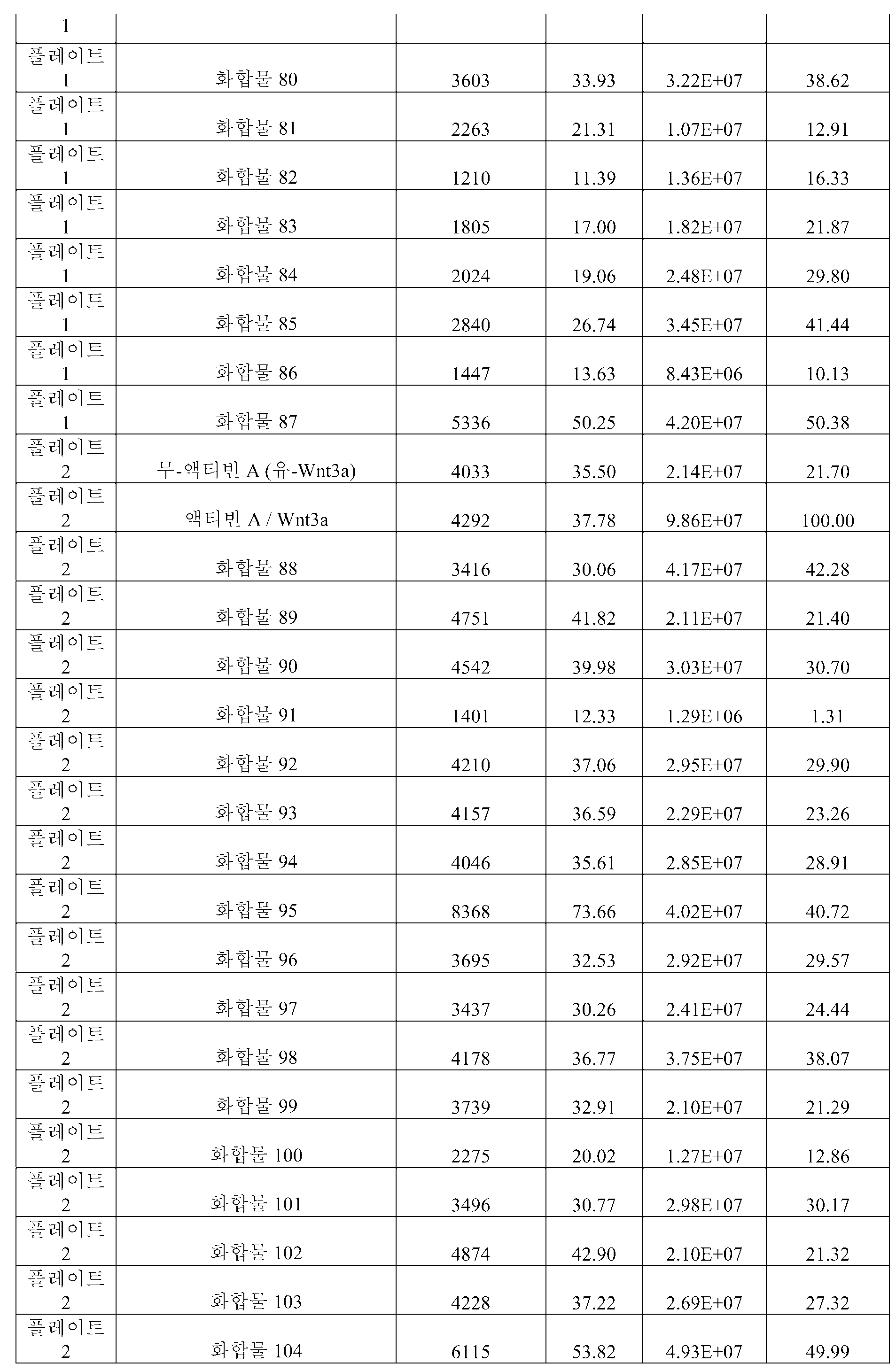


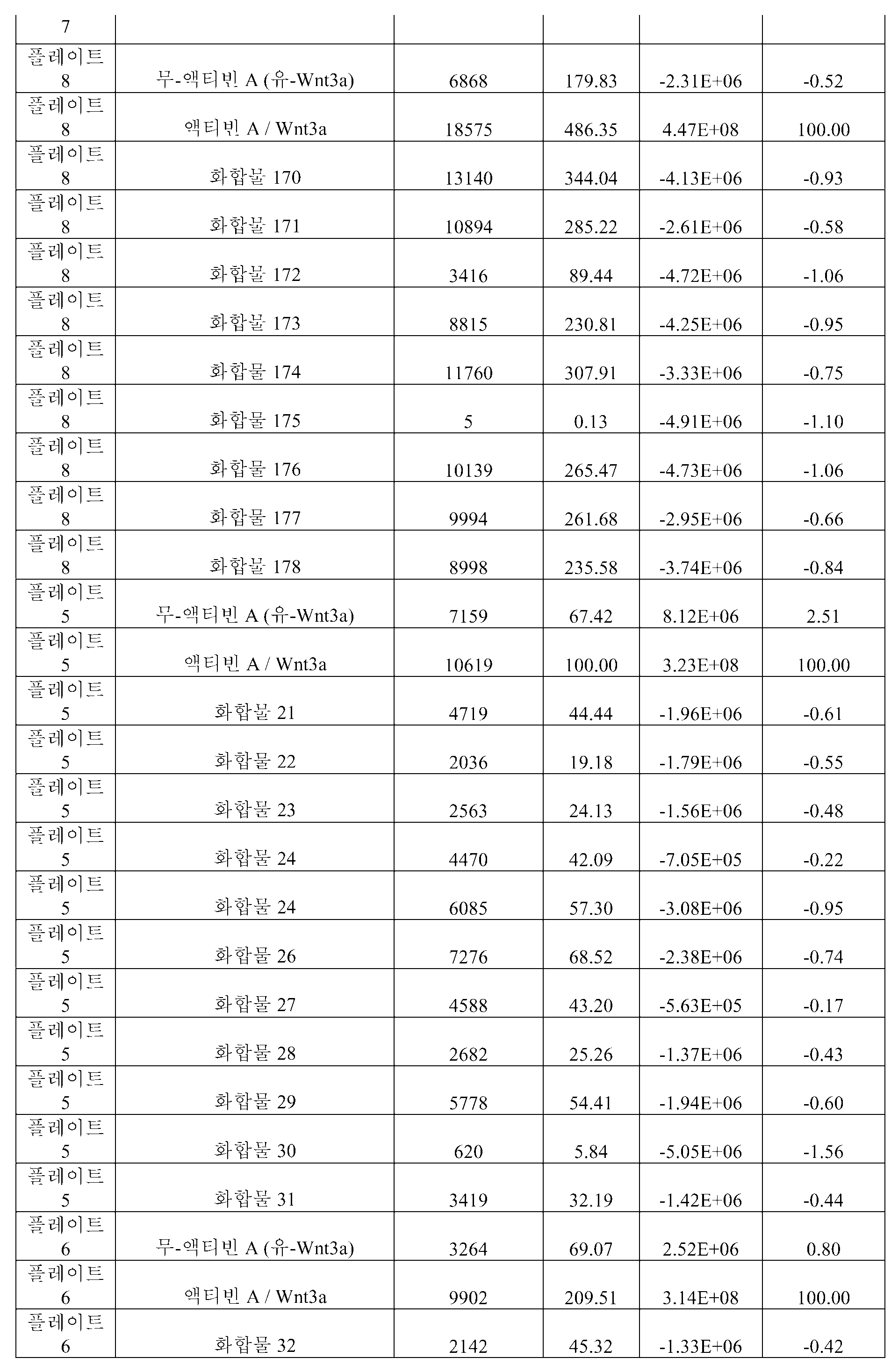
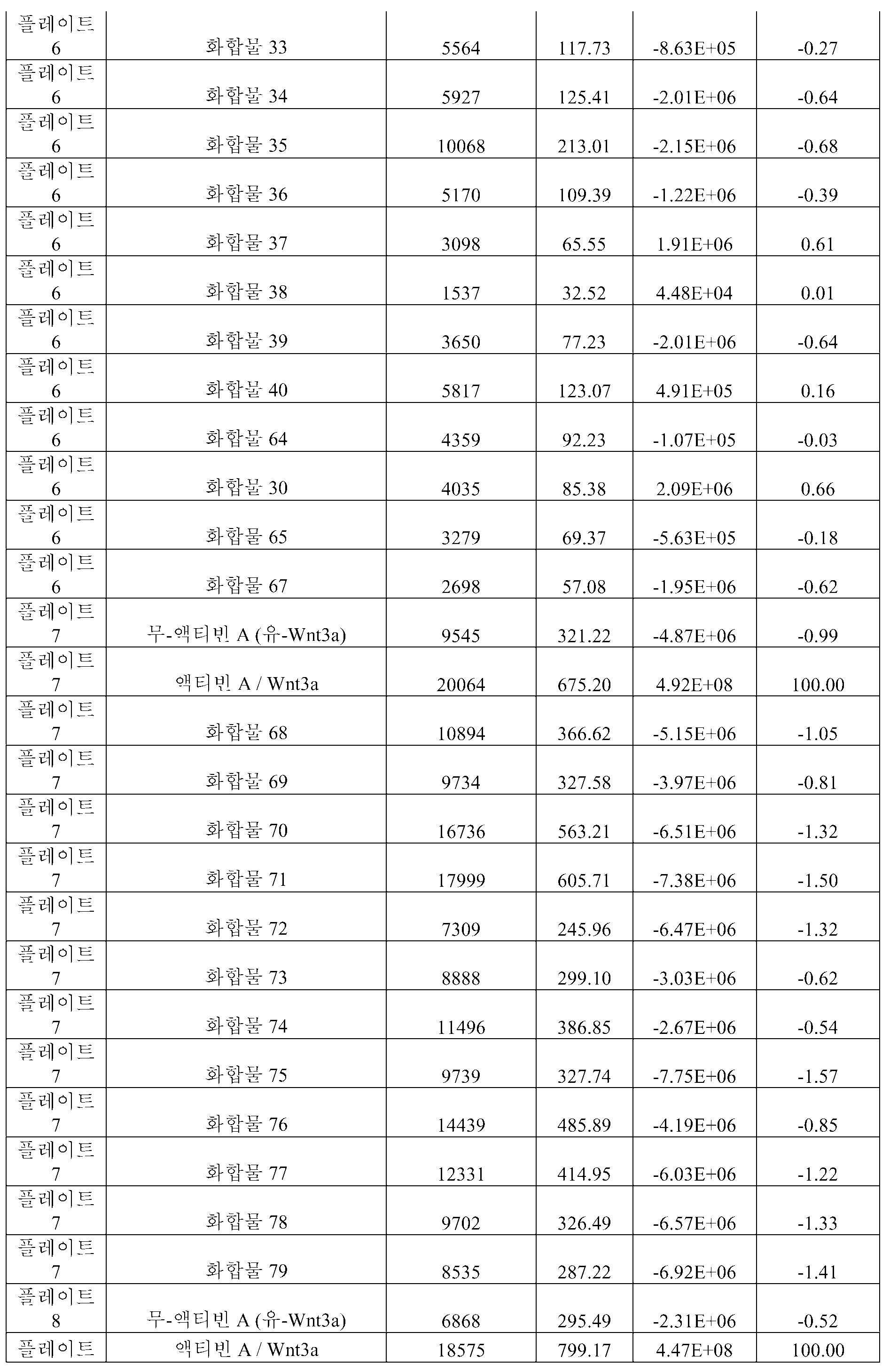
[표 2]TABLE 2
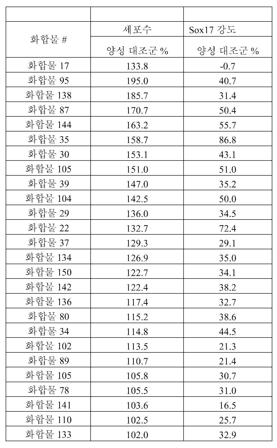
[표 3][Table 3]

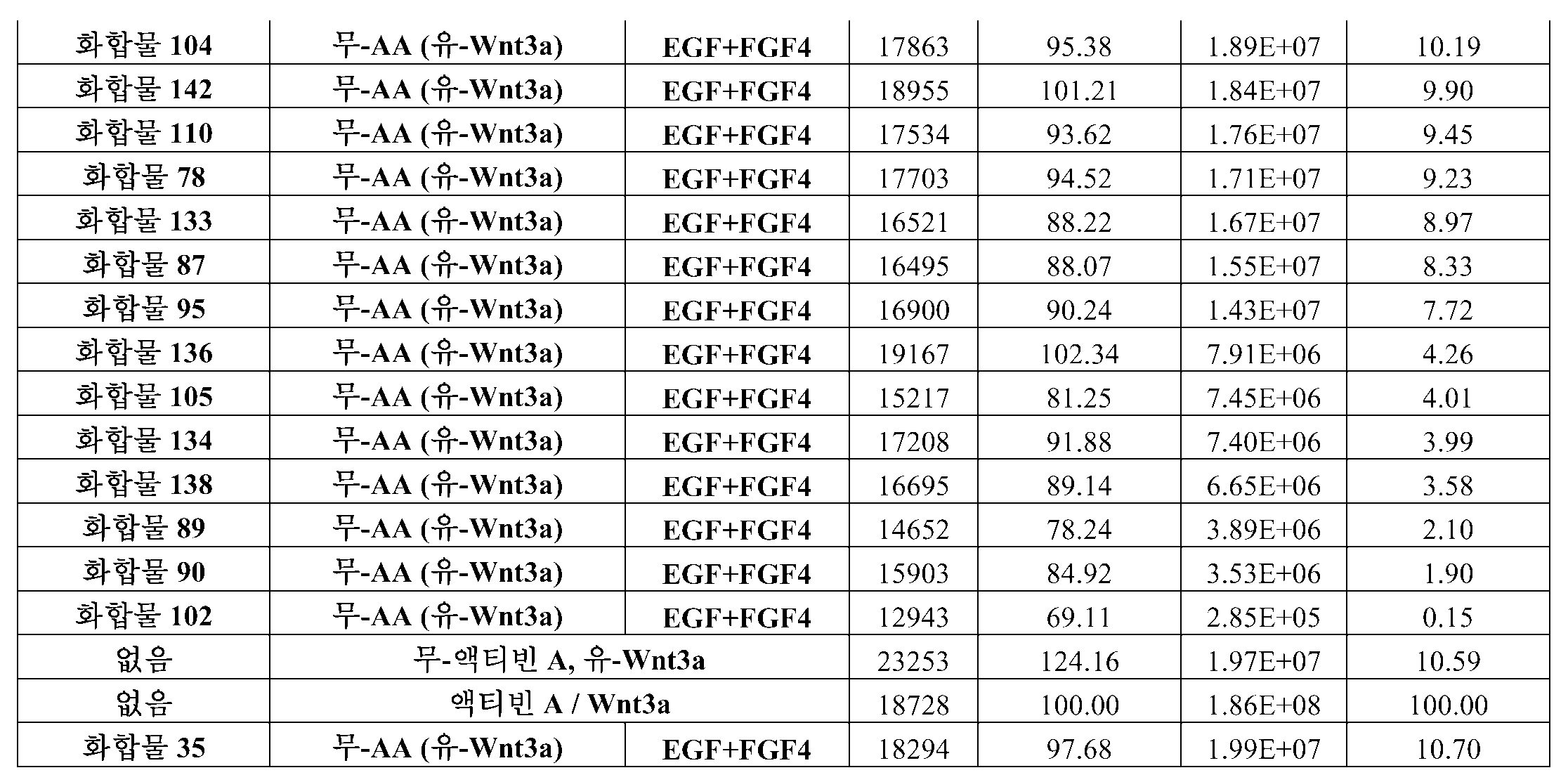
[표 3b]TABLE 3b

[표 4][Table 4]
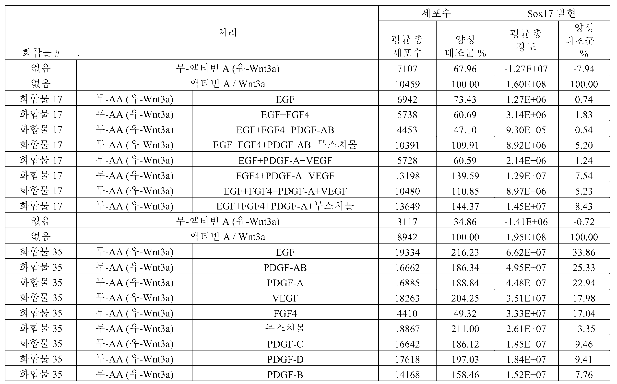

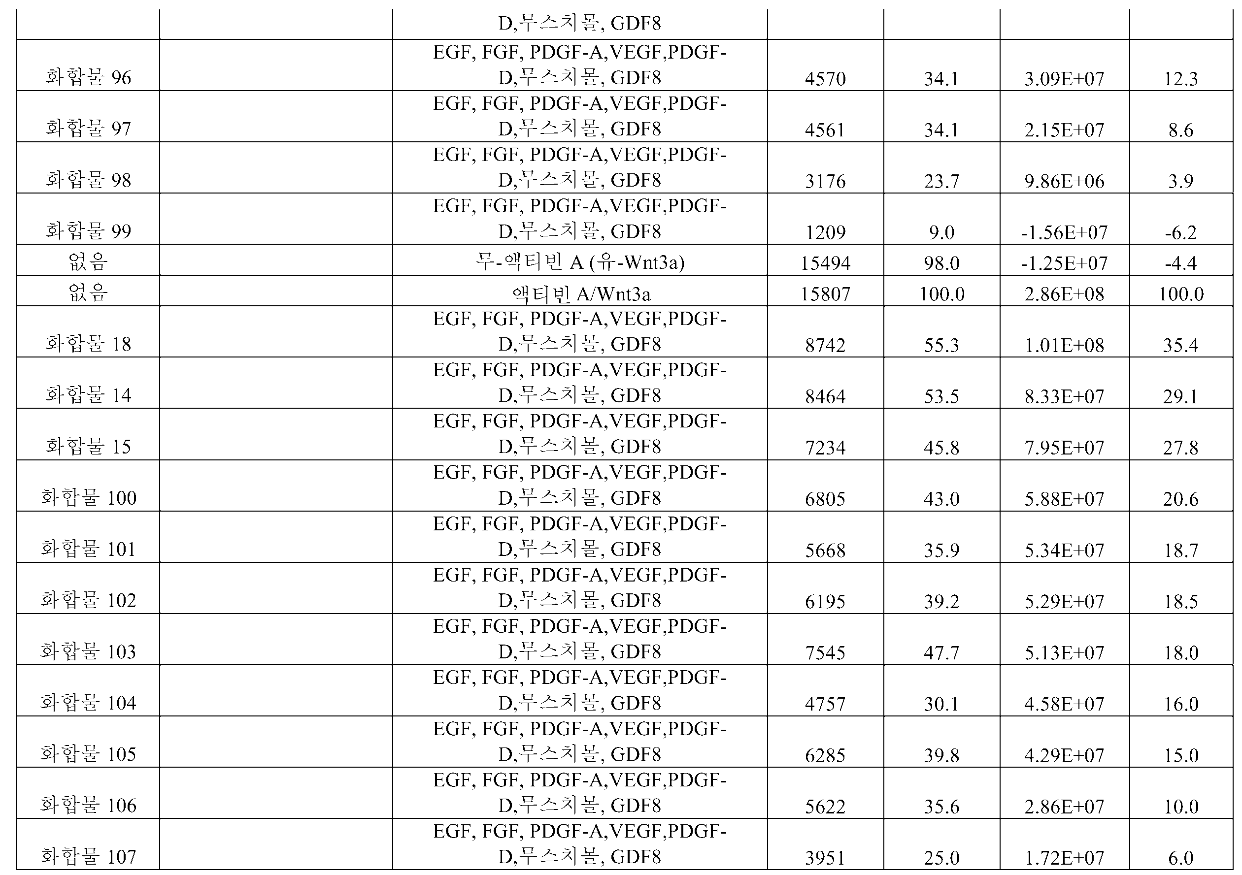

[표 5]TABLE 5
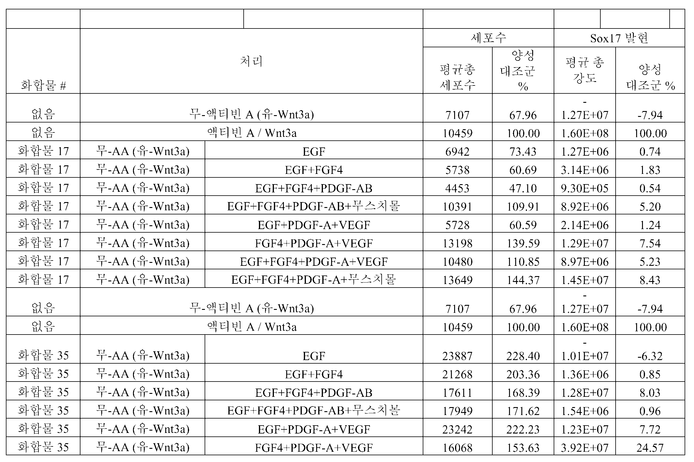


[표 6]TABLE 6

[표 7]TABLE 7


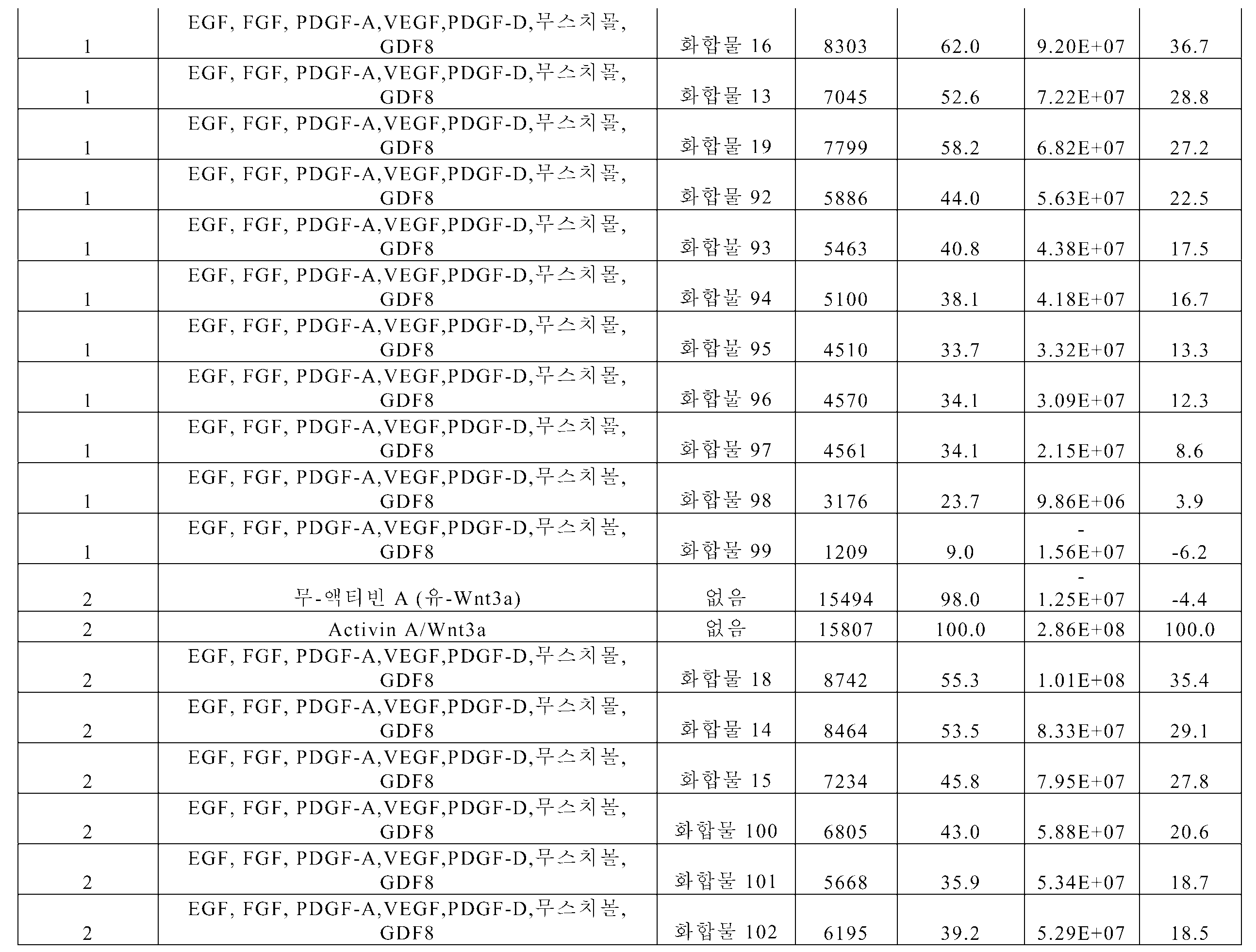


[표 8][Table 8]
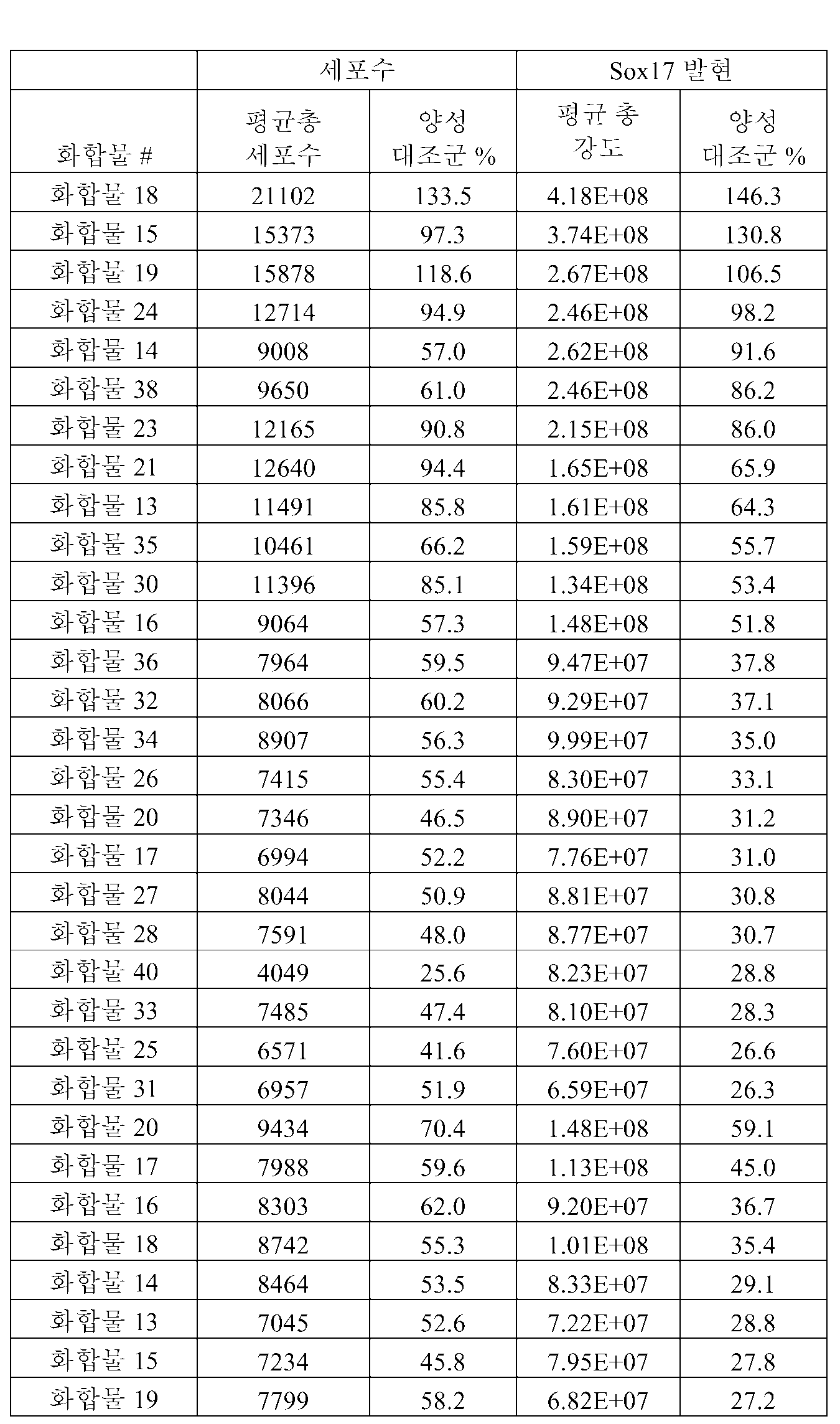
[표 9]TABLE 9

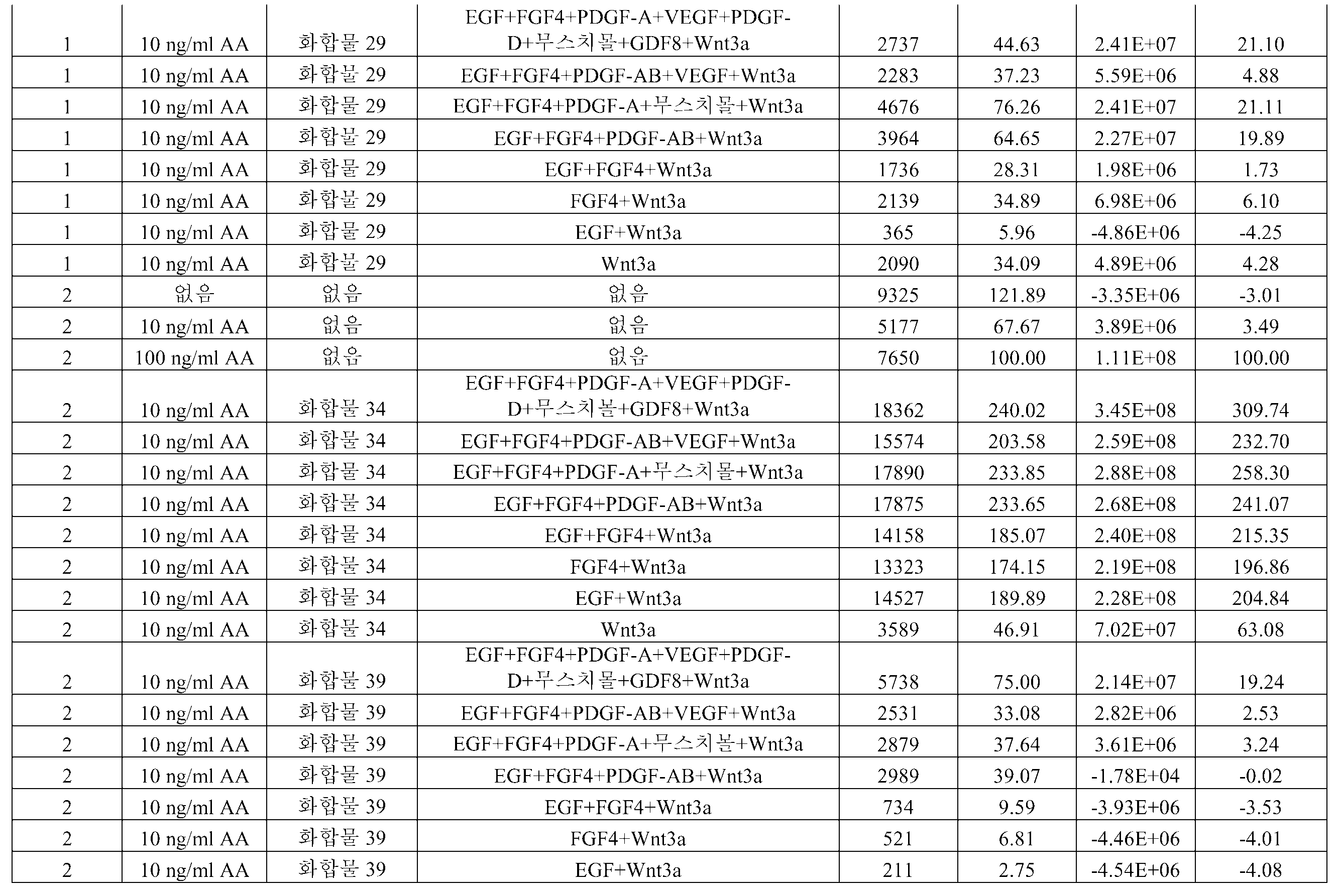

[표 10]TABLE 10

[표 11]TABLE 11
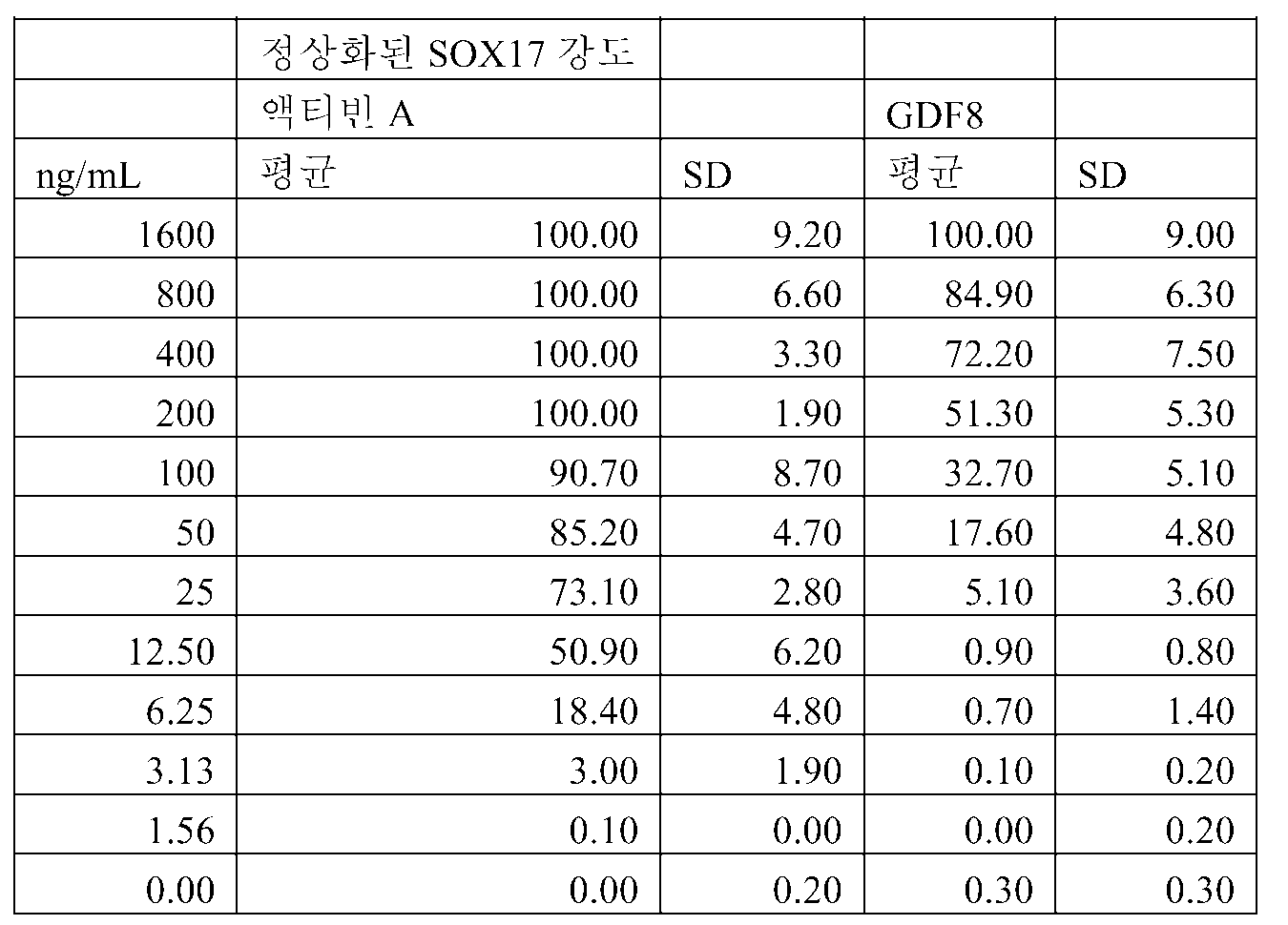
[표 12]TABLE 12


[표 13]TABLE 13

[표 14][Table 14]

[표 15]TABLE 15


[표 16]TABLE 16

[표 17]TABLE 17

[표 18]TABLE 18


[표 19]TABLE 19

Claims (2)
Applications Claiming Priority (6)
| Application Number | Priority Date | Filing Date | Title |
|---|---|---|---|
| US7691508P | 2008-06-30 | 2008-06-30 | |
| US7690008P | 2008-06-30 | 2008-06-30 | |
| US7690808P | 2008-06-30 | 2008-06-30 | |
| US61/076,908 | 2008-06-30 | ||
| US61/076,900 | 2008-06-30 | ||
| US61/076,915 | 2008-06-30 |
Related Child Applications (1)
| Application Number | Title | Priority Date | Filing Date |
|---|---|---|---|
| KR1020167022553A Division KR101734501B1 (en) | 2008-06-30 | 2009-06-30 | Differentiation of pluripotent stem cells |
Publications (2)
| Publication Number | Publication Date |
|---|---|
| KR20110029155A true KR20110029155A (en) | 2011-03-22 |
| KR101651661B1 KR101651661B1 (en) | 2016-08-26 |
Family
ID=41100882
Family Applications (4)
| Application Number | Title | Priority Date | Filing Date |
|---|---|---|---|
| KR1020167022553A KR101734501B1 (en) | 2008-06-30 | 2009-06-30 | Differentiation of pluripotent stem cells |
| KR1020187003886A KR20180018839A (en) | 2008-06-30 | 2009-06-30 | Differentiation of pluripotent stem cells |
| KR1020177012167A KR101829310B1 (en) | 2008-06-30 | 2009-06-30 | Differentiation of pluripotent stem cells |
| KR1020117001981A KR101651661B1 (en) | 2008-06-30 | 2009-06-30 | Differentiation of pluripotent stem cells |
Family Applications Before (3)
| Application Number | Title | Priority Date | Filing Date |
|---|---|---|---|
| KR1020167022553A KR101734501B1 (en) | 2008-06-30 | 2009-06-30 | Differentiation of pluripotent stem cells |
| KR1020187003886A KR20180018839A (en) | 2008-06-30 | 2009-06-30 | Differentiation of pluripotent stem cells |
| KR1020177012167A KR101829310B1 (en) | 2008-06-30 | 2009-06-30 | Differentiation of pluripotent stem cells |
Country Status (15)
| Country | Link |
|---|---|
| US (6) | US20100015711A1 (en) |
| EP (2) | EP2942392B1 (en) |
| JP (1) | JP5734183B2 (en) |
| KR (4) | KR101734501B1 (en) |
| CN (1) | CN102159703B (en) |
| AU (1) | AU2009267137A1 (en) |
| BR (1) | BRPI0914116A2 (en) |
| CA (1) | CA2729121C (en) |
| ES (2) | ES2552240T3 (en) |
| HK (2) | HK1156665A1 (en) |
| MX (1) | MX2011000125A (en) |
| PL (2) | PL2942392T3 (en) |
| RU (2) | RU2533805C2 (en) |
| WO (1) | WO2010002846A1 (en) |
| ZA (1) | ZA201100758B (en) |
Cited By (3)
| Publication number | Priority date | Publication date | Assignee | Title |
|---|---|---|---|---|
| KR20150103203A (en) * | 2012-12-31 | 2015-09-09 | 얀센 바이오테크 인코포레이티드 | Suspension and clustering of human pluripotent cells for differentiation into pancreatic endocrine cells |
| KR20160079071A (en) * | 2013-11-01 | 2016-07-05 | 얀센 바이오테크 인코포레이티드 | Suspension and clustering of human pluripotent stem cells for differentiation into pancreatic endocrine cells |
| KR20160118878A (en) * | 2015-04-03 | 2016-10-12 | 연세대학교 산학협력단 | Composition for Promoting Hair Growth Comprising PDGF-D Treated Adipose-Derived Stem Cells |
Families Citing this family (64)
| Publication number | Priority date | Publication date | Assignee | Title |
|---|---|---|---|---|
| US8017395B2 (en) | 2004-12-17 | 2011-09-13 | Lifescan, Inc. | Seeding cells on porous supports |
| CN101484575B (en) | 2005-06-08 | 2013-10-02 | 森托科尔公司 | Cellular therapy for ocular degeneration |
| US8741643B2 (en) | 2006-04-28 | 2014-06-03 | Lifescan, Inc. | Differentiation of pluripotent stem cells to definitive endoderm lineage |
| US9080145B2 (en) | 2007-07-01 | 2015-07-14 | Lifescan Corporation | Single pluripotent stem cell culture |
| CN101952415B (en) | 2007-07-31 | 2017-06-27 | 生命扫描有限公司 | The differentiation of human embryo stem cell |
| CN107574142B (en) | 2007-11-27 | 2021-07-06 | 生命扫描有限公司 | Differentiation of human embryonic stem cells |
| WO2009105570A2 (en) | 2008-02-21 | 2009-08-27 | Centocor Ortho Biotech Inc. | Methods, surface modified plates and compositions for cell attachment, cultivation and detachment |
| US8623648B2 (en) | 2008-04-24 | 2014-01-07 | Janssen Biotech, Inc. | Treatment of pluripotent cells |
| BRPI0913925A2 (en) * | 2008-06-30 | 2015-08-04 | Centocor Ortho Biotech Inc | Differentiation of pluripotent stem cells |
| JP5734183B2 (en) | 2008-06-30 | 2015-06-17 | ヤンセン バイオテツク,インコーポレーテツド | Differentiation of pluripotent stem cells |
| BRPI0919885A2 (en) * | 2008-10-31 | 2015-08-11 | Centocor Ortho Biotech Inc | Differentiation of human embryonic stem cells into pancreatic endocrine lineage |
| CA2742267C (en) | 2008-10-31 | 2019-06-04 | Centocor Ortho Biotech Inc. | Differentiation of human embryonic stem cells to the pancreatic endocrine lineage |
| RU2555538C2 (en) | 2008-11-20 | 2015-07-10 | Сентокор Орто Байотек Инк. | Culture of pluripotent stem cells on microcarriers |
| AU2009316583B2 (en) | 2008-11-20 | 2016-04-21 | Janssen Biotech, Inc. | Methods and compositions for cell attachment and cultivation on planar substrates |
| EP2412800A1 (en) * | 2010-07-29 | 2012-02-01 | Koninklijke Nederlandse Akademie van Wetenschappen | Liver organoid, uses thereof and culture method for obtaining them |
| EP2408903B1 (en) | 2009-03-20 | 2014-08-06 | Agency For Science, Technology And Research | Culture of pluripotent and multipotent cells on microcarriers |
| EP2456862A4 (en) * | 2009-07-20 | 2013-02-27 | Janssen Biotech Inc | Differentiation of human embryonic stem cells |
| GB2485113B (en) | 2009-07-20 | 2016-12-28 | Janssen Biotech Inc | Differentiation of human embryonic stem cells into cells of the pancreatic endoderm lineage |
| RU2540021C2 (en) | 2009-07-20 | 2015-01-27 | Янссен Байотек, Инк. | Differentiating human embryonic stem cells |
| RU2586506C2 (en) * | 2009-12-23 | 2016-06-10 | Янссен Байотек, Инк. | Differentiation of human embryonic stem cells |
| CN102741395B (en) | 2009-12-23 | 2016-03-16 | 詹森生物科技公司 | The differentiation of human embryo stem cell |
| AU2011223900A1 (en) | 2010-03-01 | 2012-09-13 | Janssen Biotech, Inc. | Methods for purifying cells derived from pluripotent stem cells |
| EP2563908B1 (en) * | 2010-04-25 | 2019-01-09 | Icahn School of Medicine at Mount Sinai | Generation of anterior foregut endoderm from pluripotent cells |
| MX351515B (en) | 2010-05-12 | 2017-10-17 | Janssen Biotech Inc | Differentiation of human embryonic stem cells. |
| US9528090B2 (en) * | 2010-08-31 | 2016-12-27 | Janssen Biotech, Inc. | Differentiation of human embryonic stem cells |
| CA2809305C (en) | 2010-08-31 | 2019-06-11 | Janssen Biotech, Inc. | Differentiation of pluripotent stem cells |
| AU2011296381B2 (en) * | 2010-08-31 | 2016-03-31 | Janssen Biotech, Inc. | Differentiation of human embryonic stem cells |
| CA2812300A1 (en) | 2010-10-01 | 2012-04-05 | The Government Of The United States Of America, As Represented By The Secretary Of The Department Of Health And Human Services | Manipulation of stem cell function by p53 isoforms |
| US8749484B2 (en) | 2010-10-01 | 2014-06-10 | Z124 | Multi-screen user interface with orientation based control |
| ES2902650T3 (en) | 2011-06-21 | 2022-03-29 | Novo Nordisk As | Efficient induction of definitive endoderm from pluripotent stem cells |
| US20140242038A1 (en) * | 2011-10-11 | 2014-08-28 | The Trustees Of Columbia University In The City Of New York | Method for generating beta cells |
| AU2012355698B2 (en) | 2011-12-22 | 2018-11-29 | Janssen Biotech, Inc. | Differentiation of human embryonic stem cells into single hormonal insulin positive cells |
| KR20140131999A (en) | 2012-03-07 | 2014-11-14 | 얀센 바이오테크 인코포레이티드 | Defined Media for Expansion and Maintenance of Pluripotent Stem Cells |
| EP2829606A4 (en) | 2012-03-22 | 2016-02-24 | Takara Bio Inc | Method for producing viral vector |
| WO2013146480A1 (en) * | 2012-03-29 | 2013-10-03 | タカラバイオ株式会社 | Gene introduction method |
| EP2847319A4 (en) | 2012-05-07 | 2015-12-16 | Janssen Biotech Inc | Differentiation of human embryonic stem cells into pancreatic endoderm |
| CN108103006A (en) | 2012-06-08 | 2018-06-01 | 詹森生物科技公司 | Differentiation of the human embryo stem cell to pancreatic endocrine cell |
| US20150247123A1 (en) | 2012-09-03 | 2015-09-03 | Novo Nordisk A/S | Generation of pancreatic endoderm from Pluripotent Stem cells using small molecules |
| PT2925757T (en) | 2012-11-19 | 2018-01-09 | Novartis Ag | Compounds and compositions for the treatment of parasitic diseases |
| KR102036780B1 (en) | 2012-12-31 | 2019-10-25 | 얀센 바이오테크 인코포레이티드 | Differentiation of human embryonic stem cells into pancreatic endocrine cells using hb9 regulators |
| US10370644B2 (en) | 2012-12-31 | 2019-08-06 | Janssen Biotech, Inc. | Method for making human pluripotent suspension cultures and cells derived therefrom |
| KR102084561B1 (en) | 2012-12-31 | 2020-03-04 | 얀센 바이오테크 인코포레이티드 | Culturing of human embryonic stem cells at the air-liquid interface for differentiation into pancreatic endocrine cells |
| WO2014134213A1 (en) | 2013-02-27 | 2014-09-04 | The Regents Of The University Of California | Generation of thymic epithelial progenitor cells in vitro |
| CN105431148A (en) | 2013-03-07 | 2016-03-23 | 加利菲亚生物公司 | Mixed lineage kinase inhibitors and method of treatments |
| RU2016100219A (en) | 2013-06-11 | 2017-07-17 | Президент Энд Феллоус Оф Гарвард Колледж | SC-β CELLS AND COMPOSITIONS AND METHODS FOR THEIR CREATION |
| GB201317869D0 (en) * | 2013-10-09 | 2013-11-20 | Cambridge Entpr Ltd | In vitro production of foregut stem cells |
| JP6261430B2 (en) * | 2014-04-01 | 2018-01-17 | キヤノン株式会社 | Imaging apparatus and image processing system |
| CA2949056A1 (en) | 2014-05-16 | 2015-11-19 | Janssen Biotech, Inc. | Use of small molecules to enhance mafa expression in pancreatic endocrine cells |
| US10253298B2 (en) | 2014-12-18 | 2019-04-09 | President And Fellows Of Harvard College | Methods for generating stem cell-derived beta cells and methods of use thereof |
| EP3234110B1 (en) | 2014-12-18 | 2024-02-28 | President and Fellows of Harvard College | METHODS FOR GENERATING STEM CELL-DERIVED ß CELLS AND USES THEREOF |
| US10443042B2 (en) | 2014-12-18 | 2019-10-15 | President And Fellows Of Harvard College | Serum-free in vitro directed differentiation protocol for generating stem cell-derived beta cells and uses thereof |
| EP3234109A4 (en) * | 2014-12-19 | 2018-06-27 | Janssen Biotech, Inc. | Suspension culturing of pluripotent stem cells |
| MA45479A (en) | 2016-04-14 | 2019-02-20 | Janssen Biotech Inc | DIFFERENTIATION OF PLURIPOTENT STEM CELLS IN ENDODERMAL CELLS OF MIDDLE INTESTINE |
| MA45502A (en) | 2016-06-21 | 2019-04-24 | Janssen Biotech Inc | GENERATION OF FUNCTIONAL BETA CELLS DERIVED FROM HUMAN PLURIPOTENT STEM CELLS WITH GLUCOSE-DEPENDENT MITOCHONDRIAL RESPIRATION AND TWO-PHASE INSULIN SECRETION RESPONSE |
| US10767164B2 (en) | 2017-03-30 | 2020-09-08 | The Research Foundation For The State University Of New York | Microenvironments for self-assembly of islet organoids from stem cells differentiation |
| IL305391A (en) | 2017-11-15 | 2023-10-01 | Vertex Pharma | Islet cell manufacturing compositions and methods of use |
| EP3801638A1 (en) * | 2018-06-08 | 2021-04-14 | Novartis AG | Cell-based assay for measuring drug product potency |
| AU2019320072A1 (en) | 2018-08-10 | 2021-02-25 | Vertex Pharmaceuticals Incorporated | Stem cell derived islet differentiation |
| EP3975926A1 (en) | 2019-05-31 | 2022-04-06 | W.L. Gore & Associates, Inc. | A biocompatible membrane composite |
| CA3139591C (en) | 2019-05-31 | 2024-01-16 | Timothy M. BRUHN | A biocompatible membrane composite |
| CN114401752B (en) | 2019-05-31 | 2023-04-04 | W.L.戈尔及同仁股份有限公司 | Cell encapsulation device with controlled oxygen diffusion distance |
| CN114173837A (en) | 2019-05-31 | 2022-03-11 | W.L.戈尔及同仁股份有限公司 | Biocompatible film composite |
| US11344859B2 (en) | 2019-11-27 | 2022-05-31 | Smartdyelivery Gmbh | Reactor for the preparation of a formulation |
| EP4103183A4 (en) * | 2020-02-14 | 2024-02-28 | Salk Inst For Biological Studi | Macrocyclic ulk1/2 inhibitors |
Family Cites Families (268)
| Publication number | Priority date | Publication date | Assignee | Title |
|---|---|---|---|---|
| US3209652A (en) | 1961-03-30 | 1965-10-05 | Burgsmueller Karl | Thread whirling method |
| AT326803B (en) | 1968-08-26 | 1975-12-29 | Binder Fa G | MESHWARE AND METHOD OF MANUFACTURING THE SAME |
| US3935067A (en) | 1974-11-22 | 1976-01-27 | Wyo-Ben Products, Inc. | Inorganic support for culture media |
| CA1201400A (en) | 1982-04-16 | 1986-03-04 | Joel L. Williams | Chemically specific surfaces for influencing cell activity during culture |
| US4499802A (en) | 1982-09-29 | 1985-02-19 | Container Graphics Corporation | Rotary cutting die with scrap ejection |
| US4537773A (en) | 1983-12-05 | 1985-08-27 | E. I. Du Pont De Nemours And Company | α-Aminoboronic acid derivatives |
| US4557264A (en) | 1984-04-09 | 1985-12-10 | Ethicon Inc. | Surgical filament from polypropylene blended with polyethylene |
| US5089396A (en) | 1985-10-03 | 1992-02-18 | Genentech, Inc. | Nucleic acid encoding β chain prodomains of inhibin and method for synthesizing polypeptides using such nucleic acid |
| US5215893A (en) | 1985-10-03 | 1993-06-01 | Genentech, Inc. | Nucleic acid encoding the ba chain prodomains of inhibin and method for synthesizing polypeptides using such nucleic acid |
| US4737578A (en) | 1986-02-10 | 1988-04-12 | The Salk Institute For Biological Studies | Human inhibin |
| US5863531A (en) | 1986-04-18 | 1999-01-26 | Advanced Tissue Sciences, Inc. | In vitro preparation of tubular tissue structures by stromal cell culture on a three-dimensional framework |
| US5804178A (en) * | 1986-11-20 | 1998-09-08 | Massachusetts Institute Of Technology | Implantation of cell-matrix structure adjacent mesentery, omentum or peritoneum tissue |
| CA1340581C (en) * | 1986-11-20 | 1999-06-08 | Joseph P. Vacanti | Chimeric neomorphogenesis of organs by controlled cellular implantation using artificial matrices |
| US5567612A (en) | 1986-11-20 | 1996-10-22 | Massachusetts Institute Of Technology | Genitourinary cell-matrix structure for implantation into a human and a method of making |
| NZ229354A (en) | 1988-07-01 | 1990-09-26 | Becton Dickinson Co | Treating polymer surfaces with a gas plasma and then applying a layer of endothelial cells to the surface |
| EP0363125A3 (en) | 1988-10-03 | 1990-08-16 | Hana Biologics Inc. | Proliferated pancreatic endocrine cell product and process |
| SU1767433A1 (en) | 1989-11-27 | 1992-10-07 | Пермский государственный медицинский институт | Method of determining insulin resistance of immunogenesis in patients with type 1 diabetes mellitus |
| US5837539A (en) | 1990-11-16 | 1998-11-17 | Osiris Therapeutics, Inc. | Monoclonal antibodies for human mesenchymal stem cells |
| WO1992019759A1 (en) | 1991-04-25 | 1992-11-12 | Chugai Seiyaku Kabushiki Kaisha | Reconstituted human antibody against human interleukin 6 receptor |
| US5449383A (en) | 1992-03-18 | 1995-09-12 | Chatelier; Ronald C. | Cell growth substrates |
| GB9206861D0 (en) * | 1992-03-28 | 1992-05-13 | Univ Manchester | Wound healing and treatment of fibrotic disorders |
| CA2114282A1 (en) | 1993-01-28 | 1994-07-29 | Lothar Schilder | Multi-layered implant |
| JP3525221B2 (en) | 1993-02-17 | 2004-05-10 | 味の素株式会社 | Immunosuppressants |
| JPH08510997A (en) * | 1993-03-31 | 1996-11-19 | プロ−ニューロン,インコーポレイテッド | Stem cell growth inhibitors and uses thereof |
| AU687386B2 (en) | 1993-04-08 | 1998-02-26 | Human Cell Cultures, Inc. | Cell culturing method and medium |
| US5523226A (en) | 1993-05-14 | 1996-06-04 | Biotechnology Research And Development Corp. | Transgenic swine compositions and methods |
| GB9310557D0 (en) | 1993-05-21 | 1993-07-07 | Smithkline Beecham Plc | Novel process and apparatus |
| TW257671B (en) | 1993-11-19 | 1995-09-21 | Ciba Geigy | |
| US6001647A (en) | 1994-04-28 | 1999-12-14 | Ixion Biotechnology, Inc. | In vitro growth of functional islets of Langerhans and in vivo uses thereof |
| US6703017B1 (en) | 1994-04-28 | 2004-03-09 | Ixion Biotechnology, Inc. | Reversal of insulin-dependent diabetes by islet-producing stem cells, islet progenitor cells and islet-like structures |
| US5834308A (en) * | 1994-04-28 | 1998-11-10 | University Of Florida Research Foundation, Inc. | In vitro growth of functional islets of Langerhans |
| US6083903A (en) | 1994-10-28 | 2000-07-04 | Leukosite, Inc. | Boronic ester and acid compounds, synthesis and uses |
| DE69525971T3 (en) | 1994-12-29 | 2013-01-10 | Chugai Seiyaku K.K. | USE OF A PM-1 ANTIBODY OR AN MH 166 ANTIBODY FOR REINFORCING THE ANTI-TUMOR EFFECT OF CISPLATINE OR CARBOPLATIN |
| US5843780A (en) * | 1995-01-20 | 1998-12-01 | Wisconsin Alumni Research Foundation | Primate embryonic stem cells |
| US5718922A (en) | 1995-05-31 | 1998-02-17 | Schepens Eye Research Institute, Inc. | Intravitreal microsphere drug delivery and method of preparation |
| US5908782A (en) | 1995-06-05 | 1999-06-01 | Osiris Therapeutics, Inc. | Chemically defined medium for human mesenchymal stem cells |
| US5681561A (en) | 1995-06-07 | 1997-10-28 | Life Medical Sciences, Inc. | Compositions and methods for improving autologous fat grafting |
| JP2001508302A (en) | 1997-01-10 | 2001-06-26 | ライフ テクノロジーズ,インコーポレイテッド | Embryonic stem cell serum replacement |
| UA65572C2 (en) | 1997-04-24 | 2004-04-15 | Ortho Mcneil Pharm Inc | Substituted imidazoles, intermediate compounds for the preparation thereof, a method for the preparation of substituted imidazoles and a method for the treatment of inflammatory diseases |
| DE69837491T2 (en) | 1997-07-03 | 2008-01-17 | Osiris Therapeutics, Inc. | HUMAN MESENCHYMAL STEM CELLS OF PERIPHERAL BLOOD |
| ATE462004T1 (en) | 1997-09-16 | 2010-04-15 | Centocor Inc | METHODS FOR COMPLETE CHEMICAL SYNTHESIS AND ASSEMBLY OF GENES AND GENOMES |
| US6670127B2 (en) | 1997-09-16 | 2003-12-30 | Egea Biosciences, Inc. | Method for assembly of a polynucleotide encoding a target polypeptide |
| AU729377B2 (en) * | 1997-10-23 | 2001-02-01 | Asterias Biotherapeutics, Inc. | Methods and materials for the growth of primate-derived primordial stem cells in feeder-free culture |
| ZA9811898B (en) * | 1997-12-29 | 2000-06-28 | Ortho Mcneil Pharm Inc | Anti-Inflammatory Compounds. |
| AU755888B2 (en) | 1998-03-18 | 2003-01-02 | Mesoblast International Sarl | Mesenchymal stem cells for prevention and treatment of immune responses in transplantation |
| MY132496A (en) * | 1998-05-11 | 2007-10-31 | Vertex Pharma | Inhibitors of p38 |
| US6413773B1 (en) | 1998-06-01 | 2002-07-02 | The Regents Of The University Of California | Phosphatidylinositol 3-kinase inhibitors as stimulators of endocrine differentiation |
| US7410798B2 (en) | 2001-01-10 | 2008-08-12 | Geron Corporation | Culture system for rapid expansion of human embryonic stem cells |
| US6667176B1 (en) * | 2000-01-11 | 2003-12-23 | Geron Corporation | cDNA libraries reflecting gene expression during growth and differentiation of human pluripotent stem cells |
| AU758073B2 (en) * | 1998-11-09 | 2003-03-13 | Consorzio Per La Gestione Del Centro Di Biotecnologie Avanzate | Serum free medium for chondrocyte-like cells |
| US6610540B1 (en) | 1998-11-18 | 2003-08-26 | California Institute Of Technology | Low oxygen culturing of central nervous system progenitor cells |
| US6413556B1 (en) | 1999-01-08 | 2002-07-02 | Sky High, Llc | Aqueous anti-apoptotic compositions |
| US6458593B1 (en) * | 1999-01-21 | 2002-10-01 | Vitro Diagnostics, Inc. | Immortalized cell lines and methods of making the same |
| US6815203B1 (en) | 1999-06-23 | 2004-11-09 | Joslin Diabetes Center, Inc. | Methods of making pancreatic islet cells |
| US6306424B1 (en) * | 1999-06-30 | 2001-10-23 | Ethicon, Inc. | Foam composite for the repair or regeneration of tissue |
| US6333029B1 (en) | 1999-06-30 | 2001-12-25 | Ethicon, Inc. | Porous tissue scaffoldings for the repair of regeneration of tissue |
| AU7719300A (en) | 1999-09-27 | 2001-04-30 | Ixion Biotechnology, Inc. | Reversal of insulin-dependent diabetes by islet-producing stem cells, islet progenitor cells and islet-like structures |
| US6685936B2 (en) | 1999-10-12 | 2004-02-03 | Osiris Therapeutics, Inc. | Suppressor cells induced by culture with mesenchymal stem cells for treatment of immune responses in transplantation |
| US20030082155A1 (en) | 1999-12-06 | 2003-05-01 | Habener Joel F. | Stem cells of the islets of langerhans and their use in treating diabetes mellitus |
| EP1240518A4 (en) * | 1999-12-13 | 2006-05-17 | Scripps Research Inst | Markers for identification and isolation of pancreatic islet alpha and beta cell progenitors |
| US7439064B2 (en) * | 2000-03-09 | 2008-10-21 | Wicell Research Institute, Inc. | Cultivation of human embryonic stem cells in the absence of feeder cells or without conditioned medium |
| US7005252B1 (en) | 2000-03-09 | 2006-02-28 | Wisconsin Alumni Research Foundation | Serum free cultivation of primate embryonic stem cells |
| US6436704B1 (en) | 2000-04-10 | 2002-08-20 | Raven Biotechnologies, Inc. | Human pancreatic epithelial progenitor cells and methods of isolation and use thereof |
| US6458589B1 (en) * | 2000-04-27 | 2002-10-01 | Geron Corporation | Hepatocyte lineage cells derived from pluripotent stem cells |
| EP1302534A4 (en) | 2000-06-26 | 2004-06-16 | Renomedix Inst Inc | Cell fraction containing cells capable of differentiating into neural cells |
| CZ20031125A3 (en) | 2000-10-23 | 2003-10-15 | Smithkline Beecham Corporation | Novel compounds |
| ATE301661T1 (en) | 2000-12-08 | 2005-08-15 | Ortho Mcneil Pharm Inc | MACROHETEROCYCLIC COMPOUNDS AS KINASE INHIBITORS |
| ATE326464T1 (en) | 2000-12-08 | 2006-06-15 | Ortho Mcneil Pharm Inc | INDAZOLYL-SUBSTITUTED PYRROLIDINE COMPOUNDS AS KINASE INHIBITORS |
| US6599323B2 (en) * | 2000-12-21 | 2003-07-29 | Ethicon, Inc. | Reinforced tissue implants and methods of manufacture and use |
| EP1366148A2 (en) | 2001-01-24 | 2003-12-03 | THE GOVERNMENT OF THE UNITED STATES OF AMERICA, represented by THE DEPARTMENT OF HEALTH & HUMAN SERVICES | Differentiation of stem cells to pancreatic endocrine cells |
| AU2002243646B2 (en) | 2001-01-25 | 2006-06-22 | The Government Of The United States Of America, As Represented By The Secretary, Department Of Health And Human Services | Formulation of boronic acid compounds |
| DE10117563B4 (en) | 2001-04-07 | 2006-04-13 | Interessengemeinschaft für Rundfunkschutzrechte GmbH Schutzrechtsverwertung & Co. KG. | Method for amplifying an amplitude and / or phase modulated high frequency signal according to the principle of Envelope Elimination and Restoration and circuitry |
| US6656488B2 (en) | 2001-04-11 | 2003-12-02 | Ethicon Endo-Surgery, Inc. | Bioabsorbable bag containing bioabsorbable materials of different bioabsorption rates for tissue engineering |
| JP2004527249A (en) | 2001-04-19 | 2004-09-09 | デヴェロゲン アクチエンゲゼルシャフト フュア エントヴィックルングスビオローギッシェ フォルシュング | Method of differentiating stem cells into insulin producing cells |
| JP4296781B2 (en) | 2001-04-24 | 2009-07-15 | 味の素株式会社 | Stem cells and methods for separating them |
| WO2002092756A2 (en) | 2001-05-15 | 2002-11-21 | Rappaport Family Institute For Research In The Medical Sciences | Insulin producing cells derived from human embryonic stem cells |
| US6626950B2 (en) * | 2001-06-28 | 2003-09-30 | Ethicon, Inc. | Composite scaffold with post anchor for the repair and regeneration of tissue |
| KR100418195B1 (en) | 2001-07-05 | 2004-02-11 | 주식회사 우리기술 | Apparatus and method for multi-testing insulation of power cables |
| GB0117583D0 (en) | 2001-07-19 | 2001-09-12 | Astrazeneca Ab | Novel compounds |
| CA2456981C (en) * | 2001-08-06 | 2012-02-28 | Bresagen, Inc. | Alternative compositions and methods for the culture of stem cells |
| US6617152B2 (en) | 2001-09-04 | 2003-09-09 | Corning Inc | Method for creating a cell growth surface on a polymeric substrate |
| EP1298201A1 (en) | 2001-09-27 | 2003-04-02 | Cardion AG | Process for the production of cells exhibiting an islet-beta-cell-like state |
| WO2003033697A1 (en) | 2001-10-18 | 2003-04-24 | Ixion Biotechnology, Inc. | Conversion of liver stem and progenitor cells to pancreatic functional cells |
| EP1442115B9 (en) | 2001-11-15 | 2009-12-16 | Children's Medical Center Corporation | Methods of isolation, expansion and differentiation of fetal stem cells from chorionic villus, amniotic fluid, and placenta and therapeutic uses thereof |
| KR101089591B1 (en) * | 2001-12-07 | 2011-12-05 | 제론 코포레이션 | Islet cells from human embryonic stem cells |
| EP1572071B1 (en) | 2001-12-07 | 2018-10-03 | Cytori Therapeutics, Inc. | Methods for preparing fresh adipose tissue-derived cells and uses in treating patients |
| AU2002218893A1 (en) | 2001-12-21 | 2003-07-09 | Thromb-X Nv | Compositions for the in vitro derivation and culture of embryonic stem (es) cell lines with germline transmission capability |
| US20050095703A1 (en) | 2001-12-28 | 2005-05-05 | Henrik Semb | Method for the establishment of a pluripotent human blastocyst - derived stem cell line |
| US20030162290A1 (en) | 2002-01-25 | 2003-08-28 | Kazutomo Inoue | Method for inducing differentiation of embryonic stem cells into functioning cells |
| US20030180268A1 (en) | 2002-02-05 | 2003-09-25 | Anthony Atala | Tissue engineered construct for supplementing or replacing a damaged organ |
| US20050208029A1 (en) | 2002-04-17 | 2005-09-22 | Akihiro Umezawa | Method of forming pancreatic beta cells from mesenchymal cells |
| US20040161419A1 (en) | 2002-04-19 | 2004-08-19 | Strom Stephen C. | Placental stem cells and uses thereof |
| GB0210539D0 (en) | 2002-05-08 | 2002-06-19 | Univ Edinburgh | Control of es cell self renewal and lineage specification, and medium therefor |
| ATE387444T1 (en) | 2002-05-08 | 2008-03-15 | Janssen Pharmaceutica Nv | SUBSTITUTED PYRROLINES AS KINASE INHIBITORS |
| US20060003446A1 (en) * | 2002-05-17 | 2006-01-05 | Gordon Keller | Mesoderm and definitive endoderm cell populations |
| JP2005527241A (en) | 2002-05-28 | 2005-09-15 | ベクトン・ディキンソン・アンド・カンパニー | Methods for proliferation of human pancreatic acinar cells and transdifferentiation into insulin producing cells in vitro |
| WO2003104222A1 (en) | 2002-06-05 | 2003-12-18 | Janssen Pharmaceutica N.V. | Bisindolyl-maleimid derivatives as kinase inhibitors |
| GB0212976D0 (en) | 2002-06-06 | 2002-07-17 | Tonejet Corp Pty Ltd | Ejection method and apparatus |
| CN1171991C (en) | 2002-07-08 | 2004-10-20 | 徐如祥 | Culture process of human nerve stem cell |
| US6877147B2 (en) * | 2002-07-22 | 2005-04-05 | Broadcom Corporation | Technique to assess timing delay by use of layout quality analyzer comparison |
| US7838290B2 (en) | 2002-07-25 | 2010-11-23 | The Scripps Research Institute | Hematopoietic stem cells and methods of treatment of neovascular eye diseases therewith |
| US20040110287A1 (en) | 2002-07-29 | 2004-06-10 | Es Cell International Pte Ltd. | Multi-step method for the differentiation of insulin positive, glucose responsive cells |
| US20040063204A1 (en) | 2002-08-14 | 2004-04-01 | Lijun Yang | Bone marrow cell differentiation |
| AU2003268534A1 (en) | 2002-09-06 | 2004-03-29 | Amcyte Inc. | Cd56 positive human adult pancreatic endocrine progenitor cells |
| US9969977B2 (en) | 2002-09-20 | 2018-05-15 | Garnet Biotherapeutics | Cell populations which co-express CD49c and CD90 |
| US20040062753A1 (en) | 2002-09-27 | 2004-04-01 | Alireza Rezania | Composite scaffolds seeded with mammalian cells |
| AU2003285172A1 (en) | 2002-11-08 | 2004-06-03 | The Johns Hopkins University | Human embryonic stem cell cultures, and compositions and methods for growing same |
| US7144999B2 (en) | 2002-11-23 | 2006-12-05 | Isis Pharmaceuticals, Inc. | Modulation of hypoxia-inducible factor 1 alpha expression |
| AU2003302702B2 (en) | 2002-12-05 | 2008-08-07 | Technion Research & Development Foundation Ltd. | Cultured human pancreatic islets, and uses thereof |
| ES2571355T3 (en) | 2002-12-16 | 2016-05-24 | Technion Res & Dev Foundation | Culture system without feeder cells or xenocontaminants for human embryonic stem cells |
| US20050118148A1 (en) | 2002-12-20 | 2005-06-02 | Roland Stein | Compositions and methods related to mammalian Maf-A |
| AU2004208606B2 (en) | 2003-01-29 | 2009-09-24 | Takeda Pharmaceutical Company Limited | Process for producing coated preparation |
| RU2359671C2 (en) | 2003-01-29 | 2009-06-27 | Такеда Фармасьютикал Компани Лимитед | Method of obtaining of preparation with covering |
| WO2004073633A2 (en) | 2003-02-14 | 2004-09-02 | The Board Of Trustees Of The Leland Stanford Junior University | Methods and compositions for modulating the development of stem cells |
| US20070154981A1 (en) | 2003-02-14 | 2007-07-05 | The Board Of Trustees Of The Leland Stanford Junior University | Insulin-producing cells derived from stem cells |
| EP1613726A4 (en) * | 2003-03-12 | 2007-08-22 | Reliance Life Sciences Pvt Ltd | Derivation of terminally differentiated dopaminergic neurons from human embryonic stem cells |
| CA2520861A1 (en) | 2003-03-27 | 2004-10-14 | Ixion Biotechnology, Inc. | Method for transdifferentiation of non-pancreatic stem cells to the pancreatic pathway |
| WO2004090110A2 (en) | 2003-03-31 | 2004-10-21 | Bresagen Inc. | Compositions and methods for the control, differentiation and/or manipulation of pluripotent cells through a gamma-secretase signaling pathway |
| US20090203141A1 (en) | 2003-05-15 | 2009-08-13 | Shi-Lung Lin | Generation of tumor-free embryonic stem-like pluripotent cells using inducible recombinant RNA agents |
| WO2005001079A2 (en) | 2003-06-27 | 2005-01-06 | Ethicon, Incorporated | Soft tissue repair and regeneration using postpartum-derived cells |
| IL161903A0 (en) | 2003-07-17 | 2005-11-20 | Gamida Cell Ltd | Ex vivo progenitor and stem cell expansion for usein the treatment of disease of endodermally- deri ved organs |
| ITRM20030395A1 (en) | 2003-08-12 | 2005-02-13 | Istituto Naz Per Le Malattie Infettive Lazz | CULTURE GROUND FOR MAINTENANCE, PROLIFERATION AND DIFFERENTIATION OF MAMMALIAN CELLS. |
| US20050042595A1 (en) | 2003-08-14 | 2005-02-24 | Martin Haas | Banking of multipotent amniotic fetal stem cells |
| US7157275B2 (en) | 2003-08-15 | 2007-01-02 | Becton, Dickinson And Company | Peptides for enhanced cell attachment and growth |
| WO2005021728A2 (en) | 2003-08-27 | 2005-03-10 | Stemcells California, Inc. | Enriched pancreatic stem cell and progenitor cell populations, and methods for identifying, isolating and enriching for these populations |
| EP1696899A1 (en) | 2003-12-17 | 2006-09-06 | Allergan, Inc. | Methods for treating retinoid responsive disorders using selective inhibitors of cyp26a and cyp26b |
| US20060030042A1 (en) | 2003-12-19 | 2006-02-09 | Ali Brivanlou | Maintenance of embryonic stem cells by the GSK-3 inhibitor 6-bromoindirubin-3'-oxime |
| EA200601231A1 (en) | 2003-12-23 | 2007-06-29 | Китера, Инк. | DEFINITIVE ENTODERMA |
| CN109628371B (en) | 2003-12-23 | 2021-02-19 | 维亚希特公司 | Definitive endoderm |
| US7541185B2 (en) | 2003-12-23 | 2009-06-02 | Cythera, Inc. | Methods for identifying factors for differentiating definitive endoderm |
| US20050266554A1 (en) * | 2004-04-27 | 2005-12-01 | D Amour Kevin A | PDX1 expressing endoderm |
| US7625753B2 (en) * | 2003-12-23 | 2009-12-01 | Cythera, Inc. | Expansion of definitive endoderm cells |
| TWI334443B (en) | 2003-12-31 | 2010-12-11 | Ind Tech Res Inst | Method of single cell culture of undifferentiated human embryonic stem cells |
| US20050233446A1 (en) * | 2003-12-31 | 2005-10-20 | Parsons Xuejun H | Defined media for stem cell culture |
| US7794704B2 (en) | 2004-01-23 | 2010-09-14 | Advanced Cell Technology, Inc. | Methods for producing enriched populations of human retinal pigment epithelium cells for treatment of retinal degeneration |
| US20080241107A1 (en) | 2004-01-23 | 2008-10-02 | Copland Iii John A | Methods and Compositions For Preparing Pancreatic Insulin Secreting Cells |
| WO2005080551A2 (en) | 2004-02-12 | 2005-09-01 | University Of Newcastle Upon Tyne | Stem cells |
| US7964401B2 (en) | 2004-02-19 | 2011-06-21 | Kyoto University | Screening method for somatic cell nuclear reprogramming substance affecting ECAT2 and ECAT3 |
| WO2005086860A2 (en) | 2004-03-09 | 2005-09-22 | Gang Xu | Methods for generating insulin-producing cells |
| WO2005086845A2 (en) | 2004-03-10 | 2005-09-22 | Regents Of The University Of California | Compositions and methods for growth of embryonic stem cells |
| EP1734112B1 (en) | 2004-03-23 | 2017-08-23 | Toshihiro Akaike | Method of proliferating pluripotent stem cell |
| WO2005097980A2 (en) * | 2004-03-26 | 2005-10-20 | Geron Corporation | New protocols for making hepatocytes from embryonic stem cells |
| EP1730268A2 (en) | 2004-04-01 | 2006-12-13 | Wisconsin Alumni Research Foundation | Differentiation of stem cells to endoderm and pancreatic lineage |
| CN103103158B (en) | 2004-04-27 | 2016-08-03 | 韦尔赛特公司 | Cell culture medium |
| CA2966883A1 (en) | 2004-07-09 | 2006-02-16 | Cythera, Inc. | Methods for identifying factors for differentiating definitive endoderm |
| WO2006020919A2 (en) | 2004-08-13 | 2006-02-23 | University Of Georgia Research Foundation, Inc. | Compositions and methods for self-renewal and differentiation in human embryonic stem cells |
| US20080268533A1 (en) | 2004-08-25 | 2008-10-30 | University Of Georgia Research Foundation, Inc. | Methods and Compositions Utilizing Myc and Gsk3Beta to Manipulate the Pluripotency of Embryonic Stem Cells |
| DE102004043256B4 (en) | 2004-09-07 | 2013-09-19 | Rheinische Friedrich-Wilhelms-Universität Bonn | Scalable process for culturing undifferentiated stem cells in suspension |
| MX2007002390A (en) | 2004-09-08 | 2007-04-23 | Wisconsin Alumni Res Found | Medium and culture of embryonic stem cells. |
| NZ553235A (en) | 2004-09-08 | 2009-11-27 | Wisconsin Alumni Res Found | Culturing human pluripotent stem cells |
| WO2006073911A1 (en) | 2004-12-30 | 2006-07-13 | Stemlifeline, Inc. | Methods and compositions relating to embryonic stem cell lines |
| AU2006208944A1 (en) | 2005-01-28 | 2006-08-03 | Imperial College Innovations Limited | Methods for embryonic stem cell culture |
| WO2006083782A2 (en) | 2005-01-31 | 2006-08-10 | Es Cell International Pte Ltd. | Directed differentiation of embryonic stem cells and uses thereof |
| US20060182724A1 (en) | 2005-02-15 | 2006-08-17 | Riordan Neil H | Method for expansion of stem cells |
| CN101188942B (en) | 2005-03-04 | 2011-11-30 | 生命扫描有限公司 | Adult pancreatic derived stromal cells |
| GB0505970D0 (en) | 2005-03-23 | 2005-04-27 | Univ Edinburgh | Culture medium containing kinase inhibitor, and uses thereof |
| CN100425694C (en) | 2005-04-15 | 2008-10-15 | 北京大学 | Method of inducing embryo stem cell to differentiate toward pancreatic cell |
| ATE553198T1 (en) | 2005-04-15 | 2012-04-15 | Geron Corp | TREATMENT OF CANCER THROUGH THE COMBINED INHIBITION OF PROTEASOME AND TELOMERASE ACTIVITIES |
| EP1874367B1 (en) | 2005-04-26 | 2011-07-06 | Arhus Universitet | Biocompatible material for surgical implants and cell guiding tissue culture surfaces |
| JP5092124B2 (en) | 2005-05-24 | 2012-12-05 | 国立大学法人 熊本大学 | ES cell differentiation induction method |
| AU2006202209B2 (en) | 2005-05-27 | 2011-04-14 | Lifescan, Inc. | Amniotic fluid derived cells |
| AU2006257859B2 (en) | 2005-06-10 | 2009-12-10 | Irm Llc | Compounds that maintain pluripotency of embryonic stem cells |
| WO2006138433A2 (en) | 2005-06-14 | 2006-12-28 | The Regents Of The University Of California | Induction of cell differentiation by class i bhlh polypeptides |
| US20080199959A1 (en) | 2005-06-21 | 2008-08-21 | Ge Healthcare Bio-Sciences Ab | Method For Cell Culture |
| JP5560391B2 (en) | 2005-06-22 | 2014-07-23 | アステリアス バイオセラピューティクス インコーポレイテッド | Suspension culture method of human embryonic stem cells |
| CN101341138B (en) | 2005-06-30 | 2012-11-14 | 詹森药业有限公司 | Cyclic anilino-pyridinotriazines as GSK-3 inhibitors |
| AU2006274438A1 (en) | 2005-07-29 | 2007-02-01 | Australian Stem Cell Centre Limited | Compositions and methods for growth of pluripotent cells |
| WO2007117262A2 (en) * | 2005-07-29 | 2007-10-18 | Athersys, Inc. | Culture of non-embryonic cells at high cell density |
| WO2007016485A2 (en) | 2005-07-29 | 2007-02-08 | Athersys, Inc. | Use of a gsk-3 inhibitor to maintain potency of cultured cells |
| WO2007025234A2 (en) | 2005-08-26 | 2007-03-01 | The Trustees Of Columbia University In The City Of New York | Generation of pancreatic endocrine cells from primary duct cell cultures and methods of use for treatment of diabetes |
| EP3354723B1 (en) | 2005-08-29 | 2023-12-13 | Technion Research & Development Foundation Ltd. | Media for culturing stem cells |
| BRPI0617085A2 (en) | 2005-09-02 | 2016-11-08 | Agency Science Tech & Res | method for obtaining a mesenchymal stem cell (msc), cell or cell line, method for deriving a cell culture from an embryonic stem cell, method for treating a disease, use of a mesenchymal stem cell, differentiated lineage, pharmaceutical composition, method for conditioning a cell culture medium, conditioned medium and use thereof, method for obtaining a polypeptide, method for obtaining a cell culture, cell culture, mesenchymal stem cell, mesenchymal stem cell line or a differentiated mesenchymal cell |
| US9165427B2 (en) | 2005-09-08 | 2015-10-20 | Bally Gaming, Inc. | Wagering game with multiple bonus triggering feature and bonus accrual feature |
| WO2007030870A1 (en) | 2005-09-12 | 2007-03-22 | Es Cell International Pte Ltd | Cardiomyocyte production |
| JP2009511061A (en) | 2005-10-14 | 2009-03-19 | リージェンツ オブ ザ ユニバーシティ オブ ミネソタ | Differentiation of non-embryonic stem cells into cells with pancreatic phenotype |
| US7732202B2 (en) | 2005-10-21 | 2010-06-08 | International Stem Cell Corporation | Oxygen tension for the parthenogenic activation of human oocytes for the production of human embryonic stem cells |
| DK2674485T3 (en) | 2005-10-27 | 2019-08-26 | Viacyte Inc | PDX-1 EXPRESSING DORSAL AND VENTRAL FORTARM ENDODERM |
| EP4223769A3 (en) | 2005-12-13 | 2023-11-01 | Kyoto University | Nuclear reprogramming factor |
| WO2007082963A1 (en) | 2006-01-18 | 2007-07-26 | Fundación Instituto Valenciano De Infertilidad | Human embryo stem-cell lines and methods for using same |
| CN105802904B (en) | 2006-02-23 | 2021-04-20 | 维亚赛特公司 | Compositions and methods for culturing differentiable cells |
| AU2007224116B2 (en) | 2006-03-02 | 2013-11-07 | Viacyte, Inc. | Endocrine precursor cells, pancreatic hormone-expressing cells and methods of production |
| US7695965B2 (en) | 2006-03-02 | 2010-04-13 | Cythera, Inc. | Methods of producing pancreatic hormones |
| GB0615327D0 (en) | 2006-03-30 | 2006-09-13 | Univ Edinburgh | Culture medium containing kinase inhibitors and uses thereof |
| US8741643B2 (en) * | 2006-04-28 | 2014-06-03 | Lifescan, Inc. | Differentiation of pluripotent stem cells to definitive endoderm lineage |
| CA2984541C (en) | 2006-04-28 | 2022-04-12 | Lifescan, Inc. | Differentiation of human embryonic stem cells |
| US8685730B2 (en) | 2006-05-02 | 2014-04-01 | Wisconsin Alumni Research Foundation | Methods and devices for differentiating pluripotent stem cells into cells of the pancreatic lineage |
| GB2452186B (en) * | 2006-05-02 | 2011-01-26 | Wisconsin Alumni Res Found | Method of differentiating stem cells into cells of the endoderm and pancreatic lineage |
| WO2007136673A2 (en) | 2006-05-19 | 2007-11-29 | Medistem Laboratories, Inc. | Treatment of disc degenerative disease and compositions for same |
| US7964402B2 (en) | 2006-05-25 | 2011-06-21 | Sanford-Burnham Medical Research Institute | Methods for culture and production of single cell populations of human embryonic stem cells |
| CN101541953A (en) | 2006-06-02 | 2009-09-23 | 佐治亚大学研究基金会 | Pancreatic and liver endoderm cells and tissue by differentiation of definitive endoderm cells obtained from human embryonic stems |
| US20090298169A1 (en) | 2006-06-02 | 2009-12-03 | The University Of Georgia Research Foundation | Pancreatic and Liver Endoderm Cells and Tissue by Differentiation of Definitive Endoderm Cells Obtained from Human Embryonic Stems |
| WO2007149182A2 (en) | 2006-06-19 | 2007-12-27 | Geron Corporation | Differentiation and enrichment of islet-like cells from human pluripotent stem cells |
| CN100494359C (en) | 2006-06-23 | 2009-06-03 | 中日友好医院 | Method for in vitro amplifying and in 3D solid culturing for nerve stem cell |
| CA2893679C (en) | 2006-06-26 | 2018-10-30 | Lifescan, Inc. | Conditioned medium for pluripotent stem cell culture |
| US20080003676A1 (en) | 2006-06-26 | 2008-01-03 | Millipore Corporation | Growth of embryonic stem cells |
| WO2008004990A2 (en) | 2006-07-06 | 2008-01-10 | Es Cell International Pte Ltd | Method for stem cell culture and cells derived therefrom |
| WO2008013664A2 (en) | 2006-07-26 | 2008-01-31 | Cythera, Inc. | Methods of producing pancreatic hormones |
| DK3441459T3 (en) | 2006-08-02 | 2021-06-07 | Technion Res & Dev Foundation | PROCEDURES FOR EXPANSION OF EMBRYONAL STEM CELLS IN A SUSPENSION CULTURE |
| KR101331510B1 (en) | 2006-08-30 | 2013-11-20 | 재단법인서울대학교산학협력재단 | Media compostions containing low concentrations of glucose useful for human embryonic stem cells, differentiation method of human embryonic stem cells into insulin-producing cells or cell clusters using thereof, and insulin-producing cells or cell clusters differentiated thereby |
| JP2008099662A (en) | 2006-09-22 | 2008-05-01 | Institute Of Physical & Chemical Research | Method for culturing stem cell |
| WO2008039521A2 (en) | 2006-09-26 | 2008-04-03 | Nmt Medical, Inc. | Method for modifying a medical implant surface for promoting tissue growth |
| US20100323442A1 (en) * | 2006-10-17 | 2010-12-23 | Emmanuel Edward Baetge | Modulation of the phosphatidylinositol-3-kinase pathway in the differentiation of human embryonic stem cells |
| CA2667053C (en) | 2006-10-17 | 2015-04-28 | Stiefel Laboratories, Inc. | Talarazole metabolites |
| CA2666789C (en) | 2006-10-18 | 2016-11-22 | Yong Zhao | Embryonic-like stem cells derived from adult human peripheral blood and methods of use |
| EP2088190A4 (en) | 2006-11-09 | 2011-01-05 | Japan Government | Method for culture and passage of primate embryonic stem cell, and method for induction of differentiation of the embryonic stem cell |
| TW200836749A (en) | 2007-01-09 | 2008-09-16 | Vioquest Pharmaceuticals Inc | Compositions including triciribine and bortezomib and derivatives thereof and methods of use thereof |
| US9175260B2 (en) * | 2007-01-30 | 2015-11-03 | TheUniversity of Georgia Research Foundation, Inc. | Early mesoderm cells, a stable population of mesendoderm cells that has utility for generation of endoderm and mesoderm lineages and multipotent migratory cells (MMC) |
| GB0703188D0 (en) | 2007-02-19 | 2007-03-28 | Roger Land Building | Large scale production of stem cells |
| WO2008148105A1 (en) | 2007-05-25 | 2008-12-04 | Medistem Laboratories, Inc. | Endometrial stem cells and methods of making and using same |
| US7354310B1 (en) * | 2007-06-07 | 2008-04-08 | Fci Americas Technology, Inc. | Electrical connector housing cover |
| DK3293256T3 (en) | 2007-06-29 | 2019-08-12 | Fujifilm Cellular Dynamics Inc | Automated method and apparatus for culturing embryonic stem cells |
| CN105176919A (en) | 2007-07-18 | 2015-12-23 | 生命扫描有限公司 | Differentiation Of Human Embryonic Stem Cells |
| ES2648128T3 (en) | 2007-07-31 | 2017-12-28 | Lifescan, Inc. | Differentiation of pluripotent stem cells using human feeder cells |
| CN101952415B (en) | 2007-07-31 | 2017-06-27 | 生命扫描有限公司 | The differentiation of human embryo stem cell |
| CA2696622C (en) | 2007-08-24 | 2016-07-19 | Stichting Het Nederlands Kanker Instituut | Compositions for the treatment of neoplastic diseases |
| WO2009061442A1 (en) | 2007-11-06 | 2009-05-14 | Children's Medical Center Corporation | Method to produce induced pluripotent stem (ips) cells form non-embryonic human cells |
| CN107574142B (en) | 2007-11-27 | 2021-07-06 | 生命扫描有限公司 | Differentiation of human embryonic stem cells |
| SG154367A1 (en) | 2008-01-31 | 2009-08-28 | Es Cell Int Pte Ltd | Method of differentiating stem cells |
| WO2009096049A1 (en) | 2008-02-01 | 2009-08-06 | Kyoto University | Differentiated cells originating in artificial pluripotent stem cells |
| US20100330677A1 (en) | 2008-02-11 | 2010-12-30 | Cambridge Enterprise Limited | Improved Reprogramming of Mammalian Cells, and Cells Obtained |
| WO2009105570A2 (en) | 2008-02-21 | 2009-08-27 | Centocor Ortho Biotech Inc. | Methods, surface modified plates and compositions for cell attachment, cultivation and detachment |
| JPWO2009110215A1 (en) | 2008-03-03 | 2011-07-14 | 独立行政法人科学技術振興機構 | Ciliary cell differentiation induction method |
| SG188918A1 (en) | 2008-03-17 | 2013-04-30 | Agency Science Tech & Res | |
| RU2359030C1 (en) | 2008-03-19 | 2009-06-20 | Общество С Ограниченной Ответственностью "Лаборатория Клеточных Технологий" | Method for obtaining endotheliocytes from human embryonic stem cells (versions) |
| AU2008355123B2 (en) | 2008-04-21 | 2014-12-04 | Viacyte, Inc. | Methods for purifying endoderm and pancreatic endoderm cells derived from human embryonic stem cells |
| US8338170B2 (en) | 2008-04-21 | 2012-12-25 | Viacyte, Inc. | Methods for purifying endoderm and pancreatic endoderm cells derived from human embryonic stem cells |
| US8728812B2 (en) | 2008-04-22 | 2014-05-20 | President And Fellows Of Harvard College | Compositions and methods for promoting the generation of PDX1+ pancreatic cells |
| US8623648B2 (en) | 2008-04-24 | 2014-01-07 | Janssen Biotech, Inc. | Treatment of pluripotent cells |
| US7939322B2 (en) | 2008-04-24 | 2011-05-10 | Centocor Ortho Biotech Inc. | Cells expressing pluripotency markers and expressing markers characteristic of the definitive endoderm |
| EP2993226B1 (en) | 2008-06-03 | 2020-12-16 | Viacyte, Inc. | Growth factors for production of definitive endoderm |
| US20090298178A1 (en) * | 2008-06-03 | 2009-12-03 | D Amour Kevin Allen | Growth factors for production of definitive endoderm |
| DE102008032236A1 (en) | 2008-06-30 | 2010-04-01 | Eberhard-Karls-Universität Tübingen | Isolation and / or identification of stem cells with adipocytic, chondrocytic and pancreatic differentiation potential |
| JP5734183B2 (en) | 2008-06-30 | 2015-06-17 | ヤンセン バイオテツク,インコーポレーテツド | Differentiation of pluripotent stem cells |
| US20100028307A1 (en) | 2008-07-31 | 2010-02-04 | O'neil John J | Pluripotent stem cell differentiation |
| WO2010022395A2 (en) | 2008-08-22 | 2010-02-25 | President And Fellows Of Harvard College | Methods of reprogramming cells |
| CA2742267C (en) | 2008-10-31 | 2019-06-04 | Centocor Ortho Biotech Inc. | Differentiation of human embryonic stem cells to the pancreatic endocrine lineage |
| BRPI0919885A2 (en) | 2008-10-31 | 2015-08-11 | Centocor Ortho Biotech Inc | Differentiation of human embryonic stem cells into pancreatic endocrine lineage |
| US8008075B2 (en) | 2008-11-04 | 2011-08-30 | Viacyte, Inc. | Stem cell aggregate suspension compositions and methods of differentiation thereof |
| DK2356213T3 (en) | 2008-11-04 | 2019-09-09 | Viacyte Inc | STEM CELL UNIT SUSPENSION COMPOSITIONS AND PROCEDURES FOR DIFFERENTIATING THEREOF |
| CN105349517B (en) | 2008-11-14 | 2021-05-04 | 维赛特公司 | Encapsulation of pancreatic cells derived from human pluripotent stem cells |
| RU2555538C2 (en) | 2008-11-20 | 2015-07-10 | Сентокор Орто Байотек Инк. | Culture of pluripotent stem cells on microcarriers |
| DK2356218T3 (en) | 2008-12-05 | 2017-08-21 | Inserm (Institut Nat De La Santé Et De La Rech Médicale) | METHOD AND MEDIUM FOR NEURAL DIFFERENTIZATION OF PLURIPOTENT CELLS |
| GB2485113B (en) | 2009-07-20 | 2016-12-28 | Janssen Biotech Inc | Differentiation of human embryonic stem cells into cells of the pancreatic endoderm lineage |
| EP2456862A4 (en) | 2009-07-20 | 2013-02-27 | Janssen Biotech Inc | Differentiation of human embryonic stem cells |
| PL2494035T3 (en) | 2009-10-29 | 2018-07-31 | Janssen Biotech, Inc. | Pluripotent stem cells |
| FI20096288A0 (en) | 2009-12-04 | 2009-12-04 | Kristiina Rajala | Formulations and Methods for Culturing Stem Cells |
| RU2586506C2 (en) | 2009-12-23 | 2016-06-10 | Янссен Байотек, Инк. | Differentiation of human embryonic stem cells |
| CN102741395B (en) | 2009-12-23 | 2016-03-16 | 詹森生物科技公司 | The differentiation of human embryo stem cell |
| CN102858958A (en) | 2010-02-03 | 2013-01-02 | 日本国立癌症研究中心 | Induced Hepatic Stem Cell And Process For Production Thereof, And Applications Of The Cell |
| WO2011108993A1 (en) | 2010-03-02 | 2011-09-09 | National University Of Singapore | Culture additives to boost stem cell proliferation and differentiation response |
| EP3936608A1 (en) | 2010-03-31 | 2022-01-12 | The Scripps Research Institute | Reprogramming cells |
| EP2563908B1 (en) | 2010-04-25 | 2019-01-09 | Icahn School of Medicine at Mount Sinai | Generation of anterior foregut endoderm from pluripotent cells |
| MX351515B (en) | 2010-05-12 | 2017-10-17 | Janssen Biotech Inc | Differentiation of human embryonic stem cells. |
| JP6043999B2 (en) | 2010-08-05 | 2016-12-14 | ウィスコンシン アラムニ リサーチ ファンデーション | Simple basic medium for human pluripotent cell culture |
| CA2809305C (en) | 2010-08-31 | 2019-06-11 | Janssen Biotech, Inc. | Differentiation of pluripotent stem cells |
| US9528090B2 (en) | 2010-08-31 | 2016-12-27 | Janssen Biotech, Inc. | Differentiation of human embryonic stem cells |
| MY177150A (en) | 2011-02-28 | 2020-09-08 | Stempeutics Res Malaysia Sdn Bhd | Isolation and expansion of adult stem cells, their therapeutic composition and uses thereof |
| US20130274184A1 (en) | 2011-10-11 | 2013-10-17 | The Trustees Of Columbia University In The City Of New York | Er stress relievers in beta cell protection |
| EP2766474B1 (en) | 2011-10-14 | 2020-10-07 | Children's Medical Center Corporation | Inhibition and enhancement of reprogramming by chromatin modifying enzymes |
| AU2012355698B2 (en) | 2011-12-22 | 2018-11-29 | Janssen Biotech, Inc. | Differentiation of human embryonic stem cells into single hormonal insulin positive cells |
| US10519422B2 (en) | 2012-02-29 | 2019-12-31 | Riken | Method of producing human retinal pigment epithelial cells |
| CN108103006A (en) | 2012-06-08 | 2018-06-01 | 詹森生物科技公司 | Differentiation of the human embryo stem cell to pancreatic endocrine cell |
| US20150247123A1 (en) | 2012-09-03 | 2015-09-03 | Novo Nordisk A/S | Generation of pancreatic endoderm from Pluripotent Stem cells using small molecules |
| KR102036780B1 (en) | 2012-12-31 | 2019-10-25 | 얀센 바이오테크 인코포레이티드 | Differentiation of human embryonic stem cells into pancreatic endocrine cells using hb9 regulators |
| US8859286B2 (en) | 2013-03-14 | 2014-10-14 | Viacyte, Inc. | In vitro differentiation of pluripotent stem cells to pancreatic endoderm cells (PEC) and endocrine cells |
| JP6602745B2 (en) | 2013-03-15 | 2019-11-06 | ザ ジャクソン ラボラトリー | Isolation and use of non-embryonic stem cells |
| WO2014165663A1 (en) | 2013-04-03 | 2014-10-09 | Cellular Dynamics International, Inc. | Methods and compositions for culturing endoderm progenitor cells in suspension |
| TWI768808B (en) | 2021-04-01 | 2022-06-21 | 友達光電股份有限公司 | Light shielding element substrate and display device |
-
2009
- 2009-06-30 JP JP2011516798A patent/JP5734183B2/en active Active
- 2009-06-30 KR KR1020167022553A patent/KR101734501B1/en active IP Right Grant
- 2009-06-30 ES ES09774297.7T patent/ES2552240T3/en active Active
- 2009-06-30 MX MX2011000125A patent/MX2011000125A/en active IP Right Grant
- 2009-06-30 US US12/494,789 patent/US20100015711A1/en not_active Abandoned
- 2009-06-30 WO PCT/US2009/049183 patent/WO2010002846A1/en active Application Filing
- 2009-06-30 RU RU2011103197/10A patent/RU2533805C2/en active
- 2009-06-30 EP EP15171064.7A patent/EP2942392B1/en active Active
- 2009-06-30 ES ES15171064T patent/ES2697798T3/en active Active
- 2009-06-30 KR KR1020187003886A patent/KR20180018839A/en active IP Right Grant
- 2009-06-30 KR KR1020177012167A patent/KR101829310B1/en active IP Right Grant
- 2009-06-30 KR KR1020117001981A patent/KR101651661B1/en active IP Right Grant
- 2009-06-30 BR BRPI0914116A patent/BRPI0914116A2/en not_active Application Discontinuation
- 2009-06-30 EP EP09774297.7A patent/EP2310492B1/en active Active
- 2009-06-30 CN CN200980134123.1A patent/CN102159703B/en active Active
- 2009-06-30 PL PL15171064T patent/PL2942392T3/en unknown
- 2009-06-30 PL PL09774297T patent/PL2310492T3/en unknown
- 2009-06-30 AU AU2009267137A patent/AU2009267137A1/en not_active Abandoned
- 2009-06-30 CA CA2729121A patent/CA2729121C/en active Active
-
2011
- 2011-01-28 ZA ZA2011/00758A patent/ZA201100758B/en unknown
- 2011-10-17 HK HK11111055.6A patent/HK1156665A1/en not_active IP Right Cessation
-
2012
- 2012-03-29 US US13/434,392 patent/US20120190112A1/en not_active Abandoned
- 2012-03-29 US US13/434,409 patent/US9593306B2/en active Active
- 2012-03-29 US US13/434,370 patent/US9593305B2/en active Active
-
2014
- 2014-06-10 RU RU2014123686A patent/RU2673750C2/en active
-
2016
- 2016-04-28 HK HK16104895.0A patent/HK1216905A1/en unknown
-
2017
- 2017-03-13 US US15/457,864 patent/US10351820B2/en active Active
- 2017-03-13 US US15/457,893 patent/US10233421B2/en active Active
Non-Patent Citations (1)
| Title |
|---|
| McLean, AB. et al. Stem Cells, vol.25, no.1, 2007.1.1., pp.29-38 * |
Cited By (4)
| Publication number | Priority date | Publication date | Assignee | Title |
|---|---|---|---|---|
| KR20150103203A (en) * | 2012-12-31 | 2015-09-09 | 얀센 바이오테크 인코포레이티드 | Suspension and clustering of human pluripotent cells for differentiation into pancreatic endocrine cells |
| KR20160079071A (en) * | 2013-11-01 | 2016-07-05 | 얀센 바이오테크 인코포레이티드 | Suspension and clustering of human pluripotent stem cells for differentiation into pancreatic endocrine cells |
| KR20160079072A (en) * | 2013-11-01 | 2016-07-05 | 얀센 바이오테크 인코포레이티드 | Suspension and clustering of human pluripotent stem cells for differentiation into pancreatic endocrine cells |
| KR20160118878A (en) * | 2015-04-03 | 2016-10-12 | 연세대학교 산학협력단 | Composition for Promoting Hair Growth Comprising PDGF-D Treated Adipose-Derived Stem Cells |
Also Published As
Similar Documents
| Publication | Publication Date | Title |
|---|---|---|
| KR101829310B1 (en) | Differentiation of pluripotent stem cells | |
| JP6792652B2 (en) | Differentiation of human embryonic stem cells into pancreatic endocrine cells using HB9 regulators | |
| KR101867369B1 (en) | Differentiation of human embryonic stem cells | |
| Neely et al. | DMH1, a highly selective small molecule BMP inhibitor promotes neurogenesis of hiPSCs: comparison of PAX6 and SOX1 expression during neural induction | |
| JP6050225B2 (en) | Differentiation of human embryonic stem cells | |
| CN101952415B (en) | The differentiation of human embryo stem cell | |
| KR20120039015A (en) | Differentiation of human embryonic stem cells | |
| AU2018201993B2 (en) | Differentiation of pluripotent stem cells | |
| WO2024070494A1 (en) | Method for producing pancreatic endoderm cells |
Legal Events
| Date | Code | Title | Description |
|---|---|---|---|
| A201 | Request for examination | ||
| E902 | Notification of reason for refusal | ||
| E701 | Decision to grant or registration of patent right | ||
| A107 | Divisional application of patent | ||
| GRNT | Written decision to grant | ||
| FPAY | Annual fee payment |
Payment date: 20190808 Year of fee payment: 4 |