Field of the invention
-
The invention relates to a liver organoid, uses thereof and method for obtaining them.
Background of the invention
-
The basic architectural unit of the liver is the liver lobule. Each lobule consists of plates of hepatocytes lined by sinusoidal capillaries that radiate toward a central efferent vein. Liver lobules are roughly hexagonal with each of six corners demarcated by the presence of a portal triad (portal vein, bile duct, and hepatic artery). Although hepatocytes are the major parenchymal cell type of the liver they function in concert with cholangiocytes (biliary epithelial cells), endothelial cells, sinusoidal endothelial cells, Kupffer cells, natural killer cells and hepatic stellate cells. This complex architecture is crucial for hepatic function.
-
The existence of liver stem cells remains controversial. On one hand, tissue maintenance in the liver and liver regeneration upon certain types of injury, are not driven by stem cells but rather by division of the mature cells (hepatocytes or cholangyocytes). However, liver injury models in which hepatocyte proliferation has been inhibited also demonstrated the ability of the organ to regenerate in response to damage. This suggests that the liver can be considered as an organ with facultative stem cells.
-
So far, liver cultures derived from hepatocytes, or by differentiation of Embryonic Stem cells (ES) or induced Pluripotent Stem Cells, do not expand and self renew for longer periods.
-
Recently, in the small intestine, the gene Lgr5 was identified which is specifically expressed in cycling Crypt Base Columnar (CBC) cells, which are small cells that are interspersed between the Paneth cells (Barker et al., 2007. Nature 449: 1003-1007). Using a mouse in which a GFP/tamoxifen-inducible Cre recombinase cassette was integrated into the Lgr5 locus, it was shown by lineage tracing that the Lgr5+ CBC cells constitute multipotent stem cells which generate all cell types of the epithelium even when assessed 14 months after Cre induction.
-
It was recently discovered that also Lgr6, besides Lgr5, but not Lgr4, is a unique marker for adult stem cells. While Lgr5 is expressed in stem cells of brain, kidney, liver, retina, stomach, intestine, pancreas, breast, hair follicle, ovary, adrenal medulla, and skin, Lgr6 is expressed in stem cells of brain, lung, breast, hair follicle, and skin.
-
Here we have developed a method to culture adult liver progenitors and to obtain a liver organoid that shows longer-lived maintenance, are able to differentiate to both, hepatocyte and cholangiocyte lineages and preserve the basic physiology of isolated liver fragments.
Description of the invention
-
In a first aspect, the invention provides a method for obtaining and/or culturing a liver fragment or a liver organoid, wherein said method comprises:
- culturing epithelial stem cells, and/or isolated tissue fragments comprising said epithelial stem cells in contact with an extracellular matrix in the presence of a medium, the medium comprising a basal medium for animal or human cells to which is added a BMP inhibitor, a Wnt agonist, Epidermal Growth Factor, a FGF able to bind to FGFR2 or FGFR4, preferably FGF10 and HGF as mitogenic growth factors, gastrin, Nicotinamide, B27, N2 and N-Acetylcysteine.
-
It has surprisingly been found by the present inventors that a method of the invention allows culturing of epithelial stem cells, and/or isolated fragments from the liver comprising said stem cells, while preserving the presence of stem cells that retain an undifferentiated phenotype and self-maintenance capabilities. Even more surprising was the observation that a method of the invention allows the outgrowth of a single, isolated epithelial stem cell into a liver organoid in the absence of a stem cell niche.
-
Stem cells are found in many organs of adult humans and mice. Although there may be great variation in the exact characteristics of adult stem cells in individual tissues, adult stem cells share at least the following characteristics: they retain an undifferentiated phenotype; their offspring can differentiate towards all lineages present in the pertinent tissue; they retain self-maintenance capabilities throughout life; and they are able to regenerate the pertinent tissue after injury. Stem cells reside in a specialized location, the stem cell niche, which supplies the appropriate cell-cell contacts and signals for maintenance of said stem cell population.
-
Epithelial stem cells are able to form the distinct cell types of which the epithelium is composed. Some epithelia, such as skin or intestine, show rapid cell turnover, indicating that the residing stem cells must be continuously proliferating. Other epithelia, such as the liver or pancreas, show a very slow turnover under normal conditions.
-
The isolation of liver epithelial stem cells may be carried out by a number of different protocols. Although the inventors do not wish to be bound by any particular theory, it is hypothesised herein that a population of stem cells exists within the liver that upon tissue injury, is responsible for liver regeneration. It is thought that the cell population responsible for this injury-responsive regeneration expresses the marker Lgr5 when activated.
-
In a preferred method of the invention, said epithelial stem cells are isolated from a liver fragment or a liver biliary duct, more preferably from biliary duct tissue.
-
Methods for the isolation of bile duct tissue are known to those of skill in the art. For example, biliary duct may be isolated from a liver as described in the examples enclosed herein. Briefly, an adult liver tissue may be washed in a cold (4-10°C) culture medium, preferably Advanced-DMEM/F12 (Invitrogen) and then, the tissue can be chopped into pieces of around 5mm and further washed with cold dissociation buffer (collagenase, dispase, FBS in DMEM media). The tissue fragments are preferably incubated with the dissociation buffer for about 2 h at about 37°C. Then, the tissue fragments can be vigorously suspended in 10 ml of cold (4-10°C) isolation buffer with a 10 ml pipette. The first supernatant containing dead cells is preferably discarded and the sediment preferably suspended with dissociation buffer (e.g. 10-15 ml). After further vigorous suspension of the tissue fragments the supernatant is enriched in biliary ducts. A suspension containing biliary ducts can in this way be obtained and biliary ducts are collected under the microscope and retained in cold media (DMEM + 5-10% FBS). This procedure may be repeated until at least 10-20 biliary ducts/well are collected. Then, the isolated biliary ducts may be precipitated. Isolated bile ducts are preferably seeded in 50ul of matrigel at an approximate ratio of 20 biliary ducts/ well.
-
In contrast to mature hepatocytes, which grow to confluence for a short period of time, before dying, liver epithelial stem cells isolated according to the invention are self-renewing and grow indefinitely. It has been found that the self-renewing population of cells are those which are capable of expressing Lgr5 on their surface. Lgr5 negative cells do not self-renew. The term "self-renewing" should be understood to represent the capacity of a cell to reproduce itself whilst maintaining the original proliferation and differentiation properties of cells of the invention. Such cells proliferate by dividing to form clones, which further divide into clones and therefore expand the size of the cell population without the need for external intervention, without evolving into cells with a more restricted differentiation potential.
-
A preferred method is based on the fact that liver stem cells according to the invention express
Lgr 5 and/or
Lgr 6 on their surface; these proteins belong to the large G protein-coupled receptor (GPCR) superfamily (see, for example,
WO2009/022907 , the contents of which are incorporated herein in their entirety). The Lgr subfamily is unique in carrying a large leucine-rich ectodomain important for ligand binding. A preferred method therefore comprises preparing a cell suspension from said epithelial tissue as described above, contacting said cell suspension with an Lgr5 and/or 6 binding compound (such as an antibody), isolating the Lgr5 and/or 6 binding compound, and isolating the stem cells from said binding compound.
-
It is preferred that a single cell suspension comprising the epithelial stem cells is mechanically generated from the isolated biliary duct. Small organoid fragments generated in this way by mechanical disruption are preferably split at a ratio of approximately 1:6. If necessary, such fragments can be incubated for a short time (only 2 or 3 minutes) in trypsin at a dilution of approximately 1:2. It has been found that at this stage epithelial stem cells treated with trypsin yielded rather low survival rates: if the cells are split into individual cells, then only those expressing Lgr5 survive. This fraction is rather small (approximately 12% of the total cell population).
-
Preferred Lgr5 and/or 6 binding compounds comprise antibodies, such as monoclonal antibodies that specifically recognize and bind to the extracellular domain of either Lgr5 or Lgr6, such as monoclonal antibodies including mouse and rat monoclonal antibodies (see, for example,
WO2010/016766 , the contents of which are incorporated herein in their entirety). Using such an antibody, Lgr5 and/or Lgr6-expressing stem cells can be isolated, for example with the aid of magnetic beads or through fluorescence-activated cell sorting, as is clear to a skilled person. Using a method of the invention, it is possible to isolate one single Lgr5 and/or Lgr6 expressing cell and to apply a method of the invention to it. A liver organoid may therefore be derived from one single cell.
-
Isolated stem cells are preferably cultured in a microenvironment that mimics at least in part a cellular niche in which said stem cells naturally reside. This cellular niche may be mimicked by culturing said stem cells in the presence of biomaterials, such as matrices, scaffolds, and culture substrates that represent key regulatory signals controlling stem cell fate. Such biomaterials comprise natural, semi-synthetic and synthetic biomaterials, and/or mixtures thereof. A scaffold provides a two-dimensional or three dimensional network. Suitable synthetic materials for such a scaffold comprise polymers selected from porous solids, nanofibers, and hydrogels such as, for example, peptides including self-assembling peptides, hydrogels composed of polyethylene glycol phosphate, polyethylene glycol fumarate, polyacrylamide, polyhydroxyethyl methacrylate, polycellulose acetate, and/or co-polymers thereof (see, for example, Saha et al., 2007. Curr Opin Chem Biol. 11(4): 381-387; Saha et al., 2008. Biophysical Journal 95: 4426-4438; Little et al., 2008. Chem. Rev 108, 1787-1796). As is known to a skilled person, the mechanical properties such as, for example, the elasticity of the scaffold influences proliferation, differentiation and migration of stem cells. A preferred scaffold comprises biodegradable (co)polymers that are replaced by natural occurring components after transplantation in a subject, for example to promote tissue regeneration and/or wound healing. It is furthermore preferred that said scaffold does not substantially induce an immunogenic response after transplantation in a subject. Said scaffold is supplemented with natural, semi-synthetic or synthetic ligands, which provide the signals that are required for proliferation and/or differentiation, and/or migration of stem cells. In a preferred embodiment, said ligands comprise defined amino acid fragments. Examples of said synthetic polymers comprise Pluronic® F127 block copolymer surfactant (BASF), and Ethisorb ® (Johnson and Johnson).
-
A cellular niche is in part determined by the stem cells and surrounding cells, and the extracellular matrix (ECM) that is produced by the cells in said niche. In a preferred method of the invention, isolated liver fragments or isolated biliary duct or epithelial stem cells are attached to an ECM. ECM is composed of a variety of polysaccharides, water, elastin, and glycoproteins, wherein the glycoproteins comprise collagen, entactin (nidogen), fibronectin, and laminin. ECM is secreted by connective tissue cells. Different types of ECM are known, comprising different compositions including different types of glycoproteins and/or different combination of glycoproteins. Said ECM can be provided by culturing ECM-producing cells, such as for example fibroblast cells, in a receptacle, prior to the removal of these cells and the addition of isolated liver fragments or isolated biliary duct or isolated epithelial stem cells. Examples of extracellular matrix-producing cells are chondrocytes, producing mainly collagen and proteoglycans, fibroblast cells, producing mainly type IV collagen, laminin, interstitial procollagens, and fibronectin, and colonic myofibroblasts producing mainly collagens (type I, III, and V), chondroitin sulfate proteoglycan, hyaluronic acid, fibronectin, and tenascin-C. Alternatively, said ECM is commercially provided. Examples of commercially available extracellular matrices are extracellular matrix proteins (Invitrogen) and basement membrane preparations from Engelbreth-Holm-Swarm (EHS) mouse sarcoma cells (e.g. Matrigel™ (BD Biosciences)). A synthetic extracellular matrix material, such as ProNectin (Sigma Z378666) may be used. Mixtures of extracellular matrix materials may be used, if desired. The use of an ECM for culturing stem cells enhanced long-term survival of the stem cells and the continued presence of undifferentiated stem cells. In the absence of an ECM, stem cell cultures could not be cultured for longer periods and no continued presence of undifferentiated stem cells was observed. In addition, the presence of an ECM allowed culturing of three-dimensional tissue organoids, which could not be cultured in the absence of an ECM. The extracellular matrix material will normally be coated onto a cell culture vessel, but may (in addition or alternatively) be supplied in solution. A fibronectin solution of about 1mg/ml may be used to coat a cell culture vessel, or between about 1 µg/cm2 to about 250 µg/cm2, or at about 1 µg/cm2 to about 150 µg/cm2. In some embodiments, a cell culture vessel is coated with fibronectin at between 8 µg/cm2 and 125 µg/cm2.
-
A preferred ECM for use in a method of the invention comprises at least two distinct glycoproteins, such as two different types of collagen or a collagen and laminin. The ECM can be a synthetic hydrogel extracellular matrix or a naturally occurring ECM. A further preferred ECM is provided by Matrigel™ (BD Biosciences), which comprises laminin, entactin, and collagen IV.
-
The compositions of the invention may comprise serum, or may be serum-free and/or serum-replacement free, as described elsewhere herein. Culture media and cell preparations are preferably GMP processes in line with standards required by the FDA for biologics products and to ensure product consistency.
-
A cell culture medium that is used in a method of the invention comprises any suitable cell culture medium, subject to the limitations provided herein. Cell culture media typically contain a large number of ingredients, which are necessary to support maintenance of the cultured cells. Suitable combinations of ingredients can readily be formulated by the skilled person, taking into account the following disclosure. A culture medium according to the invention will generally be a nutrient solution comprising standard cell culture ingredients, such as amino acids, vitamins, inorganic salts, a carbon energy source, and a buffer, as described in more detail in the literature and below.
-
A culture medium of the invention will normally be formulated in deionized, distilled water. A culture medium of the invention will typically be sterilized prior to use to prevent contamination, e.g. by ultraviolet light, heating, irradiation or filtration. The culture medium may be frozen (e.g. at -20°C or -80°C) for storage or transport. The medium may contain one or more antibiotics to prevent contamination. The medium may have an endotoxin content of less that 0.1 endotoxin units per ml, or may have an endotoxin content less than 0.05 endotoxin units per ml. Methods for determining the endotoxin content of culture media are known in the art.
-
A preferred cell culture medium is a defined synthetic medium that is buffered at a pH of 7.4 (preferably with a pH 7. 2 - 7.6 or at least 7.2 and not higher than 7.6) with a carbonate-based buffer, while the cells are cultured in an atmosphere comprising between 5 % and 10% CO2, or at least 5% and not more than 10% CO2, preferably 5 % CO2. A preferred cell culture medium is selected from DMEM/F12 and RPMI 1640 supplemented with glutamine, insulin, Penicillin/streptomycin and transferrin. In a further preferred embodiment, Advanced DMEM/F12 or Advanced RPMI is used, which is optimized for serum free culture and already includes insulin. In this case, said Advanced DMEM/F12 or Advanced RPMI medium is preferably supplemented with glutamine and Penicillin/streptomycin.
-
It is furthermore preferred that said cell culture medium is supplemented with a purified, natural, semi-synthetic and/or synthetic growth factor and does not comprise an undefined component such as fetal bovine serum or fetal calf serum. Various different serum replacement formulations are commercially available and are known to the skilled person. Where a serum replacement is used, it may be used at between about 1% and about 30% by volume of the medium, according to conventional techniques.
-
In one aspect of the present invention there is provided a cell culture medium which comprises or consists of a basal medium for animal or human cells to which is added: Epidermal Growth Factor, an FGF able to bind to FGFR2 or FGFR4, preferably FGF10 and HGF as mitogenic growth factors,
gastrin, Nicotinamide, B27, N2 and N-Acetylcysteine, and preferably;
a BMP inhibitor, preferably Noggin; and
a Wnt agonist, preferably R-spondin 1 and/or Wnt-3a.
-
B27 (Invitrogen), N-Acetylcysteine (Sigma) and N2 (Invitrogen), Gastrin (Sigma) and Nicotinamide (Sigma) are also added to the medium defined above and are believed to stimulate proliferation of the cells.
-
'N2 Supplement' is available from Invitrogen, Carlsbad, CA; www.invitrogen.com; catalog no. 17502-048; and from PAA Laboratories GmbH, Pasching, Austria; www.paa.com; catalog no. F005-004; Bottenstein & Sato, PNAS, 76(1):514-517, 1979. N2 Supplement is supplied by PAA Laboratories GmbH as a 100x liquid concentrate, containing 500µg/ml human transferrin, 500µg/ml bovine insulin, 0.63µg/ml progesterone, 1611µg/ml putrescine, and 0.52µg/ml sodium selenite. N2 Supplement may be added to a culture medium as a concentrate or diluted before addition to a culture medium. It may be used at a 1x final concentration or at other final concentrations. Use of N2 Supplement is a convenient way to incorporate transferrin, insulin, progesterone, putrescine and sodium selenite into a culture medium of the invention.
-
'B27 Supplement' (available from Invitrogen, Carlsbad, CA; www.invitrogen.com; currently catalog no. 17504-044; and from PAA Laboratories GmbH, Pasching, Austria; www.paa.com; catalog no. F01-002; Brewer et al., J Neurosci Res., 35(5):567-76, 1993) may be used to formulate a culture medium that comprises biotin, cholesterol, linoleic acid, linolenic acid, progesterone, putrescine, retinol, retinyl acetate, sodium selenite, tri-iodothyronine (T3), DL-alpha tocopherol (vitamin E), albumin, insulin and transferrin. B27 Supplement is supplied by PAA Laboratories GmbH as a liquid 50x concentrate, containing amongst other ingredients biotin, cholesterol, linoleic acid, linolenic acid, progesterone, putrescine, retinol, retinyl acetate, sodium selenite, tri-iodothyronine (T3), DL-alpha tocopherol (vitamin E), albumin, insulin and transferrin. Of these ingredients at least linolenic acid, retinol, retinyl acetate and tri-iodothyronine (T3) are nuclear hormone receptor agonists. B27 Supplement may be added to a culture medium as a concentrate or diluted before addition to a culture medium. It may be used at a 1x final concentration or at other final concentrations. Use of B27 Supplement is a convenient way to incorporate biotin, cholesterol, linoleic acid, linolenic acid, progesterone, putrescine, retinol, retinyl acetate, sodium selenite, tri-iodothyronine (T3), DL-alpha tocopherol (vitamin E), albumin, insulin and transferrin into a culture medium of the invention.
-
A component that is preferably added to the basal culture medium is a BMP inhibitor. BMPs bind as a dimeric ligand to a receptor complex consisting of two different receptor serine/threonine kinases, type I and type II receptors. The type II receptor phosphorylates the type I receptor, resulting in the activation of this receptor kinase. The type I receptor subsequently phosphorylates specific receptor substrates (SMAD), resulting in a signal transduction pathway leading to transcriptional activity.
-
A BMP inhibitor is defined as an agent that binds to a BMP molecule to form a complex wherein the BMP activity is neutralized, for example by preventing or inhibiting the binding of the BMP molecule to a BMP receptor. Alternatively, said inhibitor is an agent that acts as an antagonist or reverse agonist. This type of inhibitor binds with a BMP receptor and prevents binding of a BMP to said receptor. An example of a latter agent is an antibody that binds a BMP receptor and prevents binding of BMP to the antibody-bound receptor.
-
A BMP inhibitor may be added to the media in an amount effective to inhibit a BMP-dependent activity in a cell to at most 90%, more preferred at most 80%, more preferred at most 70%, more preferred at most 50%, more preferred at most 30%, more preferred at most 10%, more preferred 0%, relative to a level of a BMP activity in the absence of said inhibitor, as assessed in the same cell type. As is known to a skilled person, a BMP activity can be determined by measuring the transcriptional activity of BMP, for example as exemplified in Zilberberg et al., 2007. BMC Cell Biol. 8:41.
-
Several classes of natural BMP-binding proteins are known, including Noggin (Peprotech), Chordin and chordin-like proteins (R&D systems) comprising chordin domains, Follistatin and follistatin-related proteins (R&D systems) comprising a follistatin domain, DAN and DAN-like proteins (R&D systems) comprising a DAN cysteine-knot domain, sclerostin /SOST (R&D systems), decorin (R&D systems), and alpha-2 macroglobulin (R&D systems).
-
A preferred BMP inhibitor for use in a method of the invention is selected from Noggin, DAN, and DAN-like proteins including Cerberus and Gremlin (R&D systems). These diffusible proteins are able to bind a BMP ligand with varying degrees of affinity and inhibit their access to signalling receptors. The addition of any of these BMP inhibitors to the basal culture medium prevents the loss of stem cells.
-
A preferred BMP inhibitor is Noggin. Noggin is preferably added to the basal culture medium at a concentration of at least 10 ng/ml, more preferred at least 20 ng/ml, more preferred at least 50 ng/ml, more preferred at least 100 ng/ml. A still more preferred concentration is approximately 100 ng/ml or exactly 100 ng/ml. During culturing of stem cells, said BMP inhibitor may be added to the culture medium when required, for example, daily or every other day. The BMP inhibitor is preferably added to the culture medium every second day. The culture medium may be refreshed when required, for example, daily or every other day, although is preferably refreshed every fourth day.
-
A further component that may be added to the basal culture medium is a Wnt agonist. The Wnt signalling pathway is defined by a series of events that occur when a Wnt protein binds to a cell-surface receptor of a Frizzled receptor family member. This results in the activation of Dishevelled family proteins which inhibit a complex of proteins that includes axin, GSK-3, and the protein APC to degrade intracellular β-catenin. The resulting enriched nuclear (β-catenin enhances transcription by TCF/LEF family transcription factors.
-
A Wnt agonist is defined as an agent that activates TCF/LEF-mediated transcription in a cell. Wnt agonists are therefore selected from true Wnt agonists that bind and activate a Frizzled receptor family member including any and all of the Wnt family proteins, an inhibitor of intracellular β-catenin degradation, and activators of TCF/LEF. Said Wnt agonist is added to the media in an amount effective to stimulate a Wnt activity in a cell by at least 10%, more preferred at least 20%, more preferred at least 30%, more preferred at least 50%, more preferred at least 70%, more preferred at least 90%, more preferred at least 100%, relative to a level of said Wnt activity in the absence of said molecule, as assessed in the same cell type. As is known to a skilled person, a Wnt activity can be determined by measuring the transcriptional activity of Wnt, for example by pTOPFLASH and pFOPFLASH Tcf luciferase reporter constructs (Korinek et al., 1997. Science 275:1784-1787).
-
A Wnt agonist may comprise a secreted glycoprotein including Wnt-1/Int-1; Wnt-2/Irp (Int-1-related Protein); Wnt-2b/13; Wnt-3/Int-4; Wnt-3a (R&D systems); Wnt-4;Wnt-5a; Wnt-5b; Wnt-6 (Kirikoshi H et al. 2001. Biochem Biophys Res Com 283: 798-805);; Wnt-7a (R&D systems); Wnt-7b; Wnt-8a/8d; Wnt-8b; Wnt-9a/14; Wnt-9b/14b/15; Wnt-10a; Wnt-10b/12; Wnt-11; and Wnt-16. An overview of human Wnt proteins is provided in "THE WNT FAMILY OF SECRETED PROTEINS", R&D Systems Catalog, 2004.
-
Further Wnt agonists include the R-spondin family of secreted proteins, which is implicated in the activation and regulation of Wnt signaling pathway and which is comprised of 4 members (R-spondin 1 (NU206, Nuvelo, San Carlos, CA), R-spondin 2 ((R&D systems), R-spondin 3, and R-spondin-4); and Norrin (also called Norrie Disease Protein or NDP) (R&D systems), which is a secreted regulatory protein that functions like a Wnt protein in that it binds with high affinity to the Frizzled-4 receptor and induces activation of the Wnt signaling pathway (Kestutis Planutis et al. (2007) BMC Cell Biol. 8: 12). A small-molecule agonist of the Wnt signaling pathway, an aminopyrimidine derivative, was recently identified and is also expressly included as a Wnt agonist (Liu et al. (2005) Angew Chem Int Ed Engl. 44, 1987-90).
-
Known GSK-inhibitors comprise small-interfering RNAs (siRNA; Cell Signaling), lithium (Sigma), kenpaullone (Biomol International; Leost, M. et al. (2000) Eur. J. Biochem. 267, 5983-5994), 6-Bromoindirubin-30-acetoxime (Meijer, L. et al. (2003) Chem. Biol. 10, 1255-1266), SB 216763 and SB 415286 (Sigma-Aldrich), and FRAT-family members and FRAT-derived peptides that prevent interaction of GSK-3 with axin. An overview is provided by Meijer et al., (2004) Trends in Pharmacological Sciences 25, 471-480, which is hereby incorporated by reference. Methods and assays for determining a level of GSK-3 inhibition are known to a skilled person and comprise, for example, the methods and assay as described in Liao et al 2004, Endocrinology, 145(6): 2941-9).
-
In a preferred embodiment, said Wnt agonist is selected from one or more of a Wnt family member, R-spondin 1-4, Norrin, and a GSK-inhibitor. It was found by the inventors that the addition of at least one Wnt agonists to the basal culture medium is important for proliferation of the liver epithelial stem cells or isolated biliary duct or isolated liver fragments.
-
In a further preferred embodiment, said Wnt agonist comprises or consists of R-spondin 1. R-spondin 1 is preferably added to the basal culture medium at a concentration of at least 200 ng/ml, more preferred at least 300 ng/ml, more preferred at least 500 ng/ml. A still more preferred concentration of R-spondin 1 is approximately 500 ng/ml or exactly 500 ng/ml. During culturing of stem cells, said Wnt family member may be added to the culture medium when required, for example, daily or every other day. The Wnt family member is preferably added to the culture medium every second day. The culture medium may be refreshed when required, for example, daily or every other day, although is preferably refreshed every fourth day.
-
In a preferred embodiment, a Wnt agonist is selected from the group consisting of: R-spondin, Wnt-3a and Wnt-6. More preferably, R-spondin and Wnt-3a are both used as a Wnt agonist. This combination is particularly preferred since this combination surprisingly has a synergistic effect on organoid formation. Preferred concentrations are 200 to 500 ng/ml for R-spondin and 100 ng/ml or 1000 ng/ml for Wnt3a.
-
A Wnt agonist is preferably a Wnt ligand, such as for example Wnt3a, and may be freshly added to a culture medium. Alternatively, a Wnt ligand is expressed in a cell line by transfecting or infected a cell line with a suitable expression construct expressing said Wnt ligand. Said cell line is cultured and the culture medium comprising the secreted Wnt ligand is harvested at suitable time intervals. For example, cells will produce Wnt3a as soon as they reach confluency and stop growing. Culture medium from cells that were not transfected or infected with said expression construct is used as a control. The conditioned medium is harvested and tested, for example in an assay wherein luciferase expression in controlled by TCF responsive elements to test for the presence of a Wnt agonist such as Wnt3a (Korinek et al., 1997. Science 275:1784-1787). The medium is diluted when used in the cultures to regenerate tissue. As is known to the skilled person, the addition of an excess of Wnt ligand sometimes is as detrimental for the culture as is the addition of too little Wnt ligand. Therefore, the actual dilution of the conditioned medium will depend on the amount of Wnt ligand that is determined in the test.
-
Yet a further component that is added to the basal culture medium is a combination of mitogenic growth factors, selected from the group of epidermal growth factor (EGF; (preferably from Peprotech), a fibroblast growth factor (FGF) able to bind to FGFR2 or FGFR4, and hepatocyte growth factor (HGF) (also preferably from Peprotech). An FGF able to bind to FGFR2 (FGF receptor) or FGFR4 is preferably FGF4, FGF7 or FGF10 (preferably from Peprotech), most preferably FGF10. Preferably all 3 of EGF, an FGF and HGF are used. EGF is a potent mitogenic factor for a variety of cultured ectodermal and mesodermal cells and has a profound effect on the differentiation of specific cells in vivo and in vitro and of some fibroblasts in cell culture. The EGF precursor exists as a membrane-bound molecule which is proteolytically cleaved to generate the 53-amino acid peptide hormone that stimulates cells. EGF exerts its effects in the target cells by binding to the plasma membrane located EGF receptor. The EGF receptor is a transmembrane protein tyrosine kinase. Binding of EGF to the receptor causes activation of the kinase and subsequently receptor autophosphorylation. The autophosphorylation is essential for the interaction of the receptor with its substrates. The signal transduction pathways activated by EGF include the phosphatidylinositol pathway, leading to activation of protein kinase C and to increase in the intracellular Ca2+ concentration, and to the ras pathway leading to MAP kinase activation. These pathways are involved in regulating cellular proliferation, differentiation, and survival (Herbst, 2004, Int Journal of radiation oncology, 59, 2, S21-S26).
-
EGF is preferably added to the basal culture medium at a concentration of between 5 and 500 ng/ml or of at least 5 and not higher than 500 ng/ml. A preferred concentration is at least 10, 20, 25, 30, 40, 45, or 50 ng/ml and not higher than 500, 450, 400, 350, 300, 250, 200, 150, or 100 ng/ml. A more preferred concentration is at least 50 and not higher than 100 ng/ml. An even more preferred concentration is about 50 ng/ml or 50 ng/ml.
-
FGF10 is a protein that belongs to the fibroblast growth factor (FGF) family of proteins. FGF family members possess broad mitogenic and cell survival activities, and are involved in a variety of biological processes, including embryonic development, cell growth, morphogenesis, tissue repair, tumor growth and invasion. FGFs stimulate cells by interacting with cell surface tyrosine kinase receptors (FGFR). Four closely related receptors (FGFR1-FGFR4) have been identified. FGFR1-FGFR3 genes have been shown to encode multiple isoforms, and these isoforms can be critical in determining ligand specificity. Most FGFs bind more than one receptor (Omitz J Biol Chem. 1998 Feb 27;273 (9):5349-57). However, FGF10 and FGF7 are unique among FGFs in that they interact only with a specific isoform of FGFR2, designated FGFR2b which is expressed exclusively by epithelial cells (Igarashi, J Biol Chem. 1998 273(21):13230-5). FGF10 is a preferred FGF able to bind to FGFR2 or FGFR4.
-
Hepatocyte growth factor/scatter factor (HGF/SF) is a morphogenic factor that regulates cell growth, cell motility, and morphogenesis by activating a tyrosine kinase signaling cascade after binding to the proto-oncogenic c-Met receptor.
-
The same concentrations may be used for FGF10 and HGF. Preferred concentrations for FGF10 are 20, 50, 100, 500 ng/ml, not higher than 500ng/ml. Preferred concentrations for HGF are 1, 10, 20, 50 ng/ml, not higher than 50ng/ml. During culturing of stem cells, said combination of mitogenic growth factors (i.e. EGF, FGF10 and HGF) is preferably added to the culture medium when required, for example, daily or every other day. It is preferable that it is added every second day. The culture medium may be refreshed when required, for example, daily or every other day, although is preferably refreshed every fourth day.
-
A preferred cell culture medium, and method of using this medium, is described in the examples, and comprises or consists of Advanced-DMEM/F12 supplemented with B27, N2, 200ng/ml N-Acetylcysteine, 50ng/ml EGF, 1 µg/ml R-spondin1, 10 nM gastrin, 100 ng/ml FGF10, 10mM Nicotinamide and 50ng/ml HGF and 50% wnt conditioned media.
-
According to a still further aspect of the invention, there is provided a hermetically-sealed vessel containing a culture medium of the invention. Hermetically-sealed vessels may be preferred for transport or storage of the culture media, to prevent contamination. The vessel may be any suitable vessel, such as a flask, a plate, a bottle, a jar, a vial or a bag.
-
The invention therefore provides a cell culture medium, comprising or consisting of a basal medium for animal or human cells to which is added:
-
Epidermal Growth Factor, an FGF able to bind to FGFR2 or FGFR4, preferably FGF10 and HGF as mitogenic growth factors,
gastrin, Nicotinamide, B27, N2 and N-Acetylcystein, and preferably
a BMP inhibitor more preferably Noggin and
a Wnt agonist, more preferably R-spondin 1 and/or Wnt-3a.
-
The invention thus encompasses a first preferred culture medium comprising or consisting of a basal medium for animal or human cells to which is added:
- Epidermal Growth Factor, FGF10 and HGF as mitogenic growth factors,
- gastrin, Nicotinamide, B27, N2 and N-Acetylcysteine,
- a BMP inhibitor more preferably Noggin and
- a Wnt agonist, more preferably R-spondin 1 and Wnt-3a.
-
Preferably, the basal medium is supplemented with N-Acetylcysteine, EGF, R-spondin1, gastrin, FGF10, Nicotinamide and HGF and Wnt-conditioned media in accordance with the quantities described hereinabove.
-
For example, preferably, the basal medium is supplemented with about or exactly 200ng/ml N-Acetylcysteine. Preferably, the basal medium is supplemented with about or exactly 50ng/ml EGF. Preferably, the basal medium is supplemented with about or exactly 1 µg/ml R-spondin1. Preferably, the basal medium is supplemented with about or exactly 10nM gastrin. Preferably, the basal medium is supplemented with about or exactly 100 ng/ml FGF10. Preferably, the basal medium is supplemented with about or exactly 10mM Nicotinamide. Preferably, the basal medium is supplemented with about or exactly and 50ng/ml HGF. Preferably, the basal medium is supplemented with about or exactly 50% wnt-conditioned media.
-
Preferably, the basal medium is advanced-DMEM/F12.
-
This first culture medium is preferably used during the first two weeks of the culture method of the invention. However, it may be used for a shorter time period, such as for 1, 2, 3, 5, 7, or 10 days, or a longer time period, such as 3, 4, 5, 10, 20 or more weeks.
-
The invention further encompasses a second preferred culture medium comprising or consisting of a basal medium for animal or human cells to which is added:
- Epidermal Growth Factor, FGF10 and HGF as mitogenic growth factors;
- gastrin, Nicotinamide, B27, N2 and N-Acetylcysteine.
-
This second culture medium is preferably used after the two first weeks of culture. At this stage, it seems that a BMP inhibitor and a Wnt ligand are no longer needed.
-
As with the first culture medium, preferably, the basal medium is supplemented with N-Acetylcysteine, EGF, gastrin, FGF10, Nicotinamide and HGF in accordance with the quantities described hereinabove. For example, preferably, the basal medium is supplemented with about or exactly 200ng/ml N-Acetylcysteine. Preferably, the basal medium is supplemented with about or exactly 50ng/ml EGF. Preferably, the basal medium is supplemented with about or exactly 10nM gastrin. Preferably, the basal medium is supplemented with about or exactly 100 ng/ml FGF10. Preferably, the basal medium is supplemented with about or exactly 10mM Nicotinamide. Preferably, the basal medium is supplemented with about or exactly and 50ng/ml HGF.
-
A first and a second cell culture medium used according to the invention allows the survival and/or proliferation and/or differentiation of epithelial stem cells or liver epithelial stem cells or isolated biliary duct or isolated liver fragments on an extracellular matrix.
-
A medium allowing for survival and/or proliferation is a medium which preferably induces or promotes the survival and/or proliferation of cells during at least 5, 10, 15, 20, 30, 40, 50, 60, 70, 80, 90, 100, 120, 140, 160, 180, 200 days of culture. Proliferation can be assessed using techniques known in the art such as BrdU staining, Edu staining, Ki67 staining and the use of growth curves assay can be done. For example, we have shown that from 10 biliary ducts, it is possible after 6 days to dilute to 6 wells with 10 organoids per well (60 new organoids). In each passage of 6 days we can generate around 360 organoids. By performing 1 passage/week over 32 weeks we are able to generate over 11,000 (11520) new organoid structures in only 7 months. This is important for the industry, since the availability of cells and organoids for transplantation poses a significant problem. For a mouse transplant, for example, a minimum of 105 cells are required. Possibly 106, or 10 x 106 might be required for a human transplant, in order for a graft to be successful.
-
Put another way, media used according to the invention is capable of expanding a population of stem cells to form liver organoids for at least 3, at least 4, at least 5, at least 6, at least 7, at least 8, at least 9, at least 10, at least 20, at least 30, at least 40, at least 50, at least 60, at least 70, at least 80, at least 90 or at least 100, passages under appropriate conditions.
-
Each of the first and second medium as identified above could also be qualified as differentiation medium. Each of these media preferably induces or promotes a specific differentiation of cells during at least five days of culture. Differentiation may be measured by detecting the presence of a specific marker associated with the liver lineage as defined herein. Differentiation may be measured by detecting the presence of a specific marker associated with the liver lineage as defined herein. Depending on the identity of the marker, the expression of said marker may be assessed by RTPCR or immuno-histochemistry after at least 5, 7, 8, 9, 10, 11, 12, 13, 14, 15, 16 days of culture in a first medium or in a first subsequently in a second medium as defined herein.
-
A differentiation medium is a medium which preferably induces or promotes a specific differentiation of cells during at least five days of culture. Differentiation may be measured by detecting the presence of a specific marker associated with the liver lineage as defined herein.
-
Within the context of the invention, the term cell culture medium is synonymous with medium, culture medium or cell medium or basal medium or basal cell medium or basal cell culture medium or differentiation medium
-
In a further aspect there is provided a method for obtaining and/or culturing a liver fragment or a liver organoid, wherein said method comprises:
- culturing epithelial stem cells, and/or isolated tissue fragments comprising said epithelial stem cells in contact with an extracellular matrix in a medium comprising a BMP inhibitor, a Wnt agonist, Epidermal Growth Factor, a FGF able to bind to FGFR2 or FGFR4, preferably FGF10 and HGF as mitogenic growth factors, gastrin, Nicotinamide, B27, N2 and N-Acetylcysteine (i.e. the first medium described according to the terms of the invention above).
-
In another preferred method, after at least two weeks of culture in the medium as defined above, the culture is continued in a medium that does not comprise a BMP inhibitor and a Wnt agonist (i.e. a second medium described according to the terms of the invention above).
-
Therefore in a preferred method for obtaining and/or culturing a liver organoid, epithelial stem cells, and/or isolated liver tissue fragments comprising said epithelial stem cells are cultured in a first step in the first medium, then subsequently in a discrete second step in the second medium. The first culture step may have a duration of at least two weeks and may be longer. The second step may have a duration of 8, 9, 10, 11, 12, 13, 14, 15, 16 days or 1, 2, 3, 4, 5, 6 months or longer. Each step is preferably carried out using an extracellular matrix as defined herein, and using the culture media described in detail herein.
-
Alternatively in another embodiment, said method is carried out in the first medium, without switch to the second medium. This method may have a duration of 8, 9, 10, 11, 12, 13, 14, 15, 16 days or 1, 2, 3, 4, 5, 6 months or longer.
-
Preferred concentrations of each compound present in each medium have already been defined herein in the description or in the examples.
-
As will be apparent to the skilled reader, the preferred culture methods of the invention are advantageous because feeder cells are not required. Feeder cell layers are often used to support the culture of stem cells, and to inhibit their differentiation. A feeder cell layer is generally a monolayer of cells that is co-cultured with, and which provides a surface suitable for growth of, the cells of interest. The feeder cell layer provides an environment in which the cells of interest can grow. Feeder cells are often mitotically inactivated (e.g. by irradiation or treatment with mitomycin C) to prevent their proliferation. The use of feeder cells is undesirable, because it complicates passaging of the cells (the cells must be separated from the feeder cells at each passage, and new feeder cells are required at each passage). The use of feeder cells can also lead to contamination of the desired cells with the feeder cells. This is clearly problematic for any medical applications, and even in a research context, complicates analysis of the results of any experiments performed on the cells. As noted elsewhere herein, the culture media of the invention are particularly advantageous because they can be used to culture cells without feeder cell contact, i.e. the methods of the invention do not require a layer of feeder cells to support the cells whose growth is being sponsored.
-
Accordingly, the compositions of the invention may be feeder cell-free compositions. A composition is conventionally considered to be feeder cell-free if the liver cells in the composition have been cultured for at least one passage in the absence of a feeder cell layer. A feeder cell-free composition of the invention will normally contain less than about 5%, less than about 4%, less than about 3%, less than about 2%, less than about 1% feeder cells (expressed as a % of the total number of cells in the composition) or preferably no feeder cells at all.
-
In one preferred embodiment, for example, if a liver organoid is to be used for regenerative medicine, the method may start from epithelial cells or from an isolated liver fragment. The cells or liver fragment may be autologous or allogeneic. "Autologous" cells are cells which originated from the same organism into which they are being re-introduced for cellular therapy, for example in order to permit tissue regeneration. An autologous cell does not, in principle, require matching to the patient in order to overcome the problems of immune rejection, and/or reduces the need for immune suppression interventions upon transplant. "Allogeneic" cells are cells which originated from an individual which is different from the individual into which the cells are being introduced for cellular therapy, for example in order to permit tissue regeneration, although of the same species. Some degree of patient matching may still be required to prevent the problems of rejection. Techniques for minimising tissue rejection will be known to those of skill in the art.
-
A preferred method of the invention encompasses a method for culturing an isolated liver fragment comprising said epithelial stem cells.
-
Another preferred method encompasses a method for obtaining a liver organoid from epithelial stem cells and/or isolated liver fragments comprising said epithelial stem cells.
-
Another preferred method encompasses a method for obtaining and culturing a liver organoid from epithelial stem cells and/or isolated liver fragments comprising said epithelial stem cells.
-
In a preferred method as earlier identified herein, the BMP inhibitor is selected from Noggin, DAN, and DAN-like proteins including Cerberus and Gremlin, preferably comprises or is Noggin, and/or said Wnt agonist is selected from one or more of Wnt, preferably Wnt-3a, R-spondin 1-4, Norrin, and a GSK-inhibitor, more preferably comprises or is Wnt-3a and/or R-spondin.
-
In another preferred method, a liver organoid originates from one single cell, preferably a cell expressing Lgr5, more preferably wherein the single cell comprises a nucleic acid construct comprising a nucleic acid molecule of interest. The cell may also express liver-specific markers such as Hnf1α, and Hnf4.
-
The isolation of certain cell types expressing Lgr5 has already been described previously (see, for example,
WO2009/022907 and
WO2010/016766 ). However, liver specific stem cells expressing Lgr5 of the type disclosed herein have not previously been described. Accordingly, the invention provides a population of adult stem cells characterised by natural expression of at least Lgr5 and one or more of the following markers Hnf1α, Hnf4a, Sox9, KRT7 and KRT19 at a significant level. This cell population also expresses markers of progenitor populations common to the small intestine and stomach, such as Cd44 and Sox9 (Barker & Huch et al Cell stem cell 2010). These are highly expressed in the stem cells according to the invention, but are not expressed in adult liver, reinforcing the self-renewal capacity of the liver cultures described herein. Cells according to this aspect of the invention may also up-regulate Wnt target genes, including for example, MMP7, Sp5 and Tnfrs19. This provides strong evidence of the requirement for an active and robust canonical Wnt signalling activity to maintain the self renewing capacity of these cultures.
-
By "natural expression" is meant that the cells have not been manipulated recombinantly in any way, i.e., the cells have not been artificially induced to express these markers or to modulate these markers' expression by introduction of exogenous genetic material, such as introduction of heterologous (non-natural) or stronger promoters or other regulatory sequences operably linked to either the endogenous genes or exogenously-introduced forms of the genes. Natural expression is from genomic DNA within the cells, including introns between the exon coding sequences where these exist. Natural expression is not from cDNA. Natural expression can if necessary be proven by any one of various methods, such as sequencing out from within the reading frame of the gene to check that no extraneous heterogenous sequence is present. "Adult" means post-embryonic. With respect to the stem cells of the present invention, the term "adult stem cell" means that the stem cell is isolated from a tissue or organ of an animal at a stage of growth later than the embryonic stage.
-
This stem cell population can also be characterised by a lack of natural expression of certain markers at any significant level, many of which are associated with cellular differentiation. Specifically, the cells of the isolated adult stem cell population do not naturally express one or more of Cd11b, CD13, CD14, AFP, Pdx1, any CYP member (eg CYP3A11, CYP 11A1) at a significant level. As defined herein, these markers are said be to be negative markers.
-
The term "expressed" is used to describe the presence of a marker within a cell. In order to be considered as being expressed, a marker must be present at a detectable level. By "detectable level" is meant that the marker can be detected using one of the standard laboratory methodologies such as PCR, blotting or FACS analysis. A gene is considered to be expressed by a cell of the population of the invention if expression can be reasonably detected after 30 PCR cycles, which corresponds to an expression level in the cell of at least about 100 copies per cell. The terms "express" and "expression" have corresponding meanings. At an expression level below this threshold, a marker is considered not to be expressed. The comparison between the expression level of a marker in a cell of the invention, and the expression level of the same marker in another cell, such as for example an embryonic stem cell, may preferably be conducted by comparing the two cell types that have been isolated from the same species. Preferably this species is a mammal, and more preferably this species is human. Such comparison may conveniently be conducted using a reverse transcriptase polymerase chain reaction (RT-PCR) experiment.
-
Any one of a number of physical methods of separation known in the art may be used to select the cells of this aspect of the invention and distinguish these from other cell types. Such physical methods may involve FACS and various immuno-affinity methods based upon makers specifically expressed by the cells of the invention. As described above, Lgr5, Hnf1α and Hnf4 are 3 of the cell markers expressed at high levels in the cells of the invention. Therefore, by way of illustration only, the cells of the invention may be isolated by a number of physical methods of separation, which rely on the presence of these markers.
-
In one embodiment, the cells of the invention may be isolated by FACS utilizing an antibody, for example, against one of these markers. As will be apparent to one skilled in the art, this may be achieved through a fluorescent labeled antibody, or through a fluorescent labeled secondary antibody with binding specificity for the primary antibody. Examples of suitable fluorescent labels includes, but is not limited to, FITC, Alexa Fluor® 488, GFP, CFSE, CFDA-SE, DyLight 488, PE, PerCP, PE-Alexa Fluor® 700, PE-Cy5 (TRI-COLOR®), PE-Cy5.5, PI , PE-Alexa Fluor® 750, and PE-Cy7 . This list is provided by way of example only, and is not intended to be limiting.
-
It will be apparent to a person skilled in the art that FACS analysis using an anti-Lgr5 antibody will provide a purified cell population. However, in some embodiments, it may be preferable to purify the cell population further by performing a further round of FACS analysis using one or more of the other identifiable markers, preferably Hnf1α and Hnf4, but others may also be used.
-
In another embodiment, the cells of the invention may be isolated by immuno-affinity purification, which is a separation method well known in the art. By way of illustration only, the cells of the invention may be isolated by immuno-affinity purification directed towards c-kit. As will be apparent to one skilled in the art, this method relies upon the immobilisation of antibodies on a purification column. The cell sample is then loaded onto the column, allowing the appropriate cells to be bound by the antibodies, and therefore bound to the column. Following a washing step, the cells are eluted from the column using a competitor which binds preferentially to the immobilised anti-c-kit antibody, and permits the cells to be released from the column.
-
It will be apparent to a person skilled in the art that immune-affinity purification using an immobilised antibody will provide a purified cell population. However, in some embodiments, it may be preferable to purify the cell population further by performing a further round of immuno-affinity purification using one or more of the other identifiable markers, for example Hnf4, and use an aliquot of the isolated clones to ascertain the expression of other relevant intracellular markers.
-
It will be apparent to a person skilled in the art that the sequential purification steps are not necessarily required to involve the same physical method of separation. Therefore, it will be clear that, for example, the cells may be purified through a FACS step using an anti-Lgr5 antibody, followed by an immuno-affinity purification step using a SSEA-1 affinity column. In certain embodiments, the cells may be cultured after isolation for at least about 15, at least about 20 days, at least about 25 days, or at least about 30 days. In certain aspects, the cells are expanded in culture longer to improve the homogeneity of the cell phenotype in the cell population.
-
Other features of this method are defined in the part of the description dedicated to definitions. Single-cell suspensions or small clusters of cells (2-50 cells/cluster) will normally be seeded, rather than large clusters of cells, as in known in the art. As they divide, such cells will be seeded onto a support at a density that promotes cell proliferation. Typically, when single cells are isolated the plating density of at least 1-500 cells/well is used, the surface of the well being 0.32 cm2. When clusters are seeded the plating density is preferably 250-2500 cells/cm2. For replating, a density of between about 2500 cells/ cm2 and about 5,000 cells/ cm2 may be used. During replating, single-cell suspensions or small cluster of cells will normally be seeded, rather than large clusters of cells, as in known in the art.
-
The cells described above grow into bodies which are herein termed "organoids". Accordingly, a liver organoid obtainable by a method of the invention is a further aspect of the invention. To the best of our knowledge, this is the first time that a liver organoid has been obtained that is functional and alive after such an extended period of time (i.e. at least 7 months of culture; see examples included herein). Functionality is preferably characterized by the presence of a liver marker as defined herein and/or by the structure of said organoid as defined herein. Since the final amount of liver organoids obtained correlates with the duration of culture, the skilled person will understand that the invention is a pioneer invention and potentially opens new possibilities in for example regenerative medicine.
-
For example, an organoid according to the present invention may comprise a population of cells of at least 1x10³ cells, at least 1x10⁴ cells, at least 1x10⁵ cells, at least 1x10⁶ cells, at least 1x10⁷ cells or more. Each organoid comprises between approximately 1x10³ cells and 5x10³ cells; generally, 10-20 organoids may be grown together in one well of a 24 well plate.
-
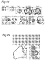
Illustrative examples of organoids generated according to the invention are given in the accompanying figures. It can be seen that organoids according to the invention may possess a cystic structure, with on the outside, a layer of cells with at least one bud and a central lumen. The organoids in the outside of the matrigel tend to be larger than the organoids in the center of the matrigel, perhaps because they have better access to the necessary growth factors. Structurally, organoids according to the invention are often elongated in shape. They may include one or more budding structure - a single cell epithelial layer which has a structure not unlike a bile duct. Under confocal microscopy, the structures may stain positive for keratin. They may include cells with polarised nuclei and small cytoplasm. The organoids may have a section which is formed of multiple layers; such cells often tend to have their nuclei more central to the cells, i.e. not polarized. The cells in the multilayer section may organise themselves to include a gap, or lumen between the cells.
-
A liver organoid preferably comprises a hepatocyte and a cholangiocyte cell, more preferably wherein at least one of the following markers could be detected: at least one hepatocyte marker such as albumin, transthyretrin, B-1 integrin and Glutamine synthetase and/or at least one cholangiocyte maker such as Keratin 7 and 19. The skilled person knows how to detect each of these markers (i.e. RT-PCR and/or immunohistochemistry). Preferably the expression of each of these markers is assessed as carried out in the experimental part. Each of these markers is usually expressed after at least two weeks, three weeks or one month of culture using a method of the invention. Microarray analysis of the organoids in both culture conditions showed that liver organoids resemble adult liver tissue.
-
Preferably, cells and organoids generated according to the invention also possess hepatocyte functions, such as expressing or staining positive for the mature hepatic markers albumin, B-1 integrin,, CK-8, CK-18, transthyretin (TTR), glucose 6P, Met, Glutamine synthase (Glul), transferrin, Fahd1, Fahd2a, K7, K19 and cytochrome P450 isoforms 3A13 (CYP3A13), 51 (CYP51) 2D10 (CYP2D10), 2j6 (CYP2j6), 39A1 (CYP39A1), 4A10 (CYP4A10), 4F13 (CYP4F13) 4F16 (CYP4F16), CYP4B1and 20A1(CYP20A1). Also, embryonic liver gene AFP is not detected in neither of both culture conditions, as in adult liver.
-
Also, the well known liver transcription factors as HNF1a HNF1b and HNF4a are highly expressed in both conditions.
-
Since liver and pancreas are closely related organs, we investigated whether our liver cultures also expressed pancreas-specific genes. The pancreas is functionally divided into endocrine and exocrine pancreas. The endocrine pancreas is mainly characterized for expressing insulin, glucagon and somatostatin. The expression of these hormones is tightly regulated by a set of endocrine pancreas-specific transcription factors, the most important being Pdx1 and NeuroD. The exocrine pancreas is formed by acinar and ductal compartments responsible of producing the digestive enzymes amylase, pancreatic lipase and chymotrypsin, among others. The expression of these genes is also regulated by specific exocrine pancreatic genes as Ptf1.
-
The pancreas specific genes Ptfla, pancreatic amylase (Amy2a4), pancreatic lipase (Pnlip), insulin (ins1 and ins2), glucagon (Gcg), chymotrypsin (cela1), Pdx1 and NeuroD were absent in the liver cultures here described.
-
Cells and organoids according to the present invention may preferably be capable of secreting albumin, for example, at a rate of between approximately 1µg per hour per 106 cells and 10µg per hour per 106 cells, preferably between 2µg and 6µg per hour per 106 cells.
-
Furthermore, such cells and organoids may secrete urea. For example, in a 35mm dish of cells, the activity of urea synthesis may be between 11µg and 50µg in 48 hours, preferably between 5µg and 30µg. 1
-
Cells and organoids according to the invention may show visible glycogen stores, for example, when stained. The capacity for cells and organoids according to the invention to synthesize glycogen actively can be tested by switching the culture media from low-glucose differentiation media to high-glucose DMEM supplemented with 10% FBS and 0.2µM dexamethasone for two days.
-
Cells and organoids according to the invention may possess inducible cytochrome P450 activity (e.g. CYP1A). Such activity may be tested, for example, using an ethoxyresorufin-O-deethylase (EROD) assay (Cancer Res, 2001, 61: 8164-8170). For example, cells or organoids may be exposed to a P450 substrate such as 3-methylcholanthrene and the levels of EROD activity compared to control cells.
-
Morphologically, the cells appear hepatocyte-like.
-
A preferred liver organoid comprises or consists of a cystic structure with on the outside a layer of cells with buds and a central lumen as depicted in Figure 2. This liver organoid may have one or more (e.g. 2, 3, or all 4) of the following characteristics: (a) having a cell density of >5×10⁵ cells/cm3, preferably >10×105 cells/cm3; (b) having a thickness equivalent to 2-30 layers of cells, preferably a thickness equivalent to 2-15 layers of cells; (c) the cells mutually contact in three dimensions, (d) demonstrate a function inherent to healthy liver tissue, (e) have an elongated shape, with 2 defined domains, i.e. a single layered epithelial domain where highly polarized cells are detected and keratin markers are expressed (this domain resembles the bile duct domain) and the other domain constitutes the main body of the organoid and is formed by a multilayered epithelia with non-polarized cells wherein albumin expression may be detected. It is clear to the skilled person that such a liver organoid is preferably not a liver fragment and/or does not comprise a blood vessel, and/or does not comprise a liver lobule or a bile duct.
-
Within the context of the invention, a liver fragment is a part of an adult liver, preferably a human adult liver. Preferably a liver organoid as identified herein is therefore not a liver fragment. A liver organoid is preferably obtained using a cell from an adult liver, preferably an epithelial stem cell from an adult liver, more preferably an epithelial stem cell from an adult liver expressing Lgr5.
-
In an embodiment, a liver organoid is a liver organoid which is still being cultured using a method of the invention and is therefore in contact with an extracellular matrix. Preferably, a liver organoid is embedded in a non-mesenchymal extracellular matrix. Within the context of the invention, "in contact" means a physical or mechanical or chemical contact, which means that for separating said liver organoid from said extracellular matrix a force needs to be used.
-
In a preferred embodiment, a liver organoid could be cultured during at least 2, 3, 4, 5,6, 7, 8, 9, 10 weeks or 1, 2, 3, 4, 5 6 months or longer.
-
In another preferred embodiment, a liver organoid originates from a single cell, preferably expressing Lgr5, more preferably wherein the single cell comprises a nucleic acid construct comprising a nucleic acid molecule of interest.
-
The invention further provides the use of a culture medium according to the invention for culturing epithelial stem cells or isolated organoid structures that comprise these stem cells on an extracellular matrix, whereby said stem cells preferably do not comprise human embryonic stem cells. Preferred are human adult stem cells. Furthermore, single sorted epithelial stem cells from the liver are also able to initiate these 3 dimensional organoids in a culture medium according to the invention. The invention further provides the use of a culture medium according to the invention for culturing liver fragments comprising stem cells.
-
It is preferred that said stem cells are liver stem cells, or epithelial stem cells. A culture medium according to the invention allowed the establishment of long-term culture conditions under which a liver organoid is formed in which all differentiated cell types are present. Using a culture method according to the invention allowed culture periods of at least seven months, at least eight months, at least nine months, at least ten months.
-
The invention further provides a liver organoid, preferably comprising at least 50% viable cells, more preferred at least 60% viable cells, more preferred at least 70% viable cells, more preferred at least 80% viable cells, more preferred at least 90% viable cells. Viability of cells may be assessed using Hoechst staining or Propidium Iodide staining in FACS.
-
The viable cells preferably possess hepatic functions, or characteristics of hepatocytes, as described above.
-
In a further aspect, the invention provides the use of a liver cell or organoid according to the invention as described above in a drug discovery screen, toxicity assay or in regenerative medicine. The invention furthermore provides the use of the progeny of liver organoids of the invention, in toxicity assays. Such toxicity assays may be in vitro assays using a cell derived from a liver organoid or a liver organoid or part thereof. Such progeny and liver organoids are easy to culture and more closely resemble primary epithelial cells than, for example, epithelial cell lines such as Caco-2 (ATCC HTB-37), I-407 (ATCC CCL6), and XBF (ATCC CRL 8808) which are currently used in toxicity assays. It is anticipated that toxicity results obtained with liver organoids more closely resemble results obtained in patients. A cell-based toxicity test is used for determining organ specific cytotoxicity. Compounds that are tested in said test comprise cancer chemopreventive agents, environmental chemicals, food supplements, and potential toxicants. The cells are exposed to multiple concentrations of a test agent for certain period of time. The concentration ranges for test agents in the assay are determined in a preliminary assay using an exposure of five days and log dilutions from the highest soluble concentration. At the end of the exposure period, the cultures are evaluated for inhibition of growth. Data are analyzed to determine the concentration that inhibited end point by 50 percent (TC50).
-
For example, induction of cytochrome P450 enzymes in liver hepatocytes is a key factor that determines the efficacy and toxicity of drugs. In particular, induction of P450s is an important mechanism of troublesome drug-drug interactions, and it is also an important factor that limits drug efficacy and governs drug toxicity. Cytochrome P450 induction assays have been difficult to develop, because they require intact normal human hepatocytes. These cells have proven intractable to production in numbers sufficient to sustain mass production of high throughput assays.
-
For example, according to this aspect of the invention, a candidate compound may be contacted with cell or organoid as described herein, and any change to the cells or in to activity of the cells may be monitored. Examples of other non-therapeutic uses of the cells or organoids of the present invention include research of liver embryology, liver cell lineages, and differentiation pathways; gene expression studies including recombinant gene expression; mechanisms involved in liver injury and repair; research of inflammatory and infectious diseases of the liver; studies of pathogenetic mechanisms; and studies of mechanisms of liver cell transformation and aetiology of liver cancer.
-
For high-throughput purposes, said liver organoids are cultured in multiwell plates such as, for example, 96 well plates or 384 well plates. Libraries of molecules are used to identify a molecule that affects said organoids. Preferred libraries comprise antibody fragment libraries, peptide phage display libraries, peptide libraries (e.g. LOPAP™, Sigma Aldrich), lipid libraries (BioMol), synthetic compound libraries (e.g. LOP AC™, Sigma Aldrich) or natural compound libraries (Specs, TimTec). Furthermore, genetic libraries can be used that induce or repress the expression of one of more genes in the progeny of the adenoma cells. These genetic libraries comprise cDNA libraries, antisense libraries, and siRNA or other non-coding RNA libraries. The cells are preferably exposed to multiple concentrations of a test agent for certain period of time. At the end of the exposure period, the cultures are evaluated. The term "affecting" is used to cover any change in a cell, including, but not limited to, a reduction in, or loss of, proliferation, a morphological change, and cell death. Said liver organoids can also be used to identify drugs that specifically target epithelial carcinoma cells, but not said liver organoids.
-
Liver organoids according to the invention can further replace the use of cell lines such as Caco-2 cells in toxicity assays of potential novel drugs or of known or novel food supplements.
-
Furthermore, such liver organoids can be used for culturing of a pathogen.
-
Cultures comprising liver organoids are useful in regenerative medicine, for example in post-radiation and/or post-surgery repair of the liver epithelium, in the repair of the epithelium in patients suffering from chronic or acute liver failure or disease. Liver diseases include, but are not limited to Hepatocellular Carcinoma, Alagille Syndrome, Alpha- 1- Antitrypsin Deficiency, Autoimmune Hepatitis, Biliary Atresia, Chronic Hepatitis, Cancer of the Liver , Cirrhosis Liver Cysts Fatty Liver, Galactosemia Gilbert's Syndrome, Primary Biliary Cirrhosis, Hepatitis A, Hepatitis B, Hepatitis C, Primary Sclerosing Cholangitis, Reye's Syndrome, Sarcoidosis, Tyrosinemia, Type I Glycogen Storage Disease, Wilson's Disease, Neonatal Hepatitis, Non-alchoholic SteatoHepatitis, Porphyria, and Hemochromatosis.
-
In the case of replacement or correction of deficient liver function, it may be possible to construct a cell-matrix structure from one or more liver organoids generated according to the present invention. It is thought that only about 10% of hepatic cell mass is necessary for adequate function. This makes implantation of organoid unit compositions into children especially preferable to whole organ transplantation, due to the relatively limited availability of donors and smaller size of juvenile organs. For example, an 8-month-old child has a normal liver that weighs approximately 250g. That child would therefore need about 25g of tissue. An adult liver weighs-approximately 1500g; therefore, the required implant would only be about 1.5% of the adult liver. When organoid units according to the invention are implanted, optionally attached to a polymer scaffold, proliferation in the new host will occur, and the resulting hepatic cell mass replaces the deficient host function.
-
Accordingly, included within the scope of the invention are methods of treatment of a human or animal patient through cellular therapy. The term "animal" here denotes all mammalian animals, preferably human patients. It also includes an individual animal in all stages of development, including embryonic and foetal stages. Such cellular therapy encompasses the administration of cells or organoids generated according to the invention to a patient through any appropriate means. Specifically, such methods of treatment involve the regeneration of damaged tissue. The term "administration" as used herein refers to well recognized forms of administration, such as intravenous or injection, as well as to administration by transplantation, for example transplantation by surgery, grafting or transplantation of tissue engineered liver derived from cells or organoids according to the present invention. In the case of cells, systemic administration to an individual may be possible, for example, by infusion into the superior mesenteric artery, the celiac artery, the subclavian vein via the thoracic duct, infusion into the heart via the superior vena cava, or infusion into the peritoneal cavity with subsequent migration of cells via subdiaphragmatic lymphatics, or directly into liver sites via infusion into the hepatic arterial blood supply or into the portal vein.
-
Between 104 and 1013 cells per 100 kg person may be administered per infusion. Preferably, between about 1-5x10⁴ and 1-5x10⁷ cells may be infused intravenously per 100 kg person. More preferably, between about 1x10⁴ and 10x106 cells may be infused intravenously per 100 kg person. In some embodiments, a single administration of cells or organoids is provided. In other embodiments, multiple administrations are used. Multiple administrations can be provided over an initial treatment regime, for example, of 3-7 consecutive days, and then repeated at other times.
-
It is also possible to reconstitute a liver organoid from one single cell expressing Lgr5 as defined herein. This single cell may have been modified by introduction of a nucleic acid construct as defined herein, for example, to correct a genetic deficiency or mutation. It would also be possible to specifically ablate expression, as desired, for example, using siRNA. Potential polypeptides to be expressed could be any of those that are deficient in metabolic liver diseases, including, for example, AAT (alpha antitrypsin). For elucidating liver physiology, we might also express or inactivate genes implicated in the wnt, EGF, FGF, BMP or notch pathway. Also, for screening of drug toxicity, the expression or inactivation of genes responsible for liver drug metabolism (for example, genes in the CYP family) would be of high interest
-
In one embodiment, the expanded epithelial stem cells may be reprogrammed into related tissue fates such as, for example, liver cells including a hepatocyte and a cholangiocyte cell. Thus far, it has not been possible to regenerate liver cells from adult stem cells. The culturing methods of the present invention will enable to analyse for factors that trans-differentiate the closely related epithelial stem cell to a liver cell, including a hepatocyte and a cholangiocyte cell.
-
It will be clear to a skilled person that gene therapy can additionally be used in a method directed at repairing damaged or diseased tissue. Use can, for example, be made of an adenoviral or retroviral gene delivery vehicle to deliver genetic information, like DNA and/or RNA to stem cells. A skilled person can replace or repair particular genes targeted in gene therapy. For example, a normal gene may be inserted into a nonspecific location within the genome to replace a non functional gene. In another example, an abnormal gene sequence can be replaced for a normal gene sequence through homologous recombination. Alternatively, selective reverse mutation can return a gene to its normal function. A further example is altering the regulation (the degree to which a gene is turned on or off) of a particular gene. Preferably, the stem cells are ex vivo treated by a gene therapy approach and are subsequently transferred to the mammal, preferably a human being in need of treatment.
-
The invention also provides a composition or cell culture vessel comprising cells and/or organoids according to any one of the aspects of the invention described above, and a culture medium according to any one of the aspects of the invention described above. For example, such a composition or cell culture vessel may comprise any number of cells or organoids cultured according to a method of the invention, in a culture medium as described above. For example, a preferred culture medium comprises or consists of Advanced-DMEM/F12 supplemented with B27, N2, 200ng/ml N-Acetylcysteine, 50ng/ml EGF, 1 µg/ml R-spondin1, 10 nM gastrin, 100 ng/ml FGF10, 10mM Nicotinamide and 50ng/ml HGF and 50% wnt conditioned media.
Definitions
-
A nucleic acid construct comprises a nucleic acid molecule of interest and will ensure expression of the given nucleic acid molecule in the cells wherein it had been introduced. A particularly preferred nucleic acid construct is an expression vector wherein a nucleic acid molecule encoding a polypeptide is operably linked to a promoter capable of directing expression of said nucleic acid molecule (i.e a coding sequence) in said cells. The phrase "expression vector" generally refers to a nucleic acid molecule that is capable of effecting expression of a gene/nucleic acid molecule it contains in a cell compatible with such sequences. These expression vectors typically include at least suitable promoter sequences and optionally, transcription termination signals. A nucleic acid or DNA or nucleotide sequence encoding a polypeptide is incorporated into a DNA/nucleic acid construct capable of introduction into and expression in an in vitro cell culture as identified in a method of the invention. A DNA construct prepared for introduction into a particular cell typically include a replication system recognized by said cell, an intended DNA segment encoding a desired polypeptide, and transcriptional and translational initiation and termination regulatory sequences operably linked to the polypeptide-encoding segment. A DNA segment is "operably linked" when it is placed into a functional relationship with another DNA segment. For example, a promoter or enhancer is operably linked to a coding sequence if it stimulates the transcription of the sequence. DNA for a signal sequence is operably linked to DNA encoding a polypeptide if it is expressed as a preprotein that participates in the secretion of a polypeptide. Generally, a DNA sequence that is operably linked are contiguous, and, in the case of a signal sequence, both contiguous and in reading phase. However, enhancers need not be contiguous with a coding sequence whose transcription they control. Linking is accomplished by ligation at convenient restriction sites or at adapters or linkers inserted in lieu thereof.
-
The selection of an appropriate promoter sequence generally depends upon the host cell selected for the expression of a DNA segment. Examples of suitable promoter sequences include eukaryotic promoters well known in the art (see, e.g. Sambrook and Russell, 2001, supra). A transcriptional regulatory sequence typically includes a heterologous enhancer or promoter that is recognised by the cell. Suitable promoters include the CMV promoter. An expression vector includes the replication system and transcriptional and translational regulatory sequences together with the insertion site for the polypeptide encoding segment can be employed. Examples of workable combinations of cell lines and expression vectors are described in Sambrook and Russell (2001, supra) and in Metzger et al. (1988) Nature 334: 31-36.
-
The invention is not limited to a specific polypeptide or nucleic acid molecule of interest to be expressed in a cell expressing Lgr5 as identified herein. Depending on the aim of the method, one may envisage to express a polypeptide in a liver cell and/or to inactivate the expression of a polypeptide in a liver cell. Potential polypeptides to be expressed could be all of those that are deficient in metabolic liver diseases, as eg AAT (alpha antitrypsin). For elucidating the liver physiology, we might also express or inactivate genes implicated in the wnt, EGF, FGF, BMP or notch pathway. Also, for screening of drug toxicity, the expression or inactivation of genes responsible for liver drug metabolism (eg. genes of the CYP family) would be of high interest.
-
Some aspects of the invention concern the use of a nucleic acid construct or expression vector comprising a nucleotide sequence as defined above, wherein the vector is a vector that is suitable for gene therapy. Vectors that are suitable for gene therapy are described in Anderson 1998, Nature 392: 25-30; Walther and Stein, 2000, Drugs 60: 249-71; Kay et al., 2001, Nat. Med. 7: 33-40; Russell, 2000, J. Gen. Virol. 81: 2573-604; Amado and Chen, 1999, Science 285: 674-6; Federico, 1999, Curr. Opin. Biotechnol.10: 448-53; Vigna and Naldini, 2000, J. Gene Med. 2: 308-16; Marin et al., 1997, Mol. Med. Today 3: 396-403; Peng and Russell, 1999, Curr. Opin. Biotechnol. 10: 454-7; Sommerfelt, 1999, J. Gen. Virol. 80: 3049-64; Reiser, 2000, Gene Ther. 7: 910-3; and references cited therein. Examples include integrative and non-integrative vectors such as those based on retroviruses, adenoviruses (AdV), adeno-associated viruses (AAV), lentiviruses, pox viruses, alphaviruses, and herpes viruses.
-
A particularly suitable gene therapy vector includes an Adenoviral (Ad) and Adeno-associated virus (AAV) vector. These vectors infect a wide number of dividing and non-dividing cell types including liver cells. In addition adenoviral vectors are capable of high levels of transgene expression. However, because of the episomal nature of the adenoviral and AAV vectors after cell entry, these viral vectors are most suited for therapeutic applications requiring only transient expression of the transgene (Russell, 2000, J. Gen. Virol. 81: 2573-2604; Goncalves, 2005, Virol J. 2(1):43) as indicated above. Preferred adenoviral vectors are modified to reduce the host response as reviewed by Russell (2000, supra). Safety and efficacy of AAV gene transfer has been extensively studied in humans with encouraging results in the liver, muscle, CNS, and retina (Manno et al Nat medicine 2006, Stroes et al ATVB 2008, Kaplitt, Feigin, Lancet 2009; Maguire, Simonelli et al NEJM 2008; Bainbridge et al NEJM 2008). AAV2 is the best characterized serotype for gene transfer studies both in humans and experimental models. AAV2 presents natural tropism towards skeletal muscles, neurons, vascular smooth muscle cells and hepatocytes. AAV2 is therefore a good choice of vector to target liver tissues. Other examples of adeno-associated virus-based non integrative vectors include AAV1, AAV3, AAV4, AAV5, AAV6, AAV7, AAV8, AAV9, AAV10, AAV11 and pseudotyped AAV. The use of non-human serotypes, like AAV8 and AAV9, might be useful to overcome these immunological responses in subjects, and clinical trials have just commenced (ClinicalTrials.gov Identifier: NCT00979238). For gene transfer into a liver cell, an adenovirus serotype 5 or an AAV serotype 2, 7 or 8 have been shown to be effective vectors and therefore a preferred Ad or AAV serotype (Gao, Molecular Therapy (2006) 13, 77-87).
-
A preferred retroviral vector for application in the present invention is a lentiviral based expression construct. Lentiviral vectors have the unique ability to infect non-dividing cells (
Amado and Chen, 1999 Science 285: 674-6). Methods for the construction and use of lentiviral based expression constructs are described in
U.S. Patent No.'s 6,165,782 ,
6,207,455 ,
6,218,181 ,
6,277,633 and
6,323,031 and in
Federico (1999, Curr Opin Biotechnol 10: 448-53) and
Vigna et al. (2000, J Gene Med 2000; 2: 308-16).
-
Generally, gene therapy vectors will be as the expression vectors described above in the sense that they comprise a nucleotide sequence encoding a polypeptide of the invention to be expressed, whereby a nucleotide sequence is operably linked to the appropriate regulatory sequences as indicated above. Such regulatory sequence will at least comprise a promoter sequence. Suitable promoters for expression of a nucleotide sequence encoding a polypeptide from gene therapy vectors include e.g. cytomegalovirus (CMV) intermediate early promoter, viral long terminal repeat promoters (LTRs), such as those from murine moloney leukaemia virus (MMLV) rous sarcoma virus, or HTLV-1, the simian virus 40 (SV 40) early promoter and the herpes simplex virus thymidine kinase promoter. Suitable promoters are described below.
-
Several inducible promoter systems have been described that may be induced by the administration of small organic or inorganic compounds. Such inducible promoters include those controlled by heavy metals, such as the metallothionine promoter (
Brinster et al. 1982 Nature 296: 39-42;
Mayo et al. 1982 Cell 29: 99-108), RU-486 (a progesterone antagonist) (
Wang et al. 1994 Proc. Natl. Acad. Sci. USA 91: 8180-8184), steroids (
Mader and White, 1993 Proc. Natl. Acad. Sci. USA 90: 5603-5607), tetracycline (
Gossen and Bujard 1992 Proc. Natl. Acad. Sci. USA 89: 5547-5551;
U.S. Pat. No. 5,464,758 ;
Furth et al. 1994 Proc. Natl. Acad. Sci. USA 91: 9302-9306;
Howe et al. 1995 J. Biol. Chem. 270: 14168-14174;
Resnitzky et al. 1994 Mol. Cell. Biol. 14: 1669-1679;
Shockett et al. 1995 Proc. Natl. Acad. Sci. USA 92: 6522-6526) and the tTAER system that is based on the multi-chimeric transactivator composed of a tetR polypeptide, as activation domain of VP16, and a ligand binding domain of an estrogen receptor (
Yee et al., 2002, US 6,432,705 ).
-
Suitable promoters for nucleotide sequences encoding small RNAs for knock down of specific genes by RNA interference (see below) include, in addition to the above mentioned polymerase II promoters, polymerase III promoters. The RNA polymerase III (pol III) is responsible for the synthesis of a large variety of small nuclear and cytoplasmic non-coding RNAs including 5S, U6, adenovirus VA1, Vault, telomerase RNA, and tRNAs. The promoter structures of a large number of genes encoding these RNAs have been determined and it has been found that RNA pol III promoters fall into three types of structures (for a review see Geiduschek and Tocchini-Valentini, 1988 Annu. Rev. Biochem. 57: 873-914; Willis, 1993 Eur. J. Biochem. 212: 1-11; Hernandez, 2001, J. Biol. Chem. 276: 26733-36). Particularly suitable for expression of siRNAs are the type 3 of the RNA pol III promoters, whereby transcription is driven by cis-acting elements found only in the 5'-flanking region, i.e. upstream of the transcription start site. Upstream sequence elements include a traditional TATA box (Mattaj et al., 1988 Cell 55, 435-442), proximal sequence element and a distal sequence element (DSE; Gupta and Reddy, 1991 Nucleic Acids Res. 19, 2073-2075). Examples of genes under the control of the type 3 pol III promoter are U6 small nuclear RNA (U6 snRNA), 7SK, Y, MRP, H1 and telomerase RNA genes (see e.g. Myslinski et al., 2001, Nucl. Acids Res. 21: 2502-09).
-
A gene therapy vector may optionally comprise a second or one or more further nucleotide sequence coding for a second or further polypeptide. A second or further polypeptide may be a (selectable) marker polypeptide that allows for the identification, selection and/or screening for cells containing the expression construct. Suitable marker proteins for this purpose are e.g. the fluorescent protein GFP, and the selectable marker genes HSV thymidine kinase (for selection on HAT medium), bacterial hygromycin B phosphotransferase (for selection on hygromycin B), Tn5 aminoglycoside phosphotransferase (for selection on G418), and dihydrofolate reductase (DHFR) (for selection on methotrexate), CD20, the low affinity nerve growth factor gene. Sources for obtaining these marker genes and methods for their use are provided in Sambrook and Russel (2001) "Molecular Cloning: A Laboratory Manual (3rd edition), Cold Spring Harbor Laboratory, Cold Spring Harbor Laboratory Press, New York.
-
Alternatively, a second or further nucleotide sequence may encode a polypeptide that provides for fail-safe mechanism that allows a subject from the transgenic cells to be cured, if deemed necessary. Such a nucleotide sequence, often referred to as a suicide gene, encodes a polypeptide that is capable of converting a prodrug into a toxic substance that is capable of killing the transgenic cells in which the polypeptide is expressed. Suitable examples of such suicide genes include e.g. the E.coli cytosine deaminase gene or one of the thymidine kinase genes from Herpes Simplex Virus, Cytomegalovirus and Varicella-Zoster virus, in which case ganciclovir may be used as prodrug to kill the IL-10 transgenic cells in the subject (see e.g. Clair et al., 1987, Antimicrob. Agents Chemother. 31: 844-849).
-
For knock down of expression of a specific polypeptide, a gene therapy vector or other expression construct is used for the expression of a desired nucleotide sequence that preferably encodes an RNAi agent, i.e. an RNA molecule that is capable of RNA interference or that is part of an RNA molecule that is capable of RNA interference. Such RNA molecules are referred to as siRNA (short interfering RNA, including e.g. a short hairpin RNA). Alternatively, a siRNA molecule may directly, e.g. in a pharmaceutical composition that is administered within or in the neighborhood of a cell expressing Lgr5, or of a liver cell (i.e. hepatocyte or cholangiocyte) or of a liver organoid.
-
A desired nucleotide sequence comprises an antisense code DNA coding for the antisense RNA directed against a region of the target gene mRNA, and/or a sense code DNA coding for the sense RNA directed against the same region of the target gene mRNA. In a DNA construct of the invention, an antisense and sense code DNAs are operably linked to one or more promoters as herein defined above that are capable of expressing an antisense and sense RNAs, respectively. "siRNA" preferably means a small interfering RNA that is a short-length double-stranded RNA that are not toxic in mammalian cells (Elbashir et al., 2001, Nature 411: 494-98; Caplen et al., 2001, Proc. Natl. Acad. Sci. USA 98: 9742-47). The length is not necessarily limited to 21 to 23 nucleotides. There is no particular limitation in the length of siRNA as long as it does not show toxicity. "siRNAs" can be, e.g. at least 15, 18 or 21 nucleotides and up to 25, 30, 35 or 49 nucleotides long. Alternatively, the double-stranded RNA portion of a final transcription product of siRNA to be expressed can be, e.g. at least 15, 18 or 21 nucleotides and up to 25, 30, 35 or 49 nucleotides long.
-
"Antisense RNA" is preferably an RNA strand having a sequence complementary to a target gene mRNA, and thought to induce RNAi by binding to the target gene mRNA. "Sense RNA" has a sequence complementary to the antisense RNA, and annealed to its complementary antisense RNA to form siRNA. The term "target gene" in this context preferably refers to a gene whose expression is to be silenced due to siRNA to be expressed by the present system, and can be arbitrarily selected. As this target gene, for example, genes whose sequences are known but whose functions remain to be elucidated, and genes whose expressions are thought to be causative of diseases are preferably selected. A target gene may be one whose genome sequence has not been fully elucidated, as long as a partial sequence of mRNA of the gene having at least 15 nucleotides or more, which is a length capable of binding to one of the strands (antisense RNA strand) of siRNA, has been determined. Therefore, genes, expressed sequence tags (ESTs) and portions of mRNA, of which some sequence (preferably at least 15 nucleotides) has been elucidated, may be selected as the "target gene" even if their full length sequences have not been determined.
-
The double-stranded RNA portions of siRNAs in which two RNA strands pair up are not limited to the completely paired ones, and may contain nonpairing portions due to mismatch (the corresponding nucleotides are not complementary), bulge (lacking in the corresponding complementary nucleotide on one strand), and the like. A non-pairing portions can be contained to the extent that they do not interfere with siRNA formation. The "bulge" used herein preferably comprise 1 to 2 non-pairing nucleotides, and the double-stranded RNA region of siRNAs in which two RNA strands pair up contains preferably 1 to 7, more preferably 1 to 5 bulges. In addition, the "mismatch" used herein is preferably contained in the double-stranded RNA region of siRNAs in which two RNA strands pair up, preferably 1 to 7, more preferably 1 to 5, in number. In a preferable mismatch, one of the nucleotides is guanine, and the other is uracil. Such a mismatch is due to a mutation from C to T, G to A, or mixtures thereof in DNA coding for sense RNA, but not particularly limited to them. Furthermore, in the present invention, a double-stranded RNA region of siRNAs in which two RNA strands pair up may contain both bulge and mismatched, which sum up to, preferably 1 to 7, more preferably 1 to 5 in number. Such non-pairing portions (mismatches or bulges, etc.) can suppress the below-described recombination between antisense and sense code DNAs and make the siRNA expression system as described below stable. Furthermore, although it is difficult to sequence stem loop DNA containing no non-pairing portion in the double-stranded RNA region of siRNAs in which two RNA strands pair up, the sequencing is enabled by introducing mismatches or bulges as described above. Moreover, siRNAs containing mismatches or bulges in the pairing double-stranded RNA region have the advantage of being stable in E. coli or animal cells.
-
The terminal structure of siRNA may be either blunt or cohesive (overhanging) as long as siRNA enables to silence the target gene expression due to its RNAi effect. The cohesive (overhanging) end structure is not limited only to the 3' overhang, and the 5' overhanging structure may be included as long as it is capable of inducing the RNAi effect. In addition, the number of overhanging nucleotide is not limited to the already reported 2 or 3, but can be any numbers as long as the overhang is capable of inducing the RNAi effect. For example, the overhang consists of 1 to 8, preferably 2 to 4 nucleotides. Herein, the total length of siRNA having cohesive end structure is expressed as the sum of the length of the paired double-stranded portion and that of a pair comprising overhanging single-strands at both ends. For example, in the case of 19 bp double-stranded RNA portion with 4 nucleotide overhangs at both ends, the total length is expressed as 23 bp. Furthermore, since this overhanging sequence has low specificity to a target gene, it is not necessarily complementary (antisense) or identical (sense) to the target gene sequence. Furthermore, as long as siRNA is able to maintain its gene silencing effect on the target gene, siRNA may contain a low molecular weight RNA (which may be a natural RNA molecule such as tRNA, rRNA or viral RNA, or an artificial RNA molecule), for example, in the overhanging portion at its one end.
-
In addition, the terminal structure of the "siRNA" is necessarily the cut off structure at both ends as described above, and may have a stem-loop structure in which ends of one side of double-stranded RNA are connected by a linker RNA (a "shRNA"). The length of the double-stranded RNA region (stem-loop portion) can be, e.g. at least 15, 18 or 21 nucleotides and up to 25, 30, 35 or 49 nucleotides long. Alternatively, the length of the double-stranded RNA region that is a final transcription product of siRNAs to be expressed is, e.g. at least 15, 18 or 21 nucleotides and up to 25, 30, 35 or 49 nucleotides long. Furthermore, there is no particular limitation in the length of the linker as long as it has a length so as not to hinder the pairing of the stem portion. For example, for stable pairing of the stem portion and suppression of the recombination between DNAs coding for the portion, the linker portion may have a clover-leaf tRNA structure. Even though the linker has a length that hinders pairing of the stem portion, it is possible, for example, to construct the linker portion to include introns so that the introns are excised during processing of precursor RNA into mature RNA, thereby allowing pairing of the stem portion. In the case of a stem-loop siRNA, either end (head or tail) of RNA with no loop structure may have a low molecular weight RNA. As described above, this low molecular weight RNA may be a natural RNA molecule such as tRNA, rRNA, snRNA or viral RNA, or an artificial RNA molecule.
-
To express antisense and sense RNAs from the antisense and sense code DNAs respectively, a DNA construct of the present invention comprise a promoter as defined above. The number and the location of the promoter in the construct can in principle be arbitrarily selected as long as it is capable of expressing antisense and sense code DNAs. As a simple example of a DNA construct of the invention, a tandem expression system can be formed, in which a promoter is located upstream of both antisense and sense code DNAs. This tandem expression system is capable of producing siRNAs having the aforementioned cut off structure on both ends. In the stem-loop siRNA expression system (stem expression system), antisense and sense code DNAs are arranged in the opposite direction, and these DNAs are connected via a linker DNA to construct a unit. A promoter is linked to one side of this unit to construct a stem-loop siRNA expression system. Herein, there is no particular limitation in the length and sequence of the linker DNA, which may have any length and sequence as long as its sequence is not the termination sequence, and its length and sequence do not hinder the stem portion pairing during the mature RNA production as described above. As an example, DNA coding for the above-mentioned tRNA and such can be used as a linker DNA.
-
In both cases of tandem and stem-loop expression systems, the 5' end may be have a sequence capable of promoting the transcription from the promoter. More specifically, in the case of tandem siRNA, the efficiency of siRNA production may be improved by adding a sequence capable of promoting the transcription from the promoters at the 5' ends of antisense and sense code DNAs. In the case of stem-loop siRNA, such a sequence can be added at the 5' end of the above-described unit. A transcript from such a sequence may be used in a state of being attached to siRNA as long as the target gene silencing by siRNA is not hindered. If this state hinders the gene silencing, it is preferable to perform trimming of the transcript using a trimming means (for example, ribozyme as are known in the art). It will be clear to the skilled person that an antisense and sense RNAs may be expressed in the same vector or in different vectors. To avoid the addition of excess sequences downstream of the sense and antisense RNAs, it is preferred to place a terminator of transcription at the 3' ends of the respective strands (strands coding for antisense and sense RNAs). The terminator may be a sequence of four or more consecutive adenine (A) nucleotides.
-
In this document and in its claims, the verb "to comprise" and its conjugations is used in its non-limiting sense to mean that items following the word are included, but items not specifically mentioned are not excluded. In addition the verb "to consist" may be replaced by "to consist essentially of" meaning that a product as defined herein may comprise additional component(s) than the ones specifically identified, said additional component(s) not altering the unique characteristic of the invention. In addition a method as defined herein may comprise additional step(s) than the ones specifically identified, said additional step(s) not altering the unique characteristic of the invention. In addition, reference to an element by the indefinite article "a" or "an" does not exclude the possibility that more than one of the element is present, unless the context clearly requires that there be one and only one of the elements. The indefinite article "a" or "an" thus usually means "at least one". The word "about" or "approximately" when used in association with a numerical value (about 10) preferably means that the value may be the given value of 10 more or less 1% of the value.
-
All patent and literature references cited in the present specification are hereby incorporated by reference in their entirety.
-
The following examples are offered for illustrative purposes only, and are not intended to limit the scope of the present invention in any way.
Description of Figures
-
- Fig. 1 liver organoids growth factor requirement. (A) DIC images of liver organoids maintained with EGF (E), R-spondin 1 (R), Noggin (N), Wnt3Aconditioned media (W) or the combination of them, supplemented with FGF10, HGF and Nicotinamide. (B) The number of organoids was counted weekly and passaged when required. Results are shown as mean ±SEM of 3 independent experiments. (C) gene expression analysis by RTPCR ofLgr5, Keratin 7 (K7) and Albumin (Alb) genes. (D) Isolated bibliary ducts growing into organoids. Differential interference contrast images from the corresponding days after seeding. Magnification 10x ( days 0, 1, 3 and 5). Days 15 on magnification 4x. Cultures were passage every 4-7 days by mechanical dissociation. Cultures have been grown at least for 8 months.
- Fig. 2 Morphology of liver organoids. (A) Upper panels: paraffin section of a mouse liver showing the different domains (PT= portal triad, CV= central vein). Lower panels: Paraffin section of a liver organoid showing different domains b (single layered epithelia) and h (stratified epithelia) (B) Right pannel: Ecadherin staining in the liver organoids. Two different domains can be identified. Domain b, formed by a single layered epithelia that resembles the bile duct structures in the liver. This bile duct domain is formed by highly polarized cells that shows positive staining for pancytokeratin (PCK) (lower panel). Left panels show the presence of a second domain within the liver organoids. This h domain is formed by a stratified epithelia with non-polarized cells. The cells are organized around a central lumen and express the hepatocyte marker Alb. Magnification 10 x.
- Fig. 3 Wnt signalling in the liver cultures. Lac Z expression was detected in cultures derived from Lgr5-LacZ or Axin2 LacZ mice. No positive staining was detected in liver cultures derived from a B16 mice. Magnification 4x, inset 20x.
- Fig. 4 expression of liver differentiating markers. (A) immunohistochemical and immunofluorescence anaylisis of the expression of the cholangiocyte marker keratin 7 (K7) and the hepatocyte markers keratin 8 (K8) and albumin (Alb). (B) analysis of the gene expression of hepatocyte markers: Albumin (Alb), transthyretrin (Ttr), Glutamine synthetase (Glul), glucose 6 phosphatase (G6P) and Cytocrome p450 isoform 3A11 (CYP3A11); and cholangiocyte markers keratin 7 (K7) and Keratin 19 (K19).
- Fig. 5 liver single cell cultures. (A) flow cytometry plot indicating the area of the sorted cells. (B) single cell growing into organoids at the time points indicated. Magnification 40 x (day 1-3), 10x (day 16), 4 x (day 21-on). (C & D) representative image of the colony formation efficiency of a Lgr5GFP single sorted cells. 100 cells were seeded in triplicate and colonies were counted 10 days later.
- Fig. 6 Microarray analysis of the liver cultures. Analysis of the gene expression profile of adult liver tissue and liver organoid cultures maintained for 1 month in the ER or ER media supplemented with Noggin (ENR) or with noggin and Wnt (ENRW). The genetic profile was compared between the different samples and the genetic profile of Brow adipose tissue (BAT) white adipose tissue (WAT), muscle and new born liver. (A) hit map analysis showing that the cultures present a similar profile to the adult liver but a different profile to non-liver related tissues as muscle and BAT and WAT, (B) List of hepatocte markers and cholangyocyte markers in the different conditions.
Examples
MATERIAL AND METHODS
Liver culture-biliary duct isolation
-
Isolated adult liver tissue was washed in cold Advanced-DMEM/F12 (Invitrogen) and then, the tissue was chopped into pieces of around 5mm animals and further washed with cold dissociation buffer (collagenase, dispase, FBS in DMEM media). The tissue fragments were incubated with the dissociation buffer for 2 h at 37°C. Then, the tissue fragments were vigorously suspended in 10 ml of cold isolation buffer with a 10 ml pipette. The first supernatant containing death cells was discarded and the sediment was suspended with 10-15ml of dissociation buffer. After further vigorous suspension of the tissue fragments the supernatant is enriched in biliary ducts. This procedure is repeated until enough biliary ducts are obtained.
-
Isolated biliary ducts are pelleted and mixed with 50µl of Matrigel (BD Bioscience), seeded on 24-well tissue culture plates and incubated for 5-10 min at 37°C until complete polymerization of the Matrigel. After polymerization, 500 µl of tissue culture media are overloaded.
-
Media composition: Advanced-DMEM/F12 supplemented with B27, N2, 200ng/ml N-Acetylcysteine, 50ng/ml EGF, 1 µg/ml R-spondin1, gastrin: 10 nM, FGF10 100 ng/ml, Nicotinamide 10mM and HGF: 50ng/ml and 50% wnt conditioned media.
-
The entire medium was changed every 2 days. After 1 week, Wnt conditioned media is withdrawal and the formed organoids removed from the Matrigel using a 1000 µl pipette and were dissociated mechanically into small fragments and transferred to fresh Matrigel. Passage was performed in 1:4 split ratio once or twice per week. Under these conditions cultures have been maintained for at least 6 month.
Reagents
-
human Hepatocyte Growth Factor (HGF) was purchased from Peprotech, EGF invitrogen, R-Spondin Nuvelo, Noggin peprotech , FGF10 Peprotech, gastrin Sigma Aldrich, nicotinamide Sigma.
Microarray
-
For the expression analysis ofLgr5-derived liver cultures, RNA was isolated using a Qiagen RNAase kit, from adult liver or from liver cultures cultured in media without Wntcm and Noggin (ER) or with Wntcm and Noggin (ENRW). 150ng of total RNA was labelled with low RNA Input Linear Amp kit (Agilent Technologies, Palo Alto, CA). Universal mouse Reference RNA (Agilent) was differentially labelled and hybridized to either adult liver tissue or ER or ENRW treated cultures. A 4X44K Agilent Whole Mouse Genome dual colour Microarrays (G4122F) was used. Labelling, hybridization, and washing were performed according to Agilent guidelines.
RESULTS
-
After isolation, biliary ducts (see Fig. 1) were suspended in Matrigel and cultured in different growth factor conditions. The combination of EGF (50 ng/ml) and R-spondin 1 (1ug/ml) supplemented with FGF10 (100ng/ml), HGF (25-50ng/ml) and Nicotinamide (1-10mM), were essential for the long term maintenance of the cultures, indicating that Wnt signalling and EGF signalling are strictly required to maintain adult liver progenitor proliferation in vitro. The addition of Noggin (100ng/ml) and Wnt conditioned media (50%) also showed long term maintenance of the cultures (see Figs. 1A and 1B). Under these conditions that supported long-term maintenance, Lgr5 expression as well as hepatocyte markers (Albumin) and cholangiocyte markers (K7) were detected by RT-PCR (see Fig. 1C). Under these conditions liver organoids have been weekly passaged by mechanical or enzymatic dissociation, at 1:8 dilution, and have been grown for many months (Fig. 1D).
-
We analysed the expression of the Wnt target genes Axin2 and Lgr5 in the cultures. Cultures of both Axin2LacZ and Lgr5-LacZ livers revealed the presence of Axin2- and Lgr5-positive cells in the liver organoids 1 month after seeding, thus confirming that the wnt signalling is active and required for culture growth (Fig. 3). The liver cultures also express hepatocyte markers (e.g. albumin, transthyretrin, Glutamine synthetase) and cholangiocyte makers (Keratin 7 and 19) (see Fig 4).
-
When single Lgr5 cells from a Lgr5LacZ or Lgr5GFP mouse were sorted, single colonies grew into organoids. These cultures also express markers of cholangiocyte and hepatocyte lineages and have been maintained and regularly split into 1:6-1:8 for more than 4 months (see Figs. 5A & B). Interestingly, only the cultures derived from Lgr5 positive cells grew into organoids (Figs 5C & D). These data indicate that Lgr5 cells are progenitor cells of these cultures and able to propagate progeny of the 2 different liver lineages.
-
Having established that the liver organoids are derived from Lgr5+ve cells we set out to determine their individual gene signature as compared to the adult liver signature. RNA was isolated from adult liver and from liver organoids grown in ER or ENRW media supplemented with FGF10, Nicotinamide and Hepatocyte Growth Factor. The genetic signature of the adult liver and the 2 liver culture conditions was subsequently derived via comparative gene expression profiling in respect to the expression of a Universal RNA reference. The use of the same reference RNA for the hybridization to all the samples allowed us to compare the 3 independent samples among them (adult liver, ER and ENRW). The heat map analysis revealed that the expression profile of both culture conditions highly resemble the adult liver tissue expression profile, whereas they do not share the same profile when compared to muscle or adipose tissue profile (see Fig 6). Among the similar gene expression profile between the adult liver and the liver cultures, liver specific genes as HNF1a, HNF1b, HNF4, Alb, Glul, Met, G6P, Fahd1, Fahd2a, CYP4B1, K7 and K19 are detected. The heat map analysis reveals that both culture conditions present similar expression pattern among each other and when compared to the adult liver sample. However, when analyzing the data in detail, we can observe that the condition without wnt and without noggin shows a more differentiated pattern that the condition including both growth factors. This is in agreement with the data shown in Fig 1C where hepatocyte differentiation (by means of albumin expression) is almost absent in the presence of Wnt. This result would indicate that Wnt is favouring the self-renewal of the culture in detriment of the differentiation.
-
Also, in both culture conditions as well as in the adult liver, non-specific adult liver genes as AFP, and non-liver transcription factors as Pdx1 or NeuroD can be detected.
-
It is remarkable that, in both culture conditions but not in the adult liver, the stem cell marker Lgr5 was one of the most highly enriched genes in the liver culture signature. Also, cell markers of progenitor populations in small intestine and stomach as Cd44 and Sox9 (Barker & Huch et al Cell stem cell 2010) were highly expressed in both culture conditions but not in adult liver, indicating again the self-renewal capacity of the liver cultures as well as the quiescent status of the normal adult liver.
-
Additionally, apart from Lgr5, multiple Wnt target genes were also highly upregulated in the liver cultures compared to the adult liver including MMP7, Sp5 and Tnfrs19, among others, providing strong evidence of the requirement of an active and robust canonical Wnt signaling activity to maintain the self renewing capacity of the cultures.