JP6884111B2 - Methylation of PD-L1 promoter in cancer - Google Patents
Methylation of PD-L1 promoter in cancer Download PDFInfo
- Publication number
- JP6884111B2 JP6884111B2 JP2017561962A JP2017561962A JP6884111B2 JP 6884111 B2 JP6884111 B2 JP 6884111B2 JP 2017561962 A JP2017561962 A JP 2017561962A JP 2017561962 A JP2017561962 A JP 2017561962A JP 6884111 B2 JP6884111 B2 JP 6884111B2
- Authority
- JP
- Japan
- Prior art keywords
- methylation
- antibody
- cancer
- chr9
- level
- Prior art date
- Legal status (The legal status is an assumption and is not a legal conclusion. Google has not performed a legal analysis and makes no representation as to the accuracy of the status listed.)
- Active
Links
- 230000011987 methylation Effects 0.000 title claims description 337
- 238000007069 methylation reaction Methods 0.000 title claims description 337
- 108010074708 B7-H1 Antigen Proteins 0.000 title claims description 273
- 206010028980 Neoplasm Diseases 0.000 title claims description 179
- 201000011510 cancer Diseases 0.000 title claims description 131
- 102000008096 B7-H1 Antigen Human genes 0.000 title claims description 23
- 210000004027 cell Anatomy 0.000 claims description 235
- 238000000034 method Methods 0.000 claims description 153
- 230000027455 binding Effects 0.000 claims description 104
- 239000012634 fragment Substances 0.000 claims description 69
- 238000011282 treatment Methods 0.000 claims description 69
- 239000003814 drug Substances 0.000 claims description 64
- 229940079593 drug Drugs 0.000 claims description 46
- LSNNMFCWUKXFEE-UHFFFAOYSA-M Bisulfite Chemical compound OS([O-])=O LSNNMFCWUKXFEE-UHFFFAOYSA-M 0.000 claims description 40
- 108010021625 Immunoglobulin Fragments Proteins 0.000 claims description 40
- 102000008394 Immunoglobulin Fragments Human genes 0.000 claims description 40
- 125000003275 alpha amino acid group Chemical group 0.000 claims description 40
- 208000002154 non-small cell lung carcinoma Diseases 0.000 claims description 40
- 208000029729 tumor suppressor gene on chromosome 11 Diseases 0.000 claims description 40
- 238000007481 next generation sequencing Methods 0.000 claims description 39
- 238000001369 bisulfite sequencing Methods 0.000 claims description 32
- 150000007523 nucleic acids Chemical group 0.000 claims description 25
- 230000008595 infiltration Effects 0.000 claims description 21
- 238000001764 infiltration Methods 0.000 claims description 21
- 206010058467 Lung neoplasm malignant Diseases 0.000 claims description 20
- 201000005202 lung cancer Diseases 0.000 claims description 20
- 208000020816 lung neoplasm Diseases 0.000 claims description 20
- 239000008194 pharmaceutical composition Substances 0.000 claims description 18
- 208000026310 Breast neoplasm Diseases 0.000 claims description 17
- 206010006187 Breast cancer Diseases 0.000 claims description 16
- 108091028043 Nucleic acid sequence Proteins 0.000 claims description 15
- 210000002865 immune cell Anatomy 0.000 claims description 14
- 201000001441 melanoma Diseases 0.000 claims description 12
- 125000002496 methyl group Chemical group [H]C([H])([H])* 0.000 claims description 12
- 201000005249 lung adenocarcinoma Diseases 0.000 claims description 11
- 210000004698 lymphocyte Anatomy 0.000 claims description 10
- 238000002965 ELISA Methods 0.000 claims description 9
- 238000011529 RT qPCR Methods 0.000 claims description 9
- 238000001262 western blot Methods 0.000 claims description 9
- 208000010507 Adenocarcinoma of Lung Diseases 0.000 claims description 8
- 229950009791 durvalumab Drugs 0.000 claims description 7
- 206010041823 squamous cell carcinoma Diseases 0.000 claims description 7
- 206010005003 Bladder cancer Diseases 0.000 claims description 6
- 208000007097 Urinary Bladder Neoplasms Diseases 0.000 claims description 6
- 239000003153 chemical reaction reagent Substances 0.000 claims description 6
- 239000003937 drug carrier Substances 0.000 claims description 6
- 201000005112 urinary bladder cancer Diseases 0.000 claims description 6
- 238000009175 antibody therapy Methods 0.000 claims description 5
- 238000000684 flow cytometry Methods 0.000 claims description 5
- 238000010208 microarray analysis Methods 0.000 claims description 5
- 230000002685 pulmonary effect Effects 0.000 claims description 3
- 102100024216 Programmed cell death 1 ligand 1 Human genes 0.000 description 201
- 230000014509 gene expression Effects 0.000 description 116
- 101150008740 cpg-1 gene Proteins 0.000 description 101
- 239000000427 antigen Substances 0.000 description 84
- 108091007433 antigens Proteins 0.000 description 79
- 102000036639 antigens Human genes 0.000 description 79
- 108091029430 CpG site Proteins 0.000 description 75
- 108090000765 processed proteins & peptides Proteins 0.000 description 69
- 239000000523 sample Substances 0.000 description 66
- 108700039691 Genetic Promoter Regions Proteins 0.000 description 64
- 108090000623 proteins and genes Proteins 0.000 description 64
- 102000004196 processed proteins & peptides Human genes 0.000 description 63
- 229920001184 polypeptide Polymers 0.000 description 61
- 239000002609 medium Substances 0.000 description 54
- 108060003951 Immunoglobulin Proteins 0.000 description 50
- 102000018358 immunoglobulin Human genes 0.000 description 50
- 102000008070 Interferon-gamma Human genes 0.000 description 43
- 108010074328 Interferon-gamma Proteins 0.000 description 43
- 229960003130 interferon gamma Drugs 0.000 description 43
- 108020004414 DNA Proteins 0.000 description 39
- 238000006467 substitution reaction Methods 0.000 description 39
- 101150093710 clec-87 gene Proteins 0.000 description 38
- 102000004169 proteins and genes Human genes 0.000 description 34
- 239000000047 product Substances 0.000 description 33
- 108091032973 (ribonucleotides)n+m Proteins 0.000 description 32
- 235000018102 proteins Nutrition 0.000 description 32
- 210000001519 tissue Anatomy 0.000 description 30
- 229940127089 cytotoxic agent Drugs 0.000 description 28
- 241000699666 Mus <mouse, genus> Species 0.000 description 26
- 108091007491 NSP3 Papain-like protease domains Proteins 0.000 description 26
- -1 benzodopa Chemical class 0.000 description 26
- 208000037265 diseases, disorders, signs and symptoms Diseases 0.000 description 26
- 235000001014 amino acid Nutrition 0.000 description 25
- 239000005557 antagonist Substances 0.000 description 25
- 238000004519 manufacturing process Methods 0.000 description 24
- 210000001744 T-lymphocyte Anatomy 0.000 description 23
- 240000004808 Saccharomyces cerevisiae Species 0.000 description 22
- 235000014680 Saccharomyces cerevisiae Nutrition 0.000 description 22
- 238000004458 analytical method Methods 0.000 description 22
- 230000000694 effects Effects 0.000 description 22
- 239000003112 inhibitor Substances 0.000 description 22
- 239000013598 vector Substances 0.000 description 22
- 201000010099 disease Diseases 0.000 description 21
- WHUUTDBJXJRKMK-VKHMYHEASA-N L-glutamic acid Chemical compound OC(=O)[C@@H](N)CCC(O)=O WHUUTDBJXJRKMK-VKHMYHEASA-N 0.000 description 19
- 108010017324 STAT3 Transcription Factor Proteins 0.000 description 19
- 102000004495 STAT3 Transcription Factor Human genes 0.000 description 19
- 239000002246 antineoplastic agent Substances 0.000 description 19
- 230000006870 function Effects 0.000 description 19
- 239000000203 mixture Substances 0.000 description 19
- 230000035772 mutation Effects 0.000 description 19
- 102000004190 Enzymes Human genes 0.000 description 18
- 108090000790 Enzymes Proteins 0.000 description 18
- 241001465754 Metazoa Species 0.000 description 18
- 208000015914 Non-Hodgkin lymphomas Diseases 0.000 description 18
- 108010044012 STAT1 Transcription Factor Proteins 0.000 description 18
- 102100029904 Signal transducer and activator of transcription 1-alpha/beta Human genes 0.000 description 18
- 239000012636 effector Substances 0.000 description 18
- 238000005516 engineering process Methods 0.000 description 18
- 229940088598 enzyme Drugs 0.000 description 18
- 101710089372 Programmed cell death protein 1 Proteins 0.000 description 17
- 102100040678 Programmed cell death protein 1 Human genes 0.000 description 17
- 239000003795 chemical substances by application Substances 0.000 description 17
- 230000001965 increasing effect Effects 0.000 description 17
- 239000002254 cytotoxic agent Substances 0.000 description 16
- 231100000599 cytotoxic agent Toxicity 0.000 description 16
- 230000013595 glycosylation Effects 0.000 description 16
- 238000006206 glycosylation reaction Methods 0.000 description 16
- 102000039446 nucleic acids Human genes 0.000 description 16
- 108020004707 nucleic acids Proteins 0.000 description 16
- 229940046168 CpG oligodeoxynucleotide Drugs 0.000 description 15
- 230000000875 corresponding effect Effects 0.000 description 15
- 210000004408 hybridoma Anatomy 0.000 description 15
- 230000000638 stimulation Effects 0.000 description 15
- 239000002202 Polyethylene glycol Substances 0.000 description 14
- 230000004071 biological effect Effects 0.000 description 14
- 229920001223 polyethylene glycol Polymers 0.000 description 14
- 238000002474 experimental method Methods 0.000 description 13
- 238000003752 polymerase chain reaction Methods 0.000 description 13
- NFGXHKASABOEEW-UHFFFAOYSA-N 1-methylethyl 11-methoxy-3,7,11-trimethyl-2,4-dodecadienoate Chemical compound COC(C)(C)CCCC(C)CC=CC(C)=CC(=O)OC(C)C NFGXHKASABOEEW-UHFFFAOYSA-N 0.000 description 12
- 229940024606 amino acid Drugs 0.000 description 12
- 150000001413 amino acids Chemical class 0.000 description 12
- 238000002487 chromatin immunoprecipitation Methods 0.000 description 12
- 230000004927 fusion Effects 0.000 description 12
- 241000894007 species Species 0.000 description 12
- 210000004881 tumor cell Anatomy 0.000 description 12
- 206010035226 Plasma cell myeloma Diseases 0.000 description 11
- 238000011374 additional therapy Methods 0.000 description 11
- 239000000556 agonist Substances 0.000 description 11
- 230000010056 antibody-dependent cellular cytotoxicity Effects 0.000 description 11
- 239000000872 buffer Substances 0.000 description 11
- 150000001875 compounds Chemical class 0.000 description 11
- 239000000562 conjugate Substances 0.000 description 11
- 238000000338 in vitro Methods 0.000 description 11
- 230000002829 reductive effect Effects 0.000 description 11
- 201000000849 skin cancer Diseases 0.000 description 11
- AOJJSUZBOXZQNB-TZSSRYMLSA-N Doxorubicin Chemical compound O([C@H]1C[C@@](O)(CC=2C(O)=C3C(=O)C=4C=CC=C(C=4C(=O)C3=C(O)C=21)OC)C(=O)CO)[C@H]1C[C@H](N)[C@H](O)[C@H](C)O1 AOJJSUZBOXZQNB-TZSSRYMLSA-N 0.000 description 10
- 108010076504 Protein Sorting Signals Proteins 0.000 description 10
- 238000003556 assay Methods 0.000 description 10
- 150000001720 carbohydrates Chemical group 0.000 description 10
- 238000004113 cell culture Methods 0.000 description 10
- 230000004048 modification Effects 0.000 description 10
- 238000012986 modification Methods 0.000 description 10
- 230000010076 replication Effects 0.000 description 10
- XAUDJQYHKZQPEU-KVQBGUIXSA-N 5-aza-2'-deoxycytidine Chemical compound O=C1N=C(N)N=CN1[C@@H]1O[C@H](CO)[C@@H](O)C1 XAUDJQYHKZQPEU-KVQBGUIXSA-N 0.000 description 9
- 101000946843 Homo sapiens T-cell surface glycoprotein CD8 alpha chain Proteins 0.000 description 9
- 241000829100 Macaca mulatta polyomavirus 1 Species 0.000 description 9
- ZDZOTLJHXYCWBA-VCVYQWHSSA-N N-debenzoyl-N-(tert-butoxycarbonyl)-10-deacetyltaxol Chemical compound O([C@H]1[C@H]2[C@@](C([C@H](O)C3=C(C)[C@@H](OC(=O)[C@H](O)[C@@H](NC(=O)OC(C)(C)C)C=4C=CC=CC=4)C[C@]1(O)C3(C)C)=O)(C)[C@@H](O)C[C@H]1OC[C@]12OC(=O)C)C(=O)C1=CC=CC=C1 ZDZOTLJHXYCWBA-VCVYQWHSSA-N 0.000 description 9
- 102100034922 T-cell surface glycoprotein CD8 alpha chain Human genes 0.000 description 9
- 239000002253 acid Substances 0.000 description 9
- 230000004913 activation Effects 0.000 description 9
- 238000010367 cloning Methods 0.000 description 9
- 229960003668 docetaxel Drugs 0.000 description 9
- 230000012010 growth Effects 0.000 description 9
- 230000002401 inhibitory effect Effects 0.000 description 9
- 239000002502 liposome Substances 0.000 description 9
- 230000001404 mediated effect Effects 0.000 description 9
- 201000000050 myeloid neoplasm Diseases 0.000 description 9
- 239000002773 nucleotide Substances 0.000 description 9
- 239000002243 precursor Substances 0.000 description 9
- 238000002360 preparation method Methods 0.000 description 9
- 238000000746 purification Methods 0.000 description 9
- 238000003753 real-time PCR Methods 0.000 description 9
- 238000011160 research Methods 0.000 description 9
- 230000001225 therapeutic effect Effects 0.000 description 9
- 238000002560 therapeutic procedure Methods 0.000 description 9
- 238000013518 transcription Methods 0.000 description 9
- 230000035897 transcription Effects 0.000 description 9
- RTKIYFITIVXBLE-QEQCGCAPSA-N trichostatin A Chemical compound ONC(=O)/C=C/C(/C)=C/[C@@H](C)C(=O)C1=CC=C(N(C)C)C=C1 RTKIYFITIVXBLE-QEQCGCAPSA-N 0.000 description 9
- 241000894006 Bacteria Species 0.000 description 8
- 241000588724 Escherichia coli Species 0.000 description 8
- DHMQDGOQFOQNFH-UHFFFAOYSA-N Glycine Chemical compound NCC(O)=O DHMQDGOQFOQNFH-UHFFFAOYSA-N 0.000 description 8
- 101001117317 Homo sapiens Programmed cell death 1 ligand 1 Proteins 0.000 description 8
- 208000008839 Kidney Neoplasms Diseases 0.000 description 8
- 206010038389 Renal cancer Diseases 0.000 description 8
- RTKIYFITIVXBLE-UHFFFAOYSA-N Trichostatin A Natural products ONC(=O)C=CC(C)=CC(C)C(=O)C1=CC=C(N(C)C)C=C1 RTKIYFITIVXBLE-UHFFFAOYSA-N 0.000 description 8
- 241000700605 Viruses Species 0.000 description 8
- 230000003213 activating effect Effects 0.000 description 8
- 125000000539 amino acid group Chemical group 0.000 description 8
- 239000003242 anti bacterial agent Substances 0.000 description 8
- 229940088710 antibiotic agent Drugs 0.000 description 8
- 239000003623 enhancer Substances 0.000 description 8
- 239000013604 expression vector Substances 0.000 description 8
- 239000001963 growth medium Substances 0.000 description 8
- 201000010982 kidney cancer Diseases 0.000 description 8
- 125000003729 nucleotide group Chemical group 0.000 description 8
- 210000003819 peripheral blood mononuclear cell Anatomy 0.000 description 8
- 238000012163 sequencing technique Methods 0.000 description 8
- 230000019491 signal transduction Effects 0.000 description 8
- 241000699670 Mus sp. Species 0.000 description 7
- 229930012538 Paclitaxel Natural products 0.000 description 7
- 239000011324 bead Substances 0.000 description 7
- 230000000903 blocking effect Effects 0.000 description 7
- 210000004978 chinese hamster ovary cell Anatomy 0.000 description 7
- 230000000295 complement effect Effects 0.000 description 7
- 229920001577 copolymer Polymers 0.000 description 7
- OPTASPLRGRRNAP-UHFFFAOYSA-N cytosine Chemical class NC=1C=CNC(=O)N=1 OPTASPLRGRRNAP-UHFFFAOYSA-N 0.000 description 7
- 238000009472 formulation Methods 0.000 description 7
- 230000001976 improved effect Effects 0.000 description 7
- 210000005265 lung cell Anatomy 0.000 description 7
- 201000005243 lung squamous cell carcinoma Diseases 0.000 description 7
- 229960001592 paclitaxel Drugs 0.000 description 7
- 238000002823 phage display Methods 0.000 description 7
- 238000010188 recombinant method Methods 0.000 description 7
- RCINICONZNJXQF-MZXODVADSA-N taxol Chemical compound O([C@@H]1[C@@]2(C[C@@H](C(C)=C(C2(C)C)[C@H](C([C@]2(C)[C@@H](O)C[C@H]3OC[C@]3([C@H]21)OC(C)=O)=O)OC(=O)C)OC(=O)[C@H](O)[C@@H](NC(=O)C=1C=CC=CC=1)C=1C=CC=CC=1)O)C(=O)C1=CC=CC=C1 RCINICONZNJXQF-MZXODVADSA-N 0.000 description 7
- 239000003053 toxin Substances 0.000 description 7
- 231100000765 toxin Toxicity 0.000 description 7
- 108700012359 toxins Proteins 0.000 description 7
- 102100022005 B-lymphocyte antigen CD20 Human genes 0.000 description 6
- 201000009030 Carcinoma Diseases 0.000 description 6
- 102100038195 Exonuclease mut-7 homolog Human genes 0.000 description 6
- GHASVSINZRGABV-UHFFFAOYSA-N Fluorouracil Chemical compound FC1=CNC(=O)NC1=O GHASVSINZRGABV-UHFFFAOYSA-N 0.000 description 6
- 101000897405 Homo sapiens B-lymphocyte antigen CD20 Proteins 0.000 description 6
- 101000958030 Homo sapiens Exonuclease mut-7 homolog Proteins 0.000 description 6
- 101000617830 Homo sapiens Sterol O-acyltransferase 1 Proteins 0.000 description 6
- 101000851370 Homo sapiens Tumor necrosis factor receptor superfamily member 9 Proteins 0.000 description 6
- ZDXPYRJPNDTMRX-VKHMYHEASA-N L-glutamine Chemical compound OC(=O)[C@@H](N)CCC(N)=O ZDXPYRJPNDTMRX-VKHMYHEASA-N 0.000 description 6
- FBOZXECLQNJBKD-ZDUSSCGKSA-N L-methotrexate Chemical compound C=1N=C2N=C(N)N=C(N)C2=NC=1CN(C)C1=CC=C(C(=O)N[C@@H](CCC(O)=O)C(O)=O)C=C1 FBOZXECLQNJBKD-ZDUSSCGKSA-N 0.000 description 6
- AYFVYJQAPQTCCC-GBXIJSLDSA-N L-threonine Chemical compound C[C@@H](O)[C@H](N)C(O)=O AYFVYJQAPQTCCC-GBXIJSLDSA-N 0.000 description 6
- QIVBCDIJIAJPQS-VIFPVBQESA-N L-tryptophane Chemical compound C1=CC=C2C(C[C@H](N)C(O)=O)=CNC2=C1 QIVBCDIJIAJPQS-VIFPVBQESA-N 0.000 description 6
- 206010039491 Sarcoma Diseases 0.000 description 6
- MTCFGRXMJLQNBG-UHFFFAOYSA-N Serine Natural products OCC(N)C(O)=O MTCFGRXMJLQNBG-UHFFFAOYSA-N 0.000 description 6
- 102100021993 Sterol O-acyltransferase 1 Human genes 0.000 description 6
- 208000005718 Stomach Neoplasms Diseases 0.000 description 6
- 101000697584 Streptomyces lavendulae Streptothricin acetyltransferase Proteins 0.000 description 6
- QIVBCDIJIAJPQS-UHFFFAOYSA-N Tryptophan Natural products C1=CC=C2C(CC(N)C(O)=O)=CNC2=C1 QIVBCDIJIAJPQS-UHFFFAOYSA-N 0.000 description 6
- 102100036856 Tumor necrosis factor receptor superfamily member 9 Human genes 0.000 description 6
- AAKJLRGGTJKAMG-UHFFFAOYSA-N erlotinib Chemical group C=12C=C(OCCOC)C(OCCOC)=CC2=NC=NC=1NC1=CC=CC(C#C)=C1 AAKJLRGGTJKAMG-UHFFFAOYSA-N 0.000 description 6
- 229910052731 fluorine Inorganic materials 0.000 description 6
- 229960002949 fluorouracil Drugs 0.000 description 6
- 206010017758 gastric cancer Diseases 0.000 description 6
- 230000016784 immunoglobulin production Effects 0.000 description 6
- 238000003780 insertion Methods 0.000 description 6
- 230000037431 insertion Effects 0.000 description 6
- 239000000463 material Substances 0.000 description 6
- 239000012528 membrane Substances 0.000 description 6
- 229960000485 methotrexate Drugs 0.000 description 6
- 229910052757 nitrogen Inorganic materials 0.000 description 6
- 230000037361 pathway Effects 0.000 description 6
- YBYRMVIVWMBXKQ-UHFFFAOYSA-N phenylmethanesulfonyl fluoride Chemical compound FS(=O)(=O)CC1=CC=CC=C1 YBYRMVIVWMBXKQ-UHFFFAOYSA-N 0.000 description 6
- BASFCYQUMIYNBI-UHFFFAOYSA-N platinum Chemical compound [Pt] BASFCYQUMIYNBI-UHFFFAOYSA-N 0.000 description 6
- 230000008569 process Effects 0.000 description 6
- 102000005962 receptors Human genes 0.000 description 6
- 108020003175 receptors Proteins 0.000 description 6
- 210000002966 serum Anatomy 0.000 description 6
- 201000008261 skin carcinoma Diseases 0.000 description 6
- 201000011549 stomach cancer Diseases 0.000 description 6
- 239000000126 substance Substances 0.000 description 6
- 229910052720 vanadium Inorganic materials 0.000 description 6
- MTCFGRXMJLQNBG-REOHCLBHSA-N (2S)-2-Amino-3-hydroxypropansäure Chemical compound OC[C@H](N)C(O)=O MTCFGRXMJLQNBG-REOHCLBHSA-N 0.000 description 5
- 208000032791 BCR-ABL1 positive chronic myelogenous leukemia Diseases 0.000 description 5
- 241000282693 Cercopithecidae Species 0.000 description 5
- 108010077544 Chromatin Proteins 0.000 description 5
- 208000010833 Chronic myeloid leukaemia Diseases 0.000 description 5
- 102100039498 Cytotoxic T-lymphocyte protein 4 Human genes 0.000 description 5
- 102100024746 Dihydrofolate reductase Human genes 0.000 description 5
- 241000196324 Embryophyta Species 0.000 description 5
- LYCAIKOWRPUZTN-UHFFFAOYSA-N Ethylene glycol Chemical compound OCCO LYCAIKOWRPUZTN-UHFFFAOYSA-N 0.000 description 5
- 241000282412 Homo Species 0.000 description 5
- OUYCCCASQSFEME-QMMMGPOBSA-N L-tyrosine Chemical compound OC(=O)[C@@H](N)CC1=CC=C(O)C=C1 OUYCCCASQSFEME-QMMMGPOBSA-N 0.000 description 5
- 239000005551 L01XE03 - Erlotinib Substances 0.000 description 5
- 241000124008 Mammalia Species 0.000 description 5
- 208000033761 Myelogenous Chronic BCR-ABL Positive Leukemia Diseases 0.000 description 5
- 208000000453 Skin Neoplasms Diseases 0.000 description 5
- 239000004473 Threonine Substances 0.000 description 5
- 230000002159 abnormal effect Effects 0.000 description 5
- 238000001042 affinity chromatography Methods 0.000 description 5
- 238000000540 analysis of variance Methods 0.000 description 5
- 229940041181 antineoplastic drug Drugs 0.000 description 5
- 210000003719 b-lymphocyte Anatomy 0.000 description 5
- 230000001580 bacterial effect Effects 0.000 description 5
- 230000009286 beneficial effect Effects 0.000 description 5
- 230000015572 biosynthetic process Effects 0.000 description 5
- 230000010261 cell growth Effects 0.000 description 5
- 210000003483 chromatin Anatomy 0.000 description 5
- 230000004540 complement-dependent cytotoxicity Effects 0.000 description 5
- 238000004132 cross linking Methods 0.000 description 5
- 238000012258 culturing Methods 0.000 description 5
- 125000000151 cysteine group Chemical class N[C@@H](CS)C(=O)* 0.000 description 5
- 108020001096 dihydrofolate reductase Proteins 0.000 description 5
- 239000000539 dimer Substances 0.000 description 5
- 208000035475 disorder Diseases 0.000 description 5
- 210000003527 eukaryotic cell Anatomy 0.000 description 5
- 239000012530 fluid Substances 0.000 description 5
- 239000003966 growth inhibitor Substances 0.000 description 5
- 230000002163 immunogen Effects 0.000 description 5
- 230000002055 immunohistochemical effect Effects 0.000 description 5
- 238000001727 in vivo Methods 0.000 description 5
- 108020004201 indoleamine 2,3-dioxygenase Proteins 0.000 description 5
- 102000006639 indoleamine 2,3-dioxygenase Human genes 0.000 description 5
- 230000006698 induction Effects 0.000 description 5
- 201000007270 liver cancer Diseases 0.000 description 5
- 208000014018 liver neoplasm Diseases 0.000 description 5
- 210000004962 mammalian cell Anatomy 0.000 description 5
- 239000013612 plasmid Substances 0.000 description 5
- 229920000642 polymer Polymers 0.000 description 5
- 210000001236 prokaryotic cell Anatomy 0.000 description 5
- 238000003127 radioimmunoassay Methods 0.000 description 5
- 238000001959 radiotherapy Methods 0.000 description 5
- 230000004044 response Effects 0.000 description 5
- 238000010561 standard procedure Methods 0.000 description 5
- 208000024891 symptom Diseases 0.000 description 5
- 230000008685 targeting Effects 0.000 description 5
- 238000012546 transfer Methods 0.000 description 5
- OUYCCCASQSFEME-UHFFFAOYSA-N tyrosine Natural products OC(=O)C(N)CC1=CC=C(O)C=C1 OUYCCCASQSFEME-UHFFFAOYSA-N 0.000 description 5
- YBJHBAHKTGYVGT-ZKWXMUAHSA-N (+)-Biotin Chemical compound N1C(=O)N[C@@H]2[C@H](CCCCC(=O)O)SC[C@@H]21 YBJHBAHKTGYVGT-ZKWXMUAHSA-N 0.000 description 4
- IJGRMHOSHXDMSA-UHFFFAOYSA-N Atomic nitrogen Chemical compound N#N IJGRMHOSHXDMSA-UHFFFAOYSA-N 0.000 description 4
- 108091003079 Bovine Serum Albumin Proteins 0.000 description 4
- 208000003174 Brain Neoplasms Diseases 0.000 description 4
- 102100038078 CD276 antigen Human genes 0.000 description 4
- 108091026890 Coding region Proteins 0.000 description 4
- 241000699802 Cricetulus griseus Species 0.000 description 4
- CMSMOCZEIVJLDB-UHFFFAOYSA-N Cyclophosphamide Chemical compound ClCCN(CCCl)P1(=O)NCCCO1 CMSMOCZEIVJLDB-UHFFFAOYSA-N 0.000 description 4
- 101000801234 Homo sapiens Tumor necrosis factor receptor superfamily member 18 Proteins 0.000 description 4
- 241000235649 Kluyveromyces Species 0.000 description 4
- QNAYBMKLOCPYGJ-REOHCLBHSA-N L-alanine Chemical compound C[C@H](N)C(O)=O QNAYBMKLOCPYGJ-REOHCLBHSA-N 0.000 description 4
- CKLJMWTZIZZHCS-REOHCLBHSA-N L-aspartic acid Chemical compound OC(=O)[C@@H](N)CC(O)=O CKLJMWTZIZZHCS-REOHCLBHSA-N 0.000 description 4
- AGPKZVBTJJNPAG-WHFBIAKZSA-N L-isoleucine Chemical compound CC[C@H](C)[C@H](N)C(O)=O AGPKZVBTJJNPAG-WHFBIAKZSA-N 0.000 description 4
- KDXKERNSBIXSRK-YFKPBYRVSA-N L-lysine Chemical compound NCCCC[C@H](N)C(O)=O KDXKERNSBIXSRK-YFKPBYRVSA-N 0.000 description 4
- COLNVLDHVKWLRT-QMMMGPOBSA-N L-phenylalanine Chemical compound OC(=O)[C@@H](N)CC1=CC=CC=C1 COLNVLDHVKWLRT-QMMMGPOBSA-N 0.000 description 4
- KDXKERNSBIXSRK-UHFFFAOYSA-N Lysine Natural products NCCCCC(N)C(O)=O KDXKERNSBIXSRK-UHFFFAOYSA-N 0.000 description 4
- 229930126263 Maytansine Natural products 0.000 description 4
- 206010027476 Metastases Diseases 0.000 description 4
- 102100033479 RAF proto-oncogene serine/threonine-protein kinase Human genes 0.000 description 4
- 208000015634 Rectal Neoplasms Diseases 0.000 description 4
- 108020004459 Small interfering RNA Proteins 0.000 description 4
- FAPWRFPIFSIZLT-UHFFFAOYSA-M Sodium chloride Chemical compound [Na+].[Cl-] FAPWRFPIFSIZLT-UHFFFAOYSA-M 0.000 description 4
- LSNNMFCWUKXFEE-UHFFFAOYSA-N Sulfurous acid Chemical group OS(O)=O LSNNMFCWUKXFEE-UHFFFAOYSA-N 0.000 description 4
- 229940123237 Taxane Drugs 0.000 description 4
- AYFVYJQAPQTCCC-UHFFFAOYSA-N Threonine Natural products CC(O)C(N)C(O)=O AYFVYJQAPQTCCC-UHFFFAOYSA-N 0.000 description 4
- 102100033728 Tumor necrosis factor receptor superfamily member 18 Human genes 0.000 description 4
- 102100022153 Tumor necrosis factor receptor superfamily member 4 Human genes 0.000 description 4
- 241000251539 Vertebrata <Metazoa> Species 0.000 description 4
- 239000002671 adjuvant Substances 0.000 description 4
- 235000004279 alanine Nutrition 0.000 description 4
- 230000003321 amplification Effects 0.000 description 4
- 239000002585 base Substances 0.000 description 4
- 210000004369 blood Anatomy 0.000 description 4
- 239000008280 blood Substances 0.000 description 4
- 208000035269 cancer or benign tumor Diseases 0.000 description 4
- 235000014633 carbohydrates Nutrition 0.000 description 4
- 238000004587 chromatography analysis Methods 0.000 description 4
- 238000010276 construction Methods 0.000 description 4
- 239000003431 cross linking reagent Substances 0.000 description 4
- 231100000433 cytotoxic Toxicity 0.000 description 4
- 230000001472 cytotoxic effect Effects 0.000 description 4
- 238000012217 deletion Methods 0.000 description 4
- 230000037430 deletion Effects 0.000 description 4
- 229960004679 doxorubicin Drugs 0.000 description 4
- 208000005017 glioblastoma Diseases 0.000 description 4
- 102000005396 glutamine synthetase Human genes 0.000 description 4
- 108020002326 glutamine synthetase Proteins 0.000 description 4
- 206010073071 hepatocellular carcinoma Diseases 0.000 description 4
- 231100000844 hepatocellular carcinoma Toxicity 0.000 description 4
- 229910052739 hydrogen Inorganic materials 0.000 description 4
- FDGQSTZJBFJUBT-UHFFFAOYSA-N hypoxanthine Chemical compound O=C1NC=NC2=C1NC=N2 FDGQSTZJBFJUBT-UHFFFAOYSA-N 0.000 description 4
- RAXXELZNTBOGNW-UHFFFAOYSA-N imidazole Substances C1=CNC=N1 RAXXELZNTBOGNW-UHFFFAOYSA-N 0.000 description 4
- 230000003053 immunization Effects 0.000 description 4
- 230000001939 inductive effect Effects 0.000 description 4
- 230000005764 inhibitory process Effects 0.000 description 4
- NOESYZHRGYRDHS-UHFFFAOYSA-N insulin Chemical compound N1C(=O)C(NC(=O)C(CCC(N)=O)NC(=O)C(CCC(O)=O)NC(=O)C(C(C)C)NC(=O)C(NC(=O)CN)C(C)CC)CSSCC(C(NC(CO)C(=O)NC(CC(C)C)C(=O)NC(CC=2C=CC(O)=CC=2)C(=O)NC(CCC(N)=O)C(=O)NC(CC(C)C)C(=O)NC(CCC(O)=O)C(=O)NC(CC(N)=O)C(=O)NC(CC=2C=CC(O)=CC=2)C(=O)NC(CSSCC(NC(=O)C(C(C)C)NC(=O)C(CC(C)C)NC(=O)C(CC=2C=CC(O)=CC=2)NC(=O)C(CC(C)C)NC(=O)C(C)NC(=O)C(CCC(O)=O)NC(=O)C(C(C)C)NC(=O)C(CC(C)C)NC(=O)C(CC=2NC=NC=2)NC(=O)C(CO)NC(=O)CNC2=O)C(=O)NCC(=O)NC(CCC(O)=O)C(=O)NC(CCCNC(N)=N)C(=O)NCC(=O)NC(CC=3C=CC=CC=3)C(=O)NC(CC=3C=CC=CC=3)C(=O)NC(CC=3C=CC(O)=CC=3)C(=O)NC(C(C)O)C(=O)N3C(CCC3)C(=O)NC(CCCCN)C(=O)NC(C)C(O)=O)C(=O)NC(CC(N)=O)C(O)=O)=O)NC(=O)C(C(C)CC)NC(=O)C(CO)NC(=O)C(C(C)O)NC(=O)C1CSSCC2NC(=O)C(CC(C)C)NC(=O)C(NC(=O)C(CCC(N)=O)NC(=O)C(CC(N)=O)NC(=O)C(NC(=O)C(N)CC=1C=CC=CC=1)C(C)C)CC1=CN=CN1 NOESYZHRGYRDHS-UHFFFAOYSA-N 0.000 description 4
- 230000003993 interaction Effects 0.000 description 4
- 229960005386 ipilimumab Drugs 0.000 description 4
- 210000003292 kidney cell Anatomy 0.000 description 4
- 230000003902 lesion Effects 0.000 description 4
- 208000032839 leukemia Diseases 0.000 description 4
- 230000000670 limiting effect Effects 0.000 description 4
- WKPWGQKGSOKKOO-RSFHAFMBSA-N maytansine Chemical compound CO[C@@H]([C@@]1(O)C[C@](OC(=O)N1)([C@H]([C@@H]1O[C@@]1(C)[C@@H](OC(=O)[C@H](C)N(C)C(C)=O)CC(=O)N1C)C)[H])\C=C\C=C(C)\CC2=CC(OC)=C(Cl)C1=C2 WKPWGQKGSOKKOO-RSFHAFMBSA-N 0.000 description 4
- 230000007246 mechanism Effects 0.000 description 4
- 239000003094 microcapsule Substances 0.000 description 4
- KKZJGLLVHKMTCM-UHFFFAOYSA-N mitoxantrone Chemical compound O=C1C2=C(O)C=CC(O)=C2C(=O)C2=C1C(NCCNCCO)=CC=C2NCCNCCO KKZJGLLVHKMTCM-UHFFFAOYSA-N 0.000 description 4
- 239000000178 monomer Substances 0.000 description 4
- HPNSFSBZBAHARI-RUDMXATFSA-N mycophenolic acid Chemical compound OC1=C(C\C=C(/C)CCC(O)=O)C(OC)=C(C)C2=C1C(=O)OC2 HPNSFSBZBAHARI-RUDMXATFSA-N 0.000 description 4
- 238000003199 nucleic acid amplification method Methods 0.000 description 4
- 210000001672 ovary Anatomy 0.000 description 4
- 239000002245 particle Substances 0.000 description 4
- 208000017805 post-transplant lymphoproliferative disease Diseases 0.000 description 4
- 230000002285 radioactive effect Effects 0.000 description 4
- 206010038038 rectal cancer Diseases 0.000 description 4
- 201000001275 rectum cancer Diseases 0.000 description 4
- 230000009467 reduction Effects 0.000 description 4
- WYWHKKSPHMUBEB-UHFFFAOYSA-N tioguanine Chemical compound N1C(N)=NC(=S)C2=C1N=CN2 WYWHKKSPHMUBEB-UHFFFAOYSA-N 0.000 description 4
- 230000005030 transcription termination Effects 0.000 description 4
- 230000009466 transformation Effects 0.000 description 4
- 229960001612 trastuzumab emtansine Drugs 0.000 description 4
- 238000011144 upstream manufacturing Methods 0.000 description 4
- 230000003612 virological effect Effects 0.000 description 4
- XLYOFNOQVPJJNP-UHFFFAOYSA-N water Substances O XLYOFNOQVPJJNP-UHFFFAOYSA-N 0.000 description 4
- 229910052727 yttrium Inorganic materials 0.000 description 4
- GZCWLCBFPRFLKL-UHFFFAOYSA-N 1-prop-2-ynoxypropan-2-ol Chemical compound CC(O)COCC#C GZCWLCBFPRFLKL-UHFFFAOYSA-N 0.000 description 3
- STQGQHZAVUOBTE-UHFFFAOYSA-N 7-Cyan-hept-2t-en-4,6-diinsaeure Natural products C1=2C(O)=C3C(=O)C=4C(OC)=CC=CC=4C(=O)C3=C(O)C=2CC(O)(C(C)=O)CC1OC1CC(N)C(O)C(C)O1 STQGQHZAVUOBTE-UHFFFAOYSA-N 0.000 description 3
- 102000013563 Acid Phosphatase Human genes 0.000 description 3
- 108010051457 Acid Phosphatase Proteins 0.000 description 3
- 108010085238 Actins Proteins 0.000 description 3
- 229920000936 Agarose Polymers 0.000 description 3
- 239000004475 Arginine Substances 0.000 description 3
- UHOVQNZJYSORNB-UHFFFAOYSA-N Benzene Chemical compound C1=CC=CC=C1 UHOVQNZJYSORNB-UHFFFAOYSA-N 0.000 description 3
- WVDDGKGOMKODPV-UHFFFAOYSA-N Benzyl alcohol Chemical compound OCC1=CC=CC=C1 WVDDGKGOMKODPV-UHFFFAOYSA-N 0.000 description 3
- 102100027207 CD27 antigen Human genes 0.000 description 3
- 101710185679 CD276 antigen Proteins 0.000 description 3
- 101150013553 CD40 gene Proteins 0.000 description 3
- 108010021064 CTLA-4 Antigen Proteins 0.000 description 3
- 229940045513 CTLA4 antagonist Drugs 0.000 description 3
- GAGWJHPBXLXJQN-UORFTKCHSA-N Capecitabine Chemical compound C1=C(F)C(NC(=O)OCCCCC)=NC(=O)N1[C@H]1[C@H](O)[C@H](O)[C@@H](C)O1 GAGWJHPBXLXJQN-UORFTKCHSA-N 0.000 description 3
- 206010008342 Cervix carcinoma Diseases 0.000 description 3
- 108010047041 Complementarity Determining Regions Proteins 0.000 description 3
- 241000699800 Cricetinae Species 0.000 description 3
- UHDGCWIWMRVCDJ-CCXZUQQUSA-N Cytarabine Chemical compound O=C1N=C(N)C=CN1[C@H]1[C@@H](O)[C@H](O)[C@@H](CO)O1 UHDGCWIWMRVCDJ-CCXZUQQUSA-N 0.000 description 3
- 102000004127 Cytokines Human genes 0.000 description 3
- 108090000695 Cytokines Proteins 0.000 description 3
- SHZGCJCMOBCMKK-UHFFFAOYSA-N D-mannomethylose Natural products CC1OC(O)C(O)C(O)C1O SHZGCJCMOBCMKK-UHFFFAOYSA-N 0.000 description 3
- 101150074155 DHFR gene Proteins 0.000 description 3
- 230000007067 DNA methylation Effects 0.000 description 3
- BWGNESOTFCXPMA-UHFFFAOYSA-N Dihydrogen disulfide Chemical compound SS BWGNESOTFCXPMA-UHFFFAOYSA-N 0.000 description 3
- 102000013128 Endothelin B Receptor Human genes 0.000 description 3
- 108010090557 Endothelin B Receptor Proteins 0.000 description 3
- LFQSCWFLJHTTHZ-UHFFFAOYSA-N Ethanol Chemical compound CCO LFQSCWFLJHTTHZ-UHFFFAOYSA-N 0.000 description 3
- WSFSSNUMVMOOMR-UHFFFAOYSA-N Formaldehyde Chemical compound O=C WSFSSNUMVMOOMR-UHFFFAOYSA-N 0.000 description 3
- PNNNRSAQSRJVSB-SLPGGIOYSA-N Fucose Natural products C[C@H](O)[C@@H](O)[C@H](O)[C@H](O)C=O PNNNRSAQSRJVSB-SLPGGIOYSA-N 0.000 description 3
- 241000233866 Fungi Species 0.000 description 3
- 108700028146 Genetic Enhancer Elements Proteins 0.000 description 3
- WQZGKKKJIJFFOK-GASJEMHNSA-N Glucose Natural products OC[C@H]1OC(O)[C@H](O)[C@@H](O)[C@@H]1O WQZGKKKJIJFFOK-GASJEMHNSA-N 0.000 description 3
- WHUUTDBJXJRKMK-UHFFFAOYSA-N Glutamic acid Natural products OC(=O)C(N)CCC(O)=O WHUUTDBJXJRKMK-UHFFFAOYSA-N 0.000 description 3
- PEDCQBHIVMGVHV-UHFFFAOYSA-N Glycerine Chemical compound OCC(O)CO PEDCQBHIVMGVHV-UHFFFAOYSA-N 0.000 description 3
- 239000004471 Glycine Substances 0.000 description 3
- 102100039620 Granulocyte-macrophage colony-stimulating factor Human genes 0.000 description 3
- 101000914511 Homo sapiens CD27 antigen Proteins 0.000 description 3
- 101000917858 Homo sapiens Low affinity immunoglobulin gamma Fc region receptor III-A Proteins 0.000 description 3
- 101000917839 Homo sapiens Low affinity immunoglobulin gamma Fc region receptor III-B Proteins 0.000 description 3
- 101000934372 Homo sapiens Macrosialin Proteins 0.000 description 3
- 101000581981 Homo sapiens Neural cell adhesion molecule 1 Proteins 0.000 description 3
- 101000738771 Homo sapiens Receptor-type tyrosine-protein phosphatase C Proteins 0.000 description 3
- 108010091358 Hypoxanthine Phosphoribosyltransferase Proteins 0.000 description 3
- 102100029098 Hypoxanthine-guanine phosphoribosyltransferase Human genes 0.000 description 3
- 108010054477 Immunoglobulin Fab Fragments Proteins 0.000 description 3
- 102000001706 Immunoglobulin Fab Fragments Human genes 0.000 description 3
- 102000006496 Immunoglobulin Heavy Chains Human genes 0.000 description 3
- 108010019476 Immunoglobulin Heavy Chains Proteins 0.000 description 3
- 108010067060 Immunoglobulin Variable Region Proteins 0.000 description 3
- 102000017727 Immunoglobulin Variable Region Human genes 0.000 description 3
- 102100025390 Integrin beta-2 Human genes 0.000 description 3
- XUJNEKJLAYXESH-REOHCLBHSA-N L-Cysteine Chemical compound SC[C@H](N)C(O)=O XUJNEKJLAYXESH-REOHCLBHSA-N 0.000 description 3
- ODKSFYDXXFIFQN-BYPYZUCNSA-P L-argininium(2+) Chemical compound NC(=[NH2+])NCCC[C@H]([NH3+])C(O)=O ODKSFYDXXFIFQN-BYPYZUCNSA-P 0.000 description 3
- DCXYFEDJOCDNAF-REOHCLBHSA-N L-asparagine Chemical compound OC(=O)[C@@H](N)CC(N)=O DCXYFEDJOCDNAF-REOHCLBHSA-N 0.000 description 3
- SHZGCJCMOBCMKK-DHVFOXMCSA-N L-fucopyranose Chemical compound C[C@@H]1OC(O)[C@@H](O)[C@H](O)[C@@H]1O SHZGCJCMOBCMKK-DHVFOXMCSA-N 0.000 description 3
- ROHFNLRQFUQHCH-YFKPBYRVSA-N L-leucine Chemical compound CC(C)C[C@H](N)C(O)=O ROHFNLRQFUQHCH-YFKPBYRVSA-N 0.000 description 3
- 102100029185 Low affinity immunoglobulin gamma Fc region receptor III-B Human genes 0.000 description 3
- 108010064548 Lymphocyte Function-Associated Antigen-1 Proteins 0.000 description 3
- 206010025323 Lymphomas Diseases 0.000 description 3
- 102100025136 Macrosialin Human genes 0.000 description 3
- 108010061593 Member 14 Tumor Necrosis Factor Receptors Proteins 0.000 description 3
- 229930193140 Neomycin Natural products 0.000 description 3
- 102100027347 Neural cell adhesion molecule 1 Human genes 0.000 description 3
- 206010029488 Nodular melanoma Diseases 0.000 description 3
- 241000283973 Oryctolagus cuniculus Species 0.000 description 3
- 108091007960 PI3Ks Proteins 0.000 description 3
- 229910019142 PO4 Inorganic materials 0.000 description 3
- 102000035195 Peptidases Human genes 0.000 description 3
- 108091005804 Peptidases Proteins 0.000 description 3
- 102000003993 Phosphatidylinositol 3-kinases Human genes 0.000 description 3
- 108090000430 Phosphatidylinositol 3-kinases Proteins 0.000 description 3
- 239000004365 Protease Substances 0.000 description 3
- 239000012980 RPMI-1640 medium Substances 0.000 description 3
- 102100037422 Receptor-type tyrosine-protein phosphatase C Human genes 0.000 description 3
- 108020004511 Recombinant DNA Proteins 0.000 description 3
- 102000006382 Ribonucleases Human genes 0.000 description 3
- 108010083644 Ribonucleases Proteins 0.000 description 3
- 241000283984 Rodentia Species 0.000 description 3
- 230000018199 S phase Effects 0.000 description 3
- 102100023085 Serine/threonine-protein kinase mTOR Human genes 0.000 description 3
- 206010041067 Small cell lung cancer Diseases 0.000 description 3
- 108010065917 TOR Serine-Threonine Kinases Proteins 0.000 description 3
- 101150006914 TRP1 gene Proteins 0.000 description 3
- CBPNZQVSJQDFBE-FUXHJELOSA-N Temsirolimus Chemical compound C1C[C@@H](OC(=O)C(C)(CO)CO)[C@H](OC)C[C@@H]1C[C@@H](C)[C@H]1OC(=O)[C@@H]2CCCCN2C(=O)C(=O)[C@](O)(O2)[C@H](C)CC[C@H]2C[C@H](OC)/C(C)=C/C=C/C=C/[C@@H](C)C[C@@H](C)C(=O)[C@H](OC)[C@H](O)/C(C)=C/[C@@H](C)C(=O)C1 CBPNZQVSJQDFBE-FUXHJELOSA-N 0.000 description 3
- IQFYYKKMVGJFEH-XLPZGREQSA-N Thymidine Chemical compound O=C1NC(=O)C(C)=CN1[C@@H]1O[C@H](CO)[C@@H](O)C1 IQFYYKKMVGJFEH-XLPZGREQSA-N 0.000 description 3
- 102100028785 Tumor necrosis factor receptor superfamily member 14 Human genes 0.000 description 3
- 101710165473 Tumor necrosis factor receptor superfamily member 4 Proteins 0.000 description 3
- 102100040245 Tumor necrosis factor receptor superfamily member 5 Human genes 0.000 description 3
- 208000006105 Uterine Cervical Neoplasms Diseases 0.000 description 3
- KZSNJWFQEVHDMF-UHFFFAOYSA-N Valine Natural products CC(C)C(N)C(O)=O KZSNJWFQEVHDMF-UHFFFAOYSA-N 0.000 description 3
- IEDXPSOJFSVCKU-HOKPPMCLSA-N [4-[[(2S)-5-(carbamoylamino)-2-[[(2S)-2-[6-(2,5-dioxopyrrolidin-1-yl)hexanoylamino]-3-methylbutanoyl]amino]pentanoyl]amino]phenyl]methyl N-[(2S)-1-[[(2S)-1-[[(3R,4S,5S)-1-[(2S)-2-[(1R,2R)-3-[[(1S,2R)-1-hydroxy-1-phenylpropan-2-yl]amino]-1-methoxy-2-methyl-3-oxopropyl]pyrrolidin-1-yl]-3-methoxy-5-methyl-1-oxoheptan-4-yl]-methylamino]-3-methyl-1-oxobutan-2-yl]amino]-3-methyl-1-oxobutan-2-yl]-N-methylcarbamate Chemical compound CC[C@H](C)[C@@H]([C@@H](CC(=O)N1CCC[C@H]1[C@H](OC)[C@@H](C)C(=O)N[C@H](C)[C@@H](O)c1ccccc1)OC)N(C)C(=O)[C@@H](NC(=O)[C@H](C(C)C)N(C)C(=O)OCc1ccc(NC(=O)[C@H](CCCNC(N)=O)NC(=O)[C@@H](NC(=O)CCCCCN2C(=O)CCC2=O)C(C)C)cc1)C(C)C IEDXPSOJFSVCKU-HOKPPMCLSA-N 0.000 description 3
- 150000007513 acids Chemical class 0.000 description 3
- 108700025316 aldesleukin Proteins 0.000 description 3
- WQZGKKKJIJFFOK-PHYPRBDBSA-N alpha-D-galactose Chemical compound OC[C@H]1O[C@H](O)[C@H](O)[C@@H](O)[C@H]1O WQZGKKKJIJFFOK-PHYPRBDBSA-N 0.000 description 3
- 239000000611 antibody drug conjugate Substances 0.000 description 3
- 229940049595 antibody-drug conjugate Drugs 0.000 description 3
- ODKSFYDXXFIFQN-UHFFFAOYSA-N arginine Natural products OC(=O)C(N)CCCNC(N)=N ODKSFYDXXFIFQN-UHFFFAOYSA-N 0.000 description 3
- 230000001588 bifunctional effect Effects 0.000 description 3
- 239000012472 biological sample Substances 0.000 description 3
- 201000000053 blastoma Diseases 0.000 description 3
- 201000010881 cervical cancer Diseases 0.000 description 3
- 230000008859 change Effects 0.000 description 3
- 238000003776 cleavage reaction Methods 0.000 description 3
- 229960002271 cobimetinib Drugs 0.000 description 3
- 239000002131 composite material Substances 0.000 description 3
- 230000002596 correlated effect Effects 0.000 description 3
- 229960004397 cyclophosphamide Drugs 0.000 description 3
- 235000018417 cysteine Nutrition 0.000 description 3
- 229940104302 cytosine Drugs 0.000 description 3
- STQGQHZAVUOBTE-VGBVRHCVSA-N daunorubicin Chemical compound O([C@H]1C[C@@](O)(CC=2C(O)=C3C(=O)C=4C=CC=C(C=4C(=O)C3=C(O)C=21)OC)C(C)=O)[C@H]1C[C@H](N)[C@H](O)[C@H](C)O1 STQGQHZAVUOBTE-VGBVRHCVSA-N 0.000 description 3
- 230000002950 deficient Effects 0.000 description 3
- 238000011161 development Methods 0.000 description 3
- 201000008184 embryoma Diseases 0.000 description 3
- 229960001433 erlotinib Drugs 0.000 description 3
- 239000012091 fetal bovine serum Substances 0.000 description 3
- 108020001507 fusion proteins Proteins 0.000 description 3
- 102000037865 fusion proteins Human genes 0.000 description 3
- 239000000499 gel Substances 0.000 description 3
- 210000004602 germ cell Anatomy 0.000 description 3
- 239000003102 growth factor Substances 0.000 description 3
- 210000003494 hepatocyte Anatomy 0.000 description 3
- 239000000710 homodimer Substances 0.000 description 3
- 229940088597 hormone Drugs 0.000 description 3
- 239000005556 hormone Substances 0.000 description 3
- 229960003445 idelalisib Drugs 0.000 description 3
- IFSDAJWBUCMOAH-HNNXBMFYSA-N idelalisib Chemical compound C1([C@@H](NC=2C=3N=CNC=3N=CN=2)CC)=NC2=CC=CC(F)=C2C(=O)N1C1=CC=CC=C1 IFSDAJWBUCMOAH-HNNXBMFYSA-N 0.000 description 3
- 238000002649 immunization Methods 0.000 description 3
- 229940072221 immunoglobulins Drugs 0.000 description 3
- 239000003018 immunosuppressive agent Substances 0.000 description 3
- 102000006495 integrins Human genes 0.000 description 3
- 108010044426 integrins Proteins 0.000 description 3
- BCFGMOOMADDAQU-UHFFFAOYSA-N lapatinib Chemical compound O1C(CNCCS(=O)(=O)C)=CC=C1C1=CC=C(N=CN=C2NC=3C=C(Cl)C(OCC=4C=C(F)C=CC=4)=CC=3)C2=C1 BCFGMOOMADDAQU-UHFFFAOYSA-N 0.000 description 3
- HPJKCIUCZWXJDR-UHFFFAOYSA-N letrozole Chemical compound C1=CC(C#N)=CC=C1C(N1N=CN=C1)C1=CC=C(C#N)C=C1 HPJKCIUCZWXJDR-UHFFFAOYSA-N 0.000 description 3
- 239000003446 ligand Substances 0.000 description 3
- 239000006166 lysate Substances 0.000 description 3
- 238000002826 magnetic-activated cell sorting Methods 0.000 description 3
- 229950001474 maitansine Drugs 0.000 description 3
- 230000036210 malignancy Effects 0.000 description 3
- 239000003550 marker Substances 0.000 description 3
- HAWPXGHAZFHHAD-UHFFFAOYSA-N mechlorethamine Chemical compound ClCCN(C)CCCl HAWPXGHAZFHHAD-UHFFFAOYSA-N 0.000 description 3
- 229960004961 mechlorethamine Drugs 0.000 description 3
- GLVAUDGFNGKCSF-UHFFFAOYSA-N mercaptopurine Chemical compound S=C1NC=NC2=C1NC=N2 GLVAUDGFNGKCSF-UHFFFAOYSA-N 0.000 description 3
- 230000001394 metastastic effect Effects 0.000 description 3
- 206010061289 metastatic neoplasm Diseases 0.000 description 3
- HPNSFSBZBAHARI-UHFFFAOYSA-N micophenolic acid Natural products OC1=C(CC=C(C)CCC(O)=O)C(OC)=C(C)C2=C1C(=O)OC2 HPNSFSBZBAHARI-UHFFFAOYSA-N 0.000 description 3
- 239000007758 minimum essential medium Substances 0.000 description 3
- 229960001156 mitoxantrone Drugs 0.000 description 3
- 108010093470 monomethyl auristatin E Proteins 0.000 description 3
- 238000002703 mutagenesis Methods 0.000 description 3
- 231100000350 mutagenesis Toxicity 0.000 description 3
- 229960005027 natalizumab Drugs 0.000 description 3
- 229960004927 neomycin Drugs 0.000 description 3
- 201000000032 nodular malignant melanoma Diseases 0.000 description 3
- 229960003347 obinutuzumab Drugs 0.000 description 3
- 210000000056 organ Anatomy 0.000 description 3
- 235000021317 phosphate Nutrition 0.000 description 3
- 229910052697 platinum Inorganic materials 0.000 description 3
- 229920002451 polyvinyl alcohol Polymers 0.000 description 3
- XOFYZVNMUHMLCC-ZPOLXVRWSA-N prednisone Chemical compound O=C1C=C[C@]2(C)[C@H]3C(=O)C[C@](C)([C@@](CC4)(O)C(=O)CO)[C@@H]4[C@@H]3CCC2=C1 XOFYZVNMUHMLCC-ZPOLXVRWSA-N 0.000 description 3
- 230000002265 prevention Effects 0.000 description 3
- 229960004641 rituximab Drugs 0.000 description 3
- 150000003839 salts Chemical class 0.000 description 3
- 230000007017 scission Effects 0.000 description 3
- 230000028327 secretion Effects 0.000 description 3
- 238000000926 separation method Methods 0.000 description 3
- 230000011664 signaling Effects 0.000 description 3
- QFJCIRLUMZQUOT-HPLJOQBZSA-N sirolimus Chemical compound C1C[C@@H](O)[C@H](OC)C[C@@H]1C[C@@H](C)[C@H]1OC(=O)[C@@H]2CCCCN2C(=O)C(=O)[C@](O)(O2)[C@H](C)CC[C@H]2C[C@H](OC)/C(C)=C/C=C/C=C/[C@@H](C)C[C@@H](C)C(=O)[C@H](OC)[C@H](O)/C(C)=C/[C@@H](C)C(=O)C1 QFJCIRLUMZQUOT-HPLJOQBZSA-N 0.000 description 3
- 238000001542 size-exclusion chromatography Methods 0.000 description 3
- 208000000587 small cell lung carcinoma Diseases 0.000 description 3
- 150000003384 small molecules Chemical class 0.000 description 3
- 239000000243 solution Substances 0.000 description 3
- 238000007920 subcutaneous administration Methods 0.000 description 3
- 239000000758 substrate Substances 0.000 description 3
- 238000001356 surgical procedure Methods 0.000 description 3
- 230000004083 survival effect Effects 0.000 description 3
- 229940124597 therapeutic agent Drugs 0.000 description 3
- RWQNBRDOKXIBIV-UHFFFAOYSA-N thymine Chemical compound CC1=CNC(=O)NC1=O RWQNBRDOKXIBIV-UHFFFAOYSA-N 0.000 description 3
- 230000009261 transgenic effect Effects 0.000 description 3
- 241000701161 unidentified adenovirus Species 0.000 description 3
- GPXBXXGIAQBQNI-UHFFFAOYSA-N vemurafenib Chemical group CCCS(=O)(=O)NC1=CC=C(F)C(C(=O)C=2C3=CC(=CN=C3NC=2)C=2C=CC(Cl)=CC=2)=C1F GPXBXXGIAQBQNI-UHFFFAOYSA-N 0.000 description 3
- FDKXTQMXEQVLRF-ZHACJKMWSA-N (E)-dacarbazine Chemical compound CN(C)\N=N\c1[nH]cnc1C(N)=O FDKXTQMXEQVLRF-ZHACJKMWSA-N 0.000 description 2
- DWZAEMINVBZMHQ-UHFFFAOYSA-N 1-[4-[4-(dimethylamino)piperidine-1-carbonyl]phenyl]-3-[4-(4,6-dimorpholin-4-yl-1,3,5-triazin-2-yl)phenyl]urea Chemical compound C1CC(N(C)C)CCN1C(=O)C(C=C1)=CC=C1NC(=O)NC1=CC=C(C=2N=C(N=C(N=2)N2CCOCC2)N2CCOCC2)C=C1 DWZAEMINVBZMHQ-UHFFFAOYSA-N 0.000 description 2
- VPFUWHKTPYPNGT-UHFFFAOYSA-N 3-(3,4-dihydroxyphenyl)-1-(5-hydroxy-2,2-dimethylchromen-6-yl)propan-1-one Chemical compound OC1=C2C=CC(C)(C)OC2=CC=C1C(=O)CCC1=CC=C(O)C(O)=C1 VPFUWHKTPYPNGT-UHFFFAOYSA-N 0.000 description 2
- OSJPPGNTCRNQQC-UWTATZPHSA-N 3-phospho-D-glyceric acid Chemical compound OC(=O)[C@H](O)COP(O)(O)=O OSJPPGNTCRNQQC-UWTATZPHSA-N 0.000 description 2
- AOJJSUZBOXZQNB-VTZDEGQISA-N 4'-epidoxorubicin Chemical compound O([C@H]1C[C@@](O)(CC=2C(O)=C3C(=O)C=4C=CC=C(C=4C(=O)C3=C(O)C=21)OC)C(=O)CO)[C@H]1C[C@H](N)[C@@H](O)[C@H](C)O1 AOJJSUZBOXZQNB-VTZDEGQISA-N 0.000 description 2
- TVZGACDUOSZQKY-LBPRGKRZSA-N 4-aminofolic acid Chemical compound C1=NC2=NC(N)=NC(N)=C2N=C1CNC1=CC=C(C(=O)N[C@@H](CCC(O)=O)C(O)=O)C=C1 TVZGACDUOSZQKY-LBPRGKRZSA-N 0.000 description 2
- GANZODCWZFAEGN-UHFFFAOYSA-N 5-mercapto-2-nitro-benzoic acid Chemical compound OC(=O)C1=CC(S)=CC=C1[N+]([O-])=O GANZODCWZFAEGN-UHFFFAOYSA-N 0.000 description 2
- LRSASMSXMSNRBT-UHFFFAOYSA-N 5-methylcytosine Chemical group CC1=CNC(=O)N=C1N LRSASMSXMSNRBT-UHFFFAOYSA-N 0.000 description 2
- 208000002008 AIDS-Related Lymphoma Diseases 0.000 description 2
- BUROJSBIWGDYCN-GAUTUEMISA-N AP 23573 Chemical compound C1C[C@@H](OP(C)(C)=O)[C@H](OC)C[C@@H]1C[C@@H](C)[C@H]1OC(=O)[C@@H]2CCCCN2C(=O)C(=O)[C@](O)(O2)[C@H](C)CC[C@H]2C[C@H](OC)/C(C)=C/C=C/C=C/[C@@H](C)C[C@@H](C)C(=O)[C@H](OC)[C@H](O)/C(C)=C/[C@@H](C)C(=O)C1 BUROJSBIWGDYCN-GAUTUEMISA-N 0.000 description 2
- QTBSBXVTEAMEQO-UHFFFAOYSA-M Acetate Chemical compound CC([O-])=O QTBSBXVTEAMEQO-UHFFFAOYSA-M 0.000 description 2
- 102100022900 Actin, cytoplasmic 1 Human genes 0.000 description 2
- 208000024893 Acute lymphoblastic leukemia Diseases 0.000 description 2
- 108010088751 Albumins Proteins 0.000 description 2
- 102000009027 Albumins Human genes 0.000 description 2
- 102000002260 Alkaline Phosphatase Human genes 0.000 description 2
- 108020004774 Alkaline Phosphatase Proteins 0.000 description 2
- 101710154825 Aminoglycoside 3'-phosphotransferase Proteins 0.000 description 2
- 208000007860 Anus Neoplasms Diseases 0.000 description 2
- 102000004452 Arginase Human genes 0.000 description 2
- 108700024123 Arginases Proteins 0.000 description 2
- BFYIZQONLCFLEV-DAELLWKTSA-N Aromasine Chemical compound O=C1C=C[C@]2(C)[C@H]3CC[C@](C)(C(CC4)=O)[C@@H]4[C@@H]3CC(=C)C2=C1 BFYIZQONLCFLEV-DAELLWKTSA-N 0.000 description 2
- 206010003445 Ascites Diseases 0.000 description 2
- CIWBSHSKHKDKBQ-JLAZNSOCSA-N Ascorbic acid Chemical compound OC[C@H](O)[C@H]1OC(=O)C(O)=C1O CIWBSHSKHKDKBQ-JLAZNSOCSA-N 0.000 description 2
- DCXYFEDJOCDNAF-UHFFFAOYSA-N Asparagine Natural products OC(=O)C(N)CC(N)=O DCXYFEDJOCDNAF-UHFFFAOYSA-N 0.000 description 2
- 108090001008 Avidin Proteins 0.000 description 2
- 102100029822 B- and T-lymphocyte attenuator Human genes 0.000 description 2
- 208000003950 B-cell lymphoma Diseases 0.000 description 2
- 241000193830 Bacillus <bacterium> Species 0.000 description 2
- 108010006654 Bleomycin Proteins 0.000 description 2
- 241000701822 Bovine papillomavirus Species 0.000 description 2
- 102100038077 CD226 antigen Human genes 0.000 description 2
- 108010019670 Chimeric Antigen Receptors Proteins 0.000 description 2
- 238000001353 Chip-sequencing Methods 0.000 description 2
- 206010009944 Colon cancer Diseases 0.000 description 2
- PMATZTZNYRCHOR-CGLBZJNRSA-N Cyclosporin A Chemical compound CC[C@@H]1NC(=O)[C@H]([C@H](O)[C@H](C)C\C=C\C)N(C)C(=O)[C@H](C(C)C)N(C)C(=O)[C@H](CC(C)C)N(C)C(=O)[C@H](CC(C)C)N(C)C(=O)[C@@H](C)NC(=O)[C@H](C)NC(=O)[C@H](CC(C)C)N(C)C(=O)[C@H](C(C)C)NC(=O)[C@H](CC(C)C)N(C)C(=O)CN(C)C1=O PMATZTZNYRCHOR-CGLBZJNRSA-N 0.000 description 2
- 229930105110 Cyclosporin A Natural products 0.000 description 2
- 108010036949 Cyclosporine Proteins 0.000 description 2
- 241000701022 Cytomegalovirus Species 0.000 description 2
- SRBFZHDQGSBBOR-IOVATXLUSA-N D-xylopyranose Chemical compound O[C@@H]1COC(O)[C@H](O)[C@H]1O SRBFZHDQGSBBOR-IOVATXLUSA-N 0.000 description 2
- 238000001712 DNA sequencing Methods 0.000 description 2
- 229940124087 DNA topoisomerase II inhibitor Drugs 0.000 description 2
- 108010053770 Deoxyribonucleases Proteins 0.000 description 2
- 102000016911 Deoxyribonucleases Human genes 0.000 description 2
- 229920002307 Dextran Polymers 0.000 description 2
- 108090000204 Dipeptidase 1 Proteins 0.000 description 2
- 241000255925 Diptera Species 0.000 description 2
- 206010061818 Disease progression Diseases 0.000 description 2
- 239000006144 Dulbecco’s modified Eagle's medium Substances 0.000 description 2
- KCXVZYZYPLLWCC-UHFFFAOYSA-N EDTA Chemical compound OC(=O)CN(CC(O)=O)CCN(CC(O)=O)CC(O)=O KCXVZYZYPLLWCC-UHFFFAOYSA-N 0.000 description 2
- 108010067770 Endopeptidase K Proteins 0.000 description 2
- 241000588914 Enterobacter Species 0.000 description 2
- HTIJFSOGRVMCQR-UHFFFAOYSA-N Epirubicin Natural products COc1cccc2C(=O)c3c(O)c4CC(O)(CC(OC5CC(N)C(=O)C(C)O5)c4c(O)c3C(=O)c12)C(=O)CO HTIJFSOGRVMCQR-UHFFFAOYSA-N 0.000 description 2
- 241000588698 Erwinia Species 0.000 description 2
- HKVAMNSJSFKALM-GKUWKFKPSA-N Everolimus Chemical compound C1C[C@@H](OCCO)[C@H](OC)C[C@@H]1C[C@@H](C)[C@H]1OC(=O)[C@@H]2CCCCN2C(=O)C(=O)[C@](O)(O2)[C@H](C)CC[C@H]2C[C@H](OC)/C(C)=C/C=C/C=C/[C@@H](C)C[C@@H](C)C(=O)[C@H](OC)[C@H](O)/C(C)=C/[C@@H](C)C(=O)C1 HKVAMNSJSFKALM-GKUWKFKPSA-N 0.000 description 2
- 241000724791 Filamentous phage Species 0.000 description 2
- VWUXBMIQPBEWFH-WCCTWKNTSA-N Fulvestrant Chemical compound OC1=CC=C2[C@H]3CC[C@](C)([C@H](CC4)O)[C@@H]4[C@@H]3[C@H](CCCCCCCCCS(=O)CCCC(F)(F)C(F)(F)F)CC2=C1 VWUXBMIQPBEWFH-WCCTWKNTSA-N 0.000 description 2
- 101150074355 GS gene Proteins 0.000 description 2
- 102000005731 Glucose-6-phosphate isomerase Human genes 0.000 description 2
- 108010070600 Glucose-6-phosphate isomerase Proteins 0.000 description 2
- 244000068988 Glycine max Species 0.000 description 2
- 235000010469 Glycine max Nutrition 0.000 description 2
- 108010017213 Granulocyte-Macrophage Colony-Stimulating Factor Proteins 0.000 description 2
- 102100034458 Hepatitis A virus cellular receptor 2 Human genes 0.000 description 2
- 101710083479 Hepatitis A virus cellular receptor 2 homolog Proteins 0.000 description 2
- 241000238631 Hexapoda Species 0.000 description 2
- 101000864344 Homo sapiens B- and T-lymphocyte attenuator Proteins 0.000 description 2
- 101000884298 Homo sapiens CD226 antigen Proteins 0.000 description 2
- 101001043809 Homo sapiens Interleukin-7 receptor subunit alpha Proteins 0.000 description 2
- 101000831007 Homo sapiens T-cell immunoreceptor with Ig and ITIM domains Proteins 0.000 description 2
- 101000914514 Homo sapiens T-cell-specific surface glycoprotein CD28 Proteins 0.000 description 2
- 101000666896 Homo sapiens V-type immunoglobulin domain-containing suppressor of T-cell activation Proteins 0.000 description 2
- UGQMRVRMYYASKQ-UHFFFAOYSA-N Hypoxanthine nucleoside Natural products OC1C(O)C(CO)OC1N1C(NC=NC2=O)=C2N=C1 UGQMRVRMYYASKQ-UHFFFAOYSA-N 0.000 description 2
- 102000013463 Immunoglobulin Light Chains Human genes 0.000 description 2
- 108010065825 Immunoglobulin Light Chains Proteins 0.000 description 2
- 102000004877 Insulin Human genes 0.000 description 2
- 108090001061 Insulin Proteins 0.000 description 2
- 108010041012 Integrin alpha4 Proteins 0.000 description 2
- 108010005716 Interferon beta-1a Proteins 0.000 description 2
- 108010065805 Interleukin-12 Proteins 0.000 description 2
- 102000013462 Interleukin-12 Human genes 0.000 description 2
- 102100021593 Interleukin-7 receptor subunit alpha Human genes 0.000 description 2
- 230000004163 JAK-STAT signaling pathway Effects 0.000 description 2
- 241000588748 Klebsiella Species 0.000 description 2
- 241000235058 Komagataella pastoris Species 0.000 description 2
- 239000002136 L01XE07 - Lapatinib Substances 0.000 description 2
- 102000017578 LAG3 Human genes 0.000 description 2
- JLERVPBPJHKRBJ-UHFFFAOYSA-N LY 117018 Chemical compound C1=CC(O)=CC=C1C1=C(C(=O)C=2C=CC(OCCN3CCCC3)=CC=2)C2=CC=C(O)C=C2S1 JLERVPBPJHKRBJ-UHFFFAOYSA-N 0.000 description 2
- 101150030213 Lag3 gene Proteins 0.000 description 2
- ROHFNLRQFUQHCH-UHFFFAOYSA-N Leucine Natural products CC(C)CC(N)C(O)=O ROHFNLRQFUQHCH-UHFFFAOYSA-N 0.000 description 2
- 108010000817 Leuprolide Proteins 0.000 description 2
- GQYIWUVLTXOXAJ-UHFFFAOYSA-N Lomustine Chemical compound ClCCN(N=O)C(=O)NC1CCCCC1 GQYIWUVLTXOXAJ-UHFFFAOYSA-N 0.000 description 2
- 241000712899 Lymphocytic choriomeningitis mammarenavirus Species 0.000 description 2
- 206010025312 Lymphoma AIDS related Diseases 0.000 description 2
- 102100028198 Macrophage colony-stimulating factor 1 receptor Human genes 0.000 description 2
- 208000025205 Mantle-Cell Lymphoma Diseases 0.000 description 2
- 102000029749 Microtubule Human genes 0.000 description 2
- 108091022875 Microtubule Proteins 0.000 description 2
- 208000034578 Multiple myelomas Diseases 0.000 description 2
- NWIBSHFKIJFRCO-WUDYKRTCSA-N Mytomycin Chemical compound C1N2C(C(C(C)=C(N)C3=O)=O)=C3[C@@H](COC(N)=O)[C@@]2(OC)[C@@H]2[C@H]1N2 NWIBSHFKIJFRCO-WUDYKRTCSA-N 0.000 description 2
- LRHPLDYGYMQRHN-UHFFFAOYSA-N N-Butanol Chemical compound CCCCO LRHPLDYGYMQRHN-UHFFFAOYSA-N 0.000 description 2
- OVRNDRQMDRJTHS-UHFFFAOYSA-N N-acelyl-D-glucosamine Natural products CC(=O)NC1C(O)OC(CO)C(O)C1O OVRNDRQMDRJTHS-UHFFFAOYSA-N 0.000 description 2
- MBLBDJOUHNCFQT-LXGUWJNJSA-N N-acetylglucosamine Natural products CC(=O)N[C@@H](C=O)[C@@H](O)[C@H](O)[C@H](O)CO MBLBDJOUHNCFQT-LXGUWJNJSA-N 0.000 description 2
- 206010029260 Neuroblastoma Diseases 0.000 description 2
- 241000221961 Neurospora crassa Species 0.000 description 2
- 230000004989 O-glycosylation Effects 0.000 description 2
- 206010030113 Oedema Diseases 0.000 description 2
- 108020005187 Oligonucleotide Probes Proteins 0.000 description 2
- 206010033128 Ovarian cancer Diseases 0.000 description 2
- 206010061535 Ovarian neoplasm Diseases 0.000 description 2
- 206010061902 Pancreatic neoplasm Diseases 0.000 description 2
- ISWSIDIOOBJBQZ-UHFFFAOYSA-N Phenol Chemical compound OC1=CC=CC=C1 ISWSIDIOOBJBQZ-UHFFFAOYSA-N 0.000 description 2
- 229920001213 Polysorbate 20 Polymers 0.000 description 2
- 241000288906 Primates Species 0.000 description 2
- NBBJYMSMWIIQGU-UHFFFAOYSA-N Propionic aldehyde Chemical compound CCC=O NBBJYMSMWIIQGU-UHFFFAOYSA-N 0.000 description 2
- 206010060862 Prostate cancer Diseases 0.000 description 2
- 208000000236 Prostatic Neoplasms Diseases 0.000 description 2
- 241000588769 Proteus <enterobacteria> Species 0.000 description 2
- 238000003559 RNA-seq method Methods 0.000 description 2
- 241000700159 Rattus Species 0.000 description 2
- 108010039491 Ricin Proteins 0.000 description 2
- 241000607142 Salmonella Species 0.000 description 2
- 241000235347 Schizosaccharomyces pombe Species 0.000 description 2
- 241000607720 Serratia Species 0.000 description 2
- VYPSYNLAJGMNEJ-UHFFFAOYSA-N Silicium dioxide Chemical compound O=[Si]=O VYPSYNLAJGMNEJ-UHFFFAOYSA-N 0.000 description 2
- 208000021712 Soft tissue sarcoma Diseases 0.000 description 2
- 241000256251 Spodoptera frugiperda Species 0.000 description 2
- 229940126547 T-cell immunoglobulin mucin-3 Drugs 0.000 description 2
- 102100024834 T-cell immunoreceptor with Ig and ITIM domains Human genes 0.000 description 2
- 102100027213 T-cell-specific surface glycoprotein CD28 Human genes 0.000 description 2
- MUMGGOZAMZWBJJ-DYKIIFRCSA-N Testostosterone Chemical compound O=C1CC[C@]2(C)[C@H]3CC[C@](C)([C@H](CC4)O)[C@@H]4[C@@H]3CCC2=C1 MUMGGOZAMZWBJJ-DYKIIFRCSA-N 0.000 description 2
- FOCVUCIESVLUNU-UHFFFAOYSA-N Thiotepa Chemical compound C1CN1P(N1CC1)(=S)N1CC1 FOCVUCIESVLUNU-UHFFFAOYSA-N 0.000 description 2
- 102000006601 Thymidine Kinase Human genes 0.000 description 2
- 108020004440 Thymidine kinase Proteins 0.000 description 2
- 208000024770 Thyroid neoplasm Diseases 0.000 description 2
- 239000000365 Topoisomerase I Inhibitor Substances 0.000 description 2
- 239000000317 Topoisomerase II Inhibitor Substances 0.000 description 2
- 102000004357 Transferases Human genes 0.000 description 2
- 108090000992 Transferases Proteins 0.000 description 2
- 241000223259 Trichoderma Species 0.000 description 2
- FYAMXEPQQLNQDM-UHFFFAOYSA-N Tris(1-aziridinyl)phosphine oxide Chemical compound C1CN1P(N1CC1)(=O)N1CC1 FYAMXEPQQLNQDM-UHFFFAOYSA-N 0.000 description 2
- 108060008682 Tumor Necrosis Factor Proteins 0.000 description 2
- ISAKRJDGNUQOIC-UHFFFAOYSA-N Uracil Chemical compound O=C1C=CNC(=O)N1 ISAKRJDGNUQOIC-UHFFFAOYSA-N 0.000 description 2
- 208000006593 Urologic Neoplasms Diseases 0.000 description 2
- 208000002495 Uterine Neoplasms Diseases 0.000 description 2
- 101150117115 V gene Proteins 0.000 description 2
- 108010079206 V-Set Domain-Containing T-Cell Activation Inhibitor 1 Proteins 0.000 description 2
- 102100038929 V-set domain-containing T-cell activation inhibitor 1 Human genes 0.000 description 2
- 102100038282 V-type immunoglobulin domain-containing suppressor of T-cell activation Human genes 0.000 description 2
- 102000005789 Vascular Endothelial Growth Factors Human genes 0.000 description 2
- 108010019530 Vascular Endothelial Growth Factors Proteins 0.000 description 2
- SPJCRMJCFSJKDE-ZWBUGVOYSA-N [(3s,8s,9s,10r,13r,14s,17r)-10,13-dimethyl-17-[(2r)-6-methylheptan-2-yl]-2,3,4,7,8,9,11,12,14,15,16,17-dodecahydro-1h-cyclopenta[a]phenanthren-3-yl] 2-[4-[bis(2-chloroethyl)amino]phenyl]acetate Chemical compound O([C@@H]1CC2=CC[C@H]3[C@@H]4CC[C@@H]([C@]4(CC[C@@H]3[C@@]2(C)CC1)C)[C@H](C)CCCC(C)C)C(=O)CC1=CC=C(N(CCCl)CCCl)C=C1 SPJCRMJCFSJKDE-ZWBUGVOYSA-N 0.000 description 2
- 208000009956 adenocarcinoma Diseases 0.000 description 2
- 230000002411 adverse Effects 0.000 description 2
- 238000012867 alanine scanning Methods 0.000 description 2
- KDGFLJKFZUIJMX-UHFFFAOYSA-N alectinib Chemical compound CCC1=CC=2C(=O)C(C3=CC=C(C=C3N3)C#N)=C3C(C)(C)C=2C=C1N(CC1)CCC1N1CCOCC1 KDGFLJKFZUIJMX-UHFFFAOYSA-N 0.000 description 2
- 125000000217 alkyl group Chemical group 0.000 description 2
- 229940100198 alkylating agent Drugs 0.000 description 2
- 239000002168 alkylating agent Substances 0.000 description 2
- ROBVIMPUHSLWNV-UHFFFAOYSA-N aminoglutethimide Chemical compound C=1C=C(N)C=CC=1C1(CC)CCC(=O)NC1=O ROBVIMPUHSLWNV-UHFFFAOYSA-N 0.000 description 2
- 229960003437 aminoglutethimide Drugs 0.000 description 2
- 229960003896 aminopterin Drugs 0.000 description 2
- YBBLVLTVTVSKRW-UHFFFAOYSA-N anastrozole Chemical compound N#CC(C)(C)C1=CC(C(C)(C#N)C)=CC(CN2N=CN=C2)=C1 YBBLVLTVTVSKRW-UHFFFAOYSA-N 0.000 description 2
- 230000033115 angiogenesis Effects 0.000 description 2
- 239000004037 angiogenesis inhibitor Substances 0.000 description 2
- 229940121369 angiogenesis inhibitor Drugs 0.000 description 2
- 210000004102 animal cell Anatomy 0.000 description 2
- 239000003435 antirheumatic agent Substances 0.000 description 2
- 201000011165 anus cancer Diseases 0.000 description 2
- 238000013459 approach Methods 0.000 description 2
- 238000003491 array Methods 0.000 description 2
- 229940120638 avastin Drugs 0.000 description 2
- 239000008228 bacteriostatic water for injection Substances 0.000 description 2
- 230000008901 benefit Effects 0.000 description 2
- WQZGKKKJIJFFOK-VFUOTHLCSA-N beta-D-glucose Chemical compound OC[C@H]1O[C@@H](O)[C@H](O)[C@@H](O)[C@@H]1O WQZGKKKJIJFFOK-VFUOTHLCSA-N 0.000 description 2
- 102000006635 beta-lactamase Human genes 0.000 description 2
- 229960002685 biotin Drugs 0.000 description 2
- 235000020958 biotin Nutrition 0.000 description 2
- 239000011616 biotin Substances 0.000 description 2
- 229960001561 bleomycin Drugs 0.000 description 2
- OYVAGSVQBOHSSS-UAPAGMARSA-O bleomycin A2 Chemical compound N([C@H](C(=O)N[C@H](C)[C@@H](O)[C@H](C)C(=O)N[C@@H]([C@H](O)C)C(=O)NCCC=1SC=C(N=1)C=1SC=C(N=1)C(=O)NCCC[S+](C)C)[C@@H](O[C@H]1[C@H]([C@@H](O)[C@H](O)[C@H](CO)O1)O[C@@H]1[C@H]([C@@H](OC(N)=O)[C@H](O)[C@@H](CO)O1)O)C=1N=CNC=1)C(=O)C1=NC([C@H](CC(N)=O)NC[C@H](N)C(N)=O)=NC(N)=C1C OYVAGSVQBOHSSS-UAPAGMARSA-O 0.000 description 2
- 230000017531 blood circulation Effects 0.000 description 2
- 230000037396 body weight Effects 0.000 description 2
- GXJABQQUPOEUTA-RDJZCZTQSA-N bortezomib Chemical compound C([C@@H](C(=O)N[C@@H](CC(C)C)B(O)O)NC(=O)C=1N=CC=NC=1)C1=CC=CC=C1 GXJABQQUPOEUTA-RDJZCZTQSA-N 0.000 description 2
- 229940112129 campath Drugs 0.000 description 2
- 229940022399 cancer vaccine Drugs 0.000 description 2
- 238000009566 cancer vaccine Methods 0.000 description 2
- YCIMNLLNPGFGHC-UHFFFAOYSA-N catechol Chemical compound OC1=CC=CC=C1O YCIMNLLNPGFGHC-UHFFFAOYSA-N 0.000 description 2
- 230000030833 cell death Effects 0.000 description 2
- 230000004663 cell proliferation Effects 0.000 description 2
- 230000001413 cellular effect Effects 0.000 description 2
- 238000005119 centrifugation Methods 0.000 description 2
- 210000001175 cerebrospinal fluid Anatomy 0.000 description 2
- 239000002738 chelating agent Substances 0.000 description 2
- 238000012412 chemical coupling Methods 0.000 description 2
- 238000006243 chemical reaction Methods 0.000 description 2
- 229960004630 chlorambucil Drugs 0.000 description 2
- JCKYGMPEJWAADB-UHFFFAOYSA-N chlorambucil Chemical compound OC(=O)CCCC1=CC=C(N(CCCl)CCCl)C=C1 JCKYGMPEJWAADB-UHFFFAOYSA-N 0.000 description 2
- HVYWMOMLDIMFJA-DPAQBDIFSA-N cholesterol Chemical compound C1C=C2C[C@@H](O)CC[C@]2(C)[C@@H]2[C@@H]1[C@@H]1CC[C@H]([C@H](C)CCCC(C)C)[C@@]1(C)CC2 HVYWMOMLDIMFJA-DPAQBDIFSA-N 0.000 description 2
- 229960001265 ciclosporin Drugs 0.000 description 2
- DQLATGHUWYMOKM-UHFFFAOYSA-L cisplatin Chemical compound N[Pt](N)(Cl)Cl DQLATGHUWYMOKM-UHFFFAOYSA-L 0.000 description 2
- 229960004316 cisplatin Drugs 0.000 description 2
- BSMCAPRUBJMWDF-KRWDZBQOSA-N cobimetinib Chemical compound C1C(O)([C@H]2NCCCC2)CN1C(=O)C1=CC=C(F)C(F)=C1NC1=CC=C(I)C=C1F BSMCAPRUBJMWDF-KRWDZBQOSA-N 0.000 description 2
- 230000000112 colonic effect Effects 0.000 description 2
- 208000029742 colonic neoplasm Diseases 0.000 description 2
- 238000002648 combination therapy Methods 0.000 description 2
- 239000002299 complementary DNA Substances 0.000 description 2
- 239000000470 constituent Substances 0.000 description 2
- 239000003246 corticosteroid Substances 0.000 description 2
- 229960001334 corticosteroids Drugs 0.000 description 2
- 230000000139 costimulatory effect Effects 0.000 description 2
- PSNOPSMXOBPNNV-VVCTWANISA-N cryptophycin 1 Chemical compound C1=C(Cl)C(OC)=CC=C1C[C@@H]1C(=O)NC[C@@H](C)C(=O)O[C@@H](CC(C)C)C(=O)O[C@H]([C@H](C)[C@@H]2[C@H](O2)C=2C=CC=CC=2)C/C=C/C(=O)N1 PSNOPSMXOBPNNV-VVCTWANISA-N 0.000 description 2
- PSNOPSMXOBPNNV-UHFFFAOYSA-N cryptophycin-327 Natural products C1=C(Cl)C(OC)=CC=C1CC1C(=O)NCC(C)C(=O)OC(CC(C)C)C(=O)OC(C(C)C2C(O2)C=2C=CC=CC=2)CC=CC(=O)N1 PSNOPSMXOBPNNV-UHFFFAOYSA-N 0.000 description 2
- 239000013078 crystal Substances 0.000 description 2
- XUJNEKJLAYXESH-UHFFFAOYSA-N cysteine Natural products SCC(N)C(O)=O XUJNEKJLAYXESH-UHFFFAOYSA-N 0.000 description 2
- BFSMGDJOXZAERB-UHFFFAOYSA-N dabrafenib Chemical compound S1C(C(C)(C)C)=NC(C=2C(=C(NS(=O)(=O)C=3C(=CC=CC=3F)F)C=CC=2)F)=C1C1=CC=NC(N)=N1 BFSMGDJOXZAERB-UHFFFAOYSA-N 0.000 description 2
- 229960003901 dacarbazine Drugs 0.000 description 2
- 229950006418 dactolisib Drugs 0.000 description 2
- JOGKUKXHTYWRGZ-UHFFFAOYSA-N dactolisib Chemical compound O=C1N(C)C2=CN=C3C=CC(C=4C=C5C=CC=CC5=NC=4)=CC3=C2N1C1=CC=C(C(C)(C)C#N)C=C1 JOGKUKXHTYWRGZ-UHFFFAOYSA-N 0.000 description 2
- 229960000975 daunorubicin Drugs 0.000 description 2
- 230000000593 degrading effect Effects 0.000 description 2
- 230000001934 delay Effects 0.000 description 2
- 239000003405 delayed action preparation Substances 0.000 description 2
- 230000003111 delayed effect Effects 0.000 description 2
- 230000017858 demethylation Effects 0.000 description 2
- 238000010520 demethylation reaction Methods 0.000 description 2
- 238000000502 dialysis Methods 0.000 description 2
- 230000029087 digestion Effects 0.000 description 2
- 239000003085 diluting agent Substances 0.000 description 2
- 206010013023 diphtheria Diseases 0.000 description 2
- 239000002988 disease modifying antirheumatic drug Substances 0.000 description 2
- 230000005750 disease progression Effects 0.000 description 2
- AUZONCFQVSMFAP-UHFFFAOYSA-N disulfiram Chemical compound CCN(CC)C(=S)SSC(=S)N(CC)CC AUZONCFQVSMFAP-UHFFFAOYSA-N 0.000 description 2
- 150000004662 dithiols Chemical class 0.000 description 2
- 230000009977 dual effect Effects 0.000 description 2
- 230000008030 elimination Effects 0.000 description 2
- 238000003379 elimination reaction Methods 0.000 description 2
- 230000002255 enzymatic effect Effects 0.000 description 2
- 102000052116 epidermal growth factor receptor activity proteins Human genes 0.000 description 2
- 108700015053 epidermal growth factor receptor activity proteins Proteins 0.000 description 2
- 229960001904 epirubicin Drugs 0.000 description 2
- 238000012869 ethanol precipitation Methods 0.000 description 2
- 229960005167 everolimus Drugs 0.000 description 2
- 238000010195 expression analysis Methods 0.000 description 2
- 229940087476 femara Drugs 0.000 description 2
- IJJVMEJXYNJXOJ-UHFFFAOYSA-N fluquinconazole Chemical compound C=1C=C(Cl)C=C(Cl)C=1N1C(=O)C2=CC(F)=CC=C2N=C1N1C=NC=N1 IJJVMEJXYNJXOJ-UHFFFAOYSA-N 0.000 description 2
- OVBPIULPVIDEAO-LBPRGKRZSA-N folic acid Chemical compound C=1N=C2NC(N)=NC(=O)C2=NC=1CNC1=CC=C(C(=O)N[C@@H](CCC(O)=O)C(O)=O)C=C1 OVBPIULPVIDEAO-LBPRGKRZSA-N 0.000 description 2
- 230000003325 follicular Effects 0.000 description 2
- 201000003444 follicular lymphoma Diseases 0.000 description 2
- 230000002538 fungal effect Effects 0.000 description 2
- 229930182830 galactose Natural products 0.000 description 2
- XGALLCVXEZPNRQ-UHFFFAOYSA-N gefitinib Chemical compound C=12C=C(OCCCN3CCOCC3)C(OC)=CC2=NC=NC=1NC1=CC=C(F)C(Cl)=C1 XGALLCVXEZPNRQ-UHFFFAOYSA-N 0.000 description 2
- 238000001502 gel electrophoresis Methods 0.000 description 2
- 229960003297 gemtuzumab ozogamicin Drugs 0.000 description 2
- 238000001415 gene therapy Methods 0.000 description 2
- 210000004392 genitalia Anatomy 0.000 description 2
- 239000008103 glucose Substances 0.000 description 2
- 201000009277 hairy cell leukemia Diseases 0.000 description 2
- 201000010536 head and neck cancer Diseases 0.000 description 2
- 208000014829 head and neck neoplasm Diseases 0.000 description 2
- 229920001519 homopolymer Polymers 0.000 description 2
- YLMAHDNUQAMNNX-UHFFFAOYSA-N imatinib methanesulfonate Chemical compound CS(O)(=O)=O.C1CN(C)CCN1CC1=CC=C(C(=O)NC=2C=C(NC=3N=C(C=CN=3)C=3C=NC=CC=3)C(C)=CC=2)C=C1 YLMAHDNUQAMNNX-UHFFFAOYSA-N 0.000 description 2
- 230000001900 immune effect Effects 0.000 description 2
- 210000000987 immune system Anatomy 0.000 description 2
- 239000002955 immunomodulating agent Substances 0.000 description 2
- 229960003444 immunosuppressant agent Drugs 0.000 description 2
- 230000006872 improvement Effects 0.000 description 2
- 238000011534 incubation Methods 0.000 description 2
- 229940125396 insulin Drugs 0.000 description 2
- 230000002452 interceptive effect Effects 0.000 description 2
- 238000007912 intraperitoneal administration Methods 0.000 description 2
- 238000001990 intravenous administration Methods 0.000 description 2
- 238000011835 investigation Methods 0.000 description 2
- UWKQSNNFCGGAFS-XIFFEERXSA-N irinotecan Chemical compound C1=C2C(CC)=C3CN(C(C4=C([C@@](C(=O)OC4)(O)CC)C=4)=O)C=4C3=NC2=CC=C1OC(=O)N(CC1)CCC1N1CCCCC1 UWKQSNNFCGGAFS-XIFFEERXSA-N 0.000 description 2
- 238000002955 isolation Methods 0.000 description 2
- 210000003734 kidney Anatomy 0.000 description 2
- JVTAAEKCZFNVCJ-UHFFFAOYSA-N lactic acid Chemical compound CC(O)C(O)=O JVTAAEKCZFNVCJ-UHFFFAOYSA-N 0.000 description 2
- 208000011080 lentigo maligna melanoma Diseases 0.000 description 2
- GFIJNRVAKGFPGQ-LIJARHBVSA-N leuprolide Chemical compound CCNC(=O)[C@@H]1CCCN1C(=O)[C@H](CCCNC(N)=N)NC(=O)[C@H](CC(C)C)NC(=O)[C@@H](CC(C)C)NC(=O)[C@@H](NC(=O)[C@H](CO)NC(=O)[C@H](CC=1C2=CC=CC=C2NC=1)NC(=O)[C@H](CC=1N=CNC=1)NC(=O)[C@H]1NC(=O)CC1)CC1=CC=C(O)C=C1 GFIJNRVAKGFPGQ-LIJARHBVSA-N 0.000 description 2
- 229960004338 leuprorelin Drugs 0.000 description 2
- 229960002247 lomustine Drugs 0.000 description 2
- 210000004072 lung Anatomy 0.000 description 2
- 230000000527 lymphocytic effect Effects 0.000 description 2
- RLSSMJSEOOYNOY-UHFFFAOYSA-N m-cresol Chemical compound CC1=CC=CC(O)=C1 RLSSMJSEOOYNOY-UHFFFAOYSA-N 0.000 description 2
- 230000003211 malignant effect Effects 0.000 description 2
- 208000015486 malignant pancreatic neoplasm Diseases 0.000 description 2
- 229960001428 mercaptopurine Drugs 0.000 description 2
- 230000002503 metabolic effect Effects 0.000 description 2
- 229910052751 metal Inorganic materials 0.000 description 2
- 239000002184 metal Substances 0.000 description 2
- 230000009401 metastasis Effects 0.000 description 2
- MYWUZJCMWCOHBA-VIFPVBQESA-N methamphetamine Chemical compound CN[C@@H](C)CC1=CC=CC=C1 MYWUZJCMWCOHBA-VIFPVBQESA-N 0.000 description 2
- 125000001360 methionine group Chemical group N[C@@H](CCSC)C(=O)* 0.000 description 2
- LXCFILQKKLGQFO-UHFFFAOYSA-N methylparaben Chemical compound COC(=O)C1=CC=C(O)C=C1 LXCFILQKKLGQFO-UHFFFAOYSA-N 0.000 description 2
- 239000010445 mica Substances 0.000 description 2
- 229910052618 mica group Inorganic materials 0.000 description 2
- 244000005700 microbiome Species 0.000 description 2
- 210000004688 microtubule Anatomy 0.000 description 2
- 239000003607 modifier Substances 0.000 description 2
- DNIAPMSPPWPWGF-UHFFFAOYSA-N monopropylene glycol Natural products CC(O)CO DNIAPMSPPWPWGF-UHFFFAOYSA-N 0.000 description 2
- 229960000951 mycophenolic acid Drugs 0.000 description 2
- YOHYSYJDKVYCJI-UHFFFAOYSA-N n-[3-[[6-[3-(trifluoromethyl)anilino]pyrimidin-4-yl]amino]phenyl]cyclopropanecarboxamide Chemical compound FC(F)(F)C1=CC=CC(NC=2N=CN=C(NC=3C=C(NC(=O)C4CC4)C=CC=3)C=2)=C1 YOHYSYJDKVYCJI-UHFFFAOYSA-N 0.000 description 2
- 239000002105 nanoparticle Substances 0.000 description 2
- 230000007935 neutral effect Effects 0.000 description 2
- 229950010203 nimotuzumab Drugs 0.000 description 2
- 229940021182 non-steroidal anti-inflammatory drug Drugs 0.000 description 2
- 231100000252 nontoxic Toxicity 0.000 description 2
- 230000003000 nontoxic effect Effects 0.000 description 2
- 235000015097 nutrients Nutrition 0.000 description 2
- 239000002751 oligonucleotide probe Substances 0.000 description 2
- 238000011275 oncology therapy Methods 0.000 description 2
- 201000002528 pancreatic cancer Diseases 0.000 description 2
- 208000008443 pancreatic carcinoma Diseases 0.000 description 2
- AQIXEPGDORPWBJ-UHFFFAOYSA-N pentan-3-ol Chemical compound CCC(O)CC AQIXEPGDORPWBJ-UHFFFAOYSA-N 0.000 description 2
- 229940031734 peptide cancer vaccine Drugs 0.000 description 2
- 239000000137 peptide hydrolase inhibitor Substances 0.000 description 2
- 238000010647 peptide synthesis reaction Methods 0.000 description 2
- 201000002628 peritoneum cancer Diseases 0.000 description 2
- 229960002087 pertuzumab Drugs 0.000 description 2
- 239000000546 pharmaceutical excipient Substances 0.000 description 2
- COLNVLDHVKWLRT-UHFFFAOYSA-N phenylalanine Natural products OC(=O)C(N)CC1=CC=CC=C1 COLNVLDHVKWLRT-UHFFFAOYSA-N 0.000 description 2
- NBIIXXVUZAFLBC-UHFFFAOYSA-K phosphate Chemical compound [O-]P([O-])([O-])=O NBIIXXVUZAFLBC-UHFFFAOYSA-K 0.000 description 2
- 239000010452 phosphate Substances 0.000 description 2
- 239000002953 phosphate buffered saline Substances 0.000 description 2
- 229910052698 phosphorus Inorganic materials 0.000 description 2
- 108700002563 poly ICLC Proteins 0.000 description 2
- 102000040430 polynucleotide Human genes 0.000 description 2
- 108091033319 polynucleotide Proteins 0.000 description 2
- 239000002157 polynucleotide Substances 0.000 description 2
- 239000000256 polyoxyethylene sorbitan monolaurate Substances 0.000 description 2
- 235000010486 polyoxyethylene sorbitan monolaurate Nutrition 0.000 description 2
- 235000019422 polyvinyl alcohol Nutrition 0.000 description 2
- 229920000036 polyvinylpyrrolidone Polymers 0.000 description 2
- 239000001267 polyvinylpyrrolidone Substances 0.000 description 2
- 235000013855 polyvinylpyrrolidone Nutrition 0.000 description 2
- 239000003755 preservative agent Substances 0.000 description 2
- 125000002924 primary amino group Chemical group [H]N([H])* 0.000 description 2
- 229940087463 proleukin Drugs 0.000 description 2
- 230000002062 proliferating effect Effects 0.000 description 2
- 230000035755 proliferation Effects 0.000 description 2
- 230000000069 prophylactic effect Effects 0.000 description 2
- QELSKZZBTMNZEB-UHFFFAOYSA-N propylparaben Chemical compound CCCOC(=O)C1=CC=C(O)C=C1 QELSKZZBTMNZEB-UHFFFAOYSA-N 0.000 description 2
- 239000003531 protein hydrolysate Substances 0.000 description 2
- 230000005180 public health Effects 0.000 description 2
- RXWNCPJZOCPEPQ-NVWDDTSBSA-N puromycin Chemical compound C1=CC(OC)=CC=C1C[C@H](N)C(=O)N[C@H]1[C@@H](O)[C@H](N2C3=NC=NC(=C3N=C2)N(C)C)O[C@@H]1CO RXWNCPJZOCPEPQ-NVWDDTSBSA-N 0.000 description 2
- ZAHRKKWIAAJSAO-UHFFFAOYSA-N rapamycin Natural products COCC(O)C(=C/C(C)C(=O)CC(OC(=O)C1CCCCN1C(=O)C(=O)C2(O)OC(CC(OC)C(=CC=CC=CC(C)CC(C)C(=O)C)C)CCC2C)C(C)CC3CCC(O)C(C3)OC)C ZAHRKKWIAAJSAO-UHFFFAOYSA-N 0.000 description 2
- 230000001105 regulatory effect Effects 0.000 description 2
- GHMLBKRAJCXXBS-UHFFFAOYSA-N resorcinol Chemical compound OC1=CC=CC(O)=C1 GHMLBKRAJCXXBS-UHFFFAOYSA-N 0.000 description 2
- 108010038379 sargramostim Proteins 0.000 description 2
- 238000012216 screening Methods 0.000 description 2
- 229950008684 sibrotuzumab Drugs 0.000 description 2
- 229960002930 sirolimus Drugs 0.000 description 2
- 239000011780 sodium chloride Substances 0.000 description 2
- 238000002415 sodium dodecyl sulfate polyacrylamide gel electrophoresis Methods 0.000 description 2
- 239000007787 solid Substances 0.000 description 2
- 239000003381 stabilizer Substances 0.000 description 2
- 239000010421 standard material Substances 0.000 description 2
- 239000006228 supernatant Substances 0.000 description 2
- 208000011580 syndromic disease Diseases 0.000 description 2
- FQZYTYWMLGAPFJ-OQKDUQJOSA-N tamoxifen citrate Chemical compound [H+].[H+].[H+].[O-]C(=O)CC(O)(CC([O-])=O)C([O-])=O.C=1C=CC=CC=1C(/CC)=C(C=1C=CC(OCCN(C)C)=CC=1)/C1=CC=CC=C1 FQZYTYWMLGAPFJ-OQKDUQJOSA-N 0.000 description 2
- 229940120982 tarceva Drugs 0.000 description 2
- 238000002626 targeted therapy Methods 0.000 description 2
- DKPFODGZWDEEBT-QFIAKTPHSA-N taxane Chemical class C([C@]1(C)CCC[C@@H](C)[C@H]1C1)C[C@H]2[C@H](C)CC[C@@H]1C2(C)C DKPFODGZWDEEBT-QFIAKTPHSA-N 0.000 description 2
- 229960000235 temsirolimus Drugs 0.000 description 2
- 238000012360 testing method Methods 0.000 description 2
- ZRKFYGHZFMAOKI-QMGMOQQFSA-N tgfbeta Chemical compound C([C@H](NC(=O)[C@H](C(C)C)NC(=O)CNC(=O)[C@H](CCC(O)=O)NC(=O)[C@H](CCCNC(N)=N)NC(=O)[C@H](CC(N)=O)NC(=O)[C@H](CC(C)C)NC(=O)[C@H]([C@@H](C)O)NC(=O)[C@H](CCC(O)=O)NC(=O)[C@H]([C@@H](C)O)NC(=O)[C@H](CC(C)C)NC(=O)CNC(=O)[C@H](C)NC(=O)[C@H](CO)NC(=O)[C@H](CCC(N)=O)NC(=O)[C@@H](NC(=O)[C@H](C)NC(=O)[C@H](C)NC(=O)[C@@H](NC(=O)[C@H](CC(C)C)NC(=O)[C@@H](N)CCSC)C(C)C)[C@@H](C)CC)C(=O)N[C@@H]([C@@H](C)O)C(=O)N[C@@H](C(C)C)C(=O)N[C@@H](CC=1C=CC=CC=1)C(=O)N[C@@H](C)C(=O)N1[C@@H](CCC1)C(=O)N[C@@H]([C@@H](C)O)C(=O)N[C@@H](CC(N)=O)C(=O)N[C@@H](CCC(O)=O)C(=O)N[C@@H](C)C(=O)N[C@@H](CC=1C=CC=CC=1)C(=O)N[C@@H](CCCNC(N)=N)C(=O)N[C@@H](C)C(=O)N[C@@H](CC(C)C)C(=O)N1[C@@H](CCC1)C(=O)N1[C@@H](CCC1)C(=O)N[C@@H](CCCNC(N)=N)C(=O)N[C@@H](CCC(O)=O)C(=O)N[C@@H](CCCNC(N)=N)C(=O)N[C@@H](CO)C(=O)N[C@@H](CCCNC(N)=N)C(=O)N[C@@H](CC(C)C)C(=O)N[C@@H](CC(C)C)C(O)=O)C1=CC=C(O)C=C1 ZRKFYGHZFMAOKI-QMGMOQQFSA-N 0.000 description 2
- 229960001196 thiotepa Drugs 0.000 description 2
- 201000002510 thyroid cancer Diseases 0.000 description 2
- 229960003087 tioguanine Drugs 0.000 description 2
- 229960005267 tositumomab Drugs 0.000 description 2
- LIRYPHYGHXZJBZ-UHFFFAOYSA-N trametinib Chemical compound CC(=O)NC1=CC=CC(N2C(N(C3CC3)C(=O)C3=C(NC=4C(=CC(I)=CC=4)F)N(C)C(=O)C(C)=C32)=O)=C1 LIRYPHYGHXZJBZ-UHFFFAOYSA-N 0.000 description 2
- 230000005945 translocation Effects 0.000 description 2
- 102000003390 tumor necrosis factor Human genes 0.000 description 2
- 230000003827 upregulation Effects 0.000 description 2
- 229950005972 urelumab Drugs 0.000 description 2
- 206010046766 uterine cancer Diseases 0.000 description 2
- 229960005486 vaccine Drugs 0.000 description 2
- 229960003862 vemurafenib Drugs 0.000 description 2
- GBABOYUKABKIAF-IELIFDKJSA-N vinorelbine Chemical compound C1N(CC=2C3=CC=CC=C3NC=22)CC(CC)=C[C@H]1C[C@]2(C(=O)OC)C1=CC([C@]23[C@H]([C@@]([C@H](OC(C)=O)[C@]4(CC)C=CCN([C@H]34)CC2)(O)C(=O)OC)N2C)=C2C=C1OC GBABOYUKABKIAF-IELIFDKJSA-N 0.000 description 2
- 229960002066 vinorelbine Drugs 0.000 description 2
- 229920003169 water-soluble polymer Polymers 0.000 description 2
- HDTRYLNUVZCQOY-UHFFFAOYSA-N α-D-glucopyranosyl-α-D-glucopyranoside Natural products OC1C(O)C(O)C(CO)OC1OC1C(O)C(O)C(O)C(CO)O1 HDTRYLNUVZCQOY-UHFFFAOYSA-N 0.000 description 1
- ZADWXFSZEAPBJS-SNVBAGLBSA-N (2r)-2-amino-3-(1-methylindol-3-yl)propanoic acid Chemical group C1=CC=C2N(C)C=C(C[C@@H](N)C(O)=O)C2=C1 ZADWXFSZEAPBJS-SNVBAGLBSA-N 0.000 description 1
- YOVVNQKCSKSHKT-HNNXBMFYSA-N (2s)-1-[4-[[2-(2-aminopyrimidin-5-yl)-7-methyl-4-morpholin-4-ylthieno[3,2-d]pyrimidin-6-yl]methyl]piperazin-1-yl]-2-hydroxypropan-1-one Chemical compound C1CN(C(=O)[C@@H](O)C)CCN1CC1=C(C)C2=NC(C=3C=NC(N)=NC=3)=NC(N3CCOCC3)=C2S1 YOVVNQKCSKSHKT-HNNXBMFYSA-N 0.000 description 1
- FLWWDYNPWOSLEO-HQVZTVAUSA-N (2s)-2-[[4-[1-(2-amino-4-oxo-1h-pteridin-6-yl)ethyl-methylamino]benzoyl]amino]pentanedioic acid Chemical compound C=1N=C2NC(N)=NC(=O)C2=NC=1C(C)N(C)C1=CC=C(C(=O)N[C@@H](CCC(O)=O)C(O)=O)C=C1 FLWWDYNPWOSLEO-HQVZTVAUSA-N 0.000 description 1
- XMQUEQJCYRFIQS-YFKPBYRVSA-N (2s)-2-amino-5-ethoxy-5-oxopentanoic acid Chemical compound CCOC(=O)CC[C@H](N)C(O)=O XMQUEQJCYRFIQS-YFKPBYRVSA-N 0.000 description 1
- URLVCROWVOSNPT-XOTOMLERSA-N (2s)-4-[(13r)-13-hydroxy-13-[(2r,5r)-5-[(2r,5r)-5-[(1r)-1-hydroxyundecyl]oxolan-2-yl]oxolan-2-yl]tridecyl]-2-methyl-2h-furan-5-one Chemical compound O1[C@@H]([C@H](O)CCCCCCCCCC)CC[C@@H]1[C@@H]1O[C@@H]([C@H](O)CCCCCCCCCCCCC=2C(O[C@@H](C)C=2)=O)CC1 URLVCROWVOSNPT-XOTOMLERSA-N 0.000 description 1
- KYBXNPIASYUWLN-WUCPZUCCSA-N (2s)-5-hydroxypyrrolidine-2-carboxylic acid Chemical compound OC1CC[C@@H](C(O)=O)N1 KYBXNPIASYUWLN-WUCPZUCCSA-N 0.000 description 1
- CGMTUJFWROPELF-YPAAEMCBSA-N (3E,5S)-5-[(2S)-butan-2-yl]-3-(1-hydroxyethylidene)pyrrolidine-2,4-dione Chemical compound CC[C@H](C)[C@@H]1NC(=O)\C(=C(/C)O)C1=O CGMTUJFWROPELF-YPAAEMCBSA-N 0.000 description 1
- VEEGZPWAAPPXRB-BJMVGYQFSA-N (3e)-3-(1h-imidazol-5-ylmethylidene)-1h-indol-2-one Chemical compound O=C1NC2=CC=CC=C2\C1=C/C1=CN=CN1 VEEGZPWAAPPXRB-BJMVGYQFSA-N 0.000 description 1
- TVIRNGFXQVMMGB-OFWIHYRESA-N (3s,6r,10r,13e,16s)-16-[(2r,3r,4s)-4-chloro-3-hydroxy-4-phenylbutan-2-yl]-10-[(3-chloro-4-methoxyphenyl)methyl]-6-methyl-3-(2-methylpropyl)-1,4-dioxa-8,11-diazacyclohexadec-13-ene-2,5,9,12-tetrone Chemical compound C1=C(Cl)C(OC)=CC=C1C[C@@H]1C(=O)NC[C@@H](C)C(=O)O[C@@H](CC(C)C)C(=O)O[C@H]([C@H](C)[C@@H](O)[C@@H](Cl)C=2C=CC=CC=2)C/C=C/C(=O)N1 TVIRNGFXQVMMGB-OFWIHYRESA-N 0.000 description 1
- JXVAMODRWBNUSF-KZQKBALLSA-N (7s,9r,10r)-7-[(2r,4s,5s,6s)-5-[[(2s,4as,5as,7s,9s,9ar,10ar)-2,9-dimethyl-3-oxo-4,4a,5a,6,7,9,9a,10a-octahydrodipyrano[4,2-a:4',3'-e][1,4]dioxin-7-yl]oxy]-4-(dimethylamino)-6-methyloxan-2-yl]oxy-10-[(2s,4s,5s,6s)-4-(dimethylamino)-5-hydroxy-6-methyloxan-2 Chemical compound O([C@@H]1C2=C(O)C=3C(=O)C4=CC=CC(O)=C4C(=O)C=3C(O)=C2[C@@H](O[C@@H]2O[C@@H](C)[C@@H](O[C@@H]3O[C@@H](C)[C@H]4O[C@@H]5O[C@@H](C)C(=O)C[C@@H]5O[C@H]4C3)[C@H](C2)N(C)C)C[C@]1(O)CC)[C@H]1C[C@H](N(C)C)[C@H](O)[C@H](C)O1 JXVAMODRWBNUSF-KZQKBALLSA-N 0.000 description 1
- INAUWOVKEZHHDM-PEDBPRJASA-N (7s,9s)-6,9,11-trihydroxy-9-(2-hydroxyacetyl)-7-[(2r,4s,5s,6s)-5-hydroxy-6-methyl-4-morpholin-4-yloxan-2-yl]oxy-4-methoxy-8,10-dihydro-7h-tetracene-5,12-dione;hydrochloride Chemical compound Cl.N1([C@H]2C[C@@H](O[C@@H](C)[C@H]2O)O[C@H]2C[C@@](O)(CC=3C(O)=C4C(=O)C=5C=CC=C(C=5C(=O)C4=C(O)C=32)OC)C(=O)CO)CCOCC1 INAUWOVKEZHHDM-PEDBPRJASA-N 0.000 description 1
- NOPNWHSMQOXAEI-PUCKCBAPSA-N (7s,9s)-7-[(2r,4s,5s,6s)-4-(2,3-dihydropyrrol-1-yl)-5-hydroxy-6-methyloxan-2-yl]oxy-6,9,11-trihydroxy-9-(2-hydroxyacetyl)-4-methoxy-8,10-dihydro-7h-tetracene-5,12-dione Chemical compound N1([C@H]2C[C@@H](O[C@@H](C)[C@H]2O)O[C@H]2C[C@@](O)(CC=3C(O)=C4C(=O)C=5C=CC=C(C=5C(=O)C4=C(O)C=32)OC)C(=O)CO)CCC=C1 NOPNWHSMQOXAEI-PUCKCBAPSA-N 0.000 description 1
- FPVKHBSQESCIEP-UHFFFAOYSA-N (8S)-3-(2-deoxy-beta-D-erythro-pentofuranosyl)-3,6,7,8-tetrahydroimidazo[4,5-d][1,3]diazepin-8-ol Natural products C1C(O)C(CO)OC1N1C(NC=NCC2O)=C2N=C1 FPVKHBSQESCIEP-UHFFFAOYSA-N 0.000 description 1
- IEXUMDBQLIVNHZ-YOUGDJEHSA-N (8s,11r,13r,14s,17s)-11-[4-(dimethylamino)phenyl]-17-hydroxy-17-(3-hydroxypropyl)-13-methyl-1,2,6,7,8,11,12,14,15,16-decahydrocyclopenta[a]phenanthren-3-one Chemical compound C1=CC(N(C)C)=CC=C1[C@@H]1C2=C3CCC(=O)C=C3CC[C@H]2[C@H](CC[C@]2(O)CCCO)[C@@]2(C)C1 IEXUMDBQLIVNHZ-YOUGDJEHSA-N 0.000 description 1
- AGNGYMCLFWQVGX-AGFFZDDWSA-N (e)-1-[(2s)-2-amino-2-carboxyethoxy]-2-diazonioethenolate Chemical compound OC(=O)[C@@H](N)CO\C([O-])=C\[N+]#N AGNGYMCLFWQVGX-AGFFZDDWSA-N 0.000 description 1
- FONKWHRXTPJODV-DNQXCXABSA-N 1,3-bis[2-[(8s)-8-(chloromethyl)-4-hydroxy-1-methyl-7,8-dihydro-3h-pyrrolo[3,2-e]indole-6-carbonyl]-1h-indol-5-yl]urea Chemical compound C1([C@H](CCl)CN2C(=O)C=3NC4=CC=C(C=C4C=3)NC(=O)NC=3C=C4C=C(NC4=CC=3)C(=O)N3C4=CC(O)=C5NC=C(C5=C4[C@H](CCl)C3)C)=C2C=C(O)C2=C1C(C)=CN2 FONKWHRXTPJODV-DNQXCXABSA-N 0.000 description 1
- WNXJIVFYUVYPPR-UHFFFAOYSA-N 1,3-dioxolane Chemical compound C1COCO1 WNXJIVFYUVYPPR-UHFFFAOYSA-N 0.000 description 1
- KKVYYGGCHJGEFJ-UHFFFAOYSA-N 1-n-(4-chlorophenyl)-6-methyl-5-n-[3-(7h-purin-6-yl)pyridin-2-yl]isoquinoline-1,5-diamine Chemical compound N=1C=CC2=C(NC=3C(=CC=CN=3)C=3C=4N=CNC=4N=CN=3)C(C)=CC=C2C=1NC1=CC=C(Cl)C=C1 KKVYYGGCHJGEFJ-UHFFFAOYSA-N 0.000 description 1
- OWEGMIWEEQEYGQ-UHFFFAOYSA-N 100676-05-9 Natural products OC1C(O)C(O)C(CO)OC1OCC1C(O)C(O)C(O)C(OC2C(OC(O)C(O)C2O)CO)O1 OWEGMIWEEQEYGQ-UHFFFAOYSA-N 0.000 description 1
- VOXZDWNPVJITMN-ZBRFXRBCSA-N 17β-estradiol Chemical compound OC1=CC=C2[C@H]3CC[C@](C)([C@H](CC4)O)[C@@H]4[C@@H]3CCC2=C1 VOXZDWNPVJITMN-ZBRFXRBCSA-N 0.000 description 1
- BTOTXLJHDSNXMW-POYBYMJQSA-N 2,3-dideoxyuridine Chemical compound O1[C@H](CO)CC[C@@H]1N1C(=O)NC(=O)C=C1 BTOTXLJHDSNXMW-POYBYMJQSA-N 0.000 description 1
- FALRKNHUBBKYCC-UHFFFAOYSA-N 2-(chloromethyl)pyridine-3-carbonitrile Chemical compound ClCC1=NC=CC=C1C#N FALRKNHUBBKYCC-UHFFFAOYSA-N 0.000 description 1
- JKMHFZQWWAIEOD-UHFFFAOYSA-N 2-[4-(2-hydroxyethyl)piperazin-1-yl]ethanesulfonic acid Chemical compound OCC[NH+]1CCN(CCS([O-])(=O)=O)CC1 JKMHFZQWWAIEOD-UHFFFAOYSA-N 0.000 description 1
- FZDFGHZZPBUTGP-UHFFFAOYSA-N 2-[[2-[bis(carboxymethyl)amino]-3-(4-isothiocyanatophenyl)propyl]-[2-[bis(carboxymethyl)amino]propyl]amino]acetic acid Chemical compound OC(=O)CN(CC(O)=O)C(C)CN(CC(O)=O)CC(N(CC(O)=O)CC(O)=O)CC1=CC=C(N=C=S)C=C1 FZDFGHZZPBUTGP-UHFFFAOYSA-N 0.000 description 1
- QCXJFISCRQIYID-IAEPZHFASA-N 2-amino-1-n-[(3s,6s,7r,10s,16s)-3-[(2s)-butan-2-yl]-7,11,14-trimethyl-2,5,9,12,15-pentaoxo-10-propan-2-yl-8-oxa-1,4,11,14-tetrazabicyclo[14.3.0]nonadecan-6-yl]-4,6-dimethyl-3-oxo-9-n-[(3s,6s,7r,10s,16s)-7,11,14-trimethyl-2,5,9,12,15-pentaoxo-3,10-di(propa Chemical compound C[C@H]1OC(=O)[C@H](C(C)C)N(C)C(=O)CN(C)C(=O)[C@@H]2CCCN2C(=O)[C@H](C(C)C)NC(=O)[C@H]1NC(=O)C1=C(N=C2C(C(=O)N[C@@H]3C(=O)N[C@H](C(N4CCC[C@H]4C(=O)N(C)CC(=O)N(C)[C@@H](C(C)C)C(=O)O[C@@H]3C)=O)[C@@H](C)CC)=C(N)C(=O)C(C)=C2O2)C2=C(C)C=C1 QCXJFISCRQIYID-IAEPZHFASA-N 0.000 description 1
- 125000000979 2-amino-2-oxoethyl group Chemical group [H]C([*])([H])C(=O)N([H])[H] 0.000 description 1
- FDAYLTPAFBGXAB-UHFFFAOYSA-N 2-chloro-n,n-bis(2-chloroethyl)ethanamine Chemical compound ClCCN(CCCl)CCCl FDAYLTPAFBGXAB-UHFFFAOYSA-N 0.000 description 1
- YIMDLWDNDGKDTJ-QLKYHASDSA-N 3'-deamino-3'-(3-cyanomorpholin-4-yl)doxorubicin Chemical compound N1([C@H]2C[C@@H](O[C@@H](C)[C@H]2O)O[C@H]2C[C@@](O)(CC=3C(O)=C4C(=O)C=5C=CC=C(C=5C(=O)C4=C(O)C=32)OC)C(=O)CO)CCOCC1C#N YIMDLWDNDGKDTJ-QLKYHASDSA-N 0.000 description 1
- PWMYMKOUNYTVQN-UHFFFAOYSA-N 3-(8,8-diethyl-2-aza-8-germaspiro[4.5]decan-2-yl)-n,n-dimethylpropan-1-amine Chemical compound C1C[Ge](CC)(CC)CCC11CN(CCCN(C)C)CC1 PWMYMKOUNYTVQN-UHFFFAOYSA-N 0.000 description 1
- AKJHMTWEGVYYSE-AIRMAKDCSA-N 4-HPR Chemical compound C=1C=C(O)C=CC=1NC(=O)/C=C(\C)/C=C/C=C(C)C=CC1=C(C)CCCC1(C)C AKJHMTWEGVYYSE-AIRMAKDCSA-N 0.000 description 1
- QFVHZQCOUORWEI-UHFFFAOYSA-N 4-[(4-anilino-5-sulfonaphthalen-1-yl)diazenyl]-5-hydroxynaphthalene-2,7-disulfonic acid Chemical compound C=12C(O)=CC(S(O)(=O)=O)=CC2=CC(S(O)(=O)=O)=CC=1N=NC(C1=CC=CC(=C11)S(O)(=O)=O)=CC=C1NC1=CC=CC=C1 QFVHZQCOUORWEI-UHFFFAOYSA-N 0.000 description 1
- DODQJNMQWMSYGS-QPLCGJKRSA-N 4-[(z)-1-[4-[2-(dimethylamino)ethoxy]phenyl]-1-phenylbut-1-en-2-yl]phenol Chemical compound C=1C=C(O)C=CC=1C(/CC)=C(C=1C=CC(OCCN(C)C)=CC=1)/C1=CC=CC=C1 DODQJNMQWMSYGS-QPLCGJKRSA-N 0.000 description 1
- 101710169336 5'-deoxyadenosine deaminase Proteins 0.000 description 1
- GYLDXIAOMVERTK-UHFFFAOYSA-N 5-(4-amino-1-propan-2-yl-3-pyrazolo[3,4-d]pyrimidinyl)-1,3-benzoxazol-2-amine Chemical compound C12=C(N)N=CN=C2N(C(C)C)N=C1C1=CC=C(OC(N)=N2)C2=C1 GYLDXIAOMVERTK-UHFFFAOYSA-N 0.000 description 1
- NMUSYJAQQFHJEW-KVTDHHQDSA-N 5-azacytidine Chemical compound O=C1N=C(N)N=CN1[C@H]1[C@H](O)[C@H](O)[C@@H](CO)O1 NMUSYJAQQFHJEW-KVTDHHQDSA-N 0.000 description 1
- 229940117976 5-hydroxylysine Drugs 0.000 description 1
- MJZJYWCQPMNPRM-UHFFFAOYSA-N 6,6-dimethyl-1-[3-(2,4,5-trichlorophenoxy)propoxy]-1,6-dihydro-1,3,5-triazine-2,4-diamine Chemical compound CC1(C)N=C(N)N=C(N)N1OCCCOC1=CC(Cl)=C(Cl)C=C1Cl MJZJYWCQPMNPRM-UHFFFAOYSA-N 0.000 description 1
- WYXSYVWAUAUWLD-SHUUEZRQSA-N 6-azauridine Chemical compound O[C@@H]1[C@H](O)[C@@H](CO)O[C@H]1N1C(=O)NC(=O)C=N1 WYXSYVWAUAUWLD-SHUUEZRQSA-N 0.000 description 1
- VVIAGPKUTFNRDU-UHFFFAOYSA-N 6S-folinic acid Natural products C1NC=2NC(N)=NC(=O)C=2N(C=O)C1CNC1=CC=C(C(=O)NC(CCC(O)=O)C(O)=O)C=C1 VVIAGPKUTFNRDU-UHFFFAOYSA-N 0.000 description 1
- VHRSUDSXCMQTMA-PJHHCJLFSA-N 6alpha-methylprednisolone Chemical compound C([C@@]12C)=CC(=O)C=C1[C@@H](C)C[C@@H]1[C@@H]2[C@@H](O)C[C@]2(C)[C@@](O)(C(=O)CO)CC[C@H]21 VHRSUDSXCMQTMA-PJHHCJLFSA-N 0.000 description 1
- ZGXJTSGNIOSYLO-UHFFFAOYSA-N 88755TAZ87 Chemical compound NCC(=O)CCC(O)=O ZGXJTSGNIOSYLO-UHFFFAOYSA-N 0.000 description 1
- KVLFRAWTRWDEDF-IRXDYDNUSA-N AZD-8055 Chemical compound C1=C(CO)C(OC)=CC=C1C1=CC=C(C(=NC(=N2)N3[C@H](COCC3)C)N3[C@H](COCC3)C)C2=N1 KVLFRAWTRWDEDF-IRXDYDNUSA-N 0.000 description 1
- 108010066676 Abrin Proteins 0.000 description 1
- 102000007469 Actins Human genes 0.000 description 1
- 102100036664 Adenosine deaminase Human genes 0.000 description 1
- 241000256118 Aedes aegypti Species 0.000 description 1
- 241000256173 Aedes albopictus Species 0.000 description 1
- 101710187573 Alcohol dehydrogenase 2 Proteins 0.000 description 1
- 101710133776 Alcohol dehydrogenase class-3 Proteins 0.000 description 1
- 108700028369 Alleles Proteins 0.000 description 1
- 102100021266 Alpha-(1,6)-fucosyltransferase Human genes 0.000 description 1
- CEIZFXOZIQNICU-UHFFFAOYSA-N Alternaria alternata Crofton-weed toxin Natural products CCC(C)C1NC(=O)C(C(C)=O)=C1O CEIZFXOZIQNICU-UHFFFAOYSA-N 0.000 description 1
- 206010061424 Anal cancer Diseases 0.000 description 1
- 102100034608 Angiopoietin-2 Human genes 0.000 description 1
- 108090000644 Angiozyme Proteins 0.000 description 1
- 101100366892 Anopheles gambiae Stat gene Proteins 0.000 description 1
- 108020000948 Antisense Oligonucleotides Proteins 0.000 description 1
- 229940088872 Apoptosis inhibitor Drugs 0.000 description 1
- 101100339431 Arabidopsis thaliana HMGB2 gene Proteins 0.000 description 1
- 208000002109 Argyria Diseases 0.000 description 1
- 108010078554 Aromatase Proteins 0.000 description 1
- 102000009133 Arylsulfatases Human genes 0.000 description 1
- 241000228212 Aspergillus Species 0.000 description 1
- 241001203868 Autographa californica Species 0.000 description 1
- MLDQJTXFUGDVEO-UHFFFAOYSA-N BAY-43-9006 Chemical compound C1=NC(C(=O)NC)=CC(OC=2C=CC(NC(=O)NC=3C=C(C(Cl)=CC=3)C(F)(F)F)=CC=2)=C1 MLDQJTXFUGDVEO-UHFFFAOYSA-N 0.000 description 1
- YUXMAKUNSXIEKN-BTJKTKAUSA-N BGT226 Chemical compound OC(=O)\C=C/C(O)=O.C1=NC(OC)=CC=C1C1=CC=C(N=CC2=C3N(C=4C=C(C(N5CCNCC5)=CC=4)C(F)(F)F)C(=O)N2C)C3=C1 YUXMAKUNSXIEKN-BTJKTKAUSA-N 0.000 description 1
- CWHUFRVAEUJCEF-UHFFFAOYSA-N BKM120 Chemical compound C1=NC(N)=CC(C(F)(F)F)=C1C1=CC(N2CCOCC2)=NC(N2CCOCC2)=N1 CWHUFRVAEUJCEF-UHFFFAOYSA-N 0.000 description 1
- VGGGPCQERPFHOB-MCIONIFRSA-N Bestatin Chemical compound CC(C)C[C@H](C(O)=O)NC(=O)[C@@H](O)[C@H](N)CC1=CC=CC=C1 VGGGPCQERPFHOB-MCIONIFRSA-N 0.000 description 1
- DWRXFEITVBNRMK-UHFFFAOYSA-N Beta-D-1-Arabinofuranosylthymine Natural products O=C1NC(=O)C(C)=CN1C1C(O)C(O)C(CO)O1 DWRXFEITVBNRMK-UHFFFAOYSA-N 0.000 description 1
- 229940122361 Bisphosphonate Drugs 0.000 description 1
- 241000409811 Bombyx mori nucleopolyhedrovirus Species 0.000 description 1
- 241000283690 Bos taurus Species 0.000 description 1
- COVZYZSDYWQREU-UHFFFAOYSA-N Busulfan Chemical compound CS(=O)(=O)OCCCCOS(C)(=O)=O COVZYZSDYWQREU-UHFFFAOYSA-N 0.000 description 1
- 102100032367 C-C motif chemokine 5 Human genes 0.000 description 1
- 102100025248 C-X-C motif chemokine 10 Human genes 0.000 description 1
- 102100036170 C-X-C motif chemokine 9 Human genes 0.000 description 1
- OYPRJOBELJOOCE-UHFFFAOYSA-N Calcium Chemical compound [Ca] OYPRJOBELJOOCE-UHFFFAOYSA-N 0.000 description 1
- KLWPJMFMVPTNCC-UHFFFAOYSA-N Camptothecin Natural products CCC1(O)C(=O)OCC2=C1C=C3C4Nc5ccccc5C=C4CN3C2=O KLWPJMFMVPTNCC-UHFFFAOYSA-N 0.000 description 1
- 241000222120 Candida <Saccharomycetales> Species 0.000 description 1
- 241000282465 Canis Species 0.000 description 1
- 241000282472 Canis lupus familiaris Species 0.000 description 1
- 101710158575 Cap-specific mRNA (nucleoside-2'-O-)-methyltransferase Proteins 0.000 description 1
- 101710169873 Capsid protein G8P Proteins 0.000 description 1
- OKTJSMMVPCPJKN-NJFSPNSNSA-N Carbon-14 Chemical compound [14C] OKTJSMMVPCPJKN-NJFSPNSNSA-N 0.000 description 1
- 229920002134 Carboxymethyl cellulose Polymers 0.000 description 1
- 102000005367 Carboxypeptidases Human genes 0.000 description 1
- 108010006303 Carboxypeptidases Proteins 0.000 description 1
- AOCCBINRVIKJHY-UHFFFAOYSA-N Carmofur Chemical compound CCCCCCNC(=O)N1C=C(F)C(=O)NC1=O AOCCBINRVIKJHY-UHFFFAOYSA-N 0.000 description 1
- DLGOEMSEDOSKAD-UHFFFAOYSA-N Carmustine Chemical compound ClCCNC(=O)N(N=O)CCCl DLGOEMSEDOSKAD-UHFFFAOYSA-N 0.000 description 1
- 108090000994 Catalytic RNA Proteins 0.000 description 1
- 102000053642 Catalytic RNA Human genes 0.000 description 1
- 101150087313 Cd8a gene Proteins 0.000 description 1
- 201000006082 Chickenpox Diseases 0.000 description 1
- JWBOIMRXGHLCPP-UHFFFAOYSA-N Chloditan Chemical compound C=1C=CC=C(Cl)C=1C(C(Cl)Cl)C1=CC=C(Cl)C=C1 JWBOIMRXGHLCPP-UHFFFAOYSA-N 0.000 description 1
- 241000282552 Chlorocebus aethiops Species 0.000 description 1
- MKQWTWSXVILIKJ-LXGUWJNJSA-N Chlorozotocin Chemical compound OC[C@@H](O)[C@@H](O)[C@H](O)[C@H](C=O)NC(=O)N(N=O)CCCl MKQWTWSXVILIKJ-LXGUWJNJSA-N 0.000 description 1
- 108090000746 Chymosin Proteins 0.000 description 1
- PTOAARAWEBMLNO-KVQBGUIXSA-N Cladribine Chemical compound C1=NC=2C(N)=NC(Cl)=NC=2N1[C@H]1C[C@H](O)[C@@H](CO)O1 PTOAARAWEBMLNO-KVQBGUIXSA-N 0.000 description 1
- 108020004705 Codon Proteins 0.000 description 1
- 108091035707 Consensus sequence Proteins 0.000 description 1
- 229920000742 Cotton Polymers 0.000 description 1
- 108091029523 CpG island Proteins 0.000 description 1
- 108700032819 Croton tiglium crotin II Proteins 0.000 description 1
- 229930188224 Cryptophycin Natural products 0.000 description 1
- FBPFZTCFMRRESA-FSIIMWSLSA-N D-Glucitol Natural products OC[C@H](O)[C@H](O)[C@@H](O)[C@H](O)CO FBPFZTCFMRRESA-FSIIMWSLSA-N 0.000 description 1
- FBPFZTCFMRRESA-KVTDHHQDSA-N D-Mannitol Chemical compound OC[C@@H](O)[C@@H](O)[C@H](O)[C@H](O)CO FBPFZTCFMRRESA-KVTDHHQDSA-N 0.000 description 1
- QNAYBMKLOCPYGJ-UWTATZPHSA-N D-alanine Chemical compound C[C@@H](N)C(O)=O QNAYBMKLOCPYGJ-UWTATZPHSA-N 0.000 description 1
- QNAYBMKLOCPYGJ-UHFFFAOYSA-N D-alpha-Ala Natural products CC([NH3+])C([O-])=O QNAYBMKLOCPYGJ-UHFFFAOYSA-N 0.000 description 1
- 150000008574 D-amino acids Chemical group 0.000 description 1
- WQZGKKKJIJFFOK-QTVWNMPRSA-N D-mannopyranose Chemical compound OC[C@H]1OC(O)[C@@H](O)[C@@H](O)[C@@H]1O WQZGKKKJIJFFOK-QTVWNMPRSA-N 0.000 description 1
- 102000053602 DNA Human genes 0.000 description 1
- 239000012624 DNA alkylating agent Substances 0.000 description 1
- 239000012650 DNA demethylating agent Substances 0.000 description 1
- 229940045805 DNA demethylating agent Drugs 0.000 description 1
- 238000007400 DNA extraction Methods 0.000 description 1
- 229940123780 DNA topoisomerase I inhibitor Drugs 0.000 description 1
- 108010092160 Dactinomycin Proteins 0.000 description 1
- WEAHRLBPCANXCN-UHFFFAOYSA-N Daunomycin Natural products CCC1(O)CC(OC2CC(N)C(O)C(C)O2)c3cc4C(=O)c5c(OC)cccc5C(=O)c4c(O)c3C1 WEAHRLBPCANXCN-UHFFFAOYSA-N 0.000 description 1
- 101710088194 Dehydrogenase Proteins 0.000 description 1
- 102000007260 Deoxyribonuclease I Human genes 0.000 description 1
- 108010008532 Deoxyribonuclease I Proteins 0.000 description 1
- 239000004375 Dextrin Substances 0.000 description 1
- 229920001353 Dextrin Polymers 0.000 description 1
- AUGQEEXBDZWUJY-ZLJUKNTDSA-N Diacetoxyscirpenol Chemical compound C([C@]12[C@]3(C)[C@H](OC(C)=O)[C@@H](O)[C@H]1O[C@@H]1C=C(C)CC[C@@]13COC(=O)C)O2 AUGQEEXBDZWUJY-ZLJUKNTDSA-N 0.000 description 1
- 102000016607 Diphtheria Toxin Human genes 0.000 description 1
- 108010053187 Diphtheria Toxin Proteins 0.000 description 1
- 241000255581 Drosophila <fruit fly, genus> Species 0.000 description 1
- 241000255601 Drosophila melanogaster Species 0.000 description 1
- 101100366894 Drosophila melanogaster Stat92E gene Proteins 0.000 description 1
- 206010059866 Drug resistance Diseases 0.000 description 1
- 102100031480 Dual specificity mitogen-activated protein kinase kinase 1 Human genes 0.000 description 1
- 102100023266 Dual specificity mitogen-activated protein kinase kinase 2 Human genes 0.000 description 1
- 206010014733 Endometrial cancer Diseases 0.000 description 1
- 206010014759 Endometrial neoplasm Diseases 0.000 description 1
- SAMRUMKYXPVKPA-VFKOLLTISA-N Enocitabine Chemical compound O=C1N=C(NC(=O)CCCCCCCCCCCCCCCCCCCCC)C=CN1[C@H]1[C@@H](O)[C@H](O)[C@@H](CO)O1 SAMRUMKYXPVKPA-VFKOLLTISA-N 0.000 description 1
- 241000588921 Enterobacteriaceae Species 0.000 description 1
- 101710146739 Enterotoxin Proteins 0.000 description 1
- YQYJSBFKSSDGFO-UHFFFAOYSA-N Epihygromycin Natural products OC1C(O)C(C(=O)C)OC1OC(C(=C1)O)=CC=C1C=C(C)C(=O)NC1C(O)C(O)C2OCOC2C1O YQYJSBFKSSDGFO-UHFFFAOYSA-N 0.000 description 1
- OBMLHUPNRURLOK-XGRAFVIBSA-N Epitiostanol Chemical compound C1[C@@H]2S[C@@H]2C[C@]2(C)[C@H]3CC[C@](C)([C@H](CC4)O)[C@@H]4[C@@H]3CC[C@H]21 OBMLHUPNRURLOK-XGRAFVIBSA-N 0.000 description 1
- 241000283086 Equidae Species 0.000 description 1
- 241000588722 Escherichia Species 0.000 description 1
- 102100038595 Estrogen receptor Human genes 0.000 description 1
- 241001025319 Etheostoma collis Species 0.000 description 1
- JOYRKODLDBILNP-UHFFFAOYSA-N Ethyl urethane Chemical compound CCOC(N)=O JOYRKODLDBILNP-UHFFFAOYSA-N 0.000 description 1
- 241000206602 Eukaryota Species 0.000 description 1
- 101710082714 Exotoxin A Proteins 0.000 description 1
- 241000282326 Felis catus Species 0.000 description 1
- 238000012413 Fluorescence activated cell sorting analysis Methods 0.000 description 1
- 102100020997 Fractalkine Human genes 0.000 description 1
- 230000010190 G1 phase Effects 0.000 description 1
- KGPGFQWBCSZGEL-ZDUSSCGKSA-N GSK690693 Chemical compound C=12N(CC)C(C=3C(=NON=3)N)=NC2=C(C#CC(C)(C)O)N=CC=1OC[C@H]1CCCNC1 KGPGFQWBCSZGEL-ZDUSSCGKSA-N 0.000 description 1
- 101710113436 GTPase KRas Proteins 0.000 description 1
- 108010093031 Galactosidases Proteins 0.000 description 1
- 102000002464 Galactosidases Human genes 0.000 description 1
- 206010017993 Gastrointestinal neoplasms Diseases 0.000 description 1
- 108010010803 Gelatin Proteins 0.000 description 1
- 108700023863 Gene Components Proteins 0.000 description 1
- 229930182566 Gentamicin Natural products 0.000 description 1
- CEAZRRDELHUEMR-URQXQFDESA-N Gentamicin Chemical compound O1[C@H](C(C)NC)CC[C@@H](N)[C@H]1O[C@H]1[C@H](O)[C@@H](O[C@@H]2[C@@H]([C@@H](NC)[C@@](C)(O)CO2)O)[C@H](N)C[C@@H]1N CEAZRRDELHUEMR-URQXQFDESA-N 0.000 description 1
- 108010072051 Glatiramer Acetate Proteins 0.000 description 1
- 108010073178 Glucan 1,4-alpha-Glucosidase Proteins 0.000 description 1
- 102100022624 Glucoamylase Human genes 0.000 description 1
- 102000030595 Glucokinase Human genes 0.000 description 1
- 108010021582 Glucokinase Proteins 0.000 description 1
- JZNWSCPGTDBMEW-UHFFFAOYSA-N Glycerophosphorylethanolamin Natural products NCCOP(O)(=O)OCC(O)CO JZNWSCPGTDBMEW-UHFFFAOYSA-N 0.000 description 1
- 102000003886 Glycoproteins Human genes 0.000 description 1
- 108090000288 Glycoproteins Proteins 0.000 description 1
- 241000219146 Gossypium Species 0.000 description 1
- 239000007995 HEPES buffer Substances 0.000 description 1
- 208000031886 HIV Infections Diseases 0.000 description 1
- 208000037357 HIV infectious disease Diseases 0.000 description 1
- 108700010013 HMGB1 Proteins 0.000 description 1
- 101150021904 HMGB1 gene Proteins 0.000 description 1
- 229940121710 HMGCoA reductase inhibitor Drugs 0.000 description 1
- 101100082540 Haemophilus influenzae (strain ATCC 51907 / DSM 11121 / KW20 / Rd) pcp gene Proteins 0.000 description 1
- HTTJABKRGRZYRN-UHFFFAOYSA-N Heparin Chemical compound OC1C(NC(=O)C)C(O)OC(COS(O)(=O)=O)C1OC1C(OS(O)(=O)=O)C(O)C(OC2C(C(OS(O)(=O)=O)C(OC3C(C(O)C(O)C(O3)C(O)=O)OS(O)(=O)=O)C(CO)O2)NS(O)(=O)=O)C(C(O)=O)O1 HTTJABKRGRZYRN-UHFFFAOYSA-N 0.000 description 1
- 208000009889 Herpes Simplex Diseases 0.000 description 1
- 102000005548 Hexokinase Human genes 0.000 description 1
- 108700040460 Hexokinases Proteins 0.000 description 1
- 102100037907 High mobility group protein B1 Human genes 0.000 description 1
- 102000003964 Histone deacetylase Human genes 0.000 description 1
- 108090000353 Histone deacetylase Proteins 0.000 description 1
- 101000819490 Homo sapiens Alpha-(1,6)-fucosyltransferase Proteins 0.000 description 1
- 101000924533 Homo sapiens Angiopoietin-2 Proteins 0.000 description 1
- 101000797762 Homo sapiens C-C motif chemokine 5 Proteins 0.000 description 1
- 101000858088 Homo sapiens C-X-C motif chemokine 10 Proteins 0.000 description 1
- 101000947172 Homo sapiens C-X-C motif chemokine 9 Proteins 0.000 description 1
- 101100407305 Homo sapiens CD274 gene Proteins 0.000 description 1
- 101000854520 Homo sapiens Fractalkine Proteins 0.000 description 1
- 101001019455 Homo sapiens ICOS ligand Proteins 0.000 description 1
- 101000599852 Homo sapiens Intercellular adhesion molecule 1 Proteins 0.000 description 1
- 101001012157 Homo sapiens Receptor tyrosine-protein kinase erbB-2 Proteins 0.000 description 1
- 101000984753 Homo sapiens Serine/threonine-protein kinase B-raf Proteins 0.000 description 1
- 108091006905 Human Serum Albumin Proteins 0.000 description 1
- 102000008100 Human Serum Albumin Human genes 0.000 description 1
- 241000701109 Human adenovirus 2 Species 0.000 description 1
- 241000701024 Human betaherpesvirus 5 Species 0.000 description 1
- VSNHCAURESNICA-UHFFFAOYSA-N Hydroxyurea Chemical compound NC(=O)NO VSNHCAURESNICA-UHFFFAOYSA-N 0.000 description 1
- 102100034980 ICOS ligand Human genes 0.000 description 1
- MPBVHIBUJCELCL-UHFFFAOYSA-N Ibandronate Chemical compound CCCCCN(C)CCC(O)(P(O)(O)=O)P(O)(O)=O MPBVHIBUJCELCL-UHFFFAOYSA-N 0.000 description 1
- XDXDZDZNSLXDNA-TZNDIEGXSA-N Idarubicin Chemical compound C1[C@H](N)[C@H](O)[C@H](C)O[C@H]1O[C@@H]1C2=C(O)C(C(=O)C3=CC=CC=C3C3=O)=C3C(O)=C2C[C@@](O)(C(C)=O)C1 XDXDZDZNSLXDNA-TZNDIEGXSA-N 0.000 description 1
- XDXDZDZNSLXDNA-UHFFFAOYSA-N Idarubicin Natural products C1C(N)C(O)C(C)OC1OC1C2=C(O)C(C(=O)C3=CC=CC=C3C3=O)=C3C(O)=C2CC(O)(C(C)=O)C1 XDXDZDZNSLXDNA-UHFFFAOYSA-N 0.000 description 1
- JJKOTMDDZAJTGQ-DQSJHHFOSA-N Idoxifene Chemical compound C=1C=CC=CC=1C(/CC)=C(C=1C=CC(OCCN2CCCC2)=CC=1)/C1=CC=C(I)C=C1 JJKOTMDDZAJTGQ-DQSJHHFOSA-N 0.000 description 1
- 102100026120 IgG receptor FcRn large subunit p51 Human genes 0.000 description 1
- 101710177940 IgG receptor FcRn large subunit p51 Proteins 0.000 description 1
- DGAQECJNVWCQMB-PUAWFVPOSA-M Ilexoside XXIX Chemical class C[C@@H]1CC[C@@]2(CC[C@@]3(C(=CC[C@H]4[C@]3(CC[C@@H]5[C@@]4(CC[C@@H](C5(C)C)OS(=O)(=O)[O-])C)C)[C@@H]2[C@]1(C)O)C)C(=O)O[C@H]6[C@@H]([C@H]([C@@H]([C@H](O6)CO)O)O)O.[Na+] DGAQECJNVWCQMB-PUAWFVPOSA-M 0.000 description 1
- 229940076838 Immune checkpoint inhibitor Drugs 0.000 description 1
- 102000037982 Immune checkpoint proteins Human genes 0.000 description 1
- 108091008036 Immune checkpoint proteins Proteins 0.000 description 1
- 108700005091 Immunoglobulin Genes Proteins 0.000 description 1
- 108010079585 Immunoglobulin Subunits Proteins 0.000 description 1
- 102000012745 Immunoglobulin Subunits Human genes 0.000 description 1
- 206010021639 Incontinence Diseases 0.000 description 1
- 102000037984 Inhibitory immune checkpoint proteins Human genes 0.000 description 1
- 108091008026 Inhibitory immune checkpoint proteins Proteins 0.000 description 1
- 102100037877 Intercellular adhesion molecule 1 Human genes 0.000 description 1
- 238000012695 Interfacial polymerization Methods 0.000 description 1
- 108010078049 Interferon alpha-2 Proteins 0.000 description 1
- 108010005714 Interferon beta-1b Proteins 0.000 description 1
- 108010047761 Interferon-alpha Proteins 0.000 description 1
- 102000006992 Interferon-alpha Human genes 0.000 description 1
- 108090000467 Interferon-beta Proteins 0.000 description 1
- 102000003996 Interferon-beta Human genes 0.000 description 1
- 102000014150 Interferons Human genes 0.000 description 1
- 108010050904 Interferons Proteins 0.000 description 1
- 108010002352 Interleukin-1 Proteins 0.000 description 1
- 108010002350 Interleukin-2 Proteins 0.000 description 1
- 108090000978 Interleukin-4 Proteins 0.000 description 1
- 108020003285 Isocitrate lyase Proteins 0.000 description 1
- 208000007766 Kaposi sarcoma Diseases 0.000 description 1
- 241001138401 Kluyveromyces lactis Species 0.000 description 1
- 229930182816 L-glutamine Natural products 0.000 description 1
- HNDVDQJCIGZPNO-YFKPBYRVSA-N L-histidine Chemical compound OC(=O)[C@@H](N)CC1=CN=CN1 HNDVDQJCIGZPNO-YFKPBYRVSA-N 0.000 description 1
- FFEARJCKVFRZRR-BYPYZUCNSA-N L-methionine Chemical compound CSCC[C@H](N)C(O)=O FFEARJCKVFRZRR-BYPYZUCNSA-N 0.000 description 1
- SXTAYKAGBXMACB-DPVSGNNYSA-N L-methionine sulfoximine Chemical compound CS(=N)(=O)CC[C@H](N)C(O)=O SXTAYKAGBXMACB-DPVSGNNYSA-N 0.000 description 1
- LRQKBLKVPFOOQJ-YFKPBYRVSA-N L-norleucine Chemical compound CCCC[C@H]([NH3+])C([O-])=O LRQKBLKVPFOOQJ-YFKPBYRVSA-N 0.000 description 1
- 125000000510 L-tryptophano group Chemical group [H]C1=C([H])C([H])=C2N([H])C([H])=C(C([H])([H])[C@@]([H])(C(O[H])=O)N([H])[*])C2=C1[H] 0.000 description 1
- KZSNJWFQEVHDMF-BYPYZUCNSA-N L-valine Chemical compound CC(C)[C@H](N)C(O)=O KZSNJWFQEVHDMF-BYPYZUCNSA-N 0.000 description 1
- 239000005517 L01XE01 - Imatinib Substances 0.000 description 1
- 239000005411 L01XE02 - Gefitinib Substances 0.000 description 1
- 239000002147 L01XE04 - Sunitinib Substances 0.000 description 1
- 239000005511 L01XE05 - Sorafenib Substances 0.000 description 1
- 101150075239 L1 gene Proteins 0.000 description 1
- GUBGYTABKSRVRQ-QKKXKWKRSA-N Lactose Natural products OC[C@H]1O[C@@H](O[C@H]2[C@H](O)[C@@H](O)C(O)O[C@@H]2CO)[C@H](O)[C@@H](O)[C@H]1O GUBGYTABKSRVRQ-QKKXKWKRSA-N 0.000 description 1
- 108091026898 Leader sequence (mRNA) Proteins 0.000 description 1
- 229920001491 Lentinan Polymers 0.000 description 1
- 229940123917 Lipid kinase inhibitor Drugs 0.000 description 1
- 235000007688 Lycopersicon esculentum Nutrition 0.000 description 1
- 239000004472 Lysine Substances 0.000 description 1
- 101001133631 Lysinibacillus sphaericus Penicillin acylase Proteins 0.000 description 1
- 108010068342 MAP Kinase Kinase 1 Proteins 0.000 description 1
- 108010068353 MAP Kinase Kinase 2 Proteins 0.000 description 1
- 102000043136 MAP kinase family Human genes 0.000 description 1
- 108091054455 MAP kinase family Proteins 0.000 description 1
- 229940124647 MEK inhibitor Drugs 0.000 description 1
- 102100030301 MHC class I polypeptide-related sequence A Human genes 0.000 description 1
- 229940124640 MK-2206 Drugs 0.000 description 1
- 241000282567 Macaca fascicularis Species 0.000 description 1
- 241000282560 Macaca mulatta Species 0.000 description 1
- 239000004907 Macro-emulsion Substances 0.000 description 1
- 101710150918 Macrophage colony-stimulating factor 1 receptor Proteins 0.000 description 1
- FYYHWMGAXLPEAU-UHFFFAOYSA-N Magnesium Chemical compound [Mg] FYYHWMGAXLPEAU-UHFFFAOYSA-N 0.000 description 1
- 101710125418 Major capsid protein Proteins 0.000 description 1
- 101710156564 Major tail protein Gp23 Proteins 0.000 description 1
- GUBGYTABKSRVRQ-PICCSMPSSA-N Maltose Natural products O[C@@H]1[C@@H](O)[C@H](O)[C@@H](CO)O[C@@H]1O[C@@H]1[C@@H](CO)OC(O)[C@H](O)[C@H]1O GUBGYTABKSRVRQ-PICCSMPSSA-N 0.000 description 1
- 229930195725 Mannitol Natural products 0.000 description 1
- 240000004658 Medicago sativa Species 0.000 description 1
- 235000017587 Medicago sativa ssp. sativa Nutrition 0.000 description 1
- 102000018697 Membrane Proteins Human genes 0.000 description 1
- 108010052285 Membrane Proteins Proteins 0.000 description 1
- IVDYZAAPOLNZKG-KWHRADDSSA-N Mepitiostane Chemical compound O([C@@H]1[C@]2(CC[C@@H]3[C@@]4(C)C[C@H]5S[C@H]5C[C@@H]4CC[C@H]3[C@@H]2CC1)C)C1(OC)CCCC1 IVDYZAAPOLNZKG-KWHRADDSSA-N 0.000 description 1
- GCKMFJBGXUYNAG-HLXURNFRSA-N Methyltestosterone Chemical compound C1CC2=CC(=O)CC[C@]2(C)[C@@H]2[C@@H]1[C@@H]1CC[C@](C)(O)[C@@]1(C)CC2 GCKMFJBGXUYNAG-HLXURNFRSA-N 0.000 description 1
- VFKZTMPDYBFSTM-KVTDHHQDSA-N Mitobronitol Chemical compound BrC[C@@H](O)[C@@H](O)[C@H](O)[C@H](O)CBr VFKZTMPDYBFSTM-KVTDHHQDSA-N 0.000 description 1
- 244000302512 Momordica charantia Species 0.000 description 1
- 235000009811 Momordica charantia Nutrition 0.000 description 1
- 101100381978 Mus musculus Braf gene Proteins 0.000 description 1
- 101100523539 Mus musculus Raf1 gene Proteins 0.000 description 1
- OVRNDRQMDRJTHS-CBQIKETKSA-N N-Acetyl-D-Galactosamine Chemical compound CC(=O)N[C@H]1[C@@H](O)O[C@H](CO)[C@H](O)[C@@H]1O OVRNDRQMDRJTHS-CBQIKETKSA-N 0.000 description 1
- NQTADLQHYWFPDB-UHFFFAOYSA-N N-Hydroxysuccinimide Chemical compound ON1C(=O)CCC1=O NQTADLQHYWFPDB-UHFFFAOYSA-N 0.000 description 1
- OVBPIULPVIDEAO-UHFFFAOYSA-N N-Pteroyl-L-glutaminsaeure Natural products C=1N=C2NC(N)=NC(=O)C2=NC=1CNC1=CC=C(C(=O)NC(CCC(O)=O)C(O)=O)C=C1 OVBPIULPVIDEAO-UHFFFAOYSA-N 0.000 description 1
- MBLBDJOUHNCFQT-UHFFFAOYSA-N N-acetyl-D-galactosamine Natural products CC(=O)NC(C=O)C(O)C(O)C(O)CO MBLBDJOUHNCFQT-UHFFFAOYSA-N 0.000 description 1
- OVRNDRQMDRJTHS-RTRLPJTCSA-N N-acetyl-D-glucosamine Chemical compound CC(=O)N[C@H]1C(O)O[C@H](CO)[C@@H](O)[C@@H]1O OVRNDRQMDRJTHS-RTRLPJTCSA-N 0.000 description 1
- OVRNDRQMDRJTHS-FMDGEEDCSA-N N-acetyl-beta-D-glucosamine Chemical compound CC(=O)N[C@H]1[C@H](O)O[C@H](CO)[C@@H](O)[C@@H]1O OVRNDRQMDRJTHS-FMDGEEDCSA-N 0.000 description 1
- 230000004988 N-glycosylation Effects 0.000 description 1
- 244000061176 Nicotiana tabacum Species 0.000 description 1
- 235000002637 Nicotiana tabacum Nutrition 0.000 description 1
- 239000000020 Nitrocellulose Substances 0.000 description 1
- KGTDRFCXGRULNK-UHFFFAOYSA-N Nogalamycin Natural products COC1C(OC)(C)C(OC)C(C)OC1OC1C2=C(O)C(C(=O)C3=C(O)C=C4C5(C)OC(C(C(C5O)N(C)C)O)OC4=C3C3=O)=C3C=C2C(C(=O)OC)C(C)(O)C1 KGTDRFCXGRULNK-UHFFFAOYSA-N 0.000 description 1
- 108091034117 Oligonucleotide Proteins 0.000 description 1
- 108010038807 Oligopeptides Proteins 0.000 description 1
- 102000015636 Oligopeptides Human genes 0.000 description 1
- 108700020796 Oncogene Proteins 0.000 description 1
- 102000043276 Oncogene Human genes 0.000 description 1
- 102100021079 Ornithine decarboxylase Human genes 0.000 description 1
- 108700005126 Ornithine decarboxylases Proteins 0.000 description 1
- 101100081884 Oryza sativa subsp. japonica OSA15 gene Proteins 0.000 description 1
- 101710160107 Outer membrane protein A Proteins 0.000 description 1
- 238000012408 PCR amplification Methods 0.000 description 1
- 239000012828 PI3K inhibitor Substances 0.000 description 1
- 239000012823 PI3K/mTOR inhibitor Substances 0.000 description 1
- 239000012648 POLY-ICLC Substances 0.000 description 1
- 101150082245 PSAG gene Proteins 0.000 description 1
- 208000002193 Pain Diseases 0.000 description 1
- 102000016387 Pancreatic elastase Human genes 0.000 description 1
- 108010067372 Pancreatic elastase Proteins 0.000 description 1
- 108090000526 Papain Proteins 0.000 description 1
- 241000282520 Papio Species 0.000 description 1
- 241001494479 Pecora Species 0.000 description 1
- 108010073038 Penicillin Amidase Proteins 0.000 description 1
- 101710123388 Penicillin G acylase Proteins 0.000 description 1
- 108010087702 Penicillinase Proteins 0.000 description 1
- 208000002471 Penile Neoplasms Diseases 0.000 description 1
- 206010034299 Penile cancer Diseases 0.000 description 1
- 108010057150 Peplomycin Proteins 0.000 description 1
- 108090000284 Pepsin A Proteins 0.000 description 1
- 102000057297 Pepsin A Human genes 0.000 description 1
- 241000393460 Peromyscus nasutus Species 0.000 description 1
- 240000007377 Petunia x hybrida Species 0.000 description 1
- 229940122907 Phosphatase inhibitor Drugs 0.000 description 1
- 102000012288 Phosphopyruvate Hydratase Human genes 0.000 description 1
- 108010022181 Phosphopyruvate Hydratase Proteins 0.000 description 1
- 102000004160 Phosphoric Monoester Hydrolases Human genes 0.000 description 1
- 108090000608 Phosphoric Monoester Hydrolases Proteins 0.000 description 1
- 108091000080 Phosphotransferase Proteins 0.000 description 1
- 101100413173 Phytolacca americana PAP2 gene Proteins 0.000 description 1
- 229920000805 Polyaspartic acid Polymers 0.000 description 1
- 241001505332 Polyomavirus sp. Species 0.000 description 1
- 239000004372 Polyvinyl alcohol Substances 0.000 description 1
- HRHKSTOGXBBQCB-UHFFFAOYSA-N Porfiromycine Chemical compound O=C1C(N)=C(C)C(=O)C2=C1C(COC(N)=O)C1(OC)C3N(C)C3CN12 HRHKSTOGXBBQCB-UHFFFAOYSA-N 0.000 description 1
- 101710094000 Programmed cell death 1 ligand 1 Proteins 0.000 description 1
- ONIBWKKTOPOVIA-UHFFFAOYSA-N Proline Natural products OC(=O)C1CCCN1 ONIBWKKTOPOVIA-UHFFFAOYSA-N 0.000 description 1
- 229940124158 Protease/peptidase inhibitor Drugs 0.000 description 1
- 229940079156 Proteasome inhibitor Drugs 0.000 description 1
- 102100024952 Protein CBFA2T1 Human genes 0.000 description 1
- 102000001253 Protein Kinase Human genes 0.000 description 1
- 102100024924 Protein kinase C alpha type Human genes 0.000 description 1
- 101710109947 Protein kinase C alpha type Proteins 0.000 description 1
- 241000589516 Pseudomonas Species 0.000 description 1
- 241000589517 Pseudomonas aeruginosa Species 0.000 description 1
- 101100084022 Pseudomonas aeruginosa (strain ATCC 15692 / DSM 22644 / CIP 104116 / JCM 14847 / LMG 12228 / 1C / PRS 101 / PAO1) lapA gene Proteins 0.000 description 1
- CZPWVGJYEJSRLH-UHFFFAOYSA-N Pyrimidine Chemical compound C1=CN=CN=C1 CZPWVGJYEJSRLH-UHFFFAOYSA-N 0.000 description 1
- 108010011939 Pyruvate Decarboxylase Proteins 0.000 description 1
- 102000013009 Pyruvate Kinase Human genes 0.000 description 1
- 108020005115 Pyruvate Kinase Proteins 0.000 description 1
- 102000004879 Racemases and epimerases Human genes 0.000 description 1
- 108090001066 Racemases and epimerases Proteins 0.000 description 1
- AHHFEZNOXOZZQA-ZEBDFXRSSA-N Ranimustine Chemical compound CO[C@H]1O[C@H](CNC(=O)N(CCCl)N=O)[C@@H](O)[C@H](O)[C@H]1O AHHFEZNOXOZZQA-ZEBDFXRSSA-N 0.000 description 1
- 102100030086 Receptor tyrosine-protein kinase erbB-2 Human genes 0.000 description 1
- 239000012891 Ringer solution Substances 0.000 description 1
- 241000714474 Rous sarcoma virus Species 0.000 description 1
- 241000235070 Saccharomyces Species 0.000 description 1
- 241000293869 Salmonella enterica subsp. enterica serovar Typhimurium Species 0.000 description 1
- 229940119182 Selectin agonist Drugs 0.000 description 1
- 229920002684 Sepharose Polymers 0.000 description 1
- 102100027103 Serine/threonine-protein kinase B-raf Human genes 0.000 description 1
- 241001522306 Serinus serinus Species 0.000 description 1
- 108010071390 Serum Albumin Proteins 0.000 description 1
- 102000007562 Serum Albumin Human genes 0.000 description 1
- 241000607768 Shigella Species 0.000 description 1
- 241000700584 Simplexvirus Species 0.000 description 1
- VMHLLURERBWHNL-UHFFFAOYSA-M Sodium acetate Chemical compound [Na+].CC([O-])=O VMHLLURERBWHNL-UHFFFAOYSA-M 0.000 description 1
- 240000003768 Solanum lycopersicum Species 0.000 description 1
- 244000061456 Solanum tuberosum Species 0.000 description 1
- 235000002595 Solanum tuberosum Nutrition 0.000 description 1
- 208000005392 Spasm Diseases 0.000 description 1
- 241000209501 Spirodela Species 0.000 description 1
- 108010090804 Streptavidin Proteins 0.000 description 1
- 241000187747 Streptomyces Species 0.000 description 1
- 229930006000 Sucrose Natural products 0.000 description 1
- CZMRCDWAGMRECN-UGDNZRGBSA-N Sucrose Chemical compound O[C@H]1[C@H](O)[C@@H](CO)O[C@@]1(CO)O[C@@H]1[C@H](O)[C@@H](O)[C@H](O)[C@@H](CO)O1 CZMRCDWAGMRECN-UGDNZRGBSA-N 0.000 description 1
- QAOWNCQODCNURD-UHFFFAOYSA-L Sulfate Chemical compound [O-]S([O-])(=O)=O QAOWNCQODCNURD-UHFFFAOYSA-L 0.000 description 1
- 230000006044 T cell activation Effects 0.000 description 1
- 230000006052 T cell proliferation Effects 0.000 description 1
- BXFOFFBJRFZBQZ-QYWOHJEZSA-N T-2 toxin Chemical compound C([C@@]12[C@]3(C)[C@H](OC(C)=O)[C@@H](O)[C@H]1O[C@H]1[C@]3(COC(C)=O)C[C@@H](C(=C1)C)OC(=O)CC(C)C)O2 BXFOFFBJRFZBQZ-QYWOHJEZSA-N 0.000 description 1
- 108091005735 TGF-beta receptors Proteins 0.000 description 1
- 229940126624 Tacatuzumab tetraxetan Drugs 0.000 description 1
- 241001116500 Taxus Species 0.000 description 1
- 244000162450 Taxus cuspidata Species 0.000 description 1
- 235000009065 Taxus cuspidata Nutrition 0.000 description 1
- CGMTUJFWROPELF-UHFFFAOYSA-N Tenuazonic acid Natural products CCC(C)C1NC(=O)C(=C(C)/O)C1=O CGMTUJFWROPELF-UHFFFAOYSA-N 0.000 description 1
- 239000004098 Tetracycline Substances 0.000 description 1
- 108090001109 Thermolysin Proteins 0.000 description 1
- 108091036066 Three prime untranslated region Proteins 0.000 description 1
- 108010034949 Thyroglobulin Proteins 0.000 description 1
- 102000009843 Thyroglobulin Human genes 0.000 description 1
- 101710120037 Toxin CcdB Proteins 0.000 description 1
- 102000000887 Transcription factor STAT Human genes 0.000 description 1
- 108050007918 Transcription factor STAT Proteins 0.000 description 1
- 102000016715 Transforming Growth Factor beta Receptors Human genes 0.000 description 1
- HDTRYLNUVZCQOY-WSWWMNSNSA-N Trehalose Natural products O[C@@H]1[C@@H](O)[C@@H](O)[C@@H](CO)O[C@@H]1O[C@@H]1[C@H](O)[C@@H](O)[C@@H](O)[C@@H](CO)O1 HDTRYLNUVZCQOY-WSWWMNSNSA-N 0.000 description 1
- 102000005924 Triose-Phosphate Isomerase Human genes 0.000 description 1
- 108700015934 Triose-phosphate isomerases Proteins 0.000 description 1
- 108090000704 Tubulin Proteins 0.000 description 1
- 102000004243 Tubulin Human genes 0.000 description 1
- 101150085840 Uro gene Proteins 0.000 description 1
- 206010046980 Varicella Diseases 0.000 description 1
- 102000009524 Vascular Endothelial Growth Factor A Human genes 0.000 description 1
- 108010073929 Vascular Endothelial Growth Factor A Proteins 0.000 description 1
- 240000001866 Vernicia fordii Species 0.000 description 1
- JXLYSJRDGCGARV-WWYNWVTFSA-N Vinblastine Natural products O=C(O[C@H]1[C@](O)(C(=O)OC)[C@@H]2N(C)c3c(cc(c(OC)c3)[C@]3(C(=O)OC)c4[nH]c5c(c4CCN4C[C@](O)(CC)C[C@H](C3)C4)cccc5)[C@@]32[C@H]2[C@@]1(CC)C=CCN2CC3)C JXLYSJRDGCGARV-WWYNWVTFSA-N 0.000 description 1
- 108020005202 Viral DNA Proteins 0.000 description 1
- IXKSXJFAGXLQOQ-XISFHERQSA-N WHWLQLKPGQPMY Chemical compound C([C@@H](C(=O)N[C@@H](CC=1C2=CC=CC=C2NC=1)C(=O)N[C@@H](CC(C)C)C(=O)N[C@@H](CCC(N)=O)C(=O)N[C@@H](CC(C)C)C(=O)N1CCC[C@H]1C(=O)NCC(=O)N[C@@H](CCC(N)=O)C(=O)N[C@@H](CC(O)=O)C(=O)N1CCC[C@H]1C(=O)N[C@@H](CCSC)C(=O)N[C@@H](CC=1C=CC(O)=CC=1)C(O)=O)NC(=O)[C@@H](N)CC=1C2=CC=CC=C2NC=1)C1=CNC=N1 IXKSXJFAGXLQOQ-XISFHERQSA-N 0.000 description 1
- 208000033559 Waldenström macroglobulinemia Diseases 0.000 description 1
- 238000012452 Xenomouse strains Methods 0.000 description 1
- 241000235013 Yarrowia Species 0.000 description 1
- 240000008042 Zea mays Species 0.000 description 1
- 235000005824 Zea mays ssp. parviglumis Nutrition 0.000 description 1
- 235000002017 Zea mays subsp mays Nutrition 0.000 description 1
- XZSRRNFBEIOBDA-CFNBKWCHSA-N [2-[(2s,4s)-4-[(2r,4s,5s,6s)-4-amino-5-hydroxy-6-methyloxan-2-yl]oxy-2,5,12-trihydroxy-7-methoxy-6,11-dioxo-3,4-dihydro-1h-tetracen-2-yl]-2-oxoethyl] 2,2-diethoxyacetate Chemical compound O([C@H]1C[C@](CC2=C(O)C=3C(=O)C4=CC=CC(OC)=C4C(=O)C=3C(O)=C21)(O)C(=O)COC(=O)C(OCC)OCC)[C@H]1C[C@H](N)[C@H](O)[C@H](C)O1 XZSRRNFBEIOBDA-CFNBKWCHSA-N 0.000 description 1
- 108010023617 abarelix Proteins 0.000 description 1
- AIWRTTMUVOZGPW-HSPKUQOVSA-N abarelix Chemical compound C([C@@H](C(=O)N[C@H](CC(N)=O)C(=O)N[C@@H](CC(C)C)C(=O)N[C@@H](CCCCNC(C)C)C(=O)N1[C@@H](CCC1)C(=O)N[C@H](C)C(N)=O)N(C)C(=O)[C@H](CO)NC(=O)[C@@H](CC=1C=NC=CC=1)NC(=O)[C@@H](CC=1C=CC(Cl)=CC=1)NC(=O)[C@@H](CC=1C=C2C=CC=CC2=CC=1)NC(C)=O)C1=CC=C(O)C=C1 AIWRTTMUVOZGPW-HSPKUQOVSA-N 0.000 description 1
- 229960002184 abarelix Drugs 0.000 description 1
- 229940028652 abraxane Drugs 0.000 description 1
- 108010076089 accutase Proteins 0.000 description 1
- QUHYUSAHBDACNG-UHFFFAOYSA-N acerogenin 3 Natural products C1=CC(O)=CC=C1CCCCC(=O)CCC1=CC=C(O)C=C1 QUHYUSAHBDACNG-UHFFFAOYSA-N 0.000 description 1
- 229930183665 actinomycin Natural products 0.000 description 1
- 239000004480 active ingredient Substances 0.000 description 1
- 239000013543 active substance Substances 0.000 description 1
- 230000004721 adaptive immunity Effects 0.000 description 1
- 238000009098 adjuvant therapy Methods 0.000 description 1
- 210000004100 adrenal gland Anatomy 0.000 description 1
- 229940009456 adriamycin Drugs 0.000 description 1
- 230000009824 affinity maturation Effects 0.000 description 1
- 150000001299 aldehydes Chemical class 0.000 description 1
- 229960005310 aldesleukin Drugs 0.000 description 1
- 229960001611 alectinib Drugs 0.000 description 1
- 229960000548 alemtuzumab Drugs 0.000 description 1
- 239000003513 alkali Substances 0.000 description 1
- 229940045714 alkyl sulfonate alkylating agent Drugs 0.000 description 1
- 150000008052 alkyl sulfonates Chemical class 0.000 description 1
- SHGAZHPCJJPHSC-YCNIQYBTSA-N all-trans-retinoic acid Chemical compound OC(=O)\C=C(/C)\C=C\C=C(/C)\C=C\C1=C(C)CCCC1(C)C SHGAZHPCJJPHSC-YCNIQYBTSA-N 0.000 description 1
- 230000000735 allogeneic effect Effects 0.000 description 1
- HDTRYLNUVZCQOY-LIZSDCNHSA-N alpha,alpha-trehalose Chemical compound O[C@@H]1[C@@H](O)[C@H](O)[C@@H](CO)O[C@@H]1O[C@@H]1[C@H](O)[C@@H](O)[C@H](O)[C@@H](CO)O1 HDTRYLNUVZCQOY-LIZSDCNHSA-N 0.000 description 1
- 108010026331 alpha-Fetoproteins Proteins 0.000 description 1
- 102000013529 alpha-Fetoproteins Human genes 0.000 description 1
- 229960000473 altretamine Drugs 0.000 description 1
- 229940037003 alum Drugs 0.000 description 1
- 150000001412 amines Chemical class 0.000 description 1
- 239000002647 aminoglycoside antibiotic agent Substances 0.000 description 1
- 229960002749 aminolevulinic acid Drugs 0.000 description 1
- 238000012870 ammonium sulfate precipitation Methods 0.000 description 1
- 229960000723 ampicillin Drugs 0.000 description 1
- AVKUERGKIZMTKX-NJBDSQKTSA-N ampicillin Chemical compound C1([C@@H](N)C(=O)N[C@H]2[C@H]3SC([C@@H](N3C2=O)C(O)=O)(C)C)=CC=CC=C1 AVKUERGKIZMTKX-NJBDSQKTSA-N 0.000 description 1
- 229960001220 amsacrine Drugs 0.000 description 1
- XCPGHVQEEXUHNC-UHFFFAOYSA-N amsacrine Chemical compound COC1=CC(NS(C)(=O)=O)=CC=C1NC1=C(C=CC=C2)C2=NC2=CC=CC=C12 XCPGHVQEEXUHNC-UHFFFAOYSA-N 0.000 description 1
- 229960002932 anastrozole Drugs 0.000 description 1
- BBDAGFIXKZCXAH-CCXZUQQUSA-N ancitabine Chemical compound N=C1C=CN2[C@@H]3O[C@H](CO)[C@@H](O)[C@@H]3OC2=N1 BBDAGFIXKZCXAH-CCXZUQQUSA-N 0.000 description 1
- 229950000242 ancitabine Drugs 0.000 description 1
- 239000003098 androgen Substances 0.000 description 1
- 238000005571 anion exchange chromatography Methods 0.000 description 1
- 239000003957 anion exchange resin Substances 0.000 description 1
- 150000001450 anions Chemical class 0.000 description 1
- 238000000137 annealing Methods 0.000 description 1
- 230000000259 anti-tumor effect Effects 0.000 description 1
- 230000003457 anti-vomiting effect Effects 0.000 description 1
- 239000000051 antiandrogen Substances 0.000 description 1
- 210000000628 antibody-producing cell Anatomy 0.000 description 1
- 239000003146 anticoagulant agent Substances 0.000 description 1
- 229940127219 anticoagulant drug Drugs 0.000 description 1
- 239000013059 antihormonal agent Substances 0.000 description 1
- 229940045687 antimetabolites folic acid analogs Drugs 0.000 description 1
- 239000002257 antimetastatic agent Substances 0.000 description 1
- 229940034982 antineoplastic agent Drugs 0.000 description 1
- 239000003963 antioxidant agent Substances 0.000 description 1
- 235000006708 antioxidants Nutrition 0.000 description 1
- 239000000074 antisense oligonucleotide Substances 0.000 description 1
- 238000012230 antisense oligonucleotides Methods 0.000 description 1
- 230000005975 antitumor immune response Effects 0.000 description 1
- 229950003145 apolizumab Drugs 0.000 description 1
- 239000000158 apoptosis inhibitor Substances 0.000 description 1
- 230000006907 apoptotic process Effects 0.000 description 1
- 239000007864 aqueous solution Substances 0.000 description 1
- PYMYPHUHKUWMLA-UHFFFAOYSA-N arabinose Natural products OCC(O)C(O)C(O)C=O PYMYPHUHKUWMLA-UHFFFAOYSA-N 0.000 description 1
- 150000008209 arabinosides Chemical class 0.000 description 1
- 229940078010 arimidex Drugs 0.000 description 1
- 229940087620 aromasin Drugs 0.000 description 1
- 239000003886 aromatase inhibitor Substances 0.000 description 1
- 229940046844 aromatase inhibitors Drugs 0.000 description 1
- 125000003118 aryl group Chemical group 0.000 description 1
- 229960005070 ascorbic acid Drugs 0.000 description 1
- 235000010323 ascorbic acid Nutrition 0.000 description 1
- 239000011668 ascorbic acid Substances 0.000 description 1
- 229950002882 aselizumab Drugs 0.000 description 1
- 235000009582 asparagine Nutrition 0.000 description 1
- 229960001230 asparagine Drugs 0.000 description 1
- 125000000613 asparagine group Chemical group N[C@@H](CC(N)=O)C(=O)* 0.000 description 1
- 235000003704 aspartic acid Nutrition 0.000 description 1
- 229940003504 avonex Drugs 0.000 description 1
- 229960002756 azacitidine Drugs 0.000 description 1
- VSRXQHXAPYXROS-UHFFFAOYSA-N azanide;cyclobutane-1,1-dicarboxylic acid;platinum(2+) Chemical compound [NH2-].[NH2-].[Pt+2].OC(=O)C1(C(O)=O)CCC1 VSRXQHXAPYXROS-UHFFFAOYSA-N 0.000 description 1
- LMEKQMALGUDUQG-UHFFFAOYSA-N azathioprine Chemical compound CN1C=NC([N+]([O-])=O)=C1SC1=NC=NC2=C1NC=N2 LMEKQMALGUDUQG-UHFFFAOYSA-N 0.000 description 1
- OSNDUYDDOHEKEE-UHFFFAOYSA-N azepane-2,7-dione Chemical compound O=C1CCCCC(=O)N1 OSNDUYDDOHEKEE-UHFFFAOYSA-N 0.000 description 1
- 150000001541 aziridines Chemical class 0.000 description 1
- 244000052616 bacterial pathogen Species 0.000 description 1
- 208000013404 behavioral symptom Diseases 0.000 description 1
- 229960000686 benzalkonium chloride Drugs 0.000 description 1
- 229960001950 benzethonium chloride Drugs 0.000 description 1
- UREZNYTWGJKWBI-UHFFFAOYSA-M benzethonium chloride Chemical compound [Cl-].C1=CC(C(C)(C)CC(C)(C)C)=CC=C1OCCOCC[N+](C)(C)CC1=CC=CC=C1 UREZNYTWGJKWBI-UHFFFAOYSA-M 0.000 description 1
- 235000019445 benzyl alcohol Nutrition 0.000 description 1
- CADWTSSKOVRVJC-UHFFFAOYSA-N benzyl(dimethyl)azanium;chloride Chemical compound [Cl-].C[NH+](C)CC1=CC=CC=C1 CADWTSSKOVRVJC-UHFFFAOYSA-N 0.000 description 1
- SRBFZHDQGSBBOR-UHFFFAOYSA-N beta-D-Pyranose-Lyxose Natural products OC1COC(O)C(O)C1O SRBFZHDQGSBBOR-UHFFFAOYSA-N 0.000 description 1
- 108010051210 beta-Fructofuranosidase Proteins 0.000 description 1
- IQFYYKKMVGJFEH-UHFFFAOYSA-N beta-L-thymidine Natural products O=C1NC(=O)C(C)=CN1C1OC(CO)C(O)C1 IQFYYKKMVGJFEH-UHFFFAOYSA-N 0.000 description 1
- OQFSQFPPLPISGP-UHFFFAOYSA-N beta-carboxyaspartic acid Natural products OC(=O)C(N)C(C(O)=O)C(O)=O OQFSQFPPLPISGP-UHFFFAOYSA-N 0.000 description 1
- GUBGYTABKSRVRQ-QUYVBRFLSA-N beta-maltose Chemical compound OC[C@H]1O[C@H](O[C@H]2[C@H](O)[C@@H](O)[C@H](O)O[C@@H]2CO)[C@H](O)[C@@H](O)[C@@H]1O GUBGYTABKSRVRQ-QUYVBRFLSA-N 0.000 description 1
- 229940021459 betaseron Drugs 0.000 description 1
- 229960000397 bevacizumab Drugs 0.000 description 1
- 238000002306 biochemical method Methods 0.000 description 1
- 230000003115 biocidal effect Effects 0.000 description 1
- 238000005460 biophysical method Methods 0.000 description 1
- HUTDDBSSHVOYJR-UHFFFAOYSA-H bis[(2-oxo-1,3,2$l^{5},4$l^{2}-dioxaphosphaplumbetan-2-yl)oxy]lead Chemical compound [Pb+2].[Pb+2].[Pb+2].[O-]P([O-])([O-])=O.[O-]P([O-])([O-])=O HUTDDBSSHVOYJR-UHFFFAOYSA-H 0.000 description 1
- 150000004663 bisphosphonates Chemical class 0.000 description 1
- 229960005522 bivatuzumab mertansine Drugs 0.000 description 1
- 229950006844 bizelesin Drugs 0.000 description 1
- 210000001124 body fluid Anatomy 0.000 description 1
- 239000010839 body fluid Substances 0.000 description 1
- 238000010322 bone marrow transplantation Methods 0.000 description 1
- 229960001467 bortezomib Drugs 0.000 description 1
- 108010006025 bovine growth hormone Proteins 0.000 description 1
- 229940098773 bovine serum albumin Drugs 0.000 description 1
- 229950003628 buparlisib Drugs 0.000 description 1
- 229960002092 busulfan Drugs 0.000 description 1
- DQXBYHZEEUGOBF-UHFFFAOYSA-N but-3-enoic acid;ethene Chemical compound C=C.OC(=O)CC=C DQXBYHZEEUGOBF-UHFFFAOYSA-N 0.000 description 1
- ZTQSAGDEMFDKMZ-UHFFFAOYSA-N butyric aldehyde Natural products CCCC=O ZTQSAGDEMFDKMZ-UHFFFAOYSA-N 0.000 description 1
- 210000004899 c-terminal region Anatomy 0.000 description 1
- 108700002839 cactinomycin Proteins 0.000 description 1
- 229950009908 cactinomycin Drugs 0.000 description 1
- 229910052791 calcium Inorganic materials 0.000 description 1
- 239000011575 calcium Substances 0.000 description 1
- 244000309466 calf Species 0.000 description 1
- 229940127093 camptothecin Drugs 0.000 description 1
- VSJKWCGYPAHWDS-FQEVSTJZSA-N camptothecin Chemical compound C1=CC=C2C=C(CN3C4=CC5=C(C3=O)COC(=O)[C@]5(O)CC)C4=NC2=C1 VSJKWCGYPAHWDS-FQEVSTJZSA-N 0.000 description 1
- 229950007296 cantuzumab mertansine Drugs 0.000 description 1
- 229960004562 carboplatin Drugs 0.000 description 1
- 125000003178 carboxy group Chemical group [H]OC(*)=O 0.000 description 1
- 239000001768 carboxy methyl cellulose Substances 0.000 description 1
- 235000010948 carboxy methyl cellulose Nutrition 0.000 description 1
- 239000008112 carboxymethyl-cellulose Substances 0.000 description 1
- 108010021331 carfilzomib Proteins 0.000 description 1
- 229960002438 carfilzomib Drugs 0.000 description 1
- BLMPQMFVWMYDKT-NZTKNTHTSA-N carfilzomib Chemical compound C([C@@H](C(=O)N[C@@H](CC(C)C)C(=O)N[C@@H](CC=1C=CC=CC=1)C(=O)N[C@@H](CC(C)C)C(=O)[C@]1(C)OC1)NC(=O)CN1CCOCC1)CC1=CC=CC=C1 BLMPQMFVWMYDKT-NZTKNTHTSA-N 0.000 description 1
- 229960003261 carmofur Drugs 0.000 description 1
- 229960005243 carmustine Drugs 0.000 description 1
- 239000000969 carrier Substances 0.000 description 1
- 238000012219 cassette mutagenesis Methods 0.000 description 1
- 230000015556 catabolic process Effects 0.000 description 1
- 238000005277 cation exchange chromatography Methods 0.000 description 1
- 239000003729 cation exchange resin Substances 0.000 description 1
- 229940023913 cation exchange resins Drugs 0.000 description 1
- 229950006754 cedelizumab Drugs 0.000 description 1
- CZTQZXZIADLWOZ-CRAIPNDOSA-N cefaloridine Chemical compound O=C([C@@H](NC(=O)CC=1SC=CC=1)[C@H]1SC2)N1C(C(=O)[O-])=C2C[N+]1=CC=CC=C1 CZTQZXZIADLWOZ-CRAIPNDOSA-N 0.000 description 1
- 230000022131 cell cycle Effects 0.000 description 1
- 230000006369 cell cycle progression Effects 0.000 description 1
- 230000003915 cell function Effects 0.000 description 1
- 230000022534 cell killing Effects 0.000 description 1
- 230000005889 cellular cytotoxicity Effects 0.000 description 1
- 230000005754 cellular signaling Effects 0.000 description 1
- 229960005395 cetuximab Drugs 0.000 description 1
- JROFGZPOBKIAEW-HAQNSBGRSA-N chembl3120215 Chemical compound N1C=2C(OC)=CC=CC=2C=C1C(=C1C(N)=NC=NN11)N=C1[C@H]1CC[C@H](C(O)=O)CC1 JROFGZPOBKIAEW-HAQNSBGRSA-N 0.000 description 1
- 239000007795 chemical reaction product Substances 0.000 description 1
- 239000012627 chemopreventive agent Substances 0.000 description 1
- 229940124443 chemopreventive agent Drugs 0.000 description 1
- 230000000973 chemotherapeutic effect Effects 0.000 description 1
- 238000002512 chemotherapy Methods 0.000 description 1
- 229960001480 chlorozotocin Drugs 0.000 description 1
- 235000012000 cholesterol Nutrition 0.000 description 1
- 239000013611 chromosomal DNA Substances 0.000 description 1
- 230000001684 chronic effect Effects 0.000 description 1
- 229940080701 chymosin Drugs 0.000 description 1
- 230000004087 circulation Effects 0.000 description 1
- 150000001860 citric acid derivatives Chemical class 0.000 description 1
- 229960002436 cladribine Drugs 0.000 description 1
- ACSIXWWBWUQEHA-UHFFFAOYSA-N clodronic acid Chemical compound OP(O)(=O)C(Cl)(Cl)P(O)(O)=O ACSIXWWBWUQEHA-UHFFFAOYSA-N 0.000 description 1
- 229960002286 clodronic acid Drugs 0.000 description 1
- 239000013599 cloning vector Substances 0.000 description 1
- 238000003501 co-culture Methods 0.000 description 1
- 230000004186 co-expression Effects 0.000 description 1
- 238000005354 coacervation Methods 0.000 description 1
- RESIMIUSNACMNW-BXRWSSRYSA-N cobimetinib fumarate Chemical compound OC(=O)\C=C\C(O)=O.C1C(O)([C@H]2NCCCC2)CN1C(=O)C1=CC=C(F)C(F)=C1NC1=CC=C(I)C=C1F.C1C(O)([C@H]2NCCCC2)CN1C(=O)C1=CC=C(F)C(F)=C1NC1=CC=C(I)C=C1F RESIMIUSNACMNW-BXRWSSRYSA-N 0.000 description 1
- 210000001072 colon Anatomy 0.000 description 1
- 238000012875 competitive assay Methods 0.000 description 1
- 239000008139 complexing agent Substances 0.000 description 1
- 238000004590 computer program Methods 0.000 description 1
- 239000012141 concentrate Substances 0.000 description 1
- 230000001268 conjugating effect Effects 0.000 description 1
- 230000021615 conjugation Effects 0.000 description 1
- 239000000356 contaminant Substances 0.000 description 1
- 239000005289 controlled pore glass Substances 0.000 description 1
- 238000007796 conventional method Methods 0.000 description 1
- 229940038717 copaxone Drugs 0.000 description 1
- 235000005822 corn Nutrition 0.000 description 1
- 239000007822 coupling agent Substances 0.000 description 1
- 230000005574 cross-species transmission Effects 0.000 description 1
- 108010006226 cryptophycin Proteins 0.000 description 1
- 108010089438 cryptophycin 1 Proteins 0.000 description 1
- 108010090203 cryptophycin 8 Proteins 0.000 description 1
- 210000004748 cultured cell Anatomy 0.000 description 1
- NZNMSOFKMUBTKW-UHFFFAOYSA-N cyclohexanecarboxylic acid Chemical compound OC(=O)C1CCCCC1 NZNMSOFKMUBTKW-UHFFFAOYSA-N 0.000 description 1
- HPXRVTGHNJAIIH-UHFFFAOYSA-N cyclohexanol Chemical compound OC1CCCCC1 HPXRVTGHNJAIIH-UHFFFAOYSA-N 0.000 description 1
- 229930182912 cyclosporin Natural products 0.000 description 1
- DUSHUSLJJMDGTE-ZJPMUUANSA-N cyproterone Chemical compound C1=C(Cl)C2=CC(=O)[C@@H]3C[C@@H]3[C@]2(C)[C@@H]2[C@@H]1[C@@H]1CC[C@@](C(=O)C)(O)[C@@]1(C)CC2 DUSHUSLJJMDGTE-ZJPMUUANSA-N 0.000 description 1
- UFULAYFCSOUIOV-UHFFFAOYSA-N cysteamine Chemical compound NCCS UFULAYFCSOUIOV-UHFFFAOYSA-N 0.000 description 1
- 229960000684 cytarabine Drugs 0.000 description 1
- 230000009089 cytolysis Effects 0.000 description 1
- 210000001151 cytotoxic T lymphocyte Anatomy 0.000 description 1
- 229960002465 dabrafenib Drugs 0.000 description 1
- 229960002806 daclizumab Drugs 0.000 description 1
- 230000006378 damage Effects 0.000 description 1
- 230000034994 death Effects 0.000 description 1
- 230000007812 deficiency Effects 0.000 description 1
- 238000006731 degradation reaction Methods 0.000 description 1
- YSMODUONRAFBET-UHFFFAOYSA-N delta-DL-hydroxylysine Natural products NCC(O)CCC(N)C(O)=O YSMODUONRAFBET-UHFFFAOYSA-N 0.000 description 1
- 229940029030 dendritic cell vaccine Drugs 0.000 description 1
- 230000001419 dependent effect Effects 0.000 description 1
- 238000001212 derivatisation Methods 0.000 description 1
- URLVCROWVOSNPT-QTTMQESMSA-N desacetyluvaricin Natural products O=C1C(CCCCCCCCCCCC[C@@H](O)[C@H]2O[C@@H]([C@@H]3O[C@@H]([C@@H](O)CCCCCCCCCC)CC3)CC2)=C[C@H](C)O1 URLVCROWVOSNPT-QTTMQESMSA-N 0.000 description 1
- 238000001514 detection method Methods 0.000 description 1
- 229950003913 detorubicin Drugs 0.000 description 1
- 229960003957 dexamethasone Drugs 0.000 description 1
- UREBDLICKHMUKA-CXSFZGCWSA-N dexamethasone Chemical compound C1CC2=CC(=O)C=C[C@]2(C)[C@]2(F)[C@@H]1[C@@H]1C[C@@H](C)[C@@](C(=O)CO)(O)[C@@]1(C)C[C@@H]2O UREBDLICKHMUKA-CXSFZGCWSA-N 0.000 description 1
- 235000019425 dextrin Nutrition 0.000 description 1
- NIJJYAXOARWZEE-UHFFFAOYSA-N di-n-propyl-acetic acid Natural products CCCC(C(O)=O)CCC NIJJYAXOARWZEE-UHFFFAOYSA-N 0.000 description 1
- WVYXNIXAMZOZFK-UHFFFAOYSA-N diaziquone Chemical compound O=C1C(NC(=O)OCC)=C(N2CC2)C(=O)C(NC(=O)OCC)=C1N1CC1 WVYXNIXAMZOZFK-UHFFFAOYSA-N 0.000 description 1
- 229950002389 diaziquone Drugs 0.000 description 1
- 239000012954 diazonium Substances 0.000 description 1
- 235000014113 dietary fatty acids Nutrition 0.000 description 1
- 125000005442 diisocyanate group Chemical group 0.000 description 1
- 238000010790 dilution Methods 0.000 description 1
- 239000012895 dilution Substances 0.000 description 1
- 239000013024 dilution buffer Substances 0.000 description 1
- 125000000118 dimethyl group Chemical group [H]C([H])([H])* 0.000 description 1
- LOKCTEFSRHRXRJ-UHFFFAOYSA-I dipotassium trisodium dihydrogen phosphate hydrogen phosphate dichloride Chemical compound P(=O)(O)(O)[O-].[K+].P(=O)(O)([O-])[O-].[Na+].[Na+].[Cl-].[K+].[Cl-].[Na+] LOKCTEFSRHRXRJ-UHFFFAOYSA-I 0.000 description 1
- 229940042399 direct acting antivirals protease inhibitors Drugs 0.000 description 1
- 150000002016 disaccharides Chemical class 0.000 description 1
- 238000002224 dissection Methods 0.000 description 1
- 238000010494 dissociation reaction Methods 0.000 description 1
- 230000005593 dissociations Effects 0.000 description 1
- ZWIBGKZDAWNIFC-UHFFFAOYSA-N disuccinimidyl suberate Chemical compound O=C1CCC(=O)N1OC(=O)CCCCCCC(=O)ON1C(=O)CCC1=O ZWIBGKZDAWNIFC-UHFFFAOYSA-N 0.000 description 1
- 150000002019 disulfides Chemical class 0.000 description 1
- 229960002563 disulfiram Drugs 0.000 description 1
- VHJLVAABSRFDPM-QWWZWVQMSA-N dithiothreitol Chemical compound SC[C@@H](O)[C@H](O)CS VHJLVAABSRFDPM-QWWZWVQMSA-N 0.000 description 1
- VSJKWCGYPAHWDS-UHFFFAOYSA-N dl-camptothecin Natural products C1=CC=C2C=C(CN3C4=CC5=C(C3=O)COC(=O)C5(O)CC)C4=NC2=C1 VSJKWCGYPAHWDS-UHFFFAOYSA-N 0.000 description 1
- 239000003534 dna topoisomerase inhibitor Substances 0.000 description 1
- NOTIQUSPUUHHEH-UXOVVSIBSA-N dromostanolone propionate Chemical compound C([C@@H]1CC2)C(=O)[C@H](C)C[C@]1(C)[C@@H]1[C@@H]2[C@@H]2CC[C@H](OC(=O)CC)[C@@]2(C)CC1 NOTIQUSPUUHHEH-UXOVVSIBSA-N 0.000 description 1
- 229950004683 drostanolone propionate Drugs 0.000 description 1
- 238000012377 drug delivery Methods 0.000 description 1
- 238000009510 drug design Methods 0.000 description 1
- 229960005501 duocarmycin Drugs 0.000 description 1
- VQNATVDKACXKTF-XELLLNAOSA-N duocarmycin Chemical compound COC1=C(OC)C(OC)=C2NC(C(=O)N3C4=CC(=O)C5=C([C@@]64C[C@@H]6C3)C=C(N5)C(=O)OC)=CC2=C1 VQNATVDKACXKTF-XELLLNAOSA-N 0.000 description 1
- 229930184221 duocarmycin Natural products 0.000 description 1
- 229960004199 dutasteride Drugs 0.000 description 1
- JWJOTENAMICLJG-QWBYCMEYSA-N dutasteride Chemical compound O=C([C@H]1CC[C@H]2[C@H]3[C@@H]([C@]4(C=CC(=O)N[C@@H]4CC3)C)CC[C@@]21C)NC1=CC(C(F)(F)F)=CC=C1C(F)(F)F JWJOTENAMICLJG-QWBYCMEYSA-N 0.000 description 1
- 230000004064 dysfunction Effects 0.000 description 1
- 229960002224 eculizumab Drugs 0.000 description 1
- 229960000284 efalizumab Drugs 0.000 description 1
- VLCYCQAOQCDTCN-UHFFFAOYSA-N eflornithine Chemical compound NCCCC(N)(C(F)F)C(O)=O VLCYCQAOQCDTCN-UHFFFAOYSA-N 0.000 description 1
- 229920001971 elastomer Polymers 0.000 description 1
- XOPYFXBZMVTEJF-PDACKIITSA-N eleutherobin Chemical compound C(/[C@H]1[C@H](C(=CC[C@@H]1C(C)C)C)C[C@@H]([C@@]1(C)O[C@@]2(C=C1)OC)OC(=O)\C=C\C=1N=CN(C)C=1)=C2\CO[C@@H]1OC[C@@H](O)[C@@H](O)[C@@H]1OC(C)=O XOPYFXBZMVTEJF-PDACKIITSA-N 0.000 description 1
- XOPYFXBZMVTEJF-UHFFFAOYSA-N eleutherobin Natural products C1=CC2(OC)OC1(C)C(OC(=O)C=CC=1N=CN(C)C=1)CC(C(=CCC1C(C)C)C)C1C=C2COC1OCC(O)C(O)C1OC(C)=O XOPYFXBZMVTEJF-UHFFFAOYSA-N 0.000 description 1
- 229940120655 eloxatin Drugs 0.000 description 1
- 239000002895 emetic Substances 0.000 description 1
- 230000002708 enhancing effect Effects 0.000 description 1
- 229950011487 enocitabine Drugs 0.000 description 1
- 108010028531 enomycin Proteins 0.000 description 1
- 239000000147 enterotoxin Substances 0.000 description 1
- 231100000655 enterotoxin Toxicity 0.000 description 1
- 239000003344 environmental pollutant Substances 0.000 description 1
- 239000002532 enzyme inhibitor Substances 0.000 description 1
- 229940125532 enzyme inhibitor Drugs 0.000 description 1
- 208000037828 epithelial carcinoma Diseases 0.000 description 1
- 230000008472 epithelial growth Effects 0.000 description 1
- 229950002973 epitiostanol Drugs 0.000 description 1
- 229950009760 epratuzumab Drugs 0.000 description 1
- 229940082789 erbitux Drugs 0.000 description 1
- YSMODUONRAFBET-UHNVWZDZSA-N erythro-5-hydroxy-L-lysine Chemical compound NC[C@H](O)CC[C@H](N)C(O)=O YSMODUONRAFBET-UHNVWZDZSA-N 0.000 description 1
- 235000020774 essential nutrients Nutrition 0.000 description 1
- 150000002148 esters Chemical class 0.000 description 1
- 229930182833 estradiol Natural products 0.000 description 1
- 229960005309 estradiol Drugs 0.000 description 1
- 229960001842 estramustine Drugs 0.000 description 1
- FRPJXPJMRWBBIH-RBRWEJTLSA-N estramustine Chemical compound ClCCN(CCCl)C(=O)OC1=CC=C2[C@H]3CC[C@](C)([C@H](CC4)O)[C@@H]4[C@@H]3CCC2=C1 FRPJXPJMRWBBIH-RBRWEJTLSA-N 0.000 description 1
- 229940011871 estrogen Drugs 0.000 description 1
- 239000000262 estrogen Substances 0.000 description 1
- 239000000328 estrogen antagonist Substances 0.000 description 1
- 108010038795 estrogen receptors Proteins 0.000 description 1
- QSRLNKCNOLVZIR-KRWDZBQOSA-N ethyl (2s)-2-[[2-[4-[bis(2-chloroethyl)amino]phenyl]acetyl]amino]-4-methylsulfanylbutanoate Chemical compound CCOC(=O)[C@H](CCSC)NC(=O)CC1=CC=C(N(CCCl)CCCl)C=C1 QSRLNKCNOLVZIR-KRWDZBQOSA-N 0.000 description 1
- 239000005038 ethylene vinyl acetate Substances 0.000 description 1
- VJJPUSNTGOMMGY-MRVIYFEKSA-N etoposide Chemical compound COC1=C(O)C(OC)=CC([C@@H]2C3=CC=4OCOC=4C=C3[C@@H](O[C@H]3[C@@H]([C@@H](O)[C@@H]4O[C@H](C)OC[C@H]4O3)O)[C@@H]3[C@@H]2C(OC3)=O)=C1 VJJPUSNTGOMMGY-MRVIYFEKSA-N 0.000 description 1
- 229960005420 etoposide Drugs 0.000 description 1
- 238000001704 evaporation Methods 0.000 description 1
- 230000008020 evaporation Effects 0.000 description 1
- 229960000255 exemestane Drugs 0.000 description 1
- 230000001747 exhibiting effect Effects 0.000 description 1
- 239000011536 extraction buffer Substances 0.000 description 1
- 238000013213 extrapolation Methods 0.000 description 1
- 229940087861 faslodex Drugs 0.000 description 1
- 229930195729 fatty acid Natural products 0.000 description 1
- 239000000194 fatty acid Substances 0.000 description 1
- 150000004665 fatty acids Chemical class 0.000 description 1
- 230000002349 favourable effect Effects 0.000 description 1
- 229950003662 fenretinide Drugs 0.000 description 1
- 238000001914 filtration Methods 0.000 description 1
- 229960004039 finasteride Drugs 0.000 description 1
- DBEPLOCGEIEOCV-WSBQPABSSA-N finasteride Chemical compound N([C@@H]1CC2)C(=O)C=C[C@]1(C)[C@@H]1[C@@H]2[C@@H]2CC[C@H](C(=O)NC(C)(C)C)[C@@]2(C)CC1 DBEPLOCGEIEOCV-WSBQPABSSA-N 0.000 description 1
- 239000000834 fixative Substances 0.000 description 1
- 239000008394 flocculating agent Substances 0.000 description 1
- 229960000961 floxuridine Drugs 0.000 description 1
- ODKNJVUHOIMIIZ-RRKCRQDMSA-N floxuridine Chemical compound C1[C@H](O)[C@@H](CO)O[C@H]1N1C(=O)NC(=O)C(F)=C1 ODKNJVUHOIMIIZ-RRKCRQDMSA-N 0.000 description 1
- 238000001943 fluorescence-activated cell sorting Methods 0.000 description 1
- 150000002222 fluorine compounds Chemical class 0.000 description 1
- 229960002074 flutamide Drugs 0.000 description 1
- MKXKFYHWDHIYRV-UHFFFAOYSA-N flutamide Chemical compound CC(C)C(=O)NC1=CC=C([N+]([O-])=O)C(C(F)(F)F)=C1 MKXKFYHWDHIYRV-UHFFFAOYSA-N 0.000 description 1
- 235000019152 folic acid Nutrition 0.000 description 1
- 239000011724 folic acid Substances 0.000 description 1
- 229960000304 folic acid Drugs 0.000 description 1
- 150000002224 folic acids Chemical class 0.000 description 1
- 235000008191 folinic acid Nutrition 0.000 description 1
- 239000011672 folinic acid Substances 0.000 description 1
- VVIAGPKUTFNRDU-ABLWVSNPSA-N folinic acid Chemical compound C1NC=2NC(N)=NC(=O)C=2N(C=O)C1CNC1=CC=C(C(=O)N[C@@H](CCC(O)=O)C(O)=O)C=C1 VVIAGPKUTFNRDU-ABLWVSNPSA-N 0.000 description 1
- 229960004421 formestane Drugs 0.000 description 1
- OSVMTWJCGUFAOD-KZQROQTASA-N formestane Chemical compound O=C1CC[C@]2(C)[C@H]3CC[C@](C)(C(CC4)=O)[C@@H]4[C@@H]3CCC2=C1O OSVMTWJCGUFAOD-KZQROQTASA-N 0.000 description 1
- 238000005194 fractionation Methods 0.000 description 1
- 230000033581 fucosylation Effects 0.000 description 1
- 108010074605 gamma-Globulins Proteins 0.000 description 1
- 210000001035 gastrointestinal tract Anatomy 0.000 description 1
- 229950008209 gedatolisib Drugs 0.000 description 1
- 229960002584 gefitinib Drugs 0.000 description 1
- 239000008273 gelatin Substances 0.000 description 1
- 229920000159 gelatin Polymers 0.000 description 1
- 235000019322 gelatine Nutrition 0.000 description 1
- 235000011852 gelatine desserts Nutrition 0.000 description 1
- SDUQYLNIPVEERB-QPPQHZFASA-N gemcitabine Chemical compound O=C1N=C(N)C=CN1[C@H]1C(F)(F)[C@H](O)[C@@H](CO)O1 SDUQYLNIPVEERB-QPPQHZFASA-N 0.000 description 1
- 229960005277 gemcitabine Drugs 0.000 description 1
- 229940020967 gemzar Drugs 0.000 description 1
- 238000003500 gene array Methods 0.000 description 1
- 238000003633 gene expression assay Methods 0.000 description 1
- 239000011521 glass Substances 0.000 description 1
- 229940080856 gleevec Drugs 0.000 description 1
- 108060003196 globin Proteins 0.000 description 1
- 102000018146 globin Human genes 0.000 description 1
- 239000003862 glucocorticoid Substances 0.000 description 1
- 229960002989 glutamic acid Drugs 0.000 description 1
- ZDXPYRJPNDTMRX-UHFFFAOYSA-N glutamine Natural products OC(=O)C(N)CCC(N)=O ZDXPYRJPNDTMRX-UHFFFAOYSA-N 0.000 description 1
- 150000004676 glycans Chemical class 0.000 description 1
- 102000006602 glyceraldehyde-3-phosphate dehydrogenase Human genes 0.000 description 1
- 108020004445 glyceraldehyde-3-phosphate dehydrogenase Proteins 0.000 description 1
- 230000002414 glycolytic effect Effects 0.000 description 1
- 229930182470 glycoside Natural products 0.000 description 1
- 150000002338 glycosides Chemical class 0.000 description 1
- 210000003714 granulocyte Anatomy 0.000 description 1
- 229940093915 gynecological organic acid Drugs 0.000 description 1
- 210000002768 hair cell Anatomy 0.000 description 1
- 230000036541 health Effects 0.000 description 1
- 239000003481 heat shock protein 90 inhibitor Substances 0.000 description 1
- 229960002897 heparin Drugs 0.000 description 1
- 229920000669 heparin Polymers 0.000 description 1
- 208000006454 hepatitis Diseases 0.000 description 1
- 231100000283 hepatitis Toxicity 0.000 description 1
- 229940022353 herceptin Drugs 0.000 description 1
- 239000000833 heterodimer Substances 0.000 description 1
- UUVWYPNAQBNQJQ-UHFFFAOYSA-N hexamethylmelamine Chemical compound CN(C)C1=NC(N(C)C)=NC(N(C)C)=N1 UUVWYPNAQBNQJQ-UHFFFAOYSA-N 0.000 description 1
- HNDVDQJCIGZPNO-UHFFFAOYSA-N histidine Natural products OC(=O)C(N)CC1=CN=CN1 HNDVDQJCIGZPNO-UHFFFAOYSA-N 0.000 description 1
- 239000003276 histone deacetylase inhibitor Substances 0.000 description 1
- 230000003054 hormonal effect Effects 0.000 description 1
- 239000002944 hormone and hormone analog Substances 0.000 description 1
- 238000002657 hormone replacement therapy Methods 0.000 description 1
- 102000048776 human CD274 Human genes 0.000 description 1
- 208000033519 human immunodeficiency virus infectious disease Diseases 0.000 description 1
- 239000000017 hydrogel Substances 0.000 description 1
- 229920001477 hydrophilic polymer Polymers 0.000 description 1
- 238000004191 hydrophobic interaction chromatography Methods 0.000 description 1
- 229920001600 hydrophobic polymer Polymers 0.000 description 1
- WGCNASOHLSPBMP-UHFFFAOYSA-N hydroxyacetaldehyde Natural products OCC=O WGCNASOHLSPBMP-UHFFFAOYSA-N 0.000 description 1
- 125000002349 hydroxyamino group Chemical group [H]ON([H])[*] 0.000 description 1
- 229960001330 hydroxycarbamide Drugs 0.000 description 1
- 229910052588 hydroxylapatite Inorganic materials 0.000 description 1
- 238000012872 hydroxylapatite chromatography Methods 0.000 description 1
- 229920003063 hydroxymethyl cellulose Polymers 0.000 description 1
- 229940031574 hydroxymethyl cellulose Drugs 0.000 description 1
- 229940015872 ibandronate Drugs 0.000 description 1
- 229960000908 idarubicin Drugs 0.000 description 1
- 229960001101 ifosfamide Drugs 0.000 description 1
- HOMGKSMUEGBAAB-UHFFFAOYSA-N ifosfamide Chemical compound ClCCNP1(=O)OCCCN1CCCl HOMGKSMUEGBAAB-UHFFFAOYSA-N 0.000 description 1
- 229960003685 imatinib mesylate Drugs 0.000 description 1
- 230000028993 immune response Effects 0.000 description 1
- 230000037451 immune surveillance Effects 0.000 description 1
- 239000012274 immune-checkpoint protein inhibitor Substances 0.000 description 1
- 229940127121 immunoconjugate Drugs 0.000 description 1
- 230000005847 immunogenicity Effects 0.000 description 1
- 238000003364 immunohistochemistry Methods 0.000 description 1
- 238000001114 immunoprecipitation Methods 0.000 description 1
- 229940124589 immunosuppressive drug Drugs 0.000 description 1
- 230000001024 immunotherapeutic effect Effects 0.000 description 1
- 238000009169 immunotherapy Methods 0.000 description 1
- 230000002637 immunotoxin Effects 0.000 description 1
- 229940051026 immunotoxin Drugs 0.000 description 1
- 239000002596 immunotoxin Substances 0.000 description 1
- 231100000608 immunotoxin Toxicity 0.000 description 1
- 230000001771 impaired effect Effects 0.000 description 1
- DBIGHPPNXATHOF-UHFFFAOYSA-N improsulfan Chemical compound CS(=O)(=O)OCCCNCCCOS(C)(=O)=O DBIGHPPNXATHOF-UHFFFAOYSA-N 0.000 description 1
- 229950008097 improsulfan Drugs 0.000 description 1
- 208000015181 infectious disease Diseases 0.000 description 1
- 238000001802 infusion Methods 0.000 description 1
- 238000002347 injection Methods 0.000 description 1
- 239000007924 injection Substances 0.000 description 1
- 150000002484 inorganic compounds Chemical class 0.000 description 1
- 229910010272 inorganic material Inorganic materials 0.000 description 1
- 229950004101 inotuzumab ozogamicin Drugs 0.000 description 1
- 229940079322 interferon Drugs 0.000 description 1
- 229940117681 interleukin-12 Drugs 0.000 description 1
- 230000004073 interleukin-2 production Effects 0.000 description 1
- 230000003834 intracellular effect Effects 0.000 description 1
- 238000007918 intramuscular administration Methods 0.000 description 1
- 238000007913 intrathecal administration Methods 0.000 description 1
- 238000007914 intraventricular administration Methods 0.000 description 1
- 239000001573 invertase Substances 0.000 description 1
- 235000011073 invertase Nutrition 0.000 description 1
- 238000005342 ion exchange Methods 0.000 description 1
- 150000002500 ions Chemical class 0.000 description 1
- 229940084651 iressa Drugs 0.000 description 1
- 229960004768 irinotecan Drugs 0.000 description 1
- 108010045069 keyhole-limpet hemocyanin Proteins 0.000 description 1
- 229940043355 kinase inhibitor Drugs 0.000 description 1
- 229950000518 labetuzumab Drugs 0.000 description 1
- 239000004310 lactic acid Substances 0.000 description 1
- 235000014655 lactic acid Nutrition 0.000 description 1
- 150000002596 lactones Chemical class 0.000 description 1
- 239000008101 lactose Substances 0.000 description 1
- 229960004891 lapatinib Drugs 0.000 description 1
- 238000011031 large-scale manufacturing process Methods 0.000 description 1
- 229940115286 lentinan Drugs 0.000 description 1
- 229960003881 letrozole Drugs 0.000 description 1
- 229960001691 leucovorin Drugs 0.000 description 1
- 229940087875 leukine Drugs 0.000 description 1
- RGLRXNKKBLIBQS-XNHQSDQCSA-N leuprolide acetate Chemical compound CC(O)=O.CCNC(=O)[C@@H]1CCCN1C(=O)[C@H](CCCNC(N)=N)NC(=O)[C@H](CC(C)C)NC(=O)[C@@H](CC(C)C)NC(=O)[C@@H](NC(=O)[C@H](CO)NC(=O)[C@H](CC=1C2=CC=CC=C2NC=1)NC(=O)[C@H](CC=1N=CNC=1)NC(=O)[C@H]1NC(=O)CC1)CC1=CC=C(O)C=C1 RGLRXNKKBLIBQS-XNHQSDQCSA-N 0.000 description 1
- 239000012035 limiting reagent Substances 0.000 description 1
- 229950002950 lintuzumab Drugs 0.000 description 1
- 150000002632 lipids Chemical class 0.000 description 1
- 244000144972 livestock Species 0.000 description 1
- 238000011068 loading method Methods 0.000 description 1
- YROQEQPFUCPDCP-UHFFFAOYSA-N losoxantrone Chemical compound OCCNCCN1N=C2C3=CC=CC(O)=C3C(=O)C3=C2C1=CC=C3NCCNCCO YROQEQPFUCPDCP-UHFFFAOYSA-N 0.000 description 1
- 229950008745 losoxantrone Drugs 0.000 description 1
- 101150074251 lpp gene Proteins 0.000 description 1
- RVFGKBWWUQOIOU-NDEPHWFRSA-N lurtotecan Chemical compound O=C([C@]1(O)CC)OCC(C(N2CC3=4)=O)=C1C=C2C3=NC1=CC=2OCCOC=2C=C1C=4CN1CCN(C)CC1 RVFGKBWWUQOIOU-NDEPHWFRSA-N 0.000 description 1
- 229950002654 lurtotecan Drugs 0.000 description 1
- 125000003588 lysine group Chemical group [H]N([H])C([H])([H])C([H])([H])C([H])([H])C([H])([H])C([H])(N([H])[H])C(*)=O 0.000 description 1
- 239000012139 lysis buffer Substances 0.000 description 1
- 230000012976 mRNA stabilization Effects 0.000 description 1
- 210000002540 macrophage Anatomy 0.000 description 1
- 229910052749 magnesium Inorganic materials 0.000 description 1
- 239000011777 magnesium Substances 0.000 description 1
- 238000012423 maintenance Methods 0.000 description 1
- 239000000594 mannitol Substances 0.000 description 1
- 235000010355 mannitol Nutrition 0.000 description 1
- MQXVYODZCMMZEM-ZYUZMQFOSA-N mannomustine Chemical compound ClCCNC[C@@H](O)[C@@H](O)[C@H](O)[C@H](O)CNCCCl MQXVYODZCMMZEM-ZYUZMQFOSA-N 0.000 description 1
- 229950008612 mannomustine Drugs 0.000 description 1
- 238000013507 mapping Methods 0.000 description 1
- 229950002736 marizomib Drugs 0.000 description 1
- 229950008001 matuzumab Drugs 0.000 description 1
- 238000005259 measurement Methods 0.000 description 1
- PSGAAPLEWMOORI-PEINSRQWSA-N medroxyprogesterone acetate Chemical compound C([C@@]12C)CC(=O)C=C1[C@@H](C)C[C@@H]1[C@@H]2CC[C@]2(C)[C@@](OC(C)=O)(C(C)=O)CC[C@H]21 PSGAAPLEWMOORI-PEINSRQWSA-N 0.000 description 1
- 229940083118 mekinist Drugs 0.000 description 1
- 229960001924 melphalan Drugs 0.000 description 1
- SGDBTWWWUNNDEQ-LBPRGKRZSA-N melphalan Chemical compound OC(=O)[C@@H](N)CC1=CC=C(N(CCCl)CCCl)C=C1 SGDBTWWWUNNDEQ-LBPRGKRZSA-N 0.000 description 1
- 229950009246 mepitiostane Drugs 0.000 description 1
- 229960005108 mepolizumab Drugs 0.000 description 1
- 229960003151 mercaptamine Drugs 0.000 description 1
- 229960005558 mertansine Drugs 0.000 description 1
- ANZJBCHSOXCCRQ-FKUXLPTCSA-N mertansine Chemical compound CO[C@@H]([C@@]1(O)C[C@H](OC(=O)N1)[C@@H](C)[C@@H]1O[C@@]1(C)[C@@H](OC(=O)[C@H](C)N(C)C(=O)CCS)CC(=O)N1C)\C=C\C=C(C)\CC2=CC(OC)=C(Cl)C1=C2 ANZJBCHSOXCCRQ-FKUXLPTCSA-N 0.000 description 1
- 108020004999 messenger RNA Proteins 0.000 description 1
- 230000004060 metabolic process Effects 0.000 description 1
- 229930182817 methionine Natural products 0.000 description 1
- 229960004452 methionine Drugs 0.000 description 1
- 239000004292 methyl p-hydroxybenzoate Substances 0.000 description 1
- 235000010270 methyl p-hydroxybenzoate Nutrition 0.000 description 1
- 229960002216 methylparaben Drugs 0.000 description 1
- 229960004584 methylprednisolone Drugs 0.000 description 1
- 108091070501 miRNA Proteins 0.000 description 1
- 239000004530 micro-emulsion Substances 0.000 description 1
- 239000002679 microRNA Substances 0.000 description 1
- 238000002493 microarray Methods 0.000 description 1
- 239000004005 microsphere Substances 0.000 description 1
- 229960005485 mitobronitol Drugs 0.000 description 1
- 239000002829 mitogen activated protein kinase inhibitor Substances 0.000 description 1
- 229960004857 mitomycin Drugs 0.000 description 1
- 230000011278 mitosis Effects 0.000 description 1
- 229960000350 mitotane Drugs 0.000 description 1
- ZAHQPTJLOCWVPG-UHFFFAOYSA-N mitoxantrone dihydrochloride Chemical compound Cl.Cl.O=C1C2=C(O)C=CC(O)=C2C(=O)C2=C1C(NCCNCCO)=CC=C2NCCNCCO ZAHQPTJLOCWVPG-UHFFFAOYSA-N 0.000 description 1
- 238000001823 molecular biology technique Methods 0.000 description 1
- 238000010369 molecular cloning Methods 0.000 description 1
- 238000002625 monoclonal antibody therapy Methods 0.000 description 1
- 150000002772 monosaccharides Chemical class 0.000 description 1
- 229960001521 motavizumab Drugs 0.000 description 1
- 238000010172 mouse model Methods 0.000 description 1
- 201000006417 multiple sclerosis Diseases 0.000 description 1
- 229940014456 mycophenolate Drugs 0.000 description 1
- 208000025113 myeloid leukemia Diseases 0.000 description 1
- GNOLWGAJQVLBSM-UHFFFAOYSA-N n,n,5,7-tetramethyl-1,2,3,4-tetrahydronaphthalen-1-amine Chemical compound C1=C(C)C=C2C(N(C)C)CCCC2=C1C GNOLWGAJQVLBSM-UHFFFAOYSA-N 0.000 description 1
- LBWFXVZLPYTWQI-IPOVEDGCSA-N n-[2-(diethylamino)ethyl]-5-[(z)-(5-fluoro-2-oxo-1h-indol-3-ylidene)methyl]-2,4-dimethyl-1h-pyrrole-3-carboxamide;(2s)-2-hydroxybutanedioic acid Chemical compound OC(=O)[C@@H](O)CC(O)=O.CCN(CC)CCNC(=O)C1=C(C)NC(\C=C/2C3=CC(F)=CC=C3NC\2=O)=C1C LBWFXVZLPYTWQI-IPOVEDGCSA-N 0.000 description 1
- 229950006780 n-acetylglucosamine Drugs 0.000 description 1
- 239000002088 nanocapsule Substances 0.000 description 1
- 229930014626 natural product Natural products 0.000 description 1
- 229940086322 navelbine Drugs 0.000 description 1
- 238000009099 neoadjuvant therapy Methods 0.000 description 1
- 210000005170 neoplastic cell Anatomy 0.000 description 1
- 229940080607 nexavar Drugs 0.000 description 1
- 229960001420 nimustine Drugs 0.000 description 1
- VFEDRRNHLBGPNN-UHFFFAOYSA-N nimustine Chemical compound CC1=NC=C(CNC(=O)N(CCCl)N=O)C(N)=N1 VFEDRRNHLBGPNN-UHFFFAOYSA-N 0.000 description 1
- 229920001220 nitrocellulos Polymers 0.000 description 1
- KGTDRFCXGRULNK-JYOBTZKQSA-N nogalamycin Chemical compound CO[C@@H]1[C@@](OC)(C)[C@@H](OC)[C@H](C)O[C@H]1O[C@@H]1C2=C(O)C(C(=O)C3=C(O)C=C4[C@@]5(C)O[C@H]([C@H]([C@@H]([C@H]5O)N(C)C)O)OC4=C3C3=O)=C3C=C2[C@@H](C(=O)OC)[C@@](C)(O)C1 KGTDRFCXGRULNK-JYOBTZKQSA-N 0.000 description 1
- 229950009266 nogalamycin Drugs 0.000 description 1
- 102000037979 non-receptor tyrosine kinases Human genes 0.000 description 1
- 108091008046 non-receptor tyrosine kinases Proteins 0.000 description 1
- 230000001293 nucleolytic effect Effects 0.000 description 1
- 229950005751 ocrelizumab Drugs 0.000 description 1
- 238000002515 oligonucleotide synthesis Methods 0.000 description 1
- 229920001542 oligosaccharide Polymers 0.000 description 1
- 150000002482 oligosaccharides Chemical class 0.000 description 1
- 229960000470 omalizumab Drugs 0.000 description 1
- CGBJSGAELGCMKE-UHFFFAOYSA-N omipalisib Chemical compound COC1=NC=C(C=2C=C3C(C=4C=NN=CC=4)=CC=NC3=CC=2)C=C1NS(=O)(=O)C1=CC=C(F)C=C1F CGBJSGAELGCMKE-UHFFFAOYSA-N 0.000 description 1
- 229950011093 onapristone Drugs 0.000 description 1
- 229950000846 onartuzumab Drugs 0.000 description 1
- 150000007524 organic acids Chemical class 0.000 description 1
- 235000005985 organic acids Nutrition 0.000 description 1
- 229940043515 other immunoglobulins in atc Drugs 0.000 description 1
- 229960001756 oxaliplatin Drugs 0.000 description 1
- DWAFYCQODLXJNR-BNTLRKBRSA-L oxaliplatin Chemical compound O1C(=O)C(=O)O[Pt]11N[C@@H]2CCCC[C@H]2N1 DWAFYCQODLXJNR-BNTLRKBRSA-L 0.000 description 1
- 230000001590 oxidative effect Effects 0.000 description 1
- 101800000857 p40 protein Proteins 0.000 description 1
- 229960000402 palivizumab Drugs 0.000 description 1
- 229960001972 panitumumab Drugs 0.000 description 1
- 229960005184 panobinostat Drugs 0.000 description 1
- FWZRWHZDXBDTFK-ZHACJKMWSA-N panobinostat Chemical compound CC1=NC2=CC=C[CH]C2=C1CCNCC1=CC=C(\C=C\C(=O)NO)C=C1 FWZRWHZDXBDTFK-ZHACJKMWSA-N 0.000 description 1
- 229940055729 papain Drugs 0.000 description 1
- 235000019834 papain Nutrition 0.000 description 1
- 230000036961 partial effect Effects 0.000 description 1
- 229950011485 pascolizumab Drugs 0.000 description 1
- 230000001575 pathological effect Effects 0.000 description 1
- 239000008188 pellet Substances 0.000 description 1
- 229950009506 penicillinase Drugs 0.000 description 1
- 208000030940 penile carcinoma Diseases 0.000 description 1
- 201000008174 penis carcinoma Diseases 0.000 description 1
- XYJRXVWERLGGKC-UHFFFAOYSA-D pentacalcium;hydroxide;triphosphate Chemical compound [OH-].[Ca+2].[Ca+2].[Ca+2].[Ca+2].[Ca+2].[O-]P([O-])([O-])=O.[O-]P([O-])([O-])=O.[O-]P([O-])([O-])=O XYJRXVWERLGGKC-UHFFFAOYSA-D 0.000 description 1
- 229960002340 pentostatin Drugs 0.000 description 1
- FPVKHBSQESCIEP-JQCXWYLXSA-N pentostatin Chemical compound C1[C@H](O)[C@@H](CO)O[C@H]1N1C(N=CNC[C@H]2O)=C2N=C1 FPVKHBSQESCIEP-JQCXWYLXSA-N 0.000 description 1
- QIMGFXOHTOXMQP-GFAGFCTOSA-N peplomycin Chemical compound N([C@H](C(=O)N[C@H](C)[C@@H](O)[C@H](C)C(=O)N[C@@H]([C@H](O)C)C(=O)NCCC=1SC=C(N=1)C=1SC=C(N=1)C(=O)NCCCN[C@@H](C)C=1C=CC=CC=1)[C@@H](O[C@H]1[C@H]([C@@H](O)[C@H](O)[C@H](CO)O1)O[C@@H]1[C@H]([C@@H](OC(N)=O)[C@H](O)[C@@H](CO)O1)O)C=1NC=NC=1)C(=O)C1=NC([C@H](CC(N)=O)NC[C@H](N)C(N)=O)=NC(N)=C1C QIMGFXOHTOXMQP-GFAGFCTOSA-N 0.000 description 1
- 229950003180 peplomycin Drugs 0.000 description 1
- 229940111202 pepsin Drugs 0.000 description 1
- 229940023041 peptide vaccine Drugs 0.000 description 1
- 125000001151 peptidyl group Chemical group 0.000 description 1
- SZFPYBIJACMNJV-UHFFFAOYSA-N perifosine Chemical compound CCCCCCCCCCCCCCCCCCOP([O-])(=O)OC1CC[N+](C)(C)CC1 SZFPYBIJACMNJV-UHFFFAOYSA-N 0.000 description 1
- 210000005259 peripheral blood Anatomy 0.000 description 1
- 239000011886 peripheral blood Substances 0.000 description 1
- 230000002093 peripheral effect Effects 0.000 description 1
- 229950003203 pexelizumab Drugs 0.000 description 1
- 238000002205 phenol-chloroform extraction Methods 0.000 description 1
- 101150009573 phoA gene Proteins 0.000 description 1
- WTJKGGKOPKCXLL-RRHRGVEJSA-N phosphatidylcholine Chemical compound CCCCCCCCCCCCCCCC(=O)OC[C@H](COP([O-])(=O)OCC[N+](C)(C)C)OC(=O)CCCCCCCC=CCCCCCCCC WTJKGGKOPKCXLL-RRHRGVEJSA-N 0.000 description 1
- 150000008104 phosphatidylethanolamines Chemical class 0.000 description 1
- 229940043441 phosphoinositide 3-kinase inhibitor Drugs 0.000 description 1
- 150000003013 phosphoric acid derivatives Chemical class 0.000 description 1
- 102000020233 phosphotransferase Human genes 0.000 description 1
- 230000004962 physiological condition Effects 0.000 description 1
- LHNIIDJUOCFXAP-UHFFFAOYSA-N pictrelisib Chemical compound C1CN(S(=O)(=O)C)CCN1CC1=CC2=NC(C=3C=4C=NNC=4C=CC=3)=NC(N3CCOCC3)=C2S1 LHNIIDJUOCFXAP-UHFFFAOYSA-N 0.000 description 1
- 239000000049 pigment Substances 0.000 description 1
- 229960000952 pipobroman Drugs 0.000 description 1
- NJBFOOCLYDNZJN-UHFFFAOYSA-N pipobroman Chemical compound BrCCC(=O)N1CCN(C(=O)CCBr)CC1 NJBFOOCLYDNZJN-UHFFFAOYSA-N 0.000 description 1
- NUKCGLDCWQXYOQ-UHFFFAOYSA-N piposulfan Chemical compound CS(=O)(=O)OCCC(=O)N1CCN(C(=O)CCOS(C)(=O)=O)CC1 NUKCGLDCWQXYOQ-UHFFFAOYSA-N 0.000 description 1
- 229950001100 piposulfan Drugs 0.000 description 1
- 229920003023 plastic Polymers 0.000 description 1
- 239000004033 plastic Substances 0.000 description 1
- 150000003057 platinum Chemical class 0.000 description 1
- 238000005498 polishing Methods 0.000 description 1
- 231100000719 pollutant Toxicity 0.000 description 1
- 229920001983 poloxamer Polymers 0.000 description 1
- 229940115270 poly iclc Drugs 0.000 description 1
- 229920000191 poly(N-vinyl pyrrolidone) Polymers 0.000 description 1
- 229920001200 poly(ethylene-vinyl acetate) Polymers 0.000 description 1
- 229920000747 poly(lactic acid) Polymers 0.000 description 1
- 229920001583 poly(oxyethylated polyols) Polymers 0.000 description 1
- 230000008488 polyadenylation Effects 0.000 description 1
- 108010054442 polyalanine Proteins 0.000 description 1
- 108010064470 polyaspartate Proteins 0.000 description 1
- 229920000728 polyester Polymers 0.000 description 1
- 229920002338 polyhydroxyethylmethacrylate Polymers 0.000 description 1
- 229920001282 polysaccharide Polymers 0.000 description 1
- 239000005017 polysaccharide Substances 0.000 description 1
- 229920000136 polysorbate Polymers 0.000 description 1
- 238000010837 poor prognosis Methods 0.000 description 1
- 239000011148 porous material Substances 0.000 description 1
- 230000001323 posttranslational effect Effects 0.000 description 1
- 235000012015 potatoes Nutrition 0.000 description 1
- 229960005205 prednisolone Drugs 0.000 description 1
- OIGNJSKKLXVSLS-VWUMJDOOSA-N prednisolone Chemical compound O=C1C=C[C@]2(C)[C@H]3[C@@H](O)C[C@](C)([C@@](CC4)(O)C(=O)CO)[C@@H]4[C@@H]3CCC2=C1 OIGNJSKKLXVSLS-VWUMJDOOSA-N 0.000 description 1
- 229960004618 prednisone Drugs 0.000 description 1
- 230000035935 pregnancy Effects 0.000 description 1
- 238000002203 pretreatment Methods 0.000 description 1
- CPTBDICYNRMXFX-UHFFFAOYSA-N procarbazine Chemical compound CNNCC1=CC=C(C(=O)NC(C)C)C=C1 CPTBDICYNRMXFX-UHFFFAOYSA-N 0.000 description 1
- 229960000624 procarbazine Drugs 0.000 description 1
- 238000004393 prognosis Methods 0.000 description 1
- 230000002035 prolonged effect Effects 0.000 description 1
- 230000001737 promoting effect Effects 0.000 description 1
- 239000004405 propyl p-hydroxybenzoate Substances 0.000 description 1
- 235000010232 propyl p-hydroxybenzoate Nutrition 0.000 description 1
- 229960003415 propylparaben Drugs 0.000 description 1
- 235000019833 protease Nutrition 0.000 description 1
- 235000019419 proteases Nutrition 0.000 description 1
- 239000003207 proteasome inhibitor Substances 0.000 description 1
- 238000000159 protein binding assay Methods 0.000 description 1
- 108060006633 protein kinase Proteins 0.000 description 1
- 239000003909 protein kinase inhibitor Substances 0.000 description 1
- 238000001742 protein purification Methods 0.000 description 1
- 230000012743 protein tagging Effects 0.000 description 1
- 230000017854 proteolysis Effects 0.000 description 1
- 230000002797 proteolythic effect Effects 0.000 description 1
- WOLQREOUPKZMEX-UHFFFAOYSA-N pteroyltriglutamic acid Chemical compound C=1N=C2NC(N)=NC(=O)C2=NC=1CNC1=CC=C(C(=O)NC(CCC(=O)NC(CCC(=O)NC(CCC(O)=O)C(O)=O)C(O)=O)C(O)=O)C=C1 WOLQREOUPKZMEX-UHFFFAOYSA-N 0.000 description 1
- 150000003212 purines Chemical class 0.000 description 1
- 229950010131 puromycin Drugs 0.000 description 1
- 238000011002 quantification Methods 0.000 description 1
- 239000002516 radical scavenger Substances 0.000 description 1
- WYZWZEOGROVVHK-GTMNPGAYSA-N radicicol Chemical compound C/1=C/C=C/C(=O)CC2=C(Cl)C(O)=CC(O)=C2C(=O)O[C@H](C)C[C@H]2O[C@@H]2\1 WYZWZEOGROVVHK-GTMNPGAYSA-N 0.000 description 1
- 229920005604 random copolymer Polymers 0.000 description 1
- 229960003876 ranibizumab Drugs 0.000 description 1
- 229960002185 ranimustine Drugs 0.000 description 1
- 229940099538 rapamune Drugs 0.000 description 1
- BMKDZUISNHGIBY-UHFFFAOYSA-N razoxane Chemical compound C1C(=O)NC(=O)CN1C(C)CN1CC(=O)NC(=O)C1 BMKDZUISNHGIBY-UHFFFAOYSA-N 0.000 description 1
- 229960000460 razoxane Drugs 0.000 description 1
- 229940038850 rebif Drugs 0.000 description 1
- 238000003259 recombinant expression Methods 0.000 description 1
- 230000006798 recombination Effects 0.000 description 1
- 238000005215 recombination Methods 0.000 description 1
- 230000022983 regulation of cell cycle Effects 0.000 description 1
- 238000002271 resection Methods 0.000 description 1
- 229920005989 resin Polymers 0.000 description 1
- 239000011347 resin Substances 0.000 description 1
- 230000004043 responsiveness Effects 0.000 description 1
- 229930002330 retinoic acid Natural products 0.000 description 1
- 238000004007 reversed phase HPLC Methods 0.000 description 1
- 230000002441 reversible effect Effects 0.000 description 1
- 238000012552 review Methods 0.000 description 1
- 108091092562 ribozyme Proteins 0.000 description 1
- 229960001302 ridaforolimus Drugs 0.000 description 1
- 229950004892 rodorubicin Drugs 0.000 description 1
- 229950009092 rovelizumab Drugs 0.000 description 1
- VHXNKPBCCMUMSW-FQEVSTJZSA-N rubitecan Chemical compound C1=CC([N+]([O-])=O)=C2C=C(CN3C4=CC5=C(C3=O)COC(=O)[C@]5(O)CC)C4=NC2=C1 VHXNKPBCCMUMSW-FQEVSTJZSA-N 0.000 description 1
- 229950005374 ruplizumab Drugs 0.000 description 1
- NGWSFRIPKNWYAO-SHTIJGAHSA-N salinosporamide A Chemical compound C([C@@H]1[C@H](O)[C@]23C(=O)O[C@]2([C@H](C(=O)N3)CCCl)C)CCC=C1 NGWSFRIPKNWYAO-SHTIJGAHSA-N 0.000 description 1
- NGWSFRIPKNWYAO-UHFFFAOYSA-N salinosporamide A Natural products N1C(=O)C(CCCl)C2(C)OC(=O)C21C(O)C1CCCC=C1 NGWSFRIPKNWYAO-UHFFFAOYSA-N 0.000 description 1
- 229950009216 sapanisertib Drugs 0.000 description 1
- 229960002530 sargramostim Drugs 0.000 description 1
- 229920006395 saturated elastomer Polymers 0.000 description 1
- 230000003248 secreting effect Effects 0.000 description 1
- 238000004062 sedimentation Methods 0.000 description 1
- 239000000333 selective estrogen receptor modulator Substances 0.000 description 1
- 229940095743 selective estrogen receptor modulator Drugs 0.000 description 1
- 125000003607 serino group Chemical group [H]N([H])[C@]([H])(C(=O)[*])C(O[H])([H])[H] 0.000 description 1
- 230000035939 shock Effects 0.000 description 1
- 239000000377 silicon dioxide Substances 0.000 description 1
- 238000009097 single-agent therapy Methods 0.000 description 1
- 229950003804 siplizumab Drugs 0.000 description 1
- 239000011734 sodium Substances 0.000 description 1
- 229910052708 sodium Inorganic materials 0.000 description 1
- 239000001632 sodium acetate Substances 0.000 description 1
- 235000017281 sodium acetate Nutrition 0.000 description 1
- PTLRDCMBXHILCL-UHFFFAOYSA-M sodium arsenite Chemical compound [Na+].[O-][As]=O PTLRDCMBXHILCL-UHFFFAOYSA-M 0.000 description 1
- 239000002904 solvent Substances 0.000 description 1
- 229950006551 sontuzumab Drugs 0.000 description 1
- 239000000600 sorbitol Substances 0.000 description 1
- 125000006850 spacer group Chemical group 0.000 description 1
- 230000009870 specific binding Effects 0.000 description 1
- 238000002798 spectrophotometry method Methods 0.000 description 1
- 238000009987 spinning Methods 0.000 description 1
- 229950006315 spirogermanium Drugs 0.000 description 1
- 210000000952 spleen Anatomy 0.000 description 1
- ICXJVZHDZFXYQC-UHFFFAOYSA-N spongistatin 1 Natural products OC1C(O2)(O)CC(O)C(C)C2CCCC=CC(O2)CC(O)CC2(O2)CC(OC)CC2CC(=O)C(C)C(OC(C)=O)C(C)C(=C)CC(O2)CC(C)(O)CC2(O2)CC(OC(C)=O)CC2CC(=O)OC2C(O)C(CC(=C)CC(O)C=CC(Cl)=C)OC1C2C ICXJVZHDZFXYQC-UHFFFAOYSA-N 0.000 description 1
- 230000000087 stabilizing effect Effects 0.000 description 1
- 238000010186 staining Methods 0.000 description 1
- SFVFIFLLYFPGHH-UHFFFAOYSA-M stearalkonium chloride Chemical compound [Cl-].CCCCCCCCCCCCCCCCCC[N+](C)(C)CC1=CC=CC=C1 SFVFIFLLYFPGHH-UHFFFAOYSA-M 0.000 description 1
- 210000000130 stem cell Anatomy 0.000 description 1
- 238000011146 sterile filtration Methods 0.000 description 1
- 238000003860 storage Methods 0.000 description 1
- 229960001052 streptozocin Drugs 0.000 description 1
- ZSJLQEPLLKMAKR-GKHCUFPYSA-N streptozocin Chemical compound O=NN(C)C(=O)N[C@H]1[C@@H](O)O[C@H](CO)[C@@H](O)[C@@H]1O ZSJLQEPLLKMAKR-GKHCUFPYSA-N 0.000 description 1
- 238000010254 subcutaneous injection Methods 0.000 description 1
- 239000007929 subcutaneous injection Substances 0.000 description 1
- 229940014800 succinic anhydride Drugs 0.000 description 1
- 239000005720 sucrose Substances 0.000 description 1
- 235000000346 sugar Nutrition 0.000 description 1
- 150000008163 sugars Chemical class 0.000 description 1
- 108060007951 sulfatase Proteins 0.000 description 1
- 239000013589 supplement Substances 0.000 description 1
- 239000004094 surface-active agent Substances 0.000 description 1
- 238000004114 suspension culture Methods 0.000 description 1
- 238000013268 sustained release Methods 0.000 description 1
- 239000012730 sustained-release form Substances 0.000 description 1
- 229940034785 sutent Drugs 0.000 description 1
- 229950001072 tadocizumab Drugs 0.000 description 1
- 229940081616 tafinlar Drugs 0.000 description 1
- 239000000454 talc Substances 0.000 description 1
- 229910052623 talc Inorganic materials 0.000 description 1
- 229960003454 tamoxifen citrate Drugs 0.000 description 1
- RCINICONZNJXQF-XAZOAEDWSA-N taxol® Chemical compound O([C@@H]1[C@@]2(CC(C(C)=C(C2(C)C)[C@H](C([C@]2(C)[C@@H](O)C[C@H]3OC[C@]3(C21)OC(C)=O)=O)OC(=O)C)OC(=O)[C@H](O)[C@@H](NC(=O)C=1C=CC=CC=1)C=1C=CC=CC=1)O)C(=O)C1=CC=CC=C1 RCINICONZNJXQF-XAZOAEDWSA-N 0.000 description 1
- 229950001788 tefibazumab Drugs 0.000 description 1
- QFJCIRLUMZQUOT-UHFFFAOYSA-N temsirolimus Natural products C1CC(O)C(OC)CC1CC(C)C1OC(=O)C2CCCCN2C(=O)C(=O)C(O)(O2)C(C)CCC2CC(OC)C(C)=CC=CC=CC(C)CC(C)C(=O)C(OC)C(O)C(C)=CC(C)C(=O)C1 QFJCIRLUMZQUOT-UHFFFAOYSA-N 0.000 description 1
- NRUKOCRGYNPUPR-QBPJDGROSA-N teniposide Chemical compound COC1=C(O)C(OC)=CC([C@@H]2C3=CC=4OCOC=4C=C3[C@@H](O[C@H]3[C@@H]([C@@H](O)[C@@H]4O[C@@H](OC[C@H]4O3)C=3SC=CC=3)O)[C@@H]3[C@@H]2C(OC3)=O)=C1 NRUKOCRGYNPUPR-QBPJDGROSA-N 0.000 description 1
- 229960001278 teniposide Drugs 0.000 description 1
- 229960003604 testosterone Drugs 0.000 description 1
- 229960002180 tetracycline Drugs 0.000 description 1
- 229930101283 tetracycline Natural products 0.000 description 1
- 235000019364 tetracycline Nutrition 0.000 description 1
- 150000003522 tetracyclines Chemical class 0.000 description 1
- 125000003396 thiol group Chemical group [H]S* 0.000 description 1
- CNHYKKNIIGEXAY-UHFFFAOYSA-N thiolan-2-imine Chemical compound N=C1CCCS1 CNHYKKNIIGEXAY-UHFFFAOYSA-N 0.000 description 1
- 125000000341 threoninyl group Chemical group [H]OC([H])(C([H])([H])[H])C([H])(N([H])[H])C(*)=O 0.000 description 1
- 229940104230 thymidine Drugs 0.000 description 1
- 229940113082 thymine Drugs 0.000 description 1
- 229960002175 thyroglobulin Drugs 0.000 description 1
- 229960003989 tocilizumab Drugs 0.000 description 1
- 230000000699 topical effect Effects 0.000 description 1
- 229940044693 topoisomerase inhibitor Drugs 0.000 description 1
- UCFGDBYHRUNTLO-QHCPKHFHSA-N topotecan Chemical compound C1=C(O)C(CN(C)C)=C2C=C(CN3C4=CC5=C(C3=O)COC(=O)[C@]5(O)CC)C4=NC2=C1 UCFGDBYHRUNTLO-QHCPKHFHSA-N 0.000 description 1
- 229960000303 topotecan Drugs 0.000 description 1
- 229950001802 toralizumab Drugs 0.000 description 1
- 229940100411 torisel Drugs 0.000 description 1
- 231100000331 toxic Toxicity 0.000 description 1
- 230000002588 toxic effect Effects 0.000 description 1
- 239000011573 trace mineral Substances 0.000 description 1
- 235000013619 trace mineral Nutrition 0.000 description 1
- 229960004066 trametinib Drugs 0.000 description 1
- 230000005026 transcription initiation Effects 0.000 description 1
- 238000001890 transfection Methods 0.000 description 1
- 230000014621 translational initiation Effects 0.000 description 1
- 102000035160 transmembrane proteins Human genes 0.000 description 1
- 108091005703 transmembrane proteins Proteins 0.000 description 1
- 238000002054 transplantation Methods 0.000 description 1
- 229960000575 trastuzumab Drugs 0.000 description 1
- 229950007217 tremelimumab Drugs 0.000 description 1
- 229950001353 tretamine Drugs 0.000 description 1
- IUCJMVBFZDHPDX-UHFFFAOYSA-N tretamine Chemical compound C1CN1C1=NC(N2CC2)=NC(N2CC2)=N1 IUCJMVBFZDHPDX-UHFFFAOYSA-N 0.000 description 1
- 229960001727 tretinoin Drugs 0.000 description 1
- 150000005671 trienes Chemical class 0.000 description 1
- ZSDSQXJSNMTJDA-UHFFFAOYSA-N trifluralin Chemical compound CCCN(CCC)C1=C([N+]([O-])=O)C=C(C(F)(F)F)C=C1[N+]([O-])=O ZSDSQXJSNMTJDA-UHFFFAOYSA-N 0.000 description 1
- 229960001670 trilostane Drugs 0.000 description 1
- KVJXBPDAXMEYOA-CXANFOAXSA-N trilostane Chemical compound OC1=C(C#N)C[C@]2(C)[C@H]3CC[C@](C)([C@H](CC4)O)[C@@H]4[C@@H]3CC[C@@]32O[C@@H]31 KVJXBPDAXMEYOA-CXANFOAXSA-N 0.000 description 1
- NOYPYLRCIDNJJB-UHFFFAOYSA-N trimetrexate Chemical compound COC1=C(OC)C(OC)=CC(NCC=2C(=C3C(N)=NC(N)=NC3=CC=2)C)=C1 NOYPYLRCIDNJJB-UHFFFAOYSA-N 0.000 description 1
- 229960001099 trimetrexate Drugs 0.000 description 1
- 239000002753 trypsin inhibitor Substances 0.000 description 1
- 239000003744 tubulin modulator Substances 0.000 description 1
- 229950003364 tucotuzumab celmoleukin Drugs 0.000 description 1
- 108700008509 tucotuzumab celmoleukin Proteins 0.000 description 1
- 230000004614 tumor growth Effects 0.000 description 1
- 229940046728 tumor necrosis factor alpha inhibitor Drugs 0.000 description 1
- 239000002451 tumor necrosis factor inhibitor Substances 0.000 description 1
- 229940094060 tykerb Drugs 0.000 description 1
- 108010087967 type I signal peptidase Proteins 0.000 description 1
- 229940079023 tysabri Drugs 0.000 description 1
- 229950009811 ubenimex Drugs 0.000 description 1
- 238000000108 ultra-filtration Methods 0.000 description 1
- 241000701447 unidentified baculovirus Species 0.000 description 1
- 241001430294 unidentified retrovirus Species 0.000 description 1
- 229940035893 uracil Drugs 0.000 description 1
- 229960003824 ustekinumab Drugs 0.000 description 1
- 239000004474 valine Substances 0.000 description 1
- MSRILKIQRXUYCT-UHFFFAOYSA-M valproate semisodium Chemical compound [Na+].CCCC(C(O)=O)CCC.CCCC(C([O-])=O)CCC MSRILKIQRXUYCT-UHFFFAOYSA-M 0.000 description 1
- 229960000604 valproic acid Drugs 0.000 description 1
- 229950000578 vatalanib Drugs 0.000 description 1
- YCOYDOIWSSHVCK-UHFFFAOYSA-N vatalanib Chemical compound C1=CC(Cl)=CC=C1NC(C1=CC=CC=C11)=NN=C1CC1=CC=NC=C1 YCOYDOIWSSHVCK-UHFFFAOYSA-N 0.000 description 1
- 229940099039 velcade Drugs 0.000 description 1
- 229960003048 vinblastine Drugs 0.000 description 1
- JXLYSJRDGCGARV-XQKSVPLYSA-N vincaleukoblastine Chemical compound C([C@@H](C[C@]1(C(=O)OC)C=2C(=CC3=C([C@]45[C@H]([C@@]([C@H](OC(C)=O)[C@]6(CC)C=CCN([C@H]56)CC4)(O)C(=O)OC)N3C)C=2)OC)C[C@@](C2)(O)CC)N2CCC2=C1NC1=CC=CC=C21 JXLYSJRDGCGARV-XQKSVPLYSA-N 0.000 description 1
- OGWKCGZFUXNPDA-XQKSVPLYSA-N vincristine Chemical compound C([N@]1C[C@@H](C[C@]2(C(=O)OC)C=3C(=CC4=C([C@]56[C@H]([C@@]([C@H](OC(C)=O)[C@]7(CC)C=CCN([C@H]67)CC5)(O)C(=O)OC)N4C=O)C=3)OC)C[C@@](C1)(O)CC)CC1=C2NC2=CC=CC=C12 OGWKCGZFUXNPDA-XQKSVPLYSA-N 0.000 description 1
- 229960004528 vincristine Drugs 0.000 description 1
- OGWKCGZFUXNPDA-UHFFFAOYSA-N vincristine Natural products C1C(CC)(O)CC(CC2(C(=O)OC)C=3C(=CC4=C(C56C(C(C(OC(C)=O)C7(CC)C=CCN(C67)CC5)(O)C(=O)OC)N4C=O)C=3)OC)CN1CCC1=C2NC2=CC=CC=C12 OGWKCGZFUXNPDA-UHFFFAOYSA-N 0.000 description 1
- CILBMBUYJCWATM-PYGJLNRPSA-N vinorelbine ditartrate Chemical compound OC(=O)[C@H](O)[C@@H](O)C(O)=O.OC(=O)[C@H](O)[C@@H](O)C(O)=O.C1N(CC=2C3=CC=CC=C3NC=22)CC(CC)=C[C@H]1C[C@]2(C(=O)OC)C1=CC([C@]23[C@H]([C@@]([C@H](OC(C)=O)[C@]4(CC)C=CCN([C@H]34)CC2)(O)C(=O)OC)N2C)=C2C=C1OC CILBMBUYJCWATM-PYGJLNRPSA-N 0.000 description 1
- WAEXFXRVDQXREF-UHFFFAOYSA-N vorinostat Chemical compound ONC(=O)CCCCCCC(=O)NC1=CC=CC=C1 WAEXFXRVDQXREF-UHFFFAOYSA-N 0.000 description 1
- 229960000237 vorinostat Drugs 0.000 description 1
- 238000005406 washing Methods 0.000 description 1
- 238000002424 x-ray crystallography Methods 0.000 description 1
- 229940053867 xeloda Drugs 0.000 description 1
- 229940055760 yervoy Drugs 0.000 description 1
- 229940034727 zelboraf Drugs 0.000 description 1
- 150000003952 β-lactams Chemical class 0.000 description 1
Images


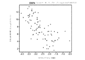


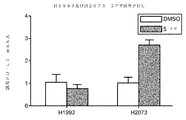
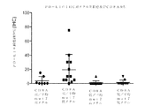


Classifications
-
- A—HUMAN NECESSITIES
- A61—MEDICAL OR VETERINARY SCIENCE; HYGIENE
- A61P—SPECIFIC THERAPEUTIC ACTIVITY OF CHEMICAL COMPOUNDS OR MEDICINAL PREPARATIONS
- A61P35/00—Antineoplastic agents
-
- C—CHEMISTRY; METALLURGY
- C07—ORGANIC CHEMISTRY
- C07K—PEPTIDES
- C07K16/00—Immunoglobulins [IGs], e.g. monoclonal or polyclonal antibodies
- C07K16/18—Immunoglobulins [IGs], e.g. monoclonal or polyclonal antibodies against material from animals or humans
- C07K16/28—Immunoglobulins [IGs], e.g. monoclonal or polyclonal antibodies against material from animals or humans against receptors, cell surface antigens or cell surface determinants
- C07K16/2803—Immunoglobulins [IGs], e.g. monoclonal or polyclonal antibodies against material from animals or humans against receptors, cell surface antigens or cell surface determinants against the immunoglobulin superfamily
- C07K16/2827—Immunoglobulins [IGs], e.g. monoclonal or polyclonal antibodies against material from animals or humans against receptors, cell surface antigens or cell surface determinants against the immunoglobulin superfamily against B7 molecules, e.g. CD80, CD86
-
- C—CHEMISTRY; METALLURGY
- C12—BIOCHEMISTRY; BEER; SPIRITS; WINE; VINEGAR; MICROBIOLOGY; ENZYMOLOGY; MUTATION OR GENETIC ENGINEERING
- C12Q—MEASURING OR TESTING PROCESSES INVOLVING ENZYMES, NUCLEIC ACIDS OR MICROORGANISMS; COMPOSITIONS OR TEST PAPERS THEREFOR; PROCESSES OF PREPARING SUCH COMPOSITIONS; CONDITION-RESPONSIVE CONTROL IN MICROBIOLOGICAL OR ENZYMOLOGICAL PROCESSES
- C12Q1/00—Measuring or testing processes involving enzymes, nucleic acids or microorganisms; Compositions therefor; Processes of preparing such compositions
- C12Q1/68—Measuring or testing processes involving enzymes, nucleic acids or microorganisms; Compositions therefor; Processes of preparing such compositions involving nucleic acids
- C12Q1/6876—Nucleic acid products used in the analysis of nucleic acids, e.g. primers or probes
- C12Q1/6883—Nucleic acid products used in the analysis of nucleic acids, e.g. primers or probes for diseases caused by alterations of genetic material
- C12Q1/6886—Nucleic acid products used in the analysis of nucleic acids, e.g. primers or probes for diseases caused by alterations of genetic material for cancer
-
- A—HUMAN NECESSITIES
- A61—MEDICAL OR VETERINARY SCIENCE; HYGIENE
- A61K—PREPARATIONS FOR MEDICAL, DENTAL OR TOILETRY PURPOSES
- A61K39/00—Medicinal preparations containing antigens or antibodies
- A61K2039/505—Medicinal preparations containing antigens or antibodies comprising antibodies
-
- C—CHEMISTRY; METALLURGY
- C07—ORGANIC CHEMISTRY
- C07K—PEPTIDES
- C07K2317/00—Immunoglobulins specific features
- C07K2317/50—Immunoglobulins specific features characterized by immunoglobulin fragments
- C07K2317/56—Immunoglobulins specific features characterized by immunoglobulin fragments variable (Fv) region, i.e. VH and/or VL
-
- C—CHEMISTRY; METALLURGY
- C07—ORGANIC CHEMISTRY
- C07K—PEPTIDES
- C07K2317/00—Immunoglobulins specific features
- C07K2317/70—Immunoglobulins specific features characterized by effect upon binding to a cell or to an antigen
- C07K2317/76—Antagonist effect on antigen, e.g. neutralization or inhibition of binding
-
- C—CHEMISTRY; METALLURGY
- C12—BIOCHEMISTRY; BEER; SPIRITS; WINE; VINEGAR; MICROBIOLOGY; ENZYMOLOGY; MUTATION OR GENETIC ENGINEERING
- C12Q—MEASURING OR TESTING PROCESSES INVOLVING ENZYMES, NUCLEIC ACIDS OR MICROORGANISMS; COMPOSITIONS OR TEST PAPERS THEREFOR; PROCESSES OF PREPARING SUCH COMPOSITIONS; CONDITION-RESPONSIVE CONTROL IN MICROBIOLOGICAL OR ENZYMOLOGICAL PROCESSES
- C12Q2600/00—Oligonucleotides characterized by their use
- C12Q2600/106—Pharmacogenomics, i.e. genetic variability in individual responses to drugs and drug metabolism
-
- C—CHEMISTRY; METALLURGY
- C12—BIOCHEMISTRY; BEER; SPIRITS; WINE; VINEGAR; MICROBIOLOGY; ENZYMOLOGY; MUTATION OR GENETIC ENGINEERING
- C12Q—MEASURING OR TESTING PROCESSES INVOLVING ENZYMES, NUCLEIC ACIDS OR MICROORGANISMS; COMPOSITIONS OR TEST PAPERS THEREFOR; PROCESSES OF PREPARING SUCH COMPOSITIONS; CONDITION-RESPONSIVE CONTROL IN MICROBIOLOGICAL OR ENZYMOLOGICAL PROCESSES
- C12Q2600/00—Oligonucleotides characterized by their use
- C12Q2600/154—Methylation markers
Landscapes
- Health & Medical Sciences (AREA)
- Chemical & Material Sciences (AREA)
- Life Sciences & Earth Sciences (AREA)
- Organic Chemistry (AREA)
- Immunology (AREA)
- Proteomics, Peptides & Aminoacids (AREA)
- Genetics & Genomics (AREA)
- General Health & Medical Sciences (AREA)
- Zoology (AREA)
- Pathology (AREA)
- Analytical Chemistry (AREA)
- Engineering & Computer Science (AREA)
- Wood Science & Technology (AREA)
- Biophysics (AREA)
- Biochemistry (AREA)
- Molecular Biology (AREA)
- Medicinal Chemistry (AREA)
- Microbiology (AREA)
- Oncology (AREA)
- Biotechnology (AREA)
- Bioinformatics & Cheminformatics (AREA)
- General Engineering & Computer Science (AREA)
- Hospice & Palliative Care (AREA)
- Physics & Mathematics (AREA)
- Pharmacology & Pharmacy (AREA)
- General Chemical & Material Sciences (AREA)
- Nuclear Medicine, Radiotherapy & Molecular Imaging (AREA)
- Chemical Kinetics & Catalysis (AREA)
- Animal Behavior & Ethology (AREA)
- Public Health (AREA)
- Veterinary Medicine (AREA)
- Medicines Containing Antibodies Or Antigens For Use As Internal Diagnostic Agents (AREA)
- Medicines That Contain Protein Lipid Enzymes And Other Medicines (AREA)
- Measuring Or Testing Involving Enzymes Or Micro-Organisms (AREA)
- Pharmaceuticals Containing Other Organic And Inorganic Compounds (AREA)
- Peptides Or Proteins (AREA)
- Investigating Or Analysing Biological Materials (AREA)
- Acyclic And Carbocyclic Compounds In Medicinal Compositions (AREA)
- Medicines Containing Plant Substances (AREA)
Description
関連出願の相互参照
本出願は、2015年5月29日出願の米国仮出願第62/168,668号の優先権の利益を主張するものであり、この仮出願は、参照によりその全体が本明細書に組み込まれる。
Cross-reference to related applications This application claims the priority benefit of US Provisional Application No. 62 / 168,668 filed May 29, 2015, and this provisional application is in its entirety by reference. Incorporated into the specification.
ASCIIテキストファイルでの配列表の提出
ASCIIテキストファイルでの以下の提出の内容は、参照によりその全体が本明細書に組み込まれる:コンピュータ可読形式(CRF)の配列表(ファイル名:146392027040SeqList.txt、記録日:2016年5月27日、サイズ:19KB)。
Submission of Sequence Listing in ASCII Text File The content of the following submission in ASCII text file is incorporated herein by reference in its entirety: Computer Readable Format (CRF) Sequence Listing (filename: 46392207040SeqList.txt, Recording date: May 27, 2016, size: 19KB).
本発明は、患者からの癌細胞を含有する試料中のPD−L1プロモーター領域の上流領域及び/またはPD−L1のイントロン1内の1つ以上のCpG部位においてメチル化レベルを判定することにより、抗PD−L1抗体を用いた治療のために癌患者を層別化する方法に関する。
The present invention determines methylation levels at one or more CpG sites within the PD-L1 promoter region upstream and / or PD-
PD−L1は、多くの癌で過剰発現され、多くの場合、予後不良に関連している(Okazaki T et al.,Intern.Immun.2007 19(7):813)(Thompson RH et al.,Cancer Res 2006,66(7):3381)。興味深いことに、腫瘍浸潤Tリンパ球の大半は、正常組織中のTリンパ球及び末梢血Tリンパ球とは対照的に、PD−1を主に発現し、腫瘍反応性T細胞でのPD−1の上方制御が抗腫瘍免疫応答障害に寄与し得ることを示す(Blood 2009 114(8):1537)。これは、T細胞活性化の減衰及び免疫監視の回避をもたらす、PD−1発現T細胞と相互作用するPD−L1発現腫瘍細胞により媒介されるPD−L1シグナル伝達の利用に起因し得る(Sharpe et al.,Nat Rev 2002)(Keir ME et al.,2008 Annu.Rev.Immunol.26:677)。したがって、PD−L1/PD−1相互作用の阻害が、CD8+T細胞媒介性の腫瘍死滅を増強し得る。 PD-L1 is overexpressed in many cancers and is often associated with a poor prognosis (Okazaki T et al., Intern. Immuno. 2007 19 (7): 813) (Thompson RH et al., Cancer Res 2006,66 (7): 3381). Interestingly, the majority of tumor-infiltrating T lymphocytes, in contrast to T lymphocytes in normal tissue and peripheral blood T lymphocytes, predominantly express PD-1 and PD-in tumor-reactive T cells. It is shown that the upregulation of 1 can contribute to the impaired anti-tumor immune response (Blood 2009 114 (8): 1537). This may be due to the utilization of PD-L1 signaling mediated by PD-L1-expressing tumor cells that interact with PD-1-expressing T cells, resulting in attenuation of T cell activation and avoidance of immune surveillance (Sharpe). et al., Nat Rev 2002) (Keira ME et al., 2008 Annu. Rev. Immunol. 26: 677). Therefore, inhibition of PD-L1 / PD-1 interactions can enhance CD8 + T cell-mediated tumor death.
抗PD−L1抗体及び悪性腫瘍を治療する上でのそれらの使用が記載されている(例えば、Philips et al.(2015)Int Immunol 27,39−46l、Herbst et al.(2014)Nature515,563−567を参照されたい)。ある特定の患者は、免疫チェックポイント阻害剤に対する一次耐性を有する(例えば、Taube et al.(2012)Sci Transl Med.4,127、Sznol et al.(2014)Clin Cancer Res.19,1021−34、及びGajewski et al.(2011)Curr Opin Immunol.23:286−92を参照されたい)。したがって、抗PD−L1抗体療法に対する癌患者の応答性を予測する必要性が未だにある。 Anti-PD-L1 antibodies and their use in treating malignant tumors have been described (eg, Phillips et al. (2015) Int Immunol 27, 39-46 l, Herbst et al. (2014) Nature 515,563. See -567). Certain patients have primary resistance to immune checkpoint inhibitors (eg, Taube et al. (2012) Sci Transl Med. 4,127, Sznoll et al. (2014) Clin Cancer Res. 19, 1021-34). , And Gajewski et al. (2011) Curr Opin Immunol. 23: 286-92). Therefore, there is still a need to predict the responsiveness of cancer patients to anti-PD-L1 antibody therapy.
本明細書で開示される全ての参考文献、公報、及び特許出願は、参照することによりそれらの全体が本明細書に組み込まれる。 All references, publications, and patent applications disclosed herein are incorporated herein by reference in their entirety.
ある特定の実施形態において、本発明は、対象に有効量の抗PD−L1抗体を投与することを含む、対象における癌を治療するか、または癌の進行を遅延させる方法を提供し、ここで、治療(または、進行の遅延)は、対象からの癌細胞を含有する試料中のPD−L1プロモーター領域内のCpG1において及び/またはPD−L1遺伝子のイントロン1内の1つ以上のCpG部位において、中または低レベルのメチル化を有する対象に基づく。
In certain embodiments, the present invention provides a method of treating or delaying the progression of a cancer in a subject, comprising administering to the subject an effective amount of an anti-PD-L1 antibody. , Treatment (or delay in progression) at CpG1 within the PD-L1 promoter region and / or at one or more CpG sites within the
ある特定の実施形態において、本発明は、対象における癌を治療するか、または癌の進行を遅延させる方法を提供するが、但し、対象が、対象からの癌細胞を含有する試料中のPD−L1プロモーター領域内のCpG1において及び/またはPD−L1遺伝子のイントロン1内の1つ以上のCpG部位において、中または低レベルのメチル化を有することが見出されていることを条件とし、本方法は、有効量の抗PD−L1抗体を対象に投与することを含む。
In certain embodiments, the present invention provides a method of treating or delaying the progression of cancer in a subject, provided that the subject is PD-L1 in a sample containing cancer cells from the subject. The method, provided that it has been found to have moderate or low levels of methylation at CpG1 within the L1 promoter region and / or at one or more CpG sites within
ある特定の実施形態において、本発明は、(a)癌を有する対象であって、該対象が、対象からの癌細胞を含有する試料中のPD−L1プロモーター領域内のCpG1において及び/またはPD−L1遺伝子のイントロン1内の1つ以上のCpG部位において、中または低レベルのメチル化を有する、対象を選択することと、(b)このように選択された対象に有効量の抗PD−L1抗体を投与することとを含む、癌を治療するか、または癌の進行を遅延させる方法を提供する。
In certain embodiments, the invention is described in (a) a subject having cancer in CpG1 within the PD-L1 promoter region in a sample containing cancer cells from the subject and / or PD. -Selecting a subject with medium or low levels of methylation at one or more CpG sites within the
ある特定の実施形態において、本発明は、対象からの癌細胞を含有する試料中のPD−L1プロモーター領域内のCpG1において及び/またはPD−L1遺伝子のイントロン1内の1つ以上のCpG部位において、メチル化レベルを測定することを含む、癌を有する対象が、抗PD−L1抗体を用いた治療に応答する可能性が高いかどうかを予測する方法を提供し、ここで、試料中のPD−L1プロモーター領域内のCpG1における、またはPD−L1遺伝子のイントロン1内の1つ以上のCpG部位における中または低レベルのメチル化は、治療に応答する可能性が高い対象を示す。
In certain embodiments, the present invention presents the present invention at CpG1 within the PD-L1 promoter region and / or at one or more CpG sites within the
ある特定の実施形態において、(a)対象からの癌細胞を含有する試料中のPD−L1プロモーター領域内のCpG1において及び/またはPD−L1遺伝子のイントロン1内の1つ以上のCpG部位において、メチル化レベルを測定することと、(b)有効量の抗PD−L1抗体を、PD−L1プロモーター領域内のCpG1において、またはPD−L1遺伝子のイントロン1内の1つ以上のCpG部位において、中または低レベルのメチル化を有することが判定されている対象に投与することとを含む、対象における癌を治療する方法が提供される。
In certain embodiments, (a) at CpG1 within the PD-L1 promoter region and / or at one or more CpG sites within the
ある特定の実施形態において、本発明は、(a)対象からの癌細胞を含有する試料中のPD−L1プロモーター領域内のCpG1において及び/またはPD−L1遺伝子のイントロン1内の1つ以上のCpG部位において、メチル化を評価することと、(b)試料中のPD−L1プロモーター領域内のCpG1において及び/またはPD−L1遺伝子のイントロン1内の1つ以上のCpG部位において、中または低レベルのメチル化を有する対象を特定することとを含む、抗PD−L1抗体治療に応答する可能性が高い癌を有する対象を特定する方法を提供する。
In certain embodiments, the invention is described in (a) one or more in CpG1 within the PD-L1 promoter region and / or within the
上記の実施形態のいずれかによる(または、それらに適用されるような)いくつかの実施形態において、本方法は、有効量の抗PD−L1抗体を対象に投与することをさらに含む。上記の実施形態のいずれかによる(または、それらに適用されるような)いくつかの実施形態において、対象は、PD−L1プロモーター領域内のCpG1において、及びPD−L1遺伝子のイントロン1内の1つ以上のCpG部位において、中または低レベルのメチル化を有する。上記の実施形態のいずれかによる(または、それらに適用されるような)いくつかの実施形態において、メチル化レベルは、バイサルファイト次世代配列決定により判定される。上記の実施形態のいずれかによる(または、それらに適用されるような)いくつかの実施形態において、メチル化レベルは、バイサルファイト次世代配列決定により判定される。上記の実施形態のいずれかによる(または、それらに適用されるような)いくつかの実施形態において、メチル化レベルは、メチル化チップアレイを使用して判定される。上記の実施形態のいずれかによる(または、それらに適用されるような)いくつかの実施形態において、対象からの試料は、免疫細胞浸潤の根拠を示す。上記の実施形態のいずれかによる(または、それらに適用されるような)いくつかの実施形態において、免疫細胞浸潤の根拠は、ウェスタンブロット、ELISA、フローサイトメトリー、qPCR、qRT−PCR、トランスクリプトームプロファイリング、マイクロアレイ分析、または次世代配列決定により検出されるCD8+リンパ球により示される。
In some embodiments according to any of the above embodiments (or as applied thereto), the method further comprises administering to the subject an effective amount of an anti-PD-L1 antibody. In some embodiments according to any of the above embodiments (or as applied thereto), the subject is at CpG1 within the PD-L1 promoter region and at 1 within the
上記の実施形態のいずれかによる(または、それらに適用されるような)いくつかの実施形態において、バイサルファイト配列決定により判定される場合、約20%〜約40%のメチル化の中レベルのメチル化。上記の実施形態のいずれかによる(または、それらに適用されるような)いくつかの実施形態において、バイサルファイト配列決定により判定される場合の低レベルのメチル化は、約20%未満のメチル化である。 In some embodiments according to any of the above embodiments (or as applied thereto), as determined by bisulfite sequencing, a medium level of methylation of about 20% to about 40%. Methylation. In some embodiments according to any of the above embodiments (or as applied thereto), the low level of methylation as determined by bisulfite sequencing is less than about 20% methylation. Is.
上記の実施形態のいずれかによる(または、それらに適用されるような)いくつかの実施形態において、バイサルファイト次世代配列決定により判定される場合、約5%〜約60%のメチル化の中レベルのメチル化。上記の実施形態のいずれかによる(または、それらに適用されるような)いくつかの実施形態において、バイサルファイト次世代配列決定により判定される場合の低レベルのメチル化は、約5%未満のメチル化である。 In some embodiments according to any of the above embodiments (or as applied thereto), as determined by bisulfite next generation sequencing, within about 5% to about 60% methylation. Level methylation. In some embodiments according to any of the above embodiments (or as applied thereto), the low level of methylation as determined by bisulfite next generation sequencing is less than about 5%. It is methylation.
上記の実施形態のいずれかによる(または、それらに適用されるような)いくつかの実施形態において、メチル化チップアレイにより判定される場合の中レベルのメチル化は、約0.2〜約0.3のベータ値である。上記の実施形態のいずれかによる(または、それらに適用されるような)いくつかの実施形態において、メチル化チップアレイにより判定される場合の低レベルのメチル化は、約0.2未満のベータ値である。 In some embodiments according to any of the above embodiments (or as applied thereto), the level of methylation as determined by the methylation chip array is from about 0.2 to about 0. It is a beta value of 0.3. In some embodiments according to any of the above embodiments (or as applied thereto), the low level of methylation as determined by the methylation chip array is less than about 0.2 beta. The value.
上記の実施形態のいずれかによる(または、それらに適用されるような)いくつかの実施形態において、癌は、肺癌、乳癌、膀胱癌、または黒色腫である。上記の実施形態のいずれかによる(または、それらに適用されるような)いくつかの実施形態において、癌は、肺癌であり、肺癌は、非小細胞肺癌、肺扁平上皮癌腫、または肺腺癌腫である。 In some embodiments according to (or as applied to) any of the above embodiments, the cancer is lung cancer, breast cancer, bladder cancer, or melanoma. In some embodiments according to (or as applied to) any of the above embodiments, the cancer is lung cancer and the lung cancer is non-small cell lung cancer, pulmonary squamous cell carcinoma, or lung adenocarcinoma. Is.
上記の実施形態のいずれかによる(または、それらに適用されるような)いくつかの実施形態において、抗PD−L1抗体は、PD−L1のPD−1への結合を阻害する。上記の実施形態のいずれかによる(または、それらに適用されるような)いくつかの実施形態において、抗PD−L1抗体は、PD−L1のB7−1への結合を阻害する。上記の実施形態のいずれかによる(または、それらに適用されるような)いくつかの実施形態において、抗PD−L1抗体は、PD−L1の、PD−1及びB7−1の両方への結合を阻害する。上記の実施形態のいずれかによる(または、それらに適用されるような)いくつかの実施形態において、抗PD−L1抗体は、モノクローナル抗体である。上記の実施形態のいずれかによる(または、それらに適用されるような)いくつかの実施形態において、抗PD−L1抗体は、Fab、Fab’−SH、Fv、scFv、及び(Fab’)2断片からなる群から選択される抗体断片である。上記の実施形態のいずれかによる(または、それらに適用されるような)いくつかの実施形態において、抗PD−L1抗体は、ヒト化抗体またはヒト抗体である。上記の実施形態のいずれかによる(または、それらに適用されるような)いくつかの実施形態において、抗PD−L1抗体は、YW243.55.S70、MPDL3280A、MDX−1105、及びMEDI4736からなる群から選択される。上記の実施形態のいずれかによる(または、それらに適用されるような)いくつかの実施形態において、抗PD−L1抗体は、配列番号15のHVR−H1配列、配列番号16のHVR−H2配列、及び配列番号3のHVR−H3配列を含む重鎖と、配列番号17のHVR−L1配列、配列番号18のHVR−L2配列、及び配列番号19のHVR−L3配列を含む軽鎖とを含む。上記の実施形態のいずれかによる(または、それらに適用されるような)いくつかの実施形態において、抗PD−L1抗体は、配列番号24のアミノ酸配列を含む重鎖可変領域と、配列番号21のアミノ酸配列を含む軽鎖可変領域とを含む。 In some embodiments according to any of the above embodiments (or as applied thereto), the anti-PD-L1 antibody inhibits the binding of PD-L1 to PD-1. In some embodiments according to any of the above embodiments (or as applied thereto), the anti-PD-L1 antibody inhibits the binding of PD-L1 to B7-1. In some embodiments according to any of the above embodiments (or as applied thereto), the anti-PD-L1 antibody binds PD-L1 to both PD-1 and B7-1. Inhibits. In some embodiments according to any of the above embodiments (or as applied thereto), the anti-PD-L1 antibody is a monoclonal antibody. In some embodiments according to any of the above embodiments (or as applied thereto), the anti-PD-L1 antibody is Fab, Fab'-SH, Fv, scFv, and (Fab') 2 An antibody fragment selected from the group consisting of fragments. In some embodiments according to any of the above embodiments (or as applied thereto), the anti-PD-L1 antibody is a humanized antibody or a human antibody. In some embodiments according to any of the above embodiments (or as applied thereto), the anti-PD-L1 antibody is YW243.55. It is selected from the group consisting of S70, MPDL3280A, MDX-1105, and MEDI4736. In some embodiments according to any of the above embodiments (or as applied thereto), the anti-PD-L1 antibody is the HVR-H1 sequence of SEQ ID NO: 15, the HVR-H2 sequence of SEQ ID NO: 16. , And a heavy chain comprising the HVR-H3 sequence of SEQ ID NO: 3, an HVR-L1 sequence of SEQ ID NO: 17, an HVR-L2 sequence of SEQ ID NO: 18, and a light chain comprising the HVR-L3 sequence of SEQ ID NO: 19. .. In some embodiments according to any of the above embodiments (or as applied thereto), the anti-PD-L1 antibody comprises a heavy chain variable region comprising the amino acid sequence of SEQ ID NO: 24 and SEQ ID NO: 21. Includes a light chain variable region containing the amino acid sequence of.
いくつかの実施形態において、本発明は、一緒にパッケージ化された、抗PD−L1抗体及び薬学的に許容される担体を含む薬学的組成物と、抗PD−L1抗体または薬学的組成物が、対象からの癌細胞を含有する試料中のPD−L1プロモーター領域内のCpG1において及び/またはPD−L1遺伝子のイントロン1内の1つ以上のCpG部位において、中または低レベルのメチル化を有する癌を有する対象の治療に適応することを表示するラベルとを含む、製品を提供する。
In some embodiments, the invention comprises a pharmaceutical composition comprising an anti-PD-L1 antibody and a pharmaceutically acceptable carrier, packaged together, and an anti-PD-L1 antibody or pharmaceutical composition. Has medium or low levels of methylation at CpG1 within the PD-L1 promoter region and / or at one or more CpG sites within
いくつかの実施形態において、本発明は、対象からの癌細胞を含有する試料中のPD−L1プロモーター領域内のCpG1において及び/またはPD−L1遺伝子のイントロン1内の1つ以上のCpG部位において、メチル化レベルを測定するための試薬と、PD−L1プロモーター領域内のCpG1において及び/またはPD−L1遺伝子のイントロン1内の1つ以上のCpG部位において、中または低メチル化レベルを有するとして対象を分類するための使用説明書とを含むキットを提供する。上記の実施形態のいずれかによる(または、それらに適用されるような)いくつかの実施形態において、キットまたは製品は、抗PD−L1抗体と、対象が、PD−L1プロモーター領域内のCpG1において及び/またはPD−L1遺伝子のイントロン1内の1つ以上のCpG部位において、中または低メチル化レベルを有する場合、抗PD−L1抗体を対象に投与するための使用説明書とをさらに含む。
In some embodiments, the invention presents at CpG1 within the PD-L1 promoter region and / or at one or more CpG sites within the
本明細書に記載される様々な実施形態の特性のうちの1つ、いくつか、または全てを組み合わせて、本発明の他の実施形態を形成することができることを理解されたい。本発明のこれら及び他の態様は、当業者に明らかであろう。本発明のこれら及び他の実施形態は、以下の発明を実施するための形態によりさらに記載される。 It should be appreciated that one, some, or all of the properties of the various embodiments described herein can be combined to form other embodiments of the invention. These and other aspects of the invention will be apparent to those skilled in the art. These and other embodiments of the present invention are further described by embodiments for carrying out the following inventions.
I.一般的技法
本明細書において記載されるか、または参照される技法及び手順は、当業者により概して十分に理解され、例えば、Sambrook et al.,Molecular Cloning:A Laboratory Manual 3d edition(2001)Cold Spring Harbor Laboratory Press,Cold Spring Harbor,N.Y.、Current Protocols in Molecular Biology(F.M.Ausubel,et al.eds.,(2003))、the series Methods in Enzymology(Academic Press,Inc.):PCR 2:A Practical Approach(M.J.MacPherson,B.D.Hames and G.R.Taylor eds.(1995)),Harlow and Lane,eds.(1988)Antibodies,A Laboratory Manual,and Animal Cell Culture(R.I.Freshney,ed.(1987))、Oligonucleotide Synthesis(M.J.Gait,ed.,1984)、Methods in Molecular Biology,Humana Press、Cell Biology:A Laboratory Notebook(J.E.Cellis,ed.,1998)Academic Press、Animal Cell Culture(R.I.Freshney),ed.,1987)、Introduction to Cell and Tissue Culture(J.P.Mather and P.E.Roberts,1998)Plenum Press、Cell and Tissue Culture:Laboratory Procedures(A.Doyle,J.B.Griffiths,and D.G.Newell,eds.,1993−8)J.Wiley and Sons、Handbook of Experimental Immunology(D.M.Weir and C.C.Blackwell,eds.)、Gene Transfer Vectors for Mammalian Cells(J.M.Miller and M.P.Calos,eds.,1987)、PCR:The Polymerase Chain Reaction,(Mullis et al.,eds.,1994)、Current Protocols in Immunology(J.E.Coligan et al.,eds.,1991)、Short Protocols in Molecular Biology(Wiley and Sons,1999)、Immunobiology(C.A.Janeway and P.Travers,1997)、Antibodies(P.Finch,1997)、Antibodies:A Practical Approach(D.Catty.,ed.,IRL Press,1988−1989)、Monoclonal Antibodies:A Practical Approach(P.Shepherd and C.Dean,eds.,Oxford University Press,2000)、Using Antibodies:A Laboratory Manual(E.Harlow and D.Lane(Cold Spring Harbor Laboratory Press,1999)、The Antibodies(M.Zanetti and J.D.Capra,eds.,Harwood Academic Publishers,1995)、及びCancer:Principles and Practice of Oncology(V.T.DeVita et al.,eds.,J.B.Lippincott Company,1993)に記載される広く利用されている方法論などの、従来の方法論などを使用して一般的に用いられる。
I. General Techniques Techniques and procedures described or referenced herein are generally well understood by those of skill in the art, eg, Sambrook et al. , Molecular Cloning: A Laboratory Manual 3d edition (2001) Cold Spring Harbor Laboratory Press, Cold Spring Harbor, N. et al. Y. , Current Protocols in Molecular Biology (FM Ausubel, et al. Eds., (2003)), the series Methods in Enzymology (Academic Press, Inc.):
II.定義
本明細書で使用される場合、「治療」という用語は、臨床的病変の経過中に治療されている個体または細胞の自然経過を改変するように設計された臨床的介入を指す。治療の望ましい効果には、疾患進行速度の減速、疾患状態の回復または緩和、及び寛解または予後の改善が含まれる。例えば、個体は、癌性細胞の増殖の低減(もしくは破壊)、疾患に起因する症状の減少、疾患に罹患している者の生活の質の向上、疾患の治療に必要な他の薬剤の用量の減少、及び/または個体の生存期間の延長を含むが、これらに限定されない、癌に関連する1つ以上の症状が軽減または排除された場合、「治療」が成功する。
II. Definitions As used herein, the term "treatment" refers to a clinical intervention designed to alter the natural course of an individual or cell being treated during the course of a clinical lesion. Desirable effects of treatment include slowing the rate of disease progression, relieving or alleviating the disease state, and improving remission or prognosis. For example, an individual may reduce (or destroy) the growth of cancerous cells, reduce symptoms caused by the disease, improve the quality of life of the person suffering from the disease, and dose other drugs needed to treat the disease. "Treatment" is successful if one or more symptoms associated with the disease, including but not limited to, and / or prolonging the survival of the individual, are alleviated or eliminated.
本明細書で使用される場合、「based upon(に基づく)」は、(1)本明細書に記載されるような患者の特徴を評価すること、判定すること、または測定すること(及び好ましくは、治療を受けるのに好適な患者を選択すること、及び(2)本明細書に記載されるような治療(複数可)を行うこと、を含む。 As used herein, "based upon" is (1) assessing, determining, or measuring (and preferably) the characteristics of a patient as described herein. Includes selecting the appropriate patient to receive treatment and (2) performing the treatment (s) as described herein.
治療目的のための「対象」、「患者」、または「個体」は、ヒト、飼育動物及び家畜、ならびに動物園、競技用、または愛玩動物、例えば、イヌ、ウマ、ネコ、ウシなどを含む、哺乳動物として分類される任意の動物を指す。好ましくは、哺乳動物は、ヒトである。 "Subjects", "patients", or "individuals" for therapeutic purposes include mammals, including humans, domestic animals and livestock, and zoos, competition or pet animals such as dogs, horses, cats, cows, etc. Refers to any animal that is classified as an animal. Preferably, the mammal is a human.
本明細書で使用される場合、「疾患の進行を遅延させる」は、疾患(癌など)の発症を延期し、妨害し、減速し、遅らせ、安定させ、かつ/または延ばすことを意味する。この遅延は、治療されている病歴及び/または個体に応じて異なる期間であり得る。当業者に明らかであるように、十分または著しい遅延は、個体が疾患を発症しないという点で、予防を事実上包含し得る。例えば、転移の発症などの末期癌を遅延させることができる。 As used herein, "delaying the progression of a disease" means delaying, interfering with, slowing down, delaying, stabilizing, and / or delaying the onset of a disease (such as cancer). This delay can be for different periods depending on the medical history and / or individual being treated. As will be apparent to those of skill in the art, sufficient or significant delays may effectively include prevention in that the individual does not develop the disease. For example, terminal cancer such as the onset of metastasis can be delayed.
「有効量」とは、特定の障害の測定可能な改善または予防をもたらすために必要な少なくとも最小の量である。本明細書における有効量は、患者の疾患状態、年齢、性別、及び体重、ならびに個体における所望の応答を誘発する抗体の能力などの要因に応じて異なり得る。有効量は、治療上有益な効果が治療の任意の毒性効果または有害効果を上回るものでもある。予防的使用の場合、有益なまたは所望の結果には、疾患の生化学的、組織学的、及び/または挙動的症状、その合併症、ならびに疾患の発症中に現れる中間病理学的表現型を含む、疾患のリスクの排除もしくは低減、疾患の重症度の軽減、または疾患の発生の遅延などの結果が含まれる。治療的使用の場合、有益なまたは所望の結果には、疾患に起因する1つ以上の症状の減少、疾患に罹患している者の生活の質の向上、疾患の治療に必要な他の薬剤の用量の減少、別の薬剤の効果の増強(例えば、標的による)、疾患の進行の遅延、及び/または生存期間の延長などの臨床結果が含まれる。癌または腫瘍の場合、有効量の薬物は、癌細胞の数を低減させ、腫瘍サイズを低減させ、癌細胞の末梢器官への浸潤を阻害し(すなわち、ある程度遅らせるか、または望ましくは停止し)、腫瘍転移を阻害し(すなわち、ある程度遅らせるか、または望ましくは停止し)、腫瘍成長をある程度阻害し、かつ/または障害に関連する症状のうちの1つ以上をある程度軽減する効果を有し得る。有効量は、1回以上の投与で投与され得る。本発明の目的に関しては、薬物、化合物、または薬学的組成物の有効量は、予防的または治療的治療を直接または間接的に達成するのに十分な量である。臨床分野において理解されるように、薬物、化合物、または薬学的組成物の有効量は、別の薬物、化合物、または薬学的組成物と併せて達成されても、されなくてもよい。したがって、「有効量」は、1つ以上の療法剤を投与するという点で考慮され得、単剤は、1つ以上の他の薬剤と併せて、望ましい結果が達成され得るか、または達成される場合、有効量で与えられると見なされ得る。 An "effective amount" is at least the minimum amount required to bring about a measurable improvement or prevention of a particular disorder. Effective amounts herein may vary depending on factors such as the patient's disease state, age, gender, and body weight, as well as the ability of the antibody to elicit the desired response in the individual. The effective amount is also such that the therapeutically beneficial effect outweighs any toxic or adverse effect of the treatment. For prophylactic use, beneficial or desired outcomes include the biochemical, histological and / or behavioral symptoms of the disease, its complications, and the intermediate pathological phenotype that appears during the onset of the disease. Includes results such as elimination or reduction of the risk of the disease, reduction of the severity of the disease, or delay in the onset of the disease. For therapeutic use, beneficial or desired outcomes include reduction of one or more symptoms due to the disease, improvement of the quality of life of the person suffering from the disease, other drugs needed to treat the disease. Includes clinical outcomes such as reduced dose of, enhanced efficacy of another drug (eg, by target), delayed disease progression, and / or prolonged survival. In the case of cancer or tumor, an effective amount of drug reduces the number of cancer cells, reduces tumor size, inhibits the infiltration of cancer cells into peripheral organs (ie, delays to some extent, or preferably stops). Can have the effect of inhibiting tumor metastasis (ie, delaying it to some extent or, preferably stopping it), inhibiting tumor growth to some extent, and / or alleviating one or more of the symptoms associated with the disorder to some extent. .. Effective amounts can be administered in one or more doses. For the purposes of the present invention, an effective amount of a drug, compound, or pharmaceutical composition is sufficient to directly or indirectly achieve prophylactic or therapeutic treatment. As is understood in the clinical setting, an effective amount of a drug, compound, or pharmaceutical composition may or may not be achieved in conjunction with another drug, compound, or pharmaceutical composition. Thus, an "effective amount" can be considered in terms of administering one or more therapeutic agents, with a single agent being combined with one or more other agents to achieve or achieve the desired results. If so, it can be considered to be given in an effective amount.
「細胞増殖性障害」及び「増殖性障害」という用語は、ある程度の異常な細胞増殖に関連する障害を指す。一実施形態において、細胞増殖性障害は、癌である。一実施形態において、細胞増殖性障害は、腫瘍である。 The terms "cell proliferation disorders" and "proliferative disorders" refer to disorders associated with some abnormal cell proliferation. In one embodiment, the cell proliferation disorder is cancer. In one embodiment, the cell proliferation disorder is a tumor.
「腫瘍」は、本明細書で使用される場合、悪性か良性かを問わず、全ての腫瘍性細胞の成長及び増殖、ならびに全ての前がん性及びがん性細胞及び組織を指す。「癌」、「癌性」、「細胞増殖性障害」、「増殖性障害」、及び「腫瘍」という用語は、本明細書で言及される場合、相互排他的ではない。 "Tumor" as used herein refers to the growth and proliferation of all neoplastic cells, whether malignant or benign, and all precancerous and cancerous cells and tissues. The terms "cancer", "cancer", "cell proliferation disorder", "proliferative disorder", and "tumor" are not mutually exclusive as referred to herein.
「癌」及び「癌性」という用語は、制御されていない細胞成長を典型的に特徴とする哺乳動物における生理学的状態を指すか、または説明する。癌の例には、癌腫、リンパ腫、芽細胞腫、肉腫、白血病またはリンパ系悪性腫瘍、扁平上皮癌(例えば、上皮性扁平上皮癌)、腹膜の癌、肝細胞癌、胃癌(gastric cancer)または胃癌(stomach cancer)(消化管癌及び消化管間質癌など)、膵臓癌、膠芽腫、子宮頸癌、卵巣癌、肝臓癌、膀胱癌、尿路性の癌、肝細胞癌、結腸癌、直腸癌、結腸直腸癌(CRC)、子宮内膜癌または子宮癌腫、唾液腺癌腫、腎臓癌(kidney cancer)または腎臓癌(renal cancer)、前立腺癌、外陰部癌、甲状腺癌、肝癌腫、肛門癌腫、陰茎癌腫、多発性骨髄腫及びB細胞リンパ腫(低悪性度/濾胞性非ホジキンリンパ腫(NHL)、小リンパ球性(SL)NHL、中悪性度/濾胞性NHL、中悪性度/びまん性NHL、高悪性度免疫芽細胞性NHL、高悪性度リンパ芽球性NHL、高悪性度小型非開裂細胞性NHL、巨大病変性NHL、マントル細胞リンパ腫、AIDS関連リンパ腫、及びヴァルデンストレームマクログロブリン血症など)、慢性骨髄芽球性白血病(CLL)、急性リンパ芽球性白血病(ALL)、有毛細胞性白血病、慢性骨髄芽球性白血病、及び移植後リンパ増殖性障害(PTLD)、ならびに母斑症に関連する異常血管増殖、浮腫(脳腫瘍に関連するものなど)、メイグス症候群、脳癌、ならびに頭頸部癌、軟部組織肉腫、カポジ肉腫、カルチノイド癌腫、及び中皮腫、膠芽腫、神経芽細胞腫、ならびに関連する転移が含まれるが、これらに限定されない。癌の他の例には、乳癌(乳癌腫など)、肺癌(小細胞肺癌、非小細胞肺癌、肺の腺癌腫、及び肺の扁平上皮癌腫など)、及び皮膚癌(黒色腫、表在拡大型黒色腫、悪性黒子型黒色腫、末端黒子型黒色腫、結節型黒色腫、及び皮膚癌腫など)(それらの癌の転移型を含む)が含まれるが、これらに限定されない。 The terms "cancer" and "cancerous" refer to or describe physiological conditions in mammals that are typically characterized by uncontrolled cell growth. Examples of cancers include cancer, lymphoma, blastoma, sarcoma, leukemia or lymphoid malignancies, squamous cell carcinoma (eg, epithelial squamous cell carcinoma), peritoneal cancer, hepatocellular carcinoma, gastric cancer or gastric cancer. Gastric cancer (stomach cancer, etc.), pancreatic cancer, glioblastoma, cervical cancer, ovarian cancer, liver cancer, bladder cancer, urinary tract cancer, hepatocellular carcinoma, colon cancer , Rectal cancer, colonic rectal cancer (CRC), endometrial cancer or uterine cancer, salivary adenocarcinoma, kidney cancer (kidney cancer) or kidney cancer (renal cancer), prostate cancer, genital cancer, thyroid cancer, liver cancer, anus Cancer, penile carcinoma, multiple myeloma and B-cell lymphoma (low grade / follicular non-Hodgkin lymphoma (NHL), small lymphocytic (SL) NHL, medium grade / follicular NHL, medium grade / diffuse NHL, high-grade immunoblastic NHL, high-grade lymphoblastic NHL, high-grade small non-cleavage NHL, giant lesion NHL, mantle cell lymphoma, AIDS-related lymphoma, and Waldenstraem macroglobulin Bloodemia, etc.), Chronic myeloid leukemia (CLL), Acute lymphoblastic leukemia (ALL), Hair cell leukemia, Chronic myeloid leukemia, and Post-transplant lymphoproliferative disorder (PTLD), and Abnormal angiogenesis associated with mammary plaques, edema (such as those associated with brain tumors), Maegus syndrome, brain cancer, and head and neck cancer, soft tissue sarcoma, capoic sarcoma, cartinoid carcinoma, and mesopharyngeal tumor, glioblastoma, Includes, but is not limited to, neuroblastoma, as well as related metastases. Other examples of cancer include breast cancer (such as breast cancer), lung cancer (such as small cell lung cancer, non-small cell lung cancer, lung adenocarcinoma, and lung squamous cell carcinoma), and skin cancer (black tumor, superficial spread). Includes, but is not limited to, type melanomas, malignant melanomas, terminal melanomas, nodular melanomas, and cutaneous carcinomas) (including, but not limited to, metastatic forms of those cancers).
「試料」は、本明細書で使用される場合、例えば、物理的、生化学的、化学的、及び/または生理的特徴に基づいて特性化及び/または特定される、細胞実体及び/または他の分子実体を含有する、目的の対象から得られるか、またはそれに由来する、組成物を指す。例えば、「疾患試料」という用語、及びその変化形は、特性化される細胞実体及び/または分子実体を含有することが予想されるか、または含有することが知られている、目的の対象から得られる任意の試料を指す。試料は、核酸(ポリヌクレオチド、例えばゲノムDNA及び/または転写など)及び/またはポリペプチドが単離され得る対象からの癌細胞及び/または腫瘍細胞を含有する生物学的組織または流体の生物学的試料(エクスビボ生物学的試料など)であり得る。かかる試料は典型的には、ヒト対象からのものであるが、他の対象(本明細書の他の場所に記載されるような哺乳動物として分類されるかかる任意の動物)から単離される組織を含む。試料には、生体組織及び解剖試料などの組織の切片、組織学的目的のために採取された凍結切片も含まれ得る。試料には、対象から採取したての試料、またはホルマリン固定パラフィン包埋(FFPE)試料などの保存組織試料が含まれ得る。試料には、患者組織に由来する外植片、ならびに初代及び/または形質転換細胞培養物も含まれる。 A "sample" as used herein is, for example, a cell entity and / or other characterized and / or specified based on physical, biochemical, chemical, and / or physiological characteristics. Refers to a composition obtained from or derived from an object of interest that contains the molecular entity of. For example, the term "disease sample", and its variants, from an object of interest that is expected or is known to contain a characterized cellular and / or molecular entity. Refers to any sample obtained. The sample is a biological tissue or fluid containing cancer cells and / or tumor cells from a subject from which nucleic acids (such as genomic DNA and / or transcription) and / or polypeptides can be isolated. It can be a sample (such as an Exvivo biological sample). Such samples are typically from human subjects, but tissues isolated from other subjects (such any animal classified as a mammal as described elsewhere herein). including. Samples may also include sections of tissue such as biological tissue and anatomical samples, and frozen sections taken for histological purposes. Samples can include freshly taken samples from the subject or preserved tissue samples such as formalin-fixed paraffin-embedded (FFPE) samples. Samples also include explants from patient tissue, as well as primary and / or transformed cell cultures.
「組織または細胞試料」は、対象または患者の組織から得られる同様の細胞の集合を意味する。組織または細胞試料の供給源は、採取したての、凍結された、及び/または保存された、臓器もしくは組織試料、または生体組織もしくは吸引液;血液または任意の血液組成成分;脳脊髄液、羊膜液、腹腔液、または組織液などの体液;対象の妊娠期間または発育における任意の時期の細胞からの固形組織であり得る。組織試料は、初代もしくは培養細胞、または細胞株でもあり得る。任意に、組織または細胞試料は、疾患組織/臓器から得られる。組織試料は、防腐剤、抗凝固剤、緩衝液、固定剤、栄養素、抗生物質などの、自然界の組織とは自然には混合されない化合物を含有し得る。 "Tissue or cell sample" means a similar collection of cells obtained from the tissue of a subject or patient. The source of the tissue or cell sample is a freshly collected, frozen and / or preserved organ or tissue sample, or living tissue or inhalation fluid; blood or any blood composition component; cerebrospinal fluid, sheep membrane. Body fluids such as fluid, cerebrospinal fluid, or tissue fluid; can be solid tissue from cells at any time during the pregnancy or development of the subject. The tissue sample can also be a primary or cultured cell, or a cell line. Optionally, the tissue or cell sample is obtained from the diseased tissue / organ. Tissue samples may contain compounds that are not naturally mixed with natural tissue, such as preservatives, anticoagulants, buffers, fixatives, nutrients, antibiotics.
「細胞傷害性剤」という用語は、本明細書で使用される場合、(例えば、細胞死を引き起こすか、増殖を阻害するか、またはさもなければ細胞機能を妨害する)細胞に有害である任意の薬剤を指す。細胞傷害性剤には、放射性同位体(例えば、At211、I131、I125、Y90、Re186、Re188、Sm153、Bi212、P32、Pb212、及びLuの放射性同位体)、化学療法剤、成長阻害剤、酵素及びそれらの断片、例えば、核酸分解酵素、ならびに毒素、例えば、細菌、真菌、植物、または動物起源の小分子毒素または酵素的に活性な毒素(それらの断片及び/または変異体を含む)が含まれるが、これらに限定されない。例示的な細胞傷害性剤は、抗微小管薬、白金配位錯体、アルキル化剤、抗生物質剤、トポイソメラーゼII阻害剤、代謝拮抗薬、トポイソメラーゼI阻害剤、ホルモン及びホルモン類似体、シグナル伝達経路阻害剤、非受容体チロシンキナーゼ血管新生阻害剤、免疫療法剤、アポトーシス促進剤、LDH−A阻害剤、脂肪酸生合成阻害剤、細胞周期シグナル伝達阻害剤、HDAC阻害剤、プロテアソーム阻害剤、ならびに癌代謝阻害剤から選択され得る。一実施形態において、細胞傷害性剤は、タキサンである。一実施形態において、タキサンは、パクリタキセルまたはドセタキセルである。一実施形態において、細胞傷害性剤は、白金剤である。一実施形態において、細胞傷害性剤は、EGFRのアンタゴニストである。一実施形態において、EGFRのアンタゴニストは、N−(3−エチニルフェニル)−6,7−ビス(2−メトキシエトキシ)キナゾリン−4−アミン(例えば、エルロチニブ)である。一実施形態において、細胞傷害性剤は、RAF阻害剤である。一実施形態において、RAF阻害剤は、BRAF及び/またはCRAF阻害剤である。一実施形態において、RAF阻害剤は、ベムラフェニブである。一実施形態において、細胞傷害性剤は、PI3K阻害剤である。 The term "cytotoxic agent", as used herein, is arbitrary that is harmful to cells (eg, causing cell death, inhibiting proliferation, or otherwise interfering with cell function). Refers to the drug of. Radioisotopes include radioisotopes (eg, At 211 , I 131 , I 125 , Y 90 , Re 186 , Re 188 , Sm 153 , Bi 212 , P 32 , Pb 212 , and Lu radioisotopes). , Chemotherapeutic agents, growth inhibitors, enzymes and fragments thereof, such as nucleic acid degrading enzymes, and toxins, such as small molecule toxins of bacterial, fungal, plant, or animal origin or enzymatically active toxins (fragments thereof). And / or including, but not limited to, variants. Exemplary cytotoxic agents are anti-microtubes, platinum coordination complexes, alkylating agents, antibiotics, topoisomerase II inhibitors, metabolic antagonists, topoisomerase I inhibitors, hormones and hormone analogs, signaling pathways. Inhibitors, non-receptor tyrosine kinase angiogenesis inhibitors, immunotherapeutic agents, apoptosis promoters, LDH-A inhibitors, fatty acid biosynthesis inhibitors, cell cycle signaling inhibitors, HDAC inhibitors, proteasome inhibitors, and cancer It can be selected from metabolic inhibitors. In one embodiment, the cytotoxic agent is a taxane. In one embodiment, the taxane is paclitaxel or docetaxel. In one embodiment, the cytotoxic agent is a platinum agent. In one embodiment, the cytotoxic agent is an antagonist of EGFR. In one embodiment, the antagonist of EGFR is N- (3-ethynylphenyl) -6,7-bis (2-methoxyethoxy) quinazoline-4-amine (eg, erlotinib). In one embodiment, the cytotoxic agent is a RAF inhibitor. In one embodiment, the RAF inhibitor is a BRAF and / or CRAF inhibitor. In one embodiment, the RAF inhibitor is vemurafenib. In one embodiment, the cytotoxic agent is a PI3K inhibitor.
「化学療法剤」は、癌の治療において有用な化合物を指す。化学療法剤の例には、エルロチニブ(TARCEVA(登録商標)、Genentech/OSI Pharm.)、ボルテゾミブ(VELCADE(登録商標)、Millennium Pharm.)、ジスルフィラム、没食子酸エピガロカテキン、サリノスポラミドA、カーフィルゾミブ、17−AAG(ゲルダナマイシン)、ラディシコール、乳酸デヒドロゲナーゼA(LDH−A)、フルベストラント(FASLODEX(登録商標)、AstraZeneca)、スニチブ(sunitib)(SUTENT(登録商標)、Pfizer/Sugen)、レトロゾール(FEMARA(登録商標)、Novartis)、メシル酸イマチニブ(GLEEVEC(登録商標)、Novartis)、フィナスネート(finasunate)(VATALANIB(登録商標)、Novartis)、オキサリプラチン(ELOXATIN(登録商標)、Sanofi)、5−FU(5−フルオロウラシル)、ロイコボリン、ラパマイシン(シロリムス、RAPAMUNE(登録商標)、Wyeth)、ラパチニブ(TYKERB(登録商標)、GSK572016、Glaxo Smith Kline)、ロナファミブ(Lonafamib)(SCH 66336)、ソラフェニブ(NEXAVAR(登録商標)、Bayer Labs)、ゲフィチニブ(IRESSA(登録商標)、AstraZeneca)、AG1478、アルキル化剤、例えばチオテパ及びCYTOXAN(登録商標)シクロホスファミド;アルキルスルホネート、例えばブスルファン、インプロスルファン、及びピポスルファン;アジリジン、例えばベンゾドーパ(benzodopa)、カルボコン、メツレドーパ(meturedopa)、及びウレドーパ(uredopa);エチレンイミン及びメチラメラミン(methylamelamine)(アルトレタミン、トリエチレンメラミン、トリエチレンホスホラミド、トリエチレンチオホスホルアミド、及びトリメチロメラミンを含む);アセトゲニン(特に、ブラタシン及びブラタシノン);カンプトテシン(トポテカン及びイリノテカンを含む);ブリオスタチン;カリスタチン(callystatin);CC−1065(そのアゾゼレシン、カルゼレシン、及びビゼレシン合成類似体を含む);クリプトフィシン(特に、クリプトフィシン1及びクリプトフィシン8);副腎皮質ステロイド(プレドニゾン及びプレドニゾロンを含む);酢酸シプロテロン;5α−還元酵素フィナステリド及びデュタステリドを含む);ボリノスタット、ロミデプシン、パノビノスタット、バルプロ酸、モセチノスタットドラスタチン;アルデスロイキン、タルクデュオカルマイシン(合成類似体KW−2189及びCB1−TM1を含む);エロイテロビン(eleutherobin);パンクラチスタチン(pancratistatin);サルコジクチイン;スポンジスタチン(spongistatin);ナイトロジェンマスタード、例えばクロラムブシル、クロマファジン(chlomaphazine)、クロロホスファミド、エストラムスチン、イホスファミド、メクロレタミン、メクロレタミンオキシド塩酸塩(mechlorethamine oxide hydrochloride)、メルファラン、ノベムビシン(novembichin)、フェネステリン(phenesterine)、プレドニムスチン、トロホスファミド、ウラシルマスタード;ニトロソ尿素、例えばカルムスチン、クロロゾトシン、フォテムスチン、ロムスチン、ニムスチン、及びラニムスチン;抗生物質、例えばエンジイン抗生物質(例えば、カリケアマイシン、特にカリケアマイシンγ1I及びカリケアマイシンω1I(Angew Chem.Intl.Ed.Engl.1994 33:183−186);ダイネマイシン(ダイネマイシンAを含む);ビスホスホネート、例えばクロドロネート;エスペラマイシン;ならびにネオカルジノスタチン発色団及び関連する色素タンパク質エンジイン抗生物質発色団)、アクラノマイシン、アクチノマイシン、オースラマイシン(authramycin)、アザセリン、ブレオマイシン、カクチノマイシン、カラビシン(carabicin)、カミノマイシン(caminomycin)、カルジノフィリン、クロモマイシニス(chromomycinis)、ダクチノマイシン、ダウノルビシン、デトルビシン(detorubicin)、6−ジアゾ−5−オキソ−L−ノルロイシン、ADRIAMYCIN(登録商標)(ドキソルビシン)、モルホリノ−ドキソルビシン、シアノモルホリノ−ドキソルビシン、2−ピロリノ−ドキソルビシン、及びデオキシドキソルビシン)、エピルビシン、エソルビシン(esorubicin)、イダルビシン、マルセロマイシン、マイトマイシン、例えばマイトマイシンC、ミコフェノール酸、ノガラマイシン、オリボマイシン、ペプロマイシン、ポルフィロマイシン、ピューロマイシン、ケラマイシン(quelamycin)、ロドルビシン(rodorubicin)、ストレプトニグリン、ストレプトゾシン、ツベルシジン、ウベニメクス、ジノスタチン、ゾルビシン;代謝拮抗薬、例えばメトトレキサート及び5−フルオロウラシル(5−FU);葉酸類似体、例えばデノプテリン、メトトレキサート、プテロプテリン、トリメトレキサート;プリン類似体、例えばフルダラビン、6−メルカプトプリン、チアミプリン(thiamiprine)、チオグアニン;ピリミジン類似体、例えばアンシタビン、アザシチジン、6−アザウリジン、カルモフール、シタラビン、ジデオキシウリジン、ドキシフルリジン、エノシタビン、フロクスウリジン;アンドロゲン、例えばカルステロン、プロピオン酸ドロモスタノロン、エピチオスタノール、メピチオスタン、テストラクトン;抗副腎剤(anti−adrenal)、例えばアミノグルテチミド、ミトタン、トリロスタン;葉酸補液(folic acid replenisher)、例えばフロリン酸(frolinic acid);アセグラトン;アルドホスファミドグリコシド(aldophosphamide glycoside);アミノレブリン酸;エニルウラシル;アムサクリン;ベストラブシル(bestrabucil);ビスアントレン;エダトラキサート(edatraxate);デフォファミン(defofamine);デメコルチン;ジアジクオン;エルホミチン(elfomithine);酢酸エリプチニウム;エポチロン;エトグルシド;硝酸ガリウム;ヒドロキシ尿素;レンチナン;ロニダイニン(lonidainine);マイタンシノイド、例えばマイタンシン及びアンサマイトシン;ミトグアゾン;ミトキサントロン;モピダムノール(mopidamnol);ニトラエリン(nitraerine);ペントスタチン;フェナメット;ピラルビシン;ロソキサントロン;ポドフィリン酸;2−エチルヒドラジド(2−ethylhydrazide);プロカルバジン;PSK(登録商標)多糖類複合体(JHS Natural Products,Eugene,Oreg.);ラゾキサン;リゾキシン;シゾフラン(sizofuran);スピロゲルマニウム;テヌアゾン酸;トリアジクオン;2,2’,2’’−トリクロロトリエチルアミン;トリコテセン(特に、T−2毒素、ベラクリンA、ロリジンA、及びアングイジン);ウレタン;ビンデシン;ダカルバジン;マンノムスチン;ミトブロニトール;ミトラクトール;ピポブロマン;ガシトシン(gacytosine);アラビノシド(「Ara−C」);シクロホスファミド;チオテパ;タキソイド、例えば、TAXOL(パクリタキセル、Bristol−Myers Squibb Oncology、Princeton,N.J.)、ABRAXANE(登録商標)(クレモホール不含)、パクリタキセルのアルブミン操作ナノ粒子製剤(American Pharmaceutical Partners,Schaumberg,Ill.)、及びTAXOTERE(登録商標)(ドセタキセル(docetaxel)、ドセタキセル(doxetaxel);Sanofi−Aventis);クロランムブシル(chloranmbucil);GEMZAR(登録商標)(ゲムシタビン);6−チオグアニン;メルカプトプリン;メトトレキサート;白金類似体、例えばシスプラチン及びカルボプラチン;ビンブラスチン;白金系剤、エトポシド(VP−16);イホスファミド;カペシタビン;ミトキサントロン;ビンクリスチン;NAVELBINE(登録商標)(ビノレルビン);ノバントロン;テニポシド;エダトレキサート;ダウノマイシン;アミノプテリン;カペシタビン(XELODA(登録商標));イバンドロネート;CPT−11;トポイソメラーゼ阻害剤RFS 2000;ジフルオロメチルオルニチン(DMFO);レチノイド、例えばレチノイン酸;ならびに上述のもののいずれかの薬学的に許容される塩、酸、及び誘導体が含まれる。 "Chemotherapeutic agent" refers to a compound useful in the treatment of cancer. Examples of chemotherapeutic agents include erlotinib (TARCEVA®, genentech / OSI Pharma.), Bortezomib (VELCADE®, Millennium Pharma.), Disulfiram, epigalocatequin asbestosate, salinosporamide A, carfilzomib. -AAG (gerdanamycin), radisicol, lactic acid dehydrogenase A (LDH-A), fluvestrant (FASLODEX®, AstraZeneca), sunitib (SUTENT®, Pphizer / Sugen), retro Zol (FEMARA®, Novalis), imatinib mesylate (GLEEVEC®, Novartis), finalate (VATALANIB®, Novartis), oxaliplatin (ELOXATIN®, Sanofi) , 5-FU (5-fluorouracil), leucovorin, lapamycin (cyclolimus, RAPAMUNE®, Wyeth), lapatinib (TYKERB®, GSK572016, Glaxo Smith Kline), lonafamib (Lonafamib) (NEXAVAR®, Bayer Labs), gefitinib (IRESSA®, AstraZeneca), AG1478, alkylating agents such as thiotepa and CYTOXAN® cyclophosphamide; alkyl sulfonates such as busulfan, improsulfan. , And piposulfan; aziridines such as benzodopa, carbocon, meturedopa, and uredopa; ethyleneimin and metylamelamine (altretamine, triethylenemelamine, triethylenephosphoramide, triethylenephosphoramide). , And trimethyromeramine); acetogenin (particularly bratacin and bratacinone); camptothecin (including topotecan and irinotecan); briostatin; callystatin; CC-1065 (its azozelesin, calzelesin, and bizelesin synthetic analogs). Includes); Cryptophycin (particularly Cryptophycin 1) And cryptophycin 8); corticosteroids (including prednison and prednisolone); ciproterone acetate; including 5α-reducing enzymes finasteride and dutasteride); vorinostat, lomustine, panobinostat, valproic acid, mosetinostat dorastatin; aldesroykin, talc Duocarmycin (including synthetic analogs KW-2189 and CB1-TM1); eleutherobin; pankratistatin; sarcodictin; spongistatin; nitrogenmastered, such as chlorambucil, chromafazine. ), Chlorambucil, estramustine, ifosfamide, chlormethine, chlormethine oxide hydrochlide, melphalan, novelbicin, phenesterine, phenesterine, phenestine For example, carmustine, chlorozotocin, fortemstin, lomustine, nimustine, and ranimustine; antibiotics such as enginein antibiotics (eg, calicheamycin, especially calikeamycin γ1I and calikeamycin ω1I (Angew Chem. Intl. Ed. Engl. 1994 33: 183-186); Dynemycin (including Dynemycin A); bisphosphonates such as clodronate; esperamycin; and neocardinostatin chromophores and related pigment protein enginein antibiotic chromophores), acranomycin, actinomycin , Authramycin, azaserin, bleomycin, cactinomycin, carabicin, caminomycin, cardinophyllin, chromomycinis, doxorubicin, daunorubicin, detorubicin (doxorubicin) -5-oxo-L-norleucin, ADRIAMYCIN® (doxorubicin), morpholino-doxorubicin, cyanomorpholino-doxorubicin, 2-pyrrolino-doxorubicin, and deoxidoxorubicin), epirubicin, esorbicin, esorbicin, idarubicin, Mitemycin, such as mitomycin C, mycophenolic acid, nogalamycin, olibomycin, peplomycin, porphyromycin, puromycin, queelamycin, rodorubicin, streptnigrin, streptozocin, tubersidin, ubenimex, dinostatin Antagonists such as methotrexate and 5-fluorouracil (5-FU); folic acid analogs such as denopterin, methotrexate, pteropterin, trimetrexate; purine analogs such as fludarabin, 6-mercaptopurine, thiamipline, thioguanine; pyrimidin Similar substances such as ancitabine, azacitidine, 6-azauridine, carmofur, cytarabine, dideoxyuridine, doxiflulysin, enocitabine, floxuridine; androgen such as carsterone, dromostanolone propionate, epithiostanol, mepitiostane, test lactone; anti-adrenal agent (anti) -Adrenal), eg aminoglutetimide, mitotan, trilostane; folic acid replenisher, eg fluoronic acid; acegraton; aldophosphami de glycoside; aminolevulinic acid; enyluracil; amsacrine; bestlabucil; bisantren; edaturaxate; defofamine; demecortin; diaziquone; erhomitin; elfomitinite; Hydroxyurea; lentinan; lonidaine; maytansinoids such as maytansinoids and ansamitecin; mitogazone; mitoxantrone; mopidamnor; nitraerine; pentostatin; phenamet; pyrarbisin; losoxantrone; 2-Ethylhydrazide; procarbazine; PSK® polysaccharide complex (JHS Natural Products, Eugene, Oreg. ); Razoxane; Risoxin; sizofuran; Spirogermanium; Tenuazonic acid; Triadicone; 2,2', 2''-trichlorotriethylamine; Tricotesen (particularly T-2 toxin, veracrine A, loridine A, and anguidin); Urethane; bindesin; dacarbazine; mannomustin; mitobronitol; mitractor; pipobroman; gasitosine; arabinoside (“Ara-C”); cyclophosphamide; thiotepa; taxoids, such as taxol (paclitaxel, vinorelbine , NJ), ABRAXane® (not including Cremohole), paclitaxel albumin-operated nanoparticle preparations (American Pharmaceutical Partners, Schaumberg, Ill.), And TAXXOLE® (registered trademark) (docetaxel (docetaxel) docetaxel (docetaxel)). docetaxel; Sanofi-Aventis; chloranmbucil; GEMZAR® (gemcitabine); 6-thioguanine; mercaptopurine; methotrexate; platinum analogs such as cisplatin and carboplatin; vinbrastin; platinum-based agents, etopocid 16); ifofamide; capecitabin; mitoxanthrone; bincritaxel; NAVELBINE® (vinorelbine); novantron; teniposide; edatorexate; daunomycin; aminopterin; capecitabin (XELODA®); ibandronate; CPT-11; Includes the topoisomerase inhibitor RFS 2000; difluoromethylornithine (DMFO); retinoids such as retinoic acid; and pharmaceutically acceptable salts, acids, and derivatives of any of the above.
この定義には、(i)抗エストロゲン剤及び選択的エストロゲン受容体調節剤(SERM)などの、腫瘍に対してホルモン作用を制御または阻害するように作用する抗ホルモン剤(例えば、タモキシフェン(NOLVADEX(登録商標)、クエン酸タモキシフェン)、ラロキシフェン、ドロロキシフェン、ヨードキシフェン(iodoxyfene)、4−ヒドロキシタモキシフェン、トリオキシフェン、ケオキシフェン、LY117018、オナプリストン、及びFARESTON(登録商標)(クエン酸トレミファイン(toremifine citrate)を含む)、(ii)副腎におけるエストロゲン産生を制御するアロマターゼ酵素を阻害する、アロマターゼ阻害剤、例えば、4(5)−イミダゾール、アミノグルテチミド、MEGASE(登録商標)(酢酸メゲストロール)、AROMASIN(登録商標)(エキセメスタン、Pfizer)、フォルメスタニー(formestanie)、ファドロゾール、RIVISOR(登録商標)(ボロゾール)、FEMARA(登録商標)(レトロゾール、Novartis)、及びARIMIDEX(登録商標)(アナストロゾール、AstraZeneca)など、(iii)抗アンドロゲン剤、例えば、フルタミド、ニルタミド、ビカルタミド、ロイプロリド、及びゴセレリン;ブセレリン、トリプテレリン(tripterelin)、酢酸メドロキシプロゲステロン、ジエチルスチルベストロール、プレマリン、フルオキシメステロン、全てのトランスレチオニン酸(transretionic acid)、フェンレチニド、ならびにトロキサシタビン(1,3−ジオキソランヌクレオシドシトシン類似体)、(iv)タンパク質キナーゼ阻害剤、(v)脂質キナーゼ阻害剤、(vi)アンチセンスオリゴヌクレオチド、特に、異常な細胞増殖に関与するシグナル伝達経路における遺伝子の発現を阻害するもの、例えば、PKC−アルファ、Ralf、及びH−Ras;(vii)リボザイム、例えば、VEGF発現阻害剤(例えば、ANGIOZYME(登録商標))及びHER2発現阻害剤、(viii)ワクチン、例えば、遺伝子療法用ワクチン、例えば、ALLOVECTIN(登録商標)、LEUVECTIN(登録商標)、及びVAXID(登録商標);PROLEUKIN(登録商標)、rIL−2;トポイソメラーゼ1阻害剤、例えば、LURTOTECAN(登録商標);ABARELIX(登録商標)rmRH、ならびに(ix)上述のもののいずれかの薬学的に許容される塩、酸、及び誘導体も含まれる。
This definition includes (i) anti-hormonal agents that act to control or inhibit hormonal action on tumors, such as anti-estrogen agents and selective estrogen receptor regulators (SERM) (eg, tamoxifen (NOLVADEX). (Registered Trademarks), Tamoxifen Citrate), Laroxyphene, Droroxyphene, Iodoxifene, 4-Hydroxytamoxifen, Trioxyfen, Keoxyphene, LY117018, Onapristone, and FARESON® Aromatase inhibitors, such as 4 (5) -imidazole, aminoglutetimide, MEGASE® (registered trademark), which inhibit the aromatase enzyme that regulates estrogen production in the adrenal gland. Roll), AROMASIN® (Exemestane, Pphizer), Formestane, Fadrosol, RIVISOR® (Borosol), FEMARA® (Letrozole, Novartis), and ARIMIDEX® (Iii) anti-androgen agents such as (anastrozole, AstraZeneca), such as flutamide, niltamide, bicartamide, leuprolide, and gocerelin; Mesterone, all transretionic acid, fenretinide, and troxacitabin (1,3-dioxolanucleoside cytosine analog), (iv) protein kinase inhibitor, (v) lipid kinase inhibitor, (vi) anti Sense oligonucleotides, especially those that inhibit the expression of genes in signaling pathways involved in abnormal cell proliferation, such as PKC-alpha, Ralf, and H-Ras; (vii) ribozymes, such as VEGF expression inhibitors ( For example, ANGIOZYME® and HER2 expression inhibitors, (viii) vaccines, eg, vaccines for gene therapy, eg, ALLOVECTIN®, LEUVECTIN®, and VAXID®; PROLEUKIN®. Trademark), rIL-2;
化学療法剤には、抗体、例えば、アレムツズマブ(Campath)、ベバシズマブ(AVASTIN(登録商標)、Genentech)、セツキシマブ(ERBITUX(登録商標)、Imclone)、パニツムマブ(VECTIBIX(登録商標)、Amgen)、リツキシマブ(RITUXAN(登録商標)、Genentech/Biogen Idec)、ペルツズマブ(OMNITARG(登録商標)、2C4、Genentech)、トラスツズマブ(HERCEPTIN(登録商標)、Genentech)、トシツモマブ(Bexxar、Corixia)、及び抗体−薬物コンジュゲート、ゲムツズマブオゾガマイシン(MYLOTARG(登録商標)、Wyeth)も含まれる。本発明の化合物との併用で薬剤として治療可能性を有するさらなるヒト化モノクローナル抗体には、アポリズマブ、アセリズマブ(aselizumab)、アトリズマブ、バピネオズマブ、ビバツズマブメルタンシン(bivatuzumab mertansine)、カンツズマブメルタンシン、セデリズマブ(cedelizumab)、セトリズマブペゴール、シドフシツズマブ(cidfusituzumab)、シドツズマブ(cidtuzumab)、ダクリズマブ、エクリズマブ、エファリズマブ、エプラツズマブ、エルリズマブ(erlizumab)、フェルビズマブ(felvizumab)、フォントリズマブ、ゲムツズマブオゾガマイシン、イノツズマブオゾガマイシン、イピリムマブ、ラベツズマブ、リンツズマブ、マツズマブ、メポリズマブ、モタビズマブ、モトビズマブ(motovizumab)、ナタリズマブ、ニモツズマブ(nimotuzumab)、ノロビズマブ(nolovizumab)、ヌマビズマブ(numavizumab)、オクレリズマブ、オマリズマブ、パリビズマブ、パスコリズマブ(pascolizumab)、ペクフシツズマブ(pecfusituzumab)、ペクツズマブ(pectuzumab)、パキセリズマブ、ラリビズマブ(ralivizumab)、ラニビズマブ、レスリビズマブ(reslivizumab)、レスリズマブ、レシビズマブ(resyvizumab)、ロベリズマブ(rovelizumab)、ルプリズマブ(ruplizumab)、シブロツズマブ(sibrotuzumab)、シプリズマブ、ソンツズマブ(sontuzumab)、タカツズマブテトラキセタン(tacatuzumab tetraxetan)、タドシズマブ(tadocizumab)、タリズマブ、テフィバズマブ(tefibazumab)、トシリズマブ、トラリズマブ(toralizumab)、ツコツズマブセルモロイキン(tucotuzumab celmoleukin)、ツクシツズマブ(tucusituzumab)、ウマビズマブ(umavizumab)、ウルトキサズマブ、ウステキヌマブ、ビジリズマブ、及びインターロイキン−12 p40タンパク質を認識するように遺伝子修飾された排他的にヒト配列の組換え全長IgG1λ抗体である、抗インターロイキン−12(ABT−874/J695、Wyeth Research and Abbott Laboratories)が含まれる。 Chemotherapeutic agents include antibodies such as alemtuzumab (Campath), bebasizumab (AVASTIN®, Genentech), cetuximab (ERBITUX®, Imclone), panituzumab (VECTIBIX®, Amen), rituximab. RITUXAN®, Genentech / Biogen Idec), Pertuzumab (OMNITALG®, 2C4, Genentech), Trastuzumab (HERCEPTIN®, Genentech), Toshitsumomab (Bexxar, Corixia) Also included is gemtuzumab ozogamicin (MYLOTARG®, Wyeth). Further humanized monoclonal antibodies that have therapeutic potential in combination with the compounds of the invention include apolizumab, aselizumab, atrizumab, bapineozumab, bivatuzumab mertansine, and cantuzumab mertansine. , Cedelizumab, setrizumab pegor, sidfusitsuzumab, sidtuzumab, daclizumab, eculizumab, efarizumab, epratuzumab mycin, inotuzumab ozogamicin, ipilimumab, labetuzumab, lintuzumab, matuzumab, mepolizumab, motavizumab, Motobizumabu (motovizumab), natalizumab, nimotuzumab (nimotuzumab), Norobizumabu (nolovizumab), Numabizumabu (numavizumab), ocrelizumab, omalizumab, palivizumab, Pasukorizumabu (pascolizumab), Pekufushitsuzumabu (pecfusituzumab), Pekutsuzumabu (pectuzumab), pexelizumab, Raribizumabu (ralivizumab), ranibizumab, Resuribizumabu (reslivizumab), Resurizumabu, Reshibizumabu (resyvizumab), Roberizumabu (rovelizumab), Rupurizumabu (ruplizumab), sibrotuzumab (sibrotuzumab) , siplizumab, Sontsuzumabu (sontuzumab), Takatsu's Mabu tetra Kise Tan (tacatuzumab tetraxetan), Tadoshizumabu (tadocizumab), Tarizumabu, Tefibazumabu (tefibazumab), tocilizumab, Torarizumabu (toralizumab), tool tips's Mabu cell Molloy Kin (tucotuzumab celmoleukin), Tsukushitsuzumabu ( Anti-interloykin, an exclusively recombinant human sequence IgG 1 λ antibody genetically modified to recognize tucusituzumab), umabizumab, ultoxazumab, ustekinumab, bizirizumab, and interleukin-12 p40 protein. 12 (AB T-874 / J695, Wyeth Research and Abbott Laboratories).
「成長阻害剤」は、本明細書で使用されるとき、インビトロまたはインビボのいずれかで細胞の成長を阻害する化合物または組成物を指す。一実施形態において、成長阻害剤は、抗体が結合する抗原を発現する細胞の増殖を予防または低減する成長阻害抗体である。別の実施形態において、成長阻害剤は、S期の細胞の割合を著しく低減させるものであり得る。成長阻害剤の例には、G1停止及びM期停止を誘導する薬剤などの、細胞周期進行(S期以外の場所で)を遮断する薬剤が含まれる。従来のM期遮断薬には、ビンカ(ビンクリスチン及びビンブラスチン)、タキサン、及び、例えば、ドキソルビシン、エピルビシン、ダウノルビシン、エトポシド、及びブレオマイシンなどのトポイソメラーゼII阻害剤が含まれる。G1を停止する薬剤、例えば、タモキシフェン、プレドニゾン、ダカルバジン、メクロレタミン、シスプラチン、メトトレキサート、5−フルオロウラシル、及びara−CなどのDNAアルキル化剤は、S期停止にも波及する。さらなる情報は、MurakamiらによるMendelsohn and Israel,eds.,The Molecular Basis of Cancer、第1章、表題「Cell cycle regulation,oncogenes,and antineoplastic drugs」(W.B.Saunders,Philadelphia,1995)、例えば、p.13で見ることができる。タキサン(パクリタキセル及びドセタキセル)は、両方ともイチイに由来する抗癌薬である。ヨーロッパイチイに由来するドセタキセル(TAXOTERE(登録商標)、Rhone−Poulenc Rorer)は、パクリタキセル(TAXOL(登録商標)、Bristol−Myers Squibb)の半合成類似体である。パクリタキセル及びドセタキセルは、チューブリン二量体由来の微小管の構築を促進し、脱重合を阻止することにより微小管を安定させ、結果的に、細胞内での有糸分裂を阻害する。
"Growth inhibitor" as used herein refers to a compound or composition that inhibits cell growth either in vitro or in vivo. In one embodiment, the growth inhibitor is a growth inhibitory antibody that prevents or reduces the growth of cells expressing the antigen to which the antibody binds. In another embodiment, the growth inhibitor can significantly reduce the proportion of cells in S phase. Examples of growth inhibitors include agents that block cell cycle progression (at locations other than S phase), such as agents that induce G1 and M phase arrest. Conventional M phase blockers include binca (vincristine and vinblastine), taxanes, and topoisomerase II inhibitors such as, for example, doxorubicin, epirubicin, daunorubicin, etoposide, and bleomycin. Drugs that stop G1, such as DNA alkylating agents such as tamoxyphene, prednisone, dacarbazine, chlormethine, cisplatin, methotrexate, 5-fluorouracil, and ara-C, also spill over into S phase arrest. Further information is provided by Murakami et al., Mendelshon and Israel, eds. , The Molecular Basis of Cancer,
本明細書における「抗体」という用語は、最も広義に使用され、モノクローナル抗体、ポリクローナル抗体、少なくとも2つの無傷抗体から形成された多重特異性抗体(例えば、二重特異性抗体)、及び抗体断片を具体的に包含するが、これは、それらが所望の生物活性を呈する場合に限る。 The term "antibody" as used herein is used in the broadest sense to refer to a monoclonal antibody, a polyclonal antibody, a multispecific antibody formed from at least two intact antibodies (eg, a bispecific antibody), and an antibody fragment. Specifically included, but only if they exhibit the desired biological activity.
「抗PD−L1抗体」という用語は、本明細書で使用される場合、PD−L1と、その結合パートナー、例えば、PD−1、B7−1のうちのいずれか1つ以上との相互作用に起因するシグナル伝達を減少、遮断、阻害、抑止、または妨害するアンタゴニスト抗体を指す。いくつかの実施形態において、抗PD−L1抗体は、PD−L1のその結合パートナーへの結合を阻害する抗体である。具体的な態様において、抗PD−L1抗体は、PD−L1の、PD−1及び/またはB7−1への結合を阻害する。いくつかの実施形態において、抗PD−L1抗体には、PD−L1とその結合パートナー、例えば、PD−1、B7−1のうちの1つ以上との相互作用に起因するシグナル伝達を減少、遮断、阻害、抑止、または妨害するその抗原結合断片が含まれる。一実施形態において、抗PD−L1抗体は、PD−L1を通したTリンパ球媒介性シグナル伝達上で発現された細胞表面タンパク質によりまたはそれを介して媒介される陰性共刺激シグナルを低減し、機能障害T細胞の機能障害性をより低くする(例えば、抗原認識へのエフェクター応答を増強する)。一実施形態において、抗PD−L1抗体は、本明細書に記載されるYW243.55.S70である。別の実施形態において、抗PD−L1抗体は、本明細書に記載されるMDX−1105である。別の実施形態において、抗PD−L1抗体は、本明細書に記載されるMPDL3280Aである。別の実施形態において、抗PD−L1抗体は、本明細書に記載されるMEDI4736である。 The term "anti-PD-L1 antibody", as used herein, interacts with PD-L1 and its binding partner, eg, one or more of PD-1, B7-1. Refers to an antagonist antibody that reduces, blocks, inhibits, suppresses, or interferes with signal transduction due to. In some embodiments, the anti-PD-L1 antibody is an antibody that inhibits the binding of PD-L1 to its binding partner. In a specific embodiment, the anti-PD-L1 antibody inhibits the binding of PD-L1 to PD-1 and / or B7-1. In some embodiments, the anti-PD-L1 antibody reduces signal transduction due to interaction between PD-L1 and its binding partner, eg, one or more of PD-1, B7-1. Includes its antigen-binding fragment that blocks, inhibits, suppresses, or interferes. In one embodiment, the anti-PD-L1 antibody reduces negative co-stimulation signals mediated by or mediated by cell surface proteins expressed on T lymphocyte-mediated signaling through PD-L1. Dysfunction Makes T cells less dysfunctional (eg, enhances the effector response to antigen recognition). In one embodiment, the anti-PD-L1 antibody is described herein in YW243.55. It is S70. In another embodiment, the anti-PD-L1 antibody is MDX-1105 as described herein. In another embodiment, the anti-PD-L1 antibody is MPDL3280A as described herein. In another embodiment, the anti-PD-L1 antibody is MEDI4736 described herein.
「遮断」抗体または「アンタゴニスト」抗体は、それが結合する抗原の生物活性を阻害または低減するものである。いくつかの実施形態において、遮断抗体またはアンタゴニスト抗体は、抗原の生物活性を実質的にまたは完全に阻害する。本発明の抗PD−L1抗体は、PD−1によるシグナル伝達を遮断する。 A "blocking" or "antagonist" antibody is one that inhibits or reduces the biological activity of the antigen to which it binds. In some embodiments, the blocking or antagonist antibody substantially or completely inhibits the biological activity of the antigen. The anti-PD-L1 antibody of the present invention blocks signal transduction by PD-1.
本明細書で使用されるとき、「binds(結合する)」、「specifically binds to(に特異的に結合する)」、または「is specific for(に特異である)」という用語は、生物学的分子を含む分子の異種分子集団の存在下で標的の存在を決定する、標的と抗体との間の結合などの測定可能かつ再現可能な相互作用を指す。例えば、標的(エピトープであり得る)に結合するか、またはそれに特異的に結合する抗体は、この標的に、他の標的に結合するよりも高い親和性で、結合力で、より容易に、かつ/またはより長期間結合する抗体である。一実施形態において、抗体が無関係の標的に結合する程度は、例えば、放射免疫アッセイ(RIA)により測定される場合、抗体の標的への結合の約10%未満である。ある特定の実施形態において、標的に特異的に結合する抗体は、≦1μM、≦100nM、≦10nM、≦1nM、または≦0.1nMの解離定数(Kd)を有する。ある特定の実施形態において、抗体は、異なる種由来のタンパク質間で保存されるタンパク質上のエピトープに特異的に結合する。別の実施形態において、特異的結合は、排他的結合を含み得るが、それを必要としない。 As used herein, the terms "binds", "specificly binds to", or "is specific for" are biological. Refers to measurable and reproducible interactions such as binding between a target and an antibody that determine the presence of a target in the presence of a heterogeneous population of molecules, including molecules. For example, an antibody that binds to or specifically binds to a target (which can be an epitope) binds to this target with higher affinity, binding strength, and easier than binding to other targets. / Or an antibody that binds for a longer period of time. In one embodiment, the degree to which an antibody binds to an unrelated target is less than about 10% of the antibody's binding to a target, as measured, for example, by radioimmunoassay (RIA). In certain embodiments, the antibody that specifically binds to the target has a dissociation constant (Kd) of ≦ 1 μM, ≦ 100 nM, ≦ 10 nM, ≦ 1 nM, or ≦ 0.1 nM. In certain embodiments, the antibody specifically binds to an epitope on a protein that is conserved between proteins from different species. In another embodiment, the specific binding may include, but does not require, an exclusive binding.
「抗体断片」は、無傷抗体の一部分を含み、好ましくはその抗原結合領域を含む。抗体断片の例には、Fab、Fab’、F(ab’)2、及びFv断片、ダイアボディ、直鎖状抗体、一本鎖抗体分子、ならびに抗体断片から形成される多重特異性抗体が含まれる。 An "antibody fragment" comprises a portion of an intact antibody, preferably an antigen-binding region thereof. Examples of antibody fragments include Fab, Fab', F (ab') 2 , and multispecific antibodies formed from Fv fragments, diabodies, linear antibodies, single chain antibody molecules, and antibody fragments. Is done.
「全長抗体」、「無傷抗体」、及び「全抗体」という用語は、以下に定義される抗体断片ではない、その実質的に無傷の形態の抗体を指すために本明細書で互換的に使用される。これらの用語は特に、Fc領域を含有する重鎖を有する抗体を指す。 The terms "full-length antibody," "intact antibody," and "whole antibody" are used interchangeably herein to refer to an antibody in its substantially intact form that is not an antibody fragment as defined below. Will be done. These terms specifically refer to antibodies with heavy chains containing the Fc region.
「単離された」抗体は、その自然環境の構成成分から特定及び分離され、かつ/または回収されたものである。その自然環境の汚染物質成分は、抗体の試験的、診断的、または治療的使用を妨害するであろうで材料であり、酵素、ホルモン、及び他のタンパク質性または非タンパク質性溶質が含まれ得る。いくつかの実施形態において、抗体は、(1)例えば、ローリー法により決定される場合、95重量%超、いくつかの実施形態において、99重量%超まで、(2)例えば、スピニングカップシークエネーターを使用することにより、N末端もしくは内部アミノ酸配列の少なくとも15個の残基を得るのに十分な程度まで、または(3)例えば、クマシーブルーもしくはシルバー染色を使用して、還元もしくは非還元条件下でSDS−PAGEにより均質になるまで精製される。単離抗体には、抗体の自然環境の少なくとも1つの構成成分が存在しないため、組換え細胞内でインサイツの抗体が含まれる。しかしながら、通常、単離抗体は、少なくとも1つの精製ステップにより調製されるであろう。 An "isolated" antibody is one that has been identified and separated and / or recovered from its constituents of its natural environment. The pollutant component of the natural environment is a material that will interfere with the experimental, diagnostic, or therapeutic use of the antibody and may include enzymes, hormones, and other proteinaceous or non-proteinaceous solutes. .. In some embodiments, the antibody is (1) greater than 95% by weight, eg, when determined by the Lowry method, and in some embodiments, up to greater than 99% by weight, (2), for example, a spinning cup seeker. To the extent that at least 15 residues of the N-terminal or internal amino acid sequence are obtained by using, or (3) under reducing or non-reducing conditions, for example, using Coomassie blue or silver staining. Purified by SDS-PAGE until homogenous. An isolated antibody contains an antibody of Insights in recombinant cells because at least one component of the antibody's natural environment is absent. However, usually isolated antibodies will be prepared by at least one purification step.
「天然抗体」は、通常、2つの同一の軽(L)鎖及び2つの同一の重(H)鎖からなる約150,000ダルトンのヘテロ四量体糖タンパク質である。各軽鎖が1つのジスルフィド共有結合により重鎖に連結している一方で、ジスルフィド結合の数は、異なる免疫グロブリンアイソタイプの重鎖間で異なる。各重及び軽鎖は、規則的に離間した鎖内ジスルフィド架橋も有する。各重鎖は、一方の端に可変ドメイン(VH)を有し、いくつかの定常ドメインが続く。各軽鎖は、一方の端(VL)に可変ドメインを有し、その他方の端に定常ドメインを有し、軽鎖の定常ドメインは、重鎖の第1の定常ドメインと整列し、軽鎖の可変ドメインは、重鎖の可変ドメインと整列する。特定のアミノ酸残基が軽鎖可変ドメインと重鎖可変ドメインとの間に界面を形成すると考えられている。 A "natural antibody" is usually about 150,000 daltons of heterotetrameric glycoprotein consisting of two identical light (L) chains and two identical heavy (H) chains. While each light chain is linked to a heavy chain by one disulfide covalent bond, the number of disulfide bonds varies between heavy chains of different immunoglobulin isotypes. Each weight and light chain also has regularly spaced intrachain disulfide bridges. Each heavy chain has a variable domain ( VH ) at one end, followed by several constant domains. Each light chain has a variable domain at one end (VL ) and a constant domain at the other end, and the constant domain of the light chain is aligned with the first constant domain of the heavy chain and is light. The variable domain of the chain aligns with the variable domain of the heavy chain. It is believed that certain amino acid residues form an interface between the light chain variable domain and the heavy chain variable domain.
「可変」という用語は、可変ドメインのある特定の部分の配列が抗体間で大きく異なり、各特定の抗体のその特定の抗原に対する結合及び特異性において使用されるという事実を指す。しかし、可変性は、抗体の可変ドメイン全体にわたって均等に分布していない。これは、軽鎖可変ドメイン及び重鎖可変ドメインの両方において、超可変領域と呼ばれる3つのセグメントに集中している。可変ドメインのより高度に保存された部分は、フレームワーク領域(FR)と呼ばれる。天然重鎖及び軽鎖の可変ドメインは各々、主にβシート構成を採用し、3つの超可変領域により連結され、βシート構造に連結し、ある場合にはその一部を形成するループを形成する4つのFRを含む。各鎖内の超可変領域は、FRにより近接近して一緒に保持され、他の鎖の超可変領域とともに、抗体の抗原結合部位の形成に寄与する(Kabat et al.,Sequences of Proteins of Immunological Interest,5th Ed.Public Health Service,National Institutes of Health,Bethesda,MD.(1991))。定常ドメインは、抗体の抗原への結合に直接関与していないが、抗体依存性細胞傷害性(ADCC)における抗体の関与などの様々なエフェクター機能を呈する。 The term "variable" refers to the fact that the sequences of certain parts of a variable domain differ widely between antibodies and are used in the binding and specificity of each particular antibody to that particular antigen. However, variability is not evenly distributed throughout the variable domain of the antibody. It is concentrated in three segments called hypervariable regions in both the light chain variable domain and the heavy chain variable domain. The more highly conserved portion of the variable domain is called the framework region (FR). The variable domains of the natural heavy and light chains each adopt a predominantly β-sheet configuration and are linked by three hypervariable regions to form a loop that connects to the β-sheet structure and, in some cases, forms part of it. Includes 4 FRs. The hypervariable regions within each strand are held closer together by FR and, together with the hypervariable regions of the other strands, contribute to the formation of the antigen-binding site of the antibody (Kabat et al., Sequences of Proteins of Immunological). Interest, 5th Ed. Public Health Service, National Antibodies of Health, Bethesda, MD. (1991)). The constant domain is not directly involved in the binding of the antibody to the antigen, but exhibits various effector functions such as the involvement of the antibody in antibody-dependent cellular cytotoxicity (ADCC).
抗体のパパイン消化は、各々が単一の抗原結合部位を有する、「Fab」断片と呼ばれる2つの同一の抗原結合断片、及び「Fc」断片を産生し、その名前は、容易に結晶化するその能力を反映している。ペプシン処理により、2つの抗原結合部位を有し、依然として抗原に架橋することができるF(ab’)2フラグメントが得られる。 Papain digestion of an antibody produces two identical antigen-binding fragments, called "Fab" fragments, each with a single antigen-binding site, and an "Fc" fragment, the name of which is easily crystallized. It reflects the ability. Treatment with pepsin results in an F (ab') 2 fragment that has two antigen binding sites and is still capable of cross-linking to the antigen.
「Fv」は、完全な抗原認識及び抗原結合部位を含む最小抗体断片である。この領域は、密接な非共有結合性会合にある1つの重鎖可変ドメイン及び1つの軽鎖可変ドメインの二量体からなる。この構成において、各可変ドメインの3つの超可変領域が相互作用して、VH−VL二量体の表面上に抗原結合部位を画定する。集合的に、6つの超可変領域は、抗体に抗原結合特異性を付与する。しかし、単一可変ドメイン(または抗原に特異的な超可変領域を3つのみ含むFvの半分)でさえも、抗原を認識してそれに結合する能力を有するが、全結合部位よりも親和性が低い。 "Fv" is the smallest antibody fragment that contains complete antigen recognition and antigen binding sites. This region consists of a dimer of one heavy chain variable domain and one light chain variable domain in close non-covalent association. In this configuration, the three hypervariable regions of each variable domain interact to define an antigen binding site on the surface of the VH - VL dimer. Collectively, the six hypervariable regions confer antigen binding specificity on the antibody. However, even a single variable domain (or half of an Fv containing only three antigen-specific hypervariable regions) has the ability to recognize and bind to an antigen, but with greater affinity than the full binding site. Low.
Fab断片は、軽鎖の定常ドメイン及び重鎖の第1の定常ドメイン(CH1)も含有する。Fab’断片は、抗体ヒンジ領域由来の1つ以上のシステインを含む重鎖CH1ドメインのカルボキシ末端での数個の残基の付加の分だけFab断片とは異なる。Fab’−SHは、定常ドメインのシステイン残基(複数可)が少なくとも1つの遊離チオール基を持つFab’の本明細書における表記である。F(ab’)2抗体断片は、元来、間にヒンジシステインを有するFab’断片の対として産生された。抗体断片の他の化学的カップリングも知られている。 The Fab fragment also contains the constant domain of the light chain and the first constant domain (CH1) of the heavy chain. The Fab'fragment differs from the Fab fragment by the addition of several residues at the carboxy terminus of the heavy chain CH1 domain containing one or more cysteines from the antibody hinge region. Fab'-SH is the notation herein for Fab'where the constant domain cysteine residue (s) have at least one free thiol group. The F (ab') 2 antibody fragment was originally produced as a pair of Fab' fragments with hinge cysteine in between. Other chemical couplings of antibody fragments are also known.
任意の脊椎動物種由来の抗体(免疫グロブリン)の「軽鎖」は、それらの定常ドメインのアミノ酸配列に基づいて、カッパ(κ)及びラムダ(λ)と呼ばれる2つの明らかに異なる種類のうちの1つに割り当てられ得る。 The "light chain" of an antibody (immunoglobulin) from any vertebrate species is of two distinct types called kappa (κ) and lambda (λ), based on the amino acid sequence of their constant domain. Can be assigned to one.
抗体は、それらの重鎖の定常ドメインのアミノ酸配列に応じて、異なるクラスまたはアイソタイプに割り当てられ得る。無傷抗体には5つの主要なクラス、IgA、IgD、IgE、IgG、及びIgMが存在し、これらのうちのいくつかは、サブクラス(アイソタイプ)、例えば、IgG1、IgG2、IgG3、IgG4、IgA、及びIgA2にさらに分別され得る。抗体の異なるクラスに対応する重鎖定常ドメインは、それぞれ、α、δ、ε、γ、及びμと呼ばれる。異なるクラスの免疫グロブリンのサブユニット構造及び三次元構成が周知されており、例えば、Abbas et al.Cellular and Mol.Immunology,4th ed.(W.B.Saunders,Co.,2000)に概して記載される。抗体は、抗体と1つ以上の他のタンパク質またはペプチドとの共有会合または非共有会合により形成された、より大きな融合分子の一部であり得る。 Antibodies can be assigned to different classes or isotypes, depending on the amino acid sequence of the constant domain of their heavy chain. There are five major classes of intact antibodies, IgA, IgD, IgE, IgG, and IgM, some of which are subclasses (isotypes) such as IgG1, IgG2, IgG3, IgG4, IgA, and It can be further fractionated into IgA2. The heavy chain constant domains corresponding to different classes of antibodies are called α, δ, ε, γ, and μ, respectively. The subunit structure and three-dimensional composition of different classes of immunoglobulins are well known, for example, Abbas et al. Cellular and Mol. Immunology, 4th ed. (WB Sanders, Co., 2000). An antibody can be part of a larger fusion molecule formed by co- or non-co-association of an antibody with one or more other proteins or peptides.
「一本鎖Fv」または「scFv」抗体断片は、抗体のVH及びVLドメインを含み、ここで、これらのドメインは、単一のポリペプチドに存在している。いくつかの実施形態において、Fvポリペプチドは、VHドメインとVLドメインとの間にポリペプチドリンカーをさらに含み、これは、scFvが抗原結合に所望の構造を形成することを可能にする。scFvの概説に関しては、Pluckthun in The Pharmacology of Monoclonal Antibodies,vol.113,Rosenburg and Moore eds.,Springer−Verlag,New York,pp.269−315(1994)を参照されたい。 A "single chain Fv" or "scFv" antibody fragment comprises the VH and VL domains of an antibody, where these domains are present in a single polypeptide. In some embodiments, the Fv polypeptide further comprises a polypeptide linker between the VH domain and the VL domain, which allows scFv to form the desired structure for antigen binding. For an overview of scFv, see Packthun in The Pharmacology of Monoclonal Antibodies, vol. 113, Rosenburg and Moore eds. , Springer-Verlag, New York, pp. See 269-315 (1994).
「ダイアボディ」という用語は、2つの抗原結合部位を有する小抗体断片を指し、これらの断片は、同じポリペプチド鎖内の軽鎖可変ドメイン(VL)に接続した重鎖可変ドメイン(VH)を含む(VH−VL)。同じ鎖上の2つのドメイン間の対合を可能にするには短すぎるリンカーを使用することにより、ドメインは、別の鎖の相補的ドメインと対合させられ、2つの抗原結合部位が作製される。ダイアボディは、例えば、EP404,097、WO93/11161、及びHollinger et al.,Proc.Natl.Acad.Sci.USA,90:6444−6448(1993)により完全に記載される。 The term "diabodies" refers to small antibody fragments with two antigen-binding sites, these fragments a heavy chain variable domain connected to a light-chain variable domain (V L) in the same polypeptide chain (V H ) Is included (V H -VL ). By using a linker that is too short to allow pairing between two domains on the same strand, the domain is paired with the complementary domain of another strand, creating two antigen binding sites. To. Diabodies are described, for example, in EP404, 097, WO 93/11161, and Hollinger et al. , Proc. Natl. Acad. Sci. USA, 90: 6444-6448 (1993).
「モノクローナル抗体」という用語は、本明細書で使用される場合、実質的に同種の抗体の集団から得られる抗体を指し、すなわち、その集団に含まれる個々の抗体は、同一であり、かつ/または同じエピトープに結合するが、モノクローナル抗体の産生中に生じ得る想定される変異体は除外され、かかる変異体は、概して、少量で存在する。異なる決定基(エピトープ)に対して指向される異なる抗体を典型的に含むポリクローナル抗体調製物とは対照的に、各モノクローナル抗体は、抗原上の単一の決定基に対して指向される。それらの特異性に加えて、モノクローナル抗体は、他の免疫グロブリンにより汚染されていないという点で有利である。「モノクローナル」という修飾語句は、実質的に同種の抗体集団から得られているような抗体の特徴を示し、任意の特定の方法による抗体の産生を必要とすると解釈されるべきではない。例えば、本発明に従って使用されるモノクローナル抗体は、まずKohler et al.,Nature,256:495(1975)により記載されるハイブリドーマ法により作製され得るか、または組換えDNA法により作製され得る(例えば、米国特許第4,816,567号を参照されたい)。「モノクローナル抗体」は、例えば、Clackson et al.,Nature,352:624−628(1991)、及びMarks et al.,J.Mol.Biol.,222:581−597(1991)に記載される技法を使用してファージ抗体ライブラリから単離もされ得る。 The term "monoclonal antibody" as used herein refers to an antibody obtained from a population of substantially the same type of antibody, i.e., the individual antibodies contained within that population are identical and /. Alternatively, it binds to the same epitope, except for possible variants that may occur during the production of the monoclonal antibody, such variants are generally present in small amounts. In contrast to polyclonal antibody preparations, which typically contain different antibodies directed against different determinants (epitopes), each monoclonal antibody is directed against a single determinant on the antigen. In addition to their specificity, monoclonal antibodies are advantageous in that they are not contaminated with other immunoglobulins. The modifier "monoclonal" should not be construed as requiring the production of the antibody by any particular method, indicating the characteristics of the antibody as derived from a substantially homogeneous population of antibodies. For example, monoclonal antibodies used in accordance with the present invention are first described in Kohler et al. , Nature, 256: 495 (1975), can be made by the hybridoma method, or can be made by the recombinant DNA method (see, eg, US Pat. No. 4,816,567). "Monoclonal antibodies" are described, for example, by Clackson et al. , Nature, 352: 624-628 (1991), and Marks et al. , J. Mol. Biol. , 222: 581-597 (1991), can also be isolated from the phage antibody library.
本明細書におけるモノクローナル抗体は、重及び/または軽鎖の一部分が、特定の種に由来するか、または特定の抗体クラスもしくはサブクラスに属する抗体における対応する配列と同一または同種である一方で、鎖(複数可)の残りが、別の種に由来するか、または別の抗体クラスもしくはサブクラスに属する抗体における対応する配列と同一または同種である「キメラ」抗体(免疫グロブリン)、ならびにかかる抗体の断片を特異的に含むが、これは、それらが所望の生物活性を呈する場合に限る(米国特許第4,816,567号、Morrison et al.,Proc.Natl.Acad.Sci.USA,81:6851−6855(1984))。本明細書における目的のキメラ抗体には、非ヒト霊長類(例えば、ヒヒ、アカゲザル、またはカニクイザルなどの旧世界ザル)に由来する可変ドメイン抗原結合配列、及びヒト定常領域配列を含む「霊長類化」抗体が含まれる(米国特許第5,693,780号)。 Monoclonal antibodies herein are chains, while a portion of the heavy and / or light chain is identical or homologous to the corresponding sequence in an antibody derived from a particular species or belonging to a particular antibody class or subclass. A "chimeric" antibody (immunoglobulin), in which the remainder (s) are of the same or homologous to the corresponding sequence in an antibody of another species or belonging to another antibody class or subclass, and fragments of such antibody. Specifically, but only if they exhibit the desired biological activity (US Pat. No. 4,816,567, Morrison et al., Proc. Natl. Acad. Sci. USA, 81: 6851. -6855 (1984)). Chimeric antibodies of interest herein include "primates" including variable domain antigen binding sequences from non-human primates (eg, Old World monkeys such as baboons, rhesus monkeys, or cynomolgus monkeys), and human constant region sequences. Antibodies are included (US Pat. No. 5,693,780).
非ヒト(例えば、マウス)抗体の「ヒト化」形態は、非ヒト免疫グロブリンに由来する最小配列を含むキメラ抗体である。ほとんどの場合、ヒト化抗体は、レシピエントの超可変領域由来の残基が、所望の特異性、親和性、及び能力を有するマウス、ラット、ウサギ、または非ヒト霊長類などの非ヒト種(供与体抗体)の超可変領域由来の残基により置き換えられるヒト免疫グロブリン(レシピエント抗体)である。いくつかの事例において、ヒト免疫グロブリンのフレームワーク領域(FR)残基は、対応する非ヒト残基により置き換えられる。さらに、ヒト化抗体は、レシピエント抗体にも供与体抗体にも見られない残基を含み得る。これらの修飾は、抗体性能をさらに洗練するために行われる。概して、ヒト化抗体は、上述のFR置換(複数可)を除いて、超可変ループの全てまたは実質的に全てが非ヒト免疫グロブリンの超可変ループに対応し、かつFRの全てまたは実質的に全てがヒト免疫グロブリン配列のFRである、少なくとも1つ、典型的には2つの可変ドメインの実質的に全てを含むであろう。ヒト化抗体は任意に、免疫グロブリンの定常領域、典型的には、ヒト免疫グロブリンの定常領域の少なくとも一部分も含むであろう。さらなる詳細に関しては、Jones et al.,Nature 321:522−525(1986)、Riechmann et al.,Nature 332:323−329(1988)、及びPresta,Curr.Op.Struct.Biol.2:593−596(1992)を参照されたい。 The "humanized" form of a non-human (eg, mouse) antibody is a chimeric antibody that contains the smallest sequence derived from a non-human immunoglobulin. In most cases, humanized antibodies are non-human species, such as mice, rats, rabbits, or non-human primates, in which residues from the recipient's hypervariable region have the desired specificity, affinity, and ability. It is a human immunoglobulin (recipient antibody) that is replaced by residues from the hypervariable region of the donor antibody). In some cases, the framework region (FR) residues of human immunoglobulin are replaced by the corresponding non-human residues. In addition, humanized antibodies may contain residues not found in either recipient or donor antibodies. These modifications are made to further refine antibody performance. In general, humanized antibodies correspond to the hypervariable loops of non-human immunoglobulins in all or substantially all of the hypervariable loops, except for the FR substitutions (s) described above, and all or substantially all of the FRs. It will contain substantially all of at least one, typically two variable domains, all of which are FRs of the human immunoglobulin sequence. The humanized antibody will optionally also include at least a portion of a constant region of immunoglobulin, typically a constant region of human immunoglobulin. For further details, see Jones et al. , Nature 321: 522-525 (1986), Richmann et al. , Nature 332: 323-329 (1988), and Presta, Curr. Op. Struct. Biol. 2: 593-596 (1992).
「ヒト抗体」は、ヒトにより産生され、かつ/または本明細書で開示されるヒト抗体を作製するための技法のうちのいずれかを使用して作製されている抗体のアミノ酸配列に対応するアミノ酸配列を有するものである。ヒト抗体のこの定義は、非ヒト抗原結合残基を含むヒト化抗体を具体的に除外する。ヒト抗体は、ファージディスプレイライブラリを含む当技術分野で既知の様々な技法を使用して産生され得る。Hoogenboom and Winter,J.Mol.Biol.,227:381(1991)、Marks et al.,J.Mol.Biol.,222:581(1991)。Cole et al.,Monoclonal Antibodies and Cancer Therapy,Alan R.Liss,p.77(1985)、Boerner et al.,J.Immunol.,147(1):86−95(1991)に記載される方法も、ヒトモノクローナル抗体の調製のために利用可能である。van Dijk and van de Winkel,Curr.Opin.Pharmacol.,5:368−74(2001)も参照されたい。ヒト抗体は、抗原攻撃に応答してかかる抗体を産生するように修飾されているが、その内因性遺伝子座が無効化されているトランスジェニック動物、例えば、免疫化異種マウスに抗原を投与することにより調製され得る(例えば、XENOMOUSE(商標)技術に関しては、米国特許第6,075,181号及び同第6,150,584号を参照されたい)。ヒトB細胞ハイブリドーマ技術により生成されるヒト抗体に関しては、例えば、Li et al.,Proc.Natl.Acad.Sci.USA103:3557−3562(2006)も参照されたい。 A "human antibody" is an amino acid corresponding to the amino acid sequence of an antibody produced by a human and / or produced using any of the techniques for producing a human antibody disclosed herein. It has a sequence. This definition of human antibody specifically excludes humanized antibodies that contain non-human antigen binding residues. Human antibodies can be produced using a variety of techniques known in the art, including phage display libraries. Hoogenboom and Winter, J. Mol. Mol. Biol. , 227: 381 (1991), Marks et al. , J. Mol. Biol. , 222: 581 (1991). Core et al. , Monoclonal Antibodies and Cancer Therapy, Alan R. et al. Liss, p. 77 (1985), Boerner et al. , J. Immunol. , 147 (1): 86-95 (1991) are also available for the preparation of human monoclonal antibodies. van Dijk and van de Winkel, Curr. Opin. Pharmacol. , 5: 368-74 (2001). Human antibodies are modified to produce such antibodies in response to antigen attack, but the antigen is administered to transgenic animals in which their endogenous loci have been disabled, such as immunized heterologous mice. (See, for example, US Pat. Nos. 6,075,181 and 6,150,584 for XENOMOUSE ™ technology). Regarding human antibodies produced by human B cell hybridoma technology, for example, Li et al. , Proc. Natl. Acad. Sci. See also USA 103: 3557-3562 (2006).
いくつかのHVR描写が本明細書で使用され、本明細書に包含される。Kabat相補性決定領域(CDR)は、配列可変性に基づき、最も一般的に使用されている(Kabat et al.,Sequences of Proteins of Immunological Interest,5th Ed.Public Health Service,National Institutes of Health,Bethesda,Md.(1991))。Chothiaは、その代わりに、構造的ループの位置に言及する(Chothia and Lesk J.Mol.Biol.196:901−917(1987))。AbM HVRは、Kabat HVRとChothia構造的ループとの間の妥協案を示し、Oxford MolecularのAbM抗体モデリングソフトウェアにより使用される。「接触」HVRは、利用可能な複合体結晶構造の分析に基づく。これらHVRの各々の残基は、以下に記載される。

HVRは、以下の「伸長HVR」を含み得る:VLにおいて、24−36または24−34(L1)、46−56または50−56(L2)、及び89−97または89−96(L3)、ならびにVHにおいて、26−35(H1)、50−65もしくは49−65(H2)、及び93−102、94−102、または95−102(H3)。可変ドメイン残基は、これらの定義の各々に関しては、Kabat et al.(上記参照)に従って番号付けされる。 HVRs may include the following "extended HVRs": in VL, 24-36 or 24-34 (L1), 46-56 or 50-56 (L2), and 89-97 or 89-96 (L3), And in VH, 26-35 (H1), 50-65 or 49-65 (H2), and 93-102, 94-102, or 95-102 (H3). Variable domain residues are referred to in Kabat et al. For each of these definitions. Numbered according to (see above).
「フレームワーク」または「FR」残基は、本明細書に定義されるような超可変領域残基以外の可変ドメイン残基である。 A "framework" or "FR" residue is a variable domain residue other than the hypervariable region residue as defined herein.
「ネイキッド抗体」は、細胞傷害性部分などの異種分子または放射標識に複合されない(本明細書に定義される)抗体である。 A "naked antibody" is an antibody (as defined herein) that is not complexed with a heterologous molecule such as a cytotoxic moiety or a radiolabel.
「about(約)」という用語は、本明細書で使用される場合、当業者に容易に知られているそれぞれの値の通常の誤差範囲を指す。本明細書における「約」値またはパラメータへの言及は、その値またはパラメータ自体を対象とする実施形態を含む(かつ説明する)。例えば、「約X」を言及する記述は、「X」の記述を含む。 The term "about" as used herein refers to the usual margin of error for each value well known to those of skill in the art. References to "about" values or parameters herein include (and describe) embodiments that cover the values or parameters themselves. For example, a description referring to "about X" includes a description of "X".
本明細書及び添付の特許請求の範囲で使用される場合、単数形「a(1つの)」、「or(または)」、及び「the(その)」は、文脈が別途明確に指示しない限り、複数の指示対象を含む。本明細書に記載される本発明の態様及び変形は、態様及び変形「consisting(からなる)」及び/または「consisting essentially of(から本質的になる)」を含むことが理解される。 As used herein and in the appended claims, the singular forms "a (one)", "or (or)", and "the" shall be used unless the context explicitly states otherwise. , Includes multiple referents. It is understood that the aspects and modifications of the present invention described herein include aspects and modifications "consisting" and / or "consisting essentially of".
本発明を詳細に記載する前に、本発明が特定の組成物または生物学的系に限定されず、言うまでもなく多様であり得ることを理解されたい。本明細書で使用される専門用語が特定の実施形態を説明することのみを目的としており、限定することを意図されていないことも理解されたい。 Before describing the invention in detail, it should be understood that the invention is not limited to a particular composition or biological system and can, of course, be diverse. It should also be understood that the terminology used herein is intended only to describe a particular embodiment and is not intended to be limiting.
III.方法
いくつかの実施形態において、対象に有効量の抗PD−L1抗体を投与することを含む、対象における癌を治療するか、または癌の進行を遅延させる方法が提供され、ここで、治療は、対象からの癌細胞を含有する試料中のPD−L1プロモーター領域内のCpG1において及び/またはPD−L1遺伝子のイントロン1内の1つ以上のCpG部位において、中または低レベルのメチル化を有する対象に基づく。
III. Methods In some embodiments, methods are provided for treating or delaying the progression of cancer in a subject, comprising administering to the subject an effective amount of an anti-PD-L1 antibody, wherein the treatment is Has medium or low levels of methylation at CpG1 within the PD-L1 promoter region and / or at one or more CpG sites within
いくつかの実施形態において、対象における癌を治療するか、または癌の進行を遅延させる方法が提供されるが、但し、対象が、対象からの癌細胞を含有する試料中のPD−L1プロモーター領域内のCpG1において及び/またはPD−L1遺伝子のイントロン1内の1つ以上のCpG部位において、中または低レベルのメチル化を有することが見出されていることを条件とし、本方法は、有効量の抗PD−L1抗体を対象に投与することを含む。
In some embodiments, methods of treating or delaying the progression of cancer in a subject are provided, provided that the subject has a PD-L1 promoter region in a sample containing cancer cells from the subject. This method is effective provided that it has been found to have medium or low levels of methylation in CpG1 within and / or at one or more CpG sites within
いくつかの実施形態において、(a)癌を有する対象であって、該対象が、対象からの癌細胞を含有する試料中のPD−L1プロモーター領域内のCpG1において及び/またはPD−L1遺伝子のイントロン1内の1つ以上のCpG部位において、中または低レベルのメチル化を有する、対象を選択することと、(b)このように選択された対象(ステップ(a)で選択された)に有効量の抗PD−L1抗体を投与することとを含む、癌を治療するか、または癌の進行を遅延させる方法が提供される。
In some embodiments, (a) a subject having cancer, the subject in CpG1 within the PD-L1 promoter region in a sample containing cancer cells from the subject and / or the PD-L1 gene. To select a subject with medium or low levels of methylation at one or more CpG sites within
いくつかの実施形態において、対象からの癌細胞を含有する試料中のPD−L1プロモーター領域内のCpG1において及び/またはPD−L1遺伝子のイントロン1内の1つ以上のCpG部位において、メチル化レベルを測定することを含む、癌を有する対象が、抗PD−L1抗体を用いた治療に応答する可能性が高いかどうかを予測する方法が提供され、ここで、試料中のPD−L1プロモーター領域内のCpG1における、またはPD−L1遺伝子のイントロン1内の1つ以上のCpG部位における、中または低レベルのメチル化が、対象が治療に応答する可能性が高いことを示す。
In some embodiments, methylation levels at CpG1 within the PD-L1 promoter region and / or at one or more CpG sites within the
いくつかの実施形態において、(a)対象からの癌細胞を含有する試料中のPD−L1プロモーター領域内のCpG1において及び/またはPD−L1遺伝子のイントロン1内の1つ以上のCpG部位において、メチル化レベルを測定することと、(b)有効量の抗PD−L1抗体を、PD−L1プロモーター領域内のCpG1において、またはPD−L1遺伝子のイントロン1内の1つ以上のCpG部位において、中または低レベルのメチル化を有することが判定されている対象に投与することとを含む、対象における癌を治療する方法が提供される。
In some embodiments, (a) at CpG1 within the PD-L1 promoter region and / or at one or more CpG sites within the
いくつかの実施形態において、(a)対象からの癌細胞を含有する試料中のPD−L1プロモーター領域内のCpG1において及び/またはPD−L1遺伝子のイントロン1内の1つ以上のCpG部位において、メチル化レベルを測定することと、(b)有効量の抗PD−L1抗体を、ステップ(a)で測定された場合、PD−L1プロモーター領域内のCpG1において、またはPD−L1遺伝子のイントロン1内の1つ以上のCpG部位において、中または低レベルのメチル化を有することが判定されている対象に投与することとを含む、対象における癌を治療する方法が提供される。
In some embodiments, (a) at CpG1 within the PD-L1 promoter region and / or at one or more CpG sites within the
いくつかの実施形態において、対象からの癌細胞を含有する試料中のPD−L1プロモーター領域内のCpG1において及び/またはPD−L1遺伝子のイントロン1内の1つ以上のCpG部位において、メチル化レベルを測定することと、対象が、PD−L1プロモーター領域内のCpG1において、またはPD−L1遺伝子のイントロン1内の1つ以上のCpG部位において、中または低レベルのメチル化を有する場合、有効量の抗PD−L1抗体を対象に投与することとを含む、対象における癌を治療する方法が提供される。
In some embodiments, methylation levels at CpG1 within the PD-L1 promoter region and / or at one or more CpG sites within the
いくつかの実施形態において、(a)対象からの癌細胞を含有する試料中のPD−L1プロモーター領域内のCpG1において及び/またはPD−L1遺伝子のイントロン1内の1つ以上のCpG部位において、メチル化レベルを評価するかまたは測定することと、(b)対象からの癌細胞を含有する試料中のPD−L1プロモーター領域内のCpG1において、またはPD−L1遺伝子のイントロン1内の1つ以上のCpG部位において、中または低レベルのメチル化を有する対象を特定することとを含む、抗PD−L1抗体治療に応答する可能性が高い癌を有する対象を特定する方法が提供される。いくつかの実施形態において、本方法は、有効量の抗PD−L1抗体を対象に投与することをさらに含む。
In some embodiments, (a) at CpG1 within the PD-L1 promoter region and / or at one or more CpG sites within the
ある特定の実施形態において、本明細書で提供される方法は、PD−L1プロモーター領域内のCpG1において、及びPD−L1遺伝子のイントロン1内の1つ以上のCpG部位において、メチル化レベルを評価することを含む。
In certain embodiments, the methods provided herein assess methylation levels at CpG1 within the PD-L1 promoter region and at one or more CpG sites within the
PD−L1プロモーター領域内のCpG1のゲノム座位(本明細書でMut2とも称される)は、hg19 chr9:5449887−5449891である。PD−L1遺伝子のイントロン1内の1つ以上のCpG部位のゲノム座位(本明細書でCpG5またはMut7とも称される)は、hg19 chr9:5450934−5451072である。これらの座位の配列は、UCSC Genome Browser(genome.ucsc.edu/)などの公開オンラインゲノムデータベースで得ることができる。
The genomic locus of CpG1 within the PD-L1 promoter region (also referred to herein as Mut2) is hg19 chr9: 54498887-5449891. The genomic locus of one or more CpG sites within the
CpG1の核酸配列は、GCTCG(配列番号22)である。 The nucleic acid sequence of CpG1 is GCTCG (SEQ ID NO: 22).
CpG5の核酸配列は、CACGGGTCCAAGTCCACCGCCAGCT GCTTGCTAGTAACATGACTTGTGTAAGTTATCCCAGCTGCAGCATCTAAGTAAG TCTCTTCCTGCGCTAAGCAGGTCCAGGATCCCTGAACGGAATTTATTTGCTCTGTCCATT(配列番号23)である。 The nucleic acid sequence of CpG5 is CACGGGTGTCCAAGTCACCGCCAGCT GCTTGCTAGTAACATGACTTGTGTAAGTTATACCCAGCTGCAGCTACTAAGTAAG TCTCTTCCGCTGCTGCTAAGCAGGTCAGGTCCACTGAACTGAATTTACTG.
hg19 chr9:5449887−5451072の配列は、以下に提供される(CpG1及びCpG5は、下線部分である)。
メチル化レベルを判定する方法
PD−L1プロモーター領域内のCpG1における及び/またはPD−L1のイントロン1内の1つ以上のCpG部位におけるメチル化度は、多様な方法を使用して測定され得る。ある特定の実施形態において、PD−L1プロモーター領域内のCpG1における、またはPD−L1のイントロン1内の1つ以上のCpG部位におけるメチル化度は、バイサルファイトDNA配列決定により判定される。バイサルファイトを用いたDNAの処理は、シトシン(「C」)残基をウラシル(「U」)に変換するが、5−メチルシトシン残基には影響を及ぼさない。したがって、バイサルファイト処理は、個体のシトシン残基のメチル化状態に応じるDNA配列に特異的な変化を導入し、それにより、DNAのセグメントのメチル化状態についての単一−ヌクレオチド分解情報を得る。この情報を回復するために、様々な分析が変化配列において行われ得る。次いで、いくつかの実施形態において、目的のバイサルファイト修飾配列(PD−L1プロモーター領域内のCpG1において及び/またはPD−L1のイントロン1内の1つ以上のCpG部位など)は、2セットのストランド特異的プライマーを有するPCRにより増幅されて、各ストランドの1つずつからなる1対の断片を得、ここで、チミン及び5−メチルシトシン残基のみがシトシンとして増幅される場合、全てのウラシル及びチミン残基が増幅される。PCR産生物は、直接配列決定され得るか、またはクローン化及び配列決定されて、単一のDNA分子のメチル化マップを提供し得る(例えば、Frommer,et al.,Proc.Natl.Acad.Sci.89:1827−1831,1992を参照されたい)。
Methods for Determining Methylation Levels Methylation levels at CpG1 within the PD-L1 promoter region and / or at one or more CpG sites within PD-
いくつかの実施形態において、バイサルファイト配列決定により判定される場合の低レベルのメチル化は、CpG1における約20%未満のメチル化である(19%、18%、17%、16%、15%、14%、13%、12%、11%、10%、9%、8%、7%、6%、5%、4%、3%、2%、1%、または約1%未満の値間の任意の範囲を含む、これらのメチル化のうちのいずれか1つなど)。いくつかの実施形態において、バイサルファイト配列決定により判定される場合の低レベルのメチル化は、PD−L1遺伝子のイントロン1内の1つ以上のCpG部位における約20%%未満のメチル化である(19%、18%、17%、16%、15%、14%、13%、12%、11%、10%、9%、8%、7%、6%、5%、4%、3%、2%、1%、または約1%未満の値間の任意の範囲を含む、これらのメチル化のうちのいずれか1つなど)。いくつかの実施形態において、バイサルファイト配列決定により判定される場合の低レベルのメチル化は、CpG1における約20%未満のメチル化(19%、18%、17%、16%、15%、14%、13%、12%、11%、10%、9%、8%、7%、6%、5%、4%、3%、2%、1%、または約1%未満の値間の任意の範囲を含む、これらのメチル化のうちのいずれか1つなど)、及びPD−L1遺伝子のイントロン1内の1つ以上のCpG部位における約20%未満のメチル化である(19%、18%、17%、16%、15%、14%、13%、12%、11%、10%、9%、8%、7%、6%、5%、4%、3%、2%、1%、または約1%未満の値間の任意の範囲を含む、これらのメチル化のうちのいずれか1つなど)。
In some embodiments, the low level of methylation as determined by bisulfite sequencing is less than about 20% methylation in CpG1 (19%, 18%, 17%, 16%, 15%). , 14%, 13%, 12%, 11%, 10%, 9%, 8%, 7%, 6%, 5%, 4%, 3%, 2%, 1%, or less than about 1% Any one of these methylations, including any range between). In some embodiments, the low level of methylation as determined by bisulfite sequencing is less than about 20%% methylation at one or more CpG sites within
いくつかの実施形態において、バイサルファイト配列決定により判定される場合の中レベルのメチル化は、CpG1における約20%〜約40%のメチル化である(20%、21%、22%、23%、24%、25%、26%、27%、28%、29%、30%、31%、32%、33%、34%、35%、36%、37%、38%、39%、または40%の値間の任意の範囲を含む、これらのうちのいずれか1つなど)。いくつかの実施形態において、バイサルファイト配列決定により判定される場合の中レベルのメチル化は、PD−L1遺伝子のイントロン1内の1つ以上のCpG部位における約20%〜約40%のメチル化である(20%、21%、22%、23%、24%、25%、26%、27%、28%、29%、30%、31%、32%、33%、34%、35%、36%、37%、38%、39%、または40%の値間の任意の範囲を含む、これらのうちのいずれか1つなど)。いくつかの実施形態において、バイサルファイト配列決定により判定される場合の中レベルのメチル化は、CpG1における約20%〜約40%のメチル化(20%、21%、22%、23%、24%、25%、26%、27%、28%、29%、30%、31%、32%、33%、34%、35%、36%、37%、38%、39%、または40%の値間の任意の範囲を含む、これらのうちのいずれか1つなど)、及びPD−L1遺伝子のイントロン1内の1つ以上のCpG部位における約20%〜約40%のメチル化である(20%、21%、22%、23%、24%、25%、26%、27%、28%、29%、30%、31%、32%、33%、34%、35%、36%、37%、38%、39%、または40%の値間の任意の範囲を含む、これらのうちのいずれか1つなど)。
In some embodiments, the medium level of methylation as determined by bisulfite sequencing is about 20% to about 40% methylation in CpG1 (20%, 21%, 22%, 23%). , 24%, 25%, 26%, 27%, 28%, 29%, 30%, 31%, 32%, 33%, 34%, 35%, 36%, 37%, 38%, 39%, or Any one of these, including any range between values of 40%). In some embodiments, medium levels of methylation, as determined by bisulfite sequencing, are about 20% to about 40% methylation at one or more CpG sites within
いくつかの実施形態において、バイサルファイト配列決定により判定される高レベルのメチル化は、CpG1における約40%超〜約100%のメチル化である(40%、45%、50%、55%、60%、65%、70%、75%、80%、85%、90%、95%、99%、または100%の値間の任意の範囲を含む、これらのメチル化のうちのいずれか1つなど)。いくつかの実施形態において、バイサルファイト配列決定により判定される高レベルのメチル化は、PD−L1遺伝子のイントロン1内の1つ以上のCpG部位における約40%超〜約100%のメチル化である(40%、45%、50%、55%、60%、65%、70%、75%、80%、85%、90%、95%、99%、または100%の値間の任意の範囲を含む、これらのメチル化のうちのいずれか1つなど)いくつかの実施形態において、バイサルファイト配列決定により判定される高レベルのメチル化は、CpG1における約40%超〜約100%のメチル化(40%、45%、50%、55%、60%、65%、70%、75%、80%、85%、90%、95%、99%、または100%の値間の任意の範囲を含む、これらのメチル化のうちのいずれか1つなど)、及びPD−L1遺伝子のイントロン1内の1つ以上のCpG部位における約40%超〜約100%のメチル化である(40%、45%、50%、55%、60%、65%、70%、75%、80%、85%、90%、95%、99%、または100%の値間の任意の範囲を含む、これらのメチル化のうちのいずれか1つなど)。
In some embodiments, the high levels of methylation determined by bisulfite sequencing are more than about 40% to about 100% methylation in CpG1 (40%, 45%, 50%, 55%, Any one of these methylations, including any range between values of 60%, 65%, 70%, 75%, 80%, 85%, 90%, 95%, 99%, or 100%. One etc.). In some embodiments, the high level of methylation as determined by bisulfite sequencing is greater than about 40% to about 100% methylation at one or more CpG sites within
ある特定の実施形態において、PD−L1プロモーター領域内のCpG1における及び/またはPD−L1のイントロン1内の1つ以上のCpG部位におけるメチル化度は、バイサルファイト次世代配列決定(BS−NGS)により判定され、ここで、バイサルファイトで処理されるDNAは、ILLUMINA(登録商標)HI SEQ(商標)配列決定系などの高処理次世代配列決定系を使用して分析される。バイサルファイト次世代配列決定に関する追加の詳細に関しては、例えば、Farlik et al.(2015)Cell Reports doi:10.1016/j.celrep.2015.02.001、Tiedemann et al.(2014)Cell Reports.doi:10.1016/j.celrep.2014.10.013、Fernandez et al.(2015)Genome Research.doi:10.1101/gr.169011.113、Lim et al.(2014)PLOS Genetics. doi:10.1371/journal.pgen.1004792を参照されたい。
In certain embodiments, the degree of methylation at CpG1 within the PD-L1 promoter region and / or at one or more CpG sites within the
いくつかの実施形態において、バイサルファイト次世代配列決定により判定される場合の低レベルのメチル化は、CpG1における約5%未満のメチル化である(約4%、約3%、約2%、約1%、または約1%未満のメチル化など、これらの値間の任意の範囲を含む)。いくつかの実施形態において、バイサルファイト次世代配列決定により判定される場合の低レベルのメチル化は、PD−L1遺伝子のイントロン1内の1つ以上のCpG部位における約5%未満のメチル化である(約4%、約3%、約2%、約1%、または約1%未満のメチル化など、これらの値間の任意の範囲を含む)。いくつかの実施形態において、バイサルファイト次世代配列決定により判定される場合の低レベルのメチル化は、CpG1における約5%未満のメチル化(約4%、約3%、約2%、約1%、または約1%未満のメチル化など、これらの値間の任意の範囲を含む)、及びPD−L1遺伝子のイントロン1内の1つ以上のCpG部位における約5%未満のメチル化である(約4%、約3%、約2%、約1%、または約1%未満のメチル化など、これらの値間の任意の範囲を含む)。
In some embodiments, the low level of methylation as determined by bisulfite next generation sequencing is less than about 5% methylation in CpG1 (about 4%, about 3%, about 2%, Includes any range between these values, such as methylation of about 1%, or less than about 1%). In some embodiments, low levels of methylation as determined by bisulfite next-generation sequencing are less than about 5% methylation at one or more CpG sites within
いくつかの実施形態において、バイサルファイト次世代配列決定により判定される場合の中レベルのメチル化は、CpG1における約5%〜約60%のメチル化である(5%、10%、15%、20%、25%、30%、35%、40%、45%、50%、55%、または約60%未満のメチル化の値間の任意の範囲を含む、これらのうちのいずれか1つなど)。いくつかの実施形態において、バイサルファイト次世代配列決定により判定される場合の中レベルのメチル化は、PD−L1遺伝子のイントロン1内の1つ以上のCpG部位における約5%〜約60%のメチル化である(5%、10%、15%、20%、25%、30%、35%、40%、45%、50%、55%、または約60%未満のメチル化の値間の任意の範囲を含む、これらのうちのいずれか1つなど)。いくつかの実施形態において、バイサルファイト次世代配列決定により判定される場合の中レベルのメチル化は、CpG1における約5%〜約60%のメチル化(約5%、約10%、約15%、約20%、約25%、約30%、約35%、約40%、約45%、約50%、約55%、または約60%未満のメチル化など、これらの値間の任意の範囲を含む)、及びPD−L1遺伝子のイントロン1内の1つ以上のCpG部位における約5%〜約60%のメチル化である(5%、10%、15%、20%、25%、30%、35%、40%、45%、50%、55%、または約60%未満のメチル化の値間の任意の範囲を含む、これらのうちのいずれか1つなど)。
In some embodiments, the medium level of methylation as determined by bisulfite next generation sequencing is about 5% to about 60% methylation in CpG1 (5%, 10%, 15%, Any one of these, including any range between values of methylation of less than 20%, 25%, 30%, 35%, 40%, 45%, 50%, 55%, or about 60%. Such). In some embodiments, mid-level methylation as determined by bisulfite next-generation sequencing is about 5% to about 60% at one or more CpG sites within
いくつかの実施形態において、バイサルファイト次世代配列決定により判定される高レベルのメチル化は、CpG1における約60%〜約100%のメチル化である(60%、65%、70%、75%、80%、85%、90%、95%、99%、約99%超、または約100%のメチル化の値間の任意の範囲を含む、これらのうちのいずれか1つなど)。いくつかの実施形態において、バイサルファイト次世代配列決定により判定される高レベルのメチル化は、PD−L1遺伝子のイントロン1内の1つ以上のCpG部位における約60%〜約100%のメチル化である(60%、65%、70%、75%、80%、85%、90%、95%、99%、約99%超、または約100%のメチル化の値間の任意の範囲を含む、これらのうちのいずれか1つなど)。いくつかの実施形態において、バイサルファイト次世代配列決定により判定される高レベルのメチル化は、CpG1における約60%〜約100%のメチル化(かかる約60%、約60%超、約65%、約70%、約75%、約80%、約85%、約90%、約95%、約99%、約99%超、または約100%のメチル化の値間の任意の範囲を含む)、及びPD−L1遺伝子のイントロン1内の1つ以上のCpG部位における約60%〜約100%のメチル化である(60%、65%、70%、75%、80%、85%、90%、95%、99%、約99%超、または約100%のメチル化の値間の任意の範囲を含む、これらのうちのいずれか1つなど)。
In some embodiments, the high levels of methylation determined by bisulfite next generation sequencing are about 60% to about 100% methylation in CpG1 (60%, 65%, 70%, 75%). , 80%, 85%, 90%, 95%, 99%, greater than about 99%, or any one of these, including any range between methylation values of about 100%). In some embodiments, the high levels of methylation as determined by bisulfite next-generation sequencing are about 60% to about 100% methylation at one or more CpG sites within
ある特定の実施形態において、PD−L1プロモーター領域内のCpG1における及び/またはPD−L1のイントロン1内の1つ以上のCpG部位におけるメチル化度は、ILLUMINA(登録商標)のINFINIUM(登録商標)HumanMethylation450BeadChipアレイなどのメチル化チップアレイを使用して判定される。簡潔に言うと、バイサルファイトを用いた処理後に、ゲノムDNAは、全ゲノム増幅され(WGA)、酵素的に断片化され、精製され、ヒトメチル化450BeadChipsに付加され、これは、99%のRefSeq遺伝子を包含する485,512個のプローブを含有する。プローブは、ショア領域及びmiRNAプロモーター、ならびに非CpG部位の3091個のプローブにおいて、追加の包含を以って19,755個の特有のCpGアイランドを査定する。交雑の間、バイサルファイトで処理されたWGA−DNA分子は、個別のビーズの種類に連結する遺伝子座特異的FNAオリゴマーにアニールする。2つのビーズの種類は、各CpG遺伝子座に対応し、すなわち、一方はメチル化(「C」)に、及び他方は非メチル化(「T」)状態に対応する。対立遺伝子特異的プライマーのアニール後に、DNP及びビオチンで標識化されたddNTPを使用する単一塩基伸長法が続く。同じCpG遺伝子座に対する両方のビーズの種類は、CpG遺伝子座内の査定された「C」の前に塩基により判定された同じ種類に標識化されたヌクレオチドを組み込み、したがって、同色のチャンネル内で検出されるであろう。伸長後、アレイは、蛍光染色、かつスキャンされ、非メチル化及びメチル化のビーズの種類により産生されるシグナルの強度が測定される。各試料中の各遺伝子座に関して、DNAメチル化値を記録するためにソフトウェアが使用され、「ベータ値」として記載される。DNAメチル化ベータ値は、0〜1の継続可変値であり、これは、メチル化ビーズの種類の強度の、組み合わされた遺伝子座の強度に対する比を示す。INFINIUM(登録商標)HumanMethylation450BeadChipアレイ及びアッセイプラットフォームのさらなる詳述は、例えば、Morris et al.2015)Methods 72,3−8、Sandoval et al.(2011)Epigenetics 6,692−702、de Ruijter et al.(2015)Laboratory Investigation doi:10.1038/labinvest.2015.53、Lehne et al.(2015)Genome Biology 16,37−49に記載され、他の場所にも記載される。
In certain embodiments, the degree of methylation at CpG1 within the PD-L1 promoter region and / or at one or more CpG sites within the
いくつかの実施形態において、メチル化チップアレイ(INFINIUM(登録商標)HumanMethylation450BeadChipアレイなど)を使用して判定される場合の低レベルのメチル化は、CpG1に関して約0〜約0.2未満のベータ値である(0、0.01、0.02、0.03、0.04、0.05、0.06、0.07、0.08、0.09、0.1、0.11、0.12、0.13、0.14、0.15、0.16、0.17、0.18、及び0.19の値間の任意の範囲を含む、これらのうちのいずれか1つなど)。いくつかの実施形態において、メチル化チップアレイ(INFINIUM(登録商標)HumanMethylation450BeadChipアレイなど)を使用して判定される場合の低レベルのメチル化は、PD−L1遺伝子のイントロン1内の1つ以上のCpG部位に関して約0〜約0.2未満のベータ値である(0、0.01、0.02、0.03、0.04、0.05、0.06、0.07、0.08、0.09、0.1、0.11、0.12、0.13、0.14、0.15、0.16、0.17、0.18、及び0.19の値間の任意の範囲を含む、これらのうちのいずれか1つなど)。いくつかの実施形態において、メチル化チップアレイ(INFINIUM(登録商標)HumanMethylation450BeadChipアレイなど)を使用して判定される場合の低レベルのメチル化は、CpG1に関して約0〜約0.2未満ほどのベータ値(0、0.01、0.02、0.03、0.04、0.05、0.06、0.07、0.08、0.09、0.1、0.11、0.12、0.13、0.14、0.15、0.16、0.17、0.18、及び0.19の値間の任意の範囲を含む、これらのうちのいずれか1つなど)、及びPD−L1遺伝子のイントロン1内の1つ以上のCpG部位に関して約0〜約0.2未満のベータ値である(0、0.01、0.02、0.03、0.04、0.05、0.06、0.07、0.08、0.09、0.1、0.11、0.12、0.13、0.14、0.15、0.16、0.17、0.18、及び0.19の値間の任意の範囲を含む、これらのうちのいずれか1つなど)。
In some embodiments, low levels of methylation as determined using a methylated chip array (such as the INFINIUM® HumanMethylation450BedChip array) have a beta value of about 0 to less than about 0.2 for CpG1. (0, 0.01, 0.02, 0.03, 0.04, 0.05, 0.06, 0.07, 0.08, 0.09, 0.1, 0.11, 0 One of these, including any range between the values of .12, 0.13, 0.14, 0.15, 0.16, 0.17, 0.18, and 0.19, etc. ). In some embodiments, low levels of methylation as determined using a methylation chip array (such as the INFINIUM® HumanMethylation450BedChip array) are one or more within the
いくつかの実施形態において、メチル化チップアレイ(INFINIUM(登録商標)HumanMethylation450BeadChipアレイなど)を使用して判定される場合の中レベルのメチル化は、CpG1に関して約0.2〜約0.3のベータ値である(0.2、0.21、0.22、0.23、0.24、0.25、0.26、0.27、0.28、または0.29の値間の任意の範囲を含む、これらのうちのいずれか1つなど)。いくつかの実施形態において、メチル化チップアレイ(INFINIUM(登録商標)HumanMethylation450BeadChipアレイなど)を使用して判定される場合の中レベルのメチル化は、PD−L1遺伝子のイントロン1内の1つ以上のCpG部位に関して約0.2〜約0.3のベータ値である(0.2、0.21、0.22、0.23、0.24、0.25、0.26、0.27、0.28、または0.29の値間の任意の範囲を含む、これらのうちのいずれか1つなど)。いくつかの実施形態において、メチル化チップアレイ(INFINIUM(登録商標)HumanMethylation450BeadChipアレイなど)を使用して判定される場合の中レベルのメチル化は、CpG1に関して約0.2〜約0.3のベータ値(0.2、0.21、0.22、0.23、0.24、0.25、0.26、0.27、0.28、または0.29の値間の任意の範囲を含む、これらのうちのいずれか1つなど)、及びPD−L1遺伝子のイントロン1内の1つ以上のCpG部位に関して約0.2〜約0.3のベータ値である(0.2、0.21、0.22、0.23、0.24、0.25、0.26、0.27、0.28、または0.29の値間の任意の範囲を含む、これらのうちのいずれか1つなど)。
In some embodiments, medium levels of methylation as determined using a methylated chip array (such as the INFINIUM® HumanMationation450BedChip array) are in beta of about 0.2 to about 0.3 with respect to CpG1. Any value between the values (0.2, 0.21, 0.22, 0.23, 0.24, 0.25, 0.26, 0.27, 0.28, or 0.29. One of these, including the range). In some embodiments, medium levels of methylation as determined using a methylated chip array (such as the INFINIUM® HumanMationation450BedChip array) are one or more within the
いくつかの実施形態において、メチル化チップアレイ(INFINIUM(登録商標)HumanMethylation450BeadChipアレイなど)を使用して判定される場合の高レベルのメチル化は、CpG1に関して約0.3超〜約1.0のベータ値である(0.3超、0.35、0.4、0.45、0.5、0.55、0.6、0.65、0.7、0.75、0.8、0.85、0.9、0.95、または1.0の値間の任意の範囲を含む、これらのうちのいずれか1つなど)。いくつかの実施形態において、メチル化チップアレイ(INFINIUM(登録商標)HumanMethylation450BeadChipアレイなど)を使用して判定される場合の高レベルのメチル化は、PD−L1遺伝子のイントロン1内の1つ以上のCpG部位に関して約0.3超〜約1.0のベータ値である(0.3超、0.35、0.4、0.45、0.5、0.55、0.6、0.65、0.7、0.75、0.8、0.85、0.9、0.95、または1.0の値間の任意の範囲を含む、これらのうちのいずれか1つなど)。いくつかの実施形態において、メチル化チップアレイ(INFINIUM(登録商標)HumanMethylation450BeadChipアレイなど)を使用して判定される場合の高レベルのメチル化は、CpG1に関して約0.3超〜約1.0のベータ値(0.3超、0.35、0.4、0.45、0.5、0.55、0.6、0.65、0.7、0.75、0.8、0.85、0.9、0.95、または1.0の値間の任意の範囲を含む、これらのうちのいずれか1つなど)、及びPD−L1遺伝子のイントロン1内の1つ以上のCpG部位に関して約0.3超〜約1.0のベータ値である(0.3超、0.35、0.4、0.45、0.5、0.55、0.6、0.65、0.7、0.75、0.8、0.85、0.9、0.95、または1.0の値間の任意の範囲を含む、これらのうちのいずれか1つなど)。
In some embodiments, high levels of methylation as determined using a methylated chip array (such as the INFINIUM® HumanMethylation450BedChip array) are greater than about 0.3 to about 1.0 with respect to CpG1. Beta values (> 0.3, 0.35, 0.4, 0.45, 0.5, 0.55, 0.6, 0.65, 0.7, 0.75, 0.8, Any one of these, including any range between values of 0.85, 0.9, 0.95, or 1.0). In some embodiments, high levels of methylation as determined using a methylation chip array (such as the INFINIUM® HumanMationlation450BedChip array) are one or more within the
いくつかの実施形態において、対象からの試料は、核酸(ポリヌクレオチド、例えばゲノムDNA及び/または転写など)及び/またはポリペプチドが単離され得る対象からの癌細胞及び/または腫瘍細胞を含有する生物学的組織または流体の生物学的試料(エクスビボ生物学的試料など)である。いくつかの実施形態において、試料は、組織学目的のために採取されたものなどの組織の凍結切片を含む。いくつかの実施形態において、試料は、生体組織から採取される。いくつかの実施形態において、試料は、解剖から採取される。いくつかの実施形態において、試料は、凍結組織試料である。いくつかの実施形態において、試料は、対象から得られた採取したての試料である。いくつかの実施形態において、試料は、保存組織試料である。いくつかの実施形態において、試料は、ホルマリン固定パラフィン包埋(FFPE)試料である。いくつかの実施形態において、試料は、対象からの組織に由来する外植片、または初代及び/もしくは形質転換細胞培養物である。 In some embodiments, the sample from the subject contains cancer cells and / or tumor cells from the subject from which nucleic acids (polynucleotides such as genomic DNA and / or transcription) and / or polypeptides can be isolated. A biological sample of a biological tissue or fluid (such as an Exvivo biological sample). In some embodiments, the sample comprises a frozen section of tissue, such as that taken for histological purposes. In some embodiments, the sample is taken from living tissue. In some embodiments, the sample is taken from the dissection. In some embodiments, the sample is a frozen tissue sample. In some embodiments, the sample is a freshly collected sample obtained from the subject. In some embodiments, the sample is a preserved tissue sample. In some embodiments, the sample is a formalin-fixed paraffin-embedded (FFPE) sample. In some embodiments, the sample is an explant from tissue from the subject, or a primary and / or transformed cell culture.
本明細書に記載される方法のいずれかのいくつかの実施形態において、対象からの癌細胞を含有する試料は、免疫細胞浸潤の根拠をさらに示す。ある特定の実施形態において、対象からの癌細胞を含有する試料中のCD16+、CD4+、CD3+、CD56+、CD45+、CD68+、CD20+、CD163+、またはCD8+リンパ球のうちのいずれか1つ以上の存在は、免疫細胞浸潤を示し、ある特定の実施形態において、対象からの癌細胞を含有する試料中のCD8+リンパ球の存在は、免疫細胞浸潤を示す。ある特定の実施形態において、対象からの癌細胞を含有する試料中のCD16+、CD4+、CD3+、CD56+、CD45+、CD68+、CD20+、CD163+、またはCD8+リンパ球のうちの1つ以上の存在が、当業者により周知されており、広く使用される免疫組織化学的(IHC)アッセイを使用して検出される。かかる方法には、例えば、ウェスタンブロット、ELISA、及びフローサイトメトリーが含まれるが、これらに限定されない。ある特定の実施形態において、対象からの癌細胞を含有する試料中のCD16+、CD4+、CD3+、CD56+、CD45+、CD68+、CD20+、CD163+、またはCD8+リンパ球のうちの1つ以上の存在は、定量PCR(qPCR)、qRT−PCR、トランスクリプトームプロファイリング(RNA配列など)、マイクロアレイ分析、次世代配列決定などが含まれるが、これらに限定されない遺伝子発現分析技法を使用して検出される。かかるサービスは、例えば、FLUIDIGM(登録商標)、NANOSTRING TECHNOLOGIES(登録商標)などにより提供される。 In some embodiments of any of the methods described herein, a sample containing cancer cells from a subject further demonstrates the basis for immune cell infiltration. In certain embodiments, among CD16 + , CD4 + , CD3 + , CD56 + , CD45 + , CD68 + , CD20 + , CD163 + , or CD8 + lymphocytes in a sample containing cancer cells from a subject. The presence of any one or more indicates immune cell infiltration, and in certain embodiments, the presence of CD8 + lymphocytes in a sample containing cancer cells from a subject indicates immune cell infiltration. In certain embodiments, among CD16 + , CD4 + , CD3 + , CD56 + , CD45 + , CD68 + , CD20 + , CD163 + , or CD8 + lymphocytes in a sample containing cancer cells from a subject. One or more of them are well known by those skilled in the art and are detected using the widely used immunohistochemical (IHC) assay. Such methods include, but are not limited to, for example, Western blots, ELISAs, and flow cytometry. In certain embodiments, of CD16 + , CD4 + , CD3 + , CD56 + , CD45 + , CD68 + , CD20 + , CD163 + , or CD8 + lymphocytes in a sample containing cancer cells from a subject. One or more presences use gene expression analysis techniques including, but not limited to, quantitative PCR (qPCR), qRT-PCR, transcriptome profiling (such as RNA sequencing), microarray analysis, next-generation sequencing, etc. Is detected. Such services are provided, for example, by FLUIDIGM®, NANOSTRING TECHNOLOGIES® and the like.
ある特定の実施形態において、対象からの癌細胞を含有する試料は、PD−L1プロモーター領域内のCpG1及び/またはPD−L1遺伝子のイントロン1内の1つ以上のCpG部位において、中レベルのメチル化を有し、免疫細胞浸潤の根拠を有さない場合、対象は抗PD−L1抗体で治療されない。ある特定の実施形態において、対象からの癌細胞を含有する試料は、PD−L1プロモーター領域内のCpG1及び/またはPD−L1遺伝子のイントロン1内の1つ以上のCpG部位において、中レベルのメチル化を有し、免疫細胞浸潤の根拠を有する場合、対象は抗PD−L1抗体で治療される。
In certain embodiments, samples containing cancer cells from a subject have medium levels of methyl at one or more CpG sites within the CpG1 and / or PD-
癌
本明細書に記載される方法のいずれかのいくつかの実施形態において、癌は、癌腫、リンパ腫、芽細胞腫、肉腫、白血病、またはリンパ系悪性腫瘍である。本明細書に記載される方法のいずれかのいくつかの実施形態において、癌は、癌腫、リンパ腫、芽細胞腫、肉腫、白血病、またはリンパ系悪性腫瘍である。いくつかの実施形態において、癌は、扁平上皮癌(例えば、上皮性扁平上皮癌)、腹膜の癌、肝細胞癌、胃癌(gastric cancer)または胃癌(stomach cancer)(消化管癌及び消化管間質癌など)、膵臓癌、膠芽腫、子宮頸癌、卵巣癌、肝臓癌、膀胱癌、尿路性の癌、肝細胞癌、結腸癌、直腸癌、結腸直腸癌(CRC)、子宮内膜癌または子宮癌腫、唾液腺癌腫、腎臓癌(kidney cancer)または腎臓癌(renal cancer)、前立腺癌、外陰部癌、甲状腺癌、肝癌腫、肛門癌腫、陰茎癌腫、多発性骨髄腫及びB細胞リンパ腫(低悪性度/濾胞性非ホジキンリンパ腫(NHL)、小リンパ球性(SL)NHL、中悪性度/濾胞性NHL、中悪性度/びまん性NHL、高悪性度免疫芽細胞性NHL、高悪性度リンパ芽球性NHL、高悪性度小型非開裂細胞性NHL、巨大病変性NHL、マントル細胞リンパ腫、AIDS関連リンパ腫、及びヴァルデンストレームマクログロブリン血症など)、慢性骨髄芽球性白血病(CLL)、急性リンパ芽球性白血病(ALL)、有毛細胞性白血病、慢性骨髄芽球性白血病、及び移植後リンパ増殖性障害(PTLD)、ならびに母斑症に関連する異常血管増殖、浮腫(脳腫瘍に関連するものなど)、メイグス症候群、脳癌、ならびに頭頸部癌、軟部組織肉腫、カポジ肉腫、カルチノイド癌腫、及び中皮腫、膠芽腫、神経芽細胞腫、ならびに関連する転移である。
Cancer In some embodiments of any of the methods described herein, the cancer is a carcinoma, lymphoma, blastoma, sarcoma, leukemia, or lymphoid malignancy. In some embodiments of any of the methods described herein, the cancer is a carcinoma, lymphoma, blastoma, sarcoma, leukemia, or lymphoid malignancies. In some embodiments, the cancer is squamous cell carcinoma (eg, epithelial squamous cell carcinoma), peritoneal cancer, hepatocellular carcinoma, gastric cancer or stomach cancer (gastrointestinal cancer and between gastrointestinal tracts). Quality cancer, etc.), pancreatic cancer, glioblastoma, cervical cancer, ovarian cancer, liver cancer, bladder cancer, urinary tract cancer, hepatocellular carcinoma, colon cancer, rectal cancer, colonic rectal cancer (CRC), intrauterine Membrane or uterine cancer, salivary adenocarcinoma, kidney cancer (kidney cancer) or kidney cancer (renal cancer), prostate cancer, genital cancer, thyroid cancer, liver cancer, anal cancer, penis cancer, multiple myeloma and B-cell lymphoma (Low grade / follicular non-hodgkin lymphoma (NHL), small lymphocytic (SL) NHL, medium grade / follicular NHL, medium grade / diffuse NHL, high grade immunoblastic NHL, high grade Degree lymphoblastic NHL, high-grade small non-cleavage NHL, giant lesion NHL, mantle cell lymphoma, AIDS-related lymphoma, and Waldenström macroglobulinemia), chronic myeloid leukemia (CLL) ), Acute lymphoblastic leukemia (ALL), hairy cell leukemia, chronic myeloblastic leukemia, and post-transplant lymphoproliferative disorder (PTLD), and abnormal angiogenesis, edema (brain tumor) associated with mammary plaques. (As related to), Maegus syndrome, brain cancer, and head and neck cancer, soft tissue sarcoma, Kaposi sarcoma, cartinoid cancer, and mesenteric tumor, glioblastoma, neuroblastoma, and related metastases.
ある特定の実施形態において、本発明の方法による治療を受け入れられる癌には、乳癌、肺癌、及び皮膚癌(それらの癌の転移型を含む)が含まれる。ある特定の実施形態において、乳癌は、乳癌腫である。いくつかの実施形態において、肺癌は、小細胞肺癌、非小細胞肺癌、肺の腺癌腫、または肺の扁平上皮癌腫である。ある特定の実施形態において、皮膚癌は、黒色腫、表在拡大型黒色腫、悪性黒子型黒色腫、末端黒子型黒色腫、結節型黒色腫、皮膚癌腫、または膀胱癌である。 In certain embodiments, cancers that are acceptable for treatment by the methods of the invention include breast cancer, lung cancer, and skin cancer, including metastatic forms of those cancers. In certain embodiments, the breast cancer is a breast cancer. In some embodiments, the lung cancer is small cell lung cancer, non-small cell lung cancer, adenocarcinoma of the lung, or squamous cell carcinoma of the lung. In certain embodiments, the skin cancer is melanoma, superficial dilated melanoma, malignant lentigo melanoma, terminal melanoma, nodular melanoma, cutaneous carcinoma, or bladder cancer.
抗PD−L1抗体
PD−L1(「プログラム死−リガンド1」としても既知である、PDCD1L1、PDCD1LG1、B7−H1、B7−H、及びCD274)は、活性化T細胞、B細胞、及び骨髄系細胞上で見られる受容体であるPD−1に結合する40kDa種1の膜貫通型タンパク質である。PD−L1とPD−1との係合は、IL−2産生及びT細胞増殖のTCR媒介性活性化を阻害するシグナルを送達する。PD−L1/PD−1経路は、主要な機序として結び付き、それにより、腫瘍が免疫系による排除を回避する(Lipson EJ,et al.Cancer Immunol Res 2013;1(1):54−63)。理論に束縛されるわけではないが、抗PD−L1抗体によるPD−L1の阻害は、T細胞の活性化を可能にし得、したがって、それらが癌細胞及び腫瘍細胞を効果的に検出し、攻撃する能力を復元する。
Anti-PD-L1 antibody PD-L1 (also known as "programmed death-
本明細書で提供される方法のいずれか1つのある特定の実施形態において、抗PD−L1抗体(または、その抗原結合断片)は、PD−L1のその結合パートナーへの結合を阻害する。具体的な態様において、PD−L1結合パートナーは、PD−1及び/またはB7−1である。ある特定の実施形態において、抗PD−L1抗体(または、その抗原結合断片)は、YW243.55.S70、MPDL3280A、MDX−1105、MEDI4736、及びMSB0010718Cからなる群から選択される。BMS−936559としても既知であるMDX−1105は、WO2007/005874に記載される抗PD−L1抗体である。抗体YW243.55.S70(配列番号20及び21にそれぞれ示される重及び軽鎖可変領域配列)は、WO2010/077634 A1に記載される抗PD−L1抗体である。MEDI4736は、WO2011/066389及びUS2013/034559に記載される抗PD−L1抗体である。 In one particular embodiment of any one of the methods provided herein, the anti-PD-L1 antibody (or antigen-binding fragment thereof) inhibits the binding of PD-L1 to its binding partner. In a specific embodiment, the PD-L1 binding partner is PD-1 and / or B7-1. In certain embodiments, the anti-PD-L1 antibody (or antigen-binding fragment thereof) is YW243.55. It is selected from the group consisting of S70, MPDL3280A, MDX-1105, MEDI4736, and MSB0010718C. MDX-1105, also known as BMS-936559, is an anti-PD-L1 antibody described in WO2007 / 005874. Antibody YW243.55. S70 (heavy and light chain variable region sequences shown in SEQ ID NOs: 20 and 21, respectively) is the anti-PD-L1 antibody described in WO2010 / 077634 A1. MEDI4736 is an anti-PD-L1 antibody described in WO2011 / 0663889 and US2013 / 034559.
本明細書で提供される方法に有用な抗PD−L1抗体(または、その抗原結合断片)の例、及びそれらを作製するための方法は、PCT特許出願WO2010/077634 A1及びUS8,217,149に記載され、これらは、参照することにより本明細書に組み込まれる。 Examples of anti-PD-L1 antibodies (or antigen-binding fragments thereof) useful for the methods provided herein, and methods for making them, are described in PCT patent applications WO2010 / 077634 A1 and US8,217,149. These are incorporated herein by reference.
いくつかの実施形態において、抗PD−L1抗体(または、その抗原結合断片)は、PD−L1とPD−1との間、及び/またはPD−L1とB7−1との間の結合を阻害することができる。いくつかの実施形態において、抗PD−L1抗体は、モノクローナル抗体である。いくつかの実施形態において、抗PD−L1抗体は、Fab、Fab’−SH、Fv、scFv、及び(Fab’)2断片からなる群から選択される抗体断片である。いくつかの実施形態において、抗PD−L1抗体は、ヒト化抗体である。いくつかの実施形態において、抗PD−L1抗体は、ヒト抗体である。 In some embodiments, the anti-PD-L1 antibody (or antigen-binding fragment thereof) inhibits binding between PD-L1 and PD-1 and / or between PD-L1 and B7-1. can do. In some embodiments, the anti-PD-L1 antibody is a monoclonal antibody. In some embodiments, the anti-PD-L1 antibody is an antibody fragment selected from the group consisting of Fab, Fab'-SH, Fv, scFv, and (Fab') two fragments. In some embodiments, the anti-PD-L1 antibody is a humanized antibody. In some embodiments, the anti-PD-L1 antibody is a human antibody.
いくつかの実施形態において、抗PD−L1抗体は、配列番号20のアミノ酸配列を含む重鎖可変領域と、配列番号21のアミノ酸配列を含む軽鎖可変領域とを含む。 In some embodiments, the anti-PD-L1 antibody comprises a heavy chain variable region comprising the amino acid sequence of SEQ ID NO: 20 and a light chain variable region comprising the amino acid sequence of SEQ ID NO: 21.
一実施形態において、抗PD−L1抗体は、HVR−H1、HVR−H2、及びHVR−H3配列を含む重鎖可変領域ポリペプチドを含有し、
1つの具体的な態様において、X1はDであり、X2はSであり、X3はTである。別の態様において、ポリペプチドは、次の式に従うHVR間に並置された可変領域重鎖フレームワーク配列をさらに含む:(HC−FR1)−(HVR−H1)−(HC−FR2)−(HVR−H2)−(HC−FR3)−(HVR−H3)−(HC−FR4)。さらに別の態様において、フレームワーク配列は、ヒトコンセンサスフレームワーク配列に由来する。さらなる態様において、フレームワーク配列は、VH下位群IIIコンセンサスフレームワークである。なおもさらなる態様において、フレームワーク配列のうちの少なくとも1つは、以下のものである。
なおもさらなる態様において、重鎖ポリペプチドは、HVR−L1、HVR−L2、及びHVR−L3を含む可変領域軽鎖とさらに組み合わされ、
なおもさらなる態様において、X4はDであり、X5はVであり、X6はSであり、X7はAであり、X8はVであり、X9はFであり、X10はYであり、X11はYであり、X12はLであり、X13はYであり、X14はHであり、X15はAである。なおもさらなる態様において、軽鎖は、次の式に従うHVR間に並置された可変領域軽鎖フレームワーク配列をさらに含む:(LC−FR1)−(HVR−L1)−(LC−FR2)−(HVR−L2)−(LC−FR3)−(HVR−L3)−(LC−FR4)。なおもさらなる態様において、フレームワーク配列は、ヒトコンセンサスフレームワーク配列に由来する。なおもさらなる態様において、フレームワーク配列は、VLカッパIコンセンサスフレームワークである。なおもさらなる態様において、フレームワーク配列のうちの少なくとも1つは、以下のものである。
別の実施形態において、重鎖及び軽鎖可変領域配列を含む単離された抗PD−L1抗体または抗原結合断片が提供され、 In another embodiment, an isolated anti-PD-L1 antibody or antigen binding fragment comprising heavy and light chain variable region sequences is provided.
重鎖は含み、HVR−H1、HVR−H2、及びHVR−H3、さらに、
軽鎖は含み、HVR−L1、HVR−L2、及びHVR−L3、さらに、
具体的な態様において、X1はDであり、X2はSであり、X3はTである。別の態様において、X4はDであり、X5はVであり、X6はSであり、X7はAであり、X8はVであり、X9はFであり、X10はYであり、X11はYであり、X12はLであり、X13はYであり、X14はHであり、X15はAである。さらに別の態様において、X1はDであり、X2はSであり、X3はTであり、X4はDであり、X5はVであり、X6はSであり、X7はAであり、X8はVであり、X9はFであり、X10はYであり、X11はYであり、X12はLであり、X13はYであり、X14はHであり、X15はAである。 In a specific embodiment, X 1 is D, X 2 is S, and X 3 is T. In another embodiment, X 4 is D, X 5 is V, X 6 is S, X 7 is A, X 8 is V, X 9 is F, and X 10 is. Y, X 11 is Y, X 12 is L, X 13 is Y, X 14 is H, and X 15 is A. In yet another embodiment, X 1 is D, X 2 is S, X 3 is T, X 4 is D, X 5 is V, X 6 is S, and X 7 Is A, X 8 is V, X 9 is F, X 10 is Y, X 11 is Y, X 12 is L, X 13 is Y, and X 14 is. It is H and X 15 is A.
さらなる態様において、重鎖可変領域は、(HC−FR1)−(HVR−H1)−(HC−FR2)−(HVR−H2)−(HC−FR3)−(HVR−H3)−(HC−FR4)のようなHVR間に並置された1つ以上のフレームワーク配列を含み、軽鎖可変領域は、(LC−FR1)−(HVR−L1)−(LC−FR2)−(HVR−L2)−(LC−FR3)−(HVR−L3)−(LC−FR4)のようなHVR間に並置された1つ以上のフレームワーク配列を含む。なおもさらなる態様において、フレームワーク配列は、ヒトコンセンサスフレームワーク配列に由来する。なおもさらなる態様において、重鎖フレームワーク配列は、Kabat下位群I、II、またはIII配列に由来する。なおもさらなる態様において、重鎖フレームワーク配列は、VH下位群IIIコンセンサスフレームワークである。なおもさらなる態様において、重鎖フレームワーク配列のうちの1つ以上は、以下のものである。
なおもさらなる態様において、軽鎖フレームワーク配列は、KabatカッパI、II、II、またはIV下位群配列に由来する。なおもさらなる態様において、軽鎖フレームワーク配列は、VLカッパIコンセンサスフレームワークである。なおもさらなる態様において、軽鎖フレームワーク配列のうちの1つ以上は、以下のものである。
なおもさらに具体的な態様において、抗体は、ヒトまたはマウス定常領域をさらに含む。なおもさらなる態様において、ヒト定常領域は、IgG1、IgG2、IgG2、IgG3、IgG4からなる群から選択される。なおもさらに具体的な態様において、ヒト定常領域は、IgG1である。なおもさらなる態様において、マウス定常領域は、IgG1、IgG2A、IgG2B、IgG3からなる群から選択される。なおもさらなる態様において、IgG2Aの場合、マウス定常領域。なおもさらに具体的な態様において、抗体は、低減したかまたは最小のエフェクター機能を有する。なおもさらに具体的な態様において、最小のエフェクター機能は、「エフェクターなしFc突然変異」またはアグリコシル化に起因する。なおもさらなる実施形態において、エフェクターなしFc突然変異は、定常領域内のN297AまたはD265A/N297A置換である。 Still in more specific embodiments, the antibody further comprises a human or mouse constant region. In yet a further embodiment, the human constant region is selected from the group consisting of IgG1, IgG2, IgG2, IgG3, IgG4. Still in a more specific embodiment, the human constant region is IgG1. In yet a further embodiment, the mouse constant region is selected from the group consisting of IgG1, IgG2A, IgG2B, IgG3. Still in a further embodiment, in the case of IgG2A, a mouse constant region. Still in more specific embodiments, the antibody has reduced or minimal effector function. Still in more specific embodiments, the minimal effector function is due to "effectorless Fc mutation" or glycosylation. Still in further embodiments, the effectorless Fc mutation is an N297A or D265A / N297A substitution within the constant region.
さらに別の実施形態において、重鎖及び軽鎖可変領域配列を含む抗PD−L1抗体が提供され、 In yet another embodiment, an anti-PD-L1 antibody comprising heavy and light chain variable region sequences is provided.
重鎖はさらに含み、GFTFSDSWIH(配列番号15)、AWISPYGGSTYYADSVKG(配列番号16)、及びRHWPGGFDY(配列番号3)に対して、それぞれ、少なくとも85%の配列同一性を有するHVR−H1、HVR−H2、及びHVR−H3配列、または HVR-H1, HVR-H2, which further comprises a heavy chain and have at least 85% sequence identity to GFTFSDSWIH (SEQ ID NO: 15), AWISPYGGSTYYADSVKG (SEQ ID NO: 16), and RHWPGGFDY (SEQ ID NO: 3), respectively. And HVR-H3 sequences, or
軽鎖は、RASQDVSTAVA(配列番号17)、SASFLYS(配列番号18)、及びQQYLYHPAT(配列番号19)に対して、それぞれ、少なくとも85%の配列同一性を有するHVR−L1、HVR−L2、及びHVR−L3配列をさらに含む。 The light chain is HVR-L1, HVR-L2, and HVR, which have at least 85% sequence identity to RASQDVSTAVA (SEQ ID NO: 17), SASFLYS (SEQ ID NO: 18), and QQYLYHPAT (SEQ ID NO: 19), respectively. -Includes an additional L3 sequence.
具体的な態様において、配列同一性は、86%、87%、88%、89%、90%、91%、92%、93%、94%、95%、96%、97%、98%、99%、または100%である。別の態様において、重鎖可変領域は、(HC−FR1)−(HVR−H1)−(HC−FR2)−(HVR−H2)−(HC−FR3)−(HVR−H3)−(HC−FR4)のようなHVR間に並置された1つ以上のフレームワーク配列を含み、軽鎖可変領域は、(LC−FR1)−(HVR−L1)−(LC−FR2)−(HVR−L2)−(LC−FR3)−(HVR−L3)−(LC−FR4)のようなHVR間に並置された1つ以上のフレームワーク配列を含む。さらに別の態様において、フレームワーク配列は、ヒトコンセンサスフレームワーク配列に由来する。なおもさらなる態様において、重鎖フレームワーク配列は、Kabat下位群I、II、またはIII配列に由来する。なおもさらなる態様において、重鎖フレームワーク配列は、VH下位群IIIコンセンサスフレームワークである。なおもさらなる態様において、重鎖フレームワーク配列のうちの1つ以上は、以下のものである。
なおもさらなる態様において、軽鎖フレームワーク配列は、KabatカッパI、II、II、またはIV下位群配列に由来する。なおもさらなる態様において、軽鎖フレームワーク配列は、VLカッパIコンセンサスフレームワークである。なおもさらなる態様において、軽鎖フレームワーク配列のうちの1つ以上は、以下のものである。
なおもさらに具体的な態様において、抗体は、ヒトまたはマウス定常領域をさらに含む。なおもさらなる態様において、ヒト定常領域は、IgG1、IgG2、IgG2、IgG3、IgG4からなる群から選択される。なおもさらに具体的な態様において、ヒト定常領域は、IgG1である。なおもさらなる態様において、マウス定常領域は、IgG1、IgG2A、IgG2B、IgG3からなる群から選択される。なおもさらなる態様において、IgG2Aの場合、マウス定常領域。なおもさらに具体的な態様において、抗体は、低減したかまたは最小のエフェクター機能を有する。なおもさらに具体的な態様において、最小のエフェクター機能は、「エフェクターなしFc突然変異」またはアグリコシル化に起因する。なおもさらなる実施形態において、エフェクターなしFc突然変異は、定常領域内のN297AまたはD265A/N297A置換である。 Still in more specific embodiments, the antibody further comprises a human or mouse constant region. In yet a further embodiment, the human constant region is selected from the group consisting of IgG1, IgG2, IgG2, IgG3, IgG4. Still in a more specific embodiment, the human constant region is IgG1. In yet a further embodiment, the mouse constant region is selected from the group consisting of IgG1, IgG2A, IgG2B, IgG3. Still in a further embodiment, in the case of IgG2A, a mouse constant region. Still in more specific embodiments, the antibody has reduced or minimal effector function. Still in more specific embodiments, the minimal effector function is due to "effectorless Fc mutation" or glycosylation. Still in further embodiments, the effectorless Fc mutation is an N297A or D265A / N297A substitution within the constant region.
なおもさらなる実施形態において、重鎖及び軽鎖可変領域配列を含む単離抗PD−L1抗体が提供され、
(a)重鎖配列は、重鎖配列:EVQLVESGGGLVQPGGSLRLSCAASGFTFSDSWIHWVRQAPGKGLEWVAWIS PYGGSTYYADSVKGRFTISADTSKNTAYLQMNSLRAEDTAVYYCARRHWPGGFDYWGQGTLVTVSA(配列番号20)に対して少なくとも85%の配列同一性を有するか、または
(b)軽鎖配列は、軽鎖配列:DIQMTQSPSSLSASVGDRVTITCRASQDVSTAVAWYQQKPGKAPKLLIYSASFLYSGVPSRFSGSGSGTDFTLTISSLQPEDFATYYCQQYLYHPATFGQGTKVEIKR(配列番号21)に対して少なくとも85%の配列同一性を有する。
Still in further embodiments, isolated anti-PD-L1 antibodies comprising heavy and light chain variable region sequences are provided.
(A) a heavy chain sequences, the heavy chain sequence: EVQLVESGGGLVQPGGSLRLSCAASGFTFSDSWIHWVRQAPGKGLEWVAWIS PYGGSTYYADSVKGRFTISADTSKNTAYLQMNSLRAEDTAVYYCARRHWPGGFDYWGQGTLVTVSA (SEQ ID NO: 20) with respect to one having at least 85% sequence identity, or (b) the light chain sequence, light chain sequence: DIQMTQSPSSLSASVGDRVTITCRASQDVSTAVAWYQQKPGKAPKLLIYSASFLYSGVPSRFSGSGSGTDFTLTISSLQPEDFATYYCQQYLYHPATFGQGTKVEIKR (SEQ ID NO: It has at least 85% sequence identity to 21).
具体的な態様において、配列同一性は、86%、87%、88%、89%、90%、91%、92%、93%、94%、95%、96%、97%、98%、99%、または100%である。別の態様において、重鎖可変領域は、(HC−FR1)−(HVR−H1)−(HC−FR2)−(HVR−H2)−(HC−FR3)−(HVR−H3)−(HC−FR4)のようなHVR間に並置された1つ以上のフレームワーク配列を含み、軽鎖可変領域は、(LC−FR1)−(HVR−L1)−(LC−FR2)−(HVR−L2)−(LC−FR3)−(HVR−L3)−(LC−FR4)のようなHVR間に並置された1つ以上のフレームワーク配列を含む。さらに別の態様において、フレームワーク配列は、ヒトコンセンサスフレームワーク配列に由来する。さらなる態様において、重鎖フレームワーク配列は、Kabat下位群I、II、またはIII配列に由来する。なおもさらなる態様において、重鎖フレームワーク配列は、VH下位群IIIコンセンサスフレームワークである。なおもさらなる態様において、重鎖フレームワーク配列のうちの1つ以上は、以下のものである。
なおもさらなる態様において、軽鎖フレームワーク配列は、KabatカッパI、II、II、またはIV下位群配列に由来する。なおもさらなる態様において、軽鎖フレームワーク配列は、VLカッパIコンセンサスフレームワークである。なおもさらなる態様において、軽鎖フレームワーク配列のうちの1つ以上は、以下のものである。
なおもさらに具体的な態様において、抗体は、ヒトまたはマウス定常領域をさらに含む。なおもさらなる態様において、ヒト定常領域は、IgG1、IgG2、IgG2、IgG3、IgG4からなる群から選択される。なおもさらに具体的な態様において、ヒト定常領域は、IgG1である。なおもさらなる態様において、マウス定常領域は、IgG1、IgG2A、IgG2B、IgG3からなる群から選択される。なおもさらなる態様において、IgG2Aの場合、マウス定常領域。なおもさらに具体的な態様において、抗体は、低減したかまたは最小のエフェクター機能を有する。なおもさらに具体的な態様において、最小のエフェクター機能は、原核細胞内での産生に起因する。なおもさらに具体的な態様において、最小のエフェクター機能は、「エフェクターなしFc突然変異」またはアグリコシル化に起因する。なおもさらなる実施形態において、エフェクターなしFc突然変異は、定常領域内のN297AまたはD265A/N297A置換である。 Still in more specific embodiments, the antibody further comprises a human or mouse constant region. In yet a further embodiment, the human constant region is selected from the group consisting of IgG1, IgG2, IgG2, IgG3, IgG4. Still in a more specific embodiment, the human constant region is IgG1. In yet a further embodiment, the mouse constant region is selected from the group consisting of IgG1, IgG2A, IgG2B, IgG3. Still in a further embodiment, in the case of IgG2A, a mouse constant region. Still in more specific embodiments, the antibody has reduced or minimal effector function. Still in more specific embodiments, the minimal effector function is due to production within prokaryotic cells. Still in more specific embodiments, the minimal effector function is due to "effectorless Fc mutation" or glycosylation. Still in further embodiments, the effectorless Fc mutation is an N297A or D265A / N297A substitution within the constant region.
別のさらなる実施形態において、重鎖及び軽鎖可変領域配列を含む単離抗PD−L1抗体が提供され、
(a)重鎖配列は、重鎖配列:EVQLVESGGGLVQPGGSLRLSCAASGFTFSDSWIHWVRQAPGKGLEWVAWISPYGGSTYYADSVKGRFTISADTSKNTAYLQMNSLRAEDTAVYYCARRHWPGGFDYWGQGTLVTVSS(配列番号24)に対して少なくとも85%の配列同一性を有するか、または
(b)軽鎖配列は、軽鎖配列:DIQMTQSPSSLSASVGDRVTITCRASQDVSTAVAWYQQKPGKAPKLLIYSASFLYSGVPSRFSGSGSGTDFTLTISSLQPEDFATYYCQQYLYHPATFGQGTKVEIKR(配列番号21)に対して少なくとも85%の配列同一性を有する。
In another further embodiment, an isolated anti-PD-L1 antibody comprising heavy and light chain variable region sequences is provided.
(A) a heavy chain sequences, the heavy chain sequence: IbuikyuerubuiiesujijijierubuikyuPijijiesueruarueruesushieieiesujiefutiefuesudiesudaburyuaieichidaburyubuiarukyueiPijikeijieruidaburyubuieidaburyuaiesuPiwaijijiesutiwaiwaieidiesubuikeijiaruefutiaiesueiditiesukeienutieiwaierukyuemuenuesueruarueiiditieibuiYYCARRHWPGGFDYWGQGTLVTVSS has at least 85% sequence identity to (SEQ ID NO: 24), or (b) the light chain sequence, light chain sequence: DiaikyuemutikyuesuPiesuesueruesueiesubuijidiarubuitiaitishiarueiesukyudibuiesutieibuieidaburyuwaikyukyukeiPijikeieiPikeierueruaiwaiesueiesuefueruwaiesujibuiPiesuaruefuesujiesujiesujitidiefutierutiaiesuesuerukyuPiidiefuATYYCQQYLYHPATFGQGTKVEIKR (SEQ ID NO: 21 ) Has at least 85% sequence identity.
なおもさらなる実施形態において、重鎖及び軽鎖可変領域配列を含む単離抗PDL1抗体が提供され、
(a)重鎖配列は、重鎖配列:EVQLVESGGGLVQPGGSLRLSCAASGFTFSDSWIHWVRQAPGKGLEWVAWI SPYGGSTYYADSVKGRFTISADTSKNTAYLQMNSLRAEDTAVYYCARRHWPGGFDYWGQGTLVTVSSASTK(配列番号28)に対して少なくとも85%の配列同一性を有するか、または
(b)軽鎖配列は、軽鎖配列:DIQMTQSPSSLSASVGDRVTITCRASQDVSTAVAWYQQKPGKAPKLLIYSASF LYSGVPSRFSGSGSGTDFTLTISSLQPEDFATYYCQQYLYHPATFGQGTKVEIKR(配列番号29)に対して少なくとも85%の配列同一性を有する。
Still in further embodiments, isolated anti-PDL1 antibodies comprising heavy and light chain variable region sequences are provided.
(A) a heavy chain sequences, the heavy chain sequence: EVQLVESGGGLVQPGGSLRLSCAASGFTFSDSWIHWVRQAPGKGLEWVAWI SPYGGSTYYADSVKGRFTISADTSKNTAYLQMNSLRAEDTAVYYCARRHWPGGFDYWGQGTLVTVSSASTK (SEQ ID NO: 28) with respect to one having at least 85% sequence identity, or (b) the light chain sequence, light chain sequence: DIQMTQSPSSLSASVGDRVTITCRASQDVSTAVAWYQQKPGKAPKLLIYSASF LYSGVPSRFSGSGSGTDFTLTISSLQPEDFATYYCQQYLYHPATFGQGTKVEIKR (SEQ It has at least 85% sequence identity to number 29).
具体的な態様において、配列同一性は、86%、87%、88%、89%、90%、91%、92%、93%、94%、95%、96%、97%、98%、99%、または100%である。別の態様において、重鎖可変領域は、(HC−FR1)−(HVR−H1)−(HC−FR2)−(HVR−H2)−(HC−FR3)−(HVR−H3)−(HC−FR4)のようなHVR間に並置された1つ以上のフレームワーク配列を含み、軽鎖可変領域は、(LC−FR1)−(HVR−L1)−(LC−FR2)−(HVR−L2)−(LC−FR3)−(HVR−L3)−(LC−FR4)のようなHVR間に並置された1つ以上のフレームワーク配列を含む。さらに別の態様において、フレームワーク配列は、ヒトコンセンサスフレームワーク配列に由来する。さらなる態様において、重鎖フレームワーク配列は、Kabat下位群I、II、またはIII配列に由来する。なおもさらなる態様において、重鎖フレームワーク配列は、VH下位群IIIコンセンサスフレームワークである。なおもさらなる態様において、重鎖フレームワーク配列のうちの1つ以上は、以下のものである。
なおもさらなる態様において、軽鎖フレームワーク配列は、KabatカッパI、II、II、またはIV下位群配列に由来する。なおもさらなる態様において、軽鎖フレームワーク配列は、VLカッパIコンセンサスフレームワークである。なおもさらなる態様において、軽鎖フレームワーク配列のうちの1つ以上は、以下のものである。
なおもさらに具体的な態様において、抗体は、ヒトまたはマウス定常領域をさらに含む。なおもさらなる態様において、ヒト定常領域は、IgG1、IgG2、IgG2、IgG3、IgG4からなる群から選択される。なおもさらに具体的な態様において、ヒト定常領域は、IgG1である。なおもさらなる態様において、マウス定常領域は、IgG1、IgG2A、IgG2B、IgG3からなる群から選択される。なおもさらなる態様において、IgG2Aの場合、マウス定常領域。なおもさらに具体的な態様において、抗体は、低減したかまたは最小のエフェクター機能を有する。なおもさらに具体的な態様において、最小のエフェクター機能は、原核細胞内での産生に起因する。なおもさらに具体的な態様において、最小のエフェクター機能は、「エフェクターなしFc突然変異」またはアグリコシル化に起因する。なおもさらなる実施形態において、エフェクターなしFc突然変異は、定常領域内のN297AまたはD265A/N297A置換である。 Still in more specific embodiments, the antibody further comprises a human or mouse constant region. In yet a further embodiment, the human constant region is selected from the group consisting of IgG1, IgG2, IgG2, IgG3, IgG4. Still in a more specific embodiment, the human constant region is IgG1. In yet a further embodiment, the mouse constant region is selected from the group consisting of IgG1, IgG2A, IgG2B, IgG3. Still in a further embodiment, in the case of IgG2A, a mouse constant region. Still in more specific embodiments, the antibody has reduced or minimal effector function. Still in more specific embodiments, the minimal effector function is due to production within prokaryotic cells. Still in more specific embodiments, the minimal effector function is due to "effectorless Fc mutation" or glycosylation. Still in further embodiments, the effectorless Fc mutation is an N297A or D265A / N297A substitution within the constant region.
さらに別の実施形態において、抗PD−1抗体は、MPDL3280A(CAS登録番号:1422185−06−5)である。なおもさらなる実施形態において、配列番号24の重鎖可変領域のアミノ酸配列を含む重鎖可変領域、及び/または配列番号25の軽鎖可変領域のアミノ酸配列を含む軽鎖可変領域を含む単離抗PD−1抗体が提供される。なおもさらなる実施形態において、重鎖及び/または軽鎖配列を含む単離抗PDL−1抗体が提供され、
(a)重鎖配列は、重鎖配列:EVQLVESGGGLVQPGGSLRLSCAASGFTFSDSWIHWVRQAPGKGLEWVAWISPYGGSTYYADSVKGRFTISADTSKNTAYLQMNSLRAEDTAVYYCARRHWPGGFDYWGQGTLVTVSSASTKGPSVFPLAPSSKSTSGGTAALGCLVKDYFPEPVTVSWNSGALTSGVHTFPAVLQSSGLYSLSSVVTVPSSSLGTQTYICNVNHKPSNTKVDKKVEPKSCDKTHTCPPCPAPELLGGPSVFLFPPKPKDTLMISRTPEVTCVVVDVSHEDPEVKFNWYVDGVEVHNAKTKPREEQYASTYRVVSVLTVLHQDWLNGKEYKCKVSNKALPAPIEKTISKAKGQPREPQVYTLPPSREEMTKNQVSLTCLVKGFYPSDIAVEWESNGQPENNYKTTPPVLDSDGSFFLYSKLTVDKSRWQQGNVFSCSVMHEALHNHYTQKSLSLSPG(配列番号26)に対して、少なくとも85%、少なくとも90%、少なくとも91%、少なくとも92%、少なくとも93%、少なくとも94%、少なくとも95%、少なくとも96%、少なくとも97%、少なくとも98%、少なくとも99%、もしくは100%の配列同一性を有するか、または
In yet another embodiment, the anti-PD-1 antibody is MPDL3280A (CAS Registry Number: 1422185-06-5). Still in a further embodiment, an isolated antibody comprising a heavy chain variable region comprising the amino acid sequence of the heavy chain variable region of SEQ ID NO: 24 and / or a light chain variable region comprising the amino acid sequence of the light chain variable region of SEQ ID NO: 25. PD-1 antibody is provided. Still in further embodiments, isolated anti-PDL-1 antibodies comprising heavy and / or light chain sequences are provided.
(A) a heavy chain sequences, the heavy chain sequence: For IbuikyuerubuiiesujijijierubuikyuPijijiesueruarueruesushieieiesujiefutiefuesudiesudaburyuaieichidaburyubuiarukyueiPijikeijieruidaburyubuieidaburyuaiesuPiwaijijiesutiwaiwaieidiesubuikeijiaruefutiaiesueiditiesukeienutieiwaierukyuemuenuesueruarueiiditieibuiwaiwaishieiaruarueichidaburyuPijijiefudiwaidaburyujikyujitierubuitibuiesuesueiesutikeiJipiesubuiefuPierueiPiesuesukeiesutiesujijitieieierujishierubuikeidiwaiefuPiiPibuitibuiesudaburyuenuesujieierutiesujibuieichitiefuPieibuierukyuesuesujieruwaiesueruesuesubuibuitibuiPiesuesuesuerujitikyutiwaiaishienubuiEnueichikeiPiesuenutikeibuidikeikeibuiiPikeiesushidikeitieichitishiPiPishiPiEipiieruerujiJipiesubuiefueruefuPiPikeiPikeiditieruemuaiesuarutiPiibuitishibuibuibuidibuiesueichiidiPiibuikeiefuenudaburyuwaibuidijibuiibuieichienueikeitikeiPiaruiikyuwaieiesutiwaiarubuibuiesubuierutibuierueichikyudidaburyueruenujikeiiwaikeishikeibuiesuenukeieieruPiEipiaiikeitiaiesukeieikeijikyuPiaruiPikyubuiwaitieruPiPiesuaruiiemutikeienukyubuiesuerutishierubuikeijiefuwaiPiesudiaieibuiidaburyuiesuenujikyuPiienuenuwaikeititiPiPibuierudiesudijiesuefuefueruwaiesukeierutibuidikeiesuarudaburyukyukyujienuVFSCSVMHEALHNHYTQKSLSLSPG (SEQ ID NO: 26), at least 85%, at least 90%, at least 91%, at least 92%, at least 93%, at least 94%, at least 95 %, At least 96%, at least 97%, at least 98%, at least 99%, or 100% sequence identity or
(b)軽鎖配列は、軽鎖配列:DIQMTQSPSSLSASVGDRVTITCRASQDVSTAVAWYQQKPGKAPKLLIYSASFLYSGVPSRFSGSGSGTDFTLTISSLQPEDFATYYCQQYLYHPATFGQGTKVEIKRTVAAPSVFIFPPSDEQLKSGTASVVCLLNNFYPREAKVQWKVDNALQSGNSQESVTEQDSKDSTYSLSSTLTLSKADYEKHKVYACEVTHQGLSSPVTKSFNRGEC(配列番号27)に対して、少なくとも85%、少なくとも90%、少なくとも91%、少なくとも92%、少なくとも93%、少なくとも94%、少なくとも95%、少なくとも96%、少なくとも97%、少なくとも98%、少なくとも99%、もしくは100%の配列同一性を有する。 (B) light chain sequences, light chain sequence: For DiaikyuemutikyuesuPiesuesueruesueiesubuijidiarubuitiaitishiarueiesukyudibuiesutieibuieidaburyuwaikyukyukeiPijikeieiPikeierueruaiwaiesueiesuefueruwaiesujibuiPiesuaruefuesujiesujiesujitidiefutierutiaiesuesuerukyuPiidiefueitiwaiwaishikyukyuwaieruwaieichiPieitiefujikyujitikeibuiiaikeiarutibuieieiPiesubuiefuaiefuPiPiesudiikyuerukeiesujitieiesubuibuishierueruenuenuefuwaiPiaruieikeibuikyudaburyukeibuidienueierukyuesujienuesukyuiesubuitiikyudiesukeidiesutiwaiesueruesuesutierutieruesukeieidiwaiikeieichiKVYACEVTHQGLSSPVTKSFNRGEC (SEQ ID NO: 27), at least 85%, at least 90%, at least 91%, at least 92%, at least 93%, at least 94%, at least 95% , At least 96%, at least 97%, at least 98%, at least 99%, or 100% sequence identity.
いくつかの実施形態において、抗PD−L1抗体の軽鎖または重鎖可変領域配列をコードする単離核酸が提供され、
(a)重鎖はさらに含み、GFTFSDSWIH(配列番号15)、AWISPYGGSTYYADSVKG(配列番号16)、及びRHWPGGFDY(配列番号3)に対して、それぞれ、少なくとも85%の配列同一性を有するHVR−H1、HVR−H2、及びHVR−H3配列、ならびに
(b)軽鎖は、RASQDVSTAVA(配列番号17)、SASFLYS(配列番号18)、及びQQYLYHPAT(配列番号19)に対して、それぞれ、少なくとも85%の配列同一性を有するHVR−L1、HVR−L2、及びHVR−L3配列をさらに含む。
In some embodiments, isolated nucleic acids encoding the light or heavy chain variable region sequences of anti-PD-L1 antibodies are provided.
(A) HVR-H1, HVR, each further comprising a heavy chain and having at least 85% sequence identity to GFTFSDSWIH (SEQ ID NO: 15), AWISPYGGSTYYADSVKG (SEQ ID NO: 16), and RHWPGGFDY (SEQ ID NO: 3), respectively. The −H2 and HVR-H3 sequences, and (b) light chain, are at least 85% sequence identical to RASQDVSTAVA (SEQ ID NO: 17), SASFLYS (SEQ ID NO: 18), and QQYLYHPAT (SEQ ID NO: 19), respectively. It further comprises the HVR-L1, HVR-L2, and HVR-L3 sequences having the sex.
具体的な態様において、配列同一性は、86%、87%、88%、89%、90%、91%、92%、93%、94%、95%、96%、97%、98%、99%、または100%である。態様において、重鎖可変領域は、(HC−FR1)−(HVR−H1)−(HC−FR2)−(HVR−H2)−(HC−FR3)−(HVR−H3)−(HC−FR4)のようなHVR間に並置された1つ以上のフレームワーク配列を含み、軽鎖可変領域は、(LC−FR1)−(HVR−L1)−(LC−FR2)−(HVR−L2)−(LC−FR3)−(HVR−L3)−(LC−FR4)のようなHVR間に並置された1つ以上のフレームワーク配列を含む。さらに別の態様において、フレームワーク配列は、ヒトコンセンサスフレームワーク配列に由来する。さらなる態様において、重鎖フレームワーク配列は、Kabat下位群I、II、またはIII配列に由来する。なおもさらなる態様において、重鎖フレームワーク配列は、VH下位群IIIコンセンサスフレームワークである。なおもさらなる態様において、重鎖フレームワーク配列のうちの1つ以上は、以下のものである。
なおもさらなる態様において、軽鎖フレームワーク配列は、KabatカッパI、II、II、またはIV下位群配列に由来する。なおもさらなる態様において、軽鎖フレームワーク配列は、VLカッパIコンセンサスフレームワークである。なおもさらなる態様において、軽鎖フレームワーク配列のうちの1つ以上は、以下のものである。
なおもさらに具体的な態様において、本明細書に記載される抗PD−L1抗体は、ヒトまたはマウス定常領域をさらに含む。なおもさらなる態様において、ヒト定常領域は、IgG1、IgG2、IgG2、IgG3、IgG4からなる群から選択される。なおもさらに具体的な態様において、ヒト定常領域は、IgG1である。なおもさらなる態様において、マウス定常領域は、IgG1、IgG2A、IgG2B、IgG3からなる群から選択される。なおもさらなる態様において、IgG2Aの場合、マウス定常領域。なおもさらに具体的な態様において、抗体は、低減したかまたは最小のエフェクター機能を有する。なおもさらに具体的な態様において、最小のエフェクター機能は、原核細胞内での産生に起因する。なおもさらに具体的な態様において、最小のエフェクター機能は、「エフェクターなしFc突然変異」またはアグリコシル化に起因する。なおもさらなる態様において、エフェクターなしFc突然変異は、定常領域内のN297AまたはD265A/N297A置換である。 Still in more specific embodiments, the anti-PD-L1 antibodies described herein further comprise a human or mouse constant region. In yet a further embodiment, the human constant region is selected from the group consisting of IgG1, IgG2, IgG2, IgG3, IgG4. Still in a more specific embodiment, the human constant region is IgG1. In yet a further embodiment, the mouse constant region is selected from the group consisting of IgG1, IgG2A, IgG2B, IgG3. Still in a further embodiment, in the case of IgG2A, a mouse constant region. Still in more specific embodiments, the antibody has reduced or minimal effector function. Still in more specific embodiments, the minimal effector function is due to production within prokaryotic cells. Still in more specific embodiments, the minimal effector function is due to "effectorless Fc mutation" or glycosylation. In yet a further embodiment, the effectorless Fc mutation is an N297A or D265A / N297A substitution within the constant region.
なおもさらなる態様において、本明細書に記載される抗体のいずれかをコードする核酸が本明細書で提供される。いくつかの実施形態において、核酸は、本明細書に記載される抗PD−L1抗体のいずれかをコードする核酸の発現に好適なベクターをさらに含む。なおもさらに具体的な態様において、ベクターは、核酸の発現に好適な宿主細胞をさらに含む。なおもさらに具体的な態様において、宿主細胞は、真核細胞または原核細胞である。なおもさらに具体的な態様において、真核細胞は、チャイニーズハムスター卵巣(CHO)などの哺乳動物細胞である。 In yet a further aspect, nucleic acids encoding any of the antibodies described herein are provided herein. In some embodiments, the nucleic acid further comprises a vector suitable for expression of the nucleic acid encoding any of the anti-PD-L1 antibodies described herein. Still in more specific embodiments, the vector further comprises a host cell suitable for expression of the nucleic acid. In still more specific embodiments, the host cell is a eukaryotic cell or a prokaryotic cell. Still in a more specific embodiment, the eukaryotic cell is a mammalian cell such as a Chinese hamster ovary (CHO).
抗体またはその抗原結合断片は、例えば、かかる抗体または断片を産生するのに好適な条件下で、発現に好適な形態にある前述の抗PD−L1抗体または抗原結合断片のうちのいずれかをコードする核酸を含有する宿主細胞を培養することと、抗体または断片を回復することと、を含む方法により、当技術分野で既知の方法を使用して作製され得る。 The antibody or antigen-binding fragment thereof encodes, for example, any of the aforementioned anti-PD-L1 antibody or antigen-binding fragment in a suitable form for expression under conditions suitable for producing such antibody or fragment. Can be made using methods known in the art by methods comprising culturing a host cell containing the nucleic acid to be produced and recovering the antibody or fragment.
IV.抗体の調製
本明細書で提供される方法、キット、及び製品は、PD−L1に結合する抗体を使用するか、または組み込む。かかる抗体を生成及び産生するための例示的な技法は、以下に記載される。
IV. Antibody Preparation The methods, kits, and products provided herein use or incorporate antibodies that bind PD-L1. Exemplary techniques for producing and producing such antibodies are described below.
抗原調製
PD−L1(細胞外ドメインなど)の可溶形態、または任意に他の分子に複合されるそれらの断片は、抗PD−L1抗体を生成するための及び/または抗PD−L1抗体を選別するための免疫原として使用され得る。あるいは、PD−L1を発現する細胞が、免疫原として、または選別するために使用され得る。かかる細胞は、天然源(例えば、癌細胞株)に由来し得るか、または膜貫通分子を発現するように組換え技法により形質転換されている細胞であり得る。抗PD−L1抗体を調製及び/または選別するのに有用なPD−L1の他の形態が、当業者には明らかであろう。
Antigen Preparation Soluble forms of PD-L1 (such as extracellular domains), or optionally those fragments that are conjugated to other molecules, provide anti-PD-L1 antibodies and / or anti-PD-L1 antibodies. It can be used as an immunogen for sorting. Alternatively, cells expressing PD-L1 can be used as an immunogen or for screening. Such cells can be derived from natural sources (eg, cancer cell lines) or can be cells that have been transformed by recombinant techniques to express transmembrane molecules. Other forms of PD-L1 useful for preparing and / or sorting anti-PD-L1 antibodies will be apparent to those skilled in the art.
ポリクローナル抗体
ポリクローナル抗体は好ましくは、関連抗原及びアジュバントの複数の皮下(sc)または腹腔内(ip)注入により、動物において生じる。二機能性または誘導化剤、例えば、マレイミドベンゾイルスルホスクシンイミドエステル(システイン残基を介した複合)、N−ヒドロキシスクシンイミド(リジン残基を介する)、グルタールアルデヒド、コハク酸無水物、SOCl2、またはR1N=C=NR(式中、R及びR1は異なるアルキル基である)を使用して、関連抗原を、免疫する種において免疫原性であるタンパク質、例えば、キーホールリンペットヘモシアニン、血清アルブミン、ウシサイログロブリン、またはダイズトリプシン阻害剤と複合することが有用であり得る。
Polyclonal Antibodies Polyclonal antibodies are preferably produced in animals by multiple subcutaneous (sc) or intraperitoneal (ip) injections of related antigens and adjuvants. Bifunctional or inducing agents such as maleimide benzoyl sulfosuccinimide ester (composite via cysteine residue), N-hydroxysuccinimide (via lysine residue), glutaaldehyde, succinic anhydride, SOCL 2 , or Using R 1 N = C = NR (where R and R 1 are different alkyl groups in the formula), proteins that are immunogenic in the species immunizing the relevant antigen, such as keyhole limpet hemocyanin,. It may be useful to combine with serum albumin, bovine thyroglobulin, or soybean trypsin inhibitors.
動物は、例えば、100μgまたは5μgのタンパク質またはコンジュゲート(それぞれ、ウサギまたはマウスの場合)を、3体積のフロイントの完全なアジュバントと組み合わせ、複数の部位で溶液を皮内注入することにより、抗原、免疫原性コンジュゲート、または誘導体に対して免疫される。1ヶ月後、動物は、複数の部位での皮下注入により完全フロイントアジュバント中のペプチドまたはコンジュゲートの元の量の1/5〜1/10で追加免疫される。7〜14日後、動物から採血し、血清を抗体力価に関してアッセイする。動物は、力価がプラトーに到達するまで追加免疫される。いくつかの実施形態において、動物は、同じ抗原のコンジュゲートで追加免疫されるが、異なるタンパク質に複合され、かつ/または異なる架橋試薬により複合される。コンジュゲートは、タンパク質融合物として組換え細胞培養物でも作製され得る。さらに、ミョウバンなどの凝集剤が、免疫応答を増強するために好適に使用される。 Animals, for example, combine 100 μg or 5 μg of protein or conjugate (in the case of rabbit or mouse, respectively) with 3 volumes of Freund's complete adjuvant and inject the solution intradermally at multiple sites to produce the antigen. Immunized against immunogenic conjugates or derivatives. After one month, animals are boost immunized with 1/5 to 1/10 of the original amount of peptide or conjugate in complete Freund's adjuvant by subcutaneous injection at multiple sites. After 7-14 days, blood is drawn from the animals and the sera are assayed for antibody titers. Animals are boost immunized until their titers reach the plateau. In some embodiments, animals are boost-immunized with conjugates of the same antigen, but complexed with different proteins and / or with different cross-linking reagents. Conjugates can also be made in recombinant cell cultures as protein fusions. In addition, flocculants such as alum are preferably used to enhance the immune response.
モノクローナル抗体
モノクローナル抗体は、実質的に同種の抗体の集団から得られ、すなわち、その集団に含まれる個々の抗体は、同一であり、かつ/または同じエピトープに結合するが、モノクローナル抗体の産生中に生じる想定される変異体は除外され、かかる変異体は、概して、少量で存在する。したがって「モノクローナル」という修飾語は、個別のまたはポリクローナル抗体の混合物ではないような抗体の特徴を示す。
Monoclonal Antibodies Monoclonal antibodies are obtained from a population of substantially homogeneous antibodies, i.e., the individual antibodies contained within that population are identical and / or bind to the same epitope, but during the production of the monoclonal antibody. The expected variants that occur are excluded, and such variants are generally present in small amounts. Thus, the modifier "monoclonal" refers to antibody characteristics such that they are not individual or mixtures of polyclonal antibodies.
例えば、モノクローナル抗体は、まずKohler et al.,Nature,256:495(1975)により記載されるハイブリドーマ法を使用して作製され得るか、または組換えDNA法により作製され得る(米国特許第4,816,567号)。 For example, monoclonal antibodies are first described in Kohler et al. , Nature, 256: 495 (1975), can be made using the hybridoma method, or can be made by the recombinant DNA method (US Pat. No. 4,816,567).
ハイブリドーマ法において、マウス、またはハムスターなどの他の適切な宿主動物が、本明細書に記載されるように免疫化されて、免疫化のために使用されるタンパク質に特異的に結合するであろう抗体を産生するか、または産生することができるリンパ球が誘発される。あるいは、リンパ球は、インビトロで免疫化され得る。次いで、リンパ球は、ポリエチレングリコールなどの好適な融合剤を使用して骨髄腫細胞で融合されて、ハイブリドーマ細胞を形成する(Goding,Monoclonal Antibodies:Principles and Practice,pp.59−103(Academic Press,1986))。 In hybridoma methods, mice, or other suitable host animals such as hamsters, will be immunized as described herein and specifically bind to the proteins used for immunization. Lymphocytes that produce or can produce antibodies are induced. Alternatively, lymphocytes can be immunized in vitro. Lymphocytes are then fused with myeloma cells using a suitable fusion agent such as polyethylene glycol to form hybridoma cells (Goding, Monoclonal Antibodies: Principles and Practice, pp. 59-103 (Academic Press,). 1986)).
このように調製されたハイブリドーマ細胞は、播種され、非融合の親骨髄腫細胞の成長または生存を阻害する1つ以上の物質を好ましくは含有する好適な培養培地で成長する。例えば、親骨髄腫細胞が、酵素ヒポキサンチングアニンホスホリボシルトランスフェラーゼ(HGPRTまたはHPRT)を欠く場合、ハイブリドーマのための培養培地は典型的には、ヒポキサンチン、アミノプテリン、及びチミジン(HAT培地)を含み、これらの物質は、HGPRT欠損細胞の成長を阻止する。 Hybridoma cells thus prepared are seeded and grown in a suitable culture medium that preferably contains one or more substances that inhibit the growth or survival of unfused myeloma cells. For example, if the parent myeloma cells lack the enzyme hypoxanthine annin phosphoribosyl transferase (HGPRT or HPRT), the culture medium for hybridomas typically comprises hypoxanthine, aminopterin, and thymidine (HAT medium). , These substances inhibit the growth of HGPRT-deficient cells.
いくつかの実施形態において、骨髄腫細胞は、効率的に融合し、選択された抗体産生細胞による安定した高レベルの抗体産生を支援し、HAT培地などの培地に感受性を示すものである。いくつかの実施形態において、とりわけ、骨髄腫細胞株は、Salk Institute Cell Distribution Center,San Diego,California USAから入手可能なMOPC−21及びMPC−11マウス腫瘍、ならびにAmerican Type Culture Collection,Rockville,Maryland USAから入手可能なSP−2またはX63−Ag8−653細胞に由来するものなどのマウス骨髄腫株である。ヒト骨髄腫及びマウス−ヒトヘテロ骨髄腫細胞株も、ヒトモノクローナル抗体の産生に関して記載されている(Kozbor,J.Immunol.,133:3001(1984)、Brodeur et al.,Monoclonal Antibody Production Techniques and Applications,pp.51−63(Marcel Dekker,Inc.,New York,1987))。 In some embodiments, myeloma cells fuse efficiently, support stable, high levels of antibody production by selected antibody-producing cells, and are sensitive to media such as HAT medium. In some embodiments, among others, myeloma cell lines are MOPC-21 and MPC-11 mouse tumors available from the Salk Institute Cell Distribution Center, San Diego, California USA, as well as the American Type Culture Collection, Rock Mouse myeloma strains such as those derived from SP-2 or X63-Ag8-653 cells available from. Human myeloma and mouse-human heteromyeloma cell lines have also been described for the production of human monoclonal antibodies (Kozbor, J. Immunol., 133: 3001 (1984), Brodeur et al., Monoclonal Antibody Production Technology, Appli. pp.51-63 (Marcel Decker, Inc., New York, 1987).
ハイブリドーマ細胞が成長する培養培地は、抗原に対して指向されたモノクローナル抗体の産生に関してアッセイされる。いくつかの実施形態において、ハイブリドーマ細胞により産生されたモノクローナル抗体の結合特異性は、免疫沈降によるか、または放射免疫アッセイ(RIA)もしくは酵素連結免疫吸収アッセイ(ELISA)などのインビトロ結合アッセイにより判定される。 The culture medium in which the hybridoma cells grow is assayed for the production of monoclonal antibodies directed against the antigen. In some embodiments, the binding specificity of a monoclonal antibody produced by a hybridoma cell is determined by immunoprecipitation or by an in vitro binding assay such as a radioimmunoassay (RIA) or enzyme-linked immunosorbent assay (ELISA). To.
モノクローナル抗体の結合親和性は、例えば、Munson et al.,Anal.Biochem.,107:220(1980)のスキャチャード分析により判定され得る。 The binding affinity of the monoclonal antibody is described, for example, by Munson et al. , Anal. Biochem. , 107: 220 (1980) scatterd analysis.
所望の特異性、親和性、及び/または活性を有する抗体を産生するハイブリドーマ細胞が特定された後、クローンは、限界希釈手順によりサブクローン化され、標準の方法により成長され得る(Goding,Monoclonal Antibodies:Principles and Practice,pp.59−103(Academic Press,1986))。この目的に好適な培養培地には、例えば、D−MEMまたはRPMI−1640培地が含まれる。加えて、ハイブリドーマ細胞は、動物における腹水腫瘍としてインビボで成長し得る。 After hybridoma cells producing antibodies with the desired specificity, affinity, and / or activity have been identified, the clones can be subcloned by limiting dilution procedures and grown by standard methods (Goding, Monoclonal Antibodies). : Clone and Practice, pp. 59-103 (Academic Press, 1986)). Culture media suitable for this purpose include, for example, D-MEM or RPMI-1640 medium. In addition, hybridoma cells can grow in vivo as ascites tumors in animals.
サブクローンにより分泌されるモノクローナル抗体は、例えば、タンパク質A−セファロース、ヒドロキシルアパタイトクロマトグラフィー、ゲル電気泳動、透析、または親和性クロマトグラフィーなどの従来の免疫グロブリン精製手順により、培養培地、腹水、または血清から好適に分離される。 Monoclonal antibodies secreted by subclones can be produced in culture medium, ascites, or serum by conventional immunoglobulin purification procedures such as protein A-cepharose, hydroxylapatite chromatography, gel electrophoresis, dialysis, or affinity chromatography. Is preferably separated from.
モノクローナル抗体をコードするDNAは、従来の手順を使用して(例えば、マウス抗体の重及び軽鎖をコードする遺伝子に特異的に結合することができるオリゴヌクレオチドプローブを使用することにより)容易に単離及び配列決定される。いくつかの実施形態において、ハイブリドーマ細胞は、かかるDNAの供給源としての機能を果たす。単離されると、DNAは発現ベクター内に配置され得、これは次いで、宿主細胞、例えばE.coli細胞、サルCOS細胞、チャイニーズハムスター卵巣(CHO)細胞、あるいは免疫グロブリンタンパク質を産生しない骨髄腫細胞などにトランスフェクトされて、組換え宿主細胞内のモノクローナル抗体の合成体が得られる。抗体をコードするDNAの細菌における組換え発現に関する概説論文には、Skerra et al.,Curr.Opinion in Immunol.,5:256−262(1993)、及びPluckthun,Immunol.Revs.,130:151−188(1992)が含まれる。 The DNA encoding the monoclonal antibody is readily simple using conventional procedures (eg, by using an oligonucleotide probe that can specifically bind to the genes encoding the heavy and light chains of the mouse antibody). Separation and sequencing. In some embodiments, hybridoma cells serve as a source of such DNA. Once isolated, the DNA can be placed within an expression vector, which in turn can then be placed on a host cell such as E. Transfecting into colli cells, monkey COS cells, Chinese hamster ovary (CHO) cells, or myeloma cells that do not produce immunoglobulin proteins, etc., yields a composite of monoclonal antibodies within recombinant host cells. A review article on recombinant expression of antibody-encoding DNA in bacteria can be found in Skera et al. , Curr. Opinion in Immunol. , 5: 256-262 (1993), and Pluckthun, Immunol. Revs. , 130: 151-188 (1992).
ライブラリ由来抗体
抗体または抗体断片は、McCafferty et al.,Nature,348:552−554(1990)に記載される技法を使用して生成された抗体ファージライブラリから単離され得る。Clackson et al.,Nature,352:624−628(1991)、及びMarks et al.,J.Mol.Biol.,222:581−597(1991)は、それぞれ、ファージライブラリを使用したマウス及びヒト抗体の単離を記載する。続報は、鎖シャッフリングによる高親和性(nM範囲)ヒト抗体の産生(Marks et al.,Bio/Technology,10:779−783(1992))、ならびに非常に大きなファージライブラリを構築するための方策としてのコンビナトリアル感染及びインビボ組換えを記載する(Waterhouse et al.,Nuc.Acids.Res.,21:2265−2266(1993))。したがって、これらの技法は、モノクローナル抗体を単離するための従来のモノクローナル抗体ハイブリドーマ技法の実行可能な代替案である。
Library-derived antibodies Antibodies or antibody fragments can be found in McCafferty et al. , Nature, 348: 552-554 (1990), can be isolated from antibody phage libraries generated using the technique. Clackson et al. , Nature, 352: 624-628 (1991), and Marks et al. , J. Mol. Biol. , 222: 581-597 (1991), respectively, describe the isolation of mouse and human antibodies using phage libraries, respectively. Follow-up reports include the production of high-affinity (nM range) human antibodies by chain shuffling (Marks et al., Bio / Technology, 10: 779-783 (1992)), as well as strategies for building very large phage libraries. The combinatorial infection and in vivo recombination of (Waterhouse et al., Nuc. Acids. Res., 21: 2265-2266 (1993)). Therefore, these techniques are viable alternatives to conventional monoclonal antibody hybridoma techniques for isolating monoclonal antibodies.
DNAは、例えば、同種マウス配列の代わりにヒト重及び軽鎖定常ドメインのコード配列の代わりに置換することによるか(米国特許第4,816,567号、Morrison et al.,Proc.Natl Acad.Sci.USA,81:6851(1984))、または非免疫グロブリンポリペプチドのコード配列の全てもしくは一部を免疫グロブリンコード配列に共有結合することにより修飾され得る。 DNA may be substituted, for example, by substituting the coding sequences for human heavy and light chain constant domains instead of allogeneic mouse sequences (US Pat. No. 4,816,567, Morrison et al., Proc. Natl Acad. Sci. USA, 81: 6851 (1984)), or can be modified by covalently binding all or part of the coding sequence of a non-immunoglobulin polypeptide to the immunoglobulin coding sequence.
典型的には、かかる非免疫グロブリンポリペプチドは、抗体の定常ドメインの代わりに置換されるか、またはそれらが、抗体の1つの抗原結合部位の可変ドメインの代わりに置換されて、抗原に対して特異性を有する1つの抗原結合部位、及び異なる抗原に対して特異性を有する別の抗原結合部位を含むキメラ二価抗体を作製する。 Typically, such non-immunoglobulin polypeptides are substituted for the constant domain of the antibody, or they are substituted for the variable domain of one antigen binding site of the antibody against the antigen. A chimeric bivalent antibody containing one antigen-binding site having specificity and another antigen-binding site having specificity for different antigens is prepared.
本発明の抗体は、所望の活性(複数可)を有する抗体に関してコンビナトリアルライブラリを選別することにより単離され得る。例えば、実施例3に記載される方法などの、ファージディスプレイライブラリを生成し、かつ所望の結合特徴を有する抗体に関してかかるライブラリを選別するための多様な方法が当技術分野で知られている。追加の方法が、例えば、Hoogenboom et al.in Methods in Molecular Biology 178:1−37(O’Brien et al.,ed.,Human Press,Totowa,NJ,2001)で概説されており、例えば、McCafferty et al.,Nature 348:552−554、Clackson et al.,Nature 352:624−628(1991)、Marks et al.,J.Mol.Biol.222:581−597(1992)、Marks and Bradbury,in Methods in Molecular Biology 248:161−175(Lo,ed.,Human Press,Totowa,NJ,2003)、Sidhu et al.,J.Mol.Biol.338(2):299−310(2004)、Lee et al.,J.Mol.Biol.340(5):1073−1093(2004)、Fellouse,Proc.Natl.Acad.Sci.USA 101(34):12467−12472(2004)、及びLee et al.,J.Immunol.Methods 284(1−2):119−132(2004)にさらに記載される。 The antibodies of the invention can be isolated by screening combinatorial libraries for antibodies with the desired activity (s). Various methods are known in the art for generating phage display libraries and selecting such libraries for antibodies having the desired binding characteristics, such as the method described in Example 3. Additional methods are described, for example, in Hoogenboom et al. In Methods in Molecular Biology 178: 1-37 (O'Brien et al., Ed., Human Press, Totowa, NJ, 2001), eg, McCafferty et al. , Nature 348: 552-554, Crackson et al. , Nature 352: 624-628 (1991), Marks et al. , J. Mol. Biol. 222: 581-597 (1992), Marks and Bradbury, in Methods in Molecular Biology 248: 161-175 (Lo, ed., Human Press, Totowa, NJ, 2003), Sidhu et al. , J. Mol. Biol. 338 (2): 299-310 (2004), Lee et al. , J. Mol. Biol. 340 (5): 1073-1903 (2004), Fellouse, Proc. Natl. Acad. Sci. USA 101 (34): 12467-12472 (2004), and Lee et al. , J. Immunol. Methods 284 (1-2): 119-132 (2004).
ある特定のファージディスプレイ方法において、VH及びVL遺伝子のレパートリーがポリメラーゼ鎖反応(PCR)により別個にクローン化され、ファージライブラリ内で無作為に組換えられ、次いで、Ann.Rev.Immunol.,12:433−455(1994)に記載されるような抗原結合ファージに関して選別され得る。ファージは典型的には、抗体断片を一本鎖Fv(scFv)断片またはFab断片のいずれかとして表示する。免疫化源由来のライブラリは、ハイブリドーマの構築を必要とすることなく、免疫原に高親和性抗体を提供する。あるいは、Griffiths et al.,EMBO J,12:725−734(1993)に記載されるように、ナイーブレパートリーがクローン化(例えば、ヒトから)されて、いずれの免疫化もなしに広範囲の非自己抗原及び自己抗原に対する抗体の単一源を提供し得る。最後に、Hoogenboom and Winter,J.Mol.Biol.,227:381−388(1992)により記載されるように、ナイーブライブラリはまた、幹細胞由来の配列されていないV−遺伝子セグメントをクローン化することにより、及びランダム配列を含有するPCRプライマーを使用することにより合成作製されて、高可変CDR3領域をコードし、かつ再配列されたインビトロを達成し得る。ヒト抗体ファージライブラリを記載する特許公報には、例えば、米国特許第5,750,373号、及び米国特許公開第2005/0079574号、同第2005/0119455号、同第2005/0266000号、同第2007/0117126号、同第2007/0160598号、同第2007/0237764号、同第2007/0292936号、及び同第2009/0002360号が含まれる。 In certain phage display methods, the repertoire of VH and VL genes is cloned separately by polymerase chain reaction (PCR), randomly recombined within the phage library, and then Ann. Rev. Immunol. , 12: 433-455 (1994). Phage typically display the antibody fragment as either a single chain Fv (scFv) fragment or a Fab fragment. Libraries derived from immunogens provide high affinity antibodies to the immunogen without the need to construct hybridomas. Alternatively, Griffiths et al. , EMBO J, 12: 725-734 (1993), the naive repertoire has been cloned (eg, from humans) and antibodies against a wide range of non-self-antigens and self-antigens without any immunization. Can provide a single source of. Finally, Hoogenboom and Winter, J. Mol. Mol. Biol. , 227: 381-388 (1992), the naive library also uses PCR primers containing random sequences by cloning unsequenced V-gene segments from stem cells. This can be synthetically produced to encode a highly variable CDR3 region and achieve rearranged in vitro. The patent gazettes describing the human antibody phage library include, for example, US Pat. Nos. 5,750,373, and US Patent Publication Nos. 2005/0079574, 2005/0119455, 2005/0266000, and the same. Included are 2007/0117126, 2007/0160598, 2007/0237764, 2007/0292936, and 2009/0002360.
ヒト抗体ライブラリから単離された抗体または抗体断片は、本明細書においてヒト抗体またはヒト抗体断片と見なされる。 Antibodies or antibody fragments isolated from the human antibody library are considered human antibodies or human antibody fragments herein.
キメラ及びヒト化抗体
ある特定の実施形態では、本明細書に提供される抗体は、キメラ抗体である。ある特定のキメラ抗体が、例えば、米国特許第4,816,567、及びMorrison et al.,Proc.Natl.Acad.Sci.USA,81:6851−6855(1984))に記載される。一例において、キメラ抗体は、非ヒト可変領域(例えば、マウス、ラット、ハムスター、ウサギ、または非ヒト霊長類、例えば、サルに由来する可変領域)及びヒト定常領域を含む。さらなる例において、キメラ抗体は、クラスまたはサブクラスが親抗体のクラスまたはサブクラスから変化した「クラススイッチ」抗体である。キメラ抗体は、その抗原結合断片を含む。
Chimeric and Humanized Antibodies In certain embodiments, the antibodies provided herein are chimeric antibodies. Certain chimeric antibodies are described, for example, in US Pat. No. 4,816,567, and Morrison et al. , Proc. Natl. Acad. Sci. USA, 81: 6851-6855 (1984)). In one example, the chimeric antibody comprises a non-human variable region (eg, a variable region derived from a mouse, rat, hamster, rabbit, or non-human primate, eg, a monkey) and a human constant region. In a further example, a chimeric antibody is a "class switch" antibody in which the class or subclass is altered from the class or subclass of the parent antibody. The chimeric antibody contains an antigen-binding fragment thereof.
ある特定の実施形態において、キメラ抗体は、ヒト化抗体である。典型的には、非ヒト抗体は、ヒトに対する免疫原性を低減する一方で、親非ヒト抗体の特異性及び親和性は保持するようにヒト化される。概して、ヒト化抗体は、HVR、例えば、CDR(または、それらの部分)が非ヒト抗体に由来し、FR(または、それらの部分)がヒト抗体配列に由来する1つ以上の可変ドメインを含む。ヒト化抗体は任意に、ヒト定常領域の少なくとも一部も含むであろう。いくつかの実施形態において、ヒト化抗体におけるいくつかのFR残基は、例えば、抗体特異性または親和性を復元または改善するように、非ヒト抗体(例えば、HVR残基が由来する抗体)由来の対応する残基で置換される。 In certain embodiments, the chimeric antibody is a humanized antibody. Typically, non-human antibodies are humanized to reduce immunogenicity to humans while retaining the specificity and affinity of the parent non-human antibody. In general, humanized antibodies include one or more variable domains in which HVRs, eg, CDRs (or parts thereof) are derived from non-human antibodies and FR (or parts thereof) are derived from human antibody sequences. .. The humanized antibody will optionally also include at least a portion of the human constant region. In some embodiments, some FR residues in humanized antibodies are derived from non-human antibodies (eg, antibodies from which HVR residues are derived), eg, to restore or improve antibody specificity or affinity. Replaced by the corresponding residue of.
非ヒト抗体をヒト化するための方法は、当技術分野で記載されている。いくつかの実施形態において、ヒト化抗体は、非ヒトである源からそれに導入される1つ以上のアミノ酸残基を有する。これらの非ヒトアミノ酸残基は、多くの場合、典型的には「移入」可変ドメインから得られる「移入」残基と称される。ヒト化は本質的に、Winter及び共同研究者らの方法に従い(Jones et al.,Nature,321:522−525(1986)、Riechmann et al.,Nature,332:323−327(1988)、Verhoeyen et al.,Science,239:1534−1536(1988))、超可変領域配列をヒト抗体の対応する配列の代わりに置換することにより行われ得る。したがって、かかる「ヒト化」抗体は、無傷のヒト可変ドメインより実質的により少ない部分が、非ヒト種由来の対応する配列により置換されているキメラ抗体である(米国特許第4,816,567)。実際に、ヒト化抗体は典型的には、いくつかの超可変領域残基、かつ場合によってはいくつかのFR残基が、齧歯類抗体中の類似部位からの残基により置換されているヒト抗体である。 Methods for humanizing non-human antibodies have been described in the art. In some embodiments, the humanized antibody has one or more amino acid residues introduced into it from a non-human source. These non-human amino acid residues are often referred to as "introduced" residues, typically obtained from "introduced" variable domains. Humanization essentially follows the method of Winter and collaborators (Jones et al., Nature, 321: 522-525 (1986), Richmann et al., Nature, 332: 323-327 (1988), Verhoeyen. et al., Science, 239: 1534-1536 (1988)), which can be done by substituting the hypervariable region sequence for the corresponding sequence of a human antibody. Thus, such "humanized" antibody is a chimeric antibody in which substantially less than the intact human variable domain is replaced by a corresponding sequence from a non-human species (US Pat. No. 4,816,567). .. In fact, humanized antibodies typically have some hypervariable region residues, and in some cases some FR residues, replaced by residues from similar sites in the rodent antibody. It is a human antibody.
ヒト化抗体を作製するのに使用されるヒト可変ドメインの選択は、軽ドメイン及び重ドメインの両方とも、抗原性を低減するのに非常に重要である。いわゆる「最適合」方法によると、齧歯類抗体の可変ドメインの配列は、既知のヒト可変−ドメイン配列の全ライブラリに対して選別される。次いで、齧歯類のものに最も近いヒト配列が、ヒト化抗体のためのヒトフレームワーク領域(FR)として受け入れられる(Sims et al.,J.Immunol.,151:2296(1993)、Chothia et al.,J.Mol.Biol.,196:901(1987))。別の方法は、軽または重鎖可変領域の特定の下位群の全てのヒト抗体のコンセンサス配列に由来する特定のフレームワーク領域を使用する。同じフレームワークが、いくつかの異なるヒト化抗体のために使用され得る(Carter et al.,Proc.Natl.Acad.Sci.USA,89:4285(1992)、Presta et al.,J.Immunol.,151:2623(1993))。 The choice of human variable domain used to make humanized antibodies is very important in reducing antigenicity in both the light and heavy domains. According to the so-called "optimal" method, the variable domain sequences of rodent antibodies are screened against the entire library of known human variable-domain sequences. The human sequence closest to that of the rodent is then accepted as the human framework region (FR) for humanized antibodies (Sims et al., J. Immunol., 151: 2296 (1993), Chothia et. al., J. Mol. Biol., 196: 901 (1987)). Another method uses a particular framework region derived from the consensus sequence of all human antibodies in a particular subgroup of a particular subgroup of light or heavy chain variable regions. The same framework can be used for several different humanized antibodies (Carter et al., Proc. Natl. Acad. Sci. USA, 89: 4285 (1992), Presta et al., J. Immunol. , 151: 2623 (1993)).
抗体が、抗原に対する高親和性及び他の好ましい生物学的特性を保持してヒト化されることがさらに重要である。この目標を達成するために、本方法のいくつかの実施形態において、ヒト化抗体は、親及びヒト化配列の三次元モデルを使用した、親配列及び様々な概念上のヒト化産生物の分析プロセスにより調製される。三次元免疫グロブリンモデルは一般的に、利用可能であり、当業者にはよく知られている。選択された候補免疫グロブリン配列の推定される三次元立体配座構造を例示及び表示するコンピュータプログラムが利用可能である。これらのディスプレイの精査は、候補免疫グロブリン配列の機能における残基の可能性の高い役割の分析、すなわち、候補免疫グロブリンがその抗原に結合する能力に影響する残基の分析を可能にする。このように、標的抗原(複数可)に対する増加した親和性などの所望の抗体特徴が達成されるように、FR残基をレシピエント及び移入配列から選択し、組み合わされ得る。概して、超可変領域残基は、抗原結合への影響に直接的に、かつ最も実質的に関与している。 It is even more important that the antibody be humanized with high affinity for the antigen and other favorable biological properties. To achieve this goal, in some embodiments of the method, humanized antibodies are analyzed for parental sequences and various conceptual humanized products using a three-dimensional model of parental and humanized sequences. Prepared by the process. Three-dimensional immunoglobulin models are generally available and well known to those of skill in the art. Computer programs are available that illustrate and display the estimated three-dimensional conformational structure of selected candidate immunoglobulin sequences. A scrutiny of these displays allows analysis of the likely role of residues in the functioning of candidate immunoglobulin sequences, i.e., analysis of residues that affect the ability of candidate immunoglobulins to bind their antigens. Thus, FR residues can be selected from the recipient and transfer sequences and combined so that the desired antibody characteristics, such as increased affinity for the target antigen (s), are achieved. In general, hypervariable region residues are directly and most substantially involved in their effects on antigen binding.
上記のCDR配列は概して、ヒト軽鎖カッパ下位群I(VL6I)の実質的にヒトコンセンサスなFR残基、及びヒト重鎖下位群III(VHIII)の実質的にヒトコンセンサスなFR残基などの、ヒト可変軽及びヒト可変重フレームワーク配列内に存在する。WO2004/056312(Lowman et al.)も参照されたい。 The above CDR sequences are generally the substantially human consensus FR residues of the human light chain kappa subgroup I ( VL 6I) and the substantially human consensus FR of the human heavy chain subgroup III ( VH III). It is present in human variable light and human variable heavy framework sequences, such as residues. See also WO2004 / 056312 (Lowman et al.).
いくつかの実施形態において、可変重領域は、ヒトIgG鎖定常領域に結合し得、ここで、天然配列及び変異体定常領域を含む領域は、例えば、IgG1またはIgG3であり得る。 In some embodiments, the variable weight region can bind to a human IgG chain constant region, where the region containing the native sequence and mutant constant regions can be, for example, IgG1 or IgG3.
いくつかの実施形態において、本明細書における抗体は、アミノ酸置換が、重鎖残基のEU番号付けを使用した298、333、及び334位、好ましくはS298A、E333A、及びK334Aにあるものなどの、ADCC活性を改善するFc領域内に少なくとも1つのアミノ酸置換をさらに含み得る。米国特許第6,737,056B1号、Prestaも参照されたい。これらの抗体のうちのいずれかは、FcRn結合または血清半減期、例えば、N434Wなどの重鎖位置434での置換を改善するFc領域内に少なくとも1つの置換を含み得る。米国特許第6,737,056B1号、Prestaも参照されたい。これらの抗体のうちのいずれかは、例えば、少なくとも326位、好ましくはK326AまたはK326Wでの置換を含む、CDC活性を増加させるFc領域内に少なくとも1つのアミノ酸置換をさらに含み得る。米国特許第6,528,624B1(Idusogie et al.)号も参照されたい。 In some embodiments, the antibodies herein have amino acid substitutions such as those at positions 298, 333, and 334, preferably S298A, E333A, and K334A, using EU numbering of heavy chain residues. , At least one amino acid substitution may be further included in the Fc region to improve ADCC activity. See also U.S. Pat. No. 6,737,056B1, Presta. Any of these antibodies may contain at least one substitution within the Fc region that improves FcRn binding or serum half-life, eg, substitution at heavy chain position 434 such as N434W. See also U.S. Pat. No. 6,737,056B1, Presta. Any of these antibodies may further comprise at least one amino acid substitution within the Fc region that increases CDC activity, including, for example, substitution at at least position 326, preferably K326A or K326W. See also U.S. Pat. No. 6,528,624B1 (Idusogie et al.).
ヒト抗体
ヒト化の代替物として、ヒト抗体が生成され得る。例えば、免疫化すると内因性免疫グロブリン産生の不在下でヒト抗体の完全レパートリーを産生することのできるトランスジェニック動物(例えば、マウス)を産生することが現在では可能である。例えば、キメラ及び生殖系列突然変異マウスにおける抗体重鎖結合領域(JH)遺伝子のホモ接合欠失が、内因性抗体産生の完全な阻害をもたらすことが記載されている。かかる生殖系突然変異マウス内にヒト生殖系免疫グロブリン遺伝子アレイを移動させると、抗原攻撃に際してヒト抗体の産生をもたらす。例えば、Jakobovits et al.,Proc.Natl.Acad.Sci.USA,90:2551(1993)、Jakobovits et al.,Nature,362:255−258(1993)、Bruggermann et al.,Year in Immuno.,7:33(1993)、ならびに米国特許第5,591,669号、同第5,589,369号、及び同第5,545,807号を参照されたい。
Human Antibodies As an alternative to humanization, human antibodies can be produced. For example, it is now possible to produce transgenic animals (eg, mice) that, upon immunization, can produce a complete repertoire of human antibodies in the absence of endogenous immunoglobulin production. For example, homozygous deletion of the antibody heavy chain binding region (JH ) gene in chimeric and germline mutant mice has been described to result in complete inhibition of endogenous antibody production. Translocation of a human germline immunoglobulin gene array into such germline mutant mice results in the production of human antibodies upon antigen attack. For example, Jakobovits et al. , Proc. Natl. Acad. Sci. USA, 90: 2551 (1993), Jakobovits et al. , Nature, 362: 255-258 (1993), Bruggermann et al. , Year in Immuno. , 7:33 (1993), and US Pat. Nos. 5,591,669, 5,589,369, and 5,545,807.
あるいは、ファージディスプレイ技術(McCafferty et al.,Nature 348:552−553(1990))は、未免疫供与体の免疫グロブリン可変(V)ドメイン遺伝子レパートリーから、ヒト抗体及び抗体断片をインビトロで産生するために使用され得る。この技法によると、抗体Vドメイン遺伝子は、M13またはfdなどの糸状バクテリオファージの主要または非主要なコートタンパク質遺伝子にインフレームでクローン化され、ファージ粒子の表面上に機能的抗体断片として表示される。糸状粒子はファージゲノムの一本ストランドDNAコピーを含有することに因り、抗体の機能的特性に基づく選択は、結果として、それらの特性を呈する抗体をコードする遺伝子の選択にもなる。したがって、ファージは、B細胞の特性のいくつかを模倣する。ファージディスプレイは、多様な形式で行われ得、それらの概説に関しては、例えば、Johnson,Kevin S.and Chiswell,David J.,Current Opinion in Structural Biology 3:564−571(1993)を参照されたい。V−遺伝子セグメントのいくつかの源は、ファージディスプレイのために使用され得る。Clackson et al.,Nature,352:624−628(1991)は、免疫化マウスの脾臓に由来するV遺伝子の小さなランダムコンビナトリアルライブラリから、抗オキサゾロン抗体の多種多様なアレイを単離した。未免疫のヒト供与体由来のV遺伝子のレパートリーは、構築され得、多種多様な抗原(自己抗原を含む)に対する抗体は、Marks et al.,J.Mol.Biol.222:581−597(1991)、またはGriffith et al.,EMBO J.12:725−734(1993)により記載される技法に本質的に従って単離され得る。米国特許第5,565,332号及び同第5,573,905号も参照されたい。 Alternatively, phage display technology (McCafferty et al., Nature 348: 552-553 (1990)) is used to produce human antibodies and antibody fragments in vitro from the immunoglobulin variable (V) domain gene repertoire of unimmunized donors. Can be used for. According to this technique, antibody V-domain genes are cloned in-frame to major or non-major coat protein genes of filamentous bacteriophage such as M13 or fd and displayed as functional antibody fragments on the surface of phage particles. .. Due to the filamentous particles containing a single-strand DNA copy of the phage genome, selection based on the functional properties of the antibody also results in the selection of the gene encoding the antibody exhibiting those properties. Thus, phage mimics some of the properties of B cells. Phage display can be performed in a variety of forms, and for an overview of them, see, eg, Johnson, Kevin S. et al. and Chiswell, David J. et al. , Current Opinion in Structural Biology 3: 564-571 (1993). Several sources of V-gene segments can be used for phage display. Clackson et al. , Nature, 352: 624-628 (1991) isolated a wide variety of arrays of anti-oxazolone antibodies from a small random combinatorial library of V genes derived from the spleen of immunized mice. A repertoire of V genes from unimmunized human donors can be constructed, and antibodies against a wide variety of antigens (including self-antigens) are available from Marks et al. , J. Mol. Biol. 222: 581-597 (1991), or Griffith et al. , EMBO J. It can be isolated essentially according to the technique described by 12: 725-734 (1993). See also U.S. Pat. Nos. 5,565,332 and 5,573,905.
また、ヒト抗体は、インビトロ活性化B細胞により生成され得る(米国特許第5,567,610号及び同第5,229,275号を参照されたい)。 Human antibodies can also be produced by in vitro activated B cells (see US Pat. Nos. 5,567,610 and 5,229,275).
抗体断片
抗体断片を産生するための様々な技法が開発されている。従来、これらの断片は、無傷抗体のタンパク質分解消化により得られていた(例えば、Morimoto et al.,Journal of Biochemical and Biophysical Methods 24:107−117(1992)、及びBrennan et al.,Science,229:81(1985)を参照されたい)。しかし、これらの断片は、現在、組換え宿主細胞により直接産生され得る。例えば、抗体断片は、上記で考察される抗体ファージライブラリから単離され得る。あるいは、Fab’−SH断片は、E.coliから直接回収され、化学的にカップリングして、F(ab’)2断片を形成し得る(Carter et al.,Bio/Technology 10:163−167(1992))。別の手法によると、F(ab’)2断片は、組換え宿主細胞培養物から直接単離され得る。抗体断片を産生するための他の技法が、当業者に明らかであろう。他の実施形態において、最適な抗体は、一本鎖Fv断片(scFv)である。WO93/16185、米国特許第5,571,894号、及び米国特許第5,587,458号を参照されたい。抗体断片は、例えば、米国特許第5,641,870号に記載されるような「線状抗体」でもあり得る。かかる線状抗体断片は、単一特異性または二重特異性であり得る。
Antibody Fragments Various techniques have been developed to produce antibody fragments. Traditionally, these fragments have been obtained by proteolytic digestion of intact antibodies (eg, Morimoto et al., Journal of Biochemical and Biophysical Methods 24: 107-117 (1992), and Brennan et al., Science. : 81 (1985)). However, these fragments can now be produced directly by recombinant host cells. For example, antibody fragments can be isolated from the antibody phage library discussed above. Alternatively, the Fab'-SH fragment can be found in E.I. It can be recovered directly from colli and chemically coupled to form an F (ab') 2 fragment (Carter et al., Bio / Technology 10: 163-167 (1992)). According to another technique, the F (ab') 2 fragment can be isolated directly from the recombinant host cell culture. Other techniques for producing antibody fragments will be apparent to those of skill in the art. In other embodiments, the best antibody is a single chain Fv fragment (scFv). See WO 93/16185, US Pat. No. 5,571,894, and US Pat. No. 5,587,458. The antibody fragment can also be, for example, a "linear antibody" as described in US Pat. No. 5,641,870. Such linear antibody fragments can be unispecific or bispecific.
多重特異性抗体
多重特異性抗体は、少なくとも2つの異なるエピトープに対する結合特異性を有し、これらのエピトープは、通常、異なる抗原由来である。かかる分子が通常2つの異なるエピトープ(すなわち、二重特異性抗体、BsAb)のみに結合する一方で、三重特異性抗体などの追加の特異性を有する抗体は、本明細書で使用されるとき、この表現により包含される。二重特異性抗体は、全長抗体または抗体断片として調製され得る(例えば、F(ab’)2二重特異性抗体)。
Multispecific Antibodies Multispecific antibodies have binding specificity for at least two different epitopes, which are usually derived from different antigens. Antibodies with additional specificity, such as trispecific antibodies, while such molecules usually bind only to two different epitopes (ie, bispecific antibodies, BsAb), are used herein. Included by this expression. Bispecific antibodies can be prepared as full length antibodies or antibody fragments (e.g., F (ab ') 2 bispecific antibodies).
二重特異性抗体を作製するための方法は、当技術分野で知られている。全長二重特異性抗体の従来の産生は、2つの免疫グロブリン重鎖−軽鎖対の共発現に基づき、これらの2つの鎖は、異なる特異性を有する(Millstein et al.,Nature,305:537−539(1983))。免疫グロブリン重及び軽鎖のランダム分類に因り、これらのハイブリドーマ(クアドローマ)は、10個の異なる抗体分子の混合物を産生する可能性があり、これらのうちの1つのみが正しい二重特異性構造を有する。通常は親和性クロマトグラフィーステップにより行われる正しい分子の精製は、やや煩雑であり、生成物収率は低い。同様の手順が、WO93/08829、及びTraunecker et al.,EMBO J.,10:3655−3659(1991)で開示される。 Methods for making bispecific antibodies are known in the art. Conventional production of full-length bispecific antibodies is based on the co-expression of two immunoglobulin heavy chain-light chain pairs, and these two chains have different specificities (Millstein et al., Nature, 305: 537-539 (1983)). Due to the random classification of immunoglobulin heavy and light chains, these hybridomas (quadromas) can produce a mixture of 10 different antibody molecules, only one of which has the correct bispecific structure. Has. Purification of the correct molecule, usually performed by an affinity chromatography step, is rather cumbersome and the product yield is low. Similar procedures are described in WO93 / 08829, and Traunecker et al. , EMBO J. , 10: 3655-3459 (1991).
当技術分野で既知の二重特異性抗体を作製するための1つの手法は、「ノブイントゥホール(knobs−into−holes)」または「プロチェバランスイントゥキャビティ(protuberance−into−cavity)」手法である(例えば、米国特許第5,731,168号を参照されたい)。この手法において、2つの免疫グロブリンポリペプチド(例えば、重鎖ポリペプチド)は各々、界面を含む。一方の免疫グロブリンポリペプチドの界面は、他方の免疫グロブリンポリペプチドの対応する界面と相互作用し、それにより、2つの免疫グロブリンポリペプチドが会合を可能にする。これらの界面は、一方の免疫グロブリンポリペプチドの界面に位置する「ノブ」または「プロチェバランス」(これらの用語は、本明細書で互換的に使用され得る)は、他方の免疫グロブリンポリペプチドの界面に位置する「ホール」または「キャビティ」(これらの用語は、本明細書で同義に使用され得る)に対応するように操作され得る。いくつかの実施形態において、ホールは、ノブと同一または同様のサイズのものであり、2つの界面が相互作用するときに、一方の界面のノブが他方の界面の対応するホール内に位置付け可能になるように好適に位置付けられる。理論に束縛されることを望むものではないが、これは、ヘテロ多量体を安定させ、かつ他の種、例えば、ホモ多量体よりもヘテロ多量体の形成を好むと考えられる。いくつかの実施形態において、この手法は、2つの異なる免疫グロブリンポリペプチドのヘテロ多量体化を促進し、異なるエピトープに対する結合特異性を有する2つの免疫グロブリンポリペプチドを含む二重特異性抗体を作製するために使用され得る。 One technique for producing bispecific antibodies known in the art is the "knobs-into-holes" or "patent-into-cavity" approach. (See, eg, US Pat. No. 5,731,168). In this technique, each of the two immunoglobulin polypeptides (eg, heavy chain polypeptides) comprises an interface. The interface of one immunoglobulin polypeptide interacts with the corresponding interface of the other immunoglobulin polypeptide, thereby allowing the two immunoglobulin polypeptides to associate. These interfaces are located at the interface of one immunoglobulin polypeptide "knob" or "procebalance" (these terms may be used interchangeably herein), the other immunoglobulin polypeptide. Can be manipulated to correspond to "holes" or "cavities" located at the interface of (these terms may be used interchangeably herein). In some embodiments, the holes are of the same or similar size as the knobs, allowing the knobs on one interface to be positioned within the corresponding holes on the other interface when the two interfaces interact. It is preferably positioned so as to become. Although not bound by theory, it is believed that it stabilizes heteromultimers and prefers the formation of heteromultimers over other species, such as homomultimers. In some embodiments, this technique promotes heteromultimerization of two different immunoglobulin polypeptides and produces bispecific antibodies comprising two immunoglobulin polypeptides with binding specificity for different epitopes. Can be used to
いくつかの実施形態において、ノブは、小さいアミノ酸側鎖をより大きい側鎖で置き換えることにより構築され得る。いくつかの実施形態において、ホールは、大きいアミノ酸側鎖をより小さい側鎖で置き換えることにより構築され得る。ノブまたはホールは、元の界面内に存在し得るか、または合成的に導入され得る。例えば、ノブまたはホールは、界面をコードする核酸配列を改変させて、少なくとも1つの「元の」アミノ酸残基を少なくとも1つの「移入」アミノ酸残基で置き換えることにより合成的に導入され得る。核酸配列を改変するための方法には、当技術分野で周知の標準の分子生物学技法が含まれ得る。様々なアミノ酸残基の側鎖体積は、以下の表に示される。いくつかの実施形態において、元の残基は、小さい側鎖体積(例えば、アラニン、アスパラギン、アスパラギン酸、グリシン、セリン、トレオニン、またはバリン)を有し、ノブを形成するための移入残基は、自然発生するアミノ酸であり、アルギニン、フェニルアラニン、チロシン、及びトリプトファンを含み得る。いくつかの実施形態において、元の残基は、大きい側鎖体積(例えば、アルギニン、フェニルアラニン、チロシン、及びトリプトファン)を有し、ホールを形成するための移入残基は、自然発生するアミノ酸であり、アラニン、セリン、トレオニン、及びバリンを含み得る。
bA.AZamyatnin,Prog.Biophys.Mol.Biol.24:107−123,1972の値。
cC.Chothia,J.Mol.Biol.105:1−14,1975の値。利用可能な表面範囲は、この参考文献の図6〜20に定義されている。
In some embodiments, the knob can be constructed by replacing the smaller amino acid side chain with a larger side chain. In some embodiments, holes can be constructed by replacing large amino acid side chains with smaller side chains. Knobs or holes can be present within the original interface or can be introduced synthetically. For example, knobs or holes can be introduced synthetically by modifying the nucleic acid sequence encoding the interface to replace at least one "original" amino acid residue with at least one "imported" amino acid residue. Methods for modifying nucleic acid sequences may include standard molecular biology techniques well known in the art. The side chain volumes of the various amino acid residues are shown in the table below. In some embodiments, the original residue has a small side chain volume (eg, alanine, asparagine, aspartic acid, glycine, serine, threonine, or valine) and the transfer residue for forming the knob is , A naturally occurring amino acid, which may include arginine, phenylalanine, tyrosine, and tryptophan. In some embodiments, the original residue has a large side chain volume (eg, arginine, phenylalanine, tyrosine, and tryptophan) and the transfer residue for forming holes is a naturally occurring amino acid. , Alanine, Serin, Threonine, and Valin.
b A. AZamyatin, Prog. Biophyss. Mol. Biol. 24: Values of 107-123, 1972.
c C. Chothia, J. et al. Mol. Biol. A value of 105: 114, 1975. The available surface range is defined in FIGS. 6-20 of this reference.
いくつかの実施形態において、ノブまたはホールを形成するための元の残基は、ヘテロ多量体の三次元構造に基づいて特定される。当技術分野で既知の三次元構造を得るための技法には、X線結晶学及びNMRが含まれ得る。いくつかの実施形態において、界面は、免疫グロブリン定常ドメインのCH3ドメインである。これらの実施形態において、ヒトIgG1のCH3/CH3界面は、4つの逆平行βストランド上に位置する各ドメイン上に16個の残基を伴う。理論に束縛されることを望むものではないが、突然変異残基は好ましくは、ノブがパートナーCH3ドメイン内の補償ホールよりむしろ周囲の溶媒により収容され得る危険性を最小限に抑えるように、2つの中央逆平行βストランド上に位置する。いくつかの実施形態において、2つの免疫グロブリンポリペプチド内の対応するノブ及びホールを形成する突然変異は、以下の表に提供される1つ以上の対に対応する。
突然変異は、元の残基、続いて、Kabat番号付けシステムを使用した位置、その後、移入残基により表示されている(全ての残基は、一文字のアミノ酸コードで示されている)。複数の突然変異は、コロンで区切られている。 Mutations are indicated by the original residue, followed by the position using the Kabat numbering system, followed by the transferred residue (all residues are indicated by a single letter amino acid code). Multiple mutations are separated by a colon.
いくつかの実施形態において、免疫グロブリンポリペプチドは、上の表2に列挙される1つ以上のアミノ酸置換を含むCH3ドメインを含む。いくつかの実施形態において、二重特異性抗体は、表2の左側の欄に列挙される1つ以上のアミノ酸置換を含むCH3ドメインを含む第1の免疫グロブリンポリペプチドと、表2の右側の欄に列挙される1つ以上の対応するアミノ酸置換を含むCH3ドメインを含む第2の免疫グロブリンポリペプチドとを含む。 In some embodiments, the immunoglobulin polypeptide comprises a CH3 domain containing one or more amino acid substitutions listed in Table 2 above. In some embodiments, the bispecific antibody is a first immunoglobulin polypeptide comprising a CH3 domain containing one or more amino acid substitutions listed in the left column of Table 2 and the right side of Table 2. Includes a second immunoglobulin polypeptide comprising a CH3 domain containing one or more corresponding amino acid substitutions listed in the column.
上記で考察されるようにDNAを突然変異させた後、1つ以上の対応するノブ形成突然変異またはホール形成突然変異を有する修飾された免疫グロブリンポリペプチドをコードするポリヌクレオチドは、当技術分野で既知の標準の組換え技法及び細胞系を使用して発現及び精製され得る。例えば、米国特許第5,731,168号、同第5,807,706号、同第5,821,333号、同第7,642,228号、同第7,695,936号、同第8,216,805号、米国公開第2013/0089553号、及びSpiess et al.,Nature Biotechnology 31:753−758,2013を参照されたい。修飾された免疫グロブリンポリペプチドは、E.coliなどの原核宿主細胞、またはCHO細胞などの真核宿主細胞を使用して産生され得る。対応するノブ及びホールを持つ免疫グロブリンポリペプチドは、共培養物中で、宿主細胞で発現され、ヘテロ多量体として一緒に精製され得るか、または単一培養物中で発現され、別個に精製され、インビトロで構築され得る。いくつかの実施形態において、細菌宿主細胞の2つの菌株(一方はノブを有する免疫グロブリンポリペプチドを発現し、他方はホールを有する免疫グロブリンポリペプチドを発現する)は、当技術分野で既知の標準の細菌培養技法を使用して共培養される。いくつかの実施形態において、2つの菌株は、例えば、培養物中で等しい発現レベルを達成するように、特定の比率で混合され得る。いくつかの実施形態において、2つの菌株は、50:50、60:40、または70:30の比率で混合され得る。ポリペプチドが発現した後、細胞が一緒に溶解され得、タンパク質が抽出され得る。ホモ多量体種対ヘテロ多量体種の存在量の測定を可能にする当技術分野で既知の標準の技法には、サイズ排除クロマトグラフィーが含まれ得る。いくつかの実施形態において、各修飾された免疫グロブリンポリペプチドは、標準の組換え技法を使用して別個に発現され、それらはインビトロで一緒に構築され得る。構築は、例えば、各修飾された免疫グロブリンポリペプチドを精製し、それらを等しい質量で一緒に混合及びインキュベートし、ジスルフィドを還元し(例えば、ジチオスレイトールで処理することにより)、濃縮し、かつポリペプチドを再酸化することにより達成され得る。形成された二重特異性抗体は、カチオン交換クロマトグラフィーを含む標準の技法を使用して精製され得、サイズ排除クロマトグラフィーを含む標準の技法を使用して測定され得る。これらの方法のより詳細な説明に関しては、Spiess et al.,Nat Biotechnol 31:753−8,2013を参照されたい。いくつかの実施形態において、修飾された免疫グロブリンポリペプチドは、CHO細胞で別個に発現され、上述の方法を使用してインビトロで構築され得る。 Polynucleotides encoding modified immunoglobulin polypeptides having one or more corresponding knob-forming or hole-forming mutations after mutating DNA as discussed above are in the art. It can be expressed and purified using known standard recombinant techniques and cell lines. For example, U.S. Pat. Nos. 5,731,168, 5,807,706, 5,821,333, 7,642,228, 7,695,936, No. 7,695,936. 8,216,805, US Publication No. 2013/089553, and Views et al. , Nature Biotechnology 31: 753-758, 2013. The modified immunoglobulin polypeptide is E. coli. It can be produced using prokaryotic host cells such as coli or eukaryotic host cells such as CHO cells. Immunoglobulin polypeptides with corresponding knobs and holes can be expressed in host cells in co-culture and purified together as heteromultimers, or expressed in a single culture and purified separately. , Can be constructed in vitro. In some embodiments, two strains of bacterial host cells, one expressing an immunoglobulin polypeptide having a knob and the other expressing an immunoglobulin polypeptide having a hole, are standards known in the art. Co-cultured using the bacterial culture technique of. In some embodiments, the two strains can be mixed in a particular ratio, eg, to achieve equal expression levels in culture. In some embodiments, the two strains can be mixed in a ratio of 50:50, 60:40, or 70:30. After the polypeptide is expressed, the cells can be lysed together and the protein can be extracted. Standard techniques known in the art that allow measurement of the abundance of homomultimeric species vs. heteromultimeric species may include size exclusion chromatography. In some embodiments, each modified immunoglobulin polypeptide is expressed separately using standard recombinant techniques and they can be constructed together in vitro. Construction, for example, purifies each modified immunoglobulin polypeptide, mixes and incubates them together in equal mass, reduces disulfides (eg, by treatment with dithiothreitol), concentrates, and It can be achieved by reoxidizing the polypeptide. The bispecific antibody formed can be purified using standard techniques including cation exchange chromatography and can be measured using standard techniques including size exclusion chromatography. For a more detailed description of these methods, see Spies et al. , Nat Biotechnology 31: 753-8, 2013. In some embodiments, the modified immunoglobulin polypeptide can be expressed separately in CHO cells and constructed in vitro using the methods described above.
異なる手法によると、所望の結合特異性を有する抗体可変ドメイン(抗体−抗原の組み合わせ部位)が、免疫グロブリン定常ドメイン配列に融合される。融合は好ましくは、ヒンジ、CH2、及びCH3領域の少なくとも一部を含む免疫グロブリン重鎖定常ドメインとの融合である。融合のうちの少なくとも1つに存在する、軽鎖結合に必要な部位を含有する第1の重鎖定常領域(CH1)を有することが典型的である。免疫グロブリン重鎖融合物、所望の場合、免疫グロブリン軽鎖融合物をコードするDNAは、別個の発現ベクターに挿入され、好適な宿主生物に共トランスフェクトされる。これは、構築時に使用される不均等な比率の3つのポリペプチド鎖が最適収率を提供するとき、実施形態において、3つのポリペプチド断片の相互割合を調整する上で優れた柔軟性を提供する。しかし、等しい比率での少なくとも2つのポリペプチド鎖の発現が高収率をもたらすとき、またはそれらの比率が特に重要でないとき、2つまたは3つ全てのポリペプチド鎖のコード配列を1つの発現ベクターに挿入することが可能である。 According to different techniques, antibody variable domains (antibody-antigen combination sites) with the desired binding specificity are fused to immunoglobulin constant domain sequences. The fusion is preferably a fusion with an immunoglobulin heavy chain constant domain comprising at least a portion of the hinge, CH2, and CH3 regions. It typically has a first heavy chain constant region (CH1) that contains the site required for light chain binding, which is present in at least one of the fusions. The DNA encoding the immunoglobulin heavy chain fusion, and if desired, the immunoglobulin light chain fusion, is inserted into a separate expression vector and co-transfected into a suitable host organism. This provides excellent flexibility in adjusting the reciprocal proportions of the three polypeptide fragments in embodiments when the three polypeptide chains of unequal proportions used during construction provide optimal yields. To do. However, when expression of at least two polypeptide chains in equal proportions results in high yields, or when their ratio is not particularly important, the coding sequences for all two or all three polypeptide chains are expressed in one expression vector. It is possible to insert it in.
この手法の一実施形態において、二重特異性抗体は、一方のアームにある第1の結合特異性を有するハイブリッド免疫グロブリン重鎖と、他方のアームにあるハイブリッド免疫グロブリン重鎖−軽鎖対(第2の結合特異性を提供する)とからなる。二重特異性分子の半分のみでの免疫グロブリン軽鎖の存在が容易な分離方法を提供するため、この非対称構造が、望ましくない免疫グロブリン鎖の組み合わせからの所望の二重特異性化合物の分離を容易にすることが見出された。この手法は、WO94/04690で開示されている。二重特異性抗体の生成のさらなる詳細に関しては、例えば、Suresh et al.,Methods in Enzymology,121:210(1986)を参照されたい。 In one embodiment of this technique, the bispecific antibody is a hybrid immunoglobulin heavy chain with a first binding specificity in one arm and a hybrid immunoglobulin heavy chain-light chain pair in the other arm. It provides a second binding specificity). This asymmetric structure provides the separation of the desired bispecific compound from an undesired combination of immunoglobulin chains, as the presence of an immunoglobulin light chain in only half of the bispecific molecule provides an easy separation method. It was found to facilitate. This technique is disclosed in WO94 / 04690. For further details on the production of bispecific antibodies, see, eg, Suresh et al. , Methods in Energy, 121: 210 (1986).
WO96/27011に記載される別の手法によると、1対の抗体分子間の界面は、組換え細胞培養物から回収されるヘテロ二量体の割合を最大にするように操作され得る。1つの界面は、抗体定常ドメインのCH3ドメインの少なくとも一部を含む。この方法において、第1の抗体分子の界面由来の1つ以上の小さいアミノ酸側鎖は、より大きい側鎖(例えば、チロシンまたはトリプトファン)で置き換えられる。大きい側鎖(複数可)と同一または同様のサイズの補償「キャビティ」は、大きいアミノ酸側鎖をより小さいアミノ酸側鎖(例えば、アラニンまたはトレオニン)で置き換えることにより第2の抗体分子の界面上に作製される。これは、ホモ二量体などの他の望ましくない最終産物よりもヘテロ二量体の収率を増加させるための機構を提供する。 According to another approach described in WO 96/27011, the interface between a pair of antibody molecules can be engineered to maximize the proportion of heterodimers recovered from the recombinant cell culture. One interface comprises at least a part of the C H 3 domain of an antibody constant domain. In this method, one or more small amino acid side chains from the interface of the first antibody molecule are replaced with larger side chains (eg, tyrosine or tryptophan). Compensation "cavities" of the same or similar size as the larger side chain (s) are located on the interface of the second antibody molecule by replacing the larger amino acid side chain with a smaller amino acid side chain (eg, alanine or threonine). Made. This provides a mechanism for increasing the yield of heterodimers over other undesired end products such as homodimers.
二重特異性抗体は、架橋または「ヘテロコンジュゲート」抗体を含む。例えば、ヘテロコンジュゲート中の抗体の1つはアビジンにカップリングし、他のものはビオチンにカップリングし得る。かかる抗体は、例えば、免疫系細胞の標的を望ましくない細胞に絞るため(米国特許第4,676,980号)、及びHIV感染の治療のために提唱されている(WO91/00360、WO92/200373、及びEP03089)。ヘテロコンジュゲート抗体は、任意の簡便な架橋方法を使用して作製され得る。好適な架橋剤が、当技術分野で周知されており、いくつかの架橋技法と併せて米国特許第4,676,980号で開示される。 Bispecific antibodies include cross-linked or "heteroconjugate" antibodies. For example, one of the antibodies in the heteroconjugate may be coupled to avidin and the other to biotin. Such antibodies have been proposed, for example, to target immune system cells to unwanted cells (US Pat. No. 4,676,980) and for the treatment of HIV infection (WO91 / 00360, WO92 / 2007733). , And EP03089). Heteroconjugate antibodies can be made using any convenient cross-linking method. Suitable cross-linking agents are well known in the art and are disclosed in US Pat. No. 4,676,980 along with some cross-linking techniques.
抗体断片から二重特異性抗体を生成するための技法も文献に記載されている。例えば、二重特異性抗体は、化学連結を使用して調製され得る。Brennan et al.,Science,229:81(1985)は、無傷抗体がタンパク質分解的に切断されて、F(ab’)2断片を生成する手順を記載する。これらの断片は、ジチオール錯化剤である亜ヒ酸ナトリウムの存在下で還元されて、隣接するジチオールを安定させ、分子間ジスルフィド形成を阻止する。次いで、生成されたFab’断片は、チオニトロベンゾエート(TNB)誘導体に変換される。Fab’−TNB誘導体のうちの1つが、メルカプトエチルアミンでの還元によりFab’−チオールに再変換され、等モル量の他のFab’−TNB誘導体と混合されて、二重特異性抗体が形成される。産生された二重特異性抗体は、酵素の選択的固定化のための薬剤として使用され得る。 Techniques for producing bispecific antibodies from antibody fragments are also described in the literature. For example, bispecific antibodies can be prepared using chemical linkage. Brennan et al. , Science, 229: 81 (1985), describes a procedure in which an intact antibody is proteolytically cleaved to produce an F (ab') 2 fragment. These fragments are reduced in the presence of the dithiol complexing agent sodium arsenite to stabilize adjacent dithiols and prevent intermolecular disulfide formation. The Fab'fragment produced is then converted to a thionitrobenzoate (TNB) derivative. One of the Fab'-TNB derivatives is reconverted to Fab'-thiol by reduction with mercaptoethylamine and mixed with an equimolar amount of the other Fab'-TNB derivative to form a bispecific antibody. To. The bispecific antibody produced can be used as an agent for selective immobilization of the enzyme.
近年の進歩により、化学的にカップリングされて二重特異性抗体を形成し得るFab’−SH断片をE.coliから直接回収することが容易になった。Shalaby et al.,J.Exp.Med.,175:217−225(1992)は、完全ヒト化二重特異性抗体F(ab’)2分子の産生を記載する。各Fab’断片は、E.coliから別個に分泌され、インビトロでの指向性化学的カップリングにかけられて、二重特異性抗体が形成された。 Recent advances have resulted in E. cerevisiae of Fab'-SH fragments that can be chemically coupled to form bispecific antibodies. It has become easier to recover directly from colli. Sharaby et al. , J. Exp. Med. , 175: 217-225 (1992) describe the production of two molecules of fully humanized bispecific antibody F (ab'). Each Fab'fragment is E. coli. Separately secreted from colli and subjected to directional chemical coupling in vitro to form bispecific antibodies.
組換え細胞培養物から直接的に二重特異性抗体断片を作製及び単離するための様々な技法も記載されている。例えば、二重特異性抗体は、ロイシンジッパーを使用して産生されている。Kostelny et al.,J.Immunol.,148(5):1547−1553(1992)。Fos及びJunタンパク質由来のロイシンジッパーペプチドが、遺伝子融合により2つの異なる抗体のFab’部分に連結した。抗体ホモ二量体がヒンジ領域で還元されてモノマーが形成され、次いで、再酸化されて、抗体ヘテロ二量体が形成された。この方法は、抗体ホモ二量体の産生のためにも利用され得る。Hollinger et al.,Proc.Natl.Acad.Sci.USA,90:6444−6448(1993)により記載される「ダイアボディ」技術は、二重特異性抗体断片を作製するための代替機序を提供している。断片は、同じ鎖上の2つのドメイン間の対合を可能にするには短すぎるリンカーにより軽鎖可変ドメイン(VL)に接続した重鎖可変ドメイン(VH)を含む。したがって、1つの断片のVH及びVLドメインは、別の断片の相補的VL及びVHドメインと対合させられ、それにより、2つの抗原結合部位が形成される。一本鎖Fv(sFv)二量体の使用により二重特異性抗体断片を作製するための別の方策も報告されている。Gruber et al,J.Immunol,152:5368(1994)を参照されたい。 Various techniques for making and isolating bispecific antibody fragments directly from recombinant cell cultures have also been described. For example, bispecific antibodies are produced using leucine zippers. Kostelny et al. , J. Immunol. , 148 (5): 1547-1553 (1992). Leucine zipper peptides from the Fos and Jun proteins were linked to the Fab'portions of two different antibodies by gene fusion. The antibody homodimer was reduced in the hinge region to form a monomer and then reoxidized to form the antibody heterodimer. This method can also be utilized for the production of antibody homodimers. Hollinger et al. , Proc. Natl. Acad. Sci. The "diabody" technique described by USA, 90: 6444-6448 (1993) provides an alternative mechanism for making bispecific antibody fragments. The fragment comprises a heavy chain variable domain ( VH ) linked to a light chain variable domain (VL ) by a linker that is too short to allow pairing between two domains on the same chain. Therefore, V H and V L domains of one fragment are forced with the complementary V L and V H domains paired with another fragment, thereby, two antigen binding sites are formed. Another strategy for making bispecific antibody fragments by the use of single-stranded Fv (sFv) dimers has also been reported. Gruber et al, J. et al. See Immunol, 152: 5368 (1994).
二重特異性抗体断片を作製するための別の技法は、「二重特異性T細胞エンゲージャー」またはBiTE(登録商標)手法である(例えば、WO2004/106381、WO2005/061547、WO2007/042261、及びWO2008/119567を参照されたい)。この手法は、単一のポリペプチド上に配列された2つの抗体可変ドメインを利用する。例えば、単一のポリペプチド鎖は、可変重鎖(VH)及び可変軽鎖(VL)ドメインを各々有する2つの一本鎖Fv(scFv)断片を含み、これらのドメインは、2つのドメイン間の分子内会合を可能にする十分な長さのポリペプチドリンカーにより分離される。この単一のポリペプチドは、2つのscFv断片間のポリペプチドスペーサー配列をさらに含む。各scFvは、異なるエピトープを認識し、2つの異なる細胞の種類の細胞が、各scFvがその同族のエピトープと係合するときに近接近するかまたは係留されるように、これらのエピトープは、異なる細胞の種類に対して特異的であり得る。この手法の1つの特定の実施形態は、免疫細胞により発現される細胞表面抗原を認識するscFv、例えば、T細胞上のCD3ポリペプチドを含み、これは、悪性または腫瘍細胞などの標的細胞により発現される細胞表面抗原を認識する別のscFvに連結する。 Another technique for making bispecific antibody fragments is the "bispecific T cell engager" or BiTE® technique (eg, WO2004 / 106381, WO2005 / 061547, WO2007 / 042261, And WO 2008/119567). This technique utilizes two antibody variable domains sequenced on a single polypeptide. For example, a single polypeptide chain contains two single chain Fv (scFv) fragments, each with a variable heavy chain (VH ) and a variable light chain ( VL) domain, and these domains are two domains. Separated by a polypeptide linker of sufficient length to allow intramolecular association between them. This single polypeptide further comprises a polypeptide spacer sequence between two scFv fragments. Each scFv recognizes a different epitope and these epitopes are different such that cells of two different cell types are in close proximity or moored when each scFv engages with its cognate epitope. It can be specific for the cell type. One particular embodiment of this technique comprises scFv, eg, a CD3 polypeptide on T cells, that recognizes cell surface antigens expressed by immune cells, which are expressed by target cells such as malignant or tumor cells. It is linked to another scFv that recognizes the cell surface antigen to be produced.
それが単一のポリペプチドであるため、二重特異性T細胞エンゲージャーは、当技術分野で既知の任意の原核または真核細胞発現系、例えば、CHO細胞株を使用して発現され得る。しかし、具体的な精製技法(例えば、EP1691833を参照されたい)が、モノマー二重特異性T細胞エンゲージャーを、モノマーの意図される活性以外の生物活性を有し得る他の多量体種から分離するために必要であり得る。1つの例示的な精製スキームにおいて、分泌ポリペプチドを含有する溶液はまず、金属親和性クロマトグラフィーにかけられ、ポリペプチドは、イミダゾール濃度の勾配を用いて溶出される。この溶出物は、アニオン交換クロマトグラフィーを使用してさらに精製され、ポリペプチドは、塩化ナトリウム濃度の勾配を用いて使用して溶出される。最後に、この溶出物は、サイズ排除クロマトグラフィーにかけられ、多量体種からモノマーが分離される。 Since it is a single polypeptide, bispecific T cell engagers can be expressed using any prokaryotic or eukaryotic cell expression system known in the art, such as the CHO cell line. However, specific purification techniques (see, eg, EP1691833) separate the monomer bispecific T cell engager from other multimeric species that may have biological activity other than the intended activity of the monomer. May be necessary to do. In one exemplary purification scheme, the solution containing the secreted polypeptide is first subjected to metal affinity chromatography and the polypeptide is eluted using a gradient of imidazole concentration. The eluate is further purified using anion exchange chromatography and the polypeptide is eluted using a gradient of sodium chloride concentration. Finally, the eluate is subjected to size exclusion chromatography to separate the monomers from the multimeric species.
Hollinger et al.,Proc.Natl.Acad.Sci.USA,90:6444−6448(1993)により記載される「ダイアボディ」技術は、二重特異性抗体断片を作製するための代替機序を提供している。断片は、同じ鎖上の2つのドメイン間の対合を可能にするには短すぎるリンカーにより軽鎖可変ドメイン(VL)に接続した重鎖可変ドメイン(VH)を含む。したがって、1つの断片のVH及びVLドメインは、別の断片の相補的VL及びVHドメインと対合させられ、それにより、2つの抗原結合部位が形成される。一本鎖Fv(sFv)二量体の使用により二重特異性抗体断片を作製するための別の方策も報告されている。Gruber et al.,J.Immunol.,152:5368(1994)を参照されたい。 Hollinger et al. , Proc. Natl. Acad. Sci. The "diabody" technique described by USA, 90: 6444-6448 (1993) provides an alternative mechanism for making bispecific antibody fragments. The fragment comprises a heavy chain variable domain ( VH ) linked to a light chain variable domain (VL ) by a linker that is too short to allow pairing between two domains on the same chain. Therefore, V H and V L domains of one fragment are forced with the complementary V L and V H domains paired with another fragment, thereby, two antigen binding sites are formed. Another strategy for making bispecific antibody fragments by the use of single-stranded Fv (sFv) dimers has also been reported. Gruber et al. , J. Immunol. , 152: 5368 (1994).
二価を超える抗体が企図される。例えば、三重特異性抗体が調製され得る。Tutt et al.J.Immunol.147:60(1991)。 Antibodies over divalent are contemplated. For example, trispecific antibodies can be prepared. Tutt et al. J. Immunol. 147: 60 (1991).
複合、あるいは修飾された抗体
本明細書における方法で使用される抗体または製品に含まれる抗体は、細胞傷害性剤に任意に複合される。例えば、抗体は、WO2004/032828に記載されるような薬物に複合され得る。
Combined or Modified Antibodies Antibodies contained in the antibodies or products used in the methods herein are optionally combined with cytotoxic agents. For example, the antibody can be combined with a drug as described in WO 2004/032828.
かかる抗体細胞傷害性剤コンジュゲートの生成において有用な化学療法剤が上述されている。 Chemotherapeutic agents useful in the production of such antibody cytotoxic agent conjugates have been described above.
抗体と、カリケアマイシン、メイタンシン(米国特許第5,208,020号)、トリコテン(trichothene)、及びCC1065などの1つ以上の小分子毒素とのコンジュゲートも、本明細書で企図される。本発明の一実施形態において、抗体は、1つ以上のメイタンシン分子(例えば、抗体1分子あたり約1〜約10のメイタンシン分子)に複合される。メイタンシンは、例えば、May−SS−Meに変換され得、これは、May−SH3に還元され、修飾された抗体と反応して(Chari et al.Cancer Research 52:127−131(1992))マイタンシノイド抗体コンジュゲートが生成され得る。 Conjugation of the antibody with one or more small molecule toxins such as Calicaremycin, Maitansine (US Pat. No. 5,208,020), trichothene, and CC1065 is also contemplated herein. In one embodiment of the invention, the antibody is conjugated to one or more maitansine molecules (eg, about 1 to about 10 maitansine molecules per antibody molecule). Maytansine can be converted, for example, to May-SS-Me, which is reduced to May-SH3 and reacts with the modified antibody (Chari et al. Cancer Research 52: 127-131 (1992)). Maytansinoid antibody conjugates can be produced.
あるいは、抗体は、1つ以上のカリケアマイシン分子に複合される。抗生物質のカリケアマイシン族は、ピコモル以下の濃度で二本ストランドDNAの切断を産生することができる。γ1 I、α2 I、α3 I、N−アセチル−γ1 I、PSAG、及びθI 1が含まれるが、これらに限定されないカリケアマイシンの構造的類似体が使用され得る(Hinman et al.Cancer Research 53:3336−3342(1993)、及びLode et al.Cancer Research 58:2925−2928(1998))。 Alternatively, the antibody is conjugated to one or more Calicaremycin molecules. The antibiotics Calicaremycin can produce cleavage of double-strand DNA at concentrations below picomols. Structural analogs of calikeamycin can be used, including, but not limited to, γ 1 I , α 2 I , α 3 I , N-acetyl-γ 1 I , PSAG, and θ I 1 (Hinman et. al. Cancer Research 53: 3336-3342 (1993), and Mode et al. Cancer Research 58: 2925-2928 (1998)).
使用され得る酵素的に活性な毒素及びそれらの断片には、ジフテリアA鎖、ジフテリア毒素の非結合活性断片、外毒素A鎖(Pseudomonas aeruginosa由来)、リシンA鎖、アブリンA鎖、モデシンA鎖、アルファ−サルシン、Aleurites fordiiタンパク質、ジアンシンタンパク質、Phytolaca americanaタンパク質(PAPI、PAPII、及びPAP−S)、momordica charantia阻害剤、クルシン、クロチン、sapaonaria officinalis阻害剤、ゲロニン、ミトゲリン、レストリクトシン、フェノマイシン、エノマイシン、及びトリコテセンが含まれる。例えば、1993年10月28日に公開されたWO93/21232を参照されたい。 Enzymatically active toxins and fragments thereof that may be used include diphtheria chain A, non-binding active fragments of diphtheria toxin, exotoxin A chain (derived from Pseudomonas aeruginosa), ricin A chain, abrin A chain, modelin A chain, Alpha-salcin, Aleurites fordii protein, diphtheria protein, Phytoraca americana protein (PAPI, PAPII, and PAP-S), momordica charantia inhibitor, curcin, crotin, sapaonaria officinalis inhibitor, sapaonaria officinalis inhibitor, , Enomycin, and tricotecene. See, for example, WO 93/21232 published on October 28, 1993.
本発明は、核酸分解活性(例えば、リボヌクレアーゼ、またはDNAエンドヌクレアーゼ、例えばデオキシリボヌクレアーゼ、DNase)を有する化合物と複合された抗体をさらに企図する。 The present invention further contemplates antibodies complexed with compounds having nucleolytic activity (eg, ribonucleases, or DNA endonucleases, such as deoxyribonuclease, DNase).
多様な放射性同位体は、放射性複合抗体の産生のために利用可能である。例には、At211、I131、I125、Y90、Re186、Re188、Sm153、Bi212、P32、及びLuの放射性同位体が含まれる。 A variety of radioisotopes are available for the production of radioactive complex antibodies. Examples include radioactive isotopes of At 211 , I 131 , I 125 , Y 90 , Re 186 , Re 188 , Sm 153 , Bi 212 , P 32 , and Lu.
抗体と細胞傷害性剤とのコンジュゲートは、N−サクシニミジル−3−(2−ピリジルジチオール)プロピオン酸塩(SPDP)、サクシニミジル−4−(N−マレイミドメチル)シクロヘキサン−1−カルボン酸塩、イミノチオラン(IT)、イミドエステルの二官能性誘導体(アジプイミド酸ジメチルHCLなど)、活性エステル(スベリン酸ジサクシニミジルなど)、アルデヒド(グルタレルデヒド(glutareldehyde)など)、ビス−アジド化合物(ビス(p−アジドベンゾイル)ヘキサンジアミンなど)、ビス−ジアゾニウム誘導体(ビス−(p−ジアゾニウムベンゾイル)−エチレンジアミンなど)、ジイソシアネート(トリエン(tolyene)2,6−ジイソシアネートなど)、及びビス−活性フッ素化合物(1,5−ジフルオロ−2,4−ジニトロベンゼンなど)などの多様な二機能性タンパク質カップリング剤を使用して作製され得る。例えば、リシン免疫毒素は、Vitetta et al.Science 238:1098(1987)に記載されるように調製され得る。炭素−14−標識化1−イソチオシアナトベンジル−3−メチルジエチレントリアミンペンタ酢酸(MX−DTPA)は、放射性ヌクレオチドを抗体に複合するための例示的なキレート剤である。WO94/11026を参照されたい。リンカーは、細胞内で細胞傷害性薬物の放出を容易にする「切断可能なリンカー」であり得る。例えば、酸不安定性リンカー、ペプチダーゼ感受性リンカー、ジメチルリンカー、またはジスルフィドリンカー(Chari et al.Cancer Research 52:127−131(1992))が使用され得る。
The compounds of the antibody and cytotoxic agent are N-succinimidyl-3- (2-pyridyldithiol) propionate (SPDP), succinimidyl-4- (N-maleimidemethyl) cyclohexane-1-carboxylate, iminothiolane. (IT), bifunctional derivatives of imide esters (such as dimethyl HCL adipimide), active esters (such as disuccinimidyl suberate), aldehydes (such as glutareldehide), and bis-azide compounds (bis (p-azidobenzoyl)). ) Hexadiazide, etc.), bis-diazonium derivatives (bis- (p-diazonium benzoyl) -ethylenediamine, etc.), diisocyanates (
あるいは、抗体及び細胞傷害性剤を含む融合タンパク質は、例えば、組換え技法またはペプチド合成により作製され得る。 Alternatively, fusion proteins containing antibodies and cytotoxic agents can be made, for example, by recombinant techniques or peptide synthesis.
さらに別の実施形態において、抗体は、抗体受容体コンジュゲートが患者に投与される腫瘍事前標的化において利用するために「受容体」(かかるストレプトアビジン)に複合され得、続いて、非結合コンジュゲートが、除去剤を使用して血液循環から除去され、次いで、細胞傷害性剤(例えば、放射性ヌクレオチド)に複合される「リガンド」(例えば、アビジン)が投与される。 In yet another embodiment, the antibody can be conjugated to a "receptor" (such a streptavidin) for use in tumor pre-targeting in which the antibody receptor conjugate is administered to a patient, followed by an unbound conjugate. The gate is removed from the blood circulation using a scavenger and then a "ligand" (eg, avidin) that is conjugated to a cytotoxic agent (eg, a radioactive nucleotide) is administered.
本発明の抗体は、前駆薬物(例えば、ペプチジル化学療法剤、WO81/01145を参照されたい)を活性抗癌薬物に変換する前駆薬物活性化酵素とも複合され得る。例えば、WO88/07378及び米国特許第4,975,278号を参照されたい。 Antibodies of the invention can also be combined with precursor drug activating enzymes that convert precursor drugs (eg, peptidyl chemotherapeutic agents, see WO 81/01145) into active anti-cancer drugs. See, for example, WO 88/07378 and US Pat. No. 4,975,278.
かかるコンジュゲートの酵素構成成分には、前駆薬物において、それをより活性な細胞傷害性形態に変換するような方法で作用することができる任意の酵素が含まれる。 Enzyme components of such conjugates include any enzyme in the precursor drug that can act in such a way as to convert it into a more active cytotoxic form.
本発明の方法で有用である酵素には、リン酸塩含有前駆薬物を遊離薬物に変換するのに有用なアルカリホスファターゼ;硫酸塩含有前駆薬物を遊離薬物に変換するのに有用なアリールスルファターゼ;無毒性5−フルオロシトシンを抗癌薬物、5−フルオロウラシルに変換するのに有用なシトシンデアミナーゼ;ペプチド含有前駆薬物を遊離薬物に変換するのに有用なセラチアプロテアーゼ、サーモリシン、サブチリシン、カルボキシペプチダーゼ、及びカテプシン(例えば、カテプシンB及びL)などのプロテアーゼ;D−アミノ酸置換基を含有する前駆薬物を変換するのに有用なD−アラニルカルボキシペプチダーゼ;グリコシル化前駆薬物を遊離薬物に変換するのに有用なβ−ガラクトシダーゼ及びノイラミニダーゼなどの炭水化物切断酵素、β−ラクタムで誘導体化された薬物を遊離薬物に変換するのに有用なβ−ラクタマーゼ、ならびにアミン窒素において、フェノキシアセチル基またはフェニルアセチル基でそれぞれ誘導体化された薬物を遊離薬物に変換するのに有用なペニシリンVアミダーゼまたはペニシリンGアミダーゼなどのペニシリンアミダーゼが含まれるが、これらに限定されない。あるいは、「アブザイム」としても当技術分野で既知の酵素活性を有する抗体が、本発明の前駆薬物を遊離活性薬物に変換するために使用され得る(例えば、Massey,Nature 328:457−458(1987)を参照されたい)。抗体アブザイムコンジュゲートは、本明細書に記載されるように、アブザイムを腫瘍細胞集団に送達するために調製され得る。 Enzymes useful in the methods of the invention include alkali phosphatases useful for converting phosphate-containing precursors to free drugs; arylsulfatase useful for converting sulfate-containing precursors to free drugs; non-toxic. Citocin deaminase useful for converting sex 5-fluorocitosine to anticancer drug, 5-fluorouracil; seratia protease, thermolysin, subtilicin, carboxypeptidase, and catepsin (useful for converting peptide-containing precursor drug to free drug) For example, proteases such as catepsin B and L); D-alanylcarboxypeptidase useful for converting precursor drugs containing D-amino acid substituents; β useful for converting glycosylation precursor drugs to free drugs. -Carbohydrate cleaving enzymes such as galactosidase and neurominidase, β-lactamase useful for converting β-lactam derivatized drugs to free drugs, and amine nitrogen derivatized with phenoxyacetyl or phenylacetyl groups, respectively. Includes, but is not limited to, penicillin amidases such as penicillin V amidase or penicillin G amidase useful for converting a drug into a free drug. Alternatively, an antibody having enzymatic activity known in the art as "abzyme" can be used to convert the precursor drug of the present invention into a free active drug (eg, Massey, Nature 328: 457-458 (1987). )). Antibody abzyme conjugates can be prepared to deliver abzyme to a tumor cell population, as described herein.
本発明の酵素は、上記で考察されるヘテロ二機能性架橋試薬の使用などの、当技術分野で周知の技法により抗体に共有結合され得る。あるいは、本発明の酵素の少なくとも機能的に活性な部分に連結する本発明の抗体の抗原結合領域を少なくとも含む融合タンパク質は、当技術分野で周知の組換えDNA技法を使用して構築され得る(例えば、Neuberger et al.,Nature,312:604−608(1984)を参照されたい)。 The enzymes of the invention can be covalently attached to the antibody by techniques well known in the art, such as the use of the heterobifunctional cross-linking reagents discussed above. Alternatively, a fusion protein comprising at least the antigen-binding region of an antibody of the invention that links to at least a functionally active portion of the enzyme of the invention can be constructed using recombinant DNA techniques well known in the art ( See, for example, Neuberger et al., Nature, 312: 604-608 (1984)).
抗体の他の変形が本明細書で企図される。例えば、抗体は、多様な非タンパク質性ポリマー、例えば、ポリエチレングリコール(PEG)、ポリエチレングリコール、ポリオキシアルキレン、またはポリエチレングリコール及びポリエチレングリコールのコポリマーのうちの1つに連結し得る。いくつかの実施形態において、Fab’などの抗体断片は、1つ以上のPEG分子に連結する。 Other variants of the antibody are contemplated herein. For example, the antibody can be linked to a variety of non-proteinaceous polymers such as polyethylene glycol (PEG), polyethylene glycol, polyoxyalkylene, or copolymers of polyethylene glycol and polyethylene glycol. In some embodiments, antibody fragments such as Fab' are linked to one or more PEG molecules.
本明細書で開示される抗体は、リポソームとしても製剤化され得る。抗体を含有するリポソームは、Epstein et al.,Proc.Natl.Acad.Sci.USA,82:3688(1985)、Hwang et al.,Proc.Natl.Acad.Sci.USA,77:4030(1980)、米国特許第4,485,045号及び同第4,544,545号、ならびに1997年10月23日に公開されたWO97/38731に記載されるような、当技術分野で既知の方法により調製される。循環時間が向上したリポソームが、米国特許第5,013,556号で開示される。 The antibodies disclosed herein can also be formulated as liposomes. Liposomes containing the antibody are available from Epstein et al. , Proc. Natl. Acad. Sci. USA, 82: 3688 (1985), Hwang et al. , Proc. Natl. Acad. Sci. As described in USA, 77: 4030 (1980), US Pat. Nos. 4,485,045 and 4,544,545, and WO 97/38731 published October 23, 1997. Prepared by methods known in the art. Liposomes with improved circulation time are disclosed in US Pat. No. 5,013,556.
特に有用なリポソームは、ホスファチジルコリン、コレステロール、及びPEG誘導体化ホスファチジルエタノールアミン(PEG−PE)を含む脂質組成物を用いた逆相蒸発法により生成され得る。リポソームは、所定の細孔サイズのフィルターを通して押し出されて、所望の直径を有するリポソームが得られる。本発明の抗体のFab’断片は、ジスルフィド互換反応を介して、Martin et al.J.Biol.Chem.257:286−288(1982)に記載されるようなリポソームに複合され得る。化学療法剤は任意に、リポソーム内に含有される。Gabizon et al.J.National Cancer Inst.81(19)1484(1989)を参照されたい。 Particularly useful liposomes can be produced by reverse phase evaporation with a lipid composition containing phosphatidylcholine, cholesterol, and PEG derivatized phosphatidylethanolamine (PEG-PE). Liposomes are extruded through a filter of predetermined pore size to give liposomes with the desired diameter. The Fab'fragment of the antibody of the present invention is subjected to a disulfide compatible reaction to Martin et al. J. Biol. Chem. It can be conjugated to liposomes as described in 257: 286-288 (1982). The chemotherapeutic agent is optionally contained within the liposome. Gabison et al. J. National Cancer Inst. See 81 (19) 1484 (1989).
抗体変異体
抗体のアミノ酸配列修飾(複数可)が企図される。例えば、抗体の結合親和性及び/または他の生物学的特性を改善することが望ましい場合がある。抗体のアミノ酸配列変異体は、適切なヌクレオチド変化を抗体核酸に導入することにより、またはペプチド合成により調製される。かかる修飾には、例えば、抗体のアミノ酸配列内の残基からの欠失、及び/またはそれへの挿入、及び/またはその置換が含まれる。欠失、挿入、及び置換の任意の組み合わせは、最終構築物に到達するためになされるが、但し、最終構築物が所望の特徴を有することを条件とする。アミノ酸変化は、抗体の翻訳後処理も改変し得、例えば、グリコシル化部位の数または位置を変化することができる。
Antibody mutants Amino acid sequence modifications (s) of antibodies are intended. For example, it may be desirable to improve the binding affinity and / or other biological properties of the antibody. Amino acid sequence variants of an antibody are prepared by introducing appropriate nucleotide changes into the antibody nucleic acid or by peptide synthesis. Such modifications include, for example, deletions from residues in the amino acid sequence of the antibody and / or insertions into them, and / or substitutions thereof. Any combination of deletions, insertions, and substitutions is made to reach the final construct, provided that the final construct has the desired characteristics. Amino acid changes can also modify the post-translational treatment of the antibody, eg, change the number or location of glycosylation sites.
突然変異生成の好ましい位置である抗体のある特定の残基または領域の特定に有用な方法は、Cunningham and Wells Science,244:1081−1085(1989)により記載されるような「アラニンスキャニング突然変異生成」と呼ばれる。ここで、残基または標的残基群が特定され(例えば、arg、asp、his、lys、及びgluなどの荷電残基)、中性または負に荷電されたアミノ酸(最も好ましくはアラニンまたはポリアラニン)により置き換えられて、アミノ酸と抗原との相互作用に影響を及ぼす。次いで、置換に対する機能的感受性を実証するアミノ酸位置は、さらなるまたは他の変異体を置換部位に、またはそこの代わりに導入することにより洗練される。したがって、アミノ酸配列変異を導入するための部位が予め決定されている一方で、変異自体の性質は予め決定される必要はない。例えば、所与の部位での突然変異の性能を分析するために、alaスキャニングまたはランダム突然変異生成が標的コドンまたは領域で実行され、発現された抗体変異体が所望の活性に関して選別される。 A useful method for identifying a particular residue or region of an antibody, which is a preferred location for mutagenesis, is "alanine scanning mutagenesis" as described by Cunningham and Wells Science, 244: 1081-1085 (1989). Is called. Here, the residue or target residue group is identified (eg, charged residues such as arg, asp, his, lys, and glu) and neutral or negatively charged amino acids (most preferably alanine or polyalanine). ) To affect the interaction of amino acids with antigens. Amino acid positions demonstrating functional susceptibility to substitutions are then refined by introducing additional or other variants at or in place of the substitution site. Therefore, while the site for introducing the amino acid sequence mutation is predetermined, the nature of the mutation itself does not need to be determined in advance. For example, to analyze the performance of mutations at a given site, ala scanning or random mutation generation is performed at the target codon or region and the expressed antibody variants are screened for the desired activity.
アミノ酸配列挿入には、長さが1残基から100以上の残基の範囲を含有するポリペプチドの範囲であるアミノ末端及び/またはカルボキシル末端の融合、ならびに単一または複数のアミノ酸残基の配列内挿入が含まれる。末端挿入の例には、N−末端メチオニル残基を有する抗体、または細胞傷害性ポリペプチドに融合した抗体が含まれる。抗体分子の他の挿入変異体には、抗体の血清半減期を増加させる酵素またはポリペプチドの抗体のN−またはC末端への融合が含まれる。 Amino acid sequence insertion includes fusion of amino and / or carboxyl terminus, which is a range of polypeptides containing a range of 1 to 100 or more residues in length, and sequences of single or multiple amino acid residues. Includes internal insertion. Examples of terminal insertions include antibodies with N-terminal methionyl residues, or antibodies fused to cytotoxic polypeptides. Other insertion variants of the antibody molecule include fusion of the antibody to the N-terminus or C-terminus of the enzyme or polypeptide that increases the serum half-life of the antibody.
別の種類の変異体は、アミノ酸置換変異体である。これらの変異体は、異なる残基により置き換えられた抗体分子内に少なくとも1つのアミノ酸残基を有する。抗体、抗体の置換突然変異生成のために最も重要な部位には、超可変領域が含まれるが、FR改変も企図される。保存的置換は、表3内の見出し「好ましい置換」の下に示される。かかる置換が生物活性の変化をもたらす場合、表3内の「例示的な置換」で表示されるより実質的な変化が導入され、産生物が選別され得る。
抗体の生物学的特性の実質的な修飾は、置換を選択することにより達成され、これらは、(a)置換領域におけるポリペプチド骨格の構造、例えば、シートもしくは螺旋立体配座、(b)標的部位における分子の荷電もしくは疎水性、または(c)側鎖のバルクの維持に対するそれらの影響が著しく異なる。アミノ酸は、それらの側鎖の特性の類似性に従って群分けされ得る(A.L.Lehninger,in Biochemistry,second ed.,pp.73−75,Worth Publishers,New York(1975))。
(1)非極性:Ala(A)、Val(V)、Leu(L)、Ile(I)、Pro(P)、Phe(F)、Trp(W)、Met(M)
(2)非荷電極性:Gly(G)、Ser(S)、Thr(T)、Cys(C)、Tyr(Y)、Asn(N)、Gln(Q)
(3)酸性:Asp(D)、Glu(E)
(4)塩基性:Lys(K)、Arg(R)、His(H)
Substantial modifications of the biological properties of the antibody are achieved by choosing substitutions, which are (a) the structure of the polypeptide backbone at the substitution region, eg, sheet or helical conformation, (b) targeting. Their effects on the charge or hydrophobicity of the molecules at the site, or (c) maintenance of the bulk of the side chains, are significantly different. Amino acids can be grouped according to similarities in their side chain properties (AL Lehninger, in Biochemistry, second ed., Pp. 73-75, Worth Publishing, New York (1975)).
(1) Non-polarity: Ala (A), Val (V), Leu (L), Ile (I), Pro (P), Phe (F), Trp (W), Met (M)
(2) Uncharged polarity: Gly (G), Ser (S), Thr (T), Cys (C), Tyr (Y), Asn (N), Gln (Q)
(3) Acidity: Asp (D), Glu (E)
(4) Basicity: Lys (K), Arg (R), His (H)
あるいは、自然発生する残基は、共通の側鎖特性に基づいて群分けされ得る。
(1)疎水性:ノルロイシン、Met、Ala、Val、Leu、Ile
(2)中性親水性:Cys、Ser、Thr、Asn、Gln
(3)酸性:Asp、Glu
(4)塩基性:His、Lys、Arg
(5)鎖配向に影響を及ぼす残基:Gly、Pro
(6)芳香族:Trp、Tyr、Phe
Alternatively, naturally occurring residues can be grouped based on common side chain properties.
(1) Hydrophobicity: Norleucine, Met, Ala, Val, Leu, Ile
(2) Neutral hydrophilicity: Cys, Ser, Thr, Asn, Gln
(3) Acidity: Asp, Glu
(4) Basicity: His, Lys, Arg
(5) Residues affecting chain orientation: Gly, Pro
(6) Aromatic: Trp, Tyr, Phe
非保存的置換は、これらのクラスのうちの1つのメンバーと別のクラスのメンバーとの交換を伴う。 Non-conservative replacement involves the exchange of one member of these classes with a member of another class.
抗体の適切な立体配座の維持に関与しない任意のシステイン残基も概してセリンで置換されて、分子の酸化的安定性が改善され、異常な架橋が阻止され得る。逆に、システイン結合(複数可)は抗体に付加されて、その安定性を改善することができる(特に、抗体がFv断片などの抗体断片である場合)。 Any cysteine residue that is not involved in maintaining the proper conformation of the antibody can also be generally replaced with serine to improve the oxidative stability of the molecule and prevent abnormal cross-linking. Conversely, a cysteine bond (s) can be added to the antibody to improve its stability (especially if the antibody is an antibody fragment such as an Fv fragment).
特に好ましい種類の置換変異体は、親抗体の1つ以上の超可変領域残基を置換することに関与する。概して、さらなる開発のために選択されて得られた変異体(複数可)は、それらが生成される親抗体に対して改善された生物学的特性を有するであろう。かかる置換変異体を生成するための簡便な方法は、ファージディスプレイを使用した親和性成熟である。簡潔に言うと、いくつかの超可変領域部位(例えば、6〜7つの部位)が突然変異されて、各部位に全ての想定されるアミノ置換が生成される。このように生成された抗体変異体は、各粒子内にパッケージングされたM13の遺伝子III産物への融合物として糸状ファージ粒子からの一価様式で表示される。次いで、ファージディスプレイされた変異体は、本明細書で開示されるようなそれらの生物活性(例えば、結合親和性)に関して選別される。修飾のための候補超可変領域部位を特定するために、アラニンスキャニング突然変異生成が行われ、抗原結合に著しく寄与する超可変領域残基が特定され得る。あるいは、または加えて、抗原−抗体複合体の結晶構造を分析して、抗体と抗原との間の接触点を特定することが有益であり得る。かかる接触残基及び隣接残基は、本明細書に詳述される技法に従う置換の候補である。かかる変異体が生成されると、変異体のパネルが本明細書に記載されるように選別され、1つ以上の関連アッセイで優れた特性を有する抗体がさらなる開発のために選択され得る。 A particularly preferred type of substitution variant is involved in substituting one or more hypervariable region residues of the parent antibody. In general, the variants (s) selected and obtained for further development will have improved biological properties with respect to the parent antibody from which they are produced. A convenient method for producing such substitution variants is affinity maturation using a phage display. Briefly, some hypervariable region sites (eg, 6-7 sites) are mutated to produce all possible amino substitutions at each site. The antibody variants thus generated are displayed in a monovalent manner from filamentous phage particles as a fusion to the Gene III product of M13 packaged within each particle. The phage-displayed variants are then screened for their biological activity (eg, binding affinity) as disclosed herein. To identify candidate hypervariable region sites for modification, alanine scanning mutagenesis can be performed to identify hypervariable region residues that contribute significantly to antigen binding. Alternatively, or in addition, it may be beneficial to analyze the crystal structure of the antigen-antibody complex to identify the point of contact between the antibody and the antigen. Such contact and flanking residues are candidates for substitution according to the techniques detailed herein. Once such variants are generated, a panel of variants can be screened as described herein and antibodies with superior properties in one or more related assays can be selected for further development.
抗体の別の種類のアミノ酸変異体は、抗体の元のグリコシル化パターンを改変する。かかる改変には、抗体に見られる1つ以上の炭水化物部分の欠失、及び/または抗体に存在しない1つ以上のグリコシル化部位の付加が含まれる。 Another type of amino acid variant of the antibody modifies the original glycosylation pattern of the antibody. Such modifications include the deletion of one or more carbohydrate moieties found in the antibody and / or the addition of one or more glycosylation sites that are not present in the antibody.
ポリペプチドのグリコシル化は典型的には、N連結型またはO連結型のいずれかである。N連結は、炭水化物部分の、アスパラギン残基の側鎖への結合を指す。トリペプチド配列であるアスパラギン−X−セリン及びアスパラギン−X−トレオニン(式中、Xは、プロリン以外の任意のアミノ酸である)は、炭水化物部分の、アスパラギン側鎖への酵素結合の認識配列である。したがって、ポリペプチド内でのこれらのトリペプチド配列のいずれかの存在は、潜在的なグリコシル化部位を作製する。O連結グリコシル化は、糖類であるN−アセイルガラクトサミン(aceylgalactosamine)、ガラクトース、またはキシロースのうちの1つの、ヒドロキシアミノ酸、最も一般的にはセリンまたはトレオニンへの結合を指すが、5−ヒドロキシプロリンまたは5−ヒドロキシリジンも使用され得る。 Glycosylation of the polypeptide is typically either N-linked or O-linked. N-linked refers to the binding of the carbohydrate moiety to the side chain of the asparagine residue. The tripeptide sequences asparagine-X-serine and asparagine-X-threonine (where X is any amino acid other than proline) are recognition sequences for the enzymatic binding of the carbohydrate moiety to the asparagine side chain. .. Therefore, the presence of any of these tripeptide sequences within a polypeptide creates a potential glycosylation site. O-linked glycosylation refers to the binding of one of the saccharides N-acetylgalactosamine, galactose, or xylose to a hydroxyamino acid, most commonly serine or threonine, but 5-hydroxyproline. Alternatively, 5-hydroxylysine can also be used.
グリコシル化部位の、抗体への付加は、(N連結グリコシル化部位のための)上述のトリペプチド配列のうちの1つ以上を含有するようにアミノ酸配列を改変することにより簡便に達成される。改変は、(O連結グリコシル化部位のための)元の抗体の配列への1つ以上のセリンまたはトレオニン残基の付加、またはそれによる置換によっても行われ得る。 Addition of the glycosylation site to the antibody is conveniently accomplished by modifying the amino acid sequence to contain one or more of the above-mentioned tripeptide sequences (for N-linked glycosylation sites). Modifications can also be made by adding or replacing one or more serine or threonine residues to the sequence of the original antibody (for the O-linked glycosylation site).
抗体がFc領域を含む場合、それに結合した炭水化物は改変され得る。例えば、抗体のFc領域に結合するフコースを欠く成熟炭水化物構造を有する抗体が、米国特許出願第US2003/0157108 A1(Presta,L.)号に記載され、CD20抗体組成物に関しては同第US2004/0093621 A1(Kyowa Hakko Kogyo Co.,Ltd)号も参照されたい。抗体のFc領域に結合する炭水化物中で二等分のN−アセチルグルコサミン(GlcNAc)を有する抗体がWO03/011878,Jean−Mairet et al.、及び米国特許第6,602,684(Umana et al.)号で参照される。抗体のFc領域に結合するオリゴ糖中で少なくとも1つのガラクトース残基を有する抗体が、WO97/30087(Patel et al.)で報告され、それらのFc領域に結合する改変された炭水化物を有する抗体に関してはWO98/58964(Raju,S.)及びWO99/22764(Raju,S.)も参照されたい。 If the antibody contains an Fc region, the carbohydrate bound to it can be modified. For example, an antibody having a mature carbohydrate structure lacking fucose that binds to the Fc region of the antibody is described in US Patent Application US2003 / 0157108 A1 (Presta, L.), and the CD20 antibody composition is described in US2004 / 039621. See also A1 (Kyowa Hakko Kogyo Co., Ltd). Antibodies with dichotomous N-acetylglucosamine (GlcNAc) in carbohydrates that bind to the Fc region of the antibody are described in WO 03/011878, Jean-Mairet et al. , And US Pat. No. 6,602,684 (Umana et al.). Antibodies with at least one galactose residue in the oligosaccharides that bind to the Fc region of the antibody have been reported in WO97 / 30807 (Patel et al.) For antibodies with modified carbohydrates that bind to those Fc regions. See also WO98 / 58964 (Raju, S.) and WO99 / 22764 (Raju, S.).
いくつかの実施形態において、本明細書におけるグリコシル化変異体は、Fc領域を含み、ここで、炭水化物構造は、フコースを欠くFc領域に結合する。かかる変異体は、改善されたADCC機能を有する。任意に、Fc領域は、ADCCをさらに改善する1つ以上のアミノ酸置換、例えば、Fc領域の298、333、及び/または334位(残基のEu番号付け)での置換をそこでさらに含む。「脱フコシル化」または「フコース欠損」抗体に関する公報の例には、米国特許出願第US2003/0157108 A1号、Presta,L、WO00/61739A1、WO01/29246A1、US2003/0115614A1、US2002/0164328A1、US2004/0093621A1、US2004/0132140A1、US2004/0110704A1、US2004/0110282A1、US2004/0109865A1、WO03/085119A1、WO03/084570A1、WO2005/035778、WO2005/035586(フコシル化のRNA阻害(RNAi)を記載する)、Okazaki et al.J.Mol.Biol.336:1239−1249(2004)、Yamane−Ohnuki et al.Biotech.Bioeng.87:614(2004)が含まれる。脱フコシル化抗体を産生する細胞株の例には、タンパク質フコシル化が欠損したLec13 CHO細胞(Ripka et al.Arch.Biochem.Biophys.249:533−545(1986)、米国特許出願第US2003/0157108 A1号、Presta,L、及びWO2004/056312 A1,Adams et al.,特に実施例11)、及びアルファ−1,6−フコシルトランスフェラーゼ遺伝子、FUT8、ノックアウトCHO細胞などのノックアウト細胞株が含まれる(Yamane−Ohnuki et al.Biotech.Bioeng.87:614(2004))。 In some embodiments, the glycosylated variant herein comprises an Fc region, where the carbohydrate structure binds to an Fc region lacking fucose. Such mutants have improved ADCC function. Optionally, the Fc region further comprises one or more amino acid substitutions that further improve ADCC, such as substitutions at positions 298, 333, and / or 334 (Eu numbering of residues) of the Fc region. Examples of publications relating to "defucosylated" or "fucose-deficient" antibodies include US Patent Application US2003 / 0157108 A1, Presta, L, WO00 / 61739A1, WO01 / 29246A1, US2003 / 0115614A1, US2002 / 0164328A1, US2004 /. 093621A1, US2004 / 0132140A1, US2004 / 0110704A1, US2004 / 0110282A1, US2004 / 0109865A1, WO03 / 085119A1, WO03 / 084570A1, WO2005 / 035778, WO2005 / 035586 (Fucosesilized RNA inhibition (RNAi) et. .. J. Mol. Biol. 336: 1239-1249 (2004), Yamane-Ohnuki et al. Biotechnology. Bioeng. 87: 614 (2004) is included. Examples of cell lines that produce defucosylated antibodies include Lec13 CHO cells lacking protein fucosylation (Ripka et al. Arch. Biochem. Biophyss. 249: 533-545 (1986), US Patent Application No. US2003 / 0157108. Includes A1, Presta, L, and WO2004 / 056312 A1, Adams et al., Particularly Example 11), and knockout cell lines such as alpha-1,6-fucosyltransferase gene, FUT8, knockout CHO cells (Yamane). -Ohnuki et al. Biotech. Bioeng. 87: 614 (2004)).
抗体のアミノ酸配列変異体をコードする核酸分子は、当技術分野で既知の多様な方法により調製される。これらの方法には、天然源からの単離(自然発生するアミノ酸配列変異体の場合)またはオリゴヌクレオチド媒介性(または、部位指向された)突然変異生成による調製、PCR突然変異生成、及び事前に調製された変異体または非変異体版の抗体のカセット突然変異生成が含まれるが、これらに限定されない。 Nucleic acid molecules encoding amino acid sequence variants of antibodies are prepared by a variety of methods known in the art. These methods include isolation from natural sources (for naturally occurring amino acid sequence variants) or preparation by oligonucleotide-mediated (or site-directed) mutagenesis, PCR mutagenesis, and prior. Includes, but is not limited to, cassette mutagenesis of prepared mutant or non-mutant versions of the antibody.
例えば、抗体の抗原依存性細胞媒介細胞傷害性(ADCC)及び/または補体依存性細胞傷害性(CDC)を増強するために、エフェクター機能に関して本発明の抗体を修飾することが望ましい場合がある。これは、1つ以上のアミノ酸置換を抗体のFc領域内に導入することにより達成され得る。あるいはまたは加えて、システイン残基(複数可)は、Fc領域内に導入され得、それにより、この領域における鎖間ジスルフィド結合形成を可能にする。このように生成されたホモ二量体抗体は、改善された内部移行能力、ならびに/または増加した補体媒介性細胞殺滅及び抗体依存性細胞傷害性(ADCC)を有し得る。Caron et al.,J.Exp Med.176:1191−1195(1992)、及びShopes,B.J.Immunol.148:2918−2922(1992)を参照されたい。増強された抗腫瘍活性を有するホモ二量体抗体も、Wolff et al.Cancer Research 53:2560−2565(1993)に記載されるようなヘテロ二機能性架橋剤を使用して調製され得る。あるいは、二重Fc領域を有する抗体が操作され得、それにより、増強された補体溶解及びADCC能力を有し得る。Stevenson et al.,Anti−Cancer Drug Design 3:219−230(1989)を参照されたい。 For example, it may be desirable to modify the antibodies of the invention with respect to effector function in order to enhance antigen-dependent cellular cytotoxicity (ADCC) and / or complement-dependent cytotoxicity (CDC) of the antibody. .. This can be achieved by introducing one or more amino acid substitutions into the Fc region of the antibody. Alternatively or additionally, the cysteine residue (s) can be introduced into the Fc region, thereby allowing the formation of interchain disulfide bonds in this region. The homodimer antibody thus produced may have improved internal translocation ability and / or increased complement-mediated cell killing and antibody-dependent cellular cytotoxicity (ADCC). Caron et al. , J. Exp Med. 176: 1191-1195 (1992), and Shopes, B. et al. J. Immunol. 148: 2918-2922 (1992). Homo-dimer antibodies with enhanced antitumor activity are also available from Wolff et al. Can be prepared using a heterobifunctional cross-linking agent as described in Cancer Research 53: 2560-1565 (1993). Alternatively, an antibody with a dual Fc region can be engineered, thereby having enhanced complement lysis and ADCC capacity. Stephenson et al. , Anti-Cancer Drug Design 3: 219-230 (1989).
WO00/42072(Presta,L.)は、ヒトエフェクター細胞の存在下で改善されたADCC機能を有する抗体を記載し、ここで、抗体は、それらのFc領域内にアミノ酸置換を含む。いくつかの実施形態において、改善されたADCCを有する抗体は、Fc領域の298、333、及び/または334位での置換を含む。いくつかの実施形態において、改変されたFc領域は、これらの位置の1つ、2つ、3つにおいて置換を含むか、またはそれらからなるヒトIgG1のFc領域である。 WO 00/42072 (Presta, L.) describes antibodies with improved ADCC function in the presence of human effector cells, where the antibodies contain amino acid substitutions within their Fc region. In some embodiments, antibodies with improved ADCC include substitutions at positions 298, 333, and / or 334 of the Fc region. In some embodiments, the modified Fc region is a human IgG1 Fc region that contains or consists of substitutions at one, two, or three of these positions.
改変されたC1q結合及び/または補体依存性細胞傷害性(CDC)を有する抗体が、WO99/51642、米国特許第6,194,551B1号、米国特許第6,242,195B1号、米国特許第6,528,624B1号、及び米国特許第6,538,124(Idusogie et al.)号に記載される。抗体は、それらのFc領域のアミノ酸位置270、322、326、327、329、313、333、及び/または334のうちの1つ以上でのアミノ酸置換を含む。 Antibodies with modified C1q binding and / or complement-dependent cytotoxicity (CDC) are WO99 / 51642, US Pat. No. 6,194,551B1, US Pat. No. 6,242,195B1, US Pat. No. 6,528,624B1 and US Pat. No. 6,538,124 (Idusogie et al.). Antibodies contain amino acid substitutions at one or more of the amino acid positions 270, 322, 326, 327, 329, 313, 333, and / or 334 of their Fc region.
抗体の血清半減期を増加させるために、例えば、米国特許第5,739,277号に記載されるようなサルベージ受容体結合エピトープが抗体(特に、抗体断片)中に組み込まれ得る。本明細書で使用される場合、用語「サルベージ受容体結合エピトープ」は、IgG分子のインビボ血清半減期を増加させる原因となる、IgG分子(例えば、IgG1、IgG2、IgG3、またはIgG4)のFc領域のエピトープを指す。Fc領域内に置換及び増加した血清半減期を有する抗体も、WO00/42072(Presta,L.)に記載される。 To increase the serum half-life of an antibody, for example, salvage receptor binding epitopes as described in US Pat. No. 5,739,277 can be incorporated into the antibody (particularly the antibody fragment). As used herein, the term "salvage receptor binding epitope" refers to an IgG molecule (eg, IgG 1 , IgG 2 , IgG 3 , or IgG 4) that is responsible for increasing the in vivo serum half-life of an IgG molecule. ) Refers to the epitope of the Fc region. Antibodies with substitutions and increased serum half-lives within the Fc region are also described in WO 00/42072 (Presta, L.).
3つ以上(好ましくは4つ)の機能的抗原結合部位を有する操作抗体も、企図される(米国出願第US2002/0004587 A1号、Miller et al.)。 Manipulative antibodies with three or more (preferably four) functional antigen binding sites are also contemplated (US Application US 2002/0004587 A1, Miller et al.).
本発明の抗体は、当技術分野で既知であり、かつ容易に利用可能である追加の非タンパク質性部分を含有するようにさらに修飾され得る。ある特定の実施形態において、抗体の誘導体化に好適な部分は、水溶性ポリマーである。水溶性ポリマーの非限定的な例には、ポリエチレングリコール(PEG)、エチレングリコール/プロピレングリコールのコポリマー、カルボキシメチルセルロース、デキストラン、ポリビニルアルコール、ポリビニルピロリドン、ポリ−1,3−ジオキソラン、ポリ−1,3,6−トリオキサン、エチレン/無水マレイン酸コポリマー、ポリアミノ酸(ホモポリマーまたはランダムコポリマーのいずれか)、及びデキストランまたはポリ(n−ビニルピロリドン)ポリエチレングリコール、プロプロピレングリコールホモポリマー、プロリプロピレンオキシド/エチレンオキシドコポリマー、ポリオキシエチル化ポリオール(例えば、グリセロール)、ポリビニルアルコール、ならびにそれらの混合物が含まれるが、これらに限定されない。ポリエチレングリコールプロピオンアルデヒドは、水中でのその安定性に因り、製造時に有利であり得る。ポリマーは、任意の分子量のものであり得、分岐状または非分岐状であり得る。抗体に結合したポリマーの数は異なり得、1つを超えるポリマーが結合している場合、それらは、同じ分子または異なる分子であり得る。概して、誘導体化のために使用されるポリマーの数及び/または種類は、改善された抗体の特定の特性または機能、抗体の誘導体が所定の条件下で療法において使用されるかどうかなどを含むが、これらに限定されない検討事項に基づいて決定され得る。 Antibodies of the invention can be further modified to contain additional non-proteinogenic moieties that are known in the art and are readily available. In certain embodiments, a suitable moiety for derivatizing an antibody is a water-soluble polymer. Non-limiting examples of water-soluble polymers include polyethylene glycol (PEG), ethylene glycol / propylene glycol copolymers, carboxymethyl cellulose, dextran, polyvinyl alcohol, polyvinylpyrrolidone, poly-1,3-dioxolane, poly-1,3. , 6-Trioxane, ethylene / maleic anhydride copolymer, polyamino acid (either homopolymer or random copolymer), and dextran or poly (n-vinylpyrrolidone) polyethylene glycol, propropylene glycol homopolymer, prolipylene oxide / ethylene oxide copolymer , Polyoxyethylated polyols (eg, glycerol), polyvinyl alcohols, and mixtures thereof, but not limited to these. Polyethylene glycol propionaldehyde may be advantageous in production due to its stability in water. The polymer can be of any molecular weight and can be branched or non-branched. The number of polymers attached to the antibody can vary, and if more than one polymer is attached, they can be the same molecule or different molecules. In general, the number and / or type of polymer used for derivatization includes certain properties or functions of the improved antibody, whether the antibody derivative is used in therapy under certain conditions, and the like. , Can be determined based on considerations not limited to these.
ベクター、宿主細胞、及び組換え方法
抗体は、組換え方法を使用しても産生され得る。抗抗原抗体の組換え産生に関して、抗体をコードする核酸が単離され、さらなるクローニング(DNAの増幅)または発現のために複製可能なベクターに挿入される。抗体をコードするDNAは、容易に単離され得、従来の手順を使用して(例えば、抗体の重及び軽鎖をコードする遺伝子に特異的に結合することができるオリゴヌクレオチドプローブを使用して)配列決定され得る。多くのベクターが利用可能である。ベクター構成成分には概して、以下のシグナル配列、複製起点、1つ以上のマーカー遺伝子、エンハンサー要素、プロモーター、及び転写終結配列のうちの1つ以上が含まれるが、これらに限定されない。
Vectors, host cells, and recombinant methods Antibodies can also be produced using recombinant methods. For recombinant production of anti-antigen antibodies, the nucleic acid encoding the antibody is isolated and inserted into a replicable vector for further cloning (amplification of DNA) or expression. The DNA encoding the antibody can be readily isolated using conventional procedures (eg, using oligonucleotide probes that can specifically bind to the genes encoding the heavy and light chains of the antibody. ) Can be sequenced. Many vectors are available. Vector components generally include, but are not limited to, one or more of the following signal sequences, origins of replication, one or more marker genes, enhancer elements, promoters, and transcription termination sequences.
シグナル配列構成成分
本発明の抗体は、直接のみならず、異種ポリペプチドとの融合ポリペプチドとしても組換え的に産生され得、これは好ましくは、成熟タンパク質またはポリペプチドのN末端に特異的切断部位を有するシグナル配列または他のポリペプチドである。選択される異種シグナル配列は好ましくは、宿主細胞により認識及び処理される(例えば、シグナルペプチダーゼにより切断される)ものである。天然抗体シグナル配列を認識も処理もしない原核宿主細胞に関して、シグナル配列は、例えば、アルカリ性ホスファターゼ、ペニシリナーゼ、lpp、または熱安定性エンテロトキシンIIリーダーの群から選択される原核シグナル配列により置換される。酵母分泌に関して、天然シグナル配列は、例えば、酵母インベルターゼリーダー、因子リーダー(Saccharomyces及びKluyveromycesα−因子リーダーを含む)、または酸性ホスファターゼリーダー、C.albicansグルコアミラーゼリーダー、またはWO90/13646に記載されるシグナルにより置換され得る。哺乳動物細胞発現において、哺乳動物シグナル配列、ならびにウイルス分泌リーダー、例えば、単純ヘルペスgDシグナルが利用可能である。
Signal Sequence Components The antibodies of the invention can be recombinantly produced not only directly but also as fusion polypeptides with heterologous polypeptides, which are preferably N-terminally specific cleaves of the mature protein or polypeptide. A signal sequence or other polypeptide having a site. The heterologous signal sequence selected is preferably one that is recognized and processed by the host cell (eg, cleaved by a signal peptidase). For prokaryotic host cells that do not recognize or process the native antibody signal sequence, the signal sequence is replaced by, for example, a prokaryotic signal sequence selected from the group of alkaline phosphatases, penicillinase, lpp, or thermostable enterotoxin II leaders. With respect to yeast secretion, the natural signal sequence includes, for example, yeast invertase leaders, factor leaders (including Saccharomyces and Kluyveromyces α-factor leaders), or acid phosphatase leaders, C.I. It can be replaced by an albicans glucoamylase leader, or a signal described in WO90 / 13646. In mammalian cell expression, a mammalian signal sequence, as well as a viral secretory leader, eg, herpes simplex gD signal, is available.
複製起点
発現及びクローニングベクターの両方は、1つ以上の選択された宿主細胞においてベクターの複製を可能にする核酸配列を含有する。概して、クローニングベクターにおいて、この配列は、宿主染色体DNAとは無関係にベクターの複製を可能にするものであり、これらには、複製起点または自己複製配列が含まれる。かかる配列は、多様な細菌、酵母、及びウイルスに関して周知されている。プラスミドpBR322由来の複製起点が大半のグラム陰性細菌に好適であり、2μプラスミド起点が酵母に好適であり、様々なウイルス起点(SV40、ポリオーマ、アデノウイルス、VSV、またはBPV)が哺乳動物細胞におけるクローニングベクターに有用である。一般に、複製起点構成成分は、哺乳動物発現ベクターに必要とされない(SV40起点は、初期プロモーターを含有することに因り、典型的に使用され得る。
Origin of Replication Both expression and cloning vectors contain nucleic acid sequences that allow replication of the vector in one or more selected host cells. Generally, in a cloning vector, this sequence allows replication of the vector independently of the host chromosomal DNA, including an origin of replication or a self-replicating sequence. Such sequences are well known for a variety of bacteria, yeasts, and viruses. The origin of replication derived from plasmid pBR322 is suitable for most gram-negative bacteria, the origin of 2μ plasmid is suitable for yeast, and various viral origins (SV40, polyoma, adenovirus, VSV, or BPV) are cloned in mammalian cells. Useful for vectors. In general, the origin of replication component is not required for the mammalian expression vector (the SV40 origin can typically be used due to its inclusion of the initial promoter.
選択遺伝子構成成分
発現及びクローニングベクターは、選択遺伝子、別名、選択可能なマーカーを含有し得る。典型的な選択遺伝子は、(a)抗生物質もしくは他の毒素、例えば、アンピシリン、ネオマイシン、メトトレキサート、もしくはテトラサイクリンへの耐性を与えるか、(b)栄養要求性欠損を補完するか、または(c)複合培地から入手不可能な必須の栄養素、例えば、BacilliのためのD−アラニンラセマーゼをコードする遺伝子を供給するタンパク質をコードする。
Selected gene component expression and cloning vectors may contain selected genes, also known as selectable markers. Typical genes of choice either (a) confer resistance to antibiotics or other toxins such as ampicillin, neomycin, methotrexate, or tetracycline, (b) complement auxotrophic deficiencies, or (c). It encodes a protein that supplies an essential nutrient not available from the complex medium, eg, the gene encoding D-alanine racemase for Bacilli.
選択スキームの一例は、宿主細胞の成長を停止する薬物を利用する。異種遺伝子を用いた形質転換に成功した細胞は、薬物耐性を与え、したがって選択レジメンで生き残るタンパク質を産生する。かかる優性選択の例は、薬物ネオマイシン、ミコフェノール酸、及びハイグロマイシンを使用する。 An example of a selection scheme utilizes a drug that arrests host cell growth. Cells that have been successfully transformed with a heterologous gene confer drug resistance and thus produce proteins that survive in the selection regimen. Examples of such dominant selection use the drugs neomycin, mycophenolic acid, and hygromycin.
哺乳動物細胞に好適な選択可能なマーカーの別の例は、DHFR、グルタミンシンテターゼ(GS)、チミジンキナーゼ、メタロチオネイン−I及び−II、好ましくは、霊長類メタロチオネイン遺伝子、アデノシンデアミナーゼ、オルニチンデカルボキシラーゼなどの抗体コード核酸を取り込むための細胞構成成分の特定を可能にするものである。 Other examples of selectable markers suitable for mammalian cells include DHFR, glutamine synthetase (GS), thymidine kinase, metallotioneins-I and -II, preferably primate metallothioneine genes, adenosine deaminase, ornithine decarboxylase and the like. It enables identification of cell constituents for uptake of antibody-encoding nucleic acids.
例えば、DHFR遺伝子で形質転換された細胞は、DHFRの競合アンタゴニストであるメトトレキサート(Mtx)を含有する培養培地中で形質転換体を培養することにより特定される。これらの条件下で、DHFR遺伝子は、任意の他の共形質転換された核酸とともに増幅される。内因性DHFR活性が欠損したチャイニーズハムスター卵巣(CHO)細胞株(例えば、ATCC CRL−9096)が使用され得る。 For example, cells transformed with the DHFR gene are identified by culturing the transformant in a culture medium containing methotrexate (Mtx), a competing antagonist of DHFR. Under these conditions, the DHFR gene is amplified with any other co-transformed nucleic acid. Chinese hamster ovary (CHO) cell lines lacking endogenous DHFR activity (eg, ATCC CRL-9096) can be used.
あるいは、GS遺伝子で形質転換された細胞は、GS阻害剤であるL−メチオニンスルホキシミン(Msx)を含有する培養培地中で形質転換体を培養することにより特定される。これらの条件下で、GS遺伝子は、任意の他の共形質転換された核酸とともに増幅される。GS選択/増幅系が、上述のDHFR選択/増幅系と組み合わせて使用され得る。 Alternatively, cells transformed with the GS gene are identified by culturing the transformant in a culture medium containing the GS inhibitor L-methionine sulfoximine (Msx). Under these conditions, the GS gene is amplified with any other co-transformed nucleic acid. A GS selection / amplification system can be used in combination with the DHFR selection / amplification system described above.
あるいは、目的の抗体をコードするDNA配列、野生型DHFR遺伝子、及び例えば、アミノグリコシド3’−ホスホトランスフェラーゼ(APH)などの別の選択可能なマーカーで形質転換または共形質転換された宿主細胞(特に、内因性DHFRを含有する野生型宿主)は、アミノグリコシド抗生物質、例えば、カナマイシン、ネオマイシン、またはG418などの選択可能なマーカー用の選択剤を含有する培地中の細胞成長により選択され得る。米国特許第4,965,199号を参照されたい。 Alternatively, host cells transformed or co-transformed with the DNA sequence encoding the antibody of interest, the wild-type DHFR gene, and another selectable marker such as, for example, aminoglycoside 3'-phosphotransferase (APH) (particularly). Wild-type hosts containing endogenous DHFR) can be selected by cell growth in medium containing a selection agent for selectable markers such as aminoglycoside antibiotics such as canamycin, neomycin, or G418. See U.S. Pat. No. 4,965,199.
酵母での使用に好適な選択遺伝子は、酵母プラスミドYRp7中に存在するtrp1遺伝子である(Stinchcomb et al.,Nature,282:39(1979))。trp1遺伝子は、トリプトファン、例えば、ATCC番号44076またはPEP4−1で成長する能力を欠く酵母の突然変異菌株のための選択マーカーを提供する。Jones,Genetics,85:12(1977)。次いで、酵母宿主細胞ゲノム中でのtrp1病変の存在は、トリプトファンの不在下での成長による形質転換の検出に有効な環境を提供する。同様に、Leu2が欠損した酵母菌株(ATCC20,622または38,626)は、Leu2遺伝子を持つ既知のプラスミドにより補完される。 A suitable selection gene for use in yeast is the trp1 gene present in the yeast plasmid YRp7 (Stinchcomb et al., Nature, 282: 39 (1979)). The trp1 gene provides a selectable marker for mutant strains of yeast that lack the ability to grow on tryptophan, eg, ATCC number 44076 or PEP4-1. Jones, Genetics, 85:12 (1977). The presence of trp1 lesions in the yeast host cell genome then provides an effective environment for the detection of growth-induced transformation in the absence of tryptophan. Similarly, a Leu2-deficient yeast strain (ATCC 20,622 or 38,626) is complemented by a known plasmid carrying the Leu2 gene.
加えて、1.6μmの環状プラスミドpKD1由来のベクターが、Kluyveromyces酵母の形質転換に使用され得る。あるいは、組換え仔牛キモシンの大規模産生のための発現系としてK.lactisが報告された。Van den Berg,Bio/Technology,8:135(1990)。Kluyveromycesの工業用菌株による成熟組換えヒト血清アルブミンの分泌のための安定した多コピー発現ベクターも開示されている。Fleer et al.,Bio/Technology,9:968−975(1991). In addition, a 1.6 μm circular plasmid pKD1 derived vector can be used for transformation of Kluyveromyces yeast. Alternatively, as an expression system for large-scale production of recombinant calf chymosin, K. lactis was reported. Van den Berg, Bio / Technology, 8: 135 (1990). A stable multicopy expression vector for the secretion of mature recombinant human serum albumin by an industrial strain of Kluyveromyces is also disclosed. Freer et al. , Bio / Technology, 9: 968-975 (1991).
プロモーター構成成分
発現及びクローニングベクターは概して、宿主生物により認識され、かつ抗体をコードする核酸に作動可能に連結するプロモーターを含有する。原核宿主との使用に好適なプロモーターには、phoAプロモーター、β−ラクタマーゼ及びラクトースプロモーター系、アルカリ性ホスファターゼプロモーター、トリプトファン(trp)プロモーター系、ならびにtacプロモーターなどのハイブリッドプロモーターが含まれる。しかし、他の既知の細菌プロモーターも好適である。細菌系で使用するためのプロモーターは、抗体をコードするDNAに作動可能に連結するシャイン・ダルガノ(S.D.)配列も含有するであろう。
Promoter Components Expression and cloning vectors generally contain a promoter that is recognized by the host organism and operably linked to the nucleic acid encoding the antibody. Promoters suitable for use with prokaryotic hosts include the phoA promoter, β-lactamase and lactose promoter systems, alkaline phosphatase promoters, tryptophan (trp) promoter systems, and hybrid promoters such as the tac promoter. However, other known bacterial promoters are also suitable. Promoters for use in bacterial systems will also contain Shine-Dalgarno (SD) sequences that operably link to the DNA encoding the antibody.
真核生物のプロモーター配列が既知である。実質的に全ての真核遺伝子が、転写が始まる部位からおよそ25〜30塩基上流に位置するATに富んだ領域を有する。多くの遺伝子の転写開始から70〜80塩基上流に見られる別の配列は、Nが任意のヌクレオチドであり得るCNCAAT領域である。大半の真核遺伝子の3’末端は、コード配列の3’末端へのポリA尾部の付加のためのシグナルであり得るAATAAA配列である。これらの配列の全てが、真核発現ベクターに好適に挿入される。 Eukaryotic promoter sequences are known. Virtually all eukaryotic genes have an AT-rich region located approximately 25-30 bases upstream from the site where transcription begins. Another sequence found 70-80 bases upstream from the transcription initiation of many genes is the CNCAAT region where N can be any nucleotide. The 3'end of most eukaryotic genes is the AATAAA sequence, which can be a signal for the addition of a polyA tail to the 3'end of the coding sequence. All of these sequences are suitably inserted into the eukaryotic expression vector.
酵母宿主との使用に好適なプロモーター配列の例には、3−ホスホグリセリン酸キナーゼまたは他の解糖酵素のプロモーター、例えば、エノラーゼ、グリセルアルデヒド−3−リン酸デヒドロゲナーゼ、ヘキソキナーゼ、ピルビン酸デカルボキシラーゼ、ホスホフルクトキナーゼ、グルコース−6−リン酸イソメラーゼ、3−ホスホグリセリン酸ムターゼ、ピルビン酸キナーゼ、トリオースリン酸イソメラーゼ、ホスホグルコースイソメラーゼ、及びグルコキナーゼが含まれる。 Examples of promoter sequences suitable for use with yeast hosts include promoters of 3-phosphoglycerate kinase or other glycolytic enzymes, such as enolase, glyceraldehyde-3-phosphate dehydrogenase, hexokinase, pyruvate decarboxylase. , Phosphorfluct kinase, glucose-6-phosphate isomerase, 3-phosphoglycerate mutase, pyruvate kinase, triosephosphate isomerase, phosphoglucose isomerase, and glucokinase.
成長条件により制御された転写のさらなる利点を有する誘導可能なプロモーターである他の酵母プロモーターは、アルコールデヒドロゲナーゼ2、イソシトクロムC、酸性ホスファターゼ、窒素代謝に関連する分解酵素、メタロチオネイン、グリセルアルデヒド−3−リン酸デヒドロゲナーゼ、ならびにマルトース及びガラクトース利用の原因となる酵素のプロモーター領域である。酵母発現での使用に好適なベクター及びプロモーターは、EP73,657にさらに記載される。酵母エンハンサーも酵母プロモーターとともに有利に使用される。
Other yeast promoters, which are inducible promoters with the additional benefit of transcription controlled by growth conditions, are
哺乳動物宿主細胞中のベクターからの抗体転写は、例えば、ポリオーマウイルス、鶏痘ウイルス、アデノウイルス(アデノウイルス2など)、ウシ乳頭腫ウイルス、トリ肉腫ウイルス、サイトメガロウイルス、レトロウイルス、B型肝炎ウイルス、シミアンウイルス40(SV40)などのウイルスのゲノムから、または異種哺乳動物プロモーター、例えば、アクチンプロモーターもしくは免疫グロブリンプロモーターから、熱ショックプロモーターから得られるプロモーターにより制御され得るが、但し、かかるプロモーターが宿主細胞系と適合性であることを条件とする。 Antibody transcription from vectors in mammalian host cells includes, for example, polyomavirus, chicken pox virus, adenovirus (such as adenovirus 2), bovine papillomavirus, trisarcoma virus, cytomegalovirus, retrovirus, type B. It can be regulated from the genome of viruses such as hepatitis virus, Simian virus 40 (SV40), or from heterologous mammalian promoters such as the actin or immunoglobulin promoters by promoters obtained from heat shock promoters, provided that such promoters Must be compatible with the host cell line.
SV40ウイルスの初期及び後期プロモーターは、SV40ウイルス複製起点も含有するSV40制限断片として簡便に得られる。ヒトサイトメガロウイルスの最初期プロモーターは、HindIII E制限断片として簡便に得られる。ウシ乳頭腫ウイルスをベクターとして使用して哺乳動物宿主におけるDNAを発現させるための系は、米国特許第4,419,446号で開示される。この系の変化形は、米国特許第4,601,978号に記載される。単純ヘルペスウイルス由来のチミジンキナーゼプロモーターの制御下でのマウス細胞内のヒトβ−インターフェロンcDNAの発現に関しては、Reyes et al.,Nature 297:598−601(1982)も参照されたい。あるいは、ラウス肉腫ウイルス長末端反復がプロモーターとして使用され得る。 The early and late promoters of the SV40 virus are conveniently obtained as SV40 restriction fragments that also contain the SV40 virus origin of replication. The earliest promoter of human cytomegalovirus is conveniently obtained as a HindIII E restriction fragment. A system for expressing DNA in a mammalian host using bovine papillomavirus as a vector is disclosed in US Pat. No. 4,419,446. Variants of this system are described in US Pat. No. 4,601,978. Regarding the expression of human β-interferon cDNA in mouse cells under the control of the thymidine kinase promoter derived from herpes simplex virus, Rayes et al. , Nature 297: 598-601 (1982). Alternatively, the long-terminal repeat of Rous sarcoma virus can be used as a promoter.
エンハンサー要素構成成分
より高次の真核生物による本発明の抗体をコードするDNAの転写は、多くの場合、エンハンサー配列をベクターに挿入することにより増加する。哺乳動物遺伝子(グロビン、エラスターゼ、アルブミン、α−フェトプロテイン、及びインスリン)由来の多くのエンハンサー配列が現在知られている。しかし、典型的には、真核細胞ウイルス由来のエンハンサーが使用されるであろう。例には、複製起点の後半側のSV40エンハンサー(bp100〜270)、サイトメガロウイルス初期プロモーターエンハンサー、複製起点の後半側のポリオーマエンハンサー、及びアデノウイルスエンハンサーが含まれる。真核プロモーターの活性化のための増強要素に関しては、Yaniv,Nature 297:17−18(1982)も参照されたい。エンハンサーは、抗体コード配列の5’位または3’位でベクターにスプライスされ得るが、好ましくは、プロモーターから5’部位に位置する。
Transcription of DNA encoding an antibody of the invention by eukaryotes higher than the enhancer component is often increased by inserting the enhancer sequence into the vector. Many enhancer sequences from mammalian genes (globin, elastase, albumin, α-fetoprotein, and insulin) are currently known. However, typically eukaryotic cell virus-derived enhancers will be used. Examples include SV40 enhancers (bp100-270) on the second half of the origin of replication, cytomegalovirus early promoter enhancers, polyoma enhancers on the second half of the origin of replication, and adenovirus enhancers. See also Yaniv, Nature 297: 17-18 (1982) for enhancing elements for eukaryotic promoter activation. The enhancer can be spliced into the vector at the 5'or 3'position of the antibody coding sequence, but is preferably located at the 5'site from the promoter.
転写終結構成成分
真核宿主細胞(酵母、真菌、昆虫、植物、動物、ヒト、または他の多細胞生物由来の有核細胞)で使用される発現ベクターは、転写終結及びmRNAの安定化に必要な配列も含有するであろう。かかる配列は一般的に、真核またはウイルスDNAまたはcDNAの5’非翻訳領域、時折、3’非翻訳領域から入手可能である。これらの領域は、抗体をコードするmRNAの非翻訳部分内のポリアデニル化断片として転写されたヌクレオチドセグメントを含有する。1つの有用な転写終結構成成分は、ウシ成長ホルモンポリアデニル化領域である。WO94/11026及びそれらで開示される発現ベクターを参照されたい。
Transcription Termination Components Expression vectors used in eukaryotic host cells (nucleated cells from yeast, fungi, insects, plants, animals, humans, or other multicellular organisms) are required for transcription termination and mRNA stabilization. Sequences will also be included. Such sequences are generally available from the 5'untranslated region, occasionally the 3'untranslated region, of eukaryotic or viral DNA or cDNA. These regions contain nucleotide segments transcribed as polyadenylated fragments within the untranslated portion of the antibody-encoding mRNA. One useful transcription termination component is the bovine growth hormone polyadenylation region. See WO94 / 11026 and the expression vectors disclosed therein.
宿主細胞の選択及び形質転換
本明細書におけるベクター内のDNAのクローニングまたは発現に好適な宿主細胞は、上述の原核生物、酵母、または高等真核生物の細胞である。この目的に好適な原核生物には、グラム陰性またはグラム陽性の生物などの真正細菌、例えば、EscherichiaなどのEnterobacteriaceae、例えば、E.coli、Enterobacter、Erwinia、Klebsiella、Proteus、Salmonella、例えば、Salmonella typhimurium、Serratia、例えば、Serratia marcescans、及びShigella、ならびにB.subtilis及びB.licheniformisなどのBacilli(例えば、1989年4月12日に公開されたDD266,710で開示されるB.licheniformis41P)、P.aeruginosaなどのPseudomonas、ならびにStreptomycesが含まれる。1つの好ましいE.coliクローニング宿主は、E.coli294(ATCC31,446)であるが、E.coli B、E.coli X1776(ATCC31,537)、及びE.coli W3110(ATCC27,325)などの他の菌株も好適である。これらの例は、限定するものではなく、例示するものである。
Host Cell Selection and Transformation Suitable host cells for cloning or expression of DNA in vectors herein are the prokaryotic, yeast, or higher eukaryotic cells described above. Prokaryotes suitable for this purpose include eubacteria such as Gram-negative or Gram-positive organisms, such as Enterobacteriaceae such as Escherichia, such as E. coli. E. coli, Enterobacter, Erwinia, Klebsiella, Proteus, Salmonella, such as Salmonella typhimurium, Serratia, such as Serratia marces, and Shigella, and B. coli, Enterobacter, Erwinia, Klebsiella, Proteus, Salmonella. Subtilis and B. bacillus. Bacillus such as licheniformis (eg, B. licheniformis41P disclosed in DD266,710 published April 12, 1989), P. licheniformis. Includes Pseudomonas such as aeruginosa, as well as Streptomyces. One preferred E.I. The coli cloning host is E. coli. Although it is colli294 (ATCC31,446), E.I. colli B, E. colli X1776 (ATCC31,537), and E.I. Other strains such as colli W3110 (ATCC 27,325) are also suitable. These examples are not limited, but are exemplary.
全長抗体、抗体融合タンパク質、及び抗体断片は、特に、グリコシル化及びFcエフェクター機能が必要とされないとき、例えば、治療抗体が、単独で腫瘍細胞破壊における効果を示す細胞傷害性剤(例えば、毒素)に複合されるとき、細菌内で産生され得る。全長抗体は、血液循環におけるより優れた半減期を有する。E.coliでの産生がより迅速であり、より費用効率が高い。細菌内の抗体断片及びポリペプチドの発現に関して、例えば、米国特許第5,648,237(CArter et. al.)号、米国特許第5,789,199(Joly et al.)号、米国特許第5,840,523(Simmons et al.)号を参照されたく、これらは、発現及び分泌を最適化するための翻訳開始領域(TIR)及びシグナル配列を記載する。Charlton,Methods in Molecular Biology,Vol.248(B.K.C.Lo,ed.,Humana Press,Totowa,N.J.,2003),pp.245−254も参照されたく、これは、E.coli内の抗体断片の発現を記載する。発現後、抗体は、可溶性画分中のE.coli細胞ペーストから単離され得、例えば、アイソタイプに応じてタンパク質AまたはGカラムにより精製され得る。最終精製は、例えば、CHO細胞で発現された抗体を精製するためのプロセスと同様に行われ得る。 Full-length antibodies, antibody fusion proteins, and antibody fragments are cytotoxic agents (eg, toxins) in which the therapeutic antibody alone exhibits an effect in tumor cell destruction, especially when glycosylation and Fc effector functions are not required. When compounded with, it can be produced within the bacterium. Full-length antibodies have a better half-life in blood circulation. E. Production on colli is faster and more cost effective. Regarding the expression of antibody fragments and polypeptides in bacteria, for example, US Pat. No. 5,648,237 (Carter et. Al.), US Pat. No. 5,789,199 (Jolly et. Al.), US Pat. No. See 5,840,523 (Simmons et al.), Which describe the translation initiation region (TIR) and signal sequence for optimizing expression and secretion. Charlton, Methods in Molecular Biology, Vol. 248 (BKC Lo, ed., Humana Press, Totowa, NJ, 2003), pp. See also 245-254, which includes E. coli. The expression of the antibody fragment in colli is described. After expression, the antibody was subjected to E. coli in the soluble fraction. It can be isolated from the colli cell paste and, for example, purified by a protein A or G column depending on the isotype. Final purification can be performed, for example, in the same manner as the process for purifying antibodies expressed in CHO cells.
原核生物に加えて、糸状真菌または酵母などの真核微生物が、抗体コードベクターに好適なクローニングまたは発現宿主である。Saccharomyces cerevisiae、または一般的なパン酵母が、下等真核生物宿主微生物の中で最も一般的に使用されているものである。しかし、Schizosaccharomyces pombe;例えば、K.lactis、K.fragilis(ATCC 12,424)、K.bulgaricus(ATCC 16,045)、K.wickeramii(ATCC 24,178)、K.waltii(ATCC 56,500)、K.drosophilarum(ATCC 36,906)、K.thermotolerans、及びK.marxianusなどのKluyveromyces宿主;yarrowia(EP402,226);Pichia pastoris(EP183,070);Candida;Trichoderma reesia(EP244,234);Neurospora crassa;Schwanniomyces occidentalisなどのSchwanniomyces;さらに、糸状真菌、例えば、Neurospora、Penicillium、Tolypocladium、ならびにA.nidulans及びA.nigerなどのAspergillus宿主などの、いくつかの他の属、種、及び菌株が、一般的に利用可能であり、本明細書において有用である。治療的タンパク質の産生のための酵母及び糸状菌の使用を考察している概説に関しては、例えば、Gerngross,Nat.Biotech.22:1409−1414(2004)を参照されたい。 In addition to prokaryotes, eukaryotic microorganisms such as filamentous fungi or yeast are suitable cloning or expression hosts for antibody coding vectors. Saccharomyces cerevisiae, or common baker's yeast, is the most commonly used lower eukaryotic host microorganism. However, Schizosaccharomyces pombe; for example, K. schizosaccharomyces pombe; lactis, K. et al. Fragilis (ATCC 12,424), K. et al. Bulgaricus (ATCC 16,045), K.M. wickeramii (ATCC 24,178), K. et al. Waltii (ATCC 56,500), K.K. drosophilalum (ATCC 36,906), K. et al. thermotolerans, and K. Kluyveromyces host such as Marxianus; yarrowia (EP402,226); Pichia pastoris (EP183,070); Candida; Trichoderma reesia (EP244,234); Neurospora crassa; Neurospora crassa; , Trichoderma, and A. et al. nidulans and A. Several other genera, species, and strains, such as Aspergillus hosts such as niger, are generally available and useful herein. For an overview discussing the use of yeast and filamentous fungi for the production of therapeutic proteins, see, eg, Gerngross, Nat. Biotechnology. 22: 1409-1414 (2004).
グリコシル化経路が「ヒト化」されており、部分的または完全ヒトグリコシル化パターンを有する抗体の産生をもたらす、ある特定の真菌及び酵母菌株が選択され得る。例えば、Li et al.,Nat.Biotech.24:210−215(2006)(Pichia pastorisにおけるグリコシル化経路のヒト化を記載している)、及びGerngross et al.(上記参照)を参照されたい。 Certain fungal and yeast strains can be selected in which the glycosylation pathway is "humanized" and results in the production of antibodies with a partially or fully human glycosylation pattern. For example, Li et al. , Nat. Biotechnology. 24: 210-215 (2006) (described the humanization of the glycosylation pathway in Pichia pastoris), and Gerngross et al. See (see above).
グリコシル化抗体の発現に好適な宿主細胞は、多細胞生物(無脊椎動物及び脊椎動物)にも由来する。無脊椎動物細胞の例には、植物及び昆虫細胞が含まれる。多数のバキュロウイルス菌株及び変異体、ならびにSpodoptera frugiperda(イモムシ)、Aedes aegypti(蚊)、Aedes albopictus(蚊)、Drosophila melanogaster(ショウジョウバエ)、及びBombyx moriなどの宿主由来の対応する許容昆虫宿主細胞が特定されている。トランスフェクションのための多様なウイルス菌株、例えば、Autographa californica NPVのL−1変異体及びBombyx mori NPVのBm−5菌株が公的に利用可能であり、かかるウイルスは、特にSpodoptera frugiperda細胞のトランスフェクションのために、本発明による本明細書におけるウイルスとして使用され得る。 Suitable host cells for expressing glycosylated antibodies are also derived from multicellular organisms (invertebrates and vertebrates). Examples of invertebrate cells include plant and insect cells. Numerous baculovirus strains and variants, as well as Spodoptera frugiperda (caterpillar), Aedes aegypti (mosquitoes), Aedes albopictus (mosquitoes), Drosophila melanogaster (Drosophila) Has been done. Various viral strains for transfection, such as the L-1 variant of Autographa californica NPV and the Bm-5 strain of Bombyx mori NPV, are publicly available, and such viruses are particularly transfecting Spodoptera frugiperda cells. Because of this, it can be used as a virus herein according to the present invention.
綿、トウモロコシ、ジャガイモ、ダイズ、ペチュニア、トマト、ウキクサ(Leninaceae)、ムラサキウマゴヤシ(M.truncatula)、及びタバコの植物細胞培養物も宿主として利用され得る。例えば、米国特許第5,959,177号、同第6,040,498号、同第6,420,548号、同第7,125,978号、及び同第6,417,429号(トランスジェニック植物における抗体の産生のためのPLANTIBODIES(商標)技術を記載している)を参照されたい。 Plant cell cultures of cotton, corn, potatoes, soybeans, petunias, tomatoes, spirodela polyrhea, alfalfa, and tobacco can also be used as hosts. For example, U.S. Pat. Nos. 5,959,177, 6,040,498, 6,420,548, 7,125,978, and 6,417,429 (transformers). Describes PLANTIBODIES ™ technology for the production of antibodies in transgenic plants).
脊椎動物細胞は、宿主として使用され得、培養物中での脊椎動物細胞の繁殖(組織培養物)は、日常的な手順になっている。有用な哺乳動物宿主細胞株の例は、SV40により形質転換されたサル腎臓CV1株(COS−7、ATCC CRL 1651)、ヒト胚腎臓株(293細胞または懸濁培養物中での成長のためにサブクローン化された293細胞、Graham et al.,J.Gen Virol.36:59(1977))、ベビーハムスター腎臓細胞(BHK、ATCC CCL 10)、マウスセルトリ細胞(TM4、Mather,Biol.Reprod.23:243−251(1980))、サル腎臓細胞(CV1 ATCC CCL 70)、アフリカミドリザル腎臓細胞(VERO−76、ATCC CRL−1587)、ヒト子宮頸癌腫細胞(HELA、ATCC CCL 2)、イヌ腎臓細胞(MDCK、ATCC CCL 34)、バッファローラット肝細胞(BRL 3A、ATCC CRL 1442)、ヒト肺細胞(W138、ATCC CCL 75)、ヒト肝細胞(Hep G2、HB 8065)、マウス乳房腫瘍(MMT 060562、ATCC CCL51)、TRI細胞(Mather et al.,Annals N.Y.Acad.Sci.383:44−68(1982))、MRC5細胞、FS4細胞、及びヒト肝臓癌株(Hep G2)である。他の有用な哺乳動物宿主細胞株には、DHFR−CHO細胞を含むチャイニーズハムスター卵巣(CHO)細胞(Urlaub et al.,Proc.Natl.Acad.Sci.USA 77:4216(1980))、ならびにNS0及びSp2/0などの骨髄腫細胞株が含まれる。抗体産生に好適なある特定の哺乳動物宿主細胞株の概説に関しては、例えば、Yazaki and Wu,Methods in Molecular Biology,Vol.248(B.K.C.Lo,ed.,Humana Press,Totowa,N.J.,2003),pp.255−268を参照されたい。 Vertebrate cells can be used as hosts, and reproduction of vertebrate cells in culture (tissue culture) has become a routine procedure. Examples of useful mammalian host cell lines are monkey kidney CV1 strains transformed by SV40 (COS-7, ATCC CRL 1651), human embryonic kidney strains (293 cells or for growth in suspension cultures). Subcloned 293 cells, Graham et al., J. Gen Virol. 36:59 (1977)), baby hamster kidney cells (BHK, ATCC CCL 10), mouse cell tricells (TM4, Mother, Biol. Reprod. 23: 243-251 (1980)), monkey kidney cells (CV1 ATCC CCL 70), African green monkey kidney cells (VERO-76, ATCC CRL-1587), human cervical cancer cells (HELA, ATCC CCL 2), canine kidney Cells (MDCK, ATCC CCL 34), Buffalo Lat Hepatocytes (BRL 3A, ATCC CRL 1442), Human Lung Cells (W138, ATCC CCL 75), Human Hepatocytes (Hep G2, HB 8065), Mouse Breast Tumors (MMT 060562) , ATCC CCL51), TRI cells (Mather et al., Anals NY Acad. Sci. 383: 44-68 (1982)), MRC5 cells, FS4 cells, and human liver cancer line (Hep G2). Other useful mammalian host cell lines include Chinese Hamster Ovary (CHO) cells, including DHFR- CHO cells (Urlab et al., Proc. Natl. Acad. Sci. USA 77: 4216 (1980)), as well as NS0. And Sp2 / 0 and other myeloma cell lines are included. For an overview of certain mammalian host cell lines suitable for antibody production, see, eg, Yazaki and Wu, Methods in Animal Biology, Vol. 248 (BKC Lo, ed., Humana Press, Totowa, NJ, 2003), pp. See 255-268.
宿主細胞は、抗体産生のために上述の発現またはクローニングベクターで形質転換され、プロモーターの誘導、形質転換対の選択、または所望の配列をコードする遺伝子の増幅に適切なものとして修飾された従来の栄養素培地中で培養される。 Host cells have been transformed with the expression or cloning vectors described above for antibody production and modified as suitable for promoter induction, transformation pair selection, or amplification of the gene encoding the desired sequence. It is cultured in a nutrient medium.
宿主細胞の培養
本発明の抗体を産生するために使用される宿主細胞は、多様な培地中で培養され得る。Ham’s F10(Sigma)、最小必須培地((MEM)、(Sigma)、RPMI−1640(Sigma)、及びダルベッコ修飾イーグル培地((DMEM)、Sigma)などの市販される培地は、宿主細胞を培養するのに好適である。加えて、Ham et al.,Meth.Enz.58:44(1979)、Barnes et al.,Anal.Biochem.102:255(1980)、米国特許第4,767,704号、同第4,657,866号、同第4,927,762号、同第4,560,655号、もしくは同第5,122,469号、WO90/03430、WO87/00195、または米国特許再発行第30,985号に記載される培地のいずれも、宿主細胞のための培養培地として使用され得る。これらの培地のいずれにも、必要に応じて、ホルモン及び/または他の成長因子(インスリン、トランスフェリン、または上皮成長因子など)、塩(塩化ナトリウム、カルシウム、マグネシウム、及びリン酸塩など)、緩衝液(HEPESなど)、ヌクレオチド(アデノシン及びチミジンなど)、抗生物質(GENTAMYCIN(商標)薬物など)、微量要素(マイクロモルの範囲の最終濃度で通常存在する無機化合物と定義される)、ならびにグルコースまたは同等のエネルギー源が補充され得る。任意の他の必要な補充物も、当業者には既知であろう適切な濃度で含まれ得る。温度、pHなどの培養条件は、発現のために選択された宿主細胞とともにこれまでに使用されているものであり、当業者には明らかであろう。
Culturing Host Cells The host cells used to produce the antibodies of the invention can be cultured in a variety of media. Commercially available media such as Ham's F10 (Sigma), minimum essential medium ((MEM), (Sigma), RPMI-1640 (Sigma), and Dulbecco-modified Eagle's medium ((DMEM), Sigma) can be used to control host cells. Suitable for culturing. In addition, Ham et al., Meth. Enz. 58:44 (1979), Barnes et al., Anal. Biochem. 102: 255 (1980), US Pat. No. 4,767, 704, 4,657,866, 4,927,762, 4,560,655, or 5,122,469, WO90 / 03430, WO87 / 00195, or the United States Any of the media described in Patent Reissue Nos. 30,985 can be used as a culture medium for host cells. In any of these media, hormones and / or other growth factors are required as required. (Insulin, transferase, or epithelial growth factor, etc.), salts (sodium chloride, calcium, magnesium, and phosphate, etc.), buffers (HEPES, etc.), nucleotides (adenocin and thymidin, etc.), antibiotics (GENTAMYCIN ™ Drugs), trace elements (defined as inorganic compounds normally present at final concentrations in the micromol range), and glucose or equivalent energy sources can be supplemented. Any other required supplements are also skilled in the art. Can be contained in appropriate concentrations that may be known to. Culture conditions such as temperature, pH, etc. have been used with host cells selected for expression and are obvious to those skilled in the art. There will be.
抗体の精製
組換え技法を使用するとき、抗体は、細胞内、細胞膜周辺腔内で産生されてもよいし、または培地に直接分泌されてもよい。抗体が細胞内で産生される場合、第1のステップとして、微粒子残屑(宿主細胞または溶解断片のいずれか)が、例えば、遠心分離または限外濾過により除去される。Carter et al.,Bio/Technology 10:163−167(1992)は、E.coliの細胞膜周辺腔に分泌される抗体を単離するための手順を記載する。簡潔に言うと、細胞ペーストが、酢酸ナトリウム(pH3.5)、EDTA、及びフッ化フェニルメチルスルホニル(PMSF)の存在下で、約30分にわたって融解される。細胞残屑は、遠心分離により除去され得る。抗体が培地に分泌される場合、かかる発現系の上清は概して、市販のタンパク質濃縮フィルター、例えば、AmiconまたはMillipore Pellicon限外濾過ユニットを使用して、まず濃縮される。PMSFなどのプロテアーゼ阻害剤は、タンパク質分解を阻害するために前述のステップのうちのいずれかに含まれ得、抗生物質は、外来性汚染物質の成長を阻止するために含まれ得る。
Purification of Antibodies When using recombinant techniques, antibodies may be produced intracellularly, in the peri-cell membrane cavity, or secreted directly into the medium. When the antibody is produced intracellularly, as a first step, particulate debris (either the host cell or the lysed fragment) is removed, for example, by centrifugation or ultrafiltration. Carter et al. , Bio / Technology 10: 163-167 (1992). The procedure for isolating the antibody secreted into the peri-cell membrane cavity of colli is described. Briefly, the cell paste is thawed in the presence of sodium acetate (pH 3.5), EDTA, and phenylmethylsulfonyl fluoride (PMSF) for about 30 minutes. Cell debris can be removed by centrifugation. When the antibody is secreted into the medium, the supernatant of such expression system is generally first concentrated using a commercially available protein concentration filter, for example, an Amicon or Millipore Cellicon extrafiltration filter unit. Protease inhibitors such as PMSF can be included in any of the steps described above to inhibit proteolysis, and antibiotics can be included to inhibit the growth of foreign contaminants.
細胞から調製された抗体組成物は、例えば、ヒドロキシアパタイトクロマトグラフィー、疎水性相互作用クロマトグラフィー、ゲル電気泳動、透析、及び親和性クロマトグラフィーを使用して精製され得、親和性クロマトグラフィーが典型的に好ましい精製ステップのうちの1つである。親和性リガンドとしてのタンパク質Aの好適性は、抗体内に存在する任意の免疫グロブリンFcドメインの種及びアイソタイプに応じる。タンパク質Aは、ヒトγ1、γ2、またはγ4重鎖に基づく抗体を精製するために使用され得る(Lindmark et al.,J.Immunol.Meth.62:1−13(1983))。タンパク質Gは、全てのマウスアイソタイプ及びヒトγ3に対して推奨される(Guss et al.,EMBO J.5:15671575(1986))。親和性リガンドが結合する基質は、ほとんどの場合アガロースであるが、他の基質も利用可能である。制御細孔ガラスまたはポリ(スチレンジビニル)ベンゼンなどの機械的に安定した基質は、アガロースで達成され得るものよりも速い流速、及び短い処理時間を可能にする。抗体がCH3ドメインを含む場合、Bakerbond ABX(商標)樹脂(J.T.Baker,Phillipsburg,N.J.)が精製に有用である。イオン交換カラム上での分画、エタノール沈降、逆相HPLC、シリカ上でのクロマトグラフィー、アニオンまたはカチオン交換樹脂(ポリアスパラギン酸カラムなど)上でのヘパリンSEPHAROSE(商標)クロマトグラフィー上でのクロマトグラフィー、クロマトフォーカシング、SDS−PAGE、及び硫酸アンモニウム沈降などのタンパク質精製のための他の技法も、回収される抗体に応じて利用可能である。 Antibody compositions prepared from cells can be purified using, for example, hydroxyapatite chromatography, hydrophobic interaction chromatography, gel electrophoresis, dialysis, and affinity chromatography, typically affinity chromatography. Is one of the preferred purification steps. The suitability of protein A as an affinity ligand depends on the species and isotype of any immunoglobulin Fc domain present in the antibody. Protein A can be used to purify antibodies based on human γ1, γ2, or γ4 heavy chain (Lindmark et al., J. Immunol. Meth. 62: 1-13 (1983)). Protein G is recommended for all mouse isotypes and human γ3 (Guss et al., EMBO J. 5: 15671575 (1986)). The substrate to which the affinity ligand binds is most often agarose, but other substrates are also available. Mechanically stable substrates such as controlled pore glass or poly (styrenedivinyl) benzene allow for faster flow rates and shorter treatment times than can be achieved with agarose. Where the antibody comprises a C H 3 domain, Bakerbond ABX (TM) resin (J.T.Baker, Phillipsburg, N.J.) Is useful for purification. Fractionation on ion exchange columns, ethanol precipitation, reverse phase HPLC, chromatography on silica, chromatography on heparin SEPHAROSE ™ chromatography on anion or cation exchange resins (such as polyaspartic acid columns) Other techniques for protein purification, such as chromatographic focusing, SDS-PAGE, and ammonium sulfate precipitation, are also available depending on the antibody recovered.
概して、研究、試験、及び臨床で使用するための抗体を調製するための様々な方法論が当技術分野で十分に確立されており、上述の方法論と一致しており、かつ/または当業者により目的の特定の抗体に適切であると見なされる。 In general, various methodologies for preparing antibodies for use in research, testing, and clinical use are well established in the art, consistent with the methodologies described above, and / or intended by those of skill in the art. Is considered suitable for a particular antibody.
生物学的に活性な抗体の選択
上述のように産生された抗体は、治療的観点から有益な特性を有する抗体を選択するか、または抗体の生物活性を保持する製剤及び条件を選択するために、1つ以上の「生物活性」アッセイにかけられ得る。抗体は、それが産生される抗原に結合するその能力について試験され得る。例えば、当技術分野で既知の方法(例えば、ELISA、ウエスタンブロットなど)が使用され得る。
Selection of Biologically Active Antibodies The antibodies produced as described above are used to select antibodies with beneficial properties from a therapeutic point of view, or to select formulations and conditions that retain the biological activity of the antibody. It can be subjected to one or more "biological activity" assays. An antibody can be tested for its ability to bind the antigen from which it is produced. For example, methods known in the art (eg, ELISA, Western blot, etc.) can be used.
例えば、抗PDL1抗体の場合、抗体の抗原結合特性は、PDL1に結合する能力を検出するアッセイで評価され得る。いくつかの実施形態において、抗体の結合は、例えば、飽和結合、ELISA、及び/または競合アッセイ(例えば、RIA’s)により判定され得る。抗体は、例えば、治療薬としてのその有効性を評価するために、他の生物活性アッセイにもかけられ得る。かかるアッセイは、当技術分野で既知であり、抗体の標的抗原及び意図される使用に応じる。例えば、抗体によるPD−L1遮断の生物学的効果は、CD8+T細胞、リンパ球性脈絡髄膜炎ウイルス(LCMV)マウスモデル、及び/または例えば、米国特許第8,217,149号に記載されるような同系腫瘍モデルにおいて評価され得る。 For example, in the case of anti-PDL1 antibody, the antigen binding properties of the antibody can be evaluated in an assay that detects the ability to bind PDL1. In some embodiments, antibody binding can be determined, for example, by saturated binding, ELISA, and / or competitive assays (eg, RIA's). Antibodies can also be subjected to other bioactivity assays, for example, to assess their effectiveness as therapeutic agents. Such assays are known in the art and depend on the target antigen and intended use of the antibody. For example, the biological effects of PD-L1 blockade by antibodies are described in CD8 + T cells, lymphocytic choriomeningitis virus (LCMV) mouse model, and / or, for example, US Pat. No. 8,217,149. Can be evaluated in such syngeneic tumor models.
目的の抗原上の特定のエピトープに結合する抗体(例えば、例示的な抗PDL1抗体の、PD−L1への結合を遮断するもの)に関して選別するために、Antibodies,A Laboratory Manual,Cold Spring Harbor Laboratory,Ed Harlow and David Lane(1988)に記載されるものなどの通常の交差遮断アッセイが行われ得る。あるいは、例えば、Champe et al.,J.Biol.Chem.270:1388−1394(1995)に記載されるようなエピトープマッピングが、抗体が目的のエピトープに結合するかどうかを判定するために行われ得る。 Antibodies, A Laboratory Manual, Cold Spring Harbor Laboratory to screen for antibodies that bind to specific epitopes on the antigen of interest (eg, those that block the binding of exemplary anti-PDL1 antibodies to PD-L1). , Ed Harlow and David Lane (1988), and other conventional cross-blocking assays can be performed. Alternatively, for example, Champe et al. , J. Biol. Chem. Epitope mapping as described in 270: 1388-1394 (1995) can be performed to determine if an antibody binds to an epitope of interest.
V.薬学的製剤
本発明に従って使用される抗体の治療製剤は、凍結乾燥製剤または水溶液の形態で、所望の純度を有する抗体を、任意の薬学的に許容される担体、賦形剤、または安定剤(Remington’s Pharmaceutical Sciences 16th edition,Osol,A.)Ed.(1980))と混合することにより、保管用に調製される。許容可能な担体、賦形剤、または安定化剤は、用いられる投与量及び濃度でレシピエントに対して無毒であり、これらには、リン酸塩、クエン酸塩、及び他の有機酸などの緩衝液;アスコルビン酸及びメチオニンを含む抗酸化剤;防腐剤(塩化オクタデシルジメチルベンジルアンモニウム;塩化ヘキサメトニウム;塩化ベンザルコニウム、塩化ベンゼトニウム;フェノール、ブチルアルコールまたはベンジルアルコール;メチルパラベンまたはプロピルパラベンなどのアルキルパラベン;カテコール;レゾルシノール;シクロヘキサノール;3−ペンタノール;及びm−クレゾールなど);低分子量(約10残基未満)のポリペプチド;血清アルブミン、ゼラチン、もしくは免疫グロブリンなどのタンパク質;ポリビニルピロリドンなどの親水性ポリマー;グリシン、グルタミン、アスパラギン、ヒスチジン、アルギニン、もしくはリジンなどのアミノ酸;グルコース、マンノース、もしくはデキストリンを含む単糖類、二糖類、及び他の炭水化物;EDTAなどのキレート剤;スクロース、マンニトール、トレハロース、もしくはソルビトールなどの糖類;ナトリウムなどの塩形成対イオン;金属錯体(例えば、Zn−タンパク質複合体);ならびに/またはTWEEN(商標)、PLURONICS(商標)、もしくはポリエチレングリコール(PEG)などの非イオン性界面活性剤が含まれる。
V. Pharmaceutical Formulation The therapeutic formulation of the antibody used in accordance with the present invention is an antibody having the desired purity in the form of a lyophilized preparation or an aqueous solution, which is an arbitrary pharmaceutically acceptable carrier, excipient, or stabilizer. Remington's Pharmaceutical Sciences 16th edition, Osol, A.) Ed. (1980))) to be prepared for storage. Acceptable carriers, excipients, or stabilizers are non-toxic to the recipient at the dosage and concentration used, including phosphates, citrates, and other organic acids. Buffer: Antioxidants containing ascorbic acid and methionine; Preservatives (octadecyldimethylbenzylammonium chloride; hexamethonium chloride; benzalkonium chloride, benzethonium chloride; phenol, butyl alcohol or benzyl alcohol; alkyl such as methylparaben or propylparaben Parabens; catechol; resorcinol; cyclohexanol; 3-pentanol; and m-cresol, etc.); low molecular weight (less than about 10 residues) polypeptides; proteins such as serum albumin, gelatin, or immunoglobulin; polyvinylpyrrolidone, etc. Hydrophilic polymers; amino acids such as glycine, glutamine, asparagin, histidine, arginine, or lysine; monosaccharides, disaccharides, and other carbohydrates including glucose, mannose, or dextrin; chelating agents such as EDTA; sucrose, mannitol, trehalose , Or sugars such as sorbitol; salt-forming counterions such as sodium; metal complexes (eg, Zn-protein complexes); and / or non-ions such as TWEEN ™, PLURONICS ™, or polyethylene glycol (PEG). Includes sex surfactants.
皮下投与のために適合された凍結乾燥製剤は、米国特許第6,267,958(Andya et al.)号に記載される。かかる凍結乾燥製剤は、好適な希釈剤により高タンパク質濃度に再構成されてもよく、この再構成された製剤は、本明細書で治療される哺乳動物に皮下投与され得る。 Lyophilized formulations adapted for subcutaneous administration are described in US Pat. No. 6,267,958 (Andya et al.). Such a lyophilized preparation may be reconstituted to a high protein concentration with a suitable diluent, and the reconstituted preparation may be administered subcutaneously to the mammal treated herein.
抗体(複数可)の結晶化形態も企図される。例えば、US2002/0136719A1(Shenoy et al.)を参照されたい。 Crystallized forms of the antibody (s) are also contemplated. See, for example, US2002 / 0136719A1 (Shenoy et al.).
本明細書における製剤は、治療されている特定の適応症に必要な、1つを超える活性化合物、いくつかの実施形態において、互いに有害な影響を及ぼさない相補的な活性を有するものも含有し得る。例えば、細胞傷害性剤;化学療法剤;免疫抑制剤;サイトカイン;サイトカインアンタゴニストもしくは抗体;成長因子;ホルモン;インテグリン;インテグリンアンタゴニストもしくは抗体(例えば、Genentechから市販されるエファリズマブ/RAPTIVAなどのLFA−1抗体、またはナタリズマブ/TYSABRI(登録商標)Biogen Idec/Elan Pharmaceuticals,Inc.から入手可能な)などのアルファ4インテグリン抗体);IFN−ベータ−1a(REBIF(登録商標)及びAVONEX(登録商標))もしくはIFN−ベータ−1b(BETASERON(登録商標))などのインターフェロンクラスの薬物;オリゴペプチド、かかる酢酸グラチラマー(COPAXONE(登録商標));ミトキサントロン(NOVANTRONE(登録商標))、メトトレキサート、シクロホスファミド、クロラムブシル、もしくはアザチオプリンなどの細胞傷害性剤;静注用免疫グロブリン(ガンマグロブリン);リンパ球減損薬物(例えば、ミトキサントロン、シクロホスファミド、Campath、抗CD4、またはクラドリビン);非リンパ球減損免疫抑制薬物(例えば、ミコフェノール酸モフェチル(MMF)またはシクロスポリン);「スタチン」クラスのコレステロール低下薬物;エストラジオール;テストステロン;ホルモン補充療法;二次MSもしくはMSに関する症状(例えば、痙縮、失禁、痛み、疲労)を治療する薬物;TNF阻害剤;疾患修飾性抗リウマチ薬物(DMARD);非ステロイド性抗炎症薬物(NSAID);コルチコステロイド(例えば、メチルプレドニゾロン、プレドニゾン、デキサメタゾン、またはグルココルチコイド);レボチロキシン;シクロスポリンA;ソマタスタチン(somatastatin)類似体;サイトカインアンタゴニスト;抗代謝剤;免疫抑制剤;インテグリンアンタゴニストもしくは抗体(例えば、エファリズマブなどのLFA−1抗体またはナタリズマブなどのアルファ4インテグリン抗体);または別のB細胞表面アンタゴニスト/抗体などを、製剤の形態でさらに提供することが望ましい場合もある。かかる他の薬剤の種類及び有効量は、例えば、製剤中に存在する抗体の量、治療されている多発性硬化症の種類、及び患者の臨床的パラメータに応じる。これらは概して、それまでに使用されたのと同じ投与量及び投与経路か、またはそれまでに用いられた投与量の約1〜99%で使用される。
The formulations herein also include one or more active compounds required for the particular indication being treated, those having complementary activities that, in some embodiments, do not adversely affect each other. obtain. For example, cytotoxic agents; chemotherapeutic agents; immunosuppressants; cytokines; cytokine antagonists or antibodies; growth factors; hormones; integrins; integrin antagonists or antibodies (eg, LFA-1 antibodies such as efarizumab / RAPTIVA commercially available from Genentech). , Or
活性成分はまた、例えばコアセルベーション技法または界面重合により調製されたマイクロカプセル、例えばそれぞれヒドロキシメチルセルロースもしくはゼラチン−マイクロカプセル及びポリ−(メチルメタチレート)マイクロカプセル中に、コロイド薬物送達系(例えば、リポソーム、アルブミンマイクロスフェア、マイクロ乳濁液、ナノ粒子、及びナノカプセル)中に、またはマクロ乳濁液中に取り込まれ得る。かかる技法は、Remington’s Pharmaceutical Sciences 16th edition,Osol,A.Ed.(1980)で開示される。 The active ingredient is also contained in microcapsules prepared by, for example, coacervation techniques or interfacial polymerization, such as hydroxymethyl cellulose or gelatin-microcapsules and poly- (methylmethatilate) microcapsules, respectively, in a colloidal drug delivery system (eg, eg. It can be incorporated into liposomes, albumin microspheres, microemulsions, nanoparticles, and nanocapsules) or into macroemulsions. Such techniques are described in Remington's Pharmaceutical Sciences 16th edition, Osol, A. et al. Ed. (1980).
徐放性調製物が調製され得る。徐放性調製物の好適な例には、抗体を含有する固体疎水性ポリマーの半透性マトリックスが含まれ、これらのマトリックスは、成形物品、例えば、フィルム、またはマイクロカプセルの形態である。徐放性マトリックスの例には、ポリエステル、ヒドロゲル(例えば、ポリ(2−ヒドロキシエチル−メタクリレート)、またはポリ(ビニルアルコール))、ポリラクチド(米国特許第3,773,919号)、L−グルタミン酸とγエチル−L−グルタミン酸塩とのコポリマー、非分解性エチレン酢酸ビニル、LUPRON DEPOT(商標)などの分解性の乳酸−グリコール酸コポリマー(乳酸−グリコール酸コポリマー及び酢酸ロイプロリドから構成される注入可能なミクロスフェア)、及びポリ−D−(−)−3−ヒドロキシ酪酸が含まれる。 Sustained release preparations can be prepared. Suitable examples of sustained release preparations include semipermeable matrices of solid hydrophobic polymers containing antibodies, which are in the form of articles such as films or microcapsules. Examples of sustained release matrices include polyesters, hydrogels (eg, poly (2-hydroxyethyl-methacrylate), or poly (vinyl alcohol)), polylactide (US Pat. No. 3,773,919), L-glutamic acid. Injectable micros composed of copolymers with γ-ethyl-L-glutamate, non-degradable ethylene vinyl acetate, degradable lactic acid-glycolic acid copolymers such as LUPRON DEPOT ™ (lactic acid-glycolic acid copolymer and leuprolide acetate) Fair), and poly-D- (-)-3-hydroxybutyric acid are included.
インビボ投与のために使用される製剤は、滅菌されていなければならない。これは、滅菌濾過膜を通す濾過により容易に達成される。 The formulation used for in vivo administration must be sterilized. This is easily achieved by filtration through sterile filtration membranes.
VI.投与
いくつかの実施形態において、抗PD−L1抗体は、静脈内、筋肉内、皮下、局所、経口、経皮、腹腔内、眼窩内、移植により、吸入により、髄腔内、脳室内、または鼻腔内投与される。有効量の抗PD−L1抗体が、疾患の予防または治療のために投与され得る。抗PD−L1抗体の適切な投与量は、治療される疾患の種類、抗PD−L1抗体の種類、疾患の重症度及び経過、個体の臨床的条件、個体の臨床歴及び治療への応答、ならびに担当医の裁量に基づいて判定され得る。
VI. Administration In some embodiments, the anti-PD-L1 antibody is intravenous, intramuscular, subcutaneous, topical, oral, transdermal, intraperitoneal, intraorbital, by transplantation, by inhalation, intrathecal, intraventricular, or. It is administered intranasally. An effective amount of anti-PD-L1 antibody can be administered for the prevention or treatment of the disease. Appropriate doses of anti-PD-L1 antibody include the type of disease to be treated, the type of anti-PD-L1 antibody, the severity and course of the disease, the clinical condition of the individual, the clinical history of the individual and the response to treatment. It can also be determined at the discretion of the attending physician.
一般的な提案として、ヒトに投与される治療有効量の抗体は、1回以上の投与にかかわらず、患者の体重の約0.01〜約50mg/kgの範囲であり得る。いくつかの実施形態において、使用される抗体は、例えば、約0.01〜約45mg/kg、約0.01〜約40mg/kg、約0.01〜約35mg/kg、約0.01〜約30mg/kg、約0.01〜約25mg/kg、約0.01〜約20mg/kg、約0.01〜約15mg/kg、約0.01〜約10mg/kg、約0.01〜約5mg/kg、または約0.01〜約1mg/kgであり、毎日投与される。いくつかの実施形態において、抗体は、15mg/kgで投与される。しかし、他の投与量レジメンも有用であり得る。一実施形態において、本明細書に記載される抗PDL1抗体は、21日周期の1日目に、約100mg、約200mg、約300mg、約400mg、約500mg、約600mg、約700mg、約800mg、約900mg、約1000mg、約1100mg、約1200mg、約1300mg、または約1400mgの用量でヒトに投与される。この用量は、注入などの、単回用量または複数回の用量(例えば、2または3回用量)で投与され得る。併用治療において投与される抗体の用量は、単剤治療と比較して低減され得る。この療法の進展は、従来の技法により容易に監視される。 As a general suggestion, a therapeutically effective amount of antibody administered to a human can range from about 0.01 to about 50 mg / kg of the patient's body weight, regardless of one or more doses. In some embodiments, the antibodies used are, for example, from about 0.01 to about 45 mg / kg, from about 0.01 to about 40 mg / kg, from about 0.01 to about 35 mg / kg, from about 0.01 to. About 30 mg / kg, about 0.01 to about 25 mg / kg, about 0.01 to about 20 mg / kg, about 0.01 to about 15 mg / kg, about 0.01 to about 10 mg / kg, about 0.01 to It is about 5 mg / kg, or about 0.01 to about 1 mg / kg and is administered daily. In some embodiments, the antibody is administered at 15 mg / kg. However, other dose regimens may also be useful. In one embodiment, the anti-PDL1 antibodies described herein are about 100 mg, about 200 mg, about 300 mg, about 400 mg, about 500 mg, about 600 mg, about 700 mg, about 800 mg, on the first day of the 21-day cycle. It is administered to humans at doses of about 900 mg, about 1000 mg, about 1100 mg, about 1200 mg, about 1300 mg, or about 1400 mg. This dose can be administered in a single dose or multiple doses (eg, 2 or 3 doses), such as infusion. The dose of antibody administered in combination therapy can be reduced compared to monotherapy. The progress of this therapy is easily monitored by conventional techniques.
いくつかの実施形態において、本方法は、追加の療法をさらに含み得る。追加の療法は、放射線療法、手術(例えば、腫瘍摘出手術及び乳房切除)、化学療法、遺伝子療法、DNA療法、ウイルス療法、RNA療法、免疫療法、骨髄移植術、ナノ療法、モノクローナル抗体療法、または前述の組み合わせであり得る。追加の療法は、アジュバント療法またはネオアジュバント療法の形態であり得る。いくつかの実施形態において、追加の療法は、小分子酵素阻害剤または抗転移薬の投与である。いくつかの実施形態において、追加の療法は、副作用制限剤(例えば、治療の副作用の発生及び/または重症度を軽減するよう意図された薬剤、例えば、制嘔吐剤など)の投与である。いくつかの実施形態において、追加の療法は、放射線療法である。いくつかの実施形態において、追加の療法は、手術である。いくつかの実施形態において、追加の療法は、放射線療法と手術との組み合わせである。いくつかの実施形態において、追加の療法は、ガンマ照射である。いくつかの実施形態において、追加の療法は、PI3K/AKT/mTOR経路を標的とする療法、HSP90阻害剤、チューブリン阻害剤、アポトーシス阻害剤、及び/または化学予防剤である。追加の療法は、本明細書に記載される化学療法剤のうちの1つ以上であり得る。 In some embodiments, the method may further comprise additional therapy. Additional therapies include radiation therapy, surgery (eg, tumor resection and mammectomy), chemotherapy, gene therapy, DNA therapy, viral therapy, RNA therapy, immunotherapy, bone marrow transplantation, nanotherapy, monoclonal antibody therapy, or It can be the combination described above. Additional therapies can be in the form of adjuvant therapy or neoadjuvant therapy. In some embodiments, the additional therapy is the administration of a small molecule enzyme inhibitor or anti-metastatic agent. In some embodiments, the additional therapy is the administration of a side effect limiting agent (eg, a drug intended to reduce the occurrence and / or severity of side effects of the treatment, such as an anti-vomiting agent). In some embodiments, the additional therapy is radiation therapy. In some embodiments, the additional therapy is surgery. In some embodiments, the additional therapy is a combination of radiation therapy and surgery. In some embodiments, the additional therapy is gamma irradiation. In some embodiments, additional therapies are therapies that target the PI3K / AKT / mTOR pathway, HSP90 inhibitors, tubulin inhibitors, apoptosis inhibitors, and / or chemopreventive agents. The additional therapy can be one or more of the chemotherapeutic agents described herein.
併用療法
ある特定の実施形態において、抗PD−L1抗体は、別の抗癌剤または癌療法と併せて投与される。「In conjunction with(と併せて)」は、別の治療法に加えた1つの治療法の施行を指す。したがって、「と併せて」は、個体への他の治療法の投与前、投与中(例えば、それとともに、またはそれと同時に)、または投与後の1つの治療法の投与を指す。
Combination Therapy In certain embodiments, the anti-PD-L1 antibody is administered in conjunction with another anti-cancer agent or cancer therapy. "In conjunction with" refers to the implementation of one treatment in addition to another treatment. Thus, "together" refers to the administration of one therapy to an individual before, during, or after administration of another therapy (eg, with or at the same time).
いくつかの実施形態において、抗PD−L1抗体は、化学療法または化学療法剤と併せて投与され得る。いくつかの実施形態において、抗PD−L1抗体は、放射線療法または放射線療法剤と併せて投与され得る。いくつかの実施形態において、抗PD−L1抗体は、標的療法または標的療法剤と併せて投与され得る。いくつかの実施形態において、抗PD−L1抗体は、免疫療法または免疫療法剤、例えば、モノクローナル抗体と併せて投与され得る。 In some embodiments, the anti-PD-L1 antibody can be administered in combination with chemotherapeutic or chemotherapeutic agents. In some embodiments, the anti-PD-L1 antibody can be administered in combination with radiation therapy or a radiation therapy agent. In some embodiments, the anti-PD-L1 antibody can be administered in conjunction with targeted therapy or targeted therapeutic agents. In some embodiments, the anti-PD-L1 antibody can be administered in conjunction with an immunotherapeutic or immunotherapeutic agent, such as a monoclonal antibody.
理論に束縛されることを望むものではないが、活性化共刺激分子を促進することによるか、または陰性共刺激分子を阻害することによる増強T細胞刺激が、腫瘍細胞死を促進し得、それにより、癌を治療するか、または癌の進行を遅延させると考えられる。いくつかの実施形態において、抗PD−L1抗体は、活性化共刺激分子に対して指向されるアゴニストと併せて投与され得る。いくつかの実施形態において、活性化共刺激分子には、CD40、CD226、CD28、OX40、GITR、CD137、CD27、HVEM、またはCD127が含まれ得る。いくつかの実施形態において、活性化共刺激分子に対して指向されるアゴニストは、CD40、CD226、CD28、OX40、GITR、CD137、CD27、HVEM、またはCD127に結合するアゴニスト抗体である。いくつかの実施形態において、抗PD−L1抗体は、阻害性共刺激分子に対して指向されるアンタゴニストと併せて投与され得る。いくつかの実施形態において、阻害性共刺激分子には、CTLA−4(CD152としても既知である)、PD−1、TIM−3、BTLA、VISTA、LAG−3、B7−H3、B7−H4、IDO、TIGIT、MICA/B、またはアルギナーゼが含まれ得る。いくつかの実施形態において、阻害性共刺激分子に対して指向されるアンタゴニストは、CTLA−4、PD−1、TIM−3、BTLA、VISTA、LAG−3、B7−H3、B7−H4、IDO、TIGIT、MICA/B、またはアルギナーゼに結合するアンタゴニスト抗体である。 Although not bound by theory, enhanced T cell stimulation by promoting activated co-stimulatory molecules or by inhibiting negative co-stimulatory molecules can promote tumor cell death, which Will treat the cancer or delay the progression of the cancer. In some embodiments, the anti-PD-L1 antibody can be administered in conjunction with an agonist directed against the activating co-stimulating molecule. In some embodiments, the activating co-stimulating molecule may include CD40, CD226, CD28, OX40, GITR, CD137, CD27, HVEM, or CD127. In some embodiments, the agonist directed against the activating costimulatory molecule is an agonist antibody that binds to CD40, CD226, CD28, OX40, GITR, CD137, CD27, HVEM, or CD127. In some embodiments, the anti-PD-L1 antibody can be administered in conjunction with an antagonist directed against an inhibitory co-stimulatory molecule. In some embodiments, the inhibitory co-stimulatory molecules include CTLA-4 (also known as CD152), PD-1, TIM-3, BTLA, VISTA, LAG-3, B7-H3, B7-H4. , IDO, TIGIT, MICA / B, or arginase. In some embodiments, the antagonists directed against the inhibitory costimulatory molecule are CTLA-4, PD-1, TIM-3, BTLA, VISTA, LAG-3, B7-H3, B7-H4, IDO. , TIGIT, MICA / B, or an antagonist antibody that binds to arginase.
いくつかの実施形態において、抗PD−L1抗体は、CTLA−4(CD152としても既知である)に対して指向されるアンタゴニスト、例えば、遮断抗体と併せて投与され得る。いくつかの実施形態において、抗PD−L1抗体は、イピリムマブ(MDX−010、MDX−101、またはYervoy(登録商標)としても既知である)と併せて投与され得る。いくつかの実施形態において、抗PD−L1抗体は、トレメリムマブ(チシリムマブまたはCP−675、206としても既知である)と併せて投与され得る。いくつかの実施形態において、抗PD−L1抗体は、B7−H3(CD276としても既知である)に対して指向されるアンタゴニスト、例えば、遮断抗体と併せて投与され得る。いくつかの実施形態において、抗PD−L1抗体は、MGA271と併せて投与され得る。いくつかの実施形態において、抗PD−L1抗体は、TGFベータに対して指向されるアンタゴニスト、例えば、メテリムマブ(metelimumab)(CAT−192としても既知である)、フレソリムマブ(fresolimumab)(GC1008としても既知である)、またはLY2157299と併せて投与され得る。 In some embodiments, the anti-PD-L1 antibody can be administered in conjunction with an antagonist directed against CTLA-4 (also known as CD152), such as a blocking antibody. In some embodiments, the anti-PD-L1 antibody can be administered in conjunction with ipilimumab, also known as MDX-010, MDX-101, or Yervoy®. In some embodiments, the anti-PD-L1 antibody can be administered in conjunction with tremelimumab (also known as ticilimmab or CP-675, 206). In some embodiments, the anti-PD-L1 antibody can be administered in conjunction with an antagonist directed against B7-H3 (also known as CD276), such as a blocking antibody. In some embodiments, the anti-PD-L1 antibody can be administered in conjunction with MGA271. In some embodiments, the anti-PD-L1 antibody is also known as an antagonist directed against TGF beta, such as meterimumab (also known as CAT-192), fresolimmab (GC1008). ), Or can be administered in combination with LY21572999.
いくつかの実施形態において、抗PD−L1抗体は、キメラ抗原受容体(CAR)を発現するT細胞(例えば、細胞傷害性T細胞またはCTL)の養子移動を含む治療と併せて投与され得る。いくつかの実施形態において、抗PD−L1抗体は、優性陰性TGFベータ受容体、例えば、優性陰性TGFベータII種の受容体を含むT細胞の養子移動を含む治療と併せて投与され得る。いくつかの実施形態において、抗PD−L1抗体は、HERCREEMプロトコルを含む治療と併せて投与され得る(例えば、ClinicalTrials.gov Identifier NCT00889954を参照されたい)。 In some embodiments, the anti-PD-L1 antibody can be administered in conjunction with treatment involving adoptive transfer of T cells expressing a chimeric antigen receptor (CAR) (eg, cytotoxic T cells or CTLs). In some embodiments, the anti-PD-L1 antibody can be administered in conjunction with a therapy comprising adoptive transfer of T cells containing a dominant negative TGF beta receptor, eg, a receptor for a dominant negative TGF beta II species. In some embodiments, the anti-PD-L1 antibody can be administered in conjunction with treatments that include the HERCREEM protocol (see, eg, ClinicalTrials.gov Identityter NCT00889954).
いくつかの実施形態において、抗PD−L1抗体は、CD137(TNFRSF9、4−1BB、またはILAとしても既知である)に対して指向されるアゴニスト、例えば、活性化抗体と併せて投与され得る。いくつかの実施形態において、抗PD−L1抗体は、ウレルマブ(urelumab)(BMS−663513としても既知である)と併せて投与され得る。いくつかの実施形態において、抗PD−L1抗体は、CD40に対して指向されるアゴニスト、例えば、活性化抗体と併せて投与され得る。いくつかの実施形態において、抗PD−L1抗体は、CP−870893と併せて投与され得る。いくつかの実施形態において、抗PD−L1抗体は、OX40(CD134としても既知である)に対して指向されるアゴニスト、例えば、活性化抗体と併せて投与され得る。いくつかの実施形態において、抗PD−L1抗体は、抗OX40抗体(例えば、AgonOX)と併せて投与され得る。いくつかの実施形態において、抗PD−L1抗体は、CD27に対して指向されるアゴニスト、例えば、活性化抗体と併せて投与され得る。いくつかの実施形態において、抗PD−L1抗体は、CDX−1127と併せて投与され得る。いくつかの実施形態において、抗PD−L1抗体は、インドールアミン−2,3−ジオキシゲナーゼ(IDO)に対して指向されるアンタゴニストと併せて投与され得る。いくつかの実施形態において、IDOアンタゴニストとともにあるのは、1−メチル−D−トリプトファン(1−D−MTとしても既知である)である。 In some embodiments, the anti-PD-L1 antibody can be administered in conjunction with an agonist directed against CD137 (TNFRSF9, 4-1BB, also known as ILA), eg, an activating antibody. In some embodiments, the anti-PD-L1 antibody can be administered in combination with urelumab (also known as BMS-663513). In some embodiments, the anti-PD-L1 antibody can be administered in conjunction with an agonist directed against CD40, such as an activating antibody. In some embodiments, the anti-PD-L1 antibody can be administered in conjunction with CP-870893. In some embodiments, the anti-PD-L1 antibody can be administered in conjunction with an agonist directed against OX40 (also known as CD134), eg, an activating antibody. In some embodiments, the anti-PD-L1 antibody can be administered in conjunction with an anti-OX40 antibody (eg, AgonOX). In some embodiments, the anti-PD-L1 antibody can be administered in conjunction with an agonist directed against CD27, such as an activating antibody. In some embodiments, the anti-PD-L1 antibody can be administered in conjunction with CDX-1127. In some embodiments, the anti-PD-L1 antibody can be administered in conjunction with an antagonist directed against indoleamine-2,3-dioxygenase (IDO). In some embodiments, with the IDO antagonist is 1-methyl-D-tryptophan (also known as 1-D-MT).
いくつかの実施形態において、抗PD−L1抗体は、抗体−薬物コンジュゲートと併せて投与され得る。いくつかの実施形態において、抗体−薬物コンジュゲートは、メルタンシン(mertansine)またはモノメチルオーリスタチンE(MMAE)を含む。いくつかの実施形態において、抗PD−L1抗体は、抗NaPi2b抗体−MMAEコンジュゲート(DNIB0600AまたはRG7599としても既知である)と併せて投与され得る。いくつかの実施形態において、抗PD−L1抗体は、トラスツズマブエムタンシン(T−DM1、アドトラスツズマブエムタンシン、またはKADCYLA(登録商標)、Genentechとしても既知である)と併せて投与され得る。いくつかの実施形態において、抗PD−L1抗体は、DMUC5754Aと併せて投与され得る。いくつかの実施形態において、抗PD−L1抗体は、エンドセリンB受容体(EDNBR)を標的とする抗体−薬物コンジュゲート、例えば、MMAEと複合されたEDNBRに対して指向される抗体と併せて投与され得る。 In some embodiments, the anti-PD-L1 antibody can be administered in conjunction with the antibody-drug conjugate. In some embodiments, the antibody-drug conjugate comprises mertansine or monomethyl auristatin E (MMAE). In some embodiments, the anti-PD-L1 antibody can be administered in conjunction with an anti-NaPi2b antibody-MMAE conjugate (also known as DNIB0600A or RG7599). In some embodiments, the anti-PD-L1 antibody can be administered in combination with trastuzumab emtansine (T-DM1, ad trastuzumab emtansine, or KADCYLA®, also known as Genentech). In some embodiments, the anti-PD-L1 antibody can be administered in conjunction with DMUC5754A. In some embodiments, the anti-PD-L1 antibody is administered in conjunction with an antibody-drug conjugate that targets the endothelin B receptor (EDNBR), eg, an antibody directed against EDNBR combined with MMAE. Can be done.
いくつかの実施形態において、抗PD−L1抗体は、血管生成阻害剤と併せて投与され得る。いくつかの実施形態において、抗PD−L1抗体は、VEGF、例えば、VEGF−Aに対して指向される抗体と併せて投与され得る。いくつかの実施形態において、抗PD−L1抗体は、ベバシズマブ(AVASTIN(登録商標)、Genentechとしても既知である)と併せて投与され得る。いくつかの実施形態において、抗PD−L1抗体は、アンギオポエチン2(Ang2としても既知である)に対して指向される抗体と併せて投与され得る。いくつかの実施形態において、抗PD−L1抗体は、MEDI3617と併せて投与され得る。 In some embodiments, the anti-PD-L1 antibody can be administered in conjunction with an angiogenesis inhibitor. In some embodiments, the anti-PD-L1 antibody can be administered in conjunction with an antibody directed against VEGF, eg, VEGF-A. In some embodiments, the anti-PD-L1 antibody can be administered in conjunction with bevacizumab (AVASTIN®, also known as Genentech). In some embodiments, the anti-PD-L1 antibody can be administered in conjunction with an antibody directed against angiopoetin 2 (also known as Ang2). In some embodiments, the anti-PD-L1 antibody can be administered in conjunction with MEDI3617.
いくつかの実施形態において、抗PD−L1抗体は、抗悪性腫瘍剤と併せて投与され得る。いくつかの実施形態において、抗PD−L1抗体は、CSF−1R(M−CSFRまたはCD115としても既知である)を標的とする薬剤と併せて投与され得る。いくつかの実施形態において、抗PD−L1抗体は、抗CSF−1R(IMC−CS4としても既知である)と併せて投与され得る。いくつかの実施形態において、抗PD−L1抗体は、インターフェロン、例えば、インターフェロンアルファまたはインターフェロンガンマと併せて投与され得る。いくつかの実施形態において、抗PD−L1抗体は、ロフェロン−A(組換えインターフェロンアルファ−2aとしても既知である)と併せて投与され得る。いくつかの実施形態において、抗PD−L1抗体は、GM−CSF(組換えヒト顆粒球マクロファージコロニー刺激因子、rhu GM−CSF、サルグラモスチム、またはLeukine(登録商標)としても既知である)と併せて投与され得る。いくつかの実施形態において、抗PD−L1抗体は、IL−2(アルデスロイキンまたはProleukin(登録商標)としても既知である)と併せて投与され得る。いくつかの実施形態において、抗PD−L1抗体は、IL−12と併せて投与され得る。いくつかの実施形態において、抗PD−L1抗体は、CD20を標的とする抗体と併せて投与され得る。いくつかの実施形態において、CD20を標的とする抗体は、オビヌツズマブ(GA101またはGazyva(登録商標)としても既知である)またはリツキシマブである。いくつかの実施形態において、抗PD−L1抗体は、GITRを標的とする抗体と併せて投与され得る。いくつかの実施形態において、GITRを標的とする抗体は、TRX518である。 In some embodiments, the anti-PD-L1 antibody can be administered in conjunction with an antineoplastic agent. In some embodiments, the anti-PD-L1 antibody can be administered in conjunction with an agent that targets CSF-1R (also known as M-CSFR or CD115). In some embodiments, the anti-PD-L1 antibody can be administered in conjunction with anti-CSF-1R (also known as IMC-CS4). In some embodiments, the anti-PD-L1 antibody can be administered in conjunction with interferon, such as interferon alpha or interferon gamma. In some embodiments, the anti-PD-L1 antibody can be administered in combination with loferon-A, also known as recombinant interferon alpha-2a. In some embodiments, the anti-PD-L1 antibody is combined with GM-CSF (also known as recombinant human granulocyte macrophage colony stimulator, ruhu GM-CSF, sargramostim, or Leukine®). Can be administered. In some embodiments, the anti-PD-L1 antibody can be administered in conjunction with IL-2, also known as aldes leukin or Proleukin®. In some embodiments, the anti-PD-L1 antibody can be administered in conjunction with IL-12. In some embodiments, the anti-PD-L1 antibody can be administered in conjunction with an antibody that targets CD20. In some embodiments, the antibody that targets CD20 is obinutuzumab (also known as GA101 or Gazyva®) or rituximab. In some embodiments, the anti-PD-L1 antibody can be administered in conjunction with an antibody that targets GITR. In some embodiments, the antibody that targets GITR is TRX518.
いくつかの実施形態において、抗PD−L1抗体は、癌ワクチンと併せて投与され得る。いくつかの実施形態において、癌ワクチンは、ペプチド癌ワクチンであり、いくつかの実施形態において、個別化ペプチドワクチンである。いくつかの実施形態において、ペプチド癌ワクチンは、多価長ペプチド、多ペプチド、ペプチドカクテル、ハイブリッドペプチド、またはペプチドパルス樹状細胞ワクチンである(例えば、Yamada et al.,Cancer Sci,104:14−21,2013を参照されたい)。いくつかの実施形態において、抗PD−L1抗体は、アジュバントと併せて投与され得る。いくつかの実施形態において、抗PD−L1抗体は、TLRアゴニスト、例えば、Poly−ICLC(Hiltonol(登録商標)としても既知である)、LPS、MPL、またはCpG ODNを含む治療と併せて投与され得る。いくつかの実施形態において、抗PD−L1抗体は、腫瘍壊死因子(TNF)アルファと併せて投与され得る。いくつかの実施形態において、抗PD−L1抗体は、IL−1と併せて投与され得る。いくつかの実施形態において、抗PD−L1抗体は、HMGB1と併せて投与され得る。いくつかの実施形態において、抗PD−L1抗体は、IL−10アンタゴニストと併せて投与され得る。いくつかの実施形態において、抗PD−L1抗体は、IL−4アンタゴニストと併せて投与され得る。いくつかの実施形態において、抗PD−L1抗体は、IL−13アンタゴニストと併せて投与され得る。いくつかの実施形態において、抗PD−L1抗体は、HVEMアンタゴニストと併せて投与され得る。いくつかの実施形態において、抗PD−L1抗体は、ICOSアゴニストと併せて、例えば、ICOS−L、またはICOSに対して指向されるアゴニスト抗体の投与により投与され得る。いくつかの実施形態において、抗PD−L1抗体は、CX3CL1を標的とする治療と併せて投与され得る。いくつかの実施形態において、抗PD−L1抗体は、CXCL9を標的とする治療と併せて投与され得る。いくつかの実施形態において、抗PD−L1抗体は、CXCL10を標的とする治療と併せて投与され得る。いくつかの実施形態において、抗PD−L1抗体は、CCL5を標的とする治療と併せて投与され得る。いくつかの実施形態において、抗PD−L1抗体は、LFA−1またはICAM1アゴニストと併せて投与され得る。いくつかの実施形態において、抗PD−L1抗体は、セレクチンアゴニストと併せて投与され得る。 In some embodiments, the anti-PD-L1 antibody can be administered in conjunction with a cancer vaccine. In some embodiments, the cancer vaccine is a peptide cancer vaccine, and in some embodiments, it is a personalized peptide vaccine. In some embodiments, the peptide cancer vaccine is a multivalent long peptide, polypeptide, peptide cocktail, hybrid peptide, or peptide pulsed dendritic cell vaccine (eg, Yamada et al., Cancer Sci, 104: 14-). 21, 2013). In some embodiments, the anti-PD-L1 antibody can be administered in conjunction with an adjuvant. In some embodiments, the anti-PD-L1 antibody is administered in conjunction with a treatment that includes a TLR agonist such as Poly-ICLC (also known as Hiltonol®), LPS, MPL, or CpG ODN. obtain. In some embodiments, the anti-PD-L1 antibody can be administered in combination with tumor necrosis factor (TNF) alpha. In some embodiments, the anti-PD-L1 antibody can be administered in conjunction with IL-1. In some embodiments, the anti-PD-L1 antibody can be administered in conjunction with HMGB1. In some embodiments, the anti-PD-L1 antibody can be administered in conjunction with an IL-10 antagonist. In some embodiments, the anti-PD-L1 antibody can be administered in conjunction with an IL-4 antagonist. In some embodiments, the anti-PD-L1 antibody can be administered in conjunction with an IL-13 antagonist. In some embodiments, the anti-PD-L1 antibody can be administered in conjunction with an HVEM antagonist. In some embodiments, the anti-PD-L1 antibody can be administered in conjunction with an ICOS agonist, for example, by administration of an agonist antibody directed against ICOS-L, or ICOS. In some embodiments, the anti-PD-L1 antibody can be administered in conjunction with treatment targeting CX3CL1. In some embodiments, the anti-PD-L1 antibody can be administered in conjunction with treatments that target CXCL9. In some embodiments, the anti-PD-L1 antibody can be administered in conjunction with treatment targeting CXCL10. In some embodiments, the anti-PD-L1 antibody can be administered in conjunction with treatments that target CCL5. In some embodiments, the anti-PD-L1 antibody can be administered in combination with an LFA-1 or ICAM1 agonist. In some embodiments, the anti-PD-L1 antibody can be administered in conjunction with a selectin agonist.
いくつかの実施形態において、抗PD−L1抗体は、標的療法と併せて投与され得る。いくつかの実施形態において、抗PD−L1抗体は、B−Rafの阻害剤と併せて投与され得る。いくつかの実施形態において、抗PD−L1抗体は、ベムラフェニブ(Zelboraf(登録商標)としても既知である)と併せて投与され得る。いくつかの実施形態において、抗PD−L1抗体は、ダブラフェニブ(Tafinlar(登録商標)としても既知である)と併せて投与され得る。いくつかの実施形態において、抗PD−L1抗体は、エルロチニブ(Tarceva(登録商標)としても既知である)と併せて投与され得る。いくつかの実施形態において、抗PD−L1抗体は、MEK1(MAP2K1としても既知である)またはMEK2(MAP2K2としても既知である)などのMEKの阻害剤と併せて投与され得る。いくつかの実施形態において、抗PD−L1抗体は、コビメチニブ(GDC−0973またはXL−518としても既知である)と併せて投与され得る。いくつかの実施形態において、抗PD−L1抗体は、トラメチニブ(Mekinist(登録商標)としても既知である)と併せて投与され得る。いくつかの実施形態において、抗PD−L1抗体は、K−Rasの阻害剤と併せて投与され得る。いくつかの実施形態において、抗PD−L1抗体は、c−Metの阻害剤と併せて投与され得る。いくつかの実施形態において、抗PD−L1抗体は、オナルツズマブ(onartuzumab)(MetMAbとしても既知である)と併せて投与され得る。いくつかの実施形態において、抗PD−L1抗体は、Alkの阻害剤と併せて投与され得る。いくつかの実施形態において、抗PD−L1抗体は、AF802(CH5424802またはアレクチニブとしても既知である)と併せて投与され得る。いくつかの実施形態において、抗PD−L1抗体は、ホスファチジルイノシトール3−キナーゼ(PI3K)の阻害剤と併せて投与され得る。いくつかの実施形態において、抗PD−L1抗体は、BKM120と併せて投与され得る。いくつかの実施形態において、抗PD−L1抗体は、イデラリシブ(GS−1101またはCAL−101としても既知である)と併せて投与され得る。いくつかの実施形態において、抗PD−L1抗体は、ペリフォシン(KRX−0401としても既知である)と併せて投与され得る。いくつかの実施形態において、抗PD−L1抗体は、Aktの阻害剤と併せて投与され得る。いくつかの実施形態において、抗PD−L1抗体は、MK2206と併せて投与され得る。いくつかの実施形態において、抗PD−L1抗体は、GSK690693と併せて投与され得る。いくつかの実施形態において、抗PD−L1抗体は、GDC−0941と併せて投与され得る。いくつかの実施形態において、抗PD−L1抗体は、mTORの阻害剤と併せて投与され得る。いくつかの実施形態において、抗PD−L1抗体は、シロリムス(ラパマイシンとしても既知である)と併せて投与され得る。いくつかの実施形態において、抗PD−L1抗体は、テムシロリムス(CCI−779またはTorisel(登録商標)としても既知である)と併せて投与され得る。いくつかの実施形態において、抗PD−L1抗体は、エベロリムス(RAD001としても既知である)と併せて投与され得る。いくつかの実施形態において、抗PD−L1抗体は、リダフォロリムス(AP−23573、MK−8669、またはデフォロリムスとしても既知である)と併せて投与され得る。いくつかの実施形態において、抗PD−L1抗体は、OSI−027と併せて投与され得る。いくつかの実施形態において、抗PD−L1抗体は、AZD8055と併せて投与され得る。いくつかの実施形態において、抗PD−L1抗体は、INK128と併せて投与され得る。いくつかの実施形態において、抗PD−L1抗体は、二重PI3K/mTOR阻害剤と併せて投与され得る。いくつかの実施形態において、抗PD−L1抗体は、XL765と併せて投与され得る。いくつかの実施形態において、抗PD−L1抗体は、GDC−0980と併せて投与され得る。いくつかの実施形態において、抗PD−L1抗体は、BEZ235(NVP−BEZ235としても既知である)と併せて投与され得る。いくつかの実施形態において、抗PD−L1抗体は、BGT226と併せて投与され得る。いくつかの実施形態において、抗PD−L1抗体は、GSK2126458と併せて投与され得る。いくつかの実施形態において、抗PD−L1抗体は、PF−04691502と併せて投与され得る。いくつかの実施形態において、抗PD−L1抗体は、PF−05212384(PKI−587としても既知である)と併せて投与され得る。 In some embodiments, the anti-PD-L1 antibody can be administered in conjunction with targeted therapy. In some embodiments, the anti-PD-L1 antibody can be administered in conjunction with an inhibitor of B-Raf. In some embodiments, the anti-PD-L1 antibody can be administered in conjunction with vemurafenib, also known as Zelboraf®. In some embodiments, the anti-PD-L1 antibody can be administered in conjunction with dabrafenib, also known as Tafinlar®. In some embodiments, the anti-PD-L1 antibody can be administered in conjunction with erlotinib, also known as Tarceva®. In some embodiments, the anti-PD-L1 antibody can be administered in conjunction with a MEK inhibitor such as MEK1 (also known as MAP2K1) or MEK2 (also known as MAP2K2). In some embodiments, the anti-PD-L1 antibody can be administered in conjunction with cobimetinib (also known as GDC-0973 or XL-518). In some embodiments, the anti-PD-L1 antibody can be administered in conjunction with trametinib, also known as Mekinist®. In some embodiments, the anti-PD-L1 antibody can be administered in conjunction with an inhibitor of K-Ras. In some embodiments, the anti-PD-L1 antibody can be administered in conjunction with an inhibitor of c-Met. In some embodiments, the anti-PD-L1 antibody can be administered in conjunction with onartuzumab (also known as MetMAb). In some embodiments, the anti-PD-L1 antibody can be administered in conjunction with an inhibitor of Alk. In some embodiments, the anti-PD-L1 antibody can be administered in conjunction with AF802 (also known as CH542402 or alectinib). In some embodiments, the anti-PD-L1 antibody can be administered in conjunction with an inhibitor of phosphatidylinositol 3-kinase (PI3K). In some embodiments, the anti-PD-L1 antibody can be administered in conjunction with BKM120. In some embodiments, the anti-PD-L1 antibody can be administered in combination with idelalisib (also known as GS-1101 or CAL-101). In some embodiments, the anti-PD-L1 antibody can be administered in combination with perifosin (also known as KRX-0401). In some embodiments, the anti-PD-L1 antibody can be administered in conjunction with an inhibitor of Akt. In some embodiments, the anti-PD-L1 antibody can be administered in conjunction with MK2206. In some embodiments, the anti-PD-L1 antibody can be administered in conjunction with GSK690693. In some embodiments, the anti-PD-L1 antibody can be administered in conjunction with GDC-0941. In some embodiments, the anti-PD-L1 antibody can be administered in conjunction with an inhibitor of mTOR. In some embodiments, the anti-PD-L1 antibody can be administered in combination with sirolimus (also known as rapamycin). In some embodiments, the anti-PD-L1 antibody can be administered in conjunction with temsirolimus, also known as CCI-779 or Torisel®. In some embodiments, the anti-PD-L1 antibody can be administered in conjunction with everolimus (also known as RAD001). In some embodiments, the anti-PD-L1 antibody can be administered in combination with lidaphorolims (also known as AP-23573, MK-8669, or defololimus). In some embodiments, the anti-PD-L1 antibody can be administered in conjunction with OSI-027. In some embodiments, the anti-PD-L1 antibody can be administered in conjunction with AZD8055. In some embodiments, the anti-PD-L1 antibody can be administered in conjunction with INK128. In some embodiments, the anti-PD-L1 antibody can be administered in conjunction with a dual PI3K / mTOR inhibitor. In some embodiments, the anti-PD-L1 antibody can be administered in conjunction with XL765. In some embodiments, the anti-PD-L1 antibody can be administered in conjunction with GDC-0980. In some embodiments, the anti-PD-L1 antibody can be administered in conjunction with BEZ235 (also known as NVP-BEZ235). In some embodiments, the anti-PD-L1 antibody can be administered in conjunction with BGT226. In some embodiments, the anti-PD-L1 antibody can be administered in conjunction with GSK2126458. In some embodiments, the anti-PD-L1 antibody can be administered in conjunction with PF-04691502. In some embodiments, the anti-PD-L1 antibody can be administered in conjunction with PF-05212384 (also known as PKI-587).
VII.キット及び製品
本発明は、本明細書に記載される方法による癌の治療に有用な材料を含有するキット及び製品をさらに提供する。
VII. Kits and Products The present invention further provides kits and products containing materials useful for the treatment of cancer by the methods described herein.
いくつかの実施形態において、本発明は、一緒にパッケージ化された、抗PD−L1抗体(または、その抗原結合断片)及び薬学的に許容される担体を含む薬学的組成物と、抗PD−L1抗体(または、その抗原結合断片)または薬学的組成物が、対象からの癌細胞を含有する試料中のPD−L1プロモーター領域内のCpG1において、またはPD−L1遺伝子のイントロン1内の1つ以上のCpG部位において、中または低メチル化レベルを有する、癌を有する対象の治療に適応することを表示するラベルとを含む、製造のを提供する。いくつかの実施形態において、本製品は、抗PD−L1抗体(または、その抗原結合断片)または薬学的組成物を、対象からの癌細胞を含有する試料中のPD−L1プロモーター領域内のCpG1において、またはPD−L1遺伝子のイントロン1内の1つ以上のCpG部位において、中または低メチル化レベルを有する癌を有する対象に投与するための使用説明書をさらに含む。
In some embodiments, the invention comprises a pharmaceutical composition comprising an anti-PD-L1 antibody (or antigen-binding fragment thereof) and a pharmaceutically acceptable carrier, packaged together, and an anti-PD-. The L1 antibody (or antigen-binding fragment thereof) or pharmaceutical composition is in CpG1 within the PD-L1 promoter region in a sample containing cancer cells from a subject, or one in
いくつかの実施形態において、本発明は、一緒にパッケージ化された、抗PD−L1抗体(または、その抗原結合断片)及び薬学的に許容される担体を含む薬学的組成物と、抗PD−L1抗体(または、その抗原結合断片)または薬学的組成物の投与が、対象からの癌細胞を含有する試料中のPD−L1プロモーター領域内のCpG1において、またはPD−L1遺伝子のイントロン1内の1つ以上のCpG部位において、中または低レベルのメチル化を有する患者に基づくことを表示するラベルとを含む、製造のを提供する。いくつかの実施形態において、本製品は、抗PD−L1抗体(または、その抗原結合断片)または薬学的組成物を、対象からの癌細胞を含有する試料中のPD−L1プロモーター領域内のCpG1において、またはPD−L1遺伝子のイントロン1内の1つ以上のCpG部位において、中または低メチル化レベルを有する癌を有する対象に投与するための使用説明書をさらに含む。
In some embodiments, the invention comprises a pharmaceutical composition comprising an anti-PD-L1 antibody (or antigen-binding fragment thereof) and a pharmaceutically acceptable carrier packaged together and an anti-PD-. Administration of the L1 antibody (or antigen-binding fragment thereof) or pharmaceutical composition is in CpG1 within the PD-L1 promoter region in a sample containing cancer cells from a subject, or within the
いくつかの実施形態において、本発明は、一緒にパッケージ化された、抗PD−L1抗体(または、その抗原結合断片)及び薬学的に許容される担体を含む薬学的組成物と、抗PD−L1抗体(または、その抗原結合断片)または薬学的組成物が選択された患者に投与されることを表示するラベルとを含む、製造のを提供し、ここで、対象は、対象からの癌細胞を含有する試料中のPD−L1プロモーター領域内のCpG1において、またはPD−L1遺伝子のイントロン1内の1つ以上のCpG部位において、中または低レベルのメチル化を有することが見出されている。いくつかの実施形態において、本製品は、抗PD−L1抗体(または、その抗原結合断片)または薬学的組成物を、対象からの癌細胞を含有する試料中のPD−L1プロモーター領域内のCpG1において、またはPD−L1遺伝子のイントロン1内の1つ以上のCpG部位において、中または低メチル化レベルを有する癌を有する対象に投与するための使用説明書をさらに含む。
In some embodiments, the invention comprises a pharmaceutical composition comprising an anti-PD-L1 antibody (or antigen-binding fragment thereof) and a pharmaceutically acceptable carrier packaged together and an anti-PD-. Provided are manufactured, including a label indicating that an L1 antibody (or antigen-binding fragment thereof) or a pharmaceutical composition is administered to a selected patient, wherein the subject is a cancer cell from the subject. It has been found to have medium or low levels of methylation at CpG1 within the PD-L1 promoter region or at one or more CpG sites within
いくつかの実施形態において、本発明は、対象からの癌細胞を含有する試料中のPD−L1プロモーター領域内のCpG1において及び/またはPD−L1遺伝子のイントロン1内の1つ以上のCpG部位において、メチル化レベルを測定するための試薬と、PD−L1プロモーター領域内のCpG1において及び/またはPD−L1遺伝子のイントロン1内の1つ以上のCpG部位において、中または低メチル化レベルを有するとして対象を分類するための使用説明書とを含むキットを提供する。ある特定の実施形態において、キットは、抗PD−L1抗体と、対象が、PD−L1プロモーター領域内のCpG1において及び/またはPD−L1遺伝子のイントロン1内の1つ以上のCpG部位において、中または低メチル化レベルを有する場合、抗PD−L1抗体を対象に投与するための使用説明書とをさらに含む。
In some embodiments, the invention presents at CpG1 within the PD-L1 promoter region and / or at one or more CpG sites within the
本明細書に記載されるキットまたは製品のいずれかのいくつかの実施形態において、対象は、対象からの癌細胞を含有する試料中のPD−L1プロモーター領域内のCpG1において、及びPD−L1遺伝子のイントロン1内の1つ以上のCpG部位において、中または低レベルのメチル化を有することが見出されている。
In some embodiments of any of the kits or products described herein, the subject is in CpG1 within the PD-L1 promoter region in a sample containing cancer cells from the subject, and the PD-L1 gene. It has been found to have medium or low levels of methylation at one or more CpG sites within
本明細書に記載されるキットまたは製品のいずれかのいくつかの実施形態において、ラベルは、PD−L1プロモーター領域内のCpG1における、またはPD−L1のイントロン1内の1つ以上のCpG部位におけるメチル化度が、バイサルファイトDNA配列決定により判定されることを表示し、本明細書に記載されるキットまたは製品のいずれかのいくつかの実施形態において、ラベルは、PD−L1プロモーター領域内のCpG1における、またはPD−L1のイントロン1内の1つ以上のCpG部位におけるメチル化度が、バイサルファイト次世代配列決定により判定されることを表示する。本明細書に記載されるキットまたは製品のいずれかのいくつかの実施形態において、ラベルは、PD−L1プロモーター領域内のCpG1における、またはPD−L1のイントロン1内の1つ以上のCpG部位におけるメチル化度が、メチル化チップアレイ(INFINIUM(登録商標)HumanMethylation450BeadChipアレイなど)を使用して判定されることを表示する。
In some embodiments of any of the kits or products described herein, the label is at CpG1 within the PD-L1 promoter region or at one or more CpG sites within the
いくつかの実施形態において、本明細書で提供されるキットまたは製品は、対象からの癌細胞を含有する試料中の免疫細胞浸潤を検出するための試薬を含む。 In some embodiments, the kits or products provided herein include reagents for detecting immune cell infiltration in a sample containing cancer cells from a subject.
いくつかの実施形態において、試薬には、以下の抗CD16抗体、抗CD4抗体、抗CD3抗体、抗CD56抗体、抗CD45抗体、抗CD68抗体、抗CD20抗体、抗CD163抗体、または抗CD8抗体のうちの1つ以上が含まれる。いくつかの実施形態において、試薬は、抗CD8抗体である。いくつかの実施形態において、本明細書で提供されるキットまたは製品は、対象からの癌細胞を含有する試料中の免疫細胞浸潤を検出するために、免疫組織化学的アッセイ(ウェスタンブロット、ELISA、またはフローサイトメトリーが含まれるが、これらに限定されない)を行うための使用説明書をさらに含む。いくつかの実施形態において、本明細書で提供されるキットまたは製品は、定量PCR(qPCR)、qRT−PCR、トランスクリプトームプロファイリング(RNA配列など)、マイクロアレイ分析、世代配列決定などが含まれるが、これらに限定されない遺伝子発現分析アッセイを行うための使用説明書をさらに含む。 In some embodiments, the reagents include the following anti-CD16 antibody, anti-CD4 antibody, anti-CD3 antibody, anti-CD56 antibody, anti-CD45 antibody, anti-CD68 antibody, anti-CD20 antibody, anti-CD163 antibody, or anti-CD8 antibody. One or more of them are included. In some embodiments, the reagent is an anti-CD8 antibody. In some embodiments, the kits or products provided herein are immunohistochemical assays (Western blot, ELISA, etc.) to detect immune cell infiltration in a sample containing cancer cells from a subject. Or further include instructions for performing flow cytometry, but not limited to these). In some embodiments, the kits or products provided herein include quantitative PCR (qPCR), qRT-PCR, transcriptome profiling (such as RNA sequencing), microarray analysis, generation sequencing, and the like. , Further include instructions for performing gene expression analysis assays, but not limited to these.
本明細書で提供されるキットまたは製品のいずれかのいくつかの実施形態において、癌は、乳癌、肺癌、または皮膚癌(それらの癌の転移型を含む)である。ある特定の実施形態において、乳癌は、乳癌腫である。いくつかの実施形態において、肺癌は、小細胞肺癌、非小細胞肺癌、肺の腺癌腫、または肺の扁平上皮癌腫である。ある特定の実施形態において、皮膚癌は、黒色腫、表在拡大型黒色腫、悪性黒子型黒色腫、末端黒子型黒色腫、結節型黒色腫、または皮膚癌腫である。 In some embodiments of any of the kits or products provided herein, the cancer is breast cancer, lung cancer, or skin cancer, including metastatic forms of those cancers. In certain embodiments, the breast cancer is a breast cancer. In some embodiments, the lung cancer is small cell lung cancer, non-small cell lung cancer, adenocarcinoma of the lung, or squamous cell carcinoma of the lung. In certain embodiments, the skin cancer is melanoma, superficial dilated melanoma, malignant lentigo melanoma, terminal melanoma, nodular melanoma, or skin carcinoma.
本キットまたは本製品のいずれかのいくつかの実施形態において、キットまたは製品中に含まれる抗PD−L1抗体(または、その抗原結合断片)は、本明細書に記載される抗PD−L1アニチ体である。本キットまたは本製品のいずれかのいくつかの実施形態において、抗PD−L1抗体(または、その抗原結合断片)は、YW243.55.S70、MPDL3280A、MDX−1105、及びMEDI4736からなる群から選択される。本明細書で提供される製品中に含まれ得るか、または本明細書で提供される製品もしくはキット中に含まれ得る他の例示的な抗PD−L1抗体(または、その抗原結合断片)は、WO2010/077634、WO2007/005874、WO2011/066389、及びUS2013/034559に記載され、それらの各々は、参照することによりその全体が本明細書に組み込まれる。 In some embodiments of the kit or the product, the anti-PD-L1 antibody (or antigen-binding fragment thereof) contained in the kit or product is the anti-PD-L1 aniti described herein. The body. In some embodiments of either the kit or the product, the anti-PD-L1 antibody (or antigen-binding fragment thereof) is YW243.55. It is selected from the group consisting of S70, MPDL3280A, MDX-1105, and MEDI4736. Other exemplary anti-PD-L1 antibodies (or antigen-binding fragments thereof) that may be included in the products provided herein or in the products or kits provided herein , WO2010 / 077634, WO2007 / 00574, WO2011 / 0663889, and US2013 / 034559, each of which is incorporated herein by reference in its entirety.
典型的には、本キットまたは本製品は、容器と、容器上のまたは容器に関連するラベルまたは添付文書と、を含む。好適な容器には、例えば、ボトル、バイアル、シリンジなどが含まれる。容器は、ガラスまたはプラスチックなどの多様な材料から形成され得る。容器は、抗PD−L1抗体(または、その抗原結合断片)、または癌を治療するのに有効な薬学的組成物を保持または収容し、滅菌アクセスポート(例えば、容器は、皮下注射針により穿刺可能なストッパーを有する静脈用溶液袋またはバイアルであり得る)を有し得る。本組成物中の少なくとも1つの活性薬剤は、抗PD−L1抗体である。 Typically, the kit or product comprises a container and a label or package insert on or associated with the container. Suitable containers include, for example, bottles, vials, syringes and the like. The container can be made of a variety of materials such as glass or plastic. The container holds or contains an anti-PD-L1 antibody (or an antigen-binding fragment thereof), or a pharmaceutical composition effective in treating cancer, and a sterile access port (eg, the container is punctured with a hypodermic needle). It can be a venous solution bag or vial with a possible stopper). At least one active agent in the composition is an anti-PD-L1 antibody.
ラベルまたは添付文書は、提供されている抗体及び任意の他の薬物の投薬量及び間隔についての具体的な指示により、本組成物が癌に罹患している患者において癌を治療するために使用されることを示す。本製品は、注入用静菌水(BWFI)、リン酸塩緩衝塩水、リンガー溶液、及びデキストロース溶液などの薬学的に許容される希釈緩衝液を含む第2の容器をさらに含み得る。本製品は、他の緩衝液、希釈剤、フィルター、針、及びシリンジを含む、商業的観点及び使用者の観点から望ましい他の材料をさらに含み得る。 Labels or package inserts are used to treat cancer in patients suffering from cancer, with specific instructions on dosages and intervals of the antibodies provided and any other drug. Indicates that. The product may further comprise a second container containing a pharmaceutically acceptable dilution buffer such as bacteriostatic water for injection (BWFI), phosphate buffered saline, Ringer solution, and dextrose solution. The product may further comprise other materials desirable from a commercial and user standpoint, including other buffers, diluents, filters, needles, and syringes.
任意に、本明細書における製品は、治療のための抗体以外の薬剤を含む容器と、患者をかかる薬剤で治療する上での使用説明書とをさらに含み、かかる薬剤は、例えば、化学療法剤(本明細書の他の場所に記載される化学療法剤など)、細胞傷害性剤(本明細書の他の場所に記載される細胞傷害性剤など)などである。 Optionally, the product herein further comprises a container containing an agent other than an antibody for treatment and instructions for treating a patient with such agent, such agent being, for example, a chemotherapeutic agent. Such as chemotherapeutic agents described elsewhere herein), cytotoxic agents (such as cytotoxic agents described elsewhere herein) and the like.
本発明のさらなる詳細が、以下の非限定的な実施例により例示されている。本明細書における全ての引用の開示内容は、参照することにより本明細書に明確に組み込まれる。 Further details of the present invention are illustrated by the following non-limiting examples. The disclosure of all citations herein is expressly incorporated herein by reference.
実施例は、単に本発明の例示となるように意図されており、したがって、決して本発明を限定するものと考えられるべきではなく、また、上記で考察される本発明の態様及び実施形態を記載及び詳述する。先の実施例及び詳細な記載は、限定するものではなく、例示として提供されている。 The examples are intended merely to be exemplary of the invention and therefore should never be considered limiting the invention and describe aspects and embodiments of the invention considered above. And details. The above embodiments and detailed descriptions are provided by way of example without limitation.
実施例1:材料及び方法
以下の実施例2において、以下の材料及び方法を使用した。
Example 1: Materials and Methods The following materials and methods were used in Example 2 below.
細胞株及び培養条件
American Type Cell Culture(ATCC)またはアカデミックソースからNSCLC細胞株を入手し、10%のウシ胎児血清(FBS)及び2mMのL−グルタミンで補充されたRPMI1640培地内で培養した。細胞を、PBS洗浄、及びAccutase剥離培地(Sigma)を用いたインキュベーション後に、分割及び/または実験分析のために剥離した。細胞を、0.1mMのトリコスタチンA(「TSA」、Sigma)及び/または1ng/mLのインターフェロンガンマ(IFNγ)で24時間、及び1mMの5−アザシチジン−dC(5−アザ−dC,Sigma)で、qdの3日間(すなわち、毎日)またはq2dの6日間(すなわち、隔日)で処理した。
Cell Lines and Culture Conditions NSCLC cell lines were obtained from the American Type Cell Culture (ATCC) or academic sources and cultured in RPMI 1640 medium supplemented with 10% fetal bovine serum (FBS) and 2 mM L-glutamine. Cells were stripped for splitting and / or experimental analysis after PBS washing and incubation with Accutase stripping medium (Sigma). Cells were subjected to 0.1 mM tricostatin A (“TSA”, Sigma) and / or 1 ng / mL interferon gamma (IFNγ) for 24 hours, and 1 mM 5-azacitidine-dC (5-aza-dC, Sigma). And treated with qd for 3 days (ie, daily) or q2d for 6 days (ie, every other day).
腫瘍試料
The MT Group、Cureline,Inc、Cambridge BioSource、Tristar Technology Group LLC、またはClinPath AdvisorsのIRB承認業者の収集物からNSCLC患者の保存腫瘍標本を入手した。
Tumor Samples Conserved tumor specimens of NSCLC patients were obtained from collections from IRB-approved vendors of The MT Group, Cureline, Inc, Cambridge BioSource, Tristar Technology Group LLC, or ClinPath Advisors.
DNA/RNA分析
AllPrep DNA/RNAミニキット(Qiagen)を使用して同じ溶解液からのRNA及びDNA抽出のための細胞を溶解するために、緩衝液RLT Plus(Qiagen)を使用した。Asuragen,IncのGeneChip Human Genome U133 Plus 2.0アレイ(Affymetrix)でのマイクロアレイ、及びTaqMan Gene Expressionアッセイ(Life Technologies)を使用したqPCRにより、RNA発現を分析した。Genomics Suite(Partek)、Spotfire(TIBCO)、JMP(SAS)、及びIPA(Ingenuity)を使用してデータを分析した。INFINIUM(登録商標)HumanMethylation450BeadChip(Illumina)でDNAを分析した。DNAを、Zymo DNA Goldメチル化キット(Zymo Research)を使用してバイサルファイト修飾し、CD274プロモーター領域を標的とするバイサルファイト特異的配列決定プライマーで増幅した。PCR産生物をTAサブクローン化し、標準の方法(ABI)を使用して配列決定した。BIQ分析装置ソフトウェア(C.Bock)を使用して、ABI配列ファイルを分析した。
DNA / RNA Analysis Buffer RLT Plus (Qiagen) was used to lyse RNA from the same lysate and cells for DNA extraction using the AllPrep DNA / RNA minikit (Qiagen). RNA expression was analyzed by microarrays on Asuragen, Inc.'s GeneChip Human Genome U133 Plus 2.0 array (Affymetrix) and qPCR using the TaqMan Gene Expression assay (Life Technologies). Data were analyzed using Genomics Suite (Partek), Spotfire (TIBCO), JMP (SAS), and IPA (Ingenuity). DNA was analyzed with INFINIUM® HumanMethylation450BedChip (Illumina). DNA was bisulfite modified using the Zymo DNA Gold Methylation Kit (Zymo Research) and amplified with bisulfite-specific sequencing primers targeting the CD274 promoter region. The PCR product was TA subcloned and sequenced using standard methods (ABI). ABI sequence files were analyzed using BIQ analyzer software (C. Bock).
タンパク質分析
タンパク質溶解液を、細胞抽出緩衝液(Life Technologies)を使用して生成し、SigmaFASTプロテアーゼ阻害剤タブレット(Sigma)、ならびにホスファターゼ阻害剤カクテル1及び2(Sigma)で補充した。溶解液を4℃で10分間、20,000xgで遠心分離し、次いで、ウェスタンブロット(WB)による分析のために上清を除去した。試料を、NuPage Novex LDS及びSRA緩衝液(Life Technologies)で処理し、SeeBlue Plus2分子量標準物質(Life Technologies)とともにビストリスゲル(Life Technologies)上に投入した。ゲルを、iBlotシステム(Life Technologies)を使用してニトロセルロース膜に移動させ、次いで、室温で1時間、Odyssey遮断緩衝液(LI−COR)で遮断した。ゲルを、ヒトPD−L1(内部)、β−アクチン(Sigma)に対する抗体で染色し、p/t STAT1、p−STAT3−Y705、p−STAT3−S727、及びt−STAT3(全てCell Signaling)を、Odyssey遮断緩衝液+0.01%のTween−20中で希釈した。初代抗体は、Odyssey遮断緩衝液+0.01%のTween−20+0.001%のSDS中でLI−CORから検出された二次抗体であり、それらをOdyssey CLxシステム(LI−COR)で分析した。
Protein Analysis Protein lysates were generated using Cell Extraction Buffer (Life Technologies) and supplemented with SigmaFAST Protease Inhibitor Tablets (Sigma) and
FACS分析に関して、細胞を剥離し、次いで、FBS染色緩衝液(BD Biosciences)中で2回洗浄した。次いで、細胞を、PE−複合抗ヒトPD−L1またはアイソタイプコントロール(BD Biosciences)のいずれかで染色し、次いで、洗浄して、FACSCantoII分析装置(BD Biosciences)上で分析した。 For FACS analysis, cells were stripped and then washed twice in FBS staining buffer (BD Biosciences). Cells were then stained with either PE-composite anti-human PD-L1 or isotype control (BD Biosciences), then washed and analyzed on a FACSCanto II analyzer (BD Biosciences).
免疫組織化学(IHC)分析を、Herbst et al.(2014)Nature515,563−574に記載されるように行った。 Immunohistochemistry (IHC) analysis was performed by Herbst et al. (2014) Nature 515, 563-574.
クロマチン免疫沈降(ChIP、Active Motif)
NSCLC細胞株は適切な集密度に成長し、処理した細胞を1%のホルムアルデヒドで15分間固定し、0.125Mのグリシンで抑制した。溶解緩衝液の付加及びDounceホモジナイザーでの分裂により、クロマチンを単離した。溶解液を超音波処理し、DNAを300〜500bpの平均長さにせん断した。クロマチンの分割量を、RNase、プロテイナーゼK、及び解架橋のための熱で処理することにより、ゲノムDNA(Input)を調製した。処理後、エタノール沈降を行った。ゲノムDNAのペレットを再懸濁し、得られたDNAをNanoDrop分光光度計上で定量化した。元のクロマチン体積への外挿は、クロマチンの総収量の定量化を可能にした。
Chromatin immunoprecipitation (ChIP, Active Motif)
The NSCLC cell line grew to a suitable density and the treated cells were fixed with 1% formaldehyde for 15 minutes and suppressed with 0.125 M glycine. Chromatin was isolated by addition of lysis buffer and division with a Dawn homogenizer. The lysate was sonicated and the DNA was sheared to an average length of 300-500 bp. Genomic DNA (Input) was prepared by treating the divided amounts of chromatin with RNase, proteinase K, and heat for decrosslinking. After the treatment, ethanol sedimentation was performed. Pellets of genomic DNA were resuspended and the resulting DNA was quantified by NanoDrop spectrophotometry. Extrapolation to the original chromatin volume allowed the quantification of total chromatin yield.
クロマチンの分割量(30μg)を、タンパク質Aアガロースビーズ(Invitrogen)で事前に清浄した。目的のゲノムDNA領域を、STAT1(Santa Cruz、cat#sc−345)及びSTAT3(Santa Cruz、sc−482)に対して4μgの抗体を使用して単離した。複合体を洗浄し、SDS緩衝液でビーズから溶出し、RNase及びプロテイナーゼK処理にかけた。65℃で一晩、インキュベーションにより架橋を戻し、フェノール−クロロホルム抽出及びエタノール沈降によりクロマチン免疫沈降(ChIP)DNAを精製した。 The divided amount of chromatin (30 μg) was pre-cleaned with protein A agarose beads (Invitrogen). The genomic DNA region of interest was isolated using 4 μg of antibody against STAT1 (Santa Cruz, cat # sc-345) and STAT3 (Santa Cruz, sc-482). The complex was washed, eluted from the beads with SDS buffer and treated with RNase and Proteinase K. Crosslinking was restored by incubation overnight at 65 ° C., and chromatin immunoprecipitation (ChIP) DNA was purified by phenol-chloroform extraction and ethanol precipitation.
ChIPの豊富さの質を、候補STAT1及びSTAT3コントロール部位に対するプライマーを使用してqPCRによりアッセイした。qPCRの反応を、SYBR Green Supermix(Bio−Rad)を使用して3セット行った。得られたシグナルを、入力DNAを使用して各プライマー対に対してqPCRを行うことによりプライマー効率に関して正常化した。 The quality of ChIP abundance was assayed by qPCR using primers for candidate STAT1 and STAT3 control sites. Three sets of qPCR reactions were performed using SYBR Green Supermix (Bio-Rad). The resulting signal was normalized with respect to primer efficiency by performing qPCR on each primer pair using the input DNA.
ChIP配列決定(Illumina、Active Motif)
Illumina配列決定ライブラリを、末端研磨、dA−付加、及びアダプターライゲーションの標準の連続酵素ステップによりChIP及びInput DNAから調製した。最終のPCR増幅ステップ後、得られたDNAライブラリを、HiSeq2500またはNexSeq500上で定量し、配列決定した。配列(50ntのリード、単一末端、または75ntのリード、単一末端)を、BWAアルゴリズムを使用してヒトゲノム(hg19)に整列させた。整列させた配列を各々、それらの3’末端においてシリコで、200bpの長さ、すなわち、サイズ選択されたライブラリ内の平均ゲノム断片長さに伸長し、ゲノムに沿って32‐ntのビンに割り当てた。得られたヒストグラム(ゲノム「シグナルマップ」)をbigWigファイル内に保存した。ピーク位置を、p値のカットオフ=1x10⁻⁷を示すMACSアルゴリズム(v1.4.2.)を使用して判定した。シグナルマップ及びピーク位置を、Active Motif専有分析プログラムへの入力データとして使用し、これは、試料比較、ピークメトリクス、ピーク位置、及び遺伝子アノテーションについての詳細な情報を含むエクセル表を作製する。
ChIP Sequencing (Illumina, Active Motif)
Illumina sequencing libraries were prepared from ChIP and Input DNA by standard continuous enzyme steps of end polishing, dA-addition, and adapter ligation. After the final PCR amplification step, the resulting DNA library was quantified and sequenced on HiSeq2500 or NexSeq500. Sequences (50 nt read, single-ended, or 75 nt read, single-ended) were aligned to the human genome (hg19) using the BWA algorithm. Each aligned sequence is extended with silico at their 3'end to a length of 200 bp, i.e., the average genomic fragment length in the size-selected library, and assigned to 32-nt bins along the genome. It was. The resulting histogram (genome "signal map") was stored in a bigWig file. The peak positions were determined using the MACS algorithm (v1.4.2.) Showing a cut-off = 1x10 -7 p-value. The signal map and peak position are used as input data to the Active Motif proprietary analysis program, which produces an Excel table containing detailed information about sample comparisons, peak metrics, peak positions, and gene annotation.
バイサルファイト次世代配列決定(バイサルファイトNGS、Active Motif)
また、ChIP配列により分析したNSCLC細胞株を、PD−L1プロモーターのメチル化状態に関してバイサルファイト次世代配列決定(NGS)を介して分析した。標的領域(プラスストランド)に対するPCRプライマーを、MethPrimerソフトウェア(world−wide−web.urogene.org/cgi−bin/methprimer/methprimer.cgi)で考案した。バイサルファイト変換ゲノムDNAから標的領域を増幅するために、プライマーを使用した。6つの試料の各々に関して、およそ等量の9つのPCR産生物を利用し、コンカテマー化し(concatemerized)、150〜300個の塩基対の平均断片長さに超音波処理し、標準物質に加工し、Illumina配列決定ライブラリのバーコードをつけた。Illumina配列決定ライブラリを、NextSeq 500で配列決定した。配列決定リードを、ビスマルク整列プログラム(v 0.7.7)(world−wide−web.bioinformatics.babraham.ac.uk/projects/bismark/)を使用して分析した。参照配列としてヒトchr6及びchr9(hg19構築)を使用した。ビスマルク整列レポートを、ファイル「2674Genentech bismark reports.xlsx」に纏める。510万〜740万のリードを試料毎に分析した。
Visal Fight Next Generation Sequencing (Bisal Fight NGS, Active Motif)
In addition, NSCLC cell lines analyzed by ChIP sequence were analyzed via bisulfite next generation sequencing (NGS) for the methylation status of the PD-L1 promoter. PCR primers for the target region (plus strands) were devised with MathPrimer software (world-wide-web.urogene.org/cgi-bin/methprimer/methprimer.cgi). Primers were used to amplify the target region from bisulfite-converted genomic DNA. For each of the six samples, approximately equal amounts of nine PCR products were used, concatemerized, sonicated to an average fragment length of 150-300 base pairs, processed into a standard material, and processed into a standard material. The Illumina sequencing library barcode was added. The Illumina sequencing library was sequenced on
実施例2:PD−L1のメチル化及び発現の分析
RNA及びDNAを91個のNSCLC細胞株から抽出し、PD−L1発現レベル(RNA配列、Log2カウント)及びプロモーターメチル化(INFINIUM(登録商標)アレイ)に関して試験した。5つのCpG部位(すなわち、CpG1〜CpG5)のうちの2つは、PD−L1 RNA発現と逆相関である異なるメチル化パターンを示した。図1を参照されたい。図1の最も左にCpG部位として示される第1のCpG部位、すなわち、CpG1は、TSSの予測されたPD−L1プロモーター部位上流で見られた。図1の最も右にCpG部位として示される第2のCpG部位、すなわち、CpG5は、イントロン1内に位置した。CpG1〜5の平均ベータ値の各々のヒートマップを、CD274転写NM_014143内のそれらの位置に対してプロットし、それらに伴う発現ヒートマップを遺伝子座9p24.1におけるPD−L1プロモーターマップの右に配置した。図1は、試験した各細胞株に関する、PD−L1プロモーター領域発現及びメチル化ヒートマップを示す。ヒートマップのPD−L1 RNA発現を高(赤)から低(緑)に分類した。高いPD−L1発現を有する細胞株が低メチル化(青)であることが見出された。
Example 2: Analysis of PD-L1 methylation and expression RNA and DNA were extracted from 91 NSCLC cell lines and PD-L1 expression levels (RNA sequence, Log2 count) and promoter methylation (INFINIUM®). Array) tested. Two of the five CpG sites (ie, CpG1-CpG5) showed different methylation patterns that were inversely correlated with PD-L1 RNA expression. See FIG. The first CpG site, shown as the CpG site on the far left of FIG. 1, ie, CpG1, was found upstream of the predicted PD-L1 promoter site of TSS. The second CpG site, CpG5, shown as the CpG site on the far right of FIG. 1, was located within the
The Cancer Genome Atlas(TCGA 3.0)の腫瘍データを分析して、PD−L1発現(RNA配列、Log2カウント)対DNAメチル化(INFINIUM(登録商標)アレイ、CpG1&CpG5の平均m値)の間の会合においてさらに調査した。4つの収集物の腫瘍には、肺腺癌腫(LUAD)、肺扁平上皮癌腫(LUSC)、乳癌(BRCA)、及び皮膚癌腫(SKCM)が含まれた。RNA発現とメチル化との間の逆相関は、これらの患者の腫瘍分析においても見られた:LUAD=−0.33、LUSC=−0.38、BRCA=−0.4、及びSKCM=−0.25。腫瘍試料をさらに下位群に分けて、各腫瘍における免疫浸潤の量によりPD−L1発現をさらに解析するために、CD8A発現(RNA配列、中央値カットオフ)により色付けした。より高いCD8A発現を有する腫瘍は、より高いPD−L1発現及びより低いPD−L1プロモーターメチル化も有する傾向にあった。図2A(肺腺癌腫)、2B(肺扁平上皮癌腫)、2C(乳癌)、及び2D(皮膚癌腫)を参照されたい。 Tumor data from The Cancer Genome Atlas (TCGA 3.0) were analyzed and between PD-L1 expression (RNA sequence, Log2 count) vs. DNA methylation (INFINIUM® array, mean m-value of CpG1 & CpG5). Further investigation was conducted at the meeting. Tumors in the four collections included lung adenocarcinoma (LUAD), lung squamous cell carcinoma (LUSC), breast cancer (BRCA), and skin carcinoma (SKCM). Inverse correlations between RNA expression and methylation were also found in tumor analysis of these patients: LUAD = -0.33, LUSC = -0.38, BRCA = -0.4, and SKCM =-. 0.25. Tumor samples were further subdivided and colored by CD8A expression (RNA sequence, median cutoff) to further analyze PD-L1 expression by the amount of immune infiltration in each tumor. Tumors with higher CD8A expression also tended to have higher PD-L1 expression and lower PD-L1 promoter methylation. See FIG. 2A (lung adenocarcinoma), 2B (lung squamous epithelial carcinoma), 2C (breast cancer), and 2D (skin carcinoma).
PD−L1発現レベル及びプロモーターメチル化に関して試験したNSCLC細胞株の中から選択した数(図1を参照されたい)を分析して、PD−L1プロモーターメチル化とPD−L1発現インビトロとの間の関係をさらに調査した。さらなる分析のために選択された細胞株を、CpG1及びCpG5における平均メチル化レベルに基づいて選択した。細胞株H661、LXFL529、及びA427を、CpG1及びCpG5において高平均メチル化レベルを有するとして分類し、細胞株H2073及びH322Tを、CpG1及びCpG5において中平均メチル化レベルを有するとして分類し、細胞株H1993を、CpG1及びCpG5において低平均メチル化レベルを有するとして分類した。 A selection of numbers selected from NSCLC cell lines tested for PD-L1 expression levels and promoter methylation (see Figure 1) was analyzed between PD-L1 promoter methylation and PD-L1 expression in vitro. Further investigation of the relationship. Cell lines selected for further analysis were selected based on mean methylation levels at CpG1 and CpG5. Cell lines H661, LXFL529, and A427 were classified as having high average methylation levels in CpG1 and CpG5, and cell lines H2073 and H322T were classified as having medium average methylation levels in CpG1 and CpG5. Was classified as having a low average methylation level at CpG1 and CpG5.
細胞株の各々の細胞を5つの条件のうちの1つに曝露した:(1)処理なし、(2)1mMの5−アザシチジン−dC(5−アザ−dC、DNA脱メチル化剤)による処理、(3)0.1mMのトリコスタチンA(TSA、クラスI及びクラスIIの哺乳動物ヒストン脱アセチル化酵素)による処理、(4)1ng/mLのインターフェロンガンマ(IFNg)による処理、または(5)5−アザ−dC、TSA、及びIFNgの組み合わせによる処理。次いで、qRT−PCRによりPD−L1 RNA発現を測定した。図3に示されるように、PD−L1 RNA発現は、3日の5−アザ−dC処理の後、H661、LXFL529、A427、及びH322Tにおいて増加した。H322Tだけが、TSA処理後にPD−L1 RNA発現の増加を実証した。5−アザ−dC、TSA、及びIFNgの組み合わせによる処理が、H1993(すなわち、CpG1及びCpG5において低平均メチル化レベルを有する細胞株)以外の全ての株においてPD−L1発現を結果的に増加させた。H1933は、高レベルのベースラインPD−L1発現を既に実証した。 Each cell of the cell line was exposed to one of five conditions: (1) no treatment, (2) treatment with 1 mM 5-azacitidine-dC (5-aza-dC, DNA demethylating agent). , (3) Treatment with 0.1 mM Trichostatin A (TSA, Class I and Class II mammalian histone deacetylases), (4) Treatment with 1 ng / mL interferon gamma (IFNg), or (5) Treatment with a combination of 5-aza-dC, TSA, and IFNg. Then, PD-L1 RNA expression was measured by qRT-PCR. As shown in FIG. 3, PD-L1 RNA expression was increased in H661, LXFL529, A427, and H322T after 3 days of 5-aza-dC treatment. Only H322T demonstrated increased PD-L1 RNA expression after TSA treatment. Treatment with a combination of 5-aza-dC, TSA, and IFNg resulted in increased PD-L1 expression in all strains except H1993 (ie, cell lines with low mean methylation levels in CpG1 and CpG5). It was. H1933 has already demonstrated high levels of baseline PD-L1 expression.
4つの細胞株、すなわち、A427(CpG1及びCpG5が高レベルのメチル化を有する)、H322T(CpG1及びCpG5が中レベルのメチル化を有し、PD−L1発現がIFNgでの処理により誘導可能である、H292(CpG1及びCpG5が低レベルのメチル化を有する)、及びH358(CpG1及びCpG5が低レベルのメチル化を有する)を実験で使用するために選択して、IFNgの存在下及び不在下における、PD−L1 RNAとタンパク質発現との関係をさらに調査した。IFNgを用いて刺激したとき、A427、H322T、H292、及びH358は、それらの元の基礎発現とは無関係にPD−L1 RNA誘導の増加を示した。図4Aを参照されたい。IFNgの不在下で低レベルでPD−L1 RNAを発現するA427及びH292は、A427のRNAレベルが低い(0.012 2−DCtにおける)ままである一方で、H292が増加したRNAレベル(0.102 2−DCtにおける)を示す、IFNg刺激への可変応答を示した。PD−L1 RNA発現における最も大きな変化がH322T株において観察され、0.018から1.356 2−DCtへ増加した。高レベルのベースラインPD−L1発現、ならびにCpG1及びCpG5において低メチル化レベルを既に示したH358細胞株は、刺激後にそのRNA発現に対して著しい変化を示さなかった。 Four cell lines, namely A427 (CpG1 and CpG5 have high levels of methylation), H322T (CpG1 and CpG5 have medium levels of methylation, PD-L1 expression can be induced by treatment with IFNg). There are H292 (CpG1 and CpG5 have low levels of methylation), and H358 (CpG1 and CpG5 have low levels of methylation) selected for use in experiments in the presence and absence of IFNg. The relationship between PD-L1 RNA and protein expression in DNA was further investigated. When stimulated with IFNg, A427, H322T, H292, and H358 induced PD-L1 RNA independently of their original basal expression. A427 and H292, which express PD-L1 RNA at low levels in the absence of IFNg, remain at low RNA levels of A427 (at 0.0122-DCt). On the other hand, H292 showed a variable response to IFNg stimulation showing increased RNA levels (at 0.1022-DCt ). The largest changes in PD-L1 RNA expression were observed in the H322T strain, 0. Increased from 018 to 1.356 2- DCt . H358 cell lines that already showed high levels of baseline PD-L1 expression and hypomethylation levels at CpG1 and CpG5 showed significant changes in their RNA expression after stimulation. Did not show.
PD−L1タンパク質発現は、ベースライン及びその後のIFNg刺激の両方において、このサブセットの細胞株内のRNA発現と非厳密に相関する。A427は依然として、20の正常化蛍光強度中央値(nMFI)を有するFACSによるバックグラウンドを超える発現をほとんど示さなかった。H322Tも、低レベルのPD−L1発現(107のnMFI)を示した。図4Aを参照されたい。H292は、1580のnMFIを有する著しくより高いタンパク質発現を示し、H358は、最も高いベースライン発現(4204のnMFI)を実証した。全ての4つの細胞株は、IFNg処理後に増加した表面PD−L1タンパク質レベルを示した。A427細胞株において、PD−L1タンパク質発現は、低いままであった。H292及びH358細胞株において、PD−L1タンパク質発現は、3〜4倍増加し、H322Tにおいて、PD−L1タンパク質発現は、ベースラインレベルと比較すると48倍を超えて増加した。比較的に、IFNg処理後のA427におけるPD−L1タンパク質レベルは、H322Tで見られる処理前のレベルに増加した。その後のH322TにおけるIFNg処理は、PD−L1タンパク質発現が高度に誘導可能であることを示した。上記で考察されるように、H322TにおけるPD−L1タンパク質のベースラインレベルは、低かった。対照的に、IFNg処理後のH322TにおけるPD−L1タンパク質レベルは、IFNg処理後のH292及びH358におけるPD−L1タンパク質レベルと比較可能であった。 PD-L1 protein expression is non-strictly correlated with RNA expression in this subset of cell lines, both at baseline and subsequent IFNg stimulation. A427 still showed little expression beyond the background by FACS with a median normalized fluorescence intensity (nMFI) of 20. H322T also showed low levels of PD-L1 expression (107 nMFI). See FIG. 4A. H292 showed significantly higher protein expression with 1580 nMFI, and H358 demonstrated the highest baseline expression (4204 nMFI). All four cell lines showed increased surface PD-L1 protein levels after IFNg treatment. PD-L1 protein expression remained low in the A427 cell line. In H292 and H358 cell lines, PD-L1 protein expression was increased 3-4-fold, and in H322T, PD-L1 protein expression was increased by more than 48-fold compared to baseline levels. Relatively, PD-L1 protein levels in A427 after IFNg treatment increased to the pre-treatment levels found in H322T. Subsequent IFNg treatment with H322T showed that PD-L1 protein expression was highly inducible. As discussed above, baseline levels of PD-L1 protein in H322T were low. In contrast, PD-L1 protein levels in H322T after IFNg treatment were comparable to PD-L1 protein levels in H292 and H358 after IFNg treatment.
次に、IFNg/JAK/STATシグナル伝達経路が、A427、H322T、H292、及びH358におけるPD−L1転写及びタンパク質レベルのIFNg媒介性誘導において役割を果たすかどうかを判定するために、ウェスタンブロットを行った。簡潔に言うと、A427、H322T、H292、及びH358の細胞に対して、(a)処理なし、(b)30分間のIFNgによる処理、または(c)24時間のIFNgによる処理、のいずれかを行った。次いで、細胞をタンパク質溶解液に加工し、ゲル上に流し、ブロットし、以下の抗体を用いて調べた:(1)抗ホスホ−STAT1、(2)抗トータル−STAT1、(3)抗ホスホ−STAT3−Y705、(4)抗ホスホ−STAT3−S727、(5)抗トータル−STAT3、及び(6)β−アクチン(ローディングコントロール)。全ての4つの細胞株は、IFNg刺激後に力強いp−STAT1活性化を示し、基礎p−STAT1は、H358だけで観察された。図4Bを参照されたい。STAT3に関する初期の活性化部位である、p−STAT3−Y705は、H292(30分のIFNg処理後)及びH358において構成的に活性化したが、H358においては、IFNg刺激後の24時間以内に消滅した。STAT3は、S727でのmTOR及びMAPK経路によりさらに活性化した。JAK/STATシグナル伝達経路の活性化は、24時間以内にH358細胞を除いて全ての細胞株において観察され、これは、初期時点及び後期時点の両方で、刺激前後におけるp−STAT3−S727活性化を示さなかった。これらの結果は、JAK/STAT経路(STAT1経路を含む)が、試験した全ての4つの細胞株において活性であることを示す。 Western blotting was then performed to determine if the IFNg / JAK / STAT signaling pathway plays a role in PD-L1 transcription and protein-level IFNg-mediated induction in A427, H322T, H292, and H358. It was. Briefly, cells A427, H322T, H292, and H358 are either (a) untreated, (b) treated with IFNg for 30 minutes, or (c) treated with IFNg for 24 hours. went. The cells were then processed into protein lysates, flushed onto a gel, blotted and examined with the following antibodies: (1) anti-phospho-STAT1, (2) anti-total-STAT1, (3) anti-phospho- STAT3-Y705, (4) anti-phospho-STAT3-S727, (5) anti-total-STAT3, and (6) β-actin (loading control). All four cell lines showed strong p-STAT1 activation after IFNg stimulation, and basal p-STAT1 was observed only in H358. See FIG. 4B. The initial activation site for STAT3, p-STAT3-Y705, was constitutively activated in H292 (after 30 minutes of IFNg treatment) and H358, but disappeared in H358 within 24 hours after IFNg stimulation. did. STAT3 was further activated by the mTOR and MAPK pathways in S727. Activation of the JAK / STAT signaling pathway was observed within 24 hours in all cell lines except H358 cells, which was p-STAT3-S727 activation before and after stimulation, both early and late. Did not show. These results indicate that the JAK / STAT pathway (including the STAT1 pathway) is active in all four cell lines tested.
次に、siRNAを使用して、A427及びH358におけるPD−L1プロモーターメチル化とIFNg/JAK/STAT経路との間の関係を調査した。上述されるように、CpG1及びCpG5は、A427において低レベルのメチル化を有し、A427は、ベースラインにおいて、及びIFNg刺激後に、低いPD−L1タンパク質発現を示したか、PD−L1タンパク質発現を示さなかった。対照的に、CpG1及びCpG5は、H358において低レベルのメチル化を有し、H358は、IFNg刺激後に増加した、ベースラインにおいて高いPD−L1タンパク質発現を示した。どのSTATがメチル化という点でPD−L1発現にとって最も重大であるかを判定するために、各細胞株の細胞を、(1)siRNA無し、(2)スクランブルコントロール、(3)siSTAT1、(4)siSTAT3、(5)IFNg、または(6)siSTAT1、siSTAT3、及びIFNgとともに投薬した。 Next, siRNA was used to investigate the relationship between PD-L1 promoter methylation in A427 and H358 and the IFNg / JAK / STAT pathway. As mentioned above, CpG1 and CpG5 have low levels of methylation at A427, and A427 showed low PD-L1 protein expression at baseline and after IFNg stimulation, or PD-L1 protein expression. Not shown. In contrast, CpG1 and CpG5 had low levels of methylation at H358, and H358 showed increased PD-L1 protein expression at baseline, increased after IFNg stimulation. To determine which STATs are most important for PD-L1 expression in terms of methylation, cells of each cell line were subjected to (1) no siRNA, (2) scramble control, (3) siSTAT1, (4). ) SiSTAT3, (5) IFNg, or (6) siSTAT1, siSTAT3, and IFNg.
A427は、IFNgを用いた刺激後の活性化STAT1及びSTAT3の強力な誘導にかかわらず、処理に関係なくPD−L1発現を示さなかった。図4Cを参照されたい。非メチル化H358細胞は、IFNg刺激でさらに誘導された基礎構成的なPD−L1発現を示した。siSTAT3は、PD−L1基礎発現をさらに低減した。siSTAT1及びsiSTAT3の両方は、ベースラインに近接したPD−L1発現をノックダウンした一方で、IFNgと両方のsiRNAとの組み合わせは、siRNA干渉を用いた同時刺激にかかわらず、PD−L1発現をほとんど示さなかった。これらの結果は、PD−L1プロモーターのメチル化が、IFNg/JAK/STAT1またはIFNg/JAK/STAT3の活性化にかかわらず、PD−L1発現を遮断したことを示す。これらの結果は、STAT1及びSTAT3の両方が、IFNgで刺激されたPD−L1発現のために必要とされ、STAT3は、PD−L1基礎発現にも部分的に必要とされるであろうことも示す。 A427 did not show PD-L1 expression regardless of treatment, despite the strong induction of activated STAT1 and STAT3 after stimulation with IFNg. See FIG. 4C. Unmethylated H358 cells exhibited basic constitutive PD-L1 expression further induced by IFNg stimulation. siSTAT3 further reduced PD-L1 basal expression. Both siSTAT1 and siSTAT3 knocked down PD-L1 expression close to baseline, while the combination of IFNg and both siRNAs produced almost no PD-L1 expression despite simultaneous stimulation with siRNA interference. Not shown. These results indicate that methylation of the PD-L1 promoter blocked PD-L1 expression regardless of activation of IFNg / JAK / STAT1 or IFNg / JAK / STAT3. These results indicate that both STAT1 and STAT3 are required for IFNg-stimulated PD-L1 expression, and STAT3 may also be partially required for PD-L1 basal expression. Shown.
次に、バイサルファイト配列決定データを、3つ全てのPD−L1プロモーターメチル化分類(すなわち、高、中、及び低メチル化レベル)を示す、末梢血単核細胞(PBMC)サブセット、不死化正常肺細胞株、及びNSCLC肺株に対して想定されるCpGメチル化部位のマップ上に重ね合わせた。図5A及び5Bにおいて、Mut2/Cpg1及びMut7/CpG5を赤色で囲んだ。患者の腫瘍において免疫浸潤として見られ得るであろう、様々なPBMCサブセットにおけるメチル化はほとんど見られないか、全く見られない。図5Aを参照されたい。正常肺細胞株も、これらの部位においてメチル化を示さなかった。図5A及び5Bを参照されたい。高レベルでPD−L1を発現した細胞株H358及びH1993は、CpG1及び5においてメチル化を示さなかった。図5Bを参照されたい。低いが、誘導可能であるベースラインPD−L1発現を示した細胞株H322T及びH2073は、両方のCpG部位において部分的なメチル化を実証した。図5Bを参照されたい。低いベースラインPD−L1発現を有すると示されたA427は、CpG1及びCpG5において高レベルのメチル化を呈した。PBMCサブセット及び正常肝細胞株内のCpG1及びCpG5において、メチル化はほとんど見られないか、全く見られないため、患者の全腫瘍試料中で検出されるPD−L1プロモーター領域におけるメチル化は、したがって、試料中の腫瘍細胞から優位に発生し、任意の他の細胞サブセットからは発生しないはずである。 The bisulfite sequencing data are then subjected to peripheral blood mononuclear cell (PBMC) subsets, immortalized normal, showing all three PD-L1 promoter methylation classifications (ie, high, medium, and low methylation levels). It was superimposed on a map of CpG methylation sites assumed for lung cell lines and NSCLC lung lines. In FIGS. 5A and 5B, Mut2 / Cpg1 and Mut7 / CpG5 are surrounded by red. Methylation in various PBMC subsets, which could be seen as immune infiltration in the patient's tumor, is seen little or no. See FIG. 5A. Normal lung cell lines also showed no methylation at these sites. See FIGS. 5A and 5B. Cell lines H358 and H1993 expressing PD-L1 at high levels did not show methylation at CpG1 and 5. See FIG. 5B. Cell lines H322T and H2073, which showed low but inducible baseline PD-L1 expression, demonstrated partial methylation at both CpG sites. See FIG. 5B. A427, which was shown to have low baseline PD-L1 expression, exhibited high levels of methylation at CpG1 and CpG5. Methylation in the PD-L1 promoter region detected in all tumor samples of a patient is therefore rare or not in CpG1 and CpG5 in PBMC subsets and normal hepatocyte cell lines. , Should develop predominantly from tumor cells in the sample and not from any other cell subset.
X軸上の平滑化CpG1及びCpG5のメチル化(M値)を、Y軸上のPD−L1発現(RNA配列、Log2カウント)と直接比較する点図表プロットを作製するために、Cancer Genome Project(CGP)からのNSCLC細胞株を使用した。図6Aに示されるように、データは、−0.7のPearsonの相関性を有し、高度に逆相関する関係を有する。これらの細胞株内のPD−L1ベースライン発現は、プロモーターメチル化のレベルにより非常に影響を受ける。次いで、図6Aにおいて分析されたCGP細胞株を、3つのメチル化グループに分類分けした:(1)低(すなわち、CpG1及びCpG5の両方において、低メチル化からメチル化無し)、(2)中間(すなわち、CpG1またはCpG5部位のメチル化)、または(3)高(CpG1及びCpG5の両方のメチル化)。これらのグループをX軸上にプロットし、RNA配列による基礎PD−L1発現をY上にプロットし、各グループの発現中央値を示した。ANOVA分析は、PD−L1発現に際し、各グループに関して高い統計的適切性を示した。グループ(1)(すなわち、低)は、最も高いPD−L1発現中央値を有し、グループ(2)(すなわち、中間)及びグループ(3)(すなわち、高)のメチル化グループは、より低いPD−L1発現中央値を示した。図6Bを参照されたい。これらのデータは、基礎PD−L1発現が、CpG1またはCpg5のいずれかにおけるプロモーターのメチル化レベルに反比例して制御されるが、メチル化されるCpG部位の数にも反比例して制御されることを示す。 To make a point chart plot that directly compares the methylation (M value) of smoothed CpG1 and CpG5 on the X-axis with PD-L1 expression (RNA sequence, Log2 count) on the Y-axis, the Cancer Genome Project ( NSCLC cell lines from CGP) were used. As shown in FIG. 6A, the data have a Pearson correlation of −0.7 and a highly inversely correlated relationship. PD-L1 baseline expression in these cell lines is highly influenced by the level of promoter methylation. The CGP cell lines analyzed in FIG. 6A were then classified into three methylation groups: (1) low (ie, hypomethylated to unmethylated in both CpG1 and CpG5), (2) intermediate. (Ie, methylation of the CpG1 or CpG5 site), or (3) high (methylation of both CpG1 and CpG5). These groups were plotted on the X-axis and the basal PD-L1 expression by RNA sequence was plotted on Y to show the median expression of each group. ANOVA analysis showed high statistical suitability for each group in PD-L1 expression. Group (1) (ie, low) has the highest median PD-L1 expression, while group (2) (ie, intermediate) and group (3) (ie, high) methylation groups are lower. The median PD-L1 expression was shown. See FIG. 6B. These data indicate that basal PD-L1 expression is regulated inversely with the level of promoter methylation in either CpG1 or Cpg5, but also inversely with the number of CpG sites to be methylated. Is shown.
次いで、より大きな数の細胞株のデータセット内のPD−L1発現に対して大域脱メチル化が有するであろう効果の順番で、CPG NSCLC細胞株パネル内の細胞株を5−アザ−dCにより処理した。PD−L1発現が著しく誘導された。グループ(3)(すなわち、高)の細胞株のみが、メチル化が抑制されたPD−L1発現の著しい誘導を示した。図6Cを参照されたい。加えて、このパネル内の2つの細胞株、H1993(低)及びH2073(中間)は、1人の患者からの2つの別個の試料から生じていることで知られている。H1993は、5−アザ−dC処理後にPD−L1発現の著しい変化を示さなかった一方で、H2073は、PD−L1プロモーターの脱メチル化後に著しいPD−L1発現誘導を示した。図6Dを参照されたい。これらの結果は、CpG1及びCpG5における非類似のパターンのメチル化を有する異なる細胞株が同じ患者に源を発するため、PD−L1のメチル化がPD−L1発現に影響を及ぼす駆動因子であり得ることを実証する。 The cell lines within the CPG NSCLC cell line panel were then subjected to 5-aza-dC, in the order of effects that global demethylation would have on PD-L1 expression in the dataset of larger numbers of cell lines. Processed. PD-L1 expression was significantly induced. Only group (3) (ie, high) cell lines showed significant induction of PD-L1 expression with suppressed methylation. See FIG. 6C. In addition, the two cell lines in this panel, H1993 (low) and H2073 (intermediate), are known to arise from two separate samples from one patient. H1993 showed no significant changes in PD-L1 expression after 5-aza-dC treatment, while H2073 showed significant induction of PD-L1 expression after demethylation of the PD-L1 promoter. See FIG. 6D. These results can be a driving factor for PD-L1 methylation to affect PD-L1 expression, as different cell lines with dissimilar patterns of methylation in CpG1 and CpG5 originate in the same patient. Demonstrate that.
適応免疫は、免疫浸潤が、IFNg及び活性化T細胞からの他の因子の放出により、腫瘍内のPD−L1などの免疫チェックポイントタンパク質を活性化及び上方制御し得るプロセスである。上述の実験の結果は、このPD−L1活性化が、CpG1及び/またはCpG5において高レベルのメチル化を有する細胞株内で遮断されること提示する。活性化T細胞浸潤(CD8A遺伝子発現、Fluidigm)及びCpG5における腫瘍細胞PD−L1プロモーターメチル化(mut7とも称される)を検出するために、ヒトNSCLC腫瘍試料の集合を分析した。図7A及び7Bに示されるように、低いCpG5/mut7メチル化を有する高度に浸潤された腫瘍試料のみが、IHC(タンパク質、図7A)及びqRT−PCR(RNA、図7Bを参照されたい)の両方により高いPD−L1腫瘍細胞発現を示した。低浸潤腫瘍もしくは未浸潤腫瘍、または高いCpG5メチル化を有する腫瘍の全てが、低いPD−L1タンパク質発現及びRNA発現を示した。活性化T細胞の浸潤による腫瘍細胞PD−L1の上方制御は、これらのNSCLC患者の腫瘍試料中のCpG5におけるプロモーターメチル化により依然として遮断される。 Adaptive immunity is the process by which immune infiltration can activate and upregulate immune checkpoint proteins such as PD-L1 in tumors by releasing IFNg and other factors from activated T cells. The results of the above experiments present that this PD-L1 activation is blocked in cell lines with high levels of methylation in CpG1 and / or CpG5. A collection of human NSCLC tumor samples was analyzed to detect activated T cell infiltration (CD8A gene expression, Fluidigm) and tumor cell PD-L1 promoter methylation (also referred to as mut7) in CpG5. As shown in FIGS. 7A and 7B, only highly infiltrated tumor samples with low CpG5 / mut7 methylation are of IHC (protein, FIG. 7A) and qRT-PCR (RNA, see FIG. 7B). Both showed higher PD-L1 tumor cell expression. All of the low or non-invasive tumors, or tumors with high CpG5 methylation, showed low PD-L1 protein expression and RNA expression. Upregulation of tumor cell PD-L1 by infiltration of activated T cells is still blocked by promoter methylation in CpG5 in tumor samples of these NSCLC patients.
次に、CpG1及びCpG5におけるCpGメチル化が、STAT1及び/またはSTAT3の、PD−L1プロモーター領域(CpG1)及びイントロン1(CpG5)への結合を物理的に遮断し得るかどうかを判定するために、実験を行った。PD−L1プロモーター内のCpG1に近接近する2つの既知のSTAT結合モチーフが存在する。上述のA427(すなわち、メチル化)及びH358(すなわち、非メチル化)細胞株は、ほぼ集密まで成長し、次いで、上述のようにコントロール緩衝液またはIFNγのいずれかで刺激された。翌朝、細胞を2つの分割量に分割し、1つ目を、バイサルファイト配列決定で使用して、2つ目を、STAT1及びSTAT3結合を評価するためにChIP配列で使用した。バイサルファイト配列決定は、両方の細胞株のメチル化状態を確認し、異なる結合パターンが、MACSピークコーリングによる著しいピークの分析により示されるように、2つの細胞株間で観察された。 Next, to determine whether CpG methylation in CpG1 and CpG5 can physically block the binding of STAT1 and / or STAT3 to the PD-L1 promoter region (CpG1) and intron 1 (CpG5). , Conducted an experiment. There are two known STAT-binding motifs in close proximity to CpG1 within the PD-L1 promoter. The A427 (ie, methylated) and H358 (ie, unmethylated) cell lines described above grew to near confluence and were then stimulated with either control buffer or IFNγ as described above. The next morning, the cells were split into two split amounts, the first was used for bisulfite sequencing and the second was used for ChIP sequencing to assess STAT1 and STAT3 binding. Bisulfite sequencing confirmed the methylation status of both cell lines and different binding patterns were observed between the two cell lines as shown by the analysis of significant peaks by MACS peak calling.
ChIP配列実験の結果は、IgV Integrated Genomics Viewer(Broad Institute)で表示されたbedファイルとして、図8に示される。図8において、上のbedファイルは、PD−L1プロモーター領域及びイントロン1内で見られるCpG’sの配位を含む。CpG1及びCpG5は、標識化される。第2のbedファイルは、PD−L1プロモーター領域内の既知のSTAT結合モチーフの配位を含む。第3のbedファイルは、Hg19配列及びPD−L1/CD274の遺伝子構造を含む。第4〜第11のbedファイル(番号1〜8)は、MACSの著しい結合ファイルであり、NSCLC細胞株A427及びH358において、STAT1及びSTAT3を用いた我々のChIP配列実験より下流で生成される。
The results of the ChIP sequence experiment are shown in FIG. 8 as a bed file displayed on the IgV Integrated Genomics Viewer (Broad Institute). In FIG. 8, the above bed file contains the PD-L1 promoter region and the coordination of CpG's found within
メチル化したA427の細胞株は、IFNγ刺激がある場合でも、またはない場合でもSTATタンパク質のいずれの結合も示さなかった。表4を参照されたい。かかる結果は、A427におけるCpG1、かつ場合によってはCpG5のメチル化は、PD−L1プロモーター内のCpG1に近接する、STAT1及びSTAT3の、STAT結合モチーフへの結合を完全に遮断することを示す。無刺激のH358は、STAT1による結合を示さなかったが、STAT1は、IFNγ刺激後に、PD−L1プロモーターに結合することが示された。表4を参照されたい。H358細胞株は、刺激に関係なくSTAT3により結合された。表4を参照されたい。先に示されたように、H358は、非常に高い基礎レベルのPD−L1RNA発現を既に有し、これらの結果は、STAT3の転写因子が、この細胞株内の高レベルの基礎PD−L1発現の駆動体であるかもしれないことを提示する。
前述の実施例は、例示目的のためのみに提供されており、決して、本発明の範囲を限定するようには意図されていない。本明細書に示されて説明される修正に加えて、本発明の様々な修正は、前述の説明から当業者に明らかになり、それらは添付の特許請求の範囲内のものである。 The aforementioned examples are provided for illustrative purposes only and are by no means intended to limit the scope of the invention. In addition to the modifications set forth herein and described, various modifications of the invention will be apparent to those skilled in the art from the aforementioned description, which are within the scope of the appended claims.
Claims (42)
メチル化レベルが、バイサルファイト配列決定により、バイサルファイト次世代配列決定により、またはメチル化チップアレイにより判定され、
(i)メチル化レベルがバイサルファイト配列決定により判定される場合、中レベルのメチル化が約20%〜約40%のメチル化であり、低レベルのメチル化が約20%未満のメチル化である、
(ii)メチル化レベルがバイサルファイト次世代配列決定により判定される場合、中レベルのメチル化が約5%〜約60%のメチル化であり、低レベルのメチル化が約5%未満のメチル化である、または
(iii)メチル化レベルがメチル化チップアレイにより判定される場合、中レベルのメチル化が約0.2〜約0.3のベータ値であり、低レベルのメチル化が約0.2未満のベータ値である、
医薬。 Medium or low levels of methyl at genomic locus hg19 chr9: 54498887-5449891 and / or genomic locus hg19 chr9: 5450934-5451072 in a sample containing cancer cells from a subject, including an effective amount of anti-PD-L1 antibody. A drug for treating cancer in a subject determined to have methylation.
Methylation levels are determined by bisulfite sequencing, by bisulfite next-generation sequencing, or by a methylated chip array.
(I) When the methylation level is determined by bisulfite sequencing, medium level methylation is about 20% to about 40% methylation and low level methylation is less than about 20% methylation. is there,
(Ii) When the methylation level is determined by bisulfite next generation sequencing, medium level methylation is about 5% to about 60% methylation and low level methylation is less than about 5% methylation. If the methylation level is determined by the methylation chip array, the medium level methylation is in beta of about 0.2 to about 0.3 and the low level methylation is about. Beta value less than 0.2,
Medicine.
メチル化レベルが、バイサルファイト配列決定により、バイサルファイト次世代配列決定により、またはメチル化チップアレイにより判定され、
(i)メチル化レベルがバイサルファイト配列決定により判定される場合、中レベルのメチル化が約20%〜約40%のメチル化であり、低レベルのメチル化が約20%未満のメチル化である、
(ii)メチル化レベルがバイサルファイト次世代配列決定により判定される場合、中レベルのメチル化が約5%〜約60%のメチル化であり、低レベルのメチル化が約5%未満のメチル化である、または
(iii)メチル化レベルがメチル化チップアレイにより判定される場合、中レベルのメチル化が約0.2〜約0.3のベータ値であり、低レベルのメチル化が約0.2未満のベータ値である、
医薬。 Medium or low levels of methyl at genomic locus hg19 chr9: 54498887-5449891 and / or genomic locus hg19 chr9: 5450934-5451072 in a sample containing cancer cells from a subject, including an effective amount of anti-PD-L1 antibody. A drug for treating cancer in subjects who have been found to have methylation.
Methylation levels are determined by bisulfite sequencing, by bisulfite next-generation sequencing, or by a methylated chip array.
(I) When the methylation level is determined by bisulfite sequencing, medium level methylation is about 20% to about 40% methylation and low level methylation is less than about 20% methylation. is there,
(Ii) When the methylation level is determined by bisulfite next generation sequencing, medium level methylation is about 5% to about 60% methylation and low level methylation is less than about 5% methylation. If the methylation level is determined by the methylation chip array, the medium level methylation is in beta of about 0.2 to about 0.3 and the low level methylation is about. Beta value less than 0.2,
Medicine.
癌を有する対象が、対象からの癌細胞を含有する試料中のゲノム座位 hg19 chr9:5449887−5449891において及び/またはゲノム座位 hg19 chr9:5450934−5451072において、中または低レベルのメチル化を有することに基づき治療のために選択され、
メチル化レベルが、バイサルファイト配列決定により、バイサルファイト次世代配列決定により、またはメチル化チップアレイにより判定され、
(i)メチル化レベルがバイサルファイト配列決定により判定される場合、中レベルのメチル化が約20%〜約40%のメチル化であり、低レベルのメチル化が約20%未満のメチル化であり、
(ii)メチル化レベルがバイサルファイト次世代配列決定により判定される場合、中レベルのメチル化が約5%〜約60%のメチル化であり、低レベルのメチル化が約5%未満のメチル化であり、または
(iii)メチル化レベルがメチル化チップアレイにより判定される場合、中レベルのメチル化が約0.2〜約0.3のベータ値であり、低レベルのメチル化が約0.2未満のベータ値であり、
医薬が、選択された対象に投与される、
医薬。 A drug for treating cancer that contains an effective amount of anti-PD-L1 antibody.
A subject with cancer has moderate or low levels of methylation at genomic locus hg19 chr9: 54498887-5449891 and / or genomic locus hg19 chr9: 5450934-5451072 in a sample containing cancer cells from the subject. Selected for treatment based on
Methylation levels are determined by bisulfite sequencing, by bisulfite next-generation sequencing, or by a methylated chip array.
(I) When the methylation level is determined by bisulfite sequencing, medium level methylation is about 20% to about 40% methylation and low level methylation is less than about 20% methylation. Yes,
(Ii) When the methylation level is determined by bisulfite next generation sequencing, medium level methylation is about 5% to about 60% methylation and low level methylation is less than about 5% methylation. If or (iii) methylation levels are determined by a methylation chip array, medium levels of methylation are in beta values of about 0.2 to about 0.3 and low levels of methylation are about. Beta value less than 0.2
The drug is administered to a selected subject,
Medicine.
対象からの癌細胞を含有する試料中のゲノム座位 hg19 chr9:5449887−5449891において、及び/またはゲノム座位 hg19 chr9:5450934−5451072において、メチル化レベルを測定することを含み、
試料中のゲノム座位 hg19 chr9:5449887−5449891及び/またはゲノム座位 hg19 chr9:5450934−5451072における、中または低レベルのメチル化が、対象が治療に応答する可能性が高いことを示し、
メチル化レベルが、バイサルファイト配列決定により、バイサルファイト次世代配列決定により、またはメチル化チップアレイにより判定され、
(i)メチル化レベルがバイサルファイト配列決定により判定される場合、中レベルのメチル化が約20%〜約40%のメチル化であり、低レベルのメチル化が約20%未満のメチル化である、
(ii)メチル化レベルがバイサルファイト次世代配列決定により判定される場合、中レベルのメチル化が約5%〜約60%のメチル化であり、低レベルのメチル化が約5%未満のメチル化である、または
(iii)メチル化レベルがメチル化チップアレイにより判定される場合、中レベルのメチル化は約0.2〜約0.3のベータ値であり、低レベルのメチル化は約0.2未満のベータ値である、
方法。 A method of predicting whether a subject with cancer is likely to respond to treatment with an anti-PD-L1 antibody.
Including measuring methylation levels at genomic locus hg19 chr9: 54498887-5449891 and / or at genomic locus hg19 chr9: 5450934-5451072 in a sample containing cancer cells from a subject.
Medium or low levels of methylation at the genomic locus hg19 chr9: 54498887-5449891 and / or the genomic locus hg19 chr9: 5450934-5451072 in the sample indicate that the subject is likely to respond to treatment.
Methylation levels are determined by bisulfite sequencing, by bisulfite next-generation sequencing, or by a methylated chip array.
(I) When the methylation level is determined by bisulfite sequencing, medium level methylation is about 20% to about 40% methylation and low level methylation is less than about 20% methylation. is there,
(Ii) When the methylation level is determined by bisulfite next generation sequencing, medium level methylation is about 5% to about 60% methylation and low level methylation is less than about 5% methylation. If the methylation level is determined by the methylation chip array, the medium level methylation is a beta value of about 0.2 to about 0.3 and the low level methylation is about. Beta value less than 0.2,
Method.
(a)対象からの癌細胞を含有する試料中のゲノム座位 hg19 chr9:5449887−5449891において及び/またはゲノム座位 hg19 chr9:5450934−5451072において、メチル化を評価することと、
(b)試料中のゲノム座位 hg19 chr9:5449887−5449891において及び/またはゲノム座位 hg19 chr9:5450934−5451072において、中または低レベルのメチル化を有する対象を特定することと、を含み、
メチル化レベルが、バイサルファイト配列決定により、バイサルファイト次世代配列決定により、またはメチル化チップアレイにより判定され、
(i)メチル化レベルがバイサルファイト配列決定により判定される場合、中レベルのメチル化が約20%〜約40%のメチル化であり、低レベルのメチル化が約20%未満のメチル化であり、
(ii)メチル化レベルがバイサルファイト次世代配列決定により判定される場合、中レベルのメチル化が約5%〜約60%のメチル化であり、低レベルのメチル化が約5%未満のメチル化であり、または
(iii)メチル化レベルがメチル化チップアレイにより判定される場合、中レベルのメチル化は約0.2〜約0.3のベータ値であり、低レベルのメチル化は約0.2未満のベータ値であり、
試料中のゲノム座位 hg19 chr9:5449887−5449891において及び/またはゲノム座位 hg19 chr9:5450934−5451072において、中または低レベルのメチル化を有する対象が、抗PD−L1抗体治療に対して応答する可能性が高い、
方法。 A method of identifying subjects with cancer who are likely to respond to anti-PD-L1 antibody therapy.
(A) To evaluate methylation at the genomic locus hg19 chr9: 54498887-5449891 and / or at the genomic locus hg19 chr9: 5450934-5451072 in a sample containing cancer cells from the subject.
(B) Includes identifying subjects with medium or low levels of methylation at genomic locus hg19 chr9: 54498887-5449891 and / or genomic locus hg19 chr9: 5459934-5451072 in a sample.
Methylation levels are determined by bisulfite sequencing, by bisulfite next-generation sequencing, or by a methylated chip array.
(I) When the methylation level is determined by bisulfite sequencing, medium level methylation is about 20% to about 40% methylation and low level methylation is less than about 20% methylation. Yes,
(Ii) When the methylation level is determined by bisulfite next generation sequencing, medium level methylation is about 5% to about 60% methylation and low level methylation is less than about 5% methylation. If or (iii) methylation levels are determined by a methylation chip array, medium levels of methylation are in beta values of about 0.2 to about 0.3 and low levels of methylation are about. Beta value less than 0.2
Subjects with medium or low levels of methylation may respond to anti-PD-L1 antibody therapy at genomic locus hg19 chr9: 54498887-5449891 and / or at genomic locus hg19 chr9: 5459934-5451072 in the sample. Is high,
Method.
メチル化レベルが、バイサルファイト配列決定により、バイサルファイト次世代配列決定により、またはメチル化チップアレイにより判定され、
(i)メチル化レベルがバイサルファイト配列決定により判定される場合、中レベルのメチル化が約20%〜約40%のメチル化であり、低レベルのメチル化が約20%未満のメチル化である、
(ii)メチル化レベルがバイサルファイト次世代配列決定により判定される場合、中レベルのメチル化が約5%〜約60%のメチル化であり、低レベルのメチル化が約5%未満のメチル化である、または
(iii)メチル化レベルがメチル化チップアレイにより判定される場合、中レベルのメチル化は約0.2〜約0.3のベータ値であり、低レベルのメチル化は約0.2未満のベータ値である、
製品。 A pharmaceutical composition comprising an anti-PD-L1 antibody and a pharmaceutically acceptable carrier, packaged together, and the anti-PD-L1 antibody or pharmaceutical composition contain cancer cells from the subject. At the genomic locus hg19 chr9: 54498887-5449891 and / or at the genomic locus hg19 chr9: 5450934-5451072 in the sample, a label indicating that it is indicated for treatment of a subject having cancer with medium or low levels of methylation. Cancer found to have moderate or low levels of methylation at genomic locus hg19 chr9: 54498887-5449891 and / or genomic locus hg19 chr9: 5450934-5451072 in a sample containing cancer cells from said subject. A product for treating subjects with
Methylation levels are determined by bisulfite sequencing, by bisulfite next-generation sequencing, or by a methylated chip array.
(I) When the methylation level is determined by bisulfite sequencing, medium level methylation is about 20% to about 40% methylation and low level methylation is less than about 20% methylation. is there,
(Ii) When the methylation level is determined by bisulfite next generation sequencing, medium level methylation is about 5% to about 60% methylation and low level methylation is less than about 5% methylation. If the methylation level is determined by the methylation chip array, the medium level methylation is a beta value of about 0.2 to about 0.3 and the low level methylation is about. Beta value less than 0.2,
Product.
メチル化レベルが、バイサルファイト配列決定により、バイサルファイト次世代配列決定により、またはメチル化チップアレイにより判定され、
(i)メチル化レベルがバイサルファイト配列決定により判定される場合、中レベルのメチル化が約20%〜約40%のメチル化であり、低レベルのメチル化が約20%未満のメチル化である、
(ii)メチル化レベルがバイサルファイト次世代配列決定により判定される場合、中レベルのメチル化が約5%〜約60%のメチル化であり、低レベルのメチル化が約5%未満のメチル化である、または
(iii)メチル化レベルがメチル化チップアレイにより判定される場合、中レベルのメチル化は約0.2〜約0.3のベータ値であり、低レベルのメチル化は約0.2未満のベータ値である、
キット。 Reagents for measuring methylation levels at genomic locus hg19 chr9: 54498887-5449891 and / or genomic locus hg19 chr9: 5450934-5451072 in samples containing cancer cells from the subject , and cancer from the subject. Includes instructions for classifying subjects as having medium or hypomethylation levels at genomic locus hg19 chr9: 54498887-5449891 and / or genomic locus hg19 chr9: 5459934-5451072 in samples containing cells. Does the subject with cancer have medium or low levels of methylation at the genomic locus hg19 chr9: 54498887-5449891 and / or at the genomic locus hg19 chr9: 5450934-5451072 in a sample containing cancer cells from said subject? It is a kit for evaluating whether or not it is
Methylation levels are determined by bisulfite sequencing, by bisulfite next-generation sequencing, or by a methylated chip array.
(I) When the methylation level is determined by bisulfite sequencing, medium level methylation is about 20% to about 40% methylation and low level methylation is less than about 20% methylation. is there,
(Ii) When the methylation level is determined by bisulfite next generation sequencing, medium level methylation is about 5% to about 60% methylation and low level methylation is less than about 5% methylation. If the methylation level is determined by the methylation chip array, the medium level methylation is a beta value of about 0.2 to about 0.3 and the low level methylation is about. Beta value less than 0.2,
kit.
請求項41に記載のキット。 Medium or hypomethylation of the anti-PD-L1 antibody and the subject at the genomic locus hg19 chr9: 54498887-5449891 and / or at the genomic locus hg19 chr9: 5450934-5451072 in a sample containing cancer cells from the subject. If it has a level, it further comprises instructions for administering the anti-PD-L1 antibody to the subject.
The kit according to claim 41.
Priority Applications (1)
| Application Number | Priority Date | Filing Date | Title |
|---|---|---|---|
| JP2021036418A JP2021102621A (en) | 2015-05-29 | 2021-03-08 | Pd-l1 promoter methylation in cancer |
Applications Claiming Priority (3)
| Application Number | Priority Date | Filing Date | Title |
|---|---|---|---|
| US201562168668P | 2015-05-29 | 2015-05-29 | |
| US62/168,668 | 2015-05-29 | ||
| PCT/US2016/034856 WO2016196381A1 (en) | 2015-05-29 | 2016-05-27 | Pd-l1 promoter methylation in cancer |
Related Child Applications (1)
| Application Number | Title | Priority Date | Filing Date |
|---|---|---|---|
| JP2021036418A Division JP2021102621A (en) | 2015-05-29 | 2021-03-08 | Pd-l1 promoter methylation in cancer |
Publications (3)
| Publication Number | Publication Date |
|---|---|
| JP2018526323A JP2018526323A (en) | 2018-09-13 |
| JP2018526323A5 JP2018526323A5 (en) | 2020-09-10 |
| JP6884111B2 true JP6884111B2 (en) | 2021-06-09 |
Family
ID=56118051
Family Applications (2)
| Application Number | Title | Priority Date | Filing Date |
|---|---|---|---|
| JP2017561962A Active JP6884111B2 (en) | 2015-05-29 | 2016-05-27 | Methylation of PD-L1 promoter in cancer |
| JP2021036418A Pending JP2021102621A (en) | 2015-05-29 | 2021-03-08 | Pd-l1 promoter methylation in cancer |
Family Applications After (1)
| Application Number | Title | Priority Date | Filing Date |
|---|---|---|---|
| JP2021036418A Pending JP2021102621A (en) | 2015-05-29 | 2021-03-08 | Pd-l1 promoter methylation in cancer |
Country Status (14)
| Country | Link |
|---|---|
| US (1) | US11254987B2 (en) |
| EP (2) | EP3763827A1 (en) |
| JP (2) | JP6884111B2 (en) |
| KR (1) | KR20180013881A (en) |
| CN (1) | CN108064170B (en) |
| AR (1) | AR105618A1 (en) |
| AU (1) | AU2016271142A1 (en) |
| CA (1) | CA2984638A1 (en) |
| ES (1) | ES2810700T3 (en) |
| HK (1) | HK1255200A1 (en) |
| IL (1) | IL255375A0 (en) |
| MX (1) | MX2017014735A (en) |
| PL (1) | PL3303619T3 (en) |
| WO (1) | WO2016196381A1 (en) |
Families Citing this family (14)
| Publication number | Priority date | Publication date | Assignee | Title |
|---|---|---|---|---|
| MA42971A (en) | 2015-03-13 | 2018-08-15 | Cytomx Therapeutics Inc | ANTI-PDL1 ANTIBODIES, ACTIVABLE ANTI-PLD1 ANTIBODIES, AND METHODS OF USE THEREOF |
| CN108025035B (en) | 2015-08-03 | 2023-06-13 | Enb治疗公司 | Compositions and methods for treating cancers associated with ETBR activation |
| AU2017205089B2 (en) | 2016-01-08 | 2023-10-05 | F. Hoffmann-La Roche Ag | Methods of treating CEA-positive cancers using PD-1 axis binding antagonists and anti-CEA/anti-CD3 bispecific antibodies |
| DE102016005947B3 (en) * | 2016-05-16 | 2017-06-08 | Dimo Dietrich | A method for estimating the prognosis and predicting the response to immunotherapy of patients with malignant diseases |
| US11685955B2 (en) | 2016-05-16 | 2023-06-27 | Dimo Dietrich | Method for predicting response of patients with malignant diseases to immunotherapy |
| US11364304B2 (en) | 2016-08-25 | 2022-06-21 | Northwestern University | Crosslinked micellar spherical nucleic acids |
| BR112019025188A2 (en) | 2017-06-01 | 2020-06-23 | Cytomx Therapeutics, Inc. | ACTIVABLE ANTI-PDL1 ANTIBODIES AND METHODS OF USE OF THE SAME |
| EP3645738A4 (en) * | 2017-06-25 | 2021-08-18 | Systimmune, Inc. | Anti-pd-l1 antibodies and methods of making and using thereof |
| EA202091630A1 (en) | 2018-01-12 | 2020-12-04 | Инб Терапьютикс, Инк. | DEUTERATED COMPOUNDS, COMPOSITIONS AND METHODS FOR TREATING CANCER ASSOCIATED WITH ETBR ACTIVATION |
| CN109355381A (en) * | 2018-09-14 | 2019-02-19 | 深圳市太空科技南方研究院 | For predicting the biomarker and method of PD1/L1 inhibitor curative effect |
| CN111035758A (en) * | 2018-10-11 | 2020-04-21 | 江苏康缘药业股份有限公司 | Pharmaceutical composition, application thereof, sterile container and kit |
| KR20210148077A (en) * | 2018-12-21 | 2021-12-07 | 에이전시 포 사이언스, 테크놀로지 앤드 리서치 | How to Predict Benefit from Immune Checkpoint Suppression Therapy |
| US20220275461A1 (en) * | 2019-08-21 | 2022-09-01 | H. Lee Moffitt Cancer Center And Research Institute, Inc. | Diagnostics and methods for prognosing response to immunotherapy based on the methylation status of immune synapse gene signature |
| CN112735513B (en) * | 2021-01-04 | 2021-11-19 | 江苏先声医疗器械有限公司 | Construction method of tumor immune checkpoint inhibitor treatment effectiveness evaluation model based on DNA methylation spectrum |
Family Cites Families (113)
| Publication number | Priority date | Publication date | Assignee | Title |
|---|---|---|---|---|
| DE266710C (en) | ||||
| US3773919A (en) | 1969-10-23 | 1973-11-20 | Du Pont | Polylactide-drug mixtures |
| USRE30985E (en) | 1978-01-01 | 1982-06-29 | Serum-free cell culture media | |
| FR2413974A1 (en) | 1978-01-06 | 1979-08-03 | David Bernard | DRYER FOR SCREEN-PRINTED SHEETS |
| WO1981001145A1 (en) | 1979-10-18 | 1981-04-30 | Univ Illinois | Hydrolytic enzyme-activatible pro-drugs |
| US4419446A (en) | 1980-12-31 | 1983-12-06 | The United States Of America As Represented By The Department Of Health And Human Services | Recombinant DNA process utilizing a papilloma virus DNA as a vector |
| US4485045A (en) | 1981-07-06 | 1984-11-27 | Research Corporation | Synthetic phosphatidyl cholines useful in forming liposomes |
| NZ201705A (en) | 1981-08-31 | 1986-03-14 | Genentech Inc | Recombinant dna method for production of hepatitis b surface antigen in yeast |
| US4601978A (en) | 1982-11-24 | 1986-07-22 | The Regents Of The University Of California | Mammalian metallothionein promoter system |
| US4560655A (en) | 1982-12-16 | 1985-12-24 | Immunex Corporation | Serum-free cell culture medium and process for making same |
| US4657866A (en) | 1982-12-21 | 1987-04-14 | Sudhir Kumar | Serum-free, synthetic, completely chemically defined tissue culture media |
| US4816567A (en) | 1983-04-08 | 1989-03-28 | Genentech, Inc. | Recombinant immunoglobin preparations |
| DD266710A3 (en) | 1983-06-06 | 1989-04-12 | Ve Forschungszentrum Biotechnologie | Process for the biotechnical production of alkaline phosphatase |
| US4544545A (en) | 1983-06-20 | 1985-10-01 | Trustees University Of Massachusetts | Liposomes containing modified cholesterol for organ targeting |
| US4767704A (en) | 1983-10-07 | 1988-08-30 | Columbia University In The City Of New York | Protein-free culture medium |
| US4965199A (en) | 1984-04-20 | 1990-10-23 | Genentech, Inc. | Preparation of functional human factor VIII in mammalian cells using methotrexate based selection |
| US4879231A (en) | 1984-10-30 | 1989-11-07 | Phillips Petroleum Company | Transformation of yeasts of the genus pichia |
| GB8516415D0 (en) | 1985-06-28 | 1985-07-31 | Celltech Ltd | Culture of animal cells |
| US4676980A (en) | 1985-09-23 | 1987-06-30 | The United States Of America As Represented By The Secretary Of The Department Of Health And Human Services | Target specific cross-linked heteroantibodies |
| US4927762A (en) | 1986-04-01 | 1990-05-22 | Cell Enterprises, Inc. | Cell culture medium with antioxidant |
| GB8610600D0 (en) | 1986-04-30 | 1986-06-04 | Novo Industri As | Transformation of trichoderma |
| US5567610A (en) | 1986-09-04 | 1996-10-22 | Bioinvent International Ab | Method of producing human monoclonal antibodies and kit therefor |
| GB8705477D0 (en) | 1987-03-09 | 1987-04-15 | Carlton Med Prod | Drug delivery systems |
| US4975278A (en) | 1988-02-26 | 1990-12-04 | Bristol-Myers Company | Antibody-enzyme conjugates in combination with prodrugs for the delivery of cytotoxic agents to tumor cells |
| ATE108068T1 (en) | 1987-09-23 | 1994-07-15 | Bristol Myers Squibb Co | ANTIBODY HETEROCONJUGATES FOR KILLING HIV-INFECTED CELLS. |
| ATE135397T1 (en) | 1988-09-23 | 1996-03-15 | Cetus Oncology Corp | CELL CULTIVATION MEDIUM FOR INCREASED CELL GROWTH, TO INCREASE THE LONGEVITY AND EXPRESSION OF THE PRODUCTS |
| GB8823869D0 (en) | 1988-10-12 | 1988-11-16 | Medical Res Council | Production of antibodies |
| US5175384A (en) | 1988-12-05 | 1992-12-29 | Genpharm International | Transgenic mice depleted in mature t-cells and methods for making transgenic mice |
| FR2646437B1 (en) | 1989-04-28 | 1991-08-30 | Transgene Sa | NOVEL DNA SEQUENCES, THEIR APPLICATION AS A SEQUENCE ENCODING A SIGNAL PEPTIDE FOR THE SECRETION OF MATURE PROTEINS BY RECOMBINANT YEASTS, EXPRESSION CASSETTES, PROCESSED YEASTS AND PROCESS FOR PREPARING THE SAME |
| EP0402226A1 (en) | 1989-06-06 | 1990-12-12 | Institut National De La Recherche Agronomique | Transformation vectors for yeast yarrowia |
| DE3920358A1 (en) | 1989-06-22 | 1991-01-17 | Behringwerke Ag | BISPECIFIC AND OLIGO-SPECIFIC, MONO- AND OLIGOVALENT ANTI-BODY CONSTRUCTS, THEIR PRODUCTION AND USE |
| ATE144793T1 (en) | 1989-06-29 | 1996-11-15 | Medarex Inc | BISPECIFIC REAGENTS FOR AIDS THERAPY |
| US5013556A (en) | 1989-10-20 | 1991-05-07 | Liposome Technology, Inc. | Liposomes with enhanced circulation time |
| US5208020A (en) | 1989-10-25 | 1993-05-04 | Immunogen Inc. | Cytotoxic agents comprising maytansinoids and their therapeutic use |
| US5959177A (en) | 1989-10-27 | 1999-09-28 | The Scripps Research Institute | Transgenic plants expressing assembled secretory antibodies |
| US6150584A (en) | 1990-01-12 | 2000-11-21 | Abgenix, Inc. | Human antibodies derived from immunized xenomice |
| US6075181A (en) | 1990-01-12 | 2000-06-13 | Abgenix, Inc. | Human antibodies derived from immunized xenomice |
| US5229275A (en) | 1990-04-26 | 1993-07-20 | Akzo N.V. | In-vitro method for producing antigen-specific human monoclonal antibodies |
| US5122469A (en) | 1990-10-03 | 1992-06-16 | Genentech, Inc. | Method for culturing Chinese hamster ovary cells to improve production of recombinant proteins |
| DK0564531T3 (en) | 1990-12-03 | 1998-09-28 | Genentech Inc | Enrichment procedure for variant proteins with altered binding properties |
| US5571894A (en) | 1991-02-05 | 1996-11-05 | Ciba-Geigy Corporation | Recombinant antibodies specific for a growth factor receptor |
| EP0586505A1 (en) | 1991-05-14 | 1994-03-16 | Repligen Corporation | Heteroconjugate antibodies for treatment of hiv infection |
| MX9204374A (en) | 1991-07-25 | 1993-03-01 | Idec Pharma Corp | RECOMBINANT ANTIBODY AND METHOD FOR ITS PRODUCTION. |
| US7018809B1 (en) | 1991-09-19 | 2006-03-28 | Genentech, Inc. | Expression of functional antibody fragments |
| ES2136092T3 (en) | 1991-09-23 | 1999-11-16 | Medical Res Council | PROCEDURES FOR THE PRODUCTION OF HUMANIZED ANTIBODIES. |
| US5587458A (en) | 1991-10-07 | 1996-12-24 | Aronex Pharmaceuticals, Inc. | Anti-erbB-2 antibodies, combinations thereof, and therapeutic and diagnostic uses thereof |
| WO1993008829A1 (en) | 1991-11-04 | 1993-05-13 | The Regents Of The University Of California | Compositions that mediate killing of hiv-infected cells |
| CA2122732C (en) | 1991-11-25 | 2008-04-08 | Marc D. Whitlow | Multivalent antigen-binding proteins |
| DE69333807T2 (en) | 1992-02-06 | 2006-02-02 | Chiron Corp., Emeryville | MARKERS FOR CANCER AND BIOSYNTHETIC BINDEPROTEIN THEREFOR |
| JP3571337B2 (en) | 1992-02-11 | 2004-09-29 | セル ジェネシス,インコーポレーテッド | Homozygous gene conjugation by gene targeting |
| US5573905A (en) | 1992-03-30 | 1996-11-12 | The Scripps Research Institute | Encoded combinatorial chemical libraries |
| ZA932522B (en) | 1992-04-10 | 1993-12-20 | Res Dev Foundation | Immunotoxins directed against c-erbB-2(HER/neu) related surface antigens |
| CA2140280A1 (en) | 1992-08-17 | 1994-03-03 | Avi J. Ashkenazi | Bispecific immunoadhesins |
| JP3095175B2 (en) | 1992-11-13 | 2000-10-03 | アイデック ファーマシューティカルズ コーポレイション | Therapeutic use of chimeric and radiolabeled antibodies against human B lymphocyte restricted differentiation antigen for the treatment of B cell lymphoma |
| US5789199A (en) | 1994-11-03 | 1998-08-04 | Genentech, Inc. | Process for bacterial production of polypeptides |
| US6214388B1 (en) | 1994-11-09 | 2001-04-10 | The Regents Of The University Of California | Immunoliposomes that optimize internalization into target cells |
| US5731168A (en) | 1995-03-01 | 1998-03-24 | Genentech, Inc. | Method for making heteromultimeric polypeptides |
| US5840523A (en) | 1995-03-01 | 1998-11-24 | Genetech, Inc. | Methods and compositions for secretion of heterologous polypeptides |
| US5641870A (en) | 1995-04-20 | 1997-06-24 | Genentech, Inc. | Low pH hydrophobic interaction chromatography for antibody purification |
| US5739277A (en) | 1995-04-14 | 1998-04-14 | Genentech Inc. | Altered polypeptides with increased half-life |
| US6267958B1 (en) | 1995-07-27 | 2001-07-31 | Genentech, Inc. | Protein formulation |
| GB9603256D0 (en) | 1996-02-16 | 1996-04-17 | Wellcome Found | Antibodies |
| JP2002506353A (en) | 1997-06-24 | 2002-02-26 | ジェネンテック・インコーポレーテッド | Methods and compositions for galactosylated glycoproteins |
| US6040498A (en) | 1998-08-11 | 2000-03-21 | North Caroline State University | Genetically engineered duckweed |
| DE69840412D1 (en) | 1997-10-31 | 2009-02-12 | Genentech Inc | METHODS AND COMPOSITIONS CONTAINING GLYCOPROTEIN GLYCOR FORMS |
| US6242195B1 (en) | 1998-04-02 | 2001-06-05 | Genentech, Inc. | Methods for determining binding of an analyte to a receptor |
| US6528624B1 (en) | 1998-04-02 | 2003-03-04 | Genentech, Inc. | Polypeptide variants |
| EP1068241B1 (en) | 1998-04-02 | 2007-10-10 | Genentech, Inc. | Antibody variants and fragments thereof |
| US6194551B1 (en) | 1998-04-02 | 2001-02-27 | Genentech, Inc. | Polypeptide variants |
| PT1071700E (en) | 1998-04-20 | 2010-04-23 | Glycart Biotechnology Ag | Glycosylation engineering of antibodies for improving antibody-dependent cellular cytotoxicity |
| US6737056B1 (en) | 1999-01-15 | 2004-05-18 | Genentech, Inc. | Polypeptide variants with altered effector function |
| PL209786B1 (en) | 1999-01-15 | 2011-10-31 | Genentech Inc | Variant of mother polypeptide containing Fc region, polypeptide containing variant of Fc region with altered affinity of Fc gamma receptor binding (Fc R), polypeptide containing variant of Fc region with altered affinity of Fc gamma neonatal receptor binding (Fc Rn), composition, isolated nucleic acid, vector, host cell, method for obtaining polypeptide variant, the use thereof and method for obtaining region Fc variant |
| ES2569919T3 (en) | 1999-04-09 | 2016-05-13 | Kyowa Hakko Kirin Co., Ltd. | Procedure to control the activity of an immunofunctional molecule |
| US7125978B1 (en) | 1999-10-04 | 2006-10-24 | Medicago Inc. | Promoter for regulating expression of foreign genes |
| AU782626B2 (en) | 1999-10-04 | 2005-08-18 | Medicago Inc. | Method for regulating transcription of foreign genes |
| WO2001029246A1 (en) | 1999-10-19 | 2001-04-26 | Kyowa Hakko Kogyo Co., Ltd. | Process for producing polypeptide |
| US20030180714A1 (en) | 1999-12-15 | 2003-09-25 | Genentech, Inc. | Shotgun scanning |
| NZ521540A (en) | 2000-04-11 | 2004-09-24 | Genentech Inc | Multivalent antibodies and uses therefor |
| US7064191B2 (en) | 2000-10-06 | 2006-06-20 | Kyowa Hakko Kogyo Co., Ltd. | Process for purifying antibody |
| US6946292B2 (en) | 2000-10-06 | 2005-09-20 | Kyowa Hakko Kogyo Co., Ltd. | Cells producing antibody compositions with increased antibody dependent cytotoxic activity |
| JP4731793B2 (en) | 2000-12-28 | 2011-07-27 | アルセア テクノロジーズ インコーポレイテッド | Crystals of whole antibodies or fragments thereof, and methods for making and using the crystals |
| NZ603111A (en) | 2001-08-03 | 2014-05-30 | Roche Glycart Ag | Antibody glycosylation variants having increased antibody-dependent cellular cytotoxicity |
| BR0213761A (en) | 2001-10-25 | 2005-04-12 | Genentech Inc | Compositions, pharmaceutical preparation, industrialized article, mammalian treatment method, host cell, method for producing a glycoprotein and use of the composition |
| US20040093621A1 (en) | 2001-12-25 | 2004-05-13 | Kyowa Hakko Kogyo Co., Ltd | Antibody composition which specifically binds to CD20 |
| EP1498490A4 (en) | 2002-04-09 | 2006-11-29 | Kyowa Hakko Kogyo Kk | Process for producing antibody composition |
| ATE503829T1 (en) | 2002-04-09 | 2011-04-15 | Kyowa Hakko Kirin Co Ltd | CELL WITH REDUCED OR DELETED ACTIVITY OF A PROTEIN INVOLVED IN GDP-FUCOSE TRANSPORT |
| US20050031613A1 (en) | 2002-04-09 | 2005-02-10 | Kazuyasu Nakamura | Therapeutic agent for patients having human FcgammaRIIIa |
| EP1498491A4 (en) | 2002-04-09 | 2006-12-13 | Kyowa Hakko Kogyo Kk | METHOD OF ENHANCING ACTIVITY OF ANTIBODY COMPOSITION OF BINDING TO Fc GAMMA RECEPTOR IIIa |
| US7691568B2 (en) | 2002-04-09 | 2010-04-06 | Kyowa Hakko Kirin Co., Ltd | Antibody composition-containing medicament |
| CA2481657A1 (en) | 2002-04-09 | 2003-10-16 | Kyowa Hakko Kogyo Co., Ltd. | Cells of which genome is modified |
| CA2488441C (en) | 2002-06-03 | 2015-01-27 | Genentech, Inc. | Synthetic antibody phage libraries |
| AU2003294210A1 (en) | 2002-07-31 | 2004-05-04 | Seattle Genetics, Inc | Anti-cd20 antibody-drug conjugates for the treatment of cancer and immune disorders |
| HU227217B1 (en) | 2002-12-16 | 2010-11-29 | Genentech Inc | Immunoglobulin variants and uses thereof |
| CA2510003A1 (en) | 2003-01-16 | 2004-08-05 | Genentech, Inc. | Synthetic antibody phage libraries |
| AU2004242846A1 (en) | 2003-05-31 | 2004-12-09 | Micromet Ag | Pharmaceutical compositions comprising bispecific anti-CD3, anti-CD19 antibody constructs for the treatment of B-cell related disorders |
| AU2004279742A1 (en) | 2003-10-08 | 2005-04-21 | Kyowa Hakko Kirin Co., Ltd. | Fused protein composition |
| US20070134759A1 (en) | 2003-10-09 | 2007-06-14 | Harue Nishiya | Process for producing antibody composition by using rna inhibiting the function of alpha1,6-fucosyltransferase |
| SI1691833T1 (en) | 2003-11-28 | 2010-06-30 | Micromet Ag | Compositions comprising polypeptides |
| US7235641B2 (en) | 2003-12-22 | 2007-06-26 | Micromet Ag | Bispecific antibodies |
| US7785903B2 (en) | 2004-04-09 | 2010-08-31 | Genentech, Inc. | Variable domain library and uses |
| HUE030877T2 (en) | 2005-06-08 | 2017-06-28 | Dana Farber Cancer Inst Inc | Methods and compositions for the treatment of persistent infections and cancer by inhibiting the programmed cell death 1 (PD-1)pathway |
| AU2006265108C1 (en) | 2005-07-01 | 2013-01-17 | E. R. Squibb & Sons, L.L.C. | Human monoclonal antibodies to programmed death ligand 1 (PD-L1) |
| ES2616316T3 (en) | 2005-10-11 | 2017-06-12 | Amgen Research (Munich) Gmbh | Compositions comprising specific antibodies for different species and uses thereof |
| EP2465870A1 (en) | 2005-11-07 | 2012-06-20 | Genentech, Inc. | Binding polypeptides with diversified and consensus VH/VL hypervariable sequences |
| WO2007064919A2 (en) | 2005-12-02 | 2007-06-07 | Genentech, Inc. | Binding polypeptides with restricted diversity sequences |
| EP2016101A2 (en) | 2006-05-09 | 2009-01-21 | Genentech, Inc. | Binding polypeptides with optimized scaffolds |
| WO2008119567A2 (en) | 2007-04-03 | 2008-10-09 | Micromet Ag | Cross-species-specific cd3-epsilon binding domain |
| CN100592373C (en) | 2007-05-25 | 2010-02-24 | 群康科技(深圳)有限公司 | Liquid crystal panel drive device and its drive method |
| PE20120341A1 (en) | 2008-12-09 | 2012-04-24 | Genentech Inc | ANTI-PD-L1 ANTIBODIES AND ITS USE TO IMPROVE T-CELL FUNCTION |
| KR101934071B1 (en) | 2009-11-24 | 2019-01-02 | 메디뮨 리미티드 | Targeted binding agents against b7-h1 |
| ES2708669T3 (en) * | 2011-08-01 | 2019-04-10 | Hoffmann La Roche | Cancer treatment procedures using PD-1 axis-binding antagonists and MEK inhibitors |
| RU2701378C2 (en) * | 2013-03-15 | 2019-09-26 | Дженентек, Инк. | Biomarkers and methods of treating associated with pd-1 and pd-l1 conditions |
| WO2015035112A1 (en) * | 2013-09-05 | 2015-03-12 | The Johns Hopkins University | Cancer therapy via a combination of epigenetic modulation and immune modulation |
-
2016
- 2016-05-27 CA CA2984638A patent/CA2984638A1/en not_active Abandoned
- 2016-05-27 PL PL16728525T patent/PL3303619T3/en unknown
- 2016-05-27 EP EP20170755.1A patent/EP3763827A1/en not_active Withdrawn
- 2016-05-27 MX MX2017014735A patent/MX2017014735A/en unknown
- 2016-05-27 ES ES16728525T patent/ES2810700T3/en active Active
- 2016-05-27 EP EP16728525.3A patent/EP3303619B1/en active Active
- 2016-05-27 CN CN201680030947.4A patent/CN108064170B/en active Active
- 2016-05-27 AR ARP160101553A patent/AR105618A1/en unknown
- 2016-05-27 WO PCT/US2016/034856 patent/WO2016196381A1/en active Application Filing
- 2016-05-27 JP JP2017561962A patent/JP6884111B2/en active Active
- 2016-05-27 AU AU2016271142A patent/AU2016271142A1/en not_active Abandoned
- 2016-05-27 KR KR1020177032871A patent/KR20180013881A/en not_active Application Discontinuation
-
2017
- 2017-11-01 IL IL255375A patent/IL255375A0/en unknown
- 2017-11-22 US US15/821,407 patent/US11254987B2/en active Active
-
2018
- 2018-11-08 HK HK18114323.9A patent/HK1255200A1/en unknown
-
2021
- 2021-03-08 JP JP2021036418A patent/JP2021102621A/en active Pending
Also Published As
| Publication number | Publication date |
|---|---|
| JP2021102621A (en) | 2021-07-15 |
| AU2016271142A1 (en) | 2017-11-23 |
| JP2018526323A (en) | 2018-09-13 |
| CN108064170B (en) | 2022-07-15 |
| EP3763827A1 (en) | 2021-01-13 |
| EP3303619B1 (en) | 2020-06-10 |
| CN108064170A (en) | 2018-05-22 |
| HK1255200A1 (en) | 2019-08-09 |
| US20180216196A1 (en) | 2018-08-02 |
| US11254987B2 (en) | 2022-02-22 |
| PL3303619T3 (en) | 2020-10-05 |
| ES2810700T3 (en) | 2021-03-09 |
| EP3303619A1 (en) | 2018-04-11 |
| KR20180013881A (en) | 2018-02-07 |
| MX2017014735A (en) | 2018-03-23 |
| WO2016196381A1 (en) | 2016-12-08 |
| IL255375A0 (en) | 2017-12-31 |
| AR105618A1 (en) | 2017-10-25 |
| CA2984638A1 (en) | 2016-12-08 |
Similar Documents
| Publication | Publication Date | Title |
|---|---|---|
| JP6884111B2 (en) | Methylation of PD-L1 promoter in cancer | |
| US20200376119A1 (en) | Methods of treating cea-positive cancers using pd-1 axis binding antagonists and anti-cea/anti-cd3 bispecific antibodies | |
| EP3083692B1 (en) | Methods of treating her2-positive cancers using pd-1 axis binding antagonists and anti-her2 antibodies | |
| JP6896650B2 (en) | Treatment of Locally Advanced or Metastatic Breast Cancer Using PD-1 Axle Antagonists and Taxanes | |
| JP7250674B2 (en) | CANCER TREATMENT AND DIAGNOSTIC METHOD | |
| KR20230082632A (en) | Dosing for Treatment with Anti-FCRH5/Anti-CD3 Bispecific Antibodies | |
| TWI759287B (en) | Methods of treating cancers using pd-1 axis binding antagonists and anti-gpc3 antibodies | |
| JP7475336B2 (en) | Diagnostic methods for triple-negative breast cancer | |
| CN115916182A (en) | Methods and compositions for treating triple negative breast cancer |
Legal Events
| Date | Code | Title | Description |
|---|---|---|---|
| A521 | Request for written amendment filed |
Free format text: JAPANESE INTERMEDIATE CODE: A523 Effective date: 20190527 |
|
| A621 | Written request for application examination |
Free format text: JAPANESE INTERMEDIATE CODE: A621 Effective date: 20190527 |
|
| A131 | Notification of reasons for refusal |
Free format text: JAPANESE INTERMEDIATE CODE: A131 Effective date: 20200428 |
|
| A524 | Written submission of copy of amendment under article 19 pct |
Free format text: JAPANESE INTERMEDIATE CODE: A524 Effective date: 20200728 |
|
| A02 | Decision of refusal |
Free format text: JAPANESE INTERMEDIATE CODE: A02 Effective date: 20201110 |
|
| A521 | Request for written amendment filed |
Free format text: JAPANESE INTERMEDIATE CODE: A523 Effective date: 20210308 |
|
| C60 | Trial request (containing other claim documents, opposition documents) |
Free format text: JAPANESE INTERMEDIATE CODE: C60 Effective date: 20210308 |
|
| A911 | Transfer to examiner for re-examination before appeal (zenchi) |
Free format text: JAPANESE INTERMEDIATE CODE: A911 Effective date: 20210322 |
|
| C21 | Notice of transfer of a case for reconsideration by examiners before appeal proceedings |
Free format text: JAPANESE INTERMEDIATE CODE: C21 Effective date: 20210323 |
|
| TRDD | Decision of grant or rejection written | ||
| A01 | Written decision to grant a patent or to grant a registration (utility model) |
Free format text: JAPANESE INTERMEDIATE CODE: A01 Effective date: 20210420 |
|
| A61 | First payment of annual fees (during grant procedure) |
Free format text: JAPANESE INTERMEDIATE CODE: A61 Effective date: 20210511 |
|
| R150 | Certificate of patent or registration of utility model |
Ref document number: 6884111 Country of ref document: JP Free format text: JAPANESE INTERMEDIATE CODE: R150 |
|
| R250 | Receipt of annual fees |
Free format text: JAPANESE INTERMEDIATE CODE: R250 |