JP6635929B2 - Augmented reality dental design method and system - Google Patents
Augmented reality dental design method and system Download PDFInfo
- Publication number
- JP6635929B2 JP6635929B2 JP2016553583A JP2016553583A JP6635929B2 JP 6635929 B2 JP6635929 B2 JP 6635929B2 JP 2016553583 A JP2016553583 A JP 2016553583A JP 2016553583 A JP2016553583 A JP 2016553583A JP 6635929 B2 JP6635929 B2 JP 6635929B2
- Authority
- JP
- Japan
- Prior art keywords
- model
- person
- data
- input
- dental
- Prior art date
- Legal status (The legal status is an assumption and is not a legal conclusion. Google has not performed a legal analysis and makes no representation as to the accuracy of the status listed.)
- Active
Links
- 238000000034 method Methods 0.000 title claims description 74
- 230000003190 augmentative effect Effects 0.000 title claims description 41
- 238000013461 design Methods 0.000 title description 24
- 230000004044 response Effects 0.000 claims description 117
- 230000033001 locomotion Effects 0.000 claims description 88
- 210000002455 dental arch Anatomy 0.000 claims description 83
- 230000000284 resting effect Effects 0.000 claims description 70
- 210000004513 dentition Anatomy 0.000 claims description 60
- 230000036346 tooth eruption Effects 0.000 claims description 60
- 230000001537 neural effect Effects 0.000 claims description 56
- 230000002269 spontaneous effect Effects 0.000 claims description 56
- 230000003287 optical effect Effects 0.000 claims description 48
- 210000001847 jaw Anatomy 0.000 claims description 47
- 230000008921 facial expression Effects 0.000 claims description 42
- 230000000694 effects Effects 0.000 claims description 37
- 210000003205 muscle Anatomy 0.000 claims description 35
- 230000008859 change Effects 0.000 claims description 28
- 210000004556 brain Anatomy 0.000 claims description 27
- 230000000875 corresponding effect Effects 0.000 claims description 22
- 238000003860 storage Methods 0.000 claims description 13
- 230000001815 facial effect Effects 0.000 claims description 9
- 238000004891 communication Methods 0.000 claims description 8
- 230000010344 pupil dilation Effects 0.000 claims description 6
- 238000012937 correction Methods 0.000 claims description 5
- 238000012545 processing Methods 0.000 claims description 5
- 230000002596 correlated effect Effects 0.000 claims description 4
- 230000002747 voluntary effect Effects 0.000 claims description 4
- 238000011017 operating method Methods 0.000 claims 4
- 230000007383 nerve stimulation Effects 0.000 claims 1
- 230000036544 posture Effects 0.000 description 26
- 238000010586 diagram Methods 0.000 description 25
- 230000006870 function Effects 0.000 description 22
- 230000002996 emotional effect Effects 0.000 description 20
- 210000003128 head Anatomy 0.000 description 10
- 238000013459 approach Methods 0.000 description 6
- 230000008901 benefit Effects 0.000 description 6
- 239000011521 glass Substances 0.000 description 6
- 210000004247 hand Anatomy 0.000 description 6
- 230000003340 mental effect Effects 0.000 description 6
- 230000002650 habitual effect Effects 0.000 description 5
- 241000282412 Homo Species 0.000 description 4
- 238000006243 chemical reaction Methods 0.000 description 4
- 238000000537 electroencephalography Methods 0.000 description 4
- 238000002567 electromyography Methods 0.000 description 4
- 230000006397 emotional response Effects 0.000 description 4
- 238000012986 modification Methods 0.000 description 4
- 230000004048 modification Effects 0.000 description 4
- 210000005036 nerve Anatomy 0.000 description 4
- 238000013179 statistical model Methods 0.000 description 4
- 238000012360 testing method Methods 0.000 description 4
- 238000002646 transcutaneous electrical nerve stimulation Methods 0.000 description 4
- 238000002604 ultrasonography Methods 0.000 description 4
- 206010061274 Malocclusion Diseases 0.000 description 3
- 239000002131 composite material Substances 0.000 description 3
- 230000008451 emotion Effects 0.000 description 3
- 230000004424 eye movement Effects 0.000 description 3
- 210000001097 facial muscle Anatomy 0.000 description 3
- 238000005457 optimization Methods 0.000 description 3
- 238000012795 verification Methods 0.000 description 3
- 238000012800 visualization Methods 0.000 description 3
- 125000002066 L-histidyl group Chemical group [H]N1C([H])=NC(C([H])([H])[C@](C(=O)[*])([H])N([H])[H])=C1[H] 0.000 description 2
- 230000009471 action Effects 0.000 description 2
- 230000009286 beneficial effect Effects 0.000 description 2
- 230000036760 body temperature Effects 0.000 description 2
- 238000005516 engineering process Methods 0.000 description 2
- 238000013213 extrapolation Methods 0.000 description 2
- VZCCETWTMQHEPK-QNEBEIHSSA-N gamma-linolenic acid Chemical compound CCCCC\C=C/C\C=C/C\C=C/CCCCC(O)=O VZCCETWTMQHEPK-QNEBEIHSSA-N 0.000 description 2
- 230000036541 health Effects 0.000 description 2
- 238000003384 imaging method Methods 0.000 description 2
- 239000000463 material Substances 0.000 description 2
- 230000003387 muscular Effects 0.000 description 2
- 230000008035 nerve activity Effects 0.000 description 2
- 238000009877 rendering Methods 0.000 description 2
- 238000012552 review Methods 0.000 description 2
- 210000001548 stomatognathic system Anatomy 0.000 description 2
- 230000035900 sweating Effects 0.000 description 2
- 210000001738 temporomandibular joint Anatomy 0.000 description 2
- 230000021542 voluntary musculoskeletal movement Effects 0.000 description 2
- 208000004099 Angle Class III Malocclusion Diseases 0.000 description 1
- 206010019233 Headaches Diseases 0.000 description 1
- 206010021703 Indifference Diseases 0.000 description 1
- 206010028391 Musculoskeletal Pain Diseases 0.000 description 1
- 208000000112 Myalgia Diseases 0.000 description 1
- 206010028836 Neck pain Diseases 0.000 description 1
- 241000404692 Opistognathidae Species 0.000 description 1
- 208000006650 Overbite Diseases 0.000 description 1
- 208000007613 Shoulder Pain Diseases 0.000 description 1
- 208000009205 Tinnitus Diseases 0.000 description 1
- 206010047571 Visual impairment Diseases 0.000 description 1
- 230000003213 activating effect Effects 0.000 description 1
- 230000004075 alteration Effects 0.000 description 1
- 238000004458 analytical method Methods 0.000 description 1
- 210000003484 anatomy Anatomy 0.000 description 1
- 230000004888 barrier function Effects 0.000 description 1
- 230000000669 biting effect Effects 0.000 description 1
- 230000017531 blood circulation Effects 0.000 description 1
- 238000005266 casting Methods 0.000 description 1
- 230000001055 chewing effect Effects 0.000 description 1
- 238000004590 computer program Methods 0.000 description 1
- 238000012790 confirmation Methods 0.000 description 1
- 239000002537 cosmetic Substances 0.000 description 1
- 238000013481 data capture Methods 0.000 description 1
- 230000007547 defect Effects 0.000 description 1
- 238000001514 detection method Methods 0.000 description 1
- 238000011161 development Methods 0.000 description 1
- 230000018109 developmental process Effects 0.000 description 1
- 238000011156 evaluation Methods 0.000 description 1
- 210000004195 gingiva Anatomy 0.000 description 1
- 231100000869 headache Toxicity 0.000 description 1
- 238000001093 holography Methods 0.000 description 1
- 239000007943 implant Substances 0.000 description 1
- 230000006872 improvement Effects 0.000 description 1
- 210000004283 incisor Anatomy 0.000 description 1
- 208000015181 infectious disease Diseases 0.000 description 1
- 230000003993 interaction Effects 0.000 description 1
- 210000001503 joint Anatomy 0.000 description 1
- 238000002582 magnetoencephalography Methods 0.000 description 1
- 210000004359 mandibular condyle Anatomy 0.000 description 1
- 238000004519 manufacturing process Methods 0.000 description 1
- 210000003784 masticatory muscle Anatomy 0.000 description 1
- 239000000203 mixture Substances 0.000 description 1
- 239000012778 molding material Substances 0.000 description 1
- 238000000465 moulding Methods 0.000 description 1
- 208000015004 muscle tenderness Diseases 0.000 description 1
- 210000001087 myotubule Anatomy 0.000 description 1
- 230000002232 neuromuscular Effects 0.000 description 1
- 210000000715 neuromuscular junction Anatomy 0.000 description 1
- 239000002858 neurotransmitter agent Substances 0.000 description 1
- 230000007935 neutral effect Effects 0.000 description 1
- 238000013421 nuclear magnetic resonance imaging Methods 0.000 description 1
- 238000004091 panning Methods 0.000 description 1
- 230000002085 persistent effect Effects 0.000 description 1
- 230000001242 postsynaptic effect Effects 0.000 description 1
- 230000010349 pulsation Effects 0.000 description 1
- 230000004043 responsiveness Effects 0.000 description 1
- 230000000638 stimulation Effects 0.000 description 1
- 208000024891 symptom Diseases 0.000 description 1
- 239000003826 tablet Substances 0.000 description 1
- 231100000886 tinnitus Toxicity 0.000 description 1
- 238000003325 tomography Methods 0.000 description 1
- 238000012549 training Methods 0.000 description 1
- 238000012546 transfer Methods 0.000 description 1
- 238000013519 translation Methods 0.000 description 1
- 230000001960 triggered effect Effects 0.000 description 1
- 208000029257 vision disease Diseases 0.000 description 1
- 230000004393 visual impairment Effects 0.000 description 1
- 210000000707 wrist Anatomy 0.000 description 1
Images


Classifications
-
- A—HUMAN NECESSITIES
- A61—MEDICAL OR VETERINARY SCIENCE; HYGIENE
- A61C—DENTISTRY; APPARATUS OR METHODS FOR ORAL OR DENTAL HYGIENE
- A61C13/00—Dental prostheses; Making same
- A61C13/34—Making or working of models, e.g. preliminary castings, trial dentures; Dowel pins [4]
-
- A—HUMAN NECESSITIES
- A61—MEDICAL OR VETERINARY SCIENCE; HYGIENE
- A61C—DENTISTRY; APPARATUS OR METHODS FOR ORAL OR DENTAL HYGIENE
- A61C13/00—Dental prostheses; Making same
- A61C13/0003—Making bridge-work, inlays, implants or the like
- A61C13/0004—Computer-assisted sizing or machining of dental prostheses
-
- A—HUMAN NECESSITIES
- A61—MEDICAL OR VETERINARY SCIENCE; HYGIENE
- A61C—DENTISTRY; APPARATUS OR METHODS FOR ORAL OR DENTAL HYGIENE
- A61C19/00—Dental auxiliary appliances
- A61C19/04—Measuring instruments specially adapted for dentistry
-
- A—HUMAN NECESSITIES
- A61—MEDICAL OR VETERINARY SCIENCE; HYGIENE
- A61C—DENTISTRY; APPARATUS OR METHODS FOR ORAL OR DENTAL HYGIENE
- A61C9/00—Impression cups, i.e. impression trays; Impression methods
- A61C9/004—Means or methods for taking digitized impressions
- A61C9/0046—Data acquisition means or methods
- A61C9/0053—Optical means or methods, e.g. scanning the teeth by a laser or light beam
-
- A—HUMAN NECESSITIES
- A61—MEDICAL OR VETERINARY SCIENCE; HYGIENE
- A61C—DENTISTRY; APPARATUS OR METHODS FOR ORAL OR DENTAL HYGIENE
- A61C9/00—Impression cups, i.e. impression trays; Impression methods
- A61C9/004—Means or methods for taking digitized impressions
- A61C9/0046—Data acquisition means or methods
- A61C9/0086—Acoustic means or methods
-
- G—PHYSICS
- G02—OPTICS
- G02B—OPTICAL ELEMENTS, SYSTEMS OR APPARATUS
- G02B27/00—Optical systems or apparatus not provided for by any of the groups G02B1/00 - G02B26/00, G02B30/00
- G02B27/01—Head-up displays
- G02B27/017—Head mounted
-
- G—PHYSICS
- G06—COMPUTING; CALCULATING OR COUNTING
- G06F—ELECTRIC DIGITAL DATA PROCESSING
- G06F3/00—Input arrangements for transferring data to be processed into a form capable of being handled by the computer; Output arrangements for transferring data from processing unit to output unit, e.g. interface arrangements
- G06F3/01—Input arrangements or combined input and output arrangements for interaction between user and computer
- G06F3/011—Arrangements for interaction with the human body, e.g. for user immersion in virtual reality
- G06F3/013—Eye tracking input arrangements
-
- G—PHYSICS
- G06—COMPUTING; CALCULATING OR COUNTING
- G06F—ELECTRIC DIGITAL DATA PROCESSING
- G06F3/00—Input arrangements for transferring data to be processed into a form capable of being handled by the computer; Output arrangements for transferring data from processing unit to output unit, e.g. interface arrangements
- G06F3/01—Input arrangements or combined input and output arrangements for interaction between user and computer
- G06F3/011—Arrangements for interaction with the human body, e.g. for user immersion in virtual reality
- G06F3/015—Input arrangements based on nervous system activity detection, e.g. brain waves [EEG] detection, electromyograms [EMG] detection, electrodermal response detection
-
- G—PHYSICS
- G06—COMPUTING; CALCULATING OR COUNTING
- G06F—ELECTRIC DIGITAL DATA PROCESSING
- G06F3/00—Input arrangements for transferring data to be processed into a form capable of being handled by the computer; Output arrangements for transferring data from processing unit to output unit, e.g. interface arrangements
- G06F3/01—Input arrangements or combined input and output arrangements for interaction between user and computer
- G06F3/017—Gesture based interaction, e.g. based on a set of recognized hand gestures
-
- G—PHYSICS
- G06—COMPUTING; CALCULATING OR COUNTING
- G06T—IMAGE DATA PROCESSING OR GENERATION, IN GENERAL
- G06T19/00—Manipulating 3D models or images for computer graphics
- G06T19/20—Editing of 3D images, e.g. changing shapes or colours, aligning objects or positioning parts
-
- G—PHYSICS
- G06—COMPUTING; CALCULATING OR COUNTING
- G06V—IMAGE OR VIDEO RECOGNITION OR UNDERSTANDING
- G06V40/00—Recognition of biometric, human-related or animal-related patterns in image or video data
- G06V40/10—Human or animal bodies, e.g. vehicle occupants or pedestrians; Body parts, e.g. hands
- G06V40/16—Human faces, e.g. facial parts, sketches or expressions
- G06V40/161—Detection; Localisation; Normalisation
-
- G—PHYSICS
- G06—COMPUTING; CALCULATING OR COUNTING
- G06V—IMAGE OR VIDEO RECOGNITION OR UNDERSTANDING
- G06V40/00—Recognition of biometric, human-related or animal-related patterns in image or video data
- G06V40/10—Human or animal bodies, e.g. vehicle occupants or pedestrians; Body parts, e.g. hands
- G06V40/16—Human faces, e.g. facial parts, sketches or expressions
- G06V40/168—Feature extraction; Face representation
-
- G—PHYSICS
- G06—COMPUTING; CALCULATING OR COUNTING
- G06V—IMAGE OR VIDEO RECOGNITION OR UNDERSTANDING
- G06V40/00—Recognition of biometric, human-related or animal-related patterns in image or video data
- G06V40/10—Human or animal bodies, e.g. vehicle occupants or pedestrians; Body parts, e.g. hands
- G06V40/16—Human faces, e.g. facial parts, sketches or expressions
- G06V40/174—Facial expression recognition
- G06V40/176—Dynamic expression
-
- G—PHYSICS
- G16—INFORMATION AND COMMUNICATION TECHNOLOGY [ICT] SPECIALLY ADAPTED FOR SPECIFIC APPLICATION FIELDS
- G16H—HEALTHCARE INFORMATICS, i.e. INFORMATION AND COMMUNICATION TECHNOLOGY [ICT] SPECIALLY ADAPTED FOR THE HANDLING OR PROCESSING OF MEDICAL OR HEALTHCARE DATA
- G16H50/00—ICT specially adapted for medical diagnosis, medical simulation or medical data mining; ICT specially adapted for detecting, monitoring or modelling epidemics or pandemics
- G16H50/50—ICT specially adapted for medical diagnosis, medical simulation or medical data mining; ICT specially adapted for detecting, monitoring or modelling epidemics or pandemics for simulation or modelling of medical disorders
-
- G—PHYSICS
- G16—INFORMATION AND COMMUNICATION TECHNOLOGY [ICT] SPECIALLY ADAPTED FOR SPECIFIC APPLICATION FIELDS
- G16Z—INFORMATION AND COMMUNICATION TECHNOLOGY [ICT] SPECIALLY ADAPTED FOR SPECIFIC APPLICATION FIELDS, NOT OTHERWISE PROVIDED FOR
- G16Z99/00—Subject matter not provided for in other main groups of this subclass
-
- G—PHYSICS
- G02—OPTICS
- G02B—OPTICAL ELEMENTS, SYSTEMS OR APPARATUS
- G02B27/00—Optical systems or apparatus not provided for by any of the groups G02B1/00 - G02B26/00, G02B30/00
- G02B27/01—Head-up displays
- G02B27/0101—Head-up displays characterised by optical features
- G02B2027/0138—Head-up displays characterised by optical features comprising image capture systems, e.g. camera
-
- G—PHYSICS
- G02—OPTICS
- G02B—OPTICAL ELEMENTS, SYSTEMS OR APPARATUS
- G02B27/00—Optical systems or apparatus not provided for by any of the groups G02B1/00 - G02B26/00, G02B30/00
- G02B27/01—Head-up displays
- G02B27/0101—Head-up displays characterised by optical features
- G02B2027/014—Head-up displays characterised by optical features comprising information/image processing systems
-
- G—PHYSICS
- G02—OPTICS
- G02B—OPTICAL ELEMENTS, SYSTEMS OR APPARATUS
- G02B27/00—Optical systems or apparatus not provided for by any of the groups G02B1/00 - G02B26/00, G02B30/00
- G02B27/01—Head-up displays
- G02B27/0179—Display position adjusting means not related to the information to be displayed
- G02B2027/0187—Display position adjusting means not related to the information to be displayed slaved to motion of at least a part of the body of the user, e.g. head, eye
-
- G—PHYSICS
- G02—OPTICS
- G02B—OPTICAL ELEMENTS, SYSTEMS OR APPARATUS
- G02B27/00—Optical systems or apparatus not provided for by any of the groups G02B1/00 - G02B26/00, G02B30/00
- G02B27/0093—Optical systems or apparatus not provided for by any of the groups G02B1/00 - G02B26/00, G02B30/00 with means for monitoring data relating to the user, e.g. head-tracking, eye-tracking
-
- G—PHYSICS
- G06—COMPUTING; CALCULATING OR COUNTING
- G06F—ELECTRIC DIGITAL DATA PROCESSING
- G06F30/00—Computer-aided design [CAD]
Landscapes
- Engineering & Computer Science (AREA)
- Health & Medical Sciences (AREA)
- Physics & Mathematics (AREA)
- Theoretical Computer Science (AREA)
- General Engineering & Computer Science (AREA)
- General Health & Medical Sciences (AREA)
- General Physics & Mathematics (AREA)
- Public Health (AREA)
- Oral & Maxillofacial Surgery (AREA)
- Life Sciences & Earth Sciences (AREA)
- Epidemiology (AREA)
- Human Computer Interaction (AREA)
- Dentistry (AREA)
- Animal Behavior & Ethology (AREA)
- Veterinary Medicine (AREA)
- Biomedical Technology (AREA)
- Optics & Photonics (AREA)
- Medical Informatics (AREA)
- Dermatology (AREA)
- Acoustics & Sound (AREA)
- Neurosurgery (AREA)
- Neurology (AREA)
- Biophysics (AREA)
- Multimedia (AREA)
- Pathology (AREA)
- Primary Health Care (AREA)
- Databases & Information Systems (AREA)
- Data Mining & Analysis (AREA)
- Software Systems (AREA)
- Computer Hardware Design (AREA)
- Computer Graphics (AREA)
- Architecture (AREA)
- Computer Vision & Pattern Recognition (AREA)
- Dental Tools And Instruments Or Auxiliary Dental Instruments (AREA)
- Processing Or Creating Images (AREA)
- Dental Prosthetics (AREA)
- Testing, Inspecting, Measuring Of Stereoscopic Televisions And Televisions (AREA)
Description
関連出願の相互参照
本出願は、参照により本明細書に組み込まれている、2014年2月21日に出願した米国特許仮出願第61/942,734号、および2014年11月5日に出願した米国特許仮出願第62/075,665号の優先権の利益を主張するものである。
CROSS-REFERENCE TO RELATED APPLICATIONSThis application is hereby incorporated by reference herein, U.S. Provisional Application No. 61 / 942,734, filed February 21, 2014, and U.S. Patent Application Serial No. Claims the benefit of priority of provisional patent application Ser. No. 62 / 075,665.
本開示は一般に、歯科器具または歯科修復物の設計に関する。 The present disclosure relates generally to the design of dental instruments or restorations.
現在、歯科器具案または歯科修復物は、患者の口内の修復物のレプリカで試みることによって、または患者の2D画像を歯科デザイナーソフトウェアに含めることによって可視化される。例として、義歯設計用のDensply's TruRxソフトウェア(米国特許出願第2010/0076581号もまた参照)、および歯がある人用の3Shapeによる2D画像構成(米国特許出願第2013/0218530号もまた参照)が含まれる。 Currently, dental instrument plans or restorations are visualized by trying on replicas of the restoration in the patient's mouth or by including a 2D image of the patient in the dental designer software. As examples, Densply's TruRx software for denture design (see also U.S. Patent Application No. 2010/0076581) and 2D image composition with 3Shape for people with teeth (see also U.S. Patent Application No. 2013/0218530) included.
Densply's TruRx法は、患者のデジタルモデリングのための市販ソリューションである。この方法は、患者の顔に基準印を置き、マウスシールドを適所に配置して患者の歯の少なくとも一部分を覆い、それによって、その後の患者の顔のデジタル写真中に空領域を作り出すことを含む。ソフトウェアは、写真中の基準印サイズを用いて患者の顔の寸法を比較する。空領域はソフトウェアで識別され、義歯を作るための選択された材料および構造が、選択された組合せによって患者がどのように見え得るかという結果が開業医または患者に分かるように、デジタル画像の空領域に重ね合わされる。 Densply's TruRx method is a commercial solution for digital modeling of patients. The method includes placing a fiducial mark on the patient's face and placing a mouse shield in place to cover at least a portion of the patient's teeth, thereby creating an empty area in a subsequent digital photograph of the patient's face. . The software compares the dimensions of the patient's face using the reference mark size in the photograph. The empty areas are identified in the software and the empty areas of the digital image so that the selected material and structure for making the denture can be seen by the practitioner or patient as to how the patient may look with the selected combination. Is superimposed.
本明細書では、3D画像化と、歯科デザイナーソフトウェアと、仮想歯科修復物および選択肢をリアルタイムで任意の角度および視点から表示する3D表示装置とを一体化するシステムが提示される。このシステムは入力を検知して、素人である人が人の頭および口の複合モデルと、拡張現実(「AR」)歯科器具または歯科修復物とに相互に作用することを、身振りおよびジェスチャ、簡単な物理的インターフェースの操作、またはブレインコンピュータインターフェース(「BCI」)(たとえば、脳波計BCI、脳磁図BCIなど)による人の神経活動の測定、のうちの1つまたはそれ以上を含む入力を使用することによって、可能にする。3D表示装置と、直感的な手のジェスチャまたは3DモデルのBCIデータに対する応答性とにより、このシステムを素人である人が使用することが容易になる。その人は、歯の形態、配列、および色などの器具の様々な設計選択肢を選ぶことができる。複合モデルは、設計選択肢に応じて更新されて、歯科器具案の外見が確認および変更されることを可能にする。システムは同様に、歯科修復物案の視像を多くの顔の表現、照明条件などで提供する。BCIデータはまた、人の様々な感情状態または他の非自発的状態における実験的なデータと比較して、その人の好みを評価し、提案された歯科器具を提供することもできる。 Presented herein is a system that integrates 3D imaging, dental designer software, and a 3D display that displays virtual dental restorations and options in real time from any angle and perspective. The system senses input and gestures and gestures that lay people interact with complex models of human head and mouth and augmented reality (`` AR '') dental appliances or restorations. Using inputs that include one or more of manipulating a simple physical interface or measuring human neural activity with a brain computer interface (`` BCI '') (e.g., electroencephalograph BCI, magnetoencephalography BCI, etc.) By making it possible. The 3D display and the intuitive hand gestures or the responsiveness of the 3D model to the BCI data facilitate the use of this system by amateurs. The person can choose various design options for the appliance, such as tooth morphology, arrangement, and color. The composite model is updated according to the design options, allowing the appearance of the dental instrument plan to be confirmed and changed. The system also provides a view of the dental restoration plan in a number of facial expressions, lighting conditions, and the like. BCI data can also be compared to experimental data in a person's various emotional states or other involuntary states to assess the person's preferences and provide suggested dental appliances.
義歯または他の歯科器具の設計では主に、生理学的に適切な咬合を提供している。このような咬合は、置換歯の様々な組合せによって人に提供することができる。咬合は、適切な姿勢にとどまりながら、歯の形状とサイズの様々な異なる組合せで成り立ち得る(特に、上下の歯列が両方とも義歯または他の器具に置き換えられる場合に)。義歯の個々の選択は、美的な結果に(たとえば、得られる微笑などに)重大な影響を及ぼし得る。したがって、ある人が、器具に含まれる歯列のサイズ、形状および/または向きに基づいて、義歯または他の歯科器具を美しく見せることに意味のある情報を得られるようにする方法およびシステムを提供することが望ましい。本開示の目的は、義歯を設計するためのこれまでの手法の少なくとも1つの不利点を回避または緩和することである。 The design of dentures or other dental instruments primarily provides a physiologically relevant occlusion. Such an occlusion can be provided to a person by various combinations of replacement teeth. The occlusion may consist of various different combinations of tooth shape and size, while remaining in the proper position (especially if both the upper and lower dentitions are replaced by dentures or other appliances). The individual choice of denture can have a significant effect on the aesthetic result (eg, on the resulting smile, etc.). Accordingly, methods and systems are provided that allow one to obtain meaningful information to make a denture or other dental appliance look beautiful based on the size, shape and / or orientation of the dentition included in the appliance. It is desirable to do. It is an object of the present disclosure to avoid or mitigate at least one disadvantage of previous approaches to designing dentures.
第1の態様では、本開示は、人の歯科器具を設計する方法およびシステムを提供する。顔の一部および歯列弓を含む人の容貌の3Dモデルが3D表示装置に表示される。3Dモデルは拡張現実歯科器具を含む。3Dモデルは、動きセンサ、ブレインコンピュータインターフェース、その両方、または他のセンサで検出された入力によって操作することができる。ジェスチャ、神経活動、または他の入力に応じて、3Dモデルの拡張現実歯科器具または他の態様が修正される。3Dモデルは、修正された歯科器具または他の変更に応じて更新され、再配置されて、更新3Dモデルが提供される。更新3Dモデルは3D表示装置に表示される。このシステムおよび方法は、拡張現実歯科器具の修正と、結果として得られる美的効果の観察とをしやすくする。 In a first aspect, the present disclosure provides a method and system for designing a human dental instrument. A 3D model of the person's appearance, including part of the face and dental arch, is displayed on a 3D display. 3D models include augmented reality dental appliances. The 3D model can be manipulated by inputs detected by motion sensors, a brain computer interface, both, or other sensors. An augmented reality dental appliance or other aspect of the 3D model is modified in response to a gesture, neural activity, or other input. The 3D model is updated and repositioned in response to the modified dental instrument or other change to provide an updated 3D model. The updated 3D model is displayed on a 3D display device. The system and method facilitates modifying augmented reality dental appliances and observing the resulting aesthetic effect.
別の態様では、本開示は、患者の人の3Dモデルを3D表示装置に表示するステップであって、3Dモデルが、患者の人の歯列弓、ならびに患者の人の顔および歯列弓の、歯列弓を顔と関係付けるための部分を含むスキャン容貌と、患者の人の歯科器具を含む拡張現実容貌とを含む、ステップと、センサで入力を検出するステップと、入力に応じて歯科器具を修正して、修正歯科器具を提供するステップと、修正歯科器具に応じてスキャン容貌を再配置して、再配置スキャン容貌を提供するステップと、修正歯科器具および再配置スキャン容貌に応じて3Dモデルを更新して、更新3Dモデルを提供するステップと、更新3Dモデルを3D表示装置に表示するステップとを含む、患者の人の歯科器具を設計する方法を提供する。 In another aspect, the present disclosure is a step of displaying a 3D model of the patient's person on a 3D display device, the 3D model comprising the patient's person's dental arch, and the patient's face and dental arch. Including a scan feature including a portion for associating a dental arch with a face, and an augmented reality feature including a dental instrument of a patient, detecting input with a sensor, and providing a dental response in response to the input. Modifying the appliance to provide a modified dental appliance; repositioning the scan feature in response to the modified dental appliance to provide a relocated scan feature; and modifying the appliance according to the modified dental appliance and the relocated scan feature. A method for designing a dental instrument for a patient is provided, comprising: updating a 3D model to provide an updated 3D model; and displaying the updated 3D model on a 3D display device.
一実施形態では、入力が自発的入力を含む。 In one embodiment, the input comprises a spontaneous input.
一実施形態では、自発的入力がジェスチャによる入力を含む。 In one embodiment, the spontaneous input comprises a gesture input.
一実施形態では、ジェスチャによる入力が、3D表示装置上での3Dモデルの容貌をつかむこと、および容貌を操作することを含む。 In one embodiment, the input by the gesture includes grabbing the appearance of the 3D model on the 3D display device and manipulating the appearance.
一実施形態では、容貌をつかむことが、容貌を手でつかむことを含む。 In one embodiment, grabbing the features includes grabbing the features by hand.
一実施形態では、容貌が歯科器具の歯列を含む。 In one embodiment, the features include a dental appliance dentition.
一実施形態では、容貌を操作することが、歯列のアンギュレーションを変更することを含む。 In one embodiment, manipulating the features includes changing dentition angulation.
一実施形態では、ジェスチャによる入力が患者の人に由来する。 In one embodiment, the gesture input comes from the patient.
一実施形態では、ジェスチャによる入力が患者以外の人に由来する。 In one embodiment, the gesture input is from a person other than the patient.
一実施形態では、入力が自発的入力を含む。 In one embodiment, the input comprises a spontaneous input.
一実施形態では、自発的入力がジェスチャによる入力を含む。 In one embodiment, the spontaneous input comprises a gesture input.
一実施形態では、センサが動きセンサを含む。 In one embodiment, the sensors include motion sensors.
一実施形態では、自発的入力が神経活動入力を含み、センサがブレインコンピュータインターフェースを含む。 In one embodiment, the spontaneous inputs include neural activity inputs and the sensors include a brain computer interface.
一実施形態では、神経活動入力が修正歯科器具の概念化を含む。 In one embodiment, the neural activity input includes a conceptualization of a modified dental appliance.
一実施形態では、神経活動入力が、歯科器具を修正することの概念化を含む。 In one embodiment, the neural activity input includes a conceptualization of modifying the dental appliance.
一実施形態では、歯科器具を修正することの概念化が、表示装置上での3Dモデルの容貌を手でつかみ容貌を操作することの概念化を含む。 In one embodiment, conceptualizing modifying a dental appliance includes conceptualizing by hand grasping and manipulating features of a 3D model on a display device.
一実施形態では、容貌が歯科器具の歯列を含む。 In one embodiment, the features include a dental appliance dentition.
一実施形態では、容貌を操作することが、歯列のアンギュレーションを変更することを含む。 In one embodiment, manipulating the features includes changing dentition angulation.
一実施形態では、自発的入力がジェスチャによる入力を含み、センサが動きセンサを含む。 In one embodiment, the spontaneous input comprises a gesture input and the sensor comprises a motion sensor.
一実施形態では、自発的入力が神経活動入力を含み、センサがブレインコンピュータインターフェースを含む。 In one embodiment, the spontaneous inputs include neural activity inputs and the sensors include a brain computer interface.
一実施形態では、神経活動入力が患者の人からの神経活動入力を含む。 In one embodiment, the neural activity input comprises a neural activity input from a patient.
一実施形態では、神経活動入力が患者以外の人からの神経活動入力を含む。 In one embodiment, the neural activity input includes neural activity input from a person other than the patient.
一実施形態では、入力が、スキャン容貌の少なくとも一部分を目標姿勢に制約することを含み、修正歯科器具が、目標姿勢を助長する修正機能を含む。 In one embodiment, the input includes constraining at least a portion of the scan feature to a target pose, and the revision dental appliance includes a correction feature that facilitates the target pose.
一実施形態では、目標姿勢が選択上下顎関係を含む。 In one embodiment, the target pose includes a selected jaw relationship.
一実施形態では、選択上下顎関係が安静位にあり、歯列が安静位において1から4mmの間の安静空隙を得る。 In one embodiment, the selected upper and lower jaw relationship is in a resting position, and the dentition obtains a resting gap between 1 and 4 mm in the resting position.
一実施形態では、選択上下顎関係が選択咬合姿勢にあり、歯列が、選択上下顎関係において咬合を得る。 In one embodiment, the selected jaw relationship is in a selected occlusal posture, and the dentition obtains an occlusion in the selected jaw relationship.
一実施形態では、修正容貌が歯科器具の歯列を含む。 In one embodiment, the correction features include a dental appliance dentition.
一実施形態では、方法が、センサで非自発的入力を検出するステップと、非自発的入力に応じて歯科器具を修正して、修正歯科器具を提供するステップと、修正歯科器具に応じてスキャン容貌を再配置して、再配置スキャン容貌を提供するステップと、修正歯科器具および再配置スキャン容貌に応じて3Dモデルを更新して、更新3Dモデルを提供するステップと、更新3Dモデルを3D表示装置に表示するステップとをさらに含む。 In one embodiment, a method includes detecting involuntary input with a sensor, modifying a dental appliance in response to the involuntary input, providing a modified dental appliance, and scanning in response to the modified dental appliance. Repositioning the feature to provide a repositioned scan feature, updating the 3D model according to the modified dental appliance and the relocated scan feature, providing an updated 3D model, and displaying the updated 3D model in 3D Displaying on the device.
一実施形態では、非自発的入力が患者の人からの非自発的入力を含む。 In one embodiment, the involuntary inputs include involuntary inputs from the patient's person.
一実施形態では、非自発的入力が患者以外の人からの非自発的入力を含む。 In one embodiment, the involuntary inputs include involuntary inputs from people other than the patient.
一実施形態では、非自発的入力が神経活動入力を含み、センサがブレインコンピュータインターフェースを含む。 In one embodiment, the involuntary inputs include neural activity inputs and the sensors include a brain computer interface.
一実施形態では、非自発的入力が顔表情の変化を含み、センサが光学センサを含む。 In one embodiment, the involuntary input comprises a change in facial expression and the sensor comprises an optical sensor.
一実施形態では、方法が、センサで非自発的入力を検出するステップと、非自発的入力を好み基準および修正歯科器具と相関させて、人の好みを決定するステップと、修正歯科器具を修正して、人の好みと相関させた提案歯科器具を提供するステップと、提案歯科器具に応じてスキャン容貌を再配置して、提案スキャン容貌を提供するステップと、提案歯科器具および提案スキャン容貌に応じて3Dモデルを更新して、提案3Dモデルを提供するステップと、提案3Dモデルを3D表示装置に表示するステップとを含む。 In one embodiment, a method includes detecting involuntary input with a sensor, correlating the involuntary input with preference criteria and a modified dental appliance to determine a person's preferences, and modifying the modified dental appliance. Providing the proposed dental appliance correlated with the preferences of the person, rearranging the scan appearance according to the proposed dental appliance, and providing the proposed scan appearance, and providing the proposed dental appliance and the proposed scan appearance. Updating the 3D model accordingly to provide a proposed 3D model; and displaying the proposed 3D model on a 3D display device.
一実施形態では、好み基準が人の感情状態を含む。 In one embodiment, the preference criteria include a person's emotional state.
一実施形態では、好み基準が人の自発的入力を含む。 In one embodiment, the preference criteria includes a person's spontaneous input.
一実施形態では、非自発的入力が患者の人からの非自発的入力を含む。 In one embodiment, the involuntary inputs include involuntary inputs from the patient's person.
一実施形態では、非自発的入力が患者以外の人からの非自発的入力を含む。 In one embodiment, the involuntary inputs include involuntary inputs from people other than the patient.
一実施形態では、非自発的入力が神経活動入力を含み、センサがブレインコンピュータインターフェースを含む。 In one embodiment, the involuntary inputs include neural activity inputs and the sensors include a brain computer interface.
一実施形態では、非自発的入力が顔表情の変化を含み、センサが光学センサを含む。 In one embodiment, the involuntary input comprises a change in facial expression and the sensor comprises an optical sensor.
一実施形態では、非自発的入力が更新3Dモデルに対応する。 In one embodiment, the involuntary input corresponds to the updated 3D model.
一実施形態では、3Dモデルが保存姿勢を含み、保存姿勢が顔の選択スキャン容貌を有する。 In one embodiment, the 3D model includes a stored pose, where the stored pose has a selected scan feature of the face.
一実施形態では、方法が、スキャン容貌を保存姿勢に再配置するステップと、保存姿勢および再配置スキャン容貌に応じて3Dモデルを更新して、保存姿勢3Dモデルを提供するステップと、保存姿勢3Dモデルを3D表示装置に表示するステップとをさらに含む。 In one embodiment, the method includes repositioning a scan feature to a stored pose, updating the 3D model according to the stored pose and the relocated scan feature to provide a stored pose 3D model, Displaying the model on a 3D display device.
一実施形態では、スキャン容貌が、3Dモデルにおける顔のさらなる細部に関する顔の外的容貌データを含む。 In one embodiment, the scan features include facial external feature data for further details of the face in the 3D model.
一実施形態では、患者の人の顔の外的容貌データが、患者の人の顔の顔全体を3Dモデルに実質的に含めるためのデータを含む。 In one embodiment, the external appearance data of the patient's human face includes data for substantially including the entire face of the patient's human face in the 3D model.
一実施形態では、方法が、スキャン容貌のデータを取得するステップをさらに含む。 In one embodiment, the method further comprises obtaining scan feature data.
一実施形態では、スキャン容貌のデータを取得するステップが、スキャン容貌を光学的にスキャンするステップを含む。 In one embodiment, obtaining data of the scan feature includes optically scanning the scan feature.
一実施形態では、スキャン容貌のデータを取得するステップが、スキャン容貌を超音波でスキャンするステップを含む。 In one embodiment, obtaining the scan feature data comprises scanning the scan feature with ultrasound.
一実施形態では、スキャン容貌のデータを取得するステップが、入力に応じてスキャン容貌の追加データを取得するステップと、追加データを含むように3Dモデルを更新するステップとを含む。 In one embodiment, obtaining the scan appearance data includes obtaining additional data of the scan appearance in response to the input and updating the 3D model to include the additional data.
一実施形態では、追加データを取得するステップと、追加データを含むように3Dモデルを更新するステップとがそれぞれ連続して、かつ実質的にリアルタイムで実行される。 In one embodiment, obtaining the additional data and updating the 3D model to include the additional data are each performed sequentially and substantially in real time.
一実施形態では、人がある顔表情をすることが、追加データを含むように3Dモデルを更新することになり、追加データが、顔表情をする人の外的容貌データを含む。 In one embodiment, performing a certain facial expression will update the 3D model to include the additional data, where the additional data includes the external appearance data of the person performing the facial expression.
一実施形態では、入力が神経活動入力を含み、センサがブレインコンピュータインターフェースを含む。 In one embodiment, the input comprises a neural activity input and the sensor comprises a brain computer interface.
一実施形態では、スキャン容貌のデータを取得するステップが、患者の人が人の安静位に対応する上下顎関係にあることを確認するステップと、上下顎関係が安静位にあるときに顔のデータを取得するステップとを含む。 In one embodiment, acquiring the scan appearance data comprises: verifying that the patient has an upper and lower jaw relationship corresponding to the person's resting position; and Obtaining data.
一実施形態では、患者の人が安静位に対応する上下顎関係にあることを確認するステップが、人の顎筋肉活動を測定して、エネルギー使用が最小限である上下顎関係を確認するステップを含む。 In one embodiment, the step of verifying that the patient has an upper and lower jaw relationship that corresponds to a resting position comprises measuring the person's jaw muscle activity to determine an upper and lower jaw relationship that minimizes energy use. including.
一実施形態では、顎筋肉活動を測定するステップが、人に筋電図検査を適用するステップを含む。 In one embodiment, measuring jaw muscle activity includes applying an electromyography to the person.
一実施形態では、患者の人が安静位に対応する上下顎関係にあることを確認するステップが、人の顎筋肉を疲弊させるステップを含む。 In one embodiment, verifying that the patient has an upper and lower jaw relationship that corresponds to a resting position includes exhausting the human jaw muscles.
一実施形態では、人の顎筋肉を疲弊させるステップが、顎筋肉に経皮的電気神経刺激を加えるステップを含む。 In one embodiment, the step of exhausting the human jaw muscle includes applying transcutaneous electrical nerve stimulation to the jaw muscle.
一実施形態では、3Dモデルを表示するためのデータが、上下顎関係が安静位にあるときの顔のデータを含む。 In one embodiment, the data for displaying the 3D model includes face data when the upper and lower jaw relations are at rest.
別の態様では、本開示は、3Dモデルを保存するためのコンピュータ可読媒体であって、3Dモデルが、患者の人の歯列弓、および患者の人の顔の部分、および歯列弓を顔と関係付けるための歯列弓を含むスキャン容貌と、患者の人の歯科器具を含む拡張現実容貌とを含む、コンピュータ可読媒体と、3Dモデルを表示する3D表示装置と、入力を検出するセンサと、3Dモデルを処理するためのコンピュータ可読媒体、入力を受け取るセンサ、および3Dモデルを表示する3D表示装置と動作可能に接続されたプロセッサであって、入力に応じて歯科器具を修正して、修正歯科器具を提供するように、修正歯科器具に応じてスキャン容貌を再配置して、再配置スキャン容貌を提供するように、修正歯科器具および再配置スキャン容貌に応じて3Dモデルを更新して、更新3Dモデルを提供するように、かつ更新3Dモデルを3D表示装置に表示するように構成され適合されるプロセッサとを備える、患者の人の歯科器具を設計するシステムを提供する。 In another aspect, the present disclosure is a computer readable medium for storing a 3D model, wherein the 3D model includes a patient's human dental arch, and a portion of the patient's human face, and a facial arch. A computer-readable medium, including a scan feature including a dental arch for associating with the dental arch, and an augmented reality feature including a dental instrument of a patient, a 3D display device for displaying a 3D model, and a sensor for detecting an input. A computer readable medium for processing the 3D model, a sensor for receiving the input, and a processor operatively connected to the 3D display device for displaying the 3D model, wherein the processor modifies and modifies the dental appliance in response to the input. Reposition the scan features according to the modified dental instrument to provide the dental instrument, and update the 3D model according to the modified dental instruments and the relocated scan feature to provide the repositioned scan feature. , A processor configured to provide an updated 3D model and to display the updated 3D model on a 3D display device.
一実施形態では、センサが、3Dモデルに対するジェスチャによる入力を検出する動きセンサを含む。 In one embodiment, the sensor includes a motion sensor that detects gesture input to the 3D model.
一実施形態では、センサが、3Dモデルに対する神経活動による入力を検出するブレインコンピュータインターフェースを含む。 In one embodiment, the sensor includes a brain computer interface that detects neural activity input to the 3D model.
一実施形態では、センサが、第1の人からの入力に対する第1の入力点と、第2の人からの入力に対する第2の入力点とを含む。 In one embodiment, the sensor includes a first input point for input from a first person and a second input point for input from a second person.
一実施形態では、センサが、ジェスチャによる入力、顔表情による入力、または瞳孔拡張による入力を検出する光学センサを含む。 In one embodiment, the sensor includes an optical sensor that detects input by gesture, input by facial expression, or input by pupil dilation.
一実施形態では、システムが、スキャン容貌のデータを取得するためのコンピュータ可読媒体と連絡しているスキャナを含む。 In one embodiment, a system includes a scanner in communication with a computer-readable medium for obtaining scan appearance data.
一実施形態では、スキャナが、歯列弓のデータを取得する口腔内スキャナを含む。 In one embodiment, the scanner comprises an intra-oral scanner that acquires dental arch data.
一実施形態では、スキャナが、患者の人の顔の一部分のデータを取得する口腔外スキャナを含む。 In one embodiment, the scanner includes an extra-oral scanner that acquires data for a portion of the patient's human face.
一実施形態では、スキャナが光学スキャナを含む。 In one embodiment, the scanner comprises an optical scanner.
一実施形態では、スキャナが超音波スキャナを含む。 In one embodiment, the scanner comprises an ultrasound scanner.
一実施形態では、システムが、人の顎の筋肉活動を測定する筋肉活動センサを含む。 In one embodiment, the system includes a muscle activity sensor that measures muscle activity of a person's jaw.
一実施形態では、筋肉活動センサが筋電図検査モジュールを含む。 In one embodiment, the muscle activity sensor includes an electromyography module.
一実施形態では、プロセッサが、スキャン容貌をモデル化するためのデータをスキャナで取得するために、スキャナと動作可能に通信し、筋肉活動センサが、筋肉活動がある選択値にあるときに、スキャン容貌をモデル化するためのデータを取得するようにスキャナに指示するためにプロセッサと連絡している。 In one embodiment, the processor is in operable communication with the scanner to obtain data for modeling the scan appearance with the scanner, and the muscle activity sensor scans when the muscle activity is at a selected value. It is in communication with the processor to instruct the scanner to obtain data to model the appearance.
一実施形態では、選択値が安静位を表す。 In one embodiment, the selected value represents a resting position.
別の態様では、本開示は、3Dモデルを3D表示装置に表示するステップであって、3Dモデルが、患者の人の歯列弓、および患者の人の顔の部分、および歯列弓を顔と関係付けるための歯列弓を含むスキャン容貌と、患者の人の歯科器具を含む拡張現実容貌とを含む、ステップと、動きセンサで入力を検出するステップと、ジェスチャによる入力に応じて歯科器具を修正して、修正歯科器具を提供するステップと、修正歯科器具に応じてスキャン容貌を再配置するステップと、修正歯科器具および再配置スキャン容貌に応じて3Dモデルを更新して、更新3Dモデルを提供するステップとを含む、患者の人の歯科器具を設計する方法を提供する。 In another aspect, the present disclosure is a step of displaying a 3D model on a 3D display device, the 3D model displaying the patient's human dental arch, and a portion of the patient's human face, and the dental arch. A scan feature including a dental arch for associating with the patient and an augmented reality feature including a dental instrument of the patient, detecting the input with a motion sensor, and the dental instrument in response to the input by the gesture Modifying and providing a modified dental instrument, repositioning the scan feature according to the modified dental instrument, updating the 3D model according to the modified dental instrument and the relocated scan feature, and updating the 3D model Providing a dental instrument for a patient.
一実施形態では、入力がジェスチャによる入力を含む。 In one embodiment, the input comprises a gesture input.
別の態様では、本開示は、3Dモデルを3D表示装置に表示するステップであって、3Dモデルが、患者の人の歯列弓、および患者の人の顔の部分、および歯列弓を顔と関係付けるための歯列弓を含むスキャン容貌と、患者の人の歯科器具を含む拡張現実容貌とを含む、ステップと、光学センサで入力を検出するステップと、ジェスチャによる入力に応じて歯科器具を修正して、修正歯科器具を提供するステップと、修正歯科器具に応じてスキャン容貌を再配置するステップと、修正歯科器具および再配置スキャン容貌に応じて3Dモデルを更新して、更新3Dモデルを提供するステップとを含む、患者の人の歯科器具を設計する方法を提供する。 In another aspect, the present disclosure is a step of displaying a 3D model on a 3D display device, the 3D model displaying the patient's human dental arch, and a portion of the patient's human face, and the dental arch. A scan feature including a dental arch for associating with the patient and an augmented reality feature including a dental instrument of the patient, detecting the input with an optical sensor, and the dental instrument in response to the input by the gesture Modifying and providing a modified dental instrument, repositioning the scan feature according to the modified dental instrument, updating the 3D model according to the modified dental instrument and the relocated scan feature, and updating the 3D model Providing a dental instrument for a patient.
一実施形態では、入力がジェスチャによる入力を含む。 In one embodiment, the input comprises a gesture input.
一実施形態では、入力を検出するステップが眼の動きを追跡するステップを含む。 In one embodiment, detecting the input includes tracking eye movement.
一実施形態では、入力が顔表情を含む。 In one embodiment, the input includes a facial expression.
別の態様では、本開示は、3Dモデルを3D表示装置に表示するステップであって、3Dモデルが、患者の人の歯列弓、および患者の人の顔の部分、および歯列弓を顔と関係付けるための歯列弓を含むスキャン容貌と、患者の人の歯科器具を含む拡張現実容貌とを含む、ステップと、ブレインコンピュータインターフェースで入力を検出するステップと、神経活動による入力に応じて歯科器具を修正して、修正歯科器具を提供するステップと、修正歯科器具に応じてスキャン容貌を再配置するステップと、修正歯科器具および再配置スキャン容貌に応じて3Dモデルを更新して、更新3Dモデルを提供するステップとを含む、患者の人の歯科器具を設計する方法を提供する。 In another aspect, the present disclosure is a step of displaying a 3D model on a 3D display device, the 3D model displaying the patient's human dental arch, and a portion of the patient's human face, and the dental arch. Responsive to input by neural activity, including: a scan feature including a dental arch for associating with the patient; and an augmented reality feature including a dental instrument of the patient's person; and detecting input with a brain computer interface. Modifying the dental appliance and providing the modified dental appliance, repositioning the scan features according to the modified dental appliance, updating and updating the 3D model according to the modified dental appliance and the repositioned scan features Providing a 3D model.
別の態様では、本開示は、3Dモデルが保存されたコンピュータ可読媒体であって、3Dモデルが、患者の人の歯列弓、および患者の人の顔の部分、および歯列弓を顔と関係付けるための歯列弓を含むスキャン容貌と、患者の人の歯科器具を含む拡張現実容貌とを含む、コンピュータ可読媒体と、3Dモデルを表示する3D表示装置と、3Dモデルで入力を検出する動きセンサと、3Dモデルを処理するためのコンピュータ可読媒体と動作可能に接続され、ジェスチャによる入力を受け取る動きセンサ、および3Dモデルを表示する3D表示装置と動作可能に接続されたプロセッサであって、ジェスチャによる入力に応じて歯科器具を修正して、修正歯科器具を提供するように、修正歯科器具に応じてスキャン容貌を再配置して、再配置スキャン容貌を提供するように、修正歯科器具および再配置スキャン容貌に応じて3Dモデルを更新して、更新3Dモデルを提供するように、かつ更新3Dモデルを3D表示装置に表示するように構成され適合されるプロセッサとを含む、患者の人の歯科器具を設計するシステムを提供する。 In another aspect, the present disclosure is a computer-readable medium having a 3D model stored thereon, wherein the 3D model comprises a patient's human dental arch, and a portion of a patient's human face, and a dental arch. Computer readable media, including scan features including the dental arch to correlate and augmented reality features including the dental instruments of the patient, a 3D display device for displaying the 3D model, and detecting input with the 3D model A processor operably connected to the motion sensor, a computer readable medium for processing the 3D model, receiving a gesture input, and a 3D display device for displaying the 3D model; To reposition a scan feature in response to a modified dental appliance to provide a repositioned scan feature, such that the dental appliance is modified in response to a gesture input to provide a modified dental appliance. A processor configured and adapted to update the 3D model according to the modified dental appliance and the repositioned scan feature, provide the updated 3D model, and display the updated 3D model on a 3D display device. Provided is a system for designing dental instruments for a patient.
別の態様では、本開示は、3Dモデルが保存されたコンピュータ可読媒体であって、3Dモデルが、患者の人の歯列弓、および患者の人の顔の部分、および歯列弓を顔と関係付けるための歯列弓を含むスキャン容貌と、患者の人の歯科器具を含む拡張現実容貌とを含む、コンピュータ可読媒体と、3Dモデルを表示する3D表示装置と、3Dモデルで神経活動による入力を検出するブレインコンピュータインターフェースと、3Dモデルを処理するためのコンピュータ可読媒体と動作可能に接続され、神経活動による入力を受け取るブレインコンピュータインターフェースと動作可能に接続され、3Dモデルを表示する3D表示装置と動作可能に接続されたプロセッサであって、ジェスチャによる入力に応じて歯科器具を修正して、修正歯科器具を提供するように、修正歯科器具に応じてスキャン容貌を再配置して、再配置スキャン容貌を提供するように、修正歯科器具および再配置スキャン容貌に応じて3Dモデルを更新して、更新3Dモデルを提供するように、かつ更新3Dモデルを3D表示装置に表示するように構成され適合されるプロセッサとを含む、患者の人の歯科器具を設計するシステムを提供する。 In another aspect, the present disclosure is a computer-readable medium having a 3D model stored thereon, wherein the 3D model comprises a patient's human dental arch, and a portion of a patient's human face, and a dental arch. Computer readable media, including scan features including the dental arch for association, and augmented reality features including the dental instruments of the patient, a 3D display device for displaying the 3D model, and neural activity input in the 3D model A brain computer interface for detecting a 3D model, a 3D display device operatively connected to a computer readable medium for processing the 3D model, and operatively connected to a brain computer interface for receiving input from a neural activity, and displaying the 3D model. An operably connected processor for modifying the dental appliance in response to input by the gesture to provide a modified dental appliance; To update the 3D model according to the modified dental instrument and the repositioned scan feature to provide the updated 3D model to reposition the scan feature according to the modified dental instrument and provide the repositioned scan feature And a processor configured and adapted to display the updated 3D model on a 3D display device.
別の態様では、本開示は、スキャン容貌および拡張現実容貌を備える3Dモデルをレンダリングするための命令であって、3Dモデルが、患者の人の歯列弓、および患者の人の顔の部分、および歯列弓を顔と関係付けるための歯列弓を含むスキャン容貌と、患者の人の歯科器具を含む拡張現実容貌とを含む、命令と、センサから入力を検出するための命令と、入力に応じて歯科器具を修正して、修正歯科器具を提供するための命令と、修正歯科器具に応じてスキャン容貌を再配置して、再配置スキャン容貌を提供するための命令と、修正歯科器具および再配置スキャン容貌に応じて3Dモデルを更新して、更新3Dモデルを提供するための命令と、更新3Dモデルを3D表示装置に表示するための命令とがコード化されている、コンピュータ可読媒体を提供する。 In another aspect, the present disclosure is directed to instructions for rendering a 3D model comprising a scan feature and an augmented reality feature, the 3D model comprising a patient's human dental arch, and a portion of the patient's human face, And instructions for detecting an input from the sensor, including a scan feature including the dental arch for associating the dental arch with the face, and an augmented reality feature including the dental instrument of the patient. Instructions for modifying the dental appliance according to the modified dental appliance to provide the modified dental appliance, and repositioning the scan features according to the modified dental appliance to provide a repositioned scan feature, and the modified dental appliance. A computer-readable medium coded with instructions for updating the 3D model according to the repositioned scan appearance and providing the updated 3D model, and instructions for displaying the updated 3D model on a 3D display device Provide .
一実施形態では、入力が自発的入力を含む。 In one embodiment, the input comprises a spontaneous input.
一実施形態では、自発的入力がジェスチャによる入力を含む。 In one embodiment, the spontaneous input comprises a gesture input.
一実施形態では、ジェスチャによる入力が、3D表示装置上での3Dモデルの容貌をつかむこと、およびその容貌を操作することを含む。 In one embodiment, the input by the gesture includes grabbing a feature of the 3D model on the 3D display device and manipulating the feature.
一実施形態では、容貌をつかむことが、容貌を手でつかむことを含む。 In one embodiment, grabbing the features includes grabbing the features by hand.
一実施形態では、容貌が歯科器具の歯列を含む。 In one embodiment, the features include a dental appliance dentition.
一実施形態では、容貌を操作することが、歯列のアンギュレーションを変更することを含む。 In one embodiment, manipulating the features includes changing dentition angulation.
一実施形態では、ジェスチャによる入力が第1の人に由来する。 In one embodiment, the gesture input is from a first person.
一実施形態では、ジェスチャによる入力が第1の人および第2の人に由来する。 In one embodiment, the input by the gesture is from a first person and a second person.
一実施形態では、センサが動きセンサを含む。 In one embodiment, the sensors include motion sensors.
一実施形態では、自発的入力が神経活動入力を含み、センサがブレインコンピュータインターフェースを含む。 In one embodiment, the spontaneous inputs include neural activity inputs and the sensors include a brain computer interface.
一実施形態では、神経活動入力が修正歯科器具の概念化を含む。 In one embodiment, the neural activity input includes a conceptualization of a modified dental appliance.
一実施形態では、神経活動入力が、歯科器具を修正することの概念化を含む。 In one embodiment, the neural activity input includes a conceptualization of modifying the dental appliance.
一実施形態では、歯科器具を修正することの概念化が、表示装置上での3Dモデルの容貌を手でつかみ容貌を操作することの概念化を含む。 In one embodiment, conceptualizing modifying a dental appliance includes conceptualizing by hand grasping and manipulating features of a 3D model on a display device.
一実施形態では、容貌が歯科器具の歯列を含む。 In one embodiment, the features include a dental appliance dentition.
一実施形態では、容貌を操作することが、歯列のアンギュレーションを変更することを含む。 In one embodiment, manipulating the face comprises changing the dentition of the dentition.
一実施形態では、自発的入力がジェスチャによる入力を含み、センサが動きセンサを含む。 In one embodiment, the spontaneous input comprises a gesture input and the sensor comprises a motion sensor.
一実施形態では、神経活動入力が第1の人からの神経活動入力を含む。 In one embodiment, the neural activity input comprises a neural activity input from a first person.
一実施形態では、神経活動入力が第1の人および第2の人からの神経活動入力を含む。 In one embodiment, the neural activity inputs include neural activity inputs from a first person and a second person.
一実施形態では、入力が、スキャン容貌の少なくとも一部分を目標姿勢に制約することを含み、修正歯科器具が、目標姿勢を助長する修正機能を含む。 In one embodiment, the input includes constraining at least a portion of the scan feature to a target pose, and the revision dental appliance includes a correction feature that facilitates the target pose.
一実施形態では、目標姿勢が選択上下顎関係を含む。 In one embodiment, the target pose includes a selected jaw relationship.
一実施形態では、選択上下顎関係が安静位にあり、歯列が安静位において1から4mmの間の安静空隙を得る。 In one embodiment, the selected upper and lower jaw relationship is in a resting position, and the dentition obtains a resting gap between 1 and 4 mm in the resting position.
一実施形態では、選択上下顎関係が選択咬合姿勢にあり、歯列が、選択上下顎関係において咬合を得る。 In one embodiment, the selected jaw relationship is in a selected occlusal posture, and the dentition obtains an occlusion in the selected jaw relationship.
一実施形態では、修正容貌が歯科器具の歯列を含む。 In one embodiment, the correction features include a dental appliance dentition.
一実施形態では、コンピュータ可読媒体にコード化されている命令が、センサで非自発的入力を検出すること、非自発的入力に応じて歯科器具を修正して、修正歯科器具を提供すること、修正歯科器具に応じてスキャン容貌を再配置して、再配置スキャン容貌を提供すること、修正歯科器具および再配置スキャン容貌に応じて3Dモデルを更新して、更新3Dモデルを提供すること、および更新3Dモデルを3D表示装置に表示することを含む。 In one embodiment, the instructions encoded on the computer-readable medium include detecting involuntary input with a sensor, modifying the dental appliance in response to the involuntary input, and providing a modified dental appliance. Repositioning the scan features according to the modified dental instrument to provide a relocated scan feature, updating the 3D model according to the modified dental instruments and the relocated scan feature, and providing an updated 3D model; and Including displaying the updated 3D model on a 3D display device.
一実施形態では、非自発的入力が第1の人からの非自発的入力を含む。 In one embodiment, the involuntary input comprises an involuntary input from a first person.
一実施形態では、非自発的入力が第1の人および第2の人からの非自発的入力を含む。 In one embodiment, the involuntary inputs include involuntary inputs from a first person and a second person.
一実施形態では、非自発的入力が神経活動入力を含み、センサがブレインコンピュータインターフェースを含む。 In one embodiment, the involuntary inputs include neural activity inputs and the sensors include a brain computer interface.
一実施形態では、非自発的入力が顔表情の変化を含み、センサが光学センサを含む。 In one embodiment, the involuntary input comprises a change in facial expression and the sensor comprises an optical sensor.
一実施形態では、コンピュータ可読媒体にコード化されている命令が、センサで第1の人からの非自発的入力を検出すること、非自発的入力を好み基準および修正歯科器具と相関させて、第1の人の好みを決定すること、修正歯科器具を修正して、第1の人の好みと相関させた提案歯科器具を提供すること、提案歯科器具に応じてスキャン容貌を再配置して、提案スキャン容貌を提供すること、提案歯科器具および提案スキャン容貌に応じて3Dモデルを更新して、提案3Dモデルを提供すること、および提案3Dモデルを3D表示装置に表示することを含む。 In one embodiment, the instructions coded on the computer readable medium include detecting involuntary input from a first person with a sensor, correlating the involuntary input with preference criteria and a modified dental appliance, Determining the first person's preferences, modifying the modified dental appliance, providing a proposed dental appliance correlated with the first person's preferences, repositioning the scan features according to the proposed dental appliance Providing the proposed scan appearance, updating the 3D model according to the proposed dental appliance and the proposed scan appearance, providing the proposed 3D model, and displaying the proposed 3D model on a 3D display device.
一実施形態では、好み基準が第1の人の感情状態を含む。 In one embodiment, the preference criteria includes a first person's emotional state.
一実施形態では、好み基準が人の自発的入力を含む。 In one embodiment, the preference criteria includes a person's spontaneous input.
一実施形態では、非自発的入力が第2の人からの非自発的入力を含み、好み基準が第2の人の感情状態を含む。 In one embodiment, the involuntary input comprises an involuntary input from a second person and the preference criteria comprises an emotional state of the second person.
一実施形態では、非自発的入力が神経活動入力を含み、センサがブレインコンピュータインターフェースを含む。 In one embodiment, the involuntary inputs include neural activity inputs and the sensors include a brain computer interface.
一実施形態では、非自発的入力が顔表情の変化を含み、センサが光学センサを含む。 In one embodiment, the involuntary input comprises a change in facial expression and the sensor comprises an optical sensor.
一実施形態では、非自発的入力が更新3Dモデルに対応する。 In one embodiment, the involuntary input corresponds to the updated 3D model.
一実施形態では、3Dモデルが、顔の選択スキャン容貌を有する保存姿勢を含む。 In one embodiment, the 3D model includes a stored pose having a selected scan appearance of the face.
一実施形態では、コンピュータ可読媒体にコード化されている命令が、スキャン容貌を保存姿勢に再配置すること、保存姿勢および再配置スキャン容貌に応じて3Dモデルを更新して、保存姿勢3Dモデルを提供すること、および保存姿勢3Dモデルを3D表示装置に表示することを含む。 In one embodiment, the instructions coded on the computer readable medium relocate the scan features to a stored orientation, update the 3D model according to the saved orientation and the relocated scan features, and update the stored orientation 3D model. Providing and displaying the stored posture 3D model on a 3D display device.
一実施形態では、スキャン容貌が、3Dモデルにおける顔のさらなる細部に関する顔の外的容貌データを含む。 In one embodiment, the scan features include facial external feature data for further details of the face in the 3D model.
一実施形態では、顔の外的容貌データが、顔全体を3Dモデルに実質的に含めるためのデータを含む。 In one embodiment, the external appearance data of the face includes data for substantially including the entire face in the 3D model.
一実施形態では、そこにコード化されている命令が、スキャン容貌のデータをスキャナで取得することをさらに含む。 In one embodiment, the instructions coded therein further comprise obtaining the scan feature data with a scanner.
一実施形態では、スキャン容貌のデータを取得することが、スキャン容貌を光学的にスキャンすることを含む。 In one embodiment, obtaining the scan feature data includes optically scanning the scan feature.
一実施形態では、スキャン容貌のデータを取得することが、スキャン容貌を超音波でスキャンすることを含む。 In one embodiment, obtaining the scan feature data includes scanning the scan feature with ultrasound.
一実施形態では、スキャン容貌のデータを取得することが、入力に応じてスキャン容貌の追加データを取得すること、および追加データを含むように3Dモデルを更新することを含む。 In one embodiment, obtaining the scan appearance data includes obtaining the scan appearance additional data in response to the input, and updating the 3D model to include the additional data.
一実施形態では、追加データを取得すること、および追加データを含むように3Dモデルを更新することがそれぞれ連続して、かつ実質的にリアルタイムで実行される。 In one embodiment, obtaining the additional data and updating the 3D model to include the additional data are each performed continuously and substantially in real time.
一実施形態では、人がある顔表情をすることが、追加データを含むように3Dモデルを更新することになり、この追加データが、その顔表情をする人の外的容貌データを含む。 In one embodiment, making a facial expression will update the 3D model to include the additional data, where the additional data includes the external appearance data of the person making the facial expression.
一実施形態では、入力が神経活動入力を含み、センサがブレインコンピュータインターフェースを含む。 In one embodiment, the input comprises a neural activity input and the sensor comprises a brain computer interface.
一実施形態では、スキャン容貌のデータを取得することが、患者の人がその人の安静位に対応する上下顎関係にあることを確認すること、および上下顎関係が安静位にあるときに顔のデータを取得することを含む。 In one embodiment, acquiring the scan appearance data includes confirming that the patient's person is in an upper-lower jaw relationship corresponding to the person's resting position, and detecting the face when the upper-lower jaw relationship is in a resting position. Including obtaining the data of
一実施形態では、患者の人が安静位に対応する上下顎関係にあることを確認することが、人の顎筋肉活動を測定して、エネルギー使用が最小限である上下顎関係を確認することを含む。 In one embodiment, confirming that the patient's person is in an upper-lower jaw relationship that corresponds to a resting position comprises measuring the person's jaw muscle activity to confirm the upper-lower jaw relationship with minimal energy use. including.
一実施形態では、顎筋肉活動を測定することが、その人に筋電図検査を適用することを含む。 In one embodiment, measuring jaw muscle activity comprises applying electromyography to the person.
一実施形態では、患者の人が安静位に対応する上下顎関係にあることを確認することが、その人の顎筋肉を疲弊させることを含む。 In one embodiment, confirming that the patient has an upper and lower jaw relationship that corresponds to a resting position includes exhausting the person's jaw muscles.
一実施形態では、人の顎筋肉を疲弊させることが、顎筋肉に経皮的電気神経刺激を加えることを含む。 In one embodiment, exhausting the human jaw muscle comprises applying transcutaneous electrical nerve stimulation to the jaw muscle.
一実施形態では、3Dモデルをレンダリングするためのデータが、上下顎関係が安静位にあるときの顔のデータを含む。 In one embodiment, the data for rendering the 3D model includes facial data when the upper and lower jaw relationships are at rest.
本開示の他の態様および特徴は、特定の実施形態についての以下の説明を添付の図と併せて見直すことによって、当業者に明らかになろう。 Other aspects and features of the disclosure will be apparent to those of skill in the art upon reviewing the following description of certain embodiments in conjunction with the accompanying figures.
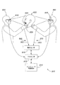
ここで、本開示の実施形態について添付の図を参照して、単なる例示として説明する。図では、参照数字の最後に2つの共通数字がある参照数字を共有する諸機能は、複数の図にまたがって合致する機能に対応する(たとえば、プロセッサ12、112、212、312、412、512、612、712、812、912、1012、1112、1212、1312など)。
Embodiments of the present disclosure will now be described, by way of example only, with reference to the accompanying figures. In the figures, functions that share a reference numeral with two common digits at the end of the reference numeral correspond to features that match across multiple figures (e.g.,
一般に、本開示は、歯科器具または歯科修復物の、歯列の変化の美的効果を器具または修復物の設計中に観察する方法およびシステムを提供する。 In general, the present disclosure provides methods and systems for observing the aesthetic effects of dental changes on a dental appliance or restoration during the design of the appliance or restoration.
歯科分野における現在の慣行は、ある人の歯の状態を専門家が評価し、必要に応じて治療を勧めるというものである。美的歯学では歯科専門家が、器具が必要になる治療を人に提示する。この器具の設計は、主として歯科専門家および歯科技工室の責任であり、その人からの情報は最小限しかない。高価なモックアップまたは試適は、時間のかかる手順によって成形用材料から作ることができる。この理由により、提示された試適またはモックアップが望ましいものでない場合に、2、3個以上のモックアップを1つが決められるまでに作り出すことはまれである。その人は代わりのものを望むかもしれないが、熟練した歯科技工室の技能者ではないので自分の要望を完全に伝えることができず、「医者が最もよく知っている」という考え方により、一般には妥協の産物がその人に残されて、その人の当初の望みが完全にはかなえられない。自分自身の修復物を設計する権限をその人に与えることは、歯科修復物を設計するのに必要な教育がかなりのものであるので、実際的な選択肢ではない。 The current practice in the field of dentistry is for an expert to evaluate a person's dental condition and recommend treatment if necessary. In aesthetic dentistry, a dental professional presents a treatment to a person that requires a device. The design of this device is primarily the responsibility of dental professionals and dental laboratories, with minimal information from the person. Expensive mock-ups or try-ins can be made from molding materials by a time-consuming procedure. For this reason, it is rare to produce a few or more mock-ups by the time one is decided on if the offered try-in or mock-up is not desirable. The person may want an alternative, but because he is not a skilled dental lab technician, he is unable to fully convey his wishes, and generally believes that "doctors know best". The product of compromise is left to the person, and the person's original wishes are not fully fulfilled. Empowering a person to design his own restoration is not a practical option, as the education required to design a dental restoration is substantial.
現在のモデリングソフトウェアで歯科修復物を設計する当業者は一般に、熟練者になる前に、設計ソフトウェアを正しく使用し理解するための数日の訓練を必要とする。歯科器具を必要とする素人である人に、このような歯科設計ソフトウェアに対する訓練をすることは実際的ではない。したがって、歯科設計ソフトウェアとすぐに対話する能力と、器具案の変更による美的結果を直感的に観察する能力とを普通の人に与えるシステムが望ましい。 Those skilled in the art of designing dental restorations with current modeling software generally require several days of training to properly use and understand the design software before becoming skilled. It is impractical to train an amateur who needs dental instruments for such dental design software. Therefore, a system that gives the ordinary person the ability to immediately interact with the dental design software and to intuitively observe the aesthetic results of changing the instrument plan is desirable.
歯科器具を設計してもらっている人は一般に、その器具を付けた結果として得られる自分の外見の情報もらうことに興味がある。器具を用意するときに、時間がかかり、高価で、不正確である予備モデルが成形および鋳造によって用意されることが多い。その人の得られる微笑に及ぼす特定の変更の効果を予測すること、およびその予測を有効に伝えることは困難である。そのため、結果として得られる自分の外見に関する意味のある情報をその人に提供することは困難である。その人の歯列の外見に及ぼす影響を考えると、結果として得られる外見に関する満足感は、肯定的な治療結果に極めて重要である。 People who have dental instruments designed are generally interested in getting information about their appearance as a result of attaching the instrument. Preparing models that are time consuming, expensive, and inaccurate when preparing the equipment are often provided by molding and casting. It is difficult to predict the effect of a particular change on the person's resulting smile and effectively communicate that prediction. Therefore, it is difficult to provide the person with meaningful information about the resulting appearance. Given the effect on the appearance of the person's dentition, satisfaction with the resulting appearance is crucial to a positive treatment outcome.
費用のかかる試適または最終の装具の製作の前に、器具案を人が観察および評価することを可能にする方法は、現在の治療可視化の方法と比べて有利である。多くの現在のシステムは、2D画像を3Dモデルに重ね合わせるソフトウェアに依拠している。このようなソフトウェアは特殊化されていることが多く、ほとんどの素人には使用および理解することが困難であり、素人である人によるリアルタイム使用向けにはなっていない。このようなソフトウェアでの歯列の操作は、個々の歯または歯の群の姿勢およびアンギュレーションを回転させ、傾け、またはそれとは別に変更して、結果として得られる咬合に影響を及ぼすことなく器具の機能を変更することによって行われる。さらに歯列は、別の事前モデル化歯列に転換することもできる。本開示は、3Dモデリング、歯科設計ソフトウェア、3D表示装置、およびセンサと一体化したリアルタイム拡張現実(「AR」)を含む方法およびシステムを提供する。このセンサは、ジェスチャに基づく入力を受け取るための(たとえば、手のジェスチャを追跡する、眼の動きを追跡するなどの)動きセンサまたは動き捕捉デバイス(たとえば、光学動きセンサ、SMI Eye Tracking Glasses 2 Wirelessシステムなどの視標追跡センサ、CyberGloveシステムなどの手首装着型動き捕捉デバイスなど)、他の光学センサ、神経活動に基づく入力を受け取るためのブレインコンピュータインターフェース(「BCI」)、脈拍、体温、もしくは発汗を測定するための、またはポリグラフとして使用される複数の試験の組合せのためのセンサ、または他の任意の適切なセンサを含むことができる。手首装着型センサはまた、身体3Dモデルの取扱いをシミュレーションするための触覚フィードバックを与えることもできる。
A method that allows one to observe and evaluate the instrument proposal prior to costly try-in or final appliance fabrication is an advantage over current methods of treatment visualization. Many current systems rely on software that superimposes 2D images onto 3D models. Such software is often specialized, difficult to use and understand for most laymen, and is not intended for real-time use by laymen. Manipulation of the dentition with such software can rotate, tilt or otherwise alter the attitude and angulation of individual teeth or groups of teeth without affecting the resulting occlusion. This is done by changing the function of the instrument. In addition, the dentition can be converted to another pre-modeled dentition. The present disclosure provides methods and systems that include real-time augmented reality ("AR") integrated with 3D modeling, dental design software, 3D displays, and sensors. This sensor may be a motion sensor or motion capture device (e.g., tracking hand gestures, tracking eye movements, etc.) to receive gesture-based inputs (e.g., optical motion sensors, SMI
人の頭の部分の3Dモデルが、本明細書に記載の方法およびシステムに使用される。3Dモデルは、人の歯列弓の、および歯列弓を頭部と関連づける人の頭部の、経験的データを含む(たとえば、3D口腔内光学スキャナからの歯列弓のデータ、および3D顎外光学スキャナからの歯列弓と頭部一緒のデータ)。3Dモデルは3次元で表示される(たとえば、Oculus Rift仮想現実ヘッドセット、Google Glassデバイス、ホログラフィック投射などによる可視化によって)。経験的データに基づく部分に加えて、3Dモデルは拡張現実歯科器具を含む。人の頭部も拡張現実歯科器具も、人の手もしくは他のジェスチャを用いることによって、またはBCIによって、人が操作することができる。動きセンサが人の手の動きもしくは他のジェスチャを検出して入力を受け取り、またはBCIが入力を受け取る。3Dモデルは、その入力に基づいて操作される。3Dモデルに示された器具の機能(たとえば、歯、歯列内の歯肉遊離縁および他の歯茎の機能など)は、把持および操作することができ、あるいはBCIの場合では、そうすることを人が想像することができる。加えて、よく知られている手のジェスチャは、回転、ズーム拡大縮小、および他の一般的な機能のために使用すること、または想像することができる。視標追跡とBCIを組み合わせることにより、両システムからのデータを共通タイムスタンプとリアルタイム同期させて、注目パターンを感情状態と相関させることが可能になり得る。 A 3D model of the human head is used in the methods and systems described herein. The 3D model includes empirical data of the human dental arch and of the human head associating the dental arch with the head (e.g., dental arch data from a 3D intraoral optical scanner, and 3D jaw). Data of dental arch and head together from external optical scanner). The 3D model is displayed in three dimensions (eg, by visualization with an Oculus Rift virtual reality headset, Google Glass device, holographic projection, etc.). In addition to the part based on empirical data, the 3D model includes augmented reality dental instruments. Both the human head and the augmented reality dental appliance can be manipulated by humans, using human hands or other gestures, or by BCI. A motion sensor detects movement of a human hand or other gesture and receives an input, or a BCI receives the input. The 3D model is manipulated based on the input. The function of the device shown in the 3D model (e.g. the function of teeth, free gingiva in the dentition and other gums, etc.) can be grasped and manipulated, or, in the case of BCI, to do so. Can imagine. In addition, well-known hand gestures can be used or imagined for rotation, zoom scaling, and other common functions. By combining target tracking and BCI, it may be possible to synchronize data from both systems with a common timestamp in real time to correlate the pattern of interest with the emotional state.
拡張現実歯科器具の変更は、その人が、またはシステムの他のユーザ(たとえば、健康管理開業医、またはその人を補助する信用のおける素人)が、リアルタイムで3Dモデルに加えることができる。その人は次に、モデルを任意の角度から、また変化するズーム角で眺めて、器具内の歯の特定のサイズ、形状、および向きの美的結果を観察することができる。拡張現実歯科器具および3Dモデルのより広い操作および修正によって、人は、器具の特定の設計への変更の美的結果に意味のある情報をもたらすことができる。これらの操作は、直感的な手および他のジェスチャによって実現することができる。このジェスチャは、実際のジェスチャでも(動きセンサまたはBCIの場合)想像したジェスチャでも(BCIの場合)でもよい。BCIを使用すると、想像した手の動きが解読され、メンタルコマンドが与えられて3Dモデルを操作することができる。モデルのAR機能および要素は、人の好みによって、たとえば押す、引っ張る、回転させる、ズーム拡大する、ズーム縮小することなどができる。加えて、色および濃淡などの要素も同様に、メンタルコマンドを用いて変更することができる。3Dモデルに対する人の非自発的反応(たとえば、感情的な反応など)が明らかにされ、モデルは、人の非自発的反応に応じて自動的に更新され得る。非自発的反応は測定することが、光学的変化、神経活動(BCI)、脈拍、体温、発汗を測定するセンサ、ポリグラフとして使用される複数の試験の組合せ、または他の任意の適切な感情状態のインジケータによって可能である。 Changes to the augmented reality dental appliance can be added to the 3D model in real time by the person or by another user of the system (eg, a health care practitioner, or a trusted lay person assisting the person). The person can then look at the model from any angle and at varying zoom angles to observe the aesthetic results of the particular size, shape, and orientation of the teeth in the appliance. With the wider manipulation and modification of augmented reality dental appliances and 3D models, one can bring meaningful information to the aesthetic consequences of changing to a particular design of the appliance. These operations can be achieved with intuitive hands and other gestures. This gesture may be an actual gesture (for a motion sensor or BCI) or an imagined gesture (for BCI). With BCI, imagined hand movements can be deciphered and mental commands can be given to manipulate the 3D model. The AR functions and elements of the model can be, for example, pushed, pulled, rotated, zoomed in, zoomed out, etc., depending on human preference. In addition, elements such as color and shading can be similarly changed using mental commands. The person's involuntary response to the 3D model (eg, emotional response, etc.) is revealed, and the model can be automatically updated in response to the person's involuntary response. Involuntary responses can be measured by sensors that measure optical changes, nerve activity (BCI), pulse, temperature, sweating, combinations of multiple tests used as polygraphs, or any other appropriate emotional state Is possible with the indicator.
器具設計案と相互作用することは、モデルの歯または他の機能に人の手を伸ばし動かすという簡単なことであり、またはBCIが使用される場合には、機能に手を伸ばし動かすことを想像する、もしくは変更の結果を想像するという簡単なことである。そのため、その人への基本的な指示しか必要とされない。設計ソフトウェアは、結果として得られる器具が、均等な相互嵌合による適切な咬合を有するように、歯列の許容変更に対する事前設定機能制限を含み、適切な生理学的安静位を可能にする。結果として得られる器具は機能的であるが、美学はその人の掌中にあり、結果として得られる歯科器具が安静状態で上側と下側の歯列間に適切な隙間を設けること、咬合時の適切な上下顎関係を実現すること、そうでなければ機能的に最適化されることを保証するために課される制約を受ける。器具案への変更の美的結果は、リアルタイムで検証することができ、人は、満足のいく設計を見出すことができる。あるいは、人は、自分の理想的な設計が機能上の制限の外側にあり、したがって達成可能ではないことを認識することができる。この場合その人は、3Dモデルを操作して、美学と機能性の間に適切な妥協点を用意することができる。任意の角度から見る、また変化する不透明度のレベルで見る機能を含む、モデルおよび表示装置の3Dの性質が人に、器具案によって自分が3次元でどのように見えるかについての理解をもたらす。 Interacting with the proposed appliance design is as simple as reaching and moving a model's teeth or other functions, or if BCI is used, imagine reaching and moving the functions It's as simple as doing it or imagining the consequences of a change. Therefore, only basic instructions to the person are needed. The design software allows for proper physiologic resting, including pre-set functional restrictions on dentition tolerance changes, so that the resulting instrument has proper occlusion with even interdigitation. The resulting instrument is functional, but the aesthetics are in the person's palm, and the resulting dental instrument is at rest, providing adequate clearance between the upper and lower dentition, There are constraints imposed to ensure that proper jaw relation is achieved, or otherwise functionally optimized. The aesthetic results of the changes to the instrument plan can be verified in real time, and one can find a satisfactory design. Alternatively, one can recognize that his ideal design is outside the functional limits and is therefore not achievable. In this case, the person can manipulate the 3D model to provide an appropriate compromise between aesthetics and functionality. The 3D nature of models and displays, including the ability to view from any angle and at varying levels of opacity, gives a person an understanding of how he or she looks in three dimensions with the instrument plan.
3Dモデルはまた、異なる顔の表現を反映するように更新することもできる。以前から存在する歯の古い写真から導出された微笑をシステムに入力して、人の自然な微笑を復元することができる。著名人の微笑もまた入力されて、設計選択肢に影響を及ぼし得る。その人は、歯科専門家による判定または批評を心配せずに、自分の所望の設計結果を表現することができる。同様に、第2の人が参加し、器具への変更を提案することもできる。事前設定微笑または他の制約事項を美的目標として設定することができ、また歯科器具を、咬合に関する機能上の制約事項を守りながらその目標に到達するように、または近づくように成形することができる。3Dモデルを操作しながら追加データを取得して、微笑の、または生理学的な安静位(「安静位」)でのその人の、現在の義歯による咬合位での経験的データなど、所与の位における経験的データを得ることができる。 The 3D model can also be updated to reflect different facial expressions. A smile derived from an old photograph of a pre-existing tooth can be input into the system to restore a person's natural smile. Celebrity smiles can also be entered and affect design choices. The person can express his desired design result without worrying about judgment or criticism by the dental professional. Similarly, a second person can participate and suggest changes to the instrument. A preset smile or other constraint can be set as an aesthetic goal, and the dental appliance can be shaped to reach or approach that goal while observing the functional constraints on occlusion . While manipulating the 3D model, additional data is acquired to provide a given, such as empirical data on the person's current denture occlusal position in a smiling or physiological resting position (`` resting position ''). Empirical data on the position can be obtained.
人の上下顎関係が安静位にあるときに取得された経験的データにより用意された3Dモデルは、安静位における人の上下顎関係を正確に提示する(別の位でデータを取得し安静位に外挿することとは異なる)。人の実際の安静位が3Dモデルの安静位を決定する。それによって3Dモデルの安静位は、安静位に影響を及ぼす関節、筋肉、神経、歯茎、インプラント(もしあれば)、および歯(もしあれば)を含む顎口腔系の中の全ての実体の相互関係を明らかにする。安静位における人のデータを何も用いずに用意された3Dモデルは、安静位を習慣位または他の位と確実に区別する可能性があまりない。 A 3D model prepared from empirical data obtained when the person's upper and lower jaw relations are in a resting position accurately presents the person's upper and lower jaw relations in the resting position (data obtained in another position and resting position Is different from extrapolating to The actual resting position of the person determines the resting position of the 3D model. The resting position of the 3D model thereby affects the resting position of all entities in the stomatognathic system, including the joints, muscles, nerves, gums, implants (if any), and teeth (if any). Clarify the relationship. A 3D model prepared without any human data in the resting position is unlikely to reliably distinguish the resting position from the habitual position or other positions.
安静位は、上顎に対する下顎の空間内の(直立姿勢維持位の頭に対して垂直、前後方向、および横方向の)、下顎閉鎖の等張経路に沿った位である。安静位では、下顎を動かす伸筋および下制筋を含む顎筋肉が、最小の電気的活動を作用させる位置の姿勢になる。安静位を維持するのに必要な、顎筋肉によるエネルギーの消費は、下顎ヒンジ連結の経路に沿う他の位と比べて最少である。安静位では、人の関節丘は中立、無拘束位にある。 The resting position is in the space of the lower jaw relative to the upper jaw (vertical, anterior-posterior, and lateral to the head in an upright position) along the isotonic path of mandibular closure. In the resting position, the jaw muscles, including the extensors and lower muscles that move the lower jaw, assume the posture where the minimal electrical activity is exerted. The energy expenditure by the jaw muscles required to maintain a resting position is minimal compared to other positions along the mandibular hinge connection. In the resting position, the human condyle is in a neutral, unrestrained position.
人の安静位は、その人に関して決定することができる。安静位は、下顎の動きをシミュレーションする歯科咬合器などの機械デバイスによって決定することができない。下顎位または上下顎関係は、頭部、首頸部領域、および背中領域の姿勢の問題を含む諸要因による影響を受け得る。人の側頭下顎関節の内部乱れ、感情的要因、および全身的健康要因もまた、下顎位不全の一因となり得る。安静位を確立する前に、これらの要因を明らかにすることが一般に有益である。場合によっては、これらの要因を明らかにしないと間違った安静位になる。たとえば、ある要因が、噛み合わせ(bite registration)に外挿するのに使用できる安静位を確立する前に、対処または除去されなければならない可能性がある。別の例では、ある要因が、別の位から安静位を外挿することをさらに複雑にして、人のデータを安静位で取得することの有利性が増す可能性がある。 The resting position of a person can be determined for that person. Resting positions cannot be determined by mechanical devices such as dental articulators that simulate mandibular movement. The mandibular position or upper and lower jaw relationship can be affected by factors including posture problems in the head, neck and neck regions, and back region. Internal disturbances in the human temporomandibular joint, emotional factors, and general health factors can also contribute to mandibular position failure. It is generally beneficial to identify these factors before establishing a resting position. In some cases, failure to identify these factors results in a false resting position. For example, certain factors may need to be addressed or eliminated before establishing a resting position that can be used to extrapolate to bite registration. In another example, certain factors may further complicate the extrapolation of the resting position from another position, increasing the advantage of obtaining human data in the resting position.
安静位における経験的データを含む3D法は、他の潜在的に有効な上下顎関係の正確な決定をしやすくする。たとえば、3D法は、咬合を確立するのに十分な歯列がない人、たとえば歯のない人の顎運動測定(jaw tracking)および顎外咬合評価に適用することができる。そのデータは、咬合を確立するのに十分な歯列を人が持っている場合に中心咬合(「CO」、これは、人の歯が最大咬頭嵌合にあり、人の顎が「CO位」にあるときに見出される)が見出される自然位の決定をしやすくすることができる。すなわちデータは、最適神経筋CO位の見積もりをしやすくすることができる。見積もられたCO位は、咬合を画成するのに十分な歯を持たない人の義歯を用意するのに利用することができる。 3D methods, including empirical data in the resting position, facilitate accurate determination of other potentially useful jaw relations. For example, 3D methods can be applied to jaw tracking and extramaxillary occlusal evaluation of persons without sufficient dentition to establish an occlusion, for example, persons without teeth. The data shows that when a person has enough dentition to establish an occlusion, a central occlusion ("CO", which means that the person's teeth are in maximum cusp engagement and the person's jaw is in the "CO position" ) Can be easily determined. That is, the data can facilitate the estimation of the optimal neuromuscular CO position. The estimated CO position can be used to prepare a denture for a person who does not have enough teeth to define an occlusion.
歯科医または他の歯科専門家にとって、器具を用意するときにCO位を確立することが一般的である。CO位を確立するのに十分な歯列を人が欠いている場合、器具と一緒にCOが見出されるべき適切な上下顎関係を決定するのに外挿が必然的に必要になる。歯のない人は、CO位を確立するのに十分な歯列を欠くことになる。部分的に歯のある一部の人、たとえば、門歯はあるが臼歯がない人もまた、COを確立するのに十分な歯列を欠くことになる。 It is common for dentists or other dental professionals to establish a CO position when preparing instruments. If a person lacks enough dentition to establish a CO position, extrapolation will be necessary to determine the appropriate upper and lower jaw relationship with which the CO should be found along with the instrument. Those without teeth will lack enough dentition to establish the CO position. Some partially toothed people, such as those with incisors but no molars, will also lack enough dentition to establish CO.
器具を用意するときに安静位に基づいてCO位を確立することにより、その器具を含む顎口腔系の、結果として得られる歯の機能、安定性および調和の改善と最適化が容易になり得る。安静位に基づいてCO位を確立することによりまた、以下のうちの1つまたはそれ以上が容易になり得る。
-正常な咬合面系に向けて人の咬合面系を最適化することであり、正常な咬合面系が適切な機能をその人にもたらし、または、どんな顎関係分類または不正咬合も明らかにすることであり、人のCO位ではそれだけ必要とし得る、
-歯の美学の最適化(前方領域と後方領域の両方の歯の形状、輪郭、解剖学組的構造および形態を含む)、
-最適な生理学的下顎位が見出された場合に、より調和した筋肉のバランスによる顔用化粧品の最適化、または
-頭痛、耳詰まり感、耳鳴り、眼の奥の圧迫、歯の知覚敏感、側頭下顎関節音、咀嚼筋圧痛、首と肩の痛みを含む、起こり得る筋骨格咬合徴候および症状の緩和。
Establishing the CO position based on the resting position when preparing the device may facilitate improvement and optimization of the resulting tooth function, stability and harmony of the stomatognathic system containing the device . Establishing a CO position based on a resting position may also facilitate one or more of the following:
-To optimize a person's occlusal surface system towards a normal occlusal surface system, where the normal occlusal surface system provides the appropriate function to the person or reveals any jaw relation classification or malocclusion That is, in the human CO rank, it may be necessary,
-Optimization of tooth aesthetics (including tooth shapes, contours, anatomical structures and morphologies in both the anterior and posterior regions),
-Optimization of facial cosmetics with more harmonious muscle balance when optimal physiological mandibular position is found, or
-Alleviation of possible musculoskeletal occlusion signs and symptoms, including headache, ear stuffiness, tinnitus, pressure in the back of the eye, sensitive teeth, temporomandibular joint sounds, masticatory muscle tenderness, and neck and shoulder pain.
安静位は、習慣位とは異なる真の安静位である。習慣位は、下顎頭並進経路に沿って前方に位置し得る獲得上下顎位である。ある特定の人では、安静位と習慣位は一致するか、または非常に近い可能性がある。しかし、習慣位を維持するために顎筋肉に必要とされるエネルギーは、必ずしも安静位のように最小限とは限らない。習慣位は場合によって、歯のない人のCO位を決定する出発点として用いられる。しかし、習慣位から始めると、歯科治療を計画することに関して、安静位から始めるよりも望ましくない結果になる可能性がある。 The resting position is a true resting position that is different from the habitual position. The habit position is the acquired upper and lower jaw position that can be located anteriorly along the mandibular condylar translation path. In certain people, resting and habitual positions can be the same or very close. However, the energy required by the jaw muscles to maintain a habitual position is not always as minimal as at rest. Habit position is sometimes used as a starting point to determine the CO position of a toothless person. However, starting from a habitual position can have less desirable consequences for planning dental treatment than starting from a resting position.
3Dモデルは、3D技法(たとえば、Google Glass、Oculus Rift、Microsoft HoloLens、3Dテレビ受像機またはモニタ、ホログラフィックプロジェクションなど)によって表示される。3Dモデルを操作するのに用いられるジェスチャは、直感的で簡単なものにすることができる(たとえば、人の手で歯をつかみ、歯を回転させるなど)。そのため人は、器具案の歯列を容易に操作して、所与の歯列選択の美的効果を観察することができる。同様の操作を2次元表示装置に対して行うには、より高い技能および抽象化が必要になる。3D表示装置を使用すると、歯の姿勢の微細で特殊な変更が素人である人にとって、特にその人が2次元表示装置上での3次元の視覚化および操作になれていない場合に(これは、義歯および他の歯科器具の主なユーザである高齢者においてより一般的であろう)、実行可能なものになる。同様に、その人と歯科専門家の間の言葉の障壁が、3Dモデルを見直しおよび操作することの障害にならない。これは、専門家が何度も訪問することが実際的ではない貧困地域、または近づきにくい地域の人の義歯を設計する場合に当てはまり得る。 The 3D model is displayed by 3D techniques (eg, Google Glass, Oculus Rift, Microsoft HoloLens, 3D television receiver or monitor, holographic projection, etc.). The gestures used to manipulate the 3D model can be intuitive and simple (eg, grasping a tooth with a human hand, rotating a tooth, etc.). Thus, one can easily manipulate the dentition of the appliance plan and observe the aesthetic effect of a given dentition selection. Performing the same operation on a two-dimensional display device requires higher skill and abstraction. The use of a 3D display device makes it difficult for a lay person to make fine and special changes in the posture of teeth, especially if he is not accustomed to 3D visualization and manipulation on a 2D display device (this is Would be more common in the elderly, who are the primary users of dentures and other dental instruments) and become workable. Similarly, language barriers between the person and the dental professional do not hinder the review and manipulation of the 3D model. This may be the case when designing dentures for people in poor or inaccessible areas where it is impractical for professionals to visit many times.
操作のハンズフリー性は、タブレット表面、キーボード、マウス、または他の物理的インターフェースデバイスとの接触を通して感染症が広がりにくいことを意味する。 Hands-free operation means that infections are less likely to spread through contact with the tablet surface, keyboard, mouse, or other physical interface device.
第2の人が3Dモデルを操作することができ、また感情状態を表すデータの入力を受け取るBCIまたは他のセンサが使用される場合には、前記の人、第2の人、または両者の感情の反応を利用して、前記の人、第2の人、または両者の好みを予測することができる。これらの好みは重み付けして、前記の人と第2の人の両方に強い反応があった器具の設計を推進することができる。第2の人は、前記の人と一緒に3Dモデルを操作することができ、あるいは、第2の人は、前記の人の参加または入力がなくてもそうすることができる(たとえば、前記の人が意見交換することができない、または自分たち自身のために効果的に選択することができない場合など)。 If a second person is capable of manipulating the 3D model and using a BCI or other sensor that receives input of data representing the emotional state, the emotions of said person, the second person, or both, Can be used to predict the preference of the person, the second person, or both. These preferences can be weighted to drive the design of instruments that responded strongly to both the person and the second person. A second person can manipulate the 3D model with the person, or the second person can do so without the participation or input of the person (e.g., (E.g., when people cannot exchange opinions, or cannot effectively choose for themselves).
以前のいくつかの方法の限界は、患者と歯の2つの統合2D画像に基づいた解析に起因する。2つの画像は共通の容貌を共有せず、3Dモデルの生成のために使用することができない。結果として得られる複合画像は、他のどの角度からも見えない。また、デジタル写真の撮影中の照明条件も一貫性がないことがあり、患者の顔を表現する際の歯シェーディングの正確な表現が、完全には正確ではない可能性がある。補綴案の表現は、既存の歯列または修復物と患者の顔との間の基準点を欠いている。加えて、真の3Dモデルがないと、設計の機能上の制限は、制約として試験すること、および適用することが困難である。そのため非機能的な設計が、潜在的な問題の表示なしでモデル化されることになり得る。最後に、グリーンスクリーン用の遮蔽物を患者の口の中に入れることによって顔および唇が変えられて、結果として得られる、モデル化されるべき美学が変わる。 The limitations of some previous methods stem from analysis based on two integrated 2D images of the patient and the tooth. The two images do not share a common feature and cannot be used for 3D model generation. The resulting composite image is not visible from any other angle. Also, lighting conditions during the taking of digital photographs may be inconsistent, and the exact representation of tooth shading in representing the patient's face may not be completely accurate. Prosthodontic representations lack a reference point between the existing dentition or restoration and the patient's face. In addition, without a true 3D model, the functional limitations of the design are difficult to test and apply as constraints. Thus, a non-functional design may be modeled without an indication of a potential problem. Finally, the face and lips are changed by putting a green screen shield into the patient's mouth, changing the resulting aesthetic to be modeled.
TruRxシステムなどの以前のグリーンスクリーン技術は、設計案の投影像を人の歯列弓の最上部に重ね合わせることを含む。対照的に、本明細書で開示された方法およびシステムに使用される3Dモデルは、歯列弓を顔の構造と関連づける。歯列弓と歯が所与の姿勢の唇によって完全に覆い隠される場合に、3Dモデルは依然として、器具案およびその美学上の効果を正確に表現することができる。 Earlier green screen technologies, such as the TruRx system, involve overlaying a projected image of a design proposal on top of a human dental arch. In contrast, the 3D models used in the methods and systems disclosed herein associate dental arches with facial structures. If the dental arch and teeth are completely obscured by the lips in a given pose, the 3D model can still accurately represent the proposed instrument and its aesthetic effect.
3Dモデルを使用することの利点は、安静時唇線と関係がある。安静時唇線では、唇は弛緩しており、歯の大部分が、多くの場合に全部が、見えない。口腔内特徴(たとえば、拡張現実によって提案された歯列)に加えて、口腔外構造(たとえば、顔の特徴など)を適用することによって、3Dモデルは、歯列弓および器具がたとえ見えなくても、外的容貌に対する歯の効果の正確な描写を実現する。 The advantage of using a 3D model has to do with the resting lip line. At the resting lip line, the lips are lax and most of the teeth, often not all, are invisible. By applying extra-oral structures (e.g., facial features, etc.) in addition to intra-oral features (e.g., dentition proposed by augmented reality), the 3D model allows the dental arch and appliances to be invisible even Also achieves an accurate depiction of the effect of the tooth on the external appearance.
システム
図1は、患者である人30の3Dモデル20を表示および操作するシステム10である。システム10は、プロセッサ12およびコンピュータ可読媒体14を含む。3Dモデル20は、プロセッサ12によって命令を実行することによりレンダリングされ、操作され、更新され、表示される。3Dモデル20は、コンピュータ可読媒体14に保持されたデータに基づいている。プロセッサ12およびコンピュータ可読媒体14は、同一のデバイス上にあっても異なるデバイス上にあってもよく、異なるネットワーク位置にあってよく、または他の任意の適切な構成とすることができる。プロセッサ12およびコンピュータ可読媒体14の諸機能は、複数の個別のプロセッサおよびコンピュータ可読媒体に割り振ることができる。
System FIG. 1 is a
システム10は、人30が自分の手を3Dモデル20の上に置いて3D表示装置16による3Dモデル20をたとえば直感的なジェスチャによって操作できるように、3Dモデル20を表示するためのプロセッサ12と動作可能に通信する3D表示装置16を含む。図1で人30は、3Dモデル20を手39で操作している。システム10は、人が、3D表示装置16に示された3Dモデル20に向けたジェスチャを用いて、3Dモデル20の姿勢または視像を変更すること、3Dモデル20の選択された容貌を変更すること、またはそれとは別に3Dモデル20を操作することを可能にし得る。ジェスチャは、3Dモデル20の一部分を手でつかむこと、および物理的なモデルを操作する場合に用いられるのと同様の手のジェスチャを適用することを含み得る。このような操作の例にはまた、表示装置に示された3Dモデル20の、回転、パニング、ズーム拡大または縮小、照明条件の変更などの視像の変更も含まれ得る。
The
3D表示装置16は、眼鏡型ARインターフェース(たとえば、Google Glass、Oculus Rift、Microsoft HoloLens、Meta Spaceglassesなど)として示されている。眼鏡型ARインターフェースは、3Dモデル20が実際の物理的環境の上に、人30の観点から表示されることを可能にする。3D表示装置16は、複合環境を投影して、人30が3Dモデル20を3次元で、リアルタイム更新によって見ることを可能にする。眼鏡型3D表示装置16は、3Dモデル20が自分の目の前にあり、自分の手または他の命令で操作することができ、多数の角度から見ることができるという感覚を人30に与える、任意の表示デバイスと交換可能である。
システム10は動きセンサ18を含む。動きセンサ18は、人30のジェスチャ(たとえば、手、頭、足などの動き)を検出する。このジェスチャは、プロセッサ12に供給される第1の入力データ60の入力になる。第1の入力データ60は、動きセンサ18で検出された人30のジェスチャに対応する自発的動作データ62を含む。動きセンサ18は、人30のジェスチャの動き、位置、姿勢および角度を監視して、人30が3D表示装置16による3Dモデル20を操作することを可能にする。動きセンサ18は、任意の適切なデータ(たとえば、光学、ドップラーレーダ、受動IR、断層撮影、これらの組合せなど)に基づいて動きを検出することができる。
動きセンサ18に加えて、または動きセンサ18の代わりに他のセンサがシステム10に含められて、人30が、プロセッサ12が解釈できる自発的動作データ62を供給するために、ジェスチャとは別の方法によって3Dモデル20および歯科設計ソフトウェアと対話することが可能になり得る(たとえば、眼の動き、音声コマンド、顔表情などを用いることなどによって)(図示せず)。このようなセンサは、人30からの光学データまたは他の形式のデータを捕捉することに基づくことができる(たとえば図39の光学センサ1296など)。
Other sensors in addition to or in place of the
3Dモデル20はまた、人30以外の人からの自発的動作データに応じて操作することもできる(たとえば、図30のシステム610など)。
The
図2は、3Dモデルを用いて作業する方法80を示す。方法80は、3Dモデルを表示するステップ82と、入力を受け取るステップ84と、入力84を受け取るステップに対応して3Dモデルを更新するステップ86とを含む。方法80は、システム10を使用して実行することができる。3Dモデルを表示するステップ82、および3Dモデルを更新するステップ86は、コンピュータ可読媒体14に保存されたデータを使用してプロセッサ12で命令を実行することによって、3D表示装置16で完了することができる。入力を受け取るステップ84は、システム10の動きセンサ18による手のジェスチャ、他の自発的入力、非自発的入力、または各入力の組合せを検出することを含み得る。
FIG. 2 shows a
3Dモデルの構成要素
図3は、3Dモデル20を見る人30を示す。3Dモデル20は、スキャン容貌データ40および拡張現実データ50に基づいてモデル化される。3Dモデル20は、患者容貌22および歯科器具案24を含む。患者容貌22は、モデル化歯列弓21およびモデル化外的容貌23を含む。眼鏡ベースのインターフェースを適用する図1の3D表示装置16とは異なり、図3の3Dモデル20は、眼鏡なしで機能する3次元プロジェクタ(たとえば、ホログラフィックプロジェクタ、Musion Das Hologram LtdのTelePresenceシステム、Magic LeapのDigital Lightfieldシステムなど)として示されている。これらのタイプの表示装置のどちらか、または他の任意の適切な表示装置が3D表示装置16として使用され得る。
Components of the 3D Model FIG. 3 shows a
スキャン容貌データ40は歯列弓データ42、関係データ44、および外的容貌データ46を含む。スキャン容貌データ40は経験的データであり、たとえば、口腔内光学スキャナによって人30の歯列弓32をスキャンすることによって取得され(たとえば、図33のシステム910などを使用して取得され)、また口腔外光学スキャナによって人30の外的容貌34をスキャンすることによって取得される(たとえば、図33のシステム910などを使用して取得される)。追加スキャン容貌データ40により所与の姿勢の3Dモデル20がさらに正確になるが、3Dモデル20は、初めに取得されたスキャン容貌データ40だけに基づいて、多くの姿勢に向けて操作することができる。人30の外見は、3Dモデル20によるセッション間でさほど変化しないが、同じスキャン容貌データ40を多数のセッションにわたって適用することができる。スキャン容貌データ40は、光学スキャナ、超音波スキャナ、他の適切なスキャナ、または外的容貌34に適用される他の適切なデータ取得技法により取得することができる。
The
歯列弓データ42は、人30の上顎および下顎の歯列弓32のモデル化を容易にし、それによってモデル化歯列弓21が得られる。
The dental
外的容貌データ46は、人30の外的容貌34の各部分のモデル化を容易にする。外的容貌データ46の量が多ければ多いほど、それに応じて外的容貌面に対する美的効果を、付加的な外的容貌データを欠いている3Dモデルよりも広く観察することによって、モデル化外的容貌23がより広範になる(たとえば、図16の3Dモデル120参照)。歯列弓データ42、関係データ44、および外的容貌データ46に類似しているデータを取得し、データの種類に基づいてモデルを用意するステップについては、本明細書と発明者を共有するWO 2013/071435でさらに論じられている。図33、35、38、および40それぞれに示された口腔外センサ992、1092、1192または1392などの、データを取得するための機能を含むことができる。
関係データ44は、歯列弓データ32のデータ、および外的容貌34のデータ(たとえば、顔の部分および首の部分など)を含む。関係データ44により、歯列弓32間、および外的容貌34と歯列弓32の間の関係が確立しやすくなる。歯列弓32の相対姿勢は、上下顎関係を画定する。関係データ44は、外的容貌34が歯列弓32の相対姿勢に基づいてモデル化されることを、歯列25に基づいてモデル化されることに加えて可能にする。所与の歯科器具案24の咬合時の上下顎関係は、モデル化された外的容貌23の外見に寄与する。特定の咬合時の上下顎関係に対する制約を考えると、歯列25は、歯科器具案24による咬合時のモデル化外的容貌23の外見を決めることになる。
The
関係データ44はまた、3Dモデル20の上下顎関係が外的容貌34の姿勢に基づいてモデル化されることを可能にする。モデル化外的容貌23がどう見えるようになるかについての制約を置くことができる。歯科器具案24は、モデル化外的容貌23の選択された外見になるようにモデル化される。制約はまた、歯科器具案24がもたらす咬合時と安静時の上下顎関係が両方とも人30にとって確実に適切になるように、歯科器具案24に適用される。結果として得られる外見のために選択されたモデル化外的容貌23は、外的容貌データ46に含まれる姿勢から得られるか、または外的容貌データ46に含まれる姿勢と実質的に類似している可能性がある。このような姿勢の経験的データは、歯列25と、人30のために適正に選択された他の特徴とを有する歯科器具案24を提供する場合に、3Dモデル20の有効性を増大させる。安静位は、たとえば以下で、また本明細書と発明者を共有するWO 2013/071435で論じられているように、経験的証拠によって画定することができ、外的容貌データ46には、安静位における人30の経験的データが含まれ得る。システム1310は、安静位における経験的外的容貌データ46を取得しやすくする特徴を含む。
関係データ44は、2つのモデル化歯列弓21間の、およびモデル化歯列弓21とモデル化外的容貌23の間の正確な関係を維持しながら、3Dモデル20の上下顎関係の操作を容易にする。これらの関係は、3Dモデル20が、人30における歯列弓32間の、および歯列弓32と外的容貌34の間の対応関係を反映する関係と一致するという点で、正確である。
拡張現実データ50は、歯科器具案24の表現を含む。図示の歯科器具案24は1対の義歯である。他の器具もまたモデル化することができる(たとえば、単一の義歯、補綴物、修復物など)。歯科器具案24は、拡張現実データ50に基づいてモデル化され、モデル化歯列弓21に重ね合わされる。歯科器具案24により、歯科器具案24上の歯列25によって容易になった嵌合における、モデル化歯列弓21間の上下顎関係が得られる。上下顎関係、ならびにモデル化歯列弓21およびモデル化外的容貌23の結果として得られる姿勢は、スキャン容貌データによって伝えられて、歯列弓32間の上下顎関係が外的容貌34に及ぼす効果が3Dモデル20で表現される。
歯科器具案24は、人30にとって適切な画定上下顎関係と(たとえば、適切な咬合位および安静位という条件などで)、咬合位からの動き方向を画定する下顎頭角度とに基づく。歯科器具案24をリアルタイムでARによってモデル化する場合、検証手順により、上下顎関係位置案を3Dモデル20が正確にモデル化して、人30の観察された動きと整合することを助長することができる。歯科器具を装着せずに、人は自分の顎を動かす(たとえば、通常の噛む機能などで)。観察データは3Dモデル20と比較することができ、不整合性が発見された場合には、3Dモデル20は、3Dモデル20の動きを人30の観察された動きと比較するときの有用な目印として上下顎咬合位または下顎頭角度を用いて、補正することができる。検証手順は、外的容貌データ46に基づくことができる。検証手順はまた、たとえばシステム910(図33)、システム1010(図35)、システム1110(図37)、システム1310(図40)などを使用して取得された追加外的容貌データ46に基づくこともできる。
The
3Dモデルの操作
システム10は、素人であり得る人30による3Dモデル20の直感的操作をしやすくする。操作は、3Dモデル20の見え方を変更することを含み得る。操作には、特にモデル化外的容貌23に関して、3Dモデル20の美学に対して得られる効果を観察しやすくするために、患者容貌22の姿勢を変更すること、歯科器具案24を変えること、下にある外的容貌データ46を変更すること、または下にある拡張現実データ50を変更することが含まれ得る。制約は、咬合時の上下顎姿勢、安静位における歯列25のスペーシング、モデル化外的容貌23の外見、これらの特徴の組合せに、または歯科器具案24を設計する目標に応じて他の適切な特徴に、適用することができる。
3D
制約を安静位における歯列25のスペーシングに適用する場合、1から4mmの間の安静空隙、たとえば約2mmの安静空隙を画定することができる。安静空隙とは、歯科器具案の安静位における上部と下部の歯列25間の隙間のことである。過度の、または不十分な安静空隙はそれぞれ、顔の外見をゆがめる。過度の安静空隙(閉じすぎ)では、下顎および唇が突出することになり、「虚脱状態」または「渋面」の外見になる。不十分な安静空隙(「開きすぎ」)では顔が長くなり、そのため唇が薄く引き伸ばされて見えることになり、顔は全体的に不快に見える。これは、顔の筋肉が適切な寸法に近づこうとしているときに静止できない顔の筋肉の緊張による。人が開きすぎ、または閉じすぎの状態にある場合に、本方法およびシステムを用いて、所望の外見、ならびに適切な安静位および咬合位を得るための上下顎関係の変え方を決定することもできる。
If the constraint applies to the spacing of the
図4は、上下顎関係が3Dモデル20とは異なる更新3Dモデル20aを示す。更新3Dモデル20aは、患者容貌22を操作することから得られる。その異なる上下顎関係がモデル化外的容貌23を再配置することになって、再配置モデル化外的容貌23aを得ることができる(また同様に、患者容貌22は再配置患者容貌22aに再配置される)。3Dモデル20の上下顎関係は、3D表示装置16上に表示され動きセンサ18で検出された、3Dモデル20に向けられたジェスチャまたは他の入力の結果として操作することができる。関係データ44を適用することによって、モデル化歯列弓21の互いの姿勢、およびモデル化外的容貌23に対する姿勢は、更新3Dモデル20aに対する上下顎関係の変化に追従して、3Dモデル20において更新することができる。
FIG. 4 shows an updated
図5は、歯科器具案24を操作することにより得られ、修正拡張現実データ50bに基づいてモデル化された修正歯科器具24bを含む、更新3Dモデル20bを示す。修正歯科器具24bは、歯科器具案24とは異なる咬合時の上下顎関係をもたらすことができる。異なる上下顎関係がモデル化外的容貌23を再配置することになって、再配置モデル化外的容貌23bを得ることができる(また同様に、患者容貌22は再配置患者容貌22bに再配置される)。加えて、修正歯科器具24bは、歯科器具案24として同じ咬合時の上下顎関係を有し得るが、それにもかかわらず、モデル化外的容貌23の姿勢および外見が異なることになって、再配置モデル化外的容貌23bが得られる。歯科器具案24およびその構成要素のサイズ、向き、形状、色調、および他の任意の適切な特徴はまた、修正歯科器具24bを得るために更新することもできる。歯科器具案24の歯列25または他の特徴に対する構造的変更は、安静位、咬合位、または他の選択基準点における上下顎関係が、歯科器具案24への変更に追従して変化するという点で、患者容貌22に効果を及ぼし得る。他の特徴には、歯科器具案24とモデル化歯列弓21の間のインターフェース、または、歯科器具案24がモデル化歯列弓21にどのように載るかを決定する、歯科器具案24の他の態様が含まれ得る。モデル化外的容貌23はまた、モデル化外的容貌23の選択された姿勢を実現するか、またはそれに近づく新しい上下顎関係になるように操作することもできる。歯科器具案24への変更は、人30、または第2の人(たとえば、図30の第2の人690など)によって画定された事前設定限度内に制約することができる。このような制約は一般に、生理学的に適切な安静位または咬合位を与えるものである。
FIG. 5 shows an updated
図6は、3Dモデル20と直接対話する人30を示す。人30は、3D表示装置に示された3Dモデル20をあたかも手39でつかむかのようなジェスチャをしている。モデル化歯列弓21間の上下顎関係、モデル化外的容貌23、および歯科器具案24はそれぞれ操作することができ、3Dモデル20に結果として生じる効果は、プロセッサ12で計算することができる。3Dモデル20は、患者容貌22の違い、歯科器具案24の違い、または両方を説明するために更新される。人30の外的容貌34における対応する違いが、モデル化外的容貌23の違い、および3Dモデル20の対応する部分の違いに反映される。3Dモデル20の一部分をつかみ、直感的なジェスチャで3Dモデル20を操作すると、3Dモデル20がリアルタイムで更新されて、これらの変更がモデル化外的容貌23の美学、歯列25、または3Dモデル20の他の態様に及ぼす諸効果を比較しやすくなる。
FIG. 6 shows a
モデル20の保存姿勢26が視認のために利用可能であり得る。保存姿勢26は、たとえば、保存された顔表情(たとえば微笑、渋面など)を含み得る。保存姿勢26において、モデル化歯列弓21の上下顎関係、モデル化外的容貌23、または両方が、保存姿勢26を反映するように更新される。歯科器具案24の特徴、および対応する拡張現実データ50は、保存姿勢26と、3Dモデル20に反映された人30の外的容貌34に対する、結果として生じたあらゆる違いとにより更新することができる。保存姿勢26は、たとえば著名人の微笑、または人30に以前にあった微笑を反映するために、歯列25のサイズおよび位置が事前設定されたカスタム微笑を含むことができる。
A stored pose 26 of the
保存歯科器具27もまた、3Dモデル20に含めるために入手できる可能性がある。人30は、事前構成された保存歯科器具27の中から選択すること、および保存歯科器具27にカスタム変更を加えることができる。加えて、歯の濃淡を濃淡セレクタ29によって変えることができる。新たな保存歯科器具27は、保存姿勢26を選択する前または後に選択して、異なる保存姿勢26における異なる保存歯科器具27の相対的美学を観察しやすくすることができる。
Preserved
図2の方法80は、3Dモデル20が操作されるときに適用される。動きセンサ18から第1の入力データ60を受け取ることに応じて、プロセッサ12は、第1の入力データ60が3Dモデル20、拡張現実データ50または外的容貌データ46を更新することになるかどうかを評価する。歯列弓データ42および回転データ44は、一定のままになる。
The
図6に示される3Dモデル20をつかむことに加えて、他の手のジェスチャを用いてモデル20を操作することもできる。
In addition to grasping the
図8は、手39がモデル20に触れることなくモデル20を操作する人30を示す。図8には、3Dモデル20の歯科器具案24部分だけが示されている。このようにして、歯科器具案24の歯列25および他の態様は、患者容貌22による障害なしに更新することができる。歯科器具案24の歯列25または他の特徴への変更が完了した後、患者容貌22は、3D表示装置16に表示されるときに3Dモデル20に再導入して、歯科器具案24への変更がモデル化外的容貌23に及ぼす効果を観察しやすくすることができる。
FIG. 8 shows a
歯科器具案24および患者容貌22の操作をしやすくすることに加えて、システムは、複数の角度から直感的に見ること、ズーム拡大縮小、および他の見え方の変更をしやすくすることができる。
In addition to facilitating manipulation of the
図9および図10はそれぞれ、2つの手39を離すように動かしてズーム拡大することによって、および手39を一緒にするように動かしてズーム縮小することによって、モデル20を操作する人30を示す。
9 and 10 show a
図11は、手39を回すことによって3Dモデル20を回転させている人30を示す。
FIG. 11 shows a
図12および図13はそれぞれ、3Dモデル20の単一の歯28をつかみ、あたかも単一の歯28を引き伸ばす、または押し縮めるように手39を動かすことによって単一の歯28を拡大および圧縮している人30を示す。単一の歯28を拡大すると、拡大された単一の歯28c、ならびに対応する修正歯科器具24cおよび更新3Dモデル20cが得られる。同様に、単一の歯28を圧縮すると、縮小された単一の歯28d、ならびに対応する修正歯科器具24dおよび更新3Dモデル20dが得られる。
12 and 13 respectively expand and compress the
図14および図15はそれぞれ、図12および図13のものと類似しているが3Dモデル20をつかむことは含まない手のジェスチャによって、単一の歯28を拡大および圧縮している人30を示す。
FIGS. 14 and 15 show a
人30は、歯科器具案24の歯の形状、サイズ、濃淡および姿勢を変更し、その結果として生じるモデル化外的容貌23および3Dモデル20の変化を全体としてリアルタイムで観察することができる。3Dモデル20は、任意の角度または位置から見て、3Dモデル20への変更を様々な角度から観察しやすくすることができる。3Dモデル20は、第1の顔表情で見ることができ、この顔表情は更新顔表情に更新することができ、それに応じて3Dモデル20の外的容貌23を更新することができる。更新顔表情は、たとえば、保存姿勢26から選択し、3Dモデル20の個々の容貌22を操作することによって用意することができ、あるいは、(たとえば、図33、35、38、および40にそれぞれ示される口腔外スキャナ992、1092、1192、または1392を用いて)取得される追加外的容貌データ46に基づいて用意することができる。歯科器具案24上の歯列25の姿勢は、選択された咬合を維持するように選択される事前設定パラメータの内側に制限され、その結果、咬合が機能的限度から外れるように歯列25が配置されることが許容されなくなる。
The
3Dモデルに含まれるデータ
図16は、スキャン容貌データ40が外的容貌データ46を欠き、3Dモデル120がモデル化外的容貌23を欠いている、システム110を示す。スキャン容貌データ140は、歯列弓データ142および関連データ144だけを含む。
Data Contained in 3D Model FIG. 16 shows a
図17は、3Dモデル220が、人の義歯238をモデル化するための義歯データ248を含む、システム210を示す。義歯データ248は、人230の現在の義歯238を表し、たとえば図33のシステム910に含まれる口腔外光学スキャナ992などを用いて人230の現在の義歯238をスキャンすることによって、取得することができる。拡張現実データ252は、義歯データ248に部分的に基づく。義歯データ248は拡張現実データ252を、歯科器具案224を3Dモデル220の見直し、および修正のために提示することによって、人230の義歯を再設計するための出発点として伝えることができる。人が1対よりも多い現在使用中の義歯を有している場合には、1セットよりも多い義歯データ248を取得することができ、対応するセット数の拡張現実データ252が提供される。システム10と同様に、歯科器具案224は、3Dモデル20を操作することによって、下にある拡張現実データ252を変えなくても修正することができる。歯科器具案224の咬合時の上下顎関係は、その人の現在使用中の義歯238のものと同一にすることができ、あるいはその人の現在使用中の義歯238のものから修正することができる(たとえば、安静位で歯列225間に適切なスペーシングを設けるなどのために)。
FIG. 17 shows a
図18は、人330が不完全な歯列336を有するシステム310を示す。3Dモデル320は、不完全に歯のある歯列弓データ343として表現されている、不完全な歯列336の表現を含む。歯科器具案324を用意するために使用される拡張現実データ350は、不完全に歯のある歯列弓データ343として示された不完全な歯列336が存在することを考慮に入れている。
FIG. 18 shows a
ブレインコンピュータインターフェース
図19は、3Dモデル420を表示および操作するためのシステム410である。
Brain Computer Interface FIG. 19 is a
図20は、3Dモデル420を用意するために使用されるシステム410およびデータ440、450を示す。
FIG. 20 shows a
システム410は、プロセッサ412、コンピュータ可読媒体414、および3D表示装置416を含む。人430は、ブレインコンピュータインターフェース(「BCI」)419を使用することによって3Dモデル420と対話する。BCI 419は、神経活動データ466の入力を受け取るために、人430の神経活動を示す脳の特性を監視する。BCI 419として使用できるBCIシステムの現在の例には、Emotive製のInsightシステムおよびEPOC/EPOC+システム、ならびにNeuroSky製のMindWaveシステムが含まれ、これらは両方とも脳波記録法(「EEG」)に基づいており、脳の電気活動状態を監視する。BCI 419は、人430によるリアルタイム使用を支援する任意の適切なBCIを含むことができ、EEGを適用するBCIに限定されない。脳内の血流を監視する機能的核磁気共鳴画像法、および脳の電気活動により生じる磁場を監視する磁気脳波記録法もまた、神経活動データ466の入力を受け取るためにBCI 419に使用することができる。
BCI 419は、3Dモデル420に対する動作または変更の精神的概念化の下流で人30の動作に基づいた動きセンサ、音声センサ、または他のセンサを必要とせずに、3Dモデル420の応答性のよい更新をしやすくする。加えて、まばたき、ウインク、微笑などの顔表情が、BCI 419で入力として受け取ることができる、またモデル化外的容貌423を更新するために使用できる、神経活動をもたらす。モデル化外的容貌のこのような更新は、保存姿勢426に対するもの、モデル化外的容貌423の姿勢への他の修正に対するもの、または追加外的容貌データ446の取得を通すものとすることができる(たとえば、図35のシステム1010、図37のシステム1110などにおけるように)。人420によって実際に取られる動作は動作の概念化を含み、それによって、人420がBCI 419の使用に不慣れである場合にシステム410が使いやすくなる。
The
神経活動データ466の入力は、人430からのメンタルコマンドに対応する自発的動作データ462を含み、これはプロセッサ412に供給される。特定のBCI 419およびプロセッサ412に必要な程度に人430に対して較正されると、神経活動データ466は、考えていること、概念化ジェスチャ(物理的になされるジェスチャを含む)、3Dモデル420に対する概念化変更、または他の、3Dモデル420に関して取られる動作に関連した人430の精神的または感情的活動状態に対応する、自発的動作データ462を含む。
The input of
自発的動作データ462は、人430によって頭の中で概念化されている人430のジェスチャの動き、位置、姿勢、および角度に対応することができる(たとえば、入力は、人430が3Dモデル420の照明状態を回転、パン、拡大および変更することなどを可能にする、一連の一般的で直感的な概念化された手のジェスチャに対応し得る)。人430が概念化できるこのようなジェスチャと、結果として得られる3Dモデル420に対する操作との例には、歯科器具案24を操作するために用いられる、またはシステム10を使用する場合に図6から図15に示されたモデル20の透視図を操作するために用いられる、手のジェスチャが含まれ得るが、システム410では、ジェスチャは人430によって単に概念化されるか、または想像されるにすぎない。
The
システムはまた、システム10とシステム410の特徴を結合して用意して、動きセンサもBCI(図示せず)も持つシステムを提供することもできる。動きセンサおよびBCIからの入力は、別々に重み付けすることができる。加えて、動きセンサ入力は、このようなシステムを使用してBCIを人に対して較正するのに使用することができる。
The system may also provide a combination of features of
図21から図27は、実際のジェスチャまたは概念化ジェスチャなしでBCI 419で受け取られる自発的動作データ462がもたらされる、変更の結果の概念化によってモデル420を操作する人430を示す。
FIGS. 21-27 show a
図21は、保存姿勢426のうちの1つへの変更を概念化する、または保存姿勢426のうちの1つを概念化することによってモデル420を操作する、人430を示す。
FIG. 21 shows a
図22は、歯科器具案424の歯の色および濃淡の変更を概念化することによって3Dモデル420を操作する人430を示す。図22には、モデル420の歯科器具案424部分だけが示されている。同じ手法が、保存歯科器具427のうちの1つを選択することに適用される。3D表示装置416は、人430が自発的動作データ462に注目し変更するための保存姿勢426、保存歯科器具427、および濃淡セレクタ429を見せることができる。別法として、これらの特徴は、3D表示装置416による表示から省略することができ、人は、どの保存姿勢426、保存歯科器具427、または濃淡の変化が3Dモデル420上に表示されるのを人430が見たいかを概念化するだけでよい。
FIG. 22 shows a
図23から図25はそれぞれ、3Dモデル420をズーム拡大する、ズーム縮小する、および回転させることを概念化することによってモデル420を操作する人430を示す。
Figures 23 to 25 show a
図26および図27はそれぞれ、単一の歯428の選択、および単一の歯428のサイズの変更を概念化することによって単一の歯428を拡大および圧縮する人430を示す。
FIGS. 26 and 27 show a
非自発的反応データ
図28は、BCI 519が人530の感情、反応、または他の非自発的反応に対応する神経活動データ566の入力を受け取って、人530の非自発的反応データ564を供給する、システム510を示す。非自発的反応データ554を参照して、また較正によって、BCI 519は、人530の感情状態および反応を評価しやすくし、ひいては、歯科器具案524に関して、人530の好みを予測しやすくすることができる。
Involuntary Response Data FIG. 28
BCI 519は、人530の顔表情に対応する入力を、自発的動作データ562または非自発的反応データ564のいずれかとして受け取りやすくする。人530は、3Dモデル520の更新を起動するために実際に微笑したり、それとは別に顔表情を変えたりしなくてもよい。BCI 519で検出される概念化された手のジェスチャ入力の神経活動データ566と同様に、顔表情に対応する神経活動データ566は、自発的顔表情の自発的動作データ562であろうと非自発的顔表情の非自発的反応データ564であろうと、概念化されるだけでよい。顔表情は、運動神経を通って神経筋接合部まで伝わる脳内の神経インパルスから生じる。適切な刺激によって運動神経は、シナプス後受容体に結合し筋繊維の反応を引き起こす多量の神経伝達物質を放出し、その結果として筋肉運動が生じる。BCI 519は、感情または他の要因(たとえば、著名人の微笑を見る要求、自発的または非自発的な採択、所与の顔表情の概念化など)に基づいて、3Dモデル520への反応的で直感的な変更をしやすくする。
外的容貌データ546は、プロセッサ512によってBCI 519からの神経活動データ566と互いに関係付けられる、経験的光学画像データを含むことができる。3Dモデル520は、神経活動データ566(たとえば、微笑を見せる、顔をしかめる、片目を閉じるなど)に応じて、リアルタイムで更新することができる。たとえば、3Dモデル520は、微笑する人530に応じた微笑に対応する、かつ微笑から神経活動データ566を生成する(自発的動作データ562であろうと非自発的反応データ564であろうと)、保存姿勢526を呈するように更新することができる。外的容貌データ546がすでに外的容貌534に基づいて取得されているので、BCI 519からのデータに基づく顔表情または他の更新は必ずしも、人520が現在作っている特定の微笑に対応しない。むしろ、3Dモデル520の更新は、関連コマンド(たとえば、微笑する、顔をしかめる、片目を閉じるなど)に対応する、以前に取得されたスキャン容貌データ540に基づく。以前に取得されたスキャン容貌データ540は、保存姿勢526に含めることができる。図35のシステム1010および図36のシステム1110は、システムの使用中に付加データを外的容貌データに追加するための口腔外光学スキャナを含む。
プロセッサ512は、歯科器具案524の様々な仮想歯科設計要素(歯の形状およびサイズ、配置、濃淡、欠陥など)に対応する非自発的反応データ564を評価および定量化するようにプログラムすることができる。拡張現実データ550は、非自発的反応データ564に基づいて、人530に特有の好みの階層順に構成することができる。好みの順序は、人530の感情状態などの好みの基準、または人530(または他の人、たとえば、第2の人690、第2の人790、第2の人890など)からの自発的入力に基づくことができる。感情状態に基づく好みの基準は、非自発的反応データ564を感情状態と同等とみなして、表示された3Dモデル520が画定された感情的反応を人530から誘発するかどうかについて判定する。応答は、2進法でも、非自発的反応データ564に適用できる統計モデルに関して以下で説明されるように、より微妙であってもよい。人530からの自発的入力に基づく好み基準により、システム510のユーザによって自発的に選択される指針または制約(たとえば、所与の前歯の幅またはスペーシングを超えない歯列525、必要なオーバーバイト、必要なアンダーバイトなど)と比べて、非自発的反応データ564を測定する。指針は、人530によって、または他の人(たとえば、第2の人690、第2の人790、第2の人890など)によって適用され得る。
The
非自発的反応データ564は、統計モデルに嵌め込むことができる(たとえば、順序効用関数を推定することができ、または間隔好みデータを適用して、統計的に開発できる要素効用部分価値関数(component utility part-worth function)の推定値を得ることができるなど)。プロセッサ512は、この統計モデルを用いて、人530に承認される可能性が高い歯科器具案524を、保存歯科器具527を選択することによって、または歯科器具案524を修正することによって推奨することができる。非自発的反応データ564は、歯科器具案524の異なる配置に対する人530の反応の評価、および人530の好みの定量化をしやすくする。統計モデルは、簡単な好き/嫌いモデルとすること、または、よく知られている(たとえば、効果的な宣伝を用意することに適用することで)、異なる大きさおよび重み係数の様々な種類の反応(たとえば、郷愁、幸福、自信、興奮、無関心、嫌悪感など)を含むことができる。
システム410と同様に、システムはまた、システム10とシステム510の機能を結合して用意して、動きセンサと、非自発的反応データの入力に応答するBCI(図示せず)との両方を持つシステムを提供することもできる。動きセンサおよびBCIからの入力は、別々に重み付けすることができる。加えて、動きセンサ入力は、このようなシステムを使用してBCIを人に対して較正するのに使用することができる。同様に、システムはまた、システム510とシステム1210の機能を結合して用意して、BCIと、非自発的反応データの入力に応答する光学センサまたは他のセンサとの両方からの非自発的反応データの2つのストリームを持つシステム(図示せず)を提供することもできる。このようなシステムでは、システムを使用する特定の人の非自発的反応データの検出を較正するクロスチェックを行う。
Like
図29は、自発的動作データ入力と非自発的反応データ入力の両方を使用する3Dモデルを用いて(たとえば、3Dモデル520、3Dモデル620、3Dモデル1220などを用いて)作業する方法180を示す。方法180は、3Dモデルを表示するステップ182と、自発的動作データの入力を受け取るステップ183と、自発的動作データの入力を受け取るステップ183に対応して3Dモデルを更新するステップ186とを含む。方法180はまた、非自発的反応データの入力を受け取るステップ185と、非自発的反応データに基づいて表示される現在の3Dモデルをランク付けするステップ187を含む。非自発的反応データ187に基づいて表示される3Dモデルをランク付けすることが完了すると、方法180は、3Dモデルへの変更がより多くの肯定的な非自発的反応を誘発する可能性があるかどうかを評価するステップ188のアルゴリズムを適用する。このアルゴリズムで、3Dモデルへの変更が、非自発的反応データの入力を受け取るステップ185から得られたデータよりも多くの肯定的な非自発的反応を誘発する可能性がないと示された場合、方法180は、自発的動作データの入力を受け取るステップ183、または非自発的反応データの入力を受け取るステップ185のどちらかが行われるまで、3Dモデルを表示するステップ182に戻る。
FIG. 29 illustrates a
アルゴリズムで、3Dモデルへの変更が、非自発的反応データの入力を受け取るステップ185から得られたデータよりも多くの肯定的な非自発的反応を誘発する可能性があると示された場合、方法は、非自発的反応データの入力を受け取るステップ185に対応して3Dモデルを更新するステップ186へ進む。この場合、3Dモデルを更新するステップ186は、非自発的反応データの入力を受け取るステップ185から得られたデータよりも多くの肯定的な非自発的反応を誘発する可能性がある変更を適用する。非自発的反応データの入力を受け取るステップ185に応じて3Dモデルを更新するステップ186は、3Dモデルを更新するステップ186を承認するユーザに支配されるか、または3Dモデルへの変更がより多くの肯定的な非自発的反応を誘発する可能性があるかどうかを判定するステップ188によって自動的に行われてよい。方法180は、システム510のコンピュータ可読媒体514に保存されたデータを参照しプロセッサ512を使用して、システム610、システム1210の対応する機能によって、または非自発的反応データを検出するセンサを含む任意のシステムによって、実行することができる。
If the algorithm indicates that changes to the 3D model can elicit more positive involuntary responses than the data obtained from
非自発的反応データを受け取るステップ185は、BCI、光学スキャナ、瞳孔拡張、脈拍、および感情状態を評価するための他の要素を検出する他のセンサ、このようなセンサを組み合わせるポリグラフ、または非自発的入力を提供する人の感情状態および好みを測定するのに適切な他の任意の手法によるものでよい。
Receiving
方法180の適用の際に、人30は、たとえば、自分の微笑の左側に対するよりも右側に対してよりよく反応する可能性がある。人30のどの設計要素に対する好みも右側にあるが、左側から欠けているものがランク付けされ、また肯定的な感情反応の確率を増すように変更案を歯科器具案に推奨することもできる。これらの反応は2値の反応でも、人30が、表示された3Dモデル20を他の選択肢と比べてどれだけ好むかという面でより詳細でもよく、また設計ソフトウェアにより、好み、同じ設計の中で共存できない設計要素、制約、および他の要因に基づいて取り込むかまたは除外するための設計要素を提案することもできる。
In applying the
方法180は、(たとえば、BCI、光学感情検出器、ポリグラフなどからの)データの品質に依存する。適用可能なアルゴリズムは、通常の効用関数推定値、間隔好みデータ(統計的に開発できる要素効用部分価値関数の推定値と共に)などに基づくことができる。BCIの開発により、このようなアルゴリズムの精度はさらに向上して、あらゆるより正確な優先的情報が得られることになる。
2人による使用
図30は、第2の患者以外の人690がBCI 619と結ばれているシステム610を示す。BSI 619は、神経活動データ666を人630から受け取り、第2の神経活動データ676を第2の人690から受け取り、または両方を受け取る。神経活動データ666は、自発的動作データ662、非自発的反応データ664、または両方を含み得る。第2の神経活動データ676は、第2の自発的動作データ672、第2の非自発的反応データ674、または両方を含み得る。
Use by Two People FIG. 30 shows a
第2の自発的動作データ672および第2の非自発的反応データ674は、BCI 619で受け取られ、第2の人690のメンタルコマンド、感情状態、反応または他の神経活動として適用することができる。対照的にシステム510は、自発的動作データ562および非自発的反応データ564を人530、または患者以外の人(図示せず)のみから受け取る。しかし、第2の人690(たとえば、配偶者、パートナー、家族、親友など)による同時制御、およびその人の意見もまた有益であることが多い(たとえば、人630が視覚障害を持つ、無口であるなどの場合)。プロセッサ612は、神経活動データ666、第2の神経活動データ676、または両方に別々に応答するよう様々に構成することができる。このようにして方法180は実施することができ、自発的動作データの入力を受け取るステップ183、および非自発的反応データ185の入力を受け取るステップ185はそれぞれ、第1の入力データ660、第2の入力データ670、または両方に適用され、第1の入力データ660と第2の入力データ670の間には適切な任意の重み付けが用いられる。
Second
プロセッサ612は、自発的動作データ662と第2の自発的動作データ672の両方に応答するように、また歯科器具案624を用意しているときに非自発的反応データ664と第2の非自発的反応データ674の両方に重み付けするように、構成することができる。この構成が、人630と第2の人690の両方による制御をしやすくし、またこれらの人の反応に応じた歯科器具案624のランク付けをしやすくする。非自発的反応データ664および第2の非自発的反応データ674は、別々に重み付けすることができる。
The
プロセッサ612は、自発的動作データ662と第2の自発的動作データ672の両方に応答するように構成することができるが、歯科器具案624を用意するときには、非自発的反応データ664または第2の非自発的反応データ674のみに重み付けするように構成することができる。この構成は、人630と第2の人690の両方による制御をしやすくするが、提案が得られ、人630または第2の人690の一方だけの非自発的反応を測定する。
The
プロセッサ612は、自発的動作データ662または第2の自発的動作データ672の一方だけに応答するように、しかし非自発的反応データ664と第2の非自発的反応データ674にはその両方に重み付けするように、構成することができる。この構成は、人630または第2の人690の一方だけによる制御をしやすくするが、歯科器具案624を用意するときには、人630と第2の人690の両方の非自発的反応を明らかにする。非自発的反応データ664および第2の非自発的反応データ674は、別々に重み付けすることができる。
The
プロセッサ612は、第2の自発的動作データ672だけに応答するように、また第2の非自発的反応データ674だけに重み付けするように、構成することができる。この構成は、第2の人690による制御をしやすくし、第2の非自発的反応データ674だけを参照して選択された歯科器具案624が得られることになる。
プロセッサ612は、自発的動作データ662だけに応答するように、また第2の非自発的反応データ674だけに重み付けするように、構成することができる。この構成は、人630だけによる制御をしやすくし、第2の非自発的反応データ674だけを参照して選択された歯科器具案624が得られることになる。
プロセッサ612は、第2の自発的動作データ672だけに応答するように、また非自発的反応データ664だけに重み付けするように、構成することができる。この構成は、第2の人690だけによる制御をしやすくし、非自発的反応データ664だけを参照して選択された歯科器具案624が得られることになる。
図31は、人730および第2の人790がそれぞれBCI 719への入力データを提供するシステム710である。人730は、3D表示装置716によって3Dモデル720を見ると共に、BCI 719を介して第1の入力データ760を入力することによって、3Dモデル720を操作する。人730の第2の3Dモデル791が、第2の人790のために第2の3D表示装置717に表示される。第2の人790は、第2の3D表示装置717によって第2の3Dモデル791を見ると共に、BCI 719を介して第2の入力データ770を入力することによって、第2の3Dモデル791を操作する。3Dモデル720に関する第1の入力データ760と、第2の3Dモデル791に関する第2の入力データ770との適用はそれぞれ、本出願の別のところに記載の通りとすることができる。人730は、3Dモデル720を操作するための自発的動作データ762、非自発的反応データ764、または両方を提供することができる。同様に、また3Dモデル720を操作する人とは別個に、第2の人790は、3Dモデル791を操作するための自発的動作データ772、非自発的反応データ774、または両方を提供することができる。別法として、非自発的反応データ764は、方法180と類似している方法に、第2の3Dモデル791に関して適用することができ、あるいは第2の非自発的反応データ774は、3Dモデル720に関して適用することができる。
FIG. 31 is a
図32は、人830および第2の人890がそれぞれ、動きセンサ818を介して3Dモデル820と対話するシステム810である。プロセッサ812は、人830および第2の人890の両方が3Dモデル830と自由に対話できるようにする命令、人830および第2の人890の一方に向けて付勢する命令、人830と第2の人890が交替できるように、または適切な任意の配置ができるようにする命令を含むことができる。
FIG. 32 is a
システムはまた、システム810とシステム610の機能を結合して用意して、人と第2の人の一方または両方のための動きセンサとBCIの両方を持つシステム(図示せず)を提供することもできる。システム710と同様に、別々の3Dモデルが表示される、また人および第2の人によって操作されるようなシステムもまた用意することができる。
The system may also provide a combined
スキャン容貌データの取得
図33は、スキャン容貌データ940を取得するための、コンピュータ可読媒体914と連絡しているスキャナを含む、システム910である。口腔内光学スキャナ993は、上下顎歯列弓932から歯列弓データ942を取得するためのものである。口腔外光学スキャナ992は、上下顎歯列弓932、ならびに外的容貌934から関係データ944を取得するためのものである。口腔外光学スキャナ992はまた、外的容貌934から外的容貌データ946を取得するためのものでもある。システム910にスキャナを含むことが、スキャン容貌データ940を取得しやすくし、また同じ位置で3Dモデル920を使用しやすくする。スキャン容貌データ940は、口腔外光学スキャナ992および口腔内光学スキャナ993からコンピュータ可読メモリ914まで、有線接続、無線接続、取外し可能媒体の移送などを使用する任意の方法によって供給される。
Acquisition of Scan Feature Data FIG. 33 is a
外的容貌データ946は、義歯または他の器具が人930の口の中にあってもなくても取得することができる。歯科器具案924に近い義歯または他の器具が人930の口の中にある状態で外的容貌データ946を取得することにより、歯科器具案924によって影響を受ける外的容貌934のモデル化を改善することができる。追加外的容貌データ946は、修復案のモデル化精度を改善することができる。外面顔データ取り込み中に、既存の義歯または咬合堤を異なる顔表情で口の中に入れて、歯科器具案924と、結果として得られるモデル化外的容貌923との間の関係を改善することもできる。一時的な材料(たとえば、歯科用ワックス)を既存の義歯に追加して、予想歯科器具案924に近い、改善された義歯に近づけることもできる。
図34は、データを取得する、3Dモデルを表示する、および操作する方法280を示す。方法280は、スキャン容貌データを取得するステップ281と、3Dモデルを表示するステップ282と、入力を受け取るステップ284と、入力を受け取るステップ284に対応して3Dモデルを更新するステップ286とを含む。方法280は、システム910を使用して実行することができる。3Dモデルを表示するステップ282、および3Dモデルを更新するステップ286は、スキャン容貌データ281から取得された、コンピュータ可読媒体914に保存されているデータを使用してプロセッサ912で命令を実行することによって、3D表示装置916で完了することができる。入力を受け取るステップ284は、システム910の動きセンサ918による手のジェスチャ、他の自発的入力、非自発的入力、または各入力の組合せを検出することを含み得る。
FIG. 34 illustrates a
図35は、外的容貌データ1046を更新するための口腔外光学スキャナ1092を含むシステム1010である。口腔外光学スキャナ1092は、図示のように独立型ユニットであっても、外向き撮像センサとして3D表示装置1016に含まれてもよい(たとえばLeapまたはKinectカメラシステム、および投影ホログラフィ/オーバレイ要素など)。3Dモデル1020用の追加外的容貌データ1046を取得するために3Dモデル1020が操作されている間、口腔外光学スキャナ1092を使用することができる。
FIG. 35 is a
図36は、人1030が異なる顔表情をしている点で外見が図35の外的容貌1034と異なる、更新外的容貌1034eを有するポーズで人1030から更新外的容貌データ1046eを取得するシステム1010である。口腔外光学スキャナ1092は、更新モデル化外的容貌1023eを含む、更新患者容貌1022eを有する更新3Dモデル1020eをレンダリングするために、更新外的容貌1034eをスキャンし、その結果得られた更新外的容貌データ1046eをプロセッサ1012用のコンピュータ可読媒体1014に供給している。3Dモデル1020eは、更新モデル化外的容貌1023eの経験的データに基づいており、更新外的容貌1034eで示されるが経験的データを用いずに近づく顔表情へと3Dモデル1020を動かすことと比較して、更新モデル化外的容貌1023eのより正確なモデル化をしやすくし得る。
FIG. 36 shows a system for acquiring updated
システム1010の使用中、人1030は、3Dモデル1020に示された所与の顔表情が経験的データの恩恵を受けて、その所与の顔表情における人1030の外見をより正確に反映することになると判断することができる。人は、自分の顔表情を更新外的容貌1034eに変え、口腔外光学スキャナ1092を作動させて更新外的容貌データ1046eを取得することができ、このデータはコンピュータ可読媒体1014に保存される。プロセッサ1012は、更新外的容貌データ1046eを含むように3Dモデル1020を更新して更新3Dモデル1020eを提供する。更新3Dモデル1020eは、保存姿勢1026として保存することができる。人1030は、顔表情が歯列(図示せず)の恩恵を受ける場合、更新外的容貌データ1046eを取得する前に、その上下顎歯列弓1032に義歯を含むことができる。
During use of the
図37は、3Dモデルを用いて作業し、3Dモデルを更新する方法380を示す。方法380は、3Dモデルを表示するステップ382と、入力を受け取るステップ384と、入力を受け取るステップ384に対応して3Dモデルを更新するステップ386とを含む。加えて、方法380は、追加外的容貌データを受け取るステップ394と、追加外的容貌データに対応して3Dモデルを更新するステップ395とを含む。方法380は、システム1010を使用して実行することができる。3Dモデルを表示するステップ382と、3Dモデルを更新するステップ386と、追加外的容貌データに対応して3Dモデルを更新するステップ395とは、更新外的容貌データ1046eを含む、コンピュータ可読媒体1014に保存されているデータを使用してプロセッサ1012で命令を実行することによって、3D表示装置1016で完了することができる。入力を受け取るステップ384は、システム1010の動きセンサ1018による手のジェスチャの検出、他の自発的入力の検出、非自発的入力の検出、または入力の組合せの検出を含み得る。
FIG. 37 illustrates a method 380 for working with and updating a 3D model. Method 380 includes displaying 382 a 3D model, receiving 384 input, and updating 386 the 3D model corresponding to receiving 384 input. In addition, method 380 includes receiving 394 additional external feature data and updating 395 the 3D model in response to the additional external feature data. Method 380 may be performed using
システム1010は、更新外的容貌データ1046eを連続して取得するように使用して、人1030の現在の顔表情を反映するための3Dモデル1020のリアルタイム更新をすることになり得る。システム1010をこうして適用すると、方法380をリアルタイムで実施することになると共に、人1030が現在保持しており、歯科器具案1024が存在することを考慮して調整される、同じ顔表情のモデル1020を見せているリアルタイム拡張現実ミラーを人1030が見る、また操作することを実際上可能にする。リアルタイムデータ取得およびモデル化は、人1030によって選択される通りに連続して、または一時的に適用することもできる。
The
図38は、口腔外光学スキャナ1192およびBCI 1119を含むシステム1110である。BCI 1119は、追加外的容貌データ1146をリアルタイムで含めるために、またはモデル化外的容貌1123に反映されるべき外的容貌1134に影響を及ぼす所与の感情状態もしくは他の状態において含めるために、追加外的容貌データ1146の予測取得および3Dモデル1120の更新をしやすくし、その結果として所与の顔表情の経験的データが得られ、3Dモデル1120によって顔表情のより正確なモデル化がしやすくなり得る。
FIG. 38 is a
BCI 1119はまた、神経活動データ1166を追加外的容貌データ1146と比較しやすくすることもできる。この比較により、神経活動データ1166を、追加外的容貌データ1146中に認められ得る感情状態と正確に相関させやすくすることができる。加えて、システム1110のリアルタイム更新適用は、システム1010などの非BCI装備システムと比較して、しやすくすることが可能である。BCI 1119は、黙想の的確な一瞬の間に感情反応のフィードバックを行うことができる。
The
図39は、非自発的反応データ1264がBCIを適用せずに受け取られるシステム1210である。非自発的反応データ1264は、顔表情と、人1230の感情状態に関連する、人1230の他の動きとを検出する光学センサ1296によって取得することができる。光学センサ1296は、微表情と、瞳孔拡張と、人1230の感情状態の他の確実なインジケータとを単独または組合せで検出することに向けることができる。加えて、光学式ではない他のスキャナが非自発的反応データ1264を受け取ることができ(図示せず、たとえば、脈拍計、体温計など)、この非自発的反応データ1264は、複数の種類のデータの組合せで受け取られ得る(図示せず、たとえば、体温、脈拍、瞳孔拡張のポリグラフなど)。BCIを使用せずに取得される非自発的反応データ1264は別にして、システム1210は、非自発的反応データ1264に対して保存姿勢1226を起動することを含め、システム510と同様に機能する。
FIG. 39 is a
システムはまた、システム510とシステム1210の機能を結合して用意して、光学センサ(または他の適切な非BCIセンサ、たとえば、体温、脈拍、瞳孔拡張のポリグラフなど)もBCI(図示せず)も持つシステムを提供することもできる。BCIおよび他のセンサからの入力は、別々に重み付けすることができる。加えて、他のセンサからの入力は、このようなシステムを使用してBCIを人に対して較正するのに使用することができる。
The system also provides a combined function of
同一のハードウェアが、動きセンサ(たとえば、動きセンサ18、口腔外光学スキャナ992、および光学スキャナ1296)の機能を実行することができる。一般に、スキャン容貌データを取得するスキャナは、第1の入力データを取得するスキャナと比較して、より高価であり、付加的な技術ボトルネックの影響を受ける可能性がある。しかしスキャン容貌データ、自発的動作データ、および非自発的反応データを取得するのに利用可能な単一のスキャナ(光学式または別方式)を、本明細書に記載の方法およびシステムから逸脱することなく適用することができる。
The same hardware can perform the functions of the motion sensors (eg,
図40は、人1330の顎筋系の活動を測定するために筋活動センサ1397が人1330と結合されているシステム1310を示す。筋活動センサ1397は、たとえば筋電図検査モジュールとすることができる。システム1310は、追加外的容貌データ1346を取得するための口腔外スキャナ1392を含む。筋活動センサ1397は、人1330の顎筋肉の筋使用が最小限のときを検出し、口腔外スキャナ1392に追加外的容貌データ1346を取得するように指示する信号をプロセッサ1312へ送る。そのため、外的容貌データ1346は、安静位において取得することができる。加えて、経皮的電気神経刺激モジュールを人の顎筋系に適用して顎筋系を疲弊させ、上下顎関係を強制的に安静位にすることもできる。
FIG. 40 shows a
3Dモデル1320は、咬合情報を検査するのに使用することができる。歯のない人1330では、これは、その人の口の中に何もない、上咬合堤だけがある、または上下咬合堤もしくは義歯がある状態で可能である(口腔内のものが所望の咬合位置より先に互いに接触しない限り)。人1330が自分の顎を閉じるとき、プロセッサ1312は、人1330が適切な咬合位置で噛んでいるかどうかの判定をしやすくし、この位置は、スキャン容貌データ1340のさらなる確認として使用することもできる。本出願と発明者を共有するWO 2013/071435と同様に、様々な位置で顔筋肉活動を評価するための筋電図検査、または安静位を強制するための経皮的電気神経刺激はそれぞれ、安静位におけるデータの取得をしやすくする。この情報は、人1330が咬合の生理学的態様を安定に保ちながら調整をその範囲内で行うことができる適切な制約を画定するときに、考慮することもできる。
The
場合によっては、撮像は上顎データだけを含む。咬合情報は、上前歯だけをモデル化するのには不要である。この情報は、前述の歯列弓データの下顎部分と、下顎歯列弓に関係がある関係データの部分とは別にして、データ取得入力を変えない。 In some cases, the imaging includes only maxillary data. Occlusion information is not needed to model only the upper front teeth. This information does not change the data acquisition input apart from the lower jaw part of the dental arch data and the related data part related to the lower dental arch.
単なる例
前の記述では、説明を目的として、実施形態の完全な理解が得られるように多くの詳細が示されている。しかし、当業者にはこれらの具体的な詳細が不要であることは明らかであろう。事例によっては、本明細書に記載の実施形態がソフトウェアルーチンとして、ハードウェア回路として、ファームウェアとして、またはこれらの組合せとして実施されるかどうかに関して、具体的な詳細が提示されていない。
Mere Examples In the preceding description, for purposes of explanation, numerous details are set forth in order to provide a thorough understanding of the embodiments. However, it will be apparent to one skilled in the art that these specific details are not required. In some cases, specific details are not provided as to whether the embodiments described herein are implemented as software routines, hardware circuits, firmware, or a combination thereof.
本開示の実施形態は、機械可読媒体(コンピュータ可読媒体、プロセッサ可読媒体、またはコンピュータ可読プログラムコードが中に具現化されているコンピュータ使用可能媒体、とも呼ばれる)に保存されたコンピュータプログラム製品として提示することができる。機械可読媒体は、磁気、光学、または電気的記憶媒体を含む、任意の適切な有形の持続性媒体とすることができ、これには、ディスケット、コンパクトディスク読取り専用メモリ(CD-ROM)、メモリデバイス(揮発性または不揮発性)または類似の記憶媒体が含まれる。機械可読媒体は、命令、コードシーケンス、構成情報、または他のデータの様々なセットを含むことができ、これらが実行されると、プロセッサが本開示の一実施形態による方法の各ステップを実行する。当業者には、記述された実施態様を実施するのに必要な他の命令および操作もまた、機械可読媒体に保存できることが理解されよう。機械可読媒体に保存された命令は、プロセッサまたは他の適切な処理デバイスによって実行することができ、また回路と連係して、記述されたタスクを実行することができる。 Embodiments of the present disclosure are presented as a computer program product stored on a machine-readable medium (also referred to as a computer-readable medium, a processor-readable medium, or a computer-usable medium having computer-readable program code embodied therein). be able to. The machine-readable medium may be any suitable tangible persistent medium, including magnetic, optical, or electrical storage media, including diskettes, compact disk-read only memory (CD-ROM), Includes devices (volatile or non-volatile) or similar storage media. A machine-readable medium may include various sets of instructions, code sequences, configuration information, or other data, which, when executed, cause a processor to perform steps of a method according to an embodiment of the disclosure. . Those of skill in the art would understand that other instructions and operations necessary to implement the described embodiments can also be stored on a machine-readable medium. The instructions stored on the machine-readable medium may be executed by a processor or other suitable processing device, and may cooperate with circuitry to perform the described tasks.
上述の実施形態は単なる例とするものである。改変、修正および変形は、当業者により、本明細書に添付の特許請求の範囲によってのみ画定されている範囲から逸脱することなく、特定の実施形態に対して行うことができる。 The above-described embodiments are merely examples. Alterations, modifications, and variations can be made by those skilled in the art to specific embodiments without departing from the scope defined only by the claims appended hereto.
10 システム
16 3D表示装置3D表示装置
20 3Dモデル
20a 更新3Dモデル
20b 更新3Dモデル
20c 更新3Dモデル
20d 更新3Dモデル
21 モデル化歯列弓
22 患者容貌
22a 再配置患者容貌
23 モデル化外的容貌
23a 再配置モデル化外的容貌
23b 再配置モデル化外的容貌
24 歯科器具案
24b 修正歯科器具
24c 修正歯科器具
24d 修正歯科器具
25 歯列
26 保存姿勢
27 保存歯科器具
28 単一の歯
28c 拡大された単一の歯
28d 縮小された単一の歯
29 濃淡セレクタ
30 人
32 歯列弓
34 外的容貌
39 手
40 スキャン容貌データ
42 歯列弓データ
44 関係データ
46 外的容貌データ
50 拡張現実データ
50b 修正拡張現実データ
60 第1の入力データ
62 自発的動作データ
110 システム
120 3Dモデル
140 スキャン容貌データ
142 歯列弓データ
144 関連データ
210 システム
220 3Dモデル
224 歯科器具案
225 歯列
238 義歯
248 義歯データ
252 拡張現実データ
310 システム
320 3Dモデル
324 歯科器具案
330 人
334 不完全に歯のある歯列弓データ
336 不完全な歯列
343 歯列弓データ
350 拡張現実データ
410 システム
416 3D表示装置
419 ブレインコンピュータインターフェース(BCI)
420 3Dモデル
424 歯科器具案
426 保存姿勢
427 保存歯科器具
430 人
440 データ
446 追加外的容貌データ
450 データ
462 自発的動作データ
466 神経活動データ
510 システム
514 コンピュータ可読媒体
520 3Dモデル
524 歯科器具案
530 人
534 外的容貌
540 以前に取得されたスキャン容貌データ
546 外的容貌データ
550 拡張現実データ
562 自発的動作データ
564 非自発的反応データ
566 神経活動データ
610 システム
616 3D表示装置
620 3Dモデル
630 人
660 第1の入力データ
662 自発的動作データ
664 非自発的反応データ
666 神経活動データ
670 第2の入力データ
672 第2の自発的動作データ
674 第2の非自発的反応データ
676 第2の神経活動データ
690 第2の人
710 システム
716 3D表示装置
717 第2の3D表示装置
720 3Dモデル
730 人
760 第1の入力データ
762 自発的動作データ
764 非自発的反応データ
770 第2の入力データ
772 自発的動作データ
774 非自発的反応データ
790 第2の人
791 第2の3Dモデル
810 システム
816 3D表示装置
820 3Dモデル
830 人
890 第2の人
910 システム
914 コンピュータ可読メモリ
920 3Dモデル
923 モデル化外的容貌
924 歯科器具案
930 人
932 上下顎歯列弓
934 外的容貌
940 スキャン容貌データ
942 歯列弓データ
944 関係データ
946 外的容貌データ
992 口腔外光学スキャナ
993 口腔内光学スキャナ
1010 システム
1020 3Dモデル
1024 歯科器具案
1030 人
1032 上下顎歯列弓
1034 外的容貌
1034e 更新外的容貌データ
1046 外的容貌データ
1092 口腔外光学スキャナ
1110 システム
1120 3Dモデル
1123 モデル化外的容貌
1134 外的容貌
1146 追加外的容貌データ
1166 神経活動データ
1192 口腔外光学スキャナ
1210 システム
1216 3D表示装置
1220 3Dモデル
1226 保存姿勢
1230 人
1264 非自発的反応データ
1310 システム
1320 3Dモデル
1330 人
1346 追加外的容貌データ
1392 口腔外スキャナ
1397 筋活動センサ
10 systems
16 3D display device 3D display device
20 3D model
20a updated 3D model
20b updated 3D model
20c updated 3D model
20d updated 3D model
21 Modeled dental arch
22 Patient appearance
22a Relocation patient appearance
23 Modeling external appearance
23a Relocation modeling external appearance
23b Relocation modeling external appearance
24 Dental instrument plan
24b modified dental appliances
24c revision dental appliances
24d revision dental instruments
25 teeth
26 Storage posture
27 Preserved dental instruments
28 single tooth
28c enlarged single tooth
28d reduced single tooth
29 Shade selector
30 people
32 dental arch
34 External appearance
39 hands
40 scan appearance data
42 Dental arch data
44 Relationship Data
46 External appearance data
50 Augmented reality data
50b modified augmented reality data
60 First input data
62 Spontaneous operation data
110 system
120 3D Model
140 scan appearance data
142 Dental arch data
144 Related data
210 System
220 3D Model
224 Dental instrument plan
225 teeth
238 denture
248 denture data
252 Augmented Reality Data
310 system
320 3D Model
324 Dental instrument plan
330 people
334 Incompletely toothed dental arch data
336 Incomplete dentition
343 Dental arch data
350 Augmented Reality Data
410 system
416 3D display device
419 Brain Computer Interface (BCI)
420 3D Model
424 Dental instrument plan
426 Conservation posture
427 Conserved dental instruments
430 people
440 data
446 additional external appearance data
450 data
462 Spontaneous motion data
466 Neural activity data
510 system
514 Computer readable media
520 3D Model
524 Dental instrument plan
530 people
534 External Appearance
Scan profile data acquired before 540
546 External appearance data
550 Augmented Reality Data
562 Spontaneous motion data
564 Involuntary response data
566 neural activity data
610 system
616 3D display device
620 3D Model
630 people
660 First input data
662 Spontaneous operation data
664 involuntary response data
666 Nerve activity data
670 Second input data
672 Second spontaneous operation data
674 Second involuntary response data
676 Second neural activity data
690 2nd person
710 system
716 3D display device
717 Second 3D display device
720 3D model
730 people
760 First input data
762 Spontaneous motion data
764 Involuntary response data
770 Second input data
772 Spontaneous motion data
774 Involuntary response data
790 Second person
791 Second 3D model
810 system
816 3D display device
820 3D Model
830
890 2nd person
910 system
914 Computer readable memory
920 3D Model
923 Modeling external appearance
924 Dental instrument plan
930 people
932 Upper and lower dental arch
934 External appearance
940 scan appearance data
942 Dental arch data
944 related data
946 External appearance data
992 Extraoral Optical Scanner
993 intraoral optical scanner
1010 system
1020 3D Model
1024 Dental instrument plan
1030 people
1032 Upper and lower jaw dental arch
1034 external appearance
1034e updated external appearance data
1046 External appearance data
1092 Extraoral optical scanner
1110 System
1120 3D Model
1123 Modeling external appearance
1134 External appearance
1146 Additional external appearance data
1166 Neural activity data
1192 Extraoral optical scanner
1210 System
1216 3D display device
1220 3D Model
1226 Storage posture
1230 people
1264 Involuntary response data
1310 system
1320 3D Model
1330 people
1346 additional external appearance data
1392 Extraoral Scanner
1397 Muscle activity sensor
Claims (22)
前記患者の人の3Dモデルを前記システムの3D表示装置に表示するステップであって、前記3Dモデルが、
前記患者の人の歯列弓、ならびに前記患者の人の顔および前記歯列弓の、前記歯列弓を前記顔と関係付けるための部分を含むスキャン容貌と、
前記患者の人の歯科器具を含む拡張現実容貌とを含む、ステップと、
前記システムのセンサで入力を検出するステップであって、前記入力が前記患者の人からの非自発的入力を含み、前記非自発的入力が神経活動入力を含み、前記センサがブレインコンピュータインターフェースを含み、および/または、前記非自発的入力が顔表情の変化を含み、前記センサが光学センサおよび/または動きセンサを含む、ステップと、
前記システムによって、前記入力に応じて前記3Dモデル内の前記歯科器具を修正するステップと、
前記システムによって、修正歯科器具に応じて前記3Dモデル内の前記スキャン容貌を再配置するステップと、
前記システムによって、前記修正歯科器具および再配置スキャン容貌に応じて前記3Dモデルを更新するステップと、
前記システムによって、更新3Dモデルを前記3D表示装置に表示するステップとを含む、システムの作動方法。 A method of operating a system for designing a dental instrument for a patient, comprising:
A step of displaying the 3D model of human of the patient to the 3D display apparatus of the system, the 3D model,
A scan appearance comprising the patient's person's dental arch and a portion of the patient's person's face and the dental arch for associating the dental arch with the face;
An augmented reality feature comprising the patient's human dental appliance; and
Detecting input with a sensor of the system , wherein the input includes an involuntary input from the patient's person, the involuntary input includes a neural activity input, and the sensor includes a brain computer interface And / or wherein the involuntary input comprises a change in facial expression, and wherein the sensor comprises an optical sensor and / or a motion sensor .
By the system, the steps that correct the dental appliance within the 3D model Osamu in response to said input,
By the system; it relocates the scan appearance in the 3D model according to the modified dental appliance,
By the system; you update the 3D model in accordance with the modified dental appliance and relocation scan appearance,
By the system, and displaying an updated 3D model to the 3D display device, method of operating the system.
前記自発的入力が、前記修正歯科器具の概念化または前記歯科器具を修正することの概念化を含む、神経活動入力を含み、前記歯科器具を修正することの概念化が、前記3D表示装置上での前記3Dモデルの容貌を手でつかみ前記容貌を操作することの概念化を含み、前記センサがブレインコンピュータインターフェースを含む、請求項2に記載のシステムの作動方法。 Wherein the spontaneous inputs include gesturing inputs, including grabbing a feature of the 3D model on the 3D display device and manipulating the feature; and / or
The spontaneous inputs include neural activity inputs, including conceptualizing the modified dental appliance or modifying the dental appliance, wherein the conceptualizing modifying the dental appliance is performed on the 3D display device. includes the concept of manipulating the appearance grasp the appearance of the 3D model, it said sensor including a brain computer interface, system the operating method of claim 2.
前記システムによって、前記修正歯科器具を、前記人の前記好みと相関させた提案歯科器具として修正するステップと、
前記システムによって、前記提案歯科器具に応じて前記スキャン容貌を、提案スキャン容貌として再配置するステップと、
前記システムによって、前記提案歯科器具および提案スキャン容貌に応じて前記3Dモデルを、提案3Dモデルとして更新するステップと、
前記システムによって、前記提案3Dモデルを前記3D表示装置に表示するステップとをさらに含む、請求項2に記載のシステムの作動方法。 By the system, the steps of the involuntary input in correlation with preference criteria and the modified dental appliance to determine the person's preferences,
By the system, and correcting the modified dental appliance, as proposed dental instrument correlated with the preferences of the person,
A step by the system, that the scan appearance in accordance with the proposed dental appliance, reposition as proposed scan appearance,
Updating the 3D model as a proposed 3D model according to the proposed dental appliance and the proposed scan appearance by the system ;
By the system, further comprising a proposal 3D models and displaying the 3D display device, the system The operating method of claim 2.
前記容貌が前記歯科器具の歯列を含み、前記選択上下顎関係が選択咬合姿勢にあり、歯列が、前記選択上下顎関係において咬合を得る、または、
前記容貌が前記歯科器具の歯列を含み、前記容貌を操作することが、前記歯列のアンギュレーションを変更することを含む、請求項7に記載のシステムの作動方法。 The feature includes the dentition of the dental appliance, the selected upper and lower jaw relationship is in a resting position, and the dentition obtains a resting gap between 1 and 4 mm in the resting position, or
Wherein the features include the dentition of the dental appliance, the selected jaw relationship is in a selected occlusal position, and the dentition obtains an occlusion in the selected jaw relationship; or
The method of operating the system of claim 7, wherein the feature includes a dentition of the dental appliance, and manipulating the feature includes changing an angulation of the dentition.
前記システムによって、前記保存姿勢および前記再配置スキャン容貌に応じて前記3Dモデルを更新して、保存姿勢3Dモデルを提供するステップと、
前記システムによって、前記保存姿勢3Dモデルを前記3D表示装置に表示するステップとをさらに含む、請求項9に記載のシステムの作動方法。 Repositioning the scan feature in the storage position by the system ;
Updating the 3D model according to the stored attitude and the repositioned scan appearance by the system to provide a stored attitude 3D model;
By the system, further comprising a storage posture 3D models and displaying the 3D display device, the system The operating method of claim 9.
前記患者の人が前記安静位に対応する上下顎関係にあることを確認するステップが、前記システムによって、前記人の顎筋肉に経皮的電気神経刺激を加えるステップを含む、請求項14に記載のシステムの作動方法。 Confirming that the patient's person is in an upper jaw relationship corresponding to the resting position, wherein the system measures the person's jaw muscle activity to determine an upper and lower jaw relationship with minimal energy use. Including the step of
15. The method of claim 14, wherein confirming that the patient's person is in an upper-lower jaw relationship corresponding to the resting position comprises applying percutaneous electrical nerve stimulation to the person's jaw muscles by the system. How the system works .
3Dモデルを保存するためのコンピュータ可読媒体であって、前記3Dモデルが、前記患者の人の歯列弓、ならびに前記患者の人の顔および前記歯列弓の、前記歯列弓を前記顔と関係付けるための部分を含むスキャン容貌と、前記患者の人の歯科器具を含む拡張現実容貌とを含む、コンピュータ可読媒体と、
前記3Dモデルを表示する3D表示装置と、
入力を検出するセンサであって、前記入力が非自発的入力を含み、前記非自発的入力が神経活動入力を含み、前記センサがブレインコンピュータインターフェースを含み、および/または、前記非自発的入力が顔表情の変化を含み、前記センサが光学センサおよび/または動きセンサを含む、センサと、
前記3Dモデルを処理するための前記コンピュータ可読媒体、前記入力を検出する前記センサ、および前記3Dモデルを表示する前記3D表示装置と動作可能に接続されたプロセッサであって、
前記入力に応じて前記歯科器具を修正して、修正歯科器具を提供するように、
前記修正歯科器具に応じて前記スキャン容貌を再配置して、再配置スキャン容貌を提供するように、
前記修正歯科器具および前記再配置スキャン容貌に応じて前記3Dモデルを更新して、更新3Dモデルを提供するように、かつ
前記更新3Dモデルを前記3D表示装置に表示するように構成され適合されるプロセッサとを備える、システム。 A system for designing dental equipment for a patient,
A computer-readable medium for storing a 3D model, the 3D model comprising: the dental arch of the patient ; and the dental arch of the patient's face and the dental arch; A computer-readable medium including a scan feature including a portion for associating, and an augmented reality feature including a dental instrument of the patient's person;
A 3D display device for displaying the 3D model,
A sensor for detecting an input, wherein the input comprises an involuntary input, the involuntary input comprises a neural activity input, the sensor comprises a brain computer interface, and / or the involuntary input comprises A sensor comprising a change in facial expression, wherein the sensor comprises an optical sensor and / or a motion sensor ;
A processor operatively connected to the computer readable medium for processing the 3D model, the sensor for detecting the input, and the 3D display device for displaying the 3D model;
Modifying the dental appliance in response to the input to provide a modified dental appliance;
Repositioning the scan features in response to the modified dental appliance to provide a repositioned scan feature;
The 3D model is configured and adapted to update the 3D model according to the modified dental appliance and the repositioned scan feature to provide an updated 3D model, and to display the updated 3D model on the 3D display device. A system comprising: a processor;
前記センサが、第1の人からの入力に対する第1の入力点と、第2の人からの入力に対する第2の入力点とを備える、および/または、
前記センサが、ジェスチャによる入力、または瞳孔拡張による入力を検出する光学センサを含む、請求項17に記載のシステム。 Wherein the sensor comprises a motion sensor for detecting an input by a gesture with respect to the 3D model, and / or,
The sensor comprises a first input point for input from a first person, and a second input point for input from a second person; and / or
Wherein the sensor, input by a gesture, were or comprises an optical sensor for detecting an input by pupil dilation system of claim 17,.
前記筋肉活動センサが、前記筋肉活動がある選択値にあるときに、前記スキャン容貌をモデル化するためのデータを取得するように前記スキャナに指示するために前記プロセッサと連絡している、請求項19に記載のシステム。 The processor is in operable communication with the scanner to obtain data for modeling the scan feature with the scanner;
The muscle activity sensor is in communication with the processor to instruct the scanner to acquire data to model the scan feature when the muscle activity is at a selected value. 20. The system according to 19.
Applications Claiming Priority (5)
| Application Number | Priority Date | Filing Date | Title |
|---|---|---|---|
| US201461942734P | 2014-02-21 | 2014-02-21 | |
| US61/942,734 | 2014-02-21 | ||
| US201462075665P | 2014-11-05 | 2014-11-05 | |
| US62/075,665 | 2014-11-05 | ||
| PCT/CA2015/000101 WO2015123759A1 (en) | 2014-02-21 | 2015-02-20 | Augmented reality dental design method and system |
Publications (3)
| Publication Number | Publication Date |
|---|---|
| JP2017512087A JP2017512087A (en) | 2017-05-18 |
| JP2017512087A5 JP2017512087A5 (en) | 2018-03-22 |
| JP6635929B2 true JP6635929B2 (en) | 2020-01-29 |
Family
ID=53877465
Family Applications (1)
| Application Number | Title | Priority Date | Filing Date |
|---|---|---|---|
| JP2016553583A Active JP6635929B2 (en) | 2014-02-21 | 2015-02-20 | Augmented reality dental design method and system |
Country Status (8)
| Country | Link |
|---|---|
| US (2) | US10166091B2 (en) |
| EP (1) | EP3107484B1 (en) |
| JP (1) | JP6635929B2 (en) |
| KR (1) | KR20160143654A (en) |
| CN (1) | CN106255473B (en) |
| AU (1) | AU2015221370A1 (en) |
| CA (1) | CA2940238C (en) |
| WO (1) | WO2015123759A1 (en) |
Cited By (2)
| Publication number | Priority date | Publication date | Assignee | Title |
|---|---|---|---|---|
| KR102107019B1 (en) * | 2020-02-24 | 2020-05-28 | 대구보건대학교산학협력단 | Dental sculpture training system and dental sculpture training method using the same |
| KR102481893B1 (en) * | 2022-02-17 | 2022-12-27 | 대구보건대학교산학협력단 | Augmented reality-based dentistry practice system and augmented reality-based dentistry practice method using the system |
Families Citing this family (64)
| Publication number | Priority date | Publication date | Assignee | Title |
|---|---|---|---|---|
| FR2979226B1 (en) * | 2011-08-31 | 2014-11-21 | Maxime Jaisson | METHOD FOR DESIGNING A DENTAL APPARATUS |
| DE102014215103C5 (en) * | 2014-07-31 | 2022-12-29 | Sirona Dental Systems Gmbh | Process for the virtual post-processing of a gingiva model |
| US11331164B2 (en) * | 2014-08-29 | 2022-05-17 | Carestream Health, Inc. | Method and apparatus for dental virtual model base |
| US20170020636A1 (en) * | 2015-04-16 | 2017-01-26 | Hadi Akeel | System and method for robotic digital scanning of teeth |
| DE102015222782A1 (en) * | 2015-11-18 | 2017-05-18 | Sirona Dental Systems Gmbh | Method for visualizing a dental situation |
| US11284969B2 (en) * | 2016-03-16 | 2022-03-29 | Easyrx, Llc. | System and method for ordering and manufacturing customized dental appliances and the tracking of orthodontic products |
| US10210661B2 (en) * | 2016-04-25 | 2019-02-19 | Microsoft Technology Licensing, Llc | Location-based holographic experience |
| WO2017222497A1 (en) * | 2016-06-20 | 2017-12-28 | Carestream Health, Inc. | Dental restoration assessment and manufacturing using virtual model |
| US10304190B2 (en) | 2016-06-29 | 2019-05-28 | 3M Innovative Properties Company | Virtual model of articulation from intra-oral scans |
| US10507087B2 (en) | 2016-07-27 | 2019-12-17 | Align Technology, Inc. | Methods and apparatuses for forming a three-dimensional volumetric model of a subject's teeth |
| CN106406875B (en) * | 2016-09-09 | 2019-11-15 | 华南理工大学 | Virtual digit sculpting method based on natural gesture |
| US10660728B2 (en) * | 2016-10-20 | 2020-05-26 | Baliram Maraj | Systems and methods for dental treatment utilizing mixed reality and deep learning |
| DE102016121687A1 (en) * | 2016-11-11 | 2018-05-17 | Ident-Ce Gbr | Intraoral scanners and methods for digital dental impressioning in the dental field |
| US10888399B2 (en) | 2016-12-16 | 2021-01-12 | Align Technology, Inc. | Augmented reality enhancements for dental practitioners |
| US10467815B2 (en) * | 2016-12-16 | 2019-11-05 | Align Technology, Inc. | Augmented reality planning and viewing of dental treatment outcomes |
| FI20166026L (en) * | 2016-12-23 | 2018-06-24 | Planmeca Oy | Tracking pieces for tracking a motion of a jaw |
| CN108268120B (en) * | 2016-12-29 | 2020-07-28 | 同方威视技术股份有限公司 | Image data processing method and device based on VR or AR and security inspection system |
| WO2018156809A1 (en) | 2017-02-24 | 2018-08-30 | Masimo Corporation | Augmented reality system for displaying patient data |
| EP3585254B1 (en) | 2017-02-24 | 2024-03-20 | Masimo Corporation | Medical device cable and method of sharing data between connected medical devices |
| US10828130B2 (en) * | 2017-03-20 | 2020-11-10 | Align Technology, Inc. | Automated 2D/3D integration and lip spline autoplacement |
| WO2018208616A1 (en) | 2017-05-08 | 2018-11-15 | Masimo Corporation | System for pairing a medical system to a network controller by use of a dongle |
| IT201700064643A1 (en) * | 2017-06-13 | 2018-12-13 | Andrea Radaelli | Method of modifying the design of a product through the development of biometric parameters and subsequent realization of the product through digital technologies. |
| JP7176723B2 (en) * | 2017-07-13 | 2022-11-22 | 雅興 西村 | Ear extraction aid and set |
| KR102006610B1 (en) * | 2017-07-27 | 2019-08-05 | 키튼플래닛 주식회사 | Method and apparatus for providing tooth-brushing guide information using augmented reality |
| EP3684463A4 (en) | 2017-09-19 | 2021-06-23 | Neuroenhancement Lab, LLC | Method and apparatus for neuroenhancement |
| US10657726B1 (en) | 2017-10-02 | 2020-05-19 | International Osseointegration Ai Research And Training Center | Mixed reality system and method for determining spatial coordinates of dental instruments |
| US11033361B2 (en) | 2017-10-19 | 2021-06-15 | Ormco Corporation | Methods for orthodontic treatment planning with augmented visual analysis |
| US10109114B1 (en) * | 2017-11-29 | 2018-10-23 | SmileDirectClub LLC | Technologies for merging three-dimensional models of dental impressions |
| US11717686B2 (en) | 2017-12-04 | 2023-08-08 | Neuroenhancement Lab, LLC | Method and apparatus for neuroenhancement to facilitate learning and performance |
| US10980613B2 (en) * | 2017-12-29 | 2021-04-20 | Align Technology, Inc. | Augmented reality enhancements for dental practitioners |
| WO2019133997A1 (en) | 2017-12-31 | 2019-07-04 | Neuroenhancement Lab, LLC | System and method for neuroenhancement to enhance emotional response |
| DE102018100439A1 (en) * | 2018-01-10 | 2019-07-11 | Kulzer Gmbh | Virtual 3D illustration of the oral cavity |
| CN108305689A (en) * | 2018-01-18 | 2018-07-20 | 成都贝施美医疗科技股份有限公司 | Dental Erosion effect generation method and device |
| EP3743892A4 (en) * | 2018-01-25 | 2021-03-24 | Facebook Technologies, Inc. | Visualization of reconstructed handstate information |
| JP7427595B2 (en) * | 2018-01-26 | 2024-02-05 | アライン テクノロジー, インコーポレイテッド | Intraoral scanning and tracking for diagnosis |
| US10803675B2 (en) * | 2018-02-20 | 2020-10-13 | Ivoclar Vivadent Ag | Dental model attributes transfer |
| EP3629337A1 (en) * | 2018-09-28 | 2020-04-01 | Ivoclar Vivadent AG | Rendering of dental models |
| ES2815523T3 (en) * | 2018-02-20 | 2021-03-30 | Ivoclar Vivadent Ag | Computer-implemented method for modifying a digital three-dimensional model of a dentition |
| KR102052026B1 (en) * | 2018-03-16 | 2019-12-05 | 서울여자대학교 산학협력단 | Hologram projection device and method thereof |
| US20190175388A1 (en) | 2018-04-17 | 2019-06-13 | Amy Dianne Urban | Mandible support device for use with a positive airflow pressure device |
| US11364361B2 (en) | 2018-04-20 | 2022-06-21 | Neuroenhancement Lab, LLC | System and method for inducing sleep by transplanting mental states |
| ES2936080T3 (en) * | 2018-05-22 | 2023-03-14 | Dental Monitoring | Analysis procedure of a dental situation |
| EP3572029B1 (en) * | 2018-05-22 | 2022-09-21 | Dental Monitoring | Device for analysing a dental situation |
| US11553988B2 (en) | 2018-06-29 | 2023-01-17 | Align Technology, Inc. | Photo of a patient with new simulated smile in an orthodontic treatment review software |
| US11357576B2 (en) * | 2018-07-05 | 2022-06-14 | Dentsply Sirona Inc. | Method and system for augmented reality guided surgery |
| EP3849410A4 (en) | 2018-09-14 | 2022-11-02 | Neuroenhancement Lab, LLC | System and method of improving sleep |
| IT201800011117A1 (en) * | 2018-12-14 | 2020-06-14 | Marco Farronato | SYSTEM AND METHOD FOR THE VISUALIZATION OF AN ANATOMICAL SITE IN AUGMENTED REALITY |
| EP3689287B1 (en) * | 2019-01-30 | 2022-07-27 | DENTSPLY SIRONA Inc. | System for proposing and visualizing dental treatments |
| EP3689218B1 (en) * | 2019-01-30 | 2023-10-18 | DENTSPLY SIRONA Inc. | Method and system for guiding an intra-oral scan |
| ES2886876T3 (en) * | 2019-03-08 | 2021-12-21 | Exocad Gmbh | Computer-implemented modeling of an individualized dental prosthetic piece for the patient |
| US11707344B2 (en) * | 2019-03-29 | 2023-07-25 | Align Technology, Inc. | Segmentation quality assessment |
| EP3725231B1 (en) | 2019-04-17 | 2021-07-21 | Sirona Dental Systems GmbH | Apparatus for real-time visualizing a movement of a lower jaw versus an upper jaw in a craniomaxillofacial area of a patient in dental diagnostics |
| US11786694B2 (en) | 2019-05-24 | 2023-10-17 | NeuroLight, Inc. | Device, method, and app for facilitating sleep |
| CN110269715B (en) * | 2019-06-20 | 2020-10-30 | 温州医科大学附属口腔医院 | Root canal monitoring method and system based on AR |
| CN110705349A (en) * | 2019-08-26 | 2020-01-17 | 深圳壹账通智能科技有限公司 | Customer satisfaction recognition method, device, terminal and medium based on micro expression |
| CN111759506B (en) * | 2020-07-23 | 2021-12-07 | 茵伯特(上海)医疗技术有限公司 | Full-digital implant restoration method and system for implant |
| ES2903244A1 (en) * | 2020-09-30 | 2022-03-31 | Movum Tech S L | Procedure for generating a virtual head and denture in four dimensions (Machine-translation by Google Translate, not legally binding) |
| US20220168075A1 (en) * | 2020-12-01 | 2022-06-02 | Marc Lemchen | Methods for Improving Intraoral Dental Scans |
| US20220218438A1 (en) * | 2021-01-14 | 2022-07-14 | Orthosnap Corp. | Creating three-dimensional (3d) animation |
| US11468713B2 (en) | 2021-03-02 | 2022-10-11 | Bank Of America Corporation | System and method for leveraging a time-series of microexpressions of users in customizing media presentation based on users# sentiments |
| US11361062B1 (en) | 2021-03-02 | 2022-06-14 | Bank Of America Corporation | System and method for leveraging microexpressions of users in multi-factor authentication |
| EP4079258A1 (en) * | 2021-04-23 | 2022-10-26 | DENTSPLY SIRONA Inc. | Dental scanning |
| KR20230105172A (en) * | 2022-01-03 | 2023-07-11 | 연세대학교 산학협력단 | Method and electronic device for analysing image for recognizing smile and dentition |
| CN115120372B (en) * | 2022-05-25 | 2023-04-14 | 北京大学口腔医学院 | Three-dimensional prosthesis form and position determining method |
Family Cites Families (90)
| Publication number | Priority date | Publication date | Assignee | Title |
|---|---|---|---|---|
| US4235594A (en) | 1979-04-02 | 1980-11-25 | Robert Schwartz | Prefabricated bite block structure |
| AU5598894A (en) | 1992-11-09 | 1994-06-08 | Ormco Corporation | Custom orthodontic appliance forming method and apparatus |
| JP3469268B2 (en) | 1993-03-16 | 2003-11-25 | 有限会社デンタルシンクタンク | Mandibular position measurement / display method |
| JPH09206319A (en) * | 1996-02-02 | 1997-08-12 | Technol Res Assoc Of Medical & Welfare Apparatus | Design supporting device for artificial denture with bed |
| IL120867A0 (en) | 1997-05-20 | 1997-09-30 | Cadent Ltd | Computer user interface for orthodontic use |
| US6152731A (en) | 1997-09-22 | 2000-11-28 | 3M Innovative Properties Company | Methods for use in dental articulation |
| JPH11101935A (en) * | 1997-09-26 | 1999-04-13 | Canon Inc | Close observation state detector and control device of electronic equipment |
| US7234937B2 (en) | 1999-11-30 | 2007-06-26 | Orametrix, Inc. | Unified workstation for virtual craniofacial diagnosis, treatment planning and therapeutics |
| JP2004504077A (en) | 2000-04-19 | 2004-02-12 | オラメトリックス インコーポレイテッド | Interactive orthodontic care system based on intraoral scanning of teeth |
| US6302689B1 (en) | 2001-03-27 | 2001-10-16 | Louis J. Mayo | Disposable impression trays for edentulous dental ridges |
| US8021147B2 (en) | 2001-04-13 | 2011-09-20 | Orametrix, Inc. | Method and system for comprehensive evaluation of orthodontic care using unified workstation |
| US9421074B2 (en) | 2001-04-13 | 2016-08-23 | Orametrix, Inc. | Unified three dimensional virtual craniofacial and dentition model and uses thereof |
| US7717708B2 (en) | 2001-04-13 | 2010-05-18 | Orametrix, Inc. | Method and system for integrated orthodontic treatment planning using unified workstation |
| KR20090115884A (en) * | 2001-10-31 | 2009-11-09 | 이마그노시스 가부시키가이샤 | Medical simulation apparatus |
| JP2003135488A (en) | 2001-11-07 | 2003-05-13 | Shiyuukai | Denture |
| JP2006513994A (en) | 2002-10-15 | 2006-04-27 | アラーガン、インコーポレイテッド | Botulinum toxin dental treatment and treatment |
| CN100548235C (en) | 2003-03-18 | 2009-10-14 | 彼得·T·格伯“康迪拉特”牙科器械公司 | Dental apparatus with the jaw position relative to each other of data board record patient |
| US7474932B2 (en) | 2003-10-23 | 2009-01-06 | Technest Holdings, Inc. | Dental computer-aided design (CAD) methods and systems |
| US20080177197A1 (en) * | 2007-01-22 | 2008-07-24 | Lee Koohyoung | Method and apparatus for quantitatively evaluating mental states based on brain wave signal processing system |
| US7322824B2 (en) | 2004-08-17 | 2008-01-29 | Schmitt Stephen M | Design and manufacture of dental implant restorations |
| US11428937B2 (en) * | 2005-10-07 | 2022-08-30 | Percept Technologies | Enhanced optical and perceptual digital eyewear |
| KR20070061979A (en) | 2005-12-12 | 2007-06-15 | 최대균 | Preliminary impression trays for korean edentulous mandible and preparation method thereof |
| US20070183572A1 (en) | 2006-01-13 | 2007-08-09 | Arielle Drummond | Apparatus and method for manufacturing a mandibular advancement device |
| US7840042B2 (en) | 2006-01-20 | 2010-11-23 | 3M Innovative Properties Company | Superposition for visualization of three-dimensional data acquisition |
| US20090305185A1 (en) | 2008-05-05 | 2009-12-10 | Lauren Mark D | Method Of Designing Custom Articulator Inserts Using Four-Dimensional Data |
| WO2007103918A2 (en) | 2006-03-06 | 2007-09-13 | D4D Technologies, Llc | Augmented reality system for a dental laboratory |
| US20090287332A1 (en) | 2006-07-06 | 2009-11-19 | Prasad Adusumilli | System and method for manufacturing full and partial dentures |
| CN100569195C (en) | 2006-09-15 | 2009-12-16 | 李晓峰 | Manufacturing method of fine personalized skull model capable of describing teeth occluding relation |
| US10206757B2 (en) | 2007-01-10 | 2019-02-19 | Nobel Biocare Services Ag | Method and system for dental planning and production |
| GB0707454D0 (en) | 2007-04-18 | 2007-05-23 | Materialise Dental Nv | Computer-assisted creation of a custom tooth set-up using facial analysis |
| JP5427778B2 (en) | 2007-05-25 | 2014-02-26 | ノベル バイオケア サーヴィシィズ アーゲー | Method and system for dental planning |
| CN101689309A (en) | 2007-06-29 | 2010-03-31 | 3M创新有限公司 | The synchronized views of video data and three-dimensional modeling data |
| DE102007030768A1 (en) | 2007-07-02 | 2009-01-08 | Sirona Dental Systems Gmbh | Measuring device and method for 3D measurement of tooth models |
| JP5638392B2 (en) | 2007-09-26 | 2014-12-10 | ディーフォーディー テクノロジーズ エルエルシー | Method of manufacturing a crown for an implant abutment |
| US20120088208A1 (en) | 2007-10-19 | 2012-04-12 | Cagenix Incorporated | Method for Defining a Dental Framework Design Volume |
| US10307221B2 (en) | 2007-12-21 | 2019-06-04 | 3M Innovative Properties Company | Orthodontic treatment monitoring based on reduced images |
| EP2257901A1 (en) | 2008-02-22 | 2010-12-08 | GlaxoSmithKline LLC | Systems and methods for providing customized dentures |
| WO2009105618A1 (en) * | 2008-02-22 | 2009-08-27 | Smithkline Beecham Corporation | An apparatus and method for manufacturing customized dentures |
| WO2009116663A1 (en) * | 2008-03-21 | 2009-09-24 | Takahashi Atsushi | Three-dimensional digital magnifier operation supporting system |
| US8070489B2 (en) | 2008-03-28 | 2011-12-06 | Global Dental Impression Trays, Inc. | Integrated modular dental measuring apparatus and method for dentures |
| US8092215B2 (en) | 2008-05-23 | 2012-01-10 | Align Technology, Inc. | Smile designer |
| WO2010008435A1 (en) | 2008-06-02 | 2010-01-21 | Dentsply International Inc. | Methods for designing a customized dental prosthesis using digital images of a patient |
| US8219438B1 (en) * | 2008-06-30 | 2012-07-10 | Videomining Corporation | Method and system for measuring shopper response to products based on behavior and facial expression |
| US9408679B2 (en) | 2008-07-03 | 2016-08-09 | Align Technology, Inc. | Method, apparatus and system for use in dental procedures |
| JP5233456B2 (en) | 2008-07-14 | 2013-07-10 | 和田精密歯研株式会社 | Crown design method and crown preparation method |
| EP2344081B1 (en) | 2008-08-26 | 2013-01-16 | Boiangiu, Andy | A dental bone implant |
| ES2670430T5 (en) * | 2008-11-20 | 2022-06-29 | Align Technology Inc | Orthodontic systems and procedures that include parametric attachments |
| JP5495415B2 (en) | 2008-12-16 | 2014-05-21 | 株式会社吉田製作所 | Mandibular anterior tooth movement tracking system, mandibular anterior tooth movement tracking device, and temporomandibular joint noise analyzer |
| JP2012520694A (en) | 2009-03-20 | 2012-09-10 | 3シェイプ アー/エス | Systems and methods for effective planning, visualization, and optimization of dental restorations |
| JP2013519461A (en) | 2010-02-19 | 2013-05-30 | 3シェイプ アー/エス | Method for constructing and designing a set of teeth |
| US8730309B2 (en) | 2010-02-23 | 2014-05-20 | Microsoft Corporation | Projectors and depth cameras for deviceless augmented reality and interaction |
| WO2011103876A1 (en) * | 2010-02-25 | 2011-09-01 | 3Shape A/S | Dynamic virtual articulator |
| JP2011177451A (en) * | 2010-03-03 | 2011-09-15 | Gc Corp | Dental diagnosis system and dental care system |
| US9226801B2 (en) | 2010-03-08 | 2016-01-05 | Ibur, Llc | Custom linkable imaging and multifunctional tray |
| JP5815962B2 (en) * | 2010-03-24 | 2015-11-17 | 株式会社アドバンス | Dental prosthesis measuring and processing system |
| DE102010036436A1 (en) | 2010-03-29 | 2011-09-29 | Degudent Gmbh | Method and arrangement for forming a dental model |
| US8805048B2 (en) * | 2010-04-01 | 2014-08-12 | Mark Batesole | Method and system for orthodontic diagnosis |
| US20110247214A1 (en) | 2010-04-08 | 2011-10-13 | Specialty Appliance Works, Inc. | Methods for forming progressive aligners |
| ES2944305T3 (en) | 2010-06-29 | 2023-06-20 | 3Shape As | Layout of 2D images |
| DK2593036T3 (en) * | 2010-07-12 | 2018-04-16 | Centre De Rech Medico Dentaire Am Inc | Dental Analysis - Method and System |
| CN103228228B (en) | 2010-07-12 | 2016-04-13 | 3形状股份有限公司 | Use the 3D object modeling of textural characteristics |
| US8643569B2 (en) | 2010-07-14 | 2014-02-04 | Zspace, Inc. | Tools for use within a three dimensional scene |
| US9569891B2 (en) | 2010-07-16 | 2017-02-14 | Tyoterveyslaitos | Method, an apparatus and an arrangement for visualizing information |
| EP2595564B1 (en) | 2010-07-23 | 2017-10-11 | Nobel Biocare Services AG | Surface manipulation in dental prosthesis design |
| US8396744B2 (en) * | 2010-08-25 | 2013-03-12 | The Nielsen Company (Us), Llc | Effective virtual reality environments for presentation of marketing materials |
| US8594820B2 (en) | 2010-09-17 | 2013-11-26 | Nobel Biocare Services Ag | Prosthesis manipulation in dental prosthesis design |
| JP2012089112A (en) * | 2010-09-22 | 2012-05-10 | Nikon Corp | Image display device |
| US8620045B2 (en) | 2010-10-15 | 2013-12-31 | Bruce William Adams | System , method and article for measuring and reporting craniomandibular biomechanical functions |
| US20150037760A1 (en) | 2010-11-03 | 2015-02-05 | Timothy C. Thompson | System and Process for Duplication of Dentures |
| US9402698B2 (en) | 2010-11-03 | 2016-08-02 | Global Dental Service LLC | Systems and processes for forming anatomical features in dentures |
| US20160008108A1 (en) | 2010-11-03 | 2016-01-14 | Timothy C. Thompson | System and Processes for Optimization for Dentures |
| US9213784B2 (en) | 2010-11-03 | 2015-12-15 | Global Dental Science Llc | System and process for optimization of dentures |
| WO2012090211A1 (en) | 2010-12-29 | 2012-07-05 | Santiago Jeevan Kumar | Augmented reality computer model facebow system for use in dentistry |
| US8860688B2 (en) | 2011-03-02 | 2014-10-14 | Smart Technologies Ulc | 3D interactive input system and method |
| US20120276502A1 (en) | 2011-04-29 | 2012-11-01 | Geodigm Corporation | Design and manufacture of dentures |
| WO2013049386A1 (en) | 2011-09-27 | 2013-04-04 | Allied Minds Devices Llc | Instruct-or |
| JP6546395B2 (en) | 2011-11-15 | 2019-07-17 | トリスペラ デンタル インコーポレイテッド | Method and system for acquiring data from a person for creating a 3D model |
| WO2013120955A1 (en) * | 2012-02-14 | 2013-08-22 | 3Shape A/S | Modeling a digital design of a denture |
| WO2014015378A1 (en) | 2012-07-24 | 2014-01-30 | Nexel Pty Ltd. | A mobile computing device, application server, computer readable storage medium and system for calculating a vitality indicia, detecting an environmental hazard, vision assistance and detecting disease |
| DK3473207T3 (en) | 2012-10-18 | 2022-06-20 | 3Shape As | SYSTEM AND METHOD FOR DESIGNING A DENTAL COMPONENT |
| US20140253432A1 (en) | 2013-01-21 | 2014-09-11 | Holly Tina Ferguson | Sensor-monitored, 3d, interactive holographic freespace control unit (hfcu) and associated components |
| US20140247260A1 (en) | 2013-03-01 | 2014-09-04 | Indiana University Research & Technology Corporation | Biomechanics Sequential Analyzer |
| KR101680995B1 (en) | 2013-03-15 | 2016-11-29 | 인텔 코포레이션 | Brain computer interface (bci) system based on gathered temporal and spatial patterns of biophysical signals |
| CN103226658B (en) | 2013-04-16 | 2016-05-04 | 江苏物联网研究发展中心 | Tooth diagnosis accessory system |
| US9158889B2 (en) | 2013-04-26 | 2015-10-13 | Oral4D Systems Ltd. | Electronic dental charting |
| CN105264571B (en) | 2013-05-30 | 2019-11-08 | 查尔斯·安东尼·史密斯 | HUD object designs and method |
| WO2015004670A1 (en) | 2013-07-10 | 2015-01-15 | Real View Imaging Ltd. | Three dimensional user interface |
| US9955895B2 (en) * | 2013-11-05 | 2018-05-01 | The Research Foundation For The State University Of New York | Wearable head-mounted, glass-style computing devices with EOG acquisition and analysis for human-computer interfaces |
| WO2015164260A1 (en) * | 2014-04-21 | 2015-10-29 | Acevedo William Carl | Method and apparatus for providing state identification |
| US9763757B2 (en) * | 2014-05-19 | 2017-09-19 | National Dentex, Llc | Method for creating a virtual oral-maxillofacial anatomical representation |
-
2015
- 2015-02-20 CN CN201580009848.3A patent/CN106255473B/en active Active
- 2015-02-20 AU AU2015221370A patent/AU2015221370A1/en not_active Abandoned
- 2015-02-20 EP EP15751686.5A patent/EP3107484B1/en active Active
- 2015-02-20 KR KR1020167025734A patent/KR20160143654A/en not_active Application Discontinuation
- 2015-02-20 US US15/120,348 patent/US10166091B2/en active Active
- 2015-02-20 CA CA2940238A patent/CA2940238C/en active Active
- 2015-02-20 WO PCT/CA2015/000101 patent/WO2015123759A1/en active Application Filing
- 2015-02-20 JP JP2016553583A patent/JP6635929B2/en active Active
-
2018
- 2018-11-15 US US16/192,461 patent/US10912634B2/en active Active
Cited By (2)
| Publication number | Priority date | Publication date | Assignee | Title |
|---|---|---|---|---|
| KR102107019B1 (en) * | 2020-02-24 | 2020-05-28 | 대구보건대학교산학협력단 | Dental sculpture training system and dental sculpture training method using the same |
| KR102481893B1 (en) * | 2022-02-17 | 2022-12-27 | 대구보건대학교산학협력단 | Augmented reality-based dentistry practice system and augmented reality-based dentistry practice method using the system |
Also Published As
| Publication number | Publication date |
|---|---|
| EP3107484A1 (en) | 2016-12-28 |
| US20190083212A1 (en) | 2019-03-21 |
| US20170065379A1 (en) | 2017-03-09 |
| CA2940238C (en) | 2023-01-03 |
| CN106255473B (en) | 2020-08-07 |
| AU2015221370A1 (en) | 2016-09-15 |
| US10912634B2 (en) | 2021-02-09 |
| WO2015123759A1 (en) | 2015-08-27 |
| JP2017512087A (en) | 2017-05-18 |
| EP3107484A4 (en) | 2017-11-15 |
| EP3107484B1 (en) | 2022-03-30 |
| CN106255473A (en) | 2016-12-21 |
| US10166091B2 (en) | 2019-01-01 |
| CA2940238A1 (en) | 2015-08-27 |
| KR20160143654A (en) | 2016-12-14 |
Similar Documents
| Publication | Publication Date | Title |
|---|---|---|
| JP6635929B2 (en) | Augmented reality dental design method and system | |
| US11704876B2 (en) | Mobile device for viewing of dental treatment outcomes | |
| US10265149B2 (en) | Method and system for modeling the mandibular kinematics of a patient | |
| EP2918240B1 (en) | Digital dentistry | |
| Baker et al. | Objective assessment of the contribution of dental esthetics and facial attractiveness in men via eye tracking | |
| JP6869586B2 (en) | Method and device for providing toothpaste guide information using augmented reality | |
| JP2017512087A5 (en) | ||
| US20150302581A1 (en) | Method and device for cosmetic dental analysis | |
| KR20180126015A (en) | Facial feature scanning systems and methods | |
| WO2012020548A1 (en) | Information processing device, information processing method and program | |
| Hickman et al. | Eye fixations when viewing faces | |
| ES2933803T3 (en) | Analysis device of a dental situation | |
| Schendel et al. | Image fusion in preoperative planning | |
| Zou et al. | Changes in anteroposterior position and inclination of the maxillary incisors after surgical-orthodontic treatment of skeletal class III malocclusions | |
| RU2610911C1 (en) | System and method of virtual smile prototyping based on tactile computer device | |
| Prahm et al. | 3D body image perception and pain visualization tool for upper limb amputees | |
| Sakr et al. | Digitized dentistry: Technology that peaked up the professionality of dental practitioners | |
| McEntire | Three-dimensional soft tissue changes upon smiling | |
| Mohindra | A Mini Review of Using the Oralift Appliance and a Pilot Study to See if 3D Imaging Techniques Could Improve Outcomes | |
| JP2022170354A (en) | Estimation device, estimation method and program for estimation | |
| JP2022170352A (en) | Estimation device, estimation method and program for estimation | |
| Salloum | Three-dimensional outcomes of twin-block appliance treatment and the influence of the postured wax bite | |
| van der Geld | Smiling and speaking in a dental perspective. A dental and psychological approach to the smile line, tooth display, and smile attractiveness. |
Legal Events
| Date | Code | Title | Description |
|---|---|---|---|
| A529 | Written submission of copy of amendment under article 34 pct |
Free format text: JAPANESE INTERMEDIATE CODE: A529 Effective date: 20161019 |
|
| A521 | Request for written amendment filed |
Free format text: JAPANESE INTERMEDIATE CODE: A523 Effective date: 20170303 |
|
| A521 | Request for written amendment filed |
Free format text: JAPANESE INTERMEDIATE CODE: A523 Effective date: 20180208 |
|
| A621 | Written request for application examination |
Free format text: JAPANESE INTERMEDIATE CODE: A621 Effective date: 20180208 |
|
| A977 | Report on retrieval |
Free format text: JAPANESE INTERMEDIATE CODE: A971007 Effective date: 20181212 |
|
| A131 | Notification of reasons for refusal |
Free format text: JAPANESE INTERMEDIATE CODE: A131 Effective date: 20181217 |
|
| A601 | Written request for extension of time |
Free format text: JAPANESE INTERMEDIATE CODE: A601 Effective date: 20190318 |
|
| A521 | Request for written amendment filed |
Free format text: JAPANESE INTERMEDIATE CODE: A523 Effective date: 20190614 |
|
| TRDD | Decision of grant or rejection written | ||
| A01 | Written decision to grant a patent or to grant a registration (utility model) |
Free format text: JAPANESE INTERMEDIATE CODE: A01 Effective date: 20191118 |
|
| A61 | First payment of annual fees (during grant procedure) |
Free format text: JAPANESE INTERMEDIATE CODE: A61 Effective date: 20191217 |
|
| R150 | Certificate of patent or registration of utility model |
Ref document number: 6635929 Country of ref document: JP Free format text: JAPANESE INTERMEDIATE CODE: R150 |
|
| R250 | Receipt of annual fees |
Free format text: JAPANESE INTERMEDIATE CODE: R250 |
|
| R250 | Receipt of annual fees |
Free format text: JAPANESE INTERMEDIATE CODE: R250 |