JP5074765B2 - Apparatus and method for tissue ligation - Google Patents
Apparatus and method for tissue ligation Download PDFInfo
- Publication number
- JP5074765B2 JP5074765B2 JP2006534449A JP2006534449A JP5074765B2 JP 5074765 B2 JP5074765 B2 JP 5074765B2 JP 2006534449 A JP2006534449 A JP 2006534449A JP 2006534449 A JP2006534449 A JP 2006534449A JP 5074765 B2 JP5074765 B2 JP 5074765B2
- Authority
- JP
- Japan
- Prior art keywords
- catheter
- atrial appendage
- left atrial
- ligation
- ligating
- Prior art date
- Legal status (The legal status is an assumption and is not a legal conclusion. Google has not performed a legal analysis and makes no representation as to the accuracy of the status listed.)
- Active
Links
Images


Classifications
-
- A—HUMAN NECESSITIES
- A61—MEDICAL OR VETERINARY SCIENCE; HYGIENE
- A61B—DIAGNOSIS; SURGERY; IDENTIFICATION
- A61B17/00—Surgical instruments, devices or methods, e.g. tourniquets
- A61B17/12—Surgical instruments, devices or methods, e.g. tourniquets for ligaturing or otherwise compressing tubular parts of the body, e.g. blood vessels, umbilical cord
- A61B17/12009—Implements for ligaturing other than by clamps or clips, e.g. using a loop with a slip knot
- A61B17/12013—Implements for ligaturing other than by clamps or clips, e.g. using a loop with a slip knot for use in minimally invasive surgery, e.g. endoscopic surgery
-
- A—HUMAN NECESSITIES
- A61—MEDICAL OR VETERINARY SCIENCE; HYGIENE
- A61B—DIAGNOSIS; SURGERY; IDENTIFICATION
- A61B17/00—Surgical instruments, devices or methods, e.g. tourniquets
- A61B17/00234—Surgical instruments, devices or methods, e.g. tourniquets for minimally invasive surgery
-
- A—HUMAN NECESSITIES
- A61—MEDICAL OR VETERINARY SCIENCE; HYGIENE
- A61B—DIAGNOSIS; SURGERY; IDENTIFICATION
- A61B17/00—Surgical instruments, devices or methods, e.g. tourniquets
- A61B17/04—Surgical instruments, devices or methods, e.g. tourniquets for suturing wounds; Holders or packages for needles or suture materials
- A61B17/0467—Instruments for cutting sutures
-
- A—HUMAN NECESSITIES
- A61—MEDICAL OR VETERINARY SCIENCE; HYGIENE
- A61B—DIAGNOSIS; SURGERY; IDENTIFICATION
- A61B17/00—Surgical instruments, devices or methods, e.g. tourniquets
- A61B17/12—Surgical instruments, devices or methods, e.g. tourniquets for ligaturing or otherwise compressing tubular parts of the body, e.g. blood vessels, umbilical cord
- A61B17/12009—Implements for ligaturing other than by clamps or clips, e.g. using a loop with a slip knot
-
- A—HUMAN NECESSITIES
- A61—MEDICAL OR VETERINARY SCIENCE; HYGIENE
- A61B—DIAGNOSIS; SURGERY; IDENTIFICATION
- A61B17/00—Surgical instruments, devices or methods, e.g. tourniquets
- A61B17/30—Surgical pincettes without pivotal connections
-
- A—HUMAN NECESSITIES
- A61—MEDICAL OR VETERINARY SCIENCE; HYGIENE
- A61M—DEVICES FOR INTRODUCING MEDIA INTO, OR ONTO, THE BODY; DEVICES FOR TRANSDUCING BODY MEDIA OR FOR TAKING MEDIA FROM THE BODY; DEVICES FOR PRODUCING OR ENDING SLEEP OR STUPOR
- A61M25/00—Catheters; Hollow probes
- A61M25/01—Introducing, guiding, advancing, emplacing or holding catheters
- A61M25/0105—Steering means as part of the catheter or advancing means; Markers for positioning
- A61M25/0108—Steering means as part of the catheter or advancing means; Markers for positioning using radio-opaque or ultrasound markers
-
- A—HUMAN NECESSITIES
- A61—MEDICAL OR VETERINARY SCIENCE; HYGIENE
- A61M—DEVICES FOR INTRODUCING MEDIA INTO, OR ONTO, THE BODY; DEVICES FOR TRANSDUCING BODY MEDIA OR FOR TAKING MEDIA FROM THE BODY; DEVICES FOR PRODUCING OR ENDING SLEEP OR STUPOR
- A61M25/00—Catheters; Hollow probes
- A61M25/01—Introducing, guiding, advancing, emplacing or holding catheters
- A61M25/0105—Steering means as part of the catheter or advancing means; Markers for positioning
- A61M25/0127—Magnetic means; Magnetic markers
-
- A—HUMAN NECESSITIES
- A61—MEDICAL OR VETERINARY SCIENCE; HYGIENE
- A61M—DEVICES FOR INTRODUCING MEDIA INTO, OR ONTO, THE BODY; DEVICES FOR TRANSDUCING BODY MEDIA OR FOR TAKING MEDIA FROM THE BODY; DEVICES FOR PRODUCING OR ENDING SLEEP OR STUPOR
- A61M25/00—Catheters; Hollow probes
- A61M25/10—Balloon catheters
-
- A—HUMAN NECESSITIES
- A61—MEDICAL OR VETERINARY SCIENCE; HYGIENE
- A61M—DEVICES FOR INTRODUCING MEDIA INTO, OR ONTO, THE BODY; DEVICES FOR TRANSDUCING BODY MEDIA OR FOR TAKING MEDIA FROM THE BODY; DEVICES FOR PRODUCING OR ENDING SLEEP OR STUPOR
- A61M29/00—Dilators with or without means for introducing media, e.g. remedies
- A61M29/02—Dilators made of swellable material
-
- A—HUMAN NECESSITIES
- A61—MEDICAL OR VETERINARY SCIENCE; HYGIENE
- A61B—DIAGNOSIS; SURGERY; IDENTIFICATION
- A61B17/00—Surgical instruments, devices or methods, e.g. tourniquets
- A61B17/00234—Surgical instruments, devices or methods, e.g. tourniquets for minimally invasive surgery
- A61B2017/00238—Type of minimally invasive operation
- A61B2017/00243—Type of minimally invasive operation cardiac
-
- A—HUMAN NECESSITIES
- A61—MEDICAL OR VETERINARY SCIENCE; HYGIENE
- A61B—DIAGNOSIS; SURGERY; IDENTIFICATION
- A61B17/00—Surgical instruments, devices or methods, e.g. tourniquets
- A61B2017/00831—Material properties
- A61B2017/00867—Material properties shape memory effect
-
- A—HUMAN NECESSITIES
- A61—MEDICAL OR VETERINARY SCIENCE; HYGIENE
- A61B—DIAGNOSIS; SURGERY; IDENTIFICATION
- A61B17/00—Surgical instruments, devices or methods, e.g. tourniquets
- A61B2017/00831—Material properties
- A61B2017/00876—Material properties magnetic
-
- A—HUMAN NECESSITIES
- A61—MEDICAL OR VETERINARY SCIENCE; HYGIENE
- A61B—DIAGNOSIS; SURGERY; IDENTIFICATION
- A61B17/00—Surgical instruments, devices or methods, e.g. tourniquets
- A61B17/12—Surgical instruments, devices or methods, e.g. tourniquets for ligaturing or otherwise compressing tubular parts of the body, e.g. blood vessels, umbilical cord
- A61B17/12009—Implements for ligaturing other than by clamps or clips, e.g. using a loop with a slip knot
- A61B2017/12018—Elastic band ligators
-
- A—HUMAN NECESSITIES
- A61—MEDICAL OR VETERINARY SCIENCE; HYGIENE
- A61B—DIAGNOSIS; SURGERY; IDENTIFICATION
- A61B17/00—Surgical instruments, devices or methods, e.g. tourniquets
- A61B17/30—Surgical pincettes without pivotal connections
- A61B2017/306—Surgical pincettes without pivotal connections holding by means of suction
- A61B2017/308—Surgical pincettes without pivotal connections holding by means of suction with suction cups
-
- A—HUMAN NECESSITIES
- A61—MEDICAL OR VETERINARY SCIENCE; HYGIENE
- A61B—DIAGNOSIS; SURGERY; IDENTIFICATION
- A61B90/00—Instruments, implements or accessories specially adapted for surgery or diagnosis and not covered by any of the groups A61B1/00 - A61B50/00, e.g. for luxation treatment or for protecting wound edges
- A61B90/08—Accessories or related features not otherwise provided for
- A61B2090/0807—Indication means
- A61B2090/0811—Indication means for the position of a particular part of an instrument with respect to the rest of the instrument, e.g. position of the anvil of a stapling instrument
-
- A—HUMAN NECESSITIES
- A61—MEDICAL OR VETERINARY SCIENCE; HYGIENE
- A61B—DIAGNOSIS; SURGERY; IDENTIFICATION
- A61B90/00—Instruments, implements or accessories specially adapted for surgery or diagnosis and not covered by any of the groups A61B1/00 - A61B50/00, e.g. for luxation treatment or for protecting wound edges
- A61B90/39—Markers, e.g. radio-opaque or breast lesions markers
- A61B2090/3966—Radiopaque markers visible in an X-ray image
-
- A—HUMAN NECESSITIES
- A61—MEDICAL OR VETERINARY SCIENCE; HYGIENE
- A61B—DIAGNOSIS; SURGERY; IDENTIFICATION
- A61B2576/00—Medical imaging apparatus involving image processing or analysis
- A61B2576/02—Medical imaging apparatus involving image processing or analysis specially adapted for a particular organ or body part
- A61B2576/023—Medical imaging apparatus involving image processing or analysis specially adapted for a particular organ or body part for the heart
-
- A—HUMAN NECESSITIES
- A61—MEDICAL OR VETERINARY SCIENCE; HYGIENE
- A61F—FILTERS IMPLANTABLE INTO BLOOD VESSELS; PROSTHESES; DEVICES PROVIDING PATENCY TO, OR PREVENTING COLLAPSING OF, TUBULAR STRUCTURES OF THE BODY, e.g. STENTS; ORTHOPAEDIC, NURSING OR CONTRACEPTIVE DEVICES; FOMENTATION; TREATMENT OR PROTECTION OF EYES OR EARS; BANDAGES, DRESSINGS OR ABSORBENT PADS; FIRST-AID KITS
- A61F2230/00—Geometry of prostheses classified in groups A61F2/00 - A61F2/26 or A61F2/82 or A61F9/00 or A61F11/00 or subgroups thereof
- A61F2230/0002—Two-dimensional shapes, e.g. cross-sections
- A61F2230/0004—Rounded shapes, e.g. with rounded corners
- A61F2230/0006—Rounded shapes, e.g. with rounded corners circular
-
- A—HUMAN NECESSITIES
- A61—MEDICAL OR VETERINARY SCIENCE; HYGIENE
- A61F—FILTERS IMPLANTABLE INTO BLOOD VESSELS; PROSTHESES; DEVICES PROVIDING PATENCY TO, OR PREVENTING COLLAPSING OF, TUBULAR STRUCTURES OF THE BODY, e.g. STENTS; ORTHOPAEDIC, NURSING OR CONTRACEPTIVE DEVICES; FOMENTATION; TREATMENT OR PROTECTION OF EYES OR EARS; BANDAGES, DRESSINGS OR ABSORBENT PADS; FIRST-AID KITS
- A61F2230/00—Geometry of prostheses classified in groups A61F2/00 - A61F2/26 or A61F2/82 or A61F9/00 or A61F11/00 or subgroups thereof
- A61F2230/0002—Two-dimensional shapes, e.g. cross-sections
- A61F2230/0028—Shapes in the form of latin or greek characters
- A61F2230/005—Rosette-shaped, e.g. star-shaped
Landscapes
- Health & Medical Sciences (AREA)
- Life Sciences & Earth Sciences (AREA)
- Surgery (AREA)
- Heart & Thoracic Surgery (AREA)
- General Health & Medical Sciences (AREA)
- Animal Behavior & Ethology (AREA)
- Engineering & Computer Science (AREA)
- Biomedical Technology (AREA)
- Veterinary Medicine (AREA)
- Public Health (AREA)
- Medical Informatics (AREA)
- Molecular Biology (AREA)
- Nuclear Medicine, Radiotherapy & Molecular Imaging (AREA)
- Vascular Medicine (AREA)
- Anesthesiology (AREA)
- Hematology (AREA)
- Reproductive Health (AREA)
- Biophysics (AREA)
- Pulmonology (AREA)
- Child & Adolescent Psychology (AREA)
- Surgical Instruments (AREA)
- Cardiology (AREA)
- Oral & Maxillofacial Surgery (AREA)
- Transplantation (AREA)
Description
本発明は、
(1)John R. Liddicoat等により2003年09月10日に出願された米国仮特許出願60/510,100、カテーテルを用いた心臓組織の取扱方法および装置(代理人事件番号 LIDDICOHN-4 PROV)、および
(2)John R. Liddicoat等により2003年12月12日に出願された米国仮特許出願60/528,995、心臓を含む組織の取扱方法および装置(代理人事件番号 LIDDICOHN-5 PROV)の利益を主張する。
2つの上記特許出願は、参照することにより本願に含まれる。
The present invention
(1) US provisional patent application 60 / 510,100 filed on Sep. 10, 2003 by John R. Liddicoat et al., Method and apparatus for handling heart tissue using a catheter (agent case number LIDDICOHN-4 PROV), and (2) US provisional patent application 60 / 528,995 filed on December 12, 2003 by John R. Liddicoat et al., Claiming the benefits of tissue handling methods and equipment including the heart (agent case number LIDDICOHN-5 PROV) To do.
The two above-mentioned patent applications are hereby incorporated by reference.
本発明は、組織を結紮するための、特に、心臓組織を結紮するための、更には左心房を結紮するための装置および方法に関する。本発明の1つの好ましい形態では、左心耳の結紮が新しい装置や方法を用いて行われる。 The present invention relates to an apparatus and method for ligating tissue, in particular for ligating heart tissue and even for ligating the left atrium. In one preferred form of the invention, ligation of the left atrial appendage is performed using a new device or method.
心房細動は多くの患者を悩ますよくある問題である。残念なことに、心房細動は、左心耳でしばしば血栓や血塊を形成する。血栓は移動し、遠くの臓器を詰まらせるため、発作のような重大な事故を招くという問題を生じる。このため、心房細動を持つ多くの患者は、血液の抗凝結薬で処置して、左心耳での血栓の形成を防いでいる。残念なことに、特に年配者において、血液の抗凝結薬はそれ自信、健康の障害となる。 Atrial fibrillation is a common problem that plagues many patients. Unfortunately, atrial fibrillation often forms thrombi and blood clots in the left atrial appendage. The thrombus moves and clogs distant organs, causing a serious accident such as a seizure. For this reason, many patients with atrial fibrillation are treated with blood anticoagulants to prevent thrombus formation in the left atrial appendage. Unfortunately, especially in the elderly, blood anticoagulants are a bottleneck in confidence and health.
心房細動に対する他の処置として、その基礎に心耳の結紮がある。この処置は、血栓が形成される空間をふさぎ、これにより左心耳の中で血塊が形成されるリスクを実質的に除去し、及び/又は心耳の中を血塊が閉鎖するのを防止する。外科医は長い間、開腹手術中に心耳を結紮してきた。この方法は効果的ではあるが、全身麻酔や外科的な開胸が必要であり、患者にとって更なる健康リスクとなる。それゆえに、そのような心耳の開胸結紮は、通常、他の理由で外科的に開胸されているような場合や、患者が塞栓のような特に高い危険状態にある場合に限定される。 Another procedure for atrial fibrillation is atrial ligation. This procedure occupies the space in which the thrombus is formed, thereby substantially eliminating the risk of clot formation in the left atrial appendage and / or preventing the clot from closing in the atrial appendage. Surgeons have long ligated the atrial appendage during laparotomy. While this method is effective, it requires general anesthesia and surgical thoracotomy, which poses additional health risks for the patient. Therefore, such atrial appendage thoracotomy is usually limited to cases where it is surgically opened for other reasons, or where the patient is at a particularly high risk condition, such as an embolus.
近年、左心耳の内部に機械装置を配置して左心耳の空間を閉塞するための、カテーテルを用いた技術が開発されている。この方法は、蛍光透視法及び/又は心エコー検査の導きの下、広範囲に渡る開胸や全身麻酔なしに行える。しかし、不都合なことに、これらの技術は、この時間中、機械の心内装置を埋め込む必要があり、血塊の形成、心耳空間の不完全な閉塞、感染を生じるかしれない。 In recent years, a technique using a catheter for occluding a space in the left atrial appendage by placing a mechanical device inside the left atrial appendage has been developed. This method can be performed without extensive thoracotomy or general anesthesia under the guidance of fluoroscopy and / or echocardiography. Unfortunately, these techniques require the implantation of a mechanical intracardiac device during this time, which may result in clot formation, incomplete occlusion of the atrial appendage space, and infection.
これらと他の論点が本発明により取り組まれ、本発明は、新しいカテーテルを用いた、心臓外部で左心耳(LAA)を結紮するシステムを含む。このシステムは好ましくはカテーテル及び/又は道具の組み合わせを用いる。例えば、左心耳の内側に配置されたガイドカテーテルは、左心耳の位置を探し、及び/又は左心耳の外側にひもを最適に配置するのを助ける。心膜腔の中の心臓の外側の結紮カテーテル及び/又は道具は、左心耳のネックに結紮要素をセットする。結果として、この新規なアプローチは、開腹手術アプローチ(即ち、心臓の外側の心房を良好に結紮するとともに、心臓の中への機械的心臓内装置の挿入を避ける)と、カテーテルを用いたアプローチ(即ち、大きな開胸又は全身麻酔の必要なしに迅速で信頼性のある左心耳へのアクセスを提供する)の双方に有益となる。 These and other issues are addressed by the present invention, which includes a system for ligating the left atrial appendage (LAA) outside the heart using a new catheter. This system preferably uses a combination of catheters and / or tools. For example, a guide catheter placed inside the left atrial appendage can help locate the left atrial appendage and / or optimally place a lace outside the left atrial appendage. A ligation catheter and / or tool outside the heart in the pericardial space sets the ligation element on the neck of the left atrial appendage. As a result, this new approach is based on a laparotomy approach (ie, good ligation of the atrium outside the heart and avoiding the insertion of a mechanical intracardiac device into the heart) and a catheterized approach ( That is, it provides both rapid and reliable access to the left atrial appendage without the need for large thoracotomy or general anesthesia).
ここで述べられる装置と方法は、主として左心耳の結紮を意図するが、しかしながら、装置及び方法は、また、同様の又は類似の構造においても用いられ、人体の他の組織を安定化し、縫合し、及び/又は結紮する。限定ではない例示により、ここに記述される装置及び方法を用いて、心臓の他の組織(例えば、左心室)が処理され、心臓の形態を変えてより好ましい形状にする。 The devices and methods described herein are primarily intended for ligation of the left atrial appendage, however, the devices and methods can also be used in similar or similar structures to stabilize and suture other tissues in the human body. And / or ligating. By way of non-limiting illustration, using the devices and methods described herein, other tissue of the heart (eg, the left ventricle) is processed to alter the shape of the heart to a more favorable shape.
本発明の他に形態では、組織を結紮するための結紮カテーテルとともに用いられるガイドカテーテルであって、
遠位端部を有するシャフトと、
シャフトの遠位端部に配置されるアライメント要素であって、結紮カテーテル上の対応するアライメント要素と作用しあって、結紮カテーテルとガイドカテーテルとのアライメントを容易にするアライメント要素とを含むガイドカテーテルが提供される。
In another embodiment of the present invention, a guide catheter used together with a ligation catheter for ligating tissue,
A shaft having a distal end;
An alignment element disposed at the distal end of the shaft, the guide catheter including an alignment element that interacts with a corresponding alignment element on the ligation catheter to facilitate alignment of the ligation catheter with the guide catheter. Provided.
本発明の他の形態では、組織を結紮するための結紮カテーテルとともに用いられるガイドカテーテルであって、
遠位端部を有するシャフトと、
シャフトの遠位端部に接続された拡張要素であって、左心耳の内部に対応する大きさに拡張するように形成された拡張要素とを含むガイドカテーテルが提供される。
In another embodiment of the present invention, a guide catheter used together with a ligation catheter for ligating tissue,
A shaft having a distal end;
A guide catheter is provided that includes an expansion element connected to the distal end of the shaft, the expansion element configured to expand to a size corresponding to the interior of the left atrial appendage.
本発明の他の形態では、組織を結紮するための結紮カテーテルであって、
遠位端部を有する中空のシャフトと、
アーチ状の形状に配置され、その上に結紮要素をはずれるように支持する複数の拡張可能なアームを含む結紮アセンブリであって、結紮アセンブリが中空のシャフト中に滑るように受け入れられ、(i)拡張可能なアームが中空のシャフト中に受け入れられる引き込み位置と、(ii)拡張可能なアームが、中空のシャフトの遠位端部から突出する延長位置との間で動くように取り付けられ、結紮アセンブリがその第2の位置にある場合に、拡張可能なアームが、シャフトの中心から半径方向に離れた結紮アセンブリ要素を保持する結紮アセンブリとを含む結紮カテーテルが提供される。
In another aspect of the invention, a ligation catheter for ligating tissue,
A hollow shaft having a distal end;
A ligation assembly including a plurality of expandable arms disposed in an arcuate shape and supporting the ligation element for disengagement thereon, wherein the ligation assembly is slidably received in the hollow shaft; (i) A ligation assembly mounted to move between a retracted position where the expandable arm is received in the hollow shaft and (ii) an extended position where the expandable arm protrudes from the distal end of the hollow shaft A ligating catheter is provided in which the expandable arm includes a ligating assembly that holds the ligating assembly element radially away from the center of the shaft when the is in its second position.
本発明の他の形態では、組織を結紮するために結紮カテーテルであって、
遠位端部を有する中空のシャフトと、
アーチ状の形状に配置され、その上に結紮要素をはずれるように支持する複数の拡張可能なアームを含む結紮アセンブリであって、結紮アセンブリが中空のシャフト中に滑るように受け入れられ、(i)拡張可能なアームが中空のシャフト中に受け入れられる引き込み位置と、(ii)拡張可能なアームが、中空のシャフトの遠位端部から突出する延長位置との間で動くように取り付けられ、結紮アセンブリがその第2の位置にある場合に、拡張可能なアームが、シャフトの中心から半径方向に離れた該結紮アセンブリ要素を保持する該結紮アセンブリと、
シャフトに搭載されたアライメント要素であって、結紮される組織の中に配置されたガイドカテーテル上の対応するアライメント要素と作用しあうアライメント要素と、
組織を掴むグリップ装置であって、中空のシャフトに搭載された吸引チューブを含むグリップ手段とを含む結紮カテーテルが提供される。
In another form of the invention, a ligation catheter for ligating tissue,
A hollow shaft having a distal end;
A ligation assembly including a plurality of expandable arms disposed in an arcuate shape and supporting the ligation element for disengagement thereon, wherein the ligation assembly is slidably received in the hollow shaft; (i) A ligation assembly mounted to move between a retracted position where the expandable arm is received in the hollow shaft and (ii) an extended position where the expandable arm protrudes from the distal end of the hollow shaft The ligating assembly wherein the expandable arm holds the ligating assembly element radially away from the center of the shaft when the is in its second position;
An alignment element mounted on the shaft that interacts with a corresponding alignment element on a guide catheter disposed in the tissue to be ligated;
A ligation catheter is provided that includes a gripping device for grasping tissue, the gripping means including a suction tube mounted on a hollow shaft.
本発明の他の形態では、組織を結紮するためのシステムであって、
遠位端部を有するシャフトと、
シャフトの遠位端部に配置されるアライメント要素であって、結紮カテーテル上の対応するアライメント要素と作用しあって、結紮カテーテルとガイドカテーテルとのアライメントを容易にするアライメント要素と、を含むガイドカテーテルと、
遠位端部を有する中空のシャフトと、
アーチ状の形状に配置され、その上に結紮要素をはずれるように支持する複数の拡張可能なアームを含む結紮アセンブリであって、結紮アセンブリが中空のシャフト中に滑るように受け入れられ、(i)拡張可能なアームが中空のシャフト中に受け入れられる引き込み位置と、(ii)拡張可能なアームが、中空のシャフトの遠位端部から突出する延長位置との間で動くように取り付けられ、結紮アセンブリが2番目の位置にある場合に、拡張可能なアームが、シャフトの中心から半径方向に離れた結紮アセンブリ要素を保持する結紮アセンブリと、
シャフトに搭載されたアライメント要素であって、ガイドカテーテルが結紮される組織の中に配置された場合に、ガイドカテーテル上の対応するアライメント要素と作用しあうアライメント要素と、を含む組織を結紮するための結紮カテーテルと、
を含むシステムが提供される。
In another aspect of the invention, a system for ligating tissue comprising:
A shaft having a distal end;
An alignment element disposed at a distal end of the shaft, the alignment element interacting with a corresponding alignment element on the ligation catheter to facilitate alignment of the ligation catheter with the guide catheter When,
A hollow shaft having a distal end;
A ligation assembly including a plurality of expandable arms disposed in an arcuate shape and supporting the ligation element for disengagement thereon, wherein the ligation assembly is slidably received in the hollow shaft; (i) A ligation assembly mounted to move between a retracted position where the expandable arm is received in the hollow shaft and (ii) an extended position where the expandable arm protrudes from the distal end of the hollow shaft A ligation assembly in which the expandable arm holds the ligation assembly element radially away from the center of the shaft when the is in the second position;
An ligation element mounted on a shaft for ligating tissue comprising an alignment element that interacts with a corresponding alignment element on the guide catheter when the guide catheter is placed in the tissue to be ligated. A ligation catheter of
Is provided.
本発明の他の形態では、組織を結紮するためのシステムであって、
遠位端部を有するシャフトと、
シャフトの遠位端部に配置されるアライメント要素であって、結紮カテーテル上の対応するアライメント要素と作用しあって、結紮カテーテルとガイドカテーテルとのアライメントを容易にするアライメント要素と、
シャフトの遠位端部に接続された拡張要素であって、左心耳の内部に対応する大きさに拡張するように形成された拡張要素と、を含むガイドカテーテルと、
遠位端部を有する中空のシャフトと、
アーチ状の形状に配置され、その上に結紮要素をはずれるように支持する複数の拡張可能なアームを含む結紮アセンブリであって、結紮アセンブリが中空のシャフト中に滑るように受け入れられ、(i)拡張可能なアームが中空のシャフト中に受け入れられる引き込み位置と、(ii)拡張可能なアームが、中空のシャフトの遠位端部から突出する延長位置との間で動くように取り付けられ、結紮アセンブリが2番目の位置にある場合に、拡張可能なアームが、シャフトの中心から半径方向に離れた結紮アセンブリ要素を保持する結紮アセンブリと、
シャフトに搭載されたアライメント要素であって、ガイドカテーテルが結紮される組織の中に配置された場合に、ガイドカテーテル上の対応するアライメント要素と作用しあうアライメント要素と、を含む組織を結紮するための結紮カテーテルと、
組織を掴むためのグリップ装置であって、中空のシャフトに搭載される吸引チューブを含むグリップ装置と、
を含むシステムが提供される。
In another aspect of the invention, a system for ligating tissue comprising:
A shaft having a distal end;
An alignment element disposed at the distal end of the shaft, wherein the alignment element interacts with a corresponding alignment element on the ligating catheter to facilitate alignment of the ligating catheter with the guide catheter;
A dilatation element connected to the distal end of the shaft, the dilatation element configured to expand to a size corresponding to the interior of the left atrial appendage;
A hollow shaft having a distal end;
A ligation assembly including a plurality of expandable arms disposed in an arcuate shape and supporting the ligation element for disengagement thereon, wherein the ligation assembly is slidably received in the hollow shaft; (i) A ligation assembly mounted to move between a retracted position where the expandable arm is received in the hollow shaft and (ii) an extended position where the expandable arm protrudes from the distal end of the hollow shaft A ligation assembly in which the expandable arm holds the ligation assembly element radially away from the center of the shaft when the is in the second position;
An ligation element mounted on a shaft for ligating tissue comprising an alignment element that interacts with a corresponding alignment element on the guide catheter when the guide catheter is placed in the tissue to be ligated. A ligation catheter of
A gripping device for grasping tissue comprising a suction tube mounted on a hollow shaft;
Is provided.
本発明の他の形態では、組織を結紮するための方法であって、
ガイドカテーテルを結紮される組織の内部に配置する工程であって、ガイドカテーテルは結紮カテーテル上の対応するアライメント要素と作用するためのアライメント要素を含む工程と、
結紮カテーテルを前進させて、結紮要素を結紮される組織の周りに配置する工程であって、結紮要素を縫合される組織の周りに配置した場合に、結紮カテーテルがガイドカテーテル上のアライメント要素と作用しあう工程と、
結紮要素を組織の周りで締めて、組織を結紮する工程と、を含む方法が提供される。
In another aspect of the invention, a method for ligating tissue comprising:
Placing a guide catheter within the tissue to be ligated, the guide catheter including alignment elements for interacting with corresponding alignment elements on the ligation catheter;
Advancing the ligation catheter to place the ligation element around the tissue to be ligated, wherein the ligation catheter interacts with the alignment element on the guide catheter when the ligation element is placed around the tissue to be sutured; And the process
Tightening the ligating element around the tissue and ligating the tissue.
本発明の他の形態では、組織を結紮する方法であって、
ガイドカテーテルと結紮カテーテルとを提供する工程であって、ガイドカテーテルが結紮カテーテル上の対応するアライメント要素と作用しあって、結紮カテーテルとガイドカテーテルとのアライメントを容易にするアライメント要素を含む工程と、
ガイドカテーテルを結紮される組織の中に配置する工程と、
アライメント要素を用いて、結紮カテーテルを、ガイドカテーテルに対して、結紮される組織の周りで、アライメントする工程と、
結紮カテーテルを用いて組織を結紮する工程と、を含む方法が提供される。
In another aspect of the invention, a method for ligating tissue comprising:
Providing a guide catheter and a ligating catheter, the guide catheter interacting with a corresponding alignment element on the ligating catheter and including an alignment element that facilitates alignment of the ligating catheter with the guide catheter;
Placing a guide catheter in the tissue to be ligated;
Aligning the ligating catheter with respect to the guide catheter around the tissue to be ligated using an alignment element;
Ligating tissue using a ligation catheter.
本発明の他の形態では、組織を結紮するためのシステムであって、
左心耳の内部から、左心耳の側壁を通って、心膜の外に延びるワイヤと、
ワイヤに滑るように搭載されたガイドカテーテルであって、
遠位端部を有するシャフトと、
遠位端部に接続された拡張要素であって、左心耳の内部に対応する大きさに拡張するように形成された拡張要素と、を含むガイドカテーテルと、
組織を結紮するための結紮カテーテルであって、
遠位端部を有する中空のシャフトと、
アーチ状の形状に配置され、その上に結紮要素をはずれるように支持する複数の拡張可能なアームを含む結紮アセンブリであって、結紮アセンブリが中空のシャフト中に滑るように受け入れられ、(i)拡張可能なアームが中空のシャフト中に受け入れられる引き込み位置と、(ii)拡張可能なアームが、中空のシャフトの遠位端部から突出する延長位置との間で動くように取り付けられ、結紮アセンブリが2番目の位置にある場合に、拡張可能なアームが、シャフトの中心から半径方向に離れた結紮アセンブリ要素を保持する結紮アセンブリと、を含む結紮カテーテルと、
を含むシステムが提供される。
In another aspect of the invention, a system for ligating tissue comprising:
A wire extending from the inside of the left atrial appendage through the left atrial appendage sidewall and out of the pericardium;
A guide catheter mounted to slide on a wire,
A shaft having a distal end;
A dilatation element connected to the distal end, the dilatation element configured to expand to a size corresponding to the interior of the left atrial appendage;
A ligation catheter for ligating tissue,
A hollow shaft having a distal end;
A ligation assembly including a plurality of expandable arms disposed in an arcuate shape and supporting the ligation element for disengagement thereon, wherein the ligation assembly is slidably received in the hollow shaft; (i) A ligation assembly mounted to move between a retracted position where the expandable arm is received in the hollow shaft and (ii) an extended position where the expandable arm protrudes from the distal end of the hollow shaft A ligation catheter, wherein the expandable arm includes a ligation assembly that retains a ligation assembly element radially away from the center of the shaft;
Is provided.
本発明の他の形態では、人体構造上で処置を行う方法であって、
アライメント要素を備えた第1装置を、人体構造に挿入する工程と、
人体構造の外側に第2装置を配置する工程と、
第1装置を、アライメント要素を備えた第2装置に対してアライメントする工程と、
これらのデバイスを用いて人体構造上で処置を行う工程と、を含む方法が提供される。
In another aspect of the invention, a method for performing a procedure on a human anatomy comprising:
Inserting a first device comprising an alignment element into a human body structure;
Arranging the second device outside the human body structure;
Aligning the first device with respect to a second device comprising an alignment element;
Using these devices to perform a procedure on a human anatomy.
結紮カテーテル
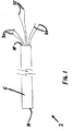
最初に図1には、本発明に従って形成された結紮カテーテル2が示されている。結紮カテーテル2は、長く延びたチューブ又はシリンダ5を含み、シリンダ5は遠位端部10と近接端部15とを有する。1又はそれ以上の前進/引き込みコントロール要素20が近接端部15から延び、コントロール要素20は、シリンダ5の内部に配置された結紮サブアセンブリ(図1には示さず)に接続されている。図1に示された構造では、前進/引き込みコントロール要素20が、ケーブル又はワイヤを含む。1又はそれ以上の前進/引き込みコントロール要素20は、シリンダ5の内部に配置された結紮サブアセンブリ(図1には示さず)を前進させ、は引っ込ませるのに用いられる。これに続いて、1又はそれ以上の収縮コントロール要素25もまた、シリンダ5の近接端部から出る。収縮コントロール要素25は、また、シリンダ5の内部に配置された結紮サブアセンブリ(図1には示さず)に接続され、組織等の部分周囲で結紮要素(図1には示さず)を締めたり、緩ませたりするのに用いられる。図1に示される構造では、収縮コントロール要素25が、ケーブル又は縫糸を含む。
Ligation Catheter Initially shown in FIG. 1 is a
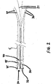
次に、図2には、外部シリンダ5が、破線で示されている。図2は、非展開状態の、すなわち、シリンダ5の中に引き込まれた結紮サブアセンブリ30を備えた、結紮サブアセンブリ30を示す。シリンダ5の内部で、結紮サブアセンブリ30は、(フエルトの綿撒糸又は輪35のような)サポート又はガイドに接続できる(例えば、縫糸やひものような)結紮要素32を有して示される。サポート又はガイドは、結紮される組織をつかみ、保護するのを助ける。結紮サブアセンブリ30は、また、圧縮材45を備えた支持構造40を含む。フエルト綿撒糸35は、圧縮材45の遠位端部に配置される。圧縮材45は、結紮サブアセンブリ30がシリンダ5から展開した場合に、拡がるように形成されている。1の好ましい構造では、圧縮材45は、バネを用いて互いに接続することで拡張され、これにより、圧縮材がシリンダ5の遠位端部から出た場合に、圧縮材45が自己拡張するようにしている。結紮サブアセンブリ30は、前進/引き込みコントロール要素20に接続され、これにより、結紮サブアセンブリ30は、シリンダ5の外部に進み又は内部に引き込まれることができる。更に、結紮サブアセンブリ30は、引き込みコントロール要素25に接続され、これにより、結紮要素32が、一片の組織等の周囲で引き込むことができる。前進/引き込みコントロール要素20、及び/又は引き込みコントロール要素25は、作業者による操作のために、適当なハンドル(図示せず)に接続されても良い。
Next, in FIG. 2, the
図3には、外部シリンダ5の中で非展開の結紮サブアセンブリ30を備え、フエルト綿撒糸35を備えた、シリンダ5の遠位端部10が示されている。
FIG. 3 shows the
次に、図4には、結紮サブアセンブリ30の一部が、シリンダ5の遠位端部から前進して示されている。圧縮材45がチューブ5の圧縮環境から出て、互いに離れて拡張しながら、結紮サブアセンブリ30は拡張する。図1〜図4に示す構造では、圧縮材45が、バネ50の影響で拡張する。結紮サブアセンブリ30は、収縮要素25に接続され、収縮要素25は、作業者の動作のためにチューブ5の近接端部を超えて延びる。結紮サブアセンブリ30は、前進/引き込みコントロール要素20を押すことによりシリンダから出ても良い。
Next, FIG. 4 shows a portion of the
図5は、シリンダ5の壁が破線で描かれている以外は図4と同様であり、これにより、装置の内部の動作を示している。圧縮材45は、内部の支持リング40のような支持部材40に接続されて示されている。
FIG. 5 is similar to FIG. 4 except that the wall of the
図6は、装置の遠位端部の図であり、シリンダ5から出た結紮サブアセンブリ30を示す。結紮サブアセンブリ30は半径方向に拡がり、結紮要素32は圧縮材45の遠位端部の周りに拡がった弓状の経路に従い、収縮コントロール要素25による動作のために結紮要素32の遠位端部はシリンダ5の中央を通る。このように、結紮要素32は圧縮材45(又は他の手段)により支持され、圧縮材45はバネ50(又は他の手段)により拡張されても良く、圧縮材45は支持構造40により支持されても良い。
FIG. 6 is a view of the distal end of the device, showing the
次に、図7には、結紮カテーテル2が、結紮要素32が引き込まれて示されている。結紮要素32は、引き込みコントロール要素25の近接端部をひっぱることにより引き込まれても良い。この動作は、結紮ループを半径方向の収縮させる。
Next, FIG. 7 shows the ligating
図8は、収縮した結紮要素32を有する結紮カテーテル2の遠位端部の図である。結紮ループは半径方向に圧縮されている。好ましくは、内部支持構造40は、シリンダ5の遠位端部のように、半径方向の寸法が不変である。好ましくは、作業者が装置を動かした場合に、圧縮材45とバネ50は、折りたたまれる。
FIG. 8 is a view of the distal end of the ligating
図9は、例えば前進/引き込みコントロール要素20を引っ張ることにより、シリンダ5の中に引き込まれた圧縮材45とバネ50とを示す。結紮要素32はその場に残り、捕まえられた組織を半径方向に圧縮する。
FIG. 9 shows the
図10は、結紮カテーテル2において、シリンダ5が破線で示された以外は、図9と同様である。圧縮材45、バネ50、及び支持構造40は、シリンダ5の中に引き込まれて示される。
FIG. 10 is the same as FIG. 9 except that the
図11は、収縮コントロール要素25から切断された結紮要素32を示す。
FIG. 11 shows the ligating
ガイドカテーテル
図12は、アライメント要素90を備えた結紮カテーテル2を示す。アライメント要素90は、結紮カテーテル2を左心耳や他のターゲット構造にアライメントするために使用される。本発明の1の具体例では、アライメント要素90は、放射線を透過しない材料を含み、装置は、例えば螢光透視法のような視覚的な方法により、所望に構造上の位置に配置される。
Guide Catheter FIG. 12 shows a ligating
代わりに、より好適には、アライメント要素90は、ガイドカテーテル100と結合して働くことを意図し、ガイドカテーテル100は左心耳の内部に(例えば、内腔に)配置される。この構造では、ガイドカテーテルはまた放射線を透過しない材料を含み、アライメント要素90とガイドカテーテル100は視覚的な方法によりアライメントされる。
Instead, more preferably, the
更に好適には、結紮カテーテル2とガイドカテーテル100が、物理手段(例えば、磁石、雄型及び雌型コネクタ、ワイアとスネア等)を備え、結紮カテーテル2とガイドカテーテル100とのアライメントを容易にする。このように、1の好ましい構造では、結紮カテーテル2が、遠位端部に参照磁石95を備えるアライメント要素90を有する。一方、ガイドカテーテル100は、その遠位端部に参照磁石105を有するアライメント要素102を含む。更に好適には、この好適な構造において、結紮カテーテル2は、シリンダ5の端部から延びることができるアライメント要素90を有する。アライメント要素90の遠位端部の上に、参照磁石95が配置される。アライメント要素90は、ガイドカテーテル100の近傍に置かれ、ガイドカテーテル100は、その遠位端部に搭載された参照磁石105を備えたアライメント要素102を有する。それらの2つのアライメント要素90、102が互いに近づけられた場合、磁石95、105により、アライメント要素90、102は自動的に互いにアライメントする。
More preferably, the ligating
例えば、左心耳の結紮中に、ガイドカテーテル100は、蛍光透視法のような視覚ガイドや超音波に従って左心耳の中に入る。結紮カテーテル2が心膜の中に入れられる。結紮カテーテルのアライメント要素90が、続いてシリンダ5から延びる。一旦アライメント要素90がアライメント要素102の近傍に配置されると、磁石95、105が、2つのカテーテルを自動的に互いにアライメントさせ、これにより、結紮カテーテル2が、左心耳に対して所望の位置をとる。続いて、結紮カテーテル2が、上述のように、左心耳を結紮するために用いられる。
For example, during ligation of the left atrial appendage, the
次に、図13には、拡張要素115を備えたガイドカテーテル100が示されている。拡張要素115は、人体(例えば左心耳)の中で拡張するように取り付けられる。上述のように、ガイドカテーテル100は、遠位端部に載置された磁石105を有する。なお、図13では、拡張要素115は非拡張状態で示されている。
Next, FIG. 13 shows a
次に、図14には、拡張状態の拡張要素115を備えたガイドカテーテル100が示されている。
Next, FIG. 14 shows a
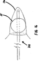
図15には、その拡張要素115が非拡張で、左心耳130の中に配置されたガイドカテーテル100が示される。
FIG. 15 shows the
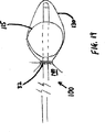
図16には、その拡張要素115が拡張し、ガイドカテーテル100が左心耳130の中に配置されたガイドカテーテル100が示される。この点において、拡張要素115が左心耳130の中で拡張状態にある場合、組織の空間の大きさや拡張した拡張要素115の大きさ等に応じて、受け入れる組織の形状が変わっても変わらなくても良いことを認識すべきである。
FIG. 16 shows the
ガイドカテーテルとの組み合わせて用いられる結紮カテーテル
図17には、拡張要素115が左心耳130の中で拡張し、結紮カテーテル2が左心耳130の上に拡張したガイドカテーテル100が示されている。このアライメントされた位置は、結紮カテーテル2の上のアライメント要素90と、ガイドカテーテル100の上のアライメント要素102との使用により容易に得られる。更に好適には、ガイドカテーテル100が左心耳130の中に配置され、拡張要素115が拡張し、結紮カテーテル2が、図4、5、及び6に示された状態に配置され、そして、装置をアライメントするためにガイド要素90、102を用いて、結紮カテーテル2が左心耳130の上に滑らされる。(左心耳の中の)ガイドカテーテル100と結紮カテーテル2は、磁石120、95を用いて互いに好ましい構造にアライメントされる。人体に向かい合う装置を適当な大きさにすることにより、アライメント要素90、102が上述のように機能した場合、結紮要素3を左心耳のネック(および拡張要素115の心房側)に配置することができる。
A ligating catheter used in combination with a guide catheter FIG. 17 shows a
次に、図18には、ガイドカテーテル100が左心耳130の中に示されている。拡張要素115は拡張状態にあり、図7、8との関連で上に述べたように、結紮カテーテル2が作動している。結紮カテーテル2が作動している間の、左心耳130の中での拡張したガイドカテーテル100の相対配置により、拡張要素115の存在により、引き込む結紮要素32は左心耳130のネックに保持される。これにより、人体に対する結紮要素32の適当な配置が確実になる。換言すれば、左心耳130の中に拡張要素が存在することにより、所望の位置135に、ひも32が導かれる。左心耳130の結紮の例では、所望の位置135は、左心耳130が左心房140につながる位置である。
Next, in FIG. 18, the
次に、図19には、まだ左心耳130の中にガイドカテーテル100があるが、結紮カテーテル2は引っ込められ、左心耳130のネックに展開された結紮要素32を残した装置が示される。
Next, FIG. 19 shows the device with the
図20において、ガイドカテーテル100は、非拡張状態に戻った拡張要素115を有している。
In FIG. 20, the
次に、図21には、ガイドカテーテル100が左心耳の内部から取り出され、左心耳130が左心房140につながる位置に結紮要素32が残された左心耳130が示されている。これにより、心房140から左心耳が有効に結紮される。
Next, FIG. 21 shows the left
一の好適な使用形態
一の好適な使用形態では、ガイドカテーテル100が、心房の隔膜を横切って内部に通り左心房に入る。ガイドカテーテル100(図22)は、1又はそれ以上の磁石105を先端に有する。磁石105は、好適には、例えば、ネオジウム−鉄−ボロン、コバルト−サマリウム、又は他の強力な固定磁気要素からなる、いわゆる「希土類」磁石である。磁石の丁度背後に、好適にはガイドカテーテルと一体となり、ガイドカテーテルのボディと軸対称である、拡張可能なバルーン115(図23)が配置される。バルーン115は、膨らんだ場合に、球状で、円錐型、楕円、又は他の形状であり、好適には、おおよそ左心耳130の大きさや形状と同じとなる。全体のガイドカテーテル100は、バルーン115と磁石105を含め、市販の鞘(図示せず)を通りぬける大きさであり、入手可能な道具や技術を用いた蛍光透視法のガイドにより、心房中隔をたやすく横切る大きさである。
One preferred mode of use In one preferred mode of use, the
このシステムの1つの好ましい使用では、セルジンガや他の標準的な技術を用いて、作業者は、皮膚を通して大腿部の欠陥に到達し、上述の鞘(図示せず)が、蛍光透視法のガイドにより心房中隔を通って導入される。ガイドカテーテルの磁石の端部105は、上述の鞘から前に出て、上で述べた方法で左心耳に入る。
In one preferred use of this system, using Serzinga or other standard techniques, an operator reaches a thigh defect through the skin, and the above-described sheath (not shown) is used for fluoroscopy. Guided through the atrial septum. The
この繰り返しとともに用いられる第2の道具(例えば、図24に示された結紮カテーテル2)が、心臓と心膜との間の心膜腔に導入される。心膜へのアクセスは、例えば、胸骨のジフォイドプロセス(zyphoid process)の下部の小さな切り込みや、又はそのような目的のために設計された針や専用のシステムを用いた経皮的アクセスによりなされる。蛍光透視法によるガイドでは、心臓と心膜袋との間の心膜腔にワイヤが導入される。同様に、小さな開胸や、「チャンバレイン」型の第2肋軟骨の切開により、心膜へのアクセスが得られる。そのような目的で設計された専用装置を用いた経皮的なアクセスは、一般に、部分麻酔の下で行われるのが好ましい。第2肋軟骨の切開又はジフォイドの下の小さな切開は、一般に、左胸膜腔の侵害を必要とするアプローチに好ましい。
A second tool (eg, the ligating
一旦、経皮的なアクセスがなされた場合、第2の道具(例えば図25に示す結紮カテーテル2)が、手術部位に導入される。好ましい具体例では、ワイヤ上を導入される拡張器や鞘が、第2の道具に先んじる。拡張器は、その遠位端部が左心耳の領域に配置され、大きな薄壁のチューブやシリンダ5の配置中に連続して大きくなり、おおよそ24フレンチまたはそれ以下になる。この薄壁のチューブ5は、続いて結紮アセンブリ30を手術部位に進めるための分配カニューレとして機能する。一回の繰り返しで、薄壁のチューブ5の遠位端部10は、引張って配置される非対称要素により折れ曲がる。代わりに、薄壁のチューブは、その先端で永久的な角やカーブを有しても良い。
Once percutaneous access has been made, a second tool (eg, the ligating
一旦、薄壁チューブ5が配置されれば、心膜内道具(例えば、結紮アセンブリ30)が薄壁チューブ5に進む。好適な具体例では、結紮カテーテル2は、その端部がすでに述べたような1又はそれ以上の希土類磁石95である中央心膜内カテーテル又はアライメント要素90を含む。これらの磁石95は、結紮カテーテル2が(先に左心耳内に配置された)ガイドカテーテル100に引きつけられるように、端部が向き合った位置で、棒で押される。他の具体例では結紮カテーテル2は、可撓性のある又は曲げられやすい、偏向する端部を有する剛体でも良い。
Once the thin
チューブ150は、このアライメント要素90と同軸になり、アライメント要素90又は薄壁チューブ5の何れかに対して、前進し又は引っ込むように構成され、端部が漏斗状またはトランペットベル状のフレア160となっている。このフレア状チューブ150の内径は、心膜内にある磁気端部を有する(その上をフレア状チューブ150が滑る)アライメント要素90の外径より十分に大きく、フレア状チューブの後ろから端部フレア160に真空が移動できるようにする。フレア状の端部150は吸引カップとして働き、心臓の外部から左心耳130の端部を掴む。
The
好適な具体例では、結紮カテーテルのアライメント要素90は、蛍光透視法のガイドにより前進し(図26)、予め心房中隔を横切り、左心耳の内側に配置されたガイドカテーテル100に噛み合って接続される(図27)。一旦そのようなアライメントが行われ、蛍光透視法で接続が確認されれば、フレア状のチューブ150が前進し(図28)、左心耳の外側と接触する。続いて、フレア状のチューブ150の後ろで吸引が行われ、左心耳がフレア状のチューブの端部に真空吸着される。
In a preferred embodiment, the ligation
このフレア状のチューブ150の外側上であって、24フレンチの薄壁チューブ5からなる内腔の内側に、ニチノール(Nitinol)のステント状構造又は結紮サブアセンブリ30が配置され、その後ろの端部に取り付けられた固いカテーテル又は他の構造(即ち、前進/引っ込みコントロール要素20)により、組織方向に薄壁チューブ5を下って前進することができる。このニチノール構造30は、(一旦薄壁の外部チューブ5の構造から解放されると)ベル状のクラウンに拡張し(図29)、その端部には環状のスネアが(周囲に)取り付けられている。好ましい具体例では、結紮要素又はスネア32は、ポリプロピレン又はPTFEの縫糸からなるスネアループ32はクラウン30の端部に、可逆的で容易に離れるように固定される。
A Nitinol stent-like structure or
一旦、左心耳が吸着固定されると、ニチノール構造30およびそれに取り付けられたスネア32は、フレア状チューブ150の上を左心耳方向に進む。フレア状チューブ150は、薄壁の外側チューブ5の端部を2又は3センチ超えて延びる。このように、ニチノール構造30は、ベル形状に拡張し始め、フレア状の吸着カテーテル150および左心耳の上に容易に進むことができるようになる。
Once the left atrial appendage is adsorbed and fixed, the
一旦、ニチノール構造30が左心耳の基部に近い点まで進むと、心耳の中のガイドカテーテル100上のバルーン115が、好ましくは対称な材料とともに膨張する。ニチノール構造は、蛍光透視法のガイドの下で進み、そのベル状クラウン30(および縫糸スネア32)の端部が、心房中のバルーンを覆う(図30)。次に、ニチノール構造30を前進させるのに用いられる固いカテーテル20の内腔を通る縫糸のより糸を引っ張ることにより、スネア32は締められ(図31)。ひっかかった縫糸を用いて、ガイドカテーテルのバルーンは収縮し、経中隔左心耳カテーテル100が除去される。次に、縫糸スネア32は、再度引っ張られて、経中隔カテーテル100により占領されていた空間を占める。スネアが引っ張られた時、ニチノール構造30は縫糸スネア32から離れる。次に、結紮カテーテル2が除去されて、縫糸が皮膚の位置で切断される。
Once the
追加構造
本発明の好ましい形態では、図32に示すように、ワイヤ155が、左心耳130の壁とその先の心膜腔を通って、左心耳130を通り、これにより、左心耳の端部165に穴があけられる。次に、心膜腔中のカテーテル又はスネアによりワイヤ155が捕まれ、心膜を通ってずっと引っ張られ、体外に出る。これにより、その上をガイドカテーテル100、結紮カテーテル2、及び/又は他の装置が、ワイヤ155の両端からその上を通ることができる1つのワイヤトラックを形成する。この構造において、結紮カテーテル2上のアライメント要素90、及び/又はガイドカテーテル上のアライメント要素は、必要であれば省略できる。更に、本発明のこの具体例では、必要であれば、ガイドカテーテル100を省略できる。
Additional Structure In a preferred form of the invention, as shown in FIG. 32, a
加えて、もし必要であれば、1又はそれ以上の磁石95及び/又は105は、電磁石を含んでも良い。そのような構造により、磁場を選択的にオンとオフにでき、これにより、処置の最後において、装置の分離が容易になる。
In addition, if necessary, the one or
更に、以下の記載において、縫糸45は、互いにバネ50に接続することにより拡張されるのが好ましいと記載され、これにより、シリンダ5の遠位端部から縫糸が出た場合に、縫糸は自分で拡張できるようになる。代わりに、例えば、圧縮材45に搭載された拡張メカニズムのような他の手段により、又はバネ材料から縫糸45を形成して(例えばニチノールのような超弾性材料)、縫糸45を拡張しても良い。
Furthermore, in the following description, it is described that the
本発明の更なる形態
本発明の1の好ましい形態は、1又はそれ以上の以下の構造要素を独自に組み合わせた、新規な装置及び方法である。
(1)シリンダのような細長い要素。
(2)結紮される組織の上にひもを配するのを助ける拡張要素。
(3)ひも。
(4)アライメントメカニズム。
(5)ひもが展開するように、ひもを適当な位置に導くのを助ける拡張要素。
Further Forms of the Invention One preferred form of the invention is a novel apparatus and method that uniquely combines one or more of the following structural elements.
(1) An elongated element such as a cylinder.
(2) An expansion element that helps place a string over the tissue to be ligated.
(3) Strings.
(4) Alignment mechanism.
(5) An expansion element that helps guide the string to the proper position so that the string deploys.
本発明の1の態様では、アライメントシステムが、左心耳のような組織構造の周囲の所望の位置に、ひも分配装置を配置するために提供される。 In one aspect of the invention, an alignment system is provided for placing a string dispensing device at a desired location around a tissue structure such as the left atrial appendage.
本発明の他の態様では、アライメントシステムが、左心耳のような組織構造の周りの所望の位置にひも分配装置を配置するために提供される。 In another aspect of the invention, an alignment system is provided for placing the string dispensing device at a desired location around a tissue structure such as the left atrial appendage.
本発明の他の態様では、左心耳のような組織構造の周りの所望の位置にひもを配置するための組織拡張器が提供される。 In another aspect of the invention, a tissue dilator is provided for placing a lace at a desired location around a tissue structure such as the left atrial appendage.
本発明の他の態様では、半径方向に調整可能なひも分配装置が、左心耳のような組織構造の周りの所望の位置にひもを配置するために提供される。この分配装置は、拡張可能である。 In another aspect of the invention, a radially adjustable string dispensing device is provided for placing the string at a desired location around a tissue structure such as the left atrial appendage. This dispensing device is expandable.
本発明の他の態様では、アライメントシステム、組織拡張器、及び半径方向に調整可能なひも分配装置を含む結紮システムが提供され、結紮システムは、左心耳のような組織構造の周囲にひもを配置するように形成される。 In another aspect of the invention, a ligation system is provided that includes an alignment system, a tissue dilator, and a radially adjustable lace dispensing device, the ligation system placing a lace around a tissue structure such as the left atrial appendage. To be formed.
本発明の他の態様では、静脈や動脈のような離れた位置から、及び/又は皮膚を通って、体内に入る少なくとも2つのカテーテルを用いて、開胸なしに、左心耳のような組織構造の周囲の所望の位置にひもを配置するように形成された結紮システムが提供される。 In another aspect of the invention, a tissue structure, such as the left atrial appendage, without a thoracotomy, using at least two catheters entering the body from a remote location such as a vein or artery and / or through the skin A ligation system is provided that is configured to place the string at a desired location around the.
本発明の他の形態では、患者の左心耳の中に配置するように形成され、左心耳の周囲の所望の位置にひもを配置するためのリファレンスを与えるように取り付けられた、ガイド部材を含む新しいシステムが提供される。 In another form of the invention, a guide member is formed for placement in the patient's left atrial appendage and is attached to provide a reference for placing a string at a desired location around the left atrial appendage. A new system is provided.
本発明の他の形態では、左心耳の中に配置された上述のガイド部材のリファレンスに対応した所望の位置にひもを配置するように形成されたアライメント構成要素を有するひも分配装置を提供する。 In another form of the invention, a string dispensing device is provided having an alignment component configured to position the string at a desired location corresponding to the reference of the guide member described above disposed in the left atrial appendage.
本発明の他の形態では、左心耳の中に配置するように形成され、ひもを配置するための所望の位置を明確にするために取り付けられる組織拡張器が提供される。 In another form of the invention, a tissue dilator is provided that is configured for placement in the left atrial appendage and is attached to define a desired location for placement of a string.
本発明の他の形態では、左心耳の中に配置するための、ガイド部材と組織拡張器の双方を有するリファレンスカテーテルが提供される。 In another form of the invention, a reference catheter is provided having both a guide member and a tissue dilator for placement in the left atrial appendage.
本発明の他の形態では、患者の左心耳の周りの所望の位置にひもを配置するように形成された半径方向に調整可能なひも分配装置を提供する。 In another aspect of the present invention, a radially adjustable string dispensing device configured to place a string at a desired location around the patient's left atrial appendage is provided.
本発明の他の形態では、患者の左心耳の中の上述のガイド部材に対応するアライメント構成要素と、その周囲にひもを配置するための調整可能なひも分配装置の双方を有する分配カテーテルを提供する。 In another form of the invention, a dispensing catheter is provided having both an alignment component corresponding to the above-described guide member in the patient's left atrial appendage and an adjustable lace dispensing device for positioning the lace around it. To do.
本発明の他の形態では、患者の左心耳の中の上述のガイド部材に対応するアライメント構成要素と、その周囲にひもを配置するための調整可能なひも分配装置の双方を有し、これによりひも分配装置が拡張要素を含む分配カテーテルを提供する。 In another form of the invention, it has both an alignment component corresponding to the above-described guide member in the patient's left atrial appendage and an adjustable lace dispensing device for positioning the lace around it, thereby A string dispensing device provides a dispensing catheter that includes an expansion element.
本発明の他の態様では、互いに対応するように形成されたリファレンスカテーテルとアライメントカテーテルの双方を含み、患者の左心耳の周りの所望の位置にひもを配置する結紮システムを提供する。 In another aspect of the invention, a ligation system is provided that includes both a reference catheter and an alignment catheter configured to correspond to each other, and places the lace at a desired location around the patient's left atrial appendage.
本発明の他の態様では、互いに対応するように形成されたリファレンスカテーテルとアライメントカテーテルの双方を含み、患者の左心耳の周りの所望の位置にひもを配置する結紮システムであって、リファレンスカテーテル又はアライメントカテーテル、又は双方が拡張要素を含む結紮システムを提供する。 In another aspect of the invention, a ligation system that includes both a reference catheter and an alignment catheter configured to correspond to each other and places a string at a desired location around the patient's left atrial appendage, comprising a reference catheter or An alignment catheter, or both, provides a ligation system that includes an expansion element.
本発明の他の態様では、上述の構成要素の1又はそれ以上を組み込む装置が結紮される組織の近傍に配置される。直接見ることにより、又は蛍光透視法、超音波、X線撮影法、CT、MRI等の導きのような多くの方法により、それが行われる。加えて、アライメントより糸、磁石等のような装置を用いてアライメントすることができる。 In another aspect of the invention, a device incorporating one or more of the components described above is placed in the vicinity of the tissue to be ligated. This is done by many methods such as direct viewing or guidance such as fluoroscopy, ultrasound, radiography, CT, MRI, etc. In addition, alignment can be performed using an apparatus such as a thread, a magnet, or the like.
本発明の他の態様では、装置や方法が、以下のように左心耳を結紮するために用いられる。セルジンガのオーバーザワイヤ技術のような標準技術を用いて、心膜腔へのアクセスが可能となる。例えば、拡張要素、ひも、及びアライメントメカニズムを含むシリンダのような細長い要素を好適には含む装置が、ガイドワイヤの上を心膜腔に入れられる。例えば、細長い装置は、拡張要素、ひも、及びアライメントメカニズムがその中にある大きなカテーテルでも好い。 In another aspect of the invention, the device or method is used to ligate the left atrial appendage as follows. Access to the pericardial space is possible using standard techniques such as Seldinger's over-the-wire technology. For example, a device that preferably includes an elongated element, such as a cylinder that includes an expansion element, a string, and an alignment mechanism, is placed over the guidewire into the pericardial space. For example, an elongate device may also be preferred for large catheters with expansion elements, strings, and alignment mechanisms therein.
本発明の他の形態では、静脈又は僧帽弁の逆行等により、経中隔のような標準技術を用いて、ガイドカテーテルが左心房の中に配置される。次に、左心房中のガイドカテーテルは蛍光透視法に導かれて、左心耳の中に配置される。この点で、ガイドカテーテルは左心耳にあり、結紮メカニズムは、心膜腔中に、展開カテーテルと接続されて配置される。次に、左心房中のガイドカテーテルと心膜腔中の展開カテーテルは、互いにアライメントされる。これは、様々な技術を用いて行われる。例えば、1又は双方の装置が磁化され、これにより磁力を用いて互いにアライメントさせる。代わりに、ガイドカテーテルと展開カテーテルが、視覚や超音波ガイドを用いて近くに導く(steered)こともできる。又は、左心房のガイドカテーテルは左心耳を貫通し、心膜中の展開カテーテルにより罠にかかっても(snared)良い。この点において、心膜中の装置は、左心耳の近傍に前進する。次に、結紮装置は展開カテーテルから、左心耳の上に展開する。好適には、左心房中のガイドカテーテルは、バルーンのような拡張要素を含む。次に、この拡張要素は左心耳の中で拡張される。そうすることで、この拡張により、もひが左心耳の周りで締められた場合に、ひもが左心耳から滑ったり移動したりするのを防止する。次に、ひもが左心耳の周囲で締められる。次に、左心房中の拡張要素が収縮する。次に、左心耳中のガイドカテーテルが、左心耳の外に戻される。この工程は、必要なだけ繰り返される。次に、ガイドカテーテルと展開カテーテルが、体腔から除去される。 In another form of the invention, a guide catheter is placed in the left atrium using standard techniques, such as a transseptal, such as by retrograde veins or mitral valves. The guide catheter in the left atrium is then guided into fluoroscopy and placed in the left atrial appendage. In this regard, the guide catheter is in the left atrial appendage and the ligation mechanism is placed in the pericardial space, connected to the deployment catheter. Next, the guide catheter in the left atrium and the deployment catheter in the pericardial space are aligned with each other. This is done using various techniques. For example, one or both devices are magnetized, thereby aligning each other using magnetic forces. Alternatively, the guide catheter and deployment catheter can be steered using visual or ultrasonic guides. Alternatively, the left atrial guide catheter may penetrate the left atrial appendage and snare by a deployment catheter in the pericardium. In this regard, the device in the pericardium is advanced near the left atrial appendage. The ligating device is then deployed from the deployment catheter over the left atrial appendage. Preferably, the guide catheter in the left atrium includes an expansion element such as a balloon. This expansion element is then expanded in the left atrial appendage. In doing so, this expansion prevents the string from slipping and moving away from the left atrial appendage when the lace is tightened around the left atrial appendage. The string is then tightened around the left atrial appendage. The dilating element in the left atrium then contracts. The guide catheter in the left atrial appendage is then returned out of the left atrial appendage. This process is repeated as often as necessary. Next, the guide catheter and deployment catheter are removed from the body cavity.
代わりに、左心耳中のガイドカテーテルが、ひもが殆ど配置された後で、ひもの最後の締め付け前に除去されて良い。これにより、左心耳からガイドカテーテルが引き出される前に、左心耳の下部が完全に閉鎖される。 Alternatively, the guide catheter in the left atrial appendage may be removed after the string has been placed and before the final tightening of the string. This completely closes the lower part of the left atrial appendage before the guide catheter is withdrawn from the left atrial appendage.
以下の記述では、左心耳の結紮のための好ましい方法を示す。希土類磁石、又は他のアライメント手段と、拡張可能なバルーンとを有する(その構造に組み込む)経中隔左心房ガイドカテーテルは、スネアやひもで左心耳を有効に封鎖するのに非常に有用である。左心耳は、一般にはおおよそ円錐形であり、左心房に接続される開口部の平面で少しネックにまたは狭く成っている。ひも又はスネアを有する外側から左心耳を有効に排除するために、スネアはこの平面で正確に締められる。理想的には、締められたひもにより、結果として生じる左心房の配置は、本質的に球状で、消えた開口部の部位で、心臓内又は内腔から確認できるわずかな窪みのみを有する。もしスネアが開口部の平面の(左心耳の端部に向かう)上方で締められた場合、左心耳の不完全な排除により、左心耳の永続的な憩室となり、これにより細動する心室で、鬱血や血栓が形成される。逆に言えば、もし、開口部の平面の下方でスネアが締められたら、房室の中を走る、弓状湾曲した環状動脈を傷つける危険性がある。 In the following description, a preferred method for ligation of the left atrial appendage is shown. A transseptal left atrial guide catheter with a rare earth magnet or other alignment means and an expandable balloon (incorporated into its structure) is very useful for effectively sealing the left atrial appendage with a snare or string . The left atrial appendage is generally approximately conical and is slightly necked or narrowed in the plane of the opening connected to the left atrium. In order to effectively exclude the left atrial appendage from the outside with a string or snare, the snare is tightened precisely in this plane. Ideally, due to the laced string, the resulting left atrial placement is essentially spherical and has only a small depression visible at the site of the missing opening, either within the heart or from the lumen. If the snare is tightened above the plane of the opening (towards the end of the left atrial appendage), the incomplete elimination of the left atrial appendage results in a permanent diverticulum of the left atrial appendage, thereby causing a fibrillation in the ventricle, Congestion and blood clots are formed. Conversely, if the snare is tightened below the plane of the opening, there is a risk of damaging the arcuately curved annular artery running through the atrioventricle.
最適化された平面で左心耳を正確で厳密にはめるために、多くに技術的な挑戦がなされた。いくらかの集団では、左心房と左心耳の形状がそれらの間のネックや狭い部分が、特に、心外膜や外部の形状から不十分に規定された。更に、左心耳の壁が薄く可撓性を有し、壁の張力が低い(左心耳では、一般に低く、例えば20mmHgより小さい)ため、左心耳と左心房との接続部の外部形状は、締め付け中に、スネアを正しい平面に持って行くのに殆ど役に立たない。この挑戦は、心室収縮からの並進運動により、細動する心房でさえも、生体構造が活発に動くという事実により悪化される。経中隔左心房ガイドカテーテルは、磁気端部と、上述のように適当な平面で左心耳を絡めることができる膨張可能なバルーンとを備える。特に、丁度、蛍光透視法や心エコー検査法による心膜内装置を用いた左心耳端部の認定と捕獲は、困難な挑戦だと信じられる。同時に、心房中隔を横切って左心房にカテーテルを入れ、その後に、それを左心耳の頂きに配置することは、市販されているカテーテルを用いた当業者が容易に達成できる。このように、端部に希土類磁石(又は他のアライメントメカニズム)を備えたガイドカテーテルを左心耳の配置することは容易に達成でき、これにより、心膜内ツールでその頂を正確に掴むのを可能にするとともに、左心耳の頂の位置について蛍光透視法で導くことができる。 Many technical challenges were made to fit the left atrial appendage accurately and precisely in an optimized plane. In some populations, the shape of the left atrium and left atrial appendage is poorly defined by the neck and narrow area between them, especially from the epicardium and external shape. Furthermore, since the wall of the left atrial appendage is thin and flexible and the wall tension is low (generally low in the left atrial appendage, eg, less than 20 mmHg), the external shape of the connection between the left atrial appendage and the left atrium is tightened. Inside, it is almost useless to take the snare to the right plane. This challenge is exacerbated by the fact that anatomy moves actively, even in fibrillation atria, due to translational movements from ventricular contractions. The transseptal left atrial guide catheter includes a magnetic end and an inflatable balloon that can entangle the left atrial appendage in a suitable plane as described above. In particular, the identification and capture of the left atrial appendage using an intrapericardial device by fluoroscopy or echocardiography is believed to be a difficult challenge. At the same time, placing a catheter in the left atrium across the atrial septum and then placing it at the left atrial appendage can be easily accomplished by one of ordinary skill in the art using commercially available catheters. In this way, placement of a guide catheter with a rare earth magnet (or other alignment mechanism) at the end can easily be achieved at the left atrial appendage, thereby allowing the endocardial tool to accurately grasp its apex. In addition, the position of the apex of the left atrial appendage can be guided by fluoroscopy.
経中隔左心耳カテーテルの端部近傍のバルーンは、左心耳と左心房との間の開口部の好ましい平面において、スネアやひもの配置や締め付けを非常に容易にする。好適には、バルーンは、おおよそ左心耳の大きさに膨らむように設計されている。バルーンが膨らんだ場合、左心耳と左心房との間の開口部のネックや狭所により、バルーンは左心耳に制限される。もしバルーンが、心エコー検査試験、蛍光透視法、特に造影剤を用いて膨らまされた場合、これは容易に確認できる。ガイドカテーテル中の分離ポートは、左心耳及び/又は左心房に造影剤が注入され、膨らんだバルーンの正しい位置を更に確認できるようにする。 A balloon near the end of the transseptal left atrial appendage catheter greatly facilitates snare and string placement and tightening in the preferred plane of the opening between the left atrial appendage and the left atrium. Preferably, the balloon is designed to inflate approximately to the size of the left atrial appendage. When the balloon is inflated, the balloon is restricted to the left atrial appendage due to the neck or constriction of the opening between the left atrial appendage and the left atrium. If the balloon is inflated using an echocardiography test, fluoroscopy, especially a contrast agent, this can be easily confirmed. A separation port in the guide catheter allows the contrast agent to be injected into the left atrial appendage and / or the left atrium to further confirm the correct location of the inflated balloon.
膨らんだバルーンは、左心耳と左心房の接合部で、外部の形状の特徴を際だたせる。球状のバルーンが膨らんだ場合、可撓性のある左心耳が膨張し、形状が(例えば球状に)変化し、結紮を容易にする。左心耳と左心房との間の接続は、雪だるまのウエストのように明確になる。これは、結紮中に、好適な平面にスネアまたはひもを強いる。バルーンおよびその結果として左心耳が、左心房の圧力より十分に高い圧力で膨らまされる。そのように、左心耳と左心房との間の壁の圧には、十分な違いがある。バルーンが球状になるため、平面の上方ではめることにより、張力の低い可撓性のあるネックに向かって、張った球状の面をスネアが滑ることとなる。左心耳及び/又は左心房に選択的にコントラストを付けることができる、バルーン中の放射線に対して不透明な造影剤や、放射線に対して不透明なスネア及び/又はひもは、蛍光透視法のガイドの下で、それらの工程を非常に容易に行えるようにする。一旦、左心耳と左心房との接続部が捕らえられた場合、バルーンが縮められ、除去され、スネアが完全に締められる。 The inflated balloon highlights external shape features at the junction of the left atrial appendage and the left atrium. When a spherical balloon is inflated, the flexible left atrial appendage is inflated and changes shape (eg, spherical), facilitating ligation. The connection between the left atrial appendage and the left atrium is as clear as a snowman's waist. This forces a snare or string into a suitable plane during ligation. The balloon and consequently the left atrial appendage are inflated with a pressure sufficiently higher than the pressure in the left atrium. As such, there is a sufficient difference in the wall pressure between the left atrial appendage and the left atrium. Since the balloon becomes spherical, the snare slides on the stretched spherical surface toward the flexible neck with low tension by fitting it above the plane. A contrast medium that is opaque to the radiation in the balloon, and a snare and / or string that is opaque to the radiation, which can selectively contrast the left atrial appendage and / or the left atrium Below, we make these steps very easy. Once the connection between the left atrial appendage and the left atrium is captured, the balloon is deflated and removed, and the snare is fully tightened.
一般に、とりわけ、本発明は、2つの装置のアライメントを含み、1つは内腔、心室等の内部、他はこれらの外部にある。このように、本発明は、腹部において、内視鏡的な検査に役立つために用いられる。 In general, among other things, the present invention includes the alignment of two devices, one inside the lumen, ventricle, etc., and the other outside these. Thus, the present invention is used to help endoscopic examination in the abdomen.
先の記載は、本発明の好ましい具体例を示すことを意図する。しかしながら、本発明の範囲から逸脱することなく、好ましい具体例の多くの変形が可能である。このように、方法の1又はそれ以上の工程、及び/又は装置の1又はそれ以上の構成要素は、変形又は省略できる。また、本装置及び方法は、人体の組織や似た構造を結紮するのにも使用できる。 The foregoing description is intended to illustrate preferred embodiments of the present invention. However, many variations of the preferred embodiment are possible without departing from the scope of the invention. In this way, one or more steps of the method and / or one or more components of the apparatus can be modified or omitted. The apparatus and method can also be used to ligate human tissue or similar structures.
本発明のこれら及び他の目的や特徴は、以下の、本発明の好ましい具体例の詳細な記述により、より全体が開示され明らかになるであろう。詳細な記載は、同様の符号が同様の部分を示す添付図面とともに考慮される。 These and other objects and features of the invention will be more fully disclosed and will become apparent from the following detailed description of the preferred embodiments of the invention. The detailed description is considered in conjunction with the accompanying drawings, wherein like numerals indicate like parts.
2 結紮カテーテル、5 シリンダ、10 遠位端部、15 近接端部、20 コントローラ、25 収縮コントローラ。
2 Ligating catheter, 5 cylinder, 10 distal end, 15 proximal end, 20 controller, 25 contraction controller.
Claims (3)
遠位領域と遠位端部とを有するガイドカテーテルであって、該ガイドカテーテルの遠位領域の上にバルーンが配置され、該ガイドカテーテルの遠位端部に第1磁石が配置され、該バルーンと該第1磁石を該左心耳の中に配置するために、中隔を通って移動する大きさと形状の該ガイドカテーテルと、
第2磁石を含み、心膜の空間中を通って移動する大きさと形状のアライメント要素であって、該第2磁石は、該左心耳の組織を横切って、該第1磁石と相互に作用するように取り付けられた該アライメント要素と、
結紮要素を含む結紮カテーテルであって、該結紮要素は、該左心耳の外側の周囲に配置されるような形状である結紮カテーテルと、を含む装置。A device used to ligate the left atrial appendage,
A guide catheter having a distal region and a distal end, the balloon on the distal region of the guide catheter is positioned, the first magnet is disposed at the distal end of the guide catheter, the balloon And a guide catheter sized and shaped to move through the septum to position the first magnet in the left atrial appendage;
A size and shape alignment element that includes a second magnet and moves through the pericardial space, wherein the second magnet interacts with the first magnet across the left atrial appendage tissue. The alignment element attached to
An apparatus comprising: a ligating catheter including a ligating element, wherein the ligating element is configured to be disposed about the outside of the left atrial appendage.
Applications Claiming Priority (5)
| Application Number | Priority Date | Filing Date | Title |
|---|---|---|---|
| US51010003P | 2003-10-09 | 2003-10-09 | |
| US60/510,100 | 2003-10-09 | ||
| US52899503P | 2003-12-12 | 2003-12-12 | |
| US60/528,995 | 2003-12-12 | ||
| PCT/US2004/033459 WO2005034802A2 (en) | 2003-10-09 | 2004-10-11 | Apparatus and method for the ligation of tissue |
Publications (3)
| Publication Number | Publication Date |
|---|---|
| JP2007534355A JP2007534355A (en) | 2007-11-29 |
| JP2007534355A5 JP2007534355A5 (en) | 2008-01-17 |
| JP5074765B2 true JP5074765B2 (en) | 2012-11-14 |
Family
ID=34437317
Family Applications (1)
| Application Number | Title | Priority Date | Filing Date |
|---|---|---|---|
| JP2006534449A Active JP5074765B2 (en) | 2003-10-09 | 2004-10-11 | Apparatus and method for tissue ligation |
Country Status (5)
| Country | Link |
|---|---|
| US (7) | US7846168B2 (en) |
| EP (1) | EP1682034B1 (en) |
| JP (1) | JP5074765B2 (en) |
| ES (1) | ES2700851T3 (en) |
| WO (1) | WO2005034802A2 (en) |
Families Citing this family (61)
| Publication number | Priority date | Publication date | Assignee | Title |
|---|---|---|---|---|
| US6488689B1 (en) | 1999-05-20 | 2002-12-03 | Aaron V. Kaplan | Methods and apparatus for transpericardial left atrial appendage closure |
| US8961541B2 (en) | 2007-12-03 | 2015-02-24 | Cardio Vascular Technologies Inc. | Vascular closure devices, systems, and methods of use |
| US20080114394A1 (en) | 2001-04-24 | 2008-05-15 | Houser Russell A | Arteriotomy Closure Devices and Techniques |
| US8992567B1 (en) | 2001-04-24 | 2015-03-31 | Cardiovascular Technologies Inc. | Compressible, deformable, or deflectable tissue closure devices and method of manufacture |
| US7527634B2 (en) * | 2002-05-14 | 2009-05-05 | University Of Pittsburgh | Device and method of use for functional isolation of animal or human tissues |
| EP1682034B1 (en) | 2003-10-09 | 2018-11-21 | Sentreheart, Inc. | Apparatus for the ligation of tissue |
| WO2006110734A2 (en) * | 2005-04-07 | 2006-10-19 | Sentreheart, Inc. | Apparatus and method for the ligation of tissue |
| US7992757B2 (en) | 2006-05-03 | 2011-08-09 | Raptor Ridge Llc | Systems and methods of tissue closure |
| US20100069925A1 (en) * | 2006-09-21 | 2010-03-18 | Mayo Foundation For Medical Education And Research | Devices and methods for ligating anatomical structures |
| EP2073723A4 (en) * | 2006-10-10 | 2011-03-30 | Mayo Foundation | Reducing contrast agent-induced toxicity |
| DK2142107T3 (en) * | 2007-03-30 | 2013-04-15 | Sentreheart Inc | Devices and systems for closing the left ventricular pendant |
| EP2148623A1 (en) * | 2007-05-21 | 2010-02-03 | Epitek, Inc. | Left atrial appendage closure |
| US20080294175A1 (en) * | 2007-05-21 | 2008-11-27 | Epitek, Inc. | Left atrial appendage closure |
| EP3272297B1 (en) | 2007-09-20 | 2020-04-22 | Sentreheart, Inc. | Devices for remote suture management |
| WO2009120953A2 (en) * | 2008-03-27 | 2009-10-01 | Mayo Foundation For Medical Education And Research | Navigation and tissue capture systems and methods |
| US20100114152A1 (en) * | 2008-11-06 | 2010-05-06 | Himanshu Shukla | Minimally-Invasive Method and Device for Permanently Compressing Tissues within the Body |
| US8332641B2 (en) * | 2009-01-30 | 2012-12-11 | Freescale Semiconductor, Inc. | Authenticated debug access for field returns |
| SE533463C2 (en) * | 2009-02-26 | 2010-10-05 | Stroemsholmen Ab | Balancing device for balancing two relatively moving parts including a gas spring and method for balancing |
| EP2413815B1 (en) | 2009-04-01 | 2018-12-12 | Sentreheart, Inc. | Tissue ligation devices and controls therefor |
| WO2010118312A2 (en) | 2009-04-09 | 2010-10-14 | Cardiovascular Systems, Inc. | Tissue closure devices, device and systems for delivery, kits and methods therefor |
| US8647350B2 (en) * | 2009-08-11 | 2014-02-11 | Raptor Ridge, Llc | Delivery device and method for compliant tissue fasteners |
| WO2011041488A2 (en) | 2009-09-30 | 2011-04-07 | Mayo Foundation For Medical Education And Research | Tissue capture and occlusion systems and methods |
| EP2482735A4 (en) | 2009-09-30 | 2017-03-29 | Mayo Foundation For Medical Education And Research | Enhanced signal navigation and capture systems and methods |
| US20110282250A1 (en) | 2010-04-13 | 2011-11-17 | Fung Gregory W | Methods and devices for treating atrial fibrillation |
| US9737309B1 (en) | 2010-06-24 | 2017-08-22 | Niv Ad | System for occlusion of left atrial appendage |
| US10631868B2 (en) | 2010-06-24 | 2020-04-28 | Niv Ad | System for occlusion of left atrial appendage |
| US9089312B2 (en) | 2011-03-10 | 2015-07-28 | Western New England University | Tamponade for biopsy surgery and method of operation |
| ES2671928T3 (en) | 2011-06-08 | 2018-06-11 | Sentreheart, Inc. | Tissue ligation devices and tension devices for them |
| WO2013070686A1 (en) * | 2011-11-08 | 2013-05-16 | Boston Scientific Scimed, Inc. | Handle assembly for a left atrial appendage occlusion device |
| JP2013103075A (en) * | 2011-11-16 | 2013-05-30 | Olympus Corp | Guidance medical system |
| US9439653B2 (en) | 2011-12-07 | 2016-09-13 | Traumatek Solutions B.V. | Devices and methods for endovascular access and therapy |
| US10118020B2 (en) | 2011-12-07 | 2018-11-06 | Traumatek Solutions B.V. | Devices and methods for endovascular access and therapy |
| CN109394216B (en) * | 2012-03-18 | 2021-11-02 | 特洛玛泰克解决方案私人有限公司 | Device and method for vascular access and treatment |
| US9186174B2 (en) * | 2012-08-22 | 2015-11-17 | Subramaniam Chitoor Krishnan | Methods and systems for accessing a pericardial space and preventing strokes arising from the left atrial appendage |
| WO2014164028A1 (en) | 2013-03-12 | 2014-10-09 | Sentreheart, Inc. | Tissue ligation devices and methods therefor |
| WO2014164572A1 (en) | 2013-03-13 | 2014-10-09 | Kaplan Aaron V | Devices and methods for excluding the left atrial appendage |
| EP3060140A4 (en) * | 2013-10-26 | 2017-06-07 | The United States Of America, As Represented By The Secretary, Department Of Health And Human Services | Atrial appendage ligation |
| WO2015066549A1 (en) | 2013-10-31 | 2015-05-07 | Sentreheart, Inc. | Devices and methods for left atrial appendage closure |
| US10485545B2 (en) | 2013-11-19 | 2019-11-26 | Datascope Corp. | Fastener applicator with interlock |
| CA3191158A1 (en) | 2014-02-28 | 2015-09-03 | Atricure, Inc. | Pericardial access devices and methods |
| JP6396667B2 (en) * | 2014-03-31 | 2018-09-26 | オリンパス株式会社 | Auricular ligation system |
| JP6302131B2 (en) | 2014-03-31 | 2018-03-28 | ジトメッド・エスピー・ゼット・オー・オーJitmed Sp Z O.O. | Left atrial appendage obturator |
| WO2016154498A1 (en) | 2015-03-24 | 2016-09-29 | Sentreheart, Inc. | Devices and methods for left atrial appendage closure |
| EP4285843A3 (en) | 2015-03-24 | 2024-05-22 | AtriCure, Inc. | Tissue ligation devices |
| US10987488B2 (en) | 2015-06-23 | 2021-04-27 | Traumatek Solutions, B.V. | Vessel cannulation device and method of use |
| WO2017091812A1 (en) | 2015-11-25 | 2017-06-01 | Talon Medical, LLC | Tissue engagement devices, systems, and methods |
| WO2017090190A1 (en) * | 2015-11-27 | 2017-06-01 | オリンパス株式会社 | Ligation device |
| AU2017223996B2 (en) | 2016-02-26 | 2022-02-10 | Atricure, Inc. | Devices and methods for left atrial appendage closure |
| US10702274B2 (en) * | 2016-05-26 | 2020-07-07 | Edwards Lifesciences Corporation | Method and system for closing left atrial appendage |
| EP3515327B1 (en) | 2016-09-23 | 2024-02-14 | AtriCure, Inc. | Devices for left atrial appendage closure |
| JP7071350B2 (en) | 2016-10-27 | 2022-05-18 | コンフォーマル・メディカル・インコーポレイテッド | Devices and methods for eliminating the left atrial appendage |
| WO2018089689A1 (en) | 2016-11-11 | 2018-05-17 | Avenu Medical, Inc. | Systems and methods for percutaneous intravascular access and guidewire placement |
| EP3459469A1 (en) | 2017-09-23 | 2019-03-27 | Universität Zürich | Medical occluder device |
| WO2019147835A1 (en) * | 2018-01-26 | 2019-08-01 | Intelligent Endoscopy Llc | Anti-slip bands |
| US11191547B2 (en) | 2018-01-26 | 2021-12-07 | Syntheon 2.0, LLC | Left atrial appendage clipping device and methods for clipping the LAA |
| WO2019191316A1 (en) | 2018-03-27 | 2019-10-03 | Sentreheart, Inc. | Devices and methods for left atrial appendage closure |
| WO2019191271A1 (en) | 2018-03-28 | 2019-10-03 | Datascope Corp. | Device for atrial appendage exclusion |
| US10925615B2 (en) | 2019-05-03 | 2021-02-23 | Syntheon 2.0, LLC | Recapturable left atrial appendage clipping device and methods for recapturing a left atrial appendage clip |
| JP2022549717A (en) | 2019-09-26 | 2022-11-28 | ウニベルシタット チューリッヒ | left atrial appendage closure device |
| WO2021216962A1 (en) * | 2020-04-23 | 2021-10-28 | Wake Forest University Health Sciences | Endocardial left atrial appendage management |
| CN117338365B (en) * | 2023-12-06 | 2024-03-08 | 东南大学泰州生物医药与医疗器械研究院 | Left auricle plugging device |
Family Cites Families (381)
| Publication number | Priority date | Publication date | Assignee | Title |
|---|---|---|---|---|
| US3435905A (en) | 1966-03-29 | 1969-04-01 | Lazarus & Peyser Associates | Tool and method of manufacturing the same |
| US3496932A (en) | 1967-12-22 | 1970-02-24 | Gen Motors Corp | Method and apparatus for substernal cardiac massage |
| US3677597A (en) | 1971-03-30 | 1972-07-18 | Harold A Stipek | Loop-supporting device |
| US3802074A (en) | 1971-05-21 | 1974-04-09 | C Hoppe | Surgical suture extractor |
| DE2132808C3 (en) | 1971-07-01 | 1981-10-29 | Deyhle, Peter, Dr.med., 8520 Erlangen | Device for the diathermic removal of growths |
| US3841685A (en) | 1973-02-01 | 1974-10-15 | A Kolodziej | Line handling device |
| US3828790A (en) | 1973-02-28 | 1974-08-13 | American Cystoscope Makers Inc | Surgical snare |
| US4018229A (en) | 1974-09-13 | 1977-04-19 | Olympus Optical Co., Ltd. | Apparatus for ligation of affected part in coeloma |
| US4030509A (en) | 1975-09-30 | 1977-06-21 | Mieczyslaw Mirowski | Implantable electrodes for accomplishing ventricular defibrillation and pacing and method of electrode implantation and utilization |
| US3999555A (en) | 1975-10-28 | 1976-12-28 | Medtronic, Inc. | Atrial pinch on lead and insertion tool |
| GB1506142A (en) | 1975-11-17 | 1978-04-05 | Akiyama T | Cutting apparatus for surgical thread |
| US4069825A (en) | 1976-01-28 | 1978-01-24 | Taichiro Akiyama | Surgical thread and cutting apparatus for the same |
| US4181123A (en) | 1977-12-28 | 1980-01-01 | The University Of Virginia Alumni Patents Foundation | Apparatus for cardiac surgery and treatment of cardiovascular disease |
| US4319562A (en) | 1977-12-28 | 1982-03-16 | The University Of Virginia Alumni Patents Foundation | Method and apparatus for permanent epicardial pacing or drainage of pericardial fluid and pericardial biopsy |
| US4249536A (en) * | 1979-05-14 | 1981-02-10 | Vega Roger E | Urological catheter |
| US4257278A (en) | 1979-08-24 | 1981-03-24 | General Electric Company | Quantitative volume blood flow measurement by an ultrasound imaging system featuring a Doppler modality |
| US4765341A (en) | 1981-06-22 | 1988-08-23 | Mieczyslaw Mirowski | Cardiac electrode with attachment fin |
| US4428375A (en) | 1982-02-16 | 1984-01-31 | Ellman Barry R | Surgical bag for splenorrhaphy |
| GB2150889B (en) | 1983-12-08 | 1987-06-03 | Thomas Ball Mcglinn | Rescue device |
| US4662377A (en) | 1985-11-07 | 1987-05-05 | Mieczyslaw Mirowski | Cardioverting method and apparatus utilizing catheter and patch electrodes |
| US4817608A (en) | 1987-05-29 | 1989-04-04 | Mieczyslaw Mirowski | Cardioverting transvenous catheter/patch electrode system and method for its use |
| US5033477A (en) | 1987-11-13 | 1991-07-23 | Thomas J. Fogarty | Method and apparatus for providing intrapericardial access and inserting intrapericardial electrodes |
| US4901405A (en) | 1988-08-25 | 1990-02-20 | Grover Alfred H | Self-aligning magnetic necklace clasp |
| US4944753A (en) | 1988-09-26 | 1990-07-31 | Burgess Frank M | Method for producing retro-sternal space |
| US5678547A (en) | 1988-12-22 | 1997-10-21 | Biofield Corp. | Method and apparatus for screening or sensing bodily conditions using DC biopotentials |
| US4991578A (en) | 1989-04-04 | 1991-02-12 | Siemens-Pacesetter, Inc. | Method and system for implanting self-anchoring epicardial defibrillation electrodes |
| US5433730A (en) | 1989-05-03 | 1995-07-18 | Intermedics, Inc. | Conductive pouch electrode for defibrillation |
| US4991603A (en) | 1989-10-30 | 1991-02-12 | Siemens-Pacesetter, Inc. | Transvenously placed defibrillation leads via an inferior vena cava access site and method of use |
| US4998975A (en) | 1989-10-30 | 1991-03-12 | Siemens-Pacesetter, Inc. | Travenously placed defibrillation leads |
| US5226908A (en) | 1989-12-05 | 1993-07-13 | Inbae Yoon | Multi-functional instruments and stretchable ligating and occluding devices |
| US5163946A (en) | 1990-04-25 | 1992-11-17 | Mitek Surgical Products, Inc. | Suture rundown tool and cutter system |
| US5176691A (en) | 1990-09-11 | 1993-01-05 | Pierce Instruments, Inc. | Knot pusher |
| US5108406A (en) | 1990-12-14 | 1992-04-28 | L.P. Wagi | Instrument to retrieve intraluminal objects |
| US5181123A (en) | 1991-03-29 | 1993-01-19 | Thomson Consumer Electronics, Inc. | Cathode-ray tube having a shrinkfit implosion protection band with tension limiting means |
| US5181919A (en) | 1991-04-23 | 1993-01-26 | Arieh Bergman | Suture ligating device for use with an endoscope |
| JPH05267604A (en) | 1991-05-08 | 1993-10-15 | Seiko Instr Inc | Manufacture of semiconductor device |
| US5676636A (en) | 1994-07-22 | 1997-10-14 | Origin Medsystems, Inc. | Method for creating a mediastinal working space |
| US5243977A (en) | 1991-06-26 | 1993-09-14 | Trabucco Hector O | Pacemaker |
| US5766151A (en) | 1991-07-16 | 1998-06-16 | Heartport, Inc. | Endovascular system for arresting the heart |
| US5452733A (en) | 1993-02-22 | 1995-09-26 | Stanford Surgical Technologies, Inc. | Methods for performing thoracoscopic coronary artery bypass |
| AU657364B2 (en) | 1991-10-18 | 1995-03-09 | United States Surgical Corporation | Self contained gas powered surgical apparatus |
| US5269326A (en) | 1991-10-24 | 1993-12-14 | Georgetown University | Method for transvenously accessing the pericardial space via the right auricle for medical procedures |
| US5281238A (en) | 1991-11-22 | 1994-01-25 | Chin Albert K | Endoscopic ligation instrument |
| US5163942A (en) | 1991-12-09 | 1992-11-17 | Everest Medical Corporation | Surgical instrument with grasping loop for laparoscopic procedures |
| US5226535A (en) | 1992-02-14 | 1993-07-13 | Ethicon, Inc. | Package for endoscopic suture loop and cannula |
| US5318578A (en) | 1992-03-17 | 1994-06-07 | Harrith M. Hasson | Apparatus for delivering a suture into a body cavity and method of using the apparatus |
| US5336231A (en) | 1992-05-01 | 1994-08-09 | Adair Edwin Lloyd | Parallel channel fixation, repair and ligation suture device |
| US5540711A (en) | 1992-06-02 | 1996-07-30 | General Surgical Innovations, Inc. | Apparatus and method for developing an anatomic space for laparoscopic procedures with laparoscopic visualization |
| US5336252A (en) | 1992-06-22 | 1994-08-09 | Cohen Donald M | System and method for implanting cardiac electrical leads |
| CA2098896C (en) | 1992-06-30 | 2005-03-29 | H. Jonathan Tovey | Specimen retrieval pouch and method for use |
| US5242459A (en) | 1992-07-10 | 1993-09-07 | Laparomed Corporation | Device and method for applying a ligating loop |
| US5443481A (en) | 1992-07-27 | 1995-08-22 | Lee; Benjamin I. | Methods and device for percutaneous sealing of arterial puncture sites |
| US5676651A (en) | 1992-08-06 | 1997-10-14 | Electric Boat Corporation | Surgically implantable pump arrangement and method for pumping body fluids |
| US5279539A (en) | 1992-08-17 | 1994-01-18 | Ethicon, Inc. | Drawstring surgical pouch and method of use for preventing ovarian adhesions |
| US5334199A (en) | 1992-08-17 | 1994-08-02 | Inbae Yoon | Ligating instrument and methods of ligating tissue in endoscopic operative procedures |
| JP2552820B2 (en) * | 1992-09-23 | 1996-11-13 | ターゲット セラピューティクス,インコーポレイテッド | Medical recovery equipment |
| US5242535A (en) | 1992-09-29 | 1993-09-07 | The Boc Group, Inc. | Method of forming a copper circuit pattern |
| US5312423A (en) | 1992-10-01 | 1994-05-17 | Advanced Surgical Intervention, Inc. | Apparatus and method for laparaoscopic ligation |
| US5300078A (en) | 1992-10-09 | 1994-04-05 | Laparomed Corporation | Device and method for applying large-diameter ligating loop |
| ES2111689T3 (en) | 1992-11-17 | 1998-03-16 | Smith & Nephew Inc | LOCKING DEVICE FOR THE IMMOBILIZATION OF A THREAD OF SUTURE. |
| DE4240533C1 (en) | 1992-11-27 | 1994-04-07 | Ethicon Gmbh | Endoscopic loop with application instrument - has knife on distal end of actuating member acting against free portion of surgical thread |
| US6283127B1 (en) | 1992-12-03 | 2001-09-04 | Wesley D. Sterman | Devices and methods for intracardiac procedures |
| US5398944A (en) | 1992-12-07 | 1995-03-21 | A. W. Chesterton Co. | Sealing system for reciprocating rod |
| US5340129A (en) | 1993-01-21 | 1994-08-23 | Minnesota Mining And Manufacturing Company | Saw blade retention system |
| USRE36269E (en) | 1993-01-21 | 1999-08-17 | Minnesota Mining And Manufacturing Company | Saw blade retention system |
| US5336229A (en) | 1993-02-09 | 1994-08-09 | Laparomed Corporation | Dual ligating and dividing apparatus |
| US6494211B1 (en) | 1993-02-22 | 2002-12-17 | Hearport, Inc. | Device and methods for port-access multivessel coronary artery bypass surgery |
| US6346074B1 (en) | 1993-02-22 | 2002-02-12 | Heartport, Inc. | Devices for less invasive intracardiac interventions |
| US6010531A (en) | 1993-02-22 | 2000-01-04 | Heartport, Inc. | Less-invasive devices and methods for cardiac valve surgery |
| US5797960A (en) | 1993-02-22 | 1998-08-25 | Stevens; John H. | Method and apparatus for thoracoscopic intracardiac procedures |
| US5624453A (en) | 1993-02-23 | 1997-04-29 | Wilson-Cook Medical, Inc. | Endoscopic ligating instrument |
| US5799661A (en) | 1993-02-22 | 1998-09-01 | Heartport, Inc. | Devices and methods for port-access multivessel coronary artery bypass surgery |
| US6161543A (en) | 1993-02-22 | 2000-12-19 | Epicor, Inc. | Methods of epicardial ablation for creating a lesion around the pulmonary veins |
| US5728151A (en) | 1993-02-22 | 1998-03-17 | Heartport, Inc. | Intercostal access devices for less-invasive cardiovascular surgery |
| AU6405994A (en) | 1993-03-11 | 1994-09-26 | Wilson Greatbatch Ltd. | Laparoscopic surgical grasper |
| US5403331A (en) | 1993-03-12 | 1995-04-04 | United States Surgical Corporation | Looped suture ligating device containing a heat-shrinkable element |
| US5306234A (en) | 1993-03-23 | 1994-04-26 | Johnson W Dudley | Method for closing an atrial appendage |
| US5449361A (en) | 1993-04-21 | 1995-09-12 | Amei Technologies Inc. | Orthopedic cable tensioner |
| US5540698A (en) | 1993-04-21 | 1996-07-30 | Amei Technologies Inc. | System and method for securing a medical cable |
| US5437643A (en) | 1993-05-17 | 1995-08-01 | Ethicon, Inc. | Safety interposer for surgical instruments |
| US6258021B1 (en) | 1993-06-17 | 2001-07-10 | Peter J. Wilk | Intrapericardial assist method |
| US6155968A (en) | 1998-07-23 | 2000-12-05 | Wilk; Peter J. | Method and device for improving cardiac function |
| US5423830A (en) | 1993-07-07 | 1995-06-13 | Schneebaum; Cary W. | Polyp retrieval method and associated instrument assembly |
| US5683364A (en) | 1993-07-30 | 1997-11-04 | Zadini; Filiberto | Percutaneous cardiac pump for cardiopulmonary resuscitation |
| US5385156A (en) | 1993-08-27 | 1995-01-31 | Rose Health Care Systems | Diagnostic and treatment method for cardiac rupture and apparatus for performing the same |
| US5609597A (en) | 1993-12-09 | 1997-03-11 | Lehrer; Theodor | Apparatus and method of extracorporeally applying and locking laparoscopic suture and loop ligatures |
| US5536273A (en) | 1993-12-09 | 1996-07-16 | Lehrer; Theodor | Apparatus and method of extracorporeally applying and locking laparoscopic suture and loop ligatures |
| WO1995018593A1 (en) | 1994-01-10 | 1995-07-13 | Cardassist Incorporated | Ventricular assist device |
| US5423821A (en) | 1994-01-18 | 1995-06-13 | Pasque; Michael K. | Sternal closure device |
| RU2088162C1 (en) | 1994-03-05 | 1997-08-27 | Вадим Борисович Берсенев | Instrument for binding and tightening liature knots |
| JPH07296645A (en) | 1994-04-21 | 1995-11-10 | Sumitomo Electric Ind Ltd | Shield wire facilitating terminal working and manufacture thereof |
| US5545178A (en) | 1994-04-29 | 1996-08-13 | Kensey Nash Corporation | System for closing a percutaneous puncture formed by a trocar to prevent tissue at the puncture from herniating |
| US5522819A (en) | 1994-05-12 | 1996-06-04 | Target Therapeutics, Inc. | Dual coil medical retrieval device |
| WO1995032669A1 (en) | 1994-06-01 | 1995-12-07 | Perclose, Inc. | Apparatus and method for advancing surgical knots |
| US5681278A (en) | 1994-06-23 | 1997-10-28 | Cormedics Corp. | Coronary vasculature treatment method |
| US5498228A (en) | 1994-08-08 | 1996-03-12 | John W. Royalty | Electromagnetic bi-ventricular assist device |
| US5601576A (en) | 1994-08-10 | 1997-02-11 | Heartport Inc. | Surgical knot pusher and method of use |
| JP3649459B2 (en) * | 1994-09-02 | 2005-05-18 | 住友ベークライト株式会社 | Internal organ lifting device |
| CA2156027C (en) | 1994-10-04 | 2006-07-18 | Keith Ratcliff | Surgical retractor |
| US5494240A (en) | 1994-10-07 | 1996-02-27 | The United States Of America As Represented By The Secretary Of The Navy | Vehicle recovery device for use by helicopter |
| US7175619B2 (en) | 1994-10-07 | 2007-02-13 | Boston Scientific Scimed, Inc. | Loop structures for positioning a diagnostic or therapeutic element on the epicardium or other organ surface |
| US6464700B1 (en) | 1994-10-07 | 2002-10-15 | Scimed Life Systems, Inc. | Loop structures for positioning a diagnostic or therapeutic element on the epicardium or other organ surface |
| US6152920A (en) | 1997-10-10 | 2000-11-28 | Ep Technologies, Inc. | Surgical method and apparatus for positioning a diagnostic or therapeutic element within the body |
| DE4438944C2 (en) | 1994-10-31 | 2003-10-09 | Wolf Gmbh Richard | endoscope |
| US5624430A (en) * | 1994-11-28 | 1997-04-29 | Eton; Darwin | Magnetic device to assist transcorporeal guidewire placement |
| US5707336A (en) | 1995-01-09 | 1998-01-13 | Cardassist Incorporated | Ventricular assist device |
| CA2158976C (en) | 1995-02-07 | 2005-05-24 | Thomas E. Watson, Jr. | Telescoping serial elastic band ligator |
| US5888247A (en) | 1995-04-10 | 1999-03-30 | Cardiothoracic Systems, Inc | Method for coronary artery bypass |
| US5571161A (en) | 1995-04-12 | 1996-11-05 | Starksen; Niel F. | Apparatus and method for implanting electrical leads in the heart |
| US6224584B1 (en) | 1997-01-14 | 2001-05-01 | Eclipse Surgical Technologies, Inc. | Therapeutic and diagnostic agent delivery |
| US5797870A (en) | 1995-06-07 | 1998-08-25 | Indiana University Foundation | Pericardial delivery of therapeutic and diagnostic agents |
| US5840059A (en) | 1995-06-07 | 1998-11-24 | Cardiogenesis Corporation | Therapeutic and diagnostic agent delivery |
| US6132438A (en) | 1995-06-07 | 2000-10-17 | Ep Technologies, Inc. | Devices for installing stasis reducing means in body tissue |
| US5827216A (en) | 1995-06-07 | 1998-10-27 | Cormedics Corp. | Method and apparatus for accessing the pericardial space |
| US5902311A (en) | 1995-06-15 | 1999-05-11 | Perclose, Inc. | Low profile intraluminal suturing device and method |
| US5900433A (en) | 1995-06-23 | 1999-05-04 | Cormedics Corp. | Vascular treatment method and apparatus |
| US5814052A (en) | 1995-06-29 | 1998-09-29 | Nakao; Naomi L. | Surgical cauterization snare with ligating suture |
| US5797946A (en) | 1995-07-13 | 1998-08-25 | Origin Medsystems, Inc. | Method for arterial harvest and anastomosis for coronary bypass grafting |
| US5693059A (en) | 1995-09-15 | 1997-12-02 | Yoon; Inbae | Ligating instrument with multiple loop ligature supply and methods therefor |
| US5704943A (en) | 1995-09-25 | 1998-01-06 | Yoon; Inbae | Ligating instrument with multiple loop ligature supply and methods therefor |
| JP3293118B2 (en) * | 1995-10-18 | 2002-06-17 | ニプロ株式会社 | Catheter assembly for endocardial suture surgery |
| US5716392A (en) | 1996-01-05 | 1998-02-10 | Medtronic, Inc. | Minimally invasive medical electrical lead |
| US5792151A (en) | 1996-01-24 | 1998-08-11 | The Ohio State University | Method and apparatus for ligating a blood vessel, tissue or other bodily duct |
| US5752526A (en) | 1996-02-19 | 1998-05-19 | The Cleveland Clinic Foundation | Minimally invasive cardiac surgery procedure |
| US5727569A (en) | 1996-02-20 | 1998-03-17 | Cardiothoracic Systems, Inc. | Surgical devices for imposing a negative pressure to fix the position of cardiac tissue during surgery |
| US5735877A (en) | 1996-02-28 | 1998-04-07 | Pagedas; Anthony C. | Self locking suture lock |
| US5855856A (en) | 1996-03-19 | 1999-01-05 | Ozone Sterilization Products, Inc. | Ozone generator and method for ozone generation |
| US5871496A (en) | 1996-03-20 | 1999-02-16 | Cardiothoracic Systems, Inc. | Surgical instrument for facilitating the detachment of an artery and the like |
| JP4136020B2 (en) | 1996-04-17 | 2008-08-20 | オリンパス株式会社 | Medical ligature |
| US5683445A (en) | 1996-04-29 | 1997-11-04 | Swoyer; John M. | Medical electrical lead |
| US5766216A (en) | 1996-05-30 | 1998-06-16 | Gangal; Hanamraddi T. | Band applicator for appendicular and meso-appendicular stumps |
| US6059750A (en) | 1996-08-01 | 2000-05-09 | Thomas J. Fogarty | Minimally invasive direct cardiac massage device and method |
| US5904147A (en) | 1996-08-16 | 1999-05-18 | University Of Massachusetts | Intravascular catheter and method of controlling hemorrhage during minimally invasive surgery |
| US5766217A (en) | 1996-09-23 | 1998-06-16 | Christy; William J. | Surgical loop delivery device and method |
| US6152936A (en) | 1996-09-23 | 2000-11-28 | Esd Medical, Llc | Surgical loop delivery device |
| US6311692B1 (en) | 1996-10-22 | 2001-11-06 | Epicor, Inc. | Apparatus and method for diagnosis and therapy of electrophysiological disease |
| US6237605B1 (en) | 1996-10-22 | 2001-05-29 | Epicor, Inc. | Methods of epicardial ablation |
| US6206004B1 (en) | 1996-12-06 | 2001-03-27 | Comedicus Incorporated | Treatment method via the pericardial space |
| US6332880B1 (en) | 1996-12-19 | 2001-12-25 | Ep Technologies, Inc. | Loop structures for supporting multiple electrode elements |
| US6048329A (en) | 1996-12-19 | 2000-04-11 | Ep Technologies, Inc. | Catheter distal assembly with pull wires |
| US5910129A (en) | 1996-12-19 | 1999-06-08 | Ep Technologies, Inc. | Catheter distal assembly with pull wires |
| US5961440A (en) | 1997-01-02 | 1999-10-05 | Myocor, Inc. | Heart wall tension reduction apparatus and method |
| US5882299A (en) | 1997-01-31 | 1999-03-16 | Minnesota Scientific, Inc. | Device and procedure for minimally invasive coronary anastomosis |
| US5699748A (en) | 1997-02-06 | 1997-12-23 | Linskey, Jr.; Edward | Line handling device for positioning and handling of mooring lines |
| US6045570A (en) | 1997-02-11 | 2000-04-04 | Biointerventional Corporation | Biological sealant mixture and system for use in percutaneous occlusion of puncture sites and tracts in the human body and method |
| US5779727A (en) | 1997-02-18 | 1998-07-14 | Orejola; Wilmo C. | Hydraulically operated surgical scissors |
| US5893869A (en) | 1997-02-19 | 1999-04-13 | University Of Iowa Research Foundation | Retrievable inferior vena cava filter system and method for use thereof |
| US6120431A (en) | 1997-03-14 | 2000-09-19 | Allegheny-Singer Research Institute | Method for assisting stystolic and diastolic cardiac function |
| US6319201B1 (en) | 1997-10-15 | 2001-11-20 | Peter J. Wilk | Imaging device and associated method |
| US5968010A (en) | 1997-04-30 | 1999-10-19 | Beth Israel Deaconess Medical Center, Inc. | Method for transvenously accessing the pericardial space via the right atrium |
| US6200303B1 (en) | 1997-04-30 | 2001-03-13 | Beth Israel Deaconess Medical Center, Inc. | Method and kit for transvenously accessing the pericardial space via the right atrium |
| US5957936A (en) | 1997-05-01 | 1999-09-28 | Inbae Yoon | Instrument assemblies for performing anatomical tissue ligation |
| US5908429A (en) | 1997-05-01 | 1999-06-01 | Yoon; Inbae | Methods of anatomical tissue ligation |
| US5895298A (en) | 1997-05-29 | 1999-04-20 | Biofield Corp. | DC biopotential electrode connector and connector condition sensor |
| US5851185A (en) * | 1997-07-02 | 1998-12-22 | Cabot Technology Corporation | Apparatus for alignment of tubular organs |
| US5897586A (en) | 1997-08-15 | 1999-04-27 | Regents Of The University Of Minnesota | Implantable defibrillator lead |
| US5972013A (en) | 1997-09-19 | 1999-10-26 | Comedicus Incorporated | Direct pericardial access device with deflecting mechanism and method |
| US6592552B1 (en) | 1997-09-19 | 2003-07-15 | Cecil C. Schmidt | Direct pericardial access device and method |
| US5991668A (en) | 1997-09-25 | 1999-11-23 | Medtronic, Inc. | Medical electrical lead |
| US5871531A (en) | 1997-09-25 | 1999-02-16 | Medtronic, Inc. | Medical electrical lead having tapered spiral fixation |
| US6006122A (en) | 1997-09-25 | 1999-12-21 | Medtronic, Inc. | Medical electrical lead |
| US5895404A (en) | 1997-09-29 | 1999-04-20 | Ruiz; Carlos E. | Apparatus and methods for percutaneously forming a passageway between adjacent vessels or portions of a vessel |
| US6071281A (en) | 1998-05-05 | 2000-06-06 | Ep Technologies, Inc. | Surgical method and apparatus for positioning a diagnostic or therapeutic element within the body and remote power control unit for use with same |
| US6610055B1 (en) | 1997-10-10 | 2003-08-26 | Scimed Life Systems, Inc. | Surgical method for positioning a diagnostic or therapeutic element on the epicardium or other organ surface |
| US6015382A (en) | 1997-10-16 | 2000-01-18 | General Surgical Innovations, Inc. | Inflatable manipulator for organ positioning during surgery and method of use |
| US5908435A (en) * | 1997-10-23 | 1999-06-01 | Samuels; Shaun L. W. | Expandable lumen device and method of use |
| US6081738A (en) | 1998-01-15 | 2000-06-27 | Lumend, Inc. | Method and apparatus for the guided bypass of coronary occlusions |
| AU1927399A (en) | 1998-01-16 | 1999-08-02 | Lumend, Inc. | Catheter apparatus for treating arterial occlusions |
| US6090042A (en) | 1998-01-23 | 2000-07-18 | Rullo; Janice Lee | Surgical support apparatus with adjustable rake and adjustable cable lifting disk |
| US5964699A (en) | 1998-01-23 | 1999-10-12 | Rultract, Inc. | Surgical support apparatus with a Z-shape rake plate |
| US5984866A (en) | 1998-01-23 | 1999-11-16 | Rultract, Inc. | Surgical support apparatus with splined coupling, cross bar support and head-to-toe extension for surgical retractor apparatus |
| US6083153A (en) | 1998-01-23 | 2000-07-04 | Rultract, Inc. | Xiphoid retraction system and method of performing reoperative midsternotomy |
| US6148230A (en) | 1998-01-30 | 2000-11-14 | Uab Research Foundation | Method for the monitoring and treatment of spontaneous cardiac arrhythmias |
| US6352543B1 (en) * | 2000-04-29 | 2002-03-05 | Ventrica, Inc. | Methods for forming anastomoses using magnetic force |
| US6296630B1 (en) | 1998-04-08 | 2001-10-02 | Biocardia, Inc. | Device and method to slow or stop the heart temporarily |
| US6095968A (en) | 1998-04-10 | 2000-08-01 | Cardio Technologies, Inc. | Reinforcement device |
| US6074418A (en) | 1998-04-20 | 2000-06-13 | St. Jude Medical, Inc. | Driver tool for heart valve prosthesis fasteners |
| US6199556B1 (en) | 1998-05-01 | 2001-03-13 | Cardiothoracic Systems, Inc. | Xyphoid access for cardiac surgical procedures |
| US6067942A (en) | 1998-05-21 | 2000-05-30 | Fernandez; John Bernard | Fish lasso |
| US6231518B1 (en) | 1998-05-26 | 2001-05-15 | Comedicus Incorporated | Intrapericardial electrophysiological procedures |
| US6149595A (en) | 1998-07-02 | 2000-11-21 | Seitz; Walter S. | Noninvasive apparatus and method for the determination of cardiac valve function |
| US7335220B2 (en) | 2004-11-05 | 2008-02-26 | Access Closure, Inc. | Apparatus and methods for sealing a vascular puncture |
| DE69930756T2 (en) | 1998-09-10 | 2006-08-31 | Percardia, Inc. | TMR DEVICE |
| US6152144A (en) | 1998-11-06 | 2000-11-28 | Appriva Medical, Inc. | Method and device for left atrial appendage occlusion |
| US7713282B2 (en) | 1998-11-06 | 2010-05-11 | Atritech, Inc. | Detachable atrial appendage occlusion balloon |
| US7128073B1 (en) | 1998-11-06 | 2006-10-31 | Ev3 Endovascular, Inc. | Method and device for left atrial appendage occlusion |
| AT3305U1 (en) | 1998-12-16 | 2000-01-25 | Ami Gmbh | ENDOSCOPIC INSTRUMENT WITH THREAD LOOP |
| US6333347B1 (en) | 1999-01-29 | 2001-12-25 | Angiotech Pharmaceuticals & Advanced Research Tech | Intrapericardial delivery of anti-microtubule agents |
| US6132439A (en) | 1999-02-17 | 2000-10-17 | X-Site, L.L.C. | Knot pusher |
| US6280415B1 (en) | 1999-03-10 | 2001-08-28 | W. Dudley Johnson | Tissue traction device |
| US20010007070A1 (en) | 1999-04-05 | 2001-07-05 | Medtronic, Inc. | Ablation catheter assembly and method for isolating a pulmonary vein |
| US6077277A (en) | 1999-04-05 | 2000-06-20 | Starion Instruments, Inc. | Suture welding device |
| US6752813B2 (en) | 1999-04-09 | 2004-06-22 | Evalve, Inc. | Methods and devices for capturing and fixing leaflets in valve repair |
| US7563267B2 (en) | 1999-04-09 | 2009-07-21 | Evalve, Inc. | Fixation device and methods for engaging tissue |
| DE60045429D1 (en) | 1999-04-09 | 2011-02-03 | Evalve Inc | Device for heart valve surgery |
| US6045571A (en) | 1999-04-14 | 2000-04-04 | Ethicon, Inc. | Multifilament surgical cord |
| US6488689B1 (en) * | 1999-05-20 | 2002-12-03 | Aaron V. Kaplan | Methods and apparatus for transpericardial left atrial appendage closure |
| AU775394B2 (en) | 1999-07-19 | 2004-07-29 | St. Jude Medical, Atrial Fibrillation Division, Inc. | Apparatus and method for ablating tissue |
| US20030187460A1 (en) | 1999-08-10 | 2003-10-02 | Chin Albert K. | Methods and apparatus for endoscopic cardiac surgery |
| US7264587B2 (en) | 1999-08-10 | 2007-09-04 | Origin Medsystems, Inc. | Endoscopic subxiphoid surgical procedures |
| US20040102804A1 (en) | 1999-08-10 | 2004-05-27 | Chin Albert K. | Apparatus and methods for endoscopic surgical procedures |
| JP3901421B2 (en) * | 1999-08-19 | 2007-04-04 | 有限会社 パックス オプティカ ジャパン | Organ anastomosis device |
| US6328689B1 (en) | 2000-03-23 | 2001-12-11 | Spiration, Inc., | Lung constriction apparatus and method |
| US6423051B1 (en) | 1999-09-16 | 2002-07-23 | Aaron V. Kaplan | Methods and apparatus for pericardial access |
| US6231561B1 (en) | 1999-09-20 | 2001-05-15 | Appriva Medical, Inc. | Method and apparatus for closing a body lumen |
| US6551303B1 (en) | 1999-10-27 | 2003-04-22 | Atritech, Inc. | Barrier device for ostium of left atrial appendage |
| US6652555B1 (en) | 1999-10-27 | 2003-11-25 | Atritech, Inc. | Barrier device for covering the ostium of left atrial appendage |
| US7758521B2 (en) | 1999-10-29 | 2010-07-20 | Medtronic, Inc. | Methods and systems for accessing the pericardial space |
| US6613062B1 (en) | 1999-10-29 | 2003-09-02 | Medtronic, Inc. | Method and apparatus for providing intra-pericardial access |
| US6293906B1 (en) | 2000-01-14 | 2001-09-25 | Acorn Cardiovascular, Inc. | Delivery of cardiac constraint jacket |
| US6296659B1 (en) | 2000-02-29 | 2001-10-02 | Opus Medical, Inc. | Single-tailed suturing method and apparatus |
| US6692491B1 (en) | 2000-03-24 | 2004-02-17 | Scimed Life Systems, Inc. | Surgical methods and apparatus for positioning a diagnostic or therapeutic element around one or more pulmonary veins or other body structures |
| US7056294B2 (en) | 2000-04-13 | 2006-06-06 | Ev3 Sunnyvale, Inc | Method and apparatus for accessing the left atrial appendage |
| US7108677B2 (en) | 2000-05-31 | 2006-09-19 | Kerberos Proximal Solutions, Inc. | Embolization protection system for vascular procedures |
| US7144414B2 (en) | 2000-06-27 | 2006-12-05 | Smith & Nephew, Inc. | Surgical procedures and instruments |
| IL154433A0 (en) | 2000-08-18 | 2003-09-17 | Atritech Inc | Expandable implant devices for filtering blood flow from atrial appendages |
| AU2001288435A1 (en) | 2000-08-25 | 2002-03-04 | Sutura, Inc. | Suture cutter |
| WO2002017799A1 (en) | 2000-08-30 | 2002-03-07 | Cerebral Vascular Applications Inc. | Medical instrument |
| US20060106278A1 (en) | 2004-05-14 | 2006-05-18 | Ample Medical, Inc. | Devices, systems, and methods for reshaping a heart valve annulus, including the use of an adjustable bridge implant system |
| US20080091264A1 (en) | 2002-11-26 | 2008-04-17 | Ample Medical, Inc. | Devices, systems, and methods for reshaping a heart valve annulus, including the use of magnetic tools |
| US6666861B1 (en) | 2000-10-05 | 2003-12-23 | James R. Grabek | Atrial appendage remodeling device and method |
| US6635065B2 (en) | 2000-11-16 | 2003-10-21 | Vascular Control Systems, Inc. | Doppler directed suture ligation device and method |
| US7905900B2 (en) | 2003-01-30 | 2011-03-15 | Integrated Vascular Systems, Inc. | Clip applier and methods of use |
| US6786898B2 (en) | 2003-01-15 | 2004-09-07 | Medtronic, Inc. | Methods and tools for accessing an anatomic space |
| US20020107531A1 (en) | 2001-02-06 | 2002-08-08 | Schreck Stefan G. | Method and system for tissue repair using dual catheters |
| US7842050B2 (en) | 2001-02-26 | 2010-11-30 | Diduch David R | Suture passing devices |
| US6807968B2 (en) | 2001-04-26 | 2004-10-26 | Medtronic, Inc. | Method and system for treatment of atrial tachyarrhythmias |
| US7011671B2 (en) | 2001-07-18 | 2006-03-14 | Atritech, Inc. | Cardiac implant device tether system and method |
| US6755338B2 (en) | 2001-08-29 | 2004-06-29 | Cerebral Vascular Applications, Inc. | Medical instrument |
| US6605047B2 (en) | 2001-09-10 | 2003-08-12 | Vivant Medical, Inc. | Biopsy marker delivery system |
| US6789509B1 (en) | 2001-09-24 | 2004-09-14 | Guy Dale Motsinger | Lariat with low-friction honda arm socks |
| US7187964B2 (en) * | 2001-09-27 | 2007-03-06 | Dirar S. Khoury | Cardiac catheter imaging system |
| JP4295619B2 (en) | 2001-10-04 | 2009-07-15 | ギブンス グループ エルエルシー | Circumferential suturing and tying device |
| US20060020336A1 (en) | 2001-10-23 | 2006-01-26 | Liddicoat John R | Automated annular plication for mitral valve repair |
| US7749157B2 (en) | 2001-12-04 | 2010-07-06 | Estech, Inc. (Endoscopic Technologies, Inc.) | Methods and devices for minimally invasive cardiac surgery for atrial fibrillation |
| US20050149069A1 (en) | 2001-12-04 | 2005-07-07 | Bertolero Arthur A. | Left atrial appendage devices and methods |
| US7918867B2 (en) | 2001-12-07 | 2011-04-05 | Abbott Laboratories | Suture trimmer |
| US7037307B2 (en) | 2001-12-07 | 2006-05-02 | Dennis William G | Automatically deforming surgical snare |
| US6746457B2 (en) | 2001-12-07 | 2004-06-08 | Abbott Laboratories | Snared suture trimmer |
| US6978176B2 (en) | 2001-12-08 | 2005-12-20 | Lattouf Omar M | Treatment for patient with congestive heart failure |
| US6656175B2 (en) | 2001-12-11 | 2003-12-02 | Medtronic, Inc. | Method and system for treatment of atrial tachyarrhythmias |
| WO2003063732A2 (en) | 2002-01-25 | 2003-08-07 | Atritech, Inc. | Atrial appendage blood filtration systems |
| US7618425B2 (en) | 2002-01-30 | 2009-11-17 | Olympus Corporation | Endoscopic suturing system |
| US7792586B2 (en) | 2002-02-22 | 2010-09-07 | Cochlear Limited | Insertion device for an electrode array |
| US8105342B2 (en) | 2002-05-08 | 2012-01-31 | Apollo Endosurgery, Inc. | Apparatus for ligating/suturing living tissues and system for resecting/suturing living tissues |
| US7008418B2 (en) | 2002-05-09 | 2006-03-07 | Stereotaxis, Inc. | Magnetically assisted pulmonary vein isolation |
| US7610104B2 (en) | 2002-05-10 | 2009-10-27 | Cerebral Vascular Applications, Inc. | Methods and apparatus for lead placement on a surface of the heart |
| US7527634B2 (en) | 2002-05-14 | 2009-05-05 | University Of Pittsburgh | Device and method of use for functional isolation of animal or human tissues |
| US20060122633A1 (en) | 2002-06-13 | 2006-06-08 | John To | Methods and devices for termination |
| US8287561B2 (en) | 2002-06-28 | 2012-10-16 | Boston Scientific Scimed, Inc. | Balloon-type actuator for surgical applications |
| US7041111B2 (en) | 2002-08-02 | 2006-05-09 | Boston Scientific Scimed, Inc. | Placing sutures |
| US7247134B2 (en) | 2002-11-12 | 2007-07-24 | Myocor, Inc. | Devices and methods for heart valve treatment |
| US7758592B2 (en) | 2002-12-02 | 2010-07-20 | Wilson-Cook Medical Inc. | Loop tip wire guide |
| US20040116943A1 (en) | 2002-12-13 | 2004-06-17 | Brandt C. Phillip | Method and apparatus for endoscopically ligating an elongate tissue structure at multiple sites |
| JP4094445B2 (en) | 2003-01-31 | 2008-06-04 | オリンパス株式会社 | Endoscopic mucosal resection tool |
| US7162309B2 (en) * | 2003-04-07 | 2007-01-09 | Medtronic, Inc. | Epicardial lead delivery system and method |
| US6985776B2 (en) | 2003-04-25 | 2006-01-10 | Medtronic, Inc. | Method and apparatus for coronary sinus cannulation |
| US7747047B2 (en) | 2003-05-07 | 2010-06-29 | Ge Medical Systems Global Technology Company, Llc | Cardiac CT system and method for planning left atrial appendage isolation |
| AU2004240645B2 (en) | 2003-05-16 | 2010-05-13 | C.R. Bard, Inc. | Single intubation, multi-stitch endoscopic suturing system |
| US7331979B2 (en) | 2003-06-04 | 2008-02-19 | Access Closure, Inc. | Apparatus and methods for sealing a vascular puncture |
| US20040260273A1 (en) | 2003-06-18 | 2004-12-23 | Wan Elaine Y. | Magnetic surgical instrument system |
| US7344543B2 (en) | 2003-07-01 | 2008-03-18 | Medtronic, Inc. | Method and apparatus for epicardial left atrial appendage isolation in patients with atrial fibrillation |
| CA2538707A1 (en) | 2003-09-11 | 2005-04-21 | Nmt Medical, Inc. | Suture sever tube |
| JP2005110860A (en) | 2003-10-06 | 2005-04-28 | Olympus Corp | Ligator device for medical use |
| US7556647B2 (en) | 2003-10-08 | 2009-07-07 | Arbor Surgical Technologies, Inc. | Attachment device and methods of using the same |
| EP1682034B1 (en) | 2003-10-09 | 2018-11-21 | Sentreheart, Inc. | Apparatus for the ligation of tissue |
| US7419498B2 (en) | 2003-10-21 | 2008-09-02 | Nmt Medical, Inc. | Quick release knot attachment system |
| EP1689289B1 (en) | 2003-11-07 | 2016-12-21 | Carnegie Mellon University | Robot for minimally invasive interventions |
| US7597705B2 (en) | 2003-12-03 | 2009-10-06 | St. Jude Medical Puerto Rico Llc | Vascular puncture seal anchor nest |
| US20050149068A1 (en) | 2003-12-17 | 2005-07-07 | Mathew Williams | Left atrial appendage exclusion device |
| US7186214B2 (en) | 2004-02-12 | 2007-03-06 | Medtronic, Inc. | Instruments and methods for accessing an anatomic space |
| US7641664B2 (en) | 2004-02-12 | 2010-01-05 | Warsaw Orthopedic, Inc. | Surgical instrumentation and method for treatment of a spinal structure |
| US8282659B2 (en) | 2004-03-04 | 2012-10-09 | T.A.G. Medical Devices—Agriculture Cooperative Ltd. | Suture manipulating and cutting implement |
| JP4643328B2 (en) | 2004-04-07 | 2011-03-02 | オリンパス株式会社 | Medical ligature suturing device |
| US20060004323A1 (en) | 2004-04-21 | 2006-01-05 | Exploramed Nc1, Inc. | Apparatus and methods for dilating and modifying ostia of paranasal sinuses and other intranasal or paranasal structures |
| US7462175B2 (en) | 2004-04-21 | 2008-12-09 | Acclarent, Inc. | Devices, systems and methods for treating disorders of the ear, nose and throat |
| US8932276B1 (en) | 2004-04-21 | 2015-01-13 | Acclarent, Inc. | Shapeable guide catheters and related methods |
| US20050256532A1 (en) | 2004-05-12 | 2005-11-17 | Asha Nayak | Cardiovascular defect patch device and method |
| EP1768575B1 (en) | 2004-06-18 | 2019-01-16 | Medtronic, Inc. | Devices for occlusion of an atrial appendage |
| US8663245B2 (en) | 2004-06-18 | 2014-03-04 | Medtronic, Inc. | Device for occlusion of a left atrial appendage |
| US7931578B2 (en) | 2004-06-18 | 2011-04-26 | Ncontact Surgical, Inc. | Methods and system for tissue cavity closure |
| WO2006026397A2 (en) | 2004-08-26 | 2006-03-09 | Stout Medical Group, L.P. | Sutures and methods of making the same |
| US8070693B2 (en) | 2004-09-30 | 2011-12-06 | Cook Medical Technologies Llc | Articulating steerable wire guide |
| WO2006042047A1 (en) | 2004-10-07 | 2006-04-20 | Flea Street Translational, Llc | Methods, systems and devices for establishing communication between hollow organs and tissue lumens |
| US7226440B2 (en) | 2005-01-31 | 2007-06-05 | G & L Consulting, Llc | Method and device for accessing a pericardial space |
| JP4823533B2 (en) | 2005-02-04 | 2011-11-24 | オリンパス株式会社 | Medical suture ligation tool and medical suture ligation apparatus |
| US7927270B2 (en) | 2005-02-24 | 2011-04-19 | Ethicon Endo-Surgery, Inc. | External mechanical pressure sensor for gastric band pressure measurements |
| US20060200169A1 (en) | 2005-03-07 | 2006-09-07 | Kevin Sniffin | Specimen retrieval apparatus and method of use |
| JP2008532645A (en) | 2005-03-08 | 2008-08-21 | テレフレックス・メディカル・インコーポレイテッド | Surgical suture cutter |
| WO2006110734A2 (en) | 2005-04-07 | 2006-10-19 | Sentreheart, Inc. | Apparatus and method for the ligation of tissue |
| WO2006115689A1 (en) | 2005-04-22 | 2006-11-02 | Rex Medical, L.P. | Closure device for left atrial appendage |
| US20060247672A1 (en) | 2005-04-27 | 2006-11-02 | Vidlund Robert M | Devices and methods for pericardial access |
| US7758594B2 (en) | 2005-05-20 | 2010-07-20 | Neotract, Inc. | Devices, systems and methods for treating benign prostatic hyperplasia and other conditions |
| CN101242785B (en) | 2005-06-20 | 2010-11-03 | 苏图诺有限公司 | Apparatus for applying a knot to a suture |
| US8945151B2 (en) | 2005-07-13 | 2015-02-03 | Atricure, Inc. | Surgical clip applicator and apparatus including the same |
| AU2006267056C1 (en) | 2005-07-14 | 2013-03-28 | Atricure, Inc. | Apparatus and methods for occluding a hollow anatomical structure |
| CN102225023B (en) | 2005-07-21 | 2014-04-02 | 泰科医疗集团有限合伙公司 | Systems and methods for treating a hollow anatomical structure |
| US8157818B2 (en) | 2005-08-01 | 2012-04-17 | Ension, Inc. | Integrated medical apparatus for non-traumatic grasping, manipulating and closure of tissue |
| US20070038229A1 (en) | 2005-08-12 | 2007-02-15 | Towertech Research Group | Flexible endoscopic knot tying apparatus |
| US20070060951A1 (en) | 2005-09-15 | 2007-03-15 | Shannon Francis L | Atrial tissue fixation device |
| JP4955328B2 (en) | 2005-09-29 | 2012-06-20 | テルモ株式会社 | In vivo tissue closure device |
| US20070083232A1 (en) | 2005-10-07 | 2007-04-12 | Innovasive, Inc. | Vascular closure device |
| US10433859B2 (en) | 2005-10-12 | 2019-10-08 | Atricure, Inc. | Diaphragm entry for posterior surgical access |
| US9808280B2 (en) | 2005-10-12 | 2017-11-07 | Atricure, Inc. | Diaphragm entry for posterior surgical access |
| US20070088369A1 (en) | 2005-10-14 | 2007-04-19 | Shaw William J | Snare with loop made of heat shrinkable shape memory material and method of use thereof |
| US9486225B2 (en) | 2005-12-22 | 2016-11-08 | Robert E. Michler | Exclusion of the left atrial appendage |
| CN101346204B (en) | 2005-12-26 | 2010-12-29 | 日东工器株式会社 | Portable drilling machine |
| US8394111B2 (en) | 2006-01-13 | 2013-03-12 | Olympus Medical Systems Corp. | Endoscopic treatment instrument and retaining device |
| US8740787B2 (en) | 2006-01-27 | 2014-06-03 | Albert N. Santilli | Retraction of the left atrial appendage |
| US7992757B2 (en) | 2006-05-03 | 2011-08-09 | Raptor Ridge Llc | Systems and methods of tissue closure |
| US20080009843A1 (en) | 2006-06-14 | 2008-01-10 | De La Torre Ralph | Surgical ablation system with chest wall platform |
| US20080033241A1 (en) | 2006-08-01 | 2008-02-07 | Ruey-Feng Peh | Left atrial appendage closure |
| WO2008017080A2 (en) | 2006-08-03 | 2008-02-07 | Hansen Medical, Inc. | Systems for performing minimally invasive procedures |
| US20080039879A1 (en) | 2006-08-09 | 2008-02-14 | Chin Albert K | Devices and methods for atrial appendage exclusion |
| US20080065156A1 (en) | 2006-09-08 | 2008-03-13 | Hauser David L | Expandable clip for tissue repair |
| US20100069925A1 (en) | 2006-09-21 | 2010-03-18 | Mayo Foundation For Medical Education And Research | Devices and methods for ligating anatomical structures |
| WO2008042909A2 (en) | 2006-10-02 | 2008-04-10 | Micell Technologies Inc. | Surgical sutures having increased strength |
| US8540734B2 (en) | 2006-11-21 | 2013-09-24 | Cayenne Medical, Inc. | Suture management and tensioning devices and methods for soft tissue reconstruction or bone-to-bone fixation |
| US20080177179A1 (en) | 2006-12-19 | 2008-07-24 | Cytyc Corporation | Target Tissue Locator for Image Guided Radiotherapy |
| US8480708B2 (en) | 2007-01-23 | 2013-07-09 | Cvdevices, Llc | Devices, systems, and methods for percutaneous trans-septal left atrial appendage occlusion |
| WO2008091612A2 (en) | 2007-01-23 | 2008-07-31 | Cvdevices, Llc. | Devices, systems, and methods for percutaneous trans-septal left atrial appendage occlusi0n |
| US20080228265A1 (en) | 2007-03-13 | 2008-09-18 | Mitralign, Inc. | Tissue anchors, systems and methods, and devices |
| DK2142107T3 (en) | 2007-03-30 | 2013-04-15 | Sentreheart Inc | Devices and systems for closing the left ventricular pendant |
| US9259233B2 (en) | 2007-04-06 | 2016-02-16 | Hologic, Inc. | Method and device for distending a gynecological cavity |
| US8075572B2 (en) | 2007-04-26 | 2011-12-13 | Ethicon Endo-Surgery, Inc. | Surgical suturing apparatus |
| US20080294175A1 (en) | 2007-05-21 | 2008-11-27 | Epitek, Inc. | Left atrial appendage closure |
| US20080294174A1 (en) | 2007-05-21 | 2008-11-27 | Epitek, Inc. | Methods and apparatus for pericardial access |
| EP2148623A1 (en) | 2007-05-21 | 2010-02-03 | Epitek, Inc. | Left atrial appendage closure |
| EP2182856A2 (en) | 2007-08-08 | 2010-05-12 | Brian J. Cavanaugh | Method and apparatus for delivery of a ligating suture |
| EP3272297B1 (en) | 2007-09-20 | 2020-04-22 | Sentreheart, Inc. | Devices for remote suture management |
| US20090088728A1 (en) | 2007-09-28 | 2009-04-02 | Dollar Michael L | Malleable sleeve for balloon catheter and endoscopic surgical method |
| US8100899B2 (en) | 2007-11-12 | 2012-01-24 | Ihc Intellectual Asset Management, Llc | Combined endocardial and epicardial magnetically coupled ablation device |
| US20090131749A1 (en) | 2007-11-21 | 2009-05-21 | Board Of Regents, The University Of Texas System | Tissue removal system and method using reinforced loop |
| NL2001109C2 (en) | 2007-12-20 | 2009-06-23 | Ureca B V | Catheter assembly. |
| WO2009120953A2 (en) | 2008-03-27 | 2009-10-01 | Mayo Foundation For Medical Education And Research | Navigation and tissue capture systems and methods |
| US8828029B2 (en) | 2008-06-30 | 2014-09-09 | Arthrocare Corporation | Independent suture tensioning and snaring apparatus |
| US20110178539A1 (en) | 2008-07-11 | 2011-07-21 | Holmes Jr David R | Left atrial appendage occlusion devices |
| AU2009302181B2 (en) | 2008-10-10 | 2015-08-06 | Ancora Heart, Inc. | Tether tensioning devices and related methods |
| WO2010048141A2 (en) | 2008-10-20 | 2010-04-29 | Mayo Foundation For Medical Education And Research | Tissue ligation devices and methods |
| US8317827B2 (en) | 2009-01-07 | 2012-11-27 | Ethicon Endo-Surgery, Inc. | Suturing devices and methods |
| US8388631B2 (en) | 2009-01-23 | 2013-03-05 | Restoration Robotics, Inc. | Skin tensioner for hair transplantation |
| EP2413815B1 (en) | 2009-04-01 | 2018-12-12 | Sentreheart, Inc. | Tissue ligation devices and controls therefor |
| WO2010129506A2 (en) | 2009-05-04 | 2010-11-11 | Aegis Medical Innovations Inc. | Suture locks and suture lock systems |
| US8430875B2 (en) | 2009-05-19 | 2013-04-30 | Estech, Inc. (Endoscopic Technologies, Inc.) | Magnetic navigation systems and methods |
| US10080482B2 (en) | 2009-06-30 | 2018-09-25 | Intuitive Surgical Operations, Inc. | Compliant surgical device |
| US20110060350A1 (en) | 2009-09-04 | 2011-03-10 | Cost Containment, Inc. | Suture passer device and suture needle |
| WO2011041488A2 (en) | 2009-09-30 | 2011-04-07 | Mayo Foundation For Medical Education And Research | Tissue capture and occlusion systems and methods |
| GB2474363B (en) | 2009-10-09 | 2013-09-25 | Acute Innovations Llc | System for tensioning a surgical wire |
| US8845682B2 (en) | 2009-10-13 | 2014-09-30 | E-Pacing, Inc. | Vasculature closure devices and methods |
| KR20110043799A (en) | 2009-10-16 | 2011-04-28 | 강호창 | Micro-coil assembly |
| BR112012011450A2 (en) | 2009-10-30 | 2017-09-19 | Cook Medical Technologies Llc | apparatus to maintain a force on a fabric using a loop member |
| US20110282250A1 (en) | 2010-04-13 | 2011-11-17 | Fung Gregory W | Methods and devices for treating atrial fibrillation |
| WO2012021207A1 (en) | 2010-06-03 | 2012-02-16 | Kl Medical Llc | Pericardial devices, systems and methods for occluding an atrial appendage |
| US9017349B2 (en) | 2010-10-27 | 2015-04-28 | Atricure, Inc. | Appendage clamp deployment assist device |
| EP3113692A4 (en) | 2011-01-11 | 2018-03-14 | Amsel Medical Corporation | Method and apparatus for occluding a blood vessel and/or other tubular structures |
| US8556916B2 (en) | 2011-02-14 | 2013-10-15 | Smith & Nephew, Inc. | Method and device for suture manipulation |
| ES2671928T3 (en) | 2011-06-08 | 2018-06-11 | Sentreheart, Inc. | Tissue ligation devices and tension devices for them |
| US8715302B2 (en) | 2011-06-17 | 2014-05-06 | Estech, Inc. (Endoscopic Technologies, Inc.) | Left atrial appendage treatment systems and methods |
| IL218737A0 (en) | 2012-03-19 | 2012-07-31 | Tel Hashomer Medical Res Infrastructure & Services Ltd | Body part repositioning apparatus and method |
| US9445857B2 (en) | 2011-07-21 | 2016-09-20 | Smith & Nephew, Inc. | Bone graft placement device |
| US20150025312A1 (en) | 2011-08-09 | 2015-01-22 | Didier De Canniere | Introductory assembly and method for inserting intracardiac instruments |
| US9186174B2 (en) | 2012-08-22 | 2015-11-17 | Subramaniam Chitoor Krishnan | Methods and systems for accessing a pericardial space and preventing strokes arising from the left atrial appendage |
| EP2877104A4 (en) | 2012-07-26 | 2016-03-30 | Univ Louisville Res Found | Atrial appendage closure device and related methods |
| US9339295B2 (en) | 2012-10-22 | 2016-05-17 | Sentreheart, Inc. | Pericardial access devices and methods |
| WO2014164028A1 (en) | 2013-03-12 | 2014-10-09 | Sentreheart, Inc. | Tissue ligation devices and methods therefor |
| CA2907125A1 (en) | 2013-03-15 | 2014-09-18 | Boston Scientific Scimed, Inc. | Tissue resection snares |
| WO2014178046A1 (en) | 2013-04-29 | 2014-11-06 | Keren Medical Ltd. | A shaft-pushed fastenable suture |
| US20140336572A1 (en) | 2013-05-07 | 2014-11-13 | St. Jude Medical, Atrial Fibrillation Division, Inc. | Guiding Medical Devices and Associated Methods of Manufacturing |
| EP2994060A4 (en) | 2013-05-08 | 2016-12-14 | Sentreheart Inc | Tissue ligation devices and methods therefor |
| EP3060140A4 (en) | 2013-10-26 | 2017-06-07 | The United States Of America, As Represented By The Secretary, Department Of Health And Human Services | Atrial appendage ligation |
| WO2015066549A1 (en) | 2013-10-31 | 2015-05-07 | Sentreheart, Inc. | Devices and methods for left atrial appendage closure |
| US11266413B2 (en) | 2013-11-21 | 2022-03-08 | Atricure, Inc. | Occlusion clip |
| CN106163422B (en) | 2013-12-30 | 2020-10-09 | 阿特瑞克尔公司 | Systems and methods for left atrial appendage occlusion |
| CA3191158A1 (en) | 2014-02-28 | 2015-09-03 | Atricure, Inc. | Pericardial access devices and methods |
| JP6332998B2 (en) | 2014-02-28 | 2018-05-30 | オリンパス株式会社 | Atrial ligation instrument |
| JP6396667B2 (en) | 2014-03-31 | 2018-09-26 | オリンパス株式会社 | Auricular ligation system |
| US20150313633A1 (en) | 2014-05-05 | 2015-11-05 | Rainbow Medical Ltd. | Pericardial access device |
| WO2016005902A1 (en) | 2014-07-07 | 2016-01-14 | Rainbow Medical Ltd. | Left atrial appendage closure |
| EP4285843A3 (en) | 2015-03-24 | 2024-05-22 | AtriCure, Inc. | Tissue ligation devices |
| WO2016154498A1 (en) | 2015-03-24 | 2016-09-29 | Sentreheart, Inc. | Devices and methods for left atrial appendage closure |
| EP3515327B1 (en) | 2016-09-23 | 2024-02-14 | AtriCure, Inc. | Devices for left atrial appendage closure |
-
2004
- 2004-10-11 EP EP04794730.4A patent/EP1682034B1/en active Active
- 2004-10-11 US US10/963,371 patent/US7846168B2/en active Active
- 2004-10-11 ES ES04794730T patent/ES2700851T3/en active Active
- 2004-10-11 WO PCT/US2004/033459 patent/WO2005034802A2/en active Application Filing
- 2004-10-11 JP JP2006534449A patent/JP5074765B2/en active Active
-
2006
- 2006-11-15 US US11/600,671 patent/US7828810B2/en active Active
-
2008
- 2008-02-26 US US12/037,802 patent/US8795297B2/en active Active
- 2008-05-20 US US12/124,023 patent/US9271819B2/en active Active
-
2016
- 2016-02-11 US US15/041,777 patent/US10327780B2/en active Active
-
2017
- 2017-06-27 US US15/634,962 patent/US10806460B2/en active Active
-
2019
- 2019-06-12 US US16/439,578 patent/US11350944B2/en active Active
Also Published As
| Publication number | Publication date |
|---|---|
| US20080221593A1 (en) | 2008-09-11 |
| US20050154404A1 (en) | 2005-07-14 |
| JP2007534355A (en) | 2007-11-29 |
| US11350944B2 (en) | 2022-06-07 |
| ES2700851T3 (en) | 2019-02-19 |
| WO2005034802A2 (en) | 2005-04-21 |
| EP1682034B1 (en) | 2018-11-21 |
| US10806460B2 (en) | 2020-10-20 |
| US9271819B2 (en) | 2016-03-01 |
| US20170290591A1 (en) | 2017-10-12 |
| US20080147097A1 (en) | 2008-06-19 |
| US7828810B2 (en) | 2010-11-09 |
| US10327780B2 (en) | 2019-06-25 |
| US7846168B2 (en) | 2010-12-07 |
| US20070073313A1 (en) | 2007-03-29 |
| WO2005034802A3 (en) | 2007-06-07 |
| US8795297B2 (en) | 2014-08-05 |
| EP1682034A2 (en) | 2006-07-26 |
| US20190290285A1 (en) | 2019-09-26 |
| EP1682034A4 (en) | 2011-04-13 |
| US20160235412A1 (en) | 2016-08-18 |
Similar Documents
| Publication | Publication Date | Title |
|---|---|---|
| JP5074765B2 (en) | Apparatus and method for tissue ligation | |
| US9522006B2 (en) | Apparatus and method for the ligation of tissue | |
| US11826050B2 (en) | Devices, systems, and methods for closing the left atrial appendage | |
| US20170135818A1 (en) | Ultrasound probe for cardiac treatment | |
| US10292689B2 (en) | Body part repositioning apparatus and method | |
| JP2013536036A (en) | Minimally invasive surgery technology | |
| AU2015210348B2 (en) | Devices, systems, and methods for closing the left atrial appendage | |
| AU2014202620B2 (en) | Devices, systems, and methods for closing the left atrial appendage |
Legal Events
| Date | Code | Title | Description |
|---|---|---|---|
| A521 | Request for written amendment filed |
Free format text: JAPANESE INTERMEDIATE CODE: A523 Effective date: 20071010 |
|
| A621 | Written request for application examination |
Free format text: JAPANESE INTERMEDIATE CODE: A621 Effective date: 20071010 |
|
| A131 | Notification of reasons for refusal |
Free format text: JAPANESE INTERMEDIATE CODE: A131 Effective date: 20101102 |
|
| A601 | Written request for extension of time |
Free format text: JAPANESE INTERMEDIATE CODE: A601 Effective date: 20110201 |
|
| A602 | Written permission of extension of time |
Free format text: JAPANESE INTERMEDIATE CODE: A602 Effective date: 20110208 |
|
| A601 | Written request for extension of time |
Free format text: JAPANESE INTERMEDIATE CODE: A601 Effective date: 20110228 |
|
| A602 | Written permission of extension of time |
Free format text: JAPANESE INTERMEDIATE CODE: A602 Effective date: 20110307 |
|
| A521 | Request for written amendment filed |
Free format text: JAPANESE INTERMEDIATE CODE: A523 Effective date: 20110401 |
|
| A131 | Notification of reasons for refusal |
Free format text: JAPANESE INTERMEDIATE CODE: A131 Effective date: 20110823 |
|
| A601 | Written request for extension of time |
Free format text: JAPANESE INTERMEDIATE CODE: A601 Effective date: 20111122 |
|
| A602 | Written permission of extension of time |
Free format text: JAPANESE INTERMEDIATE CODE: A602 Effective date: 20111130 |
|
| A601 | Written request for extension of time |
Free format text: JAPANESE INTERMEDIATE CODE: A601 Effective date: 20111221 |
|
| A602 | Written permission of extension of time |
Free format text: JAPANESE INTERMEDIATE CODE: A602 Effective date: 20120104 |
|
| A521 | Request for written amendment filed |
Free format text: JAPANESE INTERMEDIATE CODE: A523 Effective date: 20120217 |
|
| TRDD | Decision of grant or rejection written | ||
| A01 | Written decision to grant a patent or to grant a registration (utility model) |
Free format text: JAPANESE INTERMEDIATE CODE: A01 Effective date: 20120731 |
|
| A01 | Written decision to grant a patent or to grant a registration (utility model) |
Free format text: JAPANESE INTERMEDIATE CODE: A01 |
|
| A61 | First payment of annual fees (during grant procedure) |
Free format text: JAPANESE INTERMEDIATE CODE: A61 Effective date: 20120824 |
|
| R150 | Certificate of patent or registration of utility model |
Ref document number: 5074765 Country of ref document: JP Free format text: JAPANESE INTERMEDIATE CODE: R150 Free format text: JAPANESE INTERMEDIATE CODE: R150 |
|
| FPAY | Renewal fee payment (event date is renewal date of database) |
Free format text: PAYMENT UNTIL: 20150831 Year of fee payment: 3 |
|
| R250 | Receipt of annual fees |
Free format text: JAPANESE INTERMEDIATE CODE: R250 |
|
| R250 | Receipt of annual fees |
Free format text: JAPANESE INTERMEDIATE CODE: R250 |
|
| R250 | Receipt of annual fees |
Free format text: JAPANESE INTERMEDIATE CODE: R250 |
|
| R250 | Receipt of annual fees |
Free format text: JAPANESE INTERMEDIATE CODE: R250 |
|
| R250 | Receipt of annual fees |
Free format text: JAPANESE INTERMEDIATE CODE: R250 |
|
| R250 | Receipt of annual fees |
Free format text: JAPANESE INTERMEDIATE CODE: R250 |
|
| R250 | Receipt of annual fees |
Free format text: JAPANESE INTERMEDIATE CODE: R250 |
|
| R250 | Receipt of annual fees |
Free format text: JAPANESE INTERMEDIATE CODE: R250 |
|
| S111 | Request for change of ownership or part of ownership |
Free format text: JAPANESE INTERMEDIATE CODE: R313111 |
|
| R350 | Written notification of registration of transfer |
Free format text: JAPANESE INTERMEDIATE CODE: R350 |
|
| R250 | Receipt of annual fees |
Free format text: JAPANESE INTERMEDIATE CODE: R250 |