JP4751401B2 - Endoscopic cutting instrument - Google Patents
Endoscopic cutting instrument Download PDFInfo
- Publication number
- JP4751401B2 JP4751401B2 JP2007553232A JP2007553232A JP4751401B2 JP 4751401 B2 JP4751401 B2 JP 4751401B2 JP 2007553232 A JP2007553232 A JP 2007553232A JP 2007553232 A JP2007553232 A JP 2007553232A JP 4751401 B2 JP4751401 B2 JP 4751401B2
- Authority
- JP
- Japan
- Prior art keywords
- cutting
- wall
- blade
- arm
- receiving
- Prior art date
- Legal status (The legal status is an assumption and is not a legal conclusion. Google has not performed a legal analysis and makes no representation as to the accuracy of the status listed.)
- Expired - Fee Related
Links
- 230000007246 mechanism Effects 0.000 claims description 9
- 238000000034 method Methods 0.000 description 14
- 238000003780 insertion Methods 0.000 description 10
- 230000037431 insertion Effects 0.000 description 10
- 238000001839 endoscopy Methods 0.000 description 6
- 239000000463 material Substances 0.000 description 6
- -1 polypropylene Polymers 0.000 description 4
- 229920001343 polytetrafluoroethylene Polymers 0.000 description 4
- 239000004810 polytetrafluoroethylene Substances 0.000 description 4
- 238000001574 biopsy Methods 0.000 description 3
- 238000013276 bronchoscopy Methods 0.000 description 3
- 238000002052 colonoscopy Methods 0.000 description 3
- 238000002181 esophagogastroduodenoscopy Methods 0.000 description 3
- 238000002575 gastroscopy Methods 0.000 description 3
- 238000002579 sigmoidoscopy Methods 0.000 description 3
- 239000004743 Polypropylene Substances 0.000 description 2
- 230000008901 benefit Effects 0.000 description 2
- 238000004891 communication Methods 0.000 description 2
- 239000000835 fiber Substances 0.000 description 2
- 229920001684 low density polyethylene Polymers 0.000 description 2
- 239000004702 low-density polyethylene Substances 0.000 description 2
- 239000002184 metal Substances 0.000 description 2
- 239000000203 mixture Substances 0.000 description 2
- HLXZNVUGXRDIFK-UHFFFAOYSA-N nickel titanium Chemical compound [Ti].[Ti].[Ti].[Ti].[Ti].[Ti].[Ti].[Ti].[Ti].[Ti].[Ti].[Ni].[Ni].[Ni].[Ni].[Ni].[Ni].[Ni].[Ni].[Ni].[Ni].[Ni].[Ni].[Ni].[Ni] HLXZNVUGXRDIFK-UHFFFAOYSA-N 0.000 description 2
- 229910001000 nickel titanium Inorganic materials 0.000 description 2
- 229920000642 polymer Polymers 0.000 description 2
- 229920001155 polypropylene Polymers 0.000 description 2
- 210000000664 rectum Anatomy 0.000 description 2
- 238000005070 sampling Methods 0.000 description 2
- 238000010008 shearing Methods 0.000 description 2
- 210000001519 tissue Anatomy 0.000 description 2
- 210000001835 viscera Anatomy 0.000 description 2
- XLYOFNOQVPJJNP-UHFFFAOYSA-N water Substances O XLYOFNOQVPJJNP-UHFFFAOYSA-N 0.000 description 2
- 206010036790 Productive cough Diseases 0.000 description 1
- 230000003187 abdominal effect Effects 0.000 description 1
- 230000009471 action Effects 0.000 description 1
- 210000000845 cartilage Anatomy 0.000 description 1
- 238000001647 drug administration Methods 0.000 description 1
- 238000007459 endoscopic retrograde cholangiopancreatography Methods 0.000 description 1
- 239000012530 fluid Substances 0.000 description 1
- 238000005286 illumination Methods 0.000 description 1
- 238000003384 imaging method Methods 0.000 description 1
- 230000006872 improvement Effects 0.000 description 1
- 238000012986 modification Methods 0.000 description 1
- 230000004048 modification Effects 0.000 description 1
- 239000013307 optical fiber Substances 0.000 description 1
- 210000000056 organ Anatomy 0.000 description 1
- 210000000277 pancreatic duct Anatomy 0.000 description 1
- 230000001575 pathological effect Effects 0.000 description 1
- 230000008439 repair process Effects 0.000 description 1
- 208000024794 sputum Diseases 0.000 description 1
- 210000003802 sputum Anatomy 0.000 description 1
- 208000024891 symptom Diseases 0.000 description 1
- 230000001225 therapeutic effect Effects 0.000 description 1
- 238000005406 washing Methods 0.000 description 1
Images


Classifications
-
- A—HUMAN NECESSITIES
- A61—MEDICAL OR VETERINARY SCIENCE; HYGIENE
- A61B—DIAGNOSIS; SURGERY; IDENTIFICATION
- A61B17/00—Surgical instruments, devices or methods, e.g. tourniquets
- A61B17/32—Surgical cutting instruments
- A61B17/320016—Endoscopic cutting instruments, e.g. arthroscopes, resectoscopes
- A61B17/32002—Endoscopic cutting instruments, e.g. arthroscopes, resectoscopes with continuously rotating, oscillating or reciprocating cutting instruments
-
- A—HUMAN NECESSITIES
- A61—MEDICAL OR VETERINARY SCIENCE; HYGIENE
- A61B—DIAGNOSIS; SURGERY; IDENTIFICATION
- A61B17/00—Surgical instruments, devices or methods, e.g. tourniquets
- A61B17/04—Surgical instruments, devices or methods, e.g. tourniquets for suturing wounds; Holders or packages for needles or suture materials
- A61B17/0467—Instruments for cutting sutures
-
- A—HUMAN NECESSITIES
- A61—MEDICAL OR VETERINARY SCIENCE; HYGIENE
- A61B—DIAGNOSIS; SURGERY; IDENTIFICATION
- A61B17/00—Surgical instruments, devices or methods, e.g. tourniquets
- A61B17/32—Surgical cutting instruments
- A61B17/320016—Endoscopic cutting instruments, e.g. arthroscopes, resectoscopes
- A61B2017/32004—Endoscopic cutting instruments, e.g. arthroscopes, resectoscopes having a laterally movable cutting member at its most distal end which remains within the contours of said end
Landscapes
- Health & Medical Sciences (AREA)
- Surgery (AREA)
- Life Sciences & Earth Sciences (AREA)
- Biomedical Technology (AREA)
- Nuclear Medicine, Radiotherapy & Molecular Imaging (AREA)
- Engineering & Computer Science (AREA)
- Orthopedic Medicine & Surgery (AREA)
- Heart & Thoracic Surgery (AREA)
- Medical Informatics (AREA)
- Molecular Biology (AREA)
- Animal Behavior & Ethology (AREA)
- General Health & Medical Sciences (AREA)
- Public Health (AREA)
- Veterinary Medicine (AREA)
- Surgical Instruments (AREA)
Description
本発明は、内視鏡的処置及び縫合糸の切断を含む医療処置のための、内視鏡用切断器具及び装置に関する。 The present invention relates to an endoscopic cutting instrument and apparatus for medical procedures including endoscopic procedures and suture cutting.
本出願は、2005年1月27日出願の米国仮特許出願第60/647,517号「内視鏡用切断装置」の恩典を主張し、同出願の全内容をここに参考文献として援用する。 This application claims the benefit of US Provisional Patent Application No. 60 / 647,517 “ Endoscope Cutting Device” filed Jan. 27, 2005, the entire contents of which are incorporated herein by reference. .
内視鏡装置は、様々な処置に、代表的には腹部領域において広く使用されている。内視鏡検査は、体内器官、関節、又は体腔の内部を、内視鏡を介して診察及び検査することである。内視鏡検査を行えば、医師は、体内の経路を通じて熟視することができる。内視鏡処置は、内部器官及び身体構造を接近して診察することにより様々な症状を診断するのに、また、関節の座面(bearing surfaces)からの割れた軟骨の切除の様な、治療及び修復の案内をするのに使用されている。病理学的検査のための組織採取に関わる処置である生検も、内視鏡による案内下で実施される。例えば、内視鏡処置には、以下の既知の処置、即ち、胃鏡検査法、S状結腸鏡検査法及び大腸内視鏡検査法、食道胃十二指腸内視鏡検査法(EGD)、内視鏡的逆行性胆道膵管造影法(ERCP)、及び気管支鏡検査法が含まれる。 Endoscopic devices are widely used for various procedures, typically in the abdominal region. Endoscopy is the examination and examination of an internal organ, joint, or body cavity through an endoscope. With an endoscopy, a doctor can gaze through the body route . Endoscopic procedures are used to diagnose various symptoms by close examination of internal organs and body structures, and to treat broken cartilage from the bearing surfaces of joints. And used to guide repairs. A biopsy, which is a procedure related to tissue collection for a pathological examination, is also performed under the guidance of an endoscope. For example, the endoscopic procedures include the following known procedures: gastroscopy, sigmoidoscopy and colonoscopy, esophagogastroduodenoscopy (EGD), endoscopy Retrograde cholangiopancreatography (ERCP), and bronchoscopy.
通常、内視鏡は2本の光ファイバー線を使用する。「照明ファイバー」が体腔に光を放射し、「画像ファイバー」が体腔の画像を観察レンズに送る。内視鏡は、カメラ又はビデオレコーダーと共に使用され、関節の内部の画像を記録したり、内視鏡処置を記録にとどめたりすることもできる。新しい内視鏡は、ビデオ画像を操作し、一層良くするためのデジタル性能を有する。 Usually, an endoscope uses two optical fiber lines. An “illumination fiber” emits light into the body cavity, and an “image fiber” sends an image of the body cavity to the observation lens . Endoscopes can be used with cameras or video recorders to record images inside the joints or to record endoscopic procedures. New endoscopes have digital capabilities to manipulate and improve video images.
内視鏡は、通常、薬剤の投与、吸引、又は洗浄を行えるようにするため、少なくとも1つの別個のポートを有する。この様なポートは、組織の切除、試料採取、又は他の診断及び治療作業用の、鉗子、鋏、ブラシ、スネア、又はバスケットの様な小型の折り畳み式器具を導入するのにも使用される。 Endoscopes typically have at least one separate port to allow drug administration, aspiration, or washing. Such ports are also used to introduce small folding instruments such as forceps, scissors, brushes, snares, or baskets for tissue excision, sampling, or other diagnostic and therapeutic tasks. .
例えば、内視鏡の鋏及び鉗子は、試料採取と切除を目的とした、及び縫合糸を切るための、特定の内視鏡と共に使用するように作られている。現在の内視鏡の鋏の多くは適切であるが、改良の余地がある。例えば、現在の内視鏡の鋏は、通常、一対の動かすことができる顎部を有しており、その上に刃が設けられている。切断は顎部の先端から遠位方向に施されるので、圧力又は切断動作の実効性は下がる。その結果、多くの切断部や切開部はあまり鋭利ではない。鋏の刃が、刃の遠位部分に隣接する縫合糸(又は切断すべき他の物)に接すると、結果的に不本意な剪断が起こる場合が多い。 For example, endoscopic scissors and forceps are designed for use with certain endoscopes for sampling and excision purposes and for cutting sutures. Many of the current endoscope cages are appropriate, but there is room for improvement. For example, current endoscope scissors typically have a pair of moveable jaws on which a blade is provided. Since the cut is made distally from the tip of the jaw, the effectiveness of the pressure or cutting action is reduced. As a result, many cuts and incisions are not very sharp. Often unintentional shearing occurs as the scissors blade touches the suture (or other object to be cut) adjacent to the distal portion of the blade.
よって、内視鏡と適合性を有する改良された切断器具を提供することが望ましい。
本発明は、概括的には、内視鏡検査用の内視鏡と適合性を有する切断器具を提供している。切断器具は、刃の何れの部分に沿っても効果的で且つ比較的鋭利な切開を施すことができる刃を有する。切断器具は、刃に沿った切断位置に関係なく、比較的鋭利な切開を提供し、剪断や幅広の切開を回避する。その結果、剪断は回避される。 The present invention generally provides a cutting instrument that is compatible with an endoscope for endoscopy. The cutting instrument has a blade that can make an effective and relatively sharp incision along any part of the blade. The cutting instrument provides a relatively sharp incision regardless of the cutting position along the blade, avoiding shear and wide incisions. As a result, shear is avoided.
本発明の或る実施形態は、内視鏡用切断器具を提供している。本器具は、開口部が貫通形成された内壁を備える内側カテーテルを有する。内壁は、その上に可動的に配置され且つ開口部を通り付勢されて伸張する切刃を更に有する。内壁は、その上に配置された受容れ部材を有しており、協働的に切刃を受けることができるように作られている。本器具は、内側カテーテルの周りに可動的に配置された外壁を備える外側カテーテルを更に有する。外壁には孔が貫通形成されている。孔は、内壁の開口部と可動的に整列できるように作られていて、切刃が開口部を通り、付勢されて伸張するとともに受容れ部材と係合することで、切断処置を行うことができる。 Certain embodiments of the present invention provide an endoscopic cutting instrument . The device has an inner catheter with an inner wall through which an opening is formed. The inner wall further has a cutting edge movably disposed thereon and biased and extended through the opening. The inner wall has a receiving member disposed thereon and is configured to cooperatively receive the cutting edge. The instrument further includes an outer catheter having an outer wall movably disposed about the inner catheter. A hole is formed through the outer wall. The hole is movably aligned with the opening in the inner wall, and the cutting blade passes through the opening and is urged to extend and engage the receiving member to perform the cutting procedure. Can do .
別の実施形態では、切断器具は、近位部分と閉じた遠位部分とを有する内壁を備える内側カテーテルを有する。遠位部分には、開口部が貫通形成されている。内壁上には、更に、ばね押し式の切刃が配置され、この切刃は、開口部を通り、付勢されて伸張する。内壁上には、協働的に切刃を受けることができる受容れ部材が配置されている。 In another embodiment, the cutting instrument has an inner catheter with an inner wall having a proximal portion and a closed distal portion. An opening is formed through the distal portion. A spring-loaded cutting blade is further disposed on the inner wall, and the cutting blade passes through the opening and is urged to extend . On the inner wall , a receiving member is arranged which can receive the cutting blade cooperatively .
切断器具は、近位端と開口している遠位端とを有する外壁を備える外側カテーテルを更に有する。外壁は、内側カテーテルの周りに滑動可能に配置されている。外壁には、孔が貫通形成されている。外壁の孔は、内壁の開口部と整列できるように作られていて、切刃がそこを通り、付勢されて伸張するとともに受容れ部材と係合することで、切断処置を行うことができる。 The cutting instrument further has an outer catheter with an outer wall having a proximal end and an open distal end. The outer wall is slidably disposed around the inner catheter. A hole is formed through the outer wall. The hole in the outer wall is made to be aligned with the opening in the inner wall, and the cutting blade can be urged and extended therethrough to engage the receiving member to perform the cutting procedure. .
更に別の実施形態では、本発明は、内視鏡用切断装置を提供している。本装置は、近位部分と閉じた遠位部分とを有する内壁を備える内側カテーテルを有する。遠位部分には、開口部が貫通形成されている。内壁上には、更に、ばね押し式の切刃が配置され、この切刃は、開口部を通り、付勢されて伸張する。内壁上には、協働的に切刃を受けることができる受容れ部材が配置されている。 In yet another embodiment, the present invention provides an endoscopic cutting device. The device has an inner catheter with an inner wall having a proximal portion and a closed distal portion. An opening is formed through the distal portion. A spring-loaded cutting blade is further disposed on the inner wall, and the cutting blade passes through the opening and is urged to extend . On the inner wall , a receiving member is arranged which can receive the cutting blade cooperatively .
本装置は、近位端と開口している遠位端を有する外壁を備える外側カテーテルを更に有する。外壁は、内側カテーテルの周りに滑動可能に配置されている。外壁には、孔が貫通形成されている。外壁の孔は、内壁の開口部と整列するように作られていて、切刃がそこを通り、付勢されて伸張するとともに受容れ部材と係合することで、切断処置を行うことができる。 The apparatus further includes an outer catheter having an outer wall having a proximal end and an open distal end. The outer wall is slidably disposed around the inner catheter. A hole is formed through the outer wall. The hole in the outer wall is made to align with the opening in the inner wall, and the cutting blade can be urged and extended therethrough to engage the receiving member to perform the cutting procedure. .
本発明の更なる目的、特徴、及び利点は、添付図面と関連付けながら以下の説明及び特許請求の範囲を考察することにより明らかになるであろう。 Further objects, features and advantages of the present invention will become apparent from a consideration of the following description and claims taken in conjunction with the accompanying drawings.
本発明は、概括的には、切断位置に関係なく、その刃に沿って比較的鋭利な切開を提供する切断器具を備えているため、剪断と幅広切開を最小限に抑えている。本発明の実施形態は、内側カテーテルと外側カテーテルを備え、それぞれに貫通形成された開口部が整列するように作られている。内側カテーテルは、受容れ部材と、内側カテーテルの開口領域を通り、付勢されて伸張する切刃と、を有する。外側カテーテルは、切刃に内向きの力を掛け、受容れ部材に係合させ、縫合糸を切ったり、脈管又は器官を切開するために、滑動可能に後退させることができる。その結果、剪断の可能性は低くなる。 The present invention generally includes a cutting instrument that provides a relatively sharp incision along its blade, regardless of the cutting position, thus minimizing shear and wide incisions . Embodiments of the present invention include an inner catheter and an outer catheter, each configured to have an aperture formed therethrough . The inner catheter has a receiving member and a cutting edge that is biased and extended through the open region of the inner catheter. The outer catheter can be slidably retracted to exert an inward force on the cutting blade, engage the receiving member, cut the suture, or open the vessel or organ. As a result, the possibility of shearing is reduced.
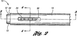
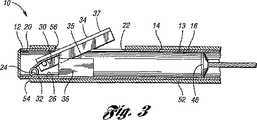
図1は、本発明の或る実施形態による内視鏡用切断器具10を示している。切断器具10は、内視鏡検査及び縫合糸切断用の内視鏡と適合性を有する。例えば、切断器具10は、以下の処置、即ち、内視鏡による縫合糸の切断、胃鏡検査法、S状結腸鏡検査法及び大腸内視鏡検査法、食道胃十二指腸内視鏡検査法(EGD)、内視鏡的逆行性胆道膵管造影法(ERCP)、及び気管支鏡検査法のために実装される。切断器具10は、切断位置に関係なく、刃に沿って比較的鋭利な切開を提供して、剪断と幅広の切開を回避する。
FIG. 1 illustrates an
図2と図3に示すように、切断器具10は、内壁13を有する内側カテーテル12と、内側カテーテル12の周りに滑動可能に配置されている外側カテーテル14と、を有する。好適には、内壁13は、近位部分16と閉じた遠位部分20を有する。内側カテーテル12は、ニチノール又はポリマー材料の様な、例えば低密度ポリエチレン、ポリプロピレン、ポリテトラフルオロエチレン(PTFE)、又はそれらの混合物など、どの様な適した材料で作ってもよい。本実施形態では、遠位部分は、長手方向に貫通形成された遠位側開口部22を有する。
As shown in FIGS. 2 and 3, the
好適には、内壁13は、先端部又は切断軸32の箇所で互いに接続された第1及び第2アーム26、30を有するばね機構24を更に有する。切断軸32は、ばね機構24により画定されているのが望ましい。図示のように、第1及び第2アーム26、30は、ばね押し式になっており、先端32に関して、互いから離れる方向に付勢されて伸張している。本実施形態では、第1アーム26は、内壁13に取り付けられており、第2アーム30は、第1アーム26から開口部22を通って近位方向に、付勢されて伸張している。而して、第1アーム26は、内壁13上に静止した状態に保たれ、一方、第2アーム30は、第1アーム26に対して付勢されて軸回転することができる。なお、ばね機構24は、第2アーム30が切断装置10に対して遠位方向に伸張するように配置してもよい。
Preferably, the
図示のように、内壁13は、遠位側開口部22に隣接して、ばね機構24の第2アーム30に取り付けられた切刃34を更に有する。本実施形態では、切刃34は、切断用の切断縁35と、切断縁35とは反対側の背部即ち非切断縁37と、を有する。而して、ばね機構24のばね押し式の切刃34は、内壁13の遠位側開口部22を通って近位方向に、付勢されて伸張している。また、切刃34は、切断軸32の周りで軸回転して、切断縁35で縫合糸を切ることができるようになっている。切刃34は、金属又は高密度ポリマーの様な、どの様な適した材料で作ってもよい。
As shown, the
図3と図4は、内壁13の上に配置された受容れアッセンブリ36を更に有する内壁13を示している。本実施形態では、受容れアッセンブリ36は、第1アーム26上に配置され、内壁13に取り付けられている。図示のように、受容れアッセンブリ36は、切断用の切刃34を協働的に受けるように作られている。受容れアッセンブリ36は、切刃34の切断縁35が第1アーム30に向けて軸回転式に動く際に切断をやり易くする。本実施形態では、受容れアッセンブリ36は、切刃34の切断縁35を受けるための基部40を有する。図示のように、基部40は、第1アーム26上に取り付けられ、内側カテーテル12の一部を横切って伸張し、ばね機構24の安定化を図っている。受容れアッセンブリは、金属又は高密度ポリマーの様な、どの様な適した材料で作ってもよい。
3 and 4 show the
図示のように、受容れアッセンブリ36は、基部40から伸張して、切刃34と協働して切断及び切開を行うことができるように作られている受刃42を更に有する。以下に更に詳しく説明するが、切刃34は、軸回転式に内向きに動いて、受容れアッセンブリ36と係合する。切断と切開は、切刃34の切断縁を受刃42と係合させて、縫合糸、脈管、又は内視鏡的に切るのが望ましいその他の物を切ることにより実現される。切刃34は、基部40にノッチ44で協働的に受けられる。
As shown, the receiving
この実施形態では、患者体内で内壁13を位置決めし操作するために、駆動ワイヤ46が内壁13の遠位部分20上に配置されている。しかしながら、本発明の範囲と精神を逸脱することなく、他の適した機構を実装してもよい。
In this embodiment, a
図示のように、切断器具10は、近位端52と開口している遠位端54とを有する外壁50を備える外側カテーテル14を更に有する。外側カテーテル14は、ニチノール又はポリマー材料の様な、例えば低密度ポリエチレン、ポリプロピレン、ポリテトラフルオロエチレン(PTFE)、又はそれらの混合物など、どの様な適した材料で作ってもよい。外側カテーテル14は、内側カテーテル12の周りに滑動可能に配置されている。遠位端54は、外側カテーテル14が内側カテーテル12の周りを長手方向に動き易くするための逃げを設けるため、開口している。外壁50は、遠位端54に隣接して貫通形成された孔56を有する。外壁50は、内側カテーテル12に対して滑動的に動かして、孔56を内壁13の遠位側開口部22と整列させることができるように作られている。
As shown, the cutting
この実施形態では、外側カテーテル14を後退させると、外側カテーテル14を切刃34の背側縁37と係合させて、切刃34を下向きに動かし、受刃42に付勢して係合させ、刃を閉じて切断を行うことができるようになっている。切刃34は、受容れアッセンブリ36の基部40で受けられる。而して、切刃34に対する外側カテーテル14の力は、縫合糸の様な切断対象に直接掛かる。外側カテーテル14が切刃34に沿って乗り上げて、切刃に内向きの力を掛けると、切刃は受容れアッセンブリ36と係合して、対象物が切断され又は切開される。
In this embodiment, when the
図5は、駆動ワイヤ46並びに内側及び外側カテーテル12、14と協働することができる制御部60を示している。この実施形態では、制御部60は、内側カテーテル12を外側カテーテル14に対して動かすため駆動ワイヤ46に接続されているスプール62を有する。制御部60は、外側カテーテル14の近位側に接続され、外側カテーテルを切断と切開のために動かし易くするように作られているハンドル64を更に有する。
FIG. 5 shows a control 60 that can cooperate with the
図6と図7は、本発明の或る実施形態による切断装置10を有する内視鏡装置70を示している。装置70は、縫合糸を切るため、及び、胃鏡検査法、S状結腸鏡検査法及び大腸内視鏡検査法、食道胃十二指腸内視鏡検査法(EGD)、内視鏡的逆行性胆道膵管造影法(ERCP)、及び気管支鏡検査法を含め、その他様々な内視鏡的処置に使用される。内視鏡装置70により、切断装置10は、切断位置に関係なく、刃34に沿って比較的鋭利な切開を提供し、剪断及び幅広の切開を最小限に抑える。
6 and 7 illustrate an
図示のように、装置70は、内視鏡検査用の内視鏡アッセンブリ72を有する。内視鏡アッセンブリ72は、複数のチャネルポート75を備える挿入チューブ74を有しており、その中を通して内視鏡ユニットが配置される。或る実施形態では、ポートの内の1つに配置された内視鏡ユニットは、上記切断装置の或る実施形態と、内視鏡カメラレンズ80と、吸引源82と、水/空気フラッシュ84を有する。必要に応じ、他の適したユニットを使用してもよい。
As shown, the
図示のように、内視鏡アッセンブリ72は、挿入チューブ74と機械的且つ流体的に連通している制御システム86を更に有する。制御システム86は、挿入チューブ74と、その中に配置されている内視鏡部品を制御できるように作られている。図示のように、制御システム86は、第1及び第2の制御ノブ87、88を有する。制御ノブ87、88は、挿入チューブ74と機械連通するように作られている。制御ノブ87、88は、医師が、既知の手段によって、患者の脈管及び体腔を通して挿入チューブ74を制御し案内できるようにしている。制御システム86は、弁スイッチ(例えば、吸引弁90、空気/水弁91、カメラ弁92)を更に有しており、各弁スイッチは、挿入チューブ74のチャネルポート75の1つに連通している。例えば、吸引弁スイッチ90は、作動させると、吸引源からの真空で、吸引チャネルポート82を通して望ましくないプラークやデブリを患者から吸引することができるようになっている。
As shown, the endoscope assembly 72 further includes a
図6と図7に示すように、内視鏡装置70は、上記内視鏡用切断器具10を有する。この実施形態では、内視鏡用切断器具10は、内視鏡アッセンブリ72の生検/鋏チャネルポート76を通して挿入される。次いで、器具10は、内視鏡アッセンブリ72の各生検チャネルポート76を通して送給される。切断器具10は、外側カテーテル14の遠位端54が挿入チューブ74のノズル78に隣接するまで、その中を通して送給されるのが望ましい。
As shown in FIGS. 6 and 7, the
先に述べたように、内視鏡用切断器具10は、上に切断基部40が配置されている内側カテーテル12と、内側カテーテル12の周りに配置されている外側カテーテル14と、内側カテーテル12内に取り付けられている駆動ワイヤ46と、を有する。
As previously mentioned, the
或る実施例では、挿入チューブ74の遠位端は、直腸又は口経由で、患者体内の事前に決められた内視鏡位置まで挿入される。挿入チューブ74の挿入は、内視鏡的処置次第で、直腸経由でも口経由でもよい。その位置で、医師は、内視鏡ユニットを思いのままに作動させ制御して、以前に患者体内に外科的に設置された縫合糸を切るという様な行為を行う。内視鏡に本発明の切断器具を組み合わせることにより、医師は、所望通りに鋭利な切開及び切断を施すことができる。
In one embodiment, the distal end of the
以上、本発明を好適な実施形態の点から説明してきたが、無論のこと、当業者には特に上記教示に鑑みて変更が想起されるであろうことから、本発明は、それら好適な実施形態に限定されるものではないと理解頂きたい。 While the invention has been described in terms of preferred embodiments, it will be understood that modifications will occur to those skilled in the art, especially in light of the above teachings, and therefore the invention is not limited to these preferred implementations. Please understand that it is not limited to form.
Claims (12)
貫通形成された開口部を有する内壁を備える内側カテーテルであって、前記内壁は、その上に可動的に配置され、前記開口部を通り、付勢されて伸張する切刃を更に有しており、前記内壁は、その上に配置され、前記切刃を協働的に受けるように作られている受容れ部材を有する、内側カテーテルと、
前記内側カテーテルの周りに可動的に配置されている外壁を備える外側カテーテルであって、前記外壁は、貫通形成された孔を有しており、前記外壁の前記孔は、前記内壁の前記開口部と可動的に整列させることができるように作られ、前記切刃が、前記開口部を通って付勢されて伸張して前記受容れ部材と付勢されて係合し、切断を行う、外側カテーテルと、を有する切断器具。 In an endoscope cutting instrument ,
An inner catheter having an inner wall with an opening formed therethrough, the inner wall further having a cutting blade movably disposed thereon and biased and extended through the opening. The inner wall having a receiving member disposed thereon and adapted to cooperatively receive the cutting blade; and
An outer catheter having an outer wall movably disposed around the inner catheter, the outer wall having a hole formed therethrough, wherein the hole in the outer wall is the opening of the inner wall. and made it possible to movably align the cutting edge, the receiving Re member is biased and engaged by expanding biased through the opening, for cutting, outer A cutting instrument having a catheter.
第1及び第2アームを有するばね機構であって、前記第1アームは前記内壁に取り付けられており、前記第2アームは前記第1アームから前記開口部を通り付勢されて伸張しており、前記第2アームは前記第1アームに対して付勢して動かすことができ、前記受容れアッセンブリは、前記第1アーム上に配置され、前記内壁に取り付けられており、前記切刃は、前記第2アーム上に配置され、切断を行うために動かすことができる、ばね機構を更に有する、請求項8に記載の切断器具。The inner wall is
A spring mechanism having first and second arms, wherein the first arm is attached to the inner wall, and the second arm is urged from the first arm through the opening and extends. The second arm can be urged and moved relative to the first arm, the receiving assembly is disposed on the first arm and attached to the inner wall, and the cutting edge comprises: 9. A cutting instrument according to claim 8, further comprising a spring mechanism disposed on the second arm and movable to perform a cut.
Applications Claiming Priority (3)
| Application Number | Priority Date | Filing Date | Title |
|---|---|---|---|
| US64751705P | 2005-01-27 | 2005-01-27 | |
| US60/647,517 | 2005-01-27 | ||
| PCT/US2006/002802 WO2006083679A1 (en) | 2005-01-27 | 2006-01-27 | Endoscopic cutting device |
Publications (3)
| Publication Number | Publication Date |
|---|---|
| JP2008534029A JP2008534029A (en) | 2008-08-28 |
| JP2008534029A5 JP2008534029A5 (en) | 2009-02-05 |
| JP4751401B2 true JP4751401B2 (en) | 2011-08-17 |
Family
ID=36481372
Family Applications (1)
| Application Number | Title | Priority Date | Filing Date |
|---|---|---|---|
| JP2007553232A Expired - Fee Related JP4751401B2 (en) | 2005-01-27 | 2006-01-27 | Endoscopic cutting instrument |
Country Status (6)
| Country | Link |
|---|---|
| US (1) | US7520886B2 (en) |
| EP (1) | EP1841365B1 (en) |
| JP (1) | JP4751401B2 (en) |
| AU (1) | AU2006211174B2 (en) |
| CA (1) | CA2596249C (en) |
| WO (1) | WO2006083679A1 (en) |
Families Citing this family (81)
| Publication number | Priority date | Publication date | Assignee | Title |
|---|---|---|---|---|
| US6610067B2 (en) | 2000-05-01 | 2003-08-26 | Arthrosurface, Incorporated | System and method for joint resurface repair |
| US8177841B2 (en) | 2000-05-01 | 2012-05-15 | Arthrosurface Inc. | System and method for joint resurface repair |
| US7163541B2 (en) | 2002-12-03 | 2007-01-16 | Arthrosurface Incorporated | Tibial resurfacing system |
| US7678151B2 (en) | 2000-05-01 | 2010-03-16 | Ek Steven W | System and method for joint resurface repair |
| US6520964B2 (en) | 2000-05-01 | 2003-02-18 | Std Manufacturing, Inc. | System and method for joint resurface repair |
| US7901408B2 (en) | 2002-12-03 | 2011-03-08 | Arthrosurface, Inc. | System and method for retrograde procedure |
| US8388624B2 (en) | 2003-02-24 | 2013-03-05 | Arthrosurface Incorporated | Trochlear resurfacing system and method |
| JP2007512108A (en) | 2003-11-20 | 2007-05-17 | アースロサーフィス・インコーポレーテッド | Regressive delivery of resurfaced devices |
| EP1845890A4 (en) | 2003-11-20 | 2010-06-09 | Arthrosurface Inc | System and method for retrograde procedure |
| US8425539B2 (en) | 2004-04-12 | 2013-04-23 | Xlumena, Inc. | Luminal structure anchoring devices and methods |
| EP1765201A4 (en) | 2004-06-28 | 2013-01-23 | Arthrosurface Inc | System for articular surface replacement |
| US7828853B2 (en) | 2004-11-22 | 2010-11-09 | Arthrosurface, Inc. | Articular surface implant and delivery system |
| JP5111112B2 (en) | 2004-12-08 | 2012-12-26 | エックスルミナ, インコーポレイテッド | Device for performing needle-guided therapy |
| US8777967B2 (en) | 2005-06-09 | 2014-07-15 | Xlumena, Inc. | Methods and devices for anchoring to tissue |
| US8784437B2 (en) | 2005-06-09 | 2014-07-22 | Xlumena, Inc. | Methods and devices for endosonography-guided fundoplexy |
| US20070225740A1 (en) * | 2006-02-22 | 2007-09-27 | Loubert Suddaby | Endoscopic Pulley Knife Instrument for Transecting Ligaments or Fascia |
| WO2008022087A2 (en) * | 2006-08-11 | 2008-02-21 | Mynosys Cellular Devices, Inc. | Three-dimensional cutting instrument |
| US7918784B2 (en) * | 2006-08-18 | 2011-04-05 | Microaire Surgical Instruments, Inc. | Endoscopic surgical tool with retractable blade for carpal tunnel release |
| EP2136717B1 (en) | 2006-12-11 | 2013-10-16 | Arthrosurface Incorporated | Retrograde resection apparatus |
| WO2009111481A1 (en) | 2008-03-03 | 2009-09-11 | Arthrosurface Incorporated | Bone resurfacing system and method |
| US8454632B2 (en) | 2008-05-12 | 2013-06-04 | Xlumena, Inc. | Tissue anchor for securing tissue layers |
| US8303594B2 (en) * | 2008-12-30 | 2012-11-06 | Howmedica Osteonics Corp. | Method and apparatus for removal of tissue |
| FR2943237B1 (en) * | 2009-03-20 | 2012-11-30 | Alexandre Worcel | ENDOSCOPIC CUTTING SURGICAL DEVICE |
| WO2016154393A1 (en) | 2009-04-17 | 2016-09-29 | Arthrosurface Incorporated | Glenoid repair system and methods of use thereof |
| EP2429429B1 (en) * | 2009-04-17 | 2018-07-25 | Arthrosurface Incorporated | Glenoid resurfacing system |
| US9662126B2 (en) | 2009-04-17 | 2017-05-30 | Arthrosurface Incorporated | Glenoid resurfacing system and method |
| US9364259B2 (en) | 2009-04-21 | 2016-06-14 | Xlumena, Inc. | System and method for delivering expanding trocar through a sheath |
| JP5535313B2 (en) | 2009-05-29 | 2014-07-02 | エックスルミナ, インコーポレイテッド | Device and method for deploying a stent across adjacent tissue layers |
| US8316493B2 (en) | 2009-11-20 | 2012-11-27 | Joseph H. Clearman | Bag closure |
| US8701295B2 (en) * | 2009-11-20 | 2014-04-22 | Joseph Clearman | Variable pressure cutting devices |
| BR112012022482A2 (en) | 2010-03-05 | 2016-07-19 | Arthrosurface Inc | tibial surface recomposition system and method. |
| US8771306B2 (en) | 2010-03-11 | 2014-07-08 | Covidien Lp | Insertion device and method of use |
| EP2364653A1 (en) * | 2010-03-11 | 2011-09-14 | Tyco Healthcare Group LP | Insertion device and method of use |
| WO2011123062A2 (en) * | 2010-03-30 | 2011-10-06 | Singapore Health Services Pte. Ltd. | Cutting device for cutting tissue |
| US8876845B2 (en) | 2010-09-30 | 2014-11-04 | Loubert Suddaby | Sling blade transection of the transverse carpal ligament |
| US10070908B2 (en) | 2010-10-13 | 2018-09-11 | Warsaw Orthopedic, Inc. | Surgical instruments for cutting elongated elements and methods of use |
| US8784420B2 (en) | 2010-10-13 | 2014-07-22 | Warsaw Orthopedic, Inc. | Surgical instruments for cutting elongated elements and methods of use |
| EP2632318B1 (en) | 2010-10-25 | 2019-11-27 | Endosee Corporation | Apparatus for hysteroscopy and endometrial biopsy |
| WO2012151073A2 (en) | 2011-05-03 | 2012-11-08 | Endosee Corporation | Method and apparatus for hysteroscopy and endometrial biopsy |
| US9066716B2 (en) | 2011-03-30 | 2015-06-30 | Arthrosurface Incorporated | Suture coil and suture sheath for tissue repair |
| EP2699170A4 (en) * | 2011-04-18 | 2014-11-26 | Eastern Virginia Med School | Cerclage suture removal device |
| EP2804565B1 (en) | 2011-12-22 | 2018-03-07 | Arthrosurface Incorporated | System for bone fixation |
| KR101711361B1 (en) * | 2012-01-06 | 2017-03-02 | 지완 스티븐 싱 | An insert and insert system for a laparoscopic instrument |
| WO2013119849A1 (en) | 2012-02-07 | 2013-08-15 | Intervene, Inc. | Systems and methods for endoluminal valve creation |
| ES2765184T3 (en) | 2012-05-17 | 2020-06-08 | Boston Scient Scimed Inc | Access devices through adjacent tissue layers |
| US9364260B2 (en) * | 2012-05-25 | 2016-06-14 | Depuy Mitek, Llc | Method for atraumatic hip access |
| US9622646B2 (en) | 2012-06-25 | 2017-04-18 | Coopersurgical, Inc. | Low-cost instrument for endoscopically guided operative procedures |
| WO2014008126A1 (en) | 2012-07-03 | 2014-01-09 | Arthrosurface Incorporated | System and method for joint resurfacing and repair |
| WO2014055981A1 (en) | 2012-10-05 | 2014-04-10 | Board Of Regents, The University Of Texas System | System and method for scoring the left ventricular endocardium to increase left ventricular compliance |
| WO2014093148A2 (en) | 2012-12-12 | 2014-06-19 | Covidien Lp | Tissue-removing catheter for body lumen |
| US9636138B2 (en) | 2012-12-12 | 2017-05-02 | Covidien Lp | Tissue-removing catheter including force-transmitting member for actuating a cutter housing |
| US9636139B2 (en) | 2012-12-12 | 2017-05-02 | Covidien Lp | Tissue-removing catheter with ball and socket deployment mechanism |
| WO2014093156A1 (en) | 2012-12-12 | 2014-06-19 | Covidien Lp | Cutter for tissue-removing catheter |
| JP6257104B2 (en) | 2012-12-12 | 2018-01-10 | コヴィディエン リミテッド パートナーシップ | Tissue removal catheter including screw blade and cutter drive shaft |
| JP6110509B2 (en) | 2012-12-12 | 2017-04-05 | コヴィディエン リミテッド パートナーシップ | Tissue removal catheter including pressing mechanism |
| WO2014110460A1 (en) | 2013-01-10 | 2014-07-17 | Intervene, Inc. | Systems and methods for endoluminal valve creation |
| ES2813871T3 (en) | 2013-02-21 | 2021-03-25 | Boston Scient Scimed Inc | Devices to form an anastomosis |
| US9402644B2 (en) * | 2013-03-13 | 2016-08-02 | Covidien Lp | Reverse seam ripper dissector |
| US9492200B2 (en) | 2013-04-16 | 2016-11-15 | Arthrosurface Incorporated | Suture system and method |
| US10433861B2 (en) | 2013-08-27 | 2019-10-08 | Board Of Regents Of The University Of Texas System | System and method for cutting trabeculae carneae of the left ventricle to increase LV compliance |
| US10231613B2 (en) | 2013-09-27 | 2019-03-19 | Intervene, Inc. | Visualization devices, systems, and methods for informing intravascular procedures on blood vessel valves |
| US9962265B2 (en) | 2014-03-07 | 2018-05-08 | Arthrosurface Incorporated | System and method for repairing articular surfaces |
| US11607319B2 (en) | 2014-03-07 | 2023-03-21 | Arthrosurface Incorporated | System and method for repairing articular surfaces |
| US10624748B2 (en) | 2014-03-07 | 2020-04-21 | Arthrosurface Incorporated | System and method for repairing articular surfaces |
| US10188419B2 (en) | 2014-03-24 | 2019-01-29 | Intervene, Inc. | Visualization devices for use during percutaneous tissue dissection and associated systems and methods |
| EP3193748B1 (en) | 2014-09-18 | 2020-07-01 | Mayo Foundation for Medical Education and Research | Soft tissue cutting device |
| JP6728181B2 (en) | 2014-12-16 | 2020-07-22 | インタービーン・インコーポレイテッドINTERVENE, Incorporated | Endovascular devices, systems and methods for controlled dissection of body cavities |
| US20170071788A1 (en) * | 2015-09-15 | 2017-03-16 | Novartis Ag | Curved vitrectomy probe |
| US10702305B2 (en) | 2016-03-23 | 2020-07-07 | Coopersurgical, Inc. | Operative cannulas and related methods |
| US10646247B2 (en) | 2016-04-01 | 2020-05-12 | Intervene, Inc. | Intraluminal tissue modifying systems and associated devices and methods |
| US10456161B2 (en) | 2016-04-14 | 2019-10-29 | Covidien Lp | Tissue-removing catheter with adjustment mechanism |
| CN107184246A (en) * | 2017-06-26 | 2017-09-22 | 苏州奥特科然医疗科技有限公司 | Sclerotin cutting apparatus in a kind of bone |
| WO2019028344A1 (en) | 2017-08-04 | 2019-02-07 | Arthrosurface Incorporated | Multicomponent articular surface implant |
| US10864055B2 (en) | 2017-10-13 | 2020-12-15 | Sonex Health, Inc. | Tray for a soft tissue cutting device and methods of use |
| CN112020333A (en) * | 2018-04-26 | 2020-12-01 | 奥林巴斯株式会社 | Treatment system and expansion device |
| US11937845B2 (en) | 2019-01-11 | 2024-03-26 | Mayo Foundation For Medical Education And Research | Micro-invasive surgical device and methods of use |
| GB2616360B (en) | 2019-03-12 | 2023-11-29 | Arthrosurface Inc | Humeral and glenoid articular surface implant systems and methods |
| CN109833088A (en) * | 2019-03-13 | 2019-06-04 | 郑州大学第一附属医院 | The linear cutting device of hysteroscope pipe side wall |
| EP4167867A1 (en) | 2020-06-23 | 2023-04-26 | Intervene, Inc. | Endovascular valve formation system with imaging capability |
| US11793599B2 (en) | 2020-08-04 | 2023-10-24 | Mazor Robotics Ltd. | Surgical cleaning tool, systems, and methods |
| USD989961S1 (en) | 2021-04-30 | 2023-06-20 | Sonex Health, Inc. | Soft tissue cutting device |
Citations (1)
| Publication number | Priority date | Publication date | Assignee | Title |
|---|---|---|---|---|
| US20020022788A1 (en) * | 1999-08-19 | 2002-02-21 | Tim Corvi | Apparatus and methods for material capture and removal |
Family Cites Families (11)
| Publication number | Priority date | Publication date | Assignee | Title |
|---|---|---|---|---|
| US4963147A (en) * | 1987-09-18 | 1990-10-16 | John M. Agee | Surgical instrument |
| US5201759A (en) * | 1991-04-29 | 1993-04-13 | Ferzli George S | Laparoscopic instrument |
| US5304190A (en) * | 1992-05-08 | 1994-04-19 | Ethicon, Inc. | Endoscopic cutting apparatus |
| EP0740529A4 (en) * | 1994-01-18 | 1997-05-14 | Coral Medical | Knot tying method and apparatus |
| DE4403602A1 (en) * | 1994-02-07 | 1995-08-10 | Storz Karl Gmbh & Co | Endoscopic cutting device |
| US6193715B1 (en) * | 1999-03-19 | 2001-02-27 | Medical Scientific, Inc. | Device for converting a mechanical cutting device to an electrosurgical cutting device |
| US6616661B2 (en) * | 2001-09-28 | 2003-09-09 | Ethicon, Inc. | Surgical device for clamping, ligating, and severing tissue |
| US8172856B2 (en) * | 2002-08-02 | 2012-05-08 | Cedars-Sinai Medical Center | Methods and apparatus for atrioventricular valve repair |
| WO2004112616A2 (en) | 2003-06-16 | 2004-12-29 | Ortheon Medical Llc | Suture cutter |
| DE602004015807D1 (en) | 2003-09-11 | 2008-09-25 | Nmt Medical Inc | CUTTING TUBE FOR SURGICAL SEAMS |
| US7029435B2 (en) * | 2003-10-16 | 2006-04-18 | Granit Medical Innovation, Llc | Endoscope having multiple working segments |
-
2006
- 2006-01-27 US US11/341,298 patent/US7520886B2/en active Active
- 2006-01-27 WO PCT/US2006/002802 patent/WO2006083679A1/en active Application Filing
- 2006-01-27 AU AU2006211174A patent/AU2006211174B2/en active Active
- 2006-01-27 CA CA2596249A patent/CA2596249C/en active Active
- 2006-01-27 JP JP2007553232A patent/JP4751401B2/en not_active Expired - Fee Related
- 2006-01-27 EP EP06733928.3A patent/EP1841365B1/en active Active
Patent Citations (1)
| Publication number | Priority date | Publication date | Assignee | Title |
|---|---|---|---|---|
| US20020022788A1 (en) * | 1999-08-19 | 2002-02-21 | Tim Corvi | Apparatus and methods for material capture and removal |
Also Published As
| Publication number | Publication date |
|---|---|
| EP1841365A1 (en) | 2007-10-10 |
| AU2006211174B2 (en) | 2012-05-31 |
| AU2006211174A1 (en) | 2006-08-10 |
| US20060184187A1 (en) | 2006-08-17 |
| JP2008534029A (en) | 2008-08-28 |
| CA2596249A1 (en) | 2006-08-10 |
| CA2596249C (en) | 2013-12-10 |
| WO2006083679A1 (en) | 2006-08-10 |
| US7520886B2 (en) | 2009-04-21 |
| EP1841365B1 (en) | 2013-07-24 |
Similar Documents
| Publication | Publication Date | Title |
|---|---|---|
| JP4751401B2 (en) | Endoscopic cutting instrument | |
| JP2008534029A5 (en) | ||
| US7691055B2 (en) | Endoscopic apparatus having an improved elevator | |
| JP5407036B2 (en) | Treatment endoscope | |
| US7261728B2 (en) | Biopsy forceps device and method | |
| US7060024B2 (en) | Apparatus for guiding an instrument used with an endoscope | |
| US7794389B2 (en) | Endoscopic elevator apparatus | |
| JP5852008B2 (en) | System for treating gastrointestinal lesions with endoscopy | |
| US7060025B2 (en) | Method for controlling position of medical instruments | |
| US8945153B2 (en) | Endoscopic apparatus having a clip device | |
| US20070208220A1 (en) | Endoscopic delivery apparatus having a catheter with radial grooves | |
| JP3192239U (en) | Surgical endoscope | |
| JP2008155030A (en) | Device for surgical procedure | |
| EP3155955B1 (en) | Endoscope cap with separable arms | |
| EP2811921A1 (en) | Cutting tool with circulating wire | |
| US9844649B2 (en) | Telescopic wire guide | |
| US20220117618A1 (en) | Tissue deflecting devices and related methods of use |
Legal Events
| Date | Code | Title | Description |
|---|---|---|---|
| A521 | Request for written amendment filed |
Free format text: JAPANESE INTERMEDIATE CODE: A523 Effective date: 20081210 |
|
| A621 | Written request for application examination |
Free format text: JAPANESE INTERMEDIATE CODE: A621 Effective date: 20081210 |
|
| TRDD | Decision of grant or rejection written | ||
| A01 | Written decision to grant a patent or to grant a registration (utility model) |
Free format text: JAPANESE INTERMEDIATE CODE: A01 Effective date: 20110426 |
|
| A61 | First payment of annual fees (during grant procedure) |
Free format text: JAPANESE INTERMEDIATE CODE: A61 Effective date: 20110520 |
|
| R150 | Certificate of patent or registration of utility model |
Ref document number: 4751401 Country of ref document: JP Free format text: JAPANESE INTERMEDIATE CODE: R150 Free format text: JAPANESE INTERMEDIATE CODE: R150 |
|
| FPAY | Renewal fee payment (event date is renewal date of database) |
Free format text: PAYMENT UNTIL: 20140527 Year of fee payment: 3 |
|
| R250 | Receipt of annual fees |
Free format text: JAPANESE INTERMEDIATE CODE: R250 |
|
| R250 | Receipt of annual fees |
Free format text: JAPANESE INTERMEDIATE CODE: R250 |
|
| R250 | Receipt of annual fees |
Free format text: JAPANESE INTERMEDIATE CODE: R250 |
|
| R250 | Receipt of annual fees |
Free format text: JAPANESE INTERMEDIATE CODE: R250 |
|
| R250 | Receipt of annual fees |
Free format text: JAPANESE INTERMEDIATE CODE: R250 |
|
| R250 | Receipt of annual fees |
Free format text: JAPANESE INTERMEDIATE CODE: R250 |
|
| R250 | Receipt of annual fees |
Free format text: JAPANESE INTERMEDIATE CODE: R250 |
|
| R250 | Receipt of annual fees |
Free format text: JAPANESE INTERMEDIATE CODE: R250 |
|
| R250 | Receipt of annual fees |
Free format text: JAPANESE INTERMEDIATE CODE: R250 |
|
| LAPS | Cancellation because of no payment of annual fees |