JP4658423B2 - Polypeptides and polynucleotides for enhancing immunoreactivity against HER-2 protein - Google Patents
Polypeptides and polynucleotides for enhancing immunoreactivity against HER-2 protein Download PDFInfo
- Publication number
- JP4658423B2 JP4658423B2 JP2001513369A JP2001513369A JP4658423B2 JP 4658423 B2 JP4658423 B2 JP 4658423B2 JP 2001513369 A JP2001513369 A JP 2001513369A JP 2001513369 A JP2001513369 A JP 2001513369A JP 4658423 B2 JP4658423 B2 JP 4658423B2
- Authority
- JP
- Japan
- Prior art keywords
- amino acid
- composition
- peptide
- epitope
- acid sequence
- Prior art date
- Legal status (The legal status is an assumption and is not a legal conclusion. Google has not performed a legal analysis and makes no representation as to the accuracy of the status listed.)
- Expired - Fee Related
Links
Images


Classifications
-
- A—HUMAN NECESSITIES
- A61—MEDICAL OR VETERINARY SCIENCE; HYGIENE
- A61K—PREPARATIONS FOR MEDICAL, DENTAL OR TOILETRY PURPOSES
- A61K9/00—Medicinal preparations characterised by special physical form
- A61K9/0012—Galenical forms characterised by the site of application
- A61K9/0019—Injectable compositions; Intramuscular, intravenous, arterial, subcutaneous administration; Compositions to be administered through the skin in an invasive manner
-
- A—HUMAN NECESSITIES
- A61—MEDICAL OR VETERINARY SCIENCE; HYGIENE
- A61K—PREPARATIONS FOR MEDICAL, DENTAL OR TOILETRY PURPOSES
- A61K9/00—Medicinal preparations characterised by special physical form
- A61K9/14—Particulate form, e.g. powders, Processes for size reducing of pure drugs or the resulting products, Pure drug nanoparticles
- A61K9/16—Agglomerates; Granulates; Microbeadlets ; Microspheres; Pellets; Solid products obtained by spray drying, spray freeze drying, spray congealing,(multiple) emulsion solvent evaporation or extraction
- A61K9/1605—Excipients; Inactive ingredients
- A61K9/1629—Organic macromolecular compounds
- A61K9/1641—Organic macromolecular compounds obtained otherwise than by reactions only involving carbon-to-carbon unsaturated bonds, e.g. polyethylene glycol, poloxamers
- A61K9/1647—Polyesters, e.g. poly(lactide-co-glycolide)
-
- A—HUMAN NECESSITIES
- A61—MEDICAL OR VETERINARY SCIENCE; HYGIENE
- A61P—SPECIFIC THERAPEUTIC ACTIVITY OF CHEMICAL COMPOUNDS OR MEDICINAL PREPARATIONS
- A61P35/00—Antineoplastic agents
-
- A—HUMAN NECESSITIES
- A61—MEDICAL OR VETERINARY SCIENCE; HYGIENE
- A61P—SPECIFIC THERAPEUTIC ACTIVITY OF CHEMICAL COMPOUNDS OR MEDICINAL PREPARATIONS
- A61P37/00—Drugs for immunological or allergic disorders
- A61P37/02—Immunomodulators
-
- A—HUMAN NECESSITIES
- A61—MEDICAL OR VETERINARY SCIENCE; HYGIENE
- A61P—SPECIFIC THERAPEUTIC ACTIVITY OF CHEMICAL COMPOUNDS OR MEDICINAL PREPARATIONS
- A61P37/00—Drugs for immunological or allergic disorders
- A61P37/02—Immunomodulators
- A61P37/04—Immunostimulants
-
- C—CHEMISTRY; METALLURGY
- C07—ORGANIC CHEMISTRY
- C07K—PEPTIDES
- C07K14/00—Peptides having more than 20 amino acids; Gastrins; Somatostatins; Melanotropins; Derivatives thereof
- C07K14/435—Peptides having more than 20 amino acids; Gastrins; Somatostatins; Melanotropins; Derivatives thereof from animals; from humans
- C07K14/705—Receptors; Cell surface antigens; Cell surface determinants
- C07K14/71—Receptors; Cell surface antigens; Cell surface determinants for growth factors; for growth regulators
-
- A—HUMAN NECESSITIES
- A61—MEDICAL OR VETERINARY SCIENCE; HYGIENE
- A61K—PREPARATIONS FOR MEDICAL, DENTAL OR TOILETRY PURPOSES
- A61K39/00—Medicinal preparations containing antigens or antibodies
- A61K2039/60—Medicinal preparations containing antigens or antibodies characteristics by the carrier linked to the antigen
- A61K2039/6031—Proteins
- A61K2039/6037—Bacterial toxins, e.g. diphteria toxoid [DT], tetanus toxoid [TT]
-
- A—HUMAN NECESSITIES
- A61—MEDICAL OR VETERINARY SCIENCE; HYGIENE
- A61K—PREPARATIONS FOR MEDICAL, DENTAL OR TOILETRY PURPOSES
- A61K39/00—Medicinal preparations containing antigens or antibodies
- A61K2039/64—Medicinal preparations containing antigens or antibodies characterised by the architecture of the carrier-antigen complex, e.g. repetition of carrier-antigen units
Description
【0001】
(関連出願の引用)
米国特許法119条(e)項(1)に基づいて、本出願は、先願の米国仮特許出願60/146,869号(1999年8月3日出願)の優先権を主張する。
【0002】
本出願に記載される研究は、National Cancer Instituteからの助成金PHS/NIH P30 CA−16058およびNIH/NCI RO1 CA 84356−01A1により少なくとも一部後援された。合衆国政府は、本発明において一定の権利を有する。
【0003】
(背景)
現在、乳癌を処置する最も一般的な形態は、外科手術、化学的介入、および/または放射線療法を含む。癌が規定された領域に限定されなければ、外科手術のみでは、癌を排除することができない。従って、手術部位近辺にあり、手術から逃れた癌細胞を破壊するために、術後、放射線処置がしばしば行われる。このような処置の副作用としては、皮膚過敏(skin sensitivity)またはかゆみ、免疫系の妨害、ときおり吐き気、および稀に肺の影響を受けた部分が繊維性になる放射線維症(radiation fibrosis)が挙げられる。化学療法はまた、外科手術後に用いられ得る。化学療法は、癌細胞に毒性の薬物を利用する。これは、完全には選択的系ではないので、正常細胞もまた影響を受ける。ネガティブな副作用としては、悪心、疲労、食欲喪失、脱毛および下痢が挙げられる。
【0004】
現在の治療の欠点を鑑みて、乳癌を処置するためのさらなるアプローチを見出す試みが行われてきた。1つのこのようなアプローチは、免疫療法である。免疫療法的アプローチのための標的の1つは、HER−2タンパク質である。HER−2タンパク質(HER−2癌遺伝子の産物)は、種々の癌において過剰発現されている。インサイチュで腺管癌のうちの50〜60%、および全ての乳癌のうちの20〜40%、ならびに卵巣、前立腺、結腸および肺で生じる腺癌の実質的な割合において見出される。HER−2タンパク質の過剰発現は、ヒトにおける悪性形質転換に関する。HER−2タンパク質の過剰発現はまた、悪性疾患の攻撃性と密接に関連し、これは、全ての侵襲性乳癌のうちの1/4において見出される。HER−2タンパク質の過剰発現は、乳癌および卵巣癌の両方において予後があまりよくないことと相関する。
【0005】
最近の研究において、HER−2の細胞外結合ドメイン(ECD)に対する抗体は、インビトロおよび動物モデルで腫瘍増殖に対する阻害効果を付与することを示した(Hudziak,R.M.ら,Mol.Cell.Biol.9:11−65−72,1989;Tagliabue,E.ら,Int.J.Cancer 47:933−7,1991;Drebin,J.A.ら,Proc.Natl.Acad.Sci.USA 83:9129−33,1986;Drebin,J.A.ら,Oncogene,2:273−7,1988;Drebin,J.A.ら,Oncogene,2:387−94,1988およびKatumata,M.ら,Nat.Med.1:644−8.1995)。さらに組換えヒト化抗HER−2モノクローナル抗体(Trastuzumab)の転移性HER−2過剰発現乳癌を有する患者における第II相および第III相の臨床試験は、単一の薬剤として15%の全体応答割合を生じた。Trastuzumabはまた、細胞傷害性化学療法剤と組み合わせた場合に、生存性を改善することが示された(Beselga,J.ら,J.Clin.Oncol.14:737−44,1996;Pegram,M.D.ら,J.Clin.Oncol.16:2659−71,1988)。組換えHER−2タンパク質、HER−2 ECD、またはラットneu(これは、HER−2のラットホモログである)のECDを標的とする多くのワクチンアプローチが評価されてきた。例えば、ラットneuのECDを発現する組換えワクシニアウイルスで免疫したNFS系統マウスは、neu形質転換NIH 3T3細胞でのその後のチャレンジに対して防御性抗体応答を発生させた(Bernards,R.ら,Proc.Natl.Acad.Sci.USA 84:6854−8,1987)。しかし、BDIXラットの同じ免疫原での免疫は、抗体応答も生じず、同系のneu発現B 104神経芽腫細胞の増殖を阻害もしなかった。このことは、このストラテジーがラットにおいて免疫応答を誘導するには不充分であったことを示唆する。ポリサッカリド−癌タンパク質複合体ワクチン(コレステリル基保有マンナンおよびプルランと複合体化したHER−2 ECDの147のアミノ末端アミノ酸からなる)は、BALB/cマウスにおけるHER−2発現肉腫の拒絶を媒介した細胞性免疫応答および体液性免疫応答を誘導した(Gu,X.G.ら,Cancer Res.,58:3385−90,1998)。精製ラットneu ECD(Esserman,L.J.,Cancer Immunol.Immunother.47:337−42,1999)またはneuトランスフェクト同種異系マウス線維芽細胞(Cefai,D.,ら,Int.J.Cancer,83:393−400,1999)のいずれかで免疫することにより乳腺腫瘍を発症させるようにしたラットneuトランスジェニックマウスにおいて、部分的な防御が示された。
【0006】
上記研究の結果にも拘わらず、ヒトにおいてHER−2またはHER−2 ECD(HER−2が変異していない)「自己」抗原を標的化する細胞ベースまたはタンパク質ベースのワクチンストラテジーを用いて、有効な免疫応答が生成され得るか否かは、未だに不確かなままである。従って、HER−2タンパク質の過剰発現が関連している乳癌および他の悪性疾患を処置または予防するためのさらなる免疫療法的アプローチを行うことが望ましい。
【0007】
(発明の要旨)
本発明は、免疫系を刺激し、そしてHER−2タンパク質の過剰発現に関連する悪性疾患を処置するための新たな化合物および組成物を提供する。この化合物は、HER−2タンパク質の免疫原性エピトープおよびこのようなエピトープを含むキメラペプチドおよび多価ペプチドである。
【0008】
第1の群の化合物は、本明細書中以降まとめて「HER−2 B細胞エピトープ」とよばれる。HER−2 B細胞エピトープは、約15〜約50アミノ酸、より好ましくは17〜40アミノ酸、最も好ましくは18〜35アミノ酸を含む。好ましくは、HER−2 B細胞エピトープは、以下の群またはその機能的等価物から選択される配列を含む:
【0009】
【化5】

【0010】
本発明はまた、本発明のHER−2 B細胞エピトープまたはその機能的等価物の少なくとも1つを含むキメラペプチド(本明細書中以降「キメラHER−2 B細胞ペプチド」といわれる)を提供する。好ましくは、このキメラHER−2 B細胞ペプチドは、約35〜約150アミノ酸長、より好ましくは約35〜約70アミノ酸長である。このキメラHER−2 B細胞ペプチドは、3つのユニットを含む。第1のユニットは、HER−2B細胞エピトープまたはその機能的等価物を含む。第2のユニットは、ヘルパーT(Th)細胞エピトープ、好ましくは、乱交雑Th細胞エピトープである。本明細書中で使用される場合、「乱交雑」Th細胞エピトープは、MHC拘束の迂回を補助するサイトカインの放出を促進するエピトープである。第2のユニットは、約14〜約22アミノ酸長、より好ましくは約15〜21アミノ酸長、最も好ましくは16アミノ酸長である。好ましくは、このTh細胞エピトープは、以下のアミノ酸配列のうちの1つを有する:
【0011】
【化6】
【0012】
本発明はまた、複数の(すなわち、少なくとも2つの)本発明のHER−2 B細胞エピトープまたはその機能的等価物およびTh細胞エピトープを含む多価HER−2 B細胞ペプチドを提供する。HER−2 B細胞エピトープおよびTh細胞エピトープは、コアβシート鋳型に結合されている。好ましくはこの鋳型は、ロイシン残基とリジン残基とが交互になっている2つの鎖を含み、これらは、リンカーにより結合される。このリンカーはアミノ酸であり、すなわち、好ましくは約2〜約15アミノ酸長、より好ましくは約2〜約10アミノ酸長、最も好ましくは約2〜約6アミノ酸長のペプチドである。最も好ましいリンカーは、アミノ酸配列Gly−Pro−Ser−Leu(配列番号20)を含む。
【0013】
本発明はまた、キメラHER−2 B細胞ペプチドまたは多価HER−2 B細胞ペプチドおよび薬理学的に受容可能なキャリアを含む免疫原性組成物に関する。好ましいキャリアは、生分解性ミクロスフェアである。このような免疫原性組成物は、HER−2タンパク質の過剰発現が関連している悪性疾患を処置または予防するために有用である。
【0014】
本発明はまた、上記のHER−2 B細胞エピトープの少なくとも1つをコードするポリヌクレオチドに関する。このようなポリヌクレオチドは、組換え技術によりエピトープを生成するために有用である。本発明はまた、本発明のキメラHER−2 B細胞ペプチドをコードする配列を有する単離されたポリヌクレオチドに関する。このようなポリヌクレオチドは、キメラHER−2 B細胞ペプチドを調製するために有用である。このようなポリヌクレオチドはまた、HER−2タンパク質の過剰発現が関連している悪性疾患を処置または予防するための免疫原性組成物(例えば、DNAワクチン)において有用である。好ましくは、このような免疫原性組成物は、筋肉内に投与される。
【0015】
本発明はまた、細胞傷害性(Tc)細胞を活性化し得るHER−2エピトープ(本明細書中以降「HER−2 CTLエピトープ」といわれる)を提供する。このHER−2 CTLエピトープは、約8〜約12アミノ酸、より好ましくは9〜11アミノ酸を含む。好ましくは、このHER−2 CTLエピトープは、以下の配列のうちの1つを含む:
【0016】
【化7】
【0017】
本発明はまた、キメラペプチドを提供し、これは、HER−2 CTLエピトープまたはその機能的等価物を含むキメラHER−2 CTLペプチドとして下に言及される。キメラHER−2 CTLペプチドは、3つのユニットを含む。第1のユニットは、CTLエピトープを含む。第2のユニットは、好ましくは、乱交雑(promiscuous)Tヘルパー細胞エピトープである。第3のユニットは、リンカーアミノ酸または第1ペプチドユニットおよび第2ペプチドユニットを結合するペプチドユニットである。
【0018】
本発明はまた、複数の、すなわち、本願HER 2 CTLエピトープの少なくとも2つまたはその機能的等価物およびTh細胞エピトープを含む多価HER 2 CTLペプチドを提供する。HER−2 CTLエピトープおよびTh細胞エピトープは、コアβシートテンプレートに連結される。好ましくは、テンプレートが、ロイシン残基とリシン残基とが交互になっている2つの鎖を含み、これは、リンカーにより連結される。
【0019】
キメラHER−2 CTLペプチドおよび多価HER−2 CTLペプチドは、Tc細胞を活性化するために有用な免疫原である。このような活性化は、IL−2レセプターおよび少ない範囲でIL−2、の発現を開始するようTc細胞を誘導し、重要なサイトカインは、増殖およびエピトープを提示する標的細胞に対するサイトカイン活性を保有する機能的細胞傷害性リンパ球への、活性化されたTc細胞の分化に必要とされる。本発明はまた、キメラHER−2 CTLペプチドまたは多価HER−2 CTLペプチドおよび薬学的に受容可能なキャリアを含む免疫原性組成物に関する。本発明はまた、このような動物に免疫原性組成物を投与することによる、哺乳動物にTc細胞を活性化する方法に関する。
【0020】
本発明はまた、上記の1つ以上のHER−2 CTLエピトープをコードする単離されたポリヌクレオチドを含む。このようなポリヌクレオチドは、組換え技術によりエピトープを産生するのに有用である。本発明はまた、キメラHER−2 CTLペプチドをコードする配列を有する単離されたポリヌクレオチドを含む。このようなポリヌクレオチドは、キメラCTL細胞エピトープペプチドを調製するために有用である。このようなポリヌクレオチドはまた、HER−2オンコジーンが関連する悪性腫瘍を処置または予防するための免疫原性組成物(例えば、DNAワクチン)において有用である。
【0021】
本発明はまた、多価のB/CTLペプチドに関し、これは、1つ以上のTヘルパー細胞エピトープに連結される1つ以上のHER−2 CTLエピトープに連結される、1つ以上のHER−2 B細胞エピトープを含む。このエピトープは、コアβシートテンプレートに連結される。本発明はまた、多価のB/CTLペプチドをコードするポリヌクレオチドおよびこのようなポリヌクレオチドを含むDNAベクターに関する。本発明はまた、B/CTLエピトープペプチドまたはそれをコードするポリヌクレオチドおよび薬学的に受容可能なキャリアを含む免疫原性組成物に関する。
【0022】
本発明はまた、HER−2タンパク質に対する被験体における免疫応答を刺激する方法に関する。このような方法は、被験体に対する本発明のキメラペプチドまたは多価ペプチドを投与することを含む。このような方法は、HER−2タンパク質の発現と関連する疾患状態に対する被験体の免疫の増強を生じる。
【0023】
(発明の詳細な説明)
本発明は、HER−2タンパク質の免疫原性エピトープであるペプチドを提供し、これは、以下でHER−2 B細胞エピトープおよびHER−2 CTLエピトープとして以下で言及される。
【0024】
HER−2 B細胞エピトープは、HER−2タンパク質の細胞外ドメインと免疫活性である抗体の産生を生じる体液反応を引き起こすことが可能である。HER−2タンパク質およびそのラットホモログneuは、長さが約1255個のアミノ酸(aa)である185kdの相対的分子量を有する膜貫通タンパク質である。HER−2/neuタンパク質は、上皮増殖因子レセプター(EGFR)に40%の相同性を有する、約645 aaの細胞外結合ドメイン(ECD)、高い疎水性膜貫通アンカードメイン(TMD)、およびEGFRに対して80%の相同性を有する約580aaのカルボキシ末端の細胞質ドメイン(CD)を有する。HER−2タンパク質のアミノ酸配列およびこのようなアミノ酸配列をコードするヌクレオチド配列が、GenBank登録番号M11730に示される。
【0025】
HER−2 B細胞エピトープは、配列の1つを有するペプチドを包含し、これは、以下の表Iに示される「参照配列」として以下に言及する。この参照配列は、抗原性の6つの相互関係を使用して、コンピューターで補助される分析を用いて選択され、そしてスコアされた:(a)個々の配列の鎖の柔軟性および可動性のプロフィールが、KarplusおよびSchultz、Naturwiss 72:212−213、1985に従って算出された;(b)水治療法プロフィールが、7つの残基範囲セッティング(residue span setting)に対して作製され、そして最終的にKyte およびDoolittle,J.Moi.Biol.157:105−132,1982の尺度を使用して3つの残基範囲と共に洗練された;(c)水治療法プロフィールはHoppおよびWoods,Proc.Natl.Acad.Sci.USA 78:3824−3828,1981のプログラムを使用して6残基のウィンドウに対して作製された;(d)1.4Aプローブを使用して水へのアミノ酸残基の曝露の分析が、Rose,Science 229:834−838,1985の溶媒曝露アルゴリズムにより実行された;(e)溶媒中へ接近そして突出するタンパク質の部分を予測する突出指数が、Thornton,EMBO J.5:409−413,1986の方法により算出された;(f)5つの残基配列が抗原性である可能性は、Welling,FEBS Lett 188:215−218,1985の方法により決定された。基本的な前提は、予測に使用されるアルゴリズムが、タンパク質上の表面に曝露される領域に常に位置し、従って、多分抗体結合に関与している。配列が、それらのそれぞれの指数値に基づいて1〜6のスコアで与えられ、そしてランクされて:もっも高いランキング配列は、調べられた分析物(6/6)について最も高い個々のスコアを有し、そして連続的な候補は、次に高いスコア(5/6)などを有した。最もよい、スコアしたエピトープは、2次構造の特性と関連することによりさらにランク付けされ、例えば、両親媒性αへリックス配列またはβターンループ領域が、ランダムコイルフラグメントについて好まれる。ChouおよびFasman,Adv.Enzymol.Relat.Subj.Biochem.47:45−148,1978によるコンピュータープログラムが、2次構造(αへリックス、βストランド/シート、3ターン/ループ、ランダムコイル)およびへリックス両親媒性モーメントを予測するために使用された。静電気的イオン対およびへリックスセグメントにおけるへリックス双極子相互作用もまた、考えられる(例えば、疎水性/親水性バランス)。好ましくは、親水性/疎水性バランスは、2/2〜4/1である。
【0026】
本明細書中に記載されるように、HER−2 B細胞エピトープはまた、以下の表Iに示されるペプチドの機能的な等価物であるペプチドを含む。このような機能的等価物が、対応する参照配列における1つ以上のアミノ酸が置換されるか、または1つ以上のアミノ酸が対応する参照配列から欠失されるかまたは付加された、変更した配列を有する。例えば、システイン残基は、欠失されるかまたは他のアミノ酸と置換され、復元において不正確な分子内ジスルフィド架橋の形成を防止し得る。必要に応じて、HER−2 B細胞エピトープが、グリコシル化される。
【0027】
非保存的アミノ酸置換を有することが可能であるが、システインを交換するために行われる置換を除き、この置換は保存的アミノ酸置換であり、ここで、置換されるアミノ酸は、参照配列において対応するアミノ酸と、類似の構造的または化学的特性を有する。例として、保存的アミノ酸置換は、1つの脂肪族アミノ酸または疎水性アミノ酸(例えば、アラニン、バリン、ロイシンおよびイソロイシン)の、別のアミノ酸との置換;水酸基を含む1つのアミノ酸(例えば、セリンおよびスレオニン)の、別のアミノ酸との置換;1つの酸性残基(例えば、グルタミン酸またはアスパラギン酸)の、別のアミノ酸との置換;アミドを含む1つの残基(例えば、アスパラギンおよびグルタミン)の、別の残基との置換;1つの芳香族残基(例えば、フェニルアラニンおよびチロシン)の、別の残基との置換;1つの塩基性残基(例えば、リシン、アルギニンおよびヒスチジン)の、別の残基との置換;および1つの小アミノ酸(例えば、アラニン、セリン、スレオニン、メチオニン、およびグリシン)の、別の残基との置換、を含む。
【0028】
好ましくは、欠失および付加は、上記で示される配列の1つの、アミノ末端、カルボキシ末端、または両方に位置する。変更の結果として、HER2 B細胞エピトープ等価物は、対応する参照配列に少なくとも70%同一、好ましくは少なくとも80%同一、より好ましくは少なくとも90%同一、最も好ましくは、少なくとも95%同一であるアミノ酸配列を有する。少なくとも90%同一である配列は、1つの変更しか有さない(すなわち、参照配列の10個のアミノ酸あたり、欠失、付加または置換、の任意の組み合わせ)。百分率の同一性が、DNA STARプログラムにおけるMEGALIGNプロジェクトを使用して、参照配列を有する改変体のアミノ酸配列を比較することにより、決定される。
【0029】
対応する参照配列より長い機能的な等価物について、この機能的等価物は、参照配列および野生型HER2タンパク質における参照配列に隣接する配列と少なくとも90%同一である配列を有する。
【0030】
(表1.アミノ酸残基数によるランクの順に列挙した強化ヒトp185 HER−2予想B細胞エピトープ。アスパラギン(N)−結合グリコシル化部位は太字で下線を付す)
【0031】
【表1】
【0032】
キメラHER−2 B細胞ペプチドは、HER−2タンパク質の細胞外ドメインと相互作用し、かつこれに結合する抗体の産生を誘導するための有用な免疫源である。キメラBペプチドはまた、患者の血清におけるHER−2タンパク質に対する抗体を検出するための研究用ツールとして有用である。本発明に従って、キメラB細胞ペプチドMVF-HER-2(628-647)、HER-2(376-395)-MVF、およびHER-2(410-429)-MVFが、ウサギにおける抗体応答を惹起し、そしてこのような抗体が、(a)HER−2タンパク質を免疫沈降し、(b)培養物中の細胞を過剰発現するER−2上のインタクトなHER−2レセプターに結合し、そして(c)インビトロでおよび異種移植マウスモデルで細胞を過剰発現するHER−2の増殖を減少させることが測定されている。トランスジェニックマウスのキメラペプチドMVF-HER-2(628-647)での免疫は、このようなマウスにおける、コントロールマウスが腫瘍を発症した時点の少なくとも9ヶ月後に腫瘍発症を遅らせることもまた測定されている。
【0033】
本発明は、HER−2タンパク質に対する細胞媒介応答を惹起する能力を有する、HER−2CTLエピトープを提供する。本明細書中で用いられる場合、用語HER−2CTLエピトープとは、以下の表2に示される配列の1つを有するペプチド、またはその機能的等価物を含む。この機能的等価物は、以下の表2に示される「参照配列」と呼ばれる、配列の1つに少なくとも80%、好ましくは少なくとも90%同一であるアミノ酸配列を有する。機能的等価物のHER−2タンパク質、またはHER−2タンパク質の細胞外結合ドメインに対する細胞媒介応答を惹起する能力は、対応する参照配列と同じか、またはこれよりも高い。
【0034】
【表2】
【0035】
本発明はまた、複数のHER−2 CTLエピトープを含む共リンカーキメラペプチドを含む。複数のHER−2 CTLエピトープは、1〜5のアミノ酸の長さであるリンカーによりお互いに連結される。必要に応じて、このリンカーは、タンパク質分解部位を含む。1つの実施形態では、このリンカーは隣接する塩基性アミノ酸残基を含む。好ましくは、共リンカーキメラペプチドは、クラスHLA−A3由来のHER−2CTLエピトープ、クラスHLA−B7由来のHER−2CTLエピトープ、クラスHLA−A2由来のHER−2CTLエピトープ、およびクラスHLA−B27由来のHER−2CTLエピトープを含む。この共リンカーキメラペプチドは、14〜22のアミノ酸の長さの乱交雑Thエピトープである第2の単位をさらに含む。この第2の単位は、リンカーにより第1の単位のアミノ末端またはカルボキシ末端に連結される。
【0036】
(A.エピトープおよび共リンカーキメラペプチドの調製)
HER−2 B細胞エピトープ、CTLエピトープ、キメラ、および多価ペプチドは、好ましくは、市販のペプチド合成機を用いて合成される。好ましくは、Kaumayaら、「DENOVO」 ENGINEERING OF PEPTIDE IMMUNOGENIC AND ANTIGENIC DETERMINANTS AS POTENTIAL VACCINES,Peptides,Design,Synthesis and Biological Activity(1994),133−164頁、これは本明細書中で参考として援用されるに記載される化学法が用いられる。
【0037】
HER−2 B細胞エピトープ、HER−2CTLエピトープ、およびキメラペプチドはまた、無細胞翻訳系、ならびにエピトープおよびペプチドをコードするDNA構築物由来のRNA分子を用いて生成され得る。あるいは、エピトープまたはキメラペプチドは、それぞれのエピトープまたはキメラペプチドをコードするDNA配列を含む、発現ベクターを用いて宿主細胞をトランスフェクトすることにより作製され、次いで、宿主細胞においてポリヌクレオチドの発現を誘導する。組換え産生について、エピトープ、キメラペプチド、またはその改変体をコードする1つ以上の配列を含む組換え構築物は、カルシウムリン酸トランスフェクション、DEAEデキストラン媒介トランスフェクション、トランスベクション、マイクロインジェクション、カチオン性脂質媒介トランスフェクション、エレクトロポレーション、形質導入、スクレイプレイディング(scrape lading)、ボリスティックイントロダクション(bollistic introduction)、またはインフェクションのような、従来の方法により宿主細胞へと導入される。
【0038】
HER−2 B細胞エピトープ、CTRエピトープおよびキメラペプチドは、適切な宿主細胞(例えば、哺乳動物細胞、酵母、細菌、昆虫細胞または他の細胞)中で、従来の技術を使用して適切なプロモーターの制御下で発現され得る。適切な宿主には、E.coli、P.Pastoris、Cos細胞および293HEK細胞が挙げられるがこれらに限定されない。適切な宿主菌株の形質転換および適切な細胞密度への宿主菌株の増殖に続いて、細胞を遠心分離により採取し、物理的または化学的手段により破壊し、そして得られた粗製の抽出物がエピトープまたはキメラペプチドのさらなる精製のために保持される。
【0039】
形質転換された宿主細胞から組換えタンパク質を単離するための従来の手順(例えば、細胞ペレットまたは細胞培養培地からの最初の抽出、続いて塩析、そして水性イオン交換クロマトグラフィー、サイズ排除クロマトグラフィー工程、および高速液体クロマトグラフィー(HPLC)、ならびにアフィニティークロマトグラフィーを含む1つ以上のクロマトグラフィー工程による単離)を使用して組換えペプチドを単離し得る。グリコシル化エピトープおよびキメラペプチドを作製するために、組換え技術を使用することが好ましい。同じものを含むグリコシル化エピトープおよびキメラペプチドを作製するために、哺乳動物細胞(例えば、Cos−7およびHep−G2細胞)を組換え技術において使用するのが好ましい。
【0040】
上の表1および表2に示されるHER−2 BエピトープおよびHER−2 CTLエピトープの天然に存在する改変体がまた、例えば、ポリペプチドをコードするDNA配列を用いて適切なcDNAライブラリーまたはゲノムライブラリーをスクリーニングすることによって単離され得る。
【0041】
(B.多価HER−2 B細胞ペプチド、多価CTLペプチド、および多価B/CTLペプチドの調製)
HER−2多価ペプチドを調製するための好ましい合成アプローチは、コンビナトリアルFmoc/tブチル、Fmoc/ベンジルおよびBocベンジルストラテジーならびに第4レベルの差示的保護基(Npys)ストラテジーを利用する。このようなアプローチの詳細は、Larimoreら(1995)Journal of Virology 69:6077−6089に示され、これは本明細書中で参考として具体的に援用される。
【0042】
(C.表1に示されるHER−2 B細胞エピトープの機能的等価体の同定)
上にしめされるHER−2 B細胞エピトープの機能的等価体は、一般的に、エピトープの配列を改変し、次いで免疫応答(例えば、抗体の産生)を刺激する能力についてアッセイすることによって同定され得る。例えば、このようなアッセイは、一般的に、改変されたポリペプチドおよび乱交雑Th細胞エピトープを含むキメラペプチドを調製し、キメラペプチドを試験動物に注射し、そして抗体をアッセイすることによって実行され得る。このような抗体は、血清および腹水を含む種々の体液において見出され得る。手短には、体液サンプルは、温血動物(例えばヒト)から単離される。このため、HER−2/neuポリペプチドに特異的な抗体が存在するか否かを決定することが望ましい。体液を、免疫複合体がポリペプチドとこのタンパク質に特異的な抗体との間に形成し得るのに十分な条件および時間で、HER−2/neuポリペプチドとともにインキュベートし、次いで、好ましくは、ELISA技術を使用してアッセイする。このような技術において、比色の変化を490nmにて測定する。HER−2/neuタンパク質に対して10,000以上の力価を示す抗体の産生を誘導するエピトープが好ましい。本明細書中で使用されるように、10,000の力価は、バックグラウンドより上の0.2の吸収値を示す。
【0043】
(D.表2に示されるHER−2 CTLエピトープの機能的等価体を同定する方法)
表2に示されるHER−2 CTLエピトープの機能的等価体を、配列を改変し、次いで得られたポリペプチドを免疫応答(例えば、Tc細胞の活性化)を刺激する能力についてアッセイすることによって同定する。CTLエピトープとの最初に遭遇において、少数の免疫T細胞がリンホカインを分泌し、エフェクターおよびメモリーT細胞に増殖および分化する。一次免疫反応はインビボで生じるが、インビトロで検出するのは難しい。メモリーT細胞による同じHER−2抗原(すなわち、CTLエピトープ)との後の遭遇は、より早くより強力な免疫応答を導く。二次応答は、インビボまたはインビトロのいずれかで生じる。従って、インビトロ応答は、増殖の程度、サイトカイン産生の程度、またはHER−2抗原に対して再曝露されるT細胞集団の細胞質溶解性活性の生成を測定することによって容易に評価される。T細胞の増殖の検出は、種々の公知の技術によって達成され得る。例えば、T細胞増殖は、DNA合成の速度を測定することによって検出され得る。増殖するように刺激されたT細胞は、DNA合成の増加速度を示す。DNA合成の速度を測定するための代表的な方法は、例えば、トリチウム化されたチミジン、新たに合成されたDNAに組み込まれるヌクレオシド前駆体を用いてT細胞をパルス標識することによる。トリチウム化されたチミジンの量は、液体シンチレーション分光光度計を使用して決定され得る。T細胞増殖を検出するための他の方法には、インターロイキン−2(IL−2)産生、Ca2+フラックス、または色素取り込み(例えば、3−(4,5−ジメチルチアゾール−2−イル)−2,5−ジフェニルテトラゾリウム)の増加を測定することが挙げられる。あるいは、リンホカイン(例えば、インターフェロン−γ)の合成が測定され得るか、またはインタクトなHER−2/neuタンパク質に応答し得るT細胞の相対的な数が定量化され得る。
【0044】
(ポリヌクレオチド)
本発明はまた、B細胞エピトープ、CTLエピトープ、および本発明のキメラペプチドをコードする、単離されたポリヌクレオチドを提供する。本発明のポリヌクレオチドはまた、ストリンジェントな条件下、好ましくは高ストリンジェントな条件下で、ヌクレオチド配列にハイブリダイズし得る配列を有するポリヌクレオチドを含む。ハイブリダイゼーション条件は、BergerおよびKimmel(1987)Guide to Molecular Cloning Techniques,Methods in Enzymology,vol 152,Academic Pressに記載されるように、核酸結合複合体、またはプローブの融解温度TMに基づく。本明細書中で使用される場合、用語「ストリンジェントな条件」は、約Tm−5(プローブの融解温度の5℃下)〜Tmの約20℃下の範囲内で生じる「ストリンジェンシー」である。本明細書中で使用される場合、「高ストリンジェント」条件は、少なくとも0.2×SSC緩衝液および少なくとも65℃を使用する。当該分野で認識されるように、ストリンジェンシー条件は、変化する多くの要因(例えば、プローブの長さおよび性質(すなわち、DNAまたはRNA);標的配列の長さおよび性質、ハイブリダイゼーション溶液の塩および他の成分(例えば、ホルムアミド、デキストラン硫酸、およびポリエチレングリコール)の濃度)によって達成され得る。これらの要因の全ては、上記に列挙される条件と等価なストリンジェンシーの条件を生成するために変更され得る。
【0045】
HER−2B細胞エピトープ、HER−2 CTLエピトープまたは本発明のキメラペプチドをコードする配列を含むポリヌクレオチドは、全体的または一部に化学的方法、または好ましくは、当該分野で公知の組換え方法を使用して合成され得る。HER−2B細胞エピトープまたはCTLエピトープをコードするポリヌクレオチドは、このようなポリヌクレオチドを含むクローンを同定するために、それぞれHER−2タンパク質またはCTLタンパク質に対して免疫特異的な抗体を用いて、ゲノムライブラリーまたはcDNAライブラリーをスクリーニングすることによって得られ得る。
【0046】
このポリヌクレオチドは、HER−2B細胞エピトープ、CTLエピトープまたはキメラペプチドを産生するために有用である。例えば、多価キメラペプチドをコードするRNA分子は、このようなポリペプチドを調製するための、細胞を含まない翻訳系において使用される。あるいは、HER−2B細胞エピトープ、CTLエピトープまたはキメラペプチドをコードするDNA分子は、発現ベクター中に導入され、そして細胞を形質転換するために使用される。適切な発現ベクターとしては、例えば、染色体、非染色体および合成のDNA配列(例えば、SV40、細菌プラスミド、ファージDNAの誘導体;酵母プラスミド、プラスミドとファージDNAとの組合わせ由来のベクター、ウイルスDNA(例えば、ワクシニア、アデノウイルス、禽痘ウイルス、仮性狂犬病、バキュロウイルス、およびレトロウイルス))が挙げられる。DNA配列は、従来の手順によって発現ベクターに導入される。
【0047】
従って、本発明はまた、本発明のポリヌクレオチド配列のうち1つ以上を含む組換え構築物に関する。適切な構築物としては、例えば、ベクター(例えば、プラスミド、ファージミド、またはウイルスベクター)が挙げられ、この中に、HER−2B細胞エピトープ、HER−2 CTLエピトープまたはキメラペプチドをコードする配列が挿入される。この発現ベクターにおいて、エピトープまたはキメラペプチドをコードするDNA配列は、発現制御配列(すなわち、プロモーター)に作動可能に連結され、これはmRNA合成を指向する。このようなプロモーターの代表例としては、LTRまたはSV40プロモーター、E.coli lacまたはtrp、ファージλPLプロモーター、および原核生物細胞または真核生物細胞あるいはウイルスにおける遺伝子の発現を制御することが公知の他のプロモーターが挙げられる。この発現ベクターはまた、好ましくは、翻訳開始および転写ターミネーターのためのリボソーム結合部位を含む。好ましくは、組換え発現ベクターはまた、形質転換された細胞(すなわち、異種DNA配列を発現する細胞)の選択を可能にするために、複製起源および選択マーカー(例えば、E.coliのアンピシリン耐性遺伝子)を含む。HER−B細胞エピトープ、HER−2 CTLエピトープまたはキメラペプチドをコードするポリヌクレオチド配列は、翻訳開始および終結配列を用いて、インフレームでベクターに組み込まれる。好ましくは、このポリヌクレオチドは、HER−2B細胞エピトープ、HER−2 CTLエピトープ、またはキメラペプチドのアミノ末端に作動可能に連結されたシグナル配列をさらにコードする。
【0048】
HER−2B細胞エピトープ、HER−2 CTLエピトープまたはこのようなエピトープを含むキメラペプチドをコードするポリヌクレオチドは、当該分野で周知の技術を使用して、組換えペプチドを発現するために使用され得る。このような技術は、Sambrook,J.ら、(1989)Molecular Cloning A Laboratory Manual,Cold Spring Harbor Press,Plainview,N.Y.およびAusubel,F.M.ら、(1989)Current Protocols in Molecular Biology,John Wile&Sons,New York,NYに記載される。HER−2B細胞エピトープ、HER−2 CTLエピトープまたはこのようなエピトープを含むキメラペプチドをコードするポリヌクレオチドはまた、動物を免疫するために使用される。
【0049】
(薬学的組成物)
キメラHER−2B細胞ペプチドおよび多価HER−2B細胞ペプチド、HER−2CTLペプチド、およびそれらをコードするHER−2B/CTLペプチドまたはポリヌクレオチドを含む薬学的組成物は、好ましくは、薬学的組成物(例えば、免疫原性組成物またはワクチン)としての使用のために処方される。このような組成物は、一般に、1つ以上のHER−2キメラペプチドまたは多価ペプチド、または薬学的に受容可能なキャリア、賦形剤または希釈剤と組み合わせてそれらをコードするポリヌクレオチドを含む。このようなキャリアは、使用される容量および濃度で、レシピエントに対して非毒性である。
【0050】
エピトープに加えて、多価のペプチド、およびキメラペプチド(抗原として機能する)またはそれをコードするポリヌクレオチド、他の成分(例えば、抗原送達のためのビヒクルおよびタンパク質の免疫原性を改善するように設計された免疫刺激物質)は、好ましくは、薬学的組成物に含まれる。抗原送達のためのビヒクルの例として、アルミニウム塩、油中水エマルジョン、生分解性油ビヒクル、水中油エマルジョン、生分解性マイクロカプセル、およびリポソームが挙げられる。キメラペプチドを含むワクチンに対して、抗原送達のための好ましいビヒクルは、生分解性ミクロスフィアであり、これは、好ましくは、ポリ(D,L−ラクチド−co−グリコリド)(PLGA)から構成される。
【0051】
当業者に公知の任意の適切なキャリアが本発明の薬学的組成物に使用され得る場合、キャリアの型は、投与の形態および実質的放出が所望されるかどうかに依存して変化する。非経口的投与(例えば、皮下注射)の場合、キャリアは、好ましくは、水、生理食塩水、アルコール、脂肪、ワックス、または緩衝液を含み得る。生分解性ミクロスフィア(例えば、ポリ乳酸ガラクチド)はまた、本発明の薬学的組成物のためのキャリアとして使用され得る。必要に応じて、薬学的組成物は、アジュバントを含む。
【0052】
HER−2キメラペプチドおよび多価ペプチドならびにそれらをコードするポリヌクレオチドは、被験体または細胞株において、体液性応答、好ましくは細胞性免疫応答(例えば、抗原特異的細胞溶解性T細胞の発生)を改善または誘発するのに有用である。本明細書中で使用されるように、用語「被験体」は、任意の温血動物、好ましくはヒトをいう。被験体は、癌(例えば、乳癌)に羅患されていてもよいし、または正常(すなわち、検出可能な疾患および感染を有しない)であってもよい。薬学的組成物は、特に、乳癌の家族履歴を有するか、または乳腫瘍が除去された女性を処置するのに有用である。
【0053】
(処置の方法)
本発明はまた、HER−2タンパク質の過剰発現に関連した癌を処置する方法を提供する。「処置」は、腫瘍の増殖を阻害または遅延または抑制することを意味する。このような癌として、乳癌、肺癌、卵巣癌、膀胱癌および前立腺癌が挙げられる。この方法は、本発明の1つ以上のキメラペプチドまたは多価ペプチドを含む薬学的組成物を被験体に投与する工程を包含する。好ましい多価ペプチドは、以下のエピトープの1つ以上を含むものである:HER−2(628−647)、HER−2(316−339)、およびHER−2(485−503)。好ましくは、3週間おきの複数回の筋肉内注射が、薬学的組成物を投与するために使用される。
【0054】
(実施例)
例示の方法は、以下に記載されるが、本明細書中に記載されるものと同様または等価の方法および材料は、本発明のペプチド、組成物および方法の実施または試験に使用され得る。本明細書中で言及される全ての公報および他の引用文献は、それらの全体を通して、参考として援用される。材料、方法、および実施例は、例示するのみにすぎず、限定することを意図しない。
【0055】
(ペプチド合成およびHPLC精製)
ペプチドは、以前に記載されるように合成した(Kaumaya 1994)。簡潔に言えば、ペプチドは、Milligen/Biosearch9600ペプチド合成機で、固体支持体として4−メチルベンズヒドリルアミン樹脂を使用して合成した(置換0.54mm/g)。リンカーとして4−(ヒドロキシメチル)フェノキシ酢酸を使用するFmoc/t−ブチル合成法を使用した。最終脱保護工程後、保護基およびペプチド樹脂結合を、90%TFA、5%アニソール、3%チオアニソール、2%エタンジチオールで切断した。粗ペプチドを、32.5℃で、Vydac C4(10mm×25cm)カラムを使用するセミ分取(semipreparative)HPLCによって精製した。緩衝液は、H2O中0.1%TFAおよびアセトニトリル中0.1%TFAであった。ペプチドは、「乱交雑な(promiscuous)」T細胞エピトープMVF288−302(Kaumaya1994):DW1MVF(HER−2 376−395)、MVFDW4(628−647)、DW5MVF(115−136)、DW6MVF(410−429)を組み込む。
【0056】
(ゲル濾過)
20mg/mlの酸性化ペプチド溶液(DTT中0.1mg/ml)をSephadex G−25カラム上に充填し、そして5mlの画分を0.1M HOAcで溶出した。ペプチドサンプルを分光学的に235nmで測定し、吸光度の値を時間に対してプロットした。0.1を超える吸光度の値を有し、DTTの前に溶出するサンプルをプールし、凍結乾燥した。この反応をEllman試薬による完了について、410nmにて、モニターした。
【0057】
(キャピラリーゾーン電気泳動)
CZEを、IBMコンピューターとインターフェースしたBeckman P/ACE System 2100で行った。サンプルを、100mMホウ酸化ナトリウム中で、50cmキャピラリーを使用して、20分間以上電圧分離した(15kV)。溶出物を214nmでモニターした。
【0058】
(円二色性および質量分析)
測定を、IBMコンピューターとインターフェースしたJASCO J−500分光偏光計で行った。機器を、アンモニウム−d−10−カンファースルホネートの0.06%(w/v)溶液中で較正した。ペプチド(水中のペプチドストックの希釈により、62.5〜250μM)CDスペクトルを、0.1cm光路長の円柱状石英キュベット(Hellma)中で周囲の温度で測定した。平均残余楕円率(mdeg)を、関係[θ]=100θ/cnlを使用して計算し、ここで、θは楕円率であり、cはペプチド濃度(mM)であり、nはペプチド中のアミノ酸の数であり、そしてlは光路長(cm)である。高速原子衝撃(fast atom bombardment)(FAB)質量分析測定を、inneganMat−900機器で行った。
【0059】
(酢酸水銀)
ペプチドを少量の水中に溶解し、100mg/mmのS−tBu溶液(2〜10倍過剰)を加えた。ペプチドを真空下に置き、撹拌下、55℃の水浴中で2−メルカプトエタノールによって沈殿させた。湿らせたCeliteに通して濾過した後、濾液をロータリーエバポレーターにかけ、水中0.1%TFAで酸性化し、凍結乾燥した。
【0060】
(生物学的手順)
(免疫および動物)
雌性ニュージーランド白ウサギを、Mohican Valley Rabbitry(Loudenville,OH)から得た。ウサギを、皮下の複数の箇所にて、CFA中で乳化した1mgのペプチドの全てを用いて免疫化した。後のブースター注射(PBS中の1mgおよび500μg)を最初の免疫化の3週間後および6週間後に与えた。血清を回収し、30分間、56℃までの加熱によって補足的に不活性化した。血清アリコートを−5℃〜−15℃で保存した。抗体を硫酸アンモニウム沈殿によって精製した:飽和硫酸アンモニウム溶液(SAS)のストック溶液を調製し、高圧滅菌し、そして4℃まで冷却した。抗体を、冷却室中の撹拌下でSASを35%v/vまでゆっくり加えることによって沈殿させた。サンプルを14,000×gで20分間遠心分離し、この上清(supemate)を−20℃で保存した。ペレットを元の容量の半分で0.1M PBSで溶解した。次いで、画分をSlide−a−lyzerカセット(Pierce)中に置き、200容量を越えるpH8、0.15MのNaClの頻繁な変化に対して透析した。この生理食塩水を数滴の0.1M NaOHでpH8にした。IgG濃度を、放射免疫拡散(RID)(The Binding Site,UK)によって決定した。モノクローナル抗体をOncogene Scienceから購入した。
【0061】
(直接ELISA) U字底のポリ塩化ビニルプラスチックアッセイプレートを、100μlのPBS中2μg/mlの抗原で、4℃で一晩コートした。非特異的結合部位を、200μlのPBS−1% BSAで1時間ブロックし、そしてプレートをPBT(0.05% Tween 20および1%ウマ血清を含む、リン酸緩衝化生理食塩水)で洗浄した。PBT中1/500のウサギ抗血清または1/50のマウス抗血清を、抗原コートプレートに添加し、PBT中1:2に段階希釈し、そして2時間室温でインキュベートした。プレートの洗浄後、50μlの1/500 西洋ワサビペルオキシダーゼ(Pierce Chemical Co.)結合体化ヤギ抗ウサギIgGまたはヤギ抗マウスIgGを、各ウェルに添加した。過剰の抗体結合体を除去し、そして結合した抗体を、0.5mg/mlの2,2’−アミノビス(3−エチルベンズチアゾリン−6−スルホン酸)を発色団として含有する、24mM クエン酸、5mM リン酸ナトリウム緩衝液(pH 5.2)中の0.15%H2O2 50μlを使用して検出した。発色を10分間進行させ、そして反応を、25μlの1%ドデシル硫酸ナトリウムで停止した。吸光度を、Dynatech MR700 ELISAリーダーを使用して、410nmで測定した。結果を、バックグラウンドを差し引いた後の二連のウェルの平均吸光度として表す。
【0062】
(細胞培養) 保存培養物を、5% CO2インキュベーター中、37℃で維持した。全ての培養培地、FCSおよび補助剤を、GEBCO(Grand Island,NY)から購入した。ヒト乳腺癌細胞株SKBR−3およびMCF−7を、American Type Culture Collectionから得て、そして10% FCSおよびL−グルタミンを補充したMcCoy 5AまたはDMEM中で継代培養した。Cav−1を、10% FCSおよびL−グルタミンを含む、RPMI 1640中で維持した。Cav−1は、冷凍保存しそしてその後培養した、新鮮な結腸腫瘍検体から誘導し;これは、検出可能なレベルのHER−2/neuを発現しない。SKBR3は、HER−2タンパク質を過剰発現する乳房腫瘍細胞株であり、MCF−7は、通常の濃度のこのタンパク質を発現する。
【0063】
(免疫沈降およびウエスタンブロッティング) 0日目に、1.0×10⁷のSKBR3細胞を、75cm3細胞培養フラスコ中にプレートし、一晩接着させた。抗ペプチド抗体を、4時間添加した(100μg/ml)。この反応を、培地を吸引しそして直ぐに氷冷0.1Mリン酸緩衝化生理食塩水(PBS)を添加することによって停止した。細胞をトリプシン処理し、冷却したハンクス平衡塩類溶液(HBSS)で2回洗浄した。3mM Na3VO4、10μg/mlのアプロチニンおよびロイペプチンの各々を含有する、冷却した溶解緩衝液(150mM NaCl;50mM Tris、pH8;10mM EDTA、10mM ピロリン酸ナトリウム、10mM フッ化ナトリウム;1% NP−40、0.1% SDS)を、100μlのHBSS中に再懸濁した細胞に添加した。4℃で20分間の緩やかな回旋によって、溶解を達成した。遠心分離(14,000×g、20分)して細胞細片を除去した後、溶解物を、3〜5μgの抗体および30μlのProtein A/Protein G(Oncogene Science)と共に一晩インキュベートした。ビーズを、遠心分離(14,000×g、30秒)によってペレット化し、1mM Na3VO4を含む溶解緩衝液中で2回洗浄し、そしてSDSサンプル緩衝液中で5分間煮沸した。
【0064】
タンパク質を、7.5% SDS−PAGEによって分離し、ニトロセルロースに転写し、そして抗体をプローブした。タンパク質の移動を、前染色した分子量スタンダード(BioRad)でモニターした。免疫反応性のバンドを、増強化学ルミネセンス(Amersham)によって、西洋ワサビペルオキシダーゼ結合体化ヤギ抗ウサギ免疫グロブリンを使用して検出した。
【0065】
(間接的結合アッセイ) SKBR3細胞またはMCF−7細胞を、V字底プレート(Linbro、McLean VA)中、5,000細胞/ウェルでプレーとした。細胞を、種々の濃度の抗体と共にインキュベートした。ハンクス平衡塩類溶液(HBSS)で洗浄した後、細胞を、フルオレセインイソチオシアネート(FITC)結合体化ヤギ抗ウサギ抗体またはヤギ抗マウス抗体と共に1時間インキュベートし、そしてホルマリンで固定した。マウスモノクローナルAb(Oncogene Scinece、Cambridge、MA)を、ポジティブコントロールとして用い、抗CD3 Abを、ネガティブコントロールとして用いた。細胞を、488nmでの励起のためのアルゴンレーザーおよびFITC蛍光に対する525ランのバンドパスフィルターを有するCoulter ELITEフローサイトメーター(Coulter、Hialeah、FL)によって分析し、5.0×10³細胞を、各サンプルについて計数し、最終処理を行った。細片、細胞クラスターおよび死細胞を、光散乱評価によってゲートアウトし、その後、単一パラメーターのヒストグラムを作成した。
【0066】
(細胞増殖に対するAbの効果) SKBR3、MCF7およびCAVI細胞を、0日目に、種々の濃度のAbと共に、V字底プレート中に5,000細胞/ウェルでプレートした。3日目に、[3H]チミジン(1μCi/ウェル)を細胞にパルスし、この時、これらの細胞を、1時間、−20℃のフリーザー中に配置した。室温で解凍した後、細胞を、PHD細胞回収機(Cambridge Tech,Inc.)で回収した。サンプルを、5mlのReady Safe液体シンチレーション混液(Beckman)中でインキュベートし、放射活性を、βカウンターで測定した。結果を、平均CPM+/−標準偏差(SD)として表す。
【0067】
(CTLアッセイ:インビトロ刺激) 鼡径部および大動脈周囲のリンパ節(LN)を、免疫後7〜10日で取り出す。次いで、1時間、1μMの適切なCTLペプチドを前パルスした1.5×10⁵の照射(10,000ラド)P815細胞と共に共培養することによって、LN細胞(4×10⁶〜5×10⁶)をインビトロで刺激した。使用した培養培地は、cDMEM(10% FCSを補充したDEME)である。上清は30U/ml(最終)のIL−2、2mM L−グルタミン、10mM Hepesおよび5×10⁵M 2−メルカプトエタノールを含む。
【0068】
インビトロ刺激7日後、CTL活性を、標準的なクロム放出アッセイにおいて試験する。P815細胞(106)を、適切なペプチド(1μM)の存在下または非存在下で、37℃で1時間、150μCiのクロム酸ナトリウム[51Cr]で標識し、そして3回洗浄した。標識した標的(2×10³)を、V字底の96ウェルプレート中に200μl容量で、予め決定した比の刺激したLN細胞と共に同時インキュベートした。37℃で4時間のインキュベーション後、上清(100μl)を、γ計数のために回収する。特異的溶解%を、100×[(実験での放出−自然放出)/(全体−自然放出)]として計算する(Valmoriら、1994)。
【0069】
(インビボでの抗体の効果) HER2細胞(3×10⁶)を、250μL PBS中に懸濁し、氷上で250μlのMATRIGEL(Beckton Dickinson)と混合し、そしてマウスに皮下注射した。ポリクローナル抗体を、2mg/マウスの全濃度で、9日目および11日目にi.p.注射した。腫瘍体積を、カリパスで1週間に2回測定し、そして式(長さ×幅×高さ)によって計算した。
【0070】
(実施例1−ペプチドDWI MVF;HER−2(376−395)MVF)
DW1と名付けたエピトープは、乱混雑なTh細胞エピトープMVFに連結した、HER−2タンパク質のアミノ酸376〜291を含む。DW1は、わずかにターン傾向を有する、α−ヘリックスであると予測される。
【0071】
(合成):20アミノ酸のHER−2配列を、4アミノ酸連結配列Gly−Pro−Ser−Leu(配列番号20)によってMVF288〜302のN末端に連結させた。この得られたペプチドを、DW1MVFと名付け、これは、N末端側でのDW1の配置を示し、対して、MVFDW1は、C末端側の配置を表す。最初のアミノ酸を手動で連結し、そしてKaiserニンヒドリン試験によって完了をモニターした。その後のカップリングを、Milligen/Biosearch 9600ペプチド合成機上で行った。最後の脱保護後、ペプチドを、試薬PUで樹脂から切断した。このペプチドは、Arg−2,2,5,7,8−ペンタメチルクロマン−6−スルホニル(PMC)およびHisを含むので、長い切断時間が必要である(ヒスチジンが存在する場合、黄色の切断溶液が観察される)。
【0072】
(精製および特徴付け):ペプチドを回転エバポレート(rotary evapotate)して、TFAを除去し、冷エーテルで沈殿させ、そして水/エーテル抽出した。抽出はかなり容易であり、そしてゲル濾過し、凍結乾燥したペプチドのHPLC分析により、2〜3の欠失ペプチドが示された。このペプチドは、DTTの前に2つのピークとして溶出した。これらをプールし、そして分析HPLCに供した。
【0073】
半調製(semiprep)精製DW1MVFの分析BPLCにより、1つの大きいピークを同定したが、CZEは、異なる電荷の3つの種を同定した。完全なペプチドは、中性のpHで、6つの負に荷電した種および4つの正に荷電した種を含む。質量分析の結果、大きいピークは、分子量4472の目的のエピトープであることが確認された。稀酢酸液中のペプチドのCDスペクトルは、わずかなランダムコイルを示す。TFE中では、構成は、わずかなαらせんに偏向し、これは190〜195nmの範囲で最大強度、そして208nmおよび222nmで最小強度であった。ペプチドの螺旋構造[θ222,−5,000]を、100%螺旋θ222についてのポリスチレンの平均楕円率=−33,000として、Chenの式を用いて算出した。
【0074】
(実施例2−ペプチドMFVDW4;HER−2(628−647)MVF)
MVFDW4は、HER−2タンパク質の628〜647のアミノ酸から伸長するペプチドの変化した配列を含む。ネイティブ配列は、3つのシステイン残基を含む。この残基のジスルフィド結合対は未知である。634位および642位のシステインは、架橋を形成する能力を有するので、Cys630をGlyで置換した。システインのグリシン置換は、その位置でのR基の相対サイズを保存するための1つの方法である。リンカーに結合したDW4(628〜647)ペプチドをまず作成することにより合成が進行し、次いで、NWF(288〜302)ヘルパーT細胞配列の付加によってN末端に配列が伸長する。これにより、MVFDW4ペプチドを産生した。
【0075】
ジスルフィド結合を生成するため、tBut保護基を切断して、遊離のチオール形態を得た。酢酸水銀/2−メルカプトエタノール手順によって、ジスルフィド結合したマルチマーの生成を減じる。粗産物およびサンプルの分析HPLCを比較した。粗サンプルにおいて、2つの先鋭なピークの直後に広い不明瞭な肩が続く。処理サンプルによって、最初のピーク(leading peak)およびより広がった第2のピークのサイズの減少が示された。正しい画分を、質量分析によって後に同定した。この分析によってこのペプチドの分子量が4612と確認された。
【0076】
新しいピークの同定を行うため、過酸化水素またはジチオスレイトール(DTT)を粗サンプルに添加した。過酸化水素の添加は、酸化を生じた。
【0077】
2RSH+H2O2→R−S−S−R+H2O
この反応により、11.5分で溶出する単一の大きいピークが得られた。これは、粗サンプルの最初のピーク(leading peak)に対応する。DTT処理は、以下の反応スキームによる還元を生じる。
【0078】
R−CH2−S−S−CH2+DTT→R−CH2−SH+HS−CH2−R
DTT処理産物のプロフィールは、出発物質に似ていた。これらのプロフィールは、出発物質が還元ペプチドおよび酸化ペプチドの混合物であることを示す。
酢酸水銀処理により、還元種に適した濃度に偏向する。
【0079】
(実施例3−ペプチドDW5MVF;HER−2(115〜136)MVF)
(合成):MVF288〜302および4残基アミノ酸リンカーを、上記実施例1に記載のように、樹脂に連結し、この配列にHER−2タンパク質の115〜136のアミノ酸を続けた。これによりペプチドDW5MVFを生成した。この配列は、高い凝集能を有するβターンであると予測される。これは、二重カップリング重要残基All5、V116、T127、V129およびS133を必要とした。
【0080】
(精製および特徴付け):DW5MVFを切断し、そしてエーテルおよび水で抽出した。抽出は、ペプチド形態高密度ではかなり困難で、粘着性に凝集し、酢酸の添加によってほとんど可溶にならなかった。粗サンプルの分析HPLCにより、1つの顕著なピークがわずかな二重物であることが示された。半調製的HPLCを用いて、この二重物を分離した。凍結乾燥サンプルを、分析HPLC用に、稀酢酸中で容易に溶解した。サンプルを飛行時間質量分析に供し、そして正確な分子量4431を得た。ペプチドは、単一のピークとして15.5分で溶出する。
【0081】
(実施例4−ペプチドDW6MVF;HER−2(410−429)MVF)
HER−2の残基410〜429(DW6と名付けられた)は、DW1MVFと同じ領域由来の可能性のある免疫原性エピトープを示す。DW1MVFの成功によって、初期のFACS実験では、本発明者らは、この領域に対してさらなる抗体が惹起されることを望んだ。残基410〜429を、以前に記載のとおり、MVF/4−残基−リンカー配列へのN末端付加によって合成した。最終産物は、DW6MVFであった。そのC末端で中度〜高度凝集能を有するβ−ターンであることが期待される。
【0082】
(合成):DW6配列は、FMOC化学を用いてMVF配列に連結したC末端に結合された。コンピューターアルゴリズムは、高い凝集能を予想し、従って、カップリング時間の延長および/または2倍のカップリングを用いて、凝集を最小限にすることを試みた。
【0083】
(精製および特徴付け):TFA切断によって、DW6MVFペプチドを得て、これをエーテル/水抽出した。このワークアップは、高い程度の凝集によって、困難であることをなお証明した。HPLCによって示されるように、有意な濃度の欠失ペプチドが存在した。半調製BPLCで、大きいピークを分離し、これを13〜14.5分で溶出した。分析BPLCにより、17分で溶出する単一のピークが示された。半調製HPLCおよび分析HPLCに関する保持時間の差異は、サンプル注入の時間の3分の違いによる。アミノ酸分析によって、それが目的のペプチドと同一であることを確認した。誘導体化アミノ酸を、そのフェニルチオヒダントイン誘導体として分析した(実測値(理論値)):Asp(2.99(3))、Glu(4.32(4))、Ser(4.77(5))、Gly(3.53(4))、His(1.62(2))、Arg(1.08(1))、Thr(0.02(0))、Ala(0.97(1)、Pro(3.02(3))、Tyr(0.95(1))、Val(3.86(4))、Met(0.09(0))、Cys(0(0))、Ile(2.87(3))、Leu(9.34(9))、Phe(0.93(1))、Lys(2.28(2))およびTrp(0(0))。
【0084】
(実施例5および6−DW2(391〜399)およびDW3(376〜399)
DW2は、HER−2タンパク質のアミノ酸391〜399を含む。DW3は、HER−2タンパク質のアミノ酸376〜399を含む。
【0085】
(合成):残基391〜394を樹脂に結合する。残基376〜390の付加により、残りを用いて、合成を続けた。これにより、376〜399の最終ペプチド(DW3)を得た。
【0086】
(精製および特徴付け):半調製HPLCにより、DW2から3つの主なピーク(11分で溶出する)を分離した。DW3は、混合組成物の2つの主要なピークを得た。質量分析によって、DW2の11分で溶出する画分は、1052の分子量を有する正しいペプチドであることを確認した。DW3についてはさらなる特徴付けは行わなかった。
【0087】
(実施例1〜6のキメラペプチドの免疫原性)
上記実施例1〜6において記載されたように調製したキメラペプチドは、免疫後3週程度の初期での高い抗体力価によって証明されるように、非常に高い免疫原性である。ペプチド配列を認識し、そしてペプチド配列に結合する能力におついて、毎週得た血清をアッセイした。DW5MVFは、抗体力価の定常的な上昇を示した。DW5MVFについての力価は、一方のウサギでは、他方よりも高かった。ウサギ1は、ペプチド免疫原に対する即時の強力な応答を示したが、ウサギ2は、抗体力価の緩徐だが安定した上昇を示した。MVFDW4は、最も即時的かつ強力な応答を生じた。これらの際立って高い力価は、第3回目のブースト後、4週間にわたって最大レベルのままであった。ペプチドDW6MVFは、4つの抗体の最低力価であったが、応答は安定しておりそしてウサギ間で比較可能であった。ポリクローナルIgG血清は、MVF T細胞配列と交差反応しなかった。全ての結果は、非近交系で得ており、このことはウサギでの、このペプチドの広い免疫原性を示す。
【0088】
(HER−2に対するペプチド抗体の特異性)
FACSおよび免疫沈降:フローサイトメトリー分析により、DW1MVF抗ペプチド抗体が、HER−2レセプターを直接標的化することを決定した(図 を参照のこと)。市販のマウスMab〜HER−2/neuを、SKBR3細胞におけるコントロールとして使用した。陰性コントロール血清は、レセプターに対する結合を示なかったが、蛍光の増加が免疫血清を用いて見られた。ポリクローナル抗ペプチド血清の蛍光強度は、モノクローナル抗体に匹敵した。従って、ポリクローナル血清は、モノクローナル抗体の特異性および親和性を模倣し得る。
【0089】
免疫血清を、Hank’s Balanced Salts 溶液で希釈し、そして平均細胞蛍光を決定した。「三次+3週」血液由来の血清は、121.4の蛍光値を示し(1:320希釈)、一方MAbは132の値を示した。以下は、様々な血清希釈における平均細胞蛍光値のリストである。
【0090】
希釈 平均細胞蛍光(DWLMVF)
コントロール 0.647
1:80 39.3
1:160 83.5
1:320 121.4
1:640 69.4
1:1280 34.4
1:2560 17.4
しかし、ペプチド抗体MRFDW4、DW5MVF、およびDW6MVFは、DW1MVFと同じ傾向強度を示さなかった。従って、免疫沈降法を、HER−2に対する特異性を検証するために使用した。SKBR3細胞を、タンパク質A/G精製抗ペプチド抗体を用いて免疫沈降した。抗体は、HER−2反応性v,であることが示された。同一のバンドは、Mabサンプルおよび抗ペプチド抗体においてはっきり現れる。
【0091】
(抗ペプチド抗体の生物学的効果の同定)
(インビボにおける増殖)
一旦、HER−2に対する特異性を確認すると、次の工程は生物学的活性を決定することであり、この場合、この活性は、HER−2過剰発現細胞と共にMAbをインキュベーションすることによって生成する腫瘍増殖の減少であった。抗体または腫瘍細胞のインビトロ効果を、標準トリチウム化チミジン増殖アッセイによって決定した。抗体のDW1MVF、MVFDW4、およびDW5MVFは、SKBR3細胞のインビトロにおける増殖を減少し得た。正常な量のレセプターを発現するMCF−7細胞は、この抗体によって阻害されなかった。逆に、市販のチロシンリン酸化を減少させるモノクローナル抗体、およびインビトロにおけるリン酸化もまた減少させるDW6MVFは、SKBR3細胞における細胞増殖を刺激した。これらの結果は、ポリクローナル抗体が、生物学的活性が認識されたエピトープに依存するという点でモノクローナルのように挙動し得ることを示す。さらに、阻害抗体は、正常な量のHER−2(MCF7)を発現する細胞に対してごくわずかな効果を有したことに注目することは興味深い。これはまた、いくつかのモノクローナルについても報告され、そして正常細胞の最小毒性が所望される乳癌の治療のために有利である。
【0092】
(インビボにおける増殖)
4つの抗ペプチド抗体のうちの3つは、たとえ異なる程度ではあっても、ヌードマウスモデルにおける腫瘍増殖の抑制において成功した。全ての場合の腫瘍の減少は、自己リン酸化の減少の結果ではなかった。この結果の要約は、いくつかの抗体(MVFDW4およびDW5MVF)は、最大腫瘍抑制のための宿主エフェクター細胞を必要とし得ることを示唆する。腫瘍の進行対時間のプロットは、抗体DW1MVF、MVFDW4およびDW5MVFを用いて、腫瘍容積が減少することを示す。DW6MVFは、増殖に対してほんのわずかな効果しか有さないようであった。
【0093】
(細胞毒性T細胞(CTL)エピトープの同定)
(CTLアッセイ)
ペプチドDW1MVF(376−395)は、オーバーラップT細胞エピトープ391−399を有する。本発明者らは、HER−2ワクチンに組み込むためのこのエピトープの有効性を試験することを所望した。結果は、特定のCTL応答が、HER−2誘導性エピトープを生じ得ることを示した。HER−2 391−399で刺激された細胞毒性T細胞は、用量依存様式で、自己標的を特異的に溶解し得た。しかし、2つのインビトロ再刺激物は、この結果を生じるために必要であった。この研究において、IL−2を培養培地に添加した。Tヘルパーおよび細胞毒性T細胞配列の両方を含むペプチドを用いる免疫は、結果を上げることが予測される。
【0094】
(ラットneuトランスジェニックマウスにおけるMVF−HER−2ペプチド構築物の効果)
ヒト乳癌細胞と同様の乳房腫瘍を発現するトランスフェニックマウスモデル(N202と称される)(Mullerら、Guy,C.T.ら、Pro,Natl.Acad.Sci.USA,89:10578−82,1992によって開発された)を用いて、キメラペプチドの抗腫瘍効果をインビボで試験した。限局性乳房腫瘍は、マウス乳房腫瘍ウイルスの3’長末端反復配列下のラットneu遺伝子の過剰発現に起因して、約28週齢の雌性トランスフェニックマウスの少なくとも50%で生じた。HER−2ペプチド配列のうち3つ(376−395、410−429、および628−647)は、ラットneuにおける類似性領域に対して80%より大きい相同性を有する。本発明者らは、初めに、20%のアミノ酸配列が相違している場合、HER−2ペプチドを生じる抗体が、ラットneuレセプターを認識し得るか否かを試験した。結果は、HER−2配列(115−136)、(410−429)および(628−647)を用いて誘導された抗体は、neu遺伝子過剰発現DHFR−C8線維芽細胞株からラットneuレセプターを免疫沈降し得ることを示した。
【0095】
これらの結果に基づいて、雌のトランスジェニックマウスを、HER−2配列115−136、410−429および628−647のMVF構築物ならびにMVF単独で別々に免疫した。MVF HER−2(628−647)は、2度目のブーストの2週間後に早くも、免疫原に対する50000を超える高力価の抗体応答を誘発し、この抗体力価は、3度目のブースト後250000を越えた。MVF HER 2(628−647)に対する抗体はまた、10000を超える力価で組換えHER−2 ECDならびにインタクトなHER−2および細胞のラットneuレセプターと反応した。
トランスジェニックマウスは、HER−2免疫原、115−136 MVFおよび410−429 MVFに対する明らかな抗体応答をマウントしなかった。
【0096】
48週齢まで、全てのトランスジェニックマウスをMVFエマルジョンで免疫し、HER−2(115−136)MVFおよびHER−2(410−429)MVFは、少なくとも10ミリメートルの大きさの腫瘍を発生した。最も顕著には、腫瘍細胞増殖のインビトロ阻害に相関して、MVF HER−2(628−647)で免疫したトランスジェニックマウスの83%(6匹中5匹)では、完全に腫瘍がなかった。MVF HER−2(628−647)でワクチン接種したマウスは、MVFエマルジョンで免疫したマウスと比較して、長い腫瘍のない期間を顕著に示した(p=0.0025)。他の群と比較してMVF HER−2(628−647)で免疫したマウスにおいて腫瘍の徴候における遅延があるものの、これらが生じた後は腫瘍増殖の反応速度論における顕著な差異はなかった。
【0097】
(乳房腫瘍細胞株の抗体媒介細胞傷害性)
本発明者らは、MVF HER−2(628−647)によって誘発されたトランスジェニックマウス血清において、IgG1(58%)およびIgG2(35%)が主要なアイソタイプであることを見出した。本発明者らは、末梢血の単核細胞を集めHER−2を過剰発現する乳房腫瘍細胞株を融解する能力をADCCアッセイにおいて試験した。トランスジェニックマウスにおいてHER−2(628−647)によって誘発されたペプチド抗体は、異なる2つのヒト乳房腫瘍細胞抗体(そのいくつかは変性タンパク質を認識する)の溶解を引き起こした。
【0098】
(実施例7:HER−2(27−45))
HER−2タンパク質のアミノ酸27−45、N末端に麻疹ウイルス融合タンパク質由来の乱交雑ヘルパーT細胞エピトープ(アミノ酸288−302)、およびGPSLリンカー(MVFDN1)を含むキメラペプチドを、実施例1で上述したような手順を使用して合成した。
【0099】
(実施例8:HER−2(316−339))
HER−2タンパク質の改変した配列316−339、N末端に麻疹ウイルス融合タンパク質由来の乱交雑(promiscuous)ヘルパーT細胞エピトープ(アミノ酸288−302)、およびGPSLリンカー(MVFDN2)を含むキメラペプチドを、上記の実施例1で記載したような手順を使用して合成した。HER−2配列319−339は、3つのシステインを331位、334位および338位に含む。分子モデル化ソフトウェア(Hyperchem,Hypercube Inc,Ontario,Canada)を使用して、本発明者らは、残基334および338が最も安定なシステイン−システイン結合対を強く形成することを決定した。一方、合成中に、本発明者らは、システイン331をアラニンに置換して2次構造形成および合成後凝集に対する干渉を防いだ。
【0100】
【表3】
【0101】
(実施例9:HER−2(485−503)
HER−2タンパク質の改変した配列495−508、N末端に麻疹ウイルス融合タンパク質由来の乱交雑ヘルパーT細胞エピトープ(アミノ酸288−302)、およびGPSLリンカー(MVFDN3)を含むキメラペプチドを、上記の実施例1で記載したような手順を使用して合成した。HER−2配列495−508は、750オングストローム2にわたる抗体の抗原結合部位の約17−18残基の最適なB細胞エピトープより4残基短い。一方、抗原結合ポケットにより適合するようにこの配列を伸長するために、本発明者らは、抗原性エピトープを形成する確率について近接配列に割り当てられたスコアを調べた。配列485−499は、Wellingの抗原性スケールで、HER−2細胞外ドメインにおいて分析した全ての配列で最高のスコアを割り当てられた。このスケールは、5残基配列が抗原性エピトープを形成する確率を示す。この配列はまた、規定されたβターン(残基488−491)およびαヘリックス(残基491−495)を保有する。従って、本発明者らは、当所示されたエピトープ495−508を伸長して485−499を含んだ。さらに、本発明者らは、配列495−508をC末端で5残基短縮して、ペプチドの凝集を導き得かつ精製および特徴付けを困難にし得る504位の単一のシステインを除外した。システインに続く4残基は、規定された二次構造のいずれをも形成しない。このエピトープを、C末端で乱交雑Tヘルパー細胞エピトープTTと連結した(表4を参照のこと)。
【0102】
【表4】
HER−2タンパク質のアミノ酸605−622、N末端に麻疹ウイルス融合タンパク質由来の乱交雑ヘルパーT細胞エピトープ(アミノ酸288−302)、およびGPSLリンカー(MVFDN4)を含むキメラペプチドを、実施例1で上述したような手順を使用して合成した。
【0103】
(実施例7−10のペプチドの免疫原性)
実施例7−10のキメラHER−2ペプチドは、免疫した対の繁殖後のウサギにおいて可変性の抗体応答を誘発した。最も瞬時でかつ高力価の抗体を、MVF HER−2(316−339)によって誘発した。低い抗体応答を、他の3つのペプチド構築物に対してマウントした。抗体力価における相対的差異にもかかわらず、キメラペプチドのそれぞれでの免疫に応答して産生した抗体は、フローサイトメトリーおよび免疫沈降の両方によってHER−2レセプターを認識し得た。
【0104】
繁殖後のウサギでの抗体産生におけるキメラペプチドの組合せの効果をまた、決定した。これらの結果は、対の繁殖後のウサギにおける単一の免疫原としてかまたはこれらの組合せのいずれかで投与した場合に、B細胞エピトープ構築物MWFND2、MWFND3およびMWFDW4が50000を超える高力価の抗体応答を誘発したことを示した。これらのウサギを、まず個々のエピトープで免疫し、そして4回目のブーストの2週間後に同一のウサギを、まず3つのエピトープの1つで、そして後に3つ全てのエピトープで交差免疫した。交差免疫に対する抗体応答は、良好であるか、または単一の免疫原として投与した場合よりも良好であった。
【0105】
(乳房腫瘍細胞株における実施例7−10のキメラペプチドの抗増殖性効果)
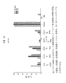
高順位であるHER−2ペプチド配列316−339およびHER−2の485−503に対して誘発された抗体は、2つのヒト乳房腫瘍細胞株(SKBR−3およびBT−474)の増殖を阻害した。MVF HER−2(316−339)抗体は、SKBR−3およびBT−474の細胞株の増殖を、未処理またはアイソタイプ抗体で処理した細胞に比べて約35%および15%だけ阻害した。MVF HER−2(485−503)抗体は、SKBR−3細胞の増殖を22%およびBT−474細胞の増殖を11%だけ阻害した。HER−2ペプチド27−45および605−622に対して惹起された抗体は、腫瘍細胞株の増殖に対する極わずかな効果を有し、そしてアイソタイプコントロール抗体と同様にBT−474細胞の増殖を増加することが示された。実施例7−10のキメラペプチドに対して惹起されたどの抗体のいずれも、別のヒト乳房腫瘍細胞株MCF−7(HER−2の標準レベルだけ発現する)の増殖を阻害しなかった。
【0106】
乳房腫瘍細胞株SK−BR3の細胞をまた、種々のHER−2ペプチド抗体の混合物を用いて、それぞれ0.1mg/mlの濃度で処理した。複数の抗体での処理によって、単一の抗体での阻害効果より優れたさらなる増殖阻害効果が生じた。異なる3つの増殖阻害ペプチド抗体の混合物は、未処理細胞に比べてSK−BR−3細胞の増殖を90%を超えて防止した。乳房腫瘍細胞株BT474の細胞をまた、種々のHER−2ペプチド抗体の混合物を用いて、266μg/ml、528μg/mlおよび792μg/mlの濃度で処理した。
【0107】
(実施例11:MVFDW4−ミクロスフェア)
実施例で上述したように調製されたMVFDW4を、下記のようにミクロスフェア内にロードした。
【0108】
(ミクロスフェア調製)
ミクロスフェアの調製を、Glas−col/G.K.Heller HST10N Stir−Testerを用いて、制御された温度の被膜された(jacketed)ビーカー内で実施した。ミクロスフェア調製は、0.58dL/gの固有の粘度を有する75/25ポリ(DL−ラクチド−コ−グリコリド)(PLGA)を120mg/mlで利用した。ペプチド/ポリマー溶液を、Span85(ソルビタントリオレート)を乳化剤として用いて、40℃にて鉱油/綿実油中で乳化した。750rpmで一時間攪拌後、油エマルジョン中の油(oil−in−oil emulsion)を、0.45μm膜を通して濾過し、そして石油エーテルで数回洗浄した。このフィルターをコニカルチューブ内に置き、液体窒素で凍結し、そして少なくとも3日間凍結乾燥した。ペプチドローディングをアミノ酸分析によって決定した。
【0109】
ミクロスフェアの形態学、表面の特徴、およびサイズを、Philips Electronics XL−30 FEG走査型電子顕微鏡を使用して観察した。乾燥サンプルを、スパッターコートした炭素帯電性タブ化標本マウント(ピンの直径3.2mm、台座の直径12.7mm)上にて、アルゴン雰囲気下で110秒間スプレーした。画像を、10mmの作動距離で、二次電子検出器および5kVの加速(accelerating)電位を使用して得た。各々の調製物について少なくとも100粒子を、Philips Electronics XL−30 Field Emission Gun Scanning Electron Microscopeを使用して得た電子顕微鏡写真より分けた。
【0110】
酢酸エチルを使用してミクロスフェアからペプチドを抽出することにより、ペプチドローディングを決定した。ミクロスフェアを、過剰の酢酸エチル中に浸漬し、そして強くボルテックスした。溶解しないポリマーおよび放出したペプチドを、5分間の遠心分離によってスピンダウンした。上清を回収し、そしてこのプロセスをさらに3回反復した。過剰の酢酸エチルを、蒸発して除いた。サンプルを、2%酢酸中で再構成し、そして酢酸分析に供した。ペプチドを、6N HClで加水分解し、そして引き続き、アミノ酸をWaters PicoTag Systemを使用してフェニルチオヒダントインとして誘導体化し、かつ分析した。
【0111】
(ペプチド免疫)
PBS中100μgのペプチドおよび100μgのnor MDPを、4:1のスクアレン:アルラセル(Arlacel)中で乳化し、そして1ペプチド当たり5匹のマウスの複数の部位に皮下注射する。3週間後、マウスを同一のプロトコルを使用してブーストする。2度目の免疫の2週間後、脾臓を外科的に回収する。
【0112】
(ミクロスフェアカプセル化ペプチド免疫)
MVFDW4ペプチドおよびNor−MDPアジュバントを、ミクロスフェア中に個別にカプセル化した(5.2%ロード(MVFDW4)および5%ロード(Nor−MDP))。1マウス当たり100μgのペプチドおよび100μgのNor−MDPを含むミクロスフェアを、200μlの4:1のスクアレン:アルラセルAと混合し、そしてマウスに皮下注射した。
【0113】
(CTL活性の検出)
最後の注射の2週間後、脾臓細胞を各マウスから取り出した。単一の細胞の懸濁液を調製し、その一部をElispotによるINF−γ検出に、残りをクロム放出アッセイに使用した。
【0114】
(i)Elispotアッセイ
4日間のElispotプロトコルを使用して、脾臓細胞のINF−γ産生を検出した。1日目、Elispotプレート(PolyFiltronics)を、アジ化物(azide)を含まない滅菌PBSで希釈した抗マウスINF−γ(クローンR4−6A2、Pharmingen)を4μg/mlで用いてコートした。次いで、プレートを湿潤チャンバー内で4℃で一晩インキュベートした。2日目、プレートをPBSで4回洗浄し、次いで、DMEM中1%のBSA(添加物なし)をウェルあたり200μlで、室温にて1時間ブロックした。BSAを除去した後、1% L−グルタミンを含むHL−1培地(Biowhittaker)中に回収した新鮮な脾臓細胞を、特定の濃度でウェル中に添加した。次いで、湿潤インキュベーターにて37℃で5% CO2にて、プレートを24時間インキュベートした。3日目、細胞を回収し、そしてプレートをPBSで1回洗浄した後、このプレートをさらに、PBS/Tween 20(2000:1)で4回洗浄した。PBS/Tween 20/1% BSAで2μg/mlに希釈したビオチン化抗INF−γ(クローンXMG 1.2、Pharmingen)を、ウェル当たり100μlでウェルに添加した。プレートを湿潤チャンバーにて4℃で一晩インキュベートした。4日目、PBS/Tween 20でプレートを4回洗浄した後、PBS/Tween 20/1% BSAで1:1000に希釈したヤギ抗ビオチン/アルカリホスファターゼ結合物(Vector Laboratories Inc.)を、ウェル当たり100μl添加した。プレートを、室温で2時間インキュベートし、PBSで4回洗浄した。BCIP/NBTアルカリホスファターゼ基質(Kirkegaard and Perry Laboratories Inc.)を、室温でのインキュベートのためにウェル当たり200μl添加した。スポットがプレート上で可視化したときに、反応を、流れる水道水でクエンチした。プレートを風乾しこのスポットを読んだ。
【0115】
(ii)細胞傷害性溶解アッセイ
単一細胞懸濁液の同一のバッチより得た同系の脾臓細胞刺激因子を使用して、インビトロでの再刺激を実施し、そしてp63合成ペプチドで1時間パルスした。反応させる(responder)細胞:刺激する(stimulator)細胞(3:1)を24ウェルプレート中で混合し、そして37度で1週間インキュベートし、次いで、これらの反応させる脾臓細胞を以下のCTLアッセイにて効果(effector)細胞として使用した。標準クロム放出アッセイを使用して、CTLアッセイを実施した。簡単には、SVBalb細胞(H−2d)またはMCS7細胞(H−2b、コントロールとして)を10μg/mlのペプチドp63および500μCi/mlの51Cr塩化クロム酸を伴うかまたは伴わないで、37℃で1時間インキュベートすることによって、ペプチドでパルスした標的を調製した。標識化標的細胞(ウェル当たり104細胞)および種々の数の効果細胞を、96ウェルプレートに最終容積0.2mlにてプレートした。37℃で5時間後、50μlの上清を各ウェルから回収し、そして特異的溶解%を、以下の式に従って決定した:[(試験したサンプルのcpm−自然発生51Cr放出のcpm)/(最大51Cr放出のcpm−自然発生51Cr放出のcpm)]×100。
【0116】
(実施例12)
HER−2を、細胞外ドメインにのみ存在する7つの潜在部位にて広範囲にわたってグリコシル化する。本発明者らは、これらのN連結グリコシル化部位(Asn−X−Ser/Thr−X)を保有するB細胞エピトープを、分析に用いたコンピューターによって同定した。安定に配列をコードするB細胞エピトープを含む3つの構築物を、変異してグリコシル化を改善し、そしてこれらの野生型対照物HER−2(115−136);(182−216);および(630−650)ならびにTヘルパー細胞エピトープを、バキュロウイルス哺乳動物シャトル発現ベクターにサブクローン化した。このベクターは、哺乳動物細胞への目的の遺伝子の容易なバキュロウイルス媒介トランスフェクションを可能にするが、効率よい発現およびエピトープのグリコシル化が可能な細胞において複製し得ない。発現したキメラグリコシル化エピトープを、糖の型およびグリコシル化効率についてキャピラリー電気泳動によって特徴付ける。効率よくグリコシル化したエピトープで、ウサギを免疫し、そしてペプチド抗体を作成する。
【0117】
ここで、本発明は、十分に記載され、添付の特許請求の範囲の精神および範囲を逸脱することなく多くの変化および改変がなされ得ることは、当業者には明白である。
【図面の簡単な説明】
【図1】 図1は、キメラHER2−B細胞ペプチドの模式図である。
【図2】 図2は、多価HER2−2B/CTLペプチドの模式図である。
【図3】 図3は、キメラHER−2 B細胞ペプチドDW1MVFに対して惹起される抗体の、HER−2過剰発現SKB3細胞への結合を示す。DW1MVFに対して惹起される抗体は、市販のモノクローナル抗体に匹敵する親和性を有して、HER−2レセプターを特異的に結合した。SKBR3細胞が、抗体とインキュベートされ、洗浄され、そしてFITC結合体化2次抗体が添加された。ホルマリン中に固定された後、細胞が、励起のために488nmでCoulter ELITEフローサイトメトリーにより分析された。5.0×10⁶個の細胞が、各サンプルについて計測された。
【図4】 図4は、複数のキメラHER−2 B細胞ペプチドを注射された非近交系のウサギにおける免疫原性を示す。
【図5】 図5は、HER−2過剰発現SK−BR−3ヒト乳癌細胞におけるヘルセプチン(herceptin)および単一および複数の、HER−2 B細胞キメラペプチドMVFDN1(HER−2(27−45))、MVFDN2(HER−2(316−337))、MVFDN3(HER−2(485−503))およびMVFDW4(DW4)に対して生成されるペプチド抗体の抗増殖効果を示す。結果は、全く同じ3つのサンプルの平均である。
【図6】 図6は、HER−2過剰発現BT474ヒト乳癌細胞におけるヘルセプチンHER−2 B細胞キメラペプチドMVFDN1(ND1)MVFDN2(N2)、MVFDN3(N3)およびMVFDW4(DW4)に対して生成されるペプチド抗体の抗増殖効果を示す。結果は、全く同じ3つのサンプルの平均である。
【図7】 図7は、HER−2過剰発現BT474ヒト乳癌細胞における単一および複数のHER−2 B細胞キメラペプチドMVFDN2(N2)、MVFDN3(N3)およびMVFDW4(DW4)に対して生成されるペプチド抗体の増殖効果を示す。結果は、全く同じ3つのサンプルの平均である。
【図8】 図8は、自発的な乳房腫瘍発症におけるHER−2 B細胞ペプチドを用いた予防接種の効果を示す。6個体のマウスの群が、約4週齢で、100μgのペプチド(およびスクアレンアルラセル(arlacel)Aにおいて乳化されたアジュバントとしてのMDP)を接種され、そして4、8、16、および24週後にブーストした。腫瘍が、長さ×幅2/2として算出された。腫瘍発症までの時間が、個々の曲線の対数ランクの比較と共にKaplan−Meier生存分析を使用して分析された。[0001]
(Citation of related application)
Based on 35 USC 119 (e) (1), this application claims priority to the earlier provisional US
[0002]
The work described in this application was at least partially sponsored by grants PHS / NIH P30 CA-16058 and NIH / NCI RO1 CA 84356-01A1 from the National Cancer Institute. The United States government has certain rights in this invention.
[0003]
(background)
Currently, the most common forms of treating breast cancer include surgery, chemical intervention, and / or radiation therapy. If the cancer is not limited to the defined area, the surgery alone cannot eliminate the cancer. Therefore, radiation treatment is often performed after surgery to destroy cancer cells that are near the surgical site and have escaped the surgery. Side effects of such treatments include skin sensitivity or itching, interference with the immune system, sometimes nausea, and radiation fibrosis in which the affected area of the lungs is rarely fibrotic. It is done. Chemotherapy can also be used after surgery. Chemotherapy utilizes drugs that are toxic to cancer cells. Since this is not a completely selective system, normal cells are also affected. Negative side effects include nausea, fatigue, loss of appetite, hair loss and diarrhea.
[0004]
In view of the shortcomings of current therapies, attempts have been made to find further approaches for treating breast cancer. One such approach is immunotherapy. One target for immunotherapeutic approaches is the HER-2 protein. HER-2 protein (a product of the HER-2 oncogene) is overexpressed in various cancers. It is found in 50-60% of ductal carcinomas in situ, and 20-40% of all breast cancers, and a substantial proportion of adenocarcinomas that occur in the ovary, prostate, colon and lung. HER-2 protein overexpression relates to malignant transformation in humans. HER-2 protein overexpression is also closely associated with malignancy aggressiveness, which is found in 1/4 of all invasive breast cancers. HER-2 protein overexpression correlates with poor prognosis in both breast and ovarian cancer.
[0005]
In recent studies, antibodies against the extracellular binding domain (ECD) of HER-2 have been shown to confer inhibitory effects on tumor growth in vitro and in animal models (Hudziak, RM et al., Mol. Cell. Biol.9: 11-65-72, 1989; Tagliabue, E. et al., Int.J. Cancer 47: 933-7, 1991; Drebin, JA et al., Proc.Natl.Acad.Sci.USA 83: 9129-33, 1986; Drebin, JA, et al., Oncogene, 2: 273-7, 1988; Drebin, JA, et al., Oncogene, 2: 387-94, 1988, and Katumata, M., et al., Nat. Med. 1: 644-8. 1995). Furthermore, Phase II and Phase III clinical trials in patients with metastatic HER-2 overexpressing breast cancer of recombinant humanized anti-HER-2 monoclonal antibody (Trastuzumab) showed a 15% overall response rate as a single agent Produced. Trastuzumab has also been shown to improve survival when combined with cytotoxic chemotherapeutic agents (Beselga, J. et al., J. Clin. Oncol. 14: 737-44, 1996; Pegram, M. D. et al., J. Clin. Oncol. 16: 2659-71, 1988). A number of vaccine approaches have been evaluated that target recombinant HER-2 protein, HER-2 ECD, or ECD of rat neu (which is a rat homolog of HER-2). For example, NFS strain mice immunized with a recombinant vaccinia virus expressing rat neu ECD developed a protective antibody response against subsequent challenge with neu transformed NIH 3T3 cells (Bernards, R. et al., Proc. Natl. Acad. Sci. USA 84: 6854-8, 1987). However, immunization of BDIX rats with the same immunogen did not result in an antibody response and did not inhibit the growth of syngeneic neu expressing B104 neuroblastoma cells. This suggests that this strategy was insufficient to induce an immune response in rats. Polysaccharide-oncoprotein conjugate vaccine (consisting of 147 amino terminal amino acids of HER-2 ECD complexed with cholesteryl-bearing mannan and pullulan) mediated rejection of HER-2 expressing sarcomas in BALB / c mice Cellular and humoral immune responses were induced (Gu, XX, et al., Cancer Res., 58: 3385-90, 1998). Purified rat neu ECD (Esserman, L.J., Cancer Immunol. Immunother. 47: 337-42, 1999) or neu-transfected allogeneic mouse fibroblasts (Cefai, D., et al., Int. J. Cancer, 83: 393-400, 1999), partial protection was shown in rat neu transgenic mice adapted to develop mammary tumors.
[0006]
Despite the results of the above studies, it is effective in humans using cell-based or protein-based vaccine strategies that target HER-2 or HER-2 ECD (HER-2 mutated) “self” antigens It is still uncertain whether an immune response can be generated. Accordingly, it is desirable to take additional immunotherapeutic approaches to treat or prevent breast cancer and other malignancies that are associated with overexpression of HER-2 protein.
[0007]
(Summary of the Invention)
The present invention provides new compounds and compositions for stimulating the immune system and treating malignancies associated with HER-2 protein overexpression. This compound is an immunogenic epitope of the HER-2 protein and chimeric and multivalent peptides containing such an epitope.
[0008]
The first group of compounds is collectively referred to herein as “HER-2 B cell epitopes”. The HER-2 B cell epitope comprises about 15 to about 50 amino acids, more preferably 17-40 amino acids, and most preferably 18-35 amino acids. Preferably, the HER-2 B cell epitope comprises a sequence selected from the following group or functional equivalents thereof:
[0009]
[Chemical formula 5]
[0010]
The present invention also provides a chimeric peptide (hereinafter referred to as “chimeric HER-2 B cell peptide”) comprising at least one of the HER-2 B cell epitopes of the present invention or a functional equivalent thereof. Preferably, the chimeric HER-2 B cell peptide is about 35 to about 150 amino acids long, more preferably about 35 to about 70 amino acids long. This chimeric HER-2 B cell peptide contains three units. The first unit contains a HER-2B cell epitope or functional equivalent thereof. The second unit is a helper T (Th) cell epitope, preferably a promiscuous Th cell epitope. As used herein, a “promiscuous” Th cell epitope is an epitope that facilitates the release of cytokines that help bypass MHC constraints. The second unit is about 14 to about 22 amino acids long, more preferably about 15 to 21 amino acids long, and most preferably 16 amino acids long. Preferably, this Th cell epitope has one of the following amino acid sequences:
[0011]
[Chemical 6]
[0012]
The invention also provides a multivalent HER-2 B cell peptide comprising a plurality (ie, at least two) of the HER-2 B cell epitopes of the invention or functional equivalents thereof and a Th cell epitope. The HER-2 B cell epitope and Th cell epitope are bound to the core β sheet template. Preferably, the template comprises two chains with alternating leucine and lysine residues that are joined by a linker. The linker is an amino acid, that is, preferably a peptide about 2 to about 15 amino acids long, more preferably about 2 to about 10 amino acids long, and most preferably about 2 to about 6 amino acids long. The most preferred linker comprises the amino acid sequence Gly-Pro-Ser-Leu (SEQ ID NO: 20).
[0013]
The invention also relates to an immunogenic composition comprising a chimeric HER-2 B cell peptide or a multivalent HER-2 B cell peptide and a pharmacologically acceptable carrier. A preferred carrier is a biodegradable microsphere. Such immunogenic compositions are useful for treating or preventing malignancies associated with overexpression of HER-2 protein.
[0014]
The present invention also relates to a polynucleotide encoding at least one of the above HER-2 B cell epitopes. Such polynucleotides are useful for generating epitopes by recombinant techniques. The invention also relates to an isolated polynucleotide having a sequence encoding a chimeric HER-2 B cell peptide of the invention. Such polynucleotides are useful for preparing chimeric HER-2 B cell peptides. Such polynucleotides are also useful in immunogenic compositions (eg, DNA vaccines) for treating or preventing malignancies associated with overexpression of HER-2 protein. Preferably, such immunogenic compositions are administered intramuscularly.
[0015]
The present invention also provides HER-2 epitopes (hereinafter referred to as “HER-2 CTL epitopes”) that can activate cytotoxic (Tc) cells. The HER-2 CTL epitope comprises about 8 to about 12 amino acids, more preferably 9-11 amino acids. Preferably, this HER-2 CTL epitope comprises one of the following sequences:
[0016]
[Chemical 7]
[0017]
The present invention also provides a chimeric peptide, referred to below as a chimeric HER-2 CTL peptide comprising a HER-2 CTL epitope or functional equivalent thereof. The chimeric HER-2 CTL peptide contains three units. The first unit contains a CTL epitope. The second unit is preferably a promiscuous T helper cell epitope. The third unit is a peptide unit that connects the linker amino acid or the first peptide unit and the second peptide unit.
[0018]
The invention also provides multivalent HER 2 CTL peptides comprising a plurality, ie, at least two of the present HER 2 CTL epitopes or functional equivalents thereof and a Th cell epitope. The HER-2 CTL epitope and Th cell epitope are linked to the core β sheet template. Preferably, the template comprises two chains with alternating leucine and lysine residues that are connected by a linker.
[0019]
Chimeric HER-2 CTL peptides and multivalent HER-2 CTL peptides are useful immunogens for activating Tc cells. Such activation induces Tc cells to initiate expression of IL-2 receptor and to a lesser extent IL-2, and important cytokines possess cytokine activity against target cells that present proliferation and epitopes It is required for the differentiation of activated Tc cells into functional cytotoxic lymphocytes. The invention also relates to an immunogenic composition comprising a chimeric HER-2 CTL peptide or a multivalent HER-2 CTL peptide and a pharmaceutically acceptable carrier. The invention also relates to a method of activating Tc cells in a mammal by administering an immunogenic composition to such an animal.
[0020]
The present invention also includes isolated polynucleotides that encode one or more HER-2 CTL epitopes described above. Such polynucleotides are useful for producing epitopes by recombinant techniques. The invention also includes an isolated polynucleotide having a sequence encoding a chimeric HER-2 CTL peptide. Such polynucleotides are useful for preparing chimeric CTL cell epitope peptides. Such polynucleotides are also useful in immunogenic compositions (eg, DNA vaccines) for treating or preventing malignancies associated with the HER-2 oncogene.
[0021]
The invention also relates to multivalent B / CTL peptides, which are linked to one or more HER-2 CTL epitopes linked to one or more T helper cell epitopes. Contains B cell epitopes. This epitope is linked to the core β sheet template. The present invention also relates to polynucleotides encoding multivalent B / CTL peptides and DNA vectors comprising such polynucleotides. The invention also relates to an immunogenic composition comprising a B / CTL epitope peptide or a polynucleotide encoding the same and a pharmaceutically acceptable carrier.
[0022]
The present invention also relates to a method of stimulating an immune response in a subject against HER-2 protein. Such methods include administering a chimeric peptide or multivalent peptide of the invention to a subject. Such a method results in an enhancement of the subject's immunity against disease states associated with the expression of HER-2 protein.
[0023]
(Detailed description of the invention)
The present invention provides peptides that are immunogenic epitopes of the HER-2 protein, which are referred to below as HER-2 B cell epitopes and HER-2 CTL epitopes.
[0024]
HER-2 B cell epitopes can cause humoral responses that result in the production of antibodies that are immunoactive with the extracellular domain of the HER-2 protein. The HER-2 protein and its rat homologue neu are transmembrane proteins with a relative molecular weight of 185 kd that are approximately 1255 amino acids (aa) in length. The HER-2 / neu protein has an approximately 645 aa extracellular binding domain (ECD), a highly hydrophobic transmembrane anchor domain (TMD), and EGFR with 40% homology to epidermal growth factor receptor (EGFR). It has an approximately 580aa carboxy-terminal cytoplasmic domain (CD) with 80% homology to it. The amino acid sequence of the HER-2 protein and the nucleotide sequence encoding such an amino acid sequence is shown in GenBank accession number M11730.
[0025]
A HER-2 B cell epitope includes a peptide having one of the sequences, which is referred to below as the “reference sequence” shown in Table I below. This reference sequence was selected and scored using computer assisted analysis using 6 antigenic correlations: (a) Strand flexibility and mobility profiles of individual sequences Were calculated according to Karplus and Schultz, Naturewiss 72: 212-213, 1985; (b) Hydrotherapy profiles were generated for seven residue span settings and finally Kyte And Doolittle, J. et al. Moi. Biol. 157: 105-132, 1982, refined with a range of three residues; (c) Hydrotherapy profiles are described in Hopp and Woods, Proc. Natl. Acad. Sci. USA 78: 3824-3828, 1981, was created for a 6 residue window; (d) Analysis of exposure of amino acid residues to water using a 1.4A probe was performed using Rose. , Science 229: 834-838, 1985; (e) Protrusion index predicting the portion of the protein that approaches and protrudes into the solvent is described by Thornton, EMBO J. et al. 5: 409-413, 1986; (f) The possibility that the five residue sequences are antigenic was determined by the method of Welling, FEBS Lett 188: 215-218, 1985. The basic premise is that the algorithm used for prediction is always located in the area exposed to the surface on the protein, and is therefore probably involved in antibody binding. Sequences are given with a score of 1-6 based on their respective index values and ranked: the highest ranking sequence gives the highest individual score for the examined analyte (6/6) Having and consecutive candidates had the next highest score (5/6) and so on. The best scored epitopes are further ranked by association with secondary structure properties, for example, amphipathic α-helix sequences or β-turn loop regions are preferred for random coil fragments. Chou and Fasman, Adv. Enzymol. Relat. Subj. Biochem. 47: 45-148, 1978 was used to predict secondary structure (alpha helix, beta strand / sheet, 3 turns / loop, random coil) and helix amphiphilic moments. Helix dipole interactions in electrostatic ion pairs and helix segments are also conceivable (eg, hydrophobic / hydrophilic balance). Preferably, the hydrophilic / hydrophobic balance is 2/2 to 4/1.
[0026]
As described herein, HER-2 B cell epitopes also include peptides that are functional equivalents of the peptides shown in Table I below. Such a functional equivalent is a modified sequence in which one or more amino acids in the corresponding reference sequence have been substituted or one or more amino acids have been deleted or added from the corresponding reference sequence Have For example, cysteine residues can be deleted or replaced with other amino acids to prevent inaccurate intramolecular disulfide bridge formation during reversion. If necessary, the HER-2 B cell epitope is glycosylated.
[0027]
Although it is possible to have non-conservative amino acid substitutions, this substitution is a conservative amino acid substitution, except for substitutions made to replace cysteines, where the substituted amino acid corresponds in the reference sequence Has similar structural or chemical properties with amino acids. By way of example, conservative amino acid substitutions include substitution of one aliphatic amino acid or hydrophobic amino acid (eg, alanine, valine, leucine and isoleucine) with another amino acid; one amino acid containing a hydroxyl group (eg, serine and threonine) ) With another amino acid; one acidic residue (eg glutamic acid or aspartic acid) with another amino acid; one residue containing an amide (eg asparagine and glutamine) with another Substitution with a residue; substitution of one aromatic residue (eg, phenylalanine and tyrosine) with another residue; another residue of one basic residue (eg, lysine, arginine and histidine) And one small amino acid (eg, alanine, serine, threonine, methionine, and glycine), Comprising the substitution of residues, the.
[0028]
Preferably, deletions and additions are located at one of the amino terminus, the carboxy terminus, or both of the sequences shown above. As a result of the alteration, the HER2 B cell epitope equivalent is an amino acid sequence that is at least 70% identical, preferably at least 80% identical, more preferably at least 90% identical, most preferably at least 95% identical to the corresponding reference sequence Have Sequences that are at least 90% identical have only one change (ie, any combination of deletions, additions or substitutions per 10 amino acids of the reference sequence). Percent identity is determined by comparing the amino acid sequences of variants with reference sequences using the MEGALIGN project in the DNA STAR program.
[0029]
For functional equivalents longer than the corresponding reference sequence, the functional equivalent has a sequence that is at least 90% identical to the reference sequence and the sequence flanking the reference sequence in the wild-type HER2 protein.
[0030]
(Table 1. Enhanced human p185 HER-2 predicted B cell epitopes listed in rank order by number of amino acid residues. Asparagine (N) -linked glycosylation sites are bold and underlined)
[0031]
[Table 1]
[0032]
Chimeric HER-2 B cell peptides are useful immunogens for inducing the production of antibodies that interact with and bind to the extracellular domain of the HER-2 protein. The chimeric B peptide is also useful as a research tool to detect antibodies against HER-2 protein in patient sera. In accordance with the present invention, the chimeric B cell peptides MVF-HER-2 (628-647), HER-2 (376-395) -MVF, and HER-2 (410-429) -MVF elicit an antibody response in rabbits And such antibodies bind to the intact HER-2 receptor on ER-2 that (a) immunoprecipitates HER-2 protein, (b) overexpresses cells in culture, and (c ) It has been measured to reduce the proliferation of HER-2 overexpressing cells in vitro and in xenograft mouse models. It has also been measured that immunization of transgenic mice with the chimeric peptide MVF-HER-2 (628-647) delays tumor development in such mice at least 9 months after the control mice develop tumors. Yes.
[0033]
The present invention provides HER-2 CTL epitopes that have the ability to elicit cell-mediated responses to HER-2 protein. As used herein, the term HER-2CTL epitope includes peptides having one of the sequences shown in Table 2 below, or functional equivalents thereof. This functional equivalent has an amino acid sequence that is at least 80%, preferably at least 90% identical to one of the sequences, referred to as the “reference sequence” shown in Table 2 below. The ability to elicit a cell-mediated response to a functional equivalent of HER-2 protein, or the extracellular binding domain of HER-2 protein, is the same as or higher than the corresponding reference sequence.
[0034]
[Table 2]
[0035]
The present invention also includes co-linker chimeric peptides comprising multiple HER-2 CTL epitopes. Multiple HER-2 CTL epitopes are linked to each other by a linker that is 1-5 amino acids in length. Optionally, this linker includes a proteolytic site. In one embodiment, the linker comprises adjacent basic amino acid residues. Preferably, the co-linker chimeric peptide comprises a HER-2CTL epitope derived from class HLA-A3, a HER-2CTL epitope derived from class HLA-B7, a HER-2CTL epitope derived from class HLA-A2, and a HER derived from class HLA-B27. -2 contains the CTL epitope. The co-linker chimeric peptide further comprises a second unit that is a promiscuous Th epitope of 14-22 amino acids in length. This second unit is linked to the amino terminus or carboxy terminus of the first unit by a linker.
[0036]
(A. Preparation of epitope and co-linker chimeric peptide)
HER-2 B cell epitopes, CTL epitopes, chimeras, and multivalent peptides are preferably synthesized using a commercially available peptide synthesizer. Preferably, as described in Kaumaya et al., “DENOVO” ENGINEERING OF PEPTIDE IMMUNOGENIC AND ANTIGENIC DETERMINANTS AS POTENTIAL VACCINES, Peptides, Design,
[0037]
HER-2 B cell epitopes, HER-2 CTL epitopes, and chimeric peptides can also be generated using cell-free translation systems and RNA molecules derived from DNA constructs encoding the epitopes and peptides. Alternatively, an epitope or chimeric peptide is created by transfecting a host cell with an expression vector that contains a DNA sequence encoding the respective epitope or chimeric peptide, and then induces expression of the polynucleotide in the host cell. . For recombinant production, recombinant constructs containing one or more sequences encoding epitopes, chimeric peptides, or variants thereof are calcium phosphate transfection, DEAE dextran mediated transfection, transfection, microinjection, cationic lipids. Introduced into the host cell by conventional methods such as mediated transfection, electroporation, transduction, scrape ladding, bolistic introduction, or infection.
[0038]
HER-2 B cell epitopes, CTR epitopes and chimeric peptides can be generated in suitable host cells (eg, mammalian cells, yeast, bacteria, insect cells or other cells) using the appropriate techniques using conventional techniques. It can be expressed under control. Suitable hosts include E. coli. E. coli, P.M. Examples include, but are not limited to, Pastoris, Cos cells, and 293 HEK cells. Following transformation of the appropriate host strain and growth of the host strain to the appropriate cell density, the cells are harvested by centrifugation, disrupted by physical or chemical means, and the resulting crude extract is an epitope Or retained for further purification of the chimeric peptide.
[0039]
Conventional procedures for isolating recombinant proteins from transformed host cells (eg, initial extraction from cell pellets or cell culture media followed by salting out and aqueous ion exchange chromatography, size exclusion chromatography) The process and high performance liquid chromatography (HPLC), and isolation by one or more chromatographic steps including affinity chromatography) can be used to isolate the recombinant peptide. It is preferred to use recombinant techniques to create glycosylated epitopes and chimeric peptides. Mammalian cells (eg, Cos-7 and Hep-G2 cells) are preferably used in recombinant technology to produce glycosylated epitopes and chimeric peptides that contain the same.
[0040]
Naturally occurring variants of the HER-2 B and HER-2 CTL epitopes shown in Tables 1 and 2 above are also suitable for use in, for example, a suitable cDNA library or genome using a DNA sequence encoding a polypeptide. It can be isolated by screening the library.
[0041]
(B. Preparation of multivalent HER-2 B cell peptide, multivalent CTL peptide, and multivalent B / CTL peptide)
Preferred synthetic approaches for preparing HER-2 multivalent peptides utilize combinatorial Fmoc / tbutyl, Fmoc / benzyl and Boc benzyl strategies and a fourth level differential protecting group (Npys) strategy. Details of such an approach are presented in Larimore et al. (1995) Journal of Virology 69: 6077-6089, which is specifically incorporated herein by reference.
[0042]
(C. Identification of functional equivalents of HER-2 B cell epitopes shown in Table 1)
The functional equivalents of the HER-2 B cell epitopes listed above are generally identified by altering the sequence of the epitope and then assaying for the ability to stimulate an immune response (eg, production of antibodies). obtain. For example, such assays can generally be performed by preparing a chimeric peptide comprising a modified polypeptide and a promiscuous Th cell epitope, injecting the chimeric peptide into a test animal, and assaying the antibody. . Such antibodies can be found in various body fluids including serum and ascites. Briefly, a body fluid sample is isolated from a warm-blooded animal (eg, a human). For this reason, it is desirable to determine whether an antibody specific for a HER-2 / neu polypeptide exists. The body fluid is incubated with the HER-2 / neu polypeptide under conditions and for a time sufficient to allow the immune complex to form between the polypeptide and an antibody specific for this protein, and then preferably, an ELISA Assay using techniques. In such a technique, the colorimetric change is measured at 490 nm. Epitopes that induce the production of antibodies that exhibit a titer of 10,000 or greater against the HER-2 / neu protein are preferred. As used herein, a titer of 10,000 indicates an absorption value of 0.2 above background.
[0043]
(D. Method for identifying functional equivalents of HER-2 CTL epitopes shown in Table 2)
Functional equivalents of the HER-2 CTL epitopes shown in Table 2 are identified by modifying the sequence and then assaying the resulting polypeptide for the ability to stimulate an immune response (eg, activation of Tc cells). To do. In the first encounter with a CTL epitope, a small number of immune T cells secrete lymphokines and proliferate and differentiate into effector and memory T cells. The primary immune response occurs in vivo but is difficult to detect in vitro. Subsequent encounters with the same HER-2 antigen (ie, CTL epitope) by memory T cells leads to faster and stronger immune responses. Secondary responses occur either in vivo or in vitro. Thus, the in vitro response is readily assessed by measuring the extent of proliferation, the degree of cytokine production, or the production of cytolytic activity in a T cell population that is re-exposed to the HER-2 antigen. Detection of T cell proliferation can be accomplished by various known techniques. For example, T cell proliferation can be detected by measuring the rate of DNA synthesis. T cells stimulated to proliferate show an increased rate of DNA synthesis. A typical method for measuring the rate of DNA synthesis is, for example, by pulse labeling T cells with tritiated thymidine, a nucleoside precursor incorporated into newly synthesized DNA. The amount of tritiated thymidine can be determined using a liquid scintillation spectrophotometer. Other methods for detecting T cell proliferation include interleukin-2 (IL-2) production, Ca 2+ Measuring the increase in flux or dye uptake (eg, 3- (4,5-dimethylthiazol-2-yl) -2,5-diphenyltetrazolium). Alternatively, the synthesis of lymphokines (eg, interferon-γ) can be measured, or the relative number of T cells that can respond to intact HER-2 / neu protein can be quantified.
[0044]
(Polynucleotide)
The invention also provides isolated polynucleotides that encode B cell epitopes, CTL epitopes, and chimeric peptides of the invention. The polynucleotide of the present invention also includes a polynucleotide having a sequence capable of hybridizing to a nucleotide sequence under stringent conditions, preferably under highly stringent conditions. Hybridization conditions are as described in Berger and Kimmel (1987) Guide to Molecular Cloning Techniques, Methods in Enzymology, vol 152, Academic Press, or melting temperature of the probe. TM based on. As used herein, the term “stringent conditions” refers to “stringency” that occurs within a range from about Tm-5 (5 ° C. below the melting temperature of the probe) to about 20 ° C. below Tm. is there. As used herein, “high stringency” conditions use at least 0.2 × SSC buffer and at least 65 ° C. As will be appreciated in the art, stringency conditions can vary with many factors (eg, probe length and nature (ie, DNA or RNA); target sequence length and nature, hybridization solution salts and It can be achieved by the concentration of other components, such as formamide, dextran sulfate, and polyethylene glycol. All of these factors can be modified to produce stringency conditions equivalent to the conditions listed above.
[0045]
A polynucleotide comprising a sequence encoding a HER-2B cell epitope, a HER-2 CTL epitope or a chimeric peptide of the present invention may be wholly or partly subjected to a chemical method, or preferably a recombinant method known in the art. Can be synthesized using. Polynucleotides encoding HER-2B cell epitopes or CTL epitopes can be identified using antibodies immunospecific for HER-2 protein or CTL protein, respectively, to identify clones containing such polynucleotides. It can be obtained by screening a library or a cDNA library.
[0046]
This polynucleotide is useful for producing HER-2B cell epitopes, CTL epitopes or chimeric peptides. For example, RNA molecules encoding multivalent chimeric peptides are used in cell-free translation systems to prepare such polypeptides. Alternatively, DNA molecules encoding HER-2B cell epitopes, CTL epitopes or chimeric peptides are introduced into expression vectors and used to transform cells. Suitable expression vectors include, for example, chromosomal, non-chromosomal and synthetic DNA sequences (eg, SV40, bacterial plasmids, phage DNA derivatives; yeast plasmids, vectors derived from a combination of plasmid and phage DNA, viral DNA (eg, , Vaccinia, adenovirus, fowlpox virus, pseudorabies, baculovirus, and retrovirus)). The DNA sequence is introduced into the expression vector by conventional procedures.
[0047]
Accordingly, the present invention also relates to a recombinant construct comprising one or more of the polynucleotide sequences of the present invention. Suitable constructs include, for example, vectors (eg, plasmids, phagemids, or viral vectors) into which sequences encoding HER-2B cell epitopes, HER-2 CTL epitopes or chimeric peptides are inserted. . In this expression vector, the DNA sequence encoding the epitope or chimeric peptide is operably linked to an expression control sequence (ie, a promoter), which directs mRNA synthesis. Representative examples of such promoters include the LTR or SV40 promoter, E.I. E. coli lac or trp, the phage λPL promoter, and other promoters known to control gene expression in prokaryotic or eukaryotic cells or viruses. The expression vector also preferably includes a ribosome binding site for translation initiation and transcription terminators. Preferably, the recombinant expression vector also provides an origin of replication and a selectable marker (eg, an E. coli ampicillin resistance gene to allow selection of transformed cells (ie, cells that express heterologous DNA sequences). )including. A polynucleotide sequence encoding a HER-B cell epitope, HER-2 CTL epitope or chimeric peptide is incorporated into the vector in frame using translation initiation and termination sequences. Preferably, the polynucleotide further encodes a signal sequence operably linked to the amino terminus of the HER-2B cell epitope, HER-2 CTL epitope, or chimeric peptide.
[0048]
Polynucleotides encoding HER-2B cell epitopes, HER-2 CTL epitopes or chimeric peptides containing such epitopes can be used to express recombinant peptides using techniques well known in the art. Such techniques are described in Sambrook, J. et al. (1989) Molecular Cloning A Laboratory Manual, Cold Spring Harbor Press, Plainview, N .; Y. And Ausubel, F .; M.M. (1989) Current Protocols in Molecular Biology, John Wille & Sons, New York, NY. Polynucleotides encoding HER-2B cell epitopes, HER-2 CTL epitopes or chimeric peptides containing such epitopes are also used to immunize animals.
[0049]
(Pharmaceutical composition)
A pharmaceutical composition comprising a chimeric HER-2B cell peptide and a multivalent HER-2B cell peptide, a HER-2CTL peptide, and a HER-2B / CTL peptide or polynucleotide encoding them, is preferably a pharmaceutical composition ( For example, it is formulated for use as an immunogenic composition or vaccine). Such compositions generally comprise one or more HER-2 chimeric or multivalent peptides, or polynucleotides encoding them in combination with a pharmaceutically acceptable carrier, excipient or diluent. Such carriers are non-toxic to recipients at the volumes and concentrations used.
[0050]
In addition to epitopes, multivalent peptides, and chimeric peptides (functioning as antigens) or polynucleotides encoding them, other components (eg, vehicles for antigen delivery and to improve the immunogenicity of proteins The designed immunostimulatory substance) is preferably included in the pharmaceutical composition. Examples of vehicles for antigen delivery include aluminum salts, water-in-oil emulsions, biodegradable oil vehicles, oil-in-water emulsions, biodegradable microcapsules, and liposomes. For vaccines comprising chimeric peptides, the preferred vehicle for antigen delivery is a biodegradable microsphere, which is preferably composed of poly (D, L-lactide-co-glycolide) (PLGA). The
[0051]
If any suitable carrier known to those skilled in the art can be used in the pharmaceutical compositions of the invention, the type of carrier will vary depending on the mode of administration and whether substantial release is desired. For parenteral administration (eg, subcutaneous injection), the carrier may preferably include water, saline, alcohol, fat, wax, or buffer. Biodegradable microspheres such as polylactic acid galactide can also be used as carriers for the pharmaceutical compositions of the present invention. Optionally, the pharmaceutical composition includes an adjuvant.
[0052]
HER-2 chimeric and multivalent peptides, and polynucleotides encoding them, produce a humoral response, preferably a cellular immune response (eg, generation of antigen-specific cytolytic T cells) in a subject or cell line. Useful to improve or induce. As used herein, the term “subject” refers to any warm-blooded animal, preferably a human. The subject may suffer from cancer (eg, breast cancer) or be normal (ie, have no detectable disease and infection). The pharmaceutical compositions are particularly useful for treating women who have a family history of breast cancer or from which a breast tumor has been removed.
[0053]
(Method of treatment)
The invention also provides a method of treating cancer associated with overexpression of HER-2 protein. “Treatment” means inhibiting or delaying or suppressing tumor growth. Such cancers include breast cancer, lung cancer, ovarian cancer, bladder cancer and prostate cancer. This method comprises administering to a subject a pharmaceutical composition comprising one or more chimeric or multivalent peptides of the invention. Preferred multivalent peptides are those that contain one or more of the following epitopes: HER-2 (628-647), HER-2 (316-339), and HER-2 (485-503). Preferably, multiple intramuscular injections every 3 weeks are used to administer the pharmaceutical composition.
[0054]
(Example)
Exemplary methods are described below, but methods and materials similar or equivalent to those described herein can be used in the practice or testing of the peptides, compositions and methods of the invention. All publications and other cited references mentioned herein are incorporated by reference throughout. The materials, methods, and examples are illustrative only and not intended to be limiting.
[0055]
(Peptide synthesis and HPLC purification)
The peptide was synthesized as previously described (Kaumaya 1994). Briefly, peptides were synthesized on a Milligen / Biosearch 9600 peptide synthesizer using 4-methylbenzhydrylamine resin as a solid support (substitution 0.54 mm / g). The Fmoc / t-butyl synthesis method using 4- (hydroxymethyl) phenoxyacetic acid as the linker was used. After the final deprotection step, protecting groups and peptide resin linkages were cleaved with 90% TFA, 5% anisole, 3% thioanisole, 2% ethanedithiol. The crude peptide was purified by semipreparative HPLC using a Vydac C4 (10 mm × 25 cm) column at 32.5 ° C. Buffer is H 2 0.1% TFA in O and 0.1% TFA in acetonitrile. The peptides are “promiscuous” T cell epitopes MVF288-302 (Kaumaya 1994): DW1MVF (HER-2 376-395), MVFDW4 (628-647), DW5MVF (115-136), DW6MVF (410-429) ).
[0056]
(Gel filtration)
A 20 mg / ml acidified peptide solution (0.1 mg / ml in DTT) was loaded onto a Sephadex G-25 column and 5 ml fractions were eluted with 0.1 M HOAc. Peptide samples were measured spectroscopically at 235 nm and absorbance values were plotted against time. Samples having absorbance values greater than 0.1 and eluting prior to DTT were pooled and lyophilized. The reaction was monitored at 410 nm for completion with Ellman reagent.
[0057]
(Capillary zone electrophoresis)
CZE was performed on a Beckman P / ACE System 2100 interfaced with an IBM computer. Samples were voltage separated (15 kV) in 100 mM sodium borate using a 50 cm capillary over 20 minutes. The eluate was monitored at 214 nm.
[0058]
(Circular dichroism and mass spectrometry)
Measurements were made on a JASCO J-500 spectropolarimeter interfaced with an IBM computer. The instrument was calibrated in a 0.06% (w / v) solution of ammonium-d-10-camphorsulfonate. Peptide (62.5-250 μM by dilution of peptide stock in water) CD spectra were measured at ambient temperature in a cylindrical quartz cuvette (Hellma) with a 0.1 cm path length. The average residual ellipticity (mdeg) is calculated using the relationship [θ] = 100θ / cnl, where θ is the ellipticity, c is the peptide concentration (mM), and n is the amino acid in the peptide And l is the optical path length (cm). Fast atom bombardment (FAB) mass spectrometry measurements were performed on an inneganMat-900 instrument.
[0059]
(Mercury acetate)
The peptide was dissolved in a small amount of water and 100 mg / mm St-Bu solution (2-10 fold excess) was added. The peptide was placed under vacuum and precipitated with 2-mercaptoethanol in a 55 ° C. water bath with stirring. After filtration through wet Celite, the filtrate was rotovaped, acidified with 0.1% TFA in water and lyophilized.
[0060]
(Biological procedure)
(Immunity and animals)
Female New Zealand white rabbits were obtained from Mohic Valley Labbitry (Loudenville, OH). Rabbits were immunized with all of the 1 mg peptide emulsified in CFA at multiple subcutaneous sites. Subsequent booster injections (1 mg and 500 μg in PBS) were given 3 and 6 weeks after the first immunization. Serum was collected and supplementarily inactivated by heating to 56 ° C. for 30 minutes. Serum aliquots were stored at -5 ° C to -15 ° C. The antibody was purified by ammonium sulfate precipitation: a stock solution of saturated ammonium sulfate solution (SAS) was prepared, autoclaved and cooled to 4 ° C. The antibody was precipitated by slowly adding SAS to 35% v / v under stirring in a cold room. Samples were centrifuged at 14,000 × g for 20 minutes and the supernatant was stored at −20 ° C. The pellet was lysed with 0.1 M PBS in half the original volume. The fractions were then placed in a Slide-a-lyzer cassette (Pierce) and dialyzed against frequent changes in pH 8, 0.15 M NaCl over 200 volumes. The saline was brought to pH 8 with a few drops of 0.1M NaOH. IgG concentration was determined by radioimmunodiffusion (RID) (The Binding Site, UK). Monoclonal antibodies were purchased from Oncogene Science.
[0061]
(Direct ELISA) U-bottomed polyvinyl chloride plastic assay plates were coated overnight at 4 ° C. with 2 μg / ml antigen in 100 μl PBS. Non-specific binding sites were blocked with 200 μl PBS-1% BSA for 1 hour and the plates were washed with PBT (phosphate buffered saline containing 0.05
[0062]
(Cell culture) 5% CO 2 Maintained at 37 ° C. in incubator. All culture media, FCS and supplements were purchased from GEBCO (Grand Island, NY). Human breast cancer cell lines SKBR-3 and MCF-7 were obtained from American Type Culture Collection and subcultured in McCoy 5A or DMEM supplemented with 10% FCS and L-glutamine. Cav-1 was maintained in RPMI 1640 containing 10% FCS and L-glutamine. Cav-1 is derived from fresh colon tumor specimens that have been stored frozen and then cultured; it does not express detectable levels of HER-2 / neu. SKBR3 is a breast tumor cell line that overexpresses HER-2 protein, and MCF-7 expresses normal concentrations of this protein.
[0063]
(Immunoprecipitation and Western blotting) On day 0, 1.0 x 10 7 Of SKBR3 cells, 75 cm Three Plate in cell culture flask and allow to adhere overnight. Anti-peptide antibody was added for 4 hours (100 μg / ml). The reaction was stopped by aspirating the medium and immediately adding ice-cold 0.1 M phosphate buffered saline (PBS). Cells were trypsinized and washed twice with chilled Hanks balanced salt solution (HBSS). 3 mM Na Three VO Four Cold lysis buffer (150 mM NaCl; 50 mM Tris, pH 8; 10 mM EDTA, 10 mM sodium pyrophosphate, 10 mM sodium fluoride; 1% NP-40, 0.1, each containing 10 μg / ml aprotinin and leupeptin % SDS) was added to cells resuspended in 100 μl HBSS. Dissolution was achieved by gentle rotation for 20 minutes at 4 ° C. After centrifugation (14,000 × g, 20 min) to remove cell debris, the lysate was incubated overnight with 3-5 μg antibody and 30 μl Protein A / Protein G (Oncogene Science). The beads are pelleted by centrifugation (14,000 × g, 30 seconds) and 1 mM Na Three VO Four Washed twice in lysis buffer containing and boiled in SDS sample buffer for 5 minutes.
[0064]
Proteins were separated by 7.5% SDS-PAGE, transferred to nitrocellulose and probed with antibodies. Protein migration was monitored with prestained molecular weight standards (BioRad). Immunoreactive bands were detected by enhanced chemiluminescence (Amersham) using horseradish peroxidase conjugated goat anti-rabbit immunoglobulin.
[0065]
Indirect binding assay SKBR3 cells or MCF-7 cells were plated at 5,000 cells / well in V-bottom plates (Linbro, McLean VA). Cells were incubated with various concentrations of antibody. After washing with Hanks Balanced Salt Solution (HBSS), cells were incubated with fluorescein isothiocyanate (FITC) conjugated goat anti-rabbit antibody or goat anti-mouse antibody for 1 hour and fixed with formalin. Mouse monoclonal Ab (Oncogene Science, Cambridge, Mass.) Was used as a positive control and anti-CD3 Ab was used as a negative control. Cells were analyzed by a Coulter ELITE flow cytometer (Coulter, Hialeah, FL) with an argon laser for excitation at 488 nm and a 525-run bandpass filter for FITC fluorescence, 5.0 × 10 Three Cells were counted for each sample and final processing was performed. Debris, cell clusters and dead cells were gated out by light scatter assessment and then a single parameter histogram was generated.
[0066]
Effect of Ab on cell proliferation SKBR3, MCF7 and CAVI cells were plated at 5,000 cells / well in V-bottom plates with various concentrations of Ab on day 0. On the third day, [ Three H] thymidine (1 μCi / well) was pulsed into the cells, at which time they were placed in a −20 ° C. freezer for 1 hour. After thawing at room temperature, cells were harvested with a PHD cell harvester (Cambridge Technology, Inc.). Samples were incubated in 5 ml of Ready Safe liquid scintillation mixture (Beckman) and radioactivity was measured with a β counter. Results are expressed as mean CPM +/− standard deviation (SD).
[0067]
(CTL Assay: In Vitro Stimulation) Inguinal and periaortic lymph nodes (LN) are removed 7-10 days after immunization. Then 1.5 × 10 1 pre-pulsed with 1 μM of the appropriate CTL peptide for 1 hour Five Of LN cells (4 × 10 4 6 ~ 5x10 6 ) Was stimulated in vitro. The culture medium used is cDMEM (DEME supplemented with 10% FCS). The supernatant was 30 U / ml (final) IL-2, 2 mM L-glutamine, 10 mM Hepes and 5 × 10 Five Contains M 2-mercaptoethanol.
[0068]
Seven days after in vitro stimulation, CTL activity is tested in a standard chromium release assay. P815 cells (10 6 ) In the presence or absence of the appropriate peptide (1 μM) at 37 ° C. for 1 hour with 150 μCi sodium chromate [ 51 Labeled with [Cr] and washed three times. Labeled target (2 × 10 Three ) Were co-incubated with a predetermined ratio of stimulated LN cells in a 200 μl volume in a V-bottom 96-well plate. After 4 hours incubation at 37 ° C., the supernatant (100 μl) is collected for gamma counting. The% specific lysis is calculated as 100 × [(experimental release-spontaneous release) / (total-spontaneous release)] (Valmori et al., 1994).
[0069]
(In vivo antibody effect) HER2 cells (3 × 10 6 ) Was suspended in 250 μL PBS, mixed with 250 μl MATRIGEL (Beckton Dickinson) on ice and injected subcutaneously into mice. Polyclonal antibodies were administered i.p. on days 9 and 11 at a total concentration of 2 mg / mouse. p. Injected. Tumor volume was measured twice a week with a caliper and calculated by the formula (length x width x height).
[0070]
(Example 1-peptide DWI MVF; HER-2 (376-395) MVF)
The epitope termed DW1 includes amino acids 376-291 of the HER-2 protein linked to a congested Th cell epitope MVF. DW1 is predicted to be an α-helix with a slight turn tendency.
[0071]
(Synthesis): A 20 amino acid HER-2 sequence was linked to the N-terminus of MVF 288-302 by a 4 amino acid linking sequence Gly-Pro-Ser-Leu (SEQ ID NO: 20). This resulting peptide was named DW1MVF, which indicates the DW1 arrangement on the N-terminal side, whereas MVFFD1 represents the C-terminal arrangement. The first amino acid was manually linked and monitored for completion by the Kaiser ninhydrin test. Subsequent coupling was performed on a Milligen / Biosearch 9600 peptide synthesizer. After the last deprotection, the peptide was cleaved from the resin with reagent PU. Since this peptide contains Arg-2,2,5,7,8-pentamethylchroman-6-sulfonyl (PMC) and His, a long cleavage time is required (if histidine is present, a yellow cleavage solution Is observed).
[0072]
Purification and characterization: The peptide was rotoevaporated to remove TFA, precipitated with cold ether, and extracted with water / ether. Extraction was fairly easy and HPLC analysis of the gel filtered and lyophilized peptides showed 2-3 deleted peptides. This peptide eluted as two peaks before DTT. These were pooled and subjected to analytical HPLC.
[0073]
Analyzing semiprep purified DW1MVF BPLC identified one large peak, while CZE identified three species of different charges. The complete peptide contains 6 negatively charged species and 4 positively charged species at neutral pH. As a result of mass spectrometry, the large peak was confirmed to be the target epitope having a molecular weight of 4472. The CD spectrum of the peptide in dilute acetic acid solution shows a slight random coil. In TFE, the configuration was deflected to a slight α helix, which was a maximum intensity in the 190-195 nm range and a minimum intensity at 208 nm and 222 nm. Peptide helical structure [θ 222 , −5,000], 100% spiral θ 222 The average ellipticity of polystyrene was calculated using the Chen formula, with the average ellipticity of polystyrene = −33,000.
[0074]
(Example 2-peptide MFVDW4; HER-2 (628-647) MVF)
MVFDW4 contains an altered sequence of peptides extending from amino acids 628 to 647 of the HER-2 protein. The native sequence contains 3 cysteine residues. The disulfide bond pair of this residue is unknown. Cys 634 and 642 had the ability to form a bridge, so Cys630 was replaced with Gly. Glycine substitution of cysteine is one way to preserve the relative size of the R group at that position. Synthesis proceeds by first creating a DW4 (628-647) peptide linked to a linker, and then the sequence is extended to the N-terminus by the addition of NWF (288-302) helper T cell sequences. This produced the MFDFD4 peptide.
[0075]
In order to generate the disulfide bond, the tBut protecting group was cleaved to give the free thiol form. The mercuric acetate / 2-mercaptoethanol procedure reduces the formation of disulfide bonded multimers. The analytical HPLC of the crude product and the sample were compared. In the coarse sample, two sharp peaks are followed immediately by a broad, obscure shoulder. The treated sample showed a reduction in the size of the first peak and the broader second peak. The correct fraction was later identified by mass spectrometry. This analysis confirmed the molecular weight of this peptide as 4612.
[0076]
Hydrogen peroxide or dithiothreitol (DTT) was added to the crude sample to identify new peaks. Addition of hydrogen peroxide resulted in oxidation.
[0077]
2RSH + H 2 O 2 → R-S-S-R + H 2 O
This reaction resulted in a single large peak eluting at 11.5 minutes. This corresponds to the first peak of the crude sample. DTT treatment results in reduction according to the following reaction scheme.
[0078]
R-CH 2 -S-S-CH 2 + DTT → R-CH 2 -SH + HS-CH 2 -R
The profile of the DTT treated product was similar to the starting material. These profiles indicate that the starting material is a mixture of reduced and oxidized peptides.
By mercury acetate treatment, it is biased to a concentration suitable for reducing species.
[0079]
(Example 3-Peptide DW5MVF; HER-2 (115-136) MVF)
Synthesis: MVF 288-302 and a 4-residue amino acid linker were linked to the resin as described in Example 1 above, followed by 115-136 amino acids of the HER-2 protein. This produced peptide DW5MVF. This sequence is predicted to be a β-turn with high aggregation ability. This required the double coupling key residues All5, V116, T127, V129 and S133.
[0080]
Purification and characterization: DW5MVF was cleaved and extracted with ether and water. Extraction was fairly difficult at high peptide form density, clumped to a sticky state and made almost insoluble by addition of acetic acid. Analytical HPLC of the crude sample showed that one prominent peak was a slight double. The duplex was separated using semi-preparative HPLC. Lyophilized samples were readily dissolved in dilute acetic acid for analytical HPLC. The sample was subjected to time-of-flight mass spectrometry and the correct molecular weight 4431 was obtained. The peptide elutes at 15.5 minutes as a single peak.
[0081]
(Example 4-peptide DW6MVF; HER-2 (410-429) MVF)
Residues 410-429 of HER-2 (named DW6) represent a possible immunogenic epitope from the same region as DW1MVF. Due to the success of DW1MVF, in early FACS experiments we wanted to raise additional antibodies against this region. Residues 410-429 were synthesized by N-terminal addition to the MVF / 4-residue-linker sequence as previously described. The final product was DW6MVF. It is expected to be a β-turn having moderate to high aggregation ability at its C-terminus.
[0082]
(Synthesis): The DW6 sequence was linked to the C-terminus linked to the MVF sequence using FMOC chemistry. The computer algorithm expected high agglutination capacity and therefore attempted to minimize aggregation using extended coupling time and / or double coupling.
[0083]
Purification and characterization: DFA6MVF peptide was obtained by TFA cleavage and was ether / water extracted. This workup still proved difficult due to the high degree of aggregation. There was a significant concentration of the deleted peptide as shown by HPLC. The semi-prepared BPLC separated a large peak that eluted at 13-14.5 minutes. Analytical BPLC showed a single peak eluting at 17 minutes. The difference in retention time for semi-preparative HPLC and analytical HPLC is due to a 3 minute difference in the time of sample injection. Amino acid analysis confirmed that it was identical to the peptide of interest. The derivatized amino acid was analyzed as its phenylthiohydantoin derivative (actual value (theoretical value)): Asp (2.99 (3)), Glu (4.32 (4)), Ser (4.77 (5) ), Gly (3.53 (4)), His (1.62 (2)), Arg (1.08 (1)), Thr (0.02 (0)), Ala (0.97 (1)) , Pro (3.02 (3)), Tyr (0.95 (1)), Val (3.86 (4)), Met (0.09 (0)), Cys (0 (0)), Ile (2.87 (3)), Leu (9.34 (9)), Phe (0.93 (1)), Lys (2.28 (2)) and Trp (0 (0)).
[0084]
(Examples 5 and 6-DW2 (391-399) and DW3 (376-399)
DW2 contains amino acids 391-399 of the HER-2 protein. DW3 contains amino acids 376-399 of the HER-2 protein.
[0085]
(Synthesis): Residues 391 to 394 are bonded to the resin. The synthesis was continued with the rest by addition of residues 376-390. Thereby, the final peptide (DW3) of 376-399 was obtained.
[0086]
Purification and characterization: Three major peaks (eluting at 11 minutes) were separated from DW2 by semi-preparative HPLC. DW3 obtained two major peaks of the mixed composition. Mass spectrometry confirmed that the fraction eluting at 11 minutes of DW2 was the correct peptide with a molecular weight of 1052. No further characterization was done for DW3.
[0087]
(Immunogenicity of chimeric peptides of Examples 1-6)
Chimeric peptides prepared as described in Examples 1-6 above are very highly immunogenic, as evidenced by high antibody titers as early as 3 weeks after immunization. Sera obtained weekly were assayed for the ability to recognize and bind to peptide sequences. DW5MVF showed a steady increase in antibody titer. The titer for DW5MVF was higher in one rabbit than in the other.
[0088]
(Specificity of peptide antibody to HER-2)
FACS and immunoprecipitation: Flow cytometric analysis determined that the DW1MVF anti-peptide antibody directly targets the HER-2 receptor (see Figure). Commercially available mouse Mab-HER-2 / neu was used as a control in SKBR3 cells. Negative control sera showed no binding to the receptor, but increased fluorescence was seen with immune sera. The fluorescence intensity of the polyclonal anti-peptide serum was comparable to that of the monoclonal antibody. Thus, polyclonal sera can mimic the specificity and affinity of monoclonal antibodies.
[0089]
The immune serum was diluted with Hank's Balanced Salts solution and the average cell fluorescence was determined. Serum from “tertiary + 3 weeks” blood showed a fluorescence value of 121.4 (1: 320 dilution), while MAb showed a value of 132. The following is a list of mean cell fluorescence values at various serum dilutions.
[0090]
Dilution Average Cell Fluorescence (DWLMVF)
Control 0.647
1:80 39.3
1: 160 83.5
1: 320 121.4
1: 640 69.4
1: 1280 34.4
1: 2560 17.4
However, the peptide antibodies MRFDW4, DW5MVF, and DW6MVF did not show the same trend intensity as DW1MVF. Therefore, immunoprecipitation was used to verify specificity for HER-2. SKBR3 cells were immunoprecipitated using protein A / G purified anti-peptide antibody. The antibody is HER-2 reactive v , It was shown that. The same band appears clearly in the Mab sample and anti-peptide antibody.
[0091]
(Identification of biological effects of anti-peptide antibodies)
(In vivo growth)
Once the specificity for HER-2 is confirmed, the next step is to determine the biological activity, in which case this activity is generated by incubating the MAb with HER-2 overexpressing cells. There was a decrease in proliferation. The in vitro effects of antibodies or tumor cells were determined by a standard tritiated thymidine proliferation assay. The antibodies DW1MVF, MVFFDW4, and DW5MVF could reduce the proliferation of SKBR3 cells in vitro. MCF-7 cells expressing normal amounts of receptor were not inhibited by this antibody. Conversely, commercially available monoclonal antibodies that reduce tyrosine phosphorylation, and DW6MVF, which also reduces phosphorylation in vitro, stimulated cell proliferation in SKBR3 cells. These results indicate that polyclonal antibodies can behave like monoclonal in that their biological activity depends on the recognized epitope. Furthermore, it is interesting to note that the inhibitory antibody had a negligible effect on cells expressing normal amounts of HER-2 (MCF7). This has also been reported for some monoclonals and is advantageous for the treatment of breast cancer where minimal toxicity of normal cells is desired.
[0092]
(In vivo growth)
Three of the four anti-peptide antibodies were successful in suppressing tumor growth in nude mouse models, even to varying degrees. The tumor reduction in all cases was not the result of reduced autophosphorylation. This summary of results suggests that some antibodies (MVFDW4 and DW5MVF) may require host effector cells for maximum tumor suppression. Tumor progression versus time plots show that tumor volumes are reduced using antibodies DW1MVF, MVFFDW4 and DW5MVF. DW6MVF appeared to have only a slight effect on proliferation.
[0093]
(Identification of cytotoxic T cell (CTL) epitope)
(CTL assay)
Peptide DW1MVF (376-395) has overlapping T cell epitopes 391-399. We wanted to test the effectiveness of this epitope for incorporation into the HER-2 vaccine. The results showed that certain CTL responses could give rise to HER-2 inducible epitopes. Cytotoxic T cells stimulated with HER-2 391-399 were able to specifically lyse self targets in a dose dependent manner. However, two in vitro restimulators were necessary to produce this result. In this study, IL-2 was added to the culture medium. Immunization with peptides containing both T helper and cytotoxic T cell sequences is expected to yield results.
[0094]
(Effect of MVF-HER-2 peptide construct in rat neu transgenic mice)
Transgenic mouse model (referred to as N202) expressing breast tumors similar to human breast cancer cells (Muller et al., Guy, CT et al., Pro, Natl. Acad. Sci. USA, 89: 10578-82, Was developed in 1992) to test the anti-tumor effects of the chimeric peptides in vivo. Localized breast tumors occurred in at least 50% of female transgenic mice about 28 weeks of age due to overexpression of the rat neu gene under the 3 'long terminal repeat of mouse mammary tumor virus. Three of the HER-2 peptide sequences (376-395, 410-429, and 628-647) have greater than 80% homology to the similarity region in rat neu. We first tested whether an antibody that produces a HER-2 peptide can recognize the rat neu receptor if the 20% amino acid sequence is different. The results show that antibodies induced with HER-2 sequences (115-136), (410-429) and (628-647) immunize rat neu receptor from the neu gene overexpressing DHFR-C8 fibroblast cell line. It was shown that it can settle.
[0095]
Based on these results, female transgenic mice were immunized separately with MVF constructs of HER-2 sequences 115-136, 410-429, and 628-647 and MVF alone. MVF HER-2 (628-647) elicited a high titer antibody response of over 50,000 to the immunogen as early as 2 weeks after the second boost, which antibody titer was 250,000 after the third boost. Exceeded. Antibodies against MVF HER 2 (628-647) also reacted with recombinant HER-2 ECD and intact HER-2 and cellular rat neu receptors with titers in excess of 10,000.
Transgenic mice did not mount an obvious antibody response to the HER-2 immunogen, 115-136 MVF and 410-429 MVF.
[0096]
Up to 48 weeks of age, all transgenic mice were immunized with MVF emulsion, and HER-2 (115-136) MVF and HER-2 (410-429) MVF developed tumors that were at least 10 millimeters in size. Most notably, 83% of transgenic mice immunized with MVF HER-2 (628-647) (5 out of 6) were completely tumor free in correlation with in vitro inhibition of tumor cell growth. Mice vaccinated with MVF HER-2 (628-647) showed significantly longer tumor-free periods compared to mice immunized with MVF emulsion (p = 0.0025). Although there was a delay in tumor signs in mice immunized with MVF HER-2 (628-647) compared to the other groups, there was no significant difference in tumor growth kinetics after these occurred.
[0097]
(Antibody-mediated cytotoxicity of breast tumor cell lines)
We found that IgG1 (58%) and IgG2 (35%) are the major isotypes in transgenic mouse serum induced by MVF HER-2 (628-647). We tested the ability of peripheral blood mononuclear cells to collect and thaw a breast tumor cell line overexpressing HER-2 in an ADCC assay. Peptide antibodies elicited by HER-2 (628-647) in transgenic mice caused lysis of two different human breast tumor cell antibodies, some of which recognize denatured proteins.
[0098]
(Example 7: HER-2 (27-45))
A chimeric peptide comprising amino acids 27-45 of the HER-2 protein, a promiscuous helper T cell epitope derived from a measles virus fusion protein at the N-terminus (amino acids 288-302), and a GPSL linker (MVFDN1) was described above in Example 1. The procedure was synthesized using the following procedure.
[0099]
(Example 8: HER-2 (316-339))
A chimeric peptide comprising a modified sequence 316-339 of the HER-2 protein, a promiscuous helper T cell epitope (amino acids 288-302) derived from a measles virus fusion protein at the N-terminus, and a GPSL linker (MVFDN2) Was synthesized using the procedure as described in Example 1 of The HER-2 sequence 319-339 contains three cysteines at positions 331, 334 and 338. Using molecular modeling software (Hyperchem, Hypercube Inc, Ontario, Canada), we determined that residues 334 and 338 strongly form the most stable cysteine-cysteine bond pair. On the other hand, during synthesis, we substituted cysteine 331 with alanine to prevent interference with secondary structure formation and post-synthesis aggregation.
[0100]
[Table 3]
[0101]
(Example 9: HER-2 (485-503)
A chimeric peptide comprising a modified sequence 495-508 of the HER-2 protein, a promiscuous helper T cell epitope derived from a measles virus fusion protein (amino acids 288-302) and a GPSL linker (MVFDN3) at the N-terminus is described in the Examples above. Synthesized using the procedure as described in 1. The HER-2 sequence 495-508 is 750 angstroms 2 It is 4 residues shorter than the optimal B cell epitope of about 17-18 residues in the antigen binding site of the antibody. On the other hand, to elongate this sequence to make it more compatible with the antigen binding pocket, we examined the scores assigned to adjacent sequences for the probability of forming an antigenic epitope. Sequence 485-499 was assigned the highest score for all sequences analyzed in the HER-2 extracellular domain on the Welling antigenic scale. This scale shows the probability that a 5-residue sequence will form an antigenic epitope. This sequence also carries a defined beta turn (residues 488-491) and alpha helix (residues 491-495). Therefore, we extended the epitope 495-508 shown here to include 485-499. In addition, the inventors truncated sequence 495-508 by 5 residues at the C-terminus, eliminating a single cysteine at position 504 that could lead to peptide aggregation and make purification and characterization difficult. The four residues following cysteine do not form any of the defined secondary structures. This epitope was linked to the promiscuous T helper cell epitope TT at the C-terminus (see Table 4).
[0102]
[Table 4]
A chimeric peptide comprising amino acids 605-622 of the HER-2 protein, a promiscuous helper T cell epitope (amino acids 288-302) from the measles virus fusion protein at the N-terminus, and a GPSL linker (MVFDN4) is described above in Example 1. The procedure was synthesized using the following procedure.
[0103]
(Immunogenicity of the peptides of Examples 7-10)
The chimeric HER-2 peptides of Examples 7-10 elicited variable antibody responses in immunized paired post-breeding rabbits. The most instantaneous and high titer antibody was elicited by MVF HER-2 (316-339). A low antibody response was mounted against the other three peptide constructs. Despite relative differences in antibody titers, antibodies produced in response to immunization with each of the chimeric peptides were able to recognize the HER-2 receptor by both flow cytometry and immunoprecipitation.
[0104]
The effect of the chimeric peptide combination on antibody production in post-breed rabbits was also determined. These results show that high-titer antibodies with B cell epitope constructs MWFND2, MWFND3 and MWFFDW4 greater than 50000 when administered either as a single immunogen in a pair of post-breeding rabbits or in combination thereof It was shown that elicited a response. These rabbits were first immunized with individual epitopes, and two weeks after the fourth boost, identical rabbits were first cross-immunized with one of the three epitopes and later with all three epitopes. The antibody response to cross immunity was better or better than when administered as a single immunogen.
[0105]
(Anti-proliferative effect of the chimeric peptide of Example 7-10 in breast tumor cell lines)
Antibodies raised against the higher ranking HER-2 peptide sequences 316-339 and HER-2 485-503 inhibited the growth of two human breast tumor cell lines (SKBR-3 and BT-474). . The MVF HER-2 (316-339) antibody inhibited the growth of SKBR-3 and BT-474 cell lines by about 35% and 15% compared to cells treated with untreated or isotype antibodies. MVF HER-2 (485-503) antibody inhibited SKBR-3 cell proliferation by 22% and BT-474 cell proliferation by 11%. Antibodies raised against HER-2 peptides 27-45 and 605-622 have negligible effects on tumor cell line proliferation and increase BT-474 cell proliferation as well as isotype control antibodies. It was shown that. None of the antibodies raised against the chimeric peptide of Examples 7-10 inhibited the growth of another human breast tumor cell line MCF-7 (expressing only standard levels of HER-2).
[0106]
Cells of the breast tumor cell line SK-BR3 were also treated with a mixture of various HER-2 peptide antibodies, each at a concentration of 0.1 mg / ml. Treatment with multiple antibodies produced a further growth inhibitory effect that was superior to that of a single antibody. A mixture of three different growth inhibitory peptide antibodies prevented the growth of SK-BR-3 cells by more than 90% compared to untreated cells. Cells of the breast tumor cell line BT474 were also treated with various HER-2 peptide antibody mixtures at concentrations of 266 μg / ml, 528 μg / ml and 792 μg / ml.
[0107]
(Example 11: MFDFD4-microsphere)
MFDFDW4 prepared as described above in the examples was loaded into the microspheres as follows.
[0108]
(Microsphere preparation)
The preparation of the microspheres was carried out using Glas-col / G. K. It was performed in a controlled temperature jacketed beaker using a Heller HST10N Stir-Tester. The microsphere preparation utilized 75/25 poly (DL-lactide-co-glycolide) (PLGA) with an intrinsic viscosity of 0.58 dL / g at 120 mg / ml. The peptide / polymer solution was emulsified in mineral / cotton seed oil at 40 ° C. using Span 85 (sorbitan trioleate) as an emulsifier. After stirring for 1 hour at 750 rpm, the oil-in-oil emulsion was filtered through a 0.45 μm membrane and washed several times with petroleum ether. The filter was placed in a conical tube, frozen with liquid nitrogen, and lyophilized for at least 3 days. Peptide loading was determined by amino acid analysis.
[0109]
Microsphere morphology, surface features, and size were observed using a Philips Electronics XL-30 FEG scanning electron microscope. The dried samples were sprayed for 110 seconds under an argon atmosphere on a sputter coated carbon charged tabbed specimen mount (pin diameter 3.2 mm, pedestal diameter 12.7 mm). Images were acquired using a secondary electron detector and an accelerating potential of 5 kV at a working distance of 10 mm. At least 100 particles for each preparation were separated from electron micrographs obtained using a Philips Electronics XL-30 Field Emission Gun Scanning Electron Microscope.
[0110]
Peptide loading was determined by extracting peptides from the microspheres using ethyl acetate. The microspheres were immersed in excess ethyl acetate and vortexed strongly. Undissolved polymer and released peptide were spun down by centrifugation for 5 minutes. The supernatant was collected and the process was repeated three more times. Excess ethyl acetate was removed by evaporation. Samples were reconstituted in 2% acetic acid and subjected to acetic acid analysis. The peptide was hydrolyzed with 6N HCl and the amino acid was subsequently derivatized and analyzed as phenylthiohydantoin using a Waters PicoTag System.
[0111]
(Peptide immunization)
100 μg of peptide and 100 μg of nor MDP in PBS are emulsified in 4: 1 squalene: Arlacel and injected subcutaneously into multiple sites of 5 mice per peptide. After 3 weeks, the mice are boosted using the same protocol. Two weeks after the second immunization, the spleen is surgically harvested.
[0112]
(Microsphere encapsulated peptide immunization)
MFDFD4 peptide and Nor-MDP adjuvant were individually encapsulated in microspheres (5.2% load (MVFDW4) and 5% load (Nor-MDP)). Microspheres containing 100 μg of peptide per mouse and 100 μg of Nor-MDP were mixed with 200 μl of 4: 1 squalene: arlacel A and injected subcutaneously into mice.
[0113]
(Detection of CTL activity)
Two weeks after the last injection, spleen cells were removed from each mouse. Single cell suspensions were prepared, some of which were used for INF-γ detection by Elispot and the rest for chromium release assays.
[0114]
(I) Elispot assay
Spleen cells were detected for INF-γ production using a 4-day Elispot protocol. On
[0115]
(Ii) Cytotoxic lysis assay
In vitro restimulation was performed using syngeneic spleen cell stimulating factors from the same batch of single cell suspension and pulsed with p63 synthetic peptide for 1 hour. Responder cells: Stimulator cells (3: 1) are mixed in a 24-well plate and incubated for 1 week at 37 degrees, then these reacted spleen cells are subjected to the following CTL assay And used as effector cells. The CTL assay was performed using a standard chromium release assay. Briefly, SVBalb cells (H-2 d ) Or MCS7 cells (H-2) b As a control) 10 μg / ml peptide p63 and 500 μCi / ml 51 Peptide pulsed targets were prepared by incubating at 37 ° C. for 1 hour with or without Cr chromic acid. Labeled target cells (10 per well Four Cells) and various numbers of effectors were plated in 96-well plates with a final volume of 0.2 ml. After 5 hours at 37 ° C., 50 μl of supernatant was collected from each well and the% specific lysis was determined according to the following formula: [(cpm-spontaneous generation of tested sample 51 Cr release cpm) / (maximum 51 Cpm of Cr release-spontaneous occurrence 51 Cr release cpm)] × 100.
[0116]
(Example 12)
HER-2 is glycosylated extensively at seven potential sites that exist only in the extracellular domain. We identified the B cell epitopes carrying these N-linked glycosylation sites (Asn-X-Ser / Thr-X) by the computer used for the analysis. Three constructs containing B cell epitopes that stably encode the sequence were mutated to improve glycosylation and these wild type controls HER-2 (115-136); (182-216); and (630 -650) as well as T helper cell epitopes were subcloned into baculovirus mammalian shuttle expression vectors. This vector allows easy baculovirus-mediated transfection of the gene of interest into mammalian cells, but cannot replicate in cells capable of efficient expression and epitope glycosylation. Expressed chimeric glycosylated epitopes are characterized by capillary electrophoresis for sugar type and glycosylation efficiency. Rabbits are immunized with efficiently glycosylated epitopes and peptide antibodies are made.
[0117]
It will be apparent to those skilled in the art that the present invention has been fully described and that many changes and modifications can be made without departing from the spirit and scope of the appended claims.
[Brief description of the drawings]
FIG. 1 is a schematic diagram of a chimeric HER2-B cell peptide.
FIG. 2 is a schematic diagram of a multivalent HER2-2B / CTL peptide.
FIG. 3 shows binding of antibodies raised against the chimeric HER-2 B cell peptide DW1MVF to HER-2 overexpressing SKB3 cells. Antibodies raised against DW1MVF specifically bound the HER-2 receptor with an affinity comparable to commercially available monoclonal antibodies. SKBR3 cells were incubated with antibody, washed, and FITC-conjugated secondary antibody was added. After being fixed in formalin, cells were analyzed by Coulter ELITE flow cytometry at 488 nm for excitation. 5.0 × 10 6 Individual cells were counted for each sample.
FIG. 4 shows immunogenicity in outbred rabbits injected with multiple chimeric HER-2 B cell peptides.
FIG. 5 shows herceptin and single and multiple HER-2 B cell chimeric peptide MVFDN1 (HER-2 (27-45) in HER-2 overexpressing SK-BR-3 human breast cancer cells. ), MFDDN2 (HER-2 (316-337)), MVFDN3 (HER-2 (485-503)) and MFDFD4 (DW4), the anti-proliferative effect of the peptide antibodies produced. The result is the average of three identical samples.
FIG. 6 is generated against Herceptin HER-2 B cell chimeric peptides MVFDN1 (ND1) MVFDN2 (N2), MVFDN3 (N3) and MVFFD4 (DW4) in HER-2 overexpressing BT474 human breast cancer cells The antiproliferative effect of a peptide antibody is shown. The result is the average of three identical samples.
FIG. 7 is generated against single and multiple HER-2 B cell chimeric peptides MVFDN2 (N2), MVFDN3 (N3) and MFDFD4 (DW4) in HER-2 overexpressing BT474 human breast cancer cells The proliferation effect of a peptide antibody is shown. The result is the average of three identical samples.
FIG. 8 shows the effect of vaccination with HER-2 B cell peptide on spontaneous breast tumor development. Groups of 6 mice are inoculated with 100 μg of peptide (and MDP as an adjuvant emulsified in squalene arlacel A) at about 4 weeks of age and after 4, 8, 16, and 24 weeks Boosted. Tumor is length x width 2 Calculated as / 2. The time to tumor onset was analyzed using a Kaplan-Meier survival analysis with a logarithmic rank comparison of individual curves.
Claims (9)
からなる群より選択されるアミノ酸配列を含み;
ここで、該Thエピトープは、
ここで、該リンカーは、配列GPSL、配列番号20を含む、組成物。A composition for stimulating an immune response against HER-2 protein, the composition is a chimeric peptide, and HER-2 B cell epitopes, T-helper (Th) epitope and the HER-2 B cells, comprise a linker for coupling to the Th epitope epitope, the HER-2 B-cell epitope hereinafter:
Containing only the amino acid sequence selected from the group consisting of;
Here, the Th epitope is
Here, the linker sequence GPSL, including SEQ ID NO: 20, the composition.
請求項1に記載の組成物;および
薬学的に受容可能なビヒクル、
を含む、組成物。A composition for treating cancer in a subject, the composition comprising:
The composition of claim 1 ; and a pharmaceutically acceptable vehicle;
A composition comprising:
からなる群より選択されるアミノ酸配列を含み;
ここで、該Thエピトープは、
ここで、該リンカーは、配列GPSL、配列番号20を含む、組成物。A composition for treating a cancer subject, comprising a mixture of chimeric peptides, the mixture comprising two or more chimeric peptides, each of the two or more chimeric peptides comprising HER-2 B A cellular epitope, a T helper (Th) epitope, and a linker that joins the HER-2 B cell epitope to the Th epitope; the HER-2 B cell epitopes of the two or more chimeric peptides are different, and the following:
An amino acid sequence selected from the group consisting of :
Here, the Th epitope is
Wherein the linker comprises the sequence GPSL, SEQ ID NO: 20 .
Applications Claiming Priority (3)
| Application Number | Priority Date | Filing Date | Title |
|---|---|---|---|
| US14686999P | 1999-08-03 | 1999-08-03 | |
| US60/146,869 | 1999-08-03 | ||
| PCT/US2000/021222 WO2001008636A2 (en) | 1999-08-03 | 2000-08-03 | Polypeptides and polynucleotides for enhancing immune reactivity to her-2 protein |
Publications (3)
| Publication Number | Publication Date |
|---|---|
| JP2003530074A JP2003530074A (en) | 2003-10-14 |
| JP2003530074A5 JP2003530074A5 (en) | 2007-10-04 |
| JP4658423B2 true JP4658423B2 (en) | 2011-03-23 |
Family
ID=22519334
Family Applications (1)
| Application Number | Title | Priority Date | Filing Date |
|---|---|---|---|
| JP2001513369A Expired - Fee Related JP4658423B2 (en) | 1999-08-03 | 2000-08-03 | Polypeptides and polynucleotides for enhancing immunoreactivity against HER-2 protein |
Country Status (5)
| Country | Link |
|---|---|
| US (4) | US7060284B1 (en) |
| EP (1) | EP1246597B1 (en) |
| JP (1) | JP4658423B2 (en) |
| AU (1) | AU6620300A (en) |
| WO (1) | WO2001008636A2 (en) |
Cited By (1)
| Publication number | Priority date | Publication date | Assignee | Title |
|---|---|---|---|---|
| WO2014136814A1 (en) * | 2013-03-08 | 2014-09-12 | 大鵬薬品工業株式会社 | Novel peptide having 5 linked ctl epitopes |
Families Citing this family (50)
| Publication number | Priority date | Publication date | Assignee | Title |
|---|---|---|---|---|
| DK0991403T3 (en) | 1997-01-30 | 2003-07-28 | Chiron Corp | Use of adsorbed antigen microparticles to stimulate immune responses |
| US6833438B1 (en) | 1999-06-01 | 2004-12-21 | Agensys, Inc. | Serpentine transmembrane antigens expressed in human cancers and uses thereof |
| US20030149531A1 (en) | 2000-12-06 | 2003-08-07 | Hubert Rene S. | Serpentine transmembrane antigens expressed in human cancers and uses thereof |
| US20060052321A1 (en) | 2002-04-05 | 2006-03-09 | Raitano Arthur B | Nucleic acid and corresponding protein entitled 98P4B6 useful in treatment and detection of cancer |
| WO2001008636A2 (en) | 1999-08-03 | 2001-02-08 | The Ohio State University | Polypeptides and polynucleotides for enhancing immune reactivity to her-2 protein |
| WO2001078766A1 (en) | 2000-04-13 | 2001-10-25 | Bio Life Science Forschungs Und Entwicklungsgesellschaft Mbh | Vaccine against cancerous diseases which is based on mimotopes of antigens expressed on tumor cells |
| EP1236740B1 (en) | 2001-02-28 | 2012-07-18 | Bio Life Science Forschungs- und Entwicklungsges.m.b.H. | Vaccine against HER-2/neu oncogene-associated cancers |
| EP2305299B1 (en) | 2001-05-31 | 2017-03-01 | GlaxoSmithKline Biologicals SA | Chimeric alphavirus replicon particles |
| EP1417236A4 (en) * | 2001-07-10 | 2010-03-17 | Corixa Corp | Compositions and methods for delivery of proteins and adjuvants encapsulated in microspheres |
| WO2004028551A1 (en) * | 2002-09-24 | 2004-04-08 | The Burnham Institute | Novel agents that modulate eph receptor activity |
| AU2002951853A0 (en) * | 2002-10-04 | 2002-10-24 | Commonwealth Scientific And Industrial Research Organisation | Crystal structure of erbb2 and uses thereof |
| WO2005007694A1 (en) * | 2003-07-16 | 2005-01-27 | Green Peptide Co., Ltd. | HER2/neu PEPTIDE AND THERAPEUTIC USE TEHREOF |
| EP1676132B1 (en) | 2003-10-21 | 2014-01-22 | Cedars-Sinai Medical Center | Combination of chemotherapy and administration of glioma-antigen-pulsed dendritic cells in the treatment of glioma |
| EP2561884B1 (en) * | 2004-02-05 | 2017-01-04 | The Ohio State University Research Foundation | Chimeric VEGF peptides |
| JP2008504292A (en) | 2004-06-24 | 2008-02-14 | ノバルティス ヴァクシンズ アンド ダイアグノスティクス, インコーポレイテッド | Immune enhancement compounds |
| EP1768662A2 (en) | 2004-06-24 | 2007-04-04 | Novartis Vaccines and Diagnostics, Inc. | Small molecule immunopotentiators and assays for their detection |
| CA2897112A1 (en) * | 2004-10-05 | 2006-04-20 | Fvg Limited | Compositions comprising an antigen and a promiscuous t-cell epitope |
| CN100381460C (en) * | 2004-11-30 | 2008-04-16 | 北京市肿瘤防治研究所 | HER-2 analogue antigen epitope and peptide containing said epitope |
| WO2006079055A2 (en) * | 2005-01-24 | 2006-07-27 | Neurosystec Corporation | Apparatus and method for delivering therapeutic and/or other agents to the inner ear and to other tissues |
| CA2595892A1 (en) | 2005-01-27 | 2006-08-03 | The Burnham Institute | Ephb receptor-binding peptides |
| CA2612394C (en) * | 2005-06-15 | 2017-02-21 | The Ohio State University Research Foundation | Her-2 peptides |
| JP2009511636A (en) | 2005-10-18 | 2009-03-19 | ノバルティス ヴァクシンズ アンド ダイアグノスティクス, インコーポレイテッド | Mucosal and systemic immunity with alphavirus replicon particles |
| CA2646539A1 (en) | 2006-03-23 | 2007-09-27 | Novartis Ag | Imidazoquinoxaline compounds as immunomodulators |
| JP2009532393A (en) * | 2006-04-07 | 2009-09-10 | コペンハーゲン ユニバーシティ | ErbB receptor-derived peptide fragment |
| DE602006015421D1 (en) * | 2006-04-13 | 2010-08-26 | Bio Life Science Forschungs & Entwicklungsgesellschaft Mbh | HER-2 / neu multipept vaccine |
| US8267905B2 (en) * | 2006-05-01 | 2012-09-18 | Neurosystec Corporation | Apparatus and method for delivery of therapeutic and other types of agents |
| US7803148B2 (en) * | 2006-06-09 | 2010-09-28 | Neurosystec Corporation | Flow-induced delivery from a drug mass |
| EP2056793A4 (en) * | 2006-07-31 | 2011-08-17 | Neurosystec Corp | Free base gacyclidine nanoparticles |
| US7972602B2 (en) * | 2006-08-11 | 2011-07-05 | Dendreon Corporation | Promiscuous HER-2/Neu CD4 T cell epitopes |
| EP2084267B1 (en) | 2006-09-26 | 2018-04-11 | Cedars-Sinai Medical Center | Cancer stem cell antigen vaccines and methods |
| US8097256B2 (en) | 2006-09-28 | 2012-01-17 | Cedars-Sinai Medical Center | Cancer vaccines and vaccination methods |
| MX2009012858A (en) | 2007-06-01 | 2010-02-03 | Jackson H M Found Military Med | Vaccine for the prevention of breast cancer relapse. |
| EP2262480B1 (en) | 2008-03-04 | 2018-02-14 | Liquidia Technologies, Inc. | Immunomodulator particles |
| US9068020B2 (en) | 2008-09-02 | 2015-06-30 | Cedars-Sinai Medical Center | CD133 epitopes |
| US20100234283A1 (en) | 2009-02-04 | 2010-09-16 | The Ohio State University Research Foundation | Immunogenic epitopes, peptidomimetics, and anti-peptide antibodies, and methods of their use |
| EP2292258A1 (en) | 2009-08-18 | 2011-03-09 | Pevion Biotech Ltd. | Multiepitope vaccine for Her2/neu-associated cancers |
| CN102933267B (en) | 2010-05-28 | 2015-05-27 | 泰特里斯在线公司 | Interactive hybrid asynchronous computer game infrastructure |
| CA2798837A1 (en) | 2010-06-07 | 2011-12-15 | Pfizer Inc. | Her-2 peptides and vaccines |
| GB201019467D0 (en) * | 2010-11-17 | 2010-12-29 | Biotecnol Sa | Therapeutic agent |
| WO2014127296A1 (en) | 2013-02-14 | 2014-08-21 | Immunocellular Therapeutics, Ltd | Cancer vaccines and vaccination methods |
| US10221230B2 (en) | 2013-02-25 | 2019-03-05 | Ohio State Innovation Foundation | HER-1, HER-3 and IGF-1R compositions and uses thereof |
| MY194484A (en) | 2013-10-18 | 2022-11-30 | Deutsches Krebsforsch | Labeled Inhibitors of Prostate Specific Membrane Antigen (PSMA), Their use as Imaging Agents and Pharmaceutical Agents for the Treatment of Prostate Cancer |
| US10293035B2 (en) | 2014-03-28 | 2019-05-21 | University Of Washington Through Its Center For Commercialization | Breast and ovarian cancer vaccines |
| CN105884885A (en) * | 2015-02-15 | 2016-08-24 | 深圳信立泰药业股份有限公司 | Hydrated salt of polypeptide vaccine, preparation method thereof and drug product containing same |
| RS63080B1 (en) * | 2015-04-17 | 2022-04-29 | Biolife Science Qld Ltd | A vaccine composition and uses thereof |
| WO2018116074A1 (en) | 2016-12-19 | 2018-06-28 | Kabiri Mona | Chimeric peptides against htlv-1 |
| CN111328287A (en) | 2017-07-04 | 2020-06-23 | 库瑞瓦格股份公司 | Novel nucleic acid molecules |
| US20220233568A1 (en) | 2017-10-19 | 2022-07-28 | Curevac Ag | Novel artificial nucleic acid molecules |
| CN109748952B (en) * | 2018-05-02 | 2020-09-15 | 中国药科大学 | Auxiliary epitope peptide and application thereof |
| TW202039534A (en) | 2018-12-14 | 2020-11-01 | 美商美國禮來大藥廠 | Kras variant mrna molecules |
Citations (4)
| Publication number | Priority date | Publication date | Assignee | Title |
|---|---|---|---|---|
| JPH08502062A (en) * | 1992-09-30 | 1996-03-05 | ジ・オハイオ・ステイト・ユニバーシティ・リサーチ・ファウンデーション | Vaccines and antigen conjugates |
| WO1997038011A1 (en) * | 1996-04-03 | 1997-10-16 | Pepresearch A/S | Non-dendritic backbone peptide carrier |
| US5876712A (en) * | 1993-03-17 | 1999-03-02 | University Of Washington | Immune reactivity to HER-2/neu protein for diagnosis and treatment of malignancies in which the HER-2/neu oncogene is associated |
| JPH11504635A (en) * | 1995-05-01 | 1999-04-27 | ティー セル サイエンシズ,インコーポレーテッド | Regulation of cholesteryl ester transfer protein (CETP) activity |
Family Cites Families (13)
| Publication number | Priority date | Publication date | Assignee | Title |
|---|---|---|---|---|
| JP3040121B2 (en) | 1988-01-12 | 2000-05-08 | ジェネンテク,インコーポレイテッド | Methods of treating tumor cells by inhibiting growth factor receptor function |
| CA2055441C (en) | 1989-05-19 | 2003-01-07 | Robert M. Hudziak | Her2 extracellular domain |
| JP2552047B2 (en) | 1990-01-26 | 1996-11-06 | ワシントン リサーチ ファウンデーション | Testing method for malignant tumor |
| IL101943A0 (en) | 1991-05-24 | 1992-12-30 | Genentech Inc | Structure,production and use of heregulin |
| ES2225825T3 (en) | 1992-09-28 | 2005-03-16 | Wyeth Holdings Corporation | IMPROVEMENT PROCEDURE FOR IMMUNE RESPONSES MEDIATED BY CELLS. |
| US5869445A (en) | 1993-03-17 | 1999-02-09 | University Of Washington | Methods for eliciting or enhancing reactivity to HER-2/neu protein |
| BR9712410A (en) * | 1996-10-18 | 1999-10-19 | Genentech Inc | Isolated antibody, composition, nucleic acid, vector, host cell, method for making an anti-erbb2 antibody, method for determining the presence of erbb2, kit, method for inducing cell death and manufactured article |
| ZA9811162B (en) * | 1997-12-12 | 2000-06-07 | Genentech Inc | Treatment with anti-ERBB2 antibodies. |
| CA2374085C (en) * | 1999-05-14 | 2015-12-29 | Genentech, Inc. | Tumour treatment with anti-erbb2 antibodies |
| WO2001008636A2 (en) | 1999-08-03 | 2001-02-08 | The Ohio State University | Polypeptides and polynucleotides for enhancing immune reactivity to her-2 protein |
| US20030235594A1 (en) * | 1999-09-14 | 2003-12-25 | Antigen Express, Inc. | Ii-Key/antigenic epitope hybrid peptide vaccines |
| US20020193329A1 (en) | 2000-08-14 | 2002-12-19 | Corixa Corporation | Compositions and methods for the therapy and diagnosis of Her-2/neu-associated malignancies |
| CA2612394C (en) * | 2005-06-15 | 2017-02-21 | The Ohio State University Research Foundation | Her-2 peptides |
-
2000
- 2000-08-03 WO PCT/US2000/021222 patent/WO2001008636A2/en active Search and Examination
- 2000-08-03 AU AU66203/00A patent/AU6620300A/en not_active Abandoned
- 2000-08-03 US US09/632,036 patent/US7060284B1/en not_active Expired - Fee Related
- 2000-08-03 EP EP00953823.2A patent/EP1246597B1/en not_active Expired - Lifetime
- 2000-08-03 JP JP2001513369A patent/JP4658423B2/en not_active Expired - Fee Related
-
2006
- 2006-06-09 US US11/423,194 patent/US7666430B2/en not_active Expired - Fee Related
-
2010
- 2010-01-06 US US12/683,114 patent/US8110657B2/en not_active Expired - Fee Related
-
2012
- 2012-02-06 US US13/366,546 patent/US20120201841A1/en not_active Abandoned
Patent Citations (4)
| Publication number | Priority date | Publication date | Assignee | Title |
|---|---|---|---|---|
| JPH08502062A (en) * | 1992-09-30 | 1996-03-05 | ジ・オハイオ・ステイト・ユニバーシティ・リサーチ・ファウンデーション | Vaccines and antigen conjugates |
| US5876712A (en) * | 1993-03-17 | 1999-03-02 | University Of Washington | Immune reactivity to HER-2/neu protein for diagnosis and treatment of malignancies in which the HER-2/neu oncogene is associated |
| JPH11504635A (en) * | 1995-05-01 | 1999-04-27 | ティー セル サイエンシズ,インコーポレーテッド | Regulation of cholesteryl ester transfer protein (CETP) activity |
| WO1997038011A1 (en) * | 1996-04-03 | 1997-10-16 | Pepresearch A/S | Non-dendritic backbone peptide carrier |
Cited By (1)
| Publication number | Priority date | Publication date | Assignee | Title |
|---|---|---|---|---|
| WO2014136814A1 (en) * | 2013-03-08 | 2014-09-12 | 大鵬薬品工業株式会社 | Novel peptide having 5 linked ctl epitopes |
Also Published As
| Publication number | Publication date |
|---|---|
| JP2003530074A (en) | 2003-10-14 |
| EP1246597A2 (en) | 2002-10-09 |
| EP1246597A4 (en) | 2004-09-22 |
| US20070071827A1 (en) | 2007-03-29 |
| US20120201841A1 (en) | 2012-08-09 |
| US20100112071A1 (en) | 2010-05-06 |
| AU6620300A (en) | 2001-02-19 |
| EP1246597B1 (en) | 2015-01-14 |
| US7060284B1 (en) | 2006-06-13 |
| WO2001008636A8 (en) | 2002-08-08 |
| US7666430B2 (en) | 2010-02-23 |
| US8110657B2 (en) | 2012-02-07 |
| WO2001008636A2 (en) | 2001-02-08 |
Similar Documents
| Publication | Publication Date | Title |
|---|---|---|
| JP4658423B2 (en) | Polypeptides and polynucleotides for enhancing immunoreactivity against HER-2 protein | |
| JP5222134B2 (en) | HER-2 peptide | |
| US8791237B2 (en) | Compositions and methods for treatment of non-hodgkins lymphoma | |
| KR100731820B1 (en) | Novel methods for therapeutic vaccination | |
| US8080253B2 (en) | Chimeric VEGF peptides | |
| JP2006526628A (en) | HLA-A2 tumor-associated antigenic peptide and composition | |
| JP2007537143A (en) | Vaccine composition comprising angiomotin or angiomotin-encoding polynucleotide and use of the vaccine composition for the treatment of diseases associated with angiogenesis | |
| JP2007505601A (en) | vaccine | |
| US20210253646A1 (en) | Vaccine vector encoding mutated gnaq for treatment of uveal melanoma and cancers having oncogenic mutations on gnaq and gna11 proteins | |
| CN114790248A (en) | MUC1-PDL1-IgG1Fc tumor vaccine as well as preparation method and application thereof | |
| JP2002517240A (en) | TGFβRII receptor nucleic acid sequences, encoded peptides and uses |
Legal Events
| Date | Code | Title | Description |
|---|---|---|---|
| A521 | Written amendment |
Free format text: JAPANESE INTERMEDIATE CODE: A523 Effective date: 20070803 |
|
| A621 | Written request for application examination |
Free format text: JAPANESE INTERMEDIATE CODE: A621 Effective date: 20070803 |
|
| A621 | Written request for application examination |
Free format text: JAPANESE INTERMEDIATE CODE: A621 Effective date: 20070803 |
|
| A521 | Written amendment |
Free format text: JAPANESE INTERMEDIATE CODE: A821 Effective date: 20071115 |
|
| RD02 | Notification of acceptance of power of attorney |
Free format text: JAPANESE INTERMEDIATE CODE: A7422 Effective date: 20071115 |
|
| RD04 | Notification of resignation of power of attorney |
Free format text: JAPANESE INTERMEDIATE CODE: A7424 Effective date: 20071115 |
|
| A521 | Written amendment |
Free format text: JAPANESE INTERMEDIATE CODE: A821 Effective date: 20071115 |
|
| A521 | Written amendment |
Free format text: JAPANESE INTERMEDIATE CODE: A523 Effective date: 20100521 |
|
| A521 | Written amendment |
Free format text: JAPANESE INTERMEDIATE CODE: A821 Effective date: 20100521 |
|
| A131 | Notification of reasons for refusal |
Free format text: JAPANESE INTERMEDIATE CODE: A131 Effective date: 20100623 |
|
| A521 | Written amendment |
Free format text: JAPANESE INTERMEDIATE CODE: A523 Effective date: 20100922 |
|
| TRDD | Decision of grant or rejection written | ||
| A01 | Written decision to grant a patent or to grant a registration (utility model) |
Free format text: JAPANESE INTERMEDIATE CODE: A01 Effective date: 20101129 |
|
| A01 | Written decision to grant a patent or to grant a registration (utility model) |
Free format text: JAPANESE INTERMEDIATE CODE: A01 |
|
| A61 | First payment of annual fees (during grant procedure) |
Free format text: JAPANESE INTERMEDIATE CODE: A61 Effective date: 20101224 |
|
| FPAY | Renewal fee payment (event date is renewal date of database) |
Free format text: PAYMENT UNTIL: 20140107 Year of fee payment: 3 |
|
| R150 | Certificate of patent or registration of utility model |
Free format text: JAPANESE INTERMEDIATE CODE: R150 |
|
| R250 | Receipt of annual fees |
Free format text: JAPANESE INTERMEDIATE CODE: R250 |
|
| LAPS | Cancellation because of no payment of annual fees |