JP3797974B2 - Expression of heterologous (human) immunoglobulins in cloned transgenic ungulates - Google Patents
Expression of heterologous (human) immunoglobulins in cloned transgenic ungulates Download PDFInfo
- Publication number
- JP3797974B2 JP3797974B2 JP2002570676A JP2002570676A JP3797974B2 JP 3797974 B2 JP3797974 B2 JP 3797974B2 JP 2002570676 A JP2002570676 A JP 2002570676A JP 2002570676 A JP2002570676 A JP 2002570676A JP 3797974 B2 JP3797974 B2 JP 3797974B2
- Authority
- JP
- Japan
- Prior art keywords
- nucleic acid
- ungulate
- cell
- human
- antibody
- Prior art date
- Legal status (The legal status is an assumption and is not a legal conclusion. Google has not performed a legal analysis and makes no representation as to the accuracy of the status listed.)
- Expired - Fee Related
Links
- 230000009261 transgenic effect Effects 0.000 title claims abstract description 103
- 108060003951 Immunoglobulin Proteins 0.000 title claims description 38
- 102000018358 immunoglobulin Human genes 0.000 title claims description 38
- 229940072221 immunoglobulins Drugs 0.000 title description 3
- 241000283690 Bos taurus Species 0.000 claims abstract description 174
- 238000004519 manufacturing process Methods 0.000 claims abstract description 25
- 210000004507 artificial chromosome Anatomy 0.000 claims abstract description 23
- 210000004027 cell Anatomy 0.000 claims description 368
- 102000039446 nucleic acids Human genes 0.000 claims description 213
- 108020004707 nucleic acids Proteins 0.000 claims description 213
- 150000007523 nucleic acids Chemical class 0.000 claims description 213
- 108090000623 proteins and genes Proteins 0.000 claims description 152
- 210000003754 fetus Anatomy 0.000 claims description 151
- 238000000034 method Methods 0.000 claims description 151
- 210000002950 fibroblast Anatomy 0.000 claims description 93
- 239000012634 fragment Substances 0.000 claims description 73
- 230000035772 mutation Effects 0.000 claims description 72
- 108700028369 Alleles Proteins 0.000 claims description 67
- 230000001605 fetal effect Effects 0.000 claims description 67
- 108700005091 Immunoglobulin Genes Proteins 0.000 claims description 58
- 210000000349 chromosome Anatomy 0.000 claims description 50
- 239000003550 marker Substances 0.000 claims description 48
- 230000008707 rearrangement Effects 0.000 claims description 45
- 210000000287 oocyte Anatomy 0.000 claims description 42
- 241001465754 Metazoa Species 0.000 claims description 39
- 239000000427 antigen Substances 0.000 claims description 36
- 108091007433 antigens Proteins 0.000 claims description 36
- 102000036639 antigens Human genes 0.000 claims description 36
- 210000001161 mammalian embryo Anatomy 0.000 claims description 32
- 210000003719 b-lymphocyte Anatomy 0.000 claims description 31
- 241001494479 Pecora Species 0.000 claims description 27
- 238000012217 deletion Methods 0.000 claims description 19
- 230000037430 deletion Effects 0.000 claims description 19
- 210000004080 milk Anatomy 0.000 claims description 19
- 239000008267 milk Substances 0.000 claims description 19
- 235000013336 milk Nutrition 0.000 claims description 19
- 239000002773 nucleotide Substances 0.000 claims description 19
- 125000003729 nucleotide group Chemical group 0.000 claims description 19
- 230000002759 chromosomal effect Effects 0.000 claims description 18
- 241000283707 Capra Species 0.000 claims description 17
- 230000003053 immunization Effects 0.000 claims description 16
- 210000002966 serum Anatomy 0.000 claims description 15
- 210000004940 nucleus Anatomy 0.000 claims description 12
- 108010077544 Chromatin Proteins 0.000 claims description 11
- 210000003483 chromatin Anatomy 0.000 claims description 11
- 210000004408 hybridoma Anatomy 0.000 claims description 11
- 230000004927 fusion Effects 0.000 claims description 10
- 210000004291 uterus Anatomy 0.000 claims description 10
- 101150008942 J gene Proteins 0.000 claims description 6
- 101150117115 V gene Proteins 0.000 claims description 6
- 101150097493 D gene Proteins 0.000 claims description 5
- 206010035226 Plasma cell myeloma Diseases 0.000 claims description 4
- 201000000050 myeloid neoplasm Diseases 0.000 claims description 4
- 108020004485 Nonsense Codon Proteins 0.000 claims description 2
- 230000037434 nonsense mutation Effects 0.000 claims description 2
- 230000002779 inactivation Effects 0.000 abstract description 11
- 238000012239 gene modification Methods 0.000 abstract description 2
- 230000005017 genetic modification Effects 0.000 abstract description 2
- 235000013617 genetically modified food Nutrition 0.000 abstract description 2
- 108020004414 DNA Proteins 0.000 description 103
- 230000008685 targeting Effects 0.000 description 85
- 239000013598 vector Substances 0.000 description 83
- 210000000688 human artificial chromosome Anatomy 0.000 description 70
- 238000003752 polymerase chain reaction Methods 0.000 description 62
- 238000012546 transfer Methods 0.000 description 62
- 210000001519 tissue Anatomy 0.000 description 48
- 101000840258 Homo sapiens Immunoglobulin J chain Proteins 0.000 description 42
- 102100029571 Immunoglobulin J chain Human genes 0.000 description 39
- 239000002609 medium Substances 0.000 description 36
- RXWNCPJZOCPEPQ-NVWDDTSBSA-N puromycin Chemical compound C1=CC(OC)=CC=C1C[C@H](N)C(=O)N[C@H]1[C@@H](O)[C@H](N2C3=NC=NC(=C3N=C2)N(C)C)O[C@@H]1CO RXWNCPJZOCPEPQ-NVWDDTSBSA-N 0.000 description 36
- 239000000047 product Substances 0.000 description 32
- 239000011541 reaction mixture Substances 0.000 description 31
- 238000003757 reverse transcription PCR Methods 0.000 description 30
- 210000000952 spleen Anatomy 0.000 description 30
- 239000002299 complementary DNA Substances 0.000 description 27
- 239000000203 mixture Substances 0.000 description 27
- 238000002744 homologous recombination Methods 0.000 description 26
- 230000006801 homologous recombination Effects 0.000 description 26
- 229930193140 Neomycin Natural products 0.000 description 24
- 230000003321 amplification Effects 0.000 description 24
- 239000003814 drug Substances 0.000 description 24
- 229960004927 neomycin Drugs 0.000 description 24
- 238000003199 nucleic acid amplification method Methods 0.000 description 24
- 238000006243 chemical reaction Methods 0.000 description 22
- 108091000054 Prion Proteins 0.000 description 21
- 238000004458 analytical method Methods 0.000 description 21
- 230000003115 biocidal effect Effects 0.000 description 21
- 239000000872 buffer Substances 0.000 description 21
- 244000309466 calf Species 0.000 description 21
- 238000011534 incubation Methods 0.000 description 21
- 229940079593 drug Drugs 0.000 description 20
- 230000035935 pregnancy Effects 0.000 description 20
- 241000894007 species Species 0.000 description 19
- 108700024394 Exon Proteins 0.000 description 18
- 238000010363 gene targeting Methods 0.000 description 18
- 210000003917 human chromosome Anatomy 0.000 description 18
- 229950010131 puromycin Drugs 0.000 description 18
- 239000002953 phosphate buffered saline Substances 0.000 description 17
- 238000001890 transfection Methods 0.000 description 16
- 241000699670 Mus sp. Species 0.000 description 15
- 238000010222 PCR analysis Methods 0.000 description 15
- 102000029797 Prion Human genes 0.000 description 15
- 239000011543 agarose gel Substances 0.000 description 15
- 241000282412 Homo Species 0.000 description 14
- 210000002257 embryonic structure Anatomy 0.000 description 14
- 102100031181 Glyceraldehyde-3-phosphate dehydrogenase Human genes 0.000 description 13
- 230000000875 corresponding effect Effects 0.000 description 13
- 238000002474 experimental method Methods 0.000 description 13
- 108020004445 glyceraldehyde-3-phosphate dehydrogenase Proteins 0.000 description 13
- 238000002649 immunization Methods 0.000 description 13
- 230000010354 integration Effects 0.000 description 13
- 102000004169 proteins and genes Human genes 0.000 description 13
- 238000010561 standard procedure Methods 0.000 description 13
- 108091032973 (ribonucleotides)n+m Proteins 0.000 description 12
- 238000010240 RT-PCR analysis Methods 0.000 description 12
- 238000004520 electroporation Methods 0.000 description 12
- 238000003780 insertion Methods 0.000 description 12
- 230000037431 insertion Effects 0.000 description 12
- 210000004185 liver Anatomy 0.000 description 12
- 210000004556 brain Anatomy 0.000 description 11
- 238000010367 cloning Methods 0.000 description 11
- 210000001082 somatic cell Anatomy 0.000 description 11
- 239000013607 AAV vector Substances 0.000 description 10
- LFQSCWFLJHTTHZ-UHFFFAOYSA-N Ethanol Chemical compound CCO LFQSCWFLJHTTHZ-UHFFFAOYSA-N 0.000 description 10
- 102000006496 Immunoglobulin Heavy Chains Human genes 0.000 description 10
- 108010019476 Immunoglobulin Heavy Chains Proteins 0.000 description 10
- 238000004925 denaturation Methods 0.000 description 10
- 230000036425 denaturation Effects 0.000 description 10
- 238000010494 dissociation reaction Methods 0.000 description 10
- 230000005593 dissociations Effects 0.000 description 10
- 239000012528 membrane Substances 0.000 description 10
- 239000008188 pellet Substances 0.000 description 10
- 241000282898 Sus scrofa Species 0.000 description 9
- 239000011777 magnesium Substances 0.000 description 9
- 244000052769 pathogen Species 0.000 description 9
- 108091033319 polynucleotide Proteins 0.000 description 9
- 102000040430 polynucleotide Human genes 0.000 description 9
- 239000002157 polynucleotide Substances 0.000 description 9
- 239000000523 sample Substances 0.000 description 9
- 238000012216 screening Methods 0.000 description 9
- 238000012360 testing method Methods 0.000 description 9
- 238000000137 annealing Methods 0.000 description 8
- 210000002459 blastocyst Anatomy 0.000 description 8
- 239000011575 calcium Substances 0.000 description 8
- 210000004602 germ cell Anatomy 0.000 description 8
- 239000001963 growth medium Substances 0.000 description 8
- 230000004044 response Effects 0.000 description 8
- 230000002441 reversible effect Effects 0.000 description 8
- 239000000243 solution Substances 0.000 description 8
- 206010059866 Drug resistance Diseases 0.000 description 7
- KCXVZYZYPLLWCC-UHFFFAOYSA-N EDTA Chemical compound OC(=O)CN(CC(O)=O)CCN(CC(O)=O)CC(O)=O KCXVZYZYPLLWCC-UHFFFAOYSA-N 0.000 description 7
- 241000699666 Mus <mouse, genus> Species 0.000 description 7
- 241000282887 Suidae Species 0.000 description 7
- 241000700605 Viruses Species 0.000 description 7
- 239000003242 anti bacterial agent Substances 0.000 description 7
- 238000010276 construction Methods 0.000 description 7
- 230000013011 mating Effects 0.000 description 7
- 239000013641 positive control Substances 0.000 description 7
- XLYOFNOQVPJJNP-UHFFFAOYSA-N water Substances O XLYOFNOQVPJJNP-UHFFFAOYSA-N 0.000 description 7
- KFZMGEQAYNKOFK-UHFFFAOYSA-N Isopropanol Chemical compound CC(C)O KFZMGEQAYNKOFK-UHFFFAOYSA-N 0.000 description 6
- FAPWRFPIFSIZLT-UHFFFAOYSA-M Sodium chloride Chemical compound [Na+].[Cl-] FAPWRFPIFSIZLT-UHFFFAOYSA-M 0.000 description 6
- 108090000631 Trypsin Proteins 0.000 description 6
- 102000004142 Trypsin Human genes 0.000 description 6
- 238000013459 approach Methods 0.000 description 6
- 230000005540 biological transmission Effects 0.000 description 6
- 239000006285 cell suspension Substances 0.000 description 6
- 239000013612 plasmid Substances 0.000 description 6
- 239000012588 trypsin Substances 0.000 description 6
- DGVVWUTYPXICAM-UHFFFAOYSA-N β‐Mercaptoethanol Chemical compound OCCS DGVVWUTYPXICAM-UHFFFAOYSA-N 0.000 description 6
- 241000702421 Dependoparvovirus Species 0.000 description 5
- 102000004190 Enzymes Human genes 0.000 description 5
- 108090000790 Enzymes Proteins 0.000 description 5
- 241000124008 Mammalia Species 0.000 description 5
- 238000012408 PCR amplification Methods 0.000 description 5
- 230000009824 affinity maturation Effects 0.000 description 5
- 210000004369 blood Anatomy 0.000 description 5
- 239000008280 blood Substances 0.000 description 5
- 201000010099 disease Diseases 0.000 description 5
- 208000037265 diseases, disorders, signs and symptoms Diseases 0.000 description 5
- 239000012091 fetal bovine serum Substances 0.000 description 5
- 238000001943 fluorescence-activated cell sorting Methods 0.000 description 5
- 230000016784 immunoglobulin production Effects 0.000 description 5
- 108020004999 messenger RNA Proteins 0.000 description 5
- 239000002245 particle Substances 0.000 description 5
- 238000002360 preparation method Methods 0.000 description 5
- 230000008569 process Effects 0.000 description 5
- 238000012163 sequencing technique Methods 0.000 description 5
- 239000006228 supernatant Substances 0.000 description 5
- 239000013603 viral vector Substances 0.000 description 5
- 108091003079 Bovine Serum Albumin Proteins 0.000 description 4
- 241000282472 Canis lupus familiaris Species 0.000 description 4
- 102000004127 Cytokines Human genes 0.000 description 4
- 108090000695 Cytokines Proteins 0.000 description 4
- 238000002965 ELISA Methods 0.000 description 4
- 241000282326 Felis catus Species 0.000 description 4
- 238000012413 Fluorescence activated cell sorting analysis Methods 0.000 description 4
- 108091028043 Nucleic acid sequence Proteins 0.000 description 4
- 150000001413 amino acids Chemical group 0.000 description 4
- 238000009395 breeding Methods 0.000 description 4
- 230000001488 breeding effect Effects 0.000 description 4
- 230000000694 effects Effects 0.000 description 4
- 108010030074 endodeoxyribonuclease MluI Proteins 0.000 description 4
- 230000012010 growth Effects 0.000 description 4
- 208000015181 infectious disease Diseases 0.000 description 4
- 238000001990 intravenous administration Methods 0.000 description 4
- 239000013642 negative control Substances 0.000 description 4
- 238000000746 purification Methods 0.000 description 4
- 108091008146 restriction endonucleases Proteins 0.000 description 4
- 238000011282 treatment Methods 0.000 description 4
- 238000002604 ultrasonography Methods 0.000 description 4
- 238000011144 upstream manufacturing Methods 0.000 description 4
- PRDFBSVERLRRMY-UHFFFAOYSA-N 2'-(4-ethoxyphenyl)-5-(4-methylpiperazin-1-yl)-2,5'-bibenzimidazole Chemical compound C1=CC(OCC)=CC=C1C1=NC2=CC=C(C=3NC4=CC(=CC=C4N=3)N3CCN(C)CC3)C=C2N1 PRDFBSVERLRRMY-UHFFFAOYSA-N 0.000 description 3
- QKNYBSVHEMOAJP-UHFFFAOYSA-N 2-amino-2-(hydroxymethyl)propane-1,3-diol;hydron;chloride Chemical compound Cl.OCC(N)(CO)CO QKNYBSVHEMOAJP-UHFFFAOYSA-N 0.000 description 3
- OYPRJOBELJOOCE-UHFFFAOYSA-N Calcium Chemical compound [Ca] OYPRJOBELJOOCE-UHFFFAOYSA-N 0.000 description 3
- 239000006144 Dulbecco’s modified Eagle's medium Substances 0.000 description 3
- 108010067770 Endopeptidase K Proteins 0.000 description 3
- 241000701959 Escherichia virus Lambda Species 0.000 description 3
- 108700039887 Essential Genes Proteins 0.000 description 3
- WSFSSNUMVMOOMR-UHFFFAOYSA-N Formaldehyde Chemical compound O=C WSFSSNUMVMOOMR-UHFFFAOYSA-N 0.000 description 3
- 108010043121 Green Fluorescent Proteins Proteins 0.000 description 3
- FYYHWMGAXLPEAU-UHFFFAOYSA-N Magnesium Chemical compound [Mg] FYYHWMGAXLPEAU-UHFFFAOYSA-N 0.000 description 3
- 101150095194 NEO1 gene Proteins 0.000 description 3
- 238000009004 PCR Kit Methods 0.000 description 3
- DBMJMQXJHONAFJ-UHFFFAOYSA-M Sodium laurylsulphate Chemical compound [Na+].CCCCCCCCCCCCOS([O-])(=O)=O DBMJMQXJHONAFJ-UHFFFAOYSA-M 0.000 description 3
- 238000002105 Southern blotting Methods 0.000 description 3
- 239000007983 Tris buffer Substances 0.000 description 3
- 230000004913 activation Effects 0.000 description 3
- 230000003497 anti-pneumococcal effect Effects 0.000 description 3
- 229940088710 antibiotic agent Drugs 0.000 description 3
- 229910052791 calcium Inorganic materials 0.000 description 3
- 150000001720 carbohydrates Chemical class 0.000 description 3
- 230000003197 catalytic effect Effects 0.000 description 3
- 239000013599 cloning vector Substances 0.000 description 3
- 238000012790 confirmation Methods 0.000 description 3
- 238000010790 dilution Methods 0.000 description 3
- 239000012895 dilution Substances 0.000 description 3
- 238000005516 engineering process Methods 0.000 description 3
- 229930182830 galactose Natural products 0.000 description 3
- 125000002519 galactosyl group Chemical group C1([C@H](O)[C@@H](O)[C@@H](O)[C@H](O1)CO)* 0.000 description 3
- 239000000499 gel Substances 0.000 description 3
- 210000005260 human cell Anatomy 0.000 description 3
- 230000000415 inactivating effect Effects 0.000 description 3
- 238000002955 isolation Methods 0.000 description 3
- 238000001638 lipofection Methods 0.000 description 3
- 210000004698 lymphocyte Anatomy 0.000 description 3
- 229910052749 magnesium Inorganic materials 0.000 description 3
- 230000001404 mediated effect Effects 0.000 description 3
- 238000000386 microscopy Methods 0.000 description 3
- 210000004681 ovum Anatomy 0.000 description 3
- 230000002265 prevention Effects 0.000 description 3
- 238000011084 recovery Methods 0.000 description 3
- 238000011160 research Methods 0.000 description 3
- 239000006152 selective media Substances 0.000 description 3
- 239000011780 sodium chloride Substances 0.000 description 3
- 239000007858 starting material Substances 0.000 description 3
- 229960005322 streptomycin Drugs 0.000 description 3
- 239000000126 substance Substances 0.000 description 3
- 230000004083 survival effect Effects 0.000 description 3
- 239000000725 suspension Substances 0.000 description 3
- 239000003104 tissue culture media Substances 0.000 description 3
- 238000013518 transcription Methods 0.000 description 3
- 230000035897 transcription Effects 0.000 description 3
- LENZDBCJOHFCAS-UHFFFAOYSA-N tris Chemical compound OCC(N)(CO)CO LENZDBCJOHFCAS-UHFFFAOYSA-N 0.000 description 3
- CNNSWSHYGANWBM-UHFFFAOYSA-N 6-chloro-2,3-dimethylquinoxaline Chemical compound C1=C(Cl)C=C2N=C(C)C(C)=NC2=C1 CNNSWSHYGANWBM-UHFFFAOYSA-N 0.000 description 2
- 241000894006 Bacteria Species 0.000 description 2
- 241000283705 Capra hircus Species 0.000 description 2
- CURLTUGMZLYLDI-UHFFFAOYSA-N Carbon dioxide Chemical compound O=C=O CURLTUGMZLYLDI-UHFFFAOYSA-N 0.000 description 2
- 108091026890 Coding region Proteins 0.000 description 2
- 108010051219 Cre recombinase Proteins 0.000 description 2
- 238000007399 DNA isolation Methods 0.000 description 2
- 102000016928 DNA-directed DNA polymerase Human genes 0.000 description 2
- 108010014303 DNA-directed DNA polymerase Proteins 0.000 description 2
- FEWJPZIEWOKRBE-JCYAYHJZSA-N Dextrotartaric acid Chemical compound OC(=O)[C@H](O)[C@@H](O)C(O)=O FEWJPZIEWOKRBE-JCYAYHJZSA-N 0.000 description 2
- 241000283086 Equidae Species 0.000 description 2
- 241000287828 Gallus gallus Species 0.000 description 2
- 208000034951 Genetic Translocation Diseases 0.000 description 2
- 102000004144 Green Fluorescent Proteins Human genes 0.000 description 2
- 241000606768 Haemophilus influenzae Species 0.000 description 2
- 102100030500 Heparin cofactor 2 Human genes 0.000 description 2
- 101001082432 Homo sapiens Heparin cofactor 2 Proteins 0.000 description 2
- 102000012745 Immunoglobulin Subunits Human genes 0.000 description 2
- 108010079585 Immunoglobulin Subunits Proteins 0.000 description 2
- ZDXPYRJPNDTMRX-VKHMYHEASA-N L-glutamine Chemical compound OC(=O)[C@@H](N)CCC(N)=O ZDXPYRJPNDTMRX-VKHMYHEASA-N 0.000 description 2
- OUYCCCASQSFEME-QMMMGPOBSA-N L-tyrosine Chemical compound OC(=O)[C@@H](N)CC1=CC=C(O)C=C1 OUYCCCASQSFEME-QMMMGPOBSA-N 0.000 description 2
- 229920002274 Nalgene Polymers 0.000 description 2
- 206010028980 Neoplasm Diseases 0.000 description 2
- 208000012902 Nervous system disease Diseases 0.000 description 2
- 208000025966 Neurological disease Diseases 0.000 description 2
- 239000004677 Nylon Substances 0.000 description 2
- 108010004729 Phycoerythrin Proteins 0.000 description 2
- 108020005067 RNA Splice Sites Proteins 0.000 description 2
- 239000013614 RNA sample Substances 0.000 description 2
- 241000700159 Rattus Species 0.000 description 2
- 241000725643 Respiratory syncytial virus Species 0.000 description 2
- 240000004808 Saccharomyces cerevisiae Species 0.000 description 2
- 210000001744 T-lymphocyte Anatomy 0.000 description 2
- 208000018756 Variant Creutzfeldt-Jakob disease Diseases 0.000 description 2
- 239000000443 aerosol Substances 0.000 description 2
- 210000004102 animal cell Anatomy 0.000 description 2
- 238000003556 assay Methods 0.000 description 2
- 239000011324 bead Substances 0.000 description 2
- 208000005881 bovine spongiform encephalopathy Diseases 0.000 description 2
- 230000030833 cell death Effects 0.000 description 2
- 230000011712 cell development Effects 0.000 description 2
- 239000008004 cell lysis buffer Substances 0.000 description 2
- 238000005119 centrifugation Methods 0.000 description 2
- 239000013611 chromosomal DNA Substances 0.000 description 2
- 239000013601 cosmid vector Substances 0.000 description 2
- YPHMISFOHDHNIV-FSZOTQKASA-N cycloheximide Chemical compound C1[C@@H](C)C[C@H](C)C(=O)[C@@H]1[C@H](O)CC1CC(=O)NC(=O)C1 YPHMISFOHDHNIV-FSZOTQKASA-N 0.000 description 2
- 230000018109 developmental process Effects 0.000 description 2
- 238000003745 diagnosis Methods 0.000 description 2
- 238000010586 diagram Methods 0.000 description 2
- 235000013601 eggs Nutrition 0.000 description 2
- 239000003623 enhancer Substances 0.000 description 2
- 238000013401 experimental design Methods 0.000 description 2
- 239000013604 expression vector Substances 0.000 description 2
- 239000012894 fetal calf serum Substances 0.000 description 2
- 238000011049 filling Methods 0.000 description 2
- MHMNJMPURVTYEJ-UHFFFAOYSA-N fluorescein-5-isothiocyanate Chemical compound O1C(=O)C2=CC(N=C=S)=CC=C2C21C1=CC=C(O)C=C1OC1=CC(O)=CC=C21 MHMNJMPURVTYEJ-UHFFFAOYSA-N 0.000 description 2
- 239000005090 green fluorescent protein Substances 0.000 description 2
- 229940047650 haemophilus influenzae Drugs 0.000 description 2
- 230000000521 hyperimmunizing effect Effects 0.000 description 2
- 230000028993 immune response Effects 0.000 description 2
- 210000000987 immune system Anatomy 0.000 description 2
- 238000009169 immunotherapy Methods 0.000 description 2
- 230000001976 improved effect Effects 0.000 description 2
- 238000001727 in vivo Methods 0.000 description 2
- 238000010348 incorporation Methods 0.000 description 2
- 230000000977 initiatory effect Effects 0.000 description 2
- 238000011835 investigation Methods 0.000 description 2
- 238000002372 labelling Methods 0.000 description 2
- 210000003563 lymphoid tissue Anatomy 0.000 description 2
- 238000013507 mapping Methods 0.000 description 2
- 238000012986 modification Methods 0.000 description 2
- 230000004048 modification Effects 0.000 description 2
- 238000001823 molecular biology technique Methods 0.000 description 2
- 238000007857 nested PCR Methods 0.000 description 2
- 229920001778 nylon Polymers 0.000 description 2
- 210000001986 peyer's patch Anatomy 0.000 description 2
- 239000004033 plastic Substances 0.000 description 2
- 230000002062 proliferating effect Effects 0.000 description 2
- 230000000069 prophylactic effect Effects 0.000 description 2
- 239000011535 reaction buffer Substances 0.000 description 2
- 230000000717 retained effect Effects 0.000 description 2
- 230000003393 splenic effect Effects 0.000 description 2
- 230000001360 synchronised effect Effects 0.000 description 2
- 229940095064 tartrate Drugs 0.000 description 2
- 230000001225 therapeutic effect Effects 0.000 description 2
- 230000005030 transcription termination Effects 0.000 description 2
- 238000010361 transduction Methods 0.000 description 2
- 230000026683 transduction Effects 0.000 description 2
- 239000012096 transfection reagent Substances 0.000 description 2
- OUYCCCASQSFEME-UHFFFAOYSA-N tyrosine Natural products OC(=O)C(N)CC1=CC=C(O)C=C1 OUYCCCASQSFEME-UHFFFAOYSA-N 0.000 description 2
- 241000701161 unidentified adenovirus Species 0.000 description 2
- 238000005406 washing Methods 0.000 description 2
- NNJPGOLRFBJNIW-HNNXBMFYSA-N (-)-demecolcine Chemical compound C1=C(OC)C(=O)C=C2[C@@H](NC)CCC3=CC(OC)=C(OC)C(OC)=C3C2=C1 NNJPGOLRFBJNIW-HNNXBMFYSA-N 0.000 description 1
- PXGPLTODNUVGFL-BRIYLRKRSA-N (E,Z)-(1R,2R,3R,5S)-7-(3,5-Dihydroxy-2-((3S)-(3-hydroxy-1-octenyl))cyclopentyl)-5-heptenoic acid Chemical compound CCCCC[C@H](O)C=C[C@H]1[C@H](O)C[C@H](O)[C@@H]1CC=CCCCC(O)=O PXGPLTODNUVGFL-BRIYLRKRSA-N 0.000 description 1
- JKMHFZQWWAIEOD-UHFFFAOYSA-N 2-[4-(2-hydroxyethyl)piperazin-1-yl]ethanesulfonic acid Chemical compound OCC[NH+]1CCN(CCS([O-])(=O)=O)CC1 JKMHFZQWWAIEOD-UHFFFAOYSA-N 0.000 description 1
- FWMNVWWHGCHHJJ-SKKKGAJSSA-N 4-amino-1-[(2r)-6-amino-2-[[(2r)-2-[[(2r)-2-[[(2r)-2-amino-3-phenylpropanoyl]amino]-3-phenylpropanoyl]amino]-4-methylpentanoyl]amino]hexanoyl]piperidine-4-carboxylic acid Chemical compound C([C@H](C(=O)N[C@H](CC(C)C)C(=O)N[C@H](CCCCN)C(=O)N1CCC(N)(CC1)C(O)=O)NC(=O)[C@H](N)CC=1C=CC=CC=1)C1=CC=CC=C1 FWMNVWWHGCHHJJ-SKKKGAJSSA-N 0.000 description 1
- 208000030507 AIDS Diseases 0.000 description 1
- 241000282979 Alces alces Species 0.000 description 1
- 235000002198 Annona diversifolia Nutrition 0.000 description 1
- 102100021569 Apoptosis regulator Bcl-2 Human genes 0.000 description 1
- 101100177160 Arabidopsis thaliana HAC2 gene Proteins 0.000 description 1
- 101100302211 Arabidopsis thaliana RNR2A gene Proteins 0.000 description 1
- 208000023275 Autoimmune disease Diseases 0.000 description 1
- 108010077805 Bacterial Proteins Proteins 0.000 description 1
- 241000588832 Bordetella pertussis Species 0.000 description 1
- 241000283725 Bos Species 0.000 description 1
- 241000282817 Bovidae Species 0.000 description 1
- 241000282832 Camelidae Species 0.000 description 1
- 241000700198 Cavia Species 0.000 description 1
- 241000700199 Cavia porcellus Species 0.000 description 1
- 241000282994 Cervidae Species 0.000 description 1
- 241000193403 Clostridium Species 0.000 description 1
- 241000193155 Clostridium botulinum Species 0.000 description 1
- 108020004705 Codon Proteins 0.000 description 1
- 208000035473 Communicable disease Diseases 0.000 description 1
- 241000699800 Cricetinae Species 0.000 description 1
- 241000699802 Cricetulus griseus Species 0.000 description 1
- 102000004594 DNA Polymerase I Human genes 0.000 description 1
- 108010017826 DNA Polymerase I Proteins 0.000 description 1
- 108020003215 DNA Probes Proteins 0.000 description 1
- 238000007400 DNA extraction Methods 0.000 description 1
- 239000003298 DNA probe Substances 0.000 description 1
- 241000450599 DNA viruses Species 0.000 description 1
- NNJPGOLRFBJNIW-UHFFFAOYSA-N Demecolcine Natural products C1=C(OC)C(=O)C=C2C(NC)CCC3=CC(OC)=C(OC)C(OC)=C3C2=C1 NNJPGOLRFBJNIW-UHFFFAOYSA-N 0.000 description 1
- 108010053187 Diphtheria Toxin Proteins 0.000 description 1
- 241001115402 Ebolavirus Species 0.000 description 1
- 241001635598 Enicostema Species 0.000 description 1
- 241000709661 Enterovirus Species 0.000 description 1
- 241000283074 Equus asinus Species 0.000 description 1
- 241001331845 Equus asinus x caballus Species 0.000 description 1
- 102000009109 Fc receptors Human genes 0.000 description 1
- 108010087819 Fc receptors Proteins 0.000 description 1
- 241000233866 Fungi Species 0.000 description 1
- 230000010190 G1 phase Effects 0.000 description 1
- 230000005526 G1 to G0 transition Effects 0.000 description 1
- 108700007698 Genetic Terminator Regions Proteins 0.000 description 1
- 108010044091 Globulins Proteins 0.000 description 1
- 239000007995 HEPES buffer Substances 0.000 description 1
- 241000606790 Haemophilus Species 0.000 description 1
- 208000005176 Hepatitis C Diseases 0.000 description 1
- 101000971171 Homo sapiens Apoptosis regulator Bcl-2 Proteins 0.000 description 1
- 101000664600 Homo sapiens Tripartite motif-containing protein 3 Proteins 0.000 description 1
- 101000639792 Homo sapiens U2 small nuclear ribonucleoprotein A' Proteins 0.000 description 1
- GRRNUXAQVGOGFE-UHFFFAOYSA-N Hygromycin-B Natural products OC1C(NC)CC(N)C(O)C1OC1C2OC3(C(C(O)C(O)C(C(N)CO)O3)O)OC2C(O)C(CO)O1 GRRNUXAQVGOGFE-UHFFFAOYSA-N 0.000 description 1
- 206010061598 Immunodeficiency Diseases 0.000 description 1
- 208000029462 Immunodeficiency disease Diseases 0.000 description 1
- 108010065825 Immunoglobulin Light Chains Proteins 0.000 description 1
- 102000013463 Immunoglobulin Light Chains Human genes 0.000 description 1
- 108010004020 Immunoglobulin lambda-Chains Proteins 0.000 description 1
- 206010061218 Inflammation Diseases 0.000 description 1
- 108091092195 Intron Proteins 0.000 description 1
- 229930182816 L-glutamine Natural products 0.000 description 1
- 241000282838 Lama Species 0.000 description 1
- 208000035752 Live birth Diseases 0.000 description 1
- 241000712079 Measles morbillivirus Species 0.000 description 1
- 101100070236 Mus musculus Hcn1 gene Proteins 0.000 description 1
- 241000588653 Neisseria Species 0.000 description 1
- 241000588650 Neisseria meningitidis Species 0.000 description 1
- 108700020796 Oncogene Proteins 0.000 description 1
- 102000043276 Oncogene Human genes 0.000 description 1
- 241000283973 Oryctolagus cuniculus Species 0.000 description 1
- 241001502414 Ovis canadensis Species 0.000 description 1
- 241000283089 Perissodactyla Species 0.000 description 1
- 229920002535 Polyethylene Glycol 1500 Polymers 0.000 description 1
- 241000283080 Proboscidea <mammal> Species 0.000 description 1
- 241000125945 Protoparvovirus Species 0.000 description 1
- 241000589516 Pseudomonas Species 0.000 description 1
- 241000589517 Pseudomonas aeruginosa Species 0.000 description 1
- 101150002896 RNR2 gene Proteins 0.000 description 1
- 241000711798 Rabies lyssavirus Species 0.000 description 1
- 206010057190 Respiratory tract infections Diseases 0.000 description 1
- 241000191940 Staphylococcus Species 0.000 description 1
- 241000194017 Streptococcus Species 0.000 description 1
- 241000193998 Streptococcus pneumoniae Species 0.000 description 1
- 241001493546 Suina Species 0.000 description 1
- 241000282890 Sus Species 0.000 description 1
- 101710120037 Toxin CcdB Proteins 0.000 description 1
- 102100038798 Tripartite motif-containing protein 3 Human genes 0.000 description 1
- 239000007984 Tris EDTA buffer Substances 0.000 description 1
- GLNADSQYFUSGOU-GPTZEZBUSA-J Trypan blue Chemical compound [Na+].[Na+].[Na+].[Na+].C1=C(S([O-])(=O)=O)C=C2C=C(S([O-])(=O)=O)C(/N=N/C3=CC=C(C=C3C)C=3C=C(C(=CC=3)\N=N\C=3C(=CC4=CC(=CC(N)=C4C=3O)S([O-])(=O)=O)S([O-])(=O)=O)C)=C(O)C2=C1N GLNADSQYFUSGOU-GPTZEZBUSA-J 0.000 description 1
- 108060008682 Tumor Necrosis Factor Proteins 0.000 description 1
- 102000000852 Tumor Necrosis Factor-alpha Human genes 0.000 description 1
- 102100034465 U2 small nuclear ribonucleoprotein A' Human genes 0.000 description 1
- GBOGMAARMMDZGR-UHFFFAOYSA-N UNPD149280 Natural products N1C(=O)C23OC(=O)C=CC(O)CCCC(C)CC=CC3C(O)C(=C)C(C)C2C1CC1=CC=CC=C1 GBOGMAARMMDZGR-UHFFFAOYSA-N 0.000 description 1
- 241000700618 Vaccinia virus Species 0.000 description 1
- 241001416177 Vicugna pacos Species 0.000 description 1
- 208000036142 Viral infection Diseases 0.000 description 1
- 230000002159 abnormal effect Effects 0.000 description 1
- 230000001464 adherent effect Effects 0.000 description 1
- 239000002671 adjuvant Substances 0.000 description 1
- 238000001042 affinity chromatography Methods 0.000 description 1
- 238000001261 affinity purification Methods 0.000 description 1
- 238000007605 air drying Methods 0.000 description 1
- APKFDSVGJQXUKY-INPOYWNPSA-N amphotericin B Chemical compound O[C@H]1[C@@H](N)[C@H](O)[C@@H](C)O[C@H]1O[C@H]1/C=C/C=C/C=C/C=C/C=C/C=C/C=C/[C@H](C)[C@@H](O)[C@@H](C)[C@H](C)OC(=O)C[C@H](O)C[C@H](O)CC[C@@H](O)[C@H](O)C[C@H](O)C[C@](O)(C[C@H](O)[C@H]2C(O)=O)O[C@H]2C1 APKFDSVGJQXUKY-INPOYWNPSA-N 0.000 description 1
- 238000003975 animal breeding Methods 0.000 description 1
- 230000003466 anti-cipated effect Effects 0.000 description 1
- 230000001857 anti-mycotic effect Effects 0.000 description 1
- 238000009175 antibody therapy Methods 0.000 description 1
- 239000002543 antimycotic Substances 0.000 description 1
- 230000001580 bacterial effect Effects 0.000 description 1
- 244000052616 bacterial pathogen Species 0.000 description 1
- 230000004888 barrier function Effects 0.000 description 1
- 230000008901 benefit Effects 0.000 description 1
- 229960000074 biopharmaceutical Drugs 0.000 description 1
- 230000015572 biosynthetic process Effects 0.000 description 1
- 210000003969 blast cell Anatomy 0.000 description 1
- 210000001185 bone marrow Anatomy 0.000 description 1
- 210000002798 bone marrow cell Anatomy 0.000 description 1
- 244000309464 bull Species 0.000 description 1
- 239000003710 calcium ionophore Substances 0.000 description 1
- 201000011510 cancer Diseases 0.000 description 1
- 210000000234 capsid Anatomy 0.000 description 1
- 229910002092 carbon dioxide Inorganic materials 0.000 description 1
- 239000001569 carbon dioxide Substances 0.000 description 1
- 230000003915 cell function Effects 0.000 description 1
- 230000010261 cell growth Effects 0.000 description 1
- 239000013592 cell lysate Substances 0.000 description 1
- 230000006037 cell lysis Effects 0.000 description 1
- 210000002230 centromere Anatomy 0.000 description 1
- 239000003153 chemical reaction reagent Substances 0.000 description 1
- 230000001684 chronic effect Effects 0.000 description 1
- 238000011281 clinical therapy Methods 0.000 description 1
- 230000005757 colony formation Effects 0.000 description 1
- 238000004440 column chromatography Methods 0.000 description 1
- 230000024203 complement activation Effects 0.000 description 1
- 230000002596 correlated effect Effects 0.000 description 1
- 238000009402 cross-breeding Methods 0.000 description 1
- 238000005138 cryopreservation Methods 0.000 description 1
- 238000012258 culturing Methods 0.000 description 1
- 238000005520 cutting process Methods 0.000 description 1
- JVHIPYJQMFNCEK-UHFFFAOYSA-N cytochalasin Natural products N1C(=O)C2(C(C=CC(C)CC(C)CC=C3)OC(C)=O)C3C(O)C(=C)C(C)C2C1CC1=CC=CC=C1 JVHIPYJQMFNCEK-UHFFFAOYSA-N 0.000 description 1
- GBOGMAARMMDZGR-JREHFAHYSA-N cytochalasin B Natural products C[C@H]1CCC[C@@H](O)C=CC(=O)O[C@@]23[C@H](C=CC1)[C@H](O)C(=C)[C@@H](C)[C@@H]2[C@H](Cc4ccccc4)NC3=O GBOGMAARMMDZGR-JREHFAHYSA-N 0.000 description 1
- GBOGMAARMMDZGR-TYHYBEHESA-N cytochalasin B Chemical compound C([C@H]1[C@@H]2[C@@H](C([C@@H](O)[C@@H]3/C=C/C[C@H](C)CCC[C@@H](O)/C=C/C(=O)O[C@@]23C(=O)N1)=C)C)C1=CC=CC=C1 GBOGMAARMMDZGR-TYHYBEHESA-N 0.000 description 1
- ZMAODHOXRBLOQO-UHFFFAOYSA-N cytochalasin-A Natural products N1C(=O)C23OC(=O)C=CC(=O)CCCC(C)CC=CC3C(O)C(=C)C(C)C2C1CC1=CC=CC=C1 ZMAODHOXRBLOQO-UHFFFAOYSA-N 0.000 description 1
- 235000013365 dairy product Nutrition 0.000 description 1
- 230000006378 damage Effects 0.000 description 1
- 238000013461 design Methods 0.000 description 1
- 238000001514 detection method Methods 0.000 description 1
- 238000011161 development Methods 0.000 description 1
- 230000004069 differentiation Effects 0.000 description 1
- 230000029087 digestion Effects 0.000 description 1
- 206010013023 diphtheria Diseases 0.000 description 1
- LOKCTEFSRHRXRJ-UHFFFAOYSA-I dipotassium trisodium dihydrogen phosphate hydrogen phosphate dichloride Chemical compound P(=O)(O)(O)[O-].[K+].P(=O)(O)([O-])[O-].[Na+].[Na+].[Cl-].[K+].[Cl-].[Na+] LOKCTEFSRHRXRJ-UHFFFAOYSA-I 0.000 description 1
- 230000006806 disease prevention Effects 0.000 description 1
- 241001493065 dsRNA viruses Species 0.000 description 1
- 230000002500 effect on skin Effects 0.000 description 1
- 238000001962 electrophoresis Methods 0.000 description 1
- 230000012173 estrus Effects 0.000 description 1
- CJAONIOAQZUHPN-KKLWWLSJSA-N ethyl 12-[[2-[(2r,3r)-3-[2-[(12-ethoxy-12-oxododecyl)-methylamino]-2-oxoethoxy]butan-2-yl]oxyacetyl]-methylamino]dodecanoate Chemical compound CCOC(=O)CCCCCCCCCCCN(C)C(=O)CO[C@H](C)[C@@H](C)OCC(=O)N(C)CCCCCCCCCCCC(=O)OCC CJAONIOAQZUHPN-KKLWWLSJSA-N 0.000 description 1
- 239000000835 fiber Substances 0.000 description 1
- 238000007667 floating Methods 0.000 description 1
- 235000013305 food Nutrition 0.000 description 1
- 239000012737 fresh medium Substances 0.000 description 1
- 230000006870 function Effects 0.000 description 1
- IRSCQMHQWWYFCW-UHFFFAOYSA-N ganciclovir Chemical compound O=C1NC(N)=NC2=C1N=CN2COC(CO)CO IRSCQMHQWWYFCW-UHFFFAOYSA-N 0.000 description 1
- 229960002963 ganciclovir Drugs 0.000 description 1
- 238000003209 gene knockout Methods 0.000 description 1
- 238000001415 gene therapy Methods 0.000 description 1
- 230000002068 genetic effect Effects 0.000 description 1
- 239000011521 glass Substances 0.000 description 1
- UYTPUPDQBNUYGX-UHFFFAOYSA-N guanine Chemical class O=C1NC(N)=NC2=C1N=CN2 UYTPUPDQBNUYGX-UHFFFAOYSA-N 0.000 description 1
- 208000006454 hepatitis Diseases 0.000 description 1
- 231100000283 hepatitis Toxicity 0.000 description 1
- 238000009396 hybridization Methods 0.000 description 1
- GRRNUXAQVGOGFE-NZSRVPFOSA-N hygromycin B Chemical compound O[C@@H]1[C@@H](NC)C[C@@H](N)[C@H](O)[C@H]1O[C@H]1[C@H]2O[C@@]3([C@@H]([C@@H](O)[C@@H](O)[C@@H](C(N)CO)O3)O)O[C@H]2[C@@H](O)[C@@H](CO)O1 GRRNUXAQVGOGFE-NZSRVPFOSA-N 0.000 description 1
- 229940097277 hygromycin b Drugs 0.000 description 1
- 230000036737 immune function Effects 0.000 description 1
- 230000036039 immunity Effects 0.000 description 1
- 230000007813 immunodeficiency Effects 0.000 description 1
- 238000007901 in situ hybridization Methods 0.000 description 1
- 238000000338 in vitro Methods 0.000 description 1
- 238000009399 inbreeding Methods 0.000 description 1
- 230000006698 induction Effects 0.000 description 1
- 230000001939 inductive effect Effects 0.000 description 1
- 230000004054 inflammatory process Effects 0.000 description 1
- 239000004615 ingredient Substances 0.000 description 1
- 238000002347 injection Methods 0.000 description 1
- 239000007924 injection Substances 0.000 description 1
- 230000007774 longterm Effects 0.000 description 1
- 229940090213 lutalyse Drugs 0.000 description 1
- 239000006166 lysate Substances 0.000 description 1
- 239000012139 lysis buffer Substances 0.000 description 1
- 238000012423 maintenance Methods 0.000 description 1
- 230000014759 maintenance of location Effects 0.000 description 1
- 210000004962 mammalian cell Anatomy 0.000 description 1
- 239000000463 material Substances 0.000 description 1
- 230000035800 maturation Effects 0.000 description 1
- 230000007246 mechanism Effects 0.000 description 1
- 238000013160 medical therapy Methods 0.000 description 1
- 230000021121 meiosis Effects 0.000 description 1
- 244000000010 microbial pathogen Species 0.000 description 1
- 239000002480 mineral oil Substances 0.000 description 1
- 235000010446 mineral oil Nutrition 0.000 description 1
- 238000002156 mixing Methods 0.000 description 1
- 238000002703 mutagenesis Methods 0.000 description 1
- 231100000350 mutagenesis Toxicity 0.000 description 1
- 230000007823 neuropathy Effects 0.000 description 1
- 201000001119 neuropathy Diseases 0.000 description 1
- 210000000056 organ Anatomy 0.000 description 1
- 210000001672 ovary Anatomy 0.000 description 1
- 238000004806 packaging method and process Methods 0.000 description 1
- 238000002559 palpation Methods 0.000 description 1
- 238000004091 panning Methods 0.000 description 1
- 230000001717 pathogenic effect Effects 0.000 description 1
- 102000054765 polymorphisms of proteins Human genes 0.000 description 1
- 239000002244 precipitate Substances 0.000 description 1
- 238000001556 precipitation Methods 0.000 description 1
- 210000001236 prokaryotic cell Anatomy 0.000 description 1
- 230000035755 proliferation Effects 0.000 description 1
- 230000001902 propagating effect Effects 0.000 description 1
- 230000013777 protein digestion Effects 0.000 description 1
- 238000011002 quantification Methods 0.000 description 1
- 108020003175 receptors Proteins 0.000 description 1
- 102000005962 receptors Human genes 0.000 description 1
- 238000003259 recombinant expression Methods 0.000 description 1
- 238000010188 recombinant method Methods 0.000 description 1
- 230000006798 recombination Effects 0.000 description 1
- 238000005215 recombination Methods 0.000 description 1
- 230000001172 regenerating effect Effects 0.000 description 1
- 230000008929 regeneration Effects 0.000 description 1
- 238000011069 regeneration method Methods 0.000 description 1
- 238000012827 research and development Methods 0.000 description 1
- 238000010839 reverse transcription Methods 0.000 description 1
- 239000012487 rinsing solution Substances 0.000 description 1
- 230000003248 secreting effect Effects 0.000 description 1
- 239000013049 sediment Substances 0.000 description 1
- 238000010187 selection method Methods 0.000 description 1
- 210000000582 semen Anatomy 0.000 description 1
- 238000002864 sequence alignment Methods 0.000 description 1
- 230000035939 shock Effects 0.000 description 1
- 238000010374 somatic cell nuclear transfer Methods 0.000 description 1
- 238000009987 spinning Methods 0.000 description 1
- 230000006641 stabilisation Effects 0.000 description 1
- 238000011105 stabilization Methods 0.000 description 1
- 238000003153 stable transfection Methods 0.000 description 1
- 238000012289 standard assay Methods 0.000 description 1
- 210000000130 stem cell Anatomy 0.000 description 1
- 238000003756 stirring Methods 0.000 description 1
- 238000003860 storage Methods 0.000 description 1
- 229940031000 streptococcus pneumoniae Drugs 0.000 description 1
- 238000006467 substitution reaction Methods 0.000 description 1
- 239000013589 supplement Substances 0.000 description 1
- 229960000814 tetanus toxoid Drugs 0.000 description 1
- 229940124597 therapeutic agent Drugs 0.000 description 1
- 238000002560 therapeutic procedure Methods 0.000 description 1
- 231100000419 toxicity Toxicity 0.000 description 1
- 230000001988 toxicity Effects 0.000 description 1
- 230000002463 transducing effect Effects 0.000 description 1
- 238000003151 transfection method Methods 0.000 description 1
- 230000009466 transformation Effects 0.000 description 1
- 210000003954 umbilical cord Anatomy 0.000 description 1
- 241001529453 unidentified herpesvirus Species 0.000 description 1
- 230000035899 viability Effects 0.000 description 1
- 108700026220 vif Genes Proteins 0.000 description 1
- 230000009385 viral infection Effects 0.000 description 1
- 244000052613 viral pathogen Species 0.000 description 1
- 230000003612 virological effect Effects 0.000 description 1
- 210000001835 viscera Anatomy 0.000 description 1
Images


Classifications
-
- A—HUMAN NECESSITIES
- A01—AGRICULTURE; FORESTRY; ANIMAL HUSBANDRY; HUNTING; TRAPPING; FISHING
- A01K—ANIMAL HUSBANDRY; AVICULTURE; APICULTURE; PISCICULTURE; FISHING; REARING OR BREEDING ANIMALS, NOT OTHERWISE PROVIDED FOR; NEW BREEDS OF ANIMALS
- A01K67/00—Rearing or breeding animals, not otherwise provided for; New or modified breeds of animals
- A01K67/027—New or modified breeds of vertebrates
-
- C—CHEMISTRY; METALLURGY
- C12—BIOCHEMISTRY; BEER; SPIRITS; WINE; VINEGAR; MICROBIOLOGY; ENZYMOLOGY; MUTATION OR GENETIC ENGINEERING
- C12N—MICROORGANISMS OR ENZYMES; COMPOSITIONS THEREOF; PROPAGATING, PRESERVING, OR MAINTAINING MICROORGANISMS; MUTATION OR GENETIC ENGINEERING; CULTURE MEDIA
- C12N15/00—Mutation or genetic engineering; DNA or RNA concerning genetic engineering, vectors, e.g. plasmids, or their isolation, preparation or purification; Use of hosts therefor
- C12N15/09—Recombinant DNA-technology
- C12N15/63—Introduction of foreign genetic material using vectors; Vectors; Use of hosts therefor; Regulation of expression
- C12N15/79—Vectors or expression systems specially adapted for eukaryotic hosts
- C12N15/85—Vectors or expression systems specially adapted for eukaryotic hosts for animal cells
- C12N15/8509—Vectors or expression systems specially adapted for eukaryotic hosts for animal cells for producing genetically modified animals, e.g. transgenic
-
- A—HUMAN NECESSITIES
- A01—AGRICULTURE; FORESTRY; ANIMAL HUSBANDRY; HUNTING; TRAPPING; FISHING
- A01K—ANIMAL HUSBANDRY; AVICULTURE; APICULTURE; PISCICULTURE; FISHING; REARING OR BREEDING ANIMALS, NOT OTHERWISE PROVIDED FOR; NEW BREEDS OF ANIMALS
- A01K67/00—Rearing or breeding animals, not otherwise provided for; New or modified breeds of animals
- A01K67/027—New or modified breeds of vertebrates
- A01K67/0273—Cloned vertebrates
-
- A—HUMAN NECESSITIES
- A01—AGRICULTURE; FORESTRY; ANIMAL HUSBANDRY; HUNTING; TRAPPING; FISHING
- A01K—ANIMAL HUSBANDRY; AVICULTURE; APICULTURE; PISCICULTURE; FISHING; REARING OR BREEDING ANIMALS, NOT OTHERWISE PROVIDED FOR; NEW BREEDS OF ANIMALS
- A01K67/00—Rearing or breeding animals, not otherwise provided for; New or modified breeds of animals
- A01K67/027—New or modified breeds of vertebrates
- A01K67/0275—Genetically modified vertebrates, e.g. transgenic
- A01K67/0276—Knock-out vertebrates
-
- A—HUMAN NECESSITIES
- A01—AGRICULTURE; FORESTRY; ANIMAL HUSBANDRY; HUNTING; TRAPPING; FISHING
- A01K—ANIMAL HUSBANDRY; AVICULTURE; APICULTURE; PISCICULTURE; FISHING; REARING OR BREEDING ANIMALS, NOT OTHERWISE PROVIDED FOR; NEW BREEDS OF ANIMALS
- A01K67/00—Rearing or breeding animals, not otherwise provided for; New or modified breeds of animals
- A01K67/027—New or modified breeds of vertebrates
- A01K67/0275—Genetically modified vertebrates, e.g. transgenic
- A01K67/0278—Knock-in vertebrates, e.g. humanised vertebrates
-
- C—CHEMISTRY; METALLURGY
- C12—BIOCHEMISTRY; BEER; SPIRITS; WINE; VINEGAR; MICROBIOLOGY; ENZYMOLOGY; MUTATION OR GENETIC ENGINEERING
- C12N—MICROORGANISMS OR ENZYMES; COMPOSITIONS THEREOF; PROPAGATING, PRESERVING, OR MAINTAINING MICROORGANISMS; MUTATION OR GENETIC ENGINEERING; CULTURE MEDIA
- C12N9/00—Enzymes; Proenzymes; Compositions thereof; Processes for preparing, activating, inhibiting, separating or purifying enzymes
- C12N9/10—Transferases (2.)
- C12N9/1048—Glycosyltransferases (2.4)
- C12N9/1051—Hexosyltransferases (2.4.1)
-
- A—HUMAN NECESSITIES
- A01—AGRICULTURE; FORESTRY; ANIMAL HUSBANDRY; HUNTING; TRAPPING; FISHING
- A01K—ANIMAL HUSBANDRY; AVICULTURE; APICULTURE; PISCICULTURE; FISHING; REARING OR BREEDING ANIMALS, NOT OTHERWISE PROVIDED FOR; NEW BREEDS OF ANIMALS
- A01K2207/00—Modified animals
- A01K2207/15—Humanized animals
-
- A—HUMAN NECESSITIES
- A01—AGRICULTURE; FORESTRY; ANIMAL HUSBANDRY; HUNTING; TRAPPING; FISHING
- A01K—ANIMAL HUSBANDRY; AVICULTURE; APICULTURE; PISCICULTURE; FISHING; REARING OR BREEDING ANIMALS, NOT OTHERWISE PROVIDED FOR; NEW BREEDS OF ANIMALS
- A01K2217/00—Genetically modified animals
-
- A—HUMAN NECESSITIES
- A01—AGRICULTURE; FORESTRY; ANIMAL HUSBANDRY; HUNTING; TRAPPING; FISHING
- A01K—ANIMAL HUSBANDRY; AVICULTURE; APICULTURE; PISCICULTURE; FISHING; REARING OR BREEDING ANIMALS, NOT OTHERWISE PROVIDED FOR; NEW BREEDS OF ANIMALS
- A01K2217/00—Genetically modified animals
- A01K2217/05—Animals comprising random inserted nucleic acids (transgenic)
-
- A—HUMAN NECESSITIES
- A01—AGRICULTURE; FORESTRY; ANIMAL HUSBANDRY; HUNTING; TRAPPING; FISHING
- A01K—ANIMAL HUSBANDRY; AVICULTURE; APICULTURE; PISCICULTURE; FISHING; REARING OR BREEDING ANIMALS, NOT OTHERWISE PROVIDED FOR; NEW BREEDS OF ANIMALS
- A01K2217/00—Genetically modified animals
- A01K2217/07—Animals genetically altered by homologous recombination
- A01K2217/075—Animals genetically altered by homologous recombination inducing loss of function, i.e. knock out
-
- A—HUMAN NECESSITIES
- A01—AGRICULTURE; FORESTRY; ANIMAL HUSBANDRY; HUNTING; TRAPPING; FISHING
- A01K—ANIMAL HUSBANDRY; AVICULTURE; APICULTURE; PISCICULTURE; FISHING; REARING OR BREEDING ANIMALS, NOT OTHERWISE PROVIDED FOR; NEW BREEDS OF ANIMALS
- A01K2227/00—Animals characterised by species
- A01K2227/10—Mammal
- A01K2227/101—Bovine
-
- A—HUMAN NECESSITIES
- A01—AGRICULTURE; FORESTRY; ANIMAL HUSBANDRY; HUNTING; TRAPPING; FISHING
- A01K—ANIMAL HUSBANDRY; AVICULTURE; APICULTURE; PISCICULTURE; FISHING; REARING OR BREEDING ANIMALS, NOT OTHERWISE PROVIDED FOR; NEW BREEDS OF ANIMALS
- A01K2267/00—Animals characterised by purpose
- A01K2267/01—Animal expressing industrially exogenous proteins
-
- C—CHEMISTRY; METALLURGY
- C12—BIOCHEMISTRY; BEER; SPIRITS; WINE; VINEGAR; MICROBIOLOGY; ENZYMOLOGY; MUTATION OR GENETIC ENGINEERING
- C12N—MICROORGANISMS OR ENZYMES; COMPOSITIONS THEREOF; PROPAGATING, PRESERVING, OR MAINTAINING MICROORGANISMS; MUTATION OR GENETIC ENGINEERING; CULTURE MEDIA
- C12N2800/00—Nucleic acids vectors
- C12N2800/30—Vector systems comprising sequences for excision in presence of a recombinase, e.g. loxP or FRT
Landscapes
- Life Sciences & Earth Sciences (AREA)
- Health & Medical Sciences (AREA)
- Environmental Sciences (AREA)
- Zoology (AREA)
- Engineering & Computer Science (AREA)
- Genetics & Genomics (AREA)
- Chemical & Material Sciences (AREA)
- Biotechnology (AREA)
- General Health & Medical Sciences (AREA)
- Bioinformatics & Cheminformatics (AREA)
- Animal Husbandry (AREA)
- Animal Behavior & Ethology (AREA)
- Organic Chemistry (AREA)
- Veterinary Medicine (AREA)
- Biodiversity & Conservation Biology (AREA)
- Wood Science & Technology (AREA)
- Biomedical Technology (AREA)
- General Engineering & Computer Science (AREA)
- Biochemistry (AREA)
- Microbiology (AREA)
- Molecular Biology (AREA)
- Plant Pathology (AREA)
- Biophysics (AREA)
- Physics & Mathematics (AREA)
- Medicinal Chemistry (AREA)
- Preparation Of Compounds By Using Micro-Organisms (AREA)
- Micro-Organisms Or Cultivation Processes Thereof (AREA)
- Peptides Or Proteins (AREA)
- Medicines Containing Antibodies Or Antigens For Use As Internal Diagnostic Agents (AREA)
Abstract
Description
本発明は、再配列を経て多種多様な抗体分子を発現する異種抗体遺伝子座の一部または全体を含む、遺伝子改変された有蹄動物(ungulate)である。特に、異種抗体遺伝子はヒトに由来するものであり得る。さらに、本発明は、内因性抗体遺伝子の発現が低減しているかまたは喪失した有蹄動物を提供する。有蹄動物(例えばウシ)における遺伝子改変は、核移植と分子技術の組合せを用いて行う。これらのクローン化トランスジェニック有蹄動物により、例えば治療薬として、診断薬として、および精製目的用の用途を有する、異種ポリクローナル抗体(特にヒト抗体)の補充可能かつ理論的に無限の供給が得られる。 The present invention is a genetically modified ungulate comprising part or all of a heterologous antibody locus that expresses a wide variety of antibody molecules via rearrangement. In particular, the heterologous antibody gene can be derived from a human. In addition, the present invention provides ungulates with reduced or lost expression of endogenous antibody genes. Genetic modification in ungulates (eg, cattle) is performed using a combination of nuclear transfer and molecular techniques. These cloned transgenic ungulates provide a replenishable and theoretically unlimited supply of heterologous polyclonal antibodies (especially human antibodies), for example, for use as therapeutics, diagnostics, and purification purposes. .
1890年、北里柴三郎およびEmil Behringは、驚くべき実験結果を報告した。すなわち、彼らは、免疫動物から血清を得て、それを非免疫動物に注射することにより、免疫を一方の動物から他方の動物に移すことができることを実証した。この画期的な実験は、受動免疫法を臨床的治療法に取り入れる基盤を築いた。今日、受動免疫法のためにヒト免疫グロブリン(Ig)を調製および使用することは、標準的な医学的療法である。米国だけでも、ヒトIgに対して年間14億ドルの市場があり、毎年16トンを超えるヒト抗体が静注抗体療法のために使用されている。これに匹敵するレベルの消費が、産業先進国の経済に存在し、発展途上国において需要が急速に成長すると予想され得る。現在、受動免疫のためのヒト抗体はヒトドナーのプールした血清から得ている。これは、治療的および予防的用途のために利用可能なヒト抗体の量に先天的に限界があることを意味する。既に需要は供給を上回っており、深刻な量不足が慢性化している。 In 1890, Saburo Kitasato and Emil Behring reported surprising experimental results. That is, they demonstrated that immunity can be transferred from one animal to another by obtaining serum from the immunized animal and injecting it into a non-immunized animal. This groundbreaking experiment laid the foundation for incorporating passive immunization into clinical therapy. Today, the preparation and use of human immunoglobulin (Ig) for passive immunization is standard medical therapy. In the United States alone, there is a $ 1.4 billion annual market for human Ig, and over 16 tons of human antibodies are used for intravenous antibody therapy each year. A comparable level of consumption exists in the economy of industrialized countries, and demand can be expected to grow rapidly in developing countries. Currently, human antibodies for passive immunization are obtained from pooled sera of human donors. This means that there is an inherent limitation in the amount of human antibody available for therapeutic and prophylactic use. Demand already exceeds supply, and serious shortages are becoming chronic.
ヒトIgの供給不足に伴ういくつかの問題を克服するために、様々な技術が開発されてきた。例えば、組織培養で組換え法によりヒトIgを生成することは常套手段である。特に、CHO発現系中でヒトIgを組換え発現することは周知であり、治療のために現在使用されているいくつかのヒト免疫グロブリン(Ig)およびキメラ抗体を生成するために目下利用されている。 Various techniques have been developed to overcome some of the problems associated with the short supply of human Ig. For example, it is a common practice to produce human Ig by recombinant methods in tissue culture. In particular, recombinant expression of human Ig in a CHO expression system is well known and currently utilized to generate some human immunoglobulins (Ig) and chimeric antibodies currently used for therapy. Yes.
未再配列ヒト免疫グロブリン遺伝子を保持するマウスが、ヒト抗体(例えば、モノクローナル抗体)の生成のために開発されている(例えば、WO98/24893;WO96/33735;WO97/13852;WO98/24884;WO97/07671(EP 0843961);米国特許第5,877,397号;米国特許第5,874,299号;米国特許第5,814,318号;米国特許第5,789,650号;米国特許第5,770,429号;米国特許第5,661,016号;米国特許第5,633,425号;米国特許第5,625,126号;米国特許第5,569,825号;および米国特許第5,545,806号を参照のこと)。 Mice carrying unrearranged human immunoglobulin genes have been developed for the production of human antibodies (eg, monoclonal antibodies) (eg, WO98 / 24893; WO96 / 33735; WO97 / 13852; WO98 / 24884; WO97). U.S. Patent No. 5,877,397; U.S. Patent No. 5,874,299; U.S. Patent No. 5,814,318; U.S. Patent No. 5,789,650; U.S. Patent No. 5,770,429; U.S. Patent No. 5,661,016; U.S. Patent No. 5,633,425; No. 5,625,126; US Pat. No. 5,569,825; and US Pat. No. 5,545,806).
さらに、WO00/10383(EP 1106061)は、ヒト染色体断片を改変し、ミクロセル融合を介して該断片をある種の細胞に導入することを記載している。 Furthermore, WO00 / 10383 (EP 1106061) describes modifying a human chromosome fragment and introducing the fragment into certain cells via microcell fusion.
さらにWO01/35735は、ウシIgM重鎖ノックアウトを記載している。 Furthermore, WO01 / 35735 describes a bovine IgM heavy chain knockout.
1998年12月15日にMeadeらに発行された米国特許第5,849,992号、および1998年10月27日にMeadeらに発行された米国特許第5,827,690号は、乳房上皮細胞中で抗体の発現をもたらすプロモーターの制御下でヒトIg遺伝子を発現したトランスジェニック動物(マウス、ヒツジ、ブタ、ウシおよびヤギが挙げられる)の乳汁中にモノクローナル抗体が産生することを記載している。この結果、原則的に、このような動物(例えばウシ)の乳汁中に抗体が発現される。 U.S. Pat.No. 5,849,992 issued to Meade et al. On December 15, 1998, and U.S. Pat.No. 5,827,690 issued to Meade et al. It describes that monoclonal antibodies are produced in the milk of transgenic animals (including mice, sheep, pigs, cows and goats) that expressed the human Ig gene under the control of a promoter. As a result, in principle, antibodies are expressed in the milk of such animals (eg cows).
しかし、既に報告されたものにも関わらず、血流中で所望の種の抗体(特にヒトIg)、特にポリクローナル抗体を生成し、所望の抗原に対して特異的な多数の異なる抗体を生成する、改善された方法および向上したトランスジェニック動物(特にウシ)が非常に望ましい。特に、(ウシ等の)有蹄動物におけるヒトIgの生成は、(1)ウシは大量の抗体を生成できる、(2)ウシはヒトその他の病原体で免疫感作できる、および(3)ウシはヒト抗原に対するヒト抗体を作るのに使用できるため、特に有利である。大量のポリクローナル抗体が利用可能になれば、感染性疾患の治療および予防、免疫系のモジュレーション、ヒト細胞(癌細胞等)の除去、ならびに特定のヒト分子のモジュレーションのために有利であろう。ヒトIgは既にマウスでは発現されているが、ヒトIgがウシその他の有蹄動物において断片的に(fractionally)再配列され発現されるか否かは予測できない。なぜなら、抗体遺伝子の構造、抗体産生メカニズムおよびB細胞機能が異なるからである。特に、マウスと異なり、ウシおよびヒツジは免疫生理学がヒトと異なる(Lucierら, J. Immunol. 161:5438, 1998;Parngら, J. Immunol. 157:5478, 1996;およびButler, Rev. Sci. Tech. 17:43, 2000)。例えば、ウシおよびヒツジにおける抗体遺伝子の多様化は、ヒトおよびマウスのように遺伝子再配列によるよりも、遺伝子変換(gene conversion)による。また、ヒトおよびマウスにおいてB細胞は当初骨髄中に局在するが、ウシおよびヒツジにおいてはB細胞は回腸パイエル板(illeal Peyer's patch)中に位置する。従って、本発明以前は、ウシ(または他の有蹄動物)B細胞系統においてヒト免疫グロブリン遺伝子座の免疫グロブリン再配列および多様化が生じるか否かを予測することは、不可能でなくとも、困難であった。また、ウシが、内因性Igの不在下でまたはヒト抗体による干渉があっても生存可能であるか(すなわち正常な免疫機能を発揮するか)否かを予測することも不可能であった。例えば、ヒトIgを発現しているウシB細胞が、ウシの回腸パイエル板に正確に移動するか否かは確実ではない。なぜならヒトにおいてはこのようなことは起き
ないからである。また、補体活性化、サイトカイン放出の誘導および抗原除去を仲介するヒトFc受容体機能が、ウシの系において正常であるか否かも明確ではない。
However, despite what has already been reported, it produces antibodies of the desired species (especially human Ig), in particular polyclonal antibodies, in the bloodstream, producing a number of different antibodies specific for the desired antigen. Improved methods and improved transgenic animals, particularly cattle, are highly desirable. In particular, the production of human Ig in ungulates (such as cattle) is (1) cattle can produce large amounts of antibodies, (2) cattle can be immunized with other pathogens, and (3) cattle can This is particularly advantageous because it can be used to make human antibodies against human antigens. The availability of large amounts of polyclonal antibodies would be advantageous for the treatment and prevention of infectious diseases, modulation of the immune system, removal of human cells (such as cancer cells), and modulation of specific human molecules. Although human Ig is already expressed in mice, it is unpredictable whether human Ig is fractionally rearranged and expressed in cattle and other ungulates. This is because the structure of the antibody gene, the antibody production mechanism and the B cell function are different. In particular, unlike mice, cows and sheep differ in immunophysiology from humans (Lucier et al., J. Immunol. 161: 5438, 1998; Parng et al., J. Immunol. 157: 5478, 1996; and Butler, Rev. Sci. Tech. 17:43, 2000). For example, antibody gene diversification in cattle and sheep is by gene conversion rather than by gene rearrangement as in humans and mice. Also, in humans and mice, B cells are initially localized in the bone marrow, whereas in cows and sheep, B cells are located in the ileal Peyer's patch. Thus, prior to the present invention, it would be possible, if not impossible, to predict whether immunoglobulin rearrangement and diversification of human immunoglobulin loci would occur in bovine (or other ungulate) B cell lines, It was difficult. It was also impossible to predict whether a cow could survive in the absence of endogenous Ig or in the presence of human antibody interference (ie, exert normal immune function). For example, it is not certain whether bovine B cells expressing human Ig will migrate accurately to the bovine ileal Peyer's patch. Because this does not happen in humans. It is also unclear whether the human Fc receptor function mediating complement activation, induction of cytokine release and antigen removal is normal in the bovine system.
本発明の目的は、再配列して、ヒトまたは他種のIg遺伝子座を発現するトランスジェニック有蹄動物(例えば、トランスジェニックウシ)を作製することである。これは、ヒトIg遺伝子を含むヒト染色体断片を安定して導入することにより、内因性Igに加えてまたはその代わりにヒトまたは他の種のIgを生成するB細胞を有する、トランスジェニック有蹄動物(例えばウシ)を生成するように実施されることが好ましい。これはまた、異種免疫グロブリン鎖または異種抗体をコードする核酸を、有蹄動物の染色体に組み込むことによっても実施され得る。本発明のさらなる目的は、内因性Igの発現が低減しているかまたはノックアウトされたトランスジェニック有蹄動物(例えば、トランスジェニックウシ)を生成することである。例えば、ナンセンスまたは欠失突然変異を、内因性免疫グロブリン鎖または抗体をコードする核酸に導入してもよい。 An object of the present invention is to rearrange to produce transgenic ungulates (eg, transgenic cows) that express human or other species Ig loci. This is a transgenic ungulate having B cells that produce human or other species of Ig in addition to or instead of endogenous Ig by stably introducing human chromosomal fragments containing the human Ig gene. Preferably it is carried out to produce (eg bovine). This can also be done by integrating a nucleic acid encoding a heterologous immunoglobulin chain or a heterologous antibody into the ungulate chromosome. It is a further object of the present invention to generate transgenic ungulates (eg, transgenic cows) that have reduced or knocked out endogenous Ig expression. For example, nonsense or deletion mutations may be introduced into the nucleic acid encoding the endogenous immunoglobulin chain or antibody.
本発明のより具体的な目的は、軽鎖遺伝子座の定常領域エキソンおよび/またはμ定常領域エキソンがノックアウトされて、別の種(好ましくはヒト)の免疫グロブリンをコードする遺伝子座を含む人工染色体を安定して取り込んだ、トランスジェニック有蹄動物(例えば、トランスジェニックウシ)を作製することである。 A more specific object of the present invention is an artificial chromosome comprising a locus where the constant region exon and / or μ constant region exon of the light chain locus is knocked out and encodes an immunoglobulin of another species (preferably human). To produce transgenic ungulates (eg, transgenic cows) that have stably incorporated.
本発明のより具体的な目的は、軽鎖遺伝子座の内因性定常領域エキソンおよび/または重鎖遺伝子座のμ定常領域エキソンがノックアウトされて、異種重鎖および軽鎖Ig遺伝子を含む人工染色体(好ましくはヒト重鎖および軽鎖Ig遺伝子座を含むヒト染色体)が安定して導入されて、別の種(好ましくはヒト)のIgを産生するが、自身の内因性Igは産生しないトランスジェニック有蹄動物を生じる核移植および相同組換え手順を用いて、クローン化有蹄動物(例えば、クローン化ウシ)を作製することである。 A more specific object of the invention is to construct an artificial chromosome (in which an endogenous constant region exon at the light chain locus and / or a μ constant region exon at the heavy chain locus is knocked out to contain the heterologous heavy and light chain Ig genes ( Preferably, a human chromosome containing human heavy and light chain Ig loci) is stably introduced to produce another species (preferably human) Ig, but not its own endogenous Ig. The use of nuclear transfer and homologous recombination procedures to produce hoofed animals is to create cloned ungulates (eg, cloned cows).
本発明の別の目的は、内因性IgM重鎖遺伝子の一方または両方の対立遺伝子が突然変異した(例えば、相同組換えにより破壊された)、有蹄動物(例えば、ウシ)体細胞または胚性幹(ES)細胞(好ましくは繊維芽細胞またはB細胞、より好ましくはオス体細胞)を作製することである。本発明の関連する目的は、内因性IgM重鎖遺伝子座の一方または両方の対立遺伝子が突然変異した(例えば、相同組換えにより破壊された)、クローン化有蹄動物(例えば、ウシ)胎仔および子孫を作製することである。 Another object of the invention is that one or both alleles of the endogenous IgM heavy chain gene has been mutated (eg, disrupted by homologous recombination), an ungulate (eg, bovine) somatic cell or embryonic Stem (ES) cells (preferably fibroblasts or B cells, more preferably male somatic cells). A related object of the invention is that a cloned ungulate (eg, bovine) fetus with one or both alleles of the endogenous IgM heavy chain locus mutated (eg, disrupted by homologous recombination) and To create offspring.
さらに本発明の別の目的は、IgM重鎖遺伝子の一方の対立遺伝子が突然変異した(例えば、相同組換えにより破壊された)、有蹄動物(例えば、ウシ)体細胞またはES細胞(好ましくは繊維芽細胞またはB細胞)、例えば、メスまたはオス体細胞を作製することである。 Yet another object of the invention is that one allele of the IgM heavy chain gene is mutated (eg, disrupted by homologous recombination), ungulate (eg, bovine) somatic cell or ES cell (preferably Fibroblasts or B cells), eg, female or male somatic cells.
本発明の関連する目的は、内因性重鎖IgM遺伝子の一方の対立遺伝子が突然変異した(例えば、相同組換えにより破壊された)、クローン化(有蹄動物、例えばウシ)胎仔または子孫を作製することである。 A related object of the present invention is to create a fetal or offspring that is mutated (eg, disrupted by homologous recombination) in one allele of the endogenous heavy chain IgM gene, eg, ungulate (eg, bovine). It is to be.
本発明のさらに別の目的は、突然変異(例えば、内因性IgMの一方の対立遺伝子の破壊、またはIg軽鎖の一方の対立遺伝子の破壊)をそれぞれ含むオスおよびメス有蹄動物(例えば、ウシ)を交配させることにより、あるいは連続した相同組換えにより、オスおよびメス重鎖および軽鎖ヘミ接合ノックアウト(MおよびF Hemi H/L)胎仔および有蹄動物の仔(ungulate calves)を作製することである。 Yet another object of the present invention is to provide male and female ungulates (eg, bovines) each containing a mutation (eg, disruption of one allele of endogenous IgM, or disruption of one allele of an Ig light chain). ) Or by continuous homologous recombination to produce male and female heavy and light chain hemizygous knockout (M and F Hemi H / L) fetuses and ungulate calves It is.
本発明のさらに別の目的は、連続した相同組換えにより、または上記オスおよびメス重鎖および軽鎖ヘミ接合ノックアウト(MおよびF Hemi H/L)の交配により、IgM遺伝子の両方の重鎖対立遺伝子が破壊され、かつIg軽鎖の両方の対立遺伝子が破壊された、ホモ接合ノックアウト(Homo H/L)胎仔を作製することである。 Yet another object of the present invention is to provide both heavy chain alleles of the IgM gene by sequential homologous recombination or by crossing the male and female heavy and light chain hemizygous knockouts (M and F Hemi H / L). To create a homozygous knockout (Homo H / L) fetus in which the gene is disrupted and both alleles of the Ig light chain are disrupted.
本発明の別の特定の目的は、非有蹄動物Igまたはそれらの重鎖および軽鎖の機能性発現のために必要な遺伝子を含む核酸(例えば、人工染色体)を挿入することである。これらのIgは、これらのIgまたはIg鎖をコードする核酸を、HomoまたはHemi H/L有蹄動物(例えば、ウシ)体細胞(好ましくは繊維芽細胞)に導入し、その核酸(例えば、ヒト人工染色体DNA)が生殖細胞系に伝達されたクローン化有蹄動物(例えば、クローン化ウシ)を作製することにより産生されるヒトIgであるのが好ましい。 Another specific object of the present invention is to insert nucleic acids (eg, artificial chromosomes) containing genes necessary for functional expression of non-ungulate Ig or their heavy and light chains. These Igs introduce nucleic acids encoding these Igs or Ig chains into Homo or Hemi H / L ungulate (eg bovine) somatic cells (preferably fibroblasts) and the nucleic acids (eg humans). Preferably, it is a human Ig produced by producing a cloned ungulate (eg, a cloned bovine) in which an artificial chromosomal DNA) has been transmitted to the germline.
本発明のさらに別の目的は、Ig発現をもたらす遺伝子を含む人工染色体(好ましくはヒト人工染色体(HAC))を上記ホモ接合ノックアウト(Homo H/L)細胞に導入し、核移植により、免疫感作に応答して非有蹄動物Ig(好ましくはヒトIg)を発現し、親和性成熟を経る有蹄動物(例えば、ウシ)を作製することである。 Still another object of the present invention is to introduce an artificial chromosome (preferably a human artificial chromosome (HAC)) containing a gene that causes Ig expression into the homozygous knockout (Homo H / L) cell, and to immunize by nuclear transfer. In response to cropping, non-ungulate Ig (preferably human Ig) is expressed to produce an ungulate (eg, bovine) that undergoes affinity maturation.
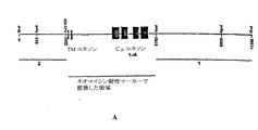
本明細書で使用する「人工染色体」とは、選択可能マーカーの追加、クローニング部位の追加、1つ以上のヌクレオチドの欠失、1つ以上のヌクレオチドの置換等の人工的な改変を有する哺乳動物染色体またはその断片を意味する。「ヒト人工染色体(HAC)」とは、1つ以上のヒト染色体から生成した人工染色体を意味する。人工染色体は、宿主細胞中で宿主細胞の内因性染色体とは独立に維持することができる。この場合、HACは内因性染色体と共に安定して複製し、かつ分離されている。あるいはまた、宿主細胞の内因性染色体に転座していてもよいし、または挿入されてもよい。2つ以上の人工染色体を、宿主細胞に同時または逐次的に導入することができる。例えば、ヒト第14染色体(Ig重鎖遺伝子を含む)、ヒト第2染色体(Igκ鎖遺伝子を含む)およびヒト第22染色体(Igλ鎖遺伝子を含む)から誘導される人工染色体を導入することができる。あるいはまた、異種Ig重鎖遺伝子およびIg軽鎖遺伝子の両方を含む人工染色体(ΔHACまたはΔΔHAC等)を導入してもよい。重鎖遺伝子座と軽鎖遺伝子座は、異なる染色体腕上(セントロメアの異なる側)にあるのが好ましい。さらに別の好適な実施形態では、HACの全体の大きさは、約10、9、8または7メガ塩基以下である。 As used herein, an “artificial chromosome” is a mammal having an artificial modification such as addition of a selectable marker, addition of a cloning site, deletion of one or more nucleotides, substitution of one or more nucleotides, etc. Means a chromosome or a fragment thereof. “Human artificial chromosome (HAC)” means an artificial chromosome generated from one or more human chromosomes. The artificial chromosome can be maintained in the host cell independently of the host cell's endogenous chromosome. In this case, HAC replicates stably with the endogenous chromosome and is isolated. Alternatively, it may be translocated or inserted into the endogenous chromosome of the host cell. Two or more artificial chromosomes can be introduced into the host cell simultaneously or sequentially. For example, artificial chromosomes derived from human chromosome 14 (including Ig heavy chain gene), human chromosome 2 (including Igκ chain gene) and human chromosome 22 (including Igλ chain gene) can be introduced. . Alternatively, an artificial chromosome (such as ΔHAC or ΔΔHAC) containing both the heterologous Ig heavy chain gene and Ig light chain gene may be introduced. The heavy and light chain loci are preferably on different chromosomal arms (different sides of the centromere). In yet another preferred embodiment, the overall size of the HAC is no more than about 10, 9, 8, or 7 megabases.
本発明のさらに別の目的は、導入された核酸(例えば、人工染色体、好ましくはヒトIg重鎖および軽鎖遺伝子を含むヒト人工染色体(HAC))上に担持されたIg遺伝子を含みかつ発現するトランスジェニック有蹄動物(例えば、トランスジェニックウシ)から誘導される、受動免疫法のためのヒトその他のIgの供給源を提供することである。本発明において、これらの核酸(例えば、HAC)としては、ヒト染色体の自然に配置されたセグメント(ヒト染色体断片)、または人工的に工学処理されたヒト染色体断片を含む(すなわち、ヒトゲノムに対して再配列された)人工染色体がある。 Yet another object of the invention includes and expresses an Ig gene carried on an introduced nucleic acid (eg, an artificial chromosome, preferably a human artificial chromosome (HAC) comprising human Ig heavy and light chain genes). To provide a source of human and other Ig for passive immunization derived from transgenic ungulates (eg, transgenic cows). In the present invention, these nucleic acids (eg, HAC) include naturally-occurring segments of human chromosomes (human chromosome fragments) or artificially engineered human chromosome fragments (ie, relative to the human genome). There are artificial chromosomes (rearranged).
本発明のさらに別の目的は、上記トランスジェニック有蹄動物(例えば、トランスジェニックウシ)由来のB細胞を用いてハイブリドーマおよびモノクローナル抗体を製造することである。 Yet another object of the present invention is to produce hybridomas and monoclonal antibodies using B cells derived from the above-described transgenic ungulates (eg, transgenic cows).
本発明のさらに別の目的は、ポリクローナルヒトIgを含む有蹄動物抗血清または乳汁を製造することである。このようなヒトIg(好ましくはヒトIgG)は、ヒトにおける疾患の治療または予防のために、静注用免疫グロブリン(IVIG)として使用できる。ポリクローナルヒトIgは、目的の抗原に対して反応性であることが好ましい。 Yet another object of the present invention is to produce an ungulate antiserum or milk containing polyclonal human Ig. Such human Ig (preferably human IgG) can be used as intravenous immunoglobulin (IVIG) for the treatment or prevention of disease in humans. Polyclonal human Ig is preferably reactive with the antigen of interest.
本発明のさらに別の目的は、1つ以上の内因性遺伝子に1つ以上の突然変異を有するトランスジェニック有蹄動物を作製することである。トランスジェニック有蹄動物は、細胞、細胞由来のクロマチン塊、または細胞由来の核を卵母細胞に挿入することにより作製する。細胞は内因性遺伝子中に、その細胞により自然には発現されない第1の突然変異を有する。卵母細胞または卵母細胞から形成される胚を、該卵母細胞または胚が胎仔に発達する条件下で、宿主有蹄動物の子宮に移入する。胎仔は生存可能な仔に発育するのが好ましい。他の好適な実施形態では、選択可能なマーカーをコードする核酸に作用可能に連結され、かつ突然変異させる内因性遺伝子と実質的な配列同一性を有する1つ以上の核酸に作用可能に連結されたプロモーターを有するカセットを含む核酸を挿入することにより、該カセットが遺伝子の一方の内因性対立遺伝子に組み込まれることによって、第1の突然変異が細胞に導入される。他の好適な実施形態で突然変異が細胞に導入するには、第1の選択可能マーカーをコードする核酸に作用可能に連結され、かつ突然変異される内因性遺伝子と実質的な配列同一性を有する第1の核酸に作用可能に連結された第1のプロモーターを有する第1のカセットを含む核酸を細胞に挿入することにより、遺伝子の第1の内因性対立遺伝子に第1のカセットが組み込まれて第1のトランスジェニック細胞が生成する。第2の選択可能マーカーをコードする核酸に作用可能に連結され、かつ遺伝子と実質的な配列同一性を有する第2の核酸に作用可能に連結された第2のプロモーターを有する第2のカセットを含む核酸を、第1のトランスジェニック細胞に挿入する。この第2の選択可能マーカーは第1の選択可能マーカーとは異なっており、第2のカセットは遺伝子の第2の内因性対立遺伝子に組み込まれて、第2のトランスジェニック細胞が生成する。さらに別の好適な実施形態では、胚、胎仔、または胎仔から生成した仔から細胞を単離し、該細胞の遺伝子に別の突然変異を導入する。次いで、得られた細胞、細胞由来のクロマチン塊、または細胞由来の核を用いて、第2ラウンドの核移植を実施して、2つ以上の突然変異を有するトランスジェニック有蹄動物を作製する。突然変異は、遺伝子の同じまたは異なる対立遺伝子に存在するか、または異なる遺伝子に存在する。好適な実施形態では、突然変異される細胞は、繊維芽細胞(例えば、胎仔繊維芽細胞)である。突然変異される内因性遺伝子は、繊維芽細胞中で活性でない内因性プロモーターに作用可能に連結されていることが好ましい。他の好適な実施形態では、突然変異される内因性遺伝子に作用可能に連結した内因性プロモーターは、GAPDH等の内因性ハウスキーピング遺伝子に作用可能に連結した内因性プロモーターと比べて活性が80、70、60、50、40、30、20、10%未満である。プロモーター活性は、遺伝子にコードされるmRNAまたはタンパク質のレベルを測定するアッセイ等の任意の標準的なアッセイを用いて測定され得る(例えば、Ausubelら, Current Protocols in Molecular Biology, volume 2, p. 11.13.1-11.13.3, John Wiley and Sons, 1995を参照)。このトランスジェニック有蹄動物作製方法は、ドナー細胞(すなわち、核移植のために使用される遺伝子材料の供給源である細胞)中で発現されない遺伝子を突然変異させられるという利点を有する。
Yet another object of the present invention is to create a transgenic ungulate having one or more mutations in one or more endogenous genes. Transgenic ungulates are made by inserting cells, cell-derived chromatin masses, or cell-derived nuclei into oocytes. The cell has a first mutation in the endogenous gene that is not naturally expressed by the cell. An oocyte or embryo formed from the oocyte is transferred to the uterus of the host ungulate under conditions where the oocyte or embryo develops into a fetus. The fetus preferably develops into a viable pup. In other preferred embodiments, the nucleic acid encoding a selectable marker is operably linked to one or more nucleic acids having substantial sequence identity with the endogenous gene to be mutated. The first mutation is introduced into the cell by inserting a nucleic acid containing a cassette having a promoter and incorporating the cassette into one endogenous allele of the gene. In other preferred embodiments, the mutation is introduced into the cell by having substantial sequence identity with the endogenous gene operably linked to and mutated to the nucleic acid encoding the first selectable marker. The first cassette is integrated into the first endogenous allele of the gene by inserting into the cell a nucleic acid comprising a first cassette having a first promoter operably linked to the first nucleic acid having To produce a first transgenic cell. A second cassette having a second promoter operably linked to a nucleic acid encoding a second selectable marker and operably linked to a second nucleic acid having substantial sequence identity to the gene. The containing nucleic acid is inserted into the first transgenic cell. This second selectable marker is different from the first selectable marker, and the second cassette is integrated into the second endogenous allele of the gene to produce a second transgenic cell. In yet another preferred embodiment, cells are isolated from embryos, fetuses, or offspring generated from fetuses, and other mutations are introduced into the genes of the cells. The resulting cell, cell-derived chromatin mass, or cell-derived nucleus is then used to perform a second round of nuclear transfer to produce a transgenic ungulate with two or more mutations. Mutations are present in the same or different alleles of the gene or are present in different genes. In preferred embodiments, the cells to be mutated are fibroblasts (eg, fetal fibroblasts). The endogenous gene to be mutated is preferably operably linked to an endogenous promoter that is not active in fibroblasts. In another preferred embodiment, the endogenous promoter operably linked to the endogenous gene to be mutated has an activity of 80 compared to an endogenous promoter operably linked to an endogenous housekeeping gene such as GAPDH, Less than 70, 60, 50, 40, 30, 20, 10%. Promoter activity can be measured using any standard assay, such as an assay that measures the level of mRNA or protein encoded by a gene (eg, Ausubel et al., Current Protocols in Molecular Biology,
従って、本発明は、再配列を経て1つを上回る異種Ig分子を発現する異種免疫グロブリン(Ig)遺伝子の全体または一部をコードする1つ以上の核酸を有するトランスジェニック有蹄動物を特徴とする。好適な実施形態では、異種Ig遺伝子の全体または一部をコードする核酸は、実質的にヒトのものである。核酸は、ヒト抗体またはポリクローナル抗体等の異種抗体をコードするのが好ましい。様々な実施形態で、Ig鎖または抗体は、血清および/または乳汁中に発現される。他の実施形態では、核酸は、ΔHACまたはΔΔHAC等の染色体断片中に含まれる。さらに別の実施形態では、核酸は、有蹄動物細胞中で宿主染色体とは独立に維持される。 Accordingly, the present invention features a transgenic ungulate having one or more nucleic acids encoding all or part of a heterologous immunoglobulin (Ig) gene that expresses more than one heterologous Ig molecule via rearrangement. To do. In a preferred embodiment, the nucleic acid encoding all or part of a heterologous Ig gene is substantially human. The nucleic acid preferably encodes a heterologous antibody such as a human antibody or a polyclonal antibody. In various embodiments, the Ig chain or antibody is expressed in serum and / or milk. In other embodiments, the nucleic acid is contained in a chromosomal fragment such as ΔHAC or ΔΔHAC. In yet another embodiment, the nucleic acid is maintained independently of the host chromosome in ungulate cells.
さらに別の実施形態では、核酸は、有蹄動物の染色体に組み込まれる。さらに別の実施形態では、核酸は、V遺伝子セグメントをコードする全ての核酸セグメントが、1つ以上のヌクレオチドにより、J遺伝子セグメントをコードする全ての核酸セグメントから分離している、未再配列抗体軽鎖核酸セグメントを含む。さらに別の実施形態では、核酸は、(i)V遺伝子セグメントをコードする全ての核酸セグメントが、1つ以上のヌクレオチドにより、D遺伝子セグメントをコードする全ての核酸セグメントから分離されている、および/または(ii)D遺伝子セグメントをコードする全ての核酸セグメントが、1つ以上のヌクレオチドにより、J遺伝子セグメントをコードする全ての核酸セグメントから分離されている、未再配列抗体重鎖核酸セグメントを含む。さらに別の好適な実施形態では、有蹄動物は、内因性Ig遺伝子、α-(1,3)-ガラクトシルトランスフェラーゼ遺伝子、プリオン遺伝子、および/またはJ鎖遺伝子の一方または両方の対立遺伝子に突然変異を有する。他の好適な実施形態では、有蹄動物は、ヒトJ鎖等の外因性J鎖をコードする核酸を有する。有蹄動物は、ウシ、ヒツジ、ブタまたはヤギであることが好ましい。 In yet another embodiment, the nucleic acid is integrated into the ungulate chromosome. In yet another embodiment, the nucleic acid is an unrearranged antibody light, wherein all nucleic acid segments encoding a V gene segment are separated from all nucleic acid segments encoding a J gene segment by one or more nucleotides. Contains a strand nucleic acid segment. In yet another embodiment, the nucleic acid is (i) all nucleic acid segments encoding V gene segments are separated from all nucleic acid segments encoding D gene segments by one or more nucleotides, and / or Or (ii) an unrearranged antibody heavy chain nucleic acid segment in which all nucleic acid segments encoding the D gene segment are separated from all nucleic acid segments encoding the J gene segment by one or more nucleotides. In yet another preferred embodiment, the ungulate is mutated to one or both alleles of an endogenous Ig gene, an α- (1,3) -galactosyltransferase gene, a prion gene, and / or a J chain gene. Have In other preferred embodiments, the ungulate has a nucleic acid encoding an exogenous J chain, such as a human J chain. The ungulate is preferably a cow, sheep, pig or goat.
別の態様では、本発明は、内因性抗体の発現を低減させる突然変異を有するトランスジェニック有蹄動物を特徴とする。突然変異は、機能性IgM重鎖の発現を低減するか、または機能性IgM重鎖の発現を実質的に喪失させるのが好ましい。他の好適な実施形態では、突然変異は、機能性Ig軽鎖の発現を低減するか、または機能性Ig軽鎖の発現を実質的に喪失させる。さらに別の好適な実施形態では、突然変異は、機能性IgM重鎖および機能性Ig軽鎖の発現を低減するか、または機能性IgM重鎖および機能性Ig軽鎖の発現を実質的に喪失させる。有蹄動物は、α-(1,3)-ガラクトシルトランスフェラーゼ、プリオンタンパク質および/またはJ鎖をコードする内因性核酸の一方または両方の対立遺伝子にも突然変異を有するのが好ましい。他の好適な実施形態では、有蹄動物は、ヒトJ鎖等の外因性J鎖をコードする核酸を有する。別の好適な実施形態では、有蹄動物は、再配列を経て1つを上回る異種Ig分子を発現する異種Ig遺伝子の全体または一部をコードする1つ以上の核酸を有する。異種Ig遺伝子の全体または一部をコードする核酸は、実質的にヒトのものであることが好ましい。他の好適な実施形態では、核酸は、別の属に由来する抗体(例えば、ヒト抗体)等の異種抗体またはポリクローナル抗体をコードする。様々な実施形態では、Ig鎖または抗体は血清中に発現される。他の実施形態では、核酸は、ΔHACまたはΔΔHAC等の染色体断片に含まれている。さらに他の実施形態では、核酸は、有蹄動物細胞中で宿主染色体とは独立に維持される。さらに他の実施形態では、核酸は、有蹄動物の染色体に組み込まれる。有蹄動物は、ウシ、ヒツジ、ブタまたはヤギであることが好ましい。 In another aspect, the invention features a transgenic ungulate having a mutation that reduces expression of an endogenous antibody. The mutation preferably reduces the expression of functional IgM heavy chain or substantially eliminates the expression of functional IgM heavy chain. In other preferred embodiments, the mutation reduces the expression of a functional Ig light chain or substantially eliminates the expression of a functional Ig light chain. In yet another preferred embodiment, the mutation reduces expression of functional IgM heavy chain and functional Ig light chain, or substantially loses expression of functional IgM heavy chain and functional Ig light chain. Let The ungulate preferably has a mutation in one or both alleles of the endogenous nucleic acid encoding α- (1,3) -galactosyltransferase, prion protein and / or J chain. In other preferred embodiments, the ungulate has a nucleic acid encoding an exogenous J chain, such as a human J chain. In another preferred embodiment, the ungulate has one or more nucleic acids encoding all or part of a heterologous Ig gene that expresses more than one heterologous Ig molecule via rearrangement. The nucleic acid encoding all or part of the heterologous Ig gene is preferably substantially human. In other preferred embodiments, the nucleic acid encodes a heterologous antibody or a polyclonal antibody, such as an antibody from another genus (eg, a human antibody). In various embodiments, the Ig chain or antibody is expressed in serum. In other embodiments, the nucleic acid is contained in a chromosomal fragment such as ΔHAC or ΔΔHAC. In yet other embodiments, the nucleic acid is maintained in ungulate cells independently of the host chromosome. In yet other embodiments, the nucleic acid is integrated into the ungulate chromosome. The ungulate is preferably a cow, sheep, pig or goat.
本発明はまた、本発明の有蹄動物のいずれかから得られる細胞、または本発明のいずれかの有蹄動物の作製に有用な細胞を提供する。 The invention also provides cells obtained from any of the ungulates of the invention, or cells useful for the production of any ungulate of the invention.
従って、別の態様では、本発明は、B細胞中で再配列を経て1つ以上の異種Ig分子を発現することが可能な異種Ig遺伝子の全体または一部をコードする1つ以上の核酸を有する有蹄動物体細胞を特徴とする。異種Ig遺伝子の全体または一部をコードする核酸は、異種抗体をコードするのが好ましい。様々な実施形態で、核酸は、ΔHACまたはΔΔHAC等の染色体断片に含まれている。さらに他の実施形態では、核酸は、有蹄動物細胞で宿主染色体とは独立に維持される。さらに他の実施形態では、核酸は、細胞の染色体に組み込まれる。別の実施形態では、核酸は実質的にヒトのものである。異種抗体は、ヒト抗体等の、別の属に由来する抗体であることが好ましい。細胞は、α-(1,3)-ガラクトシルトランスフェラーゼ、プリオンタンパク質および/またはJ鎖をコードする内因性核酸の一方または両方の対立遺伝子に突然変異を有することが好ましい。他の好適な実施形態では、細胞は、ヒトJ鎖等の外因性J鎖をコードする核酸を有する。有蹄動物細胞の例としては、胎仔繊維芽細胞およびB細胞が挙げられる。有蹄動物は、ウシ、ヒツジ、ブタまたはヤギであることが好ましい。 Thus, in another aspect, the present invention provides one or more nucleic acids encoding all or part of a heterologous Ig gene capable of expressing one or more heterologous Ig molecules via rearrangement in B cells. Featuring ungulate somatic cells. The nucleic acid encoding all or part of the heterologous Ig gene preferably encodes a heterologous antibody. In various embodiments, the nucleic acid is contained in a chromosomal fragment such as ΔHAC or ΔΔHAC. In yet other embodiments, the nucleic acid is maintained independently of the host chromosome in ungulate cells. In yet other embodiments, the nucleic acid is integrated into the chromosome of the cell. In another embodiment, the nucleic acid is substantially human. The heterologous antibody is preferably an antibody derived from another genus, such as a human antibody. The cell preferably has a mutation in one or both alleles of the endogenous nucleic acid encoding α- (1,3) -galactosyltransferase, prion protein and / or J chain. In other preferred embodiments, the cell has a nucleic acid encoding an exogenous J chain, such as a human J chain. Examples of ungulate cells include fetal fibroblasts and B cells. The ungulate is preferably a cow, sheep, pig or goat.
別の態様では、本発明は、Ig重鎖および/または軽鎖をコードする核酸中に突然変異を有する有蹄動物体細胞を特徴とする。好適な実施形態では、細胞は、IgM重鎖またはIg軽鎖の一方または両方の対立遺伝子に突然変異を有する。突然変異の例としては、ナンセンス変異および欠失変異が挙げられる。細胞は、α-(1,3)-ガラクトシルトランスフェラーゼ、プリオンタンパク質および/またはJ鎖をコードする内因性核酸の一方または両方の対立遺伝子に突然変異を有することが好ましい。他の好適な実施形態では、細胞は、ヒトJ鎖等の外因性J鎖をコードする核酸を有する。好適な実施形態では、細胞は、B細胞中で再配列を経て1つ以上の異種Ig分子を発現することが可能な異種Ig遺伝子の全体または一部をコードする1つ以上の核酸も有する。異種Ig遺伝子の全体または一部をコードする核酸は、実質的にヒトのものである、および/または別の属に由来する抗体(例えば、ヒト抗体)等の異種抗体をコードすることが好ましい。様々な実施形態において、核酸は染色体断片に含まれ、有蹄動物細胞中で宿主染色体とは独立して維持される。好適な染色体断片としては、ΔHACおよびΔΔHACが挙げられる。さらに他の実施形態では、核酸は、有蹄動物細胞中で宿主染色体とは独立に維持される。さらに他の実施形態では、核酸は、細胞の染色体に組み込まれる。有蹄動物細胞の例としては、胎仔繊維芽細胞およびB細胞が挙げられる。有蹄動物は、ウシ、ヒツジ、ブタまたはヤギであることが好ましい。 In another aspect, the invention features an ungulate somatic cell having a mutation in a nucleic acid encoding an Ig heavy chain and / or light chain. In a preferred embodiment, the cell has a mutation in one or both alleles of IgM heavy chain or Ig light chain. Examples of mutations include nonsense mutations and deletion mutations. The cell preferably has a mutation in one or both alleles of the endogenous nucleic acid encoding α- (1,3) -galactosyltransferase, prion protein and / or J chain. In other preferred embodiments, the cell has a nucleic acid encoding an exogenous J chain, such as a human J chain. In a preferred embodiment, the cell also has one or more nucleic acids that encode all or part of a heterologous Ig gene capable of expressing one or more heterologous Ig molecules via rearrangement in B cells. The nucleic acid encoding all or part of a heterologous Ig gene preferably encodes a heterologous antibody, such as an antibody (eg, a human antibody) that is substantially human and / or derived from another genus. In various embodiments, the nucleic acid is contained in a chromosomal fragment and maintained independently of the host chromosome in ungulate cells. Suitable chromosome fragments include ΔHAC and ΔΔHAC. In yet other embodiments, the nucleic acid is maintained in ungulate cells independently of the host chromosome. In yet other embodiments, the nucleic acid is integrated into the chromosome of the cell. Examples of ungulate cells include fetal fibroblasts and B cells. The ungulate is preferably a cow, sheep, pig or goat.
別の態様では、本発明は、本発明の有蹄動物B細胞と骨髄細胞との融合により形成されるハイブリドーマを特徴とする。ハイブリドーマは、ヒト抗体等の異種抗体を分泌することが好ましい。 In another aspect, the invention features a hybridoma formed by fusion of an ungulate B cell of the invention and a bone marrow cell. The hybridoma preferably secretes a heterologous antibody such as a human antibody.
本発明はまた、本発明の有蹄動物を用いた抗体の製造方法を提供する。このような方法のひとつでは、1つ以上の目的の抗原を、異種抗体遺伝子座をコードする1つ以上の核酸を有する有蹄動物に投与する。遺伝子座中の核酸セグメントは、再配列を経て、抗原に特異的な抗体を生成する。その抗体を有蹄動物から回収する。様々な実施形態で、異種抗体遺伝子座をコードする核酸は、ΔHACまたはΔΔHAC等の染色体断片に含まれている。染色体断片は、有蹄動物細胞中で宿主染色体とは独立に維持されることが好ましい。他の実施形態では、核酸は、有蹄動物の染色体に組み込まれる。核酸は、実質的にヒトのものであることが好ましい。他の好適な実施形態では、抗体の軽鎖および/または抗体の重鎖がヒト核酸にコードされている。抗体は、モノクローナルまたはポリクローナルであり得る。特定の抗原に対するモノクローナルおよびポリクローナル抗体は、様々な用途を有する。例えば、細菌またはウイルス等の病原体微生物の感染に対する予防または治療用組成物中の成分として使用し得る。様々な実施形態において、抗体は、有蹄動物の血清又は乳汁から回収する。好適な実施形態では、有蹄動物は、内因性抗体の発現を低減する突然変異、機能性IgM重鎖の発現を低減する突然変異、または機能性Ig軽鎖の発現を低減する突然変異を有する。有蹄動物は、α-(1,3)-ガラクトシルトランスフェラーゼ、プリオンタンパク質および/またはJ鎖をコードする内因性核酸の一方または両方の対立遺伝子に突然変異を有することが好ましい。他の好適な実施形態では、有蹄動物は、ヒトJ鎖等の外因性J鎖をコードする核酸を有する。有蹄動物は、ウシ、ヒツジ、ブタまたはヤギであることが好ましい。 The present invention also provides a method for producing an antibody using the ungulate of the present invention. In one such method, one or more antigens of interest are administered to an ungulate having one or more nucleic acids encoding a heterologous antibody locus. The nucleic acid segment in the locus undergoes rearrangement to produce an antibody specific for the antigen. The antibody is recovered from the ungulate. In various embodiments, the nucleic acid encoding the heterologous antibody locus is contained in a chromosomal fragment such as ΔHAC or ΔΔHAC. Chromosomal fragments are preferably maintained independently of host chromosomes in ungulate cells. In other embodiments, the nucleic acid is integrated into the ungulate chromosome. The nucleic acid is preferably substantially human. In other preferred embodiments, the light chain of the antibody and / or the heavy chain of the antibody is encoded by a human nucleic acid. The antibody can be monoclonal or polyclonal. Monoclonal and polyclonal antibodies against specific antigens have a variety of uses. For example, it can be used as a component in a composition for prevention or treatment against infection with pathogenic microorganisms such as bacteria or viruses. In various embodiments, the antibody is recovered from ungulate serum or milk. In preferred embodiments, the ungulate has a mutation that reduces expression of an endogenous antibody, a mutation that reduces expression of a functional IgM heavy chain, or a mutation that reduces expression of a functional Ig light chain. . The ungulate preferably has a mutation in one or both alleles of the endogenous nucleic acid encoding α- (1,3) -galactosyltransferase, prion protein and / or J chain. In other preferred embodiments, the ungulate has a nucleic acid encoding an exogenous J chain, such as a human J chain. The ungulate is preferably a cow, sheep, pig or goat.
関連する態様において、本発明は、別の抗体製造方法を特徴とする。本方法では、異種抗体遺伝子座をコードする核酸を有する有蹄動物から異種抗体を回収する。遺伝子座中の核酸セグメントは、再配列を経て異種抗体を生成する。様々な実施形態において、異種抗体遺伝子座をコードする核酸は、ΔHACまたはΔΔHAC等の染色体断片に含まれている。特定の実施形態では、核酸は、有蹄動物細胞中で宿主染色体とは独立に維持される。さらに他の実施形態では、核酸は、有蹄動物の染色体に組み込まれる。核酸は実質的にヒトのものであることが好ましい。特定の実施形態では、抗体の軽鎖および/または抗体の重鎖がヒト核酸にコードされている。抗体は、モノクローナルまたはポリクローナルであり得る。特定の実施形態において、特異的な抗原で有蹄動物を免疫感作すること無く生成させたIgG抗体等のポリクローナル抗体は、ヒト血清から得られるIVIG(静注用免疫グロブリン)の治療代用品として使用される。様々な実施形態において、抗体は有蹄動物の血清または乳汁から回収する。有蹄動物は、内因性抗体の発現を低減する突然変異、機能性IgM重鎖の発現を低減する突然変異、または機能性Ig軽鎖の発現を低減する突然変異を有することが好ましい。有蹄動物は、α-(1,3)-ガラクトシルトランスフェラーゼ、プリオンタンパク質および/またはJ鎖をコードする内因性核酸の一方または両方の対立遺伝子に突然変異を有することが好ましい。他の好適な実施形態では、有蹄動物は、ヒトJ鎖等の外因性J鎖をコードする核酸を有する。有蹄動物は、ウシ、ヒツジ、ブタまたはヤギであることが好ましい。 In a related aspect, the invention features another method for producing an antibody. In this method, a heterologous antibody is recovered from an ungulate having a nucleic acid encoding a heterologous antibody locus. The nucleic acid segment in the locus undergoes rearrangement to generate a heterologous antibody. In various embodiments, the nucleic acid encoding the heterologous antibody locus is contained in a chromosomal fragment such as ΔHAC or ΔΔHAC. In certain embodiments, the nucleic acid is maintained independent of the host chromosome in ungulate cells. In yet other embodiments, the nucleic acid is integrated into the ungulate chromosome. The nucleic acid is preferably substantially human. In certain embodiments, the light chain of the antibody and / or the heavy chain of the antibody is encoded by a human nucleic acid. The antibody can be monoclonal or polyclonal. In certain embodiments, polyclonal antibodies such as IgG antibodies generated without immunizing ungulates with specific antigens are used as therapeutic substitutes for IVIG (intravenous immunoglobulin) obtained from human serum. used. In various embodiments, the antibody is recovered from ungulate serum or milk. Preferably, the ungulate has a mutation that reduces the expression of an endogenous antibody, a mutation that reduces the expression of a functional IgM heavy chain, or a mutation that reduces the expression of a functional Ig light chain. The ungulate preferably has a mutation in one or both alleles of the endogenous nucleic acid encoding α- (1,3) -galactosyltransferase, prion protein and / or J chain. In other preferred embodiments, the ungulate has a nucleic acid encoding an exogenous J chain, such as a human J chain. The ungulate is preferably a cow, sheep, pig or goat.
本発明はまた、トランスジェニック有蹄動物の作製方法も提供する。これらの方法は、所望の突然変異または所望の異種核酸を有するトランスジェニック有蹄動物を作製するのに使用し得る。 The present invention also provides a method for producing a transgenic ungulate. These methods can be used to create transgenic ungulates with a desired mutation or a desired heterologous nucleic acid.
このような態様の1つで、本発明は、細胞、細胞由来のクロマチン塊、または細胞由来の核を卵母細胞に挿入することを含むトランスジェニック有蹄動物の作製方法を特徴とする。この細胞は、内因性抗体重鎖および/または軽鎖核酸中に第1の突然変異を有する。卵母細胞または卵母細胞から形成される胚を、卵母細胞または胚から胎仔が発生する条件下で宿主有蹄動物の子宮に導入する。胎仔は生存可能な仔に発育するのが好ましい。トランスジェニック有蹄動物の作製に使用される細胞は、α-(1,3)-ガラクトシルトランスフェラーゼ、プリオンタンパク質および/またはJ鎖をコードする内因性核酸の一方または両方の対立遺伝子に突然変異を有することが好ましい。他の好適な実施形態では、細胞は、ヒトJ鎖等の外因性J鎖をコードする核酸を有する。他の実施形態では、細胞は、B細胞中で再配列を経て1つ以上の異種Ig分子を発現することができる異種Ig遺伝子の全体または一部をコードする1つ以上の核酸を有する。異種Ig遺伝子の全体または一部をコードする核酸は、異種抗体をコードするのが好ましい。さらに他の実施形態では、核酸は、細胞の染色体に組み込まれる。異種抗体は、ヒト抗体等、別の属に由来する抗体であることが好ましい。特定の実施形態では、核酸は、ΔHACまたはΔΔHAC等の染色体断片に含まれている。他の特定の実施形態では、染色体断片は、有蹄動物細胞中で宿主染色体とは独立に維持される。有蹄動物は、ウシ、ヒツジ、ブタまたはヤギであることが好ましい。 In one such aspect, the present invention features a method for producing a transgenic ungulate comprising inserting a cell, a cell-derived chromatin mass, or a cell-derived nucleus into an oocyte. The cell has a first mutation in the endogenous antibody heavy and / or light chain nucleic acid. An oocyte or embryo formed from the oocyte is introduced into the uterus of the host ungulate under conditions where a fetus develops from the oocyte or embryo. The fetus preferably develops into a viable pup. Cells used to produce transgenic ungulates have mutations in one or both alleles of endogenous nucleic acid encoding α- (1,3) -galactosyltransferase, prion protein and / or J chain It is preferable. In other preferred embodiments, the cell has a nucleic acid encoding an exogenous J chain, such as a human J chain. In other embodiments, the cells have one or more nucleic acids encoding all or part of a heterologous Ig gene capable of expressing one or more heterologous Ig molecules via rearrangement in B cells. The nucleic acid encoding all or part of the heterologous Ig gene preferably encodes a heterologous antibody. In yet other embodiments, the nucleic acid is integrated into the chromosome of the cell. The heterologous antibody is preferably an antibody derived from another genus such as a human antibody. In certain embodiments, the nucleic acid is contained in a chromosomal fragment such as ΔHAC or ΔΔHAC. In other specific embodiments, the chromosomal fragments are maintained independently of the host chromosome in ungulate cells. The ungulate is preferably a cow, sheep, pig or goat.
上記態様の様々な実施形態において、本方法は、胚、胎仔または胎仔より生成した子孫から細胞を単離すること、ならびに該細胞中の内因性抗体重鎖および/または軽鎖核酸に第2の突然変異を導入することも含む。細胞、細胞由来のクロマチン塊または細胞由来の核を卵母細胞に導入し、該卵母細胞または該卵母細胞から形成される胚を、卵母細胞または胚から胎仔が発生する条件下で宿主有蹄動物の子宮に導入する。 In various embodiments of the above aspects, the method includes isolating a cell from an embryo, fetus or offspring generated from the fetus, and seconding to the endogenous antibody heavy and / or light chain nucleic acid in the cell. It also includes introducing mutations. A cell, a cell-derived chromatin mass or a cell-derived nucleus is introduced into an oocyte, and the oocyte or an embryo formed from the oocyte is transformed into a host under conditions where the fetus develops from the oocyte or embryo Introduce into the ungulate uterus.
上記態様の他の実施形態では、選択可能マーカーをコードする核酸に作用可能に連結され、かつ抗体重鎖または軽鎖核酸と実質的な配列同一性を有する1つ以上の核酸に作用可能に連結されたプロモーターを含むカセットを有する核酸を細胞に挿入することを含む方法によって、トランスジェニック有蹄動物の作製のために使用される細胞を調製する。カセットは、抗体重鎖または軽鎖核酸の一方の内因性対立遺伝子に組み込まれる。 In other embodiments of the above aspects, the nucleic acid encoding a selectable marker is operably linked to one or more nucleic acids having substantial sequence identity with the antibody heavy or light chain nucleic acid. Cells used for the production of transgenic ungulates are prepared by a method comprising inserting into the cell a nucleic acid having a cassette containing the generated promoter. The cassette is incorporated into one endogenous allele of the antibody heavy or light chain nucleic acid.
他の実施形態では、第1の選択可能マーカーをコードする核酸に作用可能に連結され
、かつ抗体重鎖または軽鎖核酸と実質的な配列同一性を有する第1の核酸に作用可能に連結された第1のプロモーターを含む第1のカセットを有する核酸を前記細胞に挿入することにより細胞を調製する。この第1のカセットは抗体重鎖または軽鎖核酸の第1の内因性対立遺伝子に組み込まれて、第1のトランスジェニック細胞を生成する。
In other embodiments, operably linked to a nucleic acid encoding a first selectable marker and operably linked to a first nucleic acid having substantial sequence identity with the antibody heavy or light chain nucleic acid. A cell is prepared by inserting a nucleic acid having a first cassette containing a first promoter into the cell. This first cassette is integrated into the first endogenous allele of the antibody heavy or light chain nucleic acid to produce a first transgenic cell.
この第1のトランスジェニック細胞に、第2の選択可能マーカーをコードする核酸に作用可能に連結され、かつ抗体重鎖または軽鎖核酸と実質的な配列同一性を有する第2の核酸に作用可能に連結された第2のプロモーターを含む第2のカセットを有する核酸を挿入する。この第2の選択可能マーカーは第1の選択可能マーカーと異なる。第2のカセットは抗体重鎖または軽鎖核酸の第2の内因性対立遺伝子に組み込まれて、第2のトランスジェニック細胞を生成する。 This first transgenic cell can act on a second nucleic acid operably linked to a nucleic acid encoding a second selectable marker and having substantial sequence identity with the antibody heavy or light chain nucleic acid A nucleic acid having a second cassette comprising a second promoter linked to is inserted. This second selectable marker is different from the first selectable marker. The second cassette is integrated into a second endogenous allele of the antibody heavy or light chain nucleic acid to generate a second transgenic cell.
さらに別の態様では、本発明は、トランスジェニック有蹄動物の別の作製方法を特徴とする。本方法では、1つ以上の異種核酸を有する細胞を卵母細胞に挿入する。異種核酸は、異種Ig遺伝子の全体または一部をコードし、該遺伝子は、B細胞中で再配列を経て1つを上回る異種Ig分子を発現することが可能である。卵母細胞または卵母細胞から形成される胚を、卵母細胞または胚から胎仔が発生する条件下で、宿主有蹄動物の子宮に移入する。胎仔は、生存可能な仔に発育することが好ましい。異種Ig遺伝子の全体または一部をコードする核酸は、異種抗体をコードすることが好ましい。他の好適な実施形態では、抗体はポリクローナル抗体である。さらに他の好適な実施形態では、免疫グロブリン鎖または抗体は、血清および/または乳汁中に発現される。様々な実施形態において、核酸は、ΔHACまたはΔΔHAC等の染色体断片に含まれている。核酸は、有蹄動物細胞中で宿主染色体とは独立して維持することができ、または細胞の染色体中に組み込まれ得る。核酸は、実質的にヒトのものであることが好ましい。他の実施形態では、異種抗体は、ヒト抗体等、別の属に由来する抗体である。有蹄動物は、ウシ、ヒツジ、ブタまたはヤギであることが好ましい。 In yet another aspect, the invention features another method of making a transgenic ungulate. In this method, cells having one or more heterologous nucleic acids are inserted into the oocyte. A heterologous nucleic acid encodes all or part of a heterologous Ig gene, which can express more than one heterologous Ig molecule via rearrangement in B cells. An oocyte or embryo formed from the oocyte is transferred to the uterus of the host ungulate under conditions where a fetus develops from the oocyte or embryo. The fetus is preferably developed into a viable pup. The nucleic acid encoding all or part of the heterologous Ig gene preferably encodes a heterologous antibody. In other preferred embodiments, the antibody is a polyclonal antibody. In still other preferred embodiments, the immunoglobulin chain or antibody is expressed in serum and / or milk. In various embodiments, the nucleic acid is contained in a chromosomal fragment such as ΔHAC or ΔΔHAC. The nucleic acid can be maintained in the ungulate cell independently of the host chromosome or can be integrated into the cell's chromosome. The nucleic acid is preferably substantially human. In other embodiments, the heterologous antibody is an antibody from another genus, such as a human antibody. The ungulate is preferably a cow, sheep, pig or goat.
さらに別の関連する態様では、本発明は、トランスジェニック有蹄動物の別の作製方法を特徴とする。本方法では、細胞、細胞由来のクロマチン塊または細胞由来の核を卵母細胞に導入する。この細胞は、この細胞により自然には発現されない内因性遺伝子中に第1の突然変異を有する。卵母細胞または卵母細胞から形成される胚を、卵母細胞または胚から胎仔が発生する条件下で宿主有蹄動物の子宮に導入する。胎仔は、生存可能な仔に発育することが好ましい。突然変異を有する遺伝子は、抗体、α-(1,3)-ガラクトシルトランスフェラーゼ、プリオンタンパク質またはJ鎖をコードするのが好ましい。別の好適な実施形態では、トランスジェニック有蹄動物の作製に使用する細胞が胎仔繊維芽細胞等の繊維芽細胞である。 In yet another related aspect, the invention features another method of making a transgenic ungulate. In this method, a cell, a cell-derived chromatin mass, or a cell-derived nucleus is introduced into an oocyte. The cell has a first mutation in an endogenous gene that is not naturally expressed by the cell. An oocyte or embryo formed from the oocyte is introduced into the uterus of the host ungulate under conditions where a fetus develops from the oocyte or embryo. The fetus is preferably developed into a viable pup. The gene having a mutation preferably encodes an antibody, α- (1,3) -galactosyltransferase, prion protein or J chain. In another preferred embodiment, the cells used to create the transgenic ungulate are fibroblasts, such as fetal fibroblasts.
上記方法の様々な実施形態において、細胞を調製するには、選択可能マーカーをコードする核酸に作用可能に連結され、かつ前記遺伝子と実質的な配列同一性を有する1つ以上の核酸に作用可能に連結されたプロモーターを含むカセットを有する核酸を細胞に挿入する。これにより、該カセットが遺伝子の一方の内因性対立遺伝子に組み込まれる。他の実施形態では、細胞を生成するのに、第1の選択可能マーカーをコードする核酸に作用可能に連結され、かつ前記遺伝子と実質的な配列同一性を有する第1の核酸に作用可能に連結された第1のプロモーターを含む第1のカセットを有する核酸を細胞に挿入することにより、第1のカセットが遺伝子の第1の内因性対立遺伝子に組み込まれて、第1のトランスジェニック細胞が生成する。この第1のトランスジェニック細胞に、第2の選択可能マーカーをコードする核酸に作用可能に連結され、かつ前記遺伝子と実質的な配列同一性を有する第2の核酸に作用可能に連結された第2のプロモーターを含む第2のカセットを有する核酸を挿入する。この第2の選択可能マーカーは、第1の選択可能マーカーと異なる。第2のカセットが遺伝子の第2の内因性対立遺伝子に組み込まれて、第2のトランスジェニック細胞を生成する。 In various embodiments of the above methods, cells can be prepared to act on one or more nucleic acids operably linked to a nucleic acid encoding a selectable marker and having substantial sequence identity with the gene. A nucleic acid having a cassette containing a promoter linked to is inserted into the cell. This incorporates the cassette into one endogenous allele of the gene. In other embodiments, to produce a cell, operably linked to a nucleic acid encoding a first selectable marker and operative to a first nucleic acid having substantial sequence identity with said gene. By inserting a nucleic acid having a first cassette comprising a linked first promoter into a cell, the first cassette is integrated into the first endogenous allele of the gene, and the first transgenic cell is Generate. A first nucleic acid operably linked to a nucleic acid encoding a second selectable marker and operably linked to a second nucleic acid having substantial sequence identity with said gene. A nucleic acid having a second cassette containing two promoters is inserted. This second selectable marker is different from the first selectable marker. The second cassette is integrated into the second endogenous allele of the gene to generate a second transgenic cell.
上記態様の他の実施形態では、本方法は、第2の突然変異をトランスジェニック有蹄動物に導入することも含む。これらの実施形態では、胚、胎仔、または胎仔より生成した仔から細胞を単離し、該細胞中の内因性遺伝子に第2の突然変異を導入する。細胞、細胞由来のクロマチン塊、または細胞由来の核を卵母細胞に挿入し、この卵母細胞または卵母細胞から形成される胚を、卵母細胞または胚から胎仔が発生する条件下で、宿主有蹄動物の子宮に導入する。 In other embodiments of the above aspects, the method also includes introducing a second mutation into the transgenic ungulate. In these embodiments, cells are isolated from embryos, fetuses, or offspring generated from the fetuses, and a second mutation is introduced into an endogenous gene in the cells. A cell, a cell-derived chromatin mass, or a cell-derived nucleus is inserted into an oocyte, and the oocyte or embryo formed from the oocyte is subjected to conditions under which the fetus develops from the oocyte or embryo, It is introduced into the uterus of the host ungulate.
本発明の有蹄動物は、目的の抗体を含む抗血清または乳汁を製造するのに使用できる。このような態様の1つでは、本発明は、ポリクローナルヒト免疫グロブリンを有する有蹄動物抗血清を特徴とする。抗血清は、ウシ、ヒツジ、ブタまたはヤギに由来することが好ましい。別の好適な実施形態では、Igは、所望の抗体に対するものである。 The ungulate of the present invention can be used to produce antiserum or milk containing the antibody of interest. In one such aspect, the invention features an ungulate antiserum having polyclonal human immunoglobulin. The antiserum is preferably derived from cows, sheep, pigs or goats. In another preferred embodiment, the Ig is against the desired antibody.
さらに別の態様では、本発明は、ポリクローナルヒトIgを有する有蹄動物乳汁を特徴とする。乳汁は、ウシ、ヒツジ、ブタまたはヤギに由来することが好ましい。別の好適な実施形態では、Igは、所望の抗体に対するものである。 In yet another aspect, the invention features ungulate milk with polyclonal human Ig. The milk is preferably derived from cows, sheep, pigs or goats. In another preferred embodiment, the Ig is against the desired antibody.
本発明の様々な態様の好適な実施形態では、重鎖はμ重鎖であり、軽鎖はλまたはκ軽鎖である。他の好適な実施形態では、異種免疫グロブリン鎖または抗体をコードする核酸は、未再配列形態である。異種免疫グロブリン遺伝子を有するトランスジェニック有蹄動物に目的の抗原を投与し、目的の抗原に応答する抗体を生成させることが好ましい。他の好適な実施形態では、1つを上回るクラスの異種抗体が、有蹄動物によって生成される。様々な実施形態では、1つを上回る異なる異種Igまたは抗体が、有蹄動物によって生成される。好ましい核移植法では、本発明の細胞、細胞由来のクロマチン塊、または細胞由来の核を除核または有核卵母細胞に挿入し、その卵母細胞または卵母細胞から形成される胚を、卵母細胞または胚から胎仔が発生する条件下で宿主有蹄動物の子宮に導入する。 In preferred embodiments of the various aspects of the invention, the heavy chain is a μ heavy chain and the light chain is a λ or κ light chain. In other preferred embodiments, the nucleic acid encoding the heterologous immunoglobulin chain or antibody is in an unrearranged form. Preferably, a target antigen is administered to a transgenic ungulate having a heterologous immunoglobulin gene to generate an antibody that responds to the target antigen. In other preferred embodiments, more than one class of heterologous antibody is produced by an ungulate. In various embodiments, more than one different heterologous Ig or antibody is produced by an ungulate. In a preferred nuclear transfer method, a cell of the present invention, a cell-derived chromatin mass, or a cell-derived nucleus is inserted into an enucleated or nucleated oocyte, and the oocyte or embryo formed from the oocyte is It is introduced into the uterus of a host ungulate under conditions where a fetus develops from an oocyte or embryo.
本発明の様々な態様の他の好適な実施形態では、有蹄動物は、内因性α-(1,3)-ガラクトシルトランスフェラーゼ、プリオンおよび/またはJ鎖核酸の一方または両方の対立遺伝子に突然変異を有する。この突然変異は、内因性α-(1,3)-ガラクトシルトランスフェラーゼ酵素、ガラクトシル(α1,3)ガラクトースエピトープ、プリオンタンパク質および/またはJ鎖の発現を低減するかまたは喪失させるものが好ましい。さらに他の好適な実施形態では、有蹄動物は、ヒトJ鎖核酸等の異種J鎖核酸を含む。有蹄動物は、ヒトJ鎖を含むヒトIgAまたはIgM分子を産生することが好ましい。本発明の様々な実施形態では、内因性有蹄動物核酸を突然変異するのに使用する核酸(例えば、選択可能マーカーをコードする核酸に作用可能に連結され、かつ突然変異させる遺伝子と実質的な配列同一性を有する核酸に作用可能に連結されたプロモーターを含むノックアウトカセット)は、ウイルスベクター(アデノウイルスベクターまたはアデノ随伴ウイルスベクター等)に含まれていない。例えば、核酸は、細胞のウイルス感染を伴わないトランスフェクションまたはリポフェクション等の標準的な方法を用いて有蹄動物細胞に挿入するプラスミドまたは人工染色体に含ませ得る。さらに別の実施形態では、内因性有蹄動物核酸を突然変異させるのに使用する核酸(例えば、選択可能マーカーをコードする核酸に作用可能に連結され、かつ突然変異させる遺伝子と実質的な配列同一性を有する核酸に作用可能に連結されたプロモーターを含むノックアウトカセット)は、ウイルスベクター(アデノウイルスベクターまたはアデノ随伴ウイルスベクター等)に含まれる。本実施形態によれば、ウイルスベクターを含むウイルスを用いて有蹄動物を感染させ、ウイルスベクターの一部または全体を有蹄動物細胞に挿入する。 In other preferred embodiments of the various aspects of the invention, the ungulate is mutated in one or both alleles of endogenous α- (1,3) -galactosyltransferase, prion and / or J chain nucleic acid. Have This mutation is preferably one that reduces or eliminates the expression of endogenous α- (1,3) -galactosyltransferase enzyme, galactosyl (α1,3) galactose epitope, prion protein and / or J chain. In yet another preferred embodiment, the ungulate comprises a heterologous J chain nucleic acid, such as a human J chain nucleic acid. The ungulate preferably produces a human IgA or IgM molecule containing human J chain. In various embodiments of the invention, a nucleic acid used to mutate an endogenous ungulate nucleic acid (eg, operably linked to a nucleic acid encoding a selectable marker and substantially mutated with the gene to be mutated). A knockout cassette comprising a promoter operably linked to a nucleic acid having sequence identity is not included in a viral vector (such as an adenovirus vector or an adeno-associated virus vector). For example, the nucleic acid can be included in a plasmid or artificial chromosome that is inserted into an ungulate cell using standard methods such as transfection or lipofection without viral infection of the cell. In yet another embodiment, a nucleic acid used to mutate an endogenous ungulate nucleic acid (eg, operably linked to a nucleic acid encoding a selectable marker and substantially identical to the gene to be mutated) A knockout cassette including a promoter operably linked to a nucleic acid having sex is included in a viral vector (such as an adenovirus vector or an adeno-associated virus vector). According to this embodiment, an ungulate is infected with a virus containing a viral vector, and a part or all of the viral vector is inserted into an ungulate cell.
有蹄動物の例として、ウマ目(奇蹄類Perissodactyla)およびウシ目(偶蹄類Artiodactyla)のメンバー(ウシ(Bos)属のあらゆるメンバー等)が挙げられる。他の好適な有蹄動物としては、ヒツジ、オオツノヒツジ、ヤギ、野牛、カモシカ、雄牛、ウマ、ロバ、ラバ、シカ、ヘラジカ、水牛、ラクダ、ラマ、アルパカ、ブタおよびゾウが挙げられる。トランスジェニック有蹄動物は、任意の他の哺乳動物由来の抗体等、別の属に由来する免疫グロブリン鎖または抗体を発現することが好ましい。 Examples of ungulates include the members of the order Horses (Perissodactyla) and Bovines (Artiodactyla) (any member of the genus Bos). Other suitable ungulates include sheep, bighorn sheep, goats, wild cattle, antelopes, bulls, horses, donkeys, mules, deer, elk, buffalo, camels, llamas, alpaca, pigs and elephants. The transgenic ungulate preferably expresses an immunoglobulin chain or antibody from another genus, such as an antibody from any other mammal.
本明細書で使用する場合、「配列前または未再配列形態の核酸」とは、V(D)J組換えを経ていない核酸を意味する。好適な実施形態では、抗体軽鎖のV遺伝子セグメントをコードする全ての核酸セグメントが、1つ以上のヌクレオチドにより、J遺伝子セグメントをコードする全ての核酸セグメントから分離されている。抗体重鎖のV遺伝子セグメントをコードする全ての核酸セグメントは、1つ以上のヌクレオチドにより、D遺伝子セグメントをコードする全ての核酸セグメントから分離されていること、および/または抗体重鎖のD遺伝子セグメントをコードする全ての核酸セグメントは、1つ以上のヌクレオチドにより、J遺伝子セグメントをコードする全ての核酸セグメントから分離されていることが好ましい。未再配列形態の核酸は、実質的にヒトのものであることが好ましい。他の好適な実施形態では、核酸は、ヒト由来の天然型核酸の対応する領域と、少なくとも70、80、90、95または99%同一である。 As used herein, “pre-sequence or unrearranged form of nucleic acid” means nucleic acid that has not undergone V (D) J recombination. In a preferred embodiment, all nucleic acid segments encoding the V gene segment of the antibody light chain are separated from all nucleic acid segments encoding the J gene segment by one or more nucleotides. All nucleic acid segments encoding the V gene segment of the antibody heavy chain are separated from all nucleic acid segments encoding the D gene segment by one or more nucleotides, and / or the D gene segment of the antibody heavy chain Preferably, all nucleic acid segments that encode are separated from all nucleic acid segments that encode the J gene segment by one or more nucleotides. The unrearranged form of the nucleic acid is preferably substantially human. In other preferred embodiments, the nucleic acid is at least 70, 80, 90, 95 or 99% identical to the corresponding region of a naturally occurring nucleic acid from humans.
上記したように、本発明は、場合によって内因性Ig発現がノックアウトされていてもよく、別の種(好ましくはヒト)の機能性抗体の産生のために必要な遺伝子を含む核酸(好ましくは人工染色体)が安定して導入されているトランスジェニック有蹄動物(好ましくはトランスジェニックウシ)の作製に関する。これにより、内因性抗体を産生しないが、代わりに別の種の抗体を産生するトランスジェニック動物を得ることができる。任意の非内因性抗体(ヒト、非ヒト霊長類、イヌ、ネコ、マウス、ラットまたはモルモット抗体が挙げられるがこれらに限定されない)を製造することができる。ヤギにおけるヒトモノクローナル抗体の生成が既に報告されているが、これは、ウシ、血清(serum)、または内因性抗体を発現しないいずれかの有蹄動物においては達成されていない。さらに、生殖細胞系(未再配列)重鎖または軽鎖遺伝子の挿入、これらの遺伝子の再配列、および多様化した抗体の発現は、トランスジェニック有蹄動物では行われていない。このような有蹄動物が生存できるか否かは予想できない。なぜなら、ヒトIgが機能性に発現されるか、または十分な量で発現されて十分な免疫応答をもたらすか否かが不確かであるからである。また、ヒト染色体が、トランスジェニック有蹄動物において安定して維持されるか否かも不確かである。さらにまた、有蹄動物(例えば、ウシ)B細胞が、ヒトまたは他の非内因性Igを発現または正しく再配列できるか否かは不確かである。 As mentioned above, the present invention may be a nucleic acid (preferably artificial) that may optionally have endogenous Ig expression knocked out and that contains a gene necessary for the production of a functional antibody of another species (preferably human). The present invention relates to the production of transgenic ungulates (preferably transgenic cows) into which chromosomes are stably introduced. This can yield a transgenic animal that does not produce endogenous antibodies, but instead produces another species of antibody. Any non-endogenous antibody can be produced, including but not limited to human, non-human primate, dog, cat, mouse, rat or guinea pig antibody. Production of human monoclonal antibodies in goats has already been reported, but this has not been achieved in bovine, serum, or any ungulate that does not express endogenous antibodies. Furthermore, insertion of germline (unrearranged) heavy or light chain genes, rearrangement of these genes, and expression of diversified antibodies has not been performed in transgenic ungulates. Whether or not such ungulates can survive is unpredictable. This is because it is uncertain whether human Ig is functionally expressed or is expressed in sufficient amounts to produce a sufficient immune response. It is also uncertain whether human chromosomes are stably maintained in transgenic ungulates. Furthermore, it is uncertain whether ungulate (eg, bovine) B cells can express or correctly rearrange human or other non-endogenous Ig.
本手法の好適な実施形態では、異種免疫グロブリン産生は、本質的に、核移植、相同組換え技術、および異種Ig遺伝子座全体を担持する人工染色体の導入を組み合わせて使用することにより達成される。より具体的には、本方法では、IgM重鎖遺伝子の一方または両方の対立遺伝子、および場合によりIg軽鎖遺伝子の一方または両方の対立遺伝子のターゲッティング破壊をするのが好ましい。ただし、異種抗体産生は野生型動物(すなわち、Igノックアウトをもたない動物)によっても実施することができる。遺伝子ノックアウトは、相同組換えを連続して行い、その後の別の交配手順によって起こすことができる。好適な実施形態では、まず、適切な相同組換えベクターを用いて、組織培養で、オスまたはメス有蹄動物(例えば、ウシ)胎仔繊維芽細胞のIgM重鎖遺伝子の一方の対立遺伝子のターゲッティング破壊を起こす。胎仔繊維芽細胞を使用することは、一部の他の体細胞よりも好ましい。なぜなら、これらの細胞は、組織培養で増殖および遺伝子操作し易いからである。しかし、胎仔繊維芽細胞の使用は、本発明において必須ではなく、実際に他の細胞系に置き換えても同等の結果が得られうる。 In a preferred embodiment of this approach, heterologous immunoglobulin production is essentially achieved by using a combination of nuclear transfer, homologous recombination techniques, and introduction of an artificial chromosome carrying the entire heterologous Ig locus. . More specifically, the method preferably involves targeted disruption of one or both alleles of the IgM heavy chain gene and optionally one or both alleles of the Ig light chain gene. However, heterologous antibody production can also be performed by wild-type animals (ie, animals that do not have Ig knockout). Gene knockout can occur by successive homologous recombination followed by another mating procedure. In a preferred embodiment, targeted disruption of one allele of the IgM heavy chain gene of a male or female ungulate (eg, bovine) fetal fibroblast is first performed in tissue culture using an appropriate homologous recombination vector. Wake up. The use of fetal fibroblasts is preferred over some other somatic cells. This is because these cells are easy to grow and genetically manipulate in tissue culture. However, the use of fetal fibroblasts is not essential in the present invention, and equivalent results can be obtained even if actually replaced with other cell lines.
当然のことながら、本方法では、ターゲッティングIgM重鎖対立遺伝子と相同な領域を有するDNA構築物を構築して、この構築物が有蹄動物ゲノム中のIgM重鎖対立遺伝子へ組み込まれた際にその発現を破壊するようにする。このようなIgM対立遺伝子のターゲッティング破壊を行うためのベクターの一例は、以下の実施例に記載する。これに関して、ターゲッティング部位において相同組換えをもたらすベクターの構築方法は、当業者に周知である。さらに、この場合、適切なベクターの構築は、特に、他の有蹄動物(以下参照)由来の免疫グロブリン遺伝子の配列と同様に、ウシIgM重鎖およびIgλ軽鎖遺伝子の配列が既知であるということを考慮すると、当該分野の技術レベル内にある。相同組換えを容易にするために、IgM遺伝子の相同組換えおよび不活性化を生じるのに使用するベクターはそれぞれ、有蹄動物IgM重鎖およびIg軽鎖遺伝子と実質的な配列同一性を示すDNAの部分を含む。これらの配列は、相同組換えおよびターゲッティング欠失または不活性化を容易にするために、ターゲッティング遺伝子座と、好ましくは少なくとも98%の配列同一性、より好ましくは少なくとも99%の配列同一性を有し、さらに好ましくは同遺伝子型である。 Of course, the present method constructs a DNA construct that has a region homologous to the targeting IgM heavy chain allele, and its expression when integrated into the IgM heavy chain allele in the ungulate genome. To destroy. An example of a vector for performing targeted disruption of such IgM alleles is described in the examples below. In this regard, methods for constructing vectors that result in homologous recombination at the targeting site are well known to those skilled in the art. Furthermore, in this case, the construction of a suitable vector is known in particular for the sequences of bovine IgM heavy chain and Igλ light chain genes, as well as the sequences of immunoglobulin genes from other ungulates (see below). Is within the technical level of the field. To facilitate homologous recombination, the vectors used to generate homologous recombination and inactivation of the IgM gene show substantial sequence identity with the ungulate IgM heavy and light chain genes, respectively. Contains a portion of DNA. These sequences preferably have at least 98% sequence identity, more preferably at least 99% sequence identity with the targeting locus to facilitate homologous recombination and targeted deletion or inactivation. More preferably, it is the same genotype.
通常、構築物は、IgM重鎖遺伝子および/またはIg軽鎖遺伝子が有効に破壊された所望の相同組換体(例えば、繊維芽細胞)の選択に役立つマーカー遺伝子を含むことが好ましい。マーカー遺伝子の例としては、数ある中で、抗生物質耐性マーカー、薬剤耐性マーカーおよびグリーン蛍光タンパク質が挙げられる。好適な構築物を図2Aに示し、この構築物を作製するのに使用する出発材料を図3Aおよび図3Bに示す。プロモーターに作用可能に連結された正の選択マーカー(例えば、抗生物質耐性遺伝子)を挟む、内因性免疫グロブリン遺伝子と相同な2つの領域を含む他の構築物は、標準的な分子生物学的技術を用いて作製することができ、本発明の方法において使用できる。 In general, the construct preferably includes a marker gene that aids in the selection of the desired homologous recombinant (eg, fibroblast) in which the IgM heavy chain gene and / or Ig light chain gene has been effectively disrupted. Examples of marker genes include antibiotic resistance markers, drug resistance markers and green fluorescent protein, among others. A suitable construct is shown in FIG. 2A, and the starting materials used to make this construct are shown in FIGS. 3A and 3B. Other constructs containing two regions homologous to an endogenous immunoglobulin gene, flanking a positive selectable marker (eg, an antibiotic resistance gene) operably linked to a promoter can be obtained using standard molecular biology techniques. And can be used in the method of the present invention.
図2Aおよび図3Cに示すμノックアウト構築物は、「C-μエキソン1〜4」と称されるウシ免疫グロブリン重鎖定常領域をコードするエキソン、および「TMエキソン」と称される膜貫通ドメインをコードする2つのエキソンを除去するように設計された。 The μ knockout construct shown in FIGS. 2A and 3C comprises an exon encoding a bovine immunoglobulin heavy chain constant region termed “C-μ exons 1-4”, and a transmembrane domain termed “TM exon”. Designed to remove two encoding exons.
このベクターを構築するために、「1」と称した領域、すなわち、ゲノムμ重鎖ウシ配列由来のXbal-Xhol断片を、酵素XbaIおよびXhoIで予め切断した市販のDNAベクターpBluescript(Stratagene, LaJolla, California)にサブクローニングした。この断片をクローン化した後では、約3.5 KbのNotI断片を挿入するのに用いるNotI制限酵素認識配列がXbal部位に隣接していた。この断片は、以下にさらに記載するネオマイシン耐性マーカーを含む。所望であれば、他のμノックアウト構築物を、別の有蹄動物品種、種または属由来のゲノムμ重鎖配列(例えば、Genbank受託番号U63637として寄託された、スイス牛/ホルスタイン交雑に由来するμ重鎖配列)を用いて構築できる。 To construct this vector, the region designated “1”, ie the Xbal-Xhol fragment derived from the genomic μ heavy chain bovine sequence, was cleaved with the enzymes XbaI and XhoI, and the commercially available DNA vector pBluescript (Stratagene, LaJolla, California). After cloning this fragment, the NotI restriction enzyme recognition sequence used to insert the approximately 3.5 Kb NotI fragment was adjacent to the Xbal site. This fragment contains the neomycin resistance marker described further below. If desired, other μ knockout constructs can be obtained from other ungulate breeds, species or genus genomic μ heavy chain sequences (eg, μ derived from Swiss cattle / Holstein crosses deposited under Genbank accession number U63637). Heavy chain sequence).
断片「1」およびネオマイシン耐性マーカーを連結してpBluescriptに入れた後、ネオマイシン耐性マーカーの隣にSacl部位が残存していた。新しい構築物をSaclで直線化し、DNAポリメラーゼを用いて、Sacl消化により生じた付着末端を充填することで平滑末端に変換した。 After ligating fragment “1” and the neomycin resistance marker into pBluescript, a Sacl site remained next to the neomycin resistance marker. The new construct was linearized with Sacl and converted to blunt ends using DNA polymerase by filling in the sticky ends generated by Sacl digestion.
「2」と称した断片を、XhoI-BstI1071断片として単離し、DNAポリメラーゼを用いて、XholおよびBstI1071酵素により生じた付着末端を充填することで平滑末端断片に変換した。 The fragment designated “2” was isolated as a XhoI-BstI1071 fragment and converted to a blunt end fragment by filling in the sticky ends generated by Xhol and BstI1071 enzymes using DNA polymerase.
終了後、最終的な構築物は、領域2、ネオマイシン耐性マーカーおよび領域1をそれぞれ含んでいた。
After completion, the final construct contained
ウシ繊維芽細胞のトランスフェクションのために、構築物を、制限酵素Kpnlで消化し(図中2つのKpnl部位が示されている)、そのDNA断片を相同組換えに使用した。 For transfection of bovine fibroblasts, the construct was digested with the restriction enzyme Kpnl (two Kpnl sites are shown in the figure) and the DNA fragment was used for homologous recombination.
ネオマイシン耐性構築物は、以下のように組み立てた。「pSTneoB」と称される構築物(Katohら, Cell Struct. Funct. 12:575, 1987;Japanese Collection of Research Biologicals (JCRB)寄託番号:VE039)は、コード領域の上流にあるSV40プロモーターおよびTKエンハンサーの制御下でネオマイシン耐性遺伝子を含むように設計された。コード領域の下流には、SV40ターミネーター配列が存在する。neoカセットを、XhoI断片として、「pSTneoB」から切り出した。標準的な分子生物学的技術を用いて断片の端部を平滑末端に変換した後、平滑末端断片を、ベクターpBS246(Gibco/Life Technologies)中のEcoRV部位にクローン化した。この部位は、loxP部位に挟まれている。「pLoxP-STNeoR」と称した新しい構築物を、μノックアウトDNA構築物を作製するために使用した。この構築物の所望の断片は、元々pBS246クローニングベクター中に存在していたloxP部位およびNotI部位に挟まれている。loxP-neo-loxPを含む所望のNotI断片を、免疫グロブリンμ定常領域エキソンを置き換えるために使用した。ネオマイシン耐性遺伝子に作用可能に連結したSV40プロモーターはネオマイシン耐性遺伝子の転写を活性化し、所望のNotI断片がμ定常領域エキソンと入れ替わった細胞を、それらの獲得した抗生物質耐性に基づいて選択することができる。 The neomycin resistance construct was assembled as follows. A construct called “pSTneoB” (Katoh et al., Cell Struct. Funct. 12: 575, 1987; Japanese Collection of Research Biologicals (JCRB) deposit number: VE039) is an SV40 promoter and TK enhancer upstream of the coding region. Designed to contain a neomycin resistance gene under control. An SV40 terminator sequence is present downstream of the coding region. The neo cassette was excised from “pSTneoB” as an XhoI fragment. After converting the ends of the fragments to blunt ends using standard molecular biology techniques, the blunt end fragments were cloned into the EcoRV site in the vector pBS246 (Gibco / Life Technologies). This site is sandwiched between loxP sites. A new construct called “pLoxP-STNeoR” was used to create a μ knockout DNA construct. The desired fragment of this construct is flanked by loxP and NotI sites that were originally present in the pBS246 cloning vector. The desired NotI fragment containing loxP-neo-loxP was used to replace the immunoglobulin μ constant region exon. The SV40 promoter operably linked to the neomycin resistance gene activates the transcription of the neomycin resistance gene, and cells that have the desired NotI fragment replaced with a μ constant region exon can be selected based on their acquired antibiotic resistance. it can.
IgM重鎖対立遺伝子が有効に破壊された細胞系を得た後、これを核移植ドナーとして使用して、クローン化有蹄動物胎仔(例えば、クローン化ウシ胎仔)を作製し、最終的に、一方のIgM重鎖対立遺伝子が破壊された胎仔または動物を作製する。その後、それから誘導された体細胞(例えば、繊維芽細胞)を用いて第2ラウンドの遺伝子ターゲッティング破壊を実施することができ、異なる選択可能マーカーを含む同様のベクターを用いて、第2のIgM重鎖対立遺伝子が不活性化された細胞を作製する。 After obtaining a cell line in which the IgM heavy chain allele has been effectively disrupted, it can be used as a nuclear transfer donor to produce a cloned ungulate fetus (eg, a cloned bovine fetus) and finally A fetus or animal in which one IgM heavy chain allele is disrupted is generated. A second round of gene targeting disruption can then be performed using somatic cells (eg, fibroblasts) derived therefrom, and a second vector of IgM heavy can be used using a similar vector containing a different selectable marker. Cells in which the strand allele is inactivated are generated.
第1のターゲッティング遺伝子破壊と同時に、同様にオスまたはメス供給源のものであり得る第2の有蹄動物(例えば、ウシ)体細胞系も遺伝子改変することが好ましい。操作される第1の細胞系がオスの場合、メス細胞系を改変することが好ましい。逆に、操作される第1の細胞系がメスの場合、オス細胞系を選択することが好ましい。この場合も、操作される細胞は有蹄動物(例えば、ウシ)胎仔繊維芽細胞を含むことが好ましい。 Simultaneously with the first targeted gene disruption, a second ungulate (eg, bovine) somatic cell line, which may be of a male or female source as well, is preferably genetically modified. If the first cell line being manipulated is male, it is preferable to modify the female cell line. Conversely, if the first cell line being manipulated is a female, it is preferable to select a male cell line. Again, the engineered cells preferably include ungulate (eg, bovine) fetal fibroblasts.
好適な実施形態では、メス胎仔繊維芽細胞を遺伝子改変して、Igλ軽鎖遺伝子の一方の対立遺伝子のターゲッティング破壊を導入する。本方法は、有蹄動物(例えば、ウシ)Igλ軽鎖に相同な領域、および選択可能マーカーを有するベクターを用いて同様に実施される。この場合のDNA構築物は、組込みおよび内因性Ig軽鎖との相同組換えの際に、ターゲッティングIgλ軽鎖遺伝子の破壊(不活性化)が生じるように設計される。 In a preferred embodiment, female fetal fibroblasts are genetically modified to introduce a targeted disruption of one allele of the Igλ light chain gene. The method is similarly performed using a vector having a region homologous to an ungulate (eg, bovine) Igλ light chain and a selectable marker. The DNA construct in this case is designed such that disruption (inactivation) of the targeting Igλ light chain gene occurs upon integration and homologous recombination with the endogenous Ig light chain.
所望のターゲッティング破壊を有するメス繊維芽細胞系を選択した後、核移植のためのドナー細胞として同様に利用するか、またはこのような細胞系から得たDNAを核移植のドナーとして使用する。 After selecting a female fibroblast cell line with the desired targeting disruption, it is similarly utilized as a donor cell for nuclear transfer, or DNA obtained from such a cell line is used as a donor for nuclear transfer.
あるいはまた、この細胞を、第2ラウンドの相同組換えに供して、第1の対立遺伝子を破壊するのに使用したものと同様であるが、異なる選択可能マーカーを含むDNA構築物を用いて、第2のIgλ軽鎖を不活性化してもよい。 Alternatively, the cells can be subjected to a second round of homologous recombination similar to that used to destroy the first allele, but with a DNA construct containing a different selectable marker, Two Igλ light chains may be inactivated.
発明の背景で論じたように、核移植を行う方法、特にクローン化ウシおよびクローン化トランスジェニックウシの作製法は既に報告されており、Sticeらに発行され、University of Massachusettsに譲渡された米国特許第5,995,577号に記載されている。あるいはまた、WO 95/16670;WO 96/07732;WO 97/0669;またはWO 97/0668に開示される核移植技術(まとめてRoslin法と呼ぶ)を使用してもよい。Roslin法は、増殖期のドナー細胞ではなく静止期の細胞を使用する点で、University of Massachusetts法とは異なる。上記特許は全て、参照により全体的に本明細書に援用される。これらの核移植手順により、クローン化トランスジェニックウシの仔(例えば、Ig軽鎖遺伝子および/またはIgM遺伝子の少なくとも一方の対立遺伝子のターゲッティング破壊を含む仔)を作製するのに使用できるトランスジェニッククローン化胎仔が作製される。このような細胞系を作製したら、これらを利用して、オスおよびメス重鎖および軽鎖ヘミ接合ノックアウト(MおよびF Hemi H/L)胎仔および仔を作製することができる。さらに、これらの技術は、トランスジェニックウシを作製するための使用のみに限定されない。上記技術は、他の有蹄動物の核移植のためにも使用できる。 As discussed in the Background of the Invention, methods for performing nuclear transfer, particularly methods for producing cloned and cloned transgenic cattle, have already been reported and are issued to Stice et al. And assigned to the University of Massachusetts. No. 5,995,577. Alternatively, the nuclear transfer technology (collectively referred to as Roslin method) disclosed in WO 95/16670; WO 96/07732; WO 97/0669; or WO 97/0668 may be used. The Roslin method differs from the University of Massachusetts method in that it uses stationary phase cells rather than proliferating phase donor cells. All of the above patents are hereby incorporated by reference in their entirety. These nuclear transfer procedures allow for the generation of cloned transgenic bovine pups (eg, pups containing a targeted disruption of at least one allele of the Ig light chain gene and / or IgM gene). A fetus is created. Once such cell lines have been generated, they can be utilized to generate male and female heavy and light chain hemizygous knockout (M and F Hemi H / L) fetuses and pups. Furthermore, these techniques are not limited to use only for producing transgenic cows. The above technique can also be used for nuclear transfer of other ungulates.
核移植の後、有蹄動物を交配させるか、または先に記載した相同ターゲッティングベクターを用いた二次遺伝子ターゲッティングにより、所望の動物を作製することができる。 Following nuclear transfer, ungulates can be bred or the desired animals can be produced by secondary gene targeting using the previously described homologous targeting vectors.
先に記載したように、本発明のさらなる目的は、オスおよびメス重鎖および軽鎖ヘミ接合ノックアウトを作製することを含んでおり、このようなヘミ接合ノックアウトは既に記載した細胞系を用いて作製される。これは上記方法に従って作製した仔を交配させることによって行うことができるが、この場合は、IgM重鎖遺伝子の対立遺伝子が破壊された仔を、Ig軽鎖の対立遺伝子が破壊された別の仔と交配させる。あるいはまた、上記手順により作製した仔から得られる細胞を操作することによる二次遺伝子ターゲッティングによって行ってもよい。これは、IgM重鎖遺伝子の対立遺伝子またはIg軽鎖の対立遺伝子の相同組換えターゲッティング破壊により行う。オスおよびメス重鎖および軽鎖ヘミ接合ノックアウト(MおよびF Hemi H/L)を含む細胞系が生成したら、これを使用して、そのようなノックアウトを含む胎仔または仔ウシを作製する。上述したように、これは、交配または二次遺伝子ターゲッティングにより行われる。 As previously described, a further object of the present invention includes making male and female heavy and light chain hemizygous knockouts, and such hemizygous knockouts are made using previously described cell lines. Is done. This can be done by mating offspring produced according to the method described above, in which case the offspring in which the IgM heavy chain allele was disrupted to another offspring in which the Ig light chain allele was disrupted. Cross with. Alternatively, secondary gene targeting may be performed by manipulating cells obtained from pups produced by the above procedure. This is done by homologous recombination targeting disruption of the allele of the IgM heavy chain gene or the allele of the Ig light chain. Once a cell line containing male and female heavy and light chain hemizygous knockouts (M and F Hemi H / L) is generated, it is used to produce a fetus or calf containing such knockouts. As mentioned above, this is done by mating or secondary gene targeting.
オスおよびメス重鎖および軽鎖ヘミ接合ノックアウトが得られたら、これらの動物に由来する細胞を利用して、ホモ接合ノックアウト(Homo H/L)胎仔を作製することができる。この場合も、遺伝子ターゲッティングを繰り返すかまたは交配によって行う。本質的に、交配を用いる場合、オス重鎖および軽鎖ヘミ接合ノックアウトを、メス重鎖および軽鎖ヘミ接合ノックアウトと交配させ、ホモ接合ノックアウトを含む仔を選択する。あるいはまた、上述したヘミ接合ノックアウト由来の細胞を組織培養で操作して、IgMまたはIg軽鎖(λ)遺伝子の他方の対立遺伝子をノックアウトしてもよい。二次遺伝子ターゲッティングはより迅速に結果が得られるので、特にウシ等の有蹄動物の妊娠期間が比較的長いことを考えると、交配より好ましいかもしれない。 Once male and female heavy and light chain hemizygous knockouts are obtained, cells derived from these animals can be used to generate homozygous knockout (Homo H / L) fetuses. Again, gene targeting is performed by repeated or mating. In essence, when mating is used, male heavy and light chain hemizygous knockouts are crossed with female heavy and light chain hemizygous knockouts and pups containing homozygous knockouts are selected. Alternatively, cells from the hemizygous knockout described above may be manipulated in tissue culture to knock out the other allele of the IgM or Ig light chain (λ) gene. Secondary gene targeting may be preferable to breeding, especially considering the relatively long gestation period of ungulates such as cows, as results are obtained more quickly.
ホモ接合ノックアウト(Homo H/L)が得られたら、これらを利用して、ヒト等の特定の種の抗体を産生するための遺伝子(好ましくは遺伝子座全体)を含む所望の核酸を導入する。WO 97/07671(EP 0843961)およびWO 00/10383(EP 1106061)に開示されているようなヒト人工染色体を使用することが好ましい。これらのヒト人工染色体は、対応日本国特許第30300092号にも記載されている。これらの出願は両方とも、参照により全体的に本明細書に援用される。また、ヒト免疫グロブリン遺伝子を含有し発現する人工ヒト染色体の構築は、Shenら, Hum. Mol. Genet. 6(8):1375-1382 (1997);Kuroiwaら, Nature Biotechnol. 18(10):1086-1090 (2000);およびLoupertら, Chromosome 107(4):255-259 (1998)に開示されている(全て参照により全体的に本明細書に援用される)。ヒト人工染色体はまた、異種抗体遺伝子を野生型動物細胞に導入するためにも利用され得る。これは、上記方法を用いて達成できる。人工染色体を動物細胞(特に、胎仔繊維芽細胞)に導入することは、本明細書に記載するように、ミクロセル融合により行うことができる。 When a homozygous knockout (Homo H / L) is obtained, a desired nucleic acid including a gene (preferably the entire locus) for producing an antibody of a specific species such as a human is introduced using these. It is preferred to use human artificial chromosomes as disclosed in WO 97/07671 (EP 0843961) and WO 00/10383 (EP 1106061). These human artificial chromosomes are also described in corresponding Japanese Patent No. 30300092. Both of these applications are hereby incorporated by reference in their entirety. Also, the construction of artificial human chromosomes containing and expressing human immunoglobulin genes is described by Shen et al., Hum. Mol. Genet. 6 (8): 1375-1382 (1997); Kuroiwa et al., Nature Biotechnol. 18 (10): 1086-1090 (2000); and Loupert et al., Chromosome 107 (4): 255-259 (1998) (all incorporated herein by reference in their entirety). Human artificial chromosomes can also be utilized to introduce heterologous antibody genes into wild type animal cells. This can be achieved using the method described above. Introducing artificial chromosomes into animal cells (particularly fetal fibroblasts) can be performed by microcell fusion, as described herein.
ヒト人工染色体を使用する代わりに、免疫グロブリン遺伝子をコードする核酸を、YACベクター、BACベクターまたはコスミドベクターを用いて染色体に組み込んでもよい。異種Ig遺伝子を含むベクター(WO98/24893、WO96/33735、WO97/13852、WO98/24884)は、エレクトロポレーション、リポフェクション、YACベクターを含む酵母スフェロプラストとの融合等の公知方法を用いて、胎仔繊維芽細胞に導入することができる。さらに、異種Ig遺伝子を含むベクターを、胎仔繊維芽細胞の内因性Ig遺伝子座にターゲッティングさせて、異種Ig遺伝子の導入および内因性Ig遺伝子の破壊を同時に行うことができる。 Instead of using a human artificial chromosome, a nucleic acid encoding an immunoglobulin gene may be integrated into the chromosome using a YAC vector, a BAC vector or a cosmid vector. A vector containing a heterologous Ig gene (WO98 / 24893, WO96 / 33735, WO97 / 13852, WO98 / 24884) can be obtained using known methods such as electroporation, lipofection, and fusion with a yeast spheroplast containing a YAC vector. It can be introduced into fetal fibroblasts. Furthermore, a vector containing a heterologous Ig gene can be targeted to the endogenous Ig locus of fetal fibroblasts, so that the introduction of the heterologous Ig gene and the destruction of the endogenous Ig gene can be performed simultaneously.
異種免疫グロブリン遺伝子をコードする核酸の組込みは、Lonbergらによる特許(前掲)に記載されているように行うこともできる。異種免疫グロブリン遺伝子を宿主有蹄動物の染色体に挿入するために使用する「ノックイン」構築物では、1つ以上の免疫グロブリン遺伝子および抗生物質耐性遺伝子が、構築物でトランスフェクトされる細胞型において活性なプロモーターと作用可能に連結され得る。例えば、構成的に活性な、誘導可能な、または組織特異的なプロモーターを使用して、組み込まれた抗生物質耐性遺伝子の転写を活性化して、トランスフェクトされた細胞をその獲得した抗生物質耐性に基づいて選択することができる。あるいはまた、Ig遺伝子および抗生物質耐性遺伝子を含むノックインカセットがプロモーターに作用可能に連結していないノックイン構築物を使用してもよい。この場合、ノックインカセットが内因性プロモーターの下流に組み込まれた細胞は、内因性プロモーターの制御下で生じる抗生物質耐性マーカーの発現に基づいて選択することができる。これらの選択された細胞を、本明細書に記載の核移植法に使用して、異種免疫グロブリン遺伝子が宿主染色体に組み込まれたトランスジェニック有蹄動物を作製することができる。 Integration of nucleic acids encoding heterologous immunoglobulin genes can also be performed as described in the patent by Lonberg et al. In “knock-in” constructs used to insert heterologous immunoglobulin genes into the host ungulate chromosome, a promoter active in the cell type into which one or more immunoglobulin genes and antibiotic resistance genes are transfected with the construct. And can be operatively coupled. For example, using a constitutively active, inducible or tissue-specific promoter to activate transcription of the incorporated antibiotic resistance gene, the transfected cell is brought to its acquired antibiotic resistance. You can choose based on. Alternatively, a knock-in construct in which a knock-in cassette containing an Ig gene and an antibiotic resistance gene is not operably linked to a promoter may be used. In this case, cells in which the knock-in cassette is incorporated downstream of the endogenous promoter can be selected based on the expression of an antibiotic resistance marker that occurs under the control of the endogenous promoter. These selected cells can be used in the nuclear transfer methods described herein to create transgenic ungulates with heterologous immunoglobulin genes integrated into the host chromosome.
同様の方法論を使用して、異なる種(数ある種の中でも特にイヌ、ネコ、他の有蹄動物、非ヒト霊長類等)のIgの発現のための遺伝子を含む人工染色体を製造し挿入することが可能である。上述したように、また当該技術分野で公知なように、異なる種の免疫グロブリン遺伝子は、異なる種にわたり実質的な配列相同性を示すことが周知である。 Using similar methodologies, manufacture and insert artificial chromosomes containing genes for the expression of Ig in different species (among other species, especially dogs, cats, other ungulates, non-human primates, etc.) It is possible. As mentioned above and as is known in the art, different species of immunoglobulin genes are well known to exhibit substantial sequence homology across different species.
挿入された人工染色体(例えば、ヒト人工染色体)が細胞系(例えば、ウシ胎仔繊維芽細胞)に安定して導入されたと判断したら、それを核移植のドナーとして利用する。この決定はPCR法により行い得る。また、ホモ接合ノックアウトを含み、さらに核酸(ヒト人工染色体等)が安定して導入された動物が同様に得られる。核酸(例えば、ヒト人工染色体)を安定して取り込んだ仔ウシを得た後、それらの動物を試験して、それらが免疫感作および親和性成熟に応答してヒトIg遺伝子を発現するか否かを決定する。 If it is determined that the inserted artificial chromosome (eg, human artificial chromosome) has been stably introduced into a cell line (eg, fetal bovine fibroblast), it is used as a donor for nuclear transfer. This determination can be made by PCR. In addition, an animal containing a homozygous knockout and into which a nucleic acid (such as a human artificial chromosome) has been stably introduced can be similarly obtained. After obtaining a calf that has stably incorporated nucleic acids (eg, human artificial chromosomes), the animals are tested to determine if they express the human Ig gene in response to immunization and affinity maturation. To decide.
上記手順全体の改変も行うことができる。例えば、異種Ig遺伝子をまず導入し、次いで内因性Ig遺伝子を不活性化してもよい。さらに、異種Ig遺伝子を保持する動物を、内因性Ig遺伝子が不活性化された動物と交配してもよい。本発明で利用する手法を上述してきたが、利用する技術を以下により詳細に記載する。これらの例は、本発明を例示するために提供するもので、限定するものと解釈されるべきではない。特に、以下の実施例は、トランスジェニックウシに焦点を当てているが、記載されている方法はあらゆるトランスジェニック有蹄動物を作製し試験するために使用され得る。 Modification of the whole procedure can also be performed. For example, the heterologous Ig gene may be first introduced and then the endogenous Ig gene inactivated. Furthermore, an animal carrying a heterologous Ig gene may be crossed with an animal in which the endogenous Ig gene has been inactivated. Although the techniques utilized in the present invention have been described above, the techniques utilized are described in more detail below. These examples are provided to illustrate the invention and should not be construed as limiting. In particular, the following examples focus on transgenic cattle, but the methods described can be used to create and test any transgenic ungulate.
ヒトIgを発現するトランスジェニック有蹄動物を作製するためのノックアウト手順
上述のように、本発明は、Homo H/L胎仔または仔ウシの作製に関する。本手法は図1に要約してある。図には3つのスキームの概要を示してある。第1のスキームでは再生胎仔細胞系における連続ノックアウトを利用する。この手法は技術的に最も難しく、リスクレベルが最も高いが、上記したように育種手法よりも結果が速く得られる可能性がある。他の2つのスキームでは動物の育種を利用する。第2のスキームでは、オスおよびメス細胞系においてそれぞれ、重鎖および軽鎖遺伝子の単一ノックアウトのみが必要である。このスキームは細胞系の再生を利用せず、技術的には最も簡単な手法であるが、終了するまでに最も時間がかかる。スキーム3はスキーム1と2の中間にある。全てのスキームにおいて、Homo H/Lノックアウト仔ウシの生存および維持は困難である可能性があるため、Homo H/L胎仔のみを作製する。必要であれば、受動免疫療法を使用してHomo H/Lノックアウト仔ウシの生存率を高めることもできる。
Knockout procedure for producing transgenic ungulates expressing human Ig As mentioned above, the present invention relates to the production of Homo H / L fetuses or calves. This approach is summarized in FIG. The figure outlines the three schemes. The first scheme utilizes a continuous knockout in a regenerative fetal cell line. This technique is the most technically difficult and has the highest risk level, but as mentioned above, results may be obtained faster than breeding techniques. The other two schemes use animal breeding. In the second scheme, only a single knockout of the heavy and light chain genes is required in male and female cell lines, respectively. This scheme does not utilize cell line regeneration and is technically the simplest technique, but takes the most time to complete.
実験設計
本発明では、ヘミ接合オス重鎖ノックアウト(M Hemi H)およびヘミ接合メス軽鎖ノックアウト(F Hemi L)を作製し、これらのターゲッティング欠失体から40日齢胎仔を作製することが好ましい。胚由来の細胞を回収し、M Hemi H細胞において軽鎖遺伝子座の一方の対立遺伝子をターゲッティングし、F Hemi L細胞において重鎖遺伝子座の一方の対立遺伝子をターゲッティングして、HおよびL遺伝子座の両方のヘミ接合欠失を有する細胞(Hemi H/L)を得る。これらの細胞を使用して40日齢胎仔を誘導し、これから繊維芽細胞を単離する。
Experimental design In the present invention, it is preferable to produce a hemizygous male heavy chain knockout (M Hemi H) and a hemizygous female light chain knockout (F Hemi L), and to produce a 40-day-old fetus from these targeted deletions. . Cells from embryos are harvested and targeted to one allele of the light chain locus in M Hemi H cells and to one of the heavy chain loci in F Hemi L cells, and the H and L loci Cells with both hemizygous deletions (Hemi H / L) are obtained. These cells are used to induce 40 day old fetuses from which fibroblasts are isolated.
M Hemi H/L繊維芽細胞を他のH鎖対立遺伝子でターゲッティングしてM Homo H/Hemi Lを作製し、F Hemi H/Lを他方のL鎖対立遺伝子でターゲッティングしてF Homo L/Hemi Hを作製する。ホモ接合欠失体を作製するためには、より高い薬剤濃度を使用してホモ接合ターゲッティングを促進する。しかし、この手法が上手くいかず、育種が必要となり得る可能性もある。選択カセットのcre/loxターゲッティングを利用する一例では、同じ選択系を1つを上回るターゲッティング欠失に対して使用することが可能である。これらの繊維芽細胞をクローニングし、40日齢胎仔を回収し、繊維芽細胞を単離する。このクローニングで得られた胎仔細胞をターゲッティングして、HまたはL遺伝子座のいずれかのホモ接合欠失を生じさせて、M Homo H/LおよびF Homo H/L胎仔繊維芽細胞を得る。これらの繊維芽細胞をクローニングし、40日齢胎仔を回収し、繊維芽細胞を単離する。次いで、これらのHomo H/L胎仔繊維芽細胞を、場合によっては育種手順を用いて、HACの取り込みに使用する。 Targeting M Hemi H / L fibroblasts with other H chain alleles to produce M Homo H / Hemi L, and targeting F Hemi H / L with the other L chain allele, F Homo L / Hemi Make H. To create homozygous deletions, higher drug concentrations are used to facilitate homozygous targeting. However, this technique may not work and breeding may be necessary. In one example that utilizes cre / lox targeting of selection cassettes, the same selection system can be used for more than one targeted deletion. These fibroblasts are cloned, 40 day old fetuses are collected and fibroblasts are isolated. The fetal cells obtained from this cloning are targeted to generate homozygous deletions of either the H or L locus, resulting in M Homo H / L and F Homo H / L fetal fibroblasts. These fibroblasts are cloned, 40 day old fetuses are collected and fibroblasts are isolated. These Homo H / L fetal fibroblasts are then used for HAC uptake, optionally using a breeding procedure.
ライブラリー構築
胎仔繊維芽細胞を使用してゲノムライブラリーを構築する。ターゲッティング構築物がクローニングに使用する細胞と同遺伝子型であることが重要であることが報告されているが、本発明において必須ではない。例えば、同遺伝子型、実質的に同遺伝子型、または非同遺伝子型構築物を使用して、内因性免疫グロブリン遺伝子に突然変異を生成することができる。1つの可能性のある方法では、他のウシ品種と比べて近親交配が遺伝的に高レベルなホルスタイン牛を使用する。本発明者らは、異なる動物間で、免疫グロブリン遺伝子中の多形を全く検出していない。これは、配列相同性が高く、非同遺伝子型構築物でのターゲッティングが上手くいくことを示唆している。
Library construction Fetal fibroblasts are used to construct a genomic library. It has been reported that the targeting construct is of the same genotype as the cell used for cloning, but is not essential in the present invention. For example, a homogenous, substantially isogenic, or non-isogenic construct can be used to generate a mutation in an endogenous immunoglobulin gene. One possible method uses Holstein cattle that have a genetically high level of inbreeding compared to other cattle breeds. We have not detected any polymorphisms in immunoglobulin genes between different animals. This suggests that sequence homology is high and targeting with non-isogeneic constructs works well.
1つのオス細胞系および1つのメス細胞系から、「クローニング可能性(clonability)」試験を行うと同時にライブラリーを構築する。このプロセスの終了時に、ライブラリーが生成し、多数の異なる胎仔細胞系が試験され、クローニングの目的に最良なものとして1つの細胞系が選択されると想定される。ゲノムライブラリーは、胎仔繊維芽細胞から単離した高分子量DNAを用いて構築する。DNAを大きさで分画し、20〜23 Kbの高分子量DNAをλファージベクターλZapまたはλFixに挿入する。本発明者らは、Stratagene調製ライブラリーを用いて素晴らしい成功を収めた。従って、DNAを単離し、大きさで選択したDNAをライブラリー調製のためにStratageneに送付する。ウシ重鎖および軽鎖を含むクローンを単離するために、放射能標識IgM cDNAおよび放射能標識軽鎖cDNAを使用する。さらに、軽鎖遺伝子座を欠失する必要がある場合には軽鎖ゲノムクローンを単離する。胎仔細胞ライブラリーをそれぞれ、ウシ重鎖および軽鎖を含むクローンについてスクリーニングする。約105〜106プラークをスクリーニングすることで、重鎖または軽鎖遺伝子座のいずれかを含むクローンを単離できると見込まれる。単離したら、両方の遺伝子座をpBluescriptにサブクローニングし、制限マッピングを行う。ホルスタインにおけるこれらの遺伝子座の制限マップを図2Bに示す(Knightら, J Immunol 140(10):3654-9, 1988)。さらに、得られたクローンのマップを作成し、ターゲッティング構築物を組み立てるために使用する。 From one male cell line and one female cell line, a library is constructed at the same time as performing a “clonability” test. At the end of this process, it is envisaged that a library will be generated, a number of different fetal cell lines will be tested, and one cell line will be selected as the best for cloning purposes. A genomic library is constructed using high molecular weight DNA isolated from fetal fibroblasts. The DNA is fractionated in size and 20-23 Kb high molecular weight DNA is inserted into the λ phage vector λZap or λFix. The inventors have had great success with the Stratagene preparation library. Therefore, DNA is isolated and size-selected DNA is sent to Stratagene for library preparation. Radiolabeled IgM cDNA and radiolabeled light chain cDNA are used to isolate clones containing bovine heavy and light chains. In addition, if it is necessary to delete the light chain locus, a light chain genomic clone is isolated. The fetal cell library is screened for clones containing bovine heavy and light chains, respectively. It is anticipated that screening about 105-106 plaques can isolate clones containing either heavy or light chain loci. Once isolated, both loci are subcloned into pBluescript and restriction mapping is performed. A restriction map of these loci in Holstein is shown in FIG. 2B (Knight et al., J Immunol 140 (10): 3654-9, 1988). In addition, a map of the resulting clone is created and used to assemble the targeting construct.
ターゲッティング構築物の作製
重鎖および軽鎖遺伝子を単離したら、構築物を作製する。IgM構築物は、IgM定常領域膜ドメインを欠失することにより作製される。Rajewskyおよび共同研究者らによりマウスで示されているように、IgMの膜ドメインの欠失はB細胞発達をブロックする。なぜなら、表面IgMは継続的なB細胞発達のために必要なシグナルであるからである(Kitamuraら, Nature 350:423-6)。従って、ホモ接合IgMウシはB細胞を欠く。本方法においては、機能性Igを欠く動物の生仔出生(live birth)は必要ないため、問題にはならない。しかし、必要であれば、受動免疫療法を使用して、ヒトIg遺伝子座が導入される最後のステップまで動物の生存を改善してもよい。
Making the targeting construct Once the heavy and light chain genes have been isolated, the construct is made. IgM constructs are made by deleting IgM constant region membrane domains. As shown in mice by Rajewsky and coworkers, deletion of the IgM membrane domain blocks B cell development. This is because surface IgM is a signal necessary for continued B cell development (Kitamura et al., Nature 350: 423-6). Thus, homozygous IgM cattle lack B cells. This method is not a problem because live birth of animals lacking functional Ig is not necessary. However, if necessary, passive immunotherapy may be used to improve animal survival until the last step where the human Ig locus is introduced.
IgM重鎖対立遺伝子のノックアウトをもたらすために使用するターゲッティング構築物の一例を図2Aに示す。重鎖については、膜IgMドメインを、lox P部位に挟まれたネオマイシンカセットと置き換える。付着膜ドメインは、neoカセットと共にスプライシングされ、その膜ドメインはloxP部位のすぐ5’側に挿入されたTAGストップコドンを有するようになり、その結果、膜ドメインの不活化を確実にすることができる。これを、約5〜6キロ塩基の3'染色体DNAを有するターゲッティング構築物の5'端に配置する。 An example of a targeting construct used to effect an IgM heavy chain allele knockout is shown in FIG. 2A. For the heavy chain, the membrane IgM domain is replaced with a neomycin cassette flanked by lox P sites. The attachment membrane domain is spliced with the neo cassette, and the membrane domain now has a TAG stop codon inserted just 5 'to the loxP site, which can ensure inactivation of the membrane domain . This is placed at the 5 'end of the targeting construct with about 5-6 kilobases of 3' chromosomal DNA.
薬剤濃度を高めることで、IgM重鎖または軽鎖のいずれかの第2の対立遺伝子の欠失が可能にならない場合は、cre/lox系(Sauer, 1998, Methods 14:381-392に概要が記載されている)を使用して選択可能マーカーを欠失させる。以下に記載するように、cre/lox系は、選択可能マーカーのターゲッティング欠失を可能にする。必要であれば、全ての選択可能マーカーをloxP配列で挟んで、これらのマーカーを欠失し易くする。 If increasing the drug concentration does not allow deletion of the second allele of either the IgM heavy chain or light chain, it is outlined in the cre / lox system (Sauer, 1998, Methods 14: 381-392). Is used to delete the selectable marker. As described below, the cre / lox system allows for targeted deletion of selectable markers. If necessary, all selectable markers are sandwiched between loxP sequences to facilitate deletion of these markers.
軽鎖構築物は、ウシλ鎖定常領域(例えば、Genbank受託番号AF396698に見られるλ軽鎖定常領域、または任意の他の有蹄動物λ軽鎖定常領域)、およびlox P部位に挟まれたピューロマイシン耐性遺伝子カセットを含んでおり、ウシ遺伝子をlox P部位に挟まれたピューロマイシンカセットと置き換える。λ定常領域遺伝子の3'側にある約5〜6キロ塩基のDNAが、ピューロマイシン耐性遺伝子の3'側で置き換えられる。必要であれば、ピューロマイシン耐性遺伝子は、5'および3'端部の両方においてlox P部位を担持して、欠失を可能にする。有蹄動物抗体遺伝子間の高度な相同性により、Genbank受託番号AF396698中のウシλ軽鎖配列は、様々な有蹄動物に由来するゲノムλ軽鎖配列にハイブリダイズすることが予想され、従って、様々な有蹄動物λ軽鎖ゲノム配列を単離するために標準的な方法で使用できる。これらのゲノム配列を、本明細書に記載する方法等の標準的な方法において使用して、任意の有蹄動物中の内因性λ軽鎖を不活性化するためのノックアウト構築物を作製し得る。
The light chain construct is a Puro sandwiched between a bovine lambda chain constant region (eg, the lambda light chain constant region found in Genbank accession number AF396698, or any other ungulate lambda light chain constant region), and a lox P site. Contains a mycin resistance gene cassette, replacing the bovine gene with a puromycin cassette flanked by lox P sites. Approximately 5-6 kilobases of
κ軽鎖ノックアウト構築物は、図3Gに示すウシκ軽鎖配列、または任意の他の有蹄動物κ軽鎖配列を使用して同様に構築し得る。このウシκ軽鎖は、様々な有蹄動物からゲノムκ軽鎖配列を単離するためにハイブリダイゼーションプローブとして使用し得る。これらのゲノム配列を、本明細書に記載する方法等の標準的な方法において使用して、任意の有蹄動物中の内因性κ軽鎖を不活性化するためのノックアウト構築物を作製し得る。 A kappa light chain knockout construct can be similarly constructed using the bovine kappa light chain sequence shown in FIG. 3G, or any other ungulate kappa light chain sequence. This bovine kappa light chain can be used as a hybridization probe to isolate genomic kappa light chain sequences from various ungulates. These genomic sequences can be used in standard methods, such as those described herein, to create knockout constructs for inactivating endogenous kappa light chains in any ungulate.
場合によっては、さらに別の有蹄動物遺伝子を突然変異または不活性化してもよい。例えば、内因性有蹄動物IgJ鎖遺伝子をノックアウトして、ヒトに投与する本発明の抗体中の有蹄動物IgJ鎖遺伝子の潜在的な抗原性を抑制することができる。ターゲッティングベクターの構築のためには、Genbank受託番号U02301に見られるウシIgJ鎖領域のcDNA配列を使用することができる。このcDNA配列をプローブとして用いて、RPC1-42(Oakland, CAのBACPAC)等のBACライブラリーからウシIgJ鎖のゲノム配列を単離するか、または任意の他の有蹄動物からJ鎖のゲノム配列を単離してもよい。さらに、ヒトJ鎖コード配列を、本発明の有蹄動物に導入して、ヒトIgAおよびIgM分子を機能性発現させてもよい。ヒトJ鎖のcDNA配列は、Genbank受託番号AH002836、M12759およびM12378から入手可能である。この配列は、本明細書に記載するような標準的な方法を用いて有蹄動物胎仔繊維芽細胞に挿入し得る。例えば、HAC、YACベクター、BACベクター、コスミドベクターまたはノックイン構築物中のヒトJ鎖核酸を、内因性有蹄動物染色体に組み込むか、または内因性有蹄動物染色体から独立して維持させてもよい。得られたトランスジェニック有蹄動物細胞を、本明細書に記載する核移植法に使用して、機能性有蹄動物J鎖の発現を低減するかまたは喪失させる突然変異を有し、かつヒトJ鎖を発現する異種核酸を含む所望の有蹄動物を作製してもよい。 In some cases, additional ungulate genes may be mutated or inactivated. For example, the endogenous ungulate IgJ chain gene can be knocked out to suppress the potential antigenicity of the ungulate IgJ chain gene in the antibodies of the invention administered to humans. For construction of the targeting vector, the cDNA sequence of the bovine IgJ chain region found in Genbank accession number U02301 can be used. Use this cDNA sequence as a probe to isolate the genomic sequence of bovine IgJ chain from a BAC library such as RPC1-42 (BACPAC of Oakland, CA) or the genome of J chain from any other ungulate The sequence may be isolated. Furthermore, human J chain coding sequences may be introduced into the ungulate of the present invention to functionally express human IgA and IgM molecules. The cDNA sequence of human J chain is available from Genbank accession numbers AH002836, M12759 and M12378. This sequence can be inserted into ungulate fetal fibroblasts using standard methods as described herein. For example, the human J chain nucleic acid in a HAC, YAC vector, BAC vector, cosmid vector or knock-in construct may be integrated into the endogenous ungulate chromosome or maintained independent of the endogenous ungulate chromosome. The resulting transgenic ungulate cells are used in the nuclear transfer methods described herein to carry mutations that reduce or eliminate functional ungulate J chain expression and human J Desired ungulates containing heterologous nucleic acids expressing the strand may be generated.
さらに、有蹄動物α-(1,3)-ガラクトシルトランスフェラーゼ遺伝子を突然変異させて、α-(1,3)-ガラクトシルトランスフェラーゼ酵素によって生成するガラクトシル(α1,3)ガラクトースエピトープの発現を低減または喪失させてもよい。本発明の有蹄動物により産生されるヒト抗体がこの糖質エピトープにより改変されると、これらのグリコシル化抗体は、治療薬としてヒトに投与された場合、この糖質エピトープと反応するレシピエント中の抗体により不活性化されるかまたは除去され得る。この糖質エピトープに対する可能な免疫応答を排除するためには、ウシα-(1,3)-ガラクトシルトランスフェラーゼ遺伝子の配列を用いて、ノックアウト構築物を設計し、有蹄動物中のこの遺伝子を不活性化するとよい(Genbank受託番号J04989;Joziasseら, J. Biol. Chem. 264(24):14290-7, 1989)。このウシ配列、または米国特許第6,153,428号および同第5,821,117号に開示されているブタα-(1,3)-ガラクトシルトランスフェラーゼ配列を用いて、様々な有蹄動物からゲノムα-(1,3)-ガラクトシルトランスフェラーゼ配列を得て、ガラクトシル(α1,3)ガラクトースエピトープの発現が低減したかまたは喪失した他の有蹄動物を作製してもよい。 In addition, the ungulate α- (1,3) -galactosyltransferase gene is mutated to reduce or eliminate expression of the galactosyl (α1,3) galactose epitope produced by the α- (1,3) -galactosyltransferase enzyme. You may let them. When human antibodies produced by the ungulates of the present invention are modified by this carbohydrate epitope, these glycosylated antibodies are present in recipients that react with this carbohydrate epitope when administered to humans as therapeutic agents. Can be inactivated or removed by other antibodies. To eliminate possible immune responses to this carbohydrate epitope, a knockout construct was designed using the sequence of the bovine α- (1,3) -galactosyltransferase gene to inactivate this gene in ungulates. (Genbank accession number J04989; Joziasse et al., J. Biol. Chem. 264 (24): 14290-7, 1989). Using this bovine sequence, or the porcine α- (1,3) -galactosyltransferase sequence disclosed in US Pat. Nos. 6,153,428 and 5,821,117, genomic α- (1,3) from various ungulates -Galactosyltransferase sequences may be obtained to produce other ungulates with reduced or lost expression of galactosyl (α1,3) galactose epitopes.
所望であれば、有蹄動物プリオン遺伝子を突然変異させるか、不活性化して、感染リスク(ウシ海綿状脳症(BSE)等)の可能性を低くしてもよい。ターゲッティングベクターの構築のためには、ウシプリオン遺伝子のゲノムDNA配列を用いるとよい(Genbank受託番号AJ298878)。あるいはまた、このゲノムプリオン配列を用いて、他の有蹄動物からゲノムプリオン配列を単離してもよい。プリオン遺伝子を、標準的な方法(本明細書に記載するような方法、またはヒツジのα-(1,3)-ガラクトシルトランスフェラーゼ遺伝子またはプリオン遺伝子をノックアウトするために示唆されている方法(Denningら, Nature Biotech. 19: 559-562, 2001)等)を使用して、不活性化してもよい。 If desired, the ungulate prion gene may be mutated or inactivated to reduce the risk of infection (such as bovine spongiform encephalopathy (BSE)). For the construction of the targeting vector, the genomic DNA sequence of the bovine prion gene may be used (Genbank accession number AJ298878). Alternatively, the genomic prion sequence may be used to isolate genomic prion sequences from other ungulates. The prion gene can be obtained using standard methods (such as those described herein or methods suggested for knocking out the ovine α- (1,3) -galactosyltransferase gene or prion gene (Denning et al., Nature Biotech. 19: 559-562, 2001) etc.) may be used.
各遺伝子座の第2の対立遺伝子をターゲッティングする際に、第1の選択可能マーカーが細胞中に残っている場合には、異なる選択可能マーカーを含む新しいターゲッティング構築物を組み立てる必要があるかもしれない。表1に示すように、様々な選択法が利用可能かつ比較でき、適切な選択系を選ぶことができる。最初は、薬剤濃度を上げて(例えば、薬剤濃度を二倍にして)、第2の対立遺伝子をターゲッティングする。これが上手くいかない場合には、新しいターゲッティング構築物を採用し得る。 When targeting the second allele at each locus, if the first selectable marker remains in the cell, it may be necessary to assemble a new targeting construct containing a different selectable marker. As shown in Table 1, various selection methods are available and comparable, and an appropriate selection system can be selected. Initially, the second allele is targeted by increasing the drug concentration (eg, doubling the drug concentration). If this does not work, a new targeting construct may be employed.
様々な方法論を用いて、上記した追加の突然変異または遺伝子不活性化を本発明の有蹄動物に取り込むことができる。各々所望の突然変異についてトランスジェニック有蹄動物細胞系が生成したら、交雑育種を用いて、これらの追加の突然変異を本発明の有蹄動物に取り込むことができる。あるいはまた、これらの追加の突然変異を有する胎仔繊維芽細胞を、内因性Ig遺伝子をノックアウトするため、および/または異種Ig遺伝子を導入するための出発材料として用いてもよい。また、内因性Ig遺伝子中にノックアウト突然変異を有する、および/または異種Ig遺伝子を含む胎仔繊維芽細胞を、これらの追加の突然変異または不活性化のための出発材料として使用することができる。 Various methodologies can be used to incorporate the additional mutations or gene inactivations described above into the ungulates of the present invention. Once a transgenic ungulate cell line has been generated for each desired mutation, cross breeding can be used to incorporate these additional mutations into the ungulate of the present invention. Alternatively, fetal fibroblasts with these additional mutations may be used as starting material to knock out endogenous Ig genes and / or introduce heterologous Ig genes. Also, fetal fibroblasts having knockout mutations in the endogenous Ig gene and / or containing a heterologous Ig gene can be used as starting material for these additional mutations or inactivation.
Ig遺伝子座のターゲッティング欠失
ターゲッティング構築物を、例えばエレクトロポレーションにより、胚繊維芽細胞に導入する。適切な抗生物質を使用して、ターゲッティングベクターを取り込んだ細胞を選択する。使用した薬剤に耐性なクローンを選択して培養する。次いで、これらのクローンを、ガンシクロビルでのネガティブ選択に供し、適切に組み込まれたクローンを選択する。あるいはまた、薬剤選択で生き残ったクローンをPCRで選択する。適切にターゲッティングされたクローンを見つけるために、少なくとも500〜1000のクローンをスクリーニングする必要があると推定される。本発明者らの推測は、IgM重鎖定常領域の膜ドメインをターゲッティングすると、300のneo耐性クローンのうち約1つが正確にターゲッティングされるというKitamuraの知見(Kitamuraら, Nature 350:423-6, 1991)に基づいている。従って、クローンを、96ウェルプレート中で10クローンずつのグループにプールして、10クローンのプールを、目的とするターゲッティングクローンについてスクリーニングすることが考えられる。陽性が同定されたら、プールしたクローンから単離した単一のクローンをスクリーニングする。この方法により、ターゲッティングクローンの同定が可能になるはずである。
A targeting deletion targeting construct at the Ig locus is introduced into embryonic fibroblasts, for example by electroporation. Using appropriate antibiotics, select cells that have taken up the targeting vector. A clone resistant to the used drug is selected and cultured. These clones are then subjected to negative selection with ganciclovir to select appropriately integrated clones. Alternatively, clones that survive the drug selection are selected by PCR. It is estimated that at least 500-1000 clones need to be screened to find a properly targeted clone. Our speculation is that when targeting the membrane domain of the IgM heavy chain constant region, approximately one out of 300 neo-resistant clones is accurately targeted (Kitamura et al., Nature 350: 423-6, 1991). Therefore, it is conceivable to clone the clones into groups of 10 clones in a 96 well plate and screen a pool of 10 clones for the targeted targeting clone. If a positive is identified, a single clone isolated from the pooled clone is screened. This method should allow identification of targeting clones.
繊維芽細胞は培養において移動するため、1枚の皿につき約10を上回るクローンが生成する場合には、個々のクローンを区別するのは難しい。さらに、高効率トランスフェクションを伴うクローン増殖のための方法を開発してもよい。希釈クローニング等のいくつかの適当な方法が使用できる。 Since fibroblasts migrate in culture, it is difficult to distinguish individual clones if more than about 10 clones are produced per dish. In addition, methods for clonal propagation with high efficiency transfection may be developed. Several suitable methods such as dilution cloning can be used.
薬剤耐性マーカーのCre/Lox切除
上述したように、例示のターゲッティング構築物は、cre/lox系を用いてマーカーを効率的に欠失し易くするために、loxP部位に挟まれた選択可能マーカーを含有している。ターゲッティングベクターを担持する胎仔繊維芽細胞を、エレクトロポレーションを介してCre含有プラスミドでトランスフェクトする。最近発表されたGFPcre融合遺伝子を含むCreプラスミド[Gagneten S.ら, Nucleic Acids Res 25:3326-31 (1997)]を使用してもよい。これにより、Creタンパク質を含む全てのクローンの迅速な選択が可能になる。これらの細胞は、FACSソーティング、または顕微鏡操作を介したグリーン蛍光発光細胞の手操作による回収のいずれかにより、選択される。緑色の細胞は、活発に転写されるCreリコンビナーゼを担持し、つまり、薬剤耐性マーカーを欠失していると期待される。Cre発現について選択した細胞をクローニングし、PCR分析を介して薬剤耐性マーカーの欠失についてクローンを分析する。切除されたと判断された細胞を小さいクローンにまで培養し、2つに分割し、1つのアリコートを選択培地中で試験して、薬剤耐性遺伝子が確実に欠失されたことを確認する。他のアリコートは、次のラウンドのターゲッティング欠失に使用する。

表1注
1 Southern PJ, Berg P. 1982. Transformation of mammalian cells to antibiotic resistance with a bacterial gene under control of the SV40 early region promoter. J Mol Appl Genet 1:327-41.
2 Santerre RF, Allen NE, Hobbs JN Jr, Rao RN, Schmidt RJ. 1984. Expression of prokaryotic genes for hygromycin B and G418 resistance as dominant-selection markers in mouse L cells. Gene 30:147-56.
3 Wirth M, Bode J, Zettlmeissl G, Hauser H. 1988. Isolation of overproducing recombinant mammalian cell lines by a fast and simple selection procedure. Gene 73:419-26.
4 Drews RE, Kolker MT, Sachar DS, Moran GP, Schnipper LE. 1996. Passage to nonselective media transiently alters growth of mycophenolic acid-resistant mammalian cells expressing the escherichia coli xanthine-guanine phosphoribosyltransferase gene: implications for sequential selection strategies. Anal Biochem 235:215-26.
5 Karreman C. 1998. New positive/negative selectable markers for mammalian cells on the basis of Blasticidin deaminase-thymidine kinase fusions. Nucleic Acids Res 26:2508-10.
6 Hartman SC, Mulligan RG. 1988. Two dominant-acting selectable markers for gene transfer studies in mammalian cells. Proc Natl Acad Sci USA 85:8047-51.
7 Yagi T, Nada S., Watanabe N, Tamemoto H, Kohmura N, Ikawa Y, Aizawa S. 1993. A novel negative selection for homologous recombinants using diphtheria toxin A fragment gene. Anal Biochem 214:77-86。
Table 1 Note
1 Southern PJ, Berg P. 1982.Transformation of mammalian cells to antibiotic resistance with a bacterial gene under control of the SV40 early region promoter.J Mol Appl Genet 1: 327-41.
2 Santerre RF, Allen NE, Hobbs JN Jr, Rao RN, Schmidt RJ. 1984. Expression of prokaryotic genes for hygromycin B and G418 resistance as dominant-selection markers in mouse L cells. Gene 30: 147-56.
3 Wirth M, Bode J, Zettlmeissl G, Hauser H. 1988. Isolation of overproducing recombinant mammalian cell lines by a fast and simple selection procedure.Gene 73: 419-26.
4 Drews RE, Kolker MT, Sachar DS, Moran GP, Schnipper LE. 1996. Passage to nonselective media transiently alters growth of mycophenolic acid-resistant mammalian cells expressing the escherichia coli xanthine-guanine phosphoribosyltransferase gene: implications for sequential selection strategies.Analy Biochem 235: 215-26.
5 Karreman C. 1998. New positive / negative selectable markers for mammalian cells on the basis of Blasticidin deaminase-thymidine kinase fusions.Nucleic Acids Res 26: 2508-10.
6 Hartman SC, Mulligan RG. 1988. Two dominant-acting selectable markers for gene transfer studies in mammalian cells.Proc Natl Acad Sci USA 85: 8047-51.
7 Yagi T, Nada S., Watanabe N, Tamemoto H, Kohmura N, Ikawa Y, Aizawa S. 1993. A novel negative selection for homologous recombinants using diphtheria toxin A fragment gene. Anal Biochem 214: 77-86.
他の有蹄動物の免疫グロブリン遺伝子を改変するためのターゲッティング法の適用
他の有蹄動物の免疫グロブリン遺伝子を改変するために、3つの主要領域を含むようにターゲッティングベクターを設計した。第1の領域は、ターゲッティングの遺伝子座に相同である。第2の領域は、そのターゲッティング遺伝子座の一部と特異的に置き換わる薬剤選択マーカーである。第3の領域は、第1の領域と同様にターゲッティング遺伝子座に相同であるが、野生型ゲノムにおいて第1の領域と隣接していない。ターゲッティングベクターと所望の野生型遺伝子座との相同組換えにより、ターゲッティングベクター中で相当する2つの相同領域間の遺伝子座配列が欠失され、該配列が薬剤耐性マーカーで置き換えられる。好適な実施形態では、2つの相同領域の合計の大きさが約6キロ塩基であり、ターゲッティング遺伝子座の一部と置き換わる第2の領域の大きさは約2キロ塩基である。このターゲッティング法は、原核生物細胞からヒト細胞まで幅広い種に対して概して有用である。使用する各ベクターの特異性は、遺伝子ターゲッティング手順のために選択される遺伝子座、およびこの方法で使用する配列にある。この手法は、全ての有蹄動物(ヤギ(Capra hircus)、ヒツジ(Ovis aries)、ブタ(Sus scrufa)、およびウシ(Bos taurus)が挙げられるがこれらに限定されない)に使用できる。
Application of targeting methods to modify other ungulate immunoglobulin genes In order to modify other ungulate immunoglobulin genes, targeting vectors were designed to include three major regions. The first region is homologous to the targeted locus. The second region is a drug selection marker that specifically replaces part of that targeting locus. The third region, like the first region, is homologous to the targeting locus, but is not adjacent to the first region in the wild type genome. By homologous recombination between the targeting vector and the desired wild type locus, the locus sequence between the two corresponding homologous regions in the targeting vector is deleted and the sequence is replaced with a drug resistance marker. In a preferred embodiment, the total size of the two homologous regions is about 6 kilobases, and the size of the second region that replaces a portion of the targeting locus is about 2 kilobases. This targeting method is generally useful for a wide range of species, from prokaryotic cells to human cells. The specificity of each vector used lies in the locus selected for the gene targeting procedure and the sequence used in this method. This approach can be used for all ungulates, including but not limited to goats (Capra hircus), sheep (Ovis aries), pigs (Sus scrufa), and cattle (Bos taurus).
有蹄動物の細胞中の特定の遺伝子をターゲッティングするためにエレクトロポレーションを使用することも、有蹄動物において広く使用され得る。本明細書に記載する一般手順は、他の有蹄動物のゲノムにターゲッティング突然変異を導入するために適応できる。エレクトロポレーション条件(電圧および容量)を変更して、他の有蹄動物から得るトランスフェクタントの数を最適化してもよい。さらに、ウシ中の重鎖遺伝子座をターゲッティングとして本発明で使用した方法(すなわち、除去されるエキソンを直に挟む領域に相同な領域を含むベクターを用いて、全てのコードエキソンおよび介在配列を除去すること)は、他の有蹄動物においても同等に使用できる。例えば、ヒツジ(Ovis aries)の免疫グロブリン重鎖遺伝子座に対して広範な分析が行われており、ヒツジ遺伝子座は構造および配列の両方の点でウシ遺伝子座と極めて類似性が高い(Genbank受託番号Z71572、Z49180〜Z49188、M60441、M60440、AF172659〜AF172703)。再配列ヒツジ免疫グロブリン鎖について報告された多数のcDNA配列に加えて、重鎖5'エンハンサー(Genbank受託番号Z98207)、3'μスイッチ領域(Z98680)および5'μスイッチ領域(Z98681)を含めて重鎖遺伝子座についてのゲノム配列情報が報告されている。重鎖のヒツジ分泌形態についての完全mRNA配列は、受託番号X59994として寄託されている。この寄託物は、対応するウシ配列と高い相同性がある4つのコードエキソンの配列全体を含んでいる。
The use of electroporation to target specific genes in ungulate cells can also be widely used in ungulates. The general procedure described herein can be adapted to introduce targeting mutations into the genomes of other ungulates. Electroporation conditions (voltage and volume) may be altered to optimize the number of transfectants obtained from other ungulates. In addition, the method used in the present invention for targeting the heavy chain locus in cattle (ie, removing all coding exons and intervening sequences using a vector containing a region homologous to the region directly sandwiching the exon to be removed). Can be used equally well in other ungulates. For example, extensive analysis has been conducted on the immunoglobulin heavy chain loci in sheep (Ovis aries), which are very similar in both structure and sequence to the bovine locus (Genbank accession). Numbers Z71572, Z49180-Z49188, M60441, M60440, AF172659-AF172703). In addition to numerous cDNA sequences reported for rearranged sheep immunoglobulin chains, including
ヒツジ遺伝子座についての情報をGenbankから入手し、ウシ配列と高い相同性を有する領域を決定するためにPCR分析用に使用するプライマーを設計するのに使用した。非同遺伝子型DNAを用いてウシ細胞をターゲッティングしたため、ヒツジ配列と高い相同性を有する領域の発見を、ウシ品種間で類似の配列保存性が存在する可能性が高いことの指標として用いた。ウシおよびヒツジ免疫グロブリン遺伝子座の配列および構造が類似していることから、ウシ免疫グロブリン遺伝子座を除去するために使用したターゲッティング法は、ヒツジの系に上手く適用できることが予想され得る。さらに、ブタ(Sus scrofa、受託番号S42881)およびヤギ(Capra hircus、受託番号AF140603)についての既存の情報は、これらの種の両方の免疫グロブリン遺伝子座も、本ターゲッティング法を利用するのに十分な程度のウシ遺伝子座に対する類似性を有することを示す。 Information about the sheep locus was obtained from Genbank and used to design primers used for PCR analysis to determine regions with high homology to bovine sequences. Because bovine cells were targeted using non-isogeneic DNA, the discovery of regions with high homology to sheep sequences was used as an indicator that there is a high probability of similar sequence conservation between bovine breeds. Since the sequences and structures of the bovine and sheep immunoglobulin loci are similar, the targeting method used to remove the bovine immunoglobulin loci can be expected to be successfully applied to the sheep system. In addition, existing information about pigs (Sus scrofa, accession number S42881) and goats (Capra hircus, accession number AF140603) shows that both immunoglobulin loci of these species are sufficient to utilize this targeting method. The degree of similarity to the bovine locus is shown.
HAC挿入のための手順
原則的に、ヒト人工染色体配列(#14fg.、#2fg.および#22fg.)を含むオスおよびメスウシ胎仔繊維芽細胞系を入手、選択および使用して、これらの系統からクローン化仔ウシを作製する。
Procedure for HAC insertion In principle, male and female bovine fetal fibroblast cell lines containing human artificial chromosome sequences (# 14fg., # 2fg. And # 22fg.) Are obtained, selected and used from these lines. Create a cloned calf.
例えば、ヒト第14染色体から誘導したHAC (Ig重鎖遺伝子を含む「#14fg」)、ヒト第2染色体から誘導したHAC (Igκ鎖遺伝子を含む「#2fg」)、およびヒト第22染色体(Igλ鎖遺伝子を含む「#22fg」)から誘導したHACを、同時にまたは逐次的に導入することができる。 For example, HAC derived from human chromosome 14 (“# 14fg” including Ig heavy chain gene), HAC derived from human chromosome 2 (“# 2fg” including Igκ chain gene), and human chromosome 22 (Igλ HACs derived from “# 22fg”) containing the chain gene can be introduced simultaneously or sequentially.
オス#14fg.動物とメス#2fgおよび#22fg.動物とを交配させ、子孫を評価することで、これらの染色体断片の伝達を試験する。伝達が成功したら、2つの系統を交配させて、3つ全ての染色体断片を含む系統を作製する。また、#14fg.、#2fg.および#22fg.染色体断片を、Homo H/L胎仔細胞に挿入して、クローン化仔ウシまたはHomo H/L仔ウシとの交雑トランスジェニックHAC仔ウシを作製するために使用してもよい。あるいはまた、他のHAC(ΔHACまたはΔΔHAC等)を、実施例2に記載のように導入するか、または任意の他の染色体移入方法を用いて導入してもよい。 Male # 14fg. And female # 2fg and # 22fg. Animals are mated and offspring are evaluated to test transmission of these chromosome fragments. If transmission is successful, the two lines are mated to produce a line that contains all three chromosome fragments. Also, insert # 14fg., # 2fg. And # 22fg. Chromosome fragments into Homo H / L fetal cells to produce cloned transgenic HAC calves with cloned or Homo H / L calves. May be used for Alternatively, other HACs (such as ΔHAC or ΔΔHAC) may be introduced as described in Example 2, or may be introduced using any other chromosome transfer method.
論理的根拠
HACの生殖細胞系伝達は、HACをIgノックアウト動物に導入するため、および動物を生成集団として増殖させるために有用であろう。生殖細胞系を介したHACの増殖で懸念されるのは、減数分裂の間の染色体物質の不完全な対合である。しかし、生殖細胞系伝達は、Tomizukaら(Proc. Natl. Acad. Sci. USA, 97:722, 2000)が示したように、マウスにおいて成功している。
Rationale
Germline transmission of HAC would be useful for introducing HAC into Ig knockout animals and for propagating animals as a production population. Of concern for HAC proliferation through the germline is the incomplete pairing of chromosomal material during meiosis. However, germline transmission has been successful in mice, as demonstrated by Tomizuka et al. (Proc. Natl. Acad. Sci. USA, 97: 722, 2000).
図1Aにまとめた方法は、#14fg.をオス細胞系統に挿入し、#2fg.および#22fg.をそれぞれメス細胞系に挿入することから構成される。HACを保持する仔ウシを作製し、メスおよびオスの両方を介して生殖細胞系伝達を試験することができる。得られる子孫の一部(約25%)は、重鎖および軽鎖HACの両方を含むであろう。さらに交雑させることにより、3つの染色体断片全てを含む仔ウシ系統が得られるであろう。これらの動物を、先に記載したように胎仔細胞から生成したHomo H/L動物との交雑に使用する。 The method summarized in FIG. 1A consists of inserting # 14fg. Into a male cell line and inserting # 2fg. And # 22fg. Into a female cell line, respectively. Calves that carry HAC can be made and tested for germline transmission through both females and males. Some of the resulting offspring (about 25%) will contain both heavy and light chain HACs. Further crossing will result in a calf line containing all three chromosomal fragments. These animals are used for crossing with Homo H / L animals generated from fetal cells as described above.
実験設計
細胞系の最初のスクリーニングから細胞を得る。これらは、ホルスタインであっても、または上で使用したものと異なった系統であってもよい。これにより、集団中で可能な限りの遺伝多様性を維持しながらの交雑が可能になる。次いで、HACを細胞系に導入し、陽性細胞系の選択を行う。選択された細胞系を核移植に使用して、仔ウシを作製する。12月齢から始めて、精液および卵子を回収し、受精させ、レシピエント動物に移す。DNAマーカー分析および核型分析のために細胞サンプルを取る。出生時から始めて、血液サンプルを取り、ヒトIgタンパク質の存在について分析する。
Cells are obtained from initial screening of experimentally designed cell lines. These may be Holstein or different strains than those used above. This allows crossing while maintaining as much genetic diversity as possible in the population. HAC is then introduced into the cell line and a positive cell line is selected. The selected cell line is used for nuclear transfer to produce calves. Beginning at the age of 12 months, semen and eggs are collected, fertilized and transferred to recipient animals. Take cell samples for DNA marker analysis and karyotype analysis. Starting at birth, a blood sample is taken and analyzed for the presence of human Ig protein.
上記したように、上記実験において開発された手順を用いて、HACはHomo H/L細胞系にも導入される。 As mentioned above, HAC is also introduced into the Homo H / L cell line using the procedure developed in the above experiment.
ヒトIg発現についての試験
この実験のゴールは、オスHomo H細胞およびクローン化胎仔を作製し、ヒトIgHおよびヒトIgL遺伝子座を含む1つ以上のHAC(HAC#14fg.および#22fg.等)をHomo H細胞に挿入して仔ウシを作製し、免疫感作および親和性成熟に応答するヒトIgの発現を試験することである。これは、以下のように行う。
Testing for human Ig expression The goal of this experiment was to generate male Homo H cells and cloned fetuses and to use one or more HACs (such as HAC # 14fg. And # 22fg.) Containing human IgH and human IgL loci. To insert calf into Homo H cells and test the expression of human Ig in response to immunization and affinity maturation. This is done as follows.
実験設計
先に記載したように作製したHemi H細胞から、Homo H細胞を作製する。抗生物質選択または第2の挿入により二重ノックアウトを生成する。先に記載したようにHACをこれらの細胞に導入する。核移植により仔ウシを作製する。HACを保持する仔ウシの試験は、出生後直ぐに開始し、(1)ヒトIg発現、(2)免疫感作への応答、(3)親和性成熟、および(4)子孫へのHACの伝達、について評価することを含む。
Homo H cells are prepared from Hemi H cells prepared as described in the experimental design . A double knockout is generated by antibiotic selection or a second insertion. HAC is introduced into these cells as described above. Calves are produced by nuclear transfer. Testing of calves that retain HAC begins immediately after birth, (1) human Ig expression, (2) response to immunization, (3) affinity maturation, and (4) transmission of HAC to offspring , Including evaluating.
動物から採血し、ELISA、RT-PCRまたはFACS分析によりヒト重鎖および軽鎖発現の存在についてアッセイすることで、ヒトIg発現をモニターする。動物がヒトIgを産生すると判断したら、アジュバントに入った破傷風トキソイドで動物を免疫感作する。免疫感作後、週に1度動物から採血し、ELISAまたはFACSにより抗原に対する応答を決定し、免疫感作前に回収した予備採血分と比較する。初回免疫感作の1ヵ月後、動物を水性形態の抗原で二次免疫感作する。二次免疫感作の1週間後、動物から採血し、ELISAまたはFACSにより抗原に対する応答を測定し、予備採血分と比較する。ELISAまたはFACSアッセイにより、ほとんどの応答力価、および産生した重鎖アイソタイプの測定が可能になる。このデータにより、抗体力価の増加、およびクラススイッチングの発生を判断できる。平均親和性の推定値も測定して、抗原に対する応答の間に親和性成熟が生じるか否かを決定する。 Human Ig expression is monitored by collecting blood from animals and assaying for the presence of human heavy and light chain expression by ELISA, RT-PCR or FACS analysis. If the animal is determined to produce human Ig, the animal is immunized with tetanus toxoid in adjuvant. After immunization, blood is drawn from the animals once a week, the response to the antigen is determined by ELISA or FACS, and compared with the pre-bleeds collected before immunization. One month after the first immunization, the animals are second immunized with an aqueous form of the antigen. One week after the second immunization, blood is drawn from the animals and the response to the antigen is measured by ELISA or FACS and compared to the pre-bleed. ELISA or FACS assays allow the determination of most response titers and the heavy chain isotype produced. From this data, the increase in antibody titer and the occurrence of class switching can be determined. Average affinity estimates are also measured to determine whether affinity maturation occurs during the response to the antigen.
上述したようにトランスジェニックウシを得た後、それらを利用して、トランスジェニックIgを製造する(好ましくはヒトであるが、例えば、イヌ、ネコ、非ヒト霊長類、他の有蹄動物(ヒツジ、ブタ、ヤギ等)、マウス、ラット、モルモット、ウサギ等の他の種のものでもあり得る)。上記したように、Ig遺伝子は種の間で保存されていることが知られている。 After obtaining transgenic cows as described above, they are used to produce transgenic Ig (preferably humans, eg, dogs, cats, non-human primates, other ungulates (sheep , Pigs, goats, etc.), mice, rats, guinea pigs, rabbits and other species). As mentioned above, Ig genes are known to be conserved among species.
異種抗体を含むトランスジェニック抗血清および乳汁
ウシ(または他の有蹄動物)は、内因的に曝露されるあらゆる抗原または外因的に投与される抗原に対するトランスジェニック抗血清を産生する。例えば、抗原を有蹄動物に投与して、該抗原(病原体(例えば、細菌、ウイルス、原生動物、酵母または菌類)、腫瘍抗原、受容体、酵素、サイトカイン等の抗原が挙げられる)に反応する所望の抗体を産生させることができる。抗体産生のための病原体の例としては、肝炎ウイルス(例えばC型肝炎)、免疫不全ウイルス(例えばHIV)、ヘルペスウイルス、パルボウイルス、エンテロウイルス、エボラウイルス、狂犬病ウイルス、麻疹ウイルス、ワクシニアウイルス、ストレプトコッカス(例えば、肺炎球菌)、ヘモフィルス(Haemaphilus)(例えば、インフルエンザ菌)、ナイセリア(例えば、髄膜炎菌)、ジフテリア菌、ヘモフィルス(例えば、Haemophilus pertussis)、クロストリジウム(例えば、ボツリヌス菌)、スタフィロコッカス、シュードモナス(例えば、緑膿菌)、および呼吸器合胞体ウイルス(RSV)が挙げられるがこれらに限定されない。
Transgenic antisera containing heterologous antibodies and milk cows (or other ungulates) produce transgenic antisera against any antigen that is exposed endogenously or antigen that is administered exogenously. For example, an antigen is administered to an ungulate and reacts with the antigen (including antigens such as pathogens (eg, bacteria, viruses, protozoa, yeast or fungi), tumor antigens, receptors, enzymes, cytokines, etc.) Desired antibodies can be produced. Examples of pathogens for antibody production include hepatitis virus (eg, hepatitis C), immunodeficiency virus (eg, HIV), herpes virus, parvovirus, enterovirus, ebola virus, rabies virus, measles virus, vaccinia virus, streptococcus ( For example, Streptococcus pneumoniae, Haemaphilus (eg, Haemophilus influenzae), Neisseria (eg, Neisseria meningitidis), Diphtheria, Haemophilus (eg, Haemophilus pertussis), Clostridium (eg, Clostridium botulinum), Staphylococcus, Pseudomonas (eg, Pseudomonas aeruginosa), and respiratory syncytial virus (RSV).
1つ以上の病原体を、トランスジェニック有蹄動物に投与して、特定の疾患の予防、安定化または治療のために有用な超免疫血清を生成させてもよい。例えば、子供の呼吸器感染に関連する病原体を、トランスジェニック有蹄動物に投与して、これらの病原体(例えば、肺炎球菌、インフルエンザ菌、および/または髄膜炎菌)と反応する抗血清を生成させることができる。これらの病原体は、有蹄動物に投与する前に、場合により、(例えば、熱、またはホルムアルデヒド等の化学物質に曝すことにより)それらの毒性を低くするように処理されてもよい。 One or more pathogens may be administered to transgenic ungulates to produce hyperimmune serum useful for the prevention, stabilization or treatment of certain diseases. For example, pathogens associated with childhood respiratory infections can be administered to transgenic ungulates to produce antisera that react with these pathogens (eg, pneumococci, Haemophilus influenzae, and / or meningococci) Can be made. These pathogens may optionally be treated (eg, by exposure to heat or chemicals such as formaldehyde) to reduce their toxicity prior to administration to ungulates.
幅広いスペクトルのIgを製造するために、様々な病原体(例えば、複数の細菌および/またはウイルス病原体)をトランスジェニック有蹄動物に投与してもよい。この超免疫血清を用いて、哺乳動物(例えば、ヒト)における感染を予防、安定化または治療することができ、特に遺伝性または後天性免疫不全を患う哺乳動物を治療するために有用である。 Various pathogens (eg, multiple bacterial and / or viral pathogens) may be administered to transgenic ungulates to produce a broad spectrum of Ig. This hyperimmune serum can be used to prevent, stabilize or treat infections in mammals (eg, humans) and is particularly useful for treating mammals suffering from inherited or acquired immune deficiencies.
さらに、本発明により製造された抗体を用いて、免疫系を抑制(例えば、神経障害を治療するため)、ならびに特定のヒト細胞を除去し、また特定の分子をモジュレートしてもよい。例えば、抗イディオタイプ抗体(すなわち、他の抗体を阻害する抗体)、およびT細胞、B細胞またはサイトカインと反応する抗体は、自己免疫疾患または神経障害(例えば、炎症による神経障害)を治療するのに有用であり得る。これらの抗体は、抗原を投与されていないトランスジェニック有蹄動物から得られるか、またはB細胞、T細胞もしくはサイトカイン(例えば、TNFα)等の抗原を投与されたトランスジェニック有蹄動物から得られる。 In addition, antibodies produced according to the present invention may be used to suppress the immune system (eg, to treat neurological disorders) as well as remove certain human cells and modulate certain molecules. For example, anti-idiotypic antibodies (ie, antibodies that inhibit other antibodies) and antibodies that react with T cells, B cells or cytokines treat autoimmune diseases or neurological disorders (eg, neuropathies due to inflammation). Can be useful to. These antibodies are obtained from transgenic ungulates that have not been administered the antigen, or from transgenic ungulates that have been administered an antigen such as a B cell, T cell or cytokine (eg, TNFα).
抗原を投与されていないトランスジェニック有蹄動物が産生するトランスジェニック抗血清を用いて、ヒトポリクローナル抗体(好ましくはヒトIgG分子)を含む医薬を製造してもよい。これらのヒト抗体は、静注用免疫グロブリン(IVIG)治療薬として、ヒトから単離された抗体の代わりに使用され得る。 A transgenic antiserum produced by a transgenic ungulate that has not been administered an antigen may be used to produce a medicament comprising a human polyclonal antibody (preferably a human IgG molecule). These human antibodies can be used in place of antibodies isolated from humans as intravenous immunoglobulin (IVIG) therapeutics.
トランスジェニック抗血清は、場合により、1つ以上の目的の抗原と反応する抗体について富化され得る。例えば、Ausubelら(Current Protocols in Molecular Biology, volume 2, p. 11.13.1-11.13.3, John Wiley and Sons, 1995)に記載されるような標準的な技術を用いて、抗血清を精製してもよい。好適な精製方法としては、抗原または抗体で被覆されたビーズを用いた沈降法、アフィニティークロマトグラフィー等のカラムクロマトグラフィー、磁気ビーズアフィニティー精製(affinity purification)、およびプレート結合抗原でのパンニング法が挙げられる。さらに、トランスジェニック抗血清を、1つ以上の目的の抗原と接触させて、抗体/抗原複合体の大きさの増大に基づき、抗原と結合する抗体を非結合抗体と分けてもよい。プロテインAおよび/またはプロテインGも、IgG分子を精製するために使用され得る。内因性抗体の発現が失われていない場合、プロテインAおよび/またはヒトIg軽鎖λに対する抗体(Pharmingen)を用いて、所望のヒト抗体を、内因性有蹄動物抗体または有蹄動物/ヒトキメラ抗体と分けてもよい。プロテインAは、ウシIg重鎖よりもヒトIg重鎖に対してより高い親和性を有し、2つのヒト重鎖を含む所望のIg分子を、1つまたは2つの有蹄動物重鎖を含む他の抗体と分けるために使用できる。ヒトIg軽鎖λに対する抗体を用いて、2つのヒトIgλ鎖を有する所望のIg分子を、1つまたは2つの有蹄動物Ig軽鎖を有するものと分けてもよい。追加的または代替的に、有蹄動物Ig重鎖または軽鎖に特異的な1つ以上の抗体をネガティブ選択ステップで使用して、1つまたは2つの有蹄動物重鎖および/または軽鎖を含むIg分子を除去してもよい。
Transgenic antisera can optionally be enriched for antibodies that react with one or more antigens of interest. For example, antisera can be purified using standard techniques such as those described in Ausubel et al. (Current Protocols in Molecular Biology,
得られる抗血清自体を、抗原に対する受動免疫法に使用してもよい。あるいはまた、抗血清は、診断的、予防的または精製的(例えば、抗原の精製を実現する)用途を有する。 The resulting antiserum itself may be used for passive immunization against antigens. Alternatively, the antiserum has diagnostic, prophylactic or purification (eg, to achieve antigen purification) applications.
あるいはまた、抗血清投与後、トランスジェニックウシからB細胞を単離して、ハイブリドーマ調製に使用してもよい。例えば、標準的技法を用いて、トランスジェニック有蹄動物に由来するB細胞を、骨髄腫と融合して、目的のモノクローナル抗体を分泌するハイブリドーマを作製することができる(Mocikat, J. Immunol. Methods 225:185-189, 1999;Jonakら, Hum. Antibodies Hybridomas 3:177-185, 1992;Srikumaranら, Science 220:522, 1983)。好ましいハイブリドーマとしては、トランスジェニック有蹄動物と同じ属または種の哺乳動物に由来するB細胞と骨髄腫との融合により生成されるものが挙げられる。他の好ましい骨髄腫は、Balb/Cマウスまたはヒトに由来するものである。この場合、特定の抗原に対する異種モノクローナル抗体を作るハイブリドーマが得られる。例えば、この技術を用いて、病原体に特異的な、(特定の人工染色体に依存して)ヒト、ネコ、イヌ等のモノクローナル抗体を製造することができる。所望の性質(すなわち、増強した結合親和性、アビディティー)を有する抗体を産生するハイブリドーマの選択方法は、周知である。 Alternatively, B cells may be isolated from transgenic cows after antiserum administration and used for hybridoma preparation. For example, using standard techniques, B cells derived from transgenic ungulates can be fused with myeloma to produce hybridomas that secrete the monoclonal antibodies of interest (Mocikat, J. Immunol. Methods). 225: 185-189, 1999; Jonak et al., Hum. Antibodies Hybridomas 3: 177-185, 1992; Srikumaran et al., Science 220: 522, 1983). Preferred hybridomas include those produced by the fusion of B cells derived from mammals of the same genus or species as the transgenic ungulate and myeloma. Other preferred myeloma are from Balb / C mice or humans. In this case, a hybridoma that produces a heterologous monoclonal antibody against a specific antigen is obtained. For example, this technique can be used to produce monoclonal antibodies such as humans, cats, dogs, etc. (depending on the particular artificial chromosome) that are specific to the pathogen. Methods for selecting hybridomas that produce antibodies having the desired properties (ie, enhanced binding affinity, avidity) are well known.
あるいはまた、トランスジェニック有蹄動物由来のB細胞を遺伝子改変して、癌遺伝子(ras、myc、abl、bcl2もしくはneu等)を発現させてもよいし、またはトランスフォーミングDNAもしくはRNAウイルス(エプスタインバーウイルスもしくはSV40ウイルス等)に感染させてもよい(Kumarら, Immunol. Lett. 65:153-159, 1999;Knightら, Proc. Nat. Acad. Sci. USA 85:3130-3134, 1988;Shammahら, J. Immunol. Methods 160-19-25, 1993;GustafssonおよびHinkula, Hum. Antibodies Hybridomas 5:98-104, 1994;Kataokaら, Differentiation 62:201-211, 1997;Chatelutら, Scand. J. Immunol. 48:659-666, 1998)。得られる不死化B細胞を使用して、理論的には無限の量の抗体を製造することもできる。またIgは有蹄動物の乳汁にも分泌されるため、有蹄動物乳汁も異種抗体の供給源として使用できる。 Alternatively, transgenic ungulate B cells may be genetically modified to express oncogenes (ras, myc, abl, bcl2 or neu, etc.), or transformed DNA or RNA viruses (Epstein Barr) Virus or SV40 virus) (Kumar et al., Immunol. Lett. 65: 153-159, 1999; Knight et al., Proc. Nat. Acad. Sci. USA 85: 3130-3134, 1988; Shammah et al. , J. Immunol. Methods 160-19-25, 1993; Gustafsson and Hinkula, Hum. Antibodies Hybridomas 5: 98-104, 1994; Kataoka et al., Differentiation 62: 201-211, 1997; Chatelut et al., Scand. J. Immunol 48: 659-666, 1998). The resulting immortalized B cells can also be used to produce a theoretically unlimited amount of antibody. Since Ig is also secreted into ungulate milk, ungulate milk can also be used as a source of heterologous antibodies.
本発明を十分に記載してきたが、以下の実施例を、本発明のさらなる例示として追加的に提供する。 Having fully described the invention, the following examples are additionally provided as further illustrations of the invention.
実施例1:ウシIgMノックアウト
以下の手順を用いて、免疫グロブリン重鎖(μ)遺伝子座の一方の対立遺伝子が相同組換えにより破壊されているウシ繊維芽細胞系を作製した。μ遺伝子座(IgM重鎖遺伝子に対応)のエキソン1〜4を除去し、ネオマイシン耐性遺伝子のコピーで置き換えることにより、IgMノックアウトをもたらすためのDNA構築物を作製した。この構築物を使用して、核移植法で上手く使用されるネオマイシン耐性細胞系を得て、これらの細胞系に由来する胚盤胞をレシピエントウシに移植した。さらに、これらの胚盤胞のいくつかをPCR法を用いて試験して、μ遺伝子座においてターゲッティング挿入が適切に生じたことを確認した。得られた細胞系の一部から核移植法により得た胚盤胞により、ヘテロ接合IgM-KO胎仔を妊娠していることが示された。さらに、単一のIgM重鎖(μ)ノックアウトを含むオスおよびメス細胞系の両方を作製した。これらの細胞系からクローニングされた動物を交配させることにより、μのコピーの両方が不活性化された子孫が得られることが予測される。以下、これらの手順をより詳細に述べる。
Example 1 Bovine IgM Knockout A bovine fibroblast cell line in which one allele of an immunoglobulin heavy chain (μ) locus was disrupted by homologous recombination was used using the following procedure. A DNA construct was made to produce IgM knockout by removing exons 1-4 of the μ locus (corresponding to the IgM heavy chain gene) and replacing it with a copy of the neomycin resistance gene. This construct was used to obtain neomycin resistant cell lines that were successfully used in nuclear transfer methods, and blastocysts derived from these cell lines were transplanted into recipient cows. In addition, some of these blastocysts were tested using the PCR method to confirm that the targeted insertion occurred appropriately at the μ locus. Heterozygous IgM-KO fetuses were shown to be pregnant by blastocysts obtained by nuclear transfer from some of the obtained cell lines. In addition, both male and female cell lines containing a single IgM heavy chain (μ) knockout were generated. It is expected that mating animals cloned from these cell lines will yield offspring in which both copies of μ are inactivated. Hereinafter, these procedures will be described in more detail.
DNA構築物
本明細書中に記載した全てのトランスフェクションに使用したDNAは以下のように作製した。4つの主要エキソン(膜貫通ドメインエキソンを除く)であるCH1〜4は、下流(CH4)端部のXhoI制限部位と、上流(CH1)端部のXbaI部位との間に挟まれている。トランスフェクション法に使用した構築物は、XhoI部位の下流にある1.5 kbのゲノム配列、およびXbaI部位の上流にある3.1 Kbのゲノム配列から構成した(図3Dおよび3E)。これらの配列を、マサチューセッツの酪農用ホルスタイン牛集団の1匹から、本明細書に記載するようにして単離した。ネオマイシン耐性マーカーを、3.5 Kb断片に載せてこれらの2つの断片の間に挿入し、元のゲノム配列に由来するCH1〜4を元々含む2.4 KbのDNAと置き換えた。ベクターの骨格はpBluescriptII SK+(Stratagene)であった。8.1 Kbのインサートを精製し、ウシ胎仔繊維芽細胞のトランスフェクションに使用した。この構築物を図3A〜3Cに示す。他の相同領域を含む、および/または別の抗生物質耐性遺伝子を含む他のμノックアウト構築物を、標準的方法を使用して構築し、内因性μ重鎖遺伝子を突然変異させるのに使用してもよい。
DNA constructs The DNA used for all transfections described herein was prepared as follows. Four major exons (excluding transmembrane domain exons) CH1-4 are sandwiched between the XhoI restriction site at the downstream (CH4) end and the XbaI site at the upstream (CH1) end. The construct used for the transfection method consisted of a 1.5 kb genomic sequence downstream of the XhoI site and a 3.1 Kb genomic sequence upstream of the XbaI site (FIGS. 3D and 3E). These sequences were isolated as described herein from one of the Massachusetts dairy Holstein cattle population. A neomycin resistance marker was inserted between these two fragments on a 3.5 Kb fragment, replacing the 2.4 Kb DNA originally containing CH1-4 from the original genomic sequence. The vector backbone was pBluescriptII SK + (Stratagene). The 8.1 Kb insert was purified and used to transfect bovine fetal fibroblasts. This construct is shown in FIGS. Other mu knockout constructs containing other homologous regions and / or containing another antibiotic resistance gene are constructed using standard methods and used to mutate the endogenous mu heavy chain gene Also good.
トランスフェクション/ノックアウト手順
市販の試薬であるSuperfectトランスフェクション試薬(Qiagen, Valencia, CA, USA)カタログ番号301305を用いて、胎仔ウシをトランスフェクトした。
Transfection / knockout procedure Fetal calves were transfected using Superfect transfection reagent (Qiagen, Valencia, CA, USA) catalog number 301305, a commercially available reagent.
HematechのKansas施設において、疾患試験を受けたオスシャロレイ(Charlais)牛からウシ繊維芽細胞を作製し、HematechのWorcester分子生物学研究所に送付して、記載する全ての実験に使用した。任意の他の有蹄動物品種、属または種も、ドナー細胞(例えば、繊維芽細胞等の体細胞)の供給源として使用できる。ドナー細胞を遺伝子改変して、機能性内因性Igの発現を低減するかまたは喪失させる突然変異を含むようにする。 Bovine fibroblasts were generated from disease-tested Charlais cattle at Hematech's Kansas facility and sent to Hematech's Worcester Molecular Biology Laboratory for use in all described experiments. Any other ungulate breed, genus or species can also be used as a source of donor cells (eg, somatic cells such as fibroblasts). The donor cell is genetically modified to contain a mutation that reduces or eliminates the expression of functional endogenous Ig.
ウシ胎仔繊維芽細胞の培養のために使用した培地は以下の成分から構成した。すなわち、500 mlαMEM(Bio-Whittaker #12-169F)、50 mlウシ胎仔血清(Hy-Clone#A-1111-D)、2 ml抗生物質/抗真菌剤(Gibco/BRL#15245-012)、1.4 ml 2-メルカプトエタノール(Gibco/BRL #21985-023)、5.0 ml L-グルタミン(Sigma Chemical #G-3126)および0.5 ml酒石酸チロシン(Sigma Chemical #T-6134)であった。 The medium used for the culture of bovine fetal fibroblasts was composed of the following components. That is, 500 ml αMEM (Bio-Whittaker # 12-169F), 50 ml fetal bovine serum (Hy-Clone # A-1111-D), 2 ml antibiotic / antimycotic (Gibco / BRL # 15245-012), 1.4 ml 2-mercaptoethanol (Gibco / BRL # 21985-023), 5.0 ml L-glutamine (Sigma Chemical # G-3126) and 0.5 ml tyrosine tartrate (Sigma Chemical # T-6134).
トランスフェクションの前日、顕微鏡検査で判断される目標集密度を40〜80%として、細胞を60 mm組織培養皿に播種した。 The day before transfection, cells were seeded in 60 mm tissue culture dishes at a target confluency determined by microscopy of 40-80%.
トランスフェクト当日、無血清/抗生物質不含培地に入れて合計容量を150μlにした5μgのDNAを、20μlのSuperfectトランスフェクション試薬と混合し、室温にて5〜10分間放置して、DNA-Superfect複合体を形成した。複合体が形成される間、トランスフェクトするウシ繊維芽細胞を含む60 mm組織培養皿から培地を除去し、4 mlのリン酸緩衝生理食塩水で細胞を一度濯いだ。1 mlの増殖培地を170μl DNA/Superfect混合液に添加し、すぐに60 mm皿に入った細胞に移した。細胞を、38.5℃、50%二酸化炭素中で、2.5時間インキュベートした。DNA/Superfect複合体と共に細胞をインキュベートした後、培地を吸引除去し、細胞を4 ml PBSで4回洗浄した。5 mlの完全培地を添加し、培養液を一晩、38.5℃、5%CO2中でインキュベートした。次いで、細胞をPBSで1回洗浄し、37℃にて1 mlのPBS中0.3%トリプシンで、顕微鏡観察で判断して細胞がプレートから離れるまでインキュベートした。各60 mm皿から得た細胞を、24ウェル組織培養プレート(41.7μl/ウェル)の24のウェルに分注した。1 mlの組織培養培地を各ウェルに添加し、プレートを、38.5℃にて24時間、および5%CO2下で24時間インキュベートした。 On the day of transfection, 5 μg of DNA in a serum-free / antibiotic-free medium to a total volume of 150 μl is mixed with 20 μl of Superfect Transfection Reagent and left at room temperature for 5-10 minutes to DNA-Superfect A complex was formed. During the formation of the complex, the medium was removed from the 60 mm tissue culture dish containing the transfected bovine fibroblasts and the cells were rinsed once with 4 ml phosphate buffered saline. 1 ml of growth medium was added to 170 μl DNA / Superfect mixture and immediately transferred to cells in a 60 mm dish. The cells were incubated for 2.5 hours at 38.5 ° C., 50% carbon dioxide. After incubating the cells with the DNA / Superfect complex, the medium was aspirated off and the cells were washed 4 times with 4 ml PBS. 5 ml of complete medium was added and the culture was incubated overnight at 38.5 ° C., 5% CO 2 . The cells were then washed once with PBS and incubated at 37 ° C. with 1% 0.3% trypsin in PBS until the cells detached from the plate as judged by microscopic observation. Cells from each 60 mm dish were dispensed into 24 wells of a 24-well tissue culture plate (41.7 μl / well). 1 ml of tissue culture medium was added to each well and the plates were incubated at 38.5 ° C. for 24 hours and 5% CO 2 for 24 hours.
全てのトランスフェクション手順の間、DNAを含まないSuperfect/PBS混合液を用いて偽(sham)トランスフェクションを行った。なぜなら、これらの細胞はいずれもネオマイシン耐性遺伝子を含まず、組織培養培地にG418を添加した後に全ての細胞が死滅すると思われたからである。これは、DNAを受容した細胞の正の選択のための陰性対照の役割を果たした。 During all transfection procedures, sham transfection was performed using a Superfect / PBS mixture without DNA. This is because none of these cells contained the neomycin resistance gene and all cells appeared to die after addition of G418 to the tissue culture medium. This served as a negative control for positive selection of cells that received DNA.
24時間のインキュベーション後、各ウェルに、400μg G418を含む1 mlの組織培養培地をさらに添加し、最終的なG418濃度を200μg/mlとした。7日間のG418選択のために、細胞をインキュベーターに戻した。その間、トランスフェクトおよび偽トランスフェクションプレートの両方を、細胞死についてモニターし、7日間にわたり、偽トランスフェクションのウェルの大部分が生存細胞をほとんどまたは全く含まず、DNAを受容した細胞を含むプレートは卓越した細胞増殖を示した。 After 24 hours of incubation, 1 ml tissue culture medium containing 400 μg G418 was further added to each well to give a final G418 concentration of 200 μg / ml. Cells were returned to the incubator for 7 days of G418 selection. Meanwhile, both transfected and mock transfection plates were monitored for cell death, and over a period of 7 days, plates with mock transfection wells containing little or no viable cells and cells receiving DNA Excellent cell growth was demonstrated.
7日間の選択期間後、90〜100%の集密度にあるウェルから得た細胞を、0.2 mlのPBS中0.3%トリプシンを用いて剥がし、増殖させるために35 mm組織培養プレートに移して、少なくとも50%の集密度になるまでインキュベートした。この時点で、細胞を、0.6 mlのPBS中0.3%トリプシンでトリプシン処理した。各35 mm組織培養プレートから、0.6 ml細胞懸濁液のうちの0.3 mlを、さらに増殖させるために、12.5 cm2組織培養フラスコに移した。残った0.3 mlは、35 mm皿に再播種し、約50%の最小限の集密度が得られるまでインキュベートした。この時点で、これらのプレートから得た細胞を、PCR分析用のDNA抽出のために処理した。各系統のフラスコは、これらの分析を終えるまでインキュベーターに入れたままにし、所望のDNA組込みを含まない場合には打ち切るか、または今後の核移植および凍結保存のために保存した。 After a 7 day selection period, cells from wells at 90-100% confluence are detached with 0.3% trypsin in 0.2 ml PBS and transferred to a 35 mm tissue culture plate for growth, at least Incubate until 50% confluency. At this point, the cells were trypsinized with 0.3% trypsin in 0.6 ml PBS. From each 35 mm tissue culture plate, 0.3 ml of the 0.6 ml cell suspension was transferred to a 12.5 cm 2 tissue culture flask for further growth. The remaining 0.3 ml was replated on a 35 mm dish and incubated until a minimum confluency of about 50% was obtained. At this point, the cells from these plates were processed for DNA extraction for PCR analysis. Each lineage flask was left in the incubator until the end of these analyses, withdrawn if it did not contain the desired DNA integration, or stored for future nuclear transfer and cryopreservation.
ターゲッティング組込みのスクリーニング
上述したように、DNA構築物を含むトランスフェクタントをスクリーニングするためのDNA供給源は、分析する一連の細胞の入った35 mm組織培養皿とした。DNAを以下のように調製し、Lairdらにより発表された手順を採用した(Lairdら, "Simplified mammalian DNA isolation procedure", Nucleic Acids Research, 19:4293)。簡単に言うと、DNAを以下のように調製した。以下の成分で、細胞溶解緩衝液を調製した。すなわち、100 mM Tris-HCl緩衝液(pH 8.5)、5 mM EDTA(pH 8.0)、0.2%ドデシル硫酸ナトリウム、200 mM NaCl、および100μg/mlプロテイナーゼKであった。
Screening for targeting integration As described above, the DNA source for screening for transfectants containing DNA constructs was a 35 mm tissue culture dish containing a series of cells to be analyzed. DNA was prepared as follows and the procedure published by Laird et al. Was adopted (Laird et al., “Simplified mammalian DNA isolation procedure”, Nucleic Acids Research, 19: 4293). Briefly, DNA was prepared as follows. A cell lysis buffer was prepared with the following components. That is, 100 mM Tris-HCl buffer (pH 8.5), 5 mM EDTA (pH 8.0), 0.2% sodium dodecyl sulfate, 200 mM NaCl, and 100 μg / ml proteinase K.
各35 mm組織培養皿から培地を吸引し、0.6 mlの上記緩衝液と置き換えた。皿を再度インキュベーターに3時間入れたところ、この間に細胞溶解とタンパク質消化が起こった。このインキュベーションの後、溶解産物を、1.5 ml微量遠心管に移し、0.6 mlのイソプロパノールを添加して、DNAを沈降させた。試験管を激しく反転振とうし、室温に3時間放置し、その後、DNA沈降物を、微量遠心機中で13,000 rpmにて10分間かけて沈降させた。各試験管の上清を捨て、ペレットを70%エタノールで一度濯いだ。70%エタノールを吸引除去し、DNAペレットを空気乾燥した。乾燥したら、各ペレットを30〜50μlのTris(10 mM)-EDTA(1 mM)緩衝液(pH 7.4)に再懸濁させ、一晩水和・可溶化させた。5〜7 mlの各DNA溶液を、それぞれのポリメラーゼ連鎖反応(PCR)手順に使用した。 Medium was aspirated from each 35 mm tissue culture dish and replaced with 0.6 ml of the above buffer. The dish was again placed in the incubator for 3 hours during which cell lysis and protein digestion occurred. Following this incubation, the lysate was transferred to a 1.5 ml microcentrifuge tube and 0.6 ml isopropanol was added to precipitate the DNA. The tube was shaken vigorously and left at room temperature for 3 hours, after which the DNA precipitate was allowed to settle for 10 minutes at 13,000 rpm in a microcentrifuge. The supernatant from each tube was discarded and the pellet was rinsed once with 70% ethanol. 70% ethanol was removed by aspiration and the DNA pellet was air dried. Once dried, each pellet was resuspended in 30-50 μl Tris (10 mM) -EDTA (1 mM) buffer (pH 7.4) and hydrated / solubilized overnight. 5-7 ml of each DNA solution was used for each polymerase chain reaction (PCR) procedure.
2つの別々のPCR法を使用して、トランスフェクタントを分析した。第1の手順は、トランスフェクションに使用するDNA内に位置する部位にアニールすると予測される2つのプライマーを使用した。第1のプライマー配列は、DNA構築物のネオマイシン耐性カセットと相同で、第2の配列は約0.5 Kb離れたところに位置し、0.5 Kbの短いPCR生成物を生じた。特に、プライマーNeo1(5'-CTT GAA GAC GAA AGG GCC TCG TGA TAC GCC-3'、配列番号42)およびIN2521(5'-CTG AGA CTT CCT TTC ACC CTC CAG GCA CCG-3'、配列番号43)を使用した。このPCR反応のために、Qiagen PCRキットを用いた。PCR反応混合液は、各プライマーを1 pmole、5 mlの10×反応緩衝液、10μlのQ溶液、5μlのDNA、および1μlのdNTP溶液を含んでいた。H2Oで、反応混合液を合計容量50μlにまでした。94℃にて2分間の開始変性インキュベーションにより、PCR増幅を行った。次いで、94℃にて45秒、60℃にて45秒および72℃にて2分間のインキュベーションにより、変性、アニーリングおよび増幅を30サイクル行った。次いで、反応混合液を、72℃にて5分間、そして混合液がPCR装置から除かれるまで4℃にて、インキュベートした。あるいはまた、細胞のゲノムに組み込まれるノックアウト構築物の領域に相同な任意の他のプライマーを、標準的PCR反応において、適切な反応条件下で使用して、G418選択で生き残る細胞がDNA構築物の組込みの結果として耐性であったことを確認してもよい。 Transfectants were analyzed using two separate PCR methods. The first procedure used two primers that were predicted to anneal to a site located within the DNA used for transfection. The first primer sequence was homologous to the neomycin resistance cassette of the DNA construct, and the second sequence was located approximately 0.5 Kb apart, resulting in a short PCR product of 0.5 Kb. In particular, primers Neo1 (5'-CTT GAA GAC GAA AGG GCC TCG TGA TAC GCC-3 ', SEQ ID NO: 42) and IN2521 (5'-CTG AGA CTT CCT TTC ACC CTC CAG GCA CCG-3', SEQ ID NO: 43) It was used. A Qiagen PCR kit was used for this PCR reaction. The PCR reaction mixture contained 1 pmole of each primer, 5 ml of 10 × reaction buffer, 10 μl Q solution, 5 μl DNA, and 1 μl dNTP solution. The reaction mixture was brought to a total volume of 50 μl with H 2 O. PCR amplification was performed by starting denaturation incubation at 94 ° C for 2 minutes. Subsequently, denaturation, annealing, and amplification were performed for 30 cycles by incubation at 94 ° C for 45 seconds, 60 ° C for 45 seconds and 72 ° C for 2 minutes. The reaction mixture was then incubated at 72 ° C. for 5 minutes and at 4 ° C. until the mixture was removed from the PCR machine. Alternatively, any other primer homologous to a region of the knockout construct that integrates into the cell's genome can be used in a standard PCR reaction under appropriate reaction conditions to allow cells surviving G418 selection to integrate the DNA construct. You may confirm that it was resistant as a result.
少ない割合のトランスフェクタントしか、所望の位置(μ遺伝子座)にDNA組込みを含まないことが予測されるため、別のプライマー対を用いて、導入されたDNAがトランスフェクタントのゲノムに存在することだけではなく、所望の位置に組み込まれたことを確認した。適切な組込みを検出するためのPCR手順は、DNA構築物のネオマイシン耐性カセット内に位置する1つのプライマー、および(相同領域が、トランスフェクションに使用したDNA構築物に含まれる領域の外側にあるため)DNAがIgM遺伝子座の適切な部位に組み込まれた場合にのみ1.8 Kb離れてアニールすると予測される1つのプライマーを用いて行った。このプライマーは、DNA構築物が所望の位置に組み込まれた場合に、DNA構築物中で相当する配列に直に隣接するDNA配列にアニールするように設計した(DNA構築物内に存在する領域内にあり、かつゲノムにおいて隣接する領域内にある遺伝子座のDNA配列は予め決定した)。特に、プライマーNeoIおよびOUT3570(5'-CGA TGA ATG CCC CAT TTC ACC CAA GTC TGT C-3'、配列番号44)をこの分析に用いた。このPCR反応を、第1のPCR反応について上述したようにQiagen PCRキットを用いて行って、ターゲッティング構築物の細胞への組み込みを確認した。あるいはまた、このPCR分析は、任意の適切な反応条件下で、細胞のゲノムに組み込まれるノックアウト構築物の領域と相同な任意の他のプライマー、および組込み部位の上流または下流にある細胞のゲノム中の領域に相同な任意の他のプライマーを用いて行ってもよい。 Since only a small percentage of transfectants are expected to contain DNA integration at the desired location (μ locus), using a different primer pair, the introduced DNA is transferred to the transfectant genome. It was confirmed not only that it was present but also incorporated in the desired position. A PCR procedure to detect proper integration consists of one primer located within the neomycin resistance cassette of the DNA construct, and DNA (because the homologous region is outside the region included in the DNA construct used for transfection). This was done with a single primer that was predicted to anneal 1.8 Kb apart only when it was integrated at the appropriate site in the IgM locus. This primer was designed to anneal to the DNA sequence immediately adjacent to the corresponding sequence in the DNA construct when the DNA construct was incorporated at the desired location (in the region present in the DNA construct; And the DNA sequence of the locus in the adjacent region in the genome was predetermined). In particular, the primers NeoI and OUT3570 (5′-CGA TGA ATG CCC CAT TTC ACC CAA GTC TGT C-3 ′, SEQ ID NO: 44) were used for this analysis. This PCR reaction was performed using the Qiagen PCR kit as described above for the first PCR reaction to confirm the incorporation of the targeting construct into the cells. Alternatively, this PCR analysis can be performed under any suitable reaction conditions in any other primer homologous to the region of the knockout construct that is integrated into the cell's genome, and in the cell's genome upstream or downstream of the integration site. Any other primer homologous to the region may be used.
これらの方法を用いて、135の個々の35 mmプレートを、DNA構築物の適切な遺伝子座へのターゲッティング組込みについてスクリーニングした。このうち8プレートのDNAが、適切にターゲッティングされたDNA構築物を含むと判断され、このうち3つを核移植手順において使用するために選択した。これらの細胞系は「8-1C」、「5-3C」および「10-1C」と称した。レシピエントウシへの導入に使用しない残った胚盤胞は、追加のPCR分析に供するDNAを抽出するために使用した。この分析は、トランスフェクトされた系統の初回スクリーニングにも使用したプライマーでのnested PCR法を用いて実施した。 Using these methods, 135 individual 35 mm plates were screened for targeted integration of the DNA construct into the appropriate locus. Of these, 8 plates of DNA were judged to contain appropriately targeted DNA constructs, 3 of which were selected for use in the nuclear transfer procedure. These cell lines were designated “8-1C”, “5-3C” and “10-1C”. The remaining blastocysts that were not used for introduction into the recipient cow were used to extract DNA for additional PCR analysis. This analysis was performed using a nested PCR method with primers that were also used for initial screening of transfected lines.
上記したように、μ遺伝子座のエキソン1〜4を除去するように設計された遺伝子ターゲッティング構築物を用いて、3つの細胞系を生成した。これらの系統は全て、PCRに基く試験により、ターゲッティング挿入について陽性と試験され、核移植に使用した。これらの核移植で残った胚盤胞を、適切にターゲッティングされた構築物を試験するPCRによりスクリーニングした。以下の陽性胚盤胞の頻度が得られた:
細胞系8-1C: 6/8
細胞系10-1C: 2/16
細胞系5-3C: 0/16
As described above, three cell lines were generated using gene targeting constructs designed to remove exons 1-4 of the μ locus. All of these lines were tested positive for targeting insertion by PCR-based tests and used for nuclear transfer. The blastocysts remaining from these nuclear transfer were screened by PCR to test the appropriately targeted constructs. The following positive blastocyst frequencies were obtained:
Cell line 8-1C: 6/8
Cell line 10-1C: 2/16
Cell line 5-3C: 0/16
妊娠40日目において、超音波検査により合計11の妊娠が検出されたが、60日目までに7胎仔が死んでいた。残った4胎仔を処理して、新しい胎仔繊維芽細胞を再生し、残った器官を使用して、PCR分析用の小組織サンプルを作製した。分析の結果は以下の通りである:
系統8-1C:2胎仔、PCRにより1胎仔がターゲッティング挿入について陽性
系統10-1C:1胎仔、PCRにより1胎仔がターゲッティング挿入について陽性
系統5-3C:1胎仔、PCRにより1胎仔がターゲッティング挿入について陰性
A total of 11 pregnancies were detected by ultrasonography on the 40th day of pregnancy, but 7 fetuses had died by the 60th day. The remaining 4 fetuses were processed to regenerate new fetal fibroblasts and the remaining organs were used to make small tissue samples for PCR analysis. The results of the analysis are as follows:
Strain 8-1C: 2 fetuses, 1 fetus is positive for targeted insertion by PCR 10-1C: 1 fetus, 1 fetus is positive for targeting by PCR 5-3C: 1 fetus, 1 fetus is targeted for insertion by PCR negative
驚くべきことに、ターゲッティング挿入について陽性と試験された10-1C胚盤胞の頻度はたった2/16だったにも関わらず、この細胞系から得た唯一の生存60日齢胎仔はPCRで判断したところ陽性であった。8-1C由来の陽性胎仔も得られた。全ての組織サンプルのDNAのサザンブロット分析を実施して、構築物が、一方の端部においてのみ正確にターゲッティングされているのではなく(これは元の構築物に存在する短い相同領域のPCRにより判断)、他方の端部においても正確にターゲッティングされていることを確認した。これまでの結果に基づき、2つの独立した組込み事象から、2つの重鎖ノックアウト胎仔が生成したと考えられる。また、これらの胎仔は、2つの異なる系統から誘導されているため、少なくとも一方は、両端部において構築物を正しく組み込んでいる可能性が高い。サザンブロット分析により、ターゲッティング構築物の両端部の適切なターゲッティングを確認した後、さらなる核移植を行って、追加の胎仔を作り、出産させた。 Surprisingly, the only surviving 60-day-old fetus obtained from this cell line was determined by PCR, even though the frequency of 10-1C blastocysts tested positive for targeting insertion was only 2/16 It was positive. A positive fetus derived from 8-1C was also obtained. A Southern blot analysis of the DNA of all tissue samples was performed and the construct was not accurately targeted only at one end (as determined by PCR of the short homologous region present in the original construct). It was confirmed that the other end was also accurately targeted. Based on previous results, it is believed that two heavy chain knockout embryos were generated from two independent integration events. Also, since these fetuses are derived from two different strains, at least one is likely to have correctly incorporated the construct at both ends. After confirming proper targeting of both ends of the targeting construct by Southern blot analysis, additional nuclear transfer was performed to create additional fetuses and give birth.
核移植および胚移植
K/O細胞系(8-1-C(18))で核移植を行い、8つの胚を作製した。このバッチから得た合計6つの胚を、Trans Ova Genetics(「TOG」、Iowa)の疾患を患っていない3匹のレシピエントに移植した。
Nuclear transfer and embryo transfer
Nuclear transfer was performed with the K / O cell line (8-1-C (18)), and 8 embryos were produced. A total of 6 embryos from this batch were transplanted into 3 recipients who did not suffer from Trans Ova Genetics (“TOG”, Iowa) disease.
凍結胚を、疾患を患っていない10匹のレシピエントに移植して、疾患を患っていないメス繊維芽細胞系を得た。35〜40日目に妊娠を確認後、胎仔回収を予定した。 Frozen embryos were transplanted into 10 non-diseased recipients to obtain non-diseased female fibroblast cell lines. After confirmation of pregnancy on days 35-40, fetal recovery was scheduled.
妊娠診断および胎仔回収
ノックアウト胎仔細胞由来のクローン化胚を移植された18匹のレシピエントの妊娠状態を、超音波検査により調べた。結果を以下にまとめる。
妊娠診断
ノックアウト細胞由来のクローン化胚(8-1C)を移植した3匹のレシピエントの妊娠状態を調べた。1匹は妊娠しておらず、残りの2匹は1ヵ月後に再確認する必要があった。
The pregnancy status of three recipients transplanted with cloned embryos (8-1C) derived from pregnancy diagnosis knockout cells was examined. One was not pregnant and the remaining two had to be reconfirmed after one month.
胎仔回収および細胞系の樹立
40日目においてK/O胚で11匹の妊娠体を得た。60日目で、4匹の生存胎仔を取り出した。4匹全てから細胞系を樹立し、後に使用するために凍結保存した。また、胎仔由来の組織サンプルを回収および急速冷凍(snap freeze)し、PCR/サザンブロット分析のためにHematech分子生物学研究所に送付した。
Fetal recovery and cell line establishment
On
4つの細胞系の全てがオスであった。メス細胞系を確保するために、Trans Ova Geneticsにおいて疾患を患っていないレシピエントで確立された妊娠55日目の妊娠体から回収した胎仔(6)から細胞系を樹立し、後にK/O細胞を樹立するために凍結保存した。最近、μノックアウトを含むメス細胞系の存在が確認された。このメス細胞系を用いて、オス細胞系から生成する動物と交配され得るクローン化動物を作製して、二重μノックアウトを含むものについて子孫をスクリーニングしてもよい。 All four cell lines were male. To secure the female cell line, a cell line was established from the fetus (6) collected from the pregnant body at 55 days of gestation established in Trans Ova Genetics with a recipient who did not suffer from the disease, and later K / O cells Was cryopreserved to establish Recently, the presence of a female cell line containing a μ knockout was confirmed. This female cell line may be used to generate cloned animals that can be bred with animals generated from male cell lines and screened for offspring for those containing double μ knockouts.
実施例2:HACの導入
追加の実験を行い、免疫グロブリン重鎖(μ)およびλ軽鎖が、単独または組み合わせられて、ウシ宿主により産生され得ることを実証した。さらに、これらの実験は、免疫グロブリン鎖が再配列し、ポリクローナル血清が得られたことを実証した。これらの手順において、免疫グロブリン発現遺伝子を、ヒト人工染色体を用いて、ウシ繊維芽細胞に導入した。次いで、繊維芽細胞を核移植のために利用し、胎仔を得て、抗体産生について分析した。これらの手順および結果は以下により詳細に記載する。
Example 2: Introduction of HAC Additional experiments were performed to demonstrate that immunoglobulin heavy chains (μ) and λ light chains can be produced by bovine hosts, either alone or in combination. Furthermore, these experiments demonstrated that the immunoglobulin chains were rearranged to obtain polyclonal sera. In these procedures, immunoglobulin expressing genes were introduced into bovine fibroblasts using human artificial chromosomes. Fibroblasts were then utilized for nuclear transfer and fetuses were obtained and analyzed for antibody production. These procedures and results are described in more detail below.
HAC構築物
先に記載された染色体クローニング系(Kuroiwaら, Nature Biotech. 18:1086-1090, 2000)を用いてヒト人工染色体(HAC)を構築した。簡単に言うと、ΔHACを構築するために、loxP配列がHCF2遺伝子座に組み込まれた、先に報告したヒト第22染色体断片(hChr22)を、テロメア配列挿入による染色体トランケーション(Kuroiwaら, Nucleic Acid Res., 26: 3447-3448, 1998)によりAP000344遺伝子座でトランケートした。次に、AP000344遺伝子座でトランケートされた上記hChr22断片(hCF22)を含むDT40細胞クローンを、安定かつ生殖細胞系伝達性のヒトミニクロモソームSC20ベクターを含むDT40細胞クローン(「Rクローン」と称する)と融合させることにより、細胞ハイブリッドを形成した。SC20ベクターは、loxP配列を、S20断片のRNR2遺伝子座に挿入することにより作製された。SC20断片は、ヒトIg重鎖遺伝子の領域全体を含むヒト第14染色体から誘導される天然の断片である(Tomizukaら, Proc. Natl. Acad. Sci. USA 97:722, 2000)。得られたDT40細胞ハイブリッドは、両方のhChr断片を含んでいた。DT40ハイブリッドを、Creリコンビナーゼ発現ベクターでトランスフェクトして、hCF22とSC20ベクター間のCre/loxP仲介型染色体転座を誘導した。nested PCRを用いて安定トランスフェクタントを分析して、HCF2およびAP000344遺伝子座により規定される2.5メガ塩基のhChr22領域の、SC20ベクターのloxPクローニング部位へのクローニングを確認した。次いで、ΔHACを含むと予想されるPCR陽性細胞を、コードされるグリーン蛍光タンパク質の蛍光に基づくFACSソーティングにより単離した。ソートされた細胞を、蛍光in situハイブリダイゼーション(FISH)分析にもかけて、2.5メガ塩基のhChr22インサートを含むΔHACの存在を確認した。
HAC constructs Human artificial chromosomes (HACs) were constructed using the chromosome cloning system described previously (Kuroiwa et al., Nature Biotech. 18: 1086-1090, 2000). Briefly, to construct ΔHAC, the previously reported
同様に、この染色体クローニング系を用いてΔΔHACも構築した。hChr22断片を、AP000344遺伝子座でトランケートし、次に、DT40細胞中での相同組換えにより、loxP配列をAP000553遺伝子座に組み込んだ。次いで、得られた細胞を、SC20ミニクロモソームベクターを含むRクローンと融合させた。細胞ハイブリッドを、Cre発現ベクターでトランスフェクトして、Cre/loxP仲介型染色体転座を生じさせた。AP000553およびAP000344遺伝子座により規定される、1.5メガ塩基のhChr22インサートを含むΔΔHACの生成を、PCRおよびFISH分析により確認した。 Similarly, ΔΔHAC was also constructed using this chromosome cloning system. The hChr22 fragment was truncated at the AP000344 locus, and then the loxP sequence was integrated into the AP000553 locus by homologous recombination in DT40 cells. The resulting cells were then fused with an R clone containing the SC20 minichromosome vector. Cell hybrids were transfected with a Cre expression vector to generate a Cre / loxP-mediated chromosomal translocation. The generation of ΔΔHAC containing the 1.5 megabase hChr22 insert, defined by the AP000553 and AP000344 loci, was confirmed by PCR and FISH analysis.
ΔHACおよびΔΔHACのin vivoでの機能性を、これらのHACを含むキメラマウスの作製により評価した。これらのHACをマウス胚性幹(ES)細胞に個々に導入し、次いでこれを、標準的な手順(特開2001-142371号;2000年5月11日出願)を用いたキメラマウスの作製に使用する。得られたマウスは、高度なキメラ率(85〜100%の毛色)を有しており、これらのHACを含むES細胞の高レベルな多分化能、およびこれらのHACのin vivoでの分裂安定性を示した。さらに、ΔHACは、ΔHACキメラマウスの生殖細胞系を介して次の仔に伝達され、このHACの減数分裂安定性が実証された。 The in vivo functionality of ΔHAC and ΔΔHAC was evaluated by generating chimeric mice containing these HACs. These HACs are individually introduced into mouse embryonic stem (ES) cells, which are then used to generate chimeric mice using standard procedures (JP 2001-142371; filed May 11, 2000). use. The resulting mice have a high chimera rate (85-100% coat color), high levels of pluripotency of ES cells containing these HACs, and in vivo division stability of these HACs Showed sex. Furthermore, ΔHAC was transmitted to the next offspring via the germline of ΔHAC chimeric mice, demonstrating the meiotic stability of this HAC.
これらのHACを保持するニワトリDT40細胞は、ブタペスト条約に基づき、2001年5月9日に、独立行政法人産業技術総合研究所特許生物寄託センター(〒305-8566茨城県つくば市東1-1-1中央第6、日本)に寄託した。寄託番号は以下の通りである。ΔHAC(FERM BP-7582)、ΔΔHAC(FERM BP-7581)、およびSC20断片(FERM BP-7583)。これらのHACを保持するニワトリDT40細胞はまた、台湾のFood Industry Research and Development Institute(FIRDI)にも寄託した。寄託番号および日付は以下の通りである。すなわち、ΔHAC(CCRC 960144;2001年11月9日)、ΔΔHAC(CCRC 960145;2001年11月9日)、およびSC20断片(細胞系をSC20(D)の名称で寄託;CCRC 960099;1999年8月18日)。 The chicken DT40 cells retaining these HACs were founded on May 9, 2001 on the basis of the Budapest Treaty. National Institute of Advanced Industrial Science and Technology, Patent Biological Deposit Center (1-1-1 Higashi, Tsukuba City, Ibaraki Prefecture 305-8566) Deposited at Chuo No. 6, Japan). The deposit number is as follows. ΔHAC (FERM BP-7582), ΔΔHAC (FERM BP-7581), and SC20 fragment (FERM BP-7583). Chicken DT40 cells carrying these HACs were also deposited with the Food Industry Research and Development Institute (FIRDI) in Taiwan. The deposit number and date are as follows. That is, ΔHAC (CCRC 960144; November 9, 2001), ΔΔHAC (CCRC 960145; November 9, 2001), and SC20 fragment (cell line deposited under the name SC20 (D); CCRC 960099; 1999 8 January 18).
ΔHAC中の2.5メガ塩基(Mb)hChr22インサートは、以下のGenbank受託番号で示すBACコンティグ(contig)から構成されている。すなわち、AC002470、AC002472、AP000550、AP000551、AP000552、AP000556、AP000557、AP000558、AP000553、AP000554 AP000555、D86995、D87019、D87012、D88268、D86993、D87004、D87022、D88271、D88269、D87000、D86996、D86989、D88270、D87003、D87018、D87016、D86999、D87010、D87009、D87011、D87013、D87014、D86991、D87002、D87006、D86994、D87007、D87015、D86998、D87021、D87024、D87020、D87023、D87017、AP000360、AP00361、AP000362、AC000029、AC000102、U07000、AP000343、およびAP000344。ΔΔHAC中の1.5 Mb hChr22インサートは、以下のBACコンティグから構成される。すなわち、AP000553、AP000554、AP000555、D86995、D87019、D87012、D88268、D86993、D87004、D87022、D88271、D88269、D87000、D86996、D86989、D88270、D87003、D87018、D87016、D86999、D87010、D87009、D87011、D87013、D87014、D86991、D87002、D87006、D86994、D87007、D87015、D86998、D87021、D87024、D87020、D87023、D87017、AP000360、AP00361、AP000362、AC000029、AC000102、U07000、AP000343、およびAP000344(Dunhamら, Nature 402:489-499, 1999)。 The 2.5 megabase (Mb) hChr22 insert in ΔHAC is composed of BAC contigs indicated by the following Genbank accession numbers. That is, AC002470, AC002472, AP000550, AP000551, AP000552, AP000556, AP000557, AP000558, AP000553, AP000554 AP000555, D86995, D87019, D87012, D88268, D86993, D87004, D87022, D88271, D88269, D87000, D86996, D86989, 0388270D , D87018, D87016, D86999, D87010, D87009, D87011, D87013, D87014, D86991, D87002, D87006, D86994, D87007, D87015, D86998, D87021, D87024, D87020, D87023, D87017, AP000360, AP00361, AC000362, AC000029 , U07000, AP000343, and AP000344. The 1.5 Mb hChr22 insert in ΔΔHAC is composed of the following BAC contigs: That is, AP000553, AP000554, AP000555, D86995, D87019, D87012, D88268, D86993, D87004, D87022, D88271, D88269, D87000, D86996, D86989, D88270, D87003, D87018, D87016, D86999, D87010, D87009, D87011, D87013 D87014, D86991, D87002, D87006, D86994, D87007, D87015, D86998, D87021, D87024, D87020, D87023, D87017, AP000360, AP00361, AP000362, AC000029, AC000102, U07000, AP000343, and AP000344 (Dunham et al., Nature 402: 489 -499, 1999).
ウシ胎仔繊維芽細胞の作製
ウシ胎仔繊維芽細胞を作製するために、45〜60日齢の胎仔を、オスおよびメス両親の系図が連続する3世代について記録された、Trans Ova(Iowa)で飼育された疾患試験済みホルスタイン牛またはジャージー牛から回収した。回収した胎仔は、一次胎仔繊維芽細胞の作製のために、HematechのWorcester分子生物学部門まで、保冷剤(wet ice)に載せて輸送した。到着後、胎仔を、組織培養フード内の非組織培養グレードの100 mmプラスチック製ペトリ皿に移した。滅菌ピンセットおよびハサミを用いて、胚外膜および臍帯を胎仔から取り除いた。胎仔を、新しいプラスチック製ペトリ皿に移した後、頭部、四肢および内臓を取り除いた。除臓した胎仔を、以下からなる約10 mlの胎仔濯ぎ溶液を含む第3のペトリ皿に移した。すなわち、Ca2+およびMg2+を含む125 ml 1×ダルベッコ-PBS(D-PBS)(Gibco-BRL、カタログ番号14040)、0.5 ml酒石酸チロシン(8 mg/ml、Sigma、カタログ番号T-3397)、2 mlペニシリン-ストレプトマイシン(Sigma、カタログ番号P-3539)、および1 mlのファンギゾン(Gibco-BRL、カタログ番号15295-017)(混合し、0.2μmナイロンフィルターユニット[Nalgene、カタログ番号150-0020]を通して濾過した)であった。
Production of bovine fetal fibroblasts To produce fetal bovine fibroblasts, 45-60 day old fetuses were raised in Trans Ova (Iowa), recorded for three successive generations of male and female parents. From disease-tested Holstein or Jersey cattle. The collected fetuses were transported on wet ice to Hematech's Worcester Molecular Biology Division for production of primary fetal fibroblasts. Upon arrival, fetuses were transferred to
胎仔を、胎仔濯ぎ溶液でさらに3回洗浄し、血液の痕跡を除去し、50 ml組織培養円錐管に移し、滅菌メスで小片に細かく刻んだ。組織切片を、Ca2+およびMg2+を含まない1× D-PBS(Gibco-BRL、カタログ番号14190)で1回洗浄した。組織切片が試験管の底に沈んだ後、上清を除去し、約30 mlの細胞解離緩衝液(Gibco-BRL、カタログ番号13151-014)と置き換えた。試験管を何回か反転させて混合し、組織培養インキュベーター中で38.5℃/5%CO2にて20分間インキュベートした。組織が試験管の底に沈んだ後、上清を除去し、等容量の新しい細胞解離緩衝液と置き換えた。組織および細胞解離緩衝液の混合液を、24 mm先を丸めたスピン棒(spin bar)を含む滅菌75 mlトリプシン処理用ガラスフラスコ(Wheaton Science Products、カタログ番号355393)に移した。フラスコを38.5℃/5%CO2組織培養インキュベーターに移し、磁気攪拌プレート上に置き、約20分間の効率的な混合を可能にするのに十分な速度で攪拌した。フラスコを、組織培養フードに移し、組織切片を沈めた後、上清を除去し、1,200 rpmにて5分間の遠心分離により解離細胞を回収した。細胞ペレットを、以下から構成される小容量の完全繊維芽細胞培養培地中に再懸濁した。すなわち、440 mlαMEM(BioWhittaker、カタログ番号12-169F)、50 ml照射ウシ胎仔血清、5 ml GLUTAMAX-I補助物質(supplement)(Gibco-BRL、カタログ番号25050-061)、5 mlペニシリン-ストレプトマイシン(Sigma、カタログ番号P-3539)、1.4 ml 2-メルカプトエタノール(Gibco-BRL、カタログ番号21985-023)(ウシ胎仔血清を除く全ての成分を混合し、0.2μmナイロンフィルターユニット[Nalgene、カタログ番号151-4020]を通して濾過した)であった。そして、氷上で保存した。解離プロセスをさらに3回、各ステップの間に30 mlの細胞解離溶液を添加して、繰り返した。細胞をプールし、完全繊維芽細胞培地中で洗浄し、23および26ゲージ針、そして最後に70μm細胞濾過器(B-D Falcon、カタログ番号352350)に順番に通して、1つの細胞懸濁液を生成した。トリパンブルー(0.4%溶液、Sigma、カタログ番号T-8154)の存在下で血球計において数えることにより、細胞密度および生存率を決定した。 Fetuses were washed three more times with fetal rinse solution to remove blood traces, transferred to a 50 ml tissue culture conical tube, and minced into small pieces with a sterile scalpel. Tissue sections were washed once with 1 × D-PBS (Gibco-BRL, catalog number 14190) without Ca 2+ and Mg 2+ . After the tissue section had settled to the bottom of the tube, the supernatant was removed and replaced with approximately 30 ml of cell dissociation buffer (Gibco-BRL, catalog number 13151-014). The tube was inverted several times to mix and incubated for 20 minutes at 38.5 ° C./5% CO 2 in a tissue culture incubator. After the tissue settled to the bottom of the tube, the supernatant was removed and replaced with an equal volume of fresh cell dissociation buffer. The tissue and cell dissociation buffer mixture was transferred to a sterile 75 ml trypsinized glass flask (Wheaton Science Products, catalog number 355393) containing a 24 mm rounded spin bar. The flask was transferred to a 38.5 ° C./5% CO 2 tissue culture incubator and placed on a magnetic stir plate and stirred at a rate sufficient to allow efficient mixing for about 20 minutes. The flask was transferred to a tissue culture hood, the tissue section was submerged, the supernatant was removed, and dissociated cells were collected by centrifugation at 1,200 rpm for 5 minutes. The cell pellet was resuspended in a small volume of complete fibroblast culture medium composed of: 440 ml αMEM (BioWhittaker, catalog number 12-169F), 50 ml irradiated fetal bovine serum, 5 ml GLUTAMAX-I supplement (Gibco-BRL, catalog number 25050-061), 5 ml penicillin-streptomycin (Sigma) , Catalog number P-3539), 1.4 ml 2-mercaptoethanol (Gibco-BRL, catalog number 21985-023) (mix all ingredients except fetal calf serum, 0.2 μm nylon filter unit [Nalgene, catalog number 151- 4020]. Then stored on ice. The dissociation process was repeated three more times with 30 ml of cell dissociation solution added between each step. Cells are pooled, washed in complete fibroblast medium, passed sequentially through 23 and 26 gauge needles and finally a 70 μm cell strainer (BD Falcon, catalog number 352350) to produce one cell suspension did. Cell density and viability were determined by counting in a hemacytometer in the presence of trypan blue (0.4% solution, Sigma, Cat # T-8154).
一次繊維芽細胞を、38.5℃/5% CO2にて、完全繊維芽細胞培地中で、T75 cm2組織培養フラスコ当たり1×10⁶生存細胞の細胞密度で増殖させた。3日間の培養後または細胞が集密状態に達する前に、フラスコを1×D-PBS(Ca2+およびMg2+を含まない)で一度濯ぎ、10 mlの細胞解離緩衝液と5〜10分間室温にてインキュベートすることにより繊維芽細胞を回収した。細胞の剥離は、倒立顕微鏡を使用して目でモニターした。このステップにおいて、上下にピペッティングすることにより細胞塊が確実にバラバラになるように注意した。洗浄および定量後、遺伝子ターゲッティング実験で使用するための解離した繊維芽細胞が用意できた。これらの細胞は、長期保存のために凍結保存することもできた。 Primary fibroblasts were grown at 38.5 ° C./5% CO 2 in complete fibroblast medium at a cell density of 1 × 10 6 viable cells per T75 cm 2 tissue culture flask. After 3 days of culture or before the cells reach confluence, the flask is rinsed once with 1 × D-PBS (without Ca 2+ and Mg 2+ ), 10 ml of cell dissociation buffer and 5-10 Fibroblasts were recovered by incubating at room temperature for minutes. Cell detachment was monitored visually using an inverted microscope. In this step, care was taken to ensure that the cell mass was separated by pipetting up and down. After washing and quantification, dissociated fibroblasts were ready for use in gene targeting experiments. These cells could also be stored frozen for long-term storage.
HACのウシ胎仔繊維芽細胞への導入
ミクロセル仲介型染色体移入(MMCT)(Kuroiwaら Nature Biotech. 18:1086-1090, 2000)を使用して、ΔHACおよびΔΔHACを、DT40細胞ハイブリッドからチャイニーズハムスター卵巣(CHO)細胞に移した。ΔHACを含むCHOクローン(「D15クローン」)を、10%FBS(Gibco)、1 mg/mlのG418および0.2 mg/mlのハイグロマイシンBを補足したF12(Gibco)培地中で、37℃および5%CO2にて、培養した。D15クローンを、12のT25フラスコにおいて増殖させた。集密度が80〜90%に到達したとき、コルセミド(Sigma)を、最終濃度を0.1μg/mlとして培地に添加した。3日後、培地を、10μg/mlのサイトカラシンB(Sigma)を補足したDMEM(Gibco)と交換した。フラスコを、60分間、8,000 rpmにて遠心分離にかけて、ミクロセルを回収した。ミクロセルを、8μm、5μmおよび3μmフィルター(Costar)に通して精製し、その後DMEM培地に再懸濁させた。これらのミクロセルを、以下に記載するように、ウシ繊維芽細胞との融合のために使用した。
Introduction of HAC into fetal bovine fibroblasts Using microcell-mediated chromosome transfer (MMCT) (Kuroiwa et al. Nature Biotech. 18: 1086-1090, 2000), ΔHAC and ΔΔHAC were transferred from DT40 cell hybrids to Chinese hamster ovary ( CHO) cells. CHO clones containing ΔHAC (“D15 clone”) were obtained at 37 ° C. and 5 ° C. in F12 (Gibco) medium supplemented with 10% FBS (Gibco), 1 mg / ml G418 and 0.2 mg / ml hygromycin B. Culturing was performed with% CO 2 . D15 clones were grown in 12 T25 flasks. When confluence reached 80-90%, colcemid (Sigma) was added to the medium at a final concentration of 0.1 μg / ml. After 3 days, the medium was replaced with DMEM (Gibco) supplemented with 10 μg / ml cytochalasin B (Sigma). The flask was centrifuged at 8,000 rpm for 60 minutes to recover the microcell. Microcells were purified through 8 μm, 5 μm and 3 μm filters (Costar) and then resuspended in DMEM medium. These microcells were used for fusion with bovine fibroblasts as described below.
ウシ胎仔繊維芽細胞を、10% FBS(Gibco)を補足したα-MEM(Gibco)培地中で、37℃および5% CO2にて培養した。繊維芽細胞を、T175フラスコ中で増殖させた。集密度が70〜80%に達したとき、0.05%トリプシンで、細胞をフラスコから剥離した。繊維芽細胞を、DMEM培地で2回洗浄し、その後ミクロセル懸濁液上にかぶせた。ミクロセル-繊維芽細胞懸濁液を、5分間、1,500 rpmにて遠心分離した後、PEG1500(Roche)を製造元のプロトコールに従ってこのペレットに添加して、ミクロセルをウシ繊維芽細胞と融合させた。融合後、融合細胞を、6つの24ウェルプレートに植え付け、10%FBSを補足したα-MEM培地中で、24時間培養した。次いで、培地を、0.7 mg/mlのG418を含む培地と交換した。G418抗生物質の存在下での約2週間の培養後、G418耐性の融合細胞を選択した。これらのG418耐性クローンを、以下に記載する核移植に使用した。 Bovine fetal fibroblasts were cultured in α-MEM (Gibco) medium supplemented with 10% FBS (Gibco) at 37 ° C. and 5% CO 2 . Fibroblasts were grown in T175 flasks. When the confluence reached 70-80%, the cells were detached from the flask with 0.05% trypsin. Fibroblasts were washed twice with DMEM medium and then overlaid on the microcell suspension. After centrifuging the microcell-fibroblast suspension for 5 minutes at 1,500 rpm, PEG1500 (Roche) was added to the pellet according to the manufacturer's protocol to fuse the microcell with bovine fibroblasts. After fusion, the fused cells were seeded in six 24-well plates and cultured in α-MEM medium supplemented with 10% FBS for 24 hours. The medium was then replaced with medium containing 0.7 mg / ml G418. After about 2 weeks of culture in the presence of G418 antibiotics, G418 resistant fused cells were selected. These G418 resistant clones were used for nuclear transfer as described below.
同様に、CHOクローンΔΔC13由来のΔΔHACを、MMCTにより、ウシ胎仔繊維芽細胞に移入した。選択したG418耐性クローンを、核移植のために使用した。 Similarly, ΔΔHAC derived from CHO clone ΔΔC13 was transferred to bovine fetal fibroblasts by MMCT. Selected G418 resistant clones were used for nuclear transfer.
核移植、活性化および胚培養
核移植手順は、本質的に既に記載されているようにして実施した(Cibelliら, Science 1998: 280:1256-1258)。in vitro成熟卵母細胞を、成熟後18〜20時間(hpm)で除核し、紫外光(UV)下でのビスベンズイミド(Hoechst 33342, Sigma)標識化により、染色体の除去を確認した。これらの細胞質体-ドナー細胞対(couplet)を、2.4 kV/cmの単一の電気パルスを20μ秒間用いて(Electrocell manipulator 200, Genetronics, San Diego, CA)、融合させた。3〜4時間後、合計移植対(couplet)の25%をランダムなサブセットとして取り、移植された核のビスベンズイミド標識化により融合を確認した。30 hpmにて、再構築した卵母細胞および対照を、カルシウムイオノホア(5μM)で4分間(Cal Biochem, San Diego, CA)、そしてACM培養培地に入った10μgシクロへキシミドおよび2.5μgサイトカラシンD(Sigma)で6時間、先に記載されているように(Linら, Mol. Reprod. Dev. 1998:49:298-307;Presicceら, Mol. Reprod. Dev. 1994:38:380-385)活性化した。活性化の後、卵子を、HEPES緩衝化ハムスター胚培養培地(HECM-Hepes)中で5回洗浄し、0.2 mlの胚試験鉱油(Sigma)で覆われた、照射マウス胎仔繊維芽細胞および0.5 mlの胚培養培地を含む4ウェル組織培養プレートに入れて培養した。25〜50の胚を各ウェルに入れ、38.5℃にて、周囲雰囲気中5% CO2下で、インキュベートした。4日目に、10% FCSを、培養培地に添加した。
Nuclear transfer, activation and embryo culture nuclear transfer procedures were performed essentially as previously described (Cibelli et al., Science 1998: 280: 1256-1258). In vitro matured oocytes were enucleated 18-20 h (hpm) after maturation and chromosomal removal was confirmed by bisbenzimide (Hoechst 33342, Sigma) labeling under ultraviolet light (UV). These cytoplast-donor cell couples were fused using a single electrical pulse of 2.4 kV / cm for 20 μs (
胚移植
7および8日目に核移植胚盤胞を、6および7日目に同期化した未出産メスレシピエントにそれぞれ移植した。ルタリス(Lutalyse)(Pharmacia and Upjohn, Kalamazoo, MI)を1回注射した後、発情期を検出することで、レシピエント動物を同期化した。胚移植の30日後および60日後に、レシピエントを、超音波検査により受胎産物の存在について検査し、その後30日ごとに、直腸触診により270日目まで検査した。これらのウシ胎仔におけるHACの保持は、表3にまとめてあり、以下の節においてより詳細に記載する。
ヒト第14染色体の断片を含むHACの導入
SC20断片、ヒト第14染色体断片(Ig重鎖遺伝子を含む「hchr.14fg」)を、上述したのと実質的に同じようにして、胎仔繊維芽細胞に導入した。任意の他の標準的な染色体移入方法を使用して、このHAC、またはヒトIg遺伝子を含む別のHACを、ドナー細胞に挿入してもよい。得られるドナー細胞を、上述したような技術等の標準的な核移植技術において用いて、HACを有するトランスジェニック有蹄動物を作製することができる。
Introduction of HAC containing a fragment of
The SC20 fragment,
hchr.14fgを含む細胞からクローン胚を移植した28匹のレシピエントの妊娠状況を、超音波検査により調べた。結果を表4にまとめた。
妊娠率は、予想よりも低かった。これは、胚移植中の極端で異常に暑い気候に起因すると考えられる。 The pregnancy rate was lower than expected. This is believed to be due to the extreme and unusually hot climate during embryo transfer.
図27に示すように、HACを担持する胚の妊娠率は、非トランスジェニッククローン妊娠体と等しいようである。ΔΔHAC仔ウシを担持する1匹のレシピエントは、最近、生存した健康な仔ウシを産んだ。他は数ヶ月先に産まれる。 As shown in FIG. 27, the pregnancy rate of embryos carrying HAC appears to be equal to non-transgenic clonal pregnancy. One recipient carrying a ΔΔHAC calf recently gave birth to a healthy healthy calf. Others are born several months ahead.
ΔHACウシ胎仔中のヒト重鎖遺伝子座の再配列および発現の実証
クローン化ΔHAC-トランスジェニックウシ胎仔を、様々な妊娠日数において取り出し、ヒト免疫グロブリン遺伝子座の存在、再配列および発現について分析した。これらの胎仔の1匹の脾臓ならびに非リンパ系組織(肝臓および脳)からRT-PCRにより得られたゲノムDNAおよびcDNAの分析は、ΔHACの存在、再配列および発現を示していた。
Demonstration of rearrangement and expression of human heavy chain locus in ΔHAC fetal calf Cloned ΔHAC-transgenic bovine fetuses were removed at various gestational days and analyzed for the presence, rearrangement and expression of human immunoglobulin loci. Analysis of genomic DNA and cDNA obtained by RT-PCR from the spleen and non-lymphoid tissues (liver and brain) of one of these fetuses indicated the presence, rearrangement and expression of ΔHAC.
ΔHAC胎仔中のヒト重鎖および/または軽鎖の存在
ヒト重鎖および軽鎖がΔHAC胎仔に保持されているか否かを決定するため、肝臓DNAをΔHAC胎仔から単離し、PCRにより、ヒト重鎖および軽鎖をコードするゲノムDNAの存在について分析した。
Presence of human heavy and / or light chains in ΔHAC fetuses To determine whether human heavy and light chains are retained in ΔHAC fetuses, liver DNA is isolated from ΔHAC fetuses, and by PCR, human heavy chains And the presence of genomic DNA encoding the light chain was analyzed.
ゲノム重鎖DNAを検出するために、以下のプライマーを使用した。すなわち、VH3-F 5'-AGTGAGATAAGCAGTGGATG-3'(配列番号1)およびVH3-R 5'-CTTGTGCTACTCCCATCACT-3'(配列番号2)であった。λ軽鎖DNAの検出のために使用したプライマーは、IgL-F 5'-GGAGACCACCAAACCCTCCAAA-3'(配列番号3)およびIgL-R 5'-GAGAGTTGCAGAAGGGGTYGACT-3'(配列番号4)であった。PCR反応混合液は、18.9μlの水、3μlの10×Ex Taq緩衝液、4.8μlのdNTP混合液、10 pmolフォワードプライマーおよび10 pmolリバースプライマー、1μlのゲノムDNA、ならびに0.3μlのEx Taqを含んでいた。反応混合液を以下の条件下でインキュベートすることにより、38サイクルのPCRを行った。すなわち、85℃にて3分間、94℃にて1分間、98℃にて10秒間、56℃にて30秒間、および72℃にて30秒間である。
The following primers were used to detect genomic heavy chain DNA. That is, they were VH3-
図5に示すように、胎仔#5968、6032および6045はそれぞれ、ヒト重鎖(μ)および軽鎖(λ)遺伝子座の両方を含んでいた。胎仔#5996は、ヒト重鎖遺伝子座のみを含んでいた。胎仔#5983は、ヒト重鎖を含んでおらず、ヒト軽鎖を含んでいなかった可能性がある。胎仔#5846は、いずれのヒト配列も含んでいなかった。従って、胎仔#5983および5846は、HACを保持していなかった可能性がある。これらの結果は、ΔHACが、ウシにおいて妊娠58日目まで安定して保持され得ることを示唆している。
As shown in FIG. 5, fetuses # 5968, 6032 and 6045 contained both human heavy chain (μ) and light chain (λ) loci, respectively.
ΔHAC胎仔#5996におけるヒトCμエキソンの存在
Cμ3およびCμ4の部分を含むmRNA転写産物に特異的なプライマーを使用して、ΔHACが存在し、胎仔#5996のヒトμ遺伝子座の定常領域をコードする転写産物を発現しているか否かを決定した。
Presence of human Cμ exon in
Using primers specific for mRNA transcripts containing portions of Cμ3 and Cμ4, determine whether ΔHAC is present and expresses a transcript encoding the constant region of the human μ locus in
このヒトμ重鎖のゲノム定常領域のRT-PCR分析のために、プライマー「CH3-F1」(5'accacctatgacagcgtgac-3'、配列番号5)および「CH4-R2」(5'-gtggcagcaagtagacatcg-3'、配列番号6)を用いて、350塩基対のRT-PCR生成物を得た。このPCR増幅は、95℃にて5分間の開始変性インキュベーションにより行った。次いで、95℃にて1分間、59℃にて1分間および72℃にて2分間のインキュベーションにより、変性、アニーリングおよび増幅を35サイクル行った。次いで、反応混合液を、72℃にて10分間インキュベートした。以下に記載するように(図7)、プライマーI7LおよびP9を用いて、再配列ウシ重鎖を検出した。内部対照として、プライマー「GAPDHフォワード」(5'-gtcatcatctctgccccttctg-3'、配列番号7)および「GAPDHリバース」(5'-aacaacttcttgatgtcatcat-3'、配列番号8)を用いて、GAPDH RNAのレベルを検出した。このGAPDH RNAの増幅のために、サンプルを、95℃にて5分間インキュベートし、その後、95℃にて1分間、55℃にて1分間および72℃にて2分間のインキュベーションを35サイクル行った。ついで、混合液を、72℃にて7分間インキュベートした。 For RT-PCR analysis of the genomic constant region of this human μ heavy chain, primers “CH3-F1” (5′accacctatgacagcgtgac-3 ′, SEQ ID NO: 5) and “CH4-R2” (5′-gtggcagcaagtagacatcg-3 ′ , SEQ ID NO: 6) was used to obtain a 350 base pair RT-PCR product. This PCR amplification was performed by starting denaturation incubation at 95 ° C. for 5 minutes. Subsequently, denaturation, annealing and amplification were performed 35 cycles by incubation at 95 ° C for 1 minute, 59 ° C for 1 minute and 72 ° C for 2 minutes. The reaction mixture was then incubated at 72 ° C. for 10 minutes. As described below (FIG. 7), rearranged bovine heavy chains were detected using primers I7L and P9. As internal controls, detect the level of GAPDH RNA using primers "GAPDH forward" (5'-gtcatcatctctgccccttctg-3 ', SEQ ID NO: 7) and "GAPDH reverse" (5'-aacaacttcttgatgtcatcat-3', SEQ ID NO: 8) did. For amplification of this GAPDH RNA, the sample was incubated at 95 ° C for 5 minutes, followed by 35 cycles of incubation at 95 ° C for 1 minute, 55 ° C for 1 minute and 72 ° C for 2 minutes. . The mixture was then incubated at 72 ° C. for 7 minutes.
この分析は、胎仔#5996の脾臓のRT-PCR分析によって、対照ヒト脾臓cDNA(レーン4)およびΔHACキメラマウスから得たcDNA(レーン5)を用いて生成した増幅産物に合致するバンド(レーン3)が生成したことを示した(図6)。非リンパ系組織(ウシ肝臓(レーン1)またはウシ脳(レーン2))においては、このようなバンドは検出されなかった。これらの組織が、RT-PCRを支援する能力を有することは、ハウスキーピング遺伝子GADPHの増幅が、肝臓(図6のレーン10)および脳(図7のレーン6)の両方において上手くいったことから示された。
This analysis was performed by RT-PCR analysis of fetal # 5996 spleen with a band (lane 3) consistent with the amplification product generated using control human spleen cDNA (lane 4) and cDNA obtained from ΔHAC chimeric mice (
妊娠77日目までのウシ重鎖遺伝子座の再配列
ΔHAC胎仔#5996を試験して、免疫グロブリン重鎖遺伝子座再配列に必要な組換え系の発現および活性化に必要な発達プロセスを経たか否かを決定した。この分析のために、標準RT-PCR分析を行い、μ-VH再配列体をコードするmRNA転写産物の存在を検出した。胎仔#5996の脾臓、肝臓および脳から単離したRNAを、プライマー「17L」(5'-ccctcctctttgtgctgtca-3'、配列番号9)および「P9」(5'-caccgtgctctcatcggatg-3'、配列番号10)を用いて、RT-PCRにより分析した。PCR反応混合液を、95℃にて3分間インキュベートし、その後、以下の条件を使用して、変性、アニーリングおよび増幅を35サイクル行った。すなわち、95℃にて1分間、58℃にて1分間および72℃にて2分間である。次いで、反応混合液を72℃にて10分間インキュベートした。
Has the bovine heavy chain locus rearrangement
図7のレーン5は、再配列ウシ重鎖の増幅について予測される大きさ(450塩基対)の生成物が得られたことを示す。この生成物は、再配列ウシ重鎖転写産物に対応する配列を含むことが知られている対照ウシCμ重鎖cDNA(レーン7)の位置と同じ位置に移動した。予測どおり、再配列重鎖は、現発達時点では、脾臓(レーン5)において発現したが、脳(レーン2)および肝臓(レーン3)では不在であった。
ΔHAC胎仔#5996中のヒト重鎖遺伝子座の再配列および発現
ヒト重鎖遺伝子座の再配列および発現は、CμおよびVH領域の部分を含むDNAのセグメントが増幅することにより実証された。Cμ(Cμ1)およびVH(VH3-30)の部分を含むRNA転写産物に特異的なプライマーを用いて、再配列ヒトCμ-VDJ配列を含むRNA転写産物が存在するか否かを決定した(図8)。
Rearrangement and expression of the human heavy chain locus in
このRT-PCR分析のために、プライマー「Cμ1」(5'-caggtgcagctggtggagtctgg-3'、配列番号11)および「VH3-30」(5'-caggagaaagtgatggagtc-3'、配列番号12)を用いて、450塩基対のRT-PCR生成物を得た。このRT-PCRは、反応混合液を、95℃にて3分間インキュベートし、その後、95℃にて30分間、69℃にて30分間および72℃にて45分間でのインキュベーションを40サイクル、ならびに72℃にて10分間のインキュベーションを1サイクル行うことにより、行った。次いで、同じプライマーで、95℃にて3分間のインキュベーションを1サイクル、95℃にて1分間、59℃にて1分間、72℃にて1分間のインキュベーションを40サイクル、そして、72℃にて10分間のインキュベーションを1サイクル行うことにより、このRT-PCR生成物を再増幅した。内部対照として、上述したようにGAPDHのRT-PCR増幅を行った。 For this RT-PCR analysis, using primers “Cμ1” (5′-caggtgcagctggtggagtctgg-3 ′, SEQ ID NO: 11) and “VH3-30” (5′-caggagaaagtgatggagtc-3 ′, SEQ ID NO: 12), 450 A base pair RT-PCR product was obtained. In this RT-PCR, the reaction mixture is incubated at 95 ° C for 3 minutes, followed by 40 cycles of incubation at 95 ° C for 30 minutes, 69 ° C for 30 minutes and 72 ° C for 45 minutes, and This was done by one cycle of incubation at 72 ° C. for 10 minutes. Then, with the same primers, 95 ° C for 3 minutes incubation, 1 cycle at 95 ° C, 1 minute at 59 ° C, 1 minute at 59 ° C, 40 cycles of incubation at 72 ° C for 1 minute, and 72 ° C The RT-PCR product was reamplified by one cycle of 10 min incubation. As an internal control, RT-PCR amplification of GAPDH was performed as described above.
図8中のゲルは、胎仔#5996由来の脾臓のRT-PCR分析により、ヒト脾臓cDNA(レーン4)またはΔHACキメラマウス脾臓cDNA(レーン1)を用いて生成した増幅産物と合致するバンド(レーン5)が生成したことを示す。ウシ肝臓(レーン2)またはウシ脳(レーン3)においては、このようなバンドは検出されなかった。陽性対照として、GADPH RNA(レーン8および9)の増幅は、これらの組織がRT-PCRを支援する能力を有することを示した。
The gel in FIG. 8 shows a band (lane) that matches the amplification product generated using human spleen cDNA (lane 4) or ΔHAC chimeric mouse spleen cDNA (lane 1) by RT-PCR analysis of the spleen from
胎仔#5996におけるヒト重鎖領域の再配列および発現は、プライマーCH3-F3(5'-GGAGACCACCAAACCCTCCAAA-3'、配列番号13)およびCH4-R2(5'-GTGGCAGCAAGTAGACATCG-3'、配列番号14)を用いたRT-PCR分析によっても実証された。これらのPCR反応混合液は、18.9μlの水、3μlの10×Ex Taq緩衝液、4.8μlのdNTP混合液、10 pmolのフォワードプライマー、10 pmolのリバースプライマー、1μlのcDNA、および0.3μlのEx Taqを含んでいた。以下の条件下で反応混合液をインキュベートすることにより40サイクルのPCRを行った。すなわち、85℃にて3分間、94℃にて1分間、98℃にて10秒間、60℃にて30秒間、および72℃にて30秒間である。
Rearrangement and expression of the human heavy chain region in
図9のレーン6および7に示すように、胎仔#5996の脾臓由来の増幅配列は、2つの陽性対照(ヒト脾臓(レーン8)およびΔHACキメラマウス脾臓(レーン9)に由来するサンプル)に由来するスプライシングされた定常領域断片と同じ大きさであった。予測したように、正常マウス脾臓およびウシ脾臓に由来する陰性対照は、増幅配列を含まなかった(レーン1および2)。胎仔#5996の肝臓および脳由来のサンプルは、スプライシングされたヒトμ重鎖定常領域断片と同じ大きさのスプライシングされ増幅された配列を含まなかったが、RNAサンプルを汚染するゲノムDNAから誘導されたスプライシングされていないゲノム断片の増幅配列は含んでいた(レーン3、4および5)。
As shown in
ΔHAC胎仔におけるヒト重鎖遺伝子座のVDJ再配列
RT-PCR分析は、ΔHAC胎仔#5996における重鎖遺伝子座中でのVDJ再配列をさらに実証するためにも行った。第1の反応のためにプライマーCμ-1(5'-CAGGAGAAAGTGATGGAGTC-3'、配列番号15)、第2の反応のためにプライマーCμ-2(5'-AGGCAGCCAACGGCCACGCT-3'、配列番号16)、そして両方の反応のためにプライマーVH3-30.3(5'-CAGGTGCAGCTGGTGGAGTCTGG-3'、配列番号17)を用いて、nested RT-PCRを行った。RT-PCR反応混合液は、18.9μlの水、3μlの10×Ex Taq緩衝液、4.8μlのdNTP混合液、10 pmolのフォワードプライマー、10 pmolのリバースプライマー、1μlのcDNA、および0.3μlのEx Taqを含んでいた。RT-PCRは、第1の反応について以下の条件下で38サイクル行うことにより行った。すなわち、85℃にて3分間、94℃にて1分間、98℃にて10秒間、65℃にて30秒間、および72℃にて30秒間であった。第2の反応については、38サイクルを以下の条件下で行った。すなわち、プライマーVH3-30.3およびCμ-2(5'-AGGCAGCCAACGGCCACGCT-3'、配列番号16)を用いて、85℃にて3分間、94℃にて1分間、98℃にて10秒間、65℃にて30秒間、および72℃にて30秒間であった。
VDJ rearrangement of the human heavy chain locus in .DELTA.HAC fetuses
RT-PCR analysis was also performed to further demonstrate VDJ rearrangement in the heavy chain locus in
図10のレーン6および7に示すように、胎仔#5996の脾臓のRT-PCR分析は、レーン8および9の陽性対照と同じ大きさの重鎖バンドを生成した。胎仔#5996の肝臓および脳由来のサンプルは、いくらかの汚染再配列DNAを含んでいた(レーン3および5)。レーン1および2の陰性対照は、正しくない大きさのバンドを生成した。
As shown in
配列決定によるΔHAC再配列の確認
ΔHAC胎仔#5996の脾臓由来のRNAの逆転写により得たcDNAを、再配列ヒトμに特異的なプライマーで増幅し、アガロースゲル上を移動させた。Cμ1-VH3-30プライマー対での増幅により生成したバンドをゲルから切り出した。増幅したcDNAをバンドから回収し、クローニングした。再配列ヒトμについてPCR陽性である得られたクローンから得たDNAを、精製および配列決定した(図11A)。
Confirmation of ΔHAC Rearrangement by Sequencing cDNA obtained by reverse transcription of RNA derived from the spleen of
このΔHAC胎仔由来の配列は、20を超える既知のヒト重鎖配列に対して95%を上回る相同性を有する。例えば、ヒト抗肺炎球菌抗体のμ鎖は、この配列の領域に97%の相同性を有する(図11B)。 This ΔHAC fetal sequence has greater than 95% homology to over 20 known human heavy chain sequences. For example, the μ chain of human anti-pneumococcal antibody has 97% homology to this region of the sequence (FIG. 11B).
再配列ヒト重鎖由来の追加の配列も、プライマーCμ-1およびVH3-30.3を用いた胎仔#5996の脾臓のRT-PCR分析、その後のプライマーCμ-2およびVH3-30.3を用いた再増幅により得た。CHROMA SPINカラム(CLONETECH)を用いてRT-PCR生成物を精製し、pCR2.1 TAクローニングベクター(Invitrogen)に製造元のプロトコールに従ってクローニングした。Dye Terminator配列決定反応(ABI Applied System)を、BigDye Terminator反応混合液(3μl)、鋳型プラスミド(200 ng)、およびCμ-2プライマー(1.6 pmol)からなる10μl容量の反応混合液中で行った。配列決定反応を、ABI3700シーケンサーを使用して行った。この分析のために、以下の条件下で25サイクルを行った。すなわち、96℃にて1分間、96℃にて10秒間、55℃にて5秒間、および60℃にて4分間であった。 Additional sequences from the rearranged human heavy chain were also obtained by RT-PCR analysis of fetal # 5996 spleen with primers Cμ-1 and VH3-30.3, followed by reamplification with primers Cμ-2 and VH3-30.3 Obtained. The RT-PCR product was purified using a CHROMA SPIN column (CLONETECH) and cloned into the pCR2.1 TA cloning vector (Invitrogen) according to the manufacturer's protocol. Dye Terminator sequencing reaction (ABI Applied System) was performed in a 10 μl volume of reaction mixture consisting of BigDye Terminator reaction mixture (3 μl), template plasmid (200 ng), and Cμ-2 primer (1.6 pmol). Sequencing reactions were performed using an ABI 3700 sequencer. For this analysis, 25 cycles were performed under the following conditions. That is, 96 ° C. for 1 minute, 96 ° C. for 10 seconds, 55 ° C. for 5 seconds, and 60 ° C. for 4 minutes.
少なくとも2つの再配列ヒト重鎖転写産物(VH3-11/D7-27/JH3/CμおよびVH3-33/D6-19/JH2/Cμ(図12Aおよび図12B))が同定された。これらの結果は、ヒトμ重鎖遺伝子座のVDJ再配列が、胎仔#5996の脾臓中のΔHACにおいて生じることを立証している。同じ胎仔に由来する2以上の再配列重鎖配列の同定も、ΔHAC胎仔が多様なヒト免疫グロブリン配列を産生する能力を有することを実証する。
At least two rearranged human heavy chain transcripts (VH3-11 / D7-27 / JH3 / Cμ and VH3-33 / D6-19 / JH2 / Cμ (FIGS. 12A and 12B)) were identified. These results demonstrate that a VDJ rearrangement of the human μ heavy chain locus occurs in ΔHAC in the spleen of
ΔΔHAC胎仔におけるヒト重鎖および軽鎖遺伝子座の再配列および発現
ΔΔHACが染色体移入(transchromosomal)されたウシ胎仔繊維芽細胞から誘導されたクローン胎仔を、様々な妊娠日数にあるレシピエントウシから取り出した。HAC担持ヒト免疫グロブリン重鎖およびλ軽鎖遺伝子座の存在および再配列について、胎仔を分析した。これらの組織に由来するゲノムDNAの調査から、一部の胎仔におけるヒト免疫グロブリン重鎖および軽鎖の存在が示された。これらの胎仔の脾臓から誘導されたcDNAの調査から、これらの胎仔の一部における免疫グロブリン重鎖および軽鎖遺伝子座の再配列および発現が示された。FACS分析も、2匹の胎仔における脾臓リンパ球の表面上のヒトλ軽鎖タンパク質の発現を実証した。
Rearrangement and expression of human heavy and light chain loci in ΔΔHAC fetuses Clone fetuses derived from transchromosomal transchromosomal bovine fetal fibroblasts were removed from recipient calves at various gestational days . Fetuses were analyzed for the presence and rearrangement of HAC-bearing human immunoglobulin heavy chain and lambda light chain loci. Investigation of genomic DNA derived from these tissues has indicated the presence of human immunoglobulin heavy and light chains in some fetuses. Investigation of cDNA derived from the spleens of these fetuses showed rearrangement and expression of immunoglobulin heavy and light chain loci in some of these fetuses. FACS analysis also demonstrated the expression of human lambda light chain protein on the surface of splenic lymphocytes in 2 fetuses.
ΔΔHAC胎仔におけるヒト重鎖および軽鎖遺伝子座の存在
ΔΔHAC胎仔が、ヒト重鎖および軽鎖遺伝子座を保持しているか否かを判断するために、58日齢胎仔#5580、57日齢胎仔#5848、ならびに91日齢胎仔#5442Aおよび5442Bの肝臓由来のゲノムDNAに対してPCR分析を行った。重鎖遺伝子座を検出するために使用したPCRプライマーは、VH3-F(5'-AGTGAGATAAGCAGTGGATG-3'、配列番号18)およびVH3-R(5'-CTTGTGCTACTCCCATCACT-3'、配列番号19)、そして軽鎖を検出するために使用したプライマーは、IgL-F(5'-GGAGACCACCAAACCCTCCAAA-3'、配列番号20)およびIgL-R(5'-GAGAGTTGCAGAAGGGGTYGACT-3'、配列番号21)であった。PCR反応混合液は、18.9μlの水、3μlの10×Ex Taq緩衝液、4.8μlのdNTP混合液、10 pmolのフォワードプライマー、10 pmolのリバースプライマー、1μlのゲノムDNA、および0.3μlのEx Taqを含んでいた。以下の通り38サイクルのPCRを行った。すなわち、85℃にて3分間、94℃にて1分間、98℃にて10秒間、56℃にて30秒間、および72℃にて30秒間であった(図13および図14)。
Presence of human heavy and light chain loci in ΔΔHAC fetuses 58 days
図13および図14に示すように、陽性対照の58日齢胎仔#5580は、ヒト重鎖および軽鎖免疫グロブリン遺伝子座の両方を含んでいた。さらに、91日齢胎仔#5442Aおよび5442Bも、重鎖および軽鎖遺伝子座の両方を含んでいた(図14)。対照的に、胎仔#5848は、いずれのヒト遺伝子座も含んでおらず、ΔΔHACを含んでいなかった可能性がある。これらの結果は、ΔΔHACが、ウシにおいて妊娠91日目まで安定して保持され得ることを示唆した。
As shown in FIGS. 13 and 14, the positive control 58-day-
ΔΔHAC胎仔#5442Aにおけるヒト重鎖遺伝子座の再配列および発現
RT-PCRを使用して、ΔΔHAC胎仔#5542Aにおける再配列ヒト重鎖RNA転写産物の発現を検出した。使用したRT-PCRプライマーは、CH3-F3(5'-GGAGACCACCAAACCCTCCAAA-3'、配列番号22)およびCH4-R2(5'-GAGAGTTGCAGAAGGGGTGACT-3'、配列番号23)であった。RT-PCR反応混合液は、18.9μlの水、3μlの10×Ex Taq緩衝液、4.8μlのdNTP混合液、10 pmolのフォワードプライマー、10 pmolのリバースプライマー、1μlのcDNA、および0.3μlのEx Taqを含んでいた。以下の通り、40サイクルのRT-PCRサイクルを行った。すなわち、85℃にて3分間、94℃にて1分間、98℃にて10秒間、60℃にて30秒間、および72℃にて30秒間であった。
Rearrangement and expression of the human heavy chain locus in .DELTA..DELTA.
RT-PCR was used to detect the expression of rearranged human heavy chain RNA transcripts in ΔΔHAC fetus # 5542A. The RT-PCR primers used were CH3-F3 (5′-GGAGACCACCAAACCCTCCAAA-3 ′, SEQ ID NO: 22) and CH4-R2 (5′-GAGAGTTGCAGAAGGGGTGACT-3 ′, SEQ ID NO: 23). The RT-PCR reaction mix consists of 18.9 μl water, 3
図15のレーン4および5は、陽性対照サンプルと大きさが近い、胎仔#5442Aの脾臓由来のスプライシングされ増幅されたμ重鎖定常領域配列を含んでいた。これらの結果は、胎仔#5442Aが、その脾臓において、再配列μ重鎖転写産物を発現したことを示す。RNAサンプル中で汚染されたゲノムDNAから増幅されたスプライシングされていないゲノム配列の領域においては曖昧なバンドも見られた。胎仔#5442Aの肝臓および脳由来の対照サンプルは、増幅再配列重鎖配列について予想していた大きさのバンドを生成しなかった。
ΔΔHAC胎仔#5868Aにおけるヒト重鎖遺伝子座の再配列および発現
RT-PCRを行って、妊娠119日目のΔΔHAC胎仔(胎仔#5868A)の脾臓における再配列ヒト重鎖RNA転写産物の発現を検出した。この分析のために使用したプライマーは、VH30-3(5'-caggtgcagctggtggagtctgg-3'、配列番号24)およびCM-1(5'-caggagaaagtgatggagtc-3'、配列番号25)であった。さらに、プライマー「GAPDH up」(5'-gtcatcatctctgccccttctg-3'、配列番号26)および「GAPDH down」(5'-aacaacttcttgatgtcatcat-3'、配列番号27)を用いて、GAPDH対照転写産物を増幅した。このPCR分析のために、反応混合液を、95℃にて5分間インキュベートし、その後、95℃にて1分間、58℃にて1分間および72℃にて2分間インキュベートすることで変性、アニーリングおよび増幅を複数サイクル行った。次いで、混合液を72℃にて10分間インキュベートした。
Rearrangement and expression of human heavy chain locus in .DELTA..DELTA.
RT-PCR was performed to detect the expression of rearranged human heavy chain RNA transcripts in the spleen of ∆ΔHAC fetuses (
図16のレーン3は、ΔΔHAC胎仔#5868Aのこの分析で生成したRT-PCR生成物を含む。このRT-PCR生成物は、再配列ヒト重鎖(470塩基対)の増幅について予測していた大きさで、ゲルにおいて、再配列ヒト重鎖転写産物に対応する配列を含むことが知られている対照cDNAと同じ位置に移動した。対照として、ΔΔHAC胎仔5868A胎仔脾臓cDNAおよび正常ウシcDNAサンプルの両方が、GAPDHプライマーで増幅された場合に生成物を生成し、cDNAが増幅を支援する能力を有することを実証した(レーン7および8)。
ΔΔHAC胎仔#5442Aおよび5442Bにおけるヒトλ遺伝子座の再配列および発現
ヒトλの部分を含む転写産物の増幅に特異的なプライマーを用いて、再配列ヒトλ軽鎖遺伝子座由来のRNA転写産物を検出した。
Rearrangement and expression of human λ locus in ΔΔHAC fetuses # 5442A and 5442B Detect RNA transcripts from rearranged human λ light chain loci using primers specific for amplification of transcripts containing portions of human λ did.
図17に示すRT-PCRについては、プライマーCλ1(5'-GGGAATTCGGGTAGAAGTTCACTGATCAG-3'、配列番号28)、Cλ2-3(5'-GGGAATTCGGGTAGAAGTCACTTATGAG-3'、配列番号29)およびCλ7(5'-GGGAATTCGGGTAGAAGTCACTTACGAG-3'、配列番号30)の等モル混合液を、プライマーVλ1 LEA1(5'-CCCCCAAGCTTRCCKGSTYYCCTCTCCTC-3'、配列番号31)と共に使用した。RT-PCR反応混合液は、18.9μlの水、3μlの10×Ex Taq緩衝液、4.8μlのdNTP混合液、10 pmolのフォワードプライマー、10 pmolのリバースプライマー、1μlのcDNA、および0.3μlのEx Taqを含んでいた。RT-PCR条件は以下の通りであった。すなわち、85℃にて3分間、94℃にて1分間、98℃にて10秒間、60℃にて30秒間、および72℃にて1分間を40サイクルであった。
For RT-PCR shown in FIG. 17, primers Cλ1 (5′-GGGAATTCGGGTAGAAGTTCACTGATCAG-3 ′, SEQ ID NO: 28), Cλ2-3 (5′-GGGAATTCGGGTAGAAGTCACTTATGAG-3 ′, SEQ ID NO: 29) and Cλ7 (5′-GGGAATTCGGGTAGAAGTCACTTACGAG- 3 ', SEQ ID NO: 30) equimolar mixture was used with primer Vλ1 LEA1 (5'-CCCCCAAGCTTRCCKGSTYYCCTCTCCTC-3', SEQ ID NO: 31). The RT-PCR reaction mix consists of 18.9 μl water, 3
図18に示すように、このRT-PCR分析は、プライマーVλ3LEA1(5'-CCCCCAAGCTTGCCTGGACCCCTCTCTGG-3'、配列番号32)、Vλ3JLEAD(5'-ATCGGCAAAGCTTGGACCCCTCTCTGGCTCAC-3'、配列番号33)、VλBACK4(5'-CCCCCAAGCTTCTCGGCGTCCTTGCTTAC-3'、配列番号34)の等モル混合液、ならびにプライマーCλ1(5'-GGGAATTCGGGTAGAAGTTCACTGATCAG-3'、配列番号35)、Cλ2-3(5'-GGGAATTCGGGTAGAAGTCACTTATGAG-3'、配列番号36)およびCλ7(5'-GGGAATTCGGGTAGAAGTCACTTACGAG-3'、配列番号37)の等モル混合液を用いても行った。RT-PCR反応条件は、図7について上述したものと同じであった。 As shown in FIG. 18, this RT-PCR analysis was performed using primers Vλ3LEA1 (5′-CCCCCAAGCTTGCCTGGACCCCTCTCTGG-3 ′, SEQ ID NO: 32), Vλ3JLEAD (5′-ATCGGCAAAGCTTGGACCCCTCTCTGGCTCAC-3 ′, SEQ ID NO: 33), VλBACK4 (5′- CCCCCAAGCTTCTCGGCGTCCTTGCTTAC-3 ', SEQ ID NO: 34), and primers Cλ1 (5'-GGGAATTCGGGTAGAAGTTCACTGATCAG-3', SEQ ID NO: 35), Cλ2-3 (5'-GGGAATTCGGGTAGAAGTCACTTATGAG-3 ', SEQ ID NO: 36) and Cλ7 It was also performed using an equimolar mixture of (5′-GGGAATTCGGGTAGAAGTCACTTACGAG-3 ′, SEQ ID NO: 37). RT-PCR reaction conditions were the same as described above for FIG.
図17のレーン6および7、ならびに図18のレーン4および5は、陽性対照バンドと大きさが似ている胎仔#5442Aの脾臓由来のRT-PCR生成物を含んでおり、この胎仔における再配列軽鎖RNA転写産物の存在を示した。胎仔#5442B由来の脾臓サンプルは、写真では目に見えない適切な大きさの非常に弱いバンドを生成した。このRT-PCR生成物は、胎仔#5442Bも、その脾臓において再配列軽鎖免疫グロブリン転写産物を発現したことを示す。予想通り、胎仔#5442Aおよび5442Bの脳由来のサンプルは、ヒト再配列λ軽鎖転写産物を発現しなかった。
ΔΔHAC胎仔#5868Aにおけるヒトλ遺伝子座の再配列および発現
再配列ヒトλ軽鎖遺伝子座由来のRNA転写産物を、ΔΔHAC胎仔#5868Aにおいても検出した。この分析のために、ヒトλの部分を含む転写産物の増幅に特異的なプライマーを用いて、再配列ヒトλ遺伝子座の部分をコードする転写産物のΔΔHACコード発現を検出した。プライマーVL1 LEAI(5'-cccccaagcttRccKgStYYcctctcctc-3'、配列番号38)、ならびにプライマーCL1(5'-gggaattcgggtagaagtcactgatcag-3'、配列番号39)、CL2-3(5'-gggaattcgggtagaagtcacttatgag-3'、配列番号40)およびCL7(5'-gggaattcgggtagaagtcacttacgag-3'、配列番号41)の等モル混合液をこの分析のために使用した。このRT-PCR反応のために、反応混合液を、95℃にて5分間インキュベートし、次いで、95℃にて1分間、60℃にて1分間および72℃にて2分間インキュベートすることで変性、アニーリングおよび増幅を複数サイクル行った。次いで、混合液を72℃にて10分間インキュベートした。
Rearrangement and expression rearrangement of human λ locus in
この分析は、ΔΔHAC#5868A(図19のレーン4)由来の脾臓cDNAが、TCマウス脾臓cDNA(レーン6)陽性対照と同じ大きさのRT-PCR生成物を生成したことを実証した。ΔΔHAC#5868A由来の脳または肝臓cDNA(それぞれレーン2および3)のいずれを使用しても、このようなRT-PCR生成物は検出されなかった。プライマー「GAPDH up」および「GAPDH down」を用いたハウスキーピング遺伝子GAPDHの増幅が上手くいったことで(レーン8および10)、これらの組織がそれぞれRT-PCRを支援する能力を有することが示された。
This analysis demonstrated that the spleen cDNA from
配列決定によるΔΔHAC再配列の確認
胎仔#5442A由来の脾臓サンプルに対して、プライマーCλ1、Cλ2-3およびCλ7の等モル混合液をプライマーVλ1LEA1と共に、またはプライマーVλ3LEA1、Vλ3JLEADおよびVλBACK4の等モル混合液、ならびにプライマーCλ1、Cλ2-3およびCλ7の等モル混合液を用いて、RT-PCR分析を行った。PCR生成物を、CHROMA SPINカラム(CLONETECH)を用いて精製し、pCR2.1 TAクローニングベクター(Invitrogen)に製造元のプロトコールに従ってクローニングした。Dye Terminator配列決定反応(ABI Applied System)を、等モル混合液中のCλ1、Cλ2-3およびCλ7プライマーを用いて実施した。96℃にて1分間、96℃にて10秒間、55℃にて5秒間および60℃にて4分間を25サイクル行った。10μl反応混合液は、BigDye Terminator反応混合液(3μl)、鋳型プラスミド(200 ng)、ならびにCλ1、Cλ2-3およびCλ7プライマー(1.6 pmol)を含んでいた。反応混合液は、ABI 3700シーケンサーを用いて分析した。
Confirmation of ΔΔHAC rearrangement by sequencing For spleen sample from
少なくとも2つの再配列ヒトλ軽鎖転写産物が同定された(V1-17/JL3/CλおよびV2-13/JL2/Cλ)。これらの結果は、ヒトλ軽鎖遺伝子のVJ再配列が胎仔#5442Aの脾臓中のΔΔHACにおいて生じることを実証している(図20および21)。
At least two rearranged human λ light chain transcripts have been identified (V1-17 / JL3 / Cλ and V2-13 / JL2 / Cλ). These results demonstrate that a VJ rearrangement of the human λ light chain gene occurs in ΔΔHAC in the spleen of
ΔΔHAC胎仔#5442Aおよび5442Bにおけるヒトλ軽鎖およびウシ重鎖の発現のFACS分析
ΔΔHAC胎仔#5442Aおよび5442B由来の脾臓リンパ球を、ヒトλ軽鎖およびウシ重鎖タンパク質の発現について分析した。これらの細胞を、フィコエリトリン標識化抗ヒトλ抗体と(図22Cおよび図22D)、FITC標識化抗ウシIgM抗体と(図22Dおよび22H)、または抗体無しで(図22A、図22B、図22Eおよび図22F)、4℃にて20分間反応させた。その後、細胞を、2% FCSを加えたPBSで2回洗浄し、FASCaliburセルソーター上で分析した。電子的にゲートを設定するために、非抗体対照を用いて、抗体と反応した細胞のパーセンテージを計算した。これらのパーセンテージは、各ヒストグラムの下に表示している。胎仔#5442A(図22A〜22D)および胎仔#5442B(図22E−22H)は、ヒトλ軽鎖タンパク質およびウシ重鎖タンパク質の両方を発現した。
FACS analysis of human λ light chain and bovine heavy chain expression in ΔΔHAC fetuses # 5442A and 5442B. Splenic lymphocytes from ΔΔHAC fetuses # 5442A and 5442B were analyzed for expression of human λ light chain and bovine heavy chain proteins. These cells were treated with phycoerythrin labeled anti-human λ antibody (FIGS. 22C and 22D), FITC labeled anti-bovine IgM antibody (FIGS. 22D and 22H), or without antibody (FIGS. 22A, 22B, 22E and (FIG. 22F) The reaction was performed at 4 ° C. for 20 minutes. The cells were then washed twice with PBS supplemented with 2% FCS and analyzed on a FASCalibur cell sorter. To electronically gate, a non-antibody control was used to calculate the percentage of cells that reacted with the antibody. These percentages are displayed below each histogram.
実施例3:異種抗体および低減した量の内因性抗体を産生するトランスジェニック有蹄動物
異種抗体を発現し、かつ内因性抗体の発現のレベルが低くなったトランスジェニック有蹄動物も作製し得る。機能性内因性重鎖または軽鎖遺伝子の数に対する、機能性異種免疫グロブリン重鎖または軽鎖遺伝子の数を増やすことで、異種抗体を発現するB細胞のパーセンテージが高まるはずである。
Example 3: Transgenic ungulates that produce xenogeneic antibodies and reduced amounts of endogenous antibodies Transgenic ungulates that express xenoantibodies and have a low level of endogenous antibody expression can also be produced. Increasing the number of functional heterologous immunoglobulin heavy or light chain genes relative to the number of functional endogenous heavy or light chain genes should increase the percentage of B cells expressing heterologous antibodies.
これらのトランスジェニック有蹄動物を作製するために、ΔHACまたはΔΔHACトランスジェニック有蹄動物を、内因性免疫グロブリン鎖(例えば、μ重鎖またはλもしくはκ軽鎖)の一方または両方の対立遺伝子に突然変異を含むトランスジェニック有蹄動物と交配させてもよい。所望であれば、得られたトランスジェニック有蹄動物を、(i)内因性α-(1,3)-ガラクトシルトランスフェラーゼ、プリオン、および/もしくはJ鎖核酸の一方または両方の対立遺伝子中に突然変異を含むトランスジェニック有蹄動物、または(ii)外因性J鎖核酸(例えば、ヒトJ鎖)を含むトランスジェニック有蹄動物、と交配させてもよい。あるいはまた、ΔHACまたはΔΔHACトランスジェニック胎仔由来の細胞(例えば、胎仔繊維芽細胞)を、1つ以上の内因性免疫グロブリン遺伝子の突然変異により遺伝子改変してもよい。別の可能性のある方法では、ΔHACまたはΔΔHACを、内因性免疫グロブリン(μ重鎖および/またはλ軽鎖)がヘミ接合的またはホモ接合的に不活性化されている細胞(例えば、胎仔繊維芽細胞)に導入する。上記方法のいずれにおいても、細胞は、(i)突然変異(好ましくはノックアウト突然変異)を、内因性α-(1,3)-ガラクトシルトランスフェラーゼ、プリオンおよび/もしくはJ鎖核酸の一方または両方の対立遺伝子に導入すること、または(ii)外因性J鎖核酸を導入すること、によっても遺伝子改変され得る。得られたトランスジェニック細胞を、核移植手順に使用して、所望のトランスジェニック有蹄動物を生成してもよい。例示の方法を以下に記載する。 To create these transgenic ungulates, a ΔHAC or ΔΔHAC transgenic ungulate is suddenly attached to one or both alleles of an endogenous immunoglobulin chain (eg, μ heavy chain or λ or κ light chain). It may be bred with transgenic ungulates containing the mutation. If desired, the resulting transgenic ungulate is mutated into one or both alleles of (i) endogenous α- (1,3) -galactosyltransferase, prion, and / or J chain nucleic acid Or (ii) a transgenic ungulate containing an exogenous J chain nucleic acid (eg, human J chain). Alternatively, cells derived from ΔHAC or ΔΔHAC transgenic fetuses (eg, fetal fibroblasts) may be genetically modified by mutation of one or more endogenous immunoglobulin genes. Another possible method is to use ΔHAC or ΔΔHAC in cells in which endogenous immunoglobulins (μ heavy chains and / or λ light chains) are hemizygously or homozygously inactivated (eg, fetal fiber Blast cells). In any of the above methods, the cell may: (i) mutate (preferably a knockout mutation) an allele of one or both of an endogenous α- (1,3) -galactosyltransferase, prion and / or J-strand nucleic acid. It can also be genetically modified by introduction into a gene or (ii) introduction of an exogenous J-strand nucleic acid. The resulting transgenic cells may be used in nuclear transfer procedures to produce the desired transgenic ungulate. An exemplary method is described below.
DNA構築物
上記したμ重鎖(図2A)、λ軽鎖、κ軽鎖、α-(1,3)-ガラクトシルトランスフェラーゼ、プリオンおよび/またはJ鎖ノックアウト構築物を使用してもよい。あるいはまた、以下に記載するピューロマイシン耐性μ重鎖構築物を使用してもよい(図3F)。このノックアウト構築物は、ウシμ重鎖遺伝子座の4つの主要コードエキソンを除去するが、膜貫通ドメインは無傷のまま残して、μ重鎖遺伝子座の不活性化を生じるように設計した。
DNA constructs The μ heavy chain (FIG. 2A), λ light chain, κ light chain, α- (1,3) -galactosyltransferase, prion and / or J chain knockout constructs described above may be used. Alternatively, the puromycin resistant μ heavy chain construct described below may be used (FIG. 3F). This knockout construct was designed to remove the four major coding exons of the bovine μ heavy chain locus, but leave the transmembrane domain intact, resulting in inactivation of the μ heavy chain locus.
ピューロマイシン耐性構築物は、以下のように組み立てた。コードエキソン1にすぐに近接する領域を含む4.4キロ塩基XhoI断片を、pBluescript II SK+のXhoI部位に挿入した。ピューロマイシン耐性遺伝子を含むプラスミドpPGKPuroを、Peter W. Laird博士(Whitehead Institute, USA)から入手した。ピューロマイシン耐性遺伝子を含む1.7 Kb XhoI断片を、4.4 Kb断片の隣かつ下流において、ポリリンカー領域に存在するSalI部位にサブクローニングした。この1.7 Kbピューロマイシンマーカーは、ウシ免疫グロブリン重鎖遺伝子座のコードエキソンCH1、CH2、CH3およびCH4と置き換わる。野生型ゲノム配列においてこれらの4つのエキソンの下流にあるμ遺伝子座の4.6 Kb領域を含むXbaI断片を、この構築物に付加して、第2の相同領域として使用した。
The puromycin resistance construct was assembled as follows. A 4.4 kilobase XhoI fragment containing a region immediately adjacent to code
最終的なターゲッティング構築物を作製するために、3つの組み立てた断片をNotIおよびMluIで切断することにより、この構築物のサブクローンを作製した。MluI制限消化は、4.6 Kb断片を1.4 Kbまでトランケートする。NotI部位は、ポリリンカーに存在し、サブクローン化DNA自体は切断しない。MluI部位をクレノウ断片で充填して平滑末端を生成させ、NotI/充填MluI断片を、新しいpBluescript II SK+ベクターに、pBluescriptベクター中に存在するNotIおよびSmaI部位を用いてサブクローニングした。遺伝子ターゲッティングのために、この最終ベクターをNotIで直線化した。 To make the final targeting construct, a subclone of this construct was made by cutting the three assembled fragments with NotI and MluI. MluI restriction digests the 4.6 Kb fragment to 1.4 Kb. The NotI site is present in the polylinker and does not cut the subcloned DNA itself. The MluI site was filled with Klenow fragment to generate blunt ends, and the NotI / filled MluI fragment was subcloned into a new pBluescript II SK + vector using the NotI and SmaI sites present in the pBluescript vector. This final vector was linearized with NotI for gene targeting.
エレクトロポレーションによる遺伝子ターゲッティング、およびトランスフェクト繊維芽細胞の薬剤選択
エレクトロポレーションのために、限られた数の集団倍加を経た1×10⁷ウシ胎仔繊維芽細胞(例えば、実施例2に記載のように、ΔHACまたはΔΔHACトランスジェニック胎仔から得た繊維芽細胞)の単一細胞懸濁液を、1200 rpmにて5分間遠心分離し、0.8 mlの無血清α-MEM培地に再懸濁させた。再懸濁細胞を、0.4 cmエレクトロポレーションキュベット(Invitrogen、カタログ番号P460-50)に移した。次に、制限酵素で直線化した遺伝子ターゲッティングベクターDNAを30μg加え、1 mlピペットを用いてキュベットの中身を混合し、その後室温にて2分間インキュベーションステップを行った。キュベットを、Gene Pulser IIエレクトロポレーションシステム(Biorad)のショックチャンバーに入れ、1000ボルトおよび50μFにてエレクトロポレーションした。キュベットを、組織培養フードに素早く移し、エレクトロポレーションした細胞を、約30 mlの完全繊維芽細胞培地にピペットで入れた。細胞を、30の100 mm組織培養皿(Corning、カタログ番号431079)に等しく分配し、緩やかに回して細胞を均一に行き渡らせ、38.5℃/5% CO2にて16〜24時間インキュベートした。吸引により培地を除去し、選択した選択薬剤を含む完全繊維芽細胞培地で置き換えた。培地は2日ごとに替え、合計7〜14日間の間続けた。薬剤選択プロセスの間、相当するプレートを目でモニターして、細胞死およびコロニー形成を調べた。遺伝子ターゲッティングベクターの不在下でエレクトロポレーションした繊維芽細胞を含み、薬剤選択プロセスの間にコロニーを生成しないはずの陰性対照プレートを用意した。
For gene targeting by electroporation and drug selection electroporation of transfected fibroblasts , 1 × 10 7 fetal bovine fibroblasts (eg, as described in Example 2) have undergone a limited number of population doublings. As described above, a single cell suspension of ΔHAC or ΔΔHAC transgenic fetus) was centrifuged at 1200 rpm for 5 minutes and resuspended in 0.8 ml serum-free α-MEM medium. . Resuspended cells were transferred to a 0.4 cm electroporation cuvette (Invitrogen, catalog number P460-50). Next, 30 μg of gene targeting vector DNA linearized with a restriction enzyme was added, and the contents of the cuvette were mixed using a 1 ml pipette, followed by an incubation step at room temperature for 2 minutes. The cuvette was placed in the shock chamber of the Gene Pulser II electroporation system (Biorad) and electroporated at 1000 volts and 50 μF. The cuvette was quickly transferred to a tissue culture hood and the electroporated cells were pipetted into approximately 30 ml complete fibroblast medium. Cells were evenly distributed in 30 100 mm tissue culture dishes (Corning, catalog number 431079) and gently swirled to allow cells to spread evenly and incubated at 38.5 ° C./5% CO 2 for 16-24 hours. The medium was removed by aspiration and replaced with complete fibroblast medium containing the selected drug of choice. The medium was changed every 2 days and continued for a total of 7-14 days. During the drug selection process, corresponding plates were visually monitored for cell death and colony formation. A negative control plate was prepared that contained fibroblasts electroporated in the absence of the gene targeting vector and should not generate colonies during the drug selection process.
薬剤耐性繊維芽細胞コロニーの採取および細胞の増殖
薬剤耐性コロニーは、薬剤選択ステップ(通常、7〜14日間)の終了後、肉眼で目に見え、すぐにも増殖用の48ウェル組織培養プレートに移せる状態である。この移すプロセスを助けるために、個々のコロニーについて、組織培養プレートの底に色ペン(Sharpie)で丸をつけた。コロニーを含む組織培養プレートを、1×D-PBS(Ca2+およびMg2+を含まない)で2回洗浄し、次いで、1プレートにつき細胞解離緩衝液の1:5希釈液を5 ml添加した。室温にて3〜5分間のインキュベーションステップの後、個々のコロニーが組織培養皿の底から剥がれ始める。コロニーが剥離する前に、P200ピペットメン(pipetmen)およびエアロゾルバリアーピペットチップ(200または250μl)を用いて、48ウェル組織培養プレートの単一のウェルに個々に移した。移した後、上下にピペッティングすることによりコロニーを完全に解離させ、1mlの完全繊維芽細胞培地を添加した。細胞が確実に薬剤耐性となるように、48ウェルステージにわたり薬剤選択を続けた。移したコロニーを、38.5℃/5% CO2にて培養し、倒立顕微鏡を用いて目でモニターした。2〜7日後、集密状態に達したウェルを、1×D-PBS(Ca2+およびMg2+を含まない)で2回洗浄し、0.2 mlの細胞解離緩衝液を添加することでウェルの底から剥がし、その後室温にて5分間インキュベーションステップを行った。剥離後、P1000ピペットメンおよびエアロゾルピペットチップ(1000μl)を用いて、上下にピペッティングすることにより細胞をさらに解離させる。解離した繊維芽細胞の約75%を、24ウェル組織培養プレートの個々のウェルに移し、後続するPCR分析のためにさらに増殖させ、残りの25%は、第2の24ウェルプレートの単一のウェルに移して増殖させ、最終的に体細胞核移植実験に使用する。元の細胞の75%を含むプレート中の細胞が集密状態近くまで増殖したら、そのクローンからDNAを単離して遺伝子分析する。
Collecting drug-resistant fibroblast colonies and proliferating cells The drug-resistant colonies are visible to the naked eye after completion of the drug selection step (usually 7-14 days) and immediately placed on a 48-well tissue culture plate for growth. It can be moved. To aid in this transfer process, individual colonies were circled with a Sharpie on the bottom of the tissue culture plate. Tissue culture plates containing colonies are washed twice with 1 × D-PBS (without Ca 2+ and Mg 2+ ), then 5 ml of 1: 5 dilution of cell dissociation buffer per plate is added did. After a 3-5 minute incubation step at room temperature, individual colonies begin to detach from the bottom of the tissue culture dish. Prior to colony detachment, P200 pipettemen and aerosol barrier pipette tips (200 or 250 μl) were used to individually transfer to a single well of a 48-well tissue culture plate. After transfer, the colonies were completely dissociated by pipetting up and down, and 1 ml of complete fibroblast medium was added. Drug selection was continued over 48 well stages to ensure that the cells were drug resistant. The transferred colonies were cultured at 38.5 ° C./5% CO 2 and visually monitored using an inverted microscope. After 2 to 7 days, wells that have reached confluence are washed twice with 1 × D-PBS (without Ca 2+ and Mg 2+ ), and 0.2 ml of cell dissociation buffer is added to the wells. And then an incubation step was performed at room temperature for 5 minutes. After detachment, the cells are further dissociated by pipetting up and down using a P1000 pipettemen and an aerosol pipette tip (1000 μl). Approximately 75% of the dissociated fibroblasts are transferred to individual wells of a 24-well tissue culture plate and further expanded for subsequent PCR analysis, with the remaining 25% being a single single in the second 24-well plate. It is transferred to a well for growth and finally used for somatic cell nuclear transfer experiments. Once the cells in the plate containing 75% of the original cells have grown to near confluence, DNA is isolated from the clone and genetically analyzed.
DNA調製
遺伝子分析のためにDNAを単離するのに使用する手順は、Lairdら, Nucleic Acids Research, 1991, Volume 19, No. 15を応用する。具体的には、24ウェルプレートの1つのウェルにおいて特定のクローンがほぼ集密状態になったら、そのウェルから培養培地を吸引し、付着細胞をPBSで2回洗浄する。PBSを吸引除去し、0.2 ml緩衝液と置き換えて、細胞を溶解し、単離しようとするDNA由来の余分なタンパク質を消化する。この緩衝液は、100 mM Tris-HCl(pH 8.5)、5 mM EDTA、0.2 % SDS、200 mM NaClおよび100μg/mlプロテイナーゼKからなる。24ウェルプレートを組織培養インキュベーターに、最低3時間戻して、DNAを放出させ、タンパク質を消化させる。この手順の粘性産物を、1.5 ml微量遠心管に移し、0.2 mlのイソプロパノールを添加して、DNAを沈降させる。遠心分離により沈降物を回収し、70%エタノールでDNAペレットを濯ぎ、通気乾燥後、10 mM Tris(pH 8)および1 mM EDTAを含む25〜50μlの緩衝液にペレットを再懸濁させる。このDNAを、クローンのPCR分析に使用する。
The procedure used to isolate DNA for DNA preparation gene analysis applies Laird et al., Nucleic Acids Research, 1991,
クローンのスクリーニング
2つの異なる手法(両方ともポリメラーゼ連鎖反応(PCR)を採用する)を用いてクローンをスクリーニングする。本節において記載する全ての手法が、任意の他の遺伝子をターゲッティングするために応用でき、唯一の違いは遺伝子分析に使用するプライマーの配列である。
Screening clones Clones are screened using two different approaches, both employing polymerase chain reaction (PCR). All the techniques described in this section can be applied to target any other gene, the only difference being the primer sequences used for gene analysis.
第1の手法によれば、2つの別々のプライマー対を使用して、安定したトランスフェクション生成物を個々に増幅する。一方のプライマー対は、組込み部位に関係なく、クローンのゲノム中にターゲッティングベクターが存在するのを検出するために使用する。プライマーは、両方ともターゲッティングベクター中に存在するDNA配列にアニールするように設計する。このPCR反応由来のPCR生成物の強度は、ゲノムに組み込まれたターゲッティングベクターのコピー数と相関させ得る。従って、1コピーのターゲッティングベクターしか含まない細胞は、PCR反応により低い強度のバンドしか得られない。他方のプライマー対は、所望の遺伝子座に組み込まれたベクターのコピーのみを検出するように設計する。この場合、一方のプライマーは、ターゲッティングベクター内でアニールするように設計し、他方のプライマーは、ターゲッティングベクターに存在しない、標的遺伝子座に特異的な配列にアニールするように設計する。この場合、ターゲッティングベクター中に存在しない部位のすぐ隣にターゲッティングベクターが組み込まれている場合のみに、PCR生成物が検出されることにより、所望のターゲッティング事象を示す。生成物が検出された場合には、そのクローンを核移植に使用する。 According to the first approach, two separate primer pairs are used to amplify stable transfection products individually. One primer pair is used to detect the presence of the targeting vector in the clone's genome regardless of the integration site. Both primers are designed to anneal to the DNA sequence present in the targeting vector. The intensity of the PCR product from this PCR reaction can be correlated with the copy number of the targeting vector integrated into the genome. Therefore, cells containing only one copy of the targeting vector can only obtain low intensity bands by the PCR reaction. The other primer pair is designed to detect only the copy of the vector integrated at the desired locus. In this case, one primer is designed to anneal within the targeting vector, and the other primer is designed to anneal to a sequence specific to the target locus that is not present in the targeting vector. In this case, the PCR product is detected only when the targeting vector is incorporated immediately adjacent to a site that is not present in the targeting vector, thereby indicating the desired targeting event. If a product is detected, the clone is used for nuclear transfer.
ネオマイシン耐性重鎖ノックアウト構築物のために、プライマーNeo1(5'-CTT GAA GAC GAA AGG GCC TCG TGA TAC GCC-3'、配列番号42)、およびIN2521(5'-CTG AGA CTT CCT TTC ACC CTC CAG GCA CCG-3'、配列番号43)を用いて、組込み位置に関係なく、細胞におけるターゲッティングベクターの存在を検出する。プライマーNeo1およびOUT3570(5'-CGA TGA ATG CCC CAT TTC ACC CAA GTC TGT C-3'、配列番号44)を用いて、μ重鎖遺伝子座に組み込まれたターゲッティング構築物のコピーのみを特異的に増幅する。 For the neomycin resistant heavy chain knockout construct, primers Neo1 (5'-CTT GAA GAC GAA AGG GCC TCG TGA TAC GCC-3 ', SEQ ID NO: 42), and IN2521 (5'-CTG AGA CTT CCT TTC ACC CTC CAG GCA CCG-3 ′, SEQ ID NO: 43) is used to detect the presence of the targeting vector in the cell regardless of the integration position. Using primers Neo1 and OUT3570 (5'-CGA TGA ATG CCC CAT TTC ACC CAA GTC TGT C-3 ', SEQ ID NO: 44) specifically amplifies only a copy of the targeting construct integrated into the μ heavy chain locus To do.
ネオマイシン耐性重鎖ノックアウト構築物の組込みを分析するこれらのPCR反応のために、Qiagen PCRキットを使用する。PCR反応混合液は、1 pmoleの各プライマー、5μlの10×反応緩衝液、10μlのQ溶液、5μlのDNAおよび1μlのdNTP溶液を含む。反応混合液を、H2Oで合計容量50μlにする。このPCR増幅は、94℃にて2分間の開始変性インキュベーションにより行う。次いで、94℃にて45秒間、60℃にて45秒間および72℃にて2分間のインキュベーションにより、変性、アニーリングおよび増幅を30サイクル行う。次いで、反応混合液を、72℃にて5分間、そして混合液がPCR装置から除去されるまで4℃にてインキュベートする。 The Qiagen PCR kit is used for these PCR reactions that analyze the integration of the neomycin resistant heavy chain knockout construct. The PCR reaction mixture contains 1 pmole of each primer, 5 μl of 10 × reaction buffer, 10 μl of Q solution, 5 μl of DNA and 1 μl of dNTP solution. The reaction mixture is brought to a total volume of 50 μl with H 2 O. This PCR amplification is performed by initiating denaturation incubation at 94 ° C. for 2 minutes. The cycles of denaturation, annealing and amplification are then performed by incubation at 94 ° C. for 45 seconds, 60 ° C. for 45 seconds and 72 ° C. for 2 minutes. The reaction mixture is then incubated at 72 ° C. for 5 minutes and at 4 ° C. until the mixture is removed from the PCR machine.
別の手法では、単一のプライマーセットを使用して、標的遺伝子座を増幅し、PCR生成物の大きさを正しいターゲッティングの診断に用いる。一方のプライマーは、ターゲッティングベクターに存在しない遺伝子座の領域にアニールするように設計され、他方のプライマーは、ターゲッティングベクターに存在するが野生型遺伝子座にも存在する部位にアニールするように設計する。この場合、ゲノム中の所望でない部位に組み込まれたターゲッティングベクターは検出されない。ターゲッティングベクターにより欠失される領域は、その場所に挿入される薬剤選択マーカーとは大きさが異なるため、生成物の大きさは、増幅する遺伝子座が野生型遺伝子型のものかまたは標的遺伝子型のものかに左右された。ターゲッティングベクターの正しくない挿入を含むか、または全く挿入を含まないクローンからのDNAの増幅は、野生型遺伝子座について予測される大きさを有する単一のPCR生成物を生じる。正しくターゲッティング(「ノックアウト」)された対立遺伝子を含むクローンからのDNAの増幅は、2つのPCR生成物を生じる(1つは、野生型対立遺伝子の増幅を示し、もう1つは、野生型対立遺伝子中の一部の配列が、(置換する配列と長さが異なる)薬剤耐性マーカーに置き換わったために予測可能な大きさに変わったものを示す)。 In another approach, a single primer set is used to amplify the target locus and the size of the PCR product is used to diagnose correct targeting. One primer is designed to anneal to a region of the locus that does not exist in the targeting vector, and the other primer is designed to anneal to a site that is present in the targeting vector but also in the wild type locus. In this case, a targeting vector integrated at an undesired site in the genome is not detected. The region deleted by the targeting vector is different in size from the drug selection marker inserted at that location, so the size of the product is either the wild type genotype or the target genotype It was influenced by things. Amplification of DNA from clones containing incorrect or no insertion of the targeting vector yields a single PCR product with the expected size for the wild type locus. Amplification of DNA from a clone containing a correctly targeted ("knock out") allele yields two PCR products (one showing amplification of the wild type allele and the other is the wild type allele). Some of the sequences in the gene have changed to predictable sizes because they have been replaced with drug resistance markers (which differ in length from the replacement sequence).
ピューロマイシン耐性重鎖ノックアウト構築物のために、プライマーShortend(5'-CTG AGC CAA GCA GTG GCC CCG AG-3'、配列番号45)、およびLongend(5'-GGG CTG AGA CTG GGT GAA CAG AAG GG-3'、配列番号46)を使用した。このプライマー対は、野生型重鎖遺伝子座、およびピューロマイシン構築物により適切にターゲッティングされた遺伝子座の両方を増幅する。2つのバンドの大きさの差は約0.7 Kbである。短いバンドの存在は、適切なターゲッティングを示す。 For the puromycin resistant heavy chain knockout construct, primers Shortend (5'-CTG AGC CAA GCA GTG GCC CCG AG-3 ', SEQ ID NO: 45), and Longend (5'-GGG CTG AGA CTG GGT GAA CAG AAG GG- 3 ′, SEQ ID NO: 46) was used. This primer pair amplifies both the wild type heavy chain locus and the locus appropriately targeted by the puromycin construct. The difference in size between the two bands is about 0.7 Kb. The presence of a short band indicates proper targeting.
ピューロマイシン耐性重鎖ノックアウト構築物の組込みを分析するこのPCR反応のために、Promega Master Mixキットを使用する。PCR反応混合液は、1 pmoleの各プライマー、2.5μlのDNAおよび25μlの2×Promega Master Mixを含む。反応混合液を、H2Oで合計容量50μlにする。このPCR増幅は、94℃にて2分間の開始変性インキュベーションにより行う。次いで、94℃にて45秒間、60℃にて45秒間および72℃にて2分間のインキュベーションにより、変性、アニーリングおよび増幅を30サイクル行う。次いで、反応混合液を、72℃にて5分間、そして混合液がPCR装置から除去されるまで4℃にてインキュベートする。 The Promega Master Mix kit is used for this PCR reaction that analyzes the incorporation of the puromycin resistant heavy chain knockout construct. The PCR reaction mixture contains 1 pmole of each primer, 2.5 μl of DNA and 25 μl of 2 × Promega Master Mix. The reaction mixture is brought to a total volume of 50 μl with H 2 O. This PCR amplification is performed by initiating denaturation incubation at 94 ° C. for 2 minutes. The cycles of denaturation, annealing and amplification are then performed by incubation at 94 ° C. for 45 seconds, 60 ° C. for 45 seconds and 72 ° C. for 2 minutes. The reaction mixture is then incubated at 72 ° C. for 5 minutes and at 4 ° C. until the mixture is removed from the PCR machine.
第1ラウンドの核移植
免疫グロブリン遺伝子が不活性化されている選択した繊維芽細胞を、実施例2に記載したように核移植に用いて、内因性免疫グロブリン遺伝子に突然変異を含み、異種免疫グロブリン遺伝子をコードするHACを含むトランスジェニック有蹄動物を作製することができる。あるいはまた、標準的な方法を用いて核移植を行い、選択したトランスジェニック繊維芽細胞から得た核またはクロマチン塊(すなわち、膜に覆われていない1つ以上の染色体)を、除核した卵母細胞(米国特許出願第60/258,151号;2000年12月22日出願)に挿入してもよい。これらの方法は、内因性α-(1,3)-ガラクトシルトランスフェラーゼ、プリオンおよび/またはJ鎖核酸が突然変異された細胞にも使用できる。
Selected fibroblasts in which the first round of nuclear transfer immunoglobulin genes have been inactivated are used for nuclear transfer as described in Example 2 to include mutations in endogenous immunoglobulin genes and heterologous immunization. Transgenic ungulates containing HAC encoding the globulin gene can be generated. Alternatively, a nuclear transfer is performed using standard methods, and a nucleus or chromatin mass (ie, one or more chromosomes not covered by a membrane) obtained from selected transgenic fibroblasts is enucleated. It may be inserted into a mother cell (US Patent Application No. 60 / 258,151; filed December 22, 2000). These methods can also be used on cells mutated with endogenous α- (1,3) -galactosyltransferase, prions and / or J-strand nucleic acids.
第2ラウンドの突然変異誘発および核移植
所望であれば、細胞(例えば、胎仔繊維芽細胞)を、第1ラウンドの核移植で生成したトランスジェニック有蹄動物から得てもよい。さらなるラウンドの遺伝子ターゲッティングを上述のように行って、第1のラウンドのターゲッティングで不活性化された遺伝子の第2の対立遺伝子を不活性化することができる。あるいはまた、別の免疫グロブリン(例えば、μ重鎖、λ軽鎖、κ軽鎖もしくはJ鎖)、α-(1,3)-ガラクトシルトランスフェラーゼ、またはプリオン遺伝子を、本ラウンドのターゲッティングで不活性化してもよい。この第2ラウンドのターゲッティングのためには、高濃度の抗生物質を使用するか、または異なる抗生物質耐性マーカーを有するノックアウト構築物を使用し得る。抗生物質耐性細胞は、上述したように選択できる。選択された細胞を上述したように第2ラウンドの核移植に使用して、例えば、内因性免疫グロブリン遺伝子に2つの突然変異を含み、そして異種免疫グロブリン遺伝子をコードするHACを含むトランスジェニック有蹄動物を作製することができる。あるいはまた、選択した抗生物質耐性細胞をまず処理して、以下に記載するようにG1期細胞を単離して、それを第2ラウンドの核移植に使用してもよい。
Second round mutagenesis and nuclear transfer If desired, cells (eg, fetal fibroblasts) may be obtained from transgenic ungulates generated by the first round of nuclear transfer. Additional rounds of gene targeting can be performed as described above to inactivate the second allele of the gene that was inactivated in the first round of targeting. Alternatively, another immunoglobulin (eg, μ heavy chain, λ light chain, κ light chain or J chain), α- (1,3) -galactosyltransferase, or prion gene is inactivated by this round of targeting. May be. For this second round of targeting, high concentrations of antibiotics can be used, or knockout constructs with different antibiotic resistance markers can be used. Antibiotic resistant cells can be selected as described above. The selected cells are used for a second round of nuclear transfer as described above, for example, a transgenic ungulate that contains two mutations in an endogenous immunoglobulin gene and contains a HAC encoding a heterologous immunoglobulin gene. Animals can be made. Alternatively, selected antibiotic resistant cells may be first treated to isolate G1 phase cells as described below and used for the second round of nuclear transfer.
核移植用にG1細胞を単離するために、単離の24時間前に、5.0×10⁵細胞を、10 mlのα-MEM+FCSを含む100 mm組織培養プレート上に植え付ける。翌日、プレートをPBSで洗浄し、単離の1〜2時間前に培養培地を換える。次いで、プレートを、Vortex-Genie 2(Fisher Scientific, Houston, TX, 中速度)上で30〜60秒間振とうして、培地を除去し、1000 Gで5分間遠心し、ペレットを250μlのMEM+FCS中に再懸濁させる。次いで、細胞間橋が付着した新しく分裂した細胞二倍体(cell doublet)を(これらの細胞は初期G1にあるため)選択する。この単離手順は、「振り払い(shake off)」法と称する。
To isolate G1 cells for nuclear transfer, 5.0 × 10 5 cells are seeded on 100 mm tissue culture plates containing 10 ml α-MEM +
実施例4:α-1,3-ガラクトシルトランスフェラーゼ活性が低減したトランスジェニック有蹄動物
α-1,3-ガラクトシルトランスフェラーゼ遺伝子座の一方の対立遺伝子が突然変異したウシ繊維芽細胞系を、相同組換えにより作製した。α-ガラクトシルトランスフェラーゼノックアウト細胞を作製するためのDNA構築物を使用し、ピューロマイシン耐性遺伝子(puro、実施例3に記載)および転写終結カセット(STOP)を、触媒ドメインを含むエキソン9に挿入することにより、機能性完全長α-ガラクトシルトランスフェラーゼmRNAの転写を防いだ。従って、得られる未熟α-ガラクトシルトランスフェラーゼ転写産物は触媒ドメインを欠く。DNA構築物(すなわち、α-ガラクトシルトランスフェラーゼKOベクター)を、3つの独立したウシ繊維芽細胞系にエレクトロポレーションし、次いで、ピューロマイシン耐性コロニーを単離した。PCR分析によれば、いくつかのコロニーでエキソン9領域において相同組換えが生じた。こうして、α1,3-ガラクトシルトランスフェラーゼ遺伝子座の一方の対立遺伝子が突然変異されたウシ繊維芽細胞系が生成した。所望であれば、同じノックアウトベクターおよびより高濃度の抗生物質を(ホモ接合ノックアウト細胞を選択するために)用いるか、または別のノックアウトベクターを異なる抗生物質耐性遺伝子と共に用いて、第2の対立遺伝子を突然変異させてもよい。本方法を他の有蹄動物由来の細胞に適用し、本明細書に記載する核移植法に使用するトランスジェニック細胞を作製して、本発明のトランスジェニック有蹄動物を作製してもよい。
Example 4: A homologous recombination of a bovine fibroblast cell line in which one allele of the transgenic ungulate α-1,3-galactosyltransferase locus with reduced α-1,3-galactosyltransferase activity was mutated It was produced by. By using a DNA construct to generate α-galactosyltransferase knockout cells and inserting a puromycin resistance gene (puro, described in Example 3) and a transcription termination cassette (STOP) into
これらの方法は、さらに以下に記載する。 These methods are further described below.
α-1,3-ガラクトシルトランスフェラーゼKOベクターの構築
α-1,3-ガラクトシルトランスフェラーゼKOベクターを以下のように作製した(図23)。α-1,3-ガラクトシルトランスフェラーゼ遺伝子のエキソン9周辺のゲノムDNAを単離するために、以下のプライマー対5'-gatgatgtctccaggatgcc-3'(配列番号61)および5'-gacaagcttaatatccgcagg-3'(配列番号62)を用いたPCRにより、DNAプローブを増幅した。このプローブを用いて、ウシゲノムλファージライブラリーをスクリーニングし、7つの陽性λファージクローンを同定した。オスシャロレイ牛繊維芽細胞由来のDNAを含む1つのクローンを、制限マッピングによりさらに分析した。エキソン9を含むNotI−XhoIゲノム断片をpBluescriptII SK(-)にサブクローニングし、puroおよびSTOPカセットの両方を、触媒ドメインの5'側にあるNotI−XhoIゲノム断片のAviI部位に挿入した。ジフテリアトキシン遺伝子(DT-A, Gibco)もベクター構築物に付加して、ターゲッティングカセットが非相同的に組み込まれた細胞を殺した。
Construction of α-1,3-galactosyltransferase KO vector An α-1,3-galactosyltransferase KO vector was prepared as follows (FIG. 23). In order to isolate genomic DNA around
トランスフェクション/ノックアウト手順
標準的なエレクトロポレーションプロトコルを用いて、以下のように3つの胎仔繊維芽細胞系(2つはオスジャージー牛由来のもの、1つはメスジャージー牛のもの)をトランスフェクトした。ウシ胎仔繊維芽細胞を培養するために使用した培地は、500 mlαMEM(Gibco, 12561-049)、50 mlウシ胎仔血清(Hy-Clone#ABL13080)、5 mlペニシリン-ストレプトマイシン(SIGMA)、および1 ml 2-メルカプトエタノール(Gibco/BRL #21985-023)を含んでいた。トランスフェクション前日、顕微鏡検査で判断する目標集密度を80〜100%として、細胞をT175組織培養フラスコに播種した。トランスフェクション当日、約107個のウシ繊維芽細胞をトリプシン処理し、αMEM培地で1回洗浄した。細胞を800μlのα-MEMに再懸濁した後、30μgのDNAを細胞懸濁液に添加し、ピペッティングによりよく混ぜた。細胞-DNA懸濁液を、エレクトロポレーションキュベットに移し、1,000Vおよび50μFにてエレクトロポレーションした。その後、エレクトロポレーションした細胞を、血清を補足したα-MEM培地と共に、20の24ウェルプレート上に植え付けた。48時間の培養後、培地を、1μg/mlのピューロマイシンを含む培地と置き換え、細胞を2〜3週間培養してピューロマイシン耐性細胞を選択した。選択後、100%に近い集密度に達した全てのコロニーを選び、ゲノムDNAをコロニーから抽出して、PCRにより所望の相同組換え事象についてスクリーニングした。
Transfection / Knockout Procedure Using standard electroporation protocols, three fetal fibroblast cell lines (two from male Jersey cattle and one from female Jersey cattle) were transfected as follows: did. The media used to culture fetal bovine fibroblasts were 500 ml αMEM (Gibco, 12561-049), 50 ml fetal calf serum (Hy-Clone # ABL13080), 5 ml penicillin-streptomycin (SIGMA), and 1 ml. 2-mercaptoethanol (Gibco / BRL # 21985-023) was included. The day before transfection, cells were seeded in T175 tissue culture flasks with a target confluency determined by microscopy of 80-100%. On the day of transfection, about 107 bovine fibroblasts were trypsinized and washed once with αMEM medium. After resuspending the cells in 800 μl α-MEM, 30 μg of DNA was added to the cell suspension and mixed well by pipetting. The cell-DNA suspension was transferred to an electroporation cuvette and electroporated at 1,000 V and 50 μF. The electroporated cells were then seeded on 20 24-well plates with α-MEM medium supplemented with serum. After 48 hours of culture, the medium was replaced with medium containing 1 μg / ml puromycin and the cells were cultured for 2-3 weeks to select puromycin resistant cells. After selection, all colonies that reached a confluency approaching 100% were picked and genomic DNA was extracted from the colonies and screened for the desired homologous recombination event by PCR.
ターゲッティング組込みについてのスクリーニング
上述したように、PUREGENE DNA単離キット(Gentra SYSTEMS)を製造元のプロトコールに従って使用して、ゲノムDNAを各24ウェルから個別に抽出した。各ゲノムDNAサンプルを、20μlの10 mM Tris-Cl(pH 8.0)および1 mM EDTA(EDTA)に再懸濁させた。以下のプライマー対5'-aagaagagaaaggtagaagaccccaaggac-3'(配列番号63)および5'-cctgggtatagacaggtgggtattgtgc-3'(配列番号64)を使用して、PCRによるスクリーニングを行った。一方のプライマーの配列は、α-1,3-ガラクトシルトランスフェラーゼKOベクター中に位置し、他方のプライマーの配列はターゲッティング内因性遺伝子座中の組込まれたベクターのすぐ外側に位置する(図23)。従って、予想されるPCR生成物は、KOベクターが相同組換えにより標的遺伝子座に組み込まれた場合にのみ検出されるはずである。
Screening for Targeting Integration As described above, genomic DNA was extracted individually from each 24 well using the PUREGENE DNA isolation kit (Gentra SYSTEMS) according to the manufacturer's protocol. Each genomic DNA sample was resuspended in 20 μl of 10 mM Tris-Cl (pH 8.0) and 1 mM EDTA (EDTA). Screening by PCR was performed using the following
PCR反応混合液は、18.9μlの水、3μlの10×LA PCR緩衝液II(Mg2+添加)、4.8μlのdNTP混合液、10 pmolのフォワードプライマー、10 pmolのリバースプライマー、1μlのゲノムDNA、および0.3μlのLA Taqを含んでいた。以下の条件下で反応混合液をインキュベートすることにより、40サイクルのPCRを行った。すなわち、85℃にて3分間、94℃にて1分間、98℃にて10秒間、および68℃にて15分間であった。PCR後、反応混合液を電気泳動により分析した。予測した大きさのPCR生成物を生成したピューロマイシン耐性クローンを選択した(図23)。こうして、α-1,3-ガラクトシルトランスフェラーゼ遺伝子座の一方の対立遺伝子がKOベクターにより突然変異されているウシ繊維芽細胞系がうまく生成した。
PCR reaction mixture was 18.9 μl water, 3
実施例5:内因性遺伝子を突然変異させるためにアデノ随伴ウイルスを用いるトランスジェニック有蹄動物の別の作製方法
細胞のゲノム中に存在する標的配列の特異的な置き換えのためにアデノ随伴ウイルス(AAV)を使用することができる(Inoueら, Mol. Ther. 3(4):526-530, 2001;Hirataら, J. Virol. 74(10):16536-42, 2000;Inoueら, J. Virol. 73(9):7376-80, 1999;およびRussellら, Nat. Genet. 18(4):325-30, 1998)。遺伝子ターゲッティング率は、より慣用的な遺伝子ターゲッティング手法と比べて非常に効率的である。AAVは、幅広い宿主範囲および組織特異性(ウシおよびヒト皮膚繊維芽細胞の両方に対する特異性を含む)を有する。従って、AAVを用いて、内因性免疫グロブリン(例えば、μ重鎖、λ軽鎖、κ軽鎖もしくはJ鎖)、α-(1,3)-ガラクトシルトランスフェラーゼまたはプリオン遺伝子中に1つ以上の突然変異を含むトランスジェニック有蹄動物細胞を作製することができる。次に、これらのトランスジェニック細胞を、本明細書に記載する核移植方法に用いて、本発明のトランスジェニック有蹄動物を作製することができる。
Example 5: Alternative method of producing transgenic ungulates using adeno-associated virus to mutate endogenous genes Adeno-associated virus (AAV) for specific replacement of target sequences present in the genome of cells (Inoue et al., Mol. Ther. 3 (4): 526-530, 2001; Hirata et al., J. Virol. 74 (10): 16536-42, 2000; Inoue et al., J. Virol 73 (9): 7376-80, 1999; and Russell et al., Nat. Genet. 18 (4): 325-30, 1998). Gene targeting rates are very efficient compared to more conventional gene targeting techniques. AAV has a broad host range and tissue specificity, including specificity for both bovine and human dermal fibroblasts. Thus, using AAV, one or more sudden mutations in an endogenous immunoglobulin (eg, μ heavy chain, λ light chain, κ light chain or J chain), α- (1,3) -galactosyltransferase or prion gene Transgenic ungulate cells containing the mutation can be generated. These transgenic cells can then be used in the nuclear transfer methods described herein to produce the transgenic ungulates of the invention.
AAVを使用することにより、ウシ免疫グロブリン重鎖遺伝子座の相同組換えが、従来の遺伝子ターゲッティング法(すなわち、エレクトロポレーションおよびリポフェクション法)を使用して以前に得られたものよりも高い頻度で生じた。第1ラウンドの遺伝子ターゲッティング実験では、AAVベクターを介してDNAが導入された73の安定した形質導入体のうち、適切にターゲッティングされた繊維芽細胞クローンが5つ得られた。 By using AAV, homologous recombination of bovine immunoglobulin heavy chain loci is more frequent than previously obtained using conventional gene targeting methods (ie, electroporation and lipofection methods). occured. In the first round of gene targeting experiments, 5 appropriately targeted fibroblast clones were obtained out of 73 stable transductants into which DNA was introduced via the AAV vector.
これらの実験は以下の通りに実施した。 These experiments were performed as follows.
AAVノックアウトベクター
AAV構築物は、外来配列の単純な挿入、または内因性配列のAAVベクター中に存在する新しい配列による置き換えにより、遺伝子を破壊することができる。図24は、ウシ免疫グロブリン重鎖μ定常領域の4つのコードエキソンの全てが、2822塩基対のBamHI-XhoI断片上に存在するAAV構築物を示す。市販のベクターpMC1Neoに存在するネオマイシン耐性マーカーを含む1.16 Kb断片を、ホルスタイン牛由来のμ重鎖遺伝子座のエキソン4に存在するSacII部位に挿入した。この遺伝子座は、実施例1に記載したノックアウトベクターを作製するために単離されたファージクローンに含まれているものであった。AAVベクターを作製するために、μ重鎖遺伝子座中のSacII部位を充填して平滑末端とし、次いでこれを平滑末端SalIリンカー(New England Biolabs)にライゲートした。その後、ネオマイシン耐性遺伝子を含むpMC1NeoのXhoI断片を、SalIリンカーを介して、この遺伝子座に付加されたSalI部位にライゲートした。このライゲーションは、XhoIおよびSalI制限部位が互換性のある端部を有するために実施可能である。このノックアウトベクターにより、内因性μ重鎖遺伝子へのネオマイシン耐性遺伝子の破壊的挿入が生じ、μ重鎖遺伝子が不活性化される。この遺伝子不活性化は、内因性μ遺伝子座の領域を欠失させることなく生じる。
AAV knockout vector
AAV constructs can disrupt genes by simple insertion of foreign sequences or replacement of endogenous sequences with new sequences present in the AAV vector. FIG. 24 shows an AAV construct in which all four coding exons of the bovine immunoglobulin heavy chain μ constant region are present on a 2822 base pair BamHI-XhoI fragment. A 1.16 Kb fragment containing a neomycin resistance marker present in the commercially available vector pMC1Neo was inserted into the SacII site present in
ターゲッティングの間にエキソン3および4を内因性遺伝子座から除去して、これらの2つのエキソンをネオマイシン耐性遺伝子の機能性コピーで置き換えるように別のベクターを設計した(図25)。この構築物は、メスジャージー牛由来のゲノムDNAのPCR増幅により作製した。具体的には、以下のプライマーを用いて、3'相同領域を増幅した。すなわち、XbaI制限部位を付加する5'GGGGTCTAGAgcagacactacactgatgggcccttggtcc3'(配列番号65)、およびHindIII制限部位を付加する5'GGGGAAGCTTcgtgtccctggtcctgtctgacacag3'(配列番号66)である。5'相同領域は、XhoI制限部位を付加するプライマー5'GGGGCTCGAGgtcggcgaaggatggggggaggtg3'(配列番号67)、およびKpnI制限部位を付加する5'GGGGGGTACCgctgggctgagctgggcagagtggg3'(配列番号68)で増幅した。これらのプライマー配列中の大文字で示すヌクレオチドは、μ重鎖遺伝子座にはアニールしないヌクレオチドであるが、その後のサブクローニングステップを容易にする制限部位を付加するためにプライマーに含まれる。最初の4つのグアニンは、プライマーの最端部から制限部位を分離するために付加されている。なぜなら、制限酵素は、プライマーの最端部にある部位を内部部位と同じようにはうまく切断しないからである。5'相同領域は1.5 Kbの長さであり、エキソン1および2を含む。5'相同領域はまた、エキソン3の最初の25ヌクレオチドを含んでいて、エキソン3のスプライス受容部位を維持する。スプライス受容部位は、エキソン3がスプライシングに使用されるようにして、エキソン1および2が下流膜貫通ドメインにスプライシングすることで異常な膜結合生成物を形成する可能性を防止する。3'相同領域は、1.24 Kbの長さで、エキソン4のすぐ下流にある領域を含む。
Another vector was designed to remove
図24に示す構築物については、ターゲッティングカセットを、Ryanら(J. of Virology 70:1542-1553, 1996)が報告したウイルス長末端反復(LTR)配列を含むAAVベクターに、標準的な方法により挿入した。TtetA2パッケージング細胞系を先に記載されたように用いて(InoueおよびRussell, 1998, J. Virol. 72:7024-7031, 1998)、AAVベクターをキャプシドにパッケージングし、先に記載されたように精製した(Zolotukhinら, Gene Therapy, 6:973-985, 1999)。図25に示す構築物については、上記方法または任意の他の標準的な方法を用いて、ターゲッティングカセットを、Ryanらが記載したAAVベクターまたは任意の他のAAVベクター(Stratagene製の市販ベクター等)に挿入して、ベクターを含むウイルスを作製することができる。 For the construct shown in FIG. 24, the targeting cassette was inserted by standard methods into an AAV vector containing the viral long terminal repeat (LTR) sequence reported by Ryan et al. (J. of Virology 70: 1542-1553, 1996). did. Using the TtetA2 packaging cell line as previously described (Inoue and Russell, 1998, J. Virol. 72: 7024-7031, 1998), the AAV vector was packaged into the capsid and as previously described. (Zolotukhin et al., Gene Therapy, 6: 973-985, 1999). For the construct shown in FIG. 25, using the above method or any other standard method, the targeting cassette can be transferred to the AAV vector described by Ryan et al. Or any other AAV vector (such as a commercial vector from Stratagene). It can be inserted to make a virus containing the vector.
形質導入手順
メスジャージー牛由来の繊維芽細胞を、48ウェル組織培養プレートの1つのウェルに、1ウェルにつき40,000細胞で播種し、完全培地中で38.5℃および5%CO2下にて、細胞がウェルの底表面に付着するまで培養した。細胞が付着したら、培地を除去し、図24に示すベクターの感染多重度(MOI)を500〜20,000粒子/細胞として、AAV粒子を含む0.2 mlの新しい培地と置き換えた。MOIは、得られるコロニー数および薬剤選択期間中のこのコロニーの間隔を判断した試験的な実験に基づき選択した。プレートを一晩インキュベートした。このインキュベーションの後、形質導入ウェルをカルシウムおよびマグネシウムを含まないPBSで濯ぎ、トリプシンまたは上記細胞解離緩衝液のいずれかを使用してウェルから剥離した。剥離した細胞をゆるやかにピペッティングすることで均一な細胞懸濁液を得、ウェル中の細胞を10の100 mm組織培養皿に再度分配した。皿を完全培地と共に一晩インキュベートした。
Transduction procedure Fibroblasts from female jersey cattle were seeded at 40,000 cells per well in one well of a 48-well tissue culture plate, and the cells were cultured at 38.5 ° C. and 5% CO 2 in complete medium. Incubated until attached to bottom surface of well. Once the cells attached, the medium was removed and replaced with 0.2 ml of fresh medium containing AAV particles with a multiplicity of infection (MOI) of the vector shown in FIG. 24 of 500-20,000 particles / cell. The MOI was selected based on pilot experiments that determined the number of colonies obtained and the spacing of these colonies during the drug selection period. Plates were incubated overnight. Following this incubation, the transduction wells were rinsed with PBS without calcium and magnesium and detached from the wells using either trypsin or the cell dissociation buffer described above. The detached cells were gently pipetted to obtain a uniform cell suspension, and the cells in the wells were redistributed into 10 100 mm tissue culture dishes. The dish was incubated overnight with complete medium.
この100 mm皿のインキュベーションの後、培地を、350μg/mlの濃度でG418を含む選択培地と置き換えた。皿表面上でコロニーが肉眼で目に見えるようになるまで、選択培地を2〜3日ごとに取り換えた。この時点で、個々のコロニーを採取し、それぞれの容器に移した。 After this 100 mm dish incubation, the medium was replaced with selective medium containing G418 at a concentration of 350 μg / ml. The selective medium was changed every 2-3 days until the colonies were visible to the naked eye on the dish surface. At this point, individual colonies were picked and transferred to their respective containers.
コロニーを含む領域には、組織培養皿の外面に印をつけた。全てのコロニーに丸をつけた後、プレートから培地を吸引除去し、プレートをカルシウムおよびマグネシウムを含まないPBSで3回洗浄した。洗浄後、1×トリプシンの1:25希釈液でプレートを溢れさせ、コロニーがプレートの表面から目に見えて剥離し始めるまで、室温にて放置した。プレートは静止した状態で保って、剥離したコロニーがプレートの別の場所へ浮遊するのを防いだ。ピペットチップを用いて、細胞凝集体を、50 mlの容量で採取し、ピペットチップの中身を24ウェル組織培養プレートの1つのウェルに移した。全てのコロニーを移した後、G418を含む完全培地を添加し、単離したクローンを集密状態近くまで増殖させた。 The area containing the colonies was marked on the outer surface of the tissue culture dish. After rounding all colonies, the medium was aspirated off the plates and the plates were washed 3 times with PBS without calcium and magnesium. After washing, the plate was overflowed with a 1:25 dilution of 1 × trypsin and allowed to stand at room temperature until colonies began to visibly peel off the plate surface. The plate was kept stationary to prevent detached colonies from floating elsewhere on the plate. Using a pipette tip, cell aggregates were harvested in a volume of 50 ml and the contents of the pipette tip were transferred to one well of a 24-well tissue culture plate. After transferring all colonies, complete medium containing G418 was added and the isolated clones were grown to near confluence.
個々のウェルがほぼ集密状態になったとき、カルシウムおよびマグネシウムを含まないPBSで2回洗浄した。0.2 mlの細胞解離緩衝液を使用して、細胞を剥離した。この細胞懸濁液のうち、20μlを新しい24ウェルプレートに写し、残った細胞は、2.0 mlの完全培地を添加した後に、元の24ウェルプレートの表面に再付着させた。元のプレートを100%の集密度までインキュベートした。新しいプレートは、後のウシクローニング手順のために、適切にターゲッティングされた細胞の供給源としての役割を果たす。 When individual wells were almost confluent, they were washed twice with PBS without calcium and magnesium. Cells were detached using 0.2 ml of cell dissociation buffer. Of this cell suspension, 20 μl was transferred to a new 24-well plate and the remaining cells were reattached to the surface of the original 24-well plate after adding 2.0 ml complete medium. The original plate was incubated to 100% confluency. The new plate serves as a source of appropriately targeted cells for subsequent bovine cloning procedures.
元の24ウェルプレートのウェルが100%の集密度に達したとき、培地を除去し、細胞をPBSで1回洗浄した。PBSを除去し、Lairdら(Nucleic Acids Res. 19:4293, 1991)を応用した細胞溶解緩衝液で置き換えた。簡単に言うと、200 mM NaCl、100 mM Tris-HCl(pH 8.5)、5 mM EDTA、0.2% SDSおよび100 ug/mlプロテイナーゼKを含む0.2 mlの溶解緩衝液を、ウェルに添加した。プレートをインキュベーターに3時間から一晩の間戻した。次いで、粘性細胞溶解産物を微量遠心管に移した。等容量のイソプロパノールを添加してDNAを沈降させた。微量遠心機中で10分間スピンした後、上清を捨て、ペレットを0.5 mlの70%エタノールで1回洗浄した。エタノールを除去した後、DNAペレットを通気乾燥させ、35 mlのTE緩衝液(10 mM Tris(pH 8)および1 mM EDTA)に再懸濁させた。3μlのアリコートをPCR分析に使用した。 When the original 24-well plate wells reached 100% confluence, the media was removed and the cells were washed once with PBS. PBS was removed and replaced with a cell lysis buffer applying Laird et al. (Nucleic Acids Res. 19: 4293, 1991). Briefly, 0.2 ml of lysis buffer containing 200 mM NaCl, 100 mM Tris-HCl (pH 8.5), 5 mM EDTA, 0.2% SDS and 100 ug / ml proteinase K was added to the wells. Plates were returned to the incubator for 3 hours to overnight. The viscous cell lysate was then transferred to a microcentrifuge tube. An equal volume of isopropanol was added to precipitate the DNA. After spinning for 10 minutes in a microcentrifuge, the supernatant was discarded and the pellet was washed once with 0.5 ml of 70% ethanol. After removing the ethanol, the DNA pellet was air-dried and resuspended in 35 ml TE buffer (10 mM Tris (pH 8) and 1 mM EDTA). A 3 μl aliquot was used for PCR analysis.
PCR分析
AAV粒子が形質導入された薬剤耐性クローン由来のDNAサンプルを、PCR分析を用いて、ベクターの適切なターゲッティングについてスクリーニングした。このスクリーニング法では、薬剤選択マーカーをコードするDNA内にアニールする一方のプライマー、および標的遺伝子座内にアニールするが、AAVターゲッティング粒子に存在する配列の外側にアニールする別のプライマーを使用する。PCR生成物は、AAVターゲッティングDNAが、内因性ゲノムの所望の位置に組み込まれた場合にのみ検出される。
PCR analysis
DNA samples from drug resistant clones transduced with AAV particles were screened for proper targeting of the vector using PCR analysis. This screening method uses one primer that anneals within the DNA encoding the drug selection marker and another primer that anneals within the target locus but anneals outside the sequence present in the AAV targeting particles. PCR products are detected only when AAV targeting DNA is integrated at the desired location in the endogenous genome.
これらのAAV粒子を用いた単一のターゲッティング実験から得た結果を、図26に示す。この分析に基づくと、73の個別のクローンのうち5つが、適切にターゲッティングされたベクターDNAを含んでいた。 The results obtained from a single targeting experiment using these AAV particles are shown in FIG. Based on this analysis, 5 out of 73 individual clones contained appropriately targeted vector DNA.
本方法はまた、図25に示すAAVベクター、または任意の他の適切なアデノウイルスもしくはアデノ随伴ウイルスベクターと共に使用され得る。所望であれば、単離したコロニー中で、異なる抗生物質耐性遺伝子(すなわち、ネオマイシン耐性遺伝子以外の遺伝子)を有するAAVベクターを形質導入することで、第2のμ重鎖対立遺伝子を突然変異させることができる。その結果生じるホモ接合ノックアウト細胞を選択するために、感染細胞を、対応する抗生物質の存在下で培養する。あるいはまた、単離したコロニーに、ネオマイシン耐性遺伝子を含むAAVベクターを形質導入して、高濃度の抗生物質(すなわち、ヘテロ接合ノックアウト細胞は殺すが、ホモ接合ノックアウト細胞は殺さない抗生物質濃度)の存在下で培養できる。 The method can also be used with the AAV vector shown in FIG. 25, or any other suitable adeno- or adeno-associated viral vector. If desired, a second μ heavy chain allele is mutated in the isolated colony by transducing an AAV vector having a different antibiotic resistance gene (ie, a gene other than the neomycin resistance gene). be able to. In order to select the resulting homozygous knockout cells, the infected cells are cultured in the presence of the corresponding antibiotic. Alternatively, an isolated colony can be transduced with an AAV vector containing a neomycin resistance gene to produce a high concentration of antibiotic (ie, an antibiotic concentration that kills heterozygous knockout cells but not homozygous knockout cells). It can be cultured in the presence.
他の実施形態
以上の説明から、本明細書に記載した本発明を変更および改変して様々な用途および状況に応用できることが明らかであろう。このような実施形態も本発明の範囲内にある。
Other Embodiments From the foregoing description, it will be apparent that the invention described herein can be modified and modified for use in various applications and situations. Such embodiments are also within the scope of the present invention.
本明細書中で引用した文献はすべて、個々の文献または特許出願をそれぞれ特定的かつ個別に援用したのと同様に、援用によりその内容が本明細書にふくまれているものとする。 All references cited herein are hereby incorporated by reference as if each individual document or patent application was specifically and individually incorporated.
Claims (68)
(b)該有蹄動物から該抗体を回収するステップ
を含む、抗体の製造方法。 (a) administering one or more antigens of interest to an ungulate according to any one of claims 1 to 21 having a nucleic acid encoding a human antibody locus, wherein A nucleic acid segment of the sequence undergoes rearrangement to produce an antibody specific for the antigen; and
(b) A method for producing an antibody, comprising the step of recovering the antibody from the ungulate.
(b)該卵母細胞または該卵母細胞から形成された胚を、該卵母細胞または該胚から胎仔を発生させる条件下で宿主有蹄動物の子宮に移入するステップ
を含む、請求項1〜21のいずれか1項に記載のトランスジェニック有蹄動物の作製方法。 (a) inserting an ungulate-derived cell, a chromatin mass derived from the cell, or a nucleus derived from the cell into an oocyte, wherein the cell undergoes rearrangement to produce one or more human Igs. A first mutation comprising one or more nucleic acids encoding all or part of a human Ig gene expressing the molecule and reducing the expression of endogenous antibody heavy and / or light chains Said step comprising a sex antibody heavy chain and / or light chain nucleic acid; and
(b) transferring the oocyte or an embryo formed from the oocyte into the uterus of a host ungulate under conditions that generate a fetus from the oocyte or embryo. The production method of the transgenic ungulate of any one of -21.
(b)該卵母細胞または該卵母細胞から形成される胚を、該卵母細胞または該胚から胎仔が発生する条件下で宿主有蹄動物の子宮に導入するステップ
を含む、請求項1〜21のいずれか1項に記載のトランスジェニック有蹄動物の作製方法。 (a) inserting an ungulate-derived cell having one or more human nucleic acids, a chromatin mass derived from the cell, or a nucleus derived from the cell into an oocyte, wherein the human nucleic acid is a human Ig gene Wherein the gene is capable of expressing one or more human Ig molecules via rearrangement in B cells; and
(b) introducing the oocyte or an embryo formed from the oocyte into the uterus of a host ungulate under conditions where a fetus develops from the oocyte or the embryo. The production method of the transgenic ungulate of any one of -21.
(b)該第1の選択可能マーカーとは異なる第2の選択可能マーカーをコードする核酸に作用可能に連結され、かつ該抗体重鎖または軽鎖核酸と少なくとも98%の配列同一性を有する第2の核酸に作用可能に連結された第2のプロモーターを有する第2のカセットを含む核酸を該第1のトランスジェニック細胞に挿入することにより、該第2のカセットが該抗体重鎖または軽鎖核酸の第2の内因性対立遺伝子に組み込まれて、第2のトランスジェニック細胞を生成するステップ
を含む方法により、前記細胞を製造する、請求項52に記載の方法。 (a) operably linked to a nucleic acid encoding a first selectable marker and operably linked to a first nucleic acid having at least 98% sequence identity with said antibody heavy or light chain nucleic acid By inserting a nucleic acid comprising a first cassette having a first promoter into the cell, the first cassette is incorporated into a first endogenous allele of the antibody heavy or light chain nucleic acid, and Generating one transgenic cell; and
(b) a second operably linked nucleic acid encoding a second selectable marker different from the first selectable marker and having at least 98% sequence identity with the antibody heavy or light chain nucleic acid; By inserting a nucleic acid comprising a second cassette having a second promoter operably linked to two nucleic acids into the first transgenic cell so that the second cassette becomes the antibody heavy or light chain 53. The method of claim 52, wherein said cell is produced by a method comprising the step of generating a second transgenic cell incorporated into a second endogenous allele of nucleic acid.
(c)前記胚、前記胎仔、または該胎仔から生成した仔から、細胞を単離するステップ;
(d)該細胞中の内因性抗体重鎖および/または軽鎖核酸に第2の突然変異を導入するステップ;
(e)該細胞、該細胞由来のクロマチン塊、または該細胞由来の核を、卵母細胞に挿入するステップ;ならびに
(f)該卵母細胞または該卵母細胞から形成された胚を、該卵母細胞または該胚から胎仔が発生する条件下で宿主有蹄動物の子宮に移入するステップ
を含む、請求項52に記載の方法。 further,
(c) isolating cells from the embryo, the fetus, or a pup produced from the fetus;
(d) introducing a second mutation into the endogenous antibody heavy and / or light chain nucleic acid in the cell;
(e) inserting the cell, a chromatin mass derived from the cell, or a nucleus derived from the cell into an oocyte; and
(f) transferring the oocyte or an embryo formed from the oocyte into the uterus of a host ungulate under conditions where a fetus develops from the oocyte or the embryo. The method described in 1.
Applications Claiming Priority (4)
| Application Number | Priority Date | Filing Date | Title |
|---|---|---|---|
| US71418500A | 2000-11-17 | 2000-11-17 | |
| US25645800P | 2000-12-20 | 2000-12-20 | |
| US31162501P | 2001-08-09 | 2001-08-09 | |
| PCT/US2001/043128 WO2002070648A2 (en) | 2000-11-17 | 2001-11-16 | Expression of xenogenous (human) immunoglobulins in cloned, transgenic ungulates |
Related Child Applications (1)
| Application Number | Title | Priority Date | Filing Date |
|---|---|---|---|
| JP2005136675A Division JP2005224250A (en) | 2000-11-17 | 2005-05-09 | Expression of xenogeneic (human) immunoglobulin in cloned, transgenic ungulate |
Publications (3)
| Publication Number | Publication Date |
|---|---|
| JP2005504507A JP2005504507A (en) | 2005-02-17 |
| JP2005504507A5 JP2005504507A5 (en) | 2005-09-02 |
| JP3797974B2 true JP3797974B2 (en) | 2006-07-19 |
Family
ID=27400960
Family Applications (2)
| Application Number | Title | Priority Date | Filing Date |
|---|---|---|---|
| JP2002570676A Expired - Fee Related JP3797974B2 (en) | 2000-11-17 | 2001-11-16 | Expression of heterologous (human) immunoglobulins in cloned transgenic ungulates |
| JP2005136675A Pending JP2005224250A (en) | 2000-11-17 | 2005-05-09 | Expression of xenogeneic (human) immunoglobulin in cloned, transgenic ungulate |
Family Applications After (1)
| Application Number | Title | Priority Date | Filing Date |
|---|---|---|---|
| JP2005136675A Pending JP2005224250A (en) | 2000-11-17 | 2005-05-09 | Expression of xenogeneic (human) immunoglobulin in cloned, transgenic ungulate |
Country Status (11)
| Country | Link |
|---|---|
| EP (2) | EP1343880B1 (en) |
| JP (2) | JP3797974B2 (en) |
| KR (2) | KR100882029B1 (en) |
| CN (1) | CN1486365B (en) |
| AT (2) | ATE503012T1 (en) |
| AU (2) | AU2001297523B2 (en) |
| CA (1) | CA2428053C (en) |
| DE (2) | DE60144305D1 (en) |
| MX (1) | MXPA03004313A (en) |
| NZ (1) | NZ525664A (en) |
| WO (1) | WO2002070648A2 (en) |
Cited By (1)
| Publication number | Priority date | Publication date | Assignee | Title |
|---|---|---|---|---|
| WO2018079857A1 (en) | 2016-10-31 | 2018-05-03 | 国立大学法人鳥取大学 | Human antibody–producing non-human animal and method for preparing human antibodies using same |
Families Citing this family (31)
| Publication number | Priority date | Publication date | Assignee | Title |
|---|---|---|---|---|
| GB0110029D0 (en) | 2001-04-24 | 2001-06-13 | Grosveld Frank | Transgenic animal |
| JP2005525817A (en) * | 2002-05-17 | 2005-09-02 | ヘマテック,エルエルシー | Transgenic ungulates capable of producing human antibodies |
| US20060185025A1 (en) | 2002-10-04 | 2006-08-17 | Kirin Beer Kabushiki Kaisha | Human artificial chromosome (hac) vector |
| NZ539459A (en) * | 2002-11-08 | 2009-02-28 | Kirin Pharma Kk | Transgenic ungulates having reduced prion protein activity and uses thereof |
| WO2005019270A2 (en) * | 2003-08-14 | 2005-03-03 | Dyax Corp. | Endotheliase-2 ligands |
| AR047720A1 (en) * | 2003-09-30 | 2006-02-15 | Bio Sidus S A | A PROCESS FOR THE OBTAINING OF TRANSGENIC MAMMALS THAT PRODUCE MILK PROTEINS AND MAMMALS PRODUCED BY THIS PROCESS |
| CN101014715A (en) | 2004-04-22 | 2007-08-08 | 麒麟麦酒株式会社 | Transgenic animals and uses thereof |
| ES2679282T3 (en) * | 2004-10-22 | 2018-08-23 | Revivicor Inc. | Transgenic pigs that lack endogenous immunoglobulin light chain |
| US20080026457A1 (en) | 2004-10-22 | 2008-01-31 | Kevin Wells | Ungulates with genetically modified immune systems |
| US20090041783A1 (en) | 2005-04-28 | 2009-02-12 | Mochida Pharmaceutical Co., Ltd. | Anti-platelet membrane glycoprotein vi monoclonal antibody |
| EP2348827B1 (en) | 2008-10-27 | 2015-07-01 | Revivicor, Inc. | Immunocompromised ungulates |
| CN102292445A (en) * | 2008-12-18 | 2011-12-21 | 伊拉兹马斯大学鹿特丹医学中心 | Non-human transgenic animals expressing humanised antibodies and use therof |
| US20120204278A1 (en) | 2009-07-08 | 2012-08-09 | Kymab Limited | Animal models and therapeutic molecules |
| LT3241435T (en) | 2009-07-08 | 2021-10-25 | Kymab Limited | Animal models and therapeutic molecules |
| US9445581B2 (en) | 2012-03-28 | 2016-09-20 | Kymab Limited | Animal models and therapeutic molecules |
| CN102803488A (en) | 2009-11-17 | 2012-11-28 | 协和发酵麒麟株式会社 | Human artificial chromosome vector |
| EP2758535B1 (en) | 2011-09-19 | 2016-11-09 | Kymab Limited | Antibodies, variable domains&chains tailored for human use |
| BR112014006405A2 (en) | 2011-09-21 | 2017-04-04 | Fujirebio Kk | antibody, pool, and method for measuring an affinity complex |
| EP2761008A1 (en) | 2011-09-26 | 2014-08-06 | Kymab Limited | Chimaeric surrogate light chains (slc) comprising human vpreb |
| US9253965B2 (en) | 2012-03-28 | 2016-02-09 | Kymab Limited | Animal models and therapeutic molecules |
| RU2756106C2 (en) * | 2011-12-22 | 2021-09-28 | Ф. Хоффманн-Ля Рош Аг | Structure of an expression vector, new methods for producing producer cells and their application for recombinant production of polypeptides |
| US10251377B2 (en) | 2012-03-28 | 2019-04-09 | Kymab Limited | Transgenic non-human vertebrate for the expression of class-switched, fully human, antibodies |
| GB2502127A (en) | 2012-05-17 | 2013-11-20 | Kymab Ltd | Multivalent antibodies and in vivo methods for their production |
| IN2015DN01700A (en) * | 2012-08-03 | 2015-05-22 | Sab Llc | |
| US9788534B2 (en) | 2013-03-18 | 2017-10-17 | Kymab Limited | Animal models and therapeutic molecules |
| US9783618B2 (en) | 2013-05-01 | 2017-10-10 | Kymab Limited | Manipulation of immunoglobulin gene diversity and multi-antibody therapeutics |
| US11707056B2 (en) | 2013-05-02 | 2023-07-25 | Kymab Limited | Animals, repertoires and methods |
| US9783593B2 (en) | 2013-05-02 | 2017-10-10 | Kymab Limited | Antibodies, variable domains and chains tailored for human use |
| ES2859373T3 (en) | 2013-10-01 | 2021-10-01 | Kymab Ltd | Animal models and therapeutic molecules |
| CA2969847A1 (en) | 2014-12-10 | 2016-06-16 | Regents Of The University Of Minnesota | Genetically modified cells, tissues, and organs for treating disease |
| KR20190017985A (en) * | 2016-06-14 | 2019-02-20 | 리전츠 오브 더 유니버스티 오브 미네소타 | Genetically modified cells, tissues, and organs for treating diseases |
Family Cites Families (24)
| Publication number | Priority date | Publication date | Assignee | Title |
|---|---|---|---|---|
| US6300129B1 (en) | 1990-08-29 | 2001-10-09 | Genpharm International | Transgenic non-human animals for producing heterologous antibodies |
| US5877397A (en) | 1990-08-29 | 1999-03-02 | Genpharm International Inc. | Transgenic non-human animals capable of producing heterologous antibodies of various isotypes |
| US5874299A (en) | 1990-08-29 | 1999-02-23 | Genpharm International, Inc. | Transgenic non-human animals capable of producing heterologous antibodies |
| US5625126A (en) * | 1990-08-29 | 1997-04-29 | Genpharm International, Inc. | Transgenic non-human animals for producing heterologous antibodies |
| US5770429A (en) | 1990-08-29 | 1998-06-23 | Genpharm International, Inc. | Transgenic non-human animals capable of producing heterologous antibodies |
| US5789650A (en) * | 1990-08-29 | 1998-08-04 | Genpharm International, Inc. | Transgenic non-human animals for producing heterologous antibodies |
| US5545806A (en) | 1990-08-29 | 1996-08-13 | Genpharm International, Inc. | Ransgenic non-human animals for producing heterologous antibodies |
| US5633425A (en) | 1990-08-29 | 1997-05-27 | Genpharm International, Inc. | Transgenic non-human animals capable of producing heterologous antibodies |
| US5661016A (en) | 1990-08-29 | 1997-08-26 | Genpharm International Inc. | Transgenic non-human animals capable of producing heterologous antibodies of various isotypes |
| ES2108048T3 (en) | 1990-08-29 | 1997-12-16 | Genpharm Int | PRODUCTION AND USE OF LOWER TRANSGENIC ANIMALS CAPABLE OF PRODUCING HETEROLOGICAL ANTIBODIES. |
| US5814318A (en) | 1990-08-29 | 1998-09-29 | Genpharm International Inc. | Transgenic non-human animals for producing heterologous antibodies |
| AU675661B2 (en) * | 1992-07-24 | 1997-02-13 | Abgenix, Inc. | Generation of xenogeneic antibodies |
| JP4038236B2 (en) | 1993-03-16 | 2008-01-23 | オースチン・リサーチ・インスティテュート | Use of porcine Ga1α (1,3) galactosyltransferase in xenotransplantation therapy |
| CN1054840C (en) | 1993-12-16 | 2000-07-26 | 普罗格特-甘布尔公司 | Process for making sulfonated fatty acid alkyl ester surfactant |
| US5827690A (en) | 1993-12-20 | 1998-10-27 | Genzyme Transgenics Corporatiion | Transgenic production of antibodies in milk |
| US5639940A (en) * | 1994-03-03 | 1997-06-17 | Pharmaceutical Proteins Ltd. | Production of fibrinogen in transgenic animals |
| CA2219361C (en) | 1995-04-27 | 2012-02-28 | Abgenix, Inc. | Human antibodies derived from immunized xenomice |
| US5683993A (en) | 1995-06-22 | 1997-11-04 | Ciba Vision Corporation | Compositions and methods for stabilizing polymers |
| AU718138B2 (en) | 1995-08-29 | 2000-04-06 | Kyowa Hakko Kirin Co., Ltd. | Chimeric animal and method for constructing the same |
| JP3030092B2 (en) | 1995-08-29 | 2000-04-10 | 麒麟麦酒株式会社 | Chimeric animal and method for producing the same |
| EP1500329B1 (en) | 1996-12-03 | 2012-03-21 | Amgen Fremont Inc. | Human antibodies that specifically bind human TNF alpha |
| TWI255853B (en) | 1998-08-21 | 2006-06-01 | Kirin Brewery | Method for modifying chromosomes |
| AU1777301A (en) * | 1999-11-19 | 2001-05-30 | Hematech, Llc | Production of ungulates, preferably bovines that produce human immunoglobulins |
| US6721750B1 (en) | 2001-07-03 | 2004-04-13 | Bellsouth Intellectual Property Corporation | System and method for broadband capacity tracking |
-
2001
- 2001-11-16 EP EP01273248A patent/EP1343880B1/en not_active Expired - Lifetime
- 2001-11-16 DE DE60144305T patent/DE60144305D1/en not_active Expired - Lifetime
- 2001-11-16 CN CN018220762A patent/CN1486365B/en not_active Expired - Lifetime
- 2001-11-16 AT AT08016978T patent/ATE503012T1/en not_active IP Right Cessation
- 2001-11-16 EP EP08016978A patent/EP2042594B1/en not_active Expired - Lifetime
- 2001-11-16 KR KR1020037006659A patent/KR100882029B1/en active IP Right Grant
- 2001-11-16 AT AT01273248T patent/ATE432343T1/en not_active IP Right Cessation
- 2001-11-16 DE DE60138832T patent/DE60138832D1/en not_active Expired - Lifetime
- 2001-11-16 AU AU2001297523A patent/AU2001297523B2/en not_active Expired
- 2001-11-16 WO PCT/US2001/043128 patent/WO2002070648A2/en active Application Filing
- 2001-11-16 MX MXPA03004313A patent/MXPA03004313A/en not_active Application Discontinuation
- 2001-11-16 CA CA2428053A patent/CA2428053C/en not_active Expired - Lifetime
- 2001-11-16 KR KR1020077030804A patent/KR100882030B1/en active IP Right Grant
- 2001-11-16 JP JP2002570676A patent/JP3797974B2/en not_active Expired - Fee Related
- 2001-11-16 NZ NZ525664A patent/NZ525664A/en not_active IP Right Cessation
-
2005
- 2005-05-09 JP JP2005136675A patent/JP2005224250A/en active Pending
-
2007
- 2007-04-17 AU AU2007201700A patent/AU2007201700A1/en not_active Abandoned
Cited By (1)
| Publication number | Priority date | Publication date | Assignee | Title |
|---|---|---|---|---|
| WO2018079857A1 (en) | 2016-10-31 | 2018-05-03 | 国立大学法人鳥取大学 | Human antibody–producing non-human animal and method for preparing human antibodies using same |
Also Published As
| Publication number | Publication date |
|---|---|
| KR20080005616A (en) | 2008-01-14 |
| EP2042594B1 (en) | 2011-03-23 |
| DE60138832D1 (en) | 2009-07-09 |
| WO2002070648A3 (en) | 2003-02-13 |
| EP1343880B1 (en) | 2009-05-27 |
| NZ525664A (en) | 2008-08-29 |
| CA2428053C (en) | 2012-08-21 |
| CN1486365B (en) | 2010-05-12 |
| KR20030059260A (en) | 2003-07-07 |
| EP1343880A4 (en) | 2005-03-16 |
| JP2005504507A (en) | 2005-02-17 |
| JP2005224250A (en) | 2005-08-25 |
| AU2007201700A1 (en) | 2007-05-10 |
| ATE503012T1 (en) | 2011-04-15 |
| CA2428053A1 (en) | 2002-09-12 |
| MXPA03004313A (en) | 2007-02-27 |
| KR100882030B1 (en) | 2009-02-09 |
| CN1486365A (en) | 2004-03-31 |
| ATE432343T1 (en) | 2009-06-15 |
| DE60144305D1 (en) | 2011-05-05 |
| WO2002070648A2 (en) | 2002-09-12 |
| EP2042594A1 (en) | 2009-04-01 |
| KR100882029B1 (en) | 2009-02-09 |
| EP1343880A2 (en) | 2003-09-17 |
| AU2001297523B2 (en) | 2007-01-18 |
Similar Documents
| Publication | Publication Date | Title |
|---|---|---|
| JP3797974B2 (en) | Expression of heterologous (human) immunoglobulins in cloned transgenic ungulates | |
| US7491867B2 (en) | Expression of xenogenous (human) immunoglobulins in cloned, transgenic ungulates | |
| JP2005504507A5 (en) | ||
| AU2001297523A1 (en) | Expression of xenogenous (human) immunoglobulins in cloned, transgenic ungulates | |
| JP3732407B2 (en) | Chromosome modification method | |
| US7803981B2 (en) | Transgenic ungulates capable of human antibody production | |
| JPWO2002092812A1 (en) | Human artificial chromosome containing human antibody λ light chain gene, and non-human animal containing human artificial chromosome capable of transmitting progeny | |
| JP4082740B2 (en) | Pluripotent retaining cells with disrupted endogenous genes | |
| JP2007533328A (en) | Transgenic animals and uses thereof | |
| US7429690B2 (en) | Transgenic bovines having reduced prion protein production | |
| EP1513397B1 (en) | Transgenic ungulates capable of human antibody production | |
| WO2009111086A1 (en) | Transgenic non-human mammals with kappa light chain of xenogenous immunoglobulin | |
| AU2003245300B2 (en) | Transgenic ungulates capable of human antibody production | |
| JP2001231403A (en) | Nonhuman animal retaining varied alien chromosome or its fragment |
Legal Events
| Date | Code | Title | Description |
|---|---|---|---|
| A975 | Report on accelerated examination |
Free format text: JAPANESE INTERMEDIATE CODE: A971005 Effective date: 20041116 |
|
| A131 | Notification of reasons for refusal |
Free format text: JAPANESE INTERMEDIATE CODE: A131 Effective date: 20041124 |
|
| A601 | Written request for extension of time |
Free format text: JAPANESE INTERMEDIATE CODE: A601 Effective date: 20050223 |
|
| A602 | Written permission of extension of time |
Free format text: JAPANESE INTERMEDIATE CODE: A602 Effective date: 20050307 |
|
| A521 | Request for written amendment filed |
Free format text: JAPANESE INTERMEDIATE CODE: A523 Effective date: 20050509 |
|
| A521 | Request for written amendment filed |
Free format text: JAPANESE INTERMEDIATE CODE: A523 Effective date: 20050810 |
|
| A521 | Request for written amendment filed |
Free format text: JAPANESE INTERMEDIATE CODE: A821 Effective date: 20050810 |
|
| A131 | Notification of reasons for refusal |
Free format text: JAPANESE INTERMEDIATE CODE: A131 Effective date: 20051206 |
|
| A521 | Request for written amendment filed |
Free format text: JAPANESE INTERMEDIATE CODE: A523 Effective date: 20060224 |
|
| TRDD | Decision of grant or rejection written | ||
| A01 | Written decision to grant a patent or to grant a registration (utility model) |
Free format text: JAPANESE INTERMEDIATE CODE: A01 Effective date: 20060404 |
|
| A61 | First payment of annual fees (during grant procedure) |
Free format text: JAPANESE INTERMEDIATE CODE: A61 Effective date: 20060418 |
|
| R150 | Certificate of patent or registration of utility model |
Free format text: JAPANESE INTERMEDIATE CODE: R150 Ref document number: 3797974 Country of ref document: JP Free format text: JAPANESE INTERMEDIATE CODE: R150 |
|
| FPAY | Renewal fee payment (event date is renewal date of database) |
Free format text: PAYMENT UNTIL: 20090428 Year of fee payment: 3 |
|
| S111 | Request for change of ownership or part of ownership |
Free format text: JAPANESE INTERMEDIATE CODE: R313113 Free format text: JAPANESE INTERMEDIATE CODE: R313111 |
|
| FPAY | Renewal fee payment (event date is renewal date of database) |
Free format text: PAYMENT UNTIL: 20090428 Year of fee payment: 3 |
|
| R371 | Transfer withdrawn |
Free format text: JAPANESE INTERMEDIATE CODE: R371 |
|
| FPAY | Renewal fee payment (event date is renewal date of database) |
Free format text: PAYMENT UNTIL: 20090428 Year of fee payment: 3 |
|
| S111 | Request for change of ownership or part of ownership |
Free format text: JAPANESE INTERMEDIATE CODE: R313113 Free format text: JAPANESE INTERMEDIATE CODE: R313115 |
|
| FPAY | Renewal fee payment (event date is renewal date of database) |
Free format text: PAYMENT UNTIL: 20090428 Year of fee payment: 3 |
|
| R360 | Written notification for declining of transfer of rights |
Free format text: JAPANESE INTERMEDIATE CODE: R360 |
|
| R360 | Written notification for declining of transfer of rights |
Free format text: JAPANESE INTERMEDIATE CODE: R360 |
|
| R371 | Transfer withdrawn |
Free format text: JAPANESE INTERMEDIATE CODE: R371 |
|
| FPAY | Renewal fee payment (event date is renewal date of database) |
Free format text: PAYMENT UNTIL: 20090428 Year of fee payment: 3 |
|
| R350 | Written notification of registration of transfer |
Free format text: JAPANESE INTERMEDIATE CODE: R350 |
|
| S111 | Request for change of ownership or part of ownership |
Free format text: JAPANESE INTERMEDIATE CODE: R313117 |
|
| S533 | Written request for registration of change of name |
Free format text: JAPANESE INTERMEDIATE CODE: R313533 |
|
| FPAY | Renewal fee payment (event date is renewal date of database) |
Free format text: PAYMENT UNTIL: 20090428 Year of fee payment: 3 |
|
| R350 | Written notification of registration of transfer |
Free format text: JAPANESE INTERMEDIATE CODE: R350 |
|
| S111 | Request for change of ownership or part of ownership |
Free format text: JAPANESE INTERMEDIATE CODE: R313111 |
|
| FPAY | Renewal fee payment (event date is renewal date of database) |
Free format text: PAYMENT UNTIL: 20090428 Year of fee payment: 3 |
|
| R350 | Written notification of registration of transfer |
Free format text: JAPANESE INTERMEDIATE CODE: R350 |
|
| FPAY | Renewal fee payment (event date is renewal date of database) |
Free format text: PAYMENT UNTIL: 20090428 Year of fee payment: 3 |
|
| S111 | Request for change of ownership or part of ownership |
Free format text: JAPANESE INTERMEDIATE CODE: R313111 |
|
| FPAY | Renewal fee payment (event date is renewal date of database) |
Free format text: PAYMENT UNTIL: 20090428 Year of fee payment: 3 |
|
| R350 | Written notification of registration of transfer |
Free format text: JAPANESE INTERMEDIATE CODE: R350 |
|
| FPAY | Renewal fee payment (event date is renewal date of database) |
Free format text: PAYMENT UNTIL: 20100428 Year of fee payment: 4 |
|
| R250 | Receipt of annual fees |
Free format text: JAPANESE INTERMEDIATE CODE: R250 |
|
| FPAY | Renewal fee payment (event date is renewal date of database) |
Free format text: PAYMENT UNTIL: 20110428 Year of fee payment: 5 |
|
| R250 | Receipt of annual fees |
Free format text: JAPANESE INTERMEDIATE CODE: R250 |
|
| FPAY | Renewal fee payment (event date is renewal date of database) |
Free format text: PAYMENT UNTIL: 20120428 Year of fee payment: 6 |
|
| R250 | Receipt of annual fees |
Free format text: JAPANESE INTERMEDIATE CODE: R250 |
|
| FPAY | Renewal fee payment (event date is renewal date of database) |
Free format text: PAYMENT UNTIL: 20130428 Year of fee payment: 7 |
|
| R250 | Receipt of annual fees |
Free format text: JAPANESE INTERMEDIATE CODE: R250 |
|
| R250 | Receipt of annual fees |
Free format text: JAPANESE INTERMEDIATE CODE: R250 |
|
| FPAY | Renewal fee payment (event date is renewal date of database) |
Free format text: PAYMENT UNTIL: 20140428 Year of fee payment: 8 |
|
| FPAY | Renewal fee payment (event date is renewal date of database) |
Free format text: PAYMENT UNTIL: 20140428 Year of fee payment: 8 |
|
| S111 | Request for change of ownership or part of ownership |
Free format text: JAPANESE INTERMEDIATE CODE: R313113 |
|
| R350 | Written notification of registration of transfer |
Free format text: JAPANESE INTERMEDIATE CODE: R350 |
|
| R250 | Receipt of annual fees |
Free format text: JAPANESE INTERMEDIATE CODE: R250 |
|
| S533 | Written request for registration of change of name |
Free format text: JAPANESE INTERMEDIATE CODE: R313533 |
|
| R350 | Written notification of registration of transfer |
Free format text: JAPANESE INTERMEDIATE CODE: R350 |
|
| R250 | Receipt of annual fees |
Free format text: JAPANESE INTERMEDIATE CODE: R250 |
|
| R250 | Receipt of annual fees |
Free format text: JAPANESE INTERMEDIATE CODE: R250 |
|
| R250 | Receipt of annual fees |
Free format text: JAPANESE INTERMEDIATE CODE: R250 |
|
| R250 | Receipt of annual fees |
Free format text: JAPANESE INTERMEDIATE CODE: R250 |
|
| R250 | Receipt of annual fees |
Free format text: JAPANESE INTERMEDIATE CODE: R250 |
|
| R250 | Receipt of annual fees |
Free format text: JAPANESE INTERMEDIATE CODE: R250 |
|
| LAPS | Cancellation because of no payment of annual fees |