JP2005525817A - Transgenic ungulates capable of producing human antibodies - Google Patents
Transgenic ungulates capable of producing human antibodies Download PDFInfo
- Publication number
- JP2005525817A JP2005525817A JP2004506471A JP2004506471A JP2005525817A JP 2005525817 A JP2005525817 A JP 2005525817A JP 2004506471 A JP2004506471 A JP 2004506471A JP 2004506471 A JP2004506471 A JP 2004506471A JP 2005525817 A JP2005525817 A JP 2005525817A
- Authority
- JP
- Japan
- Prior art keywords
- embryo
- cell
- cells
- ungulate
- antibody
- Prior art date
- Legal status (The legal status is an assumption and is not a legal conclusion. Google has not performed a legal analysis and makes no representation as to the accuracy of the status listed.)
- Pending
Links
Images


Classifications
-
- A—HUMAN NECESSITIES
- A01—AGRICULTURE; FORESTRY; ANIMAL HUSBANDRY; HUNTING; TRAPPING; FISHING
- A01K—ANIMAL HUSBANDRY; CARE OF BIRDS, FISHES, INSECTS; FISHING; REARING OR BREEDING ANIMALS, NOT OTHERWISE PROVIDED FOR; NEW BREEDS OF ANIMALS
- A01K67/00—Rearing or breeding animals, not otherwise provided for; New breeds of animals
- A01K67/027—New breeds of vertebrates
- A01K67/0275—Genetically modified vertebrates, e.g. transgenic
-
- A—HUMAN NECESSITIES
- A01—AGRICULTURE; FORESTRY; ANIMAL HUSBANDRY; HUNTING; TRAPPING; FISHING
- A01K—ANIMAL HUSBANDRY; CARE OF BIRDS, FISHES, INSECTS; FISHING; REARING OR BREEDING ANIMALS, NOT OTHERWISE PROVIDED FOR; NEW BREEDS OF ANIMALS
- A01K67/00—Rearing or breeding animals, not otherwise provided for; New breeds of animals
- A01K67/027—New breeds of vertebrates
-
- C—CHEMISTRY; METALLURGY
- C12—BIOCHEMISTRY; BEER; SPIRITS; WINE; VINEGAR; MICROBIOLOGY; ENZYMOLOGY; MUTATION OR GENETIC ENGINEERING
- C12N—MICROORGANISMS OR ENZYMES; COMPOSITIONS THEREOF; PROPAGATING, PRESERVING, OR MAINTAINING MICROORGANISMS; MUTATION OR GENETIC ENGINEERING; CULTURE MEDIA
- C12N15/00—Mutation or genetic engineering; DNA or RNA concerning genetic engineering, vectors, e.g. plasmids, or their isolation, preparation or purification; Use of hosts therefor
- C12N15/09—Recombinant DNA-technology
- C12N15/63—Introduction of foreign genetic material using vectors; Vectors; Use of hosts therefor; Regulation of expression
- C12N15/79—Vectors or expression systems specially adapted for eukaryotic hosts
- C12N15/85—Vectors or expression systems specially adapted for eukaryotic hosts for animal cells
- C12N15/8509—Vectors or expression systems specially adapted for eukaryotic hosts for animal cells for producing genetically modified animals, e.g. transgenic
-
- C—CHEMISTRY; METALLURGY
- C12—BIOCHEMISTRY; BEER; SPIRITS; WINE; VINEGAR; MICROBIOLOGY; ENZYMOLOGY; MUTATION OR GENETIC ENGINEERING
- C12N—MICROORGANISMS OR ENZYMES; COMPOSITIONS THEREOF; PROPAGATING, PRESERVING, OR MAINTAINING MICROORGANISMS; MUTATION OR GENETIC ENGINEERING; CULTURE MEDIA
- C12N15/00—Mutation or genetic engineering; DNA or RNA concerning genetic engineering, vectors, e.g. plasmids, or their isolation, preparation or purification; Use of hosts therefor
- C12N15/09—Recombinant DNA-technology
- C12N15/87—Introduction of foreign genetic material using processes not otherwise provided for, e.g. co-transformation
- C12N15/873—Techniques for producing new embryos, e.g. nuclear transfer, manipulation of totipotent cells or production of chimeric embryos
- C12N15/877—Techniques for producing new mammalian cloned embryos
- C12N15/8773—Ovine embryos
-
- A—HUMAN NECESSITIES
- A01—AGRICULTURE; FORESTRY; ANIMAL HUSBANDRY; HUNTING; TRAPPING; FISHING
- A01K—ANIMAL HUSBANDRY; CARE OF BIRDS, FISHES, INSECTS; FISHING; REARING OR BREEDING ANIMALS, NOT OTHERWISE PROVIDED FOR; NEW BREEDS OF ANIMALS
- A01K2207/00—Modified animals
- A01K2207/15—Humanized animals
-
- A—HUMAN NECESSITIES
- A01—AGRICULTURE; FORESTRY; ANIMAL HUSBANDRY; HUNTING; TRAPPING; FISHING
- A01K—ANIMAL HUSBANDRY; CARE OF BIRDS, FISHES, INSECTS; FISHING; REARING OR BREEDING ANIMALS, NOT OTHERWISE PROVIDED FOR; NEW BREEDS OF ANIMALS
- A01K2217/00—Genetically modified animals
-
- A—HUMAN NECESSITIES
- A01—AGRICULTURE; FORESTRY; ANIMAL HUSBANDRY; HUNTING; TRAPPING; FISHING
- A01K—ANIMAL HUSBANDRY; CARE OF BIRDS, FISHES, INSECTS; FISHING; REARING OR BREEDING ANIMALS, NOT OTHERWISE PROVIDED FOR; NEW BREEDS OF ANIMALS
- A01K2227/00—Animals characterised by species
- A01K2227/10—Mammal
- A01K2227/101—Bovine
-
- A—HUMAN NECESSITIES
- A01—AGRICULTURE; FORESTRY; ANIMAL HUSBANDRY; HUNTING; TRAPPING; FISHING
- A01K—ANIMAL HUSBANDRY; CARE OF BIRDS, FISHES, INSECTS; FISHING; REARING OR BREEDING ANIMALS, NOT OTHERWISE PROVIDED FOR; NEW BREEDS OF ANIMALS
- A01K2267/00—Animals characterised by purpose
- A01K2267/01—Animal expressing industrially exogenous proteins
Abstract
Description
本発明は、一般には、再配列を経て多様な抗体分子を発現する異種抗体遺伝子座の一部又は全体を含む、遺伝子改変された有蹄動物を提供する。特に、異種抗体遺伝子はヒトに由来するものであり得る。さらに、本発明は、内因性抗体遺伝子の発現が低減された又は排除された有蹄動物を提供する。有蹄動物(例えばウシ)における遺伝子改変は、核移植と遺伝子工学の組み合わせを用いて行う。これらのクローン化トランスジェニック有蹄動物(例えばウシ)により、例えば治療薬として、診断薬として、及び精製目的用の用途を有する、異種ポリクローナル抗体(特にヒト抗体)の補充可能かつ理論的に無限の供給が得られる。本発明はまた、内因性抗体及び異種抗体の両方を発現する非ヒト哺乳動物(有蹄動物など)において内因性抗体の量を低減する方法を特徴とする。これらの方法により、異種B細胞の割合及び哺乳動物により発現される異種抗体の割合が増大する。 The present invention generally provides genetically modified ungulates comprising part or all of a heterologous antibody locus that expresses various antibody molecules via rearrangement. In particular, the heterologous antibody gene can be derived from a human. In addition, the present invention provides ungulates with reduced or eliminated expression of endogenous antibody genes. Genetic modification in ungulates (eg, cattle) is performed using a combination of nuclear transfer and genetic engineering. With these cloned transgenic ungulates (eg bovine), for example, as therapeutic agents, diagnostic agents, and for purification purposes can be supplemented with heterologous polyclonal antibodies (especially human antibodies) and theoretically infinite. Supply is obtained. The invention also features a method of reducing the amount of endogenous antibody in a non-human mammal (such as an ungulate) that expresses both endogenous and heterologous antibodies. These methods increase the proportion of heterologous B cells and the proportion of heterologous antibodies expressed by the mammal.
目次
発明の背景 4
発明の概要 5
図面の簡単な説明 45
詳細な説明 55
実施例1:HACの導入及び再配列 62
HACの挿入のための手順の概要 62
HACの挿入のための手順例 64
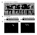
ΔHACウシ胎仔におけるヒト重鎖遺伝子座の再配列及び発現の実証 72
ΔΔHAC胎仔におけるヒト重鎖及び軽鎖遺伝子座の再配列及び発現 77
HAC仔ウシにおけるヒト抗体タンパク質の発現 82
HAC仔ウシにおいて産生されるヒト抗体の特性決定 86
実施例2:典型的なウシ核移植胚における核再プログラム欠陥の証拠 88
実施例3:典型的なウシ核移植胚における核再プログラム欠陥の例 95
実施例4:典型的なウシ核移植胚における核再プログラム欠陥の別の例 98
実施例5:哺乳動物をクローン化するための再プログラム化ドナークロマチ 102
ン塊の使用
実施例6:哺乳動物をクローン化するための再プログラム化透過性化細胞の 114
使用
実施例7:2つの新規なクローン化手順を用いるより完全な核再プログラムの 121
実証:クロマチン移植(CT)及びストレプトリシンO移植(SLOT)
実施例8:クロマチン移植(CT)を用いるより完全な核再プログラムの実証 127
実施例9:ストレプトリシンO移植(SLOT)を用いるより完全な核再プロ 132
グラムの実証
実施例10:ストレプトリシンO移植(SLOT)を用いるより完全な核再プロ 133
グラムの実証例
実施例11:ストレプトリシンO移植(SLOT)を用いるより完全な核再プロ 139
グラムの別の実証例
実施例12:キメラ哺乳動物の作出方法 145
非トランスジェニック胚由来の細胞の任意の排除 150
実施例13:1以上の内因性抗体に突然変異を有する、異種抗体を産生するトラ 152
ンスジェニック有蹄動物
実施例14:内因性免疫グロブリン遺伝子を突然変異させるための別の方法 159
ヒトIgを発現するトランスジェニック有蹄動物を作出するための 164
ノックアウト手順
他の有蹄動物の免疫グロブリン遺伝子の改変へのターゲティング手 172
法の適用
実施例15:ウシIgノックアウト 173
μ重鎖遺伝子座のエキソン1−4の除去 173
μ重鎖遺伝子座への転写終結配列の挿入 181
実施例16:内因性抗体の任意の免疫低下 188
内因性有蹄動物抗体と反応する抗体の産生 188
ウシ抗体と反応するウマ抗体を産生する方法の例 191
内因性有蹄動物抗体を免疫低下する方法の例 192
胎仔におけるB細胞発生の阻害 193
有蹄動物における免疫寛容化のための胎仔細胞移入手順、及びその 194
後の内因性抗体産生を阻害するための抗体の投与
実施例17:α−1,3−ガラクトシルトランスフェラーゼ活性が低減されたト 195
ランスジェニック有蹄動物
実施例18:内因性遺伝子を突然変異させるためにアデノ随伴ウイルスを用いて 198
トランスジェニック有蹄動物を作出する別の方法
実施例19:ヒトIg発現の試験 202
異種抗体を含有するトランスジェニック抗血清及び乳汁 203
実施例20:有蹄動物の疼痛、不快感及び全体的健康の任意の評価 207
特許請求の範囲 209
要約書 226
Table of
Summary of the
Brief Description of
Example 1: HAC introduction and
Overview of procedures for
Example procedure for
Demonstration of rearrangement and expression of the human heavy chain locus in ΔHAC
Rearrangement and expression of human heavy and light chain loci in ΔΔHAC
Expression of human antibody protein in
Characterization of human antibodies produced in HAC calves 86
Example 2: Evidence for nuclear reprogramming defects in a typical bovine
Example 3: Example of nuclear reprogramming defects in a typical bovine nuclear transfer embryo 95
Example 4: Another example of nuclear reprogramming defects in a typical bovine nuclear transfer embryo
Example 5: Reprogrammed donor chromatin for cloning mammals
Example 6: Reprogrammed permeabilized cells for cloning mammals
Use Example 7: 121 of a more complete nuclear reprogramming using two novel cloning procedures
Demonstration: Chromatin transplantation (CT) and Streptricin O transplantation (SLOT)
Example 8: Demonstration of a more complete nuclear reprogramming using chromatin transplant (CT) 127
Example 9: A more complete nuclear repro using 132 Streptricin O transplant (SLOT)
Gram Demonstration Example 10: More Complete Nuclear Repro 133 Using Streptolysin O Transplant (SLOT)
Gram Demonstration Example 11: More Complete Nuclear Repro with 139 Strepttricin O Transplant (SLOT)
Another Demonstration Example of Gram Example 12: Method for Creating
Optional exclusion of cells from
Example 13: Tiger producing a heterologous antibody with mutations in one or more endogenous antibodies
Example 14: Alternative method for mutating endogenous immunoglobulin genes 159
164 for creating transgenic ungulates expressing human Ig
Knockout procedure
Targeting hand to modification of other
Application Example 15: Bovine Ig Knockout 173
Removal of exons 1-4 at the μ heavy chain locus 173
Insertion of transcription termination sequence into the μ
Example 16: Any immune reduction of endogenous antibodies 188
Production of antibodies that react with endogenous ungulate antibodies 188
Examples of methods for producing horse antibodies that react with bovine antibodies 191
Example of a method for immunizing
Inhibition of B cell development in the
Fetal cell transfer procedure for immune tolerance in ungulates, and its 194
Administration of antibodies to inhibit subsequent endogenous antibody production Example 17: Tomato with reduced α-1,3-galactosyltransferase activity
Transgenic ungulate Example 18: Using an adeno-associated virus to mutate an
Alternative method of generating transgenic ungulates Example 19: Testing human Ig expression 202
Transgenic antiserum and milk containing heterologous antibodies 203
Example 20: Optional assessment of ungulate pain, discomfort and overall health
Claim 209
発明の背景
1890年、北里柴三郎及びEmil Behringは、驚くべき実験結果を報告した。すなわち、彼らは、免疫動物から血清を得て、それを非免疫動物に注射することにより、免疫を一方の動物から他方の動物に移すことができることを実証した。この画期的な実験は、受動免疫法を臨床的治療法に取り入れる基盤を築いた。今日、受動免疫法のためにヒト免疫グロブリン(Ig)を調製及び使用することは、標準的な医学的療法である。米国だけでも、ヒトIgに対して年間14億ドルの市場があり、毎年16トンを超えるヒト抗体が静注抗体療法のために使用されている。これに匹敵するレベルの消費が、産業先進国の経済に存在し、発展途上国において需要が急速に成長すると予想され得る。現在、受動免疫のためのヒト抗体はヒトドナーのプールした血清から得ている。これは、治療的及び予防的用途のために利用可能なヒト抗体の量に先天的に限界があることを意味する。既に需要は供給を上回っており、深刻な量不足が慢性化している。そのため、臨床用途のための、非ヒト抗体を含まないヒト抗体を作製するための改善された方法が必要とされている。
Background of the Invention In 1890, Shizaburo Kitasato and Emil Behring reported surprising experimental results. That is, they demonstrated that immunity can be transferred from one animal to another by obtaining serum from the immunized animal and injecting it into a non-immunized animal. This groundbreaking experiment laid the foundation for incorporating passive immunization into clinical therapy. Today, the preparation and use of human immunoglobulin (Ig) for passive immunization is a standard medical therapy. In the United States alone, there is a $ 1.4 billion annual market for human Ig, and over 16 tons of human antibodies are used for intravenous antibody therapy each year. A comparable level of consumption exists in the economy of industrialized countries, and demand can be expected to grow rapidly in developing countries. Currently, human antibodies for passive immunization are obtained from pooled sera of human donors. This means that there is an inherent limitation in the amount of human antibody available for therapeutic and prophylactic use. Demand already exceeds supply, and serious shortages are becoming chronic. Therefore, there is a need for improved methods for making human antibodies free of non-human antibodies for clinical use.
例えば、血流中で所望の種のポリクローナル抗体(例えばヒトIg)を生成し、所望の抗原に対して特異的な多数の異なる抗体を生成する、改善された方法及び強化されたトランスジェニック動物が非常に望ましい。特に、(ウシ等の)有蹄動物におけるヒトIgの生成は、(1)ウシは大量の抗体を生成できる、(2)ウシはヒトその他の病原体で免疫感作できる、及び(3)ウシはヒト抗原に対するヒト抗体を作るのに使用できるため、特に有利である。大量のポリクローナル抗体が利用可能になれば、感染性疾患の治療及び予防、免疫系のモジュレーション、ヒト細胞(癌細胞等)の除去、並びに特定のヒト分子のモジュレーションのために有利であろう。 For example, improved methods and enhanced transgenic animals that generate polyclonal antibodies of the desired species (eg, human Ig) in the bloodstream and generate a number of different antibodies specific for the desired antigens. Highly desirable. In particular, human Ig production in ungulates (such as cattle) is (1) cattle can produce large amounts of antibodies, (2) cattle can be immunized with human and other pathogens, and (3) cattle can This is particularly advantageous because it can be used to make human antibodies against human antigens. The availability of large amounts of polyclonal antibodies would be advantageous for the treatment and prevention of infectious diseases, modulation of the immune system, removal of human cells (such as cancer cells), and modulation of specific human molecules.
異種抗体を発現する、及び/又はレベルが低減した機能性内因性抗体を発現する、トランスジェニック有蹄動物
第1の態様において、本発明は、再配列を経て1を上回る異種Igタンパク質を発現する異種免疫グロブリン(Ig)遺伝子の全体又は一部をコードする1以上の核酸を有するトランスジェニック有蹄動物(例えばウシ)を提供する。好適な実施形態では、異種Ig遺伝子の全体又は一部をコードする核酸は、ヒトのものである。核酸は、ヒト抗体又はポリクローナル抗体等の異種抗体をコードするのが好ましい。様々な実施形態において、Ig鎖又は抗体は、血清及び/又は乳汁中に発現される。
In a first aspect of a transgenic ungulate that expresses a heterologous antibody and / or a functional endogenous antibody with reduced levels , the present invention expresses more than one heterologous Ig protein via rearrangement. Provided are transgenic ungulates (eg, cows) having one or more nucleic acids encoding all or part of a heterologous immunoglobulin (Ig) gene. In preferred embodiments, the nucleic acid encoding all or part of a heterologous Ig gene is human. The nucleic acid preferably encodes a heterologous antibody such as a human antibody or a polyclonal antibody. In various embodiments, the Ig chain or antibody is expressed in serum and / or milk.
別の態様では、本発明は、内因性抗体の発現を低減させる突然変異を有するトランスジェニック有蹄動物(例えばウシ)を特徴とする。突然変異は、機能性IgM重鎖の発現を低減するか、又は機能性IgM重鎖の発現を実質的に排除するのが好ましい。いくつかの実施形態において、転写終結配列を、内因性μ重鎖核酸に挿入する(例えば、エキソン2の最初のATGコドンの下流に挿入する)。他の好適な実施形態では、突然変異は、機能性Ig軽鎖の発現を低減するか、又は機能性Ig軽鎖の発現を実質的に排除する。さらに別の好適な実施形態では、突然変異は、機能性IgM重鎖及び機能性Ig軽鎖の発現を低減するか、又は機能性IgM重鎖及び機能性Ig軽鎖の発現を実質的に排除する。別の好適な実施形態では、有蹄動物は、再配列を経て1を上回る異種Ig分子(異種抗体タンパク質など)を発現する異種Ig遺伝子の全体又は一部をコードする1以上の核酸を有する。 In another aspect, the invention features a transgenic ungulate (eg, bovine) having a mutation that reduces expression of an endogenous antibody. The mutation preferably reduces the expression of functional IgM heavy chain or substantially eliminates the expression of functional IgM heavy chain. In some embodiments, a transcription termination sequence is inserted into the endogenous μ heavy chain nucleic acid (eg, inserted downstream of the first ATG codon of exon 2). In other preferred embodiments, the mutation reduces the expression of a functional Ig light chain or substantially eliminates the expression of a functional Ig light chain. In yet another preferred embodiment, the mutation reduces expression of functional IgM heavy chain and functional Ig light chain, or substantially eliminates expression of functional IgM heavy chain and functional Ig light chain. To do. In another preferred embodiment, the ungulate has one or more nucleic acids encoding all or part of a heterologous Ig gene that expresses more than one heterologous Ig molecule (such as a heterologous antibody protein) via rearrangement.
異種抗体を発現する、及び/又はレベルが低減した機能性内因性抗体を発現する、トランスジェニック有蹄動物由来の細胞
本発明はまた、本発明の有蹄動物(例えばウシ)のいずれかから得られる細胞、又は本発明のいずれかの有蹄動物の作製に有用な細胞を提供する。
Cells derived from transgenic ungulates expressing functional endogenous antibodies expressing heterologous antibodies and / or reduced levels The present invention is also obtained from any of the ungulates of the present invention (eg, cattle). Or cells useful for the production of any ungulate of the present invention.
従って、別の態様では、本発明は、B細胞中で再配列を経て1以上の異種Ig分子を発現することが可能な異種Ig遺伝子の全体又は一部をコードする1以上の核酸を有する有蹄動物体細胞(例えばウシ体細胞)を特徴とする。異種Ig遺伝子の全体又は一部をコードする核酸は、異種抗体タンパク質を発現するのが好ましい。有蹄動物細胞の例としては、胎仔線維芽細胞及びB細胞が挙げられる。 Accordingly, in another aspect, the present invention comprises one or more nucleic acids encoding all or part of a heterologous Ig gene capable of expressing one or more heterologous Ig molecules via rearrangement in B cells. Characterized by hoof somatic cells (eg bovine somatic cells). The nucleic acid encoding all or part of the heterologous Ig gene preferably expresses a heterologous antibody protein. Examples of ungulate cells include fetal fibroblasts and B cells.
別の態様では、本発明は、Ig重鎖及び/又は軽鎖をコードする核酸中に突然変異を有する有蹄動物体細胞(例えばウシ体細胞)を特徴とする。好適な実施形態では、細胞は、IgM重鎖又はIg軽鎖の一方又は両方のアレルに突然変異を有する。いくつかの実施形態において、転写終結配列を、内因性μ重鎖核酸に挿入する(例えば、エキソン2の最初のATGコドンの下流に挿入する)、又はIg軽鎖核酸に挿入する。いくつかの実施形態において、転写終結配列は、内因性μ重鎖核酸に挿入する(例えば、エキソン2の最初のATGコドンの下流に挿入する)、又はIg軽鎖核酸に挿入する。突然変異の例としては、ナンセンス変異及び欠失変異が挙げられる。好適な実施形態では、細胞は、B細胞中で再配列を経て1以上の異種Ig分子を発現することが可能な異種Ig遺伝子の全体又は一部をコードする1以上の核酸も有する。異種Ig遺伝子の全体又は一部をコードする核酸は、別の属に由来する抗体タンパク質(例えば、ヒト抗体)等の異種抗体を発現することが好ましい。。有蹄動物細胞の例としては、胎仔線維芽細胞及びB細胞が挙げられる。 In another aspect, the invention features an ungulate somatic cell (eg, bovine somatic cell) having a mutation in a nucleic acid encoding an Ig heavy chain and / or light chain. In preferred embodiments, the cell has a mutation in one or both alleles of IgM heavy chain or Ig light chain. In some embodiments, the transcription termination sequence is inserted into an endogenous μ heavy chain nucleic acid (eg, inserted downstream of the first ATG codon of exon 2) or inserted into an Ig light chain nucleic acid. In some embodiments, the transcription termination sequence is inserted into an endogenous μ heavy chain nucleic acid (eg, inserted downstream of the first ATG codon of exon 2) or inserted into an Ig light chain nucleic acid. Examples of mutations include nonsense mutations and deletion mutations. In a preferred embodiment, the cell also has one or more nucleic acids that encode all or part of a heterologous Ig gene capable of expressing one or more heterologous Ig molecules via rearrangement in B cells. The nucleic acid encoding the whole or part of the heterologous Ig gene preferably expresses a heterologous antibody such as an antibody protein (eg, human antibody) derived from another genus. . Examples of ungulate cells include fetal fibroblasts and B cells.
別の態様では、本発明は、本発明の有蹄動物B細胞とミエローマ細胞との融合により形成されるハイブリドーマを特徴とする。ハイブリドーマは、ヒト抗体等の異種抗体を分泌することが好ましい。 In another aspect, the invention features a hybridoma formed by the fusion of an ungulate B cell of the invention and a myeloma cell. The hybridoma preferably secretes a heterologous antibody such as a human antibody.
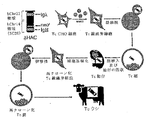
トランスジェニック有蹄動物における異種抗体の製造方法
本発明はまた、本発明の有蹄動物(例えば、ウシの胚、胎仔、仔ウシ又は成体)を用いた抗体の製造方法を提供する。このような方法のひとつでは、1以上の目的の抗原を、異種抗体遺伝子座をコードする1以上の核酸を有する有蹄動物(例えば、ウシの胚、胎仔、仔ウシ又は成体)に投与する。遺伝子座中の核酸セグメントは、再配列を経て、抗原に特異的な抗体を生成する。その抗体を有蹄動物から回収する。抗体は、モノクローナル又はポリクローナルであり得る。特定の抗原に対するモノクローナル及びポリクローナル抗体は、様々な用途を有する。例えば、細菌又はウイルス等の病原体微生物の感染に対する予防又は治療用組成物中の成分として使用し得る。様々な実施形態において、抗体は、有蹄動物の血清又は乳汁から回収する。好適な実施形態では、有蹄動物は、内因性抗体の発現を低減する突然変異、機能性IgM重鎖の発現を低減する突然変異、又は機能性Ig軽鎖の発現を低減する突然変異を有する。いくつかの実施形態において、転写終結配列を、内因性μ重鎖核酸に挿入する(例えば、エキソン2の最初のATGコドンの下流に挿入する)、又はIg軽鎖核酸に挿入する。
Method for producing heterologous antibody in transgenic ungulate The present invention also provides a method for producing an antibody using the ungulate of the present invention (eg, bovine embryo, fetus, calf or adult). In one such method, one or more antigens of interest are administered to an ungulate (eg, a bovine embryo, fetus, calf or adult) having one or more nucleic acids encoding a heterologous antibody locus. The nucleic acid segment in the locus undergoes rearrangement to produce an antibody specific for the antigen. The antibody is recovered from the ungulate. The antibody can be monoclonal or polyclonal. Monoclonal and polyclonal antibodies against specific antigens have a variety of uses. For example, it can be used as a component in a composition for prevention or treatment against infection with pathogenic microorganisms such as bacteria or viruses. In various embodiments, the antibody is recovered from ungulate serum or milk. In preferred embodiments, the ungulate has a mutation that reduces the expression of an endogenous antibody, a mutation that reduces the expression of a functional IgM heavy chain, or a mutation that reduces the expression of a functional Ig light chain. . In some embodiments, the transcription termination sequence is inserted into an endogenous μ heavy chain nucleic acid (eg, inserted downstream of the first ATG codon of exon 2) or inserted into an Ig light chain nucleic acid.
関連する態様において、本発明は、別の抗体製造方法を特徴とする。本方法では、異種抗体遺伝子座をコードする核酸を有する有蹄動物(例えば、ウシの胚、胎仔、仔ウシ又は成体)から異種抗体を回収する。遺伝子座中の核酸セグメントは、再配列を経て異種抗体タンパク質を生成する。特定の実施形態では、抗体の軽鎖及び/又は抗体の重鎖がヒト核酸にコードされている。抗体は、モノクローナル又はポリクローナルであり得る。特定の実施形態において、特異的な抗原で有蹄動物を免疫感作すること無く生成させたIgG抗体等のポリクローナル抗体は、ヒト血清から得られるIVIG(静注用免疫グロブリン)の治療代用品として使用される。様々な実施形態において、抗体は有蹄動物の血清又は乳汁から回収する。有蹄動物は、内因性抗体の発現を低減する突然変異、機能性IgM重鎖の発現を低減する突然変異、又は機能性Ig軽鎖の発現を低減する突然変異を有することが好ましい。好ましくは、転写終結配列を、内因性μ重鎖核酸に挿入する(例えば、エキソン2の最初のATGコドンの下流に挿入する)、又はIg軽鎖核酸に挿入する。 In a related aspect, the invention features another method for producing an antibody. In this method, a heterologous antibody is recovered from an ungulate (eg, bovine embryo, fetus, calf or adult) having a nucleic acid encoding a heterologous antibody locus. Nucleic acid segments in the locus undergo a rearrangement to produce a heterologous antibody protein. In certain embodiments, the light chain of the antibody and / or the heavy chain of the antibody is encoded by a human nucleic acid. The antibody can be monoclonal or polyclonal. In certain embodiments, polyclonal antibodies such as IgG antibodies generated without immunizing ungulates with specific antigens are used as therapeutic substitutes for IVIG (intravenous immunoglobulin) obtained from human serum. used. In various embodiments, the antibody is recovered from ungulate serum or milk. Preferably, the ungulate has a mutation that reduces the expression of an endogenous antibody, a mutation that reduces the expression of a functional IgM heavy chain, or a mutation that reduces the expression of a functional Ig light chain. Preferably, the transcription termination sequence is inserted into the endogenous μ heavy chain nucleic acid (eg, inserted downstream of the first ATG codon of exon 2) or inserted into the Ig light chain nucleic acid.
トランスジェニック有蹄動物の作製方法
本発明はまた、トランスジェニック有蹄動物(例えば、ウシの胚、胎仔、仔ウシ又は成体)の作製方法も提供する。これらの方法は、所望の突然変異又は所望の異種核酸を有するトランスジェニック有蹄動物を作製するのに使用し得る。
Methods for producing transgenic ungulates The present invention also provides methods for producing transgenic ungulates (eg, bovine embryos, fetuses, calves or adults). These methods can be used to create transgenic ungulates with a desired mutation or a desired heterologous nucleic acid.
このような態様の1つにおいて、本発明は、細胞、細胞由来のクロマチン塊、又は細胞由来の核を卵母細胞に挿入することを含むトランスジェニック有蹄動物(例えば、ウシの胚、胎仔、仔ウシ又は成体)の作製方法を特徴とする。この細胞は、内因性抗体重鎖及び/又は軽鎖核酸中に第1の突然変異を有する。卵母細胞又は卵母細胞から形成される胚を、卵母細胞又は胚が胎仔に発育可能な条件下で宿主有蹄動物の子宮に導入する。胎仔は生存可能な子孫(offspring)に発育するのが好ましい。いくつかの実施形態では、細胞は、B細胞中で再配列を経て1以上の異種Ig分子を発現することができる異種Ig遺伝子の全体又は一部をコードする1以上の核酸を含有する。好ましくは、転写終結配列を、内因性μ重鎖核酸に挿入する(例えば、エキソン2の最初のATGコドンの下流に挿入する)、又はIg軽鎖核酸に挿入する。 In one such embodiment, the present invention relates to a transgenic ungulate (eg, bovine embryo, fetus, It is characterized by a method for producing calves or adults. The cell has a first mutation in the endogenous antibody heavy and / or light chain nucleic acid. The oocyte or embryo formed from the oocyte is introduced into the uterus of the host ungulate under conditions that allow the oocyte or embryo to develop into a fetus. The fetus preferably develops into a viable offspring. In some embodiments, the cells contain one or more nucleic acids encoding all or part of a heterologous Ig gene that can be rearranged in B cells to express one or more heterologous Ig molecules. Preferably, the transcription termination sequence is inserted into the endogenous μ heavy chain nucleic acid (eg, inserted downstream of the first ATG codon of exon 2) or inserted into the Ig light chain nucleic acid.
上記態様の様々な実施形態において、本方法は、胚、胎仔又は胎仔より作出された子孫から細胞を単離すること、並びに該細胞中の内因性抗体重鎖及び/又は軽鎖核酸に第2の突然変異を導入することも含む。細胞、細胞由来のクロマチン塊又は細胞由来の核を卵母細胞に導入し、該卵母細胞又は該卵母細胞から形成される胚を、卵母細胞又は胚が胎仔に発育可能な条件下で宿主有蹄動物の子宮に導入する。 In various embodiments of the above aspects, the method includes isolating a cell from an embryo, fetus or offspring generated from the fetus, and second to endogenous antibody heavy and / or light chain nucleic acid in the cell. Of introducing a mutation of. A cell, a cell-derived chromatin mass or a cell-derived nucleus is introduced into an oocyte, and the oocyte or an embryo formed from the oocyte is subjected to conditions under which the oocyte or embryo can develop into a fetus It is introduced into the uterus of the host ungulate.
さらに別の態様では、本発明は、トランスジェニック有蹄動物(例えば、ウシの胚、胎仔、仔ウシ又は成体)の別の作製方法を特徴とする。本方法では、1以上の異種核酸を有する細胞、細胞由来のクロマチン塊又は細胞由来の核を卵母細胞に挿入する。異種核酸は、異種Ig遺伝子の全体又は一部をコードし、該遺伝子は、B細胞中で再配列を経て1を上回る異種Ig分子を発現することが可能である。卵母細胞又は卵母細胞から形成される胚を、好ましくは卵母細胞又は胚が胎仔に発育可能な条件下で、宿主有蹄動物の子宮に導入する。胎仔は、生存可能な子孫に発育することが好ましい。異種Ig遺伝子の全体又は一部をコードする核酸は、異種抗体をコードすることが好ましい。他の好適な実施形態では、抗体はポリクローナル抗体である。さらに他の好適な実施形態では、免疫グロブリン鎖又は抗体は、血清及び/又は乳汁中に発現される。いくつかの実施形態において、ドナー細胞は、内因性抗体の重鎖及び/又は軽鎖核酸に突然変異(例えば、転写終結配列の挿入など)を有する。 In yet another aspect, the invention features another method of making a transgenic ungulate (eg, a bovine embryo, fetus, calf, or adult). In this method, a cell having one or more heterologous nucleic acids, a cell-derived chromatin mass, or a cell-derived nucleus is inserted into an oocyte. A heterologous nucleic acid encodes all or part of a heterologous Ig gene, which can express more than one heterologous Ig molecule via rearrangement in B cells. The oocyte or embryo formed from the oocyte is introduced into the uterus of the host ungulate, preferably under conditions that allow the oocyte or embryo to develop into a fetus. The fetus is preferably developed into a viable offspring. The nucleic acid encoding all or part of the heterologous Ig gene preferably encodes a heterologous antibody. In other preferred embodiments, the antibody is a polyclonal antibody. In yet other preferred embodiments, the immunoglobulin chain or antibody is expressed in serum and / or milk. In some embodiments, the donor cell has a mutation (eg, insertion of a transcription termination sequence) in the heavy and / or light chain nucleic acid of the endogenous antibody.
異種抗体及び内因性抗体の両方を発現する有蹄動物において望ましくない内因性抗体の量を低減する方法、並びに有蹄動物における異種抗体の製造方法
本発明はまた、非ヒト哺乳動物、例えば有蹄動物(例えばウシ)において異種抗体を主に又はそれのみを製造する改善された方法を特徴とする。特に、これらの方法は、内因性B細胞活性を阻害する化合物又は内因性B細胞を破壊する化合物、例えば抗IgM又は抗Ig抗体を、内因性B細胞又は抗体の活性又は量を低減するのに十分な量で、内因性抗体及び異種抗体の両方を発現する哺乳動物に投与することを含むものである。これらの化合物は、哺乳動物の免疫系の正常な発生期間の間に(すなわち、胚、胎仔若しくは生後の段階に)、又は免疫系の発生期間の後に、投与することができる。好ましくは、内因性B細胞又は抗体を阻害する抗体は、異種B細胞又は完全に異種の抗体を実質的に阻害しないものである。得られるモノクローナル又はポリクローナル異種抗体は、種々の用途を有し、例えば、これらは細菌若しくはウイルスなどの病原性微生物の感染のための予防用又は治療用組成物中の有効成分として用いることができる。
Methods for reducing the amount of unwanted endogenous antibodies in ungulates that express both xenogeneic and endogenous antibodies, and methods for producing xenoantibodies in ungulates The present invention also provides non-human mammals such as ungulates. Characterized by an improved method of producing mainly or only heterologous antibodies in animals (eg bovine). In particular, these methods reduce compounds that inhibit endogenous B cell activity or destroy endogenous B cells, such as anti-IgM or anti-Ig antibodies, to reduce the activity or amount of endogenous B cells or antibodies. Administration to a mammal expressing both an endogenous antibody and a heterologous antibody in a sufficient amount. These compounds can be administered during the normal development period of the mammalian immune system (ie, at the embryo, fetus or postnatal stage) or after the development period of the immune system. Preferably, an antibody that inhibits endogenous B cells or antibodies is one that does not substantially inhibit heterologous B cells or fully heterologous antibodies. The resulting monoclonal or polyclonal heterologous antibodies have a variety of uses, for example, they can be used as active ingredients in prophylactic or therapeutic compositions for infection with pathogenic microorganisms such as bacteria or viruses.
従って、一態様において、本発明は、非ヒト哺乳動物(例えば有蹄動物)における内因性抗体の量又は活性を低減する方法を提供する。この方法は、完全又は部分的に内因性の抗体と反応する抗体を、完全又は部分的に内因性の抗体の量及び/又は活性を低減するのに十分な量で、内因性抗体及び異種抗体の両方を発現する有蹄動物(例えばウシ)に投与することを含む。好ましい実施形態において、抗体は初乳の前に投与する。 Accordingly, in one aspect, the invention provides a method for reducing the amount or activity of an endogenous antibody in a non-human mammal (eg, an ungulate). In this method, an antibody that reacts with a fully or partially endogenous antibody in an amount sufficient to reduce the amount and / or activity of the fully or partially endogenous antibody in an endogenous antibody and a heterologous antibody. Administration to an ungulate (eg, bovine) expressing both. In a preferred embodiment, the antibody is administered prior to colostrum.
関連する態様において、本発明は、非ヒト哺乳動物(例えば有蹄動物)において異種抗体を製造する方法を提供する。この方法は、完全又は部分的に内因性の抗体と反応する抗体を、完全又は部分的に内因性の抗体の量又は活性を低減するのに十分な量で、内因性抗体及び異種抗体の両方を発現する有蹄動物(例えばウシ)に投与することを含む。異種抗体は、有蹄動物から回収する。好ましい実施形態において、異種抗体は、有蹄動物の血清又は乳汁から回収する。他の好ましい実施形態において、異種抗体の一部又は全体は、完全に異種の抗体である。好ましい実施形態において、抗体は初乳の前に投与する。 In a related aspect, the invention provides a method for producing a heterologous antibody in a non-human mammal (eg, an ungulate). This method involves reacting an antibody that reacts with a fully or partially endogenous antibody, both an endogenous antibody and a heterologous antibody, in an amount sufficient to reduce the amount or activity of the fully or partially endogenous antibody. Administration to an ungulate that expresses The heterologous antibody is recovered from the ungulate. In a preferred embodiment, the heterologous antibody is recovered from ungulate serum or milk. In other preferred embodiments, part or all of the heterologous antibody is a completely heterologous antibody. In a preferred embodiment, the antibody is administered prior to colostrum.
他の関連する態様において、本発明は、非ヒト哺乳動物(例えば有蹄動物)において異種抗体を製造する別の方法を提供する。この方法は、細胞、核又はクロマチン塊を、除核卵母細胞に挿入し、それにより核移植卵母細胞を形成させる。細胞、核又はクロマチン塊は、第1異種抗体をコードする核酸、及び内因性抗体と反応する第2抗体をコードする核酸を有する。核移植卵母細胞又は核移植卵母細胞から形成された胚は、それが胎仔又は生存子孫に発育するような条件下にて宿主哺乳動物の子宮に導入する。その胎仔又は子孫は、異種第1抗体及び第2抗体を発現し、第2抗体は、内因性抗体の量及び/又は活性を低減する。好ましくは、第2抗体は肝臓特異的プロモーターの制御下にて発現される。いくつかの好ましい第2抗体は、内因性IgM又は内因性免疫グロブリン分子と反応する。異種抗体は、有蹄動物から回収することが好ましい。好ましい実施形態において、異種抗体は、有蹄動物の血清又は乳汁から回収する。他の好ましい実施形態において、異種抗体の一部又は全体は、完全に異種の抗体である。 In other related embodiments, the invention provides another method of producing a heterologous antibody in a non-human mammal (eg, an ungulate). This method inserts a cell, nucleus, or chromatin mass into an enucleated oocyte, thereby forming a nuclear transplanted oocyte. The cell, nucleus or chromatin mass has a nucleic acid encoding a first antibody and a nucleic acid encoding a second antibody that reacts with an endogenous antibody. Nuclear transfer oocytes or embryos formed from nuclear transfer oocytes are introduced into the uterus of the host mammal under conditions such that they develop into fetuses or viable offspring. The fetus or offspring expresses the heterologous first antibody and the second antibody, which reduces the amount and / or activity of the endogenous antibody. Preferably, the second antibody is expressed under the control of a liver specific promoter. Some preferred second antibodies react with endogenous IgM or endogenous immunoglobulin molecules. The heterologous antibody is preferably recovered from the ungulate. In a preferred embodiment, the heterologous antibody is recovered from ungulate serum or milk. In other preferred embodiments, part or all of the heterologous antibody is a completely heterologous antibody.
胎仔の生存を促進するため、核移植胚に由来する細胞が得られる胎仔組織に選択的に組み込まれている胚、及び別の胚に由来する細胞が得られる胎盤組織に選択的に組み込まれている胚の、2つの胚に由来する細胞を用いた非ヒト哺乳動物のクローン化方法
本発明はまた、非ヒト哺乳動物をクローン化するための改善された方法を提供する。これらの哺乳動物は、第1核移植胚(例えば、細胞、核又はクロマチン塊を脱核卵母細胞に挿入することにより形成された胚)に由来する細胞と、in vitroで受精(体外受精)させた、天然の又は単為生殖的に活性化された第2胚に由来する細胞とを混合することにより形成される。この得られるキメラ胚は、それが胎仔又は生存子孫に発育可能な条件下にて宿主哺乳動物の子宮に導入される。第2胚に由来する少なくとも一部の細胞が胎盤組織に組み込まれ、得られるキメラ胚の生存を促進することが好ましい。好ましくは、第1核移植胚に由来する細胞の大部分及びその子(progeny)が、第2胚に由来するものよりも、得られるキメラ胚の胎仔組織中に組み込まれる。従って、このキメラ胚に由来する胎仔又は子孫(offspring)中の細胞の大部分は、第2胚よりもむしろ第1核移植胚のゲノムと実質的に同一又は同一のゲノムを有することが好ましい。胎仔組織又は子孫に組み込まれる第2胚に由来する細胞及びその子の数を低減するために、第2胚由来の抗原と反応する抗体を、該胎仔又は子孫に組み込まれる第2胚由来の細胞の量及び/又は活性を低減するのに十分な量で、該キメラ胚、胎仔又は子孫に投与する。
In order to promote fetal survival, embryos that are selectively integrated into fetal tissue from which cells derived from nuclear transfer embryos are obtained and those that are derived from other embryos are selectively incorporated into placental tissue from which cells are obtained. Methods for cloning non-human mammals using cells derived from two embryos of the present invention The present invention also provides an improved method for cloning non-human mammals. These mammals are fertilized in vitro (in vitro fertilization) with cells derived from a first nuclear transfer embryo (eg, an embryo formed by inserting a cell, nucleus or chromatin mass into a enucleated oocyte). Formed by mixing cells derived from a natural or parthenogenetically activated second embryo. The resulting chimeric embryo is introduced into the uterus of the host mammal under conditions that allow it to develop into fetuses or viable offspring. Preferably, at least some of the cells derived from the second embryo are incorporated into the placental tissue to promote the survival of the resulting chimeric embryo. Preferably, the majority of cells derived from the first nuclear transfer embryo and its progeny are incorporated into the fetal tissue of the resulting chimeric embryo rather than those derived from the second embryo. Accordingly, it is preferred that the majority of cells in the fetus or offspring derived from this chimeric embryo have substantially the same or identical genome as the genome of the first nuclear transfer embryo rather than the second embryo. In order to reduce the number of cells derived from the second embryo and the offspring thereof integrated into the fetal tissue or offspring, an antibody that reacts with an antigen derived from the second embryo is added to the cells derived from the second embryo integrated into the fetus or offspring. Administer to the chimeric embryo, fetus or offspring in an amount sufficient to reduce the amount and / or activity.
従って、一態様において、本発明は、非ヒト哺乳動物のクローン化方法を提供する。この方法は、細胞、核又はクロマチン塊を卵母細胞に挿入し、それにより第1胚を形成することを含む。第1胚に由来する1以上の細胞を、第2胚(例えば、in vitroで受精(体外受精)させた胚、天然の胚又は単為生殖的に活性化された胚)に由来する1以上の細胞と接触させ、それにより第3胚を形成させる。この第3胚は、宿主哺乳動物の子宮に、第3胚が胎仔又は生存子孫に発育可能な条件下にて導入される。第2胚由来の抗原(例えばB細胞又は生殖細胞抗原)と反応する抗体は、第3胚、胎仔又は子孫に組み込まれる第2胚由来の細胞の量及び/又は活性を低減するのに十分な量で、第3胚、胎仔又は子孫に投与する。 Accordingly, in one aspect, the present invention provides a method for cloning a non-human mammal. This method involves inserting a cell, nucleus or chromatin mass into an oocyte, thereby forming a first embryo. One or more cells derived from a second embryo (eg, embryos fertilized in vitro (in vitro fertilization), natural embryos or parthenogenously activated embryos) from one or more cells derived from the first embryo To form a third embryo. This third embryo is introduced into the uterus of the host mammal under conditions that allow the third embryo to develop into a fetus or viable offspring. An antibody that reacts with a second embryo-derived antigen (eg, a B cell or germ cell antigen) is sufficient to reduce the amount and / or activity of a second embryo-derived cell that is integrated into the third embryo, fetus or offspring. The dose is administered to the third embryo, fetus or offspring.
関連する態様において、本発明は、非ヒト哺乳動物をクローン化するための別の方法を提供する。この方法は、透過性化細胞の核、クロマチン塊若しくは染色体からの因子の除去、又は再プログラム培地からの核、クロマチン塊若しくは染色体への因子の付加を可能にする条件下にて、再プログラム培地中で透過性化細胞をインキュベートし、それにより再プログラム化細胞を形成することを含む。再プログラム化細胞は、卵母細胞に挿入し、それにより第1胚を形成させる。第1胚に由来する1以上の細胞を、第2胚(例えば、in vitroで受精(体外受精)させた胚、天然の胚又は単為生殖的に活性化された胚)の1以上の細胞と接触させ、それにより第3胚を形成させる。この第3胚は、宿主哺乳動物の子宮に、第3胚が胎仔又は生存子孫に発育可能な条件下にて導入される。第2胚由来の抗原と反応する抗体は、第3胚、胎仔又は子孫に組み込まれる第2胚由来の細胞の量及び/又は活性を低減するのに十分な量で、第3胚、胎仔又は子孫に投与する。 In a related aspect, the present invention provides another method for cloning a non-human mammal. This method involves the reprogramming medium under conditions that allow removal of the factor from the permeabilized cell nucleus, chromatin mass or chromosome, or addition of the factor to the nucleus, chromatin mass or chromosome from the reprogramming medium. Incubating the permeabilized cells therein, thereby forming reprogrammed cells. The reprogrammed cell is inserted into the oocyte thereby forming a first embryo. One or more cells of one or more cells derived from the first embryo of a second embryo (eg, an embryo fertilized in vitro (in vitro fertilization), a natural embryo or an embryo that is parthenogenously activated) To form a third embryo. This third embryo is introduced into the uterus of the host mammal under conditions that allow the third embryo to develop into a fetus or viable offspring. The antibody that reacts with the antigen derived from the second embryo is in an amount sufficient to reduce the amount and / or activity of cells derived from the second embryo that are incorporated into the third embryo, fetus or offspring. Administer to offspring.
本発明はまた、キメラ胎仔又は子孫を作出するために使用した初期胚の1つに由来する細胞が異種抗体(例えばヒト抗体)をコードする核酸を有する、キメラ胎仔又は子孫を作出する方法を提供する。さらに、上記初期胚又は別の初期胚に由来する細胞は、異種抗体(例えば、キメラ胎仔又は子孫を作出するために用いた初期胚のいずれかに由来する細胞により天然に産生される抗体)と反応し、得られる胎仔又は子孫における内因性抗体の量又は活性を低減する抗体をコードする核酸を有する。 The present invention also provides a method for producing a chimeric fetus or progeny, wherein cells derived from one of the early embryos used to create the chimeric fetus or progeny have a nucleic acid encoding a heterologous antibody (eg, a human antibody). To do. In addition, cells derived from the early embryo or another early embryo may be a heterologous antibody (eg, an antibody that is naturally produced by a cell derived from any of the early embryos used to produce a chimeric fetus or offspring). Having a nucleic acid encoding an antibody that reacts and reduces the amount or activity of endogenous antibody in the resulting fetus or offspring.
かかる一の態様において、本発明は、非ヒト哺乳動物のクローン化方法を特徴とする。この方法は、細胞、核又はクロマチン塊を卵母細胞に挿入し、それにより第1胚を形成することを含む。該細胞、核又はクロマチン塊は、異種第1抗体をコードする核酸、及び内因性抗体と反応する第2抗体をコードする核酸を有する。第1胚に由来する1以上の細胞を、第2胚(例えば、in vitroで受精(体外受精)させた胚、天然の胚又は単為生殖的に活性化された胚)に由来する1以上の細胞と接触させ、それにより第3胚を形成させる。この第3胚は、宿主哺乳動物の子宮に、第3胚が胎仔又は生存子孫に発育可能な条件下にて導入される。得られる胎仔又は子孫は、異種第1抗体と第2抗体を発現し、この第2抗体は、内因性抗体の量及び/又は活性を低減する。第2抗体は肝臓特異的プロモーターの制御下にて発現されることが好ましい。かかる好ましい第2抗体は、内因性IgM又は内因性免疫グロブリン分子と反応する。 In one such aspect, the invention features a method for cloning a non-human mammal. This method involves inserting a cell, nucleus or chromatin mass into an oocyte, thereby forming a first embryo. The cell, nucleus or chromatin mass has a nucleic acid encoding a heterologous first antibody and a nucleic acid encoding a second antibody that reacts with an endogenous antibody. One or more cells derived from a second embryo (eg, embryos fertilized in vitro (in vitro fertilization), natural embryos or parthenogenously activated embryos) from one or more cells derived from the first embryo To form a third embryo. This third embryo is introduced into the uterus of the host mammal under conditions that allow the third embryo to develop into a fetus or viable offspring. The resulting fetus or offspring expresses a heterologous first antibody and a second antibody, which reduces the amount and / or activity of endogenous antibodies. The second antibody is preferably expressed under the control of a liver-specific promoter. Such preferred second antibody reacts with endogenous IgM or endogenous immunoglobulin molecules.
関連する態様において、本発明は、非ヒト哺乳動物をクローン化するための別の方法を提供する。この方法は、透過性化細胞の核、クロマチン塊若しくは染色体からの因子の除去、又は再プログラム培地からの核、クロマチン塊若しくは染色体への因子の付加を可能にする条件下にて、再プログラム培地中で透過性化細胞をインキュベートし、それにより再プログラム化細胞を形成することを含む。該細胞は、異種第1抗体をコードする核酸、及び内因性抗体と反応する第2抗体をコードする核酸を有する。再プログラム化細胞は、卵母細胞に挿入し、それにより第1胚を形成させる。第1胚に由来する1以上の細胞を、第2胚(例えば、in vitroで受精(体外受精)させた胚、天然の胚又は単為生殖的に活性化された胚)由来の1以上の細胞と接触させ、それにより第3胚を形成させる。この第3胚は、宿主哺乳動物の子宮に、第3胚が胎仔又は生存子孫に発育可能な条件下にて導入される。得られる胎仔又は子孫は、異種第1抗体と第2抗体を発現し、この第2抗体は、内因性抗体の量及び/又は活性を低減する。第2抗体は肝臓特異的プロモーターの制御下にて発現されることが好ましい。かかる好ましい第2抗体は、内因性IgM又は内因性免疫グロブリン分子と反応する。 In a related aspect, the present invention provides another method for cloning a non-human mammal. This method involves the reprogramming medium under conditions that allow removal of the factor from the permeabilized cell nucleus, chromatin mass or chromosome, or addition of the factor to the nucleus, chromatin mass or chromosome from the reprogramming medium. Incubating the permeabilized cells therein, thereby forming reprogrammed cells. The cell has a nucleic acid encoding a heterologous first antibody and a nucleic acid encoding a second antibody that reacts with an endogenous antibody. The reprogrammed cell is inserted into the oocyte thereby forming a first embryo. One or more cells derived from the first embryo are transformed into one or more cells derived from a second embryo (eg, an in vitro fertilized (in vitro fertilized) embryo, a natural embryo or parthenogenously activated embryo). Contact with the cells, thereby forming a third embryo. This third embryo is introduced into the uterus of the host mammal under conditions that allow the third embryo to develop into a fetus or viable offspring. The resulting fetus or offspring expresses a heterologous first antibody and a second antibody, which reduces the amount and / or activity of endogenous antibodies. The second antibody is preferably expressed under the control of a liver-specific promoter. Such preferred second antibody reacts with endogenous IgM or endogenous immunoglobulin molecules.
任意の上記クローン化方法の好ましい実施形態において、第1胚は、B細胞において再配列を経て少なくとも1つの異種免疫グロブリン(Ig)分子を発現する異種免疫グロブリン(Ig)遺伝子の全部又は一部をコードする1以上の核酸を含む。他の好ましい実施形態において、第1胚は、B細胞において少なくとも1つの異種Ig分子を発現する再配列された異種免疫グロブリン遺伝子の全部又は一部をコードする1以上の核酸を含む。いくつかの実施形態において、第1胚又は第2胚は、内因性抗体の発現を低減する突然変異を含む。他の好ましい実施形態において、抗体は、B細胞又は生殖細胞の表面上で発現される抗原、例えば抗体又は細胞表面タンパク質若しくは受容体などと反応する。いくつかの実施形態において、投与される抗体(投与抗体)は、抗IgM又は抗免疫グロブリン抗体である。好ましい実施形態において、抗体は初乳の前に投与する。 In a preferred embodiment of any of the above cloning methods, the first embryo carries all or part of a heterologous immunoglobulin (Ig) gene that expresses at least one heterologous immunoglobulin (Ig) molecule via rearrangement in B cells. It includes one or more nucleic acids that encode. In other preferred embodiments, the first embryo comprises one or more nucleic acids encoding all or part of a rearranged heterologous immunoglobulin gene that expresses at least one heterologous Ig molecule in B cells. In some embodiments, the first or second embryo comprises a mutation that reduces the expression of an endogenous antibody. In other preferred embodiments, the antibodies react with antigens expressed on the surface of B cells or germ cells, such as antibodies or cell surface proteins or receptors. In some embodiments, the administered antibody (administered antibody) is an anti-IgM or anti-immunoglobulin antibody. In a preferred embodiment, the antibody is administered prior to colostrum.
上記方法において使用するのに好ましい有蹄動物
有蹄動物の例として、ウマ目(奇蹄類Perissodactyla)及びウシ目(偶蹄類Artiodactyla)のメンバー(ウシ(Bos)属のあらゆるメンバー等)が挙げられる。他の好適な有蹄動物としては、ヒツジ、オオツノヒツジ、ヤギ、野牛、カモシカ、雄牛、ウマ、ロバ、ラバ、シカ、ヘラジカ、アメリカトナカイ、水牛、ラクダ、ラマ、アルパカ、ブタ及びゾウが挙げられる。好ましい実施形態において、レシピエント有蹄動物は、50、40、30、20、10、7、5、4、3、2又は1週齢未満である。種々の実施形態において、抗体は、妊娠の第1期、第2期又は第3期の間に胎仔に投与する。また別の好ましい実施形態においては、胎仔を、選択した時期まで妊娠状態で又は宿主哺乳動物中で発育させ、その後、胎仔を外科的に取り出すか又は標準的方法により出産を誘導する。例えば、生存胎仔は、帝王切開により取り出すことが可能であり、あるいは、胎仔の正常の満期となる1、2、3、5、10、15、20又はそれ以上の日数の前に出産を人工的に誘導することも可能である。これらの若年レシピエント有蹄動物は、天然に免疫系が抑制されており、それにより内因性抗体を低減する投与した抗体に対する有害な免疫応答を最小限に抑える又は防止することができる。他の好ましい有蹄動物は、正常よりも応答性の低い免疫系を天然に又は自発的に有するものである。
Examples of preferred ungulate ungulates for use in the above method include members of the order Horse (Periodactyla) and Bovine (Artiodactyla) (such as any member of the genus Bos). . Other suitable ungulates include sheep, bighorn sheep, goats, wild cattle, antelopes, bulls, horses, donkeys, mules, deer, elk, reindeer, buffalo, camels, llamas, alpaca, pigs and elephants . In preferred embodiments, the recipient ungulate is less than 50, 40, 30, 20, 10, 7, 5, 4, 3, 2, or 1 week old. In various embodiments, the antibody is administered to the fetus during the first, second, or third stage of pregnancy. In another preferred embodiment, the fetus is grown in a pregnant state or in a host mammal until a selected time, after which the fetus is removed surgically or birth is induced by standard methods. For example, surviving fetuses can be removed by caesarean section, or by giving birth artificially before 1, 2, 3, 5, 10, 15, 20 or more days of normal fetal maturity. It is also possible to guide to These young recipient ungulates are naturally suppressed in the immune system, thereby minimizing or preventing adverse immune responses to administered antibodies that reduce endogenous antibodies. Other preferred ungulates are those that naturally or spontaneously have an immune system that is less responsive than normal.
異種抗体を発現する好ましい有蹄動物は、ヒト染色体(例えばヒト染色体断片)の天然に配列したセグメント又は人工的に操作したヒト染色体断片を含む人工染色体(すなわち、この断片はヒトゲノムに関して再配列されうる)を含有する。好ましい有蹄動物は、異種抗体遺伝子座を有する1以上の核酸(例えば、再配列を経て少なくとも1つの異種Ig分子を発現する異種免疫グロブリン(Ig)遺伝子の全部又は一部をコードする核酸)を有する。好ましくは、核酸は、V遺伝子セグメントをコードする核酸セグメントの全てがJ遺伝子セグメントをコードする核酸セグメントの全てと1以上のヌクレオチドにより分離されている、再配列されていない抗体軽鎖核酸セグメントを有する。他の好ましい核酸は、(i)V遺伝子セグメントをコードする核酸セグメントの全てがD遺伝子セグメントをコードする核酸セグメントの全てと1以上のヌクレオチドにより分離されている、及び/又は、(ii)D遺伝子セグメントをコードする核酸セグメントの全てがJ遺伝子セグメントをコードする核酸セグメントの全てと1以上のヌクレオチドにより分離されている、再配列されていない抗体重鎖核酸セグメントを有する。 Preferred ungulates expressing heterologous antibodies are naturally-occurring segments of human chromosomes (eg, human chromosome fragments) or artificial chromosomes containing artificially engineered human chromosome fragments (ie, this fragment can be rearranged with respect to the human genome). ). Preferred ungulates are one or more nucleic acids having a heterologous antibody locus (eg, a nucleic acid encoding all or part of a heterologous immunoglobulin (Ig) gene that expresses at least one heterologous Ig molecule via rearrangement). Have. Preferably, the nucleic acid has an unrearranged antibody light chain nucleic acid segment in which all of the nucleic acid segments encoding the V gene segment are separated from all of the nucleic acid segments encoding the J gene segment by one or more nucleotides. . Other preferred nucleic acids are: (i) all of the nucleic acid segments encoding the V gene segment are separated from all of the nucleic acid segments encoding the D gene segment by one or more nucleotides, and / or (ii) the D gene All of the nucleic acid segments encoding the segment have unrearranged antibody heavy chain nucleic acid segments separated from all of the nucleic acid segments encoding the J gene segment by one or more nucleotides.
他の好ましい有蹄動物は、少なくとも1つの異種Ig分子を発現する、再配列された異種免疫グロブリン(Ig)遺伝子の全部又は一部をコードする1以上の核酸を有する。いくつかの実施形態において、上記核酸は、染色体断片内に含まれる。核酸は、有蹄動物の染色体中に組み込まれてもよいし、又は宿主の染色体から独立して有蹄動物細胞内に維持されてもよい。 Other preferred ungulates have one or more nucleic acids encoding all or part of a rearranged heterologous immunoglobulin (Ig) gene that expresses at least one heterologous Ig molecule. In some embodiments, the nucleic acid is contained within a chromosomal fragment. The nucleic acid may be integrated into the ungulate chromosome or may be maintained in the ungulate cell independently of the host chromosome.
本発明の任意の方法の他の好ましい実施形態において、抗体の軽鎖及び/又は異種抗体の重鎖は、ヒト核酸によりコードされる。好ましい実施形態において、重鎖はμ重鎖であり、軽鎖はλ又はκ軽鎖である。他の好ましい実施形態において、異種免疫グロブリン鎖又は抗体をコードする核酸は、再配列されていない形態で存在する。他の好ましい実施形態においては、2以上のクラスの異種抗体が有蹄動物により産生される。種々の実施形態においては、2以上の異なる異種Ig又は抗体が有蹄動物により産生される。異種抗体は、ポリクローナル抗体であってもモノクローナル抗体であってもよい。 In other preferred embodiments of any of the methods of the invention, the light chain of the antibody and / or the heavy chain of the heterologous antibody is encoded by a human nucleic acid. In a preferred embodiment, the heavy chain is a μ heavy chain and the light chain is a λ or κ light chain. In other preferred embodiments, the nucleic acid encoding the heterologous immunoglobulin chain or antibody is present in an unrearranged form. In other preferred embodiments, two or more classes of heterologous antibodies are produced by ungulates. In various embodiments, two or more different heterologous Igs or antibodies are produced by an ungulate. The heterologous antibody may be a polyclonal antibody or a monoclonal antibody.
上記方法で使用するための有蹄動物の好ましい作出方法
異種抗体を発現するトランスジェニック有蹄動物の作出のための特定の実施形態においては、有蹄動物は、1以上の異種核酸を有する細胞を卵母細胞に挿入することにより作出される。異種核酸は、異種Ig遺伝子の全部又は一部をコードし、該遺伝子はB細胞において再配列を経て1以上の異種Ig分子を発現可能である。卵母細胞又は卵母細胞から形成される胚は、卵母細胞又は胚が胎仔に発育可能な条件下にて宿主有蹄動物の子宮に導入される。好ましくは、胎仔は生存可能な子孫に発育する。好ましくは、異種Ig遺伝子の全部又は一部をコードする核酸は異種抗体をコードする。他の好ましい実施形態において、抗体はポリクローナル抗体である。さらに他の好ましい実施形態において、免疫グロブリン鎖又は抗体は、血清及び/又は乳汁中に発現される。種々の実施形態において、核酸は、染色体断片、例えばΔHAC又はΔΔHAC中に含まれる。核酸は、宿主の染色体から独立して有蹄動物細胞中で維持されてもよいし、又は細胞の染色体に組み込まれてもよい。好ましくは、核酸は実質的にヒトのものである。他の実施形態において、異種抗体は、別の属の抗体、例えばヒト抗体である。好ましくは、有蹄動物はウシ、ヒツジ、ブタ又はヤギである。
Preferred methods for producing ungulates for use in the above methods In a specific embodiment for the production of transgenic ungulates expressing heterologous antibodies, the ungulates comprise cells having one or more heterologous nucleic acids. Created by inserting into an oocyte. A heterologous nucleic acid encodes all or part of a heterologous Ig gene, which can be rearranged in B cells to express one or more heterologous Ig molecules. An oocyte or embryo formed from an oocyte is introduced into the uterus of a host ungulate under conditions that allow the oocyte or embryo to develop into a fetus. Preferably, the fetus develops into a viable offspring. Preferably, the nucleic acid encoding all or part of the heterologous Ig gene encodes a heterologous antibody. In other preferred embodiments, the antibody is a polyclonal antibody. In still other preferred embodiments, the immunoglobulin chain or antibody is expressed in serum and / or milk. In various embodiments, the nucleic acid is contained in a chromosomal fragment, such as ΔHAC or ΔΔHAC. The nucleic acid may be maintained in ungulate cells independently of the host chromosome or may be integrated into the cell's chromosome. Preferably, the nucleic acid is substantially human. In other embodiments, the heterologous antibody is an antibody of another genus, such as a human antibody. Preferably, the ungulate is a cow, sheep, pig or goat.
本発明者は、本発明の方法に使用するための哺乳動物をクローン化するために用いることができる、哺乳動物のクローン化の種々の改善方法を以前に開示している(2001年12月21日に出願の米国特許出願第10/032,191号、及び2001年12月21日に出願のPCT/US01/50406号)。特に、これらの方法は、核移植胚の生存子孫への発育に望ましくない遺伝子の転写を促進しうる核成分(例えば転写因子)の放出を可能にする、ドナー核のクロマチン塊への凝縮を含む。関連する方法においては、透過性化細胞を再プログラム培地(例えば細胞抽出物)でインキュベートし、細胞からの因子の付加又は除去を可能にし、その後透過性化細胞の形質膜を再度封入して所望の因子を取り囲み、細胞の膜の完全性を回復する。所望であれば、これらの方法のいずれかのステップを、1回以上反復して行ってもよいし、又は異なる再プログラム方法を連続して実施して、再プログラムの程度を増大し、クローン化胎仔の生存率を高めてもよい。 The inventor has previously disclosed various improved methods of mammalian cloning that can be used to clone mammals for use in the methods of the present invention (December 21, 2001). US patent application Ser. No. 10 / 032,191 filed on Nov. 21, and PCT / US01 / 50406 filed Dec. 21, 2001). In particular, these methods involve the condensation of donor nuclei into chromatin masses that allow for the release of nuclear components (eg, transcription factors) that can facilitate the transcription of genes that are undesirable for the development of nuclear transfer embryos into viable offspring. . In a related method, permeabilized cells are incubated in a reprogrammed medium (eg, cell extract) to allow addition or removal of factors from the cells, and then re-encapsulate the permeabilized cell plasma membrane to provide the desired. Encloses the factors and restores cell membrane integrity. If desired, any step of these methods may be repeated one or more times, or different reprogramming methods may be performed sequentially to increase the degree of reprogramming and cloning. The fetal survival rate may be increased.
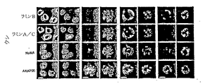
上記改善されたクローン化方法の使用を含む好ましい実施形態において、有蹄動物(例えば、ウシの胚、胎仔、仔ウシ又は成体)は、(a)好ましくは4対より少ない相同染色体を有する(すなわち2対未満の完全染色分体を有する)ドナー核(例えば異種抗体をコードする核酸を有する核)を、DNA複製を生じることなくクロマチン塊の形成が可能な条件下にてインキュベートするステップ、(b)該クロマチン塊を脱核卵母細胞に挿入し、それにより核移植卵母細胞を形成させるステップ、並びに(c)核移植卵母細胞又は核移植卵母細胞から形成される胚を、好ましくは該核移植卵母細胞又は胚が胎仔に発育可能な条件下にて、宿主哺乳動物の子宮に導入するステップ、を含む方法を用いて作出する。好ましい実施形態において、ドナー核は、核又は得られるクロマチン塊に核又は細胞質成分(例えば転写因子、リプレッサータンパク質又はクロマチン再構築タンパク質)が付加されるようなあるいは核又はクロマチン塊から除去されるような条件下で、再プログラム培地(例えば細胞抽出物)でインキュベートする。好ましくは、ドナー核は、クロマチン塊の形成が可能な以下の条件の1以上で接触させる:抗NuMA抗体の存在下若しくは不在下における有糸分裂抽出物、界面活性剤及び/又は塩溶液、あるいはプロテインキナーゼ溶液。他の好ましい実施形態において、再構成された卵母細胞又は得られる胚は、同じ細胞数で同じ種に由来する対照卵母細胞又は対照胚により発現される対応のレベルよりも5倍よりは小さなレベルでラミンA、ラミンC、又はNuMAタンパク質を発現する。 In a preferred embodiment comprising the use of the improved cloning method, the ungulate (eg, bovine embryo, fetus, calf or adult) has (a) preferably fewer than 4 pairs of homologous chromosomes (ie Incubating a donor nucleus (eg, a nucleus with a nucleic acid encoding a heterologous antibody) having less than two pairs of complete chromatids under conditions that allow the formation of a chromatin mass without causing DNA replication; (b Preferably) inserting the chromatin mass into an enucleated oocyte thereby forming a nuclear transfer oocyte; and (c) a nuclear transfer oocyte or an embryo formed from the nuclear transfer oocyte, Introducing the nuclear transfer oocyte or embryo into a uterus of a host mammal under conditions that allow the embryo to develop into a fetus. In a preferred embodiment, the donor nucleus is such that a nucleus or cytoplasmic component (eg, a transcription factor, repressor protein or chromatin remodeling protein) is added to the nucleus or the resulting chromatin mass, or is removed from the nucleus or chromatin mass. Incubate in reprogrammed medium (eg, cell extract) under mild conditions. Preferably, the donor nucleus is contacted under one or more of the following conditions that allow the formation of a chromatin mass: mitotic extract, detergent and / or salt solution in the presence or absence of anti-NuMA antibody, or Protein kinase solution. In other preferred embodiments, the reconstituted oocyte or resulting embryo is less than 5 times less than the corresponding level expressed by a control oocyte or control embryo derived from the same species with the same cell number. Expresses lamin A, lamin C, or NuMA protein at levels.
他の好ましい実施形態において、有蹄動物(例えば、ウシの胚、胎仔、仔ウシ又は成体)の作出方法は、透過性化細胞(例えば、異種抗体をコードする核酸を有する細胞)を再プログラム培地(例えば細胞抽出物)で、該透過性化細胞の核、クロマチン塊又は染色体からの因子(例えば、転写因子などの核若しくは細胞質成分)の除去、あるいは核、クロマチン塊又は染色体への因子の付加が可能な条件下にて、インキュベートし、それにより再プログラム化細胞を形成させることを含む。再プログラム化細胞は、脱核卵母細胞に挿入し、得られる卵母細胞又は該卵母細胞により形成される胚は、宿主哺乳動物の子宮に、好ましくは該卵母細胞又は胚が胎仔に発育可能な条件下にて、導入される。好ましい実施形態において、透過性化細胞は、クロマチン塊の形成が可能な以下の条件の1以上で接触させる:抗NuMA抗体の存在下若しくは不在下における有糸分裂抽出物、界面活性剤及び/又は塩溶液、あるいはプロテインキナーゼ溶液。さらに別の好ましい実施形態において、透過性化細胞は、間期再プログラム培地(例えば、間期細胞抽出物)と共にインキュベートする。また別の好ましい実施形態において、透過性化細胞における核は膜に結合した状態であり、核中の染色体はこの間期再プログラム培地とのインキュベーションの過程で凝縮しない。特定の実施形態において、再プログラム培地中での透過性化細胞のインキュベーションは、DNAの複製を生じないか、又は細胞の50%、40%、30%、20%、10%又は5%未満においてDNA複製が生じる。他の実施形態において、再プログラム培地中での透過性化細胞のインキュベーションは、細胞の少なくとも60%、70%、80%、90%、95%又は100%においてDNA複製を生じる。種々の実施形態において、透過性化細胞は、無傷細胞を、プロテアーゼ(トリプシンなど)、界面活性剤(ジギトニンなど)又は細菌性トキシン(ストレプトリシンOなど)と共にインキュベートすることにより形成される。好ましい実施形態において、再プログラム化細胞は、再プログラム化細胞の膜が卵母細胞への挿入前に再度封入されるような条件下でインキュベートするものではない。また別の好ましい実施形態において、再プログラム化細胞は、再プログラム化細胞の膜が卵母細胞への挿入前に再度封入されるような条件下でインキュベートする。他の好ましい実施形態において、再構成された卵母細胞又は得られる胚は、同じ細胞数で同じ種に由来する対照卵母細胞又は対照胚により発現される対応のレベルよりも5倍よりは小さなレベルでラミンA、ラミンC、又はNuMAタンパク質を発現する。 In another preferred embodiment, a method for producing an ungulate (eg, a bovine embryo, fetus, calf or adult) reprograms a permeabilized cell (eg, a cell having a nucleic acid encoding a heterologous antibody). Removal of factors (eg, nuclear or cytoplasmic components such as transcription factors) from the permeabilized cell nucleus, chromatin mass or chromosome, or addition of the factor to the nucleus, chromatin mass or chromosome. Incubating under possible conditions, thereby forming reprogrammed cells. The reprogrammed cell is inserted into a enucleated oocyte, and the resulting oocyte or embryo formed by the oocyte is in the uterus of the host mammal, preferably the oocyte or embryo is in the fetus. It is introduced under conditions that allow it to grow. In preferred embodiments, the permeabilized cells are contacted under one or more of the following conditions that allow the formation of a chromatin mass: mitotic extract, surfactant and / or in the presence or absence of anti-NuMA antibodies. Salt solution or protein kinase solution. In yet another preferred embodiment, the permeabilized cells are incubated with an interphase reprogramming medium (eg, an interphase cell extract). In another preferred embodiment, the nuclei in the permeabilized cells are membrane bound, and the chromosomes in the nuclei are not condensed during the incubation with this interphase reprogramming medium. In certain embodiments, incubation of permeabilized cells in reprogrammed medium does not result in DNA replication or in less than 50%, 40%, 30%, 20%, 10% or 5% of the cells DNA replication occurs. In other embodiments, incubation of permeabilized cells in reprogrammed media results in DNA replication in at least 60%, 70%, 80%, 90%, 95% or 100% of the cells. In various embodiments, permeabilized cells are formed by incubating intact cells with a protease (such as trypsin), a surfactant (such as digitonin) or a bacterial toxin (such as streptocrine O). In a preferred embodiment, the reprogrammed cells are not incubated under conditions such that the membranes of the reprogrammed cells are re-encapsulated prior to insertion into the oocyte. In yet another preferred embodiment, the reprogrammed cells are incubated under conditions such that the membrane of the reprogrammed cells is re-encapsulated prior to insertion into the oocyte. In other preferred embodiments, the reconstituted oocyte or resulting embryo is less than 5 times less than the corresponding level expressed by a control oocyte or control embryo derived from the same species with the same cell number. Expresses lamin A, lamin C, or NuMA protein at levels.
上記方法において使用するためのキメラ有蹄動物の好ましい作出方法
他の好ましい有蹄動物は、2以上の胚に由来する細胞を用いて作出されたキメラ有蹄動物又は有蹄動物である。例えば、核移植胚(例えば、細胞、核又はクロマチン塊を脱核卵母細胞に挿入することにより形成される胚)に由来する細胞は、in vitro受精(体外受精)胚、天然胚又は単為生殖活性化胚に由来する細胞と混合しうる。好ましくは、細胞及び該核移植胚からの子(progeny)の大部分は得られるキメラ胚の胎仔組織中に組み込まれる。細胞及び第2胚からの子の少なくとも一部は、胎盤組織に組み込まれ、得られるキメラ胚の生存率を促進することが好ましい。
Preferred methods for producing chimeric ungulates for use in the above methods Other preferred ungulates are chimeric ungulates or ungulates produced using cells derived from two or more embryos. For example, a cell derived from a nuclear transfer embryo (eg, an embryo formed by inserting a cell, nucleus or chromatin mass into an enucleated oocyte) can be an in vitro fertilized (in vitro fertilized) embryo, natural embryo or parthenol May be mixed with cells derived from reproductive activated embryos. Preferably, the cells and most of the progeny from the nuclear transfer embryo are incorporated into the fetal tissue of the resulting chimeric embryo. Preferably at least some of the cells and offspring from the second embryo are incorporated into placental tissue to promote survival of the resulting chimeric embryo.
好ましい実施形態において、核移植胚は異種抗体をコードする核酸を有する。好ましくは、内因性B細胞又はいずれかの初期胚に由来する細胞により産生される抗体を阻害するが、異種B細胞又は抗体を実質的に阻害しない抗体を、得られる胚、胎仔又は子孫に投与する。 In a preferred embodiment, the nuclear transfer embryo has a nucleic acid encoding a heterologous antibody. Preferably, antibodies that inhibit antibodies produced by endogenous B cells or cells derived from any early embryo but do not substantially inhibit heterologous B cells or antibodies are administered to the resulting embryo, fetus or offspring To do.
従って、種々の好ましい実施形態において、有蹄動物(例えば、ウシの胚、胎仔、仔ウシ又は成体)は、細胞、核又はクロマチン塊(例えば、異種抗体をコードする1以上の核酸を有する細胞、核又はクロマチン塊)を卵母細胞に挿入し、それにより第1胚を形成させることにより作出する。第1胚からの1以上の細胞を、第2胚からの1以上の細胞と接触させ、それにより第3胚を形成させる。第2胚は、in vitro(体外)受精胚、天然の胚、又は単為生殖的に活性化された胚である。第3胚は、宿主哺乳動物の子宮に、第3胚が胎仔に発育可能な条件下にて導入される。 Thus, in various preferred embodiments, an ungulate (eg, a bovine embryo, fetus, calf or adult) is a cell, nucleus or chromatin mass (eg, a cell having one or more nucleic acids encoding a heterologous antibody, It is created by inserting a nucleus or chromatin mass) into an oocyte, thereby forming a first embryo. One or more cells from the first embryo are contacted with one or more cells from the second embryo, thereby forming a third embryo. The second embryo is an in vitro fertilized embryo, a natural embryo, or an embryo that is parthenogenetically activated. The third embryo is introduced into the uterus of the host mammal under conditions that allow the third embryo to develop into the fetus.
一実施形態において、第1胚及び第2胚の少なくとも1つは緊密化胚である。別の実施形態において、第1胚及び第2胚は異なる細胞期にあるものである。第1胚、及び第2胚を作製するために使用されるドナー細胞は、同種に由来するものであってもよいし、又は異なる属若しくは種に由来するものであってもよい。好ましくは、胎仔の栄養外胚葉又は胎盤組織における少なくとも10%、20%、30%、40%、50%、60%、70%、80%、90%、95%又は100%の細胞が第2胚に由来するか、あるいは胎仔の内部細胞塊又は胎仔組織における少なくとも30%、40%、50%、60%、70%、80%、90%、95%又は100%の細胞が第1胚に由来するものである。他の好ましい実施形態において、第1胚又は第3胚は、同じ細胞数で同じ種に由来する対照胚により発現される対応のレベルよりも5倍より小さなレベルでラミンA、ラミンC、又はNuMAタンパク質を発現する。 In one embodiment, at least one of the first embryo and the second embryo is a compacted embryo. In another embodiment, the first embryo and the second embryo are at different cell stages. The donor cells used to produce the first embryo and the second embryo may be derived from the same species, or may be derived from different genera or species. Preferably, at least 10%, 20%, 30%, 40%, 50%, 60%, 70%, 80%, 90%, 95% or 100% of cells in fetal trophectoderm or placental tissue are second. At least 30%, 40%, 50%, 60%, 70%, 80%, 90%, 95% or 100% of cells in the embryo's inner cell mass or tissue are in the first embryo It comes from. In other preferred embodiments, the first embryo or the third embryo is lamin A, lamin C, or NuMA at a level that is 5 times less than the corresponding level expressed by a control embryo derived from the same species with the same number of cells. Express protein.
また他の実施形態において、有蹄動物(例えば、ウシの胚、胎仔、仔ウシ又は成体)は、ドナー核(例えば、異種抗体をコードする核)と再プログラム培地(例えば細胞抽出物)とを、クロマチン塊が形成可能な条件下にて接触させ、該クロマチン塊を脱核卵母細胞に挿入し、それにより第1胚を形成させることにより作出する。第1胚由来の1以上の細胞は、in vitro(体外)受精第2胚、天然の第2胚、又は単為生殖的に活性化された第2胚からの1以上の細胞と接触させ、第3胚を形成させる。この第3胚は、宿主哺乳動物の子宮に、第3胚が胎仔に発育可能な条件下にて導入される。好ましい実施形態において、クロマチン塊は、4対未満の相同染色体を有するドナー核と再プログラム培地を、DNA複製を生じることなくクロマチン塊が形成可能な条件下にて接触させることにより形成される。好ましくは、ドナー核は、クロマチン塊の形成が可能な以下の条件の1以上で接触させる:抗NuMA抗体の存在下若しくは不在下における有糸分裂抽出物、界面活性剤及び/又は塩溶液、あるいはプロテインキナーゼ溶液。 In still other embodiments, an ungulate (eg, a bovine embryo, fetus, calf or adult) comprises a donor nucleus (eg, a nucleus encoding a heterologous antibody) and a reprogramming medium (eg, a cell extract). It is created by contacting under conditions that allow the formation of a chromatin mass, inserting the chromatin mass into an enucleated oocyte, thereby forming a first embryo. One or more cells from the first embryo are contacted with one or more cells from an in vitro fertilized second embryo, a natural second embryo, or a second embryo that is parthenogenously activated; A third embryo is formed. This third embryo is introduced into the uterus of the host mammal under conditions that allow the third embryo to develop into the fetus. In a preferred embodiment, the chromatin mass is formed by contacting a donor nucleus having less than 4 pairs of homologous chromosomes with a reprogrammed medium under conditions that allow the formation of a chromatin mass without causing DNA replication. Preferably, the donor nucleus is contacted under one or more of the following conditions that allow the formation of a chromatin mass: mitotic extract, detergent and / or salt solution in the presence or absence of anti-NuMA antibody, or Protein kinase solution.
種々の実施形態において、第1胚及び第2胚の両方が緊密化胚であるか、第1胚及び第2胚の両方が緊密化前胚であるか、又は一方の胚が緊密化胚であり、他の胚が緊密化前胚である。第1胚及び第2胚は、異なる細胞期にあってもよいし又は同じ細胞期にあってもよい。第1胚、及び第2胚を作製するために用いるドナー核は、同じ種に由来するものであってもよいし又は異なる属若しくは種に由来するものであってもよい。好ましくは、胎仔の栄養外胚葉又は胎盤組織における少なくとも10%、20%、30%、40%、50%、60%、70%、80%、90%、95%又は100%の細胞が第2胚に由来するか、あるいは胎仔の内部細胞塊又は胎仔組織における少なくとも30%、40%、50%、60%、70%、80%、90%、95%又は100%の細胞が第1胚に由来するものである。他の好ましい実施形態において、第1胚又は第3胚は、同じ細胞数で同じ種に由来する対照胚により発現される対応のレベルよりも5倍よりは小さなレベルでラミンA、ラミンC、又はNuMAタンパク質を発現する。 In various embodiments, both the first embryo and the second embryo are compacted embryos, both the first embryo and the second embryo are pre-consolidated embryos, or one embryo is a compacted embryo. And other embryos are pre-embryonic embryos. The first embryo and the second embryo may be at different cell stages or at the same cell stage. The donor nuclei used to produce the first and second embryos may be derived from the same species or from different genera or species. Preferably, at least 10%, 20%, 30%, 40%, 50%, 60%, 70%, 80%, 90%, 95% or 100% of cells in fetal trophectoderm or placental tissue are second. At least 30%, 40%, 50%, 60%, 70%, 80%, 90%, 95% or 100% of cells in the embryo's inner cell mass or tissue are in the first embryo It comes from. In other preferred embodiments, the first embryo or the third embryo is lamin A, lamin C, or Expresses NuMA protein.
別の関連する態様において、本発明は、哺乳動物(ウシの胚、胎仔、仔ウシ又は成体)をクローン化する別の方法を特徴とする。この方法は、透過性化細胞(例えば、異種抗体をコードする核酸を有する細胞)を再プログラム培地(例えば細胞抽出物)中で、該透過性化細胞の核、クロマチン塊又は染色体からの因子の除去、あるいは核、クロマチン塊又は染色体への因子の付加が可能な条件下にて、インキュベートし、それにより再プログラム化細胞を形成させることを含む。再プログラム化細胞は、脱核卵母細胞に挿入し、それにより第1胚を形成させる。第1胚からの1以上の細胞は、in vitro(体外)受精第2胚、天然の第2胚、又は単為生殖的に活性化された第2胚からの1以上の細胞と接触させ、第3胚を形成させる。この第3胚は、宿主哺乳動物の子宮に、第3胚が胎仔に発育可能な条件下にて導入される。好ましい実施形態において、透過性化細胞は、核又は細胞質成分(例えば転写因子)が、核又は得られるクロマチン塊に付加される又はそれから除去されるのが可能な条件下にて、再プログラム培地(例えば細胞抽出物)と共にインキュベートする。他の好ましい実施形態において、透過性化細胞は、クロマチン塊の形成が可能な以下の条件の1以上で接触させる:抗NuMA抗体の存在下若しくは不在下における有糸分裂抽出物、界面活性剤及び/又は塩溶液、あるいはプロテインキナーゼ溶液。さらに別の好ましい実施形態において、透過性化細胞は、間期再プログラム培地(例えば、間期細胞抽出物)と共にインキュベートする。また別の好ましい実施形態において、透過性化細胞における核は膜に結合した状態であり、核中の染色体はこの間期再プログラム培地とのインキュベーションの過程で凝縮しない。いくつかの実施形態において、再プログラム培地中での透過性化細胞のインキュベーションは、DNAの複製を生じないか、又は細胞の50%、40%、30%、20%、10%又は5%未満においてDNA複製が生じる。他の実施形態において、再プログラム培地中での透過性化細胞のインキュベーションは、細胞の少なくとも60%、70%、80%、90%、95%又は100%においてDNA複製を生じる。種々の実施形態において、透過性化細胞は、無傷細胞を、プロテアーゼ(トリプシンなど)、界面活性剤(ジギトニンなど)又は細菌性トキシン(ストレプトリシンOなど)と共にインキュベートすることにより形成される。また別の好ましい実施形態において、再プログラム化細胞は、再プログラム化細胞の膜が卵母細胞への挿入前に再度封入されるような条件下でインキュベートする。種々の実施形態において、第1胚及び第2胚の両方が緊密化胚であるか、第1胚及び第2胚の両方が緊密化前胚であるか、又は胚の一方が緊密化胚であり他の胚が緊密化前胚である。第1胚及び第2胚は、異なる細胞期にあってもよいし又は同じ細胞期にあってもよい。第1胚、及び第2胚を作製するために用いるドナー核は、同じ種に由来するものであってもよいし又は異なる属若しくは種に由来するものであってもよい。好ましくは、胎仔の栄養外胚葉又は胎盤組織における少なくとも10%、20%、30%、40%、50%、60%、70%、80%、90%、95%又は100%の細胞が第2胚に由来するか、あるいは胎仔の内部細胞塊又は胎仔組織における少なくとも30%、40%、50%、60%、70%、80%、90%、95%又は100%の細胞が第1胚に由来するものである。他の好ましい実施形態において、第1胚又は第3胚は、同じ細胞数で同じ種に由来する対照胚により発現される対応のレベルよりも5倍より小さなレベルでラミンA、ラミンC、又はNuMAタンパク質を発現する。 In another related aspect, the invention features another method of cloning a mammal (bovine embryo, fetus, calf or adult). In this method, a permeabilized cell (eg, a cell having a nucleic acid encoding a heterologous antibody) is reprogrammed in a reprogramming medium (eg, cell extract) of factors from the permeabilized cell nucleus, chromatin mass, or chromosome. Incubating under conditions that allow removal or addition of factors to the nucleus, chromatin mass, or chromosome, thereby forming reprogrammed cells. The reprogrammed cell is inserted into the enucleated oocyte, thereby forming a first embryo. One or more cells from the first embryo are contacted with one or more cells from an in vitro fertilized second embryo, a natural second embryo, or a parthenogenously activated second embryo; A third embryo is formed. This third embryo is introduced into the uterus of the host mammal under conditions that allow the third embryo to develop into the fetus. In a preferred embodiment, the permeabilized cells are reprogrammed media (under reprogramming medium (under conditions) that allow nuclear or cytoplasmic components (eg, transcription factors) to be added to or removed from the nucleus or resulting chromatin mass. Incubate with eg cell extract). In other preferred embodiments, the permeabilized cells are contacted under one or more of the following conditions that allow the formation of a chromatin mass: mitotic extract in the presence or absence of anti-NuMA antibody, detergent and / Or salt solution or protein kinase solution. In yet another preferred embodiment, the permeabilized cells are incubated with an interphase reprogramming medium (eg, an interphase cell extract). In another preferred embodiment, the nuclei in the permeabilized cells are membrane bound, and the chromosomes in the nuclei are not condensed during the incubation with this interphase reprogramming medium. In some embodiments, incubation of permeabilized cells in reprogrammed media does not result in DNA replication or less than 50%, 40%, 30%, 20%, 10% or 5% of the cells DNA replication occurs in In other embodiments, incubation of permeabilized cells in reprogrammed media results in DNA replication in at least 60%, 70%, 80%, 90%, 95% or 100% of the cells. In various embodiments, permeabilized cells are formed by incubating intact cells with a protease (such as trypsin), a surfactant (such as digitonin) or a bacterial toxin (such as streptocrine O). In yet another preferred embodiment, the reprogrammed cells are incubated under conditions such that the membrane of the reprogrammed cells is re-encapsulated prior to insertion into the oocyte. In various embodiments, both the first embryo and the second embryo are compacted embryos, both the first embryo and the second embryo are pre-consolidated embryos, or one of the embryos is a compacted embryo. Other embryos are pre-embryonic embryos. The first embryo and the second embryo may be at different cell stages or at the same cell stage. The donor nuclei used to produce the first and second embryos may be derived from the same species or from different genera or species. Preferably, at least 10%, 20%, 30%, 40%, 50%, 60%, 70%, 80%, 90%, 95% or 100% of cells in fetal trophectoderm or placental tissue are second. At least 30%, 40%, 50%, 60%, 70%, 80%, 90%, 95% or 100% of cells in the embryo's inner cell mass or tissue are in the first embryo It comes from. In other preferred embodiments, the first embryo or the third embryo is lamin A, lamin C, or NuMA at a level that is 5 times less than the corresponding level expressed by a control embryo derived from the same species with the same number of cells. Express protein.
2つの胚に由来する細胞を用いて作出される有蹄動物に関する上記態様の好ましい実施形態において、第1胚又は第2胚の透明帯の一部又は全部は、各胚に由来する細胞を接触させる前に取り除く。一実施形態において、第1胚及び第2胚からの細胞は、溶液中又は固相支持体上で互いに隣接して配置させることにより接触させる。別の実施形態において、標準的な方法を用いて、第1胚からの細胞を第2胚に注入する。細胞は、第2胚のいずれの領域(例えば、透明帯と胚の間の胚の周囲、及び胚そのものなど)に注入することができる。天然の胚の例としては、標準的な方法を用いて妊娠哺乳動物(例えばウシ)から外科的に又は非外科的に取り出した胚である。in vitro受精(体外受精)胚の例としては、標準的な方法を用いて作製した細胞質内精子注入胚が含まれる。また、2を超える胚に由来する細胞(例えば、3、4、5、6又はそれ以上の胚からの細胞)を組み合わせて、クローン化哺乳動物の作出のためのキメラ胚を形成することも可能である。 In a preferred embodiment of the above aspect relating to ungulates produced using cells derived from two embryos, part or all of the zona pellucida of the first embryo or the second embryo contacts the cells derived from each embryo Remove before letting. In one embodiment, cells from the first and second embryos are contacted by placing them adjacent to each other in solution or on a solid support. In another embodiment, cells from the first embryo are injected into the second embryo using standard methods. The cells can be injected into any region of the second embryo (eg, around the embryo between the zona pellucida and the embryo, and the embryo itself). An example of a natural embryo is an embryo surgically or non-surgically removed from a pregnant mammal (eg, a cow) using standard methods. Examples of in vitro fertilized (in vitro fertilized) embryos include intracytoplasmic sperm-injected embryos produced using standard methods. It is also possible to combine cells from more than 2 embryos (eg cells from 3, 4, 5, 6 or more embryos) to form a chimeric embryo for the production of cloned mammals It is.
上記方法において用いるための有蹄動物を作出するための好ましい実施形態
上記態様の好ましい実施形態において、再プログラム培地(例えば細胞抽出物)は、因子、例えばDNAメチルトランスフェラーゼ、ヒストンデアセチラーゼ、ヒストン、プロタミン、核ラミン、転写因子、アクチベーター又はリプレッサーなどの濃縮又は枯渇により変更が加えられる。他の好ましい実施形態において、卵母細胞又はキメラ胚におけるNuMA又はAKAP95タンパク質の発現レベルは、細胞質におけるレベルよりも核におけるレベルの方が少なくとも2、5、10又は20倍大きい。また他の実施形態において、卵母細胞又はキメラ胚におけるAKAP95タンパク質の少なくとも30、40、50、60、70、80、90又は100%は、0.1%TritonX−100、1mg/ml DNaseI、及び100mM又は300mM NaClの溶液を用いて抽出される。好ましくは、クロマチン塊は、脱核卵母細胞に挿入する前に再プログラム培地(例えば抽出物)から精製される。別の好ましい実施形態において、クロマチン塊の脱核卵母細胞への挿入は、クロマチン塊及び卵母細胞と、融合性化合物(fusigenic compound)とを、クロマチン塊が卵母細胞に侵入可能な条件下にて接触させることを含む。また別の好ましい実施形態において、胎仔は生存子孫に発育する。好ましくは、核移植卵母細胞又は胚の少なくとも1、3、5、10、20、30、40、50、60、70、80又は90%が生存子孫に発育する。この方法においては、クロマチン塊を含む卵母細胞又は再プログラム化細胞は、細胞分裂が可能な条件下で培養することができ、得られる細胞の1つは、1回以上再クローン化しうる。本発明において用いるドナー核、ドナークロマチン塊又はドナー細胞、及び卵母細胞は、同種に由来するものであってもよいし、又は異なる種若しくは属に由来するものであってもよい。哺乳動物はヒト又は非ヒト哺乳動物であり、卵母細胞は受精されていても未受精であってもよい。好ましくは、ドナー核、クロマチン塊又は透過性化細胞はG1期又はG0期細胞からのものである。さらに、クローン化胚、胎仔又は哺乳動物のゲノムDNAは、ドナー細胞のものと実質的に同一であることが好ましい。また、クロマチン塊又は再プログラム化細胞は、胚において該クロマチン塊又は再プログラム化細胞と実質的に同一のDNAを有する細胞と天然細胞と実質的に同一のDNAを有する細胞との混合物を含有する、キメラ胚、胎仔又は哺乳動物の作出のための胚に挿入しうることも意図される。また、脱核卵母細胞を本発明の方法に使用しうることも意図される。
Preferred embodiments for producing ungulates for use in the above method In preferred embodiments of the above aspects, the reprogrammed medium (eg, cell extract) is a factor such as DNA methyltransferase, histone deacetylase, histone, Changes are made by enrichment or depletion of protamine, nuclear lamins, transcription factors, activators or repressors. In other preferred embodiments, the expression level of NuMA or AKAP95 protein in the oocyte or chimeric embryo is at least 2, 5, 10 or 20 times greater in the nucleus than in the cytoplasm. In still other embodiments, at least 30, 40, 50, 60, 70, 80, 90 or 100% of AKAP95 protein in an oocyte or chimeric embryo is 0.1% Triton X-100, 1 mg / ml DNase I, and Extraction is performed using a solution of 100 mM or 300 mM NaCl. Preferably, the chromatin mass is purified from reprogrammed media (eg, extract) prior to insertion into enucleated oocytes. In another preferred embodiment, the insertion of the chromatin mass into the enucleated oocyte comprises the chromatin mass and the oocyte and a fusigenic compound under conditions that allow the chromatin mass to enter the oocyte. Including contact with. In yet another preferred embodiment, the fetus develops into a live offspring. Preferably, at least 1, 3, 5, 10, 20, 30, 40, 50, 60, 70, 80 or 90% of the nuclear transfer oocyte or embryo develop into viable offspring. In this method, an oocyte or reprogrammed cell containing a chromatin mass can be cultured under conditions that allow cell division, and one of the resulting cells can be recloned one or more times. The donor nucleus, donor chromatin mass or donor cell, and oocyte used in the present invention may be derived from the same species, or may be derived from different species or genera. The mammal is a human or non-human mammal and the oocyte may be fertilized or unfertilized. Preferably, the donor nucleus, chromatin mass or permeabilized cell is from a G 1 or G 0 phase cell. Furthermore, the genomic DNA of the cloned embryo, fetus or mammal is preferably substantially identical to that of the donor cell. Also, the chromatin mass or reprogrammed cell contains a mixture of cells having substantially the same DNA in the embryo as the chromatin mass or reprogrammed cell and cells having substantially the same DNA as the natural cell. It is also contemplated that it can be inserted into an embryo for production of a chimeric embryo, fetus or mammal. It is also contemplated that enucleated oocytes can be used in the methods of the present invention.
本発明の任意の態様において用いる再プログラム培地は、異種ヌクレオチド配列を含んでもよいし又は含まなくてもよい。他の好ましい実施形態において、再プログラム培地中の又は透過性化細胞において形成されるクロマチン塊は、目的遺伝子をコードする核酸を有するベクターと、ベクター中の核酸とクロマチン塊のゲノム中の対応核酸との間でランダム組み込み又は相同組換えが可能な条件下で接触させ、クロマチン塊のゲノムを改変させる。完全な形質膜が存在せず、また核膜が存在しないため、透過性化細胞中又は溶液中のクロマチン塊は、天然細胞よりも遺伝的に改変されやすくなりうる。再プログラム抽出物を調製するために使用し得る細胞の例としては、胚性幹細胞、及び脳、血液、骨髄、膵臓、肝臓、皮膚又は他の任意の器官若しくは組織からの成体幹細胞が含まれる。他の再プログラム細胞抽出物の例としては、卵母細胞抽出物(例えば、ウシ又はウニの卵母細胞抽出物)、及び雄性生殖細胞抽出物(例えば、脊椎動物、非脊椎動物、又は哺乳動物(ウシなど)からの精原細胞、精母細胞、精子細胞又は精子の抽出物)が挙げられる。ドナー細胞又は透過性化細胞は、不死化されていないものであってもよいし、又は天然に、自発的に若しくは遺伝学的に不死化されたものであってもよい。ドナー細胞、透過性化細胞、レシピエント細胞又はサイトプラストは、任意の年齢の供給源、例えば胚、胎仔、若年又は成体の哺乳動物に由来するものであり得る。より若年の供給源に由来する細胞は、自発的突然変異の獲得がより少ない可能性があり、また卵母細胞への挿入後の寿命がより長い可能性がある。 The reprogramming medium used in any aspect of the present invention may or may not include heterologous nucleotide sequences. In another preferred embodiment, the chromatin mass formed in the reprogramming medium or in the permeabilized cells comprises a vector having a nucleic acid encoding a gene of interest, a nucleic acid in the vector and a corresponding nucleic acid in the genome of the chromatin mass. The chromatin mass genome is altered by contacting them under conditions allowing random integration or homologous recombination. Because there is no complete plasma membrane and no nuclear membrane, chromatin masses in permeabilized cells or in solution can be more genetically modified than natural cells. Examples of cells that can be used to prepare the reprogrammed extract include embryonic stem cells and adult stem cells from the brain, blood, bone marrow, pancreas, liver, skin, or any other organ or tissue. Examples of other reprogrammed cell extracts include oocyte extracts (eg, bovine or sea urchin oocyte extracts) and male germ cell extracts (eg, vertebrates, invertebrates, or mammals) Spermatogonia, spermatocytes, sperm cells or sperm extracts from bovines). Donor cells or permeabilized cells may be non-immortalized, or may be naturally, spontaneously or genetically immortalized. Donor cells, permeabilized cells, recipient cells or cytoplasts can be derived from any age source, eg, embryos, fetuses, young or adult mammals. Cells from younger sources may acquire less spontaneous mutations and may have a longer life after insertion into an oocyte.
上記方法において用いるための内因性抗体のレベルが低減された好ましい有蹄動物
本発明の方法は、内因性抗体の発現を低減する突然変異を有する有蹄動物(例えばウシ)を用いて使用しうる。従って、投与抗体が少ないと、低い初期レベルの内因性抗体を排除する必要がある。好ましくは、突然変異は、機能性IgM重鎖の発現を低減し、又は機能性IgM重鎖の発現を実質的に排除する。他の好ましい実施形態において、突然変異は、機能性Ig軽鎖の発現を低減し、又は機能性Ig軽鎖の発現を実質的に排除する。さらに他の好ましい実施形態において、突然変異は、機能性IgM重鎖及び機能性Ig軽鎖の発現を低減し、又は機能性IgM重鎖及び機能性Ig軽鎖の発現を実質的に排除する。いくつかの実施形態において、転写終結配列を、内因性μ重鎖核酸に挿入する(例えば、エキソン2における最初のATGコドンの下流に挿入する)又はIg軽鎖核酸に挿入する。好ましくは、有蹄動物はまた、α−(1,3)−ガラクトシルトランスフェラーゼ、プリオンタンパク質及び/又はJ鎖をコードする内因性核酸の一方又は両方のアレルに突然変異を有する。他の好ましい実施形態において、有蹄動物は、外因性J鎖、例えばヒトJ鎖をコードする核酸を有する。好ましくは、突然変異は、内因性α−(1,3)−ガラクトシルトランスフェラーゼ酵素、ガラクトシル(α1,3)ガラクトースエピトープ、プリオンタンパク質及び/又はJ鎖の発現を低減又は排除する。有蹄動物は、ヒトJ鎖を含有するヒトIgA又はIgM分子を産生することが好ましい。
Preferred ungulates with reduced levels of endogenous antibodies for use in the above methods The methods of the present invention may be used with ungulates (eg, cows) having mutations that reduce the expression of endogenous antibodies. . Therefore, if the administered antibody is low, it is necessary to eliminate low initial levels of endogenous antibody. Preferably, the mutation reduces the expression of a functional IgM heavy chain or substantially eliminates the expression of a functional IgM heavy chain. In other preferred embodiments, the mutation reduces functional Ig light chain expression or substantially eliminates functional Ig light chain expression. In still other preferred embodiments, the mutation reduces the expression of functional IgM heavy chain and functional Ig light chain, or substantially eliminates the expression of functional IgM heavy chain and functional Ig light chain. In some embodiments, the transcription termination sequence is inserted into an endogenous μ heavy chain nucleic acid (eg, inserted downstream of the first ATG codon in exon 2) or inserted into an Ig light chain nucleic acid. Preferably, the ungulate also has a mutation in one or both alleles of the endogenous nucleic acid encoding α- (1,3) -galactosyltransferase, prion protein and / or J chain. In other preferred embodiments, the ungulate has a nucleic acid encoding an exogenous J chain, eg, a human J chain. Preferably, the mutation reduces or eliminates the expression of endogenous α- (1,3) -galactosyltransferase enzyme, galactosyl (α1,3) galactose epitope, prion protein and / or J chain. The ungulate preferably produces a human IgA or IgM molecule containing human J chain.
好ましくは、内因性遺伝子又は遺伝子群に1以上の突然変異を有するトランスジェニック有蹄動物は、細胞、細胞由来のクロマチン塊又は細胞由来の核を卵母細胞に挿入することにより作出される。細胞は、該細胞により天然には発現されない、内因性遺伝子における第1突然変異を有する。卵母細胞又は該卵母細胞から形成される胚は、宿主有蹄動物の子宮に、該卵母細胞又は胚が胎仔に発育可能な条件下で導入される。好ましくは、胎仔は生存子孫にまで発育する。 Preferably, a transgenic ungulate having one or more mutations in an endogenous gene or group of genes is created by inserting a cell, a cell-derived chromatin mass or a cell-derived nucleus into an oocyte. The cell has a first mutation in an endogenous gene that is not naturally expressed by the cell. An oocyte or embryo formed from the oocyte is introduced into the uterus of a host ungulate under conditions that allow the oocyte or embryo to develop into a fetus. Preferably, the fetus develops to a viable offspring.
上記方法において使用するための、突然変異、例えば内因性抗体のレベルの低減を導く突然変異、を有する有蹄動物を作出する好ましい方法
他の好ましい実施形態において、第1突然変異は、選択マーカーをコードする核酸と機能的に連結し、かつ突然変異対象の内因性遺伝子と実質的に同一の配列を有する1以上の核酸と機能的に連結したプロモーターを含むカセットを含む核酸を細胞に挿入することにより、カセットが遺伝子の一方の内因性アレルに組み込まれるように、細胞中に導入する。他の好ましい実施形態において、突然変異は、第1選択マーカーをコードする核酸と機能的に連結し、かつ突然変異対象の内因性遺伝子と実質的に同一の配列を有する第1核酸と機能的に連結した第1プロモーターを含む第1カセットを含む核酸を細胞に挿入することにより、第1カセットが遺伝子の第1内因性アレルに組み込まれるように、細胞中に導入し、第1トランスジェニック細胞を作出する。第1トランスジェニック細胞には、第2選択マーカーをコードする核酸と機能的に連結し、かつ上記遺伝子と実質的に同一の配列を有する第2核酸と機能的に連結した第2プロモーターを含む第2カセットを含む核酸を挿入する。第2選択マーカーは第1選択マーカーとは異なるものであり、第2カセットは遺伝子の第2内因性アレルに組み込まれて、第2トランスジェニック細胞を作出する。また他の好ましい実施形態において、細胞は、胚、胎仔又は胎仔から作出される子孫から単離されたものであり、別の突然変異が該細胞の遺伝子に導入されている。その後、得られた細胞、該細胞からのクロマチン塊、又は該細胞からの核を用いて第2ラウンドの核移植を行い、2以上の突然変異を有するトランスジェニック有蹄動物を作出する。複数の突然変異は、遺伝子の同じ若しくは異なるアレルにあってもよいし、異なる遺伝子にあってもよい。第1ラウンド又は任意により第2ラウンドの核移植において使用する細胞は異種抗体をコードする。特定の実施形態において、細胞は、B細胞において再配列を経て1以上の異種Ig分子を発現可能な異種Ig遺伝子の全部又は一部をコードする1以上の核酸を含む。好ましい実施形態において、突然変異させる細胞は、線維芽細胞(例えば胎児線維芽細胞)である。好ましくは、突然変異させる内因性遺伝子は、線維芽細胞において活性を有しない内因性プロモーターと機能的に連結する。他の好ましい実施形態において、突然変異させる内因性遺伝子と機能的に連結した内因性プロモーターは、内因性ハウスキーピング遺伝子(GAPDHなど)と機能的に連結した内因性プロモーターの活性の80、70、60、50、40、30、20、10%未満の活性である。プロモーター活性は、任意の標準的なアッセイ、例えば遺伝子によりコードされるmRNA又はタンパク質のレベルを測定するアッセイを用いて測定する(例えば、Ausubelら、Current Protocols in Molecular Biology, volume 2, p. 11.13.1-11.13.3, John Wiley & Sons, 1995参照)。トランスジェニック有蹄動物を作出するこの方法は、ドナー細胞(すなわち、核移植に用いる遺伝物質の供与源となる細胞)において発現されない遺伝子を突然変異させることができるという利点を有する。
Preferred methods for generating ungulates with mutations, eg, mutations that lead to a reduction in the level of endogenous antibodies, for use in the above method In other preferred embodiments, the first mutation comprises a selectable marker Inserting into a cell a nucleic acid comprising a cassette comprising a promoter operably linked to the encoding nucleic acid and operably linked to one or more nucleic acids having a sequence substantially identical to the endogenous gene to be mutated To introduce the cassette into the cell so that it is integrated into one endogenous allele of the gene. In another preferred embodiment, the mutation is operably linked to a nucleic acid encoding a first selectable marker and is functionally linked to a first nucleic acid having a sequence substantially identical to the endogenous gene to be mutated. By inserting a nucleic acid comprising a first cassette comprising a linked first promoter into the cell, the first cassette is introduced into the cell such that it is incorporated into the first endogenous allele of the gene, and the first transgenic cell is Create. The first transgenic cell includes a second promoter operably linked to a nucleic acid encoding a second selectable marker and operably linked to a second nucleic acid having a sequence substantially identical to the gene. Insert nucleic acid containing 2 cassettes. The second selectable marker is different from the first selectable marker, and the second cassette is integrated into the second endogenous allele of the gene to create a second transgenic cell. In yet another preferred embodiment, the cell is isolated from an embryo, fetus or offspring generated from the fetus, and another mutation has been introduced into the gene of the cell. Thereafter, a second round of nuclear transfer is performed using the obtained cells, the chromatin mass from the cells, or the nuclei from the cells to produce transgenic ungulates having two or more mutations. Multiple mutations may be in the same or different alleles of the gene or in different genes. The cells used in the first round, or optionally the second round of nuclear transfer, encode a heterologous antibody. In certain embodiments, the cell comprises one or more nucleic acids encoding all or part of a heterologous Ig gene capable of expressing one or more heterologous Ig molecules via rearrangement in B cells. In a preferred embodiment, the cells to be mutated are fibroblasts (eg fetal fibroblasts). Preferably, the endogenous gene to be mutated is operably linked to an endogenous promoter that is not active in fibroblasts. In another preferred embodiment, the endogenous promoter operably linked to the endogenous gene to be mutated is 80, 70, 60 of the activity of the endogenous promoter operably linked to the endogenous housekeeping gene (such as GAPDH). , 50, 40, 30, 20, less than 10% activity. Promoter activity is measured using any standard assay, such as an assay that measures the level of mRNA or protein encoded by a gene (eg, Ausubel et al., Current Protocols in Molecular Biology,
好ましくは、内因性抗体遺伝子に突然変異を有するトランスジェニック有蹄動物は、細胞、細胞由来のクロマチン塊又は細胞由来の核を卵母細胞に挿入することにより作出される。細胞は、内因性抗体重鎖及び/又は軽鎖核酸に第1突然変異を含む。卵母細胞又は該卵母細胞から形成される胚は、宿主有蹄動物の子宮に、該卵母細胞又は胚が胎仔に発育可能な条件下にて導入される。好ましくは、胎仔は生存子孫にまで発育させる。好ましくは、トランスジェニック有蹄動物の作出に使用する細胞は、α−(1,3)−ガラクトシルトランスフェラーゼ、プリオンタンパク質及び/又はJ鎖をコードする内因性核酸の一方又は両方のアレルに突然変異を有する。他の好ましい実施形態において、細胞は、外因性J鎖、例えばヒトJ鎖をコードする核酸を有する。好ましくは、トランスジェニック有蹄動物を作出する方法はまた、胚、胎仔又は該胎仔から作出される子孫から細胞を単離し、該細胞における内因性抗体重鎖及び/又は軽鎖核酸に第2突然変異(例えば転写終結配列の挿入)を導入することを含む。細胞、該細胞由来のクロマチン塊又は該細胞由来の核は卵母細胞に挿入し、卵母細胞又は該卵母細胞から形成される胚は、宿主有蹄動物の子宮に、該卵母細胞又は胚が胎仔にまで発育可能な条件下にて導入される。第1ラウンド又は任意により第2ラウンドの核移植において用いる細胞は異種抗体をコードする。 Preferably, a transgenic ungulate with a mutation in an endogenous antibody gene is created by inserting a cell, a cell-derived chromatin mass or a cell-derived nucleus into an oocyte. The cell contains a first mutation in the endogenous antibody heavy and / or light chain nucleic acid. An oocyte or embryo formed from the oocyte is introduced into the uterus of a host ungulate under conditions that allow the oocyte or embryo to develop into a fetus. Preferably, the fetus is grown to a viable offspring. Preferably, the cells used for the production of the transgenic ungulates are mutated in one or both alleles of the endogenous nucleic acid encoding α- (1,3) -galactosyltransferase, prion protein and / or J chain. Have. In other preferred embodiments, the cell has a nucleic acid encoding an exogenous J chain, eg, a human J chain. Preferably, the method of creating a transgenic ungulate also isolates cells from an embryo, fetus or progeny produced from the fetus, and second suddenly adds to the endogenous antibody heavy chain and / or light chain nucleic acid in the cell. Introduction of mutations (eg insertion of transcription termination sequences). The cell, the chromatin mass derived from the cell or the nucleus derived from the cell are inserted into the oocyte, and the oocyte or an embryo formed from the oocyte is inserted into the uterus of the host ungulate, the oocyte or The embryo is introduced under conditions that allow it to develop into the fetus. The cells used in the first round, or optionally the second round of nuclear transfer, encode a heterologous antibody.
上記トランスジェニック有蹄動物の作出のための別の実施形態において、トランスジェニック有蹄動物の作出に使用する細胞は、選択マーカーをコードする核酸と機能的に連結し、かつ抗体重鎖又は軽鎖核酸と実質的に同一の配列を有する1以上の核酸と機能的に連結したプロモーターを含むカセットを有する核酸を細胞に挿入することを含む方法によって調製される。このカセットは、抗体重鎖又は軽鎖核酸の一方の内因性アレルに組み込まれる。 In another embodiment for the production of a transgenic ungulate, the cell used for the production of the transgenic ungulate is operably linked to a nucleic acid encoding a selectable marker and the antibody heavy or light chain. It is prepared by a method comprising inserting into a cell a nucleic acid having a cassette comprising a promoter operably linked to one or more nucleic acids having substantially the same sequence as the nucleic acid. This cassette is incorporated into one endogenous allele of the antibody heavy or light chain nucleic acid.
他の実施形態において、細胞は、第1選択マーカーをコードする核酸と機能的に連結し、かつ抗体重鎖又は軽鎖核酸と実質的に同一の配列を有する第1核酸と機能的に連結した第1プロモーターを含む第1カセットを有する核酸を細胞に挿入することにより作製される。第1カセットは、抗体重鎖又は軽鎖核酸の第1内因性アレルに組み込まれ、第1トランスジェニック細胞を作製する。第1トランスジェニック細胞には、第2選択マーカーをコードする核酸と機能的に連結し、かつ抗体重鎖又は軽鎖核酸と実質的に同一の配列を有する第2核酸と機能的に連結した第2プロモーターを含む第2カセットを有する核酸を挿入する。第2選択マーカーは第1選択マーカーとは異なるものである。第2カセットは抗体重鎖又は軽鎖核酸の第2内因性アレルに組み込まれて、第2トランスジェニック細胞を作製する。 In other embodiments, the cell is operably linked to a nucleic acid encoding a first selectable marker and operably linked to a first nucleic acid having a sequence that is substantially identical to the antibody heavy or light chain nucleic acid. It is produced by inserting a nucleic acid having a first cassette containing a first promoter into a cell. The first cassette is integrated into the first endogenous allele of the antibody heavy or light chain nucleic acid to create a first transgenic cell. The first transgenic cell includes a first operably linked nucleic acid encoding a second selectable marker and a second nucleic acid operably linked to a second nucleic acid having substantially the same sequence as the antibody heavy or light chain nucleic acid. A nucleic acid having a second cassette containing two promoters is inserted. The second selection marker is different from the first selection marker. The second cassette is incorporated into a second endogenous allele of the antibody heavy or light chain nucleic acid to create a second transgenic cell.
本発明の種々の実施形態において、内因性有蹄動物核酸を突然変異させるのに使用する核酸(例えば、選択マーカーをコードする核酸と機能的に連結し、かつ突然変異対象の遺伝子と実質的に同一の配列を有する核酸と機能的に連結されたプロモーターを含むノックアウトカセット)は、ウイルスベクター、例えばアデノウイルスベクター又はアデノ随伴ウイルスベクターに含まれない。例えば、核酸は、標準的な方法(トランスフェクション又はリポフェクションなど)を用いて有蹄動物細胞に挿入されるが細胞のウイルス感染を伴わないプラスミド又は人工染色体に含まれ得る。また別の実施形態において、内因性有蹄動物核酸を突然変異させるのに使用する核酸(例えば、選択マーカーをコードする核酸と機能的に連結し、かつ突然変異対象の遺伝子と実質的に同一の配列を有する核酸と機能的に連結されたプロモーターを含むノックアウトカセット)は、ウイルスベクター、例えばアデノウイルスベクター又はアデノ随伴ウイルスベクターに含まれる。この実施形態により、ウイルスベクターを含むウイルスを用いて有蹄動物細胞に感染し、それによりウイルスベクターの一部又は全体が有蹄動物細胞に挿入される。 In various embodiments of the invention, a nucleic acid used to mutate an endogenous ungulate nucleic acid (eg, operably linked to a nucleic acid encoding a selectable marker, and substantially mutated with the gene to be mutated). A knockout cassette comprising a promoter operably linked to a nucleic acid having the same sequence is not included in a viral vector, such as an adenoviral vector or an adeno-associated viral vector. For example, the nucleic acid can be contained in a plasmid or artificial chromosome that is inserted into ungulate cells using standard methods (such as transfection or lipofection) but without viral infection of the cells. In yet another embodiment, a nucleic acid used to mutate an endogenous ungulate nucleic acid (eg, operably linked to a nucleic acid encoding a selectable marker and substantially identical to the gene to be mutated) A knockout cassette comprising a promoter operably linked to a nucleic acid having a sequence) is included in a viral vector, such as an adenoviral vector or an adeno-associated viral vector. According to this embodiment, a virus containing a viral vector is used to infect ungulate cells, whereby part or all of the viral vector is inserted into ungulate cells.
望ましくない内因性抗体を排除するために上記方法において使用するための好ましい投与抗体
好ましい投与抗体としては、抗IgM抗体及び内因性有蹄動物抗体のポリクローナル混合物と反応する抗体が含まれる。投与抗体はモノクローナルでもポリクローナルでもよい。いくつかの実施形態において、投与抗体は、二機能性抗体、抗体のフラグメント、又は修飾抗体である。特定の実施形態において、投与抗体はトキシン(例えば、ジフテリアトキシン、マイタンシノイド、CC−1065、アントラサイクリン若しくはタキサン)又は放射性標識と共有結合している。いくつかの実施形態において、抗体は有蹄動物に静脈内投与する。好ましくは、少なくとも0.25、0.5、1.0、1.5、2、10、20又は50グラムの抗体を、単回又は複数回用量で有蹄動物に投与する。他の実施形態において、1〜10mg、10〜25mg、25〜50mg、10〜100mg、50〜100mg、又は100〜500mgの抗体を、単回又は複数回用量で有蹄動物胎仔に投与する。所望であれば、抗体は、薬学的に許容される希釈剤、担体、又は賦形剤(例えば、生理食塩水、緩衝化生理食塩水、デキストロース、水、グリセロール、エタノール又はこれらの組み合わせ)中に溶解して投与してもよい。
Preferred administered antibodies for use in the above methods to eliminate unwanted endogenous antibodies Preferred administered antibodies include antibodies that react with a polyclonal mixture of anti-IgM antibodies and endogenous ungulate antibodies. The administered antibody may be monoclonal or polyclonal. In some embodiments, the administered antibody is a bifunctional antibody, a fragment of an antibody, or a modified antibody. In certain embodiments, the administered antibody is covalently linked to a toxin (eg, diphtheria toxin, maytansinoid, CC-1065, anthracycline or taxane) or a radiolabel. In some embodiments, the antibody is administered intravenously to the ungulate. Preferably, at least 0.25, 0.5, 1.0, 1.5, 2, 10, 20 or 50 grams of antibody is administered to the ungulate in single or multiple doses. In other embodiments, 1-10 mg, 10-25 mg, 25-50 mg, 10-100 mg, 50-100 mg, or 100-500 mg of antibody is administered to an ungulate fetus in single or multiple doses. If desired, the antibody can be in a pharmaceutically acceptable diluent, carrier, or excipient (eg, saline, buffered saline, dextrose, water, glycerol, ethanol, or combinations thereof). It may be administered after being dissolved.
好ましい実施形態において、投与抗体は、レシピエント有蹄動物とは異なる属又は種の有蹄動物に由来するものである。他の好ましい実施形態において、抗体は二機能性抗体である。また他の好ましい抗体としては、ScFv、Fab若しくはF(ab’)2フラグメントを有する又はこれらからなるものが含まれる。他の好ましい抗体の例としては、遺伝子融合技術により抗体又は抗体フラグメントをコードする核酸がトキシン又はアフィニティタグと機能的に連結されるように改変された融合核酸によりコードされる誘導体化抗体が含まれる。誘導体化抗体中の共有結合基は、抗体又は抗体フラグメントのアミノ末端、カルボキシ末端、又はアミノ末端とカルボキシ末端の間に結合しうる。「アフィニティタグ」とは、別のペプチド、タンパク質又は化合物と結合するペプチド、タンパク質、又は化合物を意味する。好ましい実施形態において、アフィニティタグは誘導体化抗体の精製又は免疫感作に用いる。別の好ましい実施形態において、アフィニティタグ又はトキシンを用いて、in vivoにおいて抗体を特定の細胞、組織又は器官系に標的化する。 In preferred embodiments, the administered antibody is from an ungulate of a different genus or species than the recipient ungulate. In other preferred embodiments, the antibody is a bifunctional antibody. Other preferred antibodies include those having or consisting of a ScFv, Fab or F (ab ′) 2 fragment. Examples of other preferred antibodies include derivatized antibodies encoded by fusion nucleic acids that have been modified by gene fusion techniques such that the nucleic acid encoding the antibody or antibody fragment is operably linked to a toxin or affinity tag. . The covalent linking group in the derivatized antibody can be linked to the amino terminus, carboxy terminus, or between the amino terminus and carboxy terminus of the antibody or antibody fragment. “Affinity tag” means a peptide, protein, or compound that binds to another peptide, protein, or compound. In a preferred embodiment, affinity tags are used for purification or immunization of derivatized antibodies. In another preferred embodiment, affinity tags or toxins are used to target antibodies to specific cells, tissues or organ systems in vivo.
上記態様の好ましい実施形態
好ましくは、本発明のトランスジェニック細胞又は有蹄動物は、ポジティブセレクションマーカー(例えば抗生物質耐性遺伝子)の、免疫グロブリン、α−(1,3)−ガラクトシルトランスフェラーゼ、プリオンタンパク質又はJ鎖をコードする内因性核酸への挿入を有する。望ましくは、ポジティブセレクションマーカーは異種プロモーターと機能的に連結する。いくつかの実施形態において、各アレルは同じ抗生物質耐性遺伝子又は異なる抗生物質耐性遺伝子の挿入を有する。好ましくは、転写終結配列を、免疫グロブリン、α−(1,3)−ガラクトシルトランスフェラーゼ、プリオンタンパク質又はJ鎖をコードする内因性核酸に挿入する。例えば、転写終結配列は、内因性μ重鎖核酸のエキソン2における最初のATGコドンの下流に挿入し得る。好ましい実施形態において、細胞又は有蹄動物は、1以上の導入遺伝子(transgene)を含み、該導入遺伝子によりコードされるmRNA又はタンパク質を発現する1以上の核酸を有する。
Preferred embodiments of the above aspects Preferably, the transgenic cell or ungulate of the present invention is a positive selection marker (eg antibiotic resistance gene) of immunoglobulin, α- (1,3) -galactosyltransferase, prion protein or Has an insertion into the endogenous nucleic acid encoding the J chain. Desirably, the positive selection marker is operably linked to a heterologous promoter. In some embodiments, each allele has an insertion of the same antibiotic resistance gene or a different antibiotic resistance gene. Preferably, the transcription termination sequence is inserted into an endogenous nucleic acid encoding immunoglobulin, α- (1,3) -galactosyltransferase, prion protein or J chain. For example, a transcription termination sequence can be inserted downstream of the first ATG codon in
好ましくは、有蹄動物の抗血清又は乳汁はポリクローナルヒト免疫グロブリンを有する。好ましくは、抗血清又は乳汁はウシ、ヒツジ、ブタ又はヤギからのものである。別の好ましい実施形態において、Igは所望の抗原に対して生じたものである。好ましい実施形態において、抗血清は、ヒトにおける疾患の治療又は予防のための静注用免疫グロブリン(IVIG)として用い得る。別の好ましい実施形態において、目的の抗原を有蹄動物に投与し、該抗原に対して生じるIgを有蹄動物により産生させる。好ましくは、異種免疫グロブリン遺伝子座における核酸セグメントが再配列し、目的の抗原と反応する異種抗体が産生される。好ましくは、抗血清及び/又は乳汁は、完全に又は部分的に内因性の抗体よりも少なくとも2、5、10、20又は50倍以上の完全に異種の抗体を含み、あるいは完全に又は部分的に内因性の抗体を全く含まない。所望であれば、ハイブリドーマ及びモノクローナル抗体を、上記トランスジェニック有蹄動物(例えばトランスジェニックウシ)に由来する異種B細胞を用いて調製しうる。次に、有蹄動物から単離される異種抗体(例えばヒト抗体)を化学的に修飾して、それらが、トキシン、治療上有効な化合物、酵素、サイトカイン、放射性標識、蛍光標識又はアフィニティタグと共有結合するように修飾してもよいことが意図される。所望であれば、蛍光又は放射性標識を、in vitro又はin vivoにおける抗体の画像化に用いてもよい。 Preferably, the ungulate antiserum or milk has polyclonal human immunoglobulin. Preferably, the antiserum or milk is from cows, sheep, pigs or goats. In another preferred embodiment, the Ig is raised against the desired antigen. In a preferred embodiment, the antiserum can be used as an intravenous immunoglobulin (IVIG) for the treatment or prevention of disease in humans. In another preferred embodiment, an antigen of interest is administered to an ungulate and Ig generated against the antigen is produced by the ungulate. Preferably, the nucleic acid segment at the heterologous immunoglobulin locus rearranges to produce a heterologous antibody that reacts with the antigen of interest. Preferably, the antiserum and / or milk comprises at least 2, 5, 10, 20 or 50 times more or less fully heterologous antibodies than fully or partially endogenous antibodies, or completely or partially Does not contain any endogenous antibodies. If desired, hybridomas and monoclonal antibodies can be prepared using heterologous B cells derived from the transgenic ungulates (eg, transgenic cows). Next, heterologous antibodies (eg, human antibodies) isolated from ungulates are chemically modified so that they are shared with toxins, therapeutically effective compounds, enzymes, cytokines, radioactive labels, fluorescent labels, or affinity tags. It is contemplated that it may be modified to bind. If desired, fluorescent or radioactive labels may be used for imaging of the antibody in vitro or in vivo.
本発明の方法のいずれかの好ましい実施形態において、本方法において用いる有蹄動物は、完全に異種の抗体を発現する(すなわち、完全に異種の重鎖及び軽鎖を有する抗体を発現する)B細胞の小集団(subpopulation)を有する。好ましくは、内因性の機能性抗体の量は少なくとも10、25、50、75、90、95又は100%低減し、並びに/あるいは異種の機能性抗体の量は75、50、20又は10%未満で低減する。他の好ましい実施形態において、内因性の機能性抗体の量の低減は、異種の機能性抗体の量の低減よりも少なくとも2、5、10、20、30又は50倍高い。別の好ましい実施形態において、内因性抗体は有蹄動物から実質的に排除される。好ましくは、内因性抗体のみを発現する又は内因性抗体及び異種抗体分子(例えば重鎖及び軽鎖)の両方を発現するB細胞が有蹄動物から実質的に排除される。他の好ましい実施形態において、内因性抗体を発現する内因性B細胞の数及び/又は活性は、少なくとも25、50、75、90又は95%阻害される。好ましくは、抗体は、有蹄動物の免疫系発生の正常期の間又はその後に、有蹄動物に投与する。例えば、抗体は、レシピエント有蹄動物の胎仔期、胚性期、又は生後期の過程で投与しうる。 In any preferred embodiment of the method of the invention, the ungulate used in the method expresses a completely heterologous antibody (ie, expresses an antibody having a completely heterologous heavy and light chain) B Has a subpopulation of cells. Preferably, the amount of endogenous functional antibody is reduced by at least 10, 25, 50, 75, 90, 95 or 100% and / or the amount of heterologous functional antibody is less than 75, 50, 20 or 10% Reduce with. In other preferred embodiments, the reduction in the amount of endogenous functional antibody is at least 2, 5, 10, 20, 30 or 50 times higher than the reduction in the amount of heterologous functional antibody. In another preferred embodiment, endogenous antibodies are substantially excluded from ungulates. Preferably, B cells that express only endogenous antibodies or express both endogenous antibodies and heterologous antibody molecules (eg, heavy and light chains) are substantially excluded from ungulates. In other preferred embodiments, the number and / or activity of endogenous B cells expressing endogenous antibodies is inhibited by at least 25, 50, 75, 90 or 95%. Preferably, the antibody is administered to the ungulate during or after the normal phase of ungulate immune system development. For example, the antibody may be administered during the fetal, embryonic, or postnatal process of the recipient ungulate.
上記方法において使用する有蹄動物の作出に使用するための好ましいドナー細胞
好ましいドナー細胞の例としては、分化細胞、例えば、上皮細胞、神経細胞、表皮細胞、角化細胞、血液細胞、メラニン細胞、軟骨細胞、Bリンパ球、Tリンパ球、赤血球、マクロファージ、単球、線維芽細胞、及び筋細胞、並びに未分化細胞、例えば胚性細胞(幹細胞、胚性生殖細胞など)が含まれる。別の好ましい実施形態において、細胞は、雌性生殖系、例えば乳腺、卵巣、卵丘、顆粒球又は卵管細胞に由来するものである。他の好ましい細胞には、胎児細胞及び胎盤細胞が含まれる。また好ましい細胞には、任意の器官、例えば膀胱、脳、食道、ファローピウス管、心臓、腸、胆嚢、腎臓、肝臓、肺、卵巣、膵臓、前立腺、脊髄、脾臓、胃、精巣、甲状腺、気管、尿管、尿道及び子宮に由来するものがある。また別の好ましい実施形態において、核、透過性化細胞又は染色体は、トランスジェニック細胞又は哺乳動物に由来するものであるか、あるいは、ドナー細胞に見出されない又は天然細胞には見出されない突然変異を含有する。
Preferred donor cells for use in producing ungulates used in the above methods Examples of preferred donor cells include differentiated cells such as epithelial cells, neurons, epidermis cells, keratinocytes, blood cells, melanocytes, Examples include chondrocytes, B lymphocytes, T lymphocytes, erythrocytes, macrophages, monocytes, fibroblasts, and muscle cells, and undifferentiated cells such as embryonic cells (stem cells, embryonic germ cells, etc.). In another preferred embodiment, the cell is derived from a female reproductive system, such as a mammary gland, ovary, cumulus, granulocyte or oviduct cell. Other preferred cells include fetal cells and placental cells. Preferred cells also include any organ, such as bladder, brain, esophagus, fallopian tube, heart, intestine, gallbladder, kidney, liver, lung, ovary, pancreas, prostate, spinal cord, spleen, stomach, testis, thyroid, trachea , Derived from the ureter, urethra and uterus. In another preferred embodiment, the nucleus, permeabilized cell or chromosome is derived from a transgenic cell or mammal, or a mutation that is not found in the donor cell or found in the natural cell. Containing.
好ましいトランスジェニックドナー核及びドナー細胞は、クローン化哺乳動物における疾患又は寄生虫に対する改善された抵抗性を付与するタンパク質をコードする。あるいは、ドナー核又はドナー細胞は、クローン化哺乳動物が組換え産物を産生するように、例えばウシの尿、血液又は乳汁中にヒトタンパク質を産生するように操作してもよい。例えば、タンパク質は、ヒトタンパク質をコードするポリヌクレオチド配列をウロプラキン(uroplakin)プロモーターの制御下に挿入することによりウシの尿中に発現させうる。クローン化ウシの乳汁中で産生しうる治療用タンパク質の例としては、ヒト凝固因子、例えば第I〜XIII因子のいずれかが含まれる(Voet and Voet, Biochemistry, John Wiley & Sons, New York, 1990)。これらの異種タンパク質は、プロラクチンプロモーター、又はウシの乳汁中での発現に適した他の任意のプロモーターの制御下にて発現させうる。これらの又は他の組織又は液体から、標準的な精製方法(例えばAusubelら、前掲参照)を用いて組換えタンパク質を精製しうる。 Preferred transgenic donor nuclei and donor cells encode proteins that confer improved resistance to disease or parasites in cloned mammals. Alternatively, the donor nucleus or donor cell may be engineered so that the cloned mammal produces the recombinant product, eg, human protein in bovine urine, blood or milk. For example, the protein can be expressed in bovine urine by inserting a polynucleotide sequence encoding a human protein under the control of the uroplakin promoter. Examples of therapeutic proteins that can be produced in cloned bovine milk include human clotting factors, such as any of factors I-XIII (Voet and Voet, Biochemistry, John Wiley & Sons, New York, 1990). ). These heterologous proteins can be expressed under the control of a prolactin promoter or any other promoter suitable for expression in bovine milk. From these or other tissues or fluids, recombinant proteins can be purified using standard purification methods (see, eg, Ausubel et al., Supra).
細胞透過性化方法
別の態様において、本発明は、細胞又は細胞集団を透過性化する方法を特徴とする。この方法は、1以上の細胞を1以上のプロテアーゼ(例えばトリプシン)と共に、細胞の透過性化(例えば形質膜の透過性化)が可能となる条件下にてインキュベートすることを含む。好ましいプロテアーゼ濃度には、0.1〜10mg/mlプロテアーゼ、例えば0.1〜1mg/ml、1〜5mg/ml、及び5〜10mg/mlプロテアーゼが含まれる。いくつかの実施形態において、エレクトロポレーション、ジギトニン、サポニン、及び/又は機械的剪断を用いる。この方法を用いて透過性化しうる細胞の例としては、生殖細胞、体細胞、胚性細胞、胎仔細胞、成体細胞、分化細胞及び未分化細胞が挙げられる。他の細胞例としては、上記節において好ましいドナー細胞として例示した任意の細胞が挙げられる。いくつかの実施形態において、細胞は、プロテアーゼと共に、少なくとも1、5、10、30若しくは60分間、又は1〜60分間(例えば1〜10分間)インキュベートする。いくつかの実施形態において、透過性化細胞は、透過性化後に再プログラム培地(例えば、有糸分裂又は間期抽出物)中に配置する。好ましい実施形態において、細胞は、抽出物中でのインキュベーション後に再封入し、本明細書に記載のクローン化方法の1つにおいて使用する。
Cell Permeabilization Method In another aspect, the invention features a method of permeabilizing a cell or a population of cells. The method includes incubating one or more cells with one or more proteases (eg, trypsin) under conditions that allow for cell permeabilization (eg, plasma membrane permeabilization). Preferred protease concentrations include 0.1-10 mg / ml protease, such as 0.1-1 mg / ml, 1-5 mg / ml, and 5-10 mg / ml protease. In some embodiments, electroporation, digitonin, saponin, and / or mechanical shear is used. Examples of cells that can be permeabilized using this method include germ cells, somatic cells, embryonic cells, fetal cells, adult cells, differentiated cells and undifferentiated cells. Other cell examples include any cell exemplified as a preferred donor cell in the above section. In some embodiments, the cells are incubated with the protease for at least 1, 5, 10, 30 or 60 minutes, or 1 to 60 minutes (eg, 1 to 10 minutes). In some embodiments, the permeabilized cells are placed in a reprogrammed medium (eg, mitotic or interphase extract) after permeabilization. In preferred embodiments, the cells are re-encapsulated after incubation in the extract and used in one of the cloning methods described herein.
定義
本明細書で使用する「人工染色体」とは、選択マーカーの挿入、クローニング部位の挿入、1以上のヌクレオチドの欠失、1つ以上のヌクレオチドの置換等の人工的な改変を有する哺乳動物染色体又はその断片を意味する。「ヒト人工染色体(HAC)」とは、1以上のヒト染色体から作製した人工染色体を意味する。人工染色体は、宿主細胞中で宿主細胞の内因性染色体とは独立に維持することができる。この場合、HACは内因性染色体と共に安定して複製し、かつ分離されている。あるいはまた、宿主細胞の内因性染色体に転座していてもよいし、又は挿入されてもよい。2つ以上の人工染色体を、宿主細胞に同時又は逐次的に導入することができる。例えば、ヒト第14染色体(Ig重鎖遺伝子を含む)、ヒト第2染色体(Igκ鎖遺伝子を含む)及びヒト第22染色体(Igλ鎖遺伝子を含む)から誘導される人工染色体を導入することができる。あるいはまた、異種Ig重鎖遺伝子及びIg軽鎖遺伝子の両方を含む人工染色体(ΔHAC又はΔΔHAC等)を導入してもよい。重鎖遺伝子座と軽鎖遺伝子座は、異なる染色体腕上(セントロメアの異なる側)にあるのが好ましい。さらに別の好適な実施形態では、HACの全体の大きさは、約10、9、8又は7メガベース以下である。
Definitions As used herein, “artificial chromosome” refers to a mammalian chromosome having an artificial modification such as insertion of a selectable marker, insertion of a cloning site, deletion of one or more nucleotides, substitution of one or more nucleotides, etc. Or a fragment thereof. “Human artificial chromosome (HAC)” means an artificial chromosome prepared from one or more human chromosomes. The artificial chromosome can be maintained in the host cell independently of the host cell's endogenous chromosome. In this case, HAC replicates stably and is separated with the endogenous chromosome. Alternatively, it may be translocated to or inserted into the endogenous chromosome of the host cell. Two or more artificial chromosomes can be introduced into the host cell simultaneously or sequentially. For example, artificial chromosomes derived from human chromosome 14 (including Ig heavy chain gene), human chromosome 2 (including Igκ chain gene) and human chromosome 22 (including Igλ chain gene) can be introduced. . Alternatively, an artificial chromosome (such as ΔHAC or ΔΔHAC) containing both the heterologous Ig heavy chain gene and Ig light chain gene may be introduced. The heavy and light chain loci are preferably on different chromosomal arms (different sides of the centromere). In yet another preferred embodiment, the overall size of the HAC is no greater than about 10, 9, 8, or 7 megabases.
「配列前又は未再配列形態の核酸」とは、V(D)J組換えを経ていない核酸を意味する。好適な実施形態では、抗体軽鎖のV遺伝子セグメントをコードする全ての核酸セグメントが、1以上のヌクレオチドにより、J遺伝子セグメントをコードする全ての核酸セグメントから分離されている。抗体重鎖のV遺伝子セグメントをコードする全ての核酸セグメントは、1以上のヌクレオチドにより、D遺伝子セグメントをコードする全ての核酸セグメントから分離されていること、及び/又は抗体重鎖のD遺伝子セグメントをコードする全ての核酸セグメントは、1以上のヌクレオチドにより、J遺伝子セグメントをコードする全ての核酸セグメントから分離されていることが好ましい。未再配列形態の核酸は、実質的にヒトのものであることが好ましい。他の好適な実施形態では、核酸は、ヒト由来の天然型核酸の対応する領域と、少なくとも70、80、90、95又は99%同一である。 “Pre-sequence or unrearranged nucleic acid” means a nucleic acid that has not undergone V (D) J recombination. In a preferred embodiment, all nucleic acid segments encoding antibody light chain V gene segments are separated from all nucleic acid segments encoding J gene segments by one or more nucleotides. All nucleic acid segments encoding the V gene segment of the antibody heavy chain are separated from all nucleic acid segments encoding the D gene segment by one or more nucleotides and / or the D gene segment of the antibody heavy chain It is preferred that all nucleic acid segments encoding are separated from all nucleic acid segments encoding the J gene segment by one or more nucleotides. The unrearranged form of the nucleic acid is preferably substantially human. In other preferred embodiments, the nucleic acid is at least 70, 80, 90, 95, or 99% identical to the corresponding region of a naturally occurring nucleic acid from humans.
「クロマチン塊」とは、膜で囲まれていない2以上の染色体を意味する。好ましくは、クロマチン塊は細胞の全ての染色体を含む。凝縮した染色体を含む人工誘導クロマチン塊は、本明細書に記載したように、核を有糸分裂再プログラム培地(例えば、有糸分裂抽出物)に曝露することにより形成することができる。あるいは、凝縮した又は部分凝縮した染色体を含む人工誘導クロマチン塊は、本明細書に記載したように、以下の1つに曝露することにより作製することができる:すなわち、抗NuMA抗体を含む有糸分裂抽出物、界面活性剤及び/若しくは塩溶液、又はプロテインキナーゼ溶液。クロマチン塊は、互いに物理的に接触していない別個の染色体を含んでもよいし、又は物理的に接触している2以上の染色体を含むものであってもよい。 “Chromatin mass” means two or more chromosomes not surrounded by a membrane. Preferably, the chromatin mass contains all the chromosomes of the cell. An artificially derived chromatin mass containing condensed chromosomes can be formed by exposing the nucleus to a mitotic reprogramming medium (eg, a mitotic extract) as described herein. Alternatively, artificially derived chromatin masses containing condensed or partially condensed chromosomes can be made by exposure to one of the following, as described herein: ie, thread containing anti-NuMA antibodies Fission extract, detergent and / or salt solution, or protein kinase solution. A chromatin mass may include separate chromosomes that are not in physical contact with each other, or may include two or more chromosomes that are in physical contact.
所望により、染色体凝縮のレベルは、標準的方法を用いて、DNA染色素であるDAPIによる染色の強度を測定することによって決定することができる。染色体が凝縮すると、この染色強度は増大する。従って、染色体の染色強度は、分裂間期における脱凝縮した染色体の染色強度(0%凝縮と呼ばれる)及び有糸分裂における最大に凝縮した染色体の染色強度(100%凝縮と呼ばれる)と比較することができる。この比較に基づいて、最大凝縮のパーセントを決定することができる。好ましい凝縮したクロマチン塊は、少なくとも50、60、70、80、90又は100%凝縮している。好ましい脱凝縮又は部分凝縮したクロマチン塊は、50、40、30、20又は10%未満が凝縮しているものである。 If desired, the level of chromosome condensation can be determined by measuring the intensity of staining with the DNA stain DAPI using standard methods. As chromosomes condense, this staining intensity increases. Therefore, compare the staining intensity of chromosomes to the decondensed chromosome staining intensity during interphase (called 0% condensation) and the maximum condensed chromosome staining intensity during mitosis (called 100% condensation). Can do. Based on this comparison, the percentage of maximum condensation can be determined. Preferred condensed chromatin masses are at least 50, 60, 70, 80, 90 or 100% condensed. Preferred decondensed or partially condensed chromatin masses are those in which less than 50, 40, 30, 20 or 10% are condensed.
「核」とは、細胞のDNAの大部分又は全部を含む膜結合した細胞小器官を意味する。DNAは染色体に凝縮した形で詰め込まれている。好ましくは、DNAを被包する膜は、1又は2の脂質二重層を含むか、又はヌクレオポリンを有する。 By “nucleus” is meant a membrane-bound organelle that contains most or all of a cell's DNA. DNA is packed in a condensed form on the chromosome. Preferably, the membrane encapsulating DNA comprises one or two lipid bilayers or has a nucleoporin.
「4対より少ない相同染色体を有する核」とは、4nより少ないDNA含有量を有する核を意味し、ここで、「n」は特定の属又は種の哺乳動物の正常一倍体染色体対に存在する染色体の数である。このような核は4コピーの各遺伝子又は遺伝子座を持たない。好ましくは、核は二倍体であり、従って2対の相同染色体を有するが、2より少ない完全対の染色分体を有する。 By “nuclei having less than 4 pairs of homologous chromosomes” is meant nuclei having a DNA content of less than 4n, where “n” is a normal haploid chromosome pair in a mammal of a particular genus or species. The number of chromosomes present. Such nuclei do not have 4 copies of each gene or locus. Preferably, the nucleus is diploid and thus has two pairs of homologous chromosomes but fewer than two complete pairs of chromatids.
「前核」とは、減数分裂に起因する一倍体核又は核移植前核を意味する。雌性前核は、雄性前核と融合する前の卵母細胞又は卵の核である。雄性前核は、受精時に卵母細胞又は卵に入った後であるが、雌性前核と融合する前の精子核である。核移植前核は、ドナー細胞、核又はクロマチン塊が卵母細胞に導入された後に形成する前核(例えば、二倍体前核)である。核移植前核は4対より少ない相同染色体を有する。 “Pronucleus” means a haploid nucleus or a nucleus prior to nuclear transfer resulting from meiosis. The female pronucleus is the oocyte or egg nucleus prior to fusing with the male pronucleus. The male pronucleus is the sperm nucleus after it has entered the oocyte or egg at fertilization but before fusing with the female pronucleus. A pronuclear nucleus is a pronucleus (eg, a diploid pronucleus) that forms after a donor cell, nucleus, or chromatin mass is introduced into an oocyte. The nucleus prior to nuclear transfer has fewer than 4 pairs of homologous chromosomes.
「ドナー細胞」とは、核又はクロマチン塊が誘導される細胞、又は透過性化細胞(permeabilized cell)を意味する。 “Donor cell” means a cell from which a nucleus or chromatin mass is derived or a permeabilized cell.
「透過性化」とは、形質膜における孔の形成、又は形質膜の部分的若しくは完全な除去を意味する。 “Permeabilization” means the formation of pores in the plasma membrane or the partial or complete removal of the plasma membrane.
「再プログラム培地」とは、細胞、核、クロマチン塊若しくは染色体からの因子の除去、又は細胞、核、クロマチン塊若しくは染色体への因子の付加を許容する溶液を意味する。好ましくは、因子の付加又は除去にょり、ドナー細胞、クロマチン塊若しくは核中での、又は再プログラムされたクロマチン塊若しくは核を含む細胞中でのmRNA又はタンパク質の発現レベルが増加又は減少する。別の実施形態において、再プログラム培地における透過性化細胞、クロマチン塊又は核のインキュベーションにより、透過性化細胞又は再プログラムされたクロマチン塊若しくは核を含む細胞の表現型は、ドナー細胞の表現型に対して変化する。また別の実施形態において、透過性化細胞、クロマチン塊又は核の再プログラム培地におけるインキュベーションにより、透過性化細胞又は再プログラムされたクロマチン塊若しくは核を含む細胞が、ドナー細胞に対する活性を獲得又は損失する。 By “reprogrammed medium” is meant a solution that permits removal of factors from cells, nuclei, chromatin masses or chromosomes, or addition of factors to cells, nuclei, chromatin masses or chromosomes. Preferably, the addition or removal of factors increases or decreases the expression level of mRNA or protein in the donor cell, chromatin mass or nucleus, or in cells containing the reprogrammed chromatin mass or nucleus. In another embodiment, the phenotype of the permeabilized cells or cells containing the reprogrammed chromatin mass or nucleus by the incubation of the permeabilized cells, chromatin mass or nuclei in the reprogrammed medium becomes the phenotype of the donor cell. Change. In yet another embodiment, the permeabilized cells or cells containing the reprogrammed chromatin mass or nucleus gain or lose activity against the donor cells by incubation in the permeabilized cells, chromatin mass or nucleus reprogramming medium. To do.
具体例としての再プログラム培地は、生物学的分子、例えばタンパク質又は核酸を含まない溶液(例えば緩衝液)を包含する。このような溶液は、核、クロマチン塊又は染色体から1以上の因子を除去するのに有用である。他の好ましい再プログラム培地は、抽出物、例えば細胞核又は細胞質からの細胞抽出物、又はその組み合わせである。具体例としての細胞抽出物は、卵母細胞(例えば、哺乳動物、脊椎動物又は無脊椎動物の卵母細胞)、雌生殖細胞(例えば、哺乳動物、脊椎動物又は無脊椎動物の生殖細胞、例えば精祖細胞、精母細胞、精子細胞又は精子)、及び幹細胞(例えば、成体又は胚性幹細胞)からの抽出物を包含する。また他の再プログラム培地は、1以上の天然又は組換え因子(例えば、核酸又はタンパク質、例えばDNAメチルトランスフェラーゼ、ヒストンデアセチラーゼ、ヒストン、プロタミン、核ラミン、転写因子、アクチベーター、リプレッサー、クロマチン再構築タンパク質、増殖因子、インターロイキン、サイトカイン又は他のホルモン)が添加されている溶液若しくは抽出物、又は1以上の因子が除去されている抽出物である。さらに他の再プログラム培地としては、界面活性剤(例えば、0.01%〜0.1%、0.1%〜0.5%又は0.5%〜2%のイオン又は非イオン界面活性剤、例えば1以上の次の界面活性剤:SDS、TritonX−100、TritonX−114、CHAPS、デオキシコール酸Na、n−オクチルグルコシド、Nonidet P40、IGEPAL、Tween20、Tween40又はTween80)、塩(例えば、約0.1、0.15、0.25、0.5、0.75、1、1.5又は2M NaCl又はKCl)、ポリアミン(例えば、約1μM、10μM、100μM、1mM又は10mMのスペルミン、スペルミジン、プロタミン又はポリ−L−リジン)、プロテインキナーゼ(例えば、サイクリン依存性キナーゼ1、プロテインキナーゼC、プロテインキナーゼA、MAPキナーゼ、カルシウム/カルモジュリン依存性キナーゼ、CK1カゼインキナーゼ又はCK2カゼインキナーゼ)、及び/又はホスファターゼ阻害剤(例えば、約10μM、100μM、1mM、10mM、50mM、100mMの1以上の次の阻害剤:オルトバナジン酸Na、ピロリン酸Na、フッ化Na、NIPPI、インヒビター2、PNUTS、SDS22、AKAP149又はオカダ酸)の溶液が挙げられる。いくつかの実施形態において、再プログラム培地は抗NuMA抗体を含む。所望により、複数の再プログラム培地を同時又は順次に用いて、ドナー細胞、核又はクロマチン塊を再プログラムすることができる。
Exemplary reprogramming media include solutions (eg, buffers) that do not contain biological molecules such as proteins or nucleic acids. Such a solution is useful for removing one or more factors from the nucleus, chromatin mass or chromosome. Another preferred reprogramming medium is an extract, such as a cell extract from the cell nucleus or cytoplasm, or a combination thereof. Exemplary cell extracts include oocytes (eg, mammalian, vertebrate or invertebrate oocytes), female germ cells (eg, mammalian, vertebrate or invertebrate germ cells, eg, Including extracts from spermatogonia, spermatocytes, sperm cells or sperm), and stem cells (eg, adult or embryonic stem cells). Still other reprogramming media may include one or more natural or recombinant factors (eg, nucleic acids or proteins such as DNA methyltransferase, histone deacetylase, histone, protamine, nuclear lamin, transcription factors, activators, repressors, chromatin). A reconstituted protein, growth factor, interleukin, cytokine or other hormone) or an extract from which one or more factors have been removed. Still other reprogrammed media include surfactants (eg, 0.01% to 0.1%, 0.1% to 0.5%, or 0.5% to 2% ionic or nonionic surfactants). For example, one or more of the following surfactants: SDS, Triton X-100, Triton X-114, CHAPS, Na deoxycholate, n-octyl glucoside, Nonidet P40, IGEPAL,
「間期再プログラム培地」とは、クロマチンの脱凝縮及び核膜の形成を誘導する培地(例えば、間期細胞抽出物)を意味する。 By “interphase reprogrammed medium” is meant a medium (eg, an interphase cell extract) that induces chromatin decondensation and nuclear membrane formation.
「有糸分裂再プログラム培地」とは、クロマチンの凝縮及び核膜の分解を誘導する培地(例えば、有糸分裂細胞抽出物)を意味する。 By “mitotic reprogramming medium” is meant a medium that induces chromatin condensation and nuclear membrane degradation (eg, mitotic cell extract).
「再プログラム化細胞」とは、再プログラム培地に曝露された細胞を意味する。好ましくは、少なくとも1、5、10、15、20、25、50、75、100、150、200、300又はそれより多いmRNA又はタンパク質分子が、ドナー細胞又は透過性化細胞中で発現されない再プログラム化細胞中で、発現される。別の好ましい実施形態において、再プログラム化細胞中で発現されるが、ドナー細胞又は透過性化細胞中では発現されないmRNA又はタンパク質分子の数は、1〜5、5〜10、10〜25、25〜50、50〜75、75〜100、100〜150、150〜200又は200〜300(両端を含む)である。好ましくは、少なくとも1、5、10、15、20、25、50、75、100、150、200、300又はそれより多いmRNA又はタンパク質分子が、再プログラム化細胞中で発現されないドナー細胞又は透過性化細胞中で、発現される。また別の好ましい実施形態において、ドナー細胞又は透過性化細胞中で発現されるが、再プログラム化細胞中では発現されないmRNA又はタンパク質分子の数は、1〜5、5〜10、10〜25、25〜50、50〜75、75〜100、100〜150、150〜200又は200〜300(両端を含む)である。さらに別の好ましい実施形態において、これらのmRNA又はタンパク質分子は、ドナー細胞(すなわち、ドナー細胞又は透過性化された出発細胞)及び再プログラム化細胞の両者で発現されるが、これらの細胞における発現レベルは、標準的アッセイ(例えば、Ausubelら,Current Protocols in Molecular Biology,John Wiley & Sons,New York,2000参照)を用いて測定して、少なくとも2、5、10及び20倍異なる。 By “reprogrammed cell” is meant a cell that has been exposed to a reprogrammed medium. Preferably, at least 1, 5, 10, 15, 20, 25, 50, 75, 100, 150, 200, 300 or more mRNA or protein molecules are not reprogrammed in donor cells or permeabilized cells Expressed in cells. In another preferred embodiment, the number of mRNA or protein molecules that are expressed in reprogrammed cells but not in donor cells or permeabilized cells is 1-5, 5-10, 10-25, 25. -50, 50-75, 75-100, 100-150, 150-200 or 200-300 (including both ends). Preferably, at least 1, 5, 10, 15, 20, 25, 50, 75, 100, 150, 200, 300 or more mRNA or protein molecules that are not expressed in the reprogrammed cell or permeability Expressed in cells. In another preferred embodiment, the number of mRNA or protein molecules that are expressed in donor cells or permeabilized cells but not in reprogrammed cells is 1-5, 5-10, 10-25, 25-50, 50-75, 75-100, 100-150, 150-200 or 200-300 (including both ends). In yet another preferred embodiment, these mRNA or protein molecules are expressed in both donor cells (ie donor cells or permeabilized starting cells) and reprogrammed cells, but are expressed in these cells. Levels differ by at least 2, 5, 10 and 20 fold as measured using standard assays (see, eg, Ausubel et al., Current Protocols in Molecular Biology, John Wiley & Sons, New York, 2000).
「因子の付加」とは、クロマチン、染色体又は核膜の成分、例えば核膜又は核マトリックスへの因子の結合を意味する。あるいは、因子は核中に移入され、従って該因子は核膜により結合又は被包される。好ましくは、染色体に結合しているか又は核内に存在する因子の量は、少なくとも25、50、75、100、200又は500%増加する。 “Addition of a factor” means the binding of the factor to a component of chromatin, chromosome or nuclear membrane, such as the nuclear membrane or nuclear matrix. Alternatively, the factor is imported into the nucleus, so that the factor is bound or encapsulated by the nuclear membrane. Preferably, the amount of factor bound to the chromosome or present in the nucleus is increased by at least 25, 50, 75, 100, 200 or 500%.
「因子の除去」とは、クロマチン、染色体又は核膜の成分、例えば核膜又は核マトリックスからの因子の解離を意味する。あるいは、因子は核から移出され、従って該因子は、もはや核膜により結合又は被包されていない。好ましくは、染色体に結合しているか又は核内に位置する因子の量は、少なくとも25、50、75、100、200又は500%減少する。 “Removal of factors” means the dissociation of factors from components of chromatin, chromosomes or nuclear membranes, such as the nuclear membrane or nuclear matrix. Alternatively, the factor is exported from the nucleus, so that the factor is no longer bound or encapsulated by the nuclear membrane. Preferably, the amount of factor bound to the chromosome or located in the nucleus is reduced by at least 25, 50, 75, 100, 200 or 500%.
「因子の濃縮又は枯渇」とは、天然又は組換え因子が、再プログラム培地(例えば、細胞抽出物)中に元から存在する因子の数の少なくとも20、40、60、80又は100%付加又は除去することを意味する。あるいは、再プログラム培地中に自然に存在しない天然又は組換え因子を添加することができる。好ましい因子は、タンパク質、例えばDNAメチルトランスフェラーゼ、ヒストンデアセチラーゼ、ヒストン、プロタミン、核ラミン、転写因子、アクチベーター及びリプレッサー;膜小胞及び細胞小器官を包含する。一つの好ましい実施形態において、因子は、以下に記載するように、再プログラム培地に添加される前に精製される。あるいは、以下に記載する精製方法の1つを用いて、望ましくない因子を再プログラム培地から除去することができる。 “Factor enrichment or depletion” means that a natural or recombinant factor is added at least 20, 40, 60, 80 or 100% of the number of factors originally present in the reprogramming medium (eg, cell extract) or It means to remove. Alternatively, natural or recombinant factors that are not naturally present in the reprogrammed medium can be added. Preferred factors include proteins such as DNA methyltransferases, histone deacetylases, histones, protamines, nuclear lamins, transcription factors, activators and repressors; membrane vesicles and organelles. In one preferred embodiment, the factor is purified prior to being added to the reprogramming medium as described below. Alternatively, undesired factors can be removed from the reprogrammed medium using one of the purification methods described below.
「再クローン化」とは、クローン化の第二ラウンドに用いることを意味する。特に、本発明の方法から作製した胚、胎仔又は成体に由来する細胞を、有糸分裂再プログラム培地(例えば、有糸分裂細胞抽出物)中でインキュベートして、上記のように脱核卵母細胞に挿入するためのクロマチン塊を形成することができる。あるいは、細胞を透過性化し、再プログラム培地中でインキュベートし、そして上記のように脱核卵母細胞に挿入することができる。クローン化の2以上のラウンドを行うと、ドナークロマチン塊又はドナー細胞のさらなる再プログラムを生じることができ、これによってクローン化の最終ラウンド後に生存可能な子孫が得られる確率が増大する。 “Recloning” means to use for the second round of cloning. In particular, cells derived from embryos, fetuses or adults made from the methods of the invention are incubated in a mitotic reprogramming medium (eg, a mitotic cell extract) and denucleated as described above. A chromatin mass can be formed for insertion into the cell. Alternatively, the cells can be permeabilized, incubated in reprogrammed media, and inserted into enucleated oocytes as described above. Performing more than one round of cloning can result in further reprogramming of the donor chromatin mass or donor cells, thereby increasing the probability of obtaining viable progeny after the final round of cloning.
「核移植」とは、核又は核を含む細胞を卵母細胞に挿入することを意味する。卵母細胞へのこの挿入のために、任意の適切な方法、例えば、マイクロインジェクション、エレクトロポレーション又は細胞融合を用いることができる。いくつかの実施形態において、核は、本明細書に記載したように、クロマチン塊又はクロマチン塊を含む細胞から形成される。 “Nuclear transfer” means inserting a nucleus or a cell containing a nucleus into an oocyte. Any suitable method can be used for this insertion into the oocyte, such as microinjection, electroporation or cell fusion. In some embodiments, the nucleus is formed from a chromatin mass or a cell containing a chromatin mass, as described herein.
「クロマチン移植」とは、クロマチン塊又はクロマチン塊を含む細胞を卵母細胞に挿入することを意味する。卵母細胞へのこの挿入のために、任意の適切な方法、例えば、マイクロインジェクション、エレクトロポレーション又は細胞融合を用いることができる。いくつかの実施形態において、クロマチン塊は、本明細書に記載したように、核又は核を含む細胞を再プログラム培地中でインキュベートすることによって形成される。 “Chromatin transplantation” means inserting a chromatin mass or a cell containing a chromatin mass into an oocyte. Any suitable method can be used for this insertion into the oocyte, such as microinjection, electroporation or cell fusion. In some embodiments, the chromatin mass is formed by incubating nuclei or cells containing nuclei in reprogrammed media, as described herein.
「生存可能な子孫(生存子孫)」とは、子宮外で生存する哺乳動物を意味する。好ましくは、哺乳動物は、それが母性宿主を出た時点から少なくとも1秒、1分、1時間、1日、1週間、1ヵ月、6ヵ月又は1年間生存する。哺乳動物は、生存するために子宮外環境の循環系を必要としない。 “Viable offspring” means a mammal that survives outside the uterus. Preferably, the mammal survives at least 1 second, 1 minute, 1 hour, 1 day, 1 week, 1 month, 6 months or 1 year from the time it leaves the maternal host. Mammals do not require the extra-uterine circulatory system to survive.
「核移植(nuclear transfer)卵母細胞」又は「核移植(nuclear transplant)卵母細胞」とは、ドナー細胞、核又はクロマチン塊が挿入又は融合された卵母細胞を意味する。卵母細胞から形成される胚は、「核移植」又は「核移植」胚と呼ばれる。 By “nuclear transfer oocyte” or “nuclear transplant oocyte” is meant an oocyte into which a donor cell, nucleus or chromatin mass has been inserted or fused. Embryos formed from oocytes are called “nuclear transfer” or “nuclear transfer” embryos.
「胚」又は「胚性」とは、母性宿主の子宮膜に移植(implant)されていない、発育している細胞塊を意味する。従って、「胚」という用語は、受精した卵母細胞;ドナークロマチン塊、核又は再プログラム化細胞を含む卵母細胞;前胚盤胞期の発育している細胞塊;又は母性宿主の子宮膜への移植前、及び生殖隆起の形成前の発育期にある他の任意の発育している細胞塊を指すことができる。胚は、細胞発育の複数の段階を表すことができる。例えば、ある胚は接合体と呼ばれることがあり;分割した胚に起因する細胞の固体球状塊は桑実胚と呼ばれることがあり、そして割腔を有する胚は胚盤胞と呼ばれることがある。「胚性細胞」は、胚から単離されたか又はそれに含まれる細胞である。 By “embryo” or “embryonic” is meant a growing cell mass that has not been implanted into the uterine membrane of the maternal host. Thus, the term “embryo” refers to a fertilized oocyte; an oocyte containing a donor chromatin mass, nucleus or reprogrammed cells; a pre-blastocyst developing cell mass; or a maternal host endometrium Any other growing cell mass that is in the developmental phase prior to transplantation into and into the formation of the genital ridge can be referred to. An embryo can represent multiple stages of cell development. For example, an embryo may be referred to as a zygote; a solid spherical mass of cells resulting from a divided embryo may be referred to as a morula, and an embryo with a split space may be referred to as a blastocyst. An “embryonic cell” is a cell that has been isolated from or contained in an embryo.
「胚由来の細胞」とは、胚における細胞の細胞分裂に起因する細胞を意味する。 By “embryo derived cell” is meant a cell resulting from cell division in the embryo.
「キメラ胚」とは、2以上の胚に由来する細胞から形成された胚を意味する。生じる胎仔又は子孫は、初期胚の1つのみから誘導される細胞、又は、初期胚の2以上から誘導される細胞を有することができる。所望により、胎盤組織及び胎仔組織に取込まれる、各胚に由来する細胞のパーセンテージは、標準的なFISH分析又は1つの胚に加えた膜色素の分析を用いて測定することができる。 “Chimeric embryo” means an embryo formed from cells derived from two or more embryos. The resulting fetus or offspring can have cells derived from only one of the early embryos, or cells derived from more than one of the early embryos. If desired, the percentage of cells from each embryo that are taken up by placental and fetal tissues can be measured using standard FISH analysis or analysis of membrane pigment added to one embryo.
「キメラ有蹄動物」とは、2以上の胚に由来する細胞から形成された有蹄動物を意味する。有蹄動物は、初期胚の1つのみに由来する細胞、又は、初期胚の2以上に由来する細胞を有することができる。所望により、胎盤組織及び胎児組織に取込まれる、各胚に由来する細胞のパーセンテージは、標準的FISH分析又は1つの胚に加えた膜色素の分析を用いて測定することができる。 “Chimeric ungulate” means an ungulate formed from cells derived from two or more embryos. An ungulate can have cells from only one of the early embryos, or cells from more than one of the early embryos. If desired, the percentage of cells from each embryo that are taken up by placental and fetal tissues can be measured using standard FISH analysis or analysis of membrane pigment added to one embryo.
「緊密化前胚」とは、緊密化する前の胚を意味する。緊密化前胚は、その割球の表面でE−カドヘリンを本質的に発現しない。好ましい緊密化前胚は、同じ種の完全に緊密化した胚よりも、少なくとも3、5、10、20、30又は40倍少ない(3、5、10、20、30又は40分の1の)E−カドヘリンを発現する。 By “pre-consolidation embryo” is meant an embryo before being intimate. Pre-consolidated embryos essentially do not express E-cadherin on the surface of their blastomeres. Preferred pre-consolidation embryos are at least 3, 5, 10, 20, 30 or 40 times less (3, 5, 10, 20, 30 or 40 times) than fully constricted embryos of the same species E-cadherin is expressed.
「緊密化胚」とは、緊密化を受けている胚又は緊密化後の胚を意味する。緊密化胚の割球は、それらの表面でE−カドヘリンを発現する。このE−カドヘリン発現は、抗E−カドヘリン抗体を用いる標準的方法を用いて測定することができる。E−カドヘリンは割球間の接着性を増大する。好ましい緊密化胚は、緊密化過程が完了している胚を包含する。他の好ましい緊密化胚は、同じ種の緊密化前胚よりも、少なくとも3、5、10、20、30又は40倍多いE−カドヘリンを発現する。 “Consolidated embryo” means an embryo that has undergone compaction or an embryo after compaction. Closed embryo blastomeres express E-cadherin on their surface. This E-cadherin expression can be measured using standard methods using anti-E-cadherin antibodies. E-cadherin increases adhesion between blastomeres. Preferred compacted embryos include embryos that have completed the compaction process. Other preferred compacted embryos express at least 3, 5, 10, 20, 30 or 40 times more E-cadherin than pre-consolidated embryos of the same species.
「胎仔」とは、母性宿主の子宮膜内に移植された、発育している細胞塊を意味する。胎仔は、当業者により容易に確認される生殖隆起のような明確な特徴を有することがある。「胎仔細胞」は、胎仔から単離されるか又はそれに含まれる任意の細胞である。 “Fetus” means a growing cell mass transplanted into the uterine membrane of a maternal host. The fetus may have well-defined features such as genital ridges that are readily identified by those skilled in the art. A “fetal cell” is any cell that is isolated from or contained in a fetus.
「単為生殖」又は「単為生殖活性化」とは、卵母細胞又は卵が、その核と雄性前核とが融合することなく発育することを意味する。例えば、卵母細胞が受精することなく分裂するように誘導することができる。 “Parthenogenetic” or “Parthenogenetic activation” means that an oocyte or egg develops without the fusion of its nucleus with the male pronucleus. For example, an oocyte can be induced to divide without fertilization.
「透明体」とは、多くの哺乳動物の卵母細胞又は卵を取囲む半透明の柔軟な非細胞性層を意味する。 By “transparent body” is meant a translucent flexible non-cellular layer that surrounds many mammalian oocytes or eggs.
「栄養外胚葉」とは、哺乳動物胚性発育の胚盤胞期中の胞胚腔を取囲む細胞の最外層を意味する。栄養外胚葉は、さらに発育すると胎盤組織の大部分又は全部を生じる。 By “nutritive ectoderm” is meant the outermost layer of cells that surround the blastocoel during the blastocyst stage of mammalian embryonic development. Nutritional ectoderm further develops most or all of the placental tissue when it develops further.
「内部細胞塊」とは、栄養外胚葉により取囲まれた細胞を意味する。内部細胞膜は、さらに発育すると胎仔組織の大部分を生じる。 “Inner cell mass” means a cell surrounded by trophectoderm. The inner cell membrane produces the majority of fetal tissue when further developed.
「ある細胞型に特異的なmRNA又はタンパク質」とは、ある細胞型において、他の全ての細胞型における発現レベルよりも、少なくとも10、20、50、75又は100倍大きいレベルで発現されるmRNA又はタンパク質を意味する。好ましくは、mRNA又はタンパク質は、ある細胞型においてのみ発現される。 “A mRNA or protein specific for a cell type” means an mRNA that is expressed in a cell type at a level that is at least 10, 20, 50, 75 or 100 times greater than the expression level in all other cell types. Or protein. Preferably, mRNA or protein is expressed only in certain cell types.
「突然変異」とは、天然又は参照核酸配列の変化、例えば挿入、欠失、フレームシフト、サイレント変異、ナンセンス変異又はミスセンス変異を意味する。好ましくは、核酸配列によりコードされるアミノ酸配列は、天然配列から少なくとも1つのアミノ酸の変化を有する。細胞、胚、胎仔又は哺乳動物の遺伝子配列を改変するための組換えDNA技術の例は、ゲノムへの別の生物(例えば、ヒト)に由来するDNA配列の挿入、1以上のDNA配列の欠失、及び標的DNA配列への1以上の塩基変異(例えば、部位特異的又はランダム変異)の導入を包含する。これらの改変をもたらす方法の例は、レトロウイルス挿入、人工染色体技術、遺伝子挿入、組織特異的プロモーターを用いるランダム挿入、相同組換え、遺伝子ターゲティング、トランスポゾン要素、及び外来DNAを導入するための任意の他の方法を包含する。これらの技術の全ては分子生物学の当業者に周知である(例えば、Ausubelら,上記参照)。改変DNA又はドナートランスジェニック細胞を含むトランスジェニック細胞に由来するクロマチン塊、染色体及び核は、本発明の方法に使用することができる。 “Mutation” means a change in a natural or reference nucleic acid sequence, such as an insertion, deletion, frameshift, silent mutation, nonsense mutation or missense mutation. Preferably, the amino acid sequence encoded by the nucleic acid sequence has at least one amino acid change from the native sequence. Examples of recombinant DNA techniques for modifying a cell, embryo, fetus or mammalian gene sequence include insertion of a DNA sequence from another organism (eg, human) into the genome, lack of one or more DNA sequences. And the introduction of one or more base mutations (eg, site-specific or random mutations) into the target DNA sequence. Examples of methods that result in these modifications include retroviral insertion, artificial chromosome technology, gene insertion, random insertion using tissue-specific promoters, homologous recombination, gene targeting, transposon elements, and any foreign DNA to introduce foreign DNA Other methods are included. All of these techniques are well known to those skilled in molecular biology (see, eg, Ausubel et al., Supra). Chromatin masses, chromosomes and nuclei derived from transgenic cells including modified DNA or donor transgenic cells can be used in the methods of the invention.
「不死化」とは、不死化細胞と同じ細胞型、属及び種の天然対照細胞よりも、又は不死化細胞を誘導したドナー細胞よりも、少なくとも25、50、75、90又は95%多い細胞分裂を行う能力を有することを意味する。好ましくは、不死化細胞は、対照細胞よりも少なくとも2、5、10又は20倍多い細胞分裂を行うことができる。より好ましくは、不死化細胞は、無限数の細胞分裂を行うことができる。不死化細胞の例は、該細胞の正常な増殖調節過程を変更するin vivo又はin vitroでの変異を自然に獲得する細胞を包含する。さらに他の好ましい不死化細胞は、癌遺伝子、例えばras、myc、abl、bcl2若しくはneuを発現するように遺伝的に改変された細胞、又は形質転換DNA若しくはRNAウイルス、例えばエプスタイン・バーウイルス若しくはSV40ウイルスに感染させた細胞を包含する(Kumarら,Immunol.Lett.65:153−159,1999;Knightら,Proc.Nat.Acad.Sci.USA 85:3130−3134,1988;Shammahら,J.Immunol.Methods 160−19−25、1993;Gustafsson及びHinkula,Hum.Antibodies Hybridomas 5:98−104,1994;Kataokaら,Differentiation 62:201−211,1997;Chatelutら,J.Immunol.48:659−666,1998)。また、細胞は、テロメラーゼ遺伝子を発現するように遺伝的に改変されていてもよい(Roquesら,Cancer Res.61:8405−8507,2001)。 “Immortalized” means at least 25, 50, 75, 90 or 95% more cells than natural control cells of the same cell type, genus and species as the immortalized cells, or donor cells from which the immortalized cells are derived. It means having the ability to split. Preferably, the immortalized cell is capable of undergoing at least 2, 5, 10 or 20 times more cell division than the control cell. More preferably, the immortalized cells can perform an infinite number of cell divisions. Examples of immortalized cells include cells that spontaneously acquire in vivo or in vitro mutations that alter the normal growth regulatory processes of the cells. Still other preferred immortalized cells are cells genetically modified to express an oncogene such as ras, myc, abl, bcl2, or neu, or a transforming DNA or RNA virus such as Epstein-Barr virus or SV40. Includes cells infected with the virus (Kumar et al., Immunol. Lett. 65: 153-159, 1999; Knight et al., Proc. Nat. Acad. Sci. USA 85: 3130-3134, 1988; Shammah et al., J. Biol. Immunol. Methods 160-19-25, 1993; Gustafsson and Hinkula, Hum. Antibodies Hybridomas 5: 98-104, 1994; Katokaka et al., Different. ation 62: 201-211,1997; Chatelut et al., J.Immunol.48: 659-666,1998). The cells may also be genetically modified to express the telomerase gene (Roques et al., Cancer Res. 61: 8405-8507, 2001).
「非不死化」とは、上記のように不死化されていないことを意味する。 “Non-immortalized” means not immortalized as described above.
「融合性化合物(fusigenic compound)」とは、クロマチン塊又は核が、レシピエント細胞に隣接して存在するときに、該細胞に挿入される確率を増大する化合物を意味する。例えば、融合性化合物は、細胞の形質膜に対するクロマチン塊又は核のアフィニティを増大する。また、融合性化合物は、細胞の形質膜との核の核膜の結合を促進することができる。 By “fusigenic compound” is meant a compound that increases the probability that a chromatin mass or nucleus is inserted into a cell when present adjacent to the recipient cell. For example, fusogenic compounds increase the affinity of a chromatin mass or nucleus for the plasma membrane of a cell. The fusogenic compound can also promote nuclear nuclear membrane binding to the cell plasma membrane.
「実質的に同一」とは、別の配列に対して少なくとも60、70、80、90又は100%の同一性を有する配列を有することを意味する。配列同一性は、典型的には、配列解析ソフトウェアを用い、そこで規定されたデフォルトパラメータにより測定される(例えば、配列解析ソフトウエアパッケージ(Sequence Analysis Spftware Package);Genetics Computer Group,University of Wisconsin Biotechnology Center,1710 University Avenue,Madison,WI 53705)。このソフトウェアのプログラムは、種々の置換、欠失及び他の改変との相同性の程度を割り当てることにより、類似の配列と一致させるものである。 “Substantially identical” means having a sequence that has at least 60, 70, 80, 90, or 100% identity to another sequence. Sequence identity is typically measured using sequence analysis software, with default parameters defined therein (eg, Sequence Analysis Software Package; Genetics Computer Group, University of Biochemistry Biotechnology , 1710 University Avenue, Madison, WI 53705). The software program matches similar sequences by assigning degrees of homology with various substitutions, deletions and other modifications.
「内因性抗体の量及び/又は活性を低減する」とは、B細胞により産生される内因性抗体の量又はB細胞の集団の量を低減することを意味する。内因性抗体の量のこの低減は、B細胞により産生される内因性抗体の量の減少、機能性の内因性B細胞の数の減少、又はその組み合わせに起因するものでありうる。好ましくは、B細胞により分泌されるか、又は内因性抗体を発現若しくは分泌するB細胞の表面で発現される内因性抗体の量は、少なくとも25、50、75、90又は95%低減する。別の好ましい実施形態において、レシピエント哺乳動物に由来するサンプル、例えば血液サンプル中の内因性B細胞の数は、少なくとも25、50、75、90又は95%低減される。 “Reducing the amount and / or activity of an endogenous antibody” means reducing the amount of endogenous antibody or the population of B cells produced by a B cell. This reduction in the amount of endogenous antibody may be due to a decrease in the amount of endogenous antibody produced by the B cells, a decrease in the number of functional endogenous B cells, or a combination thereof. Preferably, the amount of endogenous antibody secreted by B cells or expressed on the surface of B cells that express or secrete endogenous antibodies is reduced by at least 25, 50, 75, 90 or 95%. In another preferred embodiment, the number of endogenous B cells in a sample from a recipient mammal, eg, a blood sample, is reduced by at least 25, 50, 75, 90 or 95%.
「実質的に排除された」とは、少なくとも75、80、95又は100%の低減を意味する。好ましい実施形態において、完全に又は部分的に内因性の抗体を発現するB細胞が実質的に排除された有蹄動物は、これらのB細胞の量が検出不可能である。他の好ましい実施形態において、完全又は部分的に内因性の抗体が実質的に排除された有蹄動物は、これらの抗体の量が検出不可能である。 “Substantially eliminated” means a reduction of at least 75, 80, 95 or 100%. In a preferred embodiment, an ungulate from which B cells expressing fully or partially endogenous antibodies are substantially excluded, the amount of these B cells is undetectable. In other preferred embodiments, ungulates that are substantially free of fully or partially endogenous antibodies cannot detect the amount of these antibodies.
「正常よりも反応性が少ない免疫系を有する哺乳動物」とは、正常よりも活性が少ない先天性又は適応的免疫系を自然に又は自発的に有するレシピエント哺乳動物を意味する。例えば、レシピエント哺乳動物は、本明細書に記載したような標準的方法に基づいて、より少ないB細胞、B細胞の表面で発現されるより少ないIg分子、B細胞により分泌されるより少ない抗体分子、より少ないT細胞、抗原又はマイトジェンに曝露されるT細胞により産生されるより少ないサイトカイン、抗原又はマイトジェンに応答するT細胞のより少ない細胞傷害活性又は増殖、あるいは、抗原の投与に応答して産生されるより少ない抗体又はサイトカイン分子、を有しうる。好ましくは、レシピエント哺乳動物における上記の任意の細胞若しくは免疫グロブリンの数、又は上記の任意の活性のレベルは、同じ属、種及び年齢の哺乳動物の細胞若しくは免疫グロブリンの平均数又は平均活性レベルよりも少ない。他の好ましい実施形態において、レシピエント哺乳動物におけるこれら任意の細胞若しくは免疫グロブリンの数、又はこれら任意の活性のレベルは、同じ属、種及び年齢の別の哺乳動物の対応する細胞若しくは免疫グロブリンの数又は対応する平均活性レベルよりも90、80、70、60、50、40、30又は20%少ない。また、これらのアッセイのいずれも、最少活性の免疫系を有する可能性のあるレシピエント哺乳動物の集団において哺乳動物を同定するために使用することができる。 By “mammal having an immune system that is less reactive than normal” is meant a recipient mammal that naturally or spontaneously has an innate or adaptive immune system that is less active than normal. For example, recipient mammals may have fewer B cells, fewer Ig molecules expressed on the surface of B cells, fewer antibodies secreted by B cells, based on standard methods as described herein. In response to a molecule, fewer T cells, less cytokines produced by T cells exposed to an antigen or mitogen, less cytotoxic activity or proliferation of T cells in response to an antigen or mitogen, or in response to administration of an antigen There may be less antibody or cytokine molecules produced. Preferably, the number of any cell or immunoglobulin described above in the recipient mammal, or the level of any activity described above is the average number or average activity level of cells or immunoglobulin from mammals of the same genus, species and age. Less than. In other preferred embodiments, the number of any of these cells or immunoglobulins in the recipient mammal, or the level of any of these activities is that of the corresponding cells or immunoglobulins of another mammal of the same genus, species and age. 90, 80, 70, 60, 50, 40, 30 or 20% less than the number or corresponding average activity level. Also, any of these assays can be used to identify mammals in a population of recipient mammals that may have a minimally active immune system.
「完全に内因性の抗体」とは、全体が宿主生物に内因性の配列からなるアミノ酸配列を有する抗体を意味する。所望により、トランスジェニック有蹄動物に由来するサンプル中の完全に内因性の抗体の量は、(i)内因性抗体と反応する抗体(例えば、抗ウシ免疫グロブリン抗体)と反応するが、(ii)異種抗体と反応する抗体(例えば、抗ヒト免疫グロブリン抗体)とは反応しない抗体の量をサンプル中で測定することによって測定することができる。所望であれば、このアッセイに使用される内因性抗体と反応する抗体(例えば、抗ウシ免疫グロブリン抗体)を、異種(例えば、ヒト)免疫グロブリンに対して予備吸着させて、異種抗体と交差反応する抗体が除去されるのを確保する。同様に、このアッセイに使用される異種抗体と反応する抗体(例えば、抗ヒト免疫グロブリン抗体)を、内因性(例えば、ウシ)免疫グロブリンに対して予備吸着させて、内因性抗体と交差反応する抗体が除去されるのを確保する。 By “fully endogenous antibody” is meant an antibody having an amino acid sequence consisting entirely of sequences endogenous to the host organism. Optionally, the amount of fully endogenous antibody in a sample derived from a transgenic ungulate reacts with (i) an antibody that reacts with the endogenous antibody (eg, an anti-bovine immunoglobulin antibody), but (ii) ) It can be determined by measuring in the sample the amount of antibody that does not react with the antibody that reacts with the heterologous antibody (eg, an anti-human immunoglobulin antibody). If desired, an antibody that reacts with the endogenous antibody used in this assay (eg, an anti-bovine immunoglobulin antibody) is pre-adsorbed to a heterologous (eg, human) immunoglobulin to cross-react with the heterologous antibody. Ensure that the antibody to be removed is removed. Similarly, an antibody (eg, an anti-human immunoglobulin antibody) that reacts with a heterologous antibody used in this assay is pre-adsorbed to an endogenous (eg, bovine) immunoglobulin and cross-reacts with the endogenous antibody. Ensure that the antibody is removed.
「完全に異種の抗体」とは、全体が宿主生物に異種の配列(例えば、ヒト配列)からなるアミノ酸配列を有する抗体を意味する。所望により、トランスジェニック有蹄動物に由来するサンプル中の完全に異種の抗体の量は、(i)異種抗体と反応する抗体(例えば、抗ヒト免疫グロブリン抗体)と反応するが、(ii)内因性抗体と反応する抗体(例えば、抗ウシ免疫グロブリン抗体)とは反応しない抗体の量をサンプル中で測定することによって測定することができる。 By “fully heterologous antibody” is meant an antibody having an amino acid sequence consisting entirely of sequences that are heterologous to the host organism (eg, human sequences). Optionally, the amount of fully heterologous antibody in a sample derived from a transgenic ungulate reacts with (i) an antibody that reacts with the heterologous antibody (eg, an anti-human immunoglobulin antibody), but (ii) is endogenous. It can be measured by measuring the amount of antibody that does not react with an antibody that reacts with a sex antibody (eg, an anti-bovine immunoglobulin antibody) in a sample.
「部分的に内因性の抗体」又は「部分的に異種の抗体」とは、内因性抗体配列からなるセグメント(例えば、抗体の軽鎖若しくは重鎖の領域、又は軽鎖若しくは重鎖の全体)、及び異種抗体配列からなるセグメント(例えば、抗体の軽鎖若しくは重鎖の領域、又は軽鎖若しくは重鎖の全体)を有する抗体を意味する。所望により、トランスジェニック有蹄動物に由来するサンプル中の部分的に異種又は部分的に内因性の抗体の量は、(i)異種抗体と反応する抗体(例えば、抗ヒト免疫グロブリン抗体)と反応し、かつ、(ii)内因性抗体と反応する抗体(例えば、抗ウシ免疫グロブリン抗体)と反応する抗体の量をサンプル中で測定することによって測定することができる。 A “partially endogenous antibody” or “partially heterologous antibody” is a segment consisting of an endogenous antibody sequence (eg, the region of an antibody light or heavy chain, or the entire light or heavy chain). , And a segment consisting of a heterologous antibody sequence (eg, the light chain or heavy chain region of an antibody, or the entire light chain or heavy chain). Optionally, the amount of partially heterologous or partially endogenous antibody in a sample derived from a transgenic ungulate is reacted with an antibody (eg, an anti-human immunoglobulin antibody) that reacts with (i) a heterologous antibody. And (ii) by measuring in the sample the amount of antibody that reacts with an antibody that reacts with an endogenous antibody (eg, an anti-bovine immunoglobulin antibody).
「改変型抗体」とは、有蹄動物に投与したときに、その改変型抗体に対して、より少ない抗体及び/又は免疫応答を誘発するように改変したアミノ酸配列を有する抗体を意味する。例えば、抗体の定常領域を、ウシ抗体に由来する定常領域で置き換えることができる。抗体をウシ以外の哺乳動物に使用するために、抗体をその種の形式に変換することができる。 By “modified antibody” is meant an antibody having an amino acid sequence that has been modified to induce fewer antibodies and / or immune responses to the modified antibody when administered to an ungulate. For example, the constant region of an antibody can be replaced with a constant region derived from a bovine antibody. In order to use the antibody in mammals other than bovine, the antibody can be converted to that type of format.
「二機能性抗体」とは、異なる抗体又は抗体の異なるフラグメントに共有結合した抗体又は抗体のフラグメントを包含する。一つの好ましい実施形態において、抗体又はフラグメントの両方は、同じ抗原で発現される異なるエピトープに結合している。他の好ましい二機能性抗体は、2つの異なる抗原、例えば抗体軽鎖及び抗体重鎖の両方に結合している。標準的な分子生物学的技術、例えば本明細書に記載したものを用いて、融合核酸が二機能性抗体をコードするように、2つの核酸を機能的に結合することができる。 A “bifunctional antibody” includes an antibody or fragment of an antibody covalently linked to a different antibody or a different fragment of an antibody. In one preferred embodiment, both antibodies or fragments bind to different epitopes expressed on the same antigen. Other preferred bifunctional antibodies bind to two different antigens, eg, both an antibody light chain and an antibody heavy chain. Using standard molecular biology techniques, such as those described herein, two nucleic acids can be operably linked such that the fusion nucleic acid encodes a bifunctional antibody.
「フラグメント」とは、本発明の抗体の対応領域と同一であるが、完全長配列よりも短い、連続するアミノ酸の領域を有するポリペプチドを意味する。フラグメントは、標準的アッセイ、例えば本明細書に記載したものに基づいて、対応する抗体と同じ抗原と結合する能力を有する。好ましくは、抗原へのフラグメントの結合は、対応する抗体の結合の少なくとも20、40、60、80又は90%である。 "Fragment" means a polypeptide having a region of contiguous amino acids that is identical to the corresponding region of an antibody of the invention but shorter than the full length sequence. Fragments have the ability to bind the same antigen as the corresponding antibody, based on standard assays such as those described herein. Preferably, the binding of the fragment to the antigen is at least 20, 40, 60, 80 or 90% of the binding of the corresponding antibody.
「精製された」とは、自然に付随する他の成分から分離されたことを意味する。典型的には、自然に会合するタンパク質、抗体及び天然有機化合物が少なくとも50重量%除去されている場合には、因子は実質的に純粋である。好ましくは、因子は、少なくとも75重量%、より好ましくは少なくとも90重量%、最も好ましくは少なくとも99重量%純粋である。実質的に純粋な因子は、化学合成、天然源からの因子の分離、又は自然に因子を産生しない組換え宿主細胞での因子の産生によって得ることができる。当業者は、タンパク質、小胞及び細胞小器官を、標準的技術、例えばAusubelら(Current Protocols in Molecular Biology, John Wiley & Sons,New York,2000)により記載された技術を用いて精製することができる。因子は、ポリアクリルアミドゲル電気泳動、カラムクロマトグラフィー、光学濃度、HPLC分析又はウェスタン分析(Ausubelら、上記)を用いて測定して、出発材料の少なくとも2、5又は10倍純粋であることが好ましい。好ましい精製方法は、免疫沈降、カラムクロマトグラフィー、例えばイムノアフィニティクロマトグラフィー、磁性ビーズイムノアフィニティ精製及びプレート結合抗体によるパンニング(panning)を包含する。 “Purified” means separated from other naturally associated components. Typically, an agent is substantially pure when at least 50% by weight of naturally associated proteins, antibodies and natural organic compounds are removed. Preferably, the factor is at least 75 wt%, more preferably at least 90 wt%, and most preferably at least 99 wt% pure. A substantially pure factor can be obtained by chemical synthesis, separation of the factor from natural sources, or production of the factor in a recombinant host cell that does not naturally produce the factor. One skilled in the art can purify proteins, vesicles and organelles using standard techniques such as those described by Ausubel et al. (Current Protocols in Molecular Biology, John Wiley & Sons, New York, 2000). it can. The factor is preferably at least 2, 5 or 10 times as pure as the starting material, measured using polyacrylamide gel electrophoresis, column chromatography, optical density, HPLC analysis or Western analysis (Ausubel et al., Supra). . Preferred purification methods include immunoprecipitation, column chromatography such as immunoaffinity chromatography, magnetic bead immunoaffinity purification and panning with plate-bound antibodies.
「タンパク質と特異的に結合する」とは、タンパク質を自然に含むサンプル、例えば生物学的サンプル中のタンパク質(例えば、内因性有蹄動物抗体)と結合するが、他の分子(例えば、抗体以外の内因性タンパク質又は異種抗体)とは実質的に結合しないことを意味する。好ましくは、内因性抗体に結合する抗体の量は、同じ条件下で異種抗体と結合する抗体の量よりも少なくとも50%、100%、200%、500%又は1,000%多い。 “Specifically binds to a protein” means that it binds to a protein (eg, an endogenous ungulate antibody) in a sample that naturally contains the protein, eg, a biological sample, but other molecules (eg, other than antibodies) Endogenous protein or heterologous antibody) means substantially no binding. Preferably, the amount of antibody that binds to the endogenous antibody is at least 50%, 100%, 200%, 500%, or 1,000% greater than the amount of antibody that binds to the heterologous antibody under the same conditions.
「μ(ミュー)重鎖と特異的に結合する」とは、サンプル中の他のどの分子よりも実質的に多くのμ重鎖と結合することを意味する。好ましくは、内因性μ重鎖と結合する抗体の量は、同じ条件下で他のどの免疫グロブリン分子に結合した抗体の量よりも少なくとも50%、100%、200%、500%又は1,000%多い。他の好ましい実施形態において、μ重鎖を含むIgM分子に対する抗体の結合親和性は、μ重鎖を含まないIgG抗体分子に対する結合親和性よりも少なくとも2、5、10、20又は30倍大きい。 “Specifically binds to a μ (mu) heavy chain” means to bind substantially more μ heavy chains than any other molecule in the sample. Preferably, the amount of antibody that binds to the endogenous μ heavy chain is at least 50%, 100%, 200%, 500% or 1,000 than the amount of antibody bound to any other immunoglobulin molecule under the same conditions. % More. In other preferred embodiments, the binding affinity of an antibody for an IgM molecule comprising a μ heavy chain is at least 2, 5, 10, 20 or 30 times greater than the binding affinity for an IgG antibody molecule that does not comprise a μ heavy chain.
「λ(ラムダ)鎖と特異的に結合する」とは、サンプル中の他のどの分子よりも実質的に多くのλ軽鎖と結合することを意味する。好ましくは、内因性λ軽鎖に結合する抗体の量は、同じ条件下で他のどの免疫グロブリン分子に結合した抗体の量よりも少なくとも50%、100%、200%、500%又は1,000%多い。他の好ましい実施形態において、抗体は、両方ともλ軽鎖を含むIgG及びIgM分子の両方と結合する。 “Specifically binds to a λ (lambda) chain” means to bind substantially more λ light chains than any other molecule in the sample. Preferably, the amount of antibody that binds to the endogenous lambda light chain is at least 50%, 100%, 200%, 500% or 1,000 than the amount of antibody bound to any other immunoglobulin molecule under the same conditions. % More. In other preferred embodiments, the antibody binds to both IgG and IgM molecules that both contain a lambda light chain.
利点
本発明により、トランスジェニック有蹄動物における異種抗体の製造に関して多数の利点が提供される。例えば、本明細書に記載した方法を、トランスジェニックウシにおいてヒト抗体タンパク質を発現させるために使用した。トランスジェニックウシに投与した特異抗原と反応するポリクローナルヒト抗体も製造した。これらの有望な結果が与えられれば、当業者であれば、本明細書に記載した方法を用いて、所望のヒト抗体(例えば、特定の抗原と反応する抗体、又はIVIGに代わる治療代用物として使用するための抗体)を発現する他のトランスジェニック有蹄動物を作出しうることを理解できる。
Advantages The present invention provides numerous advantages with respect to the production of heterologous antibodies in transgenic ungulates. For example, the methods described herein were used to express human antibody proteins in transgenic cows. Polyclonal human antibodies that react with specific antigens administered to transgenic cows were also produced. Given these promising results, one skilled in the art can use the methods described herein as a desired human antibody (eg, an antibody that reacts with a particular antigen, or a therapeutic surrogate to replace IVIG). It can be appreciated that other transgenic ungulates expressing antibodies for use) can be produced.
本発明の方法はまた、所望の異種抗体(例えば、ヒト抗体)をも発現するトランスジェニック有蹄動物における望ましくない抗体を排除する簡単な技術を提供する。異種抗体の量を低減することにより、これらのステップは、トランスジェニック有蹄動物の血液又は乳汁からの異種抗体の精製が大いに簡便になる。 The methods of the present invention also provide a simple technique for eliminating unwanted antibodies in transgenic ungulates that also express desired heterologous antibodies (eg, human antibodies). By reducing the amount of heterologous antibody, these steps greatly facilitate the purification of the heterologous antibody from the blood or milk of transgenic ungulates.
有蹄動物において、前駆細胞のみが、最初の半分が発育する間に分化してB細胞を形成する。従って、この発育期の後に有蹄動物に存在する内因性B細胞の全部を排除することは、追加の内因性B細胞が形成されるのを防止するはずである。なぜならば、前駆細胞がもはやB細胞には分化しないからである。従って、単回用量の投与抗体は、有蹄動物における内因性B細胞の全てを排除するのに十分でありうる。内因性B細胞の全部が排除されない場合には、追加量の抗体を投与することができる。 In ungulates, only progenitor cells differentiate while the first half develops to form B cells. Thus, eliminating all of the endogenous B cells present in the ungulate after this developmental period should prevent the formation of additional endogenous B cells. This is because progenitor cells no longer differentiate into B cells. Thus, a single dose of administered antibody may be sufficient to eliminate all of the endogenous B cells in ungulates. If not all of the endogenous B cells are eliminated, an additional amount of antibody can be administered.
本発明の他の特徴及び利点は、以下の詳細な記載及び請求項から明らかであろう。 Other features and advantages of the invention will be apparent from the following detailed description and from the claims.
図面の簡単な説明
本願の書類は、着色した図面(図31、34A、34C、36A、36B及び42〜45)を含む。
BRIEF DESCRIPTION OF THE DRAWINGS The document of this application contains colored drawings (FIGS. 31, 34A, 34C, 36A, 36B and 42-45).
図1Aは、Igノックアウト及びヒト人工染色体を含むウシを作製するのに使用する手順の概要である。図1A中の時系列は、Igノックアウトベクターを調製しノックアウト細胞を生成するのに予測される18ヶ月、ノックアウト細胞から胎仔を生成する2ヶ月、その後のノックアウトを行うための9ヶ月、仔ウシが産まれるまでの9ヶ月の妊娠期、仔ウシから胚が生成するまでの12ヶ月、及びHAC導入を行うための6ヶ月に基づく。 FIG. 1A is an overview of the procedure used to make a cow containing Ig knockout and human artificial chromosomes. The time series in FIG. 1A shows the expected 18 months for preparing Ig knockout vectors and generating knockout cells, 2 months for generating fetuses from knockout cells, 9 months for subsequent knockouts, Based on 9 months gestation period until birth, 12 months from calf to embryo generation, and 6 months for HAC introduction.
図1Bは、内因性Ig遺伝子中に突然変異を含み、ΔHAC又はΔΔHACを含むウシを作製するのに使用する方法の概要である。図1B中の時系列に関して、雄性及び雌性ノックアウト細胞を単離するのに、1週に250のコロニー、3ヶ月間に合計3,000のコロニーがスクリーニングされると考えられる。1,500コロニー当たり1つ以上のノックアウトコロニーが生成すると仮定する。ホモ接合ノックアウト有蹄動物は、(1)核移植の前に第2のIg突然変異を単離ノックアウト細胞に導入するか、(2)第2のIg突然変異を、第1ラウンドの核移植から生成した胚、胎仔(例えば、妊娠約60日目の胎仔)若しくは子孫から得た細胞に導入し、得られたホモ接合細胞を第2ラウンドの核移植のドナー細胞として使用するか、又は(3)ヘミ接合有蹄動物を交配させること、により作製し得る。図1A及び図1B中、「Homo」はホモ接合を示し、「Hemi」はヘミ接合を示し、「H」は重鎖を示し、「L」は軽鎖を示し、「HAC」はヒト人工染色体を示し、「HAC1」はいずれかのHACを示し、「HAC2」は第2のHACを示す。 FIG. 1B is an overview of the method used to produce cattle containing mutations in the endogenous Ig gene and containing ΔHAC or ΔΔHAC. With respect to the time series in FIG. 1B, it is believed that 250 colonies per week and a total of 3,000 colonies over 3 months are screened to isolate male and female knockout cells. Assume that more than one knockout colony is generated per 1,500 colonies. Homozygous knockout ungulates can either (1) introduce a second Ig mutation into the isolated knockout cell prior to nuclear transfer, or (2) transfer the second Ig mutation from the first round of nuclear transfer. Introduced into cells obtained from the resulting embryo, fetus (eg, fetus at about 60 days of gestation) or offspring, and the resulting homozygous cells are used as donor cells for the second round of nuclear transfer, or (3 ) Can be made by mating hemizygous ungulates. In FIG. 1A and FIG. 1B, “Homo” indicates a homozygote, “Hemi” indicates a hemizygote, “H” indicates a heavy chain, “L” indicates a light chain, and “HAC” indicates a human artificial chromosome. "HAC1" indicates one of the HACs, and "HAC2" indicates the second HAC.
図2Aは、本発明によるμ(IgM重鎖)ノックアウト構築物を示す。図2Bは、ホルスタイン牛由来の免疫グロブリン遺伝子座の制限マップである。 FIG. 2A shows a μ (IgM heavy chain) knockout construct according to the present invention. FIG. 2B is a restriction map of immunoglobulin loci from Holstein cattle.
図3A及び3Bは、図3Cに示したμノックアウトDNA構築物を作製するのに使用した構築物「pSTneoB」と「pLoxP−StneoB」の概略図である。図3Dは、μノックアウト構築物中の第1の相同領域として使用したゲノムウシμ重鎖遺伝子座の1.5kb領域のポリヌクレオチド配列である(配列番号47)。図3Eは、μノックアウト構築物中の第2の相同領域として使用したゲノムウシμ重鎖遺伝子座の3.1kb領域のポリヌクレオチド配列である(配列番号48)。この配列において、「n」はそれぞれ任意のヌクレオチドを示すか、ヌクレオチドが無いことを示す。連続する「n」ヌクレオチドの領域は、ポリヌクレオチド配列が決定されていない約0.9〜1.0kbの領域を表す。図3Fは、ピューロマイシン耐性ウシμ重鎖ノックアウト構築物の概略図である。図3Gは、ウシκ軽鎖cDNAのポリヌクレオチド配列である(配列番号60)。この配列の全体又は一部は、κ軽鎖ノックアウト構築物に使用し得る。さらに、このκ軽鎖は、κ軽鎖ノックアウト構築物に使用されるゲノムκ軽鎖配列を単離するのに使用し得る。 3A and 3B are schematics of the constructs “pSTneoB” and “pLoxP-StneoB” used to make the μ knockout DNA construct shown in FIG. 3C. FIG. 3D is a polynucleotide sequence of the 1.5 kb region of the genomic bovine μ heavy chain locus used as the first homologous region in the μ knockout construct (SEQ ID NO: 47). FIG. 3E is the polynucleotide sequence of the 3.1 kb region of the genomic bovine μ heavy chain locus used as the second homologous region in the μ knockout construct (SEQ ID NO: 48). In this sequence, “n” represents each arbitrary nucleotide or no nucleotide. A region of consecutive “n” nucleotides represents a region of about 0.9 to 1.0 kb whose polynucleotide sequence has not been determined. FIG. 3F is a schematic of a puromycin resistant bovine μ heavy chain knockout construct. FIG. 3G is the polynucleotide sequence of bovine kappa light chain cDNA (SEQ ID NO: 60). All or part of this sequence can be used in a kappa light chain knockout construct. In addition, this kappa light chain can be used to isolate genomic kappa light chain sequences used in kappa light chain knockout constructs.
図4は、ΔHAC及びΔΔHACの構築の概略図である。 FIG. 4 is a schematic diagram of the construction of ΔHAC and ΔΔHAC.
図5は、ΔHAC胎仔中のヒト重鎖及び軽鎖をコードするゲノムDNAの存在を示すアガロースゲルの写真である。 FIG. 5 is a photograph of an agarose gel showing the presence of genomic DNA encoding human heavy and light chains in ΔHAC fetuses.
図6は、妊娠77日目のΔHAC胎仔(胎仔#5996)中のヒトCμエキソン3及び4の発現を示すアガロースゲルの写真である。
FIG. 6 is a photograph of an agarose gel showing the expression of
図7は、ΔHAC胎仔#5996中の内因性ウシ重鎖の再配列を示すアガロースゲルの写真である。
FIG. 7 is a photograph of an agarose gel showing the rearrangement of endogenous bovine heavy chains in
図8は、ΔHAC胎仔#5996中の再配列ヒト重鎖の発現を示すアガロースゲルの写真である。
FIG. 8 is a photograph of an agarose gel showing expression of rearranged human heavy chains in
図9は、ΔHAC胎仔#5996中のヒト重鎖遺伝子座由来のスプライシングされた定常領域の発現を示すアガロースゲルの写真である。
FIG. 9 is a photograph of an agarose gel showing expression of spliced constant regions from the human heavy chain locus in
図10は、ΔHAC胎仔#5996中の再配列ヒト重鎖の発現を示すアガロースゲルの写真である。
FIG. 10 is a photograph of an agarose gel showing the expression of rearranged human heavy chains in
図11Aは、ΔHAC胎仔#5996由来の再配列ヒト重鎖転写産物のポリヌクレオチド配列である(配列番号49)。図11Bは、この配列(「Query(問合せ)」)のある領域と、ヒト抗肺炎球菌抗体(「Subject(対象)」)との配列アライメントである(それぞれ配列番号50及び51)。ΔHAC胎仔#5996由来の問合せ配列については、ヒト抗肺炎球菌抗体配列の対応するヌクレオチドと異なるヌクレオチドのみを示してある。
FIG. 11A is the polynucleotide sequence of the rearranged human heavy chain transcript from ΔHAC fetus # 5996 (SEQ ID NO: 49). FIG. 11B is a sequence alignment of a region with this sequence (“Query”) and a human anti-pneumococcal antibody (“Subject”) (SEQ ID NOs: 50 and 51, respectively). For the query sequence from
図12A及び12Bは、ΔHAC胎仔#5996由来の再配列ヒト重鎖転写産物の追加のポリヌクレオチド配列(配列番号52及び54)、及びその推定アミノ酸配列(配列番号53及び55)である。
FIGS. 12A and 12B are additional polynucleotide sequences (SEQ ID NOs: 52 and 54) and rearranged amino acid sequences (SEQ ID NOs: 53 and 55) of rearranged human heavy chain transcripts from
図13は、ΔΔHAC胎仔#5580が、ヒト重鎖及び軽鎖免疫グロブリン遺伝子座の両方を含むことを実証するアガロースゲルの写真である。
FIG. 13 is a photograph of an agarose gel demonstrating that
図14は、ΔΔHAC胎仔#5442A及び5442Bが、ヒト重鎖及び軽鎖遺伝子座の両方を含むことを実証するアガロースゲルの写真である。 FIG. 14 is a photograph of an agarose gel demonstrating that ΔΔHAC fetuses # 5422A and 5442B contain both human heavy and light chain loci.
図15は、ΔΔHAC胎仔#5542A中のヒト重鎖遺伝子座に由来するスプライスμ定常領域の発現を示すアガロースゲルの写真である。 FIG. 15 is a photograph of an agarose gel showing expression of a splice μ constant region derived from the human heavy chain locus in ΔΔHAC fetus # 5542A.
図16は、ΔΔHAC胎仔#5868A中のヒト重鎖遺伝子座の再配列及び発現を示すアガロースゲルの写真である。
FIG. 16 is a photograph of an agarose gel showing the rearrangement and expression of the human heavy chain locus in
図17は、ΔΔHAC胎仔#5442A及び5442B中のヒトIgλ遺伝子座の再配列及び発現を示すアガロースゲルの写真である。 FIG. 17 is a photograph of an agarose gel showing rearrangement and expression of the human Igλ locus in ΔΔHAC fetuses # 5422A and 5442B.
図18は、ΔΔHAC胎仔#5442A中のヒトIgλ遺伝子座の再配列及び発現を示すアガロースゲルの写真である。
FIG. 18 is a photograph of an agarose gel showing rearrangement and expression of the human Igλ locus in
図19は、ΔΔHAC胎仔#5868A中のヒトIgλ遺伝子座の再配列及び発現を示すアガロースゲルの写真である。
FIG. 19 is a photograph of an agarose gel showing the rearrangement and expression of the human Igλ locus in
図20は、ΔΔHAC胎仔#5442A由来の再配列ヒト軽鎖転写産物のポリヌクレオチド配列、及び対応する推定アミノ酸配列である(それぞれ、配列番号56及び57)。
FIG. 20 is the polynucleotide sequence of the rearranged human light chain transcript from
図21は、ΔΔHAC胎仔#5442A由来の再配列ヒト軽鎖転写産物の別のポリヌクレオチド配列、及び対応する推定アミノ酸配列である(それぞれ、配列番号58及び59)。
FIG. 21 is another polynucleotide sequence of the rearranged human light chain transcript from
図22A〜22Hは、ΔΔHAC胎仔#5442A(図22A〜図22D)及び5442B(図22E〜図22H)によるヒトλ軽鎖及びウシ重鎖タンパク質の発現のFACS分析のグラフである。これらの胎仔の脾臓由来のリンパ球を、フィコエリトリン標識化抗ヒトλ抗体と(図22C及び図22D)、FITC標識化抗ウシIgM抗体と(図22D及び22H)、又は抗体無しで(図22A、図22B、図22E及び図22F)反応させ、FASCalibur細胞ソーター上で分析した。抗体の1つで標識化された細胞のパーセンテージは、各ヒストグラムの下に表示している。
22A-22H are graphs of FACS analysis of human λ light chain and bovine heavy chain protein expression by
図23は、ピューロマイシン耐性遺伝子及び転写終結配列を、ウシ細胞中の内因性α−(1,3)−ガラクトシルトランスフェラーゼ遺伝子に挿入するために使用する、α−(1,3)−ガラクトシルトランスフェラーゼノックアウトベクターの概略図である。 FIG. 23 shows the α- (1,3) -galactosyltransferase knockout used to insert the puromycin resistance gene and transcription termination sequence into the endogenous α- (1,3) -galactosyltransferase gene in bovine cells. It is the schematic of a vector.
図24は、AAVターゲッティングベクターの骨格として使用された、エキソン2、3及び4を含むBamHI−XhoI断片の概略図である。ネオマイシン耐性マーカーを、エキソン4に挿入して、遺伝子座の挿入突然変異に使用した。その後適切なターゲッティングの確認のために使用したPCRプライマーのアニーリング部位の位置を示してある。
FIG. 24 is a schematic representation of the BamHI-XhoI
図25は、内因性ウシIgH配列を除去するために設計されたアデノ随伴ウイルス構築物の構造の概略図である。 FIG. 25 is a schematic diagram of the structure of an adeno-associated virus construct designed to remove endogenous bovine IgH sequences.
図26は、適切なターゲッティング事象のために個々に形質導入されたクローンのPCR分析を示すアガロースゲルの写真である。この実験において使用したベクターは、図24に示す。適切なターゲッティングを示すPCR生成物は、*印を付けた。 FIG. 26 is a photograph of an agarose gel showing PCR analysis of clones individually transduced for appropriate targeting events. The vector used in this experiment is shown in FIG. PCR products showing proper targeting were marked with *.
図27は、HAC担持胚について妊娠率をまとめて示す表である。 FIG. 27 is a table summarizing pregnancy rates for HAC-bearing embryos.
図28A〜28Jは、B細胞の発育を抑制するために抗ウシIgM抗体(das6)を注射した実験胎仔又は対照胎仔のいずれかからの末梢血リンパ球のFACS解析の写真である。図28A〜28Eは、B細胞の表面上で発現されるIgM分子を検出するために抗ウシIgM抗体を用いて行ったFACS解析の写真である。図28A及び2Bに示すように、対照胎仔からの約19.82〜26.61%の末梢血リンパ球がIgMを発現した。一方、抗ウシIgM抗体を注射した3頭の胎仔からの末梢血リンパ球の7.78%、11.80%又は3.95%がIgMを発現した(それぞれ図28C〜28E)。図28F〜28Jは、B細胞の表面上で発現される抗体軽鎖分子を検出するために抗ウシ軽鎖抗体(das10)を用いて行ったFACS解析の写真である。図28F及び図28Gに示されるように、対照胎仔からの末梢血リンパ球の約12.43〜29.47%が抗体軽鎖分子を発現した。一方、抗ウシIgM抗体を注射した3頭の胎仔からの末梢血リンパ球の2.54%、13.77%又は3.99%が抗体軽鎖分子を発現した(それぞれ図28H〜28J)。 FIGS. 28A-28J are photographs of FACS analysis of peripheral blood lymphocytes from either experimental or control fetuses injected with anti-bovine IgM antibody (das6) to suppress B cell development. 28A-28E are photographs of FACS analysis performed using anti-bovine IgM antibody to detect IgM molecules expressed on the surface of B cells. As shown in FIGS. 28A and 2B, approximately 19.82 to 26.61% peripheral blood lymphocytes from control fetuses expressed IgM. On the other hand, 7.78%, 11.80% or 3.95% of peripheral blood lymphocytes from 3 fetuses injected with anti-bovine IgM antibody expressed IgM (FIGS. 28C to 28E, respectively). 28F-28J are photographs of FACS analysis performed using an anti-bovine light chain antibody (das10) to detect antibody light chain molecules expressed on the surface of B cells. As shown in FIGS. 28F and 28G, about 12.43-29.47% of peripheral blood lymphocytes from control fetuses expressed antibody light chain molecules. On the other hand, 2.54%, 13.77% or 3.99% of peripheral blood lymphocytes from 3 fetuses injected with anti-bovine IgM antibody expressed antibody light chain molecules (FIGS. 28H-28J, respectively).
図29A及び29Bは、ウシ移植前胚における核膜及び核マトリックスタンパク質の免疫検出を示す。図29Aは、同じ抗体を用いて試験した前核期及び8細胞期におけるin vitro受精ウシ胚の写真である。図29Aにおける矢印は、前核期胚の雌性前核における抗NuMA及び抗AKAP95標識を指す。図29Aにおける差込図は、0.1μg/mlのHoechst33342で標識したDNAの写真である(バー、20μm)。図29Bは、ウシ線維芽細胞(上段)及び前核期胚(下段)のイムノブロッティング分析である。分子量マーカーは図29Bの右側にkDaで示している。 Figures 29A and 29B show immunodetection of nuclear membrane and nuclear matrix proteins in pre-implantation embryos. FIG. 29A is a photograph of in vitro fertilized bovine embryos at the pronuclear and 8-cell stages tested with the same antibody. The arrows in FIG. 29A refer to anti-NuMA and anti-AKAP95 labels in the female pronucleus of pronuclear embryos. The inset in FIG. 29A is a photograph of DNA labeled with 0.1 μg / ml Hoechst 33342 (bar, 20 μm). FIG. 29B is an immunoblotting analysis of bovine fibroblasts (upper) and pronuclear stage embryos (lower). Molecular weight markers are indicated by kDa on the right side of FIG. 29B.
図30は、未成熟クロマチン凝縮及び核移植胚における前核形成の過程における核膜、NuMA及びAKAP95の動態を示す。図30は、ウシドナー線維芽細胞(ドナー細胞)、未成熟クロマチン凝縮期の核移植胚(融合の3時間後)、前核期の核移植胚(融合の19時間ぼ)、及び本明細書に記載のように活性化した単為生殖前核期胚の写真である。ドナー核の分解(disassembly)及び新たな前核の形成(assembly)は、抗ラミンB、ラミンA/C、NuMA及びAKAP95抗体を用いて、注射の3時間後「hpi」の未成熟クロマチン凝縮期(PCC)及び注射の7時間後の未成熟クロマチン凝縮期(NT PN)でモニターした。10mM SrCl2でのMII卵母細胞の単為生殖活性化後に形成された雌性前核はまた、活性化処理の開始から5時間後に分析した(Parth.PN)。ラミンA/Cはウシ前核期の核移植胚の前核において生成された。DNAは、0.1μg/mlのHoechst33342で対比染色した。TRITCとは、TRITC結合二次抗体での標識を意味する(バー、20μm)。
FIG. 30 shows the dynamics of the nuclear envelope, NuMA and AKAP95 during the process of immature chromatin condensation and pronucleus formation in nuclear transfer embryos. FIG. 30 shows bovine donor fibroblasts (donor cells), immature chromatin condensation phase nuclear transfer embryos (3 hours after fusion), pronuclear phase nuclear transfer embryos (19 hours of fusion), and A photograph of parthenogenetic pronuclear embryos activated as described. The disassembly of donor nuclei and the formation of new pronuclei was performed using anti-lamin B, lamin A / C, NuMA and AKAP95 antibodies, and the “hpi” immature
図31は、AKAP95が、単為生殖胚と比較して、核移植胚の前核においてより強力に固定(存在)することを示すグラフである。このグラフは、3%パラホルムアルデヒドでの固定前に、室温にて30分かけて0.1%TritonX−100及び1mg/ml DNAseIと、100又は300mM NaClを用いたin situ抽出後の、単為生殖胚及び核移植胚の前核、並びに体細胞ドナー核における未抽出ラミンB、AKAP95及びDNA標識の相対割合(%)を示す。B型ラミン(赤)及びAKAP95(緑)の局在を、二重免疫蛍光により調べた。各波長における蛍光標識強度、すなわち赤(ラミンB)、青(DNA)及び緑(AKAP95)を定量した。基準値(100%未抽出)は、胚、又は固定化前にのみ0.1%TritonX−100を用いて透過性化した細胞におけるB型ラミン、DNA及びAKAP95染色の相対量を表す。約30個の胚を各群で試験した。 FIG. 31 is a graph showing that AKAP95 is more strongly fixed (present) in the pronuclei of nuclear transfer embryos compared to parthenogenetic embryos. This graph shows part-time after in situ extraction with 0.1% Triton X-100 and 1 mg / ml DNAseI and 100 or 300 mM NaCl over 30 minutes at room temperature before fixation with 3% paraformaldehyde. The relative percentages of unextracted lamin B, AKAP95 and DNA labeling in the pronuclei of reproductive and nuclear transfer embryos and somatic donor nuclei are shown. The localization of B-type lamin (red) and AKAP95 (green) was examined by double immunofluorescence. The fluorescence labeling intensity at each wavelength, ie red (lamin B), blue (DNA) and green (AKAP95) was quantified. Reference values (100% unextracted) represent the relative amounts of B-type lamin, DNA and AKAP95 staining in embryos or cells permeabilized with 0.1% Triton X-100 only before immobilization. About 30 embryos were tested in each group.
図32は、ラミンA/Cが核移植胚における前核再構成時に新規に転写されることを示している。図32は、線維芽細胞融合、並びに5μMイオノマイシンで4分間の後、10μg/mlシクロヘキシミド/2.5μg/mlサイトカラシンDで4時間(b’、−)、(b’)のようにイオノマイシン/シクロヘキシミド/サイトカラシンDの後、さらに10μg/mlシクロヘキシミドでの9時間の培養(b’’、CHX)、又は活性化処理期間全体において(b’)と同様にまた1μg/mlアクチノマイシンDと共にインキュベートする(b’’’)、のいずれかの卵母細胞活性化によって作製されたウシ前核核移植胚の写真である。抗ラミンB抗体(ウサギポリクローナル)及び抗ラミンA/C抗体(mAb)を同じ調製物に対して使用した。差込図は、0.1μg/mlのHoechst33342を用いたDNA標識の写真である(バー、20μm)。 FIG. 32 shows that Lamin A / C is newly transcribed during pronuclear reconstitution in nuclear transfer embryos. FIG. 32 shows fibroblast fusion as well as 5 μM ionomycin for 4 minutes followed by 10 μg / ml cycloheximide / 2.5 μg / ml cytochalasin D for 4 hours (b ′, −), ionomycin / Cycloheximide / cytochalasin D followed by further incubation with 10 μg / ml cycloheximide (b ″, CHX), or incubate with 1 μg / ml actinomycin D as well as (b ′) for the entire activation treatment period (B ′ ″) is a photograph of a bovine pronuclear nuclear transfer embryo produced by either oocyte activation. Anti-lamin B antibody (rabbit polyclonal) and anti-lamin A / C antibody (mAb) were used for the same preparation. The inset is a photograph of DNA labeling with 0.1 μg / ml Hoechst 33342 (bar, 20 μm).
図33は、有糸分裂細胞質抽出物(M−S15)、有糸分裂サイトゾル抽出物(M−S200)、及び卵母細胞抽出物(MII−S15)における染色体凝縮及び核膜の分解のグラフである(n=300〜400核、3〜5回の反復実験で試験した)。 FIG. 33 is a graph of chromosome condensation and nuclear envelope degradation in mitotic cytoplasmic extract (MS15), mitotic cytosol extract (MS200), and oocyte extract (MII-S15). (N = 300-400 nuclei, tested in 3-5 replicates).
図34A〜34Cは、精製した導入ウシ線維芽細胞核(図34A)、及び有糸分裂サイトゾル抽出物で生成された凝縮クロマチン(図634)、及び卵母細胞抽出物(図34C)の免疫蛍光分析の写真のセットである。表示する核マーカーを試験した。DNAはヨードプロピジウム(赤)で対比染色した(バー、10μm)。 FIGS. 34A-34C show immunofluorescence of purified introduced bovine fibroblast nuclei (FIG. 34A), condensed chromatin produced with mitotic cytosol extract (FIG. 634), and oocyte extract (FIG. 34C). A set of analysis photos. The displayed nuclear markers were tested. DNA was counterstained with iodopropidium (red) (bar, 10 μm).
図35は、慣用の核移植法(NT)又は核注入法(NI)の後、及び本発明の方法を用いたクロマチン塊の卵母細胞への注入(CT)後に卵母細胞で得られる凝縮クロマチンの免疫蛍光分析の写真のセットである。検出可能なラミンB及びA/Cの両方が可溶化する(バー、10μm)。 FIG. 35 shows the condensation obtained in oocytes after conventional nuclear transfer (NT) or nuclear injection (NI) and after injection of chromatin masses into oocytes (CT) using the method of the present invention. Fig. 3 is a set of photographs of chromatin immunofluorescence analysis. Both detectable lamin B and A / C are solubilized (bar, 10 μm).
図36A及び36Bは、クロマチン移植、核移植、又は核注入から得られる前核の免疫蛍光分析の写真のセットである。胚は、核移植、核注入又はクロマチン移植の19時間後に固定し、標識した。対照の単為生殖前核(Part.)も試験した。図36Aは、ラミンA/C及びBの分析を示す。図36Bは、AKAP95及びNuMAの分析を示す。ラミンA/C(緑色標識)のみが、核移植及び核注入前核においてみとめられた(バー、30μm)。 36A and 36B are a set of photographs of pronuclear immunofluorescence analysis obtained from chromatin transplantation, nuclear transfer, or nuclear injection. Embryos were fixed and labeled 19 hours after nuclear transfer, nuclear injection or chromatin transfer. A control parthenogenetic nucleus (Part.) Was also tested. FIG. 36A shows the analysis of lamin A / C and B. FIG. 36B shows the analysis of AKAP95 and NuMA. Only lamin A / C (green label) was found in the nucleus prior to nuclear transfer and nuclear injection (bar, 30 μm).
図37は、クローン化Tc仔ウシの作出手順の概要である。HACの構築は、Igλ及びIgH遺伝子を含むhChr22及びhChr14領域、並びにneo選択マーカーの位置を用いて示す。CHOクローンから、MMCT技術を用いてHACをウシ胎仔線維芽細胞に移入した。Tc線維芽細胞を核移植のために脱核卵母細胞と融合した。再構成されたTc胚を胚盤胞期までin vitroで培養し、その後レシピエントウシに移植した。約60日間の妊娠期間のTc胎仔を回収し、線維芽細胞系を再度確立し、評価し、さらなる核移植に使用した。再クローン化Tc胚はレシピエントに導入して、Tc仔ウシを作出させた。 FIG. 37 is an outline of the procedure for producing a cloned Tc calf. The construction of HAC is shown using the hChr22 and hChr14 regions containing the Igλ and IgH genes and the position of the neo selectable marker. HACs were transferred from CHO clones into fetal bovine fibroblasts using MMCT technology. Tc fibroblasts were fused with enucleated oocytes for nuclear transfer. Reconstituted Tc embryos were cultured in vitro to the blastocyst stage and then transferred to recipient cows. Tc fetuses with a gestation period of about 60 days were collected and the fibroblast line was reestablished, evaluated and used for further nuclear transfer. Recloned Tc embryos were introduced into recipients to produce Tc calves.
図38A〜38Dは、Tc胎仔の分析を説明する。再生したTc線維芽細胞系及び対照非トランスジェニック線維芽細胞のG418選択(図38A)。Tc胎仔及び対照におけるIgH及びIgl遺伝子座のゲノムPCR。3頭の胎仔、すなわち#5968(レーン1)、#6032(レーン2)及び#6045(レーン3)はΔHAC線維芽細胞に由来し、胎仔#5580(レーン4)はΔΔHAC線維芽細胞に由来する。対照として、非トランスジェニック胎仔(レーンN)を回収し、評価した(図38B)。全てのTc胎仔及び陽性対照ヒト脾臓DNAサンプル(レーンP)において、ヒトIgH及びIgl遺伝子座の両方がPCRにより検出されたが、陰性対照には検出されなかった(レーンN)。再配列され発現されたヒトIgμ及びIgλの転写産物が、陰性対照非トランスジェニックウシの脾臓(レーンN)、クローン化Tc胎仔の脳(レーン1)、肝臓(レーン2)及び脾臓(レーン3)、並びに陽性対照ヒト脾臓(レーンP)から、RT−PCRにより増幅された(図38C)。91日目に回収されたクローン化Tc胎仔からRT−PCRにより増幅されたヒトIgμ及びIgλ転写産物の代表的なヌクレオチオド配列及び推定アミノ酸配列(図38D)。RT−PCRは記載のように実施した(Kuroiwaら, Nature Biotech 18:1086-1090, 2000)。ヒトIgμ転写産物については、VH1/5 BACK、VH3 BACK、及びVH4BACKを5’プライマーとして用い、Cμ−2を3’プライマーとして用いた。ヒトIgλ転写産物については、Vλ1LEA1、Vλ2MIX及びVλ3MIXを5’プライマーとして用い、CλMIXを3’プライマーとして用いた。増幅されたcDNAは、TAクローニングキット(Invitrogen)を用いてサブクローニングし、DNA自動シーケンサー(ABI3700 system)を用いて配列決定した。 38A-38D illustrate the analysis of Tc fetuses. G418 selection of regenerated Tc fibroblast cell lines and control non-transgenic fibroblasts (FIG. 38A). Genomic PCR of IgH and Igl loci in Tc fetuses and controls. Three fetuses, # 5968 (lane 1), # 6032 (lane 2) and # 6045 (lane 3) are derived from ΔHAC fibroblasts and fetus # 5580 (lane 4) is derived from ΔΔHAC fibroblasts. . As a control, non-transgenic fetuses (lane N) were collected and evaluated (FIG. 38B). In all Tc fetuses and positive control human spleen DNA samples (lane P), both human IgH and Igl loci were detected by PCR but not in the negative control (lane N). Rearranged and expressed human Igμ and Igλ transcripts were expressed in negative control non-transgenic bovine spleen (lane N), cloned Tc fetal brain (lane 1), liver (lane 2) and spleen (lane 3). As well as from the positive control human spleen (lane P) by RT-PCR (FIG. 38C). Representative nucleotide and deduced amino acid sequences of human Igμ and Igλ transcripts amplified by RT-PCR from cloned Tc fetuses recovered on day 91 (FIG. 38D). RT-PCR was performed as described (Kuroiwa et al., Nature Biotech 18: 1086-1090, 2000). For human Igμ transcripts, VH1 / 5 BACK, VH3 BACK, and VH4BACK were used as 5 'primers and Cμ-2 was used as 3' primer. For human Igλ transcripts, Vλ1LEA1, Vλ2MIX and Vλ3MIX were used as 5 'primers and CλMIX was used as 3' primer. The amplified cDNA was subcloned using a TA cloning kit (Invitrogen) and sequenced using an automatic DNA sequencer (ABI3700 system).
図39A〜39Cは、クローン化Tc仔ウシの分析を示す写真である。4頭のクローン化Tc仔ウシ、すなわち細胞系#6045からの雄仔ウシ(#50)、及び細胞系#5968からのメス仔ウシ(#1064、#1065、#1066)(図39A)。クローン化Tc仔ウシ及び対照からのPBL由来のIgH及びIgλ遺伝子座のゲノムPCR;仔ウシ#1064(レーン1)、#1065(レーン2)、#1066(レーン3)、#50(レーン4)、#1067(レーン5)及び#1068(レーン6)(図39B)。全てのTc仔ウシ及び陽性対照ヒト肝臓DNA(レーンP)において、IgH及びIgλ遺伝子座の両方がゲノムPCRにより検出されたが、陰性対照の非トランスジェニック仔ウシにおいては検出されなかった(レーンN)。間期染色体におけるFISH分析は単一シグナルを示す細胞及び二重シグナルを示す細胞に分布する(図39C)。矢印は、周囲のウシ染色体におけるHACの位置を示す。HACの染色は、ジゴキシゲニン標識ヒトCOT−1 DNAをプローブとして用いて行い、抗ジゴキシゲニン−ローダミンを用いて検出した。 39A-39C are photographs showing analysis of cloned Tc calves. Four cloned Tc calves: male calf from cell line # 6045 (# 50) and female calf from cell line # 5968 (# 1064, # 1065, # 1066) (FIG. 39A). Genomic PCR of IgH and Igλ loci from PBL from cloned Tc calves and controls; calves # 1064 (lane 1), # 1065 (lane 2), # 1066 (lane 3), # 50 (lane 4) , # 1067 (lane 5) and # 1068 (lane 6) (FIG. 39B). In all Tc calves and positive control human liver DNA (lane P), both IgH and Igλ loci were detected by genomic PCR, but not in negative control non-transgenic calves (lane N). ). FISH analysis on interphase chromosomes is distributed in cells showing a single signal and cells showing a double signal (FIG. 39C). The arrow indicates the position of HAC in the surrounding bovine chromosome. HAC staining was performed using digoxigenin-labeled human COT-1 DNA as a probe and detected using anti-digoxigenin-rhodamine.
図40A及び40Bは、ピューロマイシン耐性遺伝子を有するIgMノックアウトベクターの構造の概要、及びこのベクターを用いて正確にターゲティングされた細胞の同定手順をそれぞれ示す。図40C及び40Dは、ネオマイシン耐性遺伝子を有するIgMノックアウトベクターの構造の概要、及びこのベクターを用いて正確にターゲティングされた細胞の同定手順をそれぞれ示す。 FIGS. 40A and 40B show an overview of the structure of an IgM knockout vector with a puromycin resistance gene, and the procedure for identifying correctly targeted cells using this vector, respectively. FIGS. 40C and 40D show an overview of the structure of an IgM knockout vector with a neomycin resistance gene and the procedure for identifying correctly targeted cells, respectively, using this vector.
図41は、DNP−KLHでΔHACトランスジェニック動物を免疫する効果を示す棒グラフである。特に、HACトランスクロモゾームウシのアジュバントで刺激した免疫により、免疫抗原に対するポリクローナルの抗原特異的応答が生じる。免疫抗原の種々のエピトープに対する抗体の反応は、HACトランスクロモゾーム動物が複合抗原の多数のエピトープを認識できることを示している。ΔHAC1066ウシは、400μgの例示抗原、DNP−KLH、2,4ジニトロフェニル化キーホールリンペットヘモシアニン、及び完全フロイントアジュバントを皮下投与して免疫した。続いてこの動物をアジュバントを用いずに400μgのDNP−KLHで2回免疫した。免疫した動物から血清を採取し、例示抗原、DNP−BSA及びKLHのハプテン及びキャリア成分の両方との反応性について分析した。そのような反応性のパターンは、複合抗原の複数の異なるエピトープを認識する抗体の混合物から構成されるポリクローナル抗体に特徴的である。ヒト免疫グロブリンは、ウシ抗ヒトIgカラムを用いて回収した血清からアフィニティ精製した。ΔHAC1066からの等モル濃度のアフィニティ精製ヒトIgを、固相ELISAを用いて、DNP−BSA、KLH又はBSA(陰性対照)との反応性について試験した。種特異的ウシ抗ヒトビオチニル化ポリクローナル抗体を検出試薬として用いた。等モル濃度の市販ヒトIg及びウシIgを陰性対照として並行で分析した。405nmにおける光学密度を測定し、全てのサンプルについてグラフを作成した。DNP−KLHで免疫したΔHAC動物(1066)からのヒトIgのみが抗原に対する特異的反応性を示した。市販のウシIg及びヒトIgはDNP−KLHと反応せず、バックグラウンドレベルと似ていた。PBS−BSAプレートもまた陰性対照として使用し、これは反応性を示さなかった。 FIG. 41 is a bar graph showing the effect of immunizing ΔHAC transgenic animals with DNP-KLH. In particular, immunization stimulated with an adjuvant of a HAC transchromosomal bovine results in a polyclonal antigen-specific response to the immunizing antigen. Antibody responses to various epitopes of immune antigens indicate that HAC transchromosomal animals can recognize multiple epitopes of complex antigens. ΔHAC1066 cattle were immunized subcutaneously with 400 μg of exemplary antigen, DNP-KLH, 2,4 dinitrophenylated keyhole limpet hemocyanin, and complete Freund's adjuvant. Subsequently, the animals were immunized twice with 400 μg DNP-KLH without adjuvant. Serum was collected from the immunized animals and analyzed for reactivity with both the exemplary antigen, DNP-BSA and KLH hapten and carrier components. Such a pattern of reactivity is characteristic of polyclonal antibodies composed of a mixture of antibodies that recognize multiple different epitopes of a complex antigen. Human immunoglobulin was affinity purified from serum collected using a bovine anti-human Ig column. Equimolar concentrations of affinity purified human Ig from ΔHAC1066 were tested for reactivity with DNP-BSA, KLH or BSA (negative control) using solid phase ELISA. A species-specific bovine anti-human biotinylated polyclonal antibody was used as a detection reagent. Equimolar concentrations of commercial human Ig and bovine Ig were analyzed in parallel as negative controls. The optical density at 405 nm was measured, and graphs were created for all samples. Only human Ig from ΔHAC animals immunized with DNP-KLH (1066) showed specific reactivity to the antigen. Commercial bovine and human Ig did not react with DNP-KLH and resembled background levels. A PBS-BSA plate was also used as a negative control and showed no reactivity.
図42は、HAC血清からのヒト抗体の精製を示すゲルの写真である。特に、完全なヒト抗体を、HACトランスクロモゾームウシの血清から精製しうる。さらに、μ及びγ重鎖の存在は、HACがクラススイッチを経たこと、そしてクローン化トランスクロモゾーム宿主がヒト重鎖遺伝子座におけるクラススイッチを補助することを示す。 FIG. 42 is a photograph of a gel showing the purification of human antibodies from HAC serum. In particular, fully human antibodies can be purified from serum of HAC transchromosomal cattle. Furthermore, the presence of μ and γ heavy chains indicates that HAC has undergone class switching and that the cloned transchromosomal host assists class switching at the human heavy chain locus.
ΔHAC82からのヒト抗体は、ウシ抗ヒトIgカラムを用いてアフィニティ精製し、ウエスタン分析のために変性タンパク質ゲルにアプライした。全てのサンプルの等量(1μg)の二重ブロットを並行して分析した。ウエスタンブロット分析は独立した二重ブロットにおいて2つの検出試薬を用いて実施した。2つのポリクローナル種特異的試薬、すなわち(i)ビオチニル化ウシ抗ヒトIg、及び(ii)ビオチニル化ウマ抗ウシIgを用いた。以下の対照を各ゲルにアプライした:市販源からのヒトIgG、IgM、及びウシIgの混合物。ウシ抗ヒトIgを用いて検査した場合、ΔHAC動物からの精製ヒトIgにより、ヒト重鎖μ及びγとヒト軽鎖の分子量に一致する分子量に移動するバンドが得られた。このウシ抗ヒト検出試薬は、ウシIgと交差反応することなくヒトIgと特異的に反応する。HAC動物からのヒトIgの調製において、ウシIgはウマ抗ウシ試薬を用いて検出されなかった(この試薬の検出限界は50ngと等量又はそれ以上のウシIgである)。このウマ抗ウシ検出試薬は、ヒトIgと交差反応することなくウシIgと特異的に反応する。HACヒトIg調製物におけるヒトIgM及びIgGの観察結果は、Igクラススイッチ能力を示している。1μgのHACヒトIgにおいては、この試薬の検出限界以上の有意なレベルのウシIgは検出されなかった。 Human antibodies from ΔHAC82 were affinity purified using a bovine anti-human Ig column and applied to a denatured protein gel for Western analysis. Equal volume (1 μg) double blots of all samples were analyzed in parallel. Western blot analysis was performed using two detection reagents in independent double blots. Two polyclonal species-specific reagents were used: (i) biotinylated bovine anti-human Ig and (ii) biotinylated horse anti-bovine Ig. The following controls were applied to each gel: a mixture of human IgG, IgM, and bovine Ig from a commercial source. When tested with bovine anti-human Ig, purified human Ig from ΔHAC animals resulted in bands that migrated to molecular weights consistent with those of human heavy chains μ and γ and human light chains. This bovine anti-human detection reagent reacts specifically with human Ig without cross-reacting with bovine Ig. In the preparation of human Ig from HAC animals, bovine Ig was not detected using a horse anti-bovine reagent (the detection limit of this reagent is equal to or greater than 50 ng of bovine Ig). This equine anti-bovine detection reagent reacts specifically with bovine Ig without cross-reacting with human Ig. Observations of human IgM and IgG in HAC human Ig preparations indicate Ig class switch ability. In 1 μg HAC human Ig, no significant level of bovine Ig above the detection limit of this reagent was detected.
図43は、HACによりコードされるヒト軽鎖(λ)がヒトμ鎖及びヒトγ鎖と結合することを示す。この結果は、HACトランスクロモゾーム動物の異種B細胞がヒト重鎖及び軽鎖を形成する(assemble)ことができることを示す。特に、ΔHAC82からの精製ヒトIgは、ヒトλ鎖に直接結合するヒトγ及びμの両方を示す。ΔHAC82からのヒト抗体は、ウシ抗ヒトIgカラムを用いてアフィニティ精製した。精製HACヒトIgの等モル濃度(100ng/ml)を2つの固相ELISAで試験した。プレートAの捕捉試薬は、精製モノクローナルマウス抗ヒトμIgであり、プレートBの捕捉試薬は、マウス抗ヒトγIgであり、両方とも10μg/mlの濃度で被覆した。各アッセイの対照として、等モル濃度の市販の精製ウシIg及びヒトIg(100ng/ml)を並行で分析した。両アッセイの検出試薬は、ビオチニル化モノクローナルマウス抗ヒトλとした。各アッセイにおいて、ΔHAC82からのヒトIgはバックグラウンドを越えるレベル(例えばウシIgG/M及びBSAプレートCよりも高いレベル)を示した。両方のモノクローナル試薬は、その認識されるヒト重鎖クラスに特異的であり、ウシIgと交差反応しない。 FIG. 43 shows that the human light chain (λ) encoded by HAC binds to human μ chain and human γ chain. This result indicates that HAC transchromosomal animal heterologous B cells can assemble human heavy and light chains. In particular, purified human Ig from ΔHAC82 exhibits both human γ and μ that bind directly to the human λ chain. Human antibody from ΔHAC82 was affinity purified using a bovine anti-human Ig column. Equimolar concentrations (100 ng / ml) of purified HAC human Ig were tested in two solid phase ELISAs. The capture reagent for plate A was purified monoclonal mouse anti-human μIg and the capture reagent for plate B was mouse anti-human γIg, both coated at a concentration of 10 μg / ml. As a control for each assay, equimolar concentrations of commercially purified bovine Ig and human Ig (100 ng / ml) were analyzed in parallel. The detection reagent for both assays was biotinylated monoclonal mouse anti-human λ. In each assay, human Ig from ΔHAC82 showed levels above background (eg, higher than bovine IgG / M and BSA plate C). Both monoclonal reagents are specific for their recognized human heavy chain class and do not cross-react with bovine Ig.
図44は、抗原特異的なヒト抗体反応を誘導したHACトランスクロモゾーム仔ウシから精製したヒトIgにおいてヒトμ、γ、及び軽鎖が見出されることを示す。特に、DNP−KLH免疫HAC血清からの精製ヒト抗体を特性決定した。ΔHAC1066からのヒト抗体は、ウシ抗ヒトIgカラムを用いてアフィニティ精製し、その後ヒトIgについてウエスタン分析した。DNP−KLH免疫動物からの約1μg(1X)及び5μg(5X)のヒトIgをPAGEゲル上で分析した。市販のヒトIgG及びIgM(各1μg)を並行分析で分析した。ポリクローナル種に特異的な試薬であるビオチニル化ウシ抗ヒトIgを用いてヒトIgを検出した。ΔHAC1066動物からのヒトIgにより、ヒト重鎖μ及びγの両方並びにヒト軽鎖の分子量に対応する分子量に移動するバンドが生じた。μ及びγの存在は、HAC耐性ヒト重鎖遺伝子座のクラススイッチを経る能力の高さ、及びHACトランスクロモゾームウシのこのヒトIg遺伝子座のクラススイッチを実行する能力を示すものである。この検出試薬は、ウシIgと交差反応することなくヒトIgと特異的に反応する。 FIG. 44 shows that human μ, γ, and light chains are found in human Ig purified from HAC transchromosomal calves that induced an antigen-specific human antibody response. In particular, purified human antibodies from DNP-KLH immunized HAC serum were characterized. Human antibodies from ΔHAC1066 were affinity purified using a bovine anti-human Ig column and then Western analyzed for human Ig. Approximately 1 μg (1 ×) and 5 μg (5 ×) human Ig from DNP-KLH immunized animals were analyzed on a PAGE gel. Commercial human IgG and IgM (1 μg each) were analyzed in parallel analysis. Human Ig was detected using biotinylated bovine anti-human Ig, a reagent specific for the polyclonal species. Human Ig from ΔHAC1066 animals produced bands that migrated to molecular weights corresponding to the molecular weights of both human heavy chains μ and γ and human light chains. The presence of μ and γ indicates the high ability to go through the class switch of the HAC resistant human heavy chain locus and the ability to perform the class switch of this human Ig locus in HAC transchromosomal cattle. This detection reagent reacts specifically with human Ig without cross-reacting with bovine Ig.
図45は、SLOT手順を示す。ステップ1において、ドナー線維芽細胞を500ng/mlのSLOで30分かけて可逆的に透過性化する。ステップ2において、透過性化細胞を洗浄し、ATP再生系を含む有糸分裂抽出物中でインキュベートして、染色体凝縮を誘発し、核成分の除去を促進する(矢印)。ステップ3において、抽出物を取り出し、細胞を任意により2mM CaCl2と共に2時間培養して再封入してもよい。ステップ4において、細胞を脱核レシピエント卵母細胞と融合し、ステップ5において、卵母細胞をNTと同様に活性化して、前核形成及び発育を誘導する。
FIG. 45 shows the SLOT procedure. In
詳細な説明
異種抗体を発現する有蹄動物の作製方法
種々の手法を使用して、異種(例えばヒト)抗体を発現する有蹄動物を作製することができる。これらの手法には、例えば、有蹄動物への重鎖及び軽鎖免疫グロブリン遺伝子の両方を含むヒト人工染色体(HAC)の挿入、又は胎仔の時期若しくはその出生後における有蹄動物へのヒトB細胞若しくはB細胞前駆体の挿入が含まれる(例えば、免疫欠陥又は免疫抑制有蹄動物)(例えば、2000年11月17日出願のWO01/35735号、2002年3月20日出願の米国特許出願第02/08645号参照)。それぞれの場合において、ヒト抗体産生B細胞及び有蹄動物抗体産生B細胞の両方を、有蹄動物において存在させることができる。HACを含有する有蹄動物においては、単一のB細胞が、有蹄動物及びヒトの重鎖及び軽鎖タンパク質の組み合わせを含む抗体を産生しうる。
Detailed description
Methods for producing ungulates expressing heterologous antibodies Various techniques can be used to produce ungulates expressing heterologous (eg, human) antibodies. These techniques include, for example, the insertion of human artificial chromosomes (HAC) containing both heavy and light chain immunoglobulin genes into ungulates, or human B into ungulates at the time of fetus or after birth. Cell or B cell precursor insertions (eg, immune deficient or immunosuppressed ungulates) (eg, WO 01/35735 filed November 17, 2000, US patent application filed March 20, 2002) No. 02/08645). In each case, both human antibody-producing B cells and ungulate antibody-producing B cells can be present in ungulates. In ungulates containing HAC, a single B cell can produce an antibody comprising a combination of ungulate and human heavy and light chain proteins.
標準的な方法を使用して、異種抗体及び内因性抗体の両方を発現するトランスジェニック有蹄動物の作出に使用するために、特定の種(例えばヒト)の抗体を生成する遺伝子(好ましくは遺伝子座全体)を含む所望の核酸をドナー細胞に導入しうる。WO97/07671号(EP 0843961)及びWO00/10383号(EP 1106061)に開示されているようなヒト人工染色体を使用することが好ましい。これらのヒト人工染色体は、対応日本国特許第30300092号にも記載されている。これらの出願は両方とも、参照により全体的に本明細書に援用される。また、ヒト免疫グロブリン遺伝子を含有し発現する人工ヒト染色体の構築は、Shenら, Hum. Mol. Genet. 6(8):1375-1382 (1997);Kuroiwaら, Nature Biotechnol. 18(10):1086-1090 (2000);及びLoupertら, Chromosome 107(4):255-259 (1998);WO00/10383号(EP1106061);WO98/24893号;WO96/33735号;WO97/13852号;WO98/24884号;WO97/07671号(EP0843961);米国特許第5,877,397号;米国特許第5,874,299号;米国特許第5,814,318号;米国特許第5,789,650号;米国特許第5,770,429号;米国特許第5,661,016号;米国特許第5,633,425号;米国特許第5,625,126号;米国特許第5,569,825号;及び米国特許第5,545,806号に開示されている(全て参照により全体的に本明細書に援用される)。ヒト人工染色体はまた、異種抗体遺伝子を野生型動物細胞に導入するためにも利用され得る。これは、上記方法を用いて達成できる。人工染色体を動物細胞(特に、胎仔線維芽細胞)に導入することは、本明細書に記載するように、ミクロセル融合により行うことができる。 A gene (preferably a gene that produces an antibody of a particular species (eg, human) for use in the production of transgenic ungulates that express both heterologous and endogenous antibodies using standard methods. The desired nucleic acid, including the entire locus) can be introduced into the donor cell. It is preferred to use human artificial chromosomes as disclosed in WO 97/07671 (EP 0843961) and WO 00/10383 (EP 1106061). These human artificial chromosomes are also described in corresponding Japanese Patent No. 30300092. Both of these applications are hereby incorporated by reference in their entirety. Also, the construction of artificial human chromosomes containing and expressing human immunoglobulin genes has been described by Shen et al., Hum. Mol. Genet. 6 (8): 1375-1382 (1997); Kuroiwa et al., Nature Biotechnol. 18 (10): 1086-1090 (2000); and Loupert et al., Chromosome 107 (4): 255-259 (1998); WO00 / 10383 (EP1106061); WO98 / 24893; WO96 / 33735; WO97 / 13852; WO98 / 24884 WO 97/07671 (EP 0843961); US Pat. No. 5,877,397; US Pat. No. 5,874,299; US Pat. No. 5,814,318; US Pat. No. 5,789,650; U.S. Patent No. 5,770,429; U.S. Patent No. 5,661,016; U.S. Patent No. 5,633,425; U.S. Patent No. 5,625,126; U.S. Patent No. 5,569,825; And US Patent No. No. 5,545,806 (all incorporated herein by reference in their entirety). Human artificial chromosomes can also be utilized to introduce heterologous antibody genes into wild type animal cells. This can be achieved using the method described above. Introducing artificial chromosomes into animal cells (particularly fetal fibroblasts) can be performed by microcell fusion, as described herein.
ヒト人工染色体を使用する代わりに、免疫グロブリン遺伝子をコードする核酸を、YACベクター、BACベクター又はコスミドベクターを用いて染色体に組み込んでもよい。異種Ig遺伝子を含むベクター(WO98/24893号、WO96/33735号、WO97/13852号、WO98/24884号、1998年12月15日に発行された米国特許第5,849,992号(Meadeら)、1998年10月27日に発行された米国特許第5,827,690号(Meadeら))は、エレクトロポレーション、リポフェクション、YACベクターを含む酵母スフェロプラストとの融合等の公知方法を用いて、胎仔線維芽細胞に導入することができる。さらに、異種Ig遺伝子を含むベクターを、胎仔線維芽細胞の内因性Ig遺伝子座にターゲッティングさせて、異種Ig遺伝子の導入及び内因性Ig遺伝子の破壊を同時に行うことができる。 Instead of using a human artificial chromosome, a nucleic acid encoding an immunoglobulin gene may be integrated into the chromosome using a YAC vector, a BAC vector, or a cosmid vector. Vectors containing heterologous Ig genes (WO 98/24893, WO 96/33735, WO 97/13852, WO 98/24884, US Pat. No. 5,849,992 issued on Dec. 15, 1998 (Meade et al.) US Pat. No. 5,827,690 (Meade et al.), Issued Oct. 27, 1998), uses known methods such as electroporation, lipofection, fusion with yeast spheroplasts including YAC vectors. Can be introduced into fetal fibroblasts. Furthermore, a vector containing a heterologous Ig gene can be targeted to the endogenous Ig gene locus of fetal fibroblasts to simultaneously introduce the heterologous Ig gene and destroy the endogenous Ig gene.
異種免疫グロブリン遺伝子をコードする核酸の組込みは、Lonbergらによる特許(前掲)に記載されているように行うこともできる。異種免疫グロブリン遺伝子を宿主有蹄動物の染色体に挿入するために使用する「ノックイン」構築物では、1つ以上の免疫グロブリン遺伝子及び抗生物質耐性遺伝子が、構築物でトランスフェクトされる細胞型において活性なプロモーターと機能的に連結され得る。例えば、構成的に活性な、誘導可能な、又は組織特異的なプロモーターを使用して、組み込まれた抗生物質耐性遺伝子の転写を活性化して、トランスフェクトされた細胞をその獲得した抗生物質耐性に基づいて選択することができる。あるいはまた、Ig遺伝子及び抗生物質耐性遺伝子を含むノックインカセットがプロモーターに機能的に連結していないノックイン構築物を使用してもよい。この場合、ノックインカセットが内因性プロモーターの下流に組み込まれた細胞は、内因性プロモーターの制御下で生じる抗生物質耐性マーカーの発現に基づいて選択することができる。これらの選択された細胞を、本明細書に記載の核移植法に使用して、異種免疫グロブリン遺伝子が宿主染色体に組み込まれたトランスジェニック有蹄動物を作製することができる。 Integration of a nucleic acid encoding a heterologous immunoglobulin gene can also be performed as described in the patent by Lonberg et al. In “knock-in” constructs used to insert heterologous immunoglobulin genes into the host ungulate chromosome, a promoter active in the cell type into which one or more immunoglobulin genes and antibiotic resistance genes are transfected with the construct. And can be functionally linked. For example, using a constitutively active, inducible or tissue specific promoter to activate transcription of the incorporated antibiotic resistance gene, the transfected cell is brought to its acquired antibiotic resistance. You can choose based on. Alternatively, a knock-in construct in which a knock-in cassette containing an Ig gene and an antibiotic resistance gene is not operably linked to a promoter may be used. In this case, cells in which the knock-in cassette is incorporated downstream of the endogenous promoter can be selected based on the expression of an antibiotic resistance marker that occurs under the control of the endogenous promoter. These selected cells can be used in the nuclear transfer methods described herein to create transgenic ungulates with heterologous immunoglobulin genes integrated into the host chromosome.
同様の方法論を使用して、異なる種(数ある種の中でも特にイヌ、ネコ、他の有蹄動物、非ヒト霊長類等)のIgの発現のための遺伝子を含む人工染色体を製造し挿入することが可能である。上述したように、また当該技術分野で公知なように、異なる種の免疫グロブリン遺伝子は、異なる種にわたり実質的な配列相同性を示すことが周知である。 Using similar methodology, produce and insert artificial chromosomes containing genes for the expression of Ig in different species (among other species, especially dogs, cats, other ungulates, non-human primates, etc.) It is possible. As mentioned above and as is known in the art, different species of immunoglobulin genes are well known to exhibit substantial sequence homology across different species.
挿入された人工染色体(例えば、ヒト人工染色体)が細胞系(例えば、ウシ胎仔線維芽細胞)に安定して導入されたと判断したら、それを核移植のドナーとして利用する。この決定はPCR法により行い得る。得られるトランスジェニック仔ウシは、安定に導入された核酸(ヒト人工染色体等)を含む。核酸(例えば、ヒト人工染色体)が安定して組み込まれた仔ウシを得た後、それらの動物を試験して、それらが免疫感作及び親和性成熟に応答してヒトIg遺伝子を発現するか否かを決定する。 If it is determined that the inserted artificial chromosome (eg, human artificial chromosome) has been stably introduced into a cell line (eg, fetal bovine fibroblast), it is used as a donor for nuclear transfer. This determination can be made by the PCR method. The resulting transgenic calf contains a stably introduced nucleic acid (such as a human artificial chromosome). After obtaining calves that have stably integrated nucleic acids (eg, human artificial chromosomes), they are tested to see if they express human Ig genes in response to immunization and affinity maturation. Decide whether or not.
上記手順全体の改変も行うことができる。例えば、得られるトランスジェニック有蹄動物により発現される内因性抗体の量を低減するために、異種核酸を挿入する前又は後にドナー細胞において1以上の内因性Ig遺伝子を不活性化してもよい。さらに、異種Ig遺伝子を保持する動物を、内因性Ig遺伝子が不活性化された動物と交配してもよい。 Modification of the whole procedure can also be performed. For example, one or more endogenous Ig genes may be inactivated in the donor cell before or after inserting the heterologous nucleic acid in order to reduce the amount of endogenous antibody expressed by the resulting transgenic ungulate. Furthermore, an animal carrying a heterologous Ig gene may be crossed with an animal in which the endogenous Ig gene has been inactivated.
内因性抗体及び異種抗体の両方を発現する有蹄動物を作出するための別の方法は、ドナー遺伝物質を、レシピエント卵母細胞に挿入してトランスジェニック有蹄動物を形成させる前にそれを再構築することを含む。再構築(remodeling)とは、細胞全体又は無傷の核をレシピエント卵母細胞に移植することにより得られるものよりも、得られる核移植卵母細胞の発育を改善するあらゆる形態的変化を意味する。再プログラムは、異種免疫グロブリン核酸を含むドナー核を再プログラム培地(例えば、有糸分裂抽出物、界面活性剤、及び/若しくは塩溶液、又はプロテインキナーゼ溶液)でインキュベートし、それにより核膜の分解及び可能であればクロマチン凝縮を生じることにより行う。この核膜の分解及びクロマチン凝縮によって、染色体に結合し、卵母細胞、胚又は胎仔の発育に望ましくない遺伝子の転写を促進する転写調節タンパク質が放出される。別の調節タンパク質は、クロマチン塊をレシピエント卵母細胞に移植する前にクロマチン塊を精製することにより除去しうる。あるいは、染色体から放出される特異的調節タンパク質は、免疫低下(immunodeplete)される、又は再プログラム化培地(例えば細胞抽出物)から除去され、それらが染色体と再結合するのを防止する。核移植後、卵母細胞の細胞質からの新しいタンパク質は、クロマチンの脱凝縮及び卵母細胞における核膜形成過程で染色体に結合する可能性がある。これらのタンパク質は、卵母細胞の生存子孫への発育を可能にする遺伝子の転写を促進する。 Another method for creating ungulates that express both endogenous and heterologous antibodies is to insert the donor genetic material into the recipient oocyte to form a transgenic ungulate. Including rebuilding. Remodeling means any morphological change that improves the development of the resulting nuclear transplant oocyte over that obtained by transplanting whole cells or intact nuclei into a recipient oocyte. . Reprogramming involves incubating a donor nucleus containing a heterologous immunoglobulin nucleic acid in a reprogramming medium (eg, mitotic extract, detergent and / or salt solution, or protein kinase solution), thereby degrading the nuclear membrane. And if possible, by causing chromatin condensation. This nuclear membrane degradation and chromatin condensation release transcriptional regulatory proteins that bind to the chromosome and promote transcription of genes that are undesirable for oocyte, embryo, or fetal development. Another regulatory protein may be removed by purifying the chromatin mass before transplanting the chromatin mass into the recipient oocyte. Alternatively, specific regulatory proteins released from the chromosome are immunodepleted or removed from the reprogrammed medium (eg, cell extract) to prevent them from recombining with the chromosome. After nuclear transfer, new proteins from the cytoplasm of the oocyte may bind to the chromosome during chromatin decondensation and nuclear membrane formation in the oocyte. These proteins promote transcription of genes that allow the oocyte to develop into viable offspring.
トランスジェニック有蹄動物を作出するために使用しうる別のクローン化方法は、細胞からの因子の付加又は除去を可能にする再プログラム培地(例えば細胞抽出物)中で透過性化細胞をインキュベートすることによる、透過性化細胞(すなわち、異種免疫グロブリン核酸を含む細胞)の再プログラム化を含む。透過性化細胞の形質膜は、好ましくは再度封入され、所望の因子を取り囲み、細胞の膜の完全性を回復させる。その後、再プログラム化細胞はクローン化哺乳動物の作出のためのレシピエント卵母細胞に移入される。このクローン化方法は、60日後に生存している異種免疫グロブリン核酸を有しない胎仔を作出するために用いられた。予備試験の結果は、第40日から第60日の間に生存する胎仔は、従来の核移植胎仔(8/16、50%)よりも本方法を用いて形成された胎仔(7/10、70%)の方が高いことを示している。異種免疫グロブリン核酸を有する胎仔について類似の結果が予想される。
Another cloning method that can be used to create transgenic ungulates is to incubate permeabilized cells in a reprogrammed medium (eg, cell extract) that allows the addition or removal of factors from the cells. Optionally, reprogramming of permeabilized cells (ie cells containing heterologous immunoglobulin nucleic acids). The plasma membrane of the permeabilized cell is preferably re-encapsulated to surround the desired factor and restore the integrity of the cell membrane. The reprogrammed cells are then transferred to recipient oocytes for production of cloned mammals. This cloning method was used to produce fetuses that do not have heterologous immunoglobulin nucleic acid surviving after 60 days. Preliminary results show that fetuses surviving between
本発明の方法はまた、異種抗体及び内因性抗体の両方の遺伝子を有するキメラ胚に由来する有蹄動物により産生される内因性抗体の量を低減するために用いることができる。胎盤組織の大部分が1つの遺伝子供与源に由来し、胎仔組織の大部分が別の遺伝子供与源に由来するキメラ胚を作出しうる。これらのキメラ胚は、胎盤の異常が少ない可能性があり、それゆえ生存率が高くなりうる。一つのかかる方法において、in vitro受精胚又は天然胚に由来する細胞を、異種抗体をコードする核酸を有し、かつ典型的な核移植法又は本明細書に記載する任意のほかのクローン化方法を用いて作出された胚に由来する細胞と接触させる。例えば、in vitro受精胚に由来する細胞を、核移植胚の周囲(例えば、透明帯と胚との間)に注入しうる。この方法を用いて、対照の核移植胚の生存率25%と比較して、第40日における生存率が67%の異種抗体遺伝子を有しないキメラ胚が作出されている。異種抗体遺伝子を有する核移植肺に由来する細胞を用いた場合にも類似の結果が予想される。他の方法において、緊密化前胚、in vitro受精胚又は天然胚に由来する細胞を、緊密化前の核移植胚に由来する細胞と共に、各胚に由来する細胞が再構成されて単一のキメラ胚を形成する条件下にてインキュベートする(Wells and Powell, Cloning 2:9-22, 2000)。両方の方法において、in vitro受精胚又は天然胚に由来する細胞は胎盤に選択的に組み込まれ、核移植法による細胞は胎仔組織に選択的に組み込まれる。
The methods of the invention can also be used to reduce the amount of endogenous antibody produced by ungulates derived from chimeric embryos carrying both heterologous and endogenous antibody genes. Chimeric embryos can be created in which the majority of placental tissue is derived from one gene source and the majority of fetal tissue is derived from another gene source. These chimeric embryos may have less placental abnormalities and may therefore have a higher survival rate. In one such method, cells derived from in vitro fertilized embryos or natural embryos have a nucleic acid encoding a heterologous antibody, and a typical nuclear transfer method or any other cloning method described herein. Is contacted with cells derived from embryos produced using. For example, cells derived from in vitro fertilized embryos can be injected around the nuclear transfer embryo (eg, between the zona pellucida and the embryo). Using this method, chimeric embryos without heterologous antibody genes with a survival rate of 67% on
ドナー細胞(例えば、ウシ胎仔線維芽細胞)の連続的操作は、子孫の交配を行って又は行わないで、ヒト抗体産生ウシを作出するために有用である。ドナー細胞の連続的操作には、(i)ドナー細胞の操作、(ii)核移植又はクロマチン移植、(iii)胎仔の作出、及び(iv)該胎仔からのドナー細胞(胎仔線維芽細胞など)の単離、の過程が含まれる。 Sequential manipulation of donor cells (eg, fetal bovine fibroblasts) is useful for creating human antibody-producing cows with or without offspring mating. Continuous manipulation of donor cells includes (i) manipulation of donor cells, (ii) nuclear or chromatin transplantation, (iii) creation of fetuses, and (iv) donor cells from the fetus (such as fetal fibroblasts) Isolation process.
特定の一実施形態において、重鎖ヘミ接合KO胎仔線維芽細胞は、核又はクロマチン移植クローン化方法におけるドナー細胞として用いる。得られる胎仔に由来する細胞は、重鎖ホモ接合KO胎仔線維芽細胞を作製するために改変される。この線維芽細胞を核又はクロマチン移植法においてドナー細胞として用い、得られる胎仔に由来する線維芽細胞を軽鎖ヘミ接合KO胎仔線維芽細胞を作製するために遺伝的に改変する。核又はクロマチン移植後、線維芽細胞は、次に核又はクロマチン移植においてドナー細胞として用いる、軽鎖ホモ接合KO胎仔線維芽細胞を作製するために遺伝的に改変する。HACは、得られる胎仔に由来する線維芽細胞に導入し、HAC含有線維芽細胞を核又はクロマチン移植において用いて、ヒト抗体を産生する仔ウシを作出する。 In one particular embodiment, heavy chain hemizygous KO fetal fibroblasts are used as donor cells in nuclear or chromatin transplant cloning methods. The resulting embryo-derived cells are modified to produce heavy chain homozygous KO fetal fibroblasts. This fibroblast is used as a donor cell in a nuclear or chromatin transplantation method, and the resulting fetal fibroblast is genetically modified to produce a light chain hemizygous KO fetal fibroblast. After nuclear or chromatin transplantation, the fibroblasts are genetically modified to produce light chain homozygous KO fetal fibroblasts that are then used as donor cells in the nuclear or chromatin transplantation. HAC is introduced into fibroblasts derived from the resulting fetus and the HAC-containing fibroblasts are used in nuclear or chromatin transplantation to produce calves that produce human antibodies.
ヒト抗体の実用的な製造に関して、典型的にはウシ軽鎖核酸の不活性化は必要とされない。その理由は、所望の完全ヒトの抗体が、ウシ抗ヒトIgLアフィニティカラムを用いてウシ軽鎖で望ましくない抗体と分離されうるためである。また、本発明者の最近のデータは、キメラ分子(例えばウシとヒトの配列の両方を有する抗体)の量は、完全にヒトの抗体の量よりもはるかに少ないことを示唆している。ウシ軽鎖核酸の不活性が望ましい場合には、本明細書に記載の方法を用いてウシλ軽鎖を突然変異させうる。 For practical production of human antibodies, typically inactivation of bovine light chain nucleic acids is not required. The reason is that the desired fully human antibody can be separated from unwanted antibodies with bovine light chains using a bovine anti-human IgL affinity column. In addition, the inventors' recent data suggest that the amount of chimeric molecule (eg, an antibody having both bovine and human sequences) is much less than the amount of fully human antibodies. If inactivation of the bovine light chain nucleic acid is desired, the bovine lambda light chain can be mutated using the methods described herein.
有蹄動物における異種抗体の製造方法、及び望ましくない内因性抗体の排除方法
上述したように、本発明はまた、(i)内因性Igの発現が、内因性B細胞又は抗体を阻害する抗体の投与により低減されている、及び(ii)他の種(例えばヒト)の機能性抗体の製造に必要な遺伝子を含む核酸(例えば人工染色体)が安定に導入されている、トランスジェニック有蹄動物、好ましくはトランスジェニックウシの作出に関する。それにより、その内因性抗体を産生しないが、その代わりに他の種の抗体を産生するトランスジェニック動物が得られ得る。例えば限定されるものではないが、ヒト、非ヒト霊長類、イヌ、ネコ、マウス、ラット又はモルモットの抗体を含む、任意の非内因性抗体を産生させる得る。
Methods for producing xenoantibodies in ungulates and methods for eliminating unwanted endogenous antibodies As noted above, the present invention also provides for (i) an antibody whose endogenous Ig expression inhibits endogenous B cells or antibodies. A transgenic ungulate that is reduced by administration, and (ii) a nucleic acid (eg, an artificial chromosome) containing a gene required for the production of a functional antibody of another species (eg, a human) is stably introduced, Preferably it relates to the production of transgenic cattle. Thereby, transgenic animals can be obtained that do not produce the endogenous antibody, but instead produce antibodies of other species. Any non-endogenous antibody can be produced including, but not limited to, human, non-human primate, dog, cat, mouse, rat or guinea pig antibodies.
特に、B細胞による内因性抗体の産生を低減する化合物、又は機能性の内因性B細胞の数を低減する化合物を非ヒト哺乳動物(例えば有蹄動物)に投与しうる。この免疫低下(immunodepletion)は、有蹄動物に、内因性IgM重鎖及び/又は内因性軽鎖(例えば、λ及び/又はκ軽鎖)に対するモノクローナル抗体又はポリクローナル抗体を注射することにより実施し得る。異種重鎖又は軽鎖タンパク質をその表面上に発現するB細胞は、体液性排除若しくは細胞媒介型排除、アポトーシス、又は抗体媒介型細胞毒素取込みを受けやすくなる。続いて、B細胞を発現する完全に異種の(例えばヒトの)抗体の集団を、抗体レベルを維持するために増大させ得る。例えば、本発明者は、抗ウシIgM抗体の胎仔への注射により、末梢血B細胞の数が低減したことを実証した。これらの化合物は、哺乳動物の免疫系の正常な発生期間(例えば、胎仔、胚若しくは出生後の段階)の過程で、又はこの期間の後に投与し、有蹄動物における内因性B細胞の発育を抑制し得る。 In particular, a compound that reduces endogenous antibody production by B cells or a compound that reduces the number of functional endogenous B cells may be administered to a non-human mammal (eg, an ungulate). This immunodepletion can be performed by injecting ungulates with monoclonal or polyclonal antibodies against endogenous IgM heavy chains and / or endogenous light chains (eg, λ and / or κ light chains). . B cells that express heterologous heavy or light chain proteins on their surface are susceptible to humoral or cell-mediated exclusion, apoptosis, or antibody-mediated cytotoxin uptake. Subsequently, a population of completely heterogeneous (eg, human) antibodies expressing B cells can be expanded to maintain antibody levels. For example, the inventors have demonstrated that injection of anti-bovine IgM antibody into fetuses has reduced the number of peripheral blood B cells. These compounds are administered during or after the normal development period of the mammalian immune system (eg, the fetus, embryo, or postnatal stage) to promote endogenous B cell development in ungulates. Can be suppressed.
この方法を、内因性抗体及び異種抗体の両方を発現する有蹄動物に適用して、有蹄動物により産生される異種抗体の割合(%)を増大させることが好ましい。産生される有蹄動物の抗体及び有蹄動物/異種のキメラ抗体分子の量を低減することによって、ヒト抗体産生の量が増強され、有蹄動物の血液又は乳汁からの完全ヒト抗体の精製が簡便となる。例えば、ウシ抗体に対するポリクローナル抗体を、ある程度の完全ウシ抗体、ある程度の完全ヒト抗体、及びある程度のウシ/ヒトキメラ抗体分子を発現するウシに注射し、ウシ抗体及びウシ/ヒトキメラ抗体を産生するB細胞を枯渇させうる。このウシは、完全ヒト抗体の産生のために濃縮しうる。 This method is preferably applied to ungulates that express both endogenous and xenoantibodies to increase the percentage of xenoantibodies produced by the ungulates. By reducing the amount of ungulate antibodies and ungulate / heterologous chimeric antibody molecules produced, the amount of human antibody production is enhanced and purification of fully human antibodies from ungulate blood or milk is achieved. It becomes simple. For example, a polyclonal antibody against a bovine antibody is injected into a cow expressing some bovine antibody, some fully human antibody, and some bovine / human chimeric antibody molecule, and B cells producing bovine and bovine / human chimeric antibodies are produced. Can be depleted. The cow can be enriched for production of fully human antibodies.
本発明に対する先行技術の説明
ヒトIgはマウスにおいて発現されてはいたが、ヒトIgがウシ又は他の有蹄動物において機能的に再配列されて発現されるかどうかは予測することはできなかった。その理由は、抗体遺伝子構造、抗体産生メカニズム、及びB細胞の機能の相違のためである。特に、マウスとは異なり、ウシ及びヒツジは、免疫生理学的にヒトと異なる(Lucierら, J. Immunol. 161: 5438, 1998;Parngら, J. Immunol. 157:5478, 1996;及びButler, Rev. Sci. Tech. 17:43, 2000)。例えば、ウシ及びヒツジにおける抗体遺伝子の多様化は、ヒト及びマウスにおける遺伝子再構成よりもはるかに遺伝子の変換(gene conversion)に頼るものである。また、ヒト及びマウスにおけるB細胞の主要な位置は骨髄であるのに対し、ウシ及びヒツジではB細胞は小腸パイエル板に存在する。その結果、本発明の前には、免疫グロブリンの再配列及びヒト免疫グロブリン遺伝子座の多様化がウシ(又は他の有蹄動物)B細胞系列内で起こるかどうかを予測することは不可能ではないにしろ困難であった。さらに、内因性Igが存在せず又はヒト抗体からの干渉を受けて、ウシが生存可能、すなわち正常な免疫機能を示すかどうかも予測することができなかった。例えば、ヒトIgを発現するウシB細胞がウシにおいて小腸パイエル板に正確に移動するかどうかも確かではなかった。なぜならこれはヒトにおいて起こらないものであるためである。また、ヒトFcレセプター機能(補体の活性化、サイトカインの誘導、及び抗原の除去を媒介する)がウシの系において正常であるかどうかも不明であった。ヒトIgが機能的に発現されるか又は適度な免疫応答をもたらうのに十分な量で発現されるかどうかが不明であるため、かかる有蹄動物が生存するかどうかは予測できなかった。また、ヒト染色体がトランスジェニック有蹄動物において安定に維持されるかどうかも不明であった。
Prior Art Description of the Invention Although human Ig was expressed in mice, it could not be predicted whether human Ig would be functionally rearranged and expressed in bovine or other ungulates. . This is because of differences in antibody gene structure, antibody production mechanism, and B cell function. In particular, unlike mice, cows and sheep are immunophysiologically different from humans (Lucier et al., J. Immunol. 161: 5438, 1998; Parng et al., J. Immunol. 157: 5478, 1996; and Butler, Rev. Sci. Tech. 17:43, 2000). For example, the diversification of antibody genes in cattle and sheep relies much more on gene conversion than gene rearrangement in humans and mice. In addition, the main location of B cells in humans and mice is the bone marrow, whereas in cattle and sheep, B cells are present in the small intestine Peyer's patch. As a result, prior to the present invention, it is not possible to predict whether immunoglobulin rearrangement and diversification of human immunoglobulin loci will occur within the bovine (or other ungulate) B cell lineage. It was difficult at all. In addition, it was not possible to predict whether bovines were viable, ie exhibit normal immune function, due to the absence of endogenous Ig or interference from human antibodies. For example, it was not certain whether bovine B cells expressing human Ig would accurately migrate to the small intestine Peyer's patch in cattle. This is because it does not occur in humans. It was also unclear whether human Fc receptor function (which mediates complement activation, cytokine induction, and antigen removal) is normal in the bovine system. Since it is unclear whether human Ig is expressed functionally or in an amount sufficient to produce a moderate immune response, it could not be predicted whether such ungulates would survive. . It was also unclear whether human chromosomes were stably maintained in transgenic ungulates.
またさらに、有蹄動物(例えばウシ)のB細胞がヒト又は他の非内因性Igを発現又は正確に再配列することができるかどうかも不明であった。またトランスジェニック有蹄動物におけるアレル排除によって完全に異種の抗体を発現する所望のB細胞を生成するかどうかも予測できなかった。この所望の完全に異種の抗体は、本明細書に記載の方法を用いて望ましくない内因性抗体を排除するために抗体を投与した場合には、有蹄動物から排除される。対照的に、このアレル排除が起こらない場合には、有蹄動物は部分的に異種の抗体又は完全に内因性の抗体しか発現しない。この後者の場合、異種抗体を排除するために有蹄動物に投与する抗体は、部分的に異種の抗体及び完全に内因性の抗体を排除する可能性がある。 Furthermore, it was also unclear whether ungulate (eg, bovine) B cells can express or accurately rearrange human or other non-endogenous Ig. Also, it could not be predicted whether allele exclusion in transgenic ungulates would produce the desired B cells that express completely heterologous antibodies. This desired fully heterologous antibody is eliminated from the ungulate when the antibody is administered to eliminate unwanted endogenous antibodies using the methods described herein. In contrast, if this allelic exclusion does not occur, ungulates will only express partially heterologous antibodies or fully endogenous antibodies. In this latter case, an antibody that is administered to an ungulate in order to eliminate the heterologous antibody may partially exclude the heterologous antibody and the completely endogenous antibody.
本発明において用いる手法を上述したが、利用される技術を以下にさらに詳細に説明する。これらの例は例示目的で提供するものであり、限定目的と解釈されるべきではない。特に、これらの例はトランスジェニックウシを重点的に記載しているが、記載の方法は、任意のトランスジェニック有蹄動物の作出及び試験のために用いることができる。 Although the technique used in the present invention has been described above, the technique used will be described in more detail below. These examples are provided for illustrative purposes and should not be construed as limiting. In particular, these examples focus on transgenic cows, but the described methods can be used for the production and testing of any transgenic ungulate.
実施例1:HACの導入及び再配列
HACの挿入のための手順の概要
異種抗体を発現し、任意により内因性抗体遺伝子に突然変異を有していてもよい有蹄動物を作出するために、標準的な方法を使用して、異種抗体をコードする核酸(例えばHAC)を細胞に挿入しうる。所望であれば、1以上の内因性抗体遺伝子を細胞において突然変異させてもよい。その後、以下により詳細に記載するように、細胞を標準的な核移植手順において使用して、所望のトランスジェニック有蹄動物を作出する。
Example 1: Introduction and rearrangement of HAC
Overview of Procedure for HAC Insertion Using standard methods to express heterologous antibodies and optionally ungulates that may have mutations in endogenous antibody genes, Nucleic acid encoding the antibody (eg, HAC) can be inserted into the cell. If desired, one or more endogenous antibody genes may be mutated in the cell. The cells are then used in standard nuclear transfer procedures to create the desired transgenic ungulate as described in more detail below.
原則的に、ヒト人工染色体配列(#14fg.、#2fg.及び#22fg.)を含む雄性及び雌性ウシ胎仔線維芽細胞系を入手、選択及び使用して、これらの系統からクローン化仔ウシを作製する。 In principle, male and female fetal bovine fibroblast cell lines containing human artificial chromosome sequences (# 14fg., # 2fg. And # 22fg.) Are obtained, selected and used to obtain cloned calves from these lines. Make it.
例えば、ヒト第14染色体から誘導したHAC(Ig重鎖遺伝子を含む「#14fg」)、ヒト第2染色体から誘導したHAC(Igκ鎖遺伝子を含む「#2fg」)、及びヒト第22染色体(Igλ鎖遺伝子を含む「#22fg」)から誘導したHACを、同時に又は逐次的に導入することができる。 For example, HAC derived from human chromosome 14 (“# 14fg” including Ig heavy chain gene), HAC derived from human chromosome 2 (“# 2fg” including Igκ chain gene), and human chromosome 22 (Igλ HACs derived from “# 22fg”) containing the chain gene can be introduced simultaneously or sequentially.
オス#14fg.動物とメス#2fg及び#22fg.動物とを交配させ、子孫を評価することで、これらの染色体断片の伝達を試験する。伝達が成功したら、2つの系統を交配させて、3つ全ての染色体断片を含む系統を作製する。 Male # 14fg. Animals and females # 2fg and # 22fg. The transmission of these chromosomal fragments is tested by mating with animals and evaluating offspring. If transmission is successful, the two lines are mated to produce a line that contains all three chromosome fragments.
また、#14fg.、#2fg.及び#22fg.染色体断片を、胎仔細胞(例えばトランスジェニックHomo H/L胎仔細胞)に挿入して、クローン化仔ウシ又は他のトランスジェニック仔ウシとの交雑トランスジェニックHAC仔ウシ(例えばHomo H/L仔ウシ)を作製するために使用してもよい。あるいはまた、他のHAC(ΔHAC又はΔΔHAC等)を、以下に記載のように導入するか、又は任意の他の染色体移入方法を用いて導入してもよい。 Also, # 14fg. , # 2fg. And # 22fg. Chromosomal fragments are inserted into fetal cells (eg, transgenic Homo H / L fetal cells) and hybrid transgenic HAC calves (eg, Homo H / L calves) with cloned or other transgenic calves. May be used to make. Alternatively, other HACs (such as ΔHAC or ΔΔHAC) may be introduced as described below or using any other chromosome transfer method.
論理的根拠
HACの生殖細胞系伝達は、HACを動物(例えばIgノックアウト動物)に導入するため、及び動物を生成集団として増殖させるために有用であろう。生殖細胞系を介したHACの増殖で懸念されるのは、減数分裂の間の染色体物質の不完全な対合である。しかし、生殖細胞系伝達は、Tomizukaら(Proc. Natl. Acad. Sci. USA, 97:722, 2000)が示したように、マウスにおいて成功している。
Rationale HAC germline transmission may be useful for introducing HAC into animals (eg, Ig knockout animals) and for propagating animals as a production population. Of concern for HAC proliferation through the germline is the incomplete pairing of chromosomal material during meiosis. However, germline transmission is successful in mice as demonstrated by Tomizuka et al. (Proc. Natl. Acad. Sci. USA, 97: 722, 2000).
図1Aにまとめた方法は、#14fg.をオス細胞系統に挿入し、#2fg.及び#22fg.をそれぞれメス細胞系に挿入することから構成される。HACを保持する仔ウシを作製し、メス及びオスの両方を介して生殖細胞系伝達を試験することができる。得られる子孫の一部(約25%)は、重鎖及び軽鎖HACの両方を含むであろう。さらに交雑させることにより、3つの染色体断片全てを含む仔ウシ系統が得られるであろう。任意によりこれらの動物を、先に記載したように胎仔細胞から生成したトランスジェニック動物(例えばHomo H/L動物)との交雑に使用してもよい。 The method summarized in FIG. Is inserted into a male cell line, and # 2fg. And # 22fg. Each of which is inserted into a female cell line. Calves that carry HAC can be made and tested for germline transmission through both females and males. Some of the resulting offspring (about 25%) will contain both heavy and light chain HACs. Further crossing will result in a calf line containing all three chromosomal fragments. Optionally, these animals may be used for crossing with transgenic animals (eg, Homo H / L animals) generated from fetal cells as described above.
実験設計
細胞系の最初のスクリーニングから細胞を得る。これらは、ホルスタインであっても、又は上で使用したものと異なった系統であってもよい。これにより、集団中で可能な限りの遺伝多様性を維持しながらの交雑が可能になる。次いで、HACを細胞系に導入し、陽性細胞系の選択を行う。選択された細胞系を核移植に使用して、仔ウシを作製する。12月齢から始めて、精液及び卵子を回収し、受精させ、レシピエント動物に導入する。DNAマーカー分析及び核型分析のために細胞サンプルを取る。出生時から始めて、血液サンプルを取り、ヒトIgタンパク質の存在について分析する。
Cells are obtained from initial screening of experimentally designed cell lines. These may be Holstein or different strains than those used above. This allows crossing while maintaining as much genetic diversity as possible in the population. HAC is then introduced into the cell line and a positive cell line is selected. The selected cell line is used for nuclear transfer to produce calves. Beginning at 12 months of age, semen and eggs are collected, fertilized, and introduced into the recipient animal. Cell samples are taken for DNA marker analysis and karyotype analysis. Starting at birth, a blood sample is taken and analyzed for the presence of human Ig protein.
上記したように、上記実験において開発された手順を用いて、HACはHomo H/L細胞系にも導入される。 As described above, HAC is also introduced into the Homo H / L cell line using the procedure developed in the above experiments.
さらに以下の実施例を、本発明の別の例示として提供する。 Furthermore, the following examples are provided as another illustration of the present invention.
HACの挿入のための手順例
追加の実験を行い、異種(例えばヒト)免疫グロブリン重鎖(μ)及びλ軽鎖が、単独又は組み合わせられて、ウシ宿主により産生され得ることを実証した。さらに、これらの実験は、免疫グロブリン鎖が再配列し、ポリクローナル血清が得られたことを実証した。これらの手順において、免疫グロブリン発現遺伝子を、ヒト人工染色体を用いて、ウシ線維芽細胞に導入した。次いで、線維芽細胞を核移植のために利用し、胎仔を得て、抗体産生について分析した。これらの手順及び結果は以下により詳細に記載する。
Example Procedure for Insertion of HAC Additional experiments were performed to demonstrate that heterologous (eg, human) immunoglobulin heavy chains (μ) and λ light chains can be produced by bovine hosts, either alone or in combination. Furthermore, these experiments demonstrated that the immunoglobulin chains were rearranged to obtain polyclonal sera. In these procedures, immunoglobulin-expressing genes were introduced into bovine fibroblasts using human artificial chromosomes. Fibroblasts were then utilized for nuclear transfer and fetuses were obtained and analyzed for antibody production. These procedures and results are described in more detail below.
HAC構築物
先に記載された染色体クローニング系(Kuroiwaら, Nature Biotech. 18:1086-1090, 2000)を用いてヒト人工染色体(HAC)を構築した。簡単に説明すると、ΔHACを構築するために、loxP配列がHCF2遺伝子座に組み込まれた、先に報告したヒト第22染色体断片(hChr22)を、テロメア配列挿入による染色体トランケーション(Kuroiwaら, Nucleic Acids Res., 26: 3447-3448, 1998)によりAP000344遺伝子座でトランケートした。次に、AP000344遺伝子座でトランケートされた上記hChr22断片(hCF22)を含むDT40細胞クローンを、安定かつ生殖細胞系伝達性のヒトミニクロモソームSC20ベクターを含むDT40細胞クローン(「Rクローン」と称する)と融合させることにより、細胞ハイブリッドを形成した。SC20ベクターは、loxP配列を、S20断片のRNR2遺伝子座に挿入することにより作製された。SC20断片は、ヒトIg重鎖遺伝子の領域全体を含むヒト第14染色体から誘導される天然の断片である(Tomizukaら, Proc. Natl. Acad. Sci. USA 97:722, 2000)。得られたDT40細胞ハイブリッドは、両方のhChr断片を含んでいた。DT40ハイブリッドを、Creリコンビナーゼ発現ベクターでトランスフェクトして、hCF22とSC20ベクター間のCre/loxP仲介型染色体転座を誘導した。nested PCRを用いて安定トランスフェクタントを分析して、HCF2及びAP000344遺伝子座により規定される2.5メガベースのhChr22領域の、SC20ベクターのloxPクローニング部位へのクローニングを確認した。次いで、ΔHACを含むと予想されるPCR陽性細胞を、コードされる緑色蛍光タンパク質の蛍光に基づくFACSソーティングにより単離した。ソートされた細胞を、蛍光in situハイブリダイゼーション(FISH)分析にもかけて、2.5メガベースのhChr22インサートを含むΔHACの存在を確認した。
HAC constructs destination chromosome cloning system described in (Kuroiwa et al.,
同様に、この染色体クローニング系を用いてΔΔHACも構築した。hChr22断片を、AP000344遺伝子座でトランケートし、次に、DT40細胞中での相同組換えにより、loxP配列をAP000553遺伝子座に組み込んだ。次いで、得られた細胞を、SC20ミニクロモソームベクターを含むRクローンと融合させた。細胞ハイブリッドを、Cre発現ベクターでトランスフェクトして、Cre/loxP仲介型染色体転座を生じさせた。AP000553及びAP000344遺伝子座により規定される、1.5メガベースのhChr22インサートを含むΔΔHACの生成を、PCR及びFISH分析により確認した。 Similarly, ΔΔHAC was also constructed using this chromosome cloning system. The hChr22 fragment was truncated at the AP000344 locus, and then the loxP sequence was integrated into the AP000553 locus by homologous recombination in DT40 cells. The resulting cells were then fused with an R clone containing the SC20 minichromosome vector. Cell hybrids were transfected with a Cre expression vector to generate a Cre / loxP-mediated chromosomal translocation. Generation of ΔΔHAC containing 1.5 megabase hChr22 insert, defined by the AP000553 and AP000344 loci, was confirmed by PCR and FISH analysis.
ΔHAC及びΔΔHACのin vivoでの機能性を、これらのHACを含むキメラマウスの作製により評価した。これらのHACをマウス胚性幹(ES)細胞に個々に導入し、次いでこれを、標準的な手順(特開2001−142371号;2000年5月11日出願)を用いたキメラマウスの作製に使用する。得られたマウスは、高度なキメラ率(85〜100%の毛色)を有しており、これらのHACを含むES細胞の高レベルな多分化能、及びこれらのHACのin vivoでの分裂安定性を示した。さらに、ΔHACは、ΔHACキメラマウスの生殖細胞系を介して次の仔に伝達され、このHACの減数分裂安定性が実証された。 The in vivo functionality of ΔHAC and ΔΔHAC was evaluated by generating chimeric mice containing these HACs. These HACs are individually introduced into mouse embryonic stem (ES) cells, which are then used to generate chimeric mice using standard procedures (JP 2001-142371; filed May 11, 2000). use. The resulting mice have a high chimerism rate (85-100% coat color), high levels of pluripotency of ES cells containing these HACs, and division stability of these HACs in vivo. Showed sex. Furthermore, ΔHAC was transmitted to the next offspring via the germline of ΔHAC chimeric mice, demonstrating the meiotic stability of this HAC.
これらのHACを保持するニワトリDT40細胞は、ブタペスト条約に基づき、2001年5月9日に、独立行政法人産業技術総合研究所特許生物寄託センター(〒305−8566茨城県つくば市東1−1−1中央第6、日本)に寄託した。寄託番号は以下の通りである。ΔHAC(FERM BP−7582)、ΔΔHAC(FERM BP−7581)、及びSC20断片(FERM BP−7583)。これらのHACを保持するニワトリDT40細胞はまた、台湾のFood Industry Research and Development Institute(FIRDI)にも寄託した。寄託番号及び日付は以下の通りである。すなわち、ΔHAC(CCRC 960144;2001年11月9日)、ΔΔHAC(CCRC 960145;2001年11月9日)、及びSC20断片(細胞系をSC20(D)の名称で寄託;CCRC 960099;1999年8月18日)。 The chicken DT40 cells retaining these HACs were founded on May 9, 2001 on the basis of the Budapest Treaty. Deposited at Chuo No. 6, Japan). The deposit number is as follows. ΔHAC (FERM BP-7582), ΔΔHAC (FERM BP-7581), and SC20 fragment (FERM BP-7583). The chicken DT40 cells harboring these HACs have also been deposited with the Food Industry Research and Development Institute (FIRDI) in Taiwan. The deposit number and date are as follows. That is, ΔHAC (CCRC 960144; November 9, 2001), ΔΔHAC (CCRC 960145; November 9, 2001), and the SC20 fragment (cell line deposited under the name SC20 (D); CCRC 9600099; 1999 8 January 18).
ΔHAC中の2.5メガベース(Mb)hChr22インサートは、以下のGenbank受託番号で示すBACコンティグ(contig)から構成されている。すなわち、AC002470、AC002472、AP000550、AP000551、AP000552、AP000556、AP000557、AP000558、AP000553、AP000554 AP000555、D86995、D87019、D87012、D88268、D86993、D87004、D87022、D88271、D88269、D87000、D86996、D86989、D88270、D87003、D87018、D87016、D86999、D87010、D87009、D87011、D87013、D87014、D86991、D87002、D87006、D86994、D87007、D87015、D86998、D87021、D87024、D87020、D87023、D87017、AP000360、AP00361、AP000362、AC000029、AC000102、U07000、AP000343、及びAP000344。ΔΔHAC中の1.5Mb hChr22インサートは、以下のBACコンティグから構成される。すなわち、AP000553、AP000554、AP000555、D86995、D87019、D87012、D88268、D86993、D87004、D87022、D88271、D88269、D87000、D86996、D86989、D88270、D87003、D87018、D87016、D86999、D87010、D87009、D87011、D87013、D87014、D86991、D87002、D87006、D86994、D87007、D87015、D86998、D87021、D87024、D87020、D87023、D87017、AP000360、AP00361、AP000362、AC000029、AC000102、U07000、AP000343、及びAP000344(Dunhamら, Nature 402:489-499, 1999)。 The 2.5 megabase (Mb) hChr22 insert in ΔHAC is composed of the BAC contig indicated by the following Genbank accession number. That is, AC002470, AC002472, AP000550, AP000551, AP000552, AP000556, AP000557, AP000558, AP000553, AP000554 AP000555, D86995, D87019, D87012, D88268, D86993, D87004, D87022, D88271, D88269, D87269, D8699, 8869 , D87018, D87016, D86999, D87010, D87009, D87011, D87013, D87014, D86991, D87002, D87006, D86994, D87007, D87015, D86998, D87021, D87024, D87020, D8702 , D87017, AP000360, AP00361, AP000362, AC000029, AC000102, U07000, AP000343, and AP000344. The 1.5 Mb hChr22 insert in ΔΔHAC is composed of the following BAC contigs: That is, AP000553, AP000554, AP000555, D86995, D87019, D87012, D88268, D86993, D87004, D87022, D88271, D88269, D87000, D86996, D86989, D88270, D87003, D87013, D87013, D87013, D87013, D87013 D87014, D86991, D87002, D87006, D86994, D87007, D87015, D86998, D87021, D87024, D87020, D87023, D87017, AP000360, AP00361, AP000362, AC00000029, AC000102, U07000, AP000343, 000344 (Dunham et al., Nature 402: 489-499, 1999).
ウシ胎仔線維芽細胞の作製
ウシ胎仔線維芽細胞を作製するために、45〜60日齢の胎仔を、オス及びメス両親の系図が連続する3世代について記録された、Trans Ova(Iowa)で飼育された疾患試験済みホルスタイン牛又はジャージー牛から回収した。回収した胎仔は、一次胎仔線維芽細胞の作製のために、HematechのWorcester分子生物学部門まで、保冷剤(wet ice)に載せて輸送した。到着後、胎仔を、組織培養フード内の非組織培養グレードの100mmプラスチック製ペトリ皿に移した。滅菌ピンセット及びハサミを用いて、胚外膜及び臍帯を胎仔から取り除いた。胎仔を、新しいプラスチック製ペトリ皿に移した後、頭部、四肢及び内臓を取り除いた。除臓した胎仔を、以下からなる約10mlの胎仔濯ぎ溶液を含む第3のペトリ皿に移した。すなわち、Ca2+及びMg2+を含む125mlの1×ダルベッコ−PBS(D−PBS)(Gibco−BRL、カタログ番号14040)、0.5ml酒石酸チロシン(8mg/ml、Sigma、カタログ番号T−3397)、2mlペニシリン−ストレプトマイシン(Sigma、カタログ番号P−3539)、及び1mlのファンギゾン(Gibco−BRL、カタログ番号15295−017)(混合し、0.2μmナイロンフィルターユニット[Nalgene、カタログ番号150−0020]を通して濾過した)であった。
Production of bovine fetal fibroblasts To produce fetal bovine fibroblasts, 45-60 day old fetuses were raised in Trans Ova (Iowa), recorded for three successive generations of male and female parents. Harvested from disease tested Holstein or Jersey cattle. The collected fetuses were transported on wet ice to Hematech's Worcester Molecular Biology Division for production of primary fetal fibroblasts. Upon arrival, fetuses were transferred to
胎仔を、胎仔濯ぎ溶液でさらに3回洗浄し、微量の血液を除去し、50ml組織培養円錐管に移し、滅菌メスで小片に細かく刻んだ。組織切片を、Ca2+及びMg2+を含まない1×D−PBS(Gibco−BRL、カタログ番号14190)で1回洗浄した。組織切片が試験管の底に沈んだ後、上清を除去し、約30mlの細胞解離緩衝液(Gibco−BRL、カタログ番号13151−014)と置き換えた。試験管を何回か反転させて混合し、組織培養インキュベーター中で38.5℃/5%CO2にて20分間インキュベートした。組織が試験管の底に沈んだ後、上清を除去し、等容量の新しい細胞解離緩衝液と置き換えた。組織及び細胞解離緩衝液の混合液を、24mm先を丸めたスピン棒(spin bar)を含む滅菌75mlトリプシン処理用ガラスフラスコ(Wheaton Science Products、カタログ番号355393)に移した。フラスコを38.5℃/5%CO2組織培養インキュベーターに移し、磁気攪拌プレート上に置き、約20分間の効率的な混合を可能にするのに十分な速度で攪拌した。フラスコを、組織培養フードに移し、組織切片を沈めた後、上清を除去し、1,200rpmにて5分間の遠心分離により解離細胞を回収した。細胞ペレットを、以下から構成される小容量の完全線維芽細胞培養培地中に再懸濁した。すなわち、440mlαMEM(BioWhittaker、カタログ番号12-169F)、50ml照射ウシ胎仔血清、5ml GLUTAMAX−I補助物質(supplement)(Gibco-BRL、カタログ番号25050-061)、5mlペニシリン−ストレプトマイシン(Sigma、カタログ番号P-3539)、1.4ml 2−メルカプトエタノール(Gibco-BRL、カタログ番号21985-023)(ウシ胎仔血清を除く全ての成分を混合し、0.2μmナイロンフィルターユニット[Nalgene、カタログ番号151−4020]を通して濾過した)であった。そして、氷上で保存した。解離プロセスをさらに3回、各ステップの間に30mlの細胞解離溶液を添加して、繰り返した。細胞をプールし、完全線維芽細胞培地中で洗浄し、23及び26ゲージ針、そして最後に70μm細胞濾過器(B-D Falcon、カタログ番号352350)に順番に通して、1つの細胞懸濁液を生成した。トリパンブルー(0.4%溶液、Sigma、カタログ番号T-8154)の存在下で血球計において数えることにより、細胞密度及び生存率を決定した。 Fetuses were washed three more times with fetal rinse solution to remove traces of blood, transferred to a 50 ml tissue culture conical tube, and minced into small pieces with a sterile scalpel. Tissue sections were washed once with 1 × D-PBS (Gibco-BRL, catalog number 14190) without Ca 2+ and Mg 2+ . After the tissue section had settled to the bottom of the tube, the supernatant was removed and replaced with approximately 30 ml of cell dissociation buffer (Gibco-BRL, catalog number 13151-014). The tube was inverted several times to mix and incubated for 20 minutes at 38.5 ° C./5% CO 2 in a tissue culture incubator. After the tissue settled to the bottom of the tube, the supernatant was removed and replaced with an equal volume of fresh cell dissociation buffer. The tissue and cell dissociation buffer mixture was transferred to a sterile 75 ml trypsinized glass flask (Wheaton Science Products, catalog number 355393) containing a 24 mm rounded spin bar. The flask was transferred to a 38.5 ° C./5% CO 2 tissue culture incubator and placed on a magnetic stir plate and stirred at a rate sufficient to allow efficient mixing for about 20 minutes. The flask was transferred to a tissue culture hood, the tissue section was submerged, the supernatant was removed, and the dissociated cells were collected by centrifugation at 1,200 rpm for 5 minutes. The cell pellet was resuspended in a small volume of complete fibroblast culture medium composed of: 440 ml αMEM (BioWhittaker, catalog number 12-169F), 50 ml irradiated fetal calf serum, 5 ml GLUTAMAX-I supplement (Gibco-BRL, catalog number 25050-061), 5 ml penicillin-streptomycin (Sigma, catalog number P) -3539), 1.4 ml 2-mercaptoethanol (Gibco-BRL, catalog number 21985-023) (all ingredients except fetal calf serum are mixed, 0.2 μm nylon filter unit [Nalgene, catalog number 151-4020] Filtered through). Then stored on ice. The dissociation process was repeated three more times, adding 30 ml of cell dissociation solution between each step. Cells are pooled, washed in complete fibroblast medium, passed sequentially through 23 and 26 gauge needles and finally a 70 μm cell strainer (BD Falcon, catalog number 352350) to produce one cell suspension did. Cell density and viability were determined by counting in a hemacytometer in the presence of trypan blue (0.4% solution, Sigma, Cat # T-8154).
一次線維芽細胞を、38.5℃/5%CO2にて、完全線維芽細胞培地中で、T75 cm2組織培養フラスコ当たり1×10⁶生存細胞の細胞密度で増殖させた。3日間の培養後又は細胞が集密状態に達する前に、フラスコを1×D−PBS(Ca2+及びMg2+を含まない)で一度濯ぎ、10mlの細胞解離緩衝液と5〜10分間室温にてインキュベートすることにより線維芽細胞を回収した。細胞の剥離は、倒立顕微鏡を使用して目でモニターした。このステップにおいて、上下にピペッティングすることにより細胞塊が確実にバラバラになるように注意した。洗浄及び定量後、遺伝子ターゲッティング実験で使用するための解離した線維芽細胞が用意できた。これらの細胞は、長期保存のために凍結保存することもできた。 Primary fibroblasts were grown at 38.5 ° C./5% CO 2 in complete fibroblast medium at a cell density of 1 × 10 6 viable cells per T75 cm 2 tissue culture flask. After 3 days of culture or before the cells reach confluence, the flask is rinsed once with 1 × D-PBS (without Ca 2+ and Mg 2+ ) and 10 ml of cell dissociation buffer for 5-10 minutes at room temperature. The fibroblasts were collected by incubation. Cell detachment was visually monitored using an inverted microscope. In this step, care was taken to ensure that the cell mass was separated by pipetting up and down. After washing and quantification, dissociated fibroblasts were prepared for use in gene targeting experiments. These cells could also be stored frozen for long-term storage.
HACのウシ胎仔線維芽細胞への導入
ミクロセル媒介型染色体移入(MMCT)(Kuroiwaら Nature Biotech. 18:1086-1090, 2000)を使用して、ΔHAC及びΔΔHACを、DT40細胞ハイブリッドからチャイニーズハムスター卵巣(CHO)細胞に移した。ΔHACを含むCHOクローン(「D15クローン」)を、10%FBS(Gibco)、1mg/mlのG418及び0.2mg/mlのハイグロマイシンBを補足したF12(Gibco)培地中で、37℃及び5%CO2にて、培養した。D15クローンを、12のT25フラスコにおいて増殖させた。集密度が80〜90%に到達したとき、コルセミド(Sigma)を、最終濃度を0.1μg/mlとして培地に添加した。3日後、培地を、10μg/mlのサイトカラシンB(Sigma)を補足したDMEM(Gibco)と交換した。フラスコを、60分間、8,000rpmにて遠心分離にかけて、ミクロセルを回収した。ミクロセルを、8μm、5μm及び3μmフィルター(Costar)に通して精製し、その後DMEM培地に再懸濁させた。これらのミクロセルを、以下に記載するように、ウシ線維芽細胞との融合のために使用した。
Introduction of HAC into fetal bovine fibroblasts Microcell-mediated chromosome transfer (MMCT) (Kuroiwa et al. Nature Biotech. 18: 1086-1090, 2000) was used to transfer ΔHAC and ΔΔHAC from a DT40 cell hybrid to a Chinese hamster ovary ( CHO) cells. CHO clones containing ΔHAC (“D15 clone”) were obtained at 37 ° C. and 5 ° C. in F12 (Gibco) medium supplemented with 10% FBS (Gibco), 1 mg / ml G418 and 0.2 mg / ml hygromycin B. at% CO 2, and cultured. D15 clones were grown in 12 T25 flasks. When the confluence reached 80-90%, colcemid (Sigma) was added to the medium at a final concentration of 0.1 μg / ml. After 3 days, the medium was replaced with DMEM (Gibco) supplemented with 10 μg / ml cytochalasin B (Sigma). The flask was centrifuged at 8,000 rpm for 60 minutes to recover the microcell. The microcell was purified through 8 μm, 5 μm and 3 μm filters (Costar) and then resuspended in DMEM medium. These microcells were used for fusion with bovine fibroblasts as described below.
ウシ胎仔線維芽細胞を、10%FBS(Gibco)を添加したα−MEM(Gibco)培地中で、37℃及び5%CO2にて培養した。線維芽細胞を、T175フラスコ中で増殖させた。集密度が70〜80%に達したとき、0.05%トリプシンで、細胞をフラスコから剥離した。線維芽細胞を、DMEM培地で2回洗浄し、その後ミクロセル懸濁液上にかぶせた。ミクロセル−線維芽細胞懸濁液を、5分間、1,500 rpmにて遠心分離した後、PEG1500(Roche)を製造元のプロトコールに従ってこのペレットに添加して、ミクロセルをウシ線維芽細胞と融合させた。融合後、融合細胞を、6つの24ウェルプレートに播き、10%FBSを補足したα−MEM培地中で、24時間培養した。次いで、培地を、0.7mg/mlのG418を含む培地と交換した。G418抗生物質の存在下での約2週間の培養後、G418耐性の融合細胞を選択した。これらのG418耐性クローンを、以下に記載する核移植に使用した。 Bovine fetal fibroblasts were cultured in α-MEM (Gibco) medium supplemented with 10% FBS (Gibco) at 37 ° C. and 5% CO 2 . Fibroblasts were grown in T175 flasks. When the confluence reached 70-80%, the cells were detached from the flask with 0.05% trypsin. Fibroblasts were washed twice with DMEM medium and then overlaid on the microcell suspension. After centrifuging the microcell-fibroblast suspension for 5 minutes at 1,500 rpm, PEG 1500 (Roche) was added to the pellet according to the manufacturer's protocol to fuse the microcell with bovine fibroblasts. . After fusion, the fused cells were seeded in 6 24-well plates and cultured in α-MEM medium supplemented with 10% FBS for 24 hours. The medium was then replaced with a medium containing 0.7 mg / ml G418. After approximately 2 weeks of culture in the presence of G418 antibiotics, G418 resistant fusion cells were selected. These G418 resistant clones were used for nuclear transfer as described below.
同様に、CHOクローンC13由来のΔΔHACを、MMCTにより、ウシ胎仔線維芽細胞に移入した。選択したG418耐性クローンを、核移植のために使用した。 Similarly, ΔΔHAC derived from CHO clone C13 was transferred to bovine fetal fibroblasts by MMCT. Selected G418 resistant clones were used for nuclear transfer.
核移植、活性化及び胚培養
核移植手順は、本質的に既に記載されているようにして実施した(Cibelliら, Science 1998: 280:1256-1258)。in vitro成熟卵母細胞を、成熟後18〜20時間(hpm)で除核し、紫外光(UV)下でのビスベンズイミド(Hoechst 33342, Sigma)標識化により、染色体の除去を確認した。これらの細胞質体−ドナー細胞対(couplet)を、2.4kV/cmの単一の電気パルスを20μ秒間用いて(Electrocell manipulator 200, Genetronics, San Diego, CA)、融合させた。3〜4時間後、合計移植対(couplet)の25%をランダムなサブセットとして取り、移植された核のビスベンズイミド標識化により融合を確認した。30hpmにて、再構築した卵母細胞及び対照を、カルシウムイオノホア(5μM)で4分間(Cal Biochem, San Diego, CA)、そしてACM培養培地に入った10μgシクロへキシミド及び2.5μgサイトカラシンD(Sigma)で6時間、先に記載されているように(Linら, Mol. Reprod. Dev. 1998:49:298-307;Presicceら, Mol. Reprod. Dev. 1994:38:380-385)活性化した。活性化の後、卵子を、HEPES緩衝化ハムスター胚培養培地(HECM−Hepes)中で5回洗浄し、0.2mlの胚試験鉱油(Sigma)で覆われた、照射マウス胎仔線維芽細胞及び0.5mlの胚培養培地を含む4ウェル組織培養プレートに入れて培養した。25〜50の胚を各ウェルに入れ、38.5℃にて、周囲雰囲気中5%CO2下で、インキュベートした。4日目に、10%FCSを、培養培地に添加した。
Nuclear transfer, activation and embryo culture nuclear transfer procedures were performed essentially as previously described (Cibelli et al., Science 1998: 280: 1256-1258). In vitro matured oocytes were enucleated 18-20 hours after maturation (hpm) and chromosomal removal was confirmed by bisbenzimide (Hoechst 33342, Sigma) labeling under ultraviolet light (UV). These cytoplast-donor cell couples were fused using a single electrical pulse of 2.4 kV / cm for 20 μs (
胚移植
7及び8日目に核移植胚盤胞を、6及び7日目に同期化した未出産メスレシピエントにそれぞれ移植した。ルタリス(Lutalyse)(Pharmacia and Upjohn, Kalamazoo, MI)を1回注射した後、発情期を検出することで、レシピエント動物を同期化した。胚移植の30日後及び60日後に、レシピエントを、超音波検査により受胎産物の存在について検査し、その後30日ごとに、直腸触診により270日目まで検査した。これらのウシ胎仔におけるHACの保持は、表1にまとめてあり、以下の節においてより詳細に記載する。

ヒト第14染色体の断片を含むHACの導入
SC20断片、すなわちヒト第14染色体断片(Ig重鎖遺伝子を含む「hchr.14fg」)を、上述したのと実質的に同じようにして、胎仔線維芽細胞に導入した。任意の他の標準的な染色体移入方法を使用して、このHAC、又はヒトIg遺伝子を含む別のHACを、ドナー細胞に挿入してもよい。得られるドナー細胞を、上述したような技術等の標準的な核移植技術において用いて、HACを有するトランスジェニック有蹄動物を作製することができる。
The HAC-introduced SC20 fragment containing the
hchr.14fgを含む細胞からクローン胚を移植した28匹のレシピエントの妊娠状況を、超音波検査により調べた。結果を表2にまとめた。 hchr. The pregnancy status of 28 recipients transplanted with cloned embryos from cells containing 14 fg was examined by ultrasonography. The results are summarized in Table 2.
所望であれば、得られたHAC胎仔又はHAC子孫由来の細胞を用いて、第2ラウンドの核移植を行い、さらにクローン化した子孫を作出することも可能である。最初のHAC胎仔又はHAC子孫由来の細胞はまた、凍結して細胞系統とし、それを別のHAC有蹄動物の作出のためのドナー細胞の供与源として使用しうる。
妊娠率は、予想よりも低かった。これは、胚移植中の極端で異常に暑い気候に起因すると考えられる。 The pregnancy rate was lower than expected. This is thought to be due to the extreme and unusually hot weather during embryo transfer.
図27に示すように、HACを担持する胚の妊娠率は、非トランスジェニッククローン妊娠体と等しいようである。ΔΔHAC仔ウシを担持する1匹のレシピエントは、最近、生存した健康な仔ウシを産んだ。他は数ヶ月先に産まれる。 As shown in FIG. 27, the pregnancy rate of embryos carrying HAC appears to be equal to non-transgenic clonal pregnancy. One recipient carrying a ΔΔHAC calf recently gave birth to a healthy healthy calf. Others are born several months ahead.
ΔHACウシ胎仔におけるヒト重鎖遺伝子座の再配列及び発現の実証
クローン化ΔHAC−トランスジェニックウシ胎仔を、様々な妊娠日数において取り出し、ヒト免疫グロブリン遺伝子座の存在、再配列及び発現について分析した。これらの胎仔の1匹の脾臓並びに非リンパ系組織(肝臓及び脳)からRT−PCRにより得られたゲノムDNA及びcDNAの分析は、ΔHACの存在、再配列及び発現を示していた。
Demonstration of human heavy chain locus rearrangement and expression in ΔHAC bovine fetuses Cloned ΔHAC-transgenic bovine fetuses were removed at various gestational days and analyzed for the presence, rearrangement and expression of human immunoglobulin loci. Analysis of genomic DNA and cDNA obtained by RT-PCR from the spleen and non-lymphoid tissues (liver and brain) of one of these fetuses indicated the presence, rearrangement and expression of ΔHAC.
ΔHAC胎仔中のヒト重鎖及び/又は軽鎖の存在
ヒト重鎖及び軽鎖がΔHAC胎仔に保持されているか否かを決定するため、肝臓DNAをΔHAC胎仔から単離し、PCRにより、ヒト重鎖及び軽鎖をコードするゲノムDNAの存在について分析した。
Presence of human heavy chain and / or light chain in ΔHAC fetus To determine whether human heavy chain and light chain are retained in ΔHAC fetus, liver DNA is isolated from ΔHAC fetus and human heavy chain is obtained by PCR. And the presence of genomic DNA encoding the light chain was analyzed.
ゲノム重鎖DNAを検出するために、以下のプライマーを使用した。すなわち、VH3−F 5'-AGTGAGATAAGCAGTGGATG-3'(配列番号1)及びVH3−R 5'-CTTGTGCTACTCCCATCACT-3'(配列番号2)であった。λ軽鎖DNAの検出のために使用したプライマーは、IgL−F 5'-GGAGACCACCAAACCCTCCAAA-3'(配列番号3)及びIgL-R 5'-GAGAGTTGCAGAAGGGGTYGACT-3'(配列番号4)であった。PCR反応混合液は、18.9μlの水、3μlの10×Ex Taq緩衝液、4.8μlのdNTP混合液、10pmolフォワードプライマー及び10pmolリバースプライマー、1μlのゲノムDNA、並びに0.3μlのEx Taqを含んでいた。反応混合液を以下の条件下でインキュベートすることにより、38サイクルのPCRを行った。すなわち、85℃にて3分間、94℃にて1分間、98℃にて10秒間、56℃にて30秒間、及び72℃にて30秒間である。
The following primers were used to detect genomic heavy chain DNA. That is, they were VH3-
図5に示すように、胎仔#5968、6032及び6045はそれぞれ、ヒト重鎖(μ)及び軽鎖(λ)遺伝子座の両方を含んでいた。胎仔#5996は、ヒト重鎖遺伝子座のみを含んでいた。胎仔#5983は、ヒト重鎖を含んでおらず、ヒト軽鎖を含んでいなかった可能性がある。胎仔#5846は、いずれのヒト配列も含んでいなかった。従って、胎仔#5983及び5846は、HACを保持していなかった可能性がある。これらの結果は、ΔHACが、ウシにおいて妊娠58日目まで安定して保持され得ることを示唆している。
As shown in FIG. 5, fetuses # 5968, 6032 and 6045 contained both human heavy chain (μ) and light chain (λ) loci, respectively.
ΔHAC胎仔#5996におけるヒトCμエキソンの存在
Cμ3及びCμ4の部分を含むmRNA転写産物に特異的なプライマーを使用して、ΔHACが存在し、胎仔#5996のヒトμ遺伝子座の定常領域をコードする転写産物を発現しているか否かを決定した。
Presence of human Cμ exon in
このヒトμ重鎖のゲノム定常領域のRT−PCR分析のために、プライマー「CH3−F1」(5'accacctatgacagcgtgac-3'、配列番号5)及び「CH4−R2」(5'-gtggcagcaagtagacatcg-3'、配列番号6)を用いて、350塩基対のRT−PCR生成物を得た。このPCR増幅は、95℃にて5分間の開始変性インキュベーションにより行った。次いで、95℃にて1分間、59℃にて1分間及び72℃にて2分間のインキュベーションにより、変性、アニーリング及び増幅を35サイクル行った。次いで、反応混合液を、72℃にて10分間インキュベートした。以下に記載するように(図7)、プライマーI7L及びP9を用いて、再配列ウシ重鎖を検出した。内部対照として、プライマー「GAPDHフォワード」(5'-gtcatcatctctgccccttctg-3'、配列番号7)及び「GAPDHリバース」(5'-aacaacttcttgatgtcatcat-3'、配列番号8)を用いて、GAPDH RNAのレベルを検出した。このGAPDH RNAの増幅のために、サンプルを、95℃にて5分間インキュベートし、その後、95℃にて1分間、55℃にて1分間及び72℃にて2分間のインキュベーションを35サイクル行った。ついで、混合液を、72℃にて7分間インキュベートした。 For RT-PCR analysis of the genomic constant region of this human μ heavy chain, primers “CH3-F1” (5′accacctatgacagcgtgac-3 ′, SEQ ID NO: 5) and “CH4-R2” (5′-gtggcagcaagtagacatcg-3 ′) , SEQ ID NO: 6) was used to obtain a 350 base pair RT-PCR product. This PCR amplification was performed by starting denaturation incubation at 95 ° C. for 5 minutes. Subsequently, denaturation, annealing and amplification were performed 35 cycles by incubation at 95 ° C for 1 minute, 59 ° C for 1 minute and 72 ° C for 2 minutes. The reaction mixture was then incubated at 72 ° C. for 10 minutes. As described below (FIG. 7), the rearranged bovine heavy chains were detected using primers I7L and P9. As internal controls, use of primers “GAPDH forward” (5′-gtcatcatctctgccccttctg-3 ′, SEQ ID NO: 7) and “GAPDH reverse” (5′-aacaacttcttgatgtcatcat-3 ′, SEQ ID NO: 8) to detect the level of GAPDH RNA did. For amplification of this GAPDH RNA, the sample was incubated at 95 ° C. for 5 minutes, followed by 35 cycles of incubation at 95 ° C. for 1 minute, 55 ° C. for 1 minute and 72 ° C. for 2 minutes. . The mixture was then incubated at 72 ° C. for 7 minutes.
この分析は、胎仔#5996の脾臓のRT−PCR分析によって、対照ヒト脾臓cDNA(レーン4)及びΔHACキメラマウスから得たcDNA(レーン5)を用いて生成した増幅産物に合致するバンド(レーン3)が生成したことを示した(図6)。非リンパ系組織(ウシ肝臓(レーン1)又はウシ脳(レーン2))においては、このようなバンドは検出されなかった。これらの組織が、RT−PCRを支援する能力を有することは、ハウスキーピング遺伝子GADPHの増幅が、肝臓(図6のレーン10)及び脳(図7のレーン6)の両方において上手くいったことから示された。
This analysis was performed by RT-PCR analysis of
妊娠77日目までのウシ重鎖遺伝子座の再配列
ΔHAC胎仔#5996を試験して、免疫グロブリン重鎖遺伝子座再配列に必要な組換え系の発現及び活性化に必要な発達プロセスを経たか否かを決定した。この分析のために、標準RT−PCR分析を行い、μ−VH再配列体をコードするmRNA転写産物の存在を検出した。胎仔#5996の脾臓、肝臓及び脳から単離したRNAを、プライマー「17L」(5'-ccctcctctttgtgctgtca-3'、配列番号9)及び「P9」(5'-caccgtgctctcatcggatg-3'、配列番号10)を用いて、RT−PCRにより分析した。PCR反応混合液を、95℃にて3分間インキュベートし、その後、以下の条件を使用して、変性、アニーリング及び増幅を35サイクル行った。すなわち、95℃にて1分間、58℃にて1分間及び72℃にて2分間である。次いで、反応混合液を72℃にて10分間インキュベートした。
Has the bovine heavy chain locus rearrangement
図7のレーン5は、再配列ウシ重鎖の増幅について予測される大きさ(450塩基対)の生成物が得られたことを示す。この生成物は、再配列ウシ重鎖転写産物に対応する配列を含むことが知られている対照ウシCμ重鎖cDNA(レーン7)の位置と同じ位置に移動した。予測どおり、再配列重鎖は、現発達時点では、脾臓(レーン5)において発現したが、脳(レーン2)及び肝臓(レーン3)では不在であった。
ΔHAC胎仔#5996中のヒト重鎖遺伝子座の再配列及び発現
ヒト重鎖遺伝子座の再配列及び発現は、Cμ及びVH領域の部分を含むDNAのセグメントが増幅することにより実証された。Cμ(Cμ1)及びVH(VH3−30)の部分を含むRNA転写産物に特異的なプライマーを用いて、再配列ヒトCμ−VDJ配列を含むRNA転写産物が存在するか否かを決定した(図8)。
Rearrangement and expression of the human heavy chain locus in
このRT−PCR分析のために、プライマー「Cμ1」(5'-caggtgcagctggtggagtctgg-3'、配列番号11)及び「VH3−30」(5'-caggagaaagtgatggagtc-3'、配列番号12)を用いて、450塩基対のRT−PCR生成物を得た。このRT−PCRは、反応混合液を、95℃にて3分間インキュベートし、その後、95℃にて30分間、69℃にて30分間及び72℃にて45分間でのインキュベーションを40サイクル、並びに72℃にて10分間のインキュベーションを1サイクル行うことにより、行った。次いで、同じプライマーで、95℃にて3分間のインキュベーションを1サイクル、95℃にて1分間、59℃にて1分間、72℃にて1分間のインキュベーションを40サイクル、そして、72℃にて10分間のインキュベーションを1サイクル行うことにより、このRT−PCR生成物を再増幅した。内部対照として、上述したようにGAPDHのRT−PCR増幅を行った。 For this RT-PCR analysis, using primers “Cμ1” (5′-caggtgcagctggtggagtctgg-3 ′, SEQ ID NO: 11) and “VH3-30” (5′-caggagaaagtgatggagtc-3 ′, SEQ ID NO: 12), 450 A base pair RT-PCR product was obtained. In this RT-PCR, the reaction mixture is incubated at 95 ° C. for 3 minutes, followed by 40 cycles of incubation at 95 ° C. for 30 minutes, 69 ° C. for 30 minutes and 72 ° C. for 45 minutes, and This was done by one cycle of incubation at 72 ° C. for 10 minutes. Then, with the same primers, 1 cycle of incubation at 95 ° C. for 3 minutes, 1 cycle at 95 ° C., 1 minute at 59 ° C., 40 cycles of incubation at 72 ° C. for 1 minute, and 72 ° C. The RT-PCR product was reamplified by one cycle of 10 minutes incubation. As an internal control, RT-PCR amplification of GAPDH was performed as described above.
図8中のゲルは、胎仔#5996由来の脾臓のRT−PCR分析により、ヒト脾臓cDNA(レーン4)又はΔHACキメラマウス脾臓cDNA(レーン1)を用いて生成した増幅産物と一致するバンド(レーン5)が生成したことを示す。ウシ肝臓(レーン2)又はウシ脳(レーン3)においては、このようなバンドは検出されなかった。陽性対照として、GADPH RNA(レーン8及び9)の増幅は、これらの組織がRT−PCRを実施する能力を有することを示した。
The gel in FIG. 8 shows a band (lane) corresponding to an amplification product generated using human spleen cDNA (lane 4) or ΔHAC chimeric mouse spleen cDNA (lane 1) by RT-PCR analysis of the spleen derived from
胎仔#5996におけるヒト重鎖領域の再配列及び発現は、プライマーCH3−F3(5'-GGAGACCACCAAACCCTCCAAA-3'、配列番号13)及びCH4−R2(5'-GTGGCAGCAAGTAGACATCG-3'、配列番号14)を用いたRT−PCR分析によっても実証された。これらのPCR反応混合液は、18.9μlの水、3μlの10×Ex Taq緩衝液、4.8μlのdNTP混合液、10pmolのフォワードプライマー、10pmolのリバースプライマー、1μlのcDNA、及び0.3μlのEx Taqを含んでいた。以下の条件下で反応混合液をインキュベートすることにより40サイクルのPCRを行った。すなわち、85℃にて3分間、94℃にて1分間、98℃にて10秒間、60℃にて30秒間、及び72℃にて30秒間である。
Rearrangement and expression of the human heavy chain region in
図9のレーン6及び7に示すように、胎仔#5996の脾臓由来の増幅配列は、2つの陽性対照(ヒト脾臓(レーン8)及びΔHACキメラマウス脾臓(レーン9)に由来するサンプル)に由来するスプライシングされた定常領域断片と同じ大きさであった。予測したように、正常マウス脾臓及びウシ脾臓に由来する陰性対照は、増幅配列を含まなかった(レーン1及び2)。胎仔#5996の肝臓及び脳由来のサンプルは、スプライシングされたヒトμ重鎖定常領域断片と同じ大きさのスプライシングされ増幅された配列を含まなかったが、RNAサンプルを汚染するゲノムDNAから誘導されたスプライシングされていないゲノム断片の増幅配列は含んでいた(レーン3、4及び5)。
As shown in
ΔHAC胎仔におけるヒト重鎖遺伝子座のVDJ再配列
RT−PCR分析は、ΔHAC胎仔#5996における重鎖遺伝子座中でのVDJ再配列をさらに実証するためにも行った。第1の反応のためにプライマーCμ−1(5'-CAGGAGAAAGTGATGGAGTC-3'、配列番号15)、第2の反応のためにプライマーCμ−2(5'-AGGCAGCCAACGGCCACGCT-3'、配列番号16)、そして両方の反応のためにプライマーVH3−30.3(5'-CAGGTGCAGCTGGTGGAGTCTGG-3'、配列番号17)を用いて、nested RT−PCRを行った。RT−PCR反応混合液は、18.9μlの水、3μlの10×Ex Taq緩衝液、4.8μlのdNTP混合液、10pmolのフォワードプライマー、10pmolのリバースプライマー、1μlのcDNA、及び0.3μlのEx Taqを含んでいた。RT−PCRは、第1の反応について以下の条件下で38サイクル行うことにより行った。すなわち、85℃にて3分間、94℃にて1分間、98℃にて10秒間、65℃にて30秒間、及び72℃にて30秒間であった。第2の反応については、38サイクルを以下の条件下で行った。すなわち、プライマーVH3−30.3及びCμ−2(5'-AGGCAGCCAACGGCCACGCT-3'、配列番号16)を用いて、85℃にて3分間、94℃にて1分間、98℃にて10秒間、65℃にて30秒間、及び72℃にて30秒間であった。
VDJ rearrangement RT-PCR analysis of the human heavy chain locus in ΔHAC fetuses was also performed to further demonstrate the VDJ rearrangement in the heavy chain locus in
図10のレーン6及び7に示すように、胎仔#5996の脾臓のRT−PCR分析は、レーン8及び9の陽性対照と同じ大きさの重鎖バンドを生成した。胎仔#5996の肝臓及び脳由来のサンプルは、いくらかの汚染再配列DNAを含んでいた(レーン3及び5)。レーン1及び2の陰性対照は、正しくない大きさのバンドを生成した。
As shown in
配列決定によるΔHAC再配列の確認
ΔHAC胎仔#5996の脾臓由来のRNAの逆転写により得たcDNAを、再配列ヒトμに特異的なプライマーで増幅し、アガロースゲル上を移動させた。Cμ1−VH3−30プライマー対での増幅により生成したバンドをゲルから切り出した。増幅したcDNAをバンドから回収し、クローニングした。再配列ヒトμについてPCR陽性である得られたクローンから得たDNAを、精製及び配列決定した(図11A)。
Confirmation of ΔHAC rearrangement by sequencing cDNA obtained by reverse transcription of RNA derived from the spleen of
このΔHAC胎仔由来の配列は、20を超える既知のヒト重鎖配列に対して95%を上回る相同性を有する。例えば、ヒト抗肺炎球菌抗体のμ鎖は、この配列の領域に97%の相同性を有する(図11B)。 This ΔHAC fetal sequence has greater than 95% homology to over 20 known human heavy chain sequences. For example, the μ chain of a human anti-pneumococcal antibody has 97% homology in the region of this sequence (FIG. 11B).
再配列ヒト重鎖由来の追加の配列も、プライマーCμ−1及びVH3−30.3を用いた胎仔#5996の脾臓のRT−PCR分析、その後のプライマーCμ−2及びVH3−30.3を用いた再増幅により得た。CHROMA SPINカラム(CLONETECH)を用いてRT−PCR生成物を精製し、pCR2.1 TAクローニングベクター(Invitrogen)に製造元のプロトコールに従ってクローニングした。Dye Terminator配列決定反応(ABI Applied System)を、BigDye Terminator反応混合液(3μl)、鋳型プラスミド(200ng)、及びCμ−2プライマー(1.6 pmol)からなる10μl容量の反応混合液中で行った。配列決定反応を、ABI3700シーケンサーを使用して行った。この分析のために、以下の条件下で25サイクルを行った。すなわち、96℃にて1分間、96℃にて10秒間、55℃にて5秒間、及び60℃にて4分間であった。
Additional sequences from rearranged human heavy chains were also used for RT-PCR analysis of
少なくとも2つの再配列ヒト重鎖転写産物(VH3−11/D7−27/JH3/Cμ及びVH3−33/D6−19/JH2/Cμ(図12A及び図12B))が同定された。これらの結果は、ヒトμ重鎖遺伝子座のVDJ再配列が、胎仔#5996の脾臓中のΔHACにおいて生じることを立証している。同じ胎仔に由来する2以上の再配列重鎖配列の同定も、ΔHAC胎仔が多様なヒト免疫グロブリン配列を産生する能力を有することを実証する。
At least two rearranged human heavy chain transcripts (VH3-11 / D7-27 / JH3 / Cμ and VH3-33 / D6-19 / JH2 / Cμ (FIGS. 12A and 12B)) were identified. These results demonstrate that VDJ rearrangement of the human μ heavy chain locus occurs in ΔHAC in the spleen of
ΔΔHAC胎仔におけるヒト重鎖及び軽鎖遺伝子座の再配列及び発現
ΔΔHACが染色体移入(transchromosomal)されたウシ胎仔線維芽細胞から誘導されたクローン胎仔を、様々な妊娠日数にあるレシピエントウシから取り出した。HAC担持ヒト免疫グロブリン重鎖及びλ軽鎖遺伝子座の存在及び再配列について、胎仔を分析した。これらの組織に由来するゲノムDNAの調査から、一部の胎仔におけるヒト免疫グロブリン重鎖及び軽鎖の存在が示された。これらの胎仔の脾臓から誘導されたcDNAの調査から、これらの胎仔の一部における免疫グロブリン重鎖及び軽鎖遺伝子座の再配列及び発現が示された。FACS分析も、2匹の胎仔における脾臓リンパ球の表面上のヒトλ軽鎖タンパク質の発現を実証した。
Rearrangement and expression of human heavy and light chain loci in ΔΔHAC fetuses Cloned fetuses derived from transchromosomal transchromosomal bovine fetal fibroblasts were removed from recipient calves at various gestational days . Fetuses were analyzed for the presence and rearrangement of HAC-bearing human immunoglobulin heavy chain and lambda light chain loci. Investigation of genomic DNA derived from these tissues showed the presence of human immunoglobulin heavy and light chains in some fetuses. Investigation of cDNA derived from the spleens of these fetuses showed rearrangement and expression of immunoglobulin heavy and light chain loci in some of these fetuses. FACS analysis also demonstrated the expression of human lambda light chain protein on the surface of splenic lymphocytes in 2 fetuses.
ΔΔHAC胎仔におけるヒト重鎖及び軽鎖遺伝子座の存在
ΔΔHAC胎仔が、ヒト重鎖及び軽鎖遺伝子座を保持しているか否かを判断するために、58日齢胎仔#5580、57日齢胎仔#5848、並びに91日齢胎仔#5442A及び5442Bの肝臓由来のゲノムDNAに対してPCR分析を行った。重鎖遺伝子座を検出するために使用したPCRプライマーは、VH3−F(5'-AGTGAGATAAGCAGTGGATG-3'、配列番号18)及びVH3−R(5'-CTTGTGCTACTCCCATCACT-3'、配列番号19)、そして軽鎖を検出するために使用したプライマーは、IgL−F(5'-GGAGACCACCAAACCCTCCAAA-3'、配列番号20)及びIgL−R(5'-GAGAGTTGCAGAAGGGGTYGACT-3'、配列番号21)であった。PCR反応混合液は、18.9μlの水、3μlの10×Ex Taq緩衝液、4.8μlのdNTP混合液、10pmolのフォワードプライマー、10pmolのリバースプライマー、1μlのゲノムDNA、及び0.3μlのEx Taqを含んでいた。以下の通り38サイクルのPCRを行った。すなわち、85℃にて3分間、94℃にて1分間、98℃にて10秒間、56℃にて30秒間、及び72℃にて30秒間であった(図13及び図14)。
Presence of human heavy and light chain loci in ΔΔHAC fetuses To determine whether ΔΔHAC fetuses carry human heavy and light chain loci, 58 day
図13及び図14に示すように、陽性対照の58日齢胎仔#5580は、ヒト重鎖及び軽鎖免疫グロブリン遺伝子座の両方を含んでいた。さらに、91日齢胎仔#5442A及び5442Bも、重鎖及び軽鎖遺伝子座の両方を含んでいた(図14)。対照的に、胎仔#5848は、いずれのヒト遺伝子座も含んでおらず、ΔΔHACを含んでいなかった可能性がある。これらの結果は、ΔΔHACが、ウシにおいて妊娠91日目まで安定して保持され得ることを示唆した。
As shown in FIGS. 13 and 14, the positive control 58-day-
ΔΔHAC胎仔#5442Aにおけるヒト重鎖遺伝子座の再配列及び発現
RT−PCRを使用して、ΔΔHAC胎仔#5542Aにおける再配列ヒト重鎖RNA転写産物の発現を検出した。使用したRT−PCRプライマーは、CH3−F3(5'-GGAGACCACCAAACCCTCCAAA-3'、配列番号22)及びCH4−R2(5'-GAGAGTTGCAGAAGGGGTGACT-3'、配列番号23)であった。RT−PCR反応混合液は、18.9μlの水、3μlの10×Ex Taq緩衝液、4.8μlのdNTP混合液、10pmolのフォワードプライマー、10pmolのリバースプライマー、1μlのcDNA、及び0.3μlのEx Taqを含んでいた。以下の通り、40サイクルのRT−PCRサイクルを行った。すなわち、85℃にて3分間、94℃にて1分間、98℃にて10秒間、60℃にて30秒間、及び72℃にて30秒間であった。
Rearrangement and expression of human heavy chain locus in
図15のレーン4及び5は、陽性対照サンプルと大きさが近い、胎仔#5442Aの脾臓由来のスプライシングされ増幅されたμ重鎖定常領域配列を含んでいた。これらの結果は、胎仔#5442Aが、その脾臓において、再配列μ重鎖転写産物を発現したことを示す。RNAサンプル中で汚染されたゲノムDNAから増幅されたスプライシングされていないゲノム配列の領域においては曖昧なバンドも見られた。胎仔#5442Aの肝臓及び脳由来の対照サンプルは、増幅再配列重鎖配列について予想していた大きさのバンドを生成しなかった。
ΔΔHAC胎仔#5868Aにおけるヒト重鎖遺伝子座の再配列及び発現
RT−PCRを行って、妊娠119日目のΔΔHAC胎仔(胎仔#5868A)の脾臓における再配列ヒト重鎖RNA転写産物の発現を検出した。この分析のために使用したプライマーは、VH30−3(5'-caggtgcagctggtggagtctgg-3'、配列番号24)及びCM−1(5'-caggagaaagtgatggagtc-3'、配列番号25)であった。さらに、プライマー「GAPDH up」(5'-gtcatcatctctgccccttctg-3'、配列番号26)及び「GAPDH down」(5'-aacaacttcttgatgtcatcat-3'、配列番号27)を用いて、GAPDH対照転写産物を増幅した。このPCR分析のために、反応混合液を、95℃にて5分間インキュベートし、その後、95℃にて1分間、58℃にて1分間及び72℃にて2分間インキュベートすることで変性、アニーリング及び増幅を複数サイクル行った。次いで、混合液を72℃にて10分間インキュベートした。
Rearrangement and expression of human heavy chain loci in
図16のレーン3は、ΔΔHAC胎仔#5868Aのこの分析で生成したRT−PCR生成物を含む。このRT−PCR生成物は、再配列ヒト重鎖(470塩基対)の増幅について予測していた大きさで、ゲルにおいて、再配列ヒト重鎖転写産物に対応する配列を含むことが知られている対照cDNAと同じ位置に移動した。対照として、ΔΔHAC胎仔5868A胎仔脾臓cDNA及び正常ウシcDNAサンプルの両方が、GAPDHプライマーで増幅された場合に生成物を生成し、cDNAが増幅を実施する能力を有することを実証した(レーン7及び8)。
ΔΔHAC胎仔#5442A及び5442Bにおけるヒトλ遺伝子座の再配列及び発現
ヒトλの部分を含む転写産物の増幅に特異的なプライマーを用いて、再配列ヒトλ軽鎖遺伝子座由来のRNA転写産物を検出した。
Detection of RNA transcripts from rearranged human λ light chain loci using primers specific for rearrangement and expression of transcripts containing portions of human λ in ΔΔHAC fetuses # 5442A and 5442B did.
図17に示すRT−PCRについては、プライマーCλ1(5'-GGGAATTCGGGTAGAAGTTCACTGATCAG-3'、配列番号28)、Cλ2−3(5'-GGGAATTCGGGTAGAAGTCACTTATGAG-3'、配列番号29)及びCλ7(5'-GGGAATTCGGGTAGAAGTCACTTACGAG-3'、配列番号30)の等モル混合液を、プライマーVλ1 LEA1(5'-CCCCCAAGCTTRCCKGSTYYCCTCTCCTC-3'、配列番号31)と共に使用した。RT−PCR反応混合液は、18.9μlの水、3μlの10×Ex Taq緩衝液、4.8μlのdNTP混合液、10pmolのフォワードプライマー、10pmolのリバースプライマー、1μlのcDNA、及び0.3μlのEx Taqを含んでいた。RT−PCR条件は以下の通りであった。すなわち、85℃にて3分間、94℃にて1分間、98℃にて10秒間、60℃にて30秒間、及び72℃にて1分間を40サイクルであった。
For RT-PCR shown in FIG. 17, primers Cλ1 (5′-GGGAATTCGGGTAGAAGTTCACTGATCAG-3 ′, SEQ ID NO: 28), Cλ2-3 (5′-GGGAATTCGGGTAGAAGTCACTTATGAG-3 ′, SEQ ID NO: 29) and Cλ7 (5′-GGGAATTCGGGTAGAAGTCACTTACGAG- An equimolar mixture of 3 ′, SEQ ID NO: 30) was used with primer Vλ1 LEA1 (5′-CCCCCAAGCTTRCCKGSTYYCCTCTCCTC-3 ′, SEQ ID NO: 31). The RT-PCR reaction mix consists of 18.9 μl water, 3
図18に示すように、このRT−PCR分析は、プライマーVλ3LEA1(5'-CCCCCAAGCTTGCCTGGACCCCTCTCTGG-3'、配列番号32)、Vλ3JLEAD(5'-ATCGGCAAAGCTTGGACCCCTCTCTGGCTCAC-3'、配列番号33)、VλBACK4(5'-CCCCCAAGCTTCTCGGCGTCCTTGCTTAC-3'、配列番号34)の等モル混合液、並びにプライマーCλ1(5'-GGGAATTCGGGTAGAAGTTCACTGATCAG-3'、配列番号35)、Cλ2−3(5'-GGGAATTCGGGTAGAAGTCACTTATGAG-3'、配列番号36)及びCλ7(5'-GGGAATTCGGGTAGAAGTCACTTACGAG-3'、配列番号37)の等モル混合液を用いても行った。RT−PCR反応条件は、図7について上述したものと同じであった。 As shown in FIG. 18, this RT-PCR analysis was performed using primers Vλ3LEA1 (5′-CCCCCAAGCTTGCCTGGACCCCTCTCTGG-3 ′, SEQ ID NO: 32), Vλ3JLEAD (5′-ATCGGCAAAGCTTGGACCCCTCTCTGGCTCAC-3 ′, SEQ ID NO: 33), VλBACK4 (5′- CCCCCAAGCTTCTCGGCGTCCTTGCTTAC-3 ', SEQ ID NO: 34), and primers Cλ1 (5'-GGGAATTCGGGTAGAAGTTCACTGATCAG-3', SEQ ID NO: 35), Cλ2-3 (5'-GGGAATTCGGGTAGAAGTCACTTATGAG-3 ', SEQ ID NO: 36) and Cλ7 It was also performed using an equimolar mixture of (5′-GGGAATTCGGGTAGAAGTCACTTACGAG-3 ′, SEQ ID NO: 37). RT-PCR reaction conditions were the same as described above for FIG.
図17のレーン6及び7、並びに図18のレーン4及び5は、陽性対照バンドと大きさが似ている胎仔#5442Aの脾臓由来のRT−PCR生成物を含んでおり、この胎仔における再配列軽鎖RNA転写産物の存在を示した。胎仔#5442B由来の脾臓サンプルは、写真では目に見えない適切な大きさの非常に弱いバンドを生成した。このRT−PCR生成物は、胎仔#5442Bも、その脾臓において再配列軽鎖免疫グロブリン転写産物を発現したことを示す。予想通り、胎仔#5442A及び5442Bの脳由来のサンプルは、ヒト再配列λ軽鎖転写産物を発現しなかった。
ΔΔHAC胎仔#5868Aにおけるヒトλ遺伝子座の再配列及び発現
再配列ヒトλ軽鎖遺伝子座由来のRNA転写産物を、ΔΔHAC胎仔#5868Aにおいても検出した。この分析のために、ヒトλの部分を含む転写産物の増幅に特異的なプライマーを用いて、再配列ヒトλ遺伝子座の部分をコードする転写産物のΔΔHACコード発現を検出した。プライマーVL1 LEAI(5'-cccccaagcttRccKgStYYcctctcctc-3'、配列番号38)、並びにプライマーCL1(5'-gggaattcgggtagaagtcactgatcag-3'、配列番号39)、CL2−3(5'-gggaattcgggtagaagtcacttatgag-3'、配列番号40)及びCL7(5'-gggaattcgggtagaagtcacttacgag-3'、配列番号41)の等モル混合液をこの分析のために使用した。このRT−PCR反応のために、反応混合液を、95℃にて5分間インキュベートし、次いで、95℃にて1分間、60℃にて1分間及び72℃にて2分間インキュベートすることで変性、アニーリング及び増幅を複数サイクル行った。次いで、混合液を72℃にて10分間インキュベートした。
Rearrangement and expression rearrangement of human λ locus in
この分析は、ΔΔHAC#5868A(図19のレーン4)由来の脾臓cDNAが、TCマウス脾臓cDNA(レーン6)陽性対照と同じ大きさのRT−PCR生成物を生成したことを実証した。ΔΔHAC#5868A由来の脳又は肝臓cDNA(それぞれレーン2及び3)のいずれを使用しても、このようなRT−PCR生成物は検出されなかった。プライマー「GAPDH up」及び「GAPDH down」を用いたハウスキーピング遺伝子GAPDHの増幅が上手くいったことで(レーン8及び10)、これらの組織がそれぞれRT−PCRを支援する能力を有することが示された。
This analysis demonstrated that spleen cDNA from
配列決定によるΔΔHAC再配列の確認
胎仔#5442A由来の脾臓サンプルに対して、プライマーCλ1、Cλ2−3及びCλ7の等モル混合液をプライマーVλ1LEA1と共に、又はプライマーVλ3LEA1、Vλ3JLEAD及びVλBACK4の等モル混合液、並びにプライマーCλ1、Cλ2−3及びCλ7の等モル混合液を用いて、RT−PCR分析を行った。PCR生成物を、CHROMA SPINカラム(CLONETECH)を用いて精製し、pCR2.1 TAクローニングベクター(Invitrogen)に製造元のプロトコールに従ってクローニングした。Dye Terminator配列決定反応(ABI Applied System)を、等モル混合液中のCλ1、Cλ2−3及びCλ7プライマーを用いて実施した。96℃にて1分間、96℃にて10秒間、55℃にて5秒間及び60℃にて4分間を25サイクル行った。10μl反応混合液は、BigDye Terminator反応混合液(3μl)、鋳型プラスミド(200ng)、並びにCλ1、Cλ2−3及びCλ7プライマー(1.6pmol)を含んでいた。反応混合液は、ABI 3700シーケンサーを用いて分析した。
Confirmation of ΔΔHAC rearrangement by sequencing For the spleen sample from
少なくとも2つの再配列ヒトλ軽鎖転写産物が同定された(V1−17/JL3/Cλ及びV2−13/JL2/Cλ)。これらの結果は、ヒトλ軽鎖遺伝子のVJ再配列が胎仔#5442Aの脾臓中のΔΔHACにおいて生じることを実証している(図20及び21)。
At least two rearranged human λ light chain transcripts have been identified (V1-17 / JL3 / Cλ and V2-13 / JL2 / Cλ). These results demonstrate that a VJ rearrangement of the human λ light chain gene occurs in ΔΔHAC in the spleen of
ΔΔHAC胎仔#5442A及び5442Bにおけるヒトλ軽鎖及びウシ重鎖の発現のFACS分析
ΔΔHAC胎仔#5442A及び5442B由来の脾臓リンパ球を、ヒトλ軽鎖及びウシ重鎖タンパク質の発現について分析した。これらの細胞を、フィコエリトリン標識化抗ヒトλ抗体と(図22C及び図22D)、FITC標識化抗ウシIgM抗体と(図22D及び22H)、又は抗体無しで(図22A、図22B、図22E及び図22F)、4℃にて20分間反応させた。その後、細胞を、2%FCSを加えたPBSで2回洗浄し、FASCaliburセルソーター上で分析した。電子的にゲートを設定するために、非抗体対照を用いて、抗体と反応した細胞のパーセンテージを計算した。これらのパーセンテージは、各ヒストグラムの下に表示している。胎仔#5442A(図22A〜22D)及び胎仔#5442B(図22E−22H)は、ヒトλ軽鎖タンパク質及びウシ重鎖タンパク質の両方を発現した。
FACS analysis of human λ light chain and bovine heavy chain expression in ΔΔHAC fetuses # 5422A and 5442B. Splenic lymphocytes from ΔΔHAC fetuses # 5422A and 5442B were analyzed for expression of human λ light chain and bovine heavy chain proteins. These cells were treated with phycoerythrin labeled anti-human λ antibody (FIGS. 22C and 22D), FITC labeled anti-bovine IgM antibody (FIGS. 22D and 22H), or without antibody (FIGS. 22A, 22B, 22E and (FIG. 22F) The reaction was performed at 4 ° C. for 20 minutes. The cells were then washed twice with PBS supplemented with 2% FCS and analyzed on a FACScalibur cell sorter. To electronically gate, a non-antibody control was used to calculate the percentage of cells that reacted with the antibody. These percentages are displayed below each histogram.
HAC仔ウシにおけるヒト抗体タンパク質の発現
上記のように、トランスクロモゾーム(Tc)仔ウシを作出するための新規な操作法を開発した(図37)。この方法は、一次ウシ線維芽細胞にとっては細胞倍加が約35のみという限られた寿命、及びドナー細胞に導入されて維持されるべき大きなDNA挿入体(insert)のための必要条件による制限を克服する。特に、ヒト人工染色体(HAC)ベクターを用いて、再配列していない全体のヒトIg重鎖(IgH)及びラムダ軽鎖(Igλ)遺伝子座の両方をウシ一次線維芽細胞に導入した。選択した線維芽細胞クローンを、クローン化胎仔を産生することにより再び幼若化し、増殖させた。クローン化胎仔細胞を選択し、再クローン化して4頭の健康なTc仔ウシを産生したところ、これらは重鎖及び軽鎖ヒトIg遺伝子座の両方を機能的に再配列し、かつヒトポリクローナル抗体を産生した。これらの結果は、トランスジェニック家畜の作出のためのHACベクターの使用可能性を実証する。より重要なことには、ヒトIg遺伝子を含むTcウシを用いて、抗生物質耐性感染症の予防からバイオテロリズムの撲滅に至る用途を有する新規なヒトポリクローナル治療剤を製造することができる。以下に、この方法をより詳細に記載する。
Expression of human antibody proteins in HAC calves As described above, a novel procedure was developed to create transchromosome (Tc) calves (FIG. 37). This method overcomes the limited life span of only about 35 cell doublings for primary bovine fibroblasts and the limitations due to the requirement for large DNA inserts to be introduced and maintained in donor cells. To do. In particular, both unrearranged whole human Ig heavy chain (IgH) and lambda light chain (Igλ) loci were introduced into bovine primary fibroblasts using human artificial chromosome (HAC) vectors. Selected fibroblast clones were rejuvenated and expanded by producing cloned fetuses. Cloned fetal cells were selected and recloned to produce 4 healthy Tc calves, which functionally rearrange both heavy and light chain human Ig loci, and human polyclonal antibodies Was produced. These results demonstrate the feasibility of using HAC vectors for the production of transgenic livestock. More importantly, Tc cattle containing the human Ig gene can be used to produce novel human polyclonal therapeutics with uses ranging from prevention of antibiotic-resistant infections to eradication of bioterrorism. In the following, this method is described in more detail.
HACをCHOクローンに由来するウシ胎仔線維芽細胞に、本明細書に記載したMMCT技術を用いて導入した。コロニーが出現し始めるまで、線維芽細胞をG418(700μg/ml)を用いてHACベクター上のneo遺伝子マーカーについて選択した。このステップで、完全な抗生物質選択及びDNAに基づくスクリーニングを回避し、核移植前の細胞分裂を最小限にした。コロニーを増殖及び形態に基づいて選択し、核移植を上記のように行った(Lucierら,J.Immunol 161:5438−5444,1998)。細胞は数日間だけ核移植に有用なので、最終選択は、再幼若化し、かつクローン化胎仔の作出により細胞を増殖させた後に行った。 HAC was introduced into fetal bovine fibroblasts derived from CHO clones using the MMCT technique described herein. Fibroblasts were selected for the neo gene marker on the HAC vector using G418 (700 μg / ml) until colonies began to appear. This step avoided complete antibiotic selection and DNA-based screening and minimized cell division prior to nuclear transfer. Colonies were selected based on growth and morphology and nuclear transfer was performed as described above (Lucier et al., J. Immunol 161: 5438-5444, 1998). Since the cells are useful for nuclear transfer for only a few days, the final selection was done after rejuvenation and growing the cells by creating cloned fetuses.
胚盤胞期への発育は17〜21%の範囲にあり、40日目の妊娠は22〜50%の範囲にあり、細胞系の間で相違がなかった。56〜58日に、4頭のΔHAC胎仔及び2頭のΔΔHAC胎仔を回収し、線維芽細胞系を再生し、さらに分析及び核移植のために凍結保存した。56〜58日に集めた胎仔、及び77〜119日に集めた追加の7頭の胎仔から誘導された線維芽細胞系中のHACの保持を、G418耐性(図38A)及びIgH及びIgλ遺伝子座のゲノムPCR(図38B)により評価した。13頭の胎仔の9頭はG418に耐性であり、それらの8頭はヒトIgH及びIgλ遺伝子座の両方の存在を示した。3頭のΔHAC(#5968、#6032及び#6045)及び1頭のΔΔHAC(#5580)陽性56〜58日胎仔を、再クローン化及び子孫の産生のために使用した。5頭の陽性77〜119日胎仔を、RT−PCR分析、続いて増幅産物の配列決定によりヒトIg遺伝子座の発現及び再配列について評価した。ヒトIgH及びIgλ遺伝子は全ての胎仔で発現され(図38C)、適切なV(D)J組換えの証拠を示した(図38D)。
Development to the blastocyst stage was in the range of 17-21%, pregnancy on
再クローン化ΔHAC細胞系は、37頭のレシピエントから1頭の雄(細胞系#6045由来)及び5頭の雌(#5968及び#6032由来)仔ウシを産生した(16%、図39A)。細胞系#6032から誘導された2頭の仔ウシは生後48時間以内に死亡した。1頭の仔ウシは非再生ΔΔHAC細胞から生まれた。5頭の生存仔ウシ全ては健康であり、かつ表現型が正常であった。HACの保持は、G418選択、ゲノムPCR及び蛍光in situハイブリダイゼーション(FISH)分析により、全ての仔ウシで確認された(図39B及び39C)。FISH分析の結果は、HACが独立した染色体として保持されたこと、及びHACを保持する細胞の割合が78〜100%であったことを示した。末梢血リンパ球(PBL、91%)と線維芽細胞(87%)との間には保持率に明らかな相違が観察されなかったが、ドナー細胞系#6045は、ドナー細胞系#5968(それぞれ97%及び86%)よりも高い保持率を有する可能性がある。興味深いことに、Tcウシにおける保持率は、本発明者が以前にマウスで観察したものよりも高いことがある。ヒトIg遺伝子座が仔ウシ並びに胎仔において再配列され、かつ発現されたかどうかを判定するために、本発明者はPBLにおいてRT−PCR解析を行った。本発明者は、PBLにおけるヒトIgH及びIgλ遺伝子の両方の発現を観察し、多様なヒトIgH及びIgλレパートリーが配列分析により測定された(表3)。配列の代表的セットは、遺伝子座にわたって分布するVH/Vλ、DH及びJH/Jλセグメントの広い利用を示した。Igλ転写産物において、VH1及びVH3に由来するVセグメントの高頻度の利用が観察された。これは、ヒトにおけるVHセグメントの利用と同様である。非生殖細胞系列のヌクレオチドの付加(N−付加)並びにヌクレオチド欠失も、IgH及びIgλ転写産物の両方で観察された。これは、重鎖及び軽鎖の両方の第三相補性決定領域(CDR3)において高度の多様性を生じた。さらに、ヒトIgタンパク質は、7頭の仔ウシのうち5頭で、初乳を与える前に採取した血液サンプル中に、固相ELISAにより測定して、13〜258ng/mlの範囲のレベル(Ig発現は新生仔ウシにおいて典型的に非常に低い〜検出不可能である)で検出された。これらのデータは、HAC移入が、一次細胞において再クローン化手法用いて効率的に行いうること、及びHACにより担われるヒトIg遺伝子が、Tc仔ウシにおいて高度の多様性で適切にプロセシング及び発現されうることを示す。
The recloned ΔHAC cell line produced 1 male (from cell line # 6045) and 5 female (from # 5968 and # 6032) calves from 16 recipients (16%, FIG. 39A). . Two calves derived from
この研究の結果は、染色体クローン化、染色体移入及び体細胞再クローン化技術の組み合わせを用いて、Mbサイズのゲノムトランスジーンを担持するHACベクターを保持する健康な仔ウシを産生しうることを実証する。さらに、これらの技術を用いて、仔ウシにおいてヒトIgH及びIgλ遺伝子の両方の遺伝子座全体の移入及び保持を実証した。興味深いことに、両方の遺伝子座は、ヒト又はマウスのどれとも実質的に異なる免疫生理学を有する種において、機能的に再配列されたヒト免疫グロブリン遺伝子の適切なプロセシングを経てそれを発現したことが実証された。このHAC系は、医薬用途のための種々の複合ヒトタンパク質(例えば、ヘモグロビン)又はタンパク質の大きな集合体を発現させるために使用することができる。例えば、ヒトIgH及びIgλ遺伝子座の両方を保持している、この研究で作出したTc仔ウシは、ヒトポリクローナル抗体の製造に有用である。ヒトポリクローナル抗体は、現在、ヒト血液又は血漿ドナーからしか入手できない。従って、ヒトポリクローナル産物の供給及び応用には制限がある。Tcウシを超免疫して、多種多様なヒト疾患を処置するための新規ポリクローナル治療剤を大量生産することができる。 The results of this study demonstrate that a combination of chromosome cloning, chromosome transfer and somatic cell recloning techniques can be used to produce healthy calves carrying HAC vectors carrying Mb-sized genomic transgenes. To do. In addition, these techniques were used to demonstrate the transfer and retention of the entire locus of both human IgH and Igλ genes in calves. Interestingly, both loci expressed it through proper processing of functionally rearranged human immunoglobulin genes in species with substantially different immunophysiology from either humans or mice. Proven. This HAC system can be used to express various complex human proteins (eg, hemoglobin) or large aggregates of proteins for pharmaceutical use. For example, Tc calves produced in this study that retain both human IgH and Igλ loci are useful for the production of human polyclonal antibodies. Human polyclonal antibodies are currently only available from human blood or plasma donors. Therefore, there are limitations on the supply and application of human polyclonal products. Tc cattle can be hyperimmunized to mass produce novel polyclonal therapeutic agents for treating a wide variety of human diseases.
表3.クローン化Tc仔ウシにおけるヒト免疫グロブリン重鎖及びλ鎖の転写産物のレパートリー分析
ヒトμ及びλ−特異的mRNAをRT−PCRにより増幅し、クローン化し、配列決定した。10の独立したμ及びλクローンのそれぞれのV(D)J結合部のヌクレオチド配列は、公開された生殖細胞系列配列(Ig−BLAST)との相同性により決定して、VH/Vλ、DH、JH/Jλ及びNセグメントに分類して示されている。
HAC仔ウシにおいて産生されたヒト抗体の特性決定
上記のように作出したHACトランスジェニック仔ウシを、それらがヒト抗体を産生することについて固相ELISAアッセイを用いて試験した(図41〜44)。試験した42頭の仔ウシのうち、42頭の仔ウシ全てがバックグラウンドよりも高い抗体力価を有した。表4に示された最大ヒトIgレベルは、10000ng/mlである。Igレベルは流動的であるので、発育の別の日又は遅くに試験したIgレベルは、いっそう高い値を生じる可能性がある。7頭の仔ウシは少なくとも2000ng/mlのレベルを有した。これらの結果は、完全ヒト抗体をHACトランスクロモゾームウシの血清から精製できることを実証する(図42)。さらに、μ鎖及びγ重鎖の存在は、HACがトランスクロモゾーム有蹄動物(例えば、ウシ)内で、ヒト重鎖遺伝子座でクラスのスイッチを行うことを実証する。HACにコードされたヒト軽鎖(λ)は、ヒトμ鎖及びヒトγ鎖に結合する(図43)。この結果は、HACトランスクロモゾーム動物の異種遺伝B細胞は、ヒト重鎖及び軽鎖を組み立てることができる。
Characterization of human antibodies produced in HAC calves HAC transgenic calves produced as described above were tested using solid phase ELISA assays for their production of human antibodies (Figures 41-44). Of the 42 calves tested, all 42 calves had antibody titers higher than background. The maximum human Ig level shown in Table 4 is 10,000 ng / ml. Since Ig levels are fluid, Ig levels tested on another day or later in development can produce even higher values. Seven calves had a level of at least 2000 ng / ml. These results demonstrate that fully human antibodies can be purified from HAC transchromosomal bovine serum (FIG. 42). Furthermore, the presence of μ and γ heavy chains demonstrates that HAC performs class switching at the human heavy chain locus in transchromosomal ungulates (eg, bovine). The human light chain (λ) encoded by HAC binds to the human μ chain and the human γ chain (FIG. 43). This result shows that heterologous B cells of HAC transchromosomal animals can assemble human heavy and light chains.
HACトランスクロモゾームウシのアジュバント刺激免疫化は、免疫化抗原に対してポリクローナルの抗原特異的応答を生じる(図41)。免疫化抗原の異なるエピトープに対する抗体の反応は、HACトランスクロモゾーム動物が複合抗原の複数のエピトープを認識できることを実証する。ヒトμ鎖、γ鎖及び軽鎖は、抗原特異的ヒト抗体応答が誘発されたHACトランスクロモゾーム仔ウシから精製したヒトIgにおいて見出された(図44)。
表4は、HAC動物により産生されるヒトIgレベル(ng/ml)の測定を含む。この測定は、捕捉試薬として高度に特異的なポリクローナルウシ抗ヒト抗体を、検出試薬としてビオチンに結合させたポリクローナルウシ抗ヒト抗体を用いた固相ELISAアッセイを用いて実施した。 Table 4 includes measurements of human Ig levels (ng / ml) produced by HAC animals. This measurement was performed using a solid phase ELISA assay using a highly specific polyclonal bovine anti-human antibody as a capture reagent and a polyclonal bovine anti-human antibody conjugated to biotin as a detection reagent.
実施例2:典型的なウシ核移植胚における核再プログラム欠陥の証拠
典型的な核移植技術は、低いパーセンテージの生きた出産を一般的に生じる。以下に記載するように、この効率は、少なくとも一部は、卵母細胞の発育に望ましい遺伝子の転写を促進し、かつ発育に望ましくない遺伝子の転写を抑制するために、再構成された卵母細胞がドナー細胞又はドナー核を再プログラムする能力がないためである。以下の実施例は、トランスジェニック有蹄動物発現異種抗体を本発明の方法で産生するために使用できる改良クローン化方法を説明する。
Example 2: Evidence for nuclear reprogramming defects in typical bovine nuclear transfer embryos Typical nuclear transfer techniques generally result in a low percentage of live births. As described below, this efficiency is at least in part reconstituted oocytes to promote transcription of genes desirable for oocyte development and to suppress transcription of genes undesirable for development. This is because the cells are not capable of reprogramming donor cells or donor nuclei. The following examples illustrate improved cloning methods that can be used to produce transgenic ungulate-expressed heterologous antibodies with the methods of the invention.
ウシの移植前発育中の核膜、核マトリックス及びクロマチン−マトリックス境界成分の分布
移植前の胚における核膜(B型及びA/C型ラミン)、核マトリックス(NuMA)及びクロマチン−マトリックス境界(AKAP95)成分の分布を測定するために、ウシ胚をin vitro受精(体外受精、IVF)により産生し、免疫蛍光分析により試験した。ウシのin vitro受精を、以前に記載されたように行った(Collasら,Mol.Reprod.Devel.34:212−223,1993)。簡単に説明すると、単一の雄ウシに由来する凍結−解凍したウシ精子を、45〜90%Percoll勾配の上部に積層し、700×gで30分間遠心した。ペレットの精子濃度を測定し、精子を受精の最終濃度が106精子/mlとなるように希釈した。成熟後22時間で、卵母細胞をTL HEPESで3回洗浄し、480μlの受精培地に入れた。20μlの精子懸濁液を50個の卵母細胞について106精子/mlで加えた。0.5mlの胚培地中にマウス胎児線維芽細胞の単層を含み、0.3mlの試験済み胚鉱油(Sigma)で被覆された4ウェル組織培養プレートの培養液に入れた。各ウェルに25〜50の胚を入れ、5%CO2空気雰囲気中、38.5℃でインキュベートした。受精率は、前核発育により測定して、90%を上回った。
Distribution of nuclear membranes, nuclear matrix and chromatin-matrix boundary components during bovine pre-transplant development Nuclear membranes (type B and A / C lamins), nuclear matrix (NuMA) and chromatin-matrix boundaries (AKAP95 in pre-implantation embryos) ) In order to determine the distribution of the components, bovine embryos were produced by in vitro fertilization (in vitro fertilization, IVF) and examined by immunofluorescence analysis. Bovine in vitro fertilization was performed as previously described (Collas et al., Mol. Reprod. Dev. 34: 212-223, 1993). Briefly, freeze-thawed bovine sperm from a single bull was layered on top of a 45-90% Percoll gradient and centrifuged at 700 xg for 30 minutes. The sperm concentration of the pellet was measured and the sperm was diluted so that the final concentration of fertilization was 10 6 sperm / ml. At 22 hours after maturation, the oocytes were washed 3 times with TL HEPES and placed in 480 μl of fertilization medium. 20 μl of sperm suspension was added at 10 6 sperm / ml for 50 oocytes. A monolayer of mouse fetal fibroblasts in 0.5 ml embryo medium was placed in the culture medium of a 4-well tissue culture plate coated with 0.3 ml of tested embryo mineral oil (Sigma). 25-50 embryos were placed in each well and incubated at 38.5 ° C. in a 5% CO 2 air atmosphere. The fertilization rate exceeded 90% as measured by pronuclear development.
これらのin vitro受精ウシ胚を免疫蛍光分析するために、抗ヒトラミンB抗体を、Dr.Jean−Claude Courvalin,CNRS,Paris,Franceから取得した。抗ラミンA/Cモノクローナル抗体をSanta−Cruz Biotechnologyから購入し、抗NuMA抗体を、Transduction Laboratoriesから取得した。抗ラットAKAP95アフィニティー精製したウサギポリクローナル抗体を、Upstate Biotechnologiesから取得した。in vitro受精ウシ胚を、ポリ−L−リジン被覆ガラスカバースリップ上に沈降させ、3%パラホルムアルデヒドで15分間固定し、0.1%Triton X−100で15分間透過性化した(Collasら,J.Cell Biol.135:1715−1725,1996)。タンパク質を、PBS/0.01%Tween 20(PBST)中の2%BSAで15分間ブロッキングした。一次抗体(抗AKAP95、抗ラミンB、抗LBR、抗NuMA及び抗ラミンA/C)及び二次抗体をそれぞれ30分間インキュベートし、PBST−BSA中の1:100希釈で使用した。DNAを、アンチフェードマウンティング培地に混合した0.1μg/mlのHoechst 33342で対比染色した。サンプルをスライドに載せ、カバースライドをネイルエナメルで密封した。Olympus BX60エピ蛍光顕微鏡で免疫蛍光観察を行い、JVC CCDカメラ及びAnalySISソフトウェアで写真を撮影した。Aldus Photostylerソフトウェアを用いて映像を処理した。AnalySIS定量プログラムを用いて蛍光シグナルの相対定量を行った。データを平均相対蛍光強度で表した。 For immunofluorescence analysis of these in vitro fertilized bovine embryos, anti-human lamin B antibody was added to Dr. Obtained from Jean-Claude Couvalin, CNRS, Paris, France. Anti-lamin A / C monoclonal antibody was purchased from Santa-Cruz Biotechnology, and anti-NuMA antibody was obtained from Transaction Laboratories. Anti-rat AKAP95 affinity purified rabbit polyclonal antibody was obtained from Upstate Biotechnologies. In vitro fertilized bovine embryos were sedimented onto poly-L-lysine-coated glass coverslips, fixed with 3% paraformaldehyde for 15 minutes, and permeabilized with 0.1% Triton X-100 for 15 minutes (Collas et al., J. Cell Biol. 135: 1715-1725, 1996). The protein was blocked with 2% BSA in PBS / 0.01% Tween 20 (PBST) for 15 minutes. Primary antibodies (anti-AKAP95, anti-lamin B, anti-LBR, anti-NuMA and anti-lamin A / C) and secondary antibody were each incubated for 30 minutes and used at a 1: 100 dilution in PBST-BSA. DNA was counterstained with 0.1 μg / ml Hoechst 33342 mixed in antifade mounting medium. The sample was placed on the slide and the cover slide was sealed with nail enamel. Immunofluorescence observation was performed with an Olympus BX60 epifluorescence microscope, and photographs were taken with a JVC CCD camera and AnalySIS software. Images were processed using Aldus Photostyler software. Relative quantification of the fluorescence signal was performed using the AnalySIS quantification program. Data were expressed as mean relative fluorescence intensity.
仔ウシ胚の免疫蛍光分析は、B型ラミンが核の周囲で検出されたことを示した(図29A)。しかしながら、ラミンA/Cは前核又は8細胞期では検出されなかった。これらの早い細胞期でラミンA/Cが検出されなかったことは、分化細胞のマーカーのためと予想される(Guilliら,EMBO J.6:3795−3799,1987)。核マトリックス構造タンパク質であるNuMAは、試験した全ての段階(期)で検出された(図29A)。しかしながら、ウシ前核期胚では、NuMA標識は、両方の前核のうち最も小さい雌性前核(FPN)に限定された(図29A、矢印)。最近、早期マウス胚で特性決定され(Bomarら,提出した2002原稿)、またアフィニティー精製した抗ラットAKAP95抗体を用いて検出されたAKAP95もまた、雌性前核に限定された(図29A)。それにもかかわらず、AKAP95の核内分布は、後続の発育期の全ての線維芽細胞の核において観察された(図29A)。 Immunofluorescence analysis of calf embryos showed that type B lamin was detected around the nucleus (FIG. 29A). However, lamin A / C was not detected in the pronucleus or 8-cell stage. The lack of detection of lamin A / C at these early cell stages is expected due to markers of differentiated cells (Guilli et al., EMBO J. 6: 3795-3799, 1987). NuMA, a nuclear matrix structural protein, was detected at all stages tested (Figure 29A). However, in bovine pronuclear embryos, NuMA labeling was limited to the smallest female pronucleus (FPN) of both pronuclei (FIG. 29A, arrow). Recently, AKAP95 characterized in early mouse embryos (Bomar et al., 2002 manuscript submitted) and detected using affinity purified anti-rat AKAP95 antibody was also restricted to the female pronucleus (FIG. 29A). Nevertheless, the nuclear distribution of AKAP95 was observed in the nuclei of all fibroblasts in the subsequent developmental phase (FIG. 29A).
免疫蛍光標識の特異性を、ウシ一次胎仔線維芽細胞及び前核期in vitro受精胚のウェスタンブロット分析により確認した(図29B)。この分析のために、10%SDS−PAGEによりゲル当り40mAでタンパク質を分析した。タンパク質を、泳動緩衝液(25mM トリスHCl、pH8.3、192mMグリシン,20%メタノール及び0.1%SDS)中で、ニトロセルロース膜上に100Vで1時間かけて電気泳動により泳動させた。膜を、トリス緩衝食塩水(TBS;すなわち、140mM NaCl、2.7mM KCl及び25mMトリスHCl、pH8.0)で10分間洗浄し、5%の乳汁を含むTBST(0.05%Tween20を含むTBS)で1時間ブロッキングし、次の一次抗体:抗AKAP95(1:250希釈)、抗ラミンB(1:1000)、抗LBR(1:500)、抗NuMA(1:500)及び抗ラミンA/C(1:500)と共に1.5時間インキュベートした。ブロットをTBSTで10分間2回洗浄し、西洋ワサビペルオキシダーゼ(HRP)と複合した二次抗体と共に1時間インキュベートした。ブロットをTBSで10分間2回洗浄し、増強化学発光(ECL、Amersham)を用いて現像した。
The specificity of the immunofluorescent labeling was confirmed by Western blot analysis of bovine primary fetal fibroblasts and pronuclear in vitro fertilized embryos (FIG. 29B). For this analysis, proteins were analyzed at 40 mA per gel by 10% SDS-PAGE. The protein was electrophoresed on a nitrocellulose membrane at 100 V for 1 hour in electrophoresis buffer (25 mM Tris HCl, pH 8.3, 192 mM glycine, 20% methanol and 0.1% SDS). The membrane was washed with Tris buffered saline (TBS; ie 140 mM NaCl, 2.7 mM KCl and 25 mM Tris HCl, pH 8.0) for 10 minutes and TBST with 5% milk (TBS with 0.05
全てのタンパク質は、それらの予想見掛けMr:68kDa(B型ラミン)、70及び60kDa(それぞれラミンA及びC)、約180kDa(NuMA)及び95kDa(AKAP95)で検出された。全体的に見て、これらの結果は、移植前ウシ胚が交差反応性抗体で検出できる核構造タンパク質を発現することを示す。特に、ラミンA/Cは、ウシ移植前胚では免疫学的に検出されない。ラミンA/Cは体細胞で発現されるので(図29B)、それらは、核移植胚における核再プログラムのための分子マーカーを潜在的に構成する。 All proteins were detected at their expected apparent M r : 68 kDa (type B lamin), 70 and 60 kDa (lamin A and C, respectively), approximately 180 kDa (NuMA) and 95 kDa (AKAP95). Overall, these results indicate that pre-implantation bovine embryos express nuclear structural proteins that can be detected with cross-reactive antibodies. In particular, lamin A / C is not immunologically detected in pre-implantation embryos. Since Lamin A / C is expressed in somatic cells (FIG. 29B), they potentially constitute a molecular marker for nuclear reprogramming in nuclear transfer embryos.
核移植ウシ胚における核膜、NuMA及びAKAP95の動態
核膜及び核マトリックス構造の動態を、典型的な核移植手法過程でウシにおいて試験した。これらの構造を、それぞれラミンA/C及びBに対する抗体、NuMA及びAKAP95を用いて調査した。核再構築過程のこれらの構造の動態を測定するために、以前に記載されたように単離した一次胎仔線維芽細胞をドナー細胞として用いて、ウシ核移植胚を産生した(Kasinathanら,Biol.Reprod.64:1487−1493,2001)。簡単に説明すると、PBS中の0.08%トリプシン及び0.02%EDTA(トリプシン−EDTA)を用いてトリプシン処理することにより、ウシ胎仔から細胞を回収した。細胞をT75培養フラスコ(Corning)中で、10%ウシ胎仔血清(FBS;Hyclone)、0.15g/mlグルタミン(Sigma)、0.003%β−メルカプトエタノール(Gibco)及び抗生物質−抗真菌剤(Gibco)を添加したα−MEM(Gibco)に播種した。播種後3日目に、細胞をトリプシン−EDTAで回収し、α−MEM/DMSO中で凍結した。G1細胞を以前に記載されたように単離した(Kasinathanら,Biol.Reprod.64:1487−1493,2001)。簡単に説明すると、単離する24時間前に、5.0×105の細胞を、10mlのMEM/FBSを含むT75フラスコに播いた。翌日、プレートをPBSで洗浄し、培地を1〜2時間で交換し、プレートをVortex上で、中速度で30〜60秒間回転した。培地を除去し、500×gで5分間遠心し、ペレットを250μlのMEM/FBSに再懸濁させた。細胞質架橋により結合した二重細胞(cell doublet)を、マイクロピペットを用いて選択し、核移植のために使用した。
Nuclear membrane, NuMA and AKAP95 dynamics in nuclear transplanted bovine embryos The dynamics of nuclear membrane and nuclear matrix structure were tested in cattle during a typical nuclear transfer procedure. These structures were investigated using antibodies against Lamin A / C and B, NuMA and AKAP95, respectively. To measure the dynamics of these structures during the nuclear remodeling process, primary fetal fibroblasts isolated as previously described were used as donor cells to produce bovine nuclear transfer embryos (Kasinathan et al., Biol Reprod. 64: 1487-1493, 2001). Briefly, cells were harvested from fetal bovines by trypsinization with 0.08% trypsin and 0.02% EDTA (trypsin-EDTA) in PBS. Cells were cultured in T75 culture flasks (Corning) with 10% fetal calf serum (FBS; Hyclone), 0.15 g / ml glutamine (Sigma), 0.003% β-mercaptoethanol (Gibco) and antibiotic-antimycotic agents It seed | inoculated to (alpha) -MEM (Gibco) which added (Gibco). On
ウシ核移植を、以前に記載されたように行った(Kasinathanら,Biol.Reprod.64:1487−1493,2001)。in vitroで成熟した卵母細胞を、成熟の18〜20時間後に脱核した。G1ドナー細胞を卵黄周囲の空間に移入した後、それらを、2.4kV/cmの単一電気パルスを20マイクロ秒間使用して融合させた(Electrocell Manipulator 200,Genetronics)。成熟後28〜30時間(すなわち、卵母細胞を、卵巣から採取した後、成熟培地に入れてから28〜30時間後、かつドナー細胞と融合して少なくとも2時間後)に、カルシウムイオノフォア(5μM)で4分間(Cal Biochem)、続いてACM培地(100mM NaCl、3mM KCl、0.27mM CaCl2、25mM NaHCO3、1mM乳酸ナトリウム、0.4mMピルベート、1mM L−グルタミン、3mg/ml BSA、1%BMEアミノ酸、及び1%MEM非必須アミノ酸)中の10μgのシクロヘキシミド及び2.5μgのサイトカラシンD(Sigma)で5時間かけて再構成された卵母細胞及び単為発生対照を活性化した(Luiら,Mol.Reprod.Dev.49:298−307,1998)。活性化した後、核移植胚又は卵母細胞卵を5回洗浄し、マウス胎児線維芽細胞と共に5%CO2雰囲気中38.5℃で一緒にインキュベートした。
Bovine nuclear transfer was performed as previously described (Kasinathan et al., Biol. Reprod. 64: 1487-1493, 2001). In vitro matured oocytes were enucleated 18-20 hours after maturation. After transfer of G1 donor cells into the space around the yolk, they were fused using a single electrical pulse of 2.4 kV / cm for 20 microseconds (
再構成された胚を、標準的方法を用いて活性化し、融合後3時間に、成熟前クロマチン凝縮(PCC)期の胚を、パラホルムアルデヒドで固定し、ラミンA/C、ラミンB、NuMA及びAKAP95に対する抗体を用いた免疫蛍光により分析した(図30、PCC)。さらに、前核(PN)期(すなわち、融合後15時間のウシ胚)に進むことが可能であった核移植胚の群を、同様にして分析した(図30、核移植−PN)。対照として、本明細書に記載したように活性化した単為生殖卵母細胞も、前核期で試験した(図30、単為生殖PN(Parth.PN))。
Reconstituted embryos are activated using standard methods and at 3 hours post-fusion embryos in pre-mature chromatin condensation (PCC) phase are fixed with paraformaldehyde, lamin A / C, lamin B, NuMA and Analysis was performed by immunofluorescence using an antibody against AKAP95 (FIG. 30, PCC). In addition, a group of nuclear transfer embryos that were able to proceed to the pronuclear (PN) phase (ie,
予想したように、ドナー体細胞(ウシ胎仔線維芽細胞、図30)は、全てのマーカーを文献から予期された分布で発現した。成熟前クロマチン凝縮期において、明確な凝縮した染色体塊は、Hoechst 33342でのDNA染色により証明された。ラミンA/C及びBは、おそらくそれらが卵細胞質中に分散した結果として、凝縮した染色体上又はその付近で検出されなかった(図30、PCC)。若干の標識したNuMAが検出された。このNuMAはおそらく、凝縮した染色体を維持する紡錘体極と会合していた。これに対し、AKAP95は凝縮した(PCC)染色体と会合していた。この結果は、有糸分裂ヒト細胞におけるAKAP95標識と類似している(reminiscent)(Collasら,J.Cell Biol.147:1167−1180,1999;Steenら,J.Cell Biol.150:1251−1262,2000)。前核期において、全てのマーカーが検出された。ラミンA/Cは前核膜に存在した(図2、核移植−PN)。これは、上記ラミンが対照の単為生殖前核の膜(図30)及び受精前核の膜(図29A)からの不在と対比して顕著だった。ラミンBは、対照前核のように核移植前核で検出された。同様に、NuMA及びAKAP95は、核小体を除き、核内部に存在した。NuMA標識は、対照の単為生殖前核よりも核移植前核において一貫して鮮明であった(核移植PNと単為生殖PNとの比較、図30)。まとめると、これらの観察結果は、核移植胚の前核が、体細胞核マーカーのラミンA及びCを再び生成(assemble)し、かつ強いNuMA染色を示すことを示す。 As expected, donor somatic cells (bovine fetal fibroblasts, FIG. 30) expressed all markers in the distribution expected from the literature. In the premature chromatin condensation phase, a clear condensed chromosome mass was demonstrated by DNA staining with Hoechst 33342. Lamin A / C and B were not detected on or near condensed chromosomes, probably as a result of their dispersion in the egg cytoplasm (FIG. 30, PCC). Some labeled NuMA was detected. This NuMA was probably associated with the spindle pole that maintains the condensed chromosomes. In contrast, AKAP95 was associated with condensed (PCC) chromosomes. This result is similar to AKAP95 labeling in mitotic human cells (reminiscent) (Collas et al., J. Cell Biol. 147: 1167-1180, 1999; Steen et al., J. Cell Biol. 150: 1251-1262. , 2000). All markers were detected in the pronuclear phase. Lamin A / C was present in the pronuclear membrane (FIG. 2, nuclear transfer-PN). This was striking in contrast to the absence of the lamin from the control parthenogenetic membrane (FIG. 30) and the membrane from the prefertilized nucleus (FIG. 29A). Lamin B was detected in the nucleus prior to nuclear transfer as in the control pronucleus. Similarly, NuMA and AKAP95 were present inside the nucleus except for the nucleolus. NuMA labeling was consistently sharper in the pretransplant nuclei than in the control prepartum nuclei (comparison between nuclear transplant PN and parthenogenetic PN, FIG. 30). Taken together, these observations indicate that the pronuclei of nuclear transfer embryos reassemble the somatic cell nuclear markers lamin A and C and show strong NuMA staining.
単為生殖胚及び核移植胚の前核におけるAKAP95の示差的固定
A−キナーゼ固定タンパク質AKAP95は、有糸分裂染色体凝縮に関係した核タンパク質である。核移植後に体細胞核の再プログラムに影響を与える別の分子マーカーとして使用するために、AKAP95の核内固定性を、胎仔線維芽細胞から形成されたウシ核移植前核期の胚において特性決定した。単為生殖胚に由来する前核、及び体細胞ドナー細胞の核におけるAKAP95の固定も、試験した。
AKAP95 Differential Fixation A-Kinase Fixation Protein AKAP95 in the Pronucleus of Parthenogenetic Embryos and Nuclear Transfer Embryos is a nuclear protein involved in mitotic chromosome condensation. For use as another molecular marker that affects reprogramming of somatic cell nuclei after nuclear transfer, the nuclear fixation of AKAP95 was characterized in bovine nuclear transfer prenuclear embryos formed from fetal fibroblasts . Fixation of AKAP95 in the pronuclei from parthenogenetic embryos and in the nuclei of somatic donor cells was also examined.
前核胚中のAKAP95の核内固定を、in situで室温で30分間、0.1%のTriton X−100、1mg/mlのDNAseI、及び100又は300mMのNaClを用いた胚の抽出により試験した。上記のように、雄性前核はAKAP95を固定しなかった。これに対し、有意な量のAKAP95及びDNAは、ウシの核移植胚の前核及びドナー核において、DNAseI及び300mMのNaClに抵抗性であった(図31)。B型ラミンは、単為生殖又は核移植前核において、DNAseI及び300mMのNaClにより抽出されなかった(図31)。これは、AKAP95及びDNA分布の変化が核構成の大きな変化に起因しなかったことを示唆する。これらのデータは、体細胞核におけるように、AKAP95が、ウシの単為生殖前核よりも核移植前核における細胞内構造物に、より強く結合することを示す。この関連がDNA組織化に制約を課するのか又は核移植胚における変化したゲノム組織化に起因するのかは、まだ決定されていない。DNAseI抵抗性DNAは転写がサイレントなので、核移植後のAKAP95固定の不完全な再構築が、発育上重要な遺伝子の発現を妨害するようである。 Intranuclear fixation of AKAP95 in pronuclear embryos was tested by extraction of embryos with 0.1% Triton X-100, 1 mg / ml DNAseI, and 100 or 300 mM NaCl in situ for 30 minutes at room temperature did. As mentioned above, the male pronucleus did not fix AKAP95. In contrast, significant amounts of AKAP95 and DNA were resistant to DNAseI and 300 mM NaCl in the pronuclei and donor nuclei of bovine nuclear transfer embryos (FIG. 31). B-type lamin was not extracted by DNAseI and 300 mM NaCl in parthenogenetic or pre-nuclear transfer nuclei (FIG. 31). This suggests that changes in AKAP95 and DNA distribution were not due to large changes in nuclear organization. These data indicate that, as in somatic cell nuclei, AKAP95 binds more strongly to intracellular structures in the nucleus prior to nuclear transfer than to the bovine parthenogenetic nucleus. Whether this association imposes constraints on DNA organization or is due to altered genome organization in nuclear transfer embryos has not yet been determined. Since DNAseI resistant DNA is silent in transcription, it appears that incomplete reconstruction of AKAP95 fixation after nuclear transfer interferes with the expression of developmentally important genes.
核移植ウシ胚におけるラミンA/Cの転写誤調節
著しい観察結果は、ラミンA/Cがウシ核移植胚における前核の周囲腔で再び生成されるが、この体細胞特異的マーカーが体外受精前核及び単為生殖前核に存在しないことであった。従って、本発明者は、ラミンA/Cの再生成(reassembly)が、(i)成熟前のクロマチン凝縮期に分解(disassembled)された体細胞ラミンの再ターゲティング(図30)、(ii)母性ラミンA/C mRNAのプールからのラミンの翻訳及び再生成、又は(iii)核移植前核の体細胞ラミンA(LMNA)遺伝子の新規転写、に起因するかどうかを調査した。
Transcriptional misregulation of lamin A / C in nuclear transplanted bovine embryos A significant observation is that lamin A / C is again generated in the perinuclear space in bovine nuclear transplanted embryos, but this somatic cell-specific marker is pre-in vitro fertilized. The absence of nuclei and parthenogenetic nuclei. Accordingly, the present inventors have found that reassembly of lamin A / C is (i) retargeting somatic lamin that has been disassembled during pre-maturation chromatin condensation (FIG. 30), (ii) maternal It was investigated whether it was due to translation and regeneration of lamin from a pool of lamin A / C mRNA, or (iii) novel transcription of somatic lamin A (LMNA) gene in the nucleus prior to nuclear transfer.
これらの可能性を区別するために、本明細書に記載した「典型的な」核移植手法によるか、核移植に続いて再構成胚をタンパク質合成阻害剤シクロヘキシミド(CHX)で活性化することによるか、又は核移植に続いてRNAポリメラーゼII(PolII)阻害剤アクチノマイシンD(ActD)の存在下で活性化して新規転写を阻害することによるか、のいずれかでウシ核移植胚を作製した。ウシ核移植胚をシクロヘキシミド中で培養するために、卵母細胞をシクロヘキシミド(CHX)中で14時間インキュベートした以外は、上記のように核移植した後、卵母細胞を活性化した。活性化後14時間に、卵母細胞を5回洗浄し、15μg/mlのHoechst 33342(Sigma)を含むACM培地に1時間入れた。インキュベーションの後、前核の発育をエピ蛍光顕微鏡により観察した。次いで、前核胚をPBS中の3%パラホルムアルデヒドで固定し、洗浄し、スライドに載せた。ウシ核移植卵母細胞をアクチノマイシンD中で培養するために、シクロヘキシミドインキュベーションステップに5μg/mlのアクチノマイシンD(ActD)を加えた以外は、上記のように核移植した後、卵母細胞を活性化した。5時間後、卵を5回洗浄し、5μg/mlのアクチノマイシンDを含むACM培地に入れた。活性化後14時間に、卵を5回洗浄し、15μg/mlのHoechst 33342(Sigma)を含むACM培地に1時間入れた。インキュベーションの後、前核の発育をエピ蛍光顕微鏡により観察した。前核期胚をPBS中の3%パラホルムアルデヒドで固定し、洗浄し、スライドに載せた。 To distinguish between these possibilities, either by the “typical” nuclear transfer procedure described herein or by activating the reconstructed embryo with the protein synthesis inhibitor cycloheximide (CHX) following nuclear transfer Bovine nuclear transfer embryos were generated either by nuclear transfer or by activation in the presence of the RNA polymerase II (Pol II) inhibitor actinomycin D (ActD) to inhibit new transcription. To culture bovine nuclear transfer embryos in cycloheximide, the oocytes were activated after nuclear transfer as described above, except that the oocytes were incubated in cycloheximide (CHX) for 14 hours. At 14 hours after activation, the oocytes were washed 5 times and placed in ACM medium containing 15 μg / ml Hoechst 33342 (Sigma) for 1 hour. After incubation, pronuclear development was observed with an epifluorescence microscope. Pronuclear embryos were then fixed with 3% paraformaldehyde in PBS, washed and mounted on slides. In order to culture bovine nuclear transfer oocytes in actinomycin D, oocytes were transplanted after nuclear transfer as described above, except that 5 μg / ml actinomycin D (ActD) was added to the cycloheximide incubation step. Activated. After 5 hours, the eggs were washed 5 times and placed in ACM medium containing 5 μg / ml actinomycin D. At 14 hours after activation, eggs were washed 5 times and placed in ACM medium containing 15 μg / ml Hoechst 33342 (Sigma) for 1 hour. After incubation, pronuclear development was observed with an epifluorescence microscope. Pronuclear stage embryos were fixed with 3% paraformaldehyde in PBS, washed and mounted on slides.
核移植前核の周囲でのラミンBの生成は、タンパク質又はRNA合成阻害剤のいずれによっても影響されなかった。この結果は、ラミンBが、前に分解した体細胞プール及び/又は卵母細胞形質中のラミンBの大きなプールいずれかから再生成されたことを示す。核移植前核において検出されたラミンA/C(図30)は、シクロヘキシミドで活性化した後に再形成された核には存在しなかった。この結果は、ラミンA/Cの生成には新規タンパク質合成を必要とすること、及び、ドナー核注入又は細胞融合により卵母細胞にもたらされた分解した体細胞プールから再び標的化されないことを示す。さらに、ラミンA/Cは、胚がアクチノマイシンDの存在下に活性化される場合には再生成されない。この結果は、核移植前核におけるラミンA/Cの再生成が、再構成された前核におけるLMNAの新規転写に起因することを示す。核移植前核において検出されたNuMAは、シクロヘキシミドで活性化された核移植胚の前核では再生成されないが、アクチノマイシンDで処理した核移植胚の前核において僅かに検出される。この知見は、核移植前核におけるNuMAの再生成が、少なくとも部分的に、母性NuMA mRNAのプールから生じる新規翻訳を必要とすることを強く示唆する。抗NuMA標識が、対照の未処理核移植胚と比較して、アクチノマイシンDで処理した核移植胚の前核では弱いという一貫した観察結果(図32のb’とb’’’との比較)は、核移植前核におけるNuMAの生成の一部が、前核期におけるNuMA遺伝子の新規転写に起因することを示唆する。 The production of lamin B around the nucleus prior to nuclear transfer was not affected by either protein or RNA synthesis inhibitors. This result indicates that Lamin B was regenerated from either a previously degraded somatic cell pool and / or a large pool of Lamin B in the oocyte trait. Lamin A / C detected in the nucleus prior to nuclear transfer (FIG. 30) was not present in the reshaped nucleus after activation with cycloheximide. This result indicates that lamin A / C production requires new protein synthesis and is not retargeted from the degraded somatic pool brought to the oocyte by donor nuclear injection or cell fusion. Show. Furthermore, lamin A / C is not regenerated when the embryo is activated in the presence of actinomycin D. This result indicates that the regeneration of lamin A / C in the prenuclear nucleus is due to a novel transcription of LMNA in the reconstituted pronucleus. NuMA detected in the nucleus prior to nuclear transfer is not regenerated in the pronuclei of nuclear transfer embryos activated with cycloheximide, but is slightly detected in the pronuclei of nuclear transfer embryos treated with actinomycin D. This finding strongly suggests that regeneration of NuMA in the nucleus prior to nuclear transfer requires new translation, at least in part, arising from a pool of maternal NuMA mRNA. The consistent observation that anti-NuMA labeling is weaker in the pronuclei of nuclear transfer embryos treated with actinomycin D compared to control untreated nuclear transfer embryos (comparison between b ′ and b ′ ″ in FIG. 32) ) Suggests that part of the NuMA production in the nucleus prior to nuclear transfer is due to a novel transcription of the NuMA gene in the pronuclear phase.
まとめると、これらの結果は、LMNA遺伝子が、核移植後の核の再構築時に発現停止(turn off)されないことを示す。同様に、NuMA遺伝子は、前核核移植胚において明らかに活性のままである。これらの遺伝子の一過性の不活性化は、クロマチンの高度に凝縮した性質から予期されるように、成熟前のクロマチン凝縮中に行われるようである(図30)。これらの結果は、本明細書に記載した条件下で産生された核移植胚における不完全な核再プログラムを説明する。AKAP95について前に論じたように、核移植前核におけるラミンA/Cの抵抗性が、遺伝子発現、例えば発育上重要な遺伝子の発現に影響を与えることを提案する。以前に報告された、ラミンA及びCと染色タンパク質及びDNAとの相互作用、並びにこれらのラミンと転写因子との関連も、この仮説を支持する。 Taken together, these results indicate that the LMNA gene is not turned off upon nuclear remodeling after nuclear transfer. Similarly, the NuMA gene remains clearly active in pronuclear nuclear transfer embryos. Transient inactivation of these genes appears to occur during premature chromatin condensation, as expected from the highly condensed nature of chromatin (FIG. 30). These results illustrate incomplete nuclear reprogramming in nuclear transfer embryos produced under the conditions described herein. As previously discussed for AKAP95, we propose that resistance of lamin A / C in the nucleus prior to nuclear transfer affects gene expression, such as the expression of genes important for development. The previously reported interactions of lamin A and C with staining proteins and DNA and the association of these lamin with transcription factors also support this hypothesis.
実施例3:典型的なウシ核移植胚における核再プログラム欠陥の例
また、天然の胚と典型的な核移植胚の相違をの例をいかに記載する。これらの相違には、分化した細胞特異的A型核ラミンの前核の生成の相違、前核NuMA及びTATA結合タンパク質濃度の上昇、並びに界面活性剤、DNAse及び塩による抽出に対する核マトリックス−クロマチン境界成分AKAP95及びDNAの感受性の増大が含まれる。
Example 3: Example of nuclear reprogramming defects in a typical bovine nuclear transfer embryo An example of the difference between a natural embryo and a typical nuclear transfer embryo is also described. These differences include differences in pronuclear production of differentiated cell-specific type A nuclear lamins, increased pronuclear NuMA and TATA binding protein concentrations, and nuclear matrix-chromatin boundaries for extraction with detergents, DNAse and salts Components AKAP95 and increased sensitivity of DNA are included.
これらの研究のために、ウシ胎仔線維芽細胞の細胞系を以前に記載されたように確立した(Kasinathanら,Nat.Biotechnol.19:1176−1178,2001及びKasinathanら,Biol.Reprod.64:1487−1493,2001)。G1期線維芽細胞の倍加物を、以前に記載されたシェイクオフ(振り払い)法(Kasinathanら,Nat.Biotechnol.19:1176−1178,2001)を用いて、培養物から単離した。in vitro成熟卵母細胞のin vitro受精を、以前に記載されたように行った(Collasら,Mol.Reprod.Dev.34:224−231,1993)。核移植(NT)及び卵母細胞の活性化のために、G1期ドナー細胞を用いる核移植を、成熟後約20時間(hpm)に、以前に記載されたように行った(Kasinathanら,Nat.Biotechnol.19:1176−1178,2001及びKasinathanら,Biol.Reprod.64:1487−1493,2001)。再構成された胚を、28〜30hpm(T=0)で、5μMのカルシウムイオノフォアで4分間、続いて10μg/mlのCHX及び2.5μg/mlのサイトカラシンDで5時間活性化した。胚を洗浄し、マウス胎児線維芽細胞と共培養した(Kasinathanら,Biol.Reprod.64:1487−1493,2001)。再構成された胚をCHX中で培養した場合には、卵母細胞を上記のように活性化し、2.5μg/mlのCHXと共にさらに9時間(CHX中で合計14時間)培養した。胚を十分に洗浄し、上記のように培養した(Kasinathanら,Biol.Reprod.64:1487−1493,2001)。再構成された胚をActDに曝露した場合には、5時間のCHXインキュベーションステップに5μg/mlのActDを添加した以外は、上記のように卵母細胞を活性化した。 For these studies, fetal bovine fibroblast cell lines were established as previously described (Kasinata et al., Nat. Biotechnol. 19: 1176-1178, 2001 and Kasinathan et al., Biol. Reprod. 64: 1487-1493, 2001). G1 fibroblast doublings were isolated from the cultures using the shake-off method previously described (Kasinata et al., Nat. Biotechnol. 19: 1176-1178, 2001). In vitro fertilization of in vitro matured oocytes was performed as previously described (Collas et al., Mol. Reprod. Dev. 34: 224-231, 1993). For nuclear transfer (NT) and oocyte activation, nuclear transfer using G1 donor cells was performed as described previously (Kasinathan et al., Nat, approximately 20 hours after maturation (hpm). Biotechnol.19: 1176-1178, 2001 and Kasinatan et al., Biol.Reprod.64: 1487-1493, 2001). Reconstituted embryos were activated at 28-30 hpm (T = 0) with 5 μM calcium ionophore for 4 minutes, followed by 10 μg / ml CHX and 2.5 μg / ml cytochalasin D for 5 hours. Embryos were washed and co-cultured with mouse fetal fibroblasts (Kasinathan et al., Biol. Reprod. 64: 1487-1493, 2001). When reconstituted embryos were cultured in CHX, the oocytes were activated as described above and cultured with 2.5 μg / ml CHX for an additional 9 hours (14 hours total in CHX). Embryos were washed thoroughly and cultured as described above (Kasinathan et al., Biol. Reprod. 64: 1487-1493, 2001). When reconstructed embryos were exposed to ActD, the oocytes were activated as described above except that 5 μg / ml ActD was added to the 5 hour CHX incubation step.
免疫学的分析のために、細胞、卵母細胞、胚、核及びクロマチン塊をポリ−L−リジン被覆カバースリップ上に存在させ、3%パラホルムアルデヒドで15分固定し、0.1%Triton X−100で15分間透過性化した。タンパク質を、PBS/2%BSA/Tween20を用いてブロッキングした。一次及び二次抗体(1:100希釈物)を、それぞれ30分間インキュベートした。特に、ヒトラミンBのペプチドに対するウサギポリクローナル抗体を用いた(Chaudharyら,J.Cell Biol.122:295−306,1993)。また、Santa−Cruz Biotechnology製のヤギ抗ラミンBポリクローナル抗体、抗ラミンA/Cモノクローナル抗体及び抗TBP抗体を用いた。抗NuMAモノクローナル抗体はTransduction Laboratories製のもの、抗ラットAKAP95アフィニティ精製ポリクローナル抗体はUpstate Biotechnologies製のものであった。DNAを0.1μg/mlのHoechst33342で対比染色した。JVC CCDカメラで写真を撮影し、AnalysSISソフトウェアを用いて免疫蛍光強度の定量を行った。データは、少なくとも3回反復試験における対照に対する平均±SD蛍光強度として表した。in situ抽出のために、カバースリップ上に配置した胚及び細胞を、Tris−HCl(pH7.2)中、0.1%TritonX−100、1mg/mlのDNAseI及び300mMのNaClと共に15分間インキュベートした後、免疫蛍光分析を行った。免疫ブロッティングのために、100個の胚を20μlのSDSサンプル緩衝液に溶解し、タンパク質を10%SDS−PAGEにより分析し、以下の抗体、すなわち抗ラミンB(1:1000)、抗ラミンA/C(1:250)、抗NuMA(1:250)、抗AKAP95(1:250)で分析した。 For immunological analysis, cells, oocytes, embryos, nuclei and chromatin mass were present on poly-L-lysine coated coverslips, fixed with 3% paraformaldehyde for 15 minutes, 0.1% Triton X Permeabilized at −100 for 15 minutes. Protein was blocked using PBS / 2% BSA / Tween20. Primary and secondary antibodies (1: 100 dilution) were each incubated for 30 minutes. In particular, a rabbit polyclonal antibody against the peptide of human lamin B was used (Chaudhary et al., J. Cell Biol. 122: 295-306, 1993). In addition, goat anti-lamin B polyclonal antibody, anti-lamin A / C monoclonal antibody and anti-TBP antibody manufactured by Santa-Cruz Biotechnology were used. The anti-NuMA monoclonal antibody was from Transduction Laboratories, and the anti-rat AKAP95 affinity purified polyclonal antibody was from Upstate Biotechnologies. DNA was counterstained with 0.1 μg / ml Hoechst 33342. Pictures were taken with a JVC CCD camera and the immunofluorescence intensity was quantified using AnalysSIS software. Data were expressed as mean ± SD fluorescence intensity relative to controls in at least 3 replicates. For in situ extraction, embryos and cells placed on coverslips were incubated with 0.1% Triton X-100, 1 mg / ml DNAseI and 300 mM NaCl in Tris-HCl (pH 7.2) for 15 minutes. Thereafter, immunofluorescence analysis was performed. For immunoblotting, 100 embryos were lysed in 20 μl SDS sample buffer and proteins were analyzed by 10% SDS-PAGE and the following antibodies: anti-lamin B (1: 1000), anti-lamin A / Analysis was performed with C (1: 250), anti-NuMA (1: 250), and anti-AKAP95 (1: 250).
上記の免疫学的分析に基づいて、A/C型及びB型ラミン、NuMA並びにAKAP95の分布を、NTのために普通に用いられるウシ胎仔線維芽細胞で特性決定した。in vitroで産生したウシの移植前胚において、ラミンBは、前核(PN)期ほどにも早く核周囲で検出された。ラミンA/Cは、分化細胞のマーカーから予想されたように、存在しなかった。NuMA及びAKAP95は、前核期で雌性前核に限定されたが、全ての核は後続する期では検出されなかった。免疫蛍光データの特異性は免疫ブロットにおいて確認した。 Based on the immunological analysis described above, the distribution of A / C and B lamins, NuMA and AKAP95 was characterized in fetal bovine fibroblasts commonly used for NT. In in vitro produced bovine pre-implantation embryos, lamin B was detected around the nucleus as early as the pronucleus (PN) stage. Lamin A / C was not present, as expected from differentiated cell markers. NuMA and AKAP95 were confined to the female pronucleus at the pronuclear stage, but all nuclei were not detected at subsequent stages. The specificity of the immunofluorescence data was confirmed in the immunoblot.
ラミンA/C、及びB、NuMA並びにAKAP95の動態を、ウシ線維芽細胞の脱核卵母細胞への移植に関連する形態学的核再構成中に試験した(Kasinathanら,Biol.Reprod.64:1487−1493,2001)。ドナー核は、融合の3時間以内に成熟前クロマチン凝縮(PCC)受けた。核ラミン及びNuMAは、卵母細胞の細胞質中に再分散され、PCC染色体では存在しなかった。AKAP95は、有糸分裂細胞の特性記憶であるPCC染色体と関連した。レシピエント卵母細胞の活性化処理の開始の14時間後に、NT胚は完全に発育した前核を示した。しかしながら、単為生殖又は受精胚とは対照的に、本質的に全てのNT前核は、ドナー体細胞の2つの特徴である強いラミンA/C及びNuMA免疫反応性を発現した。 The kinetics of Lamin A / C and B, NuMA and AKAP95 were examined during morphological nuclear reconstitution associated with transplantation of bovine fibroblasts into enucleated oocytes (Kasinathan et al., Biol. Reprod. 64). : 1487-1493, 2001). Donor nuclei underwent premature chromatin condensation (PCC) within 3 hours of fusion. Nuclear lamin and NuMA were redispersed in the cytoplasm of the oocyte and were not present on the PCC chromosome. AKAP95 was associated with the PCC chromosome, a characteristic memory of mitotic cells. Fourteen hours after the start of the recipient oocyte activation process, the NT embryos showed a fully developed pronucleus. However, in contrast to parthenogenetic or fertilized embryos, essentially all NT pronuclei expressed strong lamin A / C and NuMA immunoreactivity, two characteristics of donor somatic cells.
10μg/mlのタンパク質合成阻害剤シクロヘキシミド(CHX)又は5μg/mlのRNAポリメラーゼ(Pol)II阻害剤アクチノマイシンD(ActD)(両者は前核形成に適合する)の存在下のレシピエント卵母細胞活性化は、前核ラミンA/Cの生成を阻害した。この結果は、これらの体細胞ラミンの生成が、体細胞ラミンA(LMNA)遺伝子の転写に起因することを示した。ラミンBの生成は、CHX又はActDにより撹乱されず、それは、B型ラミンの分解された体細胞プール及び/又は母性プールから再びターゲットされなかったことを示す。CHX曝露後には、NuMAは本質的に検出されなかった。しかしながら、NT前核における40%のNuMA免疫反応性は、ActD処理後に検出された。この結果は、NuMAの生成が、母性mRNAからの翻訳及び新規転写の結果として生成(assemble)することを示唆する。ラミンA/C及びNuMAはNT前核において異常に転写されるので、これらのタンパク質を、核移植方法の能力を測定するためのマーカーとして用いて、ドナー遺伝物質を再プログラムすることができる。 Recipient oocytes in the presence of 10 μg / ml protein synthesis inhibitor cycloheximide (CHX) or 5 μg / ml RNA polymerase (Pol) II inhibitor actinomycin D (ActD), both compatible with pronuclear formation Activation inhibited the production of pronuclear lamin A / C. This result indicated that the production of these somatic lamin was due to transcription of the somatic lamin A (LMNA) gene. Production of lamin B was not perturbed by CHX or ActD, indicating that it was not retargeted from the degraded somatic and / or maternal pool of type B lamin. Essentially no NuMA was detected after CHX exposure. However, 40% NuMA immunoreactivity in the NT pronucleus was detected after ActD treatment. This result suggests that NuMA production is assembled as a result of translation from maternal mRNA and de novo transcription. Since Lamin A / C and NuMA are abnormally transcribed in the NT pronucleus, these proteins can be used as markers to measure the ability of nuclear transfer methods to reprogram donor genetic material.
上記のように、核マトリックス−クロマチン境界の構造多価タンパク質(Collasら,J.Cell Biol.147:1167−1180,1999)であり、かつ過少アセチル化クロマチン中に多量に存在するAKAP95の核内固定特性を、ドナー遺伝物質の再プログラムのためのマーカーとして用いることもできる。AKAP95は調査した唯一のマーカーであり、これは、PCC染色体上の体細胞ドナー核及びNT前核において単為生殖配胚及びNT胚の前核と同様の標識強度で検出された。NT前核におけるAKAP95の会合は、タンパク質又はRNA合成の阻害により維持される。従って、NT胚中のPN AKAP95の大部分は、体細胞起源のものである。AKAP95の固定を、NT胚、単為生殖胚及びドナー線維芽細胞を0.1%Triton X−100、1mg/mlのDNAseI及び300mMのNaClで抽出することにより試験した。単為生殖胚において、約90%のAKAP95及びDNAが抽出された。しかしながら、NT前核においては35%のAKAP95が抽出されなかった。DNAseI及びNaClに対するAKAP95及びDNAの感受性は、線維芽細胞核の感受性と類似していた。ラミンBはこれらの条件下では抽出されず、AKAP95及びDNAの抽出可能性の相違が核構成の全体的変化に起因しなかったことを示した。これらの結果は、NT前核がAKAP95の強い固定及びDNAseIへの限定されたDNA接近可能性を特徴とすることを意味する。従って、体細胞NTにより生成された前核は、ドナー核の不完全な形態学的再構築及び/又は体細胞遺伝子の転写誤調節の結果として、構造異常を示すようである。 As described above, the structural multivalent protein of the nuclear matrix-chromatin boundary (Collas et al., J. Cell Biol. 147: 1167-1180, 1999) and in the nucleus of AKAP95 present in large amounts in under-acetylated chromatin Fixed properties can also be used as markers for reprogramming donor genetic material. AKAP95 was the only marker investigated, which was detected in somatic donor nuclei and NT pronuclei on the PCC chromosome with similar labeling intensity as parthenogenetic and NT embryo pronuclei. AKAP95 association in the NT pronucleus is maintained by inhibition of protein or RNA synthesis. Thus, the majority of PN AKAP95 in NT embryos is of somatic origin. AKAP95 fixation was tested by extracting NT embryos, parthenogenetic embryos and donor fibroblasts with 0.1% Triton X-100, 1 mg / ml DNAseI and 300 mM NaCl. About 90% AKAP95 and DNA were extracted in parthenogenetic embryos. However, 35% AKAP95 was not extracted in the NT pronucleus. The sensitivity of AKAP95 and DNA to DNAseI and NaCl was similar to that of fibroblast nuclei. Lamin B was not extracted under these conditions, indicating that the difference in extractability of AKAP95 and DNA was not due to global changes in nuclear organization. These results imply that the NT pronucleus is characterized by strong fixation of AKAP95 and limited DNA accessibility to DNAseI. Thus, the pronuclei produced by somatic cells NT appear to exhibit structural abnormalities as a result of incomplete morphological reconstruction of donor nuclei and / or transcriptional misregulation of somatic genes.
実施例4:典型的なウシ核移植胚における核再プログラム欠陥の別の例
上記のように、核移植(NT)胚における発現パターンを、in vitro産生(IVP)移植前胚におけるものと比較した。この比較のために、in vitro受精を行い、胚を以前に記載されたように培養した(Collasら,Mol.Reprod.Dev.34:224−231,1993及びKasinathanら,Biol.Reprod.64:1487−1493,2001)。NTを、ドナーウシ胎仔線維芽細胞を脱核卵母細胞に融合させることにより行った(Kasinathanら,Nat.Biotechnol.19:1176−1178,2001及びKasinathanら,Biol.Reprod.64:1487−1493,2001)。レシピエント卵母細胞を、成熟後28〜30時間(hpm)に、5μMのカルシウムイオノフォアで4分間、続いて10μg/mlのCHX及び2.5μg/mlのサイトカラシンDで5時間活性化し、洗浄した。胚をマウス胎児線維芽細胞と共培養した(Kasinathanら,Biol.Reprod.64:1487−1493,2001)。CHX処理のために、卵母細胞を上記のように活性化し、培養前に胚を2.5μg/mlのCHXと共にさらに9時間培養した。ActD処理のために、培養前に5μg/mlのActDを5時間のCHXインキュベーションステップに添加し、胚を5μg/mlActD中でさらに9時間維持した以外は、卵母細胞を上記のように活性化した。NT胚をin vitroで胚盤胞期まで培養し、レシピエント雌当り2つの胚を移植した。妊娠を超音波検査法によりモニターし、Cセクションを行った。仔ウシを出産の24時間以内に獣医により評価した。
Example 4: Another example of nuclear reprogramming defects in typical bovine nuclear transfer embryos As described above, expression patterns in nuclear transfer (NT) embryos were compared to those in in vitro produced (IVP) pre-transfer embryos . For this comparison, in vitro fertilization was performed and embryos were cultured as previously described (Collas et al., Mol. Reprod. Dev. 34: 224-231, 1993 and Kasinathan et al., Biol. Reprod. 64: 1487-1493, 2001). NT was performed by fusing donor bovine fetal fibroblasts to enucleated oocytes (Kasinathan et al., Nat. Biotechnol. 19: 1176-1178, 2001 and Kasinathan et al., Biol. Reprod. 64: 1487-1493. 2001). Recipient oocytes were activated and washed with 5 μM calcium ionophore for 4 minutes, followed by 10 μg / ml CHX and 2.5 μg / ml cytochalasin D for 5 hours at 28-30 hours (hpm) after maturation. did. Embryos were co-cultured with mouse fetal fibroblasts (Kasinathan et al., Biol. Reprod. 64: 1487-1493, 2001). For CHX treatment, the oocytes were activated as described above and the embryos were cultured with 2.5 μg / ml CHX for an additional 9 hours prior to culture. For ActD treatment, the oocytes were activated as described above except that 5 μg / ml ActD was added to the 5 hour CHX incubation step prior to culture and the embryos were maintained in 5 μg / ml ActD for an additional 9 hours. did. NT embryos were cultured in vitro to the blastocyst stage and two embryos were transferred per recipient female. Pregnancy was monitored by ultrasonography and a C section was performed. Calves were evaluated by a veterinarian within 24 hours of delivery.
NT及びIVP胚におけるタンパク質レベルを分析するために、Santa−Cruz Biotechnology製の抗ラミンBポリクローナル抗体、抗ラミンA/Cモノクローナル抗体、及び抗TBPポリクローナル又はモノクローナル抗体を用いた。抗AKAP95抗体はUpstate Biotechnologies製のものであった。免疫蛍光分析を、記載されたように行った(Bomarら,J.Cell Sci.115:2931−2940,2002)。簡単に説明すると、細胞及び胚を、ポリ−L−リジン被覆ガラスカバースリップ上に存在させ、3%パラホルムアルデヒドで15分間固定し、0.1%Triton X−100で15分間透過性化し、タンパク質をPBS/2%BSA/0.01%Tween 20中で15分間ブロッキングした。サンプルを一次及び二次抗体(1:100希釈物)と共にそれぞれ30分間インキュベートした。DNAを0.1μg/mlのHoechst 33342で対比染色した。JVC CCDカメラで写真を撮影し、AnalySISソフトウェアを用いて免疫蛍光強度の定量を行った。指示されたときに、サンプルを、Tris−HCl(pH7.2)中の1%TritonX−100、1mg/mlのDNAseI及び300mMのNaClのカクテルを用いてカバースリップ上で15分間抽出した後、免疫蛍光分析を行った。免疫ブロッティングのために、タンパク質サンプル(30μg)を10%SDS−PAGEにより分析し、ニトロセルロース上にブロットし、指示された抗体で検出した。
To analyze protein levels in NT and IVP embryos, anti-lamin B polyclonal antibody, anti-lamin A / C monoclonal antibody, and anti-TBP polyclonal or monoclonal antibody from Santa-Cruz Biotechnology were used. Anti-AKAP95 antibody was from Upstate Biotechnologies. Immunofluorescence analysis was performed as described (Bomar et al., J. Cell Sci. 115: 2931-2940, 2002). Briefly, cells and embryos were present on poly-L-lysine coated glass coverslips, fixed with 3% paraformaldehyde for 15 minutes, permeabilized with 0.1% Triton X-100 for 15 minutes, and protein Was blocked in PBS / 2% BSA / 0.01
核移植胚中のドナー核の動態
ウシ核移植(NT)中に体細胞の動態を調査するために、遍在的に発現されるB型ラミン(ラミンBと呼ばれる)及び分化した細胞特異的A型ラミン(ラミンA/C)の核膜の2つの構造成分の分布を試験した(Gruenbaumら,J.Struct.Biol.129:313−323,2000)。核ラミンは核膜をクロマチンに固定し、in vitroでの(前)核再構成後に核膨張を促進することが示唆されている。また、B型ラミンの生成ができないと細胞死をもたらすので、ラミンは細胞生存にとって不可欠であることが示されている。転写装置のマーカーとして、実際上全ての遺伝子のための転写因子であるTATA結合タンパク質(TBP)の動態を分析した。ウシ胎仔線維芽細胞及びin vitro産生(IVP)移植前胚における、ラミンBの核周囲分布及びDNAと共にTBPの共局在化(colocalization)は、他の種における観察と一致した(Hoyら,Dev.Biol.168:464−478,1995;Houlistonら,Development 102:271−278,1988;及びWorradら,Development 120:2347−2357,1994)。ラミンA/Cは、分化した細胞のマーカーから予期されたように、移植前の発育中には検出されなかった。免疫蛍光標識の特異性を免疫ブロット上で検証した。
Donor nucleus dynamics in nuclear transfer embryos To investigate somatic cell dynamics during bovine nuclear transfer (NT), ubiquitously expressed B-type lamin (referred to as lamin B) and differentiated cell-specific A The distribution of the two structural components of the nuclear membrane of type lamin (lamin A / C) was examined (Gruenbaum et al., J. Struct. Biol. 129: 313-323, 2000). Nuclear lamin has been suggested to fix the nuclear membrane to chromatin and promote nuclear expansion after (pre) nuclear reconstitution in vitro. It has also been shown that lamin is essential for cell survival, as failure to produce B-type lamin results in cell death. As a transcription device marker, the dynamics of TATA binding protein (TBP), a transcription factor for virtually all genes, was analyzed. Perinuclear distribution of lamin B and colocalization of TBP along with DNA in bovine fetal fibroblasts and in vitro-produced (IVP) pre-implantation embryos was consistent with observations in other species (Hoy et al., Dev Biol. 168: 464-478, 1995; Houliston et al., Development 102: 271-278, 1988; and Worrad et al., Development 120: 2347-2357, 1994). Lamin A / C was not detected during development prior to transplantation, as expected from markers of differentiated cells. The specificity of the immunofluorescent label was verified on an immunoblot.
線維芽細胞の核を脱核卵母細胞に移植した後、ラミンA/C及びBの両者は、成熟前に凝縮した染色体から分解しなかったが、TBPは染色体に会合したままであった。レシピエント卵母細胞の活性化の開始の14時間後に、全てのNT胚は核周囲ラミンB標識を有する完全に発育した前核を含み、TBPはDNAと共に局在化した。しかしながら、IVP胚とは対照的に、95〜99%のNT胚は前核期ほどにも早くラミンA/C発現を示し、発現は早期発育中に持続した(下記参照)。NT及びIVP胚の前核中の免疫標識したラミンB、ラミンA/C及びTBPの相対量を、二次抗体の蛍光強度とDNAの蛍光強度(Hoechst33342)との比を測定して、試験した核中のDNA含有量(一倍体:二倍体)を計算することにより定量した。ラミンBの相対量はNT及びIVP胚の前核において類似したが、ラミンA/C及びTBPの相対量は、IVP胚中の雄性前核(MPN)又は雌性前核(FPN)よりもNT前核において高かった。 After transplanting fibroblast nuclei into enucleated oocytes, both lamin A / C and B did not degrade from the chromosomes condensed before maturation, but TBP remained associated with the chromosomes. Fourteen hours after initiation of recipient oocyte activation, all NT embryos contained a fully developed pronucleus with perinuclear lamin B labeling, and TBP was localized with DNA. However, in contrast to IVP embryos, 95-99% NT embryos showed lamin A / C expression as early as the pronuclear stage, and expression persisted during early development (see below). The relative amounts of immunolabeled lamin B, lamin A / C and TBP in the pronuclei of NT and IVP embryos were tested by measuring the ratio of the fluorescence intensity of the secondary antibody to the fluorescence intensity of DNA (Hoechst 33342). Quantification was performed by calculating the DNA content in the nucleus (haploid: diploid). While the relative amount of lamin B was similar in the pronuclei of NT and IVP embryos, the relative amount of lamin A / C and TBP was more pre-NT than the male pronucleus (MPN) or female pronucleus (FPN) in the IVP embryo. It was high in the nucleus.
NT胚の前核中のTBP、DNA及びA型ラミン
NT前核におけるTBPのより高い含有量は、界面活性剤(1%のTriton X−100)、ヌクレアーゼ(1mg/mlのDNAseI)及び塩(0.3MのNaCl)の組み合わせによるin situ抽出に対して、より大きな抵抗性と関連した。抽出された胚における免疫蛍光標識強度を未抽出対照のものに対して定量すると、約35%のTBPが、IVP胚の雄性(NPN)又は雌性(FPN)前核においては抽出されないままであった。しかしながら、NT前核のTBPは、線維芽細胞核におけるように抽出に対して強い抵抗性を示した。同様に、NT前核のDNAは、IVP胚の前核と比較して、3つの条件下で抽出に対する抵抗性の4.5倍の増大を示し、より緊密なクロマチンの構成を示唆した。
The higher content of TBP in the pronucleus of NT embryos, DNA and TBP in the type A lamin NT pronucleus is comprised of detergent (1% Triton X-100), nuclease (1 mg / ml DNAseI) and salt ( It was associated with greater resistance to in situ extraction with a combination of 0.3M NaCl). Quantifying the immunofluorescent labeling intensity in the extracted embryos relative to that of the unextracted control, approximately 35% TBP remained unextracted in the male (NPN) or female (FPN) pronuclei of IVP embryos. . However, NT pronuclear TBP showed strong resistance to extraction as in fibroblast nuclei. Similarly, NT pronuclear DNA showed a 4.5-fold increase in resistance to extraction under three conditions compared to the pronucleus of IVP embryos, suggesting a tighter chromatin organization.
NT胚の前核中のラミンB、ラミンA/C及びTBPの起源を判定するために、レシピエント卵母細胞を本明細書に記載したように、RNAポリメラーゼ(Pol)II阻害剤アクチノマイシンD(ActD;5μg/ml)の存在下に、又はタンパク質合成阻害剤シクロヘキシミド(CHX;10μg/ml)を用いて活性化した(Knottら,Biol.Reprod.66:1095−1103、2002)。ラミンA/C、ラミンB及びTBPの生成は、免疫蛍光標識した前核胚のデンシトメトリー分析により判定した。両方の阻害剤は前核ラミンA/Cの生成を阻害し、NT胚中のこれらの体細胞ラミンの生成が、前核期におけるラミンA遺伝子の転写に起因したことを示唆した。ラミンBの生成はCHX又はActD処理により撹乱されず、ラミンBが、NT後の卵母細胞細胞質に可溶化した体細胞ラミン及び/又はラミンの母性プールから生成されたことを示唆した。TBPの類似した量は、未処理胚において、又はRNA若しくはタンパク質合成を阻害した後に検出された。TBPはPCC中の凝縮した染色体に会合するので、また、間期II卵母細胞細胞質は検出可能なTBPを欠くので、体細胞起源のTBPは、おそらく、NT中のドナーゲノムと会合したままである。従って、A型ラミンの発現に加えて、NT胚の前核は、より多量のTBP及び向上したTBPの核内固定を示す。 To determine the origin of lamin B, lamin A / C and TBP in the pronucleus of NT embryos, recipient oocytes were treated with the RNA polymerase (Pol) II inhibitor actinomycin D as described herein. Activated in the presence of (ActD; 5 μg / ml) or with the protein synthesis inhibitor cycloheximide (CHX; 10 μg / ml) (Knott et al., Biol. Reprod. 66: 1095-1103, 2002). Production of lamin A / C, lamin B and TBP was determined by densitometric analysis of immunofluorescently labeled pronuclear embryos. Both inhibitors inhibited the production of pronuclear lamin A / C, suggesting that the production of these somatic lamin in the NT embryo was due to transcription of the lamin A gene in the pronuclear phase. The production of lamin B was not perturbed by CHX or ActD treatment, suggesting that lamin B was produced from somatic lamin and / or lamin's maternal pool solubilized in post-NT oocyte cytoplasm. Similar amounts of TBP were detected in untreated embryos or after inhibiting RNA or protein synthesis. Since TBP associates with condensed chromosomes in PCC, and because interphase II oocyte cytoplasm lacks detectable TBP, somatic TBP probably remains associated with the donor genome in NT. is there. Thus, in addition to the expression of type A lamin, the pronucleus of NT embryos show higher amounts of TBP and improved TBP nuclear fixation.
実施例5:哺乳動物をクローン化するための再プログラム化ドナークロマチン塊の使用
上記実証した典型的な核移植胚における不完全な再プログラム化の問題を克服するために、ドナークロマチンを核移植の前により効率的に再プログラム化するための新しい方法を開発した(2001年12月21日に出願されたPCT/US01/50406号)。これらの方法は、核膜(nuclear envelope)溶解及び場合によりクロマチン凝縮を生じる再プログラム培地(例えば、細胞抽出物)中で、ドナー細胞に由来する核(例えば、異種抗体をコードする核)をインキュベートすることを含む。この核膜崩壊及びクロマチン凝縮により、染色体に結合していた転写調節タンパク質が放出される(さもなければ、卵母細胞、胚、又は胎仔発育に望ましくない遺伝子の転写が促進される)。さらに、再プログラム培地に由来する調節タンパク質は、クロマチン塊に結合して、発育に望ましい遺伝子の転写を促進し得る。
Example 5: Use of a reprogrammed donor chromatin mass to clone a mammal To overcome the problem of incomplete reprogramming in a typical nuclear transfer embryo demonstrated above , donor chromatin is used for nuclear transfer. A new method has been developed to reprogram more efficiently before (PCT / US01 / 50406 filed on Dec. 21, 2001). These methods incubate nuclei derived from donor cells (eg nuclei encoding heterologous antibodies) in reprogrammed media (eg cell extracts) that result in nuclear envelope lysis and possibly chromatin condensation. Including doing. This nuclear envelope disruption and chromatin condensation release transcriptional regulatory proteins that were bound to the chromosome (otherwise promoting transcription of genes that are undesirable for oocyte, embryo, or fetal development). Furthermore, regulatory proteins derived from reprogrammed media can bind to chromatin masses and promote transcription of genes desirable for development.
異種抗体を発現する有蹄動物を作製するために、ドナー核又はクロマチン塊は、異種抗体をコードする1以上の核酸を挿入することにより、再プログラム化の前、途中又は後で改変され得る。所望であれば、クローン化胎仔又はクローン化子孫に由来する細胞を、第2ラウンドの核移植に使用して、追加のクローン化子孫を作製し得る。また、最初のクローン化胎仔又はクローン化子孫に由来する細胞を冷凍して、追加のクローン化有蹄動物の作製のためにドナー細胞の供与源として使用される細胞系統を形成してもよい。 To create an ungulate that expresses a heterologous antibody, the donor nucleus or chromatin mass can be modified before, during or after reprogramming by inserting one or more nucleic acids encoding the heterologous antibody. If desired, cells derived from cloned fetuses or cloned progeny can be used for a second round of nuclear transfer to produce additional cloned progeny. Alternatively, cells from the original cloned fetus or cloned progeny may be frozen to form a cell line that is used as a source of donor cells for the production of additional cloned ungulates.
クローン化に使用されるドナー核のバルク調製
数百万もの数の核が、培養中の同調又は非同調細胞集団から単離され得る。細胞集団は、自然に又は化学的に同調され得る。集団中の少なくとも40、60、80、90又は100%の細胞がG0又はG1期で停止(arrest)することが好ましい。これを達成するために、細胞を、例えば、低い血清(5%、2%又は0%血清等)中で、1、2又は3日間以上インキュベートして、G0期にある細胞の割合(%)を上げてもよい。G1にある細胞を同調させるために、細胞をコンフルエント状態まで付着細胞として増殖させて、その後0.5〜1μg/mlノコダゾール(nocodazole)(Sigma Chemicals, St. Louis, MO)中で、17〜20時間、先に記載されているように(例えば、Collasら, J. Cell Biol. 147:1167-1180, 1999及び該文献中の参考文献を参照)インキュベートしてもよい。片手でフラスコを繰返し叩くことにより、付着細胞が入ったフラスコを強く振とうし、有糸分裂期細胞とG1期二倍体(doublet)との離脱を生じさせる。G1期二倍体とは、分裂プロセス後でも薄い橋により接続したままの1対の伸長(elongated)細胞である。離脱G1期二倍体は、この特徴的な二倍体構造に基づいて、培地から単離され得る。G1期二倍体は、単離後、付着したままでも、又は2つの別個の細胞に分裂してもよい。
Bulk preparation of donor nuclei used for cloning Millions of nuclei can be isolated from synchronized or unsynchronized cell populations in culture. Cell populations can be naturally or chemically synchronized. It is preferred that at least 40, 60, 80, 90 or 100% of the cells in the population arrest at the G 0 or G 1 phase. To achieve this, the cells are incubated, eg, in low serum (such as 5%, 2% or 0% serum) for at least 1, 2 or 3 days to obtain the percentage of cells in G 0 phase (% ) May be raised. For tuning the cells in G 1, cells were grown as adherent cells to confluence, then 0.5~1μg / ml nocodazole (nocodazole) (Sigma Chemicals, St. Louis, MO) in, 17 Incubate for 20 hours as previously described (see, for example, Collas et al., J. Cell Biol. 147: 1167-1180, 1999 and references therein). By tapping repeatedly flask in one hand, shake vigorously attached cells entered flask, causing disengagement of the mitotic cells with G 1 phase diploid (doublet). The G 1 phase diploid, is extended (elongated) cells of a pair of still connected by a thin bridge even after splitting process. Leaving G 1 phase diploid, based on the characteristic diploid structure may be isolated from the culture medium. G 1 phase diploid, after isolation, be left attached, or may be split into two separate cells.
同調又は非同調細胞を、標準的手順を使用してリン酸緩衝化食塩水(PBS)中で回収し、数回の洗浄ステップを実行して、元の培地から低張緩衝液(10mM HEPES(pH7.5)、2mM MgCl2、25mM KCl、1mM DTT、10μMアプロチニン、10μMロイペプチン、10μMペプスタチンA、10μMダイズトリプシン阻害剤、及び100μM PMSF)に移す。例えば、細胞は、50mlのPBSで洗浄し、500×g、4℃にて10分間の遠心分離によりペレット化し得る。PBS上清をデキャンタし、ペレット化細胞を50mlのPBS中に再懸濁させ、上述したように遠心分離する。この遠心分離の後、ペレット化細胞を20〜50容量の氷冷低張緩衝液中に再懸濁化し、500×g、4℃にて10分間遠心分離する。上清を再度捨て、約20容量の低張緩衝液を細胞ペレットに添加する。細胞を、この緩衝液に注意深く再懸濁し、氷上で少なくとも1時間インキュベートし、細胞を徐々に膨張させる。 Synchronized or unsynchronized cells are recovered in phosphate buffered saline (PBS) using standard procedures, and several washing steps are performed to remove hypotonic buffer (10 mM HEPES ( pH 7.5), 2 mM MgCl 2 , 25 mM KCl, 1 mM DTT, 10 μM aprotinin, 10 μM leupeptin, 10 μM pepstatin A, 10 μM soybean trypsin inhibitor, and 100 μM PMSF). For example, the cells can be washed with 50 ml of PBS and pelleted by centrifugation at 500 × g, 4 ° C. for 10 minutes. The PBS supernatant is decanted and the pelleted cells are resuspended in 50 ml PBS and centrifuged as described above. After this centrifugation, the pelleted cells are resuspended in 20-50 volumes of ice-cold hypotonic buffer and centrifuged at 500 xg for 10 minutes at 4 ° C. Discard the supernatant again and add approximately 20 volumes of hypotonic buffer to the cell pellet. Cells are carefully resuspended in this buffer and incubated on ice for at least 1 hour to allow the cells to slowly expand.
細胞から核を単離させるために、標準的手順を使用して細胞を溶解させる。例えば、2〜5mlの細胞懸濁液をガラスホモジナイザーに移し、ぴったりと合う乳棒を最初に10〜20ストローク使用して加圧(Dounce)ホモジナイズしてもよい。あるいはまた、電動式ミキサー(例えば、Ultraturrax)を使用して、細胞懸濁液をホモジナイズする。所望であれば、位相差顕微鏡を40倍拡大で使用して細胞溶解をモニターしてもよい。このホモジナイネーションの間、核は無傷のまま、かつ元々付着していた細胞質成分(小胞、細胞小器官及びタンパク質等)のほとんど又は好ましくは全てが核から放出されるべきである。必要であれば、1〜20μg/mlの細胞骨格阻害剤(サイトカラシンB又はサイトカラシンD)を、上記低張緩衝液に添加してこのプロセスを手助けしてもよい。ホモジナイゼーションは、細胞を溶解し、細胞質成分を核から放出するのに必要なだけ続ける。細胞型によっては、100、150以上の回数ものストロークが必要となりうる。次いで、溶解物を、氷上の15ml円錐管に移し、膨張細胞の懸濁液の残りで細胞溶解手順を繰り返す。低張緩衝液中で作った2Mストック溶液から得たショ糖を、細胞溶解物に添加し(例えば、1/8容量の2Mストック溶液を溶解物に添加し)、最終濃度が250mMのショ糖を生じさせる。この溶液を倒置させて混合し、スイングアウトローター中で400×g、4℃にて、10〜40分間の遠心分離により核をペレット化する。次いで、上清を捨て、ペレット化核を、10〜20容量の核緩衝液(10mM HEPES(pH7.5)、2mM MgCl2、250mMショ糖、25mM KCl、1mM DTT、10μMアプロチニン、10μMロイペプチン、10μMペプスタチンA、10μMダイズトリプシン阻害剤、及び100μMPMSF)に再懸濁させる。上記記載したように、1〜2容量の核緩衝液中で核を沈殿及び再懸濁させる。新しく単離した核は、以下に記載するin vitro再プログラム化及び核移植のために即使用されるか、又はその後使用するために保存され得る。保存のためには、核を、核緩衝液中に約106/mlの濃度まで希釈させる。グリセロール(2.4容量の100%グリセロール)を添加し、緩やかにピペッティングしてよく混ぜる。懸濁液を、氷上の1.5ml管中100〜500μl容量に等分し、メタノール−ドライアイス浴中ですぐに冷凍し、−80℃にて保存する。使用前に、核のアリコートを氷上又は室温にて解凍させる。1容量の氷冷核緩衝液を添加し、スイングアウトローター中で溶液を1,000×gにて15分間遠心分離させる。ペレット化核を100〜500μl核緩衝液中に再懸濁し、上述したように遠心分離する。その後、ペレット化核を最小容量の核緩衝液中に再懸濁させ、使用時まで氷上で保存する。 To isolate the nuclei from the cells, the cells are lysed using standard procedures. For example, 2-5 ml of cell suspension may be transferred to a glass homogenizer and homogenized using a tightly fitting pestle first using 10-20 strokes. Alternatively, the cell suspension is homogenized using an electric mixer (eg, Ultraturrax). If desired, cell lysis may be monitored using a phase contrast microscope at 40x magnification. During this homogenization, the nucleus remains intact and most or preferably all of the originally attached cytoplasmic components (such as vesicles, organelles and proteins) should be released from the nucleus. If necessary, 1-20 μg / ml cytoskeletal inhibitor (cytochalasin B or cytochalasin D) may be added to the hypotonic buffer to aid in this process. Homogenization continues as long as necessary to lyse cells and release cytoplasmic components from the nucleus. Depending on the cell type, as many as 100, 150 or more strokes may be required. The lysate is then transferred to a 15 ml conical tube on ice and the cell lysis procedure is repeated with the remainder of the expanded cell suspension. Sucrose from a 2M stock solution made in hypotonic buffer is added to the cell lysate (eg, 1/8 volume of 2M stock solution is added to the lysate) to a final concentration of 250 mM sucrose. Give rise to This solution is inverted and mixed, and the nuclei are pelleted by centrifugation at 400 × g, 4 ° C. for 10-40 minutes in a swing-out rotor. The supernatant was then discarded and the pelleted nuclei were placed in 10-20 volumes of nuclear buffer (10 mM HEPES (pH 7.5), 2 mM MgCl 2 , 250 mM sucrose, 25 mM KCl, 1 mM DTT, 10 μM aprotinin, 10 μM leupeptin, 10 μM). Pepstatin A, 10 μM soybean trypsin inhibitor, and 100 μMPMSF). The nuclei are precipitated and resuspended in 1-2 volumes of nuclear buffer as described above. Freshly isolated nuclei can be used immediately for in vitro reprogramming and nuclear transfer described below, or stored for subsequent use. For storage, nuclei are diluted to a concentration of about 10 6 / ml in nuclear buffer. Add glycerol (2.4 volumes of 100% glycerol) and gently pipet to mix well. The suspension is aliquoted into 100-500 μl volumes in 1.5 ml tubes on ice, immediately frozen in a methanol-dry ice bath and stored at −80 ° C. Prior to use, aliquots of nuclei are thawed on ice or at room temperature. One volume of ice-cold nuclear buffer is added and the solution is centrifuged at 1,000 × g for 15 minutes in a swing-out rotor. Resuspend pelleted nuclei in 100-500 μl nuclear buffer and centrifuge as described above. The pelleted nuclei are then resuspended in a minimal volume of nuclear buffer and stored on ice until use.
ドナー遺伝物質の再プログラム化において使用する有糸分裂期抽出物又は培地の調製
有糸分裂期抽出物を調製するために、体細胞系(例えば、線維芽細胞)を、0.5〜1μg/mlノコダゾール中での17〜20時間のインキュベーションにより有糸分裂期に同調させ(例えば、Collasら, J. Cell Biol. 147:1167-1180, 1999、及び該文献中の参考文献)、上述したように激しく振とうすることで有糸分裂期細胞を離脱させる。離脱したG1期二倍体は、捨てられるか、又は離脱細胞の主要部分(典型的には、少なくとも80%)を構成する有糸分裂期細胞と共に残されてもよい。回収した離脱細胞を、10ml円錐管において500×gで10分間、4℃にて遠心分離する。いくつかの細胞ペレットをプールし、合計容量50mlの冷却PBS中に再懸濁させ、500×gで10分間、4℃にて遠心分離させる。このPBS洗浄ステップを繰り返す。細胞ペレットを、約20容量の氷冷細胞溶解緩衝液(20mM HEPES(pH8.2)、5mM MgCl2、10mM EDTA、1mM DTT、10μMアプロチニン、10μMロイペプチン、10μMペプスタチンA、10μMダイズトリプシン阻害剤、100μM PMSF、及び任意に20μg/mlサイトカラシンB)中に再懸濁し、800×gで、4℃にて10分間の遠心分離により細胞を沈殿させる。上清を捨て、わずか1容量の細胞溶解緩衝液中で細胞ペレットを注意深く再懸濁させる。細胞を、氷上で1時間インキュベートさせて、細胞を膨張させる。細胞を、チップ超音波機を使用した超音波処理、又はガラス乳鉢及び乳棒を使用した加圧ホモジナイゼーションのいずれかにより溶解させる。細胞溶解は、少なくとも90%の細胞及び核が溶解されるまで実施し、これは位相差顕微鏡を使用して評価され得る。少なくとも90%の細胞及び核を溶解させるために必要な超音波時間は、抽出物を調製するのに使用する細胞型に応じて異なり得る。
Preparation of mitotic extract or medium for use in reprogramming donor genetic material To prepare a mitotic extract, somatic cell lines (eg, fibroblasts) are prepared at 0.5-1 μg / Synchronized to mitosis by incubation in ml nocodazole for 17-20 hours (eg, Collas et al., J. Cell Biol. 147: 1167-1180, 1999, and references therein) as described above. Shake vigorously to release mitotic cells. G 1 phase diploid breakaway is either discarded, or the main part of the withdrawal cells (typically at least 80%) may be left with mitotic cells that constitute the. The collected detached cells are centrifuged at 500 × g for 10 minutes at 4 ° C. in a 10 ml conical tube. Several cell pellets are pooled, resuspended in a total volume of 50 ml cold PBS and centrifuged at 500 × g for 10 minutes at 4 ° C. Repeat this PBS wash step. The cell pellet was added to about 20 volumes of ice-cold cell lysis buffer (20 mM HEPES (pH 8.2), 5 mM MgCl 2 , 10 mM EDTA, 1 mM DTT, 10 μM aprotinin, 10 μM leupeptin, 10 μM pepstatin A, 10 μM soybean trypsin inhibitor, 100 μM). Resuspend in PMSF, and optionally 20 μg / ml cytochalasin B) and pellet the cells by centrifugation at 800 × g for 10 minutes at 4 ° C. Discard the supernatant and carefully resuspend the cell pellet in only 1 volume of cell lysis buffer. Cells are allowed to incubate on ice for 1 hour to expand the cells. Cells are lysed either by sonication using a chip sonicator or by pressure homogenization using a glass mortar and pestle. Cell lysis is performed until at least 90% of the cells and nuclei are lysed, which can be assessed using a phase contrast microscope. The ultrasound time required to lyse at least 90% of cells and nuclei can vary depending on the cell type used to prepare the extract.
細胞溶解物を1.5ml遠心分離管に入れ、卓上遠心分離機を使用し、10,000〜15,000×gで15分間、4℃にて遠心分離する。管を遠心分離機から取り出し、すぐに氷上に置く。200μlピペットチップを使用して上清を注意深く回収し、いくつかの管から得た上清をプールし、氷上に置く。この上清は「有糸分裂期細胞質」又は「MS15」抽出物である。この細胞抽出物は、クロマチン塊を生成するために通常の方法又はミクロ法(micromethod)のいずれを使用するかに応じて、氷上の管ごとに50μl又は10μl容量の抽出物に等分され得る。抽出物は、液体窒素上ですぐに瞬間冷凍され、使用時まで−80℃で保存される。あるいはまた、細胞抽出物を、(例えば、SW55 Tiローター;Beckmanに適合した)氷上の超遠心分離管に入れる。必要であれば、管の上まで鉱油を重ねる。抽出物を、200,000×gで4℃にて3時間遠心分離して、MS15抽出物に含まれる膜小胞を沈殿させる。遠心分離の終了後、油を捨てる。上清を注意深く回収し、必要に応じてプールし、氷上の冷却1.5ml管に入れる。この上清を「MS200」又は「有糸分裂期細胞質内」抽出物と称する。MS15抽出物について記載したように、抽出物を等分及び冷凍する。 Cell lysate is placed in a 1.5 ml centrifuge tube and centrifuged at 10,000-15,000 × g for 15 minutes at 4 ° C. using a tabletop centrifuge. Remove the tube from the centrifuge and immediately place on ice. Carefully collect the supernatant using a 200 μl pipette tip and pool the supernatant from several tubes and place on ice. This supernatant is the “mitotic cytoplasm” or “MS15” extract. This cell extract can be aliquoted into 50 μl or 10 μl volumes of extract per tube on ice, depending on whether normal or micromethods are used to generate the chromatin mass. The extract is immediately snap frozen on liquid nitrogen and stored at -80 ° C until use. Alternatively, the cell extract is placed in an ultracentrifuge tube on ice (eg, SW55 Ti rotor; adapted to Beckman). If necessary, add mineral oil over the tube. The extract is centrifuged at 200,000 × g for 3 hours at 4 ° C. to precipitate the membrane vesicles contained in the MS15 extract. Discard the oil after centrifugation. The supernatant is carefully collected, pooled if necessary, and placed in a chilled 1.5 ml tube on ice. This supernatant is referred to as the “MS200” or “mitotic cytoplasmic” extract. Aliquot and freeze the extract as described for the MS15 extract.
所望であれば、追加の核因子で抽出物を富化してもよい。例えば、核は、再プログラム化抽出物を誘導する細胞型の細胞か、又は任意のその他の細胞型の細胞から精製されて、上述したように超音波処理により溶解され得る。核因子は、攪拌されている0.15〜800mMの濃度のNaCl又はKClを含む核緩衝液中での10〜60分間のインキュベーションにより抽出する。溶解物を遠心分離して、抽出不可能な成分を沈殿させる。目的の抽出因子を含む上清を透析して、NaCl又はKClを除去くした。透析した核抽出物を等分し、冷凍保存した。この核抽出物を様々な濃度で、再プログラム化のために核を添加する前に上述した全細胞抽出物に添加する。 If desired, the extract may be enriched with additional nuclear factors. For example, the nuclei can be purified from cells of a cell type that induces the reprogrammed extract or cells of any other cell type and lysed by sonication as described above. Nuclear factors are extracted by incubation for 10-60 minutes in stirred nuclear buffer containing NaCl or KCl at a concentration of 0.15-800 mM. The lysate is centrifuged to precipitate non-extractable components. The supernatant containing the desired extraction factor was dialyzed to remove NaCl or KCl. The dialyzed nuclear extract was aliquoted and stored frozen. This nuclear extract is added at various concentrations to the whole cell extract described above before adding nuclei for reprogramming.
有糸分裂期抽出物は、卵母細胞又は雄性生殖細胞等の生殖細胞からも調製され得る。例えば、自然に中期II段階で止まった中期II卵母細胞を、上述したように回収し、洗浄し、溶解して卵母細胞抽出物を生成する。雄性生殖細胞抽出物を調製するためには、食肉処理場から得た精巣を細分化し、回収した細胞をショ糖又はパーコール勾配上で分画遠心分離することにより、該器官から生殖細胞を単離する。生殖細胞を、体細胞(ライディヒ及びセルトリ細胞)から分離し、懸濁により洗浄し、PBS中で沈殿させる。その後、細胞を、上述したように氷冷細胞溶解緩衝液中で一度洗浄し、超音波処理により溶解する。溶解物を、15,000×gで4℃にて15分間の遠心分離により除去し、上清(すなわち、生殖細胞抽出物)を等分し、液体窒素中で瞬間冷凍させる。 Mitotic phase extracts can also be prepared from germ cells such as oocytes or male germ cells. For example, metaphase II oocytes that naturally stopped at metaphase II are collected as described above, washed and lysed to produce an oocyte extract. To prepare a male germ cell extract, subdivide the testis obtained from the slaughterhouse and isolate the germ cells from the organ by differential centrifugation of the collected cells on a sucrose or Percoll gradient. To do. Germ cells are separated from somatic cells (Leydig and Sertoli cells), washed by suspension and precipitated in PBS. The cells are then washed once in ice-cold cell lysis buffer as described above and lysed by sonication. The lysate is removed by centrifugation at 15,000 × g for 15 minutes at 4 ° C. and the supernatant (ie germ cell extract) is aliquoted and snap frozen in liquid nitrogen.
細胞抽出物の代替物として、1つ以上の天然型又は組換え因子(例えば、核酸、又はDNAメチルトランスフェラーゼ、ヒストンデアセチラーゼ、ヒストン、プロタミン、核ラミン、転写因子、アクチベーター、リプレッサー、クロマチン再構築タンパク質、増殖因子、インターロイキン、サイトカイン、若しくは他のホルモン等のタンパク質)を、緩衝液等の溶液に添加することによっても、再プログラム培地は形成され得る。1つ以上の因子は、卵母細胞又は幹細胞に特異的であることが好ましい。 As an alternative to cell extracts, one or more natural or recombinant factors (eg nucleic acids or DNA methyltransferases, histone deacetylases, histones, protamines, nuclear lamins, transcription factors, activators, repressors, chromatin) The reprogramming medium can also be formed by adding a protein such as a reconstructed protein, growth factor, interleukin, cytokine, or other hormone to a solution such as a buffer. The one or more factors are preferably specific for oocytes or stem cells.
核を有糸分裂期抽出物又は培地に暴露することによる凝縮クロマチン塊の形成
MS15若しくはMS2000抽出物又は有糸分裂培地のアリコートを、氷上で解凍させる。ATP生成系(0.6μl)を、20μlの抽出物又は培地に添加し、攪拌により混合する。ATP生成系の調製のために、等量の100mM ATPストック、1Mクレアチンリン酸、及びH2O中で作製した2.5mg/mlクレアチンキナーゼストック溶液(100×)を混合し、使用時まで氷上で保存する。ATP生成系を抽出物に添加した後、最終濃度は、1mM ATP、10mMクレアチンリン酸、及び25μl/mlクレアチンキナーゼである。
Formation of condensed chromatin masses by exposing nuclei to mitotic extracts or media . Aliquots of MS15 or MS2000 extracts or mitotic media are thawed on ice. ATP production system (0.6 μl) is added to 20 μl of extract or medium and mixed by stirring. For preparation of the ATP generation system, an equal volume of 100 mM ATP stock, 1 M creatine phosphate, and 2.5 mg / ml creatine kinase stock solution (100 ×) made in H 2 O were mixed and kept on ice until use. Save with. After adding the ATP generation system to the extract, the final concentrations are 1 mM ATP, 10 mM creatine phosphate, and 25 μl / ml creatine kinase.
抽出物又は培地に、核懸濁液を、10μlの抽出物又は培地当たり1μl核の濃度で添加し、ピペッティングによりよく混合し、30、33、35、37又は39℃の水浴中でインキュベートする。混合液を含む管を規則的な間隔で優しく叩いて、染色体が管の底で凝集するのを防ぐ。核膜崩壊及び染色体凝縮を、規則的に(15分毎等)、顕微鏡下でモニターする。核膜が崩壊し、染色体が凝縮し始めたら、抽出物又は培地からクロマチン塊を回収するための手順を開始する。 To the extract or medium, add the nuclear suspension at a concentration of 1 μl nucleus per 10 μl extract or medium, mix well by pipetting and incubate in a water bath at 30, 33, 35, 37 or 39 ° C. . Gently tap the tube containing the mixture at regular intervals to prevent the chromosomes from aggregating at the bottom of the tube. Nuclear membrane disruption and chromosome condensation are monitored regularly (such as every 15 minutes) under a microscope. Once the nuclear membrane has collapsed and the chromosomes begin to condense, the procedure for recovering the chromatin mass from the extract or medium is initiated.
核を有糸分裂期抽出物又は培地、及び抗Numa抗体に暴露することによる凝縮クロマチン塊の形成
あるいはまた、凝縮していないか又は部分的にしか凝縮していないクロマチン塊は、抗体を単離核に予め挿入(pre-loading)した後に、核マトリックスタンパク質NuMaに対して上記手順を実施することにより形成され得る(Steenら, J. Cell Biol. 149, 531-536, 2000)。この手順により、ドナー核を囲む核膜の溶解によってクロマチンから核成分が除去される。しかし、抗NuMA抗体の添加により凝縮ステップは阻害される。染色体凝縮を防止することにより、染色体が有糸分裂期抽出物中でインキュベートされている間の染色体破壊又は損失の危険性が低下し得る。
Formation of condensed chromatin mass by exposure of nuclei to mitotic extract or medium, and anti-Numa antibody, or chromatin mass that is not condensed or only partially condensed isolates antibody It can be formed by performing the above procedure on the nuclear matrix protein NuMa after pre-loading into the nucleus (Steen et al., J. Cell Biol. 149, 531-536, 2000). This procedure removes nuclear components from chromatin by dissolution of the nuclear membrane surrounding the donor nucleus. However, the condensation step is inhibited by the addition of anti-NuMA antibody. By preventing chromosomal condensation, the risk of chromosome breakage or loss while the chromosomes are incubated in the mitotic extract may be reduced.
この手順のために、精製細胞核(2,000核/μl)を、0.75μg/mlリソレシチンを含む500μl核緩衝液中で室温にて15分間透過性にする。余分なリソレシチンは、核緩衝液中で調製した1mlの3%BSAを添加し、氷上で5分間インキュベートすることによりクエンチする。次いで、核を、沈殿させ、核緩衝液中で1回洗浄する。核を、抗NuMA抗体を含む100μl核緩衝液(1:40希釈;Transduction Laboratories)中で、2,000核/μlで再懸濁させる。氷上で緩やかに攪拌しながら1時間インキュベーションした後、1Mショ糖を用いて500×gで20分間、核を沈殿させる。次いで、核を核緩衝液中に再懸濁させ、上記節に記載したATP再生系を含む有糸分裂期抽出物又は培地に添加する。任意に、抗NuMa抗体を抽出物又は培地に添加して、染色体凝縮をさらに防止する。 For this procedure, purified cell nuclei (2,000 nuclei / μl) are permeabilized in 500 μl nuclear buffer containing 0.75 μg / ml lysolecithin for 15 minutes at room temperature. Excess lysolecithin is quenched by adding 1 ml of 3% BSA prepared in nuclear buffer and incubating on ice for 5 minutes. The nuclei are then precipitated and washed once in nuclear buffer. Nuclei are resuspended at 2,000 nuclei / μl in 100 μl nuclear buffer (1:40 dilution; Transduction Laboratories) containing anti-NuMA antibody. After 1 hour incubation with gentle agitation on ice, nuclei are precipitated with 1M sucrose at 500 × g for 20 minutes. The nuclei are then resuspended in nuclear buffer and added to the mitotic extract or medium containing the ATP regeneration system described in the section above. Optionally, anti-NuMa antibody is added to the extract or medium to further prevent chromosome condensation.
核を界面活性剤及び/若しくは溶液、又はAプロテインキナーゼ溶液に暴露することによる脱凝縮クロマチン塊の形成
凝縮されていないか、又は部分的にしか凝縮されていないクロマチン塊は、界面活性剤又はプロテインキナーゼに暴露することによっても形成され得る。界面活性剤を使用して、核中の染色体に結合していないか又は緩く結合している核成分を可溶化し、核膜を除去し得る。この手順のために、精製細胞核(2,000〜10,000核/μl)を、0.1%〜0.5%TritonX−100又はNP−40等の界面活性剤を添加した核緩衝液中でインキュベートする。核膜を除去し易くするために、NaCl等の追加の塩を、約0.1、0.15、0.25、0.5、0.75又は1Mの濃度で緩衝液に添加してもよい。緩やかに振とうさせながらの氷上での30〜60分間のインキュベーションの後、スイングアウトローター中、1,000×gで、合計容量に応じて10〜30分間の遠心分離により核を沈殿させる。ペレット化核を、0.5〜1ml核核酸中に再懸濁させ、上述したように沈殿させる。この洗浄手順を2回繰り返して、界面活性剤及び余剰塩を確実に完全に除去する。
Formation of decondensed chromatin masses by exposing nuclei to surfactants and / or solutions, or A protein kinase solutions. It can also be formed by exposure to a kinase. Surfactants can be used to solubilize nuclear components that are not or loosely bound to chromosomes in the nucleus and remove the nuclear membrane. For this procedure, purified cell nuclei (2,000-10,000 nuclei / μl) are placed in a nuclear buffer supplemented with a surfactant such as 0.1% -0.5% Triton X-100 or NP-40. Incubate with. To facilitate removal of the nuclear membrane, additional salts such as NaCl may be added to the buffer at a concentration of about 0.1, 0.15, 0.25, 0.5, 0.75 or 1M. Good. Following a 30-60 minute incubation on ice with gentle shaking, nuclei are precipitated by centrifugation at 1,000 × g in a swing-out rotor for 10-30 minutes depending on the total volume. The pelleted nuclei are resuspended in 0.5-1 ml nuclear nucleic acid and precipitated as described above. This washing procedure is repeated twice to ensure complete removal of surfactant and excess salt.
あるいはまた、組換え又は天然型プロテインキナーゼを単独で又は併用して使用して、核膜を除去する。プロテインキナーゼは、標準的手順を使用して精製するか、又は商業的供給元から精製形態で得ることが好ましい。これらのキナーゼは、核膜、核マトリックス、又はクロマチンの成分をリン酸化し、核膜を除去してもよい(例えば、Collas及びCourvalin, Trends Cell Biol. 10:5-8, 2000を参照)。好ましいキナーゼとしては、サイクリン依存キナーゼ1(CDK1)、プロテインキナーゼC(PKC)、プロテインキナーゼA(PKA)、MAPキナーゼ、カルシウム/カルモジュリン依存キナーゼ(CamKII)、及びCK1カゼインキナーゼ、又はCK2カゼインキナーゼが挙げられる。この方法のために、約20,000の精製核を、1.5ml遠心分離管に入った20μlのリン酸化緩衝液中で室温にてインキュベートする。CDK1(Upstate Biotechnology)のための好ましいリン酸化緩衝液は、200mM NaCl、50mM Tris−HCl(pH7.2−7.6)、10mM MgSO4、80mM β−グリセロリン酸、5mM EGTA、100μM ATP、及び1mM DTTを含む。PKCについては、好ましい緩衝液は、200mM NaCl、50mM Tris−HCl(pH7.2−7.6)、10mM MgSO4、100μM CaCl2、40μg/mlホスファチジルセリン、20μMジアシルグリセロール、100μM ATP、及び1mM DTTを含む。PKC及びCDK1の両方を同時に使用する場合、40μg/mlホスファチジルセリン及び20μMジアシルグリセロールを添加したCDK1リン酸化緩衝液を使用する。PKAのための好ましいリン酸化緩衝液は、200mM NaCl、10mM MgSO4、10mM Tris(pH7.0)、1mM EDTA及び100μM ATPを含む。MAPキナーゼのためには、10mM CaCl2及び1mM DTTを補充したPKAリン酸化緩衝液を使用してもよい。CamKIIについては、1mM DTTを添加したPKA緩衝液、又はUpstate Biotechnologyから得たCamキナーゼアッセイキット(Venemaら, J. Biol. Chem 272: 28187-90, 1997)のいずれかを使用する。 Alternatively, the nuclear membrane is removed using recombinant or natural protein kinases alone or in combination. Protein kinases are preferably purified using standard procedures or obtained in purified form from commercial sources. These kinases may phosphorylate nuclear membrane, nuclear matrix, or chromatin components and remove the nuclear membrane (see, eg, Collas and Courvalin, Trends Cell Biol. 10: 5-8, 2000). Preferred kinases include cyclin dependent kinase 1 (CDK1), protein kinase C (PKC), protein kinase A (PKA), MAP kinase, calcium / calmodulin dependent kinase (CamKII), and CK1 casein kinase or CK2 casein kinase. It is done. For this method, approximately 20,000 purified nuclei are incubated at room temperature in 20 μl phosphorylation buffer in a 1.5 ml centrifuge tube. Preferred phosphorylation buffers for CDK1 (Upstate Biotechnology) are 200 mM NaCl, 50 mM Tris-HCl (pH 7.2-7.6), 10 mM MgSO 4 , 80 mM β-glycerophosphate, 5 mM EGTA, 100 μM ATP, and 1 mM. Includes DTT. For PKC, preferred buffers are 200 mM NaCl, 50 mM Tris-HCl (pH 7.2-7.6), 10 mM MgSO 4 , 100 μM CaCl 2 , 40 μg / ml phosphatidylserine, 20 μM diacylglycerol, 100 μM ATP, and 1 mM DTT. including. If both PKC and CDK1 are used simultaneously, CDK1 phosphorylation buffer supplemented with 40 μg / ml phosphatidylserine and 20 μM diacylglycerol is used. A preferred phosphorylation buffer for PKA comprises 200 mM NaCl, 10 mM MgSO 4 , 10 mM Tris (pH 7.0), 1 mM EDTA and 100 μM ATP. For MAP kinase, PKA phosphorylation buffer supplemented with 10 mM CaCl 2 and 1 mM DTT may be used. For CamKII, either a PKA buffer supplemented with 1 mM DTT or a Cam kinase assay kit from Upstate Biotechnology (Venema et al., J. Biol. Chem 272: 28187-90, 1997) is used.
プロテインキナーゼを25〜100ngの最終量まで添加することにより、リン酸化反応を開始する。反応液を室温にて最大1時間インキュベートする。核膜崩壊は、このインキュベーションの間、例えば15分間隔で顕微鏡検査によりモニターしてもよい。核膜崩壊の後、上述したように核を3回洗浄して、界面活性液を除去する。 The phosphorylation reaction is initiated by adding protein kinase to a final amount of 25-100 ng. Incubate the reaction at room temperature for up to 1 hour. Nuclear envelope disruption may be monitored by microscopy, for example at 15 minute intervals during this incubation. After the nuclear membrane collapse, the nuclei are washed three times as described above to remove the surfactant solution.
培地、抽出物、界面活性剤及び/若しくは塩溶液、又はプロテインキナーゼ溶液からのクロマチン塊の回収
凝縮、部分的に凝縮、又は凝縮していないクロマチン塊を含む抽出物又は溶液を、核緩衝液中で作った等量の1Mショ糖溶液の存在下においた。クロマチン塊は、スイングアウトローター中、4℃にて1,000×gで、サンプル容量に応じて10〜30分間の遠心分離により沈殿される。上清を捨て、ペレット化クロマチン塊を、0.1〜1.0ml核緩衝液又は脂肪融合(lipofusion)緩衝液(約pH7.0若しくはpH7.5の20mM HEPES、又は約pH6.2の20mM MESのいずれかに入った、150mM NaCl、10μMアプロチニン、10μMロイペプチン、10μMペプスタチンA、10μMダイズトリプシン阻害剤、及び100μM PMSF)中にピペッティングにより注意深く再懸濁させ、1,000×gで10〜30分間遠心分離する。上清を捨て、ペレット化クロマチン塊を、核緩衝液又は脂肪融合緩衝液に再懸濁させ、使用時まで氷上で保存する。クロマチン塊をそれぞれ、微細操作皿に入った、油の存在下にある20μl液滴のHEPES緩衝化培地に移す。以下に記載するように、1つのクロマチン塊を各除核卵母細胞に挿入する。
Recovery of chromatin mass from medium, extract, surfactant and / or salt solution, or protein kinase solution Extract or solution containing chromatin mass, condensed, partially condensed or not condensed in nuclear buffer In the presence of an equal volume of 1M sucrose solution. The chromatin mass is precipitated by centrifugation at 1,000 × g at 4 ° C. in a swing-out rotor for 10-30 minutes depending on the sample volume. The supernatant is discarded and the pelleted chromatin mass is placed in 0.1-1.0 ml nuclear buffer or lipofusion buffer (20 mM HEPES at about pH 7.0 or 7.5, or 20 mM MES at about pH 6.2). 150 mM NaCl, 10 μM aprotinin, 10 μM leupeptin, 10 μM pepstatin A, 10 μM soybean trypsin inhibitor, and 100 μM PMSF) in any of the following: Centrifuge for minutes. The supernatant is discarded and the pelleted chromatin mass is resuspended in nuclear buffer or fat fusion buffer and stored on ice until use. Each chromatin mass is transferred to a 20 μl drop of HEPES buffered medium in the presence of oil in a micromanipulation dish. One chromatin mass is inserted into each enucleated oocyte as described below.
クロマチン塊を調製するためのミクロ法
ATP生成系、界面活性剤及び/若しくは塩溶液を含むMS15若しくはMS200抽出物又は有糸分裂培地、あるいは上記プロテインキナーゼ溶液を、10〜20μl液滴でペトリ皿に入れる。その後、培養培地に入った50μl液滴の単離G1期細胞二倍体又はG0期細胞、細胞溶解を促すために使用する0.1%TritonX−100又はNP−40を含むHEPES−又は重炭酸緩衝化培地の別個の50μl「溶解」液滴、及び50μl液滴の卵母細胞注入培地を添加する。これらの液滴はそれぞれ、CO2平衡鉱油で覆う。50μl「洗浄液滴」の培養培地も、ペトリ皿に添加して、溶解細胞又は核を洗浄するために使用する。
Micro-method ATP generation system for preparing chromatin mass , MS15 or MS200 extract or mitotic medium containing surfactant and / or salt solution, or the above protein kinase solution in 10-20 μl droplets in a Petri dish Put in. Thereafter, the isolated G 1 phase cells diploid or G 0 phase cells 50μl droplets entering the culture medium containing 0.1% TritonX-100 or NP-40 are used to promote cell lysis HEPES- or Add a separate 50 μl “lysis” drop of bicarbonate buffered medium and a 50 μl drop of oocyte infusion medium. Each of these droplets is covered with CO 2 balanced mineral oil. A 50 μl “wash droplet” culture medium is also added to the Petri dish and used to wash lysed cells or nuclei.
マイクロピペットを使用して、細胞を溶解液滴に移す。ピペットで緩やかに繰返し吸引することにより細胞膜を溶解させる。細胞が溶解したら、溶解物を洗浄液滴に丁寧に放出し、すぐに核を再吸引して界面活性剤を除去する。任意に、核は、有糸分裂期抽出物又は培地に添加する前に、透過性化し、抗NuMA抗体とインキュベートしてもよい。次いで、核を、MS15、MS200、培地、界面活性剤及び/若しくは塩溶液、又はプロテインキナーゼ溶液の液滴に放出する。核崩壊及び染色体凝縮を上述したようにモニターする。核膜が破壊され、かつ抗NuMa抗体を含まない有糸分裂期抽出物を使用した場合に染色体が凝集し始めた後、以下に記載するように、単一の無傷クロマチン塊がマイクロピペットで単離され、除核レシピエント卵母細胞に移される。 Using a micropipette, transfer the cells into lysed droplets. Lyse the cell membrane by gently and repeatedly aspirating with a pipette. When the cells are lysed, the lysate is gently released into the wash droplets and the nuclei are immediately re-aspirated to remove the detergent. Optionally, the nuclei may be permeabilized and incubated with anti-NuMA antibody prior to addition to the mitotic extract or medium. The nuclei are then released into droplets of MS15, MS200, media, surfactant and / or salt solution, or protein kinase solution. Nuclear decay and chromosome condensation are monitored as described above. After the chromosome membrane has been disrupted and chromosomes have begun to aggregate when using a mitotic extract that does not contain anti-NuMa antibodies, a single intact chromatin mass can be isolated with a micropipette as described below. Released and transferred to the enucleated recipient oocyte.
卵母細胞の除核
レシピエント卵母細胞は、中期II段階の卵母細胞であることが好ましい。この段階で、卵母細胞は、精子と受精した場合と同様に導入されたクロマチン塊を処理するように活性化されるか又は既に十分に活性化されている。卵母細胞の除核のために、卵母細胞中のDNAの一部又は好ましくは全体を除去又は不活化させる。レシピエント卵母細胞中のDNAのこの破壊又は除去により、卵母細胞の遺伝物質がクローン化哺乳動物の成長及び発達に寄与しないようにする。卵母細胞の前核を破壊するための1つの方法は、紫外線への暴露である(Gurdon、Methods in Cell Biology, Xenopus Laevis: -Practical Uses in cell and Molecular Biology, Kay及びPeng編, Academic Press, California, volume 36:299-309頁, 1991に掲載)。あるいはまた、卵母細胞前核は、任意の標準的技術により外科的に除去してもよい(例えば、McGrath及びSolter, Science 220:1300-1319, 1983を参照)。1つの可能性のある方法では、針を卵母細胞に刺し、針の内腔に核を吸引する。次いで、形質膜を裂かずに針を卵母細胞から抜く(米国特許第4,994,384号及び同第5,057,420号)。
The oocyte enucleation recipient oocyte is preferably a metaphase II oocyte. At this stage, the oocyte is activated or already fully activated to process the introduced chromatin mass as if fertilized with sperm. For enucleation of the oocyte, some or preferably all of the DNA in the oocyte is removed or inactivated. This disruption or removal of DNA in the recipient oocyte prevents the genetic material of the oocyte from contributing to the growth and development of the cloned mammal. One method for destroying the oocyte pronucleus is exposure to ultraviolet light (Gurdon, Methods in Cell Biology, Xenopus Laevis: -Practical Uses in cell and Molecular Biology, edited by Kay and Peng, Academic Press, California, volume 36: 299-309, 1991). Alternatively, the oocyte pronucleus may be removed surgically by any standard technique (see, eg, McGrath and Solter, Science 220: 1300-1319, 1983). One possible method is to pierce the oocyte with a needle and aspirate the nucleus into the lumen of the needle. The needle is then withdrawn from the oocyte without rupturing the plasma membrane (US Pat. Nos. 4,994,384 and 5,057,420).
卵母細胞にクロマチン塊を挿入するための脂肪融合(lipofusion)
クロマチンは、以下に記載するように脂肪融合によって、又は標準的なミクロ注入若しくは電気融合(electrofusion)技術により、レシピエント卵母細胞に導入され得る(例えば、米国特許第4,994,384号及び同第5,945,577号を参照)。以下の脂肪融合方法も、染色体をその他のレシピエント細胞に挿入する他の用途において使用され得る。
Lipofusion for inserting chromatin masses into oocytes
Chromatin can be introduced into recipient oocytes by lipofusion as described below or by standard microinjection or electrofusion techniques (see, eg, US Pat. No. 4,994,384 and No. 5,945,577). The following fat fusion methods can also be used in other applications for inserting chromosomes into other recipient cells.
クロマチン塊を、遠心分離により、有糸分裂期抽出物、界面活性剤及び/若しくは塩溶液、又はプロテインキナーゼ溶液から単離し、次いで、上述したように脂肪融合緩衝液で洗浄する。クロマチン塊は、使用時まで氷冷脂肪融合緩衝液中で保存され得る。あるいはまた、クロマチン塊を等分し、液体窒素又はメタノール・ドライアイス浴中で冷凍し、−80℃にて冷凍保存する。脂肪融合溶液は、1つ以上の融合(fusigenic)試薬を、脂肪融合緩衝液と、約5:1〜1:10にわたる個別の割合で混合することにより調製する。融合試薬は、ポリエチレングリコール(PEG)、及び脂肪親和性化合物(リポフェクチン(登録商標)、リポフェクタミン(登録商標)、DOTAP(登録商標){N−[1−(2,3−ジオレオイルオキシ)プロピル]−N,N,N−メチル硫酸トリメチルアンモニウム;C43H83NO8S}、DOSPA(登録商標){2,3−ジオレイルオキシ−N−[2(スペルミンカルボキサミド)エチル]−N,N−ジメチル−1−トリフルオロ酢酸プロパンアミニウム}、及びDOPE(登録商標)(ジオレオイルホスファチジルエタノールアミン))が含まれるがこれらに限定されない。 The chromatin mass is isolated from the mitotic extract, detergent and / or salt solution, or protein kinase solution by centrifugation and then washed with a fat fusion buffer as described above. The chromatin mass can be stored in ice-cold fat fusion buffer until use. Alternatively, the chromatin mass is equally divided, frozen in liquid nitrogen or methanol / dry ice bath, and stored frozen at −80 ° C. The fat fusion solution is prepared by mixing one or more fusigenic reagents with the fat fusion buffer in individual ratios ranging from about 5: 1 to 1:10. Fusion reagents include polyethylene glycol (PEG) and lipophilic compounds (Lipofectin (registered trademark), Lipofectamine (registered trademark), DOTAP (registered trademark) {N- [1- (2,3-dioleoyloxy) propyl ] -N, N, trimethyl ammonium N- methylsulfate; C 43 H 83 NO 8 S }, DOSPA ( R) {2,3-dioleyloxy-N-[2 (spermine carboxamido) ethyl] -N, N -Dimethyl-1-trifluoroacetic acid propanaminium}, and DOPE (registered trademark) (dioleoylphosphatidylethanolamine)).
その他の好ましい脂質としては、中性及び一価又は多価の陽イオン脂質(第四級アンモニウム基を含むもの等)が挙げられる。別の好ましい脂質は、コレステロール中のヒドロキシル基と脂質中の基との反応から形成される部分等のコレステロール部分を有する。さらに別の好ましい脂質は、好ましくは5〜10、10〜15、15〜20、又は20〜30の炭素原子を含む飽和又は不飽和脂肪酸を有する。これらの脂質は、標準的な化学合成技術を使用して合成され得るか、天然由来源から得られるか、又は商業的供給元から購入され得る(Summersら, Biophys J. 71(6):3199-206, 1996;Nabekuraら, Pharm Res. 13(7):1069-72, 1996;Walterら, Biophys J. 66(2 Pt 1):366-376, 1994;Yangら, Biosci Rep. 13(3):143-157, 1993;Walter及びSiegel, Biochemistry. 6:32(13): 3271-3281, 1993)。その他の好ましい融合化合物は、ウニ卵又は任意の他の由来源に由来する膜小胞画分等のリン脂質である(Collas及びPoccia, J. of Cell Science 109, 1275:1283, 1996)。染色体を膜小胞体画分と接触させることにより、染色体が無傷膜で封入されないことが好ましい。
Other preferred lipids include neutral and monovalent or polyvalent cationic lipids (such as those containing a quaternary ammonium group). Another preferred lipid has a cholesterol moiety, such as a moiety formed from the reaction of a hydroxyl group in cholesterol with a group in the lipid. Yet another preferred lipid preferably has a saturated or unsaturated fatty acid containing 5-10, 10-15, 15-20, or 20-30 carbon atoms. These lipids can be synthesized using standard chemical synthesis techniques, obtained from natural sources, or purchased from commercial sources (Summers et al., Biophys J. 71 (6): 3199). -206, 1996; Nabekura et al., Pharm Res. 13 (7): 1069-72, 1996; Walter et al., Biophys J. 66 (2 Pt 1): 366-376, 1994; Yang et al., Biosci Rep. 13 (3 ): 143-157, 1993; Walter and Siegel, Biochemistry. 6:32 (13): 3271-3281, 1993). Other preferred fusion compounds are phospholipids such as sea urchin eggs or membrane vesicular fractions from any other source (Collas and Poccia, J. of
例えば、DOTAP(登録商標)等の陽イオン脂質を、脂肪融合緩衝液中約0.1〜30μg/mlの濃度で使用してもよい。あるいはまた、DOPE(登録商標)等の、陽イオン脂質及び中性脂質の混合物からなるリポソーム剤形が使用され得る。 For example, a cationic lipid such as DOTAP® may be used at a concentration of about 0.1-30 μg / ml in the fat fusion buffer. Alternatively, a liposome dosage form consisting of a mixture of cationic lipids and neutral lipids, such as DOPE®, can be used.
新しく調製されたか、又は冷凍及び解凍されたクロマチン塊を、脂肪融合溶液と混合して、クロマチン塊を化合物で被覆する。インキュベーションを20〜30℃の温度にて約10〜30分間実施する。脂肪融合溶液中にクロマチン塊を含む小滴を、CO2で平衡化された鉱油の存在下に置く。除核したレシピエント卵母細胞を含む液滴も調製する。脂肪融合試薬で被覆したクロマチン塊を、マイクロピペットに吸い上げ、卵母細胞の細胞質と透明帯との間の卵黄周囲腔に挿入する。クロマチン塊は、卵母細胞の細胞膜に隣接させ確実に卵母細胞と接触させる。クロマチン塊−卵母細胞複合体は20〜30℃の温度に保ち、顕微鏡下で融合をモニターする。融合が生じた後は、再構成卵母細胞を以下に記載するように活性化する。 Freshly prepared or frozen and thawed chromatin mass is mixed with the fat fusion solution to coat the chromatin mass with the compound. Incubation is carried out at a temperature of 20-30 ° C. for about 10-30 minutes. Droplets containing the chromatin mass to fat fusion solution, placed in the presence of equilibrated mineral oil CO 2. Droplets containing enucleated recipient oocytes are also prepared. The chromatin mass coated with the fat fusion reagent is sucked into a micropipette and inserted into the perivitelline space between the oocyte cytoplasm and the zona pellucida. The chromatin mass is placed adjacent to the oocyte cell membrane to ensure contact with the oocyte. The chromatin mass-oocyte complex is kept at a temperature of 20-30 ° C. and fusion is monitored under a microscope. After fusion has occurred, the reconstituted oocyte is activated as described below.
再構成卵母細胞の活性化、培養及び導入
極体押し出し(extrusion)及び染色体欠損を防ぐために、サイトカラシンBの存在下で卵母細胞を活性化するか、又は活性化の直後にサイトカラシンBを添加し得る(Wakayamaら, PNAS 96:14984-14989, 1999;Wakayamaら, Nature Genetics 24:108-109, 2000)。電気的手段又は非電気的手段のいずれもが、再構成卵母細胞を活性化するために使用され得る。細胞を活性するための電気的技術は当該分野で周知である(例えば、米国特許第4,994,384号及び同第5,057,420号を参照)。細胞を活性化するための非電気的手段は、細胞分裂の確率を上げる当該分野で公知の任意の方法を含み得る。卵母細胞を活性化するための非電気的手段の例としては、エタノール;イノシトール;三リン酸;Ca++イオノフォア及びプロテインキナーゼ阻害剤;タンパク質合成阻害剤;ホルボールエステル;サプシガージン(thapsigargin)、又はあらゆる精子成分の存在下で卵母細胞をインキュベートすることが挙げられる。活性化のための他の非電気的方法としては、卵母細胞を低温衝撃又は機械的応力に供することが挙げられる。あるいはまた、核移植の1〜3時間後に、卵母細胞を、活性化するためのSr2+、並びに細胞質分裂及び極体押し出しを防止するためのサイトカラシンBを含む培地中で約6時間インキュベートしてもよい(Wakayamaら, PNAS 96:14984-14989, 1999;Wakayamaら, Nature Genetics 24:108-109, 2000)。クローン化する哺乳動物の種類に応じて、活性化の好ましい長さは異なり得る。例えば、畜牛等の家畜動物では、卵母細胞活性化期間は、一般的に、約16〜52時間、又は好ましくは約28〜42時間に分布する。
Activation, culture and introduction of reconstituted oocytes, activation of oocytes in the presence of cytochalasin B to prevent extrusion and chromosomal defects, or cytochalasin B immediately after activation Can be added (Wakayama et al., PNAS 96: 14984-14989, 1999; Wakayama et al., Nature Genetics 24: 108-109, 2000). Either electrical or non-electrical means can be used to activate the reconstituted oocyte. Electrical techniques for activating cells are well known in the art (see, eg, US Pat. Nos. 4,994,384 and 5,057,420). Non-electrical means for activating cells can include any method known in the art that increases the probability of cell division. Examples of non-electrical means for activating oocytes include: ethanol; inositol; triphosphate; Ca ++ ionophore and protein kinase inhibitor; protein synthesis inhibitor; phorbol ester; thapsigargin, or Incubating the oocyte in the presence of any sperm component. Other non-electrical methods for activation include subjecting the oocyte to cold shock or mechanical stress. Alternatively, 1 to 3 hours after nuclear transfer, the oocytes are incubated for about 6 hours in a medium containing Sr 2+ to activate and cytochalasin B to prevent cytokinesis and polar body extrusion. (Wakayama et al., PNAS 96: 14984-14989, 1999; Wakayama et al., Nature Genetics 24: 108-109, 2000). Depending on the type of mammal being cloned, the preferred length of activation may vary. For example, in livestock animals such as cattle, the oocyte activation period generally ranges from about 16 to 52 hours, or preferably from about 28 to 42 hours.
活性化の後、卵母細胞を、培養培地に適切な時間の間入れて、得られた胚を発育させる。二期目以降の細胞期において、胚を代理母レシピエント雌に移し、出産まで発育させる。ウシ種については、胚は、母体宿主に移す前に、典型的に胚盤胞段階まで(例えば、約6〜8日間)培養する。他のクローン化動物については、in vitro培養に適した長さは当業者に公知であるか、又は常套的な実験により決定され得る。 After activation, the oocytes are placed in the culture medium for an appropriate time to develop the resulting embryo. At the second and subsequent cell stages, embryos are transferred to surrogate mother recipient females and allowed to develop until delivery. For bovine species, embryos are typically cultured to the blastocyst stage (eg, about 6-8 days) before being transferred to the maternal host. For other cloned animals, suitable lengths for in vitro culture are known to those skilled in the art or can be determined by routine experimentation.
哺乳動物の子宮に胚を導入する方法も当該分野において周知である。胚の発育段階は、宿主動物の発情周期と相関させることが好ましい。胚を哺乳動物の子宮に入れた後、胚は出産まで発育し得る。あるいはまた、胚は選択された時点まで子宮内で発育させ、その後、標準的な外科的方法を使用して胚(又は胎仔)を取り出し健康及び生存可能性を判断する。1つの種から得た胚を、別の種から得た動物の子宮環境に入れてもよい。例えば、ウシ胚を、ヒツジの卵管内で発達させることができる(Stice及びKeefer, Biology of Reproduction 48: 715-719, 1993)。胚と子宮とのあらゆる異種間関係が本発明の方法において使用され得る。 Methods for introducing embryos into a mammalian uterus are also well known in the art. The stage of embryo development is preferably correlated with the estrous cycle of the host animal. After placing the embryo into the mammal's uterus, the embryo can develop until birth. Alternatively, embryos are allowed to develop in utero to a selected point in time, after which embryos (or fetuses) are removed using standard surgical methods to determine health and viability. Embryos obtained from one species may be placed in the uterine environment of an animal obtained from another species. For example, bovine embryos can be developed in sheep oviducts (Stice and Keefer, Biology of Reproduction 48: 715-719, 1993). Any xenogeneic relationship between the embryo and the uterus can be used in the methods of the invention.
卵母細胞又は他のレシピエント細胞と核との脂肪融合
上述したように、約5:1〜1:10に分布する個々の割合で、1つ以上の融合試薬を脂肪融合緩衝液と混合することにより脂肪融合溶液を調製する。上述したように、新しく調製されたか、又は冷凍及び解凍された核を、脂肪融合溶液と混合して、核を化合物で被覆する。20〜30℃の温度にてインキュベーションを約10〜30分間実施する。脂肪融合溶液中に核を含む小滴を、CO2平衡化鉱油の存在下に置く。レシピエント細胞、好ましくは除核細胞を含む液滴も調製する。除核レシピエント細胞は、上述したように微細操作又は遺伝物質をUV光に暴露して損傷させて染色体又は核を物理的に除去することにより調製する。卵母細胞への挿入のためには、脂肪融合試薬で被覆された核を、マイクロピペット中に吸い上げ、卵母細胞の細胞質と透明帯との間の卵黄周囲腔に挿入する。他のレシピエント細胞への挿入のために、被覆した核を、卵母細胞の細胞膜に隣接させ確実に細胞と接触させることが好ましい。核−細胞複合体は20〜30℃の温度に保ち、顕微鏡下で融合をモニターする。融合が生じた後は、再構成卵母細胞を上述したように活性化する。
Fat fusion between oocytes or other recipient cells and nucleus As described above, one or more fusion reagents are mixed with the fat fusion buffer in individual proportions distributed between about 5: 1 and 1:10. To prepare a fat fusion solution. As described above, freshly prepared or frozen and thawed nuclei are mixed with the fat fusion solution to coat the nuclei with the compound. Incubation is carried out at a temperature of 20-30 ° C. for about 10-30 minutes. Droplets containing nuclei fat fusion solution, placed in the presence of CO 2 equilibration mineral oil. Droplets containing recipient cells, preferably enucleated cells are also prepared. Enucleated recipient cells are prepared by physically removing chromosomes or nuclei as described above by micromanipulating or damaging genetic material by exposure to UV light. For insertion into the oocyte, the nuclei coated with the fat fusion reagent are sucked into a micropipette and inserted into the perivitelline space between the oocyte cytoplasm and the zona pellucida. For insertion into other recipient cells, it is preferred that the coated nuclei are adjacent to the cell membrane of the oocyte to ensure contact with the cells. The nucleus-cell complex is kept at a temperature of 20-30 ° C. and fusion is monitored under a microscope. After fusion has occurred, reconstituted oocytes are activated as described above.
哺乳動物をクローン化するための再プログラム化透過性化細胞の使用
細胞はまた、核又はクロマチン塊を細胞から単離する必要なく再プログラム化され得る。この方法では、細胞を透過性にしてから、培地(例えば、細胞抽出物)と細胞との間で因子交換可能な条件下で、間期又は有糸分裂期再プログラム培地中でインキュベートする。間期培地を使用する場合、細胞中の核は、膜に結合したままである。有糸分裂培地を使用する場合、核膜崩壊及びクロマチン凝縮が生じ得る。この培地中でのインキュベーションにより核を再プログラム化した後、形質膜を再封入して、培地から得た所望の因子を含む無傷の再プログラム化細胞を培地から形成することが好ましい。所望であれば、培地を、本明細書に記載の追加の核因子で富化させてもよい。その後、再プログラム化細胞をレシピエント卵母細胞と融合させ、再構成卵母細胞から形成された胚を母体レシピエント哺乳動物に挿入して、クローン化哺乳動物を作製する。異種抗体を発現する有蹄動物の作製のためには、異種抗体をコードする核酸の挿入によるプログラム化の前、途中又は後にドナー細胞を改変する。所望であれば、クローン化胎仔又はクローン化子孫から得た細胞を、2ラウンド目の核移植に使用して、さらなるクローン化子孫を作製できる。初期クローン化胎仔又はクローン化子孫から得た細胞を冷凍して、追加のクローン化有蹄動物を作製するためのドナー細胞の由来源として使用される細胞系を形成してもよい。
Use of Reprogrammed Permeabilized Cells to Clone Mammals Cells can also be reprogrammed without the need to isolate nuclei or chromatin masses from the cells. In this method, cells are permeabilized and then incubated in interphase or mitotic reprogramming media under conditions that allow factor exchange between the media (eg, cell extract) and the cells. When using interphase media, the nuclei in the cells remain bound to the membrane. When using mitotic media, nuclear envelope disruption and chromatin condensation can occur. After reprogramming the nuclei by incubation in this medium, the plasma membrane is preferably re-encapsulated to form intact reprogrammed cells containing the desired factors obtained from the medium. If desired, the medium may be enriched with additional nuclear factors as described herein. The reprogrammed cells are then fused with the recipient oocyte and the embryo formed from the reconstituted oocyte is inserted into the maternal recipient mammal to produce a cloned mammal. For the production of ungulates expressing heterologous antibodies, donor cells are modified before, during or after programming by insertion of nucleic acids encoding heterologous antibodies. If desired, cells from cloned fetuses or cloned progeny can be used for a second round of nuclear transfer to generate additional cloned progeny. Cells obtained from early cloned fetuses or cloned progeny may be frozen to form a cell line that can be used as a source of donor cells to generate additional cloned ungulates.
細胞の透過性化
この手順を使用して再プログラム化され得る細胞としては、非同調細胞、及びG0、G1、S、G2、M期又はこれらの期の組合せと同調した細胞が挙げられ得る。ジギトニン又はストレプトリシンOでの透過性化等の任意の標準的な手順を使用して細胞を透過性にする。簡単に説明すると、標準的な手順を使用して細胞を回収し、PBSで洗浄する。ジギトニン透過性化については、細胞を、約0.001〜0.1%の濃度でジギトニンを含む培養培地に再懸濁させ、氷上で10分間インキュベートさせる。ストレプトリシンOでの透過性化については、細胞をストレプトリシンO溶液(例えば、Maghazachiら, FASEB J. 11:765-74, 1997、及び該文献中の参考文献を参照)中で約15、30又は60分間室温にてインキュベートさせる。いずれのインキュベーションの後も、細胞を、400×gにて10分間の遠心分離により洗浄する。この洗浄ステップは、PBS中での再懸濁及び沈殿により2回繰り返す。使用時まで細胞を室温にてPBS中で保つ。透過性化細胞は、以下に記載するように、再プログラム化のために間期又は有糸分裂期培地にすぐに添加することが好ましい。
Cell Permeabilization Cells that can be reprogrammed using this procedure include non-synchronized cells and cells synchronized with G 0 , G 1 , S, G 2 , M phase, or a combination of these phases. Can be. The cells are permeabilized using any standard procedure such as permeabilization with digitonin or streptolysin O. Briefly, cells are harvested and washed with PBS using standard procedures. For digitonin permeabilization, cells are resuspended in culture medium containing digitonin at a concentration of about 0.001-0.1% and allowed to incubate on ice for 10 minutes. For permeabilization with streptolysin O, cells are about 15-30 in streptricin O solution (see, eg, Maghazachi et al., FASEB J. 11: 765-74, 1997, and references therein). Alternatively, incubate for 60 minutes at room temperature. After any incubation, the cells are washed by centrifugation at 400 × g for 10 minutes. This washing step is repeated twice by resuspension and precipitation in PBS. Keep cells in PBS at room temperature until use. Permeabilized cells are preferably added immediately to interphase or mitotic media for reprogramming, as described below.
再プログラム培地の調製
間期再プログラム化抽出物を調製するために、標準的方法を使用して間期培養細胞を回収し、10ml円錐管中で4℃にて500×gで10分間の遠心分離により洗浄する。上清を捨て、細胞ペレットを、合計容量50mlの冷却PBS中に再懸濁させる。細胞を、500×g、4℃にて10分間遠心分離させる。この洗浄ステップを繰り返し、細胞ペレットを約20容量の氷冷間期細胞溶解緩衝液(20mM HEPES(pH8.2)、5mM MgCl2、1mM DTT、10μMアプロチニン、10μMロイペプチン、10μMペプスタチンA、10μMダイズトリプシン阻害剤、100μM PMSF、及び任意により20μg/mlのサイトカラシンB)中に再懸濁させる。800×g、4℃にて10分間の遠心分離により、細胞を沈殿させる。上清を捨て、細胞ペレットを、わずか1容量の間期細胞溶解緩衝液中に注意深く再懸濁させる。細胞を、氷上で1時間インキュベートして、細胞を膨張させる。チップ超音波機を使用した超音波処理、又はガラス乳鉢及び乳棒を使用した加圧ホモジナイゼーションのいずれかにより細胞を溶解する。細胞溶解は、細胞及び核の少なくとも90%(これは、位相差顕微鏡を使用して評価され得る)が溶解されるまで実施する。細胞及び核の少なくとも90%を溶解するのに要する超音波処理時間は、抽出物を調製するのに使用する細胞の種類に応じて異なり得る。
Preparation of Reprogrammed Medium To prepare an interphase reprogrammed extract, interphase cells were harvested using standard methods and centrifuged at 500 × g for 10 minutes at 4 ° C. in a 10 ml conical tube. Wash by separation. The supernatant is discarded and the cell pellet is resuspended in a total volume of 50 ml cold PBS. The cells are centrifuged at 500 × g for 10 minutes at 4 ° C. This washing step was repeated, and the cell pellet was washed with about 20 volumes of ice cold interphase lysis buffer (20 mM HEPES (pH 8.2), 5 mM MgCl 2 , 1 mM DTT, 10 μM aprotinin, 10 μM leupeptin, 10 μM pepstatin A, 10 μM soybean trypsin). Resuspend in inhibitor, 100 μM PMSF, and optionally 20 μg / ml cytochalasin B). Cells are precipitated by centrifugation at 800 × g and 4 ° C. for 10 minutes. The supernatant is discarded and the cell pellet is carefully resuspended in only 1 volume of interphase lysis buffer. Cells are incubated for 1 hour on ice to allow the cells to expand. Cells are lysed either by sonication using a chip sonicator or by pressure homogenization using a glass mortar and pestle. Cell lysis is performed until at least 90% of the cells and nuclei are lysed (which can be assessed using a phase contrast microscope). The sonication time required to lyse at least 90% of the cells and nuclei can vary depending on the cell type used to prepare the extract.
細胞溶解物を、1.5ml遠心分離管に入れ、卓上遠心分離機を使用して、10,000〜15,000×gで、4℃にて15分間遠心分離する。遠心分離機から管を取り出し、すぐに氷上に載せる。200μlピペットチップを使用して上清を注意深く回収し、いくつかの管から得た上清をプールし氷上に載せる。この上清は、「間期細胞質」又は「IS15」抽出物である。この細胞抽出物は、氷上の管1本当たり20μl容量の抽出物に等分され、すぐに液体窒素上で瞬間冷凍され、使用時まで−80℃にて保存され得る。あるいはまた、細胞抽出物を、(例えば、SW55Tiローター;Beckmanに適した)氷上の超遠心分離管に入れる。必要であれば、管の上まで鉱油を重ねる。抽出物を、200,000×gで、4℃にて3時間遠心分離して、IS15抽出物に含まれる膜小胞を沈殿させる。遠心分離の終了後、油を捨てる。上清を注意深く回収し、必要であればプールし、氷上の冷却1.5ml管に入れる。この上清は、「IS200」又は「間期細胞質内」抽出物と称する。IS15抽出物について記載したように、抽出物を等分し、冷凍する。 Cell lysate is placed in a 1.5 ml centrifuge tube and centrifuged at 10,000-15,000 × g for 15 minutes at 4 ° C. using a tabletop centrifuge. Remove tube from centrifuge and place immediately on ice. Carefully collect the supernatant using a 200 μl pipette tip and pool the supernatant from several tubes and place on ice. This supernatant is an “interphase cytoplasm” or “IS15” extract. This cell extract can be aliquoted into 20 μl volumes of extract per tube on ice, immediately snap frozen on liquid nitrogen, and stored at −80 ° C. until use. Alternatively, the cell extract is placed in an ultracentrifuge tube on ice (eg, SW55Ti rotor; suitable for Beckman). If necessary, add mineral oil over the tube. The extract is centrifuged at 200,000 × g for 3 hours at 4 ° C. to precipitate membrane vesicles contained in the IS15 extract. Discard the oil after centrifugation. Carefully collect the supernatant, pool if necessary, and place in a chilled 1.5 ml tube on ice. This supernatant is referred to as the “IS200” or “interphase cytoplasmic” extract. Aliquot the extract and freeze as described for the IS15 extract.
所望であれば、追加の核因子で抽出物を富化してもよい。例えば、再プログラム化抽出物を誘導する元の細胞型の細胞、又はその他の任意の細胞型の細胞から核を精製し、上述したように超音波処理により溶解してもよい。核因子は、0.15〜800mMの濃度でNaCl又はKClを含む核緩衝液中で攪拌しながらの10〜60分間のインキュベーションにより抽出する。溶解物を遠心分離して抽出不可能成分を沈殿させる。目的の抽出因子を含む上清を透析して、NaCl又はKClを無くす。透析した核抽出物を等分し、冷凍保存する。この核抽出物を、様々な濃度で、再プログラム化のために細胞を添加する前に上述した全細胞抽出物に添加する。 If desired, the extract may be enriched with additional nuclear factors. For example, nuclei may be purified from cells of the original cell type from which the reprogrammed extract is derived, or from any other cell type, and lysed by sonication as described above. Nuclear factors are extracted by incubation for 10-60 minutes with stirring in a nuclear buffer containing NaCl or KCl at a concentration of 0.15-800 mM. The lysate is centrifuged to precipitate non-extractable components. Dialyze the supernatant containing the desired extraction factor to eliminate NaCl or KCl. Aliquot the dialyzed nuclear extract and store frozen. This nuclear extract is added at various concentrations to the whole cell extract described above before adding cells for reprogramming.
間期抽出物はまた、卵母細胞又は雄性生殖細胞等の生殖細胞からも調製され得る。例えば、卵母細胞は、上述したように活性化し、5時間培養して間期に導入させる。次いで、卵母細胞を、溶解緩衝液からEDTAを省くこと以外は、中期II卵母細胞抽出物について本明細書に記載したように処置する。雄性生殖細胞抽出物は、本明細書に記載するように調製され得る。 Interphase extracts can also be prepared from germ cells such as oocytes or male germ cells. For example, oocytes are activated as described above, cultured for 5 hours and introduced in the interphase. The oocytes are then treated as described herein for metaphase II oocyte extracts except omitting EDTA from the lysis buffer. Male germ cell extracts can be prepared as described herein.
細胞抽出物の代替物として、再プログラム培地は、1つ以上の天然型又は組換え因子(例えば、核酸、又はDNAメチルトランスフェラーゼ、ヒストンデアセチラーゼ、ヒストン、プロタミン、核ラミン、転写因子、アクチベーター、リプレッサー、クロマチン再構築タンパク質、増殖因子、インターロイキン、サイトカイン若しくは他のホルモン等のタンパク質)を、緩衝液等の溶液に添加することによっても形成され得る。1つ以上の因子は、卵母細胞又は幹細胞に特異的であることが好ましい。 As an alternative to cell extracts, reprogrammed media can contain one or more natural or recombinant factors (eg, nucleic acid or DNA methyltransferase, histone deacetylase, histone, protamine, nuclear lamin, transcription factor, activator). , A repressor, a chromatin remodeling protein, a growth factor, a protein such as an interleukin, a cytokine or other hormone) can also be added to a solution such as a buffer solution. The one or more factors are preferably specific for oocytes or stem cells.
培地中での細胞の再プログラム化
透過性化細胞を、上述した間期再プログラム培地、又は本明細書に記載する有糸分裂期再プログラム培地中に、約100〜1,000細胞/μlの濃度で懸濁させた。上述したようにATP生成系及びGTPを抽出物に添加し、反応液を、30〜37℃にて最大2時間インキュベートして、抽出物から細胞への因子の転移、及び活性核の取込み又は因子の染色体結合を促進する。再プログラム化細胞を、800×gにて遠心分離し、再懸濁により洗浄し、PBS中で400×gにて遠心分離する。所望であれば、細胞を、20〜30%ウシ胎仔血清(FCS)を含む培養培地、(H2Oに入った1Mストックから添加される)2mM CaCl2を含むRPMI1640、又は2mM CaCl2を含むα−MEM培地に再懸濁させ、通常の細胞培養インキュベーター中で37℃にて1〜3時間インキュベートして、細胞膜を再封入させる。次いで、細胞を、通常の温暖培養培地(10%FCS)中で洗浄し、標準的な培養条件をさらに使用して培養する。あるいはまた、再プログラム化透過性化細胞を使用して、細胞膜を再封入することなく卵母細胞に遺伝子伝達してもよい。
Reprogramming of cells in medium Permeabilized cells are about 100-1,000 cells / μl in the interphase reprogramming medium described above, or in the mitotic phase reprogramming medium described herein. Suspended at concentration. As described above, an ATP generating system and GTP are added to the extract, and the reaction solution is incubated at 30 to 37 ° C. for a maximum of 2 hours to transfer the factor from the extract to the cell, and take up the active nucleus or the factor. Promotes chromosomal binding. Reprogrammed cells are centrifuged at 800 × g, washed by resuspension, and centrifuged at 400 × g in PBS. If desired, cells contain culture medium containing 20-30% fetal calf serum (FCS), RPMI 1640 containing 2 mM CaCl 2 (added from a 1M stock in H 2 O), or 2 mM CaCl 2 . Resuspend in α-MEM medium and incubate at 37 ° C. for 1-3 hours in a normal cell culture incubator to re-encapsulate the cell membrane. The cells are then washed in normal warm culture medium (10% FCS) and cultured further using standard culture conditions. Alternatively, reprogrammed permeabilized cells may be used to transduce oocytes without re-encapsulating the cell membrane.
溶液の代わりにカバースリップ上で透過性化細胞を再プログラム化させる代替方法
あるいはまた、細胞を、カバースリップ上において透過性にすることで、細胞の取扱いを最小限にし、細胞の遠心分離を無くして、細胞の生存可能性を最大にしてもよい。細胞(例えば、線維芽細胞)を、12ウェルプレートに入っている16mmポリ−L−リシン被覆カバースリップ上のRPMI1640中で、50,000〜100,000細胞/カバースリップまで成長させる。Ca2+を含まないハンク平衡化食塩水(Gibco−BRL)に入った200ng/mlストレプトリシンO中で、50分間、37℃の通常雰囲気下で、細胞を透過性にする。所望であれば、これらの条件下で透過性にする細胞のパーセントは、ヨウ化プロピジウム摂取に基づいて測定され得る。ストレプトリシンOを吸引し、カバースリップを80〜100μlの再プログラム培地で覆い;細胞を30分間〜1時間37℃にてCO2雰囲気下でインキュベートする。再プログラム培地は、ATP生成系、並びにATP、CTP、GTP及びUTPをそれぞれ1mMずつ含むことが好ましい。任意に形質膜を再封入するために、2mM CaCl2を含むα−MEM培地、20〜30%ウシ胎仔血清を含む培地、又は2mM CaCl2を含むRPMI1640を、ウェルに添加し、細胞を2時間37℃にてインキュベートする。あるいはまた、形質膜は再封入しない。
An alternative method of reprogramming permeabilized cells on coverslips instead of solution, or alternatively , making cells permeabilizable on coverslips minimizes cell handling and eliminates cell centrifugation. Thus, cell viability may be maximized. Cells (eg, fibroblasts) are grown to 50,000-100,000 cells / coverslips in RPMI 1640 on 16 mm poly-L-lysine coated coverslips in 12 well plates. Cells are permeabilized in 200 ng / ml streptricin O in Hank's balanced saline without Ca 2+ (Gibco-BRL) for 50 minutes at 37 ° C. in a normal atmosphere. If desired, the percentage of cells that permeabilize under these conditions can be measured based on propidium iodide uptake. Aspirate Streptocrine O and cover slips with 80-100 μl of reprogrammed medium; cells are incubated for 30 minutes to 1 hour at 37 ° C. in a CO 2 atmosphere. The reprogrammed medium preferably contains 1 mM each of ATP production system and ATP, CTP, GTP and UTP. Optionally, to re-encapsulate the plasma membrane, α-MEM medium containing 2 mM CaCl 2 , medium containing 20-30% fetal calf serum, or RPMI 1640 containing 2 mM CaCl 2 is added to the wells and the cells are allowed to sit for 2 hours. Incubate at 37 ° C. Alternatively, the plasma membrane is not re-encapsulated.
透過性化及び再封入した細胞の割合(%)に対する様々なストレプトリシンO処理の影響
透過性化及び再封入した細胞の割合(%)を評価するために、ストレプトリシンOインキュベーションの量及び時間滴定を実施した(表5)。細胞の透過性は、ストレプトリシンO処理の終了時の0.1μl/mlのDNA染色色素であるヨウ化プロピジウムの取込みにより評価した。再封入は、別個の細胞群における再封入処理の終了時に同様に評価した。
ストレプトリシンO処理により透過性にされ、有糸分裂期抽出物に暴露されたウシ線維芽細胞の生存可能性の評価
TUNEL分析を実施して、0若しくは500ng/mlストレプトリシンOで透過性にされてから再封入された細胞、又はストレプトリシンOで透過性にされ、30〜60分間有糸分裂期抽出物に暴露され、かつ再封入された細胞におけるアポトーシスを評価した。TUNEL陽性細胞はアポトーシス(すなわち、細胞死)の過程にある細胞である。データは、ストレプトリシンO自体は、アポトーシスを誘導しないことを示す(表6)。TUNEL分析に基づけば、ストレプトリシンO処理細胞の有糸分裂期抽出物への60分間の暴露はアポトーシス率を10%上昇させるが、30分間の暴露では上昇しない(表6)。これらのデータに基づけば、抽出物中でのドナー細胞の30分間のインキュベーションの方が、60分間のインキュベーションよりも好ましい。30分間のインキュベーションは、細胞の免疫蛍光分析により、検査した核の大部分(約90%、n>100)において核膜崩壊を引き起こすことが示された。
Assessment of the viability of bovine fibroblasts permeabilized by treatment with streptricin O and exposed to mitotic extracts. A TUNEL analysis was performed and permeabilized with 0 or 500 ng / ml streptolysin O. Apoptosis was assessed in cells that had been re-encapsulated, or permeabilized with streptricin O, exposed to mitotic extracts for 30-60 minutes, and re-encapsulated. TUNEL positive cells are cells that are in the process of apoptosis (ie, cell death). The data show that streptricin O itself does not induce apoptosis (Table 6). Based on TUNEL analysis, 60-minute exposure to mitotic extracts of streptricin O-treated cells increases the apoptosis rate by 10%, but does not increase with 30-minute exposure (Table 6). Based on these data, a 30 minute incubation of donor cells in the extract is preferred over a 60 minute incubation. A 30 minute incubation was shown by cellular immunofluorescence analysis to cause nuclear membrane disruption in the majority of the examined nuclei (approximately 90%, n> 100).
さらに、クロマチン移植方法について本明細書に記載するように、抽出物中でインキュベートされ、緩衝液N又はTL−HEPESのいずれか、及びショ糖で洗浄される精製核は、アポトーシスの過程を経ない(それぞれ、2/34及び3/47TUNEL陽性)。
これらのクローン化方法のために選択した透過性化方法は、500ng/ml SLOで、38℃にて、30分間であった。再プログラム化細胞を囲む無傷の膜を形成するために選択した再封入方法は、2mM CaCl2を含むα−MEM培地にて2時間のインキュベーションであった。 The permeabilization method chosen for these cloning methods was 500 ng / ml SLO for 30 minutes at 38 ° C. The re-encapsulation method chosen to form an intact membrane surrounding the reprogrammed cells was a 2 hour incubation in α-MEM medium containing 2 mM CaCl 2 .
トリプシン等のプロテアーゼを使用した代替的な細胞透過性化方法
プロテアーゼを使用した細胞透過性化方法の一例では、細胞(例えば、線維芽細胞)を35mmプレート中で一晩コンフルエント状態まで成長させる。5分間の通常のトリプシン−EDTA(0.5%−5.3mM)手順により線維芽細胞をプレートから取り出す。線維芽細胞をPBS中で洗浄してから、1ml TL Hepes中に再懸濁させる。TL Hepesに入った1mlの3mg/mlプロテアーゼを、1mlの細胞懸濁液(最終プロテアーゼ濃度は1.5mg/ml)に添加し、室温にて1分間インキュベートする。細胞を、10ml HBSS中で洗浄し、1ml HBSS中に再懸濁させ、数を数える。最小容量で約105の細胞を、有糸分裂期抽出物インキュベーションに使用する。40μlのMS−15有糸分裂期抽出物を、37℃にて30分間細胞上に載せる。その後、細胞をTL Hepes中で洗浄し、本明細書に記載する核移植手順の1つにおいて使用する。
Alternative Cell Permeabilization Method Using a Protease such as Trypsin In one example of a cell permeabilization method using a protease, cells (eg, fibroblasts) are grown to confluence overnight in a 35 mm plate. The fibroblasts are removed from the plate by a normal trypsin-EDTA (0.5% -5.3 mM) procedure for 5 minutes. Fibroblasts are washed in PBS and then resuspended in 1 ml TL Hepes. 1 ml of 3 mg / ml protease in TL Hepes is added to 1 ml of cell suspension (final protease concentration is 1.5 mg / ml) and incubated for 1 minute at room temperature. Cells are washed in 10 ml HBSS, resuspended in 1 ml HBSS and counted. A minimum volume of about 10 5 cells is used for mitotic extract incubation. 40 μl of MS-15 mitotic extract is placed on the cells for 30 minutes at 37 ° C. The cells are then washed in TL Hepes and used in one of the nuclear transfer procedures described herein.
これらの細胞は、透過性の兆候を示した。なぜなら、有糸分裂期抽出物中でのインキュベーションの後、70〜80%の細胞が核膜崩壊及び未熟染色体凝縮を経て、抽出物からのMPF(成熟促進因子)又はMPFの何らかの成分が膜を横断したことを示したからである。この細胞透過性化方法は、トリプシン等の様々なプロテアーゼと共に、並びに様々な生殖細胞、体細胞、胚細胞、胎仔細胞、成熟細胞、分化細胞及び未分化細胞と共に使用され得る。 These cells showed signs of permeability. Because after incubation in the mitotic extract, 70-80% of cells undergo nuclear membrane disruption and immature chromosome condensation, and MPF (maturation promoting factor) or some component of MPF from the extract It was because it showed that it crossed. This cell permeabilization method can be used with various proteases such as trypsin and with various germ cells, somatic cells, embryonic cells, fetal cells, mature cells, differentiated cells and undifferentiated cells.
再構成卵母細胞の形成、活性化、培養、及び導入
再プログラム化細胞を、標準的なミクロ注入又は電気融合技術を使用して、レシピエント卵母細胞に挿入、又は融合させる(例えば、米国特許第4,994,384号及び5,945,577号を参照)。例えば、ショ糖(例えば、2.5%ショ糖)の存在下又は不在下にて標準的細胞培地に入った卵母細胞の隣に細胞を置いて、注入ピペットに細胞を吸引してもよい。次いで、ピペットを、数回吸引して、細胞を溶解し、核から細胞質成分を除去して、その後卵母細胞に注入する。次いで、再構成卵母細胞を、本明細書に記載するクローン化哺乳動物の作製方法等の標準的方法を使用して、活性化し、培養し、母体レシピエント哺乳動物に導入する。
Reconstituted oocyte formation, activation, culture, and transduction Reprogrammed cells are inserted or fused into recipient oocytes using standard microinjection or electrofusion techniques (eg, US Patents 4,994,384 and 5,945,577). For example, the cells may be aspirated into an injection pipette by placing the cells next to an oocyte in standard cell culture medium in the presence or absence of sucrose (eg, 2.5% sucrose). . The pipette is then aspirated several times to lyse the cells and remove cytoplasmic components from the nucleus before being injected into the oocyte. The reconstituted oocyte is then activated, cultured and introduced into the maternal recipient mammal using standard methods such as those described herein for producing cloned mammals.
実施例7:2つの新規なクローン化手順を用いるより完全な核再プログラムの実証:クロマチン移植(CT)及びストレプトリシンO移植(SLOT)
上記例証したように、従来の核移植前核段階胚では、不完全な核再構築及び再プログラム化が生じる。この発見は、前核移植胚の核膜におけるラミンA/Cの生成(assembly)、及び過剰なNuMA免疫蛍光標識化により実証された。本明細書に記載するクロマチン塊移植方法、並びに細胞透過性化及び再プログラム化方法(SLOTとも称される)を使用して、より完全な核再プログラム化を達成した。
Example 7: Demonstration of a more complete nuclear reprogramming using two new cloning procedures: Chromatin transplant (CT) and Streptricin O transplant (SLOT)
As illustrated above, conventional pre-nuclear nuclear stage embryos result in incomplete nuclear remodeling and reprogramming. This finding was demonstrated by lamin A / C assembly in the nuclear envelope of pronuclear transplanted embryos and excessive NuMA immunofluorescence labeling. More complete nuclear reprogramming was achieved using the chromatin mass transfer methods described herein, as well as cell permeabilization and reprogramming methods (also referred to as SLOT).
特に、本発明のクローン化方法により、従来のクローン化方法を使用して作製されるクローン化胚よりもin vitro受精胚により似ているタンパク質発現パターンを有する胚が作製される。本明細書に記載するように、クロマチン移植胚は、従来の核移植胚よりも少ないラミンA/Cタンパク質を発現した。ラミンA/Cは、分化細胞で自然に発現されるが、胚では発現されない核ラミナの体細胞特異的成分である。ラミンと、転写因子、クロマチンタンパク質及びDNAとの報告されている相互作用から、従来の核移植胚におけるラミンA/Cの発現は、胚発育に望ましくない体細胞に特異的なタンパク質の発現を促進する可能性が高い。従って、本発明のクロマチン移植胚は、望ましくない体細胞特異的タンパク質を、従来の核移植胚よりも少なく発現し得る。さらに、クロマチン移植胚は、転写調節に関係すると見なされている核マトリックスの主要成分であり、従来の核移植胚よりもin vitro受精胚に似ているNuMAについての発現パターンを有した。この結果も、クロマチン移植胚が、従来の核移植胚よりも効率的に再プログラム化されることを示す。 In particular, the cloning method of the present invention produces embryos having a protein expression pattern that is more similar to in vitro fertilized embryos than cloned embryos produced using conventional cloning methods. As described herein, chromatin transplanted embryos expressed less lamin A / C protein than conventional nuclear transfer embryos. Lamin A / C is a somatic cell-specific component of the nuclear lamina that is naturally expressed in differentiated cells but not in the embryo. From the reported interactions of lamin with transcription factors, chromatin proteins and DNA, lamin A / C expression in traditional nuclear transfer embryos promotes somatic cell-specific protein expression that is undesirable for embryo development There is a high possibility of doing. Thus, the chromatin transplanted embryo of the present invention can express less somatic cell-specific proteins than conventional nuclear transplanted embryos. In addition, chromatin-transplanted embryos were a major component of the nuclear matrix that was considered to be involved in transcriptional regulation and had an expression pattern for NuMA that resembled in vitro fertilized embryos than conventional nuclear-transplanted embryos. This result also shows that chromatin-transferred embryos are reprogrammed more efficiently than conventional nuclear transfer embryos.
有糸分裂期抽出物中でインキュベートされたウシ線維芽細胞核のin vitro核崩壊の評価、及び得られたクロマチン塊の特徴決定
有糸分裂期ウシ線維芽細胞から調製された抽出物は、添加した精製線維芽細胞核の約80%の崩壊を一貫して支持した(図33)。中期II卵母細胞から得た抽出物(すなわち、受精前に、中期IIで自然に止まった卵母細胞からの抽出物)も、核(30分以内に75%の核)崩壊をうまく支持した。
Evaluation of in vitro nuclear decay of bovine fibroblast nuclei incubated in mitotic extracts and characterization of the resulting chromatin mass Extracts prepared from mitotic bovine fibroblasts were added Consistently supported about 80% collapse of purified fibroblast nuclei (Figure 33). Extracts obtained from metaphase II oocytes (ie, extracts from oocytes that naturally stopped at metaphase II prior to fertilization) also supported nucleation (75% nuclei within 30 minutes) well supported. .
添加した間期核(図34A)、MS15有糸分裂期抽出物中でインキュベートした核から得たクロマチン塊(図34B)、及び卵母細胞抽出物中でインキュベートした核から得たクロマチン塊(図34C)を、以下のマーカーの発現について検査した:すなわち、内核膜の内在性タンパク質であるラミンB受容体(LBR)(膜マーカー);核ラミナの偏在成分であるラミンB;分化細胞のみに存在し、胚では不在の核ラミナの体細胞特異的成分であるラミンA/C;核マトリックスの主要成分であるNuMA;核のPKA接着性タンパク質であるAKAP95;及びDNA。体細胞質内MS15及び卵母細胞MS15抽出物の両方が、検査したクロマチンユニットの約100%において、ラミンB、ラミンA/C、LBR、及びNuMAの可溶化を誘導した(図34B及び34C)。予想通り、AKAP95は、有糸分裂期ヒト細胞において先に観察された通り(Collasら, J. Cell Biol. 147:1167-1180, 1999)、染色体と会合したままであった。この結果は、本明細書において、未熟クロマチン凝縮段階にあるウシ核移植胚についても記載している。有糸分裂期抽出物及び卵母細胞抽出物の両方が、使用方法(すなわち、従来の核移植、核注入(NI)、又はクロマチン移植)に関係なく、核膜可溶化を促進することにおいて、無傷卵母細胞と同等に効率的であると思われた(図35)。 Added interphase nuclei (FIG. 34A), chromatin mass obtained from nuclei incubated in MS15 mitotic extract (FIG. 34B), and chromatin mass obtained from nuclei incubated in oocyte extract (FIG. 34B). 34C) were examined for expression of the following markers: lamin B receptor (LBR), an integral protein of the inner nuclear membrane (membrane marker); lamin B, an ubiquitous component of nuclear lamina; present only in differentiated cells And lamin A / C, a somatic-specific component of the nuclear lamina that is absent in the embryo; NuMA, a major component of the nuclear matrix; AKAP95, a nuclear PKA adhesion protein; and DNA. Both somatic MS15 and oocyte MS15 extracts induced solubilization of lamin B, lamin A / C, LBR, and NuMA in approximately 100% of the chromatin units examined (FIGS. 34B and 34C). As expected, AKAP95 remained associated with the chromosome as previously observed in mitotic human cells (Collas et al., J. Cell Biol. 147: 1167-1180, 1999). This result also describes herein a bovine nuclear transfer embryo in the immature chromatin condensation stage. In both mitotic and oocyte extracts to promote nuclear membrane solubilization, regardless of the method of use (ie, conventional nuclear transfer, nuclear injection (NI), or chromatin transfer) It appeared to be as efficient as the intact oocyte (Figure 35).
クロマチン移植により生成した前核胚と、核移植及び核注入胚から得た前核の比較
クロマチン移植胚を生成するために、in vitro成熟卵母細胞を、成熟後約18〜20時間で除核した。間期ウシ胎仔線維芽細胞から得た核を、本明細書に記載するようにウシ胎仔細胞から調製したMS15有糸分裂期抽出物中でインキュベートした。核膜崩壊が生じた後かつクロマチン凝縮が終了する前に、抽出物からクロマチン塊を単離した。特に、間期にある染色体の凝縮レベル(0%凝縮)及び有糸分裂中の染色体の凝縮の最大レベル(100%凝縮)と比較して、クロマチンが約50〜60%凝縮された時にクロマチン塊を単離した。この段階において、クロマチン塊中の個々の染色体は、区別することができず、クロマチン塊の端部は不規則な形をしていた。有糸分裂期抽出物から単離されたクロマチン塊を、1小滴の2.5%ショ糖含有TL HEPESに、除核卵母細胞と共に入れた。ショ糖は、その後の注入手順により卵母細胞が損害されるのを最小限にするために添加した。クロマチン塊を、バーレー・ピエゾドリル(Burleigh Piezo Drill)(Fishers, NY)を使用し、面取りマイクロ注入ピペットを用いて、卵母細胞に注入した(周波数2Hzにて75マイクロ秒間、70Vの振幅)。典型的に、複数のパルス(2、3、4又は5パルス等)を実施して、注入のために針を卵母細胞に十分に貫通させる。注入後、卵母細胞を、TL HEPES含有ショ糖の連続希釈で洗浄して、浸透圧衝撃を最小限にした。成熟後28〜30時間にて(すなわち、卵母細胞が、卵巣から回収された後(クロマチン塊の注入後少なくとも2時間後)に成熟培地に入れられてから28〜30時間)、単為発生のための再構成卵母細胞及び対照を、カルシウムイオノフォア(5μM)で4分間(Cal Biochem, San Diego, CA)、並びに10μg/mlシクロヘキシミド及び2.5μg/mlサイトカラシンD(Sigma)を含むACM培養培地[100mM NaCl、3mM KCl、0.27mM CaCl2、25mM NaHCO3、1mM乳酸ナトリウム、0.4mMピルビン酸塩、1mM L−グルタミン、3mg/ml BSA(脂肪酸を含まない)、1%BMEアミノ酸、及び1%MEM非必須アミノ酸(Sigma)]中で5時間、先に記載されるように(Liuら, Mol. Reprod. Dev. 49:298-307, 1998)活性化した。活性化後、卵を5回洗浄し、マウス胎仔線維芽細胞及び0.5mlの胚培養培地を含み、0.3mlの胚テスト鉱油(Sigma)で覆われた4ウェル組織培養プレート中で培養する。25〜50の胚を各ウェルに入れ、38.5℃にて、5%CO2雰囲気下でインキュベートした。所望であれば、カルシウム(例えば、約0.5、1.0、1.5、2.0、2.5、3、3.5、5mM以上のCaCl2)を、約0.5、1.0、1.5、2.0、2.5、3.0時間以上培養培地に添加して、注入後の卵母細胞の再封入を促進してもよい。再封入卵母細胞は、本明細書に記載する標準的方法を使用してレシピエント哺乳動物に注入された場合に卵母細胞を囲む無傷の層により高い生存率を有する可能性が高い。
In vitro matured oocytes were enucleated approximately 18-20 hours after maturation to generate a pro- nuclear embryo transferred from chromatin transfer and a pronucleus obtained from nuclear transfer and nuclear injection embryos. did. Nuclei obtained from interphase fetal fetal fibroblasts were incubated in MS15 mitotic extracts prepared from fetal bovine cells as described herein. A chromatin mass was isolated from the extract after nuclear membrane disruption and before chromatin condensation was complete. In particular, the chromatin mass when the chromatin is condensed about 50-60% compared to the interphase chromosome condensation level (0% condensation) and the maximum level of chromosome condensation during mitosis (100% condensation). Was isolated. At this stage, the individual chromosomes in the chromatin mass were indistinguishable and the ends of the chromatin mass were irregularly shaped. The chromatin mass isolated from the mitotic extract was placed in a small drop of TL HEPES containing 2.5% sucrose with enucleated oocytes. Sucrose was added to minimize damage to the oocyte by subsequent injection procedures. The chromatin mass was injected into the oocytes using a Burleigh Piezo Drill (Fishers, NY) and a chamfered microinjection pipette (75 microseconds at 2 Hz frequency, 70 V amplitude). Typically, multiple pulses (such as 2, 3, 4 or 5 pulses) are performed to sufficiently penetrate the needle into the oocyte for injection. After injection, the oocytes were washed with serial dilutions of TL HEPES-containing sucrose to minimize osmotic shock. 28-30 hours after maturation (ie, 28-30 hours after the oocytes are collected from the ovaries (at least 2 hours after the injection of the chromatin mass) and then placed in the maturation medium) Reconstituted oocytes and controls for ACM with calcium ionophore (5 μM) for 4 minutes (Cal Biochem, San Diego, Calif.) And 10 μg / ml cycloheximide and 2.5 μg / ml cytochalasin D (Sigma) Culture medium [100 mM NaCl, 3 mM KCl, 0.27 mM CaCl 2 , 25 mM NaHCO 3 , 1 mM sodium lactate, 0.4 mM pyruvate, 1 mM L-glutamine, 3 mg / ml BSA (without fatty acid), 1% BME amino acid , And 1% MEM non-essential amino acids (Sigma)] for 5 hours as previously described (Liu et al., Mol. Re prod. Dev. 49: 298-307, 1998). After activation, eggs are washed 5 times and cultured in 4-well tissue culture plates containing mouse fetal fibroblasts and 0.5 ml embryo culture medium and covered with 0.3 ml embryo test mineral oil (Sigma). . 25-50 embryos were placed in each well and incubated at 38.5 ° C. in a 5% CO 2 atmosphere. If desired, calcium (eg, about 0.5, 1.0, 1.5, 2.0, 2.5 , 3, 3.5, 5 mM or more CaCl 2 ) is added to about 0.5, 1 It may be added to the culture medium for 0.0, 1.5, 2.0, 2.5, 3.0 hours or longer to promote re-encapsulation of the oocyte after injection. Re-encapsulated oocytes are more likely to have a higher survival rate in the intact layer surrounding the oocyte when injected into the recipient mammal using standard methods described herein.
核注入胚は、クロマチン塊の代わりに、抽出物中でインキュベートされていない間期ウシ胎仔線維芽細胞核を卵母細胞に注入すること以外は、クロマチン移植胚について上述したように形成した。本明細書に記載する従来方法を使用して核移植胚を生成した。 Nuclear-injected embryos were formed as described above for chromatin-transplanted embryos, except that instead of the chromatin mass, interphase fetal fetal fibroblast nuclei that were not incubated in the extract were injected into the oocyte. Nuclear transfer embryos were generated using conventional methods described herein.
核移植、核注入、及びクロマチン移植前核は、予想通り、ラミンB(図36A、赤い標識)及びAKAP95(図36B、赤い標識)を再生成(reassembly)する。核移植及び核注入前核は、ラミンA/C、体細胞特異的成分(図36A、緑の標識)も再生成し、これは核移植胚について上記報告された結果と一致する。しかし、クロマチン移植前核及び対照単為発生前核は、ラミンA/Cを再生成しない(図36A)。核移植前核は、大部分のクロマチン移植又は単為発生前核(図36B、緑の標識)と異なり、NuMA(緑の標識)も含む。単為発生核及びクロマチン移植核は、上記報告されるように低レベルのNuMAを生成する。 Nuclear transplantation, nuclear injection, and pre-chromatin transplantation nuclei, as expected, reassemble Lamin B (FIG. 36A, red label) and AKAP95 (FIG. 36B, red label). Nuclear transfer and pre-nuclear injection nuclei also regenerated lamin A / C, somatic cell specific components (Figure 36A, green label), consistent with the results reported above for nuclear transfer embryos. However, the chromatin pre-transplant and control parthenogenetic nuclei do not regenerate lamin A / C (FIG. 36A). Prenuclear nuclear nuclei, unlike most chromatin transplants or parthenogenetic prenuclear nuclei (FIG. 36B, green label), also include NuMA (green label). Parthenogenetic and chromatin transplanted nuclei produce low levels of NuMA as reported above.
クロマチン移植の前の核のin vitro分解(disassembly)は、対照単為発生前核に形態的に似ている前核を生じる。対照的に、核移植及び核注入前核は、体細胞特異的成分(ラミンA/C及び広範囲(extensive)NuMA標識)を有している。この結果は、従来の核移植又は核注入手順後の不完全な核再構築を示す。上述したように、核移植及び核注入前核で検出されたラミンA/Cは、前核期で新たに転写されたラミンに由来する。核ラミン及び場合によりNuMAは、転写調節及びヒト疾患に関係するとみなされるため、従来の核移植前核におけるラミンA/Cの持続性は、不適切な機能性再プログラム化を示し得る。本発明者らは、in vitro核分解及びクロマチン移植により、従来の核移植又は核注入よりも正常な前核が生成されると結論付ける。 In vitro disassembly of the nucleus prior to chromatin transplantation results in a pronucleus that is morphologically similar to the control parthenogenetic nucleus. In contrast, nuclear transfer and pre-nuclear injection nuclei have somatic cell-specific components (lamin A / C and extensive NuMA labeling). This result indicates incomplete nuclear remodeling after conventional nuclear transfer or nuclear injection procedures. As described above, lamin A / C detected in the nucleus before nuclear transfer and nuclear injection is derived from lamin newly transcribed in the pronuclear phase. Since nuclear lamin and possibly NuMA are considered to be involved in transcriptional regulation and human disease, persistence of lamin A / C in the nucleus prior to conventional nuclear transplantation may indicate inappropriate functional reprogramming. We conclude that in vitro nuclear degradation and chromatin transplantation produce a more normal pronucleus than conventional nuclear transplantation or nuclear injection.
再プログラム化クロマチン塊又は透過性化細胞をドナー由来源として使用したクローン化効率
本明細書に記載するように、一次ウシ胎仔線維芽細胞のストレプトリシンO(SLO)−誘導型透過性化、再プログラム培地(例えば、有糸分裂期抽出物)への透過性化細胞の30分間の暴露、任意により培養による2mMカルシウムでの線維芽細胞の再封入、及び標準的な細胞融合方法を使用した卵母細胞へのクロマチンの移植を含む、「SLOT」と称する新規クローン化手順を開発した。
Cloning efficiency using reprogrammed chromatin masses or permeabilized cells as a source from a donor, as described herein, streptolysin O (SLO) -induced permeabilization of primary fetal
このクローン化方法について、1バイアルのストレプトリシンO(Sigma S-5265;4℃にて保存粉末形態で保存された25,000ユニット)を、400μlのH2Oに溶解し、よく混合する。内容物全てを、15ml円錐管に移し、その後、3.6mlのH2Oを添加し、攪拌により混合した。10μlのアリコートを−20℃にて、0.062U/μlのストック濃度にて冷凍した。細胞(約100,000)を、100μlのHBSS(Gibco BRL、カタログ番号14170-120)に室温にて懸濁させた。これらの細胞は、コンフルエントな状態で、従って、約80〜85%の細胞がG1期にあり、その他の細胞の大部分はS期にあった。ストレプトリシンOストック溶液(5μl)(すなわち、500ng/ml又は0.3U/μl最終濃度)を添加し、混合液を38℃にて25分間、水浴中でインキュベートした。インキュベーションの間、管を2〜3回優しく叩いて、細胞を確実に懸濁させたままにした。室温PBS(200μl)を添加し、緩やかなピペッティングによりよく混合させた。細胞を、卓上遠心分離機において、5,000rpmで5分間室温にて遠心分離させた。上清を全て捨てた。この段階で、ペレットは小さく、はっきりとは目に見えない可能性がある。ATP生成系(40μl、「MS15」)を含む有糸分裂期抽出物を添加し、よく混合した。細胞の遠心分離の間に、1バイアルの40μl抽出物を解凍し、1.2μlのATP生成系を添加し、よく混合し、室温にてインキュベートすることにより、抽出物を調製した。この有糸分裂期抽出物は、上記節においてクロマチン塊の生成に使用したのと同じ抽出物であった。混合物を38℃にて水浴中で30分間インキュベートし、管を時折叩いた。室温再封入培地(RM、500μL)(1MストックからのCaCl2を2mMまで添加した完全α−MEM[Bio-Whittaker]培地)を添加した。管を開けたままにし、CO2インキュベーター中で2時間、時折管を叩いて細胞が確実に懸濁した状態でインキュベートした。細胞を、卓上遠心分離機において、5,000rpmにて5分間、室温にて遠心分離した。細胞ペレットを、100μlの室温TL HEPES(Bio-Whittaker、カタログ番号04-616F)に再懸濁させ、追加の900μlのTL HEPESを添加した。標準的な手順を用いて、核移植を実施した。卵母細胞を活性化させ、上記節においてクロマチン移植について記載したようにレシピエント哺乳動物に移した。 For this cloning method, 1 vial of streptolysin O (Sigma S-5265; 25,000 units stored in stored powder form at 4 ° C.) is dissolved in 400 μl H 2 O and mixed well. The entire contents were transferred to a 15 ml conical tube, after which 3.6 ml H 2 O was added and mixed by stirring. 10 μl aliquots were frozen at −20 ° C. at a stock concentration of 0.062 U / μl. Cells (approximately 100,000) were suspended in 100 μl HBSS (Gibco BRL, catalog number 14170-120) at room temperature. These cells were confluent, so about 80-85% of the cells were in the G1 phase and the majority of the other cells were in the S phase. Streptolysin O stock solution (5 μl) (ie 500 ng / ml or 0.3 U / μl final concentration) was added and the mixture was incubated in a water bath at 38 ° C. for 25 minutes. During the incubation, the tube was gently tapped 2-3 times to ensure that the cells were suspended. Room temperature PBS (200 μl) was added and mixed well by gentle pipetting. Cells were centrifuged at 5,000 rpm for 5 minutes at room temperature in a tabletop centrifuge. All the supernatant was discarded. At this stage, the pellets are small and may not be clearly visible. Mitotic phase extract containing ATP generation system (40 μl, “MS15”) was added and mixed well. During cell centrifugation, extracts were prepared by thawing one vial of 40 μl extract, adding 1.2 μl of ATP generating system, mixing well and incubating at room temperature. This mitotic extract was the same extract used in the above section for the production of chromatin mass. The mixture was incubated at 38 ° C. in a water bath for 30 minutes and the tube was occasionally tapped. Room temperature re-encapsulation medium (RM, 500 μL) (complete α-MEM [Bio-Whittaker] medium supplemented with 2 mM CaCl 2 from 1M stock) was added. The tube was left open and incubated in a CO 2 incubator for 2 hours with occasional tapping of the tube to ensure cell suspension. Cells were centrifuged at 5,000 rpm for 5 minutes at room temperature in a tabletop centrifuge. The cell pellet was resuspended in 100 μl of room temperature TL HEPES (Bio-Whittaker, catalog number 04-616F) and an additional 900 μl of TL HEPES was added. Nuclear transfer was performed using standard procedures. Oocytes were activated and transferred to recipient mammals as described for chromatin transplantation in the section above.
このSLOT方法及び本発明のクロマチン移植方法を用いて形成した胚の発育を表7にまとめた。胚盤胞期までの発育は、従来の核移植胚と比べて、SLOT胚の方が僅かに低かった。胚盤胞期におけるSLOTと核移植発育との差は、核移植のために、細胞周期のG1期にある細胞をSLOTよりもより高いパーセンテージで使用した影響によるものかもしれない。生存率は、クロマチン移植胚の方が低く、これは侵襲手順において予想されるものである。 Table 7 summarizes the development of embryos formed using this SLOT method and the chromatin transfer method of the present invention. Growth to the blastocyst stage was slightly lower in SLOT embryos compared to conventional nuclear transfer embryos. The difference between SLOT and nuclear transfer development at the blastocyst stage may be due to the effect of using a higher percentage of cells in the G1 phase of the cell cycle for nuclear transfer than SLOT. Survival is lower for chromatin-transplanted embryos, which is expected in an invasive procedure.
妊娠率は、妊娠40日目において核移植とSLOT胚とで同等であった(表7)。妊娠40日から60日までの生存率は、従来方法を使用して生成された核移植胚よりもSLOT胚についての方が高い傾向にあった。
上記したように、クロマチン移植胚についての生存率は、再構成卵母細胞をカルシウム中で数時間インキュベートして、卵母細胞が、レシピエント哺乳動物に挿入される前に再封入されることにより高くすることができる。SLOT胚の生存率も、細胞を培養から取り出す際と、卵母細胞と融合させる際との時差を狭めることによって高めることができる。例えば、ストレプトリシンO中でのインキュベーション、再プログラム培地中でのインキュベーション、及び/又は再封入培地中でのインキュベーションの時間の長さを短くしてもよい。特に、再封入培地中でのインキュベーションは約1時間未満まで短くしてもよい。この短縮された再封入処置は、上述したように2mMカルシウムか、又はより高い濃度のカルシウム(例えば、約2.5、3.0、3.5、4.0、4.5、5.0若しくは6.0mMカルシウム)の存在下で実施されて、再封入の率を高めてもよい。卵母細胞と融合する前に細胞が処置される時間の長さを短くすることで、細胞がS期に突入し、DNA複製を開始して再構成卵母細胞の生存率を低下させる可能性が低くなる。 As noted above, viability for chromatin-transplanted embryos is determined by incubating reconstituted oocytes for several hours in calcium and the oocytes are re-encapsulated before being inserted into the recipient mammal. Can be high. The survival rate of SLOT embryos can also be increased by narrowing the time difference between removing cells from culture and fusing with oocytes. For example, the length of time for incubation in streptolysin O, incubation in reprogrammed media, and / or incubation in re-encapsulated media may be shortened. In particular, incubation in the re-encapsulation medium may be shortened to less than about 1 hour. This shortened re-encapsulation procedure can be performed at 2 mM calcium as described above, or higher concentrations of calcium (eg, about 2.5, 3.0, 3.5, 4.0, 4.5, 5.0 Or in the presence of 6.0 mM calcium) to increase the rate of re-encapsulation. Potential to reduce the viability of reconstituted oocytes by entering the S phase by reducing the length of time the cells are treated before fusing with the oocyte and initiating DNA replication Becomes lower.
実施例8:クロマチン移植(CT)を用いるより完全な核再プログラムの実証
上述したように、ドナー核の再構築を向上させるため、体細胞遺伝子の抑制を促すため、及び受精卵(fertilized zygote)の前核構造に似た前核構造を有する胚を生成するための手法を開発した。この一般的なクロマチン移植方法の特定の例では、単離した無傷ウシ線維芽細胞核を、核分解を促すのに必要なATP生成系の存在下で、有糸分裂期ウシ線維芽細胞の細胞質抽出物中でインキュベートする。
Example 8: Demonstration of a more complete nuclear reprogramming using chromatin transfer (CT) As described above, to improve donor nuclear remodeling, to promote somatic gene repression, and fertilized zygote We have developed a technique for generating embryos with pronuclear structures similar to those of In a specific example of this general chromatin transplantation method, isolated intact bovine fibroblast nuclei are extracted from the cytoplasm of mitotic bovine fibroblasts in the presence of the ATP generation system necessary to promote nucleolysis. Incubate in food.
ドナー核をクロマチン塊に変換するための有糸分裂期再プログラム化抽出物を生成するために、線維芽細胞を、0.5μg/mlノコダゾールで約18時間有糸分裂期に同調させ、有糸分裂振り落し(mitotic shake-off)により回収し、氷冷PBS中で2回、及び氷冷細胞溶解緩衝液(20mM Hepes(pH8.2)、5mM MgCl2、10mM EDTA、1mM DTT、及びプロテアーゼ阻害剤)中で1回洗浄した(Collasら, J. Cell Biol. 147:1167-1179, 1999)。パッケージ細胞(packed cell)を1容量の細胞溶解緩衝液中に再懸濁させ、氷上で1時間膨張させ、全ての細胞が溶解されるまでぴったりと合う乳棒を用いて氷上で加圧ホモジナイズした。溶解物を、15,000×gで15分間、4℃にて遠心分離し、上清(有糸分裂期抽出物)を回収し、等分し、液体窒素中で瞬間冷凍し、−80℃にて保存した。 To produce a mitotic reprogrammed extract to convert donor nuclei into a chromatin mass, fibroblasts were synchronized to mitosis for about 18 hours with 0.5 μg / ml nocodazole, and mitotic dividing swing recovered by dropping (mitotic shake-off), 2 times with ice-cold PBS, and ice-cold lysis buffer (20mM Hepes (pH8.2), 5mM MgCl 2, 10mM EDTA, 1mM DTT, and protease inhibitors (Collas et al., J. Cell Biol. 147: 1167-1179, 1999). Packaged cells were resuspended in 1 volume of cell lysis buffer, swollen on ice for 1 hour, and pressure homogenized on ice with a pestle that fits snugly until all cells were lysed. The lysate is centrifuged at 15,000 × g for 15 minutes at 4 ° C., the supernatant (mitotic phase extract) is collected, aliquoted, snap frozen in liquid nitrogen, and −80 ° C. Saved at.
ドナークロマチンを生成するために、非同調コンフルエント線維芽細胞を回収し、洗浄し、約20容量の氷冷低張核単離緩衝液(10mM Hepes(pH7.5)、2mm MgCl2、25mM KCl、1mm DTT、及びプロテアーゼ阻害剤のカクテル)中に再懸濁させた(Collasら, J. Cell Biol. 147:1167-1179, 1999)。氷上にて1時間後、細胞をぴったりと合う乳棒で加圧ホモジナイズした。2Mストックからショ糖を250mMの濃度まで添加し、核を400×gで10分間、4℃にて沈殿させた。核を核単離緩衝液(250mMショ糖を含まないこと以外は上記緩衝液と同じ緩衝液)中で洗浄し、新鮮なままで使用するか、核単離緩衝液/70%グリセロール中で冷凍させた(Collasら, J. Cell Biol. 147:1167-1179, 1999)。 In order to produce donor chromatin, unsynchronized confluent fibroblasts were collected, washed, and approximately 20 volumes of ice-cold hypotonic nuclear isolation buffer (10 mM Hepes (pH 7.5), 2 mm MgCl 2 , 25 mM KCl, 1 mm DTT, and a cocktail of protease inhibitors) (Collas et al., J. Cell Biol. 147: 1167-1179, 1999). After 1 hour on ice, the cells were pressure homogenized with a pestle that fits snugly. Sucrose from 2M stock was added to a concentration of 250 mM and nuclei were precipitated at 400 × g for 10 minutes at 4 ° C. Nuclei are washed in nuclear isolation buffer (same buffer as above except that it does not contain 250 mM sucrose) and used fresh or frozen in nuclear isolation buffer / 70% glycerol (Collas et al., J. Cell Biol. 147: 1167-1179, 1999).
再プログラム化のために、単離線維芽細胞核を、ATP生成系(1mM ATP、10mMクレアチンリン酸、及び25μg/mlクレアチンキナーゼ)を含む40μlの有糸分裂期抽出物中、4,000核/μlで、30分間38℃のH2O浴においてインキュベートした。核膜崩壊及びクロマチン凝縮を位相差顕微鏡法によりモニターした。インキュベーションの終了時、反応混合液を、2.5%ショ糖を含む500μlのTL Hepesで希釈し、2,000×gで5分間の沈殿によりクロマチンを回収した。クロマチン塊を、TL Hepes/ショ糖中に再懸濁させ、除核卵母細胞と共に、鉱油の下にあるTL Hepes/ショ糖に移した。個々のクロマチン塊を、バーレー・ピエゾドリル(Fishers, NY)(2Hz、2μs、70V)を使用し、面取りマイクロ注入ピペットで20hpmにて除核したin vitro成熟卵母細胞に注入した。注入後、卵母細胞を、ショ糖含有TL Hepesの連続希釈液中で洗浄して、浸透圧衝撃を最小限にし、培養した。28hpmにて、再構成した卵母細胞及び単為発生対照を、NTについて記載したように活性化し、培養した(Kasinathanら, Nat. Biotechnol. 19:1176-1178, 2001、及びKasinathanら, Biol. Reprod. 64:1487-1493, 2001)。核注入(NI)のために、精製した線維芽細胞核を、有糸分裂期抽出物の代わりに細胞溶解緩衝液に暴露し、CTについてと同様に除核卵母細胞に注入した。 For reprogramming, the isolated fibroblast nuclei were 4,000 nuclei / 40 in 40 μl mitotic extract containing the ATP production system (1 mM ATP, 10 mM creatine phosphate, and 25 μg / ml creatine kinase). Incubated in μl for 30 minutes in a 38 ° C. H 2 O bath. Nuclear membrane collapse and chromatin condensation were monitored by phase contrast microscopy. At the end of the incubation, the reaction mixture was diluted with 500 μl TL Hepes containing 2.5% sucrose and chromatin was recovered by precipitation at 2,000 × g for 5 minutes. The chromatin mass was resuspended in TL Hepes / sucrose and transferred to TL Hepes / sucrose under mineral oil along with enucleated oocytes. Individual chromatin masses were injected into in vitro matured oocytes enucleated at 20 hpm with a chamfered microinjection pipette using Burley Piezodrill (Fishers, NY) (2 Hz, 2 μs, 70 V). After injection, the oocytes were washed in a serial dilution of sucrose-containing TL Hepes to culture with minimal osmotic shock. At 28 hpm, reconstituted oocytes and parthenogenetic controls were activated and cultured as described for NT (Kasinathan et al., Nat. Biotechnol. 19: 1176-1178, 2001, and Kasinathan et al., Biol. Reprod. 64: 1487-1493, 2001). For nuclear injection (NI), purified fibroblast nuclei were exposed to lysis buffer instead of mitotic extract and injected into enucleated oocytes as for CT.
再プログラム化の結果、ラミンA/C、ラミンB、及びNuMAは分解し易かった一方で、AKAP95は凝縮染色体と会合したままであった。TUNEL分析は、抽出物中でアポトーシスが生じなかったことを示した。ヒトHeLa核及び有糸分裂期抽出物での同様の核崩壊調査は、凝縮クロマチン塊が核再構築(Steenら, J. Cell Biol. 150:1251-1562, 2000)、及び間期細胞質における転写を支持可能であることを示した。従って、in vitro核分解は、機能的核を再形成可能な凝縮クロマチンを生成する。凝縮線維芽細胞クロマチンを、沈殿により回収し、個々のクロマチン塊を除核レシピエント卵母細胞に注入した。培養による回収後、NT卵母細胞について本明細書に記載したように、卵母細胞を活性化した。核の取扱いにより生じる人為的結果についての対照として、抽出物緩衝液にのみ暴露した無傷核も注入した。 As a result of reprogramming, Lamin A / C, Lamin B, and NuMA were easily degraded, while AKAP95 remained associated with the condensed chromosome. TUNEL analysis showed that no apoptosis occurred in the extract. Similar nuclear disintegration studies with human HeLa nuclei and mitotic extracts show that condensed chromatin masses are reconstructed in nuclei (Steen et al., J. Cell Biol. 150: 1251-1562, 2000), and transcription in the interphase cytoplasm. It can be supported. Thus, in vitro nucleolysis produces condensed chromatin that can reform the functional nucleus. Condensed fibroblast chromatin was collected by precipitation and individual chromatin masses were injected into enucleated recipient oocytes. After collection by culture, the oocytes were activated as described herein for NT oocytes. As a control for artifacts resulting from nuclear handling, intact nuclei exposed only to the extract buffer were also injected.
NIは、NT前核と同様に、ラミンA/C及びB、NuMA、並びにAKAP95を含む前核を生成した。CTは、注入及び活性化を経て生存した胚(注入した卵母細胞の約50%、n>2,000)の80%以上においてPN形成を生じた。しかし、CT前核は、検出可能なラミンA/Cを示さず、NT及びNI前核と比べて抗NuMA免疫標識化の1/3の低下を示した。このパターンは、単為発生前核のパターンと類似していた。上述したTritonX−100/DNaseI/NaClでのAKAP95及びDNAの抽出率は、NT前核と比べて、CT前核において8倍向上し、前核AKAP95固定(anchoring)及びDNaseI利用性(accessibility)に対するCTの有益な影響を反映している。クロマチン結合AKAP95は、主に転写において抑制された(低アセチル化)クロマチンと同時分画することから、この結果は、CTが、PN生成の際のユークロマチンの形成を向上することを示唆する。 NI produced a pronucleus containing Lamin A / C and B, NuMA, and AKAP95, similar to the NT pronucleus. CT resulted in PN formation in over 80% of embryos that survived injection and activation (approximately 50% of injected oocytes, n> 2,000). However, the CT pronucleus did not show detectable lamin A / C and showed a 1/3 reduction in anti-NuMA immunolabeling compared to the NT and NI pronuclei. This pattern was similar to that of the parthenogenetic nucleus. The extraction rate of AKAP95 and DNA with TritonX-100 / DNaseI / NaCl described above is 8 times higher in the CT pronucleus than in the NT pronucleus, and it is for pronuclear AKAP95 anchoring and DNaseI accessibility. Reflects the beneficial effects of CT. This result suggests that CT improves euchromatin formation during PN generation, since chromatin-bound AKAP95 co-fractionates with chromatin that is repressed primarily in transcription (hypoacetylated).
TATA結合タンパク質TBPの力学を、NT及びCT手順を並行して実施することにより線維芽細胞ドナー核再構築の間に検査した。TBPは、本質的に全ての遺伝子についての全般的な転写機構の生成を促進する(Sharpら, Cell 68:819-821, 1992)。TBPは、ドナー線維芽細胞核において、AKAP95及びDNAと同時局所化した。NT後に得たPCC染色体は、有糸分裂期抽出物中で凝縮される染色体の5倍(TBP/AKAP95及びTBP/DNA蛍光強度比で示される)のTBPを含んでいた。in vitroで凝縮された染色体のTPB標識強度は、有糸分裂期線維芽細胞のものに似ていた。PN段階において、TBPは、CT胚ではほとんど検出されなかったが、NT及びNI胚において強力な免疫反応性を示した。NT胚中の前核TBPは、体細胞に由来するものであった。なぜなら、転写又は翻訳の阻害は強力なTBP標識を維持していたからである。マウス中の前核TBP濃度は、間期を経過する間に上昇することが示されている(Worradら, Development 120:2347-2357, 1994)。しかし、再構成されたNT胚とCT胚との間の、PCC−又はin vitro−凝縮クロマチンからのPN形成の動態の類似は、NT前核におけるTBP濃度の向上が、細胞周期段階のさらなる進行によるものでないことを示した。従って、ドナー核のin vitro分解は、クロマチンからのTBPの解離を促進し、得られるCT前核が、同じ再構成段階にあるNT前核と比べて約1/10のTBPを含むようにする。
The dynamics of the TATA binding protein TBP was examined during fibroblast donor nuclear reconstruction by performing the NT and CT procedures in parallel. TBP promotes the generation of a general transcription machinery for essentially all genes (Sharp et al., Cell 68: 819-821, 1992). TBP colocalized with AKAP95 and DNA in donor fibroblast nuclei. PCC chromosomes obtained after NT contained
従って、NTによる不完全な再プログラム化の5つの構造的及び機能的マーカーは、ラミンA/Cの前核生成、前核NuMA及びTBP濃度の増加、並びに界面活性剤、DNase及び塩での抽出に対するAKAP95及びDNAの感受性の上昇が挙げられる。対照的に、適切な核再形成及び細胞生存のために必須なB型ラミン(Steenら, J. Cell Biol. 153:621-626, 2001)は、NT前核において正常に生成すると思われるが、生成されたB型ラミンのプールが体細胞のもの(従って、再標的化されたもの)か、又は母体由来源のものかは判断できなかった。LMNA遺伝子は、NT前核において活性のままであり、分化細胞特異的A型ラミンの生成を生じる。クロマチンとの相互作用及び転写機構を経て、核ラミンは、転写調節に関与することが示唆されている(Cohenら, Trends Biochem. Sci. 26:41-47, 2001);従って、核ラミンの正しいセットの生成が、NT胚における適切な前核機能について最も重要であると思われる。 Thus, five structural and functional markers of incomplete reprogramming by NT are the pronuclear production of lamin A / C, increased pronuclear NuMA and TBP concentrations, and extraction with detergents, DNases and salts Increased sensitivity of AKAP95 and DNA to. In contrast, type B lamin (Steen et al., J. Cell Biol. 153: 621-626, 2001) essential for proper nuclear remodeling and cell survival appears to be normally produced in the NT pronucleus. It was not possible to determine whether the pool of type B lamin produced was from somatic cells (thus retargeted) or from a maternal source. The LMNA gene remains active in the NT pronucleus, resulting in the generation of differentiated cell specific type A lamin. Through interaction with chromatin and transcriptional mechanisms, nuclear lamin has been suggested to be involved in transcriptional regulation (Cohen et al., Trends Biochem. Sci. 26: 41-47, 2001); Set generation appears to be most important for proper pronuclear function in NT embryos.
本方法では、体細胞核成分は抽出物に分散され、典型的に卵母細胞質とは接触しない。これにより、母体ラミンの組成とは異なり得るB型ラミン等の発生核に対する体細胞特異的分子の再標的化が妨げられる(Cohenら, Trends Biochem. Sci. 26:41-47, 2001)。in vitroクロマチン凝縮は、クロマチン再構築酵素等のDNA結合因子(Sifら, Genes Devel. 12:2842-2851, 1998)及び転写因子の放出も促進して、阻害性の可能性のある体細胞成分のドナーゲノムを「剥ぎ取り(stripping)」得る。特に、TBP除去により、再構成CT前核における体細胞特異的遺伝子の不活性化が生じ、受精後の雄性前核の低い転写活性を重複(duplicate)し得る(Pocciaら, Trends Biochem. Sci. 17:223-227, 1992)。 In this method, somatic cell nuclear components are dispersed in the extract and are typically not in contact with the oocyte cytoplasm. This prevents retargeting of somatic cell-specific molecules to developmental nuclei such as B-type lamin, which can differ from the composition of maternal lamin (Cohen et al., Trends Biochem. Sci. 26: 41-47, 2001). In vitro chromatin condensation also promotes the release of DNA binding factors such as chromatin remodeling enzymes (Sif et al., Genes Devel. 12: 2842-2851, 1998) and transcription factors, which may be inhibitory somatic components. Get “stripping” the donor genome. In particular, TBP removal can result in inactivation of somatic cell-specific genes in the reconstituted CT pronucleus and duplicate the low transcriptional activity of the male pronucleus after fertilization (Poccia et al., Trends Biochem. Sci. 17: 223-227, 1992).
ドナー核から因子を除去することから推測されるのは、クロマチンへの母体成分の挿入、及び生理学的前核への再構築が容易になり得るということことである。CTは、ヌクレアーゼ及び塩に対するAKAP95及びDNAの感受性を上げる。DNaseI耐性DNAは、ほとんど転写的にサイレントである。従って、NTによるAKAP95接着の不完全な再構築は、発育上重要な遺伝子(胎盤の発達、妊娠後期の維持、及びクローン化動物の出生後生存率に関与する遺伝子等)の発現を損ないうる。 It is inferred from removing the factor from the donor nucleus that the maternal component can be easily inserted into the chromatin and reconstructed into the physiological pronucleus. CT increases the sensitivity of AKAP95 and DNA to nucleases and salts. DNase I resistant DNA is almost transcriptionally silent. Thus, incomplete reconstruction of AKAP95 adhesion by NT can impair the expression of developmentally important genes (such as genes involved in placental development, maintenance of late pregnancy, and postnatal survival of cloned animals).
CT卵母細胞の以下の特徴は、有糸分裂期抽出物中でインキュベートしたクロマチン塊の核移植が、得られる再構成卵母細胞の機能的特徴を有意に改善することを示す。体細胞ドナー細胞NuMAにより高レベルで発現される核マトリックスタンパク質は、CT前核(すなわち、クロマチン塊を卵母細胞に導入した後に形成される前核)において、NT前核よりも1/3低く発現された。この結果は、CT卵母細胞のNuMAのレベルが、NT卵母細胞よりも天然型卵母細胞中のNuMAのレベルにより近いことを示す。CT前核はまた、NT前核よりも1/10の低さの全般的転写因子TBPを発現した。有糸分裂期抽出物におけるインキュベーションによるドナークロマチン塊からTBPのこの除去により、得られるCT卵母細胞における望ましくない体細胞特異的遺伝子を不活性化し得る。分化細胞に特異的で、かつin vitro受精された又は単為発生的に活性化される卵母細胞において検出されない核膜タンパク質であるラミンA/Cは、CT前核においても検出されなかったが、NT前核において検出された。核ラミンは転写を調節し得るため、CT前核における検出可能ラミンA/Cの欠如により、NT卵母細胞よりもCT卵母細胞においてより適切な転写調節をもたらし得る。界面活性剤、DNase及び塩での抽出に対する、転写的に抑制(低アセチル化)されたクロマチンで富化された、核マトリックス−クロマチン境界の構造タンパク質であるAKAP95、及びDNAの感受性を、前核において測定し、AKAP95とDNAとの接着を特徴決定し、及び前核におけるDNA利用性を特徴決定した。抽出率は、NT前核と比べてCT前核において8倍増し、AKAP95接着、及びドナーDNAの形態的再構築に対する、有糸分裂期抽出物中でのドナークロマチン塊のインキュベーションの有益な影響を反映した。AKAP95及びDNaseI耐性DNAは、転写的に抑制されたか又はサイレントのDNAに関連するため、ヌクレアーゼ、塩及び界面活性剤に対するAKAP95及びDNAの感受性の増加は、CT卵母細胞が発育上重要な遺伝子の発現を高めたかもしれないことを示唆する。 The following characteristics of CT oocytes show that nuclear transfer of chromatin masses incubated in mitotic extracts significantly improves the functional characteristics of the resulting reconstituted oocytes. Nuclear matrix proteins expressed at high levels by somatic donor cells NuMA are 1/3 lower than the NT pronucleus in the CT pronucleus (ie, the pronucleus formed after introducing the chromatin mass into the oocyte). Expressed. This result indicates that the level of NuMA in CT oocytes is closer to the level of NuMA in native oocytes than NT oocytes. The CT pronucleus also expressed 1/10 lower global transcription factor TBP than the NT pronucleus. This removal of TBP from the donor chromatin mass by incubation in the mitotic extract can inactivate unwanted somatic cell-specific genes in the resulting CT oocytes. Lamin A / C, a nuclear membrane protein that is specific for differentiated cells and not detected in in vitro fertilized or partly activated oocytes, was also not detected in the CT pronuclei , Detected in the NT pronucleus. Since nuclear lamin can regulate transcription, the lack of detectable lamin A / C in the CT pronucleus can result in more appropriate transcriptional regulation in CT oocytes than in NT oocytes. The sensitivity of prokaryotic AKAP95, which is a structural protein at the nuclear matrix-chromatin interface, enriched with transcriptionally repressed (hypoacetylated) chromatin and DNA, to extraction with detergents, DNases and salts And characterized the adhesion between AKAP95 and DNA and characterized the availability of DNA in the pronucleus. The extraction rate was increased 8-fold in the CT pronucleus compared to the NT pronucleus, indicating the beneficial effect of incubation of the donor chromatin mass in the mitotic extract on AKAP95 adhesion and morphological reconstruction of the donor DNA. Reflected. Because AKAP95 and DNaseI resistant DNA are related to transcriptionally repressed or silent DNA, increased sensitivity of AKAP95 and DNA to nucleases, salts and detergents is a gene that is important for the development of CT oocytes. Suggest that expression may have been increased.
同様の結果が、内因性免疫グロブリン又はプリオン遺伝子において1つ以上の突然変異を有する細胞に由来するドナー核について予測される。 Similar results are expected for donor nuclei derived from cells with one or more mutations in the endogenous immunoglobulin or prion gene.
実施例9:ストレプトリシンO移植(SLOT)を用いるより完全な核再プログラムの実証
SLOTを使用した、有糸分裂期抽出物中での透過性化細胞のインキュベーションの間のクロマチン塊の再プログラム化も、実証した。この調査では、体細胞ドナー細胞により高レベルで発現される核マトリックスタンパク質であるNuMAの発現、及び分化細胞に特異的な核膜タンパク質であるラミンA/Cの発現を、SLOTを使用して生成される卵母細胞について、及び抽出物中でインキュベートされていない核を含む細胞の従来の核移植を使用して生成される卵母細胞(「NT」)について比較した。活性化の14時間後、SLOT前核(すなわち、クロマチン塊を含むドナー細胞を卵母細胞へ導入した後に形成される前核)は、NT前核と比べて僅かに少ないNuMA及びラミンA/Cを発現した(三重試験において、n=15−20胚/マーカー)。これらの結果は、再構成されたSLOT卵母細胞が、NT卵母細胞よりも効率的に再プログラム化され、天然の受精卵母細胞により似ていることを実証する。核ラミンは転写を調節し得るため、SLOT前核中のラミンA/Cの有意に低いレベルにより、NT卵母細胞よりもSLOT卵母細胞において適切な転写調節がもたらされ得る。
Example 9: Demonstration of a more complete nuclear reprogramming using streptricin O transplant (SLOT) Reprogramming of chromatin masses during incubation of permeabilized cells in mitotic extracts using SLOT Also demonstrated. In this study, the expression of NuMA, a nuclear matrix protein expressed at high levels by somatic donor cells, and the expression of lamin A / C, a nuclear membrane protein specific for differentiated cells, are generated using SLOT. And oocytes produced using conventional nuclear transfer of cells containing nuclei that have not been incubated in the extract ("NT"). After 14 hours of activation, the SLOT pronucleus (ie, the pronucleus formed after introduction of donor cells containing chromatin masses into the oocyte) is slightly less NuMA and Lamin A / C compared to the NT pronucleus. (N = 15-20 embryos / marker in the triple test). These results demonstrate that reconstituted SLOT oocytes are reprogrammed more efficiently than NT oocytes and are more similar to natural fertilized oocytes. Since nuclear lamin can regulate transcription, a significantly lower level of lamin A / C in the SLOT pronucleus can result in more appropriate transcriptional regulation in SLOT oocytes than in NT oocytes.
得られる卵母細胞における望ましくない因子の発現を低くすることに加えて、SLOTは、従来の核移植方法と比べて得られる胎仔の生存可能性を高める(表8)。従って、SLOTドナー細胞の多くの構造的及び機能的な違いは、非ヒト胚及び非ヒト哺乳動物を形成するための再構成卵母細胞の能力を実質的に改善する。
同様の結果が、内因性免疫グロブリン又はプリオン遺伝子において1つ以上の突然変異を有するドナー細胞について予測される。 Similar results are expected for donor cells with one or more mutations in the endogenous immunoglobulin or prion gene.
実施例10:ストレプトリシンO移植(SLOT)を用いるより完全な核再プログラムの実証
上述したように、新規in vitro核再構築系を開発した。この系は、ドナー細胞の透過性化、有糸分裂期抽出物におけるクロマチン凝縮の誘導、及びクロマチンが凝縮される間に可溶化される核因子を除去するための透過性化細胞の洗浄を含む。ウシクロマチン移植胚の前核は、体細胞に似る核移植胚とは対照的に、通常の胚のパターンに非常に似ているいくつかのマーカーの発現パターンを示す。このクロマチン移植系を使用して、8匹の健康な仔ウシを作製した。クロマチン移植は、出産までの生存率を高め、大きい仔ウシの発生率を低くし、出産後の生存率を有意に向上させる傾向を示す。この結果は、体細胞ドナー核が移植前にうまく操作されたことを実証する。以下にさらに記載するように、有糸分裂期抽出物における体細胞核の分解、及びその後の卵母細胞への凝縮クロマチンの移植は、核再構築を向上し、クローンの発生及び生存率が改善された形跡を示す。所望であれば、この手順を使用して、核再プログラム化のメカニズムをさらに特徴決定できる。
Example 10: Demonstration of a more complete nuclear reprogramming using streptolysin O transplant (SLOT) A new in vitro nuclear remodeling system was developed as described above. This system includes permeabilization of donor cells, induction of chromatin condensation in mitotic extracts, and washing of permeabilized cells to remove nuclear factors that are solubilized while chromatin is condensed. . The pronucleus of ucyclomatine-transplanted embryos, in contrast to nuclear-transplanted embryos that resemble somatic cells, show expression patterns of several markers that are very similar to those of normal embryos. Using this chromatin transplant system, 8 healthy calves were produced. Chromatin transplantation tends to increase survival to birth, reduce the incidence of large calves, and significantly improve survival after delivery. This result demonstrates that the somatic donor nucleus was successfully manipulated prior to transplantation. As described further below, degradation of somatic nuclei in mitotic extracts, and subsequent transfer of condensed chromatin to oocytes, improves nuclear remodeling and improves clonal generation and survival. Showing traces. If desired, this procedure can be used to further characterize the mechanism of nuclear reprogramming.
クロマチン移植手法
前核NT胚において同定された不具合を軽減するために、線維芽細胞核を、レシピエント卵母細胞に移植する前に、in vitroで操作した。このSLOT系は、図45にまとめている。有糸分裂期抽出物の生成のために、線維芽細胞を、1μg/mlノコダゾールで18時間有糸分裂期に同調させ、有糸分裂振り落しにより回収し、リン酸緩衝化食塩水中で2回、及び細胞溶解緩衝液(20mM Hepes(pH8.2)、5mM MgCl2、10mM EDTA、1mM DTT、及びプロテアーゼ阻害剤)中で1回洗浄した。沈殿した細胞を、1容量の氷冷細胞溶解緩衝液に再懸濁させ、氷上で1時間膨張させ、ぴったりと合う乳棒を用いて氷上で加圧ホモジナイズした。溶解物を、15,000×gで15分間、4℃にて遠心分離し、上清(有糸分裂期抽出物)を等分し、液体窒素中で瞬間冷凍し、−80℃にて保存した。新しいか又は冷凍の抽出物を使用したが、核崩壊の効率について顕著な違いはなかった。
Chromatin transfer procedure To alleviate the defects identified in pronuclear NT embryos, fibroblast nuclei were manipulated in vitro prior to transplantation into recipient oocytes. This SLOT system is summarized in FIG. For the generation of mitotic extracts, fibroblasts were synchronized with mitosis for 18 hours with 1 μg / ml nocodazole, recovered by mitotic shaking, and twice in phosphate buffered saline. , and cell lysis buffer (20mM Hepes (pH8.2), 5mM MgCl 2, 10mM EDTA, 1mM DTT, and protease inhibitors) and washed once in. Precipitated cells were resuspended in 1 volume of ice-cold cell lysis buffer, swollen on ice for 1 hour, and pressure homogenized on ice using a snug mate. The lysate is centrifuged at 15,000 × g for 15 minutes at 4 ° C., the supernatant (mitotic phase extract) is aliquoted, snap frozen in liquid nitrogen and stored at −80 ° C. did. New or frozen extracts were used, but there was no significant difference in the efficiency of nuclear decay.
コンフルエントな培養から得たウシ胎仔線維芽細胞を、Ca2+/Mg2+を含まないハンク平衡化食塩水(HBSS)中で洗浄し、30分間、約38.5℃のH2O浴にて、31.2UストレプトリシンO(SLO;Sigma)を含む100μlのHBSSに懸濁させた100,000細胞のインキュベーションにより、細胞を透過性にした。膜不透過性DNA株、ヨウ化プロピジウム(0.1μg/ml)の取込みにより、透過性化を評価した。透過性にされた線維芽細胞を沈殿させ、洗浄し、ATP生成系(1mM ATP、10mMクレアチンリン酸、及び25μg/mlクレアチンキナーゼ)を含む40μl有糸分裂期抽出物中で、30〜45分間、約38.5℃にてインキュベートし、核分離及び核成分の除去を促した。アリコートを、0.1μg/mlのHoechst33342で標識化して、クロマチン凝縮をモニターした。インキュベーションの後、沈殿により線維芽細胞を抽出物から回収し、洗浄した。反応混合液を、膜再封入のために2mM CaCl2を含む500μlαMEM/10%ウシ胎仔血清(Gibco-BRL)で希釈し、細胞を2時間、38.5℃にて培養した(Hakelienら, Nat. Biotechnol. 20:460-466, 2002)。ヨウ化プロピジウム取込みにより再封入をモニターした。再封入した細胞を、20hpmで除核したin vitro成熟卵母細胞と融合させた;卵母細胞を28hpmで活性化し、本明細書でNTについて記載したように胚を培養した。 Fetal bovine fibroblasts obtained from confluent cultures were washed in Hank's equilibrated saline (HBSS) without Ca 2+ / Mg 2+ and in a H 2 O bath at about 38.5 ° C. for 30 minutes. Cells were permeabilized by incubation of 100,000 cells suspended in 100 μl HBSS containing 31.2 U streptocricin O (SLO; Sigma). Permeabilization was assessed by incorporation of a membrane impermeable DNA strain, propidium iodide (0.1 μg / ml). Permeabilized fibroblasts are precipitated, washed, and 30-45 minutes in 40 μl mitotic extract containing ATP generation system (1 mM ATP, 10 mM creatine phosphate, and 25 μg / ml creatine kinase) And incubated at about 38.5 ° C. to facilitate nuclear separation and removal of nuclear components. An aliquot was labeled with 0.1 μg / ml Hoechst 33342 to monitor chromatin condensation. After incubation, fibroblasts were recovered from the extract by precipitation and washed. The reaction mixture was diluted with 500 μl αMEM / 10% fetal calf serum (Gibco-BRL) containing 2 mM CaCl 2 for membrane re-encapsulation and cells were cultured for 2 hours at 38.5 ° C. (Hakelien et al., Nat. Biotechnol. 20: 460-466, 2002). Re-encapsulation was monitored by propidium iodide incorporation. The re-encapsulated cells were fused with in vitro matured oocytes enucleated at 20 hpm; the oocytes were activated at 28 hpm and embryos were cultured as described herein for NT.
胚盤胞期までin vitroでSLOT胚を培養し、レシピエント雌1匹につき2つの胚を導入した。超音波検査により妊娠をモニターし、C切除を行った。獣医により、仔ウシを出産24時間以内に評価した。 SLOT embryos were cultured in vitro to the blastocyst stage and two embryos were introduced per female recipient. Pregnancy was monitored by ultrasonography and C excision was performed. Calves were evaluated within 24 hours of delivery by a veterinarian.
有糸分裂期抽出物中における線維芽細胞核の崩壊
有糸分裂期抽出物は、有糸分裂期ウシ線維芽細胞の溶解物から得た15,000×g上清で構成されており、ATP再生系を含んでいた。ポリ(ADP)リボシルポリメラーゼ(PARP)のタンパク質分解、及びアポトーシス線維芽細胞を特徴とするDNA分画の不在から判断したところ、抽出物はアポトーシスを含んでいなかった。従って、抽出物は、体細胞核の再構築を促進するのに適していた。
Disruption of fibroblast nuclei in mitotic extract The mitotic extract is composed of 15,000 × g supernatant from lysate bovine fibroblast lysate and regenerates ATP. System included. Judging from the proteolysis of poly (ADP) ribosyl polymerase (PARP) and the absence of DNA fraction characterized by apoptotic fibroblasts, the extract did not contain apoptosis. Therefore, the extract was suitable for promoting the reconstruction of somatic cell nuclei.
抽出物は、染色体のATP依存性凝縮、クロマチンからのA/C及びB型ラミンの分解(これらのラミンの細胞質標識化により判断)、並びにクロマチンからのTBPの除去を導いた。これらの事象は、有糸分裂期抽出物からの回収後、線維芽細胞から精製した凝縮クロマチンの免疫ブロッティング分析により確認した。この実験では、Aキナーゼ接着性タンパク質AKAP95を、有糸分裂期において通常生じる凝縮染色体と会合したままの核成分のマーカーとして使用した(Collasら, J. Cell Biol. 147:1167-1180, 1999)。ヒストンH4を、ゲル中のタンパク質挿入対照として使用した。有糸分裂期抽出物中のクロマチンからの、核ラミン及びTBPの分解は、ATP再生系に依存し、有糸分裂期細胞において生じるものによく似ていた。さらに、有糸分裂期抽出物への暴露後の、(単離クロマチンと対照を成すものとして)透過性化線維芽細胞全体の免疫ブロッティング分析により、可溶化ラミンA/C及び全ての検出可能TBPの画分が細胞から排除され、及び/又はタンパク質分解されたことが示された。最後に、ATP再生系を含む、間期線維芽細胞又は細胞溶解緩衝液のみから得た対照抽出物は、核分解を促進せず、つまり、核崩壊がATP依存性であり、有糸分裂期抽出物に特異的であることが示された。有糸分裂期抽出物に暴露され、CaCl2で再封入された透過性化線維芽細胞はいくつかの継代にわたり培養されることができ、つまり、膜透過性化、抽出物中での透過性化細胞のインキュベーション、及び膜再封入が、生存に適した手順であることが示された。 The extract led to ATP-dependent condensation of chromosomes, degradation of A / C and B-type lamins from chromatin (determined by cytoplasmic labeling of these lamins), and removal of TBP from chromatin. These events were confirmed by immunoblotting analysis of condensed chromatin purified from fibroblasts after recovery from mitotic extracts. In this experiment, the A kinase adhesion protein AKAP95 was used as a marker for nuclear components that remained associated with condensed chromosomes that normally occur during mitosis (Collas et al., J. Cell Biol. 147: 1167-1180, 1999). . Histone H4 was used as a protein insertion control in the gel. The degradation of nuclear lamin and TBP from chromatin in mitotic extracts depended on the ATP regeneration system and was very similar to that occurring in mitotic cells. Furthermore, immunoblotting analysis of permeabilized fibroblasts (as opposed to isolated chromatin) after exposure to mitotic extracts revealed solubilized lamin A / C and all detectable TBP. Fractions were shown to be excluded from the cells and / or proteolyzed. Finally, control extracts obtained only from interphase fibroblasts or cell lysis buffer containing the ATP regeneration system do not promote nucleolysis, ie, nuclear decay is ATP-dependent and mitotic phase It was shown to be specific for the extract. Permeabilized fibroblasts exposed to mitotic extract and re-encapsulated with CaCl 2 can be cultured for several passages, ie membrane permeabilization, permeation in the extract Incubation of sexualized cells and membrane re-encapsulation has been shown to be a suitable procedure for survival.
クロマチン移植により生成された胚における核の特徴決定
再封入線維芽細胞とレシピエント卵母細胞との融合は、非透過性化細胞と同等の(70%を超える)効率で生じた。ドナークロマチンは、卵母細胞への導入時には凝縮形態であった。対照的に、NTのために使用した線維芽細胞のクロマチンは、融合の30分以内に脱凝縮したままであった。従って、有糸分裂期抽出物で処置した線維芽細胞を、卵母細胞へ導入する前にCaCl2で再封入することにより、ドナー細胞中での核再形成は促されなかった。この観察結果は、融合直後のSLOT胚における凝縮クロマチンの周りの核膜の不在(いくつかのラミナ及び内核膜タンパク質の免疫蛍光分析から判断)により裏付けられた。
Nuclear characterization of encapsulated fibroblasts and recipient oocytes in embryos generated by chromatin transfer occurred with an efficiency comparable to non-permeabilized cells (greater than 70%). Donor chromatin was in a condensed form when introduced into the oocyte. In contrast, the fibroblast chromatin used for NT remained decondensed within 30 minutes of fusion. Therefore, re-encapsulation of fibroblasts treated with mitotic extracts with CaCl 2 prior to introduction into the oocyte did not promote renucleation in donor cells. This observation was supported by the absence of nuclear membranes around condensed chromatin (as judged from immunofluorescence analysis of several lamina and inner nuclear membrane proteins) in the post-fusion SLOT embryo.
SLOT及びNT胚の核中の核ラミン及びTBPの免疫標識、並びにDNA蛍光強度に関係するこれらのマーカーの免疫標識強度を行った。核周囲ラミンB標識強度は、SLOT及びNT前核において類似していた。しかし際立ってNT前核とは対照的に、SLOT胚において、ラミンA/Cは、前核及び少なくとも8〜16細胞期で検出されなかった。SLOT前核はまた、NT前核と比べて、TBP標識の1/4の低下も示した。マウスにおける前核TBP濃度は、間期の経過の間に増加することが示されている(Worradら, Development 120:2347-2357, 1994)。しかし、PCC−又はin vitro−凝縮クロマチンからの前核形成の動態学は、NT及びSLOT胚において似ており、NT前核のTBP濃度の向上がより進んだ細胞周期段階によるものである可能性は低い(ただし、正式には除外されていない)。1%TritonX−100、1mg/ml DNaseI、及び0.3M NaClでの抽出に対するTBPの抵抗性は、1/2以下に低下し、TBPと核内リガンドとの会合がより弱いことを示した。同様に、DNaseIに対するDNAの抵抗性は、SLOT前核においてほぼ1/4に低下し、SLOTが、前核においてより緩いクロマチン構造の確立を支持することを示唆する。従って、有糸分裂期抽出物における線維芽細胞核の分解、及びその後の卵母細胞への凝縮クロマチンの移植は、ドナー核の形態学的再構築を向上させ、NT胚の前核において検出される不具合を軽減した。 Immunolabeling of nuclear lamin and TBP in the nucleus of SLOT and NT embryos, and immunolabeling intensity of these markers related to DNA fluorescence intensity was performed. Perinuclear lamin B labeling intensity was similar in the SLOT and NT pronuclei. However, in marked contrast to the NT pronucleus, lamin A / C was not detected in the pronucleus and at least the 8-16 cell stage in SLOT embryos. The SLOT pronucleus also showed a 1/4 reduction in TBP labeling compared to the NT pronucleus. Pronuclear TBP concentrations in mice have been shown to increase during the interphase course (Worrad et al., Development 120: 2347-2357, 1994). However, the kinetics of pronuclear formation from PCC- or in vitro-condensed chromatin is similar in NT and SLOT embryos, and may be due to a more advanced cell cycle stage in increasing NT pronuclear TBP concentration Is low (but not formally excluded). The resistance of TBP to extraction with 1% Triton X-100, 1 mg / ml DNase I, and 0.3 M NaCl was reduced to ½ or less, indicating a weaker association between TBP and the nuclear ligand. Similarly, the resistance of DNA to DNase I is reduced to almost 1/4 in the SLOT pronucleus, suggesting that SLOT supports the establishment of a looser chromatin structure in the pronucleus. Thus, degradation of fibroblast nuclei in mitotic extracts and subsequent transfer of condensed chromatin to oocytes improves donor morphologic remodeling and is detected in the pronuclei of NT embryos Reduced bugs.
クロマチン移植により健康なクローンが生成される
SLOTは、クローン化胚を出産まで発育させた。レシピエント雌への胚盤胞の導入後の妊娠率は、40日目において、NT胚よりもSLOT胚の方が有意に高く(P=0.02;フィッシャーテスト)、この傾向は発生から出産まで維持された(それぞれ12/59及び23/211の仔ウシが産まれた;P=0.05)。さらに、SLOTは、分娩後24時間を超えたクローンの生存率を向上させた(それぞれ10/59及び17/211;P=0.04)。
SLOT, in which healthy clones are generated by chromatin transplant , developed cloned embryos until delivery. The pregnancies after introduction of blastocysts into recipient females were significantly higher in SLOT embryos than in NT embryos on day 40 (P = 0.02; Fisher test), and this trend is from birth to birth (12/59 and 23/211 calves were born respectively; P = 0.05). Furthermore, SLOT improved the survival rate of clones beyond 24 hours after parturition (10/59 and 17/211, respectively; P = 0.04).
動物及び胎盤を1(正常)〜5(肉眼的に異常)にスコア付けすることにより、産まれたNT及びSLOTクローンの健康状態を評価した。胎盤スコアには、胎盤浮腫、胎盤葉数、大きさ及び形態、羊水の色、子宮の形態、並びに臍等のパラメータが含まれた。動物スコアには、機能的評価、並びに呼吸器、心血管、消化器官、泌尿器、筋肉、骨格及び神経系の全体的な様子が含まれた。ボックスプロット分析は、NTから得た動物及び胎盤のスコアが、SLOTから得られるものよりも分散していることを示している。SLOT及びNTクローンの平均出産時体重には有意な違いはなかった。しかし、出産時において45kgを超えるSLOT仔ウシの割合は、NT動物の割合よりも低かった(P=0.02;フィッシャー)。まとめると、これらの結果は、クロマチン移植により、生存子孫が作製されることを示し、発生及び生存可能性の改善の証拠を示す。 The health status of the NT and SLOT clones born was assessed by scoring animals and placenta from 1 (normal) to 5 (visually abnormal). Placental score included parameters such as placental edema, placental lobe number, size and morphology, amniotic fluid color, uterine morphology, and umbilicus. Animal scores included functional assessments and overall appearance of respiratory, cardiovascular, digestive, urinary, muscle, skeletal and nervous systems. Box plot analysis shows that animal and placenta scores obtained from NT are more dispersed than those obtained from SLOT. There was no significant difference in the mean birth weight of SLOT and NT clones. However, the proportion of SLOT calves over 45 kg at birth was lower than that of NT animals (P = 0.02; Fisher). Taken together, these results indicate that chromatin transplantation creates viable offspring and provides evidence of improved development and viability.
作出されたSLOT子孫の数が限定されている(n=8)にも関わらず、NTと比べて、SLOTは、40日目の妊娠率(P=0.02)、及び出産した仔ウシの24時間を超える生存率(P=0.04)を有意に向上させる。SLOTにより作出された動物の健康状態の改善への傾向は、ボックスプロット分析においても反映されていた。さらに、SLOTにより作出される大きい(45kgを超える)仔ウシの発生率の低さは、動物管理にとって実質的かつ経済的な意味合いを有する。 Despite the limited number of SLOT offspring produced (n = 8), SLOT has a 40-day pregnancy rate (P = 0.02), and calves Significantly improve survival (P = 0.04) over 24 hours. The trend toward improved health of animals produced by SLOT was also reflected in the boxplot analysis. Furthermore, the low incidence of large (over 45 kg) calves produced by SLOT has substantial and economic implications for animal management.
ラミンA/Cの生成、前核TBP含有量の増加、及びDNaseIに対するDNAの抵抗性の上昇を含むいくつかの核不具合がNT胚において同定されている。これらの不具合は、線維芽細胞核の不完全な再構築、及び/又は分化細胞特異的(例えば、ラミンA)遺伝子の発現の誤調節から生じ得る。in vitroでの核の再構築、及び卵母細胞への凝縮クロマチンの移植は、これらの不具合を軽減する。 Several nuclear defects have been identified in NT embryos, including lamin A / C generation, increased pronuclear TBP content, and increased DNA resistance to DNase I. These deficiencies can arise from incomplete remodeling of the fibroblast nucleus and / or misregulation of the expression of differentiated cell specific (eg, lamin A) genes. In vitro nuclear remodeling and transfer of condensed chromatin to oocytes alleviate these deficiencies.
SLOTを介した核の再構築により、DNaseIに対するDNA感受性が高まり、転写的に活性(又は潜在的に活性)なクロマチンの形成を促進し得る。この影響により、胎盤発達、妊娠後期の維持、及び出産後生存率に関わる遺伝子等の発育上重要な遺伝子の発現が促進され得る。SLOTはまた、クローン化胚におけるラミンA遺伝子発現の抑制も誘導する。核ラミナ組成物のin vitro及びin vivo操作により、正確なセットのラミンを生成するのに失敗すると、必ずアポトーシスが生じることが示されている(Steenら, J. Cell Biol. 153:621-626, 2001)。さらに、DNA、クロマチン及び転写機構とラミンとが相互作用する際に、クローン化胚における正常な核機能のために適切なラミナ再形成が必須となる可能性が高い(Gruenbaumら, J. Struct. Biol. 129:313-323, 2000、及びCohenら, Trends Biochem. Sci. 26:41-47, 2001)。 Nuclear reconstruction via SLOT may increase DNA sensitivity to DNase I and promote the formation of transcriptionally active (or potentially active) chromatin. This effect can promote the expression of genes important for development, such as genes related to placental development, maintenance of late pregnancy, and survival after childbirth. SLOT also induces suppression of lamin A gene expression in cloned embryos. In vitro and in vivo manipulation of nuclear lamina compositions has been shown to result in apoptosis whenever failure to produce the correct set of lamins (Steen et al., J. Cell Biol. 153: 621-626 , 2001). Furthermore, when lamin interacts with DNA, chromatin and transcription machinery, proper lamina remodeling is likely essential for normal nuclear function in cloned embryos (Gruenbaum et al., J. Struct. Biol. 129: 313-323, 2000, and Cohen et al., Trends Biochem. Sci. 26: 41-47, 2001).
有糸分裂期におけるか又はin vitroでのクロマチン凝縮は、クロマチン再構築酵素、転写因子(例えば、TBP)、又は他の潜在的に阻害性の体細胞成分等のDNA結合成分の放出を伴う。ドナー体細胞クロマチンからのTBPの除去は、発達を損ないうる、SLOT胚中の体細胞特異的遺伝子の抑制又は負調節を促し得る。ドナー核から因子を除去するということは、クロマチンへの母体成分の挿入、及びその後の生理学的前核への再構築が容易になるということである。 Chromatin condensation at mitosis or in vitro involves the release of DNA binding components such as chromatin remodeling enzymes, transcription factors (eg, TBP), or other potentially inhibitory somatic components. Removal of TBP from donor somatic chromatin can facilitate suppression or negative regulation of somatic cell-specific genes in SLOT embryos that can impair development. Removing the factor from the donor nucleus means that the maternal component can be easily inserted into the chromatin and subsequently reconstructed into the physiological pronucleus.
最後に、細胞抽出物中の体細胞核を直接再構築し、生存子孫を作製することが可能である。クローン化又は分化転換の目的のための核のin vitro操作(Landsverkら, EMBO Rep. 3:384-389, 2002、及びHakelienら, Nat. Biotechnol. 20:460-466, 2002)は、核再プログラム化のメカニズムのさらなる任意の調査のために有用なツールである。クロマチン移植は、クローンの出産までの発育及び生存可能性の改善の証拠を示す。系のさらなる操作により、哺乳動物クローン化の効率がさらに改善されるかもしれない。 Finally, it is possible to directly reconstruct somatic cell nuclei in cell extracts to produce viable progeny. In vitro manipulation of the nucleus for cloning or transdifferentiation purposes (Landsverk et al., EMBO Rep. 3: 384-389, 2002, and Hakelien et al., Nat. Biotechnol. 20: 460-466, 2002) A useful tool for any further investigation of the programming mechanism. Chromatin transplantation provides evidence of improved growth and survivability to the birth of the clone. Further manipulation of the system may further improve the efficiency of mammalian cloning.
実施例11:ストレプトリシンO移植(SLOT)を用いるより完全な核再プログラム化の別の実証例
上述したSLOT方法は、ドナー透過性化細胞が、遺伝的伝達の前に再封入されない場合に改善された結果をもたらした。さらに、コンフルエント細胞の代わりにG1期にあるドナー細胞を使用することにより、再構成卵母細胞及び得られる胚の生存可能性が高まり得る。これらの実験を、以下に詳細に記載する。
Example 11: Another Demonstration of More Complete Nuclear Reprogramming Using Streptolysin O Transplantation (SLOT) The SLOT method described above improves when donor permeabilized cells are not re-encapsulated prior to genetic transmission Was the result. Furthermore, by using the donor cells in G 1 phase instead of confluent cells, viability of the reconstituted oocyte and resulting embryo may increase. These experiments are described in detail below.
以下の実験のための緩衝液の調製
1mlのH2Oをエッペンドルフ管に入れ、154mgの冷蔵DTTを添加し、攪拌によりよく混合することによって、1モルDTT溶液を調製した。溶液を10μl容量に等分し、−20℃にて冷凍した。アリコートは、数回、解凍及び再冷凍され得る。核生成/分解アッセイ用の細胞抽出物を調製するための100ml容量の細胞溶解緩衝液は、NaCl(50mM、5Mストックの1ml)、MgCl2(5mM、1Mストックの0.5ml)、Hepes、pH8.2(20mM、1Mストックの2ml、pH8.2)、及びH2O(96.5ml)を含んでいた。緩衝液を等分し、4℃にて保存した。溶解物調製の際に、約1pHユニットの液滴を添加した。有糸分裂期抽出物を調製するために、10mM EGTAを添加し(1Mストックの1ml)、96.5mlの代わりに95.5mlのH2Oを添加した。使用前に、以下を添加した:すなわち、DTT(1μl/ml溶液、−20℃の1Mストック、1mM最終濃度)、PMSF(10μl/ml溶液 100mMストック、1mM最終濃度)、CAL混合液(溶液1ml当たり−20℃の10μlCALカクテル、すなわち、キモスタチン、アプロチニン及びロイペプチンがそれぞれ10μg/mlの最終濃度)、ペプスタチンA(溶液1ml当たり−20℃の10μlストック、10μg/ml最終濃度)、並びにサイトカラシンD(−20℃の1mg/mlストックから1μl/ml、1μg/ml最終濃度)。ノコダゾール1000×ストック溶液のために、1mg/mlのノコダゾール含有DMSOを調製し、160μlアリコートで保存した。
Preparation of buffer for the following experiments A 1 molar DTT solution was prepared by placing 1 ml of H 2 O into an Eppendorf tube, adding 154 mg of refrigerated DTT and mixing well by stirring. The solution was aliquoted into 10 μl volumes and frozen at −20 ° C. Aliquots can be thawed and re-frozen several times. 100 ml volumes of cell lysis buffer for preparing cell extracts for nucleation / degradation assays are NaCl (50 mM, 1 ml of 5 M stock), MgCl 2 (0.5 ml of 5 mM, 1 M stock), Hepes,
ストレプトリシンOストック(SLO)を調製するために、1バイアルのSLO(Sigma S−5265;4℃にて粉末として保存される25,000ユニット)を、400μlのH2Oに溶解し、よく混合した。内容物全体を1.5ml円錐管に移し、10μlアリコートに分け、−20℃にて冷凍した。ストック濃度は「10×」であった。プロテアーゼ溶液を調製するために、3ml TL Hepes及び9mgプロテアーゼ(Sigma P-8811)を、15ml円錐管に添加し、攪拌により混合した。溶液を0.22umシリンジフィルタを通して、TL Hepesに直接濾過させた。細胞により容量が二倍になったため、最終濃度は1.5mg/mlであった。HECM Hepesについては、NaCl(114mM、6.662g)、KCl(3.2mM、0.239g)、CaCl22H2O(2.0mM、0.294g)、MgCl26H2O(0.5mM、0.102g)、Pen/Strep(10ml、Sigma P3539 Pen/Strep、100U/ml及び100ug/ml最終濃度)、フェノールレッド(5ug/ml、1ml)、及びH2O(合計容量を990mlまで増加させるのに十分な量)を合わせた。次いで、100×アミノ酸(10ml)、乳酸ナトリウム(10mM、1.44ml)、ピルビン酸ナトリウム(0.1mM、0.011g)、NaHCO3(2mM、0.168g)、及びHEPES(10mM、2.38g)を添加した。最終的な溶液は、260〜270mOsMのオスモル濃度を有していた。次いで、3gウシ血清アルブミン(画分V)を添加し、pHを7.4に調節した。0.22uMフィルタを介して溶液を濾過し、4℃で保存した。 To prepare Streptolysin O Stock (SLO), 1 vial of SLO (Sigma S-5265; 25,000 units stored as a powder at 4 ° C.) was dissolved in 400 μl H 2 O and mixed well did. The entire contents were transferred to a 1.5 ml conical tube, divided into 10 μl aliquots and frozen at −20 ° C. The stock concentration was “10 ×”. To prepare the protease solution, 3 ml TL Hepes and 9 mg protease (Sigma P-8811) were added to a 15 ml conical tube and mixed by stirring. The solution was filtered directly through a 0.22 um syringe filter into TL Hepes. The final concentration was 1.5 mg / ml because the cell doubled the volume. For HECM Hepes, NaCl (114 mM, 6.662 g), KCl (3.2 mM, 0.239 g), CaCl 2 2H 2 O (2.0 mM, 0.294 g), MgCl 2 6H 2 O (0.5 mM, 0.102 g), Pen / Strep (10 ml, Sigma P3539 Pen / Strep, 100 U / ml and 100 ug / ml final concentration), phenol red (5 ug / ml, 1 ml), and H 2 O (increased total volume to 990 ml) Sufficient amount). Then 100 × amino acid (10 ml), sodium lactate (10 mM, 1.44 ml), sodium pyruvate (0.1 mM, 0.011 g), NaHCO 3 (2 mM, 0.168 g), and HEPES (10 mM, 2.38 g). ) Was added. The final solution had an osmolality of 260-270 mOsM. Then 3 g bovine serum albumin (fraction V) was added and the pH was adjusted to 7.4. The solution was filtered through a 0.22 uM filter and stored at 4 ° C.
ATPストック溶液(Sigma A3377:100mMストック、100×)を調製するために、H2O(1ml)及びATP(0.055g)を合わせ、10μlアリコートを−20℃にて冷凍した。クレアチンリン酸(Sigma P7936:1Mストック、100×)を調製するために、H2O(1ml)及びクレアチンリン酸(0.255g)を合わせ、10μlアリコートを−20℃にて冷凍した。クレアチンキナーゼ(Sigma C3755:2.5mg/mlストック、100×)を調製するために、H2O(1ml)及びATP(0.0025g)を合わせ、10μlアリコートを−20℃にて冷凍した。ATP生成系を調製するために、等しい割合の100mM ATPストック、1Mクレアチンリン酸、及び2.5mg/mlクレアチンキナーゼストック溶液(100×)を、H2Oを使用して作製し、混合し、冷凍溶液として保存した。ATP生成系は、使用時まで氷上で維持した。ATP生成系(1.2μl)を、抽出物(40μl)に添加し、溶液を攪拌により混合した。抽出物中のATP生成系の最終濃度は、1mM ATP、10mMクレアチンリン酸、及び25μg/mlクレアチンキナーゼであった。 To prepare an ATP stock solution (Sigma A3377: 100 mM stock, 100 ×), H 2 O (1 ml) and ATP (0.055 g) were combined and 10 μl aliquots were frozen at −20 ° C. To prepare creatine phosphate (Sigma P7936: 1M stock, 100 ×), H 2 O (1 ml) and creatine phosphate (0.255 g) were combined and 10 μl aliquots were frozen at −20 ° C. To prepare creatine kinase (Sigma C3755: 2.5 mg / ml stock, 100 ×), H 2 O (1 ml) and ATP (0.0025 g) were combined and 10 μl aliquots were frozen at −20 ° C. To prepare an ATP production system, an equal proportion of 100 mM ATP stock, 1 M creatine phosphate, and 2.5 mg / ml creatine kinase stock solution (100 ×) are made using H 2 O, mixed, Stored as a frozen solution. The ATP production system was kept on ice until use. ATP generation system (1.2 μl) was added to the extract (40 μl) and the solution was mixed by stirring. The final concentration of ATP generating system in the extract was 1 mM ATP, 10 mM creatine phosphate, and 25 μg / ml creatine kinase.
有糸分裂期抽出物の調製
1バイアルの150〜200万の細胞を解凍した。細胞を2つのT75フラスコに分け、コンフルエントになるまで2日間増殖させた。6〜8のT75フラスコ中で細胞を継代させ、コンフルエント状態まで増殖させた。細胞をトリプシン処理し、血球計算板を使用して数を数え、利用できる細胞数を全て使用して可能な限り多くのT150フラスコを使用してそれぞれに3百万の細胞を添加した。細胞系(例えば、一次線維芽細胞等の線維芽細胞、上皮細胞、又はMDBK細胞等の不死化及び疾患のない細胞)を、70〜80%のコンフルエント状態で、標準的な手順を用いて0.5〜1μg/mlノコダゾールで17〜20時間有糸分裂期に同調させた(例えば、Collasら, J. Cell Biol. 147:1167-1180, 1999及び該文献中で引用される参考文献)。同調した細胞を有糸分裂振り落しにより回収した。片手でフラスコを繰返し叩くことにより、細胞を含む各フラスコを激しく振とうさせた。有糸分裂期細胞を分離し、培養培地に浮遊させた。回収した細胞を、50ml円錐管中、500×gで10分間、4℃にて遠心分離した。上清を捨て、細胞ペレットを、合計50mlの冷却リン酸緩衝化食塩水/Ca/Mgを含まないPBSに再懸濁させた。所望であれば、いくつかの細胞ペレットを、単一の50ml管にプールしてもよい。細胞を、500×gで、10分間、4℃にて遠心分離し、上記洗浄ステップを繰り返した。細胞ペレットの容量を測定し、細胞ペレットを、プロテアーゼ阻害剤(すなわち、DTT及びPMSF)を含む約20容量の氷冷細胞溶解緩衝液に再懸濁させた。
Preparation of mitotic extract One vial of 1.5-2 million cells was thawed. Cells were split into two T75 flasks and grown for 2 days until confluent. Cells were passaged in 6-8 T75 flasks and grown to confluence. Cells were trypsinized and counted using a hemocytometer and 3 million cells were added to each using as many T150 flasks as possible using all available cell numbers. Cell lines (eg, fibroblasts such as primary fibroblasts, epithelial cells, or immortalized and disease-free cells such as MDBK cells) are 0 using standard procedures at 70-80% confluence. Synchronized with 5-1 to 1 μg / ml nocodazole for 17-20 hours mitosis (eg, Collas et al., J. Cell Biol. 147: 1167-1180, 1999 and references cited therein). Synchronized cells were collected by mitotic shaking. Each flask containing cells was vigorously shaken by repeatedly tapping the flask with one hand. Mitotic cells were isolated and suspended in culture medium. The collected cells were centrifuged at 500 × g for 10 minutes at 4 ° C. in a 50 ml conical tube. The supernatant was discarded and the cell pellet was resuspended in a total of 50 ml cold phosphate buffered saline / Ca / Mg free PBS. If desired, several cell pellets may be pooled into a single 50 ml tube. The cells were centrifuged at 500 × g for 10 minutes at 4 ° C. and the above washing step was repeated. The volume of the cell pellet was measured and the cell pellet was resuspended in approximately 20 volumes of ice-cold cell lysis buffer containing protease inhibitors (ie DTT and PMSF).
次いで、500×gで、5分間、4℃にて細胞を沈殿させた。上清を捨て、細胞ペレット容量を測定した。細胞ペレットを、全てのプロテアーゼ阻害剤を含む1容量のみの細胞溶解緩衝液に再懸濁させた。細胞を氷上で1時間インキュベートして、細胞を膨張させた。チップ超音波機を使用して、全ての細胞が破壊されて開くまで、細胞懸濁液を超音波処理した。細胞溶解を位相差顕微鏡の下でモニターした。次のステップに進むまでに、90%の細胞が溶解していることが望ましい。超音波処理は、必要なだけ延長してよい。超音波処理時間は、抽出物を調製するために使用する細胞型に応じて異なる。あるいはまた、ガラス乳鉢及び乳棒(ホモジナイザー)を使用した加圧ホモジナイゼーションにより、望ましくは細胞の少なくとも90%が溶解されるまで細胞を溶解させる。細胞溶解物を1.5ml遠心分離管に入れ、卓上冷蔵遠心分離機を使用し、約15,000×gで、15分間、4℃にて遠心分離する。遠心分離機を、冷室又は冷蔵庫に入れて、平衡化させた。管を遠心分離機から取り出し、すぐに氷上に置いた。200μlピペットチップを使用して上清を注意深く回収した。この上清が、有糸分離期抽出物である。抽出物を、氷上にある別の管に入れた。いくつかの管から得た上清をプールした。 Cells were then precipitated at 500 xg for 5 minutes at 4 ° C. The supernatant was discarded and the cell pellet volume was measured. The cell pellet was resuspended in only 1 volume of cell lysis buffer containing all protease inhibitors. The cells were incubated on ice for 1 hour to allow the cells to expand. The cell suspension was sonicated using a chip sonicator until all cells were destroyed and opened. Cell lysis was monitored under a phase contrast microscope. It is desirable that 90% of the cells are lysed before proceeding to the next step. Sonication may be extended as necessary. The sonication time varies depending on the cell type used to prepare the extract. Alternatively, the cells are lysed by pressure homogenization using a glass mortar and pestle (homogenizer), preferably until at least 90% of the cells are lysed. Cell lysate is placed in a 1.5 ml centrifuge tube and centrifuged at about 15,000 × g for 15 minutes at 4 ° C. using a tabletop refrigerated centrifuge. The centrifuge was placed in a cold room or refrigerator and allowed to equilibrate. The tube was removed from the centrifuge and immediately placed on ice. The supernatant was carefully collected using a 200 μl pipette tip. This supernatant is an extract from the mitotic separation phase. The extract was placed in a separate tube on ice. Supernatants from several tubes were pooled.
この段階で2つの代替例が存在する。細胞抽出物を、氷上の管に、1本の管あたり41μlで等分した。抽出物をすぐに液体窒素中で瞬間冷凍させ、使用時まで−80℃冷凍庫で保存した。15,000×g遠心分離により調製したこのような抽出物を、MS15(又は有糸分裂期細胞質抽出物)と呼ぶ。あるいはまた、MS15抽出物を、(例えば、SW55 Tiローター;Beckmanに適合した)氷上の超遠心分離管に入れる。必要であれば、管の上まで鉱油を重ねて、超遠心分離の際に管が倒れるのを防ぐ。抽出物を、200,000×gで、3時間、4℃にて遠心分離して、MS15に含まれる膜小胞を沈殿させた。遠心分離の終了時に、油を捨てた。上清を注意深く回収し、氷上の冷却1.5ml管に入れる。必要であれば、いくつかの上清を使用する。この上清をMS200(又は、有糸分裂期細胞質内抽出物)と称する。MS15抽出物について記載したように、MS200抽出物を等分及び冷凍する。 There are two alternatives at this stage. Cell extracts were aliquoted into tubes on ice at 41 μl per tube. The extract was immediately frozen in liquid nitrogen and stored in a −80 ° C. freezer until use. Such an extract prepared by centrifugation at 15,000 × g is called MS15 (or mitotic cytoplasmic extract). Alternatively, the MS15 extract is placed in an ultracentrifuge tube on ice (e.g., adapted for SW55 Ti rotor; Beckman). If necessary, add mineral oil over the tube to prevent the tube from falling during ultracentrifugation. The extract was centrifuged at 200,000 × g for 3 hours at 4 ° C. to precipitate membrane vesicles contained in MS15. At the end of the centrifugation, the oil was discarded. The supernatant is carefully collected and placed in a chilled 1.5 ml tube on ice. If necessary, use some supernatant. This supernatant is referred to as MS200 (or mitotic cytoplasmic extract). Aliquot and freeze the MS200 extract as described for the MS15 extract.
クロマチン移植
クローン化手順の前日に、100万の線維芽細胞で35mm Nuncを調製するか、又は500,000の細胞を含む6つのT75フラスコを振り落しのために用意した。クローン化手順の朝、全てのフラスコに入った15%FBS照射完全培地を含むαMEMを変えて、屑及び死滅細胞を除去した。細胞透過性化及び有糸分裂期抽出物反応を行う前に、SLOストック溶液の連続希釈(例えば、1×、0.5×、0.3×及び0.1×)、水浴中で約38.5℃にて30分間のインキュベート、次いでヨウ化プロピジウムでの染色により、80〜90%の細胞を透過性化するのに必要な最低濃度のSLOを決定した。
The day before the chromatin transplant cloning procedure, 35 mm Nunc was prepared with 1 million fibroblasts or 6 T75 flasks containing 500,000 cells were prepared for shaking. In the morning of the cloning procedure, αMEM containing 15% FBS irradiated complete medium in all flasks was changed to remove debris and dead cells. Prior to performing the cell permeabilization and mitotic extract reaction, serial dilutions of the SLO stock solution (eg 1 ×, 0.5 ×, 0.3 × and 0.1 ×), about 38 in a water bath. The minimum concentration of SLO required to permeabilize 80-90% of the cells was determined by incubation at 5 ° C. for 30 minutes followed by staining with propidium iodide.
トリプシン又は細胞解離緩衝液を使用して、コンフルエント培養物、又は新たにトランスフェクトした細胞培養物から細胞を解離し、15ml円錐管に入れ、及びハンク平衡化食塩水(Ca/Mgを含まない)(HBSS)を使用した遠心分離により1回洗浄した。細胞ペレットを1mlのHBSSに再懸濁させ、細胞を数えて濃度を測定した。約50,000〜100,000細胞を、100μlのHBSS(Gibco BRL、カタログ番号14170−120)に室温にて懸濁させた。先に決定した濃度にある5μlのSLOストック溶液を添加した。混合液を約38.5℃にて30分間水浴中でインキュベートした。管を、インキュベーションの間、2〜3回優しく叩いて、確実に細胞を懸濁液中に維持させた。200μl容量の室温PBS(Ca/Mgを含まない)を添加し、ゆるやかなピペッティングによりよく混合した。細胞を、卓上遠心分離機中で、500×g、5分間、室温にて遠心分離した。全ての上清を捨てた。ペレットは小さく、はっきりと目に見えない可能性があった。 Dissociate cells from confluent cultures or freshly transfected cell cultures using trypsin or cell dissociation buffer, place in 15 ml conical tubes and Hank's balanced saline (without Ca / Mg) Washed once by centrifugation using (HBSS). The cell pellet was resuspended in 1 ml HBSS, the cells were counted and the concentration was measured. Approximately 50,000-100,000 cells were suspended in 100 μl HBSS (Gibco BRL, catalog number 14170-120) at room temperature. 5 μl of SLO stock solution at the previously determined concentration was added. The mixture was incubated in a water bath at about 38.5 ° C. for 30 minutes. The tube was gently tapped 2-3 times during the incubation to ensure that the cells remained in suspension. A 200 μl volume of room temperature PBS (without Ca / Mg) was added and mixed well by gentle pipetting. Cells were centrifuged in a tabletop centrifuge at 500 xg for 5 minutes at room temperature. All the supernatant was discarded. The pellets were small and could not be clearly visible.
ATP生成系を含む40μl容量の有糸分裂期抽出物を添加し、溶液をよく混合した。上記30分間のインキュベーションの間に抽出物を調製した。1バイアルの40μl抽出物を解凍し、1.2μlのATP生成系に添加した。溶液をよく混合し、室温に保った。混合液を38.5℃にて水浴中、30分間インキュベートし、管を時折優しく叩いた。500μl容量の室温完全培地(αMEM+15%ウシ胎仔血清)を添加し、使用時まで蓋を開けたまま約38.5℃インキュベーター中で保存した。細胞を、卓上遠心分離機において、500×g、5分間、室温にて遠心分離した。細胞ペレットを、1mlの室温TL Hepesに再懸濁させ、15ml円錐管に移した。1ml容量の3mg/mlプロテアーゼ含有TL Hepes(濾過済み)を、細胞上の最終プロテアーゼ濃度である1.5mg/mlまで添加し、混合液を30秒間インキュベートした。15ml円錐管を満たすようにTL Hepesを添加し、管に蓋をかぶせ、2300rpmにて、5分間遠心分離した。TL Hepesを細胞から除去し、150μlのTL Hepesを細胞に添加した。適切な細胞系について管を標識化し、加温ステージに置いた。 A 40 μl volume of mitotic extract containing the ATP generation system was added and the solution was mixed well. Extracts were prepared during the 30 minute incubation. One vial of 40 μl extract was thawed and added to 1.2 μl of ATP production system. The solution was mixed well and kept at room temperature. The mixture was incubated at 38.5 ° C. in a water bath for 30 minutes and the tube was occasionally tapped gently. A 500 μl volume of room temperature complete medium (αMEM + 15% fetal calf serum) was added and stored in an approximately 38.5 ° C. incubator with the lid open until use. The cells were centrifuged at 500 × g for 5 minutes at room temperature in a tabletop centrifuge. The cell pellet was resuspended in 1 ml room temperature TL Hepes and transferred to a 15 ml conical tube. A 1 ml volume of TL Hepes containing 3 mg / ml protease (filtered) was added to a final protease concentration of 1.5 mg / ml on the cells and the mixture was incubated for 30 seconds. TL Hepes was added to fill the 15 ml conical tube, the tube was capped and centrifuged at 2300 rpm for 5 minutes. TL Hepes was removed from the cells and 150 μl TL Hepes was added to the cells. Tubes were labeled for the appropriate cell line and placed on a warming stage.
細胞を調製している時間の間、適切な大きさの細胞導入ピペットを使用する細胞導入のための操作ステーションを用意した。約50の除核卵母細胞を、100mm皿に入った重鉱油の下にある50ul液滴のTL Hepesに入れた。洗浄した細胞を、除核卵母細胞と共に液滴に入れた。全ての卵母細胞へ遺伝的伝達するのに十分な細胞を使用した。操作ツールを立体的にブロックするほど多くの細胞を使用しないように注意した。1つの除核卵母細胞をホルダに置き、1つの細胞を帯下の卵黄周囲腔に入れ、細胞を卵母細胞の形質膜に接触させる。融合させ易くするため、細胞を、極体に隣接させるか、又は対向させた。全ての除核卵母細胞に細胞が導入された後、卵母細胞を液滴から取り出し、HECM Hepesの入った予め加温した35mm皿に入れて、融合させた。 During the time that the cells were being prepared, an operating station for cell introduction was prepared using an appropriately sized cell introduction pipette. Approximately 50 enucleated oocytes were placed in 50 ul droplets of TL Hepes under heavy mineral oil in a 100 mm dish. Washed cells were placed in droplets with enucleated oocytes. Sufficient cells were used for genetic transmission to all oocytes. Care was taken not to use too many cells to block the operating tool in three dimensions. One enucleated oocyte is placed in the holder, one cell is placed in the perivitelline space and the cell is contacted with the plasma membrane of the oocyte. To facilitate fusion, the cells were either adjacent to or opposite the polar body. After the cells were introduced into all enucleated oocytes, the oocytes were removed from the droplets and placed in a pre-warmed 35 mm dish containing HECM Hepes for fusion.
結果
遺伝的伝達の前に膜を再封入していないドナー細胞についての以下の表10の結果を、遺伝的伝達の前に膜を再封入したドナー細胞についての結果と比較すると、透過性にした細胞膜を再封入しないことによりクローン化効率の上昇が得られうることが示されている。さらに、表10は、コンフルエント細胞の代わりにG1期にあるドナー細胞を使用することが、再構成卵母細胞及び得られる胚の生存可能性を高め得ることを実証する。表9のデータは、ウシ一次線維芽細胞及びドナーウシ胎仔線維芽細胞から得た抽出物を使用して得た。MDBK細胞も上手く使用されて、再プログラム化抽出物を生成した。
実施例12:キメラ哺乳動物の作出方法
哺乳動物をクローン化するために従来方法を使用して生じる多くの自然流産は、胎仔の問題よりも胎盤異常から生じると考えられている。従って、主に1つの由来源に由来する胎盤組織(例えば、in vitro受精胚、天然胚、又は単為発生的に活性化された胚)と主に別の由来源(例えば、異種抗体をコードする核移植胚)に由来する胎仔組織を有するキメラ胚を作製するための方法を開発した。in vitro受精胚、天然v、又は単為発生的に活性化された胚から得た細胞に主に由来する胎盤組織を有するキメラ胚は、天然型胎盤組織に似て、高い生存子孫作製率を生じることが好ましくあり得る。子孫の大部分の細胞は、核移植胚から得た細胞に由来し、従って、核移植胚に使用するドナー細胞のものと実質的に同じゲノムを有する。
Example 12: Methods for Creating Chimeric Mammals Many spontaneous abortions that occur using conventional methods to clone mammals are believed to arise from placental abnormalities rather than fetal problems. Thus, placental tissue derived primarily from one source (eg, in vitro fertilized embryos, natural embryos, or parthenogenically activated embryos) and primarily from another source (eg, encoding a heterologous antibody). A method for producing a chimeric embryo having a fetal tissue derived from a nuclear transfer embryo) has been developed. Chimeric embryos with placental tissue derived primarily from cells obtained from in vitro fertilized embryos, natural v, or parthenogenically activated embryos, have a high survival progeny production rate, similar to natural placental tissues. It may be preferable to occur. The majority of progeny cells are derived from cells obtained from nuclear transfer embryos and thus have substantially the same genome as that of donor cells used for nuclear transfer embryos.
このような方法の1つでは、in vitro受精胚から得た細胞を、従来の核移植方法、又は本明細書に記載する任意の他のクローン化方法のいずれかを使用して作製した異種抗体をコードする緊密化胚の周囲(例えば、透明帯と胚自体との間)に注入する。代替的な方法では、緊密化前、in vitro受精胚から得た細胞を、本発明のクローン化方法の1つを使用して(例えば、再プログラム化クロマチン塊又は透過性化細胞をドナー由来源として使用して)作製した異種抗体をコードする緊密化前胚から得た細胞と、それぞれの胚から得た細胞が再構成されて単一のキメラ胚を生成させるような条件下で、インキュベートさせる(Wells及びPowell, Cloning 2:9-22, 2000)。両方の方法において、in vitro受精胚から得た細胞が、胎盤に優先的に取り込まれ、核移植方法から得た細胞は胎仔組織に優先的に取り込まれる。これらの方法は、以下にさらに記載する。これらの結果は、異種抗体遺伝子を含まない核移植胚を使用して得られた。しかし、異種抗体遺伝子を含む核移植胚について同様の結果が予想される。 In one such method, cells obtained from in vitro fertilized embryos are heterologous antibodies produced using either conventional nuclear transfer methods or any other cloning method described herein. Is injected around a densified embryo that encodes (eg, between the zona pellucida and the embryo itself). In an alternative method, cells from in vitro fertilized embryos, prior to densification, are obtained using one of the cloning methods of the present invention (eg, reprogrammed chromatin mass or permeabilized cells from a donor-derived source). Cells) from pre-consolidated embryos encoding the produced heterologous antibody and incubated under conditions such that cells from each embryo are reconstituted to produce a single chimeric embryo (Wells and Powell, Cloning 2: 9-22, 2000). In both methods, cells obtained from in vitro fertilized embryos are preferentially taken up by the placenta, and cells obtained from the nuclear transfer method are preferentially taken up by fetal tissue. These methods are further described below. These results were obtained using nuclear transfer embryos that did not contain heterologous antibody genes. However, similar results are expected for nuclear transfer embryos containing heterologous antibody genes.
G1線維芽細胞の単離
核移植胚を作製するためのドナー細胞としてG1線維芽細胞を単離するために、先に記載した「振り落し」方法を使用した(Kasinathanら, Nature biotech. 19:1176-1178, 2001)。簡単に説明すると、単離の24時間前、5.0×10⁵細胞を、10mlのα−MEM及びFCSを含む100mm組織培養プレート上でプレート化した。翌日、プレートをPBSで洗浄し、単離の1〜2時間前に培養培地を置き換えた。次いで、プレートを、Voltex−Genie2(Fisher Scientific, Houston, TX, 中速)上で30〜60秒間振とうさせた。培地を除去し、500×gで、5分間回転させ、ペレットを250μlのMEM及びFCSに再懸濁させた。この細胞懸濁液は、細胞質橋により付着した新たに分裂した細胞二倍体、いくつかの単一細胞、及び中期又は後期細胞から構成されていた。細胞質橋により付着した細胞二倍体を、核移植のドナー細胞として使用した。
. To isolate G1 fibroblasts as donor cells to produce an isolated nuclear transfer embryos G1 fibroblasts was used to "swing off" method described previously (Kasinathan et al., Nature biotech 19: 1176-1178, 2001). Briefly, 24 × 10 5 cells were plated on 100 mm tissue culture plates containing 10 ml α-MEM and
核移植、活性化、及び胚培養
単離したG1線維芽細胞を使用した核移植手順を、本質的に先に記載されるように行った(Cibelliら, Nature Biotech. 16(7):642-646, 1998;Kasinathanら, Biol. Reprod. 64(5):1487-1493, 2000)。成熟化の約18〜20時間後にin vitro成熟卵細胞を除核し、染色体除去を、UV光下のビスベンズイミド(Hoechst 33342, Sigma)標識化により確認した。2.4kV/cmの単一の電気パルスを20マイクロ秒間(Electrocell操作機200、Genetronics, San Diego, CA)使用して、これらのサイトプラスト−ドナー細胞対(couplet)を融合した。成熟の30時間後、再構成された卵母細胞及び対照を、カルシウムイオノフォア(5μM)で4分間(Cal Biochem, San Diego, CA)、並びに10μgシクロヘキシミド及び2.5μgサイトカラシンD(Sigma)を含むACM培養培地(100mM NaCl、3mM KCl、0.27Mm CaCl2、25mM NaHCO3、1mM乳酸ナトリウム、0.4mMピルビン酸塩、1mM L−グルタミン、3mg/ml BSA(脂肪酸を含まない)、1%BMEアミノ酸、及び1%MEM非必須アミノ酸;全てSigmaから入手)で6時間、先に記載されるように活性化した(Liuら, Mol. Reprod. Dev. 49:298-307, 1998;Presicceら, Mol. Reprod. Dev. 38:380-385, 1994)。活性化の後、卵を、HEPES緩衝化ハムスター胚培養培地(HECM−HEPES、114mM NaCl、3.2mM KCl、2mM CaCl2、10mM乳酸ナトリウム、0.1mMピルビン酸ナトリウム、2mM NaHCO3、10mM HEPES、及び1%BMEアミノ酸;Sigma)で5回洗浄し、0.2mlの胚テスト鉱油(Sigma)で覆われた、マウス胎仔線維芽細胞及び0.5mlの胚培養培地を含む4ウェル組織培養プレート中で培養させた。25〜50胚を各ウェルに入れ、38.5℃にて、5%CO2雰囲気下でインキュベートした。4日目に、10%FCSを培養培地に添加した。7及び8日目に、胚盤胞期への発達を記
録した。
Nuclear transfer, activation, and nuclear transfer procedures using embryonic culture isolated G1 fibroblasts were performed essentially as described previously (Cibelli et al., Nature Biotech. 16 (7): 642- 646, 1998; Kasinathan et al., Biol. Reprod. 64 (5): 1487-1493, 2000). In vitro matured oocytes were enucleated approximately 18-20 hours after maturation and chromosome removal was confirmed by bisbenzimide (Hoechst 33342, Sigma) labeling under UV light. These cytoplast-donor cell couples were fused using a single electrical pulse of 2.4 kV / cm for 20 microseconds (
ウシin vitro受精
ウシin vitro受精胚を作製するために先に記載されているように、in vitro受精を行った(Collasら, Mol. Reprod. Dev. 34:224-231, 1993)。45%及び90%等張パーコール勾配を、精子TLストックで調製した(Parrishら, Theriogenology 24:537-549, 1985)。単体の雄牛から得た冷凍−解凍したウシ精子を、勾配の上に重ね、30分間、700×gにて遠心分離した(2000rpm、半径6.37インチチップを使用)。ペレット中の精子の濃度を測定し、精子を精子TL(精子TLストック、1mMピルビン酸塩、6mg/ml BSA、及び1%PS)に希釈し、受精時の最終濃度を106精子/mlとした。成熟22時間後、卵母細胞をTL HEPES中で3回洗浄し、6mg/ml BSA、0.2mMピルビン酸塩、20uMペニシラミン、10uM低タウリン(hypotaurine)、1mMエピネフリン(epinepherin)(Leibfriedら, J. Reprod. Fertil. 66:87-93, 1982)及び0.004ug/mlヘパリンを含むNuncウェルに入った480μlの受精TL(Bavisterら, Biol. Reprod. 28:235-247, 1983)中に入れた。20μlの精子を50の卵母細胞に添加して、106精子/mlの最終濃度を得た。培養条件は、核移植について上述したものと同じであった。前核発生に基づく受精率は90%を超えていた。
Bovine in vitro fertilization In vitro fertilization was performed as previously described to generate bovine in vitro fertilized embryos (Collas et al., Mol. Reprod. Dev. 34: 224-231, 1993). 45% and 90% isotonic Percoll gradients were prepared with sperm TL stock (Parrish et al., Theriogenology 24: 537-549, 1985). Frozen-thawed bovine sperm obtained from a single bull was layered on a gradient and centrifuged at 700 × g for 30 minutes (using 2000 rpm, 6.37 inch radius tip). The concentration of sperm in the pellet is measured and the sperm is diluted in sperm TL (sperm TL stock, 1 mM pyruvate, 6 mg / ml BSA, and 1% PS) and the final concentration at fertilization is 10 6 sperm / ml did. At 22 hours of maturation, the oocytes were washed 3 times in TL HEPES, 6 mg / ml BSA, 0.2 mM pyruvate, 20 uM penicillamine, 10 uM hypotaurine, 1 mM epinepherin (Leibfried et al., J Reprod. Fertil. 66: 87-93, 1982) and 480 μl of fertilized TL (Bavister et al., Biol. Reprod. 28: 235-247, 1983) in Nunc wells containing 0.004 ug / ml heparin. It was. 20 μl of sperm was added to 50 oocytes to obtain a final concentration of 10 6 sperm / ml. Culture conditions were the same as described above for nuclear transfer. The fertilization rate based on pronuclear development was over 90%.
キメラ核移植胚
8細胞期にあるin vitro受精胚(6〜12分割球)を、緊密化の前に、受精してから約96時間後に回収した。透明帯をプロテアーゼ(TL−HEPES中3mg/ml)で除去した。解剖顕微鏡を使用して、帯溶解を注意深くモニターした。帯が最初に溶解し始めると(約2分)、胚を取り、TL−HEPESで洗浄し、ハンク平衡化食塩水を含む30mmペトリ皿に移し、37.5℃にて30分間インキュベートした。これらの緊密化前胚からの分割球を、100mmペトリ皿に入った鉱油の下にあるTL−HEPESの小滴(50μl)に移した。4日目の8〜16細胞期にある核移植胚を選択し、分割球を含む同じ小滴に移した。これらの核移植胚は、緊密化前胚(例えば、8細胞期胚)及び緊密化胚(例えば、16段階胚)の両方を含んでいた。次いで、標準的な微細操作技術を用いて、面取りマイクロピペット(直径35μm)で、4〜6の分割球を核移植胚に移した。分割球を移した後、核移植胚について記載したように胚を培養した。
In vitro fertilized embryos (6-12 spheroids) at the 8-cell stage of chimeric nuclear transfer embryos were harvested approximately 96 hours after fertilization prior to densification. The zona pellucida was removed with protease (3 mg / ml in TL-HEPES). Using a dissecting microscope, band lysis was carefully monitored. When the band first started to lyse (about 2 minutes), embryos were removed, washed with TL-HEPES, transferred to a 30 mm Petri dish containing Hank's balanced saline and incubated at 37.5 ° C. for 30 minutes. Dividing spheres from these pre-consolidated embryos were transferred to TL-HEPES droplets (50 μl) under mineral oil in a 100 mm Petri dish. Nuclear transfer embryos at the 8-16 cell stage on
7及び8日目、キメラ胚の胚盤胞への発達を評価した。胚盤胞は、核移植胚に注入する前のin vitro受精胚から得た細胞に添加した膜染料DiIの存在についても分析した。細胞を4日目に標識化し、7日目に観察した。この染料は、元々染色された細胞の子孫におけるいくつかの細胞分割について維持され、いくつかの細胞分割後のキメラ胚の分析が可能であった。この分析に基づき、in vitro受精胚から得た細胞をキメラ胚に取り入れた。所望であれば、in vitro受精胚又は核移植胚のいずれかの核酸に特異的なプローブでの蛍光in situハイブリダイゼーション(FISH)を、標準的な方法を使用して行ってもよい(例えば、Ausubelら, Current Protocols in Molecular Biology, John Wiley and Sons, New York, pp. 14.7.1-14.7.12, 1995を参照)。このFISH分析を使用して、キメラ胚のそれぞれの胚から誘導された細胞の分布を決定(例えば、何パーセントの細胞が、内部細胞塊に取り込まれ、何パーセントが栄養外胚葉に取り込まれるのかを決定)できる(これは、in vitro及び胎仔中で培養されるか、胚から生成された子孫である)。あるいはまた、緑色蛍光タンパク質等のリポーター遺伝子を、胚の1つから得た細胞に添加して、胎盤及びキメラ胚の様々な胎仔組織への細胞の取り込みをモニターするのに使用できる。
On
胚導入
7及び8日目、核移植胚及びキメラ核移植胚から誘導された第1級及び第2級の核移植胚盤胞を、6及び7日目の同調された出産経験のないレシピエント雌ウシに導入した。ルタライズ(Lutalyse)(Parmacia and Upjohn, Kalamazoo, MI)の一回注射によりレシピエントを同調させ、その後の発情期を検出した。核移植の30及び60日後に、レシピエントを、受胎産物の存在について超音波検査により検査し、その後、30日毎、240日目まで直腸検査により検査した。キメラ胚、及びトランスジェニックウシ線維芽細胞と卵母細胞とを融合することにより作製される対照胚について40日目の妊娠結果を、表11で比較している。これらの結果は、多数のキメラ胚が40日目まで生存したことを示す。
キメラ胚を作製するための代替的方法
標準的な方法を使用し、上記方法を改変して、キメラ胚を作製できる。例えば、天然胚を、哺乳動物(例えば、ウシ)から外科的に単離するか、又は標準的技術を使用して卵母細胞を単為発生的に活性化して、in vitro受精胚の代わりに使用してもよい。所望であれば、in vitro受精胚、天然胚又は単為発生的に活性化させた胚に由来するより少ない数の細胞(例えば、1、2、3、4又は5細胞)を核移植胚に注入して、胎仔組織に取り込まれる注入細胞及びそれらの子孫のパーセントを低くしてもよい。あるいはまた、より多くの細胞(例えば、6、7、8、9、10、11以上の細胞)を注入して、胎盤組織に取り込まれる注入細胞及びそれらの子孫のパーセントを上げてもよい。さらに、他の細胞期にある胚から得た細胞を使用できる。例えば、4、8、16、32、64、128、256、512又はそれ以降の細胞期にあるin vitro受精胚、天然胚、又は単為発生的に活性化された胚を、4、8、16、32、64、128、256、512又はそれ以降の細胞期で核移植胚に注入してもよい。注入した細胞及び核移植胚は、同じ細胞期又は異なる細胞期にある。一実施形態では、in vitro受精、天然型、又は単為発生的に活性化された胚は、核移植胚と比べて倍数性が高く(例えば、4nのDNA内容量)、これは、栄養外胚葉(すなわち、胎盤組織を主に形成する胚の細胞の最も外側にある層)への注入細胞をさらに偏らせる。所望であれば、透明帯の全体又は一部は、注入前に除去されるのではなく、注入細胞を囲んだままにしてもよい。
Alternative methods for producing chimeric embryos Standard methods can be used and modified to produce chimeric embryos. For example, natural embryos can be surgically isolated from mammals (eg, bovine) or oocytes can be partively activated using standard techniques to replace in vitro fertilized embryos. May be used. If desired, a smaller number of cells (eg, 1, 2, 3, 4 or 5 cells) derived from in vitro fertilized embryos, natural embryos or parthenogenically activated embryos into nuclear transfer embryos Injection may be used to reduce the percentage of injected cells and their progeny that are taken up by fetal tissue. Alternatively, more cells (eg, 6, 7, 8, 9, 10, 11 or more cells) may be injected to increase the percentage of injected cells and their progeny that are taken up by placental tissue. In addition, cells obtained from embryos at other cell stages can be used. For example, in vitro fertilized embryos, natural embryos, or parthenogenically activated embryos at the cell stage of 4, 8, 16, 32, 64, 128, 256, 512 or later may be 4, 8, Nuclear transfer embryos may be injected at 16, 32, 64, 128, 256, 512 or later cell stages. The injected cells and the nuclear transfer embryo are at the same cell stage or at different cell stages. In one embodiment, in vitro fertilized, native, or parthenogenically activated embryos are more ploidy (eg, 4n DNA content) than nuclear transfer embryos, which Further bias the injected cells into the germ layer (ie, the outermost layer of embryonic cells that primarily form placental tissue). If desired, all or part of the zona pellucida may be left surrounding the injected cells rather than removed prior to injection.
他の代替的方法では、緊密化前又は緊密化、in vitro受精胚、天然胚、又は単為発生胚から得られた細胞を、緊密化前核移植胚から得た細胞と、それぞれの胚から得た細胞が単一のキメラ胚を生成するように再構成するような条件下で、インキュベートする(Wells及びPowell, Cloning 2:9-22, 2000)。in vitro受精、天然型、又は単為発生胚から得た細胞は、主に栄養外胚葉に、そして最終的に胎盤組織に寄与すると予想され、核移植胚から得た細胞は、主に内部細胞塊に、そして最終的に胎仔組織に寄与すると予想される。両方の胚から得た細胞は、同じ細胞期にあっても、異なる細胞期にあってもよく、核胚から得た同じか又は異なる数の細胞を合わせて、凝集胚を形成し得る。 In another alternative method, cells obtained from pre-consolidation or densification, in vitro fertilized embryos, natural embryos, or parthenogenetic embryos are obtained from cells obtained from pre-consolidation nuclear transfer embryos and the respective embryos. Incubate under conditions such that the resulting cells reconstitute to produce a single chimeric embryo (Wells and Powell, Cloning 2: 9-22, 2000). Cells obtained from in vitro fertilization, native, or parthenogenetic embryos are expected to contribute primarily to trophectoderm and ultimately to placental tissue, and cells obtained from nuclear transfer embryos are primarily internal cells It is expected to contribute to the mass and ultimately to fetal tissue. Cells from both embryos can be at the same cell stage or at different cell stages, and the same or different numbers of cells from the nuclear embryo can be combined to form an aggregated embryo.
所望であれば、得られるクローン化胎仔又はクローン化子孫から得た細胞を、2ラウンド目の核移植に使用して、さらなるクローン化子孫を作製できる。初期クローン化胎仔又はクローン化子孫から得た細胞は、冷凍されて、さらなるクローン化有蹄動物の作製用のドナー細胞の由来源として使用される細胞系を形成してもよい。 If desired, cells obtained from the resulting cloned fetus or cloned progeny can be used for a second round of nuclear transfer to produce additional cloned progeny. Cells obtained from early cloned fetuses or cloned progeny may be frozen to form a cell line that can be used as a source of donor cells for the production of further cloned ungulates.
非トランスジェニック胚から得た細胞の任意の排除
所望であれば、胎仔組織又は子孫に置き換わられるin vitro受精胚、天然胚、又は単為発生的に活性化された胚から得た細胞及びそれらの子孫の数をさらに減らすために、in vitro受精胚、天然胚、又は単為発生的に活性化された胚に由来する抗原(例えば、B細胞若しくは生殖細胞抗原、細胞表面抗原、又は受精胚、天然胚若しくは単為発生的に活性化された胚から得た細胞中若しくは細胞上に存在するが、核移植胚から得た細胞中又は細胞上には存在しない任意の抗原)と反応する抗体を、胎仔又は子孫に取り込まれるin vitro受精胚、天然胚、又は単為発生的に活性化された胚から得る細胞の量及び/又は活性を低減するのに十分な量で、キメラ胚、胎仔、又は子孫に投与する。好適な実施形態では、1〜10mg、10〜25mg、25〜50mg、10〜100mg、50〜100mg、又は100〜500mgの抗体を、一回又は複数用量に分けて、胎仔に投与する。少なくとも、0.25、0.5、1.0、1.5又は2グラムの抗体を、一回又は複数用量に分けて、子孫に投与することが好ましい。抗体は、初乳前に投与することが好ましい。
Optional exclusion of cells obtained from non-transgenic embryos Cells obtained from in vitro fertilized embryos, natural embryos, or parthenogenically activated embryos, which are replaced by fetal tissue or offspring, if desired In order to further reduce the number of offspring, antigens derived from in vitro fertilized embryos, natural embryos, or parthenogenously activated embryos (eg, B or germ cell antigens, cell surface antigens, or fertilized embryos) Any antigen present in or on cells obtained from natural embryos or parthenogenously activated embryos, but not present in or on cells obtained from nuclear transfer embryos) A chimeric embryo, fetus in an amount sufficient to reduce the amount and / or activity of cells obtained from in vitro fertilized embryos, natural embryos, or parthenogenically activated embryos that are incorporated into the fetus or offspring Or Administering to a grandchild. In preferred embodiments, 1-10 mg, 10-25 mg, 25-50 mg, 10-100 mg, 50-100 mg, or 100-500 mg of antibody is administered to the fetus in single or multiple doses. Preferably, at least 0.25, 0.5, 1.0, 1.5 or 2 grams of antibody are administered to the offspring in single or multiple doses. The antibody is preferably administered before colostrum.
キメラ胎仔又は子孫を作製するための別の方法では、キメラ胎仔又は子孫を作製するために使用した初期胚の1つから得た細胞は、異種抗体(例えば、ヒト抗体)をコードする核酸を有する。さらに、上記初期胚又は別の初期胚から得た細胞は、内因性抗体(キメラ胎仔又は子孫を生成するのに使用した任意の初期胚から得た細胞により自然生成される抗体)と反応し、得られる胎仔又は子孫における内因性抗体の活性量を低減する抗体をコードする核酸を有する。 In another method for producing a chimeric fetus or progeny, a cell obtained from one of the early embryos used to produce the chimeric fetus or progeny has a nucleic acid encoding a heterologous antibody (eg, a human antibody). . In addition, cells obtained from the early embryo or another early embryo react with endogenous antibodies (antibodies naturally produced by cells obtained from any early embryo used to generate a chimeric fetus or offspring); It has a nucleic acid encoding an antibody that reduces the amount of endogenous antibody activity in the resulting fetus or offspring.
内因性抗体と反応する抗体をコードする核酸は、標準的な分子生物学的技術を用いて得ることができる。例えば、有蹄動物抗体と反応する抗体を生成するB細胞に由来するmRNAを逆転写して、得られるcDNAを、初期胚の1つを形成するのに使用されるドナー細胞、核、又はクロマチン塊に挿入してもよい。一部の実施形態では、cDNAを、異種免疫グロブリン核酸を含むHACに挿入し、本明細書に記載の方法を使用して、HACを、ドナー細胞、核又はクロマチン塊に挿入する。所望であれば、cDNAを、細胞特異的プロモーター(肝臓特異的プロモーター等)の制御下に置くことができる。 Nucleic acids encoding antibodies that react with endogenous antibodies can be obtained using standard molecular biology techniques. For example, reverse transcription of mRNA from B cells that produce antibodies that react with ungulate antibodies and the resulting cDNA can be used to form one of the early embryos, donor cells, nucleus, or chromatin mass May be inserted. In some embodiments, the cDNA is inserted into a HAC comprising a heterologous immunoglobulin nucleic acid and the HAC is inserted into a donor cell, nucleus, or chromatin mass using the methods described herein. If desired, the cDNA can be placed under the control of a cell-specific promoter (such as a liver-specific promoter).
このような方法の1つでは、細胞、核又はクロマチン塊を、卵母細胞に挿入して、第1の胚を形成する。細胞、核又はクロマチン塊は、異種第1抗体をコードする核酸、及び内因性抗体と反応する第2抗体をコードする核酸を有する。第1の胚から得た1つ以上の細胞を、第2の胚(例えば、in vitro受精胚、天然胚、又は単為発生的に活性化された胚)から得た1つ以上の細胞と接触させて、第3の胚を形成する。第3の胚を、第3の胚が胎仔又は生存子孫に発育する条件下で、宿主哺乳動物の子宮に導入する。得られた胎仔又は子孫は、異種第1抗体及び第2抗体を発現し、第2抗体は内因性抗体の量及び/又は活性を低減する。 In one such method, a cell, nucleus or chromatin mass is inserted into an oocyte to form a first embryo. The cell, nucleus, or chromatin mass has a nucleic acid encoding a heterologous first antibody and a nucleic acid encoding a second antibody that reacts with an endogenous antibody. One or more cells obtained from a first embryo and one or more cells obtained from a second embryo (eg, an in vitro fertilized embryo, a natural embryo, or a partly activated embryo); Contact to form a third embryo. The third embryo is introduced into the uterus of the host mammal under conditions that allow the third embryo to develop into a fetus or viable offspring. The resulting fetus or offspring expresses a heterologous first antibody and a second antibody, which reduces the amount and / or activity of the endogenous antibody.
関連する方法では、透過性化細胞を、再プログラム培地中、透過性化細胞の核、クロマチン塊若しくは染色体から因子を除去するか、又は核、クロマチン塊若しくは染色体に再プログラム培地の因子を付加する条件下でインキュベートして、再プログラム化細胞を形成する。細胞は、異種第1抗体をコードする核酸、及び内因性抗体と反応する第2抗体をコードする核酸を有する。再プログラム化細胞を、卵母細胞に挿入して、第1の胚を形成する。第1の胚から得た1つ以上の細胞を、第2の胚(例えば、in vitro受精胚、天然胚、又は単為発生的に活性化された胚)から得た1つ以上の細胞と接触させて、第3の胚を形成する。第3の胚を、第3の胚が胎仔又は生存子孫に発育する条件下で、宿主哺乳動物の子宮に導入する。得られた胎仔又は子孫は、異種第1抗体及び第2抗体を発現し、第2抗体は内因性抗体の量及び/又は活性を低減する。 In related methods, the permeabilized cells are removed from the permeabilized cell nucleus, chromatin mass or chromosome in the reprogrammed medium, or the reprogrammed media factor is added to the nucleus, chromatin mass or chromosome. Incubate under conditions to form reprogrammed cells. The cell has a nucleic acid encoding a heterologous first antibody and a nucleic acid encoding a second antibody that reacts with an endogenous antibody. Reprogrammed cells are inserted into the oocyte to form a first embryo. One or more cells obtained from a first embryo and one or more cells obtained from a second embryo (eg, an in vitro fertilized embryo, a natural embryo, or a partly activated embryo); Contact to form a third embryo. The third embryo is introduced into the uterus of the host mammal under conditions that allow the third embryo to develop into a fetus or viable offspring. The resulting fetus or offspring expresses a heterologous first antibody and a second antibody, which reduces the amount and / or activity of the endogenous antibody.
実施例13:1以上の内因性抗体に突然変異を有する、異種抗体を産生するトランスジェニック有蹄動物
内因性抗体の発現は、1以上の内因性抗体遺伝子を突然変異させることによりさらに低減することができる。機能性内因性重鎖又は軽鎖遺伝子の数に対する、機能性異種免疫グロブリン重鎖又は軽鎖遺伝子の数を増やすことで、異種抗体を発現するB細胞の割合(%)が高まるはずである。所望であれば、以下に記載するように、残りの内因性B細胞及び抗体を排除するために抗体を投与してもよい。
Example 13: Expression of transgenic ungulate endogenous antibodies producing heterologous antibodies having mutations in one or more endogenous antibodies can be further reduced by mutating one or more endogenous antibody genes Can do. Increasing the number of functional heterologous immunoglobulin heavy or light chain genes relative to the number of functional endogenous heavy or light chain genes should increase the percentage of B cells expressing heterologous antibodies. If desired, antibodies may be administered to eliminate remaining endogenous B cells and antibodies, as described below.
これらのトランスジェニック有蹄動物を作製するために、ΔHAC又はΔΔHACトランスジェニック有蹄動物を、内因性免疫グロブリン鎖(例えば、μ重鎖又はλ若しくはκ軽鎖)の一方又は両方のアレルに突然変異を含むトランスジェニック有蹄動物と交配してもよい。所望であれば、得られたトランスジェニック有蹄動物を、(i)内因性α−(1,3)−ガラクトシルトランスフェラーゼ、プリオン、及び/若しくはJ鎖核酸の一方又は両方のアレル中に突然変異を含むトランスジェニック有蹄動物、又は(ii)外因性J鎖核酸(例えば、ヒトJ鎖)を含むトランスジェニック有蹄動物、と交配してもよい。あるいはまた、ΔHAC又はΔΔHACトランスジェニック胎仔由来の細胞(例えば、胎仔線維芽細胞)を、1つ以上の内因性免疫グロブリン遺伝子の突然変異により遺伝子改変してもよい。別の可能性のある方法では、ΔHAC又はΔΔHACを、内因性免疫グロブリン(μ重鎖及び/又はλ軽鎖)がヘミ接合的又はホモ接合的に不活性化されている細胞(例えば、胎仔線維芽細胞)に導入する。上記方法のいずれにおいても、細胞は、(i)突然変異(好ましくはノックアウト突然変異)を、内因性α−(1,3)−ガラクトシルトランスフェラーゼ、プリオン及び/若しくはJ鎖核酸の一方又は両方のアレルに導入すること、又は(ii)外因性J鎖核酸を導入すること、によっても遺伝子改変され得る。得られたトランスジェニック細胞を、核移植手順に使用して、所望のトランスジェニック有蹄動物を生成してもよい。例示の方法を以下に記載する。 To produce these transgenic ungulates, mutated ΔHAC or ΔΔHAC transgenic ungulates to one or both alleles of an endogenous immunoglobulin chain (eg, μ heavy chain or λ or κ light chain) May be bred with transgenic ungulates including If desired, the resulting transgenic ungulate can be mutated in one or both alleles of (i) an endogenous α- (1,3) -galactosyltransferase, prion, and / or J chain nucleic acid. A transgenic ungulate containing, or (ii) a transgenic ungulate containing an exogenous J chain nucleic acid (eg, human J chain) may be crossed. Alternatively, cells derived from ΔHAC or ΔΔHAC transgenic fetuses (eg, fetal fibroblasts) may be genetically modified by mutation of one or more endogenous immunoglobulin genes. Another possible method is to use ΔHAC or ΔΔHAC in cells in which endogenous immunoglobulins (μ heavy chains and / or λ light chains) are hemizygously or homozygously inactivated (eg, fetal fibers Blast cells). In any of the above methods, the cell is capable of (i) mutating (preferably a knockout mutation) an allele of one or both of an endogenous α- (1,3) -galactosyltransferase, prion and / or J chain nucleic acid. Or (ii) by introducing exogenous J-strand nucleic acid. The resulting transgenic cells may be used in nuclear transfer procedures to produce the desired transgenic ungulate. An exemplary method is described below.
DNA構築物
上記したμ重鎖(図2A)、λ軽鎖、κ軽鎖、α−(1,3)−ガラクトシルトランスフェラーゼ、プリオン及び/又はJ鎖ノックアウト構築物を使用してもよい。あるいはまた、以下に記載するピューロマイシン耐性μ重鎖構築物を使用してもよい(図3F)。このノックアウト構築物は、ウシμ重鎖遺伝子座の4つの主要コードエキソンを除去するが、膜貫通ドメインは無傷のまま残して、μ重鎖遺伝子座の不活性化を生じるように設計した。
DNA constructs The μ heavy chain (FIG. 2A), λ light chain, κ light chain, α- (1,3) -galactosyltransferase, prion and / or J chain knockout constructs described above may be used. Alternatively, the puromycin resistant μ heavy chain construct described below may be used (FIG. 3F). This knockout construct was designed to remove the four major coding exons of the bovine μ heavy chain locus, but leave the transmembrane domain intact, resulting in inactivation of the μ heavy chain locus.
ピューロマイシン耐性構築物は、以下のように組み立てた。コードエキソン1にすぐに近接する領域を含む4.4キロ塩基XhoI断片を、pBluescript II SK+のXhoI部位に挿入した。ピューロマイシン耐性遺伝子を含むプラスミドpPGKPuroを、Peter W.Laird博士(Whitehead Institute, USA)から入手した。ピューロマイシン耐性遺伝子を含む1.7KbのXhoI断片を、4.4 Kb断片の隣かつ下流において、ポリリンカー領域に存在するSalI部位にサブクローニングした。この1.7Kbピューロマイシンマーカーは、ウシ免疫グロブリン重鎖遺伝子座のコードエキソンCH1、CH2、CH3及びCH4と置き換わる。野生型ゲノム配列においてこれらの4つのエキソンの下流にあるμ遺伝子座の4.6Kb領域を含むXbaI断片を、この構築物に付加して、第2の相同領域として使用した。
The puromycin resistance construct was assembled as follows. A 4.4 kilobase XhoI fragment containing a region immediately adjacent to code
最終的なターゲッティング構築物を作製するために、3つの組み立てた断片をNotI及びMluIで切断することにより、この構築物のサブクローンを作製した。MluI制限消化は、4.6Kb断片を1.4Kbまでトランケートする。NotI部位は、ポリリンカーに存在し、サブクローン化DNA自体は切断しない。MluI部位をクレノウ断片で充填して平滑末端を生成させ、NotI/充填MluI断片を、新しいpBluescript II SK+ベクターに、pBluescriptベクター中に存在するNotI及びSmaI部位を用いてサブクローニングした。遺伝子ターゲッティングのために、この最終ベクターをNotIで直線化した。 To make the final targeting construct, a subclone of this construct was made by cutting the three assembled fragments with NotI and MluI. MluI restriction digests the 4.6 Kb fragment to 1.4 Kb. The NotI site is present in the polylinker and does not cut the subcloned DNA itself. The MluI site was filled with Klenow fragment to generate blunt ends, and the NotI / filled MluI fragment was subcloned into a new pBluescript II SK + vector using the NotI and SmaI sites present in the pBluescript vector. This final vector was linearized with NotI for gene targeting.
エレクトロポレーションによる遺伝子ターゲッティング、及びトランスフェクト線維芽細胞の薬剤選択
エレクトロポレーションのために、限られた数の集団倍加を経た1×10⁷ウシ胎仔線維芽細胞(例えば、実施例1に記載のように、ΔHAC又はΔΔHACトランスジェニック胎仔から得た線維芽細胞)の単一細胞懸濁液を、1200rpmにて5分間遠心分離し、0.8mlの無血清α−MEM培地に再懸濁させた。再懸濁細胞を、0.4cmエレクトロポレーションキュベット(Invitrogen、カタログ番号P460-50)に移した。次に、制限酵素で線状化した遺伝子ターゲッティングベクターDNAを30μg加え、1mlピペットを用いてキュベットの中身を混合し、その後室温にて2分間インキュベーションステップを行った。キュベットを、Gene Pulser IIエレクトロポレーションシステム(Biorad)のショックチャンバーに入れ、1000ボルト及び50μFにてエレクトロポレーションした。キュベットを、組織培養フードに素早く移し、エレクトロポレーションした細胞を、約30mlの完全線維芽細胞培地にピペットで入れた。細胞を、30の100mm組織培養皿(Corning、カタログ番号431079)に等しく分配し、緩やかに回して細胞を均一に行き渡らせ、38.5℃/5%CO2にて16〜24時間インキュベートした。吸引により培地を除去し、選択した選択薬剤を含む完全線維芽細胞培地で置き換えた。培地は2日ごとに替え、合計7〜14日間の間続けた。薬剤選択プロセスの間、相当するプレートを目でモニターして、細胞死及びコロニー形成を調べた。遺伝子ターゲッティングベクターの不在下でエレクトロポレーションした線維芽細胞を含み、薬剤選択プロセスの間にコロニーを生成しないはずの陰性対照プレートを用意した。
For gene targeting by electroporation and drug selection electroporation of transfected fibroblasts , 1 × 10 7 bovine fetal fibroblasts (eg, as described in Example 1) have undergone a limited number of population doublings. Single cell suspension of ΔHAC or ΔΔHAC transgenic fetus) as above, centrifuged at 1200 rpm for 5 minutes and resuspended in 0.8 ml of serum-free α-MEM medium. . Resuspended cells were transferred to a 0.4 cm electroporation cuvette (Invitrogen, catalog number P460-50). Next, 30 μg of gene targeting vector DNA linearized with a restriction enzyme was added, and the contents of the cuvette were mixed using a 1 ml pipette, followed by an incubation step at room temperature for 2 minutes. The cuvette was placed in the shock chamber of the Gene Pulser II electroporation system (Biorad) and electroporated at 1000 volts and 50 μF. The cuvette was quickly transferred to a tissue culture hood and the electroporated cells were pipetted into approximately 30 ml complete fibroblast medium. Cells were evenly distributed in 30 100 mm tissue culture dishes (Corning, catalog number 431079) and gently swirled to allow cells to spread evenly and incubated at 38.5 ° C./5% CO 2 for 16-24 hours. The medium was removed by aspiration and replaced with complete fibroblast medium containing the selected drug of choice. The medium was changed every 2 days and continued for a total of 7-14 days. During the drug selection process, corresponding plates were visually monitored for cell death and colony formation. A negative control plate was prepared containing fibroblasts electroporated in the absence of the gene targeting vector and should not generate colonies during the drug selection process.
薬剤耐性線維芽細胞コロニーの採取及び細胞の増殖
薬剤耐性コロニーは、薬剤選択ステップ(通常、7〜14日間)の終了後、肉眼で目に見え、すぐにも増殖用の48ウェル組織培養プレートに移せる状態である。この移すプロセスを助けるために、個々のコロニーについて、組織培養プレートの底に色ペン(Sharpie)で丸をつけた。コロニーを含む組織培養プレートを、1×D−PBS(Ca2+及びMg2+を含まない)で2回洗浄し、次いで、1プレートにつき細胞解離緩衝液の1:5希釈液を5ml添加した。室温にて3〜5分間のインキュベーションステップの後、個々のコロニーが組織培養皿の底から剥がれ始める。コロニーが剥離する前に、P200ピペットメン(pipetmen)及びエアロゾルバリアーピペットチップ(200又は250μl)を用いて、48ウェル組織培養プレートの単一のウェルに個々に移した。移した後、上下にピペッティングすることによりコロニーを完全に解離させ、1mlの完全線維芽細胞培地を添加した。細胞が確実に薬剤耐性となるように、48ウェルステージにわたり薬剤選択を続けた。移したコロニーを、38.5℃/5%CO2にて培養し、倒立顕微鏡を用いて目でモニターした。2〜7日後、集密状態に達したウェルを、1×D−PBS(Ca2+及びMg2+を含まない)で2回洗浄し、0.2mlの細胞解離緩衝液を添加することでウェルの底から剥がし、その後室温にて5分間インキュベーションステップを行った。剥離後、P1000ピペットメン及びエアロゾルピペットチップ(1000μl)を用いて、上下にピペッティングすることにより細胞をさらに解離させる。解離した線維芽細胞の約75%を、24ウェル組織培養プレートの個々のウェルに移し、後続するPCR分析のためにさらに増殖させ、残りの25%は、第2の24ウェルプレートの単一のウェルに移して増殖させ、最終的に体細胞核移植実験に使用する。元の細胞の75%を含むプレート中の細胞が集密状態近くまで増殖したら、そのクローンからDNAを単離して遺伝子分析する。
Collecting drug-resistant fibroblast colonies and proliferating the cells The drug-resistant colonies are visible to the naked eye after completion of the drug selection step (usually 7-14 days) and immediately placed on a 48-well tissue culture plate for growth. It can be moved. To aid in this transfer process, individual colonies were circled with a Sharpie on the bottom of the tissue culture plate. Tissue culture plates containing colonies were washed twice with 1 × D-PBS (without Ca 2+ and Mg 2+ ), then 5 ml of 1: 5 dilution of cell dissociation buffer per plate was added. After a 3-5 minute incubation step at room temperature, individual colonies begin to detach from the bottom of the tissue culture dish. Prior to colony detachment, P200 pipettemen and aerosol barrier pipette tips (200 or 250 μl) were used to individually transfer to a single well of a 48-well tissue culture plate. After transfer, the colonies were completely dissociated by pipetting up and down, and 1 ml of complete fibroblast medium was added. Drug selection was continued over a 48-well stage to ensure that the cells were drug resistant. The transferred colonies were cultured at 38.5 ° C./5% CO 2 and visually monitored using an inverted microscope. After 2-7 days, wells that reached confluence were washed twice with 1 × D-PBS (without Ca 2+ and Mg 2+ ), and 0.2 ml of cell dissociation buffer was added to the wells. It was peeled off from the bottom and then incubated for 5 minutes at room temperature. After detachment, the cells are further dissociated by pipetting up and down using a P1000 pipettemen and an aerosol pipette tip (1000 μl). Approximately 75% of dissociated fibroblasts are transferred to individual wells of a 24-well tissue culture plate and further expanded for subsequent PCR analysis, with the remaining 25% being a single single in the second 24-well plate. It is transferred to a well for growth and finally used for somatic cell nuclear transfer experiments. Once the cells in the plate containing 75% of the original cells have grown to near confluence, DNA is isolated from the clone and genetically analyzed.
DNA調製
遺伝子分析のためにDNAを単離するのに使用する手順は、Lairdら, Nucleic Acids Research, 1991, Volume 19, No. 15を応用する。具体的には、24ウェルプレートの1つのウェルにおいて特定のクローンがほぼ集密状態になったら、そのウェルから培養培地を吸引し、付着細胞をPBSで2回洗浄する。PBSを吸引除去し、0.2ml緩衝液と置き換えて、細胞を溶解し、単離しようとするDNA由来の余分なタンパク質を消化する。この緩衝液は、100mM Tris−HCl(pH8.5)、5mM EDTA、0.2%SDS、200mM NaCl及び100μg/mlプロテイナーゼKからなる。24ウェルプレートを組織培養インキュベーターに、最低3時間戻して、DNAを放出させ、タンパク質を消化させる。この手順の粘性産物を、1.5ml微量遠心管に移し、0.2mlのイソプロパノールを添加して、DNAを沈降させる。遠心分離により沈降物を回収し、70%エタノールでDNAペレットを濯ぎ、通気乾燥後、10mM Tris(pH8)及び1mM EDTAを含む25〜50μlの緩衝液にペレットを再懸濁させる。このDNAを、クローンのPCR分析に使用する。
The procedure used to isolate DNA for DNA preparation gene analysis applies Laird et al., Nucleic Acids Research, 1991,
クローンのスクリーニング
2つの異なる手法(両方ともポリメラーゼ連鎖反応(PCR)を採用する)を用いてクローンをスクリーニングする。本節において記載する全ての手法が、任意の他の遺伝子をターゲッティングするために応用でき、唯一の違いは遺伝子分析に使用するプライマーの配列である。
Screening clones Clones are screened using two different approaches, both employing the polymerase chain reaction (PCR). All the techniques described in this section can be applied to target any other gene, the only difference being the primer sequences used for gene analysis.
第1の手法によれば、2つの別々のプライマー対を使用して、安定したトランスフェクション生成物を個々に増幅する。一方のプライマー対は、組込み部位に関係なく、クローンのゲノム中にターゲッティングベクターが存在するのを検出するために使用する。プライマーは、両方ともターゲッティングベクター中に存在するDNA配列にアニールするように設計する。このPCR反応由来のPCR生成物の強度は、ゲノムに組み込まれたターゲッティングベクターのコピー数と相関させ得る。従って、1コピーのターゲッティングベクターしか含まない細胞は、PCR反応により低い強度のバンドしか得られない。他方のプライマー対は、所望の遺伝子座に組み込まれたベクターのコピーのみを検出するように設計する。この場合、一方のプライマーは、ターゲッティングベクター内でアニールするように設計し、他方のプライマーは、ターゲッティングベクターに存在しない、標的遺伝子座に特異的な配列にアニールするように設計する。この場合、ターゲッティングベクター中に存在しない部位のすぐ隣にターゲッティングベクターが組み込まれている場合のみに、PCR生成物が検出されることにより、所望のターゲッティング事象を示す。生成物が検出された場合には、そのクローンを核移植に使用する。 According to the first approach, two separate primer pairs are used to amplify stable transfection products individually. One primer pair is used to detect the presence of the targeting vector in the clone's genome regardless of the integration site. Both primers are designed to anneal to the DNA sequence present in the targeting vector. The intensity of the PCR product from this PCR reaction can be correlated with the copy number of the targeting vector integrated into the genome. Therefore, cells containing only one copy of the targeting vector can only obtain low intensity bands by the PCR reaction. The other primer pair is designed to detect only the copy of the vector integrated at the desired locus. In this case, one primer is designed to anneal within the targeting vector, and the other primer is designed to anneal to a sequence specific to the target locus that is not present in the targeting vector. In this case, the PCR product is detected only when the targeting vector is incorporated immediately adjacent to a site not present in the targeting vector, thereby indicating the desired targeting event. If a product is detected, the clone is used for nuclear transfer.
ネオマイシン耐性重鎖ノックアウト構築物のために、プライマーNeo1(5'-CTT GAA GAC GAA AGG GCC TCG TGA TAC GCC-3'、配列番号42)、及びIN2521(5'-CTG AGA CTT CCT TTC ACC CTC CAG GCA CCG-3'、配列番号43)を用いて、組込み位置に関係なく、細胞におけるターゲッティングベクターの存在を検出する。プライマーNeo1及びOUT3570(5'-CGA TGA ATG CCC CAT TTC ACC CAA GTC TGT C-3'、配列番号44)を用いて、μ重鎖遺伝子座に組み込まれたターゲッティング構築物のコピーのみを特異的に増幅する。 For the neomycin resistant heavy chain knockout construct, primers Neo1 (5′-CTT GAA GAC GAA AGG GCC TCG TGA TAC GCC-3 ′, SEQ ID NO: 42), and IN2521 (5′-CTG AGA CTT CCT TTC ACC CTC CAG GCA CCG-3 ′, SEQ ID NO: 43) is used to detect the presence of the targeting vector in the cell regardless of the integration position. Using primers Neo1 and OUT3570 (5′-CGA TGA ATG CCC CAT TTC ACC CAA GTC TGT C-3 ′, SEQ ID NO: 44), specifically amplifies only a copy of the targeting construct incorporated into the μ heavy chain locus To do.
ネオマイシン耐性重鎖ノックアウト構築物の組込みを分析するこれらのPCR反応のために、Qiagen PCRキットを使用する。PCR反応混合液は、1pmoleの各プライマー、5μlの10×反応緩衝液、10μlのQ溶液、5μlのDNA及び1μlのdNTP溶液を含む。反応混合液を、H2Oで合計容量50μlにする。このPCR増幅は、94℃にて2分間の開始変性インキュベーションにより行う。次いで、94℃にて45秒間、60℃にて45秒間及び72℃にて2分間のインキュベーションにより、変性、アニーリング及び増幅を30サイクル行う。次いで、反応混合液を、72℃にて5分間、そして混合液がPCR装置から除去されるまで4℃にてインキュベートする。 The Qiagen PCR kit is used for these PCR reactions that analyze the incorporation of the neomycin resistant heavy chain knockout construct. The PCR reaction mixture contains 1 pmole of each primer, 5 μl of 10 × reaction buffer, 10 μl of Q solution, 5 μl of DNA and 1 μl of dNTP solution. The reaction mixture is brought to a total volume of 50 μl with H 2 O. This PCR amplification is performed by initiating denaturation incubation at 94 ° C. for 2 minutes. Subsequently, denaturation, annealing and amplification are performed for 30 cycles by incubation at 94 ° C. for 45 seconds, 60 ° C. for 45 seconds and 72 ° C. for 2 minutes. The reaction mixture is then incubated at 72 ° C. for 5 minutes and at 4 ° C. until the mixture is removed from the PCR machine.
別の手法では、単一のプライマーセットを使用して、標的遺伝子座を増幅し、PCR生成物の大きさを正しいターゲッティングの診断に用いる。一方のプライマーは、ターゲッティングベクターに存在しない遺伝子座の領域にアニールするように設計され、他方のプライマーは、ターゲッティングベクターに存在するが野生型遺伝子座にも存在する部位にアニールするように設計する。この場合、ゲノム中の所望でない部位に組み込まれたターゲッティングベクターは検出されない。ターゲッティングベクターにより欠失される領域は、その場所に挿入される薬剤選択マーカーとは大きさが異なるため、生成物の大きさは、増幅する遺伝子座が野生型遺伝子型のものか又は標的遺伝子型のものかに左右された。ターゲッティングベクターの正しくない挿入を含むか、又は全く挿入を含まないクローンからのDNAの増幅は、野生型遺伝子座について予測される大きさを有する単一のPCR生成物を生じる。正しくターゲッティング(「ノックアウト」)されたアレルを含むクローンからのDNAの増幅は、2つのPCR生成物を生じる(1つは、野生型アレルの増幅を示し、もう1つは、野生型アレル中の一部の配列が、(置換する配列と長さが異なる)薬剤耐性マーカーに置き換わったために予測可能な大きさに変わったものを示す)。 In another approach, a single primer set is used to amplify the target locus and the size of the PCR product is used to diagnose correct targeting. One primer is designed to anneal to a region of the locus that does not exist in the targeting vector, and the other primer is designed to anneal to a site that is present in the targeting vector but also in the wild type locus. In this case, a targeting vector integrated at an undesired site in the genome is not detected. Since the region deleted by the targeting vector is different in size from the drug selection marker inserted at that location, the size of the product is that of the wild type genotype or the target genotype to be amplified It was influenced by things. Amplification of DNA from clones containing incorrect or no insertion of the targeting vector yields a single PCR product with the expected size for the wild type locus. Amplification of DNA from a clone containing a correctly targeted (“knock out”) allele yields two PCR products (one showing amplification of the wild type allele and the other in the wild type allele). (Some sequences have changed to predictable sizes because they have been replaced with drug resistance markers (which differ in length from the replacement sequence)).
ピューロマイシン耐性重鎖ノックアウト構築物のために、プライマーShortend(5'-CTG AGC CAA GCA GTG GCC CCG AG-3'、配列番号45)、及びLongend(5'-GGG CTG AGA CTG GGT GAA CAG AAG GG-3'、配列番号46)を使用した。このプライマー対は、野生型重鎖遺伝子座、及びピューロマイシン構築物により適切にターゲッティングされた遺伝子座の両方を増幅する。2つのバンドの大きさの差は約0.7Kbである。短いバンドの存在は、適切なターゲッティングを示す。 For the puromycin resistant heavy chain knockout construct, primers Shortend (5′-CTG AGC CAA GCA GTG GCC CCG AG-3 ′, SEQ ID NO: 45) and Longend (5′-GGG CTG AGA CTG GGT GAA CAG AAG GG− 3 ′, SEQ ID NO: 46) was used. This primer pair amplifies both the wild type heavy chain locus and the locus targeted appropriately by the puromycin construct. The difference in size between the two bands is about 0.7 Kb. The presence of a short band indicates proper targeting.
ピューロマイシン耐性重鎖ノックアウト構築物の組込みを分析するこのPCR反応のために、Promega Master Mixキットを使用する。PCR反応混合液は、1pmoleの各プライマー、2.5μlのDNA及び25μlの2×Promega Master Mixを含む。反応混合液を、H2Oで合計容量50μlにする。このPCR増幅は、94℃にて2分間の開始変性インキュベーションにより行う。次いで、94℃にて45秒間、60℃にて45秒間及び72℃にて2分間のインキュベーションにより、変性、アニーリング及び増幅を30サイクル行う。次いで、反応混合液を、72℃にて5分間、そして混合液がPCR装置から除去されるまで4℃にてインキュベートする。 The Promega Master Mix kit is used for this PCR reaction that analyzes the incorporation of the puromycin resistant heavy chain knockout construct. The PCR reaction mixture contains 1 pmole of each primer, 2.5 μl of DNA and 25 μl of 2 × Promega Master Mix. The reaction mixture is brought to a total volume of 50 μl with H 2 O. This PCR amplification is performed by initiating denaturation incubation at 94 ° C. for 2 minutes. Subsequently, denaturation, annealing and amplification are performed for 30 cycles by incubation at 94 ° C. for 45 seconds, 60 ° C. for 45 seconds and 72 ° C. for 2 minutes. The reaction mixture is then incubated at 72 ° C. for 5 minutes and at 4 ° C. until the mixture is removed from the PCR machine.
第1ラウンドの核移植
免疫グロブリン遺伝子が不活性化されている選択した線維芽細胞を、実施例1に記載したように核移植に用いて、内因性免疫グロブリン遺伝子に突然変異を含み、異種免疫グロブリン遺伝子をコードするHACを含むトランスジェニック有蹄動物を作製することができる。あるいはまた、標準的な方法を用いて核移植を行い、選択したトランスジェニック線維芽細胞から得た核又はクロマチン塊(すなわち、膜に覆われていない1つ以上の染色体)を、除核した卵母細胞(米国特許出願第60/258,151号;2000年12月22日出願)に挿入してもよい。これらの方法は、内因性α−(1,3)−ガラクトシルトランスフェラーゼ、プリオン及び/又はJ鎖核酸が突然変異された細胞にも使用できる。
Selected fibroblasts in which the first round of nuclear transfer immunoglobulin genes have been inactivated are used for nuclear transfer as described in Example 1 to include mutations in endogenous immunoglobulin genes and heterologous immunization. Transgenic ungulates can be generated that contain HAC encoding the globulin gene. Alternatively, eggs that have been enucleated from a nucleus or chromatin mass (ie, one or more chromosomes not covered by a membrane) obtained from a selected transgenic fibroblast by nuclear transfer using standard methods They may be inserted into mother cells (US Patent Application No. 60 / 258,151; filed December 22, 2000). These methods can also be used for cells in which endogenous α- (1,3) -galactosyltransferase, prion and / or J-strand nucleic acid has been mutated.
第2ラウンドの突然変異誘発及び核移植
所望であれば、細胞(例えば、胎仔線維芽細胞)を、第1ラウンドの核移植で生成したトランスジェニック有蹄動物から得てもよい。さらなるラウンドの遺伝子ターゲッティングを上述のように行って、第1のラウンドのターゲッティングで不活性化された遺伝子の第2のアレルを不活性化することができる。あるいはまた、別の免疫グロブリン(例えば、μ重鎖、λ軽鎖、κ軽鎖若しくはJ鎖)、α−(1,3)−ガラクトシルトランスフェラーゼ、又はプリオン遺伝子を、本ラウンドのターゲッティングで不活性化してもよい。この第2ラウンドのターゲッティングのためには、高濃度の抗生物質を使用するか、又は異なる抗生物質耐性マーカーを有するノックアウト構築物を使用し得る。抗生物質耐性細胞は、上述したように選択できる。選択された細胞を上述したように第2ラウンドの核移植に使用して、例えば、内因性免疫グロブリン遺伝子に2つの突然変異を含み、そして異種免疫グロブリン遺伝子をコードするHACを含むトランスジェニック有蹄動物を作製することができる。あるいはまた、選択した抗生物質耐性細胞をまず処理して、以下に記載するようにG1期細胞を単離して、それを第2ラウンドの核移植に使用してもよい。
Second round mutagenesis and nuclear transfer If desired, cells (eg, fetal fibroblasts) may be obtained from transgenic ungulates generated by the first round of nuclear transfer. Additional rounds of gene targeting can be performed as described above to inactivate a second allele of the gene that was inactivated in the first round of targeting. Alternatively, another immunoglobulin (eg, μ heavy chain, λ light chain, κ light chain or J chain), α- (1,3) -galactosyltransferase, or prion gene is inactivated by this round of targeting. May be. For this second round of targeting, high concentrations of antibiotics can be used, or knockout constructs with different antibiotic resistance markers can be used. Antibiotic resistant cells can be selected as described above. The selected cells are used for a second round of nuclear transfer as described above, eg, transgenic ungulates containing two mutations in an endogenous immunoglobulin gene and containing a HAC encoding a heterologous immunoglobulin gene. Animals can be made. Alternatively, selected antibiotic resistant cells may be first treated to isolate G1 phase cells as described below and used for the second round of nuclear transfer.
核移植用にG1細胞を単離するために、単離の24時間前に、5.0×10⁵細胞を、10mlのα−MEM+FCSを含む100mm組織培養プレート上に植え付ける。翌日、プレートをPBSで洗浄し、単離の1〜2時間前に培養培地を換える。次いで、プレートを、Vortex−Genie2(Fisher Scientific, Houston, TX, 中速度)上で30〜60秒間振とうして、培地を除去し、1000Gで5分間遠心し、ペレットを250μlのMEM+FCS中に再懸濁させる。次いで、細胞間橋が付着した新しく分裂した細胞二倍体(cell doublet)を(これらの細胞は初期G1にあるため)選択する。この単離手順は、「振り払い(shake off)」法と称する。
To isolate G1 cells for nuclear transfer, 5.0 × 10 5 cells are seeded on 100 mm tissue culture plates containing 10 ml α-MEM +
実施例14:内因性免疫グロブリン遺伝子を突然変異させるための別の方法
本手法のいくつかの実施形態では、異種免疫グロブリン産生は、本質的に、相同組換え手法、異種Ig遺伝子座全体を担持する人工染色体の導入、核移植、及び内因性抗体を排除するための抗体の投与を組み合わせて使用することにより達成される。より具体的には、本方法では、IgM重鎖遺伝子の一方又は両方のアレル、及び場合によりIg軽鎖遺伝子の一方又は両方のアレルのターゲッティング破壊をするのが好ましい。ただし、異種抗体産生は野生型動物(すなわち、Igノックアウトをもたない動物)によっても実施することができる。遺伝子ノックアウトは、相同組換えを連続して行い、その後の別の交配手順によって起こすことができる。好適な実施形態では、まず、適切な相同組換えベクターを用いて、組織培養で、オス又はメス有蹄動物(例えば、ウシ)胎仔線維芽細胞のIgM重鎖遺伝子の一方のアレルのターゲッティング破壊を起こす。胎仔線維芽細胞を使用することは、一部の他の体細胞よりも好ましい。なぜなら、これらの細胞は、組織培養で増殖及び遺伝子操作し易いからである。しかし、胎仔線維芽細胞の使用は、本発明において必須ではなく、実際に他の細胞系に置き換えても同等の結果が得られうる。
Example 14: Alternative Methods for Mutating Endogenous Immunoglobulin Genes In some embodiments of this approach, heterologous immunoglobulin production essentially carries a homologous recombination approach, the entire heterologous Ig locus. Using a combination of artificial chromosome introduction, nuclear transfer, and administration of antibodies to eliminate endogenous antibodies. More specifically, the method preferably involves targeted disruption of one or both alleles of the IgM heavy chain gene and optionally one or both alleles of the Ig light chain gene. However, heterologous antibody production can also be performed by wild-type animals (ie, animals that do not have Ig knockout). Gene knockout can occur by successive homologous recombination followed by another mating procedure. In a preferred embodiment, targeted disruption of one allele of an IgM heavy chain gene of a male or female ungulate (eg, bovine) fetal fibroblast is first performed in tissue culture using an appropriate homologous recombination vector. Wake up. The use of fetal fibroblasts is preferred over some other somatic cells. This is because these cells are easy to grow and genetically manipulate in tissue culture. However, the use of fetal fibroblasts is not essential in the present invention, and equivalent results can be obtained even if actually replaced with other cell lines.
当然のことながら、本方法では、ターゲッティングIgM重鎖アレルと相同な領域を有するDNA構築物を構築して、この構築物が有蹄動物ゲノム中のIgM重鎖アレルへ組み込まれた際にその発現を破壊するようにする。このようなIgMアレルのターゲッティング破壊を行うためのベクターの一例は、以下の実施例に記載する。これに関して、ターゲッティング部位において相同組換えをもたらすベクターの構築方法は、当業者に周知である。さらに、この場合、適切なベクターの構築は、特に、他の有蹄動物(以下参照)由来の免疫グロブリン遺伝子の配列と同様に、ウシIgM重鎖及びIgλ軽鎖遺伝子の配列が既知であるということを考慮すると、当該分野の技術レベル内にある。相同組換えを容易にするために、IgM遺伝子の相同組換え及び不活性化を生じるのに使用するベクターはそれぞれ、有蹄動物IgM重鎖及びIg軽鎖遺伝子と実質的な配列同一性を示すDNAの部分を含む。これらの配列は、相同組換え及びターゲッティング欠失又は不活性化を容易にするために、ターゲッティング遺伝子座と、好ましくは少なくとも98%の配列同一性、より好ましくは少なくとも99%の配列同一性を有し、さらに好ましくは同遺伝子型である。 Of course, the method constructs a DNA construct having a region homologous to the targeting IgM heavy chain allele and disrupts its expression when the construct is integrated into an IgM heavy chain allele in the ungulate genome. To do. An example of a vector for performing targeted disruption of such an IgM allele is described in the examples below. In this regard, methods for constructing vectors that result in homologous recombination at the targeting site are well known to those skilled in the art. Furthermore, in this case, the construction of a suitable vector is known in particular for the sequences of the bovine IgM heavy chain and Igλ light chain genes, as well as the sequences of immunoglobulin genes from other ungulates (see below). Is within the technical level of the field. To facilitate homologous recombination, the vectors used to generate homologous recombination and inactivation of the IgM gene show substantial sequence identity with the ungulate IgM heavy chain and Ig light chain genes, respectively. Contains a portion of DNA. These sequences preferably have at least 98% sequence identity, more preferably at least 99% sequence identity with the targeting locus to facilitate homologous recombination and targeted deletion or inactivation. More preferably, it is the same genotype.
通常、構築物は、IgM重鎖遺伝子及び/又はIg軽鎖遺伝子が有効に破壊された所望の相同組換体(例えば、線維芽細胞)の選択に役立つマーカー遺伝子を含むことが好ましい。マーカー遺伝子の例としては、数ある中で、抗生物質耐性マーカー、薬剤耐性マーカー及び緑色蛍光タンパク質が挙げられる。好適な構築物を図2Aに示し、この構築物を作製するのに使用する出発材料を図3A及び図3Bに示す。プロモーターに機能的に連結されたポジティブセレクションマーカー(例えば、抗生物質耐性遺伝子)を挟む、内因性免疫グロブリン遺伝子と相同な2つの領域を含む他の構築物は、標準的な分子生物学的技術を用いて作製することができ、本発明の方法において使用できる。 In general, the construct preferably includes a marker gene that aids in the selection of the desired homologous recombinant (eg, fibroblast) in which the IgM heavy chain gene and / or Ig light chain gene has been effectively disrupted. Examples of marker genes include antibiotic resistance markers, drug resistance markers and green fluorescent protein, among others. A suitable construct is shown in FIG. 2A, and the starting materials used to make this construct are shown in FIGS. 3A and 3B. Other constructs containing two regions homologous to endogenous immunoglobulin genes, flanking a positive selection marker (eg, an antibiotic resistance gene) operably linked to a promoter, use standard molecular biology techniques. And can be used in the method of the present invention.
図2A及び図3Cに示すμノックアウト構築物は、「C−μエキソン1〜4」と称されるウシ免疫グロブリン重鎖定常領域をコードするエキソン、及び「TMエキソン」と称される膜貫通ドメインをコードする2つのエキソンを除去するように設計された。 The μ knockout construct shown in FIGS. 2A and 3C comprises an exon encoding a bovine immunoglobulin heavy chain constant region termed “C-μ exons 1-4” and a transmembrane domain termed “TM exon”. Designed to remove two encoding exons.
このベクターを構築するために、「1」と称した領域、すなわち、ゲノムμ重鎖ウシ配列由来のXbal−Xhol断片を、酵素XbaI及びXhoIで予め切断した市販のDNAベクターpBluescript(Stratagene, LaJolla, California)にサブクローニングした。この断片をクローン化した後では、約3.5 KbのNotI断片を挿入するのに用いるNotI制限酵素認識配列がXbal部位に隣接していた。この断片は、以下にさらに記載するネオマイシン耐性マーカーを含む。所望であれば、他のμノックアウト構築物を、別の有蹄動物品種、種又は属由来のゲノムμ重鎖配列(例えば、Genbank受託番号U63637として寄託された、スイス牛/ホルスタイン交雑に由来するμ重鎖配列)を用いて構築できる。 To construct this vector, the region designated “1”, ie the Xbal-Xhol fragment derived from the genomic μ heavy chain bovine sequence, was pre-cleaved with enzymes XbaI and XhoI, and the commercially available DNA vector pBluescript (Stratagene, LaJolla, California). After cloning this fragment, the NotI restriction enzyme recognition sequence used to insert the approximately 3.5 Kb NotI fragment was adjacent to the Xbal site. This fragment contains the neomycin resistance marker described further below. If desired, other μ knockout constructs can be obtained from other ungulate breeds, species or genus genomic μ heavy chain sequences (eg, μ from a Swiss cattle / Holstein cross deposited as Genbank accession number U63637). Heavy chain sequence).
断片「1」及びネオマイシン耐性マーカーを連結してpBluescriptに入れた後、ネオマイシン耐性マーカーの隣にSacl部位が残存していた。新しい構築物をSaclで直線化し、DNAポリメラーゼを用いて、Sacl消化により生じた付着末端を充填することで平滑末端に変換した。 After ligating fragment “1” and the neomycin resistance marker into pBluescript, a Sacl site remained next to the neomycin resistance marker. The new construct was linearized with Sacl and converted to blunt ends using DNA polymerase by filling in the sticky ends generated by Sacl digestion.
「2」と称した断片を、XhoI−BstI1071断片として単離し、DNAポリメラーゼを用いて、Xhol及びBstI1071酵素により生じた付着末端を充填することで平滑末端断片に変換した。 The fragment designated “2” was isolated as a XhoI-BstI1071 fragment and converted to a blunt end fragment by filling in the sticky ends generated by the Xhol and BstI1071 enzymes using DNA polymerase.
終了後、最終的な構築物は、領域2、ネオマイシン耐性マーカー及び領域1をそれぞれ含んでいた。
After completion, the final construct contained
ウシ線維芽細胞のトランスフェクションのために、構築物を、制限酵素Kpnlで消化し(図中2つのKpnl部位が示されている)、そのDNA断片を相同組換えに使用した。 For transfection of bovine fibroblasts, the construct was digested with the restriction enzyme Kpnl (two Kpnl sites are shown in the figure) and the DNA fragment was used for homologous recombination.
ネオマイシン耐性構築物は、以下のように組み立てた。「pSTneoB」と称される構築物(Katohら, Cell Struct. Funct. 12:575, 1987;Japanese Collection of Research Biologicals (JCRB)寄託番号:VE039)は、コード領域の上流にあるSV40プロモーター及びTKエンハンサーの制御下でネオマイシン耐性遺伝子を含むように設計された。コード領域の下流には、SV40ターミネーター配列が存在する。neoカセットを、XhoI断片として、「pSTneoB」から切り出した。標準的な分子生物学的技術を用いて断片の端部を平滑末端に変換した後、平滑末端断片を、ベクターpBS246(Gibco/Life Technologies)中のEcoRV部位にクローン化した。この部位は、loxP部位に挟まれている。「pLoxP−STNeoR」と称した新しい構築物を、μノックアウトDNA構築物を作製するために使用した。この構築物の所望の断片は、元々pBS246クローニングベクター中に存在していたloxP部位及びNotI部位に挟まれている。loxP−neo−loxPを含む所望のNotI断片を、免疫グロブリンμ定常領域エキソンを置き換えるために使用した。ネオマイシン耐性遺伝子に機能的に連結したSV40プロモーターはネオマイシン耐性遺伝子の転写を活性化し、所望のNotI断片がμ定常領域エキソンと入れ替わった細胞を、それらの獲得した抗生物質耐性に基づいて選択することができる。 The neomycin resistance construct was assembled as follows. A construct called “pSTneoB” (Katoh et al., Cell Struct. Funct. 12: 575, 1987; Japanese Collection of Research Biologicals (JCRB) deposit number: VE039) is an SV40 promoter and TK enhancer upstream of the coding region. Designed to contain a neomycin resistance gene under control. An SV40 terminator sequence is present downstream of the coding region. The neo cassette was excised from “pSTneoB” as an XhoI fragment. After converting the ends of the fragments to blunt ends using standard molecular biology techniques, the blunt end fragments were cloned into the EcoRV site in the vector pBS246 (Gibco / Life Technologies). This site is sandwiched between loxP sites. A new construct called “pLoxP-STNeoR” was used to create a μ knockout DNA construct. The desired fragment of this construct is flanked by loxP and NotI sites that were originally present in the pBS246 cloning vector. The desired NotI fragment containing loxP-neo-loxP was used to replace the immunoglobulin μ constant region exon. The SV40 promoter operably linked to the neomycin resistance gene activates the transcription of the neomycin resistance gene and allows selection of cells in which the desired NotI fragment has been replaced with μ constant region exons based on their acquired antibiotic resistance. it can.
IgM重鎖アレルが有効に破壊された細胞系を得た後、これを核移植ドナーとして使用して、クローン化有蹄動物胎仔(例えば、クローン化ウシ胎仔)を作製し、最終的に、一方のIgM重鎖アレルが破壊された胎仔又は動物を作製する。その後、それから誘導された体細胞(例えば、線維芽細胞)を用いて第2ラウンドの遺伝子ターゲッティング破壊を実施することができ、異なる選択マーカーを含む同様のベクターを用いて、第2のIgM重鎖アレルが不活性化された細胞を作製する。 After obtaining a cell line in which the IgM heavy chain allele is effectively disrupted, it can be used as a nuclear transfer donor to produce a cloned ungulate fetus (eg, a cloned bovine fetus) and finally A fetus or animal in which the IgM heavy chain allele is destroyed. A second round of gene targeting disruption can then be performed using somatic cells (eg, fibroblasts) derived therefrom, and a second IgM heavy chain using a similar vector containing a different selectable marker. Cells in which the allele is inactivated are produced.
第1のターゲッティング遺伝子破壊と同時に、同様にオス又はメス供給源のものであり得る第2の有蹄動物(例えば、ウシ)体細胞系も遺伝子改変することが好ましい。操作される第1の細胞系がオスの場合、メス細胞系を改変することが好ましい。逆に、操作される第1の細胞系がメスの場合、オス細胞系を選択することが好ましい。この場合も、操作される細胞は有蹄動物(例えば、ウシ)胎仔線維芽細胞を含むことが好ましい。 Simultaneously with the disruption of the first targeting gene, a second ungulate (eg, bovine) somatic cell line, which may be of a male or female source as well, is preferably genetically modified. If the first cell line being manipulated is male, it is preferable to modify the female cell line. Conversely, if the first cell line being manipulated is a female, it is preferable to select a male cell line. Again, the engineered cells preferably include ungulate (eg, bovine) fetal fibroblasts.
好適な実施形態では、メス胎仔線維芽細胞を遺伝子改変して、Igλ軽鎖遺伝子の一方のアレルのターゲッティング破壊を導入する。本方法は、有蹄動物(例えば、ウシ)Igλ軽鎖に相同な領域、及び選択マーカーを有するベクターを用いて同様に実施される。この場合のDNA構築物は、組込み及び内因性Ig軽鎖との相同組換えの際に、ターゲッティングIgλ軽鎖遺伝子の破壊(不活性化)が生じるように設計される。 In a preferred embodiment, female fetal fibroblasts are genetically modified to introduce a targeted disruption of one allele of the Igλ light chain gene. The method is similarly performed using a vector having a region homologous to an ungulate (eg, bovine) Igλ light chain and a selectable marker. The DNA construct in this case is designed such that disruption (inactivation) of the targeting Igλ light chain gene occurs upon integration and homologous recombination with the endogenous Ig light chain.
所望のターゲッティング破壊を有するメス線維芽細胞系を選択した後、核移植のためのドナー細胞として同様に利用するか、又はこのような細胞系から得たDNAを核移植のドナーとして使用する。 After selecting a female fibroblast cell line with the desired targeting disruption, it is similarly utilized as a donor cell for nuclear transfer, or DNA obtained from such a cell line is used as a donor for nuclear transfer.
あるいはまた、この細胞を、第2ラウンドの相同組換えに供して、第1のアレルを破壊するのに使用したものと同様であるが、異なる選択マーカーを含むDNA構築物を用いて、第2のIgλ軽鎖を不活性化してもよい。 Alternatively, the cells are subjected to a second round of homologous recombination to destroy the first allele, but using a DNA construct containing a different selectable marker, the second The Igλ light chain may be inactivated.
核移植を行う方法、特にクローン化ウシ及びクローン化トランスジェニックウシの作製法は既に報告されており、Sticeらに発行され、University of Massachusettsに譲渡された米国特許第5,945,577号に記載されている。あるいはまた、WO95/16670号;WO96/07732号;WO97/07669号;又はWO97/07668号に開示される核移植技術(まとめてRoslin法と呼ぶ)を使用してもよい。Roslin法は、増殖期のドナー細胞ではなく静止期の細胞を使用する点で、University of Massachusetts法とは異なる。上記特許は全て、参照により全体的に本明細書に援用される。これらの核移植手順により、クローン化トランスジェニックウシの仔(例えば、Ig軽鎖遺伝子及び/又はIgM遺伝子の少なくとも一方のアレルのターゲッティング破壊を含む仔)を作製するのに使用できるトランスジェニッククローン化胎仔が作製される。このような細胞系を作製したら、これらを利用して、オス及びメス重鎖及び軽鎖ヘミ接合ノックアウト(M及びF Hemi H/L)胎仔及び仔を作製することができる。さらに、これらの技術は、トランスジェニックウシを作製するための使用のみに限定されない。上記技術は、他の有蹄動物の核移植のためにも使用できる。 Methods for performing nuclear transfer, particularly methods for producing cloned and cloned transgenic cattle, have been reported and described in US Pat. No. 5,945,577 issued to Stice et al. And assigned to the University of Massachusetts. Has been. Alternatively, the nuclear transfer technique (collectively referred to as the Roslin method) disclosed in WO95 / 16670; WO96 / 07732; WO97 / 07669; or WO97 / 07668 may be used. The Roslin method differs from the University of Massachusetts method in that it uses quiescent cells rather than proliferating donor cells. All of the above patents are hereby incorporated by reference in their entirety. These nuclear transfer procedures allow transgenic cloned bovine pups (eg, pups containing targeted disruption of at least one allele of the Ig light chain gene and / or IgM gene) to be produced. Is produced. Once such cell lines are generated, they can be used to generate male and female heavy and light chain hemizygous knockout (M and F Hemi H / L) fetuses and pups. Furthermore, these techniques are not limited to use only for producing transgenic cows. The above technique can also be used for nuclear transfer of other ungulates.
核移植の後、有蹄動物を交配させるか、又は先に記載した相同ターゲッティングベクターを用いた二次遺伝子ターゲッティングにより、所望の動物を作製することができる。 Following nuclear transfer, ungulates can be bred or the desired animals can be produced by secondary gene targeting using the previously described homologous targeting vectors.
先に記載したように、本発明のさらなる目的は、オス及びメス重鎖及び軽鎖ヘミ接合ノックアウトを作製することを含んでおり、このようなヘミ接合ノックアウトは既に記載した細胞系を用いて作製される。これは上記方法に従って作製した仔を交配させることによって行うことができるが、この場合は、IgM重鎖遺伝子のアレルが破壊された仔を、Ig軽鎖のアレルが破壊された別の仔と交配させる。あるいはまた、上記手順により作製した仔から得られる細胞を操作することによる二次遺伝子ターゲッティングによって行ってもよい。これは、IgM重鎖遺伝子のアレル又はIg軽鎖のアレルの相同組換えターゲッティング破壊により行う。オス及びメス重鎖及び軽鎖ヘミ接合ノックアウト(M及びF Hemi H/L)を含む細胞系が生成したら、これを使用して、そのようなノックアウトを含む胎仔又は仔ウシを作製する。上述したように、これは、交配又は二次遺伝子ターゲッティングにより行われる。 As previously described, a further object of the present invention includes generating male and female heavy and light chain hemizygous knockouts, and such hemizygous knockouts are generated using previously described cell lines. Is done. This can be done by mating a pup produced according to the above method, in which case the pup having the IgM heavy chain gene allele destroyed is mated with another pup having the Ig light chain allele destroyed. Let Alternatively, secondary gene targeting may be performed by manipulating cells obtained from the pups produced by the above procedure. This is done by homologous recombination targeting disruption of the IgM heavy chain gene allele or Ig light chain allele. Once a cell line containing male and female heavy and light chain hemizygous knockouts (M and F Hemi H / L) has been generated, it is used to produce a fetus or calf containing such knockouts. As mentioned above, this is done by mating or secondary gene targeting.
オス及びメス重鎖及び軽鎖ヘミ接合ノックアウトが得られたら、これらの動物に由来する細胞を利用して、ホモ接合ノックアウト(Homo H/L)胎仔を作製することができる。この場合も、遺伝子ターゲッティングを繰り返すか又は交配によって行う。本質的に、交配を用いる場合、オス重鎖及び軽鎖ヘミ接合ノックアウトを、メス重鎖及び軽鎖ヘミ接合ノックアウトと交配させ、ホモ接合ノックアウトを含む仔を選択する。あるいはまた、上述したヘミ接合ノックアウト由来の細胞を組織培養で操作して、IgM又はIg軽鎖(λ)遺伝子の他方のアレルをノックアウトしてもよい。二次遺伝子ターゲッティングはより迅速に結果が得られるので、特にウシ等の有蹄動物の妊娠期間が比較的長いことを考えると、交配より好ましいかもしれない。 Once male and female heavy and light chain hemizygous knockouts are obtained, cells derived from these animals can be used to produce homozygous knockout (Homo H / L) fetuses. Again, gene targeting is repeated or mated. In essence, when mating is used, male heavy and light chain hemizygous knockouts are crossed with female heavy and light chain hemizygous knockouts and pups containing homozygous knockouts are selected. Alternatively, the above-described hemizygous knockout-derived cells may be manipulated in tissue culture to knock out the other allele of the IgM or Ig light chain (λ) gene. Secondary gene targeting may be preferable to breeding, especially considering the relatively long gestation period of ungulates such as cows, as results are obtained more quickly.
ヒトIgを発現するトランスジェニック有蹄動物を作出するためのノックアウト手順
Homo H/L胎仔又は仔ウシを作製するための手法を図1にまとめる。図には3つのスキームの概要を示してある。第1のスキームでは再生胎仔細胞系における連続ノックアウトを利用する。この手法は技術的に最も難しく、リスクレベルが最も高いが、上記したように育種手法よりも結果が速く得られる可能性がある。他の2つのスキームでは動物の育種を利用する。第2のスキームでは、オス及びメス細胞系においてそれぞれ、重鎖及び軽鎖遺伝子の単一ノックアウトのみが必要である。このスキームは細胞系の再生を利用せず、技術的には最も簡単な手法であるが、終了するまでに最も時間がかかる。スキーム3はスキーム1と2の中間にある。全てのスキームにおいて、Homo H/Lノックアウト仔ウシの生存及び維持は困難である可能性があるため、Homo H/L胎仔のみを作製する。必要であれば、受動免疫療法を使用してHomo H/Lノックアウト仔ウシの生存率を高めることもできる。
Knockout procedure for generating transgenic ungulates expressing human Ig. The procedure for generating Homo H / L fetuses or calves is summarized in FIG. The figure outlines the three schemes. The first scheme utilizes a continuous knockout in a regenerative fetal cell line. This technique is the most technically difficult and has the highest risk level, but as mentioned above, results may be obtained faster than breeding techniques. The other two schemes use animal breeding. In the second scheme, only a single knockout of the heavy and light chain genes is required in male and female cell lines, respectively. This scheme does not utilize cell line regeneration and is technically the simplest technique, but takes the most time to complete.
実験設計
本発明では、ヘミ接合オス重鎖ノックアウト(M Hemi H)及びヘミ接合メス軽鎖ノックアウト(F Hemi L)を作製し、これらのターゲッティング欠失体から40日齢胎仔を作製することが好ましい。胚由来の細胞を回収し、M Hemi H細胞において軽鎖遺伝子座の一方のアレルをターゲッティングし、F Hemi L細胞において重鎖遺伝子座の一方のアレルをターゲッティングして、H及びL遺伝子座の両方のヘミ接合欠失を有する細胞(Hemi H/L)を得る。これらの細胞を使用して40日齢胎仔を誘導し、これから線維芽細胞を単離する。
Experimental design In the present invention, it is preferred to produce hemizygous male heavy chain knockout (M Hemi H) and hemizygous female light chain knockout (F Hemi L), and to produce 40-day-old embryos from these targeted deletions. . Cells from embryos are collected and targeted to one allele of the light chain locus in M Hemi H cells, and to one of the heavy chain loci in F Hemi L cells, and both H and L loci are targeted. Cells with a hemizygous deletion of (Hemi H / L) are obtained. These cells are used to induce 40 day old fetuses from which fibroblasts are isolated.
M Hemi H/L線維芽細胞を他のH鎖アレルでターゲッティングしてM Homo H/Hemi Lを作製し、F Hemi H/Lを他方のL鎖アレルでターゲッティングしてF Homo L/Hemi Hを作製する。ホモ接合欠失体を作製するためには、より高い薬剤濃度を使用してホモ接合ターゲッティングを促進する。しかし、この手法が上手くいかず、育種が必要となり得る可能性もある。選択カセットのcre/loxターゲッティングを利用する一例では、同じ選択系を1つを上回るターゲッティング欠失に対して使用することが可能である。これらの線維芽細胞をクローニングし、40日齢胎仔を回収し、線維芽細胞を単離する。このクローニングで得られた胎仔細胞をターゲッティングして、H又はL遺伝子座のいずれかのホモ接合欠失を生じさせて、M Homo H/L及びF Homo H/L胎仔線維芽細胞を得る。これらの線維芽細胞をクローニングし、40日齢胎仔を回収し、線維芽細胞を単離する。次いで、これらのHomo H/L胎仔線維芽細胞を、場合によっては育種手順を用いて、HACの取り込みに使用する。 M Hemi H / L fibroblasts are targeted with other H chain alleles to generate M Homo H / Hemi L, and F Hemi H / L is targeted with the other L chain allele to generate F Homo L / Hemi H. Make it. To create homozygous deletions, higher drug concentrations are used to facilitate homozygous targeting. However, this technique may not work and breeding may be necessary. In one example that utilizes cre / lox targeting of a selection cassette, the same selection system can be used for more than one targeted deletion. These fibroblasts are cloned, 40 day old fetuses are collected and fibroblasts are isolated. The fetal cells obtained from this cloning are targeted to generate homozygous deletion of either the H or L locus, resulting in M Homo H / L and F Homo H / L fetal fibroblasts. These fibroblasts are cloned, 40 day old fetuses are collected and fibroblasts are isolated. These Homo H / L fetal fibroblasts are then used for HAC uptake, optionally using a breeding procedure.
ライブラリー構築
胎仔線維芽細胞を使用してゲノムライブラリーを構築する。ターゲッティング構築物がクローニングに使用する細胞と同遺伝子型であることが重要であることが報告されているが、本発明において必須ではない。例えば、同遺伝子型、実質的に同遺伝子型、又は非同遺伝子型構築物を使用して、内因性免疫グロブリン遺伝子に突然変異を生成することができる。1つの可能性のある方法では、他のウシ品種と比べて近親交配が遺伝的に高レベルなホルスタイン牛を使用する。本発明者らは、異なる動物間で、免疫グロブリン遺伝子中の多形を全く検出していない。これは、配列相同性が高く、非同遺伝子型構築物でのターゲッティングが上手くいくことを示唆している。
Library construction Fetal fibroblasts are used to construct a genomic library. It has been reported that the targeting construct is of the same genotype as the cell used for cloning, but it is not essential in the present invention. For example, a homozygous, substantially isogeneic, or non-isogeneic construct can be used to generate a mutation in an endogenous immunoglobulin gene. One possible method uses Holstein cattle that have a genetically high level of inbreeding compared to other cattle breeds. We have not detected any polymorphisms in immunoglobulin genes between different animals. This suggests that sequence homology is high and targeting with non-isogeneic constructs works well.
1つのオス細胞系及び1つのメス細胞系から、「クローニング可能性(clonability)」試験を行うと同時にライブラリーを構築する。このプロセスの終了時に、ライブラリーが生成し、多数の異なる胎仔細胞系が試験され、クローニングの目的に最良なものとして1つの細胞系が選択されると想定される。ゲノムライブラリーは、胎仔線維芽細胞から単離した高分子量DNAを用いて構築する。DNAを大きさで分画し、20〜23Kbの高分子量DNAをλファージベクターλZap又はλFixに挿入する。本発明者らは、Stratagene調製ライブラリーを用いて素晴らしい成功を収めた。従って、DNAを単離し、大きさで選択したDNAをライブラリー調製のためにStratageneに送付する。ウシ重鎖及び軽鎖を含むクローンを単離するために、放射性標識IgM cDNA及び放射性標識軽鎖cDNAを使用する。さらに、軽鎖遺伝子座を欠失する必要がある場合には軽鎖ゲノムクローンを単離する。胎仔細胞ライブラリーをそれぞれ、ウシ重鎖及び軽鎖を含むクローンについてスクリーニングする。約105〜106プラークをスクリーニングすることで、重鎖又は軽鎖遺伝子座のいずれかを含むクローンを単離できると見込まれる。単離したら、両方の遺伝子座をpBluescriptにサブクローニングし、制限マッピングを行う。ホルスタインにおけるこれらの遺伝子座の制限マップを図2Bに示す(Knightら, J Immunol 140(10):3654-9, 1988)。さらに、得られたクローンのマップを作成し、ターゲッティング構築物を組み立てるために使用する。 From one male cell line and one female cell line, a “clonability” test is performed and a library is constructed at the same time. At the end of this process, it is assumed that a library will be generated, a number of different fetal cell lines will be tested, and one cell line will be selected as the best for cloning purposes. A genomic library is constructed using high molecular weight DNA isolated from fetal fibroblasts. The DNA is fractionated by size, and 20 to 23 Kb of high molecular weight DNA is inserted into the λ phage vector λZap or λFix. The inventors have had great success with the Stratagene preparation library. Therefore, DNA is isolated and size-selected DNA is sent to Stratagene for library preparation. To isolate clones containing bovine heavy and light chains, radiolabeled IgM cDNA and radiolabeled light chain cDNA are used. In addition, if it is necessary to delete the light chain locus, a light chain genomic clone is isolated. The fetal cell library is screened for clones containing bovine heavy and light chains, respectively. It is anticipated that screening about 10 5 to 10 6 plaques can isolate clones containing either heavy or light chain loci. Once isolated, both loci are subcloned into pBluescript and restriction mapping is performed. A restriction map of these loci in Holstein is shown in FIG. 2B (Knight et al., J Immunol 140 (10): 3654-9, 1988). In addition, a map of the resulting clone is created and used to assemble the targeting construct.
ターゲッティング構築物の作製
重鎖及び軽鎖遺伝子を単離したら、構築物を作製する。IgM構築物は、IgM定常領域膜ドメインを欠失することにより作製される。Rajewsky及び共同研究者らによりマウスで示されているように、IgMの膜ドメインの欠失はB細胞発生を阻止する。なぜなら、表面IgMは継続的なB細胞発生のために必要なシグナルであるからである(Kitamuraら, Nature 350:423-6)。従って、ホモ接合IgMウシはB細胞を欠く。本方法においては、機能性Igを欠く動物の生仔出生(live birth)は必要ないため、問題にはならない。しかし、必要であれば、受動免疫療法を使用して、ヒトIg遺伝子座が導入される最後のステップまで動物の生存を改善してもよい。
Making the targeting construct Once the heavy and light chain genes have been isolated, the construct is made. IgM constructs are made by deleting IgM constant region membrane domains. As shown in mice by Rajewsky and coworkers, deletion of the IgM membrane domain prevents B cell development. This is because surface IgM is a signal necessary for continued B cell development (Kitamura et al., Nature 350: 423-6). Thus, homozygous IgM cattle lack B cells. This method is not a problem because it does not require live birth of animals lacking functional Ig. However, if necessary, passive immunotherapy may be used to improve animal survival until the last step where the human Ig locus is introduced.
IgM重鎖アレルのノックアウトをもたらすために使用するターゲッティング構築物の一例を図2Aに示す。重鎖については、膜IgMドメインを、loxP部位に挟まれたネオマイシンカセットと置き換える。付着膜ドメインは、neoカセットと共にスプライシングされ、その膜ドメインはloxP部位のすぐ5’側に挿入されたTAGストップコドンを有するようになり、その結果、膜ドメインの不活化を確実にすることができる。これを、約5〜6キロ塩基の3’染色体DNAを有するターゲッティング構築物の5’端に配置する。 An example of a targeting construct used to effect the knockout of an IgM heavy chain allele is shown in FIG. 2A. For the heavy chain, the membrane IgM domain is replaced with a neomycin cassette flanked by loxP sites. The attached membrane domain is spliced with the neo cassette, and the membrane domain now has a TAG stop codon inserted immediately 5 'to the loxP site, which can ensure inactivation of the membrane domain . This is placed at the 5 'end of the targeting construct with about 5-6 kilobases of 3' chromosomal DNA.
薬剤濃度を高めることで、IgM重鎖又は軽鎖のいずれかの第2のアレルの欠失が可能にならない場合は、cre/lox系(Sauer, 1998, Methods 14:381-392に概要が記載されている)を使用して選択マーカーを欠失させる。以下に記載するように、cre/lox系は、選択マーカーのターゲッティング欠失を可能にする。必要であれば、全ての選択マーカーをloxP配列で挟んで、これらのマーカーを欠失し易くする。 If increasing the drug concentration does not allow deletion of the second allele of either the IgM heavy chain or light chain, it is outlined in the cre / lox system (Sauer, 1998, Methods 14: 381-392) Is used to delete the selectable marker. As described below, the cre / lox system allows for targeted deletion of selectable markers. If necessary, all selectable markers are sandwiched between loxP sequences to facilitate deletion of these markers.
軽鎖構築物は、ウシλ鎖定常領域(例えば、Genbank受託番号AF396698に見られるλ軽鎖定常領域、又は任意の他の有蹄動物λ軽鎖定常領域)、及びlox P部位に挟まれたピューロマイシン耐性遺伝子カセットを含んでおり、ウシ遺伝子をlox P部位に挟まれたピューロマイシンカセットと置き換える。λ定常領域遺伝子の3’側にある約5〜6キロ塩基のDNAが、ピューロマイシン耐性遺伝子の3’側で置き換えられる。必要であれば、ピューロマイシン耐性遺伝子は、5’及び3’端部の両方においてlox P部位を担持して、欠失を可能にする。有蹄動物抗体遺伝子間の高度な相同性により、Genbank受託番号AF396698中のウシλ軽鎖配列は、様々な有蹄動物に由来するゲノムλ軽鎖配列にハイブリダイズすることが予想され、従って、様々な有蹄動物λ軽鎖ゲノム配列を単離するために標準的な方法で使用できる。これらのゲノム配列を、本明細書に記載する方法等の標準的な方法において使用して、任意の有蹄動物中の内因性λ軽鎖を不活性化するためのノックアウト構築物を作製し得る。 The light chain construct is a bovine lambda chain constant region (eg, the lambda light chain constant region found in Genbank accession number AF396698, or any other ungulate lambda light chain constant region), and a puro flanked by lox P sites. Contains a mycin resistance gene cassette, replacing the bovine gene with a puromycin cassette flanked by lox P sites. Approximately 5 to 6 kilobases of DNA 3 'of the lambda constant region gene is replaced 3' of the puromycin resistance gene. If necessary, the puromycin resistance gene carries a lox P site at both the 5 'and 3' ends to allow deletion. Due to the high degree of homology between ungulate antibody genes, the bovine lambda light chain sequence in Genbank accession number AF396698 is expected to hybridize to genomic lambda light chain sequences from various ungulates, and thus Standard methods can be used to isolate various ungulate lambda light chain genomic sequences. These genomic sequences can be used in standard methods, such as those described herein, to create knockout constructs for inactivating endogenous lambda light chains in any ungulate.
κ軽鎖ノックアウト構築物は、図3Gに示すウシκ軽鎖配列、又は任意の他の有蹄動物κ軽鎖配列を使用して同様に構築し得る。このウシκ軽鎖は、様々な有蹄動物からゲノムκ軽鎖配列を単離するためにハイブリダイゼーションプローブとして使用し得る。これらのゲノム配列を、本明細書に記載する方法等の標準的な方法において使用して、任意の有蹄動物中の内因性κ軽鎖を不活性化するためのノックアウト構築物を作製し得る。 A kappa light chain knockout construct can be similarly constructed using the bovine kappa light chain sequence shown in FIG. 3G, or any other ungulate kappa light chain sequence. This bovine kappa light chain can be used as a hybridization probe to isolate genomic kappa light chain sequences from various ungulates. These genomic sequences can be used in standard methods, such as those described herein, to create knockout constructs for inactivating endogenous kappa light chains in any ungulate.
場合によっては、さらに別の有蹄動物遺伝子を突然変異又は不活性化してもよい。例えば、内因性有蹄動物IgJ鎖遺伝子をノックアウトして、ヒトに投与する本発明の抗体中の有蹄動物IgJ鎖遺伝子の潜在的な抗原性を抑制することができる。ターゲッティングベクターの構築のためには、Genbank受託番号U02301に見られるウシIgJ鎖領域のcDNA配列を使用することができる。このcDNA配列をプローブとして用いて、RPC1−42(Oakland, CAのBACPAC)等のBACライブラリーからウシIgJ鎖のゲノム配列を単離するか、又は任意の他の有蹄動物からJ鎖のゲノム配列を単離してもよい。さらに、ヒトJ鎖コード配列を、本発明の有蹄動物に導入して、ヒトIgA及びIgM分子を機能性発現させてもよい。ヒトJ鎖のcDNA配列は、Genbank受託番号AH002836、M12759及びM12378から入手可能である。この配列は、本明細書に記載するような標準的な方法を用いて有蹄動物胎仔線維芽細胞に挿入し得る。例えば、HAC、YACベクター、BACベクター、コスミドベクター又はノックイン構築物中のヒトJ鎖核酸を、内因性有蹄動物染色体に組み込むか、又は内因性有蹄動物染色体から独立して維持させてもよい。得られたトランスジェニック有蹄動物細胞を、本明細書に記載する核移植法に使用して、機能性有蹄動物J鎖の発現を低減するか又は排除する突然変異を有し、かつヒトJ鎖を発現する異種核酸を含む所望の有蹄動物を作製してもよい。 In some cases, additional ungulate genes may be mutated or inactivated. For example, the endogenous ungulate IgJ chain gene can be knocked out to suppress the potential antigenicity of the ungulate IgJ chain gene in the antibodies of the invention administered to humans. For construction of the targeting vector, the cDNA sequence of the bovine IgJ chain region found in Genbank accession number U02301 can be used. Use this cDNA sequence as a probe to isolate the genomic sequence of bovine IgJ chain from a BAC library such as RPC1-42 (BACPAC, Oakland, CA) or the genome of J chain from any other ungulate The sequence may be isolated. Furthermore, a human J chain coding sequence may be introduced into the ungulate of the present invention to functionally express human IgA and IgM molecules. The cDNA sequence of human J chain is available from Genbank accession numbers AH002836, M12759 and M12378. This sequence can be inserted into ungulate fetal fibroblasts using standard methods as described herein. For example, the human J chain nucleic acid in a HAC, YAC vector, BAC vector, cosmid vector or knock-in construct may be integrated into the endogenous ungulate chromosome or maintained independent of the endogenous ungulate chromosome. The resulting transgenic ungulate cells are used in the nuclear transfer methods described herein to carry mutations that reduce or eliminate functional ungulate J chain expression and human J Desired ungulates containing heterologous nucleic acids expressing the strand may be generated.
さらに、有蹄動物α−(1,3)−ガラクトシルトランスフェラーゼ遺伝子を突然変異させて、α−(1,3)−ガラクトシルトランスフェラーゼ酵素によって生成するガラクトシル(α1,3)ガラクトースエピトープの発現を低減又は排除してもよい。本発明の有蹄動物により産生されるヒト抗体がこの糖質エピトープにより改変されると、これらのグリコシル化抗体は、治療薬としてヒトに投与された場合、この糖質エピトープと反応するレシピエント中の抗体により不活性化されるか又は除去され得る。この糖質エピトープに対する可能な免疫応答を排除するためには、ウシα−(1,3)−ガラクトシルトランスフェラーゼ遺伝子の配列を用いて、ノックアウト構築物を設計し、有蹄動物中のこの遺伝子を不活性化するとよい(Genbank受託番号J04989;Joziasseら, J. Biol. Chem. 264(24):14290-7, 1989)。このウシ配列、又は米国特許第6,153,428号及び同第5,821,117号に開示されているブタα−(1,3)−ガラクトシルトランスフェラーゼ配列を用いて、様々な有蹄動物からゲノムα−(1,3)−ガラクトシルトランスフェラーゼ配列を得て、ガラクトシル(α1,3)ガラクトースエピトープの発現が低減するか又は排除される他の有蹄動物を作製してもよい。 Furthermore, the ungulate α- (1,3) -galactosyltransferase gene is mutated to reduce or eliminate the expression of the galactosyl (α1,3) galactose epitope produced by the α- (1,3) -galactosyltransferase enzyme. May be. When human antibodies produced by the ungulates of the present invention are modified by this carbohydrate epitope, these glycosylated antibodies are present in recipients that react with this carbohydrate epitope when administered to humans as therapeutic agents. Can be inactivated or removed by other antibodies. To eliminate possible immune responses against this carbohydrate epitope, a knockout construct was designed using the sequence of the bovine α- (1,3) -galactosyltransferase gene to inactivate this gene in ungulates. (Genbank accession number J04989; Joziasse et al., J. Biol. Chem. 264 (24): 14290-7, 1989). Using this bovine sequence, or the porcine α- (1,3) -galactosyltransferase sequence disclosed in US Pat. Nos. 6,153,428 and 5,821,117, from various ungulates Genomic α- (1,3) -galactosyltransferase sequences may be obtained to create other ungulates in which expression of the galactosyl (α1,3) galactose epitope is reduced or eliminated.
所望であれば、有蹄動物プリオン遺伝子を突然変異させるか、不活性化して、感染リスク(ウシ海綿状脳症(BSE)等)の可能性を低くしてもよい。ターゲッティングベクターの構築のためには、ウシプリオン遺伝子のゲノムDNA配列を用いるとよい(Genbank受託番号AJ298878)。あるいはまた、このゲノムプリオン配列を用いて、他の有蹄動物からゲノムプリオン配列を単離してもよい。プリオン遺伝子を、標準的な方法(本明細書に記載するような方法、又はヒツジのα−(1,3)−ガラクトシルトランスフェラーゼ遺伝子又はプリオン遺伝子をノックアウトするために示唆されている方法(Denningら, Nature Biotech. 19: 559-562, 2001)等)を使用して、不活性化してもよい。 If desired, the ungulate prion gene may be mutated or inactivated to reduce the risk of infection (such as bovine spongiform encephalopathy (BSE)). For the construction of the targeting vector, the genomic DNA sequence of the bovine prion gene may be used (Genbank accession number AJ298878). Alternatively, the genomic prion sequence may be used to isolate genomic prion sequences from other ungulates. The prion gene can be obtained using standard methods (such as those described herein, or methods suggested for knocking out the ovine α- (1,3) -galactosyltransferase gene or prion gene (Denning et al., Nature Biotech. 19: 559-562, 2001) etc.) may be used.
各遺伝子座の第2のアレルをターゲッティングする際に、第1の選択マーカーが細胞中に残っている場合には、異なる選択マーカーを含む新しいターゲッティング構築物を組み立てる必要があるかもしれない。表12に示すように、様々な選択法が利用可能かつ比較でき、適切な選択系を選ぶことができる。最初は、薬剤濃度を上げて(例えば、薬剤濃度を二倍にして)、第2のアレルをターゲッティングする。これが上手くいかない場合には、新しいターゲッティング構築物を採用し得る。 When targeting the second allele at each locus, if a first selectable marker remains in the cell, it may be necessary to assemble a new targeting construct containing a different selectable marker. As shown in Table 12, various selection methods are available and can be compared, and an appropriate selection system can be selected. Initially, the second allele is targeted by increasing the drug concentration (eg, doubling the drug concentration). If this does not work, a new targeting construct may be employed.
様々な方法論を用いて、上記した追加の突然変異又は遺伝子不活性化を本発明の有蹄動物に取り込むことができる。各々所望の突然変異についてトランスジェニック有蹄動物細胞系が生成したら、交雑育種を用いて、これらの追加の突然変異を本発明の有蹄動物に取り込むことができる。あるいはまた、これらの追加の突然変異を有する胎仔線維芽細胞を、内因性Ig遺伝子をノックアウトするため、及び/又は異種Ig遺伝子を導入するための出発材料として用いてもよい。また、内因性Ig遺伝子中にノックアウト突然変異を有する、及び/又は異種Ig遺伝子を含む胎仔線維芽細胞を、これらの追加の突然変異又は不活性化のための出発材料として使用することができる。 Various methodologies can be used to incorporate the additional mutations or gene inactivations described above into the ungulates of the present invention. Once transgenic ungulate cell lines have been generated for each desired mutation, cross breeding can be used to incorporate these additional mutations into the ungulates of the present invention. Alternatively, fetal fibroblasts with these additional mutations may be used as a starting material to knock out endogenous Ig genes and / or introduce heterologous Ig genes. Also, fetal fibroblasts having knockout mutations in the endogenous Ig gene and / or containing a heterologous Ig gene can be used as starting material for these additional mutations or inactivation.
Ig遺伝子座のターゲッティング欠失
ターゲッティング構築物を、例えばエレクトロポレーションにより、胚線維芽細胞に導入する。適切な抗生物質を使用して、ターゲッティングベクターを取り込んだ細胞を選択する。使用した薬剤に耐性なクローンを選択して培養する。次いで、これらのクローンを、ガンシクロビルでのネガティブセレクションに供し、適切に組み込まれたクローンを選択する。あるいはまた、薬剤選択で生き残ったクローンをPCRで選択する。適切にターゲッティングされたクローンを見つけるために、少なくとも500〜1000のクローンをスクリーニングする必要があると推定される。本発明者らの推測は、IgM重鎖定常領域の膜ドメインをターゲッティングすると、300のneo耐性クローンのうち約1つが正確にターゲッティングされるというKitamuraの知見(Kitamuraら, Nature 350:423-6, 1991)に基づいている。従って、クローンを、96ウェルプレート中で10クローンずつのグループにプールして、10クローンのプールを、目的とするターゲッティングクローンについてスクリーニングすることが考えられる。陽性が同定されたら、プールしたクローンから単離した単一のクローンをスクリーニングする。この方法により、ターゲッティングクローンの同定が可能になるはずである。
A targeting deletion targeting construct at the Ig locus is introduced into embryonic fibroblasts, eg, by electroporation. Using appropriate antibiotics, select cells that have taken up the targeting vector. A clone resistant to the used drug is selected and cultured. These clones are then subjected to negative selection with ganciclovir to select appropriately integrated clones. Alternatively, clones that survive the drug selection are selected by PCR. It is estimated that at least 500-1000 clones need to be screened to find appropriately targeted clones. Our guess is that when targeting the membrane domain of the IgM heavy chain constant region, approximately one of 300 neo-resistant clones is accurately targeted (Kitamura et al., Nature 350: 423-6, 1991). Therefore, it is conceivable to clone the clones into groups of 10 clones in a 96 well plate and screen a pool of 10 clones for the targeted targeting clone. If a positive is identified, a single clone isolated from the pooled clone is screened. This method should allow identification of targeting clones.
線維芽細胞は培養において移動するため、1枚の皿につき約10を上回るクローンが生成する場合には、個々のクローンを区別するのは難しい。さらに、高効率トランスフェクションを伴うクローン増殖のための方法を開発してもよい。希釈クローニング等のいくつかの適当な方法が使用できる。 Since fibroblasts migrate in culture, it is difficult to distinguish individual clones if more than about 10 clones are produced per dish. In addition, a method for clonal propagation with high efficiency transfection may be developed. Several suitable methods such as dilution cloning can be used.
薬剤耐性マーカーのCre/Lox切除
上述したように、例示のターゲッティング構築物は、cre/lox系を用いてマーカーを効率的に欠失し易くするために、loxP部位に挟まれた選択マーカーを含有している。ターゲッティングベクターを担持する胎仔線維芽細胞を、エレクトロポレーションを介してCre含有プラスミドでトランスフェクトする。最近発表されたGFPcre融合遺伝子を含むCreプラスミド[Gagneten S.ら, Nucleic Acids Res 25:3326-31 (1997)]を使用してもよい。これにより、Creタンパク質を含む全てのクローンの迅速な選択が可能になる。これらの細胞は、FACSソーティング、又は顕微鏡操作を介した緑色蛍光発光細胞の手操作による回収のいずれかにより、選択される。緑色の細胞は、活発に転写されるCreリコンビナーゼを担持し、つまり、薬剤耐性マーカーを欠失していると期待される。Cre発現について選択した細胞をクローニングし、PCR分析を介して薬剤耐性マーカーの欠失についてクローンを分析する。切除されたと判断された細胞を小さいクローンにまで培養し、2つに分割し、1つのアリコートを選択培地中で試験して、薬剤耐性遺伝子が確実に欠失されたことを確認する。他のアリコートは、次のラウンドのターゲッティング欠失に使用する。
1 Southern PJ, Berg P. 1982. Transformation of mammalian cells to antibiotic resistance with a bacterial gene under control of the SV40 early region promoter. J Mol Appl Genet 1:327-41.
2 Santerre RF, Allen NE, Hobbs JN Jr, Rao RN, Schmidt RJ. 1984. Expression of prokaryotic genes for hygromycin B and G418 resistance as dominant-selection markers in mouse L cells. Gene 30:147-56.
3 Wirth M, Bode J, Zettlmeissl G, Hauser H. 1988. Isolation of overproducing recombinant mammalian cell lines by a fast and simple selection procedure. Gene 73:419-26.
4 Drews RE, Kolker MT, Sachar DS, Moran GP, Schnipper LE. 1996. Passage to nonselective media transiently alters growth of mycophenolic acid-resistant mammalian cells expressing the escherichia coli xanthine-guanine phosphoribosyltransferase gene: implications for sequential selection strategies. Anal Biochem 235:215-26.
5 Karreman C. 1998. New positive/negative selectable markers for mammalian cells on the basis of Blasticidin deaminase-thymidine kinase fusions. Nucleic Acids Res 26:2508-10.
6 Hartman SC, Mulligan RG. 1988. Two dominant-acting selectable markers for gene transfer studies in mammalian cells. Proc Natl Acad Sci USA 85:8047-51.
7 Yagi T, Nada S., Watanabe N, Tamemoto H, Kohmura N, Ikawa Y, Aizawa S. 1993. A novel negative selection for homologous recombinants using diphtheria toxin A fragment gene. Anal Biochem 214:77-86。
1 Southern PJ, Berg P. 1982.Transformation of mammalian cells to antibiotic resistance with a bacterial gene under control of the SV40 early region promoter.J Mol Appl Genet 1: 327-41.
2 Santerre RF, Allen NE, Hobbs JN Jr, Rao RN, Schmidt RJ. 1984. Expression of prokaryotic genes for hygromycin B and G418 resistance as dominant-selection markers in mouse L cells. Gene 30: 147-56.
3 Wirth M, Bode J, Zettlmeissl G, Hauser H. 1988. Isolation of overproducing recombinant mammalian cell lines by a fast and simple selection procedure.Gene 73: 419-26.
4 Drews RE, Kolker MT, Sachar DS, Moran GP, Schnipper LE. 1996. Passage to nonselective media transiently alters growth of mycophenolic acid-resistant mammalian cells expressing the escherichia coli xanthine-guanine phosphoribosyltransferase gene: implications for sequential selection strategies.Analy Biochem 235: 215-26.
5 Karreman C. 1998. New positive / negative selectable markers for mammalian cells on the basis of Blasticidin deaminase-thymidine kinase fusions.Nucleic Acids Res 26: 2508-10.
6 Hartman SC, Mulligan RG. 1988. Two dominant-acting selectable markers for gene transfer studies in mammalian cells.Proc Natl Acad Sci USA 85: 8047-51.
7 Yagi T, Nada S., Watanabe N, Tamemoto H, Kohmura N, Ikawa Y, Aizawa S. 1993. A novel negative selection for homologous recombinants using diphtheria toxin A fragment gene. Anal Biochem 214: 77-86.
他の有蹄動物の免疫グロブリン遺伝子の改変へのターゲッティング手法の適用
他の有蹄動物の免疫グロブリン遺伝子を改変するために、3つの主要領域を含むようにターゲッティングベクターを設計した。第1の領域は、ターゲッティングの遺伝子座に相同である。第2の領域は、そのターゲッティング遺伝子座の一部と特異的に置き換わる薬剤選択マーカーである。第3の領域は、第1の領域と同様にターゲッティング遺伝子座に相同であるが、野生型ゲノムにおいて第1の領域と隣接していない。ターゲッティングベクターと所望の野生型遺伝子座との相同組換えにより、ターゲッティングベクター中で相当する2つの相同領域間の遺伝子座配列が欠失され、該配列が薬剤耐性マーカーで置き換えられる。好適な実施形態では、2つの相同領域の合計の大きさが約6キロ塩基であり、ターゲッティング遺伝子座の一部と置き換わる第2の領域の大きさは約2キロ塩基である。このターゲッティング法は、原核生物細胞からヒト細胞まで幅広い種に対して概して有用である。使用する各ベクターの特異性は、遺伝子ターゲッティング手順のために選択される遺伝子座、及びこの方法で使用する配列にある。この手法は、全ての有蹄動物(ヤギ(Capra hircus)、ヒツジ(Ovis aries)、ブタ(Sus scrufa)、及びウシ(Bos taurus)が挙げられるがこれらに限定されない)に使用できる。
Application of targeting techniques to modification of other ungulate immunoglobulin genes In order to modify other ungulate immunoglobulin genes, targeting vectors were designed to include three major regions. The first region is homologous to the targeted locus. The second region is a drug selection marker that specifically replaces part of that targeting locus. The third region, like the first region, is homologous to the targeting locus, but is not adjacent to the first region in the wild type genome. By homologous recombination between the targeting vector and the desired wild type locus, the locus sequence between the two corresponding homologous regions in the targeting vector is deleted and the sequence is replaced with a drug resistance marker. In a preferred embodiment, the total size of the two homologous regions is about 6 kilobases, and the size of the second region that replaces a portion of the targeting locus is about 2 kilobases. This targeting method is generally useful for a wide range of species, from prokaryotic cells to human cells. The specificity of each vector used is in the locus selected for the gene targeting procedure and the sequence used in this method. This approach can be used for all ungulates, including but not limited to goats (Capra hircus), sheep (Ovis aries), pigs (Sus scrufa), and cattle (Bos taurus).
有蹄動物の細胞中の特定の遺伝子をターゲッティングするためにエレクトロポレーションを使用することも、有蹄動物において広く使用され得る。本明細書に記載する一般手順は、他の有蹄動物のゲノムにターゲッティング突然変異を導入するために適応できる。エレクトロポレーション条件(電圧及び容量)を変更して、他の有蹄動物から得るトランスフェクタントの数を最適化してもよい。 The use of electroporation to target specific genes in ungulate cells can also be widely used in ungulates. The general procedure described herein can be adapted to introduce targeting mutations into the genomes of other ungulates. Electroporation conditions (voltage and volume) may be varied to optimize the number of transfectants obtained from other ungulates.
さらに、ウシ中の重鎖遺伝子座をターゲッティングとして本発明で使用した方法(すなわち、除去されるエキソンを直に挟む領域に相同な領域を含むベクターを用いて、全てのコードエキソン及び介在配列を除去すること)は、他の有蹄動物においても同等に使用できる。例えば、ヒツジ(Ovis aries)の免疫グロブリン重鎖遺伝子座に対して広範な分析が行われており、ヒツジ遺伝子座は構造及び配列の両方の点でウシ遺伝子座と極めて類似性が高い(Genbank受託番号Z71572、Z49180〜Z49188、M60441、M60440、AF172659〜AF172703)。再配列ヒツジ免疫グロブリン鎖について報告された多数のcDNA配列に加えて、重鎖5’エンハンサー(Genbank受託番号Z98207)、3’μスイッチ領域(Z98680)及び5’μスイッチ領域(Z98681)を含めて重鎖遺伝子座についてのゲノム配列情報が報告されている。重鎖のヒツジ分泌形態についての完全mRNA配列は、受託番号X59994として寄託されている。この寄託物は、対応するウシ配列と高い相同性がある4つのコードエキソンの配列全体を含んでいる。
In addition, the method used in the present invention for targeting the heavy chain locus in cattle (ie, removing all coding exons and intervening sequences using a vector containing a region homologous to the region directly sandwiching the exon to be removed). Can be used equally well in other ungulates. For example, extensive analysis has been performed on the immunoglobulin heavy chain loci in sheep (Ovis aries), which are very similar in both structure and sequence to the bovine locus (Genbank accession). Numbers Z71572, Z49180 to Z49188, M60441, M60440, AF172659 to AF172703). In addition to numerous cDNA sequences reported for rearranged sheep immunoglobulin chains, including
ヒツジ遺伝子座についての情報をGenbankから入手し、ウシ配列と高い相同性を有する領域を決定するためにPCR分析用に使用するプライマーを設計するのに使用した。非同遺伝子型DNAを用いてウシ細胞をターゲッティングしたため、ヒツジ配列と高い相同性を有する領域の発見を、ウシ品種間で類似の配列保存性が存在する可能性が高いことの指標として用いた。ウシ及びヒツジ免疫グロブリン遺伝子座の配列及び構造が類似していることから、ウシ免疫グロブリン遺伝子座を除去するために使用したターゲッティング法は、ヒツジの系に上手く適用できることが予想され得る。さらに、ブタ(Sus scrofa、受託番号S42881)及びヤギ(Capra hircus、受託番号AF140603)についての既存の情報は、これらの種の両方の免疫グロブリン遺伝子座も、本ターゲッティング法を利用するのに十分な程度のウシ遺伝子座に対する類似性を有することを示す。 Information about the sheep locus was obtained from Genbank and used to design primers used for PCR analysis to determine regions with high homology to the bovine sequence. Because bovine cells were targeted using non-isogeneic DNA, the discovery of regions with high homology to sheep sequences was used as an indicator that there is a high probability of similar sequence conservation between bovine breeds. Since the sequences and structures of the bovine and sheep immunoglobulin loci are similar, the targeting method used to remove the bovine immunoglobulin loci can be expected to be successfully applied to the sheep system. Furthermore, existing information about pigs (Sus scrofa, accession number S42881) and goat (Capra hircus, accession number AF140603) suggests that both immunoglobulin loci of these species are sufficient to utilize this targeting method. The degree of similarity to the bovine locus is shown.
実施例15:ウシIgノックアウト
μ重鎖遺伝子座のエキソン1−4の除去
以下の手順を用いて、免疫グロブリン重鎖(μ)遺伝子座の一方のアレルが相同組換えにより破壊されているウシ線維芽細胞系を作製した。μ遺伝子座(IgM重鎖遺伝子に対応)のエキソン1〜4を除去し、ネオマイシン耐性遺伝子のコピーで置き換えることにより、IgMノックアウトをもたらすためのDNA構築物を作製した。この構築物を使用して、核移植法で上手く使用されるネオマイシン耐性細胞系を得て、これらの細胞系に由来する胚盤胞をレシピエントウシに移植した。さらに、これらの胚盤胞のいくつかをPCR法を用いて試験して、μ遺伝子座においてターゲッティング挿入が適切に生じたことを確認した。得られた細胞系の一部から核移植法により得た胚盤胞により、ヘテロ接合IgM−KO胎仔を妊娠していることが示された。さらに、単一のIgM重鎖(μ)ノックアウトを含むオス及びメス細胞系の両方を作製した。これらの細胞系からクローニングされた動物を交配させることにより、μのコピーの両方が不活性化された子孫が得られることが予測される。以下、これらの手順をより詳細に述べる。
Example 15: Bovine Ig knockout
Removal of exon 1-4 of the μ heavy chain locus The following procedure was used to generate a bovine fibroblast cell line in which one allele of the immunoglobulin heavy chain (μ) locus was disrupted by homologous recombination. A DNA construct was made to produce an IgM knockout by removing exons 1-4 of the μ locus (corresponding to the IgM heavy chain gene) and replacing it with a copy of the neomycin resistance gene. This construct was used to obtain neomycin resistant cell lines that were successfully used in nuclear transfer methods, and blastocysts derived from these cell lines were transplanted into recipient cows. In addition, some of these blastocysts were tested using the PCR method to confirm that the targeted insertion occurred appropriately at the μ locus. It was shown that heterozygous IgM-KO fetuses are pregnant by blastocysts obtained by a nuclear transfer method from a part of the obtained cell lines. In addition, both male and female cell lines containing a single IgM heavy chain (μ) knockout were generated. It is expected that mating animals cloned from these cell lines will yield offspring in which both copies of μ are inactivated. Hereinafter, these procedures will be described in more detail.
DNA構築物
本明細書中に記載した全てのトランスフェクションに使用したDNAは以下のように作製した。4つの主要エキソン(膜貫通ドメインエキソンを除く)であるCH1〜4は、下流(CH4)端部のXhoI制限部位と、上流(CH1)端部のXbaI部位との間に挟まれている。トランスフェクション法に使用した構築物は、XhoI部位の下流にある1.5kbのゲノム配列、及びXbaI部位の上流にある3.1Kbのゲノム配列から構成した(図3D及び3E)。これらの配列を、マサチューセッツの酪農用ホルスタイン牛集団の1匹から、本明細書に記載するようにして単離した。ネオマイシン耐性マーカーを、3.5Kb断片に載せてこれらの2つの断片の間に挿入し、元のゲノム配列に由来するCH1〜4を元々含む2.4KbのDNAと置き換えた。ベクターの骨格はpBluescriptII SK+(Stratagene)であった。8.1Kbのインサートを精製し、ウシ胎仔線維芽細胞のトランスフェクションに使用した。この構築物を図3A〜3Cに示す。他の相同領域を含む、及び/又は別の抗生物質耐性遺伝子を含む他のμノックアウト構築物を、標準的方法を使用して構築し、内因性μ重鎖遺伝子を突然変異させるのに使用してもよい。
DNA constructs The DNA used for all transfections described herein was prepared as follows. Four major exons (excluding transmembrane domain exons) CH1-4 are sandwiched between the XhoI restriction site at the downstream (CH4) end and the XbaI site at the upstream (CH1) end. The construct used for the transfection method consisted of a 1.5 kb genomic sequence downstream of the XhoI site and a 3.1 Kb genomic sequence upstream of the XbaI site (FIGS. 3D and 3E). These sequences were isolated as described herein from one of the Massachusetts Dairy Holstein cattle population. A neomycin resistance marker was inserted between these two fragments on a 3.5 Kb fragment and replaced with 2.4 Kb DNA originally containing CH1-4 from the original genomic sequence. The vector backbone was pBluescript II SK + (Stratagene). The 8.1 Kb insert was purified and used to transfect bovine fetal fibroblasts. This construct is shown in FIGS. Other mu knockout constructs containing other homologous regions and / or containing another antibiotic resistance gene can be constructed using standard methods and used to mutate the endogenous mu heavy chain gene Also good.
トランスフェクション/ノックアウト手順
市販の試薬であるSuperfectトランスフェクション試薬(Qiagen, Valencia, CA, USA)カタログ番号301305を用いて、胎仔ウシをトランスフェクトした。
Transfection / Knockout Procedure Fetal calves were transfected using the commercially available Superfect transfection reagent (Qiagen, Valencia, CA, USA) catalog number 301305.
HematechのKansas施設において、疾患試験を受けたオスシャロレイ(Charlais)牛からウシ線維芽細胞を作製し、HematechのWorcester分子生物学研究所に送付して、記載する全ての実験に使用した。任意の他の有蹄動物品種、属又は種も、ドナー細胞(例えば、線維芽細胞等の体細胞)の供給源として使用できる。ドナー細胞を遺伝子改変して、機能性内因性Igの発現を低減するか又は排除する突然変異を含むようにする。 Bovine fibroblasts were generated from disease-tested Charlais cattle at Hematech's Kansas facility and sent to Hematech's Worcester Molecular Biology Laboratory for use in all described experiments. Any other ungulate breed, genus or species can also be used as a source of donor cells (eg, somatic cells such as fibroblasts). The donor cell is genetically modified to contain a mutation that reduces or eliminates the expression of functional endogenous Ig.
ウシ胎仔線維芽細胞の培養のために使用した培地は以下の成分から構成した。すなわち、500mlαMEM(Bio-Whittaker #12-169F)、50mlウシ胎仔血清(Hy-Clone#A-1111-D)、2ml抗生物質/抗真菌剤(Gibco/BRL#15245-012)、1.4ml 2−メルカプトエタノール(Gibco/BRL #21985-023)、5.0ml L−グルタミン(Sigma Chemical #G-3126)及び0.5ml酒石酸チロシン(Sigma Chemical #T-6134)であった。 The medium used for culture of fetal bovine fibroblasts was composed of the following components. That is, 500 ml αMEM (Bio-Whittaker # 12-169F), 50 ml fetal calf serum (Hy-Clone # A-1111-D), 2 ml antibiotic / antimycotic (Gibco / BRL # 15245-012), 1.4 ml 2 -Mercaptoethanol (Gibco / BRL # 21985-023), 5.0 ml L-glutamine (Sigma Chemical # G-3126) and 0.5 ml tyrosine tartrate (Sigma Chemical # T-6134).
トランスフェクションの前日、顕微鏡検査で判断される目標集密度を40〜80%として、細胞を60mm組織培養皿に播種した。 The day before transfection, cells were seeded in a 60 mm tissue culture dish at a target confluency determined by microscopy of 40-80%.
トランスフェクト当日、無血清/抗生物質不含培地に入れて合計容量を150μlにした5μgのDNAを、20μlのSuperfectトランスフェクション試薬と混合し、室温にて5〜10分間放置して、DNA−Superfect複合体を形成した。複合体が形成される間、トランスフェクトするウシ線維芽細胞を含む60mm組織培養皿から培地を除去し、4mlのリン酸緩衝生理食塩水で細胞を一度濯いだ。1mlの増殖培地を170μlのDNA/Superfect混合液に添加し、すぐに60mm皿に入った細胞に移した。細胞を、38.5℃、50%二酸化炭素中で、2.5時間インキュベートした。DNA/Superfect複合体と共に細胞をインキュベートした後、培地を吸引除去し、細胞を4ml PBSで4回洗浄した。5mlの完全培地を添加し、培養液を一晩、38.5℃、5%CO2中でインキュベートした。次いで、細胞をPBSで1回洗浄し、37℃にて1mlのPBS中0.3%トリプシンで、顕微鏡観察で判断して細胞がプレートから離れるまでインキュベートした。各60mm皿から得た細胞を、24ウェル組織培養プレート(41.7μl/ウェル)の24のウェルに分注した。1mlの組織培養培地を各ウェルに添加し、プレートを、38.5℃にて24時間、及び5%CO2下で24時間インキュベートした。 On the day of transfection, 5 μg of DNA in serum-free / antibiotic-free medium to a total volume of 150 μl was mixed with 20 μl of Superfect Transfection Reagent and left at room temperature for 5-10 minutes to allow DNA-Superfect A complex was formed. During the formation of the complex, the medium was removed from the 60 mm tissue culture dish containing the transfected bovine fibroblasts and the cells were rinsed once with 4 ml of phosphate buffered saline. 1 ml of growth medium was added to 170 μl of DNA / Superfect mixture and immediately transferred to cells in a 60 mm dish. The cells were incubated for 2.5 hours at 38.5 ° C., 50% carbon dioxide. After incubating the cells with the DNA / Superfect complex, the medium was aspirated off and the cells were washed 4 times with 4 ml PBS. 5 ml of complete medium was added and the culture was incubated overnight at 38.5 ° C., 5% CO 2 . The cells were then washed once with PBS and incubated at 37 ° C. with 1% 0.3% trypsin in PBS until the cells detached from the plate as judged by microscopic observation. Cells from each 60 mm dish were dispensed into 24 wells of a 24-well tissue culture plate (41.7 μl / well). 1 ml of tissue culture medium was added to each well and the plates were incubated for 24 hours at 38.5 ° C. and 24% under 5% CO 2 .
全てのトランスフェクション手順の間、DNAを含まないSuperfect/PBS混合液を用いて偽(sham)トランスフェクションを行った。なぜなら、これらの細胞はいずれもネオマイシン耐性遺伝子を含まず、組織培養培地にG418を添加した後に全ての細胞が死滅すると思われたからである。これは、DNAを受容した細胞のポジティブセレクションのための陰性対照の役割を果たした。 During all transfection procedures, sham transfections were performed using a Superfect / PBS mixture without DNA. This is because none of these cells contained the neomycin resistance gene and all cells appeared to die after addition of G418 to the tissue culture medium. This served as a negative control for positive selection of cells that received DNA.
24時間のインキュベーション後、各ウェルに、400μg G418を含む1mlの組織培養培地をさらに添加し、最終的なG418濃度を200μg/mlとした。7日間のG418選択のために、細胞をインキュベーターに戻した。その間、トランスフェクト及び偽トランスフェクションプレートの両方を、細胞死についてモニターし、7日間にわたり、偽トランスフェクションのウェルの大部分が生存細胞をほとんど又は全く含まず、DNAを受容した細胞を含むプレートは卓越した細胞増殖を示した。 After 24 hours of incubation, 1 ml of tissue culture medium containing 400 μg G418 was further added to each well to give a final G418 concentration of 200 μg / ml. Cells were returned to the incubator for 7 days of G418 selection. Meanwhile, both transfected and mock transfection plates were monitored for cell death, and over a period of 7 days, plates containing mock-transfected wells contained little or no viable cells and cells that received DNA. Excellent cell growth was demonstrated.
7日間の選択期間後、90〜100%の集密度にあるウェルから得た細胞を、0.2mlのPBS中0.3%トリプシンを用いて剥がし、増殖させるために35mm組織培養プレートに移して、少なくとも50%の集密度になるまでインキュベートした。この時点で、細胞を、0.6mlのPBS中0.3%トリプシンでトリプシン処理した。各35mm組織培養プレートから、0.6ml細胞懸濁液のうちの0.3mlを、さらに増殖させるために、12.5cm2組織培養フラスコに移した。残った0.3mlは、35mm皿に再播種し、約50%の最小限の集密度が得られるまでインキュベートした。この時点で、これらのプレートから得た細胞を、PCR分析用のDNA抽出のために処理した。各系統のフラスコは、これらの分析を終えるまでインキュベーターに入れたままにし、所望のDNA組込みを含まない場合には打ち切るか、又は今後の核移植及び凍結保存のために保存した。 After a 7 day selection period, cells from wells at 90-100% confluency were detached with 0.3% trypsin in 0.2 ml PBS and transferred to 35 mm tissue culture plates for growth. Incubated until at least 50% confluency. At this point, the cells were trypsinized with 0.3% trypsin in 0.6 ml PBS. From each 35 mm tissue culture plate, 0.3 ml of the 0.6 ml cell suspension was transferred to a 12.5 cm 2 tissue culture flask for further growth. The remaining 0.3 ml was replated in a 35 mm dish and incubated until a minimum confluency of about 50% was obtained. At this point, the cells from these plates were processed for DNA extraction for PCR analysis. Each line of flasks was left in the incubator until these analyzes were completed and was discontinued if it did not contain the desired DNA integration, or stored for future nuclear transfer and cryopreservation.
ターゲッティング組込みのスクリーニング
上述したように、DNA構築物を含むトランスフェクタントをスクリーニングするためのDNA供給源は、分析する一連の細胞の入った35mm組織培養皿とした。DNAを以下のように調製し、Lairdらにより発表された手順を採用した(Lairdら, "Simplified mammalian DNA isolation procedure", Nucleic Acids Research, 19:4293)。簡単に説明すると、DNAを以下のように調製した。以下の成分で、細胞溶解緩衝液を調製した。すなわち、100mM Tris−HCl緩衝液(pH8.5)、5mM EDTA(pH8.0)、0.2%ドデシル硫酸ナトリウム、200mM NaCl、及び100μg/mlプロテイナーゼKであった。
Screening for targeting integration As described above, the DNA source for screening for transfectants containing DNA constructs was a 35 mm tissue culture dish containing a series of cells to be analyzed. DNA was prepared as follows and the procedure published by Laird et al. Was adopted (Laird et al., “Simplified mammalian DNA isolation procedure”, Nucleic Acids Research, 19: 4293). Briefly, DNA was prepared as follows. A cell lysis buffer was prepared with the following components. That is, they were 100 mM Tris-HCl buffer (pH 8.5), 5 mM EDTA (pH 8.0), 0.2% sodium dodecyl sulfate, 200 mM NaCl, and 100 μg / ml proteinase K.
各35mm組織培養皿から培地を吸引し、0.6mlの上記緩衝液と置き換えた。皿を再度インキュベーターに3時間入れたところ、この間に細胞溶解とタンパク質消化が起こった。このインキュベーションの後、溶解産物を、1.5ml微量遠心管に移し、0.6mlのイソプロパノールを添加して、DNAを沈降させた。試験管を激しく反転振とうし、室温に3時間放置し、その後、DNA沈降物を、微量遠心機中で13,000rpmにて10分間かけて沈降させた。各試験管の上清を捨て、ペレットを70%エタノールで一度濯いだ。70%エタノールを吸引除去し、DNAペレットを空気乾燥した。乾燥したら、各ペレットを30〜50μlのTris(10mM)−EDTA(1mM)緩衝液(pH7.4)に再懸濁させ、一晩水和・可溶化させた。5〜7μlの各DNA溶液を、それぞれのポリメラーゼ連鎖反応(PCR)手順に使用した。 The medium was aspirated from each 35 mm tissue culture dish and replaced with 0.6 ml of the above buffer. The dish was again placed in the incubator for 3 hours during which cell lysis and protein digestion occurred. Following this incubation, the lysate was transferred to a 1.5 ml microfuge tube and 0.6 ml of isopropanol was added to precipitate the DNA. The tube was shaken vigorously and left at room temperature for 3 hours, after which the DNA precipitate was allowed to settle for 10 minutes at 13,000 rpm in a microcentrifuge. The supernatant from each tube was discarded and the pellet was rinsed once with 70% ethanol. 70% ethanol was removed by suction and the DNA pellet was air dried. Once dried, each pellet was resuspended in 30-50 μl Tris (10 mM) -EDTA (1 mM) buffer (pH 7.4) and hydrated / solubilized overnight. 5-7 μl of each DNA solution was used for each polymerase chain reaction (PCR) procedure.
2つの別々のPCR法を使用して、トランスフェクタントを分析した。第1の手順は、トランスフェクションに使用するDNA内に位置する部位にアニールすると予測される2つのプライマーを使用した。第1のプライマー配列は、DNA構築物のネオマイシン耐性カセットと相同で、第2の配列は約0.5Kb離れたところに位置し、0.5Kbの短いPCR生成物を生じた。特に、プライマーNeo1(5'-CTT GAA GAC GAA AGG GCC TCG TGA TAC GCC-3'、配列番号42)及びIN2521(5'-CTG AGA CTT CCT TTC ACC CTC CAG GCA CCG-3'、配列番号43)を使用した。このPCR反応のために、Qiagen PCRキットを用いた。PCR反応混合液は、各プライマーを1pmole、5μlの10×反応緩衝液、10μlのQ溶液、5μlのDNA、及び1μlのdNTP溶液を含んでいた。H2Oで、反応混合液を合計容量50μlにまでした。94℃にて2分間の開始変性インキュベーションにより、PCR増幅を行った。次いで、94℃にて45秒、60℃にて45秒及び72℃にて2分間のインキュベーションにより、変性、アニーリング及び増幅を30サイクル行った。次いで、反応混合液を、72℃にて5分間、そして混合液がPCR装置から除かれるまで4℃にて、インキュベートした。あるいはまた、細胞のゲノムに組み込まれるノックアウト構築物の領域に相同な任意の他のプライマーを、標準的PCR反応において、適切な反応条件下で使用して、G418選択で生き残る細胞がDNA構築物の組込みの結果として耐性であったことを確認してもよい。 Transfectants were analyzed using two separate PCR methods. The first procedure used two primers that were predicted to anneal to a site located within the DNA used for transfection. The first primer sequence was homologous to the neomycin resistance cassette of the DNA construct, and the second sequence was located approximately 0.5 Kb apart, resulting in a short PCR product of 0.5 Kb. In particular, primers Neo1 (5′-CTT GAA GAC GAA AGG GCC TCG TGA TAC GCC-3 ′, SEQ ID NO: 42) and IN2521 (5′-CTG AGA CTT CCT TTC ACC CTC CAG GCA CCG-3 ′, SEQ ID NO: 43) It was used. A Qiagen PCR kit was used for this PCR reaction. The PCR reaction mixture contained 1 pmole of each primer, 5 μl of 10 × reaction buffer, 10 μl of Q solution, 5 μl of DNA, and 1 μl of dNTP solution. The reaction mixture was brought to a total volume of 50 μl with H 2 O. PCR amplification was performed by initiating denaturation incubation at 94 ° C. for 2 minutes. Subsequently, denaturation, annealing, and amplification were performed 30 cycles by incubation at 94 ° C. for 45 seconds, 60 ° C. for 45 seconds and 72 ° C. for 2 minutes. The reaction mixture was then incubated at 72 ° C. for 5 minutes and at 4 ° C. until the mixture was removed from the PCR machine. Alternatively, any other primer homologous to a region of the knockout construct that is integrated into the cell's genome can be used in a standard PCR reaction under appropriate reaction conditions to allow cells that survive G418 selection to integrate the DNA construct. You may confirm that it was resistant as a result.
少ない割合のトランスフェクタントしか、所望の位置(μ遺伝子座)にDNA組込みを含まないことが予測されるため、別のプライマー対を用いて、導入されたDNAがトランスフェクタントのゲノムに存在することだけではなく、所望の位置に組み込まれたことを確認した。適切な組込みを検出するためのPCR手順は、DNA構築物のネオマイシン耐性カセット内に位置する1つのプライマー、及び(相同領域が、トランスフェクションに使用したDNA構築物に含まれる領域の外側にあるため)DNAがIgM遺伝子座の適切な部位に組み込まれた場合にのみ1.8Kb離れてアニールすると予測される1つのプライマーを用いて行った。このプライマーは、DNA構築物が所望の位置に組み込まれた場合に、DNA構築物中で相当する配列に直に隣接するDNA配列にアニールするように設計した(DNA構築物内に存在する領域内にあり、かつゲノムにおいて隣接する領域内にある遺伝子座のDNA配列は予め決定した)。特に、プライマーNeoI及びOUT3570(5'-CGA TGA ATG CCC CAT TTC ACC CAA GTC TGT C-3'、配列番号44)をこの分析に用いた。このPCR反応を、第1のPCR反応について上述したようにQiagen PCRキットを用いて行って、ターゲッティング構築物の細胞への組み込みを確認した。あるいはまた、このPCR分析は、任意の適切な反応条件下で、細胞のゲノムに組み込まれるノックアウト構築物の領域と相同な任意の他のプライマー、及び組込み部位の上流又は下流にある細胞のゲノム中の領域に相同な任意の他のプライマーを用いて行ってもよい。 Since only a small percentage of transfectants are expected to contain DNA integration at the desired location (μ locus), using a different primer pair, the introduced DNA is transferred to the transfectant genome. It was confirmed not only that it was present but also incorporated in the desired position. A PCR procedure to detect proper integration consists of one primer located within the neomycin resistance cassette of the DNA construct, and DNA (because the homologous region is outside the region contained in the DNA construct used for transfection). This was done with a single primer that was predicted to anneal 1.8 Kb away only when it was integrated at the appropriate site in the IgM locus. This primer was designed to anneal to the DNA sequence immediately adjacent to the corresponding sequence in the DNA construct when the DNA construct was incorporated at the desired location (in the region present in the DNA construct; And the DNA sequence of the locus in the adjacent region in the genome was determined in advance). In particular, the primers NeoI and OUT3570 (5′-CGA TGA ATG CCC CAT TTC ACC CAA GTC TGT C-3 ′, SEQ ID NO: 44) were used for this analysis. This PCR reaction was performed using the Qiagen PCR kit as described above for the first PCR reaction to confirm the incorporation of the targeting construct into the cells. Alternatively, this PCR analysis may be performed under any suitable reaction conditions in any other primer homologous to the region of the knockout construct that is integrated into the genome of the cell, and in the cell's genome upstream or downstream of the integration site. Any other primer homologous to the region may be used.
これらの方法を用いて、135の個々の35mmプレートを、DNA構築物の適切な遺伝子座へのターゲッティング組込みについてスクリーニングした。このうち8プレートのDNAが、適切にターゲッティングされたDNA構築物を含むと判断され、このうち3つを核移植手順において使用するために選択した。これらの細胞系は「8−1C」、「5−3C」及び「10−1C」と称した。レシピエントウシへの導入に使用しない残った胚盤胞は、追加のPCR分析に供するDNAを抽出するために使用した。この分析は、トランスフェクトされた系統の初回スクリーニングにも使用したプライマーでのnested PCR法を用いて実施した。 Using these methods, 135 individual 35 mm plates were screened for targeted integration of the DNA construct into the appropriate locus. Of these, 8 plates of DNA were judged to contain appropriately targeted DNA constructs, 3 of which were selected for use in the nuclear transfer procedure. These cell lines were designated “8-1C”, “5-3C” and “10-1C”. The remaining blastocysts that were not used for introduction into the recipient cow were used to extract DNA for additional PCR analysis. This analysis was performed using a nested PCR method with primers that were also used for initial screening of transfected lines.
上記したように、μ遺伝子座のエキソン1〜4を除去するように設計された遺伝子ターゲッティング構築物を用いて、3つの細胞系を生成した。これらの系統は全て、PCRに基く試験により、ターゲッティング挿入について陽性と試験され、核移植に使用した。これらの核移植で残った胚盤胞を、適切にターゲッティングされた構築物を試験するPCRによりスクリーニングした。以下の陽性胚盤胞の頻度が得られた:
細胞系8−1C: 6/8
細胞系10−1C: 2/16
細胞系5−3C: 0/16
As described above, three cell lines were generated using gene targeting constructs designed to remove exons 1-4 of the μ locus. All of these lines were tested positive for targeting insertion by PCR-based tests and used for nuclear transfer. The blastocysts remaining from these nuclear transfer were screened by PCR to test the appropriately targeted constructs. The following positive blastocyst frequencies were obtained:
Cell line 8-1C: 6/8
Cell line 10-1C: 2/16
Cell line 5-3C: 0/16
妊娠40日目において、超音波検査により合計11の妊娠が検出されたが、60日目までに7胎仔が死んでいた。残った4胎仔を処理して、新しい胎仔線維芽細胞を再生し、残った器官を使用して、PCR分析用の小組織サンプルを作製した。分析の結果は以下の通りである:
系統8−1C:2胎仔、PCRにより1胎仔がターゲッティング挿入について陽性
系統10−1C:1胎仔、PCRにより1胎仔がターゲッティング挿入について陽性
系統5−3C:1胎仔、PCRにより1胎仔がターゲッティング挿入について陰性
A total of 11 pregnancies were detected by ultrasonography on the 40th day of pregnancy, but 7 fetuses had died by the 60th day. The remaining 4 fetuses were processed to regenerate new fetal fibroblasts and the remaining organs were used to make small tissue samples for PCR analysis. The results of the analysis are as follows:
Line 8-1C: 2 fetuses, 1 fetus is positive for targeted insertion by PCR 10-1C: 1 fetus, 1 fetus is positive for targeting insertion by PCR 5-3C: 1 fetus, 1 fetus is targeted for insertion by PCR negative
驚くべきことに、ターゲッティング挿入について陽性と試験された10−1C胚盤胞の頻度はたった2/16だったにも関わらず、この細胞系から得た唯一の生存60日齢胎仔はPCRで判断したところ陽性であった。8−1C由来の陽性胎仔も得られた。全ての組織サンプルのDNAのサザンブロット分析を実施して、構築物が、一方の端部においてのみ正確にターゲッティングされているのではなく(これは元の構築物に存在する短い相同領域のPCRにより判断)、他方の端部においても正確にターゲッティングされていることを確認した。これまでの結果に基づき、2つの独立した組込み事象から、2つの重鎖ノックアウト胎仔が生成したと考えられる。また、これらの胎仔は、2つの異なる系統から誘導されているため、少なくとも一方は、両端部において構築物を正しく組み込んでいる可能性が高い。サザンブロット分析により、ターゲッティング構築物の両端部の適切なターゲッティングを確認した後、さらなる核移植を行って、追加の胎仔を作り、出産させる。 Surprisingly, even though the frequency of 10-1C blastocysts tested positive for targeting insertion was only 2/16, the only surviving 60 day old fetus obtained from this cell line was judged by PCR It was positive. A positive fetus derived from 8-1C was also obtained. A Southern blot analysis of the DNA of all tissue samples was performed and the construct was not accurately targeted only at one end (as determined by PCR of the short homologous region present in the original construct). It was confirmed that the other end was also accurately targeted. Based on previous results, it is believed that two heavy chain knockout embryos were generated from two independent integration events. Also, since these fetuses are derived from two different strains, at least one is likely to have correctly incorporated the construct at both ends. After confirming proper targeting of both ends of the targeting construct by Southern blot analysis, additional nuclear transfer is performed to create additional fetuses and give birth.
核移植及び胚導入
K/O細胞系(8−1−C(18))で核移植を行い、8つの胚を作製した。このバッチから得た合計6つの胚を、Trans Ova Genetics(「TOG」、Iowa)の疾患を患っていない3匹のレシピエントに導入した。
Nuclear Transfer and Embryo Transfer Nuclear transfer was performed using the K / O cell line (8-1-C (18)) to prepare 8 embryos. A total of 6 embryos from this batch were introduced into 3 recipients not afflicted with Trans Ova Genetics (“TOG”, Iowa) disease.
凍結胚を、疾患を患っていない10匹のレシピエントに導入して、疾患を患っていないメス線維芽細胞系を得た。35〜40日目に妊娠を確認後、胎仔回収を予定した。 Frozen embryos were introduced into 10 recipients without disease to obtain a female fibroblast cell line without disease. After confirmation of pregnancy on days 35-40, fetal recovery was scheduled.
妊娠診断及び胎仔回収
ノックアウト胎仔細胞由来のクローン化胚が導入された18匹のレシピエントの妊娠状態を、超音波検査により調べた。結果を以下にまとめる。
妊娠診断
ノックアウト細胞由来のクローン化胚(8−1C)を導入した3匹のレシピエントの妊娠状態を調べた。1匹は妊娠しておらず、残りの2匹は1ヵ月後に再確認する必要があった。
The pregnancy status of three recipients into which cloned embryos (8-1C) derived from pregnancy diagnosis knockout cells were examined. One was not pregnant and the remaining two had to be reconfirmed after one month.
胎仔回収及び細胞系の樹立
40日目においてK/O胚で11匹の妊娠体を得た。60日目で、4匹の生存胎仔を取り出した。4匹全てから細胞系を樹立し、後に使用するために凍結保存した。また、胎仔由来の組織サンプルを回収及び急速冷凍(snap freeze)し、PCR/サザンブロット分析のためにHematech分子生物学研究所に送付した。
Eleven pregnancies were obtained with K / O embryos on the 40th day of embryo collection and cell line establishment . On
4つの細胞系の全てがオスであった。メス細胞系を確保するために、Trans Ova Geneticsにおいて疾患を患っていないレシピエントで確立された妊娠55日目の妊娠体から回収した胎仔(6)から細胞系を樹立し、後にK/O細胞を樹立するために凍結保存した。最近、μノックアウトを含むメス細胞系の存在が確認された。このメス細胞系を用いて、オス細胞系から生成する動物と交配され得るクローン化動物を作製して、二重μノックアウトを含むものについて子孫をスクリーニングしてもよい。 All four cell lines were male. In order to ensure the female cell line, a cell line was established from the fetus (6) collected from the pregnant body at 55 days of gestation established in Trans Ova Genetics with a recipient who did not suffer from the disease, and later K / O cells Was cryopreserved to establish Recently, the presence of a female cell line containing a μ knockout was confirmed. This female cell line may be used to generate cloned animals that can be bred with animals generated from male cell lines and screened for offspring for those containing double μ knockouts.
所望であれば、得られたノックアウト胎仔又はノックアウト子孫由来の細胞を使用して第2ラウンドの核移植を行い、さらなるクローン化子孫を作出しうる。最初のノックアウト胎仔又はノックアウト子孫由来の細胞はまた、凍結して細胞系統を確立し、それを別のノックアウト有蹄動物の作出のためのドナー細胞供与源として用いることができる。 If desired, cells from the resulting knockout fetus or knockout progeny can be used to perform a second round of nuclear transfer to generate additional cloned progeny. Cells from the first knockout fetus or knockout progeny can also be frozen to establish a cell line that can be used as a donor cell source for the production of another knockout ungulate.
μ重鎖遺伝子座への転写終結配列の挿入
転写終結配列の挿入によりIgμ遺伝子座の一方のアレルが突然変異したウシ線維芽細胞系を、相同組換えにより作製した。特に、ネオマイシン又はピューロマイシン耐性遺伝子(neo又はpuro、本明細書に記載)及び転写終結カセット(STOP)をエキソン2に挿入することにより、機能性完全長IgμmRNAの転写を防いだ。従って、得られる未熟Igμ転写産物は機能性ドメインを欠く。この方法のため、puro遺伝子を含むDNAターゲティング構築物(すなわち、CμKOpuroベクター)を、ウシ線維芽細胞系にエレクトロポレーションし、次いで、ピューロマイシン耐性コロニーを単離した。PCR分析によれば、いくつかのコロニーでエキソン2において相同組換えが生じた。こうして、Igμ遺伝子座の一方のアレルが突然変異されたウシ線維芽細胞系が生成した。ヘミ接合性の突然変異(hemi−IgμKO)線維芽細胞から、Igμ遺伝子座の一方のアレルが突然変異された3頭の仔ウシを作出し、hemi−IgμKO線維芽細胞を再確立した。その後、neo遺伝子を含むDNAターゲティング構築物(すなわち、CμKOneoベクター)を、hemi−IgμKO線維芽細胞にエレクトロポレーションし、次いで、ネオマイシン耐性コロニーを単離した。PCR分析によれば、いくつかのコロニーで残りのアレルのエキソン2において相同組換えが生じた。こうして、Igμ遺伝子座の両方のアレルが突然変異されたウシ線維芽細胞系が生成した。ホモ接合性の突然変異(homo−IgμKO)線維芽細胞から、Igμ遺伝子座の両方のアレルが突然変異された5頭の胎仔を作出し、homo−IgμKO線維芽細胞を再確立した。このようにして、Igμの両方のアレルが突然変異されたウシ線維芽細胞(homo−IgμKO)を作出した。あるいは、homo−IgμKO線維芽細胞は、ヘミ接合性ノックアウト細胞を作製するために用いた同じノックアウトベクターを用いて、ホモ接合性ノックアウト細胞を選択するためにより高濃度の抗生物質を用いて、作出することができる。homo−IgμKO仔ウシは、標準的な核移植法又は本明細書に記載する任意の核若しくはクロマチン移植法を用いてhomo−IgμKO線維芽細胞系から作製しうる。
Insertion of transcription termination sequence into the μ heavy chain locus A bovine fibroblast cell line in which one allele of the Igμ locus was mutated by insertion of the transcription termination sequence was generated by homologous recombination. In particular, transcription of functional full-length Igμ mRNA was prevented by inserting a neomycin or puromycin resistance gene (neo or puro, described herein) and a transcription termination cassette (STOP) into
これらの方法は、さらに以下に記載する。 These methods are further described below.
IgμKOベクターの構築
IgμKOベクターを以下のように作製した(図40A〜40D)。Igμ遺伝子のエキソン2周辺のゲノムDNAを単離するために、以下のプライマー対5’- TGGTCACTCCAAGTGAGTCG-3’(配列番号69)及び5’- TGGAGTGAAATCAGGTGAAGG-3’(配列番号70)を用いたPCRにより、DNAプローブを増幅した。このプローブを用いて、ウシ(ホルスタイン)ゲノムλファージライブラリーをスクリーニングし、4つの陽性λファージクローンを同定した。この4つのクローンのうち1つのクローンを、制限マッピングによりさらに分析した。Cμの全エキソンを含む9kbのXhoI−BamHIゲノム断片を、KpnI部位をSrfIで置換しておいたpBluescriptII SK(−)にサブクローニングし、puro及びSTOPカセットの両方を、Cμのエキソン2内にちょうど位置するBglII部位に挿入した。puro及びSTOPカセットの両方の方向は、Igμ遺伝子に対してセンス鎖方向とした。Aジフテリアトキシン遺伝子(DT−A,Gibco)もpBluescriptII SK(−)のNotI部位に付加した。DT−Aは、ターゲティングカセットにおけるピューロマイシン耐性遺伝子に対してフォワード配向で挿入し、ターゲッティングカセットがゲノム中にランダムに組み込まれた細胞を死滅させた(pBCμΔKOpuroベクター)。同様に、neo遺伝子を含む別のKOベクター(pBCμΔNKOneoベクター)を構築した。いくつかの実施形態において、ベクターは、可能性あるヌクレアーゼによる攻撃からネガティブセレクションマーカーを保護するために、DT−Aネガティブセレクションマーカーに隣接するDNAのストレッチを含む。
Construction of IgμKO Vector An IgμKO vector was prepared as follows (FIGS. 40A to 40D). In order to isolate genomic DNA around
トランスフェクション/ノックアウト手順
標準的なエレクトロポレーションプロトコルを用いて、以下のように胎仔線維芽細胞系(ホルスタイン)をトランスフェクトした。ウシ胎仔線維芽細胞を培養するために使用した培地は、500mlαMEM(Gibco, 12561-049)、50mlウシ胎仔血清(Hy-Clone#ABL13080)、5mlペニシリン−ストレプトマイシン(SIGMA)、及び1ml 2−メルカプトエタノール(Gibco/BRL #21985-023)を含んでいた。トランスフェクション前日、顕微鏡検査で判断する目標集密度を80〜100%として、細胞をT175組織培養フラスコに播種した。トランスフェクション当日、約107個のウシ線維芽細胞をトリプシン処理し、αMEM培地で1回洗浄した。細胞を800μlのα−MEMに再懸濁した後、1mMスペルミジンを含有するHepes緩衝食塩水(HBS)に溶解した30μgのSrfIで消化したKOベクター(pBCμΔKOpuroベクター)を細胞懸濁液に添加し、ピペッティングによりよく混ぜた。細胞−DNA懸濁液を、エレクトロポレーションキュベットに移し、550V及び50μFにてエレクトロポレーションした。その後、エレクトロポレーションした細胞を、血清を添加したα−MEM培地と共に、30の48ウェルプレート上に植え付けた。48時間の培養後、培地を、1μg/mlのピューロマイシンを含む培地と置き換え、細胞を2〜3週間培養してピューロマイシン耐性細胞を選択した。選択後、100%に近い集密度に達した全てのコロニーを2つのレプリカプレート(24ウェル及び48ウェルプレート)に分けた。1つのプレートはゲノムDNA抽出用であり、もう1つのプレートは核移植用である。ゲノムDNAをコロニーから抽出して、PCRにより所望の相同組換え事象についてスクリーニングした。
Transfection / Knockout Procedure A fetal fibroblast cell line (Holstein) was transfected using standard electroporation protocols as follows. The media used to culture fetal bovine fibroblasts were 500 ml αMEM (Gibco, 12561-049), 50 ml fetal calf serum (Hy-Clone # ABL13080), 5 ml penicillin-streptomycin (SIGMA), and 1 ml 2-mercaptoethanol. (Gibco / BRL # 21985-023). The day before transfection, cells were seeded in a T175 tissue culture flask at a target confluency determined by microscopy of 80-100%. On the day of transfection, approximately 10 7 bovine fibroblasts were trypsinized and washed once with αMEM medium. After resuspending the cells in 800 μl α-MEM, 30 μg SrfI digested KO vector (pBCμΔKOpuro vector) dissolved in Hepes buffered saline (HBS) containing 1 mM spermidine was added to the cell suspension, Mix well by pipetting. The cell-DNA suspension was transferred to an electroporation cuvette and electroporated at 550 V and 50 μF. The electroporated cells were then seeded on 30 48-well plates with α-MEM medium supplemented with serum. After culturing for 48 hours, the medium was replaced with a medium containing 1 μg / ml puromycin, and the cells were cultured for 2 to 3 weeks to select puromycin resistant cells. After selection, all colonies that reached confluence close to 100% were divided into two replica plates (24 well and 48 well plates). One plate is for genomic DNA extraction and the other is for nuclear transfer. Genomic DNA was extracted from the colonies and screened for the desired homologous recombination event by PCR.
ターゲッティング組込みについてのスクリーニング
上述したように、PUREGENE DNA単離キット(Gentra SYSTEMS)を製造元のプロトコールに従って使用して、ゲノムDNAを各24ウェルから個別に抽出した。各ゲノムDNAサンプルを、20μlの10mM Tris−Cl(pH8.0)及び1mM EDTA(EDTA)に再懸濁させた。以下のプライマー対「F14」(5’-ccacaaaggaaaaagctgcactgctatac-3’;配列番号71)及び「R14」(5’-tgtgggatcaggaggtcagatagacatc-3’;配列番号72)を使用して、PCRによるスクリーニングを行った。一方のプライマーの配列は、KOベクター中に位置し、他方のプライマーの配列はターゲッティング内因性遺伝子座中の組込まれたベクターのすぐ外側に位置する(図40A及び40B)。従って、予想されるPCR生成物は、KOベクターが相同組換えにより標的遺伝子座に組み込まれた場合にのみ検出されるはずである。PCR反応混合液は、17.9μlの水、3μlの10×LA PCR緩衝液II(Mg2+添加)、4.8μlのdNTP混合液、10pmolのフォワードプライマー、10pmolのリバースプライマー、2μlのゲノムDNA、及び0.3μlのLA Taqを含んでいた。以下の条件下で反応混合液をインキュベートすることにより、40サイクルのPCRを行った。すなわち、85℃にて3分間、94℃にて1分間、98℃にて10秒間、及び68℃にて5分間であった。PCR後、反応混合液を電気泳動により分析した。スクリーニングしたクローン423個のうち、2つのクローン(#147及び#384)が予想した大きさのPCR産物を生じた。これらのPCR産物の内容を配列決定により確認した。多型マーカーの存在に基づいて、KOベクターは、それぞれクローン#384及び#147におけるCμエキソン2の「アレルA」及び「アレルB」に組み込まれた。これらの2つのクローンを以下に記載するように胎仔を作出するためのドナー細胞として使用した。
Screening for Targeting Integration As described above, genomic DNA was extracted individually from each 24 well using the PUREGENE DNA isolation kit (Gentra SYSTEMS) according to the manufacturer's protocol. Each genomic DNA sample was resuspended in 20 μl of 10 mM Tris-Cl (pH 8.0) and 1 mM EDTA (EDTA). The following primer pair “F14” (5′-ccacaaaggaaaaagctgcactgctatac-3 ′; SEQ ID NO: 71) and “R14” (5′-tgtgggatcaggaggtcagatagacatc-3 ′; SEQ ID NO: 72) was used for screening by PCR. The sequence of one primer is located in the KO vector and the sequence of the other primer is located just outside the integrated vector in the targeting endogenous locus (FIGS. 40A and 40B). Thus, the expected PCR product should be detected only when the KO vector is integrated into the target locus by homologous recombination. The PCR reaction mixture consisted of 17.9 μl water, 3
クロマチン移植
in vitroで成熟させた卵母細胞を20hpmで脱核させた。ウシIgμノックアウトクローンをトリプシン処理し、Ca/Mgハンクスの平衡塩溶液(HBSS)中で洗浄し、37℃のH2O浴において、100μl中31.25ユニットのストレプトリシンO(SLO-Sigma, St. Louis, MO)において50,000〜100,000細胞を30分間インキュベートすることにより透過性化した。細胞サンプルをヨウ化プロピジウムと共にインキュベートし、蛍光顕微鏡で観察して、色素の取込みに基づいて透過性化をモニターした。
Oocytes matured in vitro in chromatin transplantation were enucleated at 20 hpm. Bovine Igμ knockout clones were trypsinized, washed in Ca / Mg Hanks balanced salt solution (HBSS), and 31.25 units of streptolysin O (SLO-Sigma, St) in 100 μl in a 37 ° C. H 2 O bath. Permeabilized by incubating 50,000-100,000 cells for 30 minutes in (Louis, MO). Cell samples were incubated with propidium iodide and observed with a fluorescence microscope to monitor permeabilization based on dye uptake.
透過性化線維芽細胞を洗浄し、ペレット化し、37℃のH2O浴において、ATP生成系(1mM ATP、10mMリン酸クレアチン、及び25μg/mlクレアチンキナーゼ)を含有する、MDBK細胞から調製した40μlの有糸分裂抽出物中で30分間インキュベートした。細胞サンプルをHoechst33342で染色し、蛍光顕微鏡により観察して、クロマチン凝縮をモニターした。インキュベーションの最後に、反応混合物を500μlの細胞培養培地(10%FBS含有Alpha MEM)で希釈した。これらの細胞をペレット化し、TL Hepes中に再懸濁し、本明細書に記載のように脱核卵母細胞におけるクロマチン移植に使用した。3頭の胎仔(#2184−1、#2184−2、3287)を、puroKOベクターがIgμ遺伝子の一方のアレルに組み込まれたヘミ接合性IgμKO胎仔であると判定した。胎仔#2184−1及び#2184−2はクローン#384に由来するものであり、胎仔#3287はクローン#147に由来するものであった。従って、Igμ遺伝子座の一方のアレルがKOベクターにより突然変異された3つのウシ線維芽細胞系の作製に成功した。
2回目のトランスフェクション/ノックアウト手順
類似の方法を用いて、hemi−IgμKO胎仔線維芽細胞系をトランスフェクトした。puroKOベクターが「アレルB」に組み込まれたクローン#3287は、homo−IgμKOクローンを得るために用いた。ウシ胎仔線維芽細胞を培養するために使用した培地は、500mlαMEM(Gibco, 12561-049)、50mlウシ胎仔血清(Hy-Clone#ABL13080)、5mlペニシリン−ストレプトマイシン(SIGMA)、及び1ml 2−メルカプトエタノール(Gibco/BRL #21985-023)を含んでいた。トランスフェクション前日、顕微鏡検査で判断する目標集密度を80〜100%として、細胞をT175組織培養フラスコに播種した。トランスフェクション当日、約107個のウシ線維芽細胞をトリプシン処理し、αMEM培地で1回洗浄した。細胞を800μlのα−MEMに再懸濁した後、1mMスペルミジンを含有するHBSに溶解した30μgのSrfIで消化したKOベクター(pBCμΔNKOneoベクター)を細胞懸濁液に添加し、ピペッティングによりよく混ぜた。細胞−DNA懸濁液を、エレクトロポレーションキュベットに移し、550V及び50μFにてエレクトロポレーションした。その後、エレクトロポレーションした細胞を、血清を添加したα−MEM培地と共に、30の48ウェルプレート上に植え付けた。48時間の培養後、培地を、500μg/mlのG418を含む培地と置き換え、細胞を2〜3週間培養してG418耐性細胞を選択した。選択後、100%に近い集密度に達した全てのコロニーを2つのレプリカプレート(24ウェル及び48ウェルプレート)に分けた。1つのプレートはゲノムDNA抽出用であり、もう1つのプレートは核移植用である。ゲノムDNAをコロニーから抽出して、PCRにより所望の相同組換え事象についてスクリーニングした。
Second transfection / knockout procedure A hemi-IgμKO fetal fibroblast cell line was transfected using a similar method. Clone # 3287 in which the puroKO vector was incorporated into “allele B” was used to obtain a homo-IgμKO clone. The media used to culture fetal bovine fibroblasts were 500 ml αMEM (Gibco, 12561-049), 50 ml fetal calf serum (Hy-Clone # ABL13080), 5 ml penicillin-streptomycin (SIGMA), and 1 ml 2-mercaptoethanol. (Gibco / BRL # 21985-023). The day before transfection, cells were seeded in a T175 tissue culture flask at a target confluency determined by microscopy of 80-100%. On the day of transfection, approximately 10 7 bovine fibroblasts were trypsinized and washed once with αMEM medium. After resuspending the cells in 800 μl α-MEM, 30 μg SrfI digested KO vector (pBCμΔNKOneo vector) dissolved in HBS containing 1 mM spermidine was added to the cell suspension and mixed well by pipetting. . The cell-DNA suspension was transferred to an electroporation cuvette and electroporated at 550 V and 50 μF. The electroporated cells were then seeded on 30 48-well plates with α-MEM medium supplemented with serum. After culturing for 48 hours, the medium was replaced with a medium containing 500 μg / ml G418, and the cells were cultured for 2-3 weeks to select G418-resistant cells. After selection, all colonies that reached confluence close to 100% were divided into two replica plates (24 well and 48 well plates). One plate is for genomic DNA extraction and the other is for nuclear transfer. Genomic DNA was extracted from the colonies and screened for the desired homologous recombination event by PCR.
ホモ接合性ターゲッティング組込みについてのスクリーニング
上述したように、PUREGENE DNA単離キット(Gentra SYSTEMS)を製造元のプロトコールに従って使用して、ゲノムDNAを各24ウェルから個別に抽出した。各ゲノムDNAサンプルを、20μlの10mM Tris−Cl(pH8.0)及び1mM EDTA(EDTA)に再懸濁させた。以下のプライマー対「neoF3」(5’- TTTGGTCCTGTAGTTTGCTAACACACCC-3’;配列番号73)及び「neoR3」(5’- GGATCAGTGCCTATCACTCCAGGTTG-3’;配列番号74)を使用して、PCRによるスクリーニングを行った。一方のプライマーの配列は、KOベクター中に位置し、他方のプライマーの配列はターゲッティング内因性遺伝子座中の組込まれたベクターのすぐ外側に位置する(図40C及び40D)。従って、予想されるPCR生成物は、KOベクターが相同組換えにより標的遺伝子座に組み込まれた場合にのみ検出されるはずである。PCR反応混合液は、17.9μlの水、3μlの10×LA PCR緩衝液II(Mg2+添加)、4.8μlのdNTP混合液、10pmolのフォワードプライマー、10pmolのリバースプライマー、2μlのゲノムDNA、及び0.3μlのLA Taqを含んでいた。以下の条件下で反応混合液をインキュベートすることにより、40サイクルのPCRを行った。すなわち、85℃にて3分間、94℃にて1分間、98℃にて10秒間、及び68℃にて7分間であった。PCR後、反応混合液を電気泳動により分析した。スクリーニングしたクローン569個のうち、7つのクローン(#76、91、184、442、458、496及び527)が予想した大きさのPCR産物を生じ、これを配列決定により確認した。多型マーカーの存在に基づいて、KOベクターは、#184を除いて全てのクローンにおけるCμエキソン2の「アレルA」に組み込まれた。従って、puroKOベクター及びneoKOベクターは、それぞれ6つのホモ接合性IgμKOクローンの「アレルB」及び「アレルA」に組み込まれた。4つのクローン(#76、91、442、458)を以下に記載するように胎仔を作出するためのドナー細胞として用いた。
Screening for homozygous targeting integration As described above, genomic DNA was individually extracted from each 24 well using the PUREGENE DNA isolation kit (Gentra SYSTEMS) according to the manufacturer's protocol. Each genomic DNA sample was resuspended in 20 μl of 10 mM Tris-Cl (pH 8.0) and 1 mM EDTA (EDTA). Screening by PCR was performed using the following primer pair “neoF3” (5′-TTTGGTCCTGTAGTTTGCTAACACACCC-3 ′; SEQ ID NO: 73) and “neoR3” (5′-GGATCAGTGCCTATCACTCCAGGTTG-3 ′; SEQ ID NO: 74). The sequence of one primer is located in the KO vector and the sequence of the other primer is located just outside the integrated vector in the targeting endogenous locus (FIGS. 40C and 40D). Thus, the expected PCR product should be detected only when the KO vector is integrated into the target locus by homologous recombination. The PCR reaction mixture consisted of 17.9 μl water, 3
クロマチン移植
in vitroで成熟させた卵母細胞を20hpmで脱核させた。ウシIgμノックアウトクローンをトリプシン処理し、Ca/Mgハンクスの平衡塩溶液(HBSS)中で洗浄し、37℃のH2O浴において、100μl中31.25ユニットのストレプトリシンO(SLO-Sigma, St. Louis, MO)において50,000〜100,000細胞を30分間インキュベートすることにより透過性化した。細胞サンプルをヨウ化プロピジウムと共にインキュベートし、蛍光顕微鏡で観察して、色素の取込みに基づいて透過性化をモニターした。
Oocytes matured in vitro in chromatin transplantation were enucleated at 20 hpm. Bovine Igμ knockout clones were trypsinized, washed in Ca / Mg Hanks balanced salt solution (HBSS), and 31.25 units of streptolysin O (SLO-Sigma, St) in 100 μl in a 37 ° C. H 2 O bath. Permeabilized by incubating 50,000-100,000 cells for 30 minutes in (Louis, MO). Cell samples were incubated with propidium iodide and observed with a fluorescence microscope to monitor permeabilization based on dye uptake.
透過性化線維芽細胞を洗浄し、ペレット化し、37℃のH2O浴において、ATP生成系(1mM ATP、10mMリン酸クレアチン、及び25μg/mlクレアチンキナーゼ)を含有する、MDBK細胞から調製した40μlの有糸分裂抽出物中で30分間インキュベートした。細胞サンプルをHoechst33342で染色し、蛍光顕微鏡により観察して、クロマチン凝縮をモニターした。インキュベーションの最後に、反応混合物を500μlの細胞培養培地(10%FBS含有Alpha MEM)で希釈した。これらの細胞をペレット化し、TL Hepes中に再懸濁し、本明細書に記載のように脱核卵母細胞におけるクロマチン移植に使用した。クローン#75からの5頭の胎仔(#4658、3655、5109、5139及び4554)、及びクローン#91からの3頭の胎仔(#4039−2、5133−2及び5112)は、puroKOベクター及びneoKOベクターがそれぞれ「アレルB」及び「アレルA」に組み込まれたホモ接合性IgμKO胎仔である。従って、Igμ遺伝子座の両方のアレルがKOベクターにより突然変異された8つのウシ線維芽細胞系の作製に成功した。
各クローンを用いて2組の双子が作出された。8つの正確にターゲティングされたホモ接合性IgμKO胎仔が同定された(#4658、3655、5109、5139、4554、4039−2、5133−2及び5112)。 Two twins were created using each clone. Eight correctly targeted homozygous IgμKO embryos were identified (# 4658, 3655, 5109, 5139, 4554, 4039-2, 5133-2 and 5112).
実施例16:内因性抗体の任意の免疫低下
内因性有蹄動物抗体と反応する抗体の産生
内因性有蹄動物抗体又はB細胞と反応するポリクローナル抗体を調製するために、1以上の有蹄動物抗体(例えば、ポリクローナル有蹄動物免疫グロブリン、IgG又はIgM)、有蹄動物抗体タンパク質の断片(例えば、μ重鎖、κ軽鎖又はλ軽鎖)、又は有蹄動物抗体の特定部分を含む融合タンパク質は、天然源(例えば、血清サンプル又は有蹄動物B細胞の培養物)から精製する、又は例えば、哺乳動物、昆虫又は細菌細胞において、好適なクローニングビヒクルに含まれる対応のDNA配列を発現させることにより合成できる。融合タンパク質は、抗体を産生するための抗原源として一般的に使用される。あるいは、有蹄動物抗体の混合物、例えばポリクローナル有蹄動物免疫グロブリンを、抗原源として使用することができる。有蹄動物抗体を場合により精製し、次いでキャリアタンパク質と結合し、フロイントアジュバントと混合して接種した動物における抗原応答を増強し、他の有蹄動物、例えばウサギ、マウス又の実験動物に注射することができる。一次免疫化をフロイント完全アジュバントを用いて行い、フロイント不完全アジュバントを用いて後続の免疫を行うことができる。2週間間隔でブースター注射した後、接種した動物から採血し、血清を単離する。血清をそのまま使用するか、又は使用前に種々の方法(試薬、例えばプロテインA−セファロース、抗原−セファロース及び抗ウマIg−セファロースを用いるアフィニティークロマトグラフィーを包含する)により精製する。抗体力価は、有蹄動物抗体を用いるウェスタンブロット法及び免疫沈降分析によりモニターすることができる。免疫血清は、ビーズに結合した有蹄動物抗体を用いてアフィニティー精製することができる。抗血清特異性は、一連の異種抗体(例えば、ヒト抗体)、有蹄動物IgG及び有蹄動物IgM分子を用いて測定することができる。
Example 16: Any immune reduction of endogenous antibodies
Production of antibodies that react with endogenous ungulate antibodies To prepare endogenous ungulate antibodies or polyclonal antibodies that react with B cells, one or more ungulate antibodies (eg, polyclonal ungulate immunoglobulins, IgG Or IgM), a fragment of an ungulate antibody protein (eg, μ heavy chain, κ light chain or λ light chain), or a fusion protein comprising a particular portion of an ungulate antibody can be derived from natural sources (eg, serum samples or From a culture of hoofed B cells) or synthesized, for example, by expressing the corresponding DNA sequence contained in a suitable cloning vehicle in mammalian, insect or bacterial cells. Fusion proteins are commonly used as an antigen source for producing antibodies. Alternatively, a mixture of ungulate antibodies, such as polyclonal ungulate immunoglobulins, can be used as the antigen source. The ungulate antibody is optionally purified, then conjugated to a carrier protein, mixed with Freund's adjuvant to enhance the antigenic response in the inoculated animal, and injected into other ungulates such as rabbits, mice or laboratory animals be able to. Primary immunization can be performed with Freund's complete adjuvant and subsequent immunizations can be performed with Freund's incomplete adjuvant. After booster injections at 2-week intervals, blood is drawn from the inoculated animals and serum is isolated. Serum is used as is or purified by various methods (including affinity chromatography using reagents such as protein A-sepharose, antigen-sepharose and anti-horse Ig-sepharose) prior to use. Antibody titers can be monitored by Western blotting using ungulate antibodies and immunoprecipitation analysis. The immune serum can be affinity purified using ungulate antibodies bound to beads. Antiserum specificity can be measured using a series of heterologous antibodies (eg, human antibodies), ungulate IgG and ungulate IgM molecules.
あるいは、モノクローナル抗体は、接種した動物から脾臓を取り出し、脾臓組織をホモジネートし、リン酸緩衝食塩水(PBS)に懸濁した脾臓細胞を懸濁させることにより生成することができる。脾臓細胞はリンパ球源として役立ち、その若干は、適切な特異性の抗体を産生する。次いで、これらの細胞を、永久増殖性のミエローマパートナー細胞と融合させ、融合産物を、選択試薬、例えばヒポキサンチン、アミノプテリン及びチミジンの存在下の多数の組織培養ウェルに塗布する(Mocikat,J.Immunol.Methods 225:185−189,1999;Jonakら,Hum.Antibodies Hybridomas 3:177−185,1992;Srikumaranら,Science 220:522,1983)。次いで、ウェルをELISAによりスクリーニングして、有蹄動物抗体、フラグメント又はその変異体に結合しうる抗体を産生する細胞を含むものを同定することができる。次いで、これらの細胞を再度播いて、増殖期間の後、これらの細胞を含むウェルを再びスクリーニングして、抗体産生細胞を同定することができる。90%を超えるウェルが特異的抗体産生に対して陽性である単一クローンを含むようになるまで、幾つかのクローン化手法を行うことができる。この手法から、抗体を産生する安定なクローン系列を確立することができる。次いで、モノクローナル抗体を、プロテインAセファロースを用いるアフィニティクロマトグラフィー及びイオン交換クロマトグラフィー、並びにこれらの技術の変更及び組み合わせにより精製することができる。モノクローナル抗体が産生されたなら、ELISA、ウェスタンブロット及び/又は免疫沈降分析により、特異的有蹄動物抗体認識について試験する(Kohlerら,Nature 256:495,1975;Kohlerら,European Journal of Immunology 6:511,1976;Kohlerら,European Journal of Immunology 6:292,1976;Hammerlingら,Monoclonal Antibodies and T Cell Hybridomas,Elsevier,New York,NY,1981;Ausubelら,上記)。
Alternatively, monoclonal antibodies can be generated by removing the spleen from the inoculated animal, homogenizing the spleen tissue, and suspending the spleen cells suspended in phosphate buffered saline (PBS). Spleen cells serve as a source of lymphocytes, some of which produce antibodies of appropriate specificity. These cells are then fused with permanently proliferating myeloma partner cells and the fusion product is applied to a number of tissue culture wells in the presence of selection reagents such as hypoxanthine, aminopterin and thymidine (Mocikat, J. et al. Immunol. Methods 225: 185-189, 1999; Jonak et al., Hum. Antibodies Hybridomas 3: 177-185, 1992; Srikumaran et al., Science 220: 522, 1983). Wells can then be screened by ELISA to identify those containing cells producing antibodies that can bind to ungulate antibodies, fragments or variants thereof. These cells can then be replated and after a growth period, wells containing these cells can be screened again to identify antibody producing cells. Several cloning procedures can be performed until more than 90% of the wells contain a single clone that is positive for specific antibody production. From this approach, a stable clonal line that produces antibodies can be established. Monoclonal antibodies can then be purified by affinity and ion exchange chromatography using protein A sepharose, and variations and combinations of these techniques. Once monoclonal antibodies are produced, they are tested for specific ungulate antibody recognition by ELISA, Western blot and / or immunoprecipitation analysis (Kohler et al., Nature 256: 495, 1975; Kohler et al., European Journal of Immunology 6: Kohler et al., European Journal of Immunology 6: 292, 1976; Hammerling et al., Monoclonal Antibodies and T Cell Hybridomas, Elsevier,
有蹄動物抗体の他の又は補助免疫原として、有蹄動物抗体の比較的特有の親水性領域に対応するペプチドを作製し、導入されたC末端リジンによりキーホールリンペットヘモシアニン(KLH)に結合させることができる。同様に、これらのペプチドそれぞれに対する抗血清は、BSAに複合したペプチドを用いてアフィニティ精製し、特異性を、ペプチド複合体を用いるELISA及びウェスタンブロッティングにより、また異種及び有蹄動物抗体を用いるウェスタンブロッティング及び免疫沈降により試験することができる。 As another or auxiliary immunogen of ungulate antibody, a peptide corresponding to the relatively unique hydrophilic region of ungulate antibody is produced and bound to keyhole limpet hemocyanin (KLH) by the introduced C-terminal lysine Can be made. Similarly, antisera against each of these peptides were affinity purified using peptides conjugated to BSA, and specificity was determined by ELISA and Western blotting using peptide conjugates, and Western blotting using heterologous and ungulate antibodies. And can be tested by immunoprecipitation.
本発明の抗体は、高度保存領域内に存在せず、かつPeptide Structure Program(GCG PackageのためのGenetics Computer Group Sequence Analysis Padkage,Program Manual,Version 7,1991)によりJamesonら,CABIOS 4:181,1988のアルゴリズムを用いて与えられるような基準により評価して、抗原となりうる有蹄動物抗体アミノ酸配列を用いて産生することができる。これらのフラグメントは、標準的技術、例えばPCRにより作製し、任意の適切な発現ベクターにクローニングすることができる。例えば、GST融合タンパク質は、大腸菌で発現させ、グルタチオン−アガロースアフィニティマトリックスを用いて精製することができる(Ausubelら,上記)。ウマポリクローナル抗体を産生するため、また、非特異的であるか、又は有蹄動物抗体への低いアフィニティ結合を示す抗血清を得る可能性を最小限にするために、2又は3の融合物を、別の動物に注射した各フラグメントについて産生することができる。抗血清は、好ましくは少なくとも3回のブースター注射を含めて連続的注射により生じる。 The antibody of the present invention is not present in the highly conserved region, and Peptide Structure Program (Genetics Computer Group Sequence Analysis Padg, Program Manual, CA on JCG, 1891, 1991). Can be produced using an ungulate antibody amino acid sequence that can be an antigen. These fragments can be made by standard techniques, such as PCR, and cloned into any suitable expression vector. For example, GST fusion proteins can be expressed in E. coli and purified using a glutathione-agarose affinity matrix (Ausubel et al., Supra). Two or three fusions were used to produce equine polyclonal antibodies and to minimize the possibility of obtaining antisera that are non-specific or exhibit low affinity binding to ungulate antibodies. Can be produced for each fragment injected into another animal. The antiserum is preferably generated by sequential injections, including at least 3 booster injections.
完全モノクローナル及びポリクローナルの抗有蹄動物抗体に加えて、種々の遺伝子工学的に操作した抗体及び抗体フラグメント(例えば、F(ab’)2、Fab’、Fab、Fv及びsFVフラグメント)を、標準的方法を用いて作製することができる。例えば、モノクローナル抗体の末端切断形態は、好適な宿主で所望のモノクローナル抗体フラグメントを発現するプラスミドを生じる組換え方法により作製することができる。 In addition to fully monoclonal and polyclonal anti-ungulate antibodies, various genetically engineered antibodies and antibody fragments (eg, F (ab ′) 2, Fab ′, Fab, Fv and sFV fragments) are It can be produced using a method. For example, truncated forms of monoclonal antibodies can be made by recombinant methods that yield plasmids that express the desired monoclonal antibody fragment in a suitable host.
Ladner(米国特許第4.946,778号及び同第4,704,692号)は、一本鎖ポリペプチド抗体を製造する方法を記載している。Wardら,Nature 341:544−546,1989は、高い抗原結合親和性を有する重鎖可変ドメインの作製を記載している。McCaffertyら,Nature 348:552−554,1990は、完全抗体Vドメインをfdバクテリオファージの表面上に提示しうること、ファージが抗原に特異的に結合すること、及び稀なファージ(100万分の1)をアフィニティクロマトグラフィーの後で単離しうることを示している。Bossら,米国特許第4,816,397号は、単一宿主細胞中に重鎖及び軽鎖の可変ドメインを少なくとも含む免疫グロブリン及びそれらの免疫学的に機能性のフラグメントを製造するための種々の方法を記載している。Cabillyら,米国特許第4,816,567号は、キメラ抗体を作製するための方法を記載している。加えて、抗体を化合物、例えばトキシン又は放射性標識に結合させることができる。具体例としての抗ウシ軽鎖抗体は、以前に記載されている(Goldsbyら,Vet.Immunol.Immunopath.17:25,1987)。 Ladner (US Pat. Nos. 4,946,778 and 4,704,692) describes a method for producing single chain polypeptide antibodies. Ward et al., Nature 341: 544-546, 1989, describes the creation of heavy chain variable domains with high antigen binding affinity. McCafferty et al., Nature 348: 552-554, 1990, can display the complete antibody V domain on the surface of fd bacteriophage, that the phage specifically binds to the antigen, and rare phage (parts per million ) Can be isolated after affinity chromatography. Boss et al., US Pat. No. 4,816,397 discloses various methods for producing immunoglobulins and at least immunologically functional fragments thereof comprising at least heavy and light chain variable domains in a single host cell. The method is described. Cabilly et al., US Pat. No. 4,816,567 describes a method for making chimeric antibodies. In addition, the antibody can be conjugated to a compound, such as a toxin or a radioactive label. Exemplary anti-bovine light chain antibodies have been previously described (Goldsby et al., Vet. Immunol. Immunopath. 17:25, 1987).
ウシ抗体と反応するウマ抗体を産生する方法の例
ウシ免疫グロブリンに対する抗体は、ウマ、ヤギ、ヒツジ、ブタ又はウシを含む多くの種で作製することができる。ウシ抗体の精製サンプルを、ミョウバン又はフロイントアジュバントのようなアジュバント溶液に1部の抗体に対して3部のアジュバント溶液の比で乳化する。この溶液をウマの各肩及び頚の各側に皮下注射する。各注射部位には0.25mgの抗体を投与する。乳化した抗体アジュバント溶液の2回目のブーストを初回免疫の1ヵ月後に同様に投与する。追加のブーストを投与して高い抗体力価を維持することができる。
Examples of Methods for Producing Horse Antibodies that React with Bovine Antibodies Antibodies against bovine immunoglobulins can be made in many species including horses, goats, sheep, pigs or cows. A purified sample of bovine antibody is emulsified in an adjuvant solution such as alum or Freund's adjuvant in a ratio of 3 parts adjuvant solution to 1 part antibody. This solution is injected subcutaneously on each side of the horse's shoulder and neck. Each injection site receives 0.25 mg of antibody. A second boost of the emulsified antibody adjuvant solution is similarly administered one month after the first immunization. Additional boost can be administered to maintain high antibody titers.
初回ブーストの2週間後から始めて、ウマから採血する。採血は、ウマを適切なシュートに拘束し、食道の両側の皮膚の下を走る頚静脈に針を挿入することからなる標準的方法を用いて行われる。血液全量は体重の約6%であり、この血液量の15%を2週間ごとに採取することができる。 Blood is drawn from the horse, starting 2 weeks after the first boost. Blood collection is performed using standard methods consisting of constraining the horse to an appropriate chute and inserting a needle into the jugular vein that runs under the skin on both sides of the esophagus. The total blood volume is about 6% of body weight, and 15% of this blood volume can be collected every 2 weeks.
血液を凝血させ、サンプルを遠心分離機で遠心して血塊を分離する。液体血清分画をデカントする。あるいは、ヘパリン又はEDTAのいずれかで凝血を防止し、細胞分画を遠心によりペレットにし、血漿をデカントする。幾つかの標準的方法を用いて、血漿又は血清から抗体の部分精製サンプルを調製することができる。具体例としての方法は、標準Kohn分画システム、黄色ブドウ球菌プロテインAを用いるアフィニティクロマトグラフィー及びイオン交換クロマトグラフィーを包含する。 The blood is allowed to clot and the sample is centrifuged in a centrifuge to separate the clot. Decant liquid serum fraction. Alternatively, clotting is prevented with either heparin or EDTA, the cell fraction is pelleted by centrifugation, and the plasma is decanted. Several standard methods can be used to prepare partially purified samples of antibodies from plasma or serum. Exemplary methods include a standard Kohn fractionation system, affinity chromatography using S. aureus protein A, and ion exchange chromatography.
ヒト抗体と交差反応するウマ抗体の除去を確保するために、ウマ抗体をヒト免疫グロブリンに対して前吸着させることができる。このステップは、標準的手法を用いて、例えば、固相支持体にヒト免疫グロブリンを結合させることにより作製したアフィニティーカラムにウマ抗体分画を通過させて、行うことができる。このステップは、ヒト抗体を発現するウシへのウマ抗体の投与が、ヒト抗体を発現する所望のB細胞を排除しないことを確保する。 To ensure removal of horse antibodies that cross-react with human antibodies, horse antibodies can be pre-adsorbed to human immunoglobulin. This step can be performed using standard techniques, for example, passing the equine antibody fraction through an affinity column made by binding human immunoglobulin to a solid support. This step ensures that the administration of horse antibodies to cows expressing human antibodies does not exclude the desired B cells that express human antibodies.
あるいは、ヒト発現B細胞がウマ抗体により排除されるならば、別の動物(例えば、ヒト)に由来するB細胞又はB細胞前駆体をウシに、その胎仔期中又はその出生後に投与することができる(例えば、WO01/35735号、2000年11月17日出願参照)。 Alternatively, if human-expressing B cells are eliminated by horse antibodies, B cells or B cell precursors from another animal (eg, human) can be administered to the bovine during its fetal period or after its birth. (See, for example, WO 01/35735, filed November 17, 2000).
内因性有蹄動物抗体を免疫低下するための方法の例
B細胞免疫低下は、有蹄動物(例えば、新生仔ウシのようなウシ)に、ウシ抗体に対するウマ免疫グロブリンを注射することにより行うことができる。出生直後に、1mg〜1g(例えば、10〜100mg)の抗体を仔ウシに静脈内注入するか、又は抗体を経口で投与する。授乳又は初乳投与前の12時間までに、抗体を与えることが好ましい。さらに又はあるいは、授乳又は初乳投与後に抗体を投与する。
Example of a method for immunizing endogenous ungulate antibodies B cell immunity reduction is performed by injecting equine immunoglobulins against bovine antibodies into ungulates (eg, cattle such as newborn calves). Can do. Immediately after birth, 1 mg to 1 g (e.g., 10 to 100 mg) of antibody is intravenously injected into the calf or the antibody is administered orally. The antibody is preferably given up to 12 hours before lactation or colostrum administration. In addition or alternatively, the antibody is administered after lactation or colostrum administration.
ウシ抗体発現B細胞の免疫低下の成功は、幾つかの方法によりモニターすることができる。血液サンプルを採取し、B細胞をFACSにより分析して、ウシ抗体、ヒト抗体又はキメラウシ/ヒト抗体を産生するB細胞の割合を測定することができる。このアッセイにおいて、蛍光標識した抗ウシIg抗体を用いて、B細胞の表面上で発現されるIg分子と結合させることができ、これらの抗体で標識されたB細胞の数を、FACSを用いて測定する。B細胞により分泌された抗体の量を、抗ウシIg抗体を用いる標準的ELISA捕捉アッセイを用いて測定しうる。同様の方法を用いてヒト抗体レベルを測定することができる。血液、乳汁又はリンパ液サンプルを、種々の時点で、例えば毎週1、2又は3回採取して、残留する内因性B細胞活性を測定することができる。好ましくは、B細胞により分泌される内因性抗体の量は、内因性B細胞又は内因性抗体と反応する抗体で処理しなかった対応量と比較して、少なくとも25、50、75又は90%低減する。この阻害レベルを達成するには、仔ウシに投与される特別の化合物の用量又は投与頻度に応じて、数日、数週間又はより長期間を要することがある。また、必要ならば、上記よりも多い用量又は多い投与頻度スケジュールを用いて、内因性B細胞活性レベルを低減するか、又はやがて達成されるべき活性の所望の低減を引き起こすことができる。初乳から誘導されるウシ抗体が枯渇するので、ヒト抗体レベルは増大するはずである。 Successful immunity reduction of bovine antibody-expressing B cells can be monitored by several methods. A blood sample can be taken and B cells can be analyzed by FACS to determine the percentage of B cells producing bovine, human or chimeric bovine / human antibodies. In this assay, fluorescently labeled anti-bovine Ig antibodies can be used to bind Ig molecules expressed on the surface of B cells, and the number of B cells labeled with these antibodies can be determined using FACS. taking measurement. The amount of antibody secreted by B cells can be measured using a standard ELISA capture assay using anti-bovine Ig antibody. Similar methods can be used to measure human antibody levels. Blood, milk or lymph samples can be taken at various time points, eg, 1, 2 or 3 times weekly to measure residual endogenous B cell activity. Preferably, the amount of endogenous antibody secreted by B cells is reduced by at least 25, 50, 75 or 90% compared to the corresponding amount not treated with endogenous B cells or antibodies reactive with endogenous antibodies. To do. Achieving this level of inhibition may take days, weeks or longer depending on the dose or frequency of administration of the particular compound administered to the calf. Also, if necessary, higher doses or higher dosing frequency schedules can be used to reduce endogenous B cell activity levels or cause a desired reduction in activity to be achieved over time. As bovine antibodies derived from colostrum are depleted, human antibody levels should increase.
追加の免疫低下は、ウシ抗体を発現するB細胞集団をさらに低減することができる。動物が成長すると、成功したB細胞枯渇を確保するためにより多量の抗体が必要である。 Additional immune reduction can further reduce B cell populations expressing bovine antibodies. As animals grow, larger amounts of antibody are needed to ensure successful B cell depletion.
異種抗体を単離するために、血液、乳汁又はリンパ液サンプルを仔ウシから複数の間隔で、例えば1、3、5、7、14日目又はそれ以上の日に毎日採取し、異種抗体を精製するための標準的方法に使用する。所望により、血液サンプルを仔ウシによる内因性抗体産生の持続した阻害について分析することもできる。
To isolate xenogeneic antibodies, blood, milk or lymph samples are taken from calves at multiple intervals, for example every day on
胎仔におけるB細胞発生の阻害
抗ウシIgM抗体(das6)を胎仔に注射して、抗体が胎仔におけるB細胞の発生を阻害する能力を実証した。同様の結果は、異種抗体をコードする核酸配列を含む胎仔について予想される。妊娠75日目の3頭の胎仔に、1.5mgの抗ウシIgM抗体を注射した。この注射のために、上記の標準的外科手法を用いて、胎仔の腹腔内に抗体を注射した。同様の注射手法を、3×10⁷の胎仔肝細胞(図28A及び図28F)又は1.8×10⁷のマウス骨髄細胞(図28B及び図28G)のいずれか(これらは胎仔におけるB細胞の数に影響を与えてはならない)を注射した2頭の対照仔ウシで行った。約41日後、3頭の実験胎仔及び2頭の対照胎仔を妊娠牛から取り出した。標準的方法を用いて、胎仔それぞれに由来する血液サンプルから末梢血リンパ球を単離した。
B細胞であった末梢血リンパ球のパーセンテージを測定するために、これらのリンパ球を、標準的FACS分析を用いてIgM又は抗体軽鎖分子(これらは両方ともB細胞の表面上で発現される)のいずれかについて分析した。図28A及び図28Bに示すように、対照胎仔に由来する末梢血リンパ球の約19.82〜26.61%がIgMを発現した。これに対し、抗ウシIgM抗体を注射した3頭の胎仔に由来する末梢血リンパ球の7.78、11.80又は3.95%だけがIgMを発現した(それぞれ図28C〜28E)。図28F及び図28Gに示すように、対照胎仔に由来する末梢血リンパ球の約12.43〜29.47%が抗体軽鎖分子を発現した。抗ウシIgM抗体を注射した3頭の胎仔については、末梢血リンパ球の2.54、13.77又は3.99%が抗体軽鎖分子を発現した(それぞれ図28H〜28J)。これらの結果は、抗ウシIgM抗体の注射が、胎仔の末梢血中のB細胞の数を低減したことを示す。
Inhibition of B cell development in the fetus An anti-bovine IgM antibody (das6) was injected into the fetus to demonstrate the ability of the antibody to inhibit B cell development in the fetus. Similar results are expected for fetuses containing nucleic acid sequences encoding heterologous antibodies. Three fetuses on
In order to determine the percentage of peripheral blood lymphocytes that were B cells, these lymphocytes were analyzed using standard FACS analysis, either IgM or antibody light chain molecules (both of which are expressed on the surface of B cells). ) Was analyzed. As shown in FIGS. 28A and 28B, about 19.82 to 26.61% of peripheral blood lymphocytes derived from control fetuses expressed IgM. In contrast, only 7.78, 11.80 or 3.95% of peripheral blood lymphocytes derived from 3 fetuses injected with anti-bovine IgM antibody expressed IgM (FIGS. 28C-28E, respectively). As shown in FIGS. 28F and 28G, about 12.43-29.47% of peripheral blood lymphocytes from control fetuses expressed antibody light chain molecules. For 3 fetuses injected with anti-bovine IgM antibody, 2.54, 13.77 or 3.99% of peripheral blood lymphocytes expressed antibody light chain molecules (FIGS. 28H-28J, respectively). These results indicate that injection of anti-bovine IgM antibody reduced the number of B cells in the peripheral blood of the fetus.
有蹄動物における免疫寛容化のための胎仔細胞移入手順、及びその後の内因性抗体産生を阻害するための抗体の投与
所望により、有蹄動物胎仔を、内因性抗体を排除するために後で投与される抗体の作製に用いられるのと同じ属又は種に由来するタンパク質又は細胞に対して免疫寛容化(tolerization)することができる。この免疫寛容化は、投与された外来抗体に対して有害な反応を低減又は防止すべきである。一つの免疫寛容化技術において、妊娠ウシにおける胎仔に、ウマ骨髄細胞(2〜3mlの2×10⁷細胞/ml)及び約1〜5mgのウマ血清タンパク質、例えばIgM、IgD、IgG、IgE又はIgAの組み合わせを、妊娠75日目(2.5ヶ月目)に注射する。これらの細胞及びタンパク質は市販のものとして得ることができるか、又は標準的な細胞精製技術(例えば、FACSソーティング)又は標準的タンパク質精製技術(例えば、Ausubelら,上記参照)を用いて得ることができる。胎仔へのウマ骨髄細胞の注射は、子をはらんだ妊娠ウシの子宮を側腹部切開により露出させることによって行われる。この手法は、適切な麻酔剤及び鎮痛剤を用いて行われる。あるいは、マウス細胞及びタンパク質を、最小限の侵襲性の経膣超音波を用いて投与することができる。これらの細胞は増殖して胎仔に組み込まれるので、ウマ細胞に対する免疫寛容が発育する動物において誘発される。
Fetal cell transfer procedure for immune tolerance in ungulates, and subsequent administration of antibodies to inhibit endogenous antibody production Optionally, ungulate fetuses are administered later to eliminate endogenous antibodies Can be tolerized against proteins or cells derived from the same genus or species used to generate the antibody to be produced. This immunotolerization should reduce or prevent adverse reactions to the administered foreign antibody. In one immunotolerization technique, fetuses in pregnant cattle are subjected to equine bone marrow cells (2-3 ml of 2 × 10 7 cells / ml) and about 1-5 mg of equine serum proteins such as IgM, IgD, IgG, IgE or IgA. Are injected on
所望により、1頭以上の胎仔を、標準的な帝王切開技術を用いて妊娠中に取り出して、胎仔に由来するT細胞が増殖するのか又はウマ抗原に応答してサイトカインを産生するのかを判定し、又はB細胞が抗ウマ抗体を分泌するか否かを判定することができる。 If desired, one or more fetuses are removed during pregnancy using standard cesarean techniques to determine whether T cells derived from the fetus proliferate or produce cytokines in response to horse antigens. Or whether B cells secrete anti-equine antibodies.
あるいは、これらのウマタンパク質又は細胞を仔ウシが誕生した後に投与することができる。生後の好ましい投与経路は、非経口、静脈内、動脈内、心室内、皮下及び筋肉内投与を包含する。 Alternatively, these horse proteins or cells can be administered after the calf is born. Preferred routes of administration after birth include parenteral, intravenous, intraarterial, intraventricular, subcutaneous and intramuscular administration.
生きた仔ウシに直ちにウシ抗体(例えば、抗IgM抗体)を注射するか、又はその後の投与まで差し控え、例えば1、2、4、6、8、10、12又は14ヶ月齢で投与することができる。仔ウシにウマ抗体を1以上の部位に注射し、残留する異種抗体を上記のように仔ウシに由来する血液、乳汁又はリンパ液サンプルから精製する。所望により、血液サンプルを分析して、血清のマウスIgレベル、ウシIgレベル、白血球レベル及び動物の健康状態の他のマーカーを評価することもできる。 Live calves can be injected immediately with bovine antibodies (eg, anti-IgM antibodies) or withheld until subsequent administration, eg, at 1, 2, 4, 6, 8, 10, 12 or 14 months of age. it can. The calf is injected with horse antibody at one or more sites, and the remaining heterologous antibody is purified from blood, milk or lymph samples derived from the calf as described above. If desired, blood samples can be analyzed to assess serum mouse Ig levels, bovine Ig levels, leukocyte levels and other markers of animal health.
胎仔にウマ細胞を注射して免疫寛容化を誘発する上記方法の代替法として、ウマ胚性細胞をウシの移植前の胚に注射して生殖細胞系列キメラを形成することができる(例えば、Bradleyら,Nature 309:225−256,1984参照)。移植前の胚は、妊娠ウシの胚であってよく、又は下記のようにin vitroで培養し、次いで母性宿主に導入した胚であってよい。 As an alternative to the above method of injecting horse cells into fetuses to induce immune tolerance, horse embryonic cells can be injected into bovine pre-implantation embryos to form germline chimeras (eg, Bradley Et al., Nature 309: 225-256, 1984). The pre-implantation embryo may be a pregnant bovine embryo or may be an embryo that has been cultured in vitro as described below and then introduced into the maternal host.
実施例17:α−1,3−ガラクトシルトランスフェラーゼ活性が低減されたトランスジェニック有蹄動物
所望であれば、α−1,3−ガラクトシルトランスフェラーゼを突然変異させたトランスジェニック有蹄動物を作出し、ガラクトースα(1,3)−ガラクトースエピトープによる異種抗体の望ましくない糖鎖付加を防止することができる。α−1,3−ガラクトシルトランスフェラーゼ遺伝子座の一方のアレルが突然変異したウシ線維芽細胞系を、相同組換えにより作製した。α−ガラクトシルトランスフェラーゼノックアウト細胞を作製するためのDNA構築物を使用し、ピューロマイシン耐性遺伝子(puro、本明細書に記載)及び転写終結カセット(STOP)を、触媒ドメインを含むエキソン9に挿入することにより、機能性完全長α−ガラクトシルトランスフェラーゼmRNAの転写を防いだ。従って、得られる未熟α−ガラクトシルトランスフェラーゼ転写産物は触媒ドメインを欠く。DNA構築物(すなわち、α−ガラクトシルトランスフェラーゼKOベクター)を、3つの独立したウシ線維芽細胞系にエレクトロポレーションし、次いで、ピューロマイシン耐性コロニーを単離した。PCR分析によれば、いくつかのコロニーでエキソン9領域において相同組換えが生じた。こうして、α1,3−ガラクトシルトランスフェラーゼ遺伝子座の一方のアレルが突然変異されたウシ線維芽細胞系が生成した。所望であれば、同じノックアウトベクター及びより高濃度の抗生物質を(ホモ接合ノックアウト細胞を選択するために)用いるか、又は別のノックアウトベクターを異なる抗生物質耐性遺伝子と共に用いて、第2のアレルを突然変異させてもよい。本方法を他の有蹄動物由来の細胞に適用し、本明細書に記載する核移植法に使用するトランスジェニック細胞を作製して、本発明のトランスジェニック有蹄動物を作製してもよい。
Example 17: Transgenic ungulate with reduced α-1,3-galactosyltransferase activity If desired, a transgenic ungulate with a mutated α-1,3-galactosyltransferase is produced and galactose is produced. Undesirable glycosylation of heterologous antibodies by α (1,3) -galactose epitopes can be prevented. A bovine fibroblast cell line in which one allele of the α-1,3-galactosyltransferase locus was mutated was generated by homologous recombination. By using a DNA construct for generating α-galactosyltransferase knockout cells, a puromycin resistance gene (puro, described herein) and a transcription termination cassette (STOP) are inserted into
これらの方法は、さらに以下に記載する。 These methods are further described below.
α−1,3−ガラクトシルトランスフェラーゼKOベクターの構築
α−1,3−ガラクトシルトランスフェラーゼKOベクターを以下のように作製した(図23)。α−1,3−ガラクトシルトランスフェラーゼ遺伝子のエキソン9周辺のゲノムDNAを単離するために、以下のプライマー対5'-gatgatgtctccaggatgcc-3'(配列番号61)及び5'-gacaagcttaatatccgcagg-3'(配列番号62)を用いたPCRにより、DNAプローブを増幅した。このプローブを用いて、ウシゲノムλファージライブラリーをスクリーニングし、7つの陽性λファージクローンを同定した。オスシャロレイ牛線維芽細胞由来のDNAを含む1つのクローンを、制限マッピングによりさらに分析した。エキソン9を含むNotI−XhoIゲノム断片を、KpnI部位をSrfIで置換しておいたpBluescriptII SK(−)にサブクローニングし、puro及びSTOPカセットの両方を、触媒ドメインの5’側にあるNotI−XhoIゲノム断片のAviI部位に挿入した。puro及びSTOPカセットの両方の方向は、α−1,3−ガラクトシルトランスフェラーゼ遺伝子に対してセンス鎖方向とした。DT−Aジフテリアトキシン遺伝子(DT−A,Gibco)もベクター構築物のNotI部位に付加した。DT−Aは、ターゲティングカセットにおけるピューロマイシン耐性遺伝子に対してフォワード配向で挿入し、ターゲッティングカセットがゲノム中にランダムに組み込まれた細胞を死滅させた。
Construction of α-1,3-galactosyltransferase KO vector An α-1,3-galactosyltransferase KO vector was prepared as follows (FIG. 23). In order to isolate genomic DNA around
トランスフェクション/ノックアウト手順
標準的なエレクトロポレーションプロトコルを用いて、以下のように3つの胎仔線維芽細胞系(2つはオスジャージー牛由来のもの、1つはメスジャージー牛のもの)をトランスフェクトした。ウシ胎仔線維芽細胞を培養するために使用した培地は、500mlαMEM(Gibco, 12561-049)、50mlウシ胎仔血清(Hy-Clone#ABL13080)、5mlペニシリン−ストレプトマイシン(SIGMA)、及び1ml 2−メルカプトエタノール(Gibco/BRL #21985-023)を含んでいた。トランスフェクション前日、顕微鏡検査で判断する目標集密度を80〜100%として、細胞をT175組織培養フラスコに播種した。トランスフェクション当日、約107個のウシ線維芽細胞をトリプシン処理し、αMEM培地で1回洗浄した。細胞を800μlのα−MEMに再懸濁した後、30μgのSrfIで消化したDNAを細胞懸濁液に添加し、ピペッティングによりよく混ぜた。細胞−DNA懸濁液を、エレクトロポレーションキュベットに移し、1,000V及び50μFにてエレクトロポレーションした。その後、エレクトロポレーションした細胞を、血清を添加したα−MEM培地と共に、20の24ウェルプレート上に植え付けた。48時間の培養後、培地を、1μg/mlのピューロマイシンを含む培地と置き換え、細胞を2〜3週間培養してピューロマイシン耐性細胞を選択した。選択後、100%に近い集密度に達した全てのコロニーを選び、ゲノムDNAをコロニーから抽出して、PCRにより所望の相同組換え事象についてスクリーニングした。
Transfection / Knockout Procedure Using standard electroporation protocols, three fetal fibroblast cell lines (two from male Jersey cattle and one from female Jersey cattle) were transfected as follows: did. The media used to culture fetal bovine fibroblasts were 500 ml αMEM (Gibco, 12561-049), 50 ml fetal calf serum (Hy-Clone # ABL13080), 5 ml penicillin-streptomycin (SIGMA), and 1 ml 2-mercaptoethanol. (Gibco / BRL # 21985-023). The day before transfection, cells were seeded in a T175 tissue culture flask at a target confluency determined by microscopy of 80-100%. On the day of transfection, approximately 10 7 bovine fibroblasts were trypsinized and washed once with αMEM medium. After resuspending the cells in 800 μl α-MEM, 30 μg of SrfI digested DNA was added to the cell suspension and mixed well by pipetting. The cell-DNA suspension was transferred to an electroporation cuvette and electroporated at 1,000 V and 50 μF. The electroporated cells were then seeded on 20 24-well plates with α-MEM medium supplemented with serum. After culturing for 48 hours, the medium was replaced with a medium containing 1 μg / ml puromycin, and the cells were cultured for 2 to 3 weeks to select puromycin resistant cells. After selection, all colonies that reached a confluency approaching 100% were picked and genomic DNA was extracted from the colonies and screened for the desired homologous recombination event by PCR.
ターゲッティング組込みについてのスクリーニング
上述したように、PUREGENE DNA単離キット(Gentra SYSTEMS)を製造元のプロトコールに従って使用して、ゲノムDNAを各24ウェルから個別に抽出した。各ゲノムDNAサンプルを、20μlの10mM Tris−Cl(pH8.0)及び1mM EDTA(EDTA)に再懸濁させた。以下のプライマー対5'-aagaagagaaaggtagaagaccccaaggac-3'(配列番号63)及び5'-cctgggtatagacaggtgggtattgtgc-3'(配列番号64)を使用して、PCRによるスクリーニングを行った。一方のプライマーの配列は、α−1,3−ガラクトシルトランスフェラーゼKOベクター中に位置し、他方のプライマーの配列はターゲッティング内因性遺伝子座中の組込まれたベクターのすぐ外側に位置する(図23)。従って、予想されるPCR生成物は、KOベクターが相同組換えにより標的遺伝子座に組み込まれた場合にのみ検出されるはずである。
Screening for Targeting Integration As described above, genomic DNA was extracted individually from each 24 well using the PUREGENE DNA isolation kit (Gentra SYSTEMS) according to the manufacturer's protocol. Each genomic DNA sample was resuspended in 20 μl of 10 mM Tris-Cl (pH 8.0) and 1 mM EDTA (EDTA). Screening by PCR was performed using the following
PCR反応混合液は、18.9μlの水、3μlの10×LA PCR緩衝液II(Mg2+添加)、4.8μlのdNTP混合液、10pmolのフォワードプライマー、10pmolのリバースプライマー、1μlのゲノムDNA、及び0.3μlのLA Taqを含んでいた。以下の条件下で反応混合液をインキュベートすることにより、40サイクルのPCRを行った。すなわち、85℃にて3分間、94℃にて1分間、98℃にて10秒間、及び68℃にて15分間であった。PCR後、反応混合液を電気泳動により分析した。予測した大きさのPCR生成物を生成したピューロマイシン耐性クローンを選択した(図23)。こうして、α−1,3−ガラクトシルトランスフェラーゼ遺伝子座の一方のアレルがKOベクターにより突然変異されているウシ線維芽細胞系がうまく生成した。
The PCR reaction mixture was 18.9 μl water, 3
実施例18:内因性遺伝子を突然変異させるためにアデノ随伴ウイルスを用いてトランスジェニック有蹄動物を作出する別の方法
細胞のゲノム中に存在する標的配列の特異的な置き換えのためにアデノ随伴ウイルス(AAV)を使用することができる(Inoueら, Mol. Ther. 3(4):526-530, 2001;Hirataら, J. Virol. 74(10):16536-42, 2000;Inoueら, J. Virol. 73(9):7376-80, 1999;及びRussellら, Nat. Genet. 18(4):325-30, 1998)。遺伝子ターゲッティング率は、より慣用的な遺伝子ターゲッティング手法と比べて非常に効率的である。AAVは、幅広い宿主範囲及び組織特異性(ウシ及びヒト皮膚線維芽細胞の両方に対する特異性を含む)を有する。従って、AAVを用いて、内因性免疫グロブリン(例えば、μ重鎖、λ軽鎖、κ軽鎖若しくはJ鎖)、α−(1,3)−ガラクトシルトランスフェラーゼ又はプリオン遺伝子中に1つ以上の突然変異を含むトランスジェニック有蹄動物細胞を作製することができる。次に、これらのトランスジェニック細胞を、本明細書に記載する核移植方法に用いて、本発明のトランスジェニック有蹄動物を作製することができる。
Example 18: Another method of generating transgenic ungulates using adeno-associated virus to mutate endogenous genes Adeno-associated virus for specific replacement of target sequences present in the genome of cells (AAV) can be used (Inoue et al., Mol. Ther. 3 (4): 526-530, 2001; Hirata et al., J. Virol. 74 (10): 16536-42, 2000; Inoue et al., J Virol. 73 (9): 7376-80, 1999; and Russell et al., Nat. Genet. 18 (4): 325-30, 1998). Gene targeting rates are very efficient compared to more conventional gene targeting techniques. AAV has a broad host range and tissue specificity, including specificity for both bovine and human skin fibroblasts. Thus, using AAV, one or more suddenly in endogenous immunoglobulins (eg, μ heavy chain, λ light chain, κ light chain or J chain), α- (1,3) -galactosyltransferase or prion gene. Transgenic ungulate cells containing the mutation can be generated. These transgenic cells can then be used in the nuclear transfer methods described herein to produce the transgenic ungulates of the present invention.
AAVを使用することにより、ウシ免疫グロブリン重鎖遺伝子座の相同組換えが、従来の遺伝子ターゲッティング法(すなわち、エレクトロポレーション及びリポフェクション法)を使用して以前に得られたものよりも高い頻度で生じた。第1ラウンドの遺伝子ターゲッティング実験では、AAVベクターを介してDNAが導入された73の安定した形質導入体のうち、適切にターゲッティングされた線維芽細胞クローンが5つ得られた。 By using AAV, homologous recombination of bovine immunoglobulin heavy chain loci is more frequent than previously obtained using conventional gene targeting methods (ie, electroporation and lipofection methods). occured. In the first round of gene targeting experiments, 5 appropriately targeted fibroblast clones were obtained out of 73 stable transductants into which DNA was introduced via the AAV vector.
これらの実験は以下の通りに実施した。 These experiments were performed as follows.
AAVノックアウトベクター
AAV構築物は、外来配列の単純な挿入、又は内因性配列のAAVベクター中に存在する新しい配列による置き換えにより、遺伝子を破壊することができる。図24は、ウシ免疫グロブリン重鎖μ定常領域の4つのコードエキソンの全てが、2822塩基対のBamHI−XhoI断片上に存在するAAV構築物を示す。市販のベクターpMC1Neoに存在するネオマイシン耐性マーカーを含む1.16Kb断片を、ホルスタイン牛由来のμ重鎖遺伝子座のエキソン4に存在するSacII部位に挿入した。この遺伝子座は、本明細書に記載したノックアウトベクターを作製するために単離されたファージクローンに含まれているものであった。AAVベクターを作製するために、μ重鎖遺伝子座中のSacII部位を充填して平滑末端とし、次いでこれを平滑末端SalIリンカー(New England Biolabs)にライゲートした。その後、ネオマイシン耐性遺伝子を含むpMC1NeoのXhoI断片を、SalIリンカーを介して、この遺伝子座に付加されたSalI部位にライゲートした。このライゲーションは、XhoI及びSalI制限部位が互換性のある端部を有するために実施可能である。このノックアウトベクターにより、内因性μ重鎖遺伝子へのネオマイシン耐性遺伝子の破壊的挿入が生じ、μ重鎖遺伝子が不活性化される。この遺伝子不活性化は、内因性μ遺伝子座の領域を欠失させることなく生じる。
AAV knockout vector AAV constructs can disrupt genes by simple insertion of foreign sequences or replacement of endogenous sequences with new sequences present in the AAV vector. FIG. 24 shows an AAV construct in which all four coding exons of the bovine immunoglobulin heavy chain μ constant region are present on a 2822 base pair BamHI-XhoI fragment. A 1.16 Kb fragment containing the neomycin resistance marker present in the commercially available vector pMC1Neo was inserted into the SacII site present in
ターゲッティングの間にエキソン3及び4を内因性遺伝子座から除去して、これらの2つのエキソンをネオマイシン耐性遺伝子の機能性コピーで置き換えるように別のベクターを設計した(図25)。この構築物は、メスジャージー牛由来のゲノムDNAのPCR増幅により作製した。具体的には、以下のプライマーを用いて、3’相同領域を増幅した。すなわち、XbaI制限部位を付加する5'GGGGTCTAGAgcagacactacactgatgggcccttggtcc3'(配列番号65)、及びHindIII制限部位を付加する5'GGGGAAGCTTcgtgtccctggtcctgtctgacacag3'(配列番号66)である。5’相同領域は、XhoI制限部位を付加するプライマー5'GGGGCTCGAGgtcggcgaaggatggggggaggtg3'(配列番号67)、及びKpnI制限部位を付加する5'GGGGGGTACCgctgggctgagctgggcagagtggg3'(配列番号68)で増幅した。これらのプライマー配列中の大文字で示すヌクレオチドは、μ重鎖遺伝子座にはアニールしないヌクレオチドであるが、その後のサブクローニングステップを容易にする制限部位を付加するためにプライマーに含まれる。最初の4つのグアニンは、プライマーの最端部から制限部位を分離するために付加されている。なぜなら、制限酵素は、プライマーの最端部にある部位を内部部位と同じようにはうまく切断しないからである。5’相同領域は1.5Kbの長さであり、エキソン1及び2を含む。5’相同領域はまた、エキソン3の最初の25ヌクレオチドを含んでいて、エキソン3のスプライス受容部位を維持する。スプライス受容部位は、エキソン3がスプライシングに使用されるようにして、エキソン1及び2が下流膜貫通ドメインにスプライシングすることで異常な膜結合生成物を形成する可能性を防止する。3’相同領域は、1.24Kbの長さで、エキソン4のすぐ下流にある領域を含む。
Another vector was designed to remove
図24に示す構築物については、ターゲッティングカセットを、Ryanら(J. of Virology 70:1542-1553, 1996)が報告したウイルス長末端反復(LTR)配列を含むAAVベクターに、標準的な方法により挿入した。TtetA2パッケージング細胞系を先に記載されたように用いて(Inoue及びRussell, 1998, J. Virol. 72:7024-7031, 1998)、AAVベクターをキャプシドにパッケージングし、先に記載されたように精製した(Zolotukhinら, Gene Therapy, 6:973-985, 1999)。図25に示す構築物については、上記方法又は任意の他の標準的な方法を用いて、ターゲッティングカセットを、Ryanらが記載したAAVベクター又は任意の他のAAVベクター(Stratagene製の市販ベクター等)に挿入して、ベクターを含むウイルスを作製することができる。 For the construct shown in FIG. 24, the targeting cassette was inserted by standard methods into an AAV vector containing the viral long terminal repeat (LTR) sequence reported by Ryan et al. (J. of Virology 70: 1542-1553, 1996). did. Using the TtetA2 packaging cell line as previously described (Inoue and Russell, 1998, J. Virol. 72: 7024-7031, 1998), the AAV vector was packaged into the capsid and as previously described. (Zolotukhin et al., Gene Therapy, 6: 973-985, 1999). For the construct shown in FIG. 25, the targeting cassette can be transferred to the AAV vector described by Ryan et al. Or any other AAV vector (such as a commercial vector from Stratagene) using the above method or any other standard method. It can be inserted to make a virus containing the vector.
形質導入手順
メスジャージー牛由来の線維芽細胞を、48ウェル組織培養プレートの1つのウェルに、1ウェルにつき40,000細胞で播種し、完全培地中で38.5℃及び5%CO2下にて、細胞がウェルの底表面に付着するまで培養した。細胞が付着したら、培地を除去し、図24に示すベクターの感染多重度(MOI)を500〜20,000粒子/細胞として、AAV粒子を含む0.2mlの新しい培地と置き換えた。MOIは、得られるコロニー数及び薬剤選択期間中のこのコロニーの間隔を判断した試験的な実験に基づき選択した。プレートを一晩インキュベートした。このインキュベーションの後、形質導入ウェルをカルシウム及びマグネシウムを含まないPBSで濯ぎ、トリプシン又は上記細胞解離緩衝液のいずれかを使用してウェルから剥離した。剥離した細胞をゆるやかにピペッティングすることで均一な細胞懸濁液を得、ウェル中の細胞を10の100mm組織培養皿に再度分配した。皿を完全培地と共に一晩インキュベートした。
Transduction Procedure Fibroblasts from female jersey cattle are seeded at 40,000 cells per well in one well of a 48-well tissue culture plate, and in 38.5 ° C. and 5% CO 2 in complete medium. The cells were cultured until the cells attached to the bottom surface of the well. Once the cells attached, the medium was removed and replaced with 0.2 ml of fresh medium containing AAV particles with a multiplicity of infection (MOI) of the vector shown in FIG. 24 of 500-20,000 particles / cell. The MOI was selected based on pilot experiments that determined the number of colonies obtained and the spacing of these colonies during the drug selection period. Plates were incubated overnight. After this incubation, the transduction wells were rinsed with PBS without calcium and magnesium and detached from the wells using either trypsin or the cell dissociation buffer described above. The detached cells were gently pipetted to obtain a uniform cell suspension, and the cells in the wells were redistributed to 10 100 mm tissue culture dishes. The dish was incubated overnight with complete medium.
この100mm皿のインキュベーションの後、培地を、350μg/mlの濃度でG418を含む選択培地と置き換えた。皿表面上でコロニーが肉眼で目に見えるようになるまで、選択培地を2〜3日ごとに取り換えた。この時点で、個々のコロニーを採取し、それぞれの容器に移した。 After this 100 mm dish incubation, the medium was replaced with selective medium containing G418 at a concentration of 350 μg / ml. The selective medium was changed every 2-3 days until the colonies were visible to the naked eye on the dish surface. At this point, individual colonies were picked and transferred to their respective containers.
コロニーを含む領域には、組織培養皿の外面に印をつけた。全てのコロニーに丸をつけた後、プレートから培地を吸引除去し、プレートをカルシウム及びマグネシウムを含まないPBSで3回洗浄した。洗浄後、1×トリプシンの1:25希釈液でプレートを溢れさせ、コロニーがプレートの表面から目に見えて剥離し始めるまで、室温にて放置した。プレートは静止した状態で保って、剥離したコロニーがプレートの別の場所へ浮遊するのを防いだ。ピペットチップを用いて、細胞凝集体を、50mlの容量で採取し、ピペットチップの中身を24ウェル組織培養プレートの1つのウェルに移した。全てのコロニーを移した後、G418を含む完全培地を添加し、単離したクローンを集密状態近くまで増殖させた。 The area containing the colonies was marked on the outer surface of the tissue culture dish. After rounding all colonies, the medium was aspirated off the plate and the plate was washed 3 times with PBS without calcium and magnesium. After washing, the plate was overflowed with a 1:25 dilution of 1 × trypsin and allowed to stand at room temperature until colonies began to visibly peel off the plate surface. The plate was kept stationary to prevent detached colonies from floating elsewhere on the plate. Using a pipette tip, cell aggregates were collected in a volume of 50 ml and the contents of the pipette tip were transferred to one well of a 24-well tissue culture plate. After transferring all colonies, complete medium containing G418 was added and the isolated clones were grown to near confluence.
個々のウェルがほぼ集密状態になったとき、カルシウム及びマグネシウムを含まないPBSで2回洗浄した。0.2mlの細胞解離緩衝液を使用して、細胞を剥離した。この細胞懸濁液のうち、20μlを新しい24ウェルプレートに写し、残った細胞は、2.0mlの完全培地を添加した後に、元の24ウェルプレートの表面に再付着させた。元のプレートを100%の集密度までインキュベートした。新しいプレートは、後のウシクローニング手順のために、適切にターゲッティングされた細胞の供給源としての役割を果たす。 When individual wells were almost confluent, they were washed twice with PBS without calcium and magnesium. Cells were detached using 0.2 ml of cell dissociation buffer. Of this cell suspension, 20 μl was transferred to a new 24-well plate and the remaining cells were reattached to the surface of the original 24-well plate after adding 2.0 ml complete medium. The original plate was incubated to 100% confluency. The new plate serves as a source of appropriately targeted cells for subsequent bovine cloning procedures.
元の24ウェルプレートのウェルが100%の集密度に達したとき、培地を除去し、細胞をPBSで1回洗浄した。PBSを除去し、Lairdら(Nucleic Acids Res. 19:4293, 1991)を応用した細胞溶解緩衝液で置き換えた。簡単に説明すると、200mM NaCl、100mM Tris−HCl(pH8.5)、5mM EDTA、0.2% SDS及び100ug/mlプロテイナーゼKを含む0.2mlの溶解緩衝液を、ウェルに添加した。プレートをインキュベーターに3時間から一晩の間戻した。次いで、粘性細胞溶解産物を微量遠心管に移した。等容量のイソプロパノールを添加してDNAを沈降させた。微量遠心機中で10分間スピンした後、上清を捨て、ペレットを0.5mlの70%エタノールで1回洗浄した。エタノールを除去した後、DNAペレットを通気乾燥させ、35μlのTE緩衝液(10mM Tris(pH8)及び1mM EDTA)に再懸濁させた。3μlのアリコートをPCR分析に使用した。
When the original 24-well plate wells reached 100% confluence, the media was removed and the cells were washed once with PBS. PBS was removed and replaced with a cell lysis buffer applying Laird et al. (Nucleic Acids Res. 19: 4293, 1991). Briefly, 0.2 ml of lysis buffer containing 200 mM NaCl, 100 mM Tris-HCl (pH 8.5), 5 mM EDTA, 0.2% SDS and 100 ug / ml proteinase K was added to the wells. Plates were returned to the incubator for 3 hours to overnight. The viscous cell lysate was then transferred to a microcentrifuge tube. An equal volume of isopropanol was added to precipitate the DNA. After spinning for 10 minutes in a microcentrifuge, the supernatant was discarded and the pellet was washed once with 0.5 ml of 70% ethanol. After removing the ethanol, the DNA pellet was aerated and resuspended in 35 μl TE buffer (10
PCR分析
AAV粒子が形質導入された薬剤耐性クローン由来のDNAサンプルを、PCR分析を用いて、ベクターの適切なターゲッティングについてスクリーニングした。このスクリーニング法では、薬剤選択マーカーをコードするDNA内にアニールする一方のプライマー、及び標的遺伝子座内にアニールするが、AAVターゲッティング粒子に存在する配列の外側にアニールする別のプライマーを使用する。PCR生成物は、AAVターゲッティングDNAが、内因性ゲノムの所望の位置に組み込まれた場合にのみ検出される。
PCR analysis DNA samples from drug resistant clones transduced with AAV particles were screened for proper targeting of the vector using PCR analysis. This screening method uses one primer that anneals within the DNA encoding the drug selection marker and another primer that anneals within the target locus but anneals outside the sequence present in the AAV targeting particle. PCR products are detected only when AAV targeting DNA is integrated at the desired location in the endogenous genome.
これらのAAV粒子を用いた単一のターゲッティング実験から得た結果を、図26に示す。この分析に基づくと、73の個別のクローンのうち5つが、適切にターゲッティングされたベクターDNAを含んでいた。 The results from a single targeting experiment using these AAV particles are shown in FIG. Based on this analysis, 5 out of 73 individual clones contained appropriately targeted vector DNA.
本方法はまた、図25に示すAAVベクター、又は任意の他の適切なアデノウイルス若しくはアデノ随伴ウイルスベクターと共に使用され得る。所望であれば、単離したコロニー中で、異なる抗生物質耐性遺伝子(すなわち、ネオマイシン耐性遺伝子以外の遺伝子)を有するAAVベクターを形質導入することで、第2のμ重鎖アレルを突然変異させることができる。その結果生じるホモ接合ノックアウト細胞を選択するために、感染細胞を、対応する抗生物質の存在下で培養する。あるいはまた、単離したコロニーに、ネオマイシン耐性遺伝子を含むAAVベクターを形質導入して、高濃度の抗生物質(すなわち、ヘテロ接合ノックアウト細胞は死滅するが、ホモ接合ノックアウト細胞は死滅しない抗生物質濃度)の存在下で培養できる。 The method may also be used with the AAV vector shown in FIG. 25, or any other suitable adenovirus or adeno-associated virus vector. If desired, a second μ heavy chain allele can be mutated in an isolated colony by transducing an AAV vector having a different antibiotic resistance gene (ie, a gene other than the neomycin resistance gene). Can do. In order to select the resulting homozygous knockout cells, the infected cells are cultured in the presence of the corresponding antibiotic. Alternatively, isolated colonies are transduced with an AAV vector containing a neomycin resistance gene to provide high concentrations of antibiotics (ie, antibiotic concentrations that kill heterozygous knockout cells but do not kill homozygous knockout cells). Can be cultured in the presence of
実施例19:ヒトIg発現の試験
異種抗体をコードするHAC又は他の核酸を保持する仔ウシの試験は、出生後直ぐに開始し、(1)異種Ig発現、(2)免疫感作への応答、(3)親和性成熟、及び(4)子孫へのHACの伝達、について評価することを含む。
Example 19: Testing of human Ig expression Testing of calves carrying HAC or other nucleic acids encoding heterologous antibodies begins immediately after birth: (1) heterologous Ig expression, (2) response to immunization , (3) affinity maturation, and (4) transmission of HAC to progeny.
動物から採血し、ELISA、RT−PCR又はFACS分析によりヒト重鎖及び軽鎖発現の存在についてアッセイすることで、ヒトIg発現をモニターしうる(例えば、2000年11月17日出願のWO01/35735号参照)。動物がヒトIgを産生すると判断したら、アジュバントに入った破傷風トキソイドで動物を免疫感作する。免疫感作後、週に1度動物から採血し、ELISA又はFACSにより抗原に対する応答を決定し、免疫感作前に回収した予備採血分と比較する。初回免疫感作の1ヵ月後、動物を水性形態の抗原で二次免疫感作する。二次免疫感作の1週間後、動物から採血し、ELISA又はFACSにより抗原に対する応答を測定し、予備採血分と比較する。ELISA又はFACSアッセイにより、ほとんどの応答力価、及び産生した重鎖アイソタイプの測定が可能になる。このデータにより、抗体力価の増加、及びクラススイッチの発生を判断できる。平均親和性の推定値も測定して、抗原に対する応答の間に親和性成熟が生じるか否かを決定する。 Human Ig expression can be monitored by collecting blood from animals and assaying for the presence of human heavy and light chain expression by ELISA, RT-PCR or FACS analysis (eg, WO 01/35735 filed Nov. 17, 2000). Issue). If the animal is determined to produce human Ig, the animal is immunized with tetanus toxoid in adjuvant. After immunization, blood is drawn from the animal once a week, the response to the antigen is determined by ELISA or FACS, and compared with the pre-bleed blood collected before immunization. One month after the first immunization, the animals are second immunized with an aqueous form of the antigen. One week after the second immunization, blood is collected from the animal, and the response to the antigen is measured by ELISA or FACS and compared with the preliminary blood sample. ELISA or FACS assays allow measurement of most response titers and the heavy chain isotype produced. From this data, it is possible to determine the increase in antibody titer and the occurrence of class switching. Average affinity estimates are also measured to determine whether affinity maturation occurs during the response to the antigen.
上述したようにトランスジェニックウシを得た後、それらを利用して、トランスジェニックIgを製造する(好ましくはヒトであるが、例えば、イヌ、ネコ、非ヒト霊長類、他の有蹄動物(ヒツジ、ブタ、ヤギ等)、マウス、ラット、モルモット、ウサギ等の他の種のものでもあり得る)。上記したように、Ig遺伝子は種の間で保存されていることが知られている。 After obtaining transgenic cows as described above, they are used to produce transgenic Ig (preferably humans, eg, dogs, cats, non-human primates, other ungulates (sheep , Pigs, goats, etc.), mice, rats, guinea pigs, rabbits and other species). As described above, Ig genes are known to be conserved among species.
異種抗体を含有するトランスジェニック抗血清及び乳汁
ウシ(又は他の有蹄動物)は、内因的に曝露されるあらゆる抗原又は外因的に投与される抗原に対するトランスジェニック抗血清を産生する。例えば、抗原を有蹄動物に投与して、該抗原(病原体(例えば、細菌、ウイルス、原生動物、酵母又は菌類)、腫瘍抗原、受容体、酵素、サイトカイン等の抗原が挙げられる)に反応する所望の抗体を産生させることができる。抗体産生のための病原体の例としては、肝炎ウイルス(例えばC型肝炎)、免疫不全ウイルス(例えばHIV)、ヘルペスウイルス、パルボウイルス、エンテロウイルス、エボラウイルス、狂犬病ウイルス、麻疹ウイルス、ワクシニアウイルス、ストレプトコッカス(例えば、肺炎球菌)、ヘモフィルス(Haemaphilus)(例えば、インフルエンザ菌)、ナイセリア(例えば、髄膜炎菌)、ジフテリア菌、ヘモフィルス(例えば、Haemophilus pertussis)、クロストリジウム(例えば、ボツリヌス菌)、スタフィロコッカス、シュードモナス(例えば、緑膿菌)、及び呼吸器合胞体ウイルス(RSV)が挙げられるがこれらに限定されない。
Transgenic antisera containing heterologous antibodies and milk cows (or other ungulates) produce transgenic antisera against any antigen that is exposed endogenously or exogenously administered. For example, an antigen is administered to an ungulate and reacts with the antigen (including antigens such as pathogens (eg, bacteria, viruses, protozoa, yeast or fungi), tumor antigens, receptors, enzymes, cytokines, etc.) Desired antibodies can be produced. Examples of pathogens for antibody production include hepatitis virus (eg, hepatitis C), immunodeficiency virus (eg, HIV), herpes virus, parvovirus, enterovirus, ebola virus, rabies virus, measles virus, vaccinia virus, streptococcus ( For example, Streptococcus pneumoniae, Haemaphilus (eg, Haemophilus influenzae), Neisseria (eg, Neisseria meningitidis), Diphtheria, Haemophilus (eg, Haemophilus pertussis), Clostridium (eg, Clostridium botulinum), Staphylococcus, Pseudomonas (eg, Pseudomonas aeruginosa), and respiratory syncytial virus (RSV).
1つ以上の病原体を、トランスジェニック有蹄動物に投与して、特定の疾患の予防、安定化又は治療のために有用な超免疫血清を生成させてもよい。例えば、子供の呼吸器感染に関連する病原体を、トランスジェニック有蹄動物に投与して、これらの病原体(例えば、肺炎球菌、インフルエンザ菌、及び/又は髄膜炎菌)と反応する抗血清を生成させることができる。これらの病原体は、有蹄動物に投与する前に、場合により、(例えば、熱、又はホルムアルデヒド等の化学物質に曝すことにより)それらの毒性を低くするように処理されてもよい。 One or more pathogens may be administered to transgenic ungulates to produce hyperimmune sera useful for the prevention, stabilization or treatment of certain diseases. For example, pathogens associated with childhood respiratory infections are administered to transgenic ungulates to produce antisera that react with these pathogens (eg, pneumococci, Haemophilus influenzae, and / or meningococci) Can be made. These pathogens may optionally be treated (eg, by exposure to heat or chemicals such as formaldehyde) to reduce their toxicity prior to administration to ungulates.
幅広いスペクトルのIgを製造するために、様々な病原体(例えば、複数の細菌及び/又はウイルス病原体)をトランスジェニック有蹄動物に投与してもよい。この超免疫血清を用いて、哺乳動物(例えば、ヒト)における感染を予防、安定化又は治療することができ、特に遺伝性又は後天性免疫不全を患う哺乳動物を治療するために有用である。 In order to produce a broad spectrum of Ig, various pathogens (eg, multiple bacterial and / or viral pathogens) may be administered to transgenic ungulates. This hyperimmune serum can be used to prevent, stabilize or treat infection in mammals (eg, humans) and is particularly useful for treating mammals suffering from inherited or acquired immune deficiencies.
さらに、本発明により製造された抗体を用いて、免疫系を抑制(例えば、神経障害を治療するため)、並びに特定のヒト細胞を除去し、また特定の分子をモジュレートしてもよい。例えば、抗イディオタイプ抗体(すなわち、他の抗体を阻害する抗体)、及びT細胞、B細胞又はサイトカインと反応する抗体は、自己免疫疾患又は神経障害(例えば、炎症による神経障害)を治療するのに有用であり得る。これらの抗体は、抗原を投与されていないトランスジェニック有蹄動物から得られるか、又はB細胞、T細胞若しくはサイトカイン(例えば、TNFα)等の抗原を投与されたトランスジェニック有蹄動物から得られる。 In addition, antibodies produced according to the present invention may be used to suppress the immune system (eg, to treat neurological disorders) as well as remove certain human cells and modulate certain molecules. For example, anti-idiotypic antibodies (ie, antibodies that inhibit other antibodies) and antibodies that react with T cells, B cells or cytokines treat autoimmune diseases or neurological disorders (eg, neuropathies due to inflammation). Can be useful to. These antibodies can be obtained from transgenic ungulates that have not been administered the antigen, or from transgenic ungulates that have been administered an antigen such as a B cell, T cell or cytokine (eg, TNFα).
抗原を投与されていないトランスジェニック有蹄動物が産生するトランスジェニック抗血清を用いて、ヒトポリクローナル抗体(好ましくはヒトIgG分子)を含む医薬を製造してもよい。これらのヒト抗体は、静注用免疫グロブリン(IVIG)治療薬として、ヒトから単離された抗体の代わりに使用され得る。 A transgenic antiserum produced by a transgenic ungulate that has not been administered an antigen may be used to produce a medicament comprising a human polyclonal antibody (preferably a human IgG molecule). These human antibodies can be used in place of antibodies isolated from humans as intravenous immunoglobulin (IVIG) therapeutics.
トランスジェニック抗血清は、場合により、1つ以上の目的の抗原と反応する抗体について富化され得る。例えば、Ausubelら(Current Protocols in Molecular Biology, volume 2, p. 11.13.1-11.13.3, John Wiley and Sons, 1995)に記載されるような標準的な技術を用いて、抗血清を精製してもよい。好適な精製方法としては、抗原又は抗体で被覆されたビーズを用いた沈降法、アフィニティクロマトグラフィー等のカラムクロマトグラフィー、磁気ビーズアフィニティ精製、及びプレート結合抗原でのパンニング法が挙げられる。さらに、トランスジェニック抗血清を、1つ以上の目的の抗原と接触させて、抗体/抗原複合体の大きさの増大に基づき、抗原と結合する抗体を非結合抗体と分けてもよい。プロテインA及び/又はプロテインGも、IgG分子を精製するために使用され得る。内因性抗体の発現が失われていない場合、プロテインA及び/又はヒトIg軽鎖λに対する抗体(Pharmingen)を用いて、所望のヒト抗体を、内因性有蹄動物抗体又は有蹄動物/ヒトキメラ抗体と分けてもよい。プロテインAは、ウシIg重鎖よりもヒトIg重鎖に対してより高い親和性を有し、2つのヒト重鎖を含む所望のIg分子を、1つ又は2つの有蹄動物重鎖を含む他の抗体と分けるために使用できる。ヒトIg軽鎖λに対する抗体を用いて、2つのヒトIgλ鎖を有する所望のIg分子を、1つ又は2つの有蹄動物Ig軽鎖を有するものと分けてもよい。追加的又は代替的に、有蹄動物Ig重鎖又は軽鎖に特異的な1つ以上の抗体をネガティブ選択ステップで使用して、1つ又は2つの有蹄動物重鎖及び/又は軽鎖を含むIg分子を除去してもよい。
Transgenic antisera can optionally be enriched for antibodies that react with one or more antigens of interest. For example, antisera can be purified using standard techniques such as those described in Ausubel et al. (Current Protocols in Molecular Biology,
得られる抗血清自体を、抗原に対する受動免疫法に使用してもよい。あるいはまた、抗血清は、診断的、予防的又は精製的(例えば、抗原の精製を実現する)用途を有する。 The resulting antiserum itself may be used for passive immunization against antigens. Alternatively, the antiserum has diagnostic, prophylactic or purification (eg, to achieve antigen purification) applications.
あるいはまた、抗血清投与後、トランスジェニックウシからB細胞を単離して、ハイブリドーマ調製に使用してもよい。例えば、標準的技法を用いて、トランスジェニック有蹄動物に由来するB細胞を、ミエローマ細胞と融合して、目的のモノクローナル抗体を分泌するハイブリドーマを作製することができる(Mocikat, J. Immunol. Methods 225:185-189, 1999;Jonakら, Hum. Antibodies ハイブリドーマ 3:177-185, 1992;Srikumaranら, Science 220:522, 1983)。好ましいハイブリドーマとしては、トランスジェニック有蹄動物と同じ属又は種の哺乳動物に由来するB細胞とミエローマ細胞との融合により生成されるものが挙げられる。他の好ましいミエローマ細胞は、Balb/Cマウス又はヒトに由来するものである。この場合、特定の抗原に対する異種モノクローナル抗体を作るハイブリドーマが得られる。例えば、この技術を用いて、病原体に特異的な、(特定の人工染色体に依存して)ヒト、ネコ、イヌ等のモノクローナル抗体を製造することができる。所望の性質(すなわち、増強した結合親和性、アビディティー)を有する抗体を産生するハイブリドーマの選択方法は、周知である。 Alternatively, B cells may be isolated from transgenic cows after antiserum administration and used for hybridoma preparation. For example, using standard techniques, B cells derived from transgenic ungulates can be fused with myeloma cells to produce hybridomas that secrete the monoclonal antibodies of interest (Mocikat, J. Immunol. Methods). 225: 185-189, 1999; Jonak et al., Hum. Antibodies Hybridoma 3: 177-185, 1992; Srikumaran et al., Science 220: 522, 1983). Preferred hybridomas include those produced by fusion of B cells and myeloma cells derived from a mammal of the same genus or species as the transgenic ungulate. Other preferred myeloma cells are those derived from Balb / C mice or humans. In this case, a hybridoma that produces a heterologous monoclonal antibody against a specific antigen is obtained. For example, this technique can be used to produce monoclonal antibodies such as humans, cats, dogs, etc. (depending on the particular artificial chromosome) that are specific to the pathogen. Methods for selecting hybridomas that produce antibodies having the desired properties (ie, enhanced binding affinity, avidity) are well known.
あるいはまた、トランスジェニック有蹄動物由来のB細胞を遺伝子改変して、癌遺伝子(ras、myc、abl、bcl2若しくはneu等)を発現させてもよいし、又はトランスフォーミングDNA若しくはRNAウイルス(エプスタインバーウイルス若しくはSV40ウイルス等)に感染させてもよい(Kumarら, Immunol. Lett. 65:153-159, 1999;Knightら, Proc. Nat. Acad. Sci. USA 85:3130-3134, 1988;Shammahら, J. Immunol. Methods 160-19-25, 1993;Gustafsson及びHinkula, Hum. Antibodies ハイブリドーマ 5:98-104, 1994;Kataokaら, Differentiation 62:201-211, 1997;Chatelutら, Scand. J. Immunol. 48:659-666, 1998)。得られる不死化B細胞を使用して、理論的には無限の量の抗体を製造することもできる。またIgは有蹄動物の乳汁にも分泌されるため、有蹄動物乳汁も異種抗体の供給源として使用できる。 Alternatively, transgenic ungulate-derived B cells may be genetically modified to express oncogenes (ras, myc, abl, bcl2, or neu etc.), or transformed DNA or RNA viruses (Epstein Barr) Virus or SV40 virus) (Kumar et al., Immunol. Lett. 65: 153-159, 1999; Knight et al., Proc. Nat. Acad. Sci. USA 85: 3130-3134, 1988; Shammah et al. , J. Immunol. Methods 160-19-25, 1993; Gustafsson and Hinkula, Hum. Antibodies Hybridoma 5: 98-104, 1994; Kataoka et al., Differentiation 62: 201-211, 1997; Chatelut et al., Scand. J. Immunol 48: 659-666, 1998). The resulting immortalized B cells can also be used to produce a theoretically unlimited amount of antibody. Since Ig is also secreted into ungulate milk, ungulate milk can also be used as a source of heterologous antibodies.
実施例20:有蹄動物の疼痛、不快感及び全体的健康の任意の評価
所望であれば、本発明の方法において使用する有蹄動物を、投与抗体に起因する疼痛又は不快感の兆候について評価してもよい。標準的な臨床化学分析法を、哺乳動物からの血液サンプルに対して実施し得る。白血球数及び赤血球数もまた測定し、臨床標準と比較し得る。呼吸、心拍数、及び温度を週基準で測定する。食料及び水の摂取を測定する。さらに、日々の行動を観察し、スコアチャートに記録する。スコアチャートには、活動、湿った若しくは乾燥した眼及び鼻、並びに下痢の兆候の観察を含む。また、異種抗体及び内因性抗体の量を定期的に(例えば毎週)評価しうる。
Example 20: Any assessment of ungulate pain, discomfort and overall health If desired, ungulates used in the methods of the present invention are assessed for signs of pain or discomfort due to administered antibody. May be. Standard clinical chemistry analysis methods can be performed on blood samples from mammals. White blood cell counts and red blood cell counts can also be measured and compared to clinical standards. Breathing, heart rate, and temperature are measured on a weekly basis. Measure food and water intake. In addition, observe daily behavior and record it on a score chart. The score chart includes observation of activity, wet or dry eyes and nose, and signs of diarrhea. Also, the amount of heterologous antibody and endogenous antibody can be assessed periodically (eg weekly).
他の実施形態
以上の説明から、本明細書に記載した本発明を変更及び改変して様々な用途及び状況に応用できることが明らかであろう。このような実施形態も本発明の範囲内にある。
Other Embodiments From the foregoing description, it will be apparent that the invention described herein can be modified and modified for use in various applications and situations. Such embodiments are also within the scope of the present invention.
本明細書中で引用した文献はすべて、個々の文献又は特許出願をそれぞれ特定的かつ個別に援用したのと同様に、援用によりその内容が本明細書にふくまれているものとする。 All references cited in this specification are hereby incorporated by reference as if each individual reference or patent application was specifically and individually incorporated.
Claims (108)
(a)1以上の目的の抗原を、異種抗体遺伝子座をコードする核酸を有する有蹄動物に投与するステップであって、該遺伝子座中の核酸セグメントが再配列を経て該抗原に特異的な抗体タンパク質の産生をもたらす、上記ステップ、及び
(b)該有蹄動物から該抗体を回収するステップ、
を含む、抗体の製造方法。 The following steps:
(A) administering one or more antigens of interest to an ungulate having a nucleic acid encoding a heterologous antibody locus, wherein the nucleic acid segment in the locus undergoes rearrangement and is specific for the antigen Providing the production of antibody protein, and (b) recovering the antibody from the ungulate,
A method for producing an antibody, comprising:
(a)請求項22記載の透過性化細胞を、再プログラム培地中で、該透過性化細胞の核、クロマチン塊若しくは染色体からの因子の除去、又は再プログラム培地からの核、クロマチン塊若しくは染色体への因子の付加が可能な条件下にてインキュベートし、再プログラム化細胞を形成させるステップ、
(b)再プログラム化細胞を有核又は脱核卵母細胞に挿入し、それにより核移植卵母細胞を形成させるステップ、並びに
(c)核移植卵母細胞又は該核移植卵母細胞から形成される胚を、宿主有蹄動物の子宮に、該核移植卵母細胞又は胚が胎仔に発育可能な条件下にて導入するステップ、
を含む、トランスジェニック有蹄動物の作出方法。 The following steps:
(A) The permeabilized cell according to claim 22 is removed from the nucleus, chromatin mass or chromosome of the permeabilized cell in the reprogrammed medium, or the nucleus, chromatin mass or chromosome from the reprogrammed medium. Incubating under conditions allowing the addition of factors to form reprogrammed cells;
(B) inserting the reprogrammed cell into a nucleated or enucleated oocyte thereby forming a nuclear transplant oocyte; and (c) forming the nuclear transplant oocyte or the nuclear transplant oocyte. Introducing the resulting embryo into the uterus of a host ungulate under conditions that allow the nuclear transfer oocyte or embryo to develop into a fetus;
A method for producing a transgenic ungulate, comprising:
(a)請求項22記載の細胞由来のドナー核と再プログラム培地とを、クロマチン塊の形成が可能な条件下にて接触させるステップ、
(b)前記クロマチン塊を卵母細胞に挿入して、それにより核移植卵母細胞を形成させるステップ、並びに
(c)前記核移植卵母細胞又は前記核移植卵母細胞から形成される胚を、宿主有蹄動物の子宮に、該核移植卵母細胞又は胚が胎仔に発育可能な条件下にて導入するステップ、
を含む、トランスジェニック有蹄動物の作出方法。 The following steps:
(A) contacting the cell-derived donor nucleus according to claim 22 with a reprogramming medium under conditions capable of forming a chromatin mass;
(B) inserting the chromatin mass into an oocyte thereby forming a nuclear transfer oocyte; and (c) the nuclear transfer oocyte or an embryo formed from the nuclear transfer oocyte. Introducing the nuclear transfer oocyte or embryo into the uterus of a host ungulate under conditions that allow the embryo to develop into a fetus;
A method for producing a transgenic ungulate, comprising:
(a)請求項22記載の細胞、該細胞由来の核又は該細胞由来のクロマチン塊を卵母細胞に挿入し、それにより第1胚を形成するステップ、
(b)第1胚由来の1以上の細胞と第2胚由来の1以上の細胞とを接触させ、それにより第3胚を形成するステップであって、第2胚はin vitro受精胚、天然胚又は単為生殖活性化胚であり、第1胚及び第2胚の少なくとも1つは緊密化胚である、上記ステップ、並びに
(c)第3胚を、宿主有蹄動物の子宮に、第3胚が胎仔に発育可能な条件下にて導入するステップ、
を含む、トランスジェニック有蹄動物の作出方法。 The following steps:
(A) inserting the cell according to claim 22, the cell-derived nucleus or the cell-derived chromatin mass into an oocyte, thereby forming a first embryo;
(B) contacting one or more cells derived from the first embryo with one or more cells derived from the second embryo, thereby forming a third embryo, wherein the second embryo is an in vitro fertilized embryo, natural An embryo or parthenogenetic activated embryo, wherein at least one of the first and second embryos is a compacted embryo, and (c) the third embryo is placed in the uterus of the host ungulate, Introducing the three embryos into conditions that allow the embryo to develop;
A method for producing a transgenic ungulate, comprising:
(a)請求項22記載の細胞、該細胞由来の核又は該細胞由来のクロマチン塊を卵母細胞に挿入し、それにより第1胚を形成するステップ、
(b)第1胚由来の1以上の細胞と第2胚由来の1以上の細胞とを接触させ、それにより第3胚を形成するステップであって、第2胚はin vitro受精胚、天然胚又は単為生殖活性化胚であり、第1胚及び第2胚は異なる細胞期にあるものである、上記ステップ、並びに
(c)第3胚を、宿主有蹄動物の子宮に、第3胚が胎仔に発育可能な条件下にて導入するステップ、
を含む、トランスジェニック有蹄動物の作出方法。 The following steps:
(A) inserting the cell according to claim 22, the cell-derived nucleus or the cell-derived chromatin mass into an oocyte, thereby forming a first embryo;
(B) contacting one or more cells derived from the first embryo with one or more cells derived from the second embryo, thereby forming a third embryo, wherein the second embryo is an in vitro fertilized embryo, natural An embryo or parthenogenetic activated embryo, wherein the first and second embryos are at different cell stages, and (c) the third embryo is transferred to the uterus of the host ungulate, Introducing the embryo under conditions allowing the embryo to develop;
A method for producing a transgenic ungulate, comprising:
(a)請求項22記載の細胞由来のドナー核と再プログラム培地とを、クロマチン塊の形成が可能な条件下にて接触させるステップ、
(b)前記クロマチン塊を卵母細胞に挿入し、それにより第1胚を形成するステップ、
(c)第1胚由来の1以上の細胞と第2胚由来の1以上の細胞とを接触させ、それにより第3胚を形成するステップであって、第2胚はin vitro受精胚、天然胚又は単為生殖活性化胚である、上記ステップ、並びに
(d)第3胚を、宿主有蹄動物の子宮に、第3胚が胎仔に発育可能な条件下にて導入するステップ、
を含む、トランスジェニック有蹄動物の作出方法。 The following steps:
(A) contacting the cell-derived donor nucleus according to claim 22 with a reprogramming medium under conditions capable of forming a chromatin mass;
(B) inserting the chromatin mass into an oocyte, thereby forming a first embryo;
(C) contacting one or more cells derived from the first embryo with one or more cells derived from the second embryo, thereby forming a third embryo, wherein the second embryo is an in vitro fertilized embryo, natural The above step, which is an embryo or parthenogenetic activated embryo, and (d) introducing the third embryo into the uterus of a host ungulate under conditions that allow the third embryo to develop into a fetus;
A method for producing a transgenic ungulate, comprising:
(a)請求項22記載の透過性化細胞を、再プログラム培地中で、該透過性化細胞の核、クロマチン塊若しくは染色体からの因子の除去又は再プログラム培地からの核、クロマチン塊若しくは染色体への因子の付加が可能な条件下にてインキュベートし、それにより再プログラム化細胞を形成するステップ、
(b)前記再プログラム化細胞を卵母細胞に挿入し、それにより第1胚を形成するステップ、
(c)第1胚由来の1以上の細胞と第2胚由来の1以上の細胞とを接触させ、それにより第3胚を形成するステップであって、第2胚はin vitro受精胚、天然胚又は単為生殖活性化胚である、上記ステップ、並びに
(d)第3胚を、宿主有蹄動物の子宮に、第3胚が胎仔に発育可能な条件下にて導入するステップ、
を含む、トランスジェニック有蹄動物の作出方法。 The following steps:
(A) The permeabilized cell according to claim 22 is removed from the nucleus, chromatin mass or chromosome of the permeabilized cell in the reprogrammed medium, or to the nucleus, chromatin mass or chromosome from the reprogrammed medium. Incubating under conditions allowing the addition of a factor, thereby forming a reprogrammed cell;
(B) inserting the reprogrammed cell into an oocyte, thereby forming a first embryo;
(C) contacting one or more cells derived from the first embryo with one or more cells derived from the second embryo, thereby forming a third embryo, wherein the second embryo is an in vitro fertilized embryo, natural The above step, which is an embryo or parthenogenetic activated embryo, and (d) introducing the third embryo into the uterus of a host ungulate under conditions that allow the third embryo to develop into a fetus;
A method for producing a transgenic ungulate, comprising:
(a)細胞、該細胞由来のクロマチン塊又は該細胞由来の核を卵母細胞に挿入するステップであって、該細胞が内因性抗体重鎖及び/又は軽鎖核酸に第1突然変異を含み、第1突然変異は該核酸における転写終結配列の挿入を含む、上記ステップ、並びに
(b)前記卵母細胞又は卵母細胞から形成される胚を、宿主有蹄動物の子宮に、該卵母細胞又は胚が胎仔に発育可能な条件下にて導入するステップ、
を含む、トランスジェニック有蹄動物の作出方法。 The following steps:
(A) inserting a cell, a chromatin mass derived from the cell or a nucleus derived from the cell into an oocyte, the cell comprising a first mutation in an endogenous antibody heavy chain and / or light chain nucleic acid Wherein the first mutation comprises the insertion of a transcription termination sequence in the nucleic acid, and (b) the oocyte or an embryo formed from the oocyte into the uterus of a host ungulate, the oocyte Introducing the cell or embryo under conditions that allow the embryo to develop;
A method for producing a transgenic ungulate, comprising:
(c)前記胚、前記胎仔又は前記胎仔から作出された子孫から細胞を単離するステップ、
(d)前記細胞における内因性抗体重鎖及び/又は軽鎖核酸に第2突然変異を導入するステップであって、第2突然変異が転写終結配列の内因性免疫グロブリン核酸への挿入を含む、上記ステップ、
(e)前記細胞、前記細胞由来のクロマチン塊又は前記細胞由来の核を卵母細胞に挿入するステップ、並びに
(f)前記卵母細胞又は前記卵母細胞から形成される胚を、宿主有蹄動物の子宮に、前記卵母細胞又は胚が胎仔に発育可能な条件下にて導入するステップ、
をさらに含む、請求項75記載の方法。 The following steps:
(C) isolating cells from the embryo, the fetus or offspring created from the fetus;
(D) introducing a second mutation into the endogenous antibody heavy and / or light chain nucleic acid in said cell, wherein the second mutation comprises insertion of a transcription termination sequence into the endogenous immunoglobulin nucleic acid; The above steps,
(E) inserting the cell, the chromatin mass derived from the cell or the nucleus derived from the cell into an oocyte, and (f) an ovum formed from the oocyte or the oocyte Introducing the oocyte or embryo into the uterus of an animal under conditions that allow the embryo to develop into a fetus;
76. The method of claim 75, further comprising:
(a)請求項27記載の透過性化細胞を、再プログラム培地中で、該透過性化細胞の核、クロマチン塊若しくは染色体からの因子の除去又は再プログラム培地からの核、クロマチン塊若しくは染色体への因子の付加が可能な条件下にてインキュベートし、それにより再プログラム化細胞を形成するステップ、
(b)前記再プログラム化細胞を有核又は脱核卵母細胞に挿入し、それにより核移植卵母細胞を形成するステップ、並びに
(c)前記核移植卵母細胞又は該核移植卵母細胞から形成される胚を、宿主有蹄動物の子宮に、該核移植卵母細胞又は胚が胎仔に発育可能な条件下にて導入するステップ、
を含む、機能性内因性抗体タンパク質の発現が低減されたトランスジェニック有蹄動物の作出方法。 The following steps:
(A) The permeabilized cell according to claim 27 is removed from the nucleus, chromatin mass or chromosome of the permeabilized cell in the reprogrammed medium, or to the nucleus, chromatin mass or chromosome from the reprogrammed medium. Incubating under conditions allowing the addition of a factor, thereby forming a reprogrammed cell;
(B) inserting the reprogrammed cell into a nucleated or enucleated oocyte, thereby forming a nuclear transplant oocyte, and (c) the nuclear transplant oocyte or the nuclear transplant oocyte Introducing the embryo formed from the uterus of a host ungulate under conditions that allow the nuclear transfer oocyte or embryo to develop into a fetus;
A method for producing a transgenic ungulate with reduced expression of a functional endogenous antibody protein.
(a)請求項27記載の細胞由来のドナー核と再プログラム培地とを、クロマチン塊の形成が可能な条件下にて接触させるステップ、
(b)前記クロマチン塊を卵母細胞に挿入し、それにより核移植卵母細胞を形成するステップ、並びに
(c)前記核移植卵母細胞又は該核移植卵母細胞から形成される胚を、宿主有蹄動物の子宮に、該核移植卵母細胞又は胚が胎仔に発育可能な条件下にて導入するステップ、
を含む、機能性抗体タンパク質の発現が低減されたトランスジェニック有蹄動物の作出方法。 The following steps:
(A) contacting the cell-derived donor nucleus according to claim 27 with a reprogramming medium under conditions capable of forming a chromatin mass;
(B) inserting the chromatin mass into an oocyte thereby forming a nuclear transfer oocyte; and (c) the nuclear transfer oocyte or an embryo formed from the nuclear transfer oocyte, Introducing into the uterus of a host ungulate under conditions that allow the nuclear transfer oocyte or embryo to develop into a fetus;
A method for producing a transgenic ungulate with reduced expression of a functional antibody protein.
(a)請求項27記載の細胞、該細胞由来の核又は該細胞由来のクロマチン塊を卵母細胞に挿入し、それにより第1胚を形成するステップ、
(b)第1胚由来の1以上の細胞と第2胚由来の1以上の細胞とを接触させ、それにより第3胚を形成するステップであって、第2胚がin vitro受精胚、天然胚又は単為生殖活性化胚であり、第1胚及び第2胚の少なくとも1つが緊密化胚である、上記ステップ、
(c)第3胚を、宿主有蹄動物の子宮に、第3胚が胎仔に発育可能な条件下にて導入するステップ、
を含む、機能性内因性抗体タンパク質の発現が低減されたトランスジェニック有蹄動物の作出方法。 The following steps:
(A) inserting the cell of claim 27, the cell-derived nucleus or the cell-derived chromatin mass into an oocyte, thereby forming a first embryo;
(B) contacting one or more cells derived from the first embryo with one or more cells derived from the second embryo, thereby forming a third embryo, wherein the second embryo is an in vitro fertilized embryo, natural An embryo or parthenogenetic activated embryo, wherein at least one of the first and second embryos is a compacted embryo,
(C) introducing the third embryo into the uterus of the host ungulate under conditions that allow the third embryo to develop into the fetus;
A method for producing a transgenic ungulate with reduced expression of a functional endogenous antibody protein.
(a)請求項27記載の細胞、該細胞由来の核又は該細胞由来のクロマチン塊を卵母細胞に挿入し、それにより第1胚を形成するステップ、
(b)第1胚由来の1以上の細胞と第2胚由来の1以上の細胞とを接触させ、それにより第3胚を形成するステップであって、第2胚がin vitro受精胚、天然胚又は単為生殖活性化胚であり、第1胚及び第2胚が異なる細胞期にあるものである、上記ステップ、
(c)第3胚を、宿主有蹄動物の子宮に、第3胚が胎仔に発育可能な条件下にて導入するステップ、
を含む、機能性内因性抗体タンパク質の発現が低減されたトランスジェニック有蹄動物の作出方法。 The following steps:
(A) inserting the cell of claim 27, the cell-derived nucleus or the cell-derived chromatin mass into an oocyte, thereby forming a first embryo;
(B) contacting one or more cells derived from the first embryo with one or more cells derived from the second embryo, thereby forming a third embryo, wherein the second embryo is an in vitro fertilized embryo, natural An embryo or parthenogenetic activated embryo, wherein the first and second embryos are in different cell stages,
(C) introducing the third embryo into the uterus of the host ungulate under conditions that allow the third embryo to develop into the fetus;
A method for producing a transgenic ungulate with reduced expression of a functional endogenous antibody protein.
(a)請求項27記載の細胞由来のドナー核と再プログラム培地とを、クロマチン塊の形成が可能な条件下にて接触させるステップ、
(b)前記クロマチン塊を卵母細胞に挿入し、それにより第1胚を形成するステップ、
(c)第1胚由来の1以上の細胞と第2胚由来の1以上の細胞とを接触させ、それにより第3胚を形成するステップであって、第2胚がin vitro受精胚、天然胚又は単為生殖活性化胚である、上記ステップ、並びに
(d)第3胚を、宿主有蹄動物の子宮に、第3胚が胎仔に発育可能な条件下にて導入するステップ、
を含む、機能性内因性抗体タンパク質の発現が低減されたトランスジェニック有蹄動物の作出方法。 The following steps:
(A) contacting the cell-derived donor nucleus according to claim 27 with a reprogramming medium under conditions capable of forming a chromatin mass;
(B) inserting the chromatin mass into an oocyte, thereby forming a first embryo;
(C) contacting one or more cells derived from the first embryo with one or more cells derived from the second embryo, thereby forming a third embryo, wherein the second embryo is an in vitro fertilized embryo, natural The above step, which is an embryo or parthenogenetic activated embryo, and (d) introducing the third embryo into the uterus of a host ungulate under conditions that allow the third embryo to develop into a fetus;
A method for producing a transgenic ungulate with reduced expression of a functional endogenous antibody protein.
(a)請求項27記載の透過性化細胞を、再プログラム培地中で、該透過性化細胞の核、クロマチン塊若しくは染色体からの因子の除去又は再プログラム培地からの核、クロマチン塊若しくは染色体への因子の付加が可能な条件下にてインキュベートし、それにより再プログラム化細胞を形成するステップ、
(b)前記再プログラム化細胞を卵母細胞に挿入し、それにより第1胚を形成するステップ、
(c)第1胚由来の1以上の細胞と第2胚由来の1以上の細胞とを接触させ、それにより第3胚を形成するステップであって、第2胚がin vitro受精胚、天然胚又は単為生殖活性化胚である、上記ステップ、
(d)第3胚を、宿主有蹄動物の子宮に、第3胚が胎仔に発育可能な条件下にて導入するステップ、
を含む、機能性内因性抗体タンパク質の発現が低減されたトランスジェニック有蹄動物の作出方法。 The following steps:
(A) The permeabilized cell according to claim 27 is removed from the nucleus, chromatin mass or chromosome of the permeabilized cell in the reprogrammed medium, or to the nucleus, chromatin mass or chromosome from the reprogrammed medium. Incubating under conditions allowing the addition of a factor, thereby forming a reprogrammed cell;
(B) inserting the reprogrammed cell into an oocyte, thereby forming a first embryo;
(C) contacting one or more cells derived from the first embryo with one or more cells derived from the second embryo, thereby forming a third embryo, wherein the second embryo is an in vitro fertilized embryo, natural The above steps which are embryos or parthenogenetic activated embryos,
(D) introducing the third embryo into the uterus of the host ungulate under conditions that allow the third embryo to develop into the fetus;
A method for producing a transgenic ungulate with reduced expression of a functional endogenous antibody protein.
Applications Claiming Priority (3)
| Application Number | Priority Date | Filing Date | Title |
|---|---|---|---|
| US38153102P | 2002-05-17 | 2002-05-17 | |
| US42505602P | 2002-11-08 | 2002-11-08 | |
| PCT/US2003/015937 WO2003097812A2 (en) | 2002-05-17 | 2003-05-19 | Transgenic ungulates capable of human antibody production |
Publications (2)
| Publication Number | Publication Date |
|---|---|
| JP2005525817A true JP2005525817A (en) | 2005-09-02 |
| JP2005525817A5 JP2005525817A5 (en) | 2006-11-24 |
Family
ID=29553536
Family Applications (1)
| Application Number | Title | Priority Date | Filing Date |
|---|---|---|---|
| JP2004506471A Pending JP2005525817A (en) | 2002-05-17 | 2003-05-19 | Transgenic ungulates capable of producing human antibodies |
Country Status (10)
| Country | Link |
|---|---|
| EP (1) | EP1513397B1 (en) |
| JP (1) | JP2005525817A (en) |
| KR (1) | KR20050000558A (en) |
| CN (1) | CN1668187A (en) |
| AT (1) | ATE464389T1 (en) |
| CA (1) | CA2484655A1 (en) |
| DE (1) | DE60332111D1 (en) |
| IL (1) | IL165088A0 (en) |
| NZ (1) | NZ536341A (en) |
| WO (1) | WO2003097812A2 (en) |
Cited By (1)
| Publication number | Priority date | Publication date | Assignee | Title |
|---|---|---|---|---|
| JP2016513105A (en) * | 2013-02-13 | 2016-05-12 | ラボラトワール フランセ デュ フラクショヌマン エ デ ビオテクノロジーLaboratoire Francais du Fractionnement et des Biotechnologies | Cetuximab with modified glycosylation and uses thereof |
Families Citing this family (12)
| Publication number | Priority date | Publication date | Assignee | Title |
|---|---|---|---|---|
| GB0110029D0 (en) | 2001-04-24 | 2001-06-13 | Grosveld Frank | Transgenic animal |
| CA2563064A1 (en) * | 2004-04-22 | 2005-11-10 | Kirin Beer Kabushiki Kaisha | Transgenic animals and uses thereof |
| GB2414995B (en) | 2004-06-09 | 2006-11-15 | Univ Edinburgh | Neural stem cells |
| CA2958259C (en) * | 2004-10-22 | 2020-06-30 | Revivicor, Inc. | Ungulates with genetically modified immune systems |
| TW200817512A (en) | 2006-07-07 | 2008-04-16 | Kirin Pharma Kk | Human artificial chromosome (hac) vector, and human cell pharmaceutical comprising human artificial chromosome (hac) vector |
| EP2348827B1 (en) | 2008-10-27 | 2015-07-01 | Revivicor, Inc. | Immunocompromised ungulates |
| WO2010075500A1 (en) | 2008-12-23 | 2010-07-01 | Stemcells California, Inc | Target populations of oligodendrocyte precursor cells and methods of making and using same |
| US9707176B2 (en) | 2009-11-13 | 2017-07-18 | Sanofi-Aventis Deutschland Gmbh | Pharmaceutical composition comprising a GLP-1 agonist and methionine |
| CN102741404B (en) | 2009-11-17 | 2017-03-08 | Sab有限责任公司 | human artificial chromosome vector |
| AU2013296182B2 (en) * | 2012-08-03 | 2018-03-08 | Sab, Llc | Complex chromosome engineering for production of human antibodies in transgenic animals |
| CA2909139C (en) * | 2013-04-18 | 2021-07-06 | Institut National De La Sante Et De La Recherche Medicale | Composition with reduced immunogenicity |
| EP3207055A1 (en) * | 2014-10-15 | 2017-08-23 | Xenothera | Composition with reduced immunogenicity |
Family Cites Families (13)
| Publication number | Priority date | Publication date | Assignee | Title |
|---|---|---|---|---|
| US5652373A (en) * | 1990-01-15 | 1997-07-29 | Yeda Research And Development Co. Ltd. | Engraftment and development of xenogeneic cells in normal mammals having reconstituted hematopoetic deficient immune systems |
| ES2108048T3 (en) * | 1990-08-29 | 1997-12-16 | Genpharm Int | PRODUCTION AND USE OF LOWER TRANSGENIC ANIMALS CAPABLE OF PRODUCING HETEROLOGICAL ANTIBODIES. |
| US5770429A (en) | 1990-08-29 | 1998-06-23 | Genpharm International, Inc. | Transgenic non-human animals capable of producing heterologous antibodies |
| US6300129B1 (en) | 1990-08-29 | 2001-10-09 | Genpharm International | Transgenic non-human animals for producing heterologous antibodies |
| US5545806A (en) * | 1990-08-29 | 1996-08-13 | Genpharm International, Inc. | Ransgenic non-human animals for producing heterologous antibodies |
| US5827690A (en) | 1993-12-20 | 1998-10-27 | Genzyme Transgenics Corporatiion | Transgenic production of antibodies in milk |
| US5789655A (en) * | 1994-05-13 | 1998-08-04 | The Regents Of The University Of California | Transgenic animals expressing artificial epitope-tagged proteins |
| CA2219361C (en) | 1995-04-27 | 2012-02-28 | Abgenix, Inc. | Human antibodies derived from immunized xenomice |
| DK0843961T3 (en) * | 1995-08-29 | 2007-05-21 | Kirin Brewery | Chimeric mouse and method of producing same |
| EP1500329B1 (en) | 1996-12-03 | 2012-03-21 | Amgen Fremont Inc. | Human antibodies that specifically bind human TNF alpha |
| AU1777301A (en) * | 1999-11-19 | 2001-05-30 | Hematech, Llc | Production of ungulates, preferably bovines that produce human immunoglobulins |
| KR100882029B1 (en) * | 2000-11-17 | 2009-02-09 | 교와 핫꼬 기린 가부시키가이샤 | Expression of Xenogenous Human Immunoglobulins in Cloned, Transgenic Ungulates |
| US20020108132A1 (en) * | 2001-02-02 | 2002-08-08 | Avigenics Inc. | Production of a monoclonal antibody by a transgenic chicken |
-
2003
- 2003-05-19 IL IL16508803A patent/IL165088A0/en unknown
- 2003-05-19 CA CA002484655A patent/CA2484655A1/en not_active Abandoned
- 2003-05-19 WO PCT/US2003/015937 patent/WO2003097812A2/en active Application Filing
- 2003-05-19 KR KR10-2004-7018599A patent/KR20050000558A/en not_active Application Discontinuation
- 2003-05-19 NZ NZ536341A patent/NZ536341A/en unknown
- 2003-05-19 DE DE60332111T patent/DE60332111D1/en not_active Expired - Lifetime
- 2003-05-19 EP EP03738939A patent/EP1513397B1/en not_active Expired - Lifetime
- 2003-05-19 AT AT03738939T patent/ATE464389T1/en not_active IP Right Cessation
- 2003-05-19 JP JP2004506471A patent/JP2005525817A/en active Pending
- 2003-05-19 CN CNA038167425A patent/CN1668187A/en active Pending
Cited By (1)
| Publication number | Priority date | Publication date | Assignee | Title |
|---|---|---|---|---|
| JP2016513105A (en) * | 2013-02-13 | 2016-05-12 | ラボラトワール フランセ デュ フラクショヌマン エ デ ビオテクノロジーLaboratoire Francais du Fractionnement et des Biotechnologies | Cetuximab with modified glycosylation and uses thereof |
Also Published As
| Publication number | Publication date |
|---|---|
| WO2003097812A2 (en) | 2003-11-27 |
| EP1513397B1 (en) | 2010-04-14 |
| AU2003245300A1 (en) | 2003-12-02 |
| IL165088A0 (en) | 2005-12-18 |
| ATE464389T1 (en) | 2010-04-15 |
| CN1668187A (en) | 2005-09-14 |
| WO2003097812A3 (en) | 2004-02-12 |
| DE60332111D1 (en) | 2010-05-27 |
| EP1513397A4 (en) | 2007-10-24 |
| CA2484655A1 (en) | 2003-11-27 |
| NZ536341A (en) | 2008-05-30 |
| EP1513397A2 (en) | 2005-03-16 |
| KR20050000558A (en) | 2005-01-05 |
Similar Documents
| Publication | Publication Date | Title |
|---|---|---|
| US7803981B2 (en) | Transgenic ungulates capable of human antibody production | |
| US7807863B2 (en) | Transgenic bovine having reduced prion protein activity and uses thereof | |
| JP3797974B2 (en) | Expression of heterologous (human) immunoglobulins in cloned transgenic ungulates | |
| US7491867B2 (en) | Expression of xenogenous (human) immunoglobulins in cloned, transgenic ungulates | |
| JP2005504507A5 (en) | ||
| JP2007533328A (en) | Transgenic animals and uses thereof | |
| EP1513397B1 (en) | Transgenic ungulates capable of human antibody production | |
| US20030056237A1 (en) | Production of ungulates, preferably bovines that produce human immunoglobulins | |
| AU2003245300B2 (en) | Transgenic ungulates capable of human antibody production | |
| CN100526460C (en) | Transgenic ungulates having reduced prion protein activity and uses thereof | |
| US7820878B2 (en) | Production of ungulates, preferably bovines that produce human immunoglobulins |
Legal Events
| Date | Code | Title | Description |
|---|---|---|---|
| A521 | Request for written amendment filed |
Free format text: JAPANESE INTERMEDIATE CODE: A523 Effective date: 20060224 |
|
| A521 | Request for written amendment filed |
Free format text: JAPANESE INTERMEDIATE CODE: A821 Effective date: 20060224 |
|
| A521 | Request for written amendment filed |
Free format text: JAPANESE INTERMEDIATE CODE: A523 Effective date: 20060420 |
|
| A621 | Written request for application examination |
Free format text: JAPANESE INTERMEDIATE CODE: A621 Effective date: 20060420 |
|
| A521 | Request for written amendment filed |
Free format text: JAPANESE INTERMEDIATE CODE: A523 Effective date: 20060927 |
|
| A524 | Written submission of copy of amendment under article 19 pct |
Free format text: JAPANESE INTERMEDIATE CODE: A524 Effective date: 20060927 |
|
| A711 | Notification of change in applicant |
Free format text: JAPANESE INTERMEDIATE CODE: A711 Effective date: 20070614 |
|
| A711 | Notification of change in applicant |
Free format text: JAPANESE INTERMEDIATE CODE: A712 Effective date: 20070816 |
|
| A711 | Notification of change in applicant |
Free format text: JAPANESE INTERMEDIATE CODE: A712 Effective date: 20081015 |
|
| A131 | Notification of reasons for refusal |
Free format text: JAPANESE INTERMEDIATE CODE: A131 Effective date: 20090901 |
|
| A02 | Decision of refusal |
Free format text: JAPANESE INTERMEDIATE CODE: A02 Effective date: 20100105 |