JP2014526891A - Neuregulin antibodies and their use - Google Patents
Neuregulin antibodies and their use Download PDFInfo
- Publication number
- JP2014526891A JP2014526891A JP2014526192A JP2014526192A JP2014526891A JP 2014526891 A JP2014526891 A JP 2014526891A JP 2014526192 A JP2014526192 A JP 2014526192A JP 2014526192 A JP2014526192 A JP 2014526192A JP 2014526891 A JP2014526891 A JP 2014526891A
- Authority
- JP
- Japan
- Prior art keywords
- antibody
- seq
- amino acid
- nrg1
- cancer
- Prior art date
- Legal status (The legal status is an assumption and is not a legal conclusion. Google has not performed a legal analysis and makes no representation as to the accuracy of the status listed.)
- Pending
Links
- 102000014413 Neuregulin Human genes 0.000 title claims abstract description 47
- 108050003475 Neuregulin Proteins 0.000 title claims abstract description 47
- 206010028980 Neoplasm Diseases 0.000 claims abstract description 201
- 238000000034 method Methods 0.000 claims abstract description 88
- 238000011282 treatment Methods 0.000 claims abstract description 79
- 201000011510 cancer Diseases 0.000 claims abstract description 67
- 108010017305 cimaglermin Proteins 0.000 claims abstract description 42
- 125000003275 alpha amino acid group Chemical group 0.000 claims description 235
- 239000003814 drug Substances 0.000 claims description 64
- 229940127089 cytotoxic agent Drugs 0.000 claims description 54
- 238000003556 assay Methods 0.000 claims description 52
- 239000002246 antineoplastic agent Substances 0.000 claims description 50
- 150000001413 amino acids Chemical class 0.000 claims description 49
- 229960004316 cisplatin Drugs 0.000 claims description 45
- DQLATGHUWYMOKM-UHFFFAOYSA-L cisplatin Chemical compound N[Pt](N)(Cl)Cl DQLATGHUWYMOKM-UHFFFAOYSA-L 0.000 claims description 45
- RCINICONZNJXQF-MZXODVADSA-N taxol Chemical compound O([C@@H]1[C@@]2(C[C@@H](C(C)=C(C2(C)C)[C@H](C([C@]2(C)[C@@H](O)C[C@H]3OC[C@]3([C@H]21)OC(C)=O)=O)OC(=O)C)OC(=O)[C@H](O)[C@@H](NC(=O)C=1C=CC=CC=1)C=1C=CC=CC=1)O)C(=O)C1=CC=CC=C1 RCINICONZNJXQF-MZXODVADSA-N 0.000 claims description 42
- 229930012538 Paclitaxel Natural products 0.000 claims description 41
- 229960001592 paclitaxel Drugs 0.000 claims description 41
- 229940124597 therapeutic agent Drugs 0.000 claims description 41
- 101710100969 Receptor tyrosine-protein kinase erbB-3 Proteins 0.000 claims description 38
- 102100029986 Receptor tyrosine-protein kinase erbB-3 Human genes 0.000 claims description 38
- 208000002154 non-small cell lung carcinoma Diseases 0.000 claims description 35
- 208000029729 tumor suppressor gene on chromosome 11 Diseases 0.000 claims description 35
- 101710100963 Receptor tyrosine-protein kinase erbB-4 Proteins 0.000 claims description 33
- 102100029981 Receptor tyrosine-protein kinase erbB-4 Human genes 0.000 claims description 33
- 150000007523 nucleic acids Chemical class 0.000 claims description 31
- 102000039446 nucleic acids Human genes 0.000 claims description 30
- 108020004707 nucleic acids Proteins 0.000 claims description 30
- 229960004562 carboplatin Drugs 0.000 claims description 29
- 238000004519 manufacturing process Methods 0.000 claims description 23
- 101001012157 Homo sapiens Receptor tyrosine-protein kinase erbB-2 Proteins 0.000 claims description 21
- 102100030086 Receptor tyrosine-protein kinase erbB-2 Human genes 0.000 claims description 21
- 229940127121 immunoconjugate Drugs 0.000 claims description 20
- 230000001965 increasing effect Effects 0.000 claims description 17
- SDUQYLNIPVEERB-QPPQHZFASA-N gemcitabine Chemical compound O=C1N=C(N)C=CN1[C@H]1C(F)(F)[C@H](O)[C@@H](CO)O1 SDUQYLNIPVEERB-QPPQHZFASA-N 0.000 claims description 16
- 229960005277 gemcitabine Drugs 0.000 claims description 16
- 208000026310 Breast neoplasm Diseases 0.000 claims description 14
- 206010006187 Breast cancer Diseases 0.000 claims description 13
- 239000008194 pharmaceutical composition Substances 0.000 claims description 12
- 238000002198 surface plasmon resonance spectroscopy Methods 0.000 claims description 10
- 239000002254 cytotoxic agent Substances 0.000 claims description 9
- 206010008342 Cervix carcinoma Diseases 0.000 claims description 8
- 206010009944 Colon cancer Diseases 0.000 claims description 8
- 208000006105 Uterine Cervical Neoplasms Diseases 0.000 claims description 8
- 201000010881 cervical cancer Diseases 0.000 claims description 8
- 231100000599 cytotoxic agent Toxicity 0.000 claims description 8
- 239000003937 drug carrier Substances 0.000 claims description 8
- 206010005003 Bladder cancer Diseases 0.000 claims description 7
- 208000001333 Colorectal Neoplasms Diseases 0.000 claims description 7
- 206010033128 Ovarian cancer Diseases 0.000 claims description 7
- 206010061535 Ovarian neoplasm Diseases 0.000 claims description 7
- 206010060862 Prostate cancer Diseases 0.000 claims description 7
- 208000000236 Prostatic Neoplasms Diseases 0.000 claims description 7
- 208000007097 Urinary Bladder Neoplasms Diseases 0.000 claims description 7
- 201000010536 head and neck cancer Diseases 0.000 claims description 7
- 208000014829 head and neck neoplasm Diseases 0.000 claims description 7
- 201000005112 urinary bladder cancer Diseases 0.000 claims description 7
- 208000000461 Esophageal Neoplasms Diseases 0.000 claims description 6
- 206010030155 Oesophageal carcinoma Diseases 0.000 claims description 6
- 201000004101 esophageal cancer Diseases 0.000 claims description 6
- 238000012258 culturing Methods 0.000 claims description 3
- 190000008236 carboplatin Chemical compound 0.000 claims 4
- 102000052116 epidermal growth factor receptor activity proteins Human genes 0.000 claims 2
- 108700015053 epidermal growth factor receptor activity proteins Proteins 0.000 claims 2
- YOHYSYJDKVYCJI-UHFFFAOYSA-N n-[3-[[6-[3-(trifluoromethyl)anilino]pyrimidin-4-yl]amino]phenyl]cyclopropanecarboxamide Chemical compound FC(F)(F)C1=CC=CC(NC=2N=CN=C(NC=3C=C(NC(=O)C4CC4)C=CC=3)C=2)=C1 YOHYSYJDKVYCJI-UHFFFAOYSA-N 0.000 claims 2
- 239000000825 pharmaceutical preparation Substances 0.000 claims 2
- 208000037265 diseases, disorders, signs and symptoms Diseases 0.000 abstract description 26
- 201000010099 disease Diseases 0.000 abstract description 23
- 108010029485 Protein Isoforms Proteins 0.000 abstract description 8
- 102000001708 Protein Isoforms Human genes 0.000 abstract description 8
- 208000035475 disorder Diseases 0.000 abstract description 3
- 210000004027 cell Anatomy 0.000 description 152
- 108090000556 Neuregulin-1 Proteins 0.000 description 117
- 102000048238 Neuregulin-1 Human genes 0.000 description 117
- 230000027455 binding Effects 0.000 description 81
- 230000004614 tumor growth Effects 0.000 description 57
- 239000000427 antigen Substances 0.000 description 51
- 235000001014 amino acid Nutrition 0.000 description 50
- 238000006467 substitution reaction Methods 0.000 description 50
- 108091007433 antigens Proteins 0.000 description 49
- 102000036639 antigens Human genes 0.000 description 49
- 238000002512 chemotherapy Methods 0.000 description 48
- 230000014509 gene expression Effects 0.000 description 45
- 229940024606 amino acid Drugs 0.000 description 43
- 230000000694 effects Effects 0.000 description 36
- 210000004881 tumor cell Anatomy 0.000 description 36
- 241000699670 Mus sp. Species 0.000 description 34
- 108091007491 NSP3 Papain-like protease domains Proteins 0.000 description 34
- 108090000623 proteins and genes Proteins 0.000 description 34
- 102000001301 EGF receptor Human genes 0.000 description 31
- 108060006698 EGF receptor Proteins 0.000 description 31
- 102100033237 Pro-epidermal growth factor Human genes 0.000 description 31
- 239000000203 mixture Substances 0.000 description 31
- LOKCTEFSRHRXRJ-UHFFFAOYSA-I dipotassium trisodium dihydrogen phosphate hydrogen phosphate dichloride Chemical compound P(=O)(O)(O)[O-].[K+].P(=O)(O)([O-])[O-].[Na+].[Na+].[Cl-].[K+].[Cl-].[Na+] LOKCTEFSRHRXRJ-UHFFFAOYSA-I 0.000 description 30
- 239000002953 phosphate buffered saline Substances 0.000 description 30
- 239000003981 vehicle Substances 0.000 description 29
- 239000000872 buffer Substances 0.000 description 27
- 229930006000 Sucrose Natural products 0.000 description 26
- CZMRCDWAGMRECN-UGDNZRGBSA-N Sucrose Chemical compound O[C@H]1[C@H](O)[C@@H](CO)O[C@@]1(CO)O[C@@H]1[C@H](O)[C@@H](O)[C@H](O)[C@@H](CO)O1 CZMRCDWAGMRECN-UGDNZRGBSA-N 0.000 description 26
- 102000004169 proteins and genes Human genes 0.000 description 26
- 239000005720 sucrose Substances 0.000 description 26
- VSRXQHXAPYXROS-UHFFFAOYSA-N azanide;cyclobutane-1,1-dicarboxylic acid;platinum(2+) Chemical compound [NH2-].[NH2-].[Pt+2].OC(=O)C1(C(O)=O)CCC1 VSRXQHXAPYXROS-UHFFFAOYSA-N 0.000 description 25
- 108091003079 Bovine Serum Albumin Proteins 0.000 description 24
- 238000001727 in vivo Methods 0.000 description 23
- 241001465754 Metazoa Species 0.000 description 22
- 238000003780 insertion Methods 0.000 description 22
- 230000037431 insertion Effects 0.000 description 22
- 238000012217 deletion Methods 0.000 description 20
- 230000037430 deletion Effects 0.000 description 20
- 235000018102 proteins Nutrition 0.000 description 20
- 229920001213 Polysorbate 20 Polymers 0.000 description 19
- 239000000256 polyoxyethylene sorbitan monolaurate Substances 0.000 description 19
- 235000010486 polyoxyethylene sorbitan monolaurate Nutrition 0.000 description 19
- 230000011664 signaling Effects 0.000 description 19
- 239000012634 fragment Substances 0.000 description 18
- AOJJSUZBOXZQNB-TZSSRYMLSA-N Doxorubicin Chemical compound O([C@H]1C[C@@](O)(CC=2C(O)=C3C(=O)C=4C=CC=C(C=4C(=O)C3=C(O)C=21)OC)C(=O)CO)[C@H]1C[C@H](N)[C@H](O)[C@H](C)O1 AOJJSUZBOXZQNB-TZSSRYMLSA-N 0.000 description 17
- 108010021625 Immunoglobulin Fragments Proteins 0.000 description 17
- 102000008394 Immunoglobulin Fragments Human genes 0.000 description 17
- 238000005516 engineering process Methods 0.000 description 17
- 239000013598 vector Substances 0.000 description 17
- 238000002965 ELISA Methods 0.000 description 16
- 241000699666 Mus <mouse, genus> Species 0.000 description 16
- -1 and Ortaxane Chemical compound 0.000 description 16
- 239000003112 inhibitor Substances 0.000 description 16
- 230000005764 inhibitory process Effects 0.000 description 16
- 230000008267 autocrine signaling Effects 0.000 description 15
- 229940098773 bovine serum albumin Drugs 0.000 description 15
- 239000003795 chemical substances by application Substances 0.000 description 15
- 102000005962 receptors Human genes 0.000 description 15
- 108020003175 receptors Proteins 0.000 description 15
- 238000012360 testing method Methods 0.000 description 15
- 238000001262 western blot Methods 0.000 description 14
- 108060003951 Immunoglobulin Proteins 0.000 description 13
- 125000000539 amino acid group Chemical group 0.000 description 13
- 102000018358 immunoglobulin Human genes 0.000 description 13
- 239000002609 medium Substances 0.000 description 13
- 230000004044 response Effects 0.000 description 13
- 230000010056 antibody-dependent cellular cytotoxicity Effects 0.000 description 12
- 230000004071 biological effect Effects 0.000 description 12
- 238000009472 formulation Methods 0.000 description 12
- 238000010172 mouse model Methods 0.000 description 12
- 230000026731 phosphorylation Effects 0.000 description 12
- 238000006366 phosphorylation reaction Methods 0.000 description 12
- 230000004481 post-translational protein modification Effects 0.000 description 12
- 108010001336 Horseradish Peroxidase Proteins 0.000 description 11
- 108091027967 Small hairpin RNA Proteins 0.000 description 11
- 230000006870 function Effects 0.000 description 11
- 230000001976 improved effect Effects 0.000 description 11
- 238000000338 in vitro Methods 0.000 description 11
- 239000004055 small Interfering RNA Substances 0.000 description 11
- 238000004458 analytical method Methods 0.000 description 10
- 229940079593 drug Drugs 0.000 description 10
- 239000005090 green fluorescent protein Substances 0.000 description 10
- 230000004048 modification Effects 0.000 description 10
- 238000012986 modification Methods 0.000 description 10
- 239000000243 solution Substances 0.000 description 10
- 229960005486 vaccine Drugs 0.000 description 10
- 230000004540 complement-dependent cytotoxicity Effects 0.000 description 9
- 150000001875 compounds Chemical class 0.000 description 9
- 239000012091 fetal bovine serum Substances 0.000 description 9
- 102200006539 rs121913529 Human genes 0.000 description 9
- 238000011529 RT qPCR Methods 0.000 description 8
- 230000004913 activation Effects 0.000 description 8
- 230000004663 cell proliferation Effects 0.000 description 8
- 230000005754 cellular signaling Effects 0.000 description 8
- 238000010494 dissociation reaction Methods 0.000 description 8
- 230000005593 dissociations Effects 0.000 description 8
- 239000000463 material Substances 0.000 description 8
- 210000002966 serum Anatomy 0.000 description 8
- UCSJYZPVAKXKNQ-HZYVHMACSA-N streptomycin Chemical compound CN[C@H]1[C@H](O)[C@@H](O)[C@H](CO)O[C@H]1O[C@@H]1[C@](C=O)(O)[C@H](C)O[C@H]1O[C@@H]1[C@@H](NC(N)=N)[C@H](O)[C@@H](NC(N)=N)[C@H](O)[C@H]1O UCSJYZPVAKXKNQ-HZYVHMACSA-N 0.000 description 8
- 238000002560 therapeutic procedure Methods 0.000 description 8
- 101001005711 Homo sapiens MARVEL domain-containing protein 2 Proteins 0.000 description 7
- ZDXPYRJPNDTMRX-VKHMYHEASA-N L-glutamine Chemical compound OC(=O)[C@@H](N)CCC(N)=O ZDXPYRJPNDTMRX-VKHMYHEASA-N 0.000 description 7
- 102100025070 MARVEL domain-containing protein 2 Human genes 0.000 description 7
- 208000007660 Residual Neoplasm Diseases 0.000 description 7
- 238000001514 detection method Methods 0.000 description 7
- 239000012636 effector Substances 0.000 description 7
- 230000012010 growth Effects 0.000 description 7
- 210000004408 hybridoma Anatomy 0.000 description 7
- 238000000159 protein binding assay Methods 0.000 description 7
- 230000009467 reduction Effects 0.000 description 7
- 150000003384 small molecules Chemical class 0.000 description 7
- 210000001519 tissue Anatomy 0.000 description 7
- 108700012359 toxins Proteins 0.000 description 7
- 229940121358 tyrosine kinase inhibitor Drugs 0.000 description 7
- 239000005483 tyrosine kinase inhibitor Substances 0.000 description 7
- SHZGCJCMOBCMKK-UHFFFAOYSA-N D-mannomethylose Natural products CC1OC(O)C(O)C(O)C1O SHZGCJCMOBCMKK-UHFFFAOYSA-N 0.000 description 6
- PNNNRSAQSRJVSB-SLPGGIOYSA-N Fucose Natural products C[C@H](O)[C@@H](O)[C@H](O)[C@H](O)C=O PNNNRSAQSRJVSB-SLPGGIOYSA-N 0.000 description 6
- SHZGCJCMOBCMKK-DHVFOXMCSA-N L-fucopyranose Chemical compound C[C@@H]1OC(O)[C@@H](O)[C@H](O)[C@@H]1O SHZGCJCMOBCMKK-DHVFOXMCSA-N 0.000 description 6
- 239000002202 Polyethylene glycol Substances 0.000 description 6
- 208000000102 Squamous Cell Carcinoma of Head and Neck Diseases 0.000 description 6
- 239000002253 acid Substances 0.000 description 6
- 239000005557 antagonist Substances 0.000 description 6
- 238000002648 combination therapy Methods 0.000 description 6
- 229960004679 doxorubicin Drugs 0.000 description 6
- 229960003722 doxycycline Drugs 0.000 description 6
- XQTWDDCIUJNLTR-CVHRZJFOSA-N doxycycline monohydrate Chemical compound O.O=C1C2=C(O)C=CC=C2[C@H](C)[C@@H]2C1=C(O)[C@]1(O)C(=O)C(C(N)=O)=C(O)[C@@H](N(C)C)[C@@H]1[C@H]2O XQTWDDCIUJNLTR-CVHRZJFOSA-N 0.000 description 6
- 230000013595 glycosylation Effects 0.000 description 6
- 238000006206 glycosylation reaction Methods 0.000 description 6
- 201000000459 head and neck squamous cell carcinoma Diseases 0.000 description 6
- 239000003446 ligand Substances 0.000 description 6
- 208000020816 lung neoplasm Diseases 0.000 description 6
- 238000005259 measurement Methods 0.000 description 6
- 230000035772 mutation Effects 0.000 description 6
- 229920001542 oligosaccharide Polymers 0.000 description 6
- 150000002482 oligosaccharides Chemical class 0.000 description 6
- 229920001223 polyethylene glycol Polymers 0.000 description 6
- 229920000642 polymer Polymers 0.000 description 6
- 229920001184 polypeptide Polymers 0.000 description 6
- 238000002360 preparation method Methods 0.000 description 6
- 108090000765 processed proteins & peptides Proteins 0.000 description 6
- 102000004196 processed proteins & peptides Human genes 0.000 description 6
- 230000002285 radioactive effect Effects 0.000 description 6
- 238000012216 screening Methods 0.000 description 6
- 230000004083 survival effect Effects 0.000 description 6
- 230000001225 therapeutic effect Effects 0.000 description 6
- 239000003053 toxin Substances 0.000 description 6
- 231100000765 toxin Toxicity 0.000 description 6
- 150000004917 tyrosine kinase inhibitor derivatives Chemical class 0.000 description 6
- 101800001382 Betacellulin Proteins 0.000 description 5
- 241000196324 Embryophyta Species 0.000 description 5
- 102000004190 Enzymes Human genes 0.000 description 5
- 108090000790 Enzymes Proteins 0.000 description 5
- 108010087819 Fc receptors Proteins 0.000 description 5
- 102000009109 Fc receptors Human genes 0.000 description 5
- 102100026120 IgG receptor FcRn large subunit p51 Human genes 0.000 description 5
- XUJNEKJLAYXESH-REOHCLBHSA-N L-Cysteine Chemical compound SC[C@H](N)C(O)=O XUJNEKJLAYXESH-REOHCLBHSA-N 0.000 description 5
- 229930182816 L-glutamine Natural products 0.000 description 5
- FBOZXECLQNJBKD-ZDUSSCGKSA-N L-methotrexate Chemical compound C=1N=C2N=C(N)N=C(N)C2=NC=1CN(C)C1=CC=C(C(=O)N[C@@H](CCC(O)=O)C(O)=O)C=C1 FBOZXECLQNJBKD-ZDUSSCGKSA-N 0.000 description 5
- 108060001084 Luciferase Proteins 0.000 description 5
- 241000124008 Mammalia Species 0.000 description 5
- NWIBSHFKIJFRCO-WUDYKRTCSA-N Mytomycin Chemical compound C1N2C(C(C(C)=C(N)C3=O)=O)=C3[C@@H](COC(N)=O)[C@@]2(OC)[C@@H]2[C@H]1N2 NWIBSHFKIJFRCO-WUDYKRTCSA-N 0.000 description 5
- 102100029837 Probetacellulin Human genes 0.000 description 5
- 108020004459 Small interfering RNA Proteins 0.000 description 5
- 230000009824 affinity maturation Effects 0.000 description 5
- 238000013459 approach Methods 0.000 description 5
- 230000030833 cell death Effects 0.000 description 5
- 230000010261 cell growth Effects 0.000 description 5
- 238000006243 chemical reaction Methods 0.000 description 5
- 230000000295 complement effect Effects 0.000 description 5
- XUJNEKJLAYXESH-UHFFFAOYSA-N cysteine Natural products SCC(N)C(O)=O XUJNEKJLAYXESH-UHFFFAOYSA-N 0.000 description 5
- 235000018417 cysteine Nutrition 0.000 description 5
- 229940088598 enzyme Drugs 0.000 description 5
- 102000015694 estrogen receptors Human genes 0.000 description 5
- 108010038795 estrogen receptors Proteins 0.000 description 5
- 238000001943 fluorescence-activated cell sorting Methods 0.000 description 5
- 238000003384 imaging method Methods 0.000 description 5
- 230000001939 inductive effect Effects 0.000 description 5
- 238000002347 injection Methods 0.000 description 5
- 239000007924 injection Substances 0.000 description 5
- 238000001990 intravenous administration Methods 0.000 description 5
- 230000001404 mediated effect Effects 0.000 description 5
- 229960000485 methotrexate Drugs 0.000 description 5
- DNIAPMSPPWPWGF-UHFFFAOYSA-N monopropylene glycol Natural products CC(O)CO DNIAPMSPPWPWGF-UHFFFAOYSA-N 0.000 description 5
- 238000002823 phage display Methods 0.000 description 5
- XJMOSONTPMZWPB-UHFFFAOYSA-M propidium iodide Chemical compound [I-].[I-].C12=CC(N)=CC=C2C2=CC=C(N)C=C2[N+](CCC[N+](C)(CC)CC)=C1C1=CC=CC=C1 XJMOSONTPMZWPB-UHFFFAOYSA-M 0.000 description 5
- 238000012552 review Methods 0.000 description 5
- 235000000346 sugar Nutrition 0.000 description 5
- 229960004355 vindesine Drugs 0.000 description 5
- UGGWPQSBPIFKDZ-KOTLKJBCSA-N vindesine Chemical compound C([C@@H](C[C@]1(C(=O)OC)C=2C(=CC3=C([C@]45[C@H]([C@@]([C@H](O)[C@]6(CC)C=CCN([C@H]56)CC4)(O)C(N)=O)N3C)C=2)OC)C[C@@](C2)(O)CC)N2CCC2=C1N=C1[C]2C=CC=C1 UGGWPQSBPIFKDZ-KOTLKJBCSA-N 0.000 description 5
- 239000011534 wash buffer Substances 0.000 description 5
- XLYOFNOQVPJJNP-UHFFFAOYSA-N water Substances O XLYOFNOQVPJJNP-UHFFFAOYSA-N 0.000 description 5
- YBJHBAHKTGYVGT-ZKWXMUAHSA-N (+)-Biotin Chemical compound N1C(=O)N[C@@H]2[C@H](CCCCC(=O)O)SC[C@@H]21 YBJHBAHKTGYVGT-ZKWXMUAHSA-N 0.000 description 4
- 244000036975 Ambrosia artemisiifolia Species 0.000 description 4
- 235000003129 Ambrosia artemisiifolia var elatior Nutrition 0.000 description 4
- 108050008874 Annexin Proteins 0.000 description 4
- 102000000412 Annexin Human genes 0.000 description 4
- 241000894006 Bacteria Species 0.000 description 4
- 241000282693 Cercopithecidae Species 0.000 description 4
- 108020004705 Codon Proteins 0.000 description 4
- 108091007045 Cullin Ring E3 Ligases Proteins 0.000 description 4
- 108020004414 DNA Proteins 0.000 description 4
- 239000006144 Dulbecco’s modified Eagle's medium Substances 0.000 description 4
- 102000018710 Heparin-binding EGF-like Growth Factor Human genes 0.000 description 4
- 101800001649 Heparin-binding EGF-like growth factor Proteins 0.000 description 4
- 241000282412 Homo Species 0.000 description 4
- 101710177940 IgG receptor FcRn large subunit p51 Proteins 0.000 description 4
- 241000713666 Lentivirus Species 0.000 description 4
- 239000005089 Luciferase Substances 0.000 description 4
- 206010058467 Lung neoplasm malignant Diseases 0.000 description 4
- KDXKERNSBIXSRK-UHFFFAOYSA-N Lysine Natural products NCCCCC(N)C(O)=O KDXKERNSBIXSRK-UHFFFAOYSA-N 0.000 description 4
- OVRNDRQMDRJTHS-UHFFFAOYSA-N N-acelyl-D-glucosamine Natural products CC(=O)NC1C(O)OC(CO)C(O)C1O OVRNDRQMDRJTHS-UHFFFAOYSA-N 0.000 description 4
- MBLBDJOUHNCFQT-LXGUWJNJSA-N N-acetylglucosamine Natural products CC(=O)N[C@@H](C=O)[C@@H](O)[C@H](O)[C@H](O)CO MBLBDJOUHNCFQT-LXGUWJNJSA-N 0.000 description 4
- 229930182555 Penicillin Natural products 0.000 description 4
- JGSARLDLIJGVTE-MBNYWOFBSA-N Penicillin G Chemical compound N([C@H]1[C@H]2SC([C@@H](N2C1=O)C(O)=O)(C)C)C(=O)CC1=CC=CC=C1 JGSARLDLIJGVTE-MBNYWOFBSA-N 0.000 description 4
- FAPWRFPIFSIZLT-UHFFFAOYSA-M Sodium chloride Chemical compound [Na+].[Cl-] FAPWRFPIFSIZLT-UHFFFAOYSA-M 0.000 description 4
- 239000004480 active ingredient Substances 0.000 description 4
- 235000003484 annual ragweed Nutrition 0.000 description 4
- 239000003886 aromatase inhibitor Substances 0.000 description 4
- 229940046844 aromatase inhibitors Drugs 0.000 description 4
- 239000012472 biological sample Substances 0.000 description 4
- 235000006263 bur ragweed Nutrition 0.000 description 4
- 229930195731 calicheamicin Natural products 0.000 description 4
- HXCHCVDVKSCDHU-LULTVBGHSA-N calicheamicin Chemical compound C1[C@H](OC)[C@@H](NCC)CO[C@H]1O[C@H]1[C@H](O[C@@H]2C\3=C(NC(=O)OC)C(=O)C[C@](C/3=C/CSSSC)(O)C#C\C=C/C#C2)O[C@H](C)[C@@H](NO[C@@H]2O[C@H](C)[C@@H](SC(=O)C=3C(=C(OC)C(O[C@H]4[C@@H]([C@H](OC)[C@@H](O)[C@H](C)O4)O)=C(I)C=3C)OC)[C@@H](O)C2)[C@@H]1O HXCHCVDVKSCDHU-LULTVBGHSA-N 0.000 description 4
- 239000003153 chemical reaction reagent Substances 0.000 description 4
- 238000010367 cloning Methods 0.000 description 4
- 235000003488 common ragweed Nutrition 0.000 description 4
- 239000000562 conjugate Substances 0.000 description 4
- 230000003247 decreasing effect Effects 0.000 description 4
- 230000001934 delay Effects 0.000 description 4
- AAKJLRGGTJKAMG-UHFFFAOYSA-N erlotinib Chemical compound C=12C=C(OCCOC)C(OCCOC)=CC2=NC=NC=1NC1=CC=CC(C#C)=C1 AAKJLRGGTJKAMG-UHFFFAOYSA-N 0.000 description 4
- 230000004927 fusion Effects 0.000 description 4
- XGALLCVXEZPNRQ-UHFFFAOYSA-N gefitinib Chemical compound C=12C=C(OCCCN3CCOCC3)C(OC)=CC2=NC=NC=1NC1=CC=C(F)C(Cl)=C1 XGALLCVXEZPNRQ-UHFFFAOYSA-N 0.000 description 4
- 230000016784 immunoglobulin production Effects 0.000 description 4
- 238000011534 incubation Methods 0.000 description 4
- 238000007912 intraperitoneal administration Methods 0.000 description 4
- 210000003292 kidney cell Anatomy 0.000 description 4
- 201000007270 liver cancer Diseases 0.000 description 4
- 208000014018 liver neoplasm Diseases 0.000 description 4
- 210000004072 lung Anatomy 0.000 description 4
- 201000005202 lung cancer Diseases 0.000 description 4
- 239000006166 lysate Substances 0.000 description 4
- 125000002496 methyl group Chemical group [H]C([H])([H])* 0.000 description 4
- 238000010603 microCT Methods 0.000 description 4
- 239000003094 microcapsule Substances 0.000 description 4
- 238000002703 mutagenesis Methods 0.000 description 4
- 231100000350 mutagenesis Toxicity 0.000 description 4
- 230000037361 pathway Effects 0.000 description 4
- 229940049954 penicillin Drugs 0.000 description 4
- 229960002087 pertuzumab Drugs 0.000 description 4
- 238000003752 polymerase chain reaction Methods 0.000 description 4
- 230000002265 prevention Effects 0.000 description 4
- 239000000047 product Substances 0.000 description 4
- RXWNCPJZOCPEPQ-NVWDDTSBSA-N puromycin Chemical compound C1=CC(OC)=CC=C1C[C@H](N)C(=O)N[C@H]1[C@@H](O)[C@H](N2C3=NC=NC(=C3N=C2)N(C)C)O[C@@H]1CO RXWNCPJZOCPEPQ-NVWDDTSBSA-N 0.000 description 4
- 235000009736 ragweed Nutrition 0.000 description 4
- 102000027426 receptor tyrosine kinases Human genes 0.000 description 4
- 108091008598 receptor tyrosine kinases Proteins 0.000 description 4
- 230000002829 reductive effect Effects 0.000 description 4
- 230000001172 regenerating effect Effects 0.000 description 4
- 230000019491 signal transduction Effects 0.000 description 4
- 230000007781 signaling event Effects 0.000 description 4
- 241000894007 species Species 0.000 description 4
- 229960005322 streptomycin Drugs 0.000 description 4
- 239000000126 substance Substances 0.000 description 4
- 239000000758 substrate Substances 0.000 description 4
- 230000008685 targeting Effects 0.000 description 4
- WYWHKKSPHMUBEB-UHFFFAOYSA-N tioguanine Chemical compound N1C(N)=NC(=S)C2=C1N=CN2 WYWHKKSPHMUBEB-UHFFFAOYSA-N 0.000 description 4
- 230000009261 transgenic effect Effects 0.000 description 4
- 229960004528 vincristine Drugs 0.000 description 4
- OGWKCGZFUXNPDA-UHFFFAOYSA-N vincristine Natural products C1C(CC)(O)CC(CC2(C(=O)OC)C=3C(=CC4=C(C56C(C(C(OC(C)=O)C7(CC)C=CCN(C67)CC5)(O)C(=O)OC)N4C=O)C=3)OC)CN1CCC1=C2NC2=CC=CC=C12 OGWKCGZFUXNPDA-UHFFFAOYSA-N 0.000 description 4
- OGWKCGZFUXNPDA-XQKSVPLYSA-N vincristine Chemical compound C([N@]1C[C@@H](C[C@]2(C(=O)OC)C=3C(=CC4=C([C@]56[C@H]([C@@]([C@H](OC(C)=O)[C@]7(CC)C=CCN([C@H]67)CC5)(O)C(=O)OC)N4C=O)C=3)OC)C[C@@](C1)(O)CC)CC1=C2NC2=CC=CC=C12 OGWKCGZFUXNPDA-XQKSVPLYSA-N 0.000 description 4
- 108091032973 (ribonucleotides)n+m Proteins 0.000 description 3
- NDMPLJNOPCLANR-UHFFFAOYSA-N 3,4-dihydroxy-15-(4-hydroxy-18-methoxycarbonyl-5,18-seco-ibogamin-18-yl)-16-methoxy-1-methyl-6,7-didehydro-aspidospermidine-3-carboxylic acid methyl ester Natural products C1C(CC)(O)CC(CC2(C(=O)OC)C=3C(=CC4=C(C56C(C(C(O)C7(CC)C=CCN(C67)CC5)(O)C(=O)OC)N4C)C=3)OC)CN1CCC1=C2NC2=CC=CC=C12 NDMPLJNOPCLANR-UHFFFAOYSA-N 0.000 description 3
- XRYJULCDUUATMC-CYBMUJFWSA-N 4-[4-[[(1r)-1-phenylethyl]amino]-7h-pyrrolo[2,3-d]pyrimidin-6-yl]phenol Chemical compound N([C@H](C)C=1C=CC=CC=1)C(C=1C=2)=NC=NC=1NC=2C1=CC=C(O)C=C1 XRYJULCDUUATMC-CYBMUJFWSA-N 0.000 description 3
- STQGQHZAVUOBTE-UHFFFAOYSA-N 7-Cyan-hept-2t-en-4,6-diinsaeure Natural products C1=2C(O)=C3C(=O)C=4C(OC)=CC=CC=4C(=O)C3=C(O)C=2CC(O)(C(C)=O)CC1OC1CC(N)C(O)C(C)O1 STQGQHZAVUOBTE-UHFFFAOYSA-N 0.000 description 3
- 208000010507 Adenocarcinoma of Lung Diseases 0.000 description 3
- WVDDGKGOMKODPV-UHFFFAOYSA-N Benzyl alcohol Chemical compound OCC1=CC=CC=C1 WVDDGKGOMKODPV-UHFFFAOYSA-N 0.000 description 3
- CMSMOCZEIVJLDB-UHFFFAOYSA-N Cyclophosphamide Chemical compound ClCCN(CCCl)P1(=O)NCCCO1 CMSMOCZEIVJLDB-UHFFFAOYSA-N 0.000 description 3
- WQZGKKKJIJFFOK-QTVWNMPRSA-N D-mannopyranose Chemical compound OC[C@H]1OC(O)[C@@H](O)[C@@H](O)[C@@H]1O WQZGKKKJIJFFOK-QTVWNMPRSA-N 0.000 description 3
- 102000012545 EGF-like domains Human genes 0.000 description 3
- 108050002150 EGF-like domains Proteins 0.000 description 3
- 229940122558 EGFR antagonist Drugs 0.000 description 3
- 101150029707 ERBB2 gene Proteins 0.000 description 3
- 241000588724 Escherichia coli Species 0.000 description 3
- LYCAIKOWRPUZTN-UHFFFAOYSA-N Ethylene glycol Chemical compound OCCO LYCAIKOWRPUZTN-UHFFFAOYSA-N 0.000 description 3
- GHASVSINZRGABV-UHFFFAOYSA-N Fluorouracil Chemical compound FC1=CNC(=O)NC1=O GHASVSINZRGABV-UHFFFAOYSA-N 0.000 description 3
- 241000233866 Fungi Species 0.000 description 3
- PEDCQBHIVMGVHV-UHFFFAOYSA-N Glycerine Chemical compound OCC(O)CO PEDCQBHIVMGVHV-UHFFFAOYSA-N 0.000 description 3
- DHMQDGOQFOQNFH-UHFFFAOYSA-N Glycine Chemical compound NCC(O)=O DHMQDGOQFOQNFH-UHFFFAOYSA-N 0.000 description 3
- VEXZGXHMUGYJMC-UHFFFAOYSA-N Hydrochloric acid Chemical compound Cl VEXZGXHMUGYJMC-UHFFFAOYSA-N 0.000 description 3
- QNAYBMKLOCPYGJ-REOHCLBHSA-N L-alanine Chemical compound C[C@H](N)C(O)=O QNAYBMKLOCPYGJ-REOHCLBHSA-N 0.000 description 3
- KDXKERNSBIXSRK-YFKPBYRVSA-N L-lysine Chemical compound NCCCC[C@H](N)C(O)=O KDXKERNSBIXSRK-YFKPBYRVSA-N 0.000 description 3
- OUYCCCASQSFEME-QMMMGPOBSA-N L-tyrosine Chemical compound OC(=O)[C@@H](N)CC1=CC=C(O)C=C1 OUYCCCASQSFEME-QMMMGPOBSA-N 0.000 description 3
- OVRNDRQMDRJTHS-RTRLPJTCSA-N N-acetyl-D-glucosamine Chemical compound CC(=O)N[C@H]1C(O)O[C@H](CO)[C@@H](O)[C@@H]1O OVRNDRQMDRJTHS-RTRLPJTCSA-N 0.000 description 3
- ZDZOTLJHXYCWBA-VCVYQWHSSA-N N-debenzoyl-N-(tert-butoxycarbonyl)-10-deacetyltaxol Chemical compound O([C@H]1[C@H]2[C@@](C([C@H](O)C3=C(C)[C@@H](OC(=O)[C@H](O)[C@@H](NC(=O)OC(C)(C)C)C=4C=CC=CC=4)C[C@]1(O)C3(C)C)=O)(C)[C@@H](O)C[C@H]1OC[C@]12OC(=O)C)C(=O)C1=CC=CC=C1 ZDZOTLJHXYCWBA-VCVYQWHSSA-N 0.000 description 3
- 108091034117 Oligonucleotide Proteins 0.000 description 3
- 102000004316 Oxidoreductases Human genes 0.000 description 3
- 108090000854 Oxidoreductases Proteins 0.000 description 3
- 241000700159 Rattus Species 0.000 description 3
- IIDJRNMFWXDHID-UHFFFAOYSA-N Risedronic acid Chemical compound OP(=O)(O)C(P(O)(O)=O)(O)CC1=CC=CN=C1 IIDJRNMFWXDHID-UHFFFAOYSA-N 0.000 description 3
- 108010003723 Single-Domain Antibodies Proteins 0.000 description 3
- 241000251539 Vertebrata <Metazoa> Species 0.000 description 3
- 150000007513 acids Chemical class 0.000 description 3
- 239000000556 agonist Substances 0.000 description 3
- 235000004279 alanine Nutrition 0.000 description 3
- 239000003242 anti bacterial agent Substances 0.000 description 3
- 229940088710 antibiotic agent Drugs 0.000 description 3
- 229940049595 antibody-drug conjugate Drugs 0.000 description 3
- 230000006907 apoptotic process Effects 0.000 description 3
- 238000011717 athymic nude mouse Methods 0.000 description 3
- 150000001720 carbohydrates Chemical class 0.000 description 3
- 238000004113 cell culture Methods 0.000 description 3
- 238000012054 celltiter-glo Methods 0.000 description 3
- 230000008859 change Effects 0.000 description 3
- 210000004978 chinese hamster ovary cell Anatomy 0.000 description 3
- 229960004630 chlorambucil Drugs 0.000 description 3
- JCKYGMPEJWAADB-UHFFFAOYSA-N chlorambucil Chemical compound OC(=O)CCCC1=CC=C(N(CCCl)CCCl)C=C1 JCKYGMPEJWAADB-UHFFFAOYSA-N 0.000 description 3
- 238000002591 computed tomography Methods 0.000 description 3
- 238000004590 computer program Methods 0.000 description 3
- 229920001577 copolymer Polymers 0.000 description 3
- 231100000433 cytotoxic Toxicity 0.000 description 3
- 230000001472 cytotoxic effect Effects 0.000 description 3
- 238000002784 cytotoxicity assay Methods 0.000 description 3
- 231100000263 cytotoxicity test Toxicity 0.000 description 3
- STQGQHZAVUOBTE-VGBVRHCVSA-N daunorubicin Chemical compound O([C@H]1C[C@@](O)(CC=2C(O)=C3C(=O)C=4C=CC=C(C=4C(=O)C3=C(O)C=21)OC)C(C)=O)[C@H]1C[C@H](N)[C@H](O)[C@H](C)O1 STQGQHZAVUOBTE-VGBVRHCVSA-N 0.000 description 3
- 231100000673 dose–response relationship Toxicity 0.000 description 3
- 239000003651 drinking water Substances 0.000 description 3
- 235000020188 drinking water Nutrition 0.000 description 3
- 229940121647 egfr inhibitor Drugs 0.000 description 3
- 229960002949 fluorouracil Drugs 0.000 description 3
- 230000001900 immune effect Effects 0.000 description 3
- 230000005847 immunogenicity Effects 0.000 description 3
- 238000003364 immunohistochemistry Methods 0.000 description 3
- 230000006872 improvement Effects 0.000 description 3
- 238000000099 in vitro assay Methods 0.000 description 3
- 230000006698 induction Effects 0.000 description 3
- 238000001802 infusion Methods 0.000 description 3
- 230000002401 inhibitory effect Effects 0.000 description 3
- 230000003993 interaction Effects 0.000 description 3
- UWKQSNNFCGGAFS-XIFFEERXSA-N irinotecan Chemical compound C1=C2C(CC)=C3CN(C(C4=C([C@@](C(=O)OC4)(O)CC)C=4)=O)C=4C3=NC2=CC=C1OC(=O)N(CC1)CCC1N1CCCCC1 UWKQSNNFCGGAFS-XIFFEERXSA-N 0.000 description 3
- BCFGMOOMADDAQU-UHFFFAOYSA-N lapatinib Chemical compound O1C(CNCCS(=O)(=O)C)=CC=C1C1=CC=C(N=CN=C2NC=3C=C(Cl)C(OCC=4C=C(F)C=CC=4)=CC=3)C2=C1 BCFGMOOMADDAQU-UHFFFAOYSA-N 0.000 description 3
- 201000005249 lung adenocarcinoma Diseases 0.000 description 3
- 208000037841 lung tumor Diseases 0.000 description 3
- 230000035800 maturation Effects 0.000 description 3
- 239000012528 membrane Substances 0.000 description 3
- GLVAUDGFNGKCSF-UHFFFAOYSA-N mercaptopurine Chemical compound S=C1NC=NC2=C1NC=N2 GLVAUDGFNGKCSF-UHFFFAOYSA-N 0.000 description 3
- 108020004999 messenger RNA Proteins 0.000 description 3
- 229960004857 mitomycin Drugs 0.000 description 3
- KKZJGLLVHKMTCM-UHFFFAOYSA-N mitoxantrone Chemical compound O=C1C2=C(O)C=CC(O)=C2C(=O)C2=C1C(NCCNCCO)=CC=C2NCCNCCO KKZJGLLVHKMTCM-UHFFFAOYSA-N 0.000 description 3
- JZZFDCXSFTVOJY-UHFFFAOYSA-N n-[4-(3-chloro-4-fluoroanilino)-7-(3-morpholin-4-ylpropoxy)quinazolin-6-yl]prop-2-enamide;hydron;dichloride Chemical compound Cl.Cl.C1=C(Cl)C(F)=CC=C1NC1=NC=NC2=CC(OCCCN3CCOCC3)=C(NC(=O)C=C)C=C12 JZZFDCXSFTVOJY-UHFFFAOYSA-N 0.000 description 3
- 230000007935 neutral effect Effects 0.000 description 3
- 102000013415 peroxidase activity proteins Human genes 0.000 description 3
- 108040007629 peroxidase activity proteins Proteins 0.000 description 3
- 125000002924 primary amino group Chemical group [H]N([H])* 0.000 description 3
- 230000035755 proliferation Effects 0.000 description 3
- 238000003127 radioimmunoassay Methods 0.000 description 3
- 238000001959 radiotherapy Methods 0.000 description 3
- GZUITABIAKMVPG-UHFFFAOYSA-N raloxifene Chemical compound C1=CC(O)=CC=C1C1=C(C(=O)C=2C=CC(OCCN3CCCCC3)=CC=2)C2=CC=C(O)C=C2S1 GZUITABIAKMVPG-UHFFFAOYSA-N 0.000 description 3
- 150000003839 salts Chemical class 0.000 description 3
- 239000000523 sample Substances 0.000 description 3
- QFJCIRLUMZQUOT-HPLJOQBZSA-N sirolimus Chemical compound C1C[C@@H](O)[C@H](OC)C[C@@H]1C[C@@H](C)[C@H]1OC(=O)[C@@H]2CCCCN2C(=O)C(=O)[C@](O)(O2)[C@H](C)CC[C@H]2C[C@H](OC)/C(C)=C/C=C/C=C/[C@@H](C)C[C@@H](C)C(=O)[C@H](OC)[C@H](O)/C(C)=C/[C@@H](C)C(=O)C1 QFJCIRLUMZQUOT-HPLJOQBZSA-N 0.000 description 3
- 210000000130 stem cell Anatomy 0.000 description 3
- JJAHTWIKCUJRDK-UHFFFAOYSA-N succinimidyl 4-(N-maleimidomethyl)cyclohexane-1-carboxylate Chemical compound C1CC(CN2C(C=CC2=O)=O)CCC1C(=O)ON1C(=O)CCC1=O JJAHTWIKCUJRDK-UHFFFAOYSA-N 0.000 description 3
- 239000006228 supernatant Substances 0.000 description 3
- 208000024891 symptom Diseases 0.000 description 3
- 238000001890 transfection Methods 0.000 description 3
- 229960000575 trastuzumab Drugs 0.000 description 3
- 229930013292 trichothecene Natural products 0.000 description 3
- 241001515965 unidentified phage Species 0.000 description 3
- JXLYSJRDGCGARV-CFWMRBGOSA-N vinblastine Chemical compound C([C@H](C[C@]1(C(=O)OC)C=2C(=CC3=C([C@]45[C@H]([C@@]([C@H](OC(C)=O)[C@]6(CC)C=CCN([C@H]56)CC4)(O)C(=O)OC)N3C)C=2)OC)C[C@@](C2)(O)CC)N2CCC2=C1NC1=CC=CC=C21 JXLYSJRDGCGARV-CFWMRBGOSA-N 0.000 description 3
- GBABOYUKABKIAF-IELIFDKJSA-N vinorelbine Chemical compound C1N(CC=2C3=CC=CC=C3NC=22)CC(CC)=C[C@H]1C[C@]2(C(=O)OC)C1=CC([C@]23[C@H]([C@@]([C@H](OC(C)=O)[C@]4(CC)C=CCN([C@H]34)CC2)(O)C(=O)OC)N2C)=C2C=C1OC GBABOYUKABKIAF-IELIFDKJSA-N 0.000 description 3
- 229960002066 vinorelbine Drugs 0.000 description 3
- XRASPMIURGNCCH-UHFFFAOYSA-N zoledronic acid Chemical compound OP(=O)(O)C(P(O)(O)=O)(O)CN1C=CN=C1 XRASPMIURGNCCH-UHFFFAOYSA-N 0.000 description 3
- QMXCRMQIVATQMR-UHFFFAOYSA-N (2,5-dioxopyrrolidin-1-yl) 3-pyridin-2-ylsulfanylpropanoate Chemical compound O=C1CCC(=O)N1OC(=O)CCSC1=CC=CC=N1 QMXCRMQIVATQMR-UHFFFAOYSA-N 0.000 description 2
- IAKHMKGGTNLKSZ-INIZCTEOSA-N (S)-colchicine Chemical compound C1([C@@H](NC(C)=O)CC2)=CC(=O)C(OC)=CC=C1C1=C2C=C(OC)C(OC)=C1OC IAKHMKGGTNLKSZ-INIZCTEOSA-N 0.000 description 2
- WNXJIVFYUVYPPR-UHFFFAOYSA-N 1,3-dioxolane Chemical compound C1COCO1 WNXJIVFYUVYPPR-UHFFFAOYSA-N 0.000 description 2
- HZAXFHJVJLSVMW-UHFFFAOYSA-N 2-Aminoethan-1-ol Chemical compound NCCO HZAXFHJVJLSVMW-UHFFFAOYSA-N 0.000 description 2
- VPFUWHKTPYPNGT-UHFFFAOYSA-N 3-(3,4-dihydroxyphenyl)-1-(5-hydroxy-2,2-dimethylchromen-6-yl)propan-1-one Chemical compound OC1=C2C=CC(C)(C)OC2=CC=C1C(=O)CCC1=CC=C(O)C(O)=C1 VPFUWHKTPYPNGT-UHFFFAOYSA-N 0.000 description 2
- FPQQSJJWHUJYPU-UHFFFAOYSA-N 3-(dimethylamino)propyliminomethylidene-ethylazanium;chloride Chemical compound Cl.CCN=C=NCCCN(C)C FPQQSJJWHUJYPU-UHFFFAOYSA-N 0.000 description 2
- FWMNVWWHGCHHJJ-SKKKGAJSSA-N 4-amino-1-[(2r)-6-amino-2-[[(2r)-2-[[(2r)-2-[[(2r)-2-amino-3-phenylpropanoyl]amino]-3-phenylpropanoyl]amino]-4-methylpentanoyl]amino]hexanoyl]piperidine-4-carboxylic acid Chemical compound C([C@H](C(=O)N[C@H](CC(C)C)C(=O)N[C@H](CCCCN)C(=O)N1CCC(N)(CC1)C(O)=O)NC(=O)[C@H](N)CC=1C=CC=CC=1)C1=CC=CC=C1 FWMNVWWHGCHHJJ-SKKKGAJSSA-N 0.000 description 2
- LRFVTYWOQMYALW-UHFFFAOYSA-N 9H-xanthine Chemical compound O=C1NC(=O)NC2=C1NC=N2 LRFVTYWOQMYALW-UHFFFAOYSA-N 0.000 description 2
- 108010088751 Albumins Proteins 0.000 description 2
- 102000009027 Albumins Human genes 0.000 description 2
- OGSPWJRAVKPPFI-UHFFFAOYSA-N Alendronic Acid Chemical compound NCCCC(O)(P(O)(O)=O)P(O)(O)=O OGSPWJRAVKPPFI-UHFFFAOYSA-N 0.000 description 2
- NLXLAEXVIDQMFP-UHFFFAOYSA-N Ammonia chloride Chemical compound [NH4+].[Cl-] NLXLAEXVIDQMFP-UHFFFAOYSA-N 0.000 description 2
- 101100366892 Anopheles gambiae Stat gene Proteins 0.000 description 2
- 108020000948 Antisense Oligonucleotides Proteins 0.000 description 2
- BFYIZQONLCFLEV-DAELLWKTSA-N Aromasine Chemical compound O=C1C=C[C@]2(C)[C@H]3CC[C@](C)(C(CC4)=O)[C@@H]4[C@@H]3CC(=C)C2=C1 BFYIZQONLCFLEV-DAELLWKTSA-N 0.000 description 2
- CIWBSHSKHKDKBQ-JLAZNSOCSA-N Ascorbic acid Chemical compound OC[C@H](O)[C@H]1OC(=O)C(O)=C1O CIWBSHSKHKDKBQ-JLAZNSOCSA-N 0.000 description 2
- MLDQJTXFUGDVEO-UHFFFAOYSA-N BAY-43-9006 Chemical compound C1=NC(C(=O)NC)=CC(OC=2C=CC(NC(=O)NC=3C=C(C(Cl)=CC=3)C(F)(F)F)=CC=2)=C1 MLDQJTXFUGDVEO-UHFFFAOYSA-N 0.000 description 2
- 241000283690 Bos taurus Species 0.000 description 2
- GAGWJHPBXLXJQN-UORFTKCHSA-N Capecitabine Chemical compound C1=C(F)C(NC(=O)OCCCCC)=NC(=O)N1[C@H]1[C@H](O)[C@H](O)[C@@H](C)O1 GAGWJHPBXLXJQN-UORFTKCHSA-N 0.000 description 2
- 241000283707 Capra Species 0.000 description 2
- BVKZGUZCCUSVTD-UHFFFAOYSA-L Carbonate Chemical compound [O-]C([O-])=O BVKZGUZCCUSVTD-UHFFFAOYSA-L 0.000 description 2
- 108090000994 Catalytic RNA Proteins 0.000 description 2
- 102000053642 Catalytic RNA Human genes 0.000 description 2
- 241000699800 Cricetinae Species 0.000 description 2
- 241000699802 Cricetulus griseus Species 0.000 description 2
- UHDGCWIWMRVCDJ-CCXZUQQUSA-N Cytarabine Chemical compound O=C1N=C(N)C=CN1[C@H]1[C@@H](O)[C@H](O)[C@@H](CO)O1 UHDGCWIWMRVCDJ-CCXZUQQUSA-N 0.000 description 2
- 108010092160 Dactinomycin Proteins 0.000 description 2
- 229920002307 Dextran Polymers 0.000 description 2
- 101100366894 Drosophila melanogaster Stat92E gene Proteins 0.000 description 2
- 102100038132 Endogenous retrovirus group K member 6 Pro protein Human genes 0.000 description 2
- VWUXBMIQPBEWFH-WCCTWKNTSA-N Fulvestrant Chemical compound OC1=CC=C2[C@H]3CC[C@](C)([C@H](CC4)O)[C@@H]4[C@@H]3[C@H](CCCCCCCCCS(=O)CCCC(F)(F)C(F)(F)F)CC2=C1 VWUXBMIQPBEWFH-WCCTWKNTSA-N 0.000 description 2
- 108010010803 Gelatin Proteins 0.000 description 2
- WQZGKKKJIJFFOK-GASJEMHNSA-N Glucose Natural products OC[C@H]1OC(O)[C@H](O)[C@@H](O)[C@@H]1O WQZGKKKJIJFFOK-GASJEMHNSA-N 0.000 description 2
- 108090000288 Glycoproteins Proteins 0.000 description 2
- 102000003886 Glycoproteins Human genes 0.000 description 2
- 241000238631 Hexapoda Species 0.000 description 2
- 101000851181 Homo sapiens Epidermal growth factor receptor Proteins 0.000 description 2
- 101001109792 Homo sapiens Pro-neuregulin-2, membrane-bound isoform Proteins 0.000 description 2
- 101001109765 Homo sapiens Pro-neuregulin-3, membrane-bound isoform Proteins 0.000 description 2
- 101001109767 Homo sapiens Pro-neuregulin-4, membrane-bound isoform Proteins 0.000 description 2
- MHAJPDPJQMAIIY-UHFFFAOYSA-N Hydrogen peroxide Chemical compound OO MHAJPDPJQMAIIY-UHFFFAOYSA-N 0.000 description 2
- 108010073807 IgG Receptors Proteins 0.000 description 2
- XEEYBQQBJWHFJM-UHFFFAOYSA-N Iron Chemical compound [Fe] XEEYBQQBJWHFJM-UHFFFAOYSA-N 0.000 description 2
- DCXYFEDJOCDNAF-REOHCLBHSA-N L-asparagine Chemical compound OC(=O)[C@@H](N)CC(N)=O DCXYFEDJOCDNAF-REOHCLBHSA-N 0.000 description 2
- 239000005411 L01XE02 - Gefitinib Substances 0.000 description 2
- 239000005551 L01XE03 - Erlotinib Substances 0.000 description 2
- 239000002147 L01XE04 - Sunitinib Substances 0.000 description 2
- 239000002136 L01XE07 - Lapatinib Substances 0.000 description 2
- 108010000817 Leuprolide Proteins 0.000 description 2
- 102100029185 Low affinity immunoglobulin gamma Fc region receptor III-B Human genes 0.000 description 2
- 239000004472 Lysine Substances 0.000 description 2
- UEZVMMHDMIWARA-UHFFFAOYSA-N Metaphosphoric acid Chemical compound OP(=O)=O UEZVMMHDMIWARA-UHFFFAOYSA-N 0.000 description 2
- 241000699660 Mus musculus Species 0.000 description 2
- NQTADLQHYWFPDB-UHFFFAOYSA-N N-Hydroxysuccinimide Chemical compound ON1C(=O)CCC1=O NQTADLQHYWFPDB-UHFFFAOYSA-N 0.000 description 2
- BTYYWOYVBXILOJ-UHFFFAOYSA-N N-{4-[(3-bromophenyl)amino]quinazolin-6-yl}but-2-ynamide Chemical compound C12=CC(NC(=O)C#CC)=CC=C2N=CN=C1NC1=CC=CC(Br)=C1 BTYYWOYVBXILOJ-UHFFFAOYSA-N 0.000 description 2
- 238000005481 NMR spectroscopy Methods 0.000 description 2
- 241000283973 Oryctolagus cuniculus Species 0.000 description 2
- 108091005804 Peptidases Proteins 0.000 description 2
- ISWSIDIOOBJBQZ-UHFFFAOYSA-N Phenol Natural products OC1=CC=CC=C1 ISWSIDIOOBJBQZ-UHFFFAOYSA-N 0.000 description 2
- 108091000080 Phosphotransferase Proteins 0.000 description 2
- 206010035226 Plasma cell myeloma Diseases 0.000 description 2
- 239000004372 Polyvinyl alcohol Substances 0.000 description 2
- 241000288906 Primates Species 0.000 description 2
- 102100022668 Pro-neuregulin-2, membrane-bound isoform Human genes 0.000 description 2
- 102100022659 Pro-neuregulin-3, membrane-bound isoform Human genes 0.000 description 2
- 102100022658 Pro-neuregulin-4, membrane-bound isoform Human genes 0.000 description 2
- NBBJYMSMWIIQGU-UHFFFAOYSA-N Propionic aldehyde Chemical compound CCC=O NBBJYMSMWIIQGU-UHFFFAOYSA-N 0.000 description 2
- 108091008611 Protein Kinase B Proteins 0.000 description 2
- 102100024924 Protein kinase C alpha type Human genes 0.000 description 2
- 101710109947 Protein kinase C alpha type Proteins 0.000 description 2
- 102000004022 Protein-Tyrosine Kinases Human genes 0.000 description 2
- 108090000412 Protein-Tyrosine Kinases Proteins 0.000 description 2
- 239000012083 RIPA buffer Substances 0.000 description 2
- 238000002123 RNA extraction Methods 0.000 description 2
- 239000012979 RPMI medium Substances 0.000 description 2
- 239000012980 RPMI-1640 medium Substances 0.000 description 2
- 108010039491 Ricin Proteins 0.000 description 2
- 241000283984 Rodentia Species 0.000 description 2
- 239000006146 Roswell Park Memorial Institute medium Substances 0.000 description 2
- 240000004808 Saccharomyces cerevisiae Species 0.000 description 2
- CDBYLPFSWZWCQE-UHFFFAOYSA-L Sodium Carbonate Chemical compound [Na+].[Na+].[O-]C([O-])=O CDBYLPFSWZWCQE-UHFFFAOYSA-L 0.000 description 2
- 108050001083 Sphingosine 1-phosphate receptors Proteins 0.000 description 2
- 102000011011 Sphingosine 1-phosphate receptors Human genes 0.000 description 2
- NKANXQFJJICGDU-QPLCGJKRSA-N Tamoxifen Chemical compound C=1C=CC=CC=1C(/CC)=C(C=1C=CC(OCCN(C)C)=CC=1)/C1=CC=CC=C1 NKANXQFJJICGDU-QPLCGJKRSA-N 0.000 description 2
- NAVMQTYZDKMPEU-UHFFFAOYSA-N Targretin Chemical compound CC1=CC(C(CCC2(C)C)(C)C)=C2C=C1C(=C)C1=CC=C(C(O)=O)C=C1 NAVMQTYZDKMPEU-UHFFFAOYSA-N 0.000 description 2
- FOCVUCIESVLUNU-UHFFFAOYSA-N Thiotepa Chemical compound C1CN1P(N1CC1)(=S)N1CC1 FOCVUCIESVLUNU-UHFFFAOYSA-N 0.000 description 2
- DKJJVAGXPKPDRL-UHFFFAOYSA-N Tiludronic acid Chemical compound OP(O)(=O)C(P(O)(O)=O)SC1=CC=C(Cl)C=C1 DKJJVAGXPKPDRL-UHFFFAOYSA-N 0.000 description 2
- 108700019146 Transgenes Proteins 0.000 description 2
- 239000007983 Tris buffer Substances 0.000 description 2
- JXLYSJRDGCGARV-WWYNWVTFSA-N Vinblastine Natural products O=C(O[C@H]1[C@](O)(C(=O)OC)[C@@H]2N(C)c3c(cc(c(OC)c3)[C@]3(C(=O)OC)c4[nH]c5c(c4CCN4C[C@](O)(CC)C[C@H](C3)C4)cccc5)[C@@]32[C@H]2[C@@]1(CC)C=CCN2CC3)C JXLYSJRDGCGARV-WWYNWVTFSA-N 0.000 description 2
- 241000700605 Viruses Species 0.000 description 2
- JLCPHMBAVCMARE-UHFFFAOYSA-N [3-[[3-[[3-[[3-[[3-[[3-[[3-[[3-[[3-[[3-[[3-[[5-(2-amino-6-oxo-1H-purin-9-yl)-3-[[3-[[3-[[3-[[3-[[3-[[5-(2-amino-6-oxo-1H-purin-9-yl)-3-[[5-(2-amino-6-oxo-1H-purin-9-yl)-3-hydroxyoxolan-2-yl]methoxy-hydroxyphosphoryl]oxyoxolan-2-yl]methoxy-hydroxyphosphoryl]oxy-5-(5-methyl-2,4-dioxopyrimidin-1-yl)oxolan-2-yl]methoxy-hydroxyphosphoryl]oxy-5-(6-aminopurin-9-yl)oxolan-2-yl]methoxy-hydroxyphosphoryl]oxy-5-(6-aminopurin-9-yl)oxolan-2-yl]methoxy-hydroxyphosphoryl]oxy-5-(6-aminopurin-9-yl)oxolan-2-yl]methoxy-hydroxyphosphoryl]oxy-5-(6-aminopurin-9-yl)oxolan-2-yl]methoxy-hydroxyphosphoryl]oxyoxolan-2-yl]methoxy-hydroxyphosphoryl]oxy-5-(5-methyl-2,4-dioxopyrimidin-1-yl)oxolan-2-yl]methoxy-hydroxyphosphoryl]oxy-5-(4-amino-2-oxopyrimidin-1-yl)oxolan-2-yl]methoxy-hydroxyphosphoryl]oxy-5-(5-methyl-2,4-dioxopyrimidin-1-yl)oxolan-2-yl]methoxy-hydroxyphosphoryl]oxy-5-(5-methyl-2,4-dioxopyrimidin-1-yl)oxolan-2-yl]methoxy-hydroxyphosphoryl]oxy-5-(6-aminopurin-9-yl)oxolan-2-yl]methoxy-hydroxyphosphoryl]oxy-5-(6-aminopurin-9-yl)oxolan-2-yl]methoxy-hydroxyphosphoryl]oxy-5-(4-amino-2-oxopyrimidin-1-yl)oxolan-2-yl]methoxy-hydroxyphosphoryl]oxy-5-(4-amino-2-oxopyrimidin-1-yl)oxolan-2-yl]methoxy-hydroxyphosphoryl]oxy-5-(4-amino-2-oxopyrimidin-1-yl)oxolan-2-yl]methoxy-hydroxyphosphoryl]oxy-5-(6-aminopurin-9-yl)oxolan-2-yl]methoxy-hydroxyphosphoryl]oxy-5-(4-amino-2-oxopyrimidin-1-yl)oxolan-2-yl]methyl [5-(6-aminopurin-9-yl)-2-(hydroxymethyl)oxolan-3-yl] hydrogen phosphate Polymers Cc1cn(C2CC(OP(O)(=O)OCC3OC(CC3OP(O)(=O)OCC3OC(CC3O)n3cnc4c3nc(N)[nH]c4=O)n3cnc4c3nc(N)[nH]c4=O)C(COP(O)(=O)OC3CC(OC3COP(O)(=O)OC3CC(OC3COP(O)(=O)OC3CC(OC3COP(O)(=O)OC3CC(OC3COP(O)(=O)OC3CC(OC3COP(O)(=O)OC3CC(OC3COP(O)(=O)OC3CC(OC3COP(O)(=O)OC3CC(OC3COP(O)(=O)OC3CC(OC3COP(O)(=O)OC3CC(OC3COP(O)(=O)OC3CC(OC3COP(O)(=O)OC3CC(OC3COP(O)(=O)OC3CC(OC3COP(O)(=O)OC3CC(OC3COP(O)(=O)OC3CC(OC3COP(O)(=O)OC3CC(OC3COP(O)(=O)OC3CC(OC3CO)n3cnc4c(N)ncnc34)n3ccc(N)nc3=O)n3cnc4c(N)ncnc34)n3ccc(N)nc3=O)n3ccc(N)nc3=O)n3ccc(N)nc3=O)n3cnc4c(N)ncnc34)n3cnc4c(N)ncnc34)n3cc(C)c(=O)[nH]c3=O)n3cc(C)c(=O)[nH]c3=O)n3ccc(N)nc3=O)n3cc(C)c(=O)[nH]c3=O)n3cnc4c3nc(N)[nH]c4=O)n3cnc4c(N)ncnc34)n3cnc4c(N)ncnc34)n3cnc4c(N)ncnc34)n3cnc4c(N)ncnc34)O2)c(=O)[nH]c1=O JLCPHMBAVCMARE-UHFFFAOYSA-N 0.000 description 2
- 108010023617 abarelix Proteins 0.000 description 2
- AIWRTTMUVOZGPW-HSPKUQOVSA-N abarelix Chemical compound C([C@@H](C(=O)N[C@H](CC(N)=O)C(=O)N[C@@H](CC(C)C)C(=O)N[C@@H](CCCCNC(C)C)C(=O)N1[C@@H](CCC1)C(=O)N[C@H](C)C(N)=O)N(C)C(=O)[C@H](CO)NC(=O)[C@@H](CC=1C=NC=CC=1)NC(=O)[C@@H](CC=1C=CC(Cl)=CC=1)NC(=O)[C@@H](CC=1C=C2C=CC=CC2=CC=1)NC(C)=O)C1=CC=C(O)C=C1 AIWRTTMUVOZGPW-HSPKUQOVSA-N 0.000 description 2
- 229960002184 abarelix Drugs 0.000 description 2
- 238000002835 absorbance Methods 0.000 description 2
- RJURFGZVJUQBHK-UHFFFAOYSA-N actinomycin D Natural products CC1OC(=O)C(C(C)C)N(C)C(=O)CN(C)C(=O)C2CCCN2C(=O)C(C(C)C)NC(=O)C1NC(=O)C1=C(N)C(=O)C(C)=C2OC(C(C)=CC=C3C(=O)NC4C(=O)NC(C(N5CCCC5C(=O)N(C)CC(=O)N(C)C(C(C)C)C(=O)OC4C)=O)C(C)C)=C3N=C21 RJURFGZVJUQBHK-UHFFFAOYSA-N 0.000 description 2
- 230000001464 adherent effect Effects 0.000 description 2
- 210000000577 adipose tissue Anatomy 0.000 description 2
- 239000002671 adjuvant Substances 0.000 description 2
- 229940009456 adriamycin Drugs 0.000 description 2
- SHGAZHPCJJPHSC-YCNIQYBTSA-N all-trans-retinoic acid Chemical compound OC(=O)\C=C(/C)\C=C\C=C(/C)\C=C\C1=C(C)CCCC1(C)C SHGAZHPCJJPHSC-YCNIQYBTSA-N 0.000 description 2
- WQZGKKKJIJFFOK-PHYPRBDBSA-N alpha-D-galactose Chemical compound OC[C@H]1O[C@H](O)[C@H](O)[C@@H](O)[C@H]1O WQZGKKKJIJFFOK-PHYPRBDBSA-N 0.000 description 2
- 229960003437 aminoglutethimide Drugs 0.000 description 2
- ROBVIMPUHSLWNV-UHFFFAOYSA-N aminoglutethimide Chemical compound C=1C=C(N)C=CC=1C1(CC)CCC(=O)NC1=O ROBVIMPUHSLWNV-UHFFFAOYSA-N 0.000 description 2
- YBBLVLTVTVSKRW-UHFFFAOYSA-N anastrozole Chemical compound N#CC(C)(C)C1=CC(C(C)(C#N)C)=CC(CN2N=CN=C2)=C1 YBBLVLTVTVSKRW-UHFFFAOYSA-N 0.000 description 2
- 229940046836 anti-estrogen Drugs 0.000 description 2
- 230000001833 anti-estrogenic effect Effects 0.000 description 2
- 239000000611 antibody drug conjugate Substances 0.000 description 2
- 239000000074 antisense oligonucleotide Substances 0.000 description 2
- 238000012230 antisense oligonucleotides Methods 0.000 description 2
- 230000003305 autocrine Effects 0.000 description 2
- 230000001580 bacterial effect Effects 0.000 description 2
- 239000008228 bacteriostatic water for injection Substances 0.000 description 2
- 230000001588 bifunctional effect Effects 0.000 description 2
- 239000011230 binding agent Substances 0.000 description 2
- 239000012148 binding buffer Substances 0.000 description 2
- 229960002685 biotin Drugs 0.000 description 2
- 235000020958 biotin Nutrition 0.000 description 2
- 239000011616 biotin Substances 0.000 description 2
- 230000000903 blocking effect Effects 0.000 description 2
- 210000004369 blood Anatomy 0.000 description 2
- 239000008280 blood Substances 0.000 description 2
- GXJABQQUPOEUTA-RDJZCZTQSA-N bortezomib Chemical compound C([C@@H](C(=O)N[C@@H](CC(C)C)B(O)O)NC(=O)C=1N=CC=NC=1)C1=CC=CC=C1 GXJABQQUPOEUTA-RDJZCZTQSA-N 0.000 description 2
- 210000004899 c-terminal region Anatomy 0.000 description 2
- 229950002826 canertinib Drugs 0.000 description 2
- 235000014633 carbohydrates Nutrition 0.000 description 2
- 229910002091 carbon monoxide Inorganic materials 0.000 description 2
- 125000003178 carboxy group Chemical group [H]OC(*)=O 0.000 description 2
- 230000015556 catabolic process Effects 0.000 description 2
- YCIMNLLNPGFGHC-UHFFFAOYSA-N catechol Chemical compound OC1=CC=CC=C1O YCIMNLLNPGFGHC-UHFFFAOYSA-N 0.000 description 2
- 238000012512 characterization method Methods 0.000 description 2
- 239000002738 chelating agent Substances 0.000 description 2
- 229940044683 chemotherapy drug Drugs 0.000 description 2
- 230000002759 chromosomal effect Effects 0.000 description 2
- ACSIXWWBWUQEHA-UHFFFAOYSA-N clodronic acid Chemical compound OP(O)(=O)C(Cl)(Cl)P(O)(O)=O ACSIXWWBWUQEHA-UHFFFAOYSA-N 0.000 description 2
- 239000002299 complementary DNA Substances 0.000 description 2
- 238000010276 construction Methods 0.000 description 2
- COFJBSXICYYSKG-OAUVCNBTSA-N cph2u7dndy Chemical compound OS(O)(=O)=O.C([C@H](C[C@]1(C(=O)OC)C=2C(=CC3=C([C@]45[C@H]([C@@]([C@H](O)[C@]6(CC)C=CCN([C@H]56)CC4)(O)C(N)=O)N3C)C=2)OC)C[C@@](C2)(O)CC)N2CCC2=C1NC1=CC=CC=C21 COFJBSXICYYSKG-OAUVCNBTSA-N 0.000 description 2
- 229960004397 cyclophosphamide Drugs 0.000 description 2
- 125000000151 cysteine group Chemical group N[C@@H](CS)C(=O)* 0.000 description 2
- OPTASPLRGRRNAP-UHFFFAOYSA-N cytosine Chemical compound NC=1C=CNC(=O)N=1 OPTASPLRGRRNAP-UHFFFAOYSA-N 0.000 description 2
- 229960000975 daunorubicin Drugs 0.000 description 2
- 230000002950 deficient Effects 0.000 description 2
- 238000006731 degradation reaction Methods 0.000 description 2
- 239000003405 delayed action preparation Substances 0.000 description 2
- CYQFCXCEBYINGO-IAGOWNOFSA-N delta1-THC Chemical compound C1=C(C)CC[C@H]2C(C)(C)OC3=CC(CCCCC)=CC(O)=C3[C@@H]21 CYQFCXCEBYINGO-IAGOWNOFSA-N 0.000 description 2
- 238000001212 derivatisation Methods 0.000 description 2
- 238000003745 diagnosis Methods 0.000 description 2
- 229960003668 docetaxel Drugs 0.000 description 2
- 229930188854 dolastatin Natural products 0.000 description 2
- AMRJKAQTDDKMCE-UHFFFAOYSA-N dolastatin Chemical compound CC(C)C(N(C)C)C(=O)NC(C(C)C)C(=O)N(C)C(C(C)C)C(OC)CC(=O)N1CCCC1C(OC)C(C)C(=O)NC(C=1SC=CN=1)CC1=CC=CC=C1 AMRJKAQTDDKMCE-UHFFFAOYSA-N 0.000 description 2
- 230000003828 downregulation Effects 0.000 description 2
- 238000012377 drug delivery Methods 0.000 description 2
- 230000009977 dual effect Effects 0.000 description 2
- 229940120655 eloxatin Drugs 0.000 description 2
- 229930013356 epothilone Natural products 0.000 description 2
- 150000003883 epothilone derivatives Chemical class 0.000 description 2
- 229960001433 erlotinib Drugs 0.000 description 2
- 229940011871 estrogen Drugs 0.000 description 2
- 239000000262 estrogen Substances 0.000 description 2
- 239000000328 estrogen antagonist Substances 0.000 description 2
- VJJPUSNTGOMMGY-MRVIYFEKSA-N etoposide Chemical compound COC1=C(O)C(OC)=CC([C@@H]2C3=CC=4OCOC=4C=C3[C@@H](O[C@H]3[C@@H]([C@@H](O)[C@@H]4O[C@H](C)OC[C@H]4O3)O)[C@@H]3[C@@H]2C(OC3)=O)=C1 VJJPUSNTGOMMGY-MRVIYFEKSA-N 0.000 description 2
- 239000013604 expression vector Substances 0.000 description 2
- 238000000684 flow cytometry Methods 0.000 description 2
- 230000033581 fucosylation Effects 0.000 description 2
- 108020001507 fusion proteins Proteins 0.000 description 2
- 102000037865 fusion proteins Human genes 0.000 description 2
- CHPZKNULDCNCBW-UHFFFAOYSA-N gallium nitrate Chemical compound [Ga+3].[O-][N+]([O-])=O.[O-][N+]([O-])=O.[O-][N+]([O-])=O CHPZKNULDCNCBW-UHFFFAOYSA-N 0.000 description 2
- 229960002584 gefitinib Drugs 0.000 description 2
- 229920000159 gelatin Polymers 0.000 description 2
- 239000008273 gelatin Substances 0.000 description 2
- 235000019322 gelatine Nutrition 0.000 description 2
- 235000011852 gelatine desserts Nutrition 0.000 description 2
- 238000001415 gene therapy Methods 0.000 description 2
- 150000004676 glycans Chemical group 0.000 description 2
- 230000009036 growth inhibition Effects 0.000 description 2
- 230000036541 health Effects 0.000 description 2
- 238000013537 high throughput screening Methods 0.000 description 2
- 229940088597 hormone Drugs 0.000 description 2
- 239000005556 hormone Substances 0.000 description 2
- 102000045108 human EGFR Human genes 0.000 description 2
- 229960001101 ifosfamide Drugs 0.000 description 2
- HOMGKSMUEGBAAB-UHFFFAOYSA-N ifosfamide Chemical compound ClCCNP1(=O)OCCCN1CCCl HOMGKSMUEGBAAB-UHFFFAOYSA-N 0.000 description 2
- 230000002163 immunogen Effects 0.000 description 2
- 238000002513 implantation Methods 0.000 description 2
- 239000004615 ingredient Substances 0.000 description 2
- 238000001155 isoelectric focusing Methods 0.000 description 2
- 238000002955 isolation Methods 0.000 description 2
- 210000003734 kidney Anatomy 0.000 description 2
- 229940043355 kinase inhibitor Drugs 0.000 description 2
- 208000032839 leukemia Diseases 0.000 description 2
- GFIJNRVAKGFPGQ-LIJARHBVSA-N leuprolide Chemical compound CCNC(=O)[C@@H]1CCCN1C(=O)[C@H](CCCNC(N)=N)NC(=O)[C@H](CC(C)C)NC(=O)[C@@H](CC(C)C)NC(=O)[C@@H](NC(=O)[C@H](CO)NC(=O)[C@H](CC=1C2=CC=CC=C2NC=1)NC(=O)[C@H](CC=1N=CNC=1)NC(=O)[C@H]1NC(=O)CC1)CC1=CC=C(O)C=C1 GFIJNRVAKGFPGQ-LIJARHBVSA-N 0.000 description 2
- 229960004338 leuprorelin Drugs 0.000 description 2
- 230000000670 limiting effect Effects 0.000 description 2
- 239000002502 liposome Substances 0.000 description 2
- DHMTURDWPRKSOA-RUZDIDTESA-N lonafarnib Chemical compound C1CN(C(=O)N)CCC1CC(=O)N1CCC([C@@H]2C3=C(Br)C=C(Cl)C=C3CCC3=CC(Br)=CN=C32)CC1 DHMTURDWPRKSOA-RUZDIDTESA-N 0.000 description 2
- 230000007774 longterm Effects 0.000 description 2
- RLSSMJSEOOYNOY-UHFFFAOYSA-N m-cresol Chemical compound CC1=CC=CC(O)=C1 RLSSMJSEOOYNOY-UHFFFAOYSA-N 0.000 description 2
- 210000004962 mammalian cell Anatomy 0.000 description 2
- 239000003550 marker Substances 0.000 description 2
- 239000011159 matrix material Substances 0.000 description 2
- 229960004296 megestrol acetate Drugs 0.000 description 2
- RQZAXGRLVPAYTJ-GQFGMJRRSA-N megestrol acetate Chemical compound C1=C(C)C2=CC(=O)CC[C@]2(C)[C@@H]2[C@@H]1[C@@H]1CC[C@@](C(C)=O)(OC(=O)C)[C@@]1(C)CC2 RQZAXGRLVPAYTJ-GQFGMJRRSA-N 0.000 description 2
- 229960001924 melphalan Drugs 0.000 description 2
- SGDBTWWWUNNDEQ-LBPRGKRZSA-N melphalan Chemical compound OC(=O)[C@@H](N)CC1=CC=C(N(CCCl)CCCl)C=C1 SGDBTWWWUNNDEQ-LBPRGKRZSA-N 0.000 description 2
- 229960001428 mercaptopurine Drugs 0.000 description 2
- MYWUZJCMWCOHBA-VIFPVBQESA-N methamphetamine Chemical compound CN[C@@H](C)CC1=CC=CC=C1 MYWUZJCMWCOHBA-VIFPVBQESA-N 0.000 description 2
- 238000002493 microarray Methods 0.000 description 2
- 229960001156 mitoxantrone Drugs 0.000 description 2
- 201000000050 myeloid neoplasm Diseases 0.000 description 2
- LBWFXVZLPYTWQI-IPOVEDGCSA-N n-[2-(diethylamino)ethyl]-5-[(z)-(5-fluoro-2-oxo-1h-indol-3-ylidene)methyl]-2,4-dimethyl-1h-pyrrole-3-carboxamide;(2s)-2-hydroxybutanedioic acid Chemical compound OC(=O)[C@@H](O)CC(O)=O.CCN(CC)CCNC(=O)C1=C(C)NC(\C=C/2C3=CC(F)=CC=C3NC\2=O)=C1C LBWFXVZLPYTWQI-IPOVEDGCSA-N 0.000 description 2
- QZGIWPZCWHMVQL-UIYAJPBUSA-N neocarzinostatin chromophore Chemical compound O1[C@H](C)[C@H](O)[C@H](O)[C@@H](NC)[C@H]1O[C@@H]1C/2=C/C#C[C@H]3O[C@@]3([C@@H]3OC(=O)OC3)C#CC\2=C[C@H]1OC(=O)C1=C(O)C=CC2=C(C)C=C(OC)C=C12 QZGIWPZCWHMVQL-UIYAJPBUSA-N 0.000 description 2
- 108010087904 neutravidin Proteins 0.000 description 2
- 239000002773 nucleotide Substances 0.000 description 2
- 125000003729 nucleotide group Chemical group 0.000 description 2
- 230000003287 optical effect Effects 0.000 description 2
- 210000001672 ovary Anatomy 0.000 description 2
- 230000002018 overexpression Effects 0.000 description 2
- 229960001756 oxaliplatin Drugs 0.000 description 2
- DWAFYCQODLXJNR-BNTLRKBRSA-L oxaliplatin Chemical compound O1C(=O)C(=O)O[Pt]11N[C@@H]2CCCC[C@H]2N1 DWAFYCQODLXJNR-BNTLRKBRSA-L 0.000 description 2
- 238000004806 packaging method and process Methods 0.000 description 2
- WRUUGTRCQOWXEG-UHFFFAOYSA-N pamidronate Chemical compound NCCC(O)(P(O)(O)=O)P(O)(O)=O WRUUGTRCQOWXEG-UHFFFAOYSA-N 0.000 description 2
- 229960001972 panitumumab Drugs 0.000 description 2
- 238000004091 panning Methods 0.000 description 2
- 230000036961 partial effect Effects 0.000 description 2
- 239000008188 pellet Substances 0.000 description 2
- AQIXEPGDORPWBJ-UHFFFAOYSA-N pentan-3-ol Chemical compound CCC(O)CC AQIXEPGDORPWBJ-UHFFFAOYSA-N 0.000 description 2
- 210000003819 peripheral blood mononuclear cell Anatomy 0.000 description 2
- 102000020233 phosphotransferase Human genes 0.000 description 2
- 239000003757 phosphotransferase inhibitor Substances 0.000 description 2
- BASFCYQUMIYNBI-UHFFFAOYSA-N platinum Chemical compound [Pt] BASFCYQUMIYNBI-UHFFFAOYSA-N 0.000 description 2
- 229940068977 polysorbate 20 Drugs 0.000 description 2
- 229920002451 polyvinyl alcohol Polymers 0.000 description 2
- 230000003389 potentiating effect Effects 0.000 description 2
- 239000003755 preservative agent Substances 0.000 description 2
- 230000000069 prophylactic effect Effects 0.000 description 2
- QELSKZZBTMNZEB-UHFFFAOYSA-N propylparaben Chemical compound CCCOC(=O)C1=CC=C(O)C=C1 QELSKZZBTMNZEB-UHFFFAOYSA-N 0.000 description 2
- 230000005180 public health Effects 0.000 description 2
- 229950010131 puromycin Drugs 0.000 description 2
- 238000011002 quantification Methods 0.000 description 2
- 229960004622 raloxifene Drugs 0.000 description 2
- ZAHRKKWIAAJSAO-UHFFFAOYSA-N rapamycin Natural products COCC(O)C(=C/C(C)C(=O)CC(OC(=O)C1CCCCN1C(=O)C(=O)C2(O)OC(CC(OC)C(=CC=CC=CC(C)CC(C)C(=O)C)C)CCC2C)C(C)CC3CCC(O)C(C3)OC)C ZAHRKKWIAAJSAO-UHFFFAOYSA-N 0.000 description 2
- 238000010188 recombinant method Methods 0.000 description 2
- 238000011084 recovery Methods 0.000 description 2
- BOLDJAUMGUJJKM-LSDHHAIUSA-N renifolin D Natural products CC(=C)[C@@H]1Cc2c(O)c(O)ccc2[C@H]1CC(=O)c3ccc(O)cc3O BOLDJAUMGUJJKM-LSDHHAIUSA-N 0.000 description 2
- 238000011160 research Methods 0.000 description 2
- GHMLBKRAJCXXBS-UHFFFAOYSA-N resorcinol Chemical compound OC1=CC=CC(O)=C1 GHMLBKRAJCXXBS-UHFFFAOYSA-N 0.000 description 2
- 229930002330 retinoic acid Natural products 0.000 description 2
- 238000003757 reverse transcription PCR Methods 0.000 description 2
- 108091092562 ribozyme Proteins 0.000 description 2
- 229940095743 selective estrogen receptor modulator Drugs 0.000 description 2
- 239000000333 selective estrogen receptor modulator Substances 0.000 description 2
- 230000035945 sensitivity Effects 0.000 description 2
- 238000013207 serial dilution Methods 0.000 description 2
- 239000012679 serum free medium Substances 0.000 description 2
- 239000002356 single layer Substances 0.000 description 2
- 238000009097 single-agent therapy Methods 0.000 description 2
- 229960002930 sirolimus Drugs 0.000 description 2
- 239000011780 sodium chloride Substances 0.000 description 2
- 238000010186 staining Methods 0.000 description 2
- 230000003637 steroidlike Effects 0.000 description 2
- 230000000638 stimulation Effects 0.000 description 2
- PVYJZLYGTZKPJE-UHFFFAOYSA-N streptonigrin Chemical compound C=1C=C2C(=O)C(OC)=C(N)C(=O)C2=NC=1C(C=1N)=NC(C(O)=O)=C(C)C=1C1=CC=C(OC)C(OC)=C1O PVYJZLYGTZKPJE-UHFFFAOYSA-N 0.000 description 2
- 238000007920 subcutaneous administration Methods 0.000 description 2
- 238000010254 subcutaneous injection Methods 0.000 description 2
- 239000007929 subcutaneous injection Substances 0.000 description 2
- 229960001196 thiotepa Drugs 0.000 description 2
- 229960003087 tioguanine Drugs 0.000 description 2
- PLHJCIYEEKOWNM-HHHXNRCGSA-N tipifarnib Chemical compound CN1C=NC=C1[C@](N)(C=1C=C2C(C=3C=C(Cl)C=CC=3)=CC(=O)N(C)C2=CC=1)C1=CC=C(Cl)C=C1 PLHJCIYEEKOWNM-HHHXNRCGSA-N 0.000 description 2
- UCFGDBYHRUNTLO-QHCPKHFHSA-N topotecan Chemical compound C1=C(O)C(CN(C)C)=C2C=C(CN3C4=CC5=C(C3=O)COC(=O)[C@]5(O)CC)C4=NC2=C1 UCFGDBYHRUNTLO-QHCPKHFHSA-N 0.000 description 2
- 238000012546 transfer Methods 0.000 description 2
- 150000003327 trichothecene derivatives Chemical class 0.000 description 2
- LENZDBCJOHFCAS-UHFFFAOYSA-N tris Chemical compound OCC(N)(CO)CO LENZDBCJOHFCAS-UHFFFAOYSA-N 0.000 description 2
- OUYCCCASQSFEME-UHFFFAOYSA-N tyrosine Natural products OC(=O)C(N)CC1=CC=C(O)C=C1 OUYCCCASQSFEME-UHFFFAOYSA-N 0.000 description 2
- 229960003048 vinblastine Drugs 0.000 description 2
- 230000003612 virological effect Effects 0.000 description 2
- 238000005406 washing Methods 0.000 description 2
- 229920003169 water-soluble polymer Polymers 0.000 description 2
- 230000003442 weekly effect Effects 0.000 description 2
- 229950009268 zinostatin Drugs 0.000 description 2
- 229960004276 zoledronic acid Drugs 0.000 description 2
- HDTRYLNUVZCQOY-UHFFFAOYSA-N α-D-glucopyranosyl-α-D-glucopyranoside Natural products OC1C(O)C(O)C(CO)OC1OC1C(O)C(O)C(O)C(CO)O1 HDTRYLNUVZCQOY-UHFFFAOYSA-N 0.000 description 1
- DNXHEGUUPJUMQT-UHFFFAOYSA-N (+)-estrone Natural products OC1=CC=C2C3CCC(C)(C(CC4)=O)C4C3CCC2=C1 DNXHEGUUPJUMQT-UHFFFAOYSA-N 0.000 description 1
- JKHVDAUOODACDU-UHFFFAOYSA-N (2,5-dioxopyrrolidin-1-yl) 3-(2,5-dioxopyrrol-1-yl)propanoate Chemical compound O=C1CCC(=O)N1OC(=O)CCN1C(=O)C=CC1=O JKHVDAUOODACDU-UHFFFAOYSA-N 0.000 description 1
- PVGATNRYUYNBHO-UHFFFAOYSA-N (2,5-dioxopyrrolidin-1-yl) 4-(2,5-dioxopyrrol-1-yl)butanoate Chemical compound O=C1CCC(=O)N1OC(=O)CCCN1C(=O)C=CC1=O PVGATNRYUYNBHO-UHFFFAOYSA-N 0.000 description 1
- BQWBEDSJTMWJAE-UHFFFAOYSA-N (2,5-dioxopyrrolidin-1-yl) 4-[(2-iodoacetyl)amino]benzoate Chemical compound C1=CC(NC(=O)CI)=CC=C1C(=O)ON1C(=O)CCC1=O BQWBEDSJTMWJAE-UHFFFAOYSA-N 0.000 description 1
- PMJWDPGOWBRILU-UHFFFAOYSA-N (2,5-dioxopyrrolidin-1-yl) 4-[4-(2,5-dioxopyrrol-1-yl)phenyl]butanoate Chemical compound O=C1CCC(=O)N1OC(=O)CCCC(C=C1)=CC=C1N1C(=O)C=CC1=O PMJWDPGOWBRILU-UHFFFAOYSA-N 0.000 description 1
- VLARLSIGSPVYHX-UHFFFAOYSA-N (2,5-dioxopyrrolidin-1-yl) 6-(2,5-dioxopyrrol-1-yl)hexanoate Chemical compound O=C1CCC(=O)N1OC(=O)CCCCCN1C(=O)C=CC1=O VLARLSIGSPVYHX-UHFFFAOYSA-N 0.000 description 1
- WCMOHMXWOOBVMZ-UHFFFAOYSA-N (2,5-dioxopyrrolidin-1-yl) 6-[3-(2,5-dioxopyrrol-1-yl)propanoylamino]hexanoate Chemical compound O=C1CCC(=O)N1OC(=O)CCCCCNC(=O)CCN1C(=O)C=CC1=O WCMOHMXWOOBVMZ-UHFFFAOYSA-N 0.000 description 1
- MTCFGRXMJLQNBG-REOHCLBHSA-N (2S)-2-Amino-3-hydroxypropansäure Chemical compound OC[C@H](N)C(O)=O MTCFGRXMJLQNBG-REOHCLBHSA-N 0.000 description 1
- WDQLRUYAYXDIFW-RWKIJVEZSA-N (2r,3r,4s,5r,6r)-4-[(2s,3r,4s,5r,6r)-3,5-dihydroxy-4-[(2r,3r,4s,5s,6r)-3,4,5-trihydroxy-6-(hydroxymethyl)oxan-2-yl]oxy-6-[[(2r,3r,4s,5s,6r)-3,4,5-trihydroxy-6-(hydroxymethyl)oxan-2-yl]oxymethyl]oxan-2-yl]oxy-6-(hydroxymethyl)oxane-2,3,5-triol Chemical compound O[C@@H]1[C@@H](CO)O[C@@H](O)[C@H](O)[C@H]1O[C@H]1[C@H](O)[C@@H](O[C@H]2[C@@H]([C@@H](O)[C@H](O)[C@@H](CO)O2)O)[C@H](O)[C@@H](CO[C@H]2[C@@H]([C@@H](O)[C@H](O)[C@@H](CO)O2)O)O1 WDQLRUYAYXDIFW-RWKIJVEZSA-N 0.000 description 1
- RIWLPSIAFBLILR-WVNGMBSFSA-N (2s)-1-[(2s)-2-[[(2s,3s)-2-[[(2s)-2-[[(2s,3r)-2-[[(2r,3s)-2-[[(2s)-2-[[2-[[2-[acetyl(methyl)amino]acetyl]amino]acetyl]amino]-3-methylbutanoyl]amino]-3-methylpentanoyl]amino]-3-hydroxybutanoyl]amino]pentanoyl]amino]-3-methylpentanoyl]amino]-5-(diaminomethy Chemical compound CC(=O)N(C)CC(=O)NCC(=O)N[C@@H](C(C)C)C(=O)N[C@H]([C@@H](C)CC)C(=O)N[C@@H]([C@@H](C)O)C(=O)N[C@@H](CCC)C(=O)N[C@@H]([C@@H](C)CC)C(=O)N[C@@H](CCCN=C(N)N)C(=O)N1CCC[C@H]1C(=O)NCC RIWLPSIAFBLILR-WVNGMBSFSA-N 0.000 description 1
- MFRNYXJJRJQHNW-DEMKXPNLSA-N (2s)-2-[[(2r,3r)-3-methoxy-3-[(2s)-1-[(3r,4s,5s)-3-methoxy-5-methyl-4-[methyl-[(2s)-3-methyl-2-[[(2s)-3-methyl-2-(methylamino)butanoyl]amino]butanoyl]amino]heptanoyl]pyrrolidin-2-yl]-2-methylpropanoyl]amino]-3-phenylpropanoic acid Chemical compound CN[C@@H](C(C)C)C(=O)N[C@@H](C(C)C)C(=O)N(C)[C@@H]([C@@H](C)CC)[C@H](OC)CC(=O)N1CCC[C@H]1[C@H](OC)[C@@H](C)C(=O)N[C@H](C(O)=O)CC1=CC=CC=C1 MFRNYXJJRJQHNW-DEMKXPNLSA-N 0.000 description 1
- YXTKHLHCVFUPPT-YYFJYKOTSA-N (2s)-2-[[4-[(2-amino-5-formyl-4-oxo-1,6,7,8-tetrahydropteridin-6-yl)methylamino]benzoyl]amino]pentanedioic acid;(1r,2r)-1,2-dimethanidylcyclohexane;5-fluoro-1h-pyrimidine-2,4-dione;oxalic acid;platinum(2+) Chemical compound [Pt+2].OC(=O)C(O)=O.[CH2-][C@@H]1CCCC[C@H]1[CH2-].FC1=CNC(=O)NC1=O.C1NC=2NC(N)=NC(=O)C=2N(C=O)C1CNC1=CC=C(C(=O)N[C@@H](CCC(O)=O)C(O)=O)C=C1 YXTKHLHCVFUPPT-YYFJYKOTSA-N 0.000 description 1
- FLWWDYNPWOSLEO-HQVZTVAUSA-N (2s)-2-[[4-[1-(2-amino-4-oxo-1h-pteridin-6-yl)ethyl-methylamino]benzoyl]amino]pentanedioic acid Chemical compound C=1N=C2NC(N)=NC(=O)C2=NC=1C(C)N(C)C1=CC=C(C(=O)N[C@@H](CCC(O)=O)C(O)=O)C=C1 FLWWDYNPWOSLEO-HQVZTVAUSA-N 0.000 description 1
- URLVCROWVOSNPT-XOTOMLERSA-N (2s)-4-[(13r)-13-hydroxy-13-[(2r,5r)-5-[(2r,5r)-5-[(1r)-1-hydroxyundecyl]oxolan-2-yl]oxolan-2-yl]tridecyl]-2-methyl-2h-furan-5-one Chemical compound O1[C@@H]([C@H](O)CCCCCCCCCC)CC[C@@H]1[C@@H]1O[C@@H]([C@H](O)CCCCCCCCCCCCC=2C(O[C@@H](C)C=2)=O)CC1 URLVCROWVOSNPT-XOTOMLERSA-N 0.000 description 1
- CGMTUJFWROPELF-YPAAEMCBSA-N (3E,5S)-5-[(2S)-butan-2-yl]-3-(1-hydroxyethylidene)pyrrolidine-2,4-dione Chemical compound CC[C@H](C)[C@@H]1NC(=O)\C(=C(/C)O)C1=O CGMTUJFWROPELF-YPAAEMCBSA-N 0.000 description 1
- TVIRNGFXQVMMGB-OFWIHYRESA-N (3s,6r,10r,13e,16s)-16-[(2r,3r,4s)-4-chloro-3-hydroxy-4-phenylbutan-2-yl]-10-[(3-chloro-4-methoxyphenyl)methyl]-6-methyl-3-(2-methylpropyl)-1,4-dioxa-8,11-diazacyclohexadec-13-ene-2,5,9,12-tetrone Chemical compound C1=C(Cl)C(OC)=CC=C1C[C@@H]1C(=O)NC[C@@H](C)C(=O)O[C@@H](CC(C)C)C(=O)O[C@H]([C@H](C)[C@@H](O)[C@@H](Cl)C=2C=CC=CC=2)C/C=C/C(=O)N1 TVIRNGFXQVMMGB-OFWIHYRESA-N 0.000 description 1
- XRBSKUSTLXISAB-XVVDYKMHSA-N (5r,6r,7r,8r)-8-hydroxy-7-(hydroxymethyl)-5-(3,4,5-trimethoxyphenyl)-5,6,7,8-tetrahydrobenzo[f][1,3]benzodioxole-6-carboxylic acid Chemical compound COC1=C(OC)C(OC)=CC([C@@H]2C3=CC=4OCOC=4C=C3[C@H](O)[C@@H](CO)[C@@H]2C(O)=O)=C1 XRBSKUSTLXISAB-XVVDYKMHSA-N 0.000 description 1
- IEUUDEWWMRQUDS-UHFFFAOYSA-N (6-azaniumylidene-1,6-dimethoxyhexylidene)azanium;dichloride Chemical compound Cl.Cl.COC(=N)CCCCC(=N)OC IEUUDEWWMRQUDS-UHFFFAOYSA-N 0.000 description 1
- XRBSKUSTLXISAB-UHFFFAOYSA-N (7R,7'R,8R,8'R)-form-Podophyllic acid Natural products COC1=C(OC)C(OC)=CC(C2C3=CC=4OCOC=4C=C3C(O)C(CO)C2C(O)=O)=C1 XRBSKUSTLXISAB-UHFFFAOYSA-N 0.000 description 1
- AESVUZLWRXEGEX-DKCAWCKPSA-N (7S,9R)-7-[(2S,4R,5R,6R)-4-amino-5-hydroxy-6-methyloxan-2-yl]oxy-6,9,11-trihydroxy-9-(2-hydroxyacetyl)-4-methoxy-8,10-dihydro-7H-tetracene-5,12-dione iron(3+) Chemical compound [Fe+3].COc1cccc2C(=O)c3c(O)c4C[C@@](O)(C[C@H](O[C@@H]5C[C@@H](N)[C@@H](O)[C@@H](C)O5)c4c(O)c3C(=O)c12)C(=O)CO AESVUZLWRXEGEX-DKCAWCKPSA-N 0.000 description 1
- INAUWOVKEZHHDM-PEDBPRJASA-N (7s,9s)-6,9,11-trihydroxy-9-(2-hydroxyacetyl)-7-[(2r,4s,5s,6s)-5-hydroxy-6-methyl-4-morpholin-4-yloxan-2-yl]oxy-4-methoxy-8,10-dihydro-7h-tetracene-5,12-dione;hydrochloride Chemical compound Cl.N1([C@H]2C[C@@H](O[C@@H](C)[C@H]2O)O[C@H]2C[C@@](O)(CC=3C(O)=C4C(=O)C=5C=CC=C(C=5C(=O)C4=C(O)C=32)OC)C(=O)CO)CCOCC1 INAUWOVKEZHHDM-PEDBPRJASA-N 0.000 description 1
- NOPNWHSMQOXAEI-PUCKCBAPSA-N (7s,9s)-7-[(2r,4s,5s,6s)-4-(2,3-dihydropyrrol-1-yl)-5-hydroxy-6-methyloxan-2-yl]oxy-6,9,11-trihydroxy-9-(2-hydroxyacetyl)-4-methoxy-8,10-dihydro-7h-tetracene-5,12-dione Chemical compound N1([C@H]2C[C@@H](O[C@@H](C)[C@H]2O)O[C@H]2C[C@@](O)(CC=3C(O)=C4C(=O)C=5C=CC=C(C=5C(=O)C4=C(O)C=32)OC)C(=O)CO)CCC=C1 NOPNWHSMQOXAEI-PUCKCBAPSA-N 0.000 description 1
- FPVKHBSQESCIEP-UHFFFAOYSA-N (8S)-3-(2-deoxy-beta-D-erythro-pentofuranosyl)-3,6,7,8-tetrahydroimidazo[4,5-d][1,3]diazepin-8-ol Natural products C1C(O)C(CO)OC1N1C(NC=NCC2O)=C2N=C1 FPVKHBSQESCIEP-UHFFFAOYSA-N 0.000 description 1
- IEXUMDBQLIVNHZ-YOUGDJEHSA-N (8s,11r,13r,14s,17s)-11-[4-(dimethylamino)phenyl]-17-hydroxy-17-(3-hydroxypropyl)-13-methyl-1,2,6,7,8,11,12,14,15,16-decahydrocyclopenta[a]phenanthren-3-one Chemical compound C1=CC(N(C)C)=CC=C1[C@@H]1C2=C3CCC(=O)C=C3CC[C@H]2[C@H](CC[C@]2(O)CCCO)[C@@]2(C)C1 IEXUMDBQLIVNHZ-YOUGDJEHSA-N 0.000 description 1
- FDKXTQMXEQVLRF-ZHACJKMWSA-N (E)-dacarbazine Chemical compound CN(C)\N=N\c1[nH]cnc1C(N)=O FDKXTQMXEQVLRF-ZHACJKMWSA-N 0.000 description 1
- LKJPYSCBVHEWIU-KRWDZBQOSA-N (R)-bicalutamide Chemical compound C([C@@](O)(C)C(=O)NC=1C=C(C(C#N)=CC=1)C(F)(F)F)S(=O)(=O)C1=CC=C(F)C=C1 LKJPYSCBVHEWIU-KRWDZBQOSA-N 0.000 description 1
- AGNGYMCLFWQVGX-AGFFZDDWSA-N (e)-1-[(2s)-2-amino-2-carboxyethoxy]-2-diazonioethenolate Chemical compound OC(=O)[C@@H](N)CO\C([O-])=C\[N+]#N AGNGYMCLFWQVGX-AGFFZDDWSA-N 0.000 description 1
- VILFTWLXLYIEMV-UHFFFAOYSA-N 1,5-difluoro-2,4-dinitrobenzene Chemical compound [O-][N+](=O)C1=CC([N+]([O-])=O)=C(F)C=C1F VILFTWLXLYIEMV-UHFFFAOYSA-N 0.000 description 1
- DIYPCWKHSODVAP-UHFFFAOYSA-N 1-[3-(2,5-dioxopyrrol-1-yl)benzoyl]oxy-2,5-dioxopyrrolidine-3-sulfonic acid Chemical compound O=C1C(S(=O)(=O)O)CC(=O)N1OC(=O)C1=CC=CC(N2C(C=CC2=O)=O)=C1 DIYPCWKHSODVAP-UHFFFAOYSA-N 0.000 description 1
- CULQNACJHGHAER-UHFFFAOYSA-N 1-[4-[(2-iodoacetyl)amino]benzoyl]oxy-2,5-dioxopyrrolidine-3-sulfonic acid Chemical compound O=C1C(S(=O)(=O)O)CC(=O)N1OC(=O)C1=CC=C(NC(=O)CI)C=C1 CULQNACJHGHAER-UHFFFAOYSA-N 0.000 description 1
- NFGXHKASABOEEW-UHFFFAOYSA-N 1-methylethyl 11-methoxy-3,7,11-trimethyl-2,4-dodecadienoate Chemical compound COC(C)(C)CCCC(C)CC=CC(C)=CC(=O)OC(C)C NFGXHKASABOEEW-UHFFFAOYSA-N 0.000 description 1
- MQLACMBJVPINKE-UHFFFAOYSA-N 10-[(3-hydroxy-4-methoxyphenyl)methylidene]anthracen-9-one Chemical compound C1=C(O)C(OC)=CC=C1C=C1C2=CC=CC=C2C(=O)C2=CC=CC=C21 MQLACMBJVPINKE-UHFFFAOYSA-N 0.000 description 1
- PNDPGZBMCMUPRI-HVTJNCQCSA-N 10043-66-0 Chemical compound [131I][131I] PNDPGZBMCMUPRI-HVTJNCQCSA-N 0.000 description 1
- YBBNVCVOACOHIG-UHFFFAOYSA-N 2,2-diamino-1,4-bis(4-azidophenyl)-3-butylbutane-1,4-dione Chemical compound C=1C=C(N=[N+]=[N-])C=CC=1C(=O)C(N)(N)C(CCCC)C(=O)C1=CC=C(N=[N+]=[N-])C=C1 YBBNVCVOACOHIG-UHFFFAOYSA-N 0.000 description 1
- BTOTXLJHDSNXMW-POYBYMJQSA-N 2,3-dideoxyuridine Chemical compound O1[C@H](CO)CC[C@@H]1N1C(=O)NC(=O)C=C1 BTOTXLJHDSNXMW-POYBYMJQSA-N 0.000 description 1
- BOMZMNZEXMAQQW-UHFFFAOYSA-N 2,5,11-trimethyl-6h-pyrido[4,3-b]carbazol-2-ium-9-ol;acetate Chemical compound CC([O-])=O.C[N+]1=CC=C2C(C)=C(NC=3C4=CC(O)=CC=3)C4=C(C)C2=C1 BOMZMNZEXMAQQW-UHFFFAOYSA-N 0.000 description 1
- JKMHFZQWWAIEOD-UHFFFAOYSA-N 2-[4-(2-hydroxyethyl)piperazin-1-yl]ethanesulfonic acid Chemical compound OCC[NH+]1CCN(CCS([O-])(=O)=O)CC1 JKMHFZQWWAIEOD-UHFFFAOYSA-N 0.000 description 1
- FZDFGHZZPBUTGP-UHFFFAOYSA-N 2-[[2-[bis(carboxymethyl)amino]-3-(4-isothiocyanatophenyl)propyl]-[2-[bis(carboxymethyl)amino]propyl]amino]acetic acid Chemical compound OC(=O)CN(CC(O)=O)C(C)CN(CC(O)=O)CC(N(CC(O)=O)CC(O)=O)CC1=CC=C(N=C=S)C=C1 FZDFGHZZPBUTGP-UHFFFAOYSA-N 0.000 description 1
- FBUTXZSKZCQABC-UHFFFAOYSA-N 2-amino-1-methyl-7h-purine-6-thione Chemical compound S=C1N(C)C(N)=NC2=C1NC=N2 FBUTXZSKZCQABC-UHFFFAOYSA-N 0.000 description 1
- QCXJFISCRQIYID-IAEPZHFASA-N 2-amino-1-n-[(3s,6s,7r,10s,16s)-3-[(2s)-butan-2-yl]-7,11,14-trimethyl-2,5,9,12,15-pentaoxo-10-propan-2-yl-8-oxa-1,4,11,14-tetrazabicyclo[14.3.0]nonadecan-6-yl]-4,6-dimethyl-3-oxo-9-n-[(3s,6s,7r,10s,16s)-7,11,14-trimethyl-2,5,9,12,15-pentaoxo-3,10-di(propa Chemical compound C[C@H]1OC(=O)[C@H](C(C)C)N(C)C(=O)CN(C)C(=O)[C@@H]2CCCN2C(=O)[C@H](C(C)C)NC(=O)[C@H]1NC(=O)C1=C(N=C2C(C(=O)N[C@@H]3C(=O)N[C@H](C(N4CCC[C@H]4C(=O)N(C)CC(=O)N(C)[C@@H](C(C)C)C(=O)O[C@@H]3C)=O)[C@@H](C)CC)=C(N)C(=O)C(C)=C2O2)C2=C(C)C=C1 QCXJFISCRQIYID-IAEPZHFASA-N 0.000 description 1
- FDAYLTPAFBGXAB-UHFFFAOYSA-N 2-chloro-n,n-bis(2-chloroethyl)ethanamine Chemical compound ClCCN(CCCl)CCCl FDAYLTPAFBGXAB-UHFFFAOYSA-N 0.000 description 1
- VNBAOSVONFJBKP-UHFFFAOYSA-N 2-chloro-n,n-bis(2-chloroethyl)propan-1-amine;hydrochloride Chemical compound Cl.CC(Cl)CN(CCCl)CCCl VNBAOSVONFJBKP-UHFFFAOYSA-N 0.000 description 1
- YIMDLWDNDGKDTJ-QLKYHASDSA-N 3'-deamino-3'-(3-cyanomorpholin-4-yl)doxorubicin Chemical compound N1([C@H]2C[C@@H](O[C@@H](C)[C@H]2O)O[C@H]2C[C@@](O)(CC=3C(O)=C4C(=O)C=5C=CC=C(C=5C(=O)C4=C(O)C=32)OC)C(=O)CO)CCOCC1C#N YIMDLWDNDGKDTJ-QLKYHASDSA-N 0.000 description 1
- UAIUNKRWKOVEES-UHFFFAOYSA-N 3,3',5,5'-tetramethylbenzidine Chemical compound CC1=C(N)C(C)=CC(C=2C=C(C)C(N)=C(C)C=2)=C1 UAIUNKRWKOVEES-UHFFFAOYSA-N 0.000 description 1
- AZKSAVLVSZKNRD-UHFFFAOYSA-M 3-(4,5-dimethylthiazol-2-yl)-2,5-diphenyltetrazolium bromide Chemical compound [Br-].S1C(C)=C(C)N=C1[N+]1=NC(C=2C=CC=CC=2)=NN1C1=CC=CC=C1 AZKSAVLVSZKNRD-UHFFFAOYSA-M 0.000 description 1
- PWMYMKOUNYTVQN-UHFFFAOYSA-N 3-(8,8-diethyl-2-aza-8-germaspiro[4.5]decan-2-yl)-n,n-dimethylpropan-1-amine Chemical compound C1C[Ge](CC)(CC)CCC11CN(CCCN(C)C)CC1 PWMYMKOUNYTVQN-UHFFFAOYSA-N 0.000 description 1
- QGJZLNKBHJESQX-UHFFFAOYSA-N 3-Epi-Betulin-Saeure Natural products C1CC(O)C(C)(C)C2CCC3(C)C4(C)CCC5(C(O)=O)CCC(C(=C)C)C5C4CCC3C21C QGJZLNKBHJESQX-UHFFFAOYSA-N 0.000 description 1
- 125000004180 3-fluorophenyl group Chemical group [H]C1=C([H])C(*)=C([H])C(F)=C1[H] 0.000 description 1
- CLOUCVRNYSHRCF-UHFFFAOYSA-N 3beta-Hydroxy-20(29)-Lupen-3,27-oic acid Natural products C1CC(O)C(C)(C)C2CCC3(C)C4(C(O)=O)CCC5(C)CCC(C(=C)C)C5C4CCC3C21C CLOUCVRNYSHRCF-UHFFFAOYSA-N 0.000 description 1
- AOJJSUZBOXZQNB-VTZDEGQISA-N 4'-epidoxorubicin Chemical compound O([C@H]1C[C@@](O)(CC=2C(O)=C3C(=O)C=4C=CC=C(C=4C(=O)C3=C(O)C=21)OC)C(=O)CO)[C@H]1C[C@H](N)[C@@H](O)[C@H](C)O1 AOJJSUZBOXZQNB-VTZDEGQISA-N 0.000 description 1
- QLENTXVRDHNNQM-UHFFFAOYSA-N 4-(3-chloro-4-fluorophenyl)-6-N-(1-methylpiperidin-4-yl)-1H-pyrimido[5,4-d]pyrimidine-4,6-diamine Chemical compound ClC=1C=C(C=CC1F)C1(NC=NC2=C1N=C(N=C2)NC2CCN(CC2)C)N QLENTXVRDHNNQM-UHFFFAOYSA-N 0.000 description 1
- CLPFFLWZZBQMAO-UHFFFAOYSA-N 4-(5,6,7,8-tetrahydroimidazo[1,5-a]pyridin-5-yl)benzonitrile Chemical compound C1=CC(C#N)=CC=C1C1N2C=NC=C2CCC1 CLPFFLWZZBQMAO-UHFFFAOYSA-N 0.000 description 1
- AKJHMTWEGVYYSE-AIRMAKDCSA-N 4-HPR Chemical compound C=1C=C(O)C=CC=1NC(=O)/C=C(\C)/C=C/C=C(C)C=CC1=C(C)CCCC1(C)C AKJHMTWEGVYYSE-AIRMAKDCSA-N 0.000 description 1
- DODQJNMQWMSYGS-QPLCGJKRSA-N 4-[(z)-1-[4-[2-(dimethylamino)ethoxy]phenyl]-1-phenylbut-1-en-2-yl]phenol Chemical compound C=1C=C(O)C=CC=1C(/CC)=C(C=1C=CC(OCCN(C)C)=CC=1)/C1=CC=CC=C1 DODQJNMQWMSYGS-QPLCGJKRSA-N 0.000 description 1
- ZMRMMAOBSFSXLN-UHFFFAOYSA-N 4-[4-(2,5-dioxopyrrol-1-yl)phenyl]butanehydrazide Chemical compound C1=CC(CCCC(=O)NN)=CC=C1N1C(=O)C=CC1=O ZMRMMAOBSFSXLN-UHFFFAOYSA-N 0.000 description 1
- YRNWIFYIFSBPAU-UHFFFAOYSA-N 4-[4-(dimethylamino)phenyl]-n,n-dimethylaniline Chemical compound C1=CC(N(C)C)=CC=C1C1=CC=C(N(C)C)C=C1 YRNWIFYIFSBPAU-UHFFFAOYSA-N 0.000 description 1
- TVZGACDUOSZQKY-LBPRGKRZSA-N 4-aminofolic acid Chemical compound C1=NC2=NC(N)=NC(N)=C2N=C1CNC1=CC=C(C(=O)N[C@@H](CCC(O)=O)C(O)=O)C=C1 TVZGACDUOSZQKY-LBPRGKRZSA-N 0.000 description 1
- NFBCSWGEYDCCDW-UHFFFAOYSA-N 4-n-(3-methylphenyl)quinazoline-4,6-diamine Chemical compound CC1=CC=CC(NC=2C3=CC(N)=CC=C3N=CN=2)=C1 NFBCSWGEYDCCDW-UHFFFAOYSA-N 0.000 description 1
- IDPUKCWIGUEADI-UHFFFAOYSA-N 5-[bis(2-chloroethyl)amino]uracil Chemical compound ClCCN(CCCl)C1=CNC(=O)NC1=O IDPUKCWIGUEADI-UHFFFAOYSA-N 0.000 description 1
- NMUSYJAQQFHJEW-KVTDHHQDSA-N 5-azacytidine Chemical compound O=C1N=C(N)N=CN1[C@H]1[C@H](O)[C@H](O)[C@@H](CO)O1 NMUSYJAQQFHJEW-KVTDHHQDSA-N 0.000 description 1
- WYXSYVWAUAUWLD-SHUUEZRQSA-N 6-azauridine Chemical compound O[C@@H]1[C@H](O)[C@@H](CO)O[C@H]1N1C(=O)NC(=O)C=N1 WYXSYVWAUAUWLD-SHUUEZRQSA-N 0.000 description 1
- 229960005538 6-diazo-5-oxo-L-norleucine Drugs 0.000 description 1
- YCWQAMGASJSUIP-YFKPBYRVSA-N 6-diazo-5-oxo-L-norleucine Chemical compound OC(=O)[C@@H](N)CCC(=O)C=[N+]=[N-] YCWQAMGASJSUIP-YFKPBYRVSA-N 0.000 description 1
- 102100031126 6-phosphogluconolactonase Human genes 0.000 description 1
- 108010029731 6-phosphogluconolactonase Proteins 0.000 description 1
- CJIJXIFQYOPWTF-UHFFFAOYSA-N 7-hydroxycoumarin Natural products O1C(=O)C=CC2=CC(O)=CC=C21 CJIJXIFQYOPWTF-UHFFFAOYSA-N 0.000 description 1
- ZGXJTSGNIOSYLO-UHFFFAOYSA-N 88755TAZ87 Chemical compound NCC(=O)CCC(O)=O ZGXJTSGNIOSYLO-UHFFFAOYSA-N 0.000 description 1
- FUXVKZWTXQUGMW-FQEVSTJZSA-N 9-Aminocamptothecin Chemical compound C1=CC(N)=C2C=C(CN3C4=CC5=C(C3=O)COC(=O)[C@]5(O)CC)C4=NC2=C1 FUXVKZWTXQUGMW-FQEVSTJZSA-N 0.000 description 1
- HDZZVAMISRMYHH-UHFFFAOYSA-N 9beta-Ribofuranosyl-7-deazaadenin Natural products C1=CC=2C(N)=NC=NC=2N1C1OC(CO)C(O)C1O HDZZVAMISRMYHH-UHFFFAOYSA-N 0.000 description 1
- 108010066676 Abrin Proteins 0.000 description 1
- 206010069754 Acquired gene mutation Diseases 0.000 description 1
- 108010012934 Albumin-Bound Paclitaxel Proteins 0.000 description 1
- 102000002260 Alkaline Phosphatase Human genes 0.000 description 1
- 108020004774 Alkaline Phosphatase Proteins 0.000 description 1
- 102100021266 Alpha-(1,6)-fucosyltransferase Human genes 0.000 description 1
- CEIZFXOZIQNICU-UHFFFAOYSA-N Alternaria alternata Crofton-weed toxin Natural products CCC(C)C1NC(=O)C(C(C)=O)=C1O CEIZFXOZIQNICU-UHFFFAOYSA-N 0.000 description 1
- QGZKDVFQNNGYKY-OUBTZVSYSA-N Ammonia-15N Chemical compound [15NH3] QGZKDVFQNNGYKY-OUBTZVSYSA-N 0.000 description 1
- 108090000644 Angiozyme Proteins 0.000 description 1
- 239000004475 Arginine Substances 0.000 description 1
- DCXYFEDJOCDNAF-UHFFFAOYSA-N Asparagine Natural products OC(=O)C(N)CC(N)=O DCXYFEDJOCDNAF-UHFFFAOYSA-N 0.000 description 1
- 101000669426 Aspergillus restrictus Ribonuclease mitogillin Proteins 0.000 description 1
- 108090001008 Avidin Proteins 0.000 description 1
- 108091008875 B cell receptors Proteins 0.000 description 1
- 230000003844 B-cell-activation Effects 0.000 description 1
- 239000012664 BCL-2-inhibitor Substances 0.000 description 1
- 108090000363 Bacterial Luciferases Proteins 0.000 description 1
- 229940123711 Bcl2 inhibitor Drugs 0.000 description 1
- VGGGPCQERPFHOB-MCIONIFRSA-N Bestatin Chemical compound CC(C)C[C@H](C(O)=O)NC(=O)[C@@H](O)[C@H](N)CC1=CC=CC=C1 VGGGPCQERPFHOB-MCIONIFRSA-N 0.000 description 1
- 102100026189 Beta-galactosidase Human genes 0.000 description 1
- DIZWSDNSTNAYHK-XGWVBXMLSA-N Betulinic acid Natural products CC(=C)[C@@H]1C[C@H]([C@H]2CC[C@]3(C)[C@H](CC[C@@H]4[C@@]5(C)CC[C@H](O)C(C)(C)[C@@H]5CC[C@@]34C)[C@@H]12)C(=O)O DIZWSDNSTNAYHK-XGWVBXMLSA-N 0.000 description 1
- 229940122361 Bisphosphonate Drugs 0.000 description 1
- 108010006654 Bleomycin Proteins 0.000 description 1
- 108010037003 Buserelin Proteins 0.000 description 1
- COVZYZSDYWQREU-UHFFFAOYSA-N Busulfan Chemical compound CS(=O)(=O)OCCCCOS(C)(=O)=O COVZYZSDYWQREU-UHFFFAOYSA-N 0.000 description 1
- KLWPJMFMVPTNCC-UHFFFAOYSA-N Camptothecin Natural products CCC1(O)C(=O)OCC2=C1C=C3C4Nc5ccccc5C=C4CN3C2=O KLWPJMFMVPTNCC-UHFFFAOYSA-N 0.000 description 1
- 241000282465 Canis Species 0.000 description 1
- 241000282472 Canis lupus familiaris Species 0.000 description 1
- 101710158575 Cap-specific mRNA (nucleoside-2'-O-)-methyltransferase Proteins 0.000 description 1
- GAGWJHPBXLXJQN-UHFFFAOYSA-N Capecitabine Natural products C1=C(F)C(NC(=O)OCCCCC)=NC(=O)N1C1C(O)C(O)C(C)O1 GAGWJHPBXLXJQN-UHFFFAOYSA-N 0.000 description 1
- OKTJSMMVPCPJKN-UHFFFAOYSA-N Carbon Chemical compound [C] OKTJSMMVPCPJKN-UHFFFAOYSA-N 0.000 description 1
- OKTJSMMVPCPJKN-OUBTZVSYSA-N Carbon-13 Chemical compound [13C] OKTJSMMVPCPJKN-OUBTZVSYSA-N 0.000 description 1
- OKTJSMMVPCPJKN-NJFSPNSNSA-N Carbon-14 Chemical compound [14C] OKTJSMMVPCPJKN-NJFSPNSNSA-N 0.000 description 1
- 229920002134 Carboxymethyl cellulose Polymers 0.000 description 1
- 201000009030 Carcinoma Diseases 0.000 description 1
- AOCCBINRVIKJHY-UHFFFAOYSA-N Carmofur Chemical compound CCCCCCNC(=O)N1C=C(F)C(=O)NC1=O AOCCBINRVIKJHY-UHFFFAOYSA-N 0.000 description 1
- DLGOEMSEDOSKAD-UHFFFAOYSA-N Carmustine Chemical compound ClCCNC(=O)N(N=O)CCCl DLGOEMSEDOSKAD-UHFFFAOYSA-N 0.000 description 1
- 102000000844 Cell Surface Receptors Human genes 0.000 description 1
- 108010001857 Cell Surface Receptors Proteins 0.000 description 1
- JWBOIMRXGHLCPP-UHFFFAOYSA-N Chloditan Chemical compound C=1C=CC=C(Cl)C=1C(C(Cl)Cl)C1=CC=C(Cl)C=C1 JWBOIMRXGHLCPP-UHFFFAOYSA-N 0.000 description 1
- XCDXSSFOJZZGQC-UHFFFAOYSA-N Chlornaphazine Chemical compound C1=CC=CC2=CC(N(CCCl)CCCl)=CC=C21 XCDXSSFOJZZGQC-UHFFFAOYSA-N 0.000 description 1
- 241000282552 Chlorocebus aethiops Species 0.000 description 1
- MKQWTWSXVILIKJ-LXGUWJNJSA-N Chlorozotocin Chemical compound OC[C@@H](O)[C@@H](O)[C@H](O)[C@H](C=O)NC(=O)N(N=O)CCCl MKQWTWSXVILIKJ-LXGUWJNJSA-N 0.000 description 1
- 108010023736 Chondroitinases and Chondroitin Lyases Proteins 0.000 description 1
- 102000029816 Collagenase Human genes 0.000 description 1
- 108060005980 Collagenase Proteins 0.000 description 1
- 108010047041 Complementarity Determining Regions Proteins 0.000 description 1
- 108020004635 Complementary DNA Proteins 0.000 description 1
- 108091035707 Consensus sequence Proteins 0.000 description 1
- 239000004971 Cross linker Substances 0.000 description 1
- 108700032819 Croton tiglium crotin II Proteins 0.000 description 1
- FBPFZTCFMRRESA-FSIIMWSLSA-N D-Glucitol Natural products OC[C@H](O)[C@H](O)[C@@H](O)[C@H](O)CO FBPFZTCFMRRESA-FSIIMWSLSA-N 0.000 description 1
- IGXWBGJHJZYPQS-SSDOTTSWSA-N D-Luciferin Chemical compound OC(=O)[C@H]1CSC(C=2SC3=CC=C(O)C=C3N=2)=N1 IGXWBGJHJZYPQS-SSDOTTSWSA-N 0.000 description 1
- FBPFZTCFMRRESA-KVTDHHQDSA-N D-Mannitol Chemical compound OC[C@@H](O)[C@@H](O)[C@H](O)[C@H](O)CO FBPFZTCFMRRESA-KVTDHHQDSA-N 0.000 description 1
- FBPFZTCFMRRESA-JGWLITMVSA-N D-glucitol Chemical compound OC[C@H](O)[C@@H](O)[C@H](O)[C@H](O)CO FBPFZTCFMRRESA-JGWLITMVSA-N 0.000 description 1
- 230000004568 DNA-binding Effects 0.000 description 1
- WEAHRLBPCANXCN-UHFFFAOYSA-N Daunomycin Natural products CCC1(O)CC(OC2CC(N)C(O)C(C)O2)c3cc4C(=O)c5c(OC)cccc5C(=O)c4c(O)c3C1 WEAHRLBPCANXCN-UHFFFAOYSA-N 0.000 description 1
- CYCGRDQQIOGCKX-UHFFFAOYSA-N Dehydro-luciferin Natural products OC(=O)C1=CSC(C=2SC3=CC(O)=CC=C3N=2)=N1 CYCGRDQQIOGCKX-UHFFFAOYSA-N 0.000 description 1
- 239000004375 Dextrin Substances 0.000 description 1
- 229920001353 Dextrin Polymers 0.000 description 1
- AUGQEEXBDZWUJY-ZLJUKNTDSA-N Diacetoxyscirpenol Chemical compound C([C@]12[C@]3(C)[C@H](OC(C)=O)[C@@H](O)[C@H]1O[C@@H]1C=C(C)CC[C@@]13COC(=O)C)O2 AUGQEEXBDZWUJY-ZLJUKNTDSA-N 0.000 description 1
- AUGQEEXBDZWUJY-UHFFFAOYSA-N Diacetoxyscirpenol Natural products CC(=O)OCC12CCC(C)=CC1OC1C(O)C(OC(C)=O)C2(C)C11CO1 AUGQEEXBDZWUJY-UHFFFAOYSA-N 0.000 description 1
- BWGNESOTFCXPMA-UHFFFAOYSA-N Dihydrogen disulfide Chemical compound SS BWGNESOTFCXPMA-UHFFFAOYSA-N 0.000 description 1
- 102000016607 Diphtheria Toxin Human genes 0.000 description 1
- 108010053187 Diphtheria Toxin Proteins 0.000 description 1
- 206010061818 Disease progression Diseases 0.000 description 1
- ZQZFYGIXNQKOAV-OCEACIFDSA-N Droloxifene Chemical compound C=1C=CC=CC=1C(/CC)=C(C=1C=C(O)C=CC=1)\C1=CC=C(OCCN(C)C)C=C1 ZQZFYGIXNQKOAV-OCEACIFDSA-N 0.000 description 1
- CYQFCXCEBYINGO-DLBZAZTESA-N Dronabinol Natural products C1=C(C)CC[C@H]2C(C)(C)OC3=CC(CCCCC)=CC(O)=C3[C@H]21 CYQFCXCEBYINGO-DLBZAZTESA-N 0.000 description 1
- 229930193152 Dynemicin Natural products 0.000 description 1
- KCXVZYZYPLLWCC-UHFFFAOYSA-N EDTA Chemical compound OC(=O)CN(CC(O)=O)CCN(CC(O)=O)CC(O)=O KCXVZYZYPLLWCC-UHFFFAOYSA-N 0.000 description 1
- 238000008157 ELISA kit Methods 0.000 description 1
- AFMYMMXSQGUCBK-UHFFFAOYSA-N Endynamicin A Natural products C1#CC=CC#CC2NC(C=3C(=O)C4=C(O)C=CC(O)=C4C(=O)C=3C(O)=C3)=C3C34OC32C(C)C(C(O)=O)=C(OC)C41 AFMYMMXSQGUCBK-UHFFFAOYSA-N 0.000 description 1
- SAMRUMKYXPVKPA-VFKOLLTISA-N Enocitabine Chemical compound O=C1N=C(NC(=O)CCCCCCCCCCCCCCCCCCCCC)C=CN1[C@H]1[C@@H](O)[C@H](O)[C@@H](CO)O1 SAMRUMKYXPVKPA-VFKOLLTISA-N 0.000 description 1
- 101710121417 Envelope glycoprotein Proteins 0.000 description 1
- HTIJFSOGRVMCQR-UHFFFAOYSA-N Epirubicin Natural products COc1cccc2C(=O)c3c(O)c4CC(O)(CC(OC5CC(N)C(=O)C(C)O5)c4c(O)c3C(=O)c12)C(=O)CO HTIJFSOGRVMCQR-UHFFFAOYSA-N 0.000 description 1
- 241000283086 Equidae Species 0.000 description 1
- 108700039887 Essential Genes Proteins 0.000 description 1
- VGGSQFUCUMXWEO-UHFFFAOYSA-N Ethene Chemical compound C=C VGGSQFUCUMXWEO-UHFFFAOYSA-N 0.000 description 1
- JOYRKODLDBILNP-UHFFFAOYSA-N Ethyl urethane Chemical compound CCOC(N)=O JOYRKODLDBILNP-UHFFFAOYSA-N 0.000 description 1
- 239000005977 Ethylene Substances 0.000 description 1
- PIICEJLVQHRZGT-UHFFFAOYSA-N Ethylenediamine Chemical compound NCCN PIICEJLVQHRZGT-UHFFFAOYSA-N 0.000 description 1
- 241000206602 Eukaryota Species 0.000 description 1
- 101710082714 Exotoxin A Proteins 0.000 description 1
- 206010073306 Exposure to radiation Diseases 0.000 description 1
- 108010040476 FITC-annexin A5 Proteins 0.000 description 1
- 229940124226 Farnesyltransferase inhibitor Drugs 0.000 description 1
- 241000282326 Felis catus Species 0.000 description 1
- 108090000331 Firefly luciferases Proteins 0.000 description 1
- BJGNCJDXODQBOB-UHFFFAOYSA-N Fivefly Luciferin Natural products OC(=O)C1CSC(C=2SC3=CC(O)=CC=C3N=2)=N1 BJGNCJDXODQBOB-UHFFFAOYSA-N 0.000 description 1
- 238000012413 Fluorescence activated cell sorting analysis Methods 0.000 description 1
- PXGOKWXKJXAPGV-UHFFFAOYSA-N Fluorine Chemical compound FF PXGOKWXKJXAPGV-UHFFFAOYSA-N 0.000 description 1
- 108700012941 GNRH1 Proteins 0.000 description 1
- 229910052688 Gadolinium Inorganic materials 0.000 description 1
- 108010015133 Galactose oxidase Proteins 0.000 description 1
- 108700004714 Gelonium multiflorum GEL Proteins 0.000 description 1
- 108010073178 Glucan 1,4-alpha-Glucosidase Proteins 0.000 description 1
- 102100022624 Glucoamylase Human genes 0.000 description 1
- 239000004366 Glucose oxidase Substances 0.000 description 1
- 108010015776 Glucose oxidase Proteins 0.000 description 1
- 108010018962 Glucosephosphate Dehydrogenase Proteins 0.000 description 1
- SXRSQZLOMIGNAQ-UHFFFAOYSA-N Glutaraldehyde Chemical compound O=CCCCC=O SXRSQZLOMIGNAQ-UHFFFAOYSA-N 0.000 description 1
- 102100031181 Glyceraldehyde-3-phosphate dehydrogenase Human genes 0.000 description 1
- 239000004471 Glycine Substances 0.000 description 1
- 239000000579 Gonadotropin-Releasing Hormone Substances 0.000 description 1
- 108010069236 Goserelin Proteins 0.000 description 1
- BLCLNMBMMGCOAS-URPVMXJPSA-N Goserelin Chemical compound C([C@@H](C(=O)N[C@H](COC(C)(C)C)C(=O)N[C@@H](CC(C)C)C(=O)N[C@@H](CCCN=C(N)N)C(=O)N1[C@@H](CCC1)C(=O)NNC(N)=O)NC(=O)[C@H](CO)NC(=O)[C@H](CC=1C2=CC=CC=C2NC=1)NC(=O)[C@H](CC=1NC=NC=1)NC(=O)[C@H]1NC(=O)CC1)C1=CC=C(O)C=C1 BLCLNMBMMGCOAS-URPVMXJPSA-N 0.000 description 1
- 229940125497 HER2 kinase inhibitor Drugs 0.000 description 1
- 239000012981 Hank's balanced salt solution Substances 0.000 description 1
- 102100027618 Heme transporter HRG1 Human genes 0.000 description 1
- 108010033040 Histones Proteins 0.000 description 1
- 208000017604 Hodgkin disease Diseases 0.000 description 1
- 208000021519 Hodgkin lymphoma Diseases 0.000 description 1
- 208000010747 Hodgkins lymphoma Diseases 0.000 description 1
- 101000819490 Homo sapiens Alpha-(1,6)-fucosyltransferase Proteins 0.000 description 1
- 101000935587 Homo sapiens Flavin reductase (NADPH) Proteins 0.000 description 1
- 101001081412 Homo sapiens Heme transporter HRG1 Proteins 0.000 description 1
- 101001041117 Homo sapiens Hyaluronidase PH-20 Proteins 0.000 description 1
- 101000878605 Homo sapiens Low affinity immunoglobulin epsilon Fc receptor Proteins 0.000 description 1
- 101001109800 Homo sapiens Pro-neuregulin-1, membrane-bound isoform Proteins 0.000 description 1
- 101000620576 Homo sapiens Ras-related protein Rab-14 Proteins 0.000 description 1
- 101001010823 Homo sapiens Receptor tyrosine-protein kinase erbB-4 Proteins 0.000 description 1
- 241000713772 Human immunodeficiency virus 1 Species 0.000 description 1
- 108010003272 Hyaluronate lyase Proteins 0.000 description 1
- 102000001974 Hyaluronidases Human genes 0.000 description 1
- VSNHCAURESNICA-UHFFFAOYSA-N Hydroxyurea Chemical compound NC(=O)NO VSNHCAURESNICA-UHFFFAOYSA-N 0.000 description 1
- MPBVHIBUJCELCL-UHFFFAOYSA-N Ibandronate Chemical compound CCCCCN(C)CCC(O)(P(O)(O)=O)P(O)(O)=O MPBVHIBUJCELCL-UHFFFAOYSA-N 0.000 description 1
- XDXDZDZNSLXDNA-TZNDIEGXSA-N Idarubicin Chemical compound C1[C@H](N)[C@H](O)[C@H](C)O[C@H]1O[C@@H]1C2=C(O)C(C(=O)C3=CC=CC=C3C3=O)=C3C(O)=C2C[C@@](O)(C(C)=O)C1 XDXDZDZNSLXDNA-TZNDIEGXSA-N 0.000 description 1
- XDXDZDZNSLXDNA-UHFFFAOYSA-N Idarubicin Natural products C1C(N)C(O)C(C)OC1OC1C2=C(O)C(C(=O)C3=CC=CC=C3C3=O)=C3C(O)=C2CC(O)(C(C)=O)C1 XDXDZDZNSLXDNA-UHFFFAOYSA-N 0.000 description 1
- JJKOTMDDZAJTGQ-DQSJHHFOSA-N Idoxifene Chemical compound C=1C=CC=CC=1C(/CC)=C(C=1C=CC(OCCN2CCCC2)=CC=1)/C1=CC=C(I)C=C1 JJKOTMDDZAJTGQ-DQSJHHFOSA-N 0.000 description 1
- DGAQECJNVWCQMB-PUAWFVPOSA-M Ilexoside XXIX Chemical class C[C@@H]1CC[C@@]2(CC[C@@]3(C(=CC[C@H]4[C@]3(CC[C@@H]5[C@@]4(CC[C@@H](C5(C)C)OS(=O)(=O)[O-])C)C)[C@@H]2[C@]1(C)O)C)C(=O)O[C@H]6[C@@H]([C@H]([C@@H]([C@H](O6)CO)O)O)O.[Na+] DGAQECJNVWCQMB-PUAWFVPOSA-M 0.000 description 1
- 108010054477 Immunoglobulin Fab Fragments Proteins 0.000 description 1
- 102000001706 Immunoglobulin Fab Fragments Human genes 0.000 description 1
- 102000006496 Immunoglobulin Heavy Chains Human genes 0.000 description 1
- 108010019476 Immunoglobulin Heavy Chains Proteins 0.000 description 1
- 108010041012 Integrin alpha4 Proteins 0.000 description 1
- 238000012695 Interfacial polymerization Methods 0.000 description 1
- ZCYVEMRRCGMTRW-AHCXROLUSA-N Iodine-123 Chemical compound [123I] ZCYVEMRRCGMTRW-AHCXROLUSA-N 0.000 description 1
- PIWKPBJCKXDKJR-UHFFFAOYSA-N Isoflurane Chemical compound FC(F)OC(Cl)C(F)(F)F PIWKPBJCKXDKJR-UHFFFAOYSA-N 0.000 description 1
- 208000008839 Kidney Neoplasms Diseases 0.000 description 1
- ODKSFYDXXFIFQN-BYPYZUCNSA-P L-argininium(2+) Chemical compound NC(=[NH2+])NCCC[C@H]([NH3+])C(O)=O ODKSFYDXXFIFQN-BYPYZUCNSA-P 0.000 description 1
- HNDVDQJCIGZPNO-YFKPBYRVSA-N L-histidine Chemical compound OC(=O)[C@@H](N)CC1=CN=CN1 HNDVDQJCIGZPNO-YFKPBYRVSA-N 0.000 description 1
- AGPKZVBTJJNPAG-WHFBIAKZSA-N L-isoleucine Chemical compound CC[C@H](C)[C@H](N)C(O)=O AGPKZVBTJJNPAG-WHFBIAKZSA-N 0.000 description 1
- ROHFNLRQFUQHCH-YFKPBYRVSA-N L-leucine Chemical compound CC(C)C[C@H](N)C(O)=O ROHFNLRQFUQHCH-YFKPBYRVSA-N 0.000 description 1
- FFEARJCKVFRZRR-BYPYZUCNSA-N L-methionine Chemical compound CSCC[C@H](N)C(O)=O FFEARJCKVFRZRR-BYPYZUCNSA-N 0.000 description 1
- LRQKBLKVPFOOQJ-YFKPBYRVSA-N L-norleucine Chemical compound CCCC[C@H]([NH3+])C([O-])=O LRQKBLKVPFOOQJ-YFKPBYRVSA-N 0.000 description 1
- AYFVYJQAPQTCCC-GBXIJSLDSA-N L-threonine Chemical compound C[C@@H](O)[C@H](N)C(O)=O AYFVYJQAPQTCCC-GBXIJSLDSA-N 0.000 description 1
- 239000005511 L01XE05 - Sorafenib Substances 0.000 description 1
- 101150108666 LPG3 gene Proteins 0.000 description 1
- 102100038609 Lactoperoxidase Human genes 0.000 description 1
- 108010023244 Lactoperoxidase Proteins 0.000 description 1
- 229920001491 Lentinan Polymers 0.000 description 1
- ROHFNLRQFUQHCH-UHFFFAOYSA-N Leucine Natural products CC(C)CC(N)C(O)=O ROHFNLRQFUQHCH-UHFFFAOYSA-N 0.000 description 1
- GQYIWUVLTXOXAJ-UHFFFAOYSA-N Lomustine Chemical compound ClCCN(N=O)C(=O)NC1CCCCC1 GQYIWUVLTXOXAJ-UHFFFAOYSA-N 0.000 description 1
- 102100038007 Low affinity immunoglobulin epsilon Fc receptor Human genes 0.000 description 1
- DDWFXDSYGUXRAY-UHFFFAOYSA-N Luciferin Natural products CCc1c(C)c(CC2NC(=O)C(=C2C=C)C)[nH]c1Cc3[nH]c4C(=C5/NC(CC(=O)O)C(C)C5CC(=O)O)CC(=O)c4c3C DDWFXDSYGUXRAY-UHFFFAOYSA-N 0.000 description 1
- 206010025323 Lymphomas Diseases 0.000 description 1
- 208000030289 Lymphoproliferative disease Diseases 0.000 description 1
- 108091054455 MAP kinase family Proteins 0.000 description 1
- 102000043136 MAP kinase family Human genes 0.000 description 1
- 229940124647 MEK inhibitor Drugs 0.000 description 1
- 239000004907 Macro-emulsion Substances 0.000 description 1
- PWHULOQIROXLJO-UHFFFAOYSA-N Manganese Chemical compound [Mn] PWHULOQIROXLJO-UHFFFAOYSA-N 0.000 description 1
- 229930195725 Mannitol Natural products 0.000 description 1
- VJRAUFKOOPNFIQ-UHFFFAOYSA-N Marcellomycin Natural products C12=C(O)C=3C(=O)C4=C(O)C=CC(O)=C4C(=O)C=3C=C2C(C(=O)OC)C(CC)(O)CC1OC(OC1C)CC(N(C)C)C1OC(OC1C)CC(O)C1OC1CC(O)C(O)C(C)O1 VJRAUFKOOPNFIQ-UHFFFAOYSA-N 0.000 description 1
- 229930126263 Maytansine Natural products 0.000 description 1
- 206010027476 Metastases Diseases 0.000 description 1
- 102000029749 Microtubule Human genes 0.000 description 1
- 108091022875 Microtubule Proteins 0.000 description 1
- VFKZTMPDYBFSTM-KVTDHHQDSA-N Mitobronitol Chemical compound BrC[C@@H](O)[C@@H](O)[C@H](O)[C@H](O)CBr VFKZTMPDYBFSTM-KVTDHHQDSA-N 0.000 description 1
- 229930192392 Mitomycin Natural products 0.000 description 1
- 244000302512 Momordica charantia Species 0.000 description 1
- 235000009811 Momordica charantia Nutrition 0.000 description 1
- 102000016943 Muramidase Human genes 0.000 description 1
- 108010014251 Muramidase Proteins 0.000 description 1
- 239000012580 N-2 Supplement Substances 0.000 description 1
- 108010062010 N-Acetylmuramoyl-L-alanine Amidase Proteins 0.000 description 1
- OVRNDRQMDRJTHS-FMDGEEDCSA-N N-acetyl-beta-D-glucosamine Chemical compound CC(=O)N[C@H]1[C@H](O)O[C@H](CO)[C@@H](O)[C@@H]1O OVRNDRQMDRJTHS-FMDGEEDCSA-N 0.000 description 1
- 102100031688 N-acetylgalactosamine-6-sulfatase Human genes 0.000 description 1
- FTFRZXFNZVCRSK-UHFFFAOYSA-N N4-(3-chloro-4-fluorophenyl)-N6-(1-methyl-4-piperidinyl)pyrimido[5,4-d]pyrimidine-4,6-diamine Chemical compound C1CN(C)CCC1NC1=NC=C(N=CN=C2NC=3C=C(Cl)C(F)=CC=3)C2=N1 FTFRZXFNZVCRSK-UHFFFAOYSA-N 0.000 description 1
- 108010072915 NAc-Sar-Gly-Val-(d-allo-Ile)-Thr-Nva-Ile-Arg-ProNEt Proteins 0.000 description 1
- 239000000020 Nitrocellulose Substances 0.000 description 1
- KGTDRFCXGRULNK-UHFFFAOYSA-N Nogalamycin Natural products COC1C(OC)(C)C(OC)C(C)OC1OC1C2=C(O)C(C(=O)C3=C(O)C=C4C5(C)OC(C(C(C5O)N(C)C)O)OC4=C3C3=O)=C3C=C2C(C(=O)OC)C(C)(O)C1 KGTDRFCXGRULNK-UHFFFAOYSA-N 0.000 description 1
- 208000015914 Non-Hodgkin lymphomas Diseases 0.000 description 1
- 241000238413 Octopus Species 0.000 description 1
- 108020005187 Oligonucleotide Probes Proteins 0.000 description 1
- 108700020796 Oncogene Proteins 0.000 description 1
- 102000038030 PI3Ks Human genes 0.000 description 1
- 108091007960 PI3Ks Proteins 0.000 description 1
- 229910019142 PO4 Inorganic materials 0.000 description 1
- 241000609499 Palicourea Species 0.000 description 1
- 206010061902 Pancreatic neoplasm Diseases 0.000 description 1
- 241001494479 Pecora Species 0.000 description 1
- 108010057150 Peplomycin Proteins 0.000 description 1
- 102000035195 Peptidases Human genes 0.000 description 1
- 206010057249 Phagocytosis Diseases 0.000 description 1
- 229940122907 Phosphatase inhibitor Drugs 0.000 description 1
- 102000004160 Phosphoric Monoester Hydrolases Human genes 0.000 description 1
- 108090000608 Phosphoric Monoester Hydrolases Proteins 0.000 description 1
- NBIIXXVUZAFLBC-UHFFFAOYSA-N Phosphoric acid Chemical compound OP(O)(O)=O NBIIXXVUZAFLBC-UHFFFAOYSA-N 0.000 description 1
- 101100413173 Phytolacca americana PAP2 gene Proteins 0.000 description 1
- KMSKQZKKOZQFFG-HSUXVGOQSA-N Pirarubicin Chemical compound O([C@H]1[C@@H](N)C[C@@H](O[C@H]1C)O[C@H]1C[C@@](O)(CC=2C(O)=C3C(=O)C=4C=CC=C(C=4C(=O)C3=C(O)C=21)OC)C(=O)CO)[C@H]1CCCCO1 KMSKQZKKOZQFFG-HSUXVGOQSA-N 0.000 description 1
- HFVNWDWLWUCIHC-GUPDPFMOSA-N Prednimustine Chemical compound O=C([C@@]1(O)CC[C@H]2[C@H]3[C@@H]([C@]4(C=CC(=O)C=C4CC3)C)[C@@H](O)C[C@@]21C)COC(=O)CCCC1=CC=C(N(CCCl)CCCl)C=C1 HFVNWDWLWUCIHC-GUPDPFMOSA-N 0.000 description 1
- 101710098940 Pro-epidermal growth factor Proteins 0.000 description 1
- GOOHAUXETOMSMM-UHFFFAOYSA-N Propylene oxide Chemical compound CC1CO1 GOOHAUXETOMSMM-UHFFFAOYSA-N 0.000 description 1
- 239000004365 Protease Substances 0.000 description 1
- 229940124158 Protease/peptidase inhibitor Drugs 0.000 description 1
- 102000003923 Protein Kinase C Human genes 0.000 description 1
- 108090000315 Protein Kinase C Proteins 0.000 description 1
- 102000009516 Protein Serine-Threonine Kinases Human genes 0.000 description 1
- 108010009341 Protein Serine-Threonine Kinases Proteins 0.000 description 1
- 241000589517 Pseudomonas aeruginosa Species 0.000 description 1
- 108091030071 RNAI Proteins 0.000 description 1
- AHHFEZNOXOZZQA-ZEBDFXRSSA-N Ranimustine Chemical compound CO[C@H]1O[C@H](CNC(=O)N(CCCl)N=O)[C@@H](O)[C@H](O)[C@H]1O AHHFEZNOXOZZQA-ZEBDFXRSSA-N 0.000 description 1
- 102100022288 Ras-related protein Rab-14 Human genes 0.000 description 1
- 241000700157 Rattus norvegicus Species 0.000 description 1
- 101001081402 Rattus norvegicus Histidine-rich glycoprotein Proteins 0.000 description 1
- 102000004278 Receptor Protein-Tyrosine Kinases Human genes 0.000 description 1
- 108090000873 Receptor Protein-Tyrosine Kinases Proteins 0.000 description 1
- 108020004511 Recombinant DNA Proteins 0.000 description 1
- 206010038389 Renal cancer Diseases 0.000 description 1
- PLXBWHJQWKZRKG-UHFFFAOYSA-N Resazurin Chemical compound C1=CC(=O)C=C2OC3=CC(O)=CC=C3[N+]([O-])=C21 PLXBWHJQWKZRKG-UHFFFAOYSA-N 0.000 description 1
- OWPCHSCAPHNHAV-UHFFFAOYSA-N Rhizoxin Natural products C1C(O)C2(C)OC2C=CC(C)C(OC(=O)C2)CC2CC2OC2C(=O)OC1C(C)C(OC)C(C)=CC=CC(C)=CC1=COC(C)=N1 OWPCHSCAPHNHAV-UHFFFAOYSA-N 0.000 description 1
- NSFWWJIQIKBZMJ-YKNYLIOZSA-N Roridin A Chemical compound C([C@]12[C@]3(C)[C@H]4C[C@H]1O[C@@H]1C=C(C)CC[C@@]13COC(=O)[C@@H](O)[C@H](C)CCO[C@H](\C=C\C=C/C(=O)O4)[C@H](O)C)O2 NSFWWJIQIKBZMJ-YKNYLIOZSA-N 0.000 description 1
- 238000011579 SCID mouse model Methods 0.000 description 1
- 208000004337 Salivary Gland Neoplasms Diseases 0.000 description 1
- 206010061934 Salivary gland cancer Diseases 0.000 description 1
- 206010039491 Sarcoma Diseases 0.000 description 1
- 229920002305 Schizophyllan Polymers 0.000 description 1
- 238000012300 Sequence Analysis Methods 0.000 description 1
- 108010071390 Serum Albumin Proteins 0.000 description 1
- 102000007562 Serum Albumin Human genes 0.000 description 1
- 206010041067 Small cell lung cancer Diseases 0.000 description 1
- VMHLLURERBWHNL-UHFFFAOYSA-M Sodium acetate Chemical compound [Na+].CC([O-])=O VMHLLURERBWHNL-UHFFFAOYSA-M 0.000 description 1
- 241000256251 Spodoptera frugiperda Species 0.000 description 1
- BXFOFFBJRFZBQZ-QYWOHJEZSA-N T-2 toxin Chemical compound C([C@@]12[C@]3(C)[C@H](OC(C)=O)[C@@H](O)[C@H]1O[C@H]1[C@]3(COC(C)=O)C[C@@H](C(=C1)C)OC(=O)CC(C)C)O2 BXFOFFBJRFZBQZ-QYWOHJEZSA-N 0.000 description 1
- CYQFCXCEBYINGO-UHFFFAOYSA-N THC Natural products C1=C(C)CCC2C(C)(C)OC3=CC(CCCCC)=CC(O)=C3C21 CYQFCXCEBYINGO-UHFFFAOYSA-N 0.000 description 1
- 229940123237 Taxane Drugs 0.000 description 1
- WFWLQNSHRPWKFK-UHFFFAOYSA-N Tegafur Chemical compound O=C1NC(=O)C(F)=CN1C1OCCC1 WFWLQNSHRPWKFK-UHFFFAOYSA-N 0.000 description 1
- CBPNZQVSJQDFBE-FUXHJELOSA-N Temsirolimus Chemical compound C1C[C@@H](OC(=O)C(C)(CO)CO)[C@H](OC)C[C@@H]1C[C@@H](C)[C@H]1OC(=O)[C@@H]2CCCCN2C(=O)C(=O)[C@](O)(O2)[C@H](C)CC[C@H]2C[C@H](OC)/C(C)=C/C=C/C=C/[C@@H](C)C[C@@H](C)C(=O)[C@H](OC)[C@H](O)/C(C)=C/[C@@H](C)C(=O)C1 CBPNZQVSJQDFBE-FUXHJELOSA-N 0.000 description 1
- CGMTUJFWROPELF-UHFFFAOYSA-N Tenuazonic acid Natural products CCC(C)C1NC(=O)C(=C(C)/O)C1=O CGMTUJFWROPELF-UHFFFAOYSA-N 0.000 description 1
- 208000024770 Thyroid neoplasm Diseases 0.000 description 1
- 101710183280 Topoisomerase Proteins 0.000 description 1
- 239000000365 Topoisomerase I Inhibitor Substances 0.000 description 1
- IWEQQRMGNVVKQW-OQKDUQJOSA-N Toremifene citrate Chemical compound OC(=O)CC(O)(C(O)=O)CC(O)=O.C1=CC(OCCN(C)C)=CC=C1C(\C=1C=CC=CC=1)=C(\CCCl)C1=CC=CC=C1 IWEQQRMGNVVKQW-OQKDUQJOSA-N 0.000 description 1
- 102400001320 Transforming growth factor alpha Human genes 0.000 description 1
- 101800004564 Transforming growth factor alpha Proteins 0.000 description 1
- HDTRYLNUVZCQOY-WSWWMNSNSA-N Trehalose Natural products O[C@@H]1[C@@H](O)[C@@H](O)[C@@H](CO)O[C@@H]1O[C@@H]1[C@H](O)[C@@H](O)[C@@H](O)[C@@H](CO)O1 HDTRYLNUVZCQOY-WSWWMNSNSA-N 0.000 description 1
- UMILHIMHKXVDGH-UHFFFAOYSA-N Triethylene glycol diglycidyl ether Chemical compound C1OC1COCCOCCOCCOCC1CO1 UMILHIMHKXVDGH-UHFFFAOYSA-N 0.000 description 1
- 108010050144 Triptorelin Pamoate Proteins 0.000 description 1
- FYAMXEPQQLNQDM-UHFFFAOYSA-N Tris(1-aziridinyl)phosphine oxide Chemical compound C1CN1P(N1CC1)(=O)N1CC1 FYAMXEPQQLNQDM-UHFFFAOYSA-N 0.000 description 1
- GLNADSQYFUSGOU-GPTZEZBUSA-J Trypan blue Chemical compound [Na+].[Na+].[Na+].[Na+].C1=C(S([O-])(=O)=O)C=C2C=C(S([O-])(=O)=O)C(/N=N/C3=CC=C(C=C3C)C=3C=C(C(=CC=3)\N=N\C=3C(=CC4=CC(=CC(N)=C4C=3O)S([O-])(=O)=O)S([O-])(=O)=O)C)=C(O)C2=C1N GLNADSQYFUSGOU-GPTZEZBUSA-J 0.000 description 1
- 108010092464 Urate Oxidase Proteins 0.000 description 1
- 208000002495 Uterine Neoplasms Diseases 0.000 description 1
- 101150117115 V gene Proteins 0.000 description 1
- 244000000188 Vaccinium ovalifolium Species 0.000 description 1
- 102000005789 Vascular Endothelial Growth Factors Human genes 0.000 description 1
- 108010019530 Vascular Endothelial Growth Factors Proteins 0.000 description 1
- 240000001866 Vernicia fordii Species 0.000 description 1
- 241000711975 Vesicular stomatitis virus Species 0.000 description 1
- 241000863480 Vinca Species 0.000 description 1
- 229940122803 Vinca alkaloid Drugs 0.000 description 1
- 108700005077 Viral Genes Proteins 0.000 description 1
- 206010047741 Vulval cancer Diseases 0.000 description 1
- 208000004354 Vulvar Neoplasms Diseases 0.000 description 1
- 238000001793 Wilcoxon signed-rank test Methods 0.000 description 1
- 238000012452 Xenomouse strains Methods 0.000 description 1
- PVNFMCBFDPTNQI-UIBOPQHZSA-N [(1S,2R,5S,6S,16E,18E,20R,21S)-11-chloro-21-hydroxy-12,20-dimethoxy-2,5,9,16-tetramethyl-8,23-dioxo-4,24-dioxa-9,22-diazatetracyclo[19.3.1.110,14.03,5]hexacosa-10,12,14(26),16,18-pentaen-6-yl] acetate [(1S,2R,5S,6S,16E,18E,20R,21S)-11-chloro-21-hydroxy-12,20-dimethoxy-2,5,9,16-tetramethyl-8,23-dioxo-4,24-dioxa-9,22-diazatetracyclo[19.3.1.110,14.03,5]hexacosa-10,12,14(26),16,18-pentaen-6-yl] 3-methylbutanoate [(1S,2R,5S,6S,16E,18E,20R,21S)-11-chloro-21-hydroxy-12,20-dimethoxy-2,5,9,16-tetramethyl-8,23-dioxo-4,24-dioxa-9,22-diazatetracyclo[19.3.1.110,14.03,5]hexacosa-10,12,14(26),16,18-pentaen-6-yl] 2-methylpropanoate [(1S,2R,5S,6S,16E,18E,20R,21S)-11-chloro-21-hydroxy-12,20-dimethoxy-2,5,9,16-tetramethyl-8,23-dioxo-4,24-dioxa-9,22-diazatetracyclo[19.3.1.110,14.03,5]hexacosa-10,12,14(26),16,18-pentaen-6-yl] propanoate Chemical compound CO[C@@H]1\C=C\C=C(C)\Cc2cc(OC)c(Cl)c(c2)N(C)C(=O)C[C@H](OC(C)=O)[C@]2(C)OC2[C@H](C)[C@@H]2C[C@@]1(O)NC(=O)O2.CCC(=O)O[C@H]1CC(=O)N(C)c2cc(C\C(C)=C\C=C\[C@@H](OC)[C@@]3(O)C[C@H](OC(=O)N3)[C@@H](C)C3O[C@@]13C)cc(OC)c2Cl.CO[C@@H]1\C=C\C=C(C)\Cc2cc(OC)c(Cl)c(c2)N(C)C(=O)C[C@H](OC(=O)C(C)C)[C@]2(C)OC2[C@H](C)[C@@H]2C[C@@]1(O)NC(=O)O2.CO[C@@H]1\C=C\C=C(C)\Cc2cc(OC)c(Cl)c(c2)N(C)C(=O)C[C@H](OC(=O)CC(C)C)[C@]2(C)OC2[C@H](C)[C@@H]2C[C@@]1(O)NC(=O)O2 PVNFMCBFDPTNQI-UIBOPQHZSA-N 0.000 description 1
- SPJCRMJCFSJKDE-ZWBUGVOYSA-N [(3s,8s,9s,10r,13r,14s,17r)-10,13-dimethyl-17-[(2r)-6-methylheptan-2-yl]-2,3,4,7,8,9,11,12,14,15,16,17-dodecahydro-1h-cyclopenta[a]phenanthren-3-yl] 2-[4-[bis(2-chloroethyl)amino]phenyl]acetate Chemical compound O([C@@H]1CC2=CC[C@H]3[C@@H]4CC[C@@H]([C@]4(CC[C@@H]3[C@@]2(C)CC1)C)[C@H](C)CCCC(C)C)C(=O)CC1=CC=C(N(CCCl)CCCl)C=C1 SPJCRMJCFSJKDE-ZWBUGVOYSA-N 0.000 description 1
- IFJUINDAXYAPTO-UUBSBJJBSA-N [(8r,9s,13s,14s,17s)-17-[2-[4-[4-[bis(2-chloroethyl)amino]phenyl]butanoyloxy]acetyl]oxy-13-methyl-6,7,8,9,11,12,14,15,16,17-decahydrocyclopenta[a]phenanthren-3-yl] benzoate Chemical compound C([C@@H]1[C@@H](C2=CC=3)CC[C@]4([C@H]1CC[C@@H]4OC(=O)COC(=O)CCCC=1C=CC(=CC=1)N(CCCl)CCCl)C)CC2=CC=3OC(=O)C1=CC=CC=C1 IFJUINDAXYAPTO-UUBSBJJBSA-N 0.000 description 1
- IHGLINDYFMDHJG-UHFFFAOYSA-N [2-(4-methoxyphenyl)-3,4-dihydronaphthalen-1-yl]-[4-(2-pyrrolidin-1-ylethoxy)phenyl]methanone Chemical compound C1=CC(OC)=CC=C1C(CCC1=CC=CC=C11)=C1C(=O)C(C=C1)=CC=C1OCCN1CCCC1 IHGLINDYFMDHJG-UHFFFAOYSA-N 0.000 description 1
- IEDXPSOJFSVCKU-HOKPPMCLSA-N [4-[[(2S)-5-(carbamoylamino)-2-[[(2S)-2-[6-(2,5-dioxopyrrolidin-1-yl)hexanoylamino]-3-methylbutanoyl]amino]pentanoyl]amino]phenyl]methyl N-[(2S)-1-[[(2S)-1-[[(3R,4S,5S)-1-[(2S)-2-[(1R,2R)-3-[[(1S,2R)-1-hydroxy-1-phenylpropan-2-yl]amino]-1-methoxy-2-methyl-3-oxopropyl]pyrrolidin-1-yl]-3-methoxy-5-methyl-1-oxoheptan-4-yl]-methylamino]-3-methyl-1-oxobutan-2-yl]amino]-3-methyl-1-oxobutan-2-yl]-N-methylcarbamate Chemical compound CC[C@H](C)[C@@H]([C@@H](CC(=O)N1CCC[C@H]1[C@H](OC)[C@@H](C)C(=O)N[C@H](C)[C@@H](O)c1ccccc1)OC)N(C)C(=O)[C@@H](NC(=O)[C@H](C(C)C)N(C)C(=O)OCc1ccc(NC(=O)[C@H](CCCNC(N)=O)NC(=O)[C@@H](NC(=O)CCCCCN2C(=O)CCC2=O)C(C)C)cc1)C(C)C IEDXPSOJFSVCKU-HOKPPMCLSA-N 0.000 description 1
- IBXPAFBDJCXCDW-MHFPCNPESA-A [Na+].[Na+].[Na+].[Na+].[Na+].[Na+].[Na+].[Na+].[Na+].[Na+].[Na+].[Na+].[Na+].[Na+].[Na+].[Na+].[Na+].Cc1cn([C@H]2C[C@H](O)[C@@H](COP([S-])(=O)O[C@H]3C[C@@H](O[C@@H]3COP([O-])(=S)O[C@H]3C[C@@H](O[C@@H]3COP([O-])(=S)O[C@H]3C[C@@H](O[C@@H]3COP([O-])(=S)O[C@H]3C[C@@H](O[C@@H]3COP([O-])(=S)O[C@H]3C[C@@H](O[C@@H]3COP([O-])(=S)O[C@H]3C[C@@H](O[C@@H]3COP([O-])(=S)O[C@H]3C[C@@H](O[C@@H]3COP([O-])(=S)O[C@H]3C[C@@H](O[C@@H]3COP([O-])(=S)O[C@H]3C[C@@H](O[C@@H]3COP([O-])(=S)O[C@H]3C[C@@H](O[C@@H]3COP([O-])(=S)O[C@H]3C[C@@H](O[C@@H]3COP([O-])(=S)O[C@H]3C[C@@H](O[C@@H]3COP([O-])(=S)O[C@H]3C[C@@H](O[C@@H]3COP([O-])(=S)O[C@H]3C[C@@H](O[C@@H]3COP([O-])(=S)O[C@H]3C[C@@H](O[C@@H]3COP([O-])(=S)O[C@H]3C[C@@H](O[C@@H]3COP([O-])(=S)O[C@H]3C[C@@H](O[C@@H]3CO)n3cc(C)c(=O)[nH]c3=O)n3ccc(N)nc3=O)n3cc(C)c(=O)[nH]c3=O)n3ccc(N)nc3=O)n3ccc(N)nc3=O)n3ccc(N)nc3=O)n3cnc4c(N)ncnc34)n3cnc4c3nc(N)[nH]c4=O)n3ccc(N)nc3=O)n3cnc4c3nc(N)[nH]c4=O)n3cc(C)c(=O)[nH]c3=O)n3cnc4c3nc(N)[nH]c4=O)n3ccc(N)nc3=O)n3cnc4c3nc(N)[nH]c4=O)n3ccc(N)nc3=O)n3ccc(N)nc3=O)n3cnc4c(N)ncnc34)O2)c(=O)[nH]c1=O Chemical compound [Na+].[Na+].[Na+].[Na+].[Na+].[Na+].[Na+].[Na+].[Na+].[Na+].[Na+].[Na+].[Na+].[Na+].[Na+].[Na+].[Na+].Cc1cn([C@H]2C[C@H](O)[C@@H](COP([S-])(=O)O[C@H]3C[C@@H](O[C@@H]3COP([O-])(=S)O[C@H]3C[C@@H](O[C@@H]3COP([O-])(=S)O[C@H]3C[C@@H](O[C@@H]3COP([O-])(=S)O[C@H]3C[C@@H](O[C@@H]3COP([O-])(=S)O[C@H]3C[C@@H](O[C@@H]3COP([O-])(=S)O[C@H]3C[C@@H](O[C@@H]3COP([O-])(=S)O[C@H]3C[C@@H](O[C@@H]3COP([O-])(=S)O[C@H]3C[C@@H](O[C@@H]3COP([O-])(=S)O[C@H]3C[C@@H](O[C@@H]3COP([O-])(=S)O[C@H]3C[C@@H](O[C@@H]3COP([O-])(=S)O[C@H]3C[C@@H](O[C@@H]3COP([O-])(=S)O[C@H]3C[C@@H](O[C@@H]3COP([O-])(=S)O[C@H]3C[C@@H](O[C@@H]3COP([O-])(=S)O[C@H]3C[C@@H](O[C@@H]3COP([O-])(=S)O[C@H]3C[C@@H](O[C@@H]3COP([O-])(=S)O[C@H]3C[C@@H](O[C@@H]3COP([O-])(=S)O[C@H]3C[C@@H](O[C@@H]3CO)n3cc(C)c(=O)[nH]c3=O)n3ccc(N)nc3=O)n3cc(C)c(=O)[nH]c3=O)n3ccc(N)nc3=O)n3ccc(N)nc3=O)n3ccc(N)nc3=O)n3cnc4c(N)ncnc34)n3cnc4c3nc(N)[nH]c4=O)n3ccc(N)nc3=O)n3cnc4c3nc(N)[nH]c4=O)n3cc(C)c(=O)[nH]c3=O)n3cnc4c3nc(N)[nH]c4=O)n3ccc(N)nc3=O)n3cnc4c3nc(N)[nH]c4=O)n3ccc(N)nc3=O)n3ccc(N)nc3=O)n3cnc4c(N)ncnc34)O2)c(=O)[nH]c1=O IBXPAFBDJCXCDW-MHFPCNPESA-A 0.000 description 1
- 101150116184 abi gene Proteins 0.000 description 1
- 230000002159 abnormal effect Effects 0.000 description 1
- 229940028652 abraxane Drugs 0.000 description 1
- QUHYUSAHBDACNG-UHFFFAOYSA-N acerogenin 3 Natural products C1=CC(O)=CC=C1CCCCC(=O)CCC1=CC=C(O)C=C1 QUHYUSAHBDACNG-UHFFFAOYSA-N 0.000 description 1
- 239000008351 acetate buffer Substances 0.000 description 1
- 229930183665 actinomycin Natural products 0.000 description 1
- RJURFGZVJUQBHK-IIXSONLDSA-N actinomycin D Chemical compound C[C@H]1OC(=O)[C@H](C(C)C)N(C)C(=O)CN(C)C(=O)[C@@H]2CCCN2C(=O)[C@@H](C(C)C)NC(=O)[C@H]1NC(=O)C1=C(N)C(=O)C(C)=C2OC(C(C)=CC=C3C(=O)N[C@@H]4C(=O)N[C@@H](C(N5CCC[C@H]5C(=O)N(C)CC(=O)N(C)[C@@H](C(C)C)C(=O)O[C@@H]4C)=O)C(C)C)=C3N=C21 RJURFGZVJUQBHK-IIXSONLDSA-N 0.000 description 1
- 230000003213 activating effect Effects 0.000 description 1
- 239000013543 active substance Substances 0.000 description 1
- 229940037127 actonel Drugs 0.000 description 1
- 230000002411 adverse Effects 0.000 description 1
- 238000013019 agitation Methods 0.000 description 1
- 150000001299 aldehydes Chemical class 0.000 description 1
- 108700025316 aldesleukin Proteins 0.000 description 1
- 229940062527 alendronate Drugs 0.000 description 1
- 125000000217 alkyl group Chemical group 0.000 description 1
- 229940045714 alkyl sulfonate alkylating agent Drugs 0.000 description 1
- 150000008052 alkyl sulfonates Chemical class 0.000 description 1
- 229940100198 alkylating agent Drugs 0.000 description 1
- 239000002168 alkylating agent Substances 0.000 description 1
- HDTRYLNUVZCQOY-LIZSDCNHSA-N alpha,alpha-trehalose Chemical compound O[C@@H]1[C@@H](O)[C@H](O)[C@@H](CO)O[C@@H]1O[C@@H]1[C@H](O)[C@@H](O)[C@H](O)[C@@H](CO)O1 HDTRYLNUVZCQOY-LIZSDCNHSA-N 0.000 description 1
- 108010001818 alpha-sarcin Proteins 0.000 description 1
- 230000004075 alteration Effects 0.000 description 1
- 229960000473 altretamine Drugs 0.000 description 1
- 229960002749 aminolevulinic acid Drugs 0.000 description 1
- 229960003896 aminopterin Drugs 0.000 description 1
- 235000019270 ammonium chloride Nutrition 0.000 description 1
- 229960001220 amsacrine Drugs 0.000 description 1
- XCPGHVQEEXUHNC-UHFFFAOYSA-N amsacrine Chemical compound COC1=CC(NS(C)(=O)=O)=CC=C1NC1=C(C=CC=C2)C2=NC2=CC=CC=C12 XCPGHVQEEXUHNC-UHFFFAOYSA-N 0.000 description 1
- 229960002932 anastrozole Drugs 0.000 description 1
- BBDAGFIXKZCXAH-CCXZUQQUSA-N ancitabine Chemical compound N=C1C=CN2[C@@H]3O[C@H](CO)[C@@H](O)[C@@H]3OC2=N1 BBDAGFIXKZCXAH-CCXZUQQUSA-N 0.000 description 1
- 229950000242 ancitabine Drugs 0.000 description 1
- 239000003098 androgen Substances 0.000 description 1
- 239000004037 angiogenesis inhibitor Substances 0.000 description 1
- 238000010171 animal model Methods 0.000 description 1
- 229940045799 anthracyclines and related substance Drugs 0.000 description 1
- 230000002280 anti-androgenic effect Effects 0.000 description 1
- 230000000340 anti-metabolite Effects 0.000 description 1
- 230000000692 anti-sense effect Effects 0.000 description 1
- 230000000259 anti-tumor effect Effects 0.000 description 1
- 239000000051 antiandrogen Substances 0.000 description 1
- 229940030495 antiandrogen sex hormone and modulator of the genital system Drugs 0.000 description 1
- 230000009830 antibody antigen interaction Effects 0.000 description 1
- 238000010913 antigen-directed enzyme pro-drug therapy Methods 0.000 description 1
- 239000013059 antihormonal agent Substances 0.000 description 1
- 229940100197 antimetabolite Drugs 0.000 description 1
- 239000002256 antimetabolite Substances 0.000 description 1
- 229940045687 antimetabolites folic acid analogs Drugs 0.000 description 1
- 229940041181 antineoplastic drug Drugs 0.000 description 1
- 239000003963 antioxidant agent Substances 0.000 description 1
- 235000006708 antioxidants Nutrition 0.000 description 1
- 239000003418 antiprogestin Substances 0.000 description 1
- 238000003782 apoptosis assay Methods 0.000 description 1
- 239000007864 aqueous solution Substances 0.000 description 1
- 150000008209 arabinosides Chemical class 0.000 description 1
- ODKSFYDXXFIFQN-UHFFFAOYSA-N arginine Natural products OC(=O)C(N)CCCNC(N)=N ODKSFYDXXFIFQN-UHFFFAOYSA-N 0.000 description 1
- 229940078010 arimidex Drugs 0.000 description 1
- 229940087620 aromasin Drugs 0.000 description 1
- 125000003118 aryl group Chemical group 0.000 description 1
- 235000010323 ascorbic acid Nutrition 0.000 description 1
- 229960005070 ascorbic acid Drugs 0.000 description 1
- 239000011668 ascorbic acid Substances 0.000 description 1
- 235000009582 asparagine Nutrition 0.000 description 1
- 229960001230 asparagine Drugs 0.000 description 1
- 125000000613 asparagine group Chemical group N[C@@H](CC(N)=O)C(=O)* 0.000 description 1
- 108010044540 auristatin Proteins 0.000 description 1
- 229940120638 avastin Drugs 0.000 description 1
- 229960002756 azacitidine Drugs 0.000 description 1
- 229950011321 azaserine Drugs 0.000 description 1
- 150000001541 aziridines Chemical class 0.000 description 1
- 210000003719 b-lymphocyte Anatomy 0.000 description 1
- 229960000686 benzalkonium chloride Drugs 0.000 description 1
- 229960001950 benzethonium chloride Drugs 0.000 description 1
- UREZNYTWGJKWBI-UHFFFAOYSA-M benzethonium chloride Chemical compound [Cl-].C1=CC(C(C)(C)CC(C)(C)C)=CC=C1OCCOCC[N+](C)(C)CC1=CC=CC=C1 UREZNYTWGJKWBI-UHFFFAOYSA-M 0.000 description 1
- WPYMKLBDIGXBTP-UHFFFAOYSA-N benzoic acid Chemical compound OC(=O)C1=CC=CC=C1 WPYMKLBDIGXBTP-UHFFFAOYSA-N 0.000 description 1
- 235000019445 benzyl alcohol Nutrition 0.000 description 1
- CADWTSSKOVRVJC-UHFFFAOYSA-N benzyl(dimethyl)azanium;chloride Chemical compound [Cl-].C[NH+](C)CC1=CC=CC=C1 CADWTSSKOVRVJC-UHFFFAOYSA-N 0.000 description 1
- WQZGKKKJIJFFOK-VFUOTHLCSA-N beta-D-glucose Chemical compound OC[C@H]1O[C@@H](O)[C@H](O)[C@@H](O)[C@@H]1O WQZGKKKJIJFFOK-VFUOTHLCSA-N 0.000 description 1
- 108010005774 beta-Galactosidase Proteins 0.000 description 1
- SQVRNKJHWKZAKO-UHFFFAOYSA-N beta-N-Acetyl-D-neuraminic acid Natural products CC(=O)NC1C(O)CC(O)(C(O)=O)OC1C(O)C(O)CO SQVRNKJHWKZAKO-UHFFFAOYSA-N 0.000 description 1
- QGJZLNKBHJESQX-FZFNOLFKSA-N betulinic acid Chemical compound C1C[C@H](O)C(C)(C)[C@@H]2CC[C@@]3(C)[C@]4(C)CC[C@@]5(C(O)=O)CC[C@@H](C(=C)C)[C@@H]5[C@H]4CC[C@@H]3[C@]21C QGJZLNKBHJESQX-FZFNOLFKSA-N 0.000 description 1
- 229960000397 bevacizumab Drugs 0.000 description 1
- 229960002938 bexarotene Drugs 0.000 description 1
- 229960000997 bicalutamide Drugs 0.000 description 1
- 238000013357 binding ELISA Methods 0.000 description 1
- 230000003115 biocidal effect Effects 0.000 description 1
- 239000000090 biomarker Substances 0.000 description 1
- 230000015572 biosynthetic process Effects 0.000 description 1
- OWMVSZAMULFTJU-UHFFFAOYSA-N bis-tris Chemical compound OCCN(CCO)C(CO)(CO)CO OWMVSZAMULFTJU-UHFFFAOYSA-N 0.000 description 1
- HUTDDBSSHVOYJR-UHFFFAOYSA-H bis[(2-oxo-1,3,2$l^{5},4$l^{2}-dioxaphosphaplumbetan-2-yl)oxy]lead Chemical compound [Pb+2].[Pb+2].[Pb+2].[O-]P([O-])([O-])=O.[O-]P([O-])([O-])=O HUTDDBSSHVOYJR-UHFFFAOYSA-H 0.000 description 1
- 229950008548 bisantrene Drugs 0.000 description 1
- 150000004663 bisphosphonates Chemical class 0.000 description 1
- 201000000053 blastoma Diseases 0.000 description 1
- 229960001561 bleomycin Drugs 0.000 description 1
- OYVAGSVQBOHSSS-UAPAGMARSA-O bleomycin A2 Chemical compound N([C@H](C(=O)N[C@H](C)[C@@H](O)[C@H](C)C(=O)N[C@@H]([C@H](O)C)C(=O)NCCC=1SC=C(N=1)C=1SC=C(N=1)C(=O)NCCC[S+](C)C)[C@@H](O[C@H]1[C@H]([C@@H](O)[C@H](O)[C@H](CO)O1)O[C@@H]1[C@H]([C@@H](OC(N)=O)[C@H](O)[C@@H](CO)O1)O)C=1N=CNC=1)C(=O)C1=NC([C@H](CC(N)=O)NC[C@H](N)C(N)=O)=NC(N)=C1C OYVAGSVQBOHSSS-UAPAGMARSA-O 0.000 description 1
- 229960001467 bortezomib Drugs 0.000 description 1
- 210000000481 breast Anatomy 0.000 description 1
- 229960005520 bryostatin Drugs 0.000 description 1
- MJQUEDHRCUIRLF-TVIXENOKSA-N bryostatin 1 Chemical compound C([C@@H]1CC(/[C@@H]([C@@](C(C)(C)/C=C/2)(O)O1)OC(=O)/C=C/C=C/CCC)=C\C(=O)OC)[C@H]([C@@H](C)O)OC(=O)C[C@H](O)C[C@@H](O1)C[C@H](OC(C)=O)C(C)(C)[C@]1(O)C[C@@H]1C\C(=C\C(=O)OC)C[C@H]\2O1 MJQUEDHRCUIRLF-TVIXENOKSA-N 0.000 description 1
- MUIWQCKLQMOUAT-AKUNNTHJSA-N bryostatin 20 Natural products COC(=O)C=C1C[C@@]2(C)C[C@]3(O)O[C@](C)(C[C@@H](O)CC(=O)O[C@](C)(C[C@@]4(C)O[C@](O)(CC5=CC(=O)O[C@]45C)C(C)(C)C=C[C@@](C)(C1)O2)[C@@H](C)O)C[C@H](OC(=O)C(C)(C)C)C3(C)C MUIWQCKLQMOUAT-AKUNNTHJSA-N 0.000 description 1
- CUWODFFVMXJOKD-UVLQAERKSA-N buserelin Chemical compound CCNC(=O)[C@@H]1CCCN1C(=O)[C@H](CCCN=C(N)N)NC(=O)[C@H](CC(C)C)NC(=O)[C@@H](COC(C)(C)C)NC(=O)[C@@H](NC(=O)[C@H](CO)NC(=O)[C@H](CC=1C2=CC=CC=C2NC=1)NC(=O)[C@H](CC=1NC=NC=1)NC(=O)[C@H]1NC(=O)CC1)CC1=CC=C(O)C=C1 CUWODFFVMXJOKD-UVLQAERKSA-N 0.000 description 1
- 229960002719 buserelin Drugs 0.000 description 1
- 229960002092 busulfan Drugs 0.000 description 1
- LRHPLDYGYMQRHN-UHFFFAOYSA-N butyl alcohol Substances CCCCO LRHPLDYGYMQRHN-UHFFFAOYSA-N 0.000 description 1
- 125000000484 butyl group Chemical group [H]C([*])([H])C([H])([H])C([H])([H])C([H])([H])[H] 0.000 description 1
- ZTQSAGDEMFDKMZ-UHFFFAOYSA-N butyric aldehyde Natural products CCCC=O ZTQSAGDEMFDKMZ-UHFFFAOYSA-N 0.000 description 1
- 238000010804 cDNA synthesis Methods 0.000 description 1
- 108700002839 cactinomycin Proteins 0.000 description 1
- 229950009908 cactinomycin Drugs 0.000 description 1
- 229940088954 camptosar Drugs 0.000 description 1
- 229940127093 camptothecin Drugs 0.000 description 1
- VSJKWCGYPAHWDS-FQEVSTJZSA-N camptothecin Chemical compound C1=CC=C2C=C(CN3C4=CC5=C(C3=O)COC(=O)[C@]5(O)CC)C4=NC2=C1 VSJKWCGYPAHWDS-FQEVSTJZSA-N 0.000 description 1
- 230000005907 cancer growth Effects 0.000 description 1
- 229960004117 capecitabine Drugs 0.000 description 1
- 238000005251 capillar electrophoresis Methods 0.000 description 1
- FPPNZSSZRUTDAP-UWFZAAFLSA-N carbenicillin Chemical compound N([C@H]1[C@H]2SC([C@@H](N2C1=O)C(O)=O)(C)C)C(=O)C(C(O)=O)C1=CC=CC=C1 FPPNZSSZRUTDAP-UWFZAAFLSA-N 0.000 description 1
- 229960003669 carbenicillin Drugs 0.000 description 1
- 239000002041 carbon nanotube Substances 0.000 description 1
- 229910021393 carbon nanotube Inorganic materials 0.000 description 1
- 239000001768 carboxy methyl cellulose Substances 0.000 description 1
- 235000010948 carboxy methyl cellulose Nutrition 0.000 description 1
- 239000008112 carboxymethyl-cellulose Substances 0.000 description 1
- XREUEWVEMYWFFA-CSKJXFQVSA-N carminomycin Chemical compound C1[C@H](N)[C@H](O)[C@H](C)O[C@H]1O[C@@H]1C2=C(O)C(C(=O)C3=C(O)C=CC=C3C3=O)=C3C(O)=C2C[C@@](O)(C(C)=O)C1 XREUEWVEMYWFFA-CSKJXFQVSA-N 0.000 description 1
- 229930188550 carminomycin Natural products 0.000 description 1
- XREUEWVEMYWFFA-UHFFFAOYSA-N carminomycin I Natural products C1C(N)C(O)C(C)OC1OC1C2=C(O)C(C(=O)C3=C(O)C=CC=C3C3=O)=C3C(O)=C2CC(O)(C(C)=O)C1 XREUEWVEMYWFFA-UHFFFAOYSA-N 0.000 description 1
- 229960003261 carmofur Drugs 0.000 description 1
- 229960005243 carmustine Drugs 0.000 description 1
- 229950001725 carubicin Drugs 0.000 description 1
- 108010047060 carzinophilin Proteins 0.000 description 1
- 229960000590 celecoxib Drugs 0.000 description 1
- RZEKVGVHFLEQIL-UHFFFAOYSA-N celecoxib Chemical compound C1=CC(C)=CC=C1C1=CC(C(F)(F)F)=NN1C1=CC=C(S(N)(=O)=O)C=C1 RZEKVGVHFLEQIL-UHFFFAOYSA-N 0.000 description 1
- 238000000423 cell based assay Methods 0.000 description 1
- 239000006143 cell culture medium Substances 0.000 description 1
- 230000022131 cell cycle Effects 0.000 description 1
- 239000013592 cell lysate Substances 0.000 description 1
- 230000006037 cell lysis Effects 0.000 description 1
- 239000008004 cell lysis buffer Substances 0.000 description 1
- 238000001516 cell proliferation assay Methods 0.000 description 1
- 230000003833 cell viability Effects 0.000 description 1
- 238000003570 cell viability assay Methods 0.000 description 1
- 238000005119 centrifugation Methods 0.000 description 1
- 229960005395 cetuximab Drugs 0.000 description 1
- 238000009104 chemotherapy regimen Methods 0.000 description 1
- 229950008249 chlornaphazine Drugs 0.000 description 1
- 229960001480 chlorozotocin Drugs 0.000 description 1
- 238000004587 chromatography analysis Methods 0.000 description 1
- ZYVSOIYQKUDENJ-WKSBCEQHSA-N chromomycin A3 Chemical compound O([C@@H]1C[C@@H](O[C@H](C)[C@@H]1OC(C)=O)OC=1C=C2C=C3C[C@H]([C@@H](C(=O)C3=C(O)C2=C(O)C=1C)O[C@@H]1O[C@H](C)[C@@H](O)[C@H](O[C@@H]2O[C@H](C)[C@@H](O)[C@H](O[C@@H]3O[C@@H](C)[C@H](OC(C)=O)[C@@](C)(O)C3)C2)C1)[C@H](OC)C(=O)[C@@H](O)[C@@H](C)O)[C@@H]1C[C@@H](O)[C@@H](OC)[C@@H](C)O1 ZYVSOIYQKUDENJ-WKSBCEQHSA-N 0.000 description 1
- 210000000349 chromosome Anatomy 0.000 description 1
- 150000001860 citric acid derivatives Chemical class 0.000 description 1
- 229960002286 clodronic acid Drugs 0.000 description 1
- HJKBJIYDJLVSAO-UHFFFAOYSA-L clodronic acid disodium salt Chemical compound [Na+].[Na+].OP([O-])(=O)C(Cl)(Cl)P(O)([O-])=O HJKBJIYDJLVSAO-UHFFFAOYSA-L 0.000 description 1
- 230000004186 co-expression Effects 0.000 description 1
- 238000005354 coacervation Methods 0.000 description 1
- 229960001338 colchicine Drugs 0.000 description 1
- 229960002424 collagenase Drugs 0.000 description 1
- 208000029742 colonic neoplasm Diseases 0.000 description 1
- 238000007398 colorimetric assay Methods 0.000 description 1
- 230000009137 competitive binding Effects 0.000 description 1
- 230000024203 complement activation Effects 0.000 description 1
- 230000021615 conjugation Effects 0.000 description 1
- 238000007796 conventional method Methods 0.000 description 1
- 239000007822 coupling agent Substances 0.000 description 1
- 229940111134 coxibs Drugs 0.000 description 1
- 238000004132 cross linking Methods 0.000 description 1
- 108010089438 cryptophycin 1 Proteins 0.000 description 1
- PSNOPSMXOBPNNV-VVCTWANISA-N cryptophycin 1 Chemical compound C1=C(Cl)C(OC)=CC=C1C[C@@H]1C(=O)NC[C@@H](C)C(=O)O[C@@H](CC(C)C)C(=O)O[C@H]([C@H](C)[C@@H]2[C@H](O2)C=2C=CC=CC=2)C/C=C/C(=O)N1 PSNOPSMXOBPNNV-VVCTWANISA-N 0.000 description 1
- 108010090203 cryptophycin 8 Proteins 0.000 description 1
- PSNOPSMXOBPNNV-UHFFFAOYSA-N cryptophycin-327 Natural products C1=C(Cl)C(OC)=CC=C1CC1C(=O)NCC(C)C(=O)OC(CC(C)C)C(=O)OC(C(C)C2C(O2)C=2C=CC=CC=2)CC=CC(=O)N1 PSNOPSMXOBPNNV-UHFFFAOYSA-N 0.000 description 1
- 239000013078 crystal Substances 0.000 description 1
- 210000004748 cultured cell Anatomy 0.000 description 1
- HPXRVTGHNJAIIH-UHFFFAOYSA-N cyclohexanol Chemical compound OC1CCCCC1 HPXRVTGHNJAIIH-UHFFFAOYSA-N 0.000 description 1
- 239000003255 cyclooxygenase 2 inhibitor Substances 0.000 description 1
- 229960000684 cytarabine Drugs 0.000 description 1
- 230000009089 cytolysis Effects 0.000 description 1
- 229940104302 cytosine Drugs 0.000 description 1
- 229960003901 dacarbazine Drugs 0.000 description 1
- 229960000640 dactinomycin Drugs 0.000 description 1
- 230000006378 damage Effects 0.000 description 1
- 125000001295 dansyl group Chemical group [H]C1=C([H])C(N(C([H])([H])[H])C([H])([H])[H])=C2C([H])=C([H])C([H])=C(C2=C1[H])S(*)(=O)=O 0.000 description 1
- 230000034994 death Effects 0.000 description 1
- 230000003111 delayed effect Effects 0.000 description 1
- 230000001419 dependent effect Effects 0.000 description 1
- 230000008021 deposition Effects 0.000 description 1
- 230000003831 deregulation Effects 0.000 description 1
- URLVCROWVOSNPT-QTTMQESMSA-N desacetyluvaricin Natural products O=C1C(CCCCCCCCCCCC[C@@H](O)[C@H]2O[C@@H]([C@@H]3O[C@@H]([C@@H](O)CCCCCCCCCC)CC3)CC2)=C[C@H](C)O1 URLVCROWVOSNPT-QTTMQESMSA-N 0.000 description 1
- 238000011161 development Methods 0.000 description 1
- 230000018109 developmental process Effects 0.000 description 1
- 235000019425 dextrin Nutrition 0.000 description 1
- UQLDLKMNUJERMK-UHFFFAOYSA-L di(octadecanoyloxy)lead Chemical compound [Pb+2].CCCCCCCCCCCCCCCCCC([O-])=O.CCCCCCCCCCCCCCCCCC([O-])=O UQLDLKMNUJERMK-UHFFFAOYSA-L 0.000 description 1
- 239000000032 diagnostic agent Substances 0.000 description 1
- 229940039227 diagnostic agent Drugs 0.000 description 1
- 238000002405 diagnostic procedure Methods 0.000 description 1
- 229930191339 dianthin Natural products 0.000 description 1
- RGLYKWWBQGJZGM-ISLYRVAYSA-N diethylstilbestrol Chemical compound C=1C=C(O)C=CC=1C(/CC)=C(\CC)C1=CC=C(O)C=C1 RGLYKWWBQGJZGM-ISLYRVAYSA-N 0.000 description 1
- 229960000452 diethylstilbestrol Drugs 0.000 description 1
- PZXJOHSZQAEJFE-UHFFFAOYSA-N dihydrobetulinic acid Natural products C1CC(O)C(C)(C)C2CCC3(C)C4(C)CCC5(C(O)=O)CCC(C(C)C)C5C4CCC3C21C PZXJOHSZQAEJFE-UHFFFAOYSA-N 0.000 description 1
- 125000005442 diisocyanate group Chemical group 0.000 description 1
- 239000003085 diluting agent Substances 0.000 description 1
- 239000000539 dimer Substances 0.000 description 1
- 238000006471 dimerization reaction Methods 0.000 description 1
- 206010013023 diphtheria Diseases 0.000 description 1
- 150000002016 disaccharides Chemical class 0.000 description 1
- 230000005750 disease progression Effects 0.000 description 1
- 108010007093 dispase Proteins 0.000 description 1
- 239000002270 dispersing agent Substances 0.000 description 1
- 125000002228 disulfide group Chemical group 0.000 description 1
- VSJKWCGYPAHWDS-UHFFFAOYSA-N dl-camptothecin Natural products C1=CC=C2C=C(CN3C4=CC5=C(C3=O)COC(=O)C5(O)CC)C4=NC2=C1 VSJKWCGYPAHWDS-UHFFFAOYSA-N 0.000 description 1
- 239000003534 dna topoisomerase inhibitor Substances 0.000 description 1
- 229940115080 doxil Drugs 0.000 description 1
- 229950004203 droloxifene Drugs 0.000 description 1
- 229960004242 dronabinol Drugs 0.000 description 1
- 229960005501 duocarmycin Drugs 0.000 description 1
- VQNATVDKACXKTF-XELLLNAOSA-N duocarmycin Chemical compound COC1=C(OC)C(OC)=C2NC(C(=O)N3C4=CC(=O)C5=C([C@@]64C[C@@H]6C3)C=C(N5)C(=O)OC)=CC2=C1 VQNATVDKACXKTF-XELLLNAOSA-N 0.000 description 1
- 229930184221 duocarmycin Natural products 0.000 description 1
- AFMYMMXSQGUCBK-AKMKHHNQSA-N dynemicin a Chemical compound C1#C\C=C/C#C[C@@H]2NC(C=3C(=O)C4=C(O)C=CC(O)=C4C(=O)C=3C(O)=C3)=C3[C@@]34O[C@]32[C@@H](C)C(C(O)=O)=C(OC)[C@H]41 AFMYMMXSQGUCBK-AKMKHHNQSA-N 0.000 description 1
- FSIRXIHZBIXHKT-MHTVFEQDSA-N edatrexate Chemical compound C=1N=C2N=C(N)N=C(N)C2=NC=1CC(CC)C1=CC=C(C(=O)N[C@@H](CCC(O)=O)C(O)=O)C=C1 FSIRXIHZBIXHKT-MHTVFEQDSA-N 0.000 description 1
- 229950006700 edatrexate Drugs 0.000 description 1
- 230000002900 effect on cell Effects 0.000 description 1
- 238000001962 electrophoresis Methods 0.000 description 1
- 229950000549 elliptinium acetate Drugs 0.000 description 1
- 201000008184 embryoma Diseases 0.000 description 1
- 229940126588 endocrine therapeutic agent Drugs 0.000 description 1
- 210000004696 endometrium Anatomy 0.000 description 1
- JOZGNYDSEBIJDH-UHFFFAOYSA-N eniluracil Chemical compound O=C1NC=C(C#C)C(=O)N1 JOZGNYDSEBIJDH-UHFFFAOYSA-N 0.000 description 1
- 229950010213 eniluracil Drugs 0.000 description 1
- 229950011487 enocitabine Drugs 0.000 description 1
- 108010028531 enomycin Proteins 0.000 description 1
- 238000006911 enzymatic reaction Methods 0.000 description 1
- 229960001904 epirubicin Drugs 0.000 description 1
- 230000008556 epithelial cell proliferation Effects 0.000 description 1
- 229940082789 erbitux Drugs 0.000 description 1
- ITSGNOIFAJAQHJ-BMFNZSJVSA-N esorubicin Chemical compound O([C@H]1C[C@@](O)(CC=2C(O)=C3C(=O)C=4C=CC=C(C=4C(=O)C3=C(O)C=21)OC)C(=O)CO)[C@H]1C[C@H](N)C[C@H](C)O1 ITSGNOIFAJAQHJ-BMFNZSJVSA-N 0.000 description 1
- 229950002017 esorubicin Drugs 0.000 description 1
- 150000002148 esters Chemical class 0.000 description 1
- 229960001842 estramustine Drugs 0.000 description 1
- FRPJXPJMRWBBIH-RBRWEJTLSA-N estramustine Chemical compound ClCCN(CCCl)C(=O)OC1=CC=C2[C@H]3CC[C@](C)([C@H](CC4)O)[C@@H]4[C@@H]3CCC2=C1 FRPJXPJMRWBBIH-RBRWEJTLSA-N 0.000 description 1
- JKKFKPJIXZFSSB-CBZIJGRNSA-N estrone 3-sulfate Chemical compound OS(=O)(=O)OC1=CC=C2[C@H]3CC[C@](C)(C(CC4)=O)[C@@H]4[C@@H]3CCC2=C1 JKKFKPJIXZFSSB-CBZIJGRNSA-N 0.000 description 1
- QCYAXXZCQKMTMO-QFIPXVFZSA-N ethyl (2s)-2-[(2-bromo-3-oxospiro[3.5]non-1-en-1-yl)amino]-3-[4-(2,7-naphthyridin-1-ylamino)phenyl]propanoate Chemical compound N([C@@H](CC=1C=CC(NC=2C3=CN=CC=C3C=CN=2)=CC=1)C(=O)OCC)C1=C(Br)C(=O)C11CCCCC1 QCYAXXZCQKMTMO-QFIPXVFZSA-N 0.000 description 1
- QSRLNKCNOLVZIR-KRWDZBQOSA-N ethyl (2s)-2-[[2-[4-[bis(2-chloroethyl)amino]phenyl]acetyl]amino]-4-methylsulfanylbutanoate Chemical compound CCOC(=O)[C@H](CCSC)NC(=O)CC1=CC=C(N(CCCl)CCCl)C=C1 QSRLNKCNOLVZIR-KRWDZBQOSA-N 0.000 description 1
- 125000001495 ethyl group Chemical group [H]C([H])([H])C([H])([H])* 0.000 description 1
- 229960005237 etoglucid Drugs 0.000 description 1
- 229960005420 etoposide Drugs 0.000 description 1
- 210000003527 eukaryotic cell Anatomy 0.000 description 1
- 238000011156 evaluation Methods 0.000 description 1
- 229940085363 evista Drugs 0.000 description 1
- 230000005713 exacerbation Effects 0.000 description 1
- 230000005284 excitation Effects 0.000 description 1
- 229960000255 exemestane Drugs 0.000 description 1
- 238000002474 experimental method Methods 0.000 description 1
- 238000010195 expression analysis Methods 0.000 description 1
- 229950011548 fadrozole Drugs 0.000 description 1
- 229940043168 fareston Drugs 0.000 description 1
- 229940087861 faslodex Drugs 0.000 description 1
- 229950003662 fenretinide Drugs 0.000 description 1
- 210000003754 fetus Anatomy 0.000 description 1
- 238000001914 filtration Methods 0.000 description 1
- 238000011354 first-line chemotherapy Methods 0.000 description 1
- 229960000961 floxuridine Drugs 0.000 description 1
- ODKNJVUHOIMIIZ-RRKCRQDMSA-N floxuridine Chemical compound C1[C@H](O)[C@@H](CO)O[C@H]1N1C(=O)NC(=O)C(F)=C1 ODKNJVUHOIMIIZ-RRKCRQDMSA-N 0.000 description 1
- 229960000390 fludarabine Drugs 0.000 description 1
- GIUYCYHIANZCFB-FJFJXFQQSA-N fludarabine phosphate Chemical compound C1=NC=2C(N)=NC(F)=NC=2N1[C@@H]1O[C@H](COP(O)(O)=O)[C@@H](O)[C@@H]1O GIUYCYHIANZCFB-FJFJXFQQSA-N 0.000 description 1
- GNBHRKFJIUUOQI-UHFFFAOYSA-N fluorescein Chemical compound O1C(=O)C2=CC=CC=C2C21C1=CC=C(O)C=C1OC1=CC(O)=CC=C21 GNBHRKFJIUUOQI-UHFFFAOYSA-N 0.000 description 1
- YLRFCQOZQXIBAB-RBZZARIASA-N fluoxymesterone Chemical compound C1CC2=CC(=O)CC[C@]2(C)[C@]2(F)[C@@H]1[C@@H]1CC[C@](C)(O)[C@@]1(C)C[C@@H]2O YLRFCQOZQXIBAB-RBZZARIASA-N 0.000 description 1
- 229960001751 fluoxymesterone Drugs 0.000 description 1
- 229960002074 flutamide Drugs 0.000 description 1
- MKXKFYHWDHIYRV-UHFFFAOYSA-N flutamide Chemical compound CC(C)C(=O)NC1=CC=C([N+]([O-])=O)C(C(F)(F)F)=C1 MKXKFYHWDHIYRV-UHFFFAOYSA-N 0.000 description 1
- 229940014144 folate Drugs 0.000 description 1
- 235000019152 folic acid Nutrition 0.000 description 1
- 239000011724 folic acid Substances 0.000 description 1
- OVBPIULPVIDEAO-LBPRGKRZSA-N folic acid Chemical compound C=1N=C2NC(N)=NC(=O)C2=NC=1CNC1=CC=C(C(=O)N[C@@H](CCC(O)=O)C(O)=O)C=C1 OVBPIULPVIDEAO-LBPRGKRZSA-N 0.000 description 1
- 150000002224 folic acids Chemical class 0.000 description 1
- 229960004421 formestane Drugs 0.000 description 1
- OSVMTWJCGUFAOD-KZQROQTASA-N formestane Chemical compound O=C1CC[C@]2(C)[C@H]3CC[C@](C)(C(CC4)=O)[C@@H]4[C@@H]3CCC2=C1O OSVMTWJCGUFAOD-KZQROQTASA-N 0.000 description 1
- 229940001490 fosamax Drugs 0.000 description 1
- 229960004783 fotemustine Drugs 0.000 description 1
- YAKWPXVTIGTRJH-UHFFFAOYSA-N fotemustine Chemical compound CCOP(=O)(OCC)C(C)NC(=O)N(CCCl)N=O YAKWPXVTIGTRJH-UHFFFAOYSA-N 0.000 description 1
- 150000008267 fucoses Chemical class 0.000 description 1
- 229960002258 fulvestrant Drugs 0.000 description 1
- 230000005714 functional activity Effects 0.000 description 1
- 230000002538 fungal effect Effects 0.000 description 1
- UIWYJDYFSGRHKR-UHFFFAOYSA-N gadolinium atom Chemical compound [Gd] UIWYJDYFSGRHKR-UHFFFAOYSA-N 0.000 description 1
- 108010027225 gag-pol Fusion Proteins Proteins 0.000 description 1
- 229930182830 galactose Natural products 0.000 description 1
- 229940044658 gallium nitrate Drugs 0.000 description 1
- 239000000499 gel Substances 0.000 description 1
- 229940020967 gemzar Drugs 0.000 description 1
- 230000009368 gene silencing by RNA Effects 0.000 description 1
- 238000011773 genetically engineered mouse model Methods 0.000 description 1
- 210000004602 germ cell Anatomy 0.000 description 1
- 239000011521 glass Substances 0.000 description 1
- 239000008103 glucose Substances 0.000 description 1
- 239000006481 glucose medium Substances 0.000 description 1
- 229940116332 glucose oxidase Drugs 0.000 description 1
- 235000019420 glucose oxidase Nutrition 0.000 description 1
- ZDXPYRJPNDTMRX-UHFFFAOYSA-N glutamine Natural products OC(=O)C(N)CCC(N)=O ZDXPYRJPNDTMRX-UHFFFAOYSA-N 0.000 description 1
- 108020004445 glyceraldehyde-3-phosphate dehydrogenase Proteins 0.000 description 1
- 229930182470 glycoside Natural products 0.000 description 1
- 229960002913 goserelin Drugs 0.000 description 1
- 239000003966 growth inhibitor Substances 0.000 description 1
- 229940093915 gynecological organic acid Drugs 0.000 description 1
- 210000003958 hematopoietic stem cell Anatomy 0.000 description 1
- 206010073071 hepatocellular carcinoma Diseases 0.000 description 1
- 231100000844 hepatocellular carcinoma Toxicity 0.000 description 1
- 210000003494 hepatocyte Anatomy 0.000 description 1
- 125000000623 heterocyclic group Chemical group 0.000 description 1
- 239000000833 heterodimer Substances 0.000 description 1
- UUVWYPNAQBNQJQ-UHFFFAOYSA-N hexamethylmelamine Chemical compound CN(C)C1=NC(N(C)C)=NC(N(C)C)=N1 UUVWYPNAQBNQJQ-UHFFFAOYSA-N 0.000 description 1
- HNDVDQJCIGZPNO-UHFFFAOYSA-N histidine Natural products OC(=O)C(N)CC1=CN=CN1 HNDVDQJCIGZPNO-UHFFFAOYSA-N 0.000 description 1
- 229920001519 homopolymer Polymers 0.000 description 1
- 102000053810 human ERBB4 Human genes 0.000 description 1
- 102000055650 human NRG1 Human genes 0.000 description 1
- 210000005260 human cell Anatomy 0.000 description 1
- 235000003642 hunger Nutrition 0.000 description 1
- 229960002773 hyaluronidase Drugs 0.000 description 1
- 229940088013 hycamtin Drugs 0.000 description 1
- 229910052739 hydrogen Inorganic materials 0.000 description 1
- 229920001477 hydrophilic polymer Polymers 0.000 description 1
- 230000002209 hydrophobic effect Effects 0.000 description 1
- 229960001330 hydroxycarbamide Drugs 0.000 description 1
- 229920003063 hydroxymethyl cellulose Polymers 0.000 description 1
- 229940031574 hydroxymethyl cellulose Drugs 0.000 description 1
- 229940044700 hylenex Drugs 0.000 description 1
- 229940015872 ibandronate Drugs 0.000 description 1
- 229960000908 idarubicin Drugs 0.000 description 1
- 229950002248 idoxifene Drugs 0.000 description 1
- RAXXELZNTBOGNW-UHFFFAOYSA-N imidazole Substances C1=CNC=N1 RAXXELZNTBOGNW-UHFFFAOYSA-N 0.000 description 1
- 150000002466 imines Chemical class 0.000 description 1
- 239000012642 immune effector Substances 0.000 description 1
- 230000003053 immunization Effects 0.000 description 1
- 238000002649 immunization Methods 0.000 description 1
- 229940072221 immunoglobulins Drugs 0.000 description 1
- 229940121354 immunomodulator Drugs 0.000 description 1
- 238000012744 immunostaining Methods 0.000 description 1
- 230000002637 immunotoxin Effects 0.000 description 1
- 229940051026 immunotoxin Drugs 0.000 description 1
- 239000002596 immunotoxin Substances 0.000 description 1
- 231100000608 immunotoxin Toxicity 0.000 description 1
- DBIGHPPNXATHOF-UHFFFAOYSA-N improsulfan Chemical compound CS(=O)(=O)OCCCNCCCOS(C)(=O)=O DBIGHPPNXATHOF-UHFFFAOYSA-N 0.000 description 1
- 229950008097 improsulfan Drugs 0.000 description 1
- 238000007901 in situ hybridization Methods 0.000 description 1
- 238000012606 in vitro cell culture Methods 0.000 description 1
- 238000005462 in vivo assay Methods 0.000 description 1
- 238000011503 in vivo imaging Methods 0.000 description 1
- 238000010348 incorporation Methods 0.000 description 1
- APFVFJFRJDLVQX-AHCXROLUSA-N indium-111 Chemical compound [111In] APFVFJFRJDLVQX-AHCXROLUSA-N 0.000 description 1
- 229940055742 indium-111 Drugs 0.000 description 1
- 238000003331 infrared imaging Methods 0.000 description 1
- 230000010354 integration Effects 0.000 description 1
- 229940125798 integrin inhibitor Drugs 0.000 description 1
- 239000000138 intercalating agent Substances 0.000 description 1
- 230000002452 interceptive effect Effects 0.000 description 1
- 230000009878 intermolecular interaction Effects 0.000 description 1
- 238000001361 intraarterial administration Methods 0.000 description 1
- 238000007918 intramuscular administration Methods 0.000 description 1
- 238000010253 intravenous injection Methods 0.000 description 1
- 238000004255 ion exchange chromatography Methods 0.000 description 1
- 229940084651 iressa Drugs 0.000 description 1
- 229960004768 irinotecan Drugs 0.000 description 1
- 229910052742 iron Inorganic materials 0.000 description 1
- 229960002725 isoflurane Drugs 0.000 description 1
- 201000010982 kidney cancer Diseases 0.000 description 1
- 238000012933 kinetic analysis Methods 0.000 description 1
- 229940057428 lactoperoxidase Drugs 0.000 description 1
- 229960004891 lapatinib Drugs 0.000 description 1
- 229950005692 larotaxel Drugs 0.000 description 1
- SEFGUGYLLVNFIJ-QDRLFVHASA-N larotaxel dihydrate Chemical compound O.O.O([C@@H]1[C@@]2(C[C@@H](C(C)=C(C2(C)C)[C@H](C([C@@]23[C@H]1[C@@]1(CO[C@@H]1C[C@@H]2C3)OC(C)=O)=O)OC(=O)C)OC(=O)[C@H](O)[C@@H](NC(=O)OC(C)(C)C)C=1C=CC=CC=1)O)C(=O)C1=CC=CC=C1 SEFGUGYLLVNFIJ-QDRLFVHASA-N 0.000 description 1
- 229940115286 lentinan Drugs 0.000 description 1
- 229960003881 letrozole Drugs 0.000 description 1
- HPJKCIUCZWXJDR-UHFFFAOYSA-N letrozole Chemical compound C1=CC(C#N)=CC=C1C(N1N=CN=C1)C1=CC=C(C#N)C=C1 HPJKCIUCZWXJDR-UHFFFAOYSA-N 0.000 description 1
- RGLRXNKKBLIBQS-XNHQSDQCSA-N leuprolide acetate Chemical compound CC(O)=O.CCNC(=O)[C@@H]1CCCN1C(=O)[C@H](CCCNC(N)=N)NC(=O)[C@H](CC(C)C)NC(=O)[C@@H](CC(C)C)NC(=O)[C@@H](NC(=O)[C@H](CO)NC(=O)[C@H](CC=1C2=CC=CC=C2NC=1)NC(=O)[C@H](CC=1N=CNC=1)NC(=O)[C@H]1NC(=O)CC1)CC1=CC=C(O)C=C1 RGLRXNKKBLIBQS-XNHQSDQCSA-N 0.000 description 1
- 150000002632 lipids Chemical class 0.000 description 1
- 239000007788 liquid Substances 0.000 description 1
- 210000005229 liver cell Anatomy 0.000 description 1
- 244000144972 livestock Species 0.000 description 1
- 238000011068 loading method Methods 0.000 description 1
- 229960002247 lomustine Drugs 0.000 description 1
- 229950001750 lonafarnib Drugs 0.000 description 1
- 210000005265 lung cell Anatomy 0.000 description 1
- 230000007040 lung development Effects 0.000 description 1
- 201000005243 lung squamous cell carcinoma Diseases 0.000 description 1
- 108010078259 luprolide acetate gel depot Proteins 0.000 description 1
- 229940087857 lupron Drugs 0.000 description 1
- RVFGKBWWUQOIOU-NDEPHWFRSA-N lurtotecan Chemical compound O=C([C@]1(O)CC)OCC(C(N2CC3=4)=O)=C1C=C2C3=NC1=CC=2OCCOC=2C=C1C=4CN1CCN(C)CC1 RVFGKBWWUQOIOU-NDEPHWFRSA-N 0.000 description 1
- 229950002654 lurtotecan Drugs 0.000 description 1
- 210000004698 lymphocyte Anatomy 0.000 description 1
- 239000012931 lyophilized formulation Substances 0.000 description 1
- 125000003588 lysine group Chemical group [H]N([H])C([H])([H])C([H])([H])C([H])([H])C([H])([H])C([H])(N([H])[H])C(*)=O 0.000 description 1
- 239000004325 lysozyme Substances 0.000 description 1
- 229960000274 lysozyme Drugs 0.000 description 1
- 235000010335 lysozyme Nutrition 0.000 description 1
- 238000002595 magnetic resonance imaging Methods 0.000 description 1
- 230000014759 maintenance of location Effects 0.000 description 1
- 208000015486 malignant pancreatic neoplasm Diseases 0.000 description 1
- 229910052748 manganese Inorganic materials 0.000 description 1
- 239000011572 manganese Substances 0.000 description 1
- 239000000594 mannitol Substances 0.000 description 1
- 235000010355 mannitol Nutrition 0.000 description 1
- MQXVYODZCMMZEM-ZYUZMQFOSA-N mannomustine Chemical compound ClCCNC[C@@H](O)[C@@H](O)[C@H](O)[C@H](O)CNCCCl MQXVYODZCMMZEM-ZYUZMQFOSA-N 0.000 description 1
- 229950008612 mannomustine Drugs 0.000 description 1
- 229940099262 marinol Drugs 0.000 description 1
- 230000008774 maternal effect Effects 0.000 description 1
- 108010082117 matrigel Proteins 0.000 description 1
- 238000001840 matrix-assisted laser desorption--ionisation time-of-flight mass spectrometry Methods 0.000 description 1
- 229950008001 matuzumab Drugs 0.000 description 1
- WKPWGQKGSOKKOO-RSFHAFMBSA-N maytansine Chemical compound CO[C@@H]([C@@]1(O)C[C@](OC(=O)N1)([C@H]([C@@H]1O[C@@]1(C)[C@@H](OC(=O)[C@H](C)N(C)C(C)=O)CC(=O)N1C)C)[H])\C=C\C=C(C)\CC2=CC(OC)=C(Cl)C1=C2 WKPWGQKGSOKKOO-RSFHAFMBSA-N 0.000 description 1
- 230000007246 mechanism Effects 0.000 description 1
- 229960004961 mechlorethamine Drugs 0.000 description 1
- HAWPXGHAZFHHAD-UHFFFAOYSA-N mechlorethamine Chemical class ClCCN(C)CCCl HAWPXGHAZFHHAD-UHFFFAOYSA-N 0.000 description 1
- 229960002985 medroxyprogesterone acetate Drugs 0.000 description 1
- PSGAAPLEWMOORI-PEINSRQWSA-N medroxyprogesterone acetate Chemical compound C([C@@]12C)CC(=O)C=C1[C@@H](C)C[C@@H]1[C@@H]2CC[C@]2(C)[C@@](OC(C)=O)(C(C)=O)CC[C@H]21 PSGAAPLEWMOORI-PEINSRQWSA-N 0.000 description 1
- 229910052751 metal Inorganic materials 0.000 description 1
- 239000002184 metal Chemical class 0.000 description 1
- 230000009401 metastasis Effects 0.000 description 1
- 229930182817 methionine Natural products 0.000 description 1
- 125000001360 methionine group Chemical group N[C@@H](CCSC)C(=O)* 0.000 description 1
- 125000000956 methoxy group Chemical group [H]C([H])([H])O* 0.000 description 1
- VJRAUFKOOPNFIQ-TVEKBUMESA-N methyl (1r,2r,4s)-4-[(2r,4s,5s,6s)-5-[(2s,4s,5s,6s)-5-[(2s,4s,5s,6s)-4,5-dihydroxy-6-methyloxan-2-yl]oxy-4-hydroxy-6-methyloxan-2-yl]oxy-4-(dimethylamino)-6-methyloxan-2-yl]oxy-2-ethyl-2,5,7,10-tetrahydroxy-6,11-dioxo-3,4-dihydro-1h-tetracene-1-carboxylat Chemical compound O([C@H]1[C@@H](O)C[C@@H](O[C@H]1C)O[C@H]1[C@H](C[C@@H](O[C@H]1C)O[C@H]1C[C@]([C@@H](C2=CC=3C(=O)C4=C(O)C=CC(O)=C4C(=O)C=3C(O)=C21)C(=O)OC)(O)CC)N(C)C)[C@H]1C[C@H](O)[C@H](O)[C@H](C)O1 VJRAUFKOOPNFIQ-TVEKBUMESA-N 0.000 description 1
- 239000004292 methyl p-hydroxybenzoate Substances 0.000 description 1
- 235000010270 methyl p-hydroxybenzoate Nutrition 0.000 description 1
- 229960002216 methylparaben Drugs 0.000 description 1
- HPNSFSBZBAHARI-UHFFFAOYSA-N micophenolic acid Natural products OC1=C(CC=C(C)CCC(O)=O)C(OC)=C(C)C2=C1C(=O)OC2 HPNSFSBZBAHARI-UHFFFAOYSA-N 0.000 description 1
- 239000004530 micro-emulsion Substances 0.000 description 1
- 244000005700 microbiome Species 0.000 description 1
- 108010029942 microperoxidase Proteins 0.000 description 1
- 239000004005 microsphere Substances 0.000 description 1
- 210000004688 microtubule Anatomy 0.000 description 1
- 230000003278 mimic effect Effects 0.000 description 1
- 229960005485 mitobronitol Drugs 0.000 description 1
- 230000002438 mitochondrial effect Effects 0.000 description 1
- 229960003539 mitoguazone Drugs 0.000 description 1
- MXWHMTNPTTVWDM-NXOFHUPFSA-N mitoguazone Chemical compound NC(N)=N\N=C(/C)\C=N\N=C(N)N MXWHMTNPTTVWDM-NXOFHUPFSA-N 0.000 description 1
- VFKZTMPDYBFSTM-GUCUJZIJSA-N mitolactol Chemical compound BrC[C@H](O)[C@@H](O)[C@@H](O)[C@H](O)CBr VFKZTMPDYBFSTM-GUCUJZIJSA-N 0.000 description 1
- 229950010913 mitolactol Drugs 0.000 description 1
- 229960000350 mitotane Drugs 0.000 description 1
- 108010010621 modeccin Proteins 0.000 description 1
- 239000003607 modifier Substances 0.000 description 1
- 210000001616 monocyte Anatomy 0.000 description 1
- 231100000219 mutagenic Toxicity 0.000 description 1
- 230000003505 mutagenic effect Effects 0.000 description 1
- 229960000951 mycophenolic acid Drugs 0.000 description 1
- HPNSFSBZBAHARI-RUDMXATFSA-N mycophenolic acid Chemical compound OC1=C(C\C=C(/C)CCC(O)=O)C(OC)=C(C)C2=C1C(=O)OC2 HPNSFSBZBAHARI-RUDMXATFSA-N 0.000 description 1
- VMGAPWLDMVPYIA-HIDZBRGKSA-N n'-amino-n-iminomethanimidamide Chemical compound N\N=C\N=N VMGAPWLDMVPYIA-HIDZBRGKSA-N 0.000 description 1
- NJSMWLQOCQIOPE-OCHFTUDZSA-N n-[(e)-[10-[(e)-(4,5-dihydro-1h-imidazol-2-ylhydrazinylidene)methyl]anthracen-9-yl]methylideneamino]-4,5-dihydro-1h-imidazol-2-amine Chemical compound N1CCN=C1N\N=C\C(C1=CC=CC=C11)=C(C=CC=C2)C2=C1\C=N\NC1=NCCN1 NJSMWLQOCQIOPE-OCHFTUDZSA-N 0.000 description 1
- 229950006780 n-acetylglucosamine Drugs 0.000 description 1
- 239000002088 nanocapsule Substances 0.000 description 1
- 239000002105 nanoparticle Substances 0.000 description 1
- 210000000822 natural killer cell Anatomy 0.000 description 1
- 229930014626 natural product Natural products 0.000 description 1
- 229940086322 navelbine Drugs 0.000 description 1
- 230000035407 negative regulation of cell proliferation Effects 0.000 description 1
- 108010068617 neonatal Fc receptor Proteins 0.000 description 1
- MQYXUWHLBZFQQO-UHFFFAOYSA-N nepehinol Natural products C1CC(O)C(C)(C)C2CCC3(C)C4(C)CCC5(C)CCC(C(=C)C)C5C4CCC3C21C MQYXUWHLBZFQQO-UHFFFAOYSA-N 0.000 description 1
- 108010051255 neuregulin beta Proteins 0.000 description 1
- 210000004498 neuroglial cell Anatomy 0.000 description 1
- 238000006386 neutralization reaction Methods 0.000 description 1
- 229960002653 nilutamide Drugs 0.000 description 1
- XWXYUMMDTVBTOU-UHFFFAOYSA-N nilutamide Chemical compound O=C1C(C)(C)NC(=O)N1C1=CC=C([N+]([O-])=O)C(C(F)(F)F)=C1 XWXYUMMDTVBTOU-UHFFFAOYSA-N 0.000 description 1
- 229960001420 nimustine Drugs 0.000 description 1
- VFEDRRNHLBGPNN-UHFFFAOYSA-N nimustine Chemical compound CC1=NC=C(CNC(=O)N(CCCl)N=O)C(N)=N1 VFEDRRNHLBGPNN-UHFFFAOYSA-N 0.000 description 1
- YMVWGSQGCWCDGW-UHFFFAOYSA-N nitracrine Chemical compound C1=CC([N+]([O-])=O)=C2C(NCCCN(C)C)=C(C=CC=C3)C3=NC2=C1 YMVWGSQGCWCDGW-UHFFFAOYSA-N 0.000 description 1
- 229950008607 nitracrine Drugs 0.000 description 1
- 229920001220 nitrocellulos Polymers 0.000 description 1
- KGTDRFCXGRULNK-JYOBTZKQSA-N nogalamycin Chemical compound CO[C@@H]1[C@@](OC)(C)[C@@H](OC)[C@H](C)O[C@H]1O[C@@H]1C2=C(O)C(C(=O)C3=C(O)C=C4[C@@]5(C)O[C@H]([C@H]([C@@H]([C@H]5O)N(C)C)O)OC4=C3C3=O)=C3C=C2[C@@H](C(=O)OC)[C@@](C)(O)C1 KGTDRFCXGRULNK-JYOBTZKQSA-N 0.000 description 1
- 229950009266 nogalamycin Drugs 0.000 description 1
- 229940085033 nolvadex Drugs 0.000 description 1
- 239000002736 nonionic surfactant Substances 0.000 description 1
- 231100000252 nontoxic Toxicity 0.000 description 1
- 230000003000 nontoxic effect Effects 0.000 description 1
- 238000010899 nucleation Methods 0.000 description 1
- 230000001293 nucleolytic effect Effects 0.000 description 1
- 239000002777 nucleoside Substances 0.000 description 1
- 238000011580 nude mouse model Methods 0.000 description 1
- 230000000474 nursing effect Effects 0.000 description 1
- 239000002751 oligonucleotide probe Substances 0.000 description 1
- 229950011093 onapristone Drugs 0.000 description 1
- 150000007524 organic acids Chemical class 0.000 description 1
- 235000005985 organic acids Nutrition 0.000 description 1
- 210000004798 organs belonging to the digestive system Anatomy 0.000 description 1
- 230000003647 oxidation Effects 0.000 description 1
- 238000007254 oxidation reaction Methods 0.000 description 1
- LXCFILQKKLGQFO-UHFFFAOYSA-N p-hydroxybenzoic acid methyl ester Natural products COC(=O)C1=CC=C(O)C=C1 LXCFILQKKLGQFO-UHFFFAOYSA-N 0.000 description 1
- 229960003978 pamidronic acid Drugs 0.000 description 1
- 201000002528 pancreatic cancer Diseases 0.000 description 1
- 208000008443 pancreatic carcinoma Diseases 0.000 description 1
- 230000001575 pathological effect Effects 0.000 description 1
- 230000007170 pathology Effects 0.000 description 1
- WVUNYSQLFKLYNI-AATRIKPKSA-N pelitinib Chemical compound C=12C=C(NC(=O)\C=C\CN(C)C)C(OCC)=CC2=NC=C(C#N)C=1NC1=CC=C(F)C(Cl)=C1 WVUNYSQLFKLYNI-AATRIKPKSA-N 0.000 description 1
- 229960002340 pentostatin Drugs 0.000 description 1
- FPVKHBSQESCIEP-JQCXWYLXSA-N pentostatin Chemical compound C1[C@H](O)[C@@H](CO)O[C@H]1N1C(N=CNC[C@H]2O)=C2N=C1 FPVKHBSQESCIEP-JQCXWYLXSA-N 0.000 description 1
- QIMGFXOHTOXMQP-GFAGFCTOSA-N peplomycin Chemical compound N([C@H](C(=O)N[C@H](C)[C@@H](O)[C@H](C)C(=O)N[C@@H]([C@H](O)C)C(=O)NCCC=1SC=C(N=1)C=1SC=C(N=1)C(=O)NCCCN[C@@H](C)C=1C=CC=CC=1)[C@@H](O[C@H]1[C@H]([C@@H](O)[C@H](O)[C@H](CO)O1)O[C@@H]1[C@H]([C@@H](OC(N)=O)[C@H](O)[C@@H](CO)O1)O)C=1NC=NC=1)C(=O)C1=NC([C@H](CC(N)=O)NC[C@H](N)C(N)=O)=NC(N)=C1C QIMGFXOHTOXMQP-GFAGFCTOSA-N 0.000 description 1
- 229950003180 peplomycin Drugs 0.000 description 1
- 239000000137 peptide hydrolase inhibitor Substances 0.000 description 1
- 238000010647 peptide synthesis reaction Methods 0.000 description 1
- SZFPYBIJACMNJV-UHFFFAOYSA-N perifosine Chemical compound CCCCCCCCCCCCCCCCCCOP([O-])(=O)OC1CC[N+](C)(C)CC1 SZFPYBIJACMNJV-UHFFFAOYSA-N 0.000 description 1
- 229950010632 perifosine Drugs 0.000 description 1
- 201000002628 peritoneum cancer Diseases 0.000 description 1
- 235000020030 perry Nutrition 0.000 description 1
- 230000008782 phagocytosis Effects 0.000 description 1
- 239000000546 pharmaceutical excipient Substances 0.000 description 1
- 229960003742 phenol Drugs 0.000 description 1
- 108010076042 phenomycin Proteins 0.000 description 1
- 125000001997 phenyl group Chemical group [H]C1=C([H])C([H])=C(*)C([H])=C1[H] 0.000 description 1
- 235000021317 phosphate Nutrition 0.000 description 1
- 235000011007 phosphoric acid Nutrition 0.000 description 1
- 150000003013 phosphoric acid derivatives Chemical class 0.000 description 1
- 230000004962 physiological condition Effects 0.000 description 1
- 229960000952 pipobroman Drugs 0.000 description 1
- NJBFOOCLYDNZJN-UHFFFAOYSA-N pipobroman Chemical compound BrCCC(=O)N1CCN(C(=O)CCBr)CC1 NJBFOOCLYDNZJN-UHFFFAOYSA-N 0.000 description 1
- NUKCGLDCWQXYOQ-UHFFFAOYSA-N piposulfan Chemical compound CS(=O)(=O)OCCC(=O)N1CCN(C(=O)CCOS(C)(=O)=O)CC1 NUKCGLDCWQXYOQ-UHFFFAOYSA-N 0.000 description 1
- 229950001100 piposulfan Drugs 0.000 description 1
- 229960001221 pirarubicin Drugs 0.000 description 1
- 230000001817 pituitary effect Effects 0.000 description 1
- 229960004403 pixantrone Drugs 0.000 description 1
- PEZPMAYDXJQYRV-UHFFFAOYSA-N pixantrone Chemical compound O=C1C2=CN=CC=C2C(=O)C2=C1C(NCCN)=CC=C2NCCN PEZPMAYDXJQYRV-UHFFFAOYSA-N 0.000 description 1
- 239000013612 plasmid Substances 0.000 description 1
- 229920003023 plastic Polymers 0.000 description 1
- 239000004033 plastic Substances 0.000 description 1
- 229910052697 platinum Inorganic materials 0.000 description 1
- 229920000191 poly(N-vinyl pyrrolidone) Polymers 0.000 description 1
- 229920003229 poly(methyl methacrylate) Polymers 0.000 description 1
- 229920001583 poly(oxyethylated polyols) Polymers 0.000 description 1
- 108010054442 polyalanine Proteins 0.000 description 1
- 239000004926 polymethyl methacrylate Substances 0.000 description 1
- 229920001282 polysaccharide Polymers 0.000 description 1
- 239000005017 polysaccharide Substances 0.000 description 1
- 239000001267 polyvinylpyrrolidone Substances 0.000 description 1
- 229920000036 polyvinylpyrrolidone Polymers 0.000 description 1
- 235000013855 polyvinylpyrrolidone Nutrition 0.000 description 1
- 239000002243 precursor Substances 0.000 description 1
- 229960004694 prednimustine Drugs 0.000 description 1
- OIGNJSKKLXVSLS-VWUMJDOOSA-N prednisolone Chemical compound O=C1C=C[C@]2(C)[C@H]3[C@@H](O)C[C@](C)([C@@](CC4)(O)C(=O)CO)[C@@H]4[C@@H]3CCC2=C1 OIGNJSKKLXVSLS-VWUMJDOOSA-N 0.000 description 1
- 229960005205 prednisolone Drugs 0.000 description 1
- 229940063238 premarin Drugs 0.000 description 1
- 230000001686 pro-survival effect Effects 0.000 description 1
- CPTBDICYNRMXFX-UHFFFAOYSA-N procarbazine Chemical compound CNNCC1=CC=C(C(=O)NC(C)C)C=C1 CPTBDICYNRMXFX-UHFFFAOYSA-N 0.000 description 1
- 229960000624 procarbazine Drugs 0.000 description 1
- 230000008569 process Effects 0.000 description 1
- 238000012545 processing Methods 0.000 description 1
- 239000000583 progesterone congener Substances 0.000 description 1
- 230000003623 progesteronic effect Effects 0.000 description 1
- 238000004393 prognosis Methods 0.000 description 1
- 230000005522 programmed cell death Effects 0.000 description 1
- 210000001236 prokaryotic cell Anatomy 0.000 description 1
- 229940087463 proleukin Drugs 0.000 description 1
- 230000001737 promoting effect Effects 0.000 description 1
- 230000000644 propagated effect Effects 0.000 description 1
- 230000001902 propagating effect Effects 0.000 description 1
- 235000010232 propyl p-hydroxybenzoate Nutrition 0.000 description 1
- 239000004405 propyl p-hydroxybenzoate Substances 0.000 description 1
- 229960003415 propylparaben Drugs 0.000 description 1
- 235000019833 protease Nutrition 0.000 description 1
- 235000019419 proteases Nutrition 0.000 description 1
- 239000003528 protein farnesyltransferase inhibitor Substances 0.000 description 1
- 231100000654 protein toxin Toxicity 0.000 description 1
- WOLQREOUPKZMEX-UHFFFAOYSA-N pteroyltriglutamic acid Chemical compound C=1N=C2NC(N)=NC(=O)C2=NC=1CNC1=CC=C(C(=O)NC(CCC(=O)NC(CCC(=O)NC(CCC(O)=O)C(O)=O)C(O)=O)C(O)=O)C=C1 WOLQREOUPKZMEX-UHFFFAOYSA-N 0.000 description 1
- 150000003212 purines Chemical class 0.000 description 1
- 238000010791 quenching Methods 0.000 description 1
- 230000000171 quenching effect Effects 0.000 description 1
- 230000005855 radiation Effects 0.000 description 1
- 150000003254 radicals Chemical class 0.000 description 1
- 238000007420 radioactive assay Methods 0.000 description 1
- 229920005604 random copolymer Polymers 0.000 description 1
- 229960002185 ranimustine Drugs 0.000 description 1
- 229940099538 rapamune Drugs 0.000 description 1
- 229910052761 rare earth metal Inorganic materials 0.000 description 1
- 150000002910 rare earth metals Chemical class 0.000 description 1
- 102000016914 ras Proteins Human genes 0.000 description 1
- 108010014186 ras Proteins Proteins 0.000 description 1
- BMKDZUISNHGIBY-UHFFFAOYSA-N razoxane Chemical compound C1C(=O)NC(=O)CN1C(C)CN1CC(=O)NC(=O)C1 BMKDZUISNHGIBY-UHFFFAOYSA-N 0.000 description 1
- 229960000460 razoxane Drugs 0.000 description 1
- 238000003753 real-time PCR Methods 0.000 description 1
- 230000000306 recurrent effect Effects 0.000 description 1
- 208000016691 refractory malignant neoplasm Diseases 0.000 description 1
- 230000001373 regressive effect Effects 0.000 description 1
- 230000001105 regulatory effect Effects 0.000 description 1
- 230000004043 responsiveness Effects 0.000 description 1
- 238000004366 reverse phase liquid chromatography Methods 0.000 description 1
- 238000004007 reversed phase HPLC Methods 0.000 description 1
- 230000002441 reversible effect Effects 0.000 description 1
- OWPCHSCAPHNHAV-LMONGJCWSA-N rhizoxin Chemical compound C/C([C@H](OC)[C@@H](C)[C@@H]1C[C@H](O)[C@]2(C)O[C@@H]2/C=C/[C@@H](C)[C@]2([H])OC(=O)C[C@@](C2)(C[C@@H]2O[C@H]2C(=O)O1)[H])=C\C=C\C(\C)=C\C1=COC(C)=N1 OWPCHSCAPHNHAV-LMONGJCWSA-N 0.000 description 1
- PYWVYCXTNDRMGF-UHFFFAOYSA-N rhodamine B Chemical compound [Cl-].C=12C=CC(=[N+](CC)CC)C=C2OC2=CC(N(CC)CC)=CC=C2C=1C1=CC=CC=C1C(O)=O PYWVYCXTNDRMGF-UHFFFAOYSA-N 0.000 description 1
- 229960000759 risedronic acid Drugs 0.000 description 1
- IMUQLZLGWJSVMV-UOBFQKKOSA-N roridin A Natural products CC(O)C1OCCC(C)C(O)C(=O)OCC2CC(=CC3OC4CC(OC(=O)C=C/C=C/1)C(C)(C23)C45CO5)C IMUQLZLGWJSVMV-UOBFQKKOSA-N 0.000 description 1
- VHXNKPBCCMUMSW-FQEVSTJZSA-N rubitecan Chemical compound C1=CC([N+]([O-])=O)=C2C=C(CN3C4=CC5=C(C3=O)COC(=O)[C@]5(O)CC)C4=NC2=C1 VHXNKPBCCMUMSW-FQEVSTJZSA-N 0.000 description 1
- 210000000717 sertoli cell Anatomy 0.000 description 1
- 239000013605 shuttle vector Substances 0.000 description 1
- SQVRNKJHWKZAKO-OQPLDHBCSA-N sialic acid Chemical compound CC(=O)N[C@@H]1[C@@H](O)C[C@@](O)(C(O)=O)OC1[C@H](O)[C@H](O)CO SQVRNKJHWKZAKO-OQPLDHBCSA-N 0.000 description 1
- 238000002741 site-directed mutagenesis Methods 0.000 description 1
- 229940112726 skelid Drugs 0.000 description 1
- 208000000587 small cell lung carcinoma Diseases 0.000 description 1
- 229910052708 sodium Inorganic materials 0.000 description 1
- 239000011734 sodium Substances 0.000 description 1
- 239000001632 sodium acetate Substances 0.000 description 1
- 235000017281 sodium acetate Nutrition 0.000 description 1
- 229910000029 sodium carbonate Inorganic materials 0.000 description 1
- 238000002415 sodium dodecyl sulfate polyacrylamide gel electrophoresis Methods 0.000 description 1
- MKNJJMHQBYVHRS-UHFFFAOYSA-M sodium;1-[11-(2,5-dioxopyrrol-1-yl)undecanoyloxy]-2,5-dioxopyrrolidine-3-sulfonate Chemical compound [Na+].O=C1C(S(=O)(=O)[O-])CC(=O)N1OC(=O)CCCCCCCCCCN1C(=O)C=CC1=O MKNJJMHQBYVHRS-UHFFFAOYSA-M 0.000 description 1
- ULARYIUTHAWJMU-UHFFFAOYSA-M sodium;1-[4-(2,5-dioxopyrrol-1-yl)butanoyloxy]-2,5-dioxopyrrolidine-3-sulfonate Chemical compound [Na+].O=C1C(S(=O)(=O)[O-])CC(=O)N1OC(=O)CCCN1C(=O)C=CC1=O ULARYIUTHAWJMU-UHFFFAOYSA-M 0.000 description 1
- VUFNRPJNRFOTGK-UHFFFAOYSA-M sodium;1-[4-[(2,5-dioxopyrrol-1-yl)methyl]cyclohexanecarbonyl]oxy-2,5-dioxopyrrolidine-3-sulfonate Chemical compound [Na+].O=C1C(S(=O)(=O)[O-])CC(=O)N1OC(=O)C1CCC(CN2C(C=CC2=O)=O)CC1 VUFNRPJNRFOTGK-UHFFFAOYSA-M 0.000 description 1
- MIDXXTLMKGZDPV-UHFFFAOYSA-M sodium;1-[6-(2,5-dioxopyrrol-1-yl)hexanoyloxy]-2,5-dioxopyrrolidine-3-sulfonate Chemical compound [Na+].O=C1C(S(=O)(=O)[O-])CC(=O)N1OC(=O)CCCCCN1C(=O)C=CC1=O MIDXXTLMKGZDPV-UHFFFAOYSA-M 0.000 description 1
- 239000007787 solid Substances 0.000 description 1
- 230000000392 somatic effect Effects 0.000 description 1
- 230000037439 somatic mutation Effects 0.000 description 1
- 229960003787 sorafenib Drugs 0.000 description 1
- 239000000600 sorbitol Substances 0.000 description 1
- 229950006315 spirogermanium Drugs 0.000 description 1
- ICXJVZHDZFXYQC-UHFFFAOYSA-N spongistatin 1 Natural products OC1C(O2)(O)CC(O)C(C)C2CCCC=CC(O2)CC(O)CC2(O2)CC(OC)CC2CC(=O)C(C)C(OC(C)=O)C(C)C(=C)CC(O2)CC(C)(O)CC2(O2)CC(OC(C)=O)CC2CC(=O)OC2C(O)C(CC(=C)CC(O)C=CC(Cl)=C)OC1C2C ICXJVZHDZFXYQC-UHFFFAOYSA-N 0.000 description 1
- 206010041823 squamous cell carcinoma Diseases 0.000 description 1
- 239000003381 stabilizer Substances 0.000 description 1
- 230000037351 starvation Effects 0.000 description 1
- 125000004079 stearyl group Chemical group [H]C([*])([H])C([H])([H])C([H])([H])C([H])([H])C([H])([H])C([H])([H])C([H])([H])C([H])([H])C([H])([H])C([H])([H])C([H])([H])C([H])([H])C([H])([H])C([H])([H])C([H])([H])C([H])([H])C([H])([H])C([H])([H])[H] 0.000 description 1
- 238000011146 sterile filtration Methods 0.000 description 1
- 230000001954 sterilising effect Effects 0.000 description 1
- 238000004659 sterilization and disinfection Methods 0.000 description 1
- 150000003431 steroids Chemical class 0.000 description 1
- 238000003756 stirring Methods 0.000 description 1
- 229960001052 streptozocin Drugs 0.000 description 1
- ZSJLQEPLLKMAKR-GKHCUFPYSA-N streptozocin Chemical compound O=NN(C)C(=O)N[C@H]1[C@@H](O)O[C@H](CO)[C@@H](O)[C@@H]1O ZSJLQEPLLKMAKR-GKHCUFPYSA-N 0.000 description 1
- 125000000185 sucrose group Chemical group 0.000 description 1
- 150000008163 sugars Chemical class 0.000 description 1
- WINHZLLDWRZWRT-ATVHPVEESA-N sunitinib Chemical compound CCN(CC)CCNC(=O)C1=C(C)NC(\C=C/2C3=CC(F)=CC=C3NC\2=O)=C1C WINHZLLDWRZWRT-ATVHPVEESA-N 0.000 description 1
- 229960001796 sunitinib Drugs 0.000 description 1
- 230000001629 suppression Effects 0.000 description 1
- 239000004094 surface-active agent Substances 0.000 description 1
- 239000000725 suspension Substances 0.000 description 1
- 229940034785 sutent Drugs 0.000 description 1
- 230000002195 synergetic effect Effects 0.000 description 1
- 229960001603 tamoxifen Drugs 0.000 description 1
- FQZYTYWMLGAPFJ-OQKDUQJOSA-N tamoxifen citrate Chemical compound [H+].[H+].[H+].[O-]C(=O)CC(O)(CC([O-])=O)C([O-])=O.C=1C=CC=CC=1C(/CC)=C(C=1C=CC(OCCN(C)C)=CC=1)/C1=CC=CC=C1 FQZYTYWMLGAPFJ-OQKDUQJOSA-N 0.000 description 1
- 229940120982 tarceva Drugs 0.000 description 1
- 229940099419 targretin Drugs 0.000 description 1
- RCINICONZNJXQF-XAZOAEDWSA-N taxol® Chemical compound O([C@@H]1[C@@]2(CC(C(C)=C(C2(C)C)[C@H](C([C@]2(C)[C@@H](O)C[C@H]3OC[C@]3(C21)OC(C)=O)=O)OC(=O)C)OC(=O)[C@H](O)[C@@H](NC(=O)C=1C=CC=CC=1)C=1C=CC=CC=1)O)C(=O)C1=CC=CC=C1 RCINICONZNJXQF-XAZOAEDWSA-N 0.000 description 1
- 229960001674 tegafur Drugs 0.000 description 1
- WFWLQNSHRPWKFK-ZCFIWIBFSA-N tegafur Chemical compound O=C1NC(=O)C(F)=CN1[C@@H]1OCCC1 WFWLQNSHRPWKFK-ZCFIWIBFSA-N 0.000 description 1
- 229960000235 temsirolimus Drugs 0.000 description 1
- NRUKOCRGYNPUPR-QBPJDGROSA-N teniposide Chemical compound COC1=C(O)C(OC)=CC([C@@H]2C3=CC=4OCOC=4C=C3[C@@H](O[C@H]3[C@@H]([C@@H](O)[C@@H]4O[C@@H](OC[C@H]4O3)C=3SC=CC=3)O)[C@@H]3[C@@H]2C(OC3)=O)=C1 NRUKOCRGYNPUPR-QBPJDGROSA-N 0.000 description 1
- 229960001278 teniposide Drugs 0.000 description 1
- MODVSQKJJIBWPZ-VLLPJHQWSA-N tesetaxel Chemical compound O([C@H]1[C@@H]2[C@]3(OC(C)=O)CO[C@@H]3CC[C@@]2(C)[C@H]2[C@@H](C3=C(C)[C@@H](OC(=O)[C@H](O)[C@@H](NC(=O)OC(C)(C)C)C=4C(=CC=CN=4)F)C[C@]1(O)C3(C)C)O[C@H](O2)CN(C)C)C(=O)C1=CC=CC=C1 MODVSQKJJIBWPZ-VLLPJHQWSA-N 0.000 description 1
- 229950009016 tesetaxel Drugs 0.000 description 1
- 229960005353 testolactone Drugs 0.000 description 1
- BPEWUONYVDABNZ-DZBHQSCQSA-N testolactone Chemical compound O=C1C=C[C@]2(C)[C@H]3CC[C@](C)(OC(=O)CC4)[C@@H]4[C@@H]3CCC2=C1 BPEWUONYVDABNZ-DZBHQSCQSA-N 0.000 description 1
- 108700020534 tetracycline resistance-encoding transposon repressor Proteins 0.000 description 1
- 125000003396 thiol group Chemical group [H]S* 0.000 description 1
- CNHYKKNIIGEXAY-UHFFFAOYSA-N thiolan-2-imine Chemical compound N=C1CCCS1 CNHYKKNIIGEXAY-UHFFFAOYSA-N 0.000 description 1
- 201000002510 thyroid cancer Diseases 0.000 description 1
- 229960005324 tiludronic acid Drugs 0.000 description 1
- 229950009158 tipifarnib Drugs 0.000 description 1
- 238000004448 titration Methods 0.000 description 1
- RUELTTOHQODFPA-UHFFFAOYSA-N toluene 2,6-diisocyanate Chemical compound CC1=C(N=C=O)C=CC=C1N=C=O RUELTTOHQODFPA-UHFFFAOYSA-N 0.000 description 1
- 230000000699 topical effect Effects 0.000 description 1
- 229940044693 topoisomerase inhibitor Drugs 0.000 description 1
- 229960000303 topotecan Drugs 0.000 description 1
- 229960005026 toremifene Drugs 0.000 description 1
- XFCLJVABOIYOMF-QPLCGJKRSA-N toremifene Chemical compound C1=CC(OCCN(C)C)=CC=C1C(\C=1C=CC=CC=1)=C(\CCCl)C1=CC=CC=C1 XFCLJVABOIYOMF-QPLCGJKRSA-N 0.000 description 1
- 231100000331 toxic Toxicity 0.000 description 1
- 230000002588 toxic effect Effects 0.000 description 1
- 238000013518 transcription Methods 0.000 description 1
- 230000035897 transcription Effects 0.000 description 1
- 239000012096 transfection reagent Substances 0.000 description 1
- 238000011830 transgenic mouse model Methods 0.000 description 1
- 238000002054 transplantation Methods 0.000 description 1
- 238000011269 treatment regimen Methods 0.000 description 1
- 229950001353 tretamine Drugs 0.000 description 1
- IUCJMVBFZDHPDX-UHFFFAOYSA-N tretamine Chemical compound C1CN1C1=NC(N2CC2)=NC(N2CC2)=N1 IUCJMVBFZDHPDX-UHFFFAOYSA-N 0.000 description 1
- 229960001727 tretinoin Drugs 0.000 description 1
- PXSOHRWMIRDKMP-UHFFFAOYSA-N triaziquone Chemical compound O=C1C(N2CC2)=C(N2CC2)C(=O)C=C1N1CC1 PXSOHRWMIRDKMP-UHFFFAOYSA-N 0.000 description 1
- 229960004560 triaziquone Drugs 0.000 description 1
- LZAJKCZTKKKZNT-PMNGPLLRSA-N trichothecene Chemical compound C12([C@@]3(CC[C@H]2OC2C=C(CCC23C)C)C)CO1 LZAJKCZTKKKZNT-PMNGPLLRSA-N 0.000 description 1
- 229960001670 trilostane Drugs 0.000 description 1
- KVJXBPDAXMEYOA-CXANFOAXSA-N trilostane Chemical compound OC1=C(C#N)C[C@]2(C)[C@H]3CC[C@](C)([C@H](CC4)O)[C@@H]4[C@@H]3CC[C@@]32O[C@@H]31 KVJXBPDAXMEYOA-CXANFOAXSA-N 0.000 description 1
- NOYPYLRCIDNJJB-UHFFFAOYSA-N trimetrexate Chemical compound COC1=C(OC)C(OC)=CC(NCC=2C(=C3C(N)=NC(N)=NC3=CC=2)C)=C1 NOYPYLRCIDNJJB-UHFFFAOYSA-N 0.000 description 1
- 229960001099 trimetrexate Drugs 0.000 description 1
- 229950000212 trioxifene Drugs 0.000 description 1
- VXKHXGOKWPXYNA-PGBVPBMZSA-N triptorelin Chemical compound C([C@@H](C(=O)N[C@H](CC=1C2=CC=CC=C2NC=1)C(=O)N[C@@H](CC(C)C)C(=O)N[C@@H](CCCNC(N)=N)C(=O)N1[C@@H](CCC1)C(=O)NCC(N)=O)NC(=O)[C@H](CO)NC(=O)[C@H](CC=1C2=CC=CC=C2NC=1)NC(=O)[C@H](CC=1N=CNC=1)NC(=O)[C@H]1NC(=O)CC1)C1=CC=C(O)C=C1 VXKHXGOKWPXYNA-PGBVPBMZSA-N 0.000 description 1
- 229960004824 triptorelin Drugs 0.000 description 1
- UMKFEPPTGMDVMI-UHFFFAOYSA-N trofosfamide Chemical compound ClCCN(CCCl)P1(=O)OCCCN1CCCl UMKFEPPTGMDVMI-UHFFFAOYSA-N 0.000 description 1
- 229960000875 trofosfamide Drugs 0.000 description 1
- 229950010147 troxacitabine Drugs 0.000 description 1
- RXRGZNYSEHTMHC-BQBZGAKWSA-N troxacitabine Chemical compound O=C1N=C(N)C=CN1[C@H]1O[C@@H](CO)OC1 RXRGZNYSEHTMHC-BQBZGAKWSA-N 0.000 description 1
- HDZZVAMISRMYHH-LITAXDCLSA-N tubercidin Chemical compound C1=CC=2C(N)=NC=NC=2N1[C@@H]1O[C@@H](CO)[C@H](O)[C@H]1O HDZZVAMISRMYHH-LITAXDCLSA-N 0.000 description 1
- 230000007306 turnover Effects 0.000 description 1
- 229940094060 tykerb Drugs 0.000 description 1
- 125000001493 tyrosinyl group Chemical group [H]OC1=C([H])C([H])=C(C([H])=C1[H])C([H])([H])C([H])(N([H])[H])C(*)=O 0.000 description 1
- 229950009811 ubenimex Drugs 0.000 description 1
- HFTAFOQKODTIJY-UHFFFAOYSA-N umbelliferone Natural products Cc1cc2C=CC(=O)Oc2cc1OCC=CC(C)(C)O HFTAFOQKODTIJY-UHFFFAOYSA-N 0.000 description 1
- ORHBXUUXSCNDEV-UHFFFAOYSA-N umbelliferone Chemical compound C1=CC(=O)OC2=CC(O)=CC=C21 ORHBXUUXSCNDEV-UHFFFAOYSA-N 0.000 description 1
- 241000701447 unidentified baculovirus Species 0.000 description 1
- 238000011144 upstream manufacturing Methods 0.000 description 1
- 229960001055 uracil mustard Drugs 0.000 description 1
- 206010046766 uterine cancer Diseases 0.000 description 1
- 229910052720 vanadium Inorganic materials 0.000 description 1
- 229940099039 velcade Drugs 0.000 description 1
- CILBMBUYJCWATM-PYGJLNRPSA-N vinorelbine ditartrate Chemical compound OC(=O)[C@H](O)[C@@H](O)C(O)=O.OC(=O)[C@H](O)[C@@H](O)C(O)=O.C1N(CC=2C3=CC=CC=C3NC=22)CC(CC)=C[C@H]1C[C@]2(C(=O)OC)C1=CC([C@]23[C@H]([C@@]([C@H](OC(C)=O)[C@]4(CC)C=CCN([C@H]34)CC2)(O)C(=O)OC)N2C)=C2C=C1OC CILBMBUYJCWATM-PYGJLNRPSA-N 0.000 description 1
- 229920002554 vinyl polymer Polymers 0.000 description 1
- 239000013603 viral vector Substances 0.000 description 1
- 201000005102 vulva cancer Diseases 0.000 description 1
- XLYOFNOQVPJJNP-OUBTZVSYSA-N water-17o Chemical compound [17OH2] XLYOFNOQVPJJNP-OUBTZVSYSA-N 0.000 description 1
- 239000012130 whole-cell lysate Substances 0.000 description 1
- 229940075420 xanthine Drugs 0.000 description 1
- 229940053867 xeloda Drugs 0.000 description 1
- 229950008250 zalutumumab Drugs 0.000 description 1
- 229940002005 zometa Drugs 0.000 description 1
- 229960000641 zorubicin Drugs 0.000 description 1
- FBTUMDXHSRTGRV-ALTNURHMSA-N zorubicin Chemical compound O([C@H]1C[C@@](O)(CC=2C(O)=C3C(=O)C=4C=CC=C(C=4C(=O)C3=C(O)C=21)OC)C(\C)=N\NC(=O)C=1C=CC=CC=1)[C@H]1C[C@H](N)[C@H](O)[C@H](C)O1 FBTUMDXHSRTGRV-ALTNURHMSA-N 0.000 description 1
Images

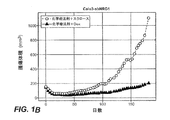
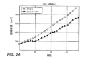
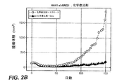


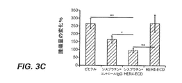


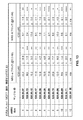

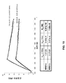

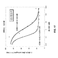
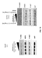
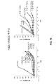

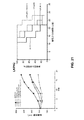
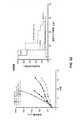
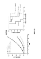
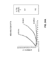

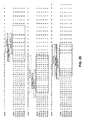
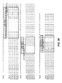


Classifications
-
- C—CHEMISTRY; METALLURGY
- C07—ORGANIC CHEMISTRY
- C07K—PEPTIDES
- C07K16/00—Immunoglobulins [IGs], e.g. monoclonal or polyclonal antibodies
- C07K16/18—Immunoglobulins [IGs], e.g. monoclonal or polyclonal antibodies against material from animals or humans
- C07K16/22—Immunoglobulins [IGs], e.g. monoclonal or polyclonal antibodies against material from animals or humans against growth factors ; against growth regulators
-
- A—HUMAN NECESSITIES
- A61—MEDICAL OR VETERINARY SCIENCE; HYGIENE
- A61K—PREPARATIONS FOR MEDICAL, DENTAL OR TOILETRY PURPOSES
- A61K47/00—Medicinal preparations characterised by the non-active ingredients used, e.g. carriers or inert additives; Targeting or modifying agents chemically bound to the active ingredient
- A61K47/50—Medicinal preparations characterised by the non-active ingredients used, e.g. carriers or inert additives; Targeting or modifying agents chemically bound to the active ingredient the non-active ingredient being chemically bound to the active ingredient, e.g. polymer-drug conjugates
- A61K47/51—Medicinal preparations characterised by the non-active ingredients used, e.g. carriers or inert additives; Targeting or modifying agents chemically bound to the active ingredient the non-active ingredient being chemically bound to the active ingredient, e.g. polymer-drug conjugates the non-active ingredient being a modifying agent
- A61K47/68—Medicinal preparations characterised by the non-active ingredients used, e.g. carriers or inert additives; Targeting or modifying agents chemically bound to the active ingredient the non-active ingredient being chemically bound to the active ingredient, e.g. polymer-drug conjugates the non-active ingredient being a modifying agent the modifying agent being an antibody, an immunoglobulin or a fragment thereof, e.g. an Fc-fragment
- A61K47/6835—Medicinal preparations characterised by the non-active ingredients used, e.g. carriers or inert additives; Targeting or modifying agents chemically bound to the active ingredient the non-active ingredient being chemically bound to the active ingredient, e.g. polymer-drug conjugates the non-active ingredient being a modifying agent the modifying agent being an antibody, an immunoglobulin or a fragment thereof, e.g. an Fc-fragment the modifying agent being an antibody or an immunoglobulin bearing at least one antigen-binding site
- A61K47/6845—Medicinal preparations characterised by the non-active ingredients used, e.g. carriers or inert additives; Targeting or modifying agents chemically bound to the active ingredient the non-active ingredient being chemically bound to the active ingredient, e.g. polymer-drug conjugates the non-active ingredient being a modifying agent the modifying agent being an antibody, an immunoglobulin or a fragment thereof, e.g. an Fc-fragment the modifying agent being an antibody or an immunoglobulin bearing at least one antigen-binding site the antibody targeting a cytokine, e.g. growth factors, VEGF, TNF, a lymphokine or an interferon
-
- A—HUMAN NECESSITIES
- A61—MEDICAL OR VETERINARY SCIENCE; HYGIENE
- A61P—SPECIFIC THERAPEUTIC ACTIVITY OF CHEMICAL COMPOUNDS OR MEDICINAL PREPARATIONS
- A61P35/00—Antineoplastic agents
-
- C—CHEMISTRY; METALLURGY
- C12—BIOCHEMISTRY; BEER; SPIRITS; WINE; VINEGAR; MICROBIOLOGY; ENZYMOLOGY; MUTATION OR GENETIC ENGINEERING
- C12N—MICROORGANISMS OR ENZYMES; COMPOSITIONS THEREOF; PROPAGATING, PRESERVING, OR MAINTAINING MICROORGANISMS; MUTATION OR GENETIC ENGINEERING; CULTURE MEDIA
- C12N15/00—Mutation or genetic engineering; DNA or RNA concerning genetic engineering, vectors, e.g. plasmids, or their isolation, preparation or purification; Use of hosts therefor
- C12N15/09—Recombinant DNA-technology
- C12N15/11—DNA or RNA fragments; Modified forms thereof; Non-coding nucleic acids having a biological activity
- C12N15/113—Non-coding nucleic acids modulating the expression of genes, e.g. antisense oligonucleotides; Antisense DNA or RNA; Triplex- forming oligonucleotides; Catalytic nucleic acids, e.g. ribozymes; Nucleic acids used in co-suppression or gene silencing
- C12N15/1135—Non-coding nucleic acids modulating the expression of genes, e.g. antisense oligonucleotides; Antisense DNA or RNA; Triplex- forming oligonucleotides; Catalytic nucleic acids, e.g. ribozymes; Nucleic acids used in co-suppression or gene silencing against oncogenes or tumor suppressor genes
-
- C—CHEMISTRY; METALLURGY
- C12—BIOCHEMISTRY; BEER; SPIRITS; WINE; VINEGAR; MICROBIOLOGY; ENZYMOLOGY; MUTATION OR GENETIC ENGINEERING
- C12N—MICROORGANISMS OR ENZYMES; COMPOSITIONS THEREOF; PROPAGATING, PRESERVING, OR MAINTAINING MICROORGANISMS; MUTATION OR GENETIC ENGINEERING; CULTURE MEDIA
- C12N15/00—Mutation or genetic engineering; DNA or RNA concerning genetic engineering, vectors, e.g. plasmids, or their isolation, preparation or purification; Use of hosts therefor
- C12N15/09—Recombinant DNA-technology
- C12N15/11—DNA or RNA fragments; Modified forms thereof; Non-coding nucleic acids having a biological activity
- C12N15/113—Non-coding nucleic acids modulating the expression of genes, e.g. antisense oligonucleotides; Antisense DNA or RNA; Triplex- forming oligonucleotides; Catalytic nucleic acids, e.g. ribozymes; Nucleic acids used in co-suppression or gene silencing
- C12N15/1136—Non-coding nucleic acids modulating the expression of genes, e.g. antisense oligonucleotides; Antisense DNA or RNA; Triplex- forming oligonucleotides; Catalytic nucleic acids, e.g. ribozymes; Nucleic acids used in co-suppression or gene silencing against growth factors, growth regulators, cytokines, lymphokines or hormones
-
- A—HUMAN NECESSITIES
- A01—AGRICULTURE; FORESTRY; ANIMAL HUSBANDRY; HUNTING; TRAPPING; FISHING
- A01K—ANIMAL HUSBANDRY; AVICULTURE; APICULTURE; PISCICULTURE; FISHING; REARING OR BREEDING ANIMALS, NOT OTHERWISE PROVIDED FOR; NEW BREEDS OF ANIMALS
- A01K2267/00—Animals characterised by purpose
- A01K2267/03—Animal model, e.g. for test or diseases
- A01K2267/0331—Animal model for proliferative diseases
-
- A—HUMAN NECESSITIES
- A61—MEDICAL OR VETERINARY SCIENCE; HYGIENE
- A61K—PREPARATIONS FOR MEDICAL, DENTAL OR TOILETRY PURPOSES
- A61K39/00—Medicinal preparations containing antigens or antibodies
- A61K2039/505—Medicinal preparations containing antigens or antibodies comprising antibodies
-
- C—CHEMISTRY; METALLURGY
- C07—ORGANIC CHEMISTRY
- C07K—PEPTIDES
- C07K2317/00—Immunoglobulins specific features
- C07K2317/50—Immunoglobulins specific features characterized by immunoglobulin fragments
- C07K2317/56—Immunoglobulins specific features characterized by immunoglobulin fragments variable (Fv) region, i.e. VH and/or VL
- C07K2317/565—Complementarity determining region [CDR]
-
- C—CHEMISTRY; METALLURGY
- C07—ORGANIC CHEMISTRY
- C07K—PEPTIDES
- C07K2317/00—Immunoglobulins specific features
- C07K2317/70—Immunoglobulins specific features characterized by effect upon binding to a cell or to an antigen
- C07K2317/76—Antagonist effect on antigen, e.g. neutralization or inhibition of binding
-
- C—CHEMISTRY; METALLURGY
- C07—ORGANIC CHEMISTRY
- C07K—PEPTIDES
- C07K2317/00—Immunoglobulins specific features
- C07K2317/90—Immunoglobulins specific features characterized by (pharmaco)kinetic aspects or by stability of the immunoglobulin
- C07K2317/92—Affinity (KD), association rate (Ka), dissociation rate (Kd) or EC50 value
-
- C—CHEMISTRY; METALLURGY
- C12—BIOCHEMISTRY; BEER; SPIRITS; WINE; VINEGAR; MICROBIOLOGY; ENZYMOLOGY; MUTATION OR GENETIC ENGINEERING
- C12N—MICROORGANISMS OR ENZYMES; COMPOSITIONS THEREOF; PROPAGATING, PRESERVING, OR MAINTAINING MICROORGANISMS; MUTATION OR GENETIC ENGINEERING; CULTURE MEDIA
- C12N2310/00—Structure or type of the nucleic acid
- C12N2310/10—Type of nucleic acid
- C12N2310/14—Type of nucleic acid interfering N.A.
-
- C—CHEMISTRY; METALLURGY
- C12—BIOCHEMISTRY; BEER; SPIRITS; WINE; VINEGAR; MICROBIOLOGY; ENZYMOLOGY; MUTATION OR GENETIC ENGINEERING
- C12N—MICROORGANISMS OR ENZYMES; COMPOSITIONS THEREOF; PROPAGATING, PRESERVING, OR MAINTAINING MICROORGANISMS; MUTATION OR GENETIC ENGINEERING; CULTURE MEDIA
- C12N2310/00—Structure or type of the nucleic acid
- C12N2310/50—Physical structure
- C12N2310/53—Physical structure partially self-complementary or closed
- C12N2310/531—Stem-loop; Hairpin
-
- C—CHEMISTRY; METALLURGY
- C12—BIOCHEMISTRY; BEER; SPIRITS; WINE; VINEGAR; MICROBIOLOGY; ENZYMOLOGY; MUTATION OR GENETIC ENGINEERING
- C12N—MICROORGANISMS OR ENZYMES; COMPOSITIONS THEREOF; PROPAGATING, PRESERVING, OR MAINTAINING MICROORGANISMS; MUTATION OR GENETIC ENGINEERING; CULTURE MEDIA
- C12N2330/00—Production
- C12N2330/50—Biochemical production, i.e. in a transformed host cell
-
- C—CHEMISTRY; METALLURGY
- C12—BIOCHEMISTRY; BEER; SPIRITS; WINE; VINEGAR; MICROBIOLOGY; ENZYMOLOGY; MUTATION OR GENETIC ENGINEERING
- C12N—MICROORGANISMS OR ENZYMES; COMPOSITIONS THEREOF; PROPAGATING, PRESERVING, OR MAINTAINING MICROORGANISMS; MUTATION OR GENETIC ENGINEERING; CULTURE MEDIA
- C12N2740/00—Reverse transcribing RNA viruses
- C12N2740/00011—Details
- C12N2740/10011—Retroviridae
- C12N2740/16011—Human Immunodeficiency Virus, HIV
- C12N2740/16041—Use of virus, viral particle or viral elements as a vector
- C12N2740/16043—Use of virus, viral particle or viral elements as a vector viral genome or elements thereof as genetic vector
Landscapes
- Health & Medical Sciences (AREA)
- Life Sciences & Earth Sciences (AREA)
- Genetics & Genomics (AREA)
- Engineering & Computer Science (AREA)
- Chemical & Material Sciences (AREA)
- Organic Chemistry (AREA)
- Biomedical Technology (AREA)
- Molecular Biology (AREA)
- Bioinformatics & Cheminformatics (AREA)
- General Health & Medical Sciences (AREA)
- Biotechnology (AREA)
- General Engineering & Computer Science (AREA)
- Zoology (AREA)
- Wood Science & Technology (AREA)
- Biochemistry (AREA)
- Biophysics (AREA)
- Medicinal Chemistry (AREA)
- Immunology (AREA)
- Plant Pathology (AREA)
- Microbiology (AREA)
- Physics & Mathematics (AREA)
- Public Health (AREA)
- Veterinary Medicine (AREA)
- Animal Behavior & Ethology (AREA)
- Pharmacology & Pharmacy (AREA)
- Proteomics, Peptides & Aminoacids (AREA)
- Oncology (AREA)
- Endocrinology (AREA)
- Epidemiology (AREA)
- General Chemical & Material Sciences (AREA)
- Nuclear Medicine, Radiotherapy & Molecular Imaging (AREA)
- Chemical Kinetics & Catalysis (AREA)
- Medicines Containing Antibodies Or Antigens For Use As Internal Diagnostic Agents (AREA)
- Peptides Or Proteins (AREA)
- Medicines That Contain Protein Lipid Enzymes And Other Medicines (AREA)
- Preparation Of Compounds By Using Micro-Organisms (AREA)
- Micro-Organisms Or Cultivation Processes Thereof (AREA)
- Pharmaceuticals Containing Other Organic And Inorganic Compounds (AREA)
Abstract
本発明は、抗ニューレグリン1抗体及び癌のような疾病又は疾患の治療における該抗体の使用方法を提供する。特定の実施態様では、抗ニューレグリン1抗体はニューレグリン1α及びニューレグリン1βアイソフォームの双方に結合する。The present invention provides anti-neuregulin 1 antibodies and methods of using the antibodies in the treatment of diseases or disorders such as cancer. In certain embodiments, the anti-neuregulin 1 antibody binds to both neuregulin 1α and neuregulin 1β isoforms.
Description
関連出願とのクロスリファレンス
本出願は、2011年8月17日に出願された米国仮出願第61/524421号の優先権を主張し、その開示内容はその全体が出典明示によりここに援用される。
This application claims the priority of US Provisional Application No. 61 / 524,421, filed Aug. 17, 2011, the disclosure of which is hereby incorporated by reference in its entirety. .
配列表
本願は、EFS-Webを介してASCIIフォーマットで提出され、その全体が出典明示によりここに援用される配列表を含む。2012年8月14日に作成された上記ASCiiコピーはP4727R1WO_ST25.txtと命名され、64664バイトのサイズである。
SEQUENCE LISTING This application contains a Sequence Listing submitted in ASCII format via EFS-Web, which is incorporated herein by reference in its entirety. The ASCii copy created on August 14, 2012 is P4727R1WO_ST25. It is named txt and is 64664 bytes in size.
発明の分野
本発明は、ニューレグリン抗体と癌のような疾病又は疾患の治療にその抗体を使用する方法を提供する。
The present invention provides neuregulin antibodies and methods of using the antibodies in the treatment of diseases or disorders such as cancer.
大部分の癌にとって、化学療法に対する乏しい応答又は化学療法後の疾患の再発は、死亡の主要な原因である。これは化学療法が疾患の全段階の患者を治療するために使用されるので、非小細胞肺癌(NSCLC)の場合に特にしかりである。患者の3分の2以上が切除不能な進行疾患を呈しており、第一線の化学療法、放射線療法又はその二種の併用で治療される。しかしながら、NSCLSCの治療における化学療法剤の積極的な使用にもかかわらず、局所進行疾患及び進行疾患のそれぞれに対して5年生存率は8%及び3.5%にとどまっている(Doebele等)。 For most cancers, poor response to chemotherapy or recurrence of disease after chemotherapy is a major cause of death. This is especially true in the case of non-small cell lung cancer (NSCLC) because chemotherapy is used to treat patients at all stages of the disease. More than two-thirds of patients have unresectable advanced disease and are treated with first-line chemotherapy, radiation therapy, or a combination of the two. However, despite aggressive use of chemotherapeutic agents in the treatment of NSCLSC, the 5-year survival rate is only 8% and 3.5% for locally advanced and advanced disease, respectively (Doebele et al.) .
化学療法に対する部分応答及び化学療法後の再発は、腫瘍細胞がその薬剤感受性において不均一であることを示唆している。幾つかの腫瘍細胞は与えられた薬剤によって効果的に死滅させられる一方、他のものは救われる。癌幹細胞(CSC)は、それらが既存の治療法の失敗に対して可能な説明を提供するので、近年の集中的な研究領域である。幾つかのグループは、CSCが常套的な化学療法剤及び放射線治療(Bao等, 2006;Costello等, 2000;Dean等, 2005;Dylla等, 2008;Matsui等, 2004;Phillips等, 2006)に対して高まった耐性を報告している。しかしながら、樹立された腫瘍の増殖の維持又は原発又は遠位部位における化学療法後の腫瘍の再開におけるCSCの役割は未解明のままである。化学療法後の腫瘍再増殖の原因である細胞は幹細胞ではなく、むしろ細胞周期の段階、ストロマ−ニッチ相互作用、又は生存促進性シグナルのレベルを反映しうる他の性質を通して耐性を達成する細胞でありうる。これらの細胞は「腫瘍再始原細胞(tumor reinitiating cells)」又はTRICと称される。米国特許出願公開20110229493。 Partial response to chemotherapy and recurrence after chemotherapy suggest that tumor cells are heterogeneous in their drug sensitivity. Some tumor cells are effectively killed by a given drug while others are saved. Cancer stem cells (CSC) are an intensive research area in recent years because they provide a possible explanation for the failure of existing therapies. Some groups have been directed against CSC routine chemotherapeutic agents and radiotherapy (Bao et al., 2006; Costello et al., 2000; Dean et al., 2005; Dylla et al., 2008; Matsui et al., 2004; Phillips et al., 2006). Reported increased resistance. However, the role of CSC in maintaining growth of established tumors or in reopening tumors after chemotherapy at the primary or distal site remains unclear. The cells responsible for tumor regrowth after chemotherapy are not stem cells, but rather cells that achieve resistance through cell cycle stages, stromal niche interactions, or other properties that may reflect the level of pro-survival signals. It is possible. These cells are referred to as “tumor reinitiating cells” or TRIC. US Patent Application Publication No. 2011102229493.
上皮増殖因子受容体(EGFR)阻害剤はしばしばNSCLC患者を治療するために使用され、その腫瘍がEGFR活性化変異を有する患者の大部分の治療において効果的であることが示されている。過剰発現又は活性化変異を介するEGFRのシグナル伝達の調節解除は肺腺癌において比較的頻繁に起こる事象である(Dahabreh等, 2010)に概説)。EGFRは、EGFR(HER1)、HER2、HER3及びHER4を含むチロシンキナーゼのErbB又はHERファミリーの原型メンバーである。最近の証拠は、他のHERファミリーもまたNSCLCにおいてある役割を果たしうることを示している。しかし、疾患に対するその寄与はあまりよく特徴付けられておらず、殆どの研究はEGFRシグナル伝達を活性化させるその能力に焦点が当てられている(Ding等, 2008;Johnson及びJanne, 2006;Kuyama等, 200;Zhou等, 2006)。ヘレグリン1とも使用されるニューレグリン1(NRG1)はHer3及びHer4受容体のリガンドである。
Epidermal growth factor receptor (EGFR) inhibitors are often used to treat NSCLC patients and have been shown to be effective in treating most patients whose tumors have EGFR-activating mutations. Deregulation of EGFR signaling through overexpression or activating mutations is a relatively frequent event in lung adenocarcinoma (reviewed in Dahabreh et al., 2010). EGFR is a prototype member of the ErbB or HER family of tyrosine kinases including EGFR (HER1), HER2, HER3 and HER4. Recent evidence indicates that other HER families may also play a role in NSCLC. However, its contribution to disease has not been well characterized and most research has focused on its ability to activate EGFR signaling (Ding et al., 2008; Johnson and Janne, 2006; Kuyama et al. , 200; Zhou et al., 2006). Neuregulin 1 (NRG1), which is also used with
4種の既知のニューレグリンファミリーメンバー、NRG1、NRG2、NRG3、及びNRG4が存在する(Falls 2003;Hirsh及びWu 2007)。NRG1転写物は、少なくとも15の異なるアイソフォームを生じる広範な選択的スプライシングを受ける。全ての活性なアイソフォームが、活性に必要かつ十分であるEGF様ドメインを共有している(Holmes 1992,Yarden 1991)。 There are four known neuregulin family members, NRG1, NRG2, NRG3, and NRG4 (Falls 2003; Hirsh and Wu 2007). NRG1 transcripts undergo extensive alternative splicing resulting in at least 15 different isoforms. All active isoforms share an EGF-like domain that is necessary and sufficient for activity (Holmes 1992, Yarden 1991).
NRG1自己分泌シグナル伝達は、肺上皮細胞増殖を調節し、ヒト肺の発達において役割を果たすことが示されており(Patel等, 2000)、EGFR阻害剤に対するNSCLCの非感受性に関与している(Zhou等, 2006)。 NRG1 autocrine signaling has been shown to regulate lung epithelial cell proliferation and play a role in human lung development (Patel et al., 2000) and is involved in the insensitivity of NSCLC to EGFR inhibitors ( Zhou et al., 2006).
本発明は抗ニューレグリン1(抗NRG1)抗体とそれを使用する方法を提供する。 The present invention provides anti-neuregulin 1 (anti-NRG1) antibodies and methods of using the same.
本発明の一態様は、ニューレグリン1αとニューレグリン1βに結合する単離された抗NRG1抗体を提供する。一実施態様では、抗NRG1抗体は、ニューレグリン1βのEGFドメインとニューレグリン1αのEGFドメインに結合する。一実施態様では、抗NRG1抗体はニューレグリン1βのEGFドメインに、それがニューレグリン1αのEGFドメインに結合する親和性より大きい親和性で結合する。特定の実施態様では、抗NRG1抗体はニューレグリン1βのEGFドメインに、それがニューレグリン1αのEGFドメインに結合する親和性の20倍、50倍、100倍、200倍、500倍又は1000倍を超える親和性で結合する。 One aspect of the invention provides isolated anti-NRG1 antibodies that bind to neuregulin 1α and neuregulin 1β. In one embodiment, the anti-NRG1 antibody binds to the EGF domain of neuregulin 1β and the EGF domain of neuregulin 1α. In one embodiment, the anti-NRG1 antibody binds to the neuregulin 1β EGF domain with an affinity greater than the affinity that it binds to the neuregulin 1α EGF domain. In certain embodiments, the anti-NRG1 antibody has a neuregulin 1β EGF domain that has a 20-fold, 50-fold, 100-fold, 200-fold, 500-fold or 1000-fold affinity for binding to the neuregulin 1α EGF domain. Bind with greater affinity.
一実施態様では、抗NRG1抗体はニューレグリン1βのEGFドメインに10nM以下のkDで結合し、ニューレグリン1αのEGFドメインに10nM以下のkDで結合する。特定の実施態様では、抗NRG1抗体はニューレグリン1βのEGFドメインに10nM以下、1nM以下、1×10⁻¹nM以下、1×10⁻²nM以下、又は1×10⁻³nM以下のkDで結合する。抗NRG1抗体の親和性は表面プラズモン共鳴アッセイによって測定される。 In one embodiment, the anti-NRG1 antibody binds to the EGF domain of neuregulin 1β with a kD of 10 nM or less and binds to the EGF domain of neuregulin 1α with a kD of 10 nM or less. In certain embodiments, the anti-NRG1 antibody has a kD of 10 nM or less, 1 nM or less, 1 × 10 −1 nM or less, 1 × 10 −2 nM or less, or 1 × 10 −3 nM or less in the EGF domain of neuregulin 1β. Join. The affinity of the anti-NRG1 antibody is measured by a surface plasmon resonance assay.
本発明の一態様は、ニューレグリン1βのエピトープに結合し、ニューレグリン1βのエピトープが配列番号4のアミノ酸1−37のアミノ酸配列又は配列番号4のアミノ酸38−64のアミノ酸配列を含む、単離された抗NRG1抗体を提供する。一実施態様では、ニューレグリン1βのエピトープは配列番号4のアミノ酸を含む。一実施態様では、抗NRG1抗体はニューレグリン1αのエピトープに更に結合し、ニューレグリン1αのエピトープは配列番号3のアミノ酸1−36のアミノ酸配列又は配列番号3のアミノ酸37−58のアミノ酸配列を含む。一実施態様では、ニューレグリン1αのエピトープは配列番号3のアミノ酸を含む。 One aspect of the present invention is an isolation that binds to an epitope of neuregulin 1β, wherein the epitope of neuregulin 1β comprises the amino acid sequence of amino acids 1-37 of SEQ ID NO: 4 or the amino acid sequence of amino acids 38-64 of SEQ ID NO: 4 Anti-NRG1 antibodies are provided. In one embodiment, the epitope of neuregulin 1β comprises the amino acid of SEQ ID NO: 4. In one embodiment, the anti-NRG1 antibody further binds to an epitope of neuregulin 1α, wherein the epitope of neuregulin 1α comprises the amino acid sequence of amino acids 1-36 of SEQ ID NO: 3 or the amino acid sequence of amino acids 37-58 of SEQ ID NO: 3 . In one embodiment, the epitope of neuregulin 1α comprises the amino acid of SEQ ID NO: 3.
ある実施態様では、抗NRG1抗体はモノクローナル抗体である。ある実施態様では、抗NRG1抗体はヒト、ヒト化、又はキメラ抗体である。ある実施態様では、抗NRG1抗体はニューレグリン1αとニューレグリン1βに結合する抗体断片である。 In certain embodiments, the anti-NRG1 antibody is a monoclonal antibody. In certain embodiments, the anti-NRG1 antibody is a human, humanized, or chimeric antibody. In one embodiment, the anti-NRG1 antibody is an antibody fragment that binds to neuregulin 1α and neuregulin 1β.
本発明の他の態様は、(a)配列番号5のアミノ酸配列を含むHVR−H1、(b)配列番号6のアミノ酸配列を含むHVR−H2、及び(c)配列番号7のアミノ酸配列を含むHVR−H3を含む単離された抗NRG1抗体を提供する。 Other aspects of the invention include (a) HVR-H1 comprising the amino acid sequence of SEQ ID NO: 5, (b) HVR-H2 comprising the amino acid sequence of SEQ ID NO: 6, and (c) the amino acid sequence of SEQ ID NO: 7. An isolated anti-NRG1 antibody comprising HVR-H3 is provided.
本発明の他の態様は、(a)配列番号16のアミノ酸配列を含むHVR−L1、(b)配列番号17のアミノ酸配列を含むHVR−L2、及び(c)配列番号18のアミノ酸配列を含むHVR−L3を含む単離された抗NRG1抗体を提供する。一実施態様では、抗NRG1抗体は、(a)配列番号16のアミノ酸配列を含むHVR−L1、(b)配列番号17のアミノ酸配列を含むHVR−L2、及び(c)配列番号18のアミノ酸配列を含むHVR−L3を更に含む。 Other aspects of the invention include (a) HVR-L1 comprising the amino acid sequence of SEQ ID NO: 16, (b) HVR-L2 comprising the amino acid sequence of SEQ ID NO: 17, and (c) the amino acid sequence of SEQ ID NO: 18. An isolated anti-NRG1 antibody comprising HVR-L3 is provided. In one embodiment, the anti-NRG1 antibody comprises (a) HVR-L1 comprising the amino acid sequence of SEQ ID NO: 16, (b) HVR-L2 comprising the amino acid sequence of SEQ ID NO: 17, and (c) the amino acid sequence of SEQ ID NO: 18. Further comprising HVR-L3.
本発明の他の態様は、(a)配列番号76のアミノ酸配列を含むHVR−H1、(b)配列番号29のアミノ酸配列を含むHVR−H2、及び(c)配列番号43のアミノ酸配列を含むHVR−H3を含む単離された抗NRG1抗体を提供する。 Other aspects of the invention include (a) HVR-H1 comprising the amino acid sequence of SEQ ID NO: 76, (b) HVR-H2 comprising the amino acid sequence of SEQ ID NO: 29, and (c) the amino acid sequence of SEQ ID NO: 43. An isolated anti-NRG1 antibody comprising HVR-H3 is provided.
本発明の他の態様は、(a)配列番号31のアミノ酸配列を含むHVR−L1、(b)配列番号32のアミノ酸配列を含むHVR−L2、及び(c)配列番号33のアミノ酸配列を含むHVR−L3を含む単離された抗NRG1抗体を提供する。一実施態様では、抗NRG1抗体は、(a)配列番号31のアミノ酸配列を含むHVR−L1、(b)配列番号32のアミノ酸配列を含むHVR−L2、及び(c)配列番号33のアミノ酸配列を含むHVR−L3を含む。 Other aspects of the invention include (a) HVR-L1 comprising the amino acid sequence of SEQ ID NO: 31, (b) HVR-L2 comprising the amino acid sequence of SEQ ID NO: 32, and (c) the amino acid sequence of SEQ ID NO: 33. An isolated anti-NRG1 antibody comprising HVR-L3 is provided. In one embodiment, the anti-NRG1 antibody comprises (a) HVR-L1 comprising the amino acid sequence of SEQ ID NO: 31, (b) HVR-L2 comprising the amino acid sequence of SEQ ID NO: 32, and (c) the amino acid sequence of SEQ ID NO: 33. Including HVR-L3.
本発明の他の態様は、(a)配列番号21のアミノ酸配列に対して少なくとも95%の配列同一性を有するVH配列;(b)配列番号26のアミノ酸配列に対して少なくとも95%の配列同一性を有するVL配列;又は(c)(a)のVH配列と(b)のVL配列を含む単離された抗NRG1抗体を提供する。一実施態様では、抗NRG1抗体は配列番号21のVH配列を含む。一実施態様では、抗NRG1抗体は配列番号26のVL配列を含む。一実施態様では、抗NRG1抗体は配列番号21のVH配列と配列番号26のVL配列を含む。 Another aspect of the present invention provides: (a) a VH sequence having at least 95% sequence identity to the amino acid sequence of SEQ ID NO: 21; (b) at least 95% sequence identity to the amino acid sequence of SEQ ID NO: 26 An isolated anti-NRG1 antibody comprising a VL sequence having sex; or (c) a VH sequence of (a) and a VL sequence of (b) is provided. In one embodiment, the anti-NRG1 antibody comprises the VH sequence of SEQ ID NO: 21. In one embodiment, the anti-NRG1 antibody comprises the VL sequence of SEQ ID NO: 26. In one embodiment, the anti-NRG1 antibody comprises the VH sequence of SEQ ID NO: 21 and the VL sequence of SEQ ID NO: 26.
本発明の一態様は、配列番号53のVH配列と配列番号63のVL配列を含む単離された抗NRG1抗体を提供する。 One aspect of the invention provides an isolated anti-NRG1 antibody comprising the VH sequence of SEQ ID NO: 53 and the VL sequence of SEQ ID NO: 63.
本発明の他の態様は、抗NRG1抗体をコードする単離された核酸を提供する。本発明の他の態様は、抗NRG1抗体をコードする核酸を含む宿主細胞を提供する。 Another aspect of the invention provides an isolated nucleic acid encoding an anti-NRG1 antibody. Another aspect of the invention provides a host cell comprising a nucleic acid encoding an anti-NRG1 antibody.
本発明の他の態様は、抗体が産生されるようにそのような宿主細胞を培養することを含む抗NRG1抗体の産生方法を提供する。
本発明の他の態様は、抗NRG1抗体と細胞傷害剤を含むイムノコンジュゲートを提供する。
Another aspect of the invention provides a method for producing an anti-NRG1 antibody comprising culturing such host cells such that the antibody is produced.
Another aspect of the invention provides an immunoconjugate comprising an anti-NRG1 antibody and a cytotoxic agent.
本発明の他の態様は、抗NRG1抗体と薬学的に許容可能な担体を含む薬学的製剤を提供する。一実施態様では、薬学的製剤は更なる治療剤、例えばゲムシタビン、パクリタキセル、又はシスプラチン、又はパクリタキセルとシスプラチンの併用剤を更に含む。 Another aspect of the invention provides a pharmaceutical formulation comprising an anti-NRG1 antibody and a pharmaceutically acceptable carrier. In one embodiment, the pharmaceutical formulation further comprises an additional therapeutic agent, such as gemcitabine, paclitaxel, or cisplatin, or a combination of paclitaxel and cisplatin.
本発明の他の態様は、医薬として使用される抗NRG1抗体を提供する。
本発明の他の態様は、癌の治療に使用される抗NRG1抗体を提供する。
本発明の他の態様は、医薬の製造における抗NRG1抗体を提供する。一実施態様では、医薬は、癌、例えば非小細胞肺癌、乳癌、卵巣癌、頭頸部癌、子宮頸癌、膀胱癌、食道癌、前立腺癌、及び結腸直腸癌の治療のためのものである。
Another aspect of the present invention provides an anti-NRG1 antibody for use as a medicament.
Another aspect of the invention provides an anti-NRG1 antibody for use in the treatment of cancer.
Another aspect of the present invention provides an anti-NRG1 antibody in the manufacture of a medicament. In one embodiment, the medicament is for the treatment of cancer, such as non-small cell lung cancer, breast cancer, ovarian cancer, head and neck cancer, cervical cancer, bladder cancer, esophageal cancer, prostate cancer, and colorectal cancer. .
本発明の他の態様は、癌を有する個体の治療方法であって、抗NRG1抗体の有効量を個体に投与することを含む方法を提供する。治療される癌は、例えば非小細胞肺癌、乳癌、卵巣癌、頭頸部癌、子宮頸癌、膀胱癌、食道癌、前立腺癌、及び結腸直腸癌である。一実施態様では、該方法は、個体に更なる治療剤、例えばゲムシタビン、パクリタキセル、カルボプラチン、及びシスプラチン又はパクリタキセル、カルボプラチン、及びシスプラチンの組み合わせ又は2又は3全てを投与することを更に含む。 Another aspect of the invention provides a method for treating an individual having cancer, comprising administering to the individual an effective amount of an anti-NRG1 antibody. Cancers to be treated are, for example, non-small cell lung cancer, breast cancer, ovarian cancer, head and neck cancer, cervical cancer, bladder cancer, esophageal cancer, prostate cancer, and colorectal cancer. In one embodiment, the method further comprises administering to the individual additional therapeutic agents such as gemcitabine, paclitaxel, carboplatin, and cisplatin or a combination of paclitaxel, carboplatin, and cisplatin, or two or all three.
本発明の他の態様は、患者に抗NRG1抗体の有効量を投与することを含む、癌患者の腫瘍再発までの時間を増やすための方法を提供する。一実施形態では、本方法は、患者に治療剤を投与することを更に含む。一実施態様では、治療剤は、化学療法剤又は二次抗体である。化学療法剤は、例えばゲムシタビン、パクリタキセル、カルボプラチン、及びシスプラチン、又はパクリタキセル、カルボプラチン、及びシスプラチンの組み合わせ又は2又は3全てである。ある実施態様では、二次抗体はEGFR、HER2、HER3、又はHER4に結合するか又はこれらの標的の二以上に結合する。ある実施態様では、治療される癌は、非小細胞肺癌、乳癌、卵巣癌、頭頸部癌、子宮頸癌、膀胱癌、食道癌、前立腺癌、及び/又は結腸直腸癌である。一実施態様では、腫瘍再発までの期間の増加は、抗体の非存在下における再発までの期間より少なくとも1.25倍大きい。一実施態様では、腫瘍再発までの期間の増加は、抗体の非存在下における再発までの期間より少なくとも1.50倍大きい。 Another aspect of the invention provides a method for increasing the time to tumor recurrence in a cancer patient comprising administering to the patient an effective amount of an anti-NRG1 antibody. In one embodiment, the method further comprises administering a therapeutic agent to the patient. In one embodiment, the therapeutic agent is a chemotherapeutic agent or a secondary antibody. The chemotherapeutic agent is, for example, gemcitabine, paclitaxel, carboplatin, and cisplatin, or a combination of paclitaxel, carboplatin, and cisplatin, or all two or three. In certain embodiments, the secondary antibody binds to EGFR, HER2, HER3, or HER4 or binds to more than one of these targets. In certain embodiments, the cancer to be treated is non-small cell lung cancer, breast cancer, ovarian cancer, head and neck cancer, cervical cancer, bladder cancer, esophageal cancer, prostate cancer, and / or colorectal cancer. In one embodiment, the increase in time to tumor recurrence is at least 1.25 times greater than the time to recurrence in the absence of antibody. In one embodiment, the increase in time to tumor recurrence is at least 1.50 times greater than the time to recurrence in the absence of antibody.
I.定義
本明細書における目的のための「アクセプターヒトフレームワーク」とは、下記に定義されるように、ヒト免疫グロブリンフレームワーク又はヒトコンセンサスフレームワークから得られる軽鎖可変ドメイン(VL)フレームワーク又は重鎖可変ドメイン(VH)フレームワークのアミノ酸配列を含有するフレームワークである。ヒト免疫グロブリンのフレームワーク又はヒトコンセンサスフレームワーク”に由来する”アクセプターヒトフレームワークは、その同一のアミノ酸配列を含んでもよく、又はそれはアミノ酸配列の変化を含み得る。幾つかの実施態様において、アミノ酸変化の数は、10以下、9以下、8以下、7以下、6以下、5以下、4以下、3以下、又は2以下である。幾つかの実施態様において、VLアクセプターヒトフレームワークは、Vはヒト免疫グロブリンのフレームワーク配列又はヒトコンセンサスフレームワーク配列に、配列が同一である。
I. Definitions An “acceptor human framework” for purposes herein is a light chain variable domain (VL) framework obtained from a human immunoglobulin framework or a human consensus framework, as defined below, or A framework containing the amino acid sequence of the heavy chain variable domain (VH) framework. An “acceptor human framework” derived from a “human immunoglobulin framework or human consensus framework” may comprise the same amino acid sequence thereof, or it may comprise amino acid sequence changes. In some embodiments, the number of amino acid changes is 10 or less, 9 or less, 8 or less, 7 or less, 6 or less, 5 or less, 4 or less, 3 or less, or 2 or less. In some embodiments, the VL acceptor human framework is identical in sequence to the human immunoglobulin framework sequence or human consensus framework sequence.
「親和性」とは、分子(例えば、抗体)の単一結合部位とその結合パートナー(例えば、抗原)との間の非共有結合相互作用の総和の強度を指す。本明細書で使用する場合、特に断らない限り、「結合親和性」は、結合対(例えば、抗体と抗原)のメンバー間の1:1の相互作用を反映している本質的な結合親和性を指す。そのパートナーYに対する分子Xの親和性は、一般的に解離定数(Kd)で表すことができる。親和性は、本明細書に記載したものを含む、当技術分野で知られている一般的な方法によって測定することができる。結合親和性を測定するための具体的な例示であって例示的な実施態様は、以下で説明される。 “Affinity” refers to the strength of the sum total of non-covalent interactions between a single binding site of a molecule (eg, an antibody) and its binding partner (eg, an antigen). As used herein, unless otherwise indicated, “binding affinity” is an intrinsic binding affinity that reflects a 1: 1 interaction between members of a binding pair (eg, antibody and antigen). Point to. The affinity of molecule X for its partner Y can generally be expressed as a dissociation constant (Kd). Affinity can be measured by common methods known in the art, including those described herein. Specific exemplary and exemplary embodiments for measuring binding affinity are described below.
「親和性成熟」抗体とは、その改変を有していない親抗体と比較して、抗原に対する抗体の親和性に改良を生じせしめる、その一又は複数の超可変領域(HVR)において一又は複数の改変を持つものである。 An “affinity matured” antibody is one or more in one or more hypervariable regions (HVRs) that cause an improvement in the affinity of the antibody for the antigen compared to the parent antibody without the modification. It has a modification of.
用語「抗NRG1抗体」及び「NRG1に特異的に結合する抗体」は、抗体がNRG1を標的とし、診断薬剤及び/又は治療的薬剤として有用であるように十分な親和性でNRG1に結合することができる抗体を指す。一実施態様において、抗NRG1抗体の、無関係な非NRG1タンパク質への結合の程度は、例えば、ラジオイムノアッセイ(RIA)によって測定される場合、抗体のNRG1への結合の約10%未満である。所定の実施態様において、NRG1へ結合する抗体は、解離定数(Kd)が、≦1μM、≦100nM、≦10nM、≦1nM、≦0.1nM、≦0.01nM、又は≦0.001nM(例えば、10−8M未満、例えば、10−8Mから10−13M、例えば、10−9Mから10−13M)。所定の実施態様において、抗NRG1抗体は、異なる種由来のNRG1間で保存されているNRG1のエピトープに結合する。 The terms “anti-NRG1 antibody” and “antibody that specifically binds NRG1” bind NRG1 with sufficient affinity so that the antibody targets NRG1 and is useful as a diagnostic and / or therapeutic agent. Refers to an antibody capable of In one embodiment, the degree of binding of the anti-NRG1 antibody to an irrelevant non-NRG1 protein is less than about 10% of the binding of the antibody to NRG1, as measured, for example, by radioimmunoassay (RIA). In certain embodiments, an antibody that binds to NRG1 has a dissociation constant (Kd) of ≦ 1 μM, ≦ 100 nM, ≦ 10 nM, ≦ 1 nM, ≦ 0.1 nM, ≦ 0.01 nM, or ≦ 0.001 nM (eg, Less than 10 −8 M, such as 10 −8 M to 10 −13 M, such as 10 −9 M to 10 −13 M). In certain embodiments, the anti-NRG1 antibody binds to an epitope of NRG1 that is conserved among NRG1 from different species.
用語「抗体」は最も広い意味で用いられ、様々な抗体構造を包含し、限定されないが、モノクローナル抗体、ポリクローナル抗体、多特異性抗体(例えば二重特異性抗体)、及び、所望の抗原結合活性を示す限り、抗体断片を含む。 The term “antibody” is used in the broadest sense and encompasses a variety of antibody structures, including but not limited to monoclonal antibodies, polyclonal antibodies, multispecific antibodies (eg, bispecific antibodies), and desired antigen binding activity. As long as the antibody fragment is included.
「抗体断片」は、インタクトな抗体が結合する抗原を結合するインタクトな抗体の一部を含むインタクトな抗体以外の分子を指す。抗体断片の例としては、限定されないが、Fv、Fab、Fab’、Fab’−SHは、F(ab’)2、ダイアボディ、直鎖状抗体、単鎖抗体分子(例えばscFv)、及び抗体断片から形成された多重特異性抗体を含む。 “Antibody fragment” refers to a molecule other than an intact antibody, including a portion of an intact antibody that binds an antigen to which the intact antibody binds. Examples of antibody fragments include, but are not limited to, Fv, Fab, Fab ′, Fab′-SH, F (ab ′) 2 , diabody, linear antibody, single chain antibody molecule (eg scFv), and antibody Includes multispecific antibodies formed from fragments.
参照抗体として「同一のエピトープに結合する抗体」とは、競合アッセイにおいて50%以上、参照抗体のその抗原への結合をブロックする抗体、逆に競合アッセイにおいて50%以上、その抗体のその抗原への結合をブロックするその参照抗体を指す。典型的な競合アッセイは、本明細書で提供される。 “Antibodies that bind to the same epitope” as a reference antibody refers to an antibody that blocks binding of a reference antibody to its antigen in a competition assay by 50% or more, and conversely, 50% or more in a competition assay to that antigen of that antibody. The reference antibody that blocks binding. A typical competition assay is provided herein.
用語「キメラ」抗体は、重鎖及び/又は軽鎖の一部分が特定の起源又は種から由来し、一方重鎖及び/又は軽鎖の残りは異なる起源又は種から由来する抗体を指す。 The term “chimeric” antibody refers to an antibody in which a portion of the heavy and / or light chain is derived from a particular source or species, while the remainder of the heavy and / or light chain is from a different source or species.
用語「癌」及び「癌性」は、無秩序な細胞成長/細胞増殖を典型的に特徴とする、哺乳動物における生理学的状態を記述する。癌の例には、限定されないが、癌腫、リンパ腫(例えば、ホジキンリンパ腫と非ホジキンリンパ腫)、芽細胞腫、肉腫、及び白血病を含む。そのような癌のより具体的な例は、扁平上皮癌、小細胞肺癌、非小細胞肺がん、肺腺癌、肺扁平上皮癌、腹膜癌、肝細胞癌、消化器癌、膵臓癌、神経膠腫を含む、子宮頚癌、卵巣癌、肝臓癌、膀胱癌、肝癌、乳癌、結腸癌、結腸直腸癌、子宮内膜、子宮癌、唾液腺癌、腎臓癌、肝臓癌、前立腺癌、外陰癌、甲状腺癌、肝癌、白血病、及びその他のリンパ増殖性疾患、及び様々な型の頭頸部癌を包含する。 The terms “cancer” and “cancerous” describe the physiological condition in mammals that is typically characterized by unregulated cell growth / cell proliferation. Examples of cancer include, but are not limited to, carcinoma, lymphoma (eg, Hodgkin and non-Hodgkin lymphoma), blastoma, sarcoma, and leukemia. More specific examples of such cancers are squamous cell carcinoma, small cell lung cancer, non-small cell lung cancer, lung adenocarcinoma, lung squamous cell carcinoma, peritoneal cancer, hepatocellular carcinoma, digestive organ cancer, pancreatic cancer, glia Cervical cancer, ovarian cancer, liver cancer, bladder cancer, liver cancer, breast cancer, colon cancer, colorectal cancer, endometrium, uterine cancer, salivary gland cancer, kidney cancer, liver cancer, prostate cancer, vulvar cancer, Includes thyroid cancer, liver cancer, leukemia, and other lymphoproliferative diseases, and various types of head and neck cancer.
抗体の「クラス」は、その重鎖が保有する定常ドメイン又は定常領域のタイプを指す。抗体の5つの主要なクラスがあり:IgA、IgD、IgE、IgG及びIgM、及びこれらのいくつかは、さらにサブクラス(アイソタイプ)、例えば、IgG1、IgG2、IgG3、IgG4、IGA1、及びIgA2に分けることができる。免疫グロブリンの異なるクラスに対応する重鎖定常ドメインは、それぞれα、δ、ε、γ、及びμと呼ばれる。 The “class” of an antibody refers to the type of constant domain or constant region possessed by its heavy chain. There are five main classes of antibodies: IgA, IgD, IgE, IgG and IgM, and some of these are further subclasses (isotypes), eg, IgG 1 , IgG 2 , IgG 3 , IgG 4 , IGA 1 , and it can be divided into IgA 2. The heavy chain constant domains that correspond to the different classes of immunoglobulins are called α, δ, ε, γ, and μ, respectively.
本明細書で用いられる「細胞傷害剤」という用語は、細胞の機能を阻害又は阻止し及び/又は細胞破壊を生ずる物質を指す。細胞傷害剤は、限定されないが、放射性同位体(例えば、At211、I131、I125、Y90、Re186、Re188、Sm153、Bi212、P32、Pb212及びLuの放射性同位体):化学療法剤又は薬物(例えば、メトトレキセート、アドリアマイシン、ビンカアルカロイド(ビンクリスチン、ビンブラスチン、エトポシド)、ドキソルビシン、メルファラン、マイトマイシンC、クロラムブシル、ダウノルビシン又は他の挿入剤)、成長阻害剤、酵素及びその断片、例えば核酸分解酵素など、抗生物質、小分子毒素などの毒素、又は細菌、真菌、植物又は動物由来の酵素活性毒素(それらの断片及び/又はその変異体を含む)、及び以下に開示される様々な抗腫瘍剤又は抗癌剤を包含する。 The term “cytotoxic agent” as used herein refers to a substance that inhibits or prevents the function of cells and / or causes destruction of cells. Cytotoxic agents include, but are not limited to, radioisotopes (eg, At 211 , I 131 , I 125 , Y 90 , Re 186 , Re 188 , Sm 153 , Bi 212 , P 32 , Pb 212 and Lu). ): Chemotherapeutic agents or drugs (eg, methotrexate, adriamycin, vinca alkaloids (vincristine, vinblastine, etoposide), doxorubicin, melphalan, mitomycin C, chlorambucil, daunorubicin or other intercalating agents), growth inhibitors, enzymes and fragments thereof For example, antibiotics such as nucleolytic enzymes, toxins such as small molecule toxins, or enzyme-active toxins from bacteria, fungi, plants or animals (including fragments and / or variants thereof) and disclosed below Includes various anti-tumor or anti-cancer agents.
「エフェクター機能」とは、抗体のアイソタイプにより変わる、抗体のFc領域に起因する生物学的活性を指す。抗体エフェクター機能の例としては、C1q結合及び補体依存性細胞傷害(CDC)、Fc受容体結合、抗体依存性細胞媒介性細胞障害(ADCC)、貪食、細胞表面受容体(例えば、B細胞受容体)のダウンレギュレーション、及びB細胞の活性化を含む。 “Effector function” refers to biological activity attributable to the Fc region of an antibody, which varies with the antibody's isotype. Examples of antibody effector functions include C1q binding and complement dependent cytotoxicity (CDC), Fc receptor binding, antibody dependent cell mediated cytotoxicity (ADCC), phagocytosis, cell surface receptors (eg, B cell receptor Body) down-regulation and B cell activation.
薬剤、例えば、医薬製剤の「有効量」とは、所望の治療的又予防的結果を達成するために必要な用量及び期間での、有効な量を指す。 An “effective amount” of an agent, eg, a pharmaceutical formulation, refers to an effective amount at the dosage and for the period necessary to achieve the desired therapeutic or prophylactic result.
用語「Fc領域」は、定常領域の少なくとも一部を含む、免疫グロブリン重鎖のC末端領域を定義するために使用される。該用語は、天然配列Fc領域と変異Fc領域を含む。一実施態様において、ヒトIgG重鎖Fc領域はCys226又はPro230から重鎖のカルボキシル末端まで伸長する。しかし、Fc領域のC末端リジン(Lys447)は存在しているか、存在していない場合がある。本明細書に明記されていない限り、Fc領域又は定常領域内のアミノ酸残基の番号付けは、Kabat et al., Sequences of Proteins of Immunological Interest, 5th Ed. Public Health Service, National Institutes of Health, Bethesda, MD, 1991に記載されるように、EUインデックスとも呼ばれるEU番号付けシステムに従う。 The term “Fc region” is used to define the C-terminal region of an immunoglobulin heavy chain, including at least part of the constant region. The term includes native sequence Fc regions and variant Fc regions. In one embodiment, the human IgG heavy chain Fc region extends from Cys226 or Pro230 to the carboxyl terminus of the heavy chain. However, the C-terminal lysine (Lys447) of the Fc region may or may not exist. Unless otherwise stated herein, the numbering of amino acid residues within an Fc region or constant region is the Kabat et al., Sequences of Proteins of Immunological Interest, 5th Ed.Public Health Service, National Institutes of Health, Bethesda. , MD, 1991, according to the EU numbering system, also called EU index.
「フレームワーク」又は「FR」は、超可変領域(HVR)残基以外の可変ドメイン残基を指す。可変ドメインのFRは、一般的に4つのFRのドメイン、FR1、FR2、FR3、及びFR4で構成される。従って、HVR及びFR配列は一般にVH(又はVL)の以下の配列に現れる:FR1−H1(L1)−FR2−H2(L2)−FR3−H3(L3)−FR4。 “Framework” or “FR” refers to variable domain residues other than hypervariable region (HVR) residues. The FR of a variable domain is generally composed of four FR domains, FR1, FR2, FR3, and FR4. Thus, HVR and FR sequences generally appear in the following sequences of VH (or VL): FR1-H1 (L1) -FR2-H2 (L2) -FR3-H3 (L3) -FR4.
用語「完全長抗体」、「インタクトな抗体」及び「全抗体」は、本明細書中で互換的に使用され、天然型抗体構造と実質的に類似の構造を有するか、又は本明細書で定義されるFc領域を含む重鎖を有する抗体を指す。 The terms “full-length antibody”, “intact antibody” and “whole antibody” are used interchangeably herein and have a structure substantially similar to or native to the native antibody structure herein. Refers to an antibody having a heavy chain comprising the defined Fc region.
用語「宿主細胞」、「宿主細胞株」及び「宿主細胞培養」は互換的に使用され、外因性の核酸が導入された細胞を指し、そのような細胞の子孫を含める。宿主細胞は、「形質転換体」及び「形質転換された細胞」を含み、継代の数に関係なく、それに由来する一次形質転換細胞及び子孫が含まれる。子孫は親細胞と核酸含量が完全に同一ではないかもしれないが、突然変異が含まれる場合がある。最初に形質転換された細胞においてスクリーニング又は選択されたものと同じ機能又は生物活性を有する変異子孫が本明細書に含まれる。 The terms “host cell”, “host cell line” and “host cell culture” are used interchangeably and refer to a cell into which exogenous nucleic acid has been introduced, including the progeny of such a cell. Host cells include “transformants” and “transformed cells”, including primary transformed cells and progeny derived therefrom, regardless of the number of passages. Progeny may not be completely identical in nucleic acid content to the parent cell, but may contain mutations. Included herein are mutant progeny that have the same function or biological activity as screened or selected in the originally transformed cell.
「ヒト抗体」は、ヒト又はヒト細胞により産生されるか、又はヒト抗体のレパートリーや他のヒト抗体をコードする配列を利用した非ヒト起源に由来する抗体のそれに対応するアミノ酸配列を有するものである。ヒト抗体のこの定義は、非ヒト抗原結合残基を含むヒト化抗体を特に除外する。 A “human antibody” is one that is produced by a human or human cell or has an amino acid sequence corresponding to that of an antibody derived from a non-human source utilizing a repertoire of human antibodies or sequences encoding other human antibodies. is there. This definition of a human antibody specifically excludes humanized antibodies that contain non-human antigen binding residues.
「ヒトコンセンサスフレームワーク」とは、ヒト免疫グロブリンVL又はVHフレームワーク配列の選択において最も一般的に存在するアミノ酸残基を表すフレームワークである。一般的には、ヒト免疫グロブリンVL又はVH配列の選択は、可変ドメイン配列のサブグループからのものである。一般的には、配列のサブグループは、 Kabat et al., Sequences of Proteins of Immunological Interest, 第5版, NIH Publication 91-3242, Bethesda MD (1991), 第1−3巻にあるサブグループである。一実施態様において、VLについて、サブグループは上掲のKabatらにあるサブグループカッパIである。一実施態様において、VHについて、サブグループは上掲のKabatらにあるサブグループIIIである。 A “human consensus framework” is a framework that represents the most commonly occurring amino acid residues in the selection of human immunoglobulin VL or VH framework sequences. In general, the selection of human immunoglobulin VL or VH sequences is from a subgroup of variable domain sequences. In general, the subgroup of sequences is the subgroup in Kabat et al., Sequences of Proteins of Immunological Interest, 5th edition, NIH Publication 91-3242, Bethesda MD (1991), Volumes 1-3. . In one embodiment, for VL, the subgroup is subgroup kappa I in Kabat et al., Supra. In one embodiment, for VH, the subgroup is subgroup III in Kabat et al., Supra.
「ヒト化」抗体は、非ヒトHVR由来のアミノ酸残基、及びヒトFR由来アミノ酸残基を含むキメラ抗体を指す。所定の実施態様において、ヒト化抗体は、少なくとも一つ、典型的には2つの可変ドメインの全てを実質的に含み、HVR(例えば、CDR)の全て又は実質的に全てが、非ヒト抗体のものに対応し、全て又は実質的にFRの全てが、ヒト抗体のものに対応する。ヒト化抗体は、任意で、ヒト抗体由来の抗体定常領域の少なくとも一部を含んでもよい。抗体の「ヒト化型」、例えば、非ヒト抗体は、ヒト化を遂げている抗体を指す。 A “humanized” antibody refers to a chimeric antibody comprising amino acid residues derived from non-human HVR and amino acid residues derived from human FRs. In certain embodiments, the humanized antibody comprises substantially at least one, typically all of the two variable domains, and all or substantially all of the HVR (eg, CDR) is non-human antibody. All or substantially all of the FR corresponds to that of a human antibody. A humanized antibody optionally may comprise at least a portion of an antibody constant region derived from a human antibody. “Humanized forms” of antibodies, eg, non-human antibodies, refer to antibodies that have undergone humanization.
本明細書で使用される用語「超可変領域」又は「HVR」は、配列が超可変であるか、及び/又は構造的に定義されたループ(「超可変ループ」)を形成する抗体可変ドメインの各領域を指す。一般的に、天然型4鎖抗体は、VH(H1、H2、H3)に3つ、及びVL(L1、L2、L3)に3つの6つのHVRSを含む。HVRは、一般的に超可変ループ由来及び/又は「相補性決定領域」(CDR)由来のアミノ酸残基を含み、後者は、最高の配列可変性であり、及び/又は抗原認識に関与している。典型的な超可変ループはアミノ酸残基26−32(L1)、50−52(L2)、91−96(L3)、26−32(H1)、53−55(H2)、及び96−101(H3)で生じる(Chothia and Lesk, J. Mol. Biol. 196:901-917 (1987))。典型的なCDR(CDR−L1、CDR−L2、CDR−L3、CDR−H1、CDR−H2、及びCDR−H3)は、アミノ酸残基L1の24−34、L2の50−56、L3の89−97、H1の31−35B、H2の50−65、及びH3の95−102に生じる。(Kabat et al., Sequences of Proteins of Immunological Interest, 5th Ed. Public Health Service, National Institutes of Health, Bethesda, MD (1991).)VHのCDR1の例外を除いて、CDRは一般的に超可変ループを形成するアミノ酸残基を含む。CDRは、抗原に接触する残基である「特異性決定残基」又は「SDR」を含む。SDRは、略称(abbreviated-)CDR、又はa−CDRと呼ばれる、CDRの領域内に含まれている。典型的なa−CDR(a−CDR−L1、a−CDR−L2、a−CDR−L3、a−CDR−H1、a−CDR−H2、及びa−CDR−H3)は、アミノ酸残基L1の31−34、L2の50−55、L3の89−96、H1の31−35B、H2の50−58、及びH3の95−102に生じる。(Almagro and Fransson, Front. Biosci.13:1619-1633(2008)を参照のこと)特に断らない限り、可変ドメイン内のHVR残基及び他の残基(例えば、FR残基)は、上掲のKabatらに従い、本明細書において番号が付けられる。 The term “hypervariable region” or “HVR” as used herein is an antibody variable domain whose sequence is hypervariable and / or forms a structurally defined loop (“hypervariable loop”). Each area. In general, a native 4-chain antibody comprises 6 HVRS, 3 in VH (H1, H2, H3) and 3 in VL (L1, L2, L3). HVRs generally contain amino acid residues from hypervariable loops and / or from “complementarity determining regions” (CDRs), the latter being the highest sequence variability and / or involved in antigen recognition. Yes. Exemplary hypervariable loops are amino acid residues 26-32 (L1), 50-52 (L2), 91-96 (L3), 26-32 (H1), 53-55 (H2), and 96-101 ( H3) (Chothia and Lesk, J. Mol. Biol. 196: 901-917 (1987)). Exemplary CDRs (CDR-L1, CDR-L2, CDR-L3, CDR-H1, CDR-H2, and CDR-H3) are amino acid residues 24-34 of L1, 50-56 of L2, 89 of L3. -97, H1-31-35B, H2-50-65, and H3-95-102. (Kabat et al., Sequences of Proteins of Immunological Interest, 5th Ed. Public Health Service, National Institutes of Health, Bethesda, MD (1991).) With the exception of VH CDR1, CDRs are generally hypervariable loops. Amino acid residues that form CDRs include “specificity determining residues” or “SDRs” that are residues that contact antigen. SDRs are contained within a region of CDRs called abbreviated-CDRs or a-CDRs. Exemplary a-CDRs (a-CDR-L1, a-CDR-L2, a-CDR-L3, a-CDR-H1, a-CDR-H2, and a-CDR-H3) are amino acid residues L1 31-34 of L2, 50-55 of L2, 89-96 of L3, 31-35B of H1, 50-58 of H2, and 95-102 of H3. (See Almagro and Fransson, Front. Biosci. 13: 1619-1633 (2008)) Unless stated otherwise, HVR residues and other residues (eg, FR residues) in the variable domain are listed above. Numbered herein according to Kabat et al.
「イムノコンジュゲート」は、1つ以上の異種分子にコンジュゲートした抗体であり、限定されないが、細胞傷害剤を含む。 An “immunoconjugate” is an antibody conjugated to one or more heterologous molecules, including but not limited to cytotoxic agents.
「個体」又は「被検体」又は「患者」は、哺乳動物である。哺乳動物は、限定されないが、家畜動物(例えば、ウシ、ヒツジ、ネコ、イヌ、ウマ)、霊長類(例えば、ヒト、サルなどの非ヒト霊長類)、ウサギ、げっ歯類(例えば、マウス及びラット)を含む。所定の実施態様において、個体又は被検体又は患者はヒトである。 An “individual” or “subject” or “patient” is a mammal. Mammals include, but are not limited to, livestock animals (eg, cows, sheep, cats, dogs, horses), primates (eg, non-human primates such as humans, monkeys), rabbits, rodents (eg, mice and Rat). In certain embodiments, the individual or subject or patient is a human.
「単離された」抗体は、その自然環境の成分から分離されたものである。幾つかの実施態様において、抗体は、例えば、電気泳動(例えば、SDS−PAGE、等電点電気泳動(IEF)、キャピラリー電気泳動)又はクロマトグラフィー(例えば、イオン交換又は逆相HPLC)により決定されるように、95%以上又は99%の純度に精製される。抗体純度の評価法の総説としては、例えばFlatman et al., J. Chromatogr. B 848:79-87 (2007)を参照のこと。 An “isolated” antibody is one that has been separated from a component of its natural environment. In some embodiments, the antibody is determined, for example, by electrophoresis (eg, SDS-PAGE, isoelectric focusing (IEF), capillary electrophoresis) or chromatography (eg, ion exchange or reverse phase HPLC). To a purity of 95% or more or 99%. For a review of antibody purity assessment methods, see, for example, Flatman et al., J. Chromatogr. B 848: 79-87 (2007).
「単離された」核酸は、その自然環境の成分から分離された核酸分子を指す。単離された核酸は、核酸分子を通常含む細胞に含まれる核酸分子を含むが、しかし、その核酸分子は、染色体外又はその自然の染色体上の位置とは異なる染色体位置に存在している。 An “isolated” nucleic acid refers to a nucleic acid molecule that has been separated from a component of its natural environment. Isolated nucleic acid includes nucleic acid molecules contained in cells that usually contain the nucleic acid molecule, but the nucleic acid molecule is present at a chromosomal location that is extrachromosomal or different from its natural chromosomal location.
「抗NRG1抗体をコードする単離された核酸」は、抗体の重鎖及び軽鎖(又はその断片)をコードする一つ以上の核酸分子を指し、単一のベクター又は別個のベクター内のそのような核酸分子、及び宿主細胞の一以上の位置に存在するそのような核酸分子を含む。 “Isolated nucleic acid encoding an anti-NRG1 antibody” refers to one or more nucleic acid molecules encoding the heavy and light chains (or fragments thereof) of an antibody, and that in a single vector or separate vectors Nucleic acid molecules such as, and such nucleic acid molecules present in one or more locations of the host cell.
本明細書で使用される用語「モノクローナル抗体」とは、実質的に均一な抗体の集団から得られる抗体を意味し、すなわち、例えば、天然に生じる変異を含み、又はモノクローナル抗体製剤の製造時に発生し、一般的に少量で存在している変異体などの、可能性のある変異体抗体を除き、集団を構成する個々の抗体は同一であり、及び/又は同じエピトープに結合する。異なる決定基(エピトープ)に対する異なる抗体を一般的に含む、ポリクローナル抗体調製物とは対照的に、モノクローナル抗体調製物の各モノクローナル抗体は、抗原上の単一の決定基に対するものである。従って、修飾語「モノクローナル」は、実質的に均一な抗体の集団から得られる抗体の特徴を示し、任意の特定の方法による抗体の産生を必要とするものとして解釈されるべきではない。例えば、本発明に従って使用されるモノクローナル抗体は、限定されないが、ハイブリドーマ法、組換えDNA法、ファージディスプレイ法、ヒト免疫グロブリン遺伝子座の全部又は一部を含むトランスジェニック動物を利用する方法を含む様々な技術によって作成され、モノクローナル抗体を作製するためのそのような方法及び他の例示的な方法は、本明細書に記載されている。 The term “monoclonal antibody” as used herein means an antibody obtained from a substantially homogeneous population of antibodies, ie, contains, for example, naturally occurring mutations or occurs during the manufacture of a monoclonal antibody formulation. However, the individual antibodies that make up the population are identical and / or bind to the same epitope, except for potential variant antibodies, such as variants that are generally present in small amounts. In contrast to polyclonal antibody preparations, which typically contain different antibodies to different determinants (epitopes), each monoclonal antibody of a monoclonal antibody preparation is against a single determinant on the antigen. Thus, the modifier “monoclonal” indicates the character of the antibody as being obtained from a substantially homogeneous population of antibodies, and is not to be construed as requiring production of the antibody by any particular method. For example, monoclonal antibodies for use in accordance with the present invention include, but are not limited to, various methods including hybridoma methods, recombinant DNA methods, phage display methods, methods utilizing transgenic animals containing all or part of a human immunoglobulin locus. Such methods and other exemplary methods for making monoclonal antibodies made by various techniques are described herein.
「ネイキッド抗体」とは、異種の部分(例えば、細胞傷害性部分)又は放射性標識にコンジュゲートしていない抗体を指す。ネイキッド抗体は薬学的製剤中に存在してもよい。 “Naked antibody” refers to an antibody that is not conjugated to a heterologous moiety (eg, a cytotoxic moiety) or a radioactive label. Naked antibodies may be present in pharmaceutical formulations.
「天然型抗体」は、天然に生じる様々な構造をとる免疫グロブリン分子を指す。例えば、天然型IgG抗体は、ジスルフィド結合している2つの同一の軽鎖と2つの同一の重鎖から成る約150,000ダルトンのヘテロ四量体糖タンパク質である。N末端からC末端に、各重鎖は、可変重鎖ドメイン又は重鎖可変ドメインとも呼ばれる可変領域(VH)を有し、3つの定常ドメイン(CH1、CH2及びCH3)が続く。同様に、N末端からC末端に、各軽鎖は、可変軽鎖ドメイン又は軽鎖可変ドメインとも呼ばれる可変領域(VL)を有し、定常軽鎖(CL)ドメインが続く。抗体の軽鎖は、その定常ドメインのアミノ酸配列に基づいて、カッパ(κ)とラムダ(λ)と呼ばれる、2つのタイプのいずれかに割り当てることができる。 “Natural antibody” refers to immunoglobulin molecules of various structures that occur in nature. For example, a native IgG antibody is a heterotetrameric glycoprotein of approximately 150,000 daltons composed of two identical light chains and two identical heavy chains that are disulfide bonded. From the N-terminus to the C-terminus, each heavy chain has a variable region (VH), also called variable heavy chain domain or heavy chain variable domain, followed by three constant domains (CH1, CH2 and CH3). Similarly, from the N-terminus to the C-terminus, each light chain has a variable region (VL), also called a variable light chain domain or light chain variable domain, followed by a constant light chain (CL) domain. The light chain of an antibody can be assigned to one of two types, called kappa (κ) and lambda (λ), based on the amino acid sequence of its constant domain.
用語「パッケージ挿入物」は、効能、用法、用量、投与、併用療法、禁忌についての情報、及び/又はそのような治療用製品の使用に関する警告を含む、治療用製品の商用パッケージに慣習的に含まれている指示書を指すために使用される。 The term “package insert” is customarily used for commercial packages of therapeutic products, including information on efficacy, usage, dosage, administration, combination therapy, contraindications, and / or warnings regarding the use of such therapeutic products. Used to refer to included instructions.
参照ポリペプチド配列に関する「パーセント(%)アミノ酸配列同一性」は、配列を整列させ、最大のパーセント配列同一性を得るために必要ならば間隙を導入した後、如何なる保存的置換も配列同一性の一部と考えないとした、参照ポリペプチドのアミノ酸残基と同一である候補配列中のアミノ酸残基のパーセントとして定義される。パーセントアミノ酸配列同一性を決定する目的のためのアラインメントは、当業者の技量の範囲にある種々の方法、例えばBLAST、BLAST-2、ALIGN、又はMegalign(DNASTAR)ソフトウェアのような公に入手可能なコンピュータソフトウェアを使用することにより達成可能である。当業者であれば、比較される配列の完全長に対して最大のアラインメントを達成するために必要な任意のアルゴリズムを含む、配列をアラインするための適切なパラメータを決定することができる。しかし、ここでの目的のためには、%アミノ酸配列同一性の値は、配列比較コンピュータプログラムALIGN-2を使用することによって生成される。ALIGN-2配列比較コンピュータプログラムはジェネンテック社によって作製され、ソースコードは米国著作権庁、ワシントンD.C.,20559に使用者用書類とともに提出され、米国著作権登録番号TXU510087の下で登録されている。ALIGN-2もまた、ジェネンテック社、サウスサンフランシスコ、カリフォルニアから公的に入手可能であり、又はそのソースコードからコンパイルすることができる。ALIGN-2プログラムは、デジタルUNIXのV4.0Dを含む、UNIXオペレーティングシステム上での使用のためにコンパイルされるべきである。全ての配列比較パラメータは、ALIGN-2プログラムによって設定され変動しない。 “Percent (%) amino acid sequence identity” with respect to a reference polypeptide sequence refers to any conservative substitution of sequence identity after aligning the sequences and introducing gaps as necessary to obtain maximum percent sequence identity. Defined as the percentage of amino acid residues in the candidate sequence that are not considered part of, are identical to the amino acid residues of the reference polypeptide. Alignments for the purpose of determining percent amino acid sequence identity are publicly available such as various methods within the skill of the art, such as BLAST, BLAST-2, ALIGN, or Megalign (DNASTAR) software This can be achieved by using computer software. One skilled in the art can determine appropriate parameters for aligning sequences, including any algorithms necessary to achieve maximal alignment over the full length of the sequences being compared. However, for purposes herein,% amino acid sequence identity values are generated by using the sequence comparison computer program ALIGN-2. The ALIGN-2 sequence comparison computer program was created by Genentech, and the source code is from the US Copyright Office, Washington D.C. C. , 20559 together with user documentation and registered under US copyright registration number TXU510087. ALIGN-2 is also publicly available from Genentech, South San Francisco, California, or can be compiled from its source code. The ALIGN-2 program should be compiled for use on the UNIX operating system, including Digital UNIX V4.0D. All sequence comparison parameters are set by the ALIGN-2 program and do not vary.
アミノ酸配列比較にALIGN-2が用いられる状況では、与えられたアミノ酸配列Aの、与えられたアミノ酸配列Bとの、又はそれに対する%アミノ酸配列同一性(或いは、与えられたアミノ酸配列Bと、又はそれに対して所定の%アミノ酸配列同一性を持つ又は含む与えられたアミノ酸配列Aと言うこともできる)は次のように計算される:
分率X/Yの100倍
ここで、Xは配列アラインメントプログラムALIGN-2により、AとBのそのプログラムのアラインメントにおいて同一と一致したスコアのアミノ酸残基の数であり、YはBの全アミノ酸残基数である。アミノ酸配列Aの長さがアミノ酸配列Bの長さと異なる場合、AのBに対する%アミノ酸配列同一性は、BのAに対する%アミノ酸配列同一性とは異なることは理解されるであろう。特に断らない限り、本明細書で使用されるすべての%アミノ酸配列同一性値が、ALIGN−2コンピュータプログラムを使用し、直前の段落で説明したように、得られる。
In situations where ALIGN-2 is used for amino acid sequence comparison,% amino acid sequence identity of a given amino acid sequence A with or against a given amino acid sequence B (or with a given amino acid sequence B, or On the other hand, a given amino acid sequence A having or containing a predetermined% amino acid sequence identity) is calculated as follows:
100 times the fraction X / Y where X is the number of amino acid residues with the same and consistent score in the alignment of the program of A and B by the sequence alignment program ALIGN-2, and Y is the total amino acid of B The number of residues. It will be understood that if the length of amino acid sequence A is different from the length of amino acid sequence B, then the% amino acid sequence identity of A to B is different from the% amino acid sequence identity of B to A. Unless otherwise noted, all% amino acid sequence identity values used herein are obtained as described in the immediately preceding paragraph using the ALIGN-2 computer program.
用語「薬学的製剤」は、その中に有効で含有される活性成分の生物学的活性を許容するような形態であって、製剤を投与する被検体にとって許容できない毒性である他の成分を含まない調製物を指す。 The term “pharmaceutical formulation” includes other ingredients that are in a form that permits the biological activity of the active ingredient contained therein and that is unacceptable to the subject to whom the formulation is administered. Refers to a preparation that is not.
「薬学的に許容される担体」は、被検体に非毒性であり、有効成分以外の薬学的製剤中の成分を指す。 薬学的に許容される担体は、限定されないが、緩衝剤、賦形剤、安定剤、又は保存剤を含む。 “Pharmaceutically acceptable carrier” refers to an ingredient in a pharmaceutical formulation that is non-toxic to a subject and that is not an active ingredient. Pharmaceutically acceptable carriers include but are not limited to buffers, excipients, stabilizers, or preservatives.
特に断らない限り、本明細書で使用する用語「NRG」は、霊長類(例えばヒト)及びげっ歯類(例えば、マウス、ラット)などの哺乳動物を含む任意の脊椎動物由来の任意の天然型ニューレグリン(ヘレグリンとしても知られる)を指す。その用語は、細胞内でのプロセシングから生じた「完全長」、未処理NRG並びにNRGの任意の形態を含む。その用語はまた、天然に存在するNRGの変異体、例えば、スプライス変異体又は対立遺伝子変異体を包含する。NRGの4つの既知の型がある:NRG1(Holmes, W.E. et al., Science 256:1205-1210 (1992));NRG2(Caraway, K.L. et al., Nature 387:512-516 (1997)); NRG3(Zhang, E. et al., Proc Natl Acad Sci USA 94:9562-9567)); 及びNRG4(Harari, D. et al., Oncogene 18:2681-2689))。選択的スプライシングに起因する、NRG1alpha (NRG1α)とNRG1beta(NRGβ)と呼ばれる、受容体結合に必要とされるNRG1のEGF様ドメインの2つの活性なアイソフォームがある。例示的なヒトNRG1の配列は、Genbank受託番号BK000383(Falls, D. L., Ex Cell Res, 284:14-30 (2003)及び米国特許第5367060号に示されている。一実施態様では、NRG1αはSwiss Prot受託番号Q7RTV8のアミノ酸配列を含む(配列番号1)。一実施態様では、NRG1αのEGFドメインは配列番号1のアミノ酸S177−K241のアミノ酸配列(配列番号3)を含む。一実施態様では、NRGβはNCBI受託番号NP_039250のアミノ酸配列(配列番号2)を含む。実施態様では、NRG1βのEGFドメインは配列番号2のアミノ酸T176−K246のアミノ酸配列(配列番号4)を含む。 Unless otherwise indicated, the term “NRG” as used herein refers to any native form from any vertebrate, including mammals such as primates (eg, humans) and rodents (eg, mice, rats). Refers to neuregulin (also known as heregulin). The term includes “full length”, untreated NRG as well as any form of NRG resulting from processing in the cell. The term also encompasses naturally occurring variants of NRG, such as splice variants or allelic variants. There are four known types of NRG: NRG1 (Holmes, WE et al., Science 256: 1205-1210 (1992)); NRG2 (Caraway, KL et al., Nature 387: 512-516 (1997)); NRG3 (Zhang, E. et al., Proc Natl Acad Sci USA 94: 9562-9567)); and NRG4 (Harari, D. et al., Oncogene 18: 2681-2689)). There are two active isoforms of the EGF-like domain of NRG1 that are required for receptor binding, called NRG1alpha (NRG1α) and NRG1beta (NRGβ), due to alternative splicing. Exemplary human NRG1 sequences are shown in Genbank accession number BK000383 (Falls, DL, Ex Cell Res, 284: 14-30 (2003)) and US Patent No. 5,367,060. In one embodiment, NRG1α is Swiss. Prot Accession Number Q7RTV8 (SEQ ID NO: 1) In one embodiment, the EGF domain of NRG1α comprises the amino acid sequence of amino acids S177-K241 of SEQ ID NO: 1 (SEQ ID NO: 3) In one embodiment, NRGβ Contains the amino acid sequence of NCBI accession number NP — 039250 (SEQ ID NO: 2) In an embodiment, the EGF domain of NRG1β comprises the amino acid sequence of amino acids T176-K246 of SEQ ID NO: 2 (SEQ ID NO: 4).
本明細書で用いられるように、「治療」(及び「治療する(treat)」又は 「治療している(treating)」など文法上の変形)は、治療されている個体の自然経過を変えようと試みる臨床的介入を指し、予防のために、又は臨床病理の過程においてのいずれかで実行できる。治療の望ましい効果は、限定されないが、疾患の発症又は再発を予防すること、症状の緩和、疾患の直接的又は間接的な病理学的帰結の縮小、転移を予防すること、疾患の進行の速度を遅らせること、疾患状態の改善又は緩和、及び寛解又は予後の改善を含む。幾つかの実施態様において、本発明の抗NRG1抗体は疾患の発症を遅らせ、疾患の進行を遅らせ、再発を防ぎ、又は腫瘍の再発までの時間を増加させるために使用される。所定の実施態様において、処置は、腫瘍再発生細胞の数の減少又は完全な消失、腫瘍再発生細胞でない腫瘍の細胞に対する固形腫瘍中の腫瘍再発生細胞の数の減少、及び/又は腫瘍再発生細胞の増殖の阻害をもたらす。所定の実施態様において、抗NRG1抗体による処置は、腫瘍再発までの時間を、抗NRG1抗体による処置無しにおける腫瘍再発までの時間よりも、少なくとも1.25、1.50、1.75、2.0倍長く増加させる。
As used herein, “treatment” (and grammatical variations such as “treat” or “treating”) will alter the natural course of the individual being treated. Clinical intervention that attempts to be performed either for prevention or in the course of clinical pathology. Desirable effects of treatment include, but are not limited to, preventing the onset or recurrence of the disease, alleviating symptoms, reducing the direct or indirect pathological consequences of the disease, preventing metastasis, and the rate of progression of the disease Delaying, improving or alleviating disease state, and improving remission or prognosis. In some embodiments, the anti-NRG1 antibodies of the invention are used to delay disease onset, slow disease progression, prevent recurrence, or increase time to tumor recurrence. In certain embodiments, the treatment comprises reducing or completely eliminating the number of tumor regenerating cells, reducing the number of tumor regenerating cells in a solid tumor relative to cells of a tumor that are not tumor regenerating cells, and / or tumor regenerating. Inhibits cell growth. In certain embodiments, treatment with an anti-NRG1 antibody causes the time to tumor recurrence to be at least 1.25, 1.50, 1.75, 2.75, more than the time to tumor recurrence without treatment with an anti-NRG1 antibody.
用語「可変領域」又は「可変ドメイン」は、抗体の抗原への結合に関与する抗体の重鎖又は軽鎖のドメインを指す。天然型抗体の可変重鎖ドメイン及び軽鎖(それぞれVH及びVL)は、4つの保存されたフレームワーク領域(FR)と3つの超可変領域(HVR)を含む各ドメインを持ち、一般的に似たような構造を有する。(例えば、Kindt et al. Kuby Immunology, 6th ed., W.H. Freeman and Co., page 91 (2007)を参照)。単一のVH又はVLドメインが、抗原結合特異性を付与するのに十分であり得る。更に、特定の抗原に結合する抗体は、相補的なVL又はVHドメインのそれぞれのライブラリーをスクリーニングするために抗原を特異的に結合する抗体由来のVH又はVLドメインを用いて単離することができる。例えば、 Portolano et al., J. Immunol. 150:880-887 (1993); Clarkson et al., Nature 352:624-628 (1991)を参照。 The term “variable region” or “variable domain” refers to the heavy or light chain domain of an antibody involved in binding the antibody to an antigen. The variable heavy and light chains (VH and VL, respectively) of natural antibodies have domains that each contain four conserved framework regions (FR) and three hypervariable regions (HVR), and are generally similar. It has a structure like that. (See, eg, Kindt et al. Kuby Immunology, 6th ed., W.H. Freeman and Co., page 91 (2007)). A single VH or VL domain may be sufficient to confer antigen binding specificity. Furthermore, antibodies that bind to a particular antigen can be isolated using VH or VL domains derived from antibodies that specifically bind the antigen in order to screen respective libraries of complementary VL or VH domains. it can. See, for example, Portolano et al., J. Immunol. 150: 880-887 (1993); Clarkson et al., Nature 352: 624-628 (1991).
本明細書で使用される、用語「ベクター」は、それがリンクされている別の核酸を伝播することができる核酸分子を指す。この用語は、自己複製核酸構造としてのベクター、並びにそれが導入された宿主細胞のゲノムに組み込まれたベクターを含む。ある種のベクターは、それらが動作可能なようにリンクされている核酸の発現を指示することができる。このようなベクターは、本明細書では「発現ベクター」と称される。 As used herein, the term “vector” refers to a nucleic acid molecule capable of propagating another nucleic acid to which it is linked. The term includes vectors as a self-replicating nucleic acid structure as well as vectors integrated into the genome of the host cell into which it has been introduced. Certain vectors can direct the expression of nucleic acids to which they are operably linked. Such vectors are referred to herein as “expression vectors”.
II.組成物及び方法
ニューレグリン(NRG1)シグナル伝達は、HER3(ErbB3)又はHER4(ErbB4)受容体の何れかを介して生じうるが、PI3K/Akt、PKC、MAPK及びRasシグナル伝達経路を含む複数のシグナル伝達カスケードを惹起しうる。Junttila, T. T.等(2009);Lee-Hoeflich等, (2008);国際公開第2011103242号;米国特許出願公開第20110229493号。更に、NRG1シグナル伝達の阻害は、治療剤での治療後に腫瘍再発(relapse)又は再発(recurrence)を遅延させ又は防止する。実施例2−4及び国際公開第2011103242号、米国特許出願公開第20110229493号。NRG1誘導シグナル伝達を阻害する抗NRG1抗体は自己分泌NRG1シグナル伝達を含むNRG1シグナル伝達に関連する癌の治療に有用である。
II. Compositions and Methods Neuregulin (NRG1) signaling can occur via either HER3 (ErbB3) or HER4 (ErbB4) receptors, but includes multiple PI3K / Akt, PKC, MAPK and Ras signaling pathways. Can trigger a signaling cascade. Junttila, TT et al. (2009); Lee-Hoeflich et al. (2008); International Publication No. 2011103342; US Patent Application Publication No. 2011102229493. Furthermore, inhibition of NRG1 signaling delays or prevents tumor relapse or recurrence after treatment with a therapeutic agent. Example 2-4 and International Publication No. 2011103342, US Patent Application Publication No. 2011102229493. Anti-NRG1 antibodies that inhibit NRG1-induced signaling are useful in the treatment of cancers associated with NRG1 signaling, including autocrine NRG1 signaling.
従って、本発明の一態様は、NRG1に結合する抗体(抗NRG1抗体)を提供する。これらの抗体は、癌の治療において及び治療剤を用いた治療後の抵抗性及び/又は癌の再発の予防に用途が見出される。 Accordingly, one embodiment of the present invention provides an antibody that binds to NRG1 (anti-NRG1 antibody). These antibodies find use in the treatment of cancer and in resistance after treatment with therapeutic agents and / or prevention of cancer recurrence.
一実施形態では、ニューレグリン1αとニューレグリン1βの双方のアイソフォームに結合する。一実施態様では、抗NRG1抗体は、ニューレグリン1βのEGFドメインとニューレグリン1αのEGFドメインに結合する。 In one embodiment, it binds to both neuregulin 1α and neuregulin 1β isoforms. In one embodiment, the anti-NRG1 antibody binds to the EGF domain of neuregulin 1β and the EGF domain of neuregulin 1α.
幾つかの実施態様では、抗NRG1抗体はニューレグリン1βに10nM以下、1nM以下、1×10⁻¹nM、1×10⁻²nM、又は1×10⁻³nM以下のkDで結合し、ニューレグリン1αに10nM以下、1nM以下、1×10⁻¹nM、1×10⁻²nM、又は1×10⁻³nM以下のkDで結合する。
幾つかの実施態様では、抗NRG1抗体はニューレグリン1βのEGFドメインに10nM以下、1nM以下、1×10⁻¹nM、1×10⁻²nM、又は1×10⁻³nM以下のkDで結合し、ニューレグリン1αのEGFドメインに10nM以下、1nM以下、1×10⁻¹nM、1×10⁻²nM、又は1×10⁻³nM以下のkDで結合する。
In some embodiments, the anti-NRG1 antibody binds to neuregulin 1β with a kD of 10 nM or less, 1 nM or less, 1 × 10 −1 nM, 1 × 10 −2 nM, or 1 × 10 −3 nM or less. It binds to Glin 1α with a kD of 10 nM or less, 1 nM or less, 1 × 10 −1 nM, 1 × 10 −2 nM, or 1 × 10 −3 nM.
In some embodiments, the anti-NRG1 antibody binds to the EGF domain of neuregulin 1β with a kD of 10 nM or less, 1 nM or less, 1 × 10 −1 nM, 1 × 10 −2 nM, or 1 × 10 −3 nM or less. Then, it binds to the EGF domain of neuregulin 1α with a kD of 10 nM or less, 1 nM or less, 1 × 10 −1 nM, 1 × 10 −2 nM, or 1 × 10 −3 nM or less.
幾つかの実施態様では、抗NRG1抗体はニューレグリン1βに10nM以下、1nM以下、1×10⁻¹nM、1×10⁻²nM、又は1×10⁻³nM以下のkDで結合する。
幾つかの実施態様では、抗NRG1抗体はニューレグリン1βのEGFドメインに10nM以下、1nM以下、1×10⁻¹nM、1×10⁻²nM、又は1×10⁻³nM以下のkDで結合する。
In some embodiments, the anti-NRG1 antibody binds to neuregulin 1β with a kD of 10 nM or less, 1 nM or less, 1 × 10 −1 nM, 1 × 10 −2 nM, or 1 × 10 −3 nM or less.
In some embodiments, the anti-NRG1 antibody binds to the EGF domain of neuregulin 1β with a kD of 10 nM or less, 1 nM or less, 1 × 10 −1 nM, 1 × 10 −2 nM, or 1 × 10 −3 nM or less. To do.
幾つかの実施態様では、抗NRG1抗体は、ニューレグリン1αに結合するのと等しいかそれより大きい親和性でニューレグリン1βに結合する。幾つかの実施態様では、抗NRG1抗体は、それがニューレグリン1αに結合する親和性の10、20、30、40、50、60、70、80、90、100、125、150、200、250、300、350、400、450、500、550、600、650、700、750、800、850、900、950、1000、1500、2000倍以上の親和性でニューレグリン1βに結合する。 In some embodiments, the anti-NRG1 antibody binds to neuregulin 1β with an affinity equal to or greater than that to neuregulin 1α. In some embodiments, the anti-NRG1 antibody has an affinity of 10, 20, 30, 40, 50, 60, 70, 80, 90, 100, 125, 150, 200, 250 that binds to neuregulin 1α. , 300, 350, 400, 450, 500, 550, 600, 650, 700, 750, 800, 850, 900, 950, 1000, 1500, bind to neuregulin 1β with an affinity more than 2000 times.
幾つかの実施態様では、抗NRG1抗体は、それがニューレグリン1αのEGFドメインに結合するのと等しいかそれより大きい親和性でニューレグリン1βのEGFドメインに結合する。幾つかの実施態様では、抗NRG1抗体は、それがニューレグリン1αのEGFドメインに結合する親和性の10、20、30、40、50、60、70、80、90、100、125、150、200、250、300、350、400、450、500、550、600、650、700、750、800、850、900、950、1000、1500、2000倍以上の親和性でニューレグリン1βのEGFドメインに結合する。 In some embodiments, the anti-NRG1 antibody binds to the EGF domain of neuregulin 1β with an affinity equal to or greater than that it binds to the EGF domain of neuregulin 1α. In some embodiments, the anti-NRG1 antibody has an affinity of 10, 20, 30, 40, 50, 60, 70, 80, 90, 100, 125, 150, that binds to the EGF domain of neuregulin 1α. 200, 250, 300, 350, 400, 450, 500, 550, 600, 650, 700, 750, 800, 850, 900, 950, 1000, 1500, 2000-fold higher affinity for EGF domain of neuregulin 1β Join.
幾つかの実施態様では、抗NRG1抗体は、それがニューレグリン1βに結合するのと等しいかそれより大きい親和性でニューレグリン1αに結合する。幾つかの実施態様では、抗NRG1抗体は、それがニューレグリン1βに結合する親和性の10、20、30、40、50、60、70、80、90、100、125、150、200、250、300、350、400、450、500、550、600、650、700、750、800、850、900、950、1000、1500、2000倍以上の親和性でニューレグリン1αに結合する。 In some embodiments, the anti-NRG1 antibody binds neuregulin 1α with an affinity equal to or greater than that it binds neuregulin 1β. In some embodiments, the anti-NRG1 antibody has an affinity of 10, 20, 30, 40, 50, 60, 70, 80, 90, 100, 125, 150, 200, 250 that binds to neuregulin 1β. , 300, 350, 400, 450, 500, 550, 600, 650, 700, 750, 800, 850, 900, 950, 1000, 1500, bind to neuregulin 1α with an affinity more than 2000 times.
幾つかの実施態様では、抗NRG1抗体は、それがニューレグリン1βのEGFドメインに結合するのと等しいかそれより大きい親和性でニューレグリン1αのEGFドメインに結合する。幾つかの実施態様では、抗NRG1抗体は、それがニューレグリン1βのEGFドメインに結合する親和性の10、20、30、40、50、60、70、80、90、100、125、150、200、250、300、350、400、450、500、550、600、650、700、750、800、850、900、950、1000、1500、2000倍以上の親和性でニューレグリン1αのEGFドメインに結合する。 In some embodiments, the anti-NRG1 antibody binds to the neuregulin 1α EGF domain with an affinity equal to or greater than that it binds to the neuregulin 1β EGF domain. In some embodiments, the anti-NRG1 antibody has an affinity of 10, 20, 30, 40, 50, 60, 70, 80, 90, 100, 125, 150, that binds to the EGF domain of neuregulin 1β. 200, 250, 300, 350, 400, 450, 500, 550, 600, 650, 700, 750, 800, 850, 900, 950, 1000, 1500, 2000-fold higher affinity for EGF domain of neuregulin 1α Join.
一実施態様では、抗NRG1抗体はニューレグリン1βのエピトープとニューレグリン1αのエピトープに結合する。一実施態様では、エピトープは、ニューレグリン1βとニューレグリン1αのEGFドメインに存在する。一実施態様では、抗NRG1抗体は、配列番号4のアミノ酸配列からの、その中の、又はそれとオーバーラップするニューレグリン1βのエピトープに結合する。一実施態様では、抗NRG1抗体が結合するニューレグリン1βのエピトープは、配列番号4のアミノ酸配列のセグメント、例えば配列番号4のアミノ酸1−37、又は配列番号4のアミノ酸38−64からのものであり、その中にあり、又はそれにオーバーラップしている。 In one embodiment, the anti-NRG1 antibody binds to an epitope of neuregulin 1β and an epitope of neuregulin 1α. In one embodiment, the epitope is present in the EGF domain of neuregulin 1β and neuregulin 1α. In one embodiment, the anti-NRG1 antibody binds to an epitope of neuregulin 1β from, within or overlapping with the amino acid sequence of SEQ ID NO: 4. In one embodiment, the epitope of neuregulin 1β to which the anti-NRG1 antibody binds is from a segment of the amino acid sequence of SEQ ID NO: 4, eg, amino acids 1-37 of SEQ ID NO: 4, or amino acids 38-64 of SEQ ID NO: 4. Yes, in it, or overlap it.
一実施態様では、抗NRG1抗体は配列番号3のアミノ酸配列からの、その中の、又はそれとオーバーラップするニューレグリン1αのエピトープと結合する。一実施態様では、抗NRG1抗体が結合するニューレグリン1αのエピトープは、配列番号3のアミノ酸配列のセグメント、例えば配列番号3のアミノ酸1−36、又は配列番号3のアミノ酸37−58からのものであり、その中にあり、又はそれにオーバーラップしている。 In one embodiment, the anti-NRG1 antibody binds to an epitope of neuregulin 1α from, within or overlapping with the amino acid sequence of SEQ ID NO: 3. In one embodiment, the epitope of neuregulin 1α to which the anti-NRG1 antibody binds is from a segment of the amino acid sequence of SEQ ID NO: 3, eg, amino acids 1-36 of SEQ ID NO: 3, or amino acids 37-58 of SEQ ID NO: 3. Yes, in it, or overlap it.
他の態様では、本発明はここに提供される抗NRG1抗体と同じエピトープに結合する抗NRG1抗体を提供する。他の態様では、本発明はここに提供される抗NRG1抗体と同じエピトープへの結合に対して競合する抗NRG1抗体を提供する。 In another aspect, the invention provides an anti-NRG1 antibody that binds to the same epitope as an anti-NRG1 antibody provided herein. In other aspects, the invention provides anti-NRG1 antibodies that compete for binding to the same epitope as the anti-NRG1 antibodies provided herein.
A.例示的NRG抗体
一態様では、本発明は配列番号:5、28、34、37、39、41、及び76から選択されるアミノ酸配列を含むHVR−H1、配列番号:6及び29から選択されるアミノ酸配列を含むHVR−H2、及び配列番号:7、30、42、43、44、48、及び50から選択されるアミノ酸配列を含むHVR−H3を含む抗NRG1抗体を提供する。一態様では、本発明は、配列番号:8、12、16、19、及び31から選択されるアミノ酸配列を含む HVR−L1、配列番号:9、13、17、32、46、及び49から選択されるアミノ酸配列を含むHVR−L2、及び配列番号:10、11、14、15、18、20、33、35、36、38、40、45、47、及び51から選択されるアミノ酸配列を含むHVR−L3を含む抗NRG1抗体を提供する。一態様では、本発明は、配列番号:5、28、34、37、39、41、及び76から選択されるアミノ酸配列を含むHVR−H1、配列番号:6及び29から選択されるアミノ酸配列を含むHVR−H2、及び配列番号:7、30、42、43、44、48、及び50から選択されるアミノ酸配列を含むHVR−H3、配列番号:8、12、16、19、及び31から選択されるアミノ酸配列を含むHVR−L1、配列番号:9、13、17、32、46、及び49から選択されるアミノ酸配列を含むHVR−L2、及び配列番号:10、11、14、15、18、20、33、35、36、38、40、45、47、及び51から選択されるアミノ酸配列を含むHVR−L3を含む抗NRG1抗体を提供する。
A. Exemplary NRG Antibodies In one aspect, the invention is selected from HVR-H1, SEQ ID NOs: 6 and 29, comprising an amino acid sequence selected from SEQ ID NOs: 5, 28, 34, 37, 39, 41, and 76. An anti-NRG1 antibody comprising HVR-H2 comprising an amino acid sequence and HVR-H3 comprising an amino acid sequence selected from SEQ ID NOs: 7, 30, 42, 43, 44, 48, and 50 is provided. In one aspect, the invention selects from HVR-L1, SEQ ID NOs: 9, 13, 17, 32, 46, and 49 comprising an amino acid sequence selected from SEQ ID NOs: 8, 12, 16, 19, and 31. HVR-L2 comprising an amino acid sequence selected from and comprising amino acid sequences selected from SEQ ID NOs: 10, 11, 14, 15, 18, 20, 33, 35, 36, 38, 40, 45, 47, and 51 An anti-NRG1 antibody comprising HVR-L3 is provided. In one aspect, the invention provides an amino acid sequence selected from HVR-H1, SEQ ID NOs: 6 and 29, comprising an amino acid sequence selected from SEQ ID NOs: 5, 28, 34, 37, 39, 41, and 76. HVR-H2 comprising, and HVR-H3 comprising an amino acid sequence selected from SEQ ID NO: 7, 30, 42, 43, 44, 48, and 50, selected from SEQ ID NO: 8, 12, 16, 19, and 31 HVR-L1, comprising an amino acid sequence selected from HVR-L1, SEQ ID NOs: 9, 13, 17, 32, 46, and 49, and SEQ ID NOs: 10, 11, 14, 15, 18 , 20, 33, 35, 36, 38, 40, 45, 47, and 51, an anti-NRG1 antibody comprising HVR-L3 comprising an amino acid sequence is provided.
一態様では、本発明は、配列番号:5のアミノ酸配列を含むHVR−H1、配列番号:6のアミノ酸配列を含むHVR−H2、及び配列番号:7のアミノ酸配列を含むHVR−H3を含む抗NRG1抗体を提供する。一態様では、本発明は、配列番号:8、12、16、及び19から選択されるアミノ酸配列を含むHVR−L1、配列番号:9、13、及び17から選択されるアミノ酸配列を含むHVR−L2、及び配列番号:10、11、14、15、18、及び20から選択されるアミノ酸配列を含むHVR−L3を含む抗NRG1抗体を提供する。一態様では、本発明は、配列番号:5のアミノ酸配列を含むHVR−H1、配列番号:6のアミノ酸配列を含むHVR−H2、及び配列番号:7のアミノ酸配列を含むHVR−H3、配列番号:8、12、16、及び19から選択されるアミノ酸配列を含むHVR−L1、配列番号:9、13、及び17から選択されるアミノ酸配列を含むHVR−L2、及び配列番号:10、11、14、15、18、及び20から選択されるアミノ酸配列を含むHVR−L3を含む抗NRG1抗体を提供する。 In one aspect, the invention includes HVR-H1 comprising the amino acid sequence of SEQ ID NO: 5, HVR-H2 comprising the amino acid sequence of SEQ ID NO: 6, and HVR-H3 comprising the amino acid sequence of SEQ ID NO: 7. NRG1 antibodies are provided. In one aspect, the invention provides HVR-L1, comprising an amino acid sequence selected from SEQ ID NOs: 8, 12, 16, and 19, and HVR- comprising an amino acid sequence selected from SEQ ID NOs: 9, 13, and 17. An anti-NRG1 antibody comprising L2 and HVR-L3 comprising an amino acid sequence selected from SEQ ID NOs: 10, 11, 14, 15, 18, and 20 is provided. In one aspect, the invention provides HVR-H1, comprising the amino acid sequence of SEQ ID NO: 5, HVR-H2 comprising the amino acid sequence of SEQ ID NO: 6, and HVR-H3 comprising the amino acid sequence of SEQ ID NO: 7, SEQ ID NO: HVR-L1, comprising an amino acid sequence selected from 8, 12, 16, and 19; HVR-L2, comprising an amino acid sequence selected from SEQ ID NOs: 9, 13, and 17; and SEQ ID NOs: 10, 11, An anti-NRG1 antibody comprising HVR-L3 comprising an amino acid sequence selected from 14, 15, 18, and 20 is provided.
一態様では、本発明は配列番号:28、34、37、39、41、及び76から選択されるアミノ酸配列を含むHVR−H1、配列番号:6のアミノ酸配列を含むHVR−H2、及び配列番号:30、42、43、44、48、及び50から選択されるアミノ酸配列を含むHVR−H3を含む抗NRG1抗体を提供する。一態様では、本発明は、配列番号:19及び31から選択されるアミノ酸配列を含むHVR−L1、配列番号:32、46、及び49から選択されるアミノ酸配列を含むHVR−L2、及び配列番号:33、35、36、38、40、45、47、及び51から選択されるアミノ酸配列を含むHVR−L3を含む抗NRG1抗体を提供する。一態様では、本発明は、配列番号:28、34、37、39、41、及び76から選択されるアミノ酸配列を含むHVR−H1、配列番号:6のアミノ酸配列を含むHVR−H2、及び配列番号:30、42、43、44、48、及び50から選択されるアミノ酸配列を含むHVR−H3、配列番号:19及び31から選択されるアミノ酸配列を含むHVR−L1、配列番号:32、46、及び49から選択されるアミノ酸配列を含むHVR−L2、及び配列番号:33、35、36、38、40、45、47、及び51から選択されるアミノ酸配列を含むHVR−L3を含む抗NRG1抗体を提供する。 In one aspect, the invention provides HVR-H1, comprising an amino acid sequence selected from SEQ ID NOs: 28, 34, 37, 39, 41, and 76, HVR-H2, comprising an amino acid sequence of SEQ ID NO: 6, and SEQ ID NO: An anti-NRG1 antibody comprising HVR-H3 comprising an amino acid sequence selected from: 30, 42, 43, 44, 48, and 50. In one aspect, the invention provides HVR-L1, comprising an amino acid sequence selected from SEQ ID NOs: 19 and 31, HVR-L2, comprising an amino acid sequence selected from SEQ ID NOs: 32, 46, and 49, and SEQ ID NO: An anti-NRG1 antibody comprising HVR-L3 comprising an amino acid sequence selected from: 33, 35, 36, 38, 40, 45, 47, and 51. In one aspect, the invention provides an HVR-H1 comprising an amino acid sequence selected from SEQ ID NO: 28, 34, 37, 39, 41, and 76, an HVR-H2 comprising an amino acid sequence of SEQ ID NO: 6, and a sequence HVR-H3 comprising an amino acid sequence selected from SEQ ID NOs: 30, 42, 43, 44, 48, and 50, HVR-L1, comprising an amino acid sequence selected from SEQ ID NOs: 19 and 31, SEQ ID NOs: 32, 46 And HVR-L2 comprising an amino acid sequence selected from 49, and 49 and anti-NRG1 comprising HVR-L3 comprising an amino acid sequence selected from SEQ ID NOs: 33, 35, 36, 38, 40, 45, 47, and 51 An antibody is provided.
一態様では、本発明は、(a)配列番号:5のアミノ酸配列を含むHVR−H1;(b)配列番号:6のアミノ酸配列を含むHVR−H2;(c)配列番号:7のアミノ酸配列を含むHVR−H3;(d)配列番号:16のアミノ酸配列を含むHVR−L1;(e)配列番号:17のアミノ酸配列を含むHVR−L2;及び(f)配列番号:18のアミノ酸配列を含むHVR−L3から選択される少なくとも1、2、3、4、5、又は6のHVRを含む抗NRG1抗体を提供する。 In one aspect, the invention provides (a) HVR-H1 comprising the amino acid sequence of SEQ ID NO: 5; (b) HVR-H2 comprising the amino acid sequence of SEQ ID NO: 6; (c) the amino acid sequence of SEQ ID NO: 7 HVR-H3 comprising: (d) HVR-L1 comprising the amino acid sequence of SEQ ID NO: 16; (e) HVR-L2 comprising the amino acid sequence of SEQ ID NO: 17; and (f) the amino acid sequence of SEQ ID NO: 18. An anti-NRG1 antibody comprising at least 1, 2, 3, 4, 5, or 6 HVRs selected from HVR-L3 comprising is provided.
一態様では、本発明は、(a)配列番号:5のアミノ酸配列を含むHVR−H1;(b)配列番号:6のアミノ酸配列を含むHVR−H2;及び(c)配列番号:7のアミノ酸配列を含むHVR−H3から選択される少なくとも1つ、少なくとも2つ、又は3つ全てのVH HVRを含む抗体を提供する。更なる実施態様では、抗体は、(a)配列番号:5のアミノ酸配列を含むHVR−H1;(b)配列番号:6のアミノ酸配列を含むHVR−H2;及び(c)配列番号:7のアミノ酸配列を含むHVR−H3を含む。 In one aspect, the invention provides (a) HVR-H1 comprising the amino acid sequence of SEQ ID NO: 5; (b) HVR-H2 comprising the amino acid sequence of SEQ ID NO: 6; and (c) the amino acid of SEQ ID NO: 7. Antibodies comprising at least one, at least two, or all three VH HVRs selected from HVR-H3 comprising a sequence are provided. In a further embodiment, the antibody comprises (a) HVR-H1 comprising the amino acid sequence of SEQ ID NO: 5; (b) HVR-H2 comprising the amino acid sequence of SEQ ID NO: 6; and (c) SEQ ID NO: 7. Includes HVR-H3 containing amino acid sequence.
一態様では、本発明は、(a)配列番号:16のアミノ酸配列を含むHVR−L1;(b)配列番号:17のアミノ酸配列を含むHVR−L2;及び(c)配列番号:18のアミノ酸配列を含むHVR−L3から選択される少なくとも1つ、少なくとも2つ、又は3つ全てのVL HVRを含む抗体を提供する。一実施態様では、抗体は、(a)配列番号:16のアミノ酸配列を含むHVR−L1;(b)配列番号:17のアミノ酸配列を含むHVR−L2;及び(c)配列番号:18のアミノ酸配列を含むHVR−L3を含む。 In one aspect, the invention provides (a) HVR-L1 comprising the amino acid sequence of SEQ ID NO: 16; (b) HVR-L2 comprising the amino acid sequence of SEQ ID NO: 17; and (c) the amino acid of SEQ ID NO: 18. Antibodies comprising at least one, at least two, or all three VL HVRs selected from HVR-L3 comprising sequences are provided. In one embodiment, the antibody comprises (a) HVR-L1 comprising the amino acid sequence of SEQ ID NO: 16; (b) HVR-L2 comprising the amino acid sequence of SEQ ID NO: 17; and (c) the amino acid of SEQ ID NO: 18. Includes HVR-L3 containing sequences.
他の態様では、本発明の抗体は、(a)(i)配列番号:5のアミノ酸配列を含むHVR−H1;(ii)配列番号:6のアミノ酸配列を含むHVR−H2;及び(iii)配列番号:7から選択されるアミノ酸配列を含むHVR−H3から選択される少なくとも1つ、少なくとも2つ、又は3つ全てのVH HVR配列を含むVHドメイン;及び(b)(i)配列番号:16のアミノ酸配列を含むHVR−L1;(ii)配列番号:17のアミノ酸配列を含むHVR−L2;及び(iii)配列番号:18を含むアミノ酸配列を含むHVR−L3から選択される少なくとも1つ、少なくとも2つ、又は3つ全てのVL HVR配列を含むVLドメインを含む。 In another aspect, the antibody of the invention comprises (a) (i) HVR-H1 comprising the amino acid sequence of SEQ ID NO: 5; (ii) HVR-H2 comprising the amino acid sequence of SEQ ID NO: 6; and (iii) A VH domain comprising at least one, at least two, or all three VH HVR sequences selected from HVR-H3 comprising an amino acid sequence selected from SEQ ID NO: 7; and (b) (i) SEQ ID NO: At least one selected from HVR-L1 comprising 16 amino acid sequences; (ii) HVR-L2 comprising amino acid sequence SEQ ID NO: 17; and (iii) HVR-L3 comprising amino acid sequence comprising SEQ ID NO: 18 A VL domain comprising at least two or all three VL HVR sequences.
他の態様では、本発明は、(a)配列番号:5のアミノ酸配列を含むHVR−H1;(b)配列番号:6のアミノ酸配列を含むHVR−H2;(c)配列番号:7のアミノ酸配列を含むHVR−H3;(d)配列番号:16のアミノ酸配列を含むHVR−L1;(e)配列番号:17のアミノ酸配列を含むHVR−L2;(f)配列番号:18のアミノ酸配列を含むHVR−L3を含む抗体を提供する。 In another aspect, the invention provides (a) HVR-H1 comprising the amino acid sequence of SEQ ID NO: 5; (b) HVR-H2 comprising the amino acid sequence of SEQ ID NO: 6; (c) the amino acid of SEQ ID NO: 7 HVR-H3 comprising the sequence; (d) HVR-L1 comprising the amino acid sequence of SEQ ID NO: 16; (e) HVR-L2 comprising the amino acid sequence of SEQ ID NO: 17; (f) the amino acid sequence of SEQ ID NO: 18 An antibody comprising HVR-L3 is provided.
一態様では、本発明は、(a)配列番号:76のアミノ酸配列を含むHVR−H1;(b)配列番号:29のアミノ酸配列を含むHVR−H2;(c)配列番号:43のアミノ酸配列を含むHVR−H3;(d)配列番号:31のアミノ酸配列を含むHVR−L1;(e)配列番号:32のアミノ酸配列を含むHVR−L2;及び(f)配列番号:33のアミノ酸配列を含むHVR−L3から選択される少なくとも1、2、3、4、5、又は6のHVRを含む抗NRG1抗体を提供する。 In one aspect, the invention provides (a) HVR-H1 comprising the amino acid sequence of SEQ ID NO: 76; (b) HVR-H2 comprising the amino acid sequence of SEQ ID NO: 29; (c) the amino acid sequence of SEQ ID NO: 43 HVR-H3 comprising: (d) HVR-L1 comprising the amino acid sequence of SEQ ID NO: 31; (e) HVR-L2 comprising the amino acid sequence of SEQ ID NO: 32; and (f) the amino acid sequence of SEQ ID NO: 33. An anti-NRG1 antibody comprising at least 1, 2, 3, 4, 5, or 6 HVRs selected from HVR-L3 comprising is provided.
一態様では、本発明は、(a)配列番号:76のアミノ酸配列を含むHVR−H1;(b)配列番号:29のアミノ酸配列を含むHVR−H2;及び(c)配列番号:43のアミノ酸配列を含むHVR−H3から選択される少なくとも一つ、少なくとも2つ、又は3つ全てのVH HVRを含む抗体を提供する。更なる実施態様では、抗体は(a)配列番号:76のアミノ酸配列を含むHVR−H1;(b)配列番号:29のアミノ酸配列を含むHVR−H2;及び(c)配列番号:43のアミノ酸配列を含むHVR−H3を含む。 In one aspect, the invention provides (a) HVR-H1 comprising the amino acid sequence of SEQ ID NO: 76; (b) HVR-H2 comprising the amino acid sequence of SEQ ID NO: 29; and (c) the amino acid of SEQ ID NO: 43. Antibodies comprising at least one, at least two, or all three VH HVRs selected from HVR-H3 comprising sequences are provided. In a further embodiment, the antibody comprises (a) HVR-H1 comprising the amino acid sequence of SEQ ID NO: 76; (b) HVR-H2 comprising the amino acid sequence of SEQ ID NO: 29; and (c) the amino acid of SEQ ID NO: 43. Contains HVR-H3 containing sequences.
一態様では、本発明は、(a)配列番号:31のアミノ酸配列を含むHVR−L1;(b)配列番号:32のアミノ酸配列を含むHVR−L2;及び(c)配列番号:33のアミノ酸配列を含むHVR−L3から選択される少なくとも一つ、少なくとも2つ、又は3つ全てのVL HVR配列を含む抗体を提供する。一実施態様では、抗体は(a)配列番号:31のアミノ酸配列を含むHVR−L1;(b)配列番号:32のアミノ酸配列を含むHVR−L2;及び(c)配列番号:33のアミノ酸配列を含むHVR−L3を含む。 In one aspect, the invention provides (a) HVR-L1 comprising the amino acid sequence of SEQ ID NO: 31; (b) HVR-L2 comprising the amino acid sequence of SEQ ID NO: 32; and (c) the amino acid of SEQ ID NO: 33. Antibodies comprising at least one, at least two, or all three VL HVR sequences selected from HVR-L3 comprising sequences are provided. In one embodiment, the antibody comprises (a) HVR-L1 comprising the amino acid sequence of SEQ ID NO: 31; (b) HVR-L2 comprising the amino acid sequence of SEQ ID NO: 32; and (c) the amino acid sequence of SEQ ID NO: 33. Including HVR-L3.
他の態様では、本発明の抗体は、(a)(i)配列番号:76のアミノ酸配列を含むHVR−H1;(ii)配列番号:29のアミノ酸配列を含むHVR−H2;及び(iii)配列番号:43から選択されるアミノ酸配列を含むHVR−H3から選択される少なくとも1つ、少なくとも2つ、又は3つ全てのVH HVR配列を含むVHドメイン;及び(b)(i)配列番号:31のアミノ酸配列を含むHVR−L1;(ii)配列番号:32のアミノ酸配列を含むHVR−L2;及び(c)配列番号:33のアミノ酸配列を含むHVR−L3から選択される少なくとも1つ、少なくとも2つ、又は3つ全てのVL HVR配列を含むVLドメインを含む。 In another aspect, an antibody of the invention comprises (a) (i) HVR-H1 comprising the amino acid sequence of SEQ ID NO: 76; (ii) HVR-H2 comprising the amino acid sequence of SEQ ID NO: 29; and (iii) A VH domain comprising at least one, at least two, or all three VH HVR sequences selected from HVR-H3 comprising an amino acid sequence selected from SEQ ID NO: 43; and (b) (i) SEQ ID NO: HVR-L1 comprising 31 amino acid sequences; (ii) HVR-L2 comprising amino acid sequence SEQ ID NO: 32; and (c) HVR-L3 comprising amino acid sequence SEQ ID NO: 33, It contains a VL domain that contains at least two, or all three VL HVR sequences.
他の態様では、本発明は、(a)配列番号:76のアミノ酸配列を含むHVR−H1;(b)配列番号:29のアミノ酸配列を含むHVR−H2;(c)配列番号:43のアミノ酸配列を含むHVR−H3;(d)配列番号:31のアミノ酸配列を含むHVR−L1;(e)配列番号:32のアミノ酸配列を含むHVR−L2;及び(f)配列番号:33から選択されるアミノ酸配列を含むHVR−L3を含む抗体を提供する。 In another aspect, the invention provides (a) HVR-H1 comprising the amino acid sequence of SEQ ID NO: 76; (b) HVR-H2 comprising the amino acid sequence of SEQ ID NO: 29; (c) the amino acid of SEQ ID NO: 43 HVR-H3 comprising the sequence; (d) HVR-L1 comprising the amino acid sequence of SEQ ID NO: 31; (e) HVR-L2 comprising the amino acid sequence of SEQ ID NO: 32; and (f) selected from SEQ ID NO: 33 An antibody comprising HVR-L3 comprising the amino acid sequence is provided.
一態様では、本発明は、配列番号21のアミノ酸配列を含む重鎖可変領域(VH)を含む抗NRG1抗体を提供する。一態様では、本発明は、配列番号:22、23、24、25、26、及び27から選択されるアミノ酸配列を含む軽鎖可変領域(VL)を含む抗NRG1抗体を提供する。一態様では、本発明は、配列番号:21のVHと、配列番号:22、23、24、25、26、及び27から選択されるアミノ酸配列を含むVLを含む抗NRG1抗体を提供する。 In one aspect, the invention provides an anti-NRG1 antibody comprising a heavy chain variable region (VH) comprising the amino acid sequence of SEQ ID NO: 21. In one aspect, the invention provides an anti-NRG1 antibody comprising a light chain variable region (VL) comprising an amino acid sequence selected from SEQ ID NOs: 22, 23, 24, 25, 26, and 27. In one aspect, the invention provides an anti-NRG1 antibody comprising a VH of SEQ ID NO: 21 and a VL comprising an amino acid sequence selected from SEQ ID NOs: 22, 23, 24, 25, 26, and 27.
一態様では、本発明は、配列番号:52、54、56、58、60、62、63、64、66、68、70、及び72から選択されるアミノ酸配列を含む重鎖可変領域(VH)を含む抗NRG1抗体を提供する。一態様では、本発明は、配列番号:53、55、57、59、61、63、65、67、69、71、73、及び75から選択されるアミノ酸配列を含む軽鎖可変領域(VL)を含む抗NRG1抗体を提供する。一態様では、本発明は、配列番号:52、54、56、58、60、62、63、64、66、68、70、及び72から選択されるアミノ酸配列を含むVHと、配列番号:53、55、57、59、61、63、65、67、69、71、73、及び75から選択されるアミノ酸配列を含むVLを含む抗NRG1抗体を提供する。 In one aspect, the invention provides a heavy chain variable region (VH) comprising an amino acid sequence selected from SEQ ID NOs: 52, 54, 56, 58, 60, 62, 63, 64, 66, 68, 70, and 72. An anti-NRG1 antibody is provided. In one aspect, the invention provides a light chain variable region (VL) comprising an amino acid sequence selected from SEQ ID NOs: 53, 55, 57, 59, 61, 63, 65, 67, 69, 71, 73, and 75. An anti-NRG1 antibody is provided. In one aspect, the invention provides a VH comprising an amino acid sequence selected from SEQ ID NOs: 52, 54, 56, 58, 60, 62, 63, 64, 66, 68, 70, and 72; , 55, 57, 59, 61, 63, 65, 67, 69, 71, 73, and 75, an anti-NRG1 antibody comprising a VL comprising an amino acid sequence is provided.
他の態様では、抗NRG1抗体は、配列番号:21のアミノ酸配列に対して少なくとも90%、91%、92%、93%、94%、95%、96%、97%、98%、99%、又は100%の配列同一性を有するVH配列を含む。他の態様では、抗NRG1抗体は、配列番号:22、23、24、25、26、及び27から選択されるアミノ酸に対して少なくとも90%、91%、92%、93%、94%、95%、96%、97%、98%、99%、又は100%の配列同一性を有するVL配列を含む。他の態様では、抗NRG1抗体は、配列番号:21のアミノ酸配列に対して少なくとも90%、91%、92%、93%、94%、95%、96%、97%、98%、99%、又は100%の配列同一性を有するVH配列と、配列番号:22、23、24、25、26、及び27から選択されるアミノ酸に対して少なくとも90%、91%、92%、93%、94%、95%、96%、97%、98%、99%、又は100%の配列同一性を有するVL配列を含む。 In other embodiments, the anti-NRG1 antibody is at least 90%, 91%, 92%, 93%, 94%, 95%, 96%, 97%, 98%, 99% relative to the amino acid sequence of SEQ ID NO: 21. Or a VH sequence with 100% sequence identity. In other embodiments, the anti-NRG1 antibody is at least 90%, 91%, 92%, 93%, 94%, 95 with respect to amino acids selected from SEQ ID NOs: 22, 23, 24, 25, 26, and 27. VL sequences with%, 96%, 97%, 98%, 99%, or 100% sequence identity are included. In other embodiments, the anti-NRG1 antibody is at least 90%, 91%, 92%, 93%, 94%, 95%, 96%, 97%, 98%, 99% relative to the amino acid sequence of SEQ ID NO: 21. Or at least 90%, 91%, 92%, 93% for a VH sequence having 100% sequence identity and an amino acid selected from SEQ ID NOs: 22, 23, 24, 25, 26, and 27, Includes VL sequences with 94%, 95%, 96%, 97%, 98%, 99%, or 100% sequence identity.
他の態様では、抗NRG1抗体は、配列番号:52、54、56、58、60、62、63、64、66、68、70、及び72から選択されるアミノ酸配列に対して少なくとも90%、91%、92%、93%、94%、95%、96%、97%、98%、99%、又は100%の配列同一性を有するVH配列を含む。他の態様では、抗NRG1抗体は、配列番号:53、55、57、59、61、63、65、67、69、71、73、及び75から選択されるアミノ酸配列に対して少なくとも90%、91%、92%、93%、94%、95%、96%、97%、98%、99%、又は100%の配列同一性を有するVL配列を含む。他の態様では、抗NRG1抗体は、配列番号:52、54、56、58、60、62、63、64、66、68、70、及び72から選択されるアミノ酸配列に対して少なくとも90%、91%、92%、93%、94%、95%、96%、97%、98%、99%、又は100%の配列同一性を有するVH配列と、配列番号:53、55、57、59、61、63、65、67、69、71、73、及び75から選択されるアミノ酸に対して少なくとも90%、91%、92%、93%、94%、95%、96%、97%、98%、99%、又は100%の配列同一性を有するVL配列を含む。 In other aspects, the anti-NRG1 antibody is at least 90% against an amino acid sequence selected from SEQ ID NOs: 52, 54, 56, 58, 60, 62, 63, 64, 66, 68, 70, and 72, Includes VH sequences with 91%, 92%, 93%, 94%, 95%, 96%, 97%, 98%, 99%, or 100% sequence identity. In other embodiments, the anti-NRG1 antibody is at least 90% against an amino acid sequence selected from SEQ ID NOs: 53, 55, 57, 59, 61, 63, 65, 67, 69, 71, 73, and 75, Includes VL sequences with 91%, 92%, 93%, 94%, 95%, 96%, 97%, 98%, 99%, or 100% sequence identity. In other aspects, the anti-NRG1 antibody is at least 90% against an amino acid sequence selected from SEQ ID NOs: 52, 54, 56, 58, 60, 62, 63, 64, 66, 68, 70, and 72, A VH sequence having 91%, 92%, 93%, 94%, 95%, 96%, 97%, 98%, 99%, or 100% sequence identity and SEQ ID NOs: 53, 55, 57, 59 , 61, 63, 65, 67, 69, 71, 73, and 75, at least 90%, 91%, 92%, 93%, 94%, 95%, 96%, 97%, Includes VL sequences with 98%, 99%, or 100% sequence identity.
他の態様では、抗NRG1抗体は、配列番号:21のアミノ酸配列に対して少なくとも90%、91%、92%、93%、94%、95%、96%、97%、98%、99%、又は100%の配列同一性を有するVH配列を含む。所定の実施態様では、少なくとも90%、91%、92%、93%、94%、95%、96%、97%、98%、又は99%の同一性を有するVH配列は、参照配列に対して置換(例えば保存的置換)、挿入、又は欠失を含むが、その配列を含む抗NRG1抗体はNRG1α及びNRG1βに結合する能力を保持する。所定の実施態様では、配列番号:21において全体で1から10のアミノ酸が置換され、挿入され、及び/又は欠失されている。所定の実施態様では、置換、挿入、又は欠失はHVRの外側(つまり、FR内)の領域で起こる。場合によっては、抗NRG1抗体は、その配列の翻訳後修飾を含む、配列番号:21におけるVH配列を含む。特定の実施態様では、VHは、(a)配列番号:5のアミノ酸配列を含むHVR−H1、(b)配列番号:6のアミノ酸配列を含むHVR−H2、及び(c)配列番号:7のアミノ酸配列を含むHVR−H3から選択される一つ、2つ又は3つのHVRを含む。 In other embodiments, the anti-NRG1 antibody is at least 90%, 91%, 92%, 93%, 94%, 95%, 96%, 97%, 98%, 99% relative to the amino acid sequence of SEQ ID NO: 21. Or a VH sequence with 100% sequence identity. In certain embodiments, a VH sequence having at least 90%, 91%, 92%, 93%, 94%, 95%, 96%, 97%, 98%, or 99% identity to a reference sequence An anti-NRG1 antibody containing the sequence retains the ability to bind to NRG1α and NRG1β, including substitutions (eg, conservative substitutions), insertions, or deletions. In certain embodiments, a total of 1 to 10 amino acids are substituted, inserted, and / or deleted in SEQ ID NO: 21. In certain embodiments, substitutions, insertions, or deletions occur in regions outside of HVR (ie, within FR). In some cases, the anti-NRG1 antibody comprises the VH sequence in SEQ ID NO: 21, including post-translational modifications of the sequence. In certain embodiments, VH comprises (a) HVR-H1 comprising the amino acid sequence of SEQ ID NO: 5, (b) HVR-H2 comprising the amino acid sequence of SEQ ID NO: 6, and (c) of SEQ ID NO: 7. Includes one, two or three HVRs selected from HVR-H3 containing amino acid sequences.
他の態様では、配列番号:26のアミノ酸配列に対して少なくとも90%、91%、92%、93%、94%、95%、96%、97%、98%、99%、又は100%の配列同一性を有する軽鎖可変ドメイン(VL)を含む抗NRG1抗体が提供される。少なくとも90%、91%、92%、93%、94%、95%、96%、97%、98%、又は99%の同一性を有するVL配列は、参照配列に対して置換(例えば保存的置換)、挿入、又は欠失を含むが、その配列を含む抗NRG1抗体はNRG1α及びNRG1βに結合する能力を保持する。所定の実施態様では、配列番号:26において全体で1から10のアミノ酸が置換され、挿入され、及び/又は欠失されている。所定の実施態様では、置換、挿入、又は欠失はHVRの外側(つまり、FR内)の領域で起こる。場合によっては、抗NRG1抗体は、その配列の翻訳後修飾を含む、配列番号:26におけるVL配列を含む。特定の実施態様では、VLは、(a)配列番号:16のアミノ酸配列を含むHVR−L1、(b)配列番号:17のアミノ酸配列を含むHVR−L2、及び(c)配列番号:18のアミノ酸配列を含むHVR−L3から選択される一つ、2つ又は3つのHVRを含む。 In other embodiments, at least 90%, 91%, 92%, 93%, 94%, 95%, 96%, 97%, 98%, 99%, or 100% of the amino acid sequence of SEQ ID NO: 26 An anti-NRG1 antibody comprising a light chain variable domain (VL) having sequence identity is provided. A VL sequence having at least 90%, 91%, 92%, 93%, 94%, 95%, 96%, 97%, 98%, or 99% identity is replaced with a reference sequence (eg, conservative) An anti-NRG1 antibody containing the sequence, but retains the ability to bind to NRG1α and NRG1β. In certain embodiments, a total of 1 to 10 amino acids are substituted, inserted, and / or deleted in SEQ ID NO: 26. In certain embodiments, substitutions, insertions or deletions occur in regions outside of HVR (ie, within FR). In some cases, the anti-NRG1 antibody comprises the VL sequence in SEQ ID NO: 26, including post-translational modifications of that sequence. In certain embodiments, the VL comprises (a) HVR-L1 comprising the amino acid sequence of SEQ ID NO: 16, (b) HVR-L2 comprising the amino acid sequence of SEQ ID NO: 17, and (c) SEQ ID NO: 18. Includes one, two or three HVRs selected from HVR-L3 containing amino acid sequences.
他の態様では、抗NRG1抗体であって、上に提供された実施態様の何れかのVHと上に提供された実施態様の何れかのVLを含む抗体が提供される。一実施態様では、抗体は、その配列の翻訳後修飾を含むそれぞれ配列番号:21及び配列番号:26のVH及びVL配列を含む。 In another aspect, an anti-NRG1 antibody is provided that comprises a VH of any of the embodiments provided above and a VL of any of the embodiments provided above. In one embodiment, the antibody comprises the VH and VL sequences of SEQ ID NO: 21 and SEQ ID NO: 26, respectively, including post-translational modifications of the sequence.
他の態様では、抗NRG1抗体は、配列番号:63のアミノ酸配列に対して少なくとも90%、91%、92%、93%、94%、95%、96%、97%、98%、99%、又は100%の配列同一性を有するVH配列を含む。所定の実施態様では、少なくとも90%、91%、92%、93%、94%、95%、96%、97%、98%、又は99%の同一性を有するVH配列は、参照配列に対して置換(例えば保存的置換)、挿入、又は欠失を含むが、その配列を含む抗NRG1抗体はNRG1α及びNRG1βに結合する能力を保持する。所定の実施態様では、配列番号:63において全体で1から10のアミノ酸が置換され、挿入され、及び/又は欠失されている。所定の実施態様では、置換、挿入、又は欠失はHVRの外側(つまり、FR内)の領域で起こる。場合によっては、抗NRG1抗体は、その配列の翻訳後修飾を含む、配列番号:63におけるVH配列を含む。特定の実施態様では、VHは、(a)配列番号:76のアミノ酸配列を含むHVR−H1、(b)配列番号:29のアミノ酸配列を含むHVR−H2、及び(c)配列番号:43のアミノ酸配列を含むHVR−H3から選択される一つ、2つ又は3つのHVRを含む。 In other embodiments, the anti-NRG1 antibody is at least 90%, 91%, 92%, 93%, 94%, 95%, 96%, 97%, 98%, 99% relative to the amino acid sequence of SEQ ID NO: 63. Or a VH sequence with 100% sequence identity. In certain embodiments, a VH sequence having at least 90%, 91%, 92%, 93%, 94%, 95%, 96%, 97%, 98%, or 99% identity to a reference sequence An anti-NRG1 antibody containing the sequence retains the ability to bind to NRG1α and NRG1β, including substitutions (eg, conservative substitutions), insertions, or deletions. In certain embodiments, a total of 1 to 10 amino acids are substituted, inserted, and / or deleted in SEQ ID NO: 63. In certain embodiments, substitutions, insertions, or deletions occur in regions outside of HVR (ie, within FR). In some cases, the anti-NRG1 antibody comprises the VH sequence in SEQ ID NO: 63, including post-translational modifications of the sequence. In certain embodiments, the VH comprises (a) HVR-H1 comprising the amino acid sequence of SEQ ID NO: 76, (b) HVR-H2 comprising the amino acid sequence of SEQ ID NO: 29, and (c) of SEQ ID NO: 43. Includes one, two or three HVRs selected from HVR-H3 containing amino acid sequences.
他の態様では、抗NRG1抗体であって、配列番号:53のアミノ酸配列に対して少なくとも90%、91%、92%、93%、94%、95%、96%、97%、98%、99%、又は100%の配列同一性を有する軽鎖可変ドメイン(VL)を含む抗体が提供される。所定の実施態様では、少なくとも90%、91%、92%、93%、94%、95%、96%、97%、98%、又は99%の同一性を有するVL配列は、参照配列に対して置換(例えば保存的置換)、挿入、又は欠失を含むが、その配列を含む抗NRG1抗体はNRG1α及びNRG1βに結合する能力を保持する。所定の実施態様では、配列番号:53において全体で1から10のアミノ酸が置換され、挿入され、及び/又は欠失されている。所定の実施態様では、置換、挿入、又は欠失はHVRの外側(つまり、FR内)の領域で起こる。場合によっては、抗NRG1抗体は、その配列の翻訳後修飾を含む、配列番号:53におけるVL配列を含む。特定の実施態様では、VLは、(a)配列番号:31のアミノ酸配列を含むHVR−L1、(b)配列番号:32のアミノ酸配列を含むHVR−L2、及び(c)配列番号:33のアミノ酸配列を含むHVR−L3から選択される一つ、2つ又は3つのHVRを含む。 In other embodiments, the anti-NRG1 antibody is at least 90%, 91%, 92%, 93%, 94%, 95%, 96%, 97%, 98% with respect to the amino acid sequence of SEQ ID NO: 53, Antibodies comprising light chain variable domains (VL) with 99% or 100% sequence identity are provided. In certain embodiments, a VL sequence having at least 90%, 91%, 92%, 93%, 94%, 95%, 96%, 97%, 98%, or 99% identity is relative to a reference sequence. An anti-NRG1 antibody containing the sequence retains the ability to bind to NRG1α and NRG1β, including substitutions (eg, conservative substitutions), insertions, or deletions. In certain embodiments, a total of 1 to 10 amino acids are substituted, inserted, and / or deleted in SEQ ID NO: 53. In certain embodiments, substitutions, insertions, or deletions occur in regions outside of HVR (ie, within FR). In some cases, the anti-NRG1 antibody comprises the VL sequence in SEQ ID NO: 53, including post-translational modifications of that sequence. In certain embodiments, the VL comprises (a) HVR-L1 comprising the amino acid sequence of SEQ ID NO: 31, (b) HVR-L2 comprising the amino acid sequence of SEQ ID NO: 32, and (c) of SEQ ID NO: 33. Includes one, two or three HVRs selected from HVR-L3 containing amino acid sequences.
他の態様では、抗NRG1抗体であって、上に提供された実施態様の何れかのVHと上に提供された実施態様の何れかのVLを含む抗体が提供される。一実施態様では、抗体は、その配列の翻訳後修飾を含むそれぞれ配列番号:63及び配列番号:53のVH及びVL配列を含む。 In another aspect, an anti-NRG1 antibody is provided that comprises a VH of any of the embodiments provided above and a VL of any of the embodiments provided above. In one embodiment, the antibody comprises the VH and VL sequences of SEQ ID NO: 63 and SEQ ID NO: 53, respectively, containing post-translational modifications of the sequence.
他の態様では、抗NRG1抗体は、配列番号:72のアミノ酸配列に対して少なくとも90%、91%、92%、93%、94%、95%、96%、97%、98%、99%、又は100%の配列同一性を有する重鎖配列を含む。所定の実施態様では、少なくとも90%、91%、92%、93%、94%、95%、96%、97%、98%、又は99%の同一性を有する重鎖配列は、参照配列に対して置換(例えば保存的置換)、挿入、又は欠失を含むが、その配列を含む抗NRG1抗体はNRG1α及びNRG1βに結合する能力を保持する。所定の実施態様では、配列番号:72において全体で1から10のアミノ酸が置換され、挿入され、及び/又は欠失されている。所定の実施態様では、置換、挿入、又は欠失はHVRの外側(つまり、FR内)の領域で起こる。場合によっては、抗NRG1抗体は、その配列の翻訳後修飾を含む、配列番号:72における重鎖配列を含む。 In other embodiments, the anti-NRG1 antibody is at least 90%, 91%, 92%, 93%, 94%, 95%, 96%, 97%, 98%, 99% relative to the amino acid sequence of SEQ ID NO: 72. Or a heavy chain sequence with 100% sequence identity. In certain embodiments, a heavy chain sequence having at least 90%, 91%, 92%, 93%, 94%, 95%, 96%, 97%, 98%, or 99% identity is present in the reference sequence. Whereas substitutions (eg, conservative substitutions), insertions, or deletions, an anti-NRG1 antibody containing that sequence retains the ability to bind to NRG1α and NRG1β. In certain embodiments, a total of 1 to 10 amino acids are substituted, inserted, and / or deleted in SEQ ID NO: 72. In certain embodiments, substitutions, insertions, or deletions occur in regions outside of HVR (ie, within FR). In some cases, the anti-NRG1 antibody comprises the heavy chain sequence in SEQ ID NO: 72, including post-translational modifications of that sequence.
他の態様では、抗NRG1抗体であって、配列番号:73のアミノ酸配列に対して少なくとも90%、91%、92%、93%、94%、95%、96%、97%、98%、99%、又は100%の配列同一性を有する軽鎖配列を含む抗体が提供される。所定の実施態様では、少なくとも90%、91%、92%、93%、94%、95%、96%、97%、98%、又は99%の同一性を有する軽鎖配列は、参照配列に対して置換(例えば保存的置換)、挿入、又は欠失を含むが、その配列を含む抗NRG1抗体はNRG1α及びNRG1βに結合する能力を保持する。所定の実施態様では、配列番号:73において全体で1から10のアミノ酸が置換され、挿入され、及び/又は欠失されている。所定の実施態様では、置換、挿入、又は欠失はHVRの外側(つまり、FR内)の領域で起こる。場合によっては、抗NRG1抗体は、その配列の翻訳後修飾を含む、配列番号:73における軽鎖配列を含む。 In other embodiments, the anti-NRG1 antibody is at least 90%, 91%, 92%, 93%, 94%, 95%, 96%, 97%, 98% with respect to the amino acid sequence of SEQ ID NO: 73, Antibodies comprising light chain sequences with 99% or 100% sequence identity are provided. In certain embodiments, a light chain sequence having at least 90%, 91%, 92%, 93%, 94%, 95%, 96%, 97%, 98%, or 99% identity to a reference sequence Whereas substitutions (eg, conservative substitutions), insertions, or deletions, an anti-NRG1 antibody containing that sequence retains the ability to bind to NRG1α and NRG1β. In certain embodiments, a total of 1 to 10 amino acids are substituted, inserted, and / or deleted in SEQ ID NO: 73. In certain embodiments, substitutions, insertions, or deletions occur in regions outside of HVR (ie, within FR). In some cases, the anti-NRG1 antibody comprises a light chain sequence in SEQ ID NO: 73, which includes a post-translational modification of that sequence.
他の態様では、抗NRG1抗体であって、上に提供された実施態様の何れかの重鎖と上に提供された実施態様の何れかの軽鎖を含む抗体が提供される。一実施態様では、抗体は、その配列の翻訳後修飾を含むそれぞれ配列番号:72及び配列番号:73の重鎖及び軽鎖配列を含む。 In another aspect, there is provided an anti-NRG1 antibody comprising a heavy chain of any of the embodiments provided above and a light chain of any of the embodiments provided above. In one embodiment, the antibody comprises the heavy and light chain sequences of SEQ ID NO: 72 and SEQ ID NO: 73, respectively, containing post-translational modifications of the sequence.
他の態様では、抗NRG1抗体は、配列番号:74のアミノ酸配列に対して少なくとも90%、91%、92%、93%、94%、95%、96%、97%、98%、99%、又は100%の配列同一性を有する重鎖配列を含む。所定の実施態様では、少なくとも90%、91%、92%、93%、94%、95%、96%、97%、98%、又は99%の同一性を有する重鎖配列は、参照配列に対して置換(例えば保存的置換)、挿入、又は欠失を含むが、その配列を含む抗NRG1抗体はNRG1α及びNRG1βに結合する能力を保持する。所定の実施態様では、配列番号:74において全体で1から10のアミノ酸が置換され、挿入され、及び/又は欠失されている。所定の実施態様では、置換、挿入、又は欠失はHVRの外側(つまり、FR内)の領域で起こる。場合によっては、抗NRG1抗体は、その配列の翻訳後修飾を含む、配列番号:74における重鎖配列を含む。 In other embodiments, the anti-NRG1 antibody is at least 90%, 91%, 92%, 93%, 94%, 95%, 96%, 97%, 98%, 99% relative to the amino acid sequence of SEQ ID NO: 74. Or a heavy chain sequence with 100% sequence identity. In certain embodiments, a heavy chain sequence having at least 90%, 91%, 92%, 93%, 94%, 95%, 96%, 97%, 98%, or 99% identity is present in the reference sequence. Whereas substitutions (eg, conservative substitutions), insertions, or deletions, an anti-NRG1 antibody containing that sequence retains the ability to bind to NRG1α and NRG1β. In certain embodiments, a total of 1 to 10 amino acids are substituted, inserted, and / or deleted in SEQ ID NO: 74. In certain embodiments, substitutions, insertions, or deletions occur in regions outside of HVR (ie, within FR). In some cases, the anti-NRG1 antibody comprises a heavy chain sequence in SEQ ID NO: 74 that includes a post-translational modification of that sequence.
他の態様では、抗NRG1抗体であって、配列番号:75のアミノ酸配列に対して少なくとも90%、91%、92%、93%、94%、95%、96%、97%、98%、99%、又は100%の配列同一性を有する軽鎖配列を含む抗体が提供される。所定の実施態様では、少なくとも90%、91%、92%、93%、94%、95%、96%、97%、98%、又は99%の同一性を有する軽鎖配列は、参照配列に対して置換(例えば保存的置換)、挿入、又は欠失を含むが、その配列を含む抗NRG1抗体はNRG1α及びNRG1βに結合する能力を保持する。所定の実施態様では、配列番号:75において全体で1から10のアミノ酸が置換され、挿入され、及び/又は欠失されている。所定の実施態様では、置換、挿入、又は欠失はHVRの外側(つまり、FR内)の領域で起こる。場合によっては、抗NRG1抗体は、その配列の翻訳後修飾を含む、配列番号:75における軽鎖配列を含む。 In other embodiments, the anti-NRG1 antibody is at least 90%, 91%, 92%, 93%, 94%, 95%, 96%, 97%, 98% with respect to the amino acid sequence of SEQ ID NO: 75, Antibodies comprising light chain sequences with 99% or 100% sequence identity are provided. In certain embodiments, a light chain sequence having at least 90%, 91%, 92%, 93%, 94%, 95%, 96%, 97%, 98%, or 99% identity to a reference sequence Whereas substitutions (eg, conservative substitutions), insertions, or deletions, an anti-NRG1 antibody containing that sequence retains the ability to bind to NRG1α and NRG1β. In certain embodiments, a total of 1 to 10 amino acids have been substituted, inserted, and / or deleted in SEQ ID NO: 75. In certain embodiments, substitutions, insertions, or deletions occur in regions outside of HVR (ie, within FR). In some cases, the anti-NRG1 antibody comprises a light chain sequence in SEQ ID NO: 75 that includes a post-translational modification of that sequence.
他の態様では、抗NRG1抗体であって、上に提供された実施態様の何れかの重鎖と上に提供された実施態様の何れかの軽鎖を含む抗体が提供される。一実施態様では、抗体は、その配列の翻訳後修飾を含むそれぞれ配列番号:74及び配列番号:75の重鎖及び軽鎖配列を含む。 In another aspect, there is provided an anti-NRG1 antibody comprising a heavy chain of any of the embodiments provided above and a light chain of any of the embodiments provided above. In one embodiment, the antibody comprises the heavy and light chain sequences of SEQ ID NO: 74 and SEQ ID NO: 75, respectively, including post-translational modifications of the sequence.
更なる態様では、本発明は、ここに提供される抗NRG1抗体と同じエピトープ(一又は複数)に結合する抗体を提供する。一実施態様では、(a)配列番号:5のアミノ酸配列を含むHVR−H1;(b)配列番号:6のアミノ酸配列を含むHVR−H2;(c)配列番号:7のアミノ酸配列を含むHVR−H3;(d)配列番号:16のアミノ酸配列を含むHVR−L1;(e)配列番号:17のアミノ酸配列を含むHVR−L2;及び(f)配列番号:18から選択されるアミノ酸配列を含むHVR−L3を含む抗NRG1抗体と同じエピトープ(一又は複数)に結合する抗体が提供される。 In a further aspect, the present invention provides an antibody that binds to the same epitope (s) as the anti-NRG1 antibodies provided herein. In one embodiment, (a) HVR-H1 comprising the amino acid sequence of SEQ ID NO: 5; (b) HVR-H2 comprising the amino acid sequence of SEQ ID NO: 6; (c) HVR comprising the amino acid sequence of SEQ ID NO: 7 -H3; (d) HVR-L1 comprising the amino acid sequence of SEQ ID NO: 16; (e) HVR-L2 comprising the amino acid sequence of SEQ ID NO: 17; and (f) an amino acid sequence selected from SEQ ID NO: 18. Antibodies are provided that bind to the same epitope (s) as the anti-NRG1 antibody comprising HVR-L3.
一実施態様では、(a)配列番号:76のアミノ酸配列を含むHVR−H1;(b)配列番号:29のアミノ酸配列を含むHVR−H2;(c)配列番号:43のアミノ酸配列を含むHVR−H3;(d)配列番号:31のアミノ酸配列を含むHVR−L1;(e)配列番号:32のアミノ酸配列を含むHVR−L2;及び(f)配列番号:33から選択されるアミノ酸配列を含むHVR−L3を含む抗NRG1抗体と同じエピトープ(一又は複数)に結合する抗体が提供される。 In one embodiment, (a) HVR-H1 comprising the amino acid sequence of SEQ ID NO: 76; (b) HVR-H2 comprising the amino acid sequence of SEQ ID NO: 29; (c) HVR comprising the amino acid sequence of SEQ ID NO: 43 -H3; (d) HVR-L1 comprising the amino acid sequence of SEQ ID NO: 31; (e) HVR-L2 comprising the amino acid sequence of SEQ ID NO: 32; and (f) an amino acid sequence selected from SEQ ID NO: 33. Antibodies are provided that bind to the same epitope (s) as the anti-NRG1 antibody comprising HVR-L3.
一実施態様では、配列番号:21のVH配列と配列番号:26のVL配列を含む抗NRG1抗体と同じエピトープ(一又は複数)に結合する抗体が提供される。一実施態様では、配列番号:63のVH配列と配列番号:53のVL配列を含む抗NRG1抗体と同じエピトープ(一又は複数)に結合する抗体が提供される。 In one embodiment, an antibody is provided that binds to the same epitope (s) as an anti-NRG1 antibody comprising the VH sequence of SEQ ID NO: 21 and the VL sequence of SEQ ID NO: 26. In one embodiment, an antibody is provided that binds to the same epitope (s) as the anti-NRG1 antibody comprising the VH sequence of SEQ ID NO: 63 and the VL sequence of SEQ ID NO: 53.
他の実施態様では、ここに記載の抗NRG1抗体と同じエピトープ(一又は複数)への結合に対して競合する抗体が提供される。 In other embodiments, antibodies are provided that compete for binding to the same epitope (s) as the anti-NRG1 antibodies described herein.
一実施態様では、抗NRG1抗体はニューレグリン1βのエピトープとニューレグリン1αのエピトープに結合する。一実施態様では、エピトープはニューレグリン1β及びニューレグリン1αのEGFドメインに存在する。一実施態様では、抗NRG1抗体は、配列番号:4のアミノ酸配列から、その中、又はそれにオーバーラップするニューレグリン1βのエピトープに結合する。一実施態様では、抗NRG1抗体が結合しているニューレグリン1βのエピトープは配列番号:4のアミノ酸配列セグメント、例えば配列番号:4のアミノ酸1−37又は配列番号:4のアミノ酸38−64からのものであり、その中にあり、又はそれにオーバーラップしている。 In one embodiment, the anti-NRG1 antibody binds to an epitope of neuregulin 1β and an epitope of neuregulin 1α. In one embodiment, the epitope is present in the neuregulin 1β and neuregulin 1α EGF domains. In one embodiment, the anti-NRG1 antibody binds to an epitope of neuregulin 1β from or within the amino acid sequence of SEQ ID NO: 4. In one embodiment, the epitope of neuregulin 1β to which the anti-NRG1 antibody is bound is from the amino acid sequence segment of SEQ ID NO: 4, eg, amino acids 1-37 of SEQ ID NO: 4 or amino acids 38-64 of SEQ ID NO: 4. Is in, is in, or overlaps with.
一実施態様では、抗NRG1抗体は配列番号:3のアミノ酸配列からのものであり、その中にあり、又はそれにオーバーラップしているニューレグリン1αのエピトープに結合する。一実施態様では、抗NRG1抗体が結合しているニューレグリン1αのエピトープは配列番号:3のアミノ酸配列セグメント、例えば配列番号:3のアミノ酸1−36又は配列番号:3のアミノ酸37−58からのものであり、その中にあり、又はそれにオーバーラップしている。 In one embodiment, the anti-NRG1 antibody binds to an epitope of neuregulin 1α that is from, within, or overlaps with the amino acid sequence of SEQ ID NO: 3. In one embodiment, the epitope of neuregulin 1α to which the anti-NRG1 antibody is bound is from the amino acid sequence segment of SEQ ID NO: 3, eg, amino acids 1-36 of SEQ ID NO: 3 or amino acids 37-58 of SEQ ID NO: 3. Is in, is in, or overlaps with.
本発明の更なる態様では、上記実施態様の何れかに係る抗NRG1抗体は、キメラ、ヒト化又はヒト抗体を含むモノクローナル抗体である。一実施態様では、抗NRG1抗体は抗体断片、例えばFv、Fab、Fab'、scFv、ダイアボディ、又はF(ab')2断片である。他の実施態様では、抗体は完全長抗体、例えばインタクトなIgG1抗体又はここに定義される他の抗体クラス又はアイソタイプである。
更なる態様では、上記実施態様の何れかに係る抗NRG1抗体は以下のセクション1−7に記載されるような特徴の何れかを単独で又は組み合わせて含んでいてもよい。
In a further aspect of the invention, the anti-NRG1 antibody according to any of the above embodiments is a monoclonal antibody comprising a chimeric, humanized or human antibody. In one embodiment, the anti-NRG1 antibody is an antibody fragment, eg, an Fv, Fab, Fab ′, scFv, diabody, or F (ab ′) 2 fragment. In other embodiments, the antibody is a full-length antibody, such as an intact IgG1 antibody or other antibody class or isotype as defined herein.
In further aspects, an anti-NRG1 antibody according to any of the above embodiments may include any of the features as described in Sections 1-7 below, alone or in combination.
1)抗体親和性
所定の実施態様において、本明細書において与えられる抗体は、解離定数(Kd)が、≦1μM、≦100nM、≦10nM、≦1nM、≦0.1nM、≦0.01nM、又は≦0.001nM(例えば、10−8M未満、例えば、10−8Mから10−13M、例えば、10−9Mから10−13M)。
1) Antibody affinity In certain embodiments, an antibody provided herein has a dissociation constant (Kd) of ≦ 1 μM, ≦ 100 nM, ≦ 10 nM, ≦ 1 nM, ≦ 0.1 nM, ≦ 0.01 nM, or ≦ 0.001 nM (eg, less than 10 −8 M, eg, 10 −8 M to 10 −13 M, eg, 10 −9 M to 10 −13 M).
一実施態様において、以下のアッセイにより説明されるように、Kdは、目的の抗体のFab型とその抗原を用いて実施される放射標識抗原結合アッセイ(RIA)により測定される。非標識抗原の力価測定系の存在下で、最小濃度の(125I)−標識抗原にてFabを均衡化して、抗Fab抗体コートプレートと結合した抗原を捕獲することによって抗原に対するFabの溶液結合親和性を測定する(例としてChen等, J. Mol. Biol. 293:865-881(1999)を参照)。アッセイの条件を決めるために、MICROTITER(登録商標)マルチウェルプレート(Thermo Scientific)を5μg/mlの捕獲抗Fab抗体(Cappel Labs)を含む50mM炭酸ナトリウム(pH9.6)にて一晩コートして、その後2%(w/v)のウシ血清アルブミンを含むPBSにて室温(およそ23℃)で2〜5時間、ブロックする。非吸着プレート(Nunc#269620)に、100pM又は26pMの[125I]抗原を段階希釈した所望のFabと混合する(例えば、Presta等, (1997) Cancer Res. 57: 4593-4599の抗VEGF抗体、Fab−12の評価と一致する)。ついで所望のFabを一晩インキュベートする;しかし、インキュベーションは平衡状態に達したことを確認するまでに長時間(例えばおよそ65時間)かかるかもしれない。その後、混合物を捕獲プレートに移し、室温で(例えば1時間)インキュベートする。そして、溶液を取り除き、プレートを0.1%のポリソルベート20(Tween20(登録商標))を含むPBSにて8回洗浄する。プレートが乾燥したら、150μl/ウェルの閃光物質(MICROSCINT−20TM;Packard)を加え、プレートをTOPCOUNTTMγ計測器(Packard)にて10分間計測する。最大結合の20%か又はそれ以下濃度のFabを選択してそれぞれ競合結合測定に用いる。
In one embodiment, Kd is measured by a radiolabeled antigen binding assay (RIA) performed using the Fab form of the antibody of interest and its antigen, as illustrated by the following assay. Solution of Fab to antigen by equilibrating Fab with minimal concentration of ( 125I ) -labeled antigen and capturing antigen bound to anti-Fab antibody coated plate in the presence of titer system of unlabeled antigen The binding affinity is measured (see, for example, Chen et al., J. Mol. Biol. 293: 865-881 (1999)). To determine assay conditions, MICROTITER® multiwell plates (Thermo Scientific) were coated overnight with 50 mM sodium carbonate (pH 9.6) containing 5 μg / ml capture anti-Fab antibody (Cappel Labs). Then, block with PBS containing 2% (w / v) bovine serum albumin at room temperature (approximately 23 ° C.) for 2-5 hours. A non-adsorbed plate (Nunc # 269620) is mixed with 100 pM or 26 pM [ 125 I] antigen with the desired Fab serially diluted (eg, Presta et al., (1997) Cancer Res. 57: 4593-4599 anti-VEGF antibody. , Consistent with Fab-12 evaluation). The desired Fab is then incubated overnight; however, the incubation may take a long time (eg approximately 65 hours) to confirm that equilibrium has been reached. The mixture is then transferred to a capture plate and incubated at room temperature (eg, 1 hour). The solution is then removed and the plate is washed 8 times with PBS containing 0.1% polysorbate 20 (
他の実施態様によると、〜10反応単位(RU)の固定した抗原 CM5チップを用いて25℃のBIAcore(登録商標)-2000又はBIAcore(登録商標)-3000(BIAcore,Inc.,Piscataway,NJ)を用いる表面プラズモン共鳴アッセイを使用してKdを測定する。簡単に言うと、カルボキシメチル化デキストランバイオセンサーチップ(CM5,BIAcore Inc.)を、提供者の指示書に従ってN-エチル-N'-(3-ジメチルアミノプロピル)-カルボジイミド塩酸塩(EDC)及びN-ヒドロキシスクシニミド(NHS)で活性化した。抗原を10mM 酢酸ナトリウム(pH4.8)で5μg/ml(〜0.2μM)に希釈し、結合したタンパク質の反応単位(RU)がおよそ10になるように5μl/分の流速で注入した。抗原の注入後、反応しない群をブロックするために1Mのエタノールアミンを注入した。動力学的な測定のために、Fabの2倍の段階希釈(0.78nMから500nM)を、25℃で、およそ25μl/分の流速で0.05%ポリソルベート20(Tween20TM)界面活性剤(PBST)を含むPBSに注入した。会合及び解離のセンサーグラムを同時にフィットさせることによる単純一対一ラングミュア結合モデル(simple one-to-one Langmuir binding model) (BIACORE(登録商標)Evaluationソフトウェアバージョン3.2)を用いて、会合速度(kon)と解離速度(koff)を算出した。平衡解離定数(Kd)をkoff/kon比として算出した。例えば、Chen et al., J. Mol. Biol. 293:865-881 (1999)を参照。上記の表面プラスモン共鳴アッセイによる結合速度が106M−1s−1を上回る場合、分光計、例えば、流動停止を備えた分光光度計(stop−flow equipped spectrophometer)(Aviv Instruments)又は撹拌キュベットを備えた8000シリーズSLM−AMINCOTM分光光度計(ThermoSpectronic)で測定される、漸増濃度の抗原の存在下にて、PBS(pH7.2)、25℃の、20nMの抗抗原抗体(Fab型)の蛍光放出強度(励起=295nm;放出=340nm、帯域通過=16nm)における増加又は減少を測定する蛍光消光技術を用いて結合速度を測定することができる。 According to another embodiment, BIAcore®-2000 or BIAcore®-3000 (BIAcore, Inc., Piscataway, NJ) at 25 ° C. using 10 antigen units (RU) of immobilized antigen CM5 chip. ) To measure Kd using a surface plasmon resonance assay. Briefly, a carboxymethylated dextran biosensor chip (CM5, BIAcore Inc.) was prepared according to the provider's instructions with N-ethyl-N ′-(3-dimethylaminopropyl) -carbodiimide hydrochloride (EDC) and N -Activated with hydroxysuccinimide (NHS). Antigen was diluted to 5 μg / ml (˜0.2 μM) with 10 mM sodium acetate (pH 4.8) and injected at a flow rate of 5 μl / min such that the reaction units (RU) of the bound protein was approximately 10. After the injection of antigen, 1M ethanolamine was injected to block the unreacted group. For kinetic measurements, two-fold serial dilutions of Fab (0.78 nM to 500 nM) were added to 0.05% polysorbate 20 (Tween 20 ™ ) surfactant at 25 ° C. at a flow rate of approximately 25 μl / min. PBS in PBS). Using the simple one-to-one Langmuir binding model (BIACORE® Evaluation software version 3.2) by fitting the association and dissociation sensorgrams simultaneously, the association rate (kon ) And dissociation rate (koff). The equilibrium dissociation constant (Kd) was calculated as the koff / kon ratio. See, for example, Chen et al., J. Mol. Biol. 293: 865-881 (1999). If the binding rate by the surface plasmon resonance assay is greater than 10 6 M- 1 s- 1 , a spectrometer such as a stop-flow equipped spectrophotometer (Aviv Instruments) or a stirred cuvette with a flow stop In the presence of increasing concentrations of antigen, PBS (pH 7.2), 25 ° C., 20 nM anti-antigen antibody (Fab type) as measured with an 8000 series SLM-AMINCO ™ spectrophotometer equipped with The binding rate can be measured using a fluorescence quenching technique that measures the increase or decrease in the fluorescence emission intensity (excitation = 295 nm; emission = 340 nm, bandpass = 16 nm).
2)抗体断片
所定の実施態様において、本明細書で提供される抗体は、抗体断片である。抗体断片は、限定されないが、Fab、Fab’、Fab’−SH、F(ab’)2、Fv、及びscFv断片、及び下記の他の断片を含む。所定の抗体断片の総説については、 Hudson et al. Nat. Med. 9:129-134 (2003)を参照。scFv断片の総説については、例えば、Pluckthun, in The Pharmacology of Monoclonal Antibodies, vol. 113, Rosenburg and Moore eds., (Springer-Verlag, New York), pp. 269-315 (1994)を参照;また、国際公開第93/16185号;及び米国特許第5571894号及び第5587458号も参照。サルベージ受容体結合エピトープ残基を含み、かつインビボ半減期を増加させたFab及びF(ab’)2断片の議論については、米国特許第5869046号を参照のこと。
2) Antibody Fragments In certain embodiments, the antibodies provided herein are antibody fragments. Antibody fragments include, but are not limited to, Fab, Fab ′, Fab′-SH, F (ab ′) 2 , Fv, and scFv fragments, and other fragments described below. For a review of certain antibody fragments, see Hudson et al. Nat. Med. 9: 129-134 (2003). For a review of scFv fragments, see, for example, Pluckthun, in The Pharmacology of Monoclonal Antibodies, vol. 113, Rosenburg and Moore eds., (Springer-Verlag, New York), pp. 269-315 (1994); See also WO 93/16185; and US Pat. Nos. 5,571,894 and 5,587,458. See US Pat. No. 5,869,046 for a discussion of Fab and F (ab ′) 2 fragments containing salvage receptor binding epitope residues and having increased in vivo half-life.
ダイアボディは2価又は二重特異性であり得る2つの抗原結合部位を有する抗体断片である。例えば、欧州特許第404097号;国際公開第1993/01161号;Hudson et al., Nat. Med. 9:129-134 (2003); 及びHollinger et al., Proc. Natl. Acad. Sci. USA 90: 6444-6448 (1993)を参照。トリアボディ及びテトラボディもまたHudson et al., Nat. Med. 9:129-134 (2003)に記載されている。 Diabodies are antibody fragments that have two antigen binding sites that can be bivalent or bispecific. For example, European Patent No. 404097; WO 1993/01161; Hudson et al., Nat. Med. 9: 129-134 (2003); and Hollinger et al., Proc. Natl. Acad. Sci. USA 90 : See 6444-6448 (1993). Triabodies and tetrabodies are also described in Hudson et al., Nat. Med. 9: 129-134 (2003).
単一ドメイン抗体は、抗体の重鎖可変ドメインの全て又は一部、又は軽鎖可変ドメインの全て又は一部を含む抗体断片である。所定の実施態様において、単一ドメイン抗体は、ヒト単一ドメイン抗体である(Domantis,Inc.,Waltham,MA;例えば、米国特許第6248516B1号を参照)。 Single domain antibodies are antibody fragments that contain all or part of an antibody heavy chain variable domain, or all or part of a light chain variable domain. In certain embodiments, the single domain antibody is a human single domain antibody (Domantis, Inc., Waltham, MA; see, eg, US Pat. No. 6,248,516 B1).
抗体断片は様々な技術で作成することができ、限定されないが、本明細書に記載するように、インタクトな抗体の分解、並びに組換え宿主細胞(例えば、大腸菌やファージ)による生産を含む。 Antibody fragments can be generated by a variety of techniques, including, but not limited to, intact antibody degradation, as well as production by recombinant host cells (eg, E. coli or phage), as described herein.
3)キメラ及びヒト化抗体
所定の実施態様において、本明細書で提供される抗体は、キメラ抗体である。所定のキメラ抗体は、例えば、米国特許第4816567号、及びMorrison et al., Proc. Natl. Acad. Sci. USA, 81:6851-6855 (1984))に記載されている。一例において、キメラ抗体は、非ヒト可変領域(例えば、マウス、ラット、ハムスター、ウサギ、又はサル等の非ヒト霊長類由来の可変領域)及びヒト定常領域を含む。更なる例において、キメラ抗体は、クラス又はサブクラスが親抗体のものから変更された「クラススイッチ」抗体である。キメラ抗体は、その抗原結合断片を含む。
3) Chimeric and humanized antibodies In certain embodiments, the antibodies provided herein are chimeric antibodies. Certain chimeric antibodies are described, for example, in US Pat. No. 4,816,567 and Morrison et al., Proc. Natl. Acad. Sci. USA, 81: 6851-6855 (1984)). In one example, a chimeric antibody comprises a non-human variable region (eg, a variable region derived from a non-human primate such as a mouse, rat, hamster, rabbit, or monkey) and a human constant region. In a further example, a chimeric antibody is a “class switch” antibody in which the class or subclass has been changed from that of the parent antibody. A chimeric antibody comprises an antigen-binding fragment thereof.
所定の実施態様において、キメラ抗体は、ヒト化抗体である。典型的には、非ヒト抗体は、ヒトに対する免疫原性を低減するために、親の非ヒト抗体の特異性と親和性を保持したまま、ヒト化されている。一般に、ヒト化抗体は、HVR、例えば、CDR(又はその一部)が、非ヒト抗体から由来し、FR(又はその一部)がヒト抗体配列に由来する、一以上の可変ドメインを含む。ヒト化抗体はまた、任意で、ヒト定常領域の少なくとも一部をも含む。所定の実施態様において、ヒト化抗体の幾つかのFR残基は、抗体特異性又は親和性を回復もしくは改善するめに、非ヒト抗体(例えば、HVR残基が由来する抗体)由来の対応する残基で置換されている。 In certain embodiments, the chimeric antibody is a humanized antibody. Typically, non-human antibodies are humanized while retaining the specificity and affinity of the parent non-human antibody to reduce immunogenicity to humans. In general, a humanized antibody comprises one or more variable domains in which the HVR, eg, CDR (or portion thereof) is derived from a non-human antibody and FR (or portion thereof) is derived from a human antibody sequence. A humanized antibody optionally also will comprise at least a portion of a human constant region. In certain embodiments, some FR residues of a humanized antibody may have a corresponding residue from a non-human antibody (eg, an antibody from which an HVR residue is derived) to restore or improve antibody specificity or affinity. Substituted with a group.
ヒト化抗体及びそれらの製造方法は、例えば、Almagro and Fransson, Front. Biosci. 13:1619-1633 (2008)に総説され、更に、 Riechmann et al., Nature 332:323-329 (1988); Queen et al., Proc. Nat’l Acad. Sci. USA 86:10029-10033 (1989); 米国特許第5821337号、第7527791号、第6982321号及び第7087409号;Kashmiri et al., Methods 36:25-34 (2005) (SDR(a−CDR)グラフティングを記述); Padlan, Mol. Immunol. 28:489-498 (1991) (「リサーフェシング」を記述); Dall’Acqua et al., Methods 36:43-60 (2005) (「FRシャッフリング」を記述);及びOsbourn et al., Methods 36:61-68 (2005) 及びKlimka et al., Br. J. Cancer, 83:252-260 (2000) (FRのシャッフリングへの「誘導選択」アプローチを記述)に記載されている。 Humanized antibodies and methods for their production are reviewed, for example, in Almagro and Fransson, Front. Biosci. 13: 1619-1633 (2008), and further Riechmann et al., Nature 332: 323-329 (1988); Queen et al., Proc. Nat'l Acad. Sci. USA 86: 10029-10033 (1989); U.S. Pat. Nos. 5,821,337, 7,757,791, 6,982,321 and 7087409; -34 (2005) (describing SDR (a-CDR) grafting); Padlan, Mol. Immunol. 28: 489-498 (1991) (describing "Resurfacing"); Dall'Acqua et al., Methods 36: 43-60 (2005) (describes `` FR shuffling ''); and Osbourn et al., Methods 36: 61-68 (2005) and Klimka et al., Br. J. Cancer, 83: 252-260 (2000) (Describes a “guided selection” approach to FR shuffling).
ヒト化に用いられ得るヒトフレームワーク領域は、限定されないが、「ベストフィット」法を用いて選択されるフレームワーク領域(例えば、Sims et al. J. Immunol. 151:2296 (1993));軽鎖又は重鎖の可変領域の特定のサブグループのヒト抗体のコンセンサス配列由来のフレームワーク領域(例えば、Carter et al. Proc. Natl. Acad. Sci. USA, 89:4285 (1992);及びPresta et al. J. Immunol., 151:2623 (1993)を参照); ヒト成熟(体細胞変異)フレームワーク領域又はヒト生殖細胞系フレームワーク領域 (例えば、Almagro and Fransson, Front. Biosci. 13:1619-1633 (2008)を参照); 及びFRライブラリスクリーニング由来のフレームワーク領域 (例えば、Baca et al., J. Biol. Chem. 272:10678-10684 (1997)及び Rosok et al., J. Biol. Chem. 271:22611-22618 (1996)を参照)を含む。 Human framework regions that can be used for humanization include, but are not limited to, framework regions selected using the “best fit” method (eg, Sims et al. J. Immunol. 151: 2296 (1993)); Framework regions from human antibody consensus sequences of specific subgroups of chain or heavy chain variable regions (e.g. Carter et al. Proc. Natl. Acad. Sci. USA, 89: 4285 (1992); and Presta et al. J. Immunol., 151: 2623 (1993)); human maturation (somatic mutation) framework region or human germline framework region (eg, Almagro and Fransson, Front. Biosci. 13: 1619- 1633 (2008)); and framework regions from FR library screening (eg, Baca et al., J. Biol. Chem. 272: 10678-10684 (1997) and Rosok et al., J. Biol. Chem). 271: 22611-22618 (1996)).
4)ヒト抗体
所定の実施態様において、本明細書で提供される抗体は、ヒト抗体である。ヒト抗体は、当技術分野で公知の様々な技術を用いて生産することができる。ヒト抗体は一般的にvan Dijk and van de Winkel, Curr. Opin. Pharmacol. 5: 368-74 (2001) 及びLonberg, Curr. Opin. Immunol. 20:450-459 (2008)に記載されている。
4) Human antibodies In certain embodiments, the antibodies provided herein are human antibodies. Human antibodies can be produced using various techniques known in the art. Human antibodies are generally described in van Dijk and van de Winkel, Curr. Opin. Pharmacol. 5: 368-74 (2001) and Lonberg, Curr. Opin. Immunol. 20: 450-459 (2008).
ヒト抗体は、抗原チャレンジに応答して、インタクトなヒト抗体又はヒト可変領域を持つ又はインタクトな抗体を産生するように改変されたトランスジェニック動物に、免疫原を投与することにより調製することができる。このような動物は、典型的には、内因性免疫グロブリン遺伝子座を置換するか、又は染色体外に存在するかもしくは動物の染色体にランダムに組み込まれている、ヒト免疫グロブリン遺伝子座の全て又は一部を含む。このようなトランスジェニックマウスでは、内因性免疫グロブリン遺伝子座は、一般的に不活性化される。トランスジェニック動物からヒト抗体を得るための方法の総説については、 Lonberg, Nat. Biotech. 23:1117-1125 (2005)を参照。 また、例えば、XENOMOUSETM技術を記載している、米国特許第6075181号及び第6150584号;HuMab(登録商標)技術を記載している米国特許第5770429号;K−M MOUSE(登録商標)技術を記載している米国特許第7041870号及び、VelociMouse(登録商標)技術を記載している米国特許出願公開第2007/0061900号)を参照。このような動物で生成されたインタクトな抗体由来のヒト可変領域は、さらに種々のヒト定常領域と組み合わせることにより、例えば、更に改変される可能性がある。 Human antibodies can be prepared by administering an immunogen to an intact human antibody or a transgenic animal with a human variable region or modified to produce an intact antibody in response to an antigen challenge. . Such animals typically replace all or one of the human immunoglobulin loci that replace the endogenous immunoglobulin loci or are either extrachromosomal or randomly integrated into the animal's chromosome. Part. In such transgenic mice, the endogenous immunoglobulin locus is generally inactivated. For a review of methods for obtaining human antibodies from transgenic animals, see Lonberg, Nat. Biotech. 23: 1117-1125 (2005). Also, for example, US Pat. Nos. 6,075,181 and 6,150,584 describing XENOMOUSE ™ technology; US Pat. No. 5,770,429, describing HuMab® technology; and KM MOUSE® technology; See U.S. Pat. No. 7,041,870, which is described, and U.S. Patent Application Publication No. 2007/0061900, which describes the Velocimouse technology. Intact antibody-derived human variable regions generated in such animals may be further modified, for example, by combining with various human constant regions.
ヒト抗体は、ハイブリドーマベースの方法によって製造することができる。ヒトモノクローナル抗体の産生のためのヒト骨髄腫及びマウス-ヒトヘテロ細胞株が記載されている。(例えば、Kozbor J. Immunol., 133: 3001 (1984); Brodeur et al., Monoclonal Antibody Production Techniques and Applications, pp. 51-63 (Marcel Dekker, Inc., New York, 1987);及びBoerner et al., J. Immunol., 147: 86 (1991)を参照)。ヒトB細胞ハイブリドーマ技術を介して生成されたヒト抗体はまた、Li et al., Proc. Natl. Acad. Sci. USA, 103:3557-3562 (2006)に記載されている。更なる方法は、例えば、米国特許第7189826号(ハイブリドーマ細胞株からのモノクローナルヒトIgM抗体の産生を記載している)及びNi, Xiandai Mianyixue, 26(4):265-268 (2006) (ヒト-ヒトハイブリドーマを記載している)に記載されたものを含む。ヒトハイブリドーマ技術(トリオーマ技術)もまた、Vollmers and Brandlein, Histology and Histopathology, 20(3):927-937 (2005) 及びVollmers and Brandlein, Methods and Findings in Experimental and Clinical Pharmacology, 27(3):185-91 (2005)に記載される。 Human antibodies can be produced by hybridoma-based methods. Human myeloma and mouse-human heterocell lines have been described for the production of human monoclonal antibodies. (E.g., Kozbor J. Immunol., 133: 3001 (1984); Brodeur et al., Monoclonal Antibody Production Techniques and Applications, pp. 51-63 (Marcel Dekker, Inc., New York, 1987); and Boerner et al. ., J. Immunol., 147: 86 (1991)). Human antibodies generated via human B cell hybridoma technology are also described in Li et al., Proc. Natl. Acad. Sci. USA, 103: 3557-3562 (2006). Additional methods include, for example, US Pat. No. 7,189,826 (which describes the production of monoclonal human IgM antibodies from hybridoma cell lines) and Ni, Xiandai Mianyixue, 26 (4): 265-268 (2006) (human- In which human hybridomas are described). Human hybridoma technology (trioma technology) is also described in Vollmers and Brandlein, Histology and Histopathology, 20 (3): 927-937 (2005) and Vollmers and Brandlein, Methods and Findings in Experimental and Clinical Pharmacology, 27 (3): 185- 91 (2005).
ヒト抗体はまた、ヒト由来のファージディスプレイライブラリーから選択されたFvクローン可変ドメイン配列を単離することによって生成され得る。このような可変ドメイン配列は、所望のヒト定常ドメインと組み合わせてもよい。抗体ライブラリーからヒト抗体を選択するための技術が、以下に説明される。 Human antibodies can also be generated by isolating selected Fv clone variable domain sequences from human-derived phage display libraries. Such variable domain sequences may be combined with the desired human constant domain. Techniques for selecting human antibodies from antibody libraries are described below.
5)ライブラリー由来の抗体
本発明の抗体は、所望の活性又は活性を有する抗体についてコンビナトリアルライブラリーをスクリーニングすることによって単離することができる。例えば、様々な方法が、ファージディスプレイライブラリーを生成し、所望の結合特性を有する抗体についてのライブラリーをスクリーニングするために、当該技術分野で知られている。そのような方法は、例えば、Hoogenboom et al. in Methods in Molecular Biology 178:1-37 (O’Brien et al., ed., Human Press, Totowa, NJ, 2001)に総説され、更に、例えば、McCafferty et al., Nature 348:552-554; Clackson et al., Nature 352: 624-628 (1991); Marks et al., J. Mol. Biol. 222: 581-597 (1992); Marks and Bradbury, in Methods in Molecular Biology 248:161-175 (Lo, ed., Human Press, Totowa, NJ, 2003); Sidhu et al., J. Mol. Biol. 338(2): 299-310 (2004); Lee et al., J. Mol. Biol. 340(5): 1073-1093 (2004); Fellouse, Proc. Natl. Acad. Sci. USA 101(34): 12467-12472 (2004); 及びLee et al., J. Immunol. Methods 284(1-2): 119-132(2004)に記載されている。
5) Library-derived antibody The antibody of the present invention can be isolated by screening a combinatorial library for an antibody having a desired activity or activity. For example, various methods are known in the art for generating phage display libraries and screening libraries for antibodies with the desired binding properties. Such methods are reviewed, for example, in Hoogenboom et al. In Methods in Molecular Biology 178: 1-37 (O'Brien et al., Ed., Human Press, Totowa, NJ, 2001) and, for example, McCafferty et al., Nature 348: 552-554; Clackson et al., Nature 352: 624-628 (1991); Marks et al., J. Mol. Biol. 222: 581-597 (1992); Marks and Bradbury , in Methods in Molecular Biology 248: 161-175 (Lo, ed., Human Press, Totowa, NJ, 2003); Sidhu et al., J. Mol. Biol. 338 (2): 299-310 (2004); Lee et al., J. Mol. Biol. 340 (5): 1073-1093 (2004); Fellouse, Proc. Natl. Acad. Sci. USA 101 (34): 12467-12472 (2004); and Lee et al. , J. Immunol. Methods 284 (1-2): 119-132 (2004).
所定のファージディスプレイ法において、VH及びVL遺伝子のレパートリーが個別ポリメラーゼ連鎖反応(PCR)によりクローニングされ、ファージライブラリーにランダムに再結合され、その後、Winter et al., Ann. Rev. Immunol., 12: 433-455 (1994)に記載されるように、抗原結合ファージについてスクリーニングすることができる。ファージは、通常、抗体断片を、単鎖Fv(scFv)断片、又はFab断片のいずれかとして提示する。免疫された起源からのライブラリーは、ハイブリドーマを構築する必要性なしで免疫原に高親和性抗体を提供する。代わりに、Griffiths et al., EMBO J, 12: 725-734 (1993)に記載されるように、ナイーブなレパートリーが、任意の免疫感作無しで、広範囲の非自己抗原及び自己抗原に対して、抗体の単一起源を提供するために、(例えば、ヒトから)クローン化することができる。最後に、ナイーブなライブラリはまた、Hoogenboom and Winter, J. Mol. Biol., 227: 381-388 (1992)に記載されるように、非常に可変なCDR3領域をコードし、インビトロで再構成を達成するために、幹細胞由来の未転位V遺伝子セグメントをクローニングし、ランダム配列を含むPCRプライマーを使用することにより合成することができる。ヒト抗体ファージライブラリーを記述する特許公報は、例えば、米国特許第5750373号、及び米国特許出願公開第2005/0079574号、第2005/0119455号、第2005/0266000号、第2007/0117126号、第2007/0160598号、第2007/0237764号、第2007/0292936号及び第2009/0002360号を含む。 In a given phage display method, a repertoire of VH and VL genes is cloned by individual polymerase chain reaction (PCR) and randomly recombined into a phage library, after which Winter et al., Ann. Rev. Immunol., 12 : 433-455 (1994) can be screened for antigen-binding phages. Phages usually present antibody fragments as either single chain Fv (scFv) fragments or Fab fragments. Libraries from immunized sources provide high affinity antibodies to the immunogen without the need to construct hybridomas. Instead, as described in Griffiths et al., EMBO J, 12: 725-734 (1993), the naïve repertoire is directed against a wide range of non-self and self antigens without any immunization. Can be cloned (eg, from humans) to provide a single source of antibody. Finally, the naïve library also encodes a highly variable CDR3 region as described in Hoogenboom and Winter, J. Mol. Biol., 227: 381-388 (1992) and is reconstituted in vitro. To achieve this, stem cell-derived untranslocated V gene segments can be cloned and synthesized by using PCR primers containing random sequences. Patent publications describing human antibody phage libraries include, for example, US Pat. No. 5,750,373, and US Patent Application Publication Nos. 2005/0079574, 2005/0119455, 2005/0266000, 2007/0117126, 2007/0160598, 2007/0237764, 2007/0292936 and 2009/0002360.
ヒト抗体ライブラリーから単離された抗体又は抗体断片は、本明細書でヒト抗体又はヒト抗体の断片とみなされる。 An antibody or antibody fragment isolated from a human antibody library is considered herein a human antibody or a fragment of a human antibody.
6)多重特異性抗体
所定の実施態様において、本明細書で提供される抗体は、多重特異性抗体、例えば二重特異性抗体である。多重特異性抗体は、少なくとも二つの異なる部位に対して結合特異性を有するモノクローナル抗体である。所定の実施態様において、結合特異性の一つはNRG1に対してであり、他は、任意の他の抗原に対してである。所定の実施態様において、二重特異性抗体は、NRG1の2つの異なるエピトープに結合することができる。二重特異性抗体はまたNRG1を発現する細胞に細胞傷害性薬剤を局所化するために用いることができる。二重特異性抗体は、全長抗体又は抗体断片として調製することができる。
6) Multispecific antibodies In certain embodiments, the antibodies provided herein are multispecific antibodies, eg, bispecific antibodies. Multispecific antibodies are monoclonal antibodies that have binding specificities for at least two different sites. In certain embodiments, one of the binding specificities is for NRG1 and the other is for any other antigen. In certain embodiments, the bispecific antibody can bind to two different epitopes of NRG1. Bispecific antibodies can also be used to localize cytotoxic agents to cells that express NRG1. Bispecific antibodies can be prepared as full length antibodies or antibody fragments.
多重特異性抗体を作製するための技術は、限定されないが、異なる特異性を有する2つの免疫グロブリン重鎖−軽鎖対の組換え共発現(Milstein and Cuello, Nature 305: 537 (1983))、国際公開第93/08829号、及びTraunecker et al., EMBO J. 10: 3655 (1991)を参照)及び“knob−in−hole”エンジニアリング(例えば、米国特許第5731168号を参照)を含む。多重特異抗体はまた、抗体のFc−ヘテロ2量体分子を作成するための静電ステアリング効果を操作すること(国際公開第2009/089004A1号)、2つ以上の抗体又は断片を架橋すること(例えば米国特許第4676980号、及びBrennan et al., Science, 229: 81 (1985)を参照)、2重特異性抗体を生成するためにロイシンジッパーを使用すること(例えば、Kostelny et al., J. Immunol., 148(5):1547-1553 (1992)を参照)、二重特異性抗体フラグメントを作製するため、「ダイアボディ」技術を使用すること(例えば、Hollinger et al., Proc. Natl. Acad. Sci. USA, 90:6444-6448 (1993)を参照)、単鎖Fv(sFv)ダイマーを使用すること(例えば、Gruber et al., J. Immunol., 152:5368 (1994)を参照)、及び、例えばTutt et al. J. Immunol. 147: 60 (1991)に記載されているように、三重特異性抗体を調製することによって作成することができる。 Techniques for making multispecific antibodies include, but are not limited to, recombinant co-expression of two immunoglobulin heavy chain-light chain pairs with different specificities (Milstein and Cuello, Nature 305: 537 (1983)), WO 93/08829 and Traunecker et al., EMBO J. 10: 3655 (1991)) and “knob-in-hole” engineering (see, eg, US Pat. No. 5,731,168). Multispecific antibodies also manipulate the electrostatic steering effect to create Fc-heterodimeric molecules of antibodies (WO 2009 / 089004A1), cross-linking two or more antibodies or fragments ( See, e.g., U.S. Pat. No. 4,676,980, and Brennan et al., Science, 229: 81 (1985)) using leucine zippers to generate bispecific antibodies (see, for example, Kostelny et al., J Immunol., 148 (5): 1547-1553 (1992)), using “diabody” technology to generate bispecific antibody fragments (eg, Hollinger et al., Proc. Natl Acad. Sci. USA, 90: 6444-6448 (1993)), using single chain Fv (sFv) dimers (see, eg, Gruber et al., J. Immunol., 152: 5368 (1994)). And trispecific antibodies as described, for example, in Tutt et al. J. Immunol. 147: 60 (1991). It can be created by Seisuru.
「オクトパス抗体」を含む、3つ以上の機能性抗原結合部位を持つ改変抗体もまた本明細書に含まれる(例えば、米国特許出願公開第2006/0025576A1号を参照)。 Also included herein are engineered antibodies with three or more functional antigen binding sites, including “Octopus antibodies” (see, eg, US Patent Application Publication No. 2006 / 0025576A1).
本明細書中の抗体又は断片はまた、NRG並びにその他の異なる抗原(例えば米国特許出願公開第2008/0069820号参照)に結合する抗原結合部位を含む、「2重作用(Dual Acting)FAb」又は「DAF」を含む。 An antibody or fragment herein also includes a “Dual Acting FAb” comprising an antigen binding site that binds NRG as well as other different antigens (see, eg, US Patent Application Publication No. 2008/0069820) or Includes “DAF”.
7)抗体変異体
所定の実施態様において、本明細書で提供される抗体のアミノ酸配列変異体が企図される。例えば、抗体の結合親和性及び/又は他の生物学的特性を改善することが望まれ得る。抗体のアミノ酸配列変異体は、抗体をコードするヌクレオチド配列に適切な改変を導入することにより、又はペプチド合成によって調製することができる。このような改変は、例えば、抗体のアミノ酸配列内における、残基の欠失、及び/又は挿入及び/又は置換を含む。最終コンストラクトが所望の特性、例えば、抗原結合を有していることを条件として、欠失、挿入、及び置換の任意の組み合わせが、最終構築物に到達させるために作成され得る。
7) Antibody Variants In certain embodiments, amino acid sequence variants of the antibodies provided herein are contemplated. For example, it may be desirable to improve the binding affinity and / or other biological properties of the antibody. Amino acid sequence variants of the antibody can be prepared by introducing appropriate modifications into the nucleotide sequence encoding the antibody, or by peptide synthesis. Such modifications include, for example, residue deletions and / or insertions and / or substitutions within the amino acid sequence of the antibody. Any combination of deletions, insertions, and substitutions can be made to arrive at the final construct, provided that the final construct has the desired properties, eg, antigen binding.
a.置換、挿入、及び欠失変異体
所定の実施態様において、一つ以上のアミノ酸置換を有する抗体変異体が提供される。置換突然変異の対象となる部位は、HVRとFRを含む。保存的置換は、表1の「保存的置換」の見出しの下に示されている。より実質的な変更が、表1の「例示的置換」の見出しの下に与えられ、アミノ酸側鎖のクラスを参照して以下に更に説明される。アミノ酸置換は、関心の抗体に導入することができ、その産物は、所望の活性、例えば、抗原結合の保持/改善、免疫原性の減少、又はADCC又はCDCの改善についてスクリーニングされた。

アミノ酸は共通の側鎖特性に基づいてグループに分けることができる:
(1)疎水性:ノルロイシン、Met、Ala、Val、Leu、Ile;
(2)中性の親水性:Cys、Ser、Thr、Asn、Gln;
(3)酸性:Asp、Glu;
(4)塩基性:His、Lys、Arg;
(5)鎖配向に影響する残基:Gly、Pro;
(6)芳香族:Trp、Tyr、Phe.
Amino acids can be grouped based on common side chain properties:
(1) Hydrophobicity: norleucine, Met, Ala, Val, Leu, Ile;
(2) Neutral hydrophilicity: Cys, Ser, Thr, Asn, Gln;
(3) Acidity: Asp, Glu;
(4) Basic: His, Lys, Arg;
(5) Residues affecting chain orientation: Gly, Pro;
(6) Aromatic: Trp, Tyr, Phe.
非保存的置換は、これらの分類の一つのメンバーを他の分類に交換することを必要とするであろう。 Non-conservative substitutions will require exchanging one member of these classes for another.
置換変異体の一つのタイプは、親抗体(例えば、ヒト化又はヒト抗体など)の一以上の高頻度可変領域残基の置換を含む。一般的には、さらなる研究のために選択され得られた変異体は、親抗体と比較して、特定の生物学的特性の改変(例えば、改善)(例えば、親和性の増加、免疫原性を減少)を有し、及び/又は親抗体の特定の生物学的特性を実質的に保持しているであろう。典型的な置換変異体は、親和性成熟抗体であり、例えば、本明細書に記載されるファージディスプレイベースの親和性成熟技術を用いて、簡便に生成され得る。簡潔に言えば、一つ以上のHVR残基が変異し、変異体抗体は、ファージ上に表示され、特定の生物学的活性(例えば、結合親和性)についてスクリーニングされる。 One type of substitutional variant involves substituting one or more hypervariable region residues of a parent antibody (eg, a humanized or human antibody). In general, mutants that can be selected for further study may have altered (eg, improved) specific biological properties (eg, increased affinity, immunogenicity) relative to the parent antibody. And / or substantially retain certain biological properties of the parent antibody. An exemplary substitution variant is an affinity matured antibody, which can be conveniently generated using, for example, the phage display-based affinity maturation techniques described herein. Briefly, one or more HVR residues are mutated and mutant antibodies are displayed on phage and screened for a specific biological activity (eg, binding affinity).
変更(例えば、置換)は、例えば抗体の親和性を向上させるために、HVRで行うことができる。このような変更は、HVRの「ホットスポット」、すなわち、体細胞成熟過程中に高頻度で変異を受けるコドンにコードされた残基で(例えばChowdhury, Methods Mol. Biol. 207:179-196 (2008)を参照)、及び/又はSDR(a−CDR)で行うことができ、得られた変異体VH又はVLが結合親和性について試験される。二次ライブラリから構築し選択し直すことによる親和性成熟が、例えばHoogenboom et al. in Methods in Molecular Biology 178:1-37 (O’Brien et al., ed., Human Press, Totowa, NJ, (2001)に記載されている。親和性成熟の幾つかの実施態様において、多様性が、種々の方法(例えば、変異性PCR、鎖シャッフリング、又はオリゴヌクレオチド指定突然変異誘発)のいずれかにより、成熟のために選択された可変遺伝子中に導入される。次いで、二次ライブラリーが作成される。次いで、ライブラリーは、所望の親和性を持つ抗体変異体を同定するためにスクリーニングされる。多様性を導入するするもう一つの方法は、いくつかのHVR残基(例えば、一度に4から6残基)がランダム化されたHVR指向のアプローチを伴う。抗原結合に関与するHVR残基は、例えばアラニンスキャニング突然変異誘発、又はモデリングを用いて、特異的に同定され得る。CDR−H3及びCDR−L3がしばしば標的にされる。 Changes (eg, substitutions) can be made with HVRs, eg, to improve antibody affinity. Such changes occur at HVR “hot spots”, ie, residues encoded by codons that are frequently mutated during the process of somatic maturation (eg, Chowdhury, Methods Mol. Biol. 207: 179-196 ( 2008)), and / or SDR (a-CDR), and the resulting mutant VH or VL is tested for binding affinity. Affinity maturation by constructing and reselecting from a secondary library is described, for example, by Hoogenboom et al. In Methods in Molecular Biology 178: 1-37 (O'Brien et al., Ed., Human Press, Totowa, NJ, ( In some embodiments of affinity maturation, diversity is achieved by any of a variety of methods (eg, variant PCR, strand shuffling, or oligonucleotide-directed mutagenesis). A secondary library is then created, which is then screened to identify antibody variants with the desired affinity. Another way of introducing sex involves an HVR-oriented approach in which several HVR residues (eg, 4 to 6 residues at a time) are randomized, HVR residues involved in antigen binding are: For example La Nin scanning mutagenesis, or by using modeling, it can be specifically identified .CDR-H3 and CDR-L3 is often targeted.
所定の実施態様において、置換、挿入、又は欠失は、そのような改変が抗原に結合する抗体の能力を実質的に低下させない限りにおいて、一以上のHVR内で発生する可能性がある。例えば、実質的に結合親和性を低下させない保存的改変(例えば本明細書で与えられる保存的置換)をHVR内で行うことができる。このような改変は、HVR「ホットスポット」又はSDRの外側であってもよい。上記に与えられた変異体VH又はVL配列の所定の実施態様において、各HVRは不変であるか、又はわずか1個、2個又は3個のアミノ酸置換が含まれているかの何れかである。 In certain embodiments, substitutions, insertions, or deletions can occur within one or more HVRs as long as such modification does not substantially reduce the ability of the antibody to bind to the antigen. For example, conservative modifications that do not substantially reduce binding affinity (eg, conservative substitutions provided herein) can be made in HVRs. Such modifications may be outside the HVR “hot spot” or SDR. In certain embodiments of the variant VH or VL sequences given above, each HVR is either unchanged or contains only one, two or three amino acid substitutions.
突然変異誘発のために標的とすることができる抗体の残基又は領域を同定するための有用な方法は、Cunningham and Wells (1989) Science, 244:1081-1085により説明されるように、「アラニンスキャニング変異誘発」と呼ばれる。この方法では、標的残基の残基又はグループ(例えば、arg、asp、his、lys及びgluなどの荷電残基)が同定され、抗原と抗体との相互作用が影響を受けるかどうかを判断するために、中性又は負に荷電したアミノ酸(例えば、アラニン又はポリアラニン)に置換される。更なる置換が、最初の置換に対する機能的感受性を示すアミノ酸の位置に導入され得る。代わりに、又は更に、抗原抗体複合体の結晶構造が、抗体と抗原との接触点を同定する。そのような接触残基及び隣接残基が、置換の候補として標的とされるか又は排除され得る。変異体はそれらが所望の特性を含むかどうかを決定するためにスクリーニングされ得る。 A useful method for identifying antibody residues or regions that can be targeted for mutagenesis is described in “Alanine,” as described by Cunningham and Wells (1989) Science, 244: 1081-1085. This is called “scanning mutagenesis”. In this method, residues or groups of target residues (eg, charged residues such as arg, asp, his, lys and glu) are identified to determine whether the antigen-antibody interaction is affected. To that end, it is replaced with a neutral or negatively charged amino acid (eg alanine or polyalanine). Additional substitutions can be introduced at amino acid positions that exhibit functional sensitivity to the first substitution. Alternatively or additionally, the crystal structure of the antigen-antibody complex identifies the point of contact between the antibody and the antigen. Such contact residues and neighboring residues can be targeted or excluded as candidates for substitution. Variants can be screened to determine if they contain the desired property.
アミノ酸配列挿入は、一残基から百以上の残基を含有するポリペプチド長にわたるアミノ及び/又はカルボキシル末端融合、並びに単一又は複数のアミノ酸残基の配列内挿入を含む。末端挿入の例としては、N末端メチオニン残基を有する抗体が含まれる。抗体分子の他の挿入変異体は、酵素に対する抗体のN末端又はC末端への融合(例えばADEPTの場合)、又は抗体の血清半減期を増加させるポリペプチドへの融合を含む。 Amino acid sequence insertions include amino and / or carboxyl terminal fusions over the length of the polypeptide containing from one residue to over a hundred residues, as well as intrasequence insertions of single or multiple amino acid residues. Examples of terminal insertions include antibodies having an N-terminal methionine residue. Other insertional variants of the antibody molecule include a fusion to the N-terminus or C-terminus of the antibody to the enzyme (eg in the case of ADEPT) or a fusion to a polypeptide that increases the serum half-life of the antibody.
b.グリコシル化変異体
所定の実施態様において、本明細書で提供される抗体は抗体がグリコシル化される程度を増加又は減少するように改変される。抗体へのグリコシル化部位の付加又は欠失は、一以上のグリコシル化部位が作成又は削除されるようにアミノ酸配列を変えることによって簡便に達成することができる。
b. Glycosylation variants In certain embodiments, the antibodies provided herein are modified to increase or decrease the extent to which the antibody is glycosylated. Addition or deletion of glycosylation sites to the antibody can be conveniently accomplished by altering the amino acid sequence such that one or more glycosylation sites are created or deleted.
抗体がFc領域を含む場合には、それに付着する糖を変えることができる。哺乳動物細胞によって生成された天然型抗体は、典型的には、Fc領域のCH2ドメインのAsn297にN−結合により一般的に付着した分岐鎖、二分岐オリゴ糖を含んでいる。例えばWright et al. TIBTECH 15:26-32 (1997)を参照。オリゴ糖は、様々な炭水化物、例えば、二分岐糖鎖構造の”幹”のGlcNAcに結合した、マンノース、N−アセチルグルコサミン(GlcNAc)、ガラクトース、シアル酸、並びにフコースを含み得る。幾つかの実施態様において、本発明の抗体におけるオリゴ糖の改変は一定の改善された特性を有する抗体変異型を作成するために行われ得る。 If the antibody contains an Fc region, the sugar attached to it can be changed. Natural-type antibodies produced by mammalian cells typically contain branched, bi-branched oligosaccharides that are commonly attached by N-linkage to Asn297 in the CH2 domain of the Fc region. See, for example, Wright et al. TIBTECH 15: 26-32 (1997). Oligosaccharides can include various carbohydrates such as mannose, N-acetylglucosamine (GlcNAc), galactose, sialic acid, and fucose linked to a “trunk” GlcNAc of a biantennary glycan structure. In some embodiments, modification of oligosaccharides in the antibodies of the present invention can be made to create antibody variants with certain improved properties.
一実施態様において、抗体変異体は、Fc領域に(直接又は間接的に)付着されたフコースを欠いた糖鎖構造を有して提供される。例えば、このような抗体のフコース量は、1%から80%、1%から65%、5%から65%、又は20%から40%であり得る。フコースの量は、例えば、国際公開第2008/077546号に記載されているように、MALDI−TOF質量分析法によって測定されるAsn297に付着しているすべての糖構造の合計(例えば、コンプレックス、ハイブリッド及び高マンノース構造)に対して、Asn297の糖鎖中のフコースの平均量を計算することによって決定される。Asn297は、Fc領域(Fc領域残基のEU番号付け)でおよそ297の位置に位置するアスパラギン残基を指し、しかし、Asn297もまた位置297の上流又は下流のおよそ±3アミノ酸に、すなわち抗体の軽微な配列変異に起因して、位置294と300の間に配置され得る。このようなフコシル化変異体はADCC機能を改善させた可能性がある。例えば、米国特許出願公開第2003/0157108号(Presta,L.);米国特許出願公開第2004/0093621号(Kyowa Hakko Kogyo Co.,Ltd)を参照。「フコース非修飾」又は「フコース欠損」抗体変異体に関連する出版物の例としては、米国特許出願公開第2003/0157108号、国際公開第2000/61739号、国際公開第2001/29246号、米国特許出願公開第2003/0115614号、米国特許出願公開第2002/0164328号、米国特許出願公開第2004/0093621号、米国特許出願公開第2004/0132140号、米国特許出願公開第2004/0110704号、米国特許出願公開第2004/0110282号、米国特許出願公開第2004/0109865号、国際公開第2003/085119号、国際公開第2003/084570号、国際公開第2005/035586号、国際公開第2005/035778号、国際公開第2005/053742号、国際公開第2002/031140号、Okazaki et al. J. Mol. Biol. 336:1239-1249 (2004); Yamane-Ohnuki et al. Biotech. Bioeng. 87: 614 (2004)が含まれる。フコース非修飾抗体を産生する能力を有する細胞株の例としては、タンパク質フコシル化を欠損しているLec13 CHO細胞(Ripka et al. Arch. Biochem. Biophys. 249:533-545 (1986); 米国特許出願公開第2003/0157108A1号,Presta,L;及び国際公開第2004/056312A1号、Adamsら、特に実施例11)、及びノックアウト細胞株、アルファ-1,6-フコシルトランスフェラーゼ遺伝子、FUT8、ノックアウトCHO細胞(例えば、Yamane-Ohnuki et al. Biotech. Bioeng. 87: 614 (2004); Kanda, Y. et al., Biotechnol. Bioeng., 94(4):680-688 (2006); 及び国際公開第2003/085107号を参照)を含む。
In one embodiment, antibody variants are provided having a carbohydrate structure lacking fucose attached (directly or indirectly) to the Fc region. For example, the fucose amount of such antibodies can be 1% to 80%, 1% to 65%, 5% to 65%, or 20% to 40%. The amount of fucose is the sum of all sugar structures attached to Asn297 as measured by MALDI-TOF mass spectrometry, eg as described in WO 2008/077546 (eg complex, hybrid And high mannose structure) by calculating the average amount of fucose in the sugar chain of Asn297. Asn297 refers to the asparagine residue located at approximately position 297 in the Fc region (EU numbering of Fc region residues), but Asn297 is also approximately ± 3 amino acids upstream or downstream of position 297, ie, the antibody. Due to minor sequence variations, it can be placed between
抗体変異体は、例えば、抗体のFc領域に結合した二分岐オリゴ糖がGlcNAcによって二分されている二分オリゴ糖とともに更に与えられる。このような抗体変異体はフコシル化を減少させ、及び/又はADCC機能を改善している可能性がある。そのような抗体変異体の例は、例えば、国際公開第2003/011878号(Jean−Mairetら);米国特許第6602684号(Umanaら);及び米国特許出願公開第2005/0123546号(Umanaら)に記載されている。Fc領域に結合したオリゴ糖内に少なくとも1つのガラクトース残基を持つ抗体変異体も提供される。このような抗体変異体はCDC機能を改善させた可能性がある。このような抗体変異体は、例えば、国際公開第1997/30087号(Patelら);国際公開第1998/58964号(Raju,S.);及び国際公開第1999/22764号(Raju,S.)に記載されている。 Antibody variants are further provided, for example, with a biant oligosaccharide in which the biantennary oligosaccharide attached to the Fc region of the antibody is bisected by GlcNAc. Such antibody variants may reduce fucosylation and / or improve ADCC function. Examples of such antibody variants are, for example, WO 2003/011878 (Jean-Mairet et al.); US Pat. No. 6,602,684 (Umana et al.); And US Patent Application Publication No. 2005/0123546 (Umana et al.). It is described in. Antibody variants are also provided that have at least one galactose residue within the oligosaccharide attached to the Fc region. Such antibody variants may have improved CDC function. Such antibody variants are, for example, WO 1997/30087 (Patel et al.); WO 1998/58964 (Raju, S.); and WO 1999/22764 (Raju, S.). It is described in.
c.Fc領域変異体
所定の実施態様において、1つ又は複数のアミノ酸改変が、本明細書で提供される抗体のFc領域に導入することができ、それによってFc領域変異体を生成する。Fc領域の変異体は、1つ又は複数のアミノ酸位置においてアミノ酸修飾(例えば置換)を含むヒトFc領域の配列(例えば、ヒトIgG1、IgG2、IgG3又はIgG4のFc領域)を含んでもよい。
c. Fc region variants In certain embodiments, one or more amino acid modifications can be introduced into the Fc region of an antibody provided herein, thereby generating an Fc region variant. An Fc region variant may comprise a sequence of a human Fc region (eg, a human IgG1, IgG2, IgG3 or IgG4 Fc region) comprising an amino acid modification (eg, substitution) at one or more amino acid positions.
所定の実施態様において、本発明は、インビボにおける抗体の半減期が重要であるが、ある種のエフェクター機能(例えば補体及びADCCなど)が不要又は有害である用途のための望ましい候補とならしめる、全てではないが一部のエフェクター機能を有する抗体変異体を意図している。インビトロ及び/又はインビボでの細胞毒性アッセイでは、CDC活性及び/又はADCC活性の減少/枯渇を確認するために行うことができる。例えば、Fc受容体(FcR)結合アッセイは、抗体がFcγR結合を欠くが(それゆえ、おそらくADCC活性を欠く)、FcRn結合能力を保持していることを確認するために行うことができる。ADCC、NK細胞を媒介する初代細胞は、FcγRIIIのみを発現するが、単球はFcγRI、FcγRII及びFcγRIIIを発現する。造血細胞におけるFcRの発現は、Ravetch and Kinet, Annu. Rev. Immunol. 9:457-492 (1991)の464ページの表3に要約されている。目的の分子のADCC活性を評価するためのインビトロアッセイの非限定的な例は、米国特許第5500362号(例えば、Hellstrom, I. et al. Proc. Nat’l Acad. Sci. USA 83:7059-7063 (1986)を参照) 及びHellstrom, I et al., Proc. Nat’l Acad. Sci. USA 82:1499-1502 (1985); 5,821,337 (Bruggemann, M. et al., J. Exp. Med. 166:1351-1361 (1987)を参照)に説明される。あるいは、非放射性アッセイ法を用いることができる(例えば、フローサイトメトリー用のACTITM非放射性細胞傷害性アッセイ(CellTechnology,Inc.Mountain View,CA;及びCytoTox 96(登録商標)非放射性細胞毒性アッセイ(Promega,Madison,WI)。このようなアッセイに有用なエフェクター細胞には、末梢血単核細胞(PBMC)及びナチュラルキラー(NK)細胞が含まれる。あるいは、又はさらに、目的の分子のADCC活性は、Clynes et al. Proc. Nat’l Acad. Sci. USA 95:652-656 (1998)に開示されるように、インビボで、例えば動物モデルにおいて評価することができるC1q結合アッセイはまた、抗体が体C1qを結合することができないこと、したがって、CDC活性を欠いていることを確認するために行うことができる。例えば、国際公開第2006/029879号及び国際公開第2005/100402号のC1q及びC3c結合ELISAを参照。補体活性化を評価するために、CDCアッセイを行うことができる(例えば、Gazzano-Santoro et al., J. Immunol. Methods 202:163 (1996); Cragg, M.S. et al., Blood 101:1045-1052 (2003);及びCragg, M.S. and M.J. Glennie, Blood 103:2738-2743 (2004)を参照)。FcRn結合、及びインビボでのクリアランス/半減期の測定ではまた、当該分野で公知の方法を用いて行うことができる(例えば、Petkova, S.B. et al., Int’l. Immunol. 18(12):1759-1769 (2006)を参照)。 In certain embodiments, the present invention makes it a desirable candidate for applications where the half-life of the antibody in vivo is important, but certain effector functions (such as complement and ADCC) are unnecessary or harmful. Contemplated are antibody variants that have some, but not all, effector functions. In vitro and / or in vivo cytotoxicity assays can be performed to confirm the reduction / depletion of CDC activity and / or ADCC activity. For example, an Fc receptor (FcR) binding assay can be performed to confirm that an antibody lacks FcγR binding (and therefore probably lacks ADCC activity) but retains FcRn binding ability. Primary cells that mediate ADCC, NK cells express FcγRIII only, whereas monocytes express FcγRI, FcγRII and FcγRIII. Expression of FcR in hematopoietic cells is summarized in Table 3 on page 464 of Ravetch and Kinet, Annu. Rev. Immunol. 9: 457-492 (1991). A non-limiting example of an in vitro assay for assessing ADCC activity of a molecule of interest is US Pat. No. 5,500,362 (eg, Hellstrom, I. et al. Proc. Nat'l Acad. Sci. USA 83: 7059- 7063 (1986)) and Hellstrom, I et al., Proc. Nat'l Acad. Sci. USA 82: 1499-1502 (1985); 5,821,337 (Bruggemann, M. et al., J. Exp. Med. 166: 1351-1361 (1987)). Alternatively, non-radioactive assays can be used (eg, ACTI ™ non-radioactive cytotoxicity assay for flow cytometry (CellTechnology, Inc. Mountain View, CA; and CytoTox 96® non-radioactive cytotoxicity assay ( Promega, Madison, Wis.) Useful effector cells for such assays include peripheral blood mononuclear cells (PBMC) and natural killer (NK) cells, or in addition, the ADCC activity of the molecule of interest. , Clynes et al. Proc. Nat'l Acad. Sci. USA 95: 652-656 (1998), C1q binding assays that can be assessed in vivo, eg, in animal models, are also antibodies The inability to bind the body C1q and thus the CDC activity For example, see the C1q and C3c binding ELISA of WO 2006/029879 and WO 2005/100402 to assess complement activation. CDC assays can be performed (eg, Gazzano-Santoro et al., J. Immunol. Methods 202: 163 (1996); Cragg, MS et al., Blood 101: 1045-1052 (2003); and Cragg , MS and MJ Glennie, Blood 103: 2738-2743 (2004)) FcRn binding and in vivo clearance / half-life measurements can also be performed using methods known in the art (eg, Petkova, SB et al., Int'l. Immunol. 18 (12): 1759-1769 (2006)).
エフェクター機能が減少した抗体は、Fc領域の残基238、265、269、270、297、327、329の一つ以上の置換を有するものが含まれる(米国特許第6737056号)。そのようなFc変異体は、残基265及び297のアラニンへの置換を有する、いわゆる「DANA」Fc変異体を含む、アミノ酸位置265、269、270、297及び327の2以上での置換を有するFc変異体を含む(米国特許第7332581号)。 Antibodies with reduced effector function include those having one or more substitutions of residues 238, 265, 269, 270, 297, 327, 329 of the Fc region (US Pat. No. 6,737,056). Such Fc variants have substitutions at two or more of amino acid positions 265, 269, 270, 297 and 327, including so-called “DANA” Fc variants with substitutions of residues 265 and 297 to alanine. Fc variants are included (US Pat. No. 7,325,581).
FcRへの改善又は減少させた結合を持つ特定の抗体変異体が記載されている。(例えば、米国特許第6737056号;国際公開第2004/056312号、及びShields et al., J. Biol. Chem. 9(2): 6591-6604 (2001)。) Specific antibody variants with improved or reduced binding to FcR have been described. (For example, US Pat. No. 6,737,056; WO 2004/056312 and Shields et al., J. Biol. Chem. 9 (2): 6591-6604 (2001)).
所定の実施態様において、抗体変異体はADCCを改善する1つ又は複数のアミノ酸置換、例えば、Fc領域の位置298、333、及び/又は334における置換(EUの残基番号付け)を含む。 In certain embodiments, the antibody variant comprises one or more amino acid substitutions that improve ADCC, eg, substitutions at positions 298, 333, and / or 334 of the Fc region (EU residue numbering).
幾つかの実施態様において、改変された(すなわち改善されたか減少した)C1q結合及び/又は補体依存性細胞傷害(CDC)を生じる、Fc領域における改変がなされ、例えば、米国特許第6194551号、国際公開第99/51642号、及びIdusogie et al. J. Immunol. 164: 4178-4184 (2000)に説明される。 In some embodiments, alterations in the Fc region are made that result in altered (ie improved or reduced) C1q binding and / or complement dependent cytotoxicity (CDC), eg, US Pat. No. 6,194,551, W099 / 51642 and Idusogie et al. J. Immunol. 164: 4178-4184 (2000).
増加した半減期及び胎仔への母性IgGの移送を担う、新生児Fc受容体(FcRn)への結合が改善された抗体(Guyer et al., J. Immunol. 117:587 (1976) 及びKim et al., J. Immunol. 24:249 (1994))が、米国特許出願公開第2005/0014934A1号(Hintonら)に記載されている。これらの抗体は、FcRnへのFc領域の結合を改善する一つ又は複数の置換を有するFc領域を含む。このようなFc変異体は、Fc領域の残基の1以上の置換を有するものが含まれる:238、256、265、272、286、303、305、307、311、312、317、340、356、360、362、376、378、380、382、413、424又は434、例えば、Fc領域の残基434の置換(米国特許第7371826号)。 Antibodies with improved binding to the neonatal Fc receptor (FcRn) responsible for increased half-life and transfer of maternal IgG to the fetus (Guyer et al., J. Immunol. 117: 587 (1976) and Kim et al , J. Immunol. 24: 249 (1994)) is described in US Patent Application Publication No. 2005 / 0014934A1 (Hinton et al.). These antibodies comprise an Fc region with one or more substitutions that improve binding of the Fc region to FcRn. Such Fc variants include those having one or more substitutions of residues in the Fc region: 238, 256, 265, 272, 286, 303, 305, 307, 311, 312, 317, 340, 356 360, 362, 376, 378, 380, 382, 413, 424 or 434, eg, substitution of residue 434 of the Fc region (US Pat. No. 7,371,826).
Duncan及びWinter, Nature 322:738-40 (1988);米国特許第5648260号;米国特許第5624821号;及び、Fc領域の変異体の他の例に関しては国際公開第94/29351号も参照のこと。 See also Duncan and Winter, Nature 322: 738-40 (1988); US Pat. No. 5,648,260; US Pat. No. 5,624,821; and WO 94/29351 for other examples of Fc region variants. .
d.システイン改変抗体変異体
所定の実施態様において、抗体の1つ以上の残基がシステイン残基で置換されている、システイン改変抗体、例えば、「thioMAbs」を作成することが望まれ得る。特定の実施態様において、置換された残基は、抗体のアクセス可能な部位で起きる。それらの残基をシステインで置換することにより、反応性チオール基は、それによって抗体のアクセス可能な部位に配置され、本明細書中でさらに記載されるように、イムノコンジュゲーを作成するために、例えば薬物部分又はリンカー−薬剤部分などの他の部分に抗体をコンジュゲートするために使用することができる。所定の実施態様において、以下の残基のいずれか又は複数がシステインに置換され得る:軽鎖のV205(Kabat番号付け);重鎖のA118(EU番号付け);及び重鎖Fc領域のS400(EU番号付け)。システイン改変抗体は、例えば、米国特許第7521541号に記載のように生成され得る。
d. Cysteine engineered antibody variants In certain embodiments, it may be desirable to create cysteine engineered antibodies, eg, “thioMAbs”, in which one or more residues of the antibody are replaced with cysteine residues. In certain embodiments, the substituted residue occurs at an accessible site of the antibody. By substituting those residues with cysteine, the reactive thiol group is thereby positioned at the accessible site of the antibody to create an immunoconjugate as further described herein. Can be used to conjugate the antibody to other moieties such as, for example, drug moieties or linker-drug moieties. In certain embodiments, one or more of the following residues may be replaced with cysteine: light chain V205 (Kabat numbering); heavy chain A118 (EU numbering); and heavy chain Fc region S400 ( EU numbering). Cysteine engineered antibodies can be generated, for example, as described in US Pat. No. 7,521,541.
e.抗体誘導体
所定の実施態様において、本明細書で提供される抗体は、当技術分野で知られ、容易に入手されている追加の非タンパク質部分を含むように更に改変することができる。抗体の誘導体化に適した部分としては、限定されないが、水溶性ポリマーを含む。水溶性ポリマーの非限定的な例は、限定されないが、ポリエチレングリコール(PEG)、エチレングリコール/プロピレングリコールの共重合体、カルボキシメチルセルロース、デキストラン、ポリビニルアルコール、ポリビニルピロリドン、ポリ−1,3−ジオキソラン、ポリ−1,3,6−トリオキソラン、エチレン/無水マレイン酸共重合体、ポリアミノ酸(単独重合体又はランダム共重合体の何れか)及びデキストラン又はポリ(n−ビニルピロリドン)ポリエチレングリコール、プロピレングリコール単独重合体、プロピレンオキシド/エチレンオキシド共重合体、ポリオキシエチル化ポリオール(例えばグリセロール)、ポリビニルアルコール及びこれらの混合物を包含するポリエチレングリコールプロピオンアルデヒドはその水中での安定性のために製造において好都合である。ポリマーは何れかの分子量のものであってよく、そして分枝鎖又は未分枝鎖であってよい。抗体に結合するポリマーの数は変動してよく、そして、1つより多い重合体が結合する場合は、それらは同じか又は異なる分子であることができる。一般的に、誘導体化に使用するポリマーの数及び/又は種類は、限定されないが、向上させるべき抗体の特定の特性又は機能、抗体誘導体が特定の条件下で治療に使用されるのか等を考慮しながら決定することができる。
e. Antibody Derivatives In certain embodiments, the antibodies provided herein can be further modified to include additional non-protein moieties known in the art and readily available. The moieties suitable for derivatization of the antibody include, but are not limited to, water soluble polymers. Non-limiting examples of water-soluble polymers include, but are not limited to, polyethylene glycol (PEG), ethylene glycol / propylene glycol copolymers, carboxymethyl cellulose, dextran, polyvinyl alcohol, polyvinyl pyrrolidone, poly-1,3-dioxolane, Poly-1,3,6-trioxolane, ethylene / maleic anhydride copolymer, polyamino acid (either homopolymer or random copolymer) and dextran or poly (n-vinylpyrrolidone) polyethylene glycol, propylene glycol alone Polyethylene glycol propionaldehyde, including polymers, propylene oxide / ethylene oxide copolymers, polyoxyethylated polyols (eg glycerol), polyvinyl alcohol and mixtures thereof are Advantageous in manufacturing because of the stability in water. The polymer can be of any molecular weight and can be branched or unbranched. The number of polymers attached to the antibody can vary, and if more than one polymer is attached, they can be the same or different molecules. In general, the number and / or type of polymer used for derivatization is not limited, but considers the specific properties or functions of the antibody to be improved, whether the antibody derivative is used in therapy under specific conditions, etc. While you can decide.
その他の実施態様において、放射線への曝露によって選択的に加熱されてもよい抗体と非タンパク質部分のコンジュゲートが、提供される。一実施態様において、非タンパク質部分はカーボンナノチューブである(Kam et al., Proc. Natl. Acad. Sci. USA 102: 11600-11605 (2005)。放射線は、任意の波長であって良いが、限定されないものの、通常の細胞に害を与えないが、抗体非タンパク質部分の近位細胞が死滅される温度に非タンパク質部分を加熱する波長が含まれる。 In other embodiments, conjugates of antibodies and non-protein moieties that may be selectively heated by exposure to radiation are provided. In one embodiment, the non-protein moiety is a carbon nanotube (Kam et al., Proc. Natl. Acad. Sci. USA 102: 11600-11605 (2005). Although not harmful to normal cells, the wavelength that heats the non-protein portion to the temperature at which the proximal cells of the non-protein portion of the antibody are killed is included.
B.組換え方法及び組成物
抗体は、例えば米国特許第4816567号で説明したように、組換えの方法及び組成物を用いて製造することができる。一実施態様において、本明細書に記載される抗NRG1抗体をコードする単離された核酸が提供される。このような核酸は、抗体のVLを含むアミノ酸配列、及び/又は抗体のVHを含むアミノ酸配列(例えば、抗体の軽鎖及び/又は重鎖)をコードし得る。更なる実施態様において、そのような核酸を含む1つ以上のベクター(例えば、発現ベクター)が提供される。更なる実施態様において、そのような核酸を含む宿主細胞が提供される。一実施態様において、宿主細胞は(例えば、〜で形質転換される):(1)抗体のVLを含むアミノ酸配列、及び抗体のVHを含むアミノ酸配列をコードする核酸を含むベクター、又は(2)抗体のVLを含むアミノ酸配列をコードする核酸を含む第一ベクター、及び抗体のVHを含むアミノ酸配列をコードする核酸を含む第ニベクターを含む(例えばこれらで形質転換される)。一実施態様において、宿主細胞は、真核生物、例えばチャイニーズハムスター卵巣(CHO)細胞、又はリンパ系細胞(例えば、Y0、NS0、Sp20細胞)である。一実施態様において、抗NRG抗体を作る方法が提供され、その方法は、上記のように、抗体の発現に適した条件下で、抗体をコードする核酸を含む宿主細胞を培養することを含み、場合によっては、宿主細胞(又は宿主細胞培養培地)から抗体を回収することを含む。
B. Recombinant Methods and Compositions Antibodies can be produced using recombinant methods and compositions, for example, as described in US Pat. No. 4,816,567. In one embodiment, an isolated nucleic acid encoding an anti-NRG1 antibody described herein is provided. Such a nucleic acid can encode an amino acid sequence comprising the VL of the antibody and / or an amino acid sequence comprising the VH of the antibody (eg, the light and / or heavy chain of the antibody). In further embodiments, one or more vectors (eg, expression vectors) comprising such nucleic acids are provided. In further embodiments, host cells comprising such nucleic acids are provided. In one embodiment, the host cell is (eg, transformed with): (1) a vector comprising a nucleic acid encoding an amino acid sequence comprising the VL of the antibody and an amino acid sequence comprising the VH of the antibody, or (2) A first vector comprising a nucleic acid encoding an amino acid sequence comprising an antibody VL, and a second vector comprising a nucleic acid encoding an amino acid sequence comprising an antibody VH (eg, transformed therewith). In one embodiment, the host cell is a eukaryote, such as a Chinese hamster ovary (CHO) cell, or a lymphoid cell (eg, Y0, NS0, Sp20 cell). In one embodiment, a method of making an anti-NRG antibody is provided, the method comprising culturing a host cell comprising a nucleic acid encoding the antibody under conditions suitable for expression of the antibody, as described above. In some cases, the method includes recovering the antibody from the host cell (or host cell culture medium).
抗NRG1抗体の組換え生産のために、例えば上述したように、抗体をコードする核酸が単離され、宿主細胞内でのさらなるクローニング及び/又は発現のために1つ以上のベクターに挿入される。このような核酸は、容易に単離され、従来の手順を用いて(例えば、抗体の重鎖と軽鎖をコードする遺伝子に特異的に結合できるオリゴヌクレオチドプローブを使用することによって)配列決定することができる。 For recombinant production of anti-NRG1 antibody, for example, as described above, the nucleic acid encoding the antibody is isolated and inserted into one or more vectors for further cloning and / or expression in a host cell. . Such nucleic acids are readily isolated and sequenced using conventional procedures (eg, by using oligonucleotide probes that can specifically bind to the genes encoding the heavy and light chains of the antibody). be able to.
抗体をコードするベクターのクローニング又は発現に適切な宿主細胞は、本明細書に記載の原核生物又は真核細胞を含む。例えば、特に、グリコシル化及びFcエフェクター機能が必要ない場合には、抗体は、細菌で製造することができる。細菌における抗体断片及びポリペプチドの発現については、例えば、米国特許第5648237号、第5789199号及び第5840523号を参照。(また、大腸菌における抗体の発現を説明している、Charlton, Methods in Molecular Biology, Vol. 248 (B.K.C. Lo, ed., Humana Press, Totowa, NJ, 2003),頁245-254,も参照。)発現の後、抗体は可溶性画分において細菌の細胞ペーストから単離され得、さらに精製することができる。 Suitable host cells for cloning or expression of the vector encoding the antibody include prokaryotic or eukaryotic cells described herein. For example, antibodies can be produced in bacteria, particularly where glycosylation and Fc effector function are not required. See, eg, US Pat. Nos. 5,648,237, 5,789,199 and 5,840,523 for expression of antibody fragments and polypeptides in bacteria. (See also Charlton, Methods in Molecular Biology, Vol. 248 (BKC Lo, ed., Humana Press, Totowa, NJ, 2003), pages 245-254, which describes the expression of antibodies in E. coli.) After expression, the antibody can be isolated from the bacterial cell paste in the soluble fraction and can be further purified.
原核生物に加えて、糸状菌又は酵母菌のような真核微生物は、菌類や酵母菌株を含む抗体をコードするベクターのための、適切なクローニング又は発現宿主であり、そのグリコシル化経路は、「ヒト化」されており、部分的又は完全なヒトのグリコシル化パターンを有する抗体の生成をもたらす。Gerngross, Nat. Biotech. 22:1409-1414 (2004)、及びLi et al., Nat. Biotech. 24:210-215 (2006)を参照。 In addition to prokaryotes, eukaryotic microorganisms such as filamentous fungi or yeast are suitable cloning or expression hosts for vectors encoding antibodies, including fungi and yeast strains, whose glycosylation pathway is " “Humanized” and results in the production of antibodies having a partial or complete human glycosylation pattern. See Gerngross, Nat. Biotech. 22: 1409-1414 (2004), and Li et al., Nat. Biotech. 24: 210-215 (2006).
グリコシル化抗体の発現に適した宿主細胞は、多細胞生物(無脊椎動物と脊椎動物)から派生している。無脊椎動物細胞の例としては、植物及び昆虫細胞が挙げられる。多数のバキュロウイルス株が同定され、これらは特にSpodoptera frugiperda細胞のトランスフェクションのために、昆虫細胞と組み合わせて使用することができる。 Suitable host cells for the expression of glycosylated antibody are derived from multicellular organisms (invertebrates and vertebrates). Examples of invertebrate cells include plant and insect cells. A number of baculovirus strains have been identified and can be used in combination with insect cells, particularly for transfection of Spodoptera frugiperda cells.
植物細胞培養を宿主として利用することができる。例えば、米国特許第5959177号、第6040498号、第6420548号、第7125978号及び第6417429号(トランスジェニック植物における抗体産生に関するPLANTIBODIESTM技術を記載)を参照。 Plant cell culture can be used as a host. See, for example, US Pat. Nos. 5,959,177, 6,040,498, 6,420,548, 7,125,978, and 6,417,429 (describing PLANTIBODIES ™ technology for antibody production in transgenic plants).
脊椎動物細胞もまた宿主として用いることができる。例えば、懸濁液中で増殖するように適合されている哺乳動物細胞系は有用であり得る。有用な哺乳動物宿主細胞株の他の例は、SV40(COS−7)で形質転換されたサル腎臓CV1株、ヒト胚腎臓株(Graham et al., J. Gen Virol. 36:59 (1977)に記載された293又は293細胞、ベビーハムスター腎臓細胞(BHK)、マウスのセルトリ細胞(Mather, Biol. Reprod. 23:243-251 (1980)に記載されるTM4細胞)、サル腎細胞(CV1)、アフリカミドリザル腎細胞(VERO−76)、ヒト子宮頚癌細胞(HELA)、イヌ腎臓細胞(MDCK;バッファローラット肝臓細胞(BRL 3A)、ヒト肺細胞(W138);ヒト肝細胞(HepG2)、マウス乳腺腫瘍(MMT060562)、例えばMather et al., Annals N.Y. Acad. Sci. 383:44-68 (1982)に記載されるTRI細胞、MRC5細胞、及びFS4細胞である。他の有用な哺乳動物宿主細胞株は、DHFR−CHO細胞(Urlaub et al., Proc. Natl. Acad. Sci. USA 77:4216(1980))を含むチャイニーズハムスター卵巣(CHO)細胞、及びY0、NS0及びSp2/0などの骨髄腫細胞株を含む。抗体産生に適した特定の哺乳動物宿主細胞系の総説については、例えば、Yazaki and Wu, Methods in Molecular Biology, Vol. 248 (B.K.C. Lo, ed., Humana Press, Totowa, NJ), 255-268頁(2003)を参照。 Vertebrate cells can also be used as hosts. For example, mammalian cell lines that are adapted to grow in suspension may be useful. Other examples of useful mammalian host cell lines include monkey kidney CV1 strain transformed with SV40 (COS-7), human embryonic kidney strain (Graham et al., J. Gen Virol. 36:59 (1977) 293 or 293 cells described in 1), baby hamster kidney cells (BHK), mouse Sertoli cells (TM4 cells described in Mather, Biol. Reprod. 23: 243-251 (1980)), monkey kidney cells (CV1) African green monkey kidney cells (VERO-76), human cervical cancer cells (HELA), canine kidney cells (MDCK; buffalo rat liver cells (BRL 3A), human lung cells (W138); human hepatocytes (HepG2), mice Breast tumors (MMT060562), such as TRI cells, MRC5 cells, and FS4 cells described in Mather et al., Annals NY Acad. Sci. 383: 44-68 (1982) Other useful mammalian host cells Stock is DH Chinese hamster ovary (CHO) cells, including R-CHO cells (Urlaub et al., Proc. Natl. Acad. Sci. USA 77: 4216 (1980)), and myeloma cell lines such as Y0, NS0 and Sp2 / 0 For a review of specific mammalian host cell lines suitable for antibody production see, eg, Yazaki and Wu, Methods in Molecular Biology, Vol. 248 (BKC Lo, ed., Humana Press, Totowa, NJ), 255 -See page 268 (2003).
C.アッセイ
本明細書で提供されるNRG1抗体は、同定され、当技術分野で公知の様々なアッセイによってその物理的/化学的性質及び/又は生物学的活性についてスクリーニングされ、又は特徴づけることができる。
C. Assays The NRG1 antibodies provided herein can be identified and screened or characterized for their physical / chemical properties and / or biological activity by various assays known in the art.
一態様において、本発明の抗体は、例えばELISA、ウエスタンブロット法など公知の方法により、その抗原結合活性について試験される。 In one embodiment, the antibodies of the present invention are tested for their antigen binding activity by known methods such as ELISA, Western blotting.
その他の態様において、アッセイは、生物学的活性を有するそれらのNRGアンタゴニストを同定するために設けられている。生物学的活性は、例えば、NRG誘導性受容体チロシンキナーゼのシグナル伝達の阻害、腫瘍増殖の阻害、細胞増殖の阻害を含むことができる。インビボ及び/又はインビトロにおいてそうした生物学的活性を有する抗NRG1抗体もまた提供される。 In other embodiments, assays are provided to identify those NRG antagonists that have biological activity. Biological activity can include, for example, inhibition of NRG-inducible receptor tyrosine kinase signaling, inhibition of tumor growth, inhibition of cell proliferation. Anti-NRG1 antibodies having such biological activity in vivo and / or in vitro are also provided.
所定の実施態様において、本発明の抗NRG1抗体は、そのような生物学的活性について試験される。一実施態様では、NRG1誘導性受容体チロシンキナーゼのシグナル伝達を阻害する抗NRG1抗体の能力は、潜在的な抗NRG1抗体の存在下及び非存在下で、受容体チロシンキナーゼのチロシン残基のNRG1誘導性リン酸化のレベルを決定することによって測定することができる。Holmesら、1992年。以下は、受容体チロシンキナーゼのリン酸化状態を決定するための例示的なアッセイである。Her2及びHer3を発現する細胞は(例えば、Caov3細胞、又はHer2とHer3を発現するように操作された細胞など)、潜在的な抗NRG1抗体又はバッファー(コントロール)のいずれかで60分間プレインキュベーションの後に、10nMのNRGで刺激される。チロシンリン酸化のレベルを決定するために、全細胞溶解物を抗リン酸化チロシン抗体でプローブされるウェスタンブロット法で分析する。ブロットは抗リン酸化シグナルを定量するためにスキャンされ得る。バッファ・コントロールに比べて、抗NRG1抗体は、チロシンリン酸化のレベルを減少させるであろう。一実施態様において、抗NRG1抗体は、未処置コントロールのと比較して、NRG1誘導性チロシンキナーゼシグナル伝達を、少なくとも30%、40%、50%、60%、70%、80%、85%、90%、95%、96%、97%、98%、又は99%阻害する。 In certain embodiments, the anti-NRG1 antibodies of the invention are tested for such biological activity. In one embodiment, the ability of an anti-NRG1 antibody to inhibit NRG1 inducible receptor tyrosine kinase signaling is in the presence and absence of a potential anti-NRG1 antibody in the NRG1 of the tyrosine residue of the receptor tyrosine kinase. It can be measured by determining the level of inducible phosphorylation. Holmes et al., 1992. The following is an exemplary assay for determining the phosphorylation status of receptor tyrosine kinases. Cells expressing Her2 and Her3 (eg, Caov3 cells or cells engineered to express Her2 and Her3) are pre-incubated for 60 minutes with either potential anti-NRG1 antibody or buffer (control). Later stimulated with 10 nM NRG. To determine the level of tyrosine phosphorylation, whole cell lysates are analyzed by Western blot probed with anti-phosphotyrosine antibodies. The blot can be scanned to quantify the antiphosphorylation signal. Compared to buffer controls, anti-NRG1 antibodies will reduce the level of tyrosine phosphorylation. In one embodiment, the anti-NRG1 antibody has at least 30%, 40%, 50%, 60%, 70%, 80%, 85%, NRG1 induced tyrosine kinase signaling compared to an untreated control. Inhibits 90%, 95%, 96%, 97%, 98%, or 99%.
所定の実施態様において、本発明の抗体は、インビトロでの細胞の成長又は増殖を阻害するその能力について試験される。細胞の成長や増殖を阻害するためのアッセイは、当該分野で周知である。本明細書中に記載される「細胞の死滅」アッセイによって例示される、細胞増殖についての特定のアッセイは、細胞の生存率を測定する。そのようなアッセイの一つは、プロメガ社(ウィスコンシン州マディソン)から市販されている、CellTiter−GloTM発光細胞生存率アッセイである。そのアッセイは、代謝活性細胞の指標である存在するATPの定量に基づき、培養液中で生存細胞の数を決定する。Crouch et al (1993) J. Immunol. Meth. 160:81-88, 米国特許第6602677号を参照。アッセイは、自動化ハイスループットスクリーニング(HTS)に適するように、96穴形式又は384穴形式で行うことができる。Cree et al (1995) AntiCancer Drugs 6:398-404を参照。アッセイ法では、直接培養細胞に単一試薬(CellTiter−Glo(登録商標)試薬)を追加することを含む。これにより、細胞溶解とルシフェラーゼ反応により生成した発光シグナルの生成を生じる。発光シグナルは、培養中に存在する生存細胞の数に直接的に比例しているATPの存在量に比例する。データは、ルミノメーター又はCCDカメラ撮像装置により記録することができる。発光出力は相対光単位(RLU)として表される。 In certain embodiments, the antibodies of the invention are tested for their ability to inhibit cell growth or proliferation in vitro. Assays for inhibiting cell growth and proliferation are well known in the art. A particular assay for cell proliferation, exemplified by the “cell death” assay described herein, measures cell viability. One such assay is the CellTiter-Glo ™ Luminescent Cell Viability Assay, commercially available from Promega (Madison, Wis.). The assay determines the number of viable cells in the culture based on the quantification of ATP present, an indicator of metabolically active cells. See Crouch et al (1993) J. Immunol. Meth. 160: 81-88, US Pat. No. 6,602,677. The assay can be performed in a 96-well format or a 384-well format, as appropriate for automated high-throughput screening (HTS). See Cree et al (1995) AntiCancer Drugs 6: 398-404. The assay involves adding a single reagent (CellTiter-Glo® reagent) directly to cultured cells. This results in the generation of a luminescent signal generated by cell lysis and luciferase reaction. The luminescent signal is proportional to the amount of ATP present which is directly proportional to the number of viable cells present in the culture. Data can be recorded with a luminometer or CCD camera imaging device. The light output is expressed as relative light units (RLU).
他の細胞増殖アッセイは、「MTT」アッセイであり、ミトコンドリア還元酵素による3−(4,5−ジメチルチアゾール−2−イル)−2,5−ジフェニルテトラゾリウム臭化物のホルマザンへの酸化を測定する比色アッセイである。CellTiter−GloTMアッセイの様に、このアッセイは細胞培養中に存在する代謝的に活性な細胞数を示す。例えばMosmann (1983) J. Immunol. Meth. 65:55-63及びZhang et al. (2005) Cancer Res. 65: 3877-3882を参照のこと。 Another cell proliferation assay is the “MTT” assay, a colorimetric that measures the oxidation of 3- (4,5-dimethylthiazol-2-yl) -2,5-diphenyltetrazolium bromide to formazan by mitochondrial reductase Assay. Like the CellTiter-Glo ™ assay, this assay indicates the number of metabolically active cells present in the cell culture. See, for example, Mosmann (1983) J. Immunol. Meth. 65: 55-63 and Zhang et al. (2005) Cancer Res. 65: 3877-3882.
一態様において、抗NRG1抗体はインビトロでの細胞死誘導能力について試験される。細胞死を誘導するためのアッセイは、当該分野で周知である。いくつかの実施態様において、そのようなアッセイは、例えば、よう化プロピジウム(PI)、トリパンブルー(Moore et al. (1995) Cytotechnology, 17:1-11を参照)又は7AADの取り込みによって示される膜統合性の損失を測定する。例示的なPI取り込みアッセイにおいて、細胞は、ダルベッコ改変イーグル培地(D−MEM):10%の熱不活性FBS(Hyclone)及び2mML−グルタミンが補充されたHam's F−12(50:50)において培養される。したがって、アッセイは、補体及び免疫エフェクター細胞の非存在下で行われる。細胞は、100×20mmのディッシュにおいてディッシュにつき3×10⁶の密度で播種され、オーバーナイトで付着することができる。培地は取り除かれ、培地のみ、又は様々な濃度の抗体又はイムノコンジュゲートを含む培地で交換される。細胞は、3日間インキューベートされる。処理に続いて、単層はPBSで洗浄され、トリプシン処理によって分離される。細胞は4℃で、5分間1200rpmで遠心分離され、ペレットが3mlの低温Ca2+結合緩衝液(10mMHepes、pH7.4、140mMのNaCl、2.5mMのCaCl2)中に再懸濁され、細胞凝集塊の除去のために35mmの濾過器キャップの(strainer-capped)12×75mmのチューブ(1ml/チューブ、処理群につき3つのチューブ)に等分される。そして、チューブにPI(10μg/mL)を加える。サンプルはFACSCANフローサイトメーター及びFACSCONVERT CellQuestソフトウェア(Becton Dickinson)を用いて解析される。PI取り込みによって決定される統計的に有意な水準の細胞死を誘導する抗体がこのように同定される。 In one embodiment, the anti-NRG1 antibody is tested for its ability to induce cell death in vitro. Assays for inducing cell death are well known in the art. In some embodiments, such assays are performed by, for example, propidium iodide (PI), trypan blue (see Moore et al. (1995) Cytotechnology, 17: 1-11) or membranes shown by incorporation of 7AAD. Measure the loss of integrity. In an exemplary PI uptake assay, cells are treated with Dulbecco's Modified Eagle Medium (D-MEM): Ham's F-12 (50:50) supplemented with 10% heat inactive FBS (Hyclone) and 2 mM L-glutamine. In culture. Thus, the assay is performed in the absence of complement and immune effector cells. The cells can be seeded at a density of 3 × 10 6 per dish in a 100 × 20 mm dish and can adhere overnight. The medium is removed and replaced with medium alone or medium containing various concentrations of antibody or immunoconjugate. Cells are incubated for 3 days. Following treatment, the monolayer is washed with PBS and separated by trypsinization. The cells are centrifuged at 1200 rpm for 5 minutes at 4 ° C. and the pellet is resuspended in 3 ml cold Ca 2+ binding buffer (10 mM Hepes, pH 7.4, 140 mM NaCl, 2.5 mM CaCl 2 ) Aliquot into 35 mm strainer-capped 12 × 75 mm tubes (1 ml / tube, 3 tubes per treatment group) for removal of clumps. Then add PI (10 μg / mL) to the tube. Samples are analyzed using a FACSCAN flow cytometer and FACSCONVERT CellQuest software (Becton Dickinson). Antibodies that induce statistically significant levels of cell death as determined by PI uptake are thus identified.
一態様において、抗NRG1はインビトロでのアポトーシス(プログラム細胞死)を誘導能力について試験される。アポトーシスを誘導する抗体又はイムノコンジュゲートの例示的なアッセイは、アネキシン結合アッセイである。前項で説明したように、典型的なアネキシン結合アッセイでは、細胞が培養され皿に播種される。培地は取り除かれ、培地のみ、又は0.001から10μg/mlの抗体又はイムノコンジュゲートを含む培地で交換される。3日間のインキュベーションに続いて、単層はPBSで洗浄され、トリプシン処理によって分離される。次いで、細胞は遠心分離され、Ca2+結合バッファーに再懸濁し、前項で説明したように、チューブに等分される。チューブは、標識されたアネキシン(例えばアネキシンV−FITC)(1μg/ml)を受け取る。サンプルはFACSCANフローサイトメーター及びFACSCONVERT CellQuestソフトウェア(BD Biosciences)を用いて解析される。コントロールに比較して、アネキシン結合の統計的に有意な水準を誘導する抗体が、このように同定される。アポトーシスを誘導する抗体又はイムノコンジュゲートについてのその他の例示的なアッセイは、ゲノムDNAのヌクレオソーム劣化を検出するためのヒストンDNAのELISA比色アッセイである。そのようなアッセイは例えば、細胞死検出ELISAキット(ロシュ、パロアルト、カリフォルニア州)を用いて行うことができる。 In one embodiment, anti-NRG1 is tested for its ability to induce apoptosis (programmed cell death) in vitro. An exemplary assay for antibodies or immunoconjugates that induce apoptosis is an annexin binding assay. As explained in the previous section, in a typical annexin binding assay, cells are cultured and seeded in dishes. The medium is removed and replaced with medium alone or medium containing 0.001 to 10 μg / ml antibody or immunoconjugate. Following the 3 day incubation, the monolayer is washed with PBS and separated by trypsinization. The cells are then centrifuged, resuspended in Ca 2+ binding buffer, and aliquoted into tubes as described in the previous section. The tube receives labeled annexin (eg annexin V-FITC) (1 μg / ml). Samples are analyzed using a FACSCAN flow cytometer and FACSCONVERT CellQuest software (BD Biosciences). Antibodies that induce statistically significant levels of annexin binding compared to controls are thus identified. Another exemplary assay for antibodies or immunoconjugates that induce apoptosis is an histone DNA ELISA colorimetric assay for detecting nucleosomal degradation of genomic DNA. Such an assay can be performed, for example, using a cell death detection ELISA kit (Roche, Palo Alto, Calif.).
インビトロアッセイにおいて、上記のいずれかで使用するための細胞は、自然にNRG1を発現するか、又はNRG1を発現するように操作されている細胞又は細胞株が含まれる。そのような細胞は、同じ組織起源の正常細胞に比較してNRG1を過剰発現する腫瘍細胞を含む。そのような細胞はまた、NRG1を発現する細胞株(腫瘍細胞株を含む)、及びNRG1を通常発現しないが、核酸をコードするNRG1でトランスフェクトされている細胞株を含む。 Cells for use in any of the above in in vitro assays include cells or cell lines that naturally express NRG1 or have been engineered to express NRG1. Such cells include tumor cells that overexpress NRG1 compared to normal cells of the same tissue origin. Such cells also include cell lines that express NRG1 (including tumor cell lines) and cell lines that do not normally express NRG1 but have been transfected with NRG1 encoding a nucleic acid.
一態様において、抗NRG1抗体はインビボでの細胞成長又は増殖を阻害するその能力について試験される。所定の実施態様において、抗NRG1抗体はインビボでの腫瘍増殖を阻害するその能力について試験される。インビボのモデル系、例えば異種移植モデルを、そうした試験に使用することができる。例示的な異種移植システムでは、ヒト腫瘍細胞は、適当に免疫が低下した非ヒト動物、例えば、無胸腺「ヌード」マウスに導入される。本発明の抗体は、動物に投与される。腫瘍増殖を阻害又は減少させる抗体の能力が測定される。上記の異種移植システムの特定の実施態様では、ヒト腫瘍細胞はヒト患者由来の腫瘍細胞である。このような異種移植モデルはOncotest社(フライブルク、ドイツ)から市販されている。所定の実施態様において、ヒト腫瘍細胞は、ヒト腫瘍細胞株由来の細胞である。所定の実施態様において、ヒト腫瘍細胞は、乳房脂肪パッドのような適当な部位に皮下注射又は移植によって、適切に免疫不全非ヒト動物に導入される。 In one embodiment, the anti-NRG1 antibody is tested for its ability to inhibit cell growth or proliferation in vivo. In certain embodiments, the anti-NRG1 antibody is tested for its ability to inhibit tumor growth in vivo. In vivo model systems, such as xenograft models, can be used for such studies. In an exemplary xenograft system, human tumor cells are introduced into non-human animals, such as athymic “nude” mice, that have been appropriately immunized. The antibody of the present invention is administered to an animal. The ability of the antibody to inhibit or reduce tumor growth is measured. In certain embodiments of the above xenograft system, the human tumor cells are tumor cells from a human patient. Such a xenograft model is commercially available from Oncotest (Freiburg, Germany). In certain embodiments, the human tumor cell is a cell derived from a human tumor cell line. In certain embodiments, human tumor cells are introduced into an appropriately immunodeficient non-human animal by subcutaneous injection or implantation at an appropriate site, such as a mammary fat pad.
所定の実施態様において、抗NRG1抗体は、未処置コントロールと比較して、細胞増殖を、少なくとも30%、40%、50%、60%、70%、80%、85%、90%、95%、96%、97%、98%、又は99%阻害する。他の実施態様において、抗NRG1抗体は、未処置コントロールと比較して、腫瘍増殖を、少なくとも30%、40%、50%、60%、70%、80%、85%、90%、95%、96%、97%、98%、又は99%阻害する。 In certain embodiments, the anti-NRG1 antibody has at least 30%, 40%, 50%, 60%, 70%, 80%, 85%, 90%, 95% cell proliferation compared to an untreated control. 96%, 97%, 98%, or 99%. In other embodiments, the anti-NRG1 antibody has at least 30%, 40%, 50%, 60%, 70%, 80%, 85%, 90%, 95% tumor growth compared to an untreated control. 96%, 97%, 98%, or 99%.
D.イムノコンジュゲート
本発明はまた、化学療法剤又は薬物、成長抑制剤、毒素(例えば、タンパク質毒素、細菌、真菌、植物、又は動物起源の酵素活性毒素、又はそれらの断片)、又は放射性同位元素など、1つ以上の細胞傷害性薬物にコンジュゲートした本明細書中の抗NRG1抗体を含むイムノコンジュゲートを提供する。
D. Immunoconjugates The invention also includes chemotherapeutic agents or drugs, growth inhibitory agents, toxins (eg, protein toxins, enzymatically active toxins of bacterial, fungal, plant, or animal origin, or fragments thereof), or radioisotopes, etc. Provided are immunoconjugates comprising an anti-NRG1 antibody herein conjugated to one or more cytotoxic drugs.
一実施態様において、イムノコンジュゲートは、抗体−薬物コンジュゲート(ADC)であって、そこでは抗体は、限定されないが、メイタンシノイド(米国特許第5208020号、第5416064号及び欧州特許425235B1を参照);モノメチルアウリスタチン薬物部分DE及びDF(MMAE及びMMAF)(米国特許第5635483号及び5780588号及び7498298号を参照)などのアウリスタチン;ドラスタチン、カリケアマイシン又はその誘導体(米国特許第5712374号、5714586号、5739116号、5767285号、5770701号、5770710号、5773001号、及び5877296号を参照;Hinman et al., Cancer Res. 53:3336-3342 (1993);及びLode et al., Cancer Res. 58:2925-2928 (1998)); ダウノマイシン又はドキソルビシンなどのアントラサイクリン(Kratz et al., Current Med. Chem. 13:477-523 (2006); Jeffrey et al., Bioorganic & Med. Chem. Letters 16:358-362 (2006); Torgov et al., Bioconj. Chem. 16:717-721 (2005); Nagy et al., Proc. Natl. Acad. Sci. USA 97:829-834 (2000); Dubowchik et al., Bioorg. & Med. Chem. Letters 12:1529-1532 (2002); King et al., J. Med. Chem. 45:4336-4343 (2002);及び米国特許第6630579号を参照);メトトレキサート、ビンデシン、ドセタキセル、パクリタキセル、ラロタキセル、テセタキセル、及びオルタタキセルなどのタキサン、トリコテセン、及びCC1065を含む一つ以上の薬物とコンジュゲートしている。 In one embodiment, the immunoconjugate is an antibody-drug conjugate (ADC), where the antibody is not limited to maytansinoids (see US Pat. Nos. 5,208,020, 5,160,664 and European Patent 425235B1). Auristatins such as monomethyl auristatin drug moieties DE and DF (MMAE and MMAF) (see US Pat. Nos. 5,635,483 and 5,780,588 and 7498298); dolastatin, calicheamicin or derivatives thereof (US Pat. No. 5,712,374) See 5714586, 5739116, 5767285, 5770701, 5770710, 5773001, and 5877296; Hinman et al., Cancer Res. 53: 3336-3342 (1993); and Lode et al., Cancer Res. 58: 2925-2928 (1998)); da Anthracyclines such as nomycin or doxorubicin (Kratz et al., Current Med. Chem. 13: 477-523 (2006); Jeffrey et al., Bioorganic & Med. Chem. Letters 16: 358-362 (2006); Torgov et al., Bioconj. Chem. 16: 717-721 (2005); Nagy et al., Proc. Natl. Acad. Sci. USA 97: 829-834 (2000); Dubowchik et al., Bioorg. & Med. Chem Letters 12: 1529-1532 (2002); King et al., J. Med. Chem. 45: 4336-4343 (2002); and US Pat. No. 6,630,579); methotrexate, vindesine, docetaxel, paclitaxel, larotaxel Conjugated with one or more drugs, including taxanes, such as Tesetaxel, and Ortaxane, Trichothecene, and CC1065.
その他の実施態様において、イムノコンジュゲートは、酵素的に活性な毒素又はその断片にコンジュゲートした、本明細書に記載の抗体を含み、限定されないが、ジフテリアA鎖、ジフテリア毒素の非結合活性断片、(緑膿菌からの)外毒素A鎖、リシンA鎖、アブリンA鎖、モデクシン(modeccin)A鎖、アルファ-サルシン、アレウリテス・フォーディ(Aleurites fordii)タンパク質、ジアンチン(dianthin)タンパク質、フィトラカ・アメリカーナ(Phytolaca americana)タンパク質(PAPI、PAPII、及びPAP−S)、モモルディカ・チャランチア(momordica charantia)インヒビター、クルシン(curcin)、クロチン(crotin)、サパオナリア・オフィシナリス(sapaonaria oficinalis)インヒビター、ゲロニン(gelonin)、ミトゲリン(mitogellin)、レストリクトシン(restrictocin)、フェノマイシン(phenomycin)、エノマイシン(enomycin)及びトリコテセン(tricothecene)が含まれる。 In other embodiments, immunoconjugates include, but are not limited to, antibodies described herein conjugated to enzymatically active toxins or fragments thereof, diphtheria A chain, non-binding active fragment of diphtheria toxin. , Exotoxin A chain (from Pseudomonas aeruginosa), ricin A chain, abrin A chain, modeccin A chain, alpha-sarcin, Aleurites fordii protein, dianthin protein, phytraca americana (Phytolaca americana) protein (PAPI, PAPII, and PAP-S), momordica charantia inhibitor, curcin, crotin, sapaonaria oficinalis inhibitor, gelonin , Mitogellin, restrictocin, fe Puromycin (phenomycin), include Enomaishin (enomycin) and trichothecenes (tricothecenes).
その他の実施態様では、イムノコンジュゲートは、放射性コンジュゲートを形成するために放射性原子にコンジュゲートした本明細書に記載されるような抗体を含む。様々な放射性同位体が放射性コンジュゲートの生産のために入手可能である。例としては、At211、I131、I125、Y90、Re186、Re188、Sm153、Bi212、P32、Pb212及びLuの放射性同位体を含む。検出のために放射性コンジュゲートを使用する場合は、それはシンチグラフィー研究のための放射性原子、例えばtc99m又はI123、又は核磁気共鳴(NMR)画像化(磁気共鳴画像化mriとしても知られている)のためのスピン標識、例えば再び、ヨウ素−123、ヨウ素−131、インジウム−111、フッ素−19、炭素−13、窒素−15、酸素−17、ガドリニウム、マンガン又は鉄を含んでよい。 In other embodiments, the immunoconjugate comprises an antibody as described herein conjugated to a radioactive atom to form a radioconjugate. A variety of radioisotopes are available for the production of radioconjugates. Examples include the radioisotopes of At 211 , I 131 , I 125 , Y 90 , Re 186 , Re 188 , Sm 153 , Bi 212 , P 32 , Pb 212 and Lu. When using a radioconjugate for detection, it is a radioactive atom for scintigraphic studies, such as tc99m or I123, or nuclear magnetic resonance (NMR) imaging (also known as magnetic resonance imaging mri). May include spin labels for, for example, iodine-123, iodine-131, indium-111, fluorine-19, carbon-13, nitrogen-15, oxygen-17, gadolinium, manganese or iron.
抗体と細胞毒性薬剤のコンジュゲートは、様々な二官能性タンパク質カップリング剤、例えば、N−スクシンイミジル3−(2−ピリジルチオ)プロピオネート(SPDP)、スクシンイミジル4−(N−マレイミドメチル)シクロヘキサン−1−カルボキシレート(SMCC)、イミノチオラン(IT)、イミドエステルの2官能性誘導体(例えばジメチルアジピミデートHCl)、活性エステル(例えばジスクシンイミジルズベレート)、アルデヒド(例えばグルタルアルデヒド)、ビス−アジド化合物(例えばビス(p−アジドベンゾイル)ヘキサンジアミン)、ビスジアゾニウム誘導体(例えばビス(p−ジアゾニウムベンゾイル)エチレンジアミン)、ジイソシアネート(例えばトルエン2,6−ジイソシアネート)及びビス活性フッ素化合物(例えば1,5−ジフルオロ−2,4−ジニトロベンゼン)など、を使って作成され得る。Vitetta et al., Science 238:1098 (1987)に記載されるように、例えば、リシンイムノトキシンを調製することができる。カーボン−14−標識1−イソチオシアナトベンジル−3−メチルジエチレントリアミン五酢酸(MX−DTPA)は、放射性ヌクレオチドの抗体へのコンジュゲーションのためのキレート剤の例である国際公開第94/11026号を参照。リンカーは、細胞中に細胞毒性薬物の放出を容易にする「切断可能なリンカー」であり得る。例えば、酸に不安定なリンカー、ペプチダーゼ過敏性リンカー、光解離性リンカー、ジメチルリンカー又はジスルフィド含有リンカー(Chari et al., Cancer Res. 52:127-131 (1992);米国特許第5208020号)が使用され得る。
Conjugates of antibodies and cytotoxic agents are available for various bifunctional protein coupling agents such as N-succinimidyl 3- (2-pyridylthio) propionate (SPDP), succinimidyl 4- (N-maleimidomethyl) cyclohexane-1- Carboxylate (SMCC), iminothiolane (IT), bifunctional derivatives of imide esters (eg dimethyl adipimidate HCl), active esters (eg disuccinimidylsbelate), aldehydes (eg glutaraldehyde), bis-azides Compounds (eg bis (p-azidobenzoyl) hexanediamine), bisdiazonium derivatives (eg bis (p-diazoniumbenzoyl) ethylenediamine), diisocyanates (eg
本明細書において、イムノコンジュゲート又はADCは、限定されないが、市販の(例えばPierce Biotechnology,Inc.,Rockford,IL.,U.S.Aからの)、BMPS、EMCS、GMBS、HBVS、LC−SMCC、MBS MPBH、SBAP、SIA、SIAB、SMCC、SMPB、SMPH、スルホ−EMCS、スルホ−GMBS、スルホ−KMUS、スルホ−MBS、スルホ−SIAB、スルホ−SMCC、及びスルホ−SMPB、SVSB(スクシンイミジル(4−ビニルスルホン)ベンゾエート)を含むクロスリンカー試薬を用いて調製されるコンジュゲートを限定されないが明示的に意図している。 As used herein, immunoconjugates or ADCs include, but are not limited to, commercially available (eg, from Pierce Biotechnology, Inc., Rockford, IL., USA), BMPS, EMCS, GMBS, HBVS, LC- SMCC, MBS MPBH, SBAP, SIA, SIAB, SMCC, SMPB, SMPH, sulfo-EMCS, sulfo-GMBS, sulfo-KMUS, sulfo-MBS, sulfo-SIAB, sulfo-SMCC, and sulfo-SMPB, SVSB (succinimidyl ( Conjugates prepared using crosslinker reagents including 4-vinylsulfone) benzoate) are expressly intended, but not limited.
E.診断及び検出のための方法及び組成物
所定の実施態様において、本明細書で提供される抗NRG1抗体は、生物学的サンプル中のNRG1の存在を検出するのに有用である。本明細書で使用する「検出」という用語は、定量的又は定性的検出を包含する。所定の実施態様において、生物学的サンプルは、肺組織又は乳房組織などの細胞又は組織を含む。
E. Methods and compositions for diagnosis and detection In certain embodiments, the anti-NRG1 antibodies provided herein are useful for detecting the presence of NRG1 in a biological sample. As used herein, the term “detection” encompasses quantitative or qualitative detection. In certain embodiments, the biological sample comprises cells or tissues such as lung tissue or breast tissue.
一実施態様において、診断又は検出の方法で使用するための抗NRG1抗体が提供される。更なる態様において、生物学的サンプル中のNRG1の存在を検出する方法が提供される。所定の実施態様において、本方法は、抗NRG1抗体のNRG1への結合を許容する条件下で、本明細書に記載のように抗NRG1抗体と生物学的サンプルを接触させること、及び複合体が抗NRG1抗体とNRG1の間に形成されているかどうかを検出することを含む。そのような方法は、インビトロ又はインビボ方法であり得る。一実施態様において、例えばNRG1が患者の選択のためのバイオマーカーであるなど、抗NRG1抗体が抗NRG1抗体による治療にふさわしい被検体を選択するために使用される。 In one embodiment, an anti-NRG1 antibody for use in a diagnostic or detection method is provided. In a further aspect, a method for detecting the presence of NRG1 in a biological sample is provided. In certain embodiments, the method comprises contacting an anti-NRG1 antibody with a biological sample as described herein under conditions that permit binding of the anti-NRG1 antibody to NRG1, and wherein the complex is Detecting whether it is formed between the anti-NRG1 antibody and NRG1. Such methods can be in vitro or in vivo methods. In one embodiment, an anti-NRG1 antibody is used to select a subject suitable for treatment with an anti-NRG1 antibody, eg, NRG1 is a biomarker for patient selection.
一実施態様において、患者が治療に耐性を持つかその可能性が高い癌を持っている場合には、患者は抗NRG1抗体による治療のために選択される。本発明の一態様は、患者が治療に耐性を持つかその可能性が高い癌を有するかを決定するアッセイを提供する。一実施態様において、アッセイは、NRG1の発現について、患者から採取した腫瘍細胞をアッセイすることを含み、そこではNRG1の発現が、患者が治療に耐性を持つか又はその可能性が高い癌を持っていることを示す。一実施態様において、腫瘍におけるNRG1の発現レベルが、腫瘍のTRICにおけるNRG1の発現レベル未満である場合、患者は治療に耐性を持つか又はその可能性がある癌を有するものとして選択される。 In one embodiment, a patient is selected for treatment with an anti-NRG1 antibody if the patient has a cancer that is resistant or likely to be treated. One aspect of the invention provides an assay for determining whether a patient has or is likely to be resistant to treatment. In one embodiment, the assay comprises assaying tumor cells taken from the patient for NRG1 expression, wherein the NRG1 expression has a cancer that the patient is or is likely to be resistant to treatment. Indicates that In one embodiment, if the expression level of NRG1 in the tumor is less than the expression level of NRG1 in the tumor TRIC, the patient is selected as having a cancer that is or is likely to be resistant to treatment.
一実施態様において、患者が治療薬による治療後に再発する可能性のある癌を持っている場合には、患者は抗NRG1抗体による治療のために選択される。本発明の一態様は、患者が治療薬による治療後に再発する可能性がある癌を有するかを決定するアッセイを提供する。一実施態様において、アッセイは、NRG1の発現について、患者から採取した腫瘍細胞をアッセイすることを含み、そこではNRG1の発現が、患者が治療薬による治療後に再発する可能性がある癌を持っていることを示す。一実施態様において、腫瘍におけるNRG1の発現レベルが、腫瘍のTRICにおけるNRG1の発現レベル未満である場合、患者は治療薬による治療後に再発する可能性がある癌を有するものとして選択される。 In one embodiment, a patient is selected for treatment with an anti-NRG1 antibody if the patient has a cancer that may recur after treatment with a therapeutic agent. One aspect of the invention provides an assay for determining whether a patient has a cancer that may recur after treatment with a therapeutic agent. In one embodiment, the assay comprises assaying tumor cells taken from the patient for NRG1 expression, wherein the NRG1 expression has a cancer that the patient may relapse after treatment with a therapeutic agent. Indicates that In one embodiment, if the expression level of NRG1 in the tumor is less than the expression level of NRG1 in the tumor TRIC, the patient is selected as having cancer that may recur after treatment with a therapeutic agent.
所定の実施態様において、診断アッセイは、例えば、免疫組織化学、インサイツハイブリダイゼーション法、又はRT−PCRを使用して、腫瘍細胞におけるニューレグリンの発現を決定することを含む。他の実施態様において、診断アッセイは、例えば、定量的RT−PCRを使用して、腫瘍細胞におけるニューレグリンの発現レベルを決定することを含む。幾つかの実施態様において、診断アッセイは、例えば、非癌性隣接組織などのコントロール組織と比較して、ニューレグリンの発現レベルを決定することを包含する。 In certain embodiments, the diagnostic assay comprises determining neuregulin expression in tumor cells using, for example, immunohistochemistry, in situ hybridization, or RT-PCR. In other embodiments, the diagnostic assay comprises determining the expression level of neuregulin in tumor cells using, for example, quantitative RT-PCR. In some embodiments, the diagnostic assay includes determining the expression level of neuregulin as compared to a control tissue such as, for example, a non-cancerous adjacent tissue.
所定の実施態様において、標識された抗NRG1抗体が与えられる。標識は、限定されるものではないが、(例えば、蛍光、発色、高電子密度の化学発光、放射性標識など)直接検出される標識又は部分、並びに、例えば、酵素反応や分子間相互作用を介して間接的に検出されるような酵素やリガンドのような部分が含まれる。このような標識の例には、ラジオアイソトープの32P、14C、125I、3H、及び131I、フルオロフォア、例えば希土類キレート又はフルオレセイン及びその誘導体、ローダミン及びその誘導体、ダンシル、ウンベリフェロン、ルシェフェラーゼ、例えばホタルルシェフェラーゼ及び細菌ルシェフェラーゼ(米国特許第4737456号)、ルシェフェリン、2,3−ジヒドロフタルジネジオン、西洋ワサビペルオキシダーゼ(HRP)、アルカリフォスファターゼ、β−ガラクトシダーゼ、グルコアミラーゼ、リゾチーム、糖オキシダーゼ、例えばグルコースオキシダーゼ、ガラクトースオキシダーゼ、及びグルコース−6−リン酸デヒドロゲナーゼ、ヘテロサイクリックオキシダーゼ、例えばウリカーゼ及びキサンチンオキシダーゼ、色素前駆体、例えばHRP、ラクトペルオキシダーゼ、又はマイクロペルオキシダーゼ、ビオチン/アビジン、スピンラベル、バクテリオファージラベル、安定な遊離ラジカルなどを酸化する過酸化水素を利用する酵素とカップリングさせたもの、などが含まれる。 In certain embodiments, a labeled anti-NRG1 antibody is provided. Labels include, but are not limited to, labels or moieties that are directly detected (eg, fluorescence, color development, high electron density chemiluminescence, radioactive labels, etc.) and, for example, via enzymatic reactions or intermolecular interactions. And include moieties such as enzymes and ligands that are detected indirectly. Examples of such labels include radioisotopes 32 P, 14 C, 125 I, 3 H, and 131 I, fluorophores such as rare earth chelates or fluorescein and its derivatives, rhodamine and its derivatives, dansyl, umbelliferone Luciferases such as firefly luciferase and bacterial luciferase (US Pat. No. 4,737,456), luciferin, 2,3-dihydrophthaldineone, horseradish peroxidase (HRP), alkaline phosphatase, β-galactosidase, glucoamylase, lysozyme Sugar oxidases such as glucose oxidase, galactose oxidase, and glucose-6-phosphate dehydrogenase, heterocyclic oxidases such as uricase and xanthine Xidases, dye precursors such as HRP, lactoperoxidase or microperoxidase, biotin / avidin, spin labels, bacteriophage labels, coupled with enzymes that utilize hydrogen peroxide to oxidize stable free radicals, etc. Is included.
F.薬学的製剤
本明細書に記載の抗NRG1抗体の薬学的製剤は、所望の程度の純度を有するそのようなアンタゴニストと任意の薬学的に許容される担体(Remington's Pharmaceutical Sciences 16th edition, Osol, A. Ed.: Williams and Wilkins PA, USA (1980))とを、凍結乾燥製剤又は水性溶液の形態で混合することによって調製される。薬学的に許容される担体は、使用される投薬量及び濃度でレシピエントに毒性でなく、そしてこれには、限定しないが、リン酸塩、クエン酸塩及び他の有機酸のような緩衝液;アスコルビン酸及びメチオニンを含む抗酸化剤;防腐剤(例えば、オクタデシルジメチオルベンジルアンモニウムクロライド;ヘキサメトニウムクロライド;塩化ベンザルコニウム、塩化ベンゼトニウム、フェノール、ブチル又はベンジルアルコール;アルキルパラベン、例えば、メチル又はプロピルパラベン;カテコール;レゾルシノール;シクロヘキサノール;3−ペンタノール;及びm−クレゾール);低分子量(約10残基未満)ポリペプチド;タンパク質、例えば、血清アルブミン、ゼラチン、又は免疫グロブリン;親水性ポリマー、例えば、ポリビニルピロリドン;アミノ酸、例えば、グリシン、グルタミン、アスパラギン、ヒスチジン、アルギニン又はリジン;マンノサッカライド、ジサッカライド、及びグルコース、マンノース又はデキストリンを含む他の炭水化物;キレート剤、例えば、EDTA;糖、例えば、スクロース、マンニトール、トレハロース又はソルビトール;塩形成対イオン、例えば、ナトリウム、金属錯体(例えば、Zn−タンパク質錯体);及び/又はポリエチレングリコール(PEG)等の非イオン性界面活性剤が挙げられる。本明細書における典型的な薬学的に許容される担体は、介在性薬物分散剤、例えば、水溶性の中性アクティブヒアルロニダーゼ糖タンパク質(sHASEGP)、例えば、rHuPH20(HYLENEX(登録商標)、Baxter International,Inc.)などのヒト可溶性PH−20ヒアルロニダーゼ糖タンパク質を更に含む。所定の典型的なsHASEGP及び使用法は、rHuPH20を含み、米国特許出願公開第2005/0260186号及び2006/0104968に開示されている。一態様において、sHASEGPは、コンドロイチナーゼなどの1つ又は複数の追加のグルコサミノグリカンと組み合わされる。
F. Pharmaceutical Formulations Pharmaceutical formulations of the anti-NRG1 antibodies described herein include such antagonists having a desired degree of purity and any pharmaceutically acceptable carrier (Remington's Pharmaceutical Sciences 16th edition, Osol, A. Ed .: Williams and Wilkins PA, USA (1980)) in the form of a lyophilized formulation or an aqueous solution. Pharmaceutically acceptable carriers are not toxic to recipients at the dosages and concentrations used and are not limited to buffers such as phosphates, citrates and other organic acids. Antioxidants including ascorbic acid and methionine; preservatives (eg octadecyl dimethylthiol ammonium chloride; hexamethonium chloride; benzalkonium chloride, benzethonium chloride, phenol, butyl or benzyl alcohol; alkyl parabens such as methyl or Propylparaben; catechol; resorcinol; cyclohexanol; 3-pentanol; and m-cresol); low molecular weight (less than about 10 residues) polypeptide; protein such as serum albumin, gelatin, or immunoglobulin; hydrophilic polymer; For example, polyvinyl Amino acids such as glycine, glutamine, asparagine, histidine, arginine or lysine; mannosaccharides, disaccharides and other carbohydrates including glucose, mannose or dextrin; chelating agents such as EDTA; sugars such as sucrose, Nonionic surfactants such as mannitol, trehalose or sorbitol; salt-forming counterions such as sodium, metal complexes (eg Zn-protein complexes); and / or polyethylene glycol (PEG). Typical pharmaceutically acceptable carriers herein include intervening drug dispersants such as water soluble neutral active hyaluronidase glycoprotein (sHASEGP), such as rHuPH20 (HYLENEX®, Baxter International, Inc.) and other human soluble PH-20 hyaluronidase glycoproteins. Certain exemplary sHASEGPs and uses include rHuPH20 and are disclosed in US Patent Application Publication Nos. 2005/0260186 and 2006/0104968. In one aspect, sHASEGP is combined with one or more additional glucosaminoglycans such as chondroitinase.
例示的な凍結乾燥抗体製剤は、米国特許第6267958号に記載されている。水性の抗体製剤は、米国特許第6171586号及び国際公開第2006/044908号に記載されているものが含まれ、後者の製剤はヒスチジン−酢酸緩衝液を含む。 An exemplary lyophilized antibody formulation is described in US Pat. No. 6,267,958. Aqueous antibody formulations include those described in US Pat. No. 6,171,586 and WO 2006/044908, the latter formulation comprising a histidine-acetate buffer.
また、ここでの製剤は治療される特定の適応症に必要な一を越える活性化合物、好ましくは互いに悪影響を及ぼさない相補的活性を持つものを含みうる。例えば、パクリタキセル、カルボプラチン、及びシスプラチン又はパクリタキセル、カルボプラチン、及びシスプラチンの組み合わせ又は2つ又は3つ全てを更に提供することが望ましい場合がある。他の例では、抗HER抗体を更に提供することが望ましい場合がある。このような活性成分は意図される目的のために効果的である量で組合わせて適切に存在する。 The formulation herein may also contain more than one active compound as necessary for the particular indication being treated, preferably those with complementary activities that do not adversely affect each other. For example, it may be desirable to further provide paclitaxel, carboplatin, and cisplatin or a combination of paclitaxel, carboplatin, and cisplatin, or all two. In other examples, it may be desirable to further provide an anti-HER antibody. Such active ingredients are suitably present in combination in amounts that are effective for the purpose intended.
活性成分は、例えばコアセルベーション技術あるいは界面重合により調製されたマイクロカプセル、例えばそれぞれヒドロキシメチルセルロース又はゼラチンマイクロカプセル及びポリ-(メタクリル酸メチル)マイクロカプセルに、コロイド状ドラッグデリバリーシステム(例えば、リポソーム、アルブミンミクロスフィア、マイクロエマルション、ナノ粒子及びナノカプセル)に、あるいはマクロエマルションに捕捉させてもよい。このような技術は、Remington's Pharmaceutical Sciences 16版, Osol, A.編(1980)に開示されている。 The active ingredient can be mixed into microcapsules prepared by, for example, coacervation techniques or interfacial polymerization, such as hydroxymethylcellulose or gelatin microcapsules and poly- (methyl methacrylate) microcapsules, respectively, in colloidal drug delivery systems (eg liposomes, albumin Microspheres, microemulsions, nanoparticles and nanocapsules) or macroemulsions. Such techniques are disclosed in Remington's Pharmaceutical Sciences 16th edition, Osol, A. Ed. (1980).
徐放性調製物を調製してもよい。徐放性調製物の適切な例は、抗体又はイムノコンジュゲートを含む疎水性固体ポリマーの半透性マトリクスを含み、そのマトリクスは成形品、例えばフィルム又はマイクロカプセルの形態である。 Sustained release preparations may be prepared. Suitable examples of sustained release preparations include a semi-permeable matrix of a hydrophobic solid polymer comprising an antibody or immunoconjugate, which matrix is in the form of a molded article, such as a film or microcapsule.
インビボ投与に使用される製剤は一般に無菌である。滅菌は、例えば滅菌濾過膜による濾過により、直ぐに達成されうる。 Formulations used for in vivo administration are generally sterile. Sterilization can be accomplished immediately, for example by filtration through a sterile filtration membrane.
G.治療的方法及び組成物
本明細書で提供される抗NRG1抗体を、治療方法で使用することができる。
本発明の一態様は、癌を治療する方法を提供する。本発明の一態様は、患者に抗NRG1抗体を投与することによって、患者における治療剤での治療に対する抵抗を防止する方法を提供する。本発明の他の態様は、患者に抗NRG1抗体を投与することによって、治療剤での治療後の癌の再発を防止することを提供する。
G. Therapeutic Methods and Compositions The anti-NRG1 antibodies provided herein can be used in therapeutic methods.
One aspect of the present invention provides a method of treating cancer. One aspect of the present invention provides a method for preventing resistance to treatment with a therapeutic agent in a patient by administering an anti-NRG1 antibody to the patient. Another aspect of the invention provides for preventing recurrence of cancer after treatment with a therapeutic agent by administering an anti-NRG1 antibody to a patient.
特定の態様は、有効量の抗NRG1抗体を患者に投与することを含む腫瘍再発を防止し又は腫瘍再発までの時間を増大させる方法を含む。一実施態様では、患者は治療剤、例えば化学療法剤又は抗原結合剤、例えば抗体で治療されている。一実施態様では、癌は腫瘍再始原細胞(tumor re-intiating cells)を含む。一実施態様では、癌は非小細胞肺癌である。一実施態様では、癌は乳癌である。一実施態様では、患者は化学療法剤で治療される。一実施態様では、化学療法剤は癌の看護治療の標準として使用される薬剤である。一実施態様では、化学療法剤はゲムシタビン、パクリタキセル又はシスプラチン又はパクリタキセルとシスプラチンの組み合わせである。一実施態様では、化学療法剤はチロシンキナーゼ阻害剤ではない。他の実施態様では、化学療法剤はチロシンキナーゼ阻害剤である。一実施態様では、化学療法剤はEGFR、HER2、HER3及び/又はHER4の阻害剤である。他の実施態様は抗NRG1抗体との併用で患者に化学療法剤を更に投与することを含む。 Particular embodiments include methods of preventing tumor recurrence or increasing time to tumor recurrence comprising administering to a patient an effective amount of an anti-NRG1 antibody. In one embodiment, the patient is being treated with a therapeutic agent such as a chemotherapeutic agent or an antigen binding agent such as an antibody. In one embodiment, the cancer comprises tumor re-intiating cells. In one embodiment, the cancer is non-small cell lung cancer. In one embodiment, the cancer is breast cancer. In one embodiment, the patient is treated with a chemotherapeutic agent. In one embodiment, the chemotherapeutic agent is an agent used as a standard for cancer nursing treatment. In one embodiment, the chemotherapeutic agent is gemcitabine, paclitaxel or cisplatin or a combination of paclitaxel and cisplatin. In one embodiment, the chemotherapeutic agent is not a tyrosine kinase inhibitor. In other embodiments, the chemotherapeutic agent is a tyrosine kinase inhibitor. In one embodiment, the chemotherapeutic agent is an inhibitor of EGFR, HER2, HER3 and / or HER4. Other embodiments include further administering a chemotherapeutic agent to the patient in combination with the anti-NRG1 antibody.
他の実施態様では、患者は抗体で治療される。一実施態様では、抗体は抗チロシンキナーゼ抗体である。一実施態様では、抗体はEGFR、HER2、HER3及び/又はHER4抗体である。他の実施態様は抗NRG1抗体との併用で患者に抗体を更に投与することを含む。 In other embodiments, the patient is treated with the antibody. In one embodiment, the antibody is an anti-tyrosine kinase antibody. In one embodiment, the antibody is an EGFR, HER2, HER3 and / or HER4 antibody. Other embodiments include further administering the antibody to the patient in combination with an anti-NRG1 antibody.
所定の実施態様では、腫瘍再発までの時間は抗NRG1抗体の不存在下での腫瘍再発までの時間の少なくとも1.25、1.50、1.75、2.0、2.5、5.0、10、20、又は50倍長い。 In certain embodiments, the time to tumor recurrence is at least 1.25, 1.50, 1.75, 2.0, 2.5, 5.5 to tumor recurrence in the absence of anti-NRG1 antibody. 0, 10, 20, or 50 times longer.
他の態様は、有効量の抗NRG1抗体を患者に投与することを含む抵抗性癌の患者を治療する方法を提供する。一実施態様では、癌は腫瘍再始原細胞を含む。一実施態様では、癌は非小細胞肺癌である。一実施態様では、癌は乳癌である。一実施態様では、癌は化学療法剤での治療に抵抗性(耐性)である。一実施態様では、癌は、ゲムシタビン、パクリタキセル、カルボプラチン、及びシスプラチン又はパクリタキセル、カルボプラチン及びシスプラチンの2つ又は3つ全ての組み合わせでの治療に抵抗性である。一実施態様では、癌はチロシンキナーゼ阻害剤での治療に抵抗性である。一実施態様では、癌はEGFR、HER2、HER3及び/又はHER4阻害剤での治療に抵抗性である。他の実施態様は患者に化学療法剤を更に投与することを含む。一実施態様では、化学療法剤はゲムシタビン、パクリタキセル、カルボプラチン、及びシスプラチン又はパクリタキセル、カルボプラチン及びシスプラチンの2つ又は3つ全ての組み合わせである。一実施態様では、化学療法剤はEGFR、HER2、HER3及び/又はHER4阻害剤である。 Another aspect provides a method of treating a patient with resistant cancer comprising administering to the patient an effective amount of an anti-NRG1 antibody. In one embodiment, the cancer comprises tumor restarting cells. In one embodiment, the cancer is non-small cell lung cancer. In one embodiment, the cancer is breast cancer. In one embodiment, the cancer is resistant (resistant) to treatment with a chemotherapeutic agent. In one embodiment, the cancer is resistant to treatment with gemcitabine, paclitaxel, carboplatin, and cisplatin or a combination of two or all three of paclitaxel, carboplatin and cisplatin. In one embodiment, the cancer is resistant to treatment with a tyrosine kinase inhibitor. In one embodiment, the cancer is resistant to treatment with an EGFR, HER2, HER3 and / or HER4 inhibitor. Other embodiments include further administering a chemotherapeutic agent to the patient. In one embodiment, the chemotherapeutic agent is gemcitabine, paclitaxel, carboplatin, and cisplatin or a combination of two or all three of paclitaxel, carboplatin and cisplatin. In one embodiment, the chemotherapeutic agent is an EGFR, HER2, HER3 and / or HER4 inhibitor.
一実施態様では、癌は治療用抗体での治療に抵抗性である。一実施態様では、癌はEGFR、HER2、HER3又はHER4抗体での治療に抵抗性である。他の実施態様は患者に抗体を更に投与することを含む。一実施態様では、抗体はトラスツズマブ又はペルツズマブである。 In one embodiment, the cancer is resistant to treatment with a therapeutic antibody. In one embodiment, the cancer is resistant to treatment with an EGFR, HER2, HER3 or HER4 antibody. Other embodiments include further administering the antibody to the patient. In one embodiment, the antibody is trastuzumab or pertuzumab.
他の態様は癌を有する患者に有効量の抗NRG1抗体及び治療剤を投与することを含む癌における抵抗性を防止する方法を提供する。一実施態様では、癌は腫瘍再始原細胞を含む。一実施態様では、癌は非小細胞肺癌である。一実施態様では、癌は乳癌である。一実施態様では、癌は化学療法剤での治療に抵抗性(耐性)である。一実施態様では、癌は、ゲムシタビン、パクリタキセル、カルボプラチン、及びシスプラチン又はパクリタキセル、カルボプラチン及びシスプラチンの2つ又は3つ全ての組み合わせでの治療に抵抗性である。一実施態様では、化学療法剤はチロシンキナーゼ阻害剤ではない。他の実施態様では、化学療法剤はチロシンキナーゼ阻害剤である。一実施態様では、化学療法剤はEGFR、HER2、HER3及び/又はHER4の阻害剤である。他の実施態様は患者に化学療法剤を更に投与することを含む。一実施態様では、化学療法剤はゲムシタビン、パクリタキセル、カルボプラチン、及びシスプラチン又はパクリタキセル、カルボプラチン、及びシスプラチンの2つ又は3つ全ての組み合わせである。 Another aspect provides a method of preventing resistance in cancer comprising administering to a patient having cancer an effective amount of an anti-NRG1 antibody and a therapeutic agent. In one embodiment, the cancer comprises tumor restarting cells. In one embodiment, the cancer is non-small cell lung cancer. In one embodiment, the cancer is breast cancer. In one embodiment, the cancer is resistant (resistant) to treatment with a chemotherapeutic agent. In one embodiment, the cancer is resistant to treatment with gemcitabine, paclitaxel, carboplatin, and cisplatin or a combination of two or all three of paclitaxel, carboplatin and cisplatin. In one embodiment, the chemotherapeutic agent is not a tyrosine kinase inhibitor. In other embodiments, the chemotherapeutic agent is a tyrosine kinase inhibitor. In one embodiment, the chemotherapeutic agent is an inhibitor of EGFR, HER2, HER3 and / or HER4. Other embodiments include further administering a chemotherapeutic agent to the patient. In one embodiment, the chemotherapeutic agent is gemcitabine, paclitaxel, carboplatin, and cisplatin or a combination of two or all three of paclitaxel, carboplatin, and cisplatin.
一実施態様では、癌は治療用抗体での治療に抵抗性である。一実施態様では、癌はEGFR、HER2、HER3又はHER4抗体での治療に抵抗性である。他の実施態様は患者に抗体を更に投与することを含む。一実施態様では、抗体はトラスツズマブ又はペルツズマブである。 In one embodiment, the cancer is resistant to treatment with a therapeutic antibody. In one embodiment, the cancer is resistant to treatment with an EGFR, HER2, HER3 or HER4 antibody. Other embodiments include further administering the antibody to the patient. In one embodiment, the antibody is trastuzumab or pertuzumab.
所定の実施態様では、治療法に使用される抗NRG1抗体が提供される。所定の実施態様では、本発明は、有効量の抗NRG1抗体を個体に投与することを含む癌を有する個体の治療方法に使用するための抗NRG1抗体を提供する。そのような一実施態様では、方法は、例えば以下に記載されるような少なくとも一つの更なる治療剤を個体に投与することを更に含む。更なる実施態様では、本発明は癌の再発を被った患者を治療するのに使用される抗NRG1抗体を提供する。所定の実施態様では、本発明は治療剤に対する抵抗を防止するために有効量の抗NRG1抗体を個体に投与することを含む個体における治療剤での治療に対する抵抗を防止する方法に使用される抗NRG1抗体を提供する。 In certain embodiments, anti-NRG1 antibodies for use in therapy are provided. In certain embodiments, the invention provides an anti-NRG1 antibody for use in a method of treating an individual having cancer comprising administering to the individual an effective amount of an anti-NRG1 antibody. In one such embodiment, the method further comprises administering to the individual at least one additional therapeutic agent, eg, as described below. In a further embodiment, the invention provides an anti-NRG1 antibody used to treat a patient who has experienced a recurrence of cancer. In certain embodiments, the invention provides an anti-antigen used in a method of preventing resistance to treatment with a therapeutic agent in an individual comprising administering to the individual an effective amount of an anti-NRG1 antibody to prevent resistance to the therapeutic agent. NRG1 antibodies are provided.
更なる態様では、本発明は医薬の製造又は調製における抗NRG1抗体の使用を提供する。一実施態様では、医薬は癌の治療のためのものである。更なる実施態様では、医薬は癌を有する個体に有効量の医薬を投与することを含む癌の治療方法に使用されるためのものである。そのような一実施態様では、方法は例えば以下に記載のもののような、少なくとも一つの更なる治療剤を個体に投与することを更に含む。更なる実施態様では、医薬は患者における治療剤での治療に対する抵抗を防止するためのものである。更なる実施態様では、医薬は患者における癌の再発を防止するためのものである。 In a further aspect, the present invention provides the use of an anti-NRG1 antibody in the manufacture or preparation of a medicament. In one embodiment, the medicament is for the treatment of cancer. In a further embodiment, the medicament is for use in a method of treating cancer comprising administering an effective amount of a medicament to an individual having cancer. In one such embodiment, the method further comprises administering to the individual at least one additional therapeutic agent, eg, as described below. In a further embodiment, the medicament is for preventing resistance to treatment with a therapeutic agent in a patient. In a further embodiment, the medicament is for preventing cancer recurrence in a patient.
更なる態様では、本発明は、例えば上記の治療方法の何れかにおいて使用するための、ここに提供される任意の抗NRG1抗体を含む薬学的製剤を提供する。一実施態様では、薬学的製剤はここで提供される抗NRG1抗体の何れかと薬学的に許容可能な担体を含有する。他の実施態様では、薬学的製剤はここで提供される抗NRG1抗体と、例えば以下に記載されるような少なくとも一つの更なる治療剤を含有する。 In a further aspect, the present invention provides a pharmaceutical formulation comprising any of the anti-NRG1 antibodies provided herein, eg, for use in any of the above therapeutic methods. In one embodiment, the pharmaceutical formulation contains any of the anti-NRG1 antibodies provided herein and a pharmaceutically acceptable carrier. In other embodiments, the pharmaceutical formulation comprises an anti-NRG1 antibody provided herein and at least one additional therapeutic agent, eg, as described below.
本発明の抗体は、治療において単独で又は他の薬剤と組み合わせて使用されうる。例えば、本発明の抗体は、少なくとも一つの更なる治療薬と同時投与されうる。更なる治療剤の例は以下に記載する。 The antibodies of the invention can be used in therapy alone or in combination with other agents. For example, an antibody of the invention can be co-administered with at least one additional therapeutic agent. Examples of additional therapeutic agents are described below.
上述のそのような併用療法は、併用投与(2以上の治療剤が同一又は別個の製剤中に含まれる場合)、及び本発明の抗体の投与が、更なる治療剤及び/又はアジュバントの投与の前、同時、及び/又は後に、生じうる別個の投与を包含する。また、本発明の抗体は放射線療法と組み合わせて使用することもできる。 Such combination therapies described above include combination administration (when two or more therapeutic agents are included in the same or separate formulations), and administration of an antibody of the present invention for administration of additional therapeutic agents and / or adjuvants. Includes separate administrations that may occur before, simultaneously, and / or after. The antibodies of the present invention can also be used in combination with radiation therapy.
本発明の抗体(及び任意の更なる治療剤)は、非経口的、肺内、及び鼻腔内、そして、必要に応じて局所の治療のために、病巣内投与を含む任意の好適な手段によって投与することができる。非経口注入は、筋肉内、静脈内、動脈内、腹膜内、又は皮下投与を含む。投薬は、投与が短期のものであるか長期のものであるかに部分的に依存して、任意の好適な経路、例えば、静脈内又は皮下注射などの注射によって、なすことができる。限定されないが、単回又は様々な時点にわたる複数回投与、ボーラス投与、及びパルス注入を含む様々な投薬スケジュールがここで考えられる。 The antibodies (and any further therapeutic agents) of the invention are administered by any suitable means including parenteral, intrapulmonary, and intranasal, and optionally topical treatment, including intralesional administration. Can be administered. Parenteral injection includes intramuscular, intravenous, intraarterial, intraperitoneal, or subcutaneous administration. Dosing can be made by any suitable route, eg, injection such as intravenous or subcutaneous injection, depending in part on whether the administration is short-term or long-term. Various dosing schedules are contemplated herein, including, but not limited to, single doses, multiple doses over various time points, bolus doses, and pulse infusions.
本発明の抗体は、良好な医療行為と一致した様式で、製剤化され、用量決定され、投与されるであろう。こここで考慮される要因には、治療される特定の疾患、治療される特定の哺乳動物、個々の患者の臨床状態、疾患の原因、薬剤の送達部位、投与方法、投与スケジュール、及び医師に知られている他の要因が含まれる。抗体は、必要ではないが、場合によっては当該問題の疾患を予防又は治療するのに現在使用されている一又は複数の薬剤と共に製剤化されうる。このような他の薬剤の有効量は、製剤中に存在する抗体の量、疾患又は治療のタイプ、及び上で検討された他の要因に依存する。これらは、一般に、同じ投薬量で、ここに記載された投与経路で、又はここに記載の用量のおよそ1〜99%か、又は経験的/臨床的に適切であると決定された任意の用量及び任意の経路により、使用される。 The antibodies of the invention will be formulated, dosed, and administered in a manner consistent with good medical practice. Factors considered here include the particular disease being treated, the particular mammal being treated, the clinical status of the individual patient, the cause of the disease, the site of drug delivery, the method of administration, the administration schedule, and the physician. Other known factors are included. The antibody is not required, but can optionally be formulated with one or more agents currently used to prevent or treat the disease in question. The effective amount of such other agents depends on the amount of antibody present in the formulation, the type of disease or treatment, and other factors discussed above. These are generally the same dosage, by the route of administration described herein, or approximately 1-99% of the dose described herein, or any dose determined to be empirically / clinically appropriate. And by any route.
疾患の予防又は治療のために、(単独で又は一又は複数の他の付加的な治療剤との併用で使用される場合)本発明の抗体の適切な投薬量は、治療される疾患のタイプ、抗体のタイプ、疾患の重篤度及び経過、抗体が予防目的で投与されるか治療目的で投与されるか、過去の治療、患者の臨床病歴及び抗体に対する応答性、及び担当医師の裁量に依存する。抗体は一度に又は一連の治療にわたって適切に患者に投与される。疾患のタイプ及び重症度に応じて、約1μg/kgから15mg/kg(例えば0.1mg/kg−10mg/kg)の抗体が、例えば一又は複数の分割投与でも又は連続注入でも、患者投与の初期候補用量とすることができる。ある典型的な1日投薬量は、上記の要因に応じて、約1μg/kgから約100mg/kg以上の範囲であるかもしれない。症状に応じて、数日間以上にわたる繰り返し投与は、疾患症状の所望の抑制が生じるまで持続される。抗体の例示的な一投薬量は、約0.05mg/kgから約10mg/kgの範囲であろう。よって、約0.5mg/kg、2.0mg/kg、4.0mg/kg又は10mg/kg(又は任意のその組合せ)の一又は複数回の用量で患者に投与することができる。このような用量は、間欠的、例えば毎週又は3週ごとに投与されうる(例えば患者に約2から約20、例えば約6用量の抗体が投与されるように)。初期のより高い負荷投与量の後、一又は複数のより低い用量が投与されうる。この治療法の進行は、一般的な技術及びアッセイによって容易にモニターされる。 For the prevention or treatment of disease, an appropriate dosage of the antibody of the invention (when used alone or in combination with one or more other additional therapeutic agents) will determine the type of disease being treated. The type of antibody, severity and course of the disease, whether the antibody is administered for prophylactic or therapeutic purposes, past treatment, patient clinical history and responsiveness to the antibody, and at the discretion of the attending physician Dependent. The antibody is suitably administered to the patient at one time or over a series of treatments. Depending on the type and severity of the disease, about 1 μg / kg to 15 mg / kg of antibody (eg, 0.1 mg / kg-10 mg / kg) can be administered, eg, in one or more divided doses or in continuous infusions, Can be an initial candidate dose. One typical daily dosage may range from about 1 μg / kg to about 100 mg / kg or more, depending on the factors described above. Depending on the symptoms, repeated administration over several days or longer is continued until the desired suppression of disease symptoms occurs. An exemplary dosage of antibody will range from about 0.05 mg / kg to about 10 mg / kg. Thus, one or more doses of about 0.5 mg / kg, 2.0 mg / kg, 4.0 mg / kg or 10 mg / kg (or any combination thereof) can be administered to the patient. Such doses can be administered intermittently, eg, every week or every 3 weeks (eg, so that the patient is administered about 2 to about 20, eg, about 6 doses of antibody). After the initial higher loading dose, one or more lower doses can be administered. The progress of this therapy is easily monitored by conventional techniques and assays.
上記の製剤又は治療方法のいずれかが、抗NRG1抗体の代わりか又はそれに加えて、本発明のイムノコンジュゲートを用いて行うことができることが理解される。 It will be appreciated that any of the above formulations or treatment methods can be performed using the immunoconjugates of the invention in place of or in addition to anti-NRG1 antibodies.
更なる治療剤
所定の実施態様では、更なる治療薬は、チロシンキナーゼ受容体経路を阻害する薬剤である。一実施態様では、更なる治療薬は、HER経路を阻害する。一実施態様では、更なる治療薬は、EGFR、HER2、HER3及び/又はHER4の阻害剤である。
Additional therapeutic agents In certain embodiments, the additional therapeutic agent is an agent that inhibits a tyrosine kinase receptor pathway. In one embodiment, the additional therapeutic agent inhibits the HER pathway. In one embodiment, the additional therapeutic agent is an inhibitor of EGFR, HER2, HER3 and / or HER4.
本明細書で使用される場合、用語「EGFR阻害剤」は、EGFRに結合するか又はそうでなければEGFRに直接相互作用し、そのシグナル伝達活性を妨げるか又は減少させる化合物を意味するか、あるいは、「EGFRアンタゴニスト」と呼ばれる。このような薬剤の例は、EGFRに結合する抗体及び小分子を含む。EGFRに結合する抗体の例は、MAb579(ATCC CRL HB 8506)、MAb455(ATCC CRL HB8507)、MAb225(ATCC CRL 8508)、MAb528(ATCC CRL 8509)(米国特許第4943533号,Mendelsohnらを参照)及びその変異体、例えばキメラ化225(C225又はセツキシマブ;ERBITUX(登録商標))及び再形成ヒト225(H225)(国際公開第96/40210号,Imclone Systems Inc.を参照);IMC−11F8,完全ヒトEGFR標的抗体(Imclone);タイプII変異体EGFRに結合する抗体(米国特許第5212290号);米国特許第5891996号に記載されているようなEGFRに結合するヒト化及びキメラ化抗体;及びEGFRに結合するヒト抗体、例えばABX−EGF又はパニツムマブ(国際公開第98/50433,Abgenix/Amgenを参照);EMD55900(Stragliottoら Eur. J. Cancer 32A:636-640 (1996));EMD7200(マツズマブ),EGFR結合についてEGF及びTGF-α双方と競合するEGFRに対するヒト化EGFR(EMD/Merck);ヒトEGFR抗体,HuMax−EGFR(GenMab);E1.1、E2.4、E2.5、E6.2、E6.4、E2.11、E6.3及びE7.6.3として知られ、米国特許第6235883号に記載されている完全なヒト抗体;MDX−447(Medarex Inc);及びmAb806又はヒト化mAb806(Johnsら、 J. Biol. Chem. 279(29):30375-30384 (2004))を含む。抗EGFR抗体は細胞傷害剤とコンジュゲートされ得、よってイムノコンジュゲートを生じる(例えば、欧州特許出願公開第659439A2号,Merck Patent GmbHを参照)。EGFRアンタゴニストは小分子、例えば米国特許第5616582号、同第5457105号、同第5475001号、同第5654307号、同第5679683号、同第6084095号、同第6265410号、同第6455534号、同第6521620号、同第6596726号、同第6713484号、同第5770599号、同第6140332号、同第5866572号、同第6399602号、同第6344459号、同第6602863号、同第6391874号、同第6344455号、同第5760041号、同第6002008号、及び同第5747498、並びに次のPCT公報:国際公開第98/14451号、国際公開第98/50038号、国際公開第99/09016号、及び国際公開第99/24037号に記載されている化合物を含む。特定の小分子EGFRアンタゴニストは、OSI-774(CP-358774、エルロチニブ,タルセバ(登録商標)Genentech/OSI Pharmaceuticals);PD 183805(CI 1033、2−プロペンアミド、N−[4−[(3−クロロ−4−フルオロフェニル)アミノ]−7−[3−(4−モルホリニル)プロポキシ]−6−キナゾリニル]−,二塩酸塩,Pfizer Inc.);ZD1839、ゲフィチニブ(IRESSATM)4−(3’−クロロ−4’−フルオロアニリノ)−7−メトキシ−6−(3−モルホリノプロポキシ)キナゾリン,AstraZeneca);ZM105180((6−アミノ−4−(3−メチルフェニル−アミノ)−キナゾリン、Zeneca);BIBX−1382(N8−(3−クロロ−4−フルオロ−フェニル)−N2−(1−メチル−ピペリジン−4−イル)−ピリミド[5,4−d]ピリミジン−2,8−ジアミン,Boehringer Ingelheim);PKI−166((R)−4−[4−[(1−フェニルエチル)アミノ]−1H−ピロロ[2,3−d]ピリミジン−6−イル]−フェノール);(R)−6−(4−ヒドロキシフェニル)−4−[(1−フェニルエチル)アミノ]−7H−ピロロ[2,3−d]ピリミジン);CL−387785(N−[4−[(3−ブロモフェニル)アミノ]−6−キナゾリニル]−2−ブチナミド);EKB−569(N−[4−[(3−クロロ−4−フルオロフェニル)アミノ]−3−シアノ−7−エトキシ−6−キノリニル]−4−(ジメチルアミノ)−2−ブテナミド)(Wyeth);AG1478(Pfizer);及びAG1571(SU 5271;Pfizer)、二重EGFR/HER2チロシンキナーゼ阻害剤、例えば、ラパチニブ(タイケルブ(TYKERB)(登録商標)、GSK572016又はN−[3−クロロ−4−[(3フルオロフェニル)メトキシ]フェニル]6[5[[[2メチルスルホニル)エチル]アミノ]メチル]−2−フラニル]−4−キナゾリンアミン;グラクソスミスクライン)を含む。 As used herein, the term “EGFR inhibitor” means a compound that binds to EGFR or otherwise directly interacts with EGFR and prevents or reduces its signaling activity, Alternatively, it is called “EGFR antagonist”. Examples of such agents include antibodies and small molecules that bind to EGFR. Examples of antibodies that bind to EGFR include MAb 579 (ATCC CRL HB 8506), MAb 455 (ATCC CRL HB 8507), MAb 225 (ATCC CRL 8508), MAb 528 (ATCC CRL 8509) (see US Pat. No. 4,943,533, Mendelsohn et al.). Variants such as chimerized 225 (C225 or cetuximab; ERBITUX®) and reshaped human 225 (H225) (see WO 96/40210, Imclone Systems Inc.); IMC-11F8, fully human EGFR targeting antibody (Imclone); antibody that binds to type II variant EGFR (US Pat. No. 5,212,290); humanization that binds to EGFR as described in US Pat. No. 5,899,1996 And chimeric antibodies; and human antibodies that bind to EGFR, such as ABX-EGF or panitumumab (see WO 98/50433, Abgenix / Amgen); EMD55900 (Stragiotto et al. Eur. J. Cancer 32A: 636-640 (1996) )); EMD7200 (matuzumab), humanized EGFR against EGFR that competes with both EGF and TGF-α for EGFR binding (EMD / Merck); human EGFR antibody, HuMax-EGFR (GenMab); E1.1, E2.4, A fully human antibody known as E2.5, E6.2, E6.4, E2.11, E6.3 and E7.6.3 and described in US Pat. No. 6,235,883; MDX-447 (Mediarex Inc. ); And mAb 806 or humanized mAb 806 (Johns et al., J. Biol. Chem. 279 (29): 30375-3038) 4 (2004)). Anti-EGFR antibodies can be conjugated with cytotoxic agents, thus producing immunoconjugates (see, eg, European Patent Application Publication No. 659439 A2, Merck Patent GmbH). EGFR antagonists are small molecules such as U.S. Pat. Nos. 5,616,582, 5,457,105, 5,475,001, 5,654,307, 5,679,683, 6,084,095, 6,265,410, 6,455,534, 6,455,534, 6521620, 6,596,726, 6,671,484, 5,770,599, 6,140,332, 5,866,572, 6,399,602, 6,344,459, 6,602,863, 6,391,874, No. 6344455, No. 5760041, No. 6002008, No. 5747498, and the following PCT publications: WO 98/14451, WO 98/50038, WO 99/09016, and International Open 99/24 Containing compounds described in No. 37. Certain small molecule EGFR antagonists include OSI-774 (CP-358774, Erlotinib, Tarceva® Genentech / OSI Pharmaceuticals); PD 183805 (CI 1033, 2-propenamide, N- [4-[(3-chloro -4-fluorophenyl) amino] -7- [3- (4-morpholinyl) propoxy] -6-quinazolinyl]-, dihydrochloride, Pfizer Inc.); ZD1839, gefitinib (IRESSA ™ ) 4- (3'- Chloro-4′-fluoroanilino) -7-methoxy-6- (3-morpholinopropoxy) quinazoline, AstraZeneca); ZM105180 ((6-amino-4- (3-methylphenyl-amino) -quinazoline, Zeneca); BIBX-1382 ( 8- (3-chloro-4-fluoro-phenyl) -N2- (1-methyl-piperidin-4-yl) -pyrimido [5,4-d] pyrimidin-2,8-diamine, Boehringer Ingelheim); PKI- 166 ((R) -4- [4-[(1-phenylethyl) amino] -1H-pyrrolo [2,3-d] pyrimidin-6-yl] -phenol); (R) -6- (4- Hydroxyphenyl) -4-[(1-phenylethyl) amino] -7H-pyrrolo [2,3-d] pyrimidine); CL-387785 (N- [4-[(3-bromophenyl) amino] -6 Quinazolinyl] -2-butynamide); EKB-569 (N- [4-[(3-chloro-4-fluorophenyl) amino] -3-cyano-7-ethoxy-6-quinolinyl] -4- (dimethyl) Amino) -2-butenamide) (Wyeth); AG1478 (Pfizer); and AG1571 (SU 5271; Pfizer), dual EGFR / HER2 tyrosine kinase inhibitors such as lapatinib (TYKERB®, GSK572016 or N- [3-chloro-4-[(3 fluorophenyl) methoxy] phenyl] 6 [5 [[[2methylsulfonyl) ethyl] amino] methyl] -2-furanyl] -4-quinazolineamine; GlaxoSmithKline) including.
本明細書で使用される場合、用語「HER2阻害剤」は、HER2に結合するか又はそうでなければHER2に直接相互作用し、そのシグナル伝達活性を妨げるか又は減少させる化合物を意味するか、あるいは、「HER2アンタゴニスト」と呼ばれる。このような薬剤の例は、HER2に結合する抗体及び小分子を含む。特定のHER2抗体は、ペルツズマブとトラスツズマブを含む。本明細書で使用される場合、用語「HER3阻害剤」は、HER3に結合するか又はそうでなければHER3に直接相互作用し、そのシグナル伝達活性を妨げるか又は減少させる化合物を意味するか、あるいは、「HER3アンタゴニスト」と呼ばれる。このような薬剤の例は、HER3に結合する抗体及び小分子を含む。本明細書で使用される場合、用語「HER4阻害剤」は、HER4に結合するか又はそうでなければHER4に直接相互作用し、そのシグナル伝達活性を妨げるか又は減少させる化合物を意味するか、あるいは、「HER4アンタゴニスト」と呼ばれる。このような薬剤の例は、HER4に結合する抗体及び小分子を含む。 As used herein, the term “HER2 inhibitor” means a compound that binds to HER2 or otherwise interacts directly with HER2, preventing or reducing its signaling activity, Alternatively, it is called “HER2 antagonist”. Examples of such agents include antibodies and small molecules that bind to HER2. Specific HER2 antibodies include pertuzumab and trastuzumab. As used herein, the term “HER3 inhibitor” means a compound that binds to HER3 or otherwise interacts directly with HER3 and prevents or reduces its signaling activity, Alternatively, it is called “HER3 antagonist”. Examples of such agents include antibodies and small molecules that bind to HER3. As used herein, the term “HER4 inhibitor” means a compound that binds to HER4 or otherwise interacts directly with HER4 and prevents or reduces its signaling activity, Alternatively, it is called “HER4 antagonist”. Examples of such agents include antibodies and small molecules that bind to HER4.
HER抗体に関連した特許公報は、米国特許第5677171号、米国特許第5720937号、米国特許第5720954号、米国特許第5725856号、米国特許第5770195号、米国特許第5772997号、米国特許第6165464号、米国特許第6387371号、米国特許第6399063号、米国特許出願公開第2002/0192211A1号、米国特許第6015567号、米国特許第6333169号、米国特許第4968603号、米国特許第5821337号、米国特許第6054297号、米国特許第6407213号、米国特許第6719971号、米国特許第6800738号、米国特許出願公開第2004/0236078A1号、米国特許第5648237号、米国特許第6267958号、米国特許第6685940号、米国特許第6821515号、国際公開第98/17797号、米国特許第6333398号、米国特許第6797814号、米国特許第6339142号、米国特許第6417335号、米国特許第6489447号、国際公開第99/31140号、米国特許出願公開第2003/0147884A1号、米国特許出願公開第2003/0170234A1号、米国特許出願公開第2005/0002928A1号、米国特許第6573043号、米国特許出願公開第2003/0152987A1号、国際公開第99/48527号、米国特許出願公開第2002/0141993A1号、国際公開第01/00245号、米国特許出願公開第2003/0086924号、米国特許出願公開第2004/0013667A1号、国際公開第00/69460号、国際公開第01/00238号、国際公開第01/15730号、米国特許第662719681号、米国特許第6632979B1号、国際公開第01/00244号、米国特許出願公開第2002/0090662A1号、国際公開第01/89566号、米国特許出願公開第2002/0064785号、米国特許出願公開第2003/0134344号、国際公開第04/24866号、米国特許出願公開第2004/0082047号、米国特許出願公開第2003/0175845A1号、国際公開第03/087131号、米国特許出願公開第2003/0228663号、国際公開第2004/008099A2号、米国特許出願公開第2004/0106161号、国際公開第2004/048525号、米国特許出願公開第2004/0258685A1号、米国特許第5985553号、米国特許第5747261号、米国特許第4935341号、米国特許第5401638号、米国特許第5604107号、国際公開第87/07646号、国際公開第89/10412号、国際公開第91/05264号、欧州特許出願第412116B1号、欧州特許出願第494135B1号、米国特許第5824311号、欧州特許出願第444181B1号、欧州特許出願第1006194A2号、米国特許出願公開第2002/0155527A1号、国際公開第91/02062号、米国特許第5571894号、米国特許第5939531号、欧州特許出願第502812B1号、国際公開第93/03741号、欧州特許出願第554441B1号、欧州特許出願第656367A1号、米国特許第5288477号、米国特許第5514554号、米国特許第5587458号、国際公開第93/12220号、国際公開第93/16185号、米国特許5877305号、国際公開第93/21319号、国際公開第93/21232号、米国特許第5856089号、国際公開第94/22478号、米国特許第5910486号、米国特許第6028059号、国際公開第96/07321号、米国特許第5804396号、米国特許第5846749号、欧州特許出願第711565号、国際公開第96/16673号、米国特許第5783404号、米国特許第5977322号、米国特許第6512097号、国際公開第97/00271号、米国特許第6270765号、米国特許第6395272号、米国特許第5837243号、国際公開第96/40789号、米国特許第5783186号、米国特許第6458356号、国際公開第97/20858号、国際公開第97/38731号、米国特許第6214388号、米国特許第5925519号、国際公開第98/02463号、米国特許第5922845号、国際公開第98/18489号、国際公開第98/33914号、米国特許第5994071号、国際公開第98/45479号、米国特許第6358682B1号、米国特許出願公開第2003/0059790号、国際公開第99/55367号、国際公開第01/20033号、米国特許出願公開第2002/0076695A1号、国際公開第00/78347号、国際公開第01/09187号、国際公開第01/21192号、国際公開第01/32155号、国際公開第01/53354号、国際公開第01/56604号、国際公開第01/76630号、国際公開第02/05791号、国際公開第02/11677号、米国特許第6582919号、米国特許出願公開第2002/0192652A1号、米国特許出願公開第2003/0211530A1号、国際公開第02/44413号、米国特許出願公開第2002/0142328号、米国特許第6602670B2号、国際公開第02/45653号、国際公開第02/055106号、米国特許出願公開第2003/0152572号、米国特許出願公開第2003/0165840号、国際公開第02/087619号、国際公開第03/006509号、国際公開第03/012072号、国際公開第03/028638号、米国特許出願公開第2003/0068318号、国際公開第03/041736号、欧州特許出願第1357132号、米国特許出願公開第2003/0202973号、米国特許出願公開第2004/0138160号、米国特許第5705157号、米国特許第6123939号、欧州特許出願第616812B1号、米国特許出願公開第2003/0103973号、米国特許出願公開第2003/0108545号、米国特許第6403630B1号、国際公開第00/61145号、国際公開第00/61185号、米国特許第6333348B1号、国際公開第01/05425号、国際公開第01/64246号、米国特許出願公開第2003/0022918号、米国特許出願公開第2002/0051785A1号、米国特許第6767541号、国際公開第01/76586号、米国特許出願公開第2003/0144252号、国際公開第01/87336号、米国特許出願公開第2002/0031515A1号、国際公開第01/87334号、国際公開第02/05791号、国際公開第02/09754号、米国特許出願公開第2003/0157097号、米国特許出願公開第2002/0076408号、国際公開第02/055106号、国際公開第02/070008号、国際公開第02/089842号、国際公開第03/86467号、及び米国特許出願公開第2010/0255010号を含む。 Patent publications related to HER antibodies include US Pat. No. 5,677,171, US Pat. No. 5,720,937, US Pat. No. 5,720,954, US Pat. No. 5,725,856, US Pat. No. 5,770,195, US Pat. No. 5,772,997, US Pat. No. 6,165,464. US Pat. No. 6,387,371, US Pat. No. 6,399,063, US Patent Application Publication No. 2002 / 0192211A1, US Pat. No. 6,015,567, US Pat. No. 6,333,169, US Pat. No. 4,968,603, US Pat. No. 5,821,337, US Pat. 6054297, U.S. Patent No. 6,407,213, U.S. Patent No. 6,719,971, U.S. Patent No. 6,800,788, U.S. Patent Application Publication No. 2004 / 0236078A1, U.S. Patent No. 5,648,237, U.S. Patent No. 6,267,958, U.S. Pat. US Pat. No. 6,683,940, US Pat. No. 6,821,515, WO 98/17797, US Pat. No. 6,333,398, US Pat. No. 6,379,814, US Pat. No. 6,339,142, US Pat. No. 6,417,335, US Pat. No. 6,489,447, International Publication No. 99/31140, United States Patent Application Publication No. 2003 / 0147884A1, United States Patent Application Publication No. 2003 / 0170234A1, United States Patent Application Publication No. 2005 / 0002928A1, United States Patent Publication No. 65733043, United States Patent Application Publication No. 2003 / No. 0152987A1, International Publication No. WO 99/48527, US Patent Application Publication No. 2002 / 0141993A1, International Publication No. WO 01/00245, US Patent Application Publication No. 2003/0086924, US Patent Application Publication No. 2004 / No. 0136667A1, WO 00/69460, WO 01/00238, WO 01/15730, US Pat. No. 6,627,19681, US Pat. No. 6,632,791B1, WO 01/00244, US patent application Publication No. 2002 / 0090662A1, International Publication No. 01/89566, U.S. Patent Application Publication No. 2002/0064785, U.S. Patent Application Publication No. 2003/0134344, International Publication No. 04/24866, U.S. Patent Application Publication No. 2004 / 0082047, U.S. Patent Application Publication No. 2003 / 0175845A1, International Publication No. 03/087131, U.S. Patent Application Publication No. 2003/0228663, International Publication No. 2004 / 008099A2, U.S. Patent Application Publication No. 2004/0106161, International Publication No. 2004/048525, US Patent Application Publication No. 2004 / 0258685A1, US Pat. No. 5,985,553, US Pat. No. 5,747,261, US Pat. No. 4,935,341, US Pat. No. 5,401,638, US Pat. No. 5,604,107, International Publication No. 87/07646, International Publication No. WO89 / 10412, International Publication No.91 / 05264, European Patent Application No. 412116B1, European Patent Application No. 494135B1, US Pat. No. 5,824,311, European Patent Application No. 444181B1, European Application No. Patent application No. 1006194A2, U.S. Patent Application Publication No. 2002 / 0155527A1, International Publication No. WO 91/02062, U.S. Pat. No. 5,571,894, U.S. Pat. No. 5,939,531, European Patent Application No. 5028112B1, International Publication No. 93 No. 03741, European Patent Application No. 554441B1, European Patent Application No. 656367A1, US Pat. No. 5,288,477, US Pat. No. 5,514,554, US Pat. No. 5,587,458, International Publication No. 93/12220, International Publication No. 93/16185 U.S. Pat. No. 5,877,305, WO 93/21319, WO 93/12232, U.S. Pat. No. 5,860,089, WO 94/22478, U.S. Pat. No. 5,910,486, U.S. Pat. No. 96/07321, US Pat. No. 5,804,396, US Pat. No. 5,846,749, European Patent Application No. 711565, International Publication No. 96/16673, US Pat. No. 5,783,404, US Pat. No. 5,973,322, US Pat. No. 6512097 International publication No. 97/00271, US Pat. No. 6,270,765, US Pat. No. 6,395,272, US Pat. No. 5,837,243, WO 96/40789, US Pat. No. 5,783,186, US Pat. No. 6,458,356, WO 97/20858. WO 97/38731, U.S. Pat. No. 6,214,388, U.S. Pat.No. 5,925,519, WO 98/02463, U.S. Pat. No. 5,922,845, WO 98/18489, WO 98/33914. US Pat. No. 5,994,071, WO 98/45479, US Pat. No. 6,358,682B1, US Patent Application Publication No. 2003/0059790, WO 99/55367, WO 01/20033, US Patent Application. Open 2002 / 0076695A1 No., International Publication No. 00/78347, International Publication No. 01/09187, International Publication No. 01/21192, International Publication No. 01/32155, International Publication No. 01/53354, International Publication No. 01/56604, WO 01/76630, WO 02/05791, WO 02/11777, US Pat. No. 6,582,919, US Patent Application Publication No. 2002 / 0192652A1, US Patent Application Publication No. 2003 / 0211530A1, WO 02/44413, U.S. Patent Application Publication No. 2002/0142328, U.S. Patent No. 6,602,670B2, WO 02/45653, WO 02/0556106, U.S. Patent Application Publication No. 2003/0152572, US Patent Application Publication No. 2003/0165840, country Publication No. 02/087619, International Publication No. 03/006509, International Publication No. 03/012072, International Publication No. 03/028638, US Patent Application Publication No. 2003/0068318, International Publication No. 03/041736, Europe Patent Application No. 1357132, US Patent Application Publication No. 2003/0202973, US Patent Application Publication No. 2004/0138160, US Patent No. 5,705,157, US Patent No. 6123939, European Patent Application No. 616812B1, US Patent Application Publication No. 2003/0103973, U.S. Patent Application Publication No. 2003/0108545, U.S. Pat. No. 6,403,630 B1, International Publication No. 00/611145, International Publication No. 00/61185, U.S. Pat. No. 6,333,348B1, International Publication No. 01/05425. ,Country Publication No. 01/64246, United States Patent Application Publication No. 2003/0022918, United States Patent Application Publication No. 2002 / 0051785A1, United States Patent No. 6767541, International Publication No. 01/76586, United States Patent Application Publication No. 2003/0144252. International Publication No. WO 01/87336, US Patent Application Publication No. 2002 / 0031515A1, International Publication No. WO 01/87334, International Publication No. WO 02/05791, International Publication No. WO 02/09754, US Patent Application Publication No. 2003 / No. 057097, U.S. Patent Application Publication No. 2002/0076408, International Publication No. WO 02/0556106, International Publication No. 02/070008, International Publication No. WO 02/089842, International Publication No. WO 03/86467, and U.S. Patent Application Publication. Including 2010/0255010 Mu
所定の実施態様において、更なる治療薬は、化学療法剤である。「化学療法剤」は、癌の治療に有用な化合物を指す。化学療法剤の例には、アルキル化剤、例えばチオテパ及びシクロホスファミド(CYTOXAN(登録商標));スルホン酸アルキル、例えばブスルファン、インプロスルファン及びピポスルファン(piposulfan);アジリジン類、例えばベンゾドーパ(benzodopa)、カルボコン、メツレドーパ(meturedopa)、及びウレドーパ(uredopa);アルトレトアミン(altretamine)、トリエチレンメラミン、トリエチレンホスホラミド、トリエチレンチオホスホラミド(triethylenethiophosphaoramide)及びトリメチローロメラミン(trimethylolomelamine)を含むエチレンイミン類及びメチラメラミン類;アセトゲニン(特にブラタシン及びブラタシノン);デルタ-9-テトラヒドロカナビノール(ドロナビノール、MARINOL(登録商標);βラパチョーネ;ラパコール;コルヒチン;ベツリン酸;カンプトセシン(合成アナログトポテカン(HYCAMTIN(登録商標)、CPT−11(イリノテカン、CAMPTOSAR(登録商標)を含む)、アセチルカンプトテシン、スコポレクチン(scopolectin)及び9-アミノカンプトテシン;ブリオスタチン;カリスタチン;CC-1065(そのアドゼレシン、カルゼレシン及びビゼレシン合成アナログを含む);ポドフィロトキシン;ポドフィリン酸(podophyllinic acid);テニポシド;クリプトフィシン(特にクリプトフィシン1及びクリプトフィシン8);ドラスタチン;ドゥオカルマイシン(合成アナログ、KW-2189及びCB1-TM1を含む);エロイテロビン;パンクラチスタチン;サルコジクチン;スポンギスタチン;クロランブシル、クロロナファジン(chlornaphazine)、チョロホスファミド(cholophosphamide)、エストラムスチン、イフォスファミド、メクロレタミン、メクロレタミンオキシドヒドロクロリド、メルファラン、ノベンビチン(novembichin)、フェネステリン(phenesterine)、プレドニムスチン(prednimustine)、トロフォスファミド(trofosfamide)、ウラシルマスタードなどのナイトロジェンマスタード;カルムスチン、クロロゾトシン(chlorozotocin)、フォテムスチン(fotemustine)、ロムスチン、ニムスチン、ラニムスチンなどのニトロスレアス(nitrosureas);抗生物質、例えばエネジイン抗生物質(例えば、カリケアマイシン(calicheamicin)、特にカリケアマイシンγ1I及びカリケアマイシンωI1(例えばAgnew Chem Intl. Ed. Engl. 33:183-186(1994)参照);CDP323、経口α−4インテグリン阻害剤;ダイネマイシン(dynemicin)Aを含むダイネマイシン;エスペラマイシン;並びにネオカルチノスタチン発色団及び関連する色素タンパクエネジイン抗生物質発色団)、アクラシノマイシン類(aclacinomysins)、アクチノマイシン、オースラマイシン(authramycin)、アザセリン、ブレオマイシン、カクチノマイシン(cactinomycin)、カラビシン(carabicin)、カルミノマイシン、カルジノフィリン(carzinophilin)、クロモマイシン類、ダクチノマイシン、ダウノルビシン、デトルビシン(detorbicin)、6-ジアゾ-5-オキソ-L-ノルロイシン、ドキソルビシン(ADRIAMYCIN(登録商標)、モルホリノ-ドキソルビシン、シアノモルホリノ-ドキソルビシン、2-ピロリノ-ドキソルビシン、ドキソルビシンHClリポソーム注射剤(DOXIL(登録商標)、リポソームドキソルビシンTLCD-99(MYOCET(登録商標))、ペグ化リポソームドキソルビシン(CAELYX(登録商標)、及びデオキシドキソルビシンを含む)、エピルビシン、エソルビシン、イダルビシン、マーセロマイシン(marcellomycin)、マイトマイシンCのようなマイトマイシン、マイコフェノール酸(mycophenolic acid)、ノガラマイシン(nogalamycin)、オリボマイシン(olivomycins)、ペプロマイシン、ポトフィロマイシン(potfiromycin)、ピューロマイシン、ケラマイシン(quelamycin)、ロドルビシン(rodorubicin)、ストレプトニグリン、ストレプトゾシン、ツベルシジン(tubercidin)、ウベニメクス、ジノスタチン(zinostatin)、ゾルビシン(zorubicin);代謝拮抗剤、例えばメトトレキセート、ゲムシタビン(gemcitabine) (GEMZAR(登録商標))、テガフル(tegafur) (UFTORAL(登録商標))、カペシタビン(capecitabine) (XELODA(登録商標))、エポチロン(epothilone)、及び5-フルオロウラシル(5-FU);葉酸アナログ、例えばデノプテリン(denopterin)、メトトレキセート、プテロプテリン(pteropterin)、トリメトレキセート(trimetrexate);プリンアナログ、例えばフルダラビン(fludarabine)、6-メルカプトプリン、チアミプリン、チオグアニン;ピリミジンアナログ、例えばアンシタビン、アザシチジン(azacitidine)、6-アザウリジン(azauridine)、カルモフール、シタラビン、ジデオキシウリジン、ドキシフルリジン、エノシタビン(enocitabine)、フロキシウリジン(floxuridine);アンドロゲン類、例えばカルステロン(calusterone)、プロピオン酸ドロモスタノロン、エピチオスタノール、メピチオスタン、テストラクトン(testolactone);抗副腎剤、例えばアミノグルテチミド、ミトタン、トリロスタン;葉酸リプレニッシャー(replenisher)、例えばフロリン酸(frolinic acid);アセグラトン;アルドホスファミドグリコシド;アミノレブリン酸;エニルウラシル;アムサクリン(amsacrine);ベストラブシル(bestrabucil);ビサントレン(bisantrene);エダトラキセート(edatraxate);デフォファミン(defofamine);デメコルシン(demecolcine);ジアジコン(diaziquone);エルフォルニチン(elfornithine);酢酸エリプチニウム(elliptinium);エポチロン;エトグルシド(etoglucid);硝酸ガリウム;ヒドロキシ尿素;レンチナン;ロニダイニン(lonidainine);メイタンシン(maytansine)及びアンサマイトシン類(ansamitocins)のようなメイタンシノイド類(maytansinoids);ミトグアゾン(mitoguazone);ミトキサントロン;モピダンモール(mopidanmol);ニトラクリン(nitracrine);ペントスタチン;フェナメット(phenamet);ピラルビシン;ロソキサントロン(losoxantron);2-エチルヒドラジド;プロカルバジン;PSK(登録商標)多糖類複合体(JHS Natural Products, Eugene, OR);ラゾキサン(razoxane);リゾキシン(rhizoxin);シゾフィラン;スピロゲルマニウム(spirogermanium);テニュアゾン酸(tenuazonic acid);トリアジコン(triaziquone);2,2',2''-トリクロロトリエチルアミン;トリコテセン(trichothecenes)(特に、T-2トキシン、ベラキュリンA(verracurin A)、ロリジンA(roridin A)及びアングイジン(anguidine));ウレタン;ビンデシン(ELDISINE(登録商標)、FILDESIN(登録商標));ダカルバジン;マンノムスチン(mannomustine);ミトブロニトール;ミトラクトール(mitolactol);ピポブロマン(pipobroman);ガシトシン(gacytosine);アラビノシド(「Ara-C」);チオテパ;タキソイド、例えばパクリタキセル(TAXOL(登録商標))、パクリタキセルのアルブミン操作ナノ粒子製剤(ABRAXANETM)、及びドキセタキセル(TAXOTERE(登録商標));クロランブシル;6-チオグアニン;メルカプトプリン;メトトレキサート;プラチナ剤、例えばシスプラチン、オキサリプラチン(例えばELOXATIN(登録商標))及びカルボプラチン;ビンカ類でチューブリン重合の微小管形成を阻害するもの、例えばビンブラスチン(VELBAN(登録商標)、ビンクリスチン(ONCOVIN(登録商標))、ビンデシン(ELDISINE(登録商標)、FILDESIN(登録商標))、及びビノレルビン(NAVELBINE(登録商標));エトポシド(VP-16);イホスファミド;ミトキサントロン;ロイコボビン(leucovovin);ノバントロン(novantrone);エダトレキセート;ダウノマイシン;アミノプテリン;イバンドロナート(ibandronate);トポイソメラーゼ阻害薬RFS2000;ジフルオロメチロールニチン(DMFO);レチノイン酸などのレチノイド、例えば、ベキサロテン(TARGRETIN(登録商標));ビスホスホネート、例えばクロドロネート(例えばBONEFOS(登録商標)又はOSTAC(登録商標))、エチドロン酸(DIDROCAL(登録商標))、NE-58095、ゾレドロン酸/ゾレドロネート(ZOMETA(登録商標))、アレンドロネート(FOSAMAX(登録商標))、パミドロン酸(AREDIA(登録商標))、チルドロン酸(SKELID(登録商標))、又はリセドロン酸(ACTONEL(登録商標));トロキサシタビン(troxacitabine)(1,3-ジオキソランヌクレオシドシトシン類似体);アンチセンスオリゴヌクレオチド、特に異常な細胞増殖に結びつくシグナル伝達経路における遺伝子の発現を阻害するもの、例えばPKC-α、Raf、及びH-Ras、及び上皮増殖因子受容体(EGF-R);THERATOPE(登録商標)ワクチン及び遺伝子治療ワクチン等のワクチン、例えばALLOVECTIN(登録商標)ワクチン、LEUVECTIN(登録商標)ワクチン、及びVAXID(登録商標)ワクチン;トポイソメラーゼ1阻害薬(例えばLURTOTECAN(登録商標));rmRH(例えばABARELIX(登録商標));BAY439006(ソラフェニブ;Bayer);SU-11248(スニチニブ、SUTENT(登録商標)、Pfizer);ペリフォシン(perifosine)、COX-2阻害剤(例えばセレコキシブ又はエトリコキシブ)、プロテオゾーム阻害剤(例えばPS341);ボルテゾミブ(VELCADE(登録商標));CCI-779;チピファルニブ(tipifarnib)(R11577);オラフィニブ(orafenib)、ABT510;BCL-2阻害剤、例えばオブリメルセンナトリウム(oblimersen sodium)(GENASENSE(登録商標));ピクサントロン;EGFR阻害剤(以下の定義を参照);チロシンキナーゼ阻害剤;セリン-スレオニンキナーゼ阻害剤、例えばラパマイシン(シロリムス、RAPAMUNE(登録商標));ファルネシルトランスフェラーゼ阻害剤、例えばロナファーニブ(SCH6636,SARASARTM);及び上述したものの何れかの薬学的に許容可能な塩、酸又は誘導体:並びに上記のうち2以上の組合せ、例えば、シクロフォスファミド、ドキソルビシン、ビンクリスチン、及びプレドニソロンの併用療法の略称であるCHOP;及び5−FU及びロイコボリンとオキサリプラチン(ELOXATINTM)を組合せた治療法の略称であるFOLFOXが含まれる。 In certain embodiments, the additional therapeutic agent is a chemotherapeutic agent. “Chemotherapeutic agent” refers to a compound useful in the treatment of cancer. Examples of chemotherapeutic agents include alkylating agents such as thiotepa and cyclophosphamide (CYTOXAN®); alkyl sulfonates such as busulfan, improsulfan and piposulfan; aziridines such as benzodopa ), Carbocon, meturedopa, and uredopa; ethylene including altretamine, triethylenemelamine, triethylenephosphoramide, triethylenethiophosphaoramide and trimethylolomelamine Imines and methylamelamines; acetogenin (especially bratacin and bratacinone); delta-9-tetrahydrocanabinol (dronabinol, MARINOL®); β-rapachone; rapacol; colchicine; betulinic acid; Camptothecin (synthetic analog topotecan (including HYCAMTIN®, CPT-11 (including irinotecan, CAMPTOSAR®)), acetylcamptothecin, scopolectin and 9-aminocamptothecin; bryostatin; calistatin; CC-1065 (its Podophyllinic acid; teniposide; cryptophysin (especially cryptophycin 1 and cryptophycin 8); dolastatin; duocarmycin (synthetic analogue, KW-2189) And CB1-TM1); eroiterobin; panclastatin; sarcodictin; spongistatin; chlorambucil, chlornaphazine, cholophosph Cholophosphamide, estramustine, ifosfamide, mechloretamine, mechloretamine oxide hydrochloride, melphalan, novembichin, phenesterine, prednimustine, trofosfamide, uracil mustard, etc. Nitrogen mustard; nitrosureas such as carmustine, chlorozotocin, fotemustine, lomustine, nimustine, ranimustine; antibiotics such as enine-in antibiotics (eg calicheamicin, in particular calicheamicin) γ1I and calicheamicin ωI1 (see, eg, Agnew Chem Intl. Ed. Engl. 33: 183-186 (1994)); CDP323, an oral α-4 integrin inhibitor; dynemicin including dynemicin A; Peramycin; and neocarcinostatin chromophore and related chromoprotein energic antibiotic chromophore), aclacinomysins, actinomycin, authramycin, azaserine, bleomycin, cactinomycin Carabicin, carminomycin, carzinophilin, chromomycin, dactinomycin, daunorubicin, detorbicin, 6-diazo-5-oxo-L-norleucine, doxorubicin (ADRIAMYCIN®) Morpholino-doxorubicin, cyanomorpholino-doxorubicin, 2-pyrrolino-doxorubicin, doxorubicin HCl liposome injection (DOXIL®, liposomal doxorubicin TLCD-99 (MYOCET®), pegylated liposo Omedocorubicin (including CAELYX® and deoxyxorubicin), epirubicin, esorubicin, idarubicin, marcellomycin, mitomycin such as mitomycin C, mycophenolic acid, nogalamycin, Orivomycins, peplomycin, potfiromycin, puromycin, quelamycin, rhodorubicin, streptonigrin, streptozocin, tubercidin, ubenimex, zinostatin, zorubicin (rubin) Antimetabolites such as methotrexate, gemcitabine (GEMZAR®), tegafur (UFTORAL®), capecitabine (XELODA®), epothilone, And 5 -Fluorouracil (5-FU); Folic acid analogs such as denopterin, methotrexate, pteropterin, trimetrexate; Purine analogs such as fludarabine, 6-mercaptopurine, thiaminpurine, thioguanine; Analogs, such as ancitabine, azacitidine, 6-azauridine, carmofur, cytarabine, dideoxyuridine, doxyfluridine, enocitabine, floxuridine; Thiostanol, mepithiostane, testolactone; anti-adrenal drugs such as aminoglutethimide, mitotane, trilostane; folate replenisher, eg Acegraton; aldophosphamide glycoside; aminolevulinic acid; eniluracil; amsacrine; bestrabucil; bisantrene; edatraxate; defofamine; defofamine (diamequone); erornithine; elliptinium acetate; epothilone; etoglucid; gallium nitrate; hydroxyurea; lentinan; lonidainine; maytansine and ansamitocin Maytansinoids like ansamitocins; mitoguazone; mitoxantrone; mopidanmol; nitracrine; pentostatin; phennamet; phenamet; pirarubicin; losoxantron 2-ethylhydrazide; procarbazine; PSK® polysaccharide complex (JHS Natural Products, Eugene, OR); razoxane; rhizoxin; schizophyllan; spirogermanium; tenuazonic acid ); Triaziquone; 2,2 ′, 2 ″ -trichlorotriethylamine; trichothecenes (especially T-2 toxin, verracurin A, roridin A and anguidine) Urethane; vindesine (ELDISINE®, FILDESIN®); dacarbazine; mannomustine; mitobronitol; mitolactol; pipobroman; gacytosine; arabinoside (“Ara-C”) Thiotepa; taxoids such as paclitaxel (TAXOL®), paclitaxel albumin engineered nano Child formulation (ABRAXANE TM), and docetaxel (TAXOTERE (R)); chlorambucil; 6-thioguanine; mercaptopurine; methotrexate; platinum agents such as cisplatin, oxaliplatin (e.g., ELOXATIN (TM)) and carboplatin; in vincas Those that inhibit microtubule formation of tubulin polymerization such as vinblastine (VELBAN®, vincristine (ONCOVIN®), vindesine (ELDISINE®, FILDESIN®), and vinorelbine (NAVELBINE ( Etoposide (VP-16); ifosfamide; mitoxantrone; leucovovin; novantrone; edatrexate; daunomycin; aminopterin; ibandronate Topoisomerase inhibitor RFS2000; difluoromethylolnitine (DMFO); retinoids such as retinoic acid such as bexarotene (TARGRETIN®); bisphosphonates such as clodronate (eg BONEFOS® or OSTAC®) (DIDROCAL®), NE-58095, zoledronic acid / zoledronate (ZOMETA®), alendronate (FOSAMAX®), pamidronic acid (AREDIA®), tiludronic acid (SKELID ( Registered trademark)), or risedronic acid (ACTONEL®); troxacitabine (1,3-dioxolane nucleoside cytosine analog); antisense oligonucleotides, especially signaling pathways associated with abnormal cell proliferation Those that inhibit the expression of genes in the tract, such as PKC-α, Raf, and H-Ras, and epidermal growth factor receptor (EGF-R); vaccines such as THERATEPE® vaccines and gene therapy vaccines, such as ALLOVECTIN (R) vaccine, LEUVECTIN (R) vaccine, and VAXID (R) vaccine; topoisomerase 1 inhibitors (eg LURTOTECAN (R)); rmRH (eg ABARELIX (R)); BAY 439006 (Sorafenib; Bayer) SU-11248 (Sunitinib, SUTENT®, Pfizer); Perifosine, COX-2 inhibitors (eg celecoxib or etoroxib), proteosome inhibitors (eg PS341); Bortezomib (VELCADE®); CCI-779 Tipifarnib (R11577); orafenib, ABT510; BCL-2 inhibitors such as oblimersen sodium (GENAENSE®); Pixantrone; EGFR inhibitor (see definition below); A tyrosine kinase inhibitor; a serine-threonine kinase inhibitor such as rapamycin (sirolimus, RAPAMUNE®); a farnesyl transferase inhibitor such as lonafarnib (SCH6636, SARASAR ™ ); and any of the above-mentioned pharmaceutically acceptable Salts, acids or derivatives: and combinations of two or more of the above, eg, CHOP, which is an abbreviation for combination therapy of cyclophosphamide, doxorubicin, vincristine, and prednisolone; and 5-FU and Leukoboli To include oxaliplatin (ELOXATIN TM) is an abbreviation for a combined therapy of FOLFOX.
ここで定義された化学療法剤には、癌の増殖を促進しうるホルモンの効果を調節、低減、ブロック、又は阻害するように働く「抗ホルモン剤」又は「内分泌治療剤」が含まれる。それらはそれ自体がホルモンであってもよく、限定するものではないが、混合アゴニスト/アンタゴニストプロファイルを持つ抗エストロゲン、例えばタモキシフェン(NOLVADEX(登録商標))、4-ヒドロキシタモキシフェン、トレミフェン(FARESTON(登録商標))、イドキシフェン、ドロロキシフェン、ラロキシフェン(raloxifene)(EVISTA(登録商標))、トリオキシフェン(trioxifene)、ケオキシフェン(keoxifene)、及びSERM3などの選択的なエストロゲン受容体調節因子(SERM);アゴニスト特性を有さない純粋な抗エストロゲン類、例えばフルベストラント(FASLODEX(登録商標))、及びEM800(このような薬剤はエストロゲン受容体(ER)二量体化をブロックし、DNA結合を阻害し、ER代謝回転を増加させ、及び/又はERレベルを抑制しうる);アロマターゼインヒビター、例えば、ホルメスタン及びエキセメスタン(AROMASIN(登録商標))などのステロイド系アロマターゼインヒビター、及びアナストロゾール(ARIMIDEX(登録商標))、レトロゾール(FEMARA(登録商標))及びアミノグルテチミドなどの非ステロイド性アロマターゼインヒビター、及びボロゾール(RIVISOR(登録商標))、メゲストロールアセテート(MEGASE(登録商標))、ファドロゾール及び4(5)-イミダゾールを含む他のアロマターゼインヒビター;黄体形成ホルモン放出ホルモンアゴニスト、例えばロイプロリド(LUPRON(登録商標)及びELIGARD(登録商標))、ゴセレリン、ブセレリン、及びトリプトレリン;性ステロイド、例えばプロゲスチン、例えばメゲストロールアセテート及びメドロキシプロゲステロンアセテート、エストロゲン、例えばジエチルスチルベストロール及びプレマリン、及びアンドロゲン/レチノイド、例えばフルオキシメステロン、全てのトランスレチオニン酸及びフェンレチニド;オナプリストン;抗プロゲステロン;エストロゲン受容体下方制御因子(ERD);抗アンドロゲン類、例えばフルタミド、ニルタミド及びビカルタミド;及び上記の何れかの薬学的に許容可能な塩、酸又は誘導体;並びに、上記のうちの2以上の組合せを含む。 Chemotherapeutic agents as defined herein include “anti-hormonal agents” or “endocrine therapeutic agents” that act to modulate, reduce, block, or inhibit the effects of hormones that can promote cancer growth. They may themselves be hormones, including but not limited to antiestrogens with mixed agonist / antagonist profiles, such as tamoxifen (NOLVADEX®), 4-hydroxytamoxifen, toremifene (FARESTON® )), Selective estrogen receptor modulators (SERM) such as idoxifene, droloxifene, raloxifene (EVISTA®), trioxifene, keoxifene, and SERM3; agonists Pure antiestrogens with no properties such as fulvestrant (FASLODEX®), and EM800 (such drugs block estrogen receptor (ER) dimerization and inhibit DNA binding , Increased ER turnover And / or can suppress ER levels); aromatase inhibitors, eg, steroidal aromatase inhibitors such as formestane and exemestane (AROMASIN®), and anastrozole (ARIMIDEX®), letrozole (FEMARA) (R) and non-steroidal aromatase inhibitors such as aminoglutethimide, and borozol (RIVISOR®), megestrol acetate (MEGASE®), fadrozole and 4 (5) -imidazole Other aromatase inhibitors; luteinizing hormone-releasing hormone agonists such as leuprolide (LUPRON® and ELIGARD®), goserelin, buserelin, and triptorelin; sex steroids such as proges Tins such as megestrol acetate and medroxyprogesterone acetate, estrogens such as diethylstilbestrol and premarin, and androgen / retinoids such as fluoxymesterone, all trans retinoic acid and fenretinide; onapristone; antiprogesterone; estrogen Receptor down-regulator (ERD); including antiandrogens such as flutamide, nilutamide and bicalutamide; and any of the above pharmaceutically acceptable salts, acids or derivatives; and combinations of two or more of the above .
そのような併用療法は、(i)脂質キナーゼ阻害剤、(ii)アンチセンスオリゴヌクレオチド、例えば、PKC−α、Ralf及びH−Rasなどの接着細胞増殖に関与するシグナル伝達経路における遺伝子の発現を阻害するもの、(iii)VEGF発現阻害剤(例えば、ANGIOZYME(登録商標)リボザイム)などのリボザイム、及びHER2発現阻害剤、(iv)遺伝子治療ワクチンなどのワクチン、例えば、ALLOVECTIN(登録商標)ワクチン、LEUVECTIN(登録商標)ワクチン及びVAXID(登録商標)ワクチン、PROLEUKIN(登録商標)rIL−2、LURTOTECAN(登録商標)トポイソメラーゼ1阻害剤;ABARELIX(登録商標)rmRH、(v)ベバシズマブ(アバスチン(登録商標)、Genentech社)などの抗血管新生剤、及び薬学的に許容可能な塩類、酸類又は上記のいずれかの誘導体が挙げられる。
Such combination therapy involves the expression of genes in signal transduction pathways involved in the proliferation of adherent cells such as (i) lipid kinase inhibitors, (ii) antisense oligonucleotides such as PKC-α, Ralf and H-Ras. Those that inhibit, (iii) ribozymes such as VEGF expression inhibitors (eg ANGIOZYME® ribozyme), and HER2 expression inhibitors, (iv) vaccines such as gene therapy vaccines, such as ALLOVECTIN® vaccines, LEUVECTIN® and VAXID® vaccines, PROLEUKIN® rIL-2,
上記のこうした併用療法は、併用投与(2つ以上の治療剤が、同一又は別々の製剤に含まれている)、及び、本発明の抗NRG1抗体の投与が、追加の治療剤及び/又はアジュバントの投与の前、同時、及び/又はその後に起きうる分離投与を包含する。 Such combination therapies described above include combination administration (two or more therapeutic agents are contained in the same or separate formulations) and administration of the anti-NRG1 antibody of the present invention as additional therapeutic agents and / or adjuvants. Including separate administration that may occur before, simultaneously with, and / or after administration.
H.製造品
本発明の他の態様では、上述した障害の治療、予防、及び/又は診断に有用な材料を含む製造品が提供される。製造品は、容器とラベル又は容器上にある又は容器に付属するパッケージ挿入物を含む。好適な容器は、例としてボトル、バイアル、シリンジ、静脈内輸液バッグ等を含む。容器はガラス又はプラスチックなどの様々な材料から形成されうる。容器は、疾患の治療、予防、及び/又は診断に有効である、それ自体か、又はその他の組成物と併用される化合物を収容し、無菌のアクセスポートを有し得る(例えば、容器は皮下注射針による穴あきストッパーを有する静脈内溶液バッグ又はバイアルであってよい)。組成物中の少なくとも一の活性剤は本発明の抗体である。ラベル又はパッケージ挿入物は、組成物が特定の症状の治療のために使用されることを示している。更に、製造品は、(a)組成物が本発明の抗体を包含する組成物を含む第一の容器;及び(b)組成物が更なる細胞障害性又はその他の治療的薬剤を包含する組成物を含む第2の容器を含み得る。本発明の本実施態様における製造品は、組成物が特定の疾患を治療することに用いることができることを示すパッケージ挿入物をさらに含んでいてもよい。別法として、又は加えて、製造品は、薬学的に許容されるバッファー、例えば注射用静菌水(BWFI)、リン酸緩衝化塩水、リンガー溶液及びデキストロース溶液を含む第二(又は第三)の容器をさらに含んでもよい。これは、他のバッファー、希釈剤、フィルター、針、及びシリンジを含む、商業的及びユーザーの立場から望まれる他の物質のさらに含んでもよい。
H. Articles of Manufacture In another aspect of the invention, an article of manufacture containing materials useful for the treatment, prevention, and / or diagnosis of the disorders described above is provided. The article of manufacture includes a container and label or a package insert that is on or attached to the container. Suitable containers include, by way of example, bottles, vials, syringes, intravenous infusion bags and the like. The container can be formed from a variety of materials such as glass or plastic. The container may contain a compound that is effective in treating, preventing, and / or diagnosing a disease, either as such or in combination with other compositions, and may have a sterile access port (eg, the container is subcutaneous It may be an intravenous solution bag or vial with a pierced stopper by a syringe needle). At least one active agent in the composition is an antibody of the invention. The label or package insert indicates that the composition is used for the treatment of a specific condition. Further, the article of manufacture comprises (a) a first container containing a composition comprising an antibody of the invention; and (b) a composition comprising a further cytotoxic or other therapeutic agent. A second container containing the object may be included. The article of manufacture in this embodiment of the invention may further include a package insert indicating that the composition can be used to treat a particular disease. Alternatively or in addition, the article of manufacture comprises a second (or third) pharmaceutically acceptable buffer, such as bacteriostatic water for injection (BWFI), phosphate buffered saline, Ringer's solution and dextrose solution. The container may further be included. This may further include other materials desired from a commercial and user standpoint, including other buffers, diluents, filters, needles, and syringes.
III.実施例
以下は本発明の方法及び組成物の実施例である。上に提供された一般的な説明を前提として、他の様々な実施態様が実施され得ることが理解される。
実施例1:方法
細胞株
NSCLC細胞株Calu3、H441、H1299、H1993、A549及びH596、及びKPL4乳癌細胞株は、アメリカン・タイプ・カルチャー・コレクション(ATCC)、マナッサスから得た。これらの細胞株は、10%FBS、ペニシリン/ストレプトマイシン及びL−グルタミンを含むPRMIで維持された。Calu3は、RPMIの代わりにATCC培地で培養された。Calu3、H441及びKPL4細胞株はTZV−b−アクチンeGFPのレンチウイルスで形質導入した。複数回の継代後、高GFP発現細胞が選別されて増幅され、約95%のGFP陽性細胞を得て、これらの下位株はCalu3−GFP及びH441−GFP及びKPL4−GFPと記載された。マウスNSCLC細胞株のLKPH1とLKPH2は、KrasLSL−G12D/+、p53FL/+、Z/EG肺腫瘍担持マウスからの2つの独立した腫瘍由来であった。細胞株は、はじめに、5%のFBS、ウシ下垂体抽出物、N2サプリメント、EGF、FGF、ペニシリン/ストレプトマイシン及びL−グルタミンを含むDMEM/F12培地で樹立された。LKPH1及びLKPH2は、10%FBS、ペニシリン/ストレプトマイシン及びL−グルタミンを含むDMEM高グルコース培地で培養した。
III. Examples The following are examples of the methods and compositions of the present invention. It is understood that various other embodiments can be implemented given the general description provided above.
Example 1: Method Cell Lines NSCLC cell lines Calu3, H441, H1299, H1993, A549 and H596, and KPL4 breast cancer cell lines were obtained from American Type Culture Collection (ATCC), Manassas. These cell lines were maintained in RPMI with 10% FBS, penicillin / streptomycin and L-glutamine. Calu3 was cultured in ATCC medium instead of RPMI. Calu3, H441 and KPL4 cell lines were transduced with TZV-b-actin eGFP lentivirus. After multiple passages, high GFP-expressing cells were selected and amplified to obtain approximately 95% GFP positive cells, and these substrains were described as Calu3-GFP and H441-GFP and KPL4-GFP. The mouse NSCLC cell lines LKPH1 and LKPH2 were derived from two independent tumors from Kras LSL-G12D / + , p53 FL / + , Z / EG lung tumor-bearing mice. The cell line was initially established in DMEM / F12 medium containing 5% FBS, bovine pituitary extract, N2 supplement, EGF, FGF, penicillin / streptomycin and L-glutamine. LKPH1 and LKPH2 were cultured in DMEM high glucose medium containing 10% FBS, penicillin / streptomycin and L-glutamine.
誘導性shRNAレンチウイルス:本研究で使用したヘアピンオリゴヌクレオチドは、以下のとおりである。
shNRG1:5’−GATCCCCCATGGTGAACATAGCGAATTTCAAGAGAATTCGCTATGTTCACCATGTTTTTTGGAAA−3’(センス)(配列番号77)
及び
5’−AGCTTTTCCAAAAAACATGGTGAACATAGCGAATTCTCTTGAAATTCGCTATGTTCACCATGGGG−3’(アンチセンス)(配列番号78)。
shNRG1.2:5’GATCCCCGAGTATATGTGCAAAGTGATTCAAGAGATCACTTTGCACATATACTCTTTTTTGGAAA−3’(センス)(配列番号79)及び
5’−AGCTTTTCCAAAAAAGAGTATATGTGCAAAGTGATCTCTTGAATCACTTTGCACATATACTCGGG−3’(アンチセンス)(配列番号80)。
shErbB4:5’−GATCCCCGATCACAACTGCTGCTTAATTCAAGAGATTAAGCAGCAGTTGTGATCTTTTTTGGAAA−3”(センス)(配列番号81)及び
5’AGCTTTTCCAAAAAAGATCACAACTGCTGCTTAATCTCTTGAATTAAGCAGCAGTTGTGATCGGG−3’(アンチセンス)(配列番号82)。
shErbB3:5’−GATCCCCAAGAGGATGTCAACGGTTATTCAAGAGATAACCGTTGACATCCTCTTTTTTTTGGAAA−3’(センス)(配列番号83)及び
5’−AGCTTTTCCAAAAAAAAGAGGATGTCAACGGTTATCTCTTGAATAACCGTTGACATCCTCTTGGG−3’(アンチセンス)(配列番号84)。
マウスshNRG1:5”−GATCCCCCATGGTGAACATAGCGAATTTCAAGAGAATTCGCTATGTTCACCATGTTTTTTGGAAA−3’(センス)(配列番号85)及び
5’−AGCTTTTCCAAAAAACATGGTGAACATAGCGAATTCTCTTGAAATTCGCTATGTTCACCATGGGG−3”(アンチセンス)(配列番号86)。
Inducible shRNA lentivirus: The hairpin oligonucleotides used in this study are as follows.
shNRG1: 5'-GATCCCCCATGGTGAACATAGCGAATTTCAAGAGATTCGCTATTTTCCATCATTTTTTGGAAA-3 '(sense) (SEQ ID NO: 77)
And 5′-AGCTTTTCCAAAAAACATGGGTGAACATAGCGAATTCTCTTGAAAATTCGCTATGTTCACCCATGGGG-3 ′ (antisense) (SEQ ID NO: 78).
shNRG1.2: 5'GATCCCCGAGTATATGGCAAAGTGATTCAAGAGATCACTTTGCCACATATACTCTTTTTTGGAAA-3 '(Sequence No. 79) and 5'-AGCTTTCCCAAAAAAAGATAGTATGTGTCAATGTGTCTCTCGAATCT
shErbB4: 5'-GATCCCGATCCACAACTGCTGCTTAATTCAAGAGATTAAGCAGCAGTTGTTGATCTTTTTTGGAAA-3 "(sense) 81
shErbB3: 5′-GATCCCCAAGAGGATGTCAACGGTTATTCAAGAGATAAACCGTTTGACATCCCTCTTTTTTTTGGAAACT-3 (sense) SEQ ID NO: 83
Mouse shNRG1: 5 "-GATCCCCCCATGGTCAACATAGCGAATTTCAGAGAATTCGCTATGTTCCATCATTTTTTTGGAAAA-3 '(sense).
相補的な二本鎖のshRNAオリゴヌクレオチドは、記載されているようにTet誘導ウイルス遺伝子導入ベクターに挿入した(Hoeflich et al. Cancer Res. 2006)。ベクター系は、シャトルベクター及び、コドン最適Tetリプレッサー内部リボソーム進入部位−DsRedカセットを含む、dsRedを発現するウイルスベクター骨格を含み、Tet調節shRNA発現を可能とする。ルシフェラーゼshRNAコンストラクトは、以前に記載されている(Hoeflichら)。 Complementary double-stranded shRNA oligonucleotides were inserted into Tet-derived viral gene transfer vectors as described (Hoeflich et al. Cancer Res. 2006). The vector system includes a shuttle vector and a viral vector backbone that expresses dsRed, including a codon-optimized Tet repressor internal ribosome entry site-DsRed cassette, allowing Tet-regulated shRNA expression. The luciferase shRNA construct has been previously described (Hoefrich et al.).
ウイルスパッケージング及び細胞株の生成:誘導性shRNAを有するレンチウイルスコンストラクトが、水疱性口内炎ウイルス(VSV−G)のエンベロープ糖タンパク質及びHIV−1パッケージングタンパク質(GAG−POL)を、リポフェクタミン(インビトロジェン、カールスバッド、カリフォルニア州)を用いてHEK293T細胞内に発現するプラスミドを有する所望のshRNAを含む、pHUSH−レンティ−DsRedコンストラクトをコ−トランスフェクトすることにより、前述の方法に基づいて作成された。標的細胞は、これらのウイルスで形質導入した。>3継代後、収集され、プールされ、拡張された、上位約20%のdsRed発現細胞を選択するためにFACSソーティングが用いられた。 Viral packaging and cell line generation: Lentiviral constructs with inducible shRNA are vesicular stomatitis virus (VSV-G) envelope glycoprotein and HIV-1 packaging protein (GAG-POL), Lipofectamine (Invitrogen, Was made based on the method described above by co-transfecting the pHUSH-lenty-DsRed construct containing the desired shRNA with the plasmid expressed in HEK293T cells using Carlsbad, Calif.). Target cells were transduced with these viruses. After> 3 passages, FACS sorting was used to select the top approximately 20% dsRed expressing cells that were collected, pooled and expanded.
インビトロでの研究:shRNA発現を誘導するために、ドキシサイクリン誘導性shNRG1又はshルシフェラーゼを保有する安定な細胞株を、計6日間、1ug/mlのドキシサイクリンで増殖させた。誘導の初日に、細胞は10%のFBSで増殖させ、続いて、更に4日間にわたりFBSの滴定が行われた。細胞は、増殖の最後の6時間の間は完全に血清飢餓であった。次いで、細胞はRNA抽出又はウェスタンブロッティングのために処理された。マウスの肺腫瘍細胞株におけるHER4ECD研究のために、LKPH細胞が2mg/mLの濃度でのHER4ECDの添加前に、24時間、血清飢餓状態で増殖させた。LKPH細胞は、その後、ウエスタンブロット法において処理する前に、さらに48時間インキュベートした。H441細胞に対する外因性NRG1の添加は以下のように行った:H441細胞は、1uM換えヒトNRG1β−1細胞外ドメイン(R&D systems)、又は対照として1uM抗ブタクサIgG2Aの添加前に18時間血清飢餓であった。NRG1又はブタクサの添加から10分後に、細胞はウェスタンブロット法のために処理された。 In vitro studies: To induce shRNA expression, stable cell lines carrying doxycycline-inducible shNRG1 or sh luciferase were grown on 1 ug / ml doxycycline for a total of 6 days. On the first day of induction, the cells were grown with 10% FBS followed by an additional 4 days of FBS titration. The cells were completely serum starved during the last 6 hours of growth. The cells were then processed for RNA extraction or Western blotting. For HER4ECD studies in mouse lung tumor cell lines, LKPH cells were grown in serum starvation for 24 hours prior to addition of HER4ECD at a concentration of 2 mg / mL. LKPH cells were then incubated for an additional 48 hours before being processed in Western blot. Addition of exogenous NRG1 to H441 cells was as follows: H441 cells were serum starved for 18 hours before addition of 1 uM modified human NRG1β-1 extracellular domain (R & D systems), or 1 uM anti-ragweed IgG2A as a control. there were. Ten minutes after the addition of NRG1 or ragweed, the cells were processed for Western blotting.
RNA単離、cDNA調製及びqPCR:RNAは、QiagenのRNeasyマイクロキットを用いて単離した。相補的DNAは、製造元の指示に従って、ABIの高忠実度キットを用いて全RNAから調製した。NRG1アルファ、NRG1ベータ、HER3、HER4の発現は、定量的リアルタイムPCR(ABI7500)によって、ABIの遺伝子特異的プライマー/プローブを用いて決定した。遺伝子発現は、GAPDH又はRAB14ハウスキーピング遺伝子を用いて正規化した。 RNA isolation, cDNA preparation and qPCR: RNA was isolated using Qiagen's RNeasy microkit. Complementary DNA was prepared from total RNA using an ABI high fidelity kit according to the manufacturer's instructions. NRG1 alpha, NRG1 beta, HER3, HER4 expression was determined by quantitative real-time PCR (ABI7500) using ABI gene-specific primers / probes. Gene expression was normalized using GAPDH or RAB14 housekeeping genes.
インビボでの異種移植腫瘍の研究:腫瘍細胞(1000万〜2000万個)が無胸腺ヌードマウスの右脇腹に移植された。腫瘍の大きさが約200立方ミリメートルに達したときに、マウスは異なる治療群に分けられた。マウスは、初期の研究のために、ビヒクル又は化学療法(パクリタキセル、静脈内+シスプラチン、腹腔内)で処置された。化学療法の投与レジメンは、パクリタキセルを20mg/kg静脈内投与を一日おきに5用量、シスプラチンを、Calu3モデルにおいては第1日と第7日に、H441モデルにおいては第1日と第14日に、5mg/kg腹腔内投与であった。退行腫瘍及び時間を一致させたビヒクルコントロールが、化学療法の最終投与後少なくとも1週間で収集された。腫瘍はディスパーゼ/コラゲナーゼを用いて分離し、サンプルは、GFP陽性腫瘍細胞を収集するために、FACSソートした。NRG1ノックダウン研究について、治療群は次のとおりだった:スクロース、ドキシサイクリン(dox)、化学療法+スクロース、及び化学療法+ドキシサイクリン。スクロース又はドキシサイクリンによる処置は、化学療法の初回投与と同時に開始され、研究の期間継続した。5%スクロース水がビヒクル群に対して自由に与えられ、5%ショ糖中の1mg/mlのドキシサイクリンがドキシサイクリン群において与えられた。
In vivo xenograft tumor study: Tumor cells (10-20 million) were transplanted into the right flank of athymic nude mice. When the tumor size reached about 200 cubic millimeters, the mice were divided into different treatment groups. Mice were treated with vehicle or chemotherapy (paclitaxel, intravenous + cisplatin, intraperitoneal) for early studies. Chemotherapy regimens include paclitaxel at 20 mg / kg intravenously every other day for 5 doses, cisplatin on
異種移植腫瘍増殖解析:時間をかけて同じ動物からの腫瘍体積の反復測定を分析するために、混合モデリングアプローチを用いた(Pinheiroら、2009)。このアプローチは、反復測定と研究終了前の動物の無処置関連終結による適度な脱落率の両方を扱うことができる。キュービック回帰スプラインが、各処置群についてlog2腫瘍体積の経時変化に非線形プロファイルを適合するために使用された。 Xenograft tumor growth analysis: A mixed modeling approach was used to analyze repeated measurements of tumor volume from the same animal over time (Pinheiro et al., 2009). This approach can handle both repeated measures and a moderate dropout rate due to treatment-free termination of animals prior to the end of the study. A cubic regression spline was used to fit a non-linear profile to the log2 tumor volume over time for each treatment group.
インビボでのLSL−K−rasG12D;p53Fl/+及びLSL−K−rasG12D;p53Fl/FlHer4ECD研究:LSL−K−rasG12D;p53Fl/+はアデノCreウイルスで感染させ、腫瘍誘導後16週間熟成させた。ベースラインCTスキャンが腫瘍誘導後16週目(研究の0日目)に実施され、マウスは開始時の腫瘍体積の平均値が等しいようにグループ化された。マウスは研究期間中、3週間に一回シスプラチン(7mg/kg)、又はリン酸緩衝生理食塩水を、隔週でHER4ECD−FC(25mg/kg)又は抗ブタクサIgG2A(25mg/kg)を投与された。シリアルCTスキャンが14日目、45日目、66日目に行われた。
In vivo LSL-K-ras G12D ; p53 Fl / + and LSL-K-ras G12D ; p53 Fl / Fl Her4 ECD study: LSL-K-ras G12D ; p53 Fl / + infected with adenoCre virus, tumor induction Aged for 16 weeks. Baseline CT scans were performed 16 weeks after tumor induction (
X線マイクロコンピュータ断層撮影(マイクロCT):2つのマイクロCTシステム(vivaCT40とvivaCT75、Scanco Medical、スイス)が、縦方向の肺イメージングのために利用された。動物は、マイクロCTシステム間で無作為に割り付けられ、ベースラインのイメージングに使用されるのと同じシステム上で再スキャンされた。データは38μm(vivaCT40)又は50μm(vivaCT75)の等方性ボクセルサイズで、1000画像、250ミリ秒(vivaCT40)又は200ミリ秒(vivaCT75)の積分時間で、45keVの光子エネルギー、177ミリアンペアの電流で得られた。生体内イメージングの期間中、動物は、2%イソフルランを含む医療空気で麻酔され、37℃の定常温度で制御された温かい空気流によって維持された。各セッションの撮像時間は動物あたり約15分(vivaCT75)又は25分(vivaCT40)であり、推定された放射線量は、約0.2Gy(vivaCT75)又は0.1Gy(vivaCT40)であった。画像データは、画像解析ソフトウェアパッケージのANALYZE(AnalyzeDirect、Lenexa、カンザス州、米国)を使って冠状面で評価された。一度各腫瘍の最大断面が同定されると、最大の腫瘍直径(d1)と最大垂直直径(d2)の推定値が決定された。総腫瘍量は、すべての腫瘍の指向性推定値の外積(d1×d2)の和として算出した。インビボマイクロCT腫瘍の分析は以前に検証され、エクスビボマイクロCT分析によって決定されるように、十分に総腫瘍体積と相関することが見出された(Singh et al., 2010)。 X-ray micro-computed tomography (micro CT): Two micro CT systems (vivaCT40 and vivaCT75, Scanco Medical, Switzerland) were utilized for longitudinal lung imaging. Animals were randomly allocated between micro CT systems and rescanned on the same system used for baseline imaging. Data are isotropic voxel sizes of 38 μm (vivaCT40) or 50 μm (vivaCT75), 1000 images, integration time of 250 milliseconds (vivaCT40) or 200 milliseconds (vivaCT75), photon energy of 45 keV, current of 177 milliamps. Obtained. During in vivo imaging, animals were anesthetized with medical air containing 2% isoflurane and maintained by a warm air flow controlled at a steady temperature of 37 ° C. The imaging time for each session was approximately 15 minutes (vivaCT75) or 25 minutes (vivaCT40) per animal, and the estimated radiation dose was approximately 0.2 Gy (vivaCT75) or 0.1 Gy (vivaCT40). Image data was evaluated on the coronal plane using the image analysis software package ANALYZE (AnalyzeDirect, Lenexa, Kansas, USA). Once the maximum cross section of each tumor was identified, estimates of maximum tumor diameter (d1) and maximum vertical diameter (d2) were determined. Total tumor volume was calculated as the sum of the outer product (d1 × d2) of the directional estimates of all tumors. In vivo micro CT tumor analysis was previously validated and found to correlate well with total tumor volume as determined by ex vivo micro CT analysis (Singh et al., 2010).
siRNA:HER3(M−003127−03)、HER1(M−003114−01)、HER2、HER4、及び非標的コントロールの低分子干渉RNAオリゴ(siRNA)のプールは、Dharmacon Lafayette,COから購入した。siRNAは、リバーストランスフェクションによってH522細胞に導入した。細胞/ウェルは、製造元の推奨に従って、50mmol/LでプールされたRNAiオリゴとOPTI−MEM(Invitrogen)に希釈されたDharmaFECT#(T−2001−02,Dharmacon)トランスフェクション試薬を事前にインキュベートした混合物を含む、96穴マイクロタイタープレートに播種された。96時間のトランスフェクション後、細胞増殖への影響はAlamarBlue染色によって測定した。 Pools of siRNA: HER3 (M-003127-03), HER1 (M-003114-01), HER2, HER4, and non-targeting control small interfering RNA oligos (siRNA) were purchased from Dharmacon Lafayette, CO. siRNA was introduced into H522 cells by reverse transfection. Cells / wells were pre-incubated with 50 mmol / L pooled RNAi oligo and DharmaFECT # (T-2001-02, Dharmacon) transfection reagent diluted in OPTI-MEM (Invitrogen) according to manufacturer's recommendations Seeded in a 96-well microtiter plate. After 96 hours of transfection, the effect on cell proliferation was measured by AlamarBlue staining.
ウェスタンブロッティング:インビトロでの細胞培養液のウエスタンブロットのために、付着細胞を氷冷した1×リン酸緩衝食塩水(PBS)で3回洗浄し、RIPAバッファー(Pierce Biotechnology)、ホルトプロテアーゼ阻害剤、及びホルトホスファターゼ阻害剤カクテル(Thermo Scientific)に溶解した。溶解液を回収し、ホモジナイズし、10分間遠心分離することによって明らかにした。マウスの主腫瘍溶解物は、上記のようにPBS洗浄せずに、調製した。上清タンパク質は、4から12%のNuPAGENovexビス−トリスゲル(Invitrogen)で分画した。ブロッティングは、製造業者の仕様に従ってiBlotドライブロッティングシステム(Invitrogen)を用いて行った。ニトロセルロース膜のブロッキングと抗体染色は、製造者の指示に従って、オデッセイウエスタンブロット分析と赤外線撮像システム(LI−COR Biosciences)を用いて行った。ブロットはオデッセイスキャナー(LI−COR Biosciences)で可視化した。 Western blotting: For in vitro cell culture Western blotting, adherent cells were washed 3 times with ice-cold 1 × phosphate buffered saline (PBS), RIPA buffer (Pierce Biotechnology), orthoprotease inhibitor, And dissolved in a phosphatase inhibitor cocktail (Thermo Scientific). Lysates were collected, homogenized and revealed by centrifugation for 10 minutes. Mouse main tumor lysates were prepared without PBS washing as described above. Supernatant proteins were fractionated on 4-12% NuPAGE Novex bis-Tris gel (Invitrogen). Blotting was performed using an iBlot drive blotting system (Invitrogen) according to manufacturer's specifications. Nitrocellulose membrane blocking and antibody staining were performed using Odyssey Western blot analysis and an infrared imaging system (LI-COR Biosciences) according to the manufacturer's instructions. Blots were visualized with an Odyssey scanner (LI-COR Biosciences).
抗体:以下の一次抗体をウェスタンブロッティング実験に使用した:抗アクチン(612656、BD Biosciences)、抗GAPDH(sc−25778,Santa Cruz Biotechnology)、抗EGF受容体(2232,Cell Signaling Technology)、抗ノイ(sc−284,Santa Cruz Biotechnology)、抗ErbB3(sc−285,Santa Cruz Biotechnology)、抗ホスホHER3(4791,Cell Signaling Technology)、抗ErbB4(sc−283,Santa Cruz Biotechnology)、抗ホスホHER4(4757,Cell Signaling Technology)、抗Akt(4691,Cell Signaling Technology)、抗ホスホAkt(4058,Cell Signaling Technology)は、Stat/ホスホStat抗体サンプラーキット(9939/9914,Cell Signaling Technology)、抗MEK1/2(9126,Cell Signaling Technology)、抗ホスホ−MEK1/2(2338,Cell Signaling Technology)。LI−Cor Biosciencesからの次の二次抗体を使用した:IRDye680結合ヤギ抗マウスIgG、IRDye800 CW結合ヤギ抗ウサギIgG。 Antibodies: The following primary antibodies were used for Western blotting experiments: anti-actin (612656, BD Biosciences), anti-GAPDH (sc-25778, Santa Cruz Biotechnology), anti-EGF receptor (2232, Cell Signaling Technology), anti-neu ( sc-284, Santa Cruz Biotechnology), anti-ErbB3 (sc-285, Santa Cruz Biotechnology), anti-phospho HER3 (4791, Cell Signaling Technology), anti-ErbB4 (sc-283, Santa 47) Cell Signaling Technology), anti-A t (4691, Cell Signaling Technology), anti-phospho Akt (4058, Cell Signaling Technology) is a Stat / phospho Stat antibody sampler kit (9939/9914, Cell Signaling Technology 9), 126 Anti-phospho-MEK1 / 2 (2338, Cell Signaling Technology). The following secondary antibodies from LI-Cor Biosciences were used: IRDye680 conjugated goat anti-mouse IgG, IRDye800 CW conjugated goat anti-rabbit IgG.
BIAcore
抗NRG1 IgGの結合親和性をBIAcore(商標)−T100機器を使用して表面プラズモン共鳴(SRP)によって測定した。抗NRG1ヒトIgGを、CM5バイオセンサーチップ上に被覆したマウス抗ヒトFc抗体(GE Healthcare,カタログ番号BR−1008−39)によって捕捉しておよそ1000応答単位(RU)を達成した。動態測定では、ヒトNRG1−α及びNRG1−βの2倍段階希釈物(500nMから0.245nM)を25℃で30μl/分の流量でHBS−Tバッファー(GE Healthcare,カタログ番号BR−1003−68)に注入した。会合速度(kon)と解離速度(koff)を単純な1:1ラングミュア結合モデルを使用して計算した(BIAcore評価ソフトウェア、バージョン3.2)。平衡解離定数(KD)は比koff/konとして計算した。
BIAcore
The binding affinity of anti-NRG1 IgG was measured by surface plasmon resonance (SRP) using a BIAcore ™ -T100 instrument. Anti-NRG1 human IgG was captured by a mouse anti-human Fc antibody (GE Healthcare, catalog number BR-1008-39) coated on a CM5 biosensor chip to achieve approximately 1000 response units (RU). For kinetic measurements, 2-fold serial dilutions of human NRG1-α and NRG1-β (500 nM to 0.245 nM) at a flow rate of 30 μl / min at 25 ° C. with HBS-T buffer (GE Healthcare, catalog number BR-1003-68). ). Association rate (k on ) and dissociation rate (k off ) were calculated using a simple 1: 1 Langmuir binding model (BIAcore evaluation software, version 3.2). The equilibrium dissociation constant (K D ) was calculated as the ratio k off / k on .
KIRA
培養MCF7細胞
MCF7細胞を、10%ウシ胎仔血清(FBS)(Sigma Aldrich Corporation; St. Louis, MO)を含むRPMI培地中、5000細胞/ウェルの播種密度で96ウェル培養プレート(No. 1270, BD Falcon; Franklin Lakes, NJ)に加えた。プレートを5%CO2中37℃で3日間、培養した。3日目にMCF7細胞を無血清培地に切り替え、ニューレグリン1及び試験材料の添加前に37℃で>6時間の間、細胞を連続的にインキュベートした。
KIRA
Cultured MCF7 cells MCF7 cells were cultured in 96-well culture plates (No. 1270, BD) at a seeding density of 5000 cells / well in RPMI medium containing 10% fetal bovine serum (FBS) (Sigma Aldrich Corporation; St. Louis, MO). Falcon; Franklin Lakes, NJ). Plates were cultured for 3 days at 37 ° C. in 5
NRG1α及びNRG1βにより誘導されるHer3ヘテロ二量体リン酸化の阻害
10通りの濃度を作製するために、試験材料(538.24.71及び526.90.28抗体)を0.5%BSA、ペニシリン(100μ/mL, Gibco Invitrogen; Carlsbad, CA)、ストレプトマイシン(100μg/mL,Gibco Invitrogen)、及びLグルタミン(10mM,Genentech)を含む血清非含有PRMI培地で段階的に希釈した。組換えヒトニューレグリン1 -α(rhNRG1a、R& D systemカタログ番号296-HR/CF, Minneapolis, MN)を、(上述のような)血清非含有培地を用いて調製した。それぞれの希釈した試験物質を等体積のrhNRG1a(0.5nMの最終濃度)と混合した。組換えヒトニューレグリン−1β(rhNRG1b,Genentech)を(上述のような)非血清含有培地で希釈した。それぞれの希釈した試験物質を等体積のrhNRG1b(0.2nMの最終濃度)と混合した。
Inhibition of NRG1α and NRG1β-induced Her3 heterodimer phosphorylation Test materials (538.24.71 and 526.90.28 antibodies) were treated with 0.5% BSA, penicillin to generate 10 concentrations. (100 μ / mL, Gibco Invitrogen; Carlsbad, Calif.), Serially diluted in serum-free RPMI medium containing streptomycin (100 μg / mL, Gibco Invitrogen), and L-glutamine (10 mM, Genentech). Recombinant human neuregulin 1-α (rhNRG1a, R & D system catalog number 296-HR / CF, Minneapolis, Minn.) Was prepared using serum-free medium (as described above). Each diluted test substance was mixed with an equal volume of rhNRG1a (0.5 nM final concentration). Recombinant human neuregulin-1β (rhNRG1b, Genentech) was diluted in non-serum-containing medium (as described above). Each diluted test substance was mixed with an equal volume of rhNRG1b (final concentration of 0.2 nM).
MCF7細胞を含むプレートをインキュベーターから除去した。試験材料及びrhNRG1a又はrhNRG1bを含む混合物を、ついで各ウェルに加えた。細胞を、5%CO2と共に37℃で15分間インキュベートした。サンプル含有培地をデカントし、プレートをペーパータオルに軽くたたいた。細胞を溶解し、受容体を可溶化するために、プロテアーゼインヒビターカクテルセットI 、番号539131、Calbiochem)を含む希釈した細胞溶解緩衝液(Cell Signaling Technologies, カタログ番号9803 )を各ウェルに添加した。プレートを、溶解を完了させるために15から60分間、攪拌しながら室温でインキュベートした。可溶化物を−80℃の冷凍庫で保存するか、ELISAを使用するリン酸化の定量に直ぐに使用した。
The plate containing MCF7 cells was removed from the incubator. A mixture containing the test material and rhNRG1a or rhNRG1b was then added to each well. Cells were incubated for 15 minutes at 37 ° C. with 5
キナーゼレセプター活性化のためのELISA
マキシソープイムノプレート(4-64718, Nunc; Neptune, NJ)にPBS中の抗erb3MAb(R+D Systems Duo Set IC Phospho-ErbB3キットパーツ番号841428, Minneapolis, MN)を一晩コートした。次の日に、捕捉MAbを除去し、プレートを洗浄バッファー(0.05%のTween20を含むPBS、pH7.4)で洗浄し、ブロックバッファー(PBS中0.5%のBSA)で1〜2時間ブロックした。プレートを洗浄バッファーで洗浄し、細胞可溶化物及びコントロール(R+D Systems Duo Set IC Phospho-ErbB3キットパーツ番号841430, Minneapolis, MN)をブロックしたプレートに加え、プレートを室温で攪拌しながら2時間インキュベートして十分に結合させた。インキュベーション後、プレートを洗浄バッファーで6回洗浄し、西洋ワサビペルオキシダーゼ(HRP)とコンジュゲートさせた抗ホスホチロシンMAb(R+D Systems Duo Set IC Phospho-ErbB3キットパーツ番号841403, Minneapolis, MN)を各ウェルに添加した。プレートを室温で1時間インキュベートした。プレートを再度洗浄し、比色基質テトラメチルベンジジン(Kirkegaard & Perry Laboratories; Gaithersburg, MD)を添加した。色を20分間発色させた。ついで、H3PO4を加えて反応を停止させた。プレートを、620nmの基準で450nmでマイクロプレートリーダー(Thermo Lab Systems; Waltham, MA)で読み取った。吸光度をグラフ化し、阻害について分析した。データは、KaleidaGraph4.0(Synergy Software; Reading, PA)に移し、最大半量阻害濃度(IC50)値を、4パラメータシグモイドフィットを用いて計算した。
ELISA for kinase receptor activation
Maxisorp immunoplates (4-64718, Nunc; Neptune, NJ) were coated overnight with anti-erb3 MAb (R + D Systems Duo Set IC Phospho-ErbB3 kit part number 841428, Minneapolis, Minn.) In PBS. The next day, capture MAbs are removed, plates are washed with wash buffer (PBS with 0.05
実施例2−NRG1ノックダウンは腫瘍増殖を阻害し化学療法後の腫瘍再発を遅延する
化学療法後の原発腫瘍の増殖や再発に対するNRG1ノックダウンの効果を、NRG1ノックダウン単独又は化学療法との併用での効果を評価することにより決定した。HERファミリー受容体の異なる発現パターンを示す3つのヒトNSCLCのモデルが、この研究で用いられた。Calu3モデルは、すべての受容体の高タンパク質レベルを有し、H441は、HER2及びHER3の強い発現、及びHER1の中程度の発現を示し、H1299は、HER1、Her2及びHer3の中程度の発現レベルを示している。
Example 2 NRG1 Knockdown Inhibits Tumor Growth and Delays Tumor Recurrence After Chemotherapy The effect of NRG1 knockdown on the growth and recurrence of the primary tumor after chemotherapy is determined by NRG1 knockdown alone or in combination with chemotherapy. It was determined by evaluating the effect on. Three human NSCLC models showing different expression patterns of HER family receptors were used in this study. The Calu3 model has high protein levels for all receptors, H441 shows strong expression of HER2 and HER3, and moderate expression of HER1, and H1299 is a moderate expression level of HER1, Her2 and Her3. Is shown.
Calu3モデルにおけるNRG1ターゲティングの有効性を決定するためにCalu3−shNRG1の瘍を有するマウスが4つのグループに割り当てられた;1)ビヒクル+スクロース、2)ビヒクル+dox、3)化学療法+スクロース、及び4)化学療法+dox。パクリタキセル(20mg/kg、静脈内、5用量を一日おき)とシスプラチン(5mg/kg、腹腔内、2用量を7日毎)と5%スクロース又はdox(2g/L)からなる化学療法剤が適宜飲料水で経口投与された。腫瘍体積を研究期間中週2回測定した。腫瘍増殖曲線が本研究で使用した個々のマウスについて作成され(ビヒクル+スクロースについてn=12匹のマウス、ビヒクル+doxについてn=13匹のマウス)、図1A及び1Bの自動決定されたノットによるキュービックスプラインとしてグラフ化された腫瘍堆積の適合を生成した線形混合効果(LME)として提示される。ビヒクル+dox群(2倍になる時間(TDT)=44.5日)対ビヒクル+スクロース(TDT=17日)において、腫瘍体積倍加時間の有意な遅延が発生し、これは、NRG1ノックダウンが部分的に腫瘍の増殖を阻害することを示唆している(図1A)。 Mice with Calu3-shNRG1 tumors were assigned to four groups to determine the efficacy of NRG1 targeting in the Calu3 model; 1) vehicle + sucrose, 2) vehicle + dox, 3) chemotherapy + sucrose, and 4 ) Chemotherapy + dox. A chemotherapeutic agent consisting of paclitaxel (20 mg / kg, intravenous, 5 doses every other day), cisplatin (5 mg / kg, intraperitoneal, 2 doses every 7 days) and 5% sucrose or dox (2 g / L) as appropriate Orally administered in drinking water. Tumor volume was measured twice weekly during the study period. Tumor growth curves were generated for the individual mice used in this study (n = 12 mice for vehicle + sucrose, n = 13 mice for vehicle + dox) and cubic with the automatically determined knots of FIGS. 1A and 1B. Presented as a linear mixed effect (LME) that produced a tumor deposition fit graphed as a spline. A significant delay in tumor volume doubling time occurred in the vehicle + dox group (doubling time (TDT) = 44.5 days) vs. vehicle + sucrose (TDT = 17 days), which is partly due to NRG1 knockdown This suggests that the tumor growth is inhibited (FIG. 1A).
腫瘍の再発に対するNRG1の効果を、化学療法+スクロース群における腫瘍の増殖を化学療法+dox群におけるものと比較することで評価した。化学療法+スクロース(TDT=124日)に対して化学療法+dox群(TDT>181日、研究の終了までに到達しなかった)において腫瘍再発に著しい遅延があった(図1B)。更に、化学療法を伴う場合と伴わない場合も、doxで処置されたマウスからの再発腫瘍の多くは、生存可能な腫瘍組織はごくわずかな領域であって主に茶色/黒の粘液状の液体で構成されていた。したがって、測定された体積はdox処置群における実際の腫瘍体積よりもかなり大きかった。Calu3−shLucコントロール研究のグループのいずれの間にも差異は観察されなかった。更に、化学療法の最終投与3日後のCalu3腫瘍において、免疫組織化学的検査(IHC)が増殖マーカーKi67について行なわれた。化学療法+スクロース腫瘍と比較して、化学療法+dox処理腫瘍におけるKi67陽性細胞の割合が著しく低く、これは、NRG1シグナル伝達が化学療法後の残存腫瘍細胞の増殖を刺激することを示唆している。
The effect of NRG1 on tumor recurrence was evaluated by comparing tumor growth in the chemotherapy + sucrose group with that in the chemotherapy + dox group. There was a significant delay in tumor recurrence in the chemotherapy + dox group (TDT> 181 days, not reached by the end of the study) versus chemotherapy + sucrose (TDT = 124 days) (FIG. 1B). Furthermore, many of the recurrent tumors from mice treated with dox, with or without chemotherapy, have a very small area of viable tumor tissue, mainly a brown / black mucous liquid. Consisted of. Therefore, the measured volume was significantly larger than the actual tumor volume in the dox treated group. No differences were observed between any of the Calu3-shLuc control study groups. In addition, immunohistochemistry (IHC) was performed for the proliferation marker Ki67 in
初期及び後期の時点で収集された腫瘍細胞におけるNRG1αとNRG1βアイソフォームのmRNAレベルが測定された。NRG1転写物は後期の時点で増加し、ノックダウンがインビボで維持されていないことを示している。 The mRNA levels of NRG1α and NRG1β isoforms in tumor cells collected at early and late time points were measured. NRG1 transcripts increase at later time points, indicating that knockdown is not maintained in vivo.
H441異種移植モデルにおいて、NRG1ノックダウンの効果も調べられた。原発腫瘍増殖において最小限度の効果があったが(自動決定されたノットによるキュービックスプラインとしてグラフ化された腫瘍体積のLME適合解析として示された図2A(n=12/群)の腫瘍増殖曲線)、化学療法+スクロース(TDT=94日)に対して、化学療法+dox群(TDT>150日、研究の終了までに到達せず)における腫瘍の再発に著しい遅延があった(図2B(n=12/群))。インビボ研究でH441−shLucの腫瘍体積にはそのような差は認められなかった。Calu3異種移植モデルと同様に、H441モデルはまた後半の時点でNRG1転写産物のレベルを増加を示した。 The effect of NRG1 knockdown was also examined in the H441 xenograft model. Although there was minimal effect on primary tumor growth (tumor growth curve in FIG. 2A (n = 12 / group) shown as LME fit analysis of tumor volume graphed as cubic spline with automatically determined knots) There was a significant delay in tumor recurrence in the chemotherapy + dox group (TDT> 150 days, not reached by the end of the study) versus chemotherapy + sucrose (TDT = 94 days) (FIG. 2B (n = 12 / group)). In vivo studies showed no such difference in H441-shLuc tumor volume. Similar to the Calu3 xenograft model, the H441 model also showed increased levels of NRG1 transcripts at later time points.
NRG1レベルの回復の背後にあるメカニズムを調べるために、腫瘍細胞をレンチウイルスで形質導入した遺伝子の発現について分析した。shRNAで細胞を形質導入するために使用されるレンチウイルスはまたdsRedマーカー遺伝子を含んでいるので、初期及び後期の時点でのdsRed陽性腫瘍細胞の割合が、フローサイトメトリーによって比較された。レンチウイルス遺伝子発現のインビボでの損失が、早期(5日)及び後期の時点(>100日)での腫瘍について、レンチウイルスのdsRed導入遺伝子を発現する腫瘍細胞(ヒト特異的ESA陽性)の割合を調べるFACS解析により評価された。初期の時点でマウスはスクロース又はdoxを投与され、後期の時点でマウスは化学療法+スクロース又は化学療法+doxを投与された。スクロース及びdoxで処置した腫瘍の両方において、後期の時点でのdsRed陽性細胞の割合が有意な減少が観察され、その減少はdox処置腫瘍について著しく大きかった(1.8倍対4.1倍、p=0.007)。これは、ウイルス導入遺伝子発現の損失が、NRG1レベルの回復と相関していることを示唆している。 To investigate the mechanism behind the recovery of NRG1 levels, tumor cells were analyzed for expression of genes transduced with lentivirus. Since the lentivirus used to transduce cells with shRNA also contains the dsRed marker gene, the percentage of dsRed positive tumor cells at the early and late time points were compared by flow cytometry. In vivo loss of lentiviral gene expression is the proportion of tumor cells (human-specific ESA positive) expressing the lentiviral dsRed transgene for tumors at early (5 days) and late time points (> 100 days) Was evaluated by FACS analysis. At an early time point, mice received sucrose or dox, and at a later time point mice received chemotherapy + sucrose or chemotherapy + dox. In both sucrose and dox treated tumors, a significant reduction in the proportion of dsRed positive cells at late time points was observed, which was significantly greater for dox treated tumors (1.8 fold vs 4.1 fold, p = 0.007). This suggests that loss of viral transgene expression correlates with recovery of NRG1 levels.
Calu3及びH441両方の細胞がHER3タンパク質レベルの増大を示しており、腫瘍再発におけるNRG1の役割が受容体の過剰発現を伴う腫瘍に特異的であるかどうかの質問を提起している。この問題に対処するために、HER3のはるかに低いレベルを持っているH1299異種移植モデルが用いられた。NRG1単独のノックダウンは、H441モデルに類似して、原発腫瘍増殖へのわずかな影響があった。対照的に、H1299腫瘍の非常に高悪性な増殖にもかかわらず、NRG1ノックダウンは化学療法への応答の増強をもたらし、化学療法+スクロース(TDT=11.5日)(n=12/群)に対して化学療法+dox群(TDT=30.45日)において腫瘍再発の有意な遅延を生じた。 Both Calu3 and H441 cells have shown increased HER3 protein levels, raising the question of whether the role of NRG1 in tumor recurrence is specific for tumors with receptor overexpression. To address this problem, an H1299 xenograft model with a much lower level of HER3 was used. Knockdown of NRG1 alone had a slight effect on primary tumor growth, similar to the H441 model. In contrast, despite the highly malignant growth of H1299 tumors, NRG1 knockdown resulted in an enhanced response to chemotherapy, chemotherapy plus sucrose (TDT = 11.5 days) (n = 12 / group) ) Resulted in a significant delay in tumor recurrence in the chemotherapy plus dox group (TDT = 30.45 days).
更に、NRG1に対して異なるshRNAを発現するH1299の安定な亜系統が生成され、NRG1のmRNAレベルにおいてより穏やかな減少を生じた。H1299−shNRG1.2を用いたインビボ研究においても、NRG1ノックダウン時の化学療法への応答の増強が示された。しかし、増殖阻害の程度は、このモデルではより小さく、NRG1ノックダウンの小さな程度と一致している。インビボ研究におけるH1299−shLucの化学療法を伴うか又は伴わないスクロース及びdoc処置群間の腫瘍体積において、差は認められなかった。 In addition, stable sublines of H1299 expressing different shRNAs against NRG1 were generated, resulting in a more moderate decrease in NRG1 mRNA levels. In vivo studies with H1299-shNRG1.2 also showed enhanced response to chemotherapy upon NRG1 knockdown. However, the degree of growth inhibition is smaller in this model, consistent with a small degree of NRG1 knockdown. There was no difference in tumor volume between sucrose and doc treatment groups with or without H1299-shLuc chemotherapy in in vivo studies.
shRNA媒介ノックダウンによるNRG1自己分泌シグナル伝達の阻害は、原発腫瘍増殖に控えめから中程度の影響のみを与えたが、化学療法後の腫瘍再発の大幅な遅延を与えた。異種移植モデルにおいてNRG1の長期的なノックダウンを維持することができないにもかかわらず、我々は、NRG1ノックダウンによって腫瘍再発の有意な遅延を観察した。これらの知見は、原発腫瘍の増殖、及び化学療法抵抗性、及び再発を調節する鍵となる経路に差異があることを示唆している。 Inhibition of NRG1 autocrine signaling by shRNA-mediated knockdown had only a modest to moderate effect on primary tumor growth, but significantly delayed tumor recurrence after chemotherapy. Although we were unable to maintain long-term knockdown of NRG1 in the xenograft model, we observed a significant delay in tumor recurrence with NRG1 knockdown. These findings suggest differences in the key pathways that regulate primary tumor growth and chemoresistance and recurrence.
実施例3−NRG1シグナル伝達の阻害は腫瘍再発を遅延する
LSL−K−rasG12D;p53Fl/+マウスモデルにおける化学療法後に再発を促進する上でNRG1シグナル伝達の役割を試験するために、NRG1を隔離し、インビボでの受容体へのその結合を防止するために、リガンド−トラップ・アプローチを採用した。マウスIgG2AのFcに融合したヒトHER4細胞外ドメインの融合(HER4−ECD)が生成された。HER4はNRG1の高親和性結合を示している(Tzahar et al., 1994)。HER4−ECDが、インビトロで血清飢餓LKPH1とLKPH2細胞に添加した場合、NRG1/HER3シグナル伝達の阻害は、減少したP−HER3レベルにより示されるように観察された。従って、インビトロで、分子は、自己分泌媒介NRG1シグナル伝達を妨害するにおいて期待されるように振る舞った。
Example 3-Inhibition of NRG1 signaling delays tumor recurrence LSL-K-ras G12D ; To test the role of NRG1 signaling in promoting recurrence after chemotherapy in the p53 Fl / + mouse model In order to sequester and prevent its binding to the receptor in vivo, a ligand-trap approach was employed. A fusion of the human HER4 extracellular domain (HER4-ECD) fused to mouse IgG2A Fc was generated. HER4 has shown high affinity binding of NRG1 (Tzahar et al., 1994). When HER4-ECD was added to serum starved LKPH1 and LKPH2 cells in vitro, inhibition of NRG1 / HER3 signaling was observed as indicated by decreased P-HER3 levels. Thus, in vitro, the molecule behaved as expected in interfering with autocrine mediated NRG1 signaling.
肺腫瘍を有するLSL−K−rasG12D;p53Fl/+マウスは、研究の開始時(0日目)にX線マイクロコンピュータ断層撮影(マイクロCT)により撮像され、等しい開始腫瘍量の三つのグループに分けて、次のように扱われた:1)PBS+コントロールのIgG2A、2)シスプラチン+コントロールのIgG2A、及び3)シスプラチン+HER4−ECD。マウスは、腫瘍量の変化を測定するために、縦のマイクロCTスキャンを受けた。平均腫瘍量の分析(図3A(グラフは、平均腫瘍体積+/−SEM、ブタクサ、コントロールマウスIgG2a抗体を表わす))、及び腫瘍増殖率((図3B(グラフは95%信頼区間を用いた治療レジメンによる腫瘍量におけるで毎日の倍率変化を示す))は、シスプラチン単独でなくシスプラチン+HER4−ECDの併用のみで、腫瘍増殖の有意な阻害をもたらしたことが明らかになった。シスプラチンを投与したマウスでは、化学療法後の最初のマイクロCTスキャンで腫瘍増殖の静止状態を示したが、研究の終了時の平均収容量と全体的な腫瘍増殖速度は、ビヒクル及びシスプラチン処置群との間に有意差は認められなかった(図3A−B)。 LSL-K-ras G12D with lung tumors; p53 Fl / + mice were imaged by X-ray micro-computed tomography (micro-CT) at the start of the study (day 0) and had three groups with equal starting tumor burden Divided into: 1) PBS + control IgG2A, 2) cisplatin + control IgG2A, and 3) cisplatin + HER4-ECD. Mice received a longitudinal micro CT scan to measure changes in tumor burden. Analysis of mean tumor volume (Figure 3A (graphs represent mean tumor volume +/- SEM, ragweed, control mouse IgG2a antibody)), and tumor growth rate (Figure 3B (graph shows treatment with 95% confidence interval) The daily fold change in tumor burden by the regimen))) was found to result in significant inhibition of tumor growth with cisplatin plus HER4-ECD alone, but not with cisplatin alone. In the first micro-CT scan after chemotherapy, tumor growth was quiescent but the mean capacity at the end of the study and the overall tumor growth rate differed significantly between the vehicle and cisplatin treatment groups. Was not observed (FIGS. 3A-B).
第二の研究が、上記のように、LSL−K−rasG12D;p53Fl/Flマウスで実施された。しかし、この研究では、上記のグループに加えてHER4−ECD単剤群が含まれた。28日目のマイクロCTによる腫瘍量の分析は、シスプラチン+ビヒクル処置したマウス及び他のすべてのグループに比べて、シスプラチン+HER4−ECDで処置したマウスにおける腫瘍量の有意な減少を明らかにした。これとは対照的に、腫瘍増殖においてHER4−ECD処置単独では効果が無く、化学療法及び/又は腫瘍の再増殖におけるNRG1自己分泌シグナル伝達の固有の役割を更に支持している。この研究では、LSL−KrasG12D;p53Fl/Flマウスが、ビヒクル+コントロールIgG(n=10)で、シスプラチン+コントロールIgG(n=11)と、シスプラチン+HER4−ECD(n=8)又はビヒクル+HER4−ECD(n=7)で処置された。図8C中のグラフは、ベースライン±SEMからの腫瘍量の平均変更率を表している。Dunnettの多重比較検定が、ビヒクルコントロールの**p=0.0016に対して処置群のすべてを比較するために利用された。組合せ活性は、その単独療法*p<0.05、**P<0.01に対
A second study was performed on LSL-K-ras G12D ; p53 Fl / Fl mice as described above. However, this study included the HER4-ECD single agent group in addition to the above groups. Analysis of tumor volume by micro CT on
実施例4
Z/EG Creレポーター株(Novakら、2000)に交配されたNSCLCのLSL−K−rasG12D遺伝子操作マウスモデル(GEMM)(Jacksonら、2001)をTRIC集団を特徴付けるために使用した。LSL−K−rasG12Dマウスのシスプラチンの治療は腫瘍量の減少をもたらしたが、生存期間の延長は生ぜず、これは腫瘍が治療後増殖を再開することを示している(Oliverら、2010)。また、腫瘍がシスプラチン+パクリタキセル二重化学療法に応答して退行するが、治療の中断の数週間後に増殖を再開する二種のヒト異種移植モデルを用いた。Calu3及びH441ヒトNSCLC異種移植片モデルのGFP発現亜系統を作製して、蛍光標示式細胞分取(FACS)による腫瘍細胞の単離を可能にした。各モデルについて、腫瘍再増殖の開始前に化学療法を生き残ったGFP陽性細胞を、それらはTRIC集団を含んでいたので、単離した。
Example 4
An NSCLC LSL-K-rasG12D engineered mouse model (GEMM) (Jackson et al., 2001) mated to the Z / EG Cre reporter strain (Novak et al., 2000) was used to characterize the TRIC population. Treatment of cisplatin in LSL-K-rasG12D mice resulted in a decrease in tumor volume, but no increase in survival, indicating that the tumor resumes growth after treatment (Oliver et al., 2010). In addition, two human xenograft models were used in which the tumors regressed in response to cisplatin + paclitaxel double chemotherapy but resumed growth several weeks after discontinuation of treatment. A GFP expression sub-line of Calu3 and H441 human NSCLC xenograft models was created to enable isolation of tumor cells by fluorescence activated cell sorting (FACS). For each model, GFP positive cells that survived chemotherapy prior to the start of tumor regrowth were isolated because they contained the TRIC population.
LSL−K−rasG12DマウスモデルにおけるTRICsを分析した。シスプラチンの最終用量の1週間後に肺を集め、GFP陽性腫瘍細胞をFACSによって単離した。ビヒクル処理及び残存腫瘍細胞の遺伝子発現プロファイルの違いを特徴づけるために、RNAを腫瘍細胞(PI−及びGFP+)から単離し、発現プロファイリングを実施した。アレイ上の全ての遺伝子のうち、NRG1が、残留化学療法剤処理腫瘍細胞において最も統計的に有意な濃縮を示した−13.7倍の濃縮(p<0.001、q=1;n=6/群)。独立して作製されたサンプルでの定量リアルタイムPCR(定量PCR)でマイクロアレイの結果を検証した(図4A)。 TRICs in the LSL-K-rasG12D mouse model were analyzed. Lungs were collected one week after the final dose of cisplatin and GFP positive tumor cells were isolated by FACS. To characterize the differences in gene expression profiles between vehicle-treated and residual tumor cells, RNA was isolated from tumor cells (PI− and GFP +) and expression profiling was performed. Of all the genes on the array, NRG1 showed the most statistically significant enrichment in residual chemotherapeutic agent treated tumor cells-13.7 fold enrichment (p <0.001, q = 1; n = 6 / group). The microarray results were verified by quantitative real-time PCR (quantitative PCR) with independently prepared samples (FIG. 4A).
Calu3及びH441異種移植モデルから単離されたTRICにおけるNRG1の発現を解析した。選択的スプライシングのため、受容体結合に必要とされるNRG1 EGF様ドメインの2つの活性型アイソフォームがあり、NRG1αとNRG1βと称される。NRG1αとNRG1βの発現は、これらのモデルからの残留化学療法剤処理腫瘍細胞において有意に濃縮されていた。Calu3腫瘍モデルは、NRG1αに対して4.7倍の濃縮(p=0.02)を、NRG1βに対して3.4倍の濃縮(p=0.04)を示した。(図4B)。H441腫瘍モデルは、NRG1αに対して11.4倍の濃縮(p=0.02)を、NRG1βに対して12.1倍の濃縮を示した(p=0.04)。(図4C)。興味深いことに、HER3あるいはHER4受容体の発現は何れも一貫して全てのモデルにおいて富んでいた。 The expression of NRG1 in TRIC isolated from Calu3 and H441 xenograft models was analyzed. There are two active isoforms of the NRG1 EGF-like domain that are required for receptor binding due to alternative splicing, termed NRG1α and NRG1β. NRG1α and NRG1β expression was significantly enriched in residual chemotherapeutic agent treated tumor cells from these models. The Calu3 tumor model showed 4.7-fold enrichment (p = 0.02) for NRG1α and 3.4-fold enrichment (p = 0.04) for NRG1β. (FIG. 4B). The H441 tumor model showed 11.4 fold enrichment (p = 0.02) for NRG1α and 12.1 fold enrichment for NRG1β (p = 0.04). (FIG. 4C). Interestingly, both HER3 or HER4 receptor expression was consistently rich in all models.
NSCLCを治療するために一般的に使用される他の化学療法剤での処置後のCalu3及びH441異種移植モデルから単離されたTRICS中のNRG1発現。ゲムシタビンでの処置はこれらのモデルの各々において腫瘍退縮をもたらし、残存腫瘍細胞ではNRG1が非常に濃縮されていた。Calu3腫瘍モデルは、NRG1では52.5倍の濃縮を示した(p=0.011)。H441腫瘍モデルは、NRG1では11.7倍の濃縮を示した(p=0.025)(図4D)。しかしながら、Calu3及びH441腫瘍はビノレルビンでの処理の際に増殖を続け、治療はNRG1の濃縮には至らなかった。 NRG1 expression in TRICS isolated from Calu3 and H441 xenograft models after treatment with other chemotherapeutic agents commonly used to treat NSCLC. Treatment with gemcitabine resulted in tumor regression in each of these models, with residual tumor cells being highly enriched for NRG1. The Calu3 tumor model showed a 52.5-fold enrichment with NRG1 (p = 0.011). The H441 tumor model showed 11.7-fold enrichment with NRG1 (p = 0.025) (FIG. 4D). However, Calu3 and H441 tumors continued to grow upon treatment with vinorelbine and treatment did not result in NRG1 enrichment.
NRG1タンパク質レベル及びNRG1受容体のHER3の活性化をまたウェスタンブロット法によって評価した。NRG1タンパク質レベル及びホスホ−Her3レベルはビヒクル処理腫瘍細胞に比べて残存腫瘍細胞において一貫して高いことが決定された。HER3の活性化は、ホスホ−HER3に対して腫瘍を免疫染色することにより調べた。残留腫瘍中の腫瘍細胞の大部分は、p−HER3陽性であった一方、ビヒクル処置腫瘍はp−HER3陽性細胞の散乱したクラスターのみを示した。よって、残存腫瘍細胞がNRG1を発現し、増強された受容体活性化を示しており、これはNRG1自己分泌シグナル伝達の増大を裏付けている。 NRG1 protein levels and NRG1 receptor HER3 activation were also assessed by Western blot. NRG1 protein levels and phospho-Her3 levels were determined to be consistently higher in residual tumor cells compared to vehicle-treated tumor cells. HER3 activation was examined by immunostaining tumors against phospho-HER3. The majority of the tumor cells in the residual tumor were p-HER3 positive, while the vehicle treated tumors showed only scattered clusters of p-HER3 positive cells. Thus, residual tumor cells express NRG1 and show enhanced receptor activation, confirming an increase in NRG1 autocrine signaling.
実施例5−NSCLCにおけるNRG1受容体の使用
どのHER受容体が、NSCLCのNRG1自己分泌性シグナル伝達で使用されているかを理解するために、我々はHER3及びHER4のノックダウンの影響を評価した。Calu3 NSCLCモデルは、他の細胞株と比較して、すべてのHERファミリー受容体を高レベルで発現した。安定したdox誘導性shHER3(Calu3−shHER3)とshHER4(Calu3−shHER4)Calu3細胞亜系統、並びにルシフェラーゼにdox誘導性shRNAを運ぶコントロール細胞株が生成された。qPCRのによって測定されるようにHER3及びHER4転写レベルは、dox(2μg/mlの)の存在下でCalu3−shHER3とCalu3−shHER4のそれぞれにおいて減少し、ウェスタンブロット法による測定でタンパク質量の減少をもたらした。興味深いことには、doxの存在下でCalu3−shHER3で観察されたp−AKTのダウンレギュレーションの程度はCalu3−shHER4に見られるはるかに大きく、HER3がCalu3モデルでNRG1自己分泌シグナル伝達を仲介する優勢な受容体であることを示唆している。
Example 5-Use of NRG1 receptors in NSCLC To understand which HER receptors are used in NSCLC NRG1 autocrine signaling, we evaluated the effects of HER3 and HER4 knockdown. The Calu3 NSCLC model expressed high levels of all HER family receptors compared to other cell lines. Stable dox-inducible shHER3 (Cal3-shHER3) and shHER4 (Cal3-shHER4) Calu3 cell sublines, and control cell lines that carry dox-inducible shRNA to luciferase were generated. HER3 and HER4 transcription levels were decreased in each of Calu3-shHER3 and Calu3-shHER4 in the presence of dox (2 μg / ml) as measured by qPCR, and decreased protein levels as measured by Western blotting. Brought. Interestingly, the degree of p-AKT down-regulation observed with Calu3-shHER3 in the presence of dox is much greater than that seen with Calu3-shHER4, and HER3 predominates in mediating NRG1 autocrine signaling in the Calu3 model It is suggested that it is a novel receptor.
インビボでこの役割を確かめるために、スクロース又はdoxの何れかで処置されたCalu3−shHER4異種移植モデルを用いた研究が実施された。Calu3−shHER3又はCalu3−shHer4異種移植腫瘍が樹立されたマウスは、それらの飲料水中に、適宜、ビヒクル(スクロース)又はdox(2mg/L)が投与された(n=14/群)。doxの処置を受けたマウス(TDT=19日)で、スクロース処置を受けたもの(TDT=11日)に比べて、Calu3−shHER3腫瘍成長の実質的な阻害があった。しかし、インビボ研究において、Calu3−shHER4における腫瘍増殖の顕著な阻害は無かった。 To confirm this role in vivo, studies were conducted using a Calu3-shHER4 xenograft model treated with either sucrose or dox. Mice in which Calu3-shHER3 or Calu3-shHER4 xenograft tumors were established were appropriately administered with vehicle (sucrose) or dox (2 mg / L) in their drinking water (n = 14 / group). There was a substantial inhibition of Calu3-shHER3 tumor growth in mice treated with dox (TDT = 19 days) compared to those treated with sucrose (TDT = 11 days). However, there was no significant inhibition of tumor growth in Calu3-shHER4 in in vivo studies.
インビトロでの受容体解析及びインビボ研究は、高いHER4レベルにもかかわらず、NRG1自己分泌性シグナリングは、このモデルで主にHER3を通して起こることを示している。 In vitro receptor analysis and in vivo studies indicate that despite high HER4 levels, NRG1 autocrine signaling occurs primarily through HER3 in this model.
NRG1自己分泌性シグナル伝達は、HER4高レベルを発現するかHER3を検出できないH522ヒトNSCLC細胞株において評価された。H522−shNRG1亜系統が生成された。血清飢餓H522−shNRG1細胞へのdoxの投与は、ホスホ−HER4及びホスホ−S6のレベルの低下をもたらす。差はH522−shLucコントロール細胞では観察されず、siRNAを介したニノックダウンが各HERファミリーの必要性を試験するために用いられた。HER4のノックダウンのみで、細胞増殖の減少を生じ、他のHERファミリー受容体では無かった。これらのデータはNRG1自己分泌性シグナル伝達は、H522細胞におけるHER4を通して起こることを示唆している。従って、NSCLCにおけるNRG1自己分泌性シグナル伝達はHER3及びHER4両方によって媒介され得る。 NRG1 autocrine signaling was evaluated in the H522 human NSCLC cell line that expresses high levels of HER4 or cannot detect HER3. The H522-shNRG1 subline was generated. Administration of dox to serum starved H522-shNRG1 cells results in decreased levels of phospho-HER4 and phospho-S6. No difference was observed in H522-shLuc control cells, and siRNA-mediated ninockdown was used to test the need for each HER family. Only HER4 knockdown caused a decrease in cell proliferation, not other HER family receptors. These data suggest that NRG1 autocrine signaling occurs through HER4 in H522 cells. Thus, NRG1 autocrine signaling in NSCLC can be mediated by both HER3 and HER4.
実施例6−抗NRG1抗体の産生
抗NRG1抗体を同定するためのライブラリーソーティング及びスクリーニング
組換えヒトNRG1−αEGFドメイン(R&D Systems, カタログ番号296-HR/CF )(配列番号:3)及びNRG1−βECDドメイン(R&D Systems、カタログ番号377-HB/CF)(配列番号:4)をライブラリーソーティングのための抗原として使用した。図5。Nunc96ウェルMaxisorpイムノプレートを標的抗原(10μg/ml)で4℃で一晩コートし、ファージブロックバッファーPBST(リン酸緩衝生理食塩水(PBS)及び1%(w/v)ウシ血清アルブミン(BSA)及び0.05%(v/v)のTween−20)で室温で1時間ブロックした。抗体ファージライブラリー(Lee等, J. Mol. Biol 340, 1073-1093, 2004, Liang等, JMB. 366: 815-829, 2007)を別々に抗原プレートに加え、室温で一晩インキュベートした。次の日に抗原被覆プレートを、PBT(0.05%のTween−20を含むPBS)で10回洗浄し、結合ファージを50mMのHCl及び500mMのNaClで30分間溶出させ、等量の1MのTris塩基(pH7)で中和させた。回収されたファージを大腸菌XL−1 Blue細胞中で増幅させた。その後の選択ラウンドの間、抗原コートプレートとの抗体ファージのインキュベーションを2〜3時間に短縮し、プレート洗浄のストリンジェンシーを徐々に増加させた。
Example 6 Production of Anti-NRG1 Antibodies Library Sorting and Screening to Identify Anti-NRG1 Antibodies Recombinant human NRG1-αEGF domain (R & D Systems, catalog number 296-HR / CF) (SEQ ID NO: 3) and NRG1- The βECD domain (R & D Systems, catalog number 377-HB / CF) (SEQ ID NO: 4) was used as an antigen for library sorting. FIG. Nunc 96-well Maxisorp immunoplates were coated with target antigen (10 μg / ml) overnight at 4 ° C., and phage block buffer PBST (phosphate buffered saline (PBS) and 1% (w / v) bovine serum albumin (BSA) And 0.05% (v / v) Tween-20) for 1 hour at room temperature. Antibody phage libraries (Lee et al., J. Mol. Biol 340, 1073-1093, 2004, Liang et al., JMB. 366: 815-829, 2007) were separately added to the antigen plate and incubated overnight at room temperature. The next day, the antigen-coated plate was washed 10 times with PBT (PBS containing 0.05% Tween-20) and the bound phages were eluted with 50 mM HCl and 500 mM NaCl for 30 minutes to give an equal volume of 1M Neutralize with Tris base (pH 7). The recovered phage was amplified in E. coli XL-1 Blue cells. During subsequent rounds of selection, antibody phage incubation with antigen-coated plates was shortened to 2-3 hours, gradually increasing the stringency of plate washing.
5ラウンドのパニング後、有意な濃縮が観察された。1000クローンをライブラリーソーティングから採取し、それらがヒトNRG1−α及びNRG1−βの両方に結合しているかどうかを判断した。これらのクローンの可変領域を配列決定して独特の配列クローンを同定した。 Significant enrichment was observed after 5 rounds of panning. 1000 clones were picked from library sorting to determine whether they bound to both human NRG1-α and NRG1-β. The variable regions of these clones were sequenced to identify unique sequence clones.
ファージ抗体の親和性を、スポット競合ELISAを用いてランク付けした。ファージ上清を、100μlの総体積で75nmのNRG1−αを伴うか伴わないELISA(酵素結合免疫吸着アッセイ)バッファー(0.5%BSA、0.05%Tween20を含むPBS)で1:5に希釈し、Fプレート(NUNC)において室温で少なくとも1時間インキュベートした。標的タンパク質を含むか含まない95μlの混合物を、標的タンパク質コートプレート(1μg/mlのNRG1−αで一晩コート)に並べて移した。プレートを15分穏やかに振盪し、標的タンパク質コートプレートへの未結合ファージの捕捉を可能にした。プレートをPBS−0.05%Tween20で10回洗浄した。結合を、ELISAバッファー(1:5000)中の西洋ワサビペルオキシダーゼ(HRP)−コンジュゲート抗M13抗体を加えることによって定量し、室温で30分間インキュベートした。プレートをPBS−0.05%Tween20で10回洗浄した。次に、100μl/ウェルの1:1の比の3,3’,5,5’−テトラメチルベンジジン(TMB)ペルオキシダーゼ基質及びペルオキシダーゼ溶液B(H2O2)(Kirkegaard-Perry Laboratories (Gaithersburg, MD))をウェルに添加し、室温で5分間インキュベートした。反応を、各ウェルに100μlの0.1Mリン酸(H3PO4)を添加することによって停止させ、室温で5分間インキュベートした。各ウェルにおける黄色のO.D.(光学密度)を450nmで標準的ELISAプレートリーダーを使用して決定した。OD減少(%)は次式により算出した。
OD450nm減少(%)=[(競合体を含むウェルのOD450nm)/(競合体を含まないウェルのOD450nm)]*100
NRG1−α標的に対して30%より低いOD450nm減少(%)を有していたクローンを採取し、個々のクローンのVL及びVH領域をそれぞれLPG3及びLPG4ベクターにクローニングすることにより、完全長ヒトIgG1に再フォーマットした。これらのクローンは、その後、哺乳動物CHO細胞中で一過性発現させ、プロテインAカラムで精製した。
Phage antibody affinity was ranked using a spot competition ELISA. The phage supernatant was 1: 5 with ELISA (enzyme linked immunosorbent assay) buffer (PBS with 0.5% BSA, 0.05% Tween 20) with or without 75 nm NRG1-α in a total volume of 100 μl. Dilute and incubate in F plate (NUNC) for at least 1 hour at room temperature. 95 μl of the mixture with or without target protein was transferred side by side to the target protein coated plate (coated overnight with 1 μg / ml NRG1-α). The plate was gently shaken for 15 minutes to allow capture of unbound phage to the target protein coated plate. The plate was washed 10 times with PBS-0.05% Tween20. Binding was quantified by adding horseradish peroxidase (HRP) -conjugated anti-M13 antibody in ELISA buffer (1: 5000) and incubated for 30 minutes at room temperature. The plate was washed 10 times with PBS-0.05% Tween20. Next, a 1: 1 ratio of 3,3 ′, 5,5′-tetramethylbenzidine (TMB) peroxidase substrate and peroxidase solution B (H 2 O 2 ) (Kirkegaard-Perry Laboratories (Gaithersburg, MD) )) Was added to the wells and incubated at room temperature for 5 minutes. The reaction was stopped by adding 100 μl 0.1 M phosphoric acid (H 3 PO 4 ) to each well and incubated at room temperature for 5 minutes. Yellow O.D. in each well. D. (Optical density) was determined using a standard ELISA plate reader at 450 nm. OD reduction (%) was calculated by the following formula.
OD 450 nm reduction (%) = [(OD 450 nm of well containing competitor) / (OD 450 nm of well containing no competitor)] * 100
By picking clones that had an OD 450 nm reduction (%) lower than 30% relative to the NRG1-α target and cloning the VL and VH regions of individual clones into the LPG3 and LPG4 vectors, respectively, Reformatted to long human IgG1. These clones were then transiently expressed in mammalian CHO cells and purified on a protein A column.
機能的な抗体の選択
抗体を、HER3−FcへのNRG1βの結合を阻害する能力について試験した。125I−HRGは過去に記載されているようにして(Sliwkowski等, JBC 1994)ジェネンテックで製造した。結合アッセイは、Nuncの離間ストリップウェル(Thermo-Fisher Scientific, Rochester, NY)で実施した。プレートに炭酸塩バッファー(pH9.6)中の250ng/ウェルのHER3−ECD−Fc融合タンパク質を4℃で一晩コートした。プレートを洗浄バッファー(PBS/0.05%のTween20)で2回すすぎ、1%のウシ血清アルブミン(BSA)を含む100μLのPBSで30分間ブロックした。0.2%のBSAを含むRPMI1640培地(Invitrogen; Carlsbad, CA)中の増加濃度の抗NRG1抗体をHER3−ECD−Fcに前もって結合させた。125I−HRG(比活性:266mCi/mg、75000CPM/ウェル)を130μLの総アッセイ容量で直後に追加した。プレートを室温で2時間インキュベートし、2回すすぎ、個々のウェルは、100シリーズIso Data γカウンターを用いて計数した。サンプルは三組をアッセイした。プロットは、カレイダグラフ3.6ソフトウェアを用いて行った。図6に示されるデータは、試験された親抗NRG1抗体のサブセットによるNRGβの用量依存的阻害を示しており、526.02、538.24及び526.90は最も強力なブロッカーを表している。(これらのクローン及びその親和性成熟変異体は、本明細書の所定の場所において数値識別子の前に「YW」を用いて識別される。)
抗体の重鎖及び軽鎖のアミノ酸配列を決定した。図25、図26、図27、及び図28。
Selection of functional antibodies Antibodies were tested for their ability to inhibit NRG1β binding to HER3-Fc. 125I-HRG was manufactured at Genentech as previously described (Sliwkowski et al., JBC 1994). Binding assays were performed in Nunc spaced strip wells (Thermo-Fisher Scientific, Rochester, NY). Plates were coated overnight at 4 ° C. with 250 ng / well HER3-ECD-Fc fusion protein in carbonate buffer (pH 9.6). Plates were rinsed twice with wash buffer (PBS / 0.05% Tween 20) and blocked with 100 μL PBS containing 1% bovine serum albumin (BSA) for 30 minutes. Increasing concentrations of anti-NRG1 antibody in RPMI 1640 medium (Invitrogen; Carlsbad, Calif.) Containing 0.2% BSA was prebound to HER3-ECD-Fc. 125I-HRG (specific activity: 266 mCi / mg, 75000 CPM / well) was added immediately with a total assay volume of 130 μL. Plates were incubated for 2 hours at room temperature, rinsed twice, and individual wells were counted using a 100 series Iso Data γ counter. Samples were assayed in triplicate. The plot was performed using Kaleidagraph 3.6 software. The data shown in FIG. 6 shows dose-dependent inhibition of NRGβ by a subset of parent anti-NRG1 antibodies tested, with 526.02, 538.24 and 526.90 representing the most potent blockers. (These clones and their affinity matured variants are identified using “YW” before the numeric identifier in place herein.)
The amino acid sequences of the antibody heavy and light chains were determined. 25, 26, 27, and 28. FIG.
実施例7−抗NRG1抗体の親和性成熟
選択された抗NRG1抗体を親和性成熟させた。親及び親和性成熟変異体のアミノ酸配列を図25、図26、図27、及び図28に示す。
Example 7-Affinity maturation of anti-NRG1 antibodies The selected anti-NRG1 antibodies were affinity matured. The amino acid sequences of the parent and affinity matured variants are shown in FIG. 25, FIG. 26, FIG. 27, and FIG.
VH又はVHVLライブラリー由来のクローンの親和性改善のための構築ライブラリー
ファージミドpW0703(全てのCDR−L3位置に終止コドン(TAA)を含み、M13バクテリオファージの表面上に一価のFabを表示するファージミドpV0350−2b(Lee等, J. Mol. Biol 340, 1073-1093 (2004)から由来)を、親和性成熟のためのVHライブラリーから目的のクローンの重鎖可変ドメイン(VH)を移植するためのライブラリテンプレートとした。ハード及びソフト両方のランダム化戦略は、親和性成熟のために使用した。ハードランダム化のために、3つの軽鎖CDRの選択された位置を有する1つの軽鎖ライブラリーを、天然ヒト抗体を模倣するために設計されたアミノ酸を使用して無作為化し、設計されたDNA縮重はLee等(J. Mol. Biol 340, 1073-1093 (2004))に記載されている通りとした。選択された位置に約50%の変異率を導入したソフトランダム化条件を達成するために、変異原性DNAを、野生型ヌクレオチドを好む塩基の70−10−10−10混合物を用いて合成した(Gallop等, Journal of Medicinal Chemistry 37:1233-1251 (1994))。ソフトランダム化では、CDR−L3の91−96、CDR−H1の30−33、35、CDR−H2の50、52、53−54、及び56、CDR−H3の95−98の位置の残基を標的とし、CDRループの2つの異なった組み合わせH1/H3/L3、H2/L3、及びH3/L3をランダム化のために選択した。
Construction library for improved affinity of clones derived from V H or V H V L libraries Phagemid pW0703 (containing stop codons (TAA) at all CDR-L3 positions and monovalent on the surface of M13 bacteriophage) A phagemid pV0350-2b (derived from Lee et al., J. Mol. Biol 340, 1073-1093 (2004)), which displays the Fab, was obtained from the VH library for affinity maturation (from the heavy chain variable domain of the clone of interest). V H ) was used as a library template for transplantation, both hard and soft randomization strategies were used for affinity maturation, and the selected positions of the three light chain CDRs were used for hard randomization. One light chain library with randomized amino acids designed to mimic natural human antibodies, and the designed DNA degeneracy is ee et al. (J. Mol. Biol 340, 1073-1093 (2004)) In order to achieve soft randomization conditions with about 50% mutation rate introduced at selected positions, Mutagenic DNA was synthesized using a 70-10-10-10 mixture of bases that prefer wild-type nucleotides (Gallop et al., Journal of Medicinal Chemistry 37: 1233-1251 (1994)). CDR loops targeting residues at positions 91-96 of L3, 30-33, 35 of CDR-H1, 50, 52, 53-54, and 56 of CDR-H2, 95-98 of CDR-H3 Two different combinations of H1 / H3 / L3, H2 / L3, and H3 / L3 were selected for randomization.
VHVLライブラリー由来のクローンでは、各CDRに4つの終止コドン(TAA)を含み、M13バクテリオファージの表面に一価のFabを表示するファージミドを個別に生成させ、親和性成熟ライブラリーの構築のためのクンケル変異誘発のためのテンプレートとした。CDR−L3の多様性がナイーブライブラリーに組み込まれたので、ソフトランダム化方策のみがVHVLライブラリー由来のクローンに使用された。ソフトランダム化条件を達成するために、CDR−L1の28−31、CDR−L2の50、53−55、CDR−L3の91−96、CDR−H1の30−35、CDR−H2の50−56、CDR−H3の95−100の位置が標的とされ;CDRループの10の異なった組み合わせ、H1*/H3、H1*/H2、H2*/L3、H3*、H3*/L2、L1*/L2、L1*/L3、H3/L3*、L2/L3*及びH1/L3*(ここで*はテンプレート上の終止コドンの位置を表す)をランダム化のために選択した。
For clones derived from the V H V L library, each CDR contains four stop codons (TAA) and individually generates phagemids that display a monovalent Fab on the surface of the M13 bacteriophage. A template for Kunkel mutagenesis for construction. Since the diversity of CDR-L3 was incorporated into the naive library, only the soft randomization strategy was used for clones derived from the V H V L library. To achieve soft randomization conditions, CDR-L1 28-31, CDR-
親和性改善をなすためのファージソーティング方策
親和性改善の選択では、NRG1−β又はNRG1−αを、限定試薬条件下で最初にビオチン化し、得られたモノビオチン化種に逆相クロマトグラフィーを施した。ファージライブラリーを増加したストリンジェンシーで6ラウンドの溶液ソーティングに供した。初回の溶液ソーティングでは、ファージインプットの1%BSA及び0.05%Tween20における3O.D./mlを、NRG1−α又はNRG1−βの何れかを3時間プレコートしたプレートにインキュベートした。ウェルをPBS−0.05%Tween20で10回洗浄した。結合したファージを30分間150ul/ウェルの50mMのHCl、500mMのKClで溶出させ、続いて50ul/ウェルの1MのトリスpH8中によって中和し、滴定し、次のラウンドのために増殖させた。その後のラウンドでは、ファージライブラリーのパニングを、溶液相で実施し、そこでは、ファージライブラリーを、1%のSuperblock(Pierce Biotechnology)と0.05%のTween20を含む100μlバッファー中の100nMのビオチン化標的タンパク質(濃度は親クローンファージIC50値に基づく)と共に室温で2時間、インキュベートした。混合物を1%のSuperblockで10倍に更に希釈し、100μl/ウェルを、穏やかに振とうしながら室温で30分間、ニュートラアビジン被覆ウェル(10μg/ml)に塗布した。バックグラウンド結合を決定するために、ファージを含むコントロールウェルを、ニュートラアビジン被覆プレート上に捕捉した。ついで、最初のラウンドで記載したようにして、結合したファージを洗浄し、溶出し、増殖させた。更に5ラウンドの溶液ソーティングを、選択ストリンジェンシーを増加させて実施した。最初の2回のラウンドは、ビオチン化標的タンパク質濃度を100nMから0.1nMに減少させることによるon−rate選択のためのものであり、最後の2回のラウンドは、室温でより弱いバインダーに競合させるために非ビオチン化標的タンパク質の過剰量(300から1000倍多い)を添加することによるoff−rate選択のためのものである。
Phage sorting strategy to improve affinity For selection of affinity improvement, NRG1-β or NRG1-α is first biotinylated under limited reagent conditions and the resulting monobiotinylated species is subjected to reverse phase chromatography. did. The phage library was subjected to 6 rounds of solution sorting at increased stringency. For the first solution sorting, 3 O.D./ml of
ハイスループット親和性スクリーニングELISA(単一スポット競合)
コロニーを6ラウンドのスクリーニングから採取した。コロニーを96ウェルプレート(Falcon)中の50μg/mlのカルベニシリン及び1×1010/mlのM13KO7を含む150μl/ウェルの2YT培地で37℃で一晩増殖させた。同じプレートから、XL−1感染した親ファージのコロニーをコントロールとして採取した。96ウェルのNunc Maxisorpプレートを、4℃で一晩PBS中100μl/ウェルのNRG1−α又はNRG1−β(0.5μg/ml)の何れかでコートした。プレートを1時間PBS中150μlの1%BSA及び0.05%Tween20でブロックした。
High-throughput affinity screening ELISA (single spot competition)
Colonies were picked from 6 rounds of screening. Colonies were grown overnight at 37 ° C. in 150 μl / well 2YT medium containing 50 μg / ml carbenicillin and 1 × 10 10 / ml M13KO7 in 96-well plates (Falcon). From the same plate, a colony of parent phage infected with XL-1 was picked as a control. 96-well Nunc Maxisorp plates were coated with either 100 μl / well NRG1-α or NRG1-β (0.5 μg / ml) in PBS overnight at 4 ° C. Plates were blocked with 150 μl of 1% BSA and 0.05
35ulのファージ上清を、5nMのNRG1−α又はNRG1−βと共に又はこれを伴わないでELISA(酵素結合免疫吸着検定法)バッファー(0.5%BSA、0.05%のTween20を含むPBS)75ulで希釈し、Fプレート(NUNC)において室温で1時間インキュベートした。95μlの混合物を抗原コートプレートに並べて移した。プレートを穏やかに15分間振盪し、PBS−0.05%Tween20で10回洗浄した。結合を、ELISAバッファー(1:2500)中の西洋ワサビペルオキシダーゼ(HRP)−コンジュゲート抗M13抗体を加えることによって定量し、室温で30分間インキュベートした。プレートをPBS−0.05%Tween20で10回洗浄した。次に、100μl/ウェルのペルオキシダーゼ基質をウェルに添加し、室温で5分間インキュベートした。反応を、各ウェルに100μlの0.1Mリン酸(H3PO4)を添加することによって停止させ、室温で5分間インキュベートした。各ウェルにおける黄色のO.D.(光学密度)を450nmで標準的ELISAプレートリーダーを使用して決定した。親ファージ(100%)のウェルのOD450nm減少(%)と比較して、50%より低いOD450nm減少(%)を有していたクローンを配列分析のために採取した。独特のクローンをファージ調製のために選択し、親クローンとの比較によってNRG1−α及びNRG1−βの両方に対する結合親和性(ファージIC50)を決定した。ついで、最も親和性が改善されたクローンを、抗体産生及び更なるBIAcore結合動態解析及び他のインビトロ又はインビボアッセイのためにヒトIgG1に再フォーマットした。 35 ul of phage supernatant with or without 5 nM NRG1-α or NRG1-β ELISA (enzyme linked immunosorbent assay) buffer (PBS containing 0.5% BSA, 0.05% Tween 20) Dilute with 75ul and incubate in F plate (NUNC) for 1 hour at room temperature. 95 μl of the mixture was transferred side by side to the antigen-coated plate. The plate was gently shaken for 15 minutes and washed 10 times with PBS-0.05% Tween20. Binding was quantified by adding horseradish peroxidase (HRP) -conjugated anti-M13 antibody in ELISA buffer (1: 2500) and incubated for 30 minutes at room temperature. The plate was washed 10 times with PBS-0.05% Tween20. Next, 100 μl / well peroxidase substrate was added to the wells and incubated for 5 minutes at room temperature. The reaction was stopped by adding 100 μl 0.1 M phosphoric acid (H 3 PO 4 ) to each well and incubated at room temperature for 5 minutes. Yellow O.D. in each well. D. (Optical density) was determined using a standard ELISA plate reader at 450 nm. Clones that had an OD 450 nm reduction (%) lower than 50% compared to the OD 450 nm reduction (%) of the wells of the parent phage (100%) were picked for sequence analysis. Unique clones were selected for phage preparation and binding affinity (phage IC50) for both NRG1-α and NRG1-β was determined by comparison with the parental clone. The clone with the most improved affinity was then reformatted into human IgG1 for antibody production and further BIAcore binding kinetic analysis and other in vitro or in vivo assays.
実施例8−親和性成熟538.24抗NRG1抗体の特徴付け
親和性成熟変異体をHER3−FcへのHRGβの結合をブロックするその能力について試験した。125I−HRGは過去に記載されているようにして(Sliwkowski等, JBC 1994)ジェネンテックで製造した。結合アッセイは、Nuncの離間ストリップウェル(Thermo-Fisher Scientific, Rochester, NY)で実施した。プレートに炭酸塩バッファー(pH9.6)中の250ng/ウェルのHER3−ECD−Fc融合タンパク質を4℃で一晩コートした。プレートを洗浄バッファー(PBS/0.05%のTween20)で2回すすぎ、1%のウシ血清アルブミン(BSA)を含む100μLのPBSで30分間ブロックした。0.2%のBSAを含むRPMI 1640培地(Invitrogen; Carlsbad, CA)中の増加濃度の抗NRG1抗体をHER3−ECD−Fcに前もって結合させた。125I−HRG(比活性:266mCi/mg、75000CPM/ウェル)を130μLの総アッセイ容量で直後に追加した。プレートを室温で2時間インキュベートし、2回すすぎ、個々のウェルは、100シリーズIso Data γカウンターを用いて計数した。サンプルは三組をアッセイした。プロットは、カレイダグラフ3.6ソフトウェアを用いて行った。
Example 8-Characterization of affinity matured 538.24 anti-NRG1 antibody Affinity matured variants were tested for their ability to block HRGβ binding to HER3-Fc. 125I-HRG was manufactured at Genentech as previously described (Sliwkowski et al., JBC 1994). Binding assays were performed in Nunc spaced strip wells (Thermo-Fisher Scientific, Rochester, NY). Plates were coated overnight at 4 ° C. with 250 ng / well HER3-ECD-Fc fusion protein in carbonate buffer (pH 9.6). Plates were rinsed twice with wash buffer (PBS / 0.05% Tween 20) and blocked with 100 μL PBS containing 1% bovine serum albumin (BSA) for 30 minutes. Increasing concentrations of anti-NRG1 antibody in RPMI 1640 medium (Invitrogen; Carlsbad, Calif.) Containing 0.2% BSA was pre-bound to HER3-ECD-Fc. 125I-HRG (specific activity: 266 mCi / mg, 75000 CPM / well) was added immediately with a total assay volume of 130 μL. Plates were incubated for 2 hours at room temperature, rinsed twice, and individual wells were counted using a 100 series Iso Data γ counter. Samples were assayed in triplicate. The plot was performed using Kaleidagraph 3.6 software.
図7に示されるデータは、538.24抗体の幾つかの親和性成熟クローンによるHer3への125I−NRG1βの結合の用量依存的阻害を示す。538.24.38を除く全ては結合のブロックにおいて非常に有効であり、IC50値はナノモル以下の範囲であった。
NRG1α及びNRG1βに対する538.24親和性成熟変異抗NRG1 IgGの結合親和性は、実施例1に記載されたようにBIAcore(商標)−T100機器を用いて表面プラズモン共鳴(SRP)により測定した。125nMのNRG1βを使用するデータを図8に示し、250nMのNRG1αを使用するデータを図9に示す。
更に、親和性分析を、6.25nMのNRG1b又は6.25nMのNRG1bを使用して抗体538.24.71で、また7.8nMのNRG1b又は7.8nMのNRG1bを使用して抗体538.24.94で実施した。結果を図10(538.24.71)と図11(538.24.94)に示す。
The data shown in FIG. 7 shows a dose-dependent inhibition of 125I-NRG1β binding to Her3 by several affinity matured clones of the 538.24 antibody. All except 538.24.38 were very effective at blocking the block, with IC50 values in the sub-nanomolar range.
The binding affinity of 538.24 affinity matured mutant anti-NRG1 IgG for NRG1α and NRG1β was measured by surface plasmon resonance (SRP) using a BIAcore ™ -T100 instrument as described in Example 1. Data using 125 nM NRG1β is shown in FIG. 8, and data using 250 nM NRG1α is shown in FIG.
In addition, affinity analysis was performed on antibody 538.24.71 using 6.25 nM NRG1b or 6.25 nM NRG1b, and antibody 538.24 using 7.8 nM NRG1b or 7.8 nM NRG1b. .94. The results are shown in FIG. 10 (538.24.71) and FIG. 11 (538.24.94).
実施例9−526.09及び526.02抗NRG1抗体及びその親和性成熟変異体の特徴付け
競合アッセイを実施して526.09及び526.02抗体の結合特異性を決定した。図12に示すように、526.90はHRG1αとHRG1β両方に対して538.24.17と競合し、これは526.90がこれらHRG1アイソフォームの両方に結合することを示している。
526.09抗体の幾つかの親和性成熟変異体を実施例7に記載のようにして産生し、実施例1に詳細に記載されるようなKIRAアッセイにおいて抗HER3抗体へのNRG1α及びNRG1βの結合をブロックするその能力について分析した。簡潔には、CO2インキュベーター中で15分間、血清飢餓MCF−7細胞を固定量のニューレグリン1−α又はニューレグリン−β及び増加量の試験材料(YW538.24.71及びYW526.90.28)で処理した。培地をデカントした後、細胞を可溶化し、以前に記載されたELISA(Sadick等, 1999)で分析した。Her3リン酸化を、捕捉MAbとして抗HER3(R+D Systems Duo Set IC ホスホ-ErbB3キットパーツ番号841428, Minneapolis, MN)を、検出として西洋ワサビペルオキシダーゼ(HRP)とコンジュゲートさせた抗ホスホチロシンMAb(R+D Systems Duo Set IC ホスホ-ErbB3キットパーツ番号841403, Minneapolis, MN)を使用して測定した。比色基質の添加後、プレートをマイクロプレートリーダー(Thermo Lab Systems; Waltham, MA)上で620nm参照で450nmで読み取った。吸光度をグラフ化し、阻害について分析した。これらのアッセイの組み合わせた結果を図13に示す。
親和性成熟変異体もまたBV試験を使用して試験し、14図に示す。
NRG1α及びNRG1βに対する526.90.28抗NRG1抗体の結合親和性は、実施例1に記載されたようにBIAcore(商標)−T100機器を用いて表面プラズモン共鳴(SRP)により測定した。6.25nMのNRG1βと6.25nMのNRG1αを使用するデータを図15に示す。
Characterization of Examples 9-526.09 and 526.02 anti-NRG1 antibodies and their affinity matured mutants were performed to determine the binding specificity of 526.09 and 526.02 antibodies. As shown in FIG. 12, 526.90 competes with 538.24.17 for both HRG1α and HRG1β, indicating that 526.90 binds to both of these HRG1 isoforms.
Several affinity matured variants of the 526.09 antibody were produced as described in Example 7 and binding of NRG1α and NRG1β to anti-HER3 antibody in a KIRA assay as described in detail in Example 1 Was analyzed for its ability to block. Briefly, serum starved MCF-7 cells in a CO2 incubator for 15 minutes with a fixed amount of neuregulin 1-α or neuregulin-β and increasing amounts of test material (YW 538.2.71 and YW 526.990.28) Was processed. After decanting the medium, the cells were solubilized and analyzed by previously described ELISA (Sadick et al., 1999). Her3 phosphorylation was performed using anti-HER3 (R + D Systems Duo Set IC Phospho-ErbB3 kit part number 841428, Minneapolis, Minn.) As capture MAb and anti-phosphotyrosine MAb (R) conjugated with horseradish peroxidase (HRP) as detection. + D Systems Duo Set IC Phospho-ErbB3 kit part number 841403, Minneapolis, Minn.). After addition of the colorimetric substrate, the plate was read at 450 nm with a 620 nm reference on a microplate reader (Thermo Lab Systems; Waltham, MA). Absorbance was graphed and analyzed for inhibition. The combined results of these assays are shown in FIG.
Affinity matured variants were also tested using the BV test and are shown in FIG.
The binding affinity of the 526.90.28 anti-NRG1 antibody for NRG1α and NRG1β was measured by surface plasmon resonance (SRP) using a BIAcore ™ -T100 instrument as described in Example 1. Data using 6.25 nM NRG1β and 6.25 nM NRG1α is shown in FIG.
実施例10−抗NRG1抗体がHer3リン酸化を阻害する
外因性NRG1リガンドでの刺激に応答してHer3リン酸化を阻害する抗NRG1抗体の能力を、実施例1及び9に記載されたようなKIRAアッセイを使用して試験した。YW538.24.71とYW526.90.28の双方が同様のIC50(それぞれ14+2.8nM及び13.9+4.8nM)でNRG1α刺激に応答してHer3活性化を阻害した(図16)。しかし、YW538.24.71はYW526.90.28よりもより強力なNRG1β誘導Her3活性化ブロッカーであった。(それぞれ、IC500.12+0.017と1.44+0.5nM)(図17)。
Example 10-Anti-NRG1 Antibody Inhibits Her3 Phosphorylation The ability of an anti-NRG1 antibody to inhibit Her3 phosphorylation in response to stimulation with exogenous NRG1 ligand was demonstrated by KIRA as described in Examples 1 and 9 Tested using the assay. Both YW538.24.71 and YW526.90.28 inhibited Her3 activation in response to NRG1α stimulation with similar IC50 (14 + 2.8 nM and 13.9 + 4.8 nM, respectively) (FIG. 16). However, YW538.24.71 was a more potent NRG1β-induced Her3 activation blocker than YW5266.90.28. (IC 500.12 + 0.017 and 1.44 + 0.5 nM, respectively) (FIG. 17).
実施例11−抗NRG1抗体はニューレグリンに特異的である
抗体特異性を確認するために、関連したEGFファミリーリガンドEGF、HB−EGF、及びベータセルリン(BTC)に対する抗体の結合を評価した。ELISAでアッセイした場合、YW538.24.71とYW526.90.28に対するこれらリガンドの検出可能な結合はなかった。
NRG1、HB−EGF及びBTCはまたHER4レセプターのリガンドである。従って、BTC及びHB_EGF誘発性HER4リン酸化を阻害するYW538.24.71及びYW526.90.28の能力を細胞ベースアッセイで分析した。YW538.24.71は潜在的にNRG1−B誘導性リン酸化を阻害したが、ウェスタンブロット分析により測定した場合、BTC又はHB−EGF誘導性リン酸化に効果はなかった。
Example 11-Anti-NRG1 antibody is specific for neuregulin To confirm antibody specificity, binding of antibodies to related EGF family ligands EGF, HB-EGF, and betacellulin (BTC) was evaluated. There was no detectable binding of these ligands to YW 538.24.71 and YW 526.90.28 when assayed by ELISA.
NRG1, HB-EGF and BTC are also ligands for the HER4 receptor. Therefore, the ability of YW538.24.71 and YW5266.90.28 to inhibit BTC and HB_EGF-induced HER4 phosphorylation was analyzed in a cell-based assay. YW538.24.71 potentially inhibited NRG1-B-induced phosphorylation, but had no effect on BTC or HB-EGF-induced phosphorylation as measured by Western blot analysis.
実施例12−抗NRG1抗体はNRG1自己分泌シグナル伝達を阻害する
NRG1自己分泌シグナル伝達を阻害する抗NRG1抗体の能力を決定した。
細胞を48時間の間、示されるように0.1%FBS+抗体を含む培地中で増殖させた。細胞を1×PBSで3回洗浄し、プロテアーゼ及びホスファターゼ阻害剤を含むRIPAバッファーで可溶化した。可溶化物を回収し、ウェスタンブロッティングのために処理した。
データは、抗NRG1抗体YW538.24.71が、ホスホ−Her3及びホスホ−AKTレベルの用量依存的減少によって示されるように、ヒト及びマウス細胞の双方におけるNRG1自己分泌シグナル伝達を阻害することを示している。図18。
Example 12-Anti-NRG1 Antibody Inhibits NRG1 Autocrine Signaling The ability of an anti-NRG1 antibody to inhibit NRG1 autocrine signaling was determined.
Cells were grown for 48 hours in medium containing 0.1% FBS + antibody as indicated. Cells were washed 3 times with 1 × PBS and solubilized with RIPA buffer containing protease and phosphatase inhibitors. The lysate was collected and processed for Western blotting.
The data show that anti-NRG1 antibody YW538.24.71 inhibits NRG1 autocrine signaling in both human and mouse cells, as shown by a dose-dependent decrease in phospho-Her3 and phospho-AKT levels. ing. FIG.
実施例13−抗NRG1抗体はHNSCC腫瘍増殖を阻害する
頭頸部扁平上皮癌(HNSCC)のマウスモデル系における腫瘍増殖を阻害する抗NRG1抗体の能力を決定した。C.B−17 SCIDマウスの右脇腹に皮下的に5百万の腫瘍HNSCC FADU細胞を接種した。腫瘍が平均150−250mm3に達したとき、動物を同等の平均開始腫瘍体積を持つ治療群にグループ分けした。グループを、示された抗体及び用量で処理し、qwk×4、IP投与した。腫瘍を、試験期間中少なくとも週一回測定した。図19に示される結果は、双方の例示的な抗NRG1抗体がHNSCC腫瘍増殖を阻害することを示している。
Example 13-Anti-NRG1 Antibody Inhibits HNSCC Tumor Growth The ability of anti-NRG1 antibody to inhibit tumor growth in a mouse model system of head and neck squamous cell carcinoma (HNSCC) was determined. C. B-17 SCID mice were inoculated subcutaneously with 5 million tumor HNSCC FADU cells in the right flank. When tumors reached an average of 150-250 mm3, animals were grouped into treatment groups with comparable average starting tumor volumes. Groups were treated with the indicated antibodies and doses and qwk × 4, IP administered. Tumors were measured at least once a week for the duration of the study. The results shown in FIG. 19 indicate that both exemplary anti-NRG1 antibodies inhibit HNSCC tumor growth.
実施例14−抗NRG1抗体は肺癌腫瘍増殖を阻害する
肺癌のマウスモデル系において腫瘍増殖を阻害する抗NRG1抗体の能力を決定した。無胸腺ヌードマウスの右脇腹に1500万個のヒト肺腺癌細胞株Calu3細胞を皮下的に接種した。腫瘍が平均150−250mm3に達したとき、動物を同等の平均開始腫瘍体積を持つ治療群にグループ分けした。動物を次のように処理した:
グループ1:ビヒクル(抗体バッファー/カルボプラチンバッファー)、i.p.qwk×2+パクリタキセルバッファー、IV q2d×5
グループ2:538.24.71、25mg/kg、IP、試験期間qwk+カルボプラチンバッファー、IP qwk×2+パクリタキセルバッファー、IV q2d×5
グループ3:526.90.28、25mg/kg、IP、試験期間qwk+カルボプラチンバッファー、IP qwk×2+パクリタキセルバッファー、IV q2d×5。
グループ4:カルボプラチン、60mg/kg、IP qwk×2+パクリタキセル、20mg/kg、IV q2d×5。
グループ5:538.24.71、25mg/kg、IP、試験期間qwk+カルボプラチン、60mg/kg、IP qwk×+パクリタキセル、20mg/kg、IV q2d×5。
グループ6:526.90.28、25mg/kg、IP、試験期間qwk+カルボプラチン、60mg/kg、IP qwk×+パクリタキセル、20mg/kg、IV q2d×5。
Example 14-Anti-NRG1 antibody inhibits lung cancer tumor growth The ability of anti-NRG1 antibody to inhibit tumor growth in a mouse model system of lung cancer was determined. Athymic nude mice were inoculated subcutaneously with 15 million human lung adenocarcinoma cell line Calu3 cells on the right flank. When tumors reached an average of 150-250 mm3, animals were grouped into treatment groups with comparable average starting tumor volumes. The animals were treated as follows:
Group 1: Vehicle (antibody buffer / carboplatin buffer), i. p. qwk × 2 + paclitaxel buffer, IV q2d × 5
Group 2: 538.24.71, 25 mg / kg, IP, test period qwk + carboplatin buffer, IP qwk × 2 + paclitaxel buffer, IV q2d × 5
Group 3: 526.990, 25 mg / kg, IP, test period qwk + carboplatin buffer, IP qwk × 2 + paclitaxel buffer, IV q2d × 5.
Group 4: Carboplatin, 60 mg / kg, IP qwk × 2 + paclitaxel, 20 mg / kg, IV q2d × 5.
Group 5: 538.24.71, 25 mg / kg, IP, study period qwk + carboplatin, 60 mg / kg, IP qwk × + paclitaxel, 20 mg / kg, IV q2d × 5.
Group 6: 526.90.28, 25 mg / kg, IP, study period qwk + carboplatin, 60 mg / kg, IP qwk × + paclitaxel, 20 mg / kg, IV q2d × 5.
腫瘍増殖曲線は自動決定されたノットによるキュービックスプラインとしてグラフ化された腫瘍体積の適合を作成したリニア混合効果(LME)モデルとして提示される。無増悪生存期間(増悪はその始まりの体積の2倍に達する腫瘍と定義される)を示すカプランマイヤー曲線もまた提示される。P値はゲーハン・ブレスロウ・ウィルコクソン検定により算出した。図20。 Tumor growth curves are presented as a linear mixed effect (LME) model that created a fit of the tumor volume graphed as a cubic spline with automatically determined knots. A Kaplan-Meier curve is also presented showing progression-free survival (exacerbations are defined as tumors reaching twice their original volume). P value was calculated by Gehan Breathlow Wilcoxon test. FIG.
データは、単剤の抗NRG1処理が腫瘍増殖を有意に阻害することを示している。更に、抗NRG1処理は、標準ケア化学療法に対する応答期間を長くし、腫瘍再増殖を遅延させる。 Data show that single agent anti-NRG1 treatment significantly inhibits tumor growth. Furthermore, anti-NRG1 treatment increases the duration of response to standard care chemotherapy and delays tumor regrowth.
実施例15−抗NRG1抗体はNSCLC腫瘍増殖を阻害する
非小細胞肺癌(NSCLC)のためのマウスモデル系において腫瘍増殖を阻害する抗NRG1抗体の能力を決定した。無胸腺ヌードマウスの右側腹部にa)2000万のH596細胞又はb)200万のLKPH2細胞を皮下接種した。腫瘍が150−250mm3の平均に達したとき、動物を、同等の平均開始腫瘍体積を持つ治療群にグループ分けした。動物を次のようにして処理した:
グループ1:ビヒクル:抗ブタクサ、20mg/kg、IP、試験期間qwk+カルボプラチンバッファー、IP q4k×5+パクリタキセルバッファー、IV q4d×5
グループ2:Y538.24.71:Y538.24.71、20mg/kg、IP、試験期間qwk+カルボプラチンバッファー、IP q4d×5+パクリタキセルバッファー、IV q4d×5
グループ3:化学療法剤+ブタクサ:抗ブタクサ、20mg/kg、IP、試験期間qwk+カルボプラチン、60mg/kg IP q4d×5+パクリタキセル、20mg/kg、IV q4d×5。
グループ4:化学療法剤+538.24.71:538.24.71、20mg/kg、IP、試験期間qwk+カルボプラチン、60mg/kg、IP q4d×5+パクリタキセル、20mg/kg、IV q4d×5。
Example 15-Anti-NRG1 Antibody Inhibits NSCLC Tumor Growth The ability of anti-NRG1 antibody to inhibit tumor growth was determined in a mouse model system for non-small cell lung cancer (NSCLC). A) 20 million H596 cells or b) 2 million LKPH2 cells were inoculated subcutaneously into the right flank of athymic nude mice. When tumors reached an average of 150-250 mm3, animals were grouped into treatment groups with comparable average starting tumor volumes. The animals were treated as follows:
Group 1: Vehicle: anti-ragweed, 20 mg / kg, IP, test period qwk + carboplatin buffer, IP q4k × 5 + paclitaxel buffer, IV q4d × 5
Group 2: Y538.24.71: Y538.24.71, 20 mg / kg, IP, test period qwk + carboplatin buffer, IP q4d × 5 + paclitaxel buffer, IV q4d × 5
Group 3: chemotherapeutic agent + ragweed: anti-ragweed, 20 mg / kg, IP, study period qwk + carboplatin, 60 mg / kg IP q4d × 5 + paclitaxel, 20 mg / kg, IV q4d × 5.
Group 4: Chemotherapeutic agent + 538.24.71: 538.24.71, 20 mg / kg, IP, study period qwk + carboplatin, 60 mg / kg, IP q4d × 5 + paclitaxel, 20 mg / kg, IV q4d × 5.
腫瘍増殖曲線及びカプラン・マイヤー曲線を実施例15に記載されているようにして作成した。図21及び22。
データは、抗NRG1処理が、化学療法単独に応答して退行しないNSCLCモデルにおいて化学療法に対する応答の大きさを増強することを示している。H596の研究に見られるように、腫瘍増殖に対する単剤抗NRG1治療の効果がない場合でも、化学療法剤とのこの相乗効果は起こりうる。
Tumor growth curves and Kaplan-Meier curves were generated as described in Example 15. 21 and 22.
The data shows that anti-NRG1 treatment enhances the magnitude of response to chemotherapy in an NSCLC model that does not regress in response to chemotherapy alone. As seen in the H596 study, this synergistic effect with chemotherapeutic agents can occur even when there is no effect of single agent anti-NRG1 treatment on tumor growth.
また、YW538.24.71もまた、NSCLCを治療するために一般に使用されている他の化学療法剤のゲムシタビン治療に対するLKPH2腫瘍の応答を増強した。本研究では、ゲムシタビンは、100mg/kgで、4用量に対して4日毎にマウスに投与した。腫瘍増殖曲線及びカプラン・マイヤー曲線を実施例15におけるように作成した。 YW 538.24.71 also enhanced the response of LKPH2 tumors to gemcitabine treatment of other chemotherapeutic agents commonly used to treat NSCLC. In this study, gemcitabine was administered to mice every 4 days for 4 doses at 100 mg / kg. Tumor growth curves and Kaplan-Meier curves were generated as in Example 15.
実施例16−抗NRG1抗体は乳癌腫瘍増殖を阻害する
乳癌のマウスモデル系において腫瘍増殖を阻害する抗NRG1抗体の能力を決定した。MDA−MB−175T腫瘍断片をベージュヌードマウスの乳房脂肪体中に移植した。0.36mgのエストロゲンペレットを腫瘍移植の1−3日前に移植した。腫瘍が150及び250mm3の平均に達したときにマウスをグループ分けし、次のようにして処理した:
グループ1:ビヒクル(PBS)、IP、1×/週×4、n=8−10
グループ2:抗HER3抗体、50mg/kg、IP、1×/週×4、n=8−10
グループ3:538.24.71、25mg/kg、IP、qwk×4、n=8−10
全注入量(1処置日当たり)は300μl/マウスを超えない。
Example 16-Anti-NRG1 Antibody Inhibits Breast Cancer Tumor Growth The ability of anti-NRG1 antibodies to inhibit tumor growth was determined in a mouse model system for breast cancer. MDA-MB-175T tumor fragment was transplanted into the mammary fat pad of beige nude mice. 0.36 mg estrogen pellet was implanted 1-3 days before tumor implantation. Mice were grouped when tumors reached an average of 150 and 250 mm3 and processed as follows:
Group 1: Vehicle (PBS), IP, 1 × / week × 4, n = 8-10
Group 2: anti-HER3 antibody, 50 mg / kg, IP, 1 × / week × 4, n = 8−10
Group 3: 538.24.71, 25 mg / kg, IP, qwk × 4, n = 8-10
Total injection volume (per treatment day) does not exceed 300 μl / mouse.
1:1のHBSS:マトリゲル溶液中のNCI−H522細胞を右脇腹に皮下注射した。腫瘍が250−350mm3の平均に達したとき、マウスをグループ分けし、次のようにして処置した:
グループ1:ビヒクル(PBS)、IP、1×/週×4週間、n=8−10
グループ2:ペルツズマブ、25mg/kg、IP、1×/週×4週間、n=8−10
グループ3:HER4:1462、25mg/kg、IP、1×/週×4週間、n=8−10
グループ4:526.90.28、25mg/kg、IP、1×/週×4週間、n=8−10
グループ5:538.24.71、25mg/kg、IP、1×/週×4週間、n=8−10
NCI-H522 cells in 1: 1 HBSS: Matrigel solution were injected subcutaneously into the right flank. When tumors reached an average of 250-350 mm3, mice were grouped and treated as follows:
Group 1: Vehicle (PBS), IP, 1 × / week × 4 weeks, n = 8-10
Group 2: Pertuzumab, 25 mg / kg, IP, 1 × / week × 4 weeks, n = 8-10
Group 3: HER4: 1462, 25 mg / kg, IP, 1 × / week × 4 weeks, n = 8−10
Group 4: 526.90.28, 25 mg / kg, IP, 1 × / week × 4 weeks, n = 8−10
Group 5: 538.24.71, 25 mg / kg, IP, 1 × / week × 4 weeks, n = 8−10
腫瘍を少なくとも週に一度測定し、動物を試験期間中少なくとも週2回モニターした。腫瘍部位は必要に応じて剃毛し、腫瘍測定を容易にした。 Tumors were measured at least once a week and animals were monitored at least twice a week for the duration of the study. Tumor sites were shaved as necessary to facilitate tumor measurements.
腫瘍増殖曲線は自動決定されたノットによるキュービックスプラインとしてグラフ化された腫瘍体積の適合を作成したリニア混合効果(LME)モデルとして提示される。腫瘍増殖曲線及びカプラン・マイヤー曲線を実施例15におけるようにして作成した。%TGIは、全ての治療群にまだ幾らかの動物が存在している日に対してのみ適合した曲線に基づいてコントロールと比較したAUC/日(元のmm3体積スケールで)減少パーセントである。図24。 Tumor growth curves are presented as a linear mixed effect (LME) model that created a fit of the tumor volume graphed as a cubic spline with automatically determined knots. Tumor growth curves and Kaplan-Meier curves were generated as in Example 15. % TGI is the percent decrease in AUC / day (on the original mm3 volume scale) compared to the control based on a curve fitted only to the day when there are still some animals in all treatment groups. FIG.
これらのデータは、抗NRG1 Abでの単剤治療がNRG1自己分泌シグナル伝達によって駆動される腫瘍の増殖を有意に阻害することを示している。MDA−MB−175モデルにおける腫瘍増殖阻害は、HER3受容体によって媒介されるNRG1シグナル伝達によって駆動される腫瘍増殖を阻害する抗NRG1の能力を証明している(図24A)一方、HER3を発現しないH522モデルにおける増殖阻害はNRG1−HER4シグナル伝達によって駆動される腫瘍の増殖を阻害する抗NRG1の能力を実証している(図24B)。 These data indicate that monotherapy with anti-NRG1 Ab significantly inhibits tumor growth driven by NRG1 autocrine signaling. Tumor growth inhibition in the MDA-MB-175 model demonstrates the ability of anti-NRG1 to inhibit tumor growth driven by NRG1 signaling mediated by the HER3 receptor (FIG. 24A) while not expressing HER3 Growth inhibition in the H522 model demonstrates the ability of anti-NRG1 to inhibit tumor growth driven by NRG1-HER4 signaling (FIG. 24B).
前述の発明は、理解を明確にする目的のために例示及び実施例によってある程度詳細に説明してきたが、説明や例は、本発明の範囲を限定するものとして解釈されるべきではない。すべての特許及び本明細書に引用される科学文献の開示は、出典明示によりその全体が援用される。 Although the foregoing invention has been described in some detail by way of illustration and example for purposes of clarity of understanding, the description and examples should not be construed as limiting the scope of the invention. The disclosures of all patents and scientific literature cited herein are incorporated by reference in their entirety.
参考文献
1) Lung Cancer Principles and Practice, 3版 (2005) (Philadelphia: Lippincott Williams & Wilkins).
2) Agus, D. B.等 (2002). Targeting ligand-activated ErbB2 signaling inhibits breast and prostate tumor growth. Cancer cell 2, 127-137.
3) al Moustafa, A. E.等(1999). Expression of P185erbB-2, P160erbB-3, P180erbB-4, and heregulin alpha in human normal bronchial epithelial and lung cancer cell lines. Anticancer Res 19, 481-486.
4) Baldi, P.,及びLong, A. D. (2001). A Bayesian framework for the analysis of microarray expression data: regularized t -test and statistical inferences of gene changes. Bioinformatics 17, 509-519.
5) Bao, S.等 (2006). Stem cell-like glioma cells promote tumor angiogenesis through vascular endothelial growth factor. Cancer research 66, 7843-7848.
6) Brennan, S. K., and Matsui, W. (2009). Cancer stem cells: controversies in multiple myeloma. Journal of molecular medicine (Berlin, Germany) 87, 1079-1085.
7) Carey, K. D.等 (2006). Kinetic analysis of epidermal growth factor receptor somatic mutant proteins shows increased sensitivity to the epidermal growth factor receptor tyrosine kinase inhibitor, erlotinib. Cancer research 66, 8163-8171.
8) Costello, R. T.等 (2000). Human acute myeloid leukemia CD34+/CD38- progenitor cells have decreased sensitivity to chemotherapy and Fas-induced apoptosis, reduced immunogenicity, and impaired dendritic cell transformation capacities. Cancer research 60, 4403-4411.
9) Dahabreh, I. J.等 (2010). Somatic EGFR mutation and gene copy gain as predictive biomarkers for response to tyrosine kinase inhibitors in non-small cell lung cancer. Clin Cancer Res 16, 291-303.
10) Dean, M., Fojo, T.,及びBates, S. (2005). Tumour stem cells and drug resistance. Nature reviews 5, 275-284.
11) Ding, L.等 (2008). Somatic mutations affect key pathways in lung adenocarcinoma. Nature 455, 1069-1075.
12) Doebele, R. C.等 (2010). New strategies to overcome limitations of reversible EGFR tyrosine kinase inhibitor therapy in non-small cell lung cancer. Lung Cancer 69, 1-12.
13) Dylla, S. J.等 (2008). Colorectal cancer stem cells are enriched in xenogeneic tumors following chemotherapy. PloS one 3, e2428.
14) Goffin, J.等 (2010). First-line systemic chemotherapy in the treatment of advanced non-small cell lung cancer: a systematic review. J Thorac Oncol 5, 260-274.
15) Gollamudi, M.等 (2004). Autocrine activation of ErbB2/ErbB3 receptor complex by NRG-1 in non-small cell lung cancer cell lines. Lung Cancer 43, 135-143.
16) Gray, D. C.等 (2007). pHUSH: a single vector system for conditional gene expression. BMC Biotechnol 7, 61.
17) Hirsch, F. R.等 (2007). Combination of EGFR gene copy number and protein expression predicts outcome for advanced non-small-cell lung cancer patients treated with gefitinib. Ann Oncol 18, 752-760.
18) Hirsch and Wu (2007). Expert Reviews, Vol. 7, 147-157.
19) Holmes, W. E.等 (1992). Identification of heregulin, a specific activator of p185erbB2. Science (New York, NY 256, 1205-1210.
20) Horner MJ, et al (eds) SEER Cancer Statistics Review, 1975-2006. In, (National Cancer Institute. Bethesda, MD).
21) Jackson, E. L.等 (2001). Analysis of lung tumor initiation and progression using conditional expression of oncogenic K-ras. Genes Dev 15, 3243-3248.
22) Johnson, B. E.及びJanne, P. A. (2006). Rationale for a phase II trial of pertuzumab, a HER-2 dimerization inhibitor, in patients with non-small cell lung cancer. Clin Cancer Res 12, 4436s-4440s.
23) Junttila, T. T.等 (2009). Ligand-independent HER2/HER3/PI3K complex is disrupted by trastuzumab and is effectively inhibited by the PI3K inhibitor GDC-0941. Cancer cell 15, 429-440.
24) Kosaka, T.等 (2004). Mutations of the epidermal growth factor receptor gene in lung cancer: biological and clinical implications. Cancer research 64, 8919-8923.
25) Kuyama, S.等 (2008). Impact of HER2 gene and protein status on the treatment outcome of cisplatin-based chemoradiotherapy for locally advanced non-small cell lung cancer. J Thorac Oncol 3, 477-482.
26) Lee-Hoeflich et al., (2008). A central role for HER3 in HER2-amplified breast cancer: implications for targeted therapy, Cancer Res. 68 , pp. 5878-5887
27) Li, Q.等 (2004). Development of an autocrine neuregulin signaling loop with malignant transformation of human breast epithelial cells. Cancer research 64, 7078-7085.
28) Liu, L. Z.等 (2007). AKT1 amplification regulates cisplatin resistance in human lung cancer cells through the mammalian target of rapamycin/p70S6K1 pathway. Cancer research 67, 6325-6332.
29) Lynch, T. J.等 (2004). Activating mutations in the epidermal growth factor receptor underlying responsiveness of non-small-cell lung cancer to gefitinib. N Engl J Med 350, 2129-2139.
30) Marchetti, A.等 (2005). EGFR mutations in non-small-cell lung cancer: analysis of a large series of cases and development of a rapid and sensitive method for diagnostic screening with potential implications on pharmacologic treatment. J Clin Oncol 23, 857-865.
31) Matsui, W.等 (2004). Characterization of clonogenic multiple myeloma cells. Blood 103, 2332-2336.
32) Novak, A.等 Z/EG, a double reporter mouse line that expresses enhanced green fluorescent protein upon Cre-mediated excision. Genesis 28, 147-155.
33) Oliver, T. G.等 (2010). Chronic cisplatin treatment promotes enhanced damage repair and tumor progression in a mouse model of lung cancer. Genes Dev 24, 837-852.
34) Paez, J. G.等 (2004). EGFR mutations in lung cancer: correlation with clinical response to gefitinib therapy. Science (New York, NY 304, 1497-1500.
35) Pao, W.等 (2005). Acquired resistance of lung adenocarcinomas to gefitinib or erlotinib is associated with a second mutation in the EGFR kinase domain. PLoS Med 2, e73.
36) Patel, N. V.等 (2000). Neuregulin-1 and human epidermal growth factor receptors 2 and 3 play a role in human lung development in vitro. American journal of respiratory cell and molecular biology 22, 432-440.
37) Phillips, T. M.等 (2006). The response of CD24(-/low)/CD44+ breast cancer-initiating cells to radiation. Journal of the National Cancer Institute 98, 1777-1785.
38) Reissmann, P. T.等 (1999). Amplification and overexpression of the cyclin D1 and epidermal growth factor receptor genes in non-small-cell lung cancer. Lung Cancer Study Group. J Cancer Res Clin Oncol 125, 61-70.
39) Schaefer, G.等 (1997). Gamma-heregulin: a novel heregulin isoform that is an autocrine growth factor for the human breast cancer cell line, MDA-MB-175. Oncogene 15, 1385-1394.
40) Sheng, Q.等 (2010). An Activated ErbB3/NRG1 Autocrine Loop Supports In Vivo Proliferation in Ovarian Cancer Cells. Cancer cell 17, 298-310.
41) Shi, D.等 (1992). Overexpression of the c-erbB-2/neu-encoded p185 protein in primary lung cancer. Mol Carcinog 5, 213-218.
42) Shigematsu, H., Lin, L.等 (2005). Clinical and biological features associated with epidermal growth factor receptor gene mutations in lung cancers. Journal of the National Cancer Institute 97, 339-346.
43) Singh, M., Lima等 (2010). Assessing therapeutic responses in Kras mutant cancers using genetically engineered mouse models. Nat Biotechnol 28, 585-593.
44) Sinnberg, T.等 (2009). Inhibition of PI3K-AKT-mTOR signaling sensitizes melanoma cells to cisplatin and temozolomide. J Invest Dermatol 129, 1500-1515.
45) Storey, J. D.,及びTibshirani, R. (2003). Statistical significance for genomewide studies. Proceedings of the National Academy of Sciences of the United States of America 100, 9440-9445.
46) Tzahar, E.等 (1994). ErbB-3 and ErbB-4 function as the respective low and high affinity receptors of all Neu differentiation factor/heregulin isoforms. J Biol Chem 269, 25226-25233.
47) Weiner, D. B.等 (1990). Expression of the neu gene-encoded protein (P185neu) in human non-small cell carcinomas of the lung. Cancer research 50, 421-425.
48) Yarden, Y.,及びPeles, E. (1991). Biochemical analysis of the ligand for the neu oncogenic receptor. Biochemistry 30, 3543-3550.
49) Yuste, L.等 (2005). Activation of ErbB2 by overexpression or by transmembrane neuregulin results in differential signaling and sensitivity to herceptin. Cancer research 65, 6801-6810.
50) Zhou, B. B.等 (2006). Targeting ADAM-mediated ligand cleavage to inhibit HER3 and EGFR pathways in non-small cell lung cancer. Cancer cell 10, 39-50.
References
1) Lung Cancer Principles and Practice, 3rd edition (2005) (Philadelphia: Lippincott Williams & Wilkins).
2) Agus, DB et al. (2002) .Targeting ligand-activated ErbB2 signaling inhibits breast and prostate tumor
3) al Moustafa, AE et al. (1999) .Expression of P185erbB-2, P160erbB-3, P180erbB-4, and heregulin alpha in human normal bronchial epithelial and lung cancer cell lines.Anticancer Res 19, 481-486.
4) Baldi, P., and Long, AD (2001) .A Bayesian framework for the analysis of microarray expression data: regularized t -test and statistical inferences of gene changes.Bioinformatics 17, 509-519.
5) Bao, S. et al. (2006). Stem cell-like glioma cells promote tumor angiogenesis through vascular endothelial growth factor. Cancer research 66, 7843-7848.
6) Brennan, SK, and Matsui, W. (2009). Cancer stem cells: controversies in multiple myeloma. Journal of molecular medicine (Berlin, Germany) 87, 1079-1085.
7) Carey, KD et al. (2006) .Kinetic analysis of epidermal growth factor receptor somatic mutant proteins shows increased sensitivity to the epidermal growth factor receptor tyrosine kinase inhibitor, erlotinib.Cancer research 66, 8163-8171.
8) Costello, RT et al. (2000) .Human acute myeloid leukemia CD34 + / CD38- progenitor cells have decreased sensitivity to chemotherapy and Fas-induced apoptosis, reduced immunogenicity, and impaired dendritic cell transformation
9) Dahabreh, IJ et al. (2010). Somatic EGFR mutation and gene copy gain as predictive biomarkers for response to tyrosine kinase inhibitors in non-small cell lung cancer.Clin Cancer Res 16, 291-303.
10) Dean, M., Fojo, T., and Bates, S. (2005). Tumour stem cells and drug resistance. Nature reviews 5, 275-284.
11) Ding, L. et al. (2008). Somatic mutations affect key pathways in lung adenocarcinoma. Nature 455, 1069-1075.
12) Doebele, RC et al. (2010). New strategies to overcome limitations of reversible EGFR tyrosine kinase inhibitor therapy in non-small cell lung cancer.Lung Cancer 69, 1-12.
13) Dylla, SJ et al. (2008) .Colorectal cancer stem cells are enriched in xenogeneic tumors following chemotherapy.PloS one 3, e2428.
14) Goffin, J. et al. (2010) .First-line systemic chemotherapy in the treatment of advanced non-small cell lung cancer: a systematic review.
15) Gollamudi, M. et al. (2004) .Autocrine activation of ErbB2 / ErbB3 receptor complex by NRG-1 in non-small cell lung cancer cell lines.Lung Cancer 43, 135-143.
16) Gray, DC, etc. (2007). PHUSH: a single vector system for conditional gene
17) Hirsch, FR et al. (2007) .Combination of EGFR gene copy number and protein expression predicts outcome for advanced non-small-cell lung cancer patients treated with gefitinib. Ann Oncol 18, 752-760.
18) Hirsch and Wu (2007). Expert Reviews, Vol. 7, 147-157.
19) Holmes, WE et al. (1992) .Identification of heregulin, a specific activator of p185erbB2.Science (New York, NY 256, 1205-1210.
20) Horner MJ, et al (eds) SEER Cancer Statistics Review, 1975-2006.In, (National Cancer Institute. Bethesda, MD).
21) Jackson, EL et al. (2001). Analysis of lung tumor initiation and progression using conditional expression of oncogenic K-ras.
22) Johnson, BE and Janne, PA (2006). Rationale for a phase II trial of pertuzumab, a HER-2 dimerization inhibitor, in patients with non-small cell lung cancer.
23) Junttila, TT et al. (2009). Ligand-independent HER2 / HER3 / PI3K complex is disrupted by trastuzumab and is effectively inhibited by the PI3K inhibitor GDC-0941.
24) Kosaka, T. et al. (2004) .Mutations of the epidermal growth factor receptor gene in lung cancer: biological and clinical implications.Cancer research 64, 8919-8923.
25) Kuyama, S. et al. (2008) .Impact of HER2 gene and protein status on the treatment outcome of cisplatin-based chemoradiotherapy for locally advanced non-small cell lung cancer.
26) Lee-Hoeflich et al., (2008) .A central role for HER3 in HER2-amplified breast cancer: implications for targeted therapy, Cancer Res. 68, pp. 5878-5887
27) Li, Q. et al. (2004) .Development of an autocrine neuregulin signaling loop with malignant transformation of human breast epithelial cells.Cancer research 64, 7078-7085.
28) Liu, LZ et al. (2007). AKT1 amplification regulates cisplatin resistance in human lung cancer cells through the mammalian target of rapamycin / p70S6K1 pathway.Cancer research 67, 6325-6332.
29) Lynch, TJ et al. (2004) .Activating mutations in the epidermal growth factor receptor underlying responsiveness of non-small-cell lung cancer to gefitinib.N
30) Marchetti, A. et al. (2005) .EGFR mutations in non-small-cell lung cancer: analysis of a large series of cases and development of a rapid and sensitive method for diagnostic screening with potential implications on pharmacologic treatment.
31) Matsui, W. et al. (2004). Characterization of clonogenic multiple myeloma cells. Blood 103, 2332-2336.
32) Novak, A. et al. Z / EG, a double reporter mouse line that expresses enhanced green fluorescent protein upon Cre-mediated excision.
33) Oliver, TG et al. (2010). Chronic cisplatin treatment promotes enhanced damage repair and tumor progression in a mouse model of lung cancer.
34) Paez, JG et al. (2004). EGFR mutations in lung cancer: correlation with clinical response to gefitinib therapy. Science (New York, NY 304, 1497-1500.
35) Pao, W. et al. (2005) .Acquired resistance of lung adenocarcinomas to gefitinib or erlotinib is associated with a second mutation in the EGFR kinase
36) Patel, NV et al. (2000) .Neuregulin-1 and human epidermal
37) Phillips, TM et al. (2006) .The response of CD24 (-/ low) / CD44 + breast cancer-initiating cells to radiation.Journal of the National Cancer Institute 98, 1777-1785.
38) Reissmann, PT et al. (1999) .Amplification and overexpression of the cyclin D1 and epidermal growth factor receptor genes in non-small-cell lung cancer.Lung Cancer Study Group.J Cancer Res Clin Oncol 125, 61-70.
39) Schaefer, G. et al. (1997) .Gamma-heregulin: a novel heregulin isoform that is an autocrine growth factor for the human breast cancer cell line, MDA-MB-
40) Sheng, Q. et al. (2010). An Activated ErbB3 / NRG1 Autocrine Loop Supports In Vivo Proliferation in Ovarian Cancer Cells. Cancer cell 17, 298-310.
41) Shi, D. et al. (1992) .Overexpression of the c-erbB-2 / neu-encoded p185 protein in primary lung cancer.
42) Shigematsu, H., Lin, L. et al. (2005) .Clinical and biological features associated with epidermal growth factor receptor gene mutations in lung cancers.Journal of the National Cancer Institute 97, 339-346.
43) Singh, M., Lima et al. (2010). Assessing therapeutic responses in Kras mutant cancers using genetically engineered mouse models.
44) Sinnberg, T. et al. (2009). Inhibition of PI3K-AKT-mTOR signaling sensitizes melanoma cells to cisplatin and temozolomide. J Invest Dermatol 129, 1500-1515.
45) Storey, JD, and Tibshirani, R. (2003). Statistical significance for genomewide studies.Proceedings of the National Academy of Sciences of the United States of
46) Tzahar, E. et al. (1994) .ErbB-3 and ErbB-4 function as the respective low and high affinity receptors of all Neu differentiation factor / heregulin isoforms.J Biol Chem 269, 25226-25233.
47) Weiner, DB et al. (1990) .Expression of the neu gene-encoded protein (P185neu) in human non-small cell carcinomas of the
48) Yarden, Y., and Peles, E. (1991) .Biochemical analysis of the ligand for the neu oncogenic receptor.
49) Yuste, L. et al. (2005) .Activation of ErbB2 by overexpression or by transmembrane neuregulin results in differential signaling and sensitivity to herceptin.Cancer research 65, 6801-6810.
50) Zhou, BB et al. (2006) .Targeting ADAM-mediated ligand cleavage to inhibit HER3 and EGFR pathways in non-small cell lung
Claims (44)
Applications Claiming Priority (3)
| Application Number | Priority Date | Filing Date | Title |
|---|---|---|---|
| US201161524421P | 2011-08-17 | 2011-08-17 | |
| US61/524,421 | 2011-08-17 | ||
| PCT/US2012/051033 WO2013025853A1 (en) | 2011-08-17 | 2012-08-16 | Neuregulin antibodies and uses thereof |
Publications (2)
| Publication Number | Publication Date |
|---|---|
| JP2014526891A true JP2014526891A (en) | 2014-10-09 |
| JP2014526891A5 JP2014526891A5 (en) | 2015-10-08 |
Family
ID=46755132
Family Applications (1)
| Application Number | Title | Priority Date | Filing Date |
|---|---|---|---|
| JP2014526192A Pending JP2014526891A (en) | 2011-08-17 | 2012-08-16 | Neuregulin antibodies and their use |
Country Status (10)
| Country | Link |
|---|---|
| US (1) | US20140363438A1 (en) |
| EP (1) | EP2744824A1 (en) |
| JP (1) | JP2014526891A (en) |
| KR (1) | KR20140057326A (en) |
| CN (1) | CN103890007A (en) |
| BR (1) | BR112014003431A2 (en) |
| CA (1) | CA2842375A1 (en) |
| MX (1) | MX2014001766A (en) |
| RU (1) | RU2014109093A (en) |
| WO (1) | WO2013025853A1 (en) |
Families Citing this family (9)
| Publication number | Priority date | Publication date | Assignee | Title |
|---|---|---|---|---|
| CA2851314A1 (en) | 2011-10-06 | 2013-04-11 | Aveo Pharmaceuticals, Inc. | Predicting tumor response to anti-erbb3 antibodies |
| US20140092346A1 (en) * | 2012-09-28 | 2014-04-03 | Apple Inc. | Borderless Display with Light-Bending Structures |
| WO2014123227A1 (en) * | 2013-02-08 | 2014-08-14 | 株式会社医学生物学研究所 | Antibodies to human nrg1 protein |
| CN105814078A (en) * | 2013-10-11 | 2016-07-27 | 豪夫迈·罗氏有限公司 | Nsp4 inhibitors and methods of use |
| IL302486A (en) | 2015-06-24 | 2023-06-01 | Hoffmann La Roche | Anti-transferrin receptor antibodies with tailored affinity |
| AR106189A1 (en) | 2015-10-02 | 2017-12-20 | Hoffmann La Roche | BIESPECTIFIC ANTIBODIES AGAINST HUMAN A-b AND THE HUMAN TRANSFERRINE RECEIVER AND METHODS OF USE |
| CA2997801A1 (en) | 2015-10-02 | 2017-04-06 | F. Hoffmann-La Roche Ag | Bispecific anti-human cd20/human transferrin receptor antibodies and methods of use |
| US20180291098A1 (en) * | 2015-10-27 | 2018-10-11 | Inserm (Institut National De La Sante Et De La Recherche Medicale) | Anti-nrg1 (heregulin) antibodies and uses thereof |
| WO2022266413A1 (en) * | 2021-06-17 | 2022-12-22 | Mayo Foundation For Medical Education And Research | Assessing and treating prostate cancer |
Family Cites Families (225)
| Publication number | Priority date | Publication date | Assignee | Title |
|---|---|---|---|---|
| CU22545A1 (en) | 1994-11-18 | 1999-03-31 | Centro Inmunologia Molecular | OBTAINING A CHEMICAL AND HUMANIZED ANTIBODY AGAINST THE RECEPTOR OF THE EPIDERMAL GROWTH FACTOR FOR DIAGNOSTIC AND THERAPEUTIC USE |
| US4935341A (en) | 1986-06-04 | 1990-06-19 | Whitehead Institute For Biomedical Research | Detection of point mutations in neu genes |
| US4816567A (en) | 1983-04-08 | 1989-03-28 | Genentech, Inc. | Recombinant immunoglobin preparations |
| US4943533A (en) | 1984-03-01 | 1990-07-24 | The Regents Of The University Of California | Hybrid cell lines that produce monoclonal antibodies to epidermal growth factor receptor |
| US4737456A (en) | 1985-05-09 | 1988-04-12 | Syntex (U.S.A.) Inc. | Reducing interference in ligand-receptor binding assays |
| US4676980A (en) | 1985-09-23 | 1987-06-30 | The United States Of America As Represented By The Secretary Of The Department Of Health And Human Services | Target specific cross-linked heteroantibodies |
| US7838216B1 (en) | 1986-03-05 | 2010-11-23 | The United States Of America, As Represented By The Department Of Health And Human Services | Human gene related to but distinct from EGF receptor gene |
| US6548640B1 (en) | 1986-03-27 | 2003-04-15 | Btg International Limited | Altered antibodies |
| US5401638A (en) | 1986-06-04 | 1995-03-28 | Oncogene Science, Inc. | Detection and quantification of neu related proteins in the biological fluids of humans |
| US4968603A (en) | 1986-12-31 | 1990-11-06 | The Regents Of The University Of California | Determination of status in neoplastic disease |
| IL85035A0 (en) | 1987-01-08 | 1988-06-30 | Int Genetic Eng | Polynucleotide molecule,a chimeric antibody with specificity for human b cell surface antigen,a process for the preparation and methods utilizing the same |
| JP3101690B2 (en) | 1987-03-18 | 2000-10-23 | エス・ビィ・2・インコーポレイテッド | Modifications of or for denatured antibodies |
| US5770701A (en) | 1987-10-30 | 1998-06-23 | American Cyanamid Company | Process for preparing targeted forms of methyltrithio antitumor agents |
| US5606040A (en) | 1987-10-30 | 1997-02-25 | American Cyanamid Company | Antitumor and antibacterial substituted disulfide derivatives prepared from compounds possessing a methyl-trithio group |
| US5824311A (en) | 1987-11-30 | 1998-10-20 | Trustees Of The University Of Pennsylvania | Treatment of tumors with monoclonal antibodies against oncogene antigens |
| JP3040121B2 (en) | 1988-01-12 | 2000-05-08 | ジェネンテク,インコーポレイテッド | Methods of treating tumor cells by inhibiting growth factor receptor function |
| US5720937A (en) | 1988-01-12 | 1998-02-24 | Genentech, Inc. | In vivo tumor detection assay |
| JPH03505964A (en) | 1988-04-18 | 1991-12-26 | アプライド・バイオテクノロジー・インコーポレーテッド | Expression of neu gene and detection of gene product |
| ATE102631T1 (en) | 1988-11-11 | 1994-03-15 | Medical Res Council | CLONING OF IMMUNOGLOBULIN SEQUENCES FROM THE VARIABLE DOMAINS. |
| DE69031120T2 (en) | 1989-05-19 | 1998-01-15 | Genentech Inc | HER2 EXTRACELLULAR DOMAIN |
| DE3920358A1 (en) | 1989-06-22 | 1991-01-17 | Behringwerke Ag | BISPECIFIC AND OLIGO-SPECIFIC, MONO- AND OLIGOVALENT ANTI-BODY CONSTRUCTS, THEIR PRODUCTION AND USE |
| US5705157A (en) | 1989-07-27 | 1998-01-06 | The Trustees Of The University Of Pennsylvania | Methods of treating cancerous cells with anti-receptor antibodies |
| CA2042064C (en) | 1989-08-04 | 2008-10-21 | Susan G. Stuart | C-erbb-2 external domain:gp75 |
| US6884418B1 (en) | 1989-08-04 | 2005-04-26 | Berlex Laboratories, Inc. | Use of ligand-mimicking agents and anti-neoplastic drugs in cancer therapy |
| JP2975679B2 (en) | 1989-09-08 | 1999-11-10 | ザ・ジョーンズ・ホプキンス・ユニバーシティ | Structural change of EGF receptor gene in human glioma |
| JP3208427B2 (en) | 1989-09-29 | 2001-09-10 | オー・エス・アイ・ファーマシューテイカルズ・インコーポレイテッド | Detection and quantification of neu-related proteins in human biological fluids |
| US5208020A (en) | 1989-10-25 | 1993-05-04 | Immunogen Inc. | Cytotoxic agents comprising maytansinoids and their therapeutic use |
| CA2026147C (en) | 1989-10-25 | 2006-02-07 | Ravi J. Chari | Cytotoxic agents comprising maytansinoids and their therapeutic use |
| US5959177A (en) | 1989-10-27 | 1999-09-28 | The Scripps Research Institute | Transgenic plants expressing assembled secretory antibodies |
| US6150584A (en) | 1990-01-12 | 2000-11-21 | Abgenix, Inc. | Human antibodies derived from immunized xenomice |
| US6075181A (en) | 1990-01-12 | 2000-06-13 | Abgenix, Inc. | Human antibodies derived from immunized xenomice |
| US5770429A (en) | 1990-08-29 | 1998-06-23 | Genpharm International, Inc. | Transgenic non-human animals capable of producing heterologous antibodies |
| DK0564531T3 (en) | 1990-12-03 | 1998-09-28 | Genentech Inc | Enrichment procedure for variant proteins with altered binding properties |
| US5571894A (en) | 1991-02-05 | 1996-11-05 | Ciba-Geigy Corporation | Recombinant antibodies specific for a growth factor receptor |
| AU662311B2 (en) | 1991-02-05 | 1995-08-31 | Novartis Ag | Recombinant antibodies specific for a growth factor receptor |
| US6800738B1 (en) | 1991-06-14 | 2004-10-05 | Genentech, Inc. | Method for making humanized antibodies |
| WO1994004679A1 (en) | 1991-06-14 | 1994-03-03 | Genentech, Inc. | Method for making humanized antibodies |
| DE69233254T2 (en) | 1991-06-14 | 2004-09-16 | Genentech, Inc., South San Francisco | Humanized Heregulin antibody |
| GB9114948D0 (en) | 1991-07-11 | 1991-08-28 | Pfizer Ltd | Process for preparing sertraline intermediates |
| US5939531A (en) | 1991-07-15 | 1999-08-17 | Novartis Corp. | Recombinant antibodies specific for a growth factor receptor |
| EP0656367A1 (en) | 1991-08-22 | 1995-06-07 | Becton, Dickinson and Company | Method and composition for cancer therapy and for prognosticating responses to cancer theraphy |
| US7018809B1 (en) | 1991-09-19 | 2006-03-28 | Genentech, Inc. | Expression of functional antibody fragments |
| US5288477A (en) | 1991-09-27 | 1994-02-22 | Becton, Dickinson And Company | Method for prognosticating response to cancer therapy |
| US5587458A (en) | 1991-10-07 | 1996-12-24 | Aronex Pharmaceuticals, Inc. | Anti-erbB-2 antibodies, combinations thereof, and therapeutic and diagnostic uses thereof |
| WO1993008829A1 (en) | 1991-11-04 | 1993-05-13 | The Regents Of The University Of California | Compositions that mediate killing of hiv-infected cells |
| AU3236793A (en) | 1991-12-12 | 1993-07-19 | Berlex Laboratories, Inc. | Recombinant and chimeric antibodies to c-erbB-2 |
| AU661533B2 (en) | 1992-01-20 | 1995-07-27 | Astrazeneca Ab | Quinazoline derivatives |
| DE69333807T2 (en) | 1992-02-06 | 2006-02-02 | Chiron Corp., Emeryville | MARKERS FOR CANCER AND BIOSYNTHETIC BINDEPROTEIN THEREFOR |
| WO1993021319A1 (en) | 1992-04-08 | 1993-10-28 | Cetus Oncology Corporation | HUMANIZED C-erbB-2 SPECIFIC ANTIBODIES |
| ZA932522B (en) | 1992-04-10 | 1993-12-20 | Res Dev Foundation | Immunotoxins directed against c-erbB-2(HER/neu) related surface antigens |
| AU5355594A (en) | 1992-10-09 | 1994-05-09 | Oncor, Inc. | Methods for the detection of chromosome structural abnormalities by (in situ) hybridization to fixed tissue |
| JP3095175B2 (en) | 1992-11-13 | 2000-10-03 | アイデック ファーマシューティカルズ コーポレイション | Therapeutic use of chimeric and radiolabeled antibodies against human B lymphocyte restricted differentiation antigen for the treatment of B cell lymphoma |
| US5635483A (en) | 1992-12-03 | 1997-06-03 | Arizona Board Of Regents Acting On Behalf Of Arizona State University | Tumor inhibiting tetrapeptide bearing modified phenethyl amides |
| US5780588A (en) | 1993-01-26 | 1998-07-14 | Arizona Board Of Regents | Elucidation and synthesis of selected pentapeptides |
| PT616812E (en) | 1993-03-24 | 2000-04-28 | Berlex Biosciences | COMBINATION OF ANTI-HORMONAL COMPOUNDS AND LIGACATION MOLECULES IN CANCER TREATMENT |
| AU6527894A (en) | 1993-03-30 | 1994-10-24 | Trustees Of The University Of Pennsylvania, The | Prevention of tumors with monoclonal antibodies against (neu) |
| JPH08511420A (en) | 1993-06-16 | 1996-12-03 | セルテック・セラピューテイクス・リミテッド | Body |
| GB9314893D0 (en) | 1993-07-19 | 1993-09-01 | Zeneca Ltd | Quinazoline derivatives |
| ES2166368T3 (en) | 1993-12-24 | 2002-04-16 | Merck Patent Gmbh | IMMUNOCONJUGADOS. |
| IL112248A0 (en) | 1994-01-25 | 1995-03-30 | Warner Lambert Co | Tricyclic heteroaromatic compounds and pharmaceutical compositions containing them |
| IL112249A (en) | 1994-01-25 | 2001-11-25 | Warner Lambert Co | Pharmaceutical compositions containing di and tricyclic pyrimidine derivatives for inhibiting tyrosine kinases of the epidermal growth factor receptor family and some new such compounds |
| US5654307A (en) | 1994-01-25 | 1997-08-05 | Warner-Lambert Company | Bicyclic compounds capable of inhibiting tyrosine kinases of the epidermal growth factor receptor family |
| US20030108545A1 (en) | 1994-02-10 | 2003-06-12 | Patricia Rockwell | Combination methods of inhibiting tumor growth with a vascular endothelial growth factor receptor antagonist |
| US6811779B2 (en) | 1994-02-10 | 2004-11-02 | Imclone Systems Incorporated | Methods for reducing tumor growth with VEGF receptor antibody combined with radiation and chemotherapy |
| US5773001A (en) | 1994-06-03 | 1998-06-30 | American Cyanamid Company | Conjugates of methyltrithio antitumor agents and intermediates for their synthesis |
| US5910486A (en) | 1994-09-06 | 1999-06-08 | Uab Research Foundation | Methods for modulating protein function in cells using, intracellular antibody homologues |
| US5846749A (en) | 1994-10-12 | 1998-12-08 | The Regents Of The University Of California | Quantitative measurement of tissue protein identified by immunohistochemistry and standardized protein determination |
| US5804396A (en) | 1994-10-12 | 1998-09-08 | Sugen, Inc. | Assay for agents active in proliferative disorders |
| US5789199A (en) | 1994-11-03 | 1998-08-04 | Genentech, Inc. | Process for bacterial production of polypeptides |
| US6214388B1 (en) | 1994-11-09 | 2001-04-10 | The Regents Of The University Of California | Immunoliposomes that optimize internalization into target cells |
| EP0711565B1 (en) | 1994-11-10 | 1998-08-26 | Eberhard-Karls-Universität Tübingen Universitätsklinikum | Inhibiting growth of leukemic cells by targeting HER-2 protein |
| CA2207869A1 (en) | 1994-12-02 | 1996-06-06 | Chiron Corporation | Method of promoting an immune response with a bispecific antibody |
| US5840523A (en) | 1995-03-01 | 1998-11-24 | Genetech, Inc. | Methods and compositions for secretion of heterologous polypeptides |
| US5731168A (en) | 1995-03-01 | 1998-03-24 | Genentech, Inc. | Method for making heteromultimeric polypeptides |
| US5783404A (en) | 1995-04-13 | 1998-07-21 | Amgen Inc. | Methods and compositions for determining HER-2/neu expression using monoclonal antibodies |
| US5869046A (en) | 1995-04-14 | 1999-02-09 | Genentech, Inc. | Altered polypeptides with increased half-life |
| GB9508538D0 (en) | 1995-04-27 | 1995-06-14 | Zeneca Ltd | Quinazoline derivatives |
| US5747498A (en) | 1996-05-28 | 1998-05-05 | Pfizer Inc. | Alkynyl and azido-substituted 4-anilinoquinazolines |
| US6410690B1 (en) | 1995-06-07 | 2002-06-25 | Medarex, Inc. | Therapeutic compounds comprised of anti-Fc receptor antibodies |
| US5712374A (en) | 1995-06-07 | 1998-01-27 | American Cyanamid Company | Method for the preparation of substantiallly monomeric calicheamicin derivative/carrier conjugates |
| US5714586A (en) | 1995-06-07 | 1998-02-03 | American Cyanamid Company | Methods for the preparation of monomeric calicheamicin derivative/carrier conjugates |
| EP0831880A4 (en) | 1995-06-07 | 2004-12-01 | Imclone Systems Inc | Antibody and antibody fragments for inhibiting the growth of tumors |
| CA2245835A1 (en) | 1995-06-14 | 1997-01-03 | The Regents Of The University Of California | Novel high affinity human antibodies to tumor antigens |
| CZ1598A3 (en) | 1995-07-06 | 1998-04-15 | Novartis Ag | Pyrrolopyrimidines and process for preparing thereof |
| US6267958B1 (en) | 1995-07-27 | 2001-07-31 | Genentech, Inc. | Protein formulation |
| US6685940B2 (en) | 1995-07-27 | 2004-02-03 | Genentech, Inc. | Protein formulation |
| US5783186A (en) | 1995-12-05 | 1998-07-21 | Amgen Inc. | Antibody-induced apoptosis |
| US5760041A (en) | 1996-02-05 | 1998-06-02 | American Cyanamid Company | 4-aminoquinazoline EGFR Inhibitors |
| GB9603095D0 (en) | 1996-02-14 | 1996-04-10 | Zeneca Ltd | Quinazoline derivatives |
| GB9603256D0 (en) | 1996-02-16 | 1996-04-17 | Wellcome Found | Antibodies |
| CN100503580C (en) | 1996-04-12 | 2009-06-24 | 沃尼尔·朗伯公司 | Irreversible inhibitor of tyrosine kinase |
| US5925519A (en) | 1996-06-03 | 1999-07-20 | The Regents Of The University Of California | Genetic alterations associated with prostate cancer |
| US5922845A (en) | 1996-07-11 | 1999-07-13 | Medarex, Inc. | Therapeutic multispecific compounds comprised of anti-Fcα receptor antibodies |
| DK0912559T3 (en) | 1996-07-13 | 2003-03-10 | Glaxo Group Ltd | Condensed heterocyclic compounds as protein tyrosine kinase inhibitors |
| ID18494A (en) | 1996-10-02 | 1998-04-16 | Novartis Ag | PIRAZOLA DISTRIBUTION IN THE SEQUENCE AND THE PROCESS OF MAKING IT |
| AU4982097A (en) | 1996-10-18 | 1998-05-15 | Board Of Regents, The University Of Texas System | Anti-erbb2 antibodies |
| US6468547B1 (en) | 1996-10-30 | 2002-10-22 | Uab Research Foundation | Enhancement of tumor cell chemosensitivity and radiosensitivity using single chain secretory antibodies |
| WO1998018489A1 (en) | 1996-10-30 | 1998-05-07 | The Uab Research Foundation | Enhancement of tumor cell chemosensitivity and radiosensitivity using single chain intracellular antibodies |
| SI1897890T1 (en) | 1996-11-27 | 2010-03-31 | Genentech Inc | Affinity purification of polypeptide on protein A matrix |
| WO1998033914A1 (en) | 1997-01-31 | 1998-08-06 | University Of Rochester | Chimeric antibody fusion proteins for the recruitment and stimulation of an antitumor immune response |
| US6002008A (en) | 1997-04-03 | 1999-12-14 | American Cyanamid Company | Substituted 3-cyano quinolines |
| US20020076695A1 (en) | 1997-04-04 | 2002-06-20 | Jeffrey S. Ross | Methods for treating prostate cancer |
| US5994071A (en) | 1997-04-04 | 1999-11-30 | Albany Medical College | Assessment of prostate cancer |
| US6235883B1 (en) | 1997-05-05 | 2001-05-22 | Abgenix, Inc. | Human monoclonal antibodies to epidermal growth factor receptor |
| ES2201484T3 (en) | 1997-05-06 | 2004-03-16 | Wyeth Holdings Corporation | USE OF QUINAZOLINE COMPOUNDS FOR THE TREATMENT OF RENAL POLYCHYSTOSIS DISEASE. |
| US6171586B1 (en) | 1997-06-13 | 2001-01-09 | Genentech, Inc. | Antibody formulation |
| JP2002506353A (en) | 1997-06-24 | 2002-02-26 | ジェネンテック・インコーポレーテッド | Methods and compositions for galactosylated glycoproteins |
| TW436485B (en) | 1997-08-01 | 2001-05-28 | American Cyanamid Co | Substituted quinazoline derivatives |
| US6040498A (en) | 1998-08-11 | 2000-03-21 | North Caroline State University | Genetically engineered duckweed |
| US6602677B1 (en) | 1997-09-19 | 2003-08-05 | Promega Corporation | Thermostable luciferases and methods of production |
| DE69840412D1 (en) | 1997-10-31 | 2009-02-12 | Genentech Inc | METHODS AND COMPOSITIONS CONTAINING GLYCOPROTEIN GLYCOR FORMS |
| BR9814116A (en) | 1997-11-06 | 2000-10-03 | American Cyanamid Co | Use of quinazoline derivatives as tyrosine kinase inhibitors for the treatment of colonic polyp |
| US6610833B1 (en) | 1997-11-24 | 2003-08-26 | The Institute For Human Genetics And Biochemistry | Monoclonal human natural antibodies |
| WO1999029888A1 (en) | 1997-12-05 | 1999-06-17 | The Scripps Research Institute | Humanization of murine antibody |
| ZA9811162B (en) | 1997-12-12 | 2000-06-07 | Genentech Inc | Treatment with anti-ERBB2 antibodies. |
| US6358682B1 (en) | 1998-01-26 | 2002-03-19 | Ventana Medical Systems, Inc. | Method and kit for the prognostication of breast cancer |
| ES2329467T3 (en) * | 1998-02-04 | 2009-11-26 | Genentech, Inc. | USE OF HETEROGUBLIN AS A GROWTH FACTOR OF EPITHELIAL CELLS. |
| US20020192211A1 (en) | 1998-03-17 | 2002-12-19 | Hudziak Robert M. | Method of treating tumor cells by inhibiting growth factor receptor function |
| CA2324494A1 (en) | 1998-03-27 | 1999-09-30 | Genentech, Inc. | Apo-2 ligand-anti-her-2 antibody synergism |
| EP1068241B1 (en) | 1998-04-02 | 2007-10-10 | Genentech, Inc. | Antibody variants and fragments thereof |
| US6194551B1 (en) | 1998-04-02 | 2001-02-27 | Genentech, Inc. | Polypeptide variants |
| PT1071700E (en) | 1998-04-20 | 2010-04-23 | Glycart Biotechnology Ag | Glycosylation engineering of antibodies for improving antibody-dependent cellular cytotoxicity |
| US7244826B1 (en) | 1998-04-24 | 2007-07-17 | The Regents Of The University Of California | Internalizing ERB2 antibodies |
| DE69907439T3 (en) | 1998-05-06 | 2014-01-02 | Genentech, Inc. | Purification of proteins by ion exchange chromatography |
| US6573043B1 (en) | 1998-10-07 | 2003-06-03 | Genentech, Inc. | Tissue analysis and kits therefor |
| ATE229008T1 (en) | 1998-11-19 | 2002-12-15 | Warner Lambert Co | N- 4-(3-CHLORO-4-FLUORO-PHENYLAMINO)-7-(3- MORFOLIN-4-YL-PROPOXY)-CHINAZOLINE-6-YL -AKRYLAMIDE, AN IRREVERSIBLE TYROSINE KINASE INHIBITOR |
| PL209786B1 (en) | 1999-01-15 | 2011-10-31 | Genentech Inc | Variant of mother polypeptide containing Fc region, polypeptide containing variant of Fc region with altered affinity of Fc gamma receptor binding (Fc R), polypeptide containing variant of Fc region with altered affinity of Fc gamma neonatal receptor binding (Fc Rn), composition, isolated nucleic acid, vector, host cell, method for obtaining polypeptide variant, the use thereof and method for obtaining region Fc variant |
| US6737056B1 (en) | 1999-01-15 | 2004-05-18 | Genentech, Inc. | Polypeptide variants with altered effector function |
| DE60041808D1 (en) | 1999-01-27 | 2009-04-30 | Cornell Res Foundation Inc | TREATMENT OF CANCER WITH HER-2 / NEU OVEREXPRIMATION |
| ES2569919T3 (en) | 1999-04-09 | 2016-05-13 | Kyowa Hakko Kirin Co., Ltd. | Procedure to control the activity of an immunofunctional molecule |
| US6316462B1 (en) | 1999-04-09 | 2001-11-13 | Schering Corporation | Methods of inducing cancer cell death and tumor regression |
| US6333348B1 (en) | 1999-04-09 | 2001-12-25 | Aventis Pharma S.A. | Use of docetaxel for treating cancers |
| AU782325B2 (en) | 1999-05-14 | 2005-07-21 | Genentech Inc. | Treatment with anti-ErbB2 antibodies |
| AUPQ105799A0 (en) | 1999-06-18 | 1999-07-08 | Victor Chang Cardiac Research Institute, The | Cell growth inhibition |
| CA2376596C (en) | 1999-06-25 | 2009-10-06 | Genentech, Inc. | Humanized anti-erbb2 antibodies and treatment with anti-erbb2 antibodies |
| IL147017A0 (en) | 1999-06-25 | 2002-08-14 | Genentech Inc | TREATING PROSTATE CANCER WITH ANTI-ErbB2 ANTIBODIES |
| ES2662581T3 (en) | 1999-06-25 | 2018-04-09 | Immunogen, Inc. | Treatment methods using anti-ErbB-maitansinoid antibody conjugates |
| US20030086924A1 (en) | 1999-06-25 | 2003-05-08 | Genentech, Inc. | Treatment with anti-ErbB2 antibodies |
| US20040013667A1 (en) | 1999-06-25 | 2004-01-22 | Genentech, Inc. | Treatment with anti-ErbB2 antibodies |
| GB9917012D0 (en) | 1999-07-20 | 1999-09-22 | Pharmacia & Upjohn Spa | Combined preparations comprising antitumor agents |
| EP1210372B1 (en) | 1999-07-29 | 2008-01-23 | Medarex, Inc. | Human monoclonal antibodies to her2/neu |
| NZ517150A (en) | 1999-08-27 | 2005-01-28 | Genentech Inc | Dosages for treatment with anti-ErbB2 antibodies |
| EP1214595A2 (en) | 1999-09-22 | 2002-06-19 | Corixa Corporation | Methods for diagnosis and therapy of hematological and virus-associated malignancies |
| AU782626B2 (en) | 1999-10-04 | 2005-08-18 | Medicago Inc. | Method for regulating transcription of foreign genes |
| US7125978B1 (en) | 1999-10-04 | 2006-10-24 | Medicago Inc. | Promoter for regulating expression of foreign genes |
| WO2001029246A1 (en) | 1999-10-19 | 2001-04-26 | Kyowa Hakko Kogyo Co., Ltd. | Process for producing polypeptide |
| GB9925958D0 (en) | 1999-11-02 | 1999-12-29 | Bundred Nigel J | Therapeutic use |
| US20030180714A1 (en) | 1999-12-15 | 2003-09-25 | Genentech, Inc. | Shotgun scanning |
| EP1242438B1 (en) | 1999-12-29 | 2006-11-08 | Immunogen, Inc. | Cytotoxic agents comprising modified doxorubicins and daunorubicins and their therapeutic use |
| WO2001053354A2 (en) | 2000-01-20 | 2001-07-26 | Chiron Corporation | Methods for treating tumors using a fusion protein comprising il-2- polypeptides and p185-specific binding molecules |
| AU2001254672A1 (en) | 2000-02-29 | 2001-09-12 | Janssen Pharmaceutica N.V. | Farnesyl protein transferase inhibitor combinations with an her2 antibody |
| US6632979B2 (en) | 2000-03-16 | 2003-10-14 | Genentech, Inc. | Rodent HER2 tumor model |
| US6767541B2 (en) | 2000-03-20 | 2004-07-27 | The Regents Of The University Of California | HER-2/neu overexpression abrogates growth inhibitory pathways |
| GB0008368D0 (en) | 2000-04-06 | 2000-05-24 | Astrazeneca Ab | Combination product |
| CA2404919A1 (en) | 2000-04-06 | 2002-10-01 | Kyowa Hakko Kogyo Co. Ltd. | Diagnostic and therapeutic agents for rheumatoid arthritis |
| NZ521540A (en) | 2000-04-11 | 2004-09-24 | Genentech Inc | Multivalent antibodies and uses therefor |
| US7306801B2 (en) | 2000-05-15 | 2007-12-11 | Health Research, Inc. | Methods of therapy for cancers characterized by overexpression of the HER2 receptor protein |
| NZ523004A (en) | 2000-05-15 | 2004-09-24 | Pharmacia Italia S | Aromatase inhibitors and monoclonal anti-HER2 antibodies having a synergistic or super-additive effect as an antitumour agent |
| JP2003534292A (en) | 2000-05-19 | 2003-11-18 | ジェネンテック・インコーポレーテッド | ERBB antagonist gene detection assays to increase the likelihood of effective response to cancer treatment |
| GB0017635D0 (en) | 2000-07-18 | 2000-09-06 | Pharmacia & Upjohn Spa | Antitumor combined therapy |
| TWI317285B (en) | 2000-07-28 | 2009-11-21 | Dainippon Sumitomo Pharma Co | New use and kit for remedies for cancer |
| JP2004527456A (en) | 2000-08-09 | 2004-09-09 | イムクローン システムズ インコーポレイティド | Treatment of hyperproliferative diseases with EGF receptor antagonists |
| US6984494B2 (en) | 2000-08-15 | 2006-01-10 | Genentech, Inc. | Analytical method |
| CA2785941C (en) | 2000-10-06 | 2017-01-10 | Kyowa Hakko Kirin Co., Ltd. | Antibody composition-producing cell |
| US6946292B2 (en) | 2000-10-06 | 2005-09-20 | Kyowa Hakko Kogyo Co., Ltd. | Cells producing antibody compositions with increased antibody dependent cytotoxic activity |
| US7064191B2 (en) | 2000-10-06 | 2006-06-20 | Kyowa Hakko Kogyo Co., Ltd. | Process for purifying antibody |
| US6596541B2 (en) | 2000-10-31 | 2003-07-22 | Regeneron Pharmaceuticals, Inc. | Methods of modifying eukaryotic cells |
| TWI313299B (en) | 2000-11-30 | 2009-08-11 | Medarex Inc | Transgenic transchromosomal rodents for making human antibodies |
| US20020142328A1 (en) | 2000-12-01 | 2002-10-03 | Danenberg Kathleen D. | Method of determining a chemotherapeutic regimen by assaying gene expression in primary tumors |
| US6602670B2 (en) | 2000-12-01 | 2003-08-05 | Response Genetics, Inc. | Method of determining a chemotherapeutic regimen based on ERCC1 expression |
| US7005278B2 (en) | 2001-03-02 | 2006-02-28 | Danenberg Kathleen D | Method of determining dihydropyrimidine dehydrogenase gene expression |
| NZ546251A (en) | 2000-12-01 | 2008-01-31 | Response Genetics Inc | Method of determining epidermal growth factor receptor gene expression in a parafin embedded tiasue sample and correlation of levels thereof with survival rates |
| US6582919B2 (en) | 2001-06-11 | 2003-06-24 | Response Genetics, Inc. | Method of determining epidermal growth factor receptor and HER2-neu gene expression and correlation of levels thereof with survival rates |
| AU2002239486A1 (en) | 2000-12-08 | 2002-06-18 | Uab Research Foundation | Combination radiation therapy and chemotherapy in conjuction with administration of growth factor receptor antibody |
| WO2002055106A2 (en) | 2001-01-09 | 2002-07-18 | Merck Patent Gmbh | Combination therapy using receptor tyrosine kinase inhibitors and angiogenesis inhibitors |
| US20040138160A1 (en) | 2001-04-27 | 2004-07-15 | Kenichiro Naito | Preventive/therapeutic method for cancer |
| EP1385546A1 (en) | 2001-05-08 | 2004-02-04 | MERCK PATENT GmbH | Combination therapy using anti-egfr antibodies and anti-hormonal agents |
| ITRM20010408A1 (en) | 2001-07-10 | 2003-01-10 | Univ Napoli Federico Ii | CYTOTOXIC HUMAN MINI-ANTIBODY FOR CANCER CELLS THAT EXPRESS THE ERBB2 RECEPTOR. |
| AU2002326531A1 (en) | 2001-08-03 | 2003-02-17 | The Trustees Of The University Of Pennsylvania | Monoclonal antibodies to activated erbb family members and methods of use thereof |
| NZ603111A (en) | 2001-08-03 | 2014-05-30 | Roche Glycart Ag | Antibody glycosylation variants having increased antibody-dependent cellular cytotoxicity |
| US20030068318A1 (en) | 2001-09-28 | 2003-04-10 | O'brien Timothy | Treatment of uterine serous papillary cancer |
| BR0213761A (en) | 2001-10-25 | 2005-04-12 | Genentech Inc | Compositions, pharmaceutical preparation, industrialized article, mammalian treatment method, host cell, method for producing a glycoprotein and use of the composition |
| US20030096823A1 (en) | 2001-11-16 | 2003-05-22 | Beryl Asp | Method for the treatment of cardiotoxicity induced by antitumor compounds |
| US20040093621A1 (en) | 2001-12-25 | 2004-05-13 | Kyowa Hakko Kogyo Co., Ltd | Antibody composition which specifically binds to CD20 |
| US20030175845A1 (en) | 2002-03-13 | 2003-09-18 | Kalbag Suresh M. | Use of sulfitolysis in high performance peptide mapping |
| JP2005534623A (en) | 2002-04-08 | 2005-11-17 | スミスクライン ビーチャム コーポレーション | ERB family inhibitors and methods of treating cancer comprising administering RAF and / or RAS inhibitors |
| US7691568B2 (en) | 2002-04-09 | 2010-04-06 | Kyowa Hakko Kirin Co., Ltd | Antibody composition-containing medicament |
| ATE503829T1 (en) | 2002-04-09 | 2011-04-15 | Kyowa Hakko Kirin Co Ltd | CELL WITH REDUCED OR DELETED ACTIVITY OF A PROTEIN INVOLVED IN GDP-FUCOSE TRANSPORT |
| US20050031613A1 (en) | 2002-04-09 | 2005-02-10 | Kazuyasu Nakamura | Therapeutic agent for patients having human FcgammaRIIIa |
| CA2481657A1 (en) | 2002-04-09 | 2003-10-16 | Kyowa Hakko Kogyo Co., Ltd. | Cells of which genome is modified |
| EP1498490A4 (en) | 2002-04-09 | 2006-11-29 | Kyowa Hakko Kogyo Kk | Process for producing antibody composition |
| EP1498491A4 (en) | 2002-04-09 | 2006-12-13 | Kyowa Hakko Kogyo Kk | METHOD OF ENHANCING ACTIVITY OF ANTIBODY COMPOSITION OF BINDING TO Fc GAMMA RECEPTOR IIIa |
| SI1501856T1 (en) | 2002-04-10 | 2013-04-30 | Genentech, Inc. | Anti-her2 antibody variants |
| ITTO20020340A1 (en) | 2002-04-19 | 2003-10-20 | Biother Di Contardi Gabriella | LOCATION OF THE HER2 RECEPTOR BY HUMANIZED BIOTINYLATE ANTIBODY. |
| US20030202973A1 (en) | 2002-04-29 | 2003-10-30 | Dr. George Pieczenik | Treatment of refractory human tumors with epidermal growth factor receptor and HER1 mitogenic ligand (EGFRML) antagonists |
| CA2488441C (en) | 2002-06-03 | 2015-01-27 | Genentech, Inc. | Synthetic antibody phage libraries |
| DK2263691T3 (en) | 2002-07-15 | 2012-10-22 | Hoffmann La Roche | Treatment of cancer with the recombinant humanized monoclonal anti-ErbB2 antibody 2C4 (rhuMAb-2C4) |
| IL262513B (en) | 2002-09-11 | 2022-09-01 | Genentech Inc | Protein purification |
| US7361740B2 (en) | 2002-10-15 | 2008-04-22 | Pdl Biopharma, Inc. | Alteration of FcRn binding affinities or serum half-lives of antibodies by mutagenesis |
| EP1572972A4 (en) | 2002-11-21 | 2007-11-21 | Genentech Inc | Therapy of non-malignant diseases or disorders with anti-erbb2 antibodies |
| HU227217B1 (en) | 2002-12-16 | 2010-11-29 | Genentech Inc | Immunoglobulin variants and uses thereof |
| CA2510003A1 (en) | 2003-01-16 | 2004-08-05 | Genentech, Inc. | Synthetic antibody phage libraries |
| US7871607B2 (en) | 2003-03-05 | 2011-01-18 | Halozyme, Inc. | Soluble glycosaminoglycanases and methods of preparing and using soluble glycosaminoglycanases |
| US20060104968A1 (en) | 2003-03-05 | 2006-05-18 | Halozyme, Inc. | Soluble glycosaminoglycanases and methods of preparing and using soluble glycosaminogly ycanases |
| AU2004279742A1 (en) | 2003-10-08 | 2005-04-21 | Kyowa Hakko Kirin Co., Ltd. | Fused protein composition |
| US20070134759A1 (en) | 2003-10-09 | 2007-06-14 | Harue Nishiya | Process for producing antibody composition by using rna inhibiting the function of alpha1,6-fucosyltransferase |
| RS54450B1 (en) | 2003-11-05 | 2016-06-30 | Roche Glycart Ag | Antigen binding molecules with increased fc receptor binding affinity and effector function |
| EP3120861B1 (en) | 2003-11-06 | 2018-08-15 | Seattle Genetics, Inc. | Intermediate for conjugate preparation comprising auristatin derivatives and a linker |
| WO2005053742A1 (en) | 2003-12-04 | 2005-06-16 | Kyowa Hakko Kogyo Co., Ltd. | Medicine containing antibody composition |
| WO2005097832A2 (en) | 2004-03-31 | 2005-10-20 | Genentech, Inc. | Humanized anti-tgf-beta antibodies |
| US7785903B2 (en) | 2004-04-09 | 2010-08-31 | Genentech, Inc. | Variable domain library and uses |
| NZ549872A (en) | 2004-04-13 | 2009-09-25 | Hoffmann La Roche | Anti-P-selectin antibodies |
| TWI380996B (en) | 2004-09-17 | 2013-01-01 | Hoffmann La Roche | Anti-ox40l antibodies |
| CA2580141C (en) | 2004-09-23 | 2013-12-10 | Genentech, Inc. | Cysteine engineered antibodies and conjugates |
| JO3000B1 (en) | 2004-10-20 | 2016-09-05 | Genentech Inc | Antibody Formulations. |
| EP2465870A1 (en) | 2005-11-07 | 2012-06-20 | Genentech, Inc. | Binding polypeptides with diversified and consensus VH/VL hypervariable sequences |
| WO2007064919A2 (en) | 2005-12-02 | 2007-06-07 | Genentech, Inc. | Binding polypeptides with restricted diversity sequences |
| EP2016101A2 (en) | 2006-05-09 | 2009-01-21 | Genentech, Inc. | Binding polypeptides with optimized scaffolds |
| WO2008027236A2 (en) | 2006-08-30 | 2008-03-06 | Genentech, Inc. | Multispecific antibodies |
| US20080226635A1 (en) | 2006-12-22 | 2008-09-18 | Hans Koll | Antibodies against insulin-like growth factor I receptor and uses thereof |
| CN100592373C (en) | 2007-05-25 | 2010-02-24 | 群康科技(深圳)有限公司 | Liquid crystal panel drive device and its drive method |
| BR122020025248B1 (en) * | 2007-11-16 | 2022-05-03 | Mind-Nrg Sa | Use of a soluble recombinant isoform of neuregulin-1, and pharmaceutical composition or kit |
| EP2235064B1 (en) | 2008-01-07 | 2015-11-25 | Amgen Inc. | Method for making antibody fc-heterodimeric molecules using electrostatic steering effects |
| KR20150036824A (en) | 2009-03-20 | 2015-04-07 | 제넨테크, 인크. | Bispecific anti-her antibodies |
| SI2536748T1 (en) | 2010-02-18 | 2014-12-31 | Genentech, Inc. | Neuregulin antagonists and use thereof in treating cancer |
-
2012
- 2012-08-16 US US14/239,229 patent/US20140363438A1/en not_active Abandoned
- 2012-08-16 JP JP2014526192A patent/JP2014526891A/en active Pending
- 2012-08-16 KR KR1020147006579A patent/KR20140057326A/en not_active Application Discontinuation
- 2012-08-16 BR BR112014003431A patent/BR112014003431A2/en unknown
- 2012-08-16 WO PCT/US2012/051033 patent/WO2013025853A1/en active Application Filing
- 2012-08-16 CN CN201280050667.1A patent/CN103890007A/en active Pending
- 2012-08-16 RU RU2014109093/10A patent/RU2014109093A/en not_active Application Discontinuation
- 2012-08-16 CA CA2842375A patent/CA2842375A1/en not_active Abandoned
- 2012-08-16 MX MX2014001766A patent/MX2014001766A/en unknown
- 2012-08-16 EP EP12751691.2A patent/EP2744824A1/en not_active Withdrawn
Non-Patent Citations (4)
| Title |
|---|
| JPN6016014394; Hans-Gert Bernstein et al.: 'Localization of neuregulin-1alpha(heregulin-alpha) and one of its receptors, ErbB-4 tyrosine kinase, in de' Brain Research Bulletin Vol.69, 2006, pp.546-559 * |
| JPN6016014395; Radhika Srinivasan et al.: 'Expression of the c-erbB-3/HER-3 and c-erbB-4/HER-4 Growth Factor Receptors and Their Ligands, Neure' Clinical Cancer Research Vol.5, 1999, pp.2877-2883 * |
| JPN6016014397; Caridad Malave et al.: 'Role of glypican-1 in the trophic activity on PC12 cells induced by cultured sciatic nerve condition' Brain Research Vol.983, 2003, pp.74-83 * |
| JPN6016014400; Srinivas Venkateswarlu et al.: 'Autocrine heregulin generates growth factor independence and blocks apoptosis in colon cancer cells' Oncogene Vol.21, 2002, pp.78-86 * |
Also Published As
| Publication number | Publication date |
|---|---|
| RU2014109093A (en) | 2015-09-27 |
| WO2013025853A1 (en) | 2013-02-21 |
| MX2014001766A (en) | 2014-05-01 |
| KR20140057326A (en) | 2014-05-12 |
| CN103890007A (en) | 2014-06-25 |
| US20140363438A1 (en) | 2014-12-11 |
| BR112014003431A2 (en) | 2017-06-13 |
| EP2744824A1 (en) | 2014-06-25 |
| CA2842375A1 (en) | 2013-02-21 |
Similar Documents
| Publication | Publication Date | Title |
|---|---|---|
| JP5981853B2 (en) | Neuregulin antagonists and their use in the treatment of cancer | |
| RU2577986C2 (en) | Antibodies against axl and their application | |
| US10266602B2 (en) | Compositions and methods for diagnosis and treatment of hepatic cancers | |
| TWI658052B (en) | Anti-rspo antibodies and methods of use | |
| JP2014526891A (en) | Neuregulin antibodies and their use | |
| KR20160146747A (en) | Combination therapy comprising anti-angiogenesis agents and ox40 binding agonists | |
| KR20210124535A (en) | Therapeutic and diagnostic methods for cancer | |
| JP2014516511A (en) | Anti-FGFR4 antibody and method of use | |
| JP2014516511A5 (en) | ||
| KR20150118159A (en) | Methods of treating cancer and preventing drug resistance |
Legal Events
| Date | Code | Title | Description |
|---|---|---|---|
| A521 | Written amendment |
Free format text: JAPANESE INTERMEDIATE CODE: A523 Effective date: 20150817 |
|
| A621 | Written request for application examination |
Free format text: JAPANESE INTERMEDIATE CODE: A621 Effective date: 20150817 |
|
| A977 | Report on retrieval |
Free format text: JAPANESE INTERMEDIATE CODE: A971007 Effective date: 20160413 |
|
| A131 | Notification of reasons for refusal |
Free format text: JAPANESE INTERMEDIATE CODE: A131 Effective date: 20160419 |
|
| A601 | Written request for extension of time |
Free format text: JAPANESE INTERMEDIATE CODE: A601 Effective date: 20160715 |
|
| A02 | Decision of refusal |
Free format text: JAPANESE INTERMEDIATE CODE: A02 Effective date: 20161220 |