JP2008518603A - Stem cell-derived platelets - Google Patents
Stem cell-derived platelets Download PDFInfo
- Publication number
- JP2008518603A JP2008518603A JP2007539283A JP2007539283A JP2008518603A JP 2008518603 A JP2008518603 A JP 2008518603A JP 2007539283 A JP2007539283 A JP 2007539283A JP 2007539283 A JP2007539283 A JP 2007539283A JP 2008518603 A JP2008518603 A JP 2008518603A
- Authority
- JP
- Japan
- Prior art keywords
- platelets
- cells
- megakaryocytes
- human
- hematopoietic
- Prior art date
- Legal status (The legal status is an assumption and is not a legal conclusion. Google has not performed a legal analysis and makes no representation as to the accuracy of the status listed.)
- Pending
Links
Images

Classifications
-
- C—CHEMISTRY; METALLURGY
- C12—BIOCHEMISTRY; BEER; SPIRITS; WINE; VINEGAR; MICROBIOLOGY; ENZYMOLOGY; MUTATION OR GENETIC ENGINEERING
- C12N—MICROORGANISMS OR ENZYMES; COMPOSITIONS THEREOF; PROPAGATING, PRESERVING, OR MAINTAINING MICROORGANISMS; MUTATION OR GENETIC ENGINEERING; CULTURE MEDIA
- C12N5/00—Undifferentiated human, animal or plant cells, e.g. cell lines; Tissues; Cultivation or maintenance thereof; Culture media therefor
- C12N5/06—Animal cells or tissues; Human cells or tissues
- C12N5/0602—Vertebrate cells
- C12N5/0634—Cells from the blood or the immune system
- C12N5/0644—Platelets; Megakaryocytes
-
- A—HUMAN NECESSITIES
- A61—MEDICAL OR VETERINARY SCIENCE; HYGIENE
- A61P—SPECIFIC THERAPEUTIC ACTIVITY OF CHEMICAL COMPOUNDS OR MEDICINAL PREPARATIONS
- A61P7/00—Drugs for disorders of the blood or the extracellular fluid
- A61P7/04—Antihaemorrhagics; Procoagulants; Haemostatic agents; Antifibrinolytic agents
-
- C—CHEMISTRY; METALLURGY
- C12—BIOCHEMISTRY; BEER; SPIRITS; WINE; VINEGAR; MICROBIOLOGY; ENZYMOLOGY; MUTATION OR GENETIC ENGINEERING
- C12N—MICROORGANISMS OR ENZYMES; COMPOSITIONS THEREOF; PROPAGATING, PRESERVING, OR MAINTAINING MICROORGANISMS; MUTATION OR GENETIC ENGINEERING; CULTURE MEDIA
- C12N5/00—Undifferentiated human, animal or plant cells, e.g. cell lines; Tissues; Cultivation or maintenance thereof; Culture media therefor
- C12N5/06—Animal cells or tissues; Human cells or tissues
- C12N5/0602—Vertebrate cells
- C12N5/0603—Embryonic cells ; Embryoid bodies
- C12N5/0606—Pluripotent embryonic cells, e.g. embryonic stem cells [ES]
-
- A—HUMAN NECESSITIES
- A61—MEDICAL OR VETERINARY SCIENCE; HYGIENE
- A61K—PREPARATIONS FOR MEDICAL, DENTAL OR TOILETRY PURPOSES
- A61K35/00—Medicinal preparations containing materials or reaction products thereof with undetermined constitution
- A61K35/12—Materials from mammals; Compositions comprising non-specified tissues or cells; Compositions comprising non-embryonic stem cells; Genetically modified cells
- A61K2035/124—Materials from mammals; Compositions comprising non-specified tissues or cells; Compositions comprising non-embryonic stem cells; Genetically modified cells the cells being hematopoietic, bone marrow derived or blood cells
-
- C—CHEMISTRY; METALLURGY
- C12—BIOCHEMISTRY; BEER; SPIRITS; WINE; VINEGAR; MICROBIOLOGY; ENZYMOLOGY; MUTATION OR GENETIC ENGINEERING
- C12N—MICROORGANISMS OR ENZYMES; COMPOSITIONS THEREOF; PROPAGATING, PRESERVING, OR MAINTAINING MICROORGANISMS; MUTATION OR GENETIC ENGINEERING; CULTURE MEDIA
- C12N2501/00—Active agents used in cell culture processes, e.g. differentation
- C12N2501/10—Growth factors
- C12N2501/115—Basic fibroblast growth factor (bFGF, FGF-2)
-
- C—CHEMISTRY; METALLURGY
- C12—BIOCHEMISTRY; BEER; SPIRITS; WINE; VINEGAR; MICROBIOLOGY; ENZYMOLOGY; MUTATION OR GENETIC ENGINEERING
- C12N—MICROORGANISMS OR ENZYMES; COMPOSITIONS THEREOF; PROPAGATING, PRESERVING, OR MAINTAINING MICROORGANISMS; MUTATION OR GENETIC ENGINEERING; CULTURE MEDIA
- C12N2501/00—Active agents used in cell culture processes, e.g. differentation
- C12N2501/10—Growth factors
- C12N2501/145—Thrombopoietin [TPO]
-
- C—CHEMISTRY; METALLURGY
- C12—BIOCHEMISTRY; BEER; SPIRITS; WINE; VINEGAR; MICROBIOLOGY; ENZYMOLOGY; MUTATION OR GENETIC ENGINEERING
- C12N—MICROORGANISMS OR ENZYMES; COMPOSITIONS THEREOF; PROPAGATING, PRESERVING, OR MAINTAINING MICROORGANISMS; MUTATION OR GENETIC ENGINEERING; CULTURE MEDIA
- C12N2502/00—Coculture with; Conditioned medium produced by
- C12N2502/13—Coculture with; Conditioned medium produced by connective tissue cells; generic mesenchyme cells, e.g. so-called "embryonic fibroblasts"
- C12N2502/1394—Bone marrow stromal cells; whole marrow
-
- C—CHEMISTRY; METALLURGY
- C12—BIOCHEMISTRY; BEER; SPIRITS; WINE; VINEGAR; MICROBIOLOGY; ENZYMOLOGY; MUTATION OR GENETIC ENGINEERING
- C12N—MICROORGANISMS OR ENZYMES; COMPOSITIONS THEREOF; PROPAGATING, PRESERVING, OR MAINTAINING MICROORGANISMS; MUTATION OR GENETIC ENGINEERING; CULTURE MEDIA
- C12N2506/00—Differentiation of animal cells from one lineage to another; Differentiation of pluripotent cells
- C12N2506/02—Differentiation of animal cells from one lineage to another; Differentiation of pluripotent cells from embryonic cells
Landscapes
- Health & Medical Sciences (AREA)
- Engineering & Computer Science (AREA)
- Life Sciences & Earth Sciences (AREA)
- Biomedical Technology (AREA)
- Organic Chemistry (AREA)
- Bioinformatics & Cheminformatics (AREA)
- Chemical & Material Sciences (AREA)
- Zoology (AREA)
- Biotechnology (AREA)
- Genetics & Genomics (AREA)
- Wood Science & Technology (AREA)
- Hematology (AREA)
- General Health & Medical Sciences (AREA)
- General Engineering & Computer Science (AREA)
- Biochemistry (AREA)
- Microbiology (AREA)
- Cell Biology (AREA)
- Immunology (AREA)
- Reproductive Health (AREA)
- Developmental Biology & Embryology (AREA)
- Gynecology & Obstetrics (AREA)
- Medicinal Chemistry (AREA)
- Veterinary Medicine (AREA)
- Public Health (AREA)
- Animal Behavior & Ethology (AREA)
- Pharmacology & Pharmacy (AREA)
- Nuclear Medicine, Radiotherapy & Molecular Imaging (AREA)
- General Chemical & Material Sciences (AREA)
- Chemical Kinetics & Catalysis (AREA)
- Diabetes (AREA)
- Micro-Organisms Or Cultivation Processes Thereof (AREA)
- Investigating Or Analysing Biological Materials (AREA)
- Medicines Containing Material From Animals Or Micro-Organisms (AREA)
Abstract
ヒト胚性幹細胞は、最初に造血系へ、次に巨核球へ分化誘導され、これらの細胞は、血小板を生成する。巨核球の適切なin vitro培養は、血小板の生成及び流出を生じる。このことは初めて、多くの患者により必要とされるヒト血液因子のin vitro生成を可能にする。
【選択図】 なしHuman embryonic stem cells are induced to differentiate first into the hematopoietic system and then into megakaryocytes, and these cells produce platelets. Proper in vitro culture of megakaryocytes results in the production and efflux of platelets. This is the first time to allow in vitro production of human blood factors required by many patients.
[Selection figure] None
Description
関連出願の相互参照
本出願は、2004年11月1日に出願された米国特許仮出願第60/623,922号及び2005年9月7日に出願された第60/714,578号の優先権を請求するものである。
CROSS-REFERENCE TO RELATED APPLICATION This application claims priority No. 60 / 714,578, filed Sep. 7, filed November 1, 2004 U.S. Provisional Patent Application No. 60 / 623,922 and 2005 Is.
連邦政府資金による研究又は開発に関する陳述
今後決定。
Statement on future federal-funded research or development .
発明の背景
幹細胞は、多くの他の分化細胞型への分化が可能である細胞として定義される。胚性幹細胞は、成熟した体の分化細胞型の全てではない大半へ分化することが可能である胚由来の幹細胞である。幹細胞は、多能性と称され、これはこれらの細胞の多くの細胞型へ分化する能力を説明している。学会(research community)にとって関心が高い多能性幹細胞の型は、時にはhES又はヒトES細胞と略されるヒト胚性幹細胞であり、これはヒト胚給源に由来する胚性幹細胞である。ヒト胚性幹細胞は、培養物において無限の増殖が可能であり、加えて他の細胞型へ分化すること、従って少なくとも原理上は、衰えた又は欠損されたヒト組織の代替のための細胞及び組織を供給することが可能であるので、これらの細胞には、大きい科学及び研究上の関心がある。培養物中のヒト胚性幹細胞の存在は、科学的研究及びヒトの健康を補助するための様々な治療プロトコールにおける使用のために、無限の量の遺伝的に安定したヒトの細胞及び組織の可能性をもたらす。将来は、治療目的で人体へ移植又は輸注することができる分化した細胞又は組織を開発するために、ヒト胚性幹細胞は、増殖され、及び特定の系譜へ分化するように指示されることが思い描かれている。
Background of the Invention Stem cells are defined as cells that are capable of differentiating into many other differentiated cell types. Embryonic stem cells are embryonic stem cells that can differentiate into most but not all of the differentiated cell types of the mature body. Stem cells are termed pluripotent, which explains the ability of these cells to differentiate into many cell types. A type of pluripotent stem cell that is of great interest to the research community is the human embryonic stem cell, sometimes abbreviated as hES or human ES cell, which is an embryonic stem cell derived from a human embryo source. Human embryonic stem cells can grow indefinitely in culture and additionally differentiate into other cell types, and thus at least in principle, cells and tissues for the replacement of depleted or defective human tissue These cells have great scientific and research interests. The presence of human embryonic stem cells in culture allows for an unlimited amount of genetically stable human cells and tissues for use in scientific research and various therapeutic protocols to assist human health. Bring sex. In the future, in order to develop differentiated cells or tissues that can be transplanted or infused into the human body for therapeutic purposes, human embryonic stem cells will be instructed to proliferate and differentiate into specific lineages. It is drawn.
血小板は、血液凝固のための必須の血液成分である。血小板は、核を有さないが、細胞膜、受容体、酵素、顆粒及び他の細胞突起を宿す(hosting)、細胞レベル以下の血液構成成分であり、その結果血小板は、血中のいくつかの因子に反応し、血塊形成を開始することが可能である。血小板輸注は、患者が、戦場、及び血小板減少症、特に白血病患者を治療するための骨髄アブレーション後のような様々な他の医学的状況において、大量の外傷性失血に冒された場合、化学物質又は高線量の放射線曝露に曝された場合に、適応である。貯蔵時の血小板の短い寿命(米国食品医薬品局(FDA)及び米国血液銀行協会(AABB)の規制により典型的にはわずかに5日間)は、戦場及び民間の医療システムにおいて、血小板不足を繰り返し引き起こしている。 Platelets are an essential blood component for blood clotting. Platelets are subcellular blood constituents that do not have a nucleus but host cell membranes, receptors, enzymes, granules and other cell processes, so that platelets are some of the blood In response to factors, it is possible to initiate clot formation. Platelet infusion is a chemical if the patient is affected by massive traumatic blood loss in a variety of other medical situations such as the battlefield and after bone marrow ablation to treat patients with thrombocytopenia, especially leukemia. Or if exposed to high doses of radiation exposure. The short platelet life on storage (typically only 5 days due to regulations of the U.S. Food and Drug Administration (FDA) and the American Blood Banking Association (AABB)) repeatedly causes platelet deficiencies in the battlefield and private medical systems. ing.
医療目的で現在貯蔵されている全ての血液の細胞成分の中で、血小板は最も脆い。現在、血小板の長期貯蔵に関する臨床的に適用可能な方法は存在しない。最新の医療施設について、血小板の保管寿命5日間は、検査及び出荷に時間をかけた後、診療所での保管寿命3〜4日と解釈される。多くの血液バンクは常に、血小板を新鮮に維持し貯蔵することのロジスティックな難点を抱えている。戦場の病院に血小板を確実に供給することは、より一層の困難を示している。 Of all the cellular components of blood currently stored for medical purposes, platelets are the most fragile. Currently, there are no clinically applicable methods for long-term storage of platelets. For modern medical facilities, the shelf life of 5 days for platelets is interpreted as 3 to 4 days in the clinic after taking time for testing and shipping. Many blood banks always have the logistical difficulty of keeping and storing platelets fresh. Reliable supply of platelets to battlefield hospitals presents even more difficulties.
体内で、血小板は、巨核球として公知の細胞上で形成される突起、又はプロ血小板から生じる。マウス及びヒトの成体の造血幹細胞由来の巨核球の分化が研究されているが、この分化の分子機序は依然不明である。成体の造血幹細胞及び巨核球の両方の長期培養は困難であり、このことは、これらの細胞の精製及び遺伝子操作をほぼ不可能にしている。未変性のヒト巨核球cDNAライブラリーは存在せず、正常な巨核球の遺伝子プロファイルは入手できない。マウスの胚性幹細胞のin vitro分化は、血小板を生じることが明らかにされているが、そのような血小板の生物学的機能は依然証明されていない。ヒトとマウスの血小板は、著しく異なる。マウスの血小板は、ヒト血小板と比べ、より小さく、より顕著に不均質な顆粒を示す。ヒト及びマウスの巨核球からの血小板の放出機序は、著しく異なるように見える。 In the body, platelets arise from processes formed on cells known as megakaryocytes, or proplatelets. Differentiation of megakaryocytes derived from adult mouse and human hematopoietic stem cells has been studied, but the molecular mechanism of this differentiation remains unclear. Long-term culture of both adult hematopoietic stem cells and megakaryocytes is difficult, which makes the purification and genetic manipulation of these cells almost impossible. There is no native human megakaryocyte cDNA library and no gene profile of normal megakaryocytes is available. Although in vitro differentiation of mouse embryonic stem cells has been shown to give rise to platelets, the biological function of such platelets remains unproven. Human and mouse platelets are significantly different. Mouse platelets show smaller and more significantly heterogeneous granules compared to human platelets. The mechanism of platelet release from human and mouse megakaryocytes appears to be significantly different.
血小板形成のプロセス及び巨核球からの血小板の出芽(budding)の理解には、依然明確さが著しく不足している。血漿及び内皮結合した膜因子を含む因子の組合せ、巨核球細胞骨格又はオルガネラの再構成、並びに血流からの剪断力を組合せ、巨核球上で形成されたプロ血小板構造からの成熟血小板の最終的分離を引き起こすという主張が受け入れられている。しかしこの主張は、大部分は証明されておらず、並びに巨核球からの血小板の分離は、依然まだ明らかにされていない研究分野である。 There is still a significant lack of clarity in understanding the process of platelet formation and the budding of platelets from megakaryocytes. Combining a combination of factors including plasma and endothelium-bound membrane factors, megakaryocyte cytoskeleton or organelle reconstitution, and shear from the bloodstream, the final of mature platelets from proplatelet structures formed on megakaryocytes The claim of causing separation is accepted. However, this claim is largely unproven and the separation of platelets from megakaryocytes is a research area that has not yet been revealed.
発明の簡単な概要
本発明は、ヒト血小板の生成法としてまとめられており、これは、ヒト胚性幹細胞の造血系(hematopoietic lineage)への分化に有利な(favor)条件下での、これらの細胞を培養する工程;造血系の細胞を巨核球へ培養する工程;巨核球を培養し、血小板を生成する工程;並びに、血小板を回収する工程を含む。
BRIEF SUMMARY OF THE INVENTION The present invention has been summarized as a method of producing human platelets, which under these conditions favors the differentiation of human embryonic stem cells into a hematopoietic lineage. Culturing cells; culturing hematopoietic cells into megakaryocytes; culturing megakaryocytes to produce platelets; and collecting platelets.
本発明は、要求量及び治療的に意味のある量のin vitroにおいて生成されたヒト血小板の量によってもまとめられている。
In vitroにおいて生成された血小板は、ヒト血流において遭遇する因子と結合しないことは、本発明の特徴である。
本発明の他の目的、特徴及び利点は、以下の明細書から明らかになるであろう。
The present invention is also summarized by the amount of human platelets produced in vitro in the required and therapeutically meaningful amounts.
It is a feature of the present invention that platelets generated in vitro do not bind to factors encountered in the human bloodstream.
Other objects, features and advantages of the present invention will become apparent from the following specification.
発明の詳細な説明
本願明細書において企図されるものは、ヒト胚性幹細胞から始まるin vitro培養及び分化のプロセスによる血小板生成である。ヒト胚性幹細胞(hES細胞)は、in vitro培養物中に巨核球を生成するように誘導され、これらの巨核球は、生物学的に機能するヒト血小板を生成するように培養される。このプロセスは、3種の主要工程プロセスにより実行されると考えることができる。第一の主要工程は、ヒトES細胞の造血細胞への有向の分化であり、この分化プロセスは順にいくつかの方法で行うことができる。hES細胞の造血系への分化のふたつの方法は、本願明細書において詳細に説明されている。造血分化プロセスに関するひとつの技術において、既知の技術を用い、ヒト胚性幹細胞(ES細胞)は培養され、胚様体(EB)を形成する。胚様体は培養され、様々な分化細胞型の分化が始まり、その後胚様体は、巨核球前駆体の培地選択中において、細胞浮遊液へ脱凝集される。本発明者らは、経時的cDNAマイクロアレイ分析の助けにより、最高の造血能を有する最終的な造血細胞を収集するのに最も適切な時間を確定した。既に造血細胞の作出に関して十分明らかにされている別の技術は、ヒトES細胞のストロマ細胞(stromal cell)への曝露を必要とし、この曝露は、ES細胞の造血系の細胞への優先的な分化を引き起こす。これらのいずれかのプロセスの結果は、ある程度の純度で、主にES細胞由来の造血細胞である、細胞培養物である。本発明者らは、単に異なる種のフィーダー細胞の夾雑がないという理由だけでなく、限定された血清非含有培地で行うことができるという理由でも、胚様体法を特に好んでいる。このプロセスの第二の主要部分は、これらの造血細胞は次に、特に巨核球の形成を促進(encourage)しかつこれらの細胞の成熟を促進する増殖因子を含有する選択的巨核球形成培地に曝される。最後にこれらの成熟巨核球は、血小板形成培地に曝され、in vitroにおける血小板生成を促進する。これらの全てのプロセスにおいて、動物又はヒトの血清及び血漿は、回避することができる。
DETAILED DESCRIPTION OF THE INVENTION Contemplated herein is platelet production by a process of in vitro culture and differentiation starting from human embryonic stem cells. Human embryonic stem cells (hES cells) are induced to produce megakaryocytes in in vitro cultures, and these megakaryocytes are cultured to produce biologically functional human platelets. This process can be considered to be performed by three main process processes. The first major step is directed differentiation of human ES cells into hematopoietic cells, and this differentiation process can be performed in several ways in turn. Two methods of differentiation of hES cells into the hematopoietic system are described in detail herein. In one technique relating to the hematopoietic differentiation process, using known techniques, human embryonic stem cells (ES cells) are cultured to form embryoid bodies (EBs). Embryoid bodies are cultured and differentiation of various differentiated cell types begins, after which embryoid bodies are disaggregated into cell suspensions during megakaryocyte precursor medium selection. We determined the most appropriate time to collect the final hematopoietic cells with the highest hematopoietic potential with the aid of time-course cDNA microarray analysis. Another technique that has already been well documented for the production of hematopoietic cells requires exposure of human ES cells to stromal cells, which preferentially exposes ES cells to cells of the hematopoietic system. Causes differentiation. The result of either of these processes is a cell culture that is hematopoietic cells derived from ES cells, with some degree of purity. We particularly prefer the embryoid body method not only because there is no contamination of different species of feeder cells, but also because it can be performed in a limited serum-free medium. The second major part of this process is that these hematopoietic cells are then placed in a selective megakaryocyte-forming medium containing growth factors that specifically promote megakaryocyte formation and promote maturation of these cells. Be exposed. Finally, these mature megakaryocytes are exposed to platelet formation media to promote platelet formation in vitro. In all these processes, animal or human serum and plasma can be avoided.
血小板は、染色体性遺伝物質を有さないので、血小板は、hES細胞由来のヒトで使用するための生物学的生成物の生成に関する目標の例である。血小板は、親巨核球の細胞質断片であると考えることができる。重要なことに、血小板は、接着、凝集及び顆粒分泌を実行することができる全ての細胞表面因子を示す。血小板成熟のプロセス及び巨核球からの血小板流出のプロセスは両方共余り理解されていないプロセスであるので、ヒトES細胞由来のin vitro細胞培養から生物学的に機能する血小板が、回収されるかどうかは不明であった。本願明細書で、血小板を、そのような細胞培養物から有用な量で回収することができることが明らかにされている。 Since platelets do not have chromosomal genetic material, platelets are an example of a goal for the generation of biological products for use in humans derived from hES cells. Platelets can be thought of as cytoplasmic fragments of parent megakaryocytes. Importantly, platelets exhibit all cell surface factors that can perform adhesion, aggregation and granule secretion. Since the process of platelet maturation and the process of platelet efflux from megakaryocytes are both poorly understood, whether biologically functional platelets are recovered from in vitro cell cultures derived from human ES cells Was unknown. It has been shown herein that platelets can be recovered in useful quantities from such cell cultures.
重要なことにここで、血小板は、in vitro細胞培養においてヒト巨核球から形成されかつ流出されることが可能であることも明らかである。このプロセスの詳細な生物学の知識を取り巻く不確実性のために、これは培養物中で生じるか又は生じ得るかどうかは、これまでは不明であった。ここでこれらの結果は、これが生じることができかつ生じていることを明らかにしている。 Significantly, it is also clear here that platelets can be formed and shed from human megakaryocytes in in vitro cell culture. Because of the uncertainties surrounding detailed biological knowledge of this process, it has not been known until now whether this can or may occur in culture. Here, these results demonstrate that this can and does occur.
本プロセスは、定義により培養物中の未分化細胞であるhES細胞で始まる。hES細胞は、造血系細胞が支配的である細胞培養物へ分化を誘導することができることは、これまでに明らかにされている。この有向の分化を実行するためのふたつの異なる技術が、これまで文献において公知であり、更に別の技術が作用するであろうと考えられる。ひとつの公知の技術は、3次元構造を獲得するhES細胞の凝集体であり、その構造は拘束された(committed)子孫系譜への幹細胞の分化を促進するように見える、胚様体の開発を必要としている。このような胚様体から、様々な系譜の分化された細胞を生成し、次に選択プロトコールを使用し、造血系の細胞などの求められる系譜の細胞を単離することができる。本発明者らが行った詳細な経時的造血分析は、これらの造血前駆体の遺伝的プロファイルを本発明者らに提供し、同時に本発明者らは、自分たちのプロトコールを最高収量を得るように最適化した。他の文書化された技術は、hES細胞の、ヒト又は非-ヒトストロマ細胞との共培養に関連している。このようなストロマ細胞との共培養も、優先的に造血細胞を生成するためにhES細胞を誘導するように見えるが、現在の技術は、回避することが求められるいくつかの培養技術を基にしている。 The process begins with hES cells, which by definition are undifferentiated cells in culture. It has been previously shown that hES cells can induce differentiation into cell cultures in which hematopoietic cells are dominant. Two different techniques for performing this directed differentiation are known in the literature so far, and it is believed that further techniques will work. One known technique is the aggregation of hES cells that acquire a three-dimensional structure that develops embryoid bodies that appear to promote the differentiation of stem cells into committed progeny lineages. In need of. From such embryoid bodies, differentiated cells of various lineages can be generated and then cells of the desired lineage, such as hematopoietic cells, can be isolated using a selection protocol. The detailed temporal hematopoietic analysis performed by the inventors provides us with a genetic profile of these hematopoietic precursors, while at the same time we try to obtain their protocol with the highest yield. Optimized for. Other documented techniques relate to the co-culture of hES cells with human or non-human stromal cells. Co-culture with such stromal cells also appears to induce hES cells to preferentially generate hematopoietic cells, but current technology is based on several culture technologies that need to be avoided. ing.
EB法の中間工程は、様々な造血系の細胞の収量を増加することがわかっているが、これはEBを断片化する。EBの特徴のひとつは、EBは、拡散により中心の細胞へ酸素及び栄養素を提供する培地の能力を超えるほど多く増殖することができることである。この結果は、EBの中心の壊死領域であり、これは失速(stall)に至るEBの増殖も引き起こす。現在、EBの断片化、すなわちEBの小片への物理的細断により、EBの増殖を再スタートすることができ、これはより分化した細胞を生じることがわかっている。本発明者らの取り組みで、この技術の使用は、プロセス全体から回収された血液細胞数の劇的増加を生じている。様々な機械的装置を使用し、このEBの断片化又は細断プロセスを行うことができる。 An intermediate step in the EB method has been found to increase the yield of cells of various hematopoietic lineages, which fragment EB. One of the features of EB is that it can grow so much that it exceeds the ability of the medium to provide oxygen and nutrients to the central cell by diffusion. The result is a necrotic region at the center of the EB, which also causes EB proliferation leading to stall. It has now been found that EB fragmentation, ie physical shredding into EB pieces, can restart EB growth, which results in more differentiated cells. In our efforts, the use of this technique has resulted in a dramatic increase in the number of blood cells recovered from the entire process. Various mechanical devices can be used to perform this EB fragmentation or shredding process.
一旦造血系細胞が生成されると、次にこれらの細胞は、巨核球を優先的に生成するように培養される。このプロセスは絶対的なものではないが、巨核球に優先的な培養条件は、培養物中の他の血液生成物の前駆細胞と比べ、巨核球の割合を増大する。未成熟及び成熟の巨核球の生成に有利な条件は、前駆細胞培養物の、トロンボポエチン(TPO)、インターロイキン3(IL3)、インターロイキン6(IL6)及び幹細胞因子との培養を含む。未成熟巨核球は、bFGF(塩基性線維芽細胞増殖因子)が存在する場合には、更に拡張される。この方法で得られる巨核球は、CD41、CD42a、CD42b、CD61、CD62P、CD38、CD45(弱い)について陽性であるが、HLA-DR、CD34、CD117について陰性である。この免疫表現型プロファイルは、正常な成熟巨核球と一致している。著しいCD45+集団は存在せず、このことは白血球夾雑が、例え仮に存在するとしても、非常に小さいことを示唆している。 Once hematopoietic cells are produced, these cells are then cultured to preferentially produce megakaryocytes. Although this process is not absolute, culture conditions preferential to megakaryocytes increase the proportion of megakaryocytes compared to progenitor cells of other blood products in the culture. Conditions advantageous for the generation of immature and mature megakaryocytes include the culture of progenitor cell cultures with thrombopoietin (TPO), interleukin 3 (IL3), interleukin 6 (IL6) and stem cell factor. Immature megakaryocytes are further expanded when bFGF (basic fibroblast growth factor) is present. Megakaryocytes obtained by this method are positive for CD41, CD42a, CD42b, CD61, CD62P, CD38, CD45 (weak) but negative for HLA-DR, CD34, CD117. This immunophenotypic profile is consistent with normal mature megakaryocytes. There is no significant CD45 + population, suggesting that leukocyte contamination is very small, if any.
巨核球による血小板の形成及び放出は次に、培養物中で生じさせることができる。血小板のin vivoにおける放出に貢献する正確な機序は完全には特徴付けられていないが、細胞培養物中の血小板を、それらの親巨核球から放出させることができる。本発明者らは、4種の因子が恐らく重要であると考えている。これらの4種の因子は、剪断力、巨核球-内皮細胞相互作用、血漿因子、及び最後に巨核球における分子機序である。血液の剪断力は、振盪、回転又は同様のプロセスによるような、培養容器の物理的操作によりシミュレーションすることができる。血漿タンパク質及び血小板受容体により作動される放出の役割は、巨核球それら自身により作動することができるか、又はこれらの因子は、必要に応じて個別に添加することができる。巨大な血小板は、ベルナール-スーリエ病及びフォン・ヴィレブランド因子(VWF)病のような、ある種の先天的血小板異常において説明されている。本発明者らは、本発明者らの胚様体由来の血小板の一部において、同様の巨大な血小板をいくつか認めている。この現象が認められる場合、VWFのような血漿因子の欠乏により引き起こされたプロ血小板由来の血小板の不充分なピンチング(pinching)のために、この問題点は、VWF単独、又は血漿中に含まれたVWFの添加により対処することができる。cGMPは、新生物形成性の巨核球からの血小板形成を促進することができることが仮定されている。cGMPは、一酸化窒素により活性化することができる。本発明者らは、一酸化窒素を放出する化合物であるGNSOの添加は、巨核球を小さい血小板-様粒子へ2時間以内に迅速に断片化することができることを認めた。最後に、このプロセスの副産物は、比較的純粋な内皮細胞である。本発明者らは、内皮細胞も、血小板流出を補助することができるかどうかを決定するために試験を行う。これらの全ての技術により、本発明者らは、血小板形成の効率及び規則性を著しく増大することができる。血小板の生物学的機序を理解するために、本発明者らは、血漿の存在及び非存在下での血小板放出を記録するために、3D実時間蛍光顕微鏡を設定した。本発明者らは、プロ血小板の3D画像を記録することもでき、並びに本発明者らは現在、このプロセスをより良くモニタリングするために微速度撮影(time-lapse)の過程にある。 Platelet formation and release by megakaryocytes can then occur in culture. Although the exact mechanism contributing to in vivo release of platelets has not been fully characterized, platelets in cell culture can be released from their parent megakaryocytes. We believe that four factors are probably important. These four factors are shear forces, megakaryocyte-endothelial cell interactions, plasma factors, and finally molecular mechanisms in megakaryocytes. Blood shear forces can be simulated by physical manipulation of the culture vessel, such as by shaking, rotating or similar processes. The role of release triggered by plasma proteins and platelet receptors can be triggered by megakaryocytes themselves, or these factors can be added individually as needed. Giant platelets have been described in certain congenital platelet abnormalities such as Bernard-Soulier disease and von Willebrand factor (VWF) disease. The present inventors have recognized some similar giant platelets in some of the platelets derived from our embryoid bodies. If this phenomenon is observed, this problem may be included in VWF alone or in plasma due to inadequate pinching of platelets derived from proplatelets caused by lack of plasma factors such as VWF. Can be dealt with by adding VWF. It has been postulated that cGMP can promote platelet formation from neoplastic megakaryocytes. cGMP can be activated by nitric oxide. We have found that the addition of GNSO, a compound that releases nitric oxide, can rapidly fragment megakaryocytes into small platelet-like particles within 2 hours. Finally, a by-product of this process is relatively pure endothelial cells. We test to determine if endothelial cells can also assist in platelet efflux. All these techniques allow us to significantly increase the efficiency and regularity of platelet formation. In order to understand the biological mechanism of platelets, we set up a 3D real-time fluorescence microscope to record platelet release in the presence and absence of plasma. We can also record 3D images of proplatelets, and we are now in the process of time-lapse to better monitor this process.
次に血小板は、収集され及びパッケージされる。12日目の巨核球分化の最終段階で、非-粘着性巨核球は、3μM細孔サイズのフィルターにより、マルチ-ウェルプレートの上側ウェルへ移される。インキュベーションは、恒温槽において穏やかに振盪しながら及びGNSOと共に実行される。ヒト血漿、又は生理的濃度のVWF及びフィブリノーゲンの存在が必要であるならば、血小板は、下側チャンバーに収集される。このin vitroシステムから単離された血小板は、逐次遠心及びクエン酸緩衝液中の再浮遊により、ドナー血小板として精製される。収集された血小板は更に低速で(300Ogで30分間)遠心され、他のデブリを分離し、その後適当なサイズのフィルターを通り濾過され、有核細胞の調製物を取り除く。こうして生成された血小板含有生成物は、望ましい濃度に濃縮された血小板を特徴とすることができる。このin vitro生成された血小板は更に、特定の臨床上の必要性に適合するために、血清又は血漿非含有生成物として精製することができる。全ての容器は、滅菌することができ、通常の給源からのドナー血小板では共通の問題点である細菌の夾雑を低下するために、滅菌されなければならない。
The platelets are then collected and packaged. At the final stage of megakaryocyte differentiation on
こうしてin vitro細胞培養物から生成された血小板は、科学又は医学にこれまで利用可能であった血小板とは、これらの血小板は血流に曝されていない点が異なる。生物においてin vivoにおいて生成された血小板は、血漿から完全に分離することはできない。結果として、現在の医療用途においてパッケージされた血小板は、一部の患者において輸血反応を引き起こし得る少量の白血球及び血漿夾雑物も保持する。本in vitroシステムからヒトES細胞の分化により生成された血小板は、白血球を含まず、及び血清又は血漿へ未だ曝されたことがない。このin vitroシステムにより生成された血小板は、フィブリノーゲン及びVWFの因子が増殖又は分離のプロセスにおいて添加された場合に、フィブリノーゲン及びVWFのみを保持する。 The platelets thus produced from in vitro cell cultures differ from the platelets previously available in science or medicine in that they are not exposed to the bloodstream. Platelets produced in vivo in an organism cannot be completely separated from plasma. As a result, packaged platelets in current medical applications also retain small amounts of white blood cells and plasma contaminants that can cause transfusion reactions in some patients. Platelets generated from the differentiation of human ES cells from this in vitro system do not contain leukocytes and have not yet been exposed to serum or plasma. Platelets produced by this in vitro system retain only fibrinogen and VWF when fibrinogen and VWF factors are added in the growth or separation process.
関連のある問題点は、一部の免疫グロブリンは血小板へ自発的に接着することである。従ってヒトドナーから単離された血小板は、必然的にドナー由来の免疫グロブリン分子を保持し、これは有害反応への別の原因となる可能性がある。ES細胞からin vitroにおいて生成された血小板は、IgGに曝されておらず、従ってそれらを含まないであろう。「ABO」血液型抗原も、血小板上に出現するが、これらは弱い。血小板輸血からの時折のABO-型反応が、血小板又は血清の夾雑に由来するかどうかは、不明である。Rh因子は、血小板には存在しない。従ってこの方法で生成された血小板は、医学的及び科学的により許容できるものであり、更には従来の分離技術により生成された血小板とは容易に識別可能である。 A related problem is that some immunoglobulins spontaneously adhere to platelets. Thus, platelets isolated from human donors necessarily retain donor-derived immunoglobulin molecules, which can be another source of adverse reactions. Platelets generated in vitro from ES cells are not exposed to IgG and therefore will not contain them. “ABO” blood group antigens also appear on platelets, but they are weak. Whether the occasional ABO-type response from platelet transfusions is due to platelet or serum contamination is unknown. Rh factor is not present in platelets. Thus, platelets produced by this method are more medically and scientifically acceptable, and more easily distinguishable from platelets produced by conventional separation techniques.
実施例
胚様体由来の造血前駆体
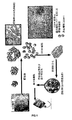
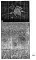
胚様体(EB)形成は、マウス及びヒトの両方のES細胞の造血系分化を研究するために使用される方法である。しかしマウスES細胞とは異なり、単独細胞の浮遊液中においてヒトES細胞で、胚様体を効率的に形成することは失敗している。代わりに、ヒトES細胞からの胚様体を形成するために、マウス胚性線維芽細胞(MEF)上で培養されたヒトES細胞の無傷のコロニーを、0.5mg/mlジスパーゼにより5分間消化し、小型の細胞クラスターを形成した。次にこれらの細胞クラスターは、血清-含有幹細胞培養培地(20%FCS)において更に凝集させた。容易に識別可能な細胞塊である胚様体は、細胞塊への参加に失敗しアポトーシスを受けた単独細胞の50%により培養の6日後形成され始めた。培養の12日後の胚様体は、卵黄嚢の初期胚構造に類似していた。胚様体の切片を採取し、その後この切片をCD34抗体で引き続き免疫-標識すると、卵黄嚢の組織学的特徴が明らかになった。細胞がCD34陽性であることにより明らかになるように、非-接着性造血前駆細胞は、小血管(small vessel)の管腔及び内皮層に存在することが認められた。その後胚様体を、37℃でのトリプシン消化(0.05%トリプシン/0.53mM EDTA)により処理した。約105個の胚様体-由来の細胞を含む初代造血前駆細胞を、メチルセルロース培養物(Stem Cells Inc. カナダ)中に播種し、10〜12日間培養した。赤血球及び巨核球コロニー形成単位(CFU)がその後、それらの本来の赤色又はモノクローナル抗-CD41もしくはCD61抗体による免疫標識により検出された。マクロファージ及び顆粒球コロニーを生じることができる最終的な造血前駆細胞は、12日目に形成された。この活性は、第一次(primary)及び最終的な造血の最初の波を示している。本発明者らは現在、動物及びヒトの両血清を含まない血清非含有胚様体培養システムを確立している。
Example
Hematopoietic progenitor embryoid body (EB) formation from embryoid bodies is a method used to study hematopoietic differentiation of both mouse and human ES cells. However, unlike mouse ES cells, it has failed to efficiently form embryoid bodies with human ES cells in suspensions of single cells. Instead, to form embryoid bodies from human ES cells, intact colonies of human ES cells cultured on mouse embryonic fibroblasts (MEF) were digested with 0.5 mg / ml dispase for 5 minutes. Formed small cell clusters. These cell clusters were then further aggregated in serum-containing stem cell culture medium (20% FCS). Embryoid bodies, a readily identifiable cell mass, began to form after 6 days of culture by 50% of single cells that failed to participate in the cell mass and underwent apoptosis. Embryoid bodies after 12 days in culture were similar to the early embryonic structure of the yolk sac. A section of the embryoid body was taken, and subsequent immuno-labeling of this section with CD34 antibody revealed the histological characteristics of the yolk sac. It was found that non-adherent hematopoietic progenitor cells were present in the lumen of small vessels and the endothelial layer, as revealed by the cells being CD34 positive. The embryoid bodies were then treated by trypsin digestion (0.05% trypsin / 0.53 mM EDTA) at 37 ° C. Primary hematopoietic progenitor cells containing approximately 10 5 embryoid body-derived cells were seeded in methylcellulose culture (Stem Cells Inc. Canada) and cultured for 10-12 days. Red blood cells and megakaryocyte colony forming units (CFU) were then detected by immunolabeling with their native red or monoclonal anti-CD41 or CD61 antibodies. The final hematopoietic progenitor cells that could give rise to macrophage and granulocyte colonies were formed on
胚様体-由来の巨核球前駆体のin vitro拡大
胚様体形成及び培養の12日後、単独細胞の浮遊培養物を、得られた細胞培養物の37℃での30分間のコラーゲナーゼ(1mg/ml)及び5分間のトリプシン消化(0.05%トリプシン/0.53mM EDTA)により作出した。CD34+細胞は造血と干渉し得るので、CD34+を他のEB細胞から分離した。CD34+細胞を、トロンボポエチン(TPO)、インターロイキン3(IL3)、インターロイキン6(IL6)、及び幹細胞因子(SCF)の存在下、ポリ-HEME表面上で培養し、これらの全ての因子は、巨核球の分化及び増殖を特異的に促進するように選択した。106個の出発ES細胞につき106個のCD41+巨核球の収量が得られた(n=6)。興味深いことに、コラーゲン-ベースの半固形マトリックスに播種した場合は、これらの巨核球は、ビーズ-様構造を示すプロ血小板を伴う極めて長い突起を形成した。成人造血幹細胞又はマウスES細胞を使用し巨核球の生成を試みる場合には、このような長い構造はこれまで報告されていない。小さいCD41陽性細胞断片は、放出された血小板として同定されたが、これらは、巨核球近傍に存在することが検出された。本発明者らは、フローサイトメトリーにより、これらの巨核球は、CD41、CD42a、CD42b、CD61、CD38、CD45(弱い)及びCD62Pについて陽性であるが、CD34、CD117、及びHLA-DRについて陰性であることを発見した。この表現型プロファイルは、正常なヒト成熟巨核球と一致する。
After 12 days of embryoid body-derived megakaryocyte precursor in vitro expanded embryoid body formation and culture, a single cell suspension culture was obtained from the resulting cell culture at 37 ° C for 30 minutes collagenase (1 mg / ml) and trypsin digestion (0.05% trypsin / 0.53 mM EDTA) for 5 minutes. Since CD34 + cells can interfere with hematopoiesis, CD34 + was isolated from other EB cells. CD34 + cells were cultured on poly-HEME surfaces in the presence of thrombopoietin (TPO), interleukin 3 (IL3), interleukin 6 (IL6), and stem cell factor (SCF), all of which are Selected to specifically promote sphere differentiation and proliferation. The yield of 10 6 starting ES cells per 10 6 CD41 + megakaryocytes were obtained (n = 6). Interestingly, when seeded on a collagen-based semi-solid matrix, these megakaryocytes formed very long protrusions with proplatelets showing a bead-like structure. Such long structures have not been reported so far when trying to generate megakaryocytes using adult hematopoietic stem cells or mouse ES cells. Small CD41 positive cell fragments were identified as released platelets, which were detected to be present near megakaryocytes. We have determined by flow cytometry that these megakaryocytes are positive for CD41, CD42a, CD42b, CD61, CD38, CD45 (weak) and CD62P, but negative for CD34, CD117, and HLA-DR. I discovered that there is. This phenotypic profile is consistent with normal human mature megakaryocytes.
ストロマ層上のヒトES細胞からの巨核球の分化
OP9ストロマ細胞株は、マウス造血を支援するために使用されている、新生仔頭蓋冠op/op欠損マウスから確立された細胞株である。このop/opマウスは、マクロファージコロニー刺激因子(M-CSF)遺伝子のコード領域に変異を有する。OP9システムを使用するヒトES細胞の造血系への分化の結果は、胚様体形成によるヒトES細胞の分化法に類似しているが、このストロマ細胞法は通常より多くの成熟前駆細胞のより高収量をもたらす。簡単に述べると、ヒトES細胞を、集密なOP9ストロマ細胞上に播種し、その後20%ウシ胎仔血清(FBS)を補充したα-MEM培地で培養した。分化は、6-ウェルプレート上105個ES細胞/ウェル、又は10cm2培養皿中8x105個細胞で開始した。6日間培養後、細胞上のCD34+細胞表面マーカーの出現により示されたように、ES細胞は造血前駆体に分化した。巨核球への分化のために、これらの細胞を6日目にトリプシン(0.05%トリプシン/0.53mnM EDTA、37℃/5%CO2)で5分間処理し、並びに10ng/ml TPOを含有する同じ培養培地において、新鮮な集密なOP9細胞上で継代した。更に8日間培養した後、巨核球は、目視検査により認められるようになり始めた。CD41免疫-染色により確認されるように、培養物の上清中の細胞の約30%は巨核球であった。これらの巨核球は多核であるが、胚様体-由来の巨核球において認められたような著しく長い突起を有さなかった。これらの巨核球は、成体巨核球に極めて類似している最終的な巨核球であると考えられた。興味深いことに、培養時に、血小板形成の徴候は存在せず、このことはマウスシステムとは極めて異なった。恐らく、OP9細胞は、巨核球の分化及び増殖を促進及び支援することはできるが、血小板形成を支援することはできないであろう。このことは、例え巨核球の分化及び増殖の機序の一部が類似していたとしても、マウス及びヒトの血小板形成の機序が異なることの、別の証拠である。
Differentiation of megakaryocytes from human ES cells on the stroma layer
The OP9 stromal cell line is a cell line established from neonatal calvarial op / op-deficient mice that has been used to support mouse hematopoiesis. This op / op mouse has a mutation in the coding region of the macrophage colony stimulating factor (M-CSF) gene. Although the results of differentiation of human ES cells into the hematopoietic system using the OP9 system are similar to the method of human ES cell differentiation by embryoid body formation, this stromal cell method is usually more than that of more mature progenitor cells. High yield is achieved. Briefly, human ES cells were seeded on confluent OP9 stromal cells and then cultured in α-MEM medium supplemented with 20% fetal bovine serum (FBS). Differentiation was initiated with 10 5 ES cells / well on 6-well plates or 8 × 10 5 cells in a 10 cm 2 culture dish. After 6 days in culture, ES cells differentiated into hematopoietic progenitors, as indicated by the appearance of CD34 + cell surface markers on the cells. For differentiation into megakaryocytes, these cells were treated with trypsin (0.05% trypsin / 0.53 mnM EDTA, 37 ° C./5% CO 2 ) for 5 minutes on day 6 and the same containing 10 ng / ml TPO Passaged on fresh confluent OP9 cells in culture medium. After a further 8 days of culture, megakaryocytes began to be recognized by visual inspection. As confirmed by CD41 immuno-staining, approximately 30% of the cells in the culture supernatant were megakaryocytes. These megakaryocytes are multinucleated, but did not have remarkably long processes as observed in embryoid body-derived megakaryocytes. These megakaryocytes were considered to be the final megakaryocytes that are very similar to adult megakaryocytes. Interestingly, there was no sign of platelet formation during culture, which was very different from the mouse system. Presumably, OP9 cells could promote and support megakaryocyte differentiation and proliferation, but not platelet formation. This is another evidence that mouse and human platelet formation mechanisms are different, even though some of the mechanisms of megakaryocyte differentiation and proliferation are similar.
巨核球の増殖、成熟及び精製
前述の方法のいずれかに由来した巨核球前駆体は、成体レシピエントマウスにおける増殖の、更には生着(engraft)の能力を明らかにしている。このプロセスの次工程として、本発明者らは、未熟な巨核球を更に増殖し、同時に巨核球成熟を停止するために、bFGFを使用した。各巨核球は2000個の血小板を形成することができると概算すると、106個ヒトES細胞(1個の6-ウェルプレート)は、106個の巨核球を生成し、引き続き約2x10⁹個の血小板を生成し、これは血小板の約1/20単位を示している(>5.5x1010個血小板/単位)。従ってこの概算された効率で、ヒト血小板1単位を生成するためには、ヒトES細胞の20 T75フラスコ(flasks)が必要であろう。これは、この収量で経済的に魅力的であることもあるが、すでに明らかに独りの技術者が支援することができる範囲である。
Megakaryocyte proliferation, maturation and purification Megakaryocyte precursors derived from any of the methods described above demonstrate the ability to proliferate and even engraft in adult recipient mice. As the next step in this process, we used bFGF to further proliferate immature megakaryocytes and simultaneously stop megakaryocyte maturation. Estimating that each megakaryocyte can form 2000 platelets, 10 6 human ES cells (1 6-well plate) produce 10 6 megakaryocytes, followed by about 2 × 10 9 Of platelets, representing approximately 1/20 units of platelets (> 5.5 × 10 10 platelets / unit). Thus, to produce one unit of human platelets with this estimated efficiency, 20 T75 flasks of human ES cells will be required. This may be economically attractive at this yield, but is already clearly within the scope of a single engineer.
分化を方向付ける代替技術
胚様体システムは、これらの細胞の大半は卵黄嚢細胞であるという事実のために、造血幹細胞を作製する望ましい効率よりも低い効率を有する。しかし胚様体システムはマウスタンパク質の夾雑を有さないので、このシステムは、OP9共培養システムよりも優れている。本発明者らのデータから、本発明者らは、造血系分化は、ストロマ細胞との共培養システムとは対称的に、EBシステムにおいてより最良に実現されると考えている。より最終的な造血細胞を入手しより効率的プロセスを作製するために、本発明者らは、EB培養を延長することを計画している。本発明者らは、微小環境が血島分化を支援し続けることを期待して、EBをより小さい断片へ機械的に解離又は断片化し、培養を継続することを試みた。本発明者らの予備的知見は、解離されたEBは、生存することができ、この解離後に成長し続けることを示唆している。このことは、EBシステムにおける更なる分化を推進する最初の試みであろう。例え最終的な血島が形成されなかったとしても、血島数の著しい増加が実現され得る。EB培養物へのVEGF及びSCFのような増殖因子の添加も試験されるであろう。
Alternative technology embryoid body systems that direct differentiation have an efficiency that is lower than the desired efficiency of producing hematopoietic stem cells due to the fact that most of these cells are yolk sac cells. However, this system is superior to the OP9 co-culture system because the embryoid body system does not have mouse protein contamination. From our data, we believe that hematopoietic differentiation is best achieved in the EB system, as opposed to a co-culture system with stromal cells. In order to obtain more final hematopoietic cells and create a more efficient process, we plan to extend the EB culture. In the hope that the microenvironment will continue to support blood island differentiation, we attempted to dissociate or fragment EBs into smaller fragments and continue culturing. Our preliminary findings suggest that dissociated EBs can survive and continue to grow after this dissociation. This will be the first attempt to promote further differentiation in the EB system. Even if the final blood islands are not formed, a significant increase in the number of blood islands can be realized. Addition of growth factors such as VEGF and SCF to the EB culture will also be tested.
改善された血小板放出及び成熟
本発明者らは、コラーゲンマトリックス、OP9及びポリHEMEを含む複数のシステムにおける血小板形成を既に認めているが、本発明者らは、そのプロセスを最適化することができるように、血小板放出の機序をより良く理解することを欲している。血小板形成を実現するために、本発明者らは、TPO、ヒト血漿、ヒト低温沈殿物及び一酸化窒素の存在下で、103〜104個の成熟巨核球を培養する。これらの血小板は、抗-CD41抗体で標識し、フローサイトメトリーで計数する。これらの血小板の形状は、電子顕微鏡を含む顕微鏡で観察する。真の血小板は、突起又は他の血小板との付着(attachment)を伴わない円盤状であるはずである。比較的大きいか又は連結した血小板は、プロ血小板形成のみを支援する、最適でない条件を示唆している。フィブリノーゲン及びフィブロネクチンのような細胞外マトリックスは、巨核球分化及び成熟を促進することが示されている。本発明者らは、巨核球の増殖及び分化に対するそれらの作用を決定するために、フィブリノーゲン及びフィブロネクチンでコートされたプレートを使用し、巨核球を培養する。
Improved platelet release and maturation We have already observed platelet formation in multiple systems including collagen matrix, OP9 and poly-HEME, but we can optimize the process As such, they want a better understanding of the mechanism of platelet release. To achieve platelet formation, we will culture 10 3 to 10 4 mature megakaryocytes in the presence of TPO, human plasma, human cryoprecipitate and nitric oxide. These platelets are labeled with anti-CD41 antibody and counted by flow cytometry. The shape of these platelets is observed with a microscope including an electron microscope. True platelets should be discoid with no protrusions or attachment with other platelets. Larger or linked platelets suggest suboptimal conditions that only support proplatelet formation. Extracellular matrices such as fibrinogen and fibronectin have been shown to promote megakaryocyte differentiation and maturation. In order to determine their effect on megakaryocyte proliferation and differentiation, we use fibrinogen and fibronectin coated plates to culture megakaryocytes.
血小板の試験
血小板は、トロンビン、ADP、及びコラーゲンに反応して凝集する。In vitro生成された血小板の異なる刺激に反応する凝集能を、血小板凝集計(Chrono-log Corporation, www.chronolog.com)により測定する。血小板は、上清から収集し、計測する。106個/mlの血小板を、PBSで洗浄し、ヒト血漿中に再浮遊する。様々な濃度のトロンビン、ADP及びコラーゲンを添加し、凝集速度論を、未変性のヒト血小板と比較する。本発明者らは、生成された「血小板」を試験し、成熟巨核球は、α顆粒放出の間接的マーカーであるCD62Pの表面発現により、0.5U/mlトロンビンにより活性化することができる。本発明者らは、以下の方法による血小板機能の試験の過程にある:
Platelet Test Platelets aggregate in response to thrombin, ADP, and collagen. Aggregation ability of in vitro generated platelets in response to different stimuli is measured by a platelet aggregometer (Chrono-log Corporation, www.chronolog.com). Platelets are collected from the supernatant and counted. 10 6 platelets / ml platelets are washed with PBS and resuspended in human plasma. Various concentrations of thrombin, ADP and collagen are added and the aggregation kinetics are compared to native human platelets. We tested the generated “platelets” and mature megakaryocytes can be activated by 0.5 U / ml thrombin by surface expression of CD62P, an indirect marker of alpha granule release. We are in the process of testing platelet function by the following method:
高密度コア顆粒放出:最初に106個の培養されたヒト血小板のアリコートを、緩衝液A(120mmol/Lグルタミン酸ナトリウム、5mmol/Lグルタミン酸カリウム、20mmol/L HEPES/NaOH(pH7.4)、2.5mmol/L EDTA、2.5mmol/L EGTA、3.15mmol/L MgCl2、及び1mmol/L DTT)中の[3H]5-HT(セロトニン)で標識する。血小板を、緩衝液Aで洗浄し、その後トロンビン1単位で活性化する。これらの反応は、この試料を氷上に4分間放置し、その後13,000gで1分間遠心することにより停止される。上清を収集し、以下のようにアッセイする。[3H]5-HT放出は、シンチレーションカウンターにより測定する。高密度コア顆粒放出の速度論も、凝集及び高密度コア顆粒からのATP分泌を同時に測定する光-血小板凝集計(Chronolog Corporation)により評価することができる。 High density core granule release: First aliquots of 10 6 cultured human platelets are added to buffer A (120 mmol / L sodium glutamate, 5 mmol / L potassium glutamate, 20 mmol / L HEPES / NaOH (pH 7.4), 2.5 Label with [ 3 H] 5-HT (serotonin) in (mmol / L EDTA, 2.5 mmol / L EGTA, 3.15 mmol / L MgCl 2 , and 1 mmol / L DTT). Platelets are washed with buffer A and then activated with 1 unit of thrombin. These reactions are stopped by leaving the sample on ice for 4 minutes, followed by centrifugation at 13,000 g for 1 minute. The supernatant is collected and assayed as follows. [ 3 H] 5-HT release is measured with a scintillation counter. The kinetics of high density core granule release can also be assessed by a photo-platelet aggregometer (Chronolog Corporation) that measures aggregation and ATP secretion from the high density core granules simultaneously.
α顆粒分泌:このアッセイは、フィコエリスリンと複合した抗-CD62抗体-AC 1.2(Becton Dickinson)を使用するフローサイトメトリーにより、P-セレクチン発現を測定することにより、モニタリングされる。典型的には、固定された血小板(109/ml)の2.5μlを、抗体溶液97.5μlに添加する。15分後、これらの試料を、0.35%BSAを含有するタイロード緩衝液1mlで希釈し、分析する。P-セレクチン発現の増加の割合を計算し、ヒトの未変性の血小板と比較する。 Alpha granule secretion: This assay is monitored by measuring P-selectin expression by flow cytometry using anti-CD62 antibody-AC 1.2 (Becton Dickinson) conjugated with phycoerythrin. Typically, 2.5 μl of fixed platelets (10 9 / ml) is added to 97.5 μl of antibody solution. After 15 minutes, these samples are diluted with 1 ml of Tyrode's buffer containing 0.35% BSA and analyzed. The percentage increase in P-selectin expression is calculated and compared to human native platelets.
リゾチーム(Lysozome)放出:ヘキソサミニダーゼは、Holmsen及びDangelmaierにより説明されたように測定する。クエン酸-リン酸緩衝液(pH4.5)5ml及び10mmol/L基質(P-ニトロフェニル-N-アセチル-D-グルコサミニド)2.5mlを混合し、96-ウェルプレートへアリコートとし(100μL)、反応上清5μLを添加した。37℃で18時間インキュベーションした後、0.08N NaOHの60μLを添加し、この反応を停止する。吸光度を、ELISAプレートリーダーにおいて、405-nmフィルターで測定する。 Lysozome release: hexosaminidase is measured as described by Holmsen and Dangelmaier. Mix 5 ml of citrate-phosphate buffer (pH 4.5) and 2.5 ml of 10 mmol / L substrate (P-nitrophenyl-N-acetyl-D-glucosaminide), aliquot into 96-well plate (100 μL), and reaction 5 μL of supernatant was added. After 18 hours of incubation at 37 ° C., the reaction is stopped by adding 60 μL of 0.08N NaOH. Absorbance is measured with a 405-nm filter in an ELISA plate reader.
これらの試験を使用し、ヒト胚性幹細胞から生成された血小板の生物学的活性を確立する。このin vitro血小板生成システムから生成された血小板は、人体の正常な血小板と機能的に類似している。しかしin vitro作出及び成熟により生成される場合、この方法で生成された血小板は、少なくとも生成の間、ヒト血液へ決して曝されないという事実のために、この生成された血小板は、血液由来のヒト血小板から容易に識別可能であろう。従ってこれらの血小板は、フィブリノーゲン、第V凝固因子及びVWFなどの通常の血清因子、in vivoにおける血流への放出後血小板が通常血液から獲得する因子へそれらを接着しない。このことは、血小板分離を補助するためにVWFが添加される場合のように、これらの因子は著しい量では培養物へ添加されないと仮定している。加えて当然血小板は、患者への送達後、直ちにこれらの因子をレシピエントの血流から獲得するであろう。 These tests are used to establish the biological activity of platelets generated from human embryonic stem cells. Platelets generated from this in vitro platelet generation system are functionally similar to normal platelets in the human body. However, when produced by in vitro generation and maturation, the platelets produced by this method are never exposed to human blood, at least during production, so that the produced platelets are blood-derived human platelets. Can be easily identified. Thus, these platelets do not adhere to normal serum factors such as fibrinogen, factor V clotting factor and VWF, factors that platelets normally acquire from blood after release into the bloodstream in vivo. This assumes that these factors are not added to the culture in significant amounts, such as when VWF is added to assist in platelet separation. In addition, platelets will naturally acquire these factors from the recipient's bloodstream immediately after delivery to the patient.
Claims (9)
(a)ヒト胚性幹細胞を、該細胞の造血系への分化に有利な条件下で培養する工程;
(b)造血系の細胞を巨核球へと培養する工程;
(c)巨核球を培養し、血小板を作出する工程;及び
(d)巨核球から血小板を回収する工程;
を含む製造法。 A method for producing human platelets, comprising:
(a) culturing human embryonic stem cells under conditions advantageous for differentiation of the cells into the hematopoietic system;
(b) culturing hematopoietic cells into megakaryocytes;
(c) culturing megakaryocytes to produce platelets; and
(d) collecting platelets from megakaryocytes;
Manufacturing method including.
Applications Claiming Priority (3)
| Application Number | Priority Date | Filing Date | Title |
|---|---|---|---|
| US62392204P | 2004-11-01 | 2004-11-01 | |
| US71457805P | 2005-09-07 | 2005-09-07 | |
| PCT/US2005/039401 WO2006050330A2 (en) | 2004-11-01 | 2005-10-31 | Platelets from stem cells |
Publications (2)
| Publication Number | Publication Date |
|---|---|
| JP2008518603A true JP2008518603A (en) | 2008-06-05 |
| JP2008518603A5 JP2008518603A5 (en) | 2008-12-18 |
Family
ID=36129866
Family Applications (1)
| Application Number | Title | Priority Date | Filing Date |
|---|---|---|---|
| JP2007539283A Pending JP2008518603A (en) | 2004-11-01 | 2005-10-31 | Stem cell-derived platelets |
Country Status (11)
| Country | Link |
|---|---|
| US (1) | US20060099198A1 (en) |
| EP (1) | EP1807507A2 (en) |
| JP (1) | JP2008518603A (en) |
| KR (1) | KR20070073932A (en) |
| AU (1) | AU2005302258B2 (en) |
| CA (1) | CA2586053C (en) |
| GB (1) | GB2434157A (en) |
| IS (1) | IS8637A (en) |
| MX (1) | MX2007005200A (en) |
| NZ (1) | NZ554866A (en) |
| WO (1) | WO2006050330A2 (en) |
Cited By (4)
| Publication number | Priority date | Publication date | Assignee | Title |
|---|---|---|---|---|
| JP2012510804A (en) * | 2008-12-04 | 2012-05-17 | インセルム (アンスティテュ・ナショナル・ドゥ・ラ・サント・エ・ドゥ・ラ・ルシェルシュ・メディカル) | Platelet production method |
| WO2015146415A1 (en) * | 2014-03-28 | 2015-10-01 | 東レエンジニアリング株式会社 | Separation apparatus and separation method |
| JPWO2017065280A1 (en) * | 2015-10-14 | 2018-09-13 | 株式会社メガカリオン | Method for producing purified platelets |
| CN111836886A (en) * | 2018-01-05 | 2020-10-27 | 血小板生源说股份有限公司 | Compositions and methods for producing megakaryocytes |
Families Citing this family (25)
| Publication number | Priority date | Publication date | Assignee | Title |
|---|---|---|---|---|
| KR102651433B1 (en) | 2006-04-14 | 2024-03-25 | 아스텔라스 인스티튜트 포 리제너러티브 메디슨 | Hemangio-colony forming cells |
| US8058064B2 (en) * | 2006-10-04 | 2011-11-15 | The University Of Tokyo | Sac-like structure enclosing hematopoietic progenitor cells produced from ES cells and method for preparing blood cells |
| WO2008151386A1 (en) * | 2007-06-15 | 2008-12-18 | Australian Stem Cell Centre Ltd | Megakaryocyte differentiation |
| JPWO2009119105A1 (en) * | 2008-03-28 | 2011-07-21 | 国立大学法人 東京大学 | GPIbα + GPV + GPVI + platelet in vitro preparation method |
| US8546141B2 (en) * | 2008-04-01 | 2013-10-01 | The University Of Tokyo | Method for preparation of platelet from iPS cell |
| KR20210003301A (en) | 2008-05-06 | 2021-01-11 | 아스텔라스 인스티튜트 포 리제너러티브 메디슨 | Methods for producing enucleated erythroid cells derived from pluripotent stem cells |
| ES2685969T3 (en) | 2008-05-06 | 2018-10-15 | Astellas Institute For Regenerative Medicine | Formation cells of hemangioblastic colonies and non-graft hemangioblastic cells |
| US8137970B2 (en) | 2008-06-30 | 2012-03-20 | Ewha University-Industry Collaboration Foundation | Methods for inducing the differentiation of hematopoietic stem cells into megakaryocytes and platelets, and gene controlling the differentiation |
| US8557580B2 (en) | 2009-02-20 | 2013-10-15 | Cellular Dynamics International, Inc. | Methods and compositions for the differentiation of stem cells |
| KR101720961B1 (en) | 2009-02-27 | 2017-03-29 | 셀룰러 다이내믹스 인터내셔널, 인코포레이티드 | Differentiation of pluripotent cells |
| US9988603B2 (en) * | 2009-12-04 | 2018-06-05 | Stem Cell & Regenerative Medicine International | Large scale generation of functional megakaryocytes and platelets from human embryonic stem cells under stromal-free conditions |
| WO2012048276A2 (en) | 2010-10-08 | 2012-04-12 | Caridianbct, Inc. | Customizable methods and systems of growing and harvesting cells in a hollow fiber bioreactor system |
| EP2633047A4 (en) | 2010-10-25 | 2014-04-02 | Philadelphia Children Hospital | Compositions and methods for the generation of platelets and methods of use thereof |
| CA2896053A1 (en) * | 2012-12-21 | 2014-06-26 | Ocata Therapeutics, Inc. | Methods for production of platelets from pluripotent stem cells and compositions thereof |
| WO2015073918A1 (en) | 2013-11-16 | 2015-05-21 | Terumo Bct, Inc. | Expanding cells in a bioreactor |
| CN106232800B (en) | 2014-03-25 | 2020-07-03 | 泰尔茂比司特公司 | Passive replacement of media |
| CN106715676A (en) | 2014-09-26 | 2017-05-24 | 泰尔茂比司特公司 | Scheduled feed |
| WO2017004592A1 (en) | 2015-07-02 | 2017-01-05 | Terumo Bct, Inc. | Cell growth with mechanical stimuli |
| JP6976939B2 (en) | 2015-10-20 | 2021-12-08 | フジフィルム セルラー ダイナミクス,インコーポレイテッド | Creation of multilineage hematopoietic progenitor cells by genetic programming |
| WO2017205667A1 (en) | 2016-05-25 | 2017-11-30 | Terumo Bct, Inc. | Cell expansion |
| US11104874B2 (en) | 2016-06-07 | 2021-08-31 | Terumo Bct, Inc. | Coating a bioreactor |
| US11685883B2 (en) | 2016-06-07 | 2023-06-27 | Terumo Bct, Inc. | Methods and systems for coating a cell growth surface |
| WO2018067826A1 (en) | 2016-10-05 | 2018-04-12 | Cellular Dynamics International, Inc. | Generating mature lineages from induced pluripotent stem cells with mecp2 disruption |
| US11624046B2 (en) | 2017-03-31 | 2023-04-11 | Terumo Bct, Inc. | Cell expansion |
| CN110612344B (en) | 2017-03-31 | 2023-09-12 | 泰尔茂比司特公司 | cell expansion |
Citations (1)
| Publication number | Priority date | Publication date | Assignee | Title |
|---|---|---|---|---|
| WO2001034776A1 (en) * | 1999-11-08 | 2001-05-17 | Wisconsin Alumni Research Foundation | Hematopoietic differentiation of human embryonic stem cells |
Family Cites Families (1)
| Publication number | Priority date | Publication date | Assignee | Title |
|---|---|---|---|---|
| BR9711204A (en) * | 1996-08-19 | 1999-08-17 | Univ Massachusetts | Embryonic cell lines or similar are undifferentiated produced by nuclear transplantation of hybrid species |
-
2005
- 2005-10-31 AU AU2005302258A patent/AU2005302258B2/en active Active
- 2005-10-31 EP EP05824903A patent/EP1807507A2/en not_active Withdrawn
- 2005-10-31 US US11/262,582 patent/US20060099198A1/en not_active Abandoned
- 2005-10-31 JP JP2007539283A patent/JP2008518603A/en active Pending
- 2005-10-31 WO PCT/US2005/039401 patent/WO2006050330A2/en active Application Filing
- 2005-10-31 NZ NZ554866A patent/NZ554866A/en unknown
- 2005-10-31 KR KR1020077011858A patent/KR20070073932A/en not_active Application Discontinuation
- 2005-10-31 MX MX2007005200A patent/MX2007005200A/en active IP Right Grant
- 2005-10-31 CA CA2586053A patent/CA2586053C/en active Active
-
2007
- 2007-04-30 IS IS8637A patent/IS8637A/en unknown
- 2007-05-17 GB GB0709533A patent/GB2434157A/en not_active Withdrawn
Patent Citations (1)
| Publication number | Priority date | Publication date | Assignee | Title |
|---|---|---|---|---|
| WO2001034776A1 (en) * | 1999-11-08 | 2001-05-17 | Wisconsin Alumni Research Foundation | Hematopoietic differentiation of human embryonic stem cells |
Cited By (6)
| Publication number | Priority date | Publication date | Assignee | Title |
|---|---|---|---|---|
| JP2012510804A (en) * | 2008-12-04 | 2012-05-17 | インセルム (アンスティテュ・ナショナル・ドゥ・ラ・サント・エ・ドゥ・ラ・ルシェルシュ・メディカル) | Platelet production method |
| WO2015146415A1 (en) * | 2014-03-28 | 2015-10-01 | 東レエンジニアリング株式会社 | Separation apparatus and separation method |
| JPWO2017065280A1 (en) * | 2015-10-14 | 2018-09-13 | 株式会社メガカリオン | Method for producing purified platelets |
| JP7287634B2 (en) | 2015-10-14 | 2023-06-06 | 株式会社メガカリオン | Method for producing purified platelets |
| CN111836886A (en) * | 2018-01-05 | 2020-10-27 | 血小板生源说股份有限公司 | Compositions and methods for producing megakaryocytes |
| JP2021509812A (en) * | 2018-01-05 | 2021-04-08 | プレートレット バイオジェネシス, インコーポレイテッド | Compositions and Methods for Producing Megakaryocytes |
Also Published As
| Publication number | Publication date |
|---|---|
| GB0709533D0 (en) | 2007-06-27 |
| EP1807507A2 (en) | 2007-07-18 |
| CA2586053C (en) | 2013-07-30 |
| IS8637A (en) | 2007-04-30 |
| NZ554866A (en) | 2010-12-24 |
| WO2006050330A2 (en) | 2006-05-11 |
| AU2005302258B2 (en) | 2010-10-21 |
| CA2586053A1 (en) | 2006-05-11 |
| WO2006050330A3 (en) | 2006-08-24 |
| GB2434157A (en) | 2007-07-18 |
| AU2005302258A1 (en) | 2006-05-11 |
| US20060099198A1 (en) | 2006-05-11 |
| WO2006050330B1 (en) | 2006-10-26 |
| MX2007005200A (en) | 2007-05-11 |
| KR20070073932A (en) | 2007-07-10 |
Similar Documents
| Publication | Publication Date | Title |
|---|---|---|
| AU2005302258B2 (en) | Platelets from stem cells | |
| US20070077654A1 (en) | Platelets from stem cells | |
| TWI288779B (en) | Dedifferentiated, programmable stem cells of monocytic origin, and their production and use | |
| JP5283120B2 (en) | A structure encapsulating hematopoietic progenitor cells from ES cells, and a method for preparing blood cells using the structure. | |
| WO1995011692A1 (en) | In vitro amplification of stem cells | |
| JP5102773B2 (en) | Methods for improving stem cell homing and engraftment | |
| KR20020013480A (en) | Human brain endothelial cells and growth medium and method for expansion of primitive cd34+cd38- bone marrow stem cells | |
| Fujimoto et al. | Microencapsulated feeder cells as a source of soluble factors for expansion of CD34+ hematopoietic stem cells | |
| JPH10295369A (en) | Production of hematopoietic stem cell | |
| Deutsch et al. | Mimicking the haematopoietic niche microenvironment provides a novel strategy for expansion of haematopoietic and megakaryocyte‐progenitor cells from cord blood | |
| Schlechta et al. | Ex-vivo expanded umbilical cord blood stem cells retain capacity for myocardial regeneration | |
| Islami et al. | A review of evaluating hematopoietic stem cells derived from umbilical cord blood's expansion and homing | |
| KR101318965B1 (en) | A composition for creating an artificial bone-marrow like environment and use thereof | |
| Hayashi et al. | Placental/umbilical cord blood-derived mesenchymal stem cell-like stromal cells support hematopoietic recovery of X-irradiated human CD34+ cells | |
| CA2767970C (en) | Method for using directing cells for specific stem/progenitor cell activation and differentiation | |
| JPWO2005054459A1 (en) | Method for producing hematopoietic stem cells or vascular endothelial progenitor cells | |
| Mobaraki et al. | Evaluation of expansion and maintenance of umbilical cord blood CD34+ cells in the co-culture with umbilical cord blood-derived mesenchymal stem cells in the presence of microcarrier beads | |
| CN101052710A (en) | Platelets from stem cells | |
| CN109182268B (en) | Application of umbilical artery endothelial cells in constructing microenvironment of blood stem cells | |
| Chitteti et al. | In vitro construction of 2D and 3D simulations of the murine hematopoietic niche | |
| JP4809940B2 (en) | Method for amplifying mononuclear cells isolated from blood in vitro | |
| JP2008539700A (en) | Selection, culture and production of lineage restricted hematopoietic stem cells | |
| US8956870B2 (en) | Method for using directing cells for specific stem/progenitor cell activation and differentiation | |
| JPH11180877A (en) | Cell preparation containing megakaryocyte precursor cells and its production | |
| JP2007175008A (en) | Method for culturing stem cell |
Legal Events
| Date | Code | Title | Description |
|---|---|---|---|
| A521 | Request for written amendment filed |
Free format text: JAPANESE INTERMEDIATE CODE: A523 Effective date: 20081030 |
|
| A621 | Written request for application examination |
Free format text: JAPANESE INTERMEDIATE CODE: A621 Effective date: 20081030 |
|
| A131 | Notification of reasons for refusal |
Free format text: JAPANESE INTERMEDIATE CODE: A131 Effective date: 20110711 |
|
| A601 | Written request for extension of time |
Free format text: JAPANESE INTERMEDIATE CODE: A601 Effective date: 20111003 |
|
| A602 | Written permission of extension of time |
Free format text: JAPANESE INTERMEDIATE CODE: A602 Effective date: 20111011 |
|
| A521 | Request for written amendment filed |
Free format text: JAPANESE INTERMEDIATE CODE: A523 Effective date: 20120111 |
|
| A02 | Decision of refusal |
Free format text: JAPANESE INTERMEDIATE CODE: A02 Effective date: 20120227 |