EP3693086B1 - Microdroplet manipulation method - Google Patents
Microdroplet manipulation method Download PDFInfo
- Publication number
- EP3693086B1 EP3693086B1 EP19156182.8A EP19156182A EP3693086B1 EP 3693086 B1 EP3693086 B1 EP 3693086B1 EP 19156182 A EP19156182 A EP 19156182A EP 3693086 B1 EP3693086 B1 EP 3693086B1
- Authority
- EP
- European Patent Office
- Prior art keywords
- microdroplets
- volume
- average volume
- range
- femtolitres
- Prior art date
- Legal status (The legal status is an assumption and is not a legal conclusion. Google has not performed a legal analysis and makes no representation as to the accuracy of the status listed.)
- Active
Links
- 238000000034 method Methods 0.000 title claims description 40
- 239000012736 aqueous medium Substances 0.000 claims description 34
- PEDCQBHIVMGVHV-UHFFFAOYSA-N Glycerine Chemical compound OCC(O)CO PEDCQBHIVMGVHV-UHFFFAOYSA-N 0.000 claims description 33
- XLYOFNOQVPJJNP-UHFFFAOYSA-N water Substances O XLYOFNOQVPJJNP-UHFFFAOYSA-N 0.000 claims description 32
- 230000000694 effects Effects 0.000 claims description 26
- 239000012530 fluid Substances 0.000 claims description 26
- 239000002777 nucleoside Substances 0.000 claims description 10
- -1 nucleoside triphosphate Chemical class 0.000 claims description 10
- 230000002255 enzymatic effect Effects 0.000 claims description 9
- 239000001226 triphosphate Substances 0.000 claims description 8
- 235000011178 triphosphate Nutrition 0.000 claims description 8
- 108091034117 Oligonucleotide Proteins 0.000 claims description 6
- 239000000872 buffer Substances 0.000 claims description 6
- 230000009257 reactivity Effects 0.000 claims description 6
- 239000012062 aqueous buffer Substances 0.000 claims description 5
- 238000006243 chemical reaction Methods 0.000 claims description 5
- 238000006911 enzymatic reaction Methods 0.000 claims description 5
- 239000007850 fluorescent dye Substances 0.000 claims description 5
- 102000004169 proteins and genes Human genes 0.000 claims description 5
- 108090000623 proteins and genes Proteins 0.000 claims description 5
- 108020004707 nucleic acids Proteins 0.000 claims description 4
- 102000039446 nucleic acids Human genes 0.000 claims description 4
- 150000007523 nucleic acids Chemical class 0.000 claims description 4
- 230000004663 cell proliferation Effects 0.000 claims description 3
- 230000002068 genetic effect Effects 0.000 claims description 3
- 238000002372 labelling Methods 0.000 claims description 3
- 239000002609 medium Substances 0.000 claims description 3
- 238000012163 sequencing technique Methods 0.000 claims description 3
- 239000000126 substance Substances 0.000 claims description 3
- 102000004190 Enzymes Human genes 0.000 claims description 2
- 108090000790 Enzymes Proteins 0.000 claims description 2
- 239000012491 analyte Substances 0.000 claims description 2
- 230000007613 environmental effect Effects 0.000 claims description 2
- 239000003921 oil Substances 0.000 description 45
- 210000004027 cell Anatomy 0.000 description 27
- 230000036571 hydration Effects 0.000 description 19
- 238000006703 hydration reaction Methods 0.000 description 19
- 239000012071 phase Substances 0.000 description 19
- 239000000839 emulsion Substances 0.000 description 18
- 239000000203 mixture Substances 0.000 description 11
- 239000004094 surface-active agent Substances 0.000 description 10
- 239000000523 sample Substances 0.000 description 9
- 238000011534 incubation Methods 0.000 description 5
- 230000010261 cell growth Effects 0.000 description 4
- 239000001963 growth medium Substances 0.000 description 4
- JLCPHMBAVCMARE-UHFFFAOYSA-N [3-[[3-[[3-[[3-[[3-[[3-[[3-[[3-[[3-[[3-[[3-[[5-(2-amino-6-oxo-1H-purin-9-yl)-3-[[3-[[3-[[3-[[3-[[3-[[5-(2-amino-6-oxo-1H-purin-9-yl)-3-[[5-(2-amino-6-oxo-1H-purin-9-yl)-3-hydroxyoxolan-2-yl]methoxy-hydroxyphosphoryl]oxyoxolan-2-yl]methoxy-hydroxyphosphoryl]oxy-5-(5-methyl-2,4-dioxopyrimidin-1-yl)oxolan-2-yl]methoxy-hydroxyphosphoryl]oxy-5-(6-aminopurin-9-yl)oxolan-2-yl]methoxy-hydroxyphosphoryl]oxy-5-(6-aminopurin-9-yl)oxolan-2-yl]methoxy-hydroxyphosphoryl]oxy-5-(6-aminopurin-9-yl)oxolan-2-yl]methoxy-hydroxyphosphoryl]oxy-5-(6-aminopurin-9-yl)oxolan-2-yl]methoxy-hydroxyphosphoryl]oxyoxolan-2-yl]methoxy-hydroxyphosphoryl]oxy-5-(5-methyl-2,4-dioxopyrimidin-1-yl)oxolan-2-yl]methoxy-hydroxyphosphoryl]oxy-5-(4-amino-2-oxopyrimidin-1-yl)oxolan-2-yl]methoxy-hydroxyphosphoryl]oxy-5-(5-methyl-2,4-dioxopyrimidin-1-yl)oxolan-2-yl]methoxy-hydroxyphosphoryl]oxy-5-(5-methyl-2,4-dioxopyrimidin-1-yl)oxolan-2-yl]methoxy-hydroxyphosphoryl]oxy-5-(6-aminopurin-9-yl)oxolan-2-yl]methoxy-hydroxyphosphoryl]oxy-5-(6-aminopurin-9-yl)oxolan-2-yl]methoxy-hydroxyphosphoryl]oxy-5-(4-amino-2-oxopyrimidin-1-yl)oxolan-2-yl]methoxy-hydroxyphosphoryl]oxy-5-(4-amino-2-oxopyrimidin-1-yl)oxolan-2-yl]methoxy-hydroxyphosphoryl]oxy-5-(4-amino-2-oxopyrimidin-1-yl)oxolan-2-yl]methoxy-hydroxyphosphoryl]oxy-5-(6-aminopurin-9-yl)oxolan-2-yl]methoxy-hydroxyphosphoryl]oxy-5-(4-amino-2-oxopyrimidin-1-yl)oxolan-2-yl]methyl [5-(6-aminopurin-9-yl)-2-(hydroxymethyl)oxolan-3-yl] hydrogen phosphate Polymers Cc1cn(C2CC(OP(O)(=O)OCC3OC(CC3OP(O)(=O)OCC3OC(CC3O)n3cnc4c3nc(N)[nH]c4=O)n3cnc4c3nc(N)[nH]c4=O)C(COP(O)(=O)OC3CC(OC3COP(O)(=O)OC3CC(OC3COP(O)(=O)OC3CC(OC3COP(O)(=O)OC3CC(OC3COP(O)(=O)OC3CC(OC3COP(O)(=O)OC3CC(OC3COP(O)(=O)OC3CC(OC3COP(O)(=O)OC3CC(OC3COP(O)(=O)OC3CC(OC3COP(O)(=O)OC3CC(OC3COP(O)(=O)OC3CC(OC3COP(O)(=O)OC3CC(OC3COP(O)(=O)OC3CC(OC3COP(O)(=O)OC3CC(OC3COP(O)(=O)OC3CC(OC3COP(O)(=O)OC3CC(OC3COP(O)(=O)OC3CC(OC3CO)n3cnc4c(N)ncnc34)n3ccc(N)nc3=O)n3cnc4c(N)ncnc34)n3ccc(N)nc3=O)n3ccc(N)nc3=O)n3ccc(N)nc3=O)n3cnc4c(N)ncnc34)n3cnc4c(N)ncnc34)n3cc(C)c(=O)[nH]c3=O)n3cc(C)c(=O)[nH]c3=O)n3ccc(N)nc3=O)n3cc(C)c(=O)[nH]c3=O)n3cnc4c3nc(N)[nH]c4=O)n3cnc4c(N)ncnc34)n3cnc4c(N)ncnc34)n3cnc4c(N)ncnc34)n3cnc4c(N)ncnc34)O2)c(=O)[nH]c1=O JLCPHMBAVCMARE-UHFFFAOYSA-N 0.000 description 3
- 238000004458 analytical method Methods 0.000 description 3
- 239000008346 aqueous phase Substances 0.000 description 3
- 238000005119 centrifugation Methods 0.000 description 3
- 230000008859 change Effects 0.000 description 3
- 238000001514 detection method Methods 0.000 description 3
- 230000000887 hydrating effect Effects 0.000 description 3
- 238000005259 measurement Methods 0.000 description 3
- 239000002773 nucleotide Substances 0.000 description 3
- 125000003729 nucleotide group Chemical group 0.000 description 3
- 239000000758 substrate Substances 0.000 description 3
- 230000012010 growth Effects 0.000 description 2
- 238000009396 hybridization Methods 0.000 description 2
- 239000006228 supernatant Substances 0.000 description 2
- 238000001712 DNA sequencing Methods 0.000 description 1
- 239000005662 Paraffin oil Substances 0.000 description 1
- 238000003559 RNA-seq method Methods 0.000 description 1
- 239000012980 RPMI-1640 medium Substances 0.000 description 1
- 239000006146 Roswell Park Memorial Institute medium Substances 0.000 description 1
- 241000239226 Scorpiones Species 0.000 description 1
- FAPWRFPIFSIZLT-UHFFFAOYSA-M Sodium chloride Chemical compound [Na+].[Cl-] FAPWRFPIFSIZLT-UHFFFAOYSA-M 0.000 description 1
- 206010042971 T-cell lymphoma Diseases 0.000 description 1
- 208000027585 T-cell non-Hodgkin lymphoma Diseases 0.000 description 1
- 241000700605 Viruses Species 0.000 description 1
- 230000002411 adverse Effects 0.000 description 1
- 239000000427 antigen Substances 0.000 description 1
- 102000036639 antigens Human genes 0.000 description 1
- 108091007433 antigens Proteins 0.000 description 1
- 239000006143 cell culture medium Substances 0.000 description 1
- 230000001413 cellular effect Effects 0.000 description 1
- 238000012512 characterization method Methods 0.000 description 1
- 239000002131 composite material Substances 0.000 description 1
- 230000009089 cytolysis Effects 0.000 description 1
- 230000005670 electromagnetic radiation Effects 0.000 description 1
- 230000001804 emulsifying effect Effects 0.000 description 1
- 230000002708 enhancing effect Effects 0.000 description 1
- 230000005284 excitation Effects 0.000 description 1
- 238000002866 fluorescence resonance energy transfer Methods 0.000 description 1
- NBVXSUQYWXRMNV-UHFFFAOYSA-N fluoromethane Chemical compound FC NBVXSUQYWXRMNV-UHFFFAOYSA-N 0.000 description 1
- 230000006872 improvement Effects 0.000 description 1
- 238000007689 inspection Methods 0.000 description 1
- 238000011835 investigation Methods 0.000 description 1
- 229940059904 light mineral oil Drugs 0.000 description 1
- 210000004962 mammalian cell Anatomy 0.000 description 1
- 239000000463 material Substances 0.000 description 1
- 239000000693 micelle Substances 0.000 description 1
- 238000001000 micrograph Methods 0.000 description 1
- 239000002480 mineral oil Substances 0.000 description 1
- 235000010446 mineral oil Nutrition 0.000 description 1
- 238000012544 monitoring process Methods 0.000 description 1
- 239000002736 nonionic surfactant Substances 0.000 description 1
- 230000003287 optical effect Effects 0.000 description 1
- 230000037361 pathway Effects 0.000 description 1
- 230000008569 process Effects 0.000 description 1
- 239000000047 product Substances 0.000 description 1
- 230000001141 propulsive effect Effects 0.000 description 1
- 239000013074 reference sample Substances 0.000 description 1
- 230000000717 retained effect Effects 0.000 description 1
- 229920006395 saturated elastomer Polymers 0.000 description 1
- 238000012216 screening Methods 0.000 description 1
- 229920002545 silicone oil Polymers 0.000 description 1
- 230000005476 size effect Effects 0.000 description 1
- 239000011780 sodium chloride Substances 0.000 description 1
- 125000006850 spacer group Chemical group 0.000 description 1
- 239000003381 stabilizer Substances 0.000 description 1
- 238000011282 treatment Methods 0.000 description 1
Images

Classifications
-
- B—PERFORMING OPERATIONS; TRANSPORTING
- B01—PHYSICAL OR CHEMICAL PROCESSES OR APPARATUS IN GENERAL
- B01L—CHEMICAL OR PHYSICAL LABORATORY APPARATUS FOR GENERAL USE
- B01L3/00—Containers or dishes for laboratory use, e.g. laboratory glassware; Droppers
- B01L3/50—Containers for the purpose of retaining a material to be analysed, e.g. test tubes
- B01L3/502—Containers for the purpose of retaining a material to be analysed, e.g. test tubes with fluid transport, e.g. in multi-compartment structures
- B01L3/5027—Containers for the purpose of retaining a material to be analysed, e.g. test tubes with fluid transport, e.g. in multi-compartment structures by integrated microfluidic structures, i.e. dimensions of channels and chambers are such that surface tension forces are important, e.g. lab-on-a-chip
- B01L3/502769—Containers for the purpose of retaining a material to be analysed, e.g. test tubes with fluid transport, e.g. in multi-compartment structures by integrated microfluidic structures, i.e. dimensions of channels and chambers are such that surface tension forces are important, e.g. lab-on-a-chip characterised by multiphase flow arrangements
- B01L3/502784—Containers for the purpose of retaining a material to be analysed, e.g. test tubes with fluid transport, e.g. in multi-compartment structures by integrated microfluidic structures, i.e. dimensions of channels and chambers are such that surface tension forces are important, e.g. lab-on-a-chip characterised by multiphase flow arrangements specially adapted for droplet or plug flow, e.g. digital microfluidics
-
- C—CHEMISTRY; METALLURGY
- C12—BIOCHEMISTRY; BEER; SPIRITS; WINE; VINEGAR; MICROBIOLOGY; ENZYMOLOGY; MUTATION OR GENETIC ENGINEERING
- C12N—MICROORGANISMS OR ENZYMES; COMPOSITIONS THEREOF; PROPAGATING, PRESERVING, OR MAINTAINING MICROORGANISMS; MUTATION OR GENETIC ENGINEERING; CULTURE MEDIA
- C12N11/00—Carrier-bound or immobilised enzymes; Carrier-bound or immobilised microbial cells; Preparation thereof
- C12N11/02—Enzymes or microbial cells immobilised on or in an organic carrier
-
- C—CHEMISTRY; METALLURGY
- C12—BIOCHEMISTRY; BEER; SPIRITS; WINE; VINEGAR; MICROBIOLOGY; ENZYMOLOGY; MUTATION OR GENETIC ENGINEERING
- C12N—MICROORGANISMS OR ENZYMES; COMPOSITIONS THEREOF; PROPAGATING, PRESERVING, OR MAINTAINING MICROORGANISMS; MUTATION OR GENETIC ENGINEERING; CULTURE MEDIA
- C12N5/00—Undifferentiated human, animal or plant cells, e.g. cell lines; Tissues; Cultivation or maintenance thereof; Culture media therefor
- C12N5/06—Animal cells or tissues; Human cells or tissues
- C12N5/0602—Vertebrate cells
- C12N5/0693—Tumour cells; Cancer cells
-
- C—CHEMISTRY; METALLURGY
- C12—BIOCHEMISTRY; BEER; SPIRITS; WINE; VINEGAR; MICROBIOLOGY; ENZYMOLOGY; MUTATION OR GENETIC ENGINEERING
- C12Q—MEASURING OR TESTING PROCESSES INVOLVING ENZYMES, NUCLEIC ACIDS OR MICROORGANISMS; COMPOSITIONS OR TEST PAPERS THEREFOR; PROCESSES OF PREPARING SUCH COMPOSITIONS; CONDITION-RESPONSIVE CONTROL IN MICROBIOLOGICAL OR ENZYMOLOGICAL PROCESSES
- C12Q1/00—Measuring or testing processes involving enzymes, nucleic acids or microorganisms; Compositions therefor; Processes of preparing such compositions
- C12Q1/68—Measuring or testing processes involving enzymes, nucleic acids or microorganisms; Compositions therefor; Processes of preparing such compositions involving nucleic acids
- C12Q1/6869—Methods for sequencing
-
- B—PERFORMING OPERATIONS; TRANSPORTING
- B01—PHYSICAL OR CHEMICAL PROCESSES OR APPARATUS IN GENERAL
- B01L—CHEMICAL OR PHYSICAL LABORATORY APPARATUS FOR GENERAL USE
- B01L2200/00—Solutions for specific problems relating to chemical or physical laboratory apparatus
- B01L2200/06—Fluid handling related problems
- B01L2200/0673—Handling of plugs of fluid surrounded by immiscible fluid
-
- B—PERFORMING OPERATIONS; TRANSPORTING
- B01—PHYSICAL OR CHEMICAL PROCESSES OR APPARATUS IN GENERAL
- B01L—CHEMICAL OR PHYSICAL LABORATORY APPARATUS FOR GENERAL USE
- B01L2200/00—Solutions for specific problems relating to chemical or physical laboratory apparatus
- B01L2200/16—Reagents, handling or storing thereof
-
- B—PERFORMING OPERATIONS; TRANSPORTING
- B01—PHYSICAL OR CHEMICAL PROCESSES OR APPARATUS IN GENERAL
- B01L—CHEMICAL OR PHYSICAL LABORATORY APPARATUS FOR GENERAL USE
- B01L2300/00—Additional constructional details
- B01L2300/08—Geometry, shape and general structure
- B01L2300/0896—Nanoscaled
-
- C—CHEMISTRY; METALLURGY
- C12—BIOCHEMISTRY; BEER; SPIRITS; WINE; VINEGAR; MICROBIOLOGY; ENZYMOLOGY; MUTATION OR GENETIC ENGINEERING
- C12Q—MEASURING OR TESTING PROCESSES INVOLVING ENZYMES, NUCLEIC ACIDS OR MICROORGANISMS; COMPOSITIONS OR TEST PAPERS THEREFOR; PROCESSES OF PREPARING SUCH COMPOSITIONS; CONDITION-RESPONSIVE CONTROL IN MICROBIOLOGICAL OR ENZYMOLOGICAL PROCESSES
- C12Q2563/00—Nucleic acid detection characterized by the use of physical, structural and functional properties
- C12Q2563/107—Nucleic acid detection characterized by the use of physical, structural and functional properties fluorescence
-
- C—CHEMISTRY; METALLURGY
- C12—BIOCHEMISTRY; BEER; SPIRITS; WINE; VINEGAR; MICROBIOLOGY; ENZYMOLOGY; MUTATION OR GENETIC ENGINEERING
- C12Q—MEASURING OR TESTING PROCESSES INVOLVING ENZYMES, NUCLEIC ACIDS OR MICROORGANISMS; COMPOSITIONS OR TEST PAPERS THEREFOR; PROCESSES OF PREPARING SUCH COMPOSITIONS; CONDITION-RESPONSIVE CONTROL IN MICROBIOLOGICAL OR ENZYMOLOGICAL PROCESSES
- C12Q2563/00—Nucleic acid detection characterized by the use of physical, structural and functional properties
- C12Q2563/161—Vesicles, e.g. liposome
Definitions
- This invention relates to an improved method of manipulating aqueous microdroplets optionally containing biological cells in an immiscible carrier fluid such as an oil. It enables the size of the microdroplets to be controlled or adjusted and any enzymatic or chemical reactions occurring therein to be maintained or optimised during a given period.
- these methods involve translocating microdroplets dispersed in an immiscible carrier fluid along microfluidic pathways in an analytical device using electrowetting propulsive forces or by directly printing of the microdroplets onto a substrate coated with the carrier fluid.
- these microdroplets tend to undergo significant shrinkage over time which can sometimes interfere with some or all the enzymatic processes going on within.
- microdroplet manipulation method which overcomes these problems. It may be used, for example, to manipulate the size and/or reactivity of the contents of microdroplets or to control chemical or enzymatic reactions occurring therein.
- the invention is as defined in the appended claims.
- a generic method of controlling the size of microdroplets and/or maintaining or optimising enzymatic or chemical reactions occurring therein said microdroplets having an average volume in the range 0.5 femtolitres to 10 nanolitres comprised of at least one biological component and a first aqueous medium having a water activity of a w1 of less than 1 characterised by the step of maintaining the microdroplets in a water-immiscible carrier fluid which further includes secondary droplets of a second aqueous medium having an average volume less than 25% of the average volume of the microdroplets up to and including a maximum of 4 femtolitres and wherein the volume ratio of carrier fluid to total volume of microdroplets per unit volume of the total is greater than 2:1.
- the invention solves the problem by using a carrier fluid which contains very small secondary droplets which can interact with the microdroplets without adversely affecting the latter's overall characteristics or the efficacy of any detection method applied to them.
- the carrier medium is an oil such a composite medium is sometimes referred to as 'hydrated oil'.
- a w the water activity of an aqueous medium (a w ) is defined as the ratio of the partial vapour pressure of the aqueous medium under investigation to that of pure water under STP conditions.
- the water activities of the first and second aqueous media may be the same or substantially the same so that any tendency for the microdroplets to shrink or expand is continuously counteracted. Thus, the sizes of the microdroplets may always be preserved.
- the first and second aqueous medium may have compositions which in one embodiment are identical.
- the water activity of the first and the second aqueous media are independently in the range from 0.9 to 1.
- the water activity of the first aqueous medium is from 0.9 to less than 1.
- the ratio of the water activities of the first and second aqueous media (a w1 :a w2 ) is in the range 0.9:1 to 1:0.9.
- first and second media which are buffers; and, if required, by varying the relative compositions of the two.
- the ionic strength of the first aqueous medium is in the range from to 1 to 5 that of the second aqueous medium; preferably from 3 to 5 times.
- the ionic strength of the second aqueous medium is in the range from to 1 to 5 times that of the first aqueous medium; preferably from 3 to 5 times.
- the ionic strengths are the same or substantially the same with the ratio of ionic strengths being in the range from 3:1 to 1:3.
- first and second aqueous media may include glycerol as a component; for example, at differing concentrations.
- the pHs of the first and second aqueous media are the same or similar and within the range 6.5 to 8.
- these have a much smaller average volume than the average value for the microdroplets and at the limit may be comprised of femto-sized droplets or micelles of the second aqueous medium emulsified within the carrier fluid and stabilised by a sheath of compatible surfactant molecules; for example, a non-ionic surfactant.
- the size of these secondary droplets is less than 10%, preferably less than 5% of the volume of the microdroplets employed.
- the average volume of the secondary droplets lies within the range 10 to 1% of the average volume of the microdroplets.
- the secondary droplets form part of a stable emulsion in the carrier fluid which in one embodiment is an immiscible oil.
- the carrier fluid is selected from a mineral oil, a silicone oil or a fluorocarbon oil.
- the oil may also contain additional surfactants and stabilisers if required.
- the volume ratio of carrier fluid to total volume of the microdroplets is greater than 3:1; preferably 5:1 or greater.
- the method of the invention is useful for several applications where biological cells are being analysed.
- One example is where a culture of immortalised mammalian cells is being caused to proliferate inside the microdroplets for the purpose of screening individual clonal copies of the cells for desirable characteristics such as protein expression or particular genetic traits.
- a method of causing the cellular proliferation of one or more cell types contained within a microdroplet having an average volume in the range 4 femtolitres to 10 nanolitres and comprised of an aqueous buffer comprising the steps of incubating the cell(s) inside the droplets in suitable environmental conditions and thereafter detecting the number of cells inside each droplet, characterised in that the microdroplets are suspended in an immiscible carrier fluid further comprising secondary droplets having an average volume less than 25% of the average volume of the microdroplets up to and including a maximum of 4 femtolitres and wherein the volume ratio of carrier fluid to total volume of microdroplets per unit volume of the total is greater than 2:1.
- a method of detecting one or more phenotypic traits, genetic traits or protein expression profiles of a cell under consideration comprising the steps of labelling a target derived from the cell(s)with a fluorescent probe and thereafter detecting an output from the probe characterised in that the cell-containing microdroplets are suspended in an immiscible carrier fluid further comprising secondary droplets having an average volume less than 25% of the average volume of the microdroplets up to and including a maximum of 4 femtolitres and wherein the volume ratio of carrier fluid to total volume of microdroplets per unit volume of the total is greater than 2:1.
- Fluorescent probe molecules suitable for this purpose are well known and include fluorescently labelled antibodies, FRET reporter probes and enzyme-labelled antigens which are degraded in the presence of a target protein.
- a method of analysing an oligonucleotide derived from a biological cell contained within a microdroplet having an average volume in the range 4 femtolitres to 10 nanolitres and further comprised of an aqueous buffer comprising the steps of labelling the oligonucleotide with a fluorescent hybridisation probe and thereafter detecting the corresponding fluorescence characterised in that the microdroplets are suspended in an immiscible carrier fluid further comprising secondary droplets having an average volume less than 25% of the average volume of the microdroplets up to and including a maximum of 4 femtolitres and wherein the volume ratio of carrier fluid to total volume of microdroplets per unit volume of the total is greater than 2:1.
- Fluorescent hybridisation probes which can be used for this purpose are well-known in the art and include molecular beacons, TaqMan® probes, Scorpion® probes and LNA® probes. Methods for detecting the fluorescence arising in all these embodiments are well-known to one of ordinary skill in the art; for example, those methods employing a source of incident electromagnetic radiation (laser, LED and the like) and a corresponding photodetector for detecting fluorescence photons and outputting a data-stream which can be analysed using microprocessor algorithms.
- a source of incident electromagnetic radiation laser, LED and the like
- a corresponding photodetector for detecting fluorescence photons and outputting a data-stream which can be analysed using microprocessor algorithms.
- the target in these methods may be the cell(s) themselves, one or more oligonucleotides derived therefrom or a product such a protein which is expressed by the cell(s) when cultured within the microdroplet itself.
- oligonucleotides may be generated from the cell(s) by lysis.
- the method of the invention may also be suitably employed in connection with biological components which are non-cellular or cell-free although in one embodiment it may be used to manipulate nucleic acids or components thereof which have been previously derived from biological cells.
- the method of the third aspect of the invention is useful for a number of applications where the biological component is a single nucleotide; for example, a single nucleoside triphosphate or single nucleoside monophosphate.
- the method may be advantageously used with one of the sequencing methods we have previously described including but not limited to those described EP3013987 or in the other above-mentioned patent applications to which the reader is directed.
- a method of sequencing comprising the steps of progressively digesting by pyrophosphorolysis a nucleic acid analyte into an ordered stream of nucleoside triphosphate molecules and generating therefrom a corresponding ordered stream of microdroplets having an average volume in the range 0.5 femtolitres to 10 nanolitres and each comprised of one of the nucleoside triphosphate molecules and aqueous buffer; reacting each nucleoside triphosphate molecule within each microdroplet with a nucleobase-specific fluorescent probe and thereafter detecting the corresponding fluorescence associated with each microdroplet thereby identifying the nucleobase characterised in that the microdroplets are suspended in an immiscible carrier fluid further comprising secondary droplets having an average volume less than 25% of the average volume of the microdroplets up to and including a maximum of 0.5 femtolitres and wherein the volume ratio of carrier fluid to total volume of microdroplets per unit volume of the total is greater than 2:1.
- Fluorescent probes suitable for use in this application have been describe by us in our previous patent applications; for example, WO2016012789 and subsequently published applications to which the reader is directed. These probes are characterised by (a) being non-fluorescing in their unused state and (b) being capable of undergoing exonucleolysis once used to produced fluorophores in a detectable state attached to single nucleoside monophosphates. The fluorescence arising may be detected and analysed as described above.
- the ratio of the water activities of the first and second aqueous media associated with respectively the microdroplets and the secondary droplets is in the range 0.9:1 to 1:0.9; preferably 0.95:1 to 1:0.95 and for example 1:1.
- Continuous oil phase material is prepared by mixing 99 parts of a Hydrofluoroether continuous phase with 1 part of a fluorinated surfactant.
- a growth-media-treated carrier phase is prepared by mixing an aliquot of RPMI 1640 media (Thermo Fisher Scientific, UK) with an equal volume of the oil/surfactant mixture and agitating the mixture for 24 hours at 37°C to form a polydisperse emulsion. This emulsion is then left to stand until it spontaneously fractionates to form an upper phase comprising large droplets and undispersed plugs of aqueous growth media, and a lower phase containing only the smallest vesicles of growth media suspended in the oil phase which is additionally now saturated with dissolved aqueous media. This lower phase is removed from the vessel using a pipette and retained for later use.
- Jurkat E6-1 T-cell lymphoma cells are suspended in RPMI media at a concentration of 8E6 cells/ml. This media and cells are then flowed through an emulsifying apparatus to form droplets of 50um diameter, with cells dispersed throughout the droplets.
- the outer carrier phase for the emulsion is a hydrofluoroether oil mixed with 1% of a suitable surfactant to stabilise the droplets in solution.
- the emulsion thus formed spontaneously fractionates to form a layer of densely packed monodisperse aqueous droplets floating at the top of a column of continuous oil/surfactant mixture. This emulsion is then evenly dispersed by gentle mixing and divided in to three aliquots containing droplets and the carrier phase.
- One aliquot (the initial reference) is immediately transferred in to a haemocytometer flow cell and the droplets therein are inspected using a 20x magnification optical microscope. The cell occupancy of each droplet is recorded by counting the number of distinct cells in each droplet. Empty droplets are disregarded.
- the second aliquot is allowed to fractionate once more, and the lower carrier phase removed using a pipette. An equivalent volume of the earlier treated carrier phase is introduced to the sample to replace the removed untreated carrier phase.
- the third aliquot is left unaltered. Both the second and third aliquots are then transferred in to partially sealed vessels which permit gas permeation between the vessel and its surroundings. Both vessels are placed in to an environment-controlled CO2 incubator set to contain 5% CO2/air mixture, 95% humidity and 37°C set temperature. The aliquots are incubated for 24 hours These aliquots are then removed from the incubator and introduced to a haemocytometer for inspection and analysis in the same way as the reference aliquot. The change in the cell population-distribution after the incubation (characteristic of cell proliferation) can then be compared between the different oil treatments.
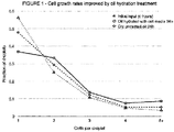
- Figure 1 compares the results obtained after 24 hours culture relative to baseline measurement at zero and 24 hours with no hydration of the oil.
- Cell growth is expressed here as a fraction of the droplets containing more than one cell. It will be seen that, relative to the baseline cell growth, an improvement occurs when the oil is hydrated with the cell culture medium.
- a continuous hydrated oil phase is prepared by mixing 99 parts of a light mineral oil with 1 part of a pegylated surfactant by weight. The oil is placed on rotator overnight to fully mix oil and surfactant. Hydrated oil is prepared by mixing 5 parts of the oil with 3 parts of the aqueous hydrating phase, consisting of either the same saline buffer used in the disperse emulsion phase or just water. The mixture is rotated overnight at 50C and then for 60min at 70C. The emulsion is left to stand for 15min. The upper portion of the emulsion is aliquoted, and the aliquots are then centrifugated to adjust the hydration level of the oil, with longer centrifugation times leading to lower hydration levels. The hydration level is measured using a Karl Fisher titrator. Once the correct hydration level is achieved, usually 500-1000ppm, the supernatant of the aliquots is pipetted into new tubes which are frozen until used.
- a polydisperse emulsion of droplets is produced by mixing 8 parts of the oil (either hydrated or not hydrated) with 1 part of the disperse aqueous phase by volume followed by mixing on a vortex mixer for 5min and centrifugation for 1min at 400RPM. The upper half of the mixture is pipetted into a new tube which is centrifuged for 5s. The emulsion is pipetted from the bottom of the tube for further use.
- the disperse aqueous phase described above consists of the single nucleotide detection chemistry as previously described and exemplified in EP3013987 .
- the emulsion For measurement of the fluorescent intensity the emulsion is sandwiched between two transparent substrates separated by spacers corresponding to the average emulsion droplet size.
- the fluorescent signal emitted from each emulsion droplet upon excitation with light of an appropriate wavelength range, is measured together with the diameter of droplet which is collected from a brightfield image of the emulsion.
- the data presented as a histogram in Figure 2 shows the average fluorescent intensity of 6um droplets, which have been incubated in oil with no hydration ('Dry oil'), in oil hydrated with water ('Water only') or oil hydrated with three times the buffer concentration of the droplets ('3x buffer').
- Droplets incubated in oil with no hydration show very low intensity above the background which for these samples is approximately 1000 counts.
- Those incubated in oil hydrated with water show an increased intensity compared to droplets incubated in oil with no hydration.
- Incubation of droplets in oil hydrated with three times the buffer concentration shows a further increase in average intensity. This demonstrates that both oil hydration can be used to maintain enzymatic reactivity in these droplets.
- Microdroplets are deposited on a substrate immersed in a continuous oil phase as for example previously described in EP3008207 to which the reader is directed.
- the continuous hydrated oil phase is prepared by mixing 99 parts of paraffin oil with 1 part of a pegylated surfactant by weight. The oil is placed on rotator overnight to fully mix oil and surfactant. Hydrated oil is prepared by mixing 5 parts of the oil with 3 parts of the aqueous hydrating phase, consisting of water with or without 4% glycerol. The mixture is rotated overnight at 50C and then for 60min at 70C. The emulsion is left to stand for 15min. The upper portion of the emulsion is aliquoted, and the aliquots are then centrifugated to adjust the hydration level of the oil, with longer centrifugation times leading to lower hydration levels. The hydration level is measured using a Karl Fisher titrator. Once the correct hydration level is achieved, usually 500-1000ppm, the supernatant of the aliquots is pipetted into new tubes which are frozen until used.
- the disperse aqueous phase consists of water with or without 4% glycerol.
- the deposited droplets are subjected to an incubation cycle at 70C for 115min.
- the emulsion droplet diameters are then measured from a brightfield microscope image and compared to measured diameters prior to the incubation cycle to infer droplet shrinkage or growth.
- the data presented below shows the average volume change of droplets upon a high temperature incubation step as a function of percentage of glycerol in the oil hydration and droplets respectively.
- the droplets shrink on average. If the droplets contain glycerol whereas the oil hydration does not, the droplets grow relative to the reference because the addition of glycerol to the droplets causes the water activity in the oil to be higher than the water activity in the droplets. The reverse happens when glycerol is added to the oil hydration but not the droplets. Droplets shrink relative to the reference due to a higher water activity in the droplets compared to the oil.
Landscapes
- Chemical & Material Sciences (AREA)
- Health & Medical Sciences (AREA)
- Life Sciences & Earth Sciences (AREA)
- Engineering & Computer Science (AREA)
- Organic Chemistry (AREA)
- Zoology (AREA)
- Wood Science & Technology (AREA)
- Genetics & Genomics (AREA)
- Bioinformatics & Cheminformatics (AREA)
- General Health & Medical Sciences (AREA)
- Proteomics, Peptides & Aminoacids (AREA)
- Biotechnology (AREA)
- Biomedical Technology (AREA)
- Microbiology (AREA)
- Biochemistry (AREA)
- General Engineering & Computer Science (AREA)
- Analytical Chemistry (AREA)
- Dispersion Chemistry (AREA)
- Physics & Mathematics (AREA)
- Biophysics (AREA)
- Molecular Biology (AREA)
- Immunology (AREA)
- Hematology (AREA)
- Clinical Laboratory Science (AREA)
- Chemical Kinetics & Catalysis (AREA)
- Oncology (AREA)
- Cell Biology (AREA)
- Measuring Or Testing Involving Enzymes Or Micro-Organisms (AREA)
- Medicines Containing Material From Animals Or Micro-Organisms (AREA)
- Apparatus Associated With Microorganisms And Enzymes (AREA)
Description
- This invention relates to an improved method of manipulating aqueous microdroplets optionally containing biological cells in an immiscible carrier fluid such as an oil. It enables the size of the microdroplets to be controlled or adjusted and any enzymatic or chemical reactions occurring therein to be maintained or optimised during a given period.
- In our previous patent applications, for example
WO2014167323 ,WO2015121675 ,WO2016012789 ,WO2017140839 andPCT/EP2018066574 US2015/247192 ,US2007/241068 andUS2017/175179 provide technological background. In some embodiments, these methods involve translocating microdroplets dispersed in an immiscible carrier fluid along microfluidic pathways in an analytical device using electrowetting propulsive forces or by directly printing of the microdroplets onto a substrate coated with the carrier fluid. In many instances, where the volume fraction of the microdroplets is relatively low, we have found that these microdroplets tend to undergo significant shrinkage over time which can sometimes interfere with some or all the enzymatic processes going on within. Also, in other instances it may be desirable to deliberately shrink or grow the size of the microdroplets in a part of a device as a given analysis is carried out. - We have now developed a microdroplet manipulation method which overcomes these problems. It may be used, for example, to manipulate the size and/or reactivity of the contents of microdroplets or to control chemical or enzymatic reactions occurring therein. The invention is as defined in the appended claims. According to a first aspect of the invention, there is provided a generic method of controlling the size of microdroplets and/or maintaining or optimising enzymatic or chemical reactions occurring therein, said microdroplets having an average volume in the range 0.5 femtolitres to 10 nanolitres comprised of at least one biological component and a first aqueous medium having a water activity of aw1 of less than 1 characterised by the step of maintaining the microdroplets in a water-immiscible carrier fluid which further includes secondary droplets of a second aqueous medium having an average volume less than 25% of the average volume of the microdroplets up to and including a maximum of 4 femtolitres and wherein the volume ratio of carrier fluid to total volume of microdroplets per unit volume of the total is greater than 2:1. Without wishing to limit the scope of the invention, it is believed that the invention solves the problem by using a carrier fluid which contains very small secondary droplets which can interact with the microdroplets without adversely affecting the latter's overall characteristics or the efficacy of any detection method applied to them. When the carrier medium is an oil such a composite medium is sometimes referred to as 'hydrated oil'. An important feature in this respect is that the relative water activities of the microdroplets and the secondary droplets are controlled within certain parameters; optionally by continuous monitoring and/or a feed-back loop. Here, the water activity of an aqueous medium (aw) is defined as the ratio of the partial vapour pressure of the aqueous medium under investigation to that of pure water under STP conditions. Since water tends to diffuse along a gradient from high to low water activity, we have found that, within the constraints of our systems, when the water activity of the second aqueous medium (aw2) is higher than that of the first aqueous medium (aw1) the net effect is for the microdroplets to undergo expansion until the water activities of the two components equalise. Conversely, when the water activity of the second aqueous medium is lower than that of the first aqueous medium the microdroplets will tend to shrink until these water activities equalise. In one useful embodiment, the water activities of the first and second aqueous media may be the same or substantially the same so that any tendency for the microdroplets to shrink or expand is continuously counteracted. Thus, the sizes of the microdroplets may always be preserved. We have also found that, by these means, these secondary droplets can be used to assist in preserving or even enhancing any enzymatic or chemical reactions occurring in the microdroplets; for example, by using the secondary droplets to feed cell-growth components to the microdroplets at one or more points in any device employing the method. The first and second aqueous medium may have compositions which in one embodiment are identical.
- Thus, in one embodiment of the invention, the water activity of the first and the second aqueous media are independently in the range from 0.9 to 1. In another embodiment, the water activity of the first aqueous medium is from 0.9 to less than 1. In yet another embodiment, the ratio of the water activities of the first and second aqueous media (aw1:aw2) is in the range 0.9:1 to 1:0.9.
- One convenient way to perform the manipulation is using first and second media which are buffers; and, if required, by varying the relative compositions of the two. For example, in one application the ionic strength of the first aqueous medium is in the range from to 1 to 5 that of the second aqueous medium; preferably from 3 to 5 times. In another, the ionic strength of the second aqueous medium is in the range from to 1 to 5 times that of the first aqueous medium; preferably from 3 to 5 times. In yet another application, the ionic strengths are the same or substantially the same with the ratio of ionic strengths being in the range from 3:1 to 1:3. In one particularly useful embodiment, either or both first and second aqueous media may include glycerol as a component; for example, at differing concentrations. In another, the pHs of the first and second aqueous media are the same or similar and within the range 6.5 to 8.
- As regards the secondary droplets, these have a much smaller average volume than the average value for the microdroplets and at the limit may be comprised of femto-sized droplets or micelles of the second aqueous medium emulsified within the carrier fluid and stabilised by a sheath of compatible surfactant molecules; for example, a non-ionic surfactant. In one embodiment, the size of these secondary droplets is less than 10%, preferably less than 5% of the volume of the microdroplets employed. In another, the average volume of the secondary droplets lies within the range 10 to 1% of the average volume of the microdroplets. Suitably the secondary droplets form part of a stable emulsion in the carrier fluid which in one embodiment is an immiscible oil. Suitably, the carrier fluid is selected from a mineral oil, a silicone oil or a fluorocarbon oil. The oil may also contain additional surfactants and stabilisers if required. Suitably the volume ratio of carrier fluid to total volume of the microdroplets is greater than 3:1; preferably 5:1 or greater.
- The method of the invention is useful for several applications where biological cells are being analysed. One example is where a culture of immortalised mammalian cells is being caused to proliferate inside the microdroplets for the purpose of screening individual clonal copies of the cells for desirable characteristics such as protein expression or particular genetic traits. Thus in a second aspect of the invention, there is in one embodiment provided a method of causing the cellular proliferation of one or more cell types contained within a microdroplet having an average volume in the
range 4 femtolitres to 10 nanolitres and comprised of an aqueous buffer comprising the steps of incubating the cell(s) inside the droplets in suitable environmental conditions and thereafter detecting the number of cells inside each droplet, characterised in that the microdroplets are suspended in an immiscible carrier fluid further comprising secondary droplets having an average volume less than 25% of the average volume of the microdroplets up to and including a maximum of 4 femtolitres and wherein the volume ratio of carrier fluid to total volume of microdroplets per unit volume of the total is greater than 2:1. - In another embodiment, there is also provided a method of detecting one or more phenotypic traits, genetic traits or protein expression profiles of a cell under consideration, that cell being contained within a microdroplet having an average volume in the
range 4 femtolitres to 10 nanolitres and comprised of an aqueous growth media comprising the steps of labelling a target derived from the cell(s)with a fluorescent probe and thereafter detecting an output from the probe characterised in that the cell-containing microdroplets are suspended in an immiscible carrier fluid further comprising secondary droplets having an average volume less than 25% of the average volume of the microdroplets up to and including a maximum of 4 femtolitres and wherein the volume ratio of carrier fluid to total volume of microdroplets per unit volume of the total is greater than 2:1. Fluorescent probe molecules suitable for this purpose are well known and include fluorescently labelled antibodies, FRET reporter probes and enzyme-labelled antigens which are degraded in the presence of a target protein. - In another embodiment, there is provided a method of analysing an oligonucleotide derived from a biological cell contained within a microdroplet having an average volume in the
range 4 femtolitres to 10 nanolitres and further comprised of an aqueous buffer comprising the steps of labelling the oligonucleotide with a fluorescent hybridisation probe and thereafter detecting the corresponding fluorescence characterised in that the microdroplets are suspended in an immiscible carrier fluid further comprising secondary droplets having an average volume less than 25% of the average volume of the microdroplets up to and including a maximum of 4 femtolitres and wherein the volume ratio of carrier fluid to total volume of microdroplets per unit volume of the total is greater than 2:1. - Fluorescent hybridisation probes which can be used for this purpose are well-known in the art and include molecular beacons, TaqMan® probes, Scorpion® probes and LNA® probes. Methods for detecting the fluorescence arising in all these embodiments are well-known to one of ordinary skill in the art; for example, those methods employing a source of incident electromagnetic radiation (laser, LED and the like) and a corresponding photodetector for detecting fluorescence photons and outputting a data-stream which can be analysed using microprocessor algorithms.
- Thus, the target in these methods may be the cell(s) themselves, one or more oligonucleotides derived therefrom or a product such a protein which is expressed by the cell(s) when cultured within the microdroplet itself. Such oligonucleotides may be generated from the cell(s) by lysis.
- The method of the invention may also be suitably employed in connection with biological components which are non-cellular or cell-free although in one embodiment it may be used to manipulate nucleic acids or components thereof which have been previously derived from biological cells. Thus in a third aspect of the invention there is provided there is provided a method of manipulating the size and/or reactivity of the contents of microdroplets having an average volume in the range 0.5 femtolitres to 10 nanolitres; the microdroplets being comprised of at least one biological component and a first aqueous medium free of biological cells having a water activity of aw1 of less than 1 characterised by the step of maintaining the microdroplets in a water-immiscible carrier fluid which further includes secondary droplets comprised of a second aqueous and having an average volume less than 25% of the average volume of the microdroplets up to and including a maximum of 0.5 femtolitres and wherein the volume ratio of carrier fluid to total volume of microdroplets per unit volume of the total is greater than 2:1.
- The method of the third aspect of the invention is useful for a number of applications where the biological component is a single nucleotide; for example, a single nucleoside triphosphate or single nucleoside monophosphate. For example, the method may be advantageously used with one of the sequencing methods we have previously described including but not limited to those described
EP3013987 or in the other above-mentioned patent applications to which the reader is directed. Thus, in a third aspect, there is provided a method of sequencing comprising the steps of progressively digesting by pyrophosphorolysis a nucleic acid analyte into an ordered stream of nucleoside triphosphate molecules and generating therefrom a corresponding ordered stream of microdroplets having an average volume in the range 0.5 femtolitres to 10 nanolitres and each comprised of one of the nucleoside triphosphate molecules and aqueous buffer; reacting each nucleoside triphosphate molecule within each microdroplet with a nucleobase-specific fluorescent probe and thereafter detecting the corresponding fluorescence associated with each microdroplet thereby identifying the nucleobase characterised in that the microdroplets are suspended in an immiscible carrier fluid further comprising secondary droplets having an average volume less than 25% of the average volume of the microdroplets up to and including a maximum of 0.5 femtolitres and wherein the volume ratio of carrier fluid to total volume of microdroplets per unit volume of the total is greater than 2:1. - Fluorescent probes suitable for use in this application have been describe by us in our previous patent applications; for example,
WO2016012789 and subsequently published applications to which the reader is directed. These probes are characterised by (a) being non-fluorescing in their unused state and (b) being capable of undergoing exonucleolysis once used to produced fluorophores in a detectable state attached to single nucleoside monophosphates. The fluorescence arising may be detected and analysed as described above. - In all these additional aspects of the invention it is preferred that the ratio of the water activities of the first and second aqueous media associated with respectively the microdroplets and the secondary droplets is in the range 0.9:1 to 1:0.9; preferably 0.95:1 to 1:0.95 and for example 1:1.
- The advantageous effect of hydrating the carrier phase as described above is now illustrated by the following Examples.
- Continuous oil phase material is prepared by mixing 99 parts of a Hydrofluoroether continuous phase with 1 part of a fluorinated surfactant. A growth-media-treated carrier phase is prepared by mixing an aliquot of RPMI 1640 media (Thermo Fisher Scientific, UK) with an equal volume of the oil/surfactant mixture and agitating the mixture for 24 hours at 37°C to form a polydisperse emulsion. This emulsion is then left to stand until it spontaneously fractionates to form an upper phase comprising large droplets and undispersed plugs of aqueous growth media, and a lower phase containing only the smallest vesicles of growth media suspended in the oil phase which is additionally now saturated with dissolved aqueous media. This lower phase is removed from the vessel using a pipette and retained for later use.
- Jurkat E6-1 T-cell lymphoma cells (ATCC, Virginia, USA) are suspended in RPMI media at a concentration of 8E6 cells/ml. This media and cells are then flowed through an emulsifying apparatus to form droplets of 50um diameter, with cells dispersed throughout the droplets. The outer carrier phase for the emulsion is a hydrofluoroether oil mixed with 1% of a suitable surfactant to stabilise the droplets in solution. The emulsion thus formed spontaneously fractionates to form a layer of densely packed monodisperse aqueous droplets floating at the top of a column of continuous oil/surfactant mixture. This emulsion is then evenly dispersed by gentle mixing and divided in to three aliquots containing droplets and the carrier phase.
- One aliquot (the initial reference) is immediately transferred in to a haemocytometer flow cell and the droplets therein are inspected using a 20x magnification optical microscope. The cell occupancy of each droplet is recorded by counting the number of distinct cells in each droplet. Empty droplets are disregarded.
- The second aliquot is allowed to fractionate once more, and the lower carrier phase removed using a pipette. An equivalent volume of the earlier treated carrier phase is introduced to the sample to replace the removed untreated carrier phase. The third aliquot is left unaltered. Both the second and third aliquots are then transferred in to partially sealed vessels which permit gas permeation between the vessel and its surroundings. Both vessels are placed in to an environment-controlled CO2 incubator set to contain 5% CO2/air mixture, 95% humidity and 37°C set temperature. The aliquots are incubated for 24 hours
These aliquots are then removed from the incubator and introduced to a haemocytometer for inspection and analysis in the same way as the reference aliquot. The change in the cell population-distribution after the incubation (characteristic of cell proliferation) can then be compared between the different oil treatments. -
Figure 1 compares the results obtained after 24 hours culture relative to baseline measurement at zero and 24 hours with no hydration of the oil. Cell growth is expressed here as a fraction of the droplets containing more than one cell. It will be seen that, relative to the baseline cell growth, an improvement occurs when the oil is hydrated with the cell culture medium. - A continuous hydrated oil phase is prepared by mixing 99 parts of a light mineral oil with 1 part of a pegylated surfactant by weight. The oil is placed on rotator overnight to fully mix oil and surfactant. Hydrated oil is prepared by mixing 5 parts of the oil with 3 parts of the aqueous hydrating phase, consisting of either the same saline buffer used in the disperse emulsion phase or just water. The mixture is rotated overnight at 50C and then for 60min at 70C. The emulsion is left to stand for 15min. The upper portion of the emulsion is aliquoted, and the aliquots are then centrifugated to adjust the hydration level of the oil, with longer centrifugation times leading to lower hydration levels. The hydration level is measured using a Karl Fisher titrator. Once the correct hydration level is achieved, usually 500-1000ppm, the supernatant of the aliquots is pipetted into new tubes which are frozen until used.
- A polydisperse emulsion of droplets is produced by mixing 8 parts of the oil (either hydrated or not hydrated) with 1 part of the disperse aqueous phase by volume followed by mixing on a vortex mixer for 5min and centrifugation for 1min at 400RPM. The upper half of the mixture is pipetted into a new tube which is centrifuged for 5s. The emulsion is pipetted from the bottom of the tube for further use.
- For measurements of the enzymatic activity, the disperse aqueous phase described above consists of the single nucleotide detection chemistry as previously described and exemplified in
EP3013987 . - For measurement of the fluorescent intensity the emulsion is sandwiched between two transparent substrates separated by spacers corresponding to the average emulsion droplet size. The fluorescent signal emitted from each emulsion droplet, upon excitation with light of an appropriate wavelength range, is measured together with the diameter of droplet which is collected from a brightfield image of the emulsion.
- The data presented as a histogram in
Figure 2 shows the average fluorescent intensity of 6um droplets, which have been incubated in oil with no hydration ('Dry oil'), in oil hydrated with water ('Water only') or oil hydrated with three times the buffer concentration of the droplets ('3x buffer'). Droplets incubated in oil with no hydration show very low intensity above the background which for these samples is approximately 1000 counts. Those incubated in oil hydrated with water show an increased intensity compared to droplets incubated in oil with no hydration. Incubation of droplets in oil hydrated with three times the buffer concentration shows a further increase in average intensity. This demonstrates that both oil hydration can be used to maintain enzymatic reactivity in these droplets. - Microdroplets are deposited on a substrate immersed in a continuous oil phase as for example previously described in
EP3008207 to which the reader is directed. - The continuous hydrated oil phase is prepared by mixing 99 parts of paraffin oil with 1 part of a pegylated surfactant by weight. The oil is placed on rotator overnight to fully mix oil and surfactant. Hydrated oil is prepared by mixing 5 parts of the oil with 3 parts of the aqueous hydrating phase, consisting of water with or without 4% glycerol. The mixture is rotated overnight at 50C and then for 60min at 70C. The emulsion is left to stand for 15min. The upper portion of the emulsion is aliquoted, and the aliquots are then centrifugated to adjust the hydration level of the oil, with longer centrifugation times leading to lower hydration levels. The hydration level is measured using a Karl Fisher titrator. Once the correct hydration level is achieved, usually 500-1000ppm, the supernatant of the aliquots is pipetted into new tubes which are frozen until used.
- The disperse aqueous phase consists of water with or without 4% glycerol. The deposited droplets are subjected to an incubation cycle at 70C for 115min. The emulsion droplet diameters are then measured from a brightfield microscope image and compared to measured diameters prior to the incubation cycle to infer droplet shrinkage or growth.
- The data presented below shows the average volume change of droplets upon a high temperature incubation step as a function of percentage of glycerol in the oil hydration and droplets respectively. In the reference sample, if glycerol is not present in either the oil hydration nor in the droplets, the droplets shrink on average. If the droplets contain glycerol whereas the oil hydration does not, the droplets grow relative to the reference because the addition of glycerol to the droplets causes the water activity in the oil to be higher than the water activity in the droplets. The reverse happens when glycerol is added to the oil hydration but not the droplets. Droplets shrink relative to the reference due to a higher water activity in the droplets compared to the oil. This shows that the specific content of the droplets and the oil hydration can be used to control droplet shrinkage and growth.
% glycerol in oil hydration % glycerol in droplets Droplet volume change 0 0 -30% (shrink) 0 4 +44% (grow) 4 0 -51% (shrink)
Claims (17)
- A method of controlling the size of microdroplets and/or maintaining or optimising enzymatic or chemical reactions occurring therein, said microdroplets having an average volume in the range from 0.5 femtolitres to 10 nanolitres comprised of at least one biological component and a first aqueous medium having a water activity of aw1 of less than 1 characterised by the step of maintaining the microdroplets in a water-immiscible carrier fluid which further includes secondary droplets comprised of a second aqueous medium having an average volume less than 25% of the average volume of the microdroplets up to and including a maximum of 4 femtolitres and wherein the volume ratio of carrier fluid to total volume of microdroplets per unit volume of the total is greater than 2:1.
- A method as claimed in claim 1 for manipulating the size and/or chemical or enzymatic reactivity of the contents of microdroplets having an average volume in the range from 0.5 femtolitres to 10 nanolitres; the microdroplets being comprised of at least one biological component and a first aqueous medium free of biological cells having a water activity of aw1 of less than 1 characterised by the step of maintaining the microdroplets in a water-immiscible carrier fluid which further includes secondary droplets comprised of a second aqueous medium and having an average volume less than 25% of the average volume of the microdroplets up to and including a maximum of 0.5 femtolitres and wherein the volume ratio of carrier fluid to total volume of microdroplets per unit volume of the total is greater than 2:1.
- A method as claimed in claim 1 for controlling chemical or enzymatic reactivity and/or microdroplet size in microdroplets having an average volume in the range from 4 femtolitres to 10 nanolitres; the microdroplets being comprised of at least one biological cell and a first aqueous medium having a water activity of aw1 of less than 1 characterised by the step of maintaining the microdroplets in a water-immiscible carrier fluid which further includes secondary droplets comprised of a second aqueous medium and having an average volume less than 25% of the average volume of the microdroplets up to and including a maximum of 4 femtolitres and wherein the volume ratio of carrier fluid to total volume of microdroplets per unit volume of the total is greater than 2:1.
- A method as claimed in any one of claims 1 to 3 characterised in that the secondary droplets have a water activity aw2 which is greater than aw1.
- A method as claimed in any of claims 1 to 3 characterised in that the secondary droplets have a water activity aw2 which is less than aw1.
- A method as claimed in any one of claims 1 to 3 characterised in the water activities aw1 and aw2 are the same.
- A method as claimed in any one of the preceding claims characterised in that aw1 and aw2 are independently in the range 0.9 to 1.
- A method as claimed in claim 4 characterised in that the ionic strength of the second aqueous medium is in the range from 1 to 5 times that of the first aqueous medium.
- A method as claimed in claim 5 characterised in that the ionic strength of the first medium is in the range from 1 to 5 times that of the second aqueous medium.
- A method as claimed in any one of the preceding claims characterised in that the average volume of the secondary droplets is less than 10% of the average volume of the microdroplets.
- A method as claimed in any of the preceding claims characterised in that at least one of the first and second aqueous media further comprise glycerol.
- A method as claimed in any one of the preceding claims characterised in that the biological component is selected from a single nucleoside triphosphate derived from a target nucleic acid, an oligonucleotide derived from the DNA or RNA of a cell, an enzyme or a cell.
- A method as claimed in claim 12 characterised in that first and/or second aqueous media are buffers.
- A method as claimed in claim 3, wherein the method further comprises causing the cellular proliferation of one or more cell types contained within a microdroplet having an average volume in the range 4 femtolitres to 10 nanolitres by incubating the cell(s) inside the droplets in suitable environmental conditions and thereafter detecting the number of cells inside each droplet.
- A method as claimed in claim 3, wherein the method further comprises analysing or detecting one or more phenotypic traits, genetic traits or protein expression profiles of a cell under consideration, that cell being contained within a microdroplet having an average volume in the range 4 femtolitres to 10 nanolitres and comprised of an aqueous buffer, by labelling a target derived from the cell(s) .
- A method as claimed in claim 2, wherein the method further comprises sequencing by progressively digesting by pyrophosphorolysis a nucleic acid analyte into an ordered stream of nucleoside triphosphate molecules and generating therefrom a corresponding ordered stream of microdroplets having an average volume in the range from 0.5 femtolitres to 10 nanolitres and each comprised of one of the nucleoside triphosphate molecules and aqueous buffer; reacting each nucleoside triphosphate molecule within each microdroplet with a nucleobase-specific fluorescent probe and thereafter detecting the corresponding fluorescence associated with each microdroplet thereby identifying the nucleobase.
- A method as claimed in any one of claims 14 to 16 characterised in that the ratio of the water activities of the microdroplets and the secondary droplets is in the range 0.9:1 to 1:0.9; preferably 0.95:1 to 1:0.95.
Priority Applications (16)
| Application Number | Priority Date | Filing Date | Title |
|---|---|---|---|
| EP19156182.8A EP3693086B1 (en) | 2019-02-08 | 2019-02-08 | Microdroplet manipulation method |
| PL19156182T PL3693086T3 (en) | 2019-02-08 | 2019-02-08 | Microdroplet manipulation method |
| ES19156182T ES2841907T3 (en) | 2019-02-08 | 2019-02-08 | Microdroplet handling method |
| KR1020217028525A KR102572821B1 (en) | 2019-02-08 | 2020-02-07 | Microdroplet Manipulation Method |
| AU2020219461A AU2020219461B2 (en) | 2019-02-08 | 2020-02-07 | Microdroplet manipulation method |
| PCT/GB2020/050280 WO2020161500A1 (en) | 2019-02-08 | 2020-02-07 | Microdroplet manipulation method |
| CN202211540865.8A CN115814869A (en) | 2019-02-08 | 2020-02-07 | Droplet operations |
| CN202080012907.3A CN113490548B (en) | 2019-02-08 | 2020-02-07 | Droplet operations method |
| SG11202108553WA SG11202108553WA (en) | 2019-02-08 | 2020-02-07 | Microdroplet manipulation method |
| KR1020237029091A KR20230128412A (en) | 2019-02-08 | 2020-02-07 | Microdroplet Manipulation Method |
| US17/429,124 US11504715B2 (en) | 2019-02-08 | 2020-02-07 | Microdroplet manipulation method |
| JP2021546352A JP7314290B2 (en) | 2019-02-08 | 2020-02-07 | How to manipulate microdroplets |
| CA3128452A CA3128452A1 (en) | 2019-02-08 | 2020-02-07 | Microdroplet manipulation method |
| IL285256A IL285256B2 (en) | 2019-02-08 | 2020-02-07 | Microdroplet manipulation method |
| US17/972,825 US20230042115A1 (en) | 2019-02-08 | 2022-10-25 | Microdroplet manipulation method |
| JP2023114399A JP2023139073A (en) | 2019-02-08 | 2023-07-12 | Microdroplet manipulation method |
Applications Claiming Priority (1)
| Application Number | Priority Date | Filing Date | Title |
|---|---|---|---|
| EP19156182.8A EP3693086B1 (en) | 2019-02-08 | 2019-02-08 | Microdroplet manipulation method |
Publications (2)
| Publication Number | Publication Date |
|---|---|
| EP3693086A1 EP3693086A1 (en) | 2020-08-12 |
| EP3693086B1 true EP3693086B1 (en) | 2020-12-09 |
Family
ID=65365847
Family Applications (1)
| Application Number | Title | Priority Date | Filing Date |
|---|---|---|---|
| EP19156182.8A Active EP3693086B1 (en) | 2019-02-08 | 2019-02-08 | Microdroplet manipulation method |
Country Status (12)
| Country | Link |
|---|---|
| US (2) | US11504715B2 (en) |
| EP (1) | EP3693086B1 (en) |
| JP (2) | JP7314290B2 (en) |
| KR (2) | KR20230128412A (en) |
| CN (2) | CN113490548B (en) |
| AU (1) | AU2020219461B2 (en) |
| CA (1) | CA3128452A1 (en) |
| ES (1) | ES2841907T3 (en) |
| IL (1) | IL285256B2 (en) |
| PL (1) | PL3693086T3 (en) |
| SG (1) | SG11202108553WA (en) |
| WO (1) | WO2020161500A1 (en) |
Families Citing this family (2)
| Publication number | Priority date | Publication date | Assignee | Title |
|---|---|---|---|---|
| GB202012485D0 (en) * | 2020-08-11 | 2020-09-23 | Lightcast Discovery Ltd | Improvements in or relating to a method of maintaing a microdroplet |
| CN114752657A (en) * | 2022-05-05 | 2022-07-15 | 中山大学 | Polydisperse liquid drop digital nucleic acid detection method and application thereof |
Family Cites Families (22)
| Publication number | Priority date | Publication date | Assignee | Title |
|---|---|---|---|---|
| US8613889B2 (en) * | 2006-04-13 | 2013-12-24 | Advanced Liquid Logic, Inc. | Droplet-based washing |
| ATE490971T1 (en) * | 2006-04-18 | 2010-12-15 | Advanced Liquid Logic Inc | BIOCHEMISTRY ON DROPLETS |
| WO2008030281A2 (en) * | 2006-05-25 | 2008-03-13 | The Regents Of The University Of California | Optical resonances in droplets in a microchannel |
| US9598725B2 (en) * | 2010-03-02 | 2017-03-21 | Bio-Rad Laboratories, Inc. | Emulsion chemistry for encapsulated droplets |
| WO2010128157A1 (en) * | 2009-05-07 | 2010-11-11 | Universite De Strasbourg | Microfluidic system and methods for highly selective droplet fusion |
| WO2012047324A2 (en) * | 2010-06-10 | 2012-04-12 | President And Fellows Of Harvard College | Systems and methods for amplification and phage display |
| DE102010032203A1 (en) * | 2010-07-26 | 2012-01-26 | MAX-PLANCK-Gesellschaft zur Förderung der Wissenschaften e.V. | Method and apparatus for the passive separation and sorting of drops, in particular in a microfluidic system, by using non-optical markers for reactions within the drops |
| WO2012117083A2 (en) * | 2011-03-03 | 2012-09-07 | F. Hoffmann-La Roche Ag | Hanging droplet plate |
| GB201217772D0 (en) * | 2012-10-04 | 2012-11-14 | Base4 Innovation Ltd | Sequencing method |
| US20150321163A1 (en) * | 2012-12-14 | 2015-11-12 | Gnubio, Inc. | Method for maintaining heterogeneous concentrations of molecules in emulsion droplets |
| KR101634350B1 (en) * | 2013-02-27 | 2016-06-28 | 한국생명공학연구원 | Single Cell-based Screening Method of Useful Enzyme Resources Using Mesh-integrated Microdroplet Array |
| GB201306444D0 (en) | 2013-04-09 | 2013-05-22 | Base4 Innovation Ltd | Single nucleotide detection method |
| GB201310584D0 (en) * | 2013-06-13 | 2013-07-31 | Base4 Innovation Ltd | Droplet storage method |
| GB201402644D0 (en) | 2014-02-14 | 2014-04-02 | Base4 Innovation Ltd | Methylation detection method |
| KR101605947B1 (en) * | 2014-03-05 | 2016-03-23 | 한국과학기술원 | System and method for culturing single cell in fine droplet |
| US10232373B2 (en) * | 2014-06-16 | 2019-03-19 | Bio-Rad Laboratories, Inc. | Size alternating injection into drops to facilitate sorting |
| GB201412977D0 (en) | 2014-07-22 | 2014-09-03 | Base4 Innovation Ltd | Single nucleotide detection method |
| US20180016622A1 (en) * | 2015-01-23 | 2018-01-18 | President And Fellows Of Harvard College | Systems, methods, and kits for amplifying or cloning within droplets |
| EP3207982A1 (en) | 2016-02-17 | 2017-08-23 | Base4 Innovation Limited | Improved droplet sequencing apparatus and method |
| CN108273454A (en) * | 2016-12-27 | 2018-07-13 | 中国科学院微生物研究所 | A kind of method that nanoliter level microlayer model merges in small-sized reaction tube |
| US11318472B2 (en) | 2017-06-21 | 2022-05-03 | Lightcast Discovery Ltd | Microfluidic analytical device |
| CN107090522B (en) * | 2017-07-03 | 2021-07-30 | 武汉科技大学 | Method for quantitatively detecting virus based on digital PCR (polymerase chain reaction) for non-diagnostic purpose |
-
2019
- 2019-02-08 ES ES19156182T patent/ES2841907T3/en active Active
- 2019-02-08 EP EP19156182.8A patent/EP3693086B1/en active Active
- 2019-02-08 PL PL19156182T patent/PL3693086T3/en unknown
-
2020
- 2020-02-07 CN CN202080012907.3A patent/CN113490548B/en active Active
- 2020-02-07 US US17/429,124 patent/US11504715B2/en active Active
- 2020-02-07 CA CA3128452A patent/CA3128452A1/en active Pending
- 2020-02-07 AU AU2020219461A patent/AU2020219461B2/en active Active
- 2020-02-07 SG SG11202108553WA patent/SG11202108553WA/en unknown
- 2020-02-07 IL IL285256A patent/IL285256B2/en unknown
- 2020-02-07 CN CN202211540865.8A patent/CN115814869A/en active Pending
- 2020-02-07 JP JP2021546352A patent/JP7314290B2/en active Active
- 2020-02-07 KR KR1020237029091A patent/KR20230128412A/en active Application Filing
- 2020-02-07 WO PCT/GB2020/050280 patent/WO2020161500A1/en active Application Filing
- 2020-02-07 KR KR1020217028525A patent/KR102572821B1/en active IP Right Grant
-
2022
- 2022-10-25 US US17/972,825 patent/US20230042115A1/en active Pending
-
2023
- 2023-07-12 JP JP2023114399A patent/JP2023139073A/en active Pending
Non-Patent Citations (1)
| Title |
|---|
| None * |
Also Published As
| Publication number | Publication date |
|---|---|
| PL3693086T3 (en) | 2021-04-19 |
| IL285256B2 (en) | 2023-03-01 |
| SG11202108553WA (en) | 2021-09-29 |
| JP2022519742A (en) | 2022-03-24 |
| CA3128452A1 (en) | 2020-08-13 |
| JP7314290B2 (en) | 2023-07-25 |
| US20220088606A1 (en) | 2022-03-24 |
| AU2020219461B2 (en) | 2024-05-09 |
| EP3693086A1 (en) | 2020-08-12 |
| US20230042115A1 (en) | 2023-02-09 |
| KR20230128412A (en) | 2023-09-04 |
| AU2020219461A1 (en) | 2021-09-02 |
| CN113490548B (en) | 2022-12-23 |
| KR20210126060A (en) | 2021-10-19 |
| JP2023139073A (en) | 2023-10-03 |
| ES2841907T3 (en) | 2021-07-12 |
| US11504715B2 (en) | 2022-11-22 |
| WO2020161500A1 (en) | 2020-08-13 |
| CN113490548A (en) | 2021-10-08 |
| IL285256A (en) | 2021-09-30 |
| IL285256B (en) | 2022-11-01 |
| CN115814869A (en) | 2023-03-21 |
| KR102572821B1 (en) | 2023-08-29 |
Similar Documents
| Publication | Publication Date | Title |
|---|---|---|
| US20230042115A1 (en) | Microdroplet manipulation method | |
| Joensson et al. | Droplet microfluidics—A tool for single‐cell analysis | |
| US20180305747A1 (en) | Systems and methods for handling microfluidic droplets | |
| US9427737B2 (en) | Methods and compositions for using oils for analysis and detection of molecules | |
| EP3782722B1 (en) | Microfluidic devices | |
| WO2014165559A2 (en) | Systems and methods for handling microfluidic droplets | |
| US11542546B2 (en) | Systems and methods for generation of emulsions with suitable clarity with applications of use | |
| JP2022510140A (en) | Devices and methods for detecting cell microdroplets | |
| Wu et al. | A thermosetting oil for droplet‐based real‐time monitoring of digital PCR and cell culture | |
| EP3570979B1 (en) | Microfluidic system and method with tightly controlled incubation time and conditions | |
| AU2024203320A1 (en) | Microdroplet manipulation method | |
| JPWO2020104769A5 (en) |
Legal Events
| Date | Code | Title | Description |
|---|---|---|---|
| STAA | Information on the status of an ep patent application or granted ep patent |
Free format text: STATUS: EXAMINATION IS IN PROGRESS |
|
| PUAI | Public reference made under article 153(3) epc to a published international application that has entered the european phase |
Free format text: ORIGINAL CODE: 0009012 |
|
| 17P | Request for examination filed |
Effective date: 20191018 |
|
| AK | Designated contracting states |
Kind code of ref document: A1 Designated state(s): AL AT BE BG CH CY CZ DE DK EE ES FI FR GB GR HR HU IE IS IT LI LT LU LV MC MK MT NL NO PL PT RO RS SE SI SK SM TR |
|
| AX | Request for extension of the european patent |
Extension state: BA ME |
|
| RAP1 | Party data changed (applicant data changed or rights of an application transferred) |
Owner name: LIGHTCAST DISCOVERY LTD |
|
| GRAP | Despatch of communication of intention to grant a patent |
Free format text: ORIGINAL CODE: EPIDOSNIGR1 |
|
| STAA | Information on the status of an ep patent application or granted ep patent |
Free format text: STATUS: GRANT OF PATENT IS INTENDED |
|
| INTG | Intention to grant announced |
Effective date: 20200907 |
|
| GRAS | Grant fee paid |
Free format text: ORIGINAL CODE: EPIDOSNIGR3 |
|
| RBV | Designated contracting states (corrected) |
Designated state(s): AL AT BE BG CH CY CZ DE DK EE ES FI FR GB GR HR HU IE IS IT LI LT LU LV MC MK MT NL NO PL PT RO RS SE SI SK SM TR |
|
| GRAA | (expected) grant |
Free format text: ORIGINAL CODE: 0009210 |
|
| STAA | Information on the status of an ep patent application or granted ep patent |
Free format text: STATUS: THE PATENT HAS BEEN GRANTED |
|
| AK | Designated contracting states |
Kind code of ref document: B1 Designated state(s): AL AT BE BG CH CY CZ DE DK EE ES FI FR GB GR HR HU IE IS IT LI LT LU LV MC MK MT NL NO PL PT RO RS SE SI SK SM TR |
|
| REG | Reference to a national code |
Ref country code: GB Ref legal event code: FG4D |
|
| REG | Reference to a national code |
Ref country code: CH Ref legal event code: EP Ref country code: AT Ref legal event code: REF Ref document number: 1342853 Country of ref document: AT Kind code of ref document: T Effective date: 20201215 |
|
| REG | Reference to a national code |
Ref country code: DE Ref legal event code: R096 Ref document number: 602019001598 Country of ref document: DE |
|
| REG | Reference to a national code |
Ref country code: IE Ref legal event code: FG4D |
|
| REG | Reference to a national code |
Ref country code: NL Ref legal event code: FP |
|
| REG | Reference to a national code |
Ref country code: CH Ref legal event code: NV Representative=s name: TR-IP CONSULTING LLC, CH |
|
| PG25 | Lapsed in a contracting state [announced via postgrant information from national office to epo] |
Ref country code: FI Free format text: LAPSE BECAUSE OF FAILURE TO SUBMIT A TRANSLATION OF THE DESCRIPTION OR TO PAY THE FEE WITHIN THE PRESCRIBED TIME-LIMIT Effective date: 20201209 Ref country code: RS Free format text: LAPSE BECAUSE OF FAILURE TO SUBMIT A TRANSLATION OF THE DESCRIPTION OR TO PAY THE FEE WITHIN THE PRESCRIBED TIME-LIMIT Effective date: 20201209 Ref country code: NO Free format text: LAPSE BECAUSE OF FAILURE TO SUBMIT A TRANSLATION OF THE DESCRIPTION OR TO PAY THE FEE WITHIN THE PRESCRIBED TIME-LIMIT Effective date: 20210309 Ref country code: GR Free format text: LAPSE BECAUSE OF FAILURE TO SUBMIT A TRANSLATION OF THE DESCRIPTION OR TO PAY THE FEE WITHIN THE PRESCRIBED TIME-LIMIT Effective date: 20210310 |
|
| REG | Reference to a national code |
Ref country code: AT Ref legal event code: MK05 Ref document number: 1342853 Country of ref document: AT Kind code of ref document: T Effective date: 20201209 |
|
| REG | Reference to a national code |
Ref country code: HK Ref legal event code: DE Ref document number: 40036309 Country of ref document: HK |
|
| PG25 | Lapsed in a contracting state [announced via postgrant information from national office to epo] |
Ref country code: LV Free format text: LAPSE BECAUSE OF FAILURE TO SUBMIT A TRANSLATION OF THE DESCRIPTION OR TO PAY THE FEE WITHIN THE PRESCRIBED TIME-LIMIT Effective date: 20201209 Ref country code: SE Free format text: LAPSE BECAUSE OF FAILURE TO SUBMIT A TRANSLATION OF THE DESCRIPTION OR TO PAY THE FEE WITHIN THE PRESCRIBED TIME-LIMIT Effective date: 20201209 Ref country code: BG Free format text: LAPSE BECAUSE OF FAILURE TO SUBMIT A TRANSLATION OF THE DESCRIPTION OR TO PAY THE FEE WITHIN THE PRESCRIBED TIME-LIMIT Effective date: 20210309 |
|
| PG25 | Lapsed in a contracting state [announced via postgrant information from national office to epo] |
Ref country code: HR Free format text: LAPSE BECAUSE OF FAILURE TO SUBMIT A TRANSLATION OF THE DESCRIPTION OR TO PAY THE FEE WITHIN THE PRESCRIBED TIME-LIMIT Effective date: 20201209 |
|
| REG | Reference to a national code |
Ref country code: ES Ref legal event code: FG2A Ref document number: 2841907 Country of ref document: ES Kind code of ref document: T3 Effective date: 20210712 Ref country code: LT Ref legal event code: MG9D |
|
| PG25 | Lapsed in a contracting state [announced via postgrant information from national office to epo] |
Ref country code: CZ Free format text: LAPSE BECAUSE OF FAILURE TO SUBMIT A TRANSLATION OF THE DESCRIPTION OR TO PAY THE FEE WITHIN THE PRESCRIBED TIME-LIMIT Effective date: 20201209 Ref country code: SM Free format text: LAPSE BECAUSE OF FAILURE TO SUBMIT A TRANSLATION OF THE DESCRIPTION OR TO PAY THE FEE WITHIN THE PRESCRIBED TIME-LIMIT Effective date: 20201209 Ref country code: SK Free format text: LAPSE BECAUSE OF FAILURE TO SUBMIT A TRANSLATION OF THE DESCRIPTION OR TO PAY THE FEE WITHIN THE PRESCRIBED TIME-LIMIT Effective date: 20201209 Ref country code: EE Free format text: LAPSE BECAUSE OF FAILURE TO SUBMIT A TRANSLATION OF THE DESCRIPTION OR TO PAY THE FEE WITHIN THE PRESCRIBED TIME-LIMIT Effective date: 20201209 Ref country code: RO Free format text: LAPSE BECAUSE OF FAILURE TO SUBMIT A TRANSLATION OF THE DESCRIPTION OR TO PAY THE FEE WITHIN THE PRESCRIBED TIME-LIMIT Effective date: 20201209 Ref country code: PT Free format text: LAPSE BECAUSE OF FAILURE TO SUBMIT A TRANSLATION OF THE DESCRIPTION OR TO PAY THE FEE WITHIN THE PRESCRIBED TIME-LIMIT Effective date: 20210409 Ref country code: LT Free format text: LAPSE BECAUSE OF FAILURE TO SUBMIT A TRANSLATION OF THE DESCRIPTION OR TO PAY THE FEE WITHIN THE PRESCRIBED TIME-LIMIT Effective date: 20201209 |
|
| PG25 | Lapsed in a contracting state [announced via postgrant information from national office to epo] |
Ref country code: AT Free format text: LAPSE BECAUSE OF FAILURE TO SUBMIT A TRANSLATION OF THE DESCRIPTION OR TO PAY THE FEE WITHIN THE PRESCRIBED TIME-LIMIT Effective date: 20201209 |
|
| REG | Reference to a national code |
Ref country code: DE Ref legal event code: R097 Ref document number: 602019001598 Country of ref document: DE |
|
| PG25 | Lapsed in a contracting state [announced via postgrant information from national office to epo] |
Ref country code: MC Free format text: LAPSE BECAUSE OF FAILURE TO SUBMIT A TRANSLATION OF THE DESCRIPTION OR TO PAY THE FEE WITHIN THE PRESCRIBED TIME-LIMIT Effective date: 20201209 Ref country code: IS Free format text: LAPSE BECAUSE OF FAILURE TO SUBMIT A TRANSLATION OF THE DESCRIPTION OR TO PAY THE FEE WITHIN THE PRESCRIBED TIME-LIMIT Effective date: 20210409 |
|
| PLBE | No opposition filed within time limit |
Free format text: ORIGINAL CODE: 0009261 |
|
| STAA | Information on the status of an ep patent application or granted ep patent |
Free format text: STATUS: NO OPPOSITION FILED WITHIN TIME LIMIT |
|
| PG25 | Lapsed in a contracting state [announced via postgrant information from national office to epo] |
Ref country code: AL Free format text: LAPSE BECAUSE OF FAILURE TO SUBMIT A TRANSLATION OF THE DESCRIPTION OR TO PAY THE FEE WITHIN THE PRESCRIBED TIME-LIMIT Effective date: 20201209 Ref country code: LU Free format text: LAPSE BECAUSE OF NON-PAYMENT OF DUE FEES Effective date: 20210208 |
|
| 26N | No opposition filed |
Effective date: 20210910 |
|
| PG25 | Lapsed in a contracting state [announced via postgrant information from national office to epo] |
Ref country code: SI Free format text: LAPSE BECAUSE OF FAILURE TO SUBMIT A TRANSLATION OF THE DESCRIPTION OR TO PAY THE FEE WITHIN THE PRESCRIBED TIME-LIMIT Effective date: 20201209 Ref country code: DK Free format text: LAPSE BECAUSE OF FAILURE TO SUBMIT A TRANSLATION OF THE DESCRIPTION OR TO PAY THE FEE WITHIN THE PRESCRIBED TIME-LIMIT Effective date: 20201209 |
|
| PG25 | Lapsed in a contracting state [announced via postgrant information from national office to epo] |
Ref country code: IS Free format text: LAPSE BECAUSE OF FAILURE TO SUBMIT A TRANSLATION OF THE DESCRIPTION OR TO PAY THE FEE WITHIN THE PRESCRIBED TIME-LIMIT Effective date: 20210409 |
|
| PGFP | Annual fee paid to national office [announced via postgrant information from national office to epo] |
Ref country code: FR Payment date: 20230221 Year of fee payment: 5 |
|
| PGFP | Annual fee paid to national office [announced via postgrant information from national office to epo] |
Ref country code: PL Payment date: 20230126 Year of fee payment: 5 Ref country code: IT Payment date: 20230217 Year of fee payment: 5 Ref country code: BE Payment date: 20230216 Year of fee payment: 5 |
|
| P01 | Opt-out of the competence of the unified patent court (upc) registered |
Effective date: 20230321 |
|
| PG25 | Lapsed in a contracting state [announced via postgrant information from national office to epo] |
Ref country code: CY Free format text: LAPSE BECAUSE OF FAILURE TO SUBMIT A TRANSLATION OF THE DESCRIPTION OR TO PAY THE FEE WITHIN THE PRESCRIBED TIME-LIMIT Effective date: 20201209 |
|
| PGFP | Annual fee paid to national office [announced via postgrant information from national office to epo] |
Ref country code: ES Payment date: 20230427 Year of fee payment: 5 |
|
| PG25 | Lapsed in a contracting state [announced via postgrant information from national office to epo] |
Ref country code: HU Free format text: LAPSE BECAUSE OF FAILURE TO SUBMIT A TRANSLATION OF THE DESCRIPTION OR TO PAY THE FEE WITHIN THE PRESCRIBED TIME-LIMIT; INVALID AB INITIO Effective date: 20190208 |
|
| PGFP | Annual fee paid to national office [announced via postgrant information from national office to epo] |
Ref country code: IE Payment date: 20240220 Year of fee payment: 6 Ref country code: NL Payment date: 20240219 Year of fee payment: 6 |
|
| PG25 | Lapsed in a contracting state [announced via postgrant information from national office to epo] |
Ref country code: MK Free format text: LAPSE BECAUSE OF FAILURE TO SUBMIT A TRANSLATION OF THE DESCRIPTION OR TO PAY THE FEE WITHIN THE PRESCRIBED TIME-LIMIT Effective date: 20201209 |
|
| PGFP | Annual fee paid to national office [announced via postgrant information from national office to epo] |
Ref country code: DE Payment date: 20240219 Year of fee payment: 6 Ref country code: GB Payment date: 20240219 Year of fee payment: 6 Ref country code: CH Payment date: 20240301 Year of fee payment: 6 |