EP2734229B1 - Methods and copositions for vaccinating against staphylococcus aureus - Google Patents
Methods and copositions for vaccinating against staphylococcus aureus Download PDFInfo
- Publication number
- EP2734229B1 EP2734229B1 EP12817530.4A EP12817530A EP2734229B1 EP 2734229 B1 EP2734229 B1 EP 2734229B1 EP 12817530 A EP12817530 A EP 12817530A EP 2734229 B1 EP2734229 B1 EP 2734229B1
- Authority
- EP
- European Patent Office
- Prior art keywords
- vaccine
- staphylococcus aureus
- use according
- ndv
- mrsa
- Prior art date
- Legal status (The legal status is an assumption and is not a legal conclusion. Google has not performed a legal analysis and makes no representation as to the accuracy of the status listed.)
- Not-in-force
Links
Images


Classifications
-
- A—HUMAN NECESSITIES
- A61—MEDICAL OR VETERINARY SCIENCE; HYGIENE
- A61K—PREPARATIONS FOR MEDICAL, DENTAL OR TOILETRY PURPOSES
- A61K39/00—Medicinal preparations containing antigens or antibodies
- A61K39/0002—Fungal antigens, e.g. Trichophyton, Aspergillus, Candida
-
- A—HUMAN NECESSITIES
- A61—MEDICAL OR VETERINARY SCIENCE; HYGIENE
- A61K—PREPARATIONS FOR MEDICAL, DENTAL OR TOILETRY PURPOSES
- A61K45/00—Medicinal preparations containing active ingredients not provided for in groups A61K31/00 - A61K41/00
- A61K45/06—Mixtures of active ingredients without chemical characterisation, e.g. antiphlogistics and cardiaca
-
- A—HUMAN NECESSITIES
- A61—MEDICAL OR VETERINARY SCIENCE; HYGIENE
- A61P—SPECIFIC THERAPEUTIC ACTIVITY OF CHEMICAL COMPOUNDS OR MEDICINAL PREPARATIONS
- A61P31/00—Antiinfectives, i.e. antibiotics, antiseptics, chemotherapeutics
- A61P31/04—Antibacterial agents
-
- A—HUMAN NECESSITIES
- A61—MEDICAL OR VETERINARY SCIENCE; HYGIENE
- A61P—SPECIFIC THERAPEUTIC ACTIVITY OF CHEMICAL COMPOUNDS OR MEDICINAL PREPARATIONS
- A61P37/00—Drugs for immunological or allergic disorders
- A61P37/02—Immunomodulators
- A61P37/04—Immunostimulants
-
- A—HUMAN NECESSITIES
- A61—MEDICAL OR VETERINARY SCIENCE; HYGIENE
- A61K—PREPARATIONS FOR MEDICAL, DENTAL OR TOILETRY PURPOSES
- A61K39/00—Medicinal preparations containing antigens or antibodies
- A61K2039/555—Medicinal preparations containing antigens or antibodies characterised by a specific combination antigen/adjuvant
- A61K2039/55505—Inorganic adjuvants
-
- A—HUMAN NECESSITIES
- A61—MEDICAL OR VETERINARY SCIENCE; HYGIENE
- A61K—PREPARATIONS FOR MEDICAL, DENTAL OR TOILETRY PURPOSES
- A61K39/00—Medicinal preparations containing antigens or antibodies
- A61K2039/58—Medicinal preparations containing antigens or antibodies raising an immune response against a target which is not the antigen used for immunisation
Description
- The present invention relates to vaccines against Staphylococcus aureus.
- Staphylococcus aureus is the leading cause of skin and skin structure infections including cellulitis and furunculosis, and is among the most common causes of bacteremia. Strains of S. aureus that exhibit the methicillin-resistant (MRSA) phenotype are predominant causes of healthcare- and community-acquired infections, including invasive disease in immune competent hosts, in immune suppression (e.g. neutropenia, solid-organ or bone marrow transplants), and in inherited immune dysfunctions manifesting recurring cutaneous infection (e.g. Job's Syndrome, Chronic Granulomatous Disease).
- In this context,
WO 2006/121895 A2 describes a vaccine including an isolated Als protein family member having cell adhesion activity, or a fragment thereof. - The significant impact of MRSA on public health is of special concern in light of high rates of mortality associated with invasive S. aureus disease even with appropriate antimicrobial therapy (e.g. 15-40% in bacteremia and endocarditis). Increasing rates of life-threatening infections and decreasing susceptibility to antibiotics call for development of an effective vaccine targeting Staphylococcus aureus. This invention meets this need.
- The present invention relates to the following items:
- 1. A vaccine comprising 30 to 300 µg of an isolated Als3 protein consisting of the amino acid sequence of SEQ ID NO: 2 in 0.5 mL phosphate-buffered saline,
pH 7, for use in a method of treatment of a skin abscess comprising a Staphylococcus aureus infection in a mammal. - 2. The vaccine for use according to
item 1, wherein said Staphylococcus aureus is a MRSA strain of Staphylococcus aureus or a MSSA strain of Staphylococcus aureus. - 3. The vaccine for use according to
item 1, wherein said Staphylococcus aureus is a vancomycin-resistant (VRSA) or daptomycin-resistant (DRSA) strain of Staphylococcus aureus. - 4. The vaccine for use according to
item 1, wherein said Als3 protein is conjugated to a carrier. - 5. The vaccine for use according to
item 4, wherein said carrier comprises keyhole limpet hemocyanin (KLH), CRM197, tetanus toxoid, diphtheria toxoid, enterotoxin B fragments, or N. meningitides outer membrane protein complex. - 6. The vaccine for use according to
item 4, wherein said carrier is a phage, a yeast, a virus, a virosome, or a recombinant virus-like particle. - 7. The vaccine for use according to
item 1, wherein said vaccine is administered by intramuscular, subcutaneous, intradermal, oral, or sublingual administration, or is administered for inhalation in a microparticulate formulation. - 8. The vaccine for use according to
item 1, wherein said vaccine comprises an immunostimulating adjuvant. - 9. The vaccine for use according to
item 1, characterized in that the vaccine is formulated for use with an antibiotic against Staphylococcus aureus. - In general, described herein is a method of vaccinating a mammal against Staphylococcus aureus including the steps of: a) identifying a mammal (e.g., a human or non-human mammal, such as livestock, e.g., a bovine, equine, porcine, or ovine species, or a domestic mammal, e.g., a canine or feline) at risk for the development of a Staphylococcus aureus skin or soft tissue infection; and b) administering to said mammal an immunogenic amount of a vaccine including a polypeptide comprising an isolated agglutinin-like sequence (Als) 3 protein (Als3p), or an immunogenic fragment thereof, in a pharmaceutically acceptable medium. Exemplary polypeptides include a Candida albicans Als3p (for example, an Als3p shown in
Figure 1A , e.g., SEQ ID NO: 1 or SEQ ID NO: 2, or an immunogenic fragment thereof). In other embodiments, the polypeptide includes the N-terminal domain of Candida albicans Als3p or an immunogenic fragment thereof. The method disclosed herein is especially useful for vaccination against a methicillin-resistant Staphylococcus aureus (MRSA) strain of S. aureus. The method disclosed herein is also useful for vaccination against other drug-resistant S. aureus (e.g. vancomycin resistant, daptomycin-resistant, etc.), or methicillin-sensitive S. aureus (MSSA) strains of S. aureus. In other embodiments, the polypeptide is conjugated to a carrier such as a keyhole limpet hemocyanin (KLH), CRM197, tetanus toxoid, diphtheria toxoid, enterotoxin B fragments, N. meningitides outer membrane protein complex, or any other carrier protein used in conjugate vaccines in the art. Such carriers also may include a phage, a yeast, a virus, a virosome, or a recombinant virus-like particle. The vaccine, in general, is administered by intramuscular, subcutaneous, intradermal, oral, or sublingual administration, or is administered for inhalation in a microparticulate formulation. If desired, the vaccine is administered as a booster dose. The vaccine optionally may include an immunostimulating adjuvant. In still other embodiments, the method includes administering an antibiotic against S. aureus in combination with the vaccine, e.g., wherein the antibiotic is co-formulated or co-administered with the vaccine. - In another aspect, described herein is a method of vaccinating a mammal (e.g., a human or non-human mammal, such as livestock, e.g., a bovine, equine, porcine, or ovine species, or a domestic mammal, e.g., a canine or feline) against Staphylococcus aureus including the steps of: a) identifying a mammal at risk for the development of a Staphylococcus aureus skin or soft tissue infection; and b) administering to said mammal an effective amount of a vaccine including a polynucleotide (e.g., an isolated polynucleotide) encoding a polypeptide including an Als3p, or an immunogenic fragment thereof, incorporated into a suitable delivery vehicle, which could include single- or double-stranded DNA or RNA, a double-stranded DNA plasmid or a viral vector, in a pharmaceutically acceptable medium, wherein the polynucleotide is expressed in vivo and the mammal generates an immune response. The vaccine containing the polynucleotide elicits an immune response in the mammal, e.g., the production of anti-Als3p antibodies that exhibit specificities for Als3p.
- In yet another aspect, described herein is an isolated Als3 protein, or an immunogenic fragment thereof, for use in a method of treatment or prevention of a Staphylococcus aureus skin or soft tissue infection in a mammal.
- In another aspect, described herein is a vaccine including an isolated Als3 protein, or an immunogenic fragment thereof, for use in a method of treatment or prevention of a Staphylococcus aureus skin or soft tissue infection in a mammal.
- Such Als3p useful for preparing isolated proteins or vaccines include those identified in Candida albicans, Candida krusei, Candida tropicalis, Candida glabrata and Candida parapsilosis, as well as those Alsp3 proteins identified in searches of publically available databases.
- In still another aspect, described herein is an isolated Als3 protein, wherein the amino acid sequence of the isolated Als3 protein consists of SEQ ID NO: 2.
- In yet another aspect, described herein is a pharmaceutical composition comprising an isolated Als3 protein, wherein the amino acid sequence of the isolated Als3 protein consists of SEQ ID NO: 2, and a pharmaceutically acceptable excipient.
- In still another aspect, described herein is a vaccine comprising an isolated Als3 protein, wherein the amino acid sequence of the isolated Als3 protein consists of SEQ ID NO: 2. In some embodiments, the protein is conjugated to a carrier such as a keyhole limpet hemocyanin (KLH), CRM197, tetanus toxoid, diphtheria toxoid, enterotoxin B fragments, N. meningitides outer membrane protein complex, or any other carrier protein used in conjugate vaccines in the art. Such carriers also may include a phage, a yeast, a virus, a virosome, or a recombinant virus-like particle. The vaccine, in general, is administered by intramuscular, subcutaneous, intradermal, oral, or sublingual administration, or is administered for inhalation in a microparticulate formulation. If desired, the vaccine is administered as a booster dose. The vaccine optionally may include an immunostimulating adjuvant. In other embodiments, the vaccine may include a combination of an isolated Als3 protein and one or more other isolated Als proteins, e.g., derived from a Candida strain selected from the group consisting of Candida albicans, Candida krusei, Candida tropicalis, Candida glabrata and Candida parapsilosis.
- In another aspect, described herein is a vaccine including a polynucleotide (e.g., an isolated polynucleotide) encoding a polypeptide including an Als3p, or an immunogenic fragment thereof, incorporated into a suitable delivery vehicle, which could include single- or double-stranded DNA or RNA, a double-stranded DNA plasmid or a viral vector, in a pharmaceutically acceptable medium. For example, an immunogenic Als3 polynucleotide vaccine, e.g., the nucleic acid sequence of which contains or consists of SEQ ID NO: 3 in part or in its entirety, and which is suitable to be used as a vaccine, may be prepared, e.g., from an Als3 gene or immunogenic fragment thereof. The vaccine may further include a polynucleotide encoding an immune-stimulant polypeptide that is co-expressed with the Als3p. Such polynucleotide vaccines may be prepared as injectables, e.g., in physiologically-acceptable liquid solutions or emulsions for polynucleotide administration. The polynucleotide may be associated with liposomes, such as lecithin liposomes or other liposomes known in the art, as a nucleic acid liposome (for example, as described in International Application Pub. No.
WO 93/24640 WO 94/27435 - "Staphylococcus aureus skin or soft tissue infection", "Staphylococcus aureus SSTI", "Staphylococcus aureus skin/skin structure infection", and "Staphylococcus aureus SSSI" are used interchangeably herein and refer to a skin or soft tissue infection (e.g. cellulitis, soft tissue abscess, dermonecrosis, myositis, or other infections) resulting from S. aureus entering the body at a site where a cut, scrape, bite, or other wound has broken the skin. In some instances, S. aureus SSSI is the result of S. aureus living on the body, and may occur spontaneously in the absence of a visible site of skin injury or wound. Such infections may affect the layers of the skin or deeper tissues, such as muscle and connective tissue (the interlacing framework of tissue that forms ligaments, tendons, and other supporting structures of the body). Skin abscesses may also occur in areas of the skin where the body has been fighting a S. aureus infection. The more important strains of S. aureus responsible for skin or soft tissue infections are the antibiotic-resistant Staphylococcus known as methicillin-resistant Staphylococcus aureus (MRSA); vancomycin-resistant and daptomycin-resistant strains of S. aureus may also cause SSSI. MRSA is resistant to commonplace antibiotics. Staphylococcus aureus SSSIs may also be caused by methicillin-sensitive Staphylococcus aureus (MSSA).
- Mammals which are at risk of developing a S. aureus skin or soft tissue infection can be treated in a prophylactic mode. Alternatively, mammals may be treated when presenting with symptoms of a S. aureus skin or soft tissue infection. Vaccination as described herein will reduce the severity, delay, or prevent the development of symptoms. Mammals are at elevated risk of infection if they are hospitalized or living in an institutionalized community, antibiotic treated, or immunosuppressed including children having HIV/AIDS or other diseases that compromise immune function, individuals having frequent contact with the healthcare system, having a chronic illness such as diabetes, cancer, HIV/AIDS, being very young or very old, frequent use of antibiotics, having an open wound, dermatitis or skin lesions, poor nutrition or poor hygiene. Other mammals at risk include those living in crowded living conditions, military personnel, especially deployed troops, athletes, and prison inmates. Still others at risk of developing a S. aureus skin or soft tissue infection are those individuals previously having such infections or individuals scheduled for or having had a surgical or invasive medical procedure.
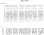
- By "Als3p" is meant a polypeptide that is substantially identical to the amino acid sequence of a sequence shown in
Figure 1A , e.g., SEQ ID NO: 1 or SEQ ID NO: 2, or to a Candida ALS3 protein identified in GenBank: XP_710431.1, XP_710435.1, AAO72959.1, XP_712646.1, XP_712666.1, EAK91173.1, EAK91169.1, AAO72958.1, EAK93494.1, EAK93472.1, 074623.1, AAD02580.1, EAK90704.1, XP_709985.1. Desirably, a Als3p has at least 70, 75%, 80%, 85%, 90%, 95%, 99%, or even 100% identity to a sequence shown inFigure 1A , e.g., SEQ ID NO: 1 or SEQ ID NO: 2. - By "Als3p fragment" or "fragment of a Als3p" is meant a portion of a Als3p polypeptide containing fewer than 1050, 1025, 1000, 975, 950, or 945 amino acids. In some embodiments, Als3p fragments are between 300 and 350 or 250 to 500 amino acids in length. In some embodiments, the fragment is fewer than 1050, 1025, 1000, 975, 950, or 945, 940, 937, 936, 935, 934, 933, 932, 931, or 930, 920, 910, 900, 890, 880, 870, 860, 850, 840, 830, 820, 810, 800, 790, 780, 770, 760, 750, 740, 730, 720, 710, 700, 690, 680, 670, 660, 650, 640, 630, 620, 610, 600, 590, 580, 570, 560, 550, 540, 530, 520, 510, 500, 490, 480, 470, 460, 450, 440, 430, 420, 410, 400, 390, 380, 370, 360, 350, 340, 330, 320, 310, 300, 290, 280, 270, 260, 250, 240, 230, 220, 210, 200, 190, 180, 170, 160, 150, 140, 130, 120, 110, 100, 90, 80, 70, 60, 50, 40, 30, 25, 20, 15, or 10 amino acids, and, in some instances, is immunogenic.
- An exemplary Als3p fragment is SEQ ID NO: 2, as shown in
Figure 1A , or fragments thereof. In some instances, Als3p fragments are between 14 and 20 amino acids in length. In general, the fragment may be fewer than, e.g., 325, 320, 310, 300, 290, 280, 270, 260, 250, 240, 230, 220, 210, 200, 190, 180, 170, 160, 150, 140, 130, 120, 110, 100, 90, 80, 70, 60, 50, 40, 30, 25, 20, 19, 18, 17, 16, 15, 14, 13, 12, or 11 amino acids, and desirably, is immunogenic. In some instances, an Als3p fragment is between 14 and 20 amino acids. - In addition, Als3p fragments, for example, may contain one or more conservative amino acid substitutions in a sequence shown in
Figure 1A , e.g., SEQ ID NO: 1 or SEQ ID NO: 2. Additional desirable Als3p fragments contain one or more conservative amino acid substitutions in a sequence shown inFigure 1A , e.g., SEQ ID NO: 1 or SEQ ID NO: 2, and/or at least one flanking amino acid (e.g., 1, 2, 3, 4, 5, 6, 7, 8, 9, or 10 flanking amino acids) at the N- and/or C-terminus of a sequence shown inFigure 1A , e.g., SEQ ID NO: 1 or SEQ ID NO: 2. Other preferred Als3p fragments contain seven or more continuous amino acids of a sequence shown inFigure 1A , e.g., SEQ ID NO: 1 or SEQ ID NO: 2. - Non-limiting examples of an Als3p fragment include amino acids 1-40, 10-50, 20-60, 30-70, 40-80, 50-90, 60-100, 70-110, 80-120, 90-130, 100-140, 110-150, 120-160, 130-170, 140-180, 150-190, 160-200, 170-210, 180-220, 190-230, 200-240, 210-250, 220-260, 230-270, 240-280, 250-290, and 260-300, 270-310, 280-320, and 290-331 amino acids of a sequence shown in
Figure 1A , e.g., SEQ ID NO: 1 or SEQ ID NO: 2; and these fragments having one or more of the following features: one or more conservative amino acid substitutions (e.g., 1, 2, 3, 4, 5, 6, 7, 8, 9, 10, 11, 12, 13, 14, 15, or 16 conservative amino acid substitutions) in a sequence shown inFigure 1A , e.g., SEQ ID NO: 1 or SEQ ID NO: 2; one or more amino acids (e.g., 1, 2, 3, 4, 5, 6, 7, 8, 9, 10, 11, 12, 13, 14, 15, or 16 amino acids) truncated from the N and/or C-terminus of a sequence shown inFigure 1A , e.g., SEQ ID NO: 1 or SEQ ID NO: 2; and at least one flanking amino acid (e.g., 1, 2, 3, 4, 5, 6, 7, 8, 9, or 10 flanking amino acids) at the N- and/or C-terminus of a sequence shown inFigure 1A , e.g., SEQ ID NO: 1 or SEQ ID NO: 2. - By "substantially identical" is meant an amino acid sequence or nucleic acid sequence that exhibits at least 50% identity to a reference sequence. Such a sequence is generally at least, e.g., 50%, 60%, 70%, 75%, 80%, 85%, 90%, 95%, 96%, 97%, 98%, or 99% identical at the amino acid level or nucleic acid level to a reference sequence. In general, for polypeptides, the length of comparison sequences can be at least five amino acids, e.g., 10, 20, 30, 40, 50, 60, 70, 80, 90, 100, 125, 150, 175, 200, 250, 300, or more amino acids, up to the entire length of the polypeptide. For nucleic acids, the length of comparison sequences can generally be at least 10, 20, 30, 40, 50, 60, 70, 80, 90, 100, 125, 150, 175, 200, 250, 300, 400, 500, 600, 700, 800, 900, or more nucleotides, up to the entire length of the nucleic acid molecule. It is understood that for the purposes of determining sequence identity when comparing a DNA sequence to an RNA sequence, a thymine nucleotide is equivalent to a uracil nucleotide.
- Also contemplated are nucleic acid sequences that encode any of the Als3p polypeptides or fragments thereof recited herein.
- As used herein, when a polypeptide or nucleic acid sequence is referred to as having "at least X% sequence identity" to a reference sequence, it is meant that at least X percent of the amino acids or nucleotides in the polypeptide or nucleic acid are identical to those of the reference sequence when the sequences are optimally aligned. An optimal alignment of sequences can be determined in various ways that are within the skill in the art, for instance, the Smith Waterman alignment algorithm (Smith et al., J. Mol. Biol. 147:195-7, 1981) and BLAST (Basic Local Alignment Search Tool; Altschul et al., J. Mol. Biol. 215: 403-10, 1990). These and other alignment algorithms are accessible using publicly available computer software such as "Best Fit" (Smith and Waterman, Advances in Applied Mathematics, 482-489, 1981) as incorporated into GeneMatcher PlusTM (Schwarz and Dayhof, Atlas of Protein Sequence and Structure, Dayhoff, M.O., Ed pp 353-358, 1979), BLAST, BLAST-2, BLAST-P, BLAST-N, BLAST-X, WU-BLAST-2, ALIGN, ALIGN-2, CLUSTAL, or Megalign (DNASTAR). In addition, those skilled in the art can determine appropriate parameters for measuring alignment, including any algorithms needed to achieve optimal alignment over the length of the sequences being compared.
- By "adjuvant" is meant one or more substances that cause stimulation of the immune system. In this context, an adjuvant is used to enhance an immune response to one or more vaccine antigens or antibodies. An adjuvant may be administered to a subject before, in combination with, or after administration of the vaccine or antibody. Examples of chemical compounds used as adjuvants include, but are not limited to, aluminum compounds (e.g., alum, Alhydrogel), oils, block polymers, immune stimulating complexes, vitamins and minerals (e.g., vitamin E, vitamin A, selenium, and vitamin B12), Quil A (saponins), bacterial and fungal cell wall components (e.g., lipopolysaccharides, lipoproteins, and glycoproteins), hormones, cytokines, and co-stimulatory factors.
- By "carrier" in the context of a conjugate is meant a moiety or particle, e.g., KLH, CRM197, tetanus toxoid, diphtheria toxoid, enterotoxin B fragments, N. meningitides outer membrane protein complex, any other carrier protein, a phage, a yeast, a virus, a virosome, or a recombinant virus-like particle, that is suitable for being linked to or displaying a polypeptide as described herein.
- By "conjugate" is meant a compound that includes a polypeptide described herein linked to another moiety or particle, e.g., KLH, CRM197, tetanus toxoid, diphtheria toxoid, enterotoxin B fragments, N. meningitides outer membrane protein complex, any other carrier protein, a phage, a yeast, a virus, a virosome, or a recombinant virus-like particle.
- By "immunogenic" is meant any substance that is capable of inducing an immune response in a subject.
- By "immunogenic amount" in the context of a vaccine is meant an amount of the vaccine required to induce an immune response in a subject in a clinically relevant manner. An immunogenic amount of vaccine used to practice the methods of vaccination as described herein varies depending upon the manner of administration, the age, body weight, and general health of the subject. Ultimately, prescribers will decide the appropriate amount and dosage regimen.
- By "isolated" or "purified" is meant separated from other naturally accompanying components. Typically, a compound (e.g., nucleic acid, polypeptide, antibody, or small molecule) is substantially isolated when it is at least 60%, by weight, free from the proteins and/or naturally occurring organic molecules with which it is naturally associated. The definition also extends, e.g., to a polypeptide or nucleic acid molecule separated from its flanking sequences (e.g., for an amino acid sequence, isolated refers to a sequence that is free from the flanking amino acids with which the sequence is naturally associated in a polypeptide). In some instances, the compound is at least 75%, more preferably at least 90%, and most preferably at least 99%, by weight, isolated. An isolated compound, e.g., polypeptide, may be obtained by standard techniques, for example, by extraction from a natural source (e.g., purification from a cell infected with Candida); by expression of a recombinant nucleic acid encoding an Als3p, an Als3p fragment or variant, or a fusion protein thereof in any standard expression system including but not limited to E. coli or Saccharomyces cerevisiae; or by chemically synthesizing the polypeptide. Purity can be measured by any appropriate method, e.g., by column chromatography, polyacrylamide gel electrophoresis, or HPLC analysis.
- By "linked to" or "conjugated to" in the context of a conjugate is meant a covalent or non-covalent interaction between the polypeptide and the carrier or fusion partner. Non-covalent interactions include, but are not limited to, hydrogen bonding, ionic interactions among charged groups, electrostatic binding, van der Waals interactions, hydrophobic interactions among non-polar groups, lipophobic interactions, and LogP-based attractions.
- The terms "peptide," "polypeptide," and "protein" are used interchangeably and refer to any chain of two or more natural or unnatural amino acids, regardless of post-translational modification (e.g., glycosylation or phosphorylation), constituting all or part of a naturally-occurring or non-naturally occurring polypeptide or peptide, as is described herein.
- The terms "pharmaceutically acceptable carrier" and "pharmaceutically acceptable excipient" are used interchangeably and mean a carrier or excipient that is physiologically acceptable to the treated mammal while retaining the therapeutic properties of the compound with which it is administered. One exemplary pharmaceutically acceptable carrier substance is physiological saline. Other physiologically acceptable carriers and their formulations are known to those skilled in the art and described, for example, in Remington's Pharmaceutical Sciences, (21th edition), ed. A. Gennaro, 2005, Lippincott, Williams & Wilkins, Philadelphia, PA.
- By "pharmaceutical composition" is meant a composition containing a polypeptide, conjugate, vaccine, or antibody described herein, formulated with a pharmaceutically acceptable excipient, and manufactured or sold with the approval of a governmental regulatory agency as part of a therapeutic regimen for the treatment or prevention of a disease or event in a mammal. Pharmaceutical compositions can be formulated, for example, for intravenous administration (e.g., as a sterile solution free of particulate emboli and in a solvent system suitable for intravenous use), for oral administration (e.g., a tablet, capsule, caplet, gelcap, or syrup), or any other formulation described herein, e.g., in unit dosage form.
- By "treating" or "treatment" is meant the medical management of a mammal, e.g., a human or non-human mammal, with the intent to cure, ameliorate, stabilize, reduce the likelihood of, or prevent a disease, pathological condition, disorder, or event, by administering a pharmaceutical composition. This term includes active treatment, that is, treatment directed specifically toward the improvement or associated with the cure of a disease, pathological condition, disorder, or event, and also includes causal treatment, that is, treatment directed toward removal of the cause of the associated disease, pathological condition, disorder, or event. In addition, this term includes palliative treatment, that is, treatment designed for the relief of symptoms rather than the curing of the disease, pathological condition, disorder, or event; symptomatic treatment, that is, treatment directed toward constitutional symptoms of the associated disease, pathological condition, disorder, or event; preventative treatment, that is, treatment directed to minimizing or partially or completely inhibiting the development of the associated disease, pathological condition, disorder, or event, e.g., in a mammal who is not yet ill, but who is susceptible to, or otherwise at risk of, a particular disease, pathological condition, disorder, or event; and supportive treatment, that is, treatment employed to supplement another specific therapy directed toward the improvement of the associated disease, pathological condition, disorder, or event.
- By "vaccine," as used herein, is meant a composition that elicits an immune response in a subject to which it is administered. The mode of administration, dose, and number of administrations can be optimized by those skilled in the art in a known manner.
- By "vaccinate" or "vaccinating" as used herein, is meant to treat a mammal by administering a vaccine, e.g., to prevent or ameliorate a disease, pathological condition, disorder, or event.
- Other features and advantages of the invention will be apparent from the following Detailed Description, the drawings, and the claims.
- The figures show:
-
Fig. 1A is a listing of two Als3p amino acid sequences, SEQ ID NO: 1 and SEQ ID NO: 2. -
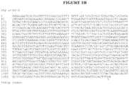
Fig. 1B is a listing of one Als3 nucleic acid sequence, SEQ ID NO: 3. -
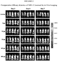
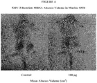
Fig. 2 is a set of photographs showing comparative efficacy kinetics of NDV-3 assessed by in vivo imaging. The photographs show mice in each of the dosage groups atdays -
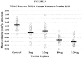
Fig. 3 is a chart showing that NDV-3 restricts MRSA abscess volume in murine SSSI. The chart shows mean volume (cm3)/abscesses for the control group and the 3 µg, 10 µg, 30 µg, and 100 µg NDV-3 dosage groups. -
Fig. 4 is a pair of photographs showing that NDV-3 restricts MRSA abscess volume in murine SSSI. Left, mouse in control group; right, mouse in 100 µg NDV-3 dosage group. -
Fig. 5 is a chart showing that NDV-3 suppresses MRSA proliferation in murine SSSI. The chart shows mean flux/abscess for the control group and the 3 µg, 10 µg, 30 µg, and 100 µg NDV-3 dosage groups. -
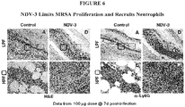
Fig. 6 is a set of images showing that NDV-3 limits MRSA proliferation and recruits neutrophils. The data shown are from the 100 µg NDV-3 dosage group atday 7 post-infection. -
Fig. 7 is a set of images showing that NDV-3 recruits CD3+ T cells and induces IL-17 expression. The data shown are from the 100 µg NDV-3 dosage group atday 7 post-infection. -
Fig. 8 is a set of images showing that NDV-3 stimulates IL-22 expression and β-defensin response. The data shown are from the 100 µg NDV-3 dosage group atday 7 post-infection. -
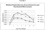
Fig. 9 is a chart showing the median flank abscess area of control and vaccinated mice. -
Fig. 10 is a chart showing the median flank abscess volume of control and vaccinated mice. -
Fig. 11 is a chart showing the mean abscess volume due to MRSA strains in control and vaccinated mice atday 7 post-infection. Asterisks indicate significant reduction as compared to respective control. -
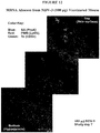
Fig. 12 is a composite immunofluorescence image of a MRSA abscess from a NDV-3 (100 µg) vaccinated mouse. -
Fig. 13 is a composite immunofluorescence image of a MRSA abscess from a control mouse. - The patent or application file contains drawings (
Figures 2 ,4 ,6-8 ,12 , and13 ) executed in color. Copies of this patent or patent application publication with color drawings will be provided by the Office upon request and payment of the necessary fee. - As is described below, agglutinin-
like sequence 3 protein (Als3p) allows for vaccination against S. aureus in mammals identified as being at risk for development of an S. aureus skin or soft tissue infection. - In the following analyses (in particular, the pilot study of Example 1 and the optimized study of Example 2) designed to evaluate the efficacy of an Als3p vaccine against the development of skin or soft tissue infection resulting from S. aureus in a murine model of MRSA skin/skin structure infection (SSSI), the organisms and methods are first described.
-
- MRSA Xen30 (lux+) Roche-16
- MRSA LAC-USA300 USA300
- MRSA MW2 USA400
- Staphylococcus aureus strain Xen30 was used in these in vivo studies. It is derived from the parental strain S. aureus MRSA-16 (Roche) and contains a luxA-E operon at a single chromosomal integration site. This MRSA strain produces luciferase enzyme and aldehyde substrate, and constitutively emits a bioluminescent signal when metabolically active. Its virulence is equivalent to other MRSA strains in the SSSI murine model used as verified in pilot studies, and all strains tested have otherwise similar phenotypes and growth characteristics. Log-phase cells (BHI; 37°C) were cultured from quantitatively- and virulence-validated master cell banks, harvested and suspended in PBS, sonicated and quantified by spectrophotometry to desired CFU.
- NDV-3 vaccine efficacy was evaluated in a murine SSSI model vs. methicillin-resistant SA (MRSA): Xen30 (lux+); LACUSA300; or MW2 (USA400). NDV-3 is a formulation of the recombinant N-terminus of the Candida surface protein Als3 protein (
Figure 1A ; SEQ ID NO:2) and the adjuvant Alhydrogel®, in phosphate-buffered saline,pH 7, e.g., with a 0.5 mL dose containing, e.g., 30-300 µg Als3 protein, and optionally further containing aluminum hydroxide at 1.0 mg Al/mL. Efficacy was compared among NDV-3 regimens administered with alhydrogel adjuvant (IM) on day 0 and boosted on day 21. Controls received adjuvant alone. Infection by subcutaneous inoculation of two flanks (2x10⁷ CFU) occurred 14 days after boost. Abscess area, volume, and CFU were quantified for multiple days post-challenge. In vivo imaging (IVIS) of abscess flux was done in mice infected by Xen30. Serum IgG (ELISA), IFN-γ and IL-17A (ELISpot) responses were quantified in parallel vaccine regimens. Tissue IL-17A, IL-22, mβD-3, CD3+ cell and neutrophil signals were assessed onday 7 post-infection by immunohistochemistry. - Vaccine. The NDV-3 vaccination was evaluated across a dose range using an identical regimen of alhydrogel adjuvant. Doses of 3, 10, 30, 100, or 300µg (IM) were studied in parallel. Primary vaccination (day 0) was followed by an identical boost on study day 21. Mice were infected 14 days after boost (study day 35).
- Murine Model of SSSI. All animal studies were performed per the approved animal use policies of LABioMed at Harbor-UCLA. Balb/C mice (Harlan) were vaccinated as above. A subcutaneous skin / soft tissue abscess model was modified from Ding et al. (J Bacteriol 2008 190:7123-9) and/or Voyich et al. (J Infect Dis 2006 194:1761-1770) for these studies. On study day 35, mice were anesthesized, flanks were shaved and sterilized, and 2x10⁷ CFU inocula (without beads or matrix) were introduced into the subcutaneous compartment by injection (100 µl). A minimum of 20 mice per control or vaccine-regimen groups were used in each study.
- Abscess Quantification. Abscess area / volume were measured in each mouse flank during the study period up to 14 days post-challenge. To do so, mice were anesthetized, and the lesion site length (l) and width (w) assessed to quantify abscess or dermonecrosis area (cm2). Abscess volume (cm3) was calculated per the formula for a spherical ellipsoid: [v = (π/6) x l x w 2].
- Imaging Studies. The Xen30 MRSA strain has a self-contained lux operon integrated in its chromosome. The construct encodes the aldehyde substrate and the luciferase enzyme itself; thus, no exogenous luciferin substrate is required (Kadurugamuwa et al., Infect Immun 2003 71:882-890). On selected study days, control and vaccinated mice underwent in vivo imaging (IVIS) using an IVIS system (Caliper Life Sciences, Inc.). Luminescence signals were captured over a five-minute time period and analyzed using the Living Image software as photons / min / abscess.
- Quantitative Culture. At pre-selected times post-infection, mice were humanely sacrificed and processed for quantitative culture of abscesses. Each flank was aseptically dissected, the abscess removed and prepared for culture. Abscesses were individually homogenized, and serially diluted in sterile PBS for quantitative culture onto sheep blood agar plates. Cultures were incubated (37°C) for 24 hours, and resulting colonies enumerated.
- Immunological Mechanisms. Multiple and complementary approaches were used to assess potential correlates of NDV-3 vaccine efficacy in the murine model of SSSI due to MRSA. These studies focused on strain Xen30, allowing correlation with IVIS data at the 7d endpoint.
- A. Antibody Quantification. Serum IgG antibody levels were determined in a 96-well ELISA format over a range dilutions. Values represent geometric mean corrected dilution of triplicate assays comparing immunized vs. control sera.
- B. Cytokine Quantification. T cell IFN-γ and IL-17A responses were determined by ELISpot analysis of splenocytes isolated from immunized vs. control mice, and exposed to the NDV-3 immunogen. The number of spot-forming units (SPUs or SFUs, used interchangeably) was quantified per 106 cells producing either IFN-γ or IL-17A. Cell viability was verified by production of IFN-γ following stimulation with phorbol-12-myristate-13-acetate (PMA) and ionomycin per established protocols.
- C. Immunohistochemistry. Immunological determinants associated with vaccine efficacy were assessed in tissues obtained from vaccinated and control animals after 7d of infection by standard methods. For immunohistochemical studies, in brief 3 µm vertical paraffin embedded sections were dewaxed and rehydrated followed by heat-induced antigen retrieval in target retrieval solution (Dako, Carpinteria, CA). Sections were incubated with dual endogenous blocking buffer (Dako) for 15 min at room temperature to block endogenous peroxidase activity, and non-specific antibody binding was blocked by incubation with 5% normal serum corresponding to the primary antibody. Sections were then incubated overnight at 4°C with a primary antibody targeting a specific antigen of interest (Table 1). Sections were then washed and incubated for 30 min with an appropriate secondary antibody (Table 1), either horseradish peroxidase (HRP)-conjugated or biotinylated (Santa Cruz Biotechnology, Santa Cruz CA). Immunohistochemical development was then achieved by 30 min development with streptavidin-HRP (Dako) and 3,3'-diaminobenzidine (DAB; Vector Laboratories, Burlingame, CA), and counterstained with hematoxylin. Images were visualized using an Olympus BX43 microscope employing a DP21 digital camera for image capture.
- D. Immunofluorescence. To evaluate the impact of NDV-3 vaccination on the interrelationships of immunologic determinants and S. aureus in context of infection in vivo, immunofluorescence studies employing confocal microscopy were performed using established methods. In brief, paraffin embedded sections were prepared as above and incubated with immunofluorescence buffer (1% bovine serum albumin and 2% fetal calf serum) for one hour at room temperature. Primary antibodies directed at target antigens of interest (Table 1) were incubated with tissue sections from control or vaccinated mice at 4°C overnight. Next, corresponding secondary antibodies (Table 1) diluted in IFF buffer (2 µg/ml) were incubated for 60 minutes. Sections were then washed in PBS, and mounted using Vectashield H-1500 (Vector Laboratories, Burlingame, CA) to minimize photobleaching. Images were visualized using a Leica SP2 confocal microscope employing argon (488nm), krypton (568nm) and helium-neon (633nm) lasers and confocal version 2.0 software (Leica Instruments, Germany).
- Statistical Analyses. Differences in experimental results were compared based on power estimates indicating that 16-20 mice per group yields > 85% power to detect 1 log difference in CFU per gram tissue, or 2 mm abscess area (a = 0.05; Mann-Whitney U test. P values are defined in Table 2 and Table 3 (below).
- In a pilot study, vaccination with NDV-3 reduced SSSI parameters due to MRSA, with equivalent efficacy in limiting abscess area, volume, and CFU for strains Xen30, USA300, and MW2. Murine immune response correlated with NDV-3 dose-related protective efficacy. These results are shown in Table 2 and
Figure 2 . These results indicate the NDV-3 vaccine induced robust B and T cell responses which correspond with protective efficacy against MRSA in the murine model of SSSI.Table 2. NDV-3 Efficacy in MRSA Xen30 SSSI and Immune Response in Murine Models. Abscess Control 3µg 10µg 30µg 100µg 300µg Area 7d 1.88cm2 1.47cm2* 1.59cm2* 0.99cm2†† 0.77cm2†† 0.69cm2†† Volume 7d 1.29cm3 0.95cm3* 0.96cm3** 0.46cm3†† 0.34cm3†† 0.29cm3†† Flux 7d 1.92x10⁵ 1.48x105* 1.81x105 1.07x105* 1.65x105* 9.03x104†† Median 7.9 7.8 8.1 8.1 7.9 7.5† Log CFU (7.6/8.0) (7.8/7.8) (7.8/7.8) (7.9/7.9) (7.5/8.0) (7.4/7.8) 7d IM [n=36] [n=20] [n=20] [n=36] [n=36] [n=36] Median 1.70 0.05Δ†† ND ND 0.05Δ†† 1.48 Log CFU (1.0/2.8) (0.05/1.3) (0.05/1.7) (0.05/2.6) 14d IM [n=48] [n=39] [n=39] [n=17] Median 3.54 3.92 ND ND 2.26** 2.40** Log CFU (2.6/6.9) (3.2/5.5) (1.8/3.6) (1.4/3.8) 14d SubQ [n=20] [n=20] [n=20] [n=20] Analyte IgG 1.0 GCU 44.8 GCU†† ND 97.8 GCU†† 81.8 GCU†† ND IFN-γ 9.5 SPU 12.8 SPU ND 21.9 SPU 34.3 SPU* ND EL-17 18.9 SPU 132.6 SPU†† ND 62.2 SPU 161.2 SPU** ND (25%/75% quartiles); *P <0.5; **P < 0.1; † P <0.05; †† P < 0.01; GCU, geomean / dilution corrected units; SPU, mean spot forming units / 106 splenocytes; Δ limit of detection. - The NDV-3 vaccine significantly reduced the abscess area, volume, luminescence signal, and CFU densities in this murine model of MRSA SSSI. NDV-3 efficacy was equivalent for each of the MRSA strains evaluated in this study. Immunological data from mice vaccinated identically to those challenged with infection indicate the NDV-3 vaccine induces robust B and T cell responses which appear to reflect a dose-response relationship. Immunological data from mice vaccinated identically to those challenged with infection indicate the NDV-3 vaccine induces robust B and T cell responses which reflect a dose-response relationship. Collectively these results provide evidence that NDV-3 induces a mixed Th1 / Th17 response that appears to be predominantly associated with protective efficacy. Antibody response may contribute to protective mechanisms of NDV-3. These results indicate that the NDV-3 vaccine is useful as a means to prevent or mitigate MRSA skin infection or abscesses or both in mammals.
- A further, optimized analysis was conducted, and results are summarized in Table 3 and
Figures 3-8 . Like Example 1, this study evaluated the efficacy and immunologic mechanisms of the NDV-3 vaccine in a murine model of skin / skin structure infection due to methicillin-resistant SA (MRSA). Abscess size, MRSA density and CFU were compared over time in NDV-3 immunized and control groups. Serum concentrations of IgG, IFNγ, IL-17A, induction of tissue IL-17A, IL-22, and mβD-3, and infiltration of CD3+ T cells or neutrophils as mediated by NDV-3 were determined in parallel. NDV-3 immunization achieved protective efficacy against MRSA in terms of abscess area, volume, bacterial density and CFU as compared to adjuvant alone. Protective efficacy of NDV-3 corresponded to increases in serum IgG, serum and tissue biomarkers of Th1-Th17 polarization, and corresponding neutrophil infiltration and host defense peptide induction in context of abscesses. These data further demonstrated that NDV-3 immunization induces robust B and T cell mechanisms of protective efficacy against MRSA in context of skin and mucosa. - NDV-3 was efficacious against MRSA as measured by reduced abscess area, volume, and CFU versus adjuvant alone (Table 3). Efficacy as measured by area of dermonecrosis and abscess volume were equivalent for all strains tested. Significant increases in serum IgG, serum and tissue biomarkers of Th1 (INF-γ) and Th17 (IL-17) polarization (Table 3), neutrophil infiltration (Ly6G), IL-22 elaboration, as well as mβD-3 induction were correlated with NDV-3 protective efficacy (
Figures 3-8 ).Table 3. NDV-3 efficacy and immune response vs. MRSA Xen30 in murine SSSI. Abscess Control 3□g 10□g 100□g Area d7 1.88cm2 1.47cm2* 1.59cm2* 0.77cm2†† Volume d7 1.29cm3 0.95cm3* 0.96cm3** 0.38cm3†† Flux d7 3.22x10⁵ 1.48x105* 2.15x105* 1.06x105** Geo Mean 7.50 6.23 † 6.68 † 6.05 † Log CFU (8.0/7.4) a (6.4/6.1) (6.8/6.4) (6.2/5.6) d7 IM [n=54] [n=20] [n=20] [n=20] Analyte IgG 1.0 GCU b 44.8 GCU †† ND 81.8 GCU †† IFN□□ 9.5 SFU c 12.8 SFU ND 34.3 SFU * IL-17 18.9 SFU 132.6 SFU †† ND 161.2 SFU ** a Mean variance
b GCU, geomean / dilution corrected units
c SPU, mean spot forming units / 106 splenocytes
* P <0.5; **P < 0.1; † P <0.05; †† P < 0.01 - NDV-3 induces protective efficacy against MRSA in murine SSSI. Immunologic mechanisms of efficacy included robust B and T cell responses consistent with Th1-Th17 paradigms in which neutrophils and host defense peptides are targeted and coordinated in context of infection.
- An additional set of experiments was conducted to evaluate the efficacy of the NDV-3 vaccine in a murine model of SSSI due to Xen30 MRSA and comparative strains of MRSA. Experiments were conducted as described in Examples 1 and 2. Median data kinetics of vaccine efficacy versus time is shown in
Fig. 9 (median flank abscess area of control and vaccinated mice) andFig. 10 (mean flank abscess volume of control and vaccinated mice). These data confirm that vaccination with NDV-3 suppresses evolution of the abscess, particularly at dosages greater than 3 µg. - In addition, efficacy of the vaccine was tested against three different MRSA strains:
Xen 30, USA300, and MW2. For each MRSA strain, a negative control and a 100 µg dosage group were tested. The mean lesion volume atday 7 post-infection was determined, as shown inFig. 11 . Each strain was the same inoculum (2 x 10⁷). MW2 exhibited low virulence in these experiments. - The data demonstrate that regardless of MRSA strain tested, the NDV-3 vaccine has equivalent efficacy (e.g., about 50% reduction) in restricting abscess volume. Thus, NDV-3 efficacy is not MRSA strain-specific.
- In a further set of experiments, composite immunofluorescence images of MRSA abscesses were recorded and analyzed.
Fig. 12 is a composite immunofluorescence image of a representative MRSA abscess from a NDV-3 (100 µg) vaccinated mouse, andFig. 13 is a composite immunofluorescence image of a representative MRSA abscess from a control mouse. - In each of the above images, each component of the image is of the same lesion, magnified approximately 500-fold. As immunofluorescence signal is difficult to resolve at low power, images were recorded for each section of the lesion at higher power, moving from the epidermis of the skin, into the subdermis, and down into the hypodermis. Thus, the components are merged to illustrate a continuous immunofluorescence map of S. aureus (blue), neutrophils (red), and CD3+ (T cells) green, throughout a lesion and maintaining magnification sufficient for resolution of immunofluorescence. The image components represent a function of high-power fields positioned to systematically capture equivalent areas in the NDV-3 and control lesions for head-to-head comparison of abscess immunophenotypes.
- As
Figs. 12 and13 reveal, in the NDV-3 vaccinated abscess, there are few MRSA organisms (blue), and they are restricted to the epidermis, with infiltration of neutrophils (red) mediated by an influx of CD3+ T cells (green). In contrast, in the control abscess, there are many MRSA organisms, and they are invasive to two distinct regions (epidermis and hypodermis), corresponding with substantially less neutrophil and CD3+ cell infiltration. While the images shown inFigs. 12 and13 are from individual lesions, they are representative of lesions in vaccinated and control groups overall and are consistent with the quantitative findings described in the preceding Examples. - The compositions and methods described herein may be used, e.g., to vaccinate a human at risk for the development of a Staphylococcus aureus skin or soft tissue infection against Staphylococcus aureus. First, a human at risk for the development of an S. aureus SSSI is identified. Second, the human is administered an immunogenic amount of a vaccine comprising a polypeptide comprising Als3p, or an immunogenic fragment thereof, in a pharmaceutically acceptable medium. For example, the human is administered between one and three doses of NDV-3 containing between 3 and 1000 µg of the recombinant N-terminus of the Candida surface protein Als3 (SEQ ID NO:2) per dose, with multiple doses occurring at intervals of two weeks to six months.
- It is expected that, following administration of the vaccine, the human is at decreased risk for the development of an S. aureus SSSI for a period lasting from one month to several years or more.
- Likewise, a human who is identified as having an S. aureus SSSI may be treated by administration of an immunogenic amount of a pharmaceutical composition comprising a polypeptide comprising Als3p, or an immunogenic fragment thereof, in a pharmaceutically acceptable medium. For example, the human is administered between one and three doses of NDV-3 containing between 3 and 1000 µg of the recombinant N-terminus of the Candida surface protein Als3 (SEQ ID NO:2) per dose, with multiple doses occurring at intervals of two weeks to six months.
- It is expected that, following administration of the pharmaceutical composition, the S. aureus SSSI of the human is decreased in severity.
- The compositions and methods described herein may be used, e.g., to vaccinate a bovine species at risk for the development of a Staphylococcus aureus skin or soft tissue infection against Staphylococcus aureus. In particular, the bovine species may be at risk of developing bovine mastitis caused by S. aureus. First, a bovine species at risk for the development of an S. aureus SSSI, e.g., bovine mastitis, is identified. For example, any milk-producing bovine may be considered to be at risk of developing bovine mastitis caused by S. aureus. Second, the bovine species is administered an immunogenic amount of a vaccine comprising a polypeptide comprising Als3p, or an immunogenic fragment thereof, in a pharmaceutically acceptable medium. For example, the bovine species is administered between one and three doses of NDV-3 containing between 3 and 1000 µg of the recombinant N-terminus of the Candida surface protein Als3 (SEQ ID NO:2) per dose, with multiple doses occurring at intervals of two weeks to six months.
- It is expected that, following administration of the vaccine, the bovine species is at decreased risk for the development of an S. aureus SSSI, e.g., bovine mastitis.
- Likewise, a bovine species identified as having an S. aureus SSSI, e.g., bovine mastitis, may be treated by administration of an immunogenic amount of a pharmaceutical composition comprising a polypeptide comprising Als3p, or an immunogenic fragment thereof, in a pharmaceutically acceptable medium. For example, the bovine species is administered between one and three doses of NDV-3 containing between 3 and 1000 µg of the recombinant N-terminus of the Candida surface protein Als3 (SEQ ID NO:2) per dose, with multiple doses occurring at intervals of two weeks to six months.
- It is expected that, following administration of the pharmaceutical composition, the S. aureus SSSI, e.g., bovine mastitis, of the bovine species is decreased in severity.
| Primary antibodies |
| Target Antigen |
| Ly-6G (granulocytes) |
| Ly-6C (monocytes / macrophages) |
| Mouse β-defensin-1 (mBD-1) |
| Mouse β-defensin-3 (mBD-3) |
| Mouse platelet factor 4 (PF-4) |
| Staphylococcus aureus (mouse) |
| Staphylococcus aureus (rabbit) |
| Staphylococcal protein A |
| CD3-γ |
| CD3-ε |
| IL-17 |
| IL-22 |
| Secondary Antibodies |
| Alexa 488-conjugated donkey α-rabbit |
| Alexa 488-conjugated donkey α-rat |
| Alexa 555-conjugated goat α-rat |
| Alexa 568-conjugated donkey α-rabbit |
| Alexa 633-conjugated donkey α-goat |
| Alexa 647-conjugated donkey α-rabbit |
| Alexa 633-conjugated streptavidin |
Claims (9)
- A vaccine comprising 30 to 300 µg of an isolated Als3 protein consisting of the amino acid sequence of SEQ ID NO: 2 in 0.5 mL phosphate-buffered saline, pH 7, for use in a method of treatment of a skin abscess comprising a Staphylococcus aureus infection in a mammal.
- The vaccine for use according to claim 1, wherein said Staphylococcus aureus is a MRSA strain of Staphylococcus aureus or a MSSA strain of Staphylococcus aureus.
- The vaccine for use according to claim 1, wherein said Staphylococcus aureus is a vancomycin-resistant (VRSA) or daptomycin-resistant (DRSA) strain of Staphylococcus aureus.
- The vaccine for use according to claim 1, wherein said Als3 protein is conjugated to a carrier.
- The vaccine for use according to claim 4, wherein said carrier comprises keyhole limpet hemocyanin (KLH), CRM197, tetanus toxoid, diphtheria toxoid, enterotoxin B fragments, or N. meningitides outer membrane protein complex.
- The vaccine for use according to claim 4, wherein said carrier is a phage, a yeast, a virus, a virosome, or a recombinant virus-like particle.
- The vaccine for use according to claim 1, wherein said vaccine is administered by intramuscular, subcutaneous, intradermal, oral, or sublingual administration, or is administered for inhalation in a microparticulate formulation.
- The vaccine for use according to claim 1, wherein said vaccine comprises an immunostimulating adjuvant.
- The vaccine for use according to claim 1, characterized in that the vaccine is formulated for use with an antibiotic against Staphylococcus aureus.
Applications Claiming Priority (2)
| Application Number | Priority Date | Filing Date | Title |
|---|---|---|---|
| US201161510896P | 2011-07-22 | 2011-07-22 | |
| PCT/US2012/000328 WO2013015831A1 (en) | 2011-07-22 | 2012-07-20 | Methods and compositions for vaccinating against staphylococcus aureus |
Publications (3)
| Publication Number | Publication Date |
|---|---|
| EP2734229A1 EP2734229A1 (en) | 2014-05-28 |
| EP2734229A4 EP2734229A4 (en) | 2015-01-21 |
| EP2734229B1 true EP2734229B1 (en) | 2019-01-02 |
Family
ID=47601425
Family Applications (1)
| Application Number | Title | Priority Date | Filing Date |
|---|---|---|---|
| EP12817530.4A Not-in-force EP2734229B1 (en) | 2011-07-22 | 2012-07-20 | Methods and copositions for vaccinating against staphylococcus aureus |
Country Status (12)
| Country | Link |
|---|---|
| US (1) | US10653757B2 (en) |
| EP (1) | EP2734229B1 (en) |
| JP (1) | JP2014521605A (en) |
| CN (1) | CN103998056B (en) |
| AU (1) | AU2012287513A1 (en) |
| BR (1) | BR112014001409A2 (en) |
| CA (1) | CA2842626A1 (en) |
| EA (1) | EA035513B1 (en) |
| ES (1) | ES2716800T3 (en) |
| GE (1) | GEP201706766B (en) |
| UA (1) | UA114286C2 (en) |
| WO (1) | WO2013015831A1 (en) |
Families Citing this family (9)
| Publication number | Priority date | Publication date | Assignee | Title |
|---|---|---|---|---|
| US20070077256A1 (en) | 1999-11-19 | 2007-04-05 | Los Angeles Biomedical Research Institute | Pharmaceutical compositions and methods to vaccinate against disseminated candidiasis and other infectious agents |
| GEP201706766B (en) | 2011-07-22 | 2017-10-25 | Los Angeles Biomedical Res Center | Methods and compositions for vaccinating against staphylococcus aureus |
| WO2014152092A2 (en) * | 2013-03-14 | 2014-09-25 | Rongfu Wang | Methods and compositions for modulating regulatory t cell function |
| WO2014153241A1 (en) * | 2013-03-14 | 2014-09-25 | The Regents Of The University Of Michigan | Treatment of staphylococcal disorders |
| US10130691B2 (en) * | 2013-03-15 | 2018-11-20 | Los Angeles Biomedical Research Institute At Harbor-Ucla Medical Center | Compositions and methods for treating fungal and bacterial pathogens |
| WO2017155949A1 (en) | 2016-03-09 | 2017-09-14 | Los Angeles Biomedical Research Institute At Harbor-Ucla Medical Center | Methods and kits for use in preventing and treating vulvovaginal candidiasis |
| CN105713096B (en) * | 2016-03-29 | 2019-01-01 | 黑龙江八一农垦大学 | A kind of preparation and application of the ATT fusion protein preventing infection of staphylococcus aureus |
| WO2019199279A1 (en) * | 2018-04-10 | 2019-10-17 | Los Angeles Biomedical Research Institute At Harbor-Ucla Medical Center | Methods of treatment for candida auris infections |
| CN112940987B (en) * | 2021-04-10 | 2022-10-14 | 福建省农业科学院畜牧兽医研究所 | Rabbit staphylococcus aureus and application thereof in preparation of inactivated vaccine |
Family Cites Families (37)
| Publication number | Priority date | Publication date | Assignee | Title |
|---|---|---|---|---|
| CA1133829A (en) | 1978-08-24 | 1982-10-19 | Neil J.L. Gilmour | Pasteurellosis vaccines |
| US5622939A (en) | 1992-08-21 | 1997-04-22 | Alpha-Beta Technology, Inc. | Glucan preparation |
| AU4528493A (en) | 1992-06-04 | 1994-01-04 | Regents Of The University Of California, The | In vivo gene therapy with intron-free sequence of interest |
| WO1994027435A1 (en) | 1993-06-01 | 1994-12-08 | Life Technologies, Inc. | Genetic immunization with cationic lipids |
| US5961970A (en) | 1993-10-29 | 1999-10-05 | Pharmos Corporation | Submicron emulsions as vaccine adjuvants |
| WO1995031998A1 (en) | 1994-05-23 | 1995-11-30 | The Research & Development Institute, Inc. | Candida albicans adhesin as a vaccine |
| US5668263A (en) | 1994-12-16 | 1997-09-16 | Smithkline Beecham Corporation | Conserved yeast nucleic acid sequences |
| WO1996040234A1 (en) | 1995-06-07 | 1996-12-19 | Pfizer Inc. | In ovo vaccination against coccidiosis |
| EP0950068B1 (en) | 1996-05-16 | 2005-11-09 | THE TEXAS A&M UNIVERSITY SYSTEM | Collagen binding protein compositions and methods of use |
| US6747137B1 (en) | 1998-02-13 | 2004-06-08 | Genome Therapeutics Corporation | Nucleic acid sequences relating to Candida albicans for diagnostics and therapeutics |
| US6248329B1 (en) | 1998-06-01 | 2001-06-19 | Ramaswamy Chandrashekar | Parasitic helminth cuticlin nucleic acid molecules and uses thereof |
| US6703025B1 (en) | 1998-08-31 | 2004-03-09 | Inhibitex, Inc. | Multicomponent vaccines |
| US8541008B2 (en) | 1999-11-19 | 2013-09-24 | Los Angeles Biomedical Research Institute At Harbor-Ucla Medical Center | Pharmaceutical compositions and methods to vaccinate against candidiasis |
| US7067138B1 (en) | 1999-11-19 | 2006-06-27 | Los Angeles Biomedical Research Institute At Harbor-Ucla Medical Center | Pharmaceutical compositions and methods to vaccinate against disseminated candidiasis |
| US20060083750A1 (en) * | 1999-11-19 | 2006-04-20 | Los Angeles Biomedical Research Institute At Harbor-Ucla Medical Center | Pharmaceutical compositions and methods to vaccinate against disseminated candidiasis |
| US20070077256A1 (en) * | 1999-11-19 | 2007-04-05 | Los Angeles Biomedical Research Institute | Pharmaceutical compositions and methods to vaccinate against disseminated candidiasis and other infectious agents |
| DK2319301T3 (en) | 2001-11-30 | 2017-12-04 | Amgen Fremont Inc | Transgenic animals with human Ig lambda light chain genes |
| CN101596312A (en) | 2002-05-21 | 2009-12-09 | 先灵-普劳有限公司 | Method of Sporozoa species In vitro culture and uses thereof |
| US7241613B1 (en) * | 2002-06-05 | 2007-07-10 | Oscient Pharmaceuticals Corporation | Identification of Candida cell surface proteins and their uses |
| US7723087B2 (en) | 2002-11-12 | 2010-05-25 | The Brigham And Women's Hospital, Inc. | Nucleic acid molecules for enhanced production of a bacterial polysaccharide and methods of use thereof |
| US20070141086A1 (en) * | 2003-11-21 | 2007-06-21 | Ohara Michael K | Use of antibiotics as vaccine adjuvants |
| US20050287146A1 (en) | 2003-12-19 | 2005-12-29 | Joseph Patti | Method of inhibiting Candida-related infections using donor selected or donor stimulated immunoglobulin compositions |
| GB0420466D0 (en) | 2004-09-14 | 2004-10-20 | Cassone Antonio | Anti-glucan antibodies |
| WO2006036817A2 (en) | 2004-09-24 | 2006-04-06 | Microbia, Inc. | Fungal variants and uses thereof |
| CA2489194A1 (en) | 2004-12-03 | 2006-06-03 | Guilhem Janbon | Polypeptides involved in candida biofilm formation and uses thereof |
| WO2007126813A2 (en) | 2006-03-29 | 2007-11-08 | Inhibitex, Inc. | Cross-reactive monoclonal antibodies recognizing als family proteins |
| CA2649426C (en) | 2006-04-17 | 2014-04-01 | Schering-Plough Ltd. | Recombinant attenuated clostridium organisms and vaccine |
| US20080311135A1 (en) | 2007-05-22 | 2008-12-18 | Baylor College Of Medicine | Immune complex vaccination as a strategy to enhance immunity in the elderly and other immune compromised populations |
| EP2039764A1 (en) * | 2007-09-19 | 2009-03-25 | Pevion Biotech AG | Truncated secretory aspartyl proteinase 2 |
| AR071917A1 (en) | 2008-05-29 | 2010-07-21 | Intervet Int Bv | VACCINES AGAINST COCCIDIOSIS |
| US20100150942A1 (en) | 2008-12-03 | 2010-06-17 | Cantor Thomas L | Affinity purified human polyclonal antibodies and methods of making and using them |
| BRPI1015567A2 (en) * | 2009-06-22 | 2021-08-31 | Wyeth Llc | IMMUNOGENIC COMPOSITIONS OF ANTIGENS FROM STAPHYLOCOCCUS AUREUS |
| CA2766805A1 (en) | 2009-07-03 | 2011-01-06 | Los Angeles Biomedical Research Institute At Harbor-Ucla Medical Center | Hyr1 as a target for active and passive immunization against candida |
| WO2012163533A1 (en) | 2011-06-01 | 2012-12-06 | Eth Zurich | T cell epitope of candida albicans |
| GEP201706766B (en) | 2011-07-22 | 2017-10-25 | Los Angeles Biomedical Res Center | Methods and compositions for vaccinating against staphylococcus aureus |
| GB201503812D0 (en) | 2015-03-06 | 2015-04-22 | Univ Aberdeen | Antibody molecules and uses thereof |
| WO2017155949A1 (en) | 2016-03-09 | 2017-09-14 | Los Angeles Biomedical Research Institute At Harbor-Ucla Medical Center | Methods and kits for use in preventing and treating vulvovaginal candidiasis |
-
2012
- 2012-07-20 GE GEAP201213225A patent/GEP201706766B/en unknown
- 2012-07-20 CN CN201280046321.4A patent/CN103998056B/en not_active Expired - Fee Related
- 2012-07-20 CA CA2842626A patent/CA2842626A1/en active Pending
- 2012-07-20 BR BR112014001409A patent/BR112014001409A2/en not_active IP Right Cessation
- 2012-07-20 WO PCT/US2012/000328 patent/WO2013015831A1/en active Application Filing
- 2012-07-20 US US14/234,269 patent/US10653757B2/en active Active
- 2012-07-20 EP EP12817530.4A patent/EP2734229B1/en not_active Not-in-force
- 2012-07-20 ES ES12817530T patent/ES2716800T3/en active Active
- 2012-07-20 EA EA201391200A patent/EA035513B1/en not_active IP Right Cessation
- 2012-07-20 JP JP2014521610A patent/JP2014521605A/en active Pending
- 2012-07-20 AU AU2012287513A patent/AU2012287513A1/en not_active Abandoned
- 2012-07-20 UA UAA201310981A patent/UA114286C2/en unknown
Non-Patent Citations (2)
| Title |
|---|
| LLOYD S. MILLER ET AL: "Immunity against Staphylococcus aureus cutaneous infections", THE JOURNAL OF IMMUNOLOGY, vol. 11, no. 8, 1 July 2011 (2011-07-01), GB, pages 505 - 518, XP055383287, ISSN: 1474-1733, DOI: 10.1038/nri3010 * |
| MICHAEL R. YEAMAN ET AL: "Mechanisms of NDV-3 vaccine efficacy in MRSA skin versus invasive infection", PROCEEDINGS OF THE NATIONAL ACADEMY OF SCIENCES, vol. 111, no. 51, 23 December 2014 (2014-12-23), US, pages E5555 - E5563, XP055290763, ISSN: 0027-8424, DOI: 10.1073/pnas.1415610111 * |
Also Published As
| Publication number | Publication date |
|---|---|
| CN103998056B (en) | 2017-09-12 |
| CA2842626A1 (en) | 2013-01-31 |
| EP2734229A1 (en) | 2014-05-28 |
| CN103998056A (en) | 2014-08-20 |
| US20150273031A1 (en) | 2015-10-01 |
| AU2012287513A1 (en) | 2014-03-13 |
| GEP201706766B (en) | 2017-10-25 |
| WO2013015831A1 (en) | 2013-01-31 |
| BR112014001409A2 (en) | 2017-07-11 |
| UA114286C2 (en) | 2017-05-25 |
| EP2734229A4 (en) | 2015-01-21 |
| ES2716800T3 (en) | 2019-06-17 |
| EA201391200A1 (en) | 2014-05-30 |
| US10653757B2 (en) | 2020-05-19 |
| JP2014521605A (en) | 2014-08-28 |
| EA035513B1 (en) | 2020-06-29 |
| AU2012287513A8 (en) | 2014-06-19 |
Similar Documents
| Publication | Publication Date | Title |
|---|---|---|
| EP2734229B1 (en) | Methods and copositions for vaccinating against staphylococcus aureus | |
| Moss et al. | HelicoVax: epitope-based therapeutic Helicobacter pylori vaccination in a mouse model | |
| Wang et al. | Vaccines in the treatment of invasive candidiasis | |
| Portuondo et al. | A cell wall protein-based vaccine candidate induce protective immune response against Sporothrix schenckii infection | |
| RU2559537C2 (en) | Hyr1 as target for active and passive immunisation against candida | |
| US10160790B2 (en) | HYR1-derived compositions and methods of treatment using same | |
| EP2968497B1 (en) | Compositions and methods for treating fungal and bacterial pathogens | |
| JP2017513849A (en) | Group A Streptococcus vaccine | |
| Joshi et al. | Studies on the protective efficacy of second-generation vaccine along with standard antileishmanial drug in Leishmania donovani infected BALB/c mice | |
| TW200911282A (en) | Pharmaceutical compound | |
| Lopes et al. | Sm21. 6 a novel EF-hand family protein member located on the surface of Schistosoma mansoni adult worm that failed to induce protection against challenge infection but reduced liver pathology | |
| US20230104241A1 (en) | Protective staphylococcal exotoxin vaccine | |
| US20160030533A1 (en) | Compositions and methods of treating fungal and bacterial pathogens | |
| CN111587122A (en) | Compositions and methods for multiple adjuvant-only immunoprophylaxis for prevention of infection | |
| KR20190039022A (en) | Chagas antigens and antibodies, and compositions, methods and uses thereof | |
| KR102092041B1 (en) | Vaccine composition for prevention or treatment of brucellosis comprising SodC, RibH, Ndk, L7/L12 and MDH protein derived from Brucella abortus as effective component | |
| JP2020028219A (en) | Malaria vaccine | |
| US20240115693A1 (en) | Sars-cov-2 antigen nanoparticles and uses there of | |
| Smith et al. | Group B meningococcal outer membrane protein vaccine promote potent anti-viral effect | |
| KR20170009573A (en) | Vaccine composition for tuberculosis containing MTBK_20640 protein | |
| KR20200048054A (en) | Vaccine composition for prevention or treatment of brucellosis comprising SodC and RibH protein derived from Brucella abortus as effective component | |
| AU2013203750A1 (en) | Pharmaceutical compositions and methods to vaccinate against disseminated candidiasis and other infectious agents |
Legal Events
| Date | Code | Title | Description |
|---|---|---|---|
| PUAI | Public reference made under article 153(3) epc to a published international application that has entered the european phase |
Free format text: ORIGINAL CODE: 0009012 |
|
| 17P | Request for examination filed |
Effective date: 20140220 |
|
| AK | Designated contracting states |
Kind code of ref document: A1 Designated state(s): AL AT BE BG CH CY CZ DE DK EE ES FI FR GB GR HR HU IE IS IT LI LT LU LV MC MK MT NL NO PL PT RO RS SE SI SK SM TR |
|
| DAX | Request for extension of the european patent (deleted) | ||
| A4 | Supplementary search report drawn up and despatched |
Effective date: 20141218 |
|
| RIC1 | Information provided on ipc code assigned before grant |
Ipc: A61K 39/00 20060101AFI20141212BHEP |
|
| 17Q | First examination report despatched |
Effective date: 20160729 |
|
| STAA | Information on the status of an ep patent application or granted ep patent |
Free format text: STATUS: EXAMINATION IS IN PROGRESS |
|
| REG | Reference to a national code |
Ref country code: DE Ref legal event code: R079 Ref document number: 602012055498 Country of ref document: DE Free format text: PREVIOUS MAIN CLASS: A61K0039000000 Ipc: A61K0045060000 |
|
| GRAP | Despatch of communication of intention to grant a patent |
Free format text: ORIGINAL CODE: EPIDOSNIGR1 |
|
| STAA | Information on the status of an ep patent application or granted ep patent |
Free format text: STATUS: GRANT OF PATENT IS INTENDED |
|
| RIC1 | Information provided on ipc code assigned before grant |
Ipc: A61K 45/06 20060101AFI20180910BHEP Ipc: A61K 39/00 20060101ALI20180910BHEP |
|
| INTG | Intention to grant announced |
Effective date: 20181011 |
|
| GRAS | Grant fee paid |
Free format text: ORIGINAL CODE: EPIDOSNIGR3 |
|
| GRAA | (expected) grant |
Free format text: ORIGINAL CODE: 0009210 |
|
| STAA | Information on the status of an ep patent application or granted ep patent |
Free format text: STATUS: THE PATENT HAS BEEN GRANTED |
|
| AK | Designated contracting states |
Kind code of ref document: B1 Designated state(s): AL AT BE BG CH CY CZ DE DK EE ES FI FR GB GR HR HU IE IS IT LI LT LU LV MC MK MT NL NO PL PT RO RS SE SI SK SM TR |
|
| REG | Reference to a national code |
Ref country code: GB Ref legal event code: FG4D |
|
| REG | Reference to a national code |
Ref country code: CH Ref legal event code: EP Ref country code: AT Ref legal event code: REF Ref document number: 1083540 Country of ref document: AT Kind code of ref document: T Effective date: 20190115 |
|
| REG | Reference to a national code |
Ref country code: IE Ref legal event code: FG4D |
|
| REG | Reference to a national code |
Ref country code: DE Ref legal event code: R096 Ref document number: 602012055498 Country of ref document: DE |
|
| REG | Reference to a national code |
Ref country code: NL Ref legal event code: MP Effective date: 20190102 |
|
| REG | Reference to a national code |
Ref country code: LT Ref legal event code: MG4D |
|
| REG | Reference to a national code |
Ref country code: AT Ref legal event code: MK05 Ref document number: 1083540 Country of ref document: AT Kind code of ref document: T Effective date: 20190102 |
|
| REG | Reference to a national code |
Ref country code: ES Ref legal event code: FG2A Ref document number: 2716800 Country of ref document: ES Kind code of ref document: T3 Effective date: 20190617 |
|
| PG25 | Lapsed in a contracting state [announced via postgrant information from national office to epo] |
Ref country code: NL Free format text: LAPSE BECAUSE OF FAILURE TO SUBMIT A TRANSLATION OF THE DESCRIPTION OR TO PAY THE FEE WITHIN THE PRESCRIBED TIME-LIMIT Effective date: 20190102 |
|
| PG25 | Lapsed in a contracting state [announced via postgrant information from national office to epo] |
Ref country code: LT Free format text: LAPSE BECAUSE OF FAILURE TO SUBMIT A TRANSLATION OF THE DESCRIPTION OR TO PAY THE FEE WITHIN THE PRESCRIBED TIME-LIMIT Effective date: 20190102 Ref country code: SE Free format text: LAPSE BECAUSE OF FAILURE TO SUBMIT A TRANSLATION OF THE DESCRIPTION OR TO PAY THE FEE WITHIN THE PRESCRIBED TIME-LIMIT Effective date: 20190102 Ref country code: NO Free format text: LAPSE BECAUSE OF FAILURE TO SUBMIT A TRANSLATION OF THE DESCRIPTION OR TO PAY THE FEE WITHIN THE PRESCRIBED TIME-LIMIT Effective date: 20190402 Ref country code: PL Free format text: LAPSE BECAUSE OF FAILURE TO SUBMIT A TRANSLATION OF THE DESCRIPTION OR TO PAY THE FEE WITHIN THE PRESCRIBED TIME-LIMIT Effective date: 20190102 Ref country code: PT Free format text: LAPSE BECAUSE OF FAILURE TO SUBMIT A TRANSLATION OF THE DESCRIPTION OR TO PAY THE FEE WITHIN THE PRESCRIBED TIME-LIMIT Effective date: 20190502 Ref country code: FI Free format text: LAPSE BECAUSE OF FAILURE TO SUBMIT A TRANSLATION OF THE DESCRIPTION OR TO PAY THE FEE WITHIN THE PRESCRIBED TIME-LIMIT Effective date: 20190102 |
|
| PG25 | Lapsed in a contracting state [announced via postgrant information from national office to epo] |
Ref country code: IS Free format text: LAPSE BECAUSE OF FAILURE TO SUBMIT A TRANSLATION OF THE DESCRIPTION OR TO PAY THE FEE WITHIN THE PRESCRIBED TIME-LIMIT Effective date: 20190502 Ref country code: BG Free format text: LAPSE BECAUSE OF FAILURE TO SUBMIT A TRANSLATION OF THE DESCRIPTION OR TO PAY THE FEE WITHIN THE PRESCRIBED TIME-LIMIT Effective date: 20190402 Ref country code: HR Free format text: LAPSE BECAUSE OF FAILURE TO SUBMIT A TRANSLATION OF THE DESCRIPTION OR TO PAY THE FEE WITHIN THE PRESCRIBED TIME-LIMIT Effective date: 20190102 Ref country code: RS Free format text: LAPSE BECAUSE OF FAILURE TO SUBMIT A TRANSLATION OF THE DESCRIPTION OR TO PAY THE FEE WITHIN THE PRESCRIBED TIME-LIMIT Effective date: 20190102 Ref country code: LV Free format text: LAPSE BECAUSE OF FAILURE TO SUBMIT A TRANSLATION OF THE DESCRIPTION OR TO PAY THE FEE WITHIN THE PRESCRIBED TIME-LIMIT Effective date: 20190102 Ref country code: GR Free format text: LAPSE BECAUSE OF FAILURE TO SUBMIT A TRANSLATION OF THE DESCRIPTION OR TO PAY THE FEE WITHIN THE PRESCRIBED TIME-LIMIT Effective date: 20190403 |
|
| REG | Reference to a national code |
Ref country code: DE Ref legal event code: R097 Ref document number: 602012055498 Country of ref document: DE |
|
| PG25 | Lapsed in a contracting state [announced via postgrant information from national office to epo] |
Ref country code: AL Free format text: LAPSE BECAUSE OF FAILURE TO SUBMIT A TRANSLATION OF THE DESCRIPTION OR TO PAY THE FEE WITHIN THE PRESCRIBED TIME-LIMIT Effective date: 20190102 Ref country code: SK Free format text: LAPSE BECAUSE OF FAILURE TO SUBMIT A TRANSLATION OF THE DESCRIPTION OR TO PAY THE FEE WITHIN THE PRESCRIBED TIME-LIMIT Effective date: 20190102 Ref country code: EE Free format text: LAPSE BECAUSE OF FAILURE TO SUBMIT A TRANSLATION OF THE DESCRIPTION OR TO PAY THE FEE WITHIN THE PRESCRIBED TIME-LIMIT Effective date: 20190102 Ref country code: DK Free format text: LAPSE BECAUSE OF FAILURE TO SUBMIT A TRANSLATION OF THE DESCRIPTION OR TO PAY THE FEE WITHIN THE PRESCRIBED TIME-LIMIT Effective date: 20190102 Ref country code: AT Free format text: LAPSE BECAUSE OF FAILURE TO SUBMIT A TRANSLATION OF THE DESCRIPTION OR TO PAY THE FEE WITHIN THE PRESCRIBED TIME-LIMIT Effective date: 20190102 Ref country code: RO Free format text: LAPSE BECAUSE OF FAILURE TO SUBMIT A TRANSLATION OF THE DESCRIPTION OR TO PAY THE FEE WITHIN THE PRESCRIBED TIME-LIMIT Effective date: 20190102 Ref country code: CZ Free format text: LAPSE BECAUSE OF FAILURE TO SUBMIT A TRANSLATION OF THE DESCRIPTION OR TO PAY THE FEE WITHIN THE PRESCRIBED TIME-LIMIT Effective date: 20190102 |
|
| PLBE | No opposition filed within time limit |
Free format text: ORIGINAL CODE: 0009261 |
|
| STAA | Information on the status of an ep patent application or granted ep patent |
Free format text: STATUS: NO OPPOSITION FILED WITHIN TIME LIMIT |
|
| PG25 | Lapsed in a contracting state [announced via postgrant information from national office to epo] |
Ref country code: SM Free format text: LAPSE BECAUSE OF FAILURE TO SUBMIT A TRANSLATION OF THE DESCRIPTION OR TO PAY THE FEE WITHIN THE PRESCRIBED TIME-LIMIT Effective date: 20190102 |
|
| 26N | No opposition filed |
Effective date: 20191003 |
|
| PG25 | Lapsed in a contracting state [announced via postgrant information from national office to epo] |
Ref country code: MC Free format text: LAPSE BECAUSE OF FAILURE TO SUBMIT A TRANSLATION OF THE DESCRIPTION OR TO PAY THE FEE WITHIN THE PRESCRIBED TIME-LIMIT Effective date: 20190102 Ref country code: SI Free format text: LAPSE BECAUSE OF FAILURE TO SUBMIT A TRANSLATION OF THE DESCRIPTION OR TO PAY THE FEE WITHIN THE PRESCRIBED TIME-LIMIT Effective date: 20190102 |
|
| REG | Reference to a national code |
Ref country code: CH Ref legal event code: PL |
|
| PG25 | Lapsed in a contracting state [announced via postgrant information from national office to epo] |
Ref country code: TR Free format text: LAPSE BECAUSE OF FAILURE TO SUBMIT A TRANSLATION OF THE DESCRIPTION OR TO PAY THE FEE WITHIN THE PRESCRIBED TIME-LIMIT Effective date: 20190102 |
|
| PG25 | Lapsed in a contracting state [announced via postgrant information from national office to epo] |
Ref country code: LU Free format text: LAPSE BECAUSE OF NON-PAYMENT OF DUE FEES Effective date: 20190720 Ref country code: LI Free format text: LAPSE BECAUSE OF NON-PAYMENT OF DUE FEES Effective date: 20190731 Ref country code: CH Free format text: LAPSE BECAUSE OF NON-PAYMENT OF DUE FEES Effective date: 20190731 |
|
| PG25 | Lapsed in a contracting state [announced via postgrant information from national office to epo] |
Ref country code: IE Free format text: LAPSE BECAUSE OF NON-PAYMENT OF DUE FEES Effective date: 20190720 |
|
| PGFP | Annual fee paid to national office [announced via postgrant information from national office to epo] |
Ref country code: DE Payment date: 20200929 Year of fee payment: 9 Ref country code: FR Payment date: 20200925 Year of fee payment: 9 Ref country code: GB Payment date: 20200928 Year of fee payment: 9 |
|
| PGFP | Annual fee paid to national office [announced via postgrant information from national office to epo] |
Ref country code: IT Payment date: 20200923 Year of fee payment: 9 Ref country code: BE Payment date: 20200928 Year of fee payment: 9 |
|
| PGFP | Annual fee paid to national office [announced via postgrant information from national office to epo] |
Ref country code: ES Payment date: 20201001 Year of fee payment: 9 |
|
| PG25 | Lapsed in a contracting state [announced via postgrant information from national office to epo] |
Ref country code: CY Free format text: LAPSE BECAUSE OF FAILURE TO SUBMIT A TRANSLATION OF THE DESCRIPTION OR TO PAY THE FEE WITHIN THE PRESCRIBED TIME-LIMIT Effective date: 20190102 |
|
| PG25 | Lapsed in a contracting state [announced via postgrant information from national office to epo] |
Ref country code: HU Free format text: LAPSE BECAUSE OF FAILURE TO SUBMIT A TRANSLATION OF THE DESCRIPTION OR TO PAY THE FEE WITHIN THE PRESCRIBED TIME-LIMIT; INVALID AB INITIO Effective date: 20120720 Ref country code: MT Free format text: LAPSE BECAUSE OF FAILURE TO SUBMIT A TRANSLATION OF THE DESCRIPTION OR TO PAY THE FEE WITHIN THE PRESCRIBED TIME-LIMIT Effective date: 20190102 |
|
| REG | Reference to a national code |
Ref country code: DE Ref legal event code: R119 Ref document number: 602012055498 Country of ref document: DE |
|
| GBPC | Gb: european patent ceased through non-payment of renewal fee |
Effective date: 20210720 |
|
| REG | Reference to a national code |
Ref country code: BE Ref legal event code: MM Effective date: 20210731 |
|
| PG25 | Lapsed in a contracting state [announced via postgrant information from national office to epo] |
Ref country code: GB Free format text: LAPSE BECAUSE OF NON-PAYMENT OF DUE FEES Effective date: 20210720 Ref country code: DE Free format text: LAPSE BECAUSE OF NON-PAYMENT OF DUE FEES Effective date: 20220201 |
|
| PG25 | Lapsed in a contracting state [announced via postgrant information from national office to epo] |
Ref country code: FR Free format text: LAPSE BECAUSE OF NON-PAYMENT OF DUE FEES Effective date: 20210731 |
|
| PG25 | Lapsed in a contracting state [announced via postgrant information from national office to epo] |
Ref country code: MK Free format text: LAPSE BECAUSE OF FAILURE TO SUBMIT A TRANSLATION OF THE DESCRIPTION OR TO PAY THE FEE WITHIN THE PRESCRIBED TIME-LIMIT Effective date: 20190102 |
|
| PG25 | Lapsed in a contracting state [announced via postgrant information from national office to epo] |
Ref country code: IT Free format text: LAPSE BECAUSE OF NON-PAYMENT OF DUE FEES Effective date: 20210720 Ref country code: BE Free format text: LAPSE BECAUSE OF NON-PAYMENT OF DUE FEES Effective date: 20210731 |
|
| REG | Reference to a national code |
Ref country code: ES Ref legal event code: FD2A Effective date: 20220930 |
|
| PG25 | Lapsed in a contracting state [announced via postgrant information from national office to epo] |
Ref country code: ES Free format text: LAPSE BECAUSE OF NON-PAYMENT OF DUE FEES Effective date: 20210721 |