EP2575857B1 - Innovative discovery of therapeutic, diagnostic, and antibody compositions related to protein fragments of lysyl-trna synthetases - Google Patents
Innovative discovery of therapeutic, diagnostic, and antibody compositions related to protein fragments of lysyl-trna synthetases Download PDFInfo
- Publication number
- EP2575857B1 EP2575857B1 EP11790361.7A EP11790361A EP2575857B1 EP 2575857 B1 EP2575857 B1 EP 2575857B1 EP 11790361 A EP11790361 A EP 11790361A EP 2575857 B1 EP2575857 B1 EP 2575857B1
- Authority
- EP
- European Patent Office
- Prior art keywords
- aars
- protein
- polypeptide
- seq
- cell
- Prior art date
- Legal status (The legal status is an assumption and is not a legal conclusion. Google has not performed a legal analysis and makes no representation as to the accuracy of the status listed.)
- Active
Links
Images


Classifications
-
- C—CHEMISTRY; METALLURGY
- C07—ORGANIC CHEMISTRY
- C07K—PEPTIDES
- C07K16/00—Immunoglobulins [IGs], e.g. monoclonal or polyclonal antibodies
- C07K16/40—Immunoglobulins [IGs], e.g. monoclonal or polyclonal antibodies against enzymes
-
- A—HUMAN NECESSITIES
- A61—MEDICAL OR VETERINARY SCIENCE; HYGIENE
- A61P—SPECIFIC THERAPEUTIC ACTIVITY OF CHEMICAL COMPOUNDS OR MEDICINAL PREPARATIONS
- A61P21/00—Drugs for disorders of the muscular or neuromuscular system
-
- A—HUMAN NECESSITIES
- A61—MEDICAL OR VETERINARY SCIENCE; HYGIENE
- A61P—SPECIFIC THERAPEUTIC ACTIVITY OF CHEMICAL COMPOUNDS OR MEDICINAL PREPARATIONS
- A61P21/00—Drugs for disorders of the muscular or neuromuscular system
- A61P21/02—Muscle relaxants, e.g. for tetanus or cramps
-
- A—HUMAN NECESSITIES
- A61—MEDICAL OR VETERINARY SCIENCE; HYGIENE
- A61P—SPECIFIC THERAPEUTIC ACTIVITY OF CHEMICAL COMPOUNDS OR MEDICINAL PREPARATIONS
- A61P25/00—Drugs for disorders of the nervous system
-
- A—HUMAN NECESSITIES
- A61—MEDICAL OR VETERINARY SCIENCE; HYGIENE
- A61P—SPECIFIC THERAPEUTIC ACTIVITY OF CHEMICAL COMPOUNDS OR MEDICINAL PREPARATIONS
- A61P29/00—Non-central analgesic, antipyretic or antiinflammatory agents, e.g. antirheumatic agents; Non-steroidal antiinflammatory drugs [NSAID]
-
- A—HUMAN NECESSITIES
- A61—MEDICAL OR VETERINARY SCIENCE; HYGIENE
- A61P—SPECIFIC THERAPEUTIC ACTIVITY OF CHEMICAL COMPOUNDS OR MEDICINAL PREPARATIONS
- A61P3/00—Drugs for disorders of the metabolism
-
- A—HUMAN NECESSITIES
- A61—MEDICAL OR VETERINARY SCIENCE; HYGIENE
- A61P—SPECIFIC THERAPEUTIC ACTIVITY OF CHEMICAL COMPOUNDS OR MEDICINAL PREPARATIONS
- A61P31/00—Antiinfectives, i.e. antibiotics, antiseptics, chemotherapeutics
- A61P31/04—Antibacterial agents
-
- A—HUMAN NECESSITIES
- A61—MEDICAL OR VETERINARY SCIENCE; HYGIENE
- A61P—SPECIFIC THERAPEUTIC ACTIVITY OF CHEMICAL COMPOUNDS OR MEDICINAL PREPARATIONS
- A61P35/00—Antineoplastic agents
-
- A—HUMAN NECESSITIES
- A61—MEDICAL OR VETERINARY SCIENCE; HYGIENE
- A61P—SPECIFIC THERAPEUTIC ACTIVITY OF CHEMICAL COMPOUNDS OR MEDICINAL PREPARATIONS
- A61P37/00—Drugs for immunological or allergic disorders
-
- A—HUMAN NECESSITIES
- A61—MEDICAL OR VETERINARY SCIENCE; HYGIENE
- A61P—SPECIFIC THERAPEUTIC ACTIVITY OF CHEMICAL COMPOUNDS OR MEDICINAL PREPARATIONS
- A61P37/00—Drugs for immunological or allergic disorders
- A61P37/02—Immunomodulators
-
- A—HUMAN NECESSITIES
- A61—MEDICAL OR VETERINARY SCIENCE; HYGIENE
- A61P—SPECIFIC THERAPEUTIC ACTIVITY OF CHEMICAL COMPOUNDS OR MEDICINAL PREPARATIONS
- A61P43/00—Drugs for specific purposes, not provided for in groups A61P1/00-A61P41/00
-
- A—HUMAN NECESSITIES
- A61—MEDICAL OR VETERINARY SCIENCE; HYGIENE
- A61P—SPECIFIC THERAPEUTIC ACTIVITY OF CHEMICAL COMPOUNDS OR MEDICINAL PREPARATIONS
- A61P7/00—Drugs for disorders of the blood or the extracellular fluid
-
- A—HUMAN NECESSITIES
- A61—MEDICAL OR VETERINARY SCIENCE; HYGIENE
- A61P—SPECIFIC THERAPEUTIC ACTIVITY OF CHEMICAL COMPOUNDS OR MEDICINAL PREPARATIONS
- A61P9/00—Drugs for disorders of the cardiovascular system
-
- C—CHEMISTRY; METALLURGY
- C12—BIOCHEMISTRY; BEER; SPIRITS; WINE; VINEGAR; MICROBIOLOGY; ENZYMOLOGY; MUTATION OR GENETIC ENGINEERING
- C12N—MICROORGANISMS OR ENZYMES; COMPOSITIONS THEREOF; PROPAGATING, PRESERVING, OR MAINTAINING MICROORGANISMS; MUTATION OR GENETIC ENGINEERING; CULTURE MEDIA
- C12N5/00—Undifferentiated human, animal or plant cells, e.g. cell lines; Tissues; Cultivation or maintenance thereof; Culture media therefor
- C12N5/06—Animal cells or tissues; Human cells or tissues
- C12N5/0602—Vertebrate cells
-
- C—CHEMISTRY; METALLURGY
- C12—BIOCHEMISTRY; BEER; SPIRITS; WINE; VINEGAR; MICROBIOLOGY; ENZYMOLOGY; MUTATION OR GENETIC ENGINEERING
- C12N—MICROORGANISMS OR ENZYMES; COMPOSITIONS THEREOF; PROPAGATING, PRESERVING, OR MAINTAINING MICROORGANISMS; MUTATION OR GENETIC ENGINEERING; CULTURE MEDIA
- C12N9/00—Enzymes; Proenzymes; Compositions thereof; Processes for preparing, activating, inhibiting, separating or purifying enzymes
- C12N9/93—Ligases (6)
-
- C—CHEMISTRY; METALLURGY
- C12—BIOCHEMISTRY; BEER; SPIRITS; WINE; VINEGAR; MICROBIOLOGY; ENZYMOLOGY; MUTATION OR GENETIC ENGINEERING
- C12N—MICROORGANISMS OR ENZYMES; COMPOSITIONS THEREOF; PROPAGATING, PRESERVING, OR MAINTAINING MICROORGANISMS; MUTATION OR GENETIC ENGINEERING; CULTURE MEDIA
- C12N9/00—Enzymes; Proenzymes; Compositions thereof; Processes for preparing, activating, inhibiting, separating or purifying enzymes
- C12N9/96—Stabilising an enzyme by forming an adduct or a composition; Forming enzyme conjugates
-
- C—CHEMISTRY; METALLURGY
- C12—BIOCHEMISTRY; BEER; SPIRITS; WINE; VINEGAR; MICROBIOLOGY; ENZYMOLOGY; MUTATION OR GENETIC ENGINEERING
- C12Y—ENZYMES
- C12Y601/00—Ligases forming carbon-oxygen bonds (6.1)
- C12Y601/01—Ligases forming aminoacyl-tRNA and related compounds (6.1.1)
- C12Y601/01007—Alanine--tRNA ligase (6.1.1.7)
-
- G—PHYSICS
- G01—MEASURING; TESTING
- G01N—INVESTIGATING OR ANALYSING MATERIALS BY DETERMINING THEIR CHEMICAL OR PHYSICAL PROPERTIES
- G01N33/00—Investigating or analysing materials by specific methods not covered by groups G01N1/00 - G01N31/00
- G01N33/48—Biological material, e.g. blood, urine; Haemocytometers
- G01N33/50—Chemical analysis of biological material, e.g. blood, urine; Testing involving biospecific ligand binding methods; Immunological testing
- G01N33/53—Immunoassay; Biospecific binding assay; Materials therefor
- G01N33/573—Immunoassay; Biospecific binding assay; Materials therefor for enzymes or isoenzymes
-
- G—PHYSICS
- G01—MEASURING; TESTING
- G01N—INVESTIGATING OR ANALYSING MATERIALS BY DETERMINING THEIR CHEMICAL OR PHYSICAL PROPERTIES
- G01N33/00—Investigating or analysing materials by specific methods not covered by groups G01N1/00 - G01N31/00
- G01N33/48—Biological material, e.g. blood, urine; Haemocytometers
- G01N33/50—Chemical analysis of biological material, e.g. blood, urine; Testing involving biospecific ligand binding methods; Immunological testing
- G01N33/68—Chemical analysis of biological material, e.g. blood, urine; Testing involving biospecific ligand binding methods; Immunological testing involving proteins, peptides or amino acids
-
- A—HUMAN NECESSITIES
- A61—MEDICAL OR VETERINARY SCIENCE; HYGIENE
- A61K—PREPARATIONS FOR MEDICAL, DENTAL OR TOILETRY PURPOSES
- A61K38/00—Medicinal preparations containing peptides
-
- C—CHEMISTRY; METALLURGY
- C07—ORGANIC CHEMISTRY
- C07K—PEPTIDES
- C07K2317/00—Immunoglobulins specific features
- C07K2317/20—Immunoglobulins specific features characterized by taxonomic origin
- C07K2317/24—Immunoglobulins specific features characterized by taxonomic origin containing regions, domains or residues from different species, e.g. chimeric, humanized or veneered
-
- C—CHEMISTRY; METALLURGY
- C07—ORGANIC CHEMISTRY
- C07K—PEPTIDES
- C07K2317/00—Immunoglobulins specific features
- C07K2317/60—Immunoglobulins specific features characterized by non-natural combinations of immunoglobulin fragments
- C07K2317/62—Immunoglobulins specific features characterized by non-natural combinations of immunoglobulin fragments comprising only variable region components
- C07K2317/622—Single chain antibody (scFv)
-
- C—CHEMISTRY; METALLURGY
- C12—BIOCHEMISTRY; BEER; SPIRITS; WINE; VINEGAR; MICROBIOLOGY; ENZYMOLOGY; MUTATION OR GENETIC ENGINEERING
- C12Y—ENZYMES
- C12Y601/00—Ligases forming carbon-oxygen bonds (6.1)
- C12Y601/01—Ligases forming aminoacyl-tRNA and related compounds (6.1.1)
-
- G—PHYSICS
- G01—MEASURING; TESTING
- G01N—INVESTIGATING OR ANALYSING MATERIALS BY DETERMINING THEIR CHEMICAL OR PHYSICAL PROPERTIES
- G01N2333/00—Assays involving biological materials from specific organisms or of a specific nature
- G01N2333/90—Enzymes; Proenzymes
- G01N2333/9015—Ligases (6)
Definitions
- the present invention relates generally to compositions comprising newly identified protein fragments of Lysyl aminoacyl-tRNA synthetase.
- AARSs aminoacyl-tRNA synthetases
- these unexpected activities are not observed in the context of the full-length or parental protein sequences; instead, they are observed following removal or resection of AARS protein fragments from their parental sequences, or by expressing and sufficiently purifying fragment AARS sequences and then testing for novel, non-synthetase related activities.
- AARS protein fragments While the full-length sequences of AARS have been known for some time, no systematic experimental analysis has been conducted to elucidate such AARS protein fragments, or protein fragments from related or associated proteins, or to evaluate the potential role of the full length AARS proteins for novel biological activities outside of the context of amino acid synthesis.
- AARS protein fragments, AARS domains, or AARS alternative splice variants are referred to herein as "resectins”.
- resectin refers to a portion of a protein which has been excised or restricted (either by means of proteolysis, alternative splicing, mutagenesis, or recombinant genetic engineering) from the context of its native full-length or parental protein sequence, which often otherwise masks its novel biological activities.
- resectins are not limited to biotherapeutic agents, diagnostic agents, or drug targets in the treatment of various medical conditions, or their potential association with human diseases.
- AARSs As essential housekeeping genes with a known function in mammals that is critical to life, AARSs were neither considered as drug targets in mammals, nor were they parsed out by standard genomic sequencing, bioinformatic, or similar efforts to identify resectins having non-synthetase activities. Standard biochemical research efforts have similarly been directed away from characterizing the biological properties of AARS resectins and their potential therapeutic and diagnostic relevance, mainly due to the previously understood role of their corresponding full-length parental AARSs.
- WO 2010/0214151 is concerned with the function of lysyl tRNA synthetase (KRS) in the context of cancer cell migration and metastasis.
- KRS lysyl tRNA synthetase
- Embodiments of the present invention relate generally to the discovery of protein fragments of aminoacyl-tRNA synthetases (AARSs), which possess non-canonical biological activities, such as extracellular signaling activities, and/or other characteristics of therapeutic and diagnostic relevance.
- AARSs aminoacyl-tRNA synthetases
- the AARSs are universal and essential elements of the protein synthesis machinery found in all organisms, but human AARSs and their associated proteins have naturally-occurring resected variants, with potent cell signaling activities that contribute to normal functioning of humans.
- the activities of these protein fragments are distinct from the protein synthesis activities commonly known for AARSs, and the present invention includes the discovery and development of these resected proteins as new biotherapeutic agents, new discovery research reagents, and as new antigens/targets for directed biologics and diagnostic agents that can be used to potentially treat or diagnose a wide variety of human diseases, such as inflammatory, hematological, neurodegenerative, autoimmune, hematopoietic, cardiovascular, and metabolic diseases or disorders.
- diseases such as inflammatory, hematological, neurodegenerative, autoimmune, hematopoietic, cardiovascular, and metabolic diseases or disorders.
- the AARS protein fragment(s) of the present invention may therefore be referred to as "resectins,” or alternatively as “appendacrines.”
- resectin derives from the process of excising or resecting a given AARS protein fragment from the context of its full-length parent AARS sequence, which typically masks its non-canonical activities.
- the AARS protein fragments and polynucleotides of the present invention were identified through the occurrence of this resection process, whether naturally-occurring (e.g ., proteolytic, splice variant), artificially-induced, or predicted.
- appendacrine derives from a combination of "append” (from Latin - appender) and to “separate” or “discern” (from Greek - crines),” and also reflects the separation of one or more appended domains of the AARS protein fragments from their corresponding full-length or parent AARS sequences.
- AARS fragments Although a few AARS fragments have been previously shown to have non-synthetase activities, the expression, isolation, purification, and characterization of such fragments for biotherapeutic, discovery, or diagnostic utility is limited, and persons skilled in the art would not have readily appreciated such activities to associate with each member of the entire family of AARSs, or with alternative fragments.
- a methodical approach was utilized to discover and verify AARS protein fragments for the 20 mitochondrial and 20 cytosolic AARSs (and associated proteins) for biotherapeutic discovery and diagnostic utility.
- certain of the present AARS protein fragment(s) and polynucleotides that encode them are identified from biological samples using mass spectrometry (MS), mainly to identify proteolytic fragments, and others were identified by deep sequencing techniques, mainly to identify splice variants.
- MS mass spectrometry
- Other AARS protein fragment(s) are identified using in silico predictions of amino acid sequences, such as by computationally comparing synthetases from humans and lower organisms along with key demarcations ( e.g. , protease sites); this approach utilized sequence analysis of the full-length AARS based on specific criteria to discern proteolytic fragments and functional domains possessing non-canonical biological activities.
- Novel resectins of the AARSs are unexpected, and their differential expression is also unexpected. Specific resections are typically seen under different treatments (e.g ., from cells grown in media with or without serum), at different stages of growth (e.g., adult brain vs. fetal brain) and for different tissue types (e.g., pancreas vs. liver). The pattern of expression is not the same for all aminoacyl tRNA synthetases despite the fact that the canonical functions for all aminoacyl tRNA synthetases are needed in the same cell locations and in relatively proportional amounts.
- AARS protein fragments can be expressed and purified to sufficiently high purity to discern their biological properties. Previously, fragments were often not of sufficient purity, folding, and stability to enable proper biological characterization of non-synthetase activities. Cell based assays, for instance, are used in conjunction with sufficiently pure, stable, soluble and folded resectins to reveal their important biotherapeutic, discovery or diagnostic activities.
- the present invention relates to protein fragments of Lysyl tRNA synthetases, related agents and compositions of biotherapeutic, discovery, or diagnostic utility, and methods of use thereof.
- the compositions of the present invention are useful in a variety of diagnostic, drug discovery, and therapeutic applications, as described herein.
- the AARS proteins and fragments are purified and stored in suitable condition to the extent required for such biotherapeutic, discovery, or diagnostic uses.
- Certain embodiments include an isolated aminoacyl-tRNA synthetase (AARS) protein fragment that consists of an amino acid sequence that is at least 80%, 85%, 90%, 95%, 98%, or 100% identical to SEQ ID NO:54
- the AARS protein fragment comprises the non-canonical activity of modulation of cell adhesion and chemotaxis.
- the AARS protein fragment has an EC 50 of less than about 1 nM, about 5 nM, about 10 nM, about 50 nM, about 100 nM or about 200 nM for a cell-based non-canonical biological activity.
- the AARS protein fragment is fused to a heterologous polypeptide.
- the AARS fusion protein substantially retains a non-canonical activity of the AARS protein fragment.
- the AARS fusion protein suppresses a non-canonical activity of the AARS protein fragment.
- the heterologous polypeptide is attached to the N-terminus of the AARS protein fragment.
- the heterologous polypeptide is attached to the C-terminus of the AARS protein fragment.
- the heterologous polypeptide is selected from the group consisting of purification tags, epitope tags, targeting sequences, signal peptides, membrane translocating sequences, and PK modifiers.
- Certain embodiments include an isolated aminoacyl-tRNA synthetase (AARS) protein fragment of at least 40 amino acids that differs from an amino acid sequence set forth in SEQ ID NO:54 by substitution, deletion, and/or addition of about 1, 2, 3, 4, 5, 6, 7, 8, 9, 10, 11, or 12amino acids, wherein the altered protein fragment substantially retains the non-canonical activity of the unaltered protein.
- AARS aminoacyl-tRNA synthetase
- One of the surprising aspects of the present invention includes certain resectins possessing "new" surfaces accessible to antibody or other directed biologics, whereas the full length AARS “hides” or covers these surfaces with other sequences or adjacent domains.
- the process of resecting can also create greater aqueous accessibility for revealing previously unidentified biological activities.
- Certain embodiments include isolated aminoacyl-tRNA synthetase (AARS) polypeptides that consist of an amino acid sequence that is at least 80%, 85%, 90%, 95%, 98%, or 100% identical to an AARS reference sequence as disclosed in SEQ ID NO:54.
- the polypeptide comprises the non-canonical biological activity of modulation of cell adhesion and chemotaxis.
- compositions comprising an AARS polypeptide described herein and a pharmaceutically acceptable excipient or carrier.
- Certain embodiments comprise a pharmaceutical composition as described herein for use in treating a subject, wherein the subject has a condition associated with an inflammatory disorder.
- the current invention is directed, at least in part, to the discovery of novel AARS polypeptides, and methods for their preparation and use, which represent the transformation of native wild type proteins into new forms that exhibit markedly different characteristics compared to the naturally occurring full length Lysyl tRNA synthetase genes.
- AARS polypeptides were identified based on extensive sequence, and mass spectrum analysis of expressed Lysyl tRNA synthetases in different tissues, followed by the systematic production and testing of each potential AARS polypeptide to identify protein sequences that represent stable and soluble protein domains which exhibit novel biological activities, and favorable therapeutic drug characteristics.
- Lysyl tRNA synthetase derived AARS polypeptides comprising polypeptide sequences comprising approximately amino acids 1-74 of cytosolic Lysyl tRNA synthetase.
- Lysyl tRNA synthetase derived AARS polypeptides comprising polypeptide sequences comprising approximately amino acids 1-214 of cytosolic Lysyl tRNA synthetase.
- Lysyl tRNA synthetase derived AARS polypeptides comprising polypeptide sequences comprising approximately amino acids 157-597 of cytosolic Lysyl tRNA synthetase.
- Lysyl tRNA synthetase derived AARS polypeptides comprising polypeptide sequences comprising approximately amino acids 469-597 of cytosolic Lysyl tRNA synthetase.
- Lysyl tRNA synthetase derived AARS polypeptides comprising polypeptide sequences comprising approximately amino acids 65-214 of cytosolic Lysyl tRNA synthetase.
- Lysyl tRNA synthetase derived AARS polypeptides comprising alternatively spliced mitochondrial Lysyl tRNA synthetase transcripts encoding AARS polypeptides comprising either i) amino acids 1 to 49 of mitochondrial Lysyl tRNA synthetase plus 12 amino acids, or ii) amino acids 1 to 102 of mitochondrial Lysyl tRNA synthetase plus 22 amino acids, or iii) amino acids 1 to 102 of mitochondrial Lysyl tRNA synthetase plus 2 amino acids, or iv) amino acids 1 to 48 of mitochondrial Lysyl tRNA synthetase, plus one amino acid or v) amino acids 1 to 333 plus amino acids 546-625 of mitochondrial Lysyl tRNA synthetase.
- Lysyl tRNA synthetase derived AARS polypeptides comprising polypeptide sequences comprising approximately amino acids 1-104 of the mitochondrial Lysyl tRNA synthetase.
- Lysyl tRNA synthetase derived AARS polypeptides comprising polypeptide sequences comprising approximately amino acids 1-242 of the mitochondrial Lysyl tRNA synthetase.
- AARS polypeptide families represent novel, previously unknown protein products which exhibit inter alia i) novel biological activity, ii) favorable protein stability and aggregation characteristics, and iii) the ability to be expressed and produced at high level in prokaryotic expression systems, which are materially different characteristics not found in the intact wild type protein.
- an element means one element or more than one element.
- An "agonist” refers to a molecule that intensifies or mimics an activity.
- Agonists may include proteins, nucleic acids, carbohydrates, small molecules, or any other compound or composition that modulates the activity of an AARS either by directly interacting with the AARS or its binding partner, or by acting on components of the biological pathway in which the AARS participates. Included are partial and full agonists.
- amino acid is intended to mean both naturally occurring and non-naturally occurring amino acids as well as amino acid analogs and mimetics.
- Naturally occurring amino acids include the 20 (L)-amino acids utilized during protein biosynthesis as well as others such as 4-hydroxyproline, hydroxylysine, desmosine, isodesmosine, homocysteine, citrulline and ornithine, for example.
- Non-naturally occurring amino acids include, for example, (D)-amino acids, norleucine, norvaline, p-fluorophenylalanine, ethionine and the like, which are known to a person skilled in the art.
- Amino acid analogs include modified forms of naturally and non-naturally occurring amino acids.
- Such modifications can include, for example, substitution or replacement of chemical groups and moieties on the amino acid or by derivitization of the amino acid.
- Amino acid mimetics include, for example, organic structures which exhibit functionally similar properties such as charge and charge spacing characteristic of the reference amino acid. For example, an organic structure which mimics Arginine (Arg or R) would have a positive charge moiety located in similar molecular space and having the same degree of mobility as the e-amino group of the side chain of the naturally occurring Arg amino acid.
- Mimetics also include constrained structures so as to maintain optimal spacing and charge interactions of the amino acid or of the amino acid functional groups. Those skilled in the art know or can determine what structures constitute functionally equivalent amino acid analogs and amino acid mimetics.
- non-natural amino acids can be utilized to modify (e.g ., increase) a selected non-canonical activity of an AARS protein fragment, or to alter the in vivo or in vitro half-life of the protein.
- Non-natural amino acids can also be used to facilitate (selective) chemical modifications (e.g ., pegylation) of an AARS protein.
- certain non-natural amino acids allow selective attachment of polymers such as PEG to a given protein, and thereby improve their pharmacokinetic properties.
- amino acid analogs and mimetics can be found described in, for example, Roberts and Vellaccio, The Peptides: Analysis, Synthesis, Biology, Eds. Gross and Meinhofer, Vol. 5, p. 341, Academic Press, Inc., New York, N.Y. (1983 ).
- Other examples include peralkylated amino acids, particularly permethylated amino acids. See, for example, Combinatorial Chemistry, Eds. Wilson and Czarnik, Ch. 11, p. 235, John Wiley & Sons Inc., New York, N.Y. (1997 ).
- Yet other examples include amino acids whose amide portion (and, therefore, the amide backbone of the resulting peptide) has been replaced, for example, by a sugar ring, steroid, benzodiazepine or carbo cycle. See, for instance, Burger's Medicinal Chemistry and Drug Discovery, Ed. Manfred E. Wolff, Ch. 15, pp. 619-620, John Wiley & Sons Inc., New York, N.Y. (1995 ). Methods for synthesizing peptides, polypeptides, peptidomimetics and proteins are well known in the art (see, for example, U.S. Pat. No. 5,420,109 ; M. Bodanzsky, Principles of Peptide Synthesis (1st ed.
- the AARS polypeptides of the present invention may be composed of naturally occurring and non-naturally occurring amino acids as well as amino acid analogs and mimetics.
- Antagonist refers to a molecule that reduces or attenuates an activity.
- a non-canonical biological activity of an AARS or another protein.
- Antagonists may include proteins such as antibodies, nucleic acids, carbohydrates, small molecules, or any other compound or composition that modulates the activity of an AARS or its binding partner, either by directly interacting with the AARS or its binding partner or by acting on components of the biological pathway in which the AARS participates. Included are partial and full antagonists.
- AARS aminoacyl-tRNA synthetase
- aminoacyl-tRNA synthetases catalyze a two-step reaction: first, they activate their respective amino acid by forming an aminoacyl-adenylate, in which the carboxyl of the amino acid is linked in to the alpha-phosphate of ATP by displacing pyrophosphate, and then, when the correct tRNA is bound, the aminoacyl group of the aminoacyl-adenylate is transferred to the 2' or 3' terminal OH of the tRNA.
- Class I aminoacyl-tRNA synthetases typically have two highly conserved sequence motifs. These enzymes aminoacylate at the 2'-OH of an adenosine nucleotide, and are usually monomeric or dimeric. Class II aminoacyl-tRNA synthetases typically have three highly conserved sequence motifs. These enzymes aminoacylate at the 3'-OH of the same adenosine, and are usually dimeric or tetrameric. The active sites of class II enzymes are mainly made up of a seven-stranded antiparallel ⁇ -sheet flanked by ⁇ -helices. Although phenylalanine-tRNA synthetase is class II, it aminoacylates at the 2'-OH.
- AARS polypeptides include sources of mitochondrial and cytoplasmic forms of tyrosyl-tRNA synthetase (TyrRS), a tryptophanyl-tRNA synthetase (TrpRS), a glutaminyl-tRNA synthetase (GlnRS), a glycyl-tRNA synthetase (GlyRS), a histidyl-tRNA synthetase (HisRS), a seryl-tRNA synthetase (SerRS), a phenylalanyl-tRNA synthetase (PheRS), an alanyl-tRNA synthetase (AlaRS), an asparaginyl-tRNA synthetase (AsnRS), an aspartyl-tRNA synthetase (AspRS), a cysteinyl-tRNA synthetase (CysRS), a glutamy
- coding sequence is meant any nucleic acid sequence that contributes to the code for the polypeptide product of a gene.
- non-coding sequence refers to any nucleic acid sequence that does not contribute to the code for the polypeptide product of a gene.
- endotoxin free or “substantially endotoxin free” relates generally to compositions, solvents, and/or vessels that contain at most trace amounts (e.g ., amounts having no clinically adverse physiological effects to a subject) of endotoxin, and preferably undetectable amounts of endotoxin.
- Endotoxins are toxins associated with certain bacteria, typically gram-negative bacteria, although endotoxins may be found in gram-positive bacteria, such as Listeria monocytogenes.
- the most prevalent endotoxins are lipopolysaccharides (LPS) or lipo-oligo-saccharides (LOS) found in the outer membrane of various Gram-negative bacteria, and which represent a central pathogenic feature in the ability of these bacteria to cause disease.
- LPS lipopolysaccharides
- LOS lipo-oligo-saccharides
- a depyrogenation oven may be used for this purpose, as temperatures in excess of 300°C are typically required to break down most endotoxins.
- a glass temperature of 250°C and a holding time of 30 minutes is often sufficient to achieve a 3 log reduction in endotoxin levels.
- Other methods of removing endotoxins are contemplated, including, for example, chromatography and filtration methods, as described herein and known in the art.
- compositions comprising AARS polypeptides represent new formulations which exhibit novel and new biological and therapeutic characteristics not found in AARS polypeptide compositions contaminated with serum or endotoxin which have the potential to bind to and alter the novel biological properties of the AARS polypeptides.
- Endotoxins can be detected using routine techniques known in the art.
- the Limulus Ameobocyte Lysate assay which utilizes blood from the horseshoe crab, is a very sensitive assay for detecting presence of endotoxin, and reagents, kits and instrumentation for the detection of endotoxin based on this assay are commercially available, for example from the Lonza Group.
- very low levels of LPS can cause detectable coagulation of the limulus lysate due a powerful enzymatic cascade that amplifies this reaction.
- Endotoxins can also be quantitated by enzyme-linked immunosorbent assay (ELISA).
- ELISA enzyme-linked immunosorbent assay
- endotoxin levels may be less than about 0.001, 0.005, 0.01, 0.02, 0.03, 0.04, 0.05, 0.06, 0.08, 0.09, 0.1, 0.5, 1.0, 1.5, 2, 2.5, 3, 4, 5, 6, 7, 8, 9, or 10 EU /mg of protein.
- 1 ng lipopolysaccharide (LPS) corresponds to about 1-10 EU.
- the "purity" of any given agent (e.g ., AARS protein fragment) in a composition may be specifically defined.
- certain compositions may comprise an agent that is at least 80, 85, 90, 91, 92, 93, 94, 95, 96, 97, 98, 99, or 100% pure, including all decimals in between, as measured, for example and by no means limiting, by high pressure liquid chromatography (HPLC), a well-known form of column chromatography used frequently in biochemistry and analytical chemistry to separate, identify, and quantify compounds.
- HPLC high pressure liquid chromatography
- the terms “function” and “functional” and the like refer to a biological, enzymatic, or therapeutic function.
- gene is meant a unit of inheritance that may occupy a specific locus on a chromosome and consists of transcriptional and/or translational regulatory sequences and/or a coding region and/or non-translated sequences (i.e ., introns, 5' and 3' untranslated sequences).
- Homology refers to the percentage number of amino acids that are identical or constitute conservative substitutions. Homology may be determined using sequence comparison programs such as GAP ( Deveraux et al., 1984, Nucleic Acids Research 12, 387-395 ). In this way sequences of a similar or substantially different length to those cited herein could be compared by insertion of gaps into the alignment, such gaps being determined, for example, by the comparison algorithm used by GAP.
- host cell includes an individual cell or cell culture that can be or has been a recipient of any recombinant vector(s), isolated polynucleotide, or polypeptide of the invention.
- Host cells include progeny of a single host cell, and the progeny may not necessarily be completely identical (in morphology or in total DNA complement) to the original parent cell due to natural, accidental, or deliberate mutation and/or change.
- a host cell includes cells transfected or infected in vivo or in vitro with a recombinant vector or a polynucleotide of the invention.
- a host cell which comprises a recombinant vector of the invention is a recombinant host cell.
- isolated is meant material that is substantially or essentially free from components that normally accompany it in its native state.
- an "isolated polynucleotide,” as used herein, includes a polynucleotide that has been purified from the sequences that flank it in its naturally-occurring state, e.g., a DNA fragment which has been removed from the sequences that are normally adjacent to the fragment.
- an "isolated peptide” or an “isolated polypeptide” and the like, as used herein, includes the in vitro isolation and/or purification of a peptide or polypeptide molecule from its natural cellular environment, and from association with other components of the cell; i.e ., it is not significantly associated with in vivo substances.
- mRNA or sometimes refer by "mRNA transcripts" as used herein, include, but not limited to pre-mRNA transcript(s), transcript processing intermediates, mature mRNA(s) ready for translation and transcripts of the gene or genes, or nucleic acids derived from the mRNA transcript(s). Transcript processing may include splicing, editing and degradation.
- a nucleic acid derived from an mRNA transcript refers to a nucleic acid for whose synthesis the mRNA transcript or a subsequence thereof has ultimately served as a template.
- mRNA derived samples include, but are not limited to, mRNA transcripts of the gene or genes, cDNA reverse transcribed from the mRNA, cRNA transcribed from the cDNA, DNA amplified from the genes, RNA transcribed from amplified DNA, and the like.
- Non-canonical activity refers generally to either i) a new activity possessed by an AARS polypeptide of the invention that is not possessed to any significant degree by the intact native full length parental protein, or ii) an activity that was possessed by the by the intact native full length parental protein, where the AARS polypeptide either exhibits a significantly higher ( i.e ., at least 20% greater) specific activity compared to the intact native full length parental protein, or exhibits the activity in a new context; for example by isolating the activity from other activities possessed by the intact native full length parental protein.
- non-limiting examples of non-canonical activities include extracellular signaling, RNA-binding, amino acid-binding, modulation of cell proliferation, modulation of cell migration, modulation of cell differentiation (e.g ., hematopoiesis, neurogenesis, myogenesis, osteogenesis, and adipogenesis), modulation of gene transcription, modulation of apoptosis or other forms of cell death, modulation of cell signaling, modulation of cellular uptake, or secretion, modulation of angiogenesis, modulation of cell binding, modulation of cellular metabolism, modulation of cytokine production or activity, modulation of cytokine receptor activity, modulation of inflammation, and the like.
- EC 50 half maximal effective concentration refers to the concentration of an AARS protein fragment, antibody or other agent described herein at which it induces a response halfway between the baseline and maximum after some specified exposure time; the EC 50 of a graded dose response curve therefore represents the concentration of a compound at which 50% of its maximal effect is observed.
- the EC 50 of an agent provided herein is indicated in relation to a “non-canonical” activity, as noted above.
- EC 50 also represents the plasma concentration required for obtaining 50% of a maximum effect in vivo.
- the “EC 90” refers to the concentration of an agent or composition at which 90% of its maximal effect is observed.
- the "EC 90 " can be calculated from the "EC 50 " and the Hill slope, or it can be determined from the data directly, using routine knowledge in the art.
- the EC 50 of an AARS protein fragment, antibody, or other agent is less than about 0.01, 0.05, 0.1, 0.2, 0.3, 0.4, 0.5, 0.6, 0.7, 0.8, 0.9, 1, 2, 3, 4, 5, 6, 7, 8, 9, 10, 11, 12, 13, 14, 15, 16, 17, 18, 19, 20, 25, 30, 40, 50, 60, 70, 80, 90, or 100 nM.
- biotherapeutic composition will have an EC 50 value of about 1nM or less.
- modulating includes “increasing” or “stimulating,” as well as “decreasing” or “reducing,” typically in a statistically significant or a physiologically significant amount as compared to a control.
- a “modulator” may be an agonist, an antagonist, or any mixture thereof depending upon the conditions used.
- An “increased” or “enhanced” amount is typically a “statistically significant” amount, and may include an increase that is 1.1, 1.2, 2, 3, 4, 5, 6, 7, 8, 9, 10, 15, 20, 30 or more times ( e.g., 500, 1000 times) (including all integers and decimal points in between and above 1, e.g., 1.5, 1.6, 1.7.
- a "decreased” or reduced amount is typically a "statistically significant” amount, and may include a 1%, 2%, 3%, 4%, 5%, 6%, 7%, 8%, 9%, 10%, 11%, 12%, 13%, 14%, 15%, 16%, 17%, 18%, 19%, 20%, 25%, 30%, 35%, 40%, 45%, 50%, 55%, 60%, 65%, 70%, 75%, 80%, 85%, 90%, 95%, or 100% decrease in the amount produced by no composition (the absence of an agent or compound) or a control composition, including all integers in between.
- a control in comparing canonical and non-canonical activities could include the AARS protein fragment of interest compared to its corresponding full-length AARS, or a fragment AARS having comparable canonical activity to its corresponding full-length AARS.
- Other examples of "statistically significant" amounts are described herein.
- an AARS sequence of the present invention may be "derived” from the sequence information of an AARS proteolytic fragment or AARS splice variant, or a portion thereof, whether naturally-occurring or artificially generated, and may thus comprise, consist essentially of, or consist of that sequence
- polypeptide and protein are used interchangeably herein to refer to a polymer of amino acid residues and to variants and synthetic and naturally occurring analogues of the same.
- amino acid polymers in which one or more amino acid residues are synthetic non-naturally occurring amino acids, such as a chemical analogue of a corresponding naturally occurring amino acid, as well as to naturally-occurring amino acid polymers and naturally occurring chemical derivatives thereof.
- derivatives include, for example, post-translational modifications and degradation products including pyroglutamyl, iso-aspartyl, proteolytic, phosphorylated, glycosylated, oxidatized, isomerized, and deaminated variants of the AARS reference fragment.
- sequence identity or, for example, comprising a “sequence 50% identical to,” as used herein, refer to the extent that sequences are identical on a nucleotide-by-nucleotide basis or an amino acid-by-amino acid basis over a window of comparison.
- a "percentage of sequence identity” may be calculated by comparing two optimally aligned sequences over the window of comparison, determining the number of positions at which the identical nucleic acid base (e.g., A, T, C, G, I) or the identical amino acid residue (e.g., Ala, Pro, Ser, Thr, Gly, Val, Leu, Ile, Phe, Tyr, Trp, Lys, Arg, His, Asp, Glu, Asn, Gln, Cys and Met) occurs in both sequences to yield the number of matched positions, dividing the number of matched positions by the total number of positions in the window of comparison ( i.e ., the window size), and multiplying the result by 100 to yield the percentage of sequence identity.
- the identical nucleic acid base e.g., A, T, C, G, I
- the identical amino acid residue e.g., Ala, Pro, Ser, Thr, Gly, Val, Leu, Ile, Phe, Tyr, Trp,
- references to describe sequence relationships between two or more polynucleotides or polypeptides include “reference sequence,” “comparison window,” “sequence identity,” “percentage of sequence identity” and “substantial identity.”
- a “reference sequence” is at least 12 but frequently 15 to 18 and often at least 25 monomer units, inclusive of nucleotides and amino acid residues, in length.
- two polynucleotides may each comprise (1) a sequence ( i.e ., only a portion of the complete polynucleotide sequence) that is similar between the two polynucleotides, and (2) a sequence that is divergent between the two polynucleotides
- sequence comparisons between two (or more) polynucleotides are typically performed by comparing sequences of the two polynucleotides over a "comparison window" to identify and compare local regions of sequence similarity.
- a “comparison window” refers to a conceptual segment of at least 6 contiguous positions, usually about 50 to about 100, more usually about 100 to about 150 in which a sequence is compared to a reference sequence of the same number of contiguous positions after the two sequences are optimally aligned.
- the comparison window may comprise additions or deletions ( i.e ., gaps) of about 20% or less as compared to the reference sequence (which does not comprise additions or deletions) for optimal alignment of the two sequences.
- Optimal alignment of sequences for aligning a comparison window may be conducted by computerized implementations of algorithms (GAP, BESTFIT, FASTA, and TFASTA in the Wisconsin Genetics Software Package Release 7.0, Genetics Computer Group, 575 Science Drive Madison, WI, USA) or by inspection and the best alignment (i.e ., resulting in the highest percentage homology over the comparison window) generated by any of the various methods selected.
- GAP Garnier et al.
- BESTFIT Pearson FASTA
- FASTA Altschul et al.
- TFASTA Pearson Sequence Protocol for Altschul et al.
- TFASTA Pearson's Alignment of Altschul et al.
- a detailed discussion of sequence analysis can be found in Unit 19.3 of Ausubel et al., "Current Protocols in Molecular Biology," John Wiley & Sons Inc, 1994-1998, Chapter 15 .
- sequence similarity or sequence identity between sequences are performed as follows. To determine the percent identity of two amino acid sequences, or of two nucleic acid sequences, the sequences are aligned for optimal comparison purposes (e.g ., gaps can be introduced in one or both of a first and a second amino acid or nucleic acid sequence for optimal alignment and non-homologous sequences can be disregarded for comparison purposes).
- the length of a reference sequence aligned for comparison purposes is at least 30%, preferably at least 40%, more preferably at least 50%, 60%, and even more preferably at least 70%, 80%, 90%, 100% of the length of the reference sequence.
- amino acid residues or nucleotides at corresponding amino acid positions or nucleotide positions are then compared. When a position in the first sequence is occupied by the same amino acid residue or nucleotide as the corresponding position in the second sequence, then the molecules are identical at that position.
- the percent identity between the two sequences is a function of the number of identical positions shared by the sequences, taking into account the number of gaps, and the length of each gap, which need to be introduced for optimal alignment of the two sequences.
- the comparison of sequences and determination of percent identity between two sequences can be accomplished using a mathematical algorithm.
- the percent identity between two amino acid sequences is determined using the Needleman and Wunsch, (1970, J. Mol. Biol. 48: 444-453 ) algorithm which has been incorporated into the GAP program in the GCG software package (available at http://www.gcg.com), using either a Blossum 62 matrix or a PAM250 matrix, and a gap weight of 16, 14, 12, 10, 8, 6, or 4 and a length weight of 1, 2, 3, 4, 5, or 6.
- the percent identity between two nucleotide sequences is determined using the GAP program in the GCG software package (available at http://www.gcg.com), using a NWSgapdna.CMP matrix and a gap weight of 40, 50, 60, 70, or 80 and a length weight of 1, 2, 3, 4, 5, or 6.
- a particularly preferred set of parameters are a Blossum 62 scoring matrix with a gap penalty of 12, a gap extend penalty of 4, and a frame shift gap penalty of 5.
- the percent identity between two amino acid or nucleotide sequences can be determined using the algorithm of E. Meyers and W. Miller (1989, Cabios, 4: 11-17 ) which has been incorporated into the ALIGN program (version 2.0), using a PAM120 weight residue table, a gap length penalty of 12 and a gap penalty of 4.
- nucleic acid and protein sequences described herein can be used as a "query sequence" to perform a search against public databases to, for example, identify other family members or related sequences.
- Such searches can be performed using the NBLAST and XBLAST programs (version 2.0) of Altschul, et al., (1990, J. Mol. Biol, 215: 403-10 ).
- Gapped BLAST can be utilized as described in Altschul et al., (1997, Nucleic Acids Res, 25: 3389-3402 ).
- the default parameters of the respective programs e.g ., XBLAST and NBLAST
- the default parameters of the respective programs e.g ., XBLAST and NBLAST
- solubility refers to the property of an agent provided herein to dissolve in a liquid solvent and form a homogeneous solution. Solubility is typically expressed as a concentration, either by mass of solute per unit volume of solvent (g of solute per kg of solvent, g per dL (100 mL), mg/ml, etc.), molarity, molality, mole fraction or other similar descriptions of concentration.
- the maximum equilibrium amount of solute that can dissolve per amount of solvent is the solubility of that solute in that solvent under the specified conditions, including temperature, pressure, pH, and the nature of the solvent.
- solubility is measured at physiological pH. In certain embodiments, solubility is measured in water or a physiological buffer such as PBS.
- solubility is measured in a biological fluid (solvent) such as blood or serum.
- the temperature can be about room temperature (e.g ., about 20, 21, 22, 23, 24, 25°C) or about body temperature (37°C).
- an agent such as an AARS protein fragment has a solubility of at least about 0.1, 0.2, 0.3, 0.4, 0.5, 0.6, 0.7, 0.8, 0.9, 1, 2, 3, 4, 5, 6, 7, 8, 9, 10, 11, 12, 13, 14, 15, 16, 17, 18, 19, 20, 25, or 30 mg/ml at room temperature or at 37°C.
- a "splice junction" as used herein includes the region in a mature mRNA transcript or the encoded polypeptide where the 3' end of a first exon joins with the 5' end of a second exon.
- the size of the region may vary, and may include 2, 3, 4, 5, 6, 7, 8, 9, 10, 11, 12, 13, 14, 15, 16, 17, 18, 19, 20, 25, 30, 35, 40, 45, 50, 55, 60, 65, 70, 75, 80, 85, 90, 95, 100 or more (including all integers in between) nucleotide or amino acid residues on either side of the exact residues where the 3' end of one exon joins with the 5' end of another exon.
- an “exon” refers to a nucleic acid sequence that is represented in the mature form of an RNA molecule after either portions of a precursor RNA (introns) have been removed by cis-splicing or two or more precursor RNA molecules have been ligated by trans-splicing.
- the mature RNA molecule can be a messenger RNA or a functional form of a non-coding RNA such as rRNA or tRNA.
- an exon can refer to the sequence in the DNA or its RNA transcript.
- An "intron” refers to a non-coding nucleic acid region within a gene, which is not translated into a protein. Non-coding intronic sections are transcribed to precursor mRNA (pre-mRNA) and some other RNAs (such as long noncoding RNAs), and subsequently removed by splicing during the processing to mature RNA.
- a “splice variant” refers to a mature mRNA and its encoded protein that are produced by alternative splicing, a process by which the exons of the RNA (a primary gene transcript or pre-mRNA) are reconnected in multiple ways during RNA splicing. The resulting different mRNAs may be translated into different protein isoforms, allowing a single gene to code for multiple proteins.
- a "subject,” as used herein, includes any animal that exhibits a symptom, or is at risk for exhibiting a symptom, which can be treated or diagnosed with an AARS polynucleotide or polypeptide of the invention. Also included are subjects for which it is desirable to profile levels of AARS polypeptides and/or polynucleotides of the invention, for diagnostic or other purposes. Suitable subjects (patients) include laboratory animals (such as mouse, rat, rabbit, or guinea pig), farm animals, and domestic animals or pets (such as a cat or dog). Non-human primates and, preferably, human patients, are included.
- Treatment includes any desirable effect on the symptoms or pathology of a disease or condition that can be effected by the non-canonical activities of an AARS polynucleotide or polypeptide, as described herein, and may include even minimal changes or improvements in one or more measurable markers of the disease or condition being treated. Also included are treatments that relate to non-AARS therapies, in which an AARS sequence described herein provides a clinical marker of treatment. "Treatment” or “treating” does not necessarily indicate complete eradication or cure of the disease or condition, or associated symptoms thereof. The subject receiving this treatment is any subject in need thereof. Exemplary markers of clinical improvement will be apparent to persons skilled in the art.
- AARS fragments possess biological activities imp ortant fo r biotherapeutic, discovery and diagnostic applications. Disclosed are therefore full length proteins, mature protein isoforms and protein fragments of aminoacyl-tRNA synthetases (AARS), in addition to biologically active variants and fragments thereof.
- the proteins and fragments may arise through endogenous proteolysis, in vitro proteolysis, splice variation, or in silico prediction, among other mechanisms.
- the AARS protein fragments described herein, and variants thereof, may possess at least one "non-canonical" biological activity.
- the AARS protein fragment(s) disclosed herein are also referred to herein as "AARS polypeptides" or "AARS reference polypeptides.”
- the AARS polypeptides provided herein can comprise or consist essentially of all or a portion of the AARS polypeptide "reference sequence(s)” as set forth in Table(s) 1-3, or Table(s) 4-6, or Table(s) 7-9, or Table(s) 10-12 below, which represent the amino acid sequence(s) of various fragments of Lysyl tRNA synthetases.
- Mouse and human AARS protein sequences are highly related, typically differing by no more than a few amino acids within an entire sequence, a particular domain, or a particular protein fragment.
- N-terminal AARS Polypeptides (Tables 1,2 & 3) Table 1A AARS polypeptides identified by MS Name Type / species /Residues Amino acid and Nucleic Acid Sequences SEQ. ID. NO. LysRS1 N1 Protein / Human / 1-366 SEQ.ID. NO. 12 LysRS1 N1 DNA / Human / SEQ.ID. NO. 13 Table 1B LysRS1 N1 Mass spec peptides detected and inferred linking peptides Type / species Sequence SEQ.ID. NO.
- Protein / mouse RGDIIGVEGNPGK SEQ.ID. NO. 14 Protein / mouse TKKGELSIIPQEITLLSPCLHMLPHLHFGLKDKETRYRQR SEQ.ID. NO. 15
- Protein / mouse YLDLILNDFVR SEQ.ID. NO. 16 Table 1C LysRS1 N1 Concatenated sequences based on mass spec peptides detected Type / species Sequence SEQ.ID. NO. Protein / mouse SEQ.ID. NO. 17 Table 2 AARS polypeptides and alternative transcripts identified by Deep Sequencing Name Type / species /Residues Amino acid and Nucleic Acid Sequences SEQ . ID . NO.
- LysRS1 N6 Protein / Human / 1-74 + 22 aa SEQ.ID. NO. 18 LysRS1 N6 DNA / Human SEQ.ID. NO. 19 LysRS1 N7 Protein / Human / 1-265 + 306-597 SEQ.ID. NO. 20 LysRS1 N7 DNA / Human SEQ.ID. NO. 21 LysRS1 N8 Protein / Human / 1-74 + 266-597 SEQ. ID. NO. 22 LysRS1 N8 DNA / Human SEQ. ID. NO. 23 LysRS1 N9 Protein / Human / 1-305 + 518-597 SEQ. ID. NO. 24 LysRS1 N9 DNA / Human SEQ. ID. NO.
- LysRS1N 10 Protein / Human / 1-74 + 2 aa SEQ. ID. NO. 26
- LysRS1 N10 DNA / SEQ. ID. Human NO. 27
- LysRS1 N11 Protein / Human / 1-20 + 1 aa MAAVQAAEVKVDGSEPKLSKK
- SEQ. ID. NO. 28 LysRS1N 11 DNA / Human SEQ. ID. NO. 29
- LysRS1 N12 Protein / Human / 1-129 + 20 aa SEQ. ID. NO. 30
- LysRS1 N12 DNA / Human SEQ. ID. NO. 31 LysRS1 N13 Protein / Human / 1-223 + 36 aa SEQ. ID. NO. 32 LysRS1 N13 DNA / Human SEQ. ID.
- Protein fragments or the amino acid sequence of protein fragments, such as proteolytic fragments or splice variant fragments, can be characterized, identified, or derived according to a variety of techniques. For instance, splice variants can be identified by techniques such as deep sequencing (see, e.g., Xing et al., RNA. 14:1470-1479, 2008 ; and Zhang et al., Genome Research. 17:503-509, 2007 ). As a further example, protein fragments such as proteolytic fragments can be identified in vitro, such as by incubating full-length or other AARS polypeptides with selected proteases, or they can be identified endogenously ( e.g., in vivo ).
- Protein fragments such as endogenous proteolytic fragments can be generated or identified, for instance, by recombinantly expressing full-length or other AARS polypeptides in a selected microorganism or eukaryotic cell that has been either modified to contain one or more selected proteases, or that naturally contains one or more proteases that are capable of acting on a selected AARS polypeptide, and isolating and characterizing the endogenously produced protein fragments therefrom.
- Protein fragments such as endogenous (e.g., naturally-occurring) proteolytic fragments can be generated or identified, for instance, from various cellular fractions (e.g., cytosolic, membrane, nuclear) and/or growth medium of various cell-types, including, for example, immune cells such as monocytes, dendritic cells, macrophages ( e.g., RAW 264.7 macrophages), neutrophils, eosinophils, basophils, and lymphocytes, such as B-cells and T-cells (e.g., CD4+ helper and CD8+ killer cells), including primary T-cells and T-cell lines such as Jurkat T-cells, as well as natural killer (NK) cells.
- immune cells such as monocytes, dendritic cells, macrophages (e.g., RAW 264.7 macrophages), neutrophils, eosinophils, basophils, and lymphocytes, such as B-cells and T-cells (
- Protein fragments such as endogenous proteolytic fragments can be identified by techniques such as mass-spectrometry, or equivalent techniques. Once an in vitro or endogenously identified protein fragment has been generated or identified, it can be mapped or sequenced, and, for example, cloned into an expression vector for recombinant production, or produced synthetically.
- proteases can be used to produce, identify, derive, or characterize the sequence of AARS protein fragments such as proteolytic fragments.
- proteases are usually classified according to three major criteria: (i) the reaction catalyzed, (ii) the chemical nature of the catalytic site, and (iii) the evolutionary relationship, as revealed by the structure.
- General examples of proteases or proteinases, as classified by mechanism of catalysis, include aspartic proteases, serine proteases, cysteine proteases, and metalloproteases.
- aspartic proteases belong to the pepsin family. This family includes digestive enzymes, such as pepsin and chymosin, as well as lysosomal cathepsins D and processing enzymes such as renin, and certain fungal proteases (e.g., penicillopepsin, rhizopuspepsin, endothiapepsin).
- a second family of aspartic proteases includes viral proteinases such as the protease from the AIDS virus (HIV), also called retropepsin.
- Serine proteases include two distinct families. First, the chymotrypsin family, which includes the mammalian enzymes such as chymotrypsin, trypsin, elastase, and kallikrein, and second, the substilisin family, which includes the bacterial enzymes such as subtilisin.
- the general 3D structure between these two families is different, but they have the same active site geometry, and catalysis proceeds via the same mechanism.
- the serine proteases exhibit different substrate specificities, differences which relate mainly to amino acid substitutions in the various enzyme subsites (substrate residue interacting sites). Some serine proteases have an extended interaction site with the substrate whereas others have a specificity that is restricted to the P1 substrate residue.
- the cysteine protease family includes the plant proteases such as papain, actinidin, and bromelain, several mammalian lysosomal cathepsins, the cytosolic calpains (calcium-activated), as well as several parasitic proteases ( e.g., Trypanosoma, Schistosoma ).
- Papain is the archetype and the best studied member of the family. Recent elucidation of the X-ray structure of the Interleukin-1-beta Converting Enzyme has revealed a novel type of fold for cysteine proteinases.
- the metalloproteases are one of the older classes of proteases, found in bacteria, fungi, and higher organisms. They differ widely in their sequences and their 3D structures, but the great majority of enzymes contain a zinc atom that is catalytically active. In some cases, zinc may be replaced by another metal such as cobalt or nickel without loss of proteolytic activity.
- Bacterial thermolysin has been well characterized and its crystallographic structure indicates that zinc is bound by two histidines and one glutamic acid.
- Many metalloproteases contain the sequence motif HEXXH, which provides two histidine ligands for the zinc. The third ligand is either a glutamic acid (thermolysin, neprilysin, alanyl aminopeptidase) or a histidine (astacin, serralysin).
- Illustrative proteases include, for example, achromopeptidase, aminopeptidase, ancrod, angiotensin converting enzyme, bromelain, calpain, calpain I, calpain II, carboxypeptidase A, carboxypeptidase B, carboxypeptidase G, carboxypeptidase P, carboxypeptidase W, carboxypeptidase Y, caspase 1, caspase 2, caspase 3, caspase 4, caspase 5, caspase 6, caspase 7, caspase 8, caspase 9, caspase 10, caspase 11, caspase 12, caspase 13, cathepsin B, cathepsin C, cathepsin D, cathepsin E, cathepsin G, cathepsin H, cathepsin L, chymopapain, chymase, chymotrypsin, clostripain, collagenase, complement C1r, complement C1s
- protease from Bacillus polymyxa protease from Bacillus sp, protease from Rhizopus sp., protease S, proteasomes, proteinase from Aspergillus oryzae, proteinase 3, proteinase A, proteinase K, protein C, pyroglutamate aminopeptidase, rennin, rennin, streptokinase, subtilisin, thermolysin, thrombin, tissue plasminogen activator, trypsin, tryptase and urokinase.
- AARS polypeptides comprising, consisting essentially of, or consisting of amino acid sequences that have been derived from endogenous, naturally-occurring AARS polypeptide fragments, and pharmaceutical compositions comprising said fragments, and methods of use thereof. These and related fragments can be generated or identified in vivo, ex vivo, and/or in vitro.
- AARS proteolytic fragments are generated or identified by incubating an AARS polypeptide, such as a full-length AARS polypeptide, with one or more isolated human proteases, mainly those proteases that are endogenous or natural to humans, such as elastase and others described herein and known in the art.
- AARS polypeptides comprising, consisting essentially of, or consisting of amino acid sequences that have been derived from endogenous, naturally-occurring AARS splice variants, and pharmaceutical compositions comprising said fragments, and methods of use thereof.
- AARS protein fragment can be isolated from samples that have been exposed to proteases, whether in vivo or in vitro.
- AARS protein fragments can be identified by techniques such as mass-spectrometry, or equivalent techniques.
- the proteomes from various cell types, tissues, or body fluids from a variety of physiological states e.g., hypoxia, diet, age, disease
- fractions thereof may be separated by 1D SDS-PAGE and the gel lanes cut into bands at fixed intervals; after which the bands may be optionally digested with an appropriate protease, such as trypsin, to release the peptides, which may then be analyzed by 1D reverse phase LC-MS/MS.
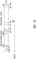
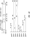
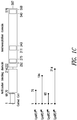
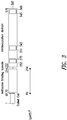
- the resulting proteomic data may be integrated into so-called peptographs, which plot, in the left panel, sequence coverage for a given protein in the horizontal dimension (N to C terminus, left to right) versus SDS-PAGE migration in the vertical dimension (high to low molecular weight, top to bottom).
- the specific peptide fragments can then be sequenced or mapped.
- the AARS reference fragment may be characterized by its unique molecular weight, as compared, for example, to the molecular weight of the corresponding full-length AARS.
- the AARS polypeptides set forth in Table(s) 1-3, or Table(s) 4-6, or Table(s) 7-9, or Table(s) 10-12 are disclosed. Also disclosed are “variants” of the AARS reference polypeptides.
- the recitation polypeptide "variant” refers to polypeptides that are distinguished from a reference AARS polypeptide by the addition, deletion, and/or substitution of at least one amino acid residue, and which typically retain ( e.g., mimic) or modulate ( e.g., antagonize) one or more non-canonical activities of a reference AARS polypeptide.
- human Lysyl tRNA synthetases include several hundred highly related polymorphic forms, and these are known in the art to be at least partially functionally interchangeable. It would thus be a routine matter to select a naturally occurring variant of Lysyl tRNA synthetase, including, for example the single nucleotide polymorphic forms listed in Table A to create an AARS polypeptide containing one or more amino acid changes based on the sequence of any of the homologues, orthologs, and naturally-occurring isoforms of human as well as other species of Lysyl tRNA synthetase.
- a polypeptide variant may be distinguished from a reference polypeptide by one or more substitutions, which may be conservative or non-conservative, as described herein and known in the art.
- the polypeptide variant may comprise conservative substitutions and, in this regard, it is well understood in the art that some amino acids may be changed to others with broadly similar properties without changing the nature of the activity of the polypeptide.
- a variant polypeptide may include an amino acid sequence having at least about 50%, 55%, 60%, 65%, 70%, 75%, 80%, 85%, 90%, 91%, 92%, 93%, 94%, 95%, 96%, 97%, 98% or more sequence identity or similarity to a corresponding sequence of an AARS reference polypeptide, as described herein, and substantially retains the non-canonical activity of that reference polypeptide. Also included are sequences differing from the reference AARS sequences by the addition, deletion, or substitution of 1, 2, 3, 4, 5, 6, 7, 8, 9, 10, 11, 12, 13, 14, 15, 16, 17, 18, 19, 20, 30, 40, 50, 60,70, 80, 90, 100, 110, 120, 130, 140, 150 or more amino acids but which retain the properties of the reference AARS polypeptide.
- the amino acid additions or deletions may occur at the C-terminal end and/or the N-terminal end of the AARS reference polypeptide.
- the amino acid additions may include 1, 2, 3, 4, 5, 6, 7, 8, 9, 10, 11, 12, 13, 14, 15, 16, 17, 18, 19, 20, 30, 40, 50 or more wild-type residues ( i.e., from the corresponding full-length AARS polypeptide) that are proximal to the C-terminal end and/or the N-terminal end of the AARS reference polypeptide.
- Variant polypeptides may differ from the corresponding AARS reference sequences by at least 1% but less than 20%, 15%, 10% or 5% of the residues. (If this comparison requires alignment, the sequences should be aligned for maximum similarity. "Looped" out sequences from deletions or insertions, or mismatches, are considered differences.) The differences are, suitably, differences or changes at a non-essential residue or a conservative substitution.
- the molecular weight of a variant AARS polypeptide may differ from that of the AARS reference polypeptide by about 1%, 2%, 3%, 4%, 5%, 6%, 7%, 8%, 9%, 10%, 11%, 12%, 13%, 14%, 15%, 16%, 17%, 18%, 19%, 20%, or more.
- biologically active fragments of the AARS reference polypeptides, i.e., biologically active fragments of the AARS protein fragments.
- Representative biologically active fragments generally participate in an interaction, e.g., an intramolecular or an inter-molecular interaction.
- An inter-molecular interaction can be a specific binding interaction or an enzymatic interaction.
- An inter-molecular interaction can be between an AARS polypeptide and a cellular binding partner, such as a cellular receptor or other host molecule that participates in the non-canonical activity of the AARS polypeptide.
- AARS proteins, variants, and biologically active fragments thereof may bind to one or more cellular binding partners with an affinity of at least about 0.01, 0.05, 0.1, 0.2, 0.3, 0.4, 0.5, 0.6, 0.7, 0.8, 0.9, 1, 2, 3, 4, 5, 6, 7, 8, 9, 10, 11, 12, 13, 14, 15, 16, 17, 18, 19, 20, 21, 22, 23, 24, 25, 26, 27, 28, 29, 30, 40, or 50 nM.
- the binding affinity of an AARS protein fragment for a selected cellular binding partner, particularly a binding partner that participates in a non-canonical activity, is typically stronger than that of the AARS protein fragment's corresponding full-length AARS polypeptide, by at least about 1.5x, 2x, 2.5x, 3x, 3.5x, 4x, 4.5x, 5x, 6x, 7x, 8x, 9x, 10x, 15x, 20x, 25x, 30x, 40x, 50x, 60x, 70x, 80x, 90x, 100x, 200x, 300x, 400x, 500x, 600x, 700x, 800x, 900x, 1000x or more (including all integers in between).
- the binding affinity of an AARS protein fragment for a binding partner that participates in at least one canonical activity of an AARS is typically weaker than that of the AARS protein fragment's corresponding full-length AARS polypeptide, by at least about 1.5x, 2x, 2.5x, 3x, 3.5x, 4x, 4.5x, 5x, 6x, 7x, 8x, 9x, 10x, 15x, 20x, 25x, 30x, 40x, 50x, 60x, 70x, 80x, 90x, 100x, 200x, 300x, 400x, 500x, 600x, 700x, 800x, 900x, 1000x or more.
- biologically active fragments comprise a domain or motif with at least one activity of an AARS reference polypeptide and may include one or more (and in some cases all) of the various active domains, and include fragments having a non-canonical activity.
- biologically active fragments of an AARS polypeptide have a biological activity that is unique to the particular, truncated fragment, such that the full-length AARS polypeptide may not have that activity.
- the biological activity may be revealed by separating the biologically active AARS polypeptide fragment from the other full-length AARS polypeptide sequences, or by altering certain residues of the full-length AARS wild-type polypeptide sequence to unmask the biologically active domains.
- a biologically active fragment of an AARS reference polypeptide can be a polypeptide fragment which is, for example, 10, 11, 12, 13, 14, 15, 16, 17, 18, 19, 20, 21, 22, 23, 24, 25, 26, 27, 28, 29, 30, 35, 40, 45, 50, 55, 60, 65, 70, 75, 80, 85, 90, 95, 100, 110, 120, 130, 140, 150, 160, 170, 180, 190, 200, 220, 240, 260, 280, 300, 320, 340, 360, 380, 400, 450, 500, 550, 600, 650, 700, 750 or more contiguous or non-contiguous amino acids, including all integers ( e.g., 101, 102, 103) and ranges ( e.g., 50-100, 50-150, 50-200) in between, of the amino acid sequences set forth in any one of the AARS reference polypeptides described herein, but typically exclude the full-length AARS.
- a biologically active fragment may comprise a non-canonical activity-related sequence, domain, or motif
- the C-terminal or N-terminal region of any AARS reference polypeptide may be truncated by about 1, 2, 3, 4, 5, 6, 7, 8, 9, 10, 15, 20, 25, 30, 35, 40, 45, 50, 60, 70, 80, 90, 100, 110, 120, 130, 140, 150, 160, 170, 180, 190, 200, 250, 300, 350, 400, 450, 500, 550, 600, 650, or 700 or more amino acids, or by about 10-50, 20-50, 50-100, 100-150, 150-200, 200-250, 250-300, 300-350, 350-400, 400-450, 450-500, 500-550, 550-600, 600-650, 650-700 or more amino acids, including all integers and ranges in between ( e.g., 101, 102, 103, 104, 105), so long as the truncated AARS polypeptide retains the non-canon
- the biologically-active fragment has no less than about 1%, about 5 %, about 10%, about 25%, or about 50% of an activity of the biologically-active (i.e., non-canonical activity) AARS reference polypeptide from which it is derived. Exemplary methods for measuring such non-canonical activities are described in the Examples.
- an AARS polypeptide may be altered in various ways including amino acid substitutions, deletions, truncations, and insertions. Methods for such manipulations are generally known in the art.
- amino acid sequence variants of an AARS reference polypeptide can be prepared by mutations in the DNA. Methods for mutagenesis and nucleotide sequence alterations are well known in the art. See, for example, Kunkel (1985, Proc. Natl. Acad. Sci. USA. 82: 488-492 ), Kunkel et al., (1987, Methods in Enzymol, 154: 367-382 ), U.S. Pat. No. 4,873,192 , Watson, J. D.
- Biologically active truncated and/or variant AARS polypeptides may contain conservative amino acid substitutions at various locations along their sequence, as compared to a reference AARS amino acid residue.
- naturally occurring variants of AARS proteins have been sequenced, and are known in the art to be at least partially functionally interchangeable. It would thus be a routine matter to select an amino acid position to introduce a conservative, or non conservative mutation into an AARS polypeptide based on naturally occurring sequence variation among the known AARS protein homologues, orthologs, and naturally-occurring isoforms of human as well as other species of an AARS protein.
- a “conservative amino acid substitution” is one in which the amino acid residue is replaced with an amino acid residue having a similar side chain. Families of amino acid residues having similar side chains have been defined in the art, which can be generally sub-classified as follows:
- proline This description also characterizes certain amino acids as “small” since their side chains are not sufficiently large, even if polar groups are lacking, to confer hydrophobicity.
- "small” amino acids are those with four carbons or less when at least one polar group is on the side chain and three carbons or less when not.
- Amino acids having a small side chain include glycine, serine, alanine and threonine.
- the gene-encoded secondary amino acid proline is a special case due to its known effects on the secondary conformation of peptide chains.
- the structure of proline differs from all the other naturally-occurring amino acids in that its side chain is bonded to the nitrogen of the ⁇ -amino group, as well as the ⁇ -carbon.
- the degree of attraction or repulsion required for classification as polar or nonpolar is arbitrary and, therefore, amino acids specifically contemplated by the invention have been classified as one or the other. Most amino acids not specifically named can be classified on the basis of known behavior.
- Amino acid residues can be further sub-classified as cyclic or non-cyclic, and aromatic or non-aromatic, self-explanatory classifications with respect to the side-chain substituent groups of the residues, and as small or large.
- the residue is considered small if it contains a total of four carbon atoms or less, inclusive of the carboxyl carbon, provided an additional polar substituent is present; three or less if not.
- Small residues are, of course, always non-aromatic.
- amino acid residues may fall in two or more classes. For the naturally-occurring protein amino acids, sub-classification according to this scheme is presented in Table B.
- Table B Amino acid sub-classification Sub - classes Amino acids Acidic Aspartic acid, Glutamic acid Basic Noncyclic: Arginine, Lysine; Cyclic: Histidine Charged Aspartic acid, Glutamic acid, Arginine, Lysine, Histidine Small Glycine, Serine, Alanine, Threonine, Proline Polar/neutral Asparagine, Histidine, Glutamine, Cysteine, Serine, Threonine Polar/large Asparagine, Glutamine Hydrophobic Tyrosine, Valine, Isoleucine, Leucine, Methionine, Phenylalanine, Tryptophan Aromatic Tryptophan, Tyrosine, Phenylalanine Residues that Glycine and Proline influence chain orientation
- Conservative amino acid substitution also includes groupings based on side chains.
- a group of amino acids having aliphatic side chains is glycine, alanine, valine, leucine, and isoleucine; a group of amino acids having aliphatic-hydroxyl side chains is serine and threonine; a group of amino acids having amide-containing side chains is asparagine and glutamine; a group of amino acids having aromatic side chains is phenylalanine, tyrosine, and tryptophan; a group of amino acids having basic side chains is lysine, arginine, and histidine; and a group of amino acids having sulfur-containing side chains is cysteine and methionine.
- Amino acid substitutions falling within the scope of the invention are, in general, accomplished by selecting substitutions that do not differ significantly in their effect on maintaining (a) the structure of the peptide backbone in the area of the substitution, (b) the charge or hydrophobicity of the molecule at the target site, (c) the bulk of the side chain, or (d) the biological function. After the substitutions are introduced, the variants are screened for biological activity.
- similar amino acids for making conservative substitutions can be grouped into three categories based on the identity of the side chains.
- the first group includes glutamic acid, aspartic acid, arginine, lysine, histidine, which all have charged side chains;
- the second group includes glycine, serine, threonine, cysteine, tyrosine, glutamine, asparagine;
- the third group includes leucine, isoleucine, valine, alanine, proline, phenylalanine, tryptophan, methionine, as described in Zubay, G., Biochemistry, third edition, Wm.C. Brown Publishers (1993).
- a predicted non-essential amino acid residue in a truncated and/or variant AARS polypeptide is typically replaced with another amino acid residue from the same side chain family.
- mutations can be introduced randomly along all or part of an AARS coding sequence, such as by saturation mutagenesis, and the resultant mutants can be screened for an activity of the parent polypeptide to identify mutants which retain that activity.
- the encoded peptide can be expressed recombinantly and the activity of the peptide can be determined.
- a "non-essential" amino acid residue is a residue that can be altered from the reference sequence of a polypeptide without abolishing or substantially altering one or more of its activities.
- the alteration does not substantially abolish one of these activities, for example, the activity is at least 20%, 40%, 60%, 70% or 80% 100%, 500%, 1000% or more of the reference AARS sequence.
- An "essential" amino acid residue is a residue that, when altered from the reference sequence of an AARS polypeptide, results in abolition of an activity of the parent molecule such that less than 20% of the reference activity is present.
- such essential amino acid residues include those that are conserved in AARS polypeptides across different species, including those sequences that are conserved in the active binding site(s) or motif(s) of AARS polypeptides from various sources.
- polypeptides and fusion polypeptides are isolated.
- An "isolated" polypeptide or polynucleotide is one that is removed from its original environment.
- a naturally-occurring protein is isolated if it is separated from some or all of the coexisting materials in the natural system.
- polypeptides are at least about 90% pure, more preferably at least about 95% pure and most preferably at least about 99% pure.
- a polynucleotide is considered to be isolated if, for example, it is cloned into a vector that is not a part of the natural environment.
- Dimers may include, for example, homodimers between two identical AARS polypeptides, heterodimers between two different AARS polypeptides (e.g., a full-length YRS polypeptide and a truncated YRS polypeptide; a truncated YRS polypeptide and a truncated WRS polypeptide), and/or heterodimers between an AARS polypeptide and a heterologous polypeptide.
- Certain heterodimers, such as those between an AARS polypeptide and a heterologous polypeptide may be bi-functional, as described herein.
- AARS polypeptides include isolated AARS polypeptides monomers that do not substantially dimerize with a second AARS polypeptide, whether due to one or more substitutions, truncations, deletions, additions, chemical modifications, or a combination of these alterations.
- Monomeric AARS polypeptides may possess biological activities, including non-canonical activities, which are not possessed by dimeric or multimeric AARS polypeptide complexes.
- modified AARS polypeptides including modifications that improved the desired characteristics of an AARS polypeptide, as described herein.
- Modifications of AARS polypeptides include chemical and/or enzymatic derivatizations at one or more constituent amino acid, including side chain modifications, backbone modifications, and N- and C-terminal modifications including acetylation, hydroxylation, methylation, amidation, and the attachment of carbohydrate or lipid moieties, cofactors, and the like.
- Exemplary modifications also include pegylation of an AARS polypeptide ( see, e.g., Veronese and Harris, Advanced Drug Delivery Reviews 54: 453-456, 2002 ; and Pasut et al., Expert Opinion. Ther. Patents 14(6) 859-894 2004 ).
- PEG is a well-known polymer having the properties of solubility in water and in many organic solvents, lack of toxicity, and lack of immunogenicity. It is also clear, colorless, odorless, and chemically stable. For these reasons and others, PEG has been selected as the preferred polymer for attachment, but it has been employed solely for purposes of illustration and not limitation.
- Similar products may be obtained with other water-soluble polymers, including without limitation; polyvinyl alcohol, other poly(alkylene oxides) such as poly(propylene glycol) and the like, poly(oxyethylated polyols) such as poly(oxyethylated glycerol) and the like, carboxymethylcellulose, dextran, polyvinyl alcohol, polyvinyl purrolidone, poly-1,3- dioxolane, poly-1,3,6-trioxane, ethylene/maleic anhydride, and polyaminoacids.
- polyvinyl alcohol other poly(alkylene oxides) such as poly(propylene glycol) and the like
- poly(oxyethylated polyols) such as poly(oxyethylated glycerol) and the like
- carboxymethylcellulose carboxymethylcellulose
- dextran polyvinyl alcohol
- polyvinyl purrolidone poly-1,3- dioxolane
- PEG derivatives are both available and suitable for use in the preparation of PEG-conjugates.
- NOF Corp.'s PEG reagents sold under the trademark SUNBRIGHT® Series provides numerous PEG derivatives, including methoxypolyethylene glycols and activated PEG derivatives such as methoxy-PEG amines, maleimides, N-hydroxysuccinimide esters, and carboxylic acids, for coupling by various methods to the N-terminal, C-terminal or any internal amino acid of the AARS polypeptide.
- Nektar Therapeutics' Advanced PEGylation technology also offers diverse PEG-coupling technologies to potentially improve the safety and efficacy of an AARS polypeptide based therapeutic.
- chemoselective ligation technology may be utilized to modify AARS polypeptides, such as by attaching polymers in a site-specific and controlled manner.
- Such technology typically relies on the incorporation of chemoselective anchors into the protein backbone by either chemical, or recombinant means, and subsequent modification with a polymer carrying a complementary linker.
- the assembly process and the covalent structure of the resulting protein-polymer conjugate may be controlled, enabling the rational optimization of drug properties, such as efficacy and pharmacokinetic properties ( see, e.g., Kochendoerfer, Current Opinion in Chemical Biology 9:555-560, 2005 ).
- Fusion proteins of AARS polypeptide to other proteins are also disclosed, and these fusion proteins may increase the AARS polypeptide's biological activity, secretion, targeting, biological life, ability to penetrate cellular membranes, or the blood brain barrier, or pharmacokinetic properties.
- fusion proteins that improve pharmacokinetic properties include without limitation, fusions to human albumin ( Osborn et al.: Eur. J. Pharmacol. 456(1-3): 149-158, (2002 )), antibody Fc domains, poly Glu or poly Asp sequences, and transferrin.
- fusion with conformationally disordered polypeptide sequences composed of the amino acids Pro, Ala, and Ser ('PASylation') or hydroxyethyl starch (sold under the trademark HESYLATION®) provides a simple way to increase the hydrodynamic volume of the AARS polypeptide.
- This additional extension adopts a bulky random structure, which significantly increases the size of the resulting fusion protein. By this means the typically rapid clearance of smaller AARS polypeptides via kidney filtration is retarded by several orders of magnitude.
- Ig G fusion proteins has also been shown to enable some fusion protein proteins to penetrate the blood brain barrier ( Fu et al., (2010) Brain Res. 1352:208-13 ).
- membrane translocating sequences refers to naturally occurring and synthetic amino acid sequences that are capable of membrane translocation across a cellular membrane.
- Representative membrane translocating sequences include those based on the naturally occurring membrane translocating sequences derived from the Tat protein, and homeotic transcription protein Antennapedia, as well as synthetic membrane translocating sequences based in whole or part on poly Arginine and Lysine resides.
- Representative membrane translocating sequences include for example those disclosed in the following patents, US5,652,122 ; US 5,670,617 ; US5,674,980 ; US5,747,641 ; US5,804,604 ; US6,316,003 ; US7,585,834 ; US7,312,244 ; US7,279,502 ; US7,229,961 ; US7,169,814 ; US7,453,011 ; US7,235,695 ; US6,982,351 ; US6,605,115 ; US7,306,784 ; US7,306,783 ; US6,589,503 ; US6,348,185 ; US6,881,825 ; US7,431,915 ; WO0074701A2 ; WO2007111993A2 ; WO2007106554A2 ; WO02069930A1 ; WO03049772A2 ; WO03106491A
- a flexible molecular linker optionally may be interposed between, and covalently join, the AARS polypeptide and any of the fusion proteins disclosed herein.
- the AARS polypeptide can also include synthetic, or naturally occurring secretion signal sequences, derived from other well characterized secreted proteins. Such proteins, may be processed by proteolytic cleavage to form the AARS polypeptide in situ. Such fusions proteins include for example fusions of AARS polypeptide to ubiquitin to provide a new N-terminal amino acid, or the use of a secretion signal to mediate high level secretion of the AARS polypeptide into the extracellular medium, or N, or C-terminal epitope tags to improve purification or detection.
- polypeptides described herein may be prepared by any suitable procedure known to those of skill in the art, such as by recombinant techniques.
- polypeptides may be produced by direct peptide synthesis using solid-phase techniques ( Merrifield, J. Am. Chem. Soc. 85:2149-2154 (1963 )). Protein synthesis may be performed using manual techniques or by automation. Automated synthesis may be achieved, for example, using Applied Biosystems 431A Peptide Synthesizer (Perkin Elmer). Alternatively, various fragments may be chemically synthesized separately and combined using chemical methods to produce the desired molecule.
- bioassays that relate to the AARS protein fragments and related agents as therapeutic and diagnostic reagents. Examples include bioassays and analytical assays that measure purity, biological activity, affinity, solubility, pH, endotoxin levels, among others, many of which are described herein. Also included are assays that establish dose response curves and/or provide one or more bases for comparison between different batches of agents. Batch comparisons can be based on any one or more of chemical characterization, biological characterization, and clinical characterization.
- protein agents also included are methods of evaluating the potency, stability, pharmacokinetics, and immunogenicity of a selected agent. Among other uses, these and other methods can be used for lot releasing testing of biologic or chemical agents, including the AARS protein fragments, antibodies, binding agents, polynucleotides such as antisense agents and vectors, and others described herein.
- bioaffinity assays can be used to assess the binding affinity, for example, between an AARS protein fragment and a cellular binding partner, or between an AARS protein fragment and an antibody. Binding affinity can also be measured between an AARS protein fragment and an alternate binding agent such as a candidate or lead test compound (e.g., small molecule modulator of an AARS), or between an AARS cellular binding partner and a candidate or lead test compound.
- a candidate or lead test compound e.g., small molecule modulator of an AARS
- Certain exemplary binding affinity assays may utilize ELISA assays, as described herein and known in the art. Certain assays utilize high-performance receptor binding chromatography ( see, e.g., Roswall et al., Biologicals. 24:25-39, 1996 ).
- binding affinity assays may utilize surface plasmon resonance (SPR)-based technologies.
- SPR surface plasmon resonance
- BIACore technologies certain of which integrate SPR technology with a microfluidics system to monitor molecular interactions in real time at concentrations ranging from pM to mM.
- KINEXATM assays also included are KINEXATM assays, which provide accurate measurements of binding specificity, binding affinity, and binding kinetics/rate constants.
- immunoassays for evaluating or optimizing the immunogenicity of protein agents.
- examples include ex vivo human cellular assays and in vitro immuno-enzymatic assays to provide useful information on the immunogenic potential of a therapeutic protein.
- Ex vivo cell-response assays can be used, for example, to reproduce the cellular co-operation between antigen-presenting cells (APCs) and T-cells, and thereby measure T-cells activation after contact with a protein of interest.
- APCs antigen-presenting cells
- Certain in vitro enzymatic assays may utilize a collection of recombinant HLA-DR molecules that cover a significant portion of a relevant human population, and may include automated immuno-enzymatic assays for testing the binding of peptides (stemming from the fragmentation of the therapeutic protein) with the HLA-DR molecules. Also included are methods of reducing the immunogenicity of a selected protein, such as by using these and related methods to identify and then remove or alter one or more T-cell epitopes from a protein agent.
- biological release assays e.g., cell-based assays
- biological release assays for measuring parameters such as specific biological activities, including non-canonical biological activities, and cytotoxicity.
- Certain specific biological assays include, for example, cell-based assays that utilize a cellular binding partner (e.g., cell-surface receptor) of a selected AARS protein fragment, which is functionally coupled to a readout, such as a fluorescent or luminescent indicator of a non-canonical biological activity, as described herein.
- a cell comprises a cell-surface receptor or an extracellular portion thereof that binds to an AARS protein fragment, wherein the cell comprises a detector or readout.
- an agent such as an AARS polypeptide or antibody
- engineered mice or other mammal see, e.g., Lee et al., The Journal of Pharmacology. 281:1431-1439, 1997 ).
- cytotoxicity-based biological assays include release assays (e.g., chromium or europium release assays to measure apoptosis; see, e.g., von Zons et al., Clin Diagn Lab Immunol.4:202-207, 1997 ), among others, which can assess the cytotoxicity of AARS protein fragments, whether for establishing dose response curves, batch testing, or other properties related to approval by various regulatory agencies, such as the Food and Drug Administration (FDA).
- release assays e.g., chromium or europium release assays to measure apoptosis; see, e.g., von Zons et al., Clin Diagn Lab Immunol.4:202-207, 1997
- FDA Food and Drug Administration
- a dose-response curve is an X-Y graph that relates the magnitude of a stressor to the response of a receptor; the response may be a physiological or biochemical response, such as a non-canonical biological activity in a cell in vitro or in a cell or tissue in vivo, a therapeutically effective amount as measured in vivo ( e.g., as measured by EC 50 ), or death, whether measured in vitro or in vivo ( e.g., cell death, organismal death).
- Death is usually indicated as an LD 50 , a statistically-derived dose that is lethal to 50% of a modeled population, though it can be indicated by LC 01 (lethal dose for 1% of the animal test population), LC 100 (lethal dose for 100% of the animal test population), or LC LO (lowest dose causing lethality). Almost any desired effect or endpoint can be characterized in this manner.
- the measured dose of a response curve is typically plotted on the X axis and the response is plotted on the Y axis. More typically, the logarithm of the dose is plotted on the X axis, most often generating a sigmoidal curve with the steepest portion in the middle.
- the No Observable Effect Level refers to the lowest experimental dose for which no measurable effect is observed, and the threshold dose refers to the first point along the graph that indicates a response above zero.
- NOEL No Observable Effect Level
- stronger drugs generate steeper dose response curves.
- the desired effects are found at doses slightly greater than the threshold dose, often because lower doses are relatively ineffective and higher doses lead to undesired side effects.
- a curve can be characterized by values such as ⁇ g/kg, mg/kg, or g/kg of body-weight, if desired.
- CV coefficient of variation
- different dose response curves of different batches e.g., between different batches of AARS protein fragments, antibodies, or other agents
- CV allows comparison between data sets with different units or different means.
- two or three or more different batches of AARS protein fragments or other agents may have a CV between them of less than about 15%, 14%, 13%, 12%, 11%, 10%, 9%, 8%, 7%, 6%, 5%, 4%, 3%, 2%, or 1% for a 4, 5, 6, 7, or 8 point dose curve.
- the dose response curve can be measured in a cell-based assay, and its readout relates to an increase or a decrease in a selected non-canonical activity of the AARS protein fragment.
- the dose response curve can be measured in a cell release assay or animal model (e.g., mouse model), and its readout relates to cell death or animal death.
- animal model e.g., mouse model
- AARS polypeptides can be conveniently prepared using standard protocols as described for example in Sambrook, et al ., (1989, supra ), in particular Sections 16 and 17; Ausubel et al ., (1994, supra ), in particular Chapters 10 and 16; and Coligan et al., Current Protocols in Protein Science (John Wiley & Sons, Inc. 1995-1997 ), in particular Chapters 1, 5 and 6.
- AARS polypeptides may be prepared by a procedure including one or more of the steps of: (a) preparing a construct comprising a polynucleotide sequence that encodes a AARS polypeptide and that is operably linked to a regulatory element; (b) introducing the construct into a host cell; (c) culturing the host cell to express the AARS polypeptide; and (d) isolating the AARS polypeptide from the host cell.
- AARS polynucleotides are described elsewhere herein.
- a nucleotide sequence encoding the polypeptide, or a functional equivalent may be inserted into appropriate expression vector, i.e., a vector which contains the necessary elements for the transcription and translation of the inserted coding sequence.
- appropriate expression vector i.e., a vector which contains the necessary elements for the transcription and translation of the inserted coding sequence.
- Methods which are well known to those skilled in the art may be used to construct expression vectors containing sequences encoding a polypeptide of interest and appropriate transcriptional and translational control elements. These methods include in vitro recombinant DNA techniques, synthetic techniques, and in vivo genetic recombination. Such techniques are described in Sambrook et al., Molecular Cloning, A Laboratory Manual (1989 ), and Ausubel et al., Current Protocols in Molecular Biology (1989 ).
- a variety of expression vector/host systems are known and may be utilized to contain and express polynucleotide sequences. These include, but are not limited to, microorganisms such as bacteria transformed with recombinant bacteriophage, plasmid, or cosmid DNA expression vectors; yeast transformed with yeast expression vectors; insect cell systems infected with virus expression vectors (e.g. , baculovirus); plant cell systems transformed with virus expression vectors (e.g ., cauliflower mosaic virus, CaMV; tobacco mosaic virus, TMV) or with bacterial expression vectors (e.g., Ti or pBR322 plasmids); or animal cell systems, including mammalian cell and more specifically human cell systems.
- microorganisms such as bacteria transformed with recombinant bacteriophage, plasmid, or cosmid DNA expression vectors
- yeast transformed with yeast expression vectors insect cell systems infected with virus expression vectors (e.g. , baculovirus)
- control elements or "regulatory sequences" present in an expression vector are those non-translated regions of the vector--enhancers, promoters, 5' and 3' untranslated regions--which interact with host cellular proteins to carry out transcription and translation. Such elements may vary in their strength and specificity. Depending on the vector system and host utilized, any number of suitable transcription and translation elements, including constitutive and inducible promoters, may be used.
- inducible promoters such as the hybrid lacZ promoter of the PBLUESCRIPT phagemid (Stratagene, La Jolla, Calif.) or PSPORT1 plasmid (Gibco BRL, Gaithersburg, Md.) and the like may be used.
- promoters from mammalian genes or from mammalian viruses are generally preferred. If it is necessary to generate a cell line that contains multiple copies of the sequence encoding a polypeptide, vectors based on SV40 or EBV may be advantageously used with an appropriate selectable marker.
- a number of expression vectors may be selected depending upon the use intended for the expressed polypeptide. For example, when large quantities are needed, vectors which direct high level expression of fusion proteins that are readily purified may be used.
- vectors include, but are not limited to, the multifunctional E. coli cloning and expression vectors such as BLUESCRIPT (Stratagene), in which the sequence encoding the polypeptide of interest may be ligated into the vector in frame with sequences for the amino-terminal Met and the subsequent 7 residues of ⁇ -galactosidase so that a hybrid protein is produced; pIN vectors ( Van Heeke & Schuster, J. Biol. Chem.
- pGEX Vectors may also be used to express foreign polypeptides as fusion proteins with glutathione S-transferase (GST).
- GST glutathione S-transferase
- fusion proteins are soluble and can easily be purified from lysed cells by adsorption to glutathione-agarose beads followed by elution in the presence of free glutathione.
- Proteins made in such systems may be designed to include heparin, thrombin, or factor XA protease cleavage sites so that the cloned polypeptide of interest can be released from the GST moiety at will.
- E. coli -based expression systems see, e.g., Structural Genomics Consortium et al., Nature Methods. 5:135-146, 2008 ) may be employed. These and related approaches may rely partially or totally on ligation-independent cloning (LIC) to produce a suitable expression vector.
- Protein expression may be controlled by a T7 RNA polymerase (e.g., pET vector series).
- T7 RNA polymerase e.g., pET vector series
- the expression host strain BL21(DE3) may be utilized, a ⁇ DE3 lysogen of BL21 that supports T7-mediated expression and is deficient in lon and ompT proteases for improved target protein stability. Also included are expression host strains carrying plasmids encoding tRNAs rarely used in E.
- coli such as ROSETTATM (DE3) and Rosetta 2 (DE3) strains.
- Cell lysis and sample handling may also be improved using reagents sold under the trademarks BENZONASE® nuclease and BUGBUSTER® Protein Extraction Reagent.
- BENZONASE® nuclease e.g., BENZONASE® nuclease
- BUGBUSTER® Protein Extraction Reagent reagents sold under the trademarks BENZONASE® nuclease and BUGBUSTER® Protein Extraction Reagent.
- auto-inducing media can improve the efficiency of many expression systems, including high-throughput expression systems.
- Media of this type e.g., OVERNIGHT EXPRESSTM Autoinduction System
- Hexahistidine tags such as those sold under the trademark HIS•TAG® fusions
- IMAC immobilized metal affinity chromatography
- clinical grade proteins can be isolated from E. coli inclusion bodies, without or without the use of affinity tags (see, e.g., Shimp et al., Protein Expr Purif. 50:58-67, 2006 ).
- affinity tags see, e.g., Shimp et al., Protein Expr Purif. 50:58-67, 2006 .
- a cold-shock induced E. coli high-yield production system may be employed, because over-expression of proteins in Escherichia coli at low temperature improves their solubility and stability ( see, e.g., Qing et al., Nature Biotechnology. 22:877-882, 2004 ).
- high-density bacterial fermentation systems For example, high cell density cultivation of Ralstonia eutropha allows protein production at cell densities of over 150 g/L, and the expression of recombinant proteins at titers exceeding 10 g/L.
- yeast Saccharomyces cerevisiae a number of vectors containing constitutive or inducible promoters such as alpha factor, alcohol oxidase, and PGH may be used.
- constitutive or inducible promoters such as alpha factor, alcohol oxidase, and PGH
- PGH palladium phosphate
- Pichia pandoris expression systems see, e.g., Li et al., Nature Biotechnology. 24, 210 - 215, 2006 ; and Hamilton et al., Science, 301:1244, 2003 ).
- yeast systems that are engineered to selectively glycosylate proteins, including yeast that have humanized N-glycosylation pathways, among others (see, e.g., Hamilton et al., Science. 313:1441-1443, 2006 ; Wildt et al., Nature Reviews Microbiol. 3:119-28, 2005 ; and Gerngross et al., Nature-Biotechnology. 22:1409 -1414, 2004 ; U.S. Patent Nos. 7,629,163 ; 7,326,681 ; and 7,029,872 ).
- recombinant yeast cultures can be grown in Fernbach Flasks or 15L, 50L, 100L, and 200L fermentors, among others.
- sequences encoding polypeptides may be driven by any of a number of promoters.
- viral promoters such as the 35S and 19S promoters of CaMV may be used alone or in combination with the omega leader sequence from TMV ( Takamatsu, EMBO J. 3:1671-1680 (1984 ); Broglie et al., Science 224:838-843 (1984 ); and Winter et al., Results Probl. Cell Differ. 17:85-105 (1991 )).
- constructs can be introduced into plant cells by direct DNA transformation or pathogen-mediated transfection. Such techniques are described in a number of generally available reviews (see, e.g., Hobbs in McGraw Hill, Yearbook of Science and Technology, pp. 191-196 (1992 )).
- An insect system may also be used to express a polypeptide of interest.
- Autographa californica nuclear polyhedrosis virus (AcNPV) is used as a vector to express foreign genes in Spodoptera frugiperda cells or in Trichoplusia cells.
- the sequences encoding the polypeptide may be cloned into a non-essential region of the virus, such as the polyhedrin gene, and placed under control of the polyhedrin promoter. Successful insertion of the polypeptide-encoding sequence will render the polyhedrin gene inactive and produce recombinant virus lacking coat protein.
- the recombinant viruses may then be used to infect, for example, S.
- frugiperda cells or Trichoplusia cells in which the polypeptide of interest may be expressed Engelhard et al., Proc. Natl. Acad. Sci. U.S.A. 91:3224-3227 (1994 )). Also included are baculovirus expression systems, including those that utilize SF9, SF21, and T. ni cells ( see, e.g., Murphy and Piwnica-Worms, Curr Protoc Protein Sci. Chapter 5:Unit5.4, 2001 ). Insect systems can provide post-translation modifications that are similar to mammalian systems.
- a number of viral-based expression systems are generally available.
- sequences encoding a polypeptide of interest may be ligated into an adenovirus transcription/translation complex consisting of the late promoter and tripartite leader sequence. Insertion in a non-essential E1 or E3 region of the viral genome may be used to obtain a viable virus which is capable of expressing the polypeptide in infected host cells ( Logan & Shenk, Proc. Natl. Acad. Sci. U.S.A. 81:3655-3659 (1984 )).
- transcription enhancers such as the Rous sarcoma virus (RSV) enhancer, may be used to increase expression in mammalian host cells.
- RSV Rous sarcoma virus
- Examples of useful mammalian host cell lines include monkey kidney CV1 line transformed by SV40 (COS-7, ATCC CRL 1651); human embryonic kidney line (293 or 293 cells sub-cloned for growth in suspension culture, Graham et al., J. Gen Virol. 36:59 (1977 )); baby hamster kidney cells (BHK, ATCC CCL 10); mouse sertoli cells (TM4, Mather, Biol. Reprod.
- COS-7 monkey kidney CV1 line transformed by SV40
- human embryonic kidney line (293 or 293 cells sub-cloned for growth in suspension culture, Graham et al., J. Gen Virol. 36:59 (1977 )
- baby hamster kidney cells BHK, ATCC CCL 10
- mouse sertoli cells TM4, Mather, Biol. Reprod.
- monkey kidney cells (CV1 ATCC CCL 70); African green monkey kidney cells (VERO-76, ATCC CRL-1587); human cervical carcinoma cells (HELA, ATCC CCL 2); canine kidney cells (MDCK, ATCC CCL 34); buffalo rat liver cells (BRL 3A, ATCC CRL 1442); human lung cells (W138, ATCC CCL 75); human liver cells (Hep G2, HB 8065); mouse mammary tumor (MMT 060562, ATCC CCL51); TR1 cells ( Mather et al., Annals N.Y. Acad. Sci. 383:44-68 (1982 )); MRC 5 cells; FS4 cells; and a human hepatoma line (Hep G2).
- CHO Chinese hamster ovary
- DHFR-CHO cells Urlaub et al., PNAS USA 77:4216 (1980 )
- myeloma cell lines such as NSO and Sp2/0.
- CHO Chinese hamster ovary
- myeloma cell lines such as NSO and Sp2/0.
- Certain preferred mammalian cell expression systems include CHO and HEK293-cell based expression systems.
- Mammalian expression systems can utilize attached cell lines, for example, in T-flasks, roller bottles, or cell factories, or suspension cultures, for example, in 1L and 5L spinners, 5L, 14L, 40L, 100L and 200L stir tank bioreactors, or 20/50L and 100/200L WAVE bioreactors, among others known in the art.
- RNA polymerase typically utilize purified RNA polymerase, ribosomes, tRNA and ribonucleotides; these reagents may be produced by extraction from cells or from a cell-based expression system.
- Specific initiation signals may also be used to achieve more efficient translation of sequences encoding a polypeptide of interest. Such signals include the ATG initiation codon and adjacent sequences. In cases where sequences encoding the polypeptide, its initiation codon, and upstream sequences are inserted into the appropriate expression vector, no additional transcriptional or translational control signals may be needed. However, in cases where only coding sequence, or a portion thereof, is inserted, exogenous translational control signals including the ATG initiation codon should be provided. Furthermore, the initiation codon should be in the correct reading frame to ensure translation of the entire insert. Exogenous translational elements and initiation codons may be of various origins, both natural and synthetic. The efficiency of expression may be enhanced by the inclusion of enhancers which are appropriate for the particular cell system which is used, such as those described in the literature ( Scharf. et al., Results Probl. Cell Differ. 20:125-162 (1994 )).
- a host cell strain may be chosen for its ability to modulate the expression of the inserted sequences or to process the expressed protein in the desired fashion.
- modifications of the polypeptide include, but are not limited to, post-translational modifications such as acetylation, carboxylation, glycosylation, phosphorylation, lipidation, and acylation.
- Post-translational processing which cleaves a "prepro" form of the protein may also be used to facilitate correct insertion, folding and/or function.
- Different host cells such as yeast, CHO, HeLa, MDCK, HEK293, and W138, in addition to bacterial cells, which have or even lack specific cellular machinery and characteristic mechanisms for such post-translational activities, may be chosen to ensure the correct modification and processing of the foreign protein.
- cell lines which stably express a polynucleotide of interest may be transformed using expression vectors which may contain viral origins of replication and/or endogenous expression elements and a selectable marker gene on the same or on a separate vector. Following the introduction of the vector, cells may be allowed to grow for about 1-2 days in an enriched media before they are switched to selective media. The purpose of the selectable marker is to confer resistance to selection, and its presence allows growth and recovery of cells which successfully express the introduced sequences. Resistant clones of stably transformed cells may be proliferated using tissue culture techniques appropriate to the cell type. Transient production, such as by transient transfection or infection, can also be employed. Exemplary mammalian expression systems that are suitable for transient production include HEK293 and CHO-based systems.
- selection systems may be used to recover transformed or transduced cell lines. These include, but are not limited to, the herpes simplex virus thymidine kinase ( Wigler et al., Cell 11:223-232 (1977 )) and adenine phosphoribosyltransferase ( Lowy et al., Cell 22:817-823 (1990 )) genes which can be employed in tk- or aprt- cells, respectively. Also, antimetabolite, antibiotic or herbicide resistance can be used as the basis for selection; for example, dhfr which confers resistance to methotrexate ( Wigler et al., Proc. Natl. Acad. Sci. U.S.A.
- npt which confers resistance to the aminoglycosides, neomycin and G-418 ( Colbere-Garapin et al., J. Mol. Biol. 150:1-14 (1981 )); and als or pat, which confer resistance to chlorsulfuron and phosphinotricin acetyltransferase, respectively (Murry, supra). Additional selectable genes have been described, for example, trpB, which allows cells to utilize indole in place of tryptophan, or hisD, which allows cells to utilize histinol in place of histidine ( Hartman & Mulligan, Proc. Natl. Acad. Sci. U.S.A.
- GFP green fluorescent protein
- RFP red fluorescent protein
- YFP green fluorescent protein
- anthocyanins e.g ., RFP, YFP
- anthocyanins e.g ., ⁇ -glucuronidase and its substrate GUS
- luciferase and its substrate luciferin being widely used not only to identify transformants, but also to quantify the amount of transient or stable protein expression attributable to a specific vector system ( see, e.g., Rhodes et al., Methods Mol. Biol. 55:121-131 (1995 )).
- High-throughput protein production systems or micro-production systems. Certain aspects may utilize, for example, hexa-histidine fusion tags for protein expression and purification on metal chelate-modified slide surfaces or MagneHis Ni-Particles ( see, e.g., Kwon et al., BMC Biotechnol. 9:72, 2009 ; and Lin et al., Methods Mol Biol. 498:129-41, 2009 )). Also included are high-throughput cell-free protein expression systems ( see, e.g., Sitaraman et al., Methods Mol Biol. 498:229-44, 2009 ). These and related approaches can be used, for example, to generate microarrays of AARS protein fragment(s), which can then be used for screening libraries to identify agents that interact with the AARS protein fragment(s).
- Means for producing labeled hybridization or PCR probes for detecting sequences related to polynucleotides include oligolabeling, nick translation, end-labeling or PCR amplification using a labeled nucleotide.
- the sequences, or any portions thereof may be cloned into a vector for the production of an mRNA probe.
- Such vectors are known in the art, are commercially available, and may be used to synthesize RNA probes in vitro by addition of an appropriate RNA polymerase such as T7, T3, or SP6 and labeled nucleotides.
- reporter molecules or labels include radionuclides, enzymes, fluorescent, chemiluminescent, or chromogenic agents as well as substrates, cofactors, inhibitors, magnetic particles, and the like.
- Host cells transformed with a polynucleotide sequence of interest may be cultured under conditions suitable for the expression and recovery of the protein from cell culture.
- Serum free cell expression systems may be utilized. Examples include HEK293 cells and CHO cells that can grown on serum free medium ( see, e.g., Rosser et al., Protein Expr. Purif. 40:237-43, 2005 ; and U.S. Patent number 6,210,922 ).
- the protein produced by a recombinant cell may be secreted or contained intracellularly depending on the sequence and/or the vector used.
- expression vectors containing polynucleotides may be designed to contain signal sequences which direct secretion of the encoded polypeptide through a prokaryotic or eukaryotic cell membrane.
- Other recombinant constructions may be used to join sequences encoding a polypeptide of interest to nucleotide sequence encoding a polypeptide domain which will facilitate purification and / or detection of soluble proteins.
- cleavable and non-cleavable affinity purification and epitope tags such as avidin, FLAG tags, poly-histidine tags (e.g., 6xHis), cMyc tags, V5-tags, glutathione S-transferase (GST) tags, and others.
- the protein produced by a recombinant cell can be purified and characterized according to a variety of techniques known in the art.
- Exemplary systems for performing protein purification and analyzing protein purity include fast protein liquid chromatography (FPLC) (e.g., AKTA and Bio-Rad FPLC systems), highpressure liquid chromatography (HPLC) (e.g., Beckman and Waters HPLC).
- FPLC fast protein liquid chromatography
- HPLC highpressure liquid chromatography
- Exemplary chemistries for purification include ion exchange chromatography (e.g., Q, S), size exclusion chromatography, salt gradients, affinity purification (e.g., Ni, Co, FLAG, maltose, glutathione, protein A/G), gel filtration, reverse-phase, ceramic HYPERD® ion exchange chromatography, and hydrophobic interaction columns (HIC), among others known in the art. Also included are analytical methods such as SDS-PAGE (e.g., coomassie, silver stain), immunoblot, Bradford, and ELISA, which may be utilized during any step of the production or purification process, typically to measure the purity of the protein composition.
- affinity purification e.g., Ni, Co, FLAG, maltose, glutathione, protein A/G
- gel filtration e.g., reverse-phase, ceramic HYPERD® ion exchange chromatography
- HIC hydrophobic interaction columns
- analytical methods such as SDS-PAGE (e.
- concentrated solutions of AARS polypeptides may comprise proteins at a concentration of about 5 mg/mL; or about 8 mg/mL; or about 10 mg/mL; about 15 mg/mL; or about 20 mg/mL.
- compositions may be substantially monodisperse, meaning that the AARS polypeptide compositions exist primarily ( i.e., at least about 90%, or greater) in one apparent molecular weight form when assessed for example, by size exclusion chromatography, dynamic light scattering, or analytical ultracentrifugation.
- compositions have a purity (on a protein basis) of at least about 90%, or in some aspects at least about 95% purity, or at least 98% purity. Purity may be determined via any routine analytical method as known in the art.
- compositions have a high molecular weight aggregate content of less than about 10%, compared to the total amount of protein present, or such compositions have a high molecular weight aggregate content of less than about 5%, or in some aspects such compositions have a high molecular weight aggregate content of less than about 3%, or a high molecular weight aggregate content of less than about 1%.
- High molecular weight aggregate content may be determined via a variety of analytical techniques including for example, by size exclusion chromatography, dynamic light scattering, or analytical ultracentrifugation.
- the AARS polypeptide compositions can have an endotoxin content of less than about 10 EU / mg of AARS polypeptide, or less than about 5 EU / mg of AARS polypeptide, less than about 3 EU / mg of AARS polypeptide, or less than about 1 EU / mg of AARS polypeptide.
- concentration approaches contemplated herein include lyophilization, which is typically employed when the solution contains few soluble components other than the protein of interest. Lyophilization is often performed after HPLC run, and can remove most or all volatile components from the mixture. Also included are ultrafiltration techniques, which typically employ one or more selective permeable membranes to concentrate a protein solution. The membrane allows water and small molecules to pass through and retains the protein; the solution can be forced against the membrane by mechanical pump, gas pressure, or centrifugation, among other techniques.
- the reagents, AARS protein fragments, or related agents can have a purity of at least about 90%, as measured according to routine techniques in the art.
- the disclosed AARS compositions have a purity of at least about 95%.
- the AARS compositions have a purity of at least about 97% or 98% or 99%.
- AARS protein fragments can be of lesser purity, and may have a purity of at least about 50%, 60%, 70%, or 80%. Purity can be measured overall or in relation to selected components, such as other proteins, e.g., purity on a protein basis.
- Purified AARS protein fragments can also be characterized according to their biological characteristics. Examples include binding affinity or binding kinetics to a selected ligand (e.g., a cellular binding partner of the AARS protein fragment such as a cell-surface receptor or an extracellular domain thereof), and the presence or levels of one or more canonical or non-canonical biological activity, as described herein. Binding affinity and binding kinetics can be measured according to a variety of techniques known in the art, such as Biacore® and related technologies that utilize surface plasmon resonance (SPR), an optical phenomenon that enables detection of unlabeled interactants in real time. SPR-based biosensors can be used in determination of active concentration, screening and characterization in terms of both affinity and kinetics.
- a selected ligand e.g., a cellular binding partner of the AARS protein fragment such as a cell-surface receptor or an extracellular domain thereof
- Binding affinity and binding kinetics can be measured according to a variety of techniques known in the art, such as Bi
- the presence or levels of one or more canonical or non-canonical biological activities can be measured according to cell-based assays, including those that utilize a cellular binding partner (e.g., cell-surface receptor) of a selected AARS protein fragment, which is functionally coupled to a readout or indicator, such as a fluorescent or luminescent indicator of a non-canonical biological activity, as described herein.
- a cellular binding partner e.g., cell-surface receptor
- a readout or indicator such as a fluorescent or luminescent indicator of a non-canonical biological activity, as described herein.
- the AARS polypeptide compositions can be about substantially endotoxin free, including, for example, about 95% endotoxin free, preferably about 99% endotoxin free, and more preferably about 99.99% endotoxin free.
- the presence of endotoxins can be detected according to routine techniques in the art, as described herein.
- the AARS compositions can be made from a eukaryotic cell such as a mammalian or human cell in substantially serum free media.
- the AARS polypeptide compositions can comprise less than about 10% wt/wt high molecular weight aggregates, or less than about 5% wt/wt high molecular weight aggregates, or less than about 2% wt/wt high molecular weight aggregates, or less than about or less than about 1% wt/wt high molecular weight aggregates.
- Protein-based analytical assays and methods which can be used to assess, for example, protein purity, size, solubility, and degree of aggregation, among other characteristics.
- Protein purity can be assessed a number of ways. For instance, purity can be assessed based on primary structure, higher order structure, size, charge, hydrophobicity, and glycosylation.
- methods for assessing primary structure include N- and C-terminal sequencing and peptide-mapping ( see, e.g., Allen et al., Biologicals. 24:255-275, 1996 )).
- methods for assessing higher order structure include circular dichroism ( see, e.g., Kelly et al., Biochim Biophys Acta.
- Hydrophobicity can be assessed, for example, by reverse-phase HPLC and hydrophobic interaction chromatography HPLC. Glycosylation can affect pharmacokinetics ( e.g., clearance), conformation or stability, receptor binding, and protein function, and can be assessed, for example, by mass spectrometry and nuclear magnetic resonance (NMR) spectroscopy.
- SEC-HPLC may be used to assess protein characteristics such as purity, size (e.g., size homogeneity) or degree of aggregation, and/or to purify proteins, among other uses.
- SEC also including gel-filtration chromatography (GFC) and gel-permeation chromatography (GPC), refers to a chromatographic method in which molecules in solution are separated in a porous material based on their size, or more specifically their hydrodynamic volume, diffusion coefficient, and/or surface properties. The process is generally used to separate biological molecules, and to determine molecular weights and molecular weight distributions of polymers.
- a biological or protein sample (such as a protein extract produced according to the protein expression methods provided herein and known in the art) is loaded into a selected size-exclusion column with a defined stationary phase (the porous material), preferably a phase that does not interact with the proteins in the sample.
- the stationary phase is composed of inert particles packed into a dense three-dimensional matrix within a glass or steel column.
- the mobile phase can be pure water, an aqueous buffer, an organic solvent, or a mixture thereof.
- the stationary-phase particles typically have small pores and/or channels which only allow molecules below a certain size to enter.
- Protein purity for clinical applications is also discussed, for example, by Anicetti et al. (Trends in Biotechnology. 7:342-349, 1989 ). More recent techniques for analyzing protein purity include, without limitation, the LabChip GXII, an automated platform for rapid analysis of proteins and nucleic acids, which provides high throughput analysis of titer, sizing, and purity analysis of proteins. Clinical grade proteins such as protein fragments and antibodies can be obtained by utilizing a combination of chromatographic materials in at least two orthogonal steps, among other methods ( see, e.g., Therapeutic Proteins: Methods and Protocols. Vol. 308, Eds., Smales and James, Humana Press Inc., 2005 ).
- protein agents e.g., AARS protein fragments, antibodies, binding agents
- other agents e.g., antisense, RNAi, small molecules
- protein agents e.g., AARS protein fragments, antibodies, binding agents
- other agents e.g., antisense, RNAi, small molecules
- Protein solubility assays are also disclosed. Such assays can be utilized, for example, to determine optimal growth and purification conditions for recombinant production, to optimize the choice of buffer(s), and to optimize the choice of AARS protein fragments or variants thereof. Solubility or aggregation can be evaluated according to a variety of parameters, including temperature, pH, salts, and the presence or absence of other additives. Examples of solubility screening assays include, without limitation, microplate-based methods of measuring protein solubility using turbidity or other measure as an end point, high-throughput assays for analysis of the solubility of purified recombinant proteins ( see, e.g., Stenvall et al., Biochim Biophys Acta.
- AARS protein fragments with increased solubility can be identified or selected for according to routine techniques in the art, including simple in vivo assays for protein solubility (see, e.g., Maxwell et al., Protein Sci. 8:1908-11, 1999 ).
- Protein solubility and aggregation can also be measured by dynamic light scattering techniques.
- Aggregation is a general term that encompasses several types of interactions or characteristics, including soluble/insoluble, covalent/noncovalent, reversible/irreversible, and native/denatured interactions and characteristics.
- the presence of aggregates is typically considered undesirable because of the concern that aggregates may cause an immunogenic reaction (e.g., small aggregates), or may cause adverse events on administration ( e.g., particulates).
- Dynamic light scattering refers to a technique that can be used to determine the size distribution profile of small particles in suspension or polymers such as proteins in solution.
- This technique also referred to as photon correlation spectroscopy (PCS) or quasi-elastic light scattering (QELS), uses scattered light to measure the rate of diffusion of the protein particles. Fluctuations of the scattering intensity can be observed due to the Brownian motion of the molecules and particles in solution.
- This motion data can be conventionally processed to derive a size distribution for the sample, wherein the size is given by the Stokes radius or hydrodynamic radius of the protein particle. The hydrodynamic size depends on both mass and shape (conformation). Dynamic scattering can detect the presence of very small amounts of aggregated protein ( ⁇ 0.01% by weight), even in samples that contain a large range of masses.
- certain approaches include the use of dynamic light scattering to analyze the solubility and/or presence of aggregates in a sample that contains an AARS protein fragment, antibody, or other agent disclosed herein.
- AARS agents such as AARS protein fragments, AARS polynucleotides, and antibodies and other binding agents described herein can be used in diagnostic assays and diagnostic compositions. Included are biochemical, histological, and cell-based methods and compositions, among others.
- AARS polypeptides include the detection of the AARS polynucleotide sequence(s) or corresponding AARS polypeptide sequence(s) or portions thereof of one or more newly identified AARS protein fragments, also referred to as AARS polypeptides.
- certain aspects include detection of the AARS polynucleotide sequence(s) or corresponding polypeptide sequence(s) or portions thereof of one or more newly identified AARS splice variants, and/or one or more splice junctions of those splice variants.
- the polynucleotide or corresponding polypeptide sequence(s) of at least one of the splice junctions can be unique to that particular AARS splice variant.
- AARS protein fragments including splice variants, proteolytic fragments, and others.
- the presence or levels of one or more newly identified AARS protein fragments can associate or correlate with one or more cellular types or cellular states.
- the presence or levels of an AARS polypeptide or polynucleotide can be used to distinguish between different cellular types or different cellular states.
- the presence or levels of AARS protein fragments or their related polynucleotides can be detected according to polynucleotide and/or polypeptide-based diagnostic techniques, as described herein and known in the art.
- Certain aspects can employ the AARS protein fragments, antibody, or AARS polynucleotides as part of a companion diagnostic method, typically to assess whether a subject or population subjects will respond favorably to a specific medical treatment.
- a given AARS therapeutic agent e.g., protein fragment, antisense, RNAi, antibody, binding agent
- biomarkers include serum/tissue markers as well as markers that can be identified by medical imaging techniques.
- a naturally-occurring AARS protein fragment may itself provide a serum and/or tissue biomarker that can be utilized to measure drug outcome or assess the desirability of drug use in a specific subject or a specific population of subjects.
- the identification of an AARS polypeptide or polynucleotide reference sequence may include characterizing the differential expression of that sequence, whether in a selected subject, selected tissue, or otherwise, as described herein and known in the art.
- Certain of the methods provided herein rely on the differential expression of an AARS polypeptide or polynucleotide to characterize the condition or state of a cell, tissue, or subject, and to distinguish it from another cell, tissue, or subject.
- Non-limiting examples include methods of detecting the presence or levels of an AARS polypeptide or polynucleotide in a biological sample to distinguish between cells or tissues of different species, cells of different tissues or organs, cellular developmental states such as neonatal and adult, cellular differentiation states, conditions such as healthy, diseased and treated, intracellular and extracellular fractions, in addition to primary cell cultures and other cell cultures, such as immortalized cell cultures.
- Differential expression includes a statistically significant difference in one or more gene expression levels of an AARS polynucleotide or polypeptide reference sequence compared to the expression levels of the same sequence in an appropriate control.
- the statistically significant difference may relate to either an increase or a decrease in expression levels, as measured by RNA levels, protein levels, protein function, or any other relevant measure of gene expression such as those described herein.
- Also included is a comparison between an AARS polynucleotide or polypeptide of the invention and a full-length or wild-type cytosolic or mitochondrial AARS sequence, typically of the same or corresponding type.
- Differential expression can be detected by a variety of techniques in the art and described herein, including polynucleotide and polypeptide based techniques, such as real-time PCR, subtractive hybridization, polynucleotide and polypeptide arrays, and others.
- a result is typically referred to as statistically significant if it is unlikely to have occurred by chance.
- the significance level of a test or result relates traditionally to a frequentist statistical hypothesis testing concept.
- statistical significance may be defined as the probability of making a decision to reject the null hypothesis when the null hypothesis is actually true (a decision known as a Type I error, or "false positive determination"). This decision is often made using the p-value: if the p-value is less than the significance level, then the null hypothesis is rejected. The smaller the p-value, the more significant the result.
- Bayes factors may also be utilized to determine statistical significance ( see, e.g., Goodman S., Ann Intern Med 130:1005-13, 1999 ).
- the significance level of a test or result may reflect an analysis in which the probability of making a decision to reject the null hypothesis when the null hypothesis is actually true is no more than the stated probability. This type of analysis allows for those applications in which the probability of deciding to reject may be much smaller than the significance level for some sets of assumptions encompassed within the null hypothesis.
- Statistically significant differential expression may include situations wherein the expression level of a given AARS sequence provides at least about a 1.2X, 1.3X, 1.4X, 1.5X, 1.6X, 1.7X, 1.8X, 1.9X, 2.0X, 2.2X, 2.4X, 2.6X, 2,8X, 3.0X, 4.0X, 5.0X, 6.0X, 7.0X, 8.0X, 9.0X, 10.0X, 15.0X, 20.0X, 50.0X, 100.0X, or greater difference in expression ( i.e., differential expression that may be higher or lower expression) in a suspected biological sample as compared to an appropriate control, including all integers and decimal points in between ( e.g.
- differential expression may include situations wherein the expression level of a given AARS sequence provides at least about 4, 5, 6, 7, 8, 9, 10, 11, 12, 13, 14, 15, 16, 17, 18, 19, 20, 25, 30, 35, 40, 45, 50, 60, 70, 80, 90, 100, 200, 300, 400, 500, 600, 700, 800, 900, 1000 percent (%) or greater difference in expression (i.e., differential expression that may be higher or lower) in a suspected biological sample as compared to an appropriate control, including all integers and decimal points in between.
- differential expression may also be determined by performing Z-testing, i.e., calculating an absolute Z score, as described herein and known in the art ( see Example 1).
- Z-testing is typically utilized to identify significant differences between a sample mean and a population mean. For example, as compared to a standard normal table (e.g., a control tissue), at a 95% confidence interval ( i.e., at the 5% significance level), a Z-score with an absolute value greater than 1.96 indicates non-randomness. For a 99% confidence interval, if the absolute Z is greater than 2.58, it means that p ⁇ .01, and the difference is even more significant-the null hypothesis can be rejected with greater confidence.
- An absolute Z-score of 1.96, 2, 2.58, 3, 4, 5, 6, 7, 8, 9, 10, 11, 12, 13, 14, 15, 16, 17, 18, 19, 20 or more, including all decimal points in between ( e.g., 10.1, 10.6, 11.2, etc.), may provide a strong measure of statistical significance.
- An absolute Z-score of greater than 6 may provide exceptionally high statistical significance.
- Substantial similarly relates generally to the lack of a statistically significant difference in the expression levels between the biological sample and the reference control.
- substantially similar expression levels may include situations wherein the expression level of a given SSCIGS provides less than about a .05X, 0.1X, 0.2X, 0.3X, 0.4X, 0.5X, 0.6X, 0.7X, 0.8X, 0.9X, 1.0X, 1.1X, 1.2X, 1.3X, or 1.4X difference in expression (i.e., differential expression that may be higher or lower expression) in a suspected biological sample as compared to a reference sample, including all decimal points in between ( e.g ., .15X, 0.25X, 0.35X, etc.).
- Differential expression may include situations wherein the expression level of a given AARS sequence provides less than about 0.25. 0.5, 1, 2, 3, 4, 5, 6, 7, 8, 9, 10, 11, 12, 13, 14, 15, 16, 17, 18, 19, 20, 30, 40, 50 percent (%) difference in expression (i.e., differential expression that may be higher or lower) in a suspected biological sample as compared to a reference sample, including all decimal points in between.
- differential expression may also be determined by the mean expression value summarized by Affymetrix Microarray Suite 5 software (Affymetrix, Santa Clara, CA), or other similar software, typically with a scaled mean expression value of 1000.
- General examples include methods of distinguishing between humans and any combination of bacteria, fungi, plants, and other non-human animals. Included within animals are methods of distinguishing between humans and any combination of vertebrates and invertebrates, including vertebrates such as fish, amphibians, reptiles, birds, and non-human mammals, and invertebrates such as insects, mollusks, crustaceans, and corals.
- non-human mammals are methods of distinguishing between humans and any combination of non-human mammals from the Order Afrosoricida, Macroscelidea, Tubulidentata, Hyracoidea, Proboscidea, Sirenia, Cingulata, Pilosa, Scandentia, Dermoptera, Primates, Rodentia, Lagomorpha, Erinaceomorpha, Soricomorpha, Chiroptera, Pholidota, Cetacea, Carnivora, Perissodactyla, or Artiodactyla.
- Primate Order are monkeys, apes, gorillas, and chimpanzees, among others known in the art.
- an AARS polynucleotide or polypeptide reference sequence or variant may be used to identify the source of a given biological sample, such as a cell, tissue, or organ, by distinguishing between any combination of these organisms, or by distinguishing between humans and any one or more of these organisms, such as a panel of organisms.
- the source of a given biological sample may also be determined by comparing the presence or levels of an AARS sequence or a portion thereof to a pre-determined value.
- Non-limiting examples include methods of distinguishing between a cell or other biological sample that originates from any combination of skin (e.g., dermis, epidermis, subcutaneous layer), hair follicles, nervous system (e.g., brain, spinal cord, peripheral nerves), auditory system or balance organs ( e.g., inner ear, middle ear, outer ear), respiratory system ( e.g., nose, trachea, lungs), gastroesophogeal tissues, the gastrointestinal system ( e.g., mouth, esophagus, stomach, small intestines, large intestines, rectum), vascular system ( e.g., heart, blood vessels and arteries), liver, gallbladder, lymphatic/immune system (e.g ., lymph
- hypothalamus pituitary, thyroid, pancreas, adrenal glands.
- these methods may be used to identify or characterize the tissue or organ from which a cell or other biological sample is derived.
- Disclosed and not part of the invention are also methods of detecting the presence or levels of an AARS polynucleotide or polypeptide reference sequence or a portion thereof to distinguish between or characterize the developmental or differentiation state of the cell. Further disclosed are methods of differentiating between germ cells, stem cells, and somatic cells. Examples of developmental states include neonatal and adult. Examples of cellular differentiation states include all of the discreet and identifiable stages between a totipotent cell, a pluripotent cell, a multipotent progenitor stem cell and a mature, fully differentiated cell.
- a totipotent cell as disclosed herein total potential typically arises during sexual and asexual reproduction, and includes and spores and zygotes, though in certain instances cells can dedifferentiate and regain totipotency.
- a pluripotent cell includes a stem cell that has the potential to differentiate into any of the three germ layers, including the endoderm (interior stomach lining, gastrointestinal tract, the lungs), the mesoderm (muscle, bone, blood, urogenital), and the ectoderm (epidermal tissues and nervous system).
- Multipotent progenitor cells are typically capable of differentiating into a limited number of tissue types.
- multipotent cells include, without limitation, hematopoietic stem cells (adult stem cells) from the bone marrow that give rise to immune cells such as red blood cells, white blood cells, and platelets, mesenchymal stem cells (adult stem cells) from the bone marrow that give rise to stromal cells, fat cells, and various types of bone cells, epithelial stem cells (progenitor cells) that give rise to the various types of skin cells, and muscle satellite cells (progenitor cells) that contribute to differentiated muscle tissue.
- hematopoietic stem cells adult stem cells
- immune cells such as red blood cells, white blood cells, and platelets
- mesenchymal stem cells adult stem cells
- progenitor cells epithelial stem cells
- muscle satellite cells progenitor cells
- AARS polynucleotide or polypeptide sequence e.g ., splice junction of an AARS splice variant, AARS proteolytic fragment
- AARS polypeptide sequence can be used to distinguish between or characterize the above-noted cellular differentiation states, as compared to a control or a predetermined level.
- diagnosis includes identifying the presence or nature of a pathologic condition, characterizing the risk of developing such a condition, and/or measuring the change (or no change) of a pathologic condition in response to therapy. Diagnostic methods may differ in their sensitivity and specificity. The "sensitivity" of a diagnostic assay may refer to the percentage of diseased cells, tissues or subjects which test positive (percent of "true positives").
- false negatives Diseased cells, tissues or subjects not detected by the assay are typically referred to as "false negatives.” Cells, tissues or subjects that are not diseased and which test negative in the assay may be termed “true negatives.”
- the "specificity" of a diagnostic assay may be defined as one (1) minus the false positive rate, where the "false positive” rate is defined as the proportion of those samples or subjects without the disease and which test positive. While a particular diagnostic method may not provide a definitive diagnosis of a condition, it suffices if the method provides a positive indication that aids in diagnosis.
- the presence or risk of developing a pathologic condition can be diagnosed by comparing the presence or levels of one or more selected AARS polynucleotide or polypeptide reference sequences or portions thereof that correlate with the condition, whether by increased or decreased levels, as compared to a suitable control.
- a "suitable control” or “appropriate control” includes a value, level, feature, characteristic, or property determined in a cell or other biological sample of a tissue or organism, e.g., a control or normal cell, tissue or organism, exhibiting, for example, normal traits, such as the absence of the condition.
- a "suitable control” or “appropriate control” can be a predefined value, level, feature, characteristic, or property.
- Other suitable controls will be apparent to persons skilled in the art. Examples of diseases and conditions are described elsewhere herein.
- AARS polynucleotide or nucleic acid-based detection techniques which offer certain advantages due to sensitivity of detection.
- the use or detection of AARS polynucleotides as part of a diagnostic method or assay is disclosed.
- the presence and/or levels of AARS polynucleotides may be measured by any method known in the art, including hybridization assays such as Northern blot, quantitative or qualitative polymerase chain reaction (PCR), quantitative or qualitative reverse transcriptase PCR (RT-PCR), microarray, dot or slot blots, or in situ hybridization such as fluorescent in situ hybridization (FISH), among others. Certain of these methods are described in greater detail below.
- AARS polynucleotides disclosed herein as DNA and RNA can be collected and/or generated from blood, biological fluids, tissues, organs, cell lines, or other relevant sample using techniques known in the art, such as those described in Springfield. (2002 Current Protocols in Molecular Biology, Greene Publ. Assoc. Inc. & John Wiley & Sons, Inc., NY , NY ( see, e.g., as described by Nelson et al. Proc Natl Acad Sci U S A, 99: 11890-11895, 2002 ) and elsewhere. Further, a variety of commercially available kits for constructing RNA are useful for making the RNA to be used.
- RNA may be constructed from organs/tissues/cells procured from normal healthy subjects; however, construction of RNA from diseased subjects is also contemplated. Using any type of organ from any type of subject or animal is contemplated. For test samples RNA may be procured from an individual ( e.g., any animal, including mammals) with or without visible disease and from tissue samples, biological fluids ( e.g., whole blood) or the like.
- RNA stabilizing reagents are optionally used, such as PAX tubes, as described, for example, in Thach et al., J. Immunol. Methods. Dec 283(1-2):269-279, 2003 and Chai et al., J. Clin. Lab Anal. 19(5):182-188, 2005 .
- Complementary DNA (cDNA) libraries can be generated using techniques known in the art, such as those described in Ausubel et al. (2001 Current Protocols in Molecular Biology, Greene Publ. Assoc. Inc.
- kits for constructing cDNA libraries are useful for making the cDNA libraries. Libraries can be constructed from organs/tissues/cells procured from normal, healthy subjects.
- Hybridization methods for detecting AARS polynucleotide sequences may be employed. Methods for conducting polynucleotide hybridization assays have been well developed in the art. Hybridization assay procedures and conditions will vary depending on the application and are selected in accordance with the general binding methods known including those referred to in: Maniatis et al. Molecular Cloning: A Laboratory Manual (2nd Ed. Cold Spring Harbor, N.Y., 1989 ); Berger and Kimmel Methods in Enzymology, Vol. 152, Guide to Molecular Cloning Techniques (Academic Press, Inc., San Diego, Calif., 1987 ); Young and Davis, PNAS. 80: 1194 (1983 ).
- Nucleic acid amplification methods for detecting AARS polynucleotide sequences may be employed.
- the term "amplification” or “nucleic acid amplification” refers to the production of multiple copies of a target nucleic acid that contains at least a portion of the intended specific target nucleic acid sequence. The multiple copies may be referred to as amplicons or amplification products.
- the amplified target may contain less than the complete target gene sequence (introns and exons) or an expressed target gene sequence (spliced transcript of exons and flanking untranslated sequences).
- specific amplicons may be produced by amplifying a portion of the target polynucleotide by using amplification primers that hybridize to, and initiate polymerization from, internal positions of the target polynucleotide.
- the amplified portion contains a detectable target sequence that may be detected using any of a variety of well-known methods.
- Selective amplification or “specific amplification,” as used herein, refers to the amplification of a target nucleic acid sequence wherein detectable amplification of the target sequence is substantially limited to amplification of target sequence contributed by a nucleic acid sample of interest that is being tested and is not contributed by target nucleic acid sequence contributed by some other sample source, e.g., contamination present in reagents used during amplification reactions or in the environment in which amplification reactions are performed.
- amplification conditions refers to conditions permitting nucleic acid amplification. Amplification conditions may be less stringent than "stringent hybridization conditions" as described herein. Oligonucleotides used in the amplification reactions of the present disclosure hybridize to their intended targets under amplification conditions, but may or may not hybridize under stringent hybridization conditions. On the other hand, detection probes of the present disclosure typically hybridize under stringent hybridization conditions. Acceptable conditions to carry out nucleic acid amplifications can be easily ascertained by someone having ordinary skill in the art depending on the particular method of amplification employed.
- RNA amplification uses multiple cycles of denaturation, annealing of primer pairs to opposite strands, and primer extension to exponentially increase copy numbers of the target sequence.
- RT-PCR reverse transcriptase (RT) is used to make a complementary DNA (cDNA) from mRNA, and the cDNA is then amplified by PCR to produce multiple copies of DNA.
- PCR refers to multiple amplification cycles that selectively amplify a target nucleic acid species. Included are quantitative PCR (qPCR), real-time PCR), reverse transcription PCR (RT-PCR) and quantitative reverse transcription PCR (qRT-PCR) is well described in the art.
- qPCR quantitative PCR
- RT-PCR reverse transcription PCR
- qRT-PCR quantitative reverse transcription polymerase chain reaction
- pPCR quantitative polymerase chain reaction
- qRT-PCR refers to quantitative reverse transcription polymerase chain reaction.
- qPCR and qRT-PCR may be used to amplify and simultaneously quantify a targeted cDNA molecule. It enables both detection and quantification of a specific sequence in a cDNA pool, such as a selected AARS gene or transcript.
- real-time PCR may use DNA-binding dye to bind to all double-stranded (ds) DNA in PCR, causing fluorescence of the dye.
- dsDNA dyes such as SYBR Green will bind to all dsDNA PCR products. Fluorescence is detected and measured in the real-time PCR thermocycler, and its geometric increase corresponding to exponential increase of the product is used to determine the threshold cycle ("Ct") in each reaction.
- Ct Score refers to the threshold cycle number, which is the cycle at which PCR amplification has surpassed a threshold level. If there is a higher quantity of mRNA for a particular gene in a sample, it will cross the threshold earlier than a lowly expressed gene since there is more starting RNA to amplify. Therefore, a low Ct score indicates high gene expression in a sample and a high Ct score is indicative of low gene expression.
- the ligase chain reaction ( Weiss, Science. 254: 1292, 1991 ), commonly referred to as LCR, may be employed, which uses two sets of complementary DNA oligonucleotides that hybridize to adjacent regions of the target nucleic acid.
- the DNA oligonucleotides are covalently linked by a DNA ligase in repeated cycles of thermal denaturation, hybridization and ligation to produce a detectable double-stranded ligated oligonucleotide product.
- SDA strand displacement amplification
- tSDA thermophilic endonucleases and polymerases at higher temperatures in essentially the same method
- amplification methods include, for example: nucleic acid sequence based amplification ( U.S. Pat. No. 5,130,238 ), commonly referred to as NASBA; one that uses an RNA replicase to amplify the probe molecule itself ( Lizardi, P. et al., 1988, BioTechnol. 6: 1197-1202 ), commonly referred to as Q ⁇ replicase; a transcription based amplification method ( Kwoh, D. et al., 1989, Proc. Natl. Acad. Sci. USA 86:1173-1177 ); self-sustained sequence replication ( Guatelli, J. et al., 1990, Proc. Natl. Acad. Sci.
- Illustrative transcription-based amplification systems include TMA, which employs an RNA polymerase to produce multiple RNA transcripts of a target region ( U.S. Pat. Nos. 5,480,784 and 5,399,491 ).
- TMA uses a "promoter-primer" that hybridizes to a target nucleic acid in the presence of a reverse transcriptase and an RNA polymerase to form a double-stranded promoter from which the RNA polymerase produces RNA transcripts. These transcripts can become templates for further rounds of TMA in the presence of a second primer capable of hybridizing to the RNA transcripts.
- TMA is an isothermal method that uses an RNase H activity to digest the RNA strand of an RNA:DNA hybrid, thereby making the DNA strand available for hybridization with a primer or promoter-primer.
- RNase H activity associated with the reverse transcriptase provided for amplification is used.
- one amplification primer is an oligonucleotide promoter-primer that comprises a promoter sequence which becomes functional when double-stranded, located 5' of a target-binding sequence, which is capable of hybridizing to a binding site of a target RNA at a location 3' to the sequence to be amplified.
- a promoter-primer may be referred to as a "T7-primer" when it is specific for T7 RNA polymerase recognition. Under certain circumstances, the 3' end of a promoter-primer, or a subpopulation of such promoter-primers, may be modified to block or reduce primer extension.
- reverse transcriptase From an unmodified promoter-primer, reverse transcriptase creates a cDNA copy of the target RNA, while RNase H activity degrades the target RNA.
- a second amplification primer then binds to the cDNA. This primer may be referred to as a "non-T7 primer" to distinguish it from a "T7-primer.”
- reverse transcriptase creates another DNA strand, resulting in a double-stranded DNA with a functional promoter at one end.
- the promoter sequence is capable of binding an RNA polymerase to begin transcription of the target sequence to which the promoter-primer is hybridized.
- RNA polymerase uses this promoter sequence to produce multiple RNA transcripts (i.e., amplicons), generally about 100 to 1,000 copies. Each newly-synthesized amplicon can anneal with the second amplification primer. Reverse transcriptase can then create a DNA copy, while the RNase H activity degrades the RNA of this RNA:DNA duplex. The promoter-primer can then bind to the newly synthesized DNA, allowing the reverse transcriptase to create a double-stranded DNA, from which the RNA polymerase produces multiple amplicons. Thus, a billion-fold isothermic amplification can be achieved using two amplification primers.
- RNA transcripts of the transcripts from a particular cDNA library may be evaluated using microarray analysis ( Han, M., et al., Nat Biotechnol, 19: 631-635, 2001 ; Bao, P., et al., Anal Chem, 74: 1792-1797, 2002 ; Schena et al., Proc. Natl. Acad. Sci. USA 93:10614-19, 1996 ; and Heller et al., Proc. Natl. Acad. Sci. USA 94:2150-55, 1997 ) and SAGE (serial analysis of gene expression). Like MPSS, SAGE is digital and can generate a large number of signature sequences.
- microarray can include a "nucleic acid microarray” having a substrate-bound plurality of nucleic acids, hybridization to each of the plurality of bound nucleic acids being separately detectable.
- the substrate can be solid or porous, planar or non-planar, unitary or distributed.
- Nucleic acid microarrays include all the devices so called in Schena (ed.), DNA Microarrays: A Practical Approach (Practical Approach Series), Oxford University Press (1999 ); Nature Genet. 21(1) (suppl.): 1-60 (1999 ); Schena (ed.), Microarray Biochip: Tools and Technology, Eaton Publishing Company/BioTechniques Books Division (2000 ).
- Nucleic acid microarrays may include a substrate-bound plurality of nucleic acids in which the plurality of nucleic acids are disposed on a plurality of beads, rather than on a unitary planar substrate, as described, for example, in Brenner et al., Proc. Natl. Acad. Sci. USA 97(4): 1665-1670 (2000 ). Examples of nucleic acid microarrays may be found in U.S. Pat. Nos.
- nucleic acid arrays that are commercially available from Affymetrix (Santa Clara, Calif.) under the brand name GENECHIPTM. Further exemplary methods of manufacturing and using arrays are provided in, for example, US. Pat. Nos. 7,028,629 ; 7,011,949 ; 7,011,945 ; 6,936,419 ; 6,927,032 ; 6,924,103 ; 6,921,642 ; and 6,818,394 .
- the present disclosure as related to arrays and microarrays also contemplates many uses for polymers attached to solid substrates. These uses include gene expression monitoring, profiling, library screening, genotyping and diagnostics. Gene expression monitoring and profiling methods and methods useful for gene expression monitoring and profiling are shown in U.S. Pat. Nos. 5,800,992 , 6,013,449 , 6,020,135 , 6,033,860 , 6,040,138 , 6,177,248 and 6,309,822 . Genotyping and uses therefore are shown in U.S. Ser. Nos. 10/442,021 , 10/013,598 (U.S. Application No. 2003/0036069 ), and U.S. Pat. Nos.
- oligonucleotides such as primers or probes, may be employed for amplification or detection, as described herein.
- Oligonucleotides of a defined sequence and chemical structure may be produced by techniques known to those of ordinary skill in the art, such as by chemical or biochemical synthesis, and by in vitro or in vivo expression from recombinant nucleic acid molecules, e.g., bacterial or viral vectors.
- An oligonucleotide may not consist solely of wild-type chromosomal DNA or the in vivo transcription products thereof.
- Oligonucleotides or primers may be modified in any way, as long as a given modification is compatible with the desired function of a given oligonucleotide.
- One of ordinary skill in the art can easily determine whether a given modification is suitable or desired for any given oligonucleotide of the present disclosure. Relevant AARS oligonucleotides are described in greater detail elsewhere herein.
- oligonucleotides While the design and sequence of oligonucleotides depends on their function as described herein, several variables are generally taken into account. Among the most relevant are: length, melting temperature (Tm), specificity, complementarity with other oligonucleotides in the system, G/C content, polypyrimidine (T, C) or polypurine (A, G) stretches, and the 3'-end sequence. Controlling for these and other variables is a standard and well known aspect of oligonucleotide design, and various computer programs are readily available to screen large numbers of potential oligonucleotides for optimal ones.
- Tm melting temperature
- specificity specificity
- G/C content polypyrimidine
- A, G polypurine
- AARS splice variants such as by detecting a unique splice junction of the splice variant, whether by hybridization, amplification, or other detection method.
- AARS polypeptide-based detection techniques including antibody-based detection techniques.
- AARS polypeptides to generate antibodies or other binders, which may then be used in diagnostic methods and compositions to detect or quantitate selected AARS polypeptides in a cell or other biological sample, typically from a subject.
- Standard methodologies and detectors may be employed such as western blotting and immunoprecipitation, enzyme-linked immunosorbent assays (ELISA), flow cytometry, and immunofluorescence assays (IFA), which utilize an imaging device.
- ELISA enzyme-linked immunosorbent assays
- IFA immunofluorescence assays
- These well-known methods typically utilize one or more monoclonal or polyclonal antibodies as described herein that specifically bind to a selected AARS polypeptide, or a unique region of that AARS polypeptide, and generally do not bind significantly to other AARS polypeptides, such as a full-length AARS polypeptide.
- the unique region of the AARS polypeptide may represent a unique three-dimensional structure that is possessed by a newly identified protein fragment of an AARS.
- a “microarray” may also refer to a “peptide microarray” or “protein microarray” having a substrate-bound collection or plurality of polypeptides, the binding to each of the plurality of bound polypeptides being separately detectable.
- the peptide microarray may have a plurality of binders, including but not limited to monoclonal antibodies, polyclonal antibodies, phage display binders, yeast 2 hybrid binders, and aptamers, which can specifically detect the binding of the AARS polypeptides described herein.
- the array may be based on autoantibody detection of these AARS polypeptides, as described, for example, in Robinson et al., Nature Medicine 8(3):295-301 (2002 ).
- Examples of peptide arrays may be found in WO 02/31463 , WO 02/25288 , WO 01/94946 , WO 01/88162 , WO 01/68671 , WO 01/57259 , WO 00/61806 , WO 00/54046 , WO 00/47774 , WO 99/40434 , WO 99/39210 , and WO 97/42507 and U.S. Pat. Nos. 6,268,210 , 5,766,960 , and 5,143,854 .
- MS or other molecular weight-based methods may be employed for diagnostically detecting AARS polypeptide sequences.
- Mass spectrometry refers generally to an analytical technique for determining the elemental composition of a sample or molecule. MS may also be used for determining the chemical structures of molecules, such as peptides and other chemical compounds.
- the MS principle consists of ionizing chemical compounds to generate charged molecules or molecule fragments, and then measuring their mass-to-charge ratios.
- a sample is loaded onto the MS instrument, and undergoes vaporization, the components of the sample are ionized by one of a variety of methods ( e.g ., by impacting them with an electron beam), which results in the formation of positively charged particles, the positive ions are then accelerated by a magnetic field, computations are performed on the mass-to-charge ratio ( m / z ) of the particles based on the details of motion of the ions as they transit through electromagnetic fields, and, detection of the ions, which in step prior were sorted according to m / z.
- An illustrative MS instruments has three modules: an ion source, which converts gas phase sample molecules into ions (or, in the case of electrospray ionization, move ions that exist in solution into the gas phase); a mass analyzer, which sorts the ions by their masses by applying electromagnetic fields; and a detector, which measures the value of an indicator quantity and thus provides data for calculating the abundances of each ion present.
- an ion source which converts gas phase sample molecules into ions (or, in the case of electrospray ionization, move ions that exist in solution into the gas phase)
- a mass analyzer which sorts the ions by their masses by applying electromagnetic fields
- a detector which measures the value of an indicator quantity and thus provides data for calculating the abundances of each ion present.
- the MS technique has both qualitative and quantitative uses, including identifying unknown compounds, determining the isotopic composition of elements in a molecule, and determining the structure of a compound by observing its fragmentation. Other uses include quantifying the amount of a compound in a sample or studying the fundamentals of gas phase ion chemistry (the chemistry of ions and neutrals in a vacuum).
- MS techniques may be used according to any of the methods provided herein to measure the presence or levels of an AARS polypeptide in a biological sample, and to compare those levels to a control sample or a pre-determined value.
- Cell-sorting or cell visualization or imaging devices/techniques may also be employed to detect or quantitate the presence or levels of AARS polynucleotides or polypeptides. Examples include flow cytometry or FACS, immunofluorescence analysis (IFA), and in situ hybridization techniques, such as fluorescent in situ hybridization (FISH).
- flow cytometry or FACS immunofluorescence analysis (IFA)
- IFA immunofluorescence analysis
- FISH fluorescent in situ hybridization
- Computer software products of the disclosure typically include computer readable medium having computer-executable instructions for performing the logic steps of the method of the disclosure.
- Suitable computer readable medium include floppy disk, CD-ROM/DVD/DVD-ROM, hard-disk drive, flash memory, ROM/RAM, magnetic tapes and etc.
- the computer executable instructions may be written in a suitable computer language or combination of several languages.
- WGSA whole genome sampling assay
- Kennedy et al. Nat. Biotech. 21, 1233-1237 (2003 )
- Matsuzaki et al. Gen. Res. 14: 414-425, (2004 )
- Matsuzaki, et al. Nature Methods 1:109-111 (2004 ).
- Algorithms for use with mapping assays are described, for example, in Liu et al., Bioinformatics. 19: 2397-2403 (2003 ) and Di et al. Bioinformatics. 21:1958 (2005 ). Additional methods related to WGSA and arrays useful for WGSA and applications of WGSA are disclosed, for example, in U.S. Patent Application Nos.
- certain approaches may include methods for providing genetic information over networks such as the Internet as shown, for example, in U.S. Application Nos. 10/197,621 , 10/063,559 (United States Publication Number 2002/0183936 ), 10/065,856 , 10/065,868 , 10/328,818 , 10/328,872 , 10/423,403 , and 60/482,389 .
- AARS polypeptides typically to identify agents that modulate one or more of the non-canonical activities of the reference AARS polypeptide, e.g ., the AARS protein fragment.
- certain approaches include methods of identifying one or more "cellular binding partners" of an AARS reference polypeptide, such as a cellular protein, lipid, nucleic acid or other host molecule that directly or physically interacts with the AARS polypeptide.
- Particular examples include for example cell-surface receptors, such as GPCRs, protein-protein interaction domains, and extracellular or intracellular domains thereof.
- host molecules include both upstream and downstream components of the non-canonical pathway, typically related by about 1, 2, 3, 4, 5 or more identifiable steps in the pathway, relative to the cellular binding partner/AARS protein interaction.
- Certain aspects include methods of identifying a compound (e.g ., polypeptide) or other agent that agonizes or antagonizes the non-canonical activity of an AARS reference polypeptide or active variant thereof, such as by interacting with the AARS polypeptide and/or one or more of its cellular binding partners. Also included are methods of identifying agents that modulate the expression (e.g ., splicing) of AARS splice variants, or modulate the activity of proteases that otherwise regulate the production of endogenous AARS protein fragments (resectins) at the protein level.
- a compound e.g ., polypeptide
- agent that agonizes or antagonizes the non-canonical activity of an AARS reference polypeptide or active variant thereof, such as by interacting with the AARS polypeptide and/or one or more of its cellular binding partners.
- Disclosed are therefore methods of identifying a binding partner of an AARS reference polypeptide comprising a) combining the AARS polypeptide with a biological sample under suitable conditions, and b) detecting specific binding of the AARS polypeptide to a binding partner, thereby identifying a binding partner that specifically binds to the AARS reference polypeptide. Also included are methods of screening for a compound that specifically binds to an AARS reference polypeptide or a binding partner of the AARS polypeptide, comprising a) combining the polypeptide or the binding partner with at least one test compound under suitable conditions, and b) detecting binding of the polypeptide or the binding partner to the test compound, thereby identifying a compound that specifically binds to the polypeptide or its binding partner.
- the compound may be a polypeptide or peptide.
- the compound may be a small molecule or other ( e.g ., non-biological) chemical compound.
- the compound may be a peptide mimetic.
- Any method suitable for detecting protein-protein interactions may be employed for identifying cellular proteins that interact with an AARS reference polypeptide, interact with one or more of its cellular binding partners, or both.
- traditional methods include co-immunoprecipitation, cross-linking, and co-purification through gradients or chromatographic columns of cell lysates or proteins obtained from cell lysates, mainly to identify proteins in the lysate that interact with the AARS polypeptide.
- amino acid sequence of a protein that interacts with an AARS polypeptide or its binding partner can be ascertained using techniques well known to those of skill in the art, such as via the Edman degradation technique. See, e.g., Creighton Proteins: Structures and Molecular Principles, W. H. Freeman & Co., N.Y., pp. 34 49, 1983 .
- the amino acid sequence obtained may be used as a guide for the generation of oligonucleotide mixtures that can be used to screen for gene sequences encoding such proteins. Screening may be accomplished, for example, by standard hybridization or PCR techniques, as described herein and known in the art.
- methods may be employed in the simultaneous identification of genes that encode the binding partner or other polypeptide. These methods include, for example, probing expression libraries, in a manner similar to the well known technique of antibody probing of lambda-gt11 libraries, using labeled AARS protein, or another polypeptide, peptide or fusion protein, e.g., a variant AARS polypeptide or AARS domain fused to a marker (e.g ., an enzyme, fluor, luminescent protein, or dye), or an Ig-Fc domain.
- a marker e.g ., an enzyme, fluor, luminescent protein, or dye
- plasmids may be constructed that encode two hybrid proteins: one plasmid consists of nucleotides encoding the DNA-binding domain of a transcription activator protein fused to an AARS reference nucleotide sequence (or its binding partner), or a variant thereof, and the other plasmid consists of nucleotides encoding the transcription activator protein's activation domain fused to a cDNA (or collection of cDNAs) encoding an unknown protein(s) that has been recombined into the plasmid as part of a cDNA library.
- the DNA-binding domain fusion plasmid and the activator cDNA library may be transformed into a strain of the yeast Saccharomyces cerevisiae that contains a reporter gene (e.g., HBS or lacZ) whose regulatory region contains the transcription activator's binding site.
- a reporter gene e.g., HBS or lacZ
- the two-hybrid system or other such methodology may be used to screen activation domain libraries for proteins that interact with the "bait" gene product.
- an AARS reference polypeptide or variant may be used as the bait gene product.
- An AARS binding partner may also be used as a "bait” gene product.
- Total genomic or cDNA sequences are fused to the DNA encoding an activation domain.
- This library and a plasmid encoding a hybrid of a bait AARS gene product fused to the DNA-binding domain are co-transformed into a yeast reporter strain, and the resulting transformants are screened for those that express the reporter gene.
- a cDNA library of the cell line from which proteins that interact with bait AARS gene products are to be detected can be made using methods routinely practiced in the art.
- the cDNA fragments can be inserted into a vector such that they are translationally fused to the transcriptional activation domain of GAL4.
- This library can be co-transformed along with the bait gene-GAL4 fusion plasmid into a yeast strain, which contains a lacZ gene driven by a promoter that contains GAL4 activation sequence.
- a cDNA encoded protein, fused to GAL4 transcriptional activation domain, that interacts with bait gene product will reconstitute an active GAL4 protein and thereby drive expression of the HIS3 gene.
- Colonies which express HIS3 can be detected by their growth on Petri dishes containing semi-solid agar based media lacking histidine. The cDNA can then be purified from these strains, and used to produce and isolate the bait AARS gene-interacting protein using techniques routinely practiced in the art.
- RNA-protein interactions in yeast. See, e.g., Hook et al., RNA. 11:227-233, 2005 . Accordingly, these and related methods can be used to identify a cellular binding partner of an AARS polypeptide, and to identify other proteins or nucleic acids that interact with the AARS polypeptide, the cellular binding partner, or both.
- interactome screening approaches include protein domain-based screening (see, e.g., Boxem et al., Cell. 134:534-545, 2008 ; and Yu et al., Science. 322:10-110, 2008 ).
- binding partners can be identified and can, in turn, be used in conjunction with standard techniques to identify proteins or other compounds with which it interacts.
- methods of screening for a compound that specifically binds to the binding partner of an AARS reference polypeptide comprising a) combining the binding partner with at least one test compound under suitable conditions, and b) detecting binding of the binding partner to the test compound, thereby identifying a compound that specifically binds to the binding partner.
- the test compound may be a polypeptide.
- the test compound may be a chemical compound, such as a small molecule compound or peptide mimetic.
- Disclosed are methods of screening for a compound that modulates the activity of an AARS reference polypeptide comprising a) combining the polypeptide with at least one test compound under conditions permissive for the activity of the polypeptide, b) assessing the activity of the polypeptide in the presence of the test compound, and c) comparing the activity of the polypeptide in the presence of the test compound with the activity of the polypeptide in the absence of the test compound, wherein a change in the activity of the polypeptide in the presence of the test compound is indicative of a compound that modulates the activity of the polypeptide.
- Disclosed are methods of screening for a compound that modulates the activity of a binding partner of an AARS reference polypeptide comprising a) combining the polypeptide with at least one test compound under conditions permissive for the activity of the binding partner, b) assessing the activity of the binding partner in the presence of the test compound, and c) comparing the activity of the binding partner in the presence of the test compound with the activity of the binding partner in the absence of the test compound, wherein a change in the activity of the binding partner in the presence of the test compound is indicative of a compound that modulates the activity of the binding partner.
- these and related approaches include assessing a selected non-canonical activity that is associated with the AARS polypeptide or its binding partner. Included are in vitro and in vivo conditions, such as cell culture conditions.
- compositions that comprise an agonist compound identified by the method and a pharmaceutically acceptable carrier or excipient.
- compositions that comprise an antagonist compound identified by the method and a pharmaceutically acceptable carrier or excipient.
- In vitro systems may be designed to identify compounds capable of interacting with or modulating an AARS reference sequence or its binding partner. Certain of the compounds identified by such systems may be useful, for example, in modulating the activity of the pathway, and in elaborating components of the pathway itself. They may also be used in screens for identifying compounds that disrupt interactions between components of the pathway; or may disrupt such interactions directly.
- One exemplary approach involves preparing a reaction mixture of the AARS polypeptide and a test compound under conditions and for a time sufficient to allow the two to interact and bind, thus forming a complex that can be removed from and/or detected in the reaction mixture
- an AARS polypeptide, a cellular binding partner, or test compound(s) can be anchored onto a solid phase.
- the resulting complexes may be captured and detected on the solid phase at the end of the reaction.
- the AARS polypeptide and/or its binding partner are anchored onto a solid surface, and the test compound(s), which are not anchored, may be labeled, either directly or indirectly, so that their capture by the component on the solid surface can be detected.
- the test compound(s) are anchored to the solid surface, and the AARS polypeptide and/or its binding partner, which are not anchored, are labeled or in some way detectable.
- Microtiter plates may conveniently be utilized as the solid phase.
- the anchored component (or test compound) may be immobilized by non-covalent or covalent attachments.
- Non-covalent attachment may be accomplished by simply coating the solid surface with a solution of the protein and drying.
- an immobilized antibody preferably a monoclonal antibody, specific for the protein to be immobilized may be used to anchor the protein to the solid surface.
- the surfaces may be prepared in advance and stored.
- the non-immobilized component is typically added to the coated surface containing the anchored component. After the reaction is complete, un-reacted components are removed ( e.g ., by washing) under conditions such that any specific complexes formed will remain immobilized on the solid surface.
- the detection of complexes anchored on the solid surface can be accomplished in a number of ways. For instance, where the previously non-immobilized component is pre-labeled, the detection of label immobilized on the surface indicates that complexes were formed.
- an indirect label can be used to detect complexes anchored on the surface; e.g ., using a labeled antibody specific for the previously non-immobilized component (the antibody, in turn, may be directly labeled or indirectly labeled with a labeled anti-Ig antibody).
- the presence or absence of binding of a test compound can be determined, for example, using surface plasmon resonance (SPR) and the change in the resonance angle as an index, wherein an AARS polypeptide or a cellular binding partner is immobilized onto the surface of a commercially available sensorchip (e.g ., manufactured by BIACORETM) according to a conventional method, the test compound is contacted therewith, and the sensorchip is illuminated with a light of a particular wavelength from a particular angle.
- SPR surface plasmon resonance
- the binding of a test compound can also be measured by detecting the appearance of a peak corresponding to the test compound by a method wherein an AARS polypeptide or a cellular binding partner is immobilized onto the surface of a protein chip adaptable to a mass spectrometer, a test compound is contacted therewith, and an ionization method such as MALDI-MS, ESI-MS, FAB-MS and the like is combined with a mass spectrometer (e.g. , double-focusing mass spectrometer, quadrupole mass spectrometer, time-of-flight mass spectrometer, Fourier transformation mass spectrometer, ion cyclotron mass spectrometer and the like).
- a mass spectrometer e.g. , double-focusing mass spectrometer, quadrupole mass spectrometer, time-of-flight mass spectrometer, Fourier transformation mass spectrometer, ion cyclotron mass spectrometer and the like.
- Cell-based assays, membrane vesicle-based assays, or membrane fraction-based assays can be used to identify compounds that modulate interactions in the non-canonical pathway of the selected AARS polypeptide.
- cell lines that express an AARS polypeptide and/or a binding partner, or a fusion protein containing a domain or fragment of such proteins (or a combination thereof), or cell lines ( e.g., COS cells, CHO cells, HEK293 cells, Hela cells etc.) that have been genetically engineered to express such protein(s) or fusion protein(s) can be used.
- Test compound(s) that influence the non-canonical activity can be identified by monitoring a change (e.g., a statistically significant change) in that activity as compared to a control or a predetermined amount.
- RNAi agents for example, also included are methods of screening a compound for effectiveness in altering expression of an AARS reference polynucleotide, comprising a) exposing a sample comprising the AARS reference polynucleotide to a compound such as a potential antisense oligonucleotide, and b) detecting altered expression of the AARS polynucleotide.
- these and related approaches can be employed in cell-based assays or in cell-free translation assays, according to routine techniques in the art.
- the antisense and RNAi agents identified by such methods.
- Antibodies to AARS protein fragments can also be used in screening assays, such as to identify an agent that specifically binds to an AARS, confirm the specificity or affinity of an agent that binds to an AARS protein fragment, or identify the site of interaction between the agent and the AARS protein fragment. Included are assays in which the antibody is used as a competitive inhibitor of the agent. For instance, an antibody that specifically binds to an AARS protein fragment with a known affinity can act as a competitive inhibitor of a selected agent, and be used to calculate the affinity of the agent for the AARS protein fragment. Also, one or more antibodies that specifically bind to known epitopes or sites of an AARS protein fragment can be used as a competitive inhibitor to confirm whether or not the agent binds at that same site. Other variations will be apparent to persons skilled in the art.
- HTS high-throughput screening
- HTS typically uses automation to run a screen of an assay against a library of candidate compounds, for instance, an assay that measures an increase or a decrease in a non-canonical activity, as described herein.
- Any of the screening methods provided herein may utilize small molecule libraries or libraries generated by combinatorial chemistry.
- Libraries of chemical and/or biological mixtures, such as fungal, bacterial, or algal extracts, are known in the art and can be screened with any of the assays of the disclosure. Examples of methods for the synthesis of molecular libraries can be found in: (Carell et al., 1994a; Carell et al., 1994b; Cho et al., 1993; DeWitt et al., 1993; Gallop et al., 1994; Zuckermann et al., 1994).
- Libraries of compounds may be presented in solution (Houghten et al., 1992) or on beads (Lam et al., 1991), on chips (Fodor et al., 1993), bacteria, spores ( Ladner et al., U.S. Pat. No. 5,223,409, 1993 ), plasmids (Cull et al., 1992) or on phage (Cwirla et al., 1990; Devlin et al., 1990; Felici et al., 1991; Ladner et al., U.S. Pat. No. 5,223,409, 1993 ; Scott and Smith, 1990).
- Libraries useful for the purposes of the disclosure include, but are not limited to, (1) chemical libraries, (2) natural product libraries, and (3) combinatorial libraries comprised of random peptides, oligonucleotides and/or organic molecules.
- Chemical libraries consist of structural analogs of known compounds or compounds that are identified as “hits” or “leads” via natural product screening.
- Natural product libraries are derived from collections of microorganisms, animals, plants, or marine organisms which are used to create mixtures for screening by: (1) fermentation and extraction of broths from soil, plant or marine microorganisms or (2) extraction of plants or marine organisms.
- Natural product libraries include polyketides, non-ribosomal peptides, and variants (non-naturally occurring) thereof. See , e.g. , Cane et al., Science 282:63-68, 1998 .
- Combinatorial libraries may be composed of large numbers of peptides, oligonucleotides or organic compounds as a mixture. They are relatively easy to prepare by traditional automated synthesis methods, PCR, cloning or proprietary synthetic methods.
- a combinatorial chemical library is a collection of diverse chemical compounds generated by either chemical synthesis or biological synthesis, by combining a number of chemical "building blocks” such as reagents.
- a linear combinatorial chemical library such as a polypeptide library is formed by combining a set of chemical building blocks (amino acids) in every possible way for a given compound length ( i.e. , the number of amino acids in a polypeptide compound). Millions of chemical compounds can be synthesized through such combinatorial mixing of chemical building blocks.
- the AARS agents described herein including AARS polypeptides, AARS polynucleotides, AARS polynucleotide-based vectors, AARS expressing host cells, antisense oligonucleotides, RNAi agents, as well as binding agents such as peptides, antibodies and antigen-binding fragments, peptide mimetics and other small molecules, can be used to treat a variety of non-limiting diseases or conditions associated with the non-canonical activities of a reference AARS.
- non-canonical activities include modulation of extracellular signaling, modulation of cell proliferation, modulation of cell migration, modulation of cell differentiation (e.g ., hematopoiesis, neurogenesis, myogenesis, osteogenesis, and adipogenesis), modulation of apoptosis or other forms of cell death, modulation of angiogenesis, modulation of cell binding, modulation of cellular metabolism, modulation of cytokine production or activity, modulation of cytokine receptor activity, modulation of cellular uptake, or secretion, immunomodulation, modulation of inflammation, modulation of metabolic processes such as glucose control, and the like.
- polynucleotide-based therapies such as antisense therapies and RNAi interference therapies, which typically relate to reducing the expression of a target molecule, such as an endogenous fragment of an AARS, or a cellular binding partner of an AARS polypeptide, which otherwise contributes to its non-canonical activity.
- Antisense or RNAi therapies typically antagonize the non-canonical activity, such as by reducing expression of the AARS reference polypeptide.
- polypeptides or peptides include antibodies or antigen-binding fragment, peptide mimetics, or other small molecule-based therapies, which either agonize or antagonize the non-canonical activity of an AARS reference polypeptide, such as by interacting directly with the AARS polypeptide, its cellular binding partner(s), or both.
- the cells or tissues that may be treated or modulated by the present disclosure are preferably mammalian cells or tissues, or more preferably human cells or tissues. Such cells or tissues can be of a healthy state or of a diseased state. Treatment of cells of the human germ line is excluded.
- methods for modulating therapeutically relevant cellular activities including, but not limited to, cellular metabolism, cell differentiation, cell proliferation, cellular uptake, cell secretion, cell death, cell mobilization, cell migration, gene transcription, mRNA translation, cell impedance, immune responses, inflammatory responses, and the like, comprising contacting a cell with an AARS agent or composition as described herein.
- the cell may be in a subject.
- the AARS compositions may be employed in treating essentially any cell or tissue or subject that would benefit from modulation of one or more such activities.
- the AARS agents and compositions disclosed may also be used in any of a number of therapeutic contexts including, for example, those relating to the treatment or prevention of neoplastic diseases, immune system diseases or conditions (e.g ., autoimmune diseases and inflammation), infectious diseases, metabolic diseases, neuronal/neurological diseases, muscular/cardiovascular diseases, diseases associated with aberrant hematopoiesis, diseases associated with aberrant myogenesis, diseases associated with aberrant neurogenesis, diseases associated with aberrant adipogenesis, diseases associated with aberrant osteogenesis, diseases associated with aberrant angiogenesis, diseases associated with aberrant cell survival, diseases associated with aberrant lipid uptake, diseases associated with aging (e.g ., hearing loss, peripheral or autonomic neuropathies, senile dementia, retinopathy) and others.
- neoplastic diseases e.g ., immune system diseases or conditions
- infectious diseases e.g ., metabolic diseases, neuronal/neurological diseases, muscular/cardiovascular diseases, diseases associated with aberrant hem
- the AARS compositions of the disclosure may be used to modulate angiogenesis, e.g ., via modulation of endothelial cell proliferation and/or signaling.
- Endothelial cell proliferation and/or signaling may be monitored using an appropriate cell line (e.g ., human microvascular endothelial lung cells (HMVEC-L) and human umbilical vein endothelial cells (HUVEC)), and using an appropriate assay (e.g ., endothelial cell migration assays, endothelial cell proliferation assays, tube-forming assays, matrigel plug assays, etc.), many of which are known and available in the art.
- HMVEC-L human microvascular endothelial lung cells
- HUVEC human umbilical vein endothelial cells
- an appropriate assay e.g ., endothelial cell migration assays, endothelial cell proliferation assays, tube-forming assays, matrigel plug assays, etc
- compositions of the disclosure may be employed in the treatment of essentially any cell or tissue or subject that would benefit from modulation of angiogenesis.
- a cell or tissue or subject experiencing or susceptible to angiogenesis e.g ., an angiogenic condition
- a suitable composition of the disclosure may be contacted with a suitable composition of the disclosure to inhibit an angiogenic condition.
- a cell or tissue experiencing or susceptible to insufficient angiogenesis e.g ., an angiostatic condition
- hematopoietic processes that may be modulated by the AARS polypeptides of the disclosure include, without limitation, the formation of myeloid cells (e.g ., erythroid cells, mast cells monocytes/macrophages, myeloid dendritic cells, granulocytes such as basophils, neutrophils, and eosinophils, megakaryocytes, platelets) and lymphoid cells (e.g., natural killer cells, lymphoid dendritic cells, B-cells, and T-cells).
- myeloid cells e.g erythroid cells, mast cells monocytes/macrophages, myeloid dendritic cells, granulocytes such as basophils, neutrophils, and eosinophils, megakaryocytes, platelets
- lymphoid cells e.g., natural killer cells, lymphoid dendritic cells, B-cells, and T-cells.
- Certain specific hematopoietic processes include erythropoiesis, granulopoiesis, lymphopoiesis, megakaryopoiesis, thrombopoiesis, and others. Also included are methods of modulating the trafficking or mobilization of hematopoietic cells, including hematopoietic stem cells, progenitor cells, erythrocytes, granulocytes, lymphocytes, megakaryocytes, and thrombocytes.
- the methods of modulating hematopoiesis may be practiced in vivo, in vitro, ex vivo, or in any combination thereof. These methods can be practiced on any biological sample, cell culture, or tissue that contains hematopoietic stem cells, hematopoietic progenitor cells, or other stem or progenitor cells that are capable of differentiating along the hematopoietic lineage (e.g ., adipose tissue derived stem cells).
- stem cells and progenitor cells whether of hematopoietic origin or otherwise, can be isolated and/or identified according to the techniques and characteristics described herein and known in the art.
- compositions of the disclosure may also be useful as immunomodulators for treating anti- or pro-inflammatory indications by modulating the cells that mediate, either directly or indirectly, autoimmune and/or inflammatory diseases, conditions and disorders.
- the utility of the compositions of the disclosure as immunomodulators or modulators of inflammation can be monitored using any of a number of known and available techniques in the art including, for example, migration assays (e.g., using leukocytes or lymphocytes) or cell viability assays (e.g., using B-cells, T-cells, monocytes or NK cells).
- Inflammation refers generally to the biological response of tissues to harmful stimuli, such as pathogens, damaged cells ( e.g ., wounds), and irritants.
- the term "inflammatory response” refers to the specific mechanisms by which inflammation is achieved and regulated, including, merely by way of illustration, immune cell activation or migration, cytokine production, vasodilation, including kinin release, fibrinolysis, and coagulation, among others described herein and known in the art.
- Chronic inflammation is associated with a variety of pathological conditions or diseases, including, for example, allergies, Alzheimer's disease, anemia, aortic valve stenosis, arthritis such as rheumatoid arthritis and osteoarthritis, cancer, congestive heart failure, fibromyalgia, fibrosis, heart attack, kidney failure, lupus, pancreatitis, stroke, surgical complications, inflammatory lung disease, inflammatory bowel disease, atherosclerosis, neurological disorders, diabetes, metabolic disorders, obesity, and psoriasis, among others described herein and known in the art.
- AARS compositions may be used to treat or manage chronic inflammation, modulate any of one or more of the individual chronic inflammatory responses, or treat any one or more diseases or conditions associated with chronic inflammation.
- Criteria for assessing the signs and symptoms of inflammatory and other conditions including for purposes of making differential diagnosis and also for monitoring treatments such as determining whether a therapeutically effective dose has been administered in the course of treatment, e.g ., by determining improvement according to accepted clinical criteria, will be apparent to those skilled in the art and are exemplified by the teachings of e.g., Berkow et al., eds., The Merck Manual, 16th edition, Merck and Co., Rahway, N.J., 1992 ; Goodman et al., eds., Goodman and Gilman's The Pharmacological Basis of Therapeutics, 10th edition, Pergamon Press, Inc., Elmsford, N.Y., (2001 ); Avery's Drug Treatment: Principles and Practice of Clinical Pharmacology and Therapeutics, 3rd edition, ADIS Press, Ltd., Williams and Wilkins, Baltimore, MD.
- the AARS compositions disclosed herein may be used to modulate cellular proliferation and/or survival and, accordingly, for treating or preventing diseases, disorders or conditions characterized by abnormalities in cellular proliferation and/or survival.
- the AARS compositions may be used to modulate apoptosis and/or to treat diseases or conditions associated with abnormal apoptosis.
- Apoptosis can be monitored by any of a number of available techniques known and available in the art including, for example, assays that measure fragmentation of DNA, alterations in membrane asymmetry, activation of apoptotic caspases and/or release of cytochrome C and AIF.
- Examples of diseases associated with the mutation of aminoacyl-tRNA synthetases include for example Charcot-Marie-Tooth Disease and Distal Spinal Muscular Atrophy Type V which are caused by mutations in Glycyl tRNA synthetase, Lysyl tRNA synthetase and Tyrosyl tRNA synthetase, as well as other aminoacyl-tRNA synthetases, diabetic nephropathy associated with mutations in Cysteinyl tRNA synthetase ( Pezzolesi et al., (2009) Diabetes 58 1403-1410 ) and leukoenchalopathy associated with mutations in mitochrondrial Aspartyl tRNA synthetase.
- Charcot-Marie-Tooth Disease and Distal Spinal Muscular Atrophy Type V which are caused by mutations in Glycyl tRNA synthetase, Lysyl tRNA synthetase and Tyrosyl tRNA synthetase
- diseases caused by inappropriate subcellular association of aminoacyl-tRNA synthetases include for example, the interaction of Lysyl tRNA synthetase with superoxide dismutase 1 in Amyotrophic Lateral Sclerosis (ALS) ( Banks et al., (2009) PLOSone 4(7)e6218 1-12 ), and the interaction of AIMP2 (p38) with Parkin in Parkinson's disease (Choi et al., (2011) 7(3)e1001351 1-13 ).
- ALS Amyotrophic Lateral Sclerosis
- diseases caused by, or associated with, the over expression of tRNA synthetases include for example, the association of various aminoacyl-tRNA synthetases, including methionyl, cysteinyl, isoleucyl, glutamyl-prolyl, phenylalanyl, glycyl, lysyl, tyrosyl and tryptophanyl tRNA synthetases in cancer development and progression ( Kushner et al., (1976) Proc. Soc. Exp. Biol. Med. 153 273-276 ; Wasenius et al., (2003) Clin. Cancer. Res.
- various aminoacyl-tRNA synthetases including methionyl, cysteinyl, isoleucyl, glutamyl-prolyl, phenylalanyl, glycyl, lysyl, tyrosyl and tryptophanyl tRNA synthetases in cancer
- soluble AARS protein fragments that exhibit favorable protein stability and aggregation characteristics, and the ability to be expressed and produced at high level in prokaryotic expression systems, which can be used to complement, or suppress the activity of a parental tRNA synthetase associated with a disease.
- AARS protein fragments for the treatment and prevention of diseases associated with aminoacyl-tRNA synthetases.
- AARS protein fragments may act to complement a lost function of a mutant aminoacyl-tRNA synthetase, or suppress a change in the conformation, rigidity, or dimerization state of a mutant AARS, or act as a decoy to a second molecule which would otherwise inappropriately interact with the wild type or mutant AARS.
- Such therapeutic methods may include the administration of one or more of the Lysyl AARS protein fragments as set forth in any of Table(s) 1-3, or Table(s) 4-6, or Table(s) 7-9, or Table(s) 10-12, or Table E2, to a subject which has, or is at risk of developing, a disease which is characterized by the mutation, inappropriate subcellular association, over expression, or subcellular distribution of a Lysyl tRNA synthetase.
- Such AARS protein fragments may be used to develop antibodies and binding agents which enable the development of antibodies and binding agents to novel cryptic epitopes which may also act to suppress a disease phenotype associated with the parental aminoacyl-tRNA synthetase. Accordingly certain approaches include the administration of one or more antibodies or binding agents to the Lysyl AARS polypeptides as set forth in any of Table(s) 1-3, or Table(s) 4-6, or Table(s) 7-9, or Table(s) 10-12, or Table E2, to a subject which has, or is at risk of developing a disease which is characterized by the mutation, inappropriate subcellular association, over expression, or subcellular distribution of a Lysyl tRNA synthetase.
- diseases associated with the mutation of Lysyl tRNA synthetase that can be treated with the AARS polypeptides of the present disclosure include for example, distal spinal muscular atrophies (dSMA) and distal hereditary motor neuropathies (dHMN), such as Charcot-Marie-Tooth Disease Type 2D (CMT2D) and Distal Spinal Muscular Atrophy Type V (dSMA-V), among others described herein and known in the art. See, e.g., Sivakamur et al., Brain. 128:2304-2314, 2005 ; and Antonellis et al., Am. J. Hum. Genet. 72:1293-1299, 2003 .
- dSMA distal spinal muscular atrophies
- dHMN distal hereditary motor neuropathies
- CMT2D Charcot-Marie-Tooth Disease Type 2D
- dSMA-V Distal Spinal Muscular Atrophy Type V
- Distal spinal muscular atrophies dSMA and distal hereditary motor neuropathies (dHMNs) such as CMT represent a group of inherited disorders that affect the peripheral nerves (i.e. , the nerves outside the brain and spine). These diseases can be characterized by slowly progressive muscle weakness and atrophy in the distal parts of the limbs caused, for example, by progressive anterior horn cell degeneration, among other processes.
- CMT Type 1 CMT1
- CMT Type 2 CMT Type 2
- CMT2 is often characterized by distal muscular atrophy, reduced compound motor action potentials, and/or reduced sensory nerve action potentials, but normal or mildly slowed motor nerve conduction velocity.
- CMT disease is also known as known also as Morbus Charcot-Marie-Tooth, Charcot-Marie-Tooth neuropathy, hereditary motor and sensory neuropathy (HMSN), hereditary motor neuropathy type V (dHMN-V), hereditary sensor and motor neuropathy (HSMN), and peroneal muscular atrophy.
- HMSN hereditary motor and sensory neuropathy
- dHMN-V hereditary motor neuropathy type V
- HSMN hereditary sensor and motor neuropathy
- peroneal muscular atrophy Currently incurable, this family of diseases is one of the most common inherited neurological disorders, with an estimated 1 in 2500 individuals affected ( see, e.g., Krajewski et al., Brain. 123:1516-27, 2000 ; and Skre, Clin. Genet. 6:98-118, 1974 ).
- a CMT disease such as CMT1 or CMT2 (including CMT2D), or dSMA-V, which is characterized
- exemplary clinical symptoms of neuronal diseases include, without limitation, foot deformity (very high arch to feet), foot drop (inability to hold foot horizontal), loss of lower leg muscle, leading to skinny calves, numbness in the foot or leg, "slapping" gait (feet hit the floor hard when walking), and weakness of the hips, legs, or feet.
- foot deformity very high arch to feet
- foot drop inability to hold foot horizontal
- loss of lower leg muscle leading to skinny calves
- numbness in the foot or leg leading to skinny calves
- numbness in the foot or leg leading to skinny calves
- numbness in the foot or leg leading to skinny calves
- numbness in the foot or leg leading to skinny calves
- numbness in the foot or leg leading to skinny calves
- numbness in the foot or leg leading to skinny calves
- numbness in the foot or leg leading to skinny calves
- numbness in the foot or leg leading to skinny calves
- Lysyl polypeptides, antibodies, binding agents, and other compounds described herein may reduce or ameliorate any one or more of these characteristics, symptoms or complications of neuronal diseases such as dSMA and dHMN, including CMT disease such as CMT1 and CMT2 (including CMT2D).
- Neuronal diseases such as CMT can be diagnosed according to routine techniques in the art. For example, physical examination may be used to observe signs such as difficulty lifting up the foot and making toe-out movements, lack of stretch reflexes in the legs, loss of muscle control and atrophy (shrinking of the muscles) in the foot or leg, and thickened nerve bundles under the skin of the legs. Muscle biopsies and/or nerve biopsies may be used to confirm a diagnosis, and nerve conduction tests can be used to tell the difference between different forms of the disease.
- the cancer can be breast cancer.
- AARS polynucleotides formulated in pharmaceutically-acceptable or physiologically-acceptable solutions for administration to a cell or an animal, either alone, or in combination with one or more other modalities of therapy.
- compositions of the disclosure may be administered in combination with other agents as well, such as, e.g., other proteins or polypeptides or various pharmaceutically-active agents.
- agents such as, e.g., other proteins or polypeptides or various pharmaceutically-active agents.
- compositions of the disclosure formulation of pharmaceutically-acceptable excipients and carrier solutions is well-known to those of skill in the art, as is the development of suitable dosing and treatment regimens for using the particular compositions described herein in a variety of treatment regimens, including e.g ., oral, parenteral, intravenous, intranasal, subcutaneous, and intramuscular administration and formulation.
- the pharmaceutical or therapeutic compositions of the disclosure do not stimulate an immune reaction.
- the pharmaceutical or therapeutic compositions typically comprising one or more AARS polypeptides or polynucleotides, may stimulate an immune reaction, such as by serving as an adjuvant in a vaccine or related composition, or being present in a composition together with a separate adjuvant or agent stimulates an immune response.
- the AARS agents such as AARS polypeptides, AARS polynucleotides, and antibodies may have a solubility that is desirable for the particular mode of administration, such intravenous administration.
- desirable solubilities include at least about 1 mg/ml, at least about 10 mg/ml, at least about 25 mg/ml, and at least about 50 mg/ml.
- compositions disclosed herein may be delivered via oral administration to a subject.
- these compositions may be formulated with an inert diluent or with an assimilable edible carrier, or they may be enclosed in hard- or soft-shell gelatin capsule, or they may be compressed into tablets, or they may be incorporated directly with the food of the diet.
- Suitable devices for parenteral administration include needle (including microneedle) injectors, needle-free injectors, and infusion techniques.
- Solutions of the active compounds as free base or pharmacologically acceptable salts may be prepared in water suitably mixed with a surfactant, such as hydroxypropylcellulose. Dispersions may also be prepared in glycerol, liquid polyethylene glycols, and mixtures thereof and in oils. Under ordinary conditions of storage and use, these preparations contain a preservative to prevent the growth of microorganisms.
- the pharmaceutical forms suitable for injectable use include sterile aqueous solutions or dispersions and sterile powders for the extemporaneous preparation of sterile injectable solutions or dispersions ( U.S. Pat. No. 5,466,468 , specifically incorporated herein by reference in its entirety).
- the form should be sterile and should be fluid to the extent that easy syringability exists. It should be stable under the conditions of manufacture and storage and should be preserved against the contaminating action of microorganisms, such as bacteria and fungi.
- the carrier can be a solvent or dispersion medium containing, for example, water, ethanol, polyol (e.g., glycerol, propylene glycol, and liquid polyethylene glycol, and the like), suitable mixtures thereof, and/or vegetable oils.
- polyol e.g., glycerol, propylene glycol, and liquid polyethylene glycol, and the like
- suitable mixtures thereof e.g., vegetable oils
- vegetable oils e.g., glycerol, propylene glycol, and liquid polyethylene glycol, and the like
- suitable mixtures thereof e.g., glycerol, propylene glycol, and liquid polyethylene glycol, and the like
- vegetable oils e.g., glycerol, propylene glycol, and liquid polyethylene glycol, and the like
- Proper fluidity may be maintained, for example, by the use of a coating, such as lecithin, by the maintenance of the required particle size in the case of dispersion
- isotonic agents for example, sugars or sodium chloride.
- Prolonged absorption of the injectable compositions can be brought about by the use in the compositions of agents delaying absorption, for example, aluminum monostearate and gelatin.
- aqueous solution for parenteral administration in an aqueous solution, for example, the solution should be suitably buffered if necessary and the liquid diluent first rendered isotonic with sufficient saline or glucose.
- aqueous solutions are especially suitable for intravenous, intramuscular, subcutaneous and intraperitoneal administration.
- a sterile aqueous medium that can be employed will be known to those of skill in the art in light of the present disclosure.
- one dosage may be dissolved in 1 ml of isotonic NaCl solution and either added to 1000 ml of hypodermoclysis fluid or injected at the proposed site of infusion (see, e.g., Remington's Pharmaceutical Sciences, 15th Edition, pp.
- Sterile injectable solutions can be prepared by incorporating the active compounds in the required amount in the appropriate solvent with the various other ingredients enumerated above, as required, followed by filtered sterilization.
- dispersions are prepared by incorporating the various sterilized active ingredients into a sterile vehicle which contains the basic dispersion medium and the required other ingredients from those enumerated above.
- the preferred methods of preparation are vacuum-drying and freeze-drying techniques which yield a powder of the active ingredient plus any additional desired ingredient from a previously sterile-filtered solution thereof.
- compositions disclosed herein may be formulated in a neutral or salt form.
- Pharmaceutically-acceptable salts include the acid addition salts (formed with the free amino groups of the protein) and which are formed with inorganic acids such as, for example, hydrochloric or phosphoric acids, or such organic acids as acetic, oxalic, tartaric, mandelic, and the like. Salts formed with the free carboxyl groups can also be derived from inorganic bases such as, for example, sodium, potassium, ammonium, calcium, or ferric hydroxides, and such organic bases as isopropylamine, trimethylamine, histidine, procaine and the like.
- solutions Upon formulation, solutions will be administered in a manner compatible with the dosage formulation and in such amount as is therapeutically effective.
- the formulations are easily administered in a variety of dosage forms such as injectable solutions, drug-release capsules, and the like.
- carrier includes any and all solvents, dispersion media, vehicles, coatings, diluents, antibacterial and antifungal agents, isotonic and absorption delaying agents, buffers, carrier solutions, suspensions, colloids, and the like.
- carrier includes any and all solvents, dispersion media, vehicles, coatings, diluents, antibacterial and antifungal agents, isotonic and absorption delaying agents, buffers, carrier solutions, suspensions, colloids, and the like.
- the use of such media and agents for pharmaceutical active substances is well known in the art. Except insofar as any conventional media or agent is incompatible with the active ingredient, its use in the therapeutic compositions is contemplated. Supplementary active ingredients can also be incorporated into the compositions.
- compositions that do not produce an allergic or similar untoward reaction when administered to a human.
- pharmaceutically-acceptable refers to molecular entities and compositions that do not produce an allergic or similar untoward reaction when administered to a human.
- aqueous composition that contains a protein as an active ingredient is well understood in the art.
- injectables either as liquid solutions or suspensions; solid forms suitable for solution in, or suspension in, liquid prior to injection can also be prepared.
- the preparation can also be emulsified.
- the pharmaceutical compositions may be delivered by intranasal sprays, inhalation, and/or other aerosol delivery vehicles.
- Methods for delivering genes, polynucleotides, and peptide compositions directly to the lungs via nasal aerosol sprays have been described e.g., in U.S. Pat. No. 5,756,353 and U.S. Pat. No. 5,804,212 (each specifically incorporated herein by reference in its entirety).
- the delivery of drugs using intranasal microparticle resins Takenaga et al., 1998) and lysophosphatidyl-glycerol compounds ( U.S. Pat. No.
- compositions may be formulated to be immediate and / or sustained release.
- Sustained release compositions include delayed, modified, pulsed, controlled, targeted and programmed release.
- the compositions may be formulated as a suspension or as a solid, semi-solid, or thixotropic liquid for administration as an implanted depot providing sustained release of the AARS polynucleotides, AARS polypeptides, binding agents, modulatory agents and other active agents.
- formulations include without limitation, drug-coated stents and semi-solids and suspensions comprising drug-loaded poly(DL-lactic-co-glycolic)acid (PGLA), poly(DL-lactide-co-glycolide) (PLG) or poly(lactide) (PLA) lamellar vesicles or microparticles, hydrogels ( Hoffman AS: Ann. N.Y. Acad. Sci.
- PGLA poly(DL-lactic-co-glycolic)acid
- PLA poly(DL-lactide-co-glycolide)
- PLA poly(lactide) lamellar vesicles or microparticles
- hydrogels Hoffman AS: Ann. N.Y. Acad. Sci.
- poly-amino acid nanoparticles systems sold under the trademark MEDUSA® developed by Flamel Technologies Inc.
- non aqueous gel systems sold under the trademark ATRIGEL® developed by Atrix, Inc.
- Sucrose Acetate Isobutyrate Extended Release formulations sold under the trademark SABER® developed by Durect Corporation
- lipid-based systems developed by SkyePharma and sold under the trademark DEPOFOAM®.
- Sustained release devices capable of delivering desired doses of the pharmaceutical compositions over extended periods of time are known in the art.
- US Pat. Nos. 5,034,229 ; 5,557,318 ; 5,110,596 ; 5,728,396 ; 5,985,305 ; 6,113,938 ; 6,156,331 ; 6,375,978 ; and 6,395,292 teach osmotically-driven devices capable of delivering an active agent formulation, such as a solution or a suspension, at a desired rate over an extended period of time ( i.e ., a period ranging from more than one week up to one year or more).
- sustained release devices include regulator-type pumps that provide constant flow, adjustable flow, or programmable flow of beneficial agent formulations, which are available from Medtronic including the Intrathecal pumps sold under the trademark SYNCHROMED INFUSION SYSTEM®, the Johnson and Johnson systems sold under the trademark CODMAN® division pumps, and INSET® technologies pumps. Further examples of devices are described in US Pat. Nos. 6,283,949 ; 5,976,109 ; 5,836,935 ; and 5,511,355 .
- compositions of the present disclosure may be formulated for delivery either encapsulated in a lipid particle, a liposome, a vesicle, a nanosphere, a nanoparticle or the like.
- the formulation and use of such delivery vehicles can be carried out using known and conventional techniques.
- the agents provided herein may be attached to a pharmaceutically acceptable solid substrate, including biocompatible and biodegradable substrates such as polymers and matrices.
- solid substrates include, without limitation, polyesters, hydrogels (for example, poly(2-hydroxyethyl-methacrylate), or poly(vinylalcohol)), polylactides ( U.S. Pat. No.
- copolymers of L-glutamic acid and ⁇ -ethyl-L-glutamate copolymers of L-glutamic acid and ⁇ -ethyl-L-glutamate, non-degradable ethylene-vinyl acetate, degradable lactic acid-glycolic acid copolymers such as poly(lactic- co -glycolic acid) (PLGA) and the LUPRON DEPOTTM (injectable microspheres composed of lactic acid-glycolic acid copolymer and leuprolide acetate), poly-D-(-)-3-hydroxybutyric acid, collagen, metal, hydroxyapatite, bioglass, aluminate, bioceramic materials, and purified proteins.
- PLGA poly(lactic- co -glycolic acid)
- LUPRON DEPOTTM injectable microspheres composed of lactic acid-glycolic acid copolymer and leuprolide acetate
- the solid substrate may comprise biodegradable polymers sold under the trademark ATRIGELTM (QLT, Inc., Vancouver, B.C.).
- ATRIGEL® drug delivery system consists of biodegradable polymers dissolved in biocompatible carriers. Pharmaceuticals may be blended into this liquid delivery system at the time of manufacturing or, depending upon the product, may be added later by the physician at the time of use.
- the liquid product is injected into the subcutaneous space through a small gauge needle or placed into accessible tissue sites through a cannula, water in the tissue fluids causes the polymer to precipitate and trap the drug in a solid implant.
- the drug encapsulated within the implant is then released in a controlled manner as the polymer matrix biodegrades with time.
- compositions for use in the present disclosure may also be administered topically, (intra)dermally, or transdermally to the skin or mucosa.
- Typical formulations for this purpose include gels, hydrogels, lotions, solutions, creams, ointments, dusting powders, dressings, foams, films, skin patches, wafers, implants, sponges, fibers, bandages, and microemulsions.
- Liposomes may also be used.
- Typical carriers include alcohol, water, mineral oil, liquid petrolatum, white petrolatum, glycerin, polyethylene glycol, and propylene glycol.
- Penetration enhancers may be incorporated-see, e.g., Finnin and Morgan: J. Pharm. Sci.
- Topical administration include delivery by electroporation, iontophoresis, phonophoresis, sonophoresis, and microneedle or needle-free injection for example using the systems sold under the trademarks POWDERJECTTM, and BIOJECTTM.
- compositions and agents provided herein may be administered according to the methods of the present disclosure in any therapeutically effective dosing regimen.
- the dosage amount and frequency are selected to create an effective level of the agent without harmful effects.
- the effective amount of a compound of the present disclosure will depend on the route of administration, the type of warm-blooded animal being treated, and the physical characteristics of the specific warm-blooded animal under consideration. These factors and their relationship to determining this amount are well known to skilled practitioners in the medical arts. This amount and the method of administration can be tailored to achieve optimal efficacy but will depend on such factors as weight, diet, concurrent medication and other factors which those skilled in the medical arts will recognize.
- the amount of a composition or agent administered may generally range from a dosage of from about 0.1 to about 100 mg/kg/day, and typically from about 0.1 to 10 mg/kg where administered orally or intravenously.
- a dosage can be 5 mg/kg or 7.5 mg/kg.
- the dosage can be about 50-2500 mg per day, 100-2500 mg/day, 300-1800 mg/day, or 500-1800 mg/day.
- the dosage can be between about 100 to 600 mg/day.
- the dosage can be between about 300 and 1200 mg/day.
- the composition or agent can be administered at a dosage of 100 mg/day, 240 mg/day 300 mg/day, 600 mg/day, 1000 mg/day, 1200 mg/day, or 1800 mg/day, in one or more doses per day ( i.e.
- a dosage may be 100 mg bid, 150 mg bid, 240 mg bid, 300 mg bid, 500 mg bid, or 600 mg bid.
- the composition or agent can be administered in single or repeat dosing.
- the initial dosage and subsequent dosages may be the same or different.
- a composition or agent can be administered in a single dosage of 0.1 to 10 mg/kg or 0.5 to 5 mg/kg.
- a composition or agent can also be administered in a dosage of 0.1 to 50 mg/kg/day, 0.5 to 20 mg/kg/day, or 5 to 20 mg/kg/day.
- a composition or agent can be administered orally or intravenously, e.g ., by infusion over a period of time of about, e.g., 10 minutes to 90 minutes.
- a composition or agent can be administered by continuous infusion, e.g ., at a dosage of between about 0.1 to about 10 mg/kg/hr over a time period. While the time period can vary, the time period may be between about 10 minutes to about 24 hours or between about 10 minutes to about three days.
- An effective amount or therapeutically effective amount can be an amount sufficient to achieve a total concentration of the composition or agent in the blood plasma of a subject with a C max of between about 0.1 ⁇ g/ml and about 20 ⁇ g/ml or between about 0.3 ⁇ g/ml and about 20 ⁇ g/ml.
- An oral dosage can be an amount sufficient to achieve a blood plasma concentration (C max ) of between about 0.1 ⁇ g/ml to about 5 ⁇ g/ml or between about 0.3 ⁇ g/ml to about 3 ⁇ g/ml.
- An intravenous dosage can be an amount sufficient to achieve a blood plasma concentration (C max ) of between about 1 ⁇ g/ml to about 10 ⁇ g/ml or between about 2 ⁇ g/ml and about 6 ⁇ g/ml.
- C max blood plasma concentration
- the total concentration of an agent in the blood plasma of the subject can have a mean trough concentration of less than about 20 ⁇ g/ml and/or a steady state concentration of less than about 20 ⁇ g/ml.
- the total concentration of an agent in the blood plasma of the subject can have a mean trough concentration of less than about 10 ⁇ g/ml and/or a steady state concentration of less than about 10 ⁇ g/ml.
- the total concentration of an agent in the blood plasma of the subject can also have a mean trough concentration of between about 1 ng/ml and about 10 ⁇ g/ml and/or a steady state concentration of between about 1 ng/ml and about 10 ⁇ g/ml.
- the total concentration of an agent in the blood plasma of the subject can have a mean trough concentration of between about 0.3 ⁇ g/ml and about 3 ⁇ g/ml and/or a steady state concentration of between about 0.3 ⁇ g/ml and about 3 ⁇ g/ml.
- a composition or agent can be administered in an amount sufficient to achieve in the mammal a blood plasma concentration having a mean trough concentration of between about 1 ng/ml and about 10 ⁇ g/ml and/or a steady state concentration of between about 1 ng/ml and about 10 ⁇ g/ml.
- the total concentration of the agent in the blood plasma of the mammal can have a mean trough concentration of between about 0.3 ⁇ g/ml and about 3 ⁇ g/ml and/or a steady state concentration of between about 0.3 ⁇ g/ml and about 3 ⁇ g/ml.
- the effective amount of a composition or agent, or the blood plasma concentration of composition or agent can be achieved or maintained, e.g ., for at least 15 minutes, at least 30 minutes, at least 45 minutes, at least 60 minutes, at least 90 minutes, at least 2 hours, at least 3 hours, at least 4 hours, at least 8 hours, at least 12 hours, at least 24 hours, at least 48 hours, at least 3 days, at least 4 days, at least 5 days, at least 6 days, at least one week, at least 2 weeks, at least one month, at least 2 months, at least 4 months, at least 6 months, at least one year, at least 2 years, or greater than 2 years.
- the amount of polypeptide administered will typically be in the range of about 0.1 ⁇ g/kg to about 0.1 mg/kg to about 50 mg/kg of patient body weight.
- about 0.1 ⁇ g/kg to about 0.1 mg/kg to about 50 mg/kg body weight (e.g ., about 0.1-15 mg/kg/dose) of polypeptide can be an initial candidate dosage for administration to the patient, whether, for example, by one or more separate administrations, or by continuous infusion.
- a dosing regimen may comprise administering an initial loading dose of about 4 mg/kg, followed by a weekly maintenance dose of about 2 mg/kg of the polypeptide, or about half of the loading dose.
- a typical daily dosage might range from about 0.1 ⁇ g/kg to about 1 ⁇ g/kg to 100 mg/kg or more, depending on the factors mentioned above.
- the treatment is sustained until a desired suppression of disease symptoms occurs.
- the effective dosage can achieve the blood plasma levels or mean trough concentration of a composition or agent described herein. These may be readily determined using routine procedures.
- kits comprising one or more containers filled with one or more of the polypeptides, polynucleotides, antibodies, multiunit complexes, compositions thereof, etc., as described herein.
- the kits can include written instructions on how to use such compositions (e.g ., to modulate cellular signaling, angiogenesis, cancer, inflammatory conditions, diagnosis etc .).
- kits herein may also include a one or more additional therapeutic agents or other components suitable or desired for the indication being treated, or for the desired diagnostic application.
- An additional therapeutic agent may be contained in a second container, if desired.
- additional therapeutic agents include, but are not limited to anti-neoplastic agents, anti-inflammatory agents, antibacterial agents, antiviral agents, angiogenic agents, etc.
- kits herein can also include one or more syringes or other components necessary or desired to facilitate an intended mode of delivery (e.g., stents, implantable depots, etc .).
- Polynucleotide sequences encoding epitope tagged versions of the AARS polypeptides were codon optimized and cloned into bacterial expression vectors using the methods listed below.
- E. coli codon-optimized DNA (Welch et al., PLoS ONE 4(9): e7007 doi:10.1371/journal.pone.0007002) encoding each AARS polypeptide is synthesized by DNA 2.0 (Menlo Park, CA), and two versions of each AARS polypeptide are synthesized, containing either an N-terminal, or C-terminal combined epitope tag comprising both a six histidine tag and V5 epitope tag.
- DNA encoding the N-terminally tagged AARS polypeptides is synthesized with a 5' extension encoding in 5' to 3' orientation, a ribosome binding site (rbs (underlined below)), NdeI restriction site, six histidine tag, and a V5 epitope tag, ( AGGAGGTAAAACAT ATGCATCATCATCATCATCACGGTAAGCCTATCCCTA ACCCTTTGCTCGGTCTCGATTCTACG) ( SEQ. ID. No. 1 ), which is fused in frame to the predicted AARS polypeptide open reading frame.
- AARS polypeptide comprises a predicted native initiation methionine (ATG) residue, or the first amino acid residue of the predicted AARS polypeptide is Met, this was deleted.
- AGTACTCGAG a XhoI site
- DNA encoding the C-terminally tagged AARS polypeptides is synthesized with a 5' extension encoding a rbs (underlined below) and NdeI restriction site that either recapitulates the predicted native start codon for the AARS polypeptide, or inserts an ATG in frame with the predicted AARS polypeptide open reading frame, (AGGAGATAAAACATATG) ( SEQ. ID. No. 3 ).
- the ribosome binding site can also comprise the sequences "AGGAGGTAAAACAT” (SEQ. ID. No. 4), "AGGAGATAAAACAT” (SEQ. ID. No. 5), or GAAGGAGATATACAT (SEQ. ID. No. 6).
- a 3' extension is synthesized which encodes in 5' to 3' order, a V5 epitope tag, six histidine tag, two stop codons and a XhoI site, (GGTAAGCCTATCCCTAACCCTCTCCTCGGTCTCGATTCTACGCACCACCATC ATCACCATTAATGACTCGAG) (SEQ. ID. No. 7), which is fused in frame to the predicted AARS polypeptide open reading frame. If the AARS polypeptide included a predicted native stop codon, this was deleted.
- E. coli codon-optimized DNA (Ermolaeva MD (2001) Curr. Iss. Mol. Biol. 3 (4) 91-7 ) encoding each AARS polypeptide is synthesized by GENEWIZ (South Plainfield, NJ). Each polynucleotide sequence encoding the AARS polypeptide was synthesized with short 5' and 3' extensions comprising unique restriction sites for subsequent cloning.
- AARS polypeptide comprises a predicted native initiation methionine residue (ATG), or the first amino acid residue of the predicted AARS polypeptide is Met, this was deleted.
- a XhoI restriction site was inserted at the 3' end of the predicted open reading frame. In cases where the AARS polypeptide comprises a predicted native stop codon, this was deleted.
- the DNA encoding the N-tagged AARS polypeptide is cloned into the N-tagged vector (pET24b_N-6XHis/V5), which comprises a 5' DNA sequence encoding six histidines and a V5 epitope tag, (CATATGCATCATCATCATCATCATCACGGTAAGCCTATCCCTAACCCTCTCCTCG GTCTCGATTCTACGGGATCC) ( SEQ. ID. No. 8 ), in frame with an initiation codon (ATG) embedded within the NdeI restriction site.
- This 5' extension is fused to the predicted AARS polypeptide open reading frame through a short 2 amino acid linker (GS).
- the DNA encoding the N-tagged AARS polypeptide comprises a DNA sequence encoding a 2 amino acid extension (LE) followed by two termination codons (CTCGAGTAATGA) (SEQ. ID. No. 9).
- the DNA encoding the C-tagged AARS polypeptide cloned into the C-tagged vector comprises a 5' sequence encoding an initiation codon (ATG) embedded within the NdeI restriction site which is fused to the predicted AARS polypeptide open reading frame through a short 2 amino acid linker (GS), (CATATGGGATCC) (SEQ. ID. No. 10).
- the DNA encoding the C-tagged AARS polypeptide comprises a 3' DNA sequence encoding a short linker 2 amino acid linker (LE) followed by a V5 epitope tag followed by six histidines, and two stop codons,
- AARS polypeptides are expressed in bacteria in a medium-throughput format and/or in larger scale flask cultures depending upon the amount of protein required.
- AARS polypeptides are purified using affinity and ion exchange chromatography as described below, and as specified for specific experiments.
- Bacterial cultures 100 ng of expression vector comprising codon optimized DNA encoding each AARS polypeptide (as described above) is transformed into BL21(DE3) (EMD chemicals, cat. no. 69450) competent E. coli bacteria at 42°C for 30 seconds in PCR plates. C41(DE3) (Lucigen, cat. no. 60442), HMS174(DE3) (EMD chemicals, cat. no. 69453) and Origami2(DE3) (EMD chemicals, cat. no. 71345) strains are also evaluated. The plates are placed on ice for 2 minutes and 100 ⁇ L of SOC medium is added, followed by a 1-hour incubation at 37°C. 5 mL of auto-induction medium (EMD chemicals, cat. no.
- Protein Isolation After the culture reached stationary phase (typical OD 600 of 3-6), the blocks are centrifuged at 3600 x g for 10 minutes. The medium is carefully aspirated and the blocks are frozen at -80°C or -20°C for 10 minutes. The blocks are then allowed to thaw at room temperature and 1 mL lysis buffer (100 mL Bugbuster supplemented with 200 ⁇ L lysonase (EMD chemicals, cat. no 71370) and protease inhibitors "complete mini EDTA-free" (Roche, cat. no. 11 836 170 001)) is added into each well.
- lysis buffer 100 mL Bugbuster supplemented with 200 ⁇ L lysonase (EMD chemicals, cat. no 71370) and protease inhibitors "complete mini EDTA-free" (Roche, cat. no. 11 836 170 001)
- the pellets are resuspended by repeat pipetting until no clump is visible and transferred to eppendorf tubes, followed by a 10-20 minute incubation on a shaker at room temperature. After centrifugation at 16,000 g for 10 minutes at 4°C, the lysates are loaded onto a TurboFilter 96 Plate included in the Ni-NTA Superflow 96 BioRobot Kit (Qiagen, cat. no. 969261) and centrifuged at 500 g for 5-10 minutes.
- the stationary phase culture is transferred into 500-mL bottles and centrifuged at 6,000 g for 10 minutes.
- the medium is decanted and the pellet is stored at -80°C or -20°C before further processing.
- the pellet is then allowed to thaw at room temperature and 20 mL lysis buffer is added into each bottle.
- the pellets are resuspended by repeat pipetting until no clump is visible, followed by 20 minute incubation on a shaker at room temperature. After centrifugation at 10,000 g for 30 minutes at 4°C, the lysates are transferred to clean tubes or bottles. If trace amounts of debris are carried over during the transfer, the sample is centrifuged again or passed through a 0.45 ⁇ m cellulose acetate membrane (Corning, cat. no. 430314) for further clarification.
- Affinity Purification A QIAFilter 96 Plate is loaded with 200 ⁇ L Ni-NTA Superflow slurry included in the Ni-NTA Superflow 96 BioRobot Kit and the resin is equilibrated by adding 600 ⁇ L binding buffer (20 mM sodium phosphate, 500 mM sodium chloride and 10 mM imidazole, pH 7.5). A vacuum of -15 in. Hg is applied until all the buffer has passed through the resin. The clarified cell lysates from the previous step are then loaded onto the QIAFilter® 96 Plate and allowed to bind for 5 minutes. A vacuum of -3 in. Hg is applied for approximately 5 minutes until all the samples have passed through the resin.
- the resin is then washed with 1 mL binding buffer, followed by two washes with 1 mL binding buffer containing 0.1% Triton X-100. The resin is then washed 10 times with 1 mL binding buffer without Triton X-100.
- the bound 6xHis-tagged AARS polypeptides are eluted with 450 ⁇ L elution buffer (20 mM sodium phosphate, 500 mM sodium chloride and 500 mM imidazole, pH 7.5) and stored at 4°C.
- an empty disposable column "Poly-Prep" Bio-Rad, cat. no. 731-1550
- 1 mL Ni-NTA Superflow slurry Qiagen, cat. no. 30450
- the clarified cell lysate from the previous step is then loaded onto the column and allowed to pass through by gravity.
- the resin is first washed with 50 mL binding buffer plus 0.1% Triton X-100, then washed with 50 mL binding buffer without Triton X-100.
- the bound 6xHis-tagged AARS polypeptides are eluted with 2 mL elution buffer and stored at 4°C.
- AARS polypeptides are recovered by adding 180 ⁇ L of 1X PBS pH 7.4 to each well, pipetting up and down 10 times carefully and then transferred to a clean block. This step is repeated to yield a total volume of 360 ⁇ L per well and the block is stored at 4°C.
- the eluates from Ni-NTA are loaded onto an Amicon Ultra-15 Centrifugal Filter Unit with Ultracel-3 membrane (Millipore, cat. no. UFC900308), followed by the addition of 10 mL 1X PBS and a centrifugation at 3,600 g for 10-30 minutes until the volume is less than 360 ⁇ L.
- the samples are recovered and 1X PBS is added to a final volume of 360 ⁇ L.
- an AcroPrep Advance filter plate with Mustang Q membrane (Pall, cat. no. 8171) is rinsed with 300 ⁇ L of 1X PBS and centrifuged at 1,000 g for 5 minutes to remove the buffer.
- the desalted AARS polypeptides (360 ⁇ L/well) are added to the filter plate and incubated on a shaker for 5-10 minutes. The plate is then centrifuged at 1,000 g for 5-10 minutes and the flow through fractions containing the AARS polypeptides are collected and stored at 4°C.
- the eluates from Ni-NTA are loaded onto an Amicon Ultra-15 Centrifugal Filter Unit with Ultracel-3 or Ultracel-10 membrane (Millipore, cat. no. UFC900308 or UFC901008) depending on the molecular weight of the AARS polypeptide and then centrifuged at 3,600 g for 10-30 minutes until the volume is reduced to 250 ⁇ L.
- the samples are mixed in 10 mL 1X PBS, pH7.4 and centrifuged again at 3,600 g for 10-30 minutes until the volume is about 250 ⁇ L. This step is repeated one more time, the supernatants are recovered and 1X PBS is added to a final volume of 1.5 mL.
- a Sartobind Q 5 strong anion exchanger membrane (Sartorius, cat. no. Q5F) is flushed with 1 mL 1X PBS and the AARS polypeptides are slowly passed through the membrane using a plastic syringe. The flow through fraction containing the AARS polypeptides is collected in a 96-deep well block that is sealed and stored at 4°C.
- 6xHis-tagged AARS polypeptides expressed in bacteria and found in inclusion bodies are purified using affinity chromatography and a series of refolding steps, as described below.
- Bacterial cultures 100 ng of plasmid encoding each AARS polypeptide is transformed into BL21(DE3) (EMD chemicals, cat. no. 69450) or C41(DE3) (Lucigen, cat. no. 60442) competent E. coli bacteria at 42°C for 30 seconds in PCR plates. The plates are placed on ice for 2 minutes and 100 ⁇ L of SOC medium is added, followed by a 1-hour incubation at 37°C. 5 mL of auto-induction medium (EMD chemicals, cat. no. 71491) supplemented with kanamycin (100 ⁇ g/mL) is added into each well of a 24-well block (Qiagen, cat. no. 19583). The transformation reactions are added to the individual wells, the block is sealed with adhesive film (VWR, cat. no 60941-078) and incubated overnight at 250 rpm in a 37°C shaker.
- VWR adhesive film
- pellets are resuspended by repeat pipetting until no clump is visible and transferred to eppendorf tubes, followed by a 10-20 minute incubation on a shaker at room temperature. After centrifugation at 16,000 x g for 10 minutes at 4°C, the soluble lysates are discarded and the inclusion bodies are thoroughly resuspended in denaturing binding buffer (20 mM sodium phosphate, 500 mM sodium chloride, 6 M guanidine hydrochloride, 10 mM imidazole, pH 7.5).
- denaturing binding buffer (20 mM sodium phosphate, 500 mM sodium chloride, 6 M guanidine hydrochloride, 10 mM imidazole, pH 7.5).
- the samples are centrifuged at 16,000 g for 10 minutes and the supernatants loaded onto a TurboFilter 96 Plate included in the Ni-NTA Superflow 96 BioRobot Kit (Qiagen, cat. no. 969261) followed by centrifugation at 500 g for 5-10 minutes.
- the filtrates are collected in a clean 96-well block (Greiner, cat. no. 780286).
- the stationary phase culture is transferred into 500-mL bottles and centrifuged at 6,000 g for 10 minutes.
- the medium is decanted and the pellet is stored at -80°C or -20°C before further processing.
- the pellet is then allowed to thaw at room temperature and 20 mL lysis buffer is added into each bottle.
- the pellets are resuspended by repeat pipetting until no clump is visible, followed by 20 minute incubation on a shaker at room temperature. After centrifugation at 10,000 g for 30 minutes at 4°C, the soluble lysates are discarded and the insoluble inclusion bodies thoroughly resuspended in denaturing binding buffer.
- Affinity Purification A QIAFilter 96 Plate is loaded with 200 ⁇ L Ni-NTA Superflow slurry included in the Ni-NTA Superflow 96 BioRobot Kit and the resin is equilibrated by adding 600 ⁇ L denaturing binding buffer (see above). A vacuum of -15 in. Hg is applied until all of the buffer passes through the resin. The clarified denatured samples from the previous step are then loaded onto the QIAFilter® 96 Plate and allowed to bind for 5 minutes. A vacuum of approximately 3 inches of mercury is applied for approximately 5 minutes until all the samples pass through the resin.
- the resin is then washed with 1 mL denaturing binding buffer, followed by five washes with 1 mL denaturing binding buffer containing 0.1% Triton X-100. The resin is then washed 15 times with 1 mL denaturing binding buffer without Triton X-100.
- the bound 6xHis-tagged AARS polypeptides are then eluted with 450 ⁇ L denaturing elution buffer (20 mM sodium phosphate, 500 mM sodium chloride, 6 M guanidine hydrochloride and 500 mM imidazole, pH 7.5) and stored at 4°C.
- an empty disposable column "Poly-Prep" Bio-Rad, cat. no. 731-1550
- 1 mL Ni-NTA Superflow slurry Qiagen, cat. no. 30450
- the denatured inclusion bodies from the previous step are then loaded onto the column and allowed to pass through by gravity.
- the resin is first washed with 50 mL denaturing binding buffer plus 0.1% Triton X-100, then washed with 50 mL denaturing binding buffer without Triton X-100.
- the bound 6xHis-tagged AARS polypeptides are eluted with 2 mL denaturing elution buffer and stored at 4°C.
- AARS polypeptides are recovered by adding 200 ⁇ L of refolding buffer containing 50 mM Tris, 250 mM sodium chloride, 10 mM potassium chloride, 2 mM magnesium chloride, 2 mM calcium chloride, 400 mM sucrose, 500 mM arginine, 1 mM DTT and 0.01% polysorbate 80, pH 7.4) to each well, pipetting up and down 10 times carefully, and then transferred to a clean block. This step is repeated to yield a total volume of 400 ⁇ L per well and the block is placed on the shaker overnight at 4°C.
- AARS polypeptides ⁇ 10 kDa the eluates from Ni-NTA are loaded onto an Amicon Ultra-15 Centrifugal Filter Unit with Ultracel-3 membrane (Millipore, cat. no. UFC900308), followed by the addition of 10mL refolding buffer and a centrifugation at 3,600 g for 10-30 minutes until the volume is less than 400 ⁇ L. The samples are recovered and extra refolding buffer is added to a final volume of 400 ⁇ L. The samples are transferred to a 96-well block, sealed with film and placed on a shaker overnight at 4°C.
- the eluates from Ni-NTA are loaded onto an Amicon Ultra-15 centrifugal filter unit with Ultracel-3 or Ultracel-10 membrane (Millipore, cat. no. UFC900308 or UFC901008 depending on the molecular weight of the AARS polypeptide) and then centrifuged at 3,600 g for 10-30 minutes until the volume is reduced to about 500 ⁇ L.
- Ultracel-3 or Ultracel-10 membrane Millipore, cat. no. UFC900308 or UFC901008 depending on the molecular weight of the AARS polypeptide
- the samples are diluted 20-fold in the following buffer: 50 mM sodium acetate, 10 mM sodium chloride, 0.4 mM potassium chloride, 1 mM EDTA, 400 mM sucrose, 500 mM arginine, 1 mM DTT and 0.01% polysorbate 80, pH 6.0.
- the samples are diluted 20-fold in the following buffer: 50 mM Tris, 250 mM sodium chloride, 10 mM potassium chloride, 2 mM magnesium chloride, 2 mM calcium chloride, 400 mM sucrose, 500 mM arginine, 1mM DTT and 0.01% polysorbate 80, pH 8.0.
- the samples are incubated on a shaker at 4°C overnight.
- the 96-well block is centrifuged at 3,600 g to remove any potential aggregates. The supernatants are then subjected to buffer exchange with 1X PBS (Invitrogen, cat. no. 10010).
- 1X PBS Invitrogen, cat. no. 10010
- the Omega 10K membrane of an AcroPrep 96 filter plate is rinsed with 20 ⁇ L 1X PBS and the plate is placed onto a vacuum manifold (>10 in. Hg) until all the liquid passes through.
- the samples in the refolding buffer are dispensed into each well and the vacuum applied until all the liquid passes through. These steps are repeated until the total sample volume (400 ⁇ L) has been processed.
- AARS polypeptides are recovered by adding 180 ⁇ L of 1X PBS pH 7.4 to each well, pipetting up and down 10 times carefully, and then transferred to a clean block. This step is repeated to yield a total volume of 360 ⁇ L per well and the block is stored at 4°C.
- the refolded samples are loaded onto an Amicon Ultra-15 Centrifugal Filter Unit with Ultracel-3 membrane (Millipore, cat. no. UFC900308) followed by the addition of 10 mL 1X PBS and centrifugation at 3,600 g for 10-30 minutes until the volume is less than 360 ⁇ L.
- the samples are recovered and 1X PBS is added to a final volume of 360 ⁇ L.
- an AcroPrep Advance filter plate with Mustang Q membrane (Pall, cat. no. 8171) is rinsed with 300 ⁇ L of 1X PBS and centrifuged at 1,000 g for 5 minutes to remove the buffer.
- the AARS polypeptides (360 ⁇ L/well) are added to the filter plate and incubated on a shaker for 5-10 minutes. The plate is then centrifuged at 1,000 g for 5-10 minutes and the flow through fractions containing the AARS polypeptides are collected and stored at 4°C.
- the refolded samples are centrifuged at 10,000 g for 10 minutes to remove any insoluble aggregates.
- the supernatant is loaded onto an Amicon Ultra-15 Centrifugal Filter Unit and centrifuged at 3,600 g until the volume is reduced to 250 ⁇ L.
- the samples are mixed in 10 mL 1X PBS and centrifuged again at 3,600 g for 10-30 minutes until the volume is about 250 ⁇ L.
- the pH of 1X PBS is adjusted to match the pH of the refolding buffer, either pH 6.0 or pH 8.0. This step is repeated one more time, the supernatants are recovered and 1X PBS is added to a final volume of 1.5 mL.
- a Sartobind Q 5 strong anion exchanger membrane (Sartorius, cat. no. Q5F) is flushed with 1 mL 1X PBS and the AARS polypeptides are slowly passed through the membrane using a plastic syringe. The flow through fraction containing the AARS polypeptides is collected in a 96-deep well block that is sealed and stored at 4°C.
- BIOPHYSICAL CHARACTERIZATION All purified AARS polypeptides are analyzed by SDS-PAGE, their concentration determined based on A 280 and calculated extinction coefficient (ProtParam on ExPASy server). Endotoxin levels are measured by the QCL-1000 Endpoint Chromogenic LAL assay (Lonza, cat. no. 50-648U) according to the manufacturer's instructions.
- Dynamic Light Scattering A Wyatt Technology DynaPro 99 instrument and the temperature controller (20°C) are warmed up for 15 minutes before the experiment followed by connection of the Dynamics software to the instrument. The acquisition time is set to 10 seconds for multiple acquisitions and the laser power is set to 100%.
- the quartz cuvette is washed thoroughly with deionized water and methanol before the addition of the protein sample (15 ⁇ L at a concentration of approximately 1 mg/mL in PBS). Air bubbles are removed by tapping the cuvette before it is inserted into the holder with the frosted side to the left. If the intensity is too high (warning message shown on the screen), the sample is further diluted with PBS until the intensity is decreased to a normal range.
- the data collected include hydrodynamic radius, polydispersity, predicted average molecular weight, percentage of intensity and percentage of mass.
- the protein sample is diluted to a concentration of about 5-10 mg/mL in PBS before being loaded into a 100 ⁇ L sample loop on the General Electric AKTA FPLC.
- the Superdex 200 10/300 GL size exclusion column (General Electric, cat. no. 17-5175-01) is used for separation.
- the column is first equilibrated with 1.5 column volume (CV) of 1X PBS buffer, followed by sample injection.
- the column is run in 1 CV of 1x PBS buffer (isocratic flow) with absorbance at 280nm monitoring.
- the peak area is integrated and the percentage calculated with the Unicorn software.
- the elution volume is used to estimate the molecular weight based on comparison with gel filtration calibration kits (General Electric, cat. no. 28-4038-41 and 28-4038-42).
- AARS polypeptides Purified AARS polypeptides (1 mg/mL) are diluted 1:10 into 0.1% formic acid and 0.6 ⁇ g protein is loaded with a Dionex autosampler onto a C4 capillary column.
- the capillary column is prepared by cutting 150 mm of fused silica tubing (0.36 mm OD by 0.1 mm ID, Polymicro Technologies, cat. no. 2000023). The capillary is pulled at one end with a Suter Instrument Laser Fiber Puller and cut with a fused silica cutter to generate a 5 ⁇ m tip.
- the capillary is packed to the length of 75 mm with C4 resin (5 ⁇ m, 300 ⁇ , Michrom, cat. no.
- the LC-MS analysis is performed on an ThermoFisher LTQ ion trap mass spectrometer coupled to a Dionex Ultimate3000 HPLC system.
- the analyte is eluted from the column using a 35-minute gradient of 5-70% acetonitrile in 0.1% formic acid at a flow rate of 0.9 ⁇ L/min.
- the LTQ is operated on a full MS scan mode (300-2,000 m/z) with a spray voltage of 2.5 kV.
- raw mass spectrometry data are stored in RAW files generated by XCalibur running on the LTQ XL mass spectrometer.
- MS spectra of the major peaks on the chromatograph are further analyzed with ThermoFisher deconvoluting algorithm ProMass to obtain the AARS polypeptide molecular weights.
- the cell types chosen for transcriptional profiling are based on the pluripotent capabilities of the cells in question and potential to identify AARS polypeptides of direct therapeutic value.
- MSCs Mesenchymal stem cells
- marrow stromal cells can also be induced to differentiate into cells of different connective tissue lineage, such as bone, cartilage, and fat.
- connective tissue lineage such as bone, cartilage, and fat.
- hMSCs Human Mesenchymal stem cells
- HMSCs are capable of cell fate crossing germ layer boundaries.
- these cells can also differentiate into neurons of ectodermal origin and hepatocyte-like cells of endodermal origin. During the process of differentiation, these cells may modify expression patterns of certain lineage specific transcripts.
- AARS polypeptides that modulate specific patterns of genes in HMSCs in a time dependent manner demonstrates that these proteins play potentially significant roles in a broad array of differentiation pathways, as well as diseases and disorders resulting from the dysfunction, or deterioration of these processes, or the corresponding cell types.
- AARS polypeptides with the ability to modulate gene transcription in MSCs have significant therapeutic utility to enable the in vitro or in vivo modulation of hematopoiesis, neurogenesis, myogenesis, osteogenesis, and adipogenesis, as well as in a broad range of disorders and diseases, including for example inflammatory responses, autoimmunity, cancer, neuronal degeneration, muscular dystrophy, osteoporosis, and lipodystrophy.
- Human Skeletal Muscle Cells can undergo differentiation to exhibit actin and myosin myofilaments, and have been used in the study of genetic muscular diseases such as Malignant Hyperthermia1.
- HSkMC also have the potential to act as a cardiac graft, mending damage to the heart.
- cultured Human Skeletal Muscle cells have been used in micro gravity experiments to study the effects of low gravity environments on Human Skeletal Muscle.
- AARS polypeptides that modulate specific patterns of genes in HSkMC in a time dependent manner demonstrates that these proteins play potentially significant roles in the processes of myogenesis, as well as diseases and disorders resulting from the dysfunction, or deterioration of these processes as well as muscle cell development or metabolism. Accordingly AARS polypeptides with the ability to modulate gene transcription in muscle cells have therapeutic utility in a broad range of diseases including for example, the treatment of metabolic disease, cachexia, various muscle wasting conditions, as well as musculoskeletal diseases.
- HMSC Human Marrow Stromal Cells
- HSkMC Human Skeletal Muscle Cells
- Controls include Differentiation media with a standard cocktail to promote (1) Adipogenesis, (2) Osteogenesis, (3) Chondrogenesis and (4) Skeletal muscle myotube formation. Additional controls include untreated wells containing only growth media. Two wells were run for each Differentiation control. Controls: all media was made utilizing DMEM as the basal media. Standard literature was followed and Differentiation media was purchased from Cell Applications.
- differentiation media contained the following additives: Skeletal muscle differentiation cocktail: FBS, insulin, glutamine, FGF, EGF; Adipogenesis cocktail: insulin, dexamethasone and IBMX; Osteogenesis cocktail: FBS, dexamethasone, ascorbate 2 phosphate, beta-glycerophosphate; Chondrogenesis cocktail: insulin, ascorbate-2-phosphate, and TGF- ⁇ 1.
- Skeletal muscle differentiation cocktail FBS, insulin, glutamine, FGF, EGF
- Adipogenesis cocktail insulin, dexamethasone and IBMX
- Osteogenesis cocktail FBS, dexamethasone, ascorbate 2 phosphate, beta-glycerophosphate
- Chondrogenesis cocktail insulin, ascorbate-2-phosphate, and TGF- ⁇ 1.
- Standard protocols for using an ABI (Applied Biosystems, Item # AM 1728) TAQMAN® Gene Expression Cells-to-CTTM Kit are utilized to lyse cells and harvest genomic material.
- An ABI Pre-Amp Mix (Applied Biosystems, Item# 4391128) is used to initiate pre-amplification.
- Gene specific primers are created using a Primer 3 program and purchased from IDT technologies.
- Fluidigm profiling arrays (Item # BMK-M-96.96) were used for actual quantitative PCR with standard Fluidigm loading reagents and pipetting devices. Table E1 below lists the genes profiled.
- Bioinformatics Analysts Data retrieved in .csv format from the Biomark machine by Fluidigm is converted to a tabular format including sample, mRNA, and replicate information along with the raw fluorescence value. PCR reactions that failed are marked as missing. Multiple experiments are combined after normalizing to total expression of mRNA species. All measured mRNA expression is filtered based on the requirement of detection in at least 2 of all of the biological replicates tested in the control samples. We assessed technical, biological and set deviation from the mean in entire dataset.
- the Fluidigm Biomark software is utilized for the analysis of the transcriptional profiling.
- AARS polypeptides with the ability to modulate the rate of cellular proliferation and or apoptosis have significant therapeutic utility in a broad range of diseases including, as growth factors, and differentiation factors for stem cells, and in treatment regimens to enhance or suppress the proliferation of specific cell types of interest in vivo or in vitro, including for example, haemopoietic cells, immunomodulatory cells, cancer, and for the treatment and prevention of diseases associated with aging, including for example neurodegeneration, peripheral neuropathy, and loss of muscular and soft tissue tone.
- Hoechst 33432 Standard cell counts to assess proliferation are performed using Hoechst 33432, which is a cell-permeant nuclear counterstain that emits blue fluorescence when bound to dsDNA. It is available as a solution (Invitrogen Cat # H-3570) that is used at a final concentration of 1ug/mL in either media or PBS. Cells are grown in 96 well plates in the presence of AARS polypeptides for a standard growth time of 48 hours, or longer depending on cell type and as described in the examples below.
- ATP-lite Cellular ATP levels correlate with cellular health and can be readily determined using a variety of commercially available kits.
- ATP-lite Perkin-Elmer, Cat #6016947 Boston, MA 02481 which is a homogenous mixture of lysis solution and ATP-detection reagent. is pre-mixed before use and is used 1:1 v:v ratio with cultured cells. Plates are incubated for 5 minutes to promote lysis and plates are measured using a luminescent plate reader. Cells are grown in 96 well plates in the presence of AARS polypeptides for a standard growth time of 48 hours, or longer depending on cell type and as described in the examples below.
- ALAMARBLUE ® is a cell viability indicator which is based on the redox state of the cells.
- Resazurin the active ingredient, is a nontoxic, cell permeable compound that is blue in color and virtually nonfluorescent when present in its oxidized form.
- resazurin is rapidly reduced to resorufin, which produces a red fluorescence signal.
- Viable cells continuously convert resazurin to resorufin, thereby generating a quantitative measure of viability-and cytotoxicity. The lack of toxicity allows long-term exposure of cells to resazurin without negative impact; cells grown in the presence of resazurin were found to produce similar numbers of viable cells as control cells, as determined by flow cytometric analysis.
- Measurements are made by adding a solution of Resazurin / ALAMARBLUE® to cells, incubating them for 1-4 hours, and reading the fluorescence or absorbance.
- the amount of fluorescence or absorbance is proportional to the number of living cells and corresponds to the cells metabolic activity. Damaged and nonviable cells have lower innate metabolic activity and thus generate a proportionally lower signal than healthy cells.
- samples can readily be measured on fluorescence and absorbance instrumentation. For fluorescence readings: 530 nm excitation and 590 nm emission filter settings are used.
- Cells are grown in 96 well plates in the presence of AARS polypeptides for a standard growth time of 48 hours, or longer depending on cell type and as described in the examples below.
- LDL is the major carrier of cholesterol in the blood, accounting for more than 60% of the cholesterol in plasma.
- the hepatic LDL receptor is responsible for clearing around 70 % of plasma LDL from circulation. Internalized LDL is degraded to free cholesterol and amino acids in the lysosome.
- the liver is the most important organ for LDL catabolism and LDL receptor activity in humans. LDL that is not internalized and remains in circulation can be transported by endothelial cells into the vessel wall, resulting in the formation of atherosclerotic plaques. Circulating LDL can also be taken up by macrophages and this can also contribute to the formation of plaques.
- Increasing LDL uptake into hepatic tissue is thought to be beneficial to human health and finding safe and efficacious therapeutics that may the positively regulate this process may provide new therapies for cardiovascular and metabolic diseases.
- AARS polypeptides can regulate uptake of acetylated LDL
- a standard assay for measuring acetylated LDL uptake is employed in HepG2C3a cells.
- AARS polypeptides with the ability to modulate LDL uptake have significant therapeutic utility in a broad range of diseases including for example, the treatment of hypercholesteremia, hyperlipidemia, type 1 and 2 diabetes, metabolic syndrome, and vascular diseases including atherosclerosis
- HEPG2C3a cells (ATCC# CRL-10741) are maintained in Eagles Minimal Essential (EMEM) medium supplemented with 10% FBS (HyClone Cat#SH30910.03), 50u/mL penicillin/50 ⁇ g/mL streptomycin, (Invitrogen) in 15 mL medium in 75 mL flasks. Cells are grown at 37°C, 5% CO 2 , in a humidified environment and utilized in BSL2 certified tissue culture hoods using sterile technique and appropriate personal protective equipment including goggles, gloves and lab coats.
- HEPG2C3a express the LDL-receptor and are competent for acetylated LDL uptake when grown on clear bottom collagen coated plates.
- a 100 ⁇ L volume of cells is plated on collagen coated plates (Invitrogen Cat#A11428) overnight in complete medium (above) at a cell density of 50,000 cells/mL.
- Cells are washed once with PBS (Invitrogen Cat# 10010) and 80 ⁇ L of serum free EMEM is added to each well.
- AARS polypeptides at a final concentration of 250nM per well are added in a consistent volume in sterile PBS to each well.
- a unique AARS polypeptide is placed in each well. Cells are serum starved and exposed to the AARS polypeptides for 16 hours.
- soluble ICAM is measured using a standard ELISA kit from RND Systems (Cat # DY643), and serum free media supplemented with 5 ⁇ g/mL ac-LDL (Alexa Fluor 488 labeled Cat # L23380, Invitrogen) is added to each well.
- ac-LDL Alexa Fluor 488 labeled Cat # L23380, Invitrogen
- Plates were analyzed for total fluorescent intensity using a bottom read on a Victor X5 fluorescent plate reader (Perkin Elmer) at an excitation wavelength centered around 485 nm, and an emission wavelength centered around 535 nm. Cells are stained with Hoechst dye and fluorescent intensity 405nm Excitation / 450nM Emission is read to confirm total cell number is consistent across the plate.
- Phagocytosis by polymorphonuclear neutrophils and monocytes constitutes an essential arm of host defense against infections by microorganisms including bacteria and fungi.
- the phagocytic process can be separated into several major stages: chemotaxis (migration of phagocytes to inflammatory sites), attachment of particles to the cell surface of phagocytes, ingestion (phagocytosis) and intracellular killing by oxygen-dependent (oxidative burst) and oxygen-independent mechanisms.
- chemotaxis miration of phagocytes to inflammatory sites
- phagocytosis ingestion
- oxygen-dependent burst oxygen-dependent burst
- Reduced or missing burst activity is observed in inborne defects like the chronic granulomatous disease (CGD).
- CGD chronic granulomatous disease
- the disease is characterized by repeated and life-threatening infections caused by bacterial and fungal organisms. These infections typically consist of pneumonia, lymphadenitis, or abscesses that involve lymph nodes, lungs, and liver.
- the NADPH oxidase is the enzyme system responsible for producing superoxide anion, which is quickly converted to hydrogen peroxide and hydroxyl radicals. Abnormalities in the constituent peptides of the NADPH oxidase enzyme system lead to the dysfunctions characteristic of CGD. Neutrophils from CGD patients fail to produce a significant oxidative burst following stimulation. Different forms of CGD are described (classical X-linked CGD and autosomal recessive patterns).
- oxidative burst of granulocytes is impaired in transplantation, later stages of HIV infection, and in the elderly, making these populations more susceptible to secondary infection and exacerbations of inflammatory disease.
- Various immunomodulators e.g., cytokines (GM-CSF, G-CSF, TNF) or drugs
- proteins with the ability to up-regulate or down-regulate oxidative burst in a therapeutic fashion to be useful for a variety of different disease states.
- the protein kinase C ligand phorbol 12-myristate 13-acetate (PMA) can be utilized in this assay as an agonist of the oxidative burst process. Heparinized whole blood is mixed with sterile dextran (0.6% final concentration) for 1 hour and allowed to separate into layers. The lower layer contains neutrophil, monocytes and red blood cells. An ammonium chloride lysis step is utilized to remove all RBCs and a 97% pure population of neutrophils with approximately 3% monocyte contamination remains following lysis step. Upon stimulation, granulocytes and monocytes produce reactive oxygen metabolites (superoxide anion, hydrogen peroxide, hypochlorous acid) which destroy bacteria inside the phagosome.
- reactive oxygen metabolites superoxide anion, hydrogen peroxide, hypochlorous acid
- Formation of the reactive oxidants during the oxidative burst can be monitored by the addition and oxidation of Amplex Red. The percentage of cells having produced reactive oxygen radicals are then analyzed as well as their mean fluorescence intensity using a fluorescent plate reader. The typical time course for this reaction is 10 minutes, with obvious burst being seen by 2 minutes and a drop off of signal being seen by 20 minutes.
- This assay can be run in agonist mode in the absence of PMA or in antagonist mode, with concomitant administration of AARS polypeptides and PMA at a concentration that is below the EC50 for this compound.
- Neutrophil elastase is a serine protease that has been implicated as having a specific role in the development of a wide range of human diseases, including inflammatory disorders of the lung and cardiovascular system. Although its key physiologic role is in innate host defense, it can also participate in tissue remodeling and possesses secretagogue actions that are now recognized as important to local inflammatory signals. Neutrophil elastase activity has been implicated in the development of emphysema for several decades, however only relatively recently has a pathogenetic function been ascribed to this serine proteinase in situations where excessive extracellular matrix deposition occurs. The use of genetically manipulated animal models is starting to uncover the potential ways in which its actions might influence fibrotic lung repair.
- This assay uses the ENZCHEK® Elastase Assay Kit (Invitrogen Catalog # E-12056). Neutrophils are prepared from fresh human blood using a 6% dextran solution and red blood cells are lysed before plating cells in RPMI media (media should be un-supplemented with no serum, no antibiotics). A 1.0 mg/mL stock solution of the DQ elastin substrate is prepared by adding 1.0 mL of deionized water (dH2O) directly to one of the three vials containing the lyophilized substrate and mixing to dissolve. 1X Reaction Buffer is prepared by diluting 6 mL of the 10X Reaction Buffer in 54 mL dH2O.
- dH2O deionized water
- a 100 ⁇ g/mL working solution of the DQ elastin substrate is prepared by diluting the DQ elastin stock solution tenfold in 1X Reaction Buffer.
- Porcine pancreatic elastase stock solution is prepared by making a 100 U/mL stock solution in dH2O.
- 50 ⁇ L of 1X Reaction Buffer is pipette into each assay well containing 500,000 neutrophils/ mL in a 30 ⁇ L volume. 8 ⁇ L of each AARS polypeptide is added per well, and the sample incubated for 20 minutes at 37 °C.
- 50 ⁇ L of 100 ⁇ g/mL DQ elastin working solution is added to each well and mixed. Samples are incubated at room temperature, protected from light, for 30 minutes. Fluorescence intensity in a fluorescence microplate reader equipped with standard fluorescein filters (ex 485/ Em 535) fluorescence may be measured over multiple time points.
- Macrophages are major players in the innate immune system and express a large repertoire of different classes of pattern recognition receptors (PRRs), including the family of Toll-like receptors (TLRs) which are powerful regulators and controllers of the immune response.
- PRRs pattern recognition receptors
- TLRs Toll-like receptors
- TLRs Stimulation of TLRs by microbial pathogens and endogenous ligands initiates signaling cascades that induce the secretion of pro-inflammatory cytokines and effector cytokines that direct downstream adaptive immune responses.
- Endogenous ligands, as well as microbial components, are recognized by and can activate TLRs, raising the possibility that these receptors may be critical targets for the development of new therapies for multiple diseases.
- AARS polypeptides that modulate TLR receptor activity have therapeutic utility in a broad range of diseases and disorders including for example, inflammatory diseases and disorders, autoimmune diseases, tissue transplantation / organ rejection, cancer prevention or treatment, the modulation of haematopoiesis and infection.
- Mouse macrophages sold under the trademark RAW-BLUETM cells express all TLRs except TLR5 and include a secreted embryonic alkaline phosphatase (SEAP) gene which is inducible by NF-kB and AP-1 transcription factors.
- SEAP embryonic alkaline phosphatase
- RAW-BLUETM cells activate NF-kB and/or AP-1 leading to the secretion of SEAP which is measurable when using SEAP detection medium.
- RAW-BLUETM cells are washed twice with PBS, trypsinized and resuspended in fresh Growth Medium (Growth Medium: DMEM, 4.5 g/l glucose, 10% heat-inactivated fetal bovine serum (30 minutes at 56°C), 100 mg/mL ZEOCINTM, 2 mM L-glutamine).
- DMEM fresh Growth Medium
- ZEOCINTM 100 mg/mL ZEOCINTM
- Cells are plated at a concentration of 50,000 cells/well in a 96 well plate in a total volume of 100 ⁇ L, and AARS polypeptides, controls, or AARS polypeptides (+LPS) are added to each well at the concentrations shown in the experiments outlined below. Cells are incubated at 37°C in a 5% CO 2 incubator for 18 hours.
- SEAP detection medium (QUANTI-BLUETM) (Invivogen Catalog code: rep-qb1) is prepared following the instructions and 120 ⁇ L is added per well to a clear flat-bottom 96-well plate, and cell supernatant is added (20 ⁇ L). Samples are incubated at 37°C for about 30 minutes to up to 2 hours. SEAP levels are determined using a spectrophotometer and reading absorbance at 650 nM.
- this assay can be modified to identify potential TLR antagonists.
- AARS polypeptides are added to the cells at a final concentration of about 250nM per well, (or as otherwise specified in the Examples below) 1 hour prior to adding 50 ng/mL LPS. Cells are incubated and SEAP detected as described above. PBS control wells with no LPS or AARS polypeptide alone added are used to find the basal level of TLR stimulation at the time of the measurement. Control wells are pretreated with PBS and known TLR agonists and antagonists. The ratio of the background subtracted [PBS plus LPS signal] to [AARS polypeptide plus LPS signal] is used to determine percent antagonism.
- Human HEK293 cells are genetically modified and sold under the trademark HEK-BlueTM TLR cells (Invivogen).
- the TLR2 and TLR4 versions of this cell type selectively express all TLR2 or TLR4 and include a secreted embryonic alkaline phosphatase (SEAP)reporter gene under the control of an IFN-beta minimal promoter which is fused to five NF-kB and AP-1 transcription factors binding sites.
- SEAP embryonic alkaline phosphatase
- HEK-BLUETM TLR2 and HEK-BLUETM TLR4 cells activate NF-kB and/or AP-1 leading to the secretion of SEAP which is measurable when using SEAP detection reagent.
- the HEK-BLUETM TLR2 cells are co-transfected with the LPS co-receptor protein CD 14 to enhance TLR2 responsiveness and improve signal quality.
- the parent cell expresses endogenous levels of TLR1, 3, 5, 6 and also NOD1.
- HEK-BLUETM -TLR2 or HEK-BLUETM -TLR4 cells are washed twice with PBS, trypsinized and resuspended in fresh Growth Medium (Growth Medium: DMEM, 4.5 g/L glucose, 10% heat-inactivated fetal bovine serum (30 minutes at 56°C), 100 mg/mL ZEOCINTM, 2 mM L-glutamine).
- DMEM 4.5 g/L glucose, 10% heat-inactivated fetal bovine serum (30 minutes at 56°C), 100 mg/mL ZEOCINTM, 2 mM L-glutamine.
- Cells are plated at a concentration of 50,000 cells/well in a 96 well plate in a total volume of 100 ⁇ L, and AARS polypeptides, controls, or AARS polypeptides (+LPS) are added to each well at the concentrations shown in the experiments outlined below.
- SEAP detection medium (QUANTI-BLUETM) (Invivogen Catalog code: rep-qb1) is prepared following the instructions and 120 ⁇ L is added per well to a clear flat-bottom 96-well plate, and cell supernatant is added (20 ⁇ L). Samples are incubated at 37°C for about 30 minutes to up to 2 hours. SEAP levels are determined using a spectrophotometer and reading absorbance at 650 nM. Control wells are pretreated with PBS and known TLR agonists such as UltraPure LPS (TLR-4) or PAM3CSK4 (TLR-2). The ratio of the background subtracted [PBS plus LPS signal] to [AARS polypeptide plus LPS signal] is used to determine percent agonism.
- TLR-4 UltraPure LPS
- TLR-2 PAM3CSK4
- Cytokines are a diverse set of small cell signaling protein molecules that are used extensively for intercellular communication, and play significant roles in normal body homeostasis, including immunomodulation and regulation. Accordingly AARS polypeptides that modulate the release, or biological activities of cytokines, have therapeutic utility in a broad range of diseases and disorders including for example, inflammatory diseases and disorders, autoimmune diseases, tissue transplantation / organ rejection, cancer prevention or treatment, the modulation of haematopoiesis and infection.
- Test cells are seeded into a 24-well plate at density of about 1 million cells/well in 1 mL of growth media. Cells are treated with either AARS polypeptide (at the concentrations shown in the examples below) or an equal volume of PBS and incubated overnight at 37° with 5% CO 2 . Following cell treatment, samples are centrifuged at 4 °C in a swinging bucket centrifuge at 2,000 x g for 5 minutes. Media is carefully removed so as to not disturb the cell pellet and transferred to a new tube. Samples are assayed immediately or snap frozen in liquid nitrogen for subsequent analysis.
- AARS polypeptide at the concentrations shown in the examples below
- Cytokine release (including the cytokines MIF, IL-8, IL-10, Serpin E1, GM-CSF, GRO, IL-1 alpha, IL-1beta, IL-1ra, IL-6, MCP-1, MIP-1, RANTES and TNFalpha) is determined using commercially available kits (R&D Systems, Inc, MN, USA) or via a contract research organization (MD Biosciences (St. Paul, MN).
- Human whole blood is obtained from normal human donors and collected with heparin in standard collection tubes. Blood is used on the same day as it is collected to ensure adequate cell health. Blood is mixed gently and plated in an 100 ⁇ L volume into 96 well polycarbonate V bottom plates. AARS polypeptides are added and slowly mixed into blood 2X using a multichannel pipet set on 50 ⁇ L. Filter tips are used for all experimentation and full PPE is worn. All experimentation occurs in a dedicated biosafety hood that is suitable for experimentation with human blood. Blood is incubated overnight at 37° C with 5% CO 2 . Following cell treatment, samples are centrifuged in a swinging bucket centrifuge at 2,000 x g for 5 minutes. Supernatant is collected for cytokine ELISAs ELISA are performed as described previously.
- peripheral blood mononuclear cells To isolate peripheral blood mononuclear cells freshly isolated human whole blood is gently layered over Sigma HISTOPAQUE®-1077 at a ratio of 1:1 in 50 mL conical tubes at room temperature. Layered samples are centrifuged at 400 x g in a swinging bucket clinical centrifuge for 30 minutes at room temperature with no brake. The white cellular layer at the interface between the plasma and density gradient is then removed by pipet. These peripheral blood mononuclear cells are washed twice with RPMI-1640 (Invitrogen #22400-105) by dilution and centrifugation for 10 minutes at 250 x g. The washed PBMC were resuspended in RPMI-1640 + 10% FBS and plated at 1x10 6 cells/mL.
- RPMI-1640 Invitrogen #22400-105
- IL-6 and IL-8 are overproduced in several diseases, and thus may play a fundamental role in the pathogenesis of inflammatory disease.
- IL-6 activates endothelial cell production, leading to the release of IL-8 and monocyte chemoattractant protein, expression of adhesion molecules, and recruitment of leukocytes to inflammatory sites.
- monocyte chemoattractant protein a protein that influences the production of IL-8 and monocyte chemoattractant protein
- adhesion molecules e.g., IL-8
- cytokines are expressed in cell types associated with inflammatory disease, including cells involved in the pathogenesis of systemic juvenile arthritis, systemic lupus erythematosus, Crohn's disease, and rheumatoid arthritis.
- One of the most important systemic actions of cytokine production is the induction of the acute phase response.
- Acute phase proteins are produced primarily by the liver and include proteins that promote the immune response through activation of complement, induction of proinflammatory cytokines, and stimulation of neutrophil chemotaxis.
- the acute phase response can be helpful, and acute-phase proteins, such as proteinase antagonists, opsonins, and procoagulants, help limit tissue destruction by resolving inflammation.
- IL-6 can stimulate synoviocyte proliferation and osteoclast activation, leading to synovial pannus formation and repair.
- IL-6 acts with IL-1 to increase production of matrix metalloproteinases, which may contribute to joint and cartilage destruction.
- IL-6 may also have protective effects in the joint, as suggested by the finding that this cytokine induces the expression of the tissue inhibitor of metalloproteinase and stimulates proteoglycan synthesis when injected into the joints of mice with antigen-induced arthritis.
- Human Fibroblast-Like Synoviocytes-Rheumatoid Arthritis (HFLS-RA) are isolated from synovial tissues obtained from patients with Rheumatoid Arthritis (RA). They are cryopreserved at second passage and can be cultured and propagated at least 5 population doublings.
- HFLS are long known for their role in joint destruction by producing cytokines and metalloproteinases that contribute to cartilage degradation.
- AARS polypeptides with the ability to modulate the growth, differentiation, or cytokine release profile of fibroblast-like synoviocytes-rheumatoid arthritis have therapeutic utility in a broad range of diseases including for example, the treatment of inflammatory diseases and disorders including systemic juvenile arthritis, systemic lupus erythematosus, Crohn's disease, and rheumatoid arthritis.
- HFLS-RA adult cells
- Cell Applications Cat # 408RA-05a are maintained in Synoviocyte Growth Medium (Cell Applications Cat #415-50) in 15 mL medium in 125 mL flasks for 1 passage before use.
- Cells are maintained at 37°C, 5% CO 2 , in a humidified environment and utilized in BSL2 certified tissue culture hoods using sterile technique and appropriate personal protective equipment including goggles, gloves and lab coats.
- An 80 ⁇ L volume of cells is plated overnight in growth medium at a cell density of about 50,000 cells/mL.
- AARS polypeptides at a final concentration of 250 nM per well are added in sterile PBS to each well following overnight adherence.
- Control wells contain untreated cells and are incubated with an equivalent volume of PBS. Cells are exposed to proteins or PBS in basal media (Cell Applications Cat #310-470) for 24 hours. Supernatant is removed and IL-8, IL-6 and TNFa ELISA assays are run according to manufacturer's instructions (RND Systems, Cat # DY206 and DY-208, DY-210 Duo-set kits). Proliferation is assessed with Resazurin as described previously by adding fresh media containing Resazurin to plates following supernatant removal and incubating for three hours at 37 °C. Plates are read on a fluorescent plate reader and viability / proliferation is expressed as a function of resorufin associated fluorescence of AARS polypeptide treated wells divided by resorufin associated fluorescence of PBS only treated wells.
- HA Human astrocytes
- HA Human astrocytes
- HA are derived from human cerebral cortex. They are cryopreserved at second passage and can be cultured and propagated 10 population doublings. HA are the most abundant cells in the central nervous system and they perform many functions such as provision of mechanical support and nutrients to neurons, and removal of wastes from neurons. In addition to playing a critical support role for optimal neuronal functioning, they also provide biochemical support of endothelial cells which form the blood-brain barrier. Recent studies have shown that astrocytes are capable of regulating neurogenesis by instructing the stem cells to adopt a neuronal fate and controlling the function of single synapses, participate actively in the transfer and storage of information in the brain.
- HA can serve as useful in vitro model for exploring the diversity of astrocytes functions.
- Astrocytes have been shown to proliferate in response to IL6 and TNFalpha.
- these cells are capable of making their own IL6 and TNFalpha.
- AARS polypeptides which modulate the proliferation and cytokine production in HA have therapeutic utility in a variety of neurological diseases including neuro-inflammation, neurodegeneration, tumorigenesis of the brain, and brain ischemia and repair.
- HA Human Astrocytes (HA) from Cell Applications (Cat # 882K-05f) are maintained in Cell Applications HA Cell Growth Medium (Cat # 821-500) according to manufacturer's instructions.
- Cells are maintained at 37°C, 5% CO 2 , in a humidified environment and utilized in BSL2 certified tissue culture hoods using sterile technique and appropriate personal protective equipment including goggles, gloves and lab coats.
- An 80 ⁇ L volume of cells is plated on collagen coated plates overnight in complete medium (above) at a cell density of 50,000 cells/mL. Cells are washed once with PBS and 80 ⁇ L of serum free growth media is added to each well.
- the pulmonary vasculature is of great physiological/pathological significance. It is now recognized to be a tissue composed of metabolically active, functionally responsive cells, that interact with circulating substrates and formed elements in ways that regulate the composition of systemic arterial blood, affect target organ functions, and contribute to thrombosis, hemostasis and immune reactions, as well as tumor metastasis.
- Human lung microvascular endothelial cells (HLMVEC) exhibit elevated expression of chemoattractant cytokines and cell adhesion molecules that provide critical cues for directed migration of leucocytes into the lung during acute lung injury. This primary cell type can be useful tool for studying various aspects of pathology and biology of the pulmonary microvasculature in vitro.
- Alteration in the structure and function of the microvasculature in response to inflammatory stimuli is believed to be a key factor in organ damage and under appropriate conditions, may provide a stimulus for repair.
- a significant cause of these vascular alterations is the induction of an inflammatory reaction involving leukocyte infiltration.
- a variety of studies focused on granulocyte adhesion to the endothelium have revealed that leukocyte recruitment and emigration involves a well- orchestrated adhesion cascade. The adhesion cascade begins when the granulocyte attaches to the endothelium and begins to roll in the direction of fluid flow at a low velocity.
- HLMVEC Cell Applications, Catalog # 540-05
- Cell Applications are maintained in Cell Applications Microvascular Endothelial Cell Growth Medium (Cat # 111-500)
- an Attachment Factor Solution containing collagen (Cell Applications, Catalog # 123-100)
- Cells are maintained at 37°C, 5% CO 2 , in a humidified environment and utilized in BSL2 certified tissue culture hoods using sterile technique and appropriate personal protective equipment including goggles, gloves and lab coats.
- a 80 ⁇ L volume of cells is plated on collagen coated plates overnight in complete medium (above) at a cell density of 50,000 cells/mL.
- Cells are washed once with PBS and 80 ⁇ L of serum free growth media is added to each well.
- AARS polypeptides at a final concentration of 250 nM per well are added in a consistent volume in sterile PBS to each well.
- Cells are exposed to AARS polypeptides for 48 hours and spent media is removed for ELISA for cell adhesion molecules and cytokine assessment (as described previously).
- Cell adhesion molecules including soluble VCAM and/or ICAM are measured using a standard ELISA kit from RND Systems (Cat # DY643 and DY720 respectively).
- CAMs Cell Adhesion Molecules
- ECM extracellular matrix
- CAMs are proteins located on the cell surface which are involved with the binding with other cells or with the extracellular matrix (ECM) in the process called cell adhesion. These proteins are typically transmembrane receptors and are composed of three domains: an intracellular domain that interacts with the cytoskeleton, a transmembrane domain, and an extracellular domain that interacts either with other CAMs of the same kind (homophilic binding) or with other CAMs or the extracellular matrix (heterophilic binding). Most of the CAMs belong to four protein families: Ig (immunoglobulin) superfamily (IgSF CAMs), the integrins, the cadherins, and the selectins.
- Ig immunoglobulin
- the immunoglobulin superfamily (IgSF) cell adhesion molecules are calcium-independent transmembrane glycoproteins, including: neural cell adhesion molecules (NCAMs), intercellular cell adhesion molecules (ICAMs), vascular cell adhesion molecule (VCAM), platelet-endothelial cell adhesion molecule (PECAM-1), endothelial cell-selective adhesion molecule (ESAM), junctional adhesion molecule (JAMs), nectins, and other cell adhesion molecules.
- NCAMs neural cell adhesion molecules
- ICMs intercellular cell adhesion molecules
- VCAM vascular cell adhesion molecule
- PECAM-1 platelet-endothelial cell adhesion molecule
- ESAM endothelial cell-selective adhesion molecule
- JAMs junctional adhesion molecule
- nectins and other cell adhesion molecules.
- ICAM-1 Intracellular fibroblasts
- ICAM-1 Intracellular cytoplasmic factor-1
- ICAM-1 Intracellular cytoplasmic factor-1
- ICAM-1 is of particular importance since it mediates firm endothelial adhesion and facilitates leukocyte transmigration.
- Studies have shown that there is an upregulation of ICAM-1 on both sinusoidal cells and hepatocytes in inflammatory liver conditions such as hepatitis B viral infection, autoimmune liver disorders, alcoholic hepatitis, and liver allograft rejection.
- AARS polypeptides which modulate cell adhesion molecule production and cell adhesion to endothelial cells have therapeutic utility in a variety of inflammatory diseases including for example, cardiovascular diseases, atherosclerosis, autoimmunity and pulmonary hypertension.
- Adipocyte differentiation and proliferation in primary human pre-adipocyte cells are defined and proliferation in primary human pre-adipocyte cells.
- adipocytes specifically are thought to arise from mesenchymal cells that undergo the commitment and differentiation process, adipogenesis.
- Pre-adipocyte cell lines can undergo adipocyte differentiation upon treatment with adipogenic agents comprised of synthetic glucocorticoid, dexamethasone (DEX), isobutylmethylxanthine (IBMX), and insulin, have been valuable in these studies.
- adipogenic agents comprised of synthetic glucocorticoid, dexamethasone (DEX), isobutylmethylxanthine (IBMX), and insulin
- AARS polypeptides which have the ability to modulate the differentiation and / or proliferation of normal human pre-adipocytes have therapeutic utility in a broad range of diseases including for example, the treatment and prevention of metabolic disease, cardiovascular diseases, obesity and lipodystrophies, as well as the long term complications of diabetes.
- HPAd human pre-adipocytes
- Cell Application Cat # 803sD HPAd (human pre-adipocytes)
- HPAd human pre-adipocytes
- cells are thawed quickly, and transferred immediately into 15mL of Adipocyte Growth Medium (Cell Application Cat # 811M-250) and plated into a standard sterile tissue culture treated flask. Media is replaced with fresh Adipocyte Growth Medium every other day until cell is >60% confluent.
- Cells are grown at 37°C, 5% CO 2 , in a humidified environment and utilized in BSL2 certified tissue culture hoods using sterile technique and appropriate personal protective equipment including goggles, gloves and lab coats.
- Proliferation is assessed with Resazurin as described previously by adding fresh media containing Resazurin to plates following supernatant removal and incubating for three hours at 37 °C. Plates are read on a fluorescent plate reader and viability / proliferation is expressed as a function of resorufin associated fluorescence of AARS polypeptide treated wells divided by resorufin associated fluorescence of PBS only treated wells. Fresh media is added and differentiation is maintained for 16 days post initial media exchange, with fresh media exchanged every other day to maintain cell health. On day 15, cells are placed in serum free media.
- differentiation to mature adipocytes is assessed with Nile Red (Invitrogen, concentration of 3 ⁇ M final) staining and quantified with a fluorescent plate reader with the appropriate wavelengths.
- Nile Red Invitrogen, concentration of 3 ⁇ M final
- cells are fixed with 10% paraformaldehyde, washed in PBS and permeabilized in PBS containing 0.5% BSA and 0.1% Triton X-100.
- Cell proliferation is assessed with an intensity measurement on a fluorescent reader with Hoechst dye 33432 at a concentration of 1ug/mL final, as described previously.
- Adipogenesis is expressed as intensity of Nile Red signal. Hoechst dye signal is used to assess cellular number.
- skeletal muscle is a multistep process that involves the determination of pluripotential mesodermal cells to give rise to myoblasts, withdrawal of the myoblasts from the cell cycle and differentiation into muscle cells, and finally growth and maturation of skeletal muscle fibers.
- Skeletal muscle differentiation involves myoblast alignment, elongation, and fusion into multinucleate myotubes, together with the induction of regulatory and structural muscle-specific genes.
- myogenic commitment and muscle-specific gene expression involve the skeletal muscle-specific helix-loop-helix (bHLH) MyoD family of proteins, which includes MyoD, myogenin, myf-5, and MRF4, and the myocyte enhancer-binding factor 2 (MEF2).
- MyoD family proteins The DNA binding activity of MyoD family proteins is attenuated by Id, which forms complexes with E2a gene products in proliferating cells and is down-regulated when they are induced to differentiate.
- the decision to differentiate into myotubes is influenced negatively by several factors.
- Treatment of myoblasts with fetal bovine serum, basic fibroblast growth factor 2, or transforming growth factor ⁇ 1 is known to inhibit differentiation of myoblasts.
- Myogenesis is also regulated negatively by oncogenes such as c-myc, c-jun, c-fos, H-ras, and E1a. There is very little information regarding the signaling that is triggered in the myoblast upon serum withdrawal which leads to the induction of the MyoD family gene expression and to muscle differentiation.
- IGF insulin-like growth factor
- AARS polypeptides with the ability to modulate muscle development have therapeutic utility in a broad range of diseases including for example, the treatment of metabolic disease, cachexia, various muscle wasting conditions, as well as musculoskeletal disease where muscle atrophy plays a key role in the pathogenesis and symptomology.
- Human Skeletal Muscle Cells (HSkMC) can undergo differentiation to exhibit actin and myosin myofilaments.
- HSkMC have been used in the study of genetic muscular diseases such as Malignant Hyperthermia.
- HSkMC also have the potential to act as a cardiac graft, mending damage to the heart, and thus AARS polypeptides with the ability to modulate muscle development also have utility as in vitro and in vivo regulators of myogenesis.
- HSkMC Human Adult Skeletal Muscle Cells
- Cell Applications Cat # 151-500. These cells can be cultured and propagated for at least 15 population doublings.
- HSkMC Human Adult Skeletal Muscle Cells
- Cell Applications Cat # 151-500. These cells can be cultured and propagated for at least 15 population doublings.
- differentiation cells are maintained in growth media for one passage and then plated at 50,000 cells per mL media in to 96 well clear bottom black walled TC treated plates treated with collagen at 100 ⁇ L per well. Cells are allowed to adhere overnight.
- AARS polypeptides in PBS, or PBS alone, is added to each well at a final concentration of 250nM protein (or as otherwise indicated in the examples below).
- Control wells received the same volume of Differentiation Media (Cell Applications Cat # 151D-250) at this time. Cells are incubated with protein or differentiation media for 48 hours. At 48 hours, cell culture supernatant is collected from all wells and differentiation media is added at a volume of 150 ⁇ L to the entire plate with the exception of control wells which are maintained in growth media only. Supernatant is utilized to assess cytokine production including IL6 and IL8 as described previously.
- Proliferation is assessed with Resazurin as described previously by adding fresh media containing Resazurin to plates following supernatant removal and incubating for three hours at 37 °C.
- Cells are monitored under the microscope and media is exchanged for fresh Differentiation media every 2 days. On Day 10, media is removed and cells are fixed with 10% paraformaldehyde for 30 minutes.
- Cells are permeabilized with 0.1% Triton X-100 in PBS for 15 minutes and cells are stained with TR-Labeled phalloidin and Hoechst 33432 (as described previously) to define actin and nuclei respectively. Nuclear intensity is used to determine cell proliferation in each well and phalloidin intensity is used to determine total actin content.
- Cells are also stained with alpha actin skeletal muscle antibody (GenTex Cat # GTX101362). Digital photos using a fluorescent microscope as well as visual inspections and scoring are made of all wells.
- MSCs Mesenchymal stem cells
- MSCs are multipotent stem cells that can differentiate into a variety of cell types, including osteoblasts, chondrocytes, myocytes, adipocytes, beta-pancreatic islets cells, and potentially, neuronal cells.
- Many different events contribute to the commitment of the MSC to other lineages including the coordination of a complex network of transcription factors, cofactors and signaling intermediates from numerous pathways.
- MSCs are of intense therapeutic interest because they represent a population of cells with the potential treat a wide range of acute and degenerative diseases.
- AARS polypeptides with the ability to modulate the differentiation of MSCs into different developmental pathways have significant therapeutic utility to enable the in vitro or in vivo modulation of hematopoiesis, neurogenesis, myogenesis, osteogenesis, and adipogenesis, as well as in a broad range of disorders and diseases, including for example inflammatory responses, autoimmunity, cancer, neuronal degeneration, muscular dystrophy, osteoporosis, and lipodystrophy.
- Human MSCs are immuno-privileged, and represent an advantageous cell type for allogenic transplantation, reducing the risks of rejection and complications of transplantation.
- MSCs for gene therapy, including transplantation of MSCs transfected with vascular endothelial growth factor for the improvement of heart function after MI in rats, MSCs as vehicles for interferon- ⁇ delivery into tumors in mice and gene therapy with MSCs expressing BMPs to promote bone formation. Accordingly due to the intense interest as MSCs as direct and modified therapeutics, as well as the potential of AARS polypeptides to act as therapeutic agents to regulate the differentiation of MSCs in vivo, AARS polypeptides were tested as potential inducers of MSC proliferation and differentiation.
- hMSC human marrow stromal cells
- Cell Application Cat # 492-05f human marrow stromal cells
- hMSC human marrow stromal cells
- Cell Application Cat # 419-500 Marrow Stromal cell Growth Medium
- Media is replaced with fresh Marrow Stromal cell Growth Medium every other day until cells are >60% confluent.
- Cells are grown at 37°C, 5% CO 2 , in a humidified environment and utilized in BSL2 certified tissue culture hoods using sterile technique and appropriate personal protective equipment including goggles, gloves and lab coats.
- Cells are plated in clear bottom black walled 96 well tissue culture treated assay plates for differentiation at a concentration of 50,000 cells/mL.
- tRNA synthetase derived proteins at a final concentration of 250 nM per well (or as otherwise specified in the Examples below) are added to each assay well. All cells are maintained in growth media for 2 days with the exception of the positive controls, which was stimulated with osteogenic or chonodrogenic differentiation media (StemPro, Invitrogen, Cat # A10072-01 and A10071-01 respectively). Cells are exposed to AARS polypeptides for 48 hours. Soluble VCAM is measured using a standard ELISA kit from RND Systems (Cat # DY643).
- Proliferation is assessed with Resazurin as described previously by adding fresh media containing Resazurin to plates following supernatant removal and incubating for three hours at 37 °C. Plates are read on a fluorescent plate reader and viability / proliferation is expressed as a function of resorufin associated fluorescence of AARS polypeptide treated wells divided by resorufin associated fluorescence of PBS only treated wells. Following an assessment of cell viability, resazurin is removed with two media exchanges and 0.5X differentiation media is added to all wells. Differentiation is monitored by visual inspections of all wells for 10 days post media exchange, with fresh media exchanged every other day to maintain cell health.
- hPASMC Human pulmonary artery smooth muscle cell
- Vascular remodeling involves PASMC growth, synthesis of matrix material, and alterations in cell-cell and cell-matrix interactions in the walls of small pulmonary arteries (PAs), which lead to increased thickness of the smooth muscle component of the vessel wall and abnormal muscularization of the normally nonmuscularized, distal PAs. This process contributes to reduced lumen diameter and increased peripheral resistance.
- PAs small pulmonary arteries
- a crucial step in studying cellular differentiation is identifying a set of cell-specific or cell-selective genes that contribute to the differentiated function(s) of the cell.
- SMC smooth muscle cell
- AARS polypeptides which have the ability to modulate the differentiation and / or proliferation of normal human PASMCs derived from adult humans have therapeutic utility in a variety of vascular and pulmonary diseases including inflammatory and obstructive lung diseases including for example, pulmonary hypertension, chronic obstructive pulmonary disease, idiopathic pulmonary fibrosis, and asthma.
- Triton X-100 Differentiation is assessed after fixation as described above, and permeabilzation with 0.1% Triton X-100, by quantifying staining to smooth muscle actin-alpha staining using an anti-SMA-alpha antibody (GeneTex Cat #GTX101362) and an Alexa 405 conjugated secondary antibody. Proliferation is assessed with Hoechst staining after cell fixation in 10% formaldehyde for 30 minutes. Hoechst dye is read using a bottom reading fluorescent plate reader with an excitation wavelength (Ex) of 405 nm, and an emission wavelength (Em) of 450 nm. Total actin staining is assessed via the use of an Alexa-488 labeled phalloidin stain (Invitrogen Cat# A12379).
- regulatory roles include for example, the binding and modulation of B-cells and T-cells (immunomodulation / chemotaxis / autoimmunity / inflammation); HepG2 cells (control of metabolism, cholesterol uptake or metabolism); THP-1, jurkat, Raji cells (immunomodulation / chemotaxis / autoimmunity / inflammation), platelets (thrombopoiesis), 3T3L1 adipocytes (lipogenesis / metabolism), and C2C12 mouse myoblasts (myogenesis, osteogenesis).
- Blood is collected in EDTA tubes from healthy donors. 2mL whole blood is placed into 5mL Falcon FACS tube. 2mL of staining buffer (PBS + 2% FBS) is added, vortexed 3-5 seconds, centrifuged for 5 minutes at 300 x g. The supernatant aspirated, the wash repeated, and the pellet resuspended in 2 mL of staining buffer.
- staining buffer PBS + 2% FBS
- BD FACS Lysing Solution (cat #349202) was added to tubes. Samples are vortexed, and placed on ice for 15 minutes. Samples are washed with 1 x 2mL PBS and resuspended in 2mL of 2% formaldehyde in PBS prior to FACS analysis. AARS polypeptides that bind greater than 25% of a cellular population, where antibody alone has no significant signal, is deemed a hit.
- Platelet binding assays 50 ⁇ L of washed blood is transferred to clean 5mL FACS sample tubes, His6- or V5-His6-tagged AARS polypeptides are added to tubes at the concentrations indicated in the specific experiments outlined below and tubes are placed on ice for 45 minutes. 20 ⁇ L CD61 pan platelet antibody (BD Pharmigen, Cat # 555754) and 0.5 ⁇ L anti- V5-FITC labeled antibody (Invitrogen, R96325) or FITC labeled anti-His6 antibody (AbCam Cat #ab1206) are added to each tube. Tubes are placed on ice and protected from light for 30 minutes.
- Binding to cells in culture Approximately 1 x 10 6 cells in 100 ⁇ L complete RPMI medium are placed into 5mL FACS tubes. His6- or V5-His6-tagged AARS polypeptides are added to tubes at the concentrations indicated in the specific experiments outlined below and tubes are placed on ice for 45 minutes. Cell samples are washed twice with 1mL staining buffer (PBS + 2% FBS), and then 0.5 ⁇ L of anti-V5-FITC antibody (Invitrogen R96325) or FITC labeled anti-His6 antibody (AbCam Cat #ab1206) in staining buffer with 200 ⁇ g/mL human IgG, is added and the samples incubated on ice, protected from light, for 30 minutes.
- staining buffer PBS + 2% FBS
- FITC labeled anti-His6 antibody AbCam Cat #ab1206
- ANIMAL STUDIES MODULATION OF HAEMATOPOIESIS AND CIRCULATING CYTOKINES
- Hematopoiesis is the formation of blood cellular components. All cellular blood components are derived from haematopoietic stem cells (HSCs) which reside in the medulla of the bone (bone marrow) and have the unique ability to give rise to all of the different mature blood cell types. HSCs are self renewing: when they proliferate, at least some of their daughter cells remain as HSCs, so the pool of stem cells does not become depleted.
- HSCs haematopoietic stem cells
- HSCs myeloid and lymphoid progenitor cells
- a change in the blood components in response to exposure to an AARS polypeptide therefore suggests that the AARS polypeptide is capable of modulating hematopoiesis, and regulating the development of haematopoietic stem cells.
- All blood cells can be divided into three lineages; Erythroid cells, lymphocytes and myelocytes.
- Lymphocytes are the cornerstone of the adaptive immune system. They are derived from common lymphoid progenitors. The lymphoid lineage is primarily composed of T-cells and B-cells (types of white blood cells). Accordingly a change in white blood cell count or composition in response to exposure to an AARS polypeptide suggests that that the AARS polypeptide modulates lymphopoiesis.
- Cytokine release in either wild type mice, or in various animal model systems of inflammation provides an initial assessment of the potential ability of the AARS polypeptides to modulate inflammatory responses.
- the role of AARS polypeptides in modulating acute chronic inflammatory processes for example, can be readily assessed using a mouse model of diet induced obesity (DIO).
- DIO diet induced obesity
- the DIO model centers upon placing rodents on a high fat diet for several months leading to increased obesity, insulin resistance and immune system dysfunction.
- a particular consequence of this immune system dysregulation results in increased production of proinflammatory cytokines in DIO animals leading to a condition of chronic systemic inflammation.
- AARS polypeptides to modulate the immune system and restore homeostatic balance towards a resolution of this chronic inflammatory state would be particularly beneficial in numerous diseases and disorders including but not limited to the treatment and prevention of the symptoms and side effects of metabolic disease, diabetes, cardiovascular diseases, atherosclerosis, obesity, as well as various autoimmune diseases and disorders, including for example, multiple sclerosis, vascular and allergic disorders.
- mice Male wild type control (C57BL/6) or diet induced obesity mice (C57BL/6NHsd) are purchased from Harlan (Indianapolis, IN) and housed individually. DIO mice are fed a high fat diet (Cat. #TD.06414-60% kcal from fat) and control mice are fed a normal diet (Cat. #2018S-18% kcal from fat). DIO mice are placed on the high fat diet starting at 6 weeks of age for a total of 10 weeks. Both DIO and control mice are allowed to feed and drink ad libitum. At 16 weeks of age, mice are sorted and randomized into groups of 5 animals based on weight. On day 2, mice are weighed and tail vein bled (100 ⁇ L) for pre-treatment complete blood count (CBC) analysis.
- CBC complete blood count
- Circulating cytokine levels are examined at 4 hours post-injection (day 1) and 24 hours after the final injection (day 5). Serum is isolated, snap frozen and sent to Rules Based Medicine (Austin, TX) for multi-analyte profiling. Serum samples are analyzed using the RodentMap panel encompassing 59 unique biomarkers including Apo A-1, CD40, CD40-L, CRP, ET-1, eotaxin, EGF, Factor VII, fibrinogen, FGF-9, FGF-basic, GST- ⁇ , GCP-2, GM-CSF, KC/GRO ⁇ , haptoglobin, IgA, IFN ⁇ , IP-10, IL-1 ⁇ , IL-1 ⁇ , IL-10, IL-11, IL-12p70, Il-17A, IL-18, IL-2, IL-3, IL-4, IL-5, IL-6, IL-7, LIF, lymphotactin, M-CSF-1, MIP-1 ⁇ , MIP-1 ⁇ , MIP-1 ⁇ ,
- samples are prepared in the following ways:
- In-gel digests are analyzed by LTQ XL ion trap mass spectrometer (Thermo Fisher) equipped with ultimate 3000 ⁇ LC system (Dionex).
- the samples are first loaded on PepTrap (michrom) for 10 min with 5% Acetonitrile in 0.1% formic acid using Dionex autosampler. Then the samples are analyzed with a 100 ⁇ m (inner diameter) fused silica capillary column containing 10 cm of C18 resin (michrom). Peptides are eluted from the column into mass spectrometer with a flow rate of 0.45 ⁇ l/min using a linear gradient of 5-33.5% acetonitrile in 0.1% formic acid within 110 min.
- LTQ is operated in data-dependent scanning mode such that one full MS scan is followed by seven MS/MS scans of the seven most abundant ions.
- Dynamic exclusion is enabled with repeat count equals to 1, repeat duration equals to 20 seconds, exclusion list size is 300 and exclusion duration is 60 seconds.
- Splice variants of the aminoacyl tRNA synthetase are identified using high throughput sequencing of cDNA libraries enriched for aminoacyl tRNA synthetase transcripts.
- the cDNA templates are prepared from total RNA extracts of tissues such as human adult and fetal brains and enriched for aminoacyl tRNA synthetase transcripts by using primer sequences specific for all annotated exons of all annotated human aminoacyl tRNA synthetases and their associated proteins.
- RNA samples Human Total RNAs are obtained from Clontech. For cell line and mouse tissue samples, total RNAs are extracted using RNA Extract II Kit (MN). Genomic DNA is digested in the total RNA samples by DNAase I. To obtain mature messenger RNAs (mRNAs), the RNA samples are enriched twice by binding polyA+ RNA and digestion of RNA without 5'-cap by 5'-phosphate dependent exonuclease. Complementary DNA (cDNA) is synthesized from mature RNAs using primers that anneal to exon sequences of aminoacyl tRNA synthetase genes.
- cDNA Complementary DNA
- a transcriptome enriched for aminoacyl tRNA synthetase genes is amplified by multiplex PCR using the aminoacyl tRNA synthetase-exon specific cDNA and different combinations of aminoacyl tRNA synthetase-exon primers.
- the double-stranded aminoacyl tRNA synthetase-enriched transcriptome PCR products are enzymatically repaired at both ends before adding A-overhangs to the 3' ends of the repaired fragments.
- Sequencing adaptors and index sequences are then added to the aminoacyl tRNA synthetase-enriched transcriptome PCRs products to generate cDNA libraries for deep sequencing with Illumina's Multiplex Sequencing Kit.
- the aminoacyl tRNA synthetase-enriched transcriptome PCR products with 3'-A overhangs are ligated to the InPE adaptor oligonucleotides provided in the kits.
- Index sequences are added to the PCR products with InPE adaptors.
- the PCR products with index sequences are further amplified by PCR.
- Aminoacyl tRNA synthetase-enriched cDNA libraries with different indexes are pooled and sequenced using an Illumina DNA sequencing machine to get 50 base pair end reads. Sequencing reads are mapped to human or mouse genome for identification of alternative splicing events.
- "Splicemap" software (available for public download at http://www-stat.stanford.edu/ ⁇ kinfai/SpliceMap/) is used to identify splice junctions.
- Deep sequencing of these cDNAs are performed to generate about 1 million sequencing reads of about 50 nucleotides in length.
- the sequences specific for exons of the aminoacyl tRNA synthetases are queried against annotated exon junctions and new exon junctions are identified as alternative splicing events.
- Tables 2, 5, and 8 labeled "5' exon” and "3'exon” indicate, when present, which exons are fused together in the cDNA sequence.
- Tables 2, 5, and 8 show sequences that were identified for alternative splice events, transcripts containing such splice events, and the polypeptides expressed by those transcripts.
- Alternative splice variants identified by deep sequencing are identified in Tables 2, 5, and 8 as those ones in which there are numbers greater than zero in the columns labeled as "Sequencing reads" in the human adult or fetal brain.
- AARS protein fragments (resectin or appendacrine peptides) are identified using bioinformatics.
- FASTA available at the website http://fasta.bioch.virginia.edu/fasta_www2/fasta_www
- Resectin sequences from the human proteins are identified as sequences covering regions where there are gaps in the bacterial sequence in the alignment, or regions with low homology between the two species.
- the peptide, and corresponding DNA sequences in Tables 3, 6, and 9 include examples identified in this way.
- a human antibody phage display library is screened by AbD Serotec (a division of MORPHOSYSTM, Martinsried/Planegg, Germany) using affinity enrichment techniques (panning).
- AbD Serotec a division of MORPHOSYSTM, Martinsried/Planegg, Germany
- affinity enrichment techniques panning
- Antibodies enriched after multiple rounds of screening with the aminoacyl tRNA synthetase fragments are subsequently characterized by ELISA for reactivity to the fragments, and to the parental, full length enzyme.
- Clones demonstrating preferential binding (e.g., ⁇ 10-fold higher affinity) to the aminoacyl tRNA synthetase fragments are further characterized.
- cDNA templates for PCR reactions are reverse transcribed from total RNA extracts of tissues or cells (e.g., human brain, IMR-32 and HEK293T).
- PCR reactions are performed using aminoacyl tRNA synthetase specific primers, pairing a forward primer (FP1) designed to anneal to the 5' untranslated region or exons in the 5' half of the gene with a reverse primer (RP1) designed to anneal to exons in the 3' half of the gene or the 3'UTR.
- FP1 forward primer
- RP1 reverse primer
- Amplified DNA products are analyzed by agarose gel electrophoresis to identify PCR products that are a different size then the fragment amplified from the canonical transcripts.
- AARS polypeptides are selected for further biochemical, biophysical and functional characterization based on one or more of the following criteria, i) the identification of AARS polypeptide proteolytic fragments, ii) the identification of AARS polypeptide splice variants, iii) the identification of AARS polypeptides by bioinformatic analysis, iv) evidence of differential expression of specific AARS polypeptides, v) the domain structure of the AARS protein, vi) the size of the AARS polypeptide, and vii) the minimization of similar duplicative sequences.
- Table E2 Summary of AARS Polypeptides Selected for Codon Optimization and Bacterial Expression AARS Polypeptide Name SEQ. ID Nos.
- Polynucleotides encoding the selected AARS polypeptides listed in Table E2, along with the appropriate N or C-terminal epitope tag, are synthesized and cloned as described in the General Materials and Methods section using the gene synthesis methodology listed in Table E2.
- AARS polypeptides listed in Table E2 are expressed in E. coli. as described in the General Materials and Methods section. The relative expression of soluble and inclusion body localized AARS polypeptides is summarized in Table E3 below.
- Table E3 Summary of AARS Polypeptide Bacterial Expression Characteristics AARS Polypeptide Location of Epitope Tag Amount of Protein Recovered from Soluble Fraction Amount of Protein Recovered from Inclusion Bodies LysRS1 N1 N-terminal + ND LysRS1 N1 C-terminal + ND LysRS1 N2 N-terminal +++++ ND LysRS1 N2 C-terminal ++++ ND LysRS1 N3 N-terminal + ND LysRS1 N3 C-terminal + ND LysRS1 N4 N-terminal ++ ND LysRS1 N4 C-terminal ++ ND LysRS1 N5 N-terminal ++ ND LysRS1 N5 C-terminal ++ ND LysRS1 N6
- the protein expression data demonstrates the existence of several protein domain families of both the cytosolic and mitochondrial Lysyl tRNA synthetase that exhibit high level expression in E. coli. Specifically the data demonstrates that the AARS polypeptides LysRS1 N2 , (amino acids 1-76), LysRS1 N4 (amino acids 1-65), and LysRS1 N10 (amino acids 1-74+2 aa), define the boundary of a novel N-terminal domain of the cytosolic Lysyl tRNA synthetase that is highly expressed in E.
- AARS polypeptides LysRS2 N2 amino acids 1-104
- LysRS2 N11 amino acids 1-102+2 aa
- AARS polypeptides LysRS1 I1 amino acids 65-214
- LysRS1 C5 amino acids 469-597
- AARS polypeptides are prepared in larger amounts to enable further functional and biophysical characterization.
- the AARS polypeptides listed in Table E4 are expressed in E. coli in large scale culture as described in the General Materials and Methods section. The yields, and specified biophysical characteristics, for each expressed soluble protein are summarized below in Table E4.
- the numbers in each column represent the number of genes which were modulated, either positively or negatively by at least 3 fold compared to the control samples as described in the general methods section.
- the data shows that specific forms of the AARS polypeptides tested have the surprising ability to regulate the transcription, and hence potentially modulate the developmental fate or differentiation status when added to either Mesenchymal Stem Cells (MSC) and / or Human Skeletal Muscle Cells (HSkMC). Shaded cells with bolded numbers in the table represent examples where the AARS polypeptide exhibits a significant impact on the regulation of gene transcription in the cell lines and times indicated in the table.
- LysRS1 N2 , LysRS1 N4 , LysRS1 N5 , LysRS1 N6 , LysRS1 N10 , LysRS1 C2 , LysRS1 C5 , LysRS1 I1 , and mitochondrial LysRS2 N3 , LysRS2 N5 , LysRS2 N11 , and LysRS2 N12 appear to be regulators of Mesenchymal Stem Cell and / or human skeletal muscle cell gene expression.
- AARS polypeptides were considered to be proliferative if the measured value was greater than 3 SD away from the PBS value in the positive direction.
- a tRNA synthetase derived AARS polypeptide was considered to be cytotoxic if the measured value was greater than greater than 3 SD away from the PBS value in the negative direction.
- a cytotoxic compound was utilized as a negative control and the average value for this was always greater than 3 SD away from PBS average value.
- TLR modulation assays were biology positive or Modulation of Toll-like receptors (TLR) Assay Description Assay Number TLR activation in RAW BLUE cells D1 TLR antagonism in RAW BLUE cells D2 Activation of hTLR2 D3 Activation of hTLR4 D4 Data analysis for TLR modulation assays was performed by dividing the numerical in the assay well by the average PBS value for the assay plate. AARS polypeptides considered to be a modulator of TLR specific biology if the measured value than 3 SD away from the PBS value in the positive or negative direction. controls, including LPS and detection reagent were always significantly distinct SD from PBS average value.
- AARS polypeptides were considered to be a modulator of cell adhesion or a regulator of biology related to cell adhesion if a value of greater than 2 SD away from the PBS value in the positive or negative direction was obtained.
- a protein standard (specific to each assay kit) was run on every plate to insure good assay quality. Only assays with protein standard curves that had an R 2 value of > than 0.9 were chosen for data analysis.
- hPAD Human pre-adipocyte
- hSKMC Human skeletal muscle
- hSKMC Human skeletal muscle
- G2 Table E6 Key to Assays and criteria for indicating a hit Proliferation assays
- Source and cell type Assay Number Human mesenchymal stem (hMSC) cell differentiation
- hPASMC Human pulmonary artery smooth muscle cell
- G4 Data analysis for cellular differentiation assays was performed by dividing the numerical value in the assay well by the average PBS value for the assay plate. Differentiation assays were scored based upon fluorescent intensity of particular antibodies as described in the methods section.
- cytosolic AARS polypeptides LysRS1 N4 , LysRS1 N5 , LysRS1 N6 , LysRS1 N10 , LysRS1 I1 , as well as the mitochondrial AARS polypeptides LysRS2 N2 , LysRS2 N3 , LysRS2 N4 , LysRS2 N5 , LysRS2 N6 , LysRS2 N7 , LysRS2 N10 , LysRS2 N11 , and LysRS2 N12 appear to be major regulators of proliferation, differentiation, acetylated LDL-uptake, cytokine release, neutrophil activation, cell adhesion and chemotaxis.
- the N and C-terminal fusion proteins have differential patterns of activity in both the transcriptional profiling experiments, as well as in the phenotypic screening experiments.
- This data is consistent with the hypothesis that for these AARS polypeptides, the novel biological activity is suppressed when the AARS polypeptide is part of the intact tRNA synthetase, or translationally fused at either terminus to another protein, but that this biological activity is revealed when the AARS polypeptides has a free amino or carboxy terminus.
- the phenotypic screening data demonstrates that the AARS polypeptides LysRS1 N4 , (amino acids 1-65), LysRS1 N6 (amino acids 1-74 +22aa) and LysRS1 N10 , (amino acids 1-74+2aa) define the boundaries of a novel protein domain that is highly active in a broad array of phenotypic and / or transcriptional screening assays.
- any AARS polypeptides comprising as few as about the first 65 amino acids of the Lysyl tRNA synthetase, to as large as about the first 74 amino acids (+an optional extension of up to 22 amino acids) of Lysyl tRNA synthetase represent functional equivalents of the specific AARS polypeptides described.
- AARS polypeptides LysRS1 N5 (amino acids 1-214) define the boundaries of a second novel protein domain that is highly active in a broad array of phenotypic and / or transcriptional screening assays.
- AARS polypeptides comprising amino acids 1-214 amino acids of Lysyl tRNA synthetase define the approximate boundaries of a second novel, highly active AARS polypeptide domain, that is i) highly functionally active, ii) can be readily made and produced in E.coli, and iii) exhibits favorable protein stability and aggregation characteristics.
- AARS polypeptides LysRS1 C2 (amino acids 157-597) define the boundaries of a further novel protein domain that is highly active in a broad array of phenotypic and / or transcriptional screening assays.
- AARS polypeptides LysRS1 C5 (amino acids 469-597) define the boundaries of a further novel protein domain that is highly active in a broad array of phenotypic and / or transcriptional screening assays.
- AARS polypeptides comprising amino acids 469-597 amino acids of Lysyl tRNA synthetase define the approximate boundaries of a further novel, highly active AARS polypeptide domain, that is i) highly functionally active, ii) can be readily made and produced in E.coli, and iii) exhibits favorable protein stability and aggregation characteristics.
- AARS polypeptides LysRS1 I1 (amino acids 65-214) define the boundaries of a further novel protein domain that is highly active in a broad array of phenotypic and / or transcriptional screening assays.
- AARS polypeptides comprising amino acids 65-214 amino acids of Lysyl tRNA synthetase define the approximate boundaries of a further novel, highly active AARS polypeptide domain, that is i) highly functionally active, ii) can be readily made and produced in E.coli, and iii) exhibits favorable protein stability and aggregation characteristics.
- the phenotypic screening data When viewed in the context of the transcriptional profiling studies, the phenotypic screening data also demonstrates that the AARS polypeptides LysRS2 N6 , LysR2 N7 , LysRS2 N10 , LysRS2 N11 , LysRS2 N12 , define the boundaries of various alternatively spliced mitochondrial Lysyl tRNA synthetase transcripts that are highly active in a broad array of phenotypic screening assays.
- AARS polypeptides comprising either i) amino acids 1 to 49 of mitochondrial Lysyl tRNA synthetase plus 12 amino acids, or ii) amino acids 1 to 102 of mitochondrial Lysyl tRNA synthetase plus 22 amino acids, or iii) amino acids 1 to 102 of mitochondrial Lysyl tRNA synthetase plus 2 amino acids, or iv) amino acids 1 to 48 of mitochondrial Lysyl tRNA synthetase, plus one amino acid or v) amino acids 1 to 333 plus amino acids 546-625 of mitochondrial Lysyl tRNA synthetase, define the approximate boundaries ( i.e., within about +/- 5 amino acids) of five novel, highly active alternatively spliced AARS polypeptides, that are i) highly functionally active, ii) can be readily made and produced in E. coli, and iii) exhibit favorable protein stability and aggregation
- the phenotypic screening data also demonstrates that the AARS polypeptides LysRS2 N2 (amino acids 1-104), and LysRS2 N4 (amino acids 1-93), define the boundaries of a further novel protein domain that is highly active in a broad array of phenotypic and / or transcriptional screening assays.
- the phenotypic screening data also demonstrates that the AARS polypeptides LysRS2 N3 (amino acids 1-222), and LysRS2 N5 (amino acids 1-242), define the boundaries of a further novel protein domain that is highly active in a broad array of phenotypic and / or transcriptional screening assays.
Landscapes
- Health & Medical Sciences (AREA)
- Life Sciences & Earth Sciences (AREA)
- Chemical & Material Sciences (AREA)
- Engineering & Computer Science (AREA)
- Organic Chemistry (AREA)
- General Health & Medical Sciences (AREA)
- Bioinformatics & Cheminformatics (AREA)
- Medicinal Chemistry (AREA)
- Genetics & Genomics (AREA)
- Biomedical Technology (AREA)
- Zoology (AREA)
- Wood Science & Technology (AREA)
- Public Health (AREA)
- Pharmacology & Pharmacy (AREA)
- Veterinary Medicine (AREA)
- Nuclear Medicine, Radiotherapy & Molecular Imaging (AREA)
- Chemical Kinetics & Catalysis (AREA)
- General Chemical & Material Sciences (AREA)
- Animal Behavior & Ethology (AREA)
- Biochemistry (AREA)
- Molecular Biology (AREA)
- Immunology (AREA)
- Biotechnology (AREA)
- Microbiology (AREA)
- General Engineering & Computer Science (AREA)
- Hematology (AREA)
- Urology & Nephrology (AREA)
- Cell Biology (AREA)
- Neurology (AREA)
- Proteomics, Peptides & Aminoacids (AREA)
- Food Science & Technology (AREA)
- Physics & Mathematics (AREA)
- Pathology (AREA)
- General Physics & Mathematics (AREA)
- Analytical Chemistry (AREA)
- Diabetes (AREA)
- Physical Education & Sports Medicine (AREA)
- Pain & Pain Management (AREA)
- Orthopedic Medicine & Surgery (AREA)
- Biophysics (AREA)
Description
- This application claims the benefit of
US provisional patent application No. 61/350,407 filed on June 1, 2010 US provisional patent application No. 61/350,409 filed on June 1, 2010 US provisional patent application No. 61/350,411 filed on June 1, 2010 US provisional patent application No. 61/350,242 filed on June 1, 2010 US provisional patent application No. 61/488,528 filed on May 20, 2011 US provisional patent application No. 61/488,514 filed on May 20, 2011 US provisional patent application No. 61/488,503 filed on May 20, 2011 US provisional patent application No. 61/488,619 filed on May 20, 2011 - The present invention relates generally to compositions comprising newly identified protein fragments of Lysyl aminoacyl-tRNA synthetase.
- For over four decades, aminoacyl-tRNA synthetases (AARSs) were thought of as essential housekeeping proteins that catalyze the aminoacylation of tRNA molecules as part of the decoding of genetic information during the process of protein translation. AARSs have been extensively studied in this respect, and many of their full-length sequences were cloned for sequence analysis and to provide a rich source of biochemical experimentation. Some fragments of AARSs, and other proteins, however, possess unexpected activities not associated with aminoacylation, including extracellular signaling activities that modulate pathways beyond protein translation. Generally, these unexpected activities are not observed in the context of the full-length or parental protein sequences; instead, they are observed following removal or resection of AARS protein fragments from their parental sequences, or by expressing and sufficiently purifying fragment AARS sequences and then testing for novel, non-synthetase related activities.
- While the full-length sequences of AARS have been known for some time, no systematic experimental analysis has been conducted to elucidate such AARS protein fragments, or protein fragments from related or associated proteins, or to evaluate the potential role of the full length AARS proteins for novel biological activities outside of the context of amino acid synthesis. In portions of this specification, such AARS protein fragments, AARS domains, or AARS alternative splice variants are referred to herein as "resectins". In its broadest context, the term "resectin" refers to a portion of a protein which has been excised or restricted (either by means of proteolysis, alternative splicing, mutagenesis, or recombinant genetic engineering) from the context of its native full-length or parental protein sequence, which often otherwise masks its novel biological activities. Likewise, no systematic experimental analysis has been conducted to explore the use of such resectins as biotherapeutic agents, diagnostic agents, or drug targets in the treatment of various medical conditions, or their potential association with human diseases. As essential housekeeping genes with a known function in mammals that is critical to life, AARSs were neither considered as drug targets in mammals, nor were they parsed out by standard genomic sequencing, bioinformatic, or similar efforts to identify resectins having non-synthetase activities. Standard biochemical research efforts have similarly been directed away from characterizing the biological properties of AARS resectins and their potential therapeutic and diagnostic relevance, mainly due to the previously understood role of their corresponding full-length parental AARSs.
-
WO 2010/0214151 -
-
Figure 1 shows the domain structure of the Lysyl RS1 aminoacyl tRNA synthetase overlaid with the relative positions and sizes of N-terminal AARS polypeptides identified shown schematically.Figure 1A representing fragments identified from mass spectrometry analysis,Figure 1B representing the fragments identified from deep sequencing of transcriptomes, andFigure 1C representing fragments identified from bioinformatics analysis. -
Figure 2 shows the domain structure of the Lysyl RS1 aminoacyl tRNA synthetase overlaid with the relative positions and sizes of C-terminal AARS polypeptides identified shown schematically.Figure 2A representing fragments identified from mass spectrometry analysis,Figure 2B representing fragments identified from deep sequencing of transcriptomes. -
Figure 3 shows the domain structure of the Lysyl RS1 aminoacyl tRNA synthetase overlaid with the relative positions and sizes of the Internal AARS polypeptides identified from bioinformatics analysis shown schematically. -
Figure 4 shows the domain structure of the Lysyl RS2 (mitochondrial) aminoacyl tRNA synthetase overlaid with the relative positions and sizes of N-terminal AARS polypeptides identified shown schematically.Figure 4A representing fragments identified from mass spectrometry analysis,Figure 4B representing the fragments identified from deep sequencing of transcriptomes, andFigure 4C representing fragments identified from bioinformatics analysis. - The invention is defined by the appended claims.
- Embodiments of the present invention relate generally to the discovery of protein fragments of aminoacyl-tRNA synthetases (AARSs), which possess non-canonical biological activities, such as extracellular signaling activities, and/or other characteristics of therapeutic and diagnostic relevance. The AARSs are universal and essential elements of the protein synthesis machinery found in all organisms, but human AARSs and their associated proteins have naturally-occurring resected variants, with potent cell signaling activities that contribute to normal functioning of humans. The activities of these protein fragments are distinct from the protein synthesis activities commonly known for AARSs, and the present invention includes the discovery and development of these resected proteins as new biotherapeutic agents, new discovery research reagents, and as new antigens/targets for directed biologics and diagnostic agents that can be used to potentially treat or diagnose a wide variety of human diseases, such as inflammatory, hematological, neurodegenerative, autoimmune, hematopoietic, cardiovascular, and metabolic diseases or disorders.
- The AARS protein fragment(s) of the present invention may therefore be referred to as "resectins," or alternatively as "appendacrines." As noted above, the term "resectin" derives from the process of excising or resecting a given AARS protein fragment from the context of its full-length parent AARS sequence, which typically masks its non-canonical activities. In certain instances, the AARS protein fragments and polynucleotides of the present invention were identified through the occurrence of this resection process, whether naturally-occurring (e.g., proteolytic, splice variant), artificially-induced, or predicted. The term "appendacrine" derives from a combination of "append" (from Latin - appender) and to "separate" or "discern" (from Greek - crines)," and also reflects the separation of one or more appended domains of the AARS protein fragments from their corresponding full-length or parent AARS sequences.
- Although a few AARS fragments have been previously shown to have non-synthetase activities, the expression, isolation, purification, and characterization of such fragments for biotherapeutic, discovery, or diagnostic utility is limited, and persons skilled in the art would not have readily appreciated such activities to associate with each member of the entire family of AARSs, or with alternative fragments. Here, a methodical approach was utilized to discover and verify AARS protein fragments for the 20 mitochondrial and 20 cytosolic AARSs (and associated proteins) for biotherapeutic discovery and diagnostic utility. For instance, certain of the present AARS protein fragment(s) and polynucleotides that encode them are identified from biological samples using mass spectrometry (MS), mainly to identify proteolytic fragments, and others were identified by deep sequencing techniques, mainly to identify splice variants. Other AARS protein fragment(s) are identified using in silico predictions of amino acid sequences, such as by computationally comparing synthetases from humans and lower organisms along with key demarcations (e.g., protease sites); this approach utilized sequence analysis of the full-length AARS based on specific criteria to discern proteolytic fragments and functional domains possessing non-canonical biological activities.
- Novel resectins of the AARSs are unexpected, and their differential expression is also unexpected. Specific resections are typically seen under different treatments (e.g., from cells grown in media with or without serum), at different stages of growth (e.g., adult brain vs. fetal brain) and for different tissue types (e.g., pancreas vs. liver). The pattern of expression is not the same for all aminoacyl tRNA synthetases despite the fact that the canonical functions for all aminoacyl tRNA synthetases are needed in the same cell locations and in relatively proportional amounts. One would not expect the levels of an aminoacyl tRNA synthetase activity to increase without an increase in the amounts of other aminoacyl tRNA synthetase activities at the same time. The mass spectrometry and deep sequencing data indicates that aminoacyl tRNA synthetase resectins do have varying levels and do occur in different sites and at different stages.
- In addition, AARS protein fragments can be expressed and purified to sufficiently high purity to discern their biological properties. Previously, fragments were often not of sufficient purity, folding, and stability to enable proper biological characterization of non-synthetase activities. Cell based assays, for instance, are used in conjunction with sufficiently pure, stable, soluble and folded resectins to reveal their important biotherapeutic, discovery or diagnostic activities.
- The present invention relates to protein fragments of Lysyl tRNA synthetases, related agents and compositions of biotherapeutic, discovery, or diagnostic utility, and methods of use thereof. The compositions of the present invention are useful in a variety of diagnostic, drug discovery, and therapeutic applications, as described herein. Preferably, the AARS proteins and fragments are purified and stored in suitable condition to the extent required for such biotherapeutic, discovery, or diagnostic uses.
- Certain embodiments include an isolated aminoacyl-tRNA synthetase (AARS) protein fragment that consists of an amino acid sequence that is at least 80%, 85%, 90%, 95%, 98%, or 100% identical to SEQ ID NO:54 The AARS protein fragment comprises the non-canonical activity of modulation of cell adhesion and chemotaxis. In some embodiments, the AARS protein fragment has an EC50 of less than about 1 nM, about 5 nM, about 10 nM, about 50 nM, about 100 nM or about 200 nM for a cell-based non-canonical biological activity.
- In certain embodiments the AARS protein fragment is fused to a heterologous polypeptide. In some embodiments, the AARS fusion protein substantially retains a non-canonical activity of the AARS protein fragment. In some embodiments, the AARS fusion protein suppresses a non-canonical activity of the AARS protein fragment. In some embodiments, the heterologous polypeptide is attached to the N-terminus of the AARS protein fragment. In some embodiments, the heterologous polypeptide is attached to the C-terminus of the AARS protein fragment. The heterologous polypeptide is selected from the group consisting of purification tags, epitope tags, targeting sequences, signal peptides, membrane translocating sequences, and PK modifiers.
- Various assays for measuring such features of resectins are described herein and may be used to define aspects of the invention. In certain aspects, these features will be preferable for biotherapeutic utility of the AARS protein fragments described herein.
- Certain embodiments include an isolated aminoacyl-tRNA synthetase (AARS) protein fragment of at least 40 amino acids that differs from an amino acid sequence set forth in SEQ ID NO:54 by substitution, deletion, and/or addition of about 1, 2, 3, 4, 5, 6, 7, 8, 9, 10, 11, or 12amino acids, wherein the altered protein fragment substantially retains the non-canonical activity of the unaltered protein.
- One of the surprising aspects of the present invention includes certain resectins possessing "new" surfaces accessible to antibody or other directed biologics, whereas the full length AARS "hides" or covers these surfaces with other sequences or adjacent domains. The process of resecting can also create greater aqueous accessibility for revealing previously unidentified biological activities. Certain embodiments include isolated aminoacyl-tRNA synthetase (AARS) polypeptides that consist of an amino acid sequence that is at least 80%, 85%, 90%, 95%, 98%, or 100% identical to an AARS reference sequence as disclosed in SEQ ID NO:54. The polypeptide comprises the non-canonical biological activity of modulation of cell adhesion and chemotaxis.
- .
- Some embodiments include pharmaceutical compositions, comprising an AARS polypeptide described herein and a pharmaceutically acceptable excipient or carrier.
- Certain embodiments comprise a pharmaceutical composition as described herein for use in treating a subject, wherein the subject has a condition associated with an inflammatory disorder.
- The invention is defined by the appended claims.
TABLE OF CONTENTS I. OVERVIEW 16 II. DEFINITIONS 17 III. PURIFIED AARS PROTEIN FRAGMENTS AND VARIANTS 30 VII. BIOASSAYS AND ANALYTICAL ASSAYS 107 VIII. EXPRESSION AND PURIFICATION SYSTEMS 110 IX. DIAGNOSTIC METHODS AND COMPOSITIONS 122 XI. DRUG DISCOVERY 154 XII. METHODS OF USE 163 XIII. PHARMACEUTICAL FORMULATIONS, ADMINISTRATION AND KITS 170 XIV. EXAMPLES 178 - The current invention is directed, at least in part, to the discovery of novel AARS polypeptides, and methods for their preparation and use, which represent the transformation of native wild type proteins into new forms that exhibit markedly different characteristics compared to the naturally occurring full length Lysyl tRNA synthetase genes. Such AARS polypeptides were identified based on extensive sequence, and mass spectrum analysis of expressed Lysyl tRNA synthetases in different tissues, followed by the systematic production and testing of each potential AARS polypeptide to identify protein sequences that represent stable and soluble protein domains which exhibit novel biological activities, and favorable therapeutic drug characteristics.
- Based on this analysis several novel families of AARS polypeptides derived from both the cytosolic and mitochondrial Lysyl tRNA synthetases have been identified.
- Disclosed are Lysyl tRNA synthetase derived AARS polypeptides comprising polypeptide sequences comprising approximately amino acids 1-74 of cytosolic Lysyl tRNA synthetase.
- Disclosed are Lysyl tRNA synthetase derived AARS polypeptides comprising polypeptide sequences comprising approximately amino acids 1-214 of cytosolic Lysyl tRNA synthetase.
- Disclosed are Lysyl tRNA synthetase derived AARS polypeptides comprising polypeptide sequences comprising approximately amino acids 157-597 of cytosolic Lysyl tRNA synthetase.
- Disclosed are Lysyl tRNA synthetase derived AARS polypeptides comprising polypeptide sequences comprising approximately amino acids 469-597 of cytosolic Lysyl tRNA synthetase.
- Disclosed are Lysyl tRNA synthetase derived AARS polypeptides comprising polypeptide sequences comprising approximately amino acids 65-214 of cytosolic Lysyl tRNA synthetase.
- Disclosed are Lysyl tRNA synthetase derived AARS polypeptides comprising alternatively spliced mitochondrial Lysyl tRNA synthetase transcripts encoding AARS polypeptides comprising either i)
amino acids 1 to 49 of mitochondrial Lysyl tRNA synthetase plus 12 amino acids, or ii)amino acids 1 to 102 of mitochondrial Lysyl tRNA synthetase plus 22 amino acids, or iii)amino acids 1 to 102 of mitochondrial Lysyl tRNA synthetase plus 2 amino acids, or iv)amino acids 1 to 48 of mitochondrial Lysyl tRNA synthetase, plus one amino acid or v)amino acids 1 to 333 plus amino acids 546-625 of mitochondrial Lysyl tRNA synthetase. - Disclosed are Lysyl tRNA synthetase derived AARS polypeptides comprising polypeptide sequences comprising approximately amino acids 1-104 of the mitochondrial Lysyl tRNA synthetase.
- Disclosed are Lysyl tRNA synthetase derived AARS polypeptides comprising polypeptide sequences comprising approximately amino acids 1-242 of the mitochondrial Lysyl tRNA synthetase.
- These new AARS polypeptide families represent novel, previously unknown protein products which exhibit inter alia i) novel biological activity, ii) favorable protein stability and aggregation characteristics, and iii) the ability to be expressed and produced at high level in prokaryotic expression systems, which are materially different characteristics not found in the intact wild type protein.
- Unless defined otherwise, all technical and scientific terms used herein have the same meaning as commonly understood by those of ordinary skill in the art to which the invention belongs. Although any methods and materials similar or equivalent to those described herein can be used in the practice or testing of the present invention, preferred methods and materials are described. For the purposes of the present invention, the following terms are defined below.
- The articles "a" and "an" are used herein to refer to one or to more than one (i.e., to at least one) of the grammatical object of the article. By way of example, "an element" means one element or more than one element.
- By "about" is meant a quantity, level, value, number, frequency, percentage, dimension, size, amount, weight or length that varies by as much as 30, 25, 20, 25, 10, 9, 8, 7, 6, 5, 4, 3, 2 or 1% to a reference quantity, level, value, number, frequency, percentage, dimension, size, amount, weight or length.
- An "agonist" refers to a molecule that intensifies or mimics an activity. For example, a non-canonical biological activity of an AARS, or another protein. Agonists may include proteins, nucleic acids, carbohydrates, small molecules, or any other compound or composition that modulates the activity of an AARS either by directly interacting with the AARS or its binding partner, or by acting on components of the biological pathway in which the AARS participates. Included are partial and full agonists.
- As used herein, the term "amino acid" is intended to mean both naturally occurring and non-naturally occurring amino acids as well as amino acid analogs and mimetics. Naturally occurring amino acids include the 20 (L)-amino acids utilized during protein biosynthesis as well as others such as 4-hydroxyproline, hydroxylysine, desmosine, isodesmosine, homocysteine, citrulline and ornithine, for example. Non-naturally occurring amino acids include, for example, (D)-amino acids, norleucine, norvaline, p-fluorophenylalanine, ethionine and the like, which are known to a person skilled in the art. Amino acid analogs include modified forms of naturally and non-naturally occurring amino acids. Such modifications can include, for example, substitution or replacement of chemical groups and moieties on the amino acid or by derivitization of the amino acid. Amino acid mimetics include, for example, organic structures which exhibit functionally similar properties such as charge and charge spacing characteristic of the reference amino acid. For example, an organic structure which mimics Arginine (Arg or R) would have a positive charge moiety located in similar molecular space and having the same degree of mobility as the e-amino group of the side chain of the naturally occurring Arg amino acid. Mimetics also include constrained structures so as to maintain optimal spacing and charge interactions of the amino acid or of the amino acid functional groups. Those skilled in the art know or can determine what structures constitute functionally equivalent amino acid analogs and amino acid mimetics.
- In certain aspects, the use of non-natural amino acids can be utilized to modify (e.g., increase) a selected non-canonical activity of an AARS protein fragment, or to alter the in vivo or in vitro half-life of the protein. Non-natural amino acids can also be used to facilitate (selective) chemical modifications (e.g., pegylation) of an AARS protein. For instance, certain non-natural amino acids allow selective attachment of polymers such as PEG to a given protein, and thereby improve their pharmacokinetic properties.
- Specific examples of amino acid analogs and mimetics can be found described in, for example, Roberts and Vellaccio, The Peptides: Analysis, Synthesis, Biology, Eds. Gross and Meinhofer, Vol. 5, p. 341, Academic Press, Inc., New York, N.Y. (1983). Other examples include peralkylated amino acids, particularly permethylated amino acids. See, for example, Combinatorial Chemistry, Eds. Wilson and Czarnik, Ch. 11, p. 235, John Wiley & Sons Inc., New York, N.Y. (1997). Yet other examples include amino acids whose amide portion (and, therefore, the amide backbone of the resulting peptide) has been replaced, for example, by a sugar ring, steroid, benzodiazepine or carbo cycle. See, for instance, Burger's Medicinal Chemistry and Drug Discovery, Ed. Manfred E. Wolff, Ch. 15, pp. 619-620, John Wiley & Sons Inc., New York, N.Y. (1995). Methods for synthesizing peptides, polypeptides, peptidomimetics and proteins are well known in the art (see, for example,
U.S. Pat. No. 5,420,109 ; M. Bodanzsky, Principles of Peptide Synthesis (1st ed. & 2d rev. ed.), Springer-Verlag, New York, N.Y. (1984 & 1993), see Chapter 7; Stewart and Young, Solid Phase Peptide Synthesis, (2d ed.), Pierce Chemical Co., Rockford, Ill. (1984)). Accordingly, the AARS polypeptides of the present invention may be composed of naturally occurring and non-naturally occurring amino acids as well as amino acid analogs and mimetics. - The term "antagonist" refers to a molecule that reduces or attenuates an activity. For example, a non-canonical biological activity of an AARS, or another protein. Antagonists may include proteins such as antibodies, nucleic acids, carbohydrates, small molecules, or any other compound or composition that modulates the activity of an AARS or its binding partner, either by directly interacting with the AARS or its binding partner or by acting on components of the biological pathway in which the AARS participates. Included are partial and full antagonists.
- The term "aminoacyl-tRNA synthetase" (AARS) refers generally to enzymes that in their natural or wild-type form are capable of catalyzing the esterification of a specific amino acid or its precursor to one of all its compatible cognate tRNAs to form an aminoacyl-tRNA. In this "canonical" activity, aminoacyl-tRNA synthetases catalyze a two-step reaction: first, they activate their respective amino acid by forming an aminoacyl-adenylate, in which the carboxyl of the amino acid is linked in to the alpha-phosphate of ATP by displacing pyrophosphate, and then, when the correct tRNA is bound, the aminoacyl group of the aminoacyl-adenylate is transferred to the 2' or 3' terminal OH of the tRNA.
- Class I aminoacyl-tRNA synthetases typically have two highly conserved sequence motifs. These enzymes aminoacylate at the 2'-OH of an adenosine nucleotide, and are usually monomeric or dimeric. Class II aminoacyl-tRNA synthetases typically have three highly conserved sequence motifs. These enzymes aminoacylate at the 3'-OH of the same adenosine, and are usually dimeric or tetrameric. The active sites of class II enzymes are mainly made up of a seven-stranded antiparallel β-sheet flanked by α-helices. Although phenylalanine-tRNA synthetase is class II, it aminoacylates at the 2'-OH.
- AARS polypeptides include sources of mitochondrial and cytoplasmic forms of tyrosyl-tRNA synthetase (TyrRS), a tryptophanyl-tRNA synthetase (TrpRS), a glutaminyl-tRNA synthetase (GlnRS), a glycyl-tRNA synthetase (GlyRS), a histidyl-tRNA synthetase (HisRS), a seryl-tRNA synthetase (SerRS), a phenylalanyl-tRNA synthetase (PheRS), an alanyl-tRNA synthetase (AlaRS), an asparaginyl-tRNA synthetase (AsnRS), an aspartyl-tRNA synthetase (AspRS), a cysteinyl-tRNA synthetase (CysRS), a glutamyl-tRNA synthetase (GluRS), a prolyl-tRNA synthetase (ProRS), an arginyl-tRNA synthetase (ArgRS), an isoleucyl-tRNA synthetase (IleRS), a leucyl-tRNA synthetase (LeuRS), a lysyl-tRNA synthetase (LysRS), a threonyl-tRNA synthetase (ThrRS), a methionyl-tRNA synthetases (MetRS), or a valyl-tRNA synthetase (ValRS). The wild-type or parental sequences of these AARS polypeptides are known in the art.
- By "coding sequence" is meant any nucleic acid sequence that contributes to the code for the polypeptide product of a gene. By contrast, the term "non-coding sequence" refers to any nucleic acid sequence that does not contribute to the code for the polypeptide product of a gene.
- Throughout this specification, unless the context requires otherwise, the words "comprise," "comprises," and "comprising" will be understood to imply the inclusion of a stated step or element or group of steps or elements but not the exclusion of any other step or element or group of steps or elements.
- By "consisting of" is meant including, and limited to, whatever follows the phrase "consisting of." Thus, the phrase "consisting of" indicates that the listed elements are required or mandatory, and that no other elements may be present. By "consisting essentially of" is meant including any elements listed after the phrase, and limited to other elements that do not interfere with or contribute to the activity or action specified in the disclosure for the listed elements. Thus, the phrase "consisting essentially of" indicates that the listed elements are required or mandatory, but that other elements are optional and may or may not be present depending upon whether or not they materially affect the activity or action of the listed elements.
- The recitation "endotoxin free" or "substantially endotoxin free" relates generally to compositions, solvents, and/or vessels that contain at most trace amounts (e.g., amounts having no clinically adverse physiological effects to a subject) of endotoxin, and preferably undetectable amounts of endotoxin. Endotoxins are toxins associated with certain bacteria, typically gram-negative bacteria, although endotoxins may be found in gram-positive bacteria, such as Listeria monocytogenes. The most prevalent endotoxins are lipopolysaccharides (LPS) or lipo-oligo-saccharides (LOS) found in the outer membrane of various Gram-negative bacteria, and which represent a central pathogenic feature in the ability of these bacteria to cause disease. Small amounts of endotoxin in humans may produce fever, a lowering of the blood pressure, and activation of inflammation and coagulation, among other adverse physiological effects.
- Therefore, in pharmaceutical production of AARS polypeptides, it is often desirable to remove most or all traces of endotoxin from drug products and/or drug containers, because even small amounts may cause adverse effects in humans. A depyrogenation oven may be used for this purpose, as temperatures in excess of 300°C are typically required to break down most endotoxins. For instance, based on primary packaging material such as syringes or vials, the combination of a glass temperature of 250°C and a holding time of 30 minutes is often sufficient to achieve a 3 log reduction in endotoxin levels. Other methods of removing endotoxins are contemplated, including, for example, chromatography and filtration methods, as described herein and known in the art. Also included are methods of producing AARS polypeptides in and isolating them from eukaryotic cells such as mammalian cells to reduce, if not eliminate, the risk of endotoxins being present in a composition of the invention. Preferred are methods of producing AARS polypeptides in and isolating them from serum free cells. Such compositions comprising AARS polypeptides represent new formulations which exhibit novel and new biological and therapeutic characteristics not found in AARS polypeptide compositions contaminated with serum or endotoxin which have the potential to bind to and alter the novel biological properties of the AARS polypeptides.
- Endotoxins can be detected using routine techniques known in the art. For example, the Limulus Ameobocyte Lysate assay, which utilizes blood from the horseshoe crab, is a very sensitive assay for detecting presence of endotoxin, and reagents, kits and instrumentation for the detection of endotoxin based on this assay are commercially available, for example from the Lonza Group. In this test, very low levels of LPS can cause detectable coagulation of the limulus lysate due a powerful enzymatic cascade that amplifies this reaction. Endotoxins can also be quantitated by enzyme-linked immunosorbent assay (ELISA). To be substantially endotoxin free, endotoxin levels may be less than about 0.001, 0.005, 0.01, 0.02, 0.03, 0.04, 0.05, 0.06, 0.08, 0.09, 0.1, 0.5, 1.0, 1.5, 2, 2.5, 3, 4, 5, 6, 7, 8, 9, or 10 EU /mg of protein. Typically, 1 ng lipopolysaccharide (LPS) corresponds to about 1-10 EU.
- In certain embodiments, the "purity" of any given agent (e.g., AARS protein fragment) in a composition may be specifically defined. For instance, certain compositions may comprise an agent that is at least 80, 85, 90, 91, 92, 93, 94, 95, 96, 97, 98, 99, or 100% pure, including all decimals in between, as measured, for example and by no means limiting, by high pressure liquid chromatography (HPLC), a well-known form of column chromatography used frequently in biochemistry and analytical chemistry to separate, identify, and quantify compounds.
- As used herein, the terms "function" and "functional" and the like refer to a biological, enzymatic, or therapeutic function.
- By "gene" is meant a unit of inheritance that may occupy a specific locus on a chromosome and consists of transcriptional and/or translational regulatory sequences and/or a coding region and/or non-translated sequences (i.e., introns, 5' and 3' untranslated sequences).
- "Homology" refers to the percentage number of amino acids that are identical or constitute conservative substitutions. Homology may be determined using sequence comparison programs such as GAP (Deveraux et al., 1984, ). In this way sequences of a similar or substantially different length to those cited herein could be compared by insertion of gaps into the alignment, such gaps being determined, for example, by the comparison algorithm used by GAP.
- The term "host cell" includes an individual cell or cell culture that can be or has been a recipient of any recombinant vector(s), isolated polynucleotide, or polypeptide of the invention. Host cells include progeny of a single host cell, and the progeny may not necessarily be completely identical (in morphology or in total DNA complement) to the original parent cell due to natural, accidental, or deliberate mutation and/or change. A host cell includes cells transfected or infected in vivo or in vitro with a recombinant vector or a polynucleotide of the invention. A host cell which comprises a recombinant vector of the invention is a recombinant host cell.
- By "isolated" is meant material that is substantially or essentially free from components that normally accompany it in its native state. For example, an "isolated polynucleotide," as used herein, includes a polynucleotide that has been purified from the sequences that flank it in its naturally-occurring state, e.g., a DNA fragment which has been removed from the sequences that are normally adjacent to the fragment. Alternatively, an "isolated peptide" or an "isolated polypeptide" and the like, as used herein, includes the in vitro isolation and/or purification of a peptide or polypeptide molecule from its natural cellular environment, and from association with other components of the cell; i.e., it is not significantly associated with in vivo substances.
- The term "mRNA" or sometimes refer by "mRNA transcripts" as used herein, include, but not limited to pre-mRNA transcript(s), transcript processing intermediates, mature mRNA(s) ready for translation and transcripts of the gene or genes, or nucleic acids derived from the mRNA transcript(s). Transcript processing may include splicing, editing and degradation. As used herein, a nucleic acid derived from an mRNA transcript refers to a nucleic acid for whose synthesis the mRNA transcript or a subsequence thereof has ultimately served as a template. A cDNA reverse transcribed from an mRNA, an RNA transcribed from that cDNA, a DNA amplified from the cDNA, an RNA transcribed from the amplified DNA, etc., are all derived from the mRNA transcript and detection of such derived products is indicative of the presence and/or abundance of the original transcript in a sample. Thus, mRNA derived samples include, but are not limited to, mRNA transcripts of the gene or genes, cDNA reverse transcribed from the mRNA, cRNA transcribed from the cDNA, DNA amplified from the genes, RNA transcribed from amplified DNA, and the like.
- "Non-canonical" activity as used herein, refers generally to either i) a new activity possessed by an AARS polypeptide of the invention that is not possessed to any significant degree by the intact native full length parental protein, or ii) an activity that was possessed by the by the intact native full length parental protein, where the AARS polypeptide either exhibits a significantly higher (i.e., at least 20% greater) specific activity compared to the intact native full length parental protein, or exhibits the activity in a new context; for example by isolating the activity from other activities possessed by the intact native full length parental protein. In the case of AARS polypeptides, non-limiting examples of non-canonical activities include extracellular signaling, RNA-binding, amino acid-binding, modulation of cell proliferation, modulation of cell migration, modulation of cell differentiation (e.g., hematopoiesis, neurogenesis, myogenesis, osteogenesis, and adipogenesis), modulation of gene transcription, modulation of apoptosis or other forms of cell death, modulation of cell signaling, modulation of cellular uptake, or secretion, modulation of angiogenesis, modulation of cell binding, modulation of cellular metabolism, modulation of cytokine production or activity, modulation of cytokine receptor activity, modulation of inflammation, and the like.
- The term "half maximal effective concentration" or "EC50" refers to the concentration of an AARS protein fragment, antibody or other agent described herein at which it induces a response halfway between the baseline and maximum after some specified exposure time; the EC50 of a graded dose response curve therefore represents the concentration of a compound at which 50% of its maximal effect is observed. In certain embodiments, the EC50 of an agent provided herein is indicated in relation to a "non-canonical" activity, as noted above. EC50 also represents the plasma concentration required for obtaining 50% of a maximum effect in vivo. Similarly, the "EC90" refers to the concentration of an agent or composition at which 90% of its maximal effect is observed. The "EC90" can be calculated from the "EC50" and the Hill slope, or it can be determined from the data directly, using routine knowledge in the art. In some embodiments, the EC50 of an AARS protein fragment, antibody, or other agent is less than about 0.01, 0.05, 0.1, 0.2, 0.3, 0.4, 0.5, 0.6, 0.7, 0.8, 0.9, 1, 2, 3, 4, 5, 6, 7, 8, 9, 10, 11, 12, 13, 14, 15, 16, 17, 18, 19, 20, 25, 30, 40, 50, 60, 70, 80, 90, or 100 nM. Preferably, biotherapeutic composition will have an EC50 value of about 1nM or less.
- The term "modulating" includes "increasing" or "stimulating," as well as "decreasing" or "reducing," typically in a statistically significant or a physiologically significant amount as compared to a control. Accordingly a "modulator" may be an agonist, an antagonist, or any mixture thereof depending upon the conditions used. An "increased" or "enhanced" amount is typically a "statistically significant" amount, and may include an increase that is 1.1, 1.2, 2, 3, 4, 5, 6, 7, 8, 9, 10, 15, 20, 30 or more times (e.g., 500, 1000 times) (including all integers and decimal points in between and above 1, e.g., 1.5, 1.6, 1.7. 1.8, etc.) the amount produced by no composition (the absence of an agent or compound) or a control composition. A "decreased" or reduced amount is typically a "statistically significant" amount, and may include a 1%, 2%, 3%, 4%, 5%, 6%, 7%, 8%, 9%, 10%, 11%, 12%, 13%, 14%, 15%, 16%, 17%, 18%, 19%, 20%, 25%, 30%, 35%, 40%, 45%, 50%, 55%, 60%, 65%, 70%, 75%, 80%, 85%, 90%, 95%, or 100% decrease in the amount produced by no composition (the absence of an agent or compound) or a control composition, including all integers in between. As one non-limiting example, a control in comparing canonical and non-canonical activities could include the AARS protein fragment of interest compared to its corresponding full-length AARS, or a fragment AARS having comparable canonical activity to its corresponding full-length AARS. Other examples of "statistically significant" amounts are described herein.
- By "obtained from" is meant that a sample such as, for example, a polynucleotide extract or polypeptide extract is isolated from, or derived from, a particular source of the subject. For example, the extract can be obtained from a tissue or a biological fluid isolated directly from the subject. "Derived" or "obtained from" can also refer to the source of a polypeptide or polynucleotide sequence. For instance, an AARS sequence of the present invention may be "derived" from the sequence information of an AARS proteolytic fragment or AARS splice variant, or a portion thereof, whether naturally-occurring or artificially generated, and may thus comprise, consist essentially of, or consist of that sequence
- The terms "polypeptide" and "protein" are used interchangeably herein to refer to a polymer of amino acid residues and to variants and synthetic and naturally occurring analogues of the same. Thus, these terms apply to amino acid polymers in which one or more amino acid residues are synthetic non-naturally occurring amino acids, such as a chemical analogue of a corresponding naturally occurring amino acid, as well as to naturally-occurring amino acid polymers and naturally occurring chemical derivatives thereof. Such derivatives include, for example, post-translational modifications and degradation products including pyroglutamyl, iso-aspartyl, proteolytic, phosphorylated, glycosylated, oxidatized, isomerized, and deaminated variants of the AARS reference fragment.
- The recitations "sequence identity" or, for example, comprising a "sequence 50% identical to," as used herein, refer to the extent that sequences are identical on a nucleotide-by-nucleotide basis or an amino acid-by-amino acid basis over a window of comparison. Thus, a "percentage of sequence identity" may be calculated by comparing two optimally aligned sequences over the window of comparison, determining the number of positions at which the identical nucleic acid base (e.g., A, T, C, G, I) or the identical amino acid residue (e.g., Ala, Pro, Ser, Thr, Gly, Val, Leu, Ile, Phe, Tyr, Trp, Lys, Arg, His, Asp, Glu, Asn, Gln, Cys and Met) occurs in both sequences to yield the number of matched positions, dividing the number of matched positions by the total number of positions in the window of comparison (i.e., the window size), and multiplying the result by 100 to yield the percentage of sequence identity.
- Terms used to describe sequence relationships between two or more polynucleotides or polypeptides include "reference sequence," "comparison window," "sequence identity," "percentage of sequence identity" and "substantial identity." A "reference sequence" is at least 12 but frequently 15 to 18 and often at least 25 monomer units, inclusive of nucleotides and amino acid residues, in length. Because two polynucleotides may each comprise (1) a sequence (i.e., only a portion of the complete polynucleotide sequence) that is similar between the two polynucleotides, and (2) a sequence that is divergent between the two polynucleotides, sequence comparisons between two (or more) polynucleotides are typically performed by comparing sequences of the two polynucleotides over a "comparison window" to identify and compare local regions of sequence similarity. A "comparison window" refers to a conceptual segment of at least 6 contiguous positions, usually about 50 to about 100, more usually about 100 to about 150 in which a sequence is compared to a reference sequence of the same number of contiguous positions after the two sequences are optimally aligned. The comparison window may comprise additions or deletions (i.e., gaps) of about 20% or less as compared to the reference sequence (which does not comprise additions or deletions) for optimal alignment of the two sequences. Optimal alignment of sequences for aligning a comparison window may be conducted by computerized implementations of algorithms (GAP, BESTFIT, FASTA, and TFASTA in the Wisconsin Genetics Software Package Release 7.0, Genetics Computer Group, 575 Science Drive Madison, WI, USA) or by inspection and the best alignment (i.e., resulting in the highest percentage homology over the comparison window) generated by any of the various methods selected. Reference also may be made to the BLAST family of programs as for example disclosed by Altschul et al., 1997, Nucl. Acids Res. 25:3389. A detailed discussion of sequence analysis can be found in Unit 19.3 of Ausubel et al., "Current Protocols in Molecular Biology," John Wiley & Sons Inc, 1994-1998, Chapter 15.
- Calculations of sequence similarity or sequence identity between sequences (the terms are used interchangeably herein) are performed as follows. To determine the percent identity of two amino acid sequences, or of two nucleic acid sequences, the sequences are aligned for optimal comparison purposes (e.g., gaps can be introduced in one or both of a first and a second amino acid or nucleic acid sequence for optimal alignment and non-homologous sequences can be disregarded for comparison purposes). In certain embodiments, the length of a reference sequence aligned for comparison purposes is at least 30%, preferably at least 40%, more preferably at least 50%, 60%, and even more preferably at least 70%, 80%, 90%, 100% of the length of the reference sequence. The amino acid residues or nucleotides at corresponding amino acid positions or nucleotide positions are then compared. When a position in the first sequence is occupied by the same amino acid residue or nucleotide as the corresponding position in the second sequence, then the molecules are identical at that position.
- The percent identity between the two sequences is a function of the number of identical positions shared by the sequences, taking into account the number of gaps, and the length of each gap, which need to be introduced for optimal alignment of the two sequences.
- The comparison of sequences and determination of percent identity between two sequences can be accomplished using a mathematical algorithm. In a preferred embodiment, the percent identity between two amino acid sequences is determined using the Needleman and Wunsch, (1970, J. Mol. Biol. 48: 444-453) algorithm which has been incorporated into the GAP program in the GCG software package (available at http://www.gcg.com), using either a Blossum 62 matrix or a PAM250 matrix, and a gap weight of 16, 14, 12, 10, 8, 6, or 4 and a length weight of 1, 2, 3, 4, 5, or 6. In yet another preferred embodiment, the percent identity between two nucleotide sequences is determined using the GAP program in the GCG software package (available at http://www.gcg.com), using a NWSgapdna.CMP matrix and a gap weight of 40, 50, 60, 70, or 80 and a length weight of 1, 2, 3, 4, 5, or 6. A particularly preferred set of parameters (and the one that should be used unless otherwise specified) are a Blossum 62 scoring matrix with a gap penalty of 12, a gap extend penalty of 4, and a frame shift gap penalty of 5.
- The percent identity between two amino acid or nucleotide sequences can be determined using the algorithm of E. Meyers and W. Miller (1989, Cabios, 4: 11-17) which has been incorporated into the ALIGN program (version 2.0), using a PAM120 weight residue table, a gap length penalty of 12 and a gap penalty of 4.
- The nucleic acid and protein sequences described herein can be used as a "query sequence" to perform a search against public databases to, for example, identify other family members or related sequences. Such searches can be performed using the NBLAST and XBLAST programs (version 2.0) of Altschul, et al., (1990, J. Mol. Biol, 215: 403-10). BLAST nucleotide searches can be performed with the NBLAST program, score = 100, wordlength = 12 to obtain nucleotide sequences homologous to nucleic acid molecules of the invention. BLAST protein searches can be performed with the XBLAST program, score = 50, wordlength = 3 to obtain amino acid sequences homologous to protein molecules of the invention. To obtain gapped alignments for comparison purposes, Gapped BLAST can be utilized as described in Altschul et al., (1997, Nucleic Acids Res, 25: 3389-3402). When utilizing BLAST and Gapped BLAST programs, the default parameters of the respective programs (e.g., XBLAST and NBLAST) can be used.
- The term "solubility" refers to the property of an agent provided herein to dissolve in a liquid solvent and form a homogeneous solution. Solubility is typically expressed as a concentration, either by mass of solute per unit volume of solvent (g of solute per kg of solvent, g per dL (100 mL), mg/ml, etc.), molarity, molality, mole fraction or other similar descriptions of concentration. The maximum equilibrium amount of solute that can dissolve per amount of solvent is the solubility of that solute in that solvent under the specified conditions, including temperature, pressure, pH, and the nature of the solvent. In certain embodiments, solubility is measured at physiological pH. In certain embodiments, solubility is measured in water or a physiological buffer such as PBS. In certain embodiments, solubility is measured in a biological fluid (solvent) such as blood or serum. In certain embodiments, the temperature can be about room temperature (e.g., about 20, 21, 22, 23, 24, 25°C) or about body temperature (37°C). In certain embodiments, an agent such as an AARS protein fragment has a solubility of at least about 0.1, 0.2, 0.3, 0.4, 0.5, 0.6, 0.7, 0.8, 0.9, 1, 2, 3, 4, 5, 6, 7, 8, 9, 10, 11, 12, 13, 14, 15, 16, 17, 18, 19, 20, 25, or 30 mg/ml at room temperature or at 37°C.
- A "splice junction" as used herein includes the region in a mature mRNA transcript or the encoded polypeptide where the 3' end of a first exon joins with the 5' end of a second exon. The size of the region may vary, and may include 2, 3, 4, 5, 6, 7, 8, 9, 10, 11, 12, 13, 14, 15, 16, 17, 18, 19, 20, 25, 30, 35, 40, 45, 50, 55, 60, 65, 70, 75, 80, 85, 90, 95, 100 or more (including all integers in between) nucleotide or amino acid residues on either side of the exact residues where the 3' end of one exon joins with the 5' end of another exon. An "exon" refers to a nucleic acid sequence that is represented in the mature form of an RNA molecule after either portions of a precursor RNA (introns) have been removed by cis-splicing or two or more precursor RNA molecules have been ligated by trans-splicing. The mature RNA molecule can be a messenger RNA or a functional form of a non-coding RNA such as rRNA or tRNA. Depending on the context, an exon can refer to the sequence in the DNA or its RNA transcript. An "intron" refers to a non-coding nucleic acid region within a gene, which is not translated into a protein. Non-coding intronic sections are transcribed to precursor mRNA (pre-mRNA) and some other RNAs (such as long noncoding RNAs), and subsequently removed by splicing during the processing to mature RNA.
- A "splice variant" refers to a mature mRNA and its encoded protein that are produced by alternative splicing, a process by which the exons of the RNA (a primary gene transcript or pre-mRNA) are reconnected in multiple ways during RNA splicing. The resulting different mRNAs may be translated into different protein isoforms, allowing a single gene to code for multiple proteins.
- A "subject," as used herein, includes any animal that exhibits a symptom, or is at risk for exhibiting a symptom, which can be treated or diagnosed with an AARS polynucleotide or polypeptide of the invention. Also included are subjects for which it is desirable to profile levels of AARS polypeptides and/or polynucleotides of the invention, for diagnostic or other purposes. Suitable subjects (patients) include laboratory animals (such as mouse, rat, rabbit, or guinea pig), farm animals, and domestic animals or pets (such as a cat or dog). Non-human primates and, preferably, human patients, are included.
- "Treatment" or "treating," as used herein, includes any desirable effect on the symptoms or pathology of a disease or condition that can be effected by the non-canonical activities of an AARS polynucleotide or polypeptide, as described herein, and may include even minimal changes or improvements in one or more measurable markers of the disease or condition being treated. Also included are treatments that relate to non-AARS therapies, in which an AARS sequence described herein provides a clinical marker of treatment. "Treatment" or "treating" does not necessarily indicate complete eradication or cure of the disease or condition, or associated symptoms thereof. The subject receiving this treatment is any subject in need thereof. Exemplary markers of clinical improvement will be apparent to persons skilled in the art.
- The practice of the present invention will employ, unless indicated specifically to the contrary, conventional methods of molecular biology and recombinant DNA techniques within the skill of the art, many of which are described below for the purpose of illustration. Such techniques are explained fully in the literature. See, e.g., Sambrook, et al., Molecular Cloning: A Laboratory Manual (3rd Edition, 2000); DNA Cloning: A Practical Approach, vol. I & II (D. Glover, ed.); Oligonucleotide Synthesis (N. Gait, ed., 1984); Oligonucleotide Synthesis: Methods and Applications (P. Herdewijn, ed., 2004); Nucleic Acid Hybridization (B. Hames & S. Higgins, eds., 1985); Nucleic Acid Hybridization: Modern Applications (Buzdin and Lukyanov, eds., 2009); Transcription and Translation (B. Hames & S. Higgins, eds., 1984); Animal Cell Culture (R. Freshney, ed., 1986); Freshney, R.I. (2005) Culture of Animal Cells, a Manual of Basic Technique, 5th Ed. Hoboken NJ, John Wiley & Sons; B. Perbal, A Practical Guide to Molecular Cloning (3rd Edition 2010); Farrell, R., RNA Methodologies: A Laboratory Guide for Isolation and Characterization (3rd Edition 2005), Methods of Enzymology: DNA Structure Part A: Synthesis and Physical Analysis of DNA Methods in Enzymology, Academic Press; Using Antibodies: A Laboratory Manual: Portable Protocol NO. I by Edward Harlow, David Lane, Ed Harlow (1999, Cold Spring Harbor Laboratory Press, ISBN 0-87969-544-7); Antibodies: A Laboratory Manual by Ed Harlow (Editor), David Lane (Editor) (1988, Cold Spring Harbor Laboratory Press, ISBN 0-87969-3, 4-2), 1855. Handbook of Drug Screening, edited by Ramakrishna Seethala, Prabhavathi B. Fernandes (2001, New York, N.Y., Marcel Dekker, ISBN 0-8247-0562-9); and Lab Ref: A Handbook of Recipes, Reagents, and Other Reference Tools for Use at the Bench, Edited Jane Roskams and Linda Rodgers, (2002, Cold Spring Harbor Laboratory, ISBN 0-87969-630-3).
- Surprisingly, and unlike their full-length parental sequences that are known only for their aminoacylation-activities, it has been found that AARS fragments possess biological activities important for biotherapeutic, discovery and diagnostic applications. Disclosed are therefore full length proteins, mature protein isoforms and protein fragments of aminoacyl-tRNA synthetases (AARS), in addition to biologically active variants and fragments thereof. In certain embodiments, the proteins and fragments may arise through endogenous proteolysis, in vitro proteolysis, splice variation, or in silico prediction, among other mechanisms.
- The AARS protein fragments described herein, and variants thereof, may possess at least one "non-canonical" biological activity. The AARS protein fragment(s) disclosed herein are also referred to herein as "AARS polypeptides" or "AARS reference polypeptides." The AARS polypeptides provided herein can comprise or consist essentially of all or a portion of the AARS polypeptide "reference sequence(s)" as set forth in Table(s) 1-3, or Table(s) 4-6, or Table(s) 7-9, or Table(s) 10-12 below, which represent the amino acid sequence(s) of various fragments of Lysyl tRNA synthetases. Mouse and human AARS protein sequences are highly related, typically differing by no more than a few amino acids within an entire sequence, a particular domain, or a particular protein fragment.
N-terminal AARS Polypeptides: (Tables 1,2 & 3) Table 1A AARS polypeptides identified by MS Name Type / species /Residues Amino acid and Nucleic Acid Sequences SEQ. ID. NO. LysRS1N1 Protein / Human / 1-366 
SEQ.ID. NO. 12 
LysRS1N1 DNA / Human / 
SEQ.ID. NO. 13 
Table 1B LysRS1N1 Mass spec peptides detected and inferred linking peptides Type / species Sequence SEQ.ID. NO. Protein / mouse RGDIIGVEGNPGK SEQ.ID. NO. 14 Protein / mouse TKKGELSIIPQEITLLSPCLHMLPHLHFGLKDKETRYRQR SEQ.ID. NO. 15 Protein / mouse YLDLILNDFVR SEQ.ID. NO. 16 Table 1C LysRS1N1 Concatenated sequences based on mass spec peptides detected Type / species Sequence SEQ.ID. NO. Protein / mouse 
SEQ.ID. NO. 17 Table 2 AARS polypeptides and alternative transcripts identified by Deep Sequencing Name Type / species /Residues Amino acid and Nucleic Acid Sequences SEQ.ID. NO. LysRS1N6 Protein / Human / 1-74 + 22 aa 
SEQ.ID. NO. 18 LysRS1N6 DNA / Human 
SEQ.ID. NO. 19 
LysRS1N7 Protein / Human / 1-265 + 306-597 
SEQ.ID. NO. 20 LysRS1N7 DNA / Human 
SEQ.ID. NO. 21 

LysRS1N8 Protein / Human / 1-74 + 266-597 
SEQ. ID. NO. 22 LysRS1N8 DNA / Human 
SEQ. ID. NO. 23 
LysRS1N9 Protein / Human / 1-305 + 518-597 
SEQ. ID. NO. 24 LysRS1N9 DNA / Human 
SEQ. ID. NO. 25 
LysRS1N10 Protein / Human / 1-74 + 2 aa 
SEQ. ID. NO. 26 LysRS1N10 DNA / 
SEQ. ID. Human 
NO. 27 LysRS1N11 Protein / Human / 1-20 + 1 aa MAAVQAAEVKVDGSEPKLSKK SEQ. ID. NO. 28 LysRS1N11 DNA / Human 
SEQ. ID. NO. 29 LysRS1N12 Protein / Human / 1-129 + 20 aa 
SEQ. ID. NO. 30 LysRS1N12 DNA / Human 
SEQ. ID. NO. 31 LysRS1N13 Protein / Human / 1-223 + 36 aa 
SEQ. ID. NO. 32 
LysRS1N13 DNA / Human 
SEQ. ID. NO. 33 Table 2B AARS polypeptides unique splice junctions Name Type / species Amino acid and Nucleic Acid Sequences in the vicinity of the unique splice junction SEQ. ID. NO. K1-SV04 DNA / Human / 
SEQ. ID. NO. 34 Protein / Human EEESVDPNVGSMPKEL SEQ. ID. NO. 35 K1-SV06 DNA / Human / 
SEQ. ID. NO. 36 Protein / Human LDELGFLEMLVVGGID SEQ. ID. NO. 37 K1-SV07 DNA / Human / 
SEQ. ID. NO. 38 Protein / Human EEESVDPNIETPMMNI SEQ. ID. NO. 39 K1-SV08 DNA / Human / 
SEQ. ID. NO. 40 Protein / Human IAPELYHKAKAAGDDE SEQ. ID. NO. 41 K1-SV10 DNA / Human / 
SEQ. ID. NO. 42 Protein / Human EEESVDPNKL SEQ. ID. NO. 43 K1-AS02 DNA / Human / 
SEQ. ID. NO. 44 Protein / Human SEPKLSKK SEQ. ID. NO. 45 K1-AS04 DNA / Human / 
SEQ. ID. NO. 46 Protein / Human TDITLKVAEIINQKKN SEQ. ID. NO. 47 K1-AS05 DNA / Human / 
SEQ. ID. NO. 48 Protein / Human LHFGLKDKKLAKFLMI SEQ. ID. NO. 49 Table 3 AARS polypeptides and nucleic acids identified by Bioinformatics Name Type / species /Residues Amino acid and Nucleic Acid Sequences SEQ. ID. NO. LysRS1N2 Protein / Human / 1-76 
SEQ. ID. NO. 50 LysRS1N2 DNA / Human 
SEQ. ID. NO. 51 
LysRS1N3 Protein / Human / 1-194 
SEQ. ID. NO. 52 LysRS1N3 DNA / Human 
SEQ. ID. NO. 53 LysRS1N4 Protein / Human / 1-65 
SEQ. ID. NO. 54 LysRS1N4 DNA / Human 
SEQ. ID. NO. 55 
LysRS1N5 Protein / Human / 1-214 
SEQ. ID. NO. 56 LysRS1N5 DNA / Human 
SEQ. ID. NO. 57 C-terminal AARS Polypeptides: (Tables 4, 5) Table 4A AARS polypeptides identified by MS Name Type / species /Residues Amino acid and Nucleic Acid Sequences SEQ. ID. NO. LysRS1C1 Protein / Human / 57-597 
SEQ. ID. NO. 94 LysRS1C1 DNA / Human / 
SEQ. ID. NO. 95 
Table 4B LysRS1C1 Mass spec peptides detected and inferred linking peptides Type / species Sequence SEQ. ID. NO. Protein / mouse GVGAEEETLDPNQYYK SEQ. ID. NO. 96 Protein / mouse IRSQAVQQLK SEQ. ID. NO. 97 Protein / mouse VTGEDPYPHK SEQ. ID. NO. 98 Protein / mouse 
SEQ. ID. NO. 99 Protein / mouse YLDLILNDFVR SEQ. ID. NO. 100 Protein / mouse 
SEQ. ID. NO. 101 Protein / mouse LPETSLFETEETR SEQ. ID. NO. 102 Protein / mouse KILDDICVAKAVECPPPRTTARLLDK SEQ. ID. NO. 103 Protein / mouse LVGEFLEVTCISPTFICDHPQIMSPLAK SEQ. ID. NO. 104 Protein / mouse WHRSKEGLTERFELFVMKK SEQ. ID. NO. 105 Protein / mouse EICNAYTELNDPVR SEQ. ID. NO. 106 Protein / mouse 
SEQ. ID. NO. 107 Protein / mouse LTMFLTDSNNIK SEQ. ID. NO. 108 Protein / mouse EVLLFPAMKPEDK SEQ. ID. NO. 109 Protein / mouse KETAATTETPESTEASPSV SEQ. ID. NO. 110 Table 4C LysRS1C1 Concatenated sequences based on mass spec peptides detected Type / species Sequence SEQ. ID. NO. Protein / mouse 
SEQ. ID. NO. 111 Table 5 AARS polypeptides and alternative transcripts identified by Deep Sequencing Name Type / species /Residues Amino acid and Nucleic Acid Sequences SEQ. ID. NO. LysRS1C2 Protein / Human / 157-597 
SEQ. ID. NO. 112 LysRS1C2 DNA / Human / 
SEQ. ID. NO. 113 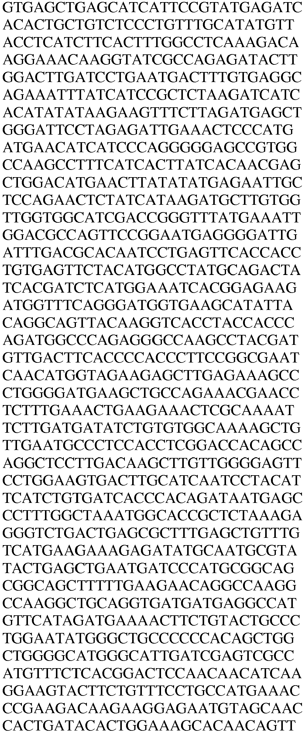

LysRS1C3 Protein / Human / 13 aa + 162-597 
SEQ. ID. NO. 114 LysRS1C3 DNA / Human / 
SEQ. ID. NO. 115 
LysRS1C4 Protein / Human / 212-597 
SEQ. ID. NO. 116 LysRS1C4 DNA / Human / 
SEQ. ID. NO. 117 
LysRS1C5 Protein / Human / 469-597 
SEQ. ID. NO. 118 LysRS1C5 DNA / Human / 
SEQ. ID. NO. 119 
Table 5B AARS polypeptides unique splice junctions Name Type / species Amino acid and Nucleic Acid Sequences in the vicinity of the unique splice junction SEQ. ID. NO. K1-AS01 DNA / Human / 
SEQ. ID. NO. 120 Protein / Human N/A K1-SV10 DNA / Human / 
SEQ. ID. NO. 121 Protein / Human RKRAWTQINYKSEEEF SEQ. ID. NO. 122 K1-AS04 DNA / Human / 
SEQ. ID. NO. 123 Protein / Human N/A K1-AS05 DNA / Human / 
SEQ. ID. NO. 124 Protein / Human N/A Internal AARS Polypeptides: (Table 9) Table 9 AARS polypeptides and nucleic acids identified by Bioinformatics Name Type / species /Residues Amino acid and Nucleic Acid Sequences SEQ. ID. NO. LysRS1I1 Protein / Human / 65-214 
SEQ. ID. NO. 137 LysRS1I1 DNA / Human 
SEQ. ID. NO. 138 N-terminal mitochondrial AARS Polypeptides: (Tables 10, 11 & 12) Table 10A AARS polypeptides identified by MS Name Type / species /Residues Amino acid and Nucleic Acid Sequences SEQ. ID. NO. LysRS2N1 Protein / Human / 1-394 
SEQ. ID. NO. 142 
LysRS2N1 DNA / Human / 
SEQ. ID. NO. 143 
Table 10B LysRS2N1 Mass spec peptides detected and inferred linking peptides Type / species Sequence SEQ. ID. NO. Protein / mouse RGDIIGVEGNPGK SEQ. ID. NO. 144 Protein / mouse TKKGELSIIPQEITLLSPCLHMLPHLHFGLKDKETRYRQR SEQ. ID. NO. 145 Protein / mouse YLDLILNDFVR SEQ. ID. NO. 146 Table 10C LysRS2N1 Concatenated sequences based on mass spec peptides detected Type / species Sequence SEQ. ID. NO. Protein / mouse 
SEQ. ID. NO. 147 Table 11 AARS polypeptides and alternative transcripts identified by Deep Sequencing Name Type / species /Residues Amino acid and Nucleic Acid Sequences SEQ. ID. NO. LysRS2N6 Protein / Human / 1-49 + 12 aa 
SEQ. ID. NO. 148 LysRS2N6 DNA / Human 
SEQ. ID. NO. 149 LysRS2N7 Protein / Human / 1-102 + 22 aa 
SEQ. ID. NO. 150 LysRS2N7 DNA / Human 
SEQ. ID. NO. 151 LysRS2N8 Protein / Human / 1-293 + 334-625 
SEQ. ID. NO. 152 
LysRS2N8 DNA / Human 
SEQ. ID. NO. 153 
LysRS2N9 Protein / Human / 1-102 + 294-625 
SEQ. ID. NO. 154 
LysRS2N9 DNA / Human 
SEQ. ID. NO. 155 
LysRS2N1 0 Protein / Human / 1-333 + 546-625 
SEQ. ID. NO. 156 LysRS2N1 0 DNA / Human 
SEQ. ID. NO. 157 
LysRS2N1 1 Protein / Human / 1-102 + 2 aa 
SEQ. ID. NO. 158 LysRS2N1 1 DNA / Human 
SEQ. ID. NO. 159 LysRS2N1 2 Protein / Human / 
SEQ. ID. NO. 160 1-48 + 1 aa LysRS2N1 2 DNA / Human 
SEQ. ID. NO. 161 LysRS2N1 3 Protein / Human / 1-157 + 20 aa 
SEQ. ID. NO. 162 LysRS2N1 3 DNA / Human 
SEQ. ID. NO. 163 LysRS2N1 4 Protein / Human / 1-251 + 36 aa 
SEQ. ID. NO. 164 
LysRS2N1 4 DNA / Human 
SEQ. ID. NO. 165 Table 11B AARS polypeptides unique splice junctions Name Type / species Amino acid and Nucleic Acid Sequences in the vicinity of the unique splice junction SEQ. ID. NO. K1-AS01 DNA / Human / 
SEQ. ID. NO. 166 Protein / Human KSFSDQRSNTTKSAVK SEQ. ID. NO.167 K1-SV04 DNA / Human / 
SEQ. ID. NO.168 Protein / Human EEESVDPNVGSMPKEL SEQ. ID. NO.169 K1-SV06 DNA / Human / 
SEQ. ID. NO.170 Protein / Human LDELGFLEMLVVGGID SEQ. ID. NO.171 K1-SV07 DNA / Human / 
SEQ. ID. NO.172 Protein / Human EEESVDPNIETPMMNI SEQ. ID. NO.173 K1-SV08 DNA / Human / 
SEQ. ID. NO. 174 Protein / Human IAPELYHKAKAAGDDE SEQ. ID. N0.175 K1-SV10 DNA / Human / 
SEQ. ID. NO.176 Protein / Human EEESVDPNKL SEQ. ID. NO.177 K1-AS03 DNA / Human / 
SEQ. ID. NO.178 Protein / Human KSFSDQRR SEQ. ID. NO.179 K1-AS04 DNA / Human / 
SEQ. ID. NO.180 Protein / Human TDITLKVAEIINQKKN SEQ. ID. NO.181 K1-AS05 DNA / Human / 
SEQ. ID. NO.182 Protein / Human LHFGLKDKKLAKFLMI SEQ. ID. NO.183 Table 12 AARS polypeptides and nucleic acids identified by Bioinformatics Name Type / species /Residues Amino acid and Nucleic Acid Sequences SEQ. ID. NO. LysRS2N2 Protein / Human / 1-104 
SEQ. ID. NO.184 
LysRS2N2 DNA/ Human / 
SEQ. ID. NO.185 LysRS2N3 Protein / Human / 1-222 
SEQ. ID. NO.186 LysRS2N3 DNA/ Human / 
SEQ. ID. NO.187 
LysRS2N4 Protein / Human / 1-93 
SEQ. ID. NO.188 LysRS2N4 DNA / Human / 
SEQ. ID. NO.189 LysRS2N5 Protein / Human / 1-242 
SEQ. ID. NO.190 LysRS2N5 DNA / Human / 
SEQ. ID. NO.191 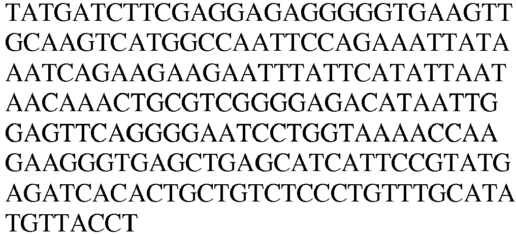
- "Protein fragments," or the amino acid sequence of protein fragments, such as proteolytic fragments or splice variant fragments, can be characterized, identified, or derived according to a variety of techniques. For instance, splice variants can be identified by techniques such as deep sequencing (see, e.g., Xing et al., RNA. 14:1470-1479, 2008; and Zhang et al., Genome Research. 17:503-509, 2007). As a further example, protein fragments such as proteolytic fragments can be identified in vitro, such as by incubating full-length or other AARS polypeptides with selected proteases, or they can be identified endogenously (e.g., in vivo). Protein fragments such as endogenous proteolytic fragments can be generated or identified, for instance, by recombinantly expressing full-length or other AARS polypeptides in a selected microorganism or eukaryotic cell that has been either modified to contain one or more selected proteases, or that naturally contains one or more proteases that are capable of acting on a selected AARS polypeptide, and isolating and characterizing the endogenously produced protein fragments therefrom.
- Protein fragments such as endogenous (e.g., naturally-occurring) proteolytic fragments can be generated or identified, for instance, from various cellular fractions (e.g., cytosolic, membrane, nuclear) and/or growth medium of various cell-types, including, for example, immune cells such as monocytes, dendritic cells, macrophages (e.g., RAW 264.7 macrophages), neutrophils, eosinophils, basophils, and lymphocytes, such as B-cells and T-cells (e.g., CD4+ helper and CD8+ killer cells), including primary T-cells and T-cell lines such as Jurkat T-cells, as well as natural killer (NK) cells.
- Protein fragments such as endogenous proteolytic fragments, however generated, can be identified by techniques such as mass-spectrometry, or equivalent techniques. Once an in vitro or endogenously identified protein fragment has been generated or identified, it can be mapped or sequenced, and, for example, cloned into an expression vector for recombinant production, or produced synthetically.
- A wide variety of proteases can be used to produce, identify, derive, or characterize the sequence of AARS protein fragments such as proteolytic fragments. Generally, proteases are usually classified according to three major criteria: (i) the reaction catalyzed, (ii) the chemical nature of the catalytic site, and (iii) the evolutionary relationship, as revealed by the structure. General examples of proteases or proteinases, as classified by mechanism of catalysis, include aspartic proteases, serine proteases, cysteine proteases, and metalloproteases.
- Most aspartic proteases belong to the pepsin family. This family includes digestive enzymes, such as pepsin and chymosin, as well as lysosomal cathepsins D and processing enzymes such as renin, and certain fungal proteases (e.g., penicillopepsin, rhizopuspepsin, endothiapepsin). A second family of aspartic proteases includes viral proteinases such as the protease from the AIDS virus (HIV), also called retropepsin.
- Serine proteases include two distinct families. First, the chymotrypsin family, which includes the mammalian enzymes such as chymotrypsin, trypsin, elastase, and kallikrein, and second, the substilisin family, which includes the bacterial enzymes such as subtilisin. The general 3D structure between these two families is different, but they have the same active site geometry, and catalysis proceeds via the same mechanism. The serine proteases exhibit different substrate specificities, differences which relate mainly to amino acid substitutions in the various enzyme subsites (substrate residue interacting sites). Some serine proteases have an extended interaction site with the substrate whereas others have a specificity that is restricted to the P1 substrate residue.
- The cysteine protease family includes the plant proteases such as papain, actinidin, and bromelain, several mammalian lysosomal cathepsins, the cytosolic calpains (calcium-activated), as well as several parasitic proteases (e.g., Trypanosoma, Schistosoma). Papain is the archetype and the best studied member of the family. Recent elucidation of the X-ray structure of the Interleukin-1-beta Converting Enzyme has revealed a novel type of fold for cysteine proteinases.
- The metalloproteases are one of the older classes of proteases, found in bacteria, fungi, and higher organisms. They differ widely in their sequences and their 3D structures, but the great majority of enzymes contain a zinc atom that is catalytically active. In some cases, zinc may be replaced by another metal such as cobalt or nickel without loss of proteolytic activity. Bacterial thermolysin has been well characterized and its crystallographic structure indicates that zinc is bound by two histidines and one glutamic acid. Many metalloproteases contain the sequence motif HEXXH, which provides two histidine ligands for the zinc. The third ligand is either a glutamic acid (thermolysin, neprilysin, alanyl aminopeptidase) or a histidine (astacin, serralysin).
- Illustrative proteases include, for example, achromopeptidase, aminopeptidase, ancrod, angiotensin converting enzyme, bromelain, calpain, calpain I, calpain II, carboxypeptidase A, carboxypeptidase B, carboxypeptidase G, carboxypeptidase P, carboxypeptidase W, carboxypeptidase Y, caspase 1, caspase 2, caspase 3, caspase 4, caspase 5, caspase 6, caspase 7, caspase 8, caspase 9, caspase 10, caspase 11, caspase 12, caspase 13, cathepsin B, cathepsin C, cathepsin D, cathepsin E, cathepsin G, cathepsin H, cathepsin L, chymopapain, chymase, chymotrypsin, clostripain, collagenase, complement C1r, complement C1s, complement Factor D, complement factor I, cucumisin, dipeptidyl peptidase IV, elastase (leukocyte), elastase (pancreatic), endoproteinase Arg-C, endoproteinase Asp-N, endoproteinase Glu-C, endoproteinase Lys-C, enterokinase, factor Xa, ficin, furin, granzyme A, granzyme B, HIV Protease, IGase, kallikrein tissue, leucine aminopeptidase (general), leucine aminopeptidase (cytosol), leucine aminopeptidase (microsomal), matrix metalloprotease, methionine aminopeptidase, neutrase, papain, pepsin, plasmin, prolidase, pronase E, prostate specific antigen, protease alkalophilic from Streptomyces griseus, protease from Aspergillus, protease from Aspergillus saitoi, protease from Aspergillus sojae, protease (B. licheniformis) (alkaline or alcalase), protease from Bacillus polymyxa, protease from Bacillus sp, protease from Rhizopus sp., protease S, proteasomes, proteinase from Aspergillus oryzae, proteinase 3, proteinase A, proteinase K, protein C, pyroglutamate aminopeptidase, rennin, rennin, streptokinase, subtilisin, thermolysin, thrombin, tissue plasminogen activator, trypsin, tryptase and urokinase.
- Disclosed are isolated AARS polypeptides, comprising, consisting essentially of, or consisting of amino acid sequences that have been derived from endogenous, naturally-occurring AARS polypeptide fragments, and pharmaceutical compositions comprising said fragments, and methods of use thereof. These and related fragments can be generated or identified in vivo, ex vivo, and/or in vitro. In certain preferred in vitro methods, AARS proteolytic fragments are generated or identified by incubating an AARS polypeptide, such as a full-length AARS polypeptide, with one or more isolated human proteases, mainly those proteases that are endogenous or natural to humans, such as elastase and others described herein and known in the art. Disclosed are also isolated AARS polypeptides, comprising, consisting essentially of, or consisting of amino acid sequences that have been derived from endogenous, naturally-occurring AARS splice variants, and pharmaceutical compositions comprising said fragments, and methods of use thereof. Essentially, AARS protein fragment can be isolated from samples that have been exposed to proteases, whether in vivo or in vitro.
- AARS protein fragments can be identified by techniques such as mass-spectrometry, or equivalent techniques. Merely by way of illustration and not limitation, the proteomes from various cell types, tissues, or body fluids from a variety of physiological states (e.g., hypoxia, diet, age, disease) or fractions thereof may be separated by 1D SDS-PAGE and the gel lanes cut into bands at fixed intervals; after which the bands may be optionally digested with an appropriate protease, such as trypsin, to release the peptides, which may then be analyzed by 1D reverse phase LC-MS/MS. The resulting proteomic data may be integrated into so-called peptographs, which plot, in the left panel, sequence coverage for a given protein in the horizontal dimension (N to C terminus, left to right) versus SDS-PAGE migration in the vertical dimension (high to low molecular weight, top to bottom). The specific peptide fragments can then be sequenced or mapped. The AARS reference fragment may be characterized by its unique molecular weight, as compared, for example, to the molecular weight of the corresponding full-length AARS.
- As noted above, the AARS polypeptides set forth in Table(s) 1-3, or Table(s) 4-6, or Table(s) 7-9, or Table(s) 10-12 are disclosed. Also disclosed are "variants" of the AARS reference polypeptides. The recitation polypeptide "variant" refers to polypeptides that are distinguished from a reference AARS polypeptide by the addition, deletion, and/or substitution of at least one amino acid residue, and which typically retain (e.g., mimic) or modulate (e.g., antagonize) one or more non-canonical activities of a reference AARS polypeptide.
- Moreover human Lysyl tRNA synthetases include several hundred highly related polymorphic forms, and these are known in the art to be at least partially functionally interchangeable. It would thus be a routine matter to select a naturally occurring variant of Lysyl tRNA synthetase, including, for example the single nucleotide polymorphic forms listed in Table A to create an AARS polypeptide containing one or more amino acid changes based on the sequence of any of the homologues, orthologs, and naturally-occurring isoforms of human as well as other species of Lysyl tRNA synthetase.
Table A Human Lysyl tRNA synthetase SNPs Gene Bank Accession Number Nucleotide Change Gene Bank Accession Number Nucleotide Change rs117946205 G/T rs11557665 C/G rs117872527 A/T rs11557664 C/T ras117585187 C/G rs11556640 A/C rs117561624 G/T rs11556639 C/G rs117472224 C/G rs11556638 C/G rs117457640 C/T rs11386229 -/A rs117188693 A/C rs11363202 -/A rs117100154 C/T rs9938305 C/G rs117041419 C/T rs8057723 C/T rs117013282 G/T rs8053257 A/G rs116989849 A/T rs8051771 C/T rs116928063 C/T rs8051113 A/C rs116811812 C/G rs8047430 C/T rs116801062 C/G rs7195338 A/G rs116635879 C/T rs7193257 C/G rs116584059 G/T rs7189895 A/G rs116531622 A/G rs7187847 A/G rs116468372 C/T rs7186504 G/T rs116318609 C/G rs6564271 C/T rs116129958 A/G rs6564270 C/T rs116069782 C/T rs5817958 -/A rs115957689 A/T rs5817957 -/G rs115912670 C/T rs5030748 A/G rs115518873 C/T rs4888442 A/G rs115450297 C/T rs4888441 C/G rs115134738 C/G rs4888440 A/G rs115082322 A/T rs3837756 -/T rs114768348 C/T rs3784931 A/G rs113993881 A/G rs3784930 A/G rs113909347 A/T rs3784929 C/T rs113846451 A/C rs3784928 A/G rs113813120 C/T rs3214635 -/G rs113577360 C/T rs3211301 A/G rs113570860 C/T rs2289064 A/C rs113531576 A/G rs2289063 C/T rs113335212 A/G rs2233810 A/C rs113272137 C/T rs2233809 G/T rs113001454 A/C rs2233808 A/G rs112982343 C/G rs2233807 C/T rs112934609 A/G rs34610510 -/C rs112878760 A/C rs34570398 -/A rs112834496 C/T rs34367936 -/T rs112783308 C/T rs34160918 -/T rs112603638 C/T rs34084576 C/T rs112599651 C/T rs28615806 A/T rs112389955 -/A rs28609444 A/T rs112370895 A/G rs28376768 A/T rs112319505 G/T rs28365852 A/G rs112268246 -/G rs17764797 C/T rs112225278 A/G rs1769721 C/T rs112121151 A/G rs17607381 A/T rs112063795 A/C rs16941533 C/T rs111883921 C/T rs16941516 A/G rs111836109 A/G rs16941409 C/T rs111806827 A/G rs16941400 A/G rs111772248 C/T rs16941301 A/G rs111679674 C/G rs16941296 -/C rs111676921 A/C rs16941287 C/T rs111330594 A/G rs16941282 C/G rs111323143 G/T rs13380501 A/G rs111236795 A/G rs13333731 C/T rs80328717 C/G rs12934451 C/T rs80288252 A/G rs 12932643 A/C rs80287344 A/G rs12919013 C/T rs80068681 C/G rs12599809 G/T rs79964843 G/T rs12596816 G/T rs79612807 A/G rs12596033 A/C rs79367972 A/G rs12447289 A/G rs79309890 C/T rs11866252 C/T rs79256828 C/T rs11649494 C/G rs79248873 A/G rs11649491 C/G rs79158843 A/C rs11649388 A/G rs78908884 A/T ras11647615 C/G rs78798046 A/G rs11639582 A/C rs78654839 C/G rs11639581 C/T rs78496415 A/G rs11557669 C/G rs78279493 A/C rs11557668 A/G rs78198545 C/T rs11557667 A/C rs78028405 C/G rs71763095 -/T rs77917474 A/G rs71394230 G/T rs77909125 -/G rs71394229 A/T rs77573084 A/G rs71394228 A/G rs77547020 C/T rs71272462 -/T rs77405224 A/G rs67862594 -/A rs77039575 C/T rs67793208 -/AA rs76950867 C/T rs66921889 -/A rs76937292 C/T rs66866136 -/A rs76742560 G/T rs62058713 G/T rs76645676 C/T rs61588474 G/T rs76583709 A/C rs60487109 G/T rs76407110 G/T rs60376290 A/C rs76274703 A/G rs59297469 G/T rs76197636 A/C rs59116766 G/T rs76151791 C/T rs58996069 G/T rs76083304 A/G rs58780202 G/T rs76000432 A/T rs58657577 C/T rs75909814 C/T rs58508930 A/G rs75636445 G/T rs58000098 C/G rs75584151 A/G rs57551067 -/T rs75374652 A/C rs57228113 -/A rs75260215 G/T rs57060791 -/T rs75134162 C/T rs56951212 -/A rs75096057 A/C rs56140205 A/G rs74830021 A/T rs41505349 C/T rs74250915 G/T rs36101987 C/T rs74024804 A/T rs36091972 C/T rs73615082 C/T rs35638876 -/A rs73615080 C/T rs35501069 -/C rs72789443 A/G rs35494814 -/T rs72563119 C/G rs35478599 -/C rs72563118 C/G rs35321652 A/G rs72563117 C/G rs35197449 -/A rs72526055 -/A rs35143320 C/G rs72500765 -/A rs35021757 -/A rs72465966 -/A rs34920043 -/C rs72092434 -/TT rs34838148 A/C rs71945664 -/T rs34729412 -/A rs2233806 C/G rs35311324 
rs2233805 C/T rs67860096 
rs2233804 C/T rs71378624 CTA/GTG rs2233803 C/T rs10556967 -/TAT rs1865493 C/G rs57952495 -/AATA rs1865492 A/G rs16941276 -/TAA rs1836547 C/G rs111735733 -/TTG rs1802511 C/T rs66905258 -/ATT rs1802510 C/T rs71883952 -/TTA rs920037 G/T rs71839384 -/TTA rs11505 
rs80330368 -/AAA rs6834 
rs35993558 CT/GC rs35903494 -/G rs35903075 C/G rs35806222 -/T/TG rs59378310 -/ATT rs72129089 -/TAT - A polypeptide variant may be distinguished from a reference polypeptide by one or more substitutions, which may be conservative or non-conservative, as described herein and known in the art. The polypeptide variant may comprise conservative substitutions and, in this regard, it is well understood in the art that some amino acids may be changed to others with broadly similar properties without changing the nature of the activity of the polypeptide.
- A variant polypeptide may include an amino acid sequence having at least about 50%, 55%, 60%, 65%, 70%, 75%, 80%, 85%, 90%, 91%, 92%, 93%, 94%, 95%, 96%, 97%, 98% or more sequence identity or similarity to a corresponding sequence of an AARS reference polypeptide, as described herein, and substantially retains the non-canonical activity of that reference polypeptide. Also included are sequences differing from the reference AARS sequences by the addition, deletion, or substitution of 1, 2, 3, 4, 5, 6, 7, 8, 9, 10, 11, 12, 13, 14, 15, 16, 17, 18, 19, 20, 30, 40, 50, 60,70, 80, 90, 100, 110, 120, 130, 140, 150 or more amino acids but which retain the properties of the reference AARS polypeptide. The amino acid additions or deletions may occur at the C-terminal end and/or the N-terminal end of the AARS reference polypeptide. The amino acid additions may include 1, 2, 3, 4, 5, 6, 7, 8, 9, 10, 11, 12, 13, 14, 15, 16, 17, 18, 19, 20, 30, 40, 50 or more wild-type residues (i.e., from the corresponding full-length AARS polypeptide) that are proximal to the C-terminal end and/or the N-terminal end of the AARS reference polypeptide.
- Variant polypeptides may differ from the corresponding AARS reference sequences by at least 1% but less than 20%, 15%, 10% or 5% of the residues. (If this comparison requires alignment, the sequences should be aligned for maximum similarity. "Looped" out sequences from deletions or insertions, or mismatches, are considered differences.) The differences are, suitably, differences or changes at a non-essential residue or a conservative substitution. The molecular weight of a variant AARS polypeptide may differ from that of the AARS reference polypeptide by about 1%, 2%, 3%, 4%, 5%, 6%, 7%, 8%, 9%, 10%, 11%, 12%, 13%, 14%, 15%, 16%, 17%, 18%, 19%, 20%, or more.
- Also included are biologically active "fragments" of the AARS reference polypeptides, i.e., biologically active fragments of the AARS protein fragments. Representative biologically active fragments generally participate in an interaction, e.g., an intramolecular or an inter-molecular interaction. An inter-molecular interaction can be a specific binding interaction or an enzymatic interaction. An inter-molecular interaction can be between an AARS polypeptide and a cellular binding partner, such as a cellular receptor or other host molecule that participates in the non-canonical activity of the AARS polypeptide. AARS proteins, variants, and biologically active fragments thereof, may bind to one or more cellular binding partners with an affinity of at least about 0.01, 0.05, 0.1, 0.2, 0.3, 0.4, 0.5, 0.6, 0.7, 0.8, 0.9, 1, 2, 3, 4, 5, 6, 7, 8, 9, 10, 11, 12, 13, 14, 15, 16, 17, 18, 19, 20, 21, 22, 23, 24, 25, 26, 27, 28, 29, 30, 40, or 50 nM. The binding affinity of an AARS protein fragment for a selected cellular binding partner, particularly a binding partner that participates in a non-canonical activity, is typically stronger than that of the AARS protein fragment's corresponding full-length AARS polypeptide, by at least about 1.5x, 2x, 2.5x, 3x, 3.5x, 4x, 4.5x, 5x, 6x, 7x, 8x, 9x, 10x, 15x, 20x, 25x, 30x, 40x, 50x, 60x, 70x, 80x, 90x, 100x, 200x, 300x, 400x, 500x, 600x, 700x, 800x, 900x, 1000x or more (including all integers in between). The binding affinity of an AARS protein fragment for a binding partner that participates in at least one canonical activity of an AARS is typically weaker than that of the AARS protein fragment's corresponding full-length AARS polypeptide, by at least about 1.5x, 2x, 2.5x, 3x, 3.5x, 4x, 4.5x, 5x, 6x, 7x, 8x, 9x, 10x, 15x, 20x, 25x, 30x, 40x, 50x, 60x, 70x, 80x, 90x, 100x, 200x, 300x, 400x, 500x, 600x, 700x, 800x, 900x, 1000x or more.
- Typically, biologically active fragments comprise a domain or motif with at least one activity of an AARS reference polypeptide and may include one or more (and in some cases all) of the various active domains, and include fragments having a non-canonical activity. In some cases, biologically active fragments of an AARS polypeptide have a biological activity that is unique to the particular, truncated fragment, such that the full-length AARS polypeptide may not have that activity. In certain cases, the biological activity may be revealed by separating the biologically active AARS polypeptide fragment from the other full-length AARS polypeptide sequences, or by altering certain residues of the full-length AARS wild-type polypeptide sequence to unmask the biologically active domains.
- A biologically active fragment of an AARS reference polypeptide can be a polypeptide fragment which is, for example, 10, 11, 12, 13, 14, 15, 16, 17, 18, 19, 20, 21, 22, 23, 24, 25, 26, 27, 28, 29, 30, 35, 40, 45, 50, 55, 60, 65, 70, 75, 80, 85, 90, 95, 100, 110, 120, 130, 140, 150, 160, 170, 180, 190, 200, 220, 240, 260, 280, 300, 320, 340, 360, 380, 400, 450, 500, 550, 600, 650, 700, 750 or more contiguous or non-contiguous amino acids, including all integers (e.g., 101, 102, 103) and ranges (e.g., 50-100, 50-150, 50-200) in between, of the amino acid sequences set forth in any one of the AARS reference polypeptides described herein, but typically exclude the full-length AARS. A biologically active fragment may comprise a non-canonical activity-related sequence, domain, or motifThe C-terminal or N-terminal region of any AARS reference polypeptide may be truncated by about 1, 2, 3, 4, 5, 6, 7, 8, 9, 10, 15, 20, 25, 30, 35, 40, 45, 50, 60, 70, 80, 90, 100, 110, 120, 130, 140, 150, 160, 170, 180, 190, 200, 250, 300, 350, 400, 450, 500, 550, 600, 650, or 700 or more amino acids, or by about 10-50, 20-50, 50-100, 100-150, 150-200, 200-250, 250-300, 300-350, 350-400, 400-450, 450-500, 500-550, 550-600, 600-650, 650-700 or more amino acids, including all integers and ranges in between (e.g., 101, 102, 103, 104, 105), so long as the truncated AARS polypeptide retains the non-canonical activity of the reference polypeptide. Typically, the biologically-active fragment has no less than about 1%, about 5 %, about 10%, about 25%, or about 50% of an activity of the biologically-active (i.e., non-canonical activity) AARS reference polypeptide from which it is derived. Exemplary methods for measuring such non-canonical activities are described in the Examples.
- As noted above, an AARS polypeptide may be altered in various ways including amino acid substitutions, deletions, truncations, and insertions. Methods for such manipulations are generally known in the art. For example, amino acid sequence variants of an AARS reference polypeptide can be prepared by mutations in the DNA. Methods for mutagenesis and nucleotide sequence alterations are well known in the art. See, for example, Kunkel (1985, Proc. Natl. Acad. Sci. USA. 82: 488-492), Kunkel et al., (1987, Methods in Enzymol, 154: 367-382),
U.S. Pat. No. 4,873,192 , Watson, J. D. et al., ("Molecular Biology of the Gene", Fourth Edition, Benjamin/Cummings, Menlo Park, Calif., 1987) and the references cited therein. Guidance as to appropriate amino acid substitutions that do not affect biological activity of the protein of interest may be found in the model of Dayhoff et al., (1978) Atlas of Protein Sequence and Structure (Natl. Biomed. Res. Found., Washington, D.C.). - Similarly it is within the skill in the art to address and / or mitigate immunogenicity concerns if they arise using an AARS polypeptide, e.g., by the use of automated computer recognition programs to identify potential T cell epitopes, and directed evolution approaches to identify less immunogenic forms.
- Methods for screening gene products of combinatorial libraries made by point mutations or truncation, and for screening cDNA libraries for gene products having a selected property are known in the art. Such methods are adaptable for rapid screening of the gene libraries generated by combinatorial mutagenesis of AARS polypeptides. Recursive ensemble mutagenesis (REM), a technique which enhances the frequency of functional mutants in the libraries, can be used in combination with the screening assays to identify AARS polypeptide variants (Arkin and Yourvan (1992) Proc. Natl. Acad. Sci. USA 89: 7811-7815; Delgrave et al., (1993) Protein Engineering, 6: 327-331). Conservative substitutions, such as exchanging one amino acid with another having similar properties, may be desirable as discussed in more detail below.
- Biologically active truncated and/or variant AARS polypeptides may contain conservative amino acid substitutions at various locations along their sequence, as compared to a reference AARS amino acid residue. Additionally, naturally occurring variants of AARS proteins have been sequenced, and are known in the art to be at least partially functionally interchangeable. It would thus be a routine matter to select an amino acid position to introduce a conservative, or non conservative mutation into an AARS polypeptide based on naturally occurring sequence variation among the known AARS protein homologues, orthologs, and naturally-occurring isoforms of human as well as other species of an AARS protein.
- A "conservative amino acid substitution" is one in which the amino acid residue is replaced with an amino acid residue having a similar side chain. Families of amino acid residues having similar side chains have been defined in the art, which can be generally sub-classified as follows:
- Acidic: The residue has a negative charge due to loss of H ion at physiological pH and the residue is attracted by aqueous solution so as to seek the surface positions in the conformation of a peptide in which it is contained when the peptide is in aqueous medium at physiological pH. Amino acids having an acidic side chain include glutamic acid and aspartic acid.
- Basic: The residue has a positive charge due to association with H ion at physiological pH or within one or two pH units thereof (e.g., histidine) and the residue is attracted by aqueous solution so as to seek the surface positions in the conformation of a peptide in which it is contained when the peptide is in aqueous medium at physiological pH. Amino acids having a basic side chain include arginine, lysine and histidine.
- Charged: The residues are charged at physiological pH and, therefore, include amino acids having acidic or basic side chains (i.e., glutamic acid, aspartic acid, arginine, lysine and histidine).
- Hydrophobic: The residues are not charged at physiological pH and the residue is repelled by aqueous solution so as to seek the inner positions in the conformation of a peptide in which it is contained when the peptide is in aqueous medium. Amino acids having a hydrophobic side chain include tyrosine, valine, isoleucine, leucine, methionine, phenylalanine and tryptophan.
- Neutral/polar: The residues are not charged at physiological pH, but the residue is not sufficiently repelled by aqueous solutions so that it would seek inner positions in the conformation of a peptide in which it is contained when the peptide is in aqueous medium. Amino acids having a neutral/polar side chain include asparagine, glutamine, cysteine, histidine, serine and threonine.
- This description also characterizes certain amino acids as "small" since their side chains are not sufficiently large, even if polar groups are lacking, to confer hydrophobicity. With the exception of proline, "small" amino acids are those with four carbons or less when at least one polar group is on the side chain and three carbons or less when not. Amino acids having a small side chain include glycine, serine, alanine and threonine. The gene-encoded secondary amino acid proline is a special case due to its known effects on the secondary conformation of peptide chains. The structure of proline differs from all the other naturally-occurring amino acids in that its side chain is bonded to the nitrogen of the α-amino group, as well as the α-carbon. Several amino acid similarity matrices are known in the art (see e.g., PAM120 matrix and PAM250 matrix as disclosed for example by Dayhoff et al., 1978, A model of evolutionary change in proteins). Matrices for determining distance relationships In M. O. Dayhoff, (ed.), Atlas of protein sequence and structure, Vol. 5, pp. 345-358, National Biomedical Research Foundation, Washington DC; and by Gonnet et al., (Science, 256: 14430-1445, 1992), however, include proline in the same group as glycine, serine, alanine and threonine. Accordingly, for the purposes of the present invention, proline is classified as a "small" amino acid.
- The degree of attraction or repulsion required for classification as polar or nonpolar is arbitrary and, therefore, amino acids specifically contemplated by the invention have been classified as one or the other. Most amino acids not specifically named can be classified on the basis of known behavior.
- Amino acid residues can be further sub-classified as cyclic or non-cyclic, and aromatic or non-aromatic, self-explanatory classifications with respect to the side-chain substituent groups of the residues, and as small or large. The residue is considered small if it contains a total of four carbon atoms or less, inclusive of the carboxyl carbon, provided an additional polar substituent is present; three or less if not. Small residues are, of course, always non-aromatic. Dependent on their structural properties, amino acid residues may fall in two or more classes. For the naturally-occurring protein amino acids, sub-classification according to this scheme is presented in Table B.
Table B: Amino acid sub-classification Sub-classes Amino acids Acidic Aspartic acid, Glutamic acid Basic Noncyclic: Arginine, Lysine; Cyclic: Histidine Charged Aspartic acid, Glutamic acid, Arginine, Lysine, Histidine Small Glycine, Serine, Alanine, Threonine, Proline Polar/neutral Asparagine, Histidine, Glutamine, Cysteine, Serine, Threonine Polar/large Asparagine, Glutamine Hydrophobic Tyrosine, Valine, Isoleucine, Leucine, Methionine, Phenylalanine, Tryptophan Aromatic Tryptophan, Tyrosine, Phenylalanine Residues that Glycine and Proline influence chain orientation - Conservative amino acid substitution also includes groupings based on side chains. For example, a group of amino acids having aliphatic side chains is glycine, alanine, valine, leucine, and isoleucine; a group of amino acids having aliphatic-hydroxyl side chains is serine and threonine; a group of amino acids having amide-containing side chains is asparagine and glutamine; a group of amino acids having aromatic side chains is phenylalanine, tyrosine, and tryptophan; a group of amino acids having basic side chains is lysine, arginine, and histidine; and a group of amino acids having sulfur-containing side chains is cysteine and methionine. For example, it is reasonable to expect that replacement of a leucine with an isoleucine or valine, an aspartate with a glutamate, a threonine with a serine, or a similar replacement of an amino acid with a structurally related amino acid will not have a major effect on the properties of the resulting variant polypeptide. Whether an amino acid change results in a functional truncated and/or variant AARS polypeptide can readily be determined by assaying its non-canonical activity, as described herein. Conservative substitutions are shown in Table C under the heading of exemplary substitutions. Amino acid substitutions falling within the scope of the invention, are, in general, accomplished by selecting substitutions that do not differ significantly in their effect on maintaining (a) the structure of the peptide backbone in the area of the substitution, (b) the charge or hydrophobicity of the molecule at the target site, (c) the bulk of the side chain, or (d) the biological function. After the substitutions are introduced, the variants are screened for biological activity.
Table C: Exemplary Amino Acid Substitutions Original Residue Exemplary Substitutions Preferred Substitutions Ala Val, Leu, He Val Arg Lys, Gln, Asn Lys Asn Gln, His, Lys, Arg Gln Asp Glu Glu Cys Ser Ser Gln Asn, His, Lys, Asn Glu Asp, Lys Asp Gly Pro Pro His Asn, Gln, Lys, Arg Arg Ile 
Leu Leu 
He Lys Arg, Gln, Asn Arg Met Leu, Ile, Phe Leu Phe Leu, Val, He, Ala Leu Pro Gly Gly Ser Thr Thr Thr Ser Ser Trp Tyr Tyr Tyr Trp, Phe, Thr, Ser Phe Val 
Leu - Alternatively, similar amino acids for making conservative substitutions can be grouped into three categories based on the identity of the side chains. The first group includes glutamic acid, aspartic acid, arginine, lysine, histidine, which all have charged side chains; the second group includes glycine, serine, threonine, cysteine, tyrosine, glutamine, asparagine; and the third group includes leucine, isoleucine, valine, alanine, proline, phenylalanine, tryptophan, methionine, as described in Zubay, G., Biochemistry, third edition, Wm.C. Brown Publishers (1993).
- Thus, a predicted non-essential amino acid residue in a truncated and/or variant AARS polypeptide is typically replaced with another amino acid residue from the same side chain family. Alternatively, mutations can be introduced randomly along all or part of an AARS coding sequence, such as by saturation mutagenesis, and the resultant mutants can be screened for an activity of the parent polypeptide to identify mutants which retain that activity. Following mutagenesis of the coding sequences, the encoded peptide can be expressed recombinantly and the activity of the peptide can be determined. A "non-essential" amino acid residue is a residue that can be altered from the reference sequence of a polypeptide without abolishing or substantially altering one or more of its activities. Suitably, the alteration does not substantially abolish one of these activities, for example, the activity is at least 20%, 40%, 60%, 70% or 80% 100%, 500%, 1000% or more of the reference AARS sequence. An "essential" amino acid residue is a residue that, when altered from the reference sequence of an AARS polypeptide, results in abolition of an activity of the parent molecule such that less than 20% of the reference activity is present. For example, such essential amino acid residues include those that are conserved in AARS polypeptides across different species, including those sequences that are conserved in the active binding site(s) or motif(s) of AARS polypeptides from various sources.
- In general, polypeptides and fusion polypeptides (as well as their encoding polynucleotides) are isolated. An "isolated" polypeptide or polynucleotide is one that is removed from its original environment. For example, a naturally-occurring protein is isolated if it is separated from some or all of the coexisting materials in the natural system. Preferably, such polypeptides are at least about 90% pure, more preferably at least about 95% pure and most preferably at least about 99% pure. A polynucleotide is considered to be isolated if, for example, it is cloned into a vector that is not a part of the natural environment.
- Disclosed are also dimers of AARS polypeptides. Dimers may include, for example, homodimers between two identical AARS polypeptides, heterodimers between two different AARS polypeptides (e.g., a full-length YRS polypeptide and a truncated YRS polypeptide; a truncated YRS polypeptide and a truncated WRS polypeptide), and/or heterodimers between an AARS polypeptide and a heterologous polypeptide. Certain heterodimers, such as those between an AARS polypeptide and a heterologous polypeptide, may be bi-functional, as described herein.
- Also included are monomers of AARS polypeptides, including isolated AARS polypeptides monomers that do not substantially dimerize with a second AARS polypeptide, whether due to one or more substitutions, truncations, deletions, additions, chemical modifications, or a combination of these alterations. Monomeric AARS polypeptides may possess biological activities, including non-canonical activities, which are not possessed by dimeric or multimeric AARS polypeptide complexes.
- Disclosed is also the use of modified AARS polypeptides, including modifications that improved the desired characteristics of an AARS polypeptide, as described herein. Modifications of AARS polypeptides include chemical and/or enzymatic derivatizations at one or more constituent amino acid, including side chain modifications, backbone modifications, and N- and C-terminal modifications including acetylation, hydroxylation, methylation, amidation, and the attachment of carbohydrate or lipid moieties, cofactors, and the like. Exemplary modifications also include pegylation of an AARS polypeptide (see, e.g., Veronese and Harris, Advanced Drug Delivery Reviews 54: 453-456, 2002; and Pasut et al., Expert Opinion. Ther. Patents 14(6) 859-894 2004).
- PEG is a well-known polymer having the properties of solubility in water and in many organic solvents, lack of toxicity, and lack of immunogenicity. It is also clear, colorless, odorless, and chemically stable. For these reasons and others, PEG has been selected as the preferred polymer for attachment, but it has been employed solely for purposes of illustration and not limitation. Similar products may be obtained with other water-soluble polymers, including without limitation; polyvinyl alcohol, other poly(alkylene oxides) such as poly(propylene glycol) and the like, poly(oxyethylated polyols) such as poly(oxyethylated glycerol) and the like, carboxymethylcellulose, dextran, polyvinyl alcohol, polyvinyl purrolidone, poly-1,3- dioxolane, poly-1,3,6-trioxane, ethylene/maleic anhydride, and polyaminoacids. One skilled in the art will be able to select the desired polymer based on the desired dosage, circulation time, resistance to proteolysis, and other considerations.
- In particular a wide variety of PEG derivatives are both available and suitable for use in the preparation of PEG-conjugates. For example, NOF Corp.'s PEG reagents sold under the trademark SUNBRIGHT® Series provides numerous PEG derivatives, including methoxypolyethylene glycols and activated PEG derivatives such as methoxy-PEG amines, maleimides, N-hydroxysuccinimide esters, and carboxylic acids, for coupling by various methods to the N-terminal, C-terminal or any internal amino acid of the AARS polypeptide. Nektar Therapeutics' Advanced PEGylation technology also offers diverse PEG-coupling technologies to potentially improve the safety and efficacy of an AARS polypeptide based therapeutic.
- A search of patents, published patent applications, and related publications will also provide those skilled in the art reading this disclosure with significant possible PEG-coupling technologies and PEG-derivatives. For example,
US Pat. Nos. 6,436,386 ;5,932,462 ;5,900,461 ;5,824,784 ; and4,904,584 ; describe such technologies and derivatives, and methods for their manufacture. - In certain aspects, chemoselective ligation technology may be utilized to modify AARS polypeptides, such as by attaching polymers in a site-specific and controlled manner. Such technology typically relies on the incorporation of chemoselective anchors into the protein backbone by either chemical, or recombinant means, and subsequent modification with a polymer carrying a complementary linker. As a result, the assembly process and the covalent structure of the resulting protein-polymer conjugate may be controlled, enabling the rational optimization of drug properties, such as efficacy and pharmacokinetic properties (see, e.g., Kochendoerfer, Current Opinion in Chemical Biology 9:555-560, 2005).
- Fusion proteins of AARS polypeptide to other proteins are also disclosed, and these fusion proteins may increase the AARS polypeptide's biological activity, secretion, targeting, biological life, ability to penetrate cellular membranes, or the blood brain barrier, or pharmacokinetic properties. Examples of fusion proteins that improve pharmacokinetic properties ("PK modifiers") include without limitation, fusions to human albumin (Osborn et al.: Eur. J. Pharmacol. 456(1-3): 149-158, (2002)), antibody Fc domains, poly Glu or poly Asp sequences, and transferrin. Additionally, fusion with conformationally disordered polypeptide sequences composed of the amino acids Pro, Ala, and Ser ('PASylation') or hydroxyethyl starch (sold under the trademark HESYLATION®) provides a simple way to increase the hydrodynamic volume of the AARS polypeptide. This additional extension adopts a bulky random structure, which significantly increases the size of the resulting fusion protein. By this means the typically rapid clearance of smaller AARS polypeptides via kidney filtration is retarded by several orders of magnitude. Additionally use of Ig G fusion proteins has also been shown to enable some fusion protein proteins to penetrate the blood brain barrier (Fu et al., (2010) Brain Res. 1352:208-13).
- Examples of fusion proteins that improve penetration across cellular membranes include fusions to membrane translocating sequences. In this context, the term "membrane translocating sequences" refers to naturally occurring and synthetic amino acid sequences that are capable of membrane translocation across a cellular membrane. Representative membrane translocating sequences include those based on the naturally occurring membrane translocating sequences derived from the Tat protein, and homeotic transcription protein Antennapedia, as well as synthetic membrane translocating sequences based in whole or part on poly Arginine and Lysine resides. Representative membrane translocating sequences include for example those disclosed in the following patents,
US5,652,122 ;US 5,670,617 ;US5,674,980 ;US5,747,641 ;US5,804,604 ;US6,316,003 ;US7,585,834 ;US7,312,244 ;US7,279,502 ;US7,229,961 ;US7,169,814 ;US7,453,011 ;US7,235,695 ;US6,982,351 ;US6,605,115 ;US7,306,784 ;US7,306,783 ;US6,589,503 ;US6,348,185 ;US6,881,825 ;US7,431,915 ;WO0074701A2 WO2007111993A2 ;WO2007106554A2 ;WO02069930A1 WO03049772A2 WO03106491A2 WO2008063113A1 . - It will be appreciated that a flexible molecular linker (or spacer) optionally may be interposed between, and covalently join, the AARS polypeptide and any of the fusion proteins disclosed herein.
- The AARS polypeptide can also include synthetic, or naturally occurring secretion signal sequences, derived from other well characterized secreted proteins. Such proteins, may be processed by proteolytic cleavage to form the AARS polypeptide in situ. Such fusions proteins include for example fusions of AARS polypeptide to ubiquitin to provide a new N-terminal amino acid, or the use of a secretion signal to mediate high level secretion of the AARS polypeptide into the extracellular medium, or N, or C-terminal epitope tags to improve purification or detection.
- The AARS polypeptides described herein may be prepared by any suitable procedure known to those of skill in the art, such as by recombinant techniques. In addition to recombinant production methods, polypeptides may be produced by direct peptide synthesis using solid-phase techniques (Merrifield, J. Am. Chem. Soc. 85:2149-2154 (1963)). Protein synthesis may be performed using manual techniques or by automation. Automated synthesis may be achieved, for example, using Applied Biosystems 431A Peptide Synthesizer (Perkin Elmer). Alternatively, various fragments may be chemically synthesized separately and combined using chemical methods to produce the desired molecule.
- Also included are bioassays that relate to the AARS protein fragments and related agents as therapeutic and diagnostic reagents. Examples include bioassays and analytical assays that measure purity, biological activity, affinity, solubility, pH, endotoxin levels, among others, many of which are described herein. Also included are assays that establish dose response curves and/or provide one or more bases for comparison between different batches of agents. Batch comparisons can be based on any one or more of chemical characterization, biological characterization, and clinical characterization. For protein agents, also included are methods of evaluating the potency, stability, pharmacokinetics, and immunogenicity of a selected agent. Among other uses, these and other methods can be used for lot releasing testing of biologic or chemical agents, including the AARS protein fragments, antibodies, binding agents, polynucleotides such as antisense agents and vectors, and others described herein.
- Disclosed is also the use of bioaffinity assays. Such assays can be used to assess the binding affinity, for example, between an AARS protein fragment and a cellular binding partner, or between an AARS protein fragment and an antibody. Binding affinity can also be measured between an AARS protein fragment and an alternate binding agent such as a candidate or lead test compound (e.g., small molecule modulator of an AARS), or between an AARS cellular binding partner and a candidate or lead test compound. Certain exemplary binding affinity assays may utilize ELISA assays, as described herein and known in the art. Certain assays utilize high-performance receptor binding chromatography (see, e.g., Roswall et al., Biologicals. 24:25-39, 1996). Other exemplary binding affinity assays may utilize surface plasmon resonance (SPR)-based technologies. Examples include BIACore technologies, certain of which integrate SPR technology with a microfluidics system to monitor molecular interactions in real time at concentrations ranging from pM to mM. Also included are KINEXA™ assays, which provide accurate measurements of binding specificity, binding affinity, and binding kinetics/rate constants.
- Disclosed are also immunoassays for evaluating or optimizing the immunogenicity of protein agents. Examples include ex vivo human cellular assays and in vitro immuno-enzymatic assays to provide useful information on the immunogenic potential of a therapeutic protein. Ex vivo cell-response assays can be used, for example, to reproduce the cellular co-operation between antigen-presenting cells (APCs) and T-cells, and thereby measure T-cells activation after contact with a protein of interest. Certain in vitro enzymatic assays may utilize a collection of recombinant HLA-DR molecules that cover a significant portion of a relevant human population, and may include automated immuno-enzymatic assays for testing the binding of peptides (stemming from the fragmentation of the therapeutic protein) with the HLA-DR molecules. Also included are methods of reducing the immunogenicity of a selected protein, such as by using these and related methods to identify and then remove or alter one or more T-cell epitopes from a protein agent.
- Also included are biological release assays (e.g., cell-based assays) for measuring parameters such as specific biological activities, including non-canonical biological activities, and cytotoxicity. Certain specific biological assays include, for example, cell-based assays that utilize a cellular binding partner (e.g., cell-surface receptor) of a selected AARS protein fragment, which is functionally coupled to a readout, such as a fluorescent or luminescent indicator of a non-canonical biological activity, as described herein. For instance, a cell is disclosed that comprises a cell-surface receptor or an extracellular portion thereof that binds to an AARS protein fragment, wherein the cell comprises a detector or readout. Also included are in vivo biological assays to characterize the pharmacokinetics of an agent, such as an AARS polypeptide or antibody, typically utilizing engineered mice or other mammal (see, e.g., Lee et al., The Journal of Pharmacology. 281:1431-1439, 1997). Examples of cytotoxicity-based biological assays include release assays (e.g., chromium or europium release assays to measure apoptosis; see, e.g., von Zons et al., Clin Diagn Lab Immunol.4:202-207, 1997), among others, which can assess the cytotoxicity of AARS protein fragments, whether for establishing dose response curves, batch testing, or other properties related to approval by various regulatory agencies, such as the Food and Drug Administration (FDA).
- Such assays can be used, for example, to develop a dose response curve for a selected AARS protein fragment or other agent, and/or to compare the dose response curve of different batches of proteins or other agents. A dose-response curve is an X-Y graph that relates the magnitude of a stressor to the response of a receptor; the response may be a physiological or biochemical response, such as a non-canonical biological activity in a cell in vitro or in a cell or tissue in vivo, a therapeutically effective amount as measured in vivo (e.g., as measured by EC50), or death, whether measured in vitro or in vivo (e.g., cell death, organismal death). Death is usually indicated as an LD50, a statistically-derived dose that is lethal to 50% of a modeled population, though it can be indicated by LC01 (lethal dose for 1% of the animal test population), LC100 (lethal dose for 100% of the animal test population), or LCLO (lowest dose causing lethality). Almost any desired effect or endpoint can be characterized in this manner.
- The measured dose of a response curve is typically plotted on the X axis and the response is plotted on the Y axis. More typically, the logarithm of the dose is plotted on the X axis, most often generating a sigmoidal curve with the steepest portion in the middle. The No Observable Effect Level (NOEL) refers to the lowest experimental dose for which no measurable effect is observed, and the threshold dose refers to the first point along the graph that indicates a response above zero. As a general rule, stronger drugs generate steeper dose response curves. For many drugs, the desired effects are found at doses slightly greater than the threshold dose, often because lower doses are relatively ineffective and higher doses lead to undesired side effects. For in vivo generated dose response curves, a curve can be characterized by values such as µg/kg, mg/kg, or g/kg of body-weight, if desired.
- For batch comparisons, it can be useful to calculate the coefficient of variation (CV) between different dose response curves of different batches (e.g., between different batches of AARS protein fragments, antibodies, or other agents), in part because the CV allows comparison between data sets with different units or different means. For instance, two or three or more different batches of AARS protein fragments or other agents may have a CV between them of less than about 15%, 14%, 13%, 12%, 11%, 10%, 9%, 8%, 7%, 6%, 5%, 4%, 3%, 2%, or 1% for a 4, 5, 6, 7, or 8 point dose curve. The dose response curve can be measured in a cell-based assay, and its readout relates to an increase or a decrease in a selected non-canonical activity of the AARS protein fragment. The dose response curve can be measured in a cell release assay or animal model (e.g., mouse model), and its readout relates to cell death or animal death. Other variations will be apparent to persons skilled in the art.
- Disclosed are also methods and related compositions for expressing and purifying the AARS protein fragments or other polypeptide-based agents. Such recombinant AARS polypeptides can be conveniently prepared using standard protocols as described for example in Sambrook, et al., (1989, supra), in particular Sections 16 and 17; Ausubel et al., (1994, supra), in particular Chapters 10 and 16; and Coligan et al., Current Protocols in Protein Science (John Wiley & Sons, Inc. 1995-1997), in
particular Chapters 1, 5 and 6. As one general example, AARS polypeptides may be prepared by a procedure including one or more of the steps of: (a) preparing a construct comprising a polynucleotide sequence that encodes a AARS polypeptide and that is operably linked to a regulatory element; (b) introducing the construct into a host cell; (c) culturing the host cell to express the AARS polypeptide; and (d) isolating the AARS polypeptide from the host cell. - AARS polynucleotides are described elsewhere herein. In order to express a desired polypeptide, a nucleotide sequence encoding the polypeptide, or a functional equivalent, may be inserted into appropriate expression vector, i.e., a vector which contains the necessary elements for the transcription and translation of the inserted coding sequence. Methods which are well known to those skilled in the art may be used to construct expression vectors containing sequences encoding a polypeptide of interest and appropriate transcriptional and translational control elements. These methods include in vitro recombinant DNA techniques, synthetic techniques, and in vivo genetic recombination. Such techniques are described in Sambrook et al., Molecular Cloning, A Laboratory Manual (1989), and Ausubel et al., Current Protocols in Molecular Biology (1989).
- A variety of expression vector/host systems are known and may be utilized to contain and express polynucleotide sequences. These include, but are not limited to, microorganisms such as bacteria transformed with recombinant bacteriophage, plasmid, or cosmid DNA expression vectors; yeast transformed with yeast expression vectors; insect cell systems infected with virus expression vectors (e.g., baculovirus); plant cell systems transformed with virus expression vectors (e.g., cauliflower mosaic virus, CaMV; tobacco mosaic virus, TMV) or with bacterial expression vectors (e.g., Ti or pBR322 plasmids); or animal cell systems, including mammalian cell and more specifically human cell systems.
- The "control elements" or "regulatory sequences" present in an expression vector are those non-translated regions of the vector--enhancers, promoters, 5' and 3' untranslated regions--which interact with host cellular proteins to carry out transcription and translation. Such elements may vary in their strength and specificity. Depending on the vector system and host utilized, any number of suitable transcription and translation elements, including constitutive and inducible promoters, may be used. For example, when cloning in bacterial systems, inducible promoters such as the hybrid lacZ promoter of the PBLUESCRIPT phagemid (Stratagene, La Jolla, Calif.) or PSPORT1 plasmid (Gibco BRL, Gaithersburg, Md.) and the like may be used. In mammalian cell systems, promoters from mammalian genes or from mammalian viruses are generally preferred. If it is necessary to generate a cell line that contains multiple copies of the sequence encoding a polypeptide, vectors based on SV40 or EBV may be advantageously used with an appropriate selectable marker.
- In bacterial systems, a number of expression vectors may be selected depending upon the use intended for the expressed polypeptide. For example, when large quantities are needed, vectors which direct high level expression of fusion proteins that are readily purified may be used. Such vectors include, but are not limited to, the multifunctional E. coli cloning and expression vectors such as BLUESCRIPT (Stratagene), in which the sequence encoding the polypeptide of interest may be ligated into the vector in frame with sequences for the amino-terminal Met and the subsequent 7 residues of β-galactosidase so that a hybrid protein is produced; pIN vectors (Van Heeke & Schuster, J. Biol. Chem. 264:5503 5509 (1989)); and the like. pGEX Vectors (Promega, Madison, Wis.) may also be used to express foreign polypeptides as fusion proteins with glutathione S-transferase (GST). In general, such fusion proteins are soluble and can easily be purified from lysed cells by adsorption to glutathione-agarose beads followed by elution in the presence of free glutathione. Proteins made in such systems may be designed to include heparin, thrombin, or factor XA protease cleavage sites so that the cloned polypeptide of interest can be released from the GST moiety at will.
- E. coli-based expression systems (see, e.g., Structural Genomics Consortium et al., Nature Methods. 5:135-146, 2008) may be employed. These and related approaches may rely partially or totally on ligation-independent cloning (LIC) to produce a suitable expression vector. Protein expression may be controlled by a T7 RNA polymerase (e.g., pET vector series). The expression host strain BL21(DE3) may be utilized, a λDE3 lysogen of BL21 that supports T7-mediated expression and is deficient in lon and ompT proteases for improved target protein stability. Also included are expression host strains carrying plasmids encoding tRNAs rarely used in E. coli, such as ROSETTA™ (DE3) and Rosetta 2 (DE3) strains. Cell lysis and sample handling may also be improved using reagents sold under the trademarks BENZONASE® nuclease and BUGBUSTER® Protein Extraction Reagent. For cell culture, auto-inducing media can improve the efficiency of many expression systems, including high-throughput expression systems. Media of this type (e.g., OVERNIGHT EXPRESS™ Autoinduction System) gradually elicit protein expression through metabolic shift without the addition of artificial inducing agents such as IPTG. Hexahistidine tags (such as those sold under the trademark HIS•TAG® fusions) may be employed, followed by immobilized metal affinity chromatography (IMAC) purification, or related techniques. In certain aspects, however, clinical grade proteins can be isolated from E. coli inclusion bodies, without or without the use of affinity tags (see, e.g., Shimp et al., Protein Expr Purif. 50:58-67, 2006). As a further example, a cold-shock induced E. coli high-yield production system may be employed, because over-expression of proteins in Escherichia coli at low temperature improves their solubility and stability (see, e.g., Qing et al., Nature Biotechnology. 22:877-882, 2004).
- Also included are high-density bacterial fermentation systems. For example, high cell density cultivation of Ralstonia eutropha allows protein production at cell densities of over 150 g/L, and the expression of recombinant proteins at titers exceeding 10 g/L.
- In the yeast Saccharomyces cerevisiae, a number of vectors containing constitutive or inducible promoters such as alpha factor, alcohol oxidase, and PGH may be used. For reviews, see Ausubel et al. (supra) and Grant et al., Methods Enzymol. 153:516-544 (1987). Also included are Pichia pandoris expression systems (see, e.g., Li et al., Nature Biotechnology. 24, 210 - 215, 2006; and Hamilton et al., Science, 301:1244, 2003). Disclosed are also yeast systems that are engineered to selectively glycosylate proteins, including yeast that have humanized N-glycosylation pathways, among others (see, e.g., Hamilton et al., Science. 313:1441-1443, 2006; Wildt et al., Nature Reviews Microbiol. 3:119-28, 2005; and Gerngross et al., Nature-Biotechnology. 22:1409 -1414, 2004;
U.S. Patent Nos. 7,629,163 ;7,326,681 ; and7,029,872 ). Merely by way of example, recombinant yeast cultures can be grown in Fernbach Flasks or 15L, 50L, 100L, and 200L fermentors, among others. - In cases where plant expression vectors are used, the expression of sequences encoding polypeptides may be driven by any of a number of promoters. For example, viral promoters such as the 35S and 19S promoters of CaMV may be used alone or in combination with the omega leader sequence from TMV (Takamatsu, EMBO J. 6:307-311 (1987)). Alternatively, plant promoters such as the small subunit of RUBISCO or heat shock promoters may be used (Coruzzi et al., EMBO J. 3:1671-1680 (1984); Broglie et al., Science 224:838-843 (1984); and Winter et al., Results Probl. Cell Differ. 17:85-105 (1991)). These constructs can be introduced into plant cells by direct DNA transformation or pathogen-mediated transfection. Such techniques are described in a number of generally available reviews (see, e.g., Hobbs in McGraw Hill, Yearbook of Science and Technology, pp. 191-196 (1992)).
- An insect system may also be used to express a polypeptide of interest. For example, in one such system, Autographa californica nuclear polyhedrosis virus (AcNPV) is used as a vector to express foreign genes in Spodoptera frugiperda cells or in Trichoplusia cells. The sequences encoding the polypeptide may be cloned into a non-essential region of the virus, such as the polyhedrin gene, and placed under control of the polyhedrin promoter. Successful insertion of the polypeptide-encoding sequence will render the polyhedrin gene inactive and produce recombinant virus lacking coat protein. The recombinant viruses may then be used to infect, for example, S. frugiperda cells or Trichoplusia cells in which the polypeptide of interest may be expressed (Engelhard et al., Proc. Natl. Acad. Sci. U.S.A. 91:3224-3227 (1994)). Also included are baculovirus expression systems, including those that utilize SF9, SF21, and T. ni cells (see, e.g., Murphy and Piwnica-Worms, Curr Protoc Protein Sci. Chapter 5:Unit5.4, 2001). Insect systems can provide post-translation modifications that are similar to mammalian systems.
- In mammalian host cells, a number of viral-based expression systems are generally available. For example, in cases where an adenovirus is used as an expression vector, sequences encoding a polypeptide of interest may be ligated into an adenovirus transcription/translation complex consisting of the late promoter and tripartite leader sequence. Insertion in a non-essential E1 or E3 region of the viral genome may be used to obtain a viable virus which is capable of expressing the polypeptide in infected host cells (Logan & Shenk, Proc. Natl. Acad. Sci. U.S.A. 81:3655-3659 (1984)). In addition, transcription enhancers, such as the Rous sarcoma virus (RSV) enhancer, may be used to increase expression in mammalian host cells.
- Examples of useful mammalian host cell lines include monkey kidney CV1 line transformed by SV40 (COS-7, ATCC CRL 1651); human embryonic kidney line (293 or 293 cells sub-cloned for growth in suspension culture, Graham et al., J. Gen Virol. 36:59 (1977)); baby hamster kidney cells (BHK, ATCC CCL 10); mouse sertoli cells (TM4, Mather, Biol. Reprod. 23:243-251 (1980)); monkey kidney cells (CV1 ATCC CCL 70); African green monkey kidney cells (VERO-76, ATCC CRL-1587); human cervical carcinoma cells (HELA, ATCC CCL 2); canine kidney cells (MDCK, ATCC CCL 34); buffalo rat liver cells (BRL 3A, ATCC CRL 1442); human lung cells (W138, ATCC CCL 75); human liver cells (Hep G2, HB 8065); mouse mammary tumor (MMT 060562, ATCC CCL51); TR1 cells (Mather et al., Annals N.Y. Acad. Sci. 383:44-68 (1982)); MRC 5 cells; FS4 cells; and a human hepatoma line (Hep G2). Other useful mammalian host cell lines include Chinese hamster ovary (CHO) cells, including DHFR-CHO cells (Urlaub et al., PNAS USA 77:4216 (1980)); and myeloma cell lines such as NSO and Sp2/0. For a review of certain mammalian host cell lines suitable for antibody production, see, e.g., Yazaki and Wu, Methods in Molecular Biology, Vol. 248 (B. K.C Lo, ed., Humana Press, Totowa, N.J., 2003), pp. 255-268. Certain preferred mammalian cell expression systems include CHO and HEK293-cell based expression systems. Mammalian expression systems can utilize attached cell lines, for example, in T-flasks, roller bottles, or cell factories, or suspension cultures, for example, in 1L and 5L spinners, 5L, 14L, 40L, 100L and 200L stir tank bioreactors, or 20/50L and 100/200L WAVE bioreactors, among others known in the art.
- Also disclosed is cell-free expression of proteins. These and related approaches typically utilize purified RNA polymerase, ribosomes, tRNA and ribonucleotides; these reagents may be produced by extraction from cells or from a cell-based expression system.
- Specific initiation signals may also be used to achieve more efficient translation of sequences encoding a polypeptide of interest. Such signals include the ATG initiation codon and adjacent sequences. In cases where sequences encoding the polypeptide, its initiation codon, and upstream sequences are inserted into the appropriate expression vector, no additional transcriptional or translational control signals may be needed. However, in cases where only coding sequence, or a portion thereof, is inserted, exogenous translational control signals including the ATG initiation codon should be provided. Furthermore, the initiation codon should be in the correct reading frame to ensure translation of the entire insert. Exogenous translational elements and initiation codons may be of various origins, both natural and synthetic. The efficiency of expression may be enhanced by the inclusion of enhancers which are appropriate for the particular cell system which is used, such as those described in the literature (Scharf. et al., Results Probl. Cell Differ. 20:125-162 (1994)).
- In addition, a host cell strain may be chosen for its ability to modulate the expression of the inserted sequences or to process the expressed protein in the desired fashion. Such modifications of the polypeptide include, but are not limited to, post-translational modifications such as acetylation, carboxylation, glycosylation, phosphorylation, lipidation, and acylation. Post-translational processing which cleaves a "prepro" form of the protein may also be used to facilitate correct insertion, folding and/or function. Different host cells such as yeast, CHO, HeLa, MDCK, HEK293, and W138, in addition to bacterial cells, which have or even lack specific cellular machinery and characteristic mechanisms for such post-translational activities, may be chosen to ensure the correct modification and processing of the foreign protein.
- For long-term, high-yield production of recombinant proteins, stable expression is generally preferred. For example, cell lines which stably express a polynucleotide of interest may be transformed using expression vectors which may contain viral origins of replication and/or endogenous expression elements and a selectable marker gene on the same or on a separate vector. Following the introduction of the vector, cells may be allowed to grow for about 1-2 days in an enriched media before they are switched to selective media. The purpose of the selectable marker is to confer resistance to selection, and its presence allows growth and recovery of cells which successfully express the introduced sequences. Resistant clones of stably transformed cells may be proliferated using tissue culture techniques appropriate to the cell type. Transient production, such as by transient transfection or infection, can also be employed. Exemplary mammalian expression systems that are suitable for transient production include HEK293 and CHO-based systems.
- Any number of selection systems may be used to recover transformed or transduced cell lines. These include, but are not limited to, the herpes simplex virus thymidine kinase (Wigler et al., Cell 11:223-232 (1977)) and adenine phosphoribosyltransferase (Lowy et al., Cell 22:817-823 (1990)) genes which can be employed in tk- or aprt- cells, respectively. Also, antimetabolite, antibiotic or herbicide resistance can be used as the basis for selection; for example, dhfr which confers resistance to methotrexate (Wigler et al., Proc. Natl. Acad. Sci. U.S.A. 77:3567-70 (1980)); npt, which confers resistance to the aminoglycosides, neomycin and G-418 (Colbere-Garapin et al., J. Mol. Biol. 150:1-14 (1981)); and als or pat, which confer resistance to chlorsulfuron and phosphinotricin acetyltransferase, respectively (Murry, supra). Additional selectable genes have been described, for example, trpB, which allows cells to utilize indole in place of tryptophan, or hisD, which allows cells to utilize histinol in place of histidine (Hartman & Mulligan, Proc. Natl. Acad. Sci. U.S.A. 85:8047-51 (1988)). The use of visible markers has gained popularity with such markers as green fluorescent protein (GFP) and other fluorescent proteins (e.g., RFP, YFP), anthocyanins, β-glucuronidase and its substrate GUS, and luciferase and its substrate luciferin, being widely used not only to identify transformants, but also to quantify the amount of transient or stable protein expression attributable to a specific vector system (see, e.g., Rhodes et al., Methods Mol. Biol. 55:121-131 (1995)).
- Disclosed are also high-throughput protein production systems, or micro-production systems. Certain aspects may utilize, for example, hexa-histidine fusion tags for protein expression and purification on metal chelate-modified slide surfaces or MagneHis Ni-Particles (see, e.g., Kwon et al., BMC Biotechnol. 9:72, 2009; and Lin et al., Methods Mol Biol. 498:129-41, 2009)). Also included are high-throughput cell-free protein expression systems (see, e.g., Sitaraman et al., Methods Mol Biol. 498:229-44, 2009). These and related approaches can be used, for example, to generate microarrays of AARS protein fragment(s), which can then be used for screening libraries to identify agents that interact with the AARS protein fragment(s).
- A variety of protocols for detecting and measuring the expression of polynucleotide-encoded products, using binding agents or antibodies such as polyclonal or monoclonal antibodies specific for the product, are known in the art. Examples include enzyme-linked immunosorbent assay (ELISA), western immunoblots, radioimmunoassays (RIA), and fluorescence activated cell sorting (FACS). These and other assays are described, among other places, in Hampton et al., Serological Methods, a Laboratory Manual (1990) and Maddox et al., J. Exp. Med. 158:1211-1216 (1983).
- A wide variety of labels and conjugation techniques are known by those skilled in the art and may be used in various nucleic acid and amino acid assays. Means for producing labeled hybridization or PCR probes for detecting sequences related to polynucleotides include oligolabeling, nick translation, end-labeling or PCR amplification using a labeled nucleotide. Alternatively, the sequences, or any portions thereof may be cloned into a vector for the production of an mRNA probe. Such vectors are known in the art, are commercially available, and may be used to synthesize RNA probes in vitro by addition of an appropriate RNA polymerase such as T7, T3, or SP6 and labeled nucleotides. These procedures may be conducted using a variety of commercially available kits. Suitable reporter molecules or labels, which may be used include radionuclides, enzymes, fluorescent, chemiluminescent, or chromogenic agents as well as substrates, cofactors, inhibitors, magnetic particles, and the like.
- Host cells transformed with a polynucleotide sequence of interest may be cultured under conditions suitable for the expression and recovery of the protein from cell culture. Serum free cell expression systems may be utilized. Examples include HEK293 cells and CHO cells that can grown on serum free medium (see, e.g., Rosser et al., Protein Expr. Purif. 40:237-43, 2005; and
U.S. Patent number 6,210,922 ). - The protein produced by a recombinant cell may be secreted or contained intracellularly depending on the sequence and/or the vector used. As will be understood by those of skill in the art, expression vectors containing polynucleotides may be designed to contain signal sequences which direct secretion of the encoded polypeptide through a prokaryotic or eukaryotic cell membrane. Other recombinant constructions may be used to join sequences encoding a polypeptide of interest to nucleotide sequence encoding a polypeptide domain which will facilitate purification and / or detection of soluble proteins. Examples of such domains include cleavable and non-cleavable affinity purification and epitope tags such as avidin, FLAG tags, poly-histidine tags (e.g., 6xHis), cMyc tags, V5-tags, glutathione S-transferase (GST) tags, and others.
- The protein produced by a recombinant cell can be purified and characterized according to a variety of techniques known in the art. Exemplary systems for performing protein purification and analyzing protein purity include fast protein liquid chromatography (FPLC) (e.g., AKTA and Bio-Rad FPLC systems), highpressure liquid chromatography (HPLC) (e.g., Beckman and Waters HPLC). Exemplary chemistries for purification include ion exchange chromatography (e.g., Q, S), size exclusion chromatography, salt gradients, affinity purification (e.g., Ni, Co, FLAG, maltose, glutathione, protein A/G), gel filtration, reverse-phase, ceramic HYPERD® ion exchange chromatography, and hydrophobic interaction columns (HIC), among others known in the art. Also included are analytical methods such as SDS-PAGE (e.g., coomassie, silver stain), immunoblot, Bradford, and ELISA, which may be utilized during any step of the production or purification process, typically to measure the purity of the protein composition.
- Also included are methods of concentrating AARS protein fragments, and composition comprising concentrated soluble proteins. In different aspects such concentrated solutions of AARS polypeptides may comprise proteins at a concentration of about 5 mg/mL; or about 8 mg/mL; or about 10 mg/mL; about 15 mg/mL; or about 20 mg/mL.
- In one aspect such compositions may be substantially monodisperse, meaning that the AARS polypeptide compositions exist primarily (i.e., at least about 90%, or greater) in one apparent molecular weight form when assessed for example, by size exclusion chromatography, dynamic light scattering, or analytical ultracentrifugation.
- In another aspect, such compositions have a purity (on a protein basis) of at least about 90%, or in some aspects at least about 95% purity, or at least 98% purity. Purity may be determined via any routine analytical method as known in the art.
- In another aspect, such compositions have a high molecular weight aggregate content of less than about 10%, compared to the total amount of protein present, or such compositions have a high molecular weight aggregate content of less than about 5%, or in some aspects such compositions have a high molecular weight aggregate content of less than about 3%, or a high molecular weight aggregate content of less than about 1%. High molecular weight aggregate content may be determined via a variety of analytical techniques including for example, by size exclusion chromatography, dynamic light scattering, or analytical ultracentrifugation.
- As noted herein, the AARS polypeptide compositions can have an endotoxin content of less than about 10 EU / mg of AARS polypeptide, or less than about 5 EU / mg of AARS polypeptide, less than about 3 EU / mg of AARS polypeptide, or less than about 1 EU / mg of AARS polypeptide.
- Examples of concentration approaches contemplated herein include lyophilization, which is typically employed when the solution contains few soluble components other than the protein of interest. Lyophilization is often performed after HPLC run, and can remove most or all volatile components from the mixture. Also included are ultrafiltration techniques, which typically employ one or more selective permeable membranes to concentrate a protein solution. The membrane allows water and small molecules to pass through and retains the protein; the solution can be forced against the membrane by mechanical pump, gas pressure, or centrifugation, among other techniques.
- The reagents, AARS protein fragments, or related agents (e.g., antibodies) can have a purity of at least about 90%, as measured according to routine techniques in the art. In certain circumstances, such as diagnostic compositions or certain therapeutic compositions, the disclosed AARS compositions have a purity of at least about 95%. In specific circumstances, such as therapeutic or pharmaceutical compositions, the AARS compositions have a purity of at least about 97% or 98% or 99%. In other circumstances, such as when being used as reference or research reagents, AARS protein fragments can be of lesser purity, and may have a purity of at least about 50%, 60%, 70%, or 80%. Purity can be measured overall or in relation to selected components, such as other proteins, e.g., purity on a protein basis.
- Purified AARS protein fragments can also be characterized according to their biological characteristics. Examples include binding affinity or binding kinetics to a selected ligand (e.g., a cellular binding partner of the AARS protein fragment such as a cell-surface receptor or an extracellular domain thereof), and the presence or levels of one or more canonical or non-canonical biological activity, as described herein. Binding affinity and binding kinetics can be measured according to a variety of techniques known in the art, such as Biacore® and related technologies that utilize surface plasmon resonance (SPR), an optical phenomenon that enables detection of unlabeled interactants in real time. SPR-based biosensors can be used in determination of active concentration, screening and characterization in terms of both affinity and kinetics. The presence or levels of one or more canonical or non-canonical biological activities can be measured according to cell-based assays, including those that utilize a cellular binding partner (e.g., cell-surface receptor) of a selected AARS protein fragment, which is functionally coupled to a readout or indicator, such as a fluorescent or luminescent indicator of a non-canonical biological activity, as described herein.
- The AARS polypeptide compositions can be about substantially endotoxin free, including, for example, about 95% endotoxin free, preferably about 99% endotoxin free, and more preferably about 99.99% endotoxin free. The presence of endotoxins can be detected according to routine techniques in the art, as described herein. The AARS compositions can be made from a eukaryotic cell such as a mammalian or human cell in substantially serum free media.
- The AARS polypeptide compositions can comprise less than about 10% wt/wt high molecular weight aggregates, or less than about 5% wt/wt high molecular weight aggregates, or less than about 2% wt/wt high molecular weight aggregates, or less than about or less than about 1% wt/wt high molecular weight aggregates.
- Also disclosed are protein-based analytical assays and methods, which can be used to assess, for example, protein purity, size, solubility, and degree of aggregation, among other characteristics. Protein purity can be assessed a number of ways. For instance, purity can be assessed based on primary structure, higher order structure, size, charge, hydrophobicity, and glycosylation. Examples of methods for assessing primary structure include N- and C-terminal sequencing and peptide-mapping (see, e.g., Allen et al., Biologicals. 24:255-275, 1996)). Examples of methods for assessing higher order structure include circular dichroism (see, e.g., Kelly et al., Biochim Biophys Acta. 1751:119-139, 2005), fluorescent spectroscopy (see, e.g., Meagher et al., J. Biol. Chem. 273:23283-89, 1998), FT-IR, amide hydrogen-deuterium exchange kinetics, differential scanning calorimetry, NMR spectroscopy, immunoreactivity with conformationally sensitive antibodies. Higher order structure can also be assessed as a function of a variety of parameters such as pH, temperature, or added salts. Examples of methods for assessing protein characteristics such as size include analytical ultracentrifugation and size exclusion HPLC (SEC-HPLC), and exemplary methods for measuring charge include ion-exchange chromatography and isolectric focusing. Hydrophobicity can be assessed, for example, by reverse-phase HPLC and hydrophobic interaction chromatography HPLC. Glycosylation can affect pharmacokinetics (e.g., clearance), conformation or stability, receptor binding, and protein function, and can be assessed, for example, by mass spectrometry and nuclear magnetic resonance (NMR) spectroscopy.
- As noted above, SEC-HPLC may be used to assess protein characteristics such as purity, size (e.g., size homogeneity) or degree of aggregation, and/or to purify proteins, among other uses. SEC, also including gel-filtration chromatography (GFC) and gel-permeation chromatography (GPC), refers to a chromatographic method in which molecules in solution are separated in a porous material based on their size, or more specifically their hydrodynamic volume, diffusion coefficient, and/or surface properties. The process is generally used to separate biological molecules, and to determine molecular weights and molecular weight distributions of polymers. Typically, a biological or protein sample (such as a protein extract produced according to the protein expression methods provided herein and known in the art) is loaded into a selected size-exclusion column with a defined stationary phase (the porous material), preferably a phase that does not interact with the proteins in the sample. In certain aspects, the stationary phase is composed of inert particles packed into a dense three-dimensional matrix within a glass or steel column. The mobile phase can be pure water, an aqueous buffer, an organic solvent, or a mixture thereof. The stationary-phase particles typically have small pores and/or channels which only allow molecules below a certain size to enter. Large particles are therefore excluded from these pores and channels, and their limited interaction with the stationary phase leads them to elute as a "totally-excluded" peak at the beginning of the experiment. Smaller molecules, which can fit into the pores, are removed from the flowing mobile phase, and the time they spend immobilized in the stationary-phase pores depends, in part, on how far into the pores they penetrate. Their removal from the mobile phase flow causes them to take longer to elute from the column and results in a separation between the particles based on differences in their size. A given size exclusion column has a range of molecular weights that can be separated. Overall, molecules larger than the upper limit will not be trapped by the stationary phase, molecules smaller than the lower limit will completely enter the solid phase and elute as a single band, and molecules within the range will elute at different rates, defined by their properties such as hydrodynamic volume. For examples of these methods in practice with pharmaceutical proteins, see Bruner et al., Journal of Pharmaceutical and Biomedical Analysis. 15: 1929-1935, 1997.
- Protein purity for clinical applications is also discussed, for example, by Anicetti et al. (Trends in Biotechnology. 7:342-349, 1989). More recent techniques for analyzing protein purity include, without limitation, the LabChip GXII, an automated platform for rapid analysis of proteins and nucleic acids, which provides high throughput analysis of titer, sizing, and purity analysis of proteins. Clinical grade proteins such as protein fragments and antibodies can be obtained by utilizing a combination of chromatographic materials in at least two orthogonal steps, among other methods (see, e.g., Therapeutic Proteins: Methods and Protocols. Vol. 308, Eds., Smales and James, Humana Press Inc., 2005). Typically, protein agents (e.g., AARS protein fragments, antibodies, binding agents) and other agents (e.g., antisense, RNAi, small molecules) are substantially endotoxin-free, as measured according to techniques known in the art and described herein.
- Protein solubility assays are also disclosed. Such assays can be utilized, for example, to determine optimal growth and purification conditions for recombinant production, to optimize the choice of buffer(s), and to optimize the choice of AARS protein fragments or variants thereof. Solubility or aggregation can be evaluated according to a variety of parameters, including temperature, pH, salts, and the presence or absence of other additives. Examples of solubility screening assays include, without limitation, microplate-based methods of measuring protein solubility using turbidity or other measure as an end point, high-throughput assays for analysis of the solubility of purified recombinant proteins (see, e.g., Stenvall et al., Biochim Biophys Acta. 1752:6-10, 2005), assays that use structural complementation of a genetic marker protein to monitor and measure protein folding and solubility in vivo (see, e.g., Wigley et al., Nature Biotechnology. 19:131-136, 2001), and electrochemical screening of recombinant protein solubility in Escherichia coli using scanning electrochemical microscopy (SECM) (see, e.g., Nagamine et al., Biotechnology and Bioengineering. 96:1008-1013, 2006), among others. AARS protein fragments with increased solubility (or reduced aggregation) can be identified or selected for according to routine techniques in the art, including simple in vivo assays for protein solubility (see, e.g., Maxwell et al., Protein Sci. 8:1908-11, 1999).
- Protein solubility and aggregation can also be measured by dynamic light scattering techniques. Aggregation is a general term that encompasses several types of interactions or characteristics, including soluble/insoluble, covalent/noncovalent, reversible/irreversible, and native/denatured interactions and characteristics. For protein therapeutics, the presence of aggregates is typically considered undesirable because of the concern that aggregates may cause an immunogenic reaction (e.g., small aggregates), or may cause adverse events on administration (e.g., particulates). Dynamic light scattering refers to a technique that can be used to determine the size distribution profile of small particles in suspension or polymers such as proteins in solution. This technique, also referred to as photon correlation spectroscopy (PCS) or quasi-elastic light scattering (QELS), uses scattered light to measure the rate of diffusion of the protein particles. Fluctuations of the scattering intensity can be observed due to the Brownian motion of the molecules and particles in solution. This motion data can be conventionally processed to derive a size distribution for the sample, wherein the size is given by the Stokes radius or hydrodynamic radius of the protein particle. The hydrodynamic size depends on both mass and shape (conformation). Dynamic scattering can detect the presence of very small amounts of aggregated protein (<0.01% by weight), even in samples that contain a large range of masses. It can also be used to compare the stability of different formulations, including, for example, applications that rely on real-time monitoring of changes at elevated temperatures. Accordingly, certain approaches include the use of dynamic light scattering to analyze the solubility and/or presence of aggregates in a sample that contains an AARS protein fragment, antibody, or other agent disclosed herein.
- AARS agents such as AARS protein fragments, AARS polynucleotides, and antibodies and other binding agents described herein can be used in diagnostic assays and diagnostic compositions. Included are biochemical, histological, and cell-based methods and compositions, among others.
- These and related approaches include the detection of the AARS polynucleotide sequence(s) or corresponding AARS polypeptide sequence(s) or portions thereof of one or more newly identified AARS protein fragments, also referred to as AARS polypeptides. For instance, certain aspects include detection of the AARS polynucleotide sequence(s) or corresponding polypeptide sequence(s) or portions thereof of one or more newly identified AARS splice variants, and/or one or more splice junctions of those splice variants. The polynucleotide or corresponding polypeptide sequence(s) of at least one of the splice junctions can be unique to that particular AARS splice variant.
- Also included is the direct detection of AARS protein fragments, including splice variants, proteolytic fragments, and others. The presence or levels of one or more newly identified AARS protein fragments can associate or correlate with one or more cellular types or cellular states. Hence, the presence or levels of an AARS polypeptide or polynucleotide can be used to distinguish between different cellular types or different cellular states. The presence or levels of AARS protein fragments or their related polynucleotides can be detected according to polynucleotide and/or polypeptide-based diagnostic techniques, as described herein and known in the art.
- Certain aspects can employ the AARS protein fragments, antibody, or AARS polynucleotides as part of a companion diagnostic method, typically to assess whether a subject or population subjects will respond favorably to a specific medical treatment. For instance, a given AARS therapeutic agent (e.g., protein fragment, antisense, RNAi, antibody, binding agent) could be identified as suitable for a subject or certain populations of subjects based on whether the subject(s) have one or more selected biomarkers for a given disease or condition. Examples of biomarkers include serum/tissue markers as well as markers that can be identified by medical imaging techniques. A naturally-occurring AARS protein fragment (or its corresponding polynucleotide) may itself provide a serum and/or tissue biomarker that can be utilized to measure drug outcome or assess the desirability of drug use in a specific subject or a specific population of subjects. In certain aspects, the identification of an AARS polypeptide or polynucleotide reference sequence may include characterizing the differential expression of that sequence, whether in a selected subject, selected tissue, or otherwise, as described herein and known in the art.
- Certain of the methods provided herein rely on the differential expression of an AARS polypeptide or polynucleotide to characterize the condition or state of a cell, tissue, or subject, and to distinguish it from another cell, tissue, or subject. Non-limiting examples include methods of detecting the presence or levels of an AARS polypeptide or polynucleotide in a biological sample to distinguish between cells or tissues of different species, cells of different tissues or organs, cellular developmental states such as neonatal and adult, cellular differentiation states, conditions such as healthy, diseased and treated, intracellular and extracellular fractions, in addition to primary cell cultures and other cell cultures, such as immortalized cell cultures.
- Differential expression includes a statistically significant difference in one or more gene expression levels of an AARS polynucleotide or polypeptide reference sequence compared to the expression levels of the same sequence in an appropriate control. The statistically significant difference may relate to either an increase or a decrease in expression levels, as measured by RNA levels, protein levels, protein function, or any other relevant measure of gene expression such as those described herein. Also included is a comparison between an AARS polynucleotide or polypeptide of the invention and a full-length or wild-type cytosolic or mitochondrial AARS sequence, typically of the same or corresponding type. Differential expression can be detected by a variety of techniques in the art and described herein, including polynucleotide and polypeptide based techniques, such as real-time PCR, subtractive hybridization, polynucleotide and polypeptide arrays, and others.
- A result is typically referred to as statistically significant if it is unlikely to have occurred by chance. The significance level of a test or result relates traditionally to a frequentist statistical hypothesis testing concept. In simple cases, statistical significance may be defined as the probability of making a decision to reject the null hypothesis when the null hypothesis is actually true (a decision known as a Type I error, or "false positive determination"). This decision is often made using the p-value: if the p-value is less than the significance level, then the null hypothesis is rejected. The smaller the p-value, the more significant the result. Bayes factors may also be utilized to determine statistical significance (see, e.g., Goodman S., Ann Intern Med 130:1005-13, 1999).
- In more complicated, but practically important cases, the significance level of a test or result may reflect an analysis in which the probability of making a decision to reject the null hypothesis when the null hypothesis is actually true is no more than the stated probability. This type of analysis allows for those applications in which the probability of deciding to reject may be much smaller than the significance level for some sets of assumptions encompassed within the null hypothesis.
- Statistically significant differential expression may include situations wherein the expression level of a given AARS sequence provides at least about a 1.2X, 1.3X, 1.4X, 1.5X, 1.6X, 1.7X, 1.8X, 1.9X, 2.0X, 2.2X, 2.4X, 2.6X, 2,8X, 3.0X, 4.0X, 5.0X, 6.0X, 7.0X, 8.0X, 9.0X, 10.0X, 15.0X, 20.0X, 50.0X, 100.0X, or greater difference in expression (i.e., differential expression that may be higher or lower expression) in a suspected biological sample as compared to an appropriate control, including all integers and decimal points in between (e.g., 1.24X, 1.25X, 2.1X, 2.5X, 60.0X, 75.0X, etc.). Statistically significant differential expression may include situations wherein the expression level of a given AARS sequence provides at least about 4, 5, 6, 7, 8, 9, 10, 11, 12, 13, 14, 15, 16, 17, 18, 19, 20, 25, 30, 35, 40, 45, 50, 60, 70, 80, 90, 100, 200, 300, 400, 500, 600, 700, 800, 900, 1000 percent (%) or greater difference in expression (i.e., differential expression that may be higher or lower) in a suspected biological sample as compared to an appropriate control, including all integers and decimal points in between.
- As an additional example, differential expression may also be determined by performing Z-testing, i.e., calculating an absolute Z score, as described herein and known in the art (see Example 1). Z-testing is typically utilized to identify significant differences between a sample mean and a population mean. For example, as compared to a standard normal table (e.g., a control tissue), at a 95% confidence interval (i.e., at the 5% significance level), a Z-score with an absolute value greater than 1.96 indicates non-randomness. For a 99% confidence interval, if the absolute Z is greater than 2.58, it means that p<.01, and the difference is even more significant-the null hypothesis can be rejected with greater confidence. An absolute Z-score of 1.96, 2, 2.58, 3, 4, 5, 6, 7, 8, 9, 10, 11, 12, 13, 14, 15, 16, 17, 18, 19, 20 or more, including all decimal points in between (e.g., 10.1, 10.6, 11.2, etc.), may provide a strong measure of statistical significance. An absolute Z-score of greater than 6 may provide exceptionally high statistical significance.
- Substantial similarly relates generally to the lack of a statistically significant difference in the expression levels between the biological sample and the reference control. Examples of substantially similar expression levels may include situations wherein the expression level of a given SSCIGS provides less than about a .05X, 0.1X, 0.2X, 0.3X, 0.4X, 0.5X, 0.6X, 0.7X, 0.8X, 0.9X, 1.0X, 1.1X, 1.2X, 1.3X, or 1.4X difference in expression (i.e., differential expression that may be higher or lower expression) in a suspected biological sample as compared to a reference sample, including all decimal points in between (e.g., .15X, 0.25X, 0.35X, etc.). Differential expression may include situations wherein the expression level of a given AARS sequence provides less than about 0.25. 0.5, 1, 2, 3, 4, 5, 6, 7, 8, 9, 10, 11, 12, 13, 14, 15, 16, 17, 18, 19, 20, 30, 40, 50 percent (%) difference in expression (i.e., differential expression that may be higher or lower) in a suspected biological sample as compared to a reference sample, including all decimal points in between.
- In certain circumstances, such as when using an Affymetrix Microarray to measure the expression levels of an AARS polynucleotide or polypeptide reference sequence, differential expression may also be determined by the mean expression value summarized by Affymetrix Microarray Suite 5 software (Affymetrix, Santa Clara, CA), or other similar software, typically with a scaled mean expression value of 1000.
- Disclosed are also methods of detecting the presence or levels of an AARS polynucleotide or polypeptide reference sequence or a portion thereof to distinguish between cells or tissues or other biological sample of a different organism or species, wherein the presence or levels of that sequence associates with a selected organism or species. General examples include methods of distinguishing between humans and any combination of bacteria, fungi, plants, and other non-human animals. Included within animals are methods of distinguishing between humans and any combination of vertebrates and invertebrates, including vertebrates such as fish, amphibians, reptiles, birds, and non-human mammals, and invertebrates such as insects, mollusks, crustaceans, and corals. Included within non-human mammals are methods of distinguishing between humans and any combination of non-human mammals from the Order Afrosoricida, Macroscelidea, Tubulidentata, Hyracoidea, Proboscidea, Sirenia, Cingulata, Pilosa, Scandentia, Dermoptera, Primates, Rodentia, Lagomorpha, Erinaceomorpha, Soricomorpha, Chiroptera, Pholidota, Cetacea, Carnivora, Perissodactyla, or Artiodactyla. Included within the Primate Order are monkeys, apes, gorillas, and chimpanzees, among others known in the art. Accordingly, the presence or levels of an AARS polynucleotide or polypeptide reference sequence or variant, as described herein, may be used to identify the source of a given biological sample, such as a cell, tissue, or organ, by distinguishing between any combination of these organisms, or by distinguishing between humans and any one or more of these organisms, such as a panel of organisms. The source of a given biological sample may also be determined by comparing the presence or levels of an AARS sequence or a portion thereof to a pre-determined value.
- Disclosed are also methods of detecting the presence or levels of an AARS polynucleotide or polypeptide reference sequence or a portion thereof to distinguish between cells or other biological samples that originate from different tissues or organs. Non-limiting examples include methods of distinguishing between a cell or other biological sample that originates from any combination of skin (e.g., dermis, epidermis, subcutaneous layer), hair follicles, nervous system (e.g., brain, spinal cord, peripheral nerves), auditory system or balance organs (e.g., inner ear, middle ear, outer ear), respiratory system (e.g., nose, trachea, lungs), gastroesophogeal tissues, the gastrointestinal system (e.g., mouth, esophagus, stomach, small intestines, large intestines, rectum), vascular system (e.g., heart, blood vessels and arteries), liver, gallbladder, lymphatic/immune system (e.g., lymph nodes, lymphoid follicles, spleen, thymus, bone marrow), uro-genital system (e.g., kidneys, ureter, bladder, urethra, cervix, Fallopian tubes, ovaries, uterus, vulva, prostate, bulbourethral glands, epididymis, prostate, seminal vesicles, testicles), musculoskeletal system (e.g., skeletal muscles, smooth muscles, bone, cartilage, tendons, ligaments), adipose tissue, mammary tissue, and the endocrine system (e.g., hypothalamus, pituitary, thyroid, pancreas, adrenal glands). Hence, based on the association of an AARS polynucleotide or polypeptide sequence as described herein, these methods may be used to identify or characterize the tissue or organ from which a cell or other biological sample is derived.
- Disclosed and not part of the invention are also methods of detecting the presence or levels of an AARS polynucleotide or polypeptide reference sequence or a portion thereof to distinguish between or characterize the developmental or differentiation state of the cell. Further disclosed are methods of differentiating between germ cells, stem cells, and somatic cells. Examples of developmental states include neonatal and adult. Examples of cellular differentiation states include all of the discreet and identifiable stages between a totipotent cell, a pluripotent cell, a multipotent progenitor stem cell and a mature, fully differentiated cell.
- A totipotent cell as disclosed herein total potential, typically arises during sexual and asexual reproduction, and includes and spores and zygotes, though in certain instances cells can dedifferentiate and regain totipotency. A pluripotent cell includes a stem cell that has the potential to differentiate into any of the three germ layers, including the endoderm (interior stomach lining, gastrointestinal tract, the lungs), the mesoderm (muscle, bone, blood, urogenital), and the ectoderm (epidermal tissues and nervous system). Multipotent progenitor cells are typically capable of differentiating into a limited number of tissue types. Examples of multipotent cells include, without limitation, hematopoietic stem cells (adult stem cells) from the bone marrow that give rise to immune cells such as red blood cells, white blood cells, and platelets, mesenchymal stem cells (adult stem cells) from the bone marrow that give rise to stromal cells, fat cells, and various types of bone cells, epithelial stem cells (progenitor cells) that give rise to the various types of skin cells, and muscle satellite cells (progenitor cells) that contribute to differentiated muscle tissue. Accordingly, the presence or levels of particular AARS polynucleotide or polypeptide sequence (e.g., splice junction of an AARS splice variant, AARS proteolytic fragment), can be used to distinguish between or characterize the above-noted cellular differentiation states, as compared to a control or a predetermined level.
- Disclosed are also methods of detecting the presence or levels of an AARS polynucleotide or polypeptide reference sequence to characterize or diagnose the condition or a cell, tissue, organ, or subject, in which that condition may be characterized as healthy, diseased, at risk for being diseased, or treated. For such diagnostic purposes, the term "diagnostic" or "diagnosed" includes identifying the presence or nature of a pathologic condition, characterizing the risk of developing such a condition, and/or measuring the change (or no change) of a pathologic condition in response to therapy. Diagnostic methods may differ in their sensitivity and specificity. The "sensitivity" of a diagnostic assay may refer to the percentage of diseased cells, tissues or subjects which test positive (percent of "true positives"). Diseased cells, tissues or subjects not detected by the assay are typically referred to as "false negatives." Cells, tissues or subjects that are not diseased and which test negative in the assay may be termed "true negatives." The "specificity" of a diagnostic assay may be defined as one (1) minus the false positive rate, where the "false positive" rate is defined as the proportion of those samples or subjects without the disease and which test positive. While a particular diagnostic method may not provide a definitive diagnosis of a condition, it suffices if the method provides a positive indication that aids in diagnosis.
- In certain instances, the presence or risk of developing a pathologic condition can be diagnosed by comparing the presence or levels of one or more selected AARS polynucleotide or polypeptide reference sequences or portions thereof that correlate with the condition, whether by increased or decreased levels, as compared to a suitable control. A "suitable control" or "appropriate control" includes a value, level, feature, characteristic, or property determined in a cell or other biological sample of a tissue or organism, e.g., a control or normal cell, tissue or organism, exhibiting, for example, normal traits, such as the absence of the condition. A "suitable control" or "appropriate control" can be a predefined value, level, feature, characteristic, or property. Other suitable controls will be apparent to persons skilled in the art. Examples of diseases and conditions are described elsewhere herein.
- Disclosed are also AARS polynucleotide or nucleic acid-based detection techniques, which offer certain advantages due to sensitivity of detection. Hence, the use or detection of AARS polynucleotides as part of a diagnostic method or assay is disclosed. The presence and/or levels of AARS polynucleotides may be measured by any method known in the art, including hybridization assays such as Northern blot, quantitative or qualitative polymerase chain reaction (PCR), quantitative or qualitative reverse transcriptase PCR (RT-PCR), microarray, dot or slot blots, or in situ hybridization such as fluorescent in situ hybridization (FISH), among others. Certain of these methods are described in greater detail below.
- AARS polynucleotides disclose herein as DNA and RNA can be collected and/or generated from blood, biological fluids, tissues, organs, cell lines, or other relevant sample using techniques known in the art, such as those described in Kingston. (2002 Current Protocols in Molecular Biology, Greene Publ. Assoc. Inc. & John Wiley & Sons, Inc., NY, NY (see, e.g., as described by Nelson et al. Proc Natl Acad Sci U S A, 99: 11890-11895, 2002) and elsewhere. Further, a variety of commercially available kits for constructing RNA are useful for making the RNA to be used. RNA may be constructed from organs/tissues/cells procured from normal healthy subjects; however, construction of RNA from diseased subjects is also contemplated. Using any type of organ from any type of subject or animal is contemplated. For test samples RNA may be procured from an individual (e.g., any animal, including mammals) with or without visible disease and from tissue samples, biological fluids (e.g., whole blood) or the like.
- Amplification or construction of cDNA sequences may be helpful to increase detection capabilities. The instant disclosure, as well as the art, provides the requisite level of detail to perform such tasks. Whole blood can be used as the source of RNA and accordingly, RNA stabilizing reagents are optionally used, such as PAX tubes, as described, for example, in Thach et al., J. Immunol. Methods. Dec 283(1-2):269-279, 2003 and Chai et al., J. Clin. Lab Anal. 19(5):182-188, 2005. Complementary DNA (cDNA) libraries can be generated using techniques known in the art, such as those described in Ausubel et al. (2001 Current Protocols in Molecular Biology, Greene Publ. Assoc. Inc. & John Wiley & Sons, Inc., NY, NY); Sambrook et al. (1989 Molecular Cloning, Second Ed., Cold Spring Harbor Laboratory, Plainview, NY); Maniatis et al. (1982 Molecular Cloning, Cold Spring Harbor Laboratory, Plainview, NY) and elsewhere. Further, a variety of commercially available kits for constructing cDNA libraries are useful for making the cDNA libraries. Libraries can be constructed from organs/tissues/cells procured from normal, healthy subjects.
- Hybridization methods for detecting AARS polynucleotide sequences may be employed. Methods for conducting polynucleotide hybridization assays have been well developed in the art. Hybridization assay procedures and conditions will vary depending on the application and are selected in accordance with the general binding methods known including those referred to in: Maniatis et al. Molecular Cloning: A Laboratory Manual (2nd Ed. Cold Spring Harbor, N.Y., 1989); Berger and Kimmel Methods in Enzymology, Vol. 152, Guide to Molecular Cloning Techniques (Academic Press, Inc., San Diego, Calif., 1987); Young and Davis, PNAS. 80: 1194 (1983). Methods and apparatus for carrying out repeated and controlled hybridization reactions have been described in
U.S. Patent Nos. 5,871,928 ,5,874,219 ,6,045,996 and6,386,749 ,6,391,623 Nucleic acid amplification methods for detecting AARS polynucleotide sequences may be employed. The term "amplification" or "nucleic acid amplification" refers to the production of multiple copies of a target nucleic acid that contains at least a portion of the intended specific target nucleic acid sequence. The multiple copies may be referred to as amplicons or amplification products. The amplified target may contain less than the complete target gene sequence (introns and exons) or an expressed target gene sequence (spliced transcript of exons and flanking untranslated sequences). For example, specific amplicons may be produced by amplifying a portion of the target polynucleotide by using amplification primers that hybridize to, and initiate polymerization from, internal positions of the target polynucleotide. Preferably, the amplified portion contains a detectable target sequence that may be detected using any of a variety of well-known methods. - "Selective amplification" or "specific amplification," as used herein, refers to the amplification of a target nucleic acid sequence wherein detectable amplification of the target sequence is substantially limited to amplification of target sequence contributed by a nucleic acid sample of interest that is being tested and is not contributed by target nucleic acid sequence contributed by some other sample source, e.g., contamination present in reagents used during amplification reactions or in the environment in which amplification reactions are performed.
- The term "amplification conditions" refers to conditions permitting nucleic acid amplification. Amplification conditions may be less stringent than "stringent hybridization conditions" as described herein. Oligonucleotides used in the amplification reactions of the present disclosure hybridize to their intended targets under amplification conditions, but may or may not hybridize under stringent hybridization conditions. On the other hand, detection probes of the present disclosure typically hybridize under stringent hybridization conditions. Acceptable conditions to carry out nucleic acid amplifications can be easily ascertained by someone having ordinary skill in the art depending on the particular method of amplification employed.
- Many well-known methods of nucleic acid amplification require thermocycling to alternately denature double-stranded nucleic acids and hybridize primers; however, other well-known methods of nucleic acid amplification are isothermal. The polymerase chain reaction (
U.S. Pat. Nos. 4,683,195 ;4,683,202 ;4,800,159 ;4,965,188 ), commonly referred to as PCR, uses multiple cycles of denaturation, annealing of primer pairs to opposite strands, and primer extension to exponentially increase copy numbers of the target sequence. In a variation called RT-PCR, reverse transcriptase (RT) is used to make a complementary DNA (cDNA) from mRNA, and the cDNA is then amplified by PCR to produce multiple copies of DNA. - As noted above, the term "PCR" refers to multiple amplification cycles that selectively amplify a target nucleic acid species. Included are quantitative PCR (qPCR), real-time PCR), reverse transcription PCR (RT-PCR) and quantitative reverse transcription PCR (qRT-PCR) is well described in the art. The term "pPCR" refers to quantitative polymerase chain reaction, and the term "qRT-PCR" refers to quantitative reverse transcription polymerase chain reaction. qPCR and qRT-PCR may be used to amplify and simultaneously quantify a targeted cDNA molecule. It enables both detection and quantification of a specific sequence in a cDNA pool, such as a selected AARS gene or transcript.
- The term "real-time PCR" may use DNA-binding dye to bind to all double-stranded (ds) DNA in PCR, causing fluorescence of the dye. An increase in DNA product during PCR therefore leads to an increase in fluorescence intensity and is measured at each cycle, thus allowing DNA concentrations to be quantified. However, dsDNA dyes such as SYBR Green will bind to all dsDNA PCR products. Fluorescence is detected and measured in the real-time PCR thermocycler, and its geometric increase corresponding to exponential increase of the product is used to determine the threshold cycle ("Ct") in each reaction.
- The term "Ct Score" refers to the threshold cycle number, which is the cycle at which PCR amplification has surpassed a threshold level. If there is a higher quantity of mRNA for a particular gene in a sample, it will cross the threshold earlier than a lowly expressed gene since there is more starting RNA to amplify. Therefore, a low Ct score indicates high gene expression in a sample and a high Ct score is indicative of low gene expression.
- The ligase chain reaction (Weiss, Science. 254: 1292, 1991), commonly referred to as LCR, may be employed, which uses two sets of complementary DNA oligonucleotides that hybridize to adjacent regions of the target nucleic acid. The DNA oligonucleotides are covalently linked by a DNA ligase in repeated cycles of thermal denaturation, hybridization and ligation to produce a detectable double-stranded ligated oligonucleotide product.
- Another method is strand displacement amplification (Walker, G. et al., 1992, Proc. Natl. Acad. Sci. USA 89:392-396;
U.S. Pat. Nos. 5,270,184 and5,455,166 ), commonly referred to as SDA, which uses cycles of annealing pairs of primer sequences to opposite strands of a target sequence, primer extension in the presence of a dNTPαS to produce a duplex hemiphosphorothioated primer extension product, endonuclease-mediated nicking of a hemimodified restriction endonuclease recognition site, and polymerase-mediated primer extension from the 3' end of the nick to displace an existing strand and produce a strand for the next round of primer annealing, nicking and strand displacement, resulting in geometric amplification of product. Thermophilic SDA (tSDA) uses thermophilic endonucleases and polymerases at higher temperatures in essentially the same method (European Pat. No.0 684 315 ). - Other amplification methods include, for example: nucleic acid sequence based amplification (
U.S. Pat. No. 5,130,238 ), commonly referred to as NASBA; one that uses an RNA replicase to amplify the probe molecule itself (Lizardi, P. et al., 1988, BioTechnol. 6: 1197-1202), commonly referred to as Qβ replicase; a transcription based amplification method (Kwoh, D. et al., 1989, Proc. Natl. Acad. Sci. USA 86:1173-1177); self-sustained sequence replication (Guatelli, J. et al., 1990, Proc. Natl. Acad. Sci. USA 87: 1874-1878); and, transcription mediated amplification (U.S. Pat. Nos. 5,480,784 and5,399,491 ), commonly referred to as TMA. For further discussion of known amplification methods see Persing, David H., 1993, "In Vitro Nucleic Acid Amplification Techniques" in Diagnostic Medical Microbiology: Principles and Applications (Persing et al., Eds.), pp. 51-87 (American Society for Microbiology, Washington, DC). - Illustrative transcription-based amplification systems include TMA, which employs an RNA polymerase to produce multiple RNA transcripts of a target region (
U.S. Pat. Nos. 5,480,784 and5,399,491 ). TMA uses a "promoter-primer" that hybridizes to a target nucleic acid in the presence of a reverse transcriptase and an RNA polymerase to form a double-stranded promoter from which the RNA polymerase produces RNA transcripts. These transcripts can become templates for further rounds of TMA in the presence of a second primer capable of hybridizing to the RNA transcripts. Unlike PCR, LCR or other methods that require heat denaturation, TMA is an isothermal method that uses an RNase H activity to digest the RNA strand of an RNA:DNA hybrid, thereby making the DNA strand available for hybridization with a primer or promoter-primer. Generally, the RNase H activity associated with the reverse transcriptase provided for amplification is used. - In an illustrative TMA method, one amplification primer is an oligonucleotide promoter-primer that comprises a promoter sequence which becomes functional when double-stranded, located 5' of a target-binding sequence, which is capable of hybridizing to a binding site of a target RNA at a location 3' to the sequence to be amplified. A promoter-primer may be referred to as a "T7-primer" when it is specific for T7 RNA polymerase recognition. Under certain circumstances, the 3' end of a promoter-primer, or a subpopulation of such promoter-primers, may be modified to block or reduce primer extension. From an unmodified promoter-primer, reverse transcriptase creates a cDNA copy of the target RNA, while RNase H activity degrades the target RNA. A second amplification primer then binds to the cDNA. This primer may be referred to as a "non-T7 primer" to distinguish it from a "T7-primer." From this second amplification primer, reverse transcriptase creates another DNA strand, resulting in a double-stranded DNA with a functional promoter at one end. When double-stranded, the promoter sequence is capable of binding an RNA polymerase to begin transcription of the target sequence to which the promoter-primer is hybridized. An RNA polymerase uses this promoter sequence to produce multiple RNA transcripts (i.e., amplicons), generally about 100 to 1,000 copies. Each newly-synthesized amplicon can anneal with the second amplification primer. Reverse transcriptase can then create a DNA copy, while the RNase H activity degrades the RNA of this RNA:DNA duplex. The promoter-primer can then bind to the newly synthesized DNA, allowing the reverse transcriptase to create a double-stranded DNA, from which the RNA polymerase produces multiple amplicons. Thus, a billion-fold isothermic amplification can be achieved using two amplification primers.
- Other techniques may be used to evaluate RNA transcripts of the transcripts from a particular cDNA library, including microarray analysis (Han, M., et al., Nat Biotechnol, 19: 631-635, 2001; Bao, P., et al., Anal Chem, 74: 1792-1797, 2002; Schena et al., Proc. Natl. Acad. Sci. USA 93:10614-19, 1996; and Heller et al., Proc. Natl. Acad. Sci. USA 94:2150-55, 1997) and SAGE (serial analysis of gene expression). Like MPSS, SAGE is digital and can generate a large number of signature sequences. (see e.g., Velculescu, V. E., et al., Trends Genet, 16: 423-425., 2000; Tuteja R. and Tuteja N. Bioessays. 2004 Aug; 26(8):916-22), although orders of magnitude fewer than that are available from techniques such as MPSS.
- The term "microarray" can include a "nucleic acid microarray" having a substrate-bound plurality of nucleic acids, hybridization to each of the plurality of bound nucleic acids being separately detectable. The substrate can be solid or porous, planar or non-planar, unitary or distributed. Nucleic acid microarrays include all the devices so called in Schena (ed.), DNA Microarrays: A Practical Approach (Practical Approach Series), Oxford University Press (1999); Nature Genet. 21(1) (suppl.): 1-60 (1999); Schena (ed.), Microarray Biochip: Tools and Technology, Eaton Publishing Company/BioTechniques Books Division (2000). Nucleic acid microarrays may include a substrate-bound plurality of nucleic acids in which the plurality of nucleic acids are disposed on a plurality of beads, rather than on a unitary planar substrate, as described, for example, in Brenner et al., Proc. Natl. Acad. Sci. USA 97(4): 1665-1670 (2000). Examples of nucleic acid microarrays may be found in
U.S. Pat. Nos. 6,391,623 ,6,383,754 ,6,383,749 ,6,380,377 ,6,379,897 ,6,376,191 ,6,372,431 ,6,351,712 6,344,316 6,316,193 ,6,312,906 ,6,309,828 ,6,309,824 ,6,306,643 ,6,300,063 ,6,287,850 ,6,284,497 ,6,284,465 ,6,280,954 ,6,262,216 ,6,251,601 ,6,245,518 ,6,263,287 ,6,251,601 ,6,238,866 ,6,228,575 ,6,214,587 ,6,203,989 ,6,171,797 ,6,103,474 ,6,083,726 ,6,054,274 ,6,040,138 ,6,083,726 ,6,004,755 ,6,001,309 ,5,958,342 ,5,952,180 ,5,936,731 ,5,843,655 ,5,814,454 ,5,837,196 ,5,436,327 ,5,412,087 , and5,405,783 . - Additional examples include nucleic acid arrays that are commercially available from Affymetrix (Santa Clara, Calif.) under the brand name GENECHIP™. Further exemplary methods of manufacturing and using arrays are provided in, for example,
US. Pat. Nos. 7,028,629 ;7,011,949 ;7,011,945 ;6,936,419 ;6,927,032 ;6,924,103 ;6,921,642 ; and6,818,394 . - The present disclosure as related to arrays and microarrays also contemplates many uses for polymers attached to solid substrates. These uses include gene expression monitoring, profiling, library screening, genotyping and diagnostics. Gene expression monitoring and profiling methods and methods useful for gene expression monitoring and profiling are shown in
U.S. Pat. Nos. 5,800,992 ,6,013,449 ,6,020,135 ,6,033,860 ,6,040,138 ,6,177,248 and6,309,822 . Genotyping and uses therefore are shown inU.S. Ser. Nos. 10/442,021 10/013,598 2003/0036069 ), andU.S. Pat. Nos. 5,925,525 ,6,268,141 ,5,856,092 ,6,267,152 ,6,300,063 ,6,525,185 ,6,632,611 ,5,858,659 ,6,284,460 ,6,361,947 ,6,368,799 ,6,673,579 and6,333,179 . Other methods of nucleic acid amplification, labeling and analysis that may be used in combination with the methods disclosed herein are embodied inU.S. Pat. Nos. 5,871,928 ,5,902,723 ,6,045,996 ,5,541,061 , and6,197,506 . - As will be apparent to persons skilled in the art, oligonucleotides, such as primers or probes, may be employed for amplification or detection, as described herein. Oligonucleotides of a defined sequence and chemical structure may be produced by techniques known to those of ordinary skill in the art, such as by chemical or biochemical synthesis, and by in vitro or in vivo expression from recombinant nucleic acid molecules, e.g., bacterial or viral vectors. An oligonucleotide may not consist solely of wild-type chromosomal DNA or the in vivo transcription products thereof.
- Oligonucleotides or primers may be modified in any way, as long as a given modification is compatible with the desired function of a given oligonucleotide. One of ordinary skill in the art can easily determine whether a given modification is suitable or desired for any given oligonucleotide of the present disclosure. Relevant AARS oligonucleotides are described in greater detail elsewhere herein.
- While the design and sequence of oligonucleotides depends on their function as described herein, several variables are generally taken into account. Among the most relevant are: length, melting temperature (Tm), specificity, complementarity with other oligonucleotides in the system, G/C content, polypyrimidine (T, C) or polypurine (A, G) stretches, and the 3'-end sequence. Controlling for these and other variables is a standard and well known aspect of oligonucleotide design, and various computer programs are readily available to screen large numbers of potential oligonucleotides for optimal ones.
- Disclosed are therefore methods for detecting a target AARS polynucleotide in a sample, the polynucleotide comprising the sequence of a reference AARS polynucleotide, as described herein, comprising a) hybridizing the sample with a probe comprising a sequence complementary to the target polynucleotide in the sample, and which probe specifically hybridizes to said target polynucleotide, under conditions whereby a hybridization complex is formed between said probe and said target polynucleotide or fragments thereof, and b) detecting the presence or absence of said hybridization complex, and optionally, if present, the amount thereof. Also included are methods for detecting a target AARS polynucleotide in a sample, the polynucleotide comprising the sequence of a reference AARS polynucleotide, as described herein, comprising a) amplifying the target polynucleotide or fragment thereof, and b) detecting the presence or absence of said amplified target polynucleotide or fragment thereof, and, optionally, if present, the amount thereof. Disclosed is the detection of AARS splice variants, such as by detecting a unique splice junction of the splice variant, whether by hybridization, amplification, or other detection method.
- Disclosed is also a variety of AARS polypeptide-based detection techniques, including antibody-based detection techniques. Disclosed is the use of AARS polypeptides to generate antibodies or other binders, which may then be used in diagnostic methods and compositions to detect or quantitate selected AARS polypeptides in a cell or other biological sample, typically from a subject.
- Standard methodologies and detectors may be employed such as western blotting and immunoprecipitation, enzyme-linked immunosorbent assays (ELISA), flow cytometry, and immunofluorescence assays (IFA), which utilize an imaging device. These well-known methods typically utilize one or more monoclonal or polyclonal antibodies as described herein that specifically bind to a selected AARS polypeptide, or a unique region of that AARS polypeptide, and generally do not bind significantly to other AARS polypeptides, such as a full-length AARS polypeptide. The unique region of the AARS polypeptide may represent a unique three-dimensional structure that is possessed by a newly identified protein fragment of an AARS.
- Certain approaches may employ "arrays," such as "microarrays." A "microarray" may also refer to a "peptide microarray" or "protein microarray" having a substrate-bound collection or plurality of polypeptides, the binding to each of the plurality of bound polypeptides being separately detectable. Alternatively, the peptide microarray may have a plurality of binders, including but not limited to monoclonal antibodies, polyclonal antibodies, phage display binders,
yeast 2 hybrid binders, and aptamers, which can specifically detect the binding of the AARS polypeptides described herein. The array may be based on autoantibody detection of these AARS polypeptides, as described, for example, in Robinson et al., Nature Medicine 8(3):295-301 (2002). Examples of peptide arrays may be found inWO 02/31463 WO 02/25288 WO 01/94946 WO 01/88162 WO 01/68671 WO 01/57259 WO 00/61806 WO 00/54046 WO 00/47774 WO 99/40434 WO 99/39210 WO 97/42507 U.S. Pat. Nos. 6,268,210 ,5,766,960 , and5,143,854 . - MS or other molecular weight-based methods may be employed for diagnostically detecting AARS polypeptide sequences. Mass spectrometry (MS) refers generally to an analytical technique for determining the elemental composition of a sample or molecule. MS may also be used for determining the chemical structures of molecules, such as peptides and other chemical compounds.
- Generally, the MS principle consists of ionizing chemical compounds to generate charged molecules or molecule fragments, and then measuring their mass-to-charge ratios. In an illustrative MS procedure: a sample is loaded onto the MS instrument, and undergoes vaporization, the components of the sample are ionized by one of a variety of methods (e.g., by impacting them with an electron beam), which results in the formation of positively charged particles, the positive ions are then accelerated by a magnetic field, computations are performed on the mass-to-charge ratio (m/z) of the particles based on the details of motion of the ions as they transit through electromagnetic fields, and, detection of the ions, which in step prior were sorted according to m/z.
- An illustrative MS instruments has three modules: an ion source, which converts gas phase sample molecules into ions (or, in the case of electrospray ionization, move ions that exist in solution into the gas phase); a mass analyzer, which sorts the ions by their masses by applying electromagnetic fields; and a detector, which measures the value of an indicator quantity and thus provides data for calculating the abundances of each ion present.
- The MS technique has both qualitative and quantitative uses, including identifying unknown compounds, determining the isotopic composition of elements in a molecule, and determining the structure of a compound by observing its fragmentation. Other uses include quantifying the amount of a compound in a sample or studying the fundamentals of gas phase ion chemistry (the chemistry of ions and neutrals in a vacuum). Included are gas chromatography-mass spectrometry (GC/MS or GC-MS), liquid chromatography mass spectrometry (LC/MS or LC-MS), and ion mobility spectrometry/mass spectrometry (IMS/MS or IMMS) Accordingly, MS techniques may be used according to any of the methods provided herein to measure the presence or levels of an AARS polypeptide in a biological sample, and to compare those levels to a control sample or a pre-determined value.
- Cell-sorting or cell visualization or imaging devices/techniques may also be employed to detect or quantitate the presence or levels of AARS polynucleotides or polypeptides. Examples include flow cytometry or FACS, immunofluorescence analysis (IFA), and in situ hybridization techniques, such as fluorescent in situ hybridization (FISH).
- Conventional biology methods, software and systems may be employed for diagnostic purposes. Computer software products of the disclosure typically include computer readable medium having computer-executable instructions for performing the logic steps of the method of the disclosure. Suitable computer readable medium include floppy disk, CD-ROM/DVD/DVD-ROM, hard-disk drive, flash memory, ROM/RAM, magnetic tapes and etc. The computer executable instructions may be written in a suitable computer language or combination of several languages. Basic computational biology methods are described in, for example Setubal and Meidanis et al., Introduction to Computational Biology Methods (PWS Publishing Company, Boston, 1997); Salzberg, Searles, Kasif, (Ed.), Computational Methods in Molecular Biology, (Elsevier, Amsterdam, 1998); Rashidi and Buehler, Bioinformatics Basics: Application in Biological Science and Medicine (CRC Press, London, 2000) and Ouelette and Bzevanis Bioinformatics: A Practical Guide for Analysis of Gene and Proteins (Wiley & Sons, Inc., 2nd ed., 2001). See
U.S. Pat. No. 6,420,108 . - Various computer program products and software may be employed for a variety of purposes, such as probe design, management of data, analysis, and instrument operation. See,
U.S. Pat. Nos. 5,593,839 ,5,795,716 ,5,733,729 ,5,974,164 ,6,066,454 ,6,090,555 ,6,185,561 ,6,188,783 ,6,223,127 ,6,229,911 and6,308,170 . - The whole genome sampling assay (WGSA) is described, for example in Kennedy et al., Nat. Biotech. 21, 1233-1237 (2003), Matsuzaki et al., Gen. Res. 14: 414-425, (2004), and Matsuzaki, et al., Nature Methods 1:109-111 (2004). Algorithms for use with mapping assays are described, for example, in Liu et al., Bioinformatics. 19: 2397-2403 (2003) and Di et al. Bioinformatics. 21:1958 (2005). Additional methods related to WGSA and arrays useful for WGSA and applications of WGSA are disclosed, for example, in
U.S. Patent Application Nos. 60/676,058 filed Apr. 29, 2005 60/616,273 filed Oct. 5, 2004 10/912,445 11/044,831 10/442,021 10/650,332 10/463,991 - Additionally, certain approaches may include methods for providing genetic information over networks such as the Internet as shown, for example, in
U.S. Application Nos. 10/197,621 ,10/063,559 2002/0183936 ),10/065,856 10/065,868 10/328,818 10/328,872 10/423,403 60/482,389 - Disclosed is the use of AARS polypeptides, antibodies, or polynucleotides in drug discovery, typically to identify agents that modulate one or more of the non-canonical activities of the reference AARS polypeptide, e.g., the AARS protein fragment. For example, certain approaches include methods of identifying one or more "cellular binding partners" of an AARS reference polypeptide, such as a cellular protein, lipid, nucleic acid or other host molecule that directly or physically interacts with the AARS polypeptide. Particular examples include for example cell-surface receptors, such as GPCRs, protein-protein interaction domains, and extracellular or intracellular domains thereof.
- Also included are methods of identifying host molecules that participate in one or more non-canonical activities of the AARS polypeptide, including molecules that directly or indirectly interact with the cellular binding partner, and either regulate its role in a non-canonical activity, or are regulated by the binding partner. Such host molecules include both upstream and downstream components of the non-canonical pathway, typically related by about 1, 2, 3, 4, 5 or more identifiable steps in the pathway, relative to the cellular binding partner/AARS protein interaction.
- Certain aspects include methods of identifying a compound (e.g., polypeptide) or other agent that agonizes or antagonizes the non-canonical activity of an AARS reference polypeptide or active variant thereof, such as by interacting with the AARS polypeptide and/or one or more of its cellular binding partners. Also included are methods of identifying agents that modulate the expression (e.g., splicing) of AARS splice variants, or modulate the activity of proteases that otherwise regulate the production of endogenous AARS protein fragments (resectins) at the protein level.
- Disclosed are therefore methods of identifying a binding partner of an AARS reference polypeptide, comprising a) combining the AARS polypeptide with a biological sample under suitable conditions, and b) detecting specific binding of the AARS polypeptide to a binding partner, thereby identifying a binding partner that specifically binds to the AARS reference polypeptide. Also included are methods of screening for a compound that specifically binds to an AARS reference polypeptide or a binding partner of the AARS polypeptide, comprising a) combining the polypeptide or the binding partner with at least one test compound under suitable conditions, and b) detecting binding of the polypeptide or the binding partner to the test compound, thereby identifying a compound that specifically binds to the polypeptide or its binding partner. The compound may be a polypeptide or peptide. The compound may be a small molecule or other (e.g., non-biological) chemical compound. The compound may be a peptide mimetic.
- Any method suitable for detecting protein-protein interactions may be employed for identifying cellular proteins that interact with an AARS reference polypeptide, interact with one or more of its cellular binding partners, or both. Examples of traditional methods that may be employed include co-immunoprecipitation, cross-linking, and co-purification through gradients or chromatographic columns of cell lysates or proteins obtained from cell lysates, mainly to identify proteins in the lysate that interact with the AARS polypeptide.
- In these and related approaches, at least a portion of the amino acid sequence of a protein that interacts with an AARS polypeptide or its binding partner can be ascertained using techniques well known to those of skill in the art, such as via the Edman degradation technique. See, e.g., Creighton Proteins: Structures and Molecular Principles, W. H. Freeman & Co., N.Y., pp. 34 49, 1983. The amino acid sequence obtained may be used as a guide for the generation of oligonucleotide mixtures that can be used to screen for gene sequences encoding such proteins. Screening may be accomplished, for example, by standard hybridization or PCR techniques, as described herein and known in the art. Techniques for the generation of oligonucleotide mixtures and the screening are well known. See, e.g., Ausubel et al. Current Protocols in Molecular Biology Green Publishing Associates and Wiley Interscience, N.Y., 1989; and Innis et al., eds. PCR Protocols: A Guide to Methods and Applications Academic Press, Inc., New York, 1990.
- Additionally, methods may be employed in the simultaneous identification of genes that encode the binding partner or other polypeptide. These methods include, for example, probing expression libraries, in a manner similar to the well known technique of antibody probing of lambda-gt11 libraries, using labeled AARS protein, or another polypeptide, peptide or fusion protein, e.g., a variant AARS polypeptide or AARS domain fused to a marker (e.g., an enzyme, fluor, luminescent protein, or dye), or an Ig-Fc domain.
- One method that detects protein interactions in vivo, the two-hybrid system, is described in detail for illustration only and not by way of limitation. One example of this system has been described (Chien et al., PNAS USA 88:9578 9582, 1991) and is commercially available from Clontech (Palo Alto, Calif.).
- Briefly, utilizing such a system, plasmids may be constructed that encode two hybrid proteins: one plasmid consists of nucleotides encoding the DNA-binding domain of a transcription activator protein fused to an AARS reference nucleotide sequence (or its binding partner), or a variant thereof, and the other plasmid consists of nucleotides encoding the transcription activator protein's activation domain fused to a cDNA (or collection of cDNAs) encoding an unknown protein(s) that has been recombined into the plasmid as part of a cDNA library. The DNA-binding domain fusion plasmid and the activator cDNA library may be transformed into a strain of the yeast Saccharomyces cerevisiae that contains a reporter gene (e.g., HBS or lacZ) whose regulatory region contains the transcription activator's binding site. Either hybrid protein alone cannot activate transcription of the reporter gene: the DNA-binding domain hybrid cannot because it does not provide activation function and the activation domain hybrid cannot because it cannot localize to the activator's binding sites. Interaction of the two hybrid proteins reconstitutes the functional activator protein and results in expression of the reporter gene, which is detected by an assay for the reporter gene product.
- The two-hybrid system or other such methodology may be used to screen activation domain libraries for proteins that interact with the "bait" gene product. By way of example, and not by way of limitation, an AARS reference polypeptide or variant may be used as the bait gene product. An AARS binding partner may also be used as a "bait" gene product. Total genomic or cDNA sequences are fused to the DNA encoding an activation domain. This library and a plasmid encoding a hybrid of a bait AARS gene product fused to the DNA-binding domain are co-transformed into a yeast reporter strain, and the resulting transformants are screened for those that express the reporter gene.
- A cDNA library of the cell line from which proteins that interact with bait AARS gene products are to be detected can be made using methods routinely practiced in the art. For example, the cDNA fragments can be inserted into a vector such that they are translationally fused to the transcriptional activation domain of GAL4. This library can be co-transformed along with the bait gene-GAL4 fusion plasmid into a yeast strain, which contains a lacZ gene driven by a promoter that contains GAL4 activation sequence. A cDNA encoded protein, fused to GAL4 transcriptional activation domain, that interacts with bait gene product will reconstitute an active GAL4 protein and thereby drive expression of the HIS3 gene. Colonies, which express HIS3, can be detected by their growth on Petri dishes containing semi-solid agar based media lacking histidine. The cDNA can then be purified from these strains, and used to produce and isolate the bait AARS gene-interacting protein using techniques routinely practiced in the art.
- Also included are three-hybrid systems, which allow the detection of RNA-protein interactions in yeast. See, e.g., Hook et al., RNA. 11:227-233, 2005. Accordingly, these and related methods can be used to identify a cellular binding partner of an AARS polypeptide, and to identify other proteins or nucleic acids that interact with the AARS polypeptide, the cellular binding partner, or both.
- Disclosed is also the use of interactome screening approaches. Particular examples include protein domain-based screening (see, e.g., Boxem et al., Cell. 134:534-545, 2008; and Yu et al., Science. 322:10-110, 2008).
- As noted above, once isolated, binding partners can be identified and can, in turn, be used in conjunction with standard techniques to identify proteins or other compounds with which it interacts. Disclosed are methods of screening for a compound that specifically binds to the binding partner of an AARS reference polypeptide, comprising a) combining the binding partner with at least one test compound under suitable conditions, and b) detecting binding of the binding partner to the test compound, thereby identifying a compound that specifically binds to the binding partner. The test compound may be a polypeptide. The test compound may be a chemical compound, such as a small molecule compound or peptide mimetic.
- Disclosed are methods of screening for a compound that modulates the activity of an AARS reference polypeptide, comprising a) combining the polypeptide with at least one test compound under conditions permissive for the activity of the polypeptide, b) assessing the activity of the polypeptide in the presence of the test compound, and c) comparing the activity of the polypeptide in the presence of the test compound with the activity of the polypeptide in the absence of the test compound, wherein a change in the activity of the polypeptide in the presence of the test compound is indicative of a compound that modulates the activity of the polypeptide. Disclosed are methods of screening for a compound that modulates the activity of a binding partner of an AARS reference polypeptide, comprising a) combining the polypeptide with at least one test compound under conditions permissive for the activity of the binding partner, b) assessing the activity of the binding partner in the presence of the test compound, and c) comparing the activity of the binding partner in the presence of the test compound with the activity of the binding partner in the absence of the test compound, wherein a change in the activity of the binding partner in the presence of the test compound is indicative of a compound that modulates the activity of the binding partner. Typically, these and related approaches include assessing a selected non-canonical activity that is associated with the AARS polypeptide or its binding partner. Included are in vitro and in vivo conditions, such as cell culture conditions.
- Disclosed are methods of screening a compound for effectiveness as a full or partial agonist of an AARS reference polypeptide or an active fragment or variant thereof, comprising a) exposing a sample comprising the polypeptide to a compound, and b) detecting agonist activity in the sample, typically by measuring an increase in the non-canonical activity of the AARS polypeptide. Certain methods include a) exposing a sample comprising a binding partner of the AARS polypeptide to a compound, and b) detecting agonist activity in the sample, typically by measuring an increase in the selected non-canonical activity of the AARS polypeptide. Disclosed are compositions that comprise an agonist compound identified by the method and a pharmaceutically acceptable carrier or excipient.
- Also included are methods of screening a compound for effectiveness as a full or partial antagonist of an AARS reference polypeptide, comprising a) exposing a sample comprising the polypeptide to a compound, and b) detecting antagonist activity in the sample, typically by measuring a decrease in the non-canonical activity of the AARS polypeptide. Certain methods include a) exposing a sample comprising a binding partner of the AARS polypeptide to a compound, and b) detecting antagonist activity in the sample, typically by measuring a decrease in the selected non-canonical activity of the AARS polypeptide. Disclosed are compositions that comprise an antagonist compound identified by the method and a pharmaceutically acceptable carrier or excipient.
- In vitro systems may be designed to identify compounds capable of interacting with or modulating an AARS reference sequence or its binding partner. Certain of the compounds identified by such systems may be useful, for example, in modulating the activity of the pathway, and in elaborating components of the pathway itself. They may also be used in screens for identifying compounds that disrupt interactions between components of the pathway; or may disrupt such interactions directly. One exemplary approach involves preparing a reaction mixture of the AARS polypeptide and a test compound under conditions and for a time sufficient to allow the two to interact and bind, thus forming a complex that can be removed from and/or detected in the reaction mixture
- In vitro screening assays can be conducted in a variety of ways. For example, an AARS polypeptide, a cellular binding partner, or test compound(s) can be anchored onto a solid phase. The resulting complexes may be captured and detected on the solid phase at the end of the reaction. In one example of such a method, the AARS polypeptide and/or its binding partner are anchored onto a solid surface, and the test compound(s), which are not anchored, may be labeled, either directly or indirectly, so that their capture by the component on the solid surface can be detected. In other examples, the test compound(s) are anchored to the solid surface, and the AARS polypeptide and/or its binding partner, which are not anchored, are labeled or in some way detectable. Microtiter plates may conveniently be utilized as the solid phase. The anchored component (or test compound) may be immobilized by non-covalent or covalent attachments. Non-covalent attachment may be accomplished by simply coating the solid surface with a solution of the protein and drying. Alternatively, an immobilized antibody, preferably a monoclonal antibody, specific for the protein to be immobilized may be used to anchor the protein to the solid surface. The surfaces may be prepared in advance and stored.
- To conduct an exemplary assay, the non-immobilized component is typically added to the coated surface containing the anchored component. After the reaction is complete, un-reacted components are removed (e.g., by washing) under conditions such that any specific complexes formed will remain immobilized on the solid surface. The detection of complexes anchored on the solid surface can be accomplished in a number of ways. For instance, where the previously non-immobilized component is pre-labeled, the detection of label immobilized on the surface indicates that complexes were formed. Where the previously non-immobilized component is not pre-labeled, an indirect label can be used to detect complexes anchored on the surface; e.g., using a labeled antibody specific for the previously non-immobilized component (the antibody, in turn, may be directly labeled or indirectly labeled with a labeled anti-Ig antibody).
- Alternatively, the presence or absence of binding of a test compound can be determined, for example, using surface plasmon resonance (SPR) and the change in the resonance angle as an index, wherein an AARS polypeptide or a cellular binding partner is immobilized onto the surface of a commercially available sensorchip (e.g., manufactured by BIACORE™) according to a conventional method, the test compound is contacted therewith, and the sensorchip is illuminated with a light of a particular wavelength from a particular angle. The binding of a test compound can also be measured by detecting the appearance of a peak corresponding to the test compound by a method wherein an AARS polypeptide or a cellular binding partner is immobilized onto the surface of a protein chip adaptable to a mass spectrometer, a test compound is contacted therewith, and an ionization method such as MALDI-MS, ESI-MS, FAB-MS and the like is combined with a mass spectrometer (e.g., double-focusing mass spectrometer, quadrupole mass spectrometer, time-of-flight mass spectrometer, Fourier transformation mass spectrometer, ion cyclotron mass spectrometer and the like).
- Cell-based assays, membrane vesicle-based assays, or membrane fraction-based assays can be used to identify compounds that modulate interactions in the non-canonical pathway of the selected AARS polypeptide. To this end, cell lines that express an AARS polypeptide and/or a binding partner, or a fusion protein containing a domain or fragment of such proteins (or a combination thereof), or cell lines (e.g., COS cells, CHO cells, HEK293 cells, Hela cells etc.) that have been genetically engineered to express such protein(s) or fusion protein(s) can be used. Test compound(s) that influence the non-canonical activity can be identified by monitoring a change (e.g., a statistically significant change) in that activity as compared to a control or a predetermined amount.
- For approaches that relate to antisense and RNAi agents, for example, also included are methods of screening a compound for effectiveness in altering expression of an AARS reference polynucleotide, comprising a) exposing a sample comprising the AARS reference polynucleotide to a compound such as a potential antisense oligonucleotide, and b) detecting altered expression of the AARS polynucleotide. In certain non-limiting examples, these and related approaches can be employed in cell-based assays or in cell-free translation assays, according to routine techniques in the art. Also included are the antisense and RNAi agents identified by such methods.
- Antibodies to AARS protein fragments can also be used in screening assays, such as to identify an agent that specifically binds to an AARS, confirm the specificity or affinity of an agent that binds to an AARS protein fragment, or identify the site of interaction between the agent and the AARS protein fragment. Included are assays in which the antibody is used as a competitive inhibitor of the agent. For instance, an antibody that specifically binds to an AARS protein fragment with a known affinity can act as a competitive inhibitor of a selected agent, and be used to calculate the affinity of the agent for the AARS protein fragment. Also, one or more antibodies that specifically bind to known epitopes or sites of an AARS protein fragment can be used as a competitive inhibitor to confirm whether or not the agent binds at that same site. Other variations will be apparent to persons skilled in the art.
- Also included are any of the above methods, or other screening methods known in the art, which are adapted for high-throughput screening (HTS). HTS typically uses automation to run a screen of an assay against a library of candidate compounds, for instance, an assay that measures an increase or a decrease in a non-canonical activity, as described herein.
- Any of the screening methods provided herein may utilize small molecule libraries or libraries generated by combinatorial chemistry. Libraries of chemical and/or biological mixtures, such as fungal, bacterial, or algal extracts, are known in the art and can be screened with any of the assays of the disclosure. Examples of methods for the synthesis of molecular libraries can be found in: (Carell et al., 1994a; Carell et al., 1994b; Cho et al., 1993; DeWitt et al., 1993; Gallop et al., 1994; Zuckermann et al., 1994).
- Libraries of compounds may be presented in solution (Houghten et al., 1992) or on beads (Lam et al., 1991), on chips (Fodor et al., 1993), bacteria, spores (
Ladner et al., U.S. Pat. No. 5,223,409, 1993 ), plasmids (Cull et al., 1992) or on phage (Cwirla et al., 1990; Devlin et al., 1990; Felici et al., 1991; Ladner et al.,U.S. Pat. No. 5,223,409, 1993 ; Scott and Smith, 1990). Disclosed is the use of different libraries for the identification of small molecule modulators of one or more AARS protein fragments, their cellular binding partners, and/or their related non-canonical activities. Libraries useful for the purposes of the disclosure include, but are not limited to, (1) chemical libraries, (2) natural product libraries, and (3) combinatorial libraries comprised of random peptides, oligonucleotides and/or organic molecules. - Chemical libraries consist of structural analogs of known compounds or compounds that are identified as "hits" or "leads" via natural product screening. Natural product libraries are derived from collections of microorganisms, animals, plants, or marine organisms which are used to create mixtures for screening by: (1) fermentation and extraction of broths from soil, plant or marine microorganisms or (2) extraction of plants or marine organisms. Natural product libraries include polyketides, non-ribosomal peptides, and variants (non-naturally occurring) thereof. See, e.g., Cane et al., Science 282:63-68, 1998. Combinatorial libraries may be composed of large numbers of peptides, oligonucleotides or organic compounds as a mixture. They are relatively easy to prepare by traditional automated synthesis methods, PCR, cloning or proprietary synthetic methods.
- More specifically, a combinatorial chemical library is a collection of diverse chemical compounds generated by either chemical synthesis or biological synthesis, by combining a number of chemical "building blocks" such as reagents. For example, a linear combinatorial chemical library such as a polypeptide library is formed by combining a set of chemical building blocks (amino acids) in every possible way for a given compound length (i.e., the number of amino acids in a polypeptide compound). Millions of chemical compounds can be synthesized through such combinatorial mixing of chemical building blocks.
- For a review of combinatorial chemistry and libraries created therefrom, see, e.g., Huc, I. and Nguyen, R. (2001) Comb. Chem. High Throughput Screen 4:53-74; Lepre,C A. (2001) Drug Discov. Today 6:133-140; Peng, S. X. (2000) Biomed. Chromatogr. 14:430-441; Bohm, H. J. and Stahl, M. (2000) Curr. Opin. Chem. Biol. 4:283-286; Barnes, C and Balasubramanian, S. (2000) Curr. Opin. Chem. Biol. 4:346-350; Lepre, Enjalbal, C, et al., (2000) Mass Septrom Rev. 19:139-161; Hall, D. G., (2000) Nat. Biotechnol. 18:262-262; Lazo, J. S., and Wipf, P. (2000) J. Pharmacol. Exp. Ther. 293:705-709; Houghten, R. A., (2000) Ann. Rev. Pharmacol. Toxicol. 40:273-282; Kobayashi, S. (2000) Curr. Opin. Chem. Biol. (2000) 4:338-345; Kopylov, A. M. and Spiridonova, V. A. (2000) Mol. Biol. (Mosk) 34:1097-1113; Weber, L. (2000) Curr. Opin. Chem. Biol. 4:295-302; Dolle, R. E. (2000) J. Comb. Chem. 2:383-433; Floyd, C D., et al., (1999) Prog. Med. Chem. 36:91-168; Kundu, B., et al., (1999) Prog. Drug Res. 53:89-156; Cabilly, S. (1999) Mol. Biotechnol. 12:143-148; Lowe, G. (1999) Nat. Prod. Rep. 16:641-651; Dolle, R. E. and Nelson, K. H. (1999) J. Comb. Chem. 1:235-282; Czarnick, A. W. and Keene, J. D. (1998) Curr. Biol. 8:R705-R707; Dolle, R. E. (1998) Mol. Divers. 4:233-256; Myers, P. L., (1997) Curr. Opin. Biotechnol. 8:701-707; and Pluckthun, A. and Cortese, R. (1997) Biol. Chem. 378:443.
- Devices for the preparation of combinatorial libraries are commercially available (see, e.g., 357 MPS, 390 MPS, Advanced Chem Tech, Louisville Ky., Symphony, Rainin, Woburn, Mass., 433A Applied Biosystems, Foster City, Calif., 9050 Plus, Millipore, Bedford, Mass.). In addition, numerous combinatorial libraries are themselves commercially available (see, e.g., ComGenex, Princeton, N.J., Asinex, Moscow, Ru, Tripos, Inc., St. Louis, Mo., ChemStar, Ltd., Moscow, RU, 3D Pharmaceuticals, Exton, Pa., Martek Biosciences, Columbia, Md., etc.).
- Disclosed are therapeutic methods of treatment. Accordingly, the AARS agents described herein, including AARS polypeptides, AARS polynucleotides, AARS polynucleotide-based vectors, AARS expressing host cells, antisense oligonucleotides, RNAi agents, as well as binding agents such as peptides, antibodies and antigen-binding fragments, peptide mimetics and other small molecules, can be used to treat a variety of non-limiting diseases or conditions associated with the non-canonical activities of a reference AARS. Examples of such non-canonical activities include modulation of extracellular signaling, modulation of cell proliferation, modulation of cell migration, modulation of cell differentiation (e.g., hematopoiesis, neurogenesis, myogenesis, osteogenesis, and adipogenesis), modulation of apoptosis or other forms of cell death, modulation of angiogenesis, modulation of cell binding, modulation of cellular metabolism, modulation of cytokine production or activity, modulation of cytokine receptor activity, modulation of cellular uptake, or secretion, immunomodulation, modulation of inflammation, modulation of metabolic processes such as glucose control, and the like.
- Disclosed are polynucleotide-based therapies, such as antisense therapies and RNAi interference therapies, which typically relate to reducing the expression of a target molecule, such as an endogenous fragment of an AARS, or a cellular binding partner of an AARS polypeptide, which otherwise contributes to its non-canonical activity. Antisense or RNAi therapies typically antagonize the non-canonical activity, such as by reducing expression of the AARS reference polypeptide. Also included are polypeptides or peptides, antibodies or antigen-binding fragment, peptide mimetics, or other small molecule-based therapies, which either agonize or antagonize the non-canonical activity of an AARS reference polypeptide, such as by interacting directly with the AARS polypeptide, its cellular binding partner(s), or both.
- These and related approaches include methods of using the AARS agents or compositions of the present disclosure for treating a cell, tissue or subject. The cells or tissues that may be treated or modulated by the present disclosure are preferably mammalian cells or tissues, or more preferably human cells or tissues. Such cells or tissues can be of a healthy state or of a diseased state. Treatment of cells of the human germ line is excluded.
- For example, methods are disclosed for modulating therapeutically relevant cellular activities including, but not limited to, cellular metabolism, cell differentiation, cell proliferation, cellular uptake, cell secretion, cell death, cell mobilization, cell migration, gene transcription, mRNA translation, cell impedance, immune responses, inflammatory responses, and the like, comprising contacting a cell with an AARS agent or composition as described herein. The cell may be in a subject. Accordingly, the AARS compositions may be employed in treating essentially any cell or tissue or subject that would benefit from modulation of one or more such activities.
- The AARS agents and compositions disclosed may also be used in any of a number of therapeutic contexts including, for example, those relating to the treatment or prevention of neoplastic diseases, immune system diseases or conditions (e.g., autoimmune diseases and inflammation), infectious diseases, metabolic diseases, neuronal/neurological diseases, muscular/cardiovascular diseases, diseases associated with aberrant hematopoiesis, diseases associated with aberrant myogenesis, diseases associated with aberrant neurogenesis, diseases associated with aberrant adipogenesis, diseases associated with aberrant osteogenesis, diseases associated with aberrant angiogenesis, diseases associated with aberrant cell survival, diseases associated with aberrant lipid uptake, diseases associated with aging (e.g., hearing loss, peripheral or autonomic neuropathies, senile dementia, retinopathy) and others.
- The AARS compositions of the disclosure may be used to modulate angiogenesis, e.g., via modulation of endothelial cell proliferation and/or signaling. Endothelial cell proliferation and/or signaling may be monitored using an appropriate cell line (e.g., human microvascular endothelial lung cells (HMVEC-L) and human umbilical vein endothelial cells (HUVEC)), and using an appropriate assay (e.g., endothelial cell migration assays, endothelial cell proliferation assays, tube-forming assays, matrigel plug assays, etc.), many of which are known and available in the art.
- Therefore, the compositions of the disclosure may be employed in the treatment of essentially any cell or tissue or subject that would benefit from modulation of angiogenesis. For example, a cell or tissue or subject experiencing or susceptible to angiogenesis (e.g., an angiogenic condition) may be contacted with a suitable composition of the disclosure to inhibit an angiogenic condition. A cell or tissue experiencing or susceptible to insufficient angiogenesis (e.g., an angiostatic condition) may also be contacted with an appropriate composition of the disclosure in order to interfere with angiostatic activity and/or promote angiogenesis.
- Also included are methods of modulating hematopoiesis and related conditions. Examples of hematopoietic processes that may be modulated by the AARS polypeptides of the disclosure include, without limitation, the formation of myeloid cells (e.g., erythroid cells, mast cells monocytes/macrophages, myeloid dendritic cells, granulocytes such as basophils, neutrophils, and eosinophils, megakaryocytes, platelets) and lymphoid cells (e.g., natural killer cells, lymphoid dendritic cells, B-cells, and T-cells). Certain specific hematopoietic processes include erythropoiesis, granulopoiesis, lymphopoiesis, megakaryopoiesis, thrombopoiesis, and others. Also included are methods of modulating the trafficking or mobilization of hematopoietic cells, including hematopoietic stem cells, progenitor cells, erythrocytes, granulocytes, lymphocytes, megakaryocytes, and thrombocytes.
- The methods of modulating hematopoiesis may be practiced in vivo, in vitro, ex vivo, or in any combination thereof. These methods can be practiced on any biological sample, cell culture, or tissue that contains hematopoietic stem cells, hematopoietic progenitor cells, or other stem or progenitor cells that are capable of differentiating along the hematopoietic lineage (e.g., adipose tissue derived stem cells). For in vitro and ex vivo methods, stem cells and progenitor cells, whether of hematopoietic origin or otherwise, can be isolated and/or identified according to the techniques and characteristics described herein and known in the art.
- The compositions of the disclosure may also be useful as immunomodulators for treating anti- or pro-inflammatory indications by modulating the cells that mediate, either directly or indirectly, autoimmune and/or inflammatory diseases, conditions and disorders. The utility of the compositions of the disclosure as immunomodulators or modulators of inflammation can be monitored using any of a number of known and available techniques in the art including, for example, migration assays (e.g., using leukocytes or lymphocytes) or cell viability assays (e.g., using B-cells, T-cells, monocytes or NK cells).
- "Inflammation" refers generally to the biological response of tissues to harmful stimuli, such as pathogens, damaged cells (e.g., wounds), and irritants. The term "inflammatory response" refers to the specific mechanisms by which inflammation is achieved and regulated, including, merely by way of illustration, immune cell activation or migration, cytokine production, vasodilation, including kinin release, fibrinolysis, and coagulation, among others described herein and known in the art.
- Clinical signs of chronic inflammation are dependent upon duration of the illness, inflammatory lesions, cause and anatomical area affected. (see, e.g., Kumar et al., Robbins Basic Pathology-8th Ed., 2009 Elsevier, London; Miller, LM, Pathology Lecture Notes, Atlantic Veterinary College, Charlottetown, PEI, Canada). Chronic inflammation is associated with a variety of pathological conditions or diseases, including, for example, allergies, Alzheimer's disease, anemia, aortic valve stenosis, arthritis such as rheumatoid arthritis and osteoarthritis, cancer, congestive heart failure, fibromyalgia, fibrosis, heart attack, kidney failure, lupus, pancreatitis, stroke, surgical complications, inflammatory lung disease, inflammatory bowel disease, atherosclerosis, neurological disorders, diabetes, metabolic disorders, obesity, and psoriasis, among others described herein and known in the art. Hence, AARS compositions may be used to treat or manage chronic inflammation, modulate any of one or more of the individual chronic inflammatory responses, or treat any one or more diseases or conditions associated with chronic inflammation.
- Criteria for assessing the signs and symptoms of inflammatory and other conditions, including for purposes of making differential diagnosis and also for monitoring treatments such as determining whether a therapeutically effective dose has been administered in the course of treatment, e.g., by determining improvement according to accepted clinical criteria, will be apparent to those skilled in the art and are exemplified by the teachings of e.g., Berkow et al., eds., The Merck Manual, 16th edition, Merck and Co., Rahway, N.J., 1992; Goodman et al., eds., Goodman and Gilman's The Pharmacological Basis of Therapeutics, 10th edition, Pergamon Press, Inc., Elmsford, N.Y., (2001); Avery's Drug Treatment: Principles and Practice of Clinical Pharmacology and Therapeutics, 3rd edition, ADIS Press, Ltd., Williams and Wilkins, Baltimore, MD. (1987); Ebadi, Pharmacology, Little, Brown and Co., Boston, (1985); Osolci al., eds., Remington's Pharmaceutical Sciences, 18th edition, Mack Publishing Co., Easton, PA (1990); Katzung, Basic and Clinical Pharmacology, Appleton and Lange, Norwalk, CT (1992).
- The AARS compositions disclosed herein may be used to modulate cellular proliferation and/or survival and, accordingly, for treating or preventing diseases, disorders or conditions characterized by abnormalities in cellular proliferation and/or survival. For example, the AARS compositions may be used to modulate apoptosis and/or to treat diseases or conditions associated with abnormal apoptosis. Apoptosis can be monitored by any of a number of available techniques known and available in the art including, for example, assays that measure fragmentation of DNA, alterations in membrane asymmetry, activation of apoptotic caspases and/or release of cytochrome C and AIF.
- Disclosed are methods of treating and preventing diseases associated with the mutation, in appropriate subcellular association, over expression, or subcellular distribution of the parental aminoacyl-tRNA synthetase (See generally Park et al., (2008) PNAS 105 (32) 11043-11049).
- Examples of diseases associated with the mutation of aminoacyl-tRNA synthetases include for example Charcot-Marie-Tooth Disease and Distal Spinal Muscular Atrophy Type V which are caused by mutations in Glycyl tRNA synthetase, Lysyl tRNA synthetase and Tyrosyl tRNA synthetase, as well as other aminoacyl-tRNA synthetases, diabetic nephropathy associated with mutations in Cysteinyl tRNA synthetase (Pezzolesi et al., (2009) Diabetes 58 1403-1410) and leukoenchalopathy associated with mutations in mitochrondrial Aspartyl tRNA synthetase. Examples of diseases caused by inappropriate subcellular association of aminoacyl-tRNA synthetases include for example, the interaction of Lysyl tRNA synthetase with
superoxide dismutase 1 in Amyotrophic Lateral Sclerosis (ALS) (Banks et al., (2009) PLOSone 4(7)e6218 1-12), and the interaction of AIMP2 (p38) with Parkin in Parkinson's disease (Choi et al., (2011) 7(3)e1001351 1-13). Examples of diseases caused by, or associated with, the over expression of tRNA synthetases, include for example, the association of various aminoacyl-tRNA synthetases, including methionyl, cysteinyl, isoleucyl, glutamyl-prolyl, phenylalanyl, glycyl, lysyl, tyrosyl and tryptophanyl tRNA synthetases in cancer development and progression (Kushner et al., (1976) Proc. Soc. Exp. Biol. Med. 153 273-276; Wasenius et al., (2003) Clin. Cancer. Res. 9 68-75; Scandurro et al., (2001) Int. J. Oncol. 19 129-135; Park (2005) ; Park et al., (2008) PNAS 105 (32) 11043-11049). - Disclosed are soluble AARS protein fragments, that exhibit favorable protein stability and aggregation characteristics, and the ability to be expressed and produced at high level in prokaryotic expression systems, which can be used to complement, or suppress the activity of a parental tRNA synthetase associated with a disease.
- Accordingly, disclosed are also therapeutic methods for the use of such AARS protein fragments for the treatment and prevention of diseases associated with aminoacyl-tRNA synthetases. Without being bound by any particular theory of operation, it is believed that such AARS protein fragments may act to complement a lost function of a mutant aminoacyl-tRNA synthetase, or suppress a change in the conformation, rigidity, or dimerization state of a mutant AARS, or act as a decoy to a second molecule which would otherwise inappropriately interact with the wild type or mutant AARS.
- Such therapeutic methods may include the administration of one or more of the Lysyl AARS protein fragments as set forth in any of Table(s) 1-3, or Table(s) 4-6, or Table(s) 7-9, or Table(s) 10-12, or Table E2, to a subject which has, or is at risk of developing, a disease which is characterized by the mutation, inappropriate subcellular association, over expression, or subcellular distribution of a Lysyl tRNA synthetase.
- Such AARS protein fragments may be used to develop antibodies and binding agents which enable the development of antibodies and binding agents to novel cryptic epitopes which may also act to suppress a disease phenotype associated with the parental aminoacyl-tRNA synthetase. Accordingly certain approaches include the administration of one or more antibodies or binding agents to the Lysyl AARS polypeptides as set forth in any of Table(s) 1-3, or Table(s) 4-6, or Table(s) 7-9, or Table(s) 10-12, or Table E2, to a subject which has, or is at risk of developing a disease which is characterized by the mutation, inappropriate subcellular association, over expression, or subcellular distribution of a Lysyl tRNA synthetase.
- Specific Examples of diseases associated with the mutation of Lysyl tRNA synthetase that can be treated with the AARS polypeptides of the present disclosure include for example, distal spinal muscular atrophies (dSMA) and distal hereditary motor neuropathies (dHMN), such as Charcot-Marie-Tooth Disease Type 2D (CMT2D) and Distal Spinal Muscular Atrophy Type V (dSMA-V), among others described herein and known in the art. See, e.g., Sivakamur et al., Brain. 128:2304-2314, 2005; and Antonellis et al., Am. J. Hum. Genet. 72:1293-1299, 2003.
- Distal spinal muscular atrophies (dSMA) and distal hereditary motor neuropathies (dHMNs) such as CMT represent a group of inherited disorders that affect the peripheral nerves (i.e., the nerves outside the brain and spine). These diseases can be characterized by slowly progressive muscle weakness and atrophy in the distal parts of the limbs caused, for example, by progressive anterior horn cell degeneration, among other processes. CMT Type 1 (CMT1) is a peripheral motor and sensory demyelinating neuropathy caused, for example, by degeneration of Schwann cells, and CMT Type 2 (CMT2) is typically characterized by primary axonal degeneration. CMT2 is often characterized by distal muscular atrophy, reduced compound motor action potentials, and/or reduced sensory nerve action potentials, but normal or mildly slowed motor nerve conduction velocity.
- CMT disease is also known as known also as Morbus Charcot-Marie-Tooth, Charcot-Marie-Tooth neuropathy, hereditary motor and sensory neuropathy (HMSN), hereditary motor neuropathy type V (dHMN-V), hereditary sensor and motor neuropathy (HSMN), and peroneal muscular atrophy. Currently incurable, this family of diseases is one of the most common inherited neurological disorders, with an estimated 1 in 2500 individuals affected (see, e.g., Krajewski et al., Brain. 123:1516-27, 2000; and Skre, Clin. Genet. 6:98-118, 1974).
- Disclosed is also a method of treating a subject which has, or is at risk of developing, a CMT disease such as CMT1 or CMT2 (including CMT2D), or dSMA-V, which is characterized by one or more mutations in Lysyl tRNA synthetase, comprising the step of administering one or more of the Lysyl AARS protein fragments as set forth in any of Table(s) 1-3, or Table(s) 4-6, or Table(s) 7-9, or Table(s) 10-12, or Table E2, or an antibody, or binding agent directed to any of the Lysyl AARS protein fragments as set forth in any of Table(s) 1-3, or Table(s) 4-6, or Table(s) 7-9, or Table(s) 10-12, or Table E2, which treats, or reduces the risk of developing, one or more symptoms of that disease.
- Further to the characteristics described above, exemplary clinical symptoms of neuronal diseases such as CMT include, without limitation, foot deformity (very high arch to feet), foot drop (inability to hold foot horizontal), loss of lower leg muscle, leading to skinny calves, numbness in the foot or leg, "slapping" gait (feet hit the floor hard when walking), and weakness of the hips, legs, or feet. These symptoms often present between mid-childhood and early adulthood. Later, similar symptoms may appear in the arms and hands, including, for example, claw-like hand deformities. Also included are complications such as a progressive inability to walk, a progressive weakness, and injury to areas of the body that have decreased sensation. Accordingly, the Lysyl polypeptides, antibodies, binding agents, and other compounds described herein may reduce or ameliorate any one or more of these characteristics, symptoms or complications of neuronal diseases such as dSMA and dHMN, including CMT disease such as CMT1 and CMT2 (including CMT2D).
- Neuronal diseases such as CMT can be diagnosed according to routine techniques in the art. For example, physical examination may be used to observe signs such as difficulty lifting up the foot and making toe-out movements, lack of stretch reflexes in the legs, loss of muscle control and atrophy (shrinking of the muscles) in the foot or leg, and thickened nerve bundles under the skin of the legs. Muscle biopsies and/or nerve biopsies may be used to confirm a diagnosis, and nerve conduction tests can be used to tell the difference between different forms of the disease.
- Disclosed is also a method of treating a subject which has, or is at risk of developing a cancer which is characterized by the over expression of Lysyl tRNA synthetase, comprising the step of administering one or more of the Lysyl AARS protein fragments as set forth in any of Table(s) 1-3, or Table(s) 4-6, or Table(s) 7-9, or Table(s) 10-12, or Table E2, or an antibody, or binding agent directed to any of the Lysyl AARS protein fragments as set forth in any of Table(s) 1-3, or Table(s) 4-6, or Table(s) 7-9, or Table(s) 10-12, or Table E2, which treats, or reduces the risk of developing, or the recurrence of cancer. The cancer can be breast cancer.
- Disclosed is also a method of treating a subject which has, or is at risk of developing ALS which is characterized by the interaction of Lysyl tRNA synthetase with
superoxide dismutase 1, comprising the step of administering one or more of the Lysyl AARS protein fragments as set forth in any of Table(s) 1-3, or Table(s) 4-6, or Table(s) 7-9, or Table(s) 10-12, or Table E2, or an antibody, or binding agent directed to any of the Lysyl AARS protein fragments as set forth in any of Table(s) 1-3, or Table(s) 4-6, or Table(s) 7-9, or Table(s) 10-12, or Table E2, which treats, or reduces the risk of developing, or the recurrence of ALS. - The progress of these and other therapies (e.g., ex vivo therapies) can be readily monitored by conventional methods and assays and based on criteria known to the physician or other persons of skill in the art.
- Disclosed are also AARS polynucleotides, AARS polypeptides, host cells expressing AARS polypeptides, binding agents, modulatory agents, or other compounds described herein, formulated in pharmaceutically-acceptable or physiologically-acceptable solutions for administration to a cell or an animal, either alone, or in combination with one or more other modalities of therapy. It will also be understood that, if desired, the compositions of the disclosure may be administered in combination with other agents as well, such as, e.g., other proteins or polypeptides or various pharmaceutically-active agents. There is virtually no limit to other components that may also be included in the compositions, provided that the additional agents do not adversely affect the modulatory or other effects desired to be achieved.
- In the pharmaceutical compositions of the disclosure, formulation of pharmaceutically-acceptable excipients and carrier solutions is well-known to those of skill in the art, as is the development of suitable dosing and treatment regimens for using the particular compositions described herein in a variety of treatment regimens, including e.g., oral, parenteral, intravenous, intranasal, subcutaneous, and intramuscular administration and formulation.
- In certain applications, the pharmaceutical or therapeutic compositions of the disclosure do not stimulate an immune reaction. The pharmaceutical or therapeutic compositions, typically comprising one or more AARS polypeptides or polynucleotides, may stimulate an immune reaction, such as by serving as an adjuvant in a vaccine or related composition, or being present in a composition together with a separate adjuvant or agent stimulates an immune response.
- The AARS agents such as AARS polypeptides, AARS polynucleotides, and antibodies may have a solubility that is desirable for the particular mode of administration, such intravenous administration. Examples of desirable solubilities include at least about 1 mg/ml, at least about 10 mg/ml, at least about 25 mg/ml, and at least about 50 mg/ml.
- In certain applications, the pharmaceutical compositions disclosed herein may be delivered via oral administration to a subject. As such, these compositions may be formulated with an inert diluent or with an assimilable edible carrier, or they may be enclosed in hard- or soft-shell gelatin capsule, or they may be compressed into tablets, or they may be incorporated directly with the food of the diet.
- In certain circumstances it will be desirable to deliver the pharmaceutical compositions disclosed herein parenterally, subcutaneously, intravenously, intramuscularly, intra-arterially, intrathecally, intraparenchymally, intracisternally, intraventricularlly, intraurethrally, intrasternally, intracranially, intrasynovially, or even intraperitoneally as described, for example, in
U.S. Pat. No. 5,543,158 ;U.S. Pat. No. 5,641,515 andU.S. Pat. No. 5,399,363 (each specifically incorporated herein by reference in its entirety). Suitable devices for parenteral administration include needle (including microneedle) injectors, needle-free injectors, and infusion techniques. - Solutions of the active compounds as free base or pharmacologically acceptable salts may be prepared in water suitably mixed with a surfactant, such as hydroxypropylcellulose. Dispersions may also be prepared in glycerol, liquid polyethylene glycols, and mixtures thereof and in oils. Under ordinary conditions of storage and use, these preparations contain a preservative to prevent the growth of microorganisms.
- The pharmaceutical forms suitable for injectable use include sterile aqueous solutions or dispersions and sterile powders for the extemporaneous preparation of sterile injectable solutions or dispersions (
U.S. Pat. No. 5,466,468 , specifically incorporated herein by reference in its entirety). In all cases the form should be sterile and should be fluid to the extent that easy syringability exists. It should be stable under the conditions of manufacture and storage and should be preserved against the contaminating action of microorganisms, such as bacteria and fungi. The carrier can be a solvent or dispersion medium containing, for example, water, ethanol, polyol (e.g., glycerol, propylene glycol, and liquid polyethylene glycol, and the like), suitable mixtures thereof, and/or vegetable oils. Proper fluidity may be maintained, for example, by the use of a coating, such as lecithin, by the maintenance of the required particle size in the case of dispersion and by the use of surfactants. The prevention of the action of microorganisms can be facilitated by various antibacterial and antifungal agents, for example, parabens, chlorobutanol, phenol, sorbic acid, thimerosal, and the like. In many cases, it will be preferable to include isotonic agents, for example, sugars or sodium chloride. Prolonged absorption of the injectable compositions can be brought about by the use in the compositions of agents delaying absorption, for example, aluminum monostearate and gelatin. - For parenteral administration in an aqueous solution, for example, the solution should be suitably buffered if necessary and the liquid diluent first rendered isotonic with sufficient saline or glucose. These particular aqueous solutions are especially suitable for intravenous, intramuscular, subcutaneous and intraperitoneal administration. In this connection, a sterile aqueous medium that can be employed will be known to those of skill in the art in light of the present disclosure. For example, one dosage may be dissolved in 1 ml of isotonic NaCl solution and either added to 1000 ml of hypodermoclysis fluid or injected at the proposed site of infusion (see, e.g., Remington's Pharmaceutical Sciences, 15th Edition, pp. 1035-1038 and 1570-1580). Some variation in dosage will necessarily occur depending on the condition of the subject being treated. The person responsible for administration will, in any event, determine the appropriate dose for the individual subject. Moreover, for human administration, preparations should meet sterility, pyrogenicity, and the general safety and purity standards as required by FDA Office of Biologics standards.
- Sterile injectable solutions can be prepared by incorporating the active compounds in the required amount in the appropriate solvent with the various other ingredients enumerated above, as required, followed by filtered sterilization. Generally, dispersions are prepared by incorporating the various sterilized active ingredients into a sterile vehicle which contains the basic dispersion medium and the required other ingredients from those enumerated above. In the case of sterile powders for the preparation of sterile injectable solutions, the preferred methods of preparation are vacuum-drying and freeze-drying techniques which yield a powder of the active ingredient plus any additional desired ingredient from a previously sterile-filtered solution thereof.
- The compositions disclosed herein may be formulated in a neutral or salt form. Pharmaceutically-acceptable salts, include the acid addition salts (formed with the free amino groups of the protein) and which are formed with inorganic acids such as, for example, hydrochloric or phosphoric acids, or such organic acids as acetic, oxalic, tartaric, mandelic, and the like. Salts formed with the free carboxyl groups can also be derived from inorganic bases such as, for example, sodium, potassium, ammonium, calcium, or ferric hydroxides, and such organic bases as isopropylamine, trimethylamine, histidine, procaine and the like. Upon formulation, solutions will be administered in a manner compatible with the dosage formulation and in such amount as is therapeutically effective. The formulations are easily administered in a variety of dosage forms such as injectable solutions, drug-release capsules, and the like.
- As used herein, "carrier" includes any and all solvents, dispersion media, vehicles, coatings, diluents, antibacterial and antifungal agents, isotonic and absorption delaying agents, buffers, carrier solutions, suspensions, colloids, and the like. The use of such media and agents for pharmaceutical active substances is well known in the art. Except insofar as any conventional media or agent is incompatible with the active ingredient, its use in the therapeutic compositions is contemplated. Supplementary active ingredients can also be incorporated into the compositions.
- The phrase "pharmaceutically-acceptable" refers to molecular entities and compositions that do not produce an allergic or similar untoward reaction when administered to a human. The preparation of an aqueous composition that contains a protein as an active ingredient is well understood in the art. Typically, such compositions are prepared as injectables, either as liquid solutions or suspensions; solid forms suitable for solution in, or suspension in, liquid prior to injection can also be prepared. The preparation can also be emulsified.
- The pharmaceutical compositions may be delivered by intranasal sprays, inhalation, and/or other aerosol delivery vehicles. Methods for delivering genes, polynucleotides, and peptide compositions directly to the lungs via nasal aerosol sprays have been described e.g., in
U.S. Pat. No. 5,756,353 andU.S. Pat. No. 5,804,212 (each specifically incorporated herein by reference in its entirety). Likewise, the delivery of drugs using intranasal microparticle resins (Takenaga et al., 1998) and lysophosphatidyl-glycerol compounds (U.S. Pat. No. 5,725,871 , specifically incorporated herein by reference in its entirety) are also well-known in the pharmaceutical arts. Likewise, transmucosal drug delivery in the form of a polytetrafluoroetheylene support matrix is described inU.S. Pat. No. 5,780,045 (specifically incorporated herein by reference in its entirety). - The pharmaceutical compositions may be formulated to be immediate and / or sustained release. Sustained release compositions include delayed, modified, pulsed, controlled, targeted and programmed release. Thus the compositions may be formulated as a suspension or as a solid, semi-solid, or thixotropic liquid for administration as an implanted depot providing sustained release of the AARS polynucleotides, AARS polypeptides, binding agents, modulatory agents and other active agents. Examples of such formulations include without limitation, drug-coated stents and semi-solids and suspensions comprising drug-loaded poly(DL-lactic-co-glycolic)acid (PGLA), poly(DL-lactide-co-glycolide) (PLG) or poly(lactide) (PLA) lamellar vesicles or microparticles, hydrogels (Hoffman AS: Ann. N.Y. Acad. Sci. 944: 62-73 (2001)), poly-amino acid nanoparticles systems, sold under the trademark MEDUSA® developed by Flamel Technologies Inc., non aqueous gel systems sold under the trademark ATRIGEL® developed by Atrix, Inc., and Sucrose Acetate Isobutyrate Extended Release formulations sold under the trademark SABER® developed by Durect Corporation, and lipid-based systems developed by SkyePharma and sold under the trademark DEPOFOAM®.
- Sustained release devices capable of delivering desired doses of the pharmaceutical compositions over extended periods of time are known in the art. For example,
US Pat. Nos. 5,034,229 ;5,557,318 ;5,110,596 ;5,728,396 ;5,985,305 ;6,113,938 ;6,156,331 ;6,375,978 ; and6,395,292 ; teach osmotically-driven devices capable of delivering an active agent formulation, such as a solution or a suspension, at a desired rate over an extended period of time (i.e., a period ranging from more than one week up to one year or more). Other exemplary sustained release devices include regulator-type pumps that provide constant flow, adjustable flow, or programmable flow of beneficial agent formulations, which are available from Medtronic including the Intrathecal pumps sold under the trademark SYNCHROMED INFUSION SYSTEM®, the Johnson and Johnson systems sold under the trademark CODMAN® division pumps, and INSET® technologies pumps. Further examples of devices are described inUS Pat. Nos. 6,283,949 ;5,976,109 ;5,836,935 ; and5,511,355 . - The delivery may occur by use of liposomes, nanocapsules, microparticles, microspheres, lipid particles, vesicles, and the like, for the introduction of the compositions of the present disclosure into suitable host cells. In particular, the compositions of the present disclosure may be formulated for delivery either encapsulated in a lipid particle, a liposome, a vesicle, a nanosphere, a nanoparticle or the like. The formulation and use of such delivery vehicles can be carried out using known and conventional techniques.
- The agents provided herein may be attached to a pharmaceutically acceptable solid substrate, including biocompatible and biodegradable substrates such as polymers and matrices. Examples of such solid substrates include, without limitation, polyesters, hydrogels (for example, poly(2-hydroxyethyl-methacrylate), or poly(vinylalcohol)), polylactides (
U.S. Pat. No. 3,773,919 ), copolymers of L-glutamic acid and γ-ethyl-L-glutamate, non-degradable ethylene-vinyl acetate, degradable lactic acid-glycolic acid copolymers such as poly(lactic-co-glycolic acid) (PLGA) and the LUPRON DEPOT™ (injectable microspheres composed of lactic acid-glycolic acid copolymer and leuprolide acetate), poly-D-(-)-3-hydroxybutyric acid, collagen, metal, hydroxyapatite, bioglass, aluminate, bioceramic materials, and purified proteins. - The solid substrate may comprise biodegradable polymers sold under the trademark ATRIGEL™ (QLT, Inc., Vancouver, B.C.). The ATRIGEL® drug delivery system consists of biodegradable polymers dissolved in biocompatible carriers. Pharmaceuticals may be blended into this liquid delivery system at the time of manufacturing or, depending upon the product, may be added later by the physician at the time of use. When the liquid product is injected into the subcutaneous space through a small gauge needle or placed into accessible tissue sites through a cannula, water in the tissue fluids causes the polymer to precipitate and trap the drug in a solid implant. The drug encapsulated within the implant is then released in a controlled manner as the polymer matrix biodegrades with time.
- Pharmaceutical compositions for use in the present disclosure may also be administered topically, (intra)dermally, or transdermally to the skin or mucosa. Typical formulations for this purpose include gels, hydrogels, lotions, solutions, creams, ointments, dusting powders, dressings, foams, films, skin patches, wafers, implants, sponges, fibers, bandages, and microemulsions. Liposomes may also be used. Typical carriers include alcohol, water, mineral oil, liquid petrolatum, white petrolatum, glycerin, polyethylene glycol, and propylene glycol. Penetration enhancers may be incorporated-see, e.g., Finnin and Morgan: J. Pharm. Sci. 88(10): 955-958, (1999). Other means of topical administration include delivery by electroporation, iontophoresis, phonophoresis, sonophoresis, and microneedle or needle-free injection for example using the systems sold under the trademarks POWDERJECT™, and BIOJECT™.
- Methods of formulation are well known in the art and are disclosed, for example, in Remington: The Science and Practice of Pharmacy, Mack Publishing Company, Easton, Pa., 20th edition, ISBN: 0683306472 (2000). The compositions and agents provided herein may be administered according to the methods of the present disclosure in any therapeutically effective dosing regimen. The dosage amount and frequency are selected to create an effective level of the agent without harmful effects. The effective amount of a compound of the present disclosure will depend on the route of administration, the type of warm-blooded animal being treated, and the physical characteristics of the specific warm-blooded animal under consideration. These factors and their relationship to determining this amount are well known to skilled practitioners in the medical arts. This amount and the method of administration can be tailored to achieve optimal efficacy but will depend on such factors as weight, diet, concurrent medication and other factors which those skilled in the medical arts will recognize.
- The amount of a composition or agent administered may generally range from a dosage of from about 0.1 to about 100 mg/kg/day, and typically from about 0.1 to 10 mg/kg where administered orally or intravenously. A dosage can be 5 mg/kg or 7.5 mg/kg. The dosage can be about 50-2500 mg per day, 100-2500 mg/day, 300-1800 mg/day, or 500-1800 mg/day. The dosage can be between about 100 to 600 mg/day. The dosage can be between about 300 and 1200 mg/day. The composition or agent can be administered at a dosage of 100 mg/day, 240 mg/day 300 mg/day, 600 mg/day, 1000 mg/day, 1200 mg/day, or 1800 mg/day, in one or more doses per day (i.e., where the combined doses achieve the desired daily dosage). A dosage may be 100 mg bid, 150 mg bid, 240 mg bid, 300 mg bid, 500 mg bid, or 600 mg bid. The composition or agent can be administered in single or repeat dosing. The initial dosage and subsequent dosages may be the same or different.
- A composition or agent can be administered in a single dosage of 0.1 to 10 mg/kg or 0.5 to 5 mg/kg. A composition or agent can also be administered in a dosage of 0.1 to 50 mg/kg/day, 0.5 to 20 mg/kg/day, or 5 to 20 mg/kg/day.
- A composition or agent can be administered orally or intravenously, e.g., by infusion over a period of time of about, e.g., 10 minutes to 90 minutes. A composition or agent can be administered by continuous infusion, e.g., at a dosage of between about 0.1 to about 10 mg/kg/hr over a time period. While the time period can vary, the time period may be between about 10 minutes to about 24 hours or between about 10 minutes to about three days.
- An effective amount or therapeutically effective amount can be an amount sufficient to achieve a total concentration of the composition or agent in the blood plasma of a subject with a Cmax of between about 0.1 µg/ml and about 20 µg/ml or between about 0.3 µg/ml and about 20 µg/ml. An oral dosage can be an amount sufficient to achieve a blood plasma concentration (Cmax) of between about 0.1 µg/ml to about 5 µg/ml or between about 0.3 µg/ml to about 3 µg/ml. An intravenous dosage can be an amount sufficient to achieve a blood plasma concentration (Cmax) of between about 1 µg/ml to about 10 µg/ml or between about 2 µg/ml and about 6 µg/ml. The total concentration of an agent in the blood plasma of the subject can have a mean trough concentration of less than about 20 µg/ml and/or a steady state concentration of less than about 20 µg/ml The total concentration of an agent in the blood plasma of the subject can have a mean trough concentration of less than about 10 µg/ml and/or a steady state concentration of less than about 10 µg/ml.
- The total concentration of an agent in the blood plasma of the subject can also have a mean trough concentration of between about 1 ng/ml and about 10 µg/ml and/or a steady state concentration of between about 1 ng/ml and about 10 µg/ml. The total concentration of an agent in the blood plasma of the subject can have a mean trough concentration of between about 0.3 µg/ml and about 3 µg/ml and/or a steady state concentration of between about 0.3 µg/ml and about 3 µg/ml.
- A composition or agent can be administered in an amount sufficient to achieve in the mammal a blood plasma concentration having a mean trough concentration of between about 1 ng/ml and about 10 µg/ml and/or a steady state concentration of between about 1 ng/ml and about 10 µg/ml. The total concentration of the agent in the blood plasma of the mammal can have a mean trough concentration of between about 0.3 µg/ml and about 3 µg/ml and/or a steady state concentration of between about 0.3 µg/ml and about 3 µg/ml.
- The effective amount of a composition or agent, or the blood plasma concentration of composition or agent can be achieved or maintained, e.g., for at least 15 minutes, at least 30 minutes, at least 45 minutes, at least 60 minutes, at least 90 minutes, at least 2 hours, at least 3 hours, at least 4 hours, at least 8 hours, at least 12 hours, at least 24 hours, at least 48 hours, at least 3 days, at least 4 days, at least 5 days, at least 6 days, at least one week, at least 2 weeks, at least one month, at least 2 months, at least 4 months, at least 6 months, at least one year, at least 2 years, or greater than 2 years.
- In certain polypeptide-based approaches, the amount of polypeptide administered will typically be in the range of about 0.1 µg/kg to about 0.1 mg/kg to about 50 mg/kg of patient body weight. Depending on the type and severity of the disease, about 0.1 µg/kg to about 0.1 mg/kg to about 50 mg/kg body weight (e.g., about 0.1-15 mg/kg/dose) of polypeptide can be an initial candidate dosage for administration to the patient, whether, for example, by one or more separate administrations, or by continuous infusion. For example, a dosing regimen may comprise administering an initial loading dose of about 4 mg/kg, followed by a weekly maintenance dose of about 2 mg/kg of the polypeptide, or about half of the loading dose. However, other dosage regimens may be useful. A typical daily dosage might range from about 0.1 µg/kg to about 1 µg/kg to 100 mg/kg or more, depending on the factors mentioned above. For repeated administrations over several days or longer, depending on the condition, the treatment is sustained until a desired suppression of disease symptoms occurs.
- The effective dosage can achieve the blood plasma levels or mean trough concentration of a composition or agent described herein. These may be readily determined using routine procedures.
- Disclosed are also kits comprising one or more containers filled with one or more of the polypeptides, polynucleotides, antibodies, multiunit complexes, compositions thereof, etc., as described herein. The kits can include written instructions on how to use such compositions (e.g., to modulate cellular signaling, angiogenesis, cancer, inflammatory conditions, diagnosis etc.).
- The kits herein may also include a one or more additional therapeutic agents or other components suitable or desired for the indication being treated, or for the desired diagnostic application. An additional therapeutic agent may be contained in a second container, if desired. Examples of additional therapeutic agents include, but are not limited to anti-neoplastic agents, anti-inflammatory agents, antibacterial agents, antiviral agents, angiogenic agents, etc.
- The kits herein can also include one or more syringes or other components necessary or desired to facilitate an intended mode of delivery (e.g., stents, implantable depots, etc.).
- The following examples are provided by way of illustration only and not by way of limitation. Examples not falling within the scope of the claims are for illustrative purposes only.
- GENERAL METHODS Unless indicated otherwise in the examples below, the following general methods for gene optimization, small and large scale protein expression, protein purification, transcriptional profiling and screening were used to make and characterize the AARS polypeptides described in the Examples below.
- Polynucleotide sequences encoding epitope tagged versions of the AARS polypeptides were codon optimized and cloned into bacterial expression vectors using the methods listed below.
- In method (1), E. coli codon-optimized DNA (Welch et al., PLoS ONE 4(9): e7007 doi:10.1371/journal.pone.0007002) encoding each AARS polypeptide is synthesized by DNA 2.0 (Menlo Park, CA), and two versions of each AARS polypeptide are synthesized, containing either an N-terminal, or C-terminal combined epitope tag comprising both a six histidine tag and V5 epitope tag.
- DNA encoding the N-terminally tagged AARS polypeptides is synthesized with a 5' extension encoding in 5' to 3' orientation, a ribosome binding site (rbs (underlined below)), NdeI restriction site, six histidine tag, and a V5 epitope tag, (AGGAGGTAAAACATATGCATCATCATCATCATCACGGTAAGCCTATCCCTA ACCCTTTGCTCGGTCTCGATTCTACG) (SEQ. ID. No. 1), which is fused in frame to the predicted AARS polypeptide open reading frame. In cases where the AARS polypeptide comprises a predicted native initiation methionine (ATG) residue, or the first amino acid residue of the predicted AARS polypeptide is Met, this was deleted. At the end of the predicted AARS polypeptide open reading frame, two stop codons and a XhoI site (TAATGACTCGAG) (SEQ. ID. No. 2) are added.
- DNA encoding the C-terminally tagged AARS polypeptides is synthesized with a 5' extension encoding a rbs (underlined below) and NdeI restriction site that either recapitulates the predicted native start codon for the AARS polypeptide, or inserts an ATG in frame with the predicted AARS polypeptide open reading frame, (AGGAGATAAAACATATG) (SEQ. ID. No. 3). The ribosome binding site can also comprise the sequences "AGGAGGTAAAACAT" (SEQ. ID. No. 4), "AGGAGATAAAACAT" (SEQ. ID. No. 5), or GAAGGAGATATACAT (SEQ. ID. No. 6). At the 3' end of the predicted AARS polypeptide open reading frame, a 3' extension is synthesized which encodes in 5' to 3' order, a V5 epitope tag, six histidine tag, two stop codons and a XhoI site, (GGTAAGCCTATCCCTAACCCTCTCCTCGGTCTCGATTCTACGCACCACCATC ATCACCATTAATGACTCGAG) (SEQ. ID. No. 7), which is fused in frame to the predicted AARS polypeptide open reading frame. If the AARS polypeptide included a predicted native stop codon, this was deleted.
- Synthesized DNA sequences encoding the AARS polypeptides are subcloned into pJExpress411 vector (DNA 2.0). After sequencing to confirm synthesis of the correct product, expression vectors are transformed into bacteria for protein expression as described more fully below.
- In method (2), E. coli codon-optimized DNA (Ermolaeva MD (2001) Curr. Iss. Mol. Biol. 3 (4) 91-7) encoding each AARS polypeptide is synthesized by GENEWIZ (South Plainfield, NJ). Each polynucleotide sequence encoding the AARS polypeptide was synthesized with short 5' and 3' extensions comprising unique restriction sites for subsequent cloning.
- Specifically a BamHI restriction site was inserted at the 5' end of the predicted open reading frame. In cases where the AARS polypeptide comprises a predicted native initiation methionine residue (ATG), or the first amino acid residue of the predicted AARS polypeptide is Met, this was deleted. Additionally a XhoI restriction site was inserted at the 3' end of the predicted open reading frame. In cases where the AARS polypeptide comprises a predicted native stop codon, this was deleted.
- After restriction digestion, the resulting DNA sequences are subcloned into modified pET-24b vectors (EMD, Gibbstown, NJ) containing either an N-terminal(pET24b_N-6XHis/V5), or C-terminal (pET24b_C-V5/6XHis) combined epitope tag comprising both a six histidine and V5 epitope tag (vector modification by GENEWIZ, (South Plainfield, NJ).
- After restriction digestion, and cloning, the DNA encoding the N-tagged AARS polypeptide is cloned into the N-tagged vector (pET24b_N-6XHis/V5), which comprises a 5' DNA sequence encoding six histidines and a V5 epitope tag, (CATATGCATCATCATCATCATCACGGTAAGCCTATCCCTAACCCTCTCCTCG GTCTCGATTCTACGGGATCC) (SEQ. ID. No. 8), in frame with an initiation codon (ATG) embedded within the NdeI restriction site. This 5' extension is fused to the predicted AARS polypeptide open reading frame through a short 2 amino acid linker (GS).
- At the 3' end of the predicted open reading frame, the DNA encoding the N-tagged AARS polypeptide comprises a DNA sequence encoding a 2 amino acid extension (LE) followed by two termination codons (CTCGAGTAATGA) (SEQ. ID. No. 9).
- After restriction digestion, and cloning, the DNA encoding the C-tagged AARS polypeptide cloned into the C-tagged vector (pET24b_C-V5/6XHis), comprises a 5' sequence encoding an initiation codon (ATG) embedded within the NdeI restriction site which is fused to the predicted AARS polypeptide open reading frame through a short 2 amino acid linker (GS), (CATATGGGATCC) (SEQ. ID. No. 10).
- At the 3' end of the predicted open reading frame, the DNA encoding the C-tagged AARS polypeptide comprises a 3' DNA sequence encoding a
short linker 2 amino acid linker (LE) followed by a V5 epitope tag followed by six histidines, and two stop codons,
- 6xHis-tagged AARS polypeptides are expressed in bacteria in a medium-throughput format and/or in larger scale flask cultures depending upon the amount of protein required. AARS polypeptides are purified using affinity and ion exchange chromatography as described below, and as specified for specific experiments.
- Bacterial cultures: 100 ng of expression vector comprising codon optimized DNA encoding each AARS polypeptide (as described above) is transformed into BL21(DE3) (EMD chemicals, cat. no. 69450) competent E. coli bacteria at 42°C for 30 seconds in PCR plates. C41(DE3) (Lucigen, cat. no. 60442), HMS174(DE3) (EMD chemicals, cat. no. 69453) and Origami2(DE3) (EMD chemicals, cat. no. 71345) strains are also evaluated. The plates are placed on ice for 2 minutes and 100 µL of SOC medium is added, followed by a 1-hour incubation at 37°C. 5 mL of auto-induction medium (EMD chemicals, cat. no. 71491) supplemented with kanamycin (100 µg/mL) is added into each well of a 24-well block (Qiagen, cat. no. 19583). The transformation reactions are added to the individual wells, the block is sealed with adhesive film (VWR, cat. no 60941-078) and incubated overnight at 250 rpm in a 37°C shaker. When low temperature (25°C) conditions are used, incubation is carried out for 48 hours instead.
- For larger scale expression, 200 mL of auto-induction medium supplemented with kanamycin (100 µg/mL) is added into 500-mL Erlenmeyer flasks with vent caps (Corning, cat. no. 431401). The transformation reactions are added to the individual flasks and incubated for 30 hours at 250 rpm in a 37°C shaker.
- Protein Isolation: After the culture reached stationary phase (typical OD600 of 3-6), the blocks are centrifuged at 3600 x g for 10 minutes. The medium is carefully aspirated and the blocks are frozen at -80°C or -20°C for 10 minutes. The blocks are then allowed to thaw at room temperature and 1 mL lysis buffer (100 mL Bugbuster supplemented with 200 µL lysonase (EMD chemicals, cat. no 71370) and protease inhibitors "complete mini EDTA-free" (Roche, cat. no. 11 836 170 001)) is added into each well. The pellets are resuspended by repeat pipetting until no clump is visible and transferred to eppendorf tubes, followed by a 10-20 minute incubation on a shaker at room temperature. After centrifugation at 16,000 g for 10 minutes at 4°C, the lysates are loaded onto a TurboFilter 96 Plate included in the Ni-NTA Superflow 96 BioRobot Kit (Qiagen, cat. no. 969261) and centrifuged at 500 g for 5-10 minutes.
- For larger scale expression, the stationary phase culture is transferred into 500-mL bottles and centrifuged at 6,000 g for 10 minutes. The medium is decanted and the pellet is stored at -80°C or -20°C before further processing. The pellet is then allowed to thaw at room temperature and 20 mL lysis buffer is added into each bottle. The pellets are resuspended by repeat pipetting until no clump is visible, followed by 20 minute incubation on a shaker at room temperature. After centrifugation at 10,000 g for 30 minutes at 4°C, the lysates are transferred to clean tubes or bottles. If trace amounts of debris are carried over during the transfer, the sample is centrifuged again or passed through a 0.45 µm cellulose acetate membrane (Corning, cat. no. 430314) for further clarification.
- Affinity Purification: A QIAFilter 96 Plate is loaded with 200 µL Ni-NTA Superflow slurry included in the Ni-NTA Superflow 96 BioRobot Kit and the resin is equilibrated by adding 600 µL binding buffer (20 mM sodium phosphate, 500 mM sodium chloride and 10 mM imidazole, pH 7.5). A vacuum of -15 in. Hg is applied until all the buffer has passed through the resin. The clarified cell lysates from the previous step are then loaded onto the QIAFilter® 96 Plate and allowed to bind for 5 minutes. A vacuum of -3 in. Hg is applied for approximately 5 minutes until all the samples have passed through the resin. The resin is then washed with 1 mL binding buffer, followed by two washes with 1 mL binding buffer containing 0.1% Triton X-100. The resin is then washed 10 times with 1 mL binding buffer without Triton X-100. The bound 6xHis-tagged AARS polypeptides are eluted with 450 µL elution buffer (20 mM sodium phosphate, 500 mM sodium chloride and 500 mM imidazole, pH 7.5) and stored at 4°C.
- For larger scale expression, an empty disposable column "Poly-Prep" (Bio-Rad, cat. no. 731-1550) is loaded with 1 mL Ni-NTA Superflow slurry (Qiagen, cat. no. 30450) and the 0.5 mL resin is equilibrated by adding 5 mL binding buffer. The clarified cell lysate from the previous step is then loaded onto the column and allowed to pass through by gravity. The resin is first washed with 50 mL binding buffer plus 0.1% Triton X-100, then washed with 50 mL binding buffer without Triton X-100. The bound 6xHis-tagged AARS polypeptides are eluted with 2 mL elution buffer and stored at 4°C.
- Desalting and Polishing Steps: For AARS polypeptides with a molecular mass of >10 kDa, the Omega 10K membrane of an AcroPrep 96 filter plate (Pall, cat. no. 5034) is rinsed with 20 µL 1X PBS and the plate is placed onto a vacuum manifold (>10 in Hg) until all the liquid passes through. The eluates from the previous step (Ni-NTA) are dispensed into each well and the vacuum applied until all the liquid passes through. These steps are repeated until the total eluate volume (450 µL) has been processed. AARS polypeptides are recovered by adding 180 µL of 1X PBS pH 7.4 to each well, pipetting up and down 10 times carefully and then transferred to a clean block. This step is repeated to yield a total volume of 360 µL per well and the block is stored at 4°C. For AARS polypeptides with a molecular mass of <10 kDa, the eluates from Ni-NTA are loaded onto an Amicon Ultra-15 Centrifugal Filter Unit with Ultracel-3 membrane (Millipore, cat. no. UFC900308), followed by the addition of 10 mL 1X PBS and a centrifugation at 3,600 g for 10-30 minutes until the volume is less than 360 µL. The samples are recovered and 1X PBS is added to a final volume of 360 µL.
- In order to remove endotoxins, an AcroPrep Advance filter plate with Mustang Q membrane (Pall, cat. no. 8171) is rinsed with 300 µL of 1X PBS and centrifuged at 1,000 g for 5 minutes to remove the buffer. The desalted AARS polypeptides (360 µL/well) are added to the filter plate and incubated on a shaker for 5-10 minutes. The plate is then centrifuged at 1,000 g for 5-10 minutes and the flow through fractions containing the AARS polypeptides are collected and stored at 4°C.
- For larger scale expression, the eluates from Ni-NTA are loaded onto an Amicon Ultra-15 Centrifugal Filter Unit with Ultracel-3 or Ultracel-10 membrane (Millipore, cat. no. UFC900308 or UFC901008) depending on the molecular weight of the AARS polypeptide and then centrifuged at 3,600 g for 10-30 minutes until the volume is reduced to 250 µL. The samples are mixed in 10 mL 1X PBS, pH7.4 and centrifuged again at 3,600 g for 10-30 minutes until the volume is about 250 µL. This step is repeated one more time, the supernatants are recovered and 1X PBS is added to a final volume of 1.5 mL.
- In order to remove endotoxins, a Sartobind Q 5 strong anion exchanger membrane (Sartorius, cat. no. Q5F) is flushed with 1 mL 1X PBS and the AARS polypeptides are slowly passed through the membrane using a plastic syringe. The flow through fraction containing the AARS polypeptides is collected in a 96-deep well block that is sealed and stored at 4°C.
- 6xHis-tagged AARS polypeptides expressed in bacteria and found in inclusion bodies are purified using affinity chromatography and a series of refolding steps, as described below.
- Bacterial cultures: 100 ng of plasmid encoding each AARS polypeptide is transformed into BL21(DE3) (EMD chemicals, cat. no. 69450) or C41(DE3) (Lucigen, cat. no. 60442) competent E. coli bacteria at 42°C for 30 seconds in PCR plates. The plates are placed on ice for 2 minutes and 100 µL of SOC medium is added, followed by a 1-hour incubation at 37°C. 5 mL of auto-induction medium (EMD chemicals, cat. no. 71491) supplemented with kanamycin (100 µg/mL) is added into each well of a 24-well block (Qiagen, cat. no. 19583). The transformation reactions are added to the individual wells, the block is sealed with adhesive film (VWR, cat. no 60941-078) and incubated overnight at 250 rpm in a 37°C shaker.
- For larger scale expression, 200 mL of auto-induction medium supplemented with kanamycin (100 µg/mL) is added into 500-mL Erlenmeyer flasks with vent caps (Corning, cat. no. 431401). The transformation reactions are added to the individual flasks and incubated for 30 hours at 250 rpm in a 37°C shaker.
- Isolation: After the cultures reach stationary phase (typical OD600 of 3-6), the blocks are centrifuged at 3,600 x g for 10 minutes. The medium is carefully aspirated and the blocks are frozen at -80°C or -20°C for 10 minutes. The blocks are then allowed to thaw at room temperature and 1 mL lysis buffer (100 mL Bugbuster supplemented with 200 µl lysonase (EMD chemicals, cat. no 71370) and protease inhibitor "complete mini EDTA-free" (Roche, cat. no. 11 836 170 001)) is added into each well. The pellets are resuspended by repeat pipetting until no clump is visible and transferred to eppendorf tubes, followed by a 10-20 minute incubation on a shaker at room temperature. After centrifugation at 16,000 x g for 10 minutes at 4°C, the soluble lysates are discarded and the inclusion bodies are thoroughly resuspended in denaturing binding buffer (20 mM sodium phosphate, 500 mM sodium chloride, 6 M guanidine hydrochloride, 10 mM imidazole, pH 7.5). The samples are centrifuged at 16,000 g for 10 minutes and the supernatants loaded onto a TurboFilter 96 Plate included in the Ni-NTA Superflow 96 BioRobot Kit (Qiagen, cat. no. 969261) followed by centrifugation at 500 g for 5-10 minutes. The filtrates are collected in a clean 96-well block (Greiner, cat. no. 780286).
- For larger scale expression, the stationary phase culture is transferred into 500-mL bottles and centrifuged at 6,000 g for 10 minutes. The medium is decanted and the pellet is stored at -80°C or -20°C before further processing. The pellet is then allowed to thaw at room temperature and 20 mL lysis buffer is added into each bottle. The pellets are resuspended by repeat pipetting until no clump is visible, followed by 20 minute incubation on a shaker at room temperature. After centrifugation at 10,000 g for 30 minutes at 4°C, the soluble lysates are discarded and the insoluble inclusion bodies thoroughly resuspended in denaturing binding buffer.
- Affinity Purification: A QIAFilter 96 Plate is loaded with 200 µL Ni-NTA Superflow slurry included in the Ni-NTA Superflow 96 BioRobot Kit and the resin is equilibrated by adding 600 µL denaturing binding buffer (see above). A vacuum of -15 in. Hg is applied until all of the buffer passes through the resin. The clarified denatured samples from the previous step are then loaded onto the QIAFilter® 96 Plate and allowed to bind for 5 minutes. A vacuum of approximately 3 inches of mercury is applied for approximately 5 minutes until all the samples pass through the resin. The resin is then washed with 1 mL denaturing binding buffer, followed by five washes with 1 mL denaturing binding buffer containing 0.1% Triton X-100. The resin is then washed 15 times with 1 mL denaturing binding buffer without Triton X-100. The bound 6xHis-tagged AARS polypeptides are then eluted with 450 µL denaturing elution buffer (20 mM sodium phosphate, 500 mM sodium chloride, 6 M guanidine hydrochloride and 500 mM imidazole, pH 7.5) and stored at 4°C.
- For larger scale expression, an empty disposable column "Poly-Prep" (Bio-Rad, cat. no. 731-1550) is loaded with 1 mL Ni-NTA Superflow slurry (Qiagen, cat. no. 30450) and the 0.5 mL resin is equilibrated by adding 5 mL denaturing binding buffer (see above). The denatured inclusion bodies from the previous step are then loaded onto the column and allowed to pass through by gravity. The resin is first washed with 50 mL denaturing binding buffer plus 0.1% Triton X-100, then washed with 50 mL denaturing binding buffer without Triton X-100. The bound 6xHis-tagged AARS polypeptides are eluted with 2 mL denaturing elution buffer and stored at 4°C.
- Refolding: For AARS polypeptides >10 kDa, the Omega 10K membrane of an AcroPrep 96 filter plate (Pall, cat. no. 5034) is rinsed with 20 µL 1X PBS and the plate is placed onto a vacuum manifold (>10 in. Hg) until all the liquid passes through. The eluates from the previous step (Ni-NTA) are dispensed into each well and the vacuum applied until all the liquid passes through. These steps are repeated until the total eluate volume (450 µL) has been processed. AARS polypeptides are recovered by adding 200 µL of refolding buffer containing 50 mM Tris, 250 mM sodium chloride, 10 mM potassium chloride, 2 mM magnesium chloride, 2 mM calcium chloride, 400 mM sucrose, 500 mM arginine, 1 mM DTT and 0.01% polysorbate 80, pH 7.4) to each well, pipetting up and down 10 times carefully, and then transferred to a clean block. This step is repeated to yield a total volume of 400 µL per well and the block is placed on the shaker overnight at 4°C. For AARS polypeptides <10 kDa, the eluates from Ni-NTA are loaded onto an Amicon Ultra-15 Centrifugal Filter Unit with Ultracel-3 membrane (Millipore, cat. no. UFC900308), followed by the addition of 10mL refolding buffer and a centrifugation at 3,600 g for 10-30 minutes until the volume is less than 400 µL. The samples are recovered and extra refolding buffer is added to a final volume of 400 µL. The samples are transferred to a 96-well block, sealed with film and placed on a shaker overnight at 4°C.
- For larger scale cultures, the eluates from Ni-NTA are loaded onto an Amicon Ultra-15 centrifugal filter unit with Ultracel-3 or Ultracel-10 membrane (Millipore, cat. no. UFC900308 or UFC901008 depending on the molecular weight of the AARS polypeptide) and then centrifuged at 3,600 g for 10-30 minutes until the volume is reduced to about 500 µL. For AARS polypeptides with pI > 7, the samples are diluted 20-fold in the following buffer: 50 mM sodium acetate, 10 mM sodium chloride, 0.4 mM potassium chloride, 1 mM EDTA, 400 mM sucrose, 500 mM arginine, 1 mM DTT and 0.01% polysorbate 80, pH 6.0. For AARS polypeptides with pI < 7, the samples are diluted 20-fold in the following buffer: 50 mM Tris, 250 mM sodium chloride, 10 mM potassium chloride, 2 mM magnesium chloride, 2 mM calcium chloride, 400 mM sucrose, 500 mM arginine, 1mM DTT and 0.01% polysorbate 80, pH 8.0. The samples are incubated on a shaker at 4°C overnight.
- Desalting and Polishing Steps: After overnight incubation, the 96-well block is centrifuged at 3,600 g to remove any potential aggregates. The supernatants are then subjected to buffer exchange with 1X PBS (Invitrogen, cat. no. 10010). For AARS polypeptides > 10 kDa, the Omega 10K membrane of an AcroPrep 96 filter plate is rinsed with 20 µL 1X PBS and the plate is placed onto a vacuum manifold (>10 in. Hg) until all the liquid passes through. The samples in the refolding buffer are dispensed into each well and the vacuum applied until all the liquid passes through. These steps are repeated until the total sample volume (400 µL) has been processed. AARS polypeptides are recovered by adding 180 µL of 1X PBS pH 7.4 to each well, pipetting up and down 10 times carefully, and then transferred to a clean block. This step is repeated to yield a total volume of 360 µL per well and the block is stored at 4°C. For AARS polypeptides < 10 kDa, the refolded samples are loaded onto an Amicon Ultra-15 Centrifugal Filter Unit with Ultracel-3 membrane (Millipore, cat. no. UFC900308) followed by the addition of 10 mL 1X PBS and centrifugation at 3,600 g for 10-30 minutes until the volume is less than 360 µL. The samples are recovered and 1X PBS is added to a final volume of 360 µL.
- In order to remove endotoxins, an AcroPrep Advance filter plate with Mustang Q membrane (Pall, cat. no. 8171) is rinsed with 300 µL of 1X PBS and centrifuged at 1,000 g for 5 minutes to remove the buffer. The AARS polypeptides (360 µL/well) are added to the filter plate and incubated on a shaker for 5-10 minutes. The plate is then centrifuged at 1,000 g for 5-10 minutes and the flow through fractions containing the AARS polypeptides are collected and stored at 4°C.
- For larger scale cultures, after overnight incubation, the refolded samples are centrifuged at 10,000 g for 10 minutes to remove any insoluble aggregates. The supernatant is loaded onto an Amicon Ultra-15 Centrifugal Filter Unit and centrifuged at 3,600 g until the volume is reduced to 250 µL. The samples are mixed in 10 mL 1X PBS and centrifuged again at 3,600 g for 10-30 minutes until the volume is about 250 µL. Note that the pH of 1X PBS is adjusted to match the pH of the refolding buffer, either pH 6.0 or pH 8.0. This step is repeated one more time, the supernatants are recovered and 1X PBS is added to a final volume of 1.5 mL.
- In order to remove endotoxins, a Sartobind Q 5 strong anion exchanger membrane (Sartorius, cat. no. Q5F) is flushed with 1 mL 1X PBS and the AARS polypeptides are slowly passed through the membrane using a plastic syringe. The flow through fraction containing the AARS polypeptides is collected in a 96-deep well block that is sealed and stored at 4°C.
- BIOPHYSICAL CHARACTERIZATION: All purified AARS polypeptides are analyzed by SDS-PAGE, their concentration determined based on A280 and calculated extinction coefficient (ProtParam on ExPASy server). Endotoxin levels are measured by the QCL-1000 Endpoint Chromogenic LAL assay (Lonza, cat. no. 50-648U) according to the manufacturer's instructions.
- Dynamic Light Scattering: A Wyatt Technology DynaPro 99 instrument and the temperature controller (20°C) are warmed up for 15 minutes before the experiment followed by connection of the Dynamics software to the instrument. The acquisition time is set to 10 seconds for multiple acquisitions and the laser power is set to 100%. The quartz cuvette is washed thoroughly with deionized water and methanol before the addition of the protein sample (15µL at a concentration of approximately 1 mg/mL in PBS). Air bubbles are removed by tapping the cuvette before it is inserted into the holder with the frosted side to the left. If the intensity is too high (warning message shown on the screen), the sample is further diluted with PBS until the intensity is decreased to a normal range. The data collected include hydrodynamic radius, polydispersity, predicted average molecular weight, percentage of intensity and percentage of mass.
- Size Exclusion Chromatography: The protein sample is diluted to a concentration of about 5-10 mg/mL in PBS before being loaded into a 100 µL sample loop on the General Electric AKTA FPLC. The Superdex 200 10/300 GL size exclusion column (General Electric, cat. no. 17-5175-01) is used for separation. The column is first equilibrated with 1.5 column volume (CV) of 1X PBS buffer, followed by sample injection. The column is run in 1 CV of 1x PBS buffer (isocratic flow) with absorbance at 280nm monitoring. The peak area is integrated and the percentage calculated with the Unicorn software. The elution volume is used to estimate the molecular weight based on comparison with gel filtration calibration kits (General Electric, cat. no. 28-4038-41 and 28-4038-42).
- Protein Recovery upon Storage at High Concentration: 10 µL of the AARS polypeptides concentrated to ≥ 10mg/mL using an Amicon Ultra-15 filter unit (Millipore, cat. no. UFC901024 or UFC900324 depending on molecular weight) are transferred to a clean microcentrifuge tube. The sample is stored at room temperature for one week followed by centrifugation at 16,000 g for 10 minutes to pellet any precipitates. The concentration of the supernatant is determined by a Bradford protein assay and compared to the concentration measured prior to the week-long exposure to room temperature. The recovery is expressed as percentage of the starting concentration.
- Characterization of AARS polypeptides by LC - MS: Purified AARS polypeptides (1 mg/mL) are diluted 1:10 into 0.1% formic acid and 0.6 µg protein is loaded with a Dionex autosampler onto a C4 capillary column. The capillary column is prepared by cutting 150 mm of fused silica tubing (0.36 mm OD by 0.1 mm ID, Polymicro Technologies, cat. no. 2000023). The capillary is pulled at one end with a Suter Instrument Laser Fiber Puller and cut with a fused silica cutter to generate a 5 µm tip. The capillary is packed to the length of 75 mm with C4 resin (5µm, 300Å, Michrom, cat. no. PM5/64300/00) using pressure bomb. The LC-MS analysis is performed on an ThermoFisher LTQ ion trap mass spectrometer coupled to a Dionex Ultimate3000 HPLC system. The analyte is eluted from the column using a 35-minute gradient of 5-70% acetonitrile in 0.1% formic acid at a flow rate of 0.9 µL/min. The LTQ is operated on a full MS scan mode (300-2,000 m/z) with a spray voltage of 2.5 kV.
- Data collection and analysis: raw mass spectrometry data are stored in RAW files generated by XCalibur running on the LTQ XL mass spectrometer. The MS spectra of the major peaks on the chromatograph are further analyzed with ThermoFisher deconvoluting algorithm ProMass to obtain the AARS polypeptide molecular weights.
- Backgrouud and therapeutic relevance: In addition to traditional target identification techniques, genomic tools have recently emerged as important approaches to aid in elucidating the mechanism of action of AARS polypeptides and can provide direct insight into therapeutic relevance early in the drug discovery process. To facilitate an understanding of potential therapeutic utility, primary human cell types are cultured with AARS polypeptides and transcriptional profiling is assessed at two separate time points following incubation with AARS polypeptides.
- The cell types chosen for transcriptional profiling are based on the pluripotent capabilities of the cells in question and potential to identify AARS polypeptides of direct therapeutic value. For example, Mesenchymal stem cells (MSCs) can differentiate into osteogenic, adipogenic, chondrogenic, myocardial, or neural lineages when exposed to specific stimuli, making them attractive for understanding the potential relevance of the AARS polypeptides to a broad range of cell types, and diseases.
- In addition to supporting hematopoietic cells, marrow stromal cells can also be induced to differentiate into cells of different connective tissue lineage, such as bone, cartilage, and fat. The potential of Human Mesenchymal stem cells (hMSCs) to maintain multipotency and proliferate extensively in vitro provides new avenues for cell-based therapy in the restoration of damaged or diseased tissue. Recent reports also indicate that HMSCs are capable of cell fate crossing germ layer boundaries. In addition to differentiating into multi-lineages of the mesoderm, these cells can also differentiate into neurons of ectodermal origin and hepatocyte-like cells of endodermal origin. During the process of differentiation, these cells may modify expression patterns of certain lineage specific transcripts.
- Accordingly the ability of specific AARS polypeptides to modulate specific patterns of genes in HMSCs in a time dependent manner demonstrates that these proteins play potentially significant roles in a broad array of differentiation pathways, as well as diseases and disorders resulting from the dysfunction, or deterioration of these processes, or the corresponding cell types. Moreover AARS polypeptides with the ability to modulate gene transcription in MSCs have significant therapeutic utility to enable the in vitro or in vivo modulation of hematopoiesis, neurogenesis, myogenesis, osteogenesis, and adipogenesis, as well as in a broad range of disorders and diseases, including for example inflammatory responses, autoimmunity, cancer, neuronal degeneration, muscular dystrophy, osteoporosis, and lipodystrophy.
- Human Skeletal Muscle Cells (HSkMC) can undergo differentiation to exhibit actin and myosin myofilaments, and have been used in the study of genetic muscular diseases such as Malignant Hyperthermia1. HSkMC also have the potential to act as a cardiac graft, mending damage to the heart. Recently, cultured Human Skeletal Muscle cells have been used in micro gravity experiments to study the effects of low gravity environments on Human Skeletal Muscle.
- Accordingly the ability of specific AARS polypeptides to modulate specific patterns of genes in HSkMC in a time dependent manner demonstrates that these proteins play potentially significant roles in the processes of myogenesis, as well as diseases and disorders resulting from the dysfunction, or deterioration of these processes as well as muscle cell development or metabolism. Accordingly AARS polypeptides with the ability to modulate gene transcription in muscle cells have therapeutic utility in a broad range of diseases including for example, the treatment of metabolic disease, cachexia, various muscle wasting conditions, as well as musculoskeletal diseases.
- Methods: The ability of AARS polypeptides to modulate gene expression is assessed using a high-throughput microfluidic real-time quantitative PCR (RT-qPCR) approach (Fluidigm Corporation).(See Petriv et al., (2010) PNAS (doi/10.1073/pnas.1009320107) in Human Marrow Stromal Cells (HMSC) and Human Skeletal Muscle Cells (HSkMC). In the experiments reported here, Human HSkMC (Cat # 150-05f) and HMSC (Cat # 492-05f) were purchased from Cell Applications. HMSC cells are cryopreserved at second passage and can be cultured and propagated to 10 population doublings. Here HMSC in the 6th Passage are used. Human Skeletal Muscle Cells (HSkMC) are cryopreserved at second passage and can be cultured and propagated for at least 15 population doublings. In the experiments reported here HSkMC at passage 6 post harvest from normal human donor are used.
- In both cases, cells are plated at 50000 cells/ mL in 100µL volume of growth media and exposed to AARS polypeptides at a concentration of 250nM, or as otherwise indicated below, for 24 hours and 72 hours. Controls include Differentiation media with a standard cocktail to promote (1) Adipogenesis, (2) Osteogenesis, (3) Chondrogenesis and (4) Skeletal muscle myotube formation. Additional controls include untreated wells containing only growth media. Two wells were run for each Differentiation control. Controls: all media was made utilizing DMEM as the basal media. Standard literature was followed and Differentiation media was purchased from Cell Applications. Per the vendor, differentiation media contained the following additives: Skeletal muscle differentiation cocktail: FBS, insulin, glutamine, FGF, EGF; Adipogenesis cocktail: insulin, dexamethasone and IBMX; Osteogenesis cocktail: FBS, dexamethasone,
ascorbate 2 phosphate, beta-glycerophosphate; Chondrogenesis cocktail: insulin, ascorbate-2-phosphate, and TGF-β1. - Standard protocols for using an ABI (Applied Biosystems, Item # AM 1728) TAQMAN® Gene Expression Cells-to-CT™ Kit are utilized to lyse cells and harvest genomic material. An ABI Pre-Amp Mix (Applied Biosystems, Item# 4391128) is used to initiate pre-amplification. Gene specific primers are created using a Primer 3 program and purchased from IDT technologies. Fluidigm profiling arrays (Item # BMK-M-96.96) were used for actual quantitative PCR with standard Fluidigm loading reagents and pipetting devices. Table E1 below lists the genes profiled.
Table E1 List of genes assessed in transcriptional profiling Compile d Unique List refseq_n t Full_name_ Synonyms ABCA1 NM_005 502 ATP-binding cassette, sub-family A (ABC1), member 1 ABC-1|ABC1|CERP|FLJ1 4958|HDLDT1 |MGC164864|MGC 165011|TGD ACTB NM_001 101 actin, beta PS1TP5BP1 ACTG1 NM_001 614 actin, gamma 1 ACT|ACTG|DFNA2 0|DFNA26 ACVR2 B NM_001 106 activin A receptor, type IIB ACTRIIB|ActR-IIB|MGC116908 APOA1 NM_000 039 apolipoprotein A-I MGC117399 ARNT NM_178 427 aryl hydrocarbon receptor nuclear translocator HIF-1beta|HIF1B|HIF1B ETA|TANGO |bHLHe2 BAD NM_032 989 BCL2-associated agonist of cell death BBC2|BCL2L8 BCL2 NM_000 657 B-cell CLL/lymphoma 2 Bcl-2 BMP2 NM_001 200 bone morphogenetic protein 2 BMP2A BMP4 NM_130 851 bone morphogenetic protein 4 BMP2B|BMP2B1|M COPS6|OFC11| ZYME C3AR1 NM_004 054 complement component 3a receptor 1 AZ3B|C3AR|HNFA G09 CASP3 NM_032 991 caspase 3, apoptosis-related cysteine peptidase CPP32|CPP32B|SC A-1 CAV1 NM_001 753 caveolin 1, caveolae protein, 22kDa BSCL3|CGL3|MST P085|VIP21 CDH5 NM_001 795 cadherin 5, type 2 (vascular endothelium) 7B4|CD144|FLJ173 76 CFLAR NM_003 879 CASP8 and FADD-like apoptosis regulator CASH|CASP8AP1| CLARP|Casper| FLAME|FLAME-1|FLAME1|FLIP| I-FLICE |MRIT| c-FLIP|c-FLIPL|c-FLIPR| c-FLlPS COMP NM_000 095 cartilage oligomeric matrix protein EDM1|EPD1|MED| MGC131819| MGC149768| PSACH|THBS5 CSF1 NM_172 212 colony stimulating factor 1 (macrophage) MCSF|MGC31930 CTGF NM_001 901 connective tissue growth factor CCN2|HCS24|IGFB P8|MGC102839|NO V2 CTNNB 1 NM_001 904 catenin (cadherin-associated protein), beta 1, 88kDa CTNNB|DKFZp686 D02253|FLJ25606| FLJ37923 DAAM1 NM_014 992 dishevelled associated activator of morphogenesis 1 FLJ41657|KIAA066 6 ELN NM_001 081755 elastin FLJ38671|FLJ4352 3|SVAS|WBS|WS ENO1 NM_001 428 enolase 1, (alpha) ENO1L1|MPB1|NN E|PPH FABP3 NM_004 102 fatty acid binding protein 3, muscle and heart (mammary-derived growth inhibitor) FABP11|H-FABP|MDGI|O-FABP FAK NM_001 199649 focal adhesion kinase fak1 FGF4 NM_002 007 fibroblast growth factor 4 HBGF-4|HST|HST-1|HSTF1|K-FGF |KFGF FIGF NM_004 469 c-fos induced growth factor (vascular endothelial growth factor D) VEGF-D|VEGFD FLT1 NM_002 019 fms-related tyrosine kinase 1 (vascular endothelial growth factor/vascular permeability factor receptor) FLT|VEGFR1 FOXA1 NM_004 496 forkhead box A1 HNF3A|MGC33105| TCF3A GAPDH NM_002 046 glyceraldehyde-3-phosphate dehydrogenase G3PD|GAPD|MGC8 8685 GFAP NM_002 glial fibrillary acidic protein FLJ45472 055 SLC2A4 NM_001 042 solute carrier family 2 (facilitated glucose transporter), member 4 GLUT4 HAND1 NM_004 821 heart and neural crest derivatives expressed 1 Hxt|Thing1|bHLHa2 7|eHand HIF1A NM_181 054 hypoxia inducible factor 1, alpha subunit (basic helix-loop-helix transcription factor) HIF-1alpha|HIF1|HIF1-ALPHA|MOP1|PAS D8|bHLHe78 HK2 NM_000 189 hexokinase 2 DKFZp686M1669|H KII|HXK2 HMGB1 NM_002 128 high-mobility group box 1 DKFZp686A04236| HMG1|HMG3| SBP-1 HNF4A NM_178 850 hepatocyte nuclear factor 4, alpha FLJ39654|HNF4|H NF4a7|HNF4a8| HNF4a9| HNF4alpha|MODY| MODY1|NR2A1| NR2A21| TCF|TCF14 HPRT1 NM_000 194 hypoxanthine phosphoribosyltransferase 1 HGPRT|HPRT HSPB1 NM_001 540 heat shock 27kDa protein 1 CMT2F|DKFZp586 P1322|HMN2B| HS.76067| HSP27|HSP28|Hsp 25|SRP27 ICAM1 NM_000 201 intercellular adhesion molecule 1 BB2|CD54|P3.58 IFNG NM_000 619 interferon, gamma IFG|IFI IGF1 NM_001 111285 insulin-like growth factor 1 (somatomedin C) IGF-I|IGF1A|IGFI IGF2 NM_001 127598 insulin-like growth factor 2 (somatomedin A) C11orf43|FLJ22066 |FLJ44734|INSIGF| pp9974 IGFBP3 NM_001 013398 insulin-like growth factor binding protein 3 BP-53|IBP3 IGFBP5 NM_000 insulin-like growth factor binding IBP5 List of genes assessed in transcriptional profiling Compile d Unique List refseq_n t Full_name_ Synonyms 599 protein 5 IKBKB NM_001 556 inhibitor of kappa light polypeptide gene enhancer in B-cells, kinase beta FLJ33771|FLJ3621 8|FLJ38368 |FLJ40509| IKK-beta|IKK2|IKKB|MG C131801 |NFKBIKB IL10 NM_000 572 interleukin 10 CSIF|IL-10|IL10A|MGC1264 50| MGC126451| TGIF IL1B NM_000 576 interleukin 1, beta IL-1|IL1-BETA|IL1F2 IL3 NM_000 588 interleukin 3 (colony-stimulating factor, multiple) IL-3|MCGF|MGC7939 8|MGC79399| MULTI-CSF IL4 NM_172 348 interleukin 4 BCGF-1|BCGF1|BSF1|IL-4|MGC79402 IL5 NM_000 879 interleukin 5 (colony-stimulating factor, eosinophil) EDF|IL-5|TRF IL6R NM_181 359 interleukin 6 receptor CD126|IL-6R-1|IL-6R-alpha|IL6RA| MGC104991 IL8 NM_000 584 interleukin 8 CXCL8|GCP-1|GCP1|LECT| LUCT|LYNAP |MDNCF |MONAP|NAF|NAP-1|NAP1 ITGA5 NM_002 205 integrin, alpha 5 (fibronectin receptor, alpha polypeptide) CD49e|FNRA|VLA5 A KDR NM_002 253 kinase insert domain receptor (a type III receptor tyrosine kinase) CD309|FLK1|VEGF R|VEGFR2 LEP NM_000 230 leptin FLJ94114|OB|OBS LPL NM_000 237 lipoprotein lipase HDLCQ11|LIPD MAPK11 NM_002 751 mitogen-activated protein kinase 11 P38B|P38BETA2|P RKM11|SAPK2| SAPK2B|p38-2|p38Beta MMP1 NM_002 421 matrix metallopeptidase 1 (interstitial collagenase) CLG|CLGN MMP3 NM_002 422 matrix metallopeptidase 3 (stromelysin 1, progelatinase) CHDS6|MGC12610 2|MGC126103| MGC126104| MMP-3|SL-1|STMY|STMY1|ST R1 MYH1 NM_005 963 myosin, heavy chain 1, skeletal muscle, adult MGC133384|MYHS A1|MYHa| MyHC-2X/D| MyHC-2x MYH11 NM_022 844 myosin, heavy chain 11, smooth muscle AAT4|DKFZp686D1 0126| DKFZp686D19237| FAA4|FLJ35232|M GC126726|MGC32 963| SMHC|SMMHC MYH7 NM_000 257 myosin, heavy chain 7, cardiac muscle, beta CMD1S|CMH1|DKF Zp451F047| MGC138376| MGC138378|MPD1| MYHCB|SPMD| SPMM MYOD1 NM_002 478 myogenic differentiation 1 MYF3|MYOD|PUM| bHLHc1 NFATC1 NM_172 390 nuclear factor of activated T-cells, cytoplasmic, calcineurin-dependent 1 MGC138448|NFATC|NFAT2|NFATc NFATC2 NM_173 091 nuclear factor of activated T-cells, cytoplasmic, calcineurin-dependent 2 NFAT1|NFATP NFKB1 NM_003 998 nuclear factor of kappa light polypeptide gene enhancer in B-cells 1 DKFZp686C01211| EBP-1|KBF1| MGC54151| NF-kappa-B|NF-kappaB|NFKB-p105| NFKB-p50|p105|p50 NOS2 NM_000 625 nitric oxide synthase 2, inducible HEP-NOS|INOS|NOS|N OS2A NOTCH 1 NM_017 617 notch 1 TAN1|hN1 NR3C1 NM_001 024094 nuclear receptor subfamily 3, group C, member 1 (glucocorticoid receptor) GCCR|GCR|GR|GR L NRP2 NM_201 279 neuropilin 2 MGC126574|NP2|N PN2|PR02714| VEGF165R2 PAX7 NM_013 945 paired box 7 FLJ37460|HUP1|PA X7B|RMS2 PDGFB NM_033 016 platelet-derived growth factor beta polypeptide (simian sarcoma viral (v-sis) oncogene homolog) FLJ12858|PDGF2|S IS|SSV|c-sis PDK4 NM_002 612 pyruvate dehydrogenase kinase, isozyme 4 FLJ40832 PLA2G1 B NM_000 928 phospholipase A2, group IB (pancreas) MGC119834|MGC1 19835|PLA2|PLA2A | PPLA2 PLIN1 NM_002 666 lipid droplet associated protein perilipin PPARG NM_138 712 peroxisome proliferator-activated receptor gamma CIMT1|GLM1|NRL1C 3|PPARG1|PPARG 2| PPARgamma QARS NM_005 051 Glutaminyl-tRNA synthetase GLNRS|PRO2195 RHOA NM_001 664 ras homolog gene family, member A ARH12|ARHA|RHO 12|RHOH12 RUNX1 NM_001 754 runt-related transcription factor 1 AML1|AML1-EVI-1|AMLCR1|CBFA2| EVI-1|PEBP2aB RXRA NM_002 957 retinoid X receptor, alpha FLJ00280|FLJ0031 8|FLJ16020|FLJ167 33 |MGC102720|NR2B 1 SERPIN E1 NM_001 165413 serpin peptidase inhibitor, clade E (nexin, plasminogen activator inhibitor type 1), member 1 PAI|PAI-1|PAI1|PLANH1 SMAD2 NM_005 901 SMAD family member 2 JV18|JV18-1|MADH2|MADR2| MGC22139| MGC34440|hMAD-2|hSMAD2 SMAD4 NM_005 359 SMAD family member 4 DPC4|JIP|MADH4 TERT NM_198 255 telomerase reverse transcriptase EST2|TCS1|TP2|TR T|hEST2 TGFB1 NM_000 660 transforming growth factor, beta 1 CED|DPD1|LAP|TG FB|TGFbeta TGFB3 NM_003 239 transforming growth factor, beta 3 ARVD|FLJ16571|T GF-beta3 THBS4 NM_003 248 thrombospondin 4 TSP4 TNF NM_000 594 tumor necrosis factor DIF|TNF-alpha|TNFA|TNFSF 2 TUBB NM_178 014 tubulin, beta M40|MGC117247|M GC16435|OK/SW-cl.56|TUBB1| TUBB5 TUBB1 NM_030 773 tubulin, beta 1 tubulin isoform beta (1) TUBG1 NM_001 070 tubulin, gamma 1 GCP-1|TUBG|TUBGCP1 VCAM1 NM_080 682 vascular cell adhesion molecule 1 CD106|DKFZp779G 2333|INCAM-100|MGC99561 VEGFA NM_003 376 vascular endothelial growth factor A MGC70609|MVCD1 |VEGF|VPF VIM NM_003 380 vimentin FLJ36605 WISP1 NM_080 838 WNT1 inducible signaling pathway protein 1 CCN4|WISP1c|WIS P1i|WISP1tc WNT1 NM_005 430 wingless-type MMTV integration site family, member 1 INT1 - Bioinformatics Analysts: Data retrieved in .csv format from the Biomark machine by Fluidigm is converted to a tabular format including sample, mRNA, and replicate information along with the raw fluorescence value. PCR reactions that failed are marked as missing. Multiple experiments are combined after normalizing to total expression of mRNA species. All measured mRNA expression is filtered based on the requirement of detection in at least 2 of all of the biological replicates tested in the control samples. We assessed technical, biological and set deviation from the mean in entire dataset.
- The Fluidigm Biomark software is utilized for the analysis of the transcriptional profiling. For data analysis Ct values for all genes of interest are first normalized to the averaged Ct values for housekeeping genes from the corresponding sample to obtain ΔCt values (ΔCt = Ct gene - Ct average housekeeping genes). Genes from each protein treated sample are then normalized to the same gene in a vehicle treated (PBS) sample to obtain ΔΔCT values (ΔΔCt =ΔCt control sample - ΔCt treated sample). All measured mRNA expression is filtered based on the requirement of detection in at least 2 of all of the biological replicates of the control samples.
- The Fluidigm Biomark software package is used to automatically obtain fold change values based on control normalized sample and gene Ct values. Analysis was performed in the same way for each unique data set analyzed. For up-regulated genes, fold Change is equivalent to 2^ΔΔCt. For down-regulated genes (i.e., ΔΔCts less than 0): Fold Change = -(2^|ΔΔCt|).
- Background and therapeutic relevance: The ability to modulate the rate of cellular proliferation and apoptosis of different cell types represents a fundamental property of many therapeutic compounds, and is of direct relevance to the treatment and prevention of a broad range of diseases and disorders.
- Accordingly AARS polypeptides with the ability to modulate the rate of cellular proliferation and or apoptosis have significant therapeutic utility in a broad range of diseases including, as growth factors, and differentiation factors for stem cells, and in treatment regimens to enhance or suppress the proliferation of specific cell types of interest in vivo or in vitro, including for example, haemopoietic cells, immunomodulatory cells, cancer, and for the treatment and prevention of diseases associated with aging, including for example neurodegeneration, peripheral neuropathy, and loss of muscular and soft tissue tone.
- Methods: Effects of the AARS polypeptides on cellular proliferation is assessed using one or more of the methods listed below, and as more specifically elaborated in the methods below.
- Hoechst 33432. Standard cell counts to assess proliferation are performed using Hoechst 33432, which is a cell-permeant nuclear counterstain that emits blue fluorescence when bound to dsDNA. It is available as a solution (Invitrogen Cat # H-3570) that is used at a final concentration of 1ug/mL in either media or PBS. Cells are grown in 96 well plates in the presence of AARS polypeptides for a standard growth time of 48 hours, or longer depending on cell type and as described in the examples below.
- ATP-lite. Cellular ATP levels correlate with cellular health and can be readily determined using a variety of commercially available kits. ATP-lite (Perkin-Elmer, Cat #6016947 Boston, MA 02481) which is a homogenous mixture of lysis solution and ATP-detection reagent. is pre-mixed before use and is used 1:1 v:v ratio with cultured cells. Plates are incubated for 5 minutes to promote lysis and plates are measured using a luminescent plate reader. Cells are grown in 96 well plates in the presence of AARS polypeptides for a standard growth time of 48 hours, or longer depending on cell type and as described in the examples below.
- ALAMARBLUE® (Resazurin) is a cell viability indicator which is based on the redox state of the cells. Resazurin, the active ingredient, is a nontoxic, cell permeable compound that is blue in color and virtually nonfluorescent when present in its oxidized form. However upon entering normal viable cells, resazurin is rapidly reduced to resorufin, which produces a red fluorescence signal. Viable cells continuously convert resazurin to resorufin, thereby generating a quantitative measure of viability-and cytotoxicity. The lack of toxicity allows long-term exposure of cells to resazurin without negative impact; cells grown in the presence of resazurin were found to produce similar numbers of viable cells as control cells, as determined by flow cytometric analysis.
- Measurements are made by adding a solution of Resazurin / ALAMARBLUE® to cells, incubating them for 1-4 hours, and reading the fluorescence or absorbance. The amount of fluorescence or absorbance is proportional to the number of living cells and corresponds to the cells metabolic activity. Damaged and nonviable cells have lower innate metabolic activity and thus generate a proportionally lower signal than healthy cells. After incubation with ALAMARBLUE®, samples can readily be measured on fluorescence and absorbance instrumentation. For fluorescence readings: 530 nm excitation and 590 nm emission filter settings are used.
- Cells are grown in 96 well plates in the presence of AARS polypeptides for a standard growth time of 48 hours, or longer depending on cell type and as described in the examples below.
- Backgrouud and therapeutic relevance: LDL is the major carrier of cholesterol in the blood, accounting for more than 60% of the cholesterol in plasma. In humans, the hepatic LDL receptor is responsible for clearing around 70 % of plasma LDL from circulation. Internalized LDL is degraded to free cholesterol and amino acids in the lysosome. The liver is the most important organ for LDL catabolism and LDL receptor activity in humans. LDL that is not internalized and remains in circulation can be transported by endothelial cells into the vessel wall, resulting in the formation of atherosclerotic plaques. Circulating LDL can also be taken up by macrophages and this can also contribute to the formation of plaques. Increasing LDL uptake into hepatic tissue is thought to be beneficial to human health and finding safe and efficacious therapeutics that may the positively regulate this process may provide new therapies for cardiovascular and metabolic diseases. To investigate whether the unique properties of AARS polypeptides can regulate uptake of acetylated LDL, a standard assay for measuring acetylated LDL uptake is employed in HepG2C3a cells.
- Accordingly AARS polypeptides with the ability to modulate LDL uptake have significant therapeutic utility in a broad range of diseases including for example, the treatment of hypercholesteremia, hyperlipidemia,
type - Methods: HEPG2C3a cells (ATCC# CRL-10741) are maintained in Eagles Minimal Essential (EMEM) medium supplemented with 10% FBS (HyClone Cat#SH30910.03), 50u/mL penicillin/50µg/mL streptomycin, (Invitrogen) in 15 mL medium in 75 mL flasks. Cells are grown at 37°C, 5% CO2, in a humidified environment and utilized in BSL2 certified tissue culture hoods using sterile technique and appropriate personal protective equipment including goggles, gloves and lab coats. HEPG2C3a express the LDL-receptor and are competent for acetylated LDL uptake when grown on clear bottom collagen coated plates. A 100 µL volume of cells is plated on collagen coated plates (Invitrogen Cat#A11428) overnight in complete medium (above) at a cell density of 50,000 cells/mL. Cells are washed once with PBS (Invitrogen Cat# 10010) and 80 µL of serum free EMEM is added to each well. AARS polypeptides at a final concentration of 250nM per well are added in a consistent volume in sterile PBS to each well. A unique AARS polypeptide is placed in each well. Cells are serum starved and exposed to the AARS polypeptides for 16 hours. Following the 16 hour incubation, the, supernatant is collected and soluble ICAM is measured using a standard ELISA kit from RND Systems (Cat # DY643), and serum free media supplemented with 5µg/mL ac-LDL (Alexa Fluor 488 labeled Cat # L23380, Invitrogen) is added to each well. Following a 2 hour incubation at 37 °C 5% CO2, cells are washed twice with sterile PBS before 100 µL PBS is added to each well for quantification. Plates were analyzed for total fluorescent intensity using a bottom read on a Victor X5 fluorescent plate reader (Perkin Elmer) at an excitation wavelength centered around 485 nm, and an emission wavelength centered around 535 nm. Cells are stained with Hoechst dye and fluorescent intensity 405nm Excitation / 450nM Emission is read to confirm total cell number is consistent across the plate.
- Background and therapeutic relevance: Phagocytosis by polymorphonuclear neutrophils and monocytes constitutes an essential arm of host defense against infections by microorganisms including bacteria and fungi. The phagocytic process can be separated into several major stages: chemotaxis (migration of phagocytes to inflammatory sites), attachment of particles to the cell surface of phagocytes, ingestion (phagocytosis) and intracellular killing by oxygen-dependent (oxidative burst) and oxygen-independent mechanisms. Reduced or missing burst activity is observed in inborne defects like the chronic granulomatous disease (CGD). CGD is a heterogeneous group of inherited disorders that usually manifests itself during the first two years of life. The disease is characterized by repeated and life-threatening infections caused by bacterial and fungal organisms. These infections typically consist of pneumonia, lymphadenitis, or abscesses that involve lymph nodes, lungs, and liver. The NADPH oxidase is the enzyme system responsible for producing superoxide anion, which is quickly converted to hydrogen peroxide and hydroxyl radicals. Abnormalities in the constituent peptides of the NADPH oxidase enzyme system lead to the dysfunctions characteristic of CGD. Neutrophils from CGD patients fail to produce a significant oxidative burst following stimulation. Different forms of CGD are described (classical X-linked CGD and autosomal recessive patterns). The oxidative burst of granulocytes is impaired in transplantation, later stages of HIV infection, and in the elderly, making these populations more susceptible to secondary infection and exacerbations of inflammatory disease. Various immunomodulators (e.g., cytokines (GM-CSF, G-CSF, TNF) or drugs) also seem to have effects on the oxidative burst. There is the potential for proteins with the ability to up-regulate or down-regulate oxidative burst in a therapeutic fashion to be useful for a variety of different disease states.
- Methods: The protein kinase C ligand phorbol 12-myristate 13-acetate (PMA) can be utilized in this assay as an agonist of the oxidative burst process. Heparinized whole blood is mixed with sterile dextran (0.6% final concentration) for 1 hour and allowed to separate into layers. The lower layer contains neutrophil, monocytes and red blood cells. An ammonium chloride lysis step is utilized to remove all RBCs and a 97% pure population of neutrophils with approximately 3% monocyte contamination remains following lysis step. Upon stimulation, granulocytes and monocytes produce reactive oxygen metabolites (superoxide anion, hydrogen peroxide, hypochlorous acid) which destroy bacteria inside the phagosome. Formation of the reactive oxidants during the oxidative burst can be monitored by the addition and oxidation of Amplex Red. The percentage of cells having produced reactive oxygen radicals are then analyzed as well as their mean fluorescence intensity using a fluorescent plate reader. The typical time course for this reaction is 10 minutes, with obvious burst being seen by 2 minutes and a drop off of signal being seen by 20 minutes. This assay can be run in agonist mode in the absence of PMA or in antagonist mode, with concomitant administration of AARS polypeptides and PMA at a concentration that is below the EC50 for this compound.
- Background and therapeutic relevance: Neutrophil elastase is a serine protease that has been implicated as having a specific role in the development of a wide range of human diseases, including inflammatory disorders of the lung and cardiovascular system. Although its key physiologic role is in innate host defense, it can also participate in tissue remodeling and possesses secretagogue actions that are now recognized as important to local inflammatory signals. Neutrophil elastase activity has been implicated in the development of emphysema for several decades, however only relatively recently has a pathogenetic function been ascribed to this serine proteinase in situations where excessive extracellular matrix deposition occurs. The use of genetically manipulated animal models is starting to uncover the potential ways in which its actions might influence fibrotic lung repair. Emerging evidence suggests that the engagement of cellular pathways with more direct effects on fibrogenic mediator generation and collagen synthesis appears to underpin the actions of neutrophil elastase in promoting lung matrix accumulation. Human neutrophil elastase is also present within atherosclerotic plaques where it contributes to matrix degradation and weakening of the vessel wall associated with the complications of aneurysm formation and plaque rupture. It is joined by other extracellular proteases in these actions but the broad range of substrates and potency of this enzyme coupled with activity associated with neutrophil degranulation single this disruptive protease out as therapeutic target in atherosclerotic disease.
- Methods: This assay uses the ENZCHEK® Elastase Assay Kit (Invitrogen Catalog # E-12056). Neutrophils are prepared from fresh human blood using a 6% dextran solution and red blood cells are lysed before plating cells in RPMI media (media should be un-supplemented with no serum, no antibiotics). A 1.0 mg/mL stock solution of the DQ elastin substrate is prepared by adding 1.0 mL of deionized water (dH2O) directly to one of the three vials containing the lyophilized substrate and mixing to dissolve. 1X Reaction Buffer is prepared by diluting 6 mL of the 10X Reaction Buffer in 54 mL dH2O. A 100 µg/mL working solution of the DQ elastin substrate is prepared by diluting the DQ elastin stock solution tenfold in 1X Reaction Buffer. Porcine pancreatic elastase stock solution is prepared by making a 100 U/mL stock solution in dH2O. To assay for elastase activity, 50 µL of 1X Reaction Buffer is pipette into each assay well containing 500,000 neutrophils/ mL in a 30 µL volume. 8µL of each AARS polypeptide is added per well, and the sample incubated for 20 minutes at 37 °C. 50 µL of 100 µg/mL DQ elastin working solution is added to each well and mixed. Samples are incubated at room temperature, protected from light, for 30 minutes. Fluorescence intensity in a fluorescence microplate reader equipped with standard fluorescein filters (ex 485/ Em 535) fluorescence may be measured over multiple time points.
- Background and therapeutic relevance: Macrophages are major players in the innate immune system and express a large repertoire of different classes of pattern recognition receptors (PRRs), including the family of Toll-like receptors (TLRs) which are powerful regulators and controllers of the immune response.
- Stimulation of TLRs by microbial pathogens and endogenous ligands initiates signaling cascades that induce the secretion of pro-inflammatory cytokines and effector cytokines that direct downstream adaptive immune responses. Endogenous ligands, as well as microbial components, are recognized by and can activate TLRs, raising the possibility that these receptors may be critical targets for the development of new therapies for multiple diseases.
- Accordingly AARS polypeptides that modulate TLR receptor activity, have therapeutic utility in a broad range of diseases and disorders including for example, inflammatory diseases and disorders, autoimmune diseases, tissue transplantation / organ rejection, cancer prevention or treatment, the modulation of haematopoiesis and infection.
- Mouse macrophages sold under the trademark RAW-BLUE™ cells (Invivogen, Catalog code: raw-sp) express all TLRs except TLR5 and include a secreted embryonic alkaline phosphatase (SEAP) gene which is inducible by NF-kB and AP-1 transcription factors. Upon TLR stimulation, RAW-BLUE™ cells activate NF-kB and/or AP-1 leading to the secretion of SEAP which is measurable when using SEAP detection medium.
- Methods: RAW-BLUE™ cells are washed twice with PBS, trypsinized and resuspended in fresh Growth Medium (Growth Medium: DMEM, 4.5 g/l glucose, 10% heat-inactivated fetal bovine serum (30 minutes at 56°C), 100 mg/mL ZEOCIN™, 2 mM L-glutamine). Cells are plated at a concentration of 50,000 cells/well in a 96 well plate in a total volume of 100 µL, and AARS polypeptides, controls, or AARS polypeptides (+LPS) are added to each well at the concentrations shown in the experiments outlined below. Cells are incubated at 37°C in a 5% CO2 incubator for 18 hours. On
experimental day 2, SEAP detection medium (QUANTI-BLUE™) (Invivogen Catalog code: rep-qb1) is prepared following the instructions and 120 µL is added per well to a clear flat-bottom 96-well plate, and cell supernatant is added (20 µL). Samples are incubated at 37°C for about 30 minutes to up to 2 hours. SEAP levels are determined using a spectrophotometer and reading absorbance at 650 nM. - To detect AARS polypeptides that specifically block TLR activation this assay can be modified to identify potential TLR antagonists. In this case AARS polypeptides are added to the cells at a final concentration of about 250nM per well, (or as otherwise specified in the Examples below) 1 hour prior to adding 50 ng/mL LPS. Cells are incubated and SEAP detected as described above. PBS control wells with no LPS or AARS polypeptide alone added are used to find the basal level of TLR stimulation at the time of the measurement. Control wells are pretreated with PBS and known TLR agonists and antagonists. The ratio of the background subtracted [PBS plus LPS signal] to [AARS polypeptide plus LPS signal] is used to determine percent antagonism.
- Human HEK293 cells are genetically modified and sold under the trademark HEK-Blue™ TLR cells (Invivogen). The TLR2 and TLR4 versions of this cell type selectively express all TLR2 or TLR4 and include a secreted embryonic alkaline phosphatase (SEAP)reporter gene under the control of an IFN-beta minimal promoter which is fused to five NF-kB and AP-1 transcription factors binding sites. With the use of
specific TLR 2 or 4 agonists (respectively), HEK-BLUE™ TLR2 and HEK-BLUE™ TLR4 cells activate NF-kB and/or AP-1 leading to the secretion of SEAP which is measurable when using SEAP detection reagent. The HEK-BLUE™ TLR2 cells are co-transfected with the LPS co-receptor protein CD 14 to enhance TLR2 responsiveness and improve signal quality. The parent cell expresses endogenous levels of TLR1, 3, 5, 6 and also NOD1. - Methods: HEK-BLUE™ -TLR2 or HEK-BLUE™ -TLR4 cells are washed twice with PBS, trypsinized and resuspended in fresh Growth Medium (Growth Medium: DMEM, 4.5 g/L glucose, 10% heat-inactivated fetal bovine serum (30 minutes at 56°C), 100 mg/mL ZEOCIN™, 2 mM L-glutamine). Cells are plated at a concentration of 50,000 cells/well in a 96 well plate in a total volume of 100 µL, and AARS polypeptides, controls, or AARS polypeptides (+LPS) are added to each well at the concentrations shown in the experiments outlined below. Cells are incubated at 37°C in a 5% CO2 incubator for 18 hours. On
experimental day 2, SEAP detection medium (QUANTI-BLUE™) (Invivogen Catalog code: rep-qb1) is prepared following the instructions and 120 µL is added per well to a clear flat-bottom 96-well plate, and cell supernatant is added (20 µL). Samples are incubated at 37°C for about 30 minutes to up to 2 hours. SEAP levels are determined using a spectrophotometer and reading absorbance at 650 nM. Control wells are pretreated with PBS and known TLR agonists such as UltraPure LPS (TLR-4) or PAM3CSK4 (TLR-2). The ratio of the background subtracted [PBS plus LPS signal] to [AARS polypeptide plus LPS signal] is used to determine percent agonism. - Background and therapeutic relevance: Cytokines are a diverse set of small cell signaling protein molecules that are used extensively for intercellular communication, and play significant roles in normal body homeostasis, including immunomodulation and regulation. Accordingly AARS polypeptides that modulate the release, or biological activities of cytokines, have therapeutic utility in a broad range of diseases and disorders including for example, inflammatory diseases and disorders, autoimmune diseases, tissue transplantation / organ rejection, cancer prevention or treatment, the modulation of haematopoiesis and infection.
- Methods: Test cells are seeded into a 24-well plate at density of about 1 million cells/well in 1 mL of growth media. Cells are treated with either AARS polypeptide (at the concentrations shown in the examples below) or an equal volume of PBS and incubated overnight at 37° with 5% CO2. Following cell treatment, samples are centrifuged at 4 °C in a swinging bucket centrifuge at 2,000 x g for 5 minutes. Media is carefully removed so as to not disturb the cell pellet and transferred to a new tube. Samples are assayed immediately or snap frozen in liquid nitrogen for subsequent analysis. Cytokine release (including the cytokines MIF, IL-8, IL-10, Serpin E1, GM-CSF, GRO, IL-1 alpha, IL-1beta, IL-1ra, IL-6, MCP-1, MIP-1, RANTES and TNFalpha) is determined using commercially available kits (R&D Systems, Inc, MN, USA) or via a contract research organization (MD Biosciences (St. Paul, MN).
- Methods: Human whole blood is obtained from normal human donors and collected with heparin in standard collection tubes. Blood is used on the same day as it is collected to ensure adequate cell health. Blood is mixed gently and plated in an 100 µL volume into 96 well polycarbonate V bottom plates. AARS polypeptides are added and slowly mixed into blood 2X using a multichannel pipet set on 50 µL. Filter tips are used for all experimentation and full PPE is worn. All experimentation occurs in a dedicated biosafety hood that is suitable for experimentation with human blood. Blood is incubated overnight at 37° C with 5% CO2. Following cell treatment, samples are centrifuged in a swinging bucket centrifuge at 2,000 x g for 5 minutes. Supernatant is collected for cytokine ELISAs ELISA are performed as described previously.
- Methods: To isolate peripheral blood mononuclear cells freshly isolated human whole blood is gently layered over Sigma HISTOPAQUE®-1077 at a ratio of 1:1 in 50 mL conical tubes at room temperature. Layered samples are centrifuged at 400 x g in a swinging bucket clinical centrifuge for 30 minutes at room temperature with no brake. The white cellular layer at the interface between the plasma and density gradient is then removed by pipet. These peripheral blood mononuclear cells are washed twice with RPMI-1640 (Invitrogen #22400-105) by dilution and centrifugation for 10 minutes at 250 x g. The washed PBMC were resuspended in RPMI-1640 + 10% FBS and plated at 1x10⁶ cells/mL.
- Background and therapeutic relevance: A large number of studies have demonstrated that IL-6 and IL-8 are overproduced in several diseases, and thus may play a fundamental role in the pathogenesis of inflammatory disease. IL-6 activates endothelial cell production, leading to the release of IL-8 and monocyte chemoattractant protein, expression of adhesion molecules, and recruitment of leukocytes to inflammatory sites. These cytokines are expressed in cell types associated with inflammatory disease, including cells involved in the pathogenesis of systemic juvenile arthritis, systemic lupus erythematosus, Crohn's disease, and rheumatoid arthritis. One of the most important systemic actions of cytokine production is the induction of the acute phase response. Acute phase proteins are produced primarily by the liver and include proteins that promote the immune response through activation of complement, induction of proinflammatory cytokines, and stimulation of neutrophil chemotaxis. Alternatively, the acute phase response can be helpful, and acute-phase proteins, such as proteinase antagonists, opsonins, and procoagulants, help limit tissue destruction by resolving inflammation. In particular, IL-6 can stimulate synoviocyte proliferation and osteoclast activation, leading to synovial pannus formation and repair. IL-6 acts with IL-1 to increase production of matrix metalloproteinases, which may contribute to joint and cartilage destruction. However, IL-6 may also have protective effects in the joint, as suggested by the finding that this cytokine induces the expression of the tissue inhibitor of metalloproteinase and stimulates proteoglycan synthesis when injected into the joints of mice with antigen-induced arthritis. Human Fibroblast-Like Synoviocytes-Rheumatoid Arthritis (HFLS-RA) are isolated from synovial tissues obtained from patients with Rheumatoid Arthritis (RA). They are cryopreserved at second passage and can be cultured and propagated at least 5 population doublings. HFLS are long known for their role in joint destruction by producing cytokines and metalloproteinases that contribute to cartilage degradation.
- Accordingly AARS polypeptides with the ability to modulate the growth, differentiation, or cytokine release profile of fibroblast-like synoviocytes-rheumatoid arthritis (HFLS-RA) have therapeutic utility in a broad range of diseases including for example, the treatment of inflammatory diseases and disorders including systemic juvenile arthritis, systemic lupus erythematosus, Crohn's disease, and rheumatoid arthritis.
- Methods: HFLS-RA, adult cells (Cell Applications Cat # 408RA-05a) are maintained in Synoviocyte Growth Medium (Cell Applications Cat #415-50) in 15 mL medium in 125 mL flasks for 1 passage before use. Cells are maintained at 37°C, 5% CO2, in a humidified environment and utilized in BSL2 certified tissue culture hoods using sterile technique and appropriate personal protective equipment including goggles, gloves and lab coats. An 80 µL volume of cells is plated overnight in growth medium at a cell density of about 50,000 cells/mL. AARS polypeptides at a final concentration of 250 nM per well (or as otherwise indicated in the examples below) are added in sterile PBS to each well following overnight adherence. Control wells contain untreated cells and are incubated with an equivalent volume of PBS. Cells are exposed to proteins or PBS in basal media (Cell Applications Cat #310-470) for 24 hours. Supernatant is removed and IL-8, IL-6 and TNFa ELISA assays are run according to manufacturer's instructions (RND Systems, Cat # DY206 and DY-208, DY-210 Duo-set kits). Proliferation is assessed with Resazurin as described previously by adding fresh media containing Resazurin to plates following supernatant removal and incubating for three hours at 37 °C. Plates are read on a fluorescent plate reader and viability / proliferation is expressed as a function of resorufin associated fluorescence of AARS polypeptide treated wells divided by resorufin associated fluorescence of PBS only treated wells.
- Background and therapeutic relevance: Human astrocytes (HA) are derived from human cerebral cortex. They are cryopreserved at second passage and can be cultured and propagated 10 population doublings. HA are the most abundant cells in the central nervous system and they perform many functions such as provision of mechanical support and nutrients to neurons, and removal of wastes from neurons. In addition to playing a critical support role for optimal neuronal functioning, they also provide biochemical support of endothelial cells which form the blood-brain barrier. Recent studies have shown that astrocytes are capable of regulating neurogenesis by instructing the stem cells to adopt a neuronal fate and controlling the function of single synapses, participate actively in the transfer and storage of information in the brain. Recognition of the importance of astrocytes in nervous system functioning is increasing, HA can serve as useful in vitro model for exploring the diversity of astrocytes functions. Astrocytes have been shown to proliferate in response to IL6 and TNFalpha. In addition, these cells are capable of making their own IL6 and TNFalpha. Thus AARS polypeptides which modulate the proliferation and cytokine production in HA have therapeutic utility in a variety of neurological diseases including neuro-inflammation, neurodegeneration, tumorigenesis of the brain, and brain ischemia and repair.
- Methods: Human Astrocytes (HA) from Cell Applications (Cat # 882K-05f) are maintained in Cell Applications HA Cell Growth Medium (Cat # 821-500) according to manufacturer's instructions. Cells are maintained at 37°C, 5% CO2, in a humidified environment and utilized in BSL2 certified tissue culture hoods using sterile technique and appropriate personal protective equipment including goggles, gloves and lab coats. An 80 µL volume of cells is plated on collagen coated plates overnight in complete medium (above) at a cell density of 50,000 cells/mL. Cells are washed once with PBS and 80 µL of serum free growth media is added to each well. AARS polypeptides at a final concentration of 250 nM per well (or as otherwise described in the examples below) are added in a consistent volume in sterile PBS to each well. Cells are exposed to AARS polypeptides for 48 hours and spent media is removed for cytokine assessment (as described previously). Cells are exposed to proteins or PBS in basal media (Cell Applications Cat #310-470) for 48 hours. Supernatant is removed and IL-8 and IL-6 ELISA assays are run according to manufacturer's instructions (RND Systems, Cat # DY206 and DY-208, DY-210 Duo-set kits). Proliferation is assessed with Resazurin as described previously by adding fresh media containing Resazurin to plates following supernatant removal and incubating for three hours at 37 °C. Plates are read on a fluorescent plate reader and viability / proliferation is expressed as a function of resorufin associated fluorescence of AARS polypeptide treated wells divided by resorufin associated fluorescence of PBS only treated wells.
- Background and therapeutic relevance: The pulmonary vasculature is of great physiological/pathological significance. It is now recognized to be a tissue composed of metabolically active, functionally responsive cells, that interact with circulating substrates and formed elements in ways that regulate the composition of systemic arterial blood, affect target organ functions, and contribute to thrombosis, hemostasis and immune reactions, as well as tumor metastasis. Human lung microvascular endothelial cells (HLMVEC) exhibit elevated expression of chemoattractant cytokines and cell adhesion molecules that provide critical cues for directed migration of leucocytes into the lung during acute lung injury. This primary cell type can be useful tool for studying various aspects of pathology and biology of the pulmonary microvasculature in vitro. Alteration in the structure and function of the microvasculature in response to inflammatory stimuli is believed to be a key factor in organ damage and under appropriate conditions, may provide a stimulus for repair. A significant cause of these vascular alterations is the induction of an inflammatory reaction involving leukocyte infiltration. A variety of studies focused on granulocyte adhesion to the endothelium have revealed that leukocyte recruitment and emigration involves a well- orchestrated adhesion cascade. The adhesion cascade begins when the granulocyte attaches to the endothelium and begins to roll in the direction of fluid flow at a low velocity. As the granulocyte rolls, it becomes activated, subsequently firmly adheres to the endothelium, and migrates across the endothelium into the extravascular space. These adhesion events are mediated, in part, by molecular interactions that occur between CAMs on the surface of the granulocytes and cognate glycoproteins present on the endothelium. A variety of studies have revealed that the endothelial cell adhesion molecule E-selectin can interact with SLex-type glycan presenting granulocyte ligands to mediate the attachment and rolling steps of the adhesion cascade. The downstream steps of the cascade involve the interaction of endothelial-expressed intercellular adhesion molecule with granulocyte- expressed CD18 integrins.
- Thus AARS polypeptides which modulate proliferation and / or cytokine production of human lung microvascular endothelial cells have therapeutic utility in a variety of vascular and pulmonary diseases including inflammatory and obstructive lung diseases including for example, pulmonary hypertension, chronic obstructive pulmonary disease, idiopathic pulmonary fibrosis, and asthma.
- Methods: HLMVEC (Cell Applications, Catalog # 540-05) are maintained in Cell Applications Microvascular Endothelial Cell Growth Medium (Cat # 111-500), For appropriate growth, an Attachment Factor Solution containing collagen (Cell Applications, Catalog # 123-100), is used to coat plates and flasks before plating cells. Cells are maintained at 37°C, 5% CO2, in a humidified environment and utilized in BSL2 certified tissue culture hoods using sterile technique and appropriate personal protective equipment including goggles, gloves and lab coats. A 80µL volume of cells is plated on collagen coated plates overnight in complete medium (above) at a cell density of 50,000 cells/mL. Cells are washed once with PBS and 80 µL of serum free growth media is added to each well. AARS polypeptides at a final concentration of 250 nM per well (or as otherwise described in the examples below) are added in a consistent volume in sterile PBS to each well. Cells are exposed to AARS polypeptides for 48 hours and spent media is removed for ELISA for cell adhesion molecules and cytokine assessment (as described previously). Cell adhesion molecules including soluble VCAM and/or ICAM are measured using a standard ELISA kit from RND Systems (Cat # DY643 and DY720 respectively). Proliferation is assessed with Resazurin as described previously by adding fresh media containing Resazurin to plates following supernatant removal and incubating for three hours at 37 °C. Plates are read on a fluorescent plate reader and viability / proliferation is expressed as a function of resorufin associated fluorescence of AARS polypeptide treated wells divided by resorufin associated fluorescence of PBS only treated wells.
- Background and therapeutic relevance: Cell Adhesion Molecules (CAMs) are proteins located on the cell surface which are involved with the binding with other cells or with the extracellular matrix (ECM) in the process called cell adhesion. These proteins are typically transmembrane receptors and are composed of three domains: an intracellular domain that interacts with the cytoskeleton, a transmembrane domain, and an extracellular domain that interacts either with other CAMs of the same kind (homophilic binding) or with other CAMs or the extracellular matrix (heterophilic binding). Most of the CAMs belong to four protein families: Ig (immunoglobulin) superfamily (IgSF CAMs), the integrins, the cadherins, and the selectins. The immunoglobulin superfamily (IgSF) cell adhesion molecules are calcium-independent transmembrane glycoproteins, including: neural cell adhesion molecules (NCAMs), intercellular cell adhesion molecules (ICAMs), vascular cell adhesion molecule (VCAM), platelet-endothelial cell adhesion molecule (PECAM-1), endothelial cell-selective adhesion molecule (ESAM), junctional adhesion molecule (JAMs), nectins, and other cell adhesion molecules.
- Cell adhesion molecules are cell surface glycoproteins that are critical for leukocyte adhesion to the sinusoidal endothelium and transmigration and cytotoxicity in a variety of inflammatory liver diseases. ICAM-1 plays an important role in inflammation, and the increased expression of ICAM-1 on endothelial cells is reflected in the activation of endothelial cells. ICAM-1 is of particular importance since it mediates firm endothelial adhesion and facilitates leukocyte transmigration. Studies have shown that there is an upregulation of ICAM-1 on both sinusoidal cells and hepatocytes in inflammatory liver conditions such as hepatitis B viral infection, autoimmune liver disorders, alcoholic hepatitis, and liver allograft rejection.
- Thus AARS polypeptides which modulate cell adhesion molecule production and cell adhesion to endothelial cells have therapeutic utility in a variety of inflammatory diseases including for example, cardiovascular diseases, atherosclerosis, autoimmunity and pulmonary hypertension.
- Methods: Human umbilical vein cells (ATCC, Cat # CRL-2873) (HUVEC) are seeded at a concentration of about 1.2 x 10⁵ cells / well in 12 well plates coated with human fibronectin attachment solution in the suggested ATCC media and supplements and grown according to manufacturer's instructions. Cells are stimulated with AARS polypeptides at the indicated concentrations, or PBS alone, and incubated overnight in growth media. Human acute monocytic leukemia (THP-1 (TIB-202)), cells are resuspended into 0.1% BSA/ RPMI serum free medium with calcein AM (6 µL/mL; Invitrogen Cat # C1430) and incubated for 30 minutes. Labeled cells are collected and resuspended in RPMI medium containing 10 % FBS, and the density adjusted to 2 x 10⁶ cells/mL.
- 100µL (2 x 10⁵) labeled THP-1 cells are placed into each well of the HUVEC monolayer in 1 mL of growth media and incubated for 15 minutes. The wells are washed twice with PBS to remove unbound cells, and then the cells are read by fluorescent plate reader with an Excitation wavelength of 488 nm and an Emission wavelength of 530 nm.
- Background and therapeutic relevance: Both obesity and lipodystrophy are commonly associated with pathologies including diabetes and cardiovascular diseases. It is now recognized that adipose tissue is an endocrine organ that secretes a wide variety of factors, and dysregulated secretion affects adipogenesis as well as whole-body glucose/insulin homeostasis. Excess adipose tissue leading to obesity has become a severe public health threat. Adipose tissue development can be affected by genetic background, hormonal balance, diet, and physical activity. Adipose tissue mass can increase when fat cells are increased in size due to higher triacylglycerol accumulation. In addition, an increase in fat cell number, arising from differentiation of precursor cells into adipocytes, can also occur even in adults as observed in severe human obesity and in rodents fed a high-carbohydrate or high-fat diet. Adipocytes specifically are thought to arise from mesenchymal cells that undergo the commitment and differentiation process, adipogenesis. Pre-adipocyte cell lines can undergo adipocyte differentiation upon treatment with adipogenic agents comprised of synthetic glucocorticoid, dexamethasone (DEX), isobutylmethylxanthine (IBMX), and insulin, have been valuable in these studies. Peroxisome proliferator-activated receptor γ (PPARγ) and CCAAT enhancer-binding protein (C/EBP) family of transcription factors have been firmly established to play critical roles in adipocyte differentiation. Early during adipocyte differentiation, C/EBPβ and C/EBPδ are induced by DEX and IBMX, respectively, which together then induce PPARγ and C/EBPα to activate various adipocyte markers that are required for adipocyte function. Other transcription factors have also been reported to either positive or negatively regulate adipogenesis and various growth factors and hormones can affect adipocyte differentiation by regulating expression of adipogenic transcription factors. In fact, in addition to being the main site for energy storage in mammals by storing triacyglycerol and releasing fatty acids in times of need, adipose tissue secretes a wide array of molecules that are involved in diverse physiological processes including immune response, vascular function, and energy homeostasis. Cytokines such as TNF-α and IL-6 are secreted from adipocytes. Some of these factors may also affect growth and development of adipose tissue by autocrine/paracrine action.
- Thus AARS polypeptides which have the ability to modulate the differentiation and / or proliferation of normal human pre-adipocytes have therapeutic utility in a broad range of diseases including for example, the treatment and prevention of metabolic disease, cardiovascular diseases, obesity and lipodystrophies, as well as the long term complications of diabetes.
- Methods: HPAd (human pre-adipocytes) (Cell Application Cat # 803sD) are maintained according to vendor instructions. For culturing, cells are thawed quickly, and transferred immediately into 15mL of Adipocyte Growth Medium (Cell Application Cat # 811M-250) and plated into a standard sterile tissue culture treated flask. Media is replaced with fresh Adipocyte Growth Medium every other day until cell is >60% confluent. Cells are grown at 37°C, 5% CO2, in a humidified environment and utilized in BSL2 certified tissue culture hoods using sterile technique and appropriate personal protective equipment including goggles, gloves and lab coats. Cells are plated in clear bottom black walled 96 well tissue culture treated assay plates for differentiation at a concentration of about 50,000 cells/mL. AARS polypeptides at a final concentration of 250nM per well (or as otherwise indicated in the Examples below) are added to each assay well. All cells are maintained in growth media for 2 days with the exception of the positive controls which are stimulated with adipogenic differentiation media (Cell Applications Cat #811D-250). Cells are exposed to AARS polypeptides for 48 hours. Cell adhesion molecules including soluble VCAM and/or ICAM are measured using a standard ELISA kit from RND Systems (Cat # DY643 and DY720 respectively). Proliferation is assessed with Resazurin as described previously by adding fresh media containing Resazurin to plates following supernatant removal and incubating for three hours at 37 °C. Plates are read on a fluorescent plate reader and viability / proliferation is expressed as a function of resorufin associated fluorescence of AARS polypeptide treated wells divided by resorufin associated fluorescence of PBS only treated wells. Fresh media is added and differentiation is maintained for 16 days post initial media exchange, with fresh media exchanged every other day to maintain cell health. On day 15, cells are placed in serum free media. On day 16, differentiation to mature adipocytes is assessed with Nile Red (Invitrogen, concentration of 3 µM final) staining and quantified with a fluorescent plate reader with the appropriate wavelengths. To perform this assay cells are fixed with 10% paraformaldehyde, washed in PBS and permeabilized in PBS containing 0.5% BSA and 0.1% Triton X-100. Cell proliferation is assessed with an intensity measurement on a fluorescent reader with Hoechst dye 33432 at a concentration of 1ug/mL final, as described previously. Adipogenesis is expressed as intensity of Nile Red signal. Hoechst dye signal is used to assess cellular number.
- Background and therapeutic relevance: The development of skeletal muscle is a multistep process that involves the determination of pluripotential mesodermal cells to give rise to myoblasts, withdrawal of the myoblasts from the cell cycle and differentiation into muscle cells, and finally growth and maturation of skeletal muscle fibers. Skeletal muscle differentiation involves myoblast alignment, elongation, and fusion into multinucleate myotubes, together with the induction of regulatory and structural muscle-specific genes. At the molecular level, myogenic commitment and muscle-specific gene expression involve the skeletal muscle-specific helix-loop-helix (bHLH) MyoD family of proteins, which includes MyoD, myogenin, myf-5, and MRF4, and the myocyte enhancer-binding factor 2 (MEF2). The DNA binding activity of MyoD family proteins is attenuated by Id, which forms complexes with E2a gene products in proliferating cells and is down-regulated when they are induced to differentiate. The decision to differentiate into myotubes is influenced negatively by several factors. Treatment of myoblasts with fetal bovine serum, basic
fibroblast growth factor 2, or transforming growth factor β1 is known to inhibit differentiation of myoblasts. Myogenesis is also regulated negatively by oncogenes such as c-myc, c-jun, c-fos, H-ras, and E1a. There is very little information regarding the signaling that is triggered in the myoblast upon serum withdrawal which leads to the induction of the MyoD family gene expression and to muscle differentiation. Myogenic differentiation appears to depend on the activation of integrins present on the plasma membrane of myoblasts suggesting the operation of an "outside-in" biochemical pathway in which integrin is the upstream molecular species. Interactions of insulin-like growth factor (IGF)-I and -II with their receptors are also positive regulators of skeletal muscle differentiation. - Accordingly AARS polypeptides with the ability to modulate muscle development have therapeutic utility in a broad range of diseases including for example, the treatment of metabolic disease, cachexia, various muscle wasting conditions, as well as musculoskeletal disease where muscle atrophy plays a key role in the pathogenesis and symptomology. Human Skeletal Muscle Cells (HSkMC) can undergo differentiation to exhibit actin and myosin myofilaments. HSkMC have been used in the study of genetic muscular diseases such as Malignant Hyperthermia. HSkMC also have the potential to act as a cardiac graft, mending damage to the heart, and thus AARS polypeptides with the ability to modulate muscle development also have utility as in vitro and in vivo regulators of myogenesis.
- Methods: To assess the potential role of AARS polypeptides in this process, a standard assay of skeletal muscle cell differentiation was employed. For this assay, Human Adult Skeletal Muscle Cells (HSkMC, Cell Application Cat # 150-05f) are isolated from healthy human donors from limbal skeletal muscle. Cells are maintained in HSkMC Growth Medium (Cell Applications, Cat # 151-500). These cells can be cultured and propagated for at least 15 population doublings. For differentiation, cells are maintained in growth media for one passage and then plated at 50,000 cells per mL media in to 96 well clear bottom black walled TC treated plates treated with collagen at 100 µL per well. Cells are allowed to adhere overnight. AARS polypeptides in PBS, or PBS alone, is added to each well at a final concentration of 250nM protein (or as otherwise indicated in the examples below). Control wells received the same volume of Differentiation Media (Cell Applications Cat # 151D-250) at this time. Cells are incubated with protein or differentiation media for 48 hours. At 48 hours, cell culture supernatant is collected from all wells and differentiation media is added at a volume of 150 µL to the entire plate with the exception of control wells which are maintained in growth media only. Supernatant is utilized to assess cytokine production including IL6 and IL8 as described previously. Proliferation is assessed with Resazurin as described previously by adding fresh media containing Resazurin to plates following supernatant removal and incubating for three hours at 37 °C. Cells are monitored under the microscope and media is exchanged for fresh Differentiation media every 2 days. On Day 10, media is removed and cells are fixed with 10% paraformaldehyde for 30 minutes. Cells are permeabilized with 0.1% Triton X-100 in PBS for 15 minutes and cells are stained with TR-Labeled phalloidin and Hoechst 33432 (as described previously) to define actin and nuclei respectively. Nuclear intensity is used to determine cell proliferation in each well and phalloidin intensity is used to determine total actin content. Cells are also stained with alpha actin skeletal muscle antibody (GenTex Cat # GTX101362). Digital photos using a fluorescent microscope as well as visual inspections and scoring are made of all wells.
- Background and therapeutic relevance: Mesenchymal stem cells (MSCs) are multipotent stem cells that can differentiate into a variety of cell types, including osteoblasts, chondrocytes, myocytes, adipocytes, beta-pancreatic islets cells, and potentially, neuronal cells. Many different events contribute to the commitment of the MSC to other lineages including the coordination of a complex network of transcription factors, cofactors and signaling intermediates from numerous pathways. MSCs are of intense therapeutic interest because they represent a population of cells with the potential treat a wide range of acute and degenerative diseases.
- Moreover AARS polypeptides with the ability to modulate the differentiation of MSCs into different developmental pathways have significant therapeutic utility to enable the in vitro or in vivo modulation of hematopoiesis, neurogenesis, myogenesis, osteogenesis, and adipogenesis, as well as in a broad range of disorders and diseases, including for example inflammatory responses, autoimmunity, cancer, neuronal degeneration, muscular dystrophy, osteoporosis, and lipodystrophy. Human MSCs are immuno-privileged, and represent an advantageous cell type for allogenic transplantation, reducing the risks of rejection and complications of transplantation. Recently, there have also been significant advances in the use of autologous mesenchymal stem cells to regenerate human tissues, including cartilage and meniscus, tendons, and bone fractures. Many studies have also investigated the use of MSCs for gene therapy, including transplantation of MSCs transfected with vascular endothelial growth factor for the improvement of heart function after MI in rats, MSCs as vehicles for interferon-β delivery into tumors in mice and gene therapy with MSCs expressing BMPs to promote bone formation. Accordingly due to the intense interest as MSCs as direct and modified therapeutics, as well as the potential of AARS polypeptides to act as therapeutic agents to regulate the differentiation of MSCs in vivo, AARS polypeptides were tested as potential inducers of MSC proliferation and differentiation.
- Methods: hMSC (human marrow stromal cells) (Cell Application Cat # 492-05f) are maintained according to vendor instructions. For culturing, cells are thawed quickly, and transferred immediately into 15mL of Marrow Stromal cell Growth Medium (Cell Application Cat # 419-500) and plated into a standard sterile tissue culture treated flask. Media is replaced with fresh Marrow Stromal cell Growth Medium every other day until cells are >60% confluent. Cells are grown at 37°C, 5% CO2, in a humidified environment and utilized in BSL2 certified tissue culture hoods using sterile technique and appropriate personal protective equipment including goggles, gloves and lab coats. Cells are plated in clear bottom black walled 96 well tissue culture treated assay plates for differentiation at a concentration of 50,000 cells/mL. tRNA synthetase derived proteins at a final concentration of 250 nM per well (or as otherwise specified in the Examples below) are added to each assay well. All cells are maintained in growth media for 2 days with the exception of the positive controls, which was stimulated with osteogenic or chonodrogenic differentiation media (StemPro, Invitrogen, Cat # A10072-01 and A10071-01 respectively). Cells are exposed to AARS polypeptides for 48 hours. Soluble VCAM is measured using a standard ELISA kit from RND Systems (Cat # DY643). Proliferation is assessed with Resazurin as described previously by adding fresh media containing Resazurin to plates following supernatant removal and incubating for three hours at 37 °C. Plates are read on a fluorescent plate reader and viability / proliferation is expressed as a function of resorufin associated fluorescence of AARS polypeptide treated wells divided by resorufin associated fluorescence of PBS only treated wells. Following an assessment of cell viability, resazurin is removed with two media exchanges and 0.5X differentiation media is added to all wells. Differentiation is monitored by visual inspections of all wells for 10 days post media exchange, with fresh media exchanged every other day to maintain cell health. Differentiation was assessed with alkaline phosphatase staining using ELF-97 stain (Invitrogen Cat# E6601) at day 10 post first differentiation exchange. (Yang et al, Nature Protocols (6) 187-213 (2011) doi:10.1038/nprot.2010.189).
- Background and therapeutic relevance: Pulmonary artery smooth muscle cells (PASMCs) in normal human adult lung blood vessels are mostly quiescent, non-migratory and are largely committed to executing their contractile function in the lung. However, PASMCs are not terminally differentiated and possess the ability to modulate their phenotype and exit their quiescent state in response to changing local environmental cues. This differentiation state may occur in development, tissue injury, and vessel remodeling in response to changes in tissue demand. Pulmonary hypertension (PH) is associated with a variety of underlying conditions including an increase in peripheral pulmonary vascular resistance as a result of increased vascular tone and PASMC contractility and vascular remodeling. Vascular remodeling involves PASMC growth, synthesis of matrix material, and alterations in cell-cell and cell-matrix interactions in the walls of small pulmonary arteries (PAs), which lead to increased thickness of the smooth muscle component of the vessel wall and abnormal muscularization of the normally nonmuscularized, distal PAs. This process contributes to reduced lumen diameter and increased peripheral resistance. Although the precise role of the PASMCs in the initial cause of the disease is controversial, the changes that occur play a key role in the clinical consequences of the disease. A crucial step in studying cellular differentiation is identifying a set of cell-specific or cell-selective genes that contribute to the differentiated function(s) of the cell. A variety of smooth muscle cell (SMC) genes have been identified that serve as useful markers of the relative state of differentiation or maturation of the vascular SMCs, such as SM alpha-actin, SM MHC, h1-calponin, SM22-alpha, desmin, metavinculin, smoothelin and others. The most widely used marker is SM alpha-actin, partially because of the commercial availability of a number of very high-affinity and highly selective antibodies for this protein. Whether changes in PASMCs result from their inherent characteristics or from dysregulation of molecular events that govern PASMC growth remains an open question. However determining the regulatory cues and managing potential dis-regulation provides significant therapeutic insight to managing a variety of vascular and pulmonary diseases including pulmonary hypertension, vascular diseases.
- Thus AARS polypeptides which have the ability to modulate the differentiation and / or proliferation of normal human PASMCs derived from adult humans have therapeutic utility in a variety of vascular and pulmonary diseases including inflammatory and obstructive lung diseases including for example, pulmonary hypertension, chronic obstructive pulmonary disease, idiopathic pulmonary fibrosis, and asthma.
- Methods: HPASMC (Cell Applications Cat # 352-05a) are maintained in HPASMC growth media (Cell Applications Cat # 352-05a) in 15 mL medium in 125 mL flasks for 1 passage before use. Cells are maintained at 37°C, 5% CO2, in a humidified environment and utilized in BSL2 certified tissue culture hoods using sterile technique and appropriate personal protective equipment including goggles, gloves and lab coats. An 80µL volume of cells is plated on collagen coated overnight in growth medium at a cell density of 50,000 cells/mL. AARS polypeptides were added in sterile PBS to each well at a final concentration of 250 nM (or as otherwise specified in the Examples below). Control wells held only an equivalent volume of PBS. Positive control samples were incubated with vendor supplied HPASMC differentiation media (Cell Applications Cat # 311D-250). Cells are exposed to AARS polypeptides or PBS in basal media (Cell Applications Cat # 310-470) for 48 hours followed by a media exchange to differentiation media for the entire plate. Supernatant is collected and utilized to assess cytokine production including IL6 and IL8 as described previously. Proliferation is assessed with Resazurin as described previously by adding fresh media containing Resazurin to plates following supernatant removal and incubating for three hours at 37 °C. Cells are monitored for 10 days with a media exchange every other day. Differentiation is assessed after fixation as described above, and permeabilzation with 0.1% Triton X-100, by quantifying staining to smooth muscle actin-alpha staining using an anti-SMA-alpha antibody (GeneTex Cat #GTX101362) and an Alexa 405 conjugated secondary antibody. Proliferation is assessed with Hoechst staining after cell fixation in 10% formaldehyde for 30 minutes. Hoechst dye is read using a bottom reading fluorescent plate reader with an excitation wavelength (Ex) of 405 nm, and an emission wavelength (Em) of 450 nm. Total actin staining is assessed via the use of an Alexa-488 labeled phalloidin stain (Invitrogen Cat# A12379).
- Background and therapeutic relevance: The binding of AARS polypeptides to specific cell types demonstrates that the cell type in question expresses specific receptors for the AARS polypeptide in question. Depending upon the cell type in question, cell binding implies a potential role for the AARS polypeptide in regulating the activity or behavior of the cell, or similar types of cell, in vivo. Specific examples of such regulatory roles include for example, the binding and modulation of B-cells and T-cells (immunomodulation / chemotaxis / autoimmunity / inflammation); HepG2 cells (control of metabolism, cholesterol uptake or metabolism); THP-1, jurkat, Raji cells (immunomodulation / chemotaxis / autoimmunity / inflammation), platelets (thrombopoiesis), 3T3L1 adipocytes (lipogenesis / metabolism), and C2C12 mouse myoblasts (myogenesis, osteogenesis).
- Methods: Blood is collected in EDTA tubes from healthy donors. 2mL whole blood is placed into 5mL Falcon FACS tube. 2mL of staining buffer (PBS + 2% FBS) is added, vortexed 3-5 seconds, centrifuged for 5 minutes at 300 x g. The supernatant aspirated, the wash repeated, and the pellet resuspended in 2 mL of staining buffer.
- 100µl of washed blood is transferred to clean 5mL FACS sample tubes. His6- or V5-His6-tagged AARS polypeptides are added to tubes at the concentrations indicated in the specific experiments outlined below and incubated on ice for 45 minutes. After incubation, antibodies for the different cell type surface markers (BD Pharmigen Cat Nos. 560910, 555398, 555415, 340953, 560361), and FITC labeled anti-V5 tag antibody (V5-FITC, Invitrogen Cat # R96325) or FITC labeled anti-His6 antibody (AbCam Cat #ab1206) are added to tubes, incubated in the dark on ice 30 minutes. After incubation 2mL of BD FACS Lysing Solution (cat #349202) was added to tubes. Samples are vortexed, and placed on ice for 15 minutes. Samples are washed with 1 x 2mL PBS and resuspended in 2mL of 2% formaldehyde in PBS prior to FACS analysis. AARS polypeptides that bind greater than 25% of a cellular population, where antibody alone has no significant signal, is deemed a hit.
- Platelet binding assays: 50µL of washed blood is transferred to clean 5mL FACS sample tubes, His6- or V5-His6-tagged AARS polypeptides are added to tubes at the concentrations indicated in the specific experiments outlined below and tubes are placed on ice for 45 minutes. 20 µL CD61 pan platelet antibody (BD Pharmigen, Cat # 555754) and 0.5 µL anti- V5-FITC labeled antibody (Invitrogen, R96325) or FITC labeled anti-His6 antibody (AbCam Cat #ab1206) are added to each tube. Tubes are placed on ice and protected from light for 30 minutes. Samples are brought up to a total volume in 2mL of 1% formaldehyde in PBS and analyzed by flow cytometry within 24 hours. AARS polypeptides that bind greater than 25% of a cellular population, where antibody alone has no significant signal, is deemed a hit.
- Binding to cells in culture: Approximately 1 x 10⁶ cells in 100 µL complete RPMI medium are placed into 5mL FACS tubes. His6- or V5-His6-tagged AARS polypeptides are added to tubes at the concentrations indicated in the specific experiments outlined below and tubes are placed on ice for 45 minutes. Cell samples are washed twice with 1mL staining buffer (PBS + 2% FBS), and then 0.5µL of anti-V5-FITC antibody (Invitrogen R96325) or FITC labeled anti-His6 antibody (AbCam Cat #ab1206) in staining buffer with 200µg/mL human IgG, is added and the samples incubated on ice, protected from light, for 30 minutes. Samples are washed twice with 1mL staining buffer, and then brought up to a total volume in 2mL of 1% formaldehyde in PBS and analyzed by flow cytometry within 24 hours. AARS polypeptides that bind greater than 25% of a cellular population, where antibody alone has no significant signal, is deemed a hit.
- Background and therapeutic relevance: Hematopoiesis (alternatively haemopoiesis or hemopoiesis) is the formation of blood cellular components. All cellular blood components are derived from haematopoietic stem cells (HSCs) which reside in the medulla of the bone (bone marrow) and have the unique ability to give rise to all of the different mature blood cell types. HSCs are self renewing: when they proliferate, at least some of their daughter cells remain as HSCs, so the pool of stem cells does not become depleted. The other daughters of HSCs (myeloid and lymphoid progenitor cells), however can each commit to any of the alternative differentiation pathways that lead to the production of one or more specific types of blood cells, but cannot themselves self-renew. A change in the blood components in response to exposure to an AARS polypeptide therefore suggests that the AARS polypeptide is capable of modulating hematopoiesis, and regulating the development of haematopoietic stem cells.
- All blood cells can be divided into three lineages; Erythroid cells, lymphocytes and myelocytes.
- Erythroid cells are the oxygen carrying red blood cells. Both reticulocytes and erythrocytes are functional and are released into the blood. Accordingly a reticulocyte count estimates the rate of erythropoiesis, and a change in red blood cell count suggests that an AARS polypeptide modulates erythropoiesis.
- Lymphocytes are the cornerstone of the adaptive immune system. They are derived from common lymphoid progenitors. The lymphoid lineage is primarily composed of T-cells and B-cells (types of white blood cells). Accordingly a change in white blood cell count or composition in response to exposure to an AARS polypeptide suggests that that the AARS polypeptide modulates lymphopoiesis.
- Myelocytes, which include granulocytes, megakaryocytes and macrophages, and are derived from common myeloid progenitors, are involved in a variety of roles, including innate immunity, adaptive immunity, and blood clotting. Accordingly a change in myeloid cell count or composition in response to exposure to an AARS polypeptide suggests that that the AARS polypeptide modulates myelopoiesis. The same rationale can be used to establish whether the AARS polypeptides modulate granulopoiesis, by measuring changes in granulocyte number in response to exposure to the AARS polypeptides. A role for the AARS polypeptide in modulating megakaryocytopoiesis may be inferred by a change in megakaryocyte or platelet composition or number in the blood.
- Cytokine release in either wild type mice, or in various animal model systems of inflammation, provides an initial assessment of the potential ability of the AARS polypeptides to modulate inflammatory responses. The role of AARS polypeptides in modulating acute chronic inflammatory processes for example, can be readily assessed using a mouse model of diet induced obesity (DIO). The DIO model centers upon placing rodents on a high fat diet for several months leading to increased obesity, insulin resistance and immune system dysfunction. A particular consequence of this immune system dysregulation results in increased production of proinflammatory cytokines in DIO animals leading to a condition of chronic systemic inflammation. There is a growing body of evidence suggesting that low grade inflammation contributes to the development and maintenance of obesity and a diabetic phenotype that is similarly observed in the human condition termed metabolic syndrome. As such, the ability of AARS polypeptides to modulate the immune system and restore homeostatic balance towards a resolution of this chronic inflammatory state would be particularly beneficial in numerous diseases and disorders including but not limited to the treatment and prevention of the symptoms and side effects of metabolic disease, diabetes, cardiovascular diseases, atherosclerosis, obesity, as well as various autoimmune diseases and disorders, including for example, multiple sclerosis, vascular and allergic disorders.
- Methods: Male wild type control (C57BL/6) or diet induced obesity mice (C57BL/6NHsd) are purchased from Harlan (Indianapolis, IN) and housed individually. DIO mice are fed a high fat diet (Cat. #TD.06414-60% kcal from fat) and control mice are fed a normal diet (Cat. #2018S-18% kcal from fat). DIO mice are placed on the high fat diet starting at 6 weeks of age for a total of 10 weeks. Both DIO and control mice are allowed to feed and drink ad libitum. At 16 weeks of age, mice are sorted and randomized into groups of 5 animals based on weight. On
day 2, mice are weighed and tail vein bled (100µL) for pre-treatment complete blood count (CBC) analysis. Onday 1, mice are weighed and intravenously injected via the tail vein with vehicle (PBS) or individual AARS polypeptides at 10mg/kg. Four hours post-injection, mice are facial vein bled (150-200µL) for subsequent cytokine analysis. Ondays 2, 3, & 4, mice are intravenously dosed as onday 1. On day 5, mice are weighed, terminated and blood are collected by heart puncture for Complete Blood Count (CBC analysis) (plasma-EDTA) and cytokine examination (serum). - CBC and cytokine analysis: Complete blood counts are analyzed from blood draws preceding injections (day -2) and 24 hours after the final injection (day 5). CBC values are assessed for total white blood cell counts and overall red blood cell morphology. White blood cells are further characterized by total and fractional percentage of neutrophils, lymphocytes, monocytes, eosinophils, & basophils. Red blood cell breakdown included measurements of hemoglobin (dL), hematocrit (%), mean corpuscular volume (fL), mean corpuscular hemoglobin, mean corpuscular hemoglobin concentration (%), and total platelet count (103/µL). CBC analysis is performed by Antech Diagnostics (Fishers, IN).
- Circulating cytokine levels are examined at 4 hours post-injection (day 1) and 24 hours after the final injection (day 5). Serum is isolated, snap frozen and sent to Rules Based Medicine (Austin, TX) for multi-analyte profiling. Serum samples are analyzed using the RodentMap panel encompassing 59 unique biomarkers including Apo A-1, CD40, CD40-L, CRP, ET-1, eotaxin, EGF, Factor VII, fibrinogen, FGF-9, FGF-basic, GST-α, GCP-2, GM-CSF, KC/GROα, haptoglobin, IgA, IFNγ, IP-10, IL-1α, IL-1β, IL-10, IL-11, IL-12p70, Il-17A, IL-18, IL-2, IL-3, IL-4, IL-5, IL-6, IL-7, LIF, lymphotactin, M-CSF-1, MIP-1α, MIP-1β, MIP-1γ, MIP-2, MIP-3β, MDC, MMP-9, MCP-1, MCP-3, MCP-5, MPO, myoglobin, SAP, SGOT, SCF, RANTES, TPO, tissue factor, TIMP-1, TNF-α, VCAM-1, VEGF-A, and vWF. A change in cytokine levels was counted as a hit if the cytokine increased by at least 2-fold or decreased by at least 50% compared to vehicle controls.
- To identify AARS fragments from cell lines, conditioned media and tissues, samples are prepared in the following ways:
- Mouse macrophage (RAW 264.7), cytosol and conditioned media: Cells are treated with serum free DMEM media at a density of 15 x 106 cells / flasks. After 48 hours conditioned media and cell pellets are collected and processed. 200 µg of protein from secreted and cytosolic proteomic fractions are separated by SDS-PAGE and gel slices are prepared for analysis by mass spectrometry.
- Mouse pancreas tissue: The pancreas from three mice are chopped, dounce homogenized, and sonicated in PBS with protease inhibitors. Cytosolic proteome is isolated by centrifugation and 200 µg of protein is separated by SDS-PAGE and gel slices are prepared for analysis by mass spectrometry.
- Mouse liver tissue: Three mouse livers are chopped, dounced homogenized, and sonicated in PBS with protease inhibitors. Cytosolic proteome is isolated by centrifugation and 200 µg of protein is separated by SDS-PAGE and gel slices are prepared for analysis by mass spectrometry.
- In-gel digests are analyzed by LTQ XL ion trap mass spectrometer (Thermo Fisher) equipped with ultimate 3000 µLC system (Dionex). The samples are first loaded on PepTrap (michrom) for 10 min with 5% Acetonitrile in 0.1% formic acid using Dionex autosampler. Then the samples are analyzed with a 100µm (inner diameter) fused silica capillary column containing 10 cm of C18 resin (michrom). Peptides are eluted from the column into mass spectrometer with a flow rate of 0.45µl/min using a linear gradient of 5-33.5% acetonitrile in 0.1% formic acid within 110 min.
- LTQ is operated in data-dependent scanning mode such that one full MS scan is followed by seven MS/MS scans of the seven most abundant ions. Dynamic exclusion is enabled with repeat count equals to 1, repeat duration equals to 20 seconds, exclusion list size is 300 and exclusion duration is 60 seconds.
- After LC-MS/MS analysis, the raw data is searched with BioWorks3.3.1(SEQUEST) using a concatenated target/decoy variant of the mouse IPI database. The SEQUEST data are filtered and sorted with DTASelect. Tables 1, 4 and 7 show sequences identified in this way.
- Splice variants of the aminoacyl tRNA synthetase are identified using high throughput sequencing of cDNA libraries enriched for aminoacyl tRNA synthetase transcripts. The cDNA templates are prepared from total RNA extracts of tissues such as human adult and fetal brains and enriched for aminoacyl tRNA synthetase transcripts by using primer sequences specific for all annotated exons of all annotated human aminoacyl tRNA synthetases and their associated proteins.
- Human Total RNAs are obtained from Clontech. For cell line and mouse tissue samples, total RNAs are extracted using RNA Extract II Kit (MN). Genomic DNA is digested in the total RNA samples by DNAase I. To obtain mature messenger RNAs (mRNAs), the RNA samples are enriched twice by binding polyA+ RNA and digestion of RNA without 5'-cap by 5'-phosphate dependent exonuclease. Complementary DNA (cDNA) is synthesized from mature RNAs using primers that anneal to exon sequences of aminoacyl tRNA synthetase genes. A transcriptome enriched for aminoacyl tRNA synthetase genes is amplified by multiplex PCR using the aminoacyl tRNA synthetase-exon specific cDNA and different combinations of aminoacyl tRNA synthetase-exon primers. The double-stranded aminoacyl tRNA synthetase-enriched transcriptome PCR products are enzymatically repaired at both ends before adding A-overhangs to the 3' ends of the repaired fragments. Sequencing adaptors and index sequences are then added to the aminoacyl tRNA synthetase-enriched transcriptome PCRs products to generate cDNA libraries for deep sequencing with Illumina's Multiplex Sequencing Kit. In brief, the aminoacyl tRNA synthetase-enriched transcriptome PCR products with 3'-A overhangs are ligated to the InPE adaptor oligonucleotides provided in the kits. Index sequences are added to the PCR products with InPE adaptors. To obtain enough DNA fragments for deep sequencing, the PCR products with index sequences are further amplified by PCR. Aminoacyl tRNA synthetase-enriched cDNA libraries with different indexes are pooled and sequenced using an Illumina DNA sequencing machine to get 50 base pair end reads. Sequencing reads are mapped to human or mouse genome for identification of alternative splicing events. "Splicemap" software (available for public download at http://www-stat.stanford.edu/∼kinfai/SpliceMap/) is used to identify splice junctions.
- Deep sequencing of these cDNAs are performed to generate about 1 million sequencing reads of about 50 nucleotides in length. The sequences specific for exons of the aminoacyl tRNA synthetases are queried against annotated exon junctions and new exon junctions are identified as alternative splicing events.
- The columns in Tables 2, 5, and 8 labeled "5' exon" and "3'exon" indicate, when present, which exons are fused together in the cDNA sequence. Tables 2, 5, and 8 show sequences that were identified for alternative splice events, transcripts containing such splice events, and the polypeptides expressed by those transcripts. Alternative splice variants identified by deep sequencing are identified in Tables 2, 5, and 8 as those ones in which there are numbers greater than zero in the columns labeled as "Sequencing reads" in the human adult or fetal brain.
- AARS protein fragments (resectin or appendacrine peptides) are identified using bioinformatics. Amino acid sequences of the full length human aminoacyl tRNA synthetase are aligned with the full length amino acid sequence of its ortholog from the bacterium Escherichia coli using a program such as FASTA (available at the website http://fasta.bioch.virginia.edu/fasta_www2/fasta_www.cgi) or the BLASTP program from the NCBI (available at the website http://www.ncbi.nlm.nih.gov/blast/Blast.cgi?PROGRAM=blastp&BLAST_PROGRAM S=blastp&PAGE_TYPE=BlastSearch&SHOW_DEFAULTS=on&LINK_LOC=blastho m). Resectin sequences from the human proteins are identified as sequences covering regions where there are gaps in the bacterial sequence in the alignment, or regions with low homology between the two species. The peptide, and corresponding DNA sequences in Tables 3, 6, and 9 include examples identified in this way.
- The PROTOMAP technique is used as described in Example 1 to compare the differential expression of Lysyl tRNA synthetases in different tissues/cell types (refer to Tables 1, 4, and 7 for sequences and comparisons): Aminoacyl-tRNA synthetase resectin expression is compared between mouse liver tissue and mouse pancreas tissue. Aminoacyl-tRNA synthetase resectin expression is compared between cytosol of RAW264.7 and conditioned media from RAW264.7 cells harvested after 48 hours of serum starvation.
- To test for differential expression of spice events, the deep sequencing is done for cDNAs prepared from different tissues.
- Expression of specific alternative splice events for aminoacyl tRNA synthetases is unexpected and indicates biological importance. The variation in relative number of reads seen in the deep sequencing of different transcriptome samples indicates that alternative splice events of aminoacyl tRNA synthetases are differentially regulated and not just artifacts due to sample handling.
- To facilitate the discovery of antibodies displaying preferential binding to specific aminoacyl tRNA synthetase fragments (e.g., ≥10-fold higher affinity when compared to the parental full length enzyme), a human antibody phage display library is screened by AbD Serotec (a division of MORPHOSYS™, Martinsried/Planegg, Germany) using affinity enrichment techniques (panning). Antibodies enriched after multiple rounds of screening with the aminoacyl tRNA synthetase fragments are subsequently characterized by ELISA for reactivity to the fragments, and to the parental, full length enzyme. Clones demonstrating preferential binding (e.g., ≥10-fold higher affinity) to the aminoacyl tRNA synthetase fragments are further characterized.
- If the necessary specificity is not achieved at the end of this process, subtraction strategies, such as pre-adsorption steps with the full length enzyme and/or counter-screening, are used to eliminate cross reacting antibodies and drive the selection process towards the unique epitope(s) on the aminoacyl tRNA synthetase fragments.
- cDNA templates for PCR reactions are reverse transcribed from total RNA extracts of tissues or cells (e.g., human brain, IMR-32 and HEK293T). PCR reactions are performed using aminoacyl tRNA synthetase specific primers, pairing a forward primer (FP1) designed to anneal to the 5' untranslated region or exons in the 5' half of the gene with a reverse primer (RP1) designed to anneal to exons in the 3' half of the gene or the 3'UTR. Amplified DNA products are analyzed by agarose gel electrophoresis to identify PCR products that are a different size then the fragment amplified from the canonical transcripts. These different PCR products are excised and purified from the gel and ligated into a standard cloning vector for DNA sequence analysis. Alternative splicing variants are identified as different sequences from the canonical transcripts. Splice variants identified by this systematic PCR approach are shown in Tables 2, 5 and 8.
- Representative AARS polypeptides (summarized in Table E2) are selected for further biochemical, biophysical and functional characterization based on one or more of the following criteria, i) the identification of AARS polypeptide proteolytic fragments, ii) the identification of AARS polypeptide splice variants, iii) the identification of AARS polypeptides by bioinformatic analysis, iv) evidence of differential expression of specific AARS polypeptides, v) the domain structure of the AARS protein, vi) the size of the AARS polypeptide, and vii) the minimization of similar duplicative sequences.
Table E2 Summary of AARS Polypeptides Selected for Codon Optimization and Bacterial Expression AARS Polypeptide Name SEQ. ID Nos. for Epitope Tagged AARS polypeptides SEQ. ID. Nos. for AARS Polynucleoti des Residues of AARS protein Location of epitope tag Cloning / synthesis method used LysRS1N1 SEQ.ID. NO. 58 SEQ.ID. NO. 82 1-366 N- terminal 2 LysRS1N1 SEQ.ID. NO. 59 SEQ.ID. NO. 82 1-366 C- terminal 2 LysRS1N2 SEQ.ID. NO. 60 SEQ.ID. NO. 83 1-76 N- terminal 2 LysRS1N2 SEQ.ID. NO. 61 SEQ.ID. NO. 83 1-76 C- terminal 2 LysRS1N3 SEQ.ID. NO. 62 SEQ.ID. NO. 84 1-194 N- terminal 2 LysRS1N3 SEQ.ID. NO. 63 SEQ.ID. NO. 84 1-194 C- terminal 2 LysRS1N4 SEQ.ID. NO. 64 SEQ.ID. NO. 85 1-65 N- terminal 2 LysRS1N4 SEQ.ID. NO. 65 SEQ.ID. NO. 85 1-65 C- terminal 2 LysRS1N5 SEQ.ID. NO. 66 SEQ.ID. NO. 86 1-214 N- terminal 2 LysRS1N5 SEQ.ID. NO. 67 SEQ.ID. NO. 86 1-214 C- terminal 2 LysRS1N6 SEQ.ID. NO. 68 SEQ.ID. NO. 87 1-74+22 aa N- terminal 2 LysRS1N6 SEQ.ID. NO. 69 SEQ.ID. NO. 87 1-74+22 aa C- terminal 2 LysRS1N8 SEQ.ID. NO. 70 SEQ.ID. NO. 88 1-74 + 266-597 N- terminal 2 LysRS1N8 SEQ.ID. NO. 71 SEQ.ID. NO. 88 1-74 + 266-597 C- terminal 2 LysRS1N9 SEQ.ID. NO. 72 SEQ.ID. NO. 89 1-305 + 518-597 N- terminal 2 LysRS1N9 SEQ.ID. NO. 73 SEQ.ID. NO. 89 1-305 + 518-597 C- terminal 2 LysRS1N10 SEQ.ID. NO. 74 SEQ.ID. NO. 90 1-74 + 2 aa N- terminal 2 LysRS1N10 SEQ.ID. NO. 75 SEQ.ID. NO. 90 1-74 + 2 aa C- terminal 2 LysRS1N11 SEQ.ID. NO. 76 SEQ.ID. NO. 91 1-20 + 1 aa N- terminal 2 LysRS1N11 SEQ.ID. NO. 77 SEQ.ID. NO. 91 1-20 + 1 aa C- terminal 2 LysRS1N12 SEQ.ID. NO. 78 SEQ.ID. NO. 92 1-129 + 20 aa N- terminal 2 LysRS1N12 SEQ.ID. NO. 79 SEQ.ID. NO. 92 1-129 + 20 aa C- terminal 2 LysRS1N13 SEQ.ID. NO. 80 SEQ.ID. NO. 93 1-223 + 36 aa N- terminal 2 LysRS1N13 SEQ.ID. NO. 81 SEQ.ID. NO. 93 1-223 + 36 aa C- terminal 2 LysRS1C2 SEQ.ID. NO. 125 SEQ.ID. NO. 133 157-597 N- terminal 2 LysRS1C2 SEQ.ID. NO. 126 SEQ.ID. NO. 133 157-597 C- terminal 2 LysRS1C3 SEQ.ID. NO. 127 SEQ.ID. NO. 134 13 aa + 162-597 N- terminal 2 LysRS1C3 SEQ.ID. NO. 128 SEQ.ID. NO. 134 13 aa + 162-597 C- terminal 2 LysRS1C4 SEQ.ID. NO. 129 SEQ.ID. NO. 135 212-597 N- terminal 2 LysRS1C4 SEQ.ID. NO. 130 SEQ.ID. NO. 135 212-597 C- terminal 2 LysRS1C5 SEQ.ID. NO. 131 SEQ.ID. NO. 136 469-597 N- terminal 2 LysRS1C5 SEQ.ID. NO. 132 SEQ.ID. NO. 136 469-597 C- terminal 2 LysRS1I1 SEQ.ID. NO. 139 SEQ.ID. NO. 141 65-214 N- terminal 2 LysRS1I1 SEQ.ID. NO. 140 SEQ.ID. NO. 141 65-214 C- terminal 2 LysRS2N1 SEQ.ID. NO. 192 SEQ.ID. NO. 218 1-394 N- terminal 2 AARS Polypeptide Name SEQ. ID Nos. for Epitope Tagged AARS polypeptides SEQ. ID. Nos. for AARS Polynucleoti des Residues of AARS protein Location of epitope tag Cloning / synthesis method used LysRS2N1 SEQ.ID. NO. 193 SEQ.ID. NO. 218 1-394 C- terminal 2 LysRS2N2 SEQ.ID. NO. 194 SEQ.ID. NO. 219 1-104 N- terminal 2 LysRS2N2 SEQ.ID. NO. 195 SEQ.ID. NO. 219 1-104 C- terminal 2 LysRS2N3 SEQ.ID. NO. 196 SEQ.ID. NO. 220 1-222 N- terminal 2 LysRS2N3 SEQ.ID. NO. 197 SEQ.ID. NO. 220 1-222 C- terminal 2 LysRS2N4 SEQ.ID. NO. 198 SEQ.ID. NO. 221 1-93 N- terminal 2 LysRS2N4 SEQ.ID. NO. 199 SEQ.ID. NO. 221 1-93 C- terminal 2 LysRS2N5 SEQ.ID. NO. 200 SEQ.ID. NO. 222 1-242 N- terminal 2 LysRS2N5 SEQ.ID. NO. 201 SEQ.ID. NO. 222 1-242 C- terminal 2 LysRS2N6 SEQ.ID. NO. 202 SEQ.ID. NO. 223 1-49 + 12 aa N- terminal 2 LysRS2N6 SEQ.ID. NO. 203 SEQ.ID. NO. 223 1-49 + 12 aa C- terminal 2 LysRS2N7 SEQ.ID. NO. 204 SEQ.ID. NO. 224 1-102 + 22 aa N- terminal 2 LysRS2N7 SEQ.ID. NO. 205 SEQ.ID. NO. 224 1-102 + 22 aa C- terminal 2 LysRS2N9 SEQ.ID. NO. 206 SEQ.ID. NO. 225 1-102 + 294-625 N- terminal 2 LysRS2N9 SEQ.ID. NO. 207 SEQ.ID. NO. 225 1-102 + 294-625 C- terminal 2 LysRS2N10 SEQ.ID. NO. 208 SEQ.ID. NO. 226 1-333 + 546-625 N- terminal 2 LysRS2N10 SEQ.ID. NO. 209 SEQ.ID. NO. 226 1-333 + 546-625 C- terminal 2 LysRS2N11 SEQ.ID. NO. 210 SEQ.ID. NO. 227 1-102 + 2 aa N- terminal 2 LysRS2N11 SEQ.ID. NO. 211 SEQ.ID. NO. 227 1-102 + 2 aa C- terminal 2 LysRS2N12 SEQ.ID. NO. 212 SEQ.ID. NO. 228 1-48 + 1 aa N- terminal 2 LysRS2N12 SEQ.ID. NO. 213 SEQ.ID. NO. 228 1-48 + 1 aa C- terminal 2 LysRS2N13 SEQ.ID. NO. 214 SEQ.ID. NO. 229 1-157 + 20 aa N- terminal 2 LysRS2N13 SEQ.ID. NO. 215 SEQ.ID. NO. 229 1-157 + 20 aa C- terminal 2 LysRS2N14 SEQ.ID. NO. 216 SEQ.ID. NO. 230 1-251 + 36 aa N- terminal 2 LysRS2N14 SEQ.ID. NO. 217 SEQ.ID. NO. 230 1-251 + 36 aa C- terminal 2 - Polynucleotides encoding the selected AARS polypeptides listed in Table E2, along with the appropriate N or C-terminal epitope tag, are synthesized and cloned as described in the General Materials and Methods section using the gene synthesis methodology listed in Table E2.
- The AARS polypeptides listed in Table E2 are expressed in E. coli. as described in the General Materials and Methods section. The relative expression of soluble and inclusion body localized AARS polypeptides is summarized in Table E3 below.
Table E3 Summary of AARS Polypeptide Bacterial Expression Characteristics AARS Polypeptide Location of Epitope Tag Amount of Protein Recovered from Soluble Fraction Amount of Protein Recovered from Inclusion Bodies LysRS1N1 N-terminal + ND LysRS1N1 C-terminal + ND LysRS1N2 N-terminal +++++ ND LysRS1N2 C-terminal ++++ ND LysRS1N3 N-terminal + ND LysRS1N3 C-terminal + ND LysRS1N4 N-terminal ++ ND LysRS1N4 C-terminal ++ ND LysRS1N5 N-terminal ++ ND LysRS1N5 C-terminal ++ ND LysRS1N6 N-terminal + ND LysRS1N6 C-terminal ++ ND LysRS1N8 N-terminal + + LysRS1N8 C-terminal + + LysRS1N9 N-terminal + ND LysRS1N9 C-terminal + ND LysRS1N10 N-terminal ++ ND LysRS1N10 C-terminal ++ ND LysRS1N11 N-terminal + ND LysRS1N11 C-terminal + ND LysRS1N12 N-terminal + ND LysRS1N12 C-terminal + ND LysRS1N13 N-terminal + ND LysRS1N13 C-terminal + ND LysRS1C2 N-terminal + + LysRS1C2 C-terminal + + LysRS1C3 N-terminal + + LysRS1C3 C-terminal + + LysRS1C4 N-terminal + + LysRS1C4 C-terminal + + LysRS1C5 N-terminal + ++ LysRS1C5 C-terminal + ++ LysRS1I1 N-terminal ++ ND LysRS1I1 C-terminal ++ ND LysRS2N1 N-terminal + + LysRS2N1 C-terminal + + LysRS2N2 N-terminal + + LysRS2N2 C-terminal + ++ LysRS2N3 N-terminal + ++ LysRS2N3 C-terminal + + LysRS2N4 N-terminal + + LysRS2N4 C-terminal + ++ LysRS2N5 N-terminal + ++ LysRS2N5 C-terminal + ++ LysRS2N6 N-terminal + + LysRS2N6 C-terminal + ++ LysRS2N7 N-terminal + + LysRS2N7 C-terminal + ++ LysRS2N9 N-terminal + + LysRS2N9 C-terminal + + LysRS2N10 N-terminal + + LysRS2N10 C-terminal + + LysRS2N11 N-terminal + + LysRS2N11 C-terminal + ++ LysRS2N12 N-terminal + ++ LysRS2N12 C-terminal + + LysRS2N13 N-terminal + + LysRS2N13 C-terminal + + LysRS2N14 N-terminal + + LysRS2N14 C-terminal + + "+" represents 0-1 mg/L AARS polypeptide expression "++" represents 1-5 mg/L AARS polypeptide expression; "+++" represents 5-10 mg/L AARS polypeptide expression; "++++" represents 10-15 mg/L AARS polypeptide expression; "+++++" represents ≥15 mg/L AARS polypeptide expression; ND: not determined - Surprisingly, the protein expression data demonstrates the existence of several protein domain families of both the cytosolic and mitochondrial Lysyl tRNA synthetase that exhibit high level expression in E. coli. Specifically the data demonstrates that the AARS polypeptides LysRS1N2, (amino acids 1-76), LysRS1N4 (amino acids 1-65), and LysRS1N10 (amino acids 1-74+2 aa), define the boundary of a novel N-terminal domain of the cytosolic Lysyl tRNA synthetase that is highly expressed in E. coli, while the AARS polypeptides LysRS2N2 (amino acids 1-104), and LysRS2N11 (amino acids 1-102+2 aa), define the boundary of a novel N-terminal domain of the mitochondrial Lysyl tRNA synthetase that is highly expressed in E. coli. Moreover the AARS polypeptides LysRS1I1 (amino acids 65-214), and LysRS1C5 (amino acids 469-597), define the boundaries of two further novel internal and C-terminal protein domains of the cytosolic Lysyl tRNA synthetase that are highly expressed in E. coli.
- Representative AARS polypeptides are prepared in larger amounts to enable further functional and biophysical characterization. The AARS polypeptides listed in Table E4 are expressed in E. coli in large scale culture as described in the General Materials and Methods section. The yields, and specified biophysical characteristics, for each expressed soluble protein are summarized below in Table E4.
Table E4 Summary of representative AARS Polypeptides yield and biophysical characterization AAR S Polyp eptide Location of Epitope Tag Yield [mg/L] (1) Purity [%] Endotox in [EU/mg] Molecular Weight Workin g stock concent ration [mg/ml ] Stabilit y [percen t recover y] (2) Aggrega tion [DLS] LysR S1N2 N-terminal 4.3 80 1.6 ND 1.1 ND ND LysR S1I1 N-terminal 6.7 85 2.6 19,745 (C) 19,778, 39,494 (D) 19.2 72 + LysR S1C5 N-terminal 5.1(3) 80 NA ND 12.4 74 +++++ LysR S2N12 N-terminal 0.9(3) 95 NA 17,156 (C) 17,155 (D) 3.5 ND ND Notes (1): Yield determined by measuring protein recovery after last purification step (2): Determined as percent recovery of non aggregated material after 1 week at 25°C (3): Measured after final Amicon concentration step (4): Likely to represent MW without N-terminal methionine C: Calculated D: Determined molecular weight(s) Key: Table E4 Summary of representative AARS Polypeptides yield and biophysical characterization AAR S Polyp eptide Location of Epitope Tag Yield [mg/L] (1) Purity [%] Endotox in [EU/mg] Molecular Weight Workin g stock concent ration [mg/ml ] Stabilit y [percen t recover y](2) Aggrega tion [DLS] "+" represents less than 1 % high molecular protein aggregates "++" represents less than 2% high molecular protein aggregates "+++" represents less than 5% high molecular protein aggregates "++++" represents less than 10 % high molecular protein aggregates "+++++" represents more than 10 % high molecular protein aggregates ND: Not Determined - The results from these studies establish that representative AARS proteins from the cytosolic LysRS1N2, LysRS1I1, LysRS1C5 and mitochondrial LysRS2N12 families of AARS proteins, exhibit reasonable initial protein expression yields and solubility characteristics.
- To test for the ability of the AARS polypeptides to modulate gene expression, selected AARS polypeptides were incubated with Mesenchymal stems cells or human skeletal muscle cells for the times and at the concentrations shown in Table E5.
Table E5 Transcriptional profiling of representative AARS Polypeptides in Mesenchymal Stem Cells (MSC) or Human Skeletal Muscle Cells (HSkMC) Test Sample Description Cell type and Exposure Time AARS Polypeptides Location of Epitope Tag Concentration nM MSC 24 hours MSC 72 hours HSkMC 24 hours HSkMC 72 hours LysRS1N2 N-terminal 250 4 2 3 0 LysRS1N2 C-terminal 250 3 2 1 1 LysRS1N4 N-terminal 250 5 0 0 0 LysRS1N4 C-terminal 250 1 1 0 0 LysRS1N5 N-terminal 250 3 1 3 0 LysRS1N5 C-terminal 250 6 1 1 1 LysRS1N6 C-terminal 250 5 11 2 0 LysRS1N10 C-terminal 250 1 0 2 1 LysRS1N10 N-terminal 250 5 0 4 0 LysRS1C2 C-terminal 30 0 1 6 0 LysRS1C5 N-terminal 250 4 1 6 0 LysRS1C5 C-terminal 250 0 2 2 0 LysRS1I1 N-terminal 250 4 0 3 0 LysRS1I1 C-terminal 250 2 0 4 0 LysRS2N2 C-terminal 250 3 0 1 1 LysRS2N3 N-terminal 136 5 0 2 0 LysRS2N4 C-terminal 250 2 0 3 0 LysRS2N5 N-terminal 250 4 5 1 0 LysRS2N5 C-terminal 250 0 0 3 0 LysRS2N6 C-terminal 250 4 0 3 0 LysRS2N7 C-terminal 250 4 3 3 0 LysRS2N10 N-terminal 38 3 0 0 0 LysRS2N11 C-terminal 250 4 5 1 1 LysRS2N12 C-terminal 250 5 0 1 3 Controls Average across all AARS polypeptides screened 3 2 2 1 Osteogenesis cocktail 11 49 ND ND Chondrogenesis cocktail 15 33 ND ND Adipogenesis cocktail 9 22 ND ND SKMC Pos Ctrl ND ND 36 29 Untreated 2 1 0 0 - In Table E5, the numbers in each column represent the number of genes which were modulated, either positively or negatively by at least 3 fold compared to the control samples as described in the general methods section. The data shows that specific forms of the AARS polypeptides tested have the surprising ability to regulate the transcription, and hence potentially modulate the developmental fate or differentiation status when added to either Mesenchymal Stem Cells (MSC) and / or Human Skeletal Muscle Cells (HSkMC). Shaded cells with bolded numbers in the table represent examples where the AARS polypeptide exhibits a significant impact on the regulation of gene transcription in the cell lines and times indicated in the table.
- It is concluded that the cytosolic LysRS1N2, LysRS1N4, LysRS1N5, LysRS1N6, LysRS1N10, LysRS1C2, LysRS1C5, LysRS1I1, and mitochondrial LysRS2N3, LysRS2N5, LysRS2N11, and LysRS2N12 appear to be regulators of Mesenchymal Stem Cell and / or human skeletal muscle cell gene expression.
- To test for the ability of the AARS polypeptides to modulate a range of phenotypic processes, selected AARS polypeptides were incubated with the cell types, and the conditions provided in the general methods section, and Tables E5 and E6.
Table E6 Key to Assays and criteria for indicating a hit Proliferation assays Source and cell type Assay Number Human megakaryocytic leukemia cells / Mo7e A1 Human acute promyelocytic leukemia cells / HL60 A2 Human lymphoblast (cancer cell line) / RPMI8226 A3 Human mesenchymal stem cells / hMSC A4 Human astrocytes A5 Human bone marrow aspirate cells / Bone Marrow Cells A6 Human bone marrow aspirate cells/ Bone Marrow Cells (Long Term Culture) A7 Human Synoviocyte / HFLS-SynRA A8 Human pre-adipocyte cells /hPAD A9 Human pulmonary artery smooth muscle cell /hPASMC A10 Human skeletal muscle cell /hSKMC A11 Data analysis for proliferation assays was performed by dividing the numerical value in the assay well by the average PBS value for the assay plate. AARS polypeptides were considered to be proliferative if the measured value was greater than 3 SD away from the PBS value in the positive direction. A tRNA synthetase derived AARS polypeptide was considered to be cytotoxic if the measured value was greater than greater than 3 SD away from the PBS value in the negative direction. A cytotoxic compound was utilized as a negative control and the average value for this was always greater than 3 SD away from PBS average value. Cellular differentiation and phenotype assays Assay Description Assay Number Human hepatocyte (HepG2C3a cells) acetylated LDL uptake B1 Data analysis for ac-LDL uptake assay was performed by dividing the numerical value in the assay well by the average PBS value for the assay plate. AARS polypeptides were considered to be a modulator of ac-LDL uptake if the measured value was greater than 2 SD away from the PBS value in the positive or negative direction. A visual check to confirm plate reader results was made using a fluorescent microscope. Table E6 Key to Assays and criteria for indicating a hit Proliferation assays Source and cell type Assay Number Human Neutrophil assays Assay Description Assay Number Neutrophil Elastase C1 Neutrophil oxidative burst (agonist) C2 Neutrophil oxidative burst (antagonist) C3 Data analysis for neutrophil assays was performed by dividing the numerical assay well by the average PBS value for the assay plate. AARS polypeptides considered to be a modulator of neutrophil elastase production or oxidative burst if the measured value was greater than 2 SD away from the PBS value in the negative direction. value in the were biology positive or Modulation of Toll-like receptors (TLR) Assay Description Assay Number TLR activation in RAW BLUE cells D1 TLR antagonism in RAW BLUE cells D2 Activation of hTLR2 D3 Activation of hTLR4 D4 Data analysis for TLR modulation assays was performed by dividing the numerical in the assay well by the average PBS value for the assay plate. AARS polypeptides considered to be a modulator of TLR specific biology if the measured value than 3 SD away from the PBS value in the positive or negative direction. controls, including LPS and detection reagent were always significantly distinct SD from PBS average value. value were was greater Positive and > 3 Cytokine Release Assay Description Assay Number Human Synoviocyte cytokine production (IL6 release) E1 Human pulmonary artery smooth muscle cell (hPASMC) cytokine production E2 Table E6 Key to Assays and criteria for indicating a hit Proliferation assays Source and cell type Assay Number (IL6 release) Human skeletal muscle cell (hSKMC) cytokine production (IL6 release) E3 Human Astrocyte cytokine production (IL6 release) E4 Whole blood IL6 release E5 Human pulmonary artery smooth muscle cell (hPASMC) cytokine production (IL8release) 72 h Incubation E6 IL8 production Assay Description Assay Number Human Synoviocyte cytokine production (IL8 release) E7 Human pulmonary artery smooth muscle cell (hPASMC) cytokine production (IL8release) E8 Human skeletal muscle cell (hSKMC) cytokine production (IL8 release) E9 Human Astrocyte cytokine production (IL8 release) E10 Human hepatocyte (HepG2C3a cells) IL8 release E11 Human acute promyelocytic leukemia cells / HL60 (IL8 release) E12 Human lymphoblast (cancer cell line) / RPMI8226 (IL8 Release) E13 TNF alpha production Human Synoviocyte cytokine production (TNF alpha release) E14 Whole blood TNF alpha release E15 IL10 Release Human acute promyelocytic leukemia cells / HL60 IL10 release E16 Human Primary Blood Mononuclear cells (IL10 Release) E17 Data analysis for cytokine release assays was performed by dividing the numerical value in the assay well by the average PBS value for the assay plate. AARS polypeptides were considered to be a modulator of cytokine production or cytokine related biology if the measured value was greater than 2 SD away from the PBS value in the positive or negative direction. A protein standard (specific to each assay kit) was run on every plate Table E6 Key to Assays and criteria for indicating a hit Proliferation assays Source and cell type Assay Number to insure good assay quality. Only assays with protein standard curves that had an R2 value of > than 0.9 were chosen for data analysis. Cell Adhesion and Chemotaxis Assay Description Assay Number Monocyte THP 1/ Human umbilical vein endothelial cell (HUVEC) cell adhesion F1 Human hepatocyte (HepG2C3a cells) (ICAM release) F2 Human lung microvascular endothelial cell (HLMVEC) cell adhesion regulation (ICAM release) F3 Human umbilical vein endothelial cell (HUVEC) cell adhesion regulation (VCAM release) F4 Human mesenchymal stem cell (hMSC) cell adhesion regulation (VCAM release) F5 Human skeletal muscle cell (hSKMC) cell adhesion regulation (VCAM release) F6 Human pulmonary artery smooth muscle cell (hPASMC) cell adhesion regulation (VCAM release) F7 Data analysis for cell adhesion regulation assays was performed by dividing the numerical value in the assay well by the average PBS value for the assay plate. AARS polypeptides were considered to be a modulator of cell adhesion or a regulator of biology related to cell adhesion if a value of greater than 2 SD away from the PBS value in the positive or negative direction was obtained. In the case of the ELISA assays, a protein standard (specific to each assay kit) was run on every plate to insure good assay quality. Only assays with protein standard curves that had an R2 value of > than 0.9 were chosen for data analysis. Cellular Differentiation Assay Description Assay Number Human pre-adipocyte (hPAD) cell differentiation G1 Human skeletal muscle (hSKMC) cell differentiation G2 Table E6 Key to Assays and criteria for indicating a hit Proliferation assays Source and cell type Assay Number Human mesenchymal stem (hMSC) cell differentiation G3 Human pulmonary artery smooth muscle cell (hPASMC) differentiation G4 Data analysis for cellular differentiation assays was performed by dividing the numerical value in the assay well by the average PBS value for the assay plate. Differentiation assays were scored based upon fluorescent intensity of particular antibodies as described in the methods section. AARS polypeptides were considered to be a modulator of cellular differentiation if an intensity value for a specific marker of differentiation was greater than 2 SD away from the PBS value in the positive or negative direction in a given treated well. For the hSKMC analysis, digital photos were taken of all wells and photos were scored in a blinded fashion by three people using a 4 point scoring system where a score of "4" indicated intense skeletal muscle actin staining and obvious myotube formation and a score of "1" indicated a lack of any differentiation or a suppression of differentiation. The average value from visual scoring was used and only wells with an average value of > 3 were considered hits. Differentiation control treated wells in this assay typically scored > 2, while PBS treated wells scored <2. Cell Binding Assay Description Assa y Num ber PBMC H1 Primary T cell H2 Primary B cell H3 Primary Monocyte H4 HepG2 H5 3T3L1 H6 C2C12 H7 THP1 H8 Jurkat H9 Raji H10 AARS polypeptides were considered to be binding to a particular cell type if the mean cell bound fluorescence intensity was greater than 2 SD away from the reagent control values for that cell type. Table E7 Results of Functional Profiling studies of AARS Polypeptides AARS Polypeptides Location of Epitope Tag Concentration [nM] Assay Hits LysRS1N2 N-terminal 250 LysRS1N2 C-terminal 250 LysRS1N4 N-terminal 250 F1 (Cell Adhesion and Chemotaxis) LysRS1N4 C-terminal 250 B1 (Ac-LDL Uptake), E11 (Cytokine Release) G2 (SKMC Differentiation), F1 (Cell Adhesion and Chemotaxis) LysRS1N5 N-terminal 250 B1 (Ac-LDL Uptake), F4 (Cell Adhesion and Chemotaxis) LysRS1N5 C-terminal 250 B1 (Ac-LDL Uptake), E11 (Cytokine Release) F1 (Cell Adhesion and Chemotaxis) LysRS1N6 C-terminal 250 G2, G3, G4 (Differentiation), F1 (Cell Adhesion and Chemotaxis) LysRS1N10 C-terminal 250 B1 (Ac-LDL Uptake), G2, G3, G4 (Differentiation) F1 (Cell Adhesion and Chemotaxis) LysRS1N10 N-terminal 250 B1 (Ac-LDL Uptake) LysRS1C2 C-terminal 30 LysRS1C5 N-terminal 250 LysRS1C5 C-terminal 250 LysRS1I1 N-terminal 250 A1 (Proliferation) B1 (Ac-LDL Uptake), F1, F4 (Cell Adhesion and Chemotaxis) LysRS1I1 C-terminal 250 B1 (Ac-LDL Uptake), F1 (Cell Adhesion and Chemotaxis) LysRS2N2 C-terminal 250 G2, G4 (Differentiation), E1 (Cytokine Release) LysRS2N3 N-terminal 136 E1 (Cytokine Release) F2 (Cell Adhesion and Chemotaxis) LysRS2N4 C-terminal 250 E1 (Syn-RA IL6 downregulation) LysRS2N5 N-terminal 250 A3, A7 (Proliferation) E1 (Cytokine Release) F2 (Cell Adhesion and Chemotaxis) LysRS2N5 C-terminal 250 B1 (ac-LDL uptake), E1 (Cytokine Release) LysRS2N6 C-terminal 250 A7 (Proliferation) B1 (ac-LDL uptake), E1, E15 (Cytokine Release) G2 (Differentiation) LysRS2N7 C-terminal 250 A5 (Proliferation) C2 (Oxidative burst agonist) LysRS2N10 N-terminal 38 E1 (Cytokine Release) LysRS2N11 C-terminal 250 LysRS2N12 C-terminal 250 E15 (Cytokine Release) F4 (Cell Adhesion and Chemotaxis) - It is concluded that the cytosolic AARS polypeptides LysRS1N4, LysRS1N5, LysRS1N6, LysRS1N10, LysRS1I1, as well as the mitochondrial AARS polypeptides LysRS2N2, LysRS2N3, LysRS2N4, LysRS2N5, LysRS2N6, LysRS2N7, LysRS2N10, LysRS2N11, and LysRS2N12 appear to be major regulators of proliferation, differentiation, acetylated LDL-uptake, cytokine release, neutrophil activation, cell adhesion and chemotaxis. Of note is that in many cases, the N and C-terminal fusion proteins have differential patterns of activity in both the transcriptional profiling experiments, as well as in the phenotypic screening experiments. This data is consistent with the hypothesis that for these AARS polypeptides, the novel biological activity is suppressed when the AARS polypeptide is part of the intact tRNA synthetase, or translationally fused at either terminus to another protein, but that this biological activity is revealed when the AARS polypeptides has a free amino or carboxy terminus.
- When viewed in light of the transcriptional profiling data, the phenotypic screening data demonstrates that the AARS polypeptides LysRS1N4, (amino acids 1-65), LysRS1N6 (amino acids 1-74 +22aa) and LysRS1N10, (amino acids 1-74+2aa) define the boundaries of a novel protein domain that is highly active in a broad array of phenotypic and / or transcriptional screening assays.
- Accordingly it is concluded that AARS polypeptides comprising
amino acids 1 to 74 amino acids of Lysyl tRNA synthetase define the approximate boundaries of a novel, highly active AARS polypeptide domain, that is i) highly functionally active, ii) can be readily made and produced in E.coli, and iii) exhibits favorable protein stability and aggregation characteristics. It will be appreciated by those of skill in the art that any AARS polypeptides comprising as few as about the first 65 amino acids of the Lysyl tRNA synthetase, to as large as about the first 74 amino acids (+an optional extension of up to 22 amino acids) of Lysyl tRNA synthetase represent functional equivalents of the specific AARS polypeptides described. - Additionally the transcriptional and screening data demonstrates that the AARS polypeptides LysRS1N5, (amino acids 1-214) define the boundaries of a second novel protein domain that is highly active in a broad array of phenotypic and / or transcriptional screening assays.
- Accordingly it is concluded that AARS polypeptides comprising amino acids 1-214 amino acids of Lysyl tRNA synthetase define the approximate boundaries of a second novel, highly active AARS polypeptide domain, that is i) highly functionally active, ii) can be readily made and produced in E.coli, and iii) exhibits favorable protein stability and aggregation characteristics.
- Additionally the transcriptional and screening data demonstrates that the AARS polypeptides LysRS1C2, (amino acids 157-597) define the boundaries of a further novel protein domain that is highly active in a broad array of phenotypic and / or transcriptional screening assays.
- Accordingly it is concluded that AARS polypeptides comprising amino acids 157-597amino acids of Lysyl tRNA synthetase define the approximate boundaries of a further novel, highly active AARS polypeptide domain, that is i) highly functionally active, ii) can be readily made and produced in E.coli, and iii) exhibits favorable protein stability and aggregation characteristics.
- Additionally the transcriptional and screening data demonstrates that the AARS polypeptides LysRS1C5, (amino acids 469-597) define the boundaries of a further novel protein domain that is highly active in a broad array of phenotypic and / or transcriptional screening assays.
- Accordingly it is concluded that AARS polypeptides comprising amino acids 469-597 amino acids of Lysyl tRNA synthetase define the approximate boundaries of a further novel, highly active AARS polypeptide domain, that is i) highly functionally active, ii) can be readily made and produced in E.coli, and iii) exhibits favorable protein stability and aggregation characteristics.
- Additionally the transcriptional and screening data demonstrates that the AARS polypeptides LysRS1I1, (amino acids 65-214) define the boundaries of a further novel protein domain that is highly active in a broad array of phenotypic and / or transcriptional screening assays.
- Accordingly it is concluded that AARS polypeptides comprising amino acids 65-214 amino acids of Lysyl tRNA synthetase define the approximate boundaries of a further novel, highly active AARS polypeptide domain, that is i) highly functionally active, ii) can be readily made and produced in E.coli, and iii) exhibits favorable protein stability and aggregation characteristics.
- When viewed in the context of the transcriptional profiling studies, the phenotypic screening data also demonstrates that the AARS polypeptides LysRS2N6, LysR2N7, LysRS2N10, LysRS2N11, LysRS2N12, define the boundaries of various alternatively spliced mitochondrial Lysyl tRNA synthetase transcripts that are highly active in a broad array of phenotypic screening assays.
- Accordingly it is concluded that AARS polypeptides comprising either i)
amino acids 1 to 49 of mitochondrial Lysyl tRNA synthetase plus 12 amino acids, or ii)amino acids 1 to 102 of mitochondrial Lysyl tRNA synthetase plus 22 amino acids, or iii)amino acids 1 to 102 of mitochondrial Lysyl tRNA synthetase plus 2 amino acids, or iv)amino acids 1 to 48 of mitochondrial Lysyl tRNA synthetase, plus one amino acid or v)amino acids 1 to 333 plus amino acids 546-625 of mitochondrial Lysyl tRNA synthetase, define the approximate boundaries (i.e., within about +/- 5 amino acids) of five novel, highly active alternatively spliced AARS polypeptides, that are i) highly functionally active, ii) can be readily made and produced in E. coli, and iii) exhibit favorable protein stability and aggregation characteristics. - The phenotypic screening data also demonstrates that the AARS polypeptides LysRS2N2 (amino acids 1-104), and LysRS2N4 (amino acids 1-93), define the boundaries of a further novel protein domain that is highly active in a broad array of phenotypic and / or transcriptional screening assays.
- Accordingly it is concluded that AARS polypeptides comprising
amino acids 1 to 104 amino acids of mitochondrial Lysyl tRNA synthetase define the approximate boundaries of a novel, highly active AARS polypeptide domain, that is i) highly functionally active, ii) can be readily made and produced in E.coli, and iii) exhibits favorable protein stability and aggregation characteristics. It will be appreciated by those of skill in the art that any AARS polypeptides comprising as few as about the first 93 amino acids of the Lysyl tRNA synthetase, to as large as about the first 104 amino acids of Lysyl tRNA synthetase represent functional equivalents of the specific AARS polypeptides described. - The phenotypic screening data also demonstrates that the AARS polypeptides LysRS2N3 (amino acids 1-222), and LysRS2N5 (amino acids 1-242), define the boundaries of a further novel protein domain that is highly active in a broad array of phenotypic and / or transcriptional screening assays.
- Accordingly it is concluded that AARS polypeptides comprising
amino acids 1 to 242 amino acids of mitochondrial Lysyl tRNA synthetase define the approximate boundaries of a novel, highly active AARS polypeptide domain, that is i) highly functionally active, ii) can be readily made and produced in E.coli, and iii) exhibits favorable protein stability and aggregation characteristics. It will be appreciated by those of skill in the art that any AARS polypeptides comprising as few as about the first 222 amino acids of the Lysyl tRNA synthetase, to as large as about the first 242 amino acids of Lysyl tRNA synthetase represent functional equivalents of the specific AARS polypeptides described. -
- <110> aTyr Pharma Inc.
Pangu Biopharma Limited
Greene, Leslie Ann
Chiang, Kyle P.
Hong, Fei
Vasserot, Alain P.
Lo, Wing-Sze
Watkins, Jeffry D.
Quinn, Cheryl L.
Mendlein, John D. - <120> INNOVATIVE DISCOVERY OF THERAPEUTIC,
DIAGNOSTIC, AND ANTIBODY COMPOSITIONS RELATED TO PROTEIN FRAGMENTS OF LYSYL-tRNA SYNTHETASES - <130> 120161.459PC
- <140> PCT
<141> 2011-06-01 - <150>
61/350,407
<151> 2010-06-01 - <150>
61/350,409
<151> 2010-06-01 - <150>
61/350,411
<151> 2010-06-01 - <150>
61/350,242
<151> 2010-06-01 - <150>
61/488,503
<151> 2011-05-20 - <150>
61/488,514
<151> 2011-05-20 - <150>
61/488,528
<151> 2011-05-20 - <150>
61/488,619
<151> 2011-05-20 - <160> 230
- <170> FastSEQ for Windows Version 4.0
- <210> 1
<211> 77
<212> DNA
<213> Artificial Sequence - <220>
<223> Oligonucleotide - <400> 1


- <210> 2
<211> 12
<212> DNA
<213> Artificial Sequence - <220>
<223> Oligonucleotide - <400> 2
taatgactcg ag 12 - <210> 3
<211> 17
<212> DNA
<213> Artificial Sequence - <220>
<223> Oligonucleotide - <400> 3
aggagataaa acatatg 17 - <210> 4
<211> 14
<212> DNA
<213> Artificial Sequence - <220>
<223> Oligonucleotide - <400> 4
aggaggtaaa acat 14 - <210> 5
<211> 14
<212> DNA
<213> Artificial Sequence - <220>
<223> Oligonucleotide - <400> 5
aggagataaa acat 14 - <210> 6
<211> 15
<212> DNA
<213> Artificial Sequence - <220>
<223> Oligonucleotide - <400> 6
gaaggagata tacat 15 - <210> 7
<211> 72
<212> DNA
<213> Artificial Sequence - <220>
<223> Oligonucleotide - <400> 7

- <210> 8
<211> 72
<212> DNA
<213> Artificial Sequence - <220>
<223> Oligonucleotide - <400> 8

- <210> 9
<211> 12
<212> DNA
<213> Artificial Sequence - <220>
<223> Oligonucleotide - <400> 9
ctcgagtaat ga 12 - <210> 10
<211> 12
<212> DNA
<213> Artificial Sequence - <220>
<223> Oligonucleotide - <400> 10
catatgggat cc 12 - <210> 11
<211> 72
<212> DNA
<213> Artificial Sequence - <220>
<223> Oligonucleotide - <400> 11

- <210> 12
<211> 366
<212> PRT
<213> Homo sapiens - <400> 12


- <210> 13
<211> 1098
<212> DNA
<213> Homo sapiens - <400> 13

- <210> 14
<211> 13
<212> PRT
<213> Mus musculus - <400> 14

- <210> 15
<211> 40
<212> PRT
<213> Mus musculus - <400> 15

- <210> 16
<211> 11
<212> PRT
<213> Mus musculus - <400> 16

- <210> 17
<211> 64
<212> PRT
<213> Mus musculus - <400> 17

- <210> 18
<211> 96
<212> PRT
<213> Homo sapiens - <400> 18


- <210> 19
<211> 291
<212> DNA
<213> Homo sapiens - <400> 19

- <210> 20
<211> 557
<212> PRT
<213> Homo sapiens - <400> 20


- <210> 21
<211> 1674
<212> DNA
<213> Homo sapiens - <400> 21

- <210> 22
<211> 406
<212> PRT
<213> Homo sapiens - <400> 22

- <210> 23
<211> 1221
<212> DNA
<213> Homo sapiens - <400> 23

- <210> 24
<211> 385
<212> PRT
<213> Homo sapiens - <400> 24


- <210> 25
<211> 1158
<212> DNA
<213> Homo sapiens - <400> 25

- <210> 26
<211> 76
<212> PRT
<213> Homo sapiens - <400> 26

- <210> 27
<211> 231
<212> DNA
<213> Homo sapiens - <400> 27

- <210> 28
<211> 21
<212> PRT
<213> Homo sapiens - <400> 28

- <210> 29
<211> 66
<212> DNA
<213> Homo sapiens - <400> 29

- <210> 30
<211> 149
<212> PRT
<213> Homo sapiens - <400> 30

- <210> 31
<211> 450
<212> DNA
<213> Homo sapiens - <400> 31


- <210> 32
<211> 259
<212> PRT
<213> Homo sapiens - <400> 32

- <210> 33
<211> 780
<212> DNA
<213> Homo sapiens - <400> 33


- <210> 34
<211> 50
<212> DNA
<213> Homo sapiens - <400> 34
tgaggaagag agcgtggacc caaatgtagg atccatgcca aaagagcttc 50 - <210> 35
<211> 16
<212> PRT
<213> Homo sapiens - <400> 35

- <210> 36
<211> 50
<212> DNA
<213> Homo sapiens - <400> 36
cttagatgag ctgggattcc tagagatgct tgtggttggt ggcatcgacc 50 - <210> 37
<211> 16
<212> PRT
<213> Homo sapiens - <400> 37

- <210> 38
<211> 50
<212> DNA
<213> Homo sapiens - <400> 38
tgaggaagag agcgtggacc caaatattga aactcccatg atgaacatca 50 - <210> 39
<211> 16
<212> PRT
<213> Homo sapiens - <400> 39

- <210> 40
<211> 50
<212> DNA
<213> Homo sapiens - <400> 40
aattgctcca gaactctatc ataaggccaa ggctgcaggt gatgatgagg 50 - <210> 41
<211> 16
<212> PRT
<213> Homo sapiens - <400> 41

- <210> 42
<211> 50
<212> DNA
<213> Homo sapiens - <400> 42
tgaggaagag agcgtggacc caaataaatt ataaatcaga agaagaattt 50 - <210> 43
<211> 10
<212> PRT
<213> Homo sapiens - <400> 43

- <210> 44
<211> 50
<212> DNA
<213> Homo sapiens - <400> 44
gcagcgagcc gaaactgagc aagaagtagg atccatgcca aaagagcttc 50 - <210> 45
<211> 8
<212> PRT
<213> Homo sapiens - <400> 45

- <210> 46
<211> 50
<212> DNA
<213> Homo sapiens - <400> 46
actgacatca ccttaaaggt ggcagaaatt ataaatcaga agaagaattt 50 - <210> 47
<211> 16
<212> PRT
<213> Homo sapiens - <400> 47

- <210> 48
<211> 50
<212> DNA
<213> Homo sapiens - <400> 48
tcttcacttt ggcctcaaag acaagaaact cgcaaaattc ttgatgatat 50 - <210> 49
<211> 16
<212> PRT
<213> Homo sapiens - <400> 49

- <210> 50
<211> 76
<212> PRT
<213> Homo sapiens - <400> 50

- <210> 51
<211> 228
<212> DNA
<213> Homo sapiens - <400> 51

- <210> 52
<211> 194
<212> PRT
<213> Homo sapiens - <400> 52


- <210> 53
<211> 582
<212> DNA
<213> Homo sapiens - <400> 53

- <210> 54
<211> 65
<212> PRT
<213> Homo sapiens - <400> 54

- <210> 55
<211> 195
<212> DNA
<213> Homo sapiens - <400> 55

- <210> 56
<211> 214
<212> PRT
<213> Homo sapiens - <400> 56

- <210> 57
<211> 642
<212> DNA
<213> Homo sapiens - <400> 57

- <210> 58
<211> 390
<212> PRT
<213> Artificial Sequence - <220>
<223> Lysyl tRNA Synthetase polypeptide with N-terminal 6xHis affinity tag - <400> 58


- <210> 59
<211> 390
<212> PRT
<213> Artificial Sequence - <220>
<223> Lysyl tRNA Synthetase polypeptide with C-terminal 6xHis affinity tag - <400> 59


- <210> 60
<211> 100
<212> PRT
<213> Artificial Sequence - <220>
<223> Lysyl tRNA Synthetase polypeptide with N-terminal 6xHis affinity tag - <400> 60


- <210> 61
<211> 100
<212> PRT
<213> Artificial Sequence - <220>
<223> Lysyl tRNA Synthetase polypeptide with C-terminal 6xHis affinity tag - <400> 61

- <210> 62
<211> 218
<212> PRT
<213> Artificial Sequence - <220>
<223> Lysyl tRNA Synthetase polypeptide with N-terminal 6xHis affinity tag - <400> 62


- <210> 63
<211> 218
<212> PRT
<213> Artificial Sequence - <220>
<223> Lysyl tRNA Synthetase polypeptide with C-terminal 6xHis affinity tag - <400> 63

- <210> 64
<211> 89
<212> PRT
<213> Artificial Sequence - <220>
<223> Lysyl tRNA Synthetase polypeptide with N-terminal 6xHis affinity tag - <400> 64


- <210> 65
<211> 89
<212> PRT
<213> Artificial Sequence - <220>
<223> Lysyl tRNA Synthetase polypeptide with C-terminal 6xHis affinity tag - <400> 65

- <210> 66
<211> 238
<212> PRT
<213> Artificial Sequence - <220>
<223> Lysyl tRNA Synthetase polypeptide with N-terminal 6xHis affinity tag - <400> 66


- <210> 67
<211> 238
<212> PRT
<213> Artificial Sequence - <220>
<223> Lysyl tRNA Synthetase polypeptide with C-terminal 6xHis affinity tag - <400> 67

- <210> 68
<211> 120
<212> PRT
<213> Artificial Sequence - <220>
<223> Lysyl tRNA Synthetase polypeptide with N-terminal 6xHis affinity tag - <400> 68


- <210> 69
<211> 120
<212> PRT
<213> Artificial Sequence - <220>
<223> Lysyl tRNA Synthetase polypeptide with C-terminal 6xHis affinity tag - <400> 69

- <210> 70
<211> 430
<212> PRT
<213> Artificial Sequence - <220>
<223> Lysyl tRNA Synthetase polypeptide with N-terminal 6xHis affinity tag - <400> 70


- <210> 71
<211> 430
<212> PRT
<213> Artificial Sequence - <220>
<223> Lysyl tRNA Synthetase polypeptide with C-terminal 6xHis affinity tag - <400> 71


- <210> 72
<211> 409
<212> PRT
<213> Artificial Sequence - <220>
<223> Lysyl tRNA Synthetase polypeptide with N-terminal 6xHis affinity tag - <400> 72


- <210> 73
<211> 409
<212> PRT
<213> Artificial Sequence - <220>
<223> Lysyl tRNA Synthetase polypeptide with C-terminal 6xHis affinity tag - <400> 73


- <210> 74
<211> 100
<212> PRT
<213> Artificial Sequence - <220>
<223> Lysyl tRNA Synthetase polypeptide with N-terminal 6xHis affinity tag - <400> 74

- <210> 75
<211> 100
<212> PRT
<213> Artificial Sequence - <220>
<223> Lysyl tRNA Synthetase polypeptide with C-terminal 6xHis affinity tag - <400> 75

- <210> 76
<211> 45
<212> PRT
<213> Artificial Sequence - <220>
<223> Lysyl tRNA Synthetase polypeptide with N-terminal 6xHis affinity tag - <400> 76

- <210> 77
<211> 45
<212> PRT
<213> Artificial Sequence - <220>
<223> Lysyl tRNA Synthetase polypeptide with C-terminal 6xHis affinity tag - <400> 77

- <210> 78
<211> 173
<212> PRT
<213> Artificial Sequence - <220>
<223> Lysyl tRNA Synthetase polypeptide with N-terminal 6xHis affinity tag - <400> 78

- <210> 79
<211> 173
<212> PRT
<213> Artificial Sequence - <220>
<223> Lysyl tRNA Synthetase polypeptide with C-terminal 6xHis affinity tag - <400> 79

- <210> 80
<211> 283
<212> PRT
<213> Artificial Sequence - <220>
<223> Lysyl tRNA Synthetase polypeptide with N-terminal 6xHis affinity tag - <400> 80

- <210> 81
<211> 283
<212> PRT
<213> Artificial Sequence - <220>
<223> Lysyl tRNA Synthetase polypeptide with C-terminal 6xHis affinity tag - <400> 81


- <210> 82
<211> 1107
<212> DNA
<213> Artificial Sequence - <220>
<223> Codon optimized lysyl tRNA Synthetase polynucleotide - <400> 82
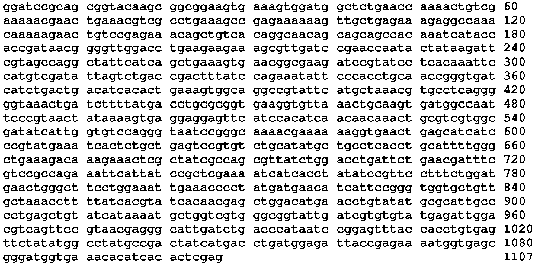
- <210> 83
<211> 237
<212> DNA
<213> Artificial Sequence - <220>
<223> Codon optimized lysyl tRNA Synthetase polynucleotide - <400> 83

- <210> 84
<211> 591
<212> DNA
<213> Artificial Sequence - <220>
<223> Codon optimized lysyl tRNA Synthetase polynucleotide - <400> 84

- <210> 85
<211> 204
<212> DNA
<213> Artificial Sequence - <220>
<223> Codon optimized lysyl tRNA Synthetase polynucleotide - <400> 85

- <210> 86
<211> 651
<212> DNA
<213> Artificial Sequence - <220>
<223> Codon optimized lysyl tRNA Synthetase polynucleotide - <400> 86


- <210> 87
<211> 297
<212> DNA
<213> Artificial Sequence - <220>
<223> Codon optimized lysyl tRNA Synthetase polynucleotide - <400> 87

- <210> 88
<211> 1227
<212> DNA
<213> Artificial Sequence - <220>
<223> Codon optimized lysyl tRNA Synthetase polynucleotide - <400> 88

- <210> 89
<211> 1164
<212> DNA
<213> Artificial Sequence - <220>
<223> Codon optimized lysyl tRNA Synthetase polynucleotide - <400> 89


- <210> 90
<211> 237
<212> DNA
<213> Artificial Sequence - <220>
<223> Codon optimized lysyl tRNA Synthetase polynucleotide - <400> 90

- <210> 91
<211> 72
<212> DNA
<213> Artificial Sequence - <220>
<223> Codon optimized lysyl tRNA Synthetase polynucleotide - <400> 91

- <210> 92
<211> 456
<212> DNA
<213> Artificial Sequence - <220>
<223> Codon optimized lysyl tRNA Synthetase polynucleotide - <400> 92
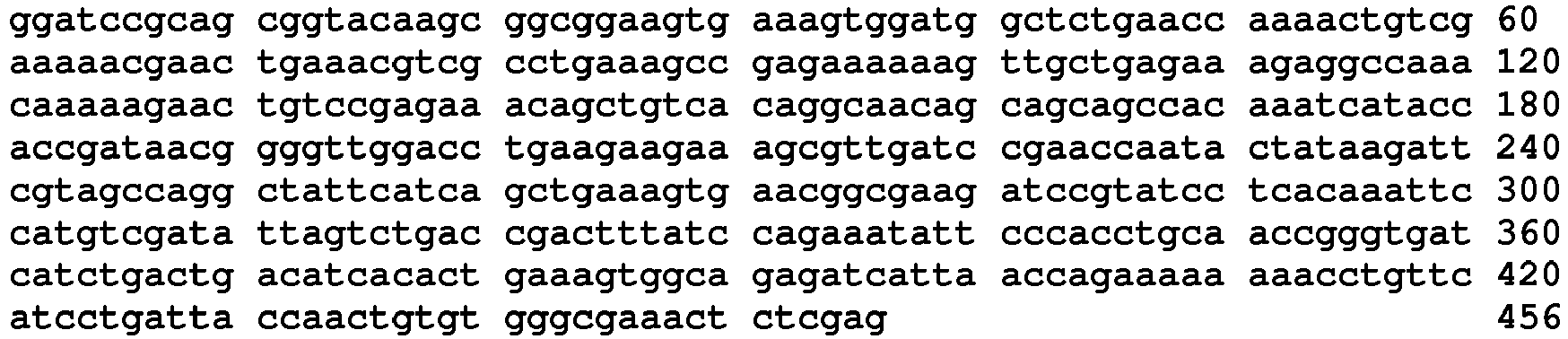
- <210> 93
<211> 786
<212> DNA
<213> Artificial Sequence - <220>
<223> Codon optimized lysyl tRNA Synthetase polynucleotide - <400> 93

- <210> 94
<211> 541
<212> PRT
<213> Homo sapiens - <400> 94


- <210> 95
<211> 1626
<212> DNA
<213> Homo sapiens - <400> 95


- <210> 96
<211> 16
<212> PRT
<213> Mus musculus - <400> 96

- <210> 97
<211> 10
<212> PRT
<213> Mus musculus - <400> 97

- <210> 98
<211> 10
<212> PRT
<213> Mus musculus - <400> 98

- <210> 99
<211> 132
<212> PRT
<213> Mus musculus - <400> 99

- <210> 100
<211> 11
<212> PRT
<213> Mus musculus - <400> 100

- <210> 101
<211> 166
<212> PRT
<213> Mus musculus - <400> 101

- <210> 102
<211> 13
<212> PRT
<213> Mus musculus - <400> 102

- <210> 103
<211> 26
<212> PRT
<213> Mus musculus - <400> 103

- <210> 104
<211> 28
<212> PRT
<213> Mus musculus - <400> 104

- <210> 105
<211> 19
<212> PRT
<213> Mus musculus - <400> 105

- <210> 106
<211> 14
<212> PRT
<213> Mus musculus - <400> 106

- <210> 107
<211> 46
<212> PRT
<213> Mus musculus - <400> 107

- <210> 108
<211> 12
<212> PRT
<213> Mus musculus - <400> 108

- <210> 109
<211> 13
<212> PRT
<213> Mus musculus - <400> 109

- <210> 110
<211> 19
<212> PRT
<213> Mus musculus - <400> 110

- <210> 111
<211> 535
<212> PRT
<213> Mus musculus - <400> 111


- <210> 112
<211> 441
<212> PRT
<213> Homo sapiens - <400> 112


- <210> 113
<211> 1326
<212> DNA
<213> Homo sapiens - <400> 113

- <210> 114
<211> 449
<212> PRT
<213> Homo sapiens - <400> 114


- <210> 115
<211> 1350
<212> DNA
<213> Homo sapiens - <400> 115


- <210> 116
<211> 386
<212> PRT
<213> Homo sapiens - <400> 116


- <210> 117
<211> 1161
<212> DNA
<213> Homo sapiens - <400> 117

- <210> 118
<211> 129
<212> PRT
<213> Homo sapiens - <400> 118

- <210> 119
<211> 390
<212> DNA
<213> Homo sapiens - <400> 119

- <210> 120
<211> 50
<212> DNA
<213> Homo sapiens - <400> 120
acaagtcatt ttctgatcaa agaagcaata ctacaaaatc cgcagtcaag 50 - <210> 121
<211> 50
<212> DNA
<213> Homo sapiens - <400> 121
tgaggaagag agcgtggacc caaataaatt ataaatcaga agaagaattt 50 - <210> 122
<211> 16
<212> PRT
<213> Homo sapiens - <400> 122

- <210> 123
<211> 50
<212> DNA
<213> Homo sapiens - <400> 123
actgacatca ccttaaaggt ggcagaaatt ataaatcaga agaagaattt 50 - <210> 124
<211> 50
<212> DNA
<213> Homo sapiens - <400> 124
tcttcacttt ggcctcaaag acaagaaact cgcaaaattc ttgatgatat 50 - <210> 125
<211> 465
<212> PRT
<213> Artificial Sequence - <220>
<223> Lysyl tRNA Synthetase polypeptide with N-terminal 6xHis affinity tag - <400> 125


- <210> 126
<211> 465
<212> PRT
<213> Artificial Sequence - <220>
<223> Lysyl tRNA Synthetase polypeptide with C-terminal 6xHis affinity tag - <400> 126

- <210> 127
<211> 473
<212> PRT
<213> Artificial Sequence - <220>
<223> Lysyl tRNA Synthetase polypeptide with N-terminal 6xHis affinity tag - <400> 127


- <210> 128
<211> 473
<212> PRT
<213> Artificial Sequence - <220>
<223> Lysyl tRNA Synthetase polypeptide with C-terminal 6xHis affinity tag - <400> 128


- <210> 129
<211> 410
<212> PRT
<213> Artificial Sequence - <220>
<223> Lysyl tRNA Synthetase polypeptide with N-terminal 6xHis affinity tag - <400> 129


- <210> 130
<211> 410
<212> PRT
<213> Artificial Sequence - <220>
<223> Lysyl tRNA Synthetase polypeptide with C-terminal 6xHis affinity tag - <400> 130


- <210> 131
<211> 153
<212> PRT
<213> Artificial Sequence - <220>
<223> Lysyl tRNA Synthetase polypeptide with N-terminal 6xHis affinity tag - <400> 131

- <210> 132
<211> 153
<212> PRT
<213> Artificial Sequence - <220>
<223> Lysyl tRNA Synthetase polypeptide with C-terminal 6xHis affinity tag - <400> 132


- <210> 133
<211> 1332
<212> DNA
<213> Artificial Sequence - <220>
<223> Codon optimized lysyl tRNA Synthetase polynucleotide - <400> 133

- <210> 134
<211> 1356
<212> DNA
<213> Artificial Sequence - <220>
<223> Codon optimized lysyl tRNA Synthetase polynucleotide - <400> 134


- <210> 135
<211> 1167
<212> DNA
<213> Artificial Sequence - <220>
<223> Codon optimized lysyl tRNA Synthetase polynucleotide - <400> 135

- <210> 136
<211> 396
<212> DNA
<213> Artificial Sequence - <220>
<223> Codon optimized lysyl tRNA Synthetase polynucleotide - <400> 136

- <210> 137
<211> 150
<212> PRT
<213> Homo sapiens - <400> 137

- <210> 138
<211> 450
<212> DNA
<213> Homo sapiens - <400> 138

- <210> 139
<211> 175
<212> PRT
<213> Artificial Sequence - <220>
<223> Lysyl tRNA Synthetase polypeptide with N-terminal 6xHis affinity tag - <400> 139


- <210> 140
<211> 175
<212> PRT
<213> Artificial Sequence - <220>
<223> Lysyl tRNA Synthetase polypeptide with C-terminal 6xHis affinity tag - <400> 140

- <210> 141
<211> 462
<212> DNA
<213> Artificial Sequence - <220>
<223> Codon optimized lysyl tRNA Synthetase polynucleotide - <400> 141

- <210> 142
<211> 394
<212> PRT
<213> Homo sapiens - <400> 142

- <210> 143
<211> 1182
<212> DNA
<213> Homo sapiens - <400> 143


- <210> 144
<211> 13
<212> PRT
<213> Mus musculus - <400> 144

- <210> 145
<211> 40
<212> PRT
<213> Mus musculus - <400> 145

- <210> 146
<211> 11
<212> PRT
<213> Mus musculus - <400> 146

- <210> 147
<211> 64
<212> PRT
<213> mUS MUSCULUS - <400> 147


- <210> 148
<211> 61
<212> PRT
<213> Homo sapiens - <400> 148

- <210> 149
<211> 186
<212> DNA
<213> Homo sapiens - <400> 149

- <210> 150
<211> 124
<212> PRT
<213> Homo sapiens - <400> 150

- <210> 151
<211> 375
<212> DNA
<213> Homo sapiens - <400> 151


- <210> 152
<211> 585
<212> PRT
<213> Homo sapiens - <400> 152


- <210> 153
<211> 1758
<212> DNA
<213> Homo sapiens - <400> 153

- <210> 154
<211> 434
<212> PRT
<213> Homo sapiens - <400> 154

- <210> 155
<211> 1305
<212> DNA
<213> Homo sapiens - <400> 155


- <210> 156
<211> 413
<212> PRT
<213> Homo sapiens - <400> 156


- <210> 157
<211> 1242
<212> DNA
<213> Homo sapiens - <400> 157

- <210> 158
<211> 104
<212> PRT
<213> Homo sapiens - <400> 158

- <210> 159
<211> 315
<212> DNA
<213> Homo sapiens - <400> 159

- <210> 160
<211> 49
<212> PRT
<213> Homo sapiens - <400> 160

- <210> 161
<211> 150
<212> DNA
<213> Homo sapiens - <400> 161

- <210> 162
<211> 177
<212> PRT
<213> Homo sapiens - <400> 162
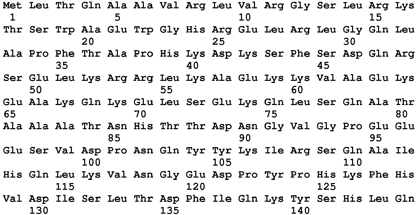

- <210> 163
<211> 534
<212> DNA
<213> Homo sapiens - <400> 163

- <210> 164
<211> 287
<212> PRT
<213> Homo sapiens - <400> 164


- <210> 165
<211> 864
<212> DNA
<213> Homo sapiens - <400> 165

- <210> 166
<211> 50
<212> DNA
<213> Homo sapiens - <400> 166
acaagtcatt ttctgatcaa agaagcaata ctacaaaatc cgcagtcaag 50 - <210> 167
<211> 16
<212> PRT
<213> Homo sapiens - <400> 167

- <210> 168
<211> 50
<212> DNA
<213> Homo sapiens - <400> 168
tgaggaagag agcgtggacc caaatgtagg atccatgcca aaagagcttc 50 - <210> 169
<211> 16
<212> PRT
<213> Homo sapiens - <400> 169

- <210> 170
<211> 50
<212> DNA
<213> Homo sapiens - <400> 170
cttagatgag ctgggattcc tagagatgct tgtggttggt ggcatcgacc 50 - <210> 171
<211> 16
<212> PRT
<213> Homo sapiens - <400> 171

- <210> 172
<211> 49
<212> DNA
<213> Homo sapiens - <400> 172
tgaggaagag agcgtggacc caaatattga aactcccatg atgaacatc 49 - <210> 173
<211> 16
<212> PRT
<213> Homo sapiens - <400> 173

- <210> 174
<211> 50
<212> DNA
<213> Homo sapiens - <400> 174
aattgctcca gaactctatc ataaggccaa ggctgcaggt gatgatgagg 50 - <210> 175
<211> 16
<212> PRT
<213> Homo sapiens - <400> 175

- <210> 176
<211> 50
<212> DNA
<213> Homo sapiens - <400> 176
tgaggaagag agcgtggacc caaataaatt ataaatcaga agaagaattt 50 - <210> 177
<211> 10
<212> PRT
<213> Homo sapiens - <400> 177


- <210> 178
<211> 50
<212> DNA
<213> Homo sapiens - <400> 178
acaagtcatt ttctgatcaa agaaggtagg atccatgcca aaagagcttc 50 - <210> 179
<211> 8
<212> PRT
<213> Homo sapiens - <400> 179

- <210> 180
<211> 50
<212> DNA
<213> Homo sapiens - <400> 180
actgacatca ccttaaaggt ggcagaaatt ataaatcaga agaagaattt 50 - <210> 181
<211> 16
<212> PRT
<213> Homo sapiens - <400> 181

- <210> 182
<211> 50
<212> DNA
<213> Homo sapiens - <400> 182
tcttcacttt ggcctcaaag acaagaaact cgcaaaattc ttgatgatat 50 - <210> 183
<211> 16
<212> PRT
<213> Homo sapiens - <400> 183

- <210> 184
<211> 104
<212> PRT
<213> Homo sapiens - <400> 184


- <210> 185
<211> 312
<212> DNA
<213> Homo sapiens - <400> 185

- <210> 186
<211> 222
<212> PRT
<213> Homo sapiens - <400> 186

- <210> 187
<211> 666
<212> DNA
<213> Homo sapiens - <400> 187

- <210> 188
<211> 93
<212> PRT
<213> Homo sapiens - <400> 188

- <210> 189
<211> 279
<212> DNA
<213> Homo sapiens - <400> 189

- <210> 190
<211> 242
<212> PRT
<213> Homo sapiens - <400> 190


- <210> 191
<211> 726
<212> DNA
<213> Homo sapiens - <400> 191

- <210> 192
<211> 418
<212> PRT
<213> Artificial Sequence - <220>
<223> Lysyl tRNA Synthetase polypeptide with N-terminal 6xHis affinity tag - <400> 192


- <210> 193
<211> 418
<212> PRT
<213> Artificial Sequence - <220>
<223> Lysyl tRNA Synthetase polypeptide with C-terminal 6xHis affinity tag - <400> 193


- <210> 194
<211> 128
<212> PRT
<213> Artificial Sequence - <220>
<223> Lysyl tRNA Synthetase polypeptide with N-terminal 6xHis affinity tag - <400> 194


- <210> 195
<211> 128
<212> PRT
<213> Artificial Sequence - <220>
<223> Lysyl tRNA Synthetase polypeptide with C-terminal 6xHis affinity tag - <400> 195

- <210> 196
<211> 246
<212> PRT
<213> Artificial Sequence - <220>
<223> Lysyl tRNA Synthetase polypeptide with N-terminal 6xHis affinity tag - <400> 196


- <210> 197
<211> 246
<212> PRT
<213> Artificial Sequence - <220>
<223> Lysyl tRNA Synthetase polypeptide with C-terminal 6xHis affinity tag - <400> 197

- <210> 198
<211> 117
<212> PRT
<213> Artificial Sequence - <220>
<223> Lysyl tRNA Synthetase polypeptide with N-terminal 6xHis affinity tag - <400> 198

- <210> 199
<211> 117
<212> PRT
<213> Artificial Sequence - <220>
<223> Lysyl tRNA Synthetase polypeptide with C-terminal 6xHis affinity tag - <400> 199

- <210> 200
<211> 266
<212> PRT
<213> Artificial Sequence - <220>
<223> Lysyl tRNA Synthetase polypeptide with N-terminal 6xHis affinity tag - <400> 200


- <210> 201
<211> 266
<212> PRT
<213> Artificial Sequence - <220>
<223> Lysyl tRNA Synthetase polypeptide with C-terminal 6xHis affinity tag - <400> 201

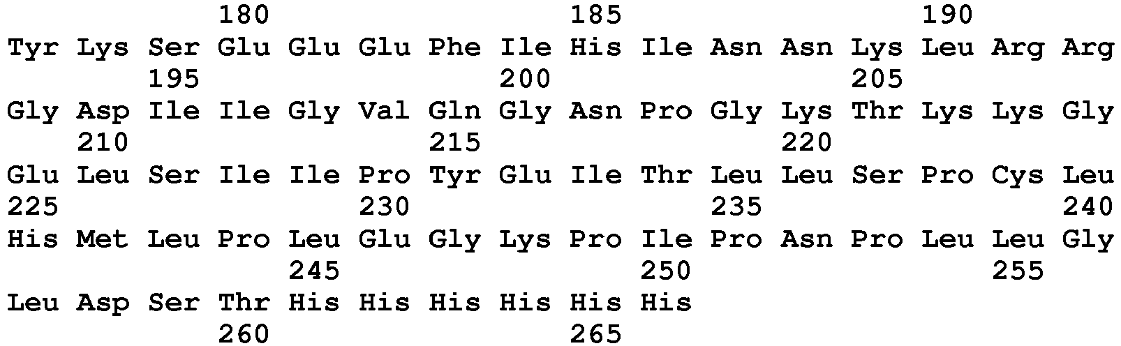
- <210> 202
<211> 85
<212> PRT
<213> Artificial Sequence - <220>
<223> Lysyl tRNA Synthetase polypeptide with N-terminal 6xHis affinity tag - <400> 202

- <210> 203
<211> 85
<212> PRT
<213> Artificial Sequence - <220>
<223> Lysyl tRNA Synthetase polypeptide with C-terminal 6xHis affinity tag - <400> 203

- <210> 204
<211> 148
<212> PRT
<213> Artificial Sequence - <220>
<223> Lysyl tRNA Synthetase polypeptide with N-terminal 6xHis affinity tag - <400> 204

- <210> 205
<211> 148
<212> PRT
<213> Artificial Sequence - <220>
<223> Lysyl tRNA Synthetase polypeptide with C-terminal 6xHis affinity tag - <400> 205

- <210> 206
<211> 458
<212> PRT
<213> Artificial Sequence - <220>
<223> Lysyl tRNA Synthetase polypeptide with N-terminal 6xHis affinity tag - <400> 206

- <210> 207
<211> 458
<212> PRT
<213> Artificial Sequence - <220>
<223> Lysyl tRNA Synthetase polypeptide with C-terminal 6xHis affinity tag - <400> 207


- <210> 208
<211> 437
<212> PRT
<213> Artificial Sequence - <220>
<223> Lysyl tRNA Synthetase polypeptide with N-terminal 6xHis affinity tag - <400> 208


- <210> 209
<211> 437
<212> PRT
<213> Artificial Sequence - <220>
<223> Lysyl tRNA Synthetase polypeptide with C-terminal 6xHis affinity tag - <400> 209


- <210> 210
<211> 128
<212> PRT
<213> Artificial Sequence - <220>
<223> Lysyl tRNA Synthetase polypeptide with N-terminal 6xHis affinity tag - <400> 210

- <210> 211
<211> 128
<212> PRT
<213> Artificial Sequence - <220>
<223> Lysyl tRNA Synthetase polypeptide with C-terminal 6xHis affinity tag - <400> 211

- <210> 212
<211> 73
<212> PRT
<213> Artificial Sequence - <220>
<223> Lysyl tRNA Synthetase polypeptide with N-terminal 6xHis affinity tag - <400> 212

- <210> 213
<211> 73
<212> PRT
<213> Artificial Sequence - <220>
<223> Lysyl tRNA Synthetase polypeptide with C-terminal 6xHis affinity tag - <400> 213

- <210> 214
<211> 201
<212> PRT
<213> Artificial Sequence - <220>
<223> Lysyl tRNA Synthetase polypeptide with N-terminal 6xHis affinity tag - <400> 214


- <210> 215
<211> 201
<212> PRT
<213> Artificial Sequence - <220>
<223> Lysyl tRNA Synthetase polypeptide with C-terminal 6xHis affinity tag - <400> 215

- <210> 216
<211> 311
<212> PRT
<213> Artificial Sequence - <220>
<223> Lysyl tRNA Synthetase polypeptide with N-terminal 6xHis affinity tag - <400> 216

- <210> 217
<211> 311
<212> PRT
<213> Artificial Sequence - <220>
<223> Lysyl tRNA Synthetase polypeptide with C-terminal 6xHis affinity tag - <400> 217


- <210> 218
<211> 1191
<212> DNA
<213> Artificial Sequence - <220>
<223> Codon optimized lysyl tRNA Synthetase polynucleotide - <400> 218

- <210> 219
<211> 321
<212> DNA
<213> Artificial Sequence - <220>
<223> Codon optimized lysyl tRNA Synthetase polynucleotide - <400> 219

- <210> 220
<211> 675
<212> DNA
<213> Artificial Sequence - <220>
<223> Codon optimized lysyl tRNA Synthetase polynucleotide - <400> 220

- <210> 221
<211> 288
<212> DNA
<213> Artificial Sequence - <220>
<223> Codon optimized lysyl tRNA Synthetase polynucleotide - <400> 221

- <210> 222
<211> 735
<212> DNA
<213> Artificial Sequence - <220>
<223> Codon optimized lysyl tRNA Synthetase polynucleotide - <400> 222


- <210> 223
<211> 192
<212> DNA
<213> Artificial Sequence - <220>
<223> Codon optimized lysyl tRNA Synthetase polynucleotide - <400> 223

- <210> 224
<211> 381
<212> DNA
<213> Artificial Sequence - <220>
<223> Codon optimized lysyl tRNA Synthetase polynucleotide - <400> 224

- <210> 225
<211> 1311
<212> DNA
<213> Artificial Sequence - <220>
<223> Codon optimized lysyl tRNA Synthetase polynucleotide - <400> 225


- <210> 226
<211> 1248
<212> DNA
<213> Artificial Sequence - <220>
<223> Codon optimized lysyl tRNA Synthetase polynucleotide - <400> 226

- <210> 227
<211> 321
<212> DNA
<213> Artificial Sequence - <220>
<223> Codon optimized lysyl tRNA Synthetase polynucleotide - <400> 227

- <210> 228
<211> 156
<212> DNA
<213> Artificial Sequence - <220>
<223> Codon optimized lysyl tRNA Synthetase polynucleotide - <400> 228

- <210> 229
<211> 540
<212> DNA
<213> Artificial Sequence - <220>
<223> Codon optimized lysyl tRNA Synthetase polynucleotide - <400> 229

- <210> 230
<211> 870
<212> DNA
<213> Artificial Sequence - <220>
<223> Codon optimized lysyl tRNA Synthetase polynucleotide - <400> 230

Claims (5)
- An isolated aminoacyl-tRNA synthetase (AARS) polypeptide consisting of an amino acid sequence that is at least 80%, 85%, 90%, 95%, 98%, or 100% identical to SEQ ID NO:54, wherein the polypeptide comprises the non-canonical biological activity of modulation of cell adhesion and chemotaxis.
- The AARS polypeptide of claim 1, wherein the AARS polypeptide is fused to a heterologous polypeptide selected from the group consisting of purification tags, epitope tags, targeting sequences, signal peptides, membrane translocating sequences, and PK modifiers.
- A pharmaceutical composition comprising an AARS polypeptide of claim 1 or 2 and a pharmaceutically acceptable carrier.
- The pharmaceutical composition of claim 3 for use in treating a subject having an inflammatory disorder.
- The pharmaceutical composition for the use of claim 4, wherein the inflammatory disorder is selected from cardiovascular diseases, atherosclerosis, autoimmunity, and pulmonary hypertension.
Applications Claiming Priority (9)
| Application Number | Priority Date | Filing Date | Title |
|---|---|---|---|
| US35040910P | 2010-06-01 | 2010-06-01 | |
| US35024210P | 2010-06-01 | 2010-06-01 | |
| US35041110P | 2010-06-01 | 2010-06-01 | |
| US35040710P | 2010-06-01 | 2010-06-01 | |
| US201161488514P | 2011-05-20 | 2011-05-20 | |
| US201161488503P | 2011-05-20 | 2011-05-20 | |
| US201161488528P | 2011-05-20 | 2011-05-20 | |
| US201161488619P | 2011-05-20 | 2011-05-20 | |
| PCT/US2011/038813 WO2011153277A2 (en) | 2010-06-01 | 2011-06-01 | Innovative discovery of therapeutic, diagnostic, and antibody compositions related to protein fragments of lysyl-trna synthetases |
Publications (3)
| Publication Number | Publication Date |
|---|---|
| EP2575857A2 EP2575857A2 (en) | 2013-04-10 |
| EP2575857A4 EP2575857A4 (en) | 2014-01-01 |
| EP2575857B1 true EP2575857B1 (en) | 2018-01-24 |
Family
ID=45067276
Family Applications (1)
| Application Number | Title | Priority Date | Filing Date |
|---|---|---|---|
| EP11790361.7A Active EP2575857B1 (en) | 2010-06-01 | 2011-06-01 | Innovative discovery of therapeutic, diagnostic, and antibody compositions related to protein fragments of lysyl-trna synthetases |
Country Status (7)
| Country | Link |
|---|---|
| US (3) | US8962560B2 (en) |
| EP (1) | EP2575857B1 (en) |
| JP (2) | JP5906237B2 (en) |
| CN (1) | CN103118694B (en) |
| AU (1) | AU2011261486B2 (en) |
| CA (1) | CA2800281C (en) |
| WO (1) | WO2011153277A2 (en) |
Families Citing this family (51)
| Publication number | Priority date | Publication date | Assignee | Title |
|---|---|---|---|---|
| EP2403864B1 (en) | 2009-02-27 | 2015-08-12 | Atyr Pharma, Inc. | Polypeptide structural motifs associated with cell signaling activity |
| AU2011248625B2 (en) | 2010-04-26 | 2017-01-05 | Pangu Biopharma Limited | Innovative discovery of therapeutic, diagnostic, and antibody compositions related to protein fragments of cysteinyl-tRNA synthetase |
| CN103096911B (en) | 2010-04-27 | 2018-05-29 | Atyr 医药公司 | Treatment relevant with the protein fragments of Isoleucyl-tRNA synthetase, diagnosis and the innovation of antibody compositions are found |
| CN103096910A (en) | 2010-04-27 | 2013-05-08 | Atyr医药公司 | Innovative discovery of therapeutic, diagnostic, and antibody compositions related to protein fragments of threonyl tRNA synthetases |
| JP6008837B2 (en) | 2010-04-28 | 2016-10-19 | エータイアー ファーマ, インコーポレイテッド | Innovative discovery of therapeutic, diagnostic and antibody compositions related to protein fragments of alanyl tRNA synthetase |
| US9068177B2 (en) | 2010-04-29 | 2015-06-30 | Atyr Pharma, Inc | Innovative discovery of therapeutic, diagnostic, and antibody compositions related to protein fragments of glutaminyl-tRNA synthetases |
| WO2011139907A2 (en) | 2010-04-29 | 2011-11-10 | Atyr Pharma, Inc. | Innovative discovery of therapeutic, diagnostic, and antibody compositions related to protein fragments of valyl trna synthetases |
| CN103097523B (en) | 2010-04-29 | 2016-09-28 | Atyr医药公司 | The innovation for the treatment of, diagnosis and the antibody compositions relevant to the protein fragments of Asparaginyl-tRNA synthetase finds |
| WO2011139986A2 (en) | 2010-05-03 | 2011-11-10 | Atyr Pharma, Inc. | Innovative discovery of therapeutic, diagnostic, and antibody compositions related to protein fragments of arginyl-trna synthetases |
| WO2011139988A2 (en) | 2010-05-03 | 2011-11-10 | Atyr Pharma, Inc. | Innovative discovery of therapeutic, diagnostic, and antibody compositions related to protein fragments of seryl-trna synthetases |
| CN105820252B (en) | 2010-05-03 | 2020-07-21 | Atyr 医药公司 | Innovative discovery of therapeutic, diagnostic, and antibody compositions related to protein fragments of phenylalanyl- α -tRNA synthetases |
| AU2011248230B2 (en) | 2010-05-03 | 2016-10-06 | Pangu Biopharma Limited | Innovative discovery of therapeutic, diagnostic, and antibody compositions related to protein fragments of methionyl-tRNA synthetases |
| CN103096909A (en) | 2010-05-04 | 2013-05-08 | Atyr医药公司 | Innovative discovery of therapeutic, diagnostic, and antibody compositions related to protein fragments of glutamyl-prolyl-trna synthetases |
| JP6008844B2 (en) | 2010-05-04 | 2016-10-19 | エータイアー ファーマ, インコーポレイテッド | Innovative discovery of therapeutic, diagnostic and antibody compositions related to protein fragments of the p38 MULTI-tRNA synthetase complex |
| CN103200953B (en) | 2010-05-14 | 2017-02-15 | Atyr 医药公司 | Innovative discovery of therapeutic, diagnostic, and antibody compositions related to protein fragments of phenylalanyl-beta-trna synthetases |
| JP6027965B2 (en) | 2010-05-17 | 2016-11-16 | エータイアー ファーマ, インコーポレイテッド | Innovative discovery of therapeutic, diagnostic and antibody compositions related to protein fragments of leucyl-tRNA synthetase |
| JP5906237B2 (en) | 2010-06-01 | 2016-04-20 | エータイアー ファーマ, インコーポレイテッド | Innovative discovery of therapeutic, diagnostic and antibody compositions related to protein fragments of lysyl tRNA synthetase |
| EP2593124B1 (en) | 2010-07-12 | 2017-04-26 | aTyr Pharma, Inc. | Innovative discovery of therapeutic, diagnostic, and antibody compositions related to protein fragments of aspartyl-trna synthetases |
| US8999321B2 (en) | 2010-07-12 | 2015-04-07 | Atyr Pharma, Inc. | Innovative discovery of therapeutic, diagnostic, and antibody compositions related to protein fragments of glycyl-tRNA synthetases |
| CN103108649B (en) | 2010-07-12 | 2016-10-12 | Atyr医药公司 | Treatment, diagnosis and the antibody compositions relevant to the protein fragments of Jo-1 |
| EP2593125B1 (en) | 2010-07-12 | 2017-11-01 | aTyr Pharma, Inc. | Innovative discovery of therapeutic, diagnostic, and antibody compositions related to protein fragments of glycyl-trna synthetases |
| WO2012027611A2 (en) | 2010-08-25 | 2012-03-01 | Atyr Pharma, Inc. | INNOVATIVE DISCOVERY OF THERAPEUTIC, DIAGNOSTIC, AND ANTIBODY COMPOSITIONS RELATED TO PROTEIN FRAGMENTS OF TYROSYL-tRNA SYNTHETASES |
| US9399770B2 (en) | 2010-10-06 | 2016-07-26 | Atyr Pharma, Inc. | Innovative discovery of therapeutic, diagnostic, and antibody compositions related to protein fragments of tryptophanyl-tRNA synthetases |
| US20160144003A1 (en) * | 2011-05-19 | 2016-05-26 | The Scripps Research Institute | Compositions and methods for treating charcot-marie-tooth diseases and related neuronal diseases |
| US9714419B2 (en) | 2011-08-09 | 2017-07-25 | Atyr Pharma, Inc. | PEGylated tyrosyl-tRNA synthetase polypeptides |
| WO2013086216A1 (en) | 2011-12-06 | 2013-06-13 | Atyr Pharma, Inc. | Improved aspartyl-trna synthetases |
| WO2013086228A1 (en) | 2011-12-06 | 2013-06-13 | Atyr Pharma, Inc. | Pegylated aspartyl-trna synthetase polypeptides |
| JP6169608B2 (en) | 2011-12-29 | 2017-07-26 | エータイアー ファーマ, インコーポレイテッド | Aspartyl tRNA synthetase-Fc conjugate |
| US10472683B2 (en) | 2012-06-01 | 2019-11-12 | Akron Biotechnology, LLC. | Detection of Mycoplasma in cell cultures and cell culture derived biologicals |
| DK2970921T3 (en) | 2013-03-15 | 2019-01-14 | Atyr Pharma Inc | Histidyl-tRNA synthetase-Fc conjugates |
| CN105431739A (en) * | 2013-05-15 | 2016-03-23 | 医药生命融合研究团 | Method for screening cancer metastasis inhibitor using culture of cells or spheroidically aggregated cells in which lysyl-trna synthetase is regulated to be expressed or unexpressed |
| CN111235149B (en) * | 2013-09-05 | 2024-04-16 | 萨罗塔治疗公司(美国) | Antisense-induced exon 2 incorporation in acid alpha-glucosidase |
| JP6519842B2 (en) * | 2013-10-04 | 2019-05-29 | 国立大学法人神戸大学 | Antisense nucleic acid for treatment of Fukuyama muscular dystrophy |
| JP2017502672A (en) * | 2013-12-30 | 2017-01-26 | メディシナル バイオコンバージェンス リサーチ センター | Anti-KRS monoclonal antibody and use thereof |
| KR101787468B1 (en) * | 2014-05-28 | 2017-11-15 | 재단법인 의약바이오컨버젼스연구단 | Novel lysyl tRNA synthetase fragment and microvesicle comprising the same |
| CN106661580B (en) | 2014-06-10 | 2022-02-15 | 鹿特丹伊拉斯谟大学医疗中心 | Antisense oligonucleotides for treating pompe disease |
| US11339392B2 (en) | 2016-03-02 | 2022-05-24 | The Board Of Trustees Of The Leland Stanford Junior University | Pan-genotypic agents against influenza virus and methods of using the same |
| EP3423107A4 (en) * | 2016-03-02 | 2020-01-15 | The Board of Trustees of the Leland Stanford Junior University | Pan-genotypic agents against influenza virus and methods of using the same |
| KR102522059B1 (en) | 2016-04-18 | 2023-04-14 | 사렙타 쎄러퓨틱스 인코퍼레이티드 | Antisense oligomers and methods of their use to treat diseases associated with the acid alpha-glucosidase gene |
| JP6792001B2 (en) * | 2016-07-01 | 2020-11-25 | メディシナル バイオコンバージェンス リサーチ センター | Composition for diagnosing thyroid cyst cancer using aminoacyl-tRNA synthetase-related protein expression level and diagnostic marker detection method |
| RU2739393C1 (en) * | 2017-03-27 | 2020-12-23 | БайоконТАК Ко., Лтд. | Antibody specifically binding to n-terminal region of lysyl-trna-synthetase exposed on cell membrane |
| KR20180109645A (en) * | 2017-03-27 | 2018-10-08 | 재단법인 의약바이오컨버젼스연구단 | Antibody specifically binding to the N-terminal region of lysyl- tRNA synthethase exposed at cell outer membrane |
| WO2018182284A1 (en) * | 2017-03-27 | 2018-10-04 | 재단법인 의약바이오컨버젼스연구단 | Antibody binding specifically to n-terminal region of lysyl-trna synthetase exposed on cell membrane |
| CN110536694A (en) | 2017-04-20 | 2019-12-03 | Atyr 医药公司 | For treating pulmonary inflammatory composition and method |
| KR102133929B1 (en) | 2017-06-16 | 2020-07-14 | 재단법인 의약바이오컨버젼스연구단 | Therapeutic agent of immunocyte migration-related diseases and method for screening thereof |
| US11826358B2 (en) | 2017-09-13 | 2023-11-28 | North Carolina State University | Locally-induced adipose tissue browning by microneedle patch for obesity treatment |
| CA3084461A1 (en) * | 2017-09-15 | 2019-03-21 | Medicinal Bioconvergence Research Center | Pharmaceutical composition comprising antibody binding specifically to lysyl-trna synthetase n-terminus as effective ingredient for preventing or treating immune cell migration-related disease |
| KR102290507B1 (en) * | 2018-09-17 | 2021-08-17 | 주식회사 자이메디 | Antibody specifically binding to the N-terminal region of lysyl- tRNA synthetase exposed at cell outer membrane |
| DE102018127075B4 (en) | 2018-10-30 | 2021-12-30 | Auto-Kabel Management Gmbh | High current circuit |
| JP2023508893A (en) * | 2019-12-19 | 2023-03-06 | インダストリ-アカデミック コオペレーション ファウンデイション, ヨンセイ ユニヴァーシティ | Method for enhancing water solubility of target protein by WHEP domain fusion |
| CN116445597A (en) * | 2022-09-10 | 2023-07-18 | 福建医科大学附属第一医院 | Human CMT pathogenic gene SARS1 with 1187 position mutation and its application |
Family Cites Families (293)
| Publication number | Priority date | Publication date | Assignee | Title |
|---|---|---|---|---|
| US3773919A (en) | 1969-10-23 | 1973-11-20 | Du Pont | Polylactide-drug mixtures |
| US4483105A (en) | 1983-03-11 | 1984-11-20 | Maschinenfabrik Gehring, Gesellschaft Mit Beschrankter Haftung & Co. Kommanditgesellschaft | Device to hold workpieces for the honing of their axial center bore |
| US4965188A (en) | 1986-08-22 | 1990-10-23 | Cetus Corporation | Process for amplifying, detecting, and/or cloning nucleic acid sequences using a thermostable enzyme |
| US4683202A (en) | 1985-03-28 | 1987-07-28 | Cetus Corporation | Process for amplifying nucleic acid sequences |
| US4683195A (en) | 1986-01-30 | 1987-07-28 | Cetus Corporation | Process for amplifying, detecting, and/or-cloning nucleic acid sequences |
| US4800159A (en) | 1986-02-07 | 1989-01-24 | Cetus Corporation | Process for amplifying, detecting, and/or cloning nucleic acid sequences |
| US4873192A (en) | 1987-02-17 | 1989-10-10 | The United States Of America As Represented By The Department Of Health And Human Services | Process for site specific mutagenesis without phenotypic selection |
| US5766960A (en) | 1987-07-27 | 1998-06-16 | Australian Membrane And Biotechnology Research Institute | Receptor membranes |
| US4904584A (en) | 1987-12-23 | 1990-02-27 | Genetics Institute, Inc. | Site-specific homogeneous modification of polypeptides |
| US5130238A (en) | 1988-06-24 | 1992-07-14 | Cangene Corporation | Enhanced nucleic acid amplification process |
| US5223409A (en) | 1988-09-02 | 1993-06-29 | Protein Engineering Corp. | Directed evolution of novel binding proteins |
| GB8822228D0 (en) | 1988-09-21 | 1988-10-26 | Southern E M | Support-bound oligonucleotides |
| US5110596A (en) | 1988-12-13 | 1992-05-05 | Alza Corporation | Delivery system comprising means for delivering agent to livestock |
| US5034229A (en) | 1988-12-13 | 1991-07-23 | Alza Corporation | Dispenser for increasing feed conversion of hog |
| US5856092A (en) | 1989-02-13 | 1999-01-05 | Geneco Pty Ltd | Detection of a nucleic acid sequence or a change therein |
| US5871928A (en) | 1989-06-07 | 1999-02-16 | Fodor; Stephen P. A. | Methods for nucleic acid analysis |
| US5547839A (en) | 1989-06-07 | 1996-08-20 | Affymax Technologies N.V. | Sequencing of surface immobilized polymers utilizing microflourescence detection |
| US5925525A (en) | 1989-06-07 | 1999-07-20 | Affymetrix, Inc. | Method of identifying nucleotide differences |
| US5143854A (en) | 1989-06-07 | 1992-09-01 | Affymax Technologies N.V. | Large scale photolithographic solid phase synthesis of polypeptides and receptor binding screening thereof |
| US5800992A (en) | 1989-06-07 | 1998-09-01 | Fodor; Stephen P.A. | Method of detecting nucleic acids |
| US6309822B1 (en) | 1989-06-07 | 2001-10-30 | Affymetrix, Inc. | Method for comparing copy number of nucleic acid sequences |
| US6040138A (en) | 1995-09-15 | 2000-03-21 | Affymetrix, Inc. | Expression monitoring by hybridization to high density oligonucleotide arrays |
| DK0408295T3 (en) | 1989-07-11 | 1996-09-16 | Gen Probe Inc | Methods for amplifying nucleic acid sequences |
| CA2020958C (en) | 1989-07-11 | 2005-01-11 | Daniel L. Kacian | Nucleic acid sequence amplification methods |
| US5725871A (en) | 1989-08-18 | 1998-03-10 | Danbiosyst Uk Limited | Drug delivery compositions comprising lysophosphoglycerolipid |
| US5707644A (en) | 1989-11-04 | 1998-01-13 | Danbiosyst Uk Limited | Small particle compositions for intranasal drug delivery |
| US5747641A (en) | 1989-12-21 | 1998-05-05 | Biogen Inc | Tat-derived transport polypeptide conjugates |
| US5804604A (en) | 1989-12-21 | 1998-09-08 | Biogen, Inc. | Tat-derived transport polypeptides and fusion proteins |
| US6316003B1 (en) | 1989-12-21 | 2001-11-13 | Whitehead Institute For Biomedical Research | Tat-derived transport polypeptides |
| US5466468A (en) | 1990-04-03 | 1995-11-14 | Ciba-Geigy Corporation | Parenterally administrable liposome formulation comprising synthetic lipids |
| US5399363A (en) | 1991-01-25 | 1995-03-21 | Eastman Kodak Company | Surface modified anticancer nanoparticles |
| US5455166A (en) | 1991-01-31 | 1995-10-03 | Becton, Dickinson And Company | Strand displacement amplification |
| US5936731A (en) | 1991-02-22 | 1999-08-10 | Applied Spectral Imaging Ltd. | Method for simultaneous detection of multiple fluorophores for in situ hybridization and chromosome painting |
| DE4137649C2 (en) | 1991-11-15 | 1997-11-20 | Gerhard Dingler | Component |
| US5270184A (en) | 1991-11-19 | 1993-12-14 | Becton, Dickinson And Company | Nucleic acid target generation |
| US5412087A (en) | 1992-04-24 | 1995-05-02 | Affymax Technologies N.V. | Spatially-addressable immobilization of oligonucleotides and other biological polymers on surfaces |
| US5756353A (en) | 1991-12-17 | 1998-05-26 | The Regents Of The University Of California | Expression of cloned genes in the lung by aerosol-and liposome-based delivery |
| DE9115660U1 (en) | 1991-12-18 | 1992-07-30 | Hoechst Ag, 6230 Frankfurt, De | |
| US5541061A (en) | 1992-04-29 | 1996-07-30 | Affymax Technologies N.V. | Methods for screening factorial chemical libraries |
| EP0641192B1 (en) | 1992-05-18 | 1997-07-23 | Minnesota Mining And Manufacturing Company | Transmucosal drug delivery device |
| US5858659A (en) | 1995-11-29 | 1999-01-12 | Affymetrix, Inc. | Polymorphism detection |
| EP0705271B1 (en) | 1993-06-25 | 2002-11-13 | Affymetrix, Inc. (a Delaware Corporation) | Hybridization and sequencing of nucleic acids |
| US5543158A (en) | 1993-07-23 | 1996-08-06 | Massachusetts Institute Of Technology | Biodegradable injectable nanoparticles |
| US6045996A (en) | 1993-10-26 | 2000-04-04 | Affymetrix, Inc. | Hybridization assays on oligonucleotide arrays |
| US5446090A (en) | 1993-11-12 | 1995-08-29 | Shearwater Polymers, Inc. | Isolatable, water soluble, and hydrolytically stable active sulfones of poly(ethylene glycol) and related polymers for modification of surfaces and molecules |
| US5420109A (en) | 1993-11-12 | 1995-05-30 | Houghten Pharmaceuticals, Inc. | Cytokine restraining agents |
| US6090555A (en) | 1997-12-11 | 2000-07-18 | Affymetrix, Inc. | Scanned image alignment systems and methods |
| CA2185239C (en) | 1994-03-16 | 2002-12-17 | Nanibhushan Dattagupta | Isothermal strand displacement nucleic acid amplification |
| US5648211A (en) | 1994-04-18 | 1997-07-15 | Becton, Dickinson And Company | Strand displacement amplification using thermophilic enzymes |
| US5571639A (en) | 1994-05-24 | 1996-11-05 | Affymax Technologies N.V. | Computer-aided engineering system for design of sequence arrays and lithographic masks |
| US5759833A (en) | 1994-05-27 | 1998-06-02 | Cubist Pharmaceuticals, Inc. | Human isoleucyl-tRNA synthetase proteins, nucleic acids and tester strains comprising same |
| US6287850B1 (en) | 1995-06-07 | 2001-09-11 | Affymetrix, Inc. | Bioarray chip reaction apparatus and its manufacture |
| US6379897B1 (en) | 2000-11-09 | 2002-04-30 | Nanogen, Inc. | Methods for gene expression monitoring on electronic microarrays |
| NL9401150A (en) | 1994-07-12 | 1996-02-01 | Nederland Ptt | Method for presenting on a receiving side a first number of video signals originating from a transmitting side, as well as a system, as well as a transmitter, as well as a network, and also a receiver. |
| US5798240A (en) | 1994-09-13 | 1998-08-25 | Cubist Pharmaceuticals, Inc. | Recombinant mycobacterial methionyl-tRNA synthetase genes and methods of use therefore |
| US5756327A (en) | 1994-09-13 | 1998-05-26 | Cubist Pharmaceuticals, Inc. | Recombinant mycobacterial isoleucyl-tRNA synthetase genes, tester strains and assays |
| US5824784A (en) | 1994-10-12 | 1998-10-20 | Amgen Inc. | N-terminally chemically modified protein compositions and methods |
| US5795716A (en) | 1994-10-21 | 1998-08-18 | Chee; Mark S. | Computer-aided visualization and analysis system for sequence evaluation |
| PT788351E (en) | 1994-11-10 | 2003-06-30 | Univ Kentucky Res Foundation T | LIBERTACAOCONTROLADA RECHARGEABLE IMPLANTABLE DEVICE FOR ADMINISTERING MEDICATIONS DIRECTLY ON AN INTERNAL PORTION OF THE BODY |
| US5932462A (en) | 1995-01-10 | 1999-08-03 | Shearwater Polymers, Inc. | Multiarmed, monofunctional, polymer for coupling to molecules and surfaces |
| IE80468B1 (en) | 1995-04-04 | 1998-07-29 | Elan Corp Plc | Controlled release biodegradable nanoparticles containing insulin |
| US5801013A (en) | 1995-05-26 | 1998-09-01 | Cubist Pharmaceuticals, Inc. | Helicobacter aminoacyl-tRNA synthetase proteins, nucleic acids and strains comprising same |
| US5545531A (en) | 1995-06-07 | 1996-08-13 | Affymax Technologies N.V. | Methods for making a device for concurrently processing multiple biological chip assays |
| US5733729A (en) | 1995-09-14 | 1998-03-31 | Affymetrix, Inc. | Computer-aided probability base calling for arrays of nucleic acid probes on chips |
| US5843655A (en) | 1995-09-18 | 1998-12-01 | Affymetrix, Inc. | Methods for testing oligonucleotide arrays |
| US6300063B1 (en) | 1995-11-29 | 2001-10-09 | Affymetrix, Inc. | Polymorphism detection |
| WO1997025426A2 (en) | 1996-01-11 | 1997-07-17 | Corixa Corporation | Compositions and methods for the treatment and diagnosis of breast cancer |
| GB9601067D0 (en) | 1996-01-19 | 1996-03-20 | Smithkline Beecham Plc | Novel compounds |
| EP1288298A3 (en) | 1996-01-19 | 2003-03-12 | Smithkline Beecham Plc | TRNA synthetase from Staphylococcus aureus |
| US5750387A (en) | 1996-01-19 | 1998-05-12 | Smithkline Beecham P.L.C. | DNA encoing leucyl TRNA synthetase from staphylococcus aureus |
| US5795757A (en) | 1997-01-17 | 1998-08-18 | Smithkline Beecham, P.L.C. | DNA encoding threonyl tRNA synthetase from staphylococcus aureus |
| EP0880598A4 (en) | 1996-01-23 | 2005-02-23 | Affymetrix Inc | Nucleic acid analysis techniques |
| US5837196A (en) | 1996-01-26 | 1998-11-17 | The Regents Of The University Of California | High density array fabrication and readout method for a fiber optic biosensor |
| PT877599E (en) | 1996-02-02 | 2003-08-29 | Alza Corp | CONTINUOUS LEUPROLIDAL ADMINISTRATION USING AN IMPLANTABLE SYSTEM |
| US6156331A (en) | 1996-02-02 | 2000-12-05 | Alza Corporation | Sustained delivery of an active agent using an implantable system |
| US6395292B2 (en) | 1996-02-02 | 2002-05-28 | Alza Corporation | Sustained delivery of an active agent using an implantable system |
| US6228575B1 (en) | 1996-02-08 | 2001-05-08 | Affymetrix, Inc. | Chip-based species identification and phenotypic characterization of microorganisms |
| US6114122A (en) | 1996-03-26 | 2000-09-05 | Affymetrix, Inc. | Fluidics station with a mounting system and method of using |
| US6238866B1 (en) | 1996-04-16 | 2001-05-29 | The United States Of America As Represented By The Secretary Of The Army | Detector for nucleic acid typing and methods of using the same |
| GB9607991D0 (en) | 1996-04-18 | 1996-06-19 | Smithkline Beecham Plc | Novel compounds |
| US5795758A (en) | 1997-04-18 | 1998-08-18 | Smithkline Beecham Corporation | DNA encoding histidyl tRNA synthetase variant from Streptococcus pneumoniae |
| GB9607993D0 (en) | 1996-04-18 | 1996-06-19 | Smithkline Beecham Plc | Novel compounds |
| US5976109A (en) | 1996-04-30 | 1999-11-02 | Medtronic, Inc. | Apparatus for drug infusion implanted within a living body |
| GB9609262D0 (en) | 1996-05-02 | 1996-07-03 | Isis Innovation | Peptide library and method |
| US5958342A (en) | 1996-05-17 | 1999-09-28 | Incyte Pharmaceuticals, Inc. | Jet droplet device |
| US6852512B2 (en) * | 1996-10-04 | 2005-02-08 | Hanil Synthetic Fiber Co., Ltd. | Expression vectors for production of foreign proteins as soluble forms |
| KR100203919B1 (en) * | 1996-10-04 | 1999-06-15 | 신동권 | A novel expression vector which produces water soluble protein |
| US5853993A (en) | 1996-10-21 | 1998-12-29 | Hewlett-Packard Company | Signal enhancement method and kit |
| US5885815A (en) | 1996-11-01 | 1999-03-23 | Cubist Pharmaceuticals, Inc. | Candida isoleucyl-tRNA synthetase proteins, nucleic acids and strains comprising same |
| CA2268740C (en) | 1996-11-06 | 2010-07-20 | Sequenom, Inc. | High density immobilization of nucleic acids |
| US5804386A (en) | 1997-01-15 | 1998-09-08 | Incyte Pharmaceuticals, Inc. | Sets of labeled energy transfer fluorescent primers and their use in multi component analysis |
| US6309824B1 (en) | 1997-01-16 | 2001-10-30 | Hyseq, Inc. | Methods for analyzing a target nucleic acid using immobilized heterogeneous mixtures of oligonucleotide probes |
| US5776749A (en) | 1997-01-17 | 1998-07-07 | Smithkline Beecham P.L.C. | DNA encoding cysteinyl tRNA synthetase from Staphylococcus aureus |
| EP0981625A2 (en) | 1997-05-06 | 2000-03-01 | Human Genome Sciences, Inc. | $i(ENTEROCOCCUS FAECALIS) POLYNUCLEOTIDES AND POLYPEPTIDES |
| US6368799B1 (en) | 1997-06-13 | 2002-04-09 | Affymetrix, Inc. | Method to detect gene polymorphisms and monitor allelic expression employing a probe array |
| US6333179B1 (en) | 1997-06-20 | 2001-12-25 | Affymetrix, Inc. | Methods and compositions for multiplex amplification of nucleic acids |
| US5858720A (en) | 1997-07-23 | 1999-01-12 | Smithkline Beecham Corporation | Hiss |
| US5882892A (en) | 1997-07-23 | 1999-03-16 | Smithkline Beecham Corporation | Asps |
| US5939298A (en) | 1997-07-23 | 1999-08-17 | Smithkline Beecham Corporation | DNA encoding phenylalanyl tRNA synthetase alpha sub-unit from chlamydi a trachomatis |
| US6420108B2 (en) | 1998-02-09 | 2002-07-16 | Affymetrix, Inc. | Computer-aided display for comparative gene expression |
| JP2001515234A (en) | 1997-07-25 | 2001-09-18 | アフィメトリックス インコーポレイテッド | System for providing a polymorphism database |
| DE69827154T2 (en) | 1997-08-15 | 2006-03-09 | Affymetrix, Inc. (n.d.Ges.d.Staates Delaware), Santa Clara | POLYMORPHISM IDENTIFICATION WITH HELP CLUSTER ANALYSIS |
| US6511803B1 (en) | 1997-10-10 | 2003-01-28 | President And Fellows Of Harvard College | Replica amplification of nucleic acid arrays |
| WO1999023254A1 (en) | 1997-10-31 | 1999-05-14 | Affymetrix, Inc. | Expression profiles in adult and fetal organs |
| US6054274A (en) | 1997-11-12 | 2000-04-25 | Hewlett-Packard Company | Method of amplifying the signal of target nucleic acid sequence analyte |
| US6013449A (en) | 1997-11-26 | 2000-01-11 | The United States Of America As Represented By The Department Of Health And Human Services | Probe-based analysis of heterozygous mutations using two-color labelling |
| ES2182379T3 (en) | 1997-12-22 | 2003-03-01 | Alza Corp | MEMBRANE OF REGULATION OF THE LIBERATION REGIME FOR DEVICES OF CONTROLLED ADMINISTRATION OF PHARMACOS. |
| ES2273427T3 (en) | 1997-12-30 | 2007-05-01 | Alza Corporation | SUPPLY SYSTEM OF A BENEFICIENT AGENT WITH A SHUTTER MEMBRANE. |
| WO1999039210A1 (en) | 1998-01-29 | 1999-08-05 | Miller, Samuel | High density arrays for proteome analysis and methods and compositions therefor |
| WO1999039001A2 (en) | 1998-02-02 | 1999-08-05 | Amersham Pharmacia Biotech Ab | Nucleic acid analysis method |
| US6083726A (en) | 1998-02-03 | 2000-07-04 | Lucent Technologies, Inc. | Methods for polynucleotide synthesis and articles for polynucleotide hybridization |
| ATE393912T1 (en) | 1998-02-04 | 2008-05-15 | Invitrogen Corp | MICROARRAYS AND THEIR USES |
| CN1292033A (en) | 1998-03-04 | 2001-04-18 | 昂尼克斯药物公司 | Baculovirus expression system and method for high through put expression of genetic material |
| US6428960B1 (en) | 1998-03-04 | 2002-08-06 | Onyx Pharmaceuticals, Inc. | Selection method for producing recombinant baculovirus |
| US6020135A (en) | 1998-03-27 | 2000-02-01 | Affymetrix, Inc. | P53-regulated genes |
| US6004755A (en) | 1998-04-07 | 1999-12-21 | Incyte Pharmaceuticals, Inc. | Quantitative microarray hybridizaton assays |
| US6284497B1 (en) | 1998-04-09 | 2001-09-04 | Trustees Of Boston University | Nucleic acid arrays and methods of synthesis |
| US6525185B1 (en) | 1998-05-07 | 2003-02-25 | Affymetrix, Inc. | Polymorphisms associated with hypertension |
| US6268210B1 (en) | 1998-05-27 | 2001-07-31 | Hyseq, Inc. | Sandwich arrays of biological compounds |
| CA2328457A1 (en) | 1998-06-20 | 1999-12-29 | Washington University | Membrane-permeant peptide complexes for medical imaging, diagnostics, and pharmaceutical therapy |
| US6589503B1 (en) | 1998-06-20 | 2003-07-08 | Washington University | Membrane-permeant peptide complexes for medical imaging, diagnostics, and pharmaceutical therapy |
| US7306784B2 (en) | 1998-06-20 | 2007-12-11 | Washington University | Membrane-permeant peptide complexes for medical imaging, diagnostics, and pharmaceutical therapy |
| US6255090B1 (en) | 1998-07-15 | 2001-07-03 | E. I. Du Pont De Nemours & Company | Plant aminoacyl-tRNA synthetase |
| US6271441B1 (en) * | 1998-07-21 | 2001-08-07 | E. I. Du Pont De Nemours & Company | Plant aminoacyl-tRNA synthetase |
| US6306643B1 (en) | 1998-08-24 | 2001-10-23 | Affymetrix, Inc. | Methods of using an array of pooled probes in genetic analysis |
| US6185561B1 (en) | 1998-09-17 | 2001-02-06 | Affymetrix, Inc. | Method and apparatus for providing and expression data mining database |
| US6203989B1 (en) | 1998-09-30 | 2001-03-20 | Affymetrix, Inc. | Methods and compositions for amplifying detectable signals in specific binding assays |
| US6316193B1 (en) | 1998-10-06 | 2001-11-13 | Origene Technologies, Inc. | Rapid-screen cDNA library panels |
| US6262216B1 (en) | 1998-10-13 | 2001-07-17 | Affymetrix, Inc. | Functionalized silicon compounds and methods for their synthesis and use |
| EP1124990B1 (en) | 1998-10-27 | 2006-01-18 | Affymetrix, Inc. | Complexity management and analysis of genomic dna |
| US6696619B1 (en) | 1998-11-05 | 2004-02-24 | Omolayo O. Famodu | Plant aminoacyl-tRNA synthetases |
| US6263287B1 (en) | 1998-11-12 | 2001-07-17 | Scios Inc. | Systems for the analysis of gene expression data |
| US6309828B1 (en) | 1998-11-18 | 2001-10-30 | Agilent Technologies, Inc. | Method and apparatus for fabricating replicate arrays of nucleic acid molecules |
| US6210922B1 (en) | 1998-11-30 | 2001-04-03 | National Research Council Of Canada | Serum free production of recombinant proteins and adenoviral vectors |
| US6245518B1 (en) | 1998-12-11 | 2001-06-12 | Hyseq, Inc. | Polynucleotide arrays and methods of making and using the same |
| US6351712B1 (en) | 1998-12-28 | 2002-02-26 | Rosetta Inpharmatics, Inc. | Statistical combining of cell expression profiles |
| WO2000042222A2 (en) | 1999-01-15 | 2000-07-20 | Gene Logic Inc. | Immobilized nucleic acid hybridization reagent and method |
| US6251601B1 (en) | 1999-02-02 | 2001-06-26 | Vysis, Inc. | Simultaneous measurement of gene expression and genomic abnormalities using nucleic acid microarrays |
| US6329145B1 (en) | 1999-02-09 | 2001-12-11 | Gilead Science, Inc. | Determining non-nucleic acid molecule binding to target by competition with nucleic acid ligand |
| US6177248B1 (en) | 1999-02-24 | 2001-01-23 | Affymetrix, Inc. | Downstream genes of tumor suppressor WT1 |
| CA2365431A1 (en) | 1999-03-10 | 2000-09-14 | Hui Ge | Universal protein array system |
| WO2000061806A2 (en) | 1999-04-09 | 2000-10-19 | Arcturus Engineering, Inc. | GENERIC cDNA OR PROTEIN ARRAY FOR CUSTOMIZED ASSAYS |
| US6284465B1 (en) | 1999-04-15 | 2001-09-04 | Agilent Technologies, Inc. | Apparatus, systems and method for locating nucleic acids bound to surfaces |
| AU4678300A (en) | 1999-04-30 | 2000-11-17 | Slil Biomedical Corporation | Novel polyamine analog conjugates and quinone conjugates as therapies for cancers and prostate diseases |
| US6268141B1 (en) | 1999-05-12 | 2001-07-31 | Beckman Coulter, Inc. | Immobilization of unmodified biopolymers to acyl fluoride activated substrates |
| ATE356629T1 (en) | 1999-06-05 | 2007-04-15 | Univ Leland Stanford Junior | METHOD AND COMPOSITION FOR INHIBITING CARDIOVASCULAR CELL PROLIFERATION |
| AU6364700A (en) | 1999-07-22 | 2001-02-13 | Incyte Genomics, Inc. | Human synthetases |
| WO2001012855A2 (en) | 1999-08-13 | 2001-02-22 | Yale University | Binary encoded sequence tags |
| US6669951B2 (en) | 1999-08-24 | 2003-12-30 | Cellgate, Inc. | Compositions and methods for enhancing drug delivery across and into epithelial tissues |
| US7229961B2 (en) | 1999-08-24 | 2007-06-12 | Cellgate, Inc. | Compositions and methods for enhancing drug delivery across and into ocular tissues |
| AU7473500A (en) | 1999-09-01 | 2001-03-26 | University Of Pittsburgh | Identification of peptides that facilitate uptake and cytoplasmic and/or nucleartransport of proteins, dna and viruses |
| WO2001019999A1 (en) | 1999-09-14 | 2001-03-22 | Shanghai Biorigin Gene Development Co. Ltd. | A GENE ENCODING A NOVEL THREONYL-tRNA SYNTHETASE, ITS USES AND THE PREPARING METHODS |
| US6171797B1 (en) | 1999-10-20 | 2001-01-09 | Agilent Technologies Inc. | Methods of making polymeric arrays |
| US6548060B1 (en) | 1999-11-18 | 2003-04-15 | Sunghoon Kim | Anti-apoptotic use of human glutaminyl-tRNA synthetase with two consecutive pro-apoptotic mediators |
| US6372431B1 (en) | 1999-11-19 | 2002-04-16 | Incyte Genomics, Inc. | Mammalian toxicological response markers |
| DE19957827C2 (en) | 1999-11-25 | 2003-06-12 | Epigenomics Ag | Use of an oligomer array with PNA and / or DNA oligomers on a surface |
| US6383749B2 (en) | 1999-12-02 | 2002-05-07 | Clontech Laboratories, Inc. | Methods of labeling nucleic acids for use in array based hybridization assays |
| US6283949B1 (en) | 1999-12-27 | 2001-09-04 | Advanced Cardiovascular Systems, Inc. | Refillable implantable drug delivery pump |
| AU784335B2 (en) | 2000-02-03 | 2006-03-16 | Research Development Foundation | Signaling aptamers that transduce molecular recognition to differential signal |
| US6267152B1 (en) | 2000-02-18 | 2001-07-31 | Wisconsin Label Corporation | Hang tag and method of applying hang tag to an elongated object |
| CN1311300A (en) | 2000-03-02 | 2001-09-05 | 上海博德基因开发有限公司 | New polypeptide-human threonine-tRNA synthetase 14 and polynucleotide for coding such polypeptide |
| US6376191B1 (en) | 2000-03-22 | 2002-04-23 | Mergen, Ltd. | Microarray-based analysis of polynucleotide sequence variations |
| CA2404488C (en) | 2000-03-31 | 2012-01-10 | The Scripps Research Institute | Human aminoacyl-trna synthetase polypeptides useful for the regulation of angiogenesis |
| US6436703B1 (en) | 2000-03-31 | 2002-08-20 | Hyseq, Inc. | Nucleic acids and polypeptides |
| US7144984B2 (en) | 2000-03-31 | 2006-12-05 | The Scripps Research Institute | Human aminoacyl-tRNA synthetase polypeptides useful for the regulation of angiogenesis |
| US7273844B2 (en) | 2000-03-31 | 2007-09-25 | The Scripps Research Institute | Tryptophanyl-tRNA synthetase-derived polypeptides useful for the regulation of angiogenesis |
| CN1322818A (en) | 2000-05-09 | 2001-11-21 | 上海博德基因开发有限公司 | New polypeptide human class-II aminoacyl-tRNA synthetase 10 and encoding polynucleotide of the same polypeptide |
| US20040181830A1 (en) | 2001-05-07 | 2004-09-16 | Kovalic David K. | Nucleic acid molecules and other molecules associated with plants and uses thereof for plant improvement |
| AU2001259631A1 (en) | 2000-05-16 | 2001-11-26 | Genway Biotech, Inc. | Methods and vectors for generating antibodies in avian species and uses therefor |
| AU2001256780A1 (en) | 2000-05-18 | 2001-11-26 | Nihon University, School Juridical Person | Method for examining ischemic conditions |
| WO2001090330A2 (en) | 2000-05-25 | 2001-11-29 | Incyte Genomics, Inc. | AMINOACYL tRNA SYNTHETASES |
| US20020055125A1 (en) | 2000-06-05 | 2002-05-09 | Chiron Corporation | Microarrays for performing proteomic analyses |
| US6531283B1 (en) | 2000-06-20 | 2003-03-11 | Molecular Staging, Inc. | Protein expression profiling |
| US6386749B1 (en) | 2000-06-26 | 2002-05-14 | Affymetrix, Inc. | Systems and methods for heating and mixing fluids |
| EP1297172B1 (en) | 2000-06-28 | 2005-11-09 | Glycofi, Inc. | Methods for producing modified glycoproteins |
| US6380377B1 (en) | 2000-07-14 | 2002-04-30 | Applied Gene Technologies, Inc. | Nucleic acid hairpin probes and uses thereof |
| JP2002071687A (en) | 2000-08-31 | 2002-03-12 | Canon Inc | Screening method for variant gene |
| WO2002031463A2 (en) | 2000-08-31 | 2002-04-18 | Motorola, Inc. | High density column and row addressable electrode arrays |
| CN1341727A (en) | 2000-09-07 | 2002-03-27 | 上海博德基因开发有限公司 | A novel polypeptide-methionyl tRNA conjugated enzyme 35.09 and polynucleotide for coding said polypeptide |
| CN1341725A (en) | 2000-09-07 | 2002-03-27 | 上海博德基因开发有限公司 | A novel polypeptide-human threonyl-tRNA synthetase 48.73 and polynucleotide for coding said polypeptide |
| US7354721B2 (en) | 2000-09-22 | 2008-04-08 | Clontech Laboratories, Inc. | Highly sensitive proteomic analysis methods, and kits and systems for practicing the same |
| CN1352242A (en) | 2000-11-02 | 2002-06-05 | 上海博德基因开发有限公司 | New polypeptide-human glutamyl tRNA synthetase 12.65 and polynucleotide for encoding such polypeptide |
| CN1352252A (en) | 2000-11-06 | 2002-06-05 | 上海博德基因开发有限公司 | New polypeptide-human II aminoacyl-tRNA synthetase 11.77 and polynucleotide for encoding such polypeptide |
| EP1337504A4 (en) | 2000-11-08 | 2005-10-05 | Cellgate Inc | Novel polyamine analog-amino acid conjugates useful as anticancer agents |
| US6436386B1 (en) | 2000-11-14 | 2002-08-20 | Shearwater Corporation | Hydroxyapatite-targeting poly (ethylene glycol) and related polymers |
| WO2002044349A2 (en) | 2000-11-28 | 2002-06-06 | Mcgill University | INCORPORATION AND PRIMING FUNCTION OF tRNALYS IN HIV AND RELATED VIRUSES |
| CA2431493A1 (en) | 2000-12-15 | 2002-08-01 | Incyte Genomics, Inc. | Aminoacyl trna synthetases |
| US20020183936A1 (en) | 2001-01-24 | 2002-12-05 | Affymetrix, Inc. | Method, system, and computer software for providing a genomic web portal |
| MXPA03007358A (en) | 2001-02-16 | 2004-12-13 | Cellgate Inc | Transporters comprising spaced arginine moieties. |
| WO2002067970A1 (en) | 2001-02-23 | 2002-09-06 | The Scripps Research Institute | TRYPTOPHANYL-tRNA SYNTHETASE DERIVED POLYPEPTIDES USEFUL FOR THE REGULATION OF ANGIOGENESIS |
| US6903189B2 (en) | 2001-03-21 | 2005-06-07 | The Scripps Research Institute | Human aminoacyl-tRNA synthetase polypeptides useful for the regulation of angiogenesis |
| JP2005502322A (en) | 2001-04-19 | 2005-01-27 | ザ スクリップス リサーチ インスティテュート | In vivo incorporation of unnatural amino acids |
| US20030215827A1 (en) | 2001-05-22 | 2003-11-20 | Henry Yue | Aminoacyl trna synthetases |
| KR100405919B1 (en) | 2001-06-05 | 2003-11-14 | 주식회사 이매진 | Pharmaceutical composition for immunological enhancement comprising N-terminal peptide of p43 as an effective component |
| US20040018505A1 (en) | 2001-06-29 | 2004-01-29 | Lee Ernestine A. | Aminoacyl trna synthetases |
| US6632611B2 (en) | 2001-07-20 | 2003-10-14 | Affymetrix, Inc. | Method of target enrichment and amplification |
| US6872529B2 (en) | 2001-07-25 | 2005-03-29 | Affymetrix, Inc. | Complexity management of genomic DNA |
| WO2003009813A2 (en) | 2001-07-26 | 2003-02-06 | Novartis Ag | Methods of treating neuropilin-mediated diseases |
| US20040142325A1 (en) * | 2001-09-14 | 2004-07-22 | Liat Mintz | Methods and systems for annotating biomolecular sequences |
| US7785827B2 (en) | 2001-09-20 | 2010-08-31 | University Of Houston System | Method and composition for leucyl-tRNA synthetases and derivatives thereof that activate and aminoacylate non-leucine amino acid to tRNA adaptor molecules |
| JP2005511734A (en) | 2001-12-07 | 2005-04-28 | スリル バイオメディカル コーポレイション | Cycloalkyl-substituted polyamines for cancer treatment and methods for their synthesis |
| JP2005538035A (en) | 2001-12-11 | 2005-12-15 | ザ ボード オブ トラスティーズ オブ ザ リーランド スタンフォード ジュニア ユニバーシティ | Guanidinium transport reagents and conjugates |
| US20040048290A1 (en) | 2001-12-13 | 2004-03-11 | Lee Ernestine A | Aminoacyl trna synthetases |
| US7011945B2 (en) | 2001-12-21 | 2006-03-14 | Eastman Kodak Company | Random array of micro-spheres for the analysis of nucleic acids |
| AU2003234600A1 (en) | 2002-05-13 | 2003-11-11 | Rigel Pharmaceuticals, Inc. | tRNA SYNTHASE: MODULATORS OF ANGIOGENESIS |
| SE0201863D0 (en) | 2002-06-18 | 2002-06-18 | Cepep Ab | Cell penetrating peptides |
| WO2004011604A2 (en) * | 2002-07-26 | 2004-02-05 | Incyte Corporation | Nucleic acid-associated proteins |
| WO2004022736A1 (en) | 2002-08-30 | 2004-03-18 | Japan Science And Technology Corporation | Method of targeted gene disruption, genome of hyperthermostable bacterium and genome chip using the same |
| CN100351375C (en) | 2002-09-06 | 2007-11-28 | 伊索杰尼卡有限公司 | In vitro peptide expression library |
| US7011949B2 (en) | 2002-09-30 | 2006-03-14 | Agilent Technologies, Inc. | Methods and compositions for producing labeled probe nucleic acids for use in array based comparative genomic hybridization applications |
| US7028629B2 (en) | 2003-02-03 | 2006-04-18 | Richard Walcome | Self-sealing port light assembly |
| KR100575251B1 (en) | 2003-03-03 | 2006-05-02 | 재단법인서울대학교산학협력재단 | Pharmaceutical composition for treatment of cancer containing p38/JTV-1 as an effective component and screening method for pharmaceutical composition for treatment of cancer |
| WO2004087875A2 (en) | 2003-03-25 | 2004-10-14 | Incyte Corporation | Nucleic acid-associated proteins |
| EP1619947A4 (en) | 2003-05-01 | 2006-05-31 | Replidyne Inc | Antibacterial methods and compositions |
| PT1914314E (en) | 2003-07-07 | 2010-09-08 | Scripps Research Inst | Compositions of orthogonal lysyl-trna and aminoacyl-trna synthetase pairs and uses thereof |
| EP1704242A4 (en) | 2003-07-07 | 2008-06-18 | Scripps Research Inst | COMPOSITIONS OF ORTHOGONAL LEUCYL-tRNA AND AMINOACYL-tRNA SYNTHETASE PAIRS AND USES THEREOF |
| US7431915B2 (en) | 2003-10-31 | 2008-10-07 | The Regents Of The University Of California | Peptides whose uptake by cells is controllable |
| US7709604B2 (en) | 2003-12-18 | 2010-05-04 | The Scripps Research Institute | Selective incorporation of 5-hydroxytryptophan into proteins in mammalian cells |
| KR100599454B1 (en) | 2004-04-27 | 2006-07-12 | 재단법인서울대학교산학협력재단 | 3 Novel use of AIM3 acting as a tumor suppressor |
| EP2484365B1 (en) | 2004-06-04 | 2013-10-02 | The Scripps Research Institute | Compositions and method for treatment of neovascular diseases |
| US20060024288A1 (en) | 2004-08-02 | 2006-02-02 | Pfizer Inc. | tRNA synthetase fragments |
| US20060078553A1 (en) | 2004-10-07 | 2006-04-13 | Paul Glidden | Diverse multi-unit complexes including a tRNA synthetase fragment |
| US20060079673A1 (en) | 2004-08-02 | 2006-04-13 | Paul Glidden | Polynucleotides encoding tRNA synthetase fragments and uses thereof |
| US8282921B2 (en) | 2004-08-02 | 2012-10-09 | Paul Glidden | tRNA synthetase fragments |
| US7459529B2 (en) | 2004-11-24 | 2008-12-02 | Seoul National University Industry Foundation | AIMP2-DX2 and its uses |
| US8003780B2 (en) | 2004-11-24 | 2011-08-23 | Neomics Co., Ltd. | AIMP2-DX2 gene and SiRNA targeting AIMP2-DX2 |
| US7736872B2 (en) | 2004-12-22 | 2010-06-15 | Ambrx, Inc. | Compositions of aminoacyl-TRNA synthetase and uses thereof |
| EP2284191A3 (en) | 2004-12-22 | 2011-07-20 | Ambrx, Inc. | Process for the preparation of hGH |
| US7632823B2 (en) | 2005-08-18 | 2009-12-15 | Ambrx, Inc. | Compositions of tRNA and uses thereof |
| US7514229B2 (en) | 2005-09-29 | 2009-04-07 | The Board Of Trustees Of The Leland Stanford Junior University | Methods for diagnosing and evaluating treatment of blood disorders |
| CN101336299B (en) | 2005-12-02 | 2012-12-05 | 斯克里普斯研究学院 | Angiogenic tyrosyl tRNA synthetase compositions and methods |
| JP5808882B2 (en) | 2006-03-03 | 2015-11-10 | カリフォルニア インスティテュート オブ テクノロジー | Site-specific incorporation of amino acids into the molecule |
| US20070232677A1 (en) | 2006-03-14 | 2007-10-04 | Marton Laurence J | Treatment and prevention of vascular hyperplasia using polyamine and polyamine analog compounds |
| WO2007111993A2 (en) | 2006-03-22 | 2007-10-04 | Cellgate, Inc. | Polyamine analogs as therapeutic agents for skin diseases |
| CA2653752A1 (en) | 2006-05-26 | 2007-12-06 | Vickery Laurence Arcus | Ob fold domains |
| WO2008016356A2 (en) | 2006-08-02 | 2008-02-07 | Genizon Biosciences | Genemap of the human genes associated with psoriasis |
| EP2520935A3 (en) | 2006-08-09 | 2013-02-13 | Homestead Clinical Corporation | Organ-specific proteins and methods of their use |
| KR20090057072A (en) | 2006-09-08 | 2009-06-03 | 암브룩스, 인코포레이티드 | Site specific incorporation of non-natural amino acids by vertebrate cells |
| WO2008058736A1 (en) | 2006-11-17 | 2008-05-22 | Novartis Ag | Lingo binding molecules and pharmaceutical use thereof |
| WO2008063113A1 (en) | 2006-11-20 | 2008-05-29 | Cepep Iii Ab | Cell -penetrating peptides and constructs containing them consisting 15-25 amino acids of tumor supressor protein p14arf or p19arf |
| WO2008133359A1 (en) | 2007-04-27 | 2008-11-06 | Imagene Co., Ltd. | Method for screening immune modulator |
| JP2009017840A (en) | 2007-07-13 | 2009-01-29 | Japan Agengy For Marine-Earth Science & Technology | Method for stably holding extraneous gene in cell |
| WO2009059056A2 (en) | 2007-11-02 | 2009-05-07 | The Scripps Research Institute | A genetically encoded boronate amino acid |
| JP5585904B2 (en) * | 2008-02-08 | 2014-09-10 | 独立行政法人理化学研究所 | Polypeptide having aminoacyl-tRNA synthetase activity and use thereof |
| CA2718153A1 (en) | 2008-03-11 | 2009-09-17 | University Of North Carolina At Chapel Hill | Angiostatic compositions comprising truncated tyrosyl-trna synthetase polypeptides and methods of using same |
| CN102105164A (en) | 2008-06-11 | 2011-06-22 | Atyr医药公司 | Thrombopoietic activity of tyrosyl-TRNA synthetase polypeptides |
| WO2009158649A1 (en) | 2008-06-26 | 2009-12-30 | Atyr Pharma, Inc. | Compositions and methods comprising glycyl-trna synthetases having non-canonical biological activities |
| CN102124104A (en) | 2008-08-18 | 2011-07-13 | 财团法人首尔大学校产学协力财团 | Method for controlling cancer metastasis or cancer cell migration by modulating the cellular level of lysyl tRNA synthetase |
| WO2010041913A2 (en) | 2008-10-10 | 2010-04-15 | 서울대학교산학협력단 | Novel uses of grs proteins or fragments thereof |
| KR101067817B1 (en) | 2008-10-10 | 2011-09-27 | 서울대학교산학협력단 | Composition for diagnosing arthritis comprising an antibody against AIMP1 polypeptide |
| KR101067815B1 (en) | 2009-02-05 | 2011-09-27 | 서울대학교산학협력단 | Novel diagnostic marker for type I diabetes mellitus |
| WO2010096170A2 (en) | 2009-02-19 | 2010-08-26 | President And Fellows Of Harvard College | Inhibition of trna synthetases and therapeutic applications thereof |
| EP2403864B1 (en) | 2009-02-27 | 2015-08-12 | Atyr Pharma, Inc. | Polypeptide structural motifs associated with cell signaling activity |
| AU2010226726B8 (en) | 2009-03-16 | 2014-08-14 | Pangu Biopharma Limited | Compositions and methods comprising histidyl-tRNA synthetase splice variants having non-canonical biological activities |
| EP2414513B1 (en) | 2009-03-31 | 2015-10-28 | Atyr Pharma, Inc. | Compositions and methods comprising aspartyl-trna synthetases having non-canonical biological activities |
| US20110150885A1 (en) | 2009-12-11 | 2011-06-23 | Atyr Pharma, Inc. | Aminoacyl trna synthetases for modulating hematopoiesis |
| US8828395B2 (en) | 2009-12-11 | 2014-09-09 | Atyr Pharma, Inc. | Antibodies that bind tyrosyl-tRNA synthetases |
| CN102821784B (en) | 2009-12-11 | 2016-09-21 | Atyr医药公司 | For regulating the aminoacyl tRNA synthetase of inflammation |
| WO2011097031A2 (en) | 2010-02-04 | 2011-08-11 | The Scripps Research Institute | Monomeric forms of human aminoacyl-trna synthetases having non-canonical biological activities |
| AU2011248625B2 (en) | 2010-04-26 | 2017-01-05 | Pangu Biopharma Limited | Innovative discovery of therapeutic, diagnostic, and antibody compositions related to protein fragments of cysteinyl-tRNA synthetase |
| CN103096910A (en) | 2010-04-27 | 2013-05-08 | Atyr医药公司 | Innovative discovery of therapeutic, diagnostic, and antibody compositions related to protein fragments of threonyl tRNA synthetases |
| CN103096911B (en) | 2010-04-27 | 2018-05-29 | Atyr 医药公司 | Treatment relevant with the protein fragments of Isoleucyl-tRNA synthetase, diagnosis and the innovation of antibody compositions are found |
| JP6008837B2 (en) | 2010-04-28 | 2016-10-19 | エータイアー ファーマ, インコーポレイテッド | Innovative discovery of therapeutic, diagnostic and antibody compositions related to protein fragments of alanyl tRNA synthetase |
| WO2011139907A2 (en) | 2010-04-29 | 2011-11-10 | Atyr Pharma, Inc. | Innovative discovery of therapeutic, diagnostic, and antibody compositions related to protein fragments of valyl trna synthetases |
| CN103097523B (en) | 2010-04-29 | 2016-09-28 | Atyr医药公司 | The innovation for the treatment of, diagnosis and the antibody compositions relevant to the protein fragments of Asparaginyl-tRNA synthetase finds |
| US9068177B2 (en) | 2010-04-29 | 2015-06-30 | Atyr Pharma, Inc | Innovative discovery of therapeutic, diagnostic, and antibody compositions related to protein fragments of glutaminyl-tRNA synthetases |
| CN105820252B (en) | 2010-05-03 | 2020-07-21 | Atyr 医药公司 | Innovative discovery of therapeutic, diagnostic, and antibody compositions related to protein fragments of phenylalanyl- α -tRNA synthetases |
| WO2011139988A2 (en) | 2010-05-03 | 2011-11-10 | Atyr Pharma, Inc. | Innovative discovery of therapeutic, diagnostic, and antibody compositions related to protein fragments of seryl-trna synthetases |
| WO2011139986A2 (en) | 2010-05-03 | 2011-11-10 | Atyr Pharma, Inc. | Innovative discovery of therapeutic, diagnostic, and antibody compositions related to protein fragments of arginyl-trna synthetases |
| AU2011248230B2 (en) | 2010-05-03 | 2016-10-06 | Pangu Biopharma Limited | Innovative discovery of therapeutic, diagnostic, and antibody compositions related to protein fragments of methionyl-tRNA synthetases |
| JP6008844B2 (en) | 2010-05-04 | 2016-10-19 | エータイアー ファーマ, インコーポレイテッド | Innovative discovery of therapeutic, diagnostic and antibody compositions related to protein fragments of the p38 MULTI-tRNA synthetase complex |
| CN103096909A (en) | 2010-05-04 | 2013-05-08 | Atyr医药公司 | Innovative discovery of therapeutic, diagnostic, and antibody compositions related to protein fragments of glutamyl-prolyl-trna synthetases |
| CN103200953B (en) | 2010-05-14 | 2017-02-15 | Atyr 医药公司 | Innovative discovery of therapeutic, diagnostic, and antibody compositions related to protein fragments of phenylalanyl-beta-trna synthetases |
| JP6027965B2 (en) | 2010-05-17 | 2016-11-16 | エータイアー ファーマ, インコーポレイテッド | Innovative discovery of therapeutic, diagnostic and antibody compositions related to protein fragments of leucyl-tRNA synthetase |
| JP5906237B2 (en) | 2010-06-01 | 2016-04-20 | エータイアー ファーマ, インコーポレイテッド | Innovative discovery of therapeutic, diagnostic and antibody compositions related to protein fragments of lysyl tRNA synthetase |
| EP2593124B1 (en) | 2010-07-12 | 2017-04-26 | aTyr Pharma, Inc. | Innovative discovery of therapeutic, diagnostic, and antibody compositions related to protein fragments of aspartyl-trna synthetases |
| US8999321B2 (en) | 2010-07-12 | 2015-04-07 | Atyr Pharma, Inc. | Innovative discovery of therapeutic, diagnostic, and antibody compositions related to protein fragments of glycyl-tRNA synthetases |
| EP2593125B1 (en) | 2010-07-12 | 2017-11-01 | aTyr Pharma, Inc. | Innovative discovery of therapeutic, diagnostic, and antibody compositions related to protein fragments of glycyl-trna synthetases |
| CN103108649B (en) | 2010-07-12 | 2016-10-12 | Atyr医药公司 | Treatment, diagnosis and the antibody compositions relevant to the protein fragments of Jo-1 |
| WO2012027611A2 (en) | 2010-08-25 | 2012-03-01 | Atyr Pharma, Inc. | INNOVATIVE DISCOVERY OF THERAPEUTIC, DIAGNOSTIC, AND ANTIBODY COMPOSITIONS RELATED TO PROTEIN FRAGMENTS OF TYROSYL-tRNA SYNTHETASES |
| US9399770B2 (en) | 2010-10-06 | 2016-07-26 | Atyr Pharma, Inc. | Innovative discovery of therapeutic, diagnostic, and antibody compositions related to protein fragments of tryptophanyl-tRNA synthetases |
| US20160144003A1 (en) | 2011-05-19 | 2016-05-26 | The Scripps Research Institute | Compositions and methods for treating charcot-marie-tooth diseases and related neuronal diseases |
| US9714419B2 (en) | 2011-08-09 | 2017-07-25 | Atyr Pharma, Inc. | PEGylated tyrosyl-tRNA synthetase polypeptides |
| WO2013086228A1 (en) | 2011-12-06 | 2013-06-13 | Atyr Pharma, Inc. | Pegylated aspartyl-trna synthetase polypeptides |
| WO2013086216A1 (en) | 2011-12-06 | 2013-06-13 | Atyr Pharma, Inc. | Improved aspartyl-trna synthetases |
| JP6169608B2 (en) | 2011-12-29 | 2017-07-26 | エータイアー ファーマ, インコーポレイテッド | Aspartyl tRNA synthetase-Fc conjugate |
| DK2814514T3 (en) | 2012-02-16 | 2017-12-18 | Atyr Pharma Inc | Histidyl tRNA synthetases for the treatment of autoimmune and inflammatory diseases |
| US20140066321A1 (en) | 2012-07-23 | 2014-03-06 | Pangu Biopharma Limited | Structures of human histidyl-trna synthetase and methods of use |
| GB201405715D0 (en) | 2014-03-31 | 2014-05-14 | Abo Hamed Enass | Methods, apparatus and materials |
| US10065868B2 (en) | 2016-03-28 | 2018-09-04 | Saudi Arabian Oil Company | Coupling photovoltaic and concentrated solar power technologies for desalination |
| US10328872B2 (en) | 2016-06-29 | 2019-06-25 | Hamilton Sundstrand Corporation | Methodology for simplification of aircraft harnessing in multi-redundant control systems |
| KR101936465B1 (en) | 2016-09-21 | 2019-01-08 | 현대자동차주식회사 | System and Method for charging Battery |
| US10423403B1 (en) | 2018-11-29 | 2019-09-24 | Capital One Services, Llc | Utilizing a machine learning model to predict metrics for an application development process |
-
2011
- 2011-06-01 JP JP2013513330A patent/JP5906237B2/en active Active
- 2011-06-01 EP EP11790361.7A patent/EP2575857B1/en active Active
- 2011-06-01 CA CA2800281A patent/CA2800281C/en active Active
- 2011-06-01 CN CN201180031806.1A patent/CN103118694B/en active Active
- 2011-06-01 WO PCT/US2011/038813 patent/WO2011153277A2/en active Application Filing
- 2011-06-01 AU AU2011261486A patent/AU2011261486B2/en active Active
- 2011-06-01 US US13/700,998 patent/US8962560B2/en active Active
-
2015
- 2015-01-08 US US14/592,431 patent/US9322009B2/en active Active
- 2015-09-22 JP JP2015186150A patent/JP2016000758A/en not_active Withdrawn
-
2016
- 2016-03-21 US US15/076,284 patent/US20170008973A1/en not_active Abandoned
Non-Patent Citations (1)
| Title |
|---|
| None * |
Also Published As
| Publication number | Publication date |
|---|---|
| US20130243745A1 (en) | 2013-09-19 |
| EP2575857A4 (en) | 2014-01-01 |
| US20170008973A1 (en) | 2017-01-12 |
| CN103118694A (en) | 2013-05-22 |
| CA2800281A1 (en) | 2011-12-08 |
| JP2013532965A (en) | 2013-08-22 |
| JP5906237B2 (en) | 2016-04-20 |
| US9322009B2 (en) | 2016-04-26 |
| US8962560B2 (en) | 2015-02-24 |
| CN103118694B (en) | 2016-08-03 |
| WO2011153277A2 (en) | 2011-12-08 |
| AU2011261486A1 (en) | 2013-01-17 |
| JP2016000758A (en) | 2016-01-07 |
| US20150252347A1 (en) | 2015-09-10 |
| CA2800281C (en) | 2021-01-12 |
| AU2011261486B2 (en) | 2017-02-23 |
| EP2575857A2 (en) | 2013-04-10 |
| WO2011153277A3 (en) | 2012-05-24 |
Similar Documents
| Publication | Publication Date | Title |
|---|---|---|
| US10563192B2 (en) | Innovative discovery of therapeutic, diagnostic, and antibody compositions related to protein fragments of threonyl-tRNA synthetases | |
| EP2575857B1 (en) | Innovative discovery of therapeutic, diagnostic, and antibody compositions related to protein fragments of lysyl-trna synthetases | |
| US10220080B2 (en) | Innovative discovery of therapeutic, diagnostic, and antibody compositions related to protein fragments of phenylalanyl-beta-tRNA synthetases | |
| US20200172632A1 (en) | Innovative discovery of therapeutic, diagnostic, and antibody compositions related to protein fragments of glutaminyl-trna synthetases | |
| US20200231702A1 (en) | Innovative discovery of therapeutic, diagnostic, and antibody compositions related to protein fragments of seryl-trna synthetases | |
| US9428743B2 (en) | Innovative discovery of therapeutic, diagnostic, and antibody compositions related to protein fragments of tyrosyl-trna synthetases | |
| US20190062454A1 (en) | Innovative discovery of therapeutic, diagnostic, and antibody compositions related to protein fragments of cysteinyl-trna synthetase | |
| US9790482B2 (en) | Innovative discovery of therapeutic, diagnostic, and antibody compositions related to protein fragments of leucyl-tRNA synthetases | |
| EP2566496B1 (en) | Innovative discovery of therapeutic, diagnostic, and antibody compositions related to protein fragments of methionyl-trna synthetases | |
| US20180163194A1 (en) | Innovative discovery of therapeutic, diagnostic, and antibody compositions related to protein fragments of glycyl-trna synthetases | |
| EP2566515B1 (en) | Innovative discovery of therapeutic, diagnostic, and antibody compositions related to protein fragments of arginyl-trna synthetases | |
| EP2624857B1 (en) | Innovative discovery of therapeutic, diagnostic, and antibody compositions related to protein fragments of tryptophanyl trna synthetases | |
| EP2566492B1 (en) | Innovative discovery of therapeutic compositions related to protein fragments of glutamyl-prolyl-trna synthetases | |
| EP2563911B1 (en) | Innovative discovery of therapeutic, diagnostic, and antibody compositions related to protein fragments of alanyl trna synthetases | |
| EP2566495B1 (en) | Innovative discovery of therapeutic, diagnostic, and antibody compositions related to protein fragments of phenylalanyl-alpha-trna synthetases | |
| EP2566499B1 (en) | Innovative discovery of therapeutic, diagnostic, and antibody compositions related to protein fragments of p38 multi-trna synthetase complex | |
| EP2593126B1 (en) | Innovative discovery of therapeutic, diagnostic, and antibody compositions related to protein fragments of histidyl-trna synthetases | |
| EP2563381B1 (en) | Innovative discovery of therapeutic, diagnostic, and antibody compositions related to protein fragments of isoleucyl trna synthetases | |
| EP2563383B1 (en) | Innovative discovery of therapeutic, diagnostic, and antibody compositions related to protein fragments of valyl trna synthetases | |
| EP2593124B1 (en) | Innovative discovery of therapeutic, diagnostic, and antibody compositions related to protein fragments of aspartyl-trna synthetases | |
| EP2563912B1 (en) | Innovative discovery of therapeutic, diagnostic, and antibody compositions related to protein fragments of asparaginyl trna synthetases | |
| EP2571513B1 (en) | Innovative discovery of therapeutic, diagnostic, and antibody compositions related to protein fragments of leucyl-trna synthetases |
Legal Events
| Date | Code | Title | Description |
|---|---|---|---|
| PUAI | Public reference made under article 153(3) epc to a published international application that has entered the european phase |
Free format text: ORIGINAL CODE: 0009012 |
|
| 17P | Request for examination filed |
Effective date: 20121213 |
|
| AK | Designated contracting states |
Kind code of ref document: A2 Designated state(s): AL AT BE BG CH CY CZ DE DK EE ES FI FR GB GR HR HU IE IS IT LI LT LU LV MC MK MT NL NO PL PT RO RS SE SI SK SM TR |
|
| AX | Request for extension of the european patent |
Extension state: BA ME |
|
| A4 | Supplementary search report drawn up and despatched |
Effective date: 20131202 |
|
| RIC1 | Information provided on ipc code assigned before grant |
Ipc: A61P 21/00 20060101ALI20131126BHEP Ipc: A61K 38/17 20060101ALI20131126BHEP Ipc: G01N 33/50 20060101ALI20131126BHEP Ipc: C07K 16/00 20060101ALI20131126BHEP Ipc: C12N 9/96 20060101ALI20131126BHEP Ipc: A61K 38/16 20060101AFI20131126BHEP Ipc: A61K 39/395 20060101ALI20131126BHEP Ipc: A61K 38/08 20060101ALI20131126BHEP Ipc: A61P 37/00 20060101ALI20131126BHEP Ipc: G01N 33/68 20060101ALI20131126BHEP Ipc: C12N 9/00 20060101ALI20131126BHEP Ipc: C12N 5/071 20100101ALI20131126BHEP |
|
| 17Q | First examination report despatched |
Effective date: 20150206 |
|
| REG | Reference to a national code |
Ref country code: DE Ref legal event code: R079 Ref document number: 602011045337 Country of ref document: DE Free format text: PREVIOUS MAIN CLASS: A61K0038160000 Ipc: A61K0038000000 |
|
| RIC1 | Information provided on ipc code assigned before grant |
Ipc: C07K 16/40 20060101ALI20170519BHEP Ipc: C12N 9/96 20060101ALI20170519BHEP Ipc: C12N 5/071 20100101ALI20170519BHEP Ipc: G01N 33/68 20060101ALI20170519BHEP Ipc: G01N 33/573 20060101ALI20170519BHEP Ipc: A61K 38/00 20060101AFI20170519BHEP |
|
| GRAP | Despatch of communication of intention to grant a patent |
Free format text: ORIGINAL CODE: EPIDOSNIGR1 |
|
| STAA | Information on the status of an ep patent application or granted ep patent |
Free format text: STATUS: GRANT OF PATENT IS INTENDED |
|
| INTG | Intention to grant announced |
Effective date: 20170718 |
|
| GRAS | Grant fee paid |
Free format text: ORIGINAL CODE: EPIDOSNIGR3 |
|
| GRAA | (expected) grant |
Free format text: ORIGINAL CODE: 0009210 |
|
| STAA | Information on the status of an ep patent application or granted ep patent |
Free format text: STATUS: THE PATENT HAS BEEN GRANTED |
|
| AK | Designated contracting states |
Kind code of ref document: B1 Designated state(s): AL AT BE BG CH CY CZ DE DK EE ES FI FR GB GR HR HU IE IS IT LI LT LU LV MC MK MT NL NO PL PT RO RS SE SI SK SM TR |
|
| AX | Request for extension of the european patent |
Extension state: BA ME |
|
| REG | Reference to a national code |
Ref country code: GB Ref legal event code: FG4D |
|
| REG | Reference to a national code |
Ref country code: CH Ref legal event code: EP |
|
| REG | Reference to a national code |
Ref country code: AT Ref legal event code: REF Ref document number: 965388 Country of ref document: AT Kind code of ref document: T Effective date: 20180215 |
|
| REG | Reference to a national code |
Ref country code: IE Ref legal event code: FG4D |
|
| REG | Reference to a national code |
Ref country code: DE Ref legal event code: R096 Ref document number: 602011045337 Country of ref document: DE |
|
| REG | Reference to a national code |
Ref country code: NL Ref legal event code: MP Effective date: 20180124 |
|
| REG | Reference to a national code |
Ref country code: LT Ref legal event code: MG4D |
|
| REG | Reference to a national code |
Ref country code: AT Ref legal event code: MK05 Ref document number: 965388 Country of ref document: AT Kind code of ref document: T Effective date: 20180124 |
|
| REG | Reference to a national code |
Ref country code: FR Ref legal event code: PLFP Year of fee payment: 8 |
|
| PG25 | Lapsed in a contracting state [announced via postgrant information from national office to epo] |
Ref country code: NL Free format text: LAPSE BECAUSE OF FAILURE TO SUBMIT A TRANSLATION OF THE DESCRIPTION OR TO PAY THE FEE WITHIN THE PRESCRIBED TIME-LIMIT Effective date: 20180124 |
|
| PG25 | Lapsed in a contracting state [announced via postgrant information from national office to epo] |
Ref country code: NO Free format text: LAPSE BECAUSE OF FAILURE TO SUBMIT A TRANSLATION OF THE DESCRIPTION OR TO PAY THE FEE WITHIN THE PRESCRIBED TIME-LIMIT Effective date: 20180424 Ref country code: LT Free format text: LAPSE BECAUSE OF FAILURE TO SUBMIT A TRANSLATION OF THE DESCRIPTION OR TO PAY THE FEE WITHIN THE PRESCRIBED TIME-LIMIT Effective date: 20180124 Ref country code: HR Free format text: LAPSE BECAUSE OF FAILURE TO SUBMIT A TRANSLATION OF THE DESCRIPTION OR TO PAY THE FEE WITHIN THE PRESCRIBED TIME-LIMIT Effective date: 20180124 Ref country code: ES Free format text: LAPSE BECAUSE OF FAILURE TO SUBMIT A TRANSLATION OF THE DESCRIPTION OR TO PAY THE FEE WITHIN THE PRESCRIBED TIME-LIMIT Effective date: 20180124 Ref country code: CY Free format text: LAPSE BECAUSE OF FAILURE TO SUBMIT A TRANSLATION OF THE DESCRIPTION OR TO PAY THE FEE WITHIN THE PRESCRIBED TIME-LIMIT Effective date: 20180124 Ref country code: FI Free format text: LAPSE BECAUSE OF FAILURE TO SUBMIT A TRANSLATION OF THE DESCRIPTION OR TO PAY THE FEE WITHIN THE PRESCRIBED TIME-LIMIT Effective date: 20180124 |
|
| PG25 | Lapsed in a contracting state [announced via postgrant information from national office to epo] |
Ref country code: GR Free format text: LAPSE BECAUSE OF FAILURE TO SUBMIT A TRANSLATION OF THE DESCRIPTION OR TO PAY THE FEE WITHIN THE PRESCRIBED TIME-LIMIT Effective date: 20180425 Ref country code: IS Free format text: LAPSE BECAUSE OF FAILURE TO SUBMIT A TRANSLATION OF THE DESCRIPTION OR TO PAY THE FEE WITHIN THE PRESCRIBED TIME-LIMIT Effective date: 20180524 Ref country code: PL Free format text: LAPSE BECAUSE OF FAILURE TO SUBMIT A TRANSLATION OF THE DESCRIPTION OR TO PAY THE FEE WITHIN THE PRESCRIBED TIME-LIMIT Effective date: 20180124 Ref country code: LV Free format text: LAPSE BECAUSE OF FAILURE TO SUBMIT A TRANSLATION OF THE DESCRIPTION OR TO PAY THE FEE WITHIN THE PRESCRIBED TIME-LIMIT Effective date: 20180124 Ref country code: SE Free format text: LAPSE BECAUSE OF FAILURE TO SUBMIT A TRANSLATION OF THE DESCRIPTION OR TO PAY THE FEE WITHIN THE PRESCRIBED TIME-LIMIT Effective date: 20180124 Ref country code: RS Free format text: LAPSE BECAUSE OF FAILURE TO SUBMIT A TRANSLATION OF THE DESCRIPTION OR TO PAY THE FEE WITHIN THE PRESCRIBED TIME-LIMIT Effective date: 20180124 Ref country code: AT Free format text: LAPSE BECAUSE OF FAILURE TO SUBMIT A TRANSLATION OF THE DESCRIPTION OR TO PAY THE FEE WITHIN THE PRESCRIBED TIME-LIMIT Effective date: 20180124 Ref country code: BG Free format text: LAPSE BECAUSE OF FAILURE TO SUBMIT A TRANSLATION OF THE DESCRIPTION OR TO PAY THE FEE WITHIN THE PRESCRIBED TIME-LIMIT Effective date: 20180424 |
|
| REG | Reference to a national code |
Ref country code: DE Ref legal event code: R097 Ref document number: 602011045337 Country of ref document: DE |
|
| PG25 | Lapsed in a contracting state [announced via postgrant information from national office to epo] |
Ref country code: EE Free format text: LAPSE BECAUSE OF FAILURE TO SUBMIT A TRANSLATION OF THE DESCRIPTION OR TO PAY THE FEE WITHIN THE PRESCRIBED TIME-LIMIT Effective date: 20180124 Ref country code: AL Free format text: LAPSE BECAUSE OF FAILURE TO SUBMIT A TRANSLATION OF THE DESCRIPTION OR TO PAY THE FEE WITHIN THE PRESCRIBED TIME-LIMIT Effective date: 20180124 Ref country code: IT Free format text: LAPSE BECAUSE OF FAILURE TO SUBMIT A TRANSLATION OF THE DESCRIPTION OR TO PAY THE FEE WITHIN THE PRESCRIBED TIME-LIMIT Effective date: 20180124 Ref country code: RO Free format text: LAPSE BECAUSE OF FAILURE TO SUBMIT A TRANSLATION OF THE DESCRIPTION OR TO PAY THE FEE WITHIN THE PRESCRIBED TIME-LIMIT Effective date: 20180124 |
|
| PG25 | Lapsed in a contracting state [announced via postgrant information from national office to epo] |
Ref country code: CZ Free format text: LAPSE BECAUSE OF FAILURE TO SUBMIT A TRANSLATION OF THE DESCRIPTION OR TO PAY THE FEE WITHIN THE PRESCRIBED TIME-LIMIT Effective date: 20180124 Ref country code: SM Free format text: LAPSE BECAUSE OF FAILURE TO SUBMIT A TRANSLATION OF THE DESCRIPTION OR TO PAY THE FEE WITHIN THE PRESCRIBED TIME-LIMIT Effective date: 20180124 Ref country code: DK Free format text: LAPSE BECAUSE OF FAILURE TO SUBMIT A TRANSLATION OF THE DESCRIPTION OR TO PAY THE FEE WITHIN THE PRESCRIBED TIME-LIMIT Effective date: 20180124 Ref country code: SK Free format text: LAPSE BECAUSE OF FAILURE TO SUBMIT A TRANSLATION OF THE DESCRIPTION OR TO PAY THE FEE WITHIN THE PRESCRIBED TIME-LIMIT Effective date: 20180124 |
|
| PLBE | No opposition filed within time limit |
Free format text: ORIGINAL CODE: 0009261 |
|
| STAA | Information on the status of an ep patent application or granted ep patent |
Free format text: STATUS: NO OPPOSITION FILED WITHIN TIME LIMIT |
|
| 26N | No opposition filed |
Effective date: 20181025 |
|
| REG | Reference to a national code |
Ref country code: CH Ref legal event code: PL |
|
| PG25 | Lapsed in a contracting state [announced via postgrant information from national office to epo] |
Ref country code: SI Free format text: LAPSE BECAUSE OF FAILURE TO SUBMIT A TRANSLATION OF THE DESCRIPTION OR TO PAY THE FEE WITHIN THE PRESCRIBED TIME-LIMIT Effective date: 20180124 |
|
| REG | Reference to a national code |
Ref country code: BE Ref legal event code: MM Effective date: 20180630 |
|
| REG | Reference to a national code |
Ref country code: IE Ref legal event code: MM4A |
|
| PG25 | Lapsed in a contracting state [announced via postgrant information from national office to epo] |
Ref country code: LU Free format text: LAPSE BECAUSE OF NON-PAYMENT OF DUE FEES Effective date: 20180601 Ref country code: MC Free format text: LAPSE BECAUSE OF FAILURE TO SUBMIT A TRANSLATION OF THE DESCRIPTION OR TO PAY THE FEE WITHIN THE PRESCRIBED TIME-LIMIT Effective date: 20180124 |
|
| PG25 | Lapsed in a contracting state [announced via postgrant information from national office to epo] |
Ref country code: IE Free format text: LAPSE BECAUSE OF NON-PAYMENT OF DUE FEES Effective date: 20180601 Ref country code: CH Free format text: LAPSE BECAUSE OF NON-PAYMENT OF DUE FEES Effective date: 20180630 Ref country code: LI Free format text: LAPSE BECAUSE OF NON-PAYMENT OF DUE FEES Effective date: 20180630 |
|
| PG25 | Lapsed in a contracting state [announced via postgrant information from national office to epo] |
Ref country code: BE Free format text: LAPSE BECAUSE OF NON-PAYMENT OF DUE FEES Effective date: 20180630 |
|
| PG25 | Lapsed in a contracting state [announced via postgrant information from national office to epo] |
Ref country code: MT Free format text: LAPSE BECAUSE OF NON-PAYMENT OF DUE FEES Effective date: 20180601 |
|
| PG25 | Lapsed in a contracting state [announced via postgrant information from national office to epo] |
Ref country code: TR Free format text: LAPSE BECAUSE OF FAILURE TO SUBMIT A TRANSLATION OF THE DESCRIPTION OR TO PAY THE FEE WITHIN THE PRESCRIBED TIME-LIMIT Effective date: 20180124 |
|
| PG25 | Lapsed in a contracting state [announced via postgrant information from national office to epo] |
Ref country code: HU Free format text: LAPSE BECAUSE OF FAILURE TO SUBMIT A TRANSLATION OF THE DESCRIPTION OR TO PAY THE FEE WITHIN THE PRESCRIBED TIME-LIMIT; INVALID AB INITIO Effective date: 20110601 Ref country code: PT Free format text: LAPSE BECAUSE OF FAILURE TO SUBMIT A TRANSLATION OF THE DESCRIPTION OR TO PAY THE FEE WITHIN THE PRESCRIBED TIME-LIMIT Effective date: 20180124 |
|
| PG25 | Lapsed in a contracting state [announced via postgrant information from national office to epo] |
Ref country code: MK Free format text: LAPSE BECAUSE OF NON-PAYMENT OF DUE FEES Effective date: 20180124 |
|
| P01 | Opt-out of the competence of the unified patent court (upc) registered |
Effective date: 20230426 |
|
| PGFP | Annual fee paid to national office [announced via postgrant information from national office to epo] |
Ref country code: FR Payment date: 20230626 Year of fee payment: 13 Ref country code: DE Payment date: 20230626 Year of fee payment: 13 |
|
| PGFP | Annual fee paid to national office [announced via postgrant information from national office to epo] |
Ref country code: GB Payment date: 20230627 Year of fee payment: 13 |