CN105820252B - Innovative discovery of therapeutic, diagnostic, and antibody compositions related to protein fragments of phenylalanyl- α -tRNA synthetases - Google Patents
Innovative discovery of therapeutic, diagnostic, and antibody compositions related to protein fragments of phenylalanyl- α -tRNA synthetases Download PDFInfo
- Publication number
- CN105820252B CN105820252B CN201610177621.6A CN201610177621A CN105820252B CN 105820252 B CN105820252 B CN 105820252B CN 201610177621 A CN201610177621 A CN 201610177621A CN 105820252 B CN105820252 B CN 105820252B
- Authority
- CN
- China
- Prior art keywords
- aars
- polypeptide
- sequence
- protein
- cell
- Prior art date
- Legal status (The legal status is an assumption and is not a legal conclusion. Google has not performed a legal analysis and makes no representation as to the accuracy of the status listed.)
- Active
Links
Images


Classifications
-
- C—CHEMISTRY; METALLURGY
- C12—BIOCHEMISTRY; BEER; SPIRITS; WINE; VINEGAR; MICROBIOLOGY; ENZYMOLOGY; MUTATION OR GENETIC ENGINEERING
- C12N—MICROORGANISMS OR ENZYMES; COMPOSITIONS THEREOF; PROPAGATING, PRESERVING, OR MAINTAINING MICROORGANISMS; MUTATION OR GENETIC ENGINEERING; CULTURE MEDIA
- C12N9/00—Enzymes; Proenzymes; Compositions thereof; Processes for preparing, activating, inhibiting, separating or purifying enzymes
- C12N9/93—Ligases (6)
-
- A—HUMAN NECESSITIES
- A61—MEDICAL OR VETERINARY SCIENCE; HYGIENE
- A61K—PREPARATIONS FOR MEDICAL, DENTAL OR TOILETRY PURPOSES
- A61K38/00—Medicinal preparations containing peptides
- A61K38/16—Peptides having more than 20 amino acids; Gastrins; Somatostatins; Melanotropins; Derivatives thereof
- A61K38/43—Enzymes; Proenzymes; Derivatives thereof
- A61K38/53—Ligases (6)
-
- A—HUMAN NECESSITIES
- A61—MEDICAL OR VETERINARY SCIENCE; HYGIENE
- A61K—PREPARATIONS FOR MEDICAL, DENTAL OR TOILETRY PURPOSES
- A61K39/00—Medicinal preparations containing antigens or antibodies
- A61K39/395—Antibodies; Immunoglobulins; Immune serum, e.g. antilymphocytic serum
- A61K39/39533—Antibodies; Immunoglobulins; Immune serum, e.g. antilymphocytic serum against materials from animals
- A61K39/3955—Antibodies; Immunoglobulins; Immune serum, e.g. antilymphocytic serum against materials from animals against proteinaceous materials, e.g. enzymes, hormones, lymphokines
-
- A—HUMAN NECESSITIES
- A61—MEDICAL OR VETERINARY SCIENCE; HYGIENE
- A61P—SPECIFIC THERAPEUTIC ACTIVITY OF CHEMICAL COMPOUNDS OR MEDICINAL PREPARATIONS
- A61P25/00—Drugs for disorders of the nervous system
-
- A—HUMAN NECESSITIES
- A61—MEDICAL OR VETERINARY SCIENCE; HYGIENE
- A61P—SPECIFIC THERAPEUTIC ACTIVITY OF CHEMICAL COMPOUNDS OR MEDICINAL PREPARATIONS
- A61P29/00—Non-central analgesic, antipyretic or antiinflammatory agents, e.g. antirheumatic agents; Non-steroidal antiinflammatory drugs [NSAID]
-
- A—HUMAN NECESSITIES
- A61—MEDICAL OR VETERINARY SCIENCE; HYGIENE
- A61P—SPECIFIC THERAPEUTIC ACTIVITY OF CHEMICAL COMPOUNDS OR MEDICINAL PREPARATIONS
- A61P3/00—Drugs for disorders of the metabolism
-
- A—HUMAN NECESSITIES
- A61—MEDICAL OR VETERINARY SCIENCE; HYGIENE
- A61P—SPECIFIC THERAPEUTIC ACTIVITY OF CHEMICAL COMPOUNDS OR MEDICINAL PREPARATIONS
- A61P3/00—Drugs for disorders of the metabolism
- A61P3/04—Anorexiants; Antiobesity agents
-
- A—HUMAN NECESSITIES
- A61—MEDICAL OR VETERINARY SCIENCE; HYGIENE
- A61P—SPECIFIC THERAPEUTIC ACTIVITY OF CHEMICAL COMPOUNDS OR MEDICINAL PREPARATIONS
- A61P3/00—Drugs for disorders of the metabolism
- A61P3/08—Drugs for disorders of the metabolism for glucose homeostasis
- A61P3/10—Drugs for disorders of the metabolism for glucose homeostasis for hyperglycaemia, e.g. antidiabetics
-
- A—HUMAN NECESSITIES
- A61—MEDICAL OR VETERINARY SCIENCE; HYGIENE
- A61P—SPECIFIC THERAPEUTIC ACTIVITY OF CHEMICAL COMPOUNDS OR MEDICINAL PREPARATIONS
- A61P35/00—Antineoplastic agents
-
- A—HUMAN NECESSITIES
- A61—MEDICAL OR VETERINARY SCIENCE; HYGIENE
- A61P—SPECIFIC THERAPEUTIC ACTIVITY OF CHEMICAL COMPOUNDS OR MEDICINAL PREPARATIONS
- A61P37/00—Drugs for immunological or allergic disorders
- A61P37/02—Immunomodulators
-
- A—HUMAN NECESSITIES
- A61—MEDICAL OR VETERINARY SCIENCE; HYGIENE
- A61P—SPECIFIC THERAPEUTIC ACTIVITY OF CHEMICAL COMPOUNDS OR MEDICINAL PREPARATIONS
- A61P37/00—Drugs for immunological or allergic disorders
- A61P37/02—Immunomodulators
- A61P37/06—Immunosuppressants, e.g. drugs for graft rejection
-
- A—HUMAN NECESSITIES
- A61—MEDICAL OR VETERINARY SCIENCE; HYGIENE
- A61P—SPECIFIC THERAPEUTIC ACTIVITY OF CHEMICAL COMPOUNDS OR MEDICINAL PREPARATIONS
- A61P37/00—Drugs for immunological or allergic disorders
- A61P37/08—Antiallergic agents
-
- A—HUMAN NECESSITIES
- A61—MEDICAL OR VETERINARY SCIENCE; HYGIENE
- A61P—SPECIFIC THERAPEUTIC ACTIVITY OF CHEMICAL COMPOUNDS OR MEDICINAL PREPARATIONS
- A61P9/00—Drugs for disorders of the cardiovascular system
-
- A—HUMAN NECESSITIES
- A61—MEDICAL OR VETERINARY SCIENCE; HYGIENE
- A61P—SPECIFIC THERAPEUTIC ACTIVITY OF CHEMICAL COMPOUNDS OR MEDICINAL PREPARATIONS
- A61P9/00—Drugs for disorders of the cardiovascular system
- A61P9/10—Drugs for disorders of the cardiovascular system for treating ischaemic or atherosclerotic diseases, e.g. antianginal drugs, coronary vasodilators, drugs for myocardial infarction, retinopathy, cerebrovascula insufficiency, renal arteriosclerosis
-
- C—CHEMISTRY; METALLURGY
- C07—ORGANIC CHEMISTRY
- C07K—PEPTIDES
- C07K16/00—Immunoglobulins [IGs], e.g. monoclonal or polyclonal antibodies
- C07K16/40—Immunoglobulins [IGs], e.g. monoclonal or polyclonal antibodies against enzymes
-
- C—CHEMISTRY; METALLURGY
- C12—BIOCHEMISTRY; BEER; SPIRITS; WINE; VINEGAR; MICROBIOLOGY; ENZYMOLOGY; MUTATION OR GENETIC ENGINEERING
- C12N—MICROORGANISMS OR ENZYMES; COMPOSITIONS THEREOF; PROPAGATING, PRESERVING, OR MAINTAINING MICROORGANISMS; MUTATION OR GENETIC ENGINEERING; CULTURE MEDIA
- C12N15/00—Mutation or genetic engineering; DNA or RNA concerning genetic engineering, vectors, e.g. plasmids, or their isolation, preparation or purification; Use of hosts therefor
- C12N15/09—Recombinant DNA-technology
- C12N15/11—DNA or RNA fragments; Modified forms thereof; Non-coding nucleic acids having a biological activity
-
- C—CHEMISTRY; METALLURGY
- C12—BIOCHEMISTRY; BEER; SPIRITS; WINE; VINEGAR; MICROBIOLOGY; ENZYMOLOGY; MUTATION OR GENETIC ENGINEERING
- C12N—MICROORGANISMS OR ENZYMES; COMPOSITIONS THEREOF; PROPAGATING, PRESERVING, OR MAINTAINING MICROORGANISMS; MUTATION OR GENETIC ENGINEERING; CULTURE MEDIA
- C12N5/00—Undifferentiated human, animal or plant cells, e.g. cell lines; Tissues; Cultivation or maintenance thereof; Culture media therefor
- C12N5/06—Animal cells or tissues; Human cells or tissues
- C12N5/0602—Vertebrate cells
-
- C—CHEMISTRY; METALLURGY
- C12—BIOCHEMISTRY; BEER; SPIRITS; WINE; VINEGAR; MICROBIOLOGY; ENZYMOLOGY; MUTATION OR GENETIC ENGINEERING
- C12N—MICROORGANISMS OR ENZYMES; COMPOSITIONS THEREOF; PROPAGATING, PRESERVING, OR MAINTAINING MICROORGANISMS; MUTATION OR GENETIC ENGINEERING; CULTURE MEDIA
- C12N9/00—Enzymes; Proenzymes; Compositions thereof; Processes for preparing, activating, inhibiting, separating or purifying enzymes
- C12N9/96—Stabilising an enzyme by forming an adduct or a composition; Forming enzyme conjugates
-
- C—CHEMISTRY; METALLURGY
- C12—BIOCHEMISTRY; BEER; SPIRITS; WINE; VINEGAR; MICROBIOLOGY; ENZYMOLOGY; MUTATION OR GENETIC ENGINEERING
- C12P—FERMENTATION OR ENZYME-USING PROCESSES TO SYNTHESISE A DESIRED CHEMICAL COMPOUND OR COMPOSITION OR TO SEPARATE OPTICAL ISOMERS FROM A RACEMIC MIXTURE
- C12P21/00—Preparation of peptides or proteins
- C12P21/02—Preparation of peptides or proteins having a known sequence of two or more amino acids, e.g. glutathione
-
- C—CHEMISTRY; METALLURGY
- C12—BIOCHEMISTRY; BEER; SPIRITS; WINE; VINEGAR; MICROBIOLOGY; ENZYMOLOGY; MUTATION OR GENETIC ENGINEERING
- C12Y—ENZYMES
- C12Y601/00—Ligases forming carbon-oxygen bonds (6.1)
- C12Y601/01—Ligases forming aminoacyl-tRNA and related compounds (6.1.1)
- C12Y601/0102—Phenylalanine-tRNA ligase (6.1.1.20)
-
- G—PHYSICS
- G01—MEASURING; TESTING
- G01N—INVESTIGATING OR ANALYSING MATERIALS BY DETERMINING THEIR CHEMICAL OR PHYSICAL PROPERTIES
- G01N33/00—Investigating or analysing materials by specific methods not covered by groups G01N1/00 - G01N31/00
- G01N33/48—Biological material, e.g. blood, urine; Haemocytometers
- G01N33/50—Chemical analysis of biological material, e.g. blood, urine; Testing involving biospecific ligand binding methods; Immunological testing
- G01N33/53—Immunoassay; Biospecific binding assay; Materials therefor
- G01N33/573—Immunoassay; Biospecific binding assay; Materials therefor for enzymes or isoenzymes
-
- G—PHYSICS
- G01—MEASURING; TESTING
- G01N—INVESTIGATING OR ANALYSING MATERIALS BY DETERMINING THEIR CHEMICAL OR PHYSICAL PROPERTIES
- G01N33/00—Investigating or analysing materials by specific methods not covered by groups G01N1/00 - G01N31/00
- G01N33/48—Biological material, e.g. blood, urine; Haemocytometers
- G01N33/50—Chemical analysis of biological material, e.g. blood, urine; Testing involving biospecific ligand binding methods; Immunological testing
- G01N33/68—Chemical analysis of biological material, e.g. blood, urine; Testing involving biospecific ligand binding methods; Immunological testing involving proteins, peptides or amino acids
-
- C—CHEMISTRY; METALLURGY
- C07—ORGANIC CHEMISTRY
- C07K—PEPTIDES
- C07K2317/00—Immunoglobulins specific features
- C07K2317/30—Immunoglobulins specific features characterized by aspects of specificity or valency
- C07K2317/34—Identification of a linear epitope shorter than 20 amino acid residues or of a conformational epitope defined by amino acid residues
-
- C—CHEMISTRY; METALLURGY
- C07—ORGANIC CHEMISTRY
- C07K—PEPTIDES
- C07K2317/00—Immunoglobulins specific features
- C07K2317/70—Immunoglobulins specific features characterized by effect upon binding to a cell or to an antigen
- C07K2317/76—Antagonist effect on antigen, e.g. neutralization or inhibition of binding
-
- C—CHEMISTRY; METALLURGY
- C07—ORGANIC CHEMISTRY
- C07K—PEPTIDES
- C07K2317/00—Immunoglobulins specific features
- C07K2317/90—Immunoglobulins specific features characterized by (pharmaco)kinetic aspects or by stability of the immunoglobulin
- C07K2317/92—Affinity (KD), association rate (Ka), dissociation rate (Kd) or EC50 value
-
- C—CHEMISTRY; METALLURGY
- C07—ORGANIC CHEMISTRY
- C07K—PEPTIDES
- C07K2319/00—Fusion polypeptide
-
- C—CHEMISTRY; METALLURGY
- C07—ORGANIC CHEMISTRY
- C07K—PEPTIDES
- C07K2319/00—Fusion polypeptide
- C07K2319/01—Fusion polypeptide containing a localisation/targetting motif
-
- C—CHEMISTRY; METALLURGY
- C07—ORGANIC CHEMISTRY
- C07K—PEPTIDES
- C07K2319/00—Fusion polypeptide
- C07K2319/01—Fusion polypeptide containing a localisation/targetting motif
- C07K2319/02—Fusion polypeptide containing a localisation/targetting motif containing a signal sequence
-
- C—CHEMISTRY; METALLURGY
- C12—BIOCHEMISTRY; BEER; SPIRITS; WINE; VINEGAR; MICROBIOLOGY; ENZYMOLOGY; MUTATION OR GENETIC ENGINEERING
- C12N—MICROORGANISMS OR ENZYMES; COMPOSITIONS THEREOF; PROPAGATING, PRESERVING, OR MAINTAINING MICROORGANISMS; MUTATION OR GENETIC ENGINEERING; CULTURE MEDIA
- C12N2310/00—Structure or type of the nucleic acid
- C12N2310/10—Type of nucleic acid
- C12N2310/14—Type of nucleic acid interfering N.A.
-
- C—CHEMISTRY; METALLURGY
- C12—BIOCHEMISTRY; BEER; SPIRITS; WINE; VINEGAR; MICROBIOLOGY; ENZYMOLOGY; MUTATION OR GENETIC ENGINEERING
- C12Y—ENZYMES
- C12Y601/00—Ligases forming carbon-oxygen bonds (6.1)
- C12Y601/01—Ligases forming aminoacyl-tRNA and related compounds (6.1.1)
-
- G—PHYSICS
- G01—MEASURING; TESTING
- G01N—INVESTIGATING OR ANALYSING MATERIALS BY DETERMINING THEIR CHEMICAL OR PHYSICAL PROPERTIES
- G01N2333/00—Assays involving biological materials from specific organisms or of a specific nature
- G01N2333/90—Enzymes; Proenzymes
- G01N2333/9015—Ligases (6)
Landscapes
- Health & Medical Sciences (AREA)
- Life Sciences & Earth Sciences (AREA)
- Chemical & Material Sciences (AREA)
- Engineering & Computer Science (AREA)
- Organic Chemistry (AREA)
- Bioinformatics & Cheminformatics (AREA)
- General Health & Medical Sciences (AREA)
- Medicinal Chemistry (AREA)
- Genetics & Genomics (AREA)
- Wood Science & Technology (AREA)
- Zoology (AREA)
- Immunology (AREA)
- Biomedical Technology (AREA)
- Biochemistry (AREA)
- Molecular Biology (AREA)
- Public Health (AREA)
- Veterinary Medicine (AREA)
- Animal Behavior & Ethology (AREA)
- Pharmacology & Pharmacy (AREA)
- Biotechnology (AREA)
- Microbiology (AREA)
- Chemical Kinetics & Catalysis (AREA)
- General Chemical & Material Sciences (AREA)
- General Engineering & Computer Science (AREA)
- Nuclear Medicine, Radiotherapy & Molecular Imaging (AREA)
- Hematology (AREA)
- Proteomics, Peptides & Aminoacids (AREA)
- Urology & Nephrology (AREA)
- Diabetes (AREA)
- Epidemiology (AREA)
- Cell Biology (AREA)
- Physics & Mathematics (AREA)
- Biophysics (AREA)
- Gastroenterology & Hepatology (AREA)
- Obesity (AREA)
- Pathology (AREA)
- General Physics & Mathematics (AREA)
- Analytical Chemistry (AREA)
- Food Science & Technology (AREA)
- Cardiology (AREA)
Abstract
Provided herein are compositions comprising newly identified protein fragments of aminoacyl-tRNA synthetases, polynucleotides encoding the protein fragments and complements thereof, related agents, and methods of use thereof in diagnostic, drug discovery, research, and therapeutic applications.
Description
Cross Reference to Related Applications
This application claims the benefit of U.S. provisional patent application No. 61/330,828 filed 5/3/2010 and U.S. provisional patent application No. 61/330,829 filed 5/3/2010 in accordance with 35u.s.c. 119(e), the entire contents of each provisional application being incorporated herein by reference.
Statement regarding sequence listing
The text file containing the SEQUENCE listing is named 120161_465PC _ SEQUENCE _ L ISTING.txt, which is about 124KB, was created on 5/2/2011 and was submitted electronically via the EFS-Web.
Technical Field
The present invention relates generally to compositions comprising newly identified protein fragments of aminoacyl-tRNA synthetases and other proteins, polynucleotides encoding them and complements thereof, related agents, and methods of use thereof in diagnostic, drug discovery, research, and therapeutic applications.
Background
Over the past four decades, aminoacyl-tRNA synthetases (AARSs) have been identified as housekeeping proteins that are necessary to catalyze the aminoacylation of tRNA molecules as part of the decoding of genetic information during protein translation. AARS have been extensively studied in this regard, and many of their full-length sequences have been cloned for sequence analysis and to provide a rich source of biochemical experiments. However, some AARS fragments and other proteins have unexpected activities not related to aminoacylation, including modulating extracellular signaling activity beyond the pathway of protein translation. In general, these unexpected activities were not found in the case of full-length or parent protein sequences; instead, these activities are found after removal or excision of the AARS protein fragment from its parent sequence, or by expressing and sufficiently purifying the AARS fragment sequence and then testing for new, non-synthetase-related activities.
Although the full-length sequence of the AARS has been known for some time, no systematic experimental analysis has been performed to elucidate such AARS protein fragments or protein fragments derived from related or related proteins, or to evaluate the potential role of full-length AARS proteins in novel biological activities beyond amino acid synthesis. Such AARS protein fragments, AARS domains, or AARS alternative splice variants are referred to herein as "excision proteins" (restins), in various portions of the specification. In its broadest scope, the term "resectin" refers to a portion of a protein that has been excised or limited (by proteolysis, alternative splicing, mutagenesis, or recombinant genetic engineering) from its native full-length or parent protein sequence, which otherwise often masks its new biological activity. Likewise, no systematic experimental analysis has been performed to investigate the use of such resectins as biotherapeutic, diagnostic or drug targets in the treatment of various medical conditions, or their potential association with human diseases. As with mammals vital to the life of known function of the necessary housekeeping gene, AARS is not considered as mammalian drug target, nor by standard genome sequencing, bioinformatics or similar work analysis of them to identify with non-synthetase activity of protein excision. Likewise, standard biochemical research efforts have moved away from the direction of identifying the biological nature of AARS resectins and their potential therapeutic and diagnostic relevance, largely due to the previously understood role of their corresponding full-length parental AARS.
Brief Description of Drawings
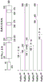
FIG. 1 shows a schematic of the domain structure of phenylalanyl- α -tRNA synthetase overlapping the relative position and size of the N-terminal AARS polypeptide, FIG. 1A represents a fragment identified by mass spectrometry, FIG. 1B represents a fragment identified by deep sequencing of a transcriptome, and FIG. 1C represents a fragment identified by bioinformatic analysis.
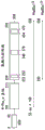
FIG. 2 shows a schematic of the domain structure of phenylalanyl- α -tRNA synthetase overlapping the relative position and size of the C-terminal AARS polypeptide, FIG. 2A represents a fragment identified by deep sequencing of a transcriptome, and FIG. 2B represents a fragment identified by bioinformatic analysis.
Summary of The Invention
Embodiments of the invention generally relate to the discovery of protein fragments of aminoacyl-tRNA synthetases (AARSs) that have unconventional biological activity, such as extracellular signaling activity, and/or other properties relevant to therapy and diagnosis. AARS is a common and essential element of the protein synthesizer found in all organisms, but human AARS and its related proteins have naturally occurring excision variants with potent cell signaling activity, facilitating normal human function. The activity of these protein fragments differs from the well-known protein synthesis activity of AARS, and the invention includes the discovery and development of these resectins as novel therapeutic agents, novel discovery research reagents, and novel antigens/targets for targeted biologies and diagnostics, potentially useful in the treatment or diagnosis of a number of human diseases, such as inflammatory, hematologic, neurodegenerative, autoimmune, hematopoiesis, cardiovascular, and metabolic diseases or disorders.
Thus, the AARS protein fragments of the invention may be referred to as "resectins" or "apendacrines". As described above, the term "resectin" results from the process of cleaving or excising a given AARS protein fragment from its full-length parent AARS sequence environment, while its full-length parent AARS sequence typically masks its non-canonical activity. In certain instances, the AARS protein fragments and polynucleotides of the invention are identified by the occurrence of this excision process, whether naturally occurring (e.g., proteolytic, splice variants), artificially induced, or predicted. The term "apendacrine" is derived from "attachment" (from latin-attacher) and "separation" or "resolution" (from greek-crines), and also reflects the separation of one or more additional domains of an AARS protein fragment from its corresponding full-length or parent AARS sequence.
Although several AARS fragments have been previously shown to have non-synthetase activities, the expression, isolation, purification, and characterization of such fragments for biological therapeutic, discovery, or diagnostic utility is limited, and one skilled in the art would not readily associate such activities with each member or alternate fragment of the entire AARS family. Here, AARS protein fragments of 20 mitochondrial AARS and 20 cytoplasmic AARS (and related proteins) for biotherapeutic discovery and diagnostic utility were discovered and confirmed using systematic methods. For example, certain of the AARS protein fragments of the invention and polynucleotides encoding them were identified from biological samples using Mass Spectrometry (MS) which primarily identifies proteolytic fragments, and others were identified by deep sequencing techniques which primarily identify splice variants. Using in silico prediction of amino acid sequences, e.g., by in silico comparison of synthetases from humans and lower organisms, as well as key differences (e.g., protease sites), other AARS protein fragments are identified; the method utilizes sequence analysis of full-length AARS based on specific criteria for distinguishing proteolytic fragments and functional domains with non-canonical biological activities.
Novel excision proteins for AARS are unexpected, and their differential expression is also unexpected. Specific resections are typically found in different treatments (e.g., from cells grown in media with or without serum), different growth stages (e.g., adult versus fetal brain), and different tissue types (e.g., pancreas versus liver). Although the conventional function of all aminoacyl-tRNA synthetases is required in relative proportional amounts at the same cellular location, the expression pattern of all aminoacyl-tRNA synthetases is different. One would not expect the level of activity of one aminoacyl-tRNA synthetase to increase while the activity of the other aminoacyl-tRNA synthetase does not increase. Mass spectrometry and deep sequencing data indicate that aminoacyl tRNA synthetase deproteinization does have different levels and occurs at different sites and at different stages.
In addition, AARS protein fragments can be expressed and purified to a high enough purity to discern their biological properties. Previously, fragments often did not have sufficient purity, folding, and stability to enable proper biological characterization of non-synthetase activities. For example, cell-based assays are used in conjunction with sufficiently pure, stable, soluble and folded resectins to reveal important biotherapeutic, discovery or diagnostic activities.
In particular, the invention relates to protein fragments of phenylalanyl- α -tRNA synthetases having biological therapeutic, discovery, or diagnostic utility, related agents and compositions, and methods of use thereof.
Certain embodiments include compositions comprising an isolated aminoacyl-tRNA synthetase (AARS) protein fragment of at least about 100, 90, 80, 70, 60, 50, or 40 amino acids that comprises an amino acid sequence as set forth in any of Table(s) 1-3 or Table(s) 4-6 or Table(s) 7-9, and has a solubility of at least about 5mg/m L, and wherein the composition has a purity of at least about 95% on a protein basis and less than about 10EU endotoxin/mg proteinEC50。
In certain embodiments, the AARS protein fragment is fused to a heterologous polypeptide. In some embodiments, the AARS fusion protein substantially retains a non-canonical activity of the AARS protein fragment. In some embodiments, the AARS fusion protein inhibits a non-canonical activity of the AARS protein fragment. In certain embodiments, the heterologous polypeptide is linked to the N-terminus of the AARS protein fragment. In certain embodiments, the heterologous polypeptide is linked to the C-terminus of the AARS protein fragment. In one aspect, in any of these embodiments, the heterologous polypeptide is selected from the group consisting of: purification tags, epitope tags, targeting sequences, signal peptides, membrane translocation sequences, and PK modulators.
In certain embodiments, the composition comprises a concentration of at least about 10mg/m L of the AARS protein fragment, in certain embodiments, the composition comprises at least 90% monodisperse of the AARS protein fragment, in certain embodiments, the composition comprises less than about 3% high molecular weight aggrecan, in certain embodiments, the composition exhibits less than 3% aggregation when stored in PBS at a concentration of at least 10mg/m L for 1 week at 4 deg.C, in certain embodiments, the composition exhibits less than 3% aggregation when stored in PBS at a concentration of at least 10mg/m L for 1 week at room temperature.
Various assays for measuring such characteristics of resectins are described herein and may be used to define aspects of the invention. In certain aspects, these features are preferred for the biological therapeutic utility of the AARS protein fragments described herein.
Certain embodiments include compositions, comprising an isolated aminoacyl-tRNA synthetase (AARS) protein fragment of at least 35 amino acids that differs from an amino acid sequence set forth in any of Table(s) 1-3 or Table(s) 4-6 or Table(s) 7-9, by substitution, deletion, and/or addition of about 1,2, 3,4, 5,6, 7,8, 9, 10, 11, 12, 13, 14, 15, 16, 17, 18, 19, or 20 amino acids, wherein the altered protein fragment substantially retains a non-canonical activity of the unaltered protein or has a dominant negative phenotype associated with the non-canonical activity, wherein the protein fragment has a solubility of at least about 5mg/m L, and wherein the composition has a purity of at least about 95% on a protein basis and less than about 10EU/mg protein.
Other embodiments include compositions comprising an isolated antibody that specifically binds to an isolated aminoacyl-tRNA synthetase (AARS) protein fragment as set forth in Table(s) 1-3, or Table(s) 4-6, or Table(s) 7-9, wherein affinity of the antibody for the AARS protein fragment is about 10X stronger than affinity of the antibody for a corresponding full-length AARS polypeptide. A surprising aspect of the invention includes certain resectins having "new" surfaces accessible to antibodies or other targeted biologies, while the full-length AARS "hides" or covers these surfaces with other sequences or adjacent domains. The excision process also results in greater water accessibility for revealing previously unidentified biological properties. Some embodiments include a composition, comprising an isolated antibody that specifically binds to an isolated aminoacyl-tRNA synthetase (AARS) protein fragment as set forth in table(s) 1-3 or table(s) 4-6 or table(s) 7-9, wherein the antibody has an affinity of at least about 10nM for the AARS protein fragment, and an affinity of at least about 100nM for a corresponding full-length AARS polypeptide. In certain embodiments, the antibody binds to an epitope located within an AARS polypeptide unique splice junction as set forth in any of tables 1-3 or tables 4-6 or tables 7-9, or binds to a C-terminal amino acid sequence of the splice site. In certain embodiments, the antibody antagonizes a non-canonical activity of the AARS protein fragment. Such antagonists may optionally bind to the corresponding parent or full-length AARS.
Other aspects relate to a bioassay system, comprising a substantially pure aminoacyl-tRNA synthetase (AARS) protein fragment of at least 90 amino acids that comprises an amino acid sequence as set forth in Table(s) 1-3 or Table(s) 4-6, or Table(s) 7-9, and a binding ligand that binds to the AARS protein fragment. In one aspect, the binding ligand is selected from the group consisting of: cell surface receptor proteins, nucleic acids, lipid membranes, cell regulatory proteins, enzymes and transcription factors. Optionally, such receptors may be part of a cell, preferably a cell biologically associated with the disclosed resectins.
Certain embodiments include a cell composition comprising: an isolated aminoacyl-tRNA synthetase (AARS) protein fragment of at least 90 amino acids that comprises an amino acid sequence as set forth in Table(s) 1-3 or Table(s) 4-6 or Table(s) 7-9; and an engineered population of cells, wherein at least one cell comprises a polynucleotide encoding the AARS protein fragment. In one aspect, the cell is capable of growing in serum-free media.
Also included is a detection system comprising: a substantially pure aminoacyl-tRNA synthetase (AARS) protein fragment of at least 50 or 100 amino acids that comprises an amino acid sequence as set forth in Table(s) 1-3 or Table(s) 4-6 or Table(s) 7-9; a cell comprising a cell surface receptor or extracellular portion thereof that binds to the protein fragment; and a molecule or second polypeptide of less than about 2000 daltons that modulates binding or interaction of the AARS protein fragment and an extracellular receptor.
Particular embodiments include a diagnostic system comprising: a substantially pure aminoacyl-tRNA synthetase (AARS) protein fragment of at least 90 amino acids that comprises an amino acid sequence as set forth in Table(s) 1-3 or Table(s) 4-6 or Table(s) 7-9; and a cell comprising a cell surface receptor or extracellular portion thereof that binds to the AARS protein fragment, wherein the system or cell comprises an indicator molecule that allows for detection of a change in the level or activity of the cell surface receptor or extracellular portion thereof.
Certain embodiments include a cell growth device comprising: an isolated aminoacyl-tRNA synthetase (AARS) protein fragment of at least 35 amino acids that comprises an amino acid sequence as set forth in Table(s) 1-3 or Table(s) 4-6 or Table(s) 7-9; an engineered population of cells, wherein at least one cell comprises a polynucleotide encoding the AARS protein fragment; at least about 10 liters of serum-free growth medium; and a sterile container. In particular embodiments, cells of any of the methods or compositions described herein are capable of growing in serum-free media, optionally containing an antibiotic and an inducer.
Some embodiments relate to antisense or RNA interference (RNAi) agents comprising sequences that target unique splice junctions against AARS splice variants listed in tables 1-3 or tables 4-6 or tables 7-9.
Also included are therapeutic compositions comprising an isolated aminoacyl-tRNA synthetase (AARS) protein fragment of at least 35 amino acids that comprises an amino acid sequence as set forth in Table(s) 1-3, or Table(s) 4-6, or Table(s) 7-9, wherein the protein fragment specifically binds to a binding partner and has a solubility of at least about 5mg/m L, and wherein the composition has a purity of at least about 95% on a protein basis.
Also included are compositions comprising an isolated aminoacyl-tRNA synthetase (AARS) protein fragment of at least 35 amino acids that is at least 80%, 85%, 90%, 95%, 98%, or 100% identical to an amino acid sequence set forth in Table(s) 1-3 or Table(s) 4-6 or Table(s) 7-9, wherein the protein fragment has a solubility of at least about 5mg/m L, and wherein the composition has a purity of at least about 95% on a protein basis and less than about 10EU endotoxin/mg protein.
In another aspect of any of these embodiments, the composition exhibits less than 10% aggregation when stored in PBS at a concentration of at least 10mg/m L for 1 week at 4 ℃, or less than 5% aggregation when stored in PBS at a concentration of at least 10mg/m L for 1 week at 4 ℃, or less than 3% aggregation when stored in PBS at a concentration of at least 10mg/m L for 1 week at 4 ℃, or less than 2% aggregation when stored in PBS at a concentration of at least 10mg/m L for 1 week at 4 ℃, or less than 1% aggregation when stored in PBS at a concentration of at least 10mg/m L for 1 week at 4 ℃.
Certain embodiments include compositions comprising a substantially pure aminoacyl-tRNA synthetase (AARS) protein fragment of at least 35 amino acids that comprises an amino acid sequence as set forth in Table(s) 1-3 or Table(s) 4-6, or Table(s) 7-9, and at least one covalently or non-covalently moiety attached thereto. In certain embodiments, the moiety is a detectable label. In certain embodiments, the moiety is a water-soluble polymer. In certain embodiments, the moiety is PEG. In one aspect of any of these embodiments, the moiety is linked to the N-terminus of the protein fragment. In one aspect of any of these embodiments, the moiety is linked to the C-terminus of the protein fragment.
Particular embodiments include a composition, comprising a solid substrate attached to an isolated aminoacyl-tRNA synthetase (AARS) protein fragment of at least 35 amino acids that comprises an amino acid sequence as set forth in Table(s) 1-3 or Table(s) 4-6, or Table(s) 7-9, or a biologically active fragment or variant thereof, wherein the protein fragment has a solubility of at least about 5mg/m L, and the composition has a purity of at least about 95% on a protein basis.
Also included are compositions comprising a binding agent that specifically binds to an isolated aminoacyl-tRNA synthetase (AARS) protein fragment as set forth in Table(s) 1-3, or Table(s) 4-6, or Table(s) 7-9, wherein the binding agent has an affinity of at least about 1nM for the protein fragment. In one aspect, the binding agent binds to an epitope located within an AARS polypeptide unique splice junction as set forth in any of tables 1-3 or tables 4-6 or tables 7-9, or to the C-terminal amino acid sequence of the splice site. In certain embodiments, the binding agent antagonizes a non-canonical activity of the AARS polypeptide.
Certain embodiments include isolated aminoacyl-tRNA synthetase (AARS) polypeptides, comprising an amino acid sequence of an AARS protein fragment as described herein, an amino acid sequence encoded by an AARS polynucleotide as described herein, or a variant or fragment thereof. Certain AARS polypeptides comprise an amino acid sequence that is at least 80%, 85%, 90%, 95%, 98%, or 100% identical to an AARS reference sequence disclosed in tables 1-3 or tables 4-6 or tables 7-9 or table E2. Certain AARS polypeptides consist essentially of an amino acid sequence that is at least 80%, 85%, 90%, 95%, 98%, or 100% identical to an AARS reference sequence disclosed in tables 1-3 or tables 4-6 or tables 7-9 or table E2. In certain embodiments, the polypeptide comprises a non-canonical biological activity. In a specific embodiment, the non-canonical biological activity is selected from the group consisting of: modulation of cell signaling (e.g., extracellular signaling), modulation of cell proliferation, modulation of cell migration, modulation of cell differentiation, modulation of apoptosis or cell death, modulation of angiogenesis, modulation of cell binding, modulation of cell metabolism, modulation of cellular uptake, modulation of gene transcription, or modulation of secretion, modulation of cytokine production or activity, modulation of cytokine receptor activity, and modulation of inflammation.
Other aspects include antibodies and other binding agents that exhibit binding specificity for an isolated AARS polypeptide described herein, a binding partner for the AARS polypeptide, or a complex of both. In certain embodiments, the affinity of the antibody or binding agent for the AARS polypeptide is about 10X stronger than its affinity for the corresponding full-length AARS polypeptide. In particular embodiments, the binding agent is selected from the group consisting of a peptide, a peptidomimetic, an adnectin, an aptamer, and a small molecule. In certain embodiments, the antibody or binding agent antagonizes a non-canonical activity of the AARS polypeptide. In other embodiments, the antibody or binding agent agonizes a non-canonical activity of the AARS polypeptide.
Certain embodiments include isolated aminoacyl-tRNA synthetase (AARS) polynucleotides comprising a nucleotide sequence of an AARS polynucleotide described herein, a nucleotide sequence encoding an AARS protein fragment described herein, or a variant, fragment, or complement thereof. Certain AARS polynucleotides comprise a nucleotide sequence that is at least 80%, 85%, 90%, 95%, 98%, or 100% identical to an AARS reference polynucleotide disclosed in tables 1-3 or tables 4-6 or tables 7-9 or table E2, or a complement thereof. In certain embodiments, the nucleotide sequence is codon optimized for bacterial expression. In one aspect, the nucleotide sequence is at least 80% identical to a polynucleotide sequence disclosed in table E2.
A particular AARS polynucleotide consists of a nucleotide sequence that is at least 80%, 85%, 90%, 95%, 98%, or 100% identical to an AARS reference polynucleotide disclosed in tables 1-3 or tables 4-6 or tables 7-9 or table E2, or a complement thereof. Other AARS polynucleotides comprise or consist essentially of a nucleotide sequence that specifically hybridizes to an AARS reference polynucleotide disclosed in tables 1-3 or tables 4-6 or tables 7-9 or table E2. In certain embodiments, the polynucleotide is selected from the group consisting of a primer, a probe, and an antisense oligonucleotide. In particular embodiments, the primer, probe, or antisense oligonucleotide targets a specific or unique splice junction within the AARS polynucleotide, and/or a 3' sequence of the splice site.
Certain embodiments include methods of determining the presence or level of an AARS protein fragment in a sample, comprising contacting the sample with one or more binding agents that specifically bind to an AARS protein fragment described herein, detecting the presence or absence of the binding agent, and thereby determining the presence or level of the AARS protein fragment.
Certain embodiments include discovery methods and related compositions for identifying a compound that specifically binds to one or more of an aminoacyl-tRNA synthetase (AARS) polypeptide or a cell-bound ligand thereof as described herein, the method comprising a) mixing the AARS polypeptide, or the cell-bound ligand thereof, or both, with at least one test compound under suitable conditions, and b) detecting binding of the AARS polypeptide, or the cell-bound ligand thereof, or both, to the test compound, thereby identifying a compound that specifically binds to the AARS polypeptide, or the cell-bound ligand thereof, or both. In certain embodiments, the test compound is a polypeptide or peptide, an antibody or antigen-binding fragment thereof, a peptide mimetic, or a small molecule. In certain embodiments, the test compound agonizes a non-canonical biological activity of the AARS polypeptide or its cell-binding ligand. In other embodiments, the test compound antagonizes a non-canonical biological activity of the AARS polypeptide or its cell-bound ligand. Certain embodiments include compounds identified by the above-described methods, such as agonists (e.g., small molecules, peptides).
Certain embodiments include a method of determining the presence or level of a polynucleotide sequence of an AARS splice variant in a sample, the method comprising contacting the sample with one or more oligonucleotides that specifically hybridize to an AARS polynucleotide described herein, detecting the presence or absence of the oligonucleotides in the sample, and thereby determining the presence or level of a polynucleotide sequence of the AARS splice variant. Other embodiments include methods of determining the presence or level of a polynucleotide sequence of an AARS splice variant in a sample, comprising contacting the sample with at least two oligonucleotides that specifically amplify an AARS polynucleotide described herein, performing an amplification reaction, detecting the presence or absence of an amplification product, and thereby determining the presence or level of a polynucleotide sequence of the AARS splice variant. In particular embodiments, the oligonucleotide specifically hybridizes to or specifically amplifies a splice junction unique to the AARS splice variant. Certain embodiments comprise comparing the presence or level of the AARS protein fragment or splice variant to a control sample or a predetermined value. Certain embodiments comprise characterizing the state of the sample to distinguish it from the control. In particular embodiments, the sample and control comprise cells or tissues, and the method comprises distinguishing between cells or tissues of different species, cells of different tissues or organs, cells of different cellular developmental states, cells of different cellular differentiation states, or healthy cells and diseased cells.
Some embodiments include pharmaceutical compositions comprising an AARS polynucleotide described herein, an AARS polypeptide described herein, a binding agent described herein, or a compound identified by the above methods or described herein, and a pharmaceutically acceptable excipient or carrier.
Certain embodiments include a method of modulating a cellular activity of a cell, the method comprising contacting the cell with an AARS polynucleotide described herein, an AARS polypeptide described herein, a binding agent described herein, a method described above, or a compound described herein, or a pharmaceutical composition described herein. In particular embodiments, the cellular activity is selected from the group consisting of cell proliferation, cell migration, cell differentiation, apoptosis or cell death, cell signaling, angiogenesis, cell binding, cell uptake, cell secretion, metabolism, cytokine production or activity, cytokine receptor activity, gene transcription, and inflammation. In one aspect, the cell is selected from the group consisting of: preadipocytes, bone marrow, neutrophils, blood cells, hepatocytes, astrocytes, mesenchymal stem cells, and skeletal muscle cells.
In certain embodiments, the cell is in a subject. Certain embodiments include treating a subject, wherein the subject has a disorder associated with a neoplastic disease, an immune system disease or disorder, an infectious disease, a metabolic disease, an inflammatory disorder, a neuronal/nervous system disease, a muscle/cardiovascular disease, a disease associated with abnormal hematopoiesis, a disease associated with abnormal angiogenesis, or a disease associated with abnormal cell survival.
Also included are methods of preparing a pharmaceutical compound comprising: a) performing an in vitro screen of one or more candidate compounds in the presence of an AARS protein fragment comprising at least 35 amino acids of an amino acid sequence set forth in tables 1-3 or tables 4-6 or tables 7-9 to identify compounds that specifically bind to the AARS protein fragment; b) performing a cell-based or biochemical or receptor assay using the compound identified in step a) to identify a compound that modulates one or more non-canonical activities of the AARS protein fragment; c) optionally assessing the structure-activity relationship (SAR) of the compound identified in step b) to correlate its structure with modulation of said non-canonical activity, and optionally derivatizing said compound to alter its ability to modulate said non-canonical activity; and d) producing a compound identified in step b) or a compound derived in step c) in an amount sufficient for human use, thereby preparing the pharmaceutical compound.
Other embodiments include methods of making a pharmaceutical compound comprising: a) performing an in vitro screening of one or more candidate compounds in the presence of a cell surface receptor or extracellular portion thereof that specifically binds to an AARS protein fragment of tables 1-3 or tables 4-6 or tables 7-9 to identify a compound that specifically binds to the cell surface receptor or extracellular portion thereof; b) performing a cell-based or biochemical or receptor assay using the compound identified in step a) to identify a compound that modulates one or more non-canonical activities of the AARS protein fragment; c) optionally assessing the structure-activity relationship (SAR) of the compound identified in step b) to correlate its structure with modulation of said non-canonical activity, and optionally derivatizing said compound to alter its ability to modulate said non-canonical activity; and d) producing a compound identified in step b) or a compound derived in step c) in an amount sufficient for human use, thereby preparing the pharmaceutical compound.
Some embodiments include a cellular composition, comprising an engineered population of cells in which at least one cell comprises a polynucleotide encoding a heterologous full length cysteinyl-tRNA synthetase (AARS) protein, wherein the cells are capable of growing in serum-free media. In one aspect, the full length aminoacyl-tRNA synthetase (AARS) protein comprises a heterologous purification tag or an epitope tag to facilitate purification of the AARS protein fragment. In another aspect, the full length aminoacyl-tRNA synthetase (AARS) protein comprises a heterologous proteolysis site to enable production of an AARS protein fragment upon cleavage.
Some embodiments include a method of producing in situ within a cell an AARS polypeptide listed in tables 1-3 or tables 4-6 or tables 7-9 or table E2, the method comprising: i) expressing a heterologous full length cysteinyl-tRNA synthetase (AARS) protein within the cell, wherein the cell comprises a protease that is capable of cleaving the heterologous full length aminoacyl-tRNA synthetase (AARS) protein to produce the AARS polypeptide.
Some embodiments include a method of producing an AARS polypeptide set forth in table 1-3 or table 4-6 or table 7-9 or table E2, the method comprising contacting an isolated full length aminoacyl-tRNA synthetase (AARS) protein with a protease that is capable of cleaving the heterologous full length aminoacyl-tRNA synthetase (AARS) protein and producing an AARS polypeptide.
Some embodiments include engineered full length aminoacyl-tRNA synthetase (AARS) proteins comprising a heterologous proteolytic site capable of effecting proteolytic generation of an AARS protein fragment as set forth in any of table(s) 1-3 or table(s) 4-6 or table(s) 7-9 or table(s) E2.
Some embodiments include a composition comprising an isolated full length aminoacyl-tRNA synthetase protein, wherein the composition has a purity of at least about 95% on a protein basis, less than about 10EU endotoxin/mg protein, and is substantially serum free.
Another embodiment includes a method of treating a disease or condition mediated by aberrant modulation of tRNA synthetase expression, activity or spatiotemporal location by administering an AARS protein fragment listed in any of tables 1-3 or tables 4-6 or tables 7-9 or table E2 or a nucleic acid encoding said ARRS protein fragment. In one aspect of this embodiment, the disease is selected from the group consisting of cancer, neuropathy, diabetes, and inflammatory disorders.
Detailed Description
Directory

I. Overview
The present invention relates, at least in part, to the discovery of novel AARS polypeptides, and methods for their preparation and use, that represent the conversion of a native wild-type protein into a novel form that exhibits significantly different properties as compared to the naturally occurring full-length phenylalanyl- α subunit-tRNA synthetase gene, the identification of such AARS polypeptides is based on mass spectrometry analysis of a broad range of sequences and phenylalanyl- α subunit-tRNA synthetases expressed in different tissues, followed by the systematic generation and testing of each potential AARS polypeptide to identify protein sequences that represent stable and soluble protein domains that exhibit novel biological activities and advantageous therapeutic drug properties.
Based on this analysis, at least two new families of AARS polypeptides derived from the phenylalanyl- α subunit, tRNA synthetases have been identified.
In one aspect, such an phenylalanyl- α subunit tRNA synthetase-derived AARS polypeptide includes a polypeptide sequence that comprises about amino acids 1-226 of the phenylalanyl- α subunit tRNA synthetase.
In a second aspect, such an phenylalanyl- α subunit tRNA synthetase-derived AARS polypeptide includes a polypeptide sequence that comprises about amino acids 1-152 of a phenylalanyl- α subunit tRNA synthetase.
These new AARS polypeptide families represent new previously unknown protein products, in particular exhibiting i) new biological activities, ii) favorable protein stability and aggregation properties, and iii) the ability to be expressed and produced at high levels in prokaryotic expression systems, which are significantly different properties not found in the intact wild-type protein.
Definition of
Unless defined otherwise, all technical and scientific terms used herein have the same meaning as commonly understood by one of ordinary skill in the art to which this invention belongs. Although any methods and materials similar or equivalent to those described herein can be used in the practice or testing of the present invention, the preferred methods and materials are described herein. For the purposes of the present invention, the following terms are defined below.
The articles "a" and "an" are used herein to refer to one or to more than one (i.e., to at least one) of the grammatical object of the article. For example, "an element" means one element or more than one element.
"about" means an amount, level, value, number, frequency, percentage, dimension, size, amount, weight, or length that varies by as much as 30, 25, 20, 25, 10,9, 8, 7,6, 5,4, 3,2, or 1% from a reference amount, level, value, number, frequency, percentage, dimension, size, amount, weight, or length.
An "agonist" refers to a molecule that enhances or mimics an activity. For example, a non-canonical biological activity of AARS or another protein. Agonists may include proteins, nucleic acids, carbohydrates, small molecules, or any other compound or composition that modulates the activity of an AARS by interacting directly with the AARS or its binding ligand or by acting on a component in a biological pathway in which the AARS is involved. Including partial agonists and full agonists.
As used herein, the term "amino acid" is intended to refer to naturally occurring and non-naturally occurring amino acids, as well as amino acid analogs and mimetics, naturally occurring amino acids include the 20 (L) -amino acids used in protein biosynthesis, as well as other amino acids, such as 4-hydroxyproline, hydroxylysine, desmosine, isodesmosine, homocysteine, citrulline, and ornithine non-naturally occurring amino acids include, for example, (D) -amino acids, norleucine, norvaline, p-fluorophenylalanine, ethylmethionine, and the like, which are known to those skilled in the art.
In certain aspects, the use of unnatural amino acids can be used to modify (e.g., increase) a selected, non-canonical activity of an AARS protein fragment, or to alter the in vivo or in vitro half-life of the protein. Unnatural amino acids can be used to facilitate (selective) chemical modification (e.g., pegylation) of AARS proteins. For example, certain unnatural amino acids allow polymers, such as PEG, to be attached to a given protein, thereby improving its pharmacokinetic properties.
Specific examples of amino acid analogs and mimetics can be found in, for example, Roberts and Vellaccio, the peptides: Analysis, Synthesis, Biology, eds Gross and Meinhofer, Vol.5, p.341, academic Press, Inc., New York, N.Y. (1983), the entire Vol.incorporated by reference herein. Other examples include fully alkylated amino acids, particularly fully methylated amino acids. See, e.g., combinatory Chemistry, editors of Wilson and Czarnik, ch.11, p.235, John Wiley & Sons inc, New York, n.y. (1997), the entire book of which is incorporated herein by reference. Yet other examples include amino acids whose amide moiety (and, therefore, the amide backbone of the resulting peptide) has been replaced by, for example, a sugar ring, a steroid, a benzodiazepine, or a carbocyclic ring. See, e.g., Burger's Medicinal chemistry and Drug Discovery, editors Man fred E.Wolff, Ch.15, pp.619-620, John Wiley & Sons Inc., New York, N.Y. (1995), the entire book of which is incorporated herein by reference. Methods for synthesizing peptides, polypeptides, Peptide mimetics, and proteins are well known in the art (see, e.g., U.S. Pat. No. 5,420,109; M.Bodanzsky, Principles of Peptide Synthesis (1 st edition & 2 nd revision), Springer-Verlag, New York, N.Y. (1984&1993), Chapter 7; Stewart and Young, Solid Phase Peptide Synthesis (2 nd edition), Pierce chemical Co., Rockford, Ill., each of which is incorporated herein by reference). Thus, the AARS polypeptides of the invention can be comprised of naturally occurring and non-naturally occurring amino acids, as well as amino acid analogs and mimetics.
The term "antagonist" refers to a molecule that reduces or attenuates an activity. For example, a non-canonical active biological activity of AARS or another protein. Antagonists may include proteins such as antibodies, nucleic acids, carbohydrates, small molecules, or any other compound or composition that modulates the activity of an AARS or its binding ligand by interacting directly with the AARS or its binding ligand or by acting on a component in a biological pathway in which the AARS is involved. Including partial and complete antagonists.
In this "conventional" activity, aminoacyl-tRNA synthetases catalyze a two-step reaction in which, first, they activate their respective amino acids by forming aminoacyl-adenylates, in which the carboxyl group of the amino acid is linked to α -phosphate of ATP by replacement of pyrophosphate, and then, when bound to the correct tRNA, the aminoacyl of the aminoacyl-adenylate is transferred to the 2 'or 3' terminal OH of the tRNA.
Class I aminoacyl-tRNA synthetases typically have two highly conserved sequence motifs, which aminoacylate at the 2 '-OH of an adenosine nucleotide and are typically monomeric or dimeric class II aminoacyl-tRNA synthetases typically have three highly conserved sequence motifs, which aminoacylate at the 3' -OH of the same adenosine and are typically dimeric or tetrameric.
AARS polypeptides include tyrosyl-tRNA synthetase (TyrRS), tryptophanyl-tRNA synthetase (TrpRS), glutaminyl-tRNA synthetase (GlnRS), glycyl-tRNA synthetase (GlyRS), histidyl-tRNA synthetase (HisRS), seryl-tRNA synthetase (SerRS), phenylalanyl-tRNA synthetase (PheRS), alanyl-tRNA synthetase (AlaRS), asparaginyl-tRNA synthetase (AsnRS), aspartyl-tRNA synthetase (AspRS), cysteinyl-tRNA synthetase (CysRS), glutamyl-tRNA synthetase (GluRS), prolyl-tRNA synthetase (ProRS), arginyl-tRNA synthetase (ArgRS), isoleucyl-tRNA synthetase (IleRS), leucyl-tRNA synthetase (L euRS), lysyl-tRNA synthetase (L ysRS), threonyl-tRNA synthetase (ThrRS), methionyl-tRNA synthetase (MetRS), or cytosolic-tRNA synthetase (metval-rs), and wild-type strains of these are known in the art.
"coding sequence" means any nucleic acid sequence responsible for encoding the polypeptide product of a gene. In contrast, the term "non-coding sequence" refers to any nucleic acid sequence that is not responsible for encoding the polypeptide product of a gene.
Throughout this specification, unless the context requires otherwise, the words "comprise", "comprises" and "comprising" will be understood to imply the inclusion of a stated step or element or group of steps or elements but not the exclusion of any other step or element or group of steps or elements.
"consists of" means including and limited to anything following the phrase "consists of. Thus, the phrase "consisting of" indicates that the listed elements are required or mandatory, and that no other elements may be present. "consisting essentially of means including any elements listed after the phrase and is not limited to other elements that do not interfere with or facilitate the activity or effect specified in the disclosure of the listed elements. Thus, the phrase "consisting essentially of indicates that the listed elements are required or mandatory, but that other elements are optional, may or may not be present, depending on whether they substantially affect the activity or function of the listed elements.
The expression "endotoxin-free" or "substantially endotoxin-free" generally refers to compositions, solvents, and/or containers that contain up to trace amounts (e.g., amounts that have no clinically undesirable physiological effect on a subject) of endotoxin, and preferably undetectable amounts of endotoxin, endotoxin is a toxin associated with certain bacteria, typically gram-negative bacteria, but endotoxin may also be present in gram-positive bacteria, such as Listeria monocytogenes.
Therefore, in the pharmaceutical production of AARS polypeptides, it is often desirable to remove most or all trace amounts of endotoxins from pharmaceutical products and/or pharmaceutical containers, since even small amounts can cause adverse effects in humans. An oven that removes the heat source can be used for this purpose, as temperatures in excess of 300 ℃ are generally required to break down most endotoxins. For example, based on the primary packaging material, such as a syringe or vial, a glass temperature of 250 ℃ in combination with a 30 minute hold time is often sufficient to achieve a reduction in endotoxin levels of 3 orders of magnitude. Other methods of removing endotoxins are contemplated, including, for example, chromatographic and filtration methods, as described herein and known in the art. Also included are methods of producing AARS polypeptides in and isolating them from eukaryotic cells, such as mammalian cells, to reduce, if not eliminate, the risk of endotoxins present in the compositions of the invention. Methods of producing AARS polypeptides in serum-free cells and isolating them therefrom are preferred. Such compositions comprising AARS polypeptides represent novel formulations exhibiting novel biological and therapeutic properties not found in AARS polypeptide compositions contaminated with serum or endotoxin, which have the potential to bind to AARS polypeptides and alter the novel biological properties of AARS polypeptides.
For example, a limulus amoebocyte lysate assay utilizing the blood of a limulus is a very sensitive assay for determining the presence of endotoxin, and reagents, kits and instruments for detecting endotoxin based on this assay are commercially available from, for example, L naza group.
For example, certain compositions may comprise at least 80, 85, 90, 91, 92, 93, 94, 95, 96, 97, 98, 99, or 100% pure agents, including all decimals therebetween, such as, but not limited to, as measured by high performance liquid chromatography (HP L C), which is a commonly known form of column chromatography used in biochemistry and analytical chemistry for the separation, identification, and quantification of compounds.
As used herein, "functional" and like terms refer to a biological function, an enzymatic blood function, or a therapeutic function.
"Gene" means a genetic unit that can occupy a particular locus on a chromosome and consists of transcriptional and/or translational regulatory sequences, and/or coding regions, and/or untranslated sequences (i.e., introns, 5 'and 3' untranslated sequences).
"homology" refers to the percentage of identical or constitutively conservatively substituted amino acids. Homology can be determined using sequence comparison programs such as GAP (Deverlux et al, 1984, Nucleic acids research12,387-395), which is incorporated herein by reference. In this way, sequences of similar or significantly different length to those cited herein can be compared by inserting GAPs in the alignment, which GAPs can be determined by, for example, a comparison algorithm using GAP.
The term "host cell" includes individual cells or cell cultures which may be or have been the recipient of any recombinant vector or isolated polynucleotide of the present invention. Host cells include progeny of a single host cell, and such progeny may not necessarily be identical (morphologically or in total DNA complement) to the original parent cell, due to natural, accidental, or deliberate mutation and/or alteration. Host cells include cells transfected or infected in vivo or in vitro with a recombinant vector or polynucleotide of the invention. The host cell comprising the recombinant vector of the invention is a recombinant host cell.
"isolated" means substantially or essentially free of materials that normally accompany it in its native state. For example, an "isolated polynucleotide" as used herein includes a polynucleotide that has been purified from the sequences that flank it in a naturally occurring state, e.g., by removing fragments of DNA from the sequences that are normally adjacent to the fragments. Alternatively, "isolated peptide" or "isolated polypeptide" and the like as used herein includes a peptide or polypeptide molecule that is isolated and/or purified in vitro from its natural cellular environment and from binding to other components of the cell, i.e., it is not significantly bound to in vivo material.
As used herein, the term "mRNA" or sometimes "mRNA transcript" includes, but is not limited to: pre-mRNA transcripts, transcription processing intermediates, mature mRNA prepared for translation, and transcripts of one or more genes, or nucleic acids derived from mRNA transcripts. Transcript processing may include splicing, editing, and degradation. As used herein, a nucleic acid derived from an mRNA transcript refers to a nucleic acid synthesized by the mRNA transcript or a subsequence thereof that is ultimately used as a template. cDNA reverse transcribed from mRNA, RNA transcribed from the cDNA, DNA amplified from the cDNA, RNA transcribed from the amplified DNA, etc., are transcripts derived from the mRNA, and detection of products of such origin is indicative of the presence and/or abundance of the original transcript in a sample. Thus, a sample from which mRNA is derived includes, but is not limited to, mRNA transcripts of one or more genes, cDNA reverse transcribed from the mRNA, cRNA transcribed from the cDNA, DNA amplified from a gene, RNA transcribed from amplified DNA, and the like.
As used herein, "unconventional" activity generally refers to i) the novel activity possessed by the AARS polypeptides of the invention, to any significant extent, which the intact full-length parent protein does not possess, or ii) the activity possessed by the intact native full-length parent protein, wherein the AARS polypeptide exhibits a significantly higher (i.e., at least 20% greater) specific activity, or exhibits activity in a novel environment, as compared to the intact native full-length parent protein; e.g., an activity that is independent of other activities possessed by the intact native full-length parent protein. In the case of AARS polypeptides, non-limiting examples of non-canonical activities include extracellular signaling, RNA binding, amino acid binding, modulation of cell proliferation, modulation of cell migration, modulation of cell differentiation (e.g., hematopoiesis, neurogenesis, myogenesis, osteogenesis, and adipogenesis), modulation of gene transcription, modulation of apoptosis or other forms of cell death, modulation of cell signaling, modulation of cellular uptake or secretion, modulation of angiogenesis, modulation of cell binding, modulation of cellular metabolism, modulation of cytokine production or activity, modulation of cytokine receptor activity, modulation of inflammation, and the like.
The term "half effective concentration" or "EC50"refers to the concentration of an AARS protein fragment, antibody, or other agent described herein that induces a response intermediate to the baseline and maximum values after a specified period of exposure; thus, EC of the fractionated dose response curves50Represents the concentration of the compound at which 50% of its maximal effect is observed. In certain embodiments, the EC of an agent provided herein50Is associated with "non-conventional" activity as described above. EC (EC)50But also represents the plasma concentration required to obtain 50% of the maximal effect in vivo. Similarly, "EC90"refers to the concentration of an agent or compound at which 90% of its maximum effect is observed. "EC90"can be derived from" EC50"and Hill slope calculations, or can be determined directly from the data using conventional knowledge in the art. In some embodiments, the EC of an AARS protein fragment, antibody, or other agent50Less than about 0.01, 0.05, 0.1, 0.2, 0.3, 0.4, 0.5, 0.6, 0.7, 0.8, 0.9, 1,2, 3,4, 5,6, 7,8, 9, 10, 11, 12, 13, 14, 15, 16, 17, 18, 19, 20, 25, 30, 40, 50, 60, 70, 80, 90, or 100 nM. Preferably, the biotherapeutic composition will have an EC of about 1nM or less50The value is obtained.
The term "modulating" includes "increasing" or "stimulating", as well as "decreasing" or "decreasing", typically in a statistically or physiologically significant amount, as compared to a control. Thus, depending on the conditions of use, a "modulator" may be an agonist, an antagonist or any mixture thereof. An "increased" or "increased" amount is typically a "statistically significant" amount, and can include an increase of 1.1, 1.2, 2,3, 4,5, 6,7, 8, 9, 10, 15,20, 30, or more (e.g., 500, 1000 fold) of the amount produced in the absence of a composition (lacking an agent or compound) or in the presence of a control composition (including all integers and decimals greater than 1 therebetween, e.g., 1.5, 1.6, 1.7, 1.8, etc.). A "reduced" or reduced amount is typically a "statistically significant" amount, and can include a reduction of 1%, 2%, 3%, 4%, 5%, 6%, 7%, 8%, 9%, 10%, 11%, 12%, 13%, 14%, 15%, 16%, 17%, 18%, 19%, 20%, 25%, 30%, 35%, 40%, 45%, 50%, 55%, 60%, 65%, 70%, 75%, 80%, 85%, 90%, 95%, or 100% of the amount produced in the absence of a composition (in the absence of a reagent or compound) or in the presence of a control composition, including all integers therebetween. As a non-limiting example, a control when comparing conventional and non-conventional activity may include an AARS protein fragment of interest as compared to its corresponding full-length AARS, or a fragment AARS having conventional activity comparable to its corresponding full-length AARS. Other examples of "statistically significant" amounts are described below.
By "obtained from" is meant that a sample such as, for example, a polynucleotide extract or a polypeptide extract, is isolated from or derived from a particular source of the subject. For example, the extract may be obtained from a tissue or biological fluid isolated directly from the subject. "derived from" or "obtained from" may also refer to the source of the polypeptide or polynucleotide sequence. For example, an AARS sequence of the invention can be "derived from" sequence information of an AARS proteolytic fragment or AARS splice variant or portion thereof, whether naturally occurring or artificially generated, and thus can comprise, consist essentially of, or consist of such a sequence.
The terms "polypeptide" and "protein" are used interchangeably herein to refer to polymers of amino acid residues and variants and synthetic and naturally occurring analogs thereof. Thus, these terms apply to amino acid polymers in which one or more amino acid residues is a synthetic non-naturally occurring amino acid (such as a chemical analog of a corresponding naturally occurring amino acid), as well as to naturally occurring amino acid polymers and naturally occurring chemical derivatives thereof. Such derivatives include, for example, post-translational modifications and degradation products, including pyroglutamyl, isoaspartyl, proteolytic, phosphorylated, glycosylated, oxidized, isomerized, and deaminated variants of the AARS reference fragment.
Thus, "percent sequence identity" can be calculated by comparing two optimally aligned sequences over a comparison window, determining the number of positions of the same nucleic acid base (e.g., A, T, C, G, I) or the same amino acid residue (e.g., Ala, Pro, Ser, Thr, Gly, Val, L eu, Ile, Phe, Tyr, Trp, L ys, Arg, His, Asp, Glu, Asn, gin, Cys, and Met) that occur in the two sequences to yield the number of matched positions, dividing the number of matched positions by the total number of positions in the comparison window (i.e., the window size), and multiplying the result by 100 to yield the percent sequence identity.
The terms used to describe the sequence relationship between two or more polynucleotides or polypeptides include "reference sequence", "comparison window", "sequence identity", "percent sequence identity" and "substantial identity". "reference sequence" are at least 12, but typically 15 to 18, often at least 25 monomer units (including nucleotide and amino acid residues) in length, since two polynucleotides can each comprise (1) a sequence that is similar between the two polynucleotides (i.e., only a portion of the complete polynucleotide sequence) and (2) a sequence that is different between the two polynucleotides, sequence comparison between two (or more) polynucleotides is typically performed by comparing the sequences of the two polynucleotides in the "comparison window" to identify and compare local regions of sequence similarity, "comparison window" refers to a conceptual segment of at least 6 consecutive positions, typically about 50 to about 100, more typically about 100 to 150 consecutive positions, wherein after an optimal alignment of a sequence with a reference sequence having the same number of consecutive positions, the sequence comparison can comprise the sequence comparison with the sequence (e.g. fastfail. 10. the algorithm) for comparison of the most closely related sequences, i.e. the best results of the comparison of the addition of the sequences, or deletion of the sequences, which can be found by the various genetic algorithm, e.g. the best when the sequence comparison is performed by the algorithm, e.g. the algorithm, e.e.e. faste. faste.g. the algorithm, e. faste. the best when the comparison can be performed by comparing the algorithm, e. after optimal alignment of the sequence comparison window, the addition of sequences can be found by the addition of the most preferably includes the addition of sequences, the sequence comparison window, or deletion of the most preferably the sequences, i.g. the most preferably the sequences, e.g. the addition of sequences, the sequences, e.g. the most preferably the addition of sequences, the most preferably the sequence addition of sequences, the sequence comparison window, the sequence, e.g. the most preferably the sequence, the most preferably the sequences, e.e.e.g. the most preferably the sequence, the most preferably the sequence, the sequences, e.
The calculation of sequence similarity or sequence identity (the terms are used interchangeably herein) between two sequences is performed as follows. To determine the percent identity of two amino acid sequences or two nucleic acid sequences, the sequences are aligned for optimal comparison purposes (e.g., gaps can be introduced in one or both of the first and second amino acid or nucleic acid sequences for optimal alignment, and non-homologous sequences can be disregarded for comparison purposes). In certain embodiments, the length of a reference sequence aligned for comparison purposes is at least 30%, preferably at least 40%, more preferably at least 50%, 60% and even more preferably at least 70%, 80%, 90%, 100% of the length of the reference sequence. The amino acid residues or nucleotides at corresponding amino acid positions or nucleotide positions are then compared. When a position in the first sequence is occupied by the same amino acid residue or nucleotide as the corresponding position in the second sequence, then the molecules are identical at that position.
The percent identity between two sequences is a function of the number of identical positions shared by the sequences, taking into account the number of empty bits and the length of each empty bit, which needs to be introduced for optimal alignment of the two sequences.
Sequence comparison between two sequences and determination of percent identity can be accomplished using a mathematical algorithm. In a preferred embodiment, the percentage identity between two amino acid sequences is determined using the Needleman and Wunsch algorithm (1970, J.MoI.biol.48: 444-453) of the GAP program which has been incorporated into the GCG software package (available at http:// www.gcg.com), using either the Blossum62 matrix or the PAM250 matrix, with GAP weights of 16, 14, 12, 10, 8, 6 or 4 and length weights of 1,2, 3,4, 5 or 6. In another preferred embodiment, the percent identity between two nucleotide sequences is determined using the GAP program of the GCG software package (available at http:// www.gcg.com), using the NWSgapdna. CMP matrix and GAP weights of 40, 50, 60, 70 or 80 and length weights of 1,2, 3,4, 5 or 6. A particularly preferred set of parameters (one that should be utilized unless otherwise specified) is a Blossum62 scoring matrix, a gap penalty of 12, a gap extension penalty of 4, and a frameshift gap penalty of 5.
The percent identity between two amino acid or nucleotide sequences can be determined using the algorithm of e.meyers and w.miller (1989, cab, 4: 11-17), incorporated into the a L IGN program (version 2.0), using a PAM120 weight residue table, a gap length penalty of 12 and a gap penalty of 4.
The Nucleic acid and protein sequences described herein may be used as "query sequences" to perform searches on public databases, e.g., to identify other family members or related sequences such searches may be performed using NB L AST and XB L AST programs (version 2.0) of Altschul, et al, (1990, j.mol.biol,215:403-10) the B L AST nucleotide search may be performed using NB L AST program, score 100, word length 12 to obtain nucleotide sequences homologous to the Nucleic acid molecules of the present invention the B357 AST protein search may be performed using XB L AST program, score 50, word length 3 to obtain amino acid sequences homologous to the protein molecules of the present invention the B L AST protein search may be performed using Altschul et al, (1997, Nucleic acids Res,25: 3389) for alignment to obtain gaps for comparison purposes the B-carrying gaps may be performed using B L AST, B L6, B27 AST programs, B L3, B27, B493 programs may be used when the respective gap parameters are used, e.g., NB L, NB 493 3, ba programs.
The term "solubility" refers to the property of an agent provided herein to dissolve in a liquid solvent and form a homogeneous solution, solubility is generally expressed as a concentration, described by the mass of solute per unit volume of solvent (g solute/kg solvent, g/d L (100m L), mg/ml, etc.), molarity, gravimetric molarity, molar fraction, or other similar concentration the maximum equilibrium amount of solute that a unit amount of solvent can dissolve is the solubility of that solute in that solvent under specified conditions, including temperature, pressure, pH, and solvent properties, in certain embodiments the solubility is measured at physiological pH, in certain embodiments the solubility is measured in water or a physiological buffer, such as PBS, in certain embodiments the solubility is measured in a biological liquid (solvent), such as blood or serum, in certain embodiments the temperature can be about room temperature (e.g., about 20, 21, 22, 23, 24, 25 ℃) or about body temperature (37 ℃), such as the protein fraction AARS having a solubility of at least about 0.7, 0.8, 9, 13, 0.8, 7, 13, 8, 9, 13, or 1.6.8, 0.7, 8, 7.8, 9,8, 13.8, or other similar concentrations.
As used herein, a "splice junction" includes a region of a mature mRNA transcript or encoded polypeptide in which the 3 'end of a first exon is joined to the 5' end of a second exon. The size of this region may vary and either end of the exact residues at the junction of the 3 'end of one exon and the 5' end of another exon may comprise 2,3, 4,5, 6,7, 8, 9, 10, 11, 12, 13, 14, 15, 16, 17, 18, 19, 20, 25, 30, 35, 40, 45, 50, 55, 60, 65, 70, 75, 80, 85, 90, 95, 100 or more (including all integers therebetween) nucleotides or amino acid residues. "exon" refers to a nucleic acid sequence that is present in a mature form of an RNA molecule after a portion of a precursor RNA (an intron) has been removed by cis-splicing or two or more precursor RNA molecules have been joined together by trans-splicing. The mature RNA molecule can be a messenger RNA or a functional form of a non-coding RNA such as rRNA or tRNA. Depending on the context, an exon may refer to a sequence in DNA or its RNA transcript. "Intron" refers to a non-coding nucleic acid region of a gene that is not translated into a protein. The non-coding intron segment is transcribed into a precursor mRNA (pre-mRNA) and some other RNAs (e.g., long non-coding RNAs) and is subsequently removed by splicing during processing into mature RNA.
"splice variant" refers to the mature mRNA or protein encoded thereby produced by alternative splicing, which refers to the process by which exons of an RNA (primary gene transcript or pre-mRNA) are rejoined in multiple ways during RNA splicing. The resulting different mrnas can be translated into different protein isoforms, allowing a single gene to encode multiple proteins.
As used herein, a "subject" includes any animal exhibiting or at risk of exhibiting a symptom that can be treated or diagnosed with an AARS polynucleotide or polypeptide of the invention. Also included are subjects in which it is desirable to profile the levels of the AARS polypeptides and/or polynucleotides of the invention for diagnostic or other purposes. Suitable subjects (patients) include laboratory animals (such as mice, rats, rabbits or guinea pigs), farm animals, and domestic or pet animals (such as cats or dogs). Including non-human primates and preferably human patients.
As used herein, "treatment" or "treating" includes any desired effect on the symptoms or pathology of a disease or disorder that can be affected by the non-canonical activity of the AARS polynucleotide or polypeptide described herein, and may include even minimal changes or improvement in one or more measurable markers of the disease or disorder being treated. Also included are treatments associated with non-AARS therapies, wherein the AARS sequences described herein provide clinical markers of treatment. "treatment" or "treating" does not necessarily mean the complete eradication or cure of the disease or disorder or the symptoms associated therewith. The subject receiving the treatment is any subject in need thereof. Exemplary markers of clinical improvement will be apparent to those skilled in the art.
Unless the context clearly indicates to the contrary, the practice of the present invention may utilize conventional Molecular biology Methods and recombinant DNA techniques within the art, many of which are described below for illustrative purposes, see, for example, Sambrook et al, Molecular Cloning: A L aboratormylManual (3 rd edition, 2000), DNA Cloning: A Practical Applications, volume I & II (D.Glover editor), Oligonucleotide Synthesis (N.Gate edition, 1984), Oligonucleotide Synthesis: Methods Applications (P.herewinjjn. editor, 2004), Nucleic acids Hybridization (B.Hashimeue & S.Higgins editor, 1985), closure of Nucleic acids, coding: protein, 19884, Cell No. 7, publication No. 7, nucleotide No. 7, publication No. 7, Cell No. 7, publication No. 7, nucleotide No. 7, No. 2,7, No. 7, No. 5, No. 2,7, No. 7, No. 2, No. 5, No. 2, No. 7, No. 2,7, No. 2, No. 5, No. 2, No. 7, No. 5, No. 7, No. 9, No. 7, No. 2, No. 9, No. 2, No. 9, No. 7, No. 9, No. 7, No. 9, No. 7, No. 9, No. 7, No. 9, No. 7, No. 9, No. 7, No.
All publications, patents, and patent applications cited herein are incorporated by reference in their entirety.
Purified AARS protein fragments and variants for therapeutic and other applications
In certain embodiments, the AARS protein fragments and variants thereof described herein can have at least one "unusual" biological activity.A protein and fragment of an AARS protein is also referred to herein as an "AARS polypeptide" or an "AARS reference polypeptide". in certain embodiments, the AARS polypeptides provided herein include all or a portion of an AARS polypeptide "reference sequence" that represents the amino acid sequence of a fragment of a various phenylalanyl- α subunit tRNA synthetase, and that is highly divergent from the amino acid sequence of a mouse and human AARS, and that is not more than a few specific amino acid domains, as compared to the amino acid sequence of a mouse and human AARS, as well as to highly related protein domains, as compared to the entire amino acid sequence of a mouse and human AARS.
An N-terminal AARS polypeptide: (tables 1 and 2)&3)


C-terminal AARS Polypeptides (Table 4, 5)&6)


Internal AARS Polypeptides (tables 7, 8)&9)


The amino acid sequences of a "protein fragment" or protein fragment, such as a proteolytic fragment or a splice variant fragment, can be characterized, identified, or derived according to a number of techniques. For example, splice variants can be identified by techniques such as deep sequencing (see, e.g., Xing et al, RNA.14: 1470-. As further examples, protein fragments, e.g., proteolytic fragments, can be identified in vitro, e.g., by incubating full-length or other AARS polypeptides with a protease of choice, or they can be identified endogenously (e.g., in vivo). In certain embodiments, protein fragments, e.g., endogenous proteolytic fragments, can be produced or identified, e.g., by recombinantly expressing full-length or other AARS polypeptides in selected microorganisms or eukaryotic cells that have been modified to contain one or more selected proteases or naturally contain one or more proteases capable of acting on a selected AARS polypeptide, and isolating and characterizing endogenously produced protein fragments therefrom.
In certain embodiments, protein fragments such as endogenous (e.g., naturally occurring) proteolytic fragments can be produced or identified from the following fractions: such as various cell fractions (e.g., cytosolic, membrane, nuclear) and/or growth media of various cell types including, for example, immune cells such as monocytes, dendritic cells, macrophages (e.g., RAW264.7 macrophages), neutrophils, eosinophils, basophils, and lymphocytes, such as B cells and T cells (e.g., CD4+ helper and CD8+ killer cells), including primary T cells and T cell lines such as Jurkat T cell lines, and Natural Killer (NK) cells, among others.
In certain embodiments, protein fragments, such as endogenous proteolytic fragments, regardless of how they are produced, can be identified by techniques such as mass spectrometry or equivalent techniques. Once a protein fragment identified in vitro or endogenously has been produced or identified, it can be mapped or sequenced and, for example, cloned into an expression vector for recombinant production, or synthetically produced.
A large number of proteases can be used to generate, identify, derive, or characterize the sequence of AARS protein fragments, such as proteolytic fragments. Generally, proteases are generally classified according to three main criteria: (i) the reaction catalyzed, (ii) the chemistry of the catalytic site, and (iii) the evolutionary relationship revealed by the structure. Typical examples of proteases or proteolytic enzymes classified by catalytic mechanisms include aspartic proteases, serine proteases, cysteine proteases, and metallo proteases.
Most aspartic proteases belong to the family of pepsins. This family includes digestive enzymes such as pepsin and chymosin, as well as lysosomal cathepsin D and processing enzymes such as renin, and certain fungal proteases (e.g., penicillinase (penicillopepsin), rhizopus protease (rhizopus pepsin), ascomycete protease (endothecaspin)). The second family of aspartic proteases includes viral proteolytic enzymes, such as the proteases from the AIDS virus (HIV), also known as retroviral proteases (retropeptins).
Serine proteases comprise two distinct families. First, the chymotrypsin family, which includes mammalian enzymes such as chymotrypsin, trypsin, elastase and kallikrein, and second, the subtilisin family, which includes bacterial enzymes such as subtilisin. The universal 3D structure differs between these two families, but they have the same active site geometry and are catalyzed by the same mechanism. Serine proteases exhibit different substrate specificity, variability, which is mainly related to amino acid substitutions in the respective enzyme subsites (substrate residue action sites). Some serine proteases have extended sites of action with the substrate, while others have specificity limited to the P1 substrate residue.
The family of cysteine proteases includes plant proteases such as papain, actinidin, and bromelain, several mammalian lysosomal cathepsins, cytosolic calpain (calcium-activated), and several parasitic proteases (e.g., Trypanosoma (Trypanosoma), Schistosoma (Schistosoma)). papain is the prototype and the most studied member of this family.
Metalloproteases are one of the older class of proteases found in bacteria, fungi and higher organisms. They differ significantly in sequence and 3D structure, but most enzymes contain a catalytically active zinc atom. In some cases, the zinc can be replaced by another metal such as cobalt or nickel without losing proteolytic activity. Bacterial thermolysin has been well characterized and its crystalline structure shows that zinc is bound by two histidines and one glutamate. Many metalloproteases contain the sequence motif HEXXH, which provides two histidine ligands for zinc. The third ligand is glutamic acid (thermolysin, enkephalinase, alanyl aminopeptidase) or histidine (astaxanthin, Serralysin).
Exemplary proteases include, for example, achromopeptidase, aminopeptidase, ancrod, angiotensin converting enzyme, bromelain, calpain I, calpain II, carboxypeptidase a, carboxypeptidase B, carboxypeptidase G, carboxypeptidase P, carboxypeptidase W, carboxypeptidase Y, caspase 1, caspase 2, caspase 3, caspase 4, caspase 5, caspase 6, caspase 7, caspase 8, caspase 9, caspase 10, caspase 11, caspase 12, caspase 13, cathepsin B, cathepsin C, cathepsin D, cathepsin E, cathepsin G, cathepsin H, cathepsin L, chymopapain, chymase, chymotrypsin, clostripain, collagenase, complement C1r, C1S, factor D, complement factor I, dipeptidyl peptidase IV, elastase (leukocyte), elastase (pancreatic endoprotease), endoprotease C, pepsin, trypsin protease from Aspergillus protease, alkaline protease (pepsin), pepsin (pepsin), protease from Aspergillus protease, pepsin, alkaline protease (Aspergillus protease), pepsin, alkaline protease (pepsin), protease, alkaline protease from Aspergillus protease, pepsin, alkaline protease (pepsin) protease, pepsin, alkaline protease (pepsin), trypsin) protease, alkaline protease (pepsin, trypsin) protease, trypsin-derived from Aspergillus protease (pepsin) protease from Aspergillus protease (Aspergillus protease, trypsin) protease, trypsin-derived from Aspergillus protease (Aspergillus protease, trypsin) protease (Aspergillus protease, trypsin-derived from Aspergillus protease (Aspergillus protease, trypsin) protease, trypsin-like protease (Aspergillus protease, trypsin-derived from Aspergillus protease (Aspergillus protease, trypsin) protease, trypsin) protease (Aspergillus protease, trypsin-derived from Aspergillus protease (Aspergillus protease, trypsin) protease, trypsin, pepsin, trypsin-derived from Aspergillus protease (Aspergillus protease, trypsin) protease (Aspergillus protease, trypsin-derived from Aspergillus protease, trypsin-derived from Aspergillus protease (Aspergillus protease, trypsin-derived from Aspergillus protease, Aspergillus protease (Aspergillus protease, trypsin) protease (Aspergillus protease ), trypsin.
Certain embodiments relate to isolated AARS polypeptides comprising, consisting essentially of, or consisting of an amino acid sequence derived from an endogenous naturally occurring AARS polypeptide fragment, pharmaceutical compositions comprising the fragment, and methods of using the same. These and related embodiments can be generated or identified in vivo, ex vivo, and/or in vitro. In certain preferred in vitro embodiments, the AARS proteolytic fragments are generated or identified by incubating an AARS polypeptide, e.g., a full-length AARS polypeptide, with one or more isolated human proteases, most of which are endogenous or native to humans, such as elastase and other proteases described herein and known in the art. Certain embodiments relate to isolated AARS polypeptides comprising, consisting essentially of, or consisting of an amino acid sequence derived from an endogenous naturally occurring AARS splice variant, pharmaceutical compositions comprising the fragments, and methods of using the same. Basically, AARS protein fragments can be isolated from a sample that has been exposed to a protease in vivo or in vitro.
By way of example only and not limitation, in certain embodiments, proteomes or fractions thereof from various cell types, tissues or body fluids of a number of physiological states (e.g., hypoxia, diet, age, disease) may be separated by 1D SDS-PAGE and the gel lanes cut into bands at regular intervals, which bands may then optionally be digested with a suitable protease such as trypsin to release peptides which may then be analyzed by 1D reverse phase L C-MS/MS.
As described above, embodiments of the invention include AARS polypeptides listed in tables 1-3, or tables 4-6, or tables 7-9. Also included are "variants" of the AARS reference polypeptides. The expression "variant" of a polypeptide refers to a polypeptide that differs from a reference AARS polypeptide by the addition, deletion, and/or substitution of at least one amino acid residue, which typically retains (e.g., mimics) or mediates (e.g., antagonizes) one or more non-canonical activities of the reference AARS polypeptide.
Thus, it is routine to select naturally occurring phenylalanyl- α tRNA synthetase variants (including, e.g., the single nucleotide polymorphic forms listed in Table A) to produce AARS polypeptides that contain one or more amino acid changes based on the sequence of any of the human and other species' homologues, orthologs, and naturally occurring isoforms of phenylalanyl- α tRNA synthetases.


In certain embodiments, a polypeptide variant differs from a reference polypeptide by one or more substitutions, which may be conservative or non-conservative substitutions as described herein and known in the art. In certain embodiments, polypeptide variants comprise conservative substitutions, in which regard it is well known in the art that some amino acids may be changed to other amino acids with widely similar properties without changing the active properties of the polypeptide.
In certain embodiments, a variant polypeptide comprises an amino acid sequence that has at least about 50%, 55%, 60%, 65%, 70%, 75%, 80%, 85%, 90%, 91%, 92%, 93%, 94%, 95%, 96%, 97%, 98% or more sequence identity or similarity to the corresponding sequence of an AARS reference polypeptide described herein, and substantially retains a non-canonical activity of the reference polypeptide. Also included are sequences that differ from a reference AARS sequence by the addition, deletion, or substitution of 1,2, 3,4, 5,6, 7,8, 9, 10, 11, 12, 13, 14, 15, 16, 17, 18, 19, 20, 30, 40, 50, 60, 70, 80, 90, 100, 110, 120, 130, 140, 150, or more amino acids, but retain the properties of the reference AARS polypeptide. In certain embodiments, the amino acid addition or deletion occurs at the C-terminus and/or N-terminus of the AARS reference polypeptide. In certain embodiments, amino acid additions include 1,2, 3,4, 5,6, 7,8, 9, 10, 11, 12, 13, 14, 15, 16, 17, 18, 19, 20, 30, 40, 50, or more wild-type residues adjacent to the C-terminus and/or N-terminus of these AARS reference polypeptides (i.e., from the corresponding full-length AARS polypeptide).
In certain embodiments, a variant polypeptide differs from the corresponding AARS reference sequence by at least 1% but less than 20%, 15%, 10%, or 5% residues. (if such comparison requires alignment, the sequences should be aligned for maximum similarity. Suitably, the difference is a difference or change or a conservative substitution at a non-essential residue. In certain embodiments, the molecular weight of a variant AARS polypeptide differs from an AARS reference polypeptide by about 1%, 2%, 3%, 4%, 5%, 6%, 7%, 8%, 9%, 10%, 11%, 12%, 13%, 14%, 15%, 16%, 17%, 18%, 19%, 20% or more.
Also included are biologically active "fragments" of the AARS reference polypeptide, i.e., biologically active fragments of an AARS protein fragment. Representative biologically active fragments are often involved in interactions, such as intramolecular or intermolecular interactions. The intermolecular interaction may be a specific binding interaction or an enzymatic interaction. Intermolecular interactions may occur between the AARS polypeptide and cell-bound ligands, such as cellular receptors or other host molecules involved in the non-canonical activity of the AARS polypeptide. In some embodiments, the AARS proteins, variants, and biologically active fragments thereof bind to one or more cell binding ligands with an affinity of at least about 0.01, 0.05, 0.1, 0.2, 0.3, 0.4, 0.5, 0.6, 0.7, 0.8, 0.9, 1,2, 3,4, 5,6, 7,8, 9, 10, 11, 12, 13, 14, 15, 16, 17, 18, 19, 20, 21, 22, 23, 24, 25, 26, 27, 28, 29, 30, 40, or 50 nM. The binding affinity of the AARS protein fragment to a selected cell-binding ligand, particularly a binding ligand involved in a non-canonical activity, is typically at least about 1.5x, 2x, 2.5x, 3x, 3.5x, 4x, 4.5x, 5x, 6x, 7x, 8x, 9x, 10x, 15x, 20x, 25x, 30x, 40x, 50x, 60x, 70x, 80x, 90x, 100x, 200x, 300x, 400x, 500x, 600x, 700x, 800x, 900x, 1000x or more (including all integers therebetween) stronger than the AARS protein fragment corresponding to the full-length AARS polypeptide. The binding affinity of the AARS protein fragment for a binding ligand involved in at least one conventional activity of the AARS is typically at least about 1.5x, 2x, 2.5x, 3x, 3.5x, 4x, 4.5x, 5x, 6x, 7x, 8x, 9x, 10x, 15x, 20x, 25x, 30x, 40x, 50x, 60x, 70x, 80x, 90x, 100x, 200x, 300x, 400x, 500x, 600x, 700x, 800x, 900x, 1000x or more weaker than the AARS protein fragment corresponding to the full-length AARS polypeptide.
Typically, a biologically active fragment comprises a domain or motif having at least one activity of an AARS reference polypeptide, and can include one or more (in some cases, all) of the individual active domains and include fragments having non-canonical activities. In some cases, a biologically active fragment of an AARS polypeptide has a biological activity that is unique to the particular truncated fragment, such that the full-length AARS polypeptide may not have that activity. In some cases, biological activity can be revealed by separating a biologically active AARS polypeptide fragment from other full-length AARS polypeptide sequences, or by altering certain residues of a full-length AARS wild-type polypeptide sequence to expose a biologically active domain.
A biologically active fragment of an AARS reference polypeptide can be a polypeptide fragment, such as 10, 11, 12, 13, 14, 15, 16, 17, 18, 19, 20, 21, 22, 23, 24, 25, 26, 27, 28, 29, 30, 35, 40, 45, 50, 55, 60, 65, 70, 75, 80, 85, 90, 95, 100, 110, 120, 130, 140, 150, 160, 170, 180, 190, 200, 220, 240, 260, 280, 300, 320, 340, 360, 380, 400, 450, 500, 550, 600, 650, 700, 750 or more contiguous or non-contiguous amino acids of an amino acid sequence set forth in any one of the AARS reference polypeptides described herein, including all integers (e.g., 101, 102, 103) and ranges (e.g., 50-100, 50-150, 50-200) therebetween, but typically does not include a full-length AARS. In certain embodiments, a biologically active fragment comprises a sequence, domain, or motif associated with a non-canonical activity. In certain embodiments the C-terminal or N-terminal region of any AARS reference polypeptide can be truncated by about 1,2, 3,4, 5,6, 7,8, 9, 10, 15,20, 25, 30, 35, 40, 45, 50, 60, 70, 80, 90, 100, 110, 120, 130, 140, 150, 160, 170, 180, 190, 200, 250, 300, 350, 400, 450, 500, 550, 600, 650, or 700 or more amino acids, or truncated about 10-50, 20-50, 50-100, 100-150, 150-200, 200-250, 250-300, 300-350, 350-400, 400-450, 450-500, 500-550, 550-600, 600-650, 650-700 or more amino acids, including all integers and ranges (e.g., 101, 102, 103, 104, 105) therebetween, so long as the truncated AARS polypeptide retains the non-canonical activity of the reference polypeptide. Typically, a biologically active fragment has no less than about 1%, 5%, about 10%, about 25%, or about 50% of the activity of the AARS reference polypeptide from which it is derived (i.e., a non-canonical activity). Exemplary methods for measuring such unconventional activity are described in the examples.
As noted above AARS polypeptides may be altered in different ways, including amino acid substitutions, deletions, truncations, and insertions. Methods for such manipulation are generally known in the art. For example, amino acid sequence variants of an AARS reference polypeptide can be made by mutations in DNA. Methods for mutagenesis and nucleotide sequence changes are known in the art. See, for example, Kunkel (1985, Proc. Natl. Acad. Sci. USA.82:488-492), Kunkel et al (1987, Methods in Enzymol,154:367-382), U.S. Pat. No. 4,873,192, Watson, J.D. et al ("Molecular Biology of the Gene", fourth edition, Benjamin/Cummings, Menlo Park, Calif.,1987), and references cited therein. Guidance on suitable amino acid substitutions that do not affect the biological activity of the Protein of interest can be found in the model of Dalhoff et al, (1978) Atlas of Protein sequences and Structure (Natl. biomed. Res. found., Washington, D.C.).
Similarly, if using AARS polypeptides to generate immunogenicity issues, it is within the skill of the art to solve and/or reduce immunogenicity, for example, by using automated computer recognition programs to identify potential T cell epitopes, and directed evolution methods to identify smaller immunogenic forms.
Methods for screening combinatorial library gene products constructed by point mutations or truncations and for screening cDNA libraries for gene products having a selected property are known in the art. These methods are applicable to the rapid screening of gene libraries generated by combinatorial mutagenesis of AARS polypeptides. A technique to enhance the frequency of functional mutants in the library, Recursive Ensemble Mutagenesis (REM), can be used in combination with screening assays to identify AARS polypeptide variants (Arkin and Yourvan (1992) Proc. Natl. Acad. Sci. USA89: 7811. 7815; Delgrave et al, (1993) Protein Engineering,6: 327. 331). Conservative substitutions, such as exchanging one amino acid for another with similar properties, may be desirable, as discussed in more detail below.
Biologically active truncated and/or variant AARS polypeptides can comprise conservative amino acid substitutions at different positions along their sequence, as compared to a reference AARS amino acid sequence. Furthermore, naturally occurring variants of AARS proteins have been sequenced and are known in the art to be at least partially functionally interchangeable. Therefore, it is routine to select amino acid positions for introduction of conservative or non-conservative mutations into AARS polypeptides based on naturally occurring sequence changes in known AARS protein homologs, orthologs, and naturally occurring isoforms of human as well as other species of AARS proteins.
A "conservative amino acid substitution" is one in which the amino acid residue is replaced with an amino acid residue having a similar side chain. Families of amino acid residues with similar side chains have been defined in the art, which can be generally sub-classified as follows:
acidic: a residue is negatively charged at physiological pH due to loss of H ions, and the residue is attracted to aqueous solutions causing it to seek the surface position of the peptide conformation in the aqueous medium containing it at physiological pH. Amino acids having acidic side chains include glutamic acid and aspartic acid.
Basic: a residue is positively charged at physiological pH or within one or both of its pH units due to bound H ions (e.g., histidine), and the residue is attracted to aqueous solutions so that it looks for the surface position of the conformation of a peptide comprising it when in aqueous media at physiological pH. Amino acids having basic side chains include arginine, lysine and histidine.
Charged: residues are charged at physiological pH and thus include amino acids with acidic or basic side chains (i.e., glutamic acid, aspartic acid, arginine, lysine, and histidine).
Hydrophobic: the residue is uncharged at physiological pH, and the residue is repelled in aqueous solution so that it seeks the internal position of the conformation of the peptide in which it is contained when the peptide is in aqueous medium. Amino acids having hydrophobic side chains include tyrosine, valine, isoleucine, leucine, methionine, phenylalanine, and tryptophan.
Neutral/polar: the residue is uncharged at physiological pH, but the residue is not sufficiently repelled by aqueous solutions that it will seek an internal position of the conformation of the peptide in which it is contained when the peptide is in aqueous media. Amino acids having neutral/polar side chains include asparagine, glutamine, cysteine, histidine, serine, and threonine.
This description also characterizes certain amino acids as "small" because their side chains are not large enough to provide hydrophobicity, even in the absence of polar groups, except proline, "small" amino acids are those with 4 or fewer carbons, at least one polar group being on the side chain and three or fewer carbons not being on the side chain. amino acids with small side chains include glycine, serine, alanine and threonine. genetically encoded proline is a special case because of its known effect on the secondary conformation of the peptide chain. proline differs in structure from all other naturally occurring amino acids in that its side chain is bound to the nitrogen of the α -amino group, and α -carbon.
The degree of attraction or repulsion required for polar or nonpolar classification is random, and thus, amino acids specifically encompassed by the present invention have been classified as one or the other. Most amino acids not specifically named can be classified based on known behavior.
Amino acid residues can be further sub-classified as cyclic or acyclic, aromatic or non-aromatic, self-explanatory classes relating to residue side-chain substitution genes, and as small or large. Residues are considered small if they contain a total of 4 carbon atoms or less, including the carboxyl carbon, but additional polar substituents are present; if no additional polar substituents are present, a total of three carbon atoms or less are present. Of course, small residues are always non-aromatic. Amino acid residues can be divided into two or more classes depending on their structural properties. For naturally occurring proteinogenic amino acids, a sub-classification according to this scheme is presented in table B.
Table B: subclass of amino acids

Conservative amino acid substitutions also include side chain-based groupings. For example, the group of amino acids having aliphatic side chains is glycine, alanine, valine, leucine, and isoleucine; the group of amino acids having aliphatic-hydroxy side chains are serine and threonine; the group of amino acids having amide-containing side chains is asparagine and glutamine; the group of amino acids having aromatic side chains are phenylalanine, tyrosine and tryptophan; the group of amino acids having basic side chains is lysine, arginine and histidine; and the group of amino acids having sulfur-containing side chains are cysteine and methionine. For example, it is reasonable to expect that an isoleucine or valine substitution for leucine, a glutamine for asparagine, a serine for threonine, or a similar substitution of an amino acid with a structurally related amino acid will not have a major effect on the properties of the resulting variant polypeptide. As described herein, whether an amino acid change results in a functional truncated and/or variant AARS polypeptide can be readily determined by testing its activity. Conservative substitutions are shown in table C entitled "exemplary substitutions". Amino acid substitutions falling within the scope of the present invention are generally achieved by selecting substitutions that do not differ significantly in their following effects: maintaining (a) the structure of the peptide backbone within the substituted region, (b) the charge or hydrophobicity of the molecule at the target site, or (c) the volume of the side chain. After introduction of the substitutions, the variants are screened for biological activity.
Table C: exemplary amino acid substitutions


Alternatively, analogous amino acids used to make conservative substitutions may be classified into three classes based on the identification of the side chain. The first group comprises glutamic acid, aspartic acid, arginine, lysine, histidine, all with charged side chains; the second group comprises glycine, serine, threonine, cysteine, tyrosine, glutamine, asparagine; and a third group comprising leucine, isoleucine, valine, alanine, proline, phenylalanine, tryptophan, methionine, as described in Zubay, G., Biochemistry, third edition, Wm.C. Brown Publishers (1993).
Thus, a predicted nonessential amino acid residue in a truncated and/or variant AARS polypeptide is typically substituted with another amino acid residue from the same side chain family. Alternatively, mutations can be introduced randomly along all or part of the AARS coding sequence, such as by saturation mutagenesis, and the resulting mutants can be screened for the activity of the parent polypeptide to identify mutants that retain that activity. Following mutagenesis of the coding sequence, the encoded peptide can be expressed recombinantly, and the activity of the peptide determined. A "non-essential" amino acid residue is a residue that can be altered from the wild-type sequence of the polypeptide of the embodiments without abolishing or substantially altering one or more of its activities. Suitably, the alteration does not substantially abolish one of these activities, e.g., an activity of at least 20%, 40%, 60%, 70% or 80%, 100%, 500%, 1000% or more of wild-type. An "essential" amino acid residue is one which, when altered from the wild-type sequence of a reference truncated polypeptide, results in the elimination of the activity of the parent molecule, such that less than 20% of the wild-type activity is present. For example, such essential amino acid residues include those conserved among different species of AARS polypeptides, including those conserved among thrombopoietic stimulation binding sites or motifs of AARS polypeptides from different sources.
Generally, the polypeptide and the fusion polypeptide (and their encoding nucleotides) are isolated. An "isolated" polypeptide or polynucleotide is one that has been removed from its original environment. For example, a naturally occurring protein is isolated if separated from some or all of the coexisting materials in the natural system. Preferably, such polypeptides are at least about 90% pure, more preferably, at least about 95% pure, and most preferably, at least about 99% pure. For example, a polynucleotide is considered isolated if it is cloned into a vector that is not part of its natural environment.
Certain embodiments also include dimers of the AARS polypeptide. Dimers may include, for example, homodimers between two identical AARS polypeptides, heterodimers of two different AARS polypeptides (e.g., a full-length YRS polypeptide and a truncated YRS polypeptide; a truncated YRS polypeptide and a truncated WRS polypeptide), and/or heterodimers between an AARS polypeptide and a heterologous polypeptide. As described herein, certain heterodimers, such as those between an AARS polypeptide and a heterologous polypeptide, can be bifunctional.
Also included are monomers of an AARS polypeptide, including isolated AARS monomers that do not substantially dimerize with a second AARS polypeptide, whether due to one or more substitutions, truncations, deletions, additions, chemical modifications, or a combination of these alterations. In certain embodiments, monomeric AARS polypeptides have biological activity, including non-canonical activity not possessed by dimeric or multimeric AARS polypeptide complexes.
As described herein, certain embodiments of the invention also contemplate the use of modified AARS polypeptides, including modifications that improve desirable properties of the AARS polypeptide. Exemplary modifications of the AARS polypeptides of the invention include, but are not limited to, chemical and/or enzymatic derivatization of one or more constituent amino acids, including side chain modifications, backbone modifications, and N-and C-terminal modifications, including acetylation, hydroxylation, methylation, amidation; and carbohydrate or lipid moieties, cofactors, and the like. Exemplary modifications also include pegylation of the AARS polypeptide (see, e.g., Veronese and Harris, Advanced drug delivery Reviews54:453-456, 2002; and paper et al, Expert opinion. ther. Patents14(6) 859-8942004, which is incorporated herein by reference).
PEG is a well-known polymer that is soluble in water and in many organic solvents, lacks toxicity, and lacks immunogenicity. It is also clear, colorless, odorless and chemically stable. For these and other reasons, PEG has been chosen as the preferred polymer for attachment, but it is intended to be illustrative only and not limiting. Similar products can be obtained using other water soluble polymers including, but not limited to, polyvinyl alcohol, other poly (alkylene oxides) such as poly (propylene glycol) and the like, poly (ethoxylated polyols) such as poly (ethoxylated glycerol) and the like, carboxymethyl cellulose, dextran, polyvinyl alcohol, polyvinyl pyrrolidone, poly-1, 3-dioxolane, poly-l, 3, 6-trioxane, ethylene/maleic anhydride, and polyamino acids. One skilled in the art can select the desired polymer based on the desired dosage, circulation time, resistance to proteolysis, and other considerations.
In particular, many PEG derivatives are both available and suitable for use in preparing PEG-conjugates. For example, under the trademark The series of marketed NOF corp. PEG reagents provide a number of PEG derivatives, including methoxypolyethylene glycols and activated PEG derivatives, such as methoxy-PEG amines, maleimides, N-hydroxysuccinimide esters, and carboxylic acids, attached to the N-terminus, C-terminus, or any internal amino acid of the AARS polypeptide by various methods. Advanced pegylation technology by Nektar Therapeutics also offers a variety of PEG-coupling technologies that may improve the safety and efficacy of AARS polypeptide-based Therapeutics.
A search for patents, published patent applications, and related publications will also provide those skilled in the art with a significant potential for PEG-coupling techniques and PEG-derivatives to read this disclosure. For example, U.S. patent nos. 6,436,386; 5,932,462; 5,900,461, respectively; 5,824,784, respectively; and 4,904,584; the contents of which are incorporated herein by reference, describe such techniques and derivatives and methods for their production.
In certain aspects, the AARS polypeptides of the invention can be modified using chemoselective ligation techniques, such as by ligating polymers in a site-specific and controlled manner. This technique typically relies on the incorporation of chemoselective anchors into the protein backbone, either chemically or recombinantly, and subsequent modification with polymers bearing complementary linkers. Thus, the assembly process and the covalent structure of the resulting protein-polymer conjugate can be controlled, which makes possible a reasonable optimization of drug properties such as efficacy and pharmacokinetic properties (see, e.g., Kochendoerfer, Current opinion Chemical Biology9: 555-.
In other embodiments, fusion proteins of the AARS polypeptide with other proteins are also included, and these fusion proteins can increase the biological activity, secretion, targeting, biological longevity, ability to cross the cell membrane or blood brain barrier, or pharmacokinetic properties of the AARS polypeptide. Examples of fusion proteins that improve pharmacokinetic properties ("PK modulators") include, but are not limited to, fusions with human albumin (Osborn et al: Eur. J. Pharmacol.456(1-3):149-158, (2002)), antibody Fc domains, polyglulu or polyaAsp sequences, and transferrin. Furthermore, fusions to conformationally disordered polypeptide sequences consisting of the amino acids Pro, Ala and Ser ('PASylation') or hydroxyethyl starch (under the trademark: SEQ ID NO) Marketing) provides a simple way to increase the hydrodynamic volume of an AARS polypeptide. This additional extension uses a bulky random structure, which significantly increases the size of the resulting fusion protein. In this way, clearance of small AARS polypeptides, which is typically rapid, via renal filtration is delayed by several orders of magnitude. Furthermore, the use of IgG fusion proteins has also been shown to enable some fusion proteins to cross the blood Brain barrier (Fu et al, (2010) Brain res.1352: 208-13).
Examples of fusion proteins that improve crossing cell membranes include fusions with membrane translocation sequences. In this context, the term "membrane translocation sequence" refers to naturally occurring and synthetic amino acid sequences that are capable of membrane translocation across the cell membrane. Representative membrane translocation sequences include those based on naturally occurring membrane translocation sequences derived from the Tat protein and the homologous transcription protein Antennapedia, as well as synthetic membrane translocation sequences based in whole or in part on poly-arginine and lysine residues. Representative membrane translocation sequences include, for example, those disclosed in the following patents: US5,652,122; US5,670,617; US5,674,980; US5,747,641; US5,804,604; US6,316,003; US7,585,834; US7,312,244; US7,279,502; US7,229,961; US7,169,814; US7,453,011; US7,235,695; US6,982,351; US6,605,115; US7,306,784; US7,306,783; US6,589,503; US6,348,185; US6,881,825; US7,431,915; WO0074701a 2; WO2007111993a 2; WO2007106554a 2; WO02069930a 1; WO03049772a 2; WO03106491a 2; and WO2008063113a 1.
It is understood that a flexible molecular linker (or spacer) optionally can be interposed therebetween and covalently links the AARS polypeptide and any of the fusion proteins disclosed herein.
In addition, in certain embodiments, the AARS polypeptide can include synthetic or naturally occurring secretory signal sequences, derived from other known secretory proteins. In some embodiments, such proteins can be processed by proteolytic cleavage to form the AARS polypeptide in situ. Such fusion proteins include, for example, fusions of AARS polypeptides with: ubiquitin to provide new N-terminal amino acids, or to use secretion signals to mediate high levels of secretion of AARS polypeptides into the extracellular medium, or N-or C-terminal epitope tags to improve purification or detection.
The AARS polypeptides described herein can be prepared by any suitable method known to those of skill in the art, for example, by recombinant techniques. In addition to recombinant production methods, the polypeptides of the invention and fragments thereof can be produced by directed peptide synthesis using solid phase techniques (Merrifield, J.Am.chem.Soc.85:2149-2154 (1963)). Protein synthesis can be performed by manual techniques or by automation. Automated synthesis can be achieved, for example, using a 431A peptide synthesizer (Perkinelmer) from Applied Biosystems. Alternatively, the different degrees of fragmentation can be separately chemically synthesized and chemically combined to produce the desired molecule.
AARS polynucleotides
Embodiments of the invention include polynucleotides encoding one or more newly identified protein fragments of an aminoacyl-tRNA synthetase (AARS), as well as complements, variants, and fragments thereof, hi certain embodiments, the AARS polynucleotides encode all or a portion of an AARS polypeptide reference sequence listed in table(s) 1-3 or table(s) 4-6 or table(s) 7-9, which represents a splice variant, proteolytic fragment, or other type of fragment of phenylalanyl- α tRNA synthetase.
As described herein, the AARS polynucleotides of the invention also include primers, probes, antisense oligonucleotides and RNA interfering agents comprising all or a portion of these reference polynucleotides that are complementary to or specifically hybridize to all or a portion of these reference polynucleotides.
The term "polynucleotide" or "nucleic acid" as used herein refers to mRNA, RNA, cRNA, cDNA or DNA. The term generally refers to a polymeric form of nucleotides of at least 10 bases in length, which are ribonucleotides or deoxynucleotides or modified forms of either type of nucleotide. The term includes both single-stranded and double-stranded forms of DNA. The terms "DNA" and "polynucleotide" and "nucleic acid" as used herein refer to a DNA molecule that is isolated from the total genomic DNA of a particular species. Thus, a DNA segment encoding a polypeptide refers to a DNA segment that contains one or more coding sequences and is substantially isolated or purified from the total genomic DNA of the species from which the DNA segment was obtained. Also included are non-coding polynucleotides (e.g., primers, probes, oligonucleotides) that do not encode an AARS polypeptide. The terms "DNA segment" and "polynucleotide" include DNA segments and smaller fragments of such segments, and also include recombinant vectors, including, for example, plasmids, cosmids, phagemids, phages, viruses, and the like.
Additional coding or non-coding sequences may, but need not, be present within the polynucleotides of the invention, and the polynucleotides may, but need not, be linked to other molecules and/or support materials. Thus, the polynucleotides of the invention, regardless of the length of their coding sequences, may be combined with other DNA sequences such as promoters, polyadenylation signals, additional restriction sites, multiple cloning sites, other coding segments, and the like, such that their overall length may vary significantly.
It is therefore contemplated that polynucleotide fragments of almost any length may be utilized, the overall length of which is preferably limited by ease of preparation and use in contemplated recombinant DNA protocols. Polynucleotides comprising about 10, 11, 12, 13, 14, 15, 16, 17, 18, 19, 20, 21, 22, 23, 24, 25, 26, 27, 28, 29, 30, 31, 32, 33, 34, 35, 36, 37, 38, 39, 40, 41, 43, 44, 45, 46, 47, 48, 49,50, 60, 70, 80, 90, 100, 110, 120, 130, 140, 150, 160, 170, 180, 190, 200, 220, 240, 260, 270, 280, 300, 350, 400, 450, 500, 550, 600, 650, 700, 750, 800, 850, 900, 950, 1000, 1100, 1200, 1300, 1400, 1500, 1600, 1800, 1700, 1900, 2000, 2100, 2200, 2300, 2400, 2500, 2600, 2700, 2800, 2900, 3000 or more bases (including all integers therebetween) and including polynucleotides that are referred to the number of AARS (e.g., AARS X-3000 or more) and are complementary fragments thereof, e.g., about 10X-3000 or more, greater than about 6,7, 8, 9, or 10 nucleotides in length).
Embodiments of the invention also include "variants" of the AARS reference polynucleotide sequence. A polynucleotide "variant" comprises one or more substitutions, additions, deletions and/or insertions as compared to a reference polynucleotide. In general, variants of an AARS reference polynucleotide sequence can have at least about 30%, 40% 50%, 55%, 60%, 65%, 70%, generally at least about 75%, 80%, 85%, desirably about 90% to 95% or more, and more suitably about 98% or more sequence identity to a particular nucleotide sequence as determined by sequence alignment programs described elsewhere herein using default parameters. In certain embodiments, a variant differs from a reference sequence by about 1,2, 3,4, 5,6, 7,8, 9, 10, 11, 12, 13, 14, 15, 16, 17, 18, 19, 20, 21, 22, 23, 24, 25, 26, 27, 28, 29, 30, 31, 32, 33, 34, 35, 36, 37, 38, 39, 40, 41, 43, 44, 45, 46, 47, 48, 49,50, 60, 70, 80, 90, 100 (including all integers therebetween) or more bases. In certain embodiments, for example, when the polynucleotide variant encodes an AARS polypeptide having a non-canonical activity, the desired activity of the encoded AARS polypeptide is not substantially reduced relative to the unmodified polypeptide. The effect on the activity of the encoded polypeptide can generally be assessed as described herein.
Certain embodiments include polynucleotides that hybridize under stringent conditions to a reference AARS polynucleotide sequence or their complement, as described below. As used herein, the term "hybridization under low stringency, medium stringency, high stringency, or very high stringency conditions" describes conditions for hybridization and washing. Instructions for performing hybridization reactions can be found in Ausubel et al, (1998, supra), Sections 6.3.1-6.3.6. Aqueous and non-aqueous methods are described in this reference, either of which may be used.
Low stringency conditions as referred to herein include and encompass hybridization with at least about 1% v/v to at least about 15% v/v formamide, and at least about 1M to at least about 2M salt at 42 ℃; and washing with at least about 1M to at least about 2M salt at 42 ℃. Low stringency conditions can also include treatment with 1% Bovine Serum Albumin (BSA), 1mM EDTA, 0.5M NaHPO4(pH7.2), 7% SDS hybridization at 65 ℃ and treatment with (i)2 × SSC, 0.1% SDS, or (ii) 0.5% BSA, 1mM EDTA, 40mM NaHPO4(pH47.2), 5% SDS at room temperature washing. one embodiment of low stringency conditions comprises hybridization in 6 × sodium chloride/sodium citrate (SSC) at about 45 ℃ followed by two washes in 0.2 × SSC, 0.1% SDS at least 50 ℃ (for low stringency conditions, the temperature of the washes can be raised to 55 ℃).
Moderately stringent conditions include and encompass hybridization with at least about 16% v/v to at least about 30% v/v formamide and at least about 0.5M to at least about 0.9M salt at 42 ℃ and washing with at least about 0.1M to at least about 0.2M salt at 55 ℃. Moderately stringent conditions may also include the use of 1% Bovine Serum Albumin (BSA), 1mM EDTA, 0.5M NaHPO4(pH7.2), 7% SDS hybridization at 65 ℃ and treatment with (i)2 × SSC, 0.1% SDS, or (ii) 0.5% BSA, 1mM EDTA, 40mM NaHPO4(pH47.2), 5% SDS at 60-65 ℃ washing. One implementation of moderately stringent conditionsThe protocol includes hybridization in 6 × SSC at about 45 ℃ followed by one or more washes in 0.2 × SSC, 0.1% SDS at 60 ℃ high stringency conditions include and encompass hybridization with at least about 31% v/v to at least about 50% v/v formamide and at least about 0.01M to at least about 0.15M salt at 42 ℃ and washes with about 0.01M to about 0.02M salt at 55 ℃.
High stringency conditions may also include the use of 1% BSA, 1mM EDTA, 0.5M NaHPO4(pH7.2), 7% SDS hybridization at 65 ℃ and treatment with (i)0.2 × SSC, 0.1% SDS, or (ii) 0.5% BSA, 1mM EDTA, 40mM NaHPO4(pH7.2), 41% SDS washed at a temperature in excess of 65 deg.C one embodiment of high stringency conditions comprises hybridization in 6 × SSC at about 45 deg.C followed by one or more washes in 0.2 × SSC, 0.1% SDS at 65 deg.C one embodiment of very high stringency conditions comprises hybridization with 0.5M sodium phosphate, 7% SDS at 65 deg.C followed by one or more washes in 0.2 × SSC, 0.1% SDS at 65 deg.C.
Other stringent conditions are well known in the art, and one skilled in the art will recognize that a variety of factors can be manipulated to optimize the specificity of hybridization. Stringency optimization of the final wash ensures a high degree of hybridization. For specific examples, see Ausubel et al, supra, at pages 2.10.1 to 2.10.16 and sections 1.101 to 1.104 of Sambrook et al (1989, supra).
Although stringent washing is typically performed at a temperature of about 42 ℃ to 68 ℃, one skilled in the art will appreciate that other temperatures may be suitable for stringent conditions. The maximum hybridization rate usually occurs below TmAbout 20 ℃ to 25 ℃ to form a DNA-DNA hybrid. Known in the art, TmIs the melting temperature, or the temperature at which two complementary polynucleotide sequences separate. For estimating TmMethods of (D) are well known in the art (see Ausubel et al, supra at page 2.10.8).
Generally, a perfectly matched T of a double strand of DNAmThe prediction can be approximated according to the following formula: t ism=81.5+16.6(log10M) +0.41 (% G + C) -0.63 (% formamide) - (600/length), wherein: m is Na+Preferably in the range of 0.01 to 0.4 molarA step of; % G + C is the percentage of the total number of bases that are guanine and cytosine and is in the range of 30% to 75% G + C; % formamide is the volume percent of formamide concentration; length is the number of base pairs in the DNA duplex. T of double-stranded DNA per 1% increase in the number of randomly mismatched base pairsmThe decrease is about 1 deg.c. For high stringency conditions, generally at TmWashing at-15 ℃ or, for moderately stringent conditions, at TmWashing was carried out at-30 ℃.
In one example of a hybridization protocol, a membrane comprising immobilized DNA (e.g., a nitrocellulose membrane or a nylon membrane) is hybridized overnight at 42 ℃ in hybridization buffer (50% deionized formamide, 5 × SSC, 5 × Denhardt's solution (0.1% ficoll, 0.1% polyvinylpyrrolidone, and 0.1% bovine serum albumin), 0.1% SDS, and 200mg/m L denatured salmon sperm DNA) containing labeled probes, followed by two sequential moderately stringent washes (i.e., with 2 × SSC, 0.1% SDS at 45 ℃ for 15 minutes, followed by 2 × SSC, 0.1% SDS at 50 ℃ for 15 minutes), followed by two sequential more stringent washes (i.e., with 0.2 × SSC, 0.1% SDS at 55 ℃ for 12 minutes, followed by 0.2 × and 0.1% SDS solutions at 65-68 ℃ for 12 minutes).
As described above, certain embodiments relate to AARS polynucleotides encoding AARS polypeptides. These embodiments are useful, among other applications, for recombinantly producing a desired AARS polypeptide or variant thereof, or expressing an AARS polypeptide in a selected cell or subject. One of ordinary skill in the art will appreciate that due to the degeneracy of the genetic code, there are many nucleotide sequences that encode the polypeptides described herein. Some of these polynucleotides may have low homology to the nucleotide sequence of any native gene. Nonetheless, the present invention specifically contemplates polynucleotides that differ due to differences in codon usage, e.g., polynucleotides optimized for human and/or primate codon usage.
Thus, a plurality of polynucleotides can encode an AARS polypeptide of the invention. Moreover, polynucleotide sequences can be manipulated for a variety of reasons. Examples include, but are not limited to, the addition of biased codons to enhance expression of the polynucleotide in various organisms (see generally Nakamura et al, nuc. In addition, silent mutations can be added to introduce or eliminate restriction sites, to reduce the density of CpG dinucleotide motifs (see, e.g., Kameda et al, biochem. Biophys. Res. Commun. (2006)349(4): 1269-: (see, e.g., Zuker M., Nucl. acid Res. (2003); 31(13): 3406-. In addition, mammalian expression can be further optimized by adding a Kozak consensus sequence [ i.e., (a/g) cc (a/g) ccATGg ] at the start codon. The Kozak consensus sequence used for this purpose is known in the art (Mantyh et al PNAS92: 2662-.
The polynucleotides of the invention, regardless of the length of their coding sequence itself, may be combined with other DNA sequences, such as promoters, polyadenylation signals, other restriction enzyme sites, multiple cloning sites, other coding segments, and the like, such that their overall length may vary significantly. It is therefore contemplated that polynucleotide fragments of almost any length may be utilized; the overall length is preferably limited by the ease of preparation and use in contemplated recombinant DNA protocols.
Polynucleotides and fusions thereof can be prepared, manipulated, and/or expressed using any of a variety of well-established techniques known and available in the art. For example, a polynucleotide sequence encoding a polypeptide of the invention or a fusion protein or functional equivalent thereof can be used in a recombinant DNA molecule to direct the expression of an AARS polypeptide in an appropriate host cell. Due to the inherent degeneracy of the genetic code, other DNA sequences encoding substantially identical or functionally equivalent amino acid sequences can be generated and used to clone and express a given polypeptide.
In some instances, it may be advantageous to produce a nucleotide sequence encoding a polypeptide having non-naturally occurring codons as understood by those of skill in the art.e., codons preferred by a particular prokaryotic or eukaryotic host may be selected to increase the rate of Protein expression or to produce a recombinant RNA transcript having desired properties, such as a half-life longer than that of a transcript produced from a naturally occurring sequence.
In addition, polynucleotide sequences of the present invention can be engineered using methods well known in the art to alter polynucleotides encoding sequences for a variety of reasons, including, but not limited to, alterations that modify the cloning, processing, expression, and/or activity of gene products.
According to another aspect of the invention, polynucleotides encoding the polypeptides of the invention may be delivered to a subject in vivo, for example, using gene therapy techniques. Gene therapy generally refers to the transfer of a heterologous nucleic acid into certain cells, target cells, of a mammal, particularly a human, suffering from a disorder or condition targeted by the therapy. The nucleic acid is introduced into the selected target cell in a manner that allows expression of the heterologous DNA and thereby production of the encoded therapeutic product.
Various viral vectors that can be used in gene therapy as taught herein include adenovirus, herpesvirus, vaccinia virus, adeno-associated virus (AAV), or preferably RNA viruses such as retroviruses preferably the retroviral vector is a derivative of a murine or avian retrovirus or is a lentiviral vector preferred retroviral vectors are lentiviral vectors examples of retroviral vectors that can be inserted with a single foreign gene include, but are not limited to, moloney murine leukemia virus (MoMu L V), hagy murine sarcoma virus (hamussv), murine mammary tumor virus (MuMTV), SIV, BIV, HIV and Rous Sarcoma Virus (RSV).
The helper cell lines that lack the packaging signal include, but are not limited to, PSI.2, PA317 and PA12, for example, these cell lines produce empty virions because the genome is not packaged, if the retroviral vector is introduced into such cells with the packaging signal intact, and the structural gene is replaced with another gene of interest, the vector virions produced by this method can then be used to infect tissue cell lines such as NIH3T3 cells to produce large quantities of chimeric retroviral virions.
"non-viral" delivery techniques for gene therapy can also be used, including, for example, DNA-ligand complexes, adenovirus-ligand-DNA complexes, direct injection of DNA, CaPO4Precipitation, particle gun technology, electroporation, liposomes, lipofection, and the like. Any of these methods are widely available to those skilled in the art and are suitable for use in the present invention. Other suitable methods are available to those skilled in the art, and it will be understood that any available method may be usedTransfection methods are used to practice the present invention. Lipofection can be achieved by encapsulating the isolated DNA molecule in a liposome particle and contacting the liposome particle with the cell membrane of the target cell. Liposomes are self-assembled colloidal particles in which a lipid bilayer composed of amphiphilic molecules such as phosphatidylserine or phosphatidylcholine encapsulates a portion of the surrounding medium such that the lipid bilayer surrounds a hydrophilic interior. Unilamellar or multilamellar liposomes can be constructed so as to contain the desired chemical, drug or isolated DNA molecule of the invention within the interior.
In another aspect, polynucleotides encoding the polypeptides of the invention can be used to express and deliver AARS polypeptides by cell therapy. Thus in another aspect, the invention includes cell therapy for treating a disease or disorder comprising administering a host cell that expresses or is capable of expressing an AARS polypeptide.
Cell Therapy involves the administration of cells that have been selected, propagated, and pharmacologically altered (i.e., genetically modified) in vitro (Bordignon, C. et al, Cell Therapy: Achievements and Perspectives (1999), Haematologica,84, pp.1110-1149). For example, such host cells include primary cells that have been genetically modified to express an AARS polypeptide, including macrophages and stem cells. The purpose of cell therapy is to replace, repair or enhance the biological function of damaged tissues or organs.
The use of transplanted cells for the treatment of a number of endocrine disorders, such as anemia and dwarfism, hematological disorders, kidney and liver failure, pituitary and CNS deficiencies, and diabetes has been examined (Uludag et al, Technology of mammarian Cell Encapsulation (2000), Advanced Drug delivery reviews,42, pp.29-64). The transplanted cells may function by releasing a biologically active compound, such as an AARS polypeptide of the invention, to replace endogenous AARS polypeptides produced in deficient or insufficient amounts in the functioning system.
Embodiments of the invention also include oligonucleotides whether used for detection, amplification, antisense therapy, or other purposes. For these and related purposes, the term "oligonucleotide" or "oligomer" is intended to include both the singular form of "oligonucleotide" and the plural form of "oligonucleotide" and refers to any polymer of two or more nucleotides, nucleosides, nucleobases or related compounds that can be used as reagents in the amplification methods of the invention and in the subsequent detection methods. The oligonucleotide may be DNA and/or RNA and/or their analogues.
The term oligonucleotide does not necessarily indicate any particular function of the agent, but is merely used generically to cover all such agents as described herein. The oligonucleotide may have a variety of different functions, for example it may function as a primer if it is capable of hybridizing to a complementary strand and of being further extended in the presence of a nucleic acid polymerase, as a promoter if it contains a sequence recognized by an RNA polymerase and allows transcription, and as a means of preventing hybridization or preventing primer extension if appropriately placed and/or modified. The oligonucleotide may function as a probe or an antisense agent. The oligonucleotide may be of virtually any length, limited only by its particular function, e.g., in an amplification reaction, in detecting amplification products of an amplification reaction, or in antisense or RNA interference applications. Any of the oligonucleotides described herein can be used as primers, probes, antisense oligomers, or RNA interference agents.
The term "primer" as used herein refers to a single-stranded oligonucleotide capable of serving as a starting site for template-directed DNA synthesis in the presence of four different nucleoside triphosphates and a polymerizing agent such as DNA or RNA polymerase or reverse transcriptase under suitable conditions defined by, for example, buffer and temperature. The length of the primer in any given case depends, for example, on the intended use of the primer, and typically ranges from about 15 to 30 nucleotides, although shorter and longer primers may be used. Shorter primer molecules generally require lower temperatures in order to form sufficiently stable hybridization complexes with the template. The primer need not reflect the exact sequence of the template, but must be sufficiently complementary to hybridize to the template. A primer site is a region of the template to which a primer hybridizes. The primer pair is a set of primers comprising a 5 'upstream primer that hybridizes to the 5' end of the sequence to be amplified and a3 'downstream primer that hybridizes to the complement of the 3' end of the sequence to be amplified.
The term "probe" as used herein includes immobilized molecules that are surface immobilized or soluble but recognized by a particular target. See, e.g., U.S. Pat. No. 6,582,908, examples of arrays of all possible combinations of probes having 10, 12, and more bases. Probes and primers used herein typically comprise at least 10-15 contiguous nucleotides of known sequence. To enhance specificity, longer probes and primers can also be used, for example probes and primers comprising at least 20, 25, 30, 40, 50, 60, 70, 80, 90, 100, or at least 150 nucleotides of the AARS reference sequence or its complement. Probes and primers can be significantly longer than these examples, and it is understood that any length supported by the knowledge in the art and the present specification (including tables, figures and sequence listings) can be used.
Methods for preparing and using probes and primers are described in the literature references, e.g., Sambrook, J.et al (1989) Molecular Cloning: A L laboratory Manual, 2. addition edition, volumes 1-3, Cold Spring harbor Press, Plainview N.Y.; Ausubel, F.M. et al (1987) Current Protocols in Molecular biology, Greene Publ.Assoc. & Wiley-Interscience, New York N.Y.; Innis, M.et al (1990) PCR protocols.A Guide to Methods and Applications, Academic Press, Sangego Calif. PCR Primer pairs can be derived from known sequences, e.g., by using computer programs for this purpose, e.g., library version (Master 0.5,1991, Mass.) for example.
For example, O L IGO4.06 software is used to select PCR primer pairs of up to 100 nucleotides each, and to analyze oligonucleotides and larger polynucleotides of up to 5,000 nucleotides from input polynucleotide sequences of up to 32 kb.
Primer3 Primer selection program (available publicly from Whitehead Institute/MIT Center for genome research, Cambridge mass.) allows the user to enter a "Primer mismatch library" in which sequences that avoid being Primer binding sites are specified by the user. Oligonucleotide and polynucleotide fragments identified by any of the selection methods described above can be used in hybridization techniques, for example, as PCR or sequencing primers, microarray elements, or specific probes to identify fully or partially complementary polynucleotides in a nucleic acid sample. The method of oligonucleotide selection is not limited to those described herein. Primer3 is particularly useful for selecting oligonucleotides for use in microarrays (the source code for the latter two Primer selection programs is also available from their respective sources and is modified to meet the particular needs of the user). The PrimeGen program (publicly available from UK Human Genome mapping project Resource Centre, Cambridge UK) designed primers based on multiple sequence alignments, allowing selection of primers that hybridize to the most or least conserved regions of the aligned nucleic acid sequences. Thus, the procedure is used to identify unique and conserved oligonucleotide and polynucleotide fragments. Oligonucleotide and polynucleotide fragments identified by any of the above-described selection methods are used in hybridization techniques, for example, as PCR or sequencing primers, microarray elements or specific probes to identify fully or partially complementary polynucleotides in a nucleic acid sample. The method of oligonucleotide selection is not limited to those described herein.
In certain embodiments, oligonucleotides can be prepared by stepwise solid phase synthesis using methods detailed in the above references and the methods described below for the synthesis of oligonucleotides having mixed or uncharged and cationic backbone linkages. In some cases, it may be desirable to add other chemical moieties to the oligonucleotide, for example, to enhance pharmacokinetics or to facilitate capture or detection of the compound. These moieties can be covalently attached to the ends of the oligomer, typically according to standard synthetic methods. For example, the addition of a polyethylene glycol moiety or other hydrophilic polymer (e.g., one having 10-100 monomeric subunits) may be used to increase solubility. One or more charged groups, such as negatively charged groups, such as organic acids, can enhance cellular uptake.
A number of detectable molecules may be used to render the oligonucleotide or protein detectable, such as radioisotopes, fluorochromes, dyes, enzymes, nanoparticles, chemiluminescent markers, biotin or other monomers known in the art that may be detected directly (e.g., by light emission) or indirectly (e.g., by binding to a fluorescently labeled antibody).
Radioisotopes provide examples of detectable molecules that may be used in certain aspects of the invention. Several radioisotopes can be used as detectable molecules to label nucleotides or proteins, including for example32P、33P、35S、3H and125I. these radioisotopes have different half-lives, decay types, and energy levels, which can be tailored to meet the needs of a particular regimen. For example,3h is a low energy emitter, resulting in low background levels, however, this low energy also results in long periods of autoradiography. radiolabeled ribonucleotides, deoxyribonucleotides and amino acids are commercially available32P]dATP and [ gamma-32P]dATP is commercially available. In addition, different specific activities of radiolabeled nucleotides are also commercially available and can be adjusted according to different protocols.
Several fluorophores can be used to label The nucleotides, including, for example, fluorescein, tetramethylrhodamine, Texas Red, and many other fluorophores (e.g., Haughland, Handbook of Fluorescent Probes-9 th edition, 2002, Molec. Probes, Inc., Eugene OR; Haughland, The Handbook: A Guide to Fluorescent Probes and L available technologies-10 th edition, 2005, Invitrogen, Carlsbad, Calif.).
As an example, oligonucleotides can be fluorescently labeled during chemical synthesis, as the addition of an amine or thiol during nucleotide synthesis allows for the addition of a fluorophore. Fluorescently labeled nucleotides are commercially available. For example, uridine and deoxyuridine triphosphates conjugated to ten different fluorophores covering the spectrum are available. Fluorescent dyes that can bind directly to nucleotides can also be used as detectable molecules. For example, FAM, JOE, TAMRA, and ROX are amine-reactive fluorescent dyes that have been linked to nucleotides and are used for automated DNA sequencing. These fluorescently labeled nucleotides are commercially available, such as ROX-ddATP, ROX-ddCTP, ROX-ddGTP and ROX-ddUTP.
Non-radioactive and non-fluorescent detectable molecules are also useful. As described above, biotin can be directly linked to a nucleotide and detected by specific and high affinity binding to avidin or streptavidin that has been chemically coupled to an enzyme that catalyzes a colorimetric reaction (e.g., phosphatase, luciferase, or peroxidase). Digoxigenin-labeled nucleotides can also be similarly used for non-isotopic detection of nucleic acids. Biotinylated and digoxigenin-labeled nucleotides are commercially available.
Very small particles, called nanoparticles, may also be used to label oligonucleotide probes. These particles range in size from 1 to 1000nm and include a variety of chemical structures such as gold and silver particles and quanta. Silver or gold nanoparticles in the range of 40-120nm will scatter monochromatic light at high density when illuminated with obliquely incident white light. The wavelength of the scattered light depends on the size of the particles. 4 to 5 different particles in close proximity scatter monochromatic light from each other, which when overlapped will produce a specific unique color. These particles are produced by companies such as Genicon Sciences (Carlsbad, CA). Derivatized silver or gold particles can be linked to a variety of molecules including proteins, antibodies, small molecules, receptor ligands, and nucleic acids. For example, the particle surface may be chemically derivatized to allow attachment of nucleotides.
Other types of nanoparticles that can be used to detect detectable molecules include quanta, which are fluorescent crystals with diameters of 1-5nm that can be excited by light of a wide range of wavelengths, which when excited by light of the appropriate wavelength, emit light, e.g., monochromatic light, with wavelengths depending on its chemical composition and size.
Depending on the number of size classes of the quantum crystals, tens of classes of particles can be produced. The size class of crystals is generated by: 1) strictly controlling the crystal formation parameters to produce particles of each desired size class, or 2) by producing batches of crystals under loosely controlled crystal formation parameters, and then sorting according to desired size and/or emission wavelength. Two examples of references in which a quantum is embedded within the intrinsic silicon epitaxial layer of a semiconductor light emitting/detecting device are ChappleSokol, et al, U.S. patent nos. 5,293,050 and 5,354,707.
In certain embodiments, the oligonucleotide primers or probes may be labeled with one or more luminescent or otherwise detectable dyes, the light emitted by the dyes may be visible or invisible, such as ultraviolet or infrared light, in exemplary embodiments, the dyes may be Fluorescence Resonance Energy Transfer (FRET) dyes, xanthene dyes, such as fluorescein and rhodamine, dyes having an amino group at the α or β positions (such as naphthylamine dyes, 1-dimethylaminonaphthyl-5-sulfonate, 1-anilino-8-naphthalenesulfonate, and 2-p-toluidino-6-naphthalenesulfonate), dyes having 3-phenyl-7-isocoumarin, acridines, such as 9-acridine isothiocyanate and acridine, pyrene, benzoxadiazole, and stilbene, dyes having 3- (-carboxypentyl) -3 '-ethyl-5, 5' -dimethyloxacarbocyanin (CYA), 6-carboxyfluorescein (FAM), 5-carboxyfluorescein (FAM)&6-carboxyrhodamine-110 (R110), 6-carboxyrhodamine-6G (R6G), N, N, N ', N' -tetramethyl-6-carboxyrhodamine (TAMRA), 6-carboxy-X-Rhodamine (ROX), 6-carboxy-4 ', 5' -dichloro-2 ', 7' -dimethoxyfluorescein (JOE), A L EXA F L UORTM(ii) a Cy 2; texas red and rhodamine red; 6-carboxy-2 ', 4,7, 7' -tetrachlorofluorescein (TET); 6-carboxy-2 ', 4, 4', 5 ', 7, 7' -Hexachlorofluorescein (HEX); 5-carboxy-2 ', 4', 5 ', 7' -tetrachlorofluorescein (ZOE); NAN; NED; cy 3; cy3.5; cy 5; cy5.5; cy 7; andcy7.5; IR800CW, ICG, Alexa Fluor 350; alexa Fluor 488; alexa Fluor 532; alexa Fluor 546; AlexaFluor 568; alexa Fluor 594; alexa Fluor 647; alexa Fluor 680 or Alexa Fluor 750.
The AARS polynucleotides and oligonucleotides of the invention can be used in any of the therapeutic, diagnostic, research, or drug discovery compositions and methods described herein.
V. antibody
According to another aspect, the invention also provides antibodies that exhibit binding specificity for an AARS polypeptide, or its native cell binding ligand (i.e., a cell receptor, lipid, carbohydrate, protein, or nucleic acid binding ligand), or a complex thereof, and methods of using the same. As described herein and known in the art, the term antibody includes variants of antibodies, such as FABs, human antibodies, modified human antibodies, single chains, non-human antibodies, and other derivatives of immunoglobulin folds, which form the basis of immune system ligands against antigens. The antibodies may be used in the therapeutic, diagnostic, drug discovery, or protein expression/purification methods and compositions provided herein.
Certain antibodies of the invention differ from certain previously prepared antibodies in that they can distinguish the AARS protein fragments of tables 1-3 or tables 4-6 or tables 7-9 from their corresponding full-length AARS, typically by binding to the AARS protein fragments with greater affinity than the corresponding full-length AARS. In general, such antibodies can bind to a unique sequence or structure that is generated or revealed by splice changes, proteolysis, or other cellular processes that produce the AARS protein fragments of the invention (e.g., post-translational processes, including but not limited to phosphorylation and other modifications that alter the protein structure). In some aspects, the antibody can bind to a sequence near a unique splice junction (e.g., bind to one or more regions of at least 5 contiguous amino acids selected from the splice junction sequences listed in tables 2B, 5B, or 8B, or bind to any amino acid sequence C-terminal to the splice junction listed in tables 2B, 5B, or 8B). For example, such an antibody may have binding specificity for: one or more non-solvent exposed faces exposed in the AARS protein fragment that are not exposed in the full-length AARS, or sequences that are not present or otherwise inaccessible in the full-length AARS. Antibodies can also bind unique three-dimensional structures due to differences in folding between AARS protein fragments and full-length AARS. Such binding differences may be local (e.g., in a particular domain or region) or global. As an example, folding of an AARS protein fragment can result in a unique contiguous or discontiguous epitope that is not present in the corresponding or parent AARS. Examples also include antibodies that specifically bind to the N-terminus or C-terminus produced by splicing changes, proteolysis, or other cellular processes; such ends may be unique compared to full-length AARS, or may not be exposed to full-length forms of antibody binding, as their ends are fully or partially embedded within the overall structure of the larger AARS parent molecule.
In certain embodiments, the antibodies provided herein do not form aggregates, have a desired solubility, and/or have immunogenic characteristics suitable for use in humans, as described herein and known in the art. Also included are antibodies suitable for production work, such as purification of AARS protein fragments described herein. Preferably, the active antibody may be concentrated to at least about 10mg/ml and optionally formulated for biotherapeutic use.
In certain embodiments, the antibody is effective to modulate one or more non-canonical activities mediated by the AARS polypeptides of the invention. For example, in some embodiments, the antibody is an antibody that binds to, inhibits the ability of, and/or antagonizes a non-canonical activity of the AARS polypeptide and/or its binding ligand. For example, in certain embodiments, the antibody binds to a cell-bound ligand of the AARS polypeptide and mimics the activity of the AARS polypeptide, e.g., by increasing or agonizing a non-canonical activity mediated by the AARS polypeptide. Thus, for example, antibodies can be used to treat, or prevent a disease, disorder, or other condition mediated by an AARS polypeptide of the invention by partially or fully antagonizing or agonizing the activity of the AARS polypeptide.
An antibody or antigen-binding fragment thereof is said to "specifically bind," "immunobind," and/or be "immunoreactive" with a polypeptide of the invention if it reacts with the polypeptide at a detectable level (e.g., in an E L ISA assay) and does not react detectably in a statistically significant manner with an unrelated polypeptide under similar conditions.
Immunological binding, as used in this context, generally refers to the non-covalent type of interaction that occurs between an immunoglobulin molecule and the antigen against which the immunoglobulin is specifically directed. The dissociation constant (K) of the interaction can be usedd) To indicate the strength or affinity of binding (such as an immunological binding interaction) where KdSmaller means higher affinity. The immunological binding properties of the selected polypeptide may be quantified using methods well known in the art. See, e.g., Davies et al (1990) AnnualRev. biochem.59: 439-. In certain exemplary embodiments, the antibody has an affinity for the AARS protein fragment of at least about 0.01, 0.05, 0.1, 0.2, 0.3, 0.4, 0.5, 0.6, 0.7, 0.8, 0.9, 1,2, 3,4, 5,6, 7,8, 9, 10, 11, 12, 13, 14, 15, 16, 17, 18, 19, 20, 21, 22, 23, 24, 25, 26, 27, 28, 29, 30, 40, or 50 nM. In certain embodiments, the affinity of an antibody for an AARS protein fragment is greater than its affinity for the corresponding full-length AARS polypeptide, typically by about 1.5x, 2x, 2.5x, 3x, 3.5x, 4x, 4.5x, 5x, 6x, 7x, 8x, 9x, 10x, 15x, 20x, 25x, 30x, 40x, 50x, 60x, 70x, 80x, 90x, 100x, 200x, 300x, 400x, 500x, 600x, 700x, 800x, 900x, 1000x or more (including all integers therebetween). In certain embodiments, the affinity of an antibody for a corresponding full-length AARS protein is at least about 0.05, 0.1, 0.25, 0.5, 0.75, 1,2, 3,4, 5,6, 7,8, 9, 10, 11, 12, 13, 14, 15, 16, 17, 18, 19, or 20 μ M. In certain embodiments, the antibody binds weakly or substantially undetectable to the full-length AARS protein.
The term "FR" refers to an amino acid sequence naturally found between and adjacent to hypervariable regions in immunoglobulins, and in antibody molecules, the three hypervariable regions of the light and light chains are arranged relative to one another in three dimensions to form an antigen-binding surface.
Antibodies can be prepared by any of a variety of techniques known to those of ordinary skill in the art, see, e.g., Harlow and L ane, Antibodies: A L antibody Manual, Cold Spring Harbor L absorber, 1988 monoclonal Antibodies specific for a polypeptide of interest can be prepared using techniques such as, and improvements to, Kohler and Milstein, Eur.J.Immunol.6:511-519,1976, methods for expressing human Antibodies using transgenic animals such as mice, see, e.g., Neuberger et al, Nature Biotechnology14:826,1996, L berong et al, Handbook of Experimental Pharmacology 113:49-101,1994, and L onberg et al, Internal Review of Immunology13:65-93,1995 Is/are as follows
 Platforms (see, e.g., U.S. Pat. No. 6,596,541.) antibodies can also be generated or identified by using phage display or yeast display libraries (see, e.g., U.S. Pat. No. 7,244,592; Chao et al, Nature protocols.1:755-Synthetic DNA of sex, and human library of synthetic DNA encoding heavy chain CDR-2 diversity. Other libraries suitable for use will be apparent to those skilled in the art. The polypeptides of the invention may be used in purification processes, such as affinity chromatography steps.
"Fv" fragments can be produced by preferential proteolytic cleavage of IgM, whereas "Fv" fragments are rarely produced by proteolytic cleavage of IgG or IgA immunoglobulin molecules. However, Fv fragments are more commonly derived using recombinant techniques known in the art. Fv fragments comprising non-covalent VH::VLA heterodimer comprising an antigen binding site that retains most of the antigen recognition and binding capabilities of the native antibody molecule. See, e.g., Inbar et al (1972) Proc.Nat.Acad.Sci.USA 69: 2659-; hochman et al (1976) Biochem15: 2706-2710; and Ehrlich et al (1980) Biochem19: 4091-.
Single chain Fv ("sFv") polypeptides are covalently linked V expressed from a fusion geneH::VLHeterodimers, the fusion gene comprising V linked by a peptide-encoding linkerH-and VLHuston et al (1988) PNAS USA.85(16): 5879-.
Each of the above molecules comprises sets of heavy and light chain CDRs inserted between sets of heavy and light chain FRs, respectively, which provide support for the CDRs and define the spatial relationship of the CDRs with respect to each other. The term "set of CDRs" as used herein refers to the three hypervariable regions of the heavy or light chain V regions. Starting from the N-terminus of the heavy or light chain, these regions are denoted as "CDR 1", "CDR 2" and "CDR 3", respectively. Thus, the antigen binding site includes 6 CDRs, comprising a set of CDRs from each of the heavy and light chain V regions. Polypeptides comprising a single CDR (e.g., CDR1, CDR2, or CDR3) are referred to herein as "molecular recognition units". Crystal analysis of multiple antigen-antibody complexes has demonstrated that the amino acid residues of the CDRs form extensive contacts with the bound antigen, with the most extensive antigen contact being with the heavy chain CDR 3. Thus, the molecular recognition unit is primarily responsible for the specificity of the antigen binding site.
The term "FR set" as used herein refers to the four flanking amino acid sequences that provide a framework for the CDRs of the CDR set of the heavy or light chain V region. Some FR residues may contact the bound antigen; however, the FR is primarily responsible for folding the V region into the antigen binding site, especially the FR residues immediately adjacent to the CDRs. In the FR, certain amino acid residues and certain structural features are highly conserved. In this regard, all V region sequences contain an internal disulfide loop of about 90 amino acid residues. When the V region folds into a binding site, the CDRs appear as prominent cyclic motifs that form the antigen binding surface. It is generally accepted that conserved structural regions of the FR affect the folding shape of the CDR loops into certain "regular" structures, regardless of the particular CDR amino acid sequence. Furthermore, certain FR residues are known to participate in contact with non-covalent interdomains, which stabilizes the interaction of the heavy and light chains of antibodies.
Certain embodiments include single domain antibodies (sdabs or "nanobodies"), which refer to antibody fragments consisting of a single monomeric variable antibody domain (see, e.g., U.S. Pat. nos. 5,840,526; 5,874,541; 6,005,079,6,765,087,5,800,988; 5,874,541; and 6,015,695). Such sdabs typically have a molecular weight of about 12-15 kDa. In certain aspects, sdabs and other antibody molecules may be derived from or isolated from the unique heavy chain antibodies of immunized camels and llamas, often referred to as camel antibodies. See, e.g., Conrath et al, JBC.276:7346 and 7350, 2001.
A number of "humanized" antibody molecules comprising an antigen binding site derived from a non-human immunoglobulin have been described, including chimeric antibodies having rodent V regions and their associated CDRs fused to human constant domains (Winter et al (1991) Nature 349:293 299; L obulio et al (1989) Proc. Nat. Acad. Sci. USA 86: 4220-4224; Shaw et al (1987) J Immunol.138: 4534-4538; and Brown et al (1987) Cancer Res.47:3577-3583), rodent CDRs implanted in human supporting FRs prior to fusion with suitable human constant domains (Riechmann et al (1988) Nature 332: 323-327; Verhoeyen et al (1988) Science 239: 1534-1536; Jones et al (1986) Nature 332: 323-327; Verhoeyen et al (1988) Science 239: 1534; Jones et al (1986) Nature 522: 519; and rodent immune response molecules designed by European Patents No. 695; European Patents No. 35; European Patents No. 6935; European Patents No. 180: 6935; No. 11,525; rodent response to No. 11; rodent immune response No. 180; rodent response No. 35; rodent 35; European sustained No. 35; rodent response No. 35; European Patents 35; European sustained FR; European Patents 35; No..
The antibodies of the invention can be used in any of the therapeutic, diagnostic, drug discovery, protein purification and analysis methods and compositions described herein.
Antibody substitutes and other binding agents
According to another aspect, the invention also provides antibody substitutes or other binding agents, such as soluble receptors, adnectins, peptides, peptidomimetics, small molecules, aptamers, and the like, that exhibit binding specificity to an AARS polypeptide disclosed herein or a cell binding ligand thereof, or a portion, variant, or derivative thereof, as well as compositions and methods of using the same. The binding agents may be used in any of the therapeutic, diagnostic, drug discovery or protein expression/purification and analysis methods and compositions described herein. Biologically based binding agents such as adnectins, soluble receptors, avimers and trinnectins are particularly useful.
In certain embodiments, these binding agents are effective in modulating one or more non-canonical activities mediated by the AARS polypeptides of the invention. For example, in some embodiments, the binding agent is a binding agent that binds to, inhibits the ability to interact with, and/or antagonizes a non-canonical activity of the AARS polypeptide of the invention and/or its binding ligand. For example, in certain embodiments, the binding agent binds to a cell-bound ligand of the AARS polypeptide and mimics the activity of the AARS polypeptide, e.g., by increasing or agonizing a non-canonical activity mediated by the AARS polypeptide. Thus, such binding agents can be used to treat, or prevent diseases, disorders, or other conditions mediated by the AARS polypeptides, e.g., by partially or fully antagonizing or agonizing the activity of the AARS polypeptides of the invention.
A binding agent is said to "specifically bind" to an AARS polypeptide of the invention if the binding agent reacts at a detectable level (e.g., in an E L ISA assay) with the polypeptide or its cell-binding ligand, and does not react in a statistically significant manner under similar conditions with an unrelated polypeptide, hi certain exemplary embodiments, the binding agent has an affinity of at least about 0.01, 0.05, 0.1, 0.2, 0.3, 0.4, 0.5, 0.6, 0.7, 0.8, 0.9, 1,2, 3,4, 5,6, 7,8, 9, 10, 11, 12, 13, 14, 15, 16, 17, 18, 19, 20, 21, 22, 23, 24, 25, 26, 27, 28, 29, 30, 40, or 50nM for an AARS protein fragment or cell-binding ligand thereof that is typically at least about 0.01, 0.7, 10, 12, 13, 14, 15,20, 25, 26, 27, 28, 29, 30, 40, or 50nM for an AARS protein fragment or cell-binding ligand thereof, and in certain embodiments, the affinity of the binding agent is typically at least about 0.5, 10, or more of a binding protein fragment of a binding agent that corresponds to a protein fragment of which is typically, or more of the binding protein, 10, or 1, 10, or 1, 10, or more of a protein fragment of a protein, or 1, 10, or 1, 10, or 1, 10.
As described above, the binding agent includes a "peptide". The term peptide generally refers to polymers of amino acid residues and variants and synthetic analogs thereof. In certain embodiments, the term "peptide" refers to a relatively short polypeptide, including a peptide consisting of about 2,3, 4,5, 6,7, 8, 9, 10, 11, 12, 13, 14, 15, 16, 17, 18, 19, 20, 25, 30, 35, 40, 45, or 50 (including all integers and ranges therebetween (e.g., 5-10, 8-12, 10-15)) amino acids and that interacts with an AARS polypeptide, a cell-binding ligand thereof, or both. As described herein, a peptide may be composed of naturally occurring amino acids or non-naturally occurring amino acids.
Peptide analogs are commonly used in the pharmaceutical industry as non-peptide drugs with similar properties to the template peptide (L uthman, et al, A Textbook of Drug Design and Development,14:386-406, 2 nd edition, Harwood Academic Publishers (1996); Joachim Grante, Angel.Chem.int.Ed.Engl., 33:1699 (1994); Fauchere, J., Adv.drug Res.,15:29 (1986); Veber and Freeder TINS, p.392 (1985); and Evans, et al, J.chem.30: 198229. 1987) peptidomimetics are biological peptide mimetics that are known in the art but are no longer active in the peptide mimetics of the peptide analogs (6,245,886).
The invention also includes peptoids. Peptoid derivatives of peptides represent another form of modified peptide that retains structural determinants important for biological activity, but eliminates peptide bonds, thereby conferring resistance to proteolysis (Simon, et al, PNAS USA.89:9367-9371, 1992). Peptoids are oligomers of N-substituted glycines. A number of N-alkyl groups have been described, each corresponding to the side chain of a natural amino acid. Peptidomimetics of the present invention include compounds in which at least one amino acid, several amino acids, or all amino acid residues are replaced with the corresponding N-substituted glycine. Peptoid libraries are described, for example, in U.S. patent No. 5,811,387.
The binding agent may comprise one or more small molecules. "Small molecule" refers to an organic compound of synthetic or biological origin (biomolecule), but is generally not a polymer. Organic compounds refer to a broad class of compounds whose molecules contain carbon, generally excluding those compounds that contain only carbonates, simple oxides of carbon, or cyanides. "biomolecule" generally refers to organic molecules produced by living organisms, including large polymeric molecules (biopolymers) such as peptides, polysaccharides and nucleic acids, as well as small molecules such as primary metabolites, secondary metabolites, lipids, phospholipids, glycolipids, sterols, glycerolipids, vitamins and hormones. "Polymer" generally refers to a macromolecule or macromolecule composed of repeating structural units, which are typically linked by covalent chemical bonds.
In certain embodiments, the small molecule has a molecular weight of less than 1000-2000 daltons, typically about 300 to 700 daltons, and including about 50, 100, 150, 200, 250, 300, 350, 400, 450, 500, 550, 500, 650, 600, 750, 700, 850, 800, 950, or 1000 daltons. Small molecule libraries are described elsewhere herein.
Binders also include aptamers (see, e.g., Ellington et al, Nature.346,818-22,1990; and Tuerk et al, science.249,505-10,1990.) examples of aptamers include nucleic acid aptamers (e.g., DNA aptamers, RNA aptamers) and peptide aptamers nucleic acid aptamers generally refer to species of nucleic acids that have been engineered to bind to various molecular targets, such as small molecules, proteins, nucleic acids, and even cells, tissues, and organisms, through repeated rounds of in vitro selection, such as SE L EX (systematic evolution of ligands by exponential enrichment) or equivalent methods see, e.g., U.S. Pat. Nos. 6,376,190; and 6,387,620.
Peptide aptamers typically include a variable peptide loop that is attached at both ends to a protein scaffold (i.e., a double structural constraint that typically enhances the binding affinity of the peptide aptamer to a level comparable to that of an antibody (e.g., in the nanomolar range)). In certain embodiments, the variable loop length may be comprised of about 10-20 (including all integers therebetween) amino acids, and the scaffold may include any protein with good solubility and containment properties. Certain exemplary embodiments may use the bacterial protein thioredoxin a as a scaffold protein, with the variable loop (the-Cys-Gly-Pro-Cys-loop in the wild-type protein) to be inserted into the reducing active site having two cysteine side chains capable of forming disulfide bonds. For example, a method for identifying peptide aptamers is described in U.S. application No. 2003/0108532. Thus, peptide aptamers that bind to the AARS polypeptides described herein and/or their cell-binding ligands are included. Peptide aptamer selection can be performed using various systems known in the art, including the yeast two-hybrid system.
Also included are ADNECTINS that specifically bind to the AARS protein fragments of the inventionTM、AVIMERSTMAnaphorone and anticalin. ADNECTINSTMRefers to a class of targeted biologics derived from human fibronectin, an abundant extracellular protein that naturally binds to other proteins. See, e.g., U.S. application No. 2007/0082365; 2008/0139791, respectively; and 2008/0220049. ADNECTINSTMIt is usually composed of the natural fibronectin scaffold as well as multiple targeting domains of specific parts of human fibronectin. The targeting domain can be engineered to render ADNECTINTMCan specifically recognize a therapeutic target of interest, such as an AARS protein fragment of the invention.
AVIMERSTMRefers to the use of exon shuffling in vitro and phage display of engineered multimeric binding proteins or peptides. Multiple binding domains are linked, resulting in greater affinity and specificity compared to single epitope immunoglobulin domains. See, e.g., Silverman et al, Nature Biotechnology.23: 1556-; U.S. patent nos. 7,166,697; and U.S. application nos. 2004/0175756, 2005/0048512, 2005/0053973, 2005/0089932, and 2005/0221384.
Also included are designed ankyrin repeat proteins (darpins), which comprise a class of non-immunoglobulin proteins that can provide target binding advantages over antibodies in drug discovery and drug development. Darpins are ideally suited for in vivo imaging or delivery of toxins or other therapeutic loads, among other uses, because of their favorable molecular properties, including small size and high stability. The low cost production and rapid production of many target properties DARPin in bacteria makes the DARPin method useful for drug discovery. Moreover, darpins can be readily produced in a multispecific format, providing the potential to target the effect darpins to a specific organ or to target multiple receptors with one molecule consisting of several darpins. See, e.g., Stumpp et al, Curr Opin drug Discov Devel.10: 153-; U.S. application No. 2009/0082274; and PCT/EP 2001/10454.
Certain embodiments include "monomers" that typically utilize the 10 th fibronectin type III domain (FNfn10) of human fibronectin as a scaffold to display multiple surface loops for targeted binding FNfn10 is a small (94 residue) Protein with a β -sandwich structure that folds similar to immunoglobulins, is highly stable in the absence of disulfide bonds or metal ions, and it can be expressed in bacteria at high levels in a correctly folded form FNfn10 scaffold is compatible with virtually any display technology, see, e.g., Batori et al, Protein Eng.15:1015-20,2002, and Wojcik et al, Nat Struct Molbiol.2010, and U.S. Pat. No. 6,673,901.
Anticalin refers to a family of binding proteins, typically synthesized from human lipocalins, with hypervariable loop regions supported by a structurally rigid framework, see, e.g., U.S. application No. 2006/0058510. Anticalin typically has a size of about 20 kDa. Anticalin can be characterized by a cylindrical structure (stable β -cylindrical scaffold) formed by eight antiparallel β -chains and one linked α -helix, the eight antiparallel β -chains being linked by four peptide loop pairs.
Bioassay and analytical assays for drug release assays and product specifications, diagnostics and reagents
Bioassays involving AARS protein fragments and related agents as therapeutic and diagnostic agents are also included. Examples include biological and analytical assays that measure purity, biological activity, affinity, solubility, pH, endotoxin level, and the like, many of which are described herein. Also included are assays that establish dose response curves and/or provide one or more bases for comparison between two batches of the agent. The batch comparison may be based on any one or more of chemical, biological, and clinical characterization. For protein agents, methods of evaluating the potency, stability, pharmacokinetics and immunogenicity of selected agents are also included. These and other methods are useful, among other uses, for mass release testing of biological or chemical agents, including AARS protein fragments, antibodies, binding agents, polynucleotides, such as antisense agents and vectors, and other agents described herein.
Certain exemplary binding affinity assays can be determined using the E L ISA described herein and known in the art, certain assays utilize high performance receptor binding chromatography (see, e.g., Roswell et al, biologicals.24:25-39,1996), other exemplary binding affinity assays utilize Surface Plasmon Resonance (SPR) -based techniquesTMAn assay that provides an accurate measurement of binding specificity, binding affinity, and binding kinetics/rate constants.
Certain in vitro enzyme assays may utilize a collection of recombinant H L a-DR molecules encompassing a large portion of the relevant population, and may include automated immunoenzyme assays that test peptides (derived from fragmentation of therapeutic proteins) for binding using H L a-DR molecules.
Certain specific bioassays include, for example, cell-based assays that utilize cell-binding ligands (e.g., cell surface receptors) of selected AARS protein fragments functionally coupled to readers, such as fluorescent or luminescent indicators of non-canonical bioactivities described herein, for example, particular embodiments include cells comprising cell surface receptors or extracellular portions thereof that bind to AARS protein fragments, wherein The cells comprise detectors or readers, and in vivo bioassays that characterize pharmacokinetics of agents such as AARS polypeptides or antibodies, typically using engineered mice or other mammals (see, e.g., L ee et al, The Journal of pharmacology.281:1431-1439, 1997). examples of cytotoxicity-based bioassays include, but are not limited to, release assays that measure apoptotic chromium or europium release; see, e.g., von of clon, L. for example, FDA test for The response of various FDA-related proteins, such as FDA-approved drugs, FDA 202, published for example, FDA responses to various FDA responses, published for example, FDA responses, published regulatory agencies 202, published regulatory agency profiles.
For example, such assays can be used to develop dose response curves for selected AARS protein fragments or other agents, and/or to compare dose response curves for different batches of proteins or other agents. The dose response curve is an X-Y curve relating stimulus intensity to receptor response; the response may be a physiological or biochemical response, e.g., a non-canonical biological activity in a cell or tissue in vitro or in vivo, a therapeutically effective amount measured in vivo (e.g., by EC50Measured), or death (e.g., cell death, biological death) whether measured in vitro or in vivo, death is generally indicated as L D50A statistically derived dose that is 50% lethal to the model population, but which can also pass L C01(lethal dose for 1% of animal test population), L C100(lethal dose for 100% of animal test population) or L CLO(lowest dose causing mortality) as indicated. Almost any desired effect or endpoint can be characterized in this manner.
More generally, the logarithm of the dose is plotted on the X-axis, most often producing a sigmoid curve, with the steepest portion in the middle.No observable effect level (NOE L) refers to the lowest experimental dose at which no measurable effect is observed, and the threshold dose refers to the first point on the curve that indicates a response above zero.
For batch comparisons, it may be useful to calculate the Coefficient of Variation (CV) between different dose response curves for different batches (e.g., between different batches of AARS protein fragments, antibodies, or other agents), in part because CV allows for comparisons between data sets in different units or in different ways. For example, in certain exemplary embodiments, there is less than about 15%, 14%, 13%, 12%, 11%, 10%, 9%, 8%, 7%, 6%, 5%, 4%, 3%, 2%, or 1% CV between 2 or 3 or more different batches of AARS protein fragments or other agents for a 4,5, 6,7, or 8-point dose curve. In certain embodiments, the dose response curve is measured in a cell-based assay, and its readout relates to an increase or decrease in a selected non-canonical activity of the AARS protein fragment. In certain embodiments, the dose response curve is measured in a cell release assay or animal model (e.g., a mouse model), the readings of which relate to cell death or animal death. Other variations will be apparent to persons skilled in the art.
Expression and purification System
Embodiments of the invention include methods and related compositions for expressing and purifying AARS protein fragments or other polypeptide-based agents of the invention. Such recombinant AARS polypeptides can be conveniently prepared using standard protocols as described below: such as Sambrook, et al, (1989, supra), particularly sections 16 and 17; ausubel et al, (1994, supra), particularly chapters 10 and 16; and Coligan et al, Current Protocols in Protein Science (John Wiley & Sons, Inc.1995-1997), especially chapters 1,5 and 6. As a general example, an AARS polypeptide can be prepared by a method comprising one or more of the following steps: (a) preparing a construct comprising a polynucleotide sequence encoding an AARS polypeptide operably linked to a regulatory element; (b) introducing the construct into a host cell; (c) culturing the host cell to express the AARS polypeptide; and (d) isolating the AARS polypeptide from the host cell.
In order to express the desired polypeptide, the nucleotide sequence encoding the polypeptide or a functional equivalent may be inserted into a suitable expression vector, i.e., a vector containing the elements necessary for transcription and translation of the inserted coding sequence, expression vectors containing the sequence encoding the polypeptide of interest and suitable transcription and translation control elements may be constructed using methods well known to those skilled in the art, including in vitro recombinant DNA techniques, synthetic techniques, and in vivo genetic recombination, as described in Sambrook et al, Molecular Cloning, A L Biology Manual (1989), and Autobel et al, Current Protocols in Molecular Biology (1989).
A variety of expression vector/host systems are known and can be used to contain and express polynucleotide sequences. These expression vector/host systems include, but are not limited to, microorganisms, such as bacteria transformed with recombinant phage, plasmid, or cosmid DNA expression vectors; yeast transformed with a yeast expression vector; insect cell systems infected with viral expression vectors (e.g., baculovirus); plant cell systems transformed with viral expression vectors (e.g., cauliflower mosaic virus, CaMV; tobacco mosaic virus, TMV) or with bacterial expression vectors (e.g., Ti or pBR322 plasmids); or animal cell systems, including mammalian cells and more particularly human cell systems.
The "control elements" or "regulatory sequences" present in the expression vector are those untranslated regions of the vector that interact with host cell proteins for transcription and translation-enhancers, promoters, 5 'and 3' untranslated regions-these elements may differ in their strength and specificity-depending on the vector system and host used, any number of suitable transcription and translation elements may be used, including constitutive promoters and inducible promoters.
In bacterial systems, depending on the desired use of the expressed polypeptide, a variety of expression vectors may be selected, for example, where largely required, vectors may be used which direct high level expression of readily purified fusion proteins, including, but not limited to, multifunctional E.coli cloning and expression vectors such as B L UESCRIPT (Stratagene), wherein a sequence encoding a polypeptide of interest is linked to the vector in frame with the amino terminal Met of β -galactosidase and the subsequent 7 residues to produce a hybrid protein, pIN vector (Van Hee & Schuster, J.biol.Chem.264: 35550509 (1989)) and the like pGEX vector (Promega, Madison, Wis.) may also be used to express foreign polypeptides as fusion proteins with glutathione mercaptotransferase (GST.) typically, such fusion proteins are soluble and may be readily purified from cells by adsorption on agarose beads and subsequent elution in the presence of free glutathione.
Certain embodiments may employ an E.coli-based expression system (see, e.g., structural genomics Consortium et al, Nature methods.5:135-These and related embodiments can utilize expression host strain B L (DE3), a lambda DE3 lysogenic bacterium that supports T7-mediated expression and lacks B L lon and ompT proteases for improved target protein stability, an expression host strain carrying a plasmid encoding a tRNA rarely used in E.coli, e.g., ROSETTATM(DE3) and Rosetta2(DE 3). Trademarks may also be used Nucleases and
 protein extraction reagents are sold as reagents that improve cell lysis and sample handling. For cell culture, the self-induction medium can improve the efficiency of many expression systems, including high-throughput expression systems. This type of medium (e.g., OVERNIGHT EXPRESS)TMSelf-induced system) gradually induces protein expression through metabolic shift without the addition of artificial inducers, such as IPTG. Particular embodiments employ a hexa-histidine tag (e.g. under the trademark Semagidine @)
 Those sold as fusions) followed by Immobilized Metal Affinity Chromatography (IMAC) purification or related techniques. However, in certain aspects, clinical-grade proteins can be isolated from E.coli inclusion bodies with or without the use of affinity tags (see, e.g., Shimp et al, Protein Expr Purif.50:58-67,2006). As another example, certain embodiments may employ a cold shock-induced high-yield E.coli production system, since overexpression of the protein in E.coli at low temperatures improves its solubility and stability (see, e.g., Qing et al, Nature Biotechnology.22: 877-.
For example, high cell density culture of Alcaligenes eutrophus (Ralstoniaeutopha) allows production of proteins at cell densities above 150 g/L and expression of recombinant proteins at titers above 10 g/L.
In Saccharomyces cerevisiae (Saccharomyces cerevisiae), various vectors containing constitutive or inducible promoters such as the α factor, alcohol oxidase and PGH may be used for review, see Ausubel et al (supra) and Grant et al, Methods enzymol.153:516-544(1987) and Pichia pastoris (1987) also including Pichia pastoris) expression systems (see, e.g., L i et al, Nature Biotechnology.24, 210-215, 2006; and Hamilton et al, Science,301:1244,2003) certain embodiments include yeast systems engineered to selectively glycosylate proteins, including yeast with humanized N-glycosylation pathways and other yeasts (see, e.g., Hamilton et al, science.313:1441-1443, 2006; Wildt et al, Nature Reviews Microol.3: 2005: 119, 872; and Gerngrosy et al, Science, 1989: 1441-1443, 2004; Wildt et al, Nature research Microol.1409: 1409, Nature 19, Biotechnology et al, Nature 5: 387, and Ferngry et al, Nature 11, Nature Biotechnology, 3932, 103, 5, and 5,11, 2, and 5 as recombinant fermentors.
If a plant expression vector is used, expression of the sequence encoding the polypeptide may be driven by any of a variety of promoters. For example, viral promoters such as the 35S and 19S promoters of CaMV may be used alone or in combination with the omega leader sequence from TMV (Takamatsu, EMBO J.6:307-311 (1987)). Alternatively, plant promoters such as the small subunit of RUBISCO or the heat shock promoter may be used (Coruzzi et al, EMBO J.3: 1671-. These constructs can be introduced into plant cells by direct DNA transformation or pathogen-mediated transfection. Such techniques are described in a number of commonly available reviews (see, e.g., Hobbs in McGrawHill, Yeast of Science and Technology, pp.191-196 (1992)).
Insect systems may also be used to express the polypeptide of interest. For example, in one such system, Autographa californica nuclear polyhedrosis virus (AcNPV) is used as a vector to express foreign genes in Spodoptera frugiperda (Spodoptera frugiperda) cells or Trichoplusia ni (Trichoplusia) cells. Sequences encoding the polypeptides may be cloned into a non-essential region of the virus, such as the polyhedrin gene, and placed under the control of the polyhedrin promoter. Successful insertion of the sequence encoding the polypeptide inactivates the polyhedrin gene and produces a recombinant virus lacking capsid proteins. Subsequently, the recombinant virus can be used to infect, for example, Spodoptera frugiperda cells or Trichoplusia ni cells, in which the polypeptide of interest can be expressed (Engelhard et al, Proc. Natl. Acad. Sci. U.S.A.91:3224-3227 (1994)). Also included are baculovirus expression systems, including those that utilize SF9, SF21, and Tni cells (see, e.g., Murphy and Piwnica-Worms, Curr Protoc Protein Sci. Chapter 5: Unit 5.4, 2001). Insect systems can provide post-translational modifications similar to mammalian systems.
In mammalian host cells, a variety of viral-based expression systems are generally available, e.g., if an adenovirus is used as an expression vector, sequences encoding the polypeptide of interest can be ligated into an adenovirus transcription/translation complex consisting of a late promoter and tripartite leader sequence, insertions in the non-essential E1 or E3 regions of the viral genome can be used to obtain a live virus capable of expressing the polypeptide in an infected host cell (L ogan & Shenk, Proc. Natl.Acad.Sci.U.S.A.81:3655-3659(1984)), hi addition, transcription enhancers such as the Rous Sarcoma Virus (RSV) enhancer can be used to enhance expression in mammalian host cells.
Examples of useful mammalian host cell lines include monkey kidney CV1 cell lines (COS-7, ATCC CR L1651) transformed by SV40, human embryonic kidney cell lines (293 or 293 cells subcloned for growth in suspension medium, Graham et al, J.Gen Virol.36:59(1977)), baby hamster kidney cells (BHK, ATCC CC L), mouse testis support cells (TM4, Mather, biol.Reprod.23:243-251(1980)), monkey kidney cells (CV1 ATCC CC L), African green monkey kidney cells (VERO-76, ATCC CR L-1587), human cervical cancer cells (HE L A, ATCC L C) canine kidney cells (MDCK, ATCC L BR), buffalo mouse liver cells (ATCC 3653A, ATCC CR L), human lung cells (W, ATCC 775, ATCC 36255, ATCC L), mouse kidney cells (MDCK, ATCC L, CHO-60, and other mammalian cell lines expressing antibodies (e.g) expressed in mammalian cell lines (e.g., CHO-L, CHO-60, CHO cells, MRAS L, CHO cells expressing a cell lines, and other mammalian cell lines expressing mammalian cell lines (CHO cells expressing mammalian cell lines, CHO-L, CHO cells expressing mammalian cell lines, and other cell lines (see, CHO-L, CHO cells expressing mammalian cell lines, CHO-L, CHO systems, CHO-L, and other cell lines (CHO systems, and other cell lines (CHO-L, and other cell lines expressing mammalian cell lines (CHO systems, preferably cell lines expressing mammalian cell lines (see, CHO-L, CHO systems, CHO-L, and other cell lines (see, CHO systems, and other cell lines (CHO-L, and other cell lines, CHO systems, and other cell lines (.
Cell-free expression of the protein is also included. These and related embodiments typically utilize purified RNA polymerase, ribosomes, trnas, and ribonucleotides; these agents may be produced by extraction from cells or from cell-based expression systems.
Specific initiation signals may also be used to achieve more efficient translation of the sequence encoding the polypeptide of interest. These signals include the ATG initiation codon and adjacent sequences. If the sequence encoding the polypeptide, its start codon and upstream sequences are inserted into a suitable expression vector, no further transcriptional or translational control signals may be required. However, if only the coding sequence or a portion thereof is inserted, exogenous translational control signals, including the ATG initiation codon, should be provided. In addition, the initiation codon should be in the correct reading frame to ensure translation of the entire insert. Exogenous translational elements and initiation codons can be of various origins, either natural or synthetic. Expression efficiency may be enhanced by including enhancers appropriate to the particular cell system used, such as those described in the literature (Scharf et al, Results Probl. CellDiffer.20:125-162 (1994)).
In addition, different host cells such as yeast, CHO, He L a, MDCK, HEK293, and W138 that have or even lack specific organelles and characteristic mechanisms for such post-translational activities can be selected in addition to bacterial cells to ensure proper modification and processing of foreign proteins.
For long-term, high-yield production of recombinant proteins, stable expression is generally preferred. For example, a cell line stably expressing a polynucleotide of interest can be transformed with an expression vector that can contain a viral origin of replication and/or an endogenous expression element, as well as a selectable marker gene, either on the same vector or on separate vectors. After introduction of the vector, the cells may be allowed to grow in the enrichment medium for about 1-2 days before being transferred to the selection medium. The purpose of the selectable marker is to confer resistance to selection, and the presence of the selectable marker allows growth and recovery of cells that successfully express the introduced sequence. Resistant clones of stably transformed cells can be propagated using tissue culture techniques appropriate to the cell type. Transient generation may also be employed, for example by transient transfection or infection. Exemplary mammalian expression systems suitable for transient production include HEK293 and CHO-based systems.
The selection systems include, but are not limited to, herpes simplex virus thymidine kinase (Wigler et al, Cell11:223-232(1977)) and adenine phosphoribosyltransferase (L owy et al, Cell22:817-823(1990)) genes, which can be used in tk-or aprt-cells, respectively, in addition, antimetabolite, antibiotic or herbicide resistance can be used as a basis for selection, e.g., dhfr (Wigler et al, Proc. Natl. Acad. Sci. U.S.A.77:3567-70(1980)) which provides methotrexate resistance, e.g., luciferase-Garapin et al, J.mol.biol.150:1-14(1981)) which provides resistance to aminoglycosides, neomycin and G-418, and other luciferase-acetyl transferase (such as the luciferase gene for which provides resistance to chlorosulfonyl phosphine (Colber-Gardenin) and other luciferase markers for which can be used in the transient transformation of cells using the luciferase gene, such as described herein, luciferase, transferase, luciferase, which can be used in the substitution, or luciferase gene for example, which can be used in the use of which can be used in the invention (see, where applicable to be used in the invention) and other markers) provided for example, where the invention) and other markers for example, where the use of the invention, where the invention is described herein incorporated by the invention, where the use of the invention, where the invention is described in the invention, where the invention.
Certain aspects can utilize, for example, a hexahistidine fusion tag for protein expression and purification on metal chelate-modified slide surfaces or MagneHis Ni-particles (see, e.g., Kwon et al, BMC biotechnol.9:72,2009; and L in et al, Methods Mol biol.498:129-41, 2009)).
Examples include enzyme-linked immunosorbent assays (E L ISA), Western immunoblotting, Radioimmunoassays (RIA) and fluorescence-activated cell sorting (FACS). these and other assays are described in Hampton et al, clinical Methods, a L anaerobic Manual (1990) and Maddox et al, J.Exp.Med.158:1211-1216(1983), among others.
A wide variety of labeling and conjugation techniques are known to those skilled in the art and can be used in a variety of nucleic acid and amino acid assays. Methods of preparing labeled hybridization or PCR probes for detecting sequences of interest in polynucleotides include oligonucleotide labeling, nick translation, end labeling, or PCR amplification using labeled nucleotides. Alternatively, the sequence or any portion thereof may be cloned into a vector for the production of mRNA probes. Such vectors are known in the art and are commercially available, and RNA probes can be synthesized in vitro by adding a suitable RNA polymerase such as T7, T3, or SP6 and labeled nucleotides. These procedures can be performed using a variety of commercially available kits. Suitable reporter molecules or labels that may be used include radionuclides, enzymes, fluorescent, chemiluminescent, or chromogenic agents as well as substrates, cofactors, inhibitors, magnetic particles, and the like.
Host cells transformed with a polynucleotide sequence of interest can be cultured under conditions suitable for expression and recovery of the protein from the cell culture. Certain embodiments utilize serum-free cellular expression systems. Examples include HEK293 cells and CHO cells that can be grown in serum-free media (see, e.g., Rosser et al, protein Expr. Purif.40: 237-.
As will be appreciated by those skilled in the art, expression vectors comprising a polynucleotide of the present invention may be designed to contain a signal sequence that directs secretion of the encoded polypeptide through the cell membrane of a prokaryote or eukaryote.
Exemplary systems for performing protein purification and analyzing protein purity include flash protein liquid chromatography (FP L C) (e.g., AKTA and Bio-Rad FP L C systems), high performance liquid chromatography (HP L C) (e.g., Beckman and Waters HP L C)Exemplary chemistries of (a) include ion exchange chromatography (e.g., Q, S), size exclusion chromatography, salt gradients, affinity purification (e.g., Ni, Co, F L AG, maltose, glutathione, protein A/G), gel filtration, reverse phase ceramic Also included are analytical methods such as SDS-PAGE (e.g., coomassie, silver stain), immunoblot, Bradford, and E L ISA, which may be utilized at any step in the production or purification process, typically to measure the purity of a protein composition.
In various aspects, such concentrated solutions of AARS polypeptides can comprise a protein at a concentration of about 5mg/m L, or about 8mg/m L, or about 10mg/m L, about 15mg/m L, or about 20mg/m L.
In one aspect, such compositions can be substantially monodisperse, meaning that the AARS polypeptide composition is predominantly (i.e., at least about 90% or more) present in one apparent molecular weight form when evaluated, for example, by size exclusion chromatography, dynamic light scattering, or analytical ultracentrifugation.
In another aspect, such compositions are at least about 90% pure (on a protein basis), or in some aspects at least about 95% pure, or in certain embodiments, at least 98% pure. Purity can be determined by any conventional analytical method known in the art.
On the other hand, such compositions have a high molecular weight aggregate content of less than about 10%, or in some embodiments, less than about 5%, or in some aspects, less than about 3%, or in some embodiments, less than about 1%, as compared to the total amount of protein present. High molecular weight aggregate content can be determined by a variety of analytical techniques including, for example, size exclusion chromatography, dynamic light scattering, or analytical ultracentrifugation.
In certain embodiments, as described herein, an AARS polypeptide composition has an endotoxin content of less than about 10EU/mg AARS polypeptide, or less than about 5EU/mg AARS polypeptide, less than about 3EU/mg AARS polypeptide, or less than about 1EU/mg AARS polypeptide.
Examples of concentration methods contemplated herein include lyophilization, which is typically employed when the solution contains few soluble components other than the protein of interest, lyophilization is typically performed after the HP L C run, and can remove most or all of the volatile components from the mixture.
In certain embodiments, the agent, AARS protein fragment, or related agent (e.g., antibody) has a purity of at least about 90 as measured according to techniques conventional in the art. In certain embodiments, such as diagnostic compositions or certain therapeutic compositions, the AARS compositions of the invention have a purity of at least about 95%. In particular embodiments, e.g., therapeutic or pharmaceutical compositions, the AARS compositions of the invention have a purity of at least about 97% or 98% or 99%. In other embodiments, for example when used as a reference or research reagent, the AARS protein fragment can have a lower purity, and can have a purity of at least about 50%, 60%, 70%, or 80%. Purity, e.g., on a protein basis, can be measured as a whole or relative to selected components (e.g., other proteins).
Purified AARS protein fragments can also be characterized according to their biological properties. As described herein, examples include binding affinity or binding kinetics for a selected ligand (e.g., a cell binding ligand of an AARS protein fragment, such as a cell surface receptor or extracellular domain thereof), and the presence or level of one or more conventional or non-conventional biological activities. Binding affinity and binding kinetics can be measured according to a variety of techniques known in the art, for example, using Surface Plasmon Resonance (SPR) And related art, Surface Plasmon Resonance (SPR) is an optical phenomenon that enables real-time detection of unlabeled interactors. SPR-based biosensors can be used to determine active concentration, screening and characterization, both in terms of affinity and kinetics. The presence or level of one or more conventional or non-conventional biological activities can be measured according to cell-based assays, including those that utilize cell-binding ligands (e.g., cell surface receptors) of selected AARS protein fragments functionally coupled to a reader or indicator, such as a fluorescent or luminescent indicator of non-conventional biological activities described herein.
In certain embodiments, as described above, the AARS polypeptide composition is substantially endotoxin-free, including, for example, about 95% endotoxin-free, preferably about 99% endotoxin-free, and more preferably about 99.99% endotoxin-free. The presence of endotoxin can be detected according to techniques conventional in the art, as described herein. In particular embodiments, the AARS compositions can be prepared from eukaryotic cells, such as mammalian cells or human cells, in a substantially serum-free medium.
In certain embodiments, the AARS polypeptide composition comprises less than about 10% wt/wt high molecular weight aggregates, or less than about 5% wt/wt high molecular weight aggregates, or less than about 2% wt/wt high molecular weight aggregates, or less than about 1% wt/wt high molecular weight aggregates.
Examples of methods for assessing major structures include N-terminal and C-terminal sequencing and peptide mapping (see, e.g., Allen et al, biologicals.24:255-275,1996)), examples for assessing higher structures include circular dichroism (see, e.g., Kelly et al, Biophys acta.1751: 119; 2005), fluorescence spectroscopy (see, e.g., Meagher et al, J.biol.Chem.273:23283-89,1998), FT-IR, amidohydrogen-deuterium exchange kinetics, differential scanning calorimetry, NMR spectroscopy, immunoreactivity with conformational sensitive antibodies can also be assessed as various parameters (e.g., pH, temperature, or SEC) for immunoreactivity with conformational sensitive antibodies, and further examples of immunoreactivity can be assessed by a differential scanning calorimetry, e.g., a temperature, or temperature-dependent ion exchange, for example, for protein stability, including a small-temperature, or temperature-dependent ion exchange, and size, and for example, by a reverse phase resonance (HP) for protein clearance analysis, e.g., a small-steric exclusion chromatography (see, HP 6335).
SEC, also including Gel Filtration Chromatography (GFC) and Gel Permeation Chromatography (GPC), refers to a chromatographic method in which molecules in solution are separated in a porous material according to their size or more specifically their hydrodynamic volume, diffusion coefficient and/or surface properties, this method is generally used to separate biomolecules and determine the molecular weight and molecular weight distribution of polymers.
For example, Anicetti et al (Trends in Biotechnology.7:342-349,1989) also discuss protein purity for clinical applications.A recent technique for analyzing protein purity includes, but is not limited to, L abChip GXII, an automated platform for rapid analysis of proteins and nucleic acids that provides high throughput analysis of protein titer, size fractionation, and purity analysis in certain non-limiting embodiments, clinical-grade proteins, such as protein fragments and antibodies (see, e.g., Therapeutic proteins: Methods and protocols, Vol. 308, compiled Smales and James, Humana Press Inc., 2005.) can be obtained in at least two orthogonal steps as well as other Methods using combinations of chromatographic materials.
Protein solubility assays are also included. For example, such assays can be utilized to determine optimal growth and purification conditions for recombinant production to optimize the selection of buffers and to optimize the selection of AARS protein fragments or variants thereof. Solubility or aggregation can be assessed according to a number of parameters, including temperature, pH, presence or absence of salts and other additives. Examples of solubility screening assays include, but are not limited to, microplate-based methods that measure protein solubility using turbidity or other measures as an endpoint, high throughput assays for analyzing the solubility of purified recombinant proteins (see, e.g., Stenvell et al, Biochim Biophys acta.1752:6-10,2005), assays that utilize structural complementarity of genetically tagged proteins to monitor and measure in vivo folding and solubility of proteins (see, e.g., Wigley et al, Nature Biotechnology.19: 131-. AARS Protein fragments with increased solubility (or reduced aggregation) can be identified or selected according to routine techniques in the art, including simple in vivo assays for Protein solubility (see, e.g., Maxwell et al, Protein Sci.8: 1908-.
For protein therapeutics, the presence of aggregates is generally considered undesirable because it is considered that aggregates may cause immunogenic reactions (e.g., small aggregates) or may cause adverse events related to administration (e.g., microparticles). dynamic light scattering refers to a technique that can be used to determine the size distribution characteristics of small particles in suspensions or polymers (e.g., proteins in solution). this technique is also known as PCS-related spectroscopy (PCS) or quasi-elastic light scattering (QE L S) to measure the diffusion rate of protein particles using scattered light.
IX. diagnostic methods and compositions
The AARS agents described herein, such as AARS protein fragments, AARS polynucleotides and antibodies, and other binding agents, can be used in diagnostic assays and diagnostic compositions. Including biochemical, tissue, and cell-based methods and compositions, among others.
These and related embodiments include detecting an AARS polynucleotide sequence or a corresponding AARS polypeptide sequence, or a portion thereof, of one or more newly identified AARS protein fragments (also referred to as AARS polypeptides). For example, certain aspects include AARS polynucleotide sequences or corresponding polypeptide sequences or portions thereof that detect one or more newly identified AARS splice variants and/or one or more splice junctions of such splice variants. In certain embodiments, the polynucleotide or corresponding polypeptide sequence of at least one of the splice junctions is unique to a particular AARS splice variant.
Also included are direct detection of AARS protein fragments, including splice variants, proteolytic fragments, and others. In certain embodiments, the presence or level of one or more newly identified AARSAARS protein fragments is linked or correlated with one or more cell types or cell states. Thus, the presence or level of an AARS polypeptide or polynucleotide can be used to distinguish between different cell types or different cell states. As described herein and known in the art, the presence or level of an AARS protein fragment and its associated polynucleotide can be detected according to polynucleotide and/or polypeptide-based diagnostic techniques.
Certain aspects can employ AARS protein fragments, antibodies, or AARS polynucleotides as part of a companion diagnostic method to generally assess whether a subject or population of subjects is favorably responding to a particular medical treatment. For example, a given AARS therapeutic agent (e.g., protein fragment, antisense, RNAi, antibody, binding agent) can be identified as appropriate for a subject or population of subjects based on whether the subject has one or more biomarkers selected for a given disease or disorder. Examples of biomarkers include serum/tissue markers and markers that can be identified by medical imaging techniques. In certain embodiments, the naturally occurring AARS protein fragments (or their corresponding polynucleotides) themselves can provide serum and/or tissue biomarkers that can be used to measure drug outcome or assess the value of drug use in a particular subject or population of subjects. In certain aspects, identification of an AARS polypeptide or polynucleotide reference sequence can include identifying differential expression of the sequence, whether in a selected subject, a selected tissue, or otherwise, as described herein and known in the art.
Certain methods provided herein rely on differential expression of AARS polypeptides or polynucleotides to characterize a disorder or condition of a cell, tissue, or subject and distinguish it from another cell, tissue, or subject. Non-limiting examples include detecting the presence or level of an AARS polypeptide or polynucleotide in a biological sample to distinguish between different types of cells or tissues, cells of different tissues or organs, such as the cellular developmental states of new and adult, cellular differentiation states, such as healthy, diseased, and treated conditions, intracellular and extracellular fractions, as well as primary cell cultures and other cell cultures, e.g., immortalized cell cultures.
Differential expression includes statistically significant differences in the expression level of one or more genes of an AARS polynucleotide or polypeptide reference sequence compared to the expression level of the same sequence in a suitable control. A statistically significant difference may refer to an increase or decrease in the expression level measured by RNA levels, protein function, or any other relevant measure of gene expression such as those described herein. Also included are comparisons between the AARS polynucleotides or polypeptides of the invention and full-length or wild-type cytoplasmic or mitochondrial AARS sequences, which are typically of the same or corresponding type. Differential expression can be detected by a number of techniques described in the art and herein, including polynucleotide and polypeptide based techniques such as real-time PCR, subtractive hybridization, polynucleotide and polypeptide arrays, and others.
The results are often referred to as statistically significant if the results are unlikely to occur by chance. The significance level of a test or result generally relates to the concept of probabilistic hypothesis testing. In a simple case, statistical significance may be defined as the probability of making a decision to reject a zero hypothesis when it is in fact true (a decision known as a type I error or "false positive determination"). This decision is usually made with a p-value: if the p-value is less than the significance level, the null hypothesis is overruled. The smaller the p-value, the more pronounced the results. Bayesian factors can also be used to determine statistical significance (see, e.g., Goodman S., AnnIntern Med 130:1005-13, 1999).
In more complex but actually important cases, the significance level of the test or result may be reflected in an analysis where the probability of making a decision to reject a zero hypothesis when it is in fact true does not exceed a prescribed probability. This type of analysis allows those applications where the probability of deciding to reject may be much less than the significance level of some set of hypotheses contained in the null hypothesis.
In certain exemplary embodiments, statistically significant differential expression can include situations where the expression level of a given AARS sequence in a suspect biological sample provides a difference in expression of at least about 1.2X, 1.3X, 1.4X, 1.5X, 1.6X, 1.7X, 1.8X, 1.9X, 2.0X, 2.2X, 2.4X, 2.6X, 2,8X, 3.0X, 4.0X, 5.0X, 6.0X, 7.0X, 8.0X, 9.0X, 10.0X, 15.0X, 20.0X, 50.0X, 100.0X or greater (i.e., a differential expression that can be expressed higher or lower), including all integers and decimal places during the period (e., 1.24X, 1.25X, 2.1X, 2.5X, 60.0X, etc.). In certain embodiments, statistically significant differential expression can include situations where the level of expression of a given AARS sequence in a suspect biological sample relative to a suitable control provides at least about 4,5, 6,7, 8, 9, 10, 11, 12, 13, 14, 15, 16, 17, 18, 19, 20, 25, 30, 35, 40, 45, 50, 60, 70, 80, 90, 100, 200, 300, 400, 500, 600, 700, 800, 900, 1000 percent (%) or greater difference in expression (i.e., can be expressed with greater or lesser difference), including all integers and decimal places therebetween.
As an additional example, differential expression may also be determined by performing the Z-test described herein and known in the art, i.e., calculating an absolute Z-score (see example 1). The Z-test is typically used to identify significant differences between sample means and population means. For example, a Z-score with an absolute value greater than 1.96 relative to a standard normal table (e.g., control tissue) with a 95% confidence interval (i.e., a significance level of 5%) indicates non-randomness. For a 99% confidence interval, if the absolute Z is greater than 2.58, it means p <. 01, and the difference is more significant-the null hypothesis can be rejected with higher confidence. In these and related embodiments, an absolute Z score of 1.96, 2, 2.58, 3,4, 5,6, 7,8, 9, 10, 11, 12, 13, 14, 15, 16, 17, 18, 19, 20, or higher, including all fractional numbers therebetween (e.g., 10.1, 10.6, 11.2, etc.), can provide a strong criterion for statistical significance. In certain embodiments, an absolute Z score greater than 6 provides a particularly high statistical significance.
Substantially similar generally refers to a lack of statistically significant differences in expression levels between the biological sample and the reference control. Examples of substantially similar expression levels can include instances where the expression level of a given sscgs in a suspect biological sample provides a difference in expression relative to a reference sample of less than about.05X, 0.1X, 0.2X, 0.3X, 0.4X, 0.5X, 0.6X, 0.7X, 0.8X, 0.9X, 1.0X, 1.1X, 1.2X, 1.3X, or 1.4X (i.e., a difference that can be expressed higher or lower), including all decimals therebetween (e.g., 15X, 0.25X, 0.35X, etc.). In certain embodiments, differential expression can include situations where the expression level of a given AARS sequence in a suspect biological sample relative to a reference sample provides for a difference in expression of less than about 0.25, 0.5, 1,2, 3,4, 5,6, 7,8, 9, 10, 11, 12, 13, 14, 15, 16, 17, 18, 19, 20, 30, 40, 50 percent (%) (i.e., can be expressed with higher or lower differences), including all fractional numbers therebetween.
In certain embodiments, for example when an Affymethx Microarray is used to measure the expression level of an AARS polynucleotide or polypeptide reference sequence, differential expression can also be determined by the average expression value summarized by Affymethx Microarray Suite5 software (Affymetrix, santa clara, CA) or other similar software, typically amounting to an average expression value of 1000.
Embodiments of the invention include methods of detecting the presence or level of an AARS polynucleotide or polypeptide reference sequence, or portion thereof, to distinguish between cells or tissues of different organisms or species or other biological samples, wherein the presence or level of the sequence is associated with a selected organism or species. Typical examples include methods of distinguishing between humans and any combination of bacteria, fungi, plants and other non-human animals. Animals include methods of distinguishing between humans and any combination of vertebrates and invertebrates, including vertebrates such as fish, amphibians, reptiles, birds, and non-human mammals, and invertebrates such as insects, mollusks, crustaceans, and corals. The non-human mammal includes a method of distinguishing between a human and a non-human mammal from the order Hedgehog, Wedelia, Turkey, Degargiae, longsnotidea, Haliotidae, Alocasiales, Aphorridae, Tree shrew, Piroptera, Primates, Rodentia, Leptodona, Hedgehog, shrew, pterodonta, Lepidoptera, Cetaceae, Carnivora, Periplaneta, or artiodactyla. Primates include monkeys, apes, gorillas, and chimpanzees, as well as other species known in the art. Thus, the presence or level of an AARS polynucleotide or polypeptide reference sequence or variant described herein can be used to identify the source of a given biological sample, such as a cell, tissue, or organ, by distinguishing between any combination of these organisms, or by distinguishing between a human and any one or more of these organisms (e.g., a biological group). In certain embodiments, the origin of a given biological sample can also be determined by comparing the presence or level of an AARS sequence or portion thereof to a predetermined value.
Embodiments of the invention include methods of detecting the presence or level of an AARS polynucleotide or polypeptide reference sequence, or portion thereof, to distinguish between cells or other biological samples from different tissues or organs. Non-limiting examples include methods of differentiating cells or other biological samples from any combination of: skin (e.g., dermal, epidermal, subcutaneous tissue layers), hair follicles, nervous system (e.g., brain, spinal cord, peripheral nerve), auditory system or balance organ (e.g., inner ear, middle ear, outer ear), respiratory system (e.g., nose, trachea, lung), gastroesophageal tissue, gastrointestinal system (e.g., oral cavity, esophagus, stomach, small intestine, large intestine, rectum), vascular system (e.g., heart, blood vessels, and arteries), liver, gall bladder, lymphatic/immune system (e.g., lymph node, lymphatic follicle, spleen, thymus, bone marrow), genitourinary system (e.g., kidney, ureter, bladder, urethra, cervix, fallopian tube, ovary, uterus, vulva, prostate, bulbar urethra, epididymis, prostate, seminal vesicle, testis), musculoskeletal system (e.g., skeletal muscle, smooth muscle, bone, cartilage, tendon, ligament), Adipose tissue, breast, and endocrine systems (e.g., hypothalamus, pituitary, thyroid, pancreas, adrenal gland). Thus, these methods can be used to identify or characterize a tissue or organ from which a cell or other biological sample is derived, based on the combination of AARS polynucleotide or polypeptide sequences described herein.
Embodiments of the invention include methods of detecting the presence or level of an AARS polynucleotide or polypeptide reference sequence, or portion thereof, to differentiate or characterize the developmental or differentiation state of a cell. Also included are methods of differentiating between germ cells, stem cells and somatic cells. Examples of developmental states include neonatal and adult. Examples of cell differentiation states include all fine and identifiable stages between a pluripotent cell, a multipotent cell, a pluripotent progenitor stem cell and a mature, fully differentiated cell.
Totipotent cells have full potential, usually occurring during sexual and asexual reproduction, and include spores and zygotes, although in some cases cells can dedifferentiate and regain totipotency. Pluripotent cells include stem cells with the potential to differentiate into any of the three germ layers, including endoderm (inner gastric wall, gastrointestinal tract, lung), mesoderm (muscle, bone, blood, urogenital), and ectoderm (epidermal tissue and nervous system). Multipotent progenitor cells can often differentiate into a limited number of tissue types. Examples of pluripotent cells include, but are not limited to, hematopoietic stem cells (adult stem cells) from bone marrow that produce immune cells such as red blood cells, white blood cells, and platelets, mesenchymal stem cells (adult stem cells) from bone marrow that produce stromal cells, adipocytes, and various types of bone cells, epithelial stem cells (progenitor cells) that produce various types of skin cells, and myosatellite cells (progenitor cells) that produce differentiated muscle tissue. Thus, the presence or level of a particular AARS polynucleotide or polypeptide sequence (e.g., splice junction of AARS splice variants, AARS proteolytic fragment) can be used to differentiate or characterize the cell differentiation states described above, as compared to a control or predetermined level.
Embodiments of the invention include methods of detecting the presence or level of an AARS polynucleotide or polypeptide reference sequence to characterize or diagnose a condition in a cell, tissue, organ, or subject, wherein the condition can be characterized as healthy, diseased, at risk for disease, or treated. For the purposes of this diagnosis, the terms "diagnostic" or "diagnosed" include identifying the presence or nature of a pathological condition, characterizing the risk of developing such a condition and/or detecting a change (or lack thereof) in the pathological condition in response to treatment. Diagnostic methods vary in their sensitivity and specificity. In certain embodiments, the "sensitivity" of a diagnostic assay refers to the percentage of diseased cells, tissues, or individuals that test positive ("percentage of true positives"). Diseased cells, tissues, or individuals not detected by this assay are often referred to as "false negatives". Cells, tissues, or individuals that are not diseased and test negative in the assay may be referred to as "true negative". In certain embodiments, the "specificity" of a diagnostic assay may be defined as 1(1) minus the false positive ratio, where the "false positive" ratio is defined as the ratio of those samples or individuals that are free of disease and test positive. While a particular diagnostic method may not provide a final diagnosis of a condition, it is sufficient if the method provides a positive indication of a secondary diagnosis.
In certain instances, the presence of, or risk of developing, a pathological condition can be diagnosed by comparing the presence or level of one or more selected AARS polynucleotide or polypeptide reference sequences, or portions thereof, associated with the pathological condition to an appropriate control, whether the level is increased or decreased. A "suitable control" or "appropriate control" includes a value, level, characteristic, property or property determined in a cell or other biological sample of a tissue or organism, for example, a control or normal cell, tissue or organism that exhibits a normal trait such as the absence of the stated condition. In certain embodiments, a "suitable control" or "appropriate control" is a predetermined value, level, characteristic, property, or characteristic. Other suitable controls will be apparent to those skilled in the art. Examples of diseases and conditions are described elsewhere herein.
Embodiments of the invention include AARS polynucleotide or nucleic acid based detection techniques that provide certain advantages due to the sensitivity of detection. Thus, certain embodiments relate to the use or detection of AARS polynucleotides as part of a diagnostic method or assay. The presence and/or level of an AARS polynucleotide can be measured by any method known in the art, including hybridization assays such as Northern blots, quantitative or qualitative Polymerase Chain Reaction (PCR), quantitative or qualitative reverse transcriptase PCR (RT-PCR), microarrays, dot or slot blots, or in situ hybridization such as Fluorescence In Situ Hybridization (FISH), among others. Some of these methods are described in more detail below.
AARS polynucleotides such as DNA and RNA can be collected and/or produced from blood, biological fluids, tissues, organs, cell lines, or other relevant samples by methods known in the art, such as those described below: kingston. (2002 Current Protocols in Molecular Biology, Greene pub. Assoc. Inc. & John Wiley & Sons, Inc., NY, NY (see, e.g., as described in Nelson et al Proc Natl Acad Sci U SA,99: 11890-.
In one exemplary embodiment, whole blood is used as the RNA source, and thus, RNA stabilizing reagents are optionally used, e.g., PAX tubes, as described, for example, in Thach et al, J.Immunol.methods.Dec 283(1-2):269,279, 2003 and Chai et al, J.Clin. L ab anal 19(5): 182. 188,2005 (both references are incorporated by reference), complementary DNA cDNA (cDNA) libraries can be generated using techniques known in the art, e.g., those described in Ausubel et al (2001 Current protocol Molecular Biology, Greene Punin.asonic. John Wiley & Sons, NY, NY), prepared from the sample protocol, Inc., Harroad et al, and from other tissues available from the sample cell, Harmineral tissue, Harroad et al, Harvice, Sporin, Inc., Biochemical, Inc., 19852, and other libraries can be used in the construction of the sample libraries, Harmineral/nucleic acid libraries, Harmineral, Spinner tissue, Harroad, Harmineral, Spound, Sporin.
Certain embodiments may utilize hybridization Methods for detecting AARS polynucleotide sequences the Methods for performing polynucleotide hybridization assays are well developed in the art, hybridization assay procedures and conditions vary depending on the application, and are selected according to known general binding Methods, including those described in Maniatis et al, Molecular Cloning, A L laboratory Manual (2 nd edition, Cold Spring Harbor, N.Y.,1989), Berger and Kimmel Methods in Enzymology, Vol.152, Guide Molecular Cloning technologies (Acto Ademc Press, Inc., San Diego, Calif.,1987), Young and Davis, PNAS.80:1194(1983), Methods and apparatus for performing repeated and controlled hybridization reactions are described in U.S. Pat. Nos. 5,871,928, 5,99874, PNAS.80,874, 746,391, 386, and 386, each of which is incorporated herein by reference.
Certain embodiments may utilize nucleic acid amplification methods for detecting AARS polynucleotide sequences. The term "amplification" or "nucleic acid amplification" refers to the generation of multiple copies of a target nucleic acid comprising at least a portion of a desired specific target nucleic acid sequence. Multiple copies may be referred to as amplicons or amplification products. In certain embodiments, the amplified target comprises less than the entire target gene sequence (introns and exons) or expressed target gene sequence (spliced transcript of an exon and flanking untranslated sequences). For example, a specific amplicon can be generated by amplifying a portion of a target polynucleotide using an amplification primer that hybridizes to an internal location of the target polynucleotide and initiates polymerization from that location. Preferably, the amplified portion comprises a detectable target sequence that can be detected using any of a variety of well-known methods.
As used herein, "selective amplification" or "specific amplification" refers to amplification of a target nucleic acid sequence of the present invention, wherein detectable amplification of the target sequence is substantially limited to amplification of the target sequence from the nucleic acid sample of interest to be tested, rather than amplification of the target nucleic acid sequence from some other sample source, such as contamination present in reagents used during the amplification reaction or in the environment in which the amplification reaction is performed.
The term "amplification conditions" refers to conditions that allow nucleic acid amplification according to the present invention. In certain embodiments, the amplification conditions may be less stringent than the "stringent hybridization conditions" described herein. The oligonucleotides used in the amplification reactions of the invention hybridize to their intended targets under amplification conditions, but may or may not hybridize under stringent hybridization conditions. On the other hand, the detection probes of the present invention generally hybridize under stringent hybridization conditions. One skilled in the art can readily determine acceptable conditions for performing nucleic acid amplification according to the present invention depending on the particular amplification method utilized.
Many known nucleic acid amplification methods require thermal cycling to alternately denature double-stranded nucleic acids and hybridize them to primers; however, other known nucleic acid amplification methods are isothermal. The polymerase chain reaction commonly known as PCR (U.S. Pat. nos. 4,683,195, 4,683,202, 4,800,159, 4,965,188) uses denaturation, multiple cycles of annealing of primer pairs to complementary strands and primer extension exponentially increasing the copy number of the target sequence. In a variation called RT-PCR, Reverse Transcriptase (RT) is used to generate complementary DNA (cDNA) from mRNA, and the cDNA is then amplified by PCR to generate multiple copies of the DNA.
As described above, the term "PCR" refers to a plurality of amplification cycles that selectively amplify a target nucleic acid species. Including quantitative PCR (qpcr), real-time (PCR), reverse transcription PCR (RT-PCR) and quantitative reverse transcription PCR (qRT-PCR) are well described in the art. The term "pPCR" designates quantitative polymerase chain reaction and the term "qRT-PCR" refers to quantitative reverse transcription polymerase chain reaction. qPCR and qRT-PCR can be used to amplify and simultaneously quantify targeted cDNA molecules. It allows for the simultaneous detection and quantification of specific sequences in a cDNA library, such as selected AARS genes or transcripts.
The term "real-time PCR" may use a DNA binding dye that binds to all double stranded (ds) DNA in the PCR causing the dye to fluoresce. Thus, an increase in DNA product during PCR results in an increase in fluorescence intensity, and measurements are taken at each cycle, allowing quantification of DNA concentration. However, dsDNA dyes such as SYBRGreen will bind to all dsDNAPCR products. Fluorescence was detected and measured in a real-time PCR thermal cycler, and the geometric progression increase in fluorescence corresponding to the exponential increase in product was used to determine the threshold cycle ("CT") in each reaction.
The term "Ct score" refers to the threshold cycle number, which is the cycle at which PCR amplification has exceeded a threshold level. If the amount of mRNA for a particular gene in a sample is higher, it will cross the threshold earlier than a low-expressing gene because there is more starting RNA for amplification. Thus, a low Ct score indicates high gene expression in the sample, and a high Ct score indicates low gene expression.
Certain embodiments may employ a ligase chain reaction commonly known as L CR (Weiss, R.1991, Science 254: 1292) that uses two sets of complementary DNA oligonucleotides that hybridize to adjacent regions of a target nucleic acid.
Another method is strand displacement amplification (Walker, G.et al, 1992, Proc. Natl. Acad. Sci. USA89: 392-396; U.S. Pat. Nos. 5,270,184 and 5,455,166), commonly referred to as SDA, which utilizes cycles of pairing primer sequences to opposing strands of a target sequence for annealing, primer extension in the presence of dNTP α S to produce a duplex semi-phosphorothioate primer extension product, endonuclease-mediated cleavage of a semi-modified restriction endonuclease recognition site, and polymerase-mediated primer extension from the 3' end of the nick to displace an existing strand and produce a strand for the next round of primer annealing, cleavage and strand displacement, resulting in geometric amplification of the SDA. thermophilic endonuclease and polymerase (European patent No. 0684315) are used at higher temperatures in essentially the same manner.
Other Amplification methods include, for example, Nucleic Acid sequence-based Amplification (U.S. Pat. No.5,130,238), commonly known as NASBA, methods for amplifying the probe molecules themselves using RNA replicase, commonly known as Q β replicase (L izordi, P. et al, 1988, BioTechnol.6: 1197-.
Exemplary transcription-based amplification systems of the invention include TMAs that utilize an RNA polymerase to generate multiple RNA transcripts of a target region (U.S. Pat. nos. 5,480,784 and 5,399,491.) TMAs utilize "promoter-primers" that hybridize to a target nucleic acid in the presence of a reverse transcriptase and an RNA polymerase to form a double-stranded promoter from which the RNA polymerase generates RNA transcripts.
In an exemplary TMA method, one amplification primer is an oligonucleotide promoter-primer that comprises a promoter sequence that becomes functional when double-stranded, located 5 'to a target binding sequence that is capable of hybridizing to a binding site of a target RNA at a position 3' to the sequence to be amplified. When recognized against T7RNA polymerase, the promoter-primer may be referred to as a "T7 primer". In certain instances, the 3' end of a subset of promoter-primers such promoter-primers can be modified to block or reduce primer extension. From the unmodified promoter-primer, reverse transcriptase produces a cDNA copy of the target RNA, while rnase H activity degrades the target RNA. The second amplification primer then binds to the cDNA. This primer may be referred to as a "non-T7 primer" to distinguish it from the "T7 primer". From this second amplification primer, reverse transcriptase produces another DNA strand, resulting in a double-stranded DNA having a functional promoter at one end. When double-stranded, the promoter sequence is capable of binding RNA polymerase to initiate transcription of the target sequence for promoter-primer hybridization. RNA polymerases utilize the promoter sequence to produce multiple RNA transcripts (i.e., amplicons), typically about 100 to 1,000 copies. Each newly synthesized amplicon may anneal to a second amplification primer. The reverse transcriptase can then make a copy of the DNA, and RNase H activity degrades the RNA of the RNA-DNA duplex. The promoter-primer can then bind to the newly synthesized DNA, allowing the reverse transcriptase to produce double-stranded DNA, whereby the RNA polymerase produces multiple amplicons. Thus, one billion-fold isothermal amplification can be achieved using two amplification primers.
In certain embodiments, other techniques may be used to assess RNA transcripts from a particular cDNA library transcript, including microarray analysis (Han, M., et al, Nat Biotechnol,19: 631-. Similar to MPSS, SAGE is digital and can generate a large number of signal sequences (see, e.g., Velculescu, V.E., et al, Trends Genet,16: 423-.
In certain embodiments, the term "microarray" includes a "nucleic acid microarray" having a plurality of nucleic acids bound to a substrate, and hybridization to each of the plurality of bound nucleic acids can be separately detected. The substrate may be solid or porous, planar or non-planar, monolithic or dispersed. Nucleic acid Microarrays are included in Schena (eds.), DNA microarray: A Practical Approach (Practical Approach Series), Oxford University Press (1999); nature Genet.21(1) (supplement) 1-60 (1999); all devices described in Schena (eds.), Microarray Biochip: Tools and Technology, Eaton Publishing Company/Biotechnology Books Division (2000). A nucleic acid microarray may comprise a plurality of nucleic acids bound to a substrate, wherein the plurality of nucleic acids are located on a plurality of beads rather than a monolithic planar substrate, for example as described in Brenner et al, Proc. Natl. Acad. Sci. USA97(4): 1665-. Examples of nucleic acid microarrays can be found in U.S. Pat. nos. 6,391,623, 6,383,754, 6,383,749, 6,380,377, 6,379,897, 6,376,191, 6,372,431, 6,351,712, 6,344,316, 6,316,193, 6,312,906, 6,309,828, 6,309,824, 6,306,643, 6,300,063, 6,287,850, 6,284,497, 6,284,465, 6,280,954, 6,262,216, 6,251,601, 6,245,518, 6,263,287, 6,251,601, 6,238,866, 6,228,575, 6,214,587, 6,203,989, 6,171,797, 6,214,587, 36083, 36405, and 36083, which are incorporated herein by reference.
Other examples include GENECHIP available from Affymetrix (Santa Clara, Calif.) under the trade name GENECHIPTMCommercially available nucleic acid arrays. Other exemplary methods of making and using arrays are provided in, for example, U.S. patent nos. 7,028,629, 7,011,949, 7,011,945, 6,936,419, 6,927,032, 6,924,103, 6,921,642, and 6,818,394.
The present invention, which relates to arrays and microarrays, also contemplates a variety of uses for the polymers attached to the solid substrate. Such uses include gene expression monitoring, profiling, library screening, genotyping, and diagnostics. Gene expression monitoring and profiling methods and methods for gene expression monitoring and profiling are described in U.S. Pat. nos. 5,800,992, 6,013,449, 6,020,135, 6,033,860, 6,040,138, 6,177,248 and 6,309,822. Genotyping and uses thereof are described in U.S. application serial nos. 10/442,021, 10/013,598 (U.S. application 2003/0036069), and U.S. patent nos. 5,925,525, 6,268,141, 5,856,092, 6,267,152, 6,300,063, 6,525,185, 6,632,611, 5,858,659, 6,284,460, 6,361,947, 6,368,799, 6,673,579 and 6,333,179. Other methods of nucleic acid amplification, labeling and analysis that can be used in conjunction with the methods disclosed herein are exemplified in U.S. Pat. nos. 5,871,928, 5,902,723, 6,045,996, 5,541,061 and 6,197,506.
As described herein, certain embodiments may utilize oligonucleotides, such as primers or probes, for amplification or detection, as will be apparent to those skilled in the art. Oligonucleotides of defined sequence and chemical structure can be generated by techniques known to those of ordinary skill in the art, for example, by chemical or biochemical synthesis, and by in vitro or in vivo expression from recombinant nucleic acid molecules, e.g., bacterial or viral vectors. In certain embodiments, the oligonucleotide consists not only of wild-type chromosomal DNA or in vivo transcription products thereof.
The oligonucleotides or primers may be modified in any manner so long as the given modification is compatible with the intended function of the given oligonucleotide. One of ordinary skill in the art can readily determine whether a given modification is appropriate for or desirable for any given oligonucleotide of the invention. Related AARS oligonucleotides are described in more detail elsewhere herein.
As described herein, although the design and sequence of an oligonucleotide depends on its function, several variables should generally be considered. The most relevant variables are: length, melting temperature (Tm), specificity, complementarity to other oligonucleotides in the system, G/C content, poly-pyrimidine (A, G) or poly-purine (T, C) segments, and 3' end sequences. Control of these and other variables is a standard and well-known aspect of oligonucleotide design, and various computer programs are readily available to screen large numbers of potential oligonucleotides for optimal oligonucleotides.
Accordingly, certain embodiments include a method of detecting a target AARS polynucleotide in a sample, said polynucleotide comprising a sequence of a reference AARS polynucleotide described herein, said method comprising a) hybridizing the sample to a probe, said probe comprising a sequence complementary to the target polynucleotide in the sample, and said probe specifically hybridizes to said target polynucleotide under conditions in which a hybridization complex is formed between said probe and said target polynucleotide or fragment thereof, and b) detecting the presence or absence, and optionally, if present, the amount of said hybridization complex. Also included are methods of detecting a target AARS polynucleotide in a sample, said polynucleotide comprising a reference AARS polynucleotide sequence described herein, said method comprising a) amplifying said target polynucleotide or fragment thereof, and b) detecting the presence or absence, and optionally, if present, the amount of said amplified target polynucleotide or fragment thereof. Particular embodiments relate to detecting AARS splice variants, for example, by detecting unique splice junctions of the splice variants, whether by hybridization, amplification, or other detection methods.
Embodiments of the invention include a variety of AARS polypeptide-based detection techniques, including antibody-based detection techniques. These embodiments include the use of AARS polypeptides to generate antibodies or other binding agents that can then be used in diagnostic methods and compositions to detect or quantify selected AARS polypeptides in cells or other biological samples, typically from a subject.
Certain embodiments may utilize standard methods and detectors, such as western blotting and immunoprecipitation reactions, enzyme-linked immunosorbent assays (E L ISA), flow cytometry, and immunofluorescence assays (IFA) using imaging equipment these well known methods generally utilize one or more monoclonal or polyclonal antibodies described herein that specifically bind to a selected AARS polypeptide of the invention or a unique region of the AARS polypeptide, and generally do not significantly bind to other AARS polypeptides, such as a full-length AARS polypeptide.
Certain embodiments may utilize an "array," such as a "microarray. In certain embodiments, "microarray" may also refer to a "peptide microarray" or "protein microarray" having a collection of polypeptides or a plurality of polypeptides bound to a substrate, the binding to the plurality of bound polypeptides may be detected individually. Alternatively, the peptide microarray may have a plurality of binding agents, including, but not limited to, monoclonal antibodies, polyclonal antibodies, phage display binding agents, yeast two-hybrid binding agents, and aptamers that can specifically detect binding of an AARS polypeptide as described herein. Arrays can be based on autoantibody detection of these AARS polypeptides, for example, as described in Robinson et al, Nature Medicine8(3):295-301 (2002). Examples of peptide arrays can be found in WO02/31463, WO02/25288, WO01/94946, WO01/88162, WO01/68671, WO01/57259, WO00/61806, WO00/54046, WO00/47774, WO99/40434, WO99/39210 and WO97/42507, and U.S. Pat. Nos. 6,268,210, 5,766,960 and 5,143,854, each of which is incorporated herein by reference.
Certain embodiments can utilize MS or other molecular weight-based methods for the diagnostic detection of AARS polypeptide sequences. Mass Spectrometry (MS) generally refers to an analytical technique for determining the elemental composition of a sample or molecule. MS can also be used to determine the chemical structure of molecules such as peptides and other compounds.
Generally, the MS principle involves ionizing a compound to produce charged molecules or molecular fragments, and then measuring their mass-to-charge ratios. In an exemplary MS scenario: the sample is loaded onto the MS apparatus and subjected to vaporization, the components of the sample are ionized by one of a number of methods (e.g., by bombarding them with an electron beam), which results in the formation of positively charged particles, then the cations are accelerated by a magnetic field, the mass-to-charge ratio (m/z) of the particles is calculated from their motion trajectories as they pass through the electromagnetic field, and the ions detected in the previous step are classified according to m/z.
The exemplary MS device has three modules: an ion source that converts sample molecules in the gas phase into ions (or, in the case of electrospray ionization, moves ions present in solution into the gas phase); a mass analyser which uses an electromagnetic field to classify ions by their mass; and a detector which measures a value indicative of the dose and thus provides data for calculating the abundance of each ion present.
Other uses include quantifying the amount of a compound in a sample or studying the principles of gas phase ion chemistry (ion and neutral particle chemistry in vacuum), including gas chromatography-mass spectrometry (GC/MS or GC-MS), liquid chromatography mass spectrometry (L C/MS or L C-MS), and ion mobility spectrometry/mass spectrometry (IMS/MS or IMMS).
Certain embodiments may employ cell sorting or cell visualization or imaging devices/techniques to detect or quantify the presence or level of an AARS polynucleotide or polypeptide. Examples include flow cytometry or FACS, immunofluorescence analysis (IFA), and in situ hybridization techniques, such as Fluorescence In Situ Hybridization (FISH).
Suitable computer-readable media include floppy disks, CD-ROM/DVD/DVD-ROM, hard drives, flash memory, ROM/RAM, tapes, etc. computer-executable instructions may be written in a suitable computer language or combination of languages, basic computer Biological Methods are described, for example, in Setubal and Meidensis et al, Integrated to computer Biological Methods (PWS Publishing Company, Boston,1997), Salzberg, Searles, Kasif, editorial Methods in molecular Biology, (P.editorial, molecular Biology, scientific, see, analytical, scientific, Inc. Biotechnology, Inc. No. 420 & S. Biocoding, Biocoding Company, 1998, Rashield and Busines, basic information, Biologics, Inc. No.2, and software, Bioprinciples, Inc. No. 420, Bioprinciples, and software, Inc. 2, Inc. Biodynamics, Inc. Bioprinciples, and software, Inc. Bioprinciples, see, Bioprinciples, Biotechnology, Inc. 2, Inc. Bioprinciples, Inc. Biotechnology, Inc. 2, incorporated, Bioprinciples, incorporated, Bioprinciples, Biotechnology, incorporated, Biotechnology, Inc. 2, incorporated, Biotechnology, incorporated, Biotechnology.
Certain embodiments may employ various computer program products and software for a variety of purposes, such as probe design, data management, analysis, and instrumentation. See, U.S. Pat. nos. 5,593,839, 5,795,716, 5,733,729, 5,974,164, 6,066,454, 6,090,555, 6,185,561, 6,188,783, 6,223,127, 6,229,911, and 6,308,170.
Whole Genome Sampling Assays (WGSA) are described, for example, in Kennedy et al, nat. Biotech.21,1233-1237(2003), Matsuzaki et al, Gen. Res.14: 414-.
Further, certain embodiments may include methods for providing genetic information over a network, such as the internet, as shown, for example, in: U.S. application nos. 10/197,621, 10/063,559 (U.S. publication No. 2002/0183936), 10/065,856, 10/065,868, 10/328,818, 10/328,872, 10/423,403, and 60/482,389.
X. antisense and RNAi agents
Embodiments of the invention also include antisense oligonucleotides and RNAi agents that target AARS polynucleotide sequences, and methods of using them to reduce expression of selected AARS transcripts and/or protein fragments. Certain embodiments relate to targeting one or more splice junctions (often unique) to produce splice variants, i.e., AARS protein fragments of the invention. Also included are methods of antisense or RNAi inhibition that target certain splice forms to encourage or discourage splicing of selected protein fragments. In certain preferred embodiments, the splice junction that produces the AARS protein fragment is overexpressed in a particular tissue and is unique to that splice variant. In these and related embodiments, such splice variants are not the only source of cytoplasmic AARS activity in the targeted cell type. For example, certain splice variants to be targeted may represent about 10% to 50% of the total copy number of the AARS RNA splice variant in a given cell or tissue, and preferably represent about 1-10% of the total copy number of the AARS RNA splice variant in a given cell or tissue. Splice variants that account for about < 1% of the total copy number of AARS RNA splice variants in a given cell or tissue can also be targeted.
In certain embodiments, the antisense or RNAi agents do not target full-length proteins, as such full-length proteins are responsible for key steps in protein synthesis, and thereby avoid lethality often arising from wild-type AARS knockouts. Thus, certain methods described herein can be used to avoid undesirable effects, such as toxicity, in chronic and acute therapy, and to selectively modulate the unconventional activity of AARS protein fragments. However, certain embodiments can generically target AARS sequences, including full-length AARS sequences, e.g., kill or substantially disrupt the cellular physiology of a target cell or tissue.
In certain embodiments, the AARS splice variant to be targeted has a non-canonical biological activity. In certain embodiments, the AARS splice variant has reduced or undetectable conventional AARS activity, and the antisense or RNAi-related methods more specifically modulate its non-conventional activity. In certain embodiments, antisense or RNAi-related agents may be combined with targeted or local delivery methods to reduce unwanted systemic effects on non-targeted cells or tissues. Exemplary cells or tissues that can be targeted in this manner include cancer cells as well as cells that target themselves to a tissue, such as a tumor or epithelial cell, via topical application, in addition to other cells or tissues described herein.
Antisense agents
The terms "antisense oligomer" or "antisense compound" or "antisense oligonucleotide" are used interchangeably and refer to a sequence of circular subunits, each carrying a base-pairing moiety, which are joined by inter-subunit linkages that allow the base-pairing moiety to hybridize to a target sequence in a nucleic acid (typically an RNA) by Watson-Crick base pairing, thereby forming a nucleic acid: oligomer heteroduplex in the target sequence and generally thereby preventing translation of the RNA. Also included are methods of using it to modulate the expression of selected AARS transcripts, such as splice variants or proteolytic fragments, and/or their corresponding polypeptides.
Antisense oligonucleotides may comprise about 8 to 40 subunits, typically about 8-25 subunits, and preferably about 12 to 25 subunits. In certain embodiments, the oligonucleotide may have exact sequence complementarity or near complementarity to a target sequence, as defined below. In certain embodiments, the degree of complementarity between the target and the antisense targeting sequence is sufficient to form a stable duplex. The region of complementarity between the antisense oligomer and the target RNA sequence can be as short as 8-11 bases, but is preferably 12-15 bases or more, e.g., 12-20 bases, or 12-25 bases, including all integers between these ranges. Antisense oligomers of about 14-15 bases are generally long enough to have a unique complementary sequence when targeted to a selected AARS transcript. In certain embodiments, the minimum length of complementary bases may be required to achieve the necessary binding Tm as discussed above.
In certain embodiments, antisense oligomers of up to 40 bases may be suitable, with at least a minimum number of bases, e.g., 10-12 bases, complementary to the target sequence, however, typically at less than about 30 oligomer lengths, ready uptake or active uptake by cells is optimal for certain oligomers described further below, the optimal balance of binding stability and uptake typically occurs at 18-25 base lengths, including antisense oligomers consisting of about 10, 11, 12, 13, 14, 15, 16, 17, 18, 19, 20, 21, 22, 23, 24, 25, 26, 27, 28, 29, 30, 31, 32, 33, 34, 35, 36, 37, 38, 39, or 40 bases (e.g., PNA, L NA, 2' -OMe, MOE), with at least about 6,8, 9, 10, 11, 12, 13, 14, 15, 16, 17, 18, 19, 20, 21, 22, 23, 24, 25, 26, 27, 29, 30, 31, 32, 35, 32, 35, or 40 bases of contiguous or non-contiguous variants thereof.
In certain embodiments, the antisense oligomer may be 100% complementary to the AARS nucleic acid target sequence, or it may comprise mismatches, e.g., to accommodate variants, so long as the heteroduplex formed between the oligomer and the AARS nucleic acid target sequence is sufficiently stable to withstand the action of cellular nucleases and other degradation modes that may occur in vivo. The term "target sequence" refers to a portion of the target RNA to which an oligonucleotide is directed, i.e., a sequence that hybridizes to an oligonucleotide by Watson-Crick base pairing of complementary sequences. In certain embodiments, the target sequence may be a contiguous region of the AARS mRNA (e.g., a unique splice junction of the AARS mRNA), or may consist of a non-contiguous region of the mRNA.
Oligomeric backbones that are not susceptible to nuclease cleavage are discussed below. If mismatches are present, they produce less destabilization of the terminal regions relative to the middle region of the hybrid duplex. The number of mismatches allowed will depend on the length of the oligomer, the percentage of G: C base pairs in the duplex, and the location of the mismatch in the duplex, according to well-known principles of duplex stability. Although such antisense oligomers are not necessarily 100% complementary to an AARS nucleic acid target sequence, they are effective for stably and specifically binding to the target sequence, thereby modulating a biological activity of the nucleic acid target, e.g., expression of an AARS protein.
The Tm of antisense oligonucleotides and complementary sequence RNA can be measured by conventional Methods, such as those described in Hames et al, Nucleic acid Hybridization, IR L Press,1985, pp.107-108 or Miyada C.G. and Wallace R.B.,1987, oligonucleotides Hybridization techniques, Methods Enzymol. pp.154, pp.94-107. in certain embodiments, the Tm of the antisense oligomer to complementary sequence RNA can be higher than body temperature and preferably higher than the Tm in the range of 50 ℃ to 80 ℃ or higher. according to well known principles, the Tm of antisense compounds to complementary sequence RNA can be increased by increasing the ratio of C: G base pairs in the duplex and/or by increasing the length of the heteroduplex (oligo base pairs) while the Tm of the antisense compounds to complementary base pairs is increased for the Tm., the ratio of the antisense RNA to complementary base pairs can be advantageously increased for the purpose of optimizing the cellular uptake of small oligomers and/or small for the purpose of small molecular weight compounds, and the Tm of compounds can be more advantageously obtained for the purpose of exhibiting a high Tm value of greater than 25.
Antisense oligomers can be designed for blocking or inhibiting translation of mRNA or for inhibiting the processing of native pre-mRNA splicing or inducing degradation of a target mRNA, and can be referred to as "directed to" or "targeted to" the target sequence to which they hybridize. In certain embodiments, the target sequence may comprise any coding or non-coding sequence of an AARS mRNA transcript, and thus may be in an exon or in an intron. In certain embodiments, the target sequence is relatively unique or aberrant in an AARS (e.g., a full-length AARS) and is selected for reducing expression of a selected AARS proteolytic fragment or splice variant. In certain embodiments, the target site comprises a 3 'or 5' splice site or branch point of the pre-processing mRNA. The target sequence for a splice site can comprise an mRNA sequence having from 1 to about 25 to about 50 base pairs at its 5' end downstream of a splice acceptor site or upstream of a splice donor site in a pre-processed mRNA. In certain embodiments, the target sequence may include a splice junction of an alternatively spliced AARS mRNA, e.g., a splice junction that is not present in the full-length AARS or is unique or aberrant to the transcript, as it is absent or rarely present in other AARS splice variants. When an oligomer is targeted to a target nucleic acid in the manner described herein, the oligomer is more often referred to as "targeting" a biologically relevant target, e.g., with reference to an AARS polynucleotide.
Oligonucleotides are typically complementary to a target sequence, such as a target DNA or RNA. The terms "complementary" and "complementarity" refer to polynucleotides (i.e., nucleotide sequences) related by the base-pairing rules. For example, the sequence "A-G-T" is complementary to the sequence "T-C-A". Complementarity may be "partial," in which only a portion of the nucleic acid bases are matched according to the base pairing rules. Alternatively, there may be "complete" or "total" complementarity (100%) between the nucleic acids. The degree of complementarity between nucleic acid strands has an important effect on the efficiency and strength of hybridization between nucleic acid strands. Although perfect complementarity is generally desired, certain embodiments may include one or more, but preferably 20, 19, 18, 17, 16, 15, 14, 13, 12, 11, 10, 9,8, 7,6, 5,4, 3,2, or 1 mismatches with the target sequence. Including variations at any position in the oligomer. In certain embodiments, variations in the sequence near the ends of the oligomer are generally more preferred over internal variations, and if present, are generally within about 10, 9,8, 7,6, 5,4, 3,2, or 1 nucleotides of the 5 'and/or 3' end.
The term "targeting sequence" or "antisense targeting sequence" in certain embodiments refers to a sequence in an oligonucleotide that is complementary (also meaning substantially complementary) to a target sequence in a DNA or RNA target molecule. The entire sequence or only a portion of the antisense compound can be complementary to the target sequence. For example, of the oligonucleotides having 20-30 bases, about 6,7, 8, 9, 10, 11, 12, 13, 14, 15, 16, 17, 18, 19, 20, 21, 22, 23, 24, 25, 26, 27, 28, or 29 can be a targeting sequence complementary to a target region. Typically, the targeting sequence is formed of contiguous bases, but alternatively the targeting sequence may be formed of non-contiguous sequences which, when placed together, for example from opposite ends of an oligonucleotide, constitute a sequence spanning the target sequence.
When hybridization occurs in an antiparallel configuration, the target sequence and the targeting sequence are described as being "complementary" to one another. The targeting sequence may have "approximate" or "general" complementarity to the target sequence and still function for the purposes of the present invention, i.e., it may still be functionally "complementary". In certain embodiments, the oligonucleotide may have at most one mismatch with the target sequence in 10 nucleotides, and preferably at most one mismatch in 20 nucleotides. Alternatively, the oligonucleotide may have at least about 80%, 85%, 90% sequence homology, and preferably at least 95% sequence homology to an AARS reference polynucleotide sequence described herein, or the complement thereof.
An oligonucleotide "specifically hybridizes" to a target polynucleotide sequence if the oligomer hybridizes to the target (e.g., an AARS reference polynucleotide or its complement) under physiological conditions (Tm substantially above 45 ℃, preferably at least 50 ℃ and usually 60 ℃ -80 ℃ or higher). Such hybridization preferably complies with stringent hybridization conditions. At a given ionic strength and pH, the Tm is the temperature at which 50% of the target sequence hybridizes to a complementary polynucleotide. Likewise, such hybridization can occur under conditions where the antisense oligomer has "approximate" or "general" complementarity to the target sequence, as well as precise complementarity.
The term specific binding or specific hybridization generally refers to the binding of an oligonucleotide probe or polynucleotide sequence not only to its intended target gene sequence in a sample, but also to no significant binding to other target sequences in the sample under selected hybridization conditions, thereby distinguishing its intended target from all other targets in a pool of targets. Probes that specifically hybridize to their intended target sequences can also detect concentration differences under selected hybridization conditions, as described herein.
"nuclease-resistant" oligomeric molecules (oligomers) refer to oligomers whose backbone is substantially resistant to nuclease cleavage in a non-hybridized or hybridized form; the nucleases are extracellular and intracellular nucleases commonly found in vivo; that is, the oligomers show little or no nuclease cleavage under normal nuclease conditions in vivo to which the oligomers are exposed.
"heteroduplex" refers to a duplex between an oligonucleotide and a complementary portion of a target polynucleotide (e.g., target DNA or RNA). "nuclease-resistant heteroduplexes" refers to heteroduplexes formed by binding of an oligomer to its complementary target such that the heteroduplexes are substantially resistant to in vivo degradation by intracellular and extracellular nucleases, such as RNase H, capable of cleaving double-stranded RNA/RNA or RNA/DNA complexes.
A "subunit" of an oligonucleotide refers to a unit of nucleotides (or nucleotide analogs). The term may refer to nucleotides with or without a linkage between the attached subunits, but when referring to "charged subunits" the charge is generally localized among the linkages between subunits (e.g., phosphate or phosphorothioate linkages or cationic linkages).
Examples of modified oligonucleotide backbones include, but are not limited to, phosphorothioates, chiral phosphorothioates, phosphorodithioates, phosphotriesters, aminoalkyl phosphotriesters, methyl and other alkyl phosphates (including 3 ' alkylene phosphates and chiral phosphates), phosphites, phosphoramidates (including 3 ' phosphoramidates and aminoalkyl phosphoramidates), phosphorothioates, thioalkyl phosphates, thioalkyl phosphotriesters, and borane phosphates with normal 3 ' -5 ' linkages, their 2 ' -5 ' linked analogs, and those with reversed polarity, where adjacent pairs of nucleoside units are linked 3 ' -5 ' to 5 ' -3 ' or 2 ' -5 ' to 5 ' -2 ', Peptide Nucleic Acids (PNA), locked nucleic acids (L NA), 2 ' -O-methyl oligonucleotides (2 ' -OMe), 2 ' -Methoxyethoxy Oligonucleotides (MOE), morpholinos, and other oligonucleotides known in the art.
Purine or pyrimidine base-pairing moieties are typically adenine, cytosine, guanine, uracil, thymine or hypoxanthine.also include bases such as pyridin-4-one, pyridin-2-one, phenyl, pseudouracil, 2,4, 6-trimethyl115-oxybenzene (2,4,6-trime 115-thiobezene), 3-methyluracil, dihydrouridine, naphthyl, aminophenyl, 5 'alkylcytidine (e.g., 5-methylcytidine), 5-alkyluridine (e.g., ribothymidine), 5-halouridine (e.g., 5-bromouridine) or 6-azapyrimidine or 6-alkylpyrimidine (e.g., 6-methyluridine), propyne, braided glycoside, 2-thiouridine, 4-thiouridine, pethidine, digoxigenin (wybutosine), 4-acetylcytidine, 5- (carboxyhydroxymethyl) uridine, 5' -carboxymethylaminomethyl-2-thiouridine, 5-carboxymethylaminomethyl-thiouridine, β -D-1-methylglycine, 5-acetyluridine, 5-methylcytidine, 5-methyluridine, 5-aminomethyluridine, 5-2-thiouridine, 5-methyluridine, purine, or 5-methyluridine, purine, or 5-methyluridine, adenine, or 5-methyluridine, adenine, or a, antisense oligonucleotides, more oligonucleotide, as shown in addition to the like, antisense oligonucleotide, antisense nucleotide.
As described above, certain oligonucleotides provided herein comprise Peptide Nucleic Acids (PNAs). Peptide Nucleic Acids (PNAs) are DNA analogs in which the backbone is substantially homomorphic to the deoxyribose backbone, consisting of N- (2-aminoethyl) glycine units to which pyrimidine or purine bases are attached. PNAs containing natural pyrimidine and purine bases hybridize to complementary oligonucleotides, following the Watson-Crick base pairing rules, and mimic DNA in base pair recognition (Egholm, Buchardt et al 1993). The backbone of PNAs is formed by peptide bonds rather than phosphodiester bonds, making them well suited for antisense applications (see structure below). The backbone is uncharged, resulting in PNA/DNA or PNA/RNA duplexes that exhibit greater than normal thermal stability. PNAs are not recognized by nucleases or proteases.
PNAs can be produced synthetically using any technique known in the art. PNA is a DNA analogue in which a polyamide backbone replaces the traditional phosphoribosyl ring of DNA. Although there are fundamental structural changes from the natural structure, PNAs are capable of sequence-specific binding to DNA or RNA in a helical format. Properties of PNAs include high binding affinity to complementary DNA or RNA, destabilizing effects caused by single base mismatches, resistance to nucleases and proteases, hybridization to DNA or RNA independent of salt concentration, and triplex formation with homopurine DNA. PanageneTMIts proprietary Bts PNA monomer (Bts; benzothiazole-2-sulfonyl) and proprietary oligomerization methods have been developed. PNA oligomerization using Bts PNA monomers consists of repeated cycles of deprotection, coupling and capping. Patagene patents to this technology include US 6969766, US 7211668, US 7022851, US 7125994, US7145006 and US 7179896. Representative U.S. patents that teach the preparation of PNA compounds include, but are not limited to, U.S. Pat. nos. 5,539,082; 5,714,331; and 5,719,262, each of which is incorporated herein by reference. Further teachings of PNA compounds can be found in Nielsen et al, Science,1991,254,1497.
Structures also including "locked nucleic acid" subunits (L NA). L NA are known in the art, for example, Wengel, et al, Chemical Communications (1998) 455; Tetrahedron (1998)54,3607, and Accounts of chem. research (1999)32,301), Obbika, et al, Tetrahedron L ets (1997)38,8735; (1998)39,5401, and Bioorganic Medicinal Chemistry (2008)16,9230.
Oligonucleotides may incorporate one or more L NA, in some cases the compound may consist entirely of L NA methods for synthesizing individual L NA nucleoside subunits and incorporating them into oligonucleotides are known in the art, U.S. Pat. Nos. 7,572,582, 7,569,575, 7,084,125, 7,060,809, 7,053,207, 7,034,133, 6,794,499, and 6,670,461 typical inter-subunit linkers include phosphodiester and phosphorothioate moieties, or, phosphorus-free linkers may be used.
Certain oligonucleotides may comprise morpholino-based subunits having base-pairing moieties linked by uncharged or substantially uncharged linkages. The term "morpholino oligomer" or "PMO" (phosphoramidate or phosphorodiamidate morpholino oligomers) refers to an oligonucleotide analog consisting of a morpholino subunit structure wherein (i) the structures are linked together by phosphorus-containing bonds that are 1 to 3 atoms long, preferably 2 atoms long, and are preferably uncharged or cationic, the morpholino nitrogen of one subunit is linked to the 5' exocyclic carbon of an adjacent subunit, and (ii) each morpholino ring has a purine or pyrimidine or equivalent base-pairing moiety that is effective to bind to a base in a polynucleotide through base-specific hydrogen bonding.
Changes may be made to the linkages as long as they do not interfere with binding or activity. For example, the oxygen attached to the phosphorus may be replaced by sulfur (thiophosphorodiamidites). The 5' oxygen may be substituted with amino or lower alkyl substituted amino. The nitrogen of the side chain attached to the phosphorus may be unsubstituted, mono-or di-substituted with (optionally substituted) lower alkyl. The purine or pyrimidine base-pairing moiety is typically adenine, cytosine, guanine, uracil, thymine or inosine. The synthesis, structural and binding properties of morpholino oligomers are detailed in U.S. Pat. nos. 5,698,685, 5,217,866, 5,142,047, 5,034,506, 5,166,315, 5,521,063 and 5,506,337 and PCT application nos. PCT/US07/11435 (cationic bonding) and US08/012804 (modified synthesis), all of which are incorporated herein by reference.
As described further below, morpholino subunits can also be attached by non-phosphorus based inter-subunit bonds, wherein at least one bond is modified by the above described pendant cationic groups. Other oligonucleotides that are uncharged in their unmodified state but which may also carry side chain amine substituents may be used for similar linkages. For example, the 5 'nitrogen atom on the morpholine ring may be used in a thioamide linkage or a urea linkage (where phosphorus is replaced by carbon or sulfur, respectively), and modified in a manner similar to the 5' -nitrogen atom in structure (b3) above.
Certain embodiments include substantially uncharged morpholino oligomers, such as substantially uncharged phosphorodiamidate-linked morpholino oligomers. A substantially uncharged phosphorus-containing backbone in an oligonucleotide analog is one in which a majority of the subunit linkages, e.g., 50-100%, typically at least 60% to 100% or 75% or 80% of the linkages thereof, are uncharged at physiological pH and contain a single phosphorus atom. Examples of morpholino oligonucleotides having phosphorus-containing backbone linkages include phosphoramidate and phosphorodiamidate linked morpholino oligonucleotides. Certain embodiments may contain positively charged groups, preferably about 10% to 50% of their backbone linkages.
The properties of morpholino-based subunits include, for example, the ability to be linked in oligomeric form by stable uncharged or positively charged backbone linkages, the ability to support nucleotide bases (e.g., adenine, cytosine, guanine, thymidine, uracil, and hypoxanthine) such that the polymer formed can hybridize to complementary base target nucleic acids (including target RNA) in relatively short oligonucleotides (e.g., 10-15 bases) having Tm values above about 45 ℃, the ability of the oligonucleotides to be actively or passively transported into mammalian cells, and the ability of antisense oligonucleotides, RNA heteroduplexes, to resist degradation by rnases and rnases H, respectively.
In certain embodiments, substantially uncharged oligonucleotides can be modified to include charged linkages, e.g., about 1 charged linkage/2-5 uncharged linkages, e.g., about 4-5 charged linkages/10 uncharged linkages. In certain embodiments, an optimal increase in antisense activity can be seen when about 25% of the backbone linkages are cationic. In certain embodiments, a small amount (e.g., 10-20%) of cationic linkages are used, or when the number of cationic linkages is in the range of 50-80%, e.g., about 60%, enhancement can be seen. In certain embodiments, the cationic backbone charge may be further enhanced by distributing a substantial charge near the "central region" backbone linkages of the antisense oligonucleotide, e.g., in a 20mer oligonucleotide having 8 cationic backbone linkages, such that at least 70% of these charged linkages are located at the 10 centermost linkages.
Oligonucleotides or their complements that target one or more portions of an AARS polynucleotide reference sequence can be used in any of the therapeutic, diagnostic, or drug screening methods described herein and known to those of skill in the art.
RNA interference agents
Certain embodiments relate to RNA interference (RNAi) agents that target one or more mRNA transcripts of an aminoacyl-tRNA synthetase (AARS) reference polynucleotide, including fragments and splice variants thereof. Also included are methods of using the agents to modulate the level of a selected AARS transcript, such as an AARS splice variant or endogenous proteolytic fragment.
The term "double-stranded" refers to a nucleic acid strand comprising two separate strands of nucleic acids in which at least a portion of the strands are sufficient to complement each other into hydrogen bonds and form a duplex structure. The term "duplex" or "duplex structure" refers to a region of a double-stranded molecule in which two individual strands are substantially complementary and thus hybridize to each other. "dsRNA" refers to a ribonucleic acid molecule having a duplex structure comprising two complementary and antiparallel nucleic acid strands (i.e., a sense strand and an antisense strand). Not all of the nucleotides of a dsRNA must exhibit Watson-Crick base pairing, and the two RNA strands may be substantially complementary. The RNA strands may have the same or different number of nucleotides.
In certain embodiments, the dsRNA is or includes a region at least partially complementary to a target RNA. In certain embodiments, the dsRNA is fully complementary to the target RNA. Perfect complementarity does not necessarily exist between the dsRNA and the target, but the correspondence must be sufficient for the dsRNA or its cleavage product to direct sequence-specific silencing, e.g., by RNAi cleavage of the target RNA. The degree of complementarity or homology to the target strand is usually most critical in the antisense strand. While perfect complementarity is generally desirable, particularly in the antisense strand, some embodiments may include one or more, but preferably 6,5, 4,3, 2, or fewer mismatches as compared to the target RNA. Mismatches are most tolerated in the terminal regions and, if present, are preferably in the terminal regions, e.g., within 6,5, 4, or 3 nucleotides of the 5 'and/or 3' end. The sense strand need only be substantially complementary to the antisense strand to maintain the overall double-stranded character of the molecule.
As used herein, "modified dsRNA" refers to a dsRNA molecule that comprises at least one alteration that renders it more resistant to nucleases (e.g., protein kinases) than the same dsRNA molecule pair that recognizes the same target RNA. The modified dsRNA may comprise a single-stranded nucleotide overhang and/or at least one substituted nucleotide.
As used herein, a "nucleotide overhang" refers to an unpaired nucleotide that protrudes from a duplex structure when the 3 'end of an RNA strand extends beyond the 5' end of another complementary strand, or vice versa. "blunt" or "blunt" means that there are no unpaired nucleotides at the ends of the dsRNA, i.e. no nucleotide overhangs. A "blunt-ended" dsRNA is a dsRNA that is double-stranded throughout its length, i.e., no nucleotide overhang at either end of the molecule.
As used herein, the term "terminal base pair" refers to the last nucleotide base pair at one end of a duplex region of a double-stranded molecule. For example, if a dsRNA or other molecule is blunt-ended (i.e., no nucleotide overhang), the last nucleotide base pair at both ends of the molecule is a terminal base pair. If the dsRNA or other molecule has a nucleotide overhang at one or both ends of the duplex structure, the last nucleotide base pair immediately adjacent to the nucleotide overhang is the terminal base pair at that end of the molecule.
In certain embodiments, the methods provided herein can utilize double-stranded ribonucleic acid (dsRNA) molecules as modulators for reducing the expression of AARS transcripts, such as selected fragments or splice variants. dsRNA generally comprises two single strands. One strand of the dsRNA comprises a nucleotide sequence that is substantially identical to a portion of the target gene or region (the "sense" strand), and the other strand (the "complementary" or "antisense" strand) comprises a sequence that is substantially complementary to a portion of the target gene. The two strands are sufficiently complementary to hybridize to form a duplex structure. In certain embodiments, the complementary RNA strands may be less than 30 nucleotides in length, less than 25 nucleotides in length, or even 19 to 24 nucleotides in length. In certain aspects, the complementary nucleotide sequence can be 20-23 nucleotides in length or 22 nucleotides in length.
In certain embodiments, at least one of the RNA strands comprises a nucleotide overhang of 1 to 4 nucleotides in length. In other embodiments, the dsRNA may further comprise at least one chemically modified nucleotide. In certain aspects, a dsRNA comprising a single stranded overhang of 1 to 4 nucleotides may comprise a molecule in which the unpaired nucleotides of the single stranded overhang immediately adjacent to the terminal nucleotide pair contain a purine base. In other aspects, the last complementary nucleotide pair on both ends of the dsRNA is a G-C pair, or at least two of the last four terminal nucleotide pairs are G-C pairs.
Certain embodiments of the invention may comprise micrornas. Micrornas represent a large class of small RNAs that occur naturally in organisms, some of which regulate the expression of target genes. microRNAs are formed from single-stranded hairpin precursor transcripts of about 70 nucleotides by dicer. (V.Ambros et al Current Biology13:807,2003). Certain micrornas can be transcribed into hairpin RNA precursors and then processed into their mature forms by dicer.
Certain embodiments may also utilize short interfering RNA (siRNA). In certain embodiments, the first strand of the double-stranded oligonucleotide is two more nucleoside residues than the second strand. In other embodiments, the first strand and the second strand have the same number of nucleosides; however, the first and second strands may be offset such that the two terminal nucleosides on the first and second strands do not pair with residues on the complementary strand. In some cases, the two unpaired nucleosides are thymidine residues.
Also included are short hairpin RNAs (shRNAs) and microRNAs (miRNAs). The double-stranded structure of the shRNA is formed by a single self-complementary RNA strand, the formation of which can begin inside or outside the cell. Micrornas (mirnas) are small non-coding RNAs of 20-22 nucleotides, typically excised from-70-nucleotide reverse-turn RNA precursor structures called pre-mirnas.
Where the modulator comprises an siRNA, the agent should include a region sufficiently homologous to the target region and of sufficient length in terms of nucleotides such that the siRNA agent or fragment thereof can mediate down-regulation of the target RNA. It will be understood that in the case of modified RNA or nucleotide substitutions, the term "ribonucleotide" or "nucleotide" may also refer to a modified nucleotide or a substitution moiety at one or more positions. Thus, the siRNA agent is or includes a region that is at least partially complementary to a target RNA described herein.
In addition, siRNA modulators may be modified or include nucleoside substitutes. The single stranded region of the siRNA agent can be modified or include a nucleoside substitute, e.g., one or more unpaired regions of the hairpin structure, e.g., the region linking the two complementary regions can have a modification or a nucleoside substitute. It may also be useful to stabilize one or more of the 3 'end or the 5' end of the siRNA agent (e.g., against endonucleases) or to facilitate modification of the antisense siRNA agent into RISC. Modifications may include C3 (or C6, C7, C12) amino linkers, thiol linkers, carboxyl linkers, non-nucleotide spacers (C3, C6, C9, C12, abasic, triethylene glycol, hexaethylene glycol), special biotin or fluorescein reagents introduced as phosphoramidites and with an additional DMT protected hydroxyl group, allowing multiple couplings during RNA synthesis.
For example, siRNA agents may include molecules that are long enough to elicit an interferon response (which may be cleaved by a dicer (Bernstein et al 2001.Nature,409: 363-. The siRNA modulator or cleavage product thereof can down-regulate a target gene, for example, by inducing RNAi associated with a target RNA, preferably an AARS target (e.g., a selected splice variant).
Each strand of the siRNA agent can be equal to or less than 35, 30, 25, 24, 23, 22, 21, 20, 19, 18, 17, 16, or 15 nucleotides in length. The strand is preferably at least 19 nucleotides in length. For example, each strand may be 21 to 25 nucleotides in length. Preferred siRNA agents have a duplex region of 17, 18, 19, 29, 21, 22, 23, 24 or 25 nucleotide pairs and one or more 2-3 nucleotide overhangs, preferably one or two 3' overhangs.
In addition to homology to target RNA and the ability to down-regulate target genes, siRNA agents may have one or more of the following properties: although there are even a large number or all of the nucleoside modifications, it may have an antisense strand that can present bases (or modified bases) in an appropriate three-dimensional framework to enable proper base pairing and formation of duplex structures with a homologous target RNA, sufficient to down-regulate the target, for example by cleaving the target RNA; despite even a large number or all of the nucleoside modifications, it still has "RNA-like" properties, i.e. it may have the overall structural, chemical and physical properties of RNA molecules based on deoxynucleotide content, even if not exclusively or even in part. For example, the siRNA agent can contain, e.g., a sense and/or antisense strand in which all nucleotide sugars contain, e.g., a 2 'fluoro in place of a 2' hydroxyl group. It can still be expected that the agent containing deoxynucleotides exhibits RNA-like properties. While not wishing to be bound by theory, electronegative fluorine prefers axial positioning when attached to the C2' position of ribose. This steric preference of fluorine in turn forces the sugar to adopt C3' -inner fold. This is the same folding pattern observed in RNA molecules and produces a family a type helix that is characteristic of RNA. Furthermore, because fluorine is a good hydrogen bond acceptor, it can participate in the same hydrogen bonding interactions with water molecules that are known to stabilize RNA structures. It is generally preferred that the modified portion of the 2' sugar position is capable of entering the H linkage, which is more characteristic of the OH portion of ribonucleotides than the H portion of deoxyribonucleotides.
As used herein, a "single-stranded RNAi agent" is an RNAi agent made from a single molecule. The RNAi agent can comprise a duplex region formed by intrastrand pairing, e.g., it can be or can comprise a hairpin or a disk-stem structure. The single stranded RNAi modulators are preferably antisense to the target molecule. The single-stranded RNAi agent should be of sufficient length to enable it to enter the RISC and participate in RISC-mediated cleavage of the target mRNA. The single stranded RNAi agent is at least 14 nucleotides in length, and more preferably at least 15, 20, 25, 29, 35, 40, or 50 nucleotides in length. Preferably less than 200, 100 or 60 nucleotides in length.
The hairpin RNAi modulators can have a duplex region of equal to or at least 12, 18, 19, 29, 21, 22, 23, 24, or 25 nucleotide pairs. The length of the duplex region may preferably be equal to or less than 200, 100 or 50. Certain ranges of duplex region lengths are 15-30, 17-23, 19-23, and 19-21 nucleotides. The hairpin may have a single-stranded overhang or terminal unpaired region, preferably at the 3' end, and preferably on the antisense side of the hairpin. In certain embodiments, the overhang is 2-3 nucleotides in length.
Certain modulators for use in accordance with the methods provided herein may include RNAi oligonucleotides, such as chimeric oligonucleotides or "chimeras," that contain two or more chemically distinct regions, each consisting of at least one monomeric unit (i.e., a nucleotide in the case of an oligonucleotide compound). These oligonucleotides typically contain at least one region in which the oligonucleotide is modified to confer increased resistance to nuclease degradation, increased cellular uptake, and/or increased binding affinity for the target nucleic acid to the oligonucleotide. Thus, when chimeric oligonucleotides are used, comparable results can often be obtained with shorter oligonucleotides compared to phosphorothioate oligodeoxynucleotides. As described above, the chimeric oligonucleotide may be formed as a composite structure of two or more oligonucleotides, modified oligonucleotides, oligonucleotides and/or oligonucleotide mimetics. Such oligonucleotides are also known in the art as hybrids or gapmers. Representative U.S. patents that teach the preparation of such hybrid structures include, but are not limited to, U.S. patent nos. 5,013,830; 5,149,797; 5,220,007, respectively; 5,256,775; 5,366,878; 5,403,711; 5,491,133, respectively; 5,565,350; 5,623,065, respectively; 5,652,355, respectively; 5,652,356; 5,700,922, respectively; and 5,955,589, each of which is incorporated herein by reference. In certain embodiments, the chimeric oligonucleotide is RNA-DNA, DNA-RNA, RNA-DNA-RNA, DNA-RNA-DNA, or RNA-DNA-RNA-DNA, wherein the oligonucleotide is 5 to 60 nucleotides in length.
In one aspect of the invention, the RNAi agent involves an oligonucleotide comprising at least one ligand linked to an altered or unnatural nucleobase. Many compounds can be used as the altered base. The structure of the altered base is important to the extent that the altered base should not significantly prevent binding of the oligonucleotide to its target, e.g., mRNA. In certain embodiments, the altered base is a divalent radical of any one of difluorotolyl, nitropyrrolyl, nitroimidazolyl, nitroindolyl, naphthyl, anthracenyl, pyridyl, quinolinyl, pyrenyl, or the non-natural nucleobases described herein. In certain embodiments, the non-natural nucleobase is a difluorotolyl, nitropyrrolyl, or nitroimidazolyl group. In certain embodiments, the non-natural nucleobase is a difluorotolyl group. Many ligands are known in the art and are suitable for the present invention. For example, the ligand may be a steroid, bile acid, lipid, folic acid, pyridoxal, B12, riboflavin, biotin, aromatic compound, polycyclic compound, crown ether, intercalator, cleavage molecule, protein binder, or carbohydrate. In certain embodiments, the ligand is a steroid or an aromatic compound. In some cases, the ligand is a cholesteryl group.
In other embodiments, the RNAi agent is an oligonucleotide linked to a ligand for the purpose of improving cellular targeting and uptake. For example, the RNAi agent can be linked to an antibody or antigen-binding fragment thereof. As an additional example, an RNAi agent can be linked to a particular ligand binding molecule, such as a polypeptide or polypeptide fragment that specifically binds to a particular cell surface receptor.
In other embodiments, the modulator comprises a non-natural nucleobase described herein. In certain embodiments, the naturally occurring ribose moiety in a nucleoside is replaced by a hexose. In certain aspects, the hexose is allose, altrose, glucose, mannose, gulose, idose, galactose, talose, or a derivative thereof. In a preferred embodiment, the hexose is a D-hexose. In some instances, the naturally occurring ribose moiety in a nucleoside is replaced by a polycyclic heteroalkyl ring or a cyclohexenyl group. In some cases, a polycyclic heteroalkyl is a bicyclic ring containing one oxygen atom in the ring. In some cases, the polycyclic heteroalkyl is bicyclo [2.2.1] heptane, bicyclo [3.2.1] octane, or bicyclo [3.3.1] nonane. Examples of modified RNAi agents also include oligonucleotides containing modified backbones or non-natural internucleoside linkages, as described herein.
The invention also includes oligonucleotides employing synthases. Synthetic RNA molecules and their derivatives that catalyze highly specific endoribonucleases are known as ribozymes (see, e.g., U.S. Pat. No.5,543,508 to Haseloff et al, and U.S. Pat. No.5,545,729 to Goodchild et al). The cleavage reaction is self-catalyzed by the RNA molecule. In naturally occurring RNA molecules, the autocatalytic cleavage site is located within a highly conserved region of the RNA secondary structure (Buzayan et al, Proc. Natl. Acad. Sci. U.S.A.,1986,83, 8859; Forster et al, Cell,1987,50, 9). Naturally occurring autocatalytic RNA molecules have been modified to produce ribozymes that can be highly specifically targeted to specific cells or pathogenic RNA. Thus, ribozymes are generally used as antisense oligonucleotides (i.e., to regulate the expression of a particular gene) and, like oligonucleotides, are nucleic acids with a distinct single-stranded portion.
In some cases, RNAi agents or antisense oligonucleotides for use in the methods provided herein may be modified with non-ligand groups A number of non-ligand molecules have been conjugated to oligonucleotides to enhance oligonucleotide activity, cellular distribution, cellular targeting or cellular uptake, and procedures for such conjugation are available in the scientific literature.such non-ligand moieties include lipid moieties such as cholesterol (L etsinger et al, proc.Natl.Acad.Sci.USA,1989,86:6553), arginine-rich peptides, cholic acid (Manohara et al, bioorg.Med.Chem. L et.1994, 4:1053), thioethers such as hexyl-5-trityl thiol (Manohara et al, Ann.N.Y.Acad.Sci.,1992,660: 306; oan et al, bioorg.m.chem.7, 1993,3:2765), thiocholesterol (On.Y.Acad.Acar.Sci.C., 1992,660: 306; Bioorg.n.n.J.P.C.P.C.P.C.J.7, 3. C.7, 3: 19865), or a conjugate of a polysaccharide conjugate with a polysaccharide conjugate, a polysaccharide, a conjugate, a polysaccharide.
Additional examples of RNAi agents can be found in U.S. application publication nos. 2007/0275465, 2007/0054279, 2006/0287260, 2006/0035254, 2006/0008822, which are incorporated herein by reference. Also included are vector delivery systems capable of expressing the AARS-targeting sequences described herein. Including vectors that express siRNA or other duplex-forming RNA interference molecules.
The vector or nucleic acid construct system may comprise a single vector or plasmid, two or more vectors or plasmids, together containing the total DNA to be introduced into the genome of the host cell or the transposon. The choice of the vector will generally depend on the compatibility of the vector with the host cell into which the vector is to be introduced. In the present case, the vector or nucleic acid construct is preferably one having an operable function in a mammalian cell, such as a muscle cell. The vector may also contain a selectable marker, such as an antibiotic or drug resistance gene, or a reporter gene (i.e., green fluorescent protein, luciferase), which can be used to select or identify suitable transformants or transfectants. Exemplary delivery systems can include viral vector systems (i.e., virus-mediated transduction) including, but not limited to, retroviral (e.g., lentiviral) vectors, adenoviral vectors, adeno-associated viral vectors, and herpes viral vectors, as well as other viral vectors known in the art.
Xi drug discovery
Certain embodiments relate to the use of an AARS polypeptide, antibody, or polynucleotide in drug discovery, generally involving identifying an agent that modulates one or more non-canonical activities of a reference AARS polypeptide (e.g., an AARS protein fragment).
For example, certain embodiments include methods of identifying one or more "cell-binding ligands" of an AARS reference polypeptide, such as cellular proteins, lipids, nucleic acids, or other host molecules that directly or physically interact with the AARS polypeptide. Specific examples include, for example, cell surface receptors (e.g., GPCRs), protein-protein interaction domains, and extracellular or intracellular domains thereof.
Also included are host molecules that identify one or more non-canonical activities involved in the AARS polypeptide (including molecules that interact directly or indirectly with a cell-binding ligand), and modulate its role in non-canonical activities or are modulated by a binding ligand. Such host molecules include upstream and downstream components of a non-canonical pathway, typically associated with about 1,2, 3,4, 5 or more identifiable steps in the pathway, associated with cell-binding ligand/AARS protein interactions.
Certain aspects include methods of identifying compounds (e.g., polypeptides) or other agents that agonize or antagonize a non-canonical activity of an AARS reference polypeptide or an active variant thereof, e.g., by one or more interactions with an AARS polypeptide and/or its cellular binding ligand. Also included are methods of identifying agents that modulate the expression (e.g., splicing) of an AARS splice variant or modulate the activity of a protease that otherwise modulates the production of endogenous AARS protein fragments (excision proteins) at the protein level.
Accordingly, certain embodiments include a method of identifying a binding partner of an AARS reference polypeptide, comprising a) combining an AARS polypeptide with a biological sample under suitable conditions, and b) detecting specific binding of the AARS polypeptide to the binding partner, thereby identifying the binding partner that specifically binds to the AARS reference polypeptide. Also included are methods of screening for compounds that specifically bind to an AARS reference polypeptide or a binding ligand of an AARS polypeptide, comprising a) combining said polypeptide or binding ligand with at least one test compound under suitable conditions, and b) detecting binding of said polypeptide or binding ligand to said test compound, thereby identifying a compound that specifically binds to said polypeptide or binding ligand thereof. In certain embodiments, the compound is a polypeptide or peptide. In certain embodiments, the compound is a small molecule or other (e.g., abiotic) compound. In certain embodiments, the compound is a peptidomimetic.
Any method suitable for detecting protein-protein interactions can be used to identify cellular proteins that interact with the AARS reference polypeptide, interact with one or more of its cell-binding ligands, or both. Examples of conventional methods that may be utilized include co-immunoprecipitation of cell lysates or proteins obtained from cell lysates, cross-linking, and co-purification by gradient or chromatography columns, primarily for identifying proteins in lysates that interact with AARS polypeptides.
In these and related embodiments, at least a portion of the amino acid sequence of a protein that interacts with an AARS polypeptide or binding ligand thereof can be determined using techniques well known to those skilled in the art, e.g., by Edman degradation techniques. See, for example, Creighton proteins: Structures and Molecular Principles, W.H.Freeman & Co., N.Y., pp.3449, 1983. The obtained amino acid sequences can be used as a guide for the generation of oligonucleotide mixtures which can be used for screening gene sequences encoding such proteins. For example, screening can be accomplished by standard hybridization or PCR techniques as described herein and known in the art. Techniques for generating oligonucleotide mixtures and performing screening are well known. See, e.g., Ausubel et al Current Protocols in Molecular Biology Greenpublishing Associates and Wiley Interscience, N.Y., 1989; and Innis et al, eds PCRProtocols A guides to Methods and Applications Academic Press, Inc., New York, 1990.
In addition, methods for simultaneously identifying genes encoding binding ligands or other polypeptides may be utilized. These methods include, for example, probing an expression library with a labeled AARS protein or another polypeptide, peptide, or fusion protein, such as a variant AARS polypeptide or AARS domain or Ig-Fc domain fused to a marker (e.g., an enzyme, fluorescent agent, luminescent protein, or dye), in a manner similar to well-known techniques for probing λ -gt11 library antibodies.
One method of detecting protein interactions in vivo, the two-hybrid system, is described in detail for illustration only and not by way of limitation. An example of this system is described in (Chien et al, PNAS USA 88: 95789582,1991) and is commercially available from Clontech (Palo Alto, Calif.).
Briefly, using this system, a plasmid can be constructed that encodes two hybrid proteins: one plasmid consists of nucleotides encoding the DNA binding domain of a transcriptional activator protein fused to an AARS reference nucleotide sequence (or in certain embodiments, its binding partner) or variant thereof, and the other plasmid consists of nucleotides encoding the transcriptional activator protein activation domain fused to a cDNA (or cDNA pool) encoding an unknown protein that has been recombined into the plasmid as part of a cDNA library. The DNA binding domain fusion plasmid and activator cDNA library can be transformed into a strain of Saccharomyces cerevisiae containing a reporter gene (e.g., HBS or lacZ) whose regulatory region contains the binding site for the transcriptional activator. Neither hybrid protein alone can activate transcription of the reporter gene: the DNA binding domain hybrid cannot be because it does not provide an activating function, whereas the activation domain hybrid cannot be because it cannot localize to the binding site of the activator. The interaction of the two hybrid proteins reconstitutes the functional activator protein and results in the expression of the reporter gene, which is detected by an assay directed to the reporter gene product.
Two-hybrid systems or other such methods can be used to screen libraries of protein activation domains that interact with "bait" gene products. By way of example and not limitation, AARS reference polypeptides or variants can be used as decoy gene products. AARS binding ligands can also be used as "bait" gene products. The total genomic or cDNA sequence is fused to DNA encoding the activation domain. The library and a plasmid encoding a hybrid of a bait AARS gene product fused to a DNA binding domain are co-transformed into a yeast reporter strain, and the resulting transformants are screened for those that express the reporter gene.
For example, cDNA fragments can be inserted into a vector such that they are translationally fused with the transcriptional activation domain of GA L4, this library can be co-transformed with the decoy gene-GA L4 fusion plasmid into a yeast strain containing the lacZ gene driven by a promoter containing the GA L4 activation sequence, cDNA encoding proteins that interact with the decoy gene product, fused with the GA L4 transcriptional activation domain, will reconstitute the active GA L4 protein and thereby drive expression of the HIS3 gene, colonies expressing HIS3 can be detected by their growth on a petri dish containing semi-solid agar, histidine-deficient medium.
Also included are triple hybrid systems that allow for the detection of RNA-protein interactions in yeast. See, e.g., Hook et al, RNA.11: 227-. Thus, these and related methods can be used to identify cell-bound ligands for AARS polypeptides, and to identify other proteins or nucleic acids that interact with AARS polypeptides, cell-bound ligands, or both.
Certain embodiments relate to the use of panel screening methods. Specific examples include protein domain-based screens (see, e.g., Boxem et al, cell.134:534-545, 2008; and Yu et al, science.322:10-110,2008).
As noted above, once isolated, a binding partner can be identified and then used accordingly in conjunction with standard techniques to identify the protein or other compound with which it interacts. Accordingly, certain embodiments relate to a method of screening for a compound that specifically binds to a binding ligand of an AARS reference polypeptide, the method comprising a) combining the binding ligand with at least one test compound under suitable conditions, and b) detecting binding of the binding ligand to the test compound, thereby identifying a compound that specifically binds to the binding ligand. In certain embodiments, the test compound is a polypeptide. In certain embodiments, the test compound is a compound, such as a small molecule compound or a peptidomimetic.
Certain embodiments include a method of screening for a compound that modulates the activity of an AARS reference polypeptide, comprising a) combining the polypeptide with at least one test compound under conditions that allow the polypeptide to be active, b) assessing the activity of the polypeptide in the presence of the test compound, and c) comparing the activity of the polypeptide in the presence of the test compound to the activity of the polypeptide in the absence of the test compound, wherein an alteration in the activity of the polypeptide in the presence of the test compound is indicative of the compound modulating the activity of the polypeptide. Certain embodiments include a method of screening for a compound that modulates the activity of a binding partner of an AARS reference polypeptide, the method comprising a) combining the polypeptide with at least one test compound under conditions that allow the binding partner to be active, b) assessing the activity of the binding partner in the presence of the test compound, and c) comparing the activity of the binding partner in the presence of the test compound to the activity of the binding partner in the absence of the test compound, wherein a change in the activity of the binding partner in the presence of the test compound indicates that the compound modulates the activity of the binding partner. In general, these and related embodiments include assessing selected non-canonical activities associated with the AARS polypeptide or binding ligand thereof. Including in vitro and in vivo conditions, such as cell culture conditions.
Certain embodiments include methods of screening for a compound that is effective as a full or partial agonist of an AARS reference polypeptide or active fragment or variant thereof, comprising a) exposing a sample comprising the polypeptide to the compound, and b) detecting agonist activity in the sample, typically by measuring an increase in a non-canonical activity of the AARS polypeptide. Certain methods comprise a) exposing a sample comprising a binding partner for an AARS polypeptide to a compound, and b) detecting agonist activity in the sample, typically by measuring an increase in a selected, non-canonical activity of the AARS polypeptide. Certain embodiments include compositions comprising an agonist compound identified by the methods and a pharmaceutically acceptable carrier or excipient.
Also included are methods of screening for compounds effective as full or partial antagonists of an AARS reference polypeptide, comprising a) exposing a sample comprising the polypeptide to a compound, and b) detecting antagonist activity in the sample, typically by measuring a decrease in a non-canonical activity of the AARS polypeptide. Certain methods comprise a) exposing a sample comprising a binding partner for an AARS polypeptide to a compound, and b) detecting antagonist activity in the sample, typically by measuring a decrease in a selected non-canonical activity of the AARS polypeptide. Certain embodiments include compositions comprising an antagonist compound identified by the methods and a pharmaceutically acceptable carrier or excipient.
In certain embodiments, an in vitro system can be designed to identify compounds that can interact with or modulate an AARS reference sequence or its binding ligand. Certain compounds identified by such systems are useful, for example, in modulating the activity of pathways and processing components of the pathways themselves. They may also be used in screens to identify compounds that disrupt interactions between components of the pathway, or that may directly disrupt such interactions. One exemplary method involves preparing a reaction mixture of the AARS polypeptide and the test compound under conditions and for a sufficient time to allow interaction and binding of the polypeptides and the test compound, thereby forming a complex that can be removed and/or detected from the reaction mixture.
In vitro screening assays can be performed in a variety of ways. For example, AARS polypeptides, cell binding ligands, or test compounds can be anchored to a solid phase. In these and related embodiments, the resulting complex can be captured and detected on a solid phase at the end of the reaction. In one example of such a method, the AARS polypeptide and/or binding ligand thereof is anchored to a solid phase surface, and the unanchored test compound can be labeled, either directly or indirectly, such that capture of the test compound by components on the solid phase surface can be detected. In other examples, the test compound is anchored to a solid phase surface, and the unanchored AARS polypeptide and/or its binding ligand is labeled or somehow detectable. In certain embodiments, microtiter plates are generally useful as solid phases. The anchored component (or test compound) may be immobilized by non-covalent or covalent attachment. Non-covalent attachment can be achieved by simply coating the solid phase surface with a protein solution and drying. Alternatively, the protein to be immobilized can be anchored to the solid phase surface using an immobilized antibody, preferably a monoclonal antibody, specific for the protein to be immobilized. The surface may be prepared in advance and stored.
For exemplary assays, an unfixed component is typically added to the coated surface containing the anchored component. After the reaction is complete, unreacted components are removed (e.g., by washing) under conditions such that any specific complexes formed will remain immobilized on the solid phase surface. Detection of complexes anchored on the solid phase surface can be achieved in a variety of ways. For example, if a previously unimmobilized component is pre-labeled, detection of the label immobilized on the surface indicates that a complex is formed. If the previously unimmobilized component is not pre-labeled, the complex anchored to the surface may be detected using indirect labeling, e.g., using a labeled antibody specific for the previously unimmobilized component (which may then be directly labeled or indirectly labeled with a labeled anti-Ig antibody).
Alternatively, for example, the presence or absence of binding of a test compound can be determined using Surface Plasmon Resonance (SPR) and a change in resonance angle as indicators, wherein an AARS polypeptide or a cell binding ligand is immobilized on a commercially available sensor chip according to a conventional method (e.g., by Biacore)TMCan also be through detecting and testing the test compound corresponding to the test compound peak appears to determine the test compound binding by such a method, wherein AARS polypeptide or cell binding ligand is fixed suitable for mass spectrometer protein chip surface, test compound contact, and such as MA L DI-MS, ESI-MS, FAB-MS ionization method and mass spectrometer (example) with the test compoundE.g., a dual focus mass spectrometer, a quadrupole mass spectrometer, a time-of-flight mass spectrometer, a fourier transform mass spectrometer, an ion cyclotron mass spectrometer, etc.).
In certain embodiments, cell-based assays, membrane vesicle-based assays, or membrane fraction-based assays can be used to identify compounds that modulate the interaction of a selected AARS polypeptide in a non-canonical pathway. For this purpose, cell lines expressing AARS polypeptides and/or binding ligands, or cell lines expressing fusion proteins comprising domains or fragments of such proteins (or combinations thereof), or cell lines that have been genetically engineered to express such proteins or fusion proteins (e.g., COS cells, CHO cells, HEK293 cells, Hela cells, etc.) may be used. Test compounds that affect this activity can be identified by monitoring changes (e.g., statistically significant changes) in the unconventional activity relative to a control or predetermined amount.
For embodiments involving antisense and RNAi agents, for example, methods of screening for compounds effective to alter expression of an AARS reference polynucleotide are also included, the methods comprising a) exposing a sample comprising an AARS reference polynucleotide to a compound, such as a potential antisense oligonucleotide, and b) detecting an alteration in expression of the AARS polynucleotide. In certain non-limiting examples, these and related embodiments can be used in cell-based assays or in cell-free translation assays, according to routine techniques in the art. Also included are antisense and RNAi agents identified by such methods.
Antibodies to AARS protein fragments can also be used in screening assays, e.g., to identify an agent that specifically binds to AARS, to confirm the specificity or affinity of an agent that binds to an AARS protein fragment, or to identify a site of interaction between the agent and an AARS protein fragment. Including assays in which the antibody acts as a competitive inhibitor of the agent. For example, antibodies that specifically bind to AARS protein fragments with known affinities can be used as competitive inhibitors of selected agents and to calculate the affinity of the agent for the AARS protein fragment. Furthermore, one or more antibodies that specifically bind to a known epitope or site of an AARS protein fragment can be used as a competitive inhibitor to confirm whether the agent binds to the same site. Other variations will be apparent to those skilled in the art.
Also included are any of the above methods suitable for High Throughput Screening (HTS), or other screening methods known in the art. HTS typically uses automation to run screens for assays against libraries of candidate compounds, e.g., assays that measure an increase or decrease in the unconventional activity described herein.
Any of the screening methods provided herein can utilize a library of small molecules produced by combinatorial chemistry. Libraries of chemical and/or biological mixtures such as fungal, bacterial or algal extracts are known in the art and can be screened with any of the assays of the present invention. Examples of methods for synthesizing libraries of molecules can be found in: (Carell et al, 1994 a; Carell et al, 1994 b; Cho et al, 1993; DeWitt et al, 1993; Gallop et al, 1994; Zuckermann et al, 1994).
Libraries of compounds may be provided in solution (Houghten et al, 1992) or on beads (L am et al, 1991) or on chips (Fodor et al, 1993), bacteria, spores (L adner et al, U.S. Pat. No.5,223,409,1993), plasmids (Cull et al, 1992) or phage (Cwirla et al, 1990; Devrin et al, 1990; Felici et al, 1991; L adner et al, U.S. Pat. No.5,223,409,1993; Scott and Smith, 1990). embodiments of the invention include the use of different libraries to identify one or more AARS protein fragments, their cell-binding ligands, and/or their associated small molecule modulators of unconventional activity.
Chemical libraries consist of structural analogs of one or more known compounds identified as "hits" or "leads" by natural product screening. Natural product libraries derived from a collection of microorganisms, animals, plants or marine organisms, which are used to generate mixtures for the following screens: (1) fermentation and extraction of broth culture from soil, plants or marine microorganisms or (2) extraction of plants or marine organisms. Natural product libraries include polyketide compounds (polyketides), non-ribosomal peptides, and variants thereof (non-naturally occurring). See, e.g., Cane et al, Science 282:63-68, 1998. Combinatorial libraries can be composed of a large number of peptides, oligonucleotides or organic compounds as a mixture. They are relatively easy to prepare by conventional automated synthetic methods, PCR, cloning or proprietary synthetic methods.
More specifically, a combinatorial chemical library is a collection of multiple compounds produced by chemical synthesis or biosynthesis, by combining a number of chemical "building blocks," such as reagents, together. For example, a linear combinatorial chemical library, such as a polypeptide library, is formed by combining a set of chemical building blocks (amino acids) in every possible way for a given compound length (i.e., the number of amino acids in a polypeptide compound). Millions of compounds can be synthesized by this combination of chemical building blocks.
For reviews on combinatorial chemistry and libraries generated therefrom, see for example Huc, I.and Nguyen, R. (2001) Comb.Chem.High through purified Screen4: 53-74; L epre, C A (2001) Drug Discov.Today6: 133-140; Peng, S.X (2000) biomed.Chromatog.14: 430-441; Bohm, H.J. and Stahl, M. (2000) Curr.Opin.Chem.4: 283-286; Barnes, C and Baublasannan, S. (2000) Curr.Op.Chem.4: 346-350; L epr, Enlbjal, C, et al, (Ditrov. Mas.s.139: 19. Cheyne.35; Mas.35, 35. J. 10; Mellin, J.35, 35, J.35, J. Ser. 10, 35, 2000, J.103, 2000, J.103, 2000, J. 10, 2000, J.103, 2000, J.103, 2000, 35, 2000, J.J.J.103, 2000, J.J.J.J.J.J.J.103, J.103, 2000, J.103, 2000, J.J.103, 2000, J.J.J.J.K, J.J.K, J.K, J.K.K.K.K.K.K.K.K.K.K.K.K.K.K.K.K.K.K.K.K.K.K.K.K.K.K.K.K.K.K.K.K.K.K.K.K.K.K.K.K.K.K.K.K.K.K.K.K.K.K.K.K.K.K.K.K.K.K.K.K.K.K.K.K.K.K.K.K.K.K.K.K.K.K.K.K.K.K.K.K.K.K.K.K.K.K.K.K.K.K.K.K.K.K.K.K.K.K.K.K.K.K.K.K.K.K.K.K.K.K.K.K.K.K.K.K.K.K.K.K.K.K.K.K.K.K.K.K.K.K.K.K.K.K.K.K.K.K.K.K.K.K.K.K.K.K.K.K.
Devices for preparing combinatorial libraries are commercially available (see, e.g., 357 MPS,390 MPS, Advanced Chem Tech, L outville Ky., Symphony, Rainin, Woburn, masss., 433A applied biosystems, Foster City, calif.,9050 Plus, Millipore, Bedford, masss.) furthermore, many combinatorial libraries are commercially available themselves (see, e.g., comgene, Princeton, n.j., Asinex, moscex, Ru, Tripos, inc., St L ouis, mo., ChemStar, lttt transition L ">t L/t >tt td., Moscow, mosceow, 3D Pharmaceuticals, exutica, Pa., biology, Ru Md.).
Application method
Embodiments of the invention include therapeutic methods of treatment. Thus, the AARS agents described herein, including AARS polypeptides, AARS polynucleotides, AARS polynucleotide-based vectors, AARS-expressing host cells, antisense oligonucleotides, RNAi agents, and binding agents such as peptides, antibodies and antigen-binding fragments, peptide mimetics, and other small molecules, can be used to treat a variety of non-limiting diseases or disorders associated with the non-canonical activity of the reference AARS. Examples of such unconventional activities include modulation of extracellular signaling, modulation of cell proliferation, modulation of cell migration, modulation of cell differentiation (e.g., hematopoiesis, neurogenesis, myogenesis, osteogenesis, and adipogenesis), modulation of apoptosis or other forms of cell death, modulation of angiogenesis, modulation of cell binding, modulation of cell metabolism, modulation of cytokine production or activity, modulation of cytokine receptor activity, modulation of cellular uptake or secretion, immune modulation, modulation of inflammation, modulation of metabolic processes such as glycemic control, and the like.
Including polynucleotide-based therapies, such as antisense and RNAi-interference therapies, which typically involve reducing the expression of a target molecule, e.g., an endogenous fragment of an AARS polypeptide that contributes its non-canonical activity or a cell-bound ligand of the AARS polypeptide. Antisense or RNAi therapy typically antagonizes the non-canonical activity, e.g., by reducing expression of an AARS reference polypeptide. Also included are polypeptide or peptide, antibody or antigen-binding fragments, peptide mimetics, or other small molecule-based therapies that agonize or antagonize the non-canonical activity of the AARS reference polypeptide, e.g., by direct interaction with the AARS polypeptide, its cell-binding ligand, or both.
These and related embodiments include methods of treating a cell, tissue, or subject with an AARS agent or composition of the invention. The cells or tissues which can be modulated by the present invention are preferably mammalian cells or tissues, or more preferably human cells or tissues. These cells or tissues may be in a healthy state or a diseased state.
In certain embodiments, for example, methods are provided for modulating a therapeutically relevant cellular activity including, but not limited to, cellular metabolism, cellular differentiation, cellular proliferation, cellular uptake, cellular secretion, cellular death, cellular mobilization, cellular migration, gene transcription, mRNA translation, cellular impedance, immune response, inflammatory response, and the like, comprising contacting a cell with an AARS agent or AARS composition described herein. In certain embodiments, the cell is in a subject. Thus, the AARS compositions can be used to treat essentially any cell or tissue or subject that can benefit from modulation of one or more of these activities.
The AARS agents and compositions can also be used in any of a variety of therapeutic settings, including, for example, those associated with the treatment or prevention of: neoplastic diseases, immune system diseases or disorders (e.g., autoimmune diseases and inflammation), infectious diseases, metabolic diseases, neuronal/neurological diseases, muscular/cardiovascular diseases, diseases associated with abnormal hematopoiesis, diseases associated with abnormal myogenesis, diseases associated with abnormal neurogenesis, diseases associated with abnormal adipogenesis, diseases associated with abnormal osteogenesis, diseases associated with abnormal angiogenesis, diseases associated with abnormal cell survival, diseases associated with abnormal lipid uptake, diseases associated with aging (e.g., hearing loss, peripheral or autonomic neuropathy, senile dementia, retinopathy) and other diseases.
For example, in certain exemplary embodiments, the AARS compositions of the invention can be used to modulate angiogenesis, e.g., by modulation of endothelial cell proliferation and/or signaling, the proliferation and/or signaling of endothelial cells can be monitored using suitable cell lines (e.g., human microvascular endothelial lung cells (HMVEC-L) and Human Umbilical Vein Endothelial Cells (HUVEC)) and using suitable assays (e.g., endothelial cell migration assays, endothelial cell proliferation assays, tube formation assays, matrigel plug assays, etc.), many of which are known and available in the art.
Thus, in related embodiments, the compositions of the invention can be used to treat essentially any cell or tissue or subject that can benefit from modulation of angiogenesis. For example, in some embodiments, a cell or tissue or subject that is undergoing or susceptible to angiogenesis (e.g., an angiogenic disorder) can be contacted with a suitable composition of the invention, thereby inhibiting the angiogenic disorder. In other embodiments, cells or tissues that are experiencing or susceptible to insufficient angiogenesis (e.g., angiogenesis inhibiting disorders) may be contacted with a suitable composition of the invention to interfere with angiogenesis inhibiting activity and/or promote angiogenesis.
Also included are methods of modulating hematopoiesis and related disorders. Examples of hematopoiesis processes that can be modulated by the AARS polypeptides of the invention include, but are not limited to, the formation of bone marrow cells (e.g., erythroid cells, mast cells, monocytes/macrophages, myeloid dendritic cells, granulocytes such as basophils, neutrophils, and eosinophils, megakaryocytes, platelets) and lymphocytes (e.g., natural killer cells, lymphoid dendritic cells, B cells, and T cells). Certain specific hematopoiesis processes include erythropoiesis, granulocytopoiesis, lymphocytopoiesis, megakaryocytopoiesis, thrombocytopoiesis, and others. Also included are methods of modulating the transport or mobilization of hematopoietic cells, including hematopoietic stem cells, progenitor cells, erythrocytes, granulocytes, lymphocytes, megakaryocytes, and platelets.
The methods of modulating hematopoiesis can be performed in vivo, in vitro, ex vivo, or any combination thereof. These methods can be performed on any biological sample, cell culture, or tissue containing hematopoietic stem cells, hematopoietic progenitor cells, or other stem or progenitor cells capable of differentiating along the hematopoietic lineage (e.g., adipose tissue-derived stem cells). For in vitro and ex vivo methods, stem and progenitor cells, whether of hematopoietic or other origin, can be isolated and/or identified according to the techniques and features described herein and known in the art.
The compositions of the present invention may also be used as immunomodulators, for the treatment of anti-inflammatory or pro-inflammatory indications by modulating cells that directly or indirectly mediate autoimmune and/or inflammatory diseases, disorders and conditions. The efficacy of the compositions of the present invention as immunomodulators or modulators of inflammation can be monitored using any of a number of techniques known and available in the art, including, for example, migration assays (e.g., using leukocytes or lymphocytes) or cell viability assays (e.g., using B cells, T cells, monocytes or NK cells).
"inflammation" generally refers to the biological response of a tissue to harmful stimuli such as pathogens, damaged cells (e.g., wounds), and irritants. The term "inflammatory response" refers to specific mechanisms by which inflammation is acquired and modulated, including, for example, activation or migration of immune cells, production of cytokines, vasodilation (including kallikrein-release, fibrinolysis and coagulation) and other mechanisms described herein and known in the art.
The clinical signs of chronic inflammation depend on the duration of the disease, the inflammatory lesion, the cause, and the affected anatomical region (see, e.g., Kumar et al, Robbins Basic Pathology-8 th edition, 2009 Elsevier, L ondon; Miller, L M, Pathology L capture Notes, Atlantic Veterinary Colaxle, Charlottetown, PEI, Canada.) chronic inflammation is associated with a variety of pathological states or diseases, including, e.g., allergy, Alzheimer's disease, anemia, aortic stenosis, arthritis such as rheumatoid arthritis and osteoarthritis, cancer, congestive heart failure, fibromyalgia, fibrosis, heart attack, renal failure, lupus, pancreatitis, stroke, surgical complications, inflammatory diseases, inflammatory bowel diseases, atherosclerosis, neurological diseases, diabetes, metabolic disorders, obesity and psoriasis, and other diseases known in the art.
Criteria for assessing signs and symptoms of inflammation and other disorders, including for purposes of making a differential diagnosis and monitoring Treatment, e.g., determining whether a therapeutically effective dose has been administered during Treatment by determining improvement according to accepted Clinical criteria, will be apparent to those skilled in The art and exemplified by The following teachings, e.g., Berkow et al, The Merck Manual, 16 th edition, Merck and Co., Rahway, N.J.,1992, Goodman et al, Goodman and Gilman's Pharmaceutical based of Pharmaceutical of Therapeutics, 10 th edition, Pergamon Press, Inc., Elmsord, N.Y. (2001); Avery's therapy: Principles and practical Clinical of Therapeutics and Therapeutics, Pharmacologne, Pharma, L td., William therapy, William, Williams, 1987, Aust, 19832, Osakwood, Aust, Australin's, 19832, Osakwood, Austria 6332, and Australine's publication No. (19832).
In other embodiments, the AARS compositions of the invention can be used to modulate cell proliferation and/or survival, and thus, to treat or prevent a disease, disorder, or condition characterized by abnormal cell proliferation and/or survival. For example, in certain embodiments, the AARS compositions can be used to modulate apoptosis and/or treat diseases or disorders associated with aberrant apoptosis. Apoptosis can be monitored using any of a number of techniques known and available in the art, including, for example, assays that measure DNA fragmentation, membrane asymmetry changes, apoptotic caspase activation, and/or cytochrome C and AIF release.
The progress of these and other therapies (e.g., in vitro therapies) can be readily monitored by routine methods and assays and based on criteria known to the physician or other skilled artisan.
Pharmaceutical formulations, administration and kits
Embodiments of the invention include AARS polynucleotides, AARS polypeptides, host cells expressing AARS polypeptides, binding agents, modulators, or other compounds described herein formulated in a pharmaceutically or physiologically acceptable solution for administration to cells or animals, alone or in combination with one or more other forms of treatment. It will also be appreciated that the compositions of the invention may also be administered in combination with other agents, such as other proteins or polypeptides or various pharmaceutically active agents, if desired. There is little restriction on other components that may also be included in the composition, but other agents do not adversely affect the conditioning or other effects that need to be achieved.
The formulation of pharmaceutically acceptable excipients and carrier solutions in the pharmaceutical compositions of the present invention is well known to those skilled in the art, as are the development of suitable dosing and treatment regimens for the use of the particular compositions described herein in a number of treatment regimens, including, for example, oral, parenteral, intravenous, intranasal, subcutaneous, and intramuscular administration and formulations.
In certain embodiments, the pharmaceutical or therapeutic compositions of the invention do not stimulate an immune response. In other embodiments, the pharmaceutical or therapeutic compositions of the invention generally comprise one or more AARS polypeptides or polynucleotides, for example, by being used as an adjuvant in a vaccine or related composition or being present in a composition with a separate adjuvant or agent that stimulates an immune response.
In certain embodiments, AARS agents such as AARS polypeptides, AARS polynucleotides, and antibodies have a solubility that is desirable for a particular mode of administration (e.g., intravenous administration). Examples of desired solubilities include at least about 1mg/ml, at least about 10mg/ml, at least about 25mg/ml, and at least about 50 mg/ml.
In certain applications, the pharmaceutical compositions disclosed herein can be delivered to a subject by oral administration. Thus, these compositions may be formulated with inert diluents or with ingestible edible carriers, or they may be enclosed in hard or soft shell gelatin capsules, or they may be compressed into tablets, or they may be mixed directly into the dietary food.
In certain instances, it is desirable to deliver the pharmaceutical compositions disclosed herein parenterally, subcutaneously, intravenously, intramuscularly, intraarterially, intrathecally, intraparenchymally, intraventricularly, intraurethrally, intrathoracic, intracranially, intrasynovially, or even intraperitoneally, as described, for example, in U.S. patent No.5,543,158, U.S. patent No.5,641,515, and U.S. patent No.5,399,363, each of which is incorporated herein by reference in its entirety. Devices suitable for parenteral administration include needle (including microneedle) injectors, needle-free injectors and infusion techniques.
Solutions of the active compound as a free base or a pharmaceutically acceptable salt can be prepared in water, suitably mixed with a surfactant such as hydroxypropyl cellulose. Dispersions can also be formulated in glycerol, liquid polyethylene glycols or mixtures thereof, as well as in oils. Under normal conditions of storage and use, these formulations contain preservatives that prevent the growth of microorganisms.
Pharmaceutical forms suitable for injectable use include sterile aqueous solutions or dispersions and sterile powders for the extemporaneous preparation of sterile injectable solutions or dispersions (U.S. Pat. No.5,466,468, expressly incorporated herein in its entirety by reference). In all cases, the form should be sterile and fluid to the extent that easy injection is possible. It should be stable under the conditions of preparation and storage and should be resistant to the contaminating action of microorganisms such as bacteria and fungi. The carrier can be a solvent or dispersion medium containing, for example, water, ethanol, polyol (for example, glycerol, propylene glycol, and liquid polyethylene glycol, and the like), suitable mixtures thereof, and/or vegetable oils. By using a coating such as lecithin, the desired particle size for the dispersion is maintained and by using a surfactant, the proper fluidity can be maintained. Various antibacterial and antifungal agents may be useful in the prevention of the action of microorganisms, for example, parabens, chlorobutanol, phenol, sorbic acid, thimerosal, and the like. In many cases, it will be preferable to include isotonic agents, for example, sugars or sodium chloride. Prolonged absorption of the injectable compositions can be brought about by the use in the compositions of agents delaying absorption, for example, aluminum monostearate and gelatin.
For parenteral administration of aqueous solutions, for example, the solution should be suitably buffered if necessary and the liquid diluent first rendered isotonic with sufficient saline or glucose. These particular aqueous solutions are particularly suitable for intravenous, intramuscular, subcutaneous and intraperitoneal administration. In this regard, sterile aqueous media that can be utilized in accordance with the present disclosure are known to those skilled in the art. For example, 1 dose can be dissolved in 1ml of isotonic NaCl solution and added either to a 1000ml subcutaneous infusion or injected at the planned infusion site (see, e.g., Remington's pharmaceutical sciences, 5th edition, pp.1035-1038 and 1570-. Certain variations in dosage will necessarily occur depending on the condition of the subject to be treated. In any case, the person responsible for administration will determine the appropriate dose for the individual subject. Moreover, for human administration, the formulations should meet sterility, pyrogenicity, and general safety and purity standards as required by FDA office of biologies standards.
Sterile injectable solutions can be prepared by: the desired amount of active compound in a suitable solvent is combined with the various other ingredients enumerated above, as required, followed by filter sterilization. Generally, dispersions are prepared by incorporating the various sterilized active ingredients into a sterile vehicle which contains the base dispersion medium and the required other ingredients from those enumerated above. For sterile powders for the preparation of sterile injectable solutions, the preferred methods of preparation are vacuum drying and freeze-drying techniques which yield a powder of the active ingredient plus any additional desired ingredient from a previously sterile-filtered solution thereof.
The compositions disclosed herein may be formulated in a neutral form or in a salt form. Pharmaceutically acceptable salts include acid addition salts (formed with the free amino groups of the protein) and which may be formed with inorganic acids such as hydrochloric or phosphoric acids or with organic acids such as acetic, oxalic, tartaric, mandelic, and the like. Salts formed with the free carboxyl groups can also be derived from inorganic bases such as sodium hydroxide, potassium hydroxide, ammonium hydroxide, calcium hydroxide, or ferric hydroxide, and organic bases such as isopropylamine, trimethylamine, histidine, procaine and the like. After formulation, the solution is administered in a manner compatible with the dosage form and in a therapeutically effective amount. The formulations can be readily administered in a variety of dosage forms, such as injectable solutions, drug-releasing capsules, and the like.
As used herein, "carrier" includes any and all solvents, dispersion media, vehicles, coatings, diluents, antibacterial and antifungal agents, isotonic and absorption delaying agents, buffers, carrier solutions, suspensions, colloids, and the like. The use of such media and agents for pharmaceutically active substances is well known in the art. Except insofar as any conventional media or agent is incompatible with the active ingredient, its use in the therapeutic compositions is contemplated. Supplemental active ingredients can also be incorporated into the composition.
The phrase "pharmaceutically acceptable" refers to molecular entities and compositions that do not produce allergic or similar untoward reactions when administered to a human. The preparation of aqueous compositions comprising a protein as active ingredient is well known in the art. Typically, such compositions are prepared as injectables, either as liquid solutions or suspensions; solid forms suitable for dissolution or suspension in a liquid prior to injection may also be prepared. The formulation may also be emulsified.
In certain embodiments, the pharmaceutical composition may be delivered by intranasal spray, inhalation, and/or other aerosol delivery vehicles. Methods for targeted delivery of gene, polynucleotide and peptide compositions to the lung by intranasal aerosol spray have been described, for example, U.S. Pat. No.5,756,353 and U.S. Pat. No.5,804,212 (each of which is specifically incorporated herein by reference in its entirety). Likewise, drug delivery using intranasal microparticle resins (Takenaga et al, 1998) and lysophosphatidylglycerol compounds (U.S. Pat. No.5,725,871, which is expressly incorporated herein by reference in its entirety) is also well known in the pharmaceutical industry. Likewise, transmucosal drug delivery in the form of a polytetrafluoroethylene support matrix is described in U.S. Pat. No.5,780,045 (which is specifically incorporated herein in its entirety by reference).
The pharmaceutical composition may be formulated for immediate and/or sustained release. Sustained release compositions include a delayThus, the compositions may be configured AS suspensions or solid, semi-solid or thixotropic liquids to be administered AS implant reservoirs, providing sustained release of the AARS polynucleotide, AARS polypeptide, binding agent, modulating agent and other active agent examples of such formulations include, but are not limited to, drug-coated stents and semi-solids and suspensions comprising drug-loaded (D L-lactic-glycolic acid) (PG L A), poly (D L-lactide-glycolide) (P L G) or poly (lactide) (P L A) lamellar vesicles or microparticles, hydrogels (Hoffman AS: Ann.N.Y.Acad.Sci.944:62-73(2001)), flame Technologies Inc The polyamino acid nanoparticle system marketed, developed by Attix, Inc. under the trademark "Zener
 Non-aqueous gel systems for sale, and developed under the trademark Durect Corporation
 Sucrose acetate isobutyrate sustained release formulations are sold, and developed by SkyePharma under the trademark SkyePharma
 A marketed lipid-based system.
Sustained release devices capable of delivering a desired dose of a pharmaceutical composition over an extended period of time are known in the art. For example, U.S. Pat. nos. 5,034,229; 5,557,318, respectively; 5,110,596, respectively; 5,728,396; 5,985,305; 6,113,938; 6,156,331; 6,375,978; and 6,395,292; osmotic engines are taught that are capable of delivering an active agent formulation, such as a solution or suspension, at a desired rate over an extended period of time (i.e., a period ranging from more than one week to one year or more). Other exemplary sustained release devices include regulated pumps available from Medtronic that provide constant flow, regulated flow, or programmable flow of the beneficial agent formulation, including that sold under the trademark SYNCHROMED INFUSION Pumps sold under the trade mark
 Johnson and Johnson system for water diversion pump sales and
 a technical pump. Other examples of devices are described in U.S. patent nos. 6,283,949; 5,976,109, respectively; 5,836,935, respectively; and 5,511,355.
In certain embodiments, delivery may be by use of liposomes, nanocapsules, microparticles, microspheres, lipid particles, vesicles, and the like, for introducing the compositions of the invention into a suitable host cell. In particular, the compositions of the present invention can be formulated for delivery encapsulated in lipid particles, liposomes, vesicles, nanospheres, nanoparticles, and the like. The formulation and use of such delivery vehicles can be carried out using known and conventional techniques.
Examples of such solid substrates include, but are not limited to, polyesters, hydrogels (e.g., poly (2-hydroxyethyl-methacrylate) or poly (vinyl alcohol)), polylactides (U.S. Pat. No.3,773,919), L-glutamic acid and a copolymer of γ -ethyl-L-glutamic acid, non-degradable ethylene-vinyl acetate, degradable lactic acid-glycolic acid copolymers, such as poly (lactic acid-glycolic acid) (P L GA) and L UPRON DEPOTTM(injectable microspheres composed of lactic acid-glycolic acid copolymer and leuprolide acetate), poly-D- (-) -3-hydroxybutyric acid, collagen, metal, hydroxyapatite, bioglass, aluminate, bioceramic materials and purified proteins.
In one embodiment, the solid substrate comprises ATRIGE LTM(Q L T, Inc., Vancouver, B.C.). The drug delivery system consists of a biodegradable polymer dissolved in a biocompatible carrier. The drug may be incorporated into the liquid delivery system at the time of manufacture, or may be added later by the physician at the time of use, depending on the product. When a liquid product is injected into the subcutaneous space through a small gauge needle or placed into the accessible tissue site through a catheter, the water in the tissue fluid causes the polymer to precipitate and entrap the drug in a solid implant. The drug encapsulated within the implant is then released in a controlled manner as the polymer matrix biodegrades over time.
The pharmaceutical compositions for use in the present invention may also be administered topically, transdermally (intra) or transdermally to the skin or mucosa. Typical formulations for this purpose include gels, hydrogels, lotions, solutions, creams, ointments, dusting powders, dressings, foams, films, skin patches, wafers, implants, sponges, fibers, bandages and microemulsions. Liposomes may also be used. Typical carriers include alcohols, water, mineral oil, liquid paraffin, white petrolatum, glycerin, polyethylene glycol, and propylene glycol. Penetration enhancers may be added, see, for example, Finnin and Morgan: J.Pharm.Sci.88(10):955 Across 958, (1999). Other means of topical application include by electroporation, iontophoresis, insonification, sonophoresis and microneedle or needle-free injection, for example using the trademark POWDERJECTTMAnd BIOJECTTMA system for sale.
Formulation methods are well known in The art and are disclosed, for example, in Remington, The Science and practice of Pharmacy, Mack Publishing Company, Easton, Pa., 20 th edition, ISBN:0683306472 (2000). The compositions and agents provided herein can be administered according to the methods of the present invention in any therapeutically effective dosing regimen. The dosage and frequency are selected to produce effective levels of the agent without deleterious effects. The effective amount of a compound of the present invention will depend on the route of administration, the type of warm-blooded animal to be treated and the physical characteristics of the particular warm-blooded animal in question. These factors and their relationship to determining the amount are well known to those skilled in the medical arts. The amount and method of administration can be adjusted to achieve optimal efficacy, but will depend on the following factors: such as body weight, diet, concurrent medication, and other factors that will be appreciated by those skilled in the medical arts.
In particular embodiments, when administered orally or intravenously, the amount of composition or agent administered generally ranges from a dose of about 0.1 to about 100 mg/kg/day and typically from about 0.1 to 10 mg/kg. In particular embodiments, the dose is 5mg/kg or 7.5 mg/kg. In various embodiments, the dose is about 50-2500 mg/day, 100-2500 mg/day, 300-1800 mg/day, or 500-1800 mg/day. In one embodiment, the dose is about 100 to 600 mg/day. In another embodiment, the dose is about 300 to 1200 mg/day. In particular embodiments, the composition or dose is administered in a dose of 100 mg/day, 240 mg/day, 300 mg/day, 600 mg/day, 1000 mg/day, 1200 mg/day, or 1800 mg/day, in one or more doses per day (i.e., when the combined doses achieve the desired daily dose). In related embodiments, the dose is 100mg bid, 150mg bid, 240mg bid, 300mg bid, 500mg bid, or 600mg bid. In various embodiments, the composition or agent is administered in a single or repeated administration. The initial dose and subsequent doses may be the same or different.
In certain embodiments, the composition or agent is administered in a single dose of 0.1 to 10mg/kg or 0.5 to 5 mg/kg. In other embodiments, the composition or agent is administered at a dose of 0.1 to 50 mg/kg/day, 0.5 to 20 mg/kg/day, or 5 to 20 mg/kg/day.
In certain embodiments, the composition or agent is administered orally or intravenously, e.g., by infusion over a period of about, e.g., 10 minutes to 90 minutes. In other related embodiments, the composition or agent is administered by continuous infusion over a period of time, for example at a dose of about 0.1 to about 10 mg/kg/h. Although the time period may vary, in certain embodiments, the time period may be from about 10 minutes to about 24 hours or from about 10 minutes to about three days.
In particular embodiments, an effective or therapeutically effective amount is sufficient to achieve a C in the plasma of a subject of about 0.1 μ g/ml to about 20 μ g/ml or about 0.3 μ g/ml to about 20 μ g/mlmaxThe total concentration of the composition or agent of (a). In certain embodiments, the oral dose is sufficient to achieve a plasma concentration of about 0.1 μ g/ml to about 5 μ g/ml or about 0.3 μ g/ml to about 3 μ g/ml(Cmax) The amount of (c). In certain embodiments, the intravenous dose is sufficient to achieve a plasma concentration (C) of about 1 μ g/ml to about 10 μ g/ml or about 2 μ g/ml to about 6 μ g/mlmax) The amount of (c). In related embodiments, the total concentration of agents in the subject's plasma has a mean trough concentration of less than about 20 μ g/ml and/or a steady-state concentration of less than about 20 μ g/ml. In further embodiments, the total concentration of agents in the plasma of the subject has a mean trough concentration of less than about 10 μ g/ml and/or a steady-state concentration of less than about 10 μ g/ml.
In yet another embodiment, the total concentration of agents in the plasma of the subject has a mean trough concentration of about 1ng/ml to about 10 μ g/ml and/or a steady state concentration of about 1ng/ml to about 10 μ g/m. In one embodiment, the total concentration of agents in the plasma of the subject has a mean trough concentration of about 0.3 μ g/ml to about 3 μ g/ml and/or a steady state concentration of about 0.3 μ g/ml to about 3 μ g/ml.
In particular embodiments, the composition or agent is administered in an amount sufficient to achieve a plasma concentration in the mammal having a mean trough concentration of from about 1ng/ml to about 10 μ g/ml and/or a steady state concentration of from about 1ng/ml to about 10 μ g/ml. In related embodiments, the total concentration of agents in the plasma of a mammal has a mean trough concentration of about 0.3 μ g/ml to about 3 μ g/ml and/or a steady state concentration of about 0.3 μ g/ml to about 3 μ g/ml
In particular embodiments of the invention, an effective amount of the composition or agent, or plasma concentration of the composition or agent, is achieved or maintained, e.g., for at least 15 minutes, at least 30 minutes, at least 45 minutes, at least 60 minutes, at least 90 minutes, at least 2 hours, at least 3 hours, at least 4 hours, at least 8 hours, at least 12 hours, at least 24 hours, at least 48 hours, at least 3 days, at least 4 days, at least 5 days, at least 6 days, at least 1 week, at least 2 weeks, at least 1 month, at least 2 months, at least 4 months, at least 6 months, at least 1 year, at least 2 years, or more than 2 years.
In certain polypeptide-based embodiments, the amount of polypeptide administered is generally in the range of about 0.1 μ g/kg to about 0.1mg/kg to about 50mg/kg of patient body weight. Depending on the type and severity of the disease, the initial candidate dose administered to the patient may be, for example, from about 0.1 μ g/kg to about 0.1mg/kg to about 50mg/kg body weight (e.g., about 0.1-15 mg/kg/dose) of the polypeptide, whether administered alone or by continuous infusion in one or more doses. For example, a dosing regimen may include administering an initial dose of about 4mg/kg followed by a weekly maintenance dose of about 2mg/kg of the polypeptide, or about half of the loading dose. However, other dosage regimens may be used. Typical daily dosages may range from about 0.1. mu.g/kg to about 1. mu.g/kg to 100mg/kg or more, depending on the factors mentioned above. With respect to repeated administration over several days or longer, depending on the condition, the treatment is continued until the desired suppression of disease symptoms occurs.
In particular embodiments, the effective dose achieves a plasma level or mean trough concentration of a composition or agent described herein. These can be readily determined using routine experimentation.
In other aspects, the invention provides kits comprising one or more containers filled with one or more polypeptides, polynucleotides, antibodies, multi-unit complexes, compositions thereof, and the like of the invention as described herein. The kit includes written instructions for how to use these compositions (e.g., modulating cell signaling, angiogenesis, cancer, inflammatory conditions, diagnosis, etc.).
The kits herein may also comprise one or more additional therapeutic agents or other components as appropriate or required for the indication to be treated or the desired diagnostic application. If desired, additional therapeutic agents may be contained in the second container. Examples of other therapeutic agents include, but are not limited to, antineoplastic agents, anti-inflammatory agents, antibacterial agents, antiviral agents, angiogenic agents, and the like.
The kits herein may also contain one or more syringes or other components necessary or desirable to facilitate the intended mode of delivery (e.g., stents, implantable depots, etc.).
All publications, patent applications, and issued patents cited in this specification are herein incorporated by reference as if each individual publication, patent application, or issued patent were specifically and individually indicated to be incorporated by reference.
Although the foregoing invention has been described in some detail by way of illustration and example for purposes of clarity of understanding, it will be readily apparent to those of ordinary skill in the art in light of the teachings of this invention that certain changes and modifications may be made thereto without departing from the spirit or scope of the appended claims. The following examples are provided for illustration only and are not intended to be limiting. One skilled in the art will readily recognize that many non-critical parameters may be varied and modified to produce substantially similar results.
XIV example
General methods unless otherwise indicated in the following examples, the following general methods for gene optimization, small and large scale protein expression, protein purification, transcriptional analysis and screening were used to prepare and characterize the AARS polypeptides described in the following examples.
Gene synthesis and cloning into expression vectors
The polynucleotide sequences encoding the epitope tagged forms of the AARS polypeptides were codon optimized and cloned into bacterial expression vectors using the following methods.
In method (1), E.coli codon optimized DNA encoding each AARS polypeptide was synthesized by DNA 2.0(Menlo Park, CA) (Welch et al, P L oS ONE4(9): e7007 doi:10.1371/journal. po. 0007002), and two forms containing either an N-terminal or C-terminal combined epitope tag, both of which comprise a hexahistidine tag and a V5 epitope tag, were synthesized for each AARS polypeptide.
DNA encoding an N-terminally tagged AARS polypeptide was synthesized using a 5 ' extension encoding the ribosome binding site (rbs (underlined) in the 5 ' to 3 ' direction), NdeI restriction site, hexahistidine tag, and V5 epitope tag: (AGGAGGTAAAACA TATGCATCATCATCATCATCACGGTAAGCCTATCCCTAACCCTTTGCTCGGTCTCGATTCTACG) (seq. id No.1) fused in frame to the predicted open reading frame of the AARS polypeptide. In the case where the AARS polypeptide comprises a predicted native initiating methionine (ATG) residue or the first amino acid residue of the predicted AARS polypeptide is Met, it is deleted. At the end of the predicted AARS polypeptide open reading frame, two stop codons and an XhoI site (TAATGACTCGAG) were added (seq. id No. 2).
Synthesis of code C using 5' extensions of the restriction sites encoding rbs (underlined below) and NdeIDNA of an end-labeled AARS polypeptide that reproduces the predicted native initiation codon of the AARS polypeptide or that is inserted in-frame with the predicted open reading frame of the AARS polypeptide with an ATG (AGGAGATAAAACATATG) (seq. id No. 3). In various embodiments, the ribosome binding site may comprise the sequence "AGGAGGTAAAACAT" (seq.id No.4), "AGGAGATAAAACAT" (seq.id No.5) or GAAGGAGATATACAT (seq.id No. 6). At the 3 'end of the predicted AARS polypeptide open reading frame, a 3' extension (GGTAAGCCTATCCCTAACCCTCTCCTCGGTCTCGATTCTACGCACCACCATCATCACCATTAATGACTCGAG) (seq. id No.7) encoding, in 5 'to 3' order, a V5 epitope tag, a hexahistidine tag, two stop codons, and an XhoI site was synthesized, fused in frame with the predicted AARS polypeptide open reading frame. If the AARS polypeptide includes a predicted natural stop codon, then it is deleted.
The synthetic DNA sequence encoding the AARS polypeptide was subcloned into pJExpress411 vector (DNA 2.0). After sequencing to confirm proper product synthesis, the expression vector is transformed into bacteria for protein expression, as described more fully below.
In method (2), E.coli codon-optimized DNA encoding each AARS polypeptide was synthesized by GENEWIZ (South Plainfield, NJ) (Ermolaeva MD (2001) curr. iss. mol. biol.3(4) 91-7). Each polynucleotide sequence encoding an AARS polypeptide is synthesized using short 5 'and 3' extensions that contain unique restriction sites for subsequent cloning.
Specifically, a BamHI restriction site was inserted at the 5' end of the predicted open reading frame. In the case where the AARS polypeptide comprises a predicted native initiating methionine residue (ATG) or the first amino acid residue of the predicted AARS polypeptide is Met, it is deleted. In addition, an XhoI restriction site was inserted at the 3' end of the predicted open reading frame. In the case where the AARS polypeptide comprises a predicted natural stop codon, it is deleted.
After restriction digestion, the resulting DNA sequence was subcloned into a modified pET-24b vector (EMD, Gibbstown, NJ) containing either an N-terminal (pET24b _ N-6XHis/V5) or C-terminal (pET24b _ C-V5/6XHis) combined epitope tag, both of which comprise a hexahistidine and a V5 epitope tag (vector modification by GENEWIZ, (South Plainfield, NJ)).
After restriction digestion and cloning, DNA encoding the N-tagged AARS polypeptide was cloned into an N-tagged vector (pET24b _ N-6XHis/V5) comprising the 5' DNA sequence (CATATGCATCATCATCATCATCACGGTAAGCCTATCCCTAACCCTCTCCTCGGTCTCGATTCTACGGGATCC) (seq. id No.8) encoding the hexahistidine and V5 epitope tag, in frame with the start codon (ATG) embedded in the NdeI restriction site. The 5' extension is fused to the predicted AARS polypeptide open reading frame by a short 2 amino acid linker (GS).
At the 3' end of the predicted open reading frame, the DNA encoding the N-tagged AARS polypeptide comprises a DNA sequence (CTCGAGTAATGA) (seq. id No.9) encoding a 2 amino acid extension (L E) followed by two stop codons.
After restriction digestion and cloning, the DNA encoding the C-tagged AARS polypeptide cloned into a C-tagged vector (pET24b _ C-V5/6XHis) contained a 5' sequence (CATATGGGATCC) encoding an initiation codon (ATG) embedded with an NdeI restriction site fused to the predicted AARS polypeptide open reading frame by a short 2 amino acid linker (GS) (seq. id No. 10).
At the 3 'end of the predicted open reading frame, the DNA encoding the C-tagged AARS polypeptide comprises a 3' DNA sequence CTCGAGGGTAAGCCTATCCCTAACCCTCTCCTCGGTCTCGATTCTACGCACCACCACCACCACCACTAATGA (seq. id No.11) encoding a short linker 2 amino acid linker (L E) followed by a V5 epitope tag and then hexa-histidine and two stop codons.
In method (3) (preparation of AARS polypeptide for animal studies), human full-length phenylalanyl- α t RNA synthetase (PheRSa) (Invitrogen Ultimate ORF collection Clone IDIOH5778) was subcloned into pET301(Invitrogen) by recombinant subcloning using L R clonase recombination reaction the resulting vector was used as a template to PCR amplify DNA encoding PheRSa using AccuPrime Pfx SuperMix (Invitrogen, cat no 12344-040) to increase the 5 'NdeI site and the 3' XhoI site using the following oligonucleotides:
5′–GGCCAACATATGGCGGATGGTCAGGTGG–3′(SEQ.ID.No.93)
5′–GATTCTCGAGCGCAGCCTCCTGTGTGG-3′(SEQ.ID.No.94)
the resulting PCR product was purified using the Qiagen PCR Purification Kit (Qiagen, cat no 28104) using the restriction sites described above, subcloning of the purified PCR product into pET21a (Novagen) containing the in-frame C-terminal V5 epitope tag and the eight histidine tag version of the C-tagged vector (pET21a _ C-V5/8XHis) the DNA encoding the C-tagged AARS cloned into the C-tagged vector (pET21a _ C-V5/8XHis) contained a 5 'sequence encoding the start codon (ATG) of the NdeI restriction site contained in the vector sequence, which fuses the predicted open reading frame of the AARS polypeptide, at the 3' end of the predicted open reading frame, the DNA encoding the C-tagged PheRSa contained a DNA sequence encoding a 2 amino acid extension (L E), followed by a V5 epitope tag and a stop codon:
5′-CTCGAGGGTAAGCCTATCCCTAACCCTCTCCTCGGTCTCGATTCTACGCATCATCACCACCACCACCACCACTGA-3′(SEQ.ID.No.95)
DNA sequencing was used to confirm the predicted polynucleotide sequence (Retrogen, San Diego).
ssDNA preparation to prepare pET21_ C-V5/8XHis-PheRSa ssDNA, the dsDNA vector was transformed into CJ236 bacterial cells (NEB, cat no E4141S) and inoculated onto L B-agar plates containing ampicillin (100 μ g/M L) and chloramphenicol (30 μ g/M L). the plates were incubated overnight at 37 ℃ and L B medium containing ampicillin and chloramphenicol was used to inoculate L B medium and incubated overnight at 225rpm and 37 ℃, L B containing 20M L of ampicillin and chloramphenicol was inoculated into 200 μ L overnight cultures and grown at 225rpm and 37 ℃ for 2 hours. the cultures were infected with 5E9 pfu M13KO7 helper phage (NEB, cat no N0315) after 1 hour, kanamycin was added to kanamycin culture to a final concentration of 50 μ g/M and the colonies were isolated twice by centrifugation at 500 ℃ and centrifugation of Qiagen DNA fragments in APM 6314. the Qiagen medium overnight culture medium and incubated overnight at 12029. the final concentration of DNA was removed from the supernatant by centrifugation of PEG 6314. 500. the supernatant fluid culture (500. Alcohort).
Kunkel mutation was performed to prepare PheRSa physiocrinepsersa 1 using the following oligonucleotidesN9:
(5’–GTTAGGGATAGGCTTACCCTCGAGCCCCAGCTTCTCAGCCTGTCCCCC-3’)SEQ.ID.No.96
100ng of the oligonucleotide and 5U of PNK kinase (Roche, cat no 10633542001) were incubated in the presence of 1 XPNK kinase buffer and 0.5mM ATP. The reaction was incubated at 37 ℃ for 1 hour.
100ng of ssDNA vector and annealing buffer (20mM Tris, pH7.4,2mM MgCl.) were loaded onto a 75 ℃ heat block250mM NaCl, final concentration) for 5 minutes. The reaction was allowed to cool to room temperature while remaining in the heating block.
To extend the plasmid, 1U of T4 DNA polymerase (Roche, cat no 11004786001) and 1U T4 DNA ligase (Roche, cat no 10481220001) were added to the reaction solution in addition, synthesis buffer was added to a final concentration of 0.45mM dNTPs,0.91mM ATP,9.1mM Tris, pH7.4,4.5mM MgCl2, 1.8mM DTT.) the reaction was incubated on ice for 5 minutes and then at 37 ℃ for 90 minutes, 5 μ L of the extension reaction was switched to 200 μ L DH5a cells, the switch was plated on ampicillin plates and incubated at 37 ℃ overnight.
Individual colonies were used to inoculate L B medium containing ampicillin, cultures were grown overnight at 37 deg.C DNA plasmids were prepared using the Qiagen Spin Miniprep kit (Qiagen, cat no 27106) and the sequence was verified (Retrogen, San Diego).
AARS polypeptide expression, purification, and biophysical characterization
Depending on the amount of protein desired, the 6 xHis-tagged AARS polypeptide is expressed in bacteria in a medium flux format and/or in larger scale flask cultures. AARS polypeptides were purified using affinity chromatography and ion exchange chromatography as described below and specified for the particular experiment.
Bacterial culture 100ng of expression vector containing codon optimized DNA encoding each AARS polypeptide (as described above) was transformed into B L21 (DE3) (EMD Chemicals, Cat. No. 69450) competent E.coli bacteria at 42 ℃ in PCR plates for 30 seconds C41(DE3) (L ucigen, Cat. No. 60442), HMS174(DE3) (EMDChemicals, Cat. No. 69453) and Origami2(DE3) (EMD Chemicals, Cat. No. 71345) strains were also evaluated, the plates were placed on ice for 2 minutes, 100. mu. L SOC medium was added, followed by incubation at 37 ℃ for 1 hour, 5m L was added to each well of a 24-well deep-well plate (Qiagen, Cat. 19583) supplemented with kanamycin (100. mu. g/m L) auto-inducible medium (EMhemics, Cat. 71491. kanamycin, individual wells were incubated with shake culture at 250 ℃ for overnight using VW 078 rpm.
For larger scale expression, 200m L auto-induction medium supplemented with kanamycin (100 μ g/m L) was added to 500-m L conical flasks (Corning, catalog No. 431401) with a vented cap.
Protein isolation-the stationary phase (usually OD) is reached in the culture6003-6), the deep well plate was centrifuged at 3600x g for 10 minutes, the media was carefully aspirated and the deep well plate was frozen at-80 ℃ or-20 ℃ for 10 minutes, then the deep well plate was thawed at room temperature and 100m L bugbusters supplemented with 200 μ L lysonase (EMD chemicals, cat # 71370) and the protease inhibitor "complete mini EDTA-free" (Roche, cat # 11836170001) was added to each well, the pellet was resuspended by repeated pipetting until there were no visible pieces and transferred to eppendorf tubes, followed by incubation on a shaker at room temperature for 10-20 minutes, after centrifugation at 16,000g for 10 minutes at 4 ℃, the lysate was loaded into a TurboFilter96 contained in a Ni-NTA Superflow96 BioRobot kit (Qiagen, cat # 969261) and centrifuged at 500g for 5-10 minutes.
For larger scale expression, the stationary phase culture was transferred into 500-m L bottles and centrifuged at 6,000g for 10 minutes, the medium was discarded and the pellet was stored at-80 ℃ or-20 ℃ before further processing, then the pellet was allowed to thaw at room temperature and 20m L lysis buffer was added to each bottle, the pellet was suspended by repeated pipetting until no clumps were visible, followed by incubation on a shaker at room temperature for 20 minutes, after centrifugation at 10,000g for 30 minutes at 4 ℃, the lysate was transferred to a clean tube or bottle.
Affinity purification QIAFilter96 plates were loaded with 200 μ L Ni-NTA Superflow slurry contained in the Ni-NTA Superflow96 BioRobot kit and the resin was equilibrated by addition of 600 μ L binding buffer (20mM sodium phosphate, 500mM sodium chloride and 10mM imidazole, pH 7.5.) A vacuum of-15 inches of mercury column was applied until all buffer passed through the resin 96 plates and allowed to bind for 5 minutes.apply a vacuum of-3 inches of mercury for approximately 5 minutes until all samples passed through the resin.the resin was then washed with 1m L binding buffer followed by two washes with 1m L binding buffer containing 0.1% Triton X-100. then, the resin was washed 10 times with 1m L binding buffer without Triton X-100. the bound 6 XHis-tagged AARS polypeptide was eluted with 450. mu. L elution buffer (20mM sodium phosphate, 500mM sodium chloride and 500mM imidazole, pH7.5) and stored at 4 ℃.
For larger scale expression, an empty disposable column "Poly-Prep" (Bio-Rad, Cat 731) -1550) was loaded with 1m L Ni-NTA Superflow slurry (Qiagen, Cat 30450) and 0.5m L resin equilibrated by addition of 5m L binding buffer the clarified cell lysate from the previous step was then loaded onto the column and allowed to pass by gravity through the resin was first washed with 50m L binding buffer containing 0.1% Triton X-100 and then with 50m L binding buffer without Triton X-100. the bound 6 XHis-tagged AARS polypeptide was eluted with 2m L elution buffer and stored at 4 ℃.
Desalting and refining procedure for AARS polypeptides with molecular weights >10kDa Omega10K membranes of Acroprep96 filter plates (Pall, Cat. 5034) were washed with 20. mu. L1 XPBS and the plates were placed on a vacuum manifold (>10 inches of mercury) until all liquid passed through the elution solution from the previous step (Ni-NTA) was dispensed into each well and vacuum was applied until all liquid passed through the steps were repeated until the total elution solution volume (450. mu. L) was processed.
The AARS polypeptide is recovered by adding 180 μ L1 XPBS pH7.4 to each well, carefully pipetting 10 times, then transferring to a clean deep-well plate this step is repeated to generate a total volume of 360 μ L per well and storing the deep-well plate at 4 ℃. for AARS polypeptides with a molecular weight <10kDa, the eluate from the Ni-NTA is loaded into an Amicon Ultra-15 centrifugal filtration device with an Ultracel-3 membrane (Millipore, Cat. No. UFC900308), followed by addition of 10m L1 XPBS and centrifugation at 3,600g for 10-30 minutes until the volume is less than 360 μ L. the sample is recovered and 1 XPBS is added to a final volume of 360 μ L.
To remove endotoxins, AcroPrep Advance filter plates (Pall, cat # 8171) with Mustang Q membranes were washed with 300 μ L1X PBS and centrifuged at 1,000g for 5 minutes to remove buffer, desalted AARS polypeptide (360 μ L/well) was added to the filter plates and incubated on a shaker for 5-10 minutes, the plates were then centrifuged at 1,000g for 5-10 minutes, and the flow-through fractions containing the AARS polypeptide were collected and stored at 4 ℃.
For larger scale expression, the eluate from the Ni-NTA was loaded into an AmiconUltra-15 centrifugal filtration device with Ultracel-3 or Ultracel-10 membranes (Millipore, Cat. UFC900308 or UFC901008) depending on the molecular weight of the AARS polypeptide, and then centrifuged at 3,600g for 10-30 minutes until the volume was reduced to 250 μ L. the sample was mixed in 10m L1 XPBS, pH7.4 and centrifuged again at 3,600g for 10-30 minutes until the volume was about 250 μ L. this step was repeated once more, recovering the supernatant and adding 1 XPBS to a final volume of 1.5m L.
To remove endotoxins, the Sartobind Q5 strong anion exchange membrane (Sartorius, catalog No. Q5F) was rinsed with 1m L1X PBS and AARS polypeptide was slowly passed through the membrane using a plastic syringe.
The 6 xHis-tagged AARS polypeptides expressed in bacteria and present in inclusion bodies were purified using affinity chromatography and a series of refolding steps, as described below.
Bacterial culture 100ng of plasmids encoding each AARS polypeptide was transformed into B L21 (DE3) (EMD chemicals, catalog No. 69450) or C41(DE3) (L, catalog No. 60442) competent E.coli bacteria at 42 ℃ in PCR plates for 30 seconds the plates were placed on ice for 2 minutes and 100 μ L SOC medium was added followed by incubation at 37 ℃ for 1 hour 5m L auto-induction medium (EMD chemicals, catalog No. 71491) supplemented with kanamycin (100 μ g/m L) was added to each well of a 24-well deep-well plate (Qiagen, catalog No. 19583). transformation reactions were added to individual wells, the deep-well plates were sealed with an adhesive membrane (VWR, catalog No. 941. 078) and incubated overnight at 250rpm in a shaker at 37 ℃.
For larger scale expression, 200m L auto-induction medium supplemented with kanamycin (100 μ g/m L) was added to 500-m L conical flasks (Corning, catalog No. 431401) with a vented cap.
Isolation until the culture reaches stationary phase (usually OD)6003-6), the deep well plate was centrifuged at 3600x g for 10 minutes, the medium was carefully aspirated and the deep well plate was frozen at-80 ℃ or-20 ℃ for 10 minutes, then the deep well plate was thawed at room temperature and 100M L Bugbuster supplemented with 200 μ L lysonase (EMD chemicals, catalog No. 71370) and the protease inhibitor "complete mini EDTA-free" (Roche, catalog No. 11836170001) was added to each well, the pellet was resuspended by repeated pipetting until there were no visible pieces and transferred to eppendorf tubes, followed by incubation on a shaker at room temperature for 10-20 minutes, after centrifugation at 4 ℃ for 10 minutes at 16,000g, the soluble lysate was discarded, and the inclusion bodies were completely resuspended in denaturing binding buffer (20mM sodium phosphate, 500mM sodium chloride, 6M guanidine hydrochloride, 10mM imidazole, ph7.5), the sample was centrifuged at 16,000g for 10 minutes and the supernatant was loaded on the catalog for 10 minutes and collected in a superblot kit (r 96, 500 g), followed by centrifugation of the supernatant containing qian-3-96 Ni-g supernatant in a centrifuge (r 969261).
For larger scale expression, the stationary phase cultures were transferred into 500-m L bottles and centrifuged at 6,000g for 10 minutes, the medium was discarded and the pellet was stored at-80 ℃ or-20 ℃ prior to further processing, then the pellet was allowed to thaw at room temperature and 20m L lysis buffer was added to each bottle, the pellet was suspended by repeated pipetting until no clumps were visible, followed by incubation on a shaker at room temperature for 20 minutes, after centrifugation at 10,000g for 30 minutes at 4 ℃, the soluble lysate was discarded and the insoluble inclusion bodies were completely resuspended in denaturing binding buffer.
Affinity purification QIAFilter96 plates were loaded with 200 μ L Ni-NTA Superflow slurry contained in the Ni-NTA Superflow96 BioRobot kit and the resin was equilibrated by addition of 600 μ L denaturing binding buffer (see above.) A vacuum of-15 inches of mercury was applied until all buffers passed through the resin 96 plates and allowed to bind for 5 minutes.apply a vacuum of about 3 inches of mercury for about 5 minutes until all samples passed through the resin.then wash the resin with 1M L denaturing binding buffer followed by five washes with 1M L denaturing binding buffer containing 0.1% Triton X-100. then wash the resin 15 times with 1M L denaturing binding buffer without Triton X-100. then, the bound 6 XHis-tagged AARS polypeptide was eluted with 450. mu. L denaturing elution buffer (20mM sodium phosphate, 500mM sodium chloride, 6M guanidine hydrochloride, and 500mM imidazole, pH7.5) and stored at 4 ℃.
For larger scale expression, an empty disposable column "Poly-Prep" (Bio-Rad, Cat 731) -1550) was loaded with 1m L Ni-NTA Superflow slurry (Qiagen, Cat 30450) and 0.5m L resin equilibrated by addition of 5m L denaturing binding buffer (see above). The denatured inclusion bodies from the previous step were then loaded onto the column and allowed to pass by gravity.
And (3) refolding:
for >10kDa AARS polypeptides, the Omega10K membrane of the AcroPrep96 filter plate (Pall, catalog No. 5034) was rinsed with 20 μ L X PBS and the plate was placed in a vacuum manifold (>10 inches mercury) until all liquid passed through the eluent from the previous step (Ni-NTA) was dispensed into each well and vacuum applied until all liquid passed through the steps were repeated until the total eluent volume (450 μ L) was processed the AARS polypeptide was recovered by adding 200 μ L refolding buffer (containing 50mM Tris, 250mM sodium chloride, 10mM potassium chloride, 2mM magnesium chloride, 2mM calcium chloride, 400mM sucrose, 500mM arginine, 1mM DTT and 0.01% polysorbate 80, ph7.4) to each well, carefully pipetting 10 times, then transferring to a clean deep-well shaker to produce 400 μ L per well and transferring deep-shake on 4 ℃ to a shaker for 4 ℃ for a sample from <10 μ cell filtration devices with addition of < AARS >10 μ 50-30 μ rockwell buffer, centrifugation of the sample at 673 μ 50-96 μ rockwell buffer, and centrifugation of the sample, centrifugation devices, adding a buffer, adding a total volume of <10 μ 6778, adding a sample to a minus 10 μ 6780, and centrifuging the sample to a filter.
For larger scale cultures, the eluate from the Ni-NTA was loaded into an Amicone Ultra-15 centrifugal filtration device with Ultracel-3 or Ultracel-10 membranes (Millipore, catalog No. UFC900308 or UFC901008, depending on the molecular weight of the AARS polypeptide), and then centrifuged at 3,600g for 10-30 minutes until the volume was reduced to about 500 μ L. for AARS polypeptides with a pI >7, the samples were diluted 20-fold in 50mM sodium acetate, 10mM sodium chloride, 0.4mM potassium chloride, 1mM EDTA, 400mM sucrose, 500mM arginine, 1mM DTT, and 0.01% polysorbate 80, pH 6.0. for AARS polypeptides with a pI <7, the samples were diluted 20-fold in 50mM Tris, 250mM sodium chloride, 10mM potassium chloride, 2mM magnesium chloride, 2mM calcium chloride, 400mM sucrose, 500mM arginine, 1mM DTT, and 0.01% polysorbate 80. the samples were incubated on a shaker at 50mM, 250mM sodium chloride, 10mM potassium chloride, 2mM magnesium chloride, 2mM calcium chloride, 400mM, 1mM arginine, and 0.01% polysorbate 80.
Desalting and refining steps after overnight incubation, 96 well deep well plates were centrifuged at 3,600g to remove any potential aggregates, then the supernatant was subjected to buffer exchange with 1X PBS (Invitrogen, catalog No. 10010.) for AARS polypeptides of >10kDa, the Omega10K membranes of the AcroPrep96 filter plates were washed with 20 μ L X PBS and the plates were placed in a vacuum manifold (>10 inch mercury column) until all liquid passed through, samples in refolding buffer were dispensed into each well and vacuum was applied until all liquid passed through, these steps were repeated until the total sample volume was processed (400 μ L), the AARS polypeptides were recovered by adding 180 μ L X PBS pH7.4 to each well, aspirated 10 times, then transferred to deep well plates of 389, the steps were repeated to generate a total volume of 360 μ L per well, and deep storage well plates were present at 4 ℃ for AARS polypeptides of <10kDa, the refolded samples were loaded into a multiwell plate with 3-9003 g filter membranes of 3-9003 g to a final volume of <10 μ 70 μ 3 c, and centrifuged at a volume of <10 μ 70 μm to recover a final volume of sample, followed by centrifugation at 3875 μ 3 g to a centrifuge to a final volume of 3 μm to recover a final volume of 3 μm to a final volume of 3 to a final sample, 3 to recover a final volume of <10 μm to a final volume of <10 μ L μm.
To remove endotoxins, AcroPrep Advance filter plates (Pall, cat # 8171) with Mustang Q membranes were washed with 300 μ L1X PBS and centrifuged at 1,000g for 5 minutes to remove buffer, AARS polypeptides (360 μ L/well) were added to the filter plates and incubated on a shaker for 5-10 minutes, then the plates were centrifuged at 1,000g for 5-10 minutes, and the flow-through fractions containing the AARS polypeptides were collected and stored at 4 ℃.
For larger scale cultures, after overnight incubation, refolded samples were centrifuged at 10,000g for 10 minutes to remove any insoluble aggregates the supernatant was loaded into an Amicon Ultra-15 centrifugal filtration device and centrifuged at 3,600g until the volume was reduced to 250 μ L the samples were mixed in 10m L1X PBS and centrifuged again at 3,600g for 10-30 minutes until the volume was about 250 μ L note that the pH of 1X PBS was adjusted to match the pH of the refolding buffer, pH6.0 or pH8.0 this step was repeated once more, the supernatant was recovered and 1X PBS was added to a final volume of 1.5m L.
To remove endotoxins, the Sartobind Q5 strong anion exchange membrane (Sartorius, catalog No. Q5F) was rinsed with 1m L1X PBS and AARS polypeptide was slowly passed through the membrane using a plastic syringe.
Protein preparation for in vivo assays
Bacterial culture 100ng of expression vector containing DNA encoding each AARS polypeptide (as described in Gene Synthesis and cloning procedure (3)) was switched to B L21 (DE3) -RIP L (Agilent Technologies cat. #230280) in E.coli for 30 seconds at 42 deg.C, 500. mu. L of L B medium was added to the cells and incubated at 250rpm for 1 hour in a shaker at 37 deg.C, and 150. mu.l of the switch reaction was inoculated onto ampicillin L B agar plates and incubated overnight at 37 deg.C.
Individual colonies were picked to start seed culture in 30ml of L B-Amp and incubated overnight at 250rpm in a shaker at 37 deg.C then the seed culture was used to inoculate 2.5L L B-Amp in a 6L Erlenmeyer flask until the culture reached stationary phase (usually OD)6000.6-0.8), the flask was iced for 30 minutes and then induced to a final concentration of 200 μ M using 1M IPTG. The individual cultures were then incubated overnight at 250rpm in a shaker at 30 ℃.
Protein isolation the culture was then transferred to 500ml Nalgene vials (Cat #3141-0500) and centrifuged at 8,000x g for 10 min at 4 ℃. The medium was carefully aspirated and the pellet was frozen at-20 ℃.
The pellet was then thawed and resuspended in 50ml of Ni-NTA ph8.0 buffer with 50 μ l β -ME and a protease inhibitor table (Roche #11873580001) 300mg lysozyme (Sigma # L6878) was added and the mixture was spun at 4 ℃ for 30 minutes, then the resuspended pellets were sonicated at 25,50 and 75% each for 1 minute (start 10 seconds, shut off 5 seconds), then the sample was spun at 35,000x g for 45 minutes at 4 ℃.
Affinity purification the supernatant was then added to 2ml buffer equilibrated Ni-NTA agar (Qiagen #30230) and spun at 4 ℃ for 1 hour, then the nickel-bound protein mixture was poured onto an eco column equilibrated with buffer of Bio-RAD (Cat #737-4151), then washed with 1L Ni-NTA buffer pH8.0(50mM Tris, pH8,300mM NaCl,25mM imidazole) with 0.5% Triton-X114(Sigma # X114), then washed with 100ml endotoxin-free Ni-NTA buffer pH8.0, then eluted from the Ni-NTA agar with 10ml endotoxin-free elution buffer pH8.0(50mM, pH8,300mM NaCl,300mM imidazole), and dialyzed against 1X pH7.4 (PBS #10 hours later) in slide-a-lyzers (10 mM Tris) overnight at 1 hour intervals of 10010 hours.
Concentration and endotoxin removal the dialyzed eluate from Ni-NTA was loaded onto an Amicon Ultra-15 Centrifugal Filter Unit with an Ultracel-10 membrane (Millipore, cat. # UFC901008) and then centrifuged at 3,600x g for 10-30 minutes until the desired concentration (typically 1.7mg/ml) was reached.
To remove endotoxins, a Sartobind Q15 strong anion exchange membrane (Sartorius, cat. # Q15X) was washed with 1m L1X PBS, and AARS polypeptide was passed slowly through the membrane using a plastic syringe.
Biophysical characterization all purified AARS polypeptides were analyzed by SDS-PAGE based on A280The concentration was determined and the absorbance coefficient calculated (ProtParam on ExPASy server). endotoxin levels were measured by QC L-1000 endpoint chromogenic L A L assay (L onza, Cat. No. 50-648U) according to the manufacturer's instructions.
Dynamic light scattering Wyatt Technology DynaPro99 instrument and temperature controller (20 ℃) were warmed up for 15 minutes prior to the experiment, then Dynamics software was attached to the instrument setting the acquisition time for multiple acquisitions to 10 seconds and laser power to 100%. before adding the protein sample (concentration of about 1mg/Ml in 15 μ L PBS), the quartz cuvette was washed thoroughly with deionized water and methanol.
Size exclusion chromatography protein samples were diluted to a concentration of about 5-10mg/m L in PBS prior to loading into a 100 μ L sample loop on General Electric AKTA FP L C the column was isolated using a Superdex 20010/300G L size exclusion column (General Electric, catalog No. 17-5175-01), the column was first equilibrated with 1.5 Column Volumes (CV) of 1X PBS buffer followed by sample injection, the column was run in 1CV of 1X PBS buffer (equal gradient flow), absorbance was monitored at 280nm the integrated peak area was monitored and percentages calculated with Unicorn software.
Protein recovery after high concentration storage 10 μ L AARS polypeptide concentrated to ≧ 10mg/m L using Amicon Ultra-15 filtration devices (Millipore, catalog number UFC901024 or UFC900324, depending on molecular weight) was transferred to a clean microcentrifuge tube.
Characterization of AARS polypeptides by L C-MS purified AARS polypeptide (1mg/m L) was diluted 1:10 into 0.1% formic acid and 0.6 μ g protein was loaded onto a C4 capillary column with a Dionex autosampler, capillary columns were prepared by cutting 150mm fused silica tubes (0.36mm OD × 0.1.1 mm ID, Polymicro Technologies, catalog No. 2000023), capillary tubes were pulled at one end using a Suter Instrument laser fiber puller and cut with a fused silica cutter to create 5 μm tips, capillary tubes were treated with C4 resin (5 μm, michrom, catalog number PM5/64300/00) to a length of 75mm L C-MS analysis was performed on a ThermoFisher L TQ ion trap mass spectrometer coupled to a Dionex Ultimate3000 HP L C system the analytes were eluted from the column using a 35-minute gradient of 5-70% acetonitrile in 0.1% formic acid at a flow rate of 0.9 μ L/min L TQ was performed in a full MS scan mode (300-.
Data collection and analysis raw mass spectral data were stored in raw files generated by XCalibur run on L TQ X L mass spectrometer MS spectra of the main peaks on the chromatogram were further analyzed with the ThermoFisher deconvolution algorithm promas to obtain AARS polypeptide molecular weights.
Functional analysis of AARS polypeptides
Transcript profiling
Background and therapeutic relevance in addition to traditional target identification techniques, genomic tools have recently emerged as an important approach to help elucidate the mechanism of AARS polypeptide action, and can provide a direct understanding of therapeutic relevance early in the drug discovery process. To facilitate understanding of potential therapeutic utility, primary human cell types were cultured with AARS polypeptides, and transcriptional analysis was assessed at two separate time points after incubation with AARS polypeptides.
The cell type selected for transcriptional analysis is based on the pluripotent capacity of the relevant cells and the potential to identify the direct therapeutic value of the AARS polypeptide. For example, Mesenchymal Stem Cells (MSCs) can differentiate into osteogenic, adipogenic, chondrogenic, cardiac, or neural cell lines when exposed to specific stimuli, making them potentially useful for understanding the potential relevance of AARS polypeptides to a wide range of cell types and diseases.
In addition to supporting hematopoietic cells, bone marrow stromal cells can also be induced to differentiate into cells of different connective tissue cell lines, such as bone, cartilage and fat. The potential for human mesenchymal stem cells (hmscs) to remain multipotent and to proliferate extensively in vitro opens up a new avenue for cell therapies to restore damaged or diseased tissue. Recent reports also indicate that the cell fate of HMSCs can cross germ layer boundaries. In addition to differentiation into mesodermal multi-cell lineages, these cells can differentiate into endodermally derived neurons and endodermally derived hepatocyte-like cells. During differentiation, these cells may alter the expression pattern of specific transcripts of certain cell lines.
Thus, the ability of specific AARS polypeptides to modulate gene-specific patterns in HMSCs in a time-dependent manner demonstrates that these proteins may play an important role in a broad differentiation pathway as well as in diseases and disorders resulting from dysfunction or deterioration of these processes or corresponding cell types. Furthermore, AARS polypeptides having the ability to modulate gene transcription in MSCs have therapeutic utility to achieve modulation of hematopoiesis, neurogenesis, myogenesis, osteogenesis, and adipogenesis in vitro or in vivo, as well as being important in a number of conditions and diseases, including, for example, inflammatory responses, autoimmunity, cancer, neurodegeneration, muscle atrophy, osteoporosis, and lipoatrophy.
Human skeletal muscle cells (HSkMC) are capable of undergoing differentiation to display actin and myosin myofilaments and have been used to study genetic muscle diseases such as malignant hyperthermia 1. HSkMC also has potential for use as a heart transplant, repairing heart damage. Recently, cultured human skeletal muscle cells have been used in microgravity experiments to study the effect of low gravity environments on human skeletal muscle.
Thus, the ability of specific AARS polypeptides to modulate gene-specific patterns in HSkMC in a time-dependent manner demonstrates that these proteins may play important roles in myogenic processes and diseases and disorders resulting from these processes and from dysfunction or deterioration of muscle cell development or metabolism. Thus, AARS polypeptides having the ability to modulate gene transcription in muscle cells have therapeutic utility in a number of diseases, such as the treatment of metabolic diseases, cachexia, various muscle wasting disorders, and skeletal muscle diseases.
Methods the ability of AARS polypeptides to modulate gene expression in human bone marrow stromal cells (HMSC) and human skeletal muscle cells (HSkMC) was evaluated using a high throughput microfluidic real-time quantitative PCR (RT-qPCR) method (Fluidigm Corporation) (see Petriv et al, (2010) PNAS (doi/10.1073/pnas.1009320107.) in the experiments reported herein, human HSkMC (catalog number 150-05f) and HMSC (catalog number 492-05f) were purchased from Cell applications HMSC cells cryopreserved at the second generation and can be cultured and propagated up to 10 population doublings. here, HMSC. human skeletal muscle cells (HSkMC) were cryopreserved at the second generation and can be cultured and propagated at least 15 population doublings. in the experiments reported herein, 6 th generation HSkMC collected from a normal human donor was used.
In both cases, cells were seeded at 50000 cells/m L in 100 μ L volumes of growth medium for 24 and 72 hours with AARS polypeptide at a concentration of 250nM or as shown below the controls included differentiation medium with a standard mix that promoted (1) adipogenesis, (2) osteogenesis, (3) chondrogenesis, and (4) skeletal myomyomyomyomyomyomyotube formation the additional controls included untreated cells containing only growth medium two wells were run for each differentiation control all media was made using DMEM as basal media, according to the standard literature and differentiation medium was purchased from cellappllications.
Using ABI (Applied Biosystems, catalog number AM1728) Gene ExpressionCells-to-CTTMStandard protocol for the kit to lyse cells and collect genomic material. ABI Pre-Amp Mix (Applied Biosystems, Item #4391128) was used to initiate Pre-amplification. Gene-specific primers were generated using Primer3 program and purchased from IDT technologies. Actual quantitative PCR was performed using a Fluidigm assay array (item No. BMK-M-96.96) using standard Fluidigm loading reagents and pipetting devices, and the genes analyzed are listed in Table E1 below.


Bioinformatics analysis: data retrieved in the csv format from the Fluidigm Biomark machine was converted to table form containing sample, mRNA and replication information as well as raw fluorescence data. Failed PCR reactions were marked as missing. After normalization to total expression of mRNA species, multiple experiments were combined. All measured mRNA expression was filtered based on the detection requirements of at least 2 of all tested biological replicates. We evaluated the technical, biological and set-bias means throughout the data set.
For data analysis, Ct values for all genes of interest were first normalized to the average Ct value of the housekeeping gene of the respective sample to obtain △ Ct values (△ Ct ═ Ct gene-Ct average housekeeping gene) —. then the genes for each sample were normalized to the same gene in the untreated control to obtain △△ Ct values (△△ Ct ═ △ Ct control sample- △ Ct test sample).
To obtain the fold-change value, the up-regulated gene (i.e., △△ Ct greater than 0) was calculated as fold-change ^ 2^ △△ Ct. for the down-regulated gene (i.e., △△ Ct less than 0): fold-change ^ 2 | △△ Ct |).
Cell proliferation assay (assay in the data sheet A1-A11)
Background and treatment relevance: the ability to modulate the rate of cell proliferation and apoptosis of different cell types is an essential property of many therapeutic compounds and is directly relevant to the treatment and prevention of many diseases and conditions.
Thus, AARS polypeptides having the ability to modulate the rate of cell proliferation and or apoptosis have significant therapeutic utility in a number of diseases, including as growth and differentiation factors for stem cells, and in therapeutic regimens to enhance or inhibit the proliferation of particular cell types of interest (including, e.g., hematopoietic cells, immunoregulatory cells, cancer) in vivo or in vitro, and for the treatment and prevention of diseases associated with aging, including, e.g., neurodegeneration, peripheral neuropathy, and loss of muscle and soft tissue tone.
The method comprises the following steps: the effect of the AARS polypeptide on cell proliferation is assessed using one or more of the following methods, as set forth in more detail in the methods below.
Hoechst 33432. standard cell counts to assess proliferation were performed using Hoechst33432, which is a cell-permeable nuclear counterstain that emits blue fluorescence when bound to dsDNA, which is available as a solution (Invitrogen catalog No. H-3570), used at a final concentration of 1ug/m L in culture medium or PBS.
Cellular ATP levels are associated with cellular health and can be readily determined using a number of commercially available kits. ATP-lite (Perkin-Elmer, Cat. No. 6016947 Boston, MA 02481), which is a homogeneous mixture of lysis solution and ATP detection reagents, was premixed prior to use and used in a 1:1v: v ratio with cultured cells. Plates were incubated for 5 minutes to facilitate lysis and plates were measured using a luminescence plate reader. Cells are grown in 96-well plates in the presence of AARS polypeptides for a standard growth time of 48 hours, or longer depending on the cell type, as described in the examples below.

By adding resazurin to cells The solutions were measured, they were incubated for 1-4 hours and either fluorescence or absorbance was read. The amount of fluorescence or absorbance is proportional to the number of viable cells and corresponds to the metabolic activity of the cell. Damaged and non-viable cells have lower innate metabolic activity and therefore produce a proportionally lower signal than healthy cells. In and with
 After incubation, the samples can be easily measured on fluorescence and absorbance instruments. For fluorescence readings: a 530nm excitation and 590nm emission filter setting was used.
Cells are grown in 96-well plates in the presence of AARS polypeptides for a standard growth time of 48 hours, or longer depending on the cell type, as described in the examples below.
Acetylation L D L uptake in HepG2C3a human hepatocytes (determination B1 in the following data sheet)
Background and therapeutic relevance L D L is the primary vector for cholesterol in blood, responsible for more than 60% of cholesterol in plasma in humans, the liver L D L receptor is responsible for clearing approximately 70% of plasma L D L in the circulation, the internalized L D7375 degrades to free cholesterol and amino acids in lysosomes, the liver is the most important organ in humans for L D L catabolism and L D L receptor activity, L D L0, which is not internalized and remains in the circulation, can be transported by endothelial cells into the vessel wall, leading to the formation of atherosclerotic plaques, circulation L D L can also be taken up by macrophages, and this also promotes plaque formation, it is believed that increasing L D L uptake into liver tissue is beneficial for human health, and it was found that safe and effective therapeutics that can provide a new treatment for cardiovascular and metabolic diseases can positively regulate this process L uptake of Hec 593 pG3 in humans.
Therefore, AARS polypeptides having the ability to modulate L D L uptake have important therapeutic utility in a number of diseases, including, for example, the treatment of hypercholesterolemia, hyperlipidemia, type 1 and type 2 diabetes, metabolic syndrome, and vascular disease, including arteriosclerosis.
Method HEPG2C3a cells (ATCC # CR L-10741) were maintained in Eagles Minimal Essential (EMEM) medium supplemented with 10% FBS (HyClone catalog number SH30910.03), 50u/m L penicillin/50 μ g/m L streptomycin (Invitrogen), in 75m L flask 15m L medium cells were incubated at 37 ℃, 5% CO2Growth in humidified environment and use in BS L certified tissue culture hood, using sterile technology and appropriate personal protective equipment, including goggles, gloves and lab coat when grown on collagen-coated plates with clear bottom, HEPG2C3a expresses L D L receptor and has the ability to undergo acetylation L D L uptake, 100 μ L volumes of cells are plated on collagen-coated plates (Invitrogen catalog No. a11428), overnight in complete medium (above) at a cell density of 50,000 cells/m L, cells are washed once with PBS (Invitrogen catalog No. 10010), 80 μ 6323 serum-free emem is added to each well, AARS polypeptides are added to each well at a final concentration of 250nM per well, a consistent volume in sterile PBS, unique AARS polypeptides are placed into each well, cells are exposed to AARS polypeptides for 16 hours, after 16 hours, supernatants are collected and incubated using standard PBS catalog No. 24 nM PBS, 24% serum supplemented with Invitrogen standard ISA 300 g, 12 m is added to each well, serum-supplemented with tac 99 μ 28 mm yellow 9, 12 g serum supplemented with Invitrogen 99 g bnr 99 g2After 2 hours of incubation, cells were washed twice with sterile PBS before quantification by adding 100 μ L PBS to each well total fluorescence intensity of the plate was analyzed using bottom reading on Victor X5 fluorescence plate reader (PerkinElmer), excitation wavelength centered at about 485nM, emission wavelength centered at about 535nM, cells were stained with Hoechst dye, fluorescence intensity read (405nM excitation/450 nM emission) to confirm that total cell number was consistent across the plate.
Modulation of human neutrophil oxidative burst and elastase production (assay in the data tables below C1-C3)
Neutrophil oxidative burst
Background and treatment relevance: phagocytosis of polymorphonuclear neutrophils and monocytes constitutes an essential aspect of host defense against microbial (including bacterial and fungal) infections. The phagocytosis process can be divided into several major phases: chemotaxis (migration of phagocytes to the site of inflammation), adhesion of particles to the cell surface of phagocytes, uptake (phagocytosis) and intracellular killing by oxygen-dependent (oxidative burst) and oxygen-independent mechanisms. Reduced or absent burst activity was observed in natural defects like Chronic Granulomatous Disease (CGD). CGD is a heterogeneous set of genetic disorders that usually manifest themselves two years before life. The disease is characterized by recurrent and life-threatening infections caused by bacterial and fungal organisms. These infections usually consist of pneumonia, lymphadenitis, or abscesses involving the lymph nodes, lungs, and liver. NADPH oxidase is an enzyme system responsible for the production of peroxide anions that are rapidly converted to hydrogen peroxide and hydroxyl radicals. Abnormalities in the constituent peptides of the NADPH oxidase enzyme system lead to the dysfunctional nature of CGD. Neutrophils from CGD patients were unable to produce a significant oxidative burst following stimulation. Different forms of CGD (classical X-linked CGD and autosomal recessive pattern) are described. The oxidative burst of granulocytes is impaired in transplants, later stages of HIV infection and in the elderly, making these populations more susceptible to secondary infections and exacerbation of inflammatory disease. Various immunomodulators (e.g., cytokines (GM-CSF, G-CSF, TNF) or drugs) also appear to have an effect on the oxidative burst. Proteins with the ability to up-or down-regulate oxidative burst in a therapeutic manner have the potential to be useful for a variety of different disease states.
The method comprises the following steps: as an agonist of the oxidative burst process, the protein kinase C ligand phorbol 12-myristate 13-acetate (PMA) may be used in this assay. Heparinized whole blood was mixed with sterile dextran (0.6% final concentration) for 1 hour and allowed to separate. The lower layer contains neutrophils, monocytes and erythrocytes. All RBCs were removed using an ammonium chloride lysis step, leaving a 97% pure neutrophil population with approximately 3% monocyte contamination after the lysis step. Upon stimulation, granulocytes and monocytes produce reactive oxygen metabolites (superoxide anion, hydrogen peroxide and hypochlorous acid), which destroy bacteria within the phagosome. The formation of reactive oxidant during the oxidation burst can be monitored by the addition and oxidation of Amplex Red. The percentage of cells that produced reactive oxygen radicals was then analyzed and their mean fluorescence intensity was analyzed using a fluorescence plate reader. The typical time course of the reaction was 10 minutes, with a distinct burst seen at 2 minutes and a reduced signal seen at 20 minutes. The assay may be run in an agonistic mode in the absence of PMA or in an antagonistic mode at co-administered concentrations of AARS polypeptide and PMA below the compound EC 50.
Modulation of human neutrophil elastase production
Background and treatment relevance: neutrophil elastase is a serine protease that has been implicated in having a particular role in the development of a number of human diseases, including inflammatory disorders of the pulmonary and cardiovascular systems. Although its main physiological role lies in the innate host defense, it may also be involved in tissue remodeling and has a secretagogue role that is currently recognized as important for local inflammatory signals. Neutrophil elastase activity has been implicated in the development of emphysema for decades, however, only recently the pathogenic function has been attributed to the serine protease, in which case excessive extracellular matrix deposition occurs. The use of genetically manipulated animal models began to reveal possible pathways whose effects could influence pulmonary fibrosis repair. Emerging evidence suggests that the involvement of cellular pathways with more direct effects on fibrogenesis modulator production and collagen synthesis appear to support the role of neutrophil elastase in promoting lung stroma accumulation. Human neutrophil elastase is also present in atherosclerotic plaques where it promotes matrix degradation and weakening of the blood vessel wall associated with aneurysm formation and plaque rupture complications. Other extracellular proteases are also involved in these actions, but the broad substrate range and potency of this enzyme and the neutrophil degranulation-related activity make this destructive protease an alternative therapeutic target for atherosclerotic diseases.
The method comprises the following steps: the assay uses Elastase assay kit (Invitrogen catalog No. E-12056). 6% dextran was usedSolution preparation of neutrophils from fresh human blood, lysis of erythrocytes before plating the cells on RPMI Medium (Medium should be no addition, no serum, no antibiotics.) by direct addition of 1.0m L deionized water (dH) to one of the three vials containing the lyophilized substrate2O) and mixed for dissolution to prepare a 1.0mg/m L stock of DQ elastin substrate, 1X reaction buffer is prepared by diluting 6m L010X reaction buffer in 54m L dH2O, 100 μ g/m L working solution of DQ elastin is prepared by ten-fold dilution of DQ elastin stock in 1X reaction buffer, porcine pancreatic elastase stock is prepared by preparing 100U/m L stock in dH2O for elastase activity, 50 μ L1X reaction buffer is pipetted into each assay well containing 30 μ L volumes of 500,000 neutrophils/m L for measurement of elastase activity, 8 μ L of each AARS polypeptide is added per well, the sample is incubated at 37 ℃ for 20 minutes, 50 μ L100 μ g/m L working solution is added per well and mixed at room temperature, the sample is read in the dark for 30 minutes, the fluorescence intensity of a microplate 535 equipped with standard fluorescein filter (485/485 ex) can be measured at multiple time points.
Binding to Toll-like receptors and activation of NF-. kappa.B (determination D1-D4 in the data tables below)
Background and therapeutic relevance macrophages are major players of the innate immune system and express a number of different classes of Pattern Recognition Receptors (PRRs), including the Toll-like receptor (T L R) family, which are potent regulators and control agents of the immune response.
Stimulation of T L R by microbial pathogens and endogenous ligands initiates a signaling cascade that induces proinflammatory cytokines and effector cytokine secretion that direct downstream adaptive immune responses endogenous ligands and microbial components are recognized by T L R and can activate T L R, raising the possibility that these receptors may become key targets for developing new therapies against a variety of diseases.
Therefore, AARS polypeptides that modulate the activity of the T L R receptor have therapeutic utility in a number of diseases and conditions, including, for example, inflammatory diseases and conditions, autoimmune diseases, tissue transplantation/organ rejection, cancer prevention or treatment, modulation of hematopoiesis and infection.
Measurement of T L R activation in RAW-B L UE cells
Under the trademark RAW-B L UETMMouse macrophages sold by cells (Invivogen, product code: RAW-sp) express all but T L R5 of T L R and include a Secreted Embryonic Alkaline Phosphatase (SEAP) gene that can be induced by NF-kB and AP-1 transcription factors upon stimulation with T L R, RAW-B L UETMThe cells activate NF-kB and/or AP-1, resulting in measurable SEAP secretion using SEAP assay media.
Method of RAW-B L UETMThe cells were washed twice with PBS, trypsinized, and resuspended in fresh growth medium (growth medium: DMEM, 4.5g/l glucose, 10% heat-inactivated fetal bovine serum (30 min at 56 ℃), 100mg/m L ZEOCINTM2mM L-glutamine) was plated in 96-well plates at a concentration of 50,000 cells/well in a total volume of 100 μ L, and AARS polypeptide, control or AARS polypeptide (+ L PS) was added to each well at a concentration shown in the experiments described below at 37 ℃, 5% CO2Cells were incubated in incubator for 18 hours on day 2 of the experiment, SEAP assay medium (QUANTI-B L UE) was prepared according to the instructionsTM) (Invivogen product code: rep-qb1), add 120. mu. L to each well of a clear flat bottom 96 well plate, add cell supernatant (20. mu. L.) incubate samples at 37 ℃ for about 30 minutes to 2 hours SEAP levels were determined using a spectrophotometer and read for absorbance at 650 nM.
In order to detect AARS polypeptides that specifically block T L R activation, the assay can be adjusted to identify potential T L R antagonists in this case, AARS polypeptides are added to cells at a final concentration of about 250nM per well (or as otherwise specified in the examples below) 1 hour prior to the addition of 50ng/m L L PS, cells are incubated as described above and seap is detected, PBS control wells without L PS or AARS polypeptide addition alone are used to determine the basal level of T L R stimulation at the time of measurement, control wells are pre-treated with PBS and known T L R agonists and antagonists, the ratio of background-subtracted [ PBS + L PS signal ] to [ AARS polypeptide + L PS signal ] is used to determine percent antagonism.
Screening of human T L R in Hek293 cells
Human HEK293 cells are genetically modifiedAnd is marked with the trademark HEK-BlueTMT L R cells (Invivogen) marketed, T L R2 and T L R4 forms of this cell type selectively express all T L R2 or T L R4 and include a Secreted Embryonic Alkaline Phosphatase (SEAP) reporter gene under the control of an IFN- β minimal promoter fused to five NF-kB and AP-1 transcription factor binding sites, Hek-Blue using specific T L R2 or 4 agonists (respectively)TMT L R2 and Hek-BlueTMT L R4 cells activate NF-kB and/or AP-1, resulting in measurable SEAP secretion using SEAP detection reagents Hek-BlueTMT L R2 cells were co-transfected with the L PS co-receptor protein CD14 to enhance T L R2 responsiveness and improve signal quality parental cells express endogenous levels of T L R1, 3, 5, 6, and NOD 1.
The method comprises the following steps: Hek-BlueTM-T L R2 or Hek-BlueTM-T L R4 cells were washed twice with PBS, trypsinized, and resuspended in fresh growth medium (growth medium: DMEM, 4.5g/l glucose, 10% heat-inactivated fetal bovine serum (30 min at 56 ℃), 100mg/m L ZEOCINTM2mM L-glutamine) was plated in 96-well plates at a concentration of 50,000 cells/well in a total volume of 100 μ L, and AARS polypeptide, control or AARS polypeptide (+ L PS) was added to each well at a concentration shown in the experiments described below at 37 ℃, 5% CO2Cells were incubated in incubator for 18 hours on day 2 of the experiment, SEAP assay medium (QUANTI-B L UE) was prepared according to the instructionsTM) (Invivogen product code: rep-qb1), addition of 120. mu. L to each well of a clear flat bottom 96 well plate, addition of cell supernatant (20. mu. L.) incubation of the samples at 37 ℃ for about 30 minutes to 2 hours]And [ AARS polypeptide + L PS signal]To determine percent activation.
Cytokine release (determination in the data tables E1-E16)
Background and treatment relevance: cytokines are a diverse group of small cell signaling protein molecules that are widely used for intercellular communication and play important roles in normal homeostasis, including immune regulation and regulation. Therefore, AARS polypeptides that modulate cytokine release or biological activity have therapeutic utility in a number of diseases and conditions, including, for example, inflammatory diseases and conditions, autoimmune diseases, tissue transplantation/organ rejection, cancer prevention or treatment, modulation of hematopoiesis and infection.
Cytokine release from cultured cells
Method test cells were seeded at a density of about 1 million cells/well in 1m L growth medium in 24-well plates, cells were treated with either AARS polypeptide (at the concentrations shown in the examples below) or an equal volume of PBS, and incubated at 37 °, 5% CO2Incubate overnight. After cell processing, the samples were centrifuged at 2,000x g for 5 minutes in a 4 ℃ horizontal centrifuge. The medium was carefully removed so as not to disturb the cell pellet and transferred to a new tube. Samples were assayed immediately or snap frozen in liquid nitrogen for subsequent analysis. Using a commercially available kit (R)&D Systems, Inc, MN, USA) or cytokine release (including cytokines MIF, I L-8, I L-10, serine protease inhibitor E1, GM-CSF, GRO, I L-1 α, I L-1 β, I L-1 ra, I L-6, MCP-1, MIP-1, RANTES and TNF- α) via contract research organizations (MD Biosciences (St. Paul, MN).
Cytokine release from human whole blood
Method human whole blood is obtained from normal human donors and collected in standard collection tubes containing heparin blood is used on the day of collection to ensure adequate cellular health, blood is carefully mixed and plated into 96-well polycarbonate V-plates at 100 μ L volumes, AARS polypeptide is added and slowly mixed into blood 2x using a multichannel pipettor set to 50 μ L filter heads are used for all experiments, and full set ppe is worn all experiments are performed in a dedicated biosafety hood suitable for human blood experiments blood is at 37 ℃, 5% CO2After overnight incubation, the samples were centrifuged for 5 minutes at 2,000x g in a horizontal centrifuge and the supernatants were collected for cytokine E L isa.e L ISA.
Cytokine release from PBMC
Method for isolation of peripheral blood mononuclear cells freshly isolated human whole blood was subjected to Sigma in a 50m L conical tube in a ratio of 1:1 at room temperature Gently delaminate on 1077. The layered samples were centrifuged continuously at 400x g for 30 minutes at room temperature in a clinical level centrifuge. The white cell layer at the interface between the plasma and the density gradient was then removed by pipette. These peripheral blood mononuclear cells were washed twice with RPMI-1640(Invitrogen #22400-105) by dilution and centrifugation at 250x g for 10 minutes. The washed PBMC were resuspended in RPMI-1640+ 10% FBS and at 1X10⁶Cells/m L plated.
Cytokine release from human synoviocytes
Background and therapeutic relevance numerous studies have demonstrated that I L-6 and I L-8 are overproduced in several diseases and thus may play a fundamental role in inflammatory disease pathology I L-6 activates endotheliocyte production leading to the release of I L-8 and monocyte chemotactic protein, expression of adhesion molecules and recruitment of leukocytes to the site of inflammation one of the most important systemic effects of cytokine production is the induction of acute phase proteins produced primarily by the liver and including proteins that promote immune responses by activating complement, inducing cytokines and stimulating neutrophil chemotaxis or acute phase proteins such as protease antagonists, opsonins and procoagulant agents help to limit tissue destruction by addressing inflammation, in particular I L-6 may stimulate the proliferation and activation of synovial cells, and I L-6 and I3564-8 may promote the proliferation of synovial tissue repair by stimulating the proliferation of synovial cells and the proliferation of synovial tissue receptor proteins, and pro-inflammatory tissue repair proteins such as protease antagonists, opsonins and pro-coagulants, and this increase the proliferation of synovial tissue repair by stimulating the proliferation of synovial tissue receptor kinase, such as the growth of inflammatory articular tissue receptor kinase, as TNF-7-II-I-III-II-I-III-II-I-III-II-III-I-III-I-III-V-III-V-III-V-III-V-.
Thus, AARS polypeptides having the ability to modulate the growth, differentiation, or cytokine release characteristics of fibroblast-like synoviocytes-rheumatoid arthritis (HF L S-RA) have therapeutic utility in a number of diseases, including systemic juvenile arthritis, systemic lupus erythematosus, crohn' S disease, and rheumatoid arthritis.
HF L S-RA, adult cells (Cell Applications catalog No. 408RA-05a) were maintained in synovial Cell growth medium (Cell Applications catalog No. 415-50) (15 m L medium in 125m L flask) for 1 passage prior to use cells were maintained at 37 ℃, 5% CO2In a humidified environment, and for use in a BS L2 certified tissue culture hood, sterile techniques and appropriate personal protective equipment including goggles, gloves and experimental clothes were used, 80 μ L volumes of cells were plated overnight in growth medium at a Cell density of 50,000 cells/m L after overnight adhesion, AARS polypeptide in sterile PBS was added to each well at a final concentration of 250nM per well (or otherwise specified in the examples below) control wells contained untreated cells and incubated with equal volumes of PBS, cells were exposed to protein or PBS in basal medium (Cell excipients catalog No. 310 & 470) for 24 hours after overnight adhesion, supernatants were removed, I L-8, I L-6 and TNFa E L assays were run according to manufacturer's instructions (RND Systems, catalog No. DY206 and PBS-208, DY-210 Duo-set kit) as previously described, viability of the relevant azure-treated by adding fresh plates containing azure to the azure-containing culture media and incubating the culture media at 37 ℃ for proliferation, and the viability of the relevant azure read plate was evaluated using a fluorescent reader for the azure/read plate.
Human astrocyte proliferation and inflammatory cytokine production
Background and therapeutic relevance Human Astrocytes (HA) are derived from the human cerebral cortex, they are cryopreserved in the second generation and can be cultured and proliferate for 10 population doublings HA is the most abundant cell in the central nervous system and they perform many functions, such as providing mechanical support and nutrition to neurons and removing waste products from neurons in addition to playing a key support for optimal neuronal function, they also provide biochemical support for endothelial cells forming the blood brain barrier recent studies have shown that astrocytes can modulate neurogenesis by teaching stem cells to take neuronal fate and control monosynaptic function, actively participating in the transmission and storage of intracerebral information.
The method comprises the following steps: human Astrocytes (HA) from Cell Applications (catalog number 882K-05f) were maintained in Cell Applications HA Cell growth medium (catalog number 821-500) according to the manufacturer's instructions. The cells were maintained at 37 ℃ and 5% CO2In a humidified environment, and used in a BS L2 certified tissue culture hood, sterile techniques and appropriate personal protective equipment, including goggles, gloves and lab coats were used, 80 μ L volumes of cells were plated on collagen-coated plates overnight at a Cell density of 50,000 cells/m L (described above), the cells were washed once with PBS, 80 μ L serum-free growth medium was added to each well, AARS polypeptide was added to each well at a final concentration of 250nM per well (or described further in the examples below), consistent volumes in sterile PBS, the cells were exposed to AARS polypeptide for 48 hours, the media used was removed for cytokine assessment (as described above), the cells were exposed to protein or PBS in basal media (Cell Applications catalog No. 310 and 470), the basal supernatant was removed, the cells were plated in accordance with manufacturer's instructions (RND Systems, catalog nos. 206 and DY-208,DY-210 Duo-set kit) run the I L-8 and I L-6E L ISA assays as previously described, proliferation was assessed using resazurin by adding fresh medium containing resazurin to the plates after the supernatant was removed and incubating at 37 ℃ for 3 hours.
Human pulmonary microvascular endothelial cell (H L MVEC) proliferation and inflammatory cytokine production
The human pulmonary microvascular endothelial cells (H L MVEC) exhibit enhanced expression of chemotactic cytokines and cell adhesion molecules that provide key cues for directed leukocyte migration into the lung during acute lung injury, and under appropriate conditions can provide repair stimuli.
Thus, AARS polypeptides that modulate proliferation of human pulmonary microvascular endothelial cells and/or cytokine production have therapeutic utility in a number of vascular and pulmonary diseases, including inflammatory and obstructive pulmonary diseases, including, for example, pulmonary hypertension, chronic obstructive pulmonary disease, idiopathic pulmonary fibrosis, and asthma.
Method H L MVEC (Cell Applications, catalog # 540-05) was maintained in Cell Applications microvascular endothelial Cell growth medium (catalog # 111-500) for proper growth, plates and flasks were coated with collagen-containing adhesion factor solution (Cell Applications, catalog # 123-100) prior to plating of cells, cells were maintained at 37 deg.C, 5% CO2In a humidified environment, and for use in a BS L2 certified tissue culture hood, sterile techniques and appropriate personal protective equipment, including goggles, gloves and laboratory clothes, were used 80 μ L volumes of cells were plated on collagen coated plates overnight at a cell density of 50,000 cells/m L on complete media (described above), the cells were washed once with PBS, 80 μ L serum-free growth medium was added to each well, AARS polypeptide was added to each well at a final concentration of 250nM per well (or described further in the examples below), consistent volumes in sterile PBS, the cells were exposed to AARS polypeptide for 48 hours, the media used was removed for E L ISA to evaluate cell adhesion molecules and cytokines (as described previously), cell adhesion molecules including soluble VCAM and/or ICAM were measured using the standard E L ISA kit (catalog numbers DY643 and DY720, respectively) for RND Systems, the viability of cells including soluble VCAM and/or ICAM was evaluated by adding fresh plates containing azure ISA after supernatant removal and incubating the fresh plates at 37 ℃ for proliferation, and the viability of the relevant azure-read fluorescence reading plate was evaluated using a fluorescence reader.
Cell adhesion (determination in the data tables F1-F7)
Background and treatment relevance: cell Adhesion Molecules (CAMs) are proteins located on the surface of cells and are involved in binding to other cells or to the extracellular matrix (ECM) in a process called cell adhesion. These proteins are typically transmembrane receptors and are composed of three domains: an intracellular domain that interacts with the cytoskeleton, a transmembrane domain, and an extracellular domain that interacts with other CAMs of the same type (homogeneous molecule binding) or with other CAMs or extracellular matrix (heterogeneous molecule binding). Most CAMs belong to four protein families: ig (immunoglobulin) superfamily (IgSF CAM), integrins, cadherins and selectins. Immunoglobulin superfamily (IgSF) cell adhesion molecules are calcium-independent transmembrane glycoproteins that include: neural Cell Adhesion Molecule (NCAM), Intercellular Cell Adhesion Molecule (ICAM), Vascular Cell Adhesion Molecule (VCAM), platelet endothelial cell adhesion molecule (PECAM-1), endothelial cell selective adhesion molecule (ESAM), Junctional Adhesion Molecule (JAM), bindin and other cell adhesion molecules.
Cell adhesion molecules are cell surface glycoproteins that are important for leukocyte adhesion to the endothelium of the liver sinuses, as well as migration and cytotoxicity in many inflammatory liver diseases. ICAM-1 plays an important role in inflammation, and increased expression of ICAM-1 on endothelial cells is reflected in activation of endothelial cells. ICAM-1 is particularly important because it mediates firm endothelial adhesion and promotes leukocyte migration. Studies have shown that ICAM-1 is upregulated on both sinusoidal liver endothelial cells and hepatocytes in inflammatory liver diseases such as hepatitis b virus infection, autoimmune liver disease, alcoholic hepatitis, and liver allograft rejection.
Therefore, AARS polypeptides that modulate the production of cell adhesion molecules and cell adhesion to endothelial cells have therapeutic utility in a number of inflammatory diseases, including, for example, cardiovascular disease, atherosclerosis, autoimmunity, and pulmonary hypertension.
Method human umbilical vein cells (ATCC, Cat. No. CR L-2873) (HUVEC) were cultured at about 1.2X10⁵The concentration of cells/well was seeded into 12-well plates coated with human fibronectin adherent solution in the indicated ATCC medium and supplements and cultured according to the manufacturer's instructions. Cells were stimulated with AARS polypeptide at the indicated concentrations or PBS alone and incubated overnight in growth medium. Human urgencySex monocytic leukemia (THP-1(TIB-202)) cells were resuspended in 0.1% BSA/RPMI serum-free medium containing calcein AM (6 μ L/m L; Invitrogen catalog number C1430) and incubated for 30 minutes labeled cells were harvested and resuspended in RPMI medium containing 10% FBS, adjusted to density 2X10⁶Cells/m L.
100μL(2x10⁵) Labeled THP-1 cells were placed into each well of a HUVEC monolayer in 1m L growth medium and incubated for 15 minutes the wells were washed twice with PBS to remove unbound cells and then the cells were read by a fluorescence plate reader using an excitation wavelength of 488nm and an emission wavelength of 530 nm.
Cell differentiation (determination in the data tables below G1-G4)
Adipocyte differentiation and proliferation in primary human preadipocytes
Background and treatment relevance obesity and lipoatrophy are both commonly associated with pathologies including diabetes and cardiovascular disease, it is currently accepted that adipose tissue is an endocrine organ that secretes many factors, dysregulated secretion affects adipogenesis and systemic glucose/insulin homeostasis, excess adipose tissue leading to obesity has become a serious public health threat.adipose tissue development may be influenced by genetic background, hormone balance, dietary and physical activity when adipocyte size increases due to higher accumulation of triacylglycerols, adipose tissue mass may increase.
Thus, AARS polypeptides having the ability to modulate the differentiation and/or proliferation of normal human preadipocytes have therapeutic utility in a number of diseases, including, for example, the treatment and prevention of metabolic diseases, cardiovascular diseases, obesity and lipoatrophy, as well as long-term complications of diabetes.
Method HPAd (human Pre-adipocytes) (Cell Application catalog number 803sD) is maintained according to supplier's instructions for culture, cells are quickly thawed and immediately transferred into 15M L of adipocyte growth medium (Cellapplication catalog number 811M-250) and plated into standard sterile tissue culture process flasks, medium is replaced with fresh adipocyte growth medium every other day until cells are cultured>60% confluence. Cells were incubated at 37 ℃ with 5% CO2Growth in humidified environments, and use in a BS L2 certified tissue culture hood, using sterile technology and appropriate personal protective equipment, including goggles, gloves, and lab coat the cells were plated at a concentration of about 50,000 cells/m L on a clear-backed black-wall 96-well tissue culture treatment assay plate to differentiate the AARS polypeptides were added to each well at a final concentration of 250nM per well (or otherwise specified in the examples below). all cells were maintained in growth medium for 2 days except for positive controls stimulated with adipogenic differentiation medium (CellApplications catalog No. 811D-250). the cells were exposed to AARS polypeptides for 48 hours. standard E L ISA kit (catalog No. RND Systems) was usedDY643 and DY720, respectively) and evaluating proliferation using resazurin by adding fresh medium containing resazurin to the plates after supernatant removal and incubating at 37 ℃ for 3 hours, as previously described, reading the plates on a fluorescence plate reader, viability/proliferation expressed as the resazurin-related fluorescence of AARS polypeptide-treated wells divided by the resazurin-related fluorescence of PBS-only treated wells, adding fresh medium, differentiation maintaining 16 days after initial medium change, changing fresh medium every other day to maintain cell health, plating cells on serum-free medium on day 15, on day 16, evaluating differentiation to mature adipocytes using Nile Red (Invitrogen,3 μ M final concentration) staining, and quantifying using a fluorescence plate reader of appropriate wavelength for this assay, cells are fixed with 10% paraformaldehyde, washed in PBS, and evaluated for proliferation signal intensity on Hoechst L in PBS containing 0.5% BSA and 0.1% X-100% clear PBS, as previously described for the intensity of fluorescence signal on Hoechst 3332.
Human skeletal muscle cell differentiation and proliferation
Background and therapeutic relevance skeletal muscle development is a multistep process involving the directed production of myoblasts from pluripotent mesodermal cells, the withdrawal and differentiation of myoblasts from the cell cycle into myoblasts, and finally the growth and maturation of skeletal muscle fibers skeletal muscle differentiation includes myoblast arrangement, elongation and fusion into multinuclear myotubes, while regulatory and structural muscle-specific genes are induced at the molecular level myogenic commitment and muscle-specific gene expression includes skeletal muscle-specific helix-loop-helix (bH L H) MyoD family proteins including MyoD, myogenin, myf-5 and MRF4 and muscle enhancer binding factor 2(MEF2), the DNA binding activity of MyoD family proteins is attenuated by Id, Id forms complexes with E2a gene products in proliferating cells and is down-regulated when they are induced to differentiate.
Therefore, AARS polypeptides having the ability to modulate muscle development have therapeutic utility in a number of diseases, including, for example, the treatment of metabolic diseases, cachexia, various muscle wasting disorders, and musculoskeletal diseases, where muscle atrophy plays a key role in pathogenesis and symptomology. Human skeletal muscle cells (HSkMC) can undergo differentiation to exhibit actin and myosin myofilaments. HSkMC has been used in the study of genetic muscle diseases, such as malignant hyperthermia. HSkMC also has potential as a cardiac transplant, repairing cardiac injury, and therefore AARS polypeptides with the ability to modulate muscle development also have utility as modulators of myogenesis in vitro and in vivo.
In order to evaluate the potential effect of AARS polypeptides in this process, standard assays for skeletal muscle Cell differentiation were employed, for which human adult skeletal muscle cells were isolated from the limbal skeletal muscle of healthy human donors (HSkMC, CellApplication catalog No. 150-05f), cells were maintained in HSkMC growth medium (Cell Applications, catalog No. 151-500), these cells could be cultured and propagated for at least 15 population doublings, for differentiation, cells were maintained in growth medium for 1 passage, then plated with 96-well clear bottom black wall TC treated plates treated with 100 μ 2/well collagen at 50,000 cells/m L medium, cells were allowed to adhere overnight, AARS polypeptides in PBS were added to each well at 250nM per well (PBS or otherwise specified in the examples below) final concentration or individual control wells at which received the same volume of medium (CellApplications catalog No. 151D-250) cells were incubated with protein or PBS at a final concentration, or PBS alone, the control wells were then incubated with a fresh medium at a medium containing a medium with a medium at a medium with a medium staining probe at a temperature of 20 nM, after Cell proliferation, Cell proliferation was removed from the wells at a time of the medium, and incubated with a medium at a medium containing a medium at a medium concentration of 30 mm, and after Cell proliferation was assessed using a microscope at a time of a medium of light intensity of staining of light intensity, and a light intensity of light intensity, and a light intensity measured at a microscope at a light intensity of light intensity measured at a microscope at a point of light intensity measured at 30 mm after Cell growth medium at a point of light intensity measured at 30 mm, after Cell growth medium at a point of light intensity measured at 30 min, after Cell growth medium at a point of light intensity measured at 30 min, after Cell growth medium at a point of Cell growth.
Differentiation and proliferation of human bone marrow mesenchymal hepatocytes
Background and therapeutic relevance mesenchymal hepatocytes (MSCs) are pluripotent stem cells that can differentiate into a variety of cell types, including osteoblasts, chondrocytes, myocytes, adipocytes, β -islet cells, and potentially neural cells.
Furthermore, AARS polypeptides having the ability to modulate MSC differentiation into different developmental pathways have significant therapeutic utility, being able to modulate hematopoiesis, neurogenesis, myogenesis, osteogenesis and adipogenesis, as well as a number of conditions and diseases, including, for example, inflammatory responses, autoimmunity, cancer, neurodegeneration, muscle atrophy, osteoporosis and lipoatrophy, in vitro or in vivo human MSCs are immune-privileged and represent favorable cell types for allogeneic transplantation, reducing the risk of graft rejection and graft complications.
Method hMSC (human bone marrow stromal cells) (Cell Application catalog No. 492-05f) is maintained according to supplier's instructions for culture, cells are quickly thawed and immediately transferred into 15m L bone marrow stromal Cell growth medium (Cell Application catalog No. 419-500) and plated into standard sterile tissue culture processing flasks the medium is replaced with fresh bone marrow stromal Cell growth medium every other day until the cells are cultured>60% confluence. Cells were incubated at 37 ℃ with 5% CO2Growth in humidified environments and use in BS L2 certified tissue culture hood, using sterile technology and appropriate personal protective equipment including goggles, gloves and lab clothes, cells were plated in 96 well clear-backed black-wall tissue culture treatment assay plates at a concentration of about 50,000 cells/m L for differentiation, tRNA synthetase-derived proteins were added to each assay well at a final concentration of 250nM per well (or otherwise specified in the examples below) all cells were maintained in growth medium for 2 days except for the positive control stimulation with osteogenic and chondrogenic differentiation medium (StemPro, Invitrogen, catalog numbers a10072-01 and a10071-01, respectively), cells were exposed to AARS polypeptides for 48 hours, the soluble vcam was measured using the standard E L kit from RND Systems (catalog number DY643), as previously described, by adding fresh medium containing azure to the plate after supernatant was removed and incubating for 3 hours at 37 ℃, proliferation was assessed using the ISA PBS proliferation assay microplate reader for proliferation, the viability of the proliferation treatment with fluorescence reader plates representing the microplate reader/microplate reader for fluorescence reading of azure-related polypeptidesAfter assessing cell viability, resazurin was removed by two media changes and 0.5X differentiation media was added to all wells. differentiation was monitored by visual inspection of all wells, fresh media was changed every other day for 10 days after the media change was continued.differentiation was assessed 10 days after the first differentiation change using alkaline phosphatase staining of E L F-97 stain (Invitrogen catalog No. E6601) (Yang et al, Nature Protocols (6) 187-213 (2011) doi: 10.1038/nprot.2010.189).
Human pulmonary artery smooth muscle cell (hPASMC) proliferation and differentiation.
Background and treatment relevance Pulmonary Artery Smooth Muscle Cells (PASMCs) in normal human adult pulmonary vessels are mostly quiescent, non-migratory, and primarily dedicated to exerting their contractile function within the lung. nevertheless, PASMCs are not terminally differentiated, and have the ability to modulate their phenotype and exit from their quiescent state in response to changes in tissue requirements, which can occur in development, tissue injury, and vascular remodeling.
Accordingly, AARS polypeptides having the ability to modulate the differentiation and/or proliferation of normal human PASMC derived from adults have therapeutic utility in a number of vascular and pulmonary diseases, including inflammatory and obstructive pulmonary diseases, including, for example, pulmonary hypertension, chronic obstructive pulmonary disease, idiopathic pulmonary fibrosis, and asthma.
Method for maintaining HPASMC (Cell Applications catalog No. 352-05a) in HPASMC medium (Cell Applications catalog No. 352-05a) (15 m L medium in 125m L flask) for one generation before use, the cells are maintained at 37 deg.C, 5% CO2In a humidified environment, and for use in a BS L certified tissue culture hood, using sterile techniques and appropriate personal protective equipment, including goggles, gloves and experimental clothes, 80 μ L volumes of cells were plated in growth medium at a Cell density of 50,000 cells/m L overnight on coated collagen, AARS polypeptide in sterile PBS was added to each well at a final concentration of 250nM (or as otherwise specified in the examples below), control wells contained only an equal volume of PBS, positive control samples were incubated with HPASMC differentiation medium (Cell Applications catalog No. 311D-250) supplied by the supplier, cells were exposed to AARS polypeptide or PBS in basal medium (Cell Applications catalog No. 310-470) for 48 hours, then the entire plate medium was changed to Cell differentiation medium, supernatants were collected and used to evaluate cytokine production including I L and I L by adding fresh media containing I L and I L at intervals of hours and evaluating proliferation after removing the supernatant and replacing the fresh media with fresh media at 1017 hours and evaluating the proliferation of culture media using a fixed edge of anti-X7. after removing the cells and evaluating the culture media, and evaluating proliferation of cells using a fixed edge 37. after culturing the cells on day 1. the culture medium was incubated for a day, evaluation, 3. the culture medium, evaluation, using a culture medium, evaluation, and evaluation, using a day 7Proliferation was assessed using Hoechst staining after fixation of cells in 10% formaldehyde for 30 minutes.hoechst staining was read using a fluorescence plate reader with bottom reading, excitation wavelength (Ex) 405nm and emission wavelength (Em) 450 nm.
Analysis of binding of AARS polypeptides to cells (determination in the data tables below H1-H10)
Background and therapeutic relevance binding of AARS polypeptides to specific cell types demonstrates the expression of specific receptors for AARS polypeptides of interest, according to cell types of interest, cell binding implies the potential role of AARS polypeptides in regulating the activity or behavior of this or similar types of cells in vivo specific examples of such regulation include, for example, binding and regulation of B and T cells (immunomodulation/chemotaxis/autoimmunity/inflammation), HepG2 cells (control of metabolism, cholesterol uptake or metabolism), THP-1, jurkat, Raji cells (immunomodulation/chemotaxis/autoimmunity/inflammation), platelets (thrombosis), 3T 3L 1 adipocytes (lipogenesis/metabolism), and C2C12 mouse myoblasts (myogenesis, osteogenesis).
Binding to blood cells
Method blood was collected from healthy donors in EDTA tubes 2m L whole blood was placed in 5m L Falcon FACS tubes 2m L staining buffer (PBS + 2% FBS) was added, vortexed for 3-5 seconds, centrifuged at 300x g for 5 minutes, supernatant was aspirated, washed repeatedly, and pellet resuspended in 2m L staining buffer.
After incubation, antibodies to surface markers of different cell types (BD Pharmigen catalog No. 560910, 555398, 555415, 340953, 560361) and FITC-labeled anti-V5 tag antibodies (V5-FITC, Invitrogen catalog No. R96325) or FITC-labeled anti-His 6 antibodies (AbCam catalog No. ab1206) were added to the tubes and incubated on ice for 30 minutes after incubation, 2m L FACS lysis solution (catalog No. 349202) was added to the tubes and vortexed on ice for 15 minutes, samples were washed with 1x2m L and resuspended in 2m 892% formaldehyde solution, after incubation, PBS was significantly analyzed when no signal was significantly analyzed for binding of the AARS polypeptides to the neat 5m L FACS sample tubes, the AARS polypeptides were considered to be a cell-bound population when no signal was detected by PBS alone.
Platelet binding assay 50 μ l of washed blood was transferred to clean 5m L FACS sample tubes, to which either His 6-or V5-His 6-tagged AARS polypeptide was added at concentrations indicated in the specific experiments listed below and the tubes were placed on ice for 45 minutes, 20 μ L CD61 pan platelet antibody (BD Pharmigen, cat No. 555754) and 0.5 μ L anti-V5-FITC tagged antibody (Invitrogen, R96325) or FITC tagged anti-His 6 antibody (AbCam cat No. ab1206) were added to each tube, the tubes were placed on ice and protected from light for 30 minutes the samples were brought to a total volume of 2m L in 1% formaldehyde in PBS solution and analyzed by flow cytometry within 24 hours.
Combining with cultured cells 100. mu. L in complete RPMI medium at about 1X10⁶Cells were placed into 5m L FACS tubes, either His 6-or V5-His 6-labeled AARS polypeptide was added to the tubes at concentrations indicated in the specific experiments listed below and the tubes were placed on ice for 45 minutes the cell samples were washed twice with 1m L staining buffer (PBS + 2% FBS), then 0.5 μ L anti-V5-FITC antibody (Invitrogen R96325) or FITC-labeled anti-His 6 antibody (AbCam catalog No. ab1206) in staining buffer containing 200 μ g/m L human IgG was added and the samples were incubated on ice protected from light for 30 minutes the samples were washed twice with 1m L staining buffer, then brought to a total volume of 1m L in 1% formaldehyde PBS solution and analyzed by flow cytometry within 24 hours.
Animal studies: modulating hematopoiesis and circulating cytokines
Background and treatment relevance: hematopoiesis (or hematopoiesis or hematogenesis) is the formation of cellular components of blood. All blood cellular components are derived from Hematopoietic Stem Cells (HSCs) which are present in the medulla of the bone (bone marrow) and have the unique ability to produce all of the different types of mature blood cells. HSCs are self-renewing: as they proliferate, at least some of their daughter cells remain HSCs, so that the stem cell pool is not depleted. However, other daughter cells of HSCs (myeloid and lymphoid progenitor cells) can each be directed to any of the alternative differentiation pathways, such that one or more specific types of blood cells are produced, but are not themselves self-renewing. Thus, a change in blood composition in response to exposure to an AARS polypeptide indicates that the AARS polypeptide is capable of modulating hematopoiesis and modulating the development of hematopoietic stem cells.
All blood cells can be divided into three cell lines: erythroid, lymphocyte, and myeloid cells.
Erythroid cells are oxygen-carrying red blood cells. Reticulocytes and red blood cells are functional and released into the blood. Thus, reticulocyte counts estimate the rate of erythropoiesis, and changes in red blood cell counts indicate that the AARS polypeptide modulates erythropoiesis.
Lymphocytes are the cornerstone of the adaptive immune system. They are derived from a common lymphoid progenitor. The lymphocyte cell line is mainly composed of T cells and B cells (leukocyte type). Thus, a change in white blood cell count or composition in response to AARS polypeptide exposure indicates that the AARS polypeptide modulates lymphopoiesis.
Myeloid cells include granulocytes, megakaryocytes, and macrophages, and are derived from common myeloid progenitor cells, involved in many roles, including innate immunity, adaptive immunity, and blood clotting. Thus, a change in myeloid cell count or composition in response to exposure of the AARS polypeptide indicates that the AARS polypeptide modulates myelopoiesis. The same principle can be used to determine whether an AARS polypeptide modulates granulocyte production by measuring changes in the number of granulocytes in response to exposure to the AARS polypeptide. The effect of the AARS polypeptide in modulating megakaryocytopoiesis can be inferred by changes in megakaryocytes or in the composition or number of blood cells in the blood.
Cytokine release in wild-type mice or various animal model systems of inflammation provides an initial assessment of the potential ability of AARS polypeptides to modulate inflammatory responses. For example, a mouse model of diet-induced obesity (DIO) can be used to readily assess the role of AARS polypeptides in modulating acute chronic inflammatory processes. The DIO model focuses on placing rodents on a high fat diet for months, resulting in increased obesity, insulin resistance, and immune system dysfunction. The specific consequences of this immune system dysregulation lead to increased production of pro-inflammatory cytokines in DIO animals, leading to chronic systemic inflammatory conditions. There is increasing evidence that low grade inflammation contributes to the development and maintenance of obesity and the diabetic phenotype also found in human conditions known as metabolic syndrome. Thus, the ability of AARS polypeptides to modulate the immune system and restore homeostasis to address this chronic inflammatory state would be particularly beneficial in a number of diseases and disorders, including but not limited to the treatment and prevention of symptoms and side effects of metabolic diseases, diabetes, cardiovascular disease, atherosclerosis, obesity, and various autoimmune diseases and disorders, including, for example, multiple sclerosis, vascular disorders, and allergic disorders.
Method male wild type control (C57B L/6) or diet-induced obese mice (C57B L/6 NHsd) were purchased from Harlan (Indianapolis, IN) and housed alone DIO mice were fed a high fat diet (catalog No. td.06414-60% kcal from fat), control mice were fed a normal diet (catalog No. 2018S-18% kcal from fat), DIO mice were placed on a high fat diet, starting at 6 weeks of age, for a total of 10 weeks, DIO and control mice were allowed to eat and drink ad libitum, at 16 weeks of age, mice were sorted based on body weight and randomized into a 5 animal group, at day 2 mice were weighed and tail vein bled (100 μ L) for pre-treatment whole blood count (CBC) analysis, at day 1 mice were weighed and either with 10mg/kg of injection medium (PBS) or individual AARS polypeptides (4 hours after intravenous injection) for pre-treatment whole blood count (CBC) analysis, at day 1 mice were weighed and blood plasma was withdrawn via tail vein (150 hours) for examination of blood factors (EDTA, and blood plasma factor) was collected on day 365 days of the same day 363 and blood plasma was then examined by puncture (EDTA).
CBC and cytokine analysis: whole blood counts were analyzed from blood drawn before injection (day 2) and 24 hours after the last injection (day 5).The white blood cells are further characterized by the total and respective percentages of neutrophils, lymphocytes, monocytes, eosinophils, basophils, and basophils, red blood cell lysis includes hemoglobin (d L), hematocrit (%), mean hematocrit (f L), mean corpuscular hemoglobin concentration (%), and total platelet count (10)3μ L) by Antech Diagnostics (Fishers, IN.) if the cell or analyte level IN the physiocrine-treated animals differs from the vehicle-treated animals (calculated for the control group) by more than one standard deviation, the change IN CBC measurement is considered a hit, as long as the deviation is more than 15% of the mean value calculated from the control group.
Serum was isolated, flash frozen, and sent to Rules Based Medicine (Austin, TX) for multi-analyte analysis at 4 hours post-injection (day 1) and 24 hours post-last injection (day 5). serum samples were analyzed using RodentMap plates covering 59 unique biomarkers including Apo A-1, CD40, CD 40-L, CRP, ET-1, eotaxin, EGF, factor VII, fibrinogen, FGF-9, FGF-basic, GST- α, GCP-2, GM-CSF, KC/GRO α, haptoglobin, IgA, IFN γ, IP-10, I84-1 MDC α, I α -1 α, I α -10, I5-11, I α -12p70, Il-17, 24-18, CSF 5-A, I α, I596-3, I-7-469, MIP-7, VEGF-27, VEGF- α, VEGF-2-MIP-2, VEGF-2-MIP-3-14, VEGF-2, VEGF-3, VEGF-12, VEGF-2, VEGF-.
Example 1
Identification of proteolytic fragments and alternative splice products from AARS using a protein topology and migration analysis platform
To identify AARS fragments from cell lines, conditioned media, and tissues, samples were prepared as follows:
mouse macrophages (RAW 264.7), cytosol and conditioned media: 15X10 using serum-free DMEM medium6Cell/flask density treatment of cells. After 48 hours, conditioned media and cell pellets were collected and processed. Gel sections were prepared to analyze by mass spectrometry by separating 200 μ g of protein from the secretory and cytosolic proteome fractions by SDS-PAGE.
Mouse pancreatic tissue: the pancreas was excised from three mice, homogenized by dounce and sonicated in PBS containing protease inhibitors. Cytosolic proteomes were separated by centrifugation and 200 μ g of protein was separated by SDS-PAGE and gel sections were prepared for analysis by mass spectrometry.
Mouse liver tissue: three mice were excised from their livers, homogenized by dounce and sonicated in PBS containing protease inhibitors. The cytosolic proteome was separated by centrifugation and 200 μ g of protein was separated by SDS-PAGE and gel sections were prepared for analysis by mass spectrometry.
Gel mesogens were analyzed by L TQ X L ion trap mass spectrometer (ThermoFisher) equipped with a terminal 3000 μ L C system (Dionex) samples were first loaded onto peptrap (michrom) with a 0.1% solution of acetonitrile in formic acid using a Dionex autosampler for 10 minutes then samples were analyzed on a100 μm (internal diameter) fused silica capillary column containing 10cm C18 resin (michrom) the peptides were eluted from the column into the mass spectrometer at a flow rate of 0.45 μ l/min over 110 minutes using a linear gradient of 5-33.5% acetonitrile in 0.1% formic acid.
The L TQ was operated in data-dependent screening mode such that one full MS scan was followed by 7 MS/MS scans of the 7 most abundant ions.
After L C-MS/MS analysis, using the linkage target/decoy variants of the mouse IPI database, raw data were retrieved with BioWorks3.3.1 (SEQUEST). SEQUEST data were filtered and sorted with DTASelect tables 1, 4 and 7 show the sequences identified in this way.
Example 2
Identification of splice variants using deep sequencing
Splice variants of aminoacyl-tRNA synthetases are identified using high-throughput sequencing of cDNA libraries enriched for aminoacyl-tRNA synthetase transcripts. cDNA templates were prepared from total RNA extracts of tissues such as adult and fetal brain and aminoacyl-tRNA synthetase transcripts were enriched using primer sequences specific for all annotated exons of all annotated human aminoacyl-tRNA synthetases and their related proteins.
Human total RNA was obtained from Clontech. For cell lines and mouse tissue samples, total RNA was extracted using RNA extraction II kit (MN). Genomic DNA in total RNA samples was digested by dnase I. To obtain mature messenger RNA (mrna), RNA samples were enriched twice by binding poly a + RNA and digesting 5 '-cap-free RNA by 5' -phosphate-dependent exonuclease. Complementary DNA (cDNA) is synthesized from mature RNA using primers that anneal to exon sequences of the aminoacyl-tRNA synthetase gene. Transcriptomes enriched for aminoacyl-tRNA synthetase genes were amplified by multiplex PCR using different combinations of aminoacyl-tRNA synthetase-exon specific cDNAs and aminoacyl-tRNA synthetase-exon primers. Enzymatic repair of the double-stranded aminoacyl-tRNA synthetase-enriched transcriptome PCR product at both ends prior to the addition of an A-overhang to the 3' end of the repair fragment. Sequencing adaptors and indexing sequences are then added to the aminoacyl-tRNA synthetase-enriched transcriptome PCR products to generate a cDNA library for deep sequencing by the multiplex sequencing kit of Illumina. Briefly, aminoacyl-tRNA synthetase-enriched transcriptome PCR products with 3' -a overhangs are ligated to InPE adapter oligonucleotides provided in the kit. The index sequence was added to the PCR product with the InPE adaptor. In order to obtain sufficient DNA fragments for deep sequencing, PCR products with the index sequence were further amplified by PCR. The aminoacyl-tRNA synthetase-enriched cDNA libraries with different indices were pooled and sequenced using an Illumina DNA sequencer to obtain 50 base pair end reads. The sequencing reads are mapped to human or mouse genomes to identify alternative splicing events. Splice points were identified using "Splicemap" software (available in http:// www-stat. stanford. edu/. kinfai/SpliceMap/public download).
Deep sequencing of these cdnas was performed to generate about 1 million sequencing reads of about 50 nucleotides in length. The sequence specific for the exon of aminoacyl-tRNA synthetase is queried for annotated exon junctions, and new exon junctions are identified as alternative splicing events.
The labeled "5 'exon" and "3' exon" in each of tables 2, 5 and 8 indicate that these exons are fused together in the cDNA sequence when present. Tables 2, 5 and 8 show sequences identified based on alternative splicing events, transcripts containing such splicing events, and polypeptides expressed from these transcripts. Alternative splice variants identified by deep sequencing are identified in tables 2, 5 and 8 as those variants having a value greater than zero in the column labeled "sequencing reads" in adult or fetal brain.
Example 3
Identification of AARS polypeptides using bioinformatics
The AARS protein fragments (excision protein or apenddacrine peptides) were identified using bioinformatics, B L ASTP PROGRAMs such as FASTA (available from the website http:// FASTA. bioch. virginia. edu/FASTA _ www2/FASTA _ www.cgi) or NCBI (available from the website http:// www.ncbi.nlm.nih.gov/blast. cgi?) or NCBI (blastp & B L AST _ PROGRAM ═ blastp & PAGE _ TYPE ═ blast search & slow _ defi L TS ═ on & L INK _ L OC ═ blastcom), the amino acid sequence of the full-length human aminoacyl tRNA synthetase was aligned with the amino acid sequence of its ortholog from bacterial full-length e.
Example 4
Differential expression of AARS polypeptides identified by mass spectrometry
The ProTOMAP technique was used as described in example 1 to compare the differential expression of phenylalanyl- α subunit tRNA synthetase in different tissues/cell types (for sequence and comparison, see tables 1, 4 and 7). The expression was compared between mouse liver tissue and mouse pancreas tissue.comparison of aminoacyl-tRNA synthetase deproteinized expression between the cytosol of RAW264.7 and conditioned media of RAW264.7 cells harvested 48 hours after serum starvation.
Example 5
Identification of differential expression of AARS polypeptides by deep sequencing
To test for differential expression of splice variants, cdnas prepared from different tissues were deeply sequenced.
The expression of specific alternative splice variant events for aminoacyl-tRNA synthetases was unexpected and is indicative of biological importance. The change in the relative number of reads seen in the deep sequencing of different transcriptome samples indicates that the alternative splicing events of aminoacyl-tRNA synthetases are differentially regulated and not merely due to artifacts of sample processing.
Example 6
Antibody screening
To facilitate the discovery of antibodies that exhibit preferential binding to a particular aminoacyl-tRNA synthetase fragment (e.g., greater than or equal to 10-fold higher affinity compared to the parent full-length enzyme), affinity enrichment techniques (panning) by AbD Serotec (MorphoSys) were usedTMMartinsried/Planegg, Germany) screening human antibody phage display libraries enriched antibodies after multiple rounds of screening using aminoacyl tRNA synthetase fragments were then characterized by the E L ISA for reactivity with these fragments and reactivity with the parent full-length enzyme.
If the necessary specificity is not achieved at the end of the process, subtractive strategies, such as pre-adsorption steps with the full-length enzyme and/or counter-screening, are used to eliminate the cross-reactive antibodies and drive the selection process towards unique epitopes on the aminoacyl-tRNA synthetase fragments.
Example 7
Identification of splice variants using systematic PCR
The cDNA template for the PCR reaction is reverse transcribed from total RNA extracts of tissues or cells (e.g., human brain, IMR-32, and HEK 293T). The PCR reaction was performed using aminoacyl-tRNA synthetase specific primers, pairing a forward primer (FP1) designed to anneal to a 5 'untranslated region or exon in the 5' half of the gene with a reverse primer (FP1) designed to anneal to an exon in the 3 'half of the gene or 3' UTR. The amplified DNA products were analyzed by agarose gel electrophoresis to identify PCR products of different sizes, and then the fragments were amplified to form conventional transcripts. These different PCR products were excised from the gel and purified and ligated into standard cloning vectors for DNA sequence analysis. Alternative splice variants were identified as different sequences from the conventional transcript. Splice variants identified by this systematic PCR method are shown in tables 2, 5 and 8.
Example 8
Codon optimization of selected AARS polynucleotides
Representative AARS polypeptides (summarized in table E2) were selected for further biochemical, biophysical, and functional characterization based on one or more of the following criteria: i) identification of proteolytic fragments of the AARS polypeptide, ii) identification of splice variants of the AARS polypeptide, iii) identification of the AARS polypeptide by bioinformatic analysis, iv) evidence of differential expression of a particular AARS polypeptide, v) domain structure of the AARS protein, vi) size of the AARS polypeptide, and vii) minimization of similar repetitive sequences.

Polynucleotides encoding selected AARS polypeptides listed in table E2 and appropriate N-or C-terminal epitope tags were synthesized and cloned using the gene synthesis methods listed in table E2, as described in the genetic materials and methods section.
Example 9
Small-Scale bacterial expression and purification
As described in the genetic materials and methods section, the AARS polypeptides listed in Table E2 were expressed in E.coli. The relative expression of soluble and inclusion body localized AARS polypeptides is summarized in table E3 below.

Surprisingly, protein expression data demonstrate the presence of at least two protein domains that exhibit high levels of soluble protein expression when expressed in e. In particular, the data demonstrate that the AARS polypeptide PheRSa1N1(amino acids 1-226) and PheRSa1N2(amino acids 1-129) define the boundaries of the first novel protein domain that is highly expressed in E.coli. In addition, the data demonstrate that the AARS polypeptide PheRSa1N3(amino acids 1-128+13aa) define the boundaries of the second novel protein domain that is highly expressed in E.coli.
Example 10
Large-scale production of AARS polypeptides
Representative AARS polypeptides are prepared in larger quantities to achieve further functional and biophysical characterization. The AARS polypeptides listed in Table E4 are expressed in large-scale cultured E.coli as described in the genetic materials and methods section. The yield and the specified biophysical properties of each of the expressed soluble proteins are summarized below in table E4.

The results of these studies confirmed that the protein PheRSa1 from AARS proteinN2Representative AARS proteins of the family exhibit reasonable initial protein expression yield and solubility characteristics.
Example 11
Transcriptional analysis of representative AARS polypeptides
To test the ability of AARS polypeptides to modulate gene expression, selected AARS polypeptides were incubated with mesenchymal liver cells or human skeletal muscle cells for the times and concentrations shown in table E5.

In table E5, the numbers in each column represent the number of genes that are positively or negatively regulated by at least 4-fold compared to control genes, as described in the general methods section. The data show that particular forms of AARS polypeptides tested have surprising ability to modulate transcription and thus potentially modulate developmental fate or differentiation status when added to mesenchymal liver cells (MSC) and/or human skeletal muscle cells (HSkMC). The cells shown in the table with bold numbers represent examples where the AARS polypeptide shows a significant effect on gene transcription regulation for the cell lines and times shown in the table.
Conclusion PheRSa1N1,PheRSa1N2And PheRSa1N3Shown to be the primary regulator of gene expression in mesenchymal hepatocytes and/or human skeletal muscle cells. .
Example 12
Functional analysis of AARS polypeptides
To test the ability of AARS polypeptides to modulate a number of phenotypic processes, selected AARS polypeptides were incubated with the cell types and conditions provided in the general methods section and tables E5 and E6.


Conclusion PheRSa1N1,PheRSa1N2,PheRSa1N3And PheRSa1N9Appear to be the major contributors to proliferation, differentiation, cytokine release and neutrophil activationA modulator.
When viewed in the context of transcriptional analysis data, phenotypic screening data demonstrated that the AARS polypeptide PheRSa1N1(amino acids 1-226) and PheRSa1N2(amino acids 1-219) define the boundaries of the first novel protein domain that is highly active in a number of phenotypic screening assays. It is noted that in many cases, the N-and C-terminal tagged versions of AARS polypeptides have significant differences in relative activity, particularly in transcriptional analysis data assays and phenotypic screening assays. This data is consistent with the hypothesis for these AARS polypeptides that when an AARS polypeptide is part of an intact tRNA synthetase or is translationally fused to another protein, the novel biological activity is inhibited, but when the AARS polypeptide has a free amino terminus or C terminus, the biological activity is exhibited.
Thus, it was concluded that the AARS polypeptide comprising amino acids 1-226 of the phenylalanyl- α subunit tRNA synthetase defines the appropriate limits (i.e., within about +/-5 amino acids) for the first new highly active AARS polypeptide domain, i.e., i) highly functional activity, ii) can be readily prepared and produced in e.coli, and iii) exhibits favorable protein stability and aggregation properties.
It will be understood by those of skill in the art that any AARS polypeptide that comprises as little as about the first 219 amino acids of the phenylalanyl- α subunit-tRNA synthetase and as long as about the first 226 amino acids of the phenylalanyl- α subunit-tRNA synthetase represents a functional equivalent of a particular AARS polypeptide described herein.
Phenotypic screening data also demonstrated that the AARS polypeptide PheRSa1N3(amino acids 1-128+13aa) and PheRSa1N9(amino acids 1-152) define the boundaries of a second novel protein domain that is highly active in a number of phenotypic screening assays.
Thus, it was concluded that the AARS polypeptide comprising amino acids 1-152 of the phenylalanyl- α subunit tRNA synthetase defines the appropriate limits (i.e., within about +/-5 amino acids) for a second novel highly active AARS polypeptide domain, i.e., i) highly functional activity, ii) can be readily prepared and produced in e.coli, and iii) exhibits favorable protein stability and aggregation properties.
It will be understood by those of skill in the art that any AARS polypeptide that comprises as little as about amino acids 1-128 of the phenylalanyl- α subunit-tRNA synthetase to as long as amino acids 1-152 of the phenylalanyl- α subunit-tRNA synthetase represents a functional equivalent of a particular AARS polypeptide described herein.
Example 13
Animal research
To test the ability of AARS polypeptides to modulate hematopoiesis and inflammatory responses, a representative AARS polypeptide PheRSa1 was tested according to the general protocolN9Administration to wild type and DIO mice. The results relating to cellular blood counts and cytokine release are shown in tables E8 and E9, respectively.


The results of in vivo animal studies showed that PheRSa1 was administered daily at 10mg/kg in wild type mice or food-induced obese miceN9Day 4 had little or no effect on blood cell formation in all animals. PheRSa1N9Affecting acute cytokine release, suggesting a role in immune function and inflammatory response. Furthermore, animal studies demonstrated that PheRSa1 was present at this doseN9Is safe and well tolerated and does not produce undue adverse effects in the treated animals.


Claims (13)
1. A therapeutic composition comprising an isolated aminoacyl-tRNA synthetase (AARS) polypeptide consisting of any of SEQ ID NOs 45, 12, 19, and 43, or is distinguished from any of SEQ ID NOs 45, 12, 19, and 43 by a deletion of one amino acid of the N-terminus, wherein the polypeptide has extracellular signaling activity and has a solubility of at least 5mg/m L, and wherein the composition has a purity of at least 95% on a protein basis and less than 10EU endotoxin/mg protein.
2. The therapeutic composition of claim 1, wherein the AARS polypeptide consists of an amino acid sequence set forth in any one of SEQ ID NOs 45, 12, 19, and 43.
3. The therapeutic composition of claim 1 or 2, wherein the AARS polypeptide is fused to a heterologous polypeptide, wherein the heterologous polypeptide is selected from the group consisting of a purification tag, an epitope tag, a targeting sequence, a signal peptide, a membrane translocation sequence, and a PK modulator.
4. An isolated aminoacyl-tRNA synthetase (AARS) polypeptide, the polypeptide consisting of any of SEQ ID NOs 45, 12, 19, and 43, or is distinguished from any of SEQ ID NOs 45, 12, 19, and 43 by a deletion of one amino acid of the N-terminus, wherein the AARS polypeptide has extracellular signaling activity.
5. The isolated AARS polypeptide of claim 4, wherein the AARS polypeptide consists of an amino acid sequence set forth in any one of SEQ ID NOs 45, 12, 19, and 43.
6. The isolated AARS polypeptide of claim 4 or 5, wherein the AARS polypeptide is fused to a heterologous polypeptide, wherein the heterologous polypeptide is selected from a purification tag, an epitope tag, a targeting sequence, a signal peptide, a membrane translocation sequence, and a PK modulator.
7. A composition, comprising an isolated aminoacyl-tRNA synthetase (AARS) polypeptide as in any of claims 4-6, wherein the polypeptide has a solubility of at least 5mg/m L, and wherein the composition has a purity of at least 95% on a protein basis, and less than 10EU endotoxin/mg protein.
8. The composition of claim 7, wherein at least one moiety or solid substrate is covalently or non-covalently attached to the AARS polypeptide, wherein the moiety is a water-soluble polymer or a detectable label.
9. A cellular composition, comprising an aminoacyl-tRNA synthetase (AARS) polypeptide of any of claims 4-6; and an engineered population of cells, wherein at least one cell comprises a polynucleotide encoding the AARS polypeptide, wherein the cells are capable of growing in serum-free media.
10. A cell growth apparatus comprising an aminoacyl-tRNA synthetase (AARS) polypeptide as in any of claims 4-6; and an engineered population of cells, wherein at least one cell comprises a polynucleotide encoding the AARS polypeptide; at least 10 liters of serum-free growth medium; and a sterile container.
11. A method of identifying a compound that specifically binds to an aminoacyl-tRNA synthetase (AARS) polypeptide as in any of claims 4-6, the method comprising:
a) mixing the AARS polypeptide with at least one test compound under suitable conditions, and
b) detecting binding of the AARS polypeptide to the test compound, thereby identifying a compound that specifically binds to the AARS polypeptide.
12. An isolated aminoacyl-tRNA synthetase (AARS) polynucleotide, consisting of:
a nucleotide sequence encoding the AARS polypeptide of any of claims 4-6, and optionally, one or more control elements or regulatory or non-coding sequences.
13. A pharmaceutical composition comprising an AARS polypeptide of any one of claims 4-6, or an isolated AARS polynucleotide of claim 12, and a pharmaceutically acceptable carrier.
Applications Claiming Priority (5)
| Application Number | Priority Date | Filing Date | Title |
|---|---|---|---|
| US33082910P | 2010-05-03 | 2010-05-03 | |
| US33082810P | 2010-05-03 | 2010-05-03 | |
| US61/330,829 | 2010-05-03 | ||
| US61/330,828 | 2010-05-03 | ||
| CN2011800313049A CN103096912A (en) | 2010-05-03 | 2011-05-03 | Innovative discovery of therapeutic, diagnostic, and antibody compositions related to protein fragments of phenylalanyl-alpha-tRNA synthetases |
Related Parent Applications (1)
| Application Number | Title | Priority Date | Filing Date |
|---|---|---|---|
| CN2011800313049A Division CN103096912A (en) | 2010-05-03 | 2011-05-03 | Innovative discovery of therapeutic, diagnostic, and antibody compositions related to protein fragments of phenylalanyl-alpha-tRNA synthetases |
Publications (2)
| Publication Number | Publication Date |
|---|---|
| CN105820252A CN105820252A (en) | 2016-08-03 |
| CN105820252B true CN105820252B (en) | 2020-07-21 |
Family
ID=44904437
Family Applications (2)
| Application Number | Title | Priority Date | Filing Date |
|---|---|---|---|
| CN201610177621.6A Active CN105820252B (en) | 2010-05-03 | 2011-05-03 | Innovative discovery of therapeutic, diagnostic, and antibody compositions related to protein fragments of phenylalanyl- α -tRNA synthetases |
| CN2011800313049A Pending CN103096912A (en) | 2010-05-03 | 2011-05-03 | Innovative discovery of therapeutic, diagnostic, and antibody compositions related to protein fragments of phenylalanyl-alpha-tRNA synthetases |
Family Applications After (1)
| Application Number | Title | Priority Date | Filing Date |
|---|---|---|---|
| CN2011800313049A Pending CN103096912A (en) | 2010-05-03 | 2011-05-03 | Innovative discovery of therapeutic, diagnostic, and antibody compositions related to protein fragments of phenylalanyl-alpha-tRNA synthetases |
Country Status (8)
| Country | Link |
|---|---|
| US (3) | US9034321B2 (en) |
| EP (1) | EP2566495B1 (en) |
| JP (2) | JP6008840B2 (en) |
| CN (2) | CN105820252B (en) |
| AU (1) | AU2011248227B2 (en) |
| CA (1) | CA2797977C (en) |
| ES (1) | ES2623805T3 (en) |
| WO (1) | WO2011140132A2 (en) |
Families Citing this family (32)
| Publication number | Priority date | Publication date | Assignee | Title |
|---|---|---|---|---|
| EP2403864B1 (en) | 2009-02-27 | 2015-08-12 | Atyr Pharma, Inc. | Polypeptide structural motifs associated with cell signaling activity |
| AU2011248625B2 (en) | 2010-04-26 | 2017-01-05 | Pangu Biopharma Limited | Innovative discovery of therapeutic, diagnostic, and antibody compositions related to protein fragments of cysteinyl-tRNA synthetase |
| CN103096911B (en) | 2010-04-27 | 2018-05-29 | Atyr 医药公司 | Treatment relevant with the protein fragments of Isoleucyl-tRNA synthetase, diagnosis and the innovation of antibody compositions are found |
| CN103096910A (en) | 2010-04-27 | 2013-05-08 | Atyr医药公司 | Innovative discovery of therapeutic, diagnostic, and antibody compositions related to protein fragments of threonyl tRNA synthetases |
| JP6008837B2 (en) | 2010-04-28 | 2016-10-19 | エータイアー ファーマ, インコーポレイテッド | Innovative discovery of therapeutic, diagnostic and antibody compositions related to protein fragments of alanyl tRNA synthetase |
| WO2011139907A2 (en) | 2010-04-29 | 2011-11-10 | Atyr Pharma, Inc. | Innovative discovery of therapeutic, diagnostic, and antibody compositions related to protein fragments of valyl trna synthetases |
| US9068177B2 (en) | 2010-04-29 | 2015-06-30 | Atyr Pharma, Inc | Innovative discovery of therapeutic, diagnostic, and antibody compositions related to protein fragments of glutaminyl-tRNA synthetases |
| CN103097523B (en) | 2010-04-29 | 2016-09-28 | Atyr医药公司 | The innovation for the treatment of, diagnosis and the antibody compositions relevant to the protein fragments of Asparaginyl-tRNA synthetase finds |
| CN105820252B (en) | 2010-05-03 | 2020-07-21 | Atyr 医药公司 | Innovative discovery of therapeutic, diagnostic, and antibody compositions related to protein fragments of phenylalanyl- α -tRNA synthetases |
| AU2011248230B2 (en) | 2010-05-03 | 2016-10-06 | Pangu Biopharma Limited | Innovative discovery of therapeutic, diagnostic, and antibody compositions related to protein fragments of methionyl-tRNA synthetases |
| WO2011139986A2 (en) | 2010-05-03 | 2011-11-10 | Atyr Pharma, Inc. | Innovative discovery of therapeutic, diagnostic, and antibody compositions related to protein fragments of arginyl-trna synthetases |
| WO2011139988A2 (en) | 2010-05-03 | 2011-11-10 | Atyr Pharma, Inc. | Innovative discovery of therapeutic, diagnostic, and antibody compositions related to protein fragments of seryl-trna synthetases |
| CN103096909A (en) | 2010-05-04 | 2013-05-08 | Atyr医药公司 | Innovative discovery of therapeutic, diagnostic, and antibody compositions related to protein fragments of glutamyl-prolyl-trna synthetases |
| JP6008844B2 (en) | 2010-05-04 | 2016-10-19 | エータイアー ファーマ, インコーポレイテッド | Innovative discovery of therapeutic, diagnostic and antibody compositions related to protein fragments of the p38 MULTI-tRNA synthetase complex |
| CN103200953B (en) * | 2010-05-14 | 2017-02-15 | Atyr 医药公司 | Innovative discovery of therapeutic, diagnostic, and antibody compositions related to protein fragments of phenylalanyl-beta-trna synthetases |
| JP6027965B2 (en) | 2010-05-17 | 2016-11-16 | エータイアー ファーマ, インコーポレイテッド | Innovative discovery of therapeutic, diagnostic and antibody compositions related to protein fragments of leucyl-tRNA synthetase |
| JP5906237B2 (en) | 2010-06-01 | 2016-04-20 | エータイアー ファーマ, インコーポレイテッド | Innovative discovery of therapeutic, diagnostic and antibody compositions related to protein fragments of lysyl tRNA synthetase |
| EP2593124B1 (en) | 2010-07-12 | 2017-04-26 | aTyr Pharma, Inc. | Innovative discovery of therapeutic, diagnostic, and antibody compositions related to protein fragments of aspartyl-trna synthetases |
| US8999321B2 (en) | 2010-07-12 | 2015-04-07 | Atyr Pharma, Inc. | Innovative discovery of therapeutic, diagnostic, and antibody compositions related to protein fragments of glycyl-tRNA synthetases |
| EP2593125B1 (en) | 2010-07-12 | 2017-11-01 | aTyr Pharma, Inc. | Innovative discovery of therapeutic, diagnostic, and antibody compositions related to protein fragments of glycyl-trna synthetases |
| CN103108649B (en) | 2010-07-12 | 2016-10-12 | Atyr医药公司 | Treatment, diagnosis and the antibody compositions relevant to the protein fragments of Jo-1 |
| WO2012027611A2 (en) | 2010-08-25 | 2012-03-01 | Atyr Pharma, Inc. | INNOVATIVE DISCOVERY OF THERAPEUTIC, DIAGNOSTIC, AND ANTIBODY COMPOSITIONS RELATED TO PROTEIN FRAGMENTS OF TYROSYL-tRNA SYNTHETASES |
| US9399770B2 (en) | 2010-10-06 | 2016-07-26 | Atyr Pharma, Inc. | Innovative discovery of therapeutic, diagnostic, and antibody compositions related to protein fragments of tryptophanyl-tRNA synthetases |
| US20160144003A1 (en) * | 2011-05-19 | 2016-05-26 | The Scripps Research Institute | Compositions and methods for treating charcot-marie-tooth diseases and related neuronal diseases |
| US9714419B2 (en) | 2011-08-09 | 2017-07-25 | Atyr Pharma, Inc. | PEGylated tyrosyl-tRNA synthetase polypeptides |
| WO2013086228A1 (en) | 2011-12-06 | 2013-06-13 | Atyr Pharma, Inc. | Pegylated aspartyl-trna synthetase polypeptides |
| WO2013086216A1 (en) | 2011-12-06 | 2013-06-13 | Atyr Pharma, Inc. | Improved aspartyl-trna synthetases |
| JP6169608B2 (en) | 2011-12-29 | 2017-07-26 | エータイアー ファーマ, インコーポレイテッド | Aspartyl tRNA synthetase-Fc conjugate |
| DK2970921T3 (en) | 2013-03-15 | 2019-01-14 | Atyr Pharma Inc | Histidyl-tRNA synthetase-Fc conjugates |
| CN110536694A (en) | 2017-04-20 | 2019-12-03 | Atyr 医药公司 | For treating pulmonary inflammatory composition and method |
| US11826358B2 (en) | 2017-09-13 | 2023-11-28 | North Carolina State University | Locally-induced adipose tissue browning by microneedle patch for obesity treatment |
| CN115161333B (en) * | 2022-06-23 | 2023-08-25 | 中国农业科学院哈尔滨兽医研究所(中国动物卫生与流行病学中心哈尔滨分中心) | Reverse screening marker of streptococcus suis, streptococcus suis containing reverse screening marker and application of streptococcus suis |
Family Cites Families (359)
| Publication number | Priority date | Publication date | Assignee | Title |
|---|---|---|---|---|
| US3773919A (en) | 1969-10-23 | 1973-11-20 | Du Pont | Polylactide-drug mixtures |
| US5166315A (en) | 1989-12-20 | 1992-11-24 | Anti-Gene Development Group | Sequence-specific binding polymers for duplex nucleic acids |
| US5521063A (en) | 1985-03-15 | 1996-05-28 | Antivirals Inc. | Polynucleotide reagent containing chiral subunits and methods of use |
| EP0639582B1 (en) | 1985-03-15 | 1998-09-16 | Antivirals Inc. | Polynucleotide assay reagent and method |
| US5506337A (en) | 1985-03-15 | 1996-04-09 | Antivirals Inc. | Morpholino-subunit combinatorial library and method |
| US5217866A (en) | 1985-03-15 | 1993-06-08 | Anti-Gene Development Group | Polynucleotide assay reagent and method |
| US5034506A (en) | 1985-03-15 | 1991-07-23 | Anti-Gene Development Group | Uncharged morpholino-based polymers having achiral intersubunit linkages |
| US4683195A (en) | 1986-01-30 | 1987-07-28 | Cetus Corporation | Process for amplifying, detecting, and/or-cloning nucleic acid sequences |
| US4683202A (en) | 1985-03-28 | 1987-07-28 | Cetus Corporation | Process for amplifying nucleic acid sequences |
| US4965188A (en) | 1986-08-22 | 1990-10-23 | Cetus Corporation | Process for amplifying, detecting, and/or cloning nucleic acid sequences using a thermostable enzyme |
| US4800159A (en) | 1986-02-07 | 1989-01-24 | Cetus Corporation | Process for amplifying, detecting, and/or cloning nucleic acid sequences |
| US4946778A (en) | 1987-09-21 | 1990-08-07 | Genex Corporation | Single polypeptide chain binding molecules |
| EP0260032B1 (en) | 1986-09-08 | 1994-01-26 | Ajinomoto Co., Inc. | Compounds for the cleavage at a specific position of RNA, oligomers employed for the formation of said compounds, and starting materials for the synthesis of said oligomers |
| US4873192A (en) | 1987-02-17 | 1989-10-10 | The United States Of America As Represented By The Department Of Health And Human Services | Process for site specific mutagenesis without phenotypic selection |
| US5132405A (en) | 1987-05-21 | 1992-07-21 | Creative Biomolecules, Inc. | Biosynthetic antibody binding sites |
| US5091513A (en) | 1987-05-21 | 1992-02-25 | Creative Biomolecules, Inc. | Biosynthetic antibody binding sites |
| US5766960A (en) | 1987-07-27 | 1998-06-16 | Australian Membrane And Biotechnology Research Institute | Receptor membranes |
| ATE151467T1 (en) | 1987-11-30 | 1997-04-15 | Univ Iowa Res Found | DNA MOLECULES STABILIZED BY MODIFICATIONS TO THE 3'-TERMINAL PHOSPHODIESTER BOND, THEIR USE AS NUCLEIC ACID PROBE AND AS THERAPEUTIC AGENTS FOR INHIBITING THE EXPRESSION OF SPECIFIC TARGET GENES |
| US5403711A (en) | 1987-11-30 | 1995-04-04 | University Of Iowa Research Foundation | Nucleic acid hybridization and amplification method for detection of specific sequences in which a complementary labeled nucleic acid probe is cleaved |
| US5543508A (en) | 1987-12-15 | 1996-08-06 | Gene Shears Pty. Limited | Ribozymes |
| US4904584A (en) | 1987-12-23 | 1990-02-27 | Genetics Institute, Inc. | Site-specific homogeneous modification of polypeptides |
| US5130238A (en) | 1988-06-24 | 1992-07-14 | Cangene Corporation | Enhanced nucleic acid amplification process |
| US5223409A (en) | 1988-09-02 | 1993-06-29 | Protein Engineering Corp. | Directed evolution of novel binding proteins |
| GB8822228D0 (en) | 1988-09-21 | 1988-10-26 | Southern E M | Support-bound oligonucleotides |
| US5034229A (en) | 1988-12-13 | 1991-07-23 | Alza Corporation | Dispenser for increasing feed conversion of hog |
| US5110596A (en) | 1988-12-13 | 1992-05-05 | Alza Corporation | Delivery system comprising means for delivering agent to livestock |
| US5530101A (en) | 1988-12-28 | 1996-06-25 | Protein Design Labs, Inc. | Humanized immunoglobulins |
| US5856092A (en) | 1989-02-13 | 1999-01-05 | Geneco Pty Ltd | Detection of a nucleic acid sequence or a change therein |
| US5256775A (en) | 1989-06-05 | 1993-10-26 | Gilead Sciences, Inc. | Exonuclease-resistant oligonucleotides |
| US6309822B1 (en) | 1989-06-07 | 2001-10-30 | Affymetrix, Inc. | Method for comparing copy number of nucleic acid sequences |
| US5925525A (en) | 1989-06-07 | 1999-07-20 | Affymetrix, Inc. | Method of identifying nucleotide differences |
| US5871928A (en) | 1989-06-07 | 1999-02-16 | Fodor; Stephen P. A. | Methods for nucleic acid analysis |
| US5800992A (en) | 1989-06-07 | 1998-09-01 | Fodor; Stephen P.A. | Method of detecting nucleic acids |
| US5547839A (en) | 1989-06-07 | 1996-08-20 | Affymax Technologies N.V. | Sequencing of surface immobilized polymers utilizing microflourescence detection |
| US6040138A (en) | 1995-09-15 | 2000-03-21 | Affymetrix, Inc. | Expression monitoring by hybridization to high density oligonucleotide arrays |
| US5143854A (en) | 1989-06-07 | 1992-09-01 | Affymax Technologies N.V. | Large scale photolithographic solid phase synthesis of polypeptides and receptor binding screening thereof |
| DK0408295T3 (en) | 1989-07-11 | 1996-09-16 | Gen Probe Inc | Methods for amplifying nucleic acid sequences |
| CA2020958C (en) | 1989-07-11 | 2005-01-11 | Daniel L. Kacian | Nucleic acid sequence amplification methods |
| US5725871A (en) | 1989-08-18 | 1998-03-10 | Danbiosyst Uk Limited | Drug delivery compositions comprising lysophosphoglycerolipid |
| US5707644A (en) | 1989-11-04 | 1998-01-13 | Danbiosyst Uk Limited | Small particle compositions for intranasal drug delivery |
| US5804604A (en) | 1989-12-21 | 1998-09-08 | Biogen, Inc. | Tat-derived transport polypeptides and fusion proteins |
| US5747641A (en) | 1989-12-21 | 1998-05-05 | Biogen Inc | Tat-derived transport polypeptide conjugates |
| US6316003B1 (en) | 1989-12-21 | 2001-11-13 | Whitehead Institute For Biomedical Research | Tat-derived transport polypeptides |
| US5623065A (en) | 1990-08-13 | 1997-04-22 | Isis Pharmaceuticals, Inc. | Gapped 2' modified oligonucleotides |
| US5955589A (en) | 1991-12-24 | 1999-09-21 | Isis Pharmaceuticals Inc. | Gapped 2' modified oligonucleotides |
| US5220007A (en) | 1990-02-15 | 1993-06-15 | The Worcester Foundation For Experimental Biology | Method of site-specific alteration of RNA and production of encoded polypeptides |
| US5149797A (en) | 1990-02-15 | 1992-09-22 | The Worcester Foundation For Experimental Biology | Method of site-specific alteration of rna and production of encoded polypeptides |
| US5466468A (en) | 1990-04-03 | 1995-11-14 | Ciba-Geigy Corporation | Parenterally administrable liposome formulation comprising synthetic lipids |
| US5965695A (en) | 1990-05-15 | 1999-10-12 | Chiron Corporation | Modified peptide and peptide libraries with protease resistance, derivatives thereof and methods of producing and screening such |
| US6582908B2 (en) | 1990-12-06 | 2003-06-24 | Affymetrix, Inc. | Oligonucleotides |
| US5399363A (en) | 1991-01-25 | 1995-03-21 | Eastman Kodak Company | Surface modified anticancer nanoparticles |
| US5455166A (en) | 1991-01-31 | 1995-10-03 | Becton, Dickinson And Company | Strand displacement amplification |
| US5936731A (en) | 1991-02-22 | 1999-08-10 | Applied Spectral Imaging Ltd. | Method for simultaneous detection of multiple fluorophores for in situ hybridization and chromosome painting |
| DE69233482T2 (en) | 1991-05-17 | 2006-01-12 | Merck & Co., Inc. | Method for reducing the immunogenicity of antibody variable domains |
| US5714331A (en) | 1991-05-24 | 1998-02-03 | Buchardt, Deceased; Ole | Peptide nucleic acids having enhanced binding affinity, sequence specificity and solubility |
| US5719262A (en) | 1993-11-22 | 1998-02-17 | Buchardt, Deceased; Ole | Peptide nucleic acids having amino acid side chains |
| US5539082A (en) | 1993-04-26 | 1996-07-23 | Nielsen; Peter E. | Peptide nucleic acids |
| DE4137649C2 (en) | 1991-11-15 | 1997-11-20 | Gerhard Dingler | Component |
| US5270184A (en) | 1991-11-19 | 1993-12-14 | Becton, Dickinson And Company | Nucleic acid target generation |
| US5412087A (en) | 1992-04-24 | 1995-05-02 | Affymax Technologies N.V. | Spatially-addressable immobilization of oligonucleotides and other biological polymers on surfaces |
| US5756353A (en) | 1991-12-17 | 1998-05-26 | The Regents Of The University Of California | Expression of cloned genes in the lung by aerosol-and liposome-based delivery |
| DE9115660U1 (en) | 1991-12-18 | 1992-07-30 | Hoechst Ag, 6230 Frankfurt, De | |
| US5700922A (en) | 1991-12-24 | 1997-12-23 | Isis Pharmaceuticals, Inc. | PNA-DNA-PNA chimeric macromolecules |
| US5541061A (en) | 1992-04-29 | 1996-07-30 | Affymax Technologies N.V. | Methods for screening factorial chemical libraries |
| EP0641192B1 (en) | 1992-05-18 | 1997-07-23 | Minnesota Mining And Manufacturing Company | Transmucosal drug delivery device |
| US5652355A (en) | 1992-07-23 | 1997-07-29 | Worcester Foundation For Experimental Biology | Hybrid oligonucleotide phosphorothioates |
| US6005079A (en) | 1992-08-21 | 1999-12-21 | Vrije Universiteit Brussels | Immunoglobulins devoid of light chains |
| US6765087B1 (en) | 1992-08-21 | 2004-07-20 | Vrije Universiteit Brussel | Immunoglobulins devoid of light chains |
| ATE452207T1 (en) | 1992-08-21 | 2010-01-15 | Univ Bruxelles | IMMUNOGLOBULINS WITHOUT LIGHT CHAINS |
| US5293050A (en) | 1993-03-25 | 1994-03-08 | International Business Machines Corporation | Semiconductor quantum dot light emitting/detecting devices |
| EP0705271B1 (en) | 1993-06-25 | 2002-11-13 | Affymetrix, Inc. (a Delaware Corporation) | Hybridization and sequencing of nucleic acids |
| US5858659A (en) | 1995-11-29 | 1999-01-12 | Affymetrix, Inc. | Polymorphism detection |
| US5543158A (en) | 1993-07-23 | 1996-08-06 | Massachusetts Institute Of Technology | Biodegradable injectable nanoparticles |
| US6045996A (en) | 1993-10-26 | 2000-04-04 | Affymetrix, Inc. | Hybridization assays on oligonucleotide arrays |
| US5420109A (en) | 1993-11-12 | 1995-05-30 | Houghten Pharmaceuticals, Inc. | Cytokine restraining agents |
| US5446090A (en) | 1993-11-12 | 1995-08-29 | Shearwater Polymers, Inc. | Isolatable, water soluble, and hydrolytically stable active sulfones of poly(ethylene glycol) and related polymers for modification of surfaces and molecules |
| EP0733059B1 (en) | 1993-12-09 | 2000-09-13 | Thomas Jefferson University | Compounds and methods for site-directed mutations in eukaryotic cells |
| US6090555A (en) | 1997-12-11 | 2000-07-18 | Affymetrix, Inc. | Scanned image alignment systems and methods |
| CA2185239C (en) | 1994-03-16 | 2002-12-17 | Nanibhushan Dattagupta | Isothermal strand displacement nucleic acid amplification |
| US5648211A (en) | 1994-04-18 | 1997-07-15 | Becton, Dickinson And Company | Strand displacement amplification using thermophilic enzymes |
| US5571639A (en) | 1994-05-24 | 1996-11-05 | Affymax Technologies N.V. | Computer-aided engineering system for design of sequence arrays and lithographic masks |
| US5759833A (en) | 1994-05-27 | 1998-06-02 | Cubist Pharmaceuticals, Inc. | Human isoleucyl-tRNA synthetase proteins, nucleic acids and tester strains comprising same |
| US6287850B1 (en) | 1995-06-07 | 2001-09-11 | Affymetrix, Inc. | Bioarray chip reaction apparatus and its manufacture |
| US6379897B1 (en) | 2000-11-09 | 2002-04-30 | Nanogen, Inc. | Methods for gene expression monitoring on electronic microarrays |
| NL9401150A (en) | 1994-07-12 | 1996-02-01 | Nederland Ptt | Method for presenting on a receiving side a first number of video signals originating from a transmitting side, as well as a system, as well as a transmitter, as well as a network, and also a receiver. |
| US5798240A (en) * | 1994-09-13 | 1998-08-25 | Cubist Pharmaceuticals, Inc. | Recombinant mycobacterial methionyl-tRNA synthetase genes and methods of use therefore |
| US5756327A (en) | 1994-09-13 | 1998-05-26 | Cubist Pharmaceuticals, Inc. | Recombinant mycobacterial isoleucyl-tRNA synthetase genes, tester strains and assays |
| US5824784A (en) | 1994-10-12 | 1998-10-20 | Amgen Inc. | N-terminally chemically modified protein compositions and methods |
| US5795716A (en) | 1994-10-21 | 1998-08-18 | Chee; Mark S. | Computer-aided visualization and analysis system for sequence evaluation |
| PT788351E (en) | 1994-11-10 | 2003-06-30 | Univ Kentucky Res Foundation T | LIBERTACAOCONTROLADA RECHARGEABLE IMPLANTABLE DEVICE FOR ADMINISTERING MEDICATIONS DIRECTLY ON AN INTERNAL PORTION OF THE BODY |
| US5545729A (en) | 1994-12-22 | 1996-08-13 | Hybridon, Inc. | Stabilized ribozyme analogs |
| US5932462A (en) | 1995-01-10 | 1999-08-03 | Shearwater Polymers, Inc. | Multiarmed, monofunctional, polymer for coupling to molecules and surfaces |
| US6169073B1 (en) | 1995-02-16 | 2001-01-02 | Bayer Corporation | Peptides and peptidomimetics with structural similarity to human p53 that activate p53 function |
| IE80468B1 (en) | 1995-04-04 | 1998-07-29 | Elan Corp Plc | Controlled release biodegradable nanoparticles containing insulin |
| US5801013A (en) | 1995-05-26 | 1998-09-01 | Cubist Pharmaceuticals, Inc. | Helicobacter aminoacyl-tRNA synthetase proteins, nucleic acids and strains comprising same |
| US5545531A (en) | 1995-06-07 | 1996-08-13 | Affymax Technologies N.V. | Methods for making a device for concurrently processing multiple biological chip assays |
| US5652356A (en) | 1995-08-17 | 1997-07-29 | Hybridon, Inc. | Inverted chimeric and hybrid oligonucleotides |
| US5733729A (en) | 1995-09-14 | 1998-03-31 | Affymetrix, Inc. | Computer-aided probability base calling for arrays of nucleic acid probes on chips |
| US5843655A (en) | 1995-09-18 | 1998-12-01 | Affymetrix, Inc. | Methods for testing oligonucleotide arrays |
| US6300063B1 (en) | 1995-11-29 | 2001-10-09 | Affymetrix, Inc. | Polymorphism detection |
| WO1997025426A2 (en) | 1996-01-11 | 1997-07-17 | Corixa Corporation | Compositions and methods for the treatment and diagnosis of breast cancer |
| EP1288298A3 (en) | 1996-01-19 | 2003-03-12 | Smithkline Beecham Plc | TRNA synthetase from Staphylococcus aureus |
| GB9601067D0 (en) | 1996-01-19 | 1996-03-20 | Smithkline Beecham Plc | Novel compounds |
| US5795757A (en) | 1997-01-17 | 1998-08-18 | Smithkline Beecham, P.L.C. | DNA encoding threonyl tRNA synthetase from staphylococcus aureus |
| US5750387A (en) | 1996-01-19 | 1998-05-12 | Smithkline Beecham P.L.C. | DNA encoing leucyl TRNA synthetase from staphylococcus aureus |
| EP0880598A4 (en) | 1996-01-23 | 2005-02-23 | Affymetrix Inc | Nucleic acid analysis techniques |
| US5837196A (en) | 1996-01-26 | 1998-11-17 | The Regents Of The University Of California | High density array fabrication and readout method for a fiber optic biosensor |
| US6156331A (en) | 1996-02-02 | 2000-12-05 | Alza Corporation | Sustained delivery of an active agent using an implantable system |
| PT877599E (en) | 1996-02-02 | 2003-08-29 | Alza Corp | CONTINUOUS LEUPROLIDAL ADMINISTRATION USING AN IMPLANTABLE SYSTEM |
| US6395292B2 (en) | 1996-02-02 | 2002-05-28 | Alza Corporation | Sustained delivery of an active agent using an implantable system |
| US6228575B1 (en) | 1996-02-08 | 2001-05-08 | Affymetrix, Inc. | Chip-based species identification and phenotypic characterization of microorganisms |
| US6114122A (en) | 1996-03-26 | 2000-09-05 | Affymetrix, Inc. | Fluidics station with a mounting system and method of using |
| US6238866B1 (en) | 1996-04-16 | 2001-05-29 | The United States Of America As Represented By The Secretary Of The Army | Detector for nucleic acid typing and methods of using the same |
| US5795758A (en) | 1997-04-18 | 1998-08-18 | Smithkline Beecham Corporation | DNA encoding histidyl tRNA synthetase variant from Streptococcus pneumoniae |
| GB9607993D0 (en) | 1996-04-18 | 1996-06-19 | Smithkline Beecham Plc | Novel compounds |
| GB9607991D0 (en) | 1996-04-18 | 1996-06-19 | Smithkline Beecham Plc | Novel compounds |
| US5976109A (en) | 1996-04-30 | 1999-11-02 | Medtronic, Inc. | Apparatus for drug infusion implanted within a living body |
| GB9609262D0 (en) | 1996-05-02 | 1996-07-03 | Isis Innovation | Peptide library and method |
| US5958342A (en) | 1996-05-17 | 1999-09-28 | Incyte Pharmaceuticals, Inc. | Jet droplet device |
| KR100203919B1 (en) | 1996-10-04 | 1999-06-15 | 신동권 | A novel expression vector which produces water soluble protein |
| US5853993A (en) | 1996-10-21 | 1998-12-29 | Hewlett-Packard Company | Signal enhancement method and kit |
| US5885815A (en) | 1996-11-01 | 1999-03-23 | Cubist Pharmaceuticals, Inc. | Candida isoleucyl-tRNA synthetase proteins, nucleic acids and strains comprising same |
| CA2268740C (en) | 1996-11-06 | 2010-07-20 | Sequenom, Inc. | High density immobilization of nucleic acids |
| US5804386A (en) | 1997-01-15 | 1998-09-08 | Incyte Pharmaceuticals, Inc. | Sets of labeled energy transfer fluorescent primers and their use in multi component analysis |
| US6309824B1 (en) | 1997-01-16 | 2001-10-30 | Hyseq, Inc. | Methods for analyzing a target nucleic acid using immobilized heterogeneous mixtures of oligonucleotide probes |
| US5776749A (en) | 1997-01-17 | 1998-07-07 | Smithkline Beecham P.L.C. | DNA encoding cysteinyl tRNA synthetase from Staphylococcus aureus |
| EP0981625A2 (en) | 1997-05-06 | 2000-03-01 | Human Genome Sciences, Inc. | $i(ENTEROCOCCUS FAECALIS) POLYNUCLEOTIDES AND POLYPEPTIDES |
| CA2293632C (en) | 1997-06-12 | 2011-11-29 | Research Corporation Technologies, Inc. | Artificial antibody polypeptides |
| US6368799B1 (en) | 1997-06-13 | 2002-04-09 | Affymetrix, Inc. | Method to detect gene polymorphisms and monitor allelic expression employing a probe array |
| US6333179B1 (en) | 1997-06-20 | 2001-12-25 | Affymetrix, Inc. | Methods and compositions for multiplex amplification of nucleic acids |
| US5939298A (en) | 1997-07-23 | 1999-08-17 | Smithkline Beecham Corporation | DNA encoding phenylalanyl tRNA synthetase alpha sub-unit from chlamydi a trachomatis |
| US5882892A (en) | 1997-07-23 | 1999-03-16 | Smithkline Beecham Corporation | Asps |
| US5858720A (en) | 1997-07-23 | 1999-01-12 | Smithkline Beecham Corporation | Hiss |
| JP2001515234A (en) | 1997-07-25 | 2001-09-18 | アフィメトリックス インコーポレイテッド | System for providing a polymorphism database |
| US6420108B2 (en) | 1998-02-09 | 2002-07-16 | Affymetrix, Inc. | Computer-aided display for comparative gene expression |
| DE69827154T2 (en) | 1997-08-15 | 2006-03-09 | Affymetrix, Inc. (n.d.Ges.d.Staates Delaware), Santa Clara | POLYMORPHISM IDENTIFICATION WITH HELP CLUSTER ANALYSIS |
| US6794499B2 (en) | 1997-09-12 | 2004-09-21 | Exiqon A/S | Oligonucleotide analogues |
| US7572582B2 (en) | 1997-09-12 | 2009-08-11 | Exiqon A/S | Oligonucleotide analogues |
| DE19742706B4 (en) | 1997-09-26 | 2013-07-25 | Pieris Proteolab Ag | lipocalin muteins |
| US6511803B1 (en) | 1997-10-10 | 2003-01-28 | President And Fellows Of Harvard College | Replica amplification of nucleic acid arrays |
| WO1999023254A1 (en) | 1997-10-31 | 1999-05-14 | Affymetrix, Inc. | Expression profiles in adult and fetal organs |
| US6054274A (en) | 1997-11-12 | 2000-04-25 | Hewlett-Packard Company | Method of amplifying the signal of target nucleic acid sequence analyte |
| US6013449A (en) | 1997-11-26 | 2000-01-11 | The United States Of America As Represented By The Department Of Health And Human Services | Probe-based analysis of heterozygous mutations using two-color labelling |
| ES2182379T3 (en) | 1997-12-22 | 2003-03-01 | Alza Corp | MEMBRANE OF REGULATION OF THE LIBERATION REGIME FOR DEVICES OF CONTROLLED ADMINISTRATION OF PHARMACOS. |
| ES2273427T3 (en) | 1997-12-30 | 2007-05-01 | Alza Corporation | SUPPLY SYSTEM OF A BENEFICIENT AGENT WITH A SHUTTER MEMBRANE. |
| WO1999039210A1 (en) | 1998-01-29 | 1999-08-05 | Miller, Samuel | High density arrays for proteome analysis and methods and compositions therefor |
| WO1999039001A2 (en) | 1998-02-02 | 1999-08-05 | Amersham Pharmacia Biotech Ab | Nucleic acid analysis method |
| US6083726A (en) | 1998-02-03 | 2000-07-04 | Lucent Technologies, Inc. | Methods for polynucleotide synthesis and articles for polynucleotide hybridization |
| ATE393912T1 (en) | 1998-02-04 | 2008-05-15 | Invitrogen Corp | MICROARRAYS AND THEIR USES |
| US6428960B1 (en) | 1998-03-04 | 2002-08-06 | Onyx Pharmaceuticals, Inc. | Selection method for producing recombinant baculovirus |
| CN1292033A (en) | 1998-03-04 | 2001-04-18 | 昂尼克斯药物公司 | Baculovirus expression system and method for high through put expression of genetic material |
| AUPP221098A0 (en) | 1998-03-06 | 1998-04-02 | Diatech Pty Ltd | V-like domain binding molecules |
| US6020135A (en) | 1998-03-27 | 2000-02-01 | Affymetrix, Inc. | P53-regulated genes |
| US6004755A (en) | 1998-04-07 | 1999-12-21 | Incyte Pharmaceuticals, Inc. | Quantitative microarray hybridizaton assays |
| US6284497B1 (en) | 1998-04-09 | 2001-09-04 | Trustees Of Boston University | Nucleic acid arrays and methods of synthesis |
| US6525185B1 (en) | 1998-05-07 | 2003-02-25 | Affymetrix, Inc. | Polymorphisms associated with hypertension |
| US6268210B1 (en) | 1998-05-27 | 2001-07-31 | Hyseq, Inc. | Sandwich arrays of biological compounds |
| US7306784B2 (en) | 1998-06-20 | 2007-12-11 | Washington University | Membrane-permeant peptide complexes for medical imaging, diagnostics, and pharmaceutical therapy |
| US6589503B1 (en) | 1998-06-20 | 2003-07-08 | Washington University | Membrane-permeant peptide complexes for medical imaging, diagnostics, and pharmaceutical therapy |
| CA2328457A1 (en) | 1998-06-20 | 1999-12-29 | Washington University | Membrane-permeant peptide complexes for medical imaging, diagnostics, and pharmaceutical therapy |
| US6255090B1 (en) | 1998-07-15 | 2001-07-03 | E. I. Du Pont De Nemours & Company | Plant aminoacyl-tRNA synthetase |
| US6271441B1 (en) | 1998-07-21 | 2001-08-07 | E. I. Du Pont De Nemours & Company | Plant aminoacyl-tRNA synthetase |
| US6306643B1 (en) | 1998-08-24 | 2001-10-23 | Affymetrix, Inc. | Methods of using an array of pooled probes in genetic analysis |
| US6185561B1 (en) | 1998-09-17 | 2001-02-06 | Affymetrix, Inc. | Method and apparatus for providing and expression data mining database |
| US6203989B1 (en) | 1998-09-30 | 2001-03-20 | Affymetrix, Inc. | Methods and compositions for amplifying detectable signals in specific binding assays |
| US6316193B1 (en) | 1998-10-06 | 2001-11-13 | Origene Technologies, Inc. | Rapid-screen cDNA library panels |
| US6262216B1 (en) | 1998-10-13 | 2001-07-17 | Affymetrix, Inc. | Functionalized silicon compounds and methods for their synthesis and use |
| EP1124990B1 (en) | 1998-10-27 | 2006-01-18 | Affymetrix, Inc. | Complexity management and analysis of genomic dna |
| US6696619B1 (en) | 1998-11-05 | 2004-02-24 | Omolayo O. Famodu | Plant aminoacyl-tRNA synthetases |
| US6263287B1 (en) | 1998-11-12 | 2001-07-17 | Scios Inc. | Systems for the analysis of gene expression data |
| US6309828B1 (en) | 1998-11-18 | 2001-10-30 | Agilent Technologies, Inc. | Method and apparatus for fabricating replicate arrays of nucleic acid molecules |
| US6210922B1 (en) | 1998-11-30 | 2001-04-03 | National Research Council Of Canada | Serum free production of recombinant proteins and adenoviral vectors |
| US7115396B2 (en) | 1998-12-10 | 2006-10-03 | Compound Therapeutics, Inc. | Protein scaffolds for antibody mimics and other binding proteins |
| US6245518B1 (en) | 1998-12-11 | 2001-06-12 | Hyseq, Inc. | Polynucleotide arrays and methods of making and using the same |
| US6351712B1 (en) | 1998-12-28 | 2002-02-26 | Rosetta Inpharmatics, Inc. | Statistical combining of cell expression profiles |
| WO2000042222A2 (en) | 1999-01-15 | 2000-07-20 | Gene Logic Inc. | Immobilized nucleic acid hybridization reagent and method |
| US6251601B1 (en) | 1999-02-02 | 2001-06-26 | Vysis, Inc. | Simultaneous measurement of gene expression and genomic abnormalities using nucleic acid microarrays |
| US6329145B1 (en) | 1999-02-09 | 2001-12-11 | Gilead Science, Inc. | Determining non-nucleic acid molecule binding to target by competition with nucleic acid ligand |
| US6177248B1 (en) | 1999-02-24 | 2001-01-23 | Affymetrix, Inc. | Downstream genes of tumor suppressor WT1 |
| CA2365431A1 (en) | 1999-03-10 | 2000-09-14 | Hui Ge | Universal protein array system |
| US7084125B2 (en) | 1999-03-18 | 2006-08-01 | Exiqon A/S | Xylo-LNA analogues |
| WO2000061806A2 (en) | 1999-04-09 | 2000-10-19 | Arcturus Engineering, Inc. | GENERIC cDNA OR PROTEIN ARRAY FOR CUSTOMIZED ASSAYS |
| US6284465B1 (en) | 1999-04-15 | 2001-09-04 | Agilent Technologies, Inc. | Apparatus, systems and method for locating nucleic acids bound to surfaces |
| AU4678300A (en) | 1999-04-30 | 2000-11-17 | Slil Biomedical Corporation | Novel polyamine analog conjugates and quinone conjugates as therapies for cancers and prostate diseases |
| EP1178999B1 (en) | 1999-05-04 | 2007-03-14 | Santaris Pharma A/S | L-ribo-lna analogues |
| US6268141B1 (en) | 1999-05-12 | 2001-07-31 | Beckman Coulter, Inc. | Immobilization of unmodified biopolymers to acyl fluoride activated substrates |
| ATE356629T1 (en) | 1999-06-05 | 2007-04-15 | Univ Leland Stanford Junior | METHOD AND COMPOSITION FOR INHIBITING CARDIOVASCULAR CELL PROLIFERATION |
| AU6364700A (en) | 1999-07-22 | 2001-02-13 | Incyte Genomics, Inc. | Human synthetases |
| US6387620B1 (en) | 1999-07-28 | 2002-05-14 | Gilead Sciences, Inc. | Transcription-free selex |
| WO2001012855A2 (en) | 1999-08-13 | 2001-02-22 | Yale University | Binary encoded sequence tags |
| US7229961B2 (en) | 1999-08-24 | 2007-06-12 | Cellgate, Inc. | Compositions and methods for enhancing drug delivery across and into ocular tissues |
| US6669951B2 (en) | 1999-08-24 | 2003-12-30 | Cellgate, Inc. | Compositions and methods for enhancing drug delivery across and into epithelial tissues |
| AU7473500A (en) | 1999-09-01 | 2001-03-26 | University Of Pittsburgh | Identification of peptides that facilitate uptake and cytoplasmic and/or nucleartransport of proteins, dna and viruses |
| WO2001019999A1 (en) | 1999-09-14 | 2001-03-22 | Shanghai Biorigin Gene Development Co. Ltd. | A GENE ENCODING A NOVEL THREONYL-tRNA SYNTHETASE, ITS USES AND THE PREPARING METHODS |
| US6171797B1 (en) | 1999-10-20 | 2001-01-09 | Agilent Technologies Inc. | Methods of making polymeric arrays |
| US6548060B1 (en) | 1999-11-18 | 2003-04-15 | Sunghoon Kim | Anti-apoptotic use of human glutaminyl-tRNA synthetase with two consecutive pro-apoptotic mediators |
| US6372431B1 (en) | 1999-11-19 | 2002-04-16 | Incyte Genomics, Inc. | Mammalian toxicological response markers |
| DE19957827C2 (en) | 1999-11-25 | 2003-06-12 | Epigenomics Ag | Use of an oligomer array with PNA and / or DNA oligomers on a surface |
| US6383749B2 (en) | 1999-12-02 | 2002-05-07 | Clontech Laboratories, Inc. | Methods of labeling nucleic acids for use in array based hybridization assays |
| US6283949B1 (en) | 1999-12-27 | 2001-09-04 | Advanced Cardiovascular Systems, Inc. | Refillable implantable drug delivery pump |
| AU784335B2 (en) | 2000-02-03 | 2006-03-16 | Research Development Foundation | Signaling aptamers that transduce molecular recognition to differential signal |
| US6267152B1 (en) | 2000-02-18 | 2001-07-31 | Wisconsin Label Corporation | Hang tag and method of applying hang tag to an elongated object |
| CN1311300A (en) | 2000-03-02 | 2001-09-05 | 上海博德基因开发有限公司 | New polypeptide-human threonine-tRNA synthetase 14 and polynucleotide for coding such polypeptide |
| US6376191B1 (en) | 2000-03-22 | 2002-04-23 | Mergen, Ltd. | Microarray-based analysis of polynucleotide sequence variations |
| US7273844B2 (en) | 2000-03-31 | 2007-09-25 | The Scripps Research Institute | Tryptophanyl-tRNA synthetase-derived polypeptides useful for the regulation of angiogenesis |
| US6436703B1 (en) | 2000-03-31 | 2002-08-20 | Hyseq, Inc. | Nucleic acids and polypeptides |
| US7144984B2 (en) | 2000-03-31 | 2006-12-05 | The Scripps Research Institute | Human aminoacyl-tRNA synthetase polypeptides useful for the regulation of angiogenesis |
| CA2404488C (en) | 2000-03-31 | 2012-01-10 | The Scripps Research Institute | Human aminoacyl-trna synthetase polypeptides useful for the regulation of angiogenesis |
| AU2001289284A1 (en) | 2000-04-04 | 2001-10-15 | Enanta Pharmaceuticals, Inc. | Methods for identifying peptide aptamers capable of altering a cell phenotype |
| CN1322818A (en) | 2000-05-09 | 2001-11-21 | 上海博德基因开发有限公司 | New polypeptide human class-II aminoacyl-tRNA synthetase 10 and encoding polynucleotide of the same polypeptide |
| US20040181830A1 (en) | 2001-05-07 | 2004-09-16 | Kovalic David K. | Nucleic acid molecules and other molecules associated with plants and uses thereof for plant improvement |
| AU2001259631A1 (en) | 2000-05-16 | 2001-11-26 | Genway Biotech, Inc. | Methods and vectors for generating antibodies in avian species and uses therefor |
| AU2001256780A1 (en) | 2000-05-18 | 2001-11-26 | Nihon University, School Juridical Person | Method for examining ischemic conditions |
| WO2001090330A2 (en) | 2000-05-25 | 2001-11-29 | Incyte Genomics, Inc. | AMINOACYL tRNA SYNTHETASES |
| US20020055125A1 (en) | 2000-06-05 | 2002-05-09 | Chiron Corporation | Microarrays for performing proteomic analyses |
| US6531283B1 (en) | 2000-06-20 | 2003-03-11 | Molecular Staging, Inc. | Protein expression profiling |
| US6386749B1 (en) | 2000-06-26 | 2002-05-14 | Affymetrix, Inc. | Systems and methods for heating and mixing fluids |
| EP1297172B1 (en) | 2000-06-28 | 2005-11-09 | Glycofi, Inc. | Methods for producing modified glycoproteins |
| US6380377B1 (en) | 2000-07-14 | 2002-04-30 | Applied Gene Technologies, Inc. | Nucleic acid hairpin probes and uses thereof |
| JP2002071687A (en) | 2000-08-31 | 2002-03-12 | Canon Inc | Screening method for variant gene |
| WO2002031463A2 (en) | 2000-08-31 | 2002-04-18 | Motorola, Inc. | High density column and row addressable electrode arrays |
| CN1341725A (en) | 2000-09-07 | 2002-03-27 | 上海博德基因开发有限公司 | A novel polypeptide-human threonyl-tRNA synthetase 48.73 and polynucleotide for coding said polypeptide |
| CN1341727A (en) | 2000-09-07 | 2002-03-27 | 上海博德基因开发有限公司 | A novel polypeptide-methionyl tRNA conjugated enzyme 35.09 and polynucleotide for coding said polypeptide |
| JP5291279B2 (en) | 2000-09-08 | 2013-09-18 | ウニヴェルジテート・チューリッヒ | A collection of repetitive proteins containing repetitive modules |
| US6376190B1 (en) | 2000-09-22 | 2002-04-23 | Somalogic, Inc. | Modified SELEX processes without purified protein |
| US7354721B2 (en) | 2000-09-22 | 2008-04-08 | Clontech Laboratories, Inc. | Highly sensitive proteomic analysis methods, and kits and systems for practicing the same |
| US6596541B2 (en) | 2000-10-31 | 2003-07-22 | Regeneron Pharmaceuticals, Inc. | Methods of modifying eukaryotic cells |
| CN1352242A (en) | 2000-11-02 | 2002-06-05 | 上海博德基因开发有限公司 | New polypeptide-human glutamyl tRNA synthetase 12.65 and polynucleotide for encoding such polypeptide |
| CN1352252A (en) | 2000-11-06 | 2002-06-05 | 上海博德基因开发有限公司 | New polypeptide-human II aminoacyl-tRNA synthetase 11.77 and polynucleotide for encoding such polypeptide |
| EP1337504A4 (en) | 2000-11-08 | 2005-10-05 | Cellgate Inc | Novel polyamine analog-amino acid conjugates useful as anticancer agents |
| US6436386B1 (en) | 2000-11-14 | 2002-08-20 | Shearwater Corporation | Hydroxyapatite-targeting poly (ethylene glycol) and related polymers |
| WO2002044349A2 (en) | 2000-11-28 | 2002-06-06 | Mcgill University | INCORPORATION AND PRIMING FUNCTION OF tRNALYS IN HIV AND RELATED VIRUSES |
| CA2431493A1 (en) | 2000-12-15 | 2002-08-01 | Incyte Genomics, Inc. | Aminoacyl trna synthetases |
| US20020183936A1 (en) | 2001-01-24 | 2002-12-05 | Affymetrix, Inc. | Method, system, and computer software for providing a genomic web portal |
| MXPA03007358A (en) | 2001-02-16 | 2004-12-13 | Cellgate Inc | Transporters comprising spaced arginine moieties. |
| WO2002067970A1 (en) | 2001-02-23 | 2002-09-06 | The Scripps Research Institute | TRYPTOPHANYL-tRNA SYNTHETASE DERIVED POLYPEPTIDES USEFUL FOR THE REGULATION OF ANGIOGENESIS |
| US6903189B2 (en) | 2001-03-21 | 2005-06-07 | The Scripps Research Institute | Human aminoacyl-tRNA synthetase polypeptides useful for the regulation of angiogenesis |
| JP2005502322A (en) | 2001-04-19 | 2005-01-27 | ザ スクリップス リサーチ インスティテュート | In vivo incorporation of unnatural amino acids |
| US20050089932A1 (en) | 2001-04-26 | 2005-04-28 | Avidia Research Institute | Novel proteins with targeted binding |
| US20050048512A1 (en) | 2001-04-26 | 2005-03-03 | Avidia Research Institute | Combinatorial libraries of monomer domains |
| US20040175756A1 (en) | 2001-04-26 | 2004-09-09 | Avidia Research Institute | Methods for using combinatorial libraries of monomer domains |
| US20050053973A1 (en) | 2001-04-26 | 2005-03-10 | Avidia Research Institute | Novel proteins with targeted binding |
| WO2002088171A2 (en) | 2001-04-26 | 2002-11-07 | Avidia Research Institute | Combinatorial libraries of monomer domains |
| US20030215827A1 (en) | 2001-05-22 | 2003-11-20 | Henry Yue | Aminoacyl trna synthetases |
| KR100405919B1 (en) | 2001-06-05 | 2003-11-14 | 주식회사 이매진 | Pharmaceutical composition for immunological enhancement comprising N-terminal peptide of p43 as an effective component |
| US20040018505A1 (en) | 2001-06-29 | 2004-01-29 | Lee Ernestine A. | Aminoacyl trna synthetases |
| US6632611B2 (en) | 2001-07-20 | 2003-10-14 | Affymetrix, Inc. | Method of target enrichment and amplification |
| US6872529B2 (en) | 2001-07-25 | 2005-03-29 | Affymetrix, Inc. | Complexity management of genomic DNA |
| WO2003009813A2 (en) | 2001-07-26 | 2003-02-06 | Novartis Ag | Methods of treating neuropilin-mediated diseases |
| US7060809B2 (en) | 2001-09-04 | 2006-06-13 | Exiqon A/S | LNA compositions and uses thereof |
| US7785827B2 (en) | 2001-09-20 | 2010-08-31 | University Of Houston System | Method and composition for leucyl-tRNA synthetases and derivatives thereof that activate and aminoacylate non-leucine amino acid to tRNA adaptor molecules |
| JP2005511734A (en) | 2001-12-07 | 2005-04-28 | スリル バイオメディカル コーポレイション | Cycloalkyl-substituted polyamines for cancer treatment and methods for their synthesis |
| JP2005538035A (en) | 2001-12-11 | 2005-12-15 | ザ ボード オブ トラスティーズ オブ ザ リーランド スタンフォード ジュニア ユニバーシティ | Guanidinium transport reagents and conjugates |
| US20040048290A1 (en) | 2001-12-13 | 2004-03-11 | Lee Ernestine A | Aminoacyl trna synthetases |
| US7011945B2 (en) | 2001-12-21 | 2006-03-14 | Eastman Kodak Company | Random array of micro-spheres for the analysis of nucleic acids |
| KR100464261B1 (en) | 2002-01-24 | 2005-01-03 | 주식회사 파나진 | A Novel Monomer For Synthesis of PNA Oligomer And A Process For Producing The Same |
| CA2476518A1 (en) | 2002-02-22 | 2003-09-04 | Genentech, Inc. | Compositions and methods for the treatment of immune related diseases |
| US7244592B2 (en) | 2002-03-07 | 2007-07-17 | Dyax Corp. | Ligand screening and discovery |
| KR20030084444A (en) | 2002-04-26 | 2003-11-01 | 주식회사 파나진 | A Novel Monomer For Synthesis of PNA Oligomer And A Process For Producing The Same |
| US7569575B2 (en) | 2002-05-08 | 2009-08-04 | Santaris Pharma A/S | Synthesis of locked nucleic acid derivatives |
| AU2003234600A1 (en) | 2002-05-13 | 2003-11-11 | Rigel Pharmaceuticals, Inc. | tRNA SYNTHASE: MODULATORS OF ANGIOGENESIS |
| US20070065816A1 (en) | 2002-05-17 | 2007-03-22 | Affymetrix, Inc. | Methods for genotyping |
| US20040072217A1 (en) | 2002-06-17 | 2004-04-15 | Affymetrix, Inc. | Methods of analysis of linkage disequilibrium |
| SE0201863D0 (en) | 2002-06-18 | 2002-06-18 | Cepep Ab | Cell penetrating peptides |
| WO2004022736A1 (en) * | 2002-08-30 | 2004-03-18 | Japan Science And Technology Corporation | Method of targeted gene disruption, genome of hyperthermostable bacterium and genome chip using the same |
| CN100351375C (en) | 2002-09-06 | 2007-11-28 | 伊索杰尼卡有限公司 | In vitro peptide expression library |
| US7011949B2 (en) | 2002-09-30 | 2006-03-14 | Agilent Technologies, Inc. | Methods and compositions for producing labeled probe nucleic acids for use in array based comparative genomic hybridization applications |
| JP2006516089A (en) * | 2002-10-02 | 2006-06-22 | ジェネンテック・インコーポレーテッド | Compositions and methods for tumor diagnosis and treatment |
| US7459273B2 (en) | 2002-10-04 | 2008-12-02 | Affymetrix, Inc. | Methods for genotyping selected polymorphism |
| US20070185017A1 (en) * | 2002-10-29 | 2007-08-09 | Genentech, Inc. | Compositions and methods for the treatment of immune related diseases |
| AU2003291481A1 (en) | 2002-11-11 | 2004-06-03 | Affymetrix, Inc. | Methods for identifying dna copy number changes |
| US20040146883A1 (en) | 2003-01-28 | 2004-07-29 | Affymetrix, Inc. | Methods for prenatal diagnosis |
| US7028629B2 (en) | 2003-02-03 | 2006-04-18 | Richard Walcome | Self-sealing port light assembly |
| KR100575251B1 (en) | 2003-03-03 | 2006-05-02 | 재단법인서울대학교산학협력재단 | Pharmaceutical composition for treatment of cancer containing p38/JTV-1 as an effective component and screening method for pharmaceutical composition for treatment of cancer |
| WO2004087875A2 (en) | 2003-03-25 | 2004-10-14 | Incyte Corporation | Nucleic acid-associated proteins |
| EP1619947A4 (en) | 2003-05-01 | 2006-05-31 | Replidyne Inc | Antibacterial methods and compositions |
| EP3604537B1 (en) | 2003-06-13 | 2021-12-08 | Alnylam Europe AG | Double-stranded ribonucleic acid with increased effectiveness in an organism |
| EP1704242A4 (en) | 2003-07-07 | 2008-06-18 | Scripps Research Inst | COMPOSITIONS OF ORTHOGONAL LEUCYL-tRNA AND AMINOACYL-tRNA SYNTHETASE PAIRS AND USES THEREOF |
| PT1914314E (en) | 2003-07-07 | 2010-09-08 | Scripps Research Inst | Compositions of orthogonal lysyl-trna and aminoacyl-trna synthetase pairs and uses thereof |
| US7211668B2 (en) | 2003-07-28 | 2007-05-01 | Panagene, Inc. | PNA monomer and precursor |
| WO2005019258A2 (en) * | 2003-08-11 | 2005-03-03 | Genentech, Inc. | Compositions and methods for the treatment of immune related diseases |
| US7431915B2 (en) | 2003-10-31 | 2008-10-07 | The Regents Of The University Of California | Peptides whose uptake by cells is controllable |
| US20080220049A1 (en) | 2003-12-05 | 2008-09-11 | Adnexus, A Bristol-Myers Squibb R&D Company | Compositions and methods for intraocular delivery of fibronectin scaffold domain proteins |
| US7709604B2 (en) | 2003-12-18 | 2010-05-04 | The Scripps Research Institute | Selective incorporation of 5-hydroxytryptophan into proteins in mammalian cells |
| KR100599454B1 (en) | 2004-04-27 | 2006-07-12 | 재단법인서울대학교산학협력재단 | 3 Novel use of AIM3 acting as a tumor suppressor |
| AU2005325262B2 (en) | 2004-04-27 | 2011-08-11 | Alnylam Pharmaceuticals, Inc. | Single-stranded and double-stranded oligonucleotides comprising a 2-arylpropyl moiety |
| EP2484365B1 (en) | 2004-06-04 | 2013-10-02 | The Scripps Research Institute | Compositions and method for treatment of neovascular diseases |
| EP1789553B1 (en) | 2004-06-30 | 2014-03-26 | Alnylam Pharmaceuticals Inc. | Oligonucleotides comprising a non-phosphate backbone linkage |
| US7579451B2 (en) | 2004-07-21 | 2009-08-25 | Alnylam Pharmaceuticals, Inc. | Oligonucleotides comprising a modified or non-natural nucleobase |
| US20060024288A1 (en) | 2004-08-02 | 2006-02-02 | Pfizer Inc. | tRNA synthetase fragments |
| US20060079673A1 (en) | 2004-08-02 | 2006-04-13 | Paul Glidden | Polynucleotides encoding tRNA synthetase fragments and uses thereof |
| US8282921B2 (en) | 2004-08-02 | 2012-10-09 | Paul Glidden | tRNA synthetase fragments |
| US20060078553A1 (en) | 2004-10-07 | 2006-04-13 | Paul Glidden | Diverse multi-unit complexes including a tRNA synthetase fragment |
| US7632932B2 (en) | 2004-08-04 | 2009-12-15 | Alnylam Pharmaceuticals, Inc. | Oligonucleotides comprising a ligand tethered to a modified or non-natural nucleobase |
| US7459529B2 (en) | 2004-11-24 | 2008-12-02 | Seoul National University Industry Foundation | AIMP2-DX2 and its uses |
| US8003780B2 (en) | 2004-11-24 | 2011-08-23 | Neomics Co., Ltd. | AIMP2-DX2 gene and SiRNA targeting AIMP2-DX2 |
| EP2284191A3 (en) | 2004-12-22 | 2011-07-20 | Ambrx, Inc. | Process for the preparation of hGH |
| US7736872B2 (en) | 2004-12-22 | 2010-06-15 | Ambrx, Inc. | Compositions of aminoacyl-TRNA synthetase and uses thereof |
| US7632823B2 (en) | 2005-08-18 | 2009-12-15 | Ambrx, Inc. | Compositions of tRNA and uses thereof |
| US7514229B2 (en) | 2005-09-29 | 2009-04-07 | The Board Of Trustees Of The Leland Stanford Junior University | Methods for diagnosing and evaluating treatment of blood disorders |
| CN101336299B (en) | 2005-12-02 | 2012-12-05 | 斯克里普斯研究学院 | Angiogenic tyrosyl tRNA synthetase compositions and methods |
| JP5808882B2 (en) | 2006-03-03 | 2015-11-10 | カリフォルニア インスティテュート オブ テクノロジー | Site-specific incorporation of amino acids into the molecule |
| US20070232677A1 (en) | 2006-03-14 | 2007-10-04 | Marton Laurence J | Treatment and prevention of vascular hyperplasia using polyamine and polyamine analog compounds |
| WO2007111993A2 (en) | 2006-03-22 | 2007-10-04 | Cellgate, Inc. | Polyamine analogs as therapeutic agents for skin diseases |
| CA2653752A1 (en) | 2006-05-26 | 2007-12-06 | Vickery Laurence Arcus | Ob fold domains |
| WO2008016356A2 (en) | 2006-08-02 | 2008-02-07 | Genizon Biosciences | Genemap of the human genes associated with psoriasis |
| EP2520935A3 (en) | 2006-08-09 | 2013-02-13 | Homestead Clinical Corporation | Organ-specific proteins and methods of their use |
| KR20090057072A (en) | 2006-09-08 | 2009-06-03 | 암브룩스, 인코포레이티드 | Site specific incorporation of non-natural amino acids by vertebrate cells |
| WO2008058736A1 (en) | 2006-11-17 | 2008-05-22 | Novartis Ag | Lingo binding molecules and pharmaceutical use thereof |
| WO2008063113A1 (en) | 2006-11-20 | 2008-05-29 | Cepep Iii Ab | Cell -penetrating peptides and constructs containing them consisting 15-25 amino acids of tumor supressor protein p14arf or p19arf |
| WO2008133359A1 (en) | 2007-04-27 | 2008-11-06 | Imagene Co., Ltd. | Method for screening immune modulator |
| JP2009017840A (en) | 2007-07-13 | 2009-01-29 | Japan Agengy For Marine-Earth Science & Technology | Method for stably holding extraneous gene in cell |
| WO2009059056A2 (en) | 2007-11-02 | 2009-05-07 | The Scripps Research Institute | A genetically encoded boronate amino acid |
| JP5585904B2 (en) | 2008-02-08 | 2014-09-10 | 独立行政法人理化学研究所 | Polypeptide having aminoacyl-tRNA synthetase activity and use thereof |
| CA2718153A1 (en) | 2008-03-11 | 2009-09-17 | University Of North Carolina At Chapel Hill | Angiostatic compositions comprising truncated tyrosyl-trna synthetase polypeptides and methods of using same |
| CN102105164A (en) | 2008-06-11 | 2011-06-22 | Atyr医药公司 | Thrombopoietic activity of tyrosyl-TRNA synthetase polypeptides |
| WO2009158649A1 (en) | 2008-06-26 | 2009-12-30 | Atyr Pharma, Inc. | Compositions and methods comprising glycyl-trna synthetases having non-canonical biological activities |
| CN102124104A (en) | 2008-08-18 | 2011-07-13 | 财团法人首尔大学校产学协力财团 | Method for controlling cancer metastasis or cancer cell migration by modulating the cellular level of lysyl tRNA synthetase |
| WO2010041913A2 (en) | 2008-10-10 | 2010-04-15 | 서울대학교산학협력단 | Novel uses of grs proteins or fragments thereof |
| KR101067817B1 (en) | 2008-10-10 | 2011-09-27 | 서울대학교산학협력단 | Composition for diagnosing arthritis comprising an antibody against AIMP1 polypeptide |
| KR101067815B1 (en) | 2009-02-05 | 2011-09-27 | 서울대학교산학협력단 | Novel diagnostic marker for type I diabetes mellitus |
| WO2010096170A2 (en) | 2009-02-19 | 2010-08-26 | President And Fellows Of Harvard College | Inhibition of trna synthetases and therapeutic applications thereof |
| EP2403864B1 (en) | 2009-02-27 | 2015-08-12 | Atyr Pharma, Inc. | Polypeptide structural motifs associated with cell signaling activity |
| AU2010226726B8 (en) | 2009-03-16 | 2014-08-14 | Pangu Biopharma Limited | Compositions and methods comprising histidyl-tRNA synthetase splice variants having non-canonical biological activities |
| EP2414513B1 (en) | 2009-03-31 | 2015-10-28 | Atyr Pharma, Inc. | Compositions and methods comprising aspartyl-trna synthetases having non-canonical biological activities |
| CN102821784B (en) | 2009-12-11 | 2016-09-21 | Atyr医药公司 | For regulating the aminoacyl tRNA synthetase of inflammation |
| US20110150885A1 (en) | 2009-12-11 | 2011-06-23 | Atyr Pharma, Inc. | Aminoacyl trna synthetases for modulating hematopoiesis |
| US8828395B2 (en) | 2009-12-11 | 2014-09-09 | Atyr Pharma, Inc. | Antibodies that bind tyrosyl-tRNA synthetases |
| US8012804B1 (en) | 2009-12-23 | 2011-09-06 | Western Digital (Fremont), Llc | Method and system for mounting lasers on energy assisted magnetic recording heads |
| WO2011097031A2 (en) | 2010-02-04 | 2011-08-11 | The Scripps Research Institute | Monomeric forms of human aminoacyl-trna synthetases having non-canonical biological activities |
| AU2011248625B2 (en) | 2010-04-26 | 2017-01-05 | Pangu Biopharma Limited | Innovative discovery of therapeutic, diagnostic, and antibody compositions related to protein fragments of cysteinyl-tRNA synthetase |
| CN103096911B (en) | 2010-04-27 | 2018-05-29 | Atyr 医药公司 | Treatment relevant with the protein fragments of Isoleucyl-tRNA synthetase, diagnosis and the innovation of antibody compositions are found |
| CN103096910A (en) | 2010-04-27 | 2013-05-08 | Atyr医药公司 | Innovative discovery of therapeutic, diagnostic, and antibody compositions related to protein fragments of threonyl tRNA synthetases |
| JP6008837B2 (en) | 2010-04-28 | 2016-10-19 | エータイアー ファーマ, インコーポレイテッド | Innovative discovery of therapeutic, diagnostic and antibody compositions related to protein fragments of alanyl tRNA synthetase |
| CN103097523B (en) | 2010-04-29 | 2016-09-28 | Atyr医药公司 | The innovation for the treatment of, diagnosis and the antibody compositions relevant to the protein fragments of Asparaginyl-tRNA synthetase finds |
| WO2011139907A2 (en) | 2010-04-29 | 2011-11-10 | Atyr Pharma, Inc. | Innovative discovery of therapeutic, diagnostic, and antibody compositions related to protein fragments of valyl trna synthetases |
| US9068177B2 (en) | 2010-04-29 | 2015-06-30 | Atyr Pharma, Inc | Innovative discovery of therapeutic, diagnostic, and antibody compositions related to protein fragments of glutaminyl-tRNA synthetases |
| WO2011139988A2 (en) | 2010-05-03 | 2011-11-10 | Atyr Pharma, Inc. | Innovative discovery of therapeutic, diagnostic, and antibody compositions related to protein fragments of seryl-trna synthetases |
| CN105820252B (en) | 2010-05-03 | 2020-07-21 | Atyr 医药公司 | Innovative discovery of therapeutic, diagnostic, and antibody compositions related to protein fragments of phenylalanyl- α -tRNA synthetases |
| AU2011248230B2 (en) | 2010-05-03 | 2016-10-06 | Pangu Biopharma Limited | Innovative discovery of therapeutic, diagnostic, and antibody compositions related to protein fragments of methionyl-tRNA synthetases |
| WO2011139986A2 (en) | 2010-05-03 | 2011-11-10 | Atyr Pharma, Inc. | Innovative discovery of therapeutic, diagnostic, and antibody compositions related to protein fragments of arginyl-trna synthetases |
| JP6008844B2 (en) | 2010-05-04 | 2016-10-19 | エータイアー ファーマ, インコーポレイテッド | Innovative discovery of therapeutic, diagnostic and antibody compositions related to protein fragments of the p38 MULTI-tRNA synthetase complex |
| CN103096909A (en) | 2010-05-04 | 2013-05-08 | Atyr医药公司 | Innovative discovery of therapeutic, diagnostic, and antibody compositions related to protein fragments of glutamyl-prolyl-trna synthetases |
| CN103200953B (en) | 2010-05-14 | 2017-02-15 | Atyr 医药公司 | Innovative discovery of therapeutic, diagnostic, and antibody compositions related to protein fragments of phenylalanyl-beta-trna synthetases |
| JP6027965B2 (en) | 2010-05-17 | 2016-11-16 | エータイアー ファーマ, インコーポレイテッド | Innovative discovery of therapeutic, diagnostic and antibody compositions related to protein fragments of leucyl-tRNA synthetase |
| JP5906237B2 (en) | 2010-06-01 | 2016-04-20 | エータイアー ファーマ, インコーポレイテッド | Innovative discovery of therapeutic, diagnostic and antibody compositions related to protein fragments of lysyl tRNA synthetase |
| EP2593125B1 (en) | 2010-07-12 | 2017-11-01 | aTyr Pharma, Inc. | Innovative discovery of therapeutic, diagnostic, and antibody compositions related to protein fragments of glycyl-trna synthetases |
| US8999321B2 (en) | 2010-07-12 | 2015-04-07 | Atyr Pharma, Inc. | Innovative discovery of therapeutic, diagnostic, and antibody compositions related to protein fragments of glycyl-tRNA synthetases |
| CN103108649B (en) | 2010-07-12 | 2016-10-12 | Atyr医药公司 | Treatment, diagnosis and the antibody compositions relevant to the protein fragments of Jo-1 |
| EP2593124B1 (en) | 2010-07-12 | 2017-04-26 | aTyr Pharma, Inc. | Innovative discovery of therapeutic, diagnostic, and antibody compositions related to protein fragments of aspartyl-trna synthetases |
| WO2012027611A2 (en) | 2010-08-25 | 2012-03-01 | Atyr Pharma, Inc. | INNOVATIVE DISCOVERY OF THERAPEUTIC, DIAGNOSTIC, AND ANTIBODY COMPOSITIONS RELATED TO PROTEIN FRAGMENTS OF TYROSYL-tRNA SYNTHETASES |
| US9399770B2 (en) | 2010-10-06 | 2016-07-26 | Atyr Pharma, Inc. | Innovative discovery of therapeutic, diagnostic, and antibody compositions related to protein fragments of tryptophanyl-tRNA synthetases |
| US20160144003A1 (en) | 2011-05-19 | 2016-05-26 | The Scripps Research Institute | Compositions and methods for treating charcot-marie-tooth diseases and related neuronal diseases |
| US9714419B2 (en) | 2011-08-09 | 2017-07-25 | Atyr Pharma, Inc. | PEGylated tyrosyl-tRNA synthetase polypeptides |
| WO2013086216A1 (en) | 2011-12-06 | 2013-06-13 | Atyr Pharma, Inc. | Improved aspartyl-trna synthetases |
| WO2013086228A1 (en) | 2011-12-06 | 2013-06-13 | Atyr Pharma, Inc. | Pegylated aspartyl-trna synthetase polypeptides |
| JP6169608B2 (en) | 2011-12-29 | 2017-07-26 | エータイアー ファーマ, インコーポレイテッド | Aspartyl tRNA synthetase-Fc conjugate |
| DK2814514T3 (en) | 2012-02-16 | 2017-12-18 | Atyr Pharma Inc | Histidyl tRNA synthetases for the treatment of autoimmune and inflammatory diseases |
| US20140066321A1 (en) | 2012-07-23 | 2014-03-06 | Pangu Biopharma Limited | Structures of human histidyl-trna synthetase and methods of use |
-
2011
- 2011-05-03 CN CN201610177621.6A patent/CN105820252B/en active Active
- 2011-05-03 CA CA2797977A patent/CA2797977C/en active Active
- 2011-05-03 CN CN2011800313049A patent/CN103096912A/en active Pending
- 2011-05-03 WO PCT/US2011/035053 patent/WO2011140132A2/en active Application Filing
- 2011-05-03 AU AU2011248227A patent/AU2011248227B2/en active Active
- 2011-05-03 ES ES11778205.2T patent/ES2623805T3/en active Active
- 2011-05-03 US US13/696,046 patent/US9034321B2/en active Active
- 2011-05-03 EP EP11778205.2A patent/EP2566495B1/en active Active
- 2011-05-03 JP JP2013509189A patent/JP6008840B2/en active Active
-
2015
- 2015-04-15 US US14/687,760 patent/US9593323B2/en active Active
- 2015-08-07 JP JP2015157009A patent/JP2015211694A/en active Pending
-
2017
- 2017-01-18 US US15/408,780 patent/US10179906B2/en active Active
Also Published As
| Publication number | Publication date |
|---|---|
| JP2013533733A (en) | 2013-08-29 |
| WO2011140132A3 (en) | 2012-03-29 |
| WO2011140132A2 (en) | 2011-11-10 |
| US9593323B2 (en) | 2017-03-14 |
| EP2566495A4 (en) | 2014-05-14 |
| AU2011248227B2 (en) | 2016-12-01 |
| US20150361410A1 (en) | 2015-12-17 |
| CA2797977C (en) | 2019-08-20 |
| JP6008840B2 (en) | 2016-10-19 |
| EP2566495A2 (en) | 2013-03-13 |
| ES2623805T3 (en) | 2017-07-12 |
| CN103096912A (en) | 2013-05-08 |
| US20130195832A1 (en) | 2013-08-01 |
| AU2011248227A1 (en) | 2012-12-13 |
| JP2015211694A (en) | 2015-11-26 |
| CA2797977A1 (en) | 2011-11-10 |
| WO2011140132A9 (en) | 2012-05-31 |
| EP2566495B1 (en) | 2017-03-01 |
| US10179906B2 (en) | 2019-01-15 |
| US20170247678A1 (en) | 2017-08-31 |
| US9034321B2 (en) | 2015-05-19 |
| CN105820252A (en) | 2016-08-03 |
Similar Documents
| Publication | Publication Date | Title |
|---|---|---|
| CN105820252B (en) | Innovative discovery of therapeutic, diagnostic, and antibody compositions related to protein fragments of phenylalanyl- α -tRNA synthetases | |
| CN103118696B (en) | Therapeutic, diagnostic, and antibody compositions related to protein fragments of tryptophanyl-tRNA synthetases | |
| JP6360594B2 (en) | Innovative discovery of therapeutic, diagnostic and antibody compositions related to protein fragments of seryl tRNA synthetase | |
| JP6066900B2 (en) | Innovative discovery of therapeutic, diagnostic and antibody compositions related to protein fragments of cysteinyl tRNA synthetase | |
| JP6008844B2 (en) | Innovative discovery of therapeutic, diagnostic and antibody compositions related to protein fragments of the p38 MULTI-tRNA synthetase complex | |
| JP6046607B2 (en) | Innovative discovery of therapeutic, diagnostic and antibody compositions related to protein fragments of glutaminyl tRNA synthetase | |
| JP6008841B2 (en) | Innovative discovery of therapeutic, diagnostic and antibody compositions related to protein fragments of methionyl tRNA synthetase | |
| JP5976638B2 (en) | Innovative discovery of therapeutic, diagnostic and antibody compositions related to protein fragments of arginyl tRNA synthetase | |
| JP6294074B2 (en) | Innovative discovery of therapeutic, diagnostic and antibody compositions related to protein fragments of isoleucyl-tRNA synthetase | |
| JP6008837B2 (en) | Innovative discovery of therapeutic, diagnostic and antibody compositions related to protein fragments of alanyl tRNA synthetase | |
| JP6027965B2 (en) | Innovative discovery of therapeutic, diagnostic and antibody compositions related to protein fragments of leucyl-tRNA synthetase | |
| JP6240500B2 (en) | Innovative discovery of therapeutic, diagnostic and antibody compositions related to protein fragments of threonyl-tRNA synthetase | |
| JP6177129B2 (en) | Innovative discovery of therapeutic, diagnostic and antibody compositions related to protein fragments of histidyl tRNA synthetase | |
| JP6008838B2 (en) | Innovative discovery of therapeutic, diagnostic and antibody compositions related to protein fragments of asparaginyl tRNA synthetase | |
| KR102099730B1 (en) | Innovative discovery of therapeutic, diagnostic, and antibody compositions related to protein fragments of aspartyl-trna synthetases | |
| JP5991963B2 (en) | Innovative discovery of therapeutic, diagnostic and antibody compositions related to protein fragments of valyl tRNA synthetase | |
| JP5964304B2 (en) | Innovative discovery of therapeutic, diagnostic and antibody compositions related to protein fragments of tyrosyl-tRNA synthetase | |
| JP6008843B2 (en) | Innovative discovery of therapeutic, diagnostic and antibody compositions related to protein fragments of glutamyl-prolyl tRNA synthetase | |
| JP2017165771A (en) | INNOVATIVE DISCOVERY OF THERAPEUTIC, DIAGNOSTIC, AND ANTIBODY COMPOSITION RELATED TO PROTEIN FRAGMENT OF PHENYLALANYL-β-TRNA SYNTHETASE | |
| JP2017060527A (en) | INNOVATIVE DISCOVERY OF THERAPEUTIC, DIAGNOSTIC AND ANTIBODY COMPOSITIONS RELATED TO PROTEIN FRAGMENTS OF GLYCYL-tRNA SYNTHETASES |
Legal Events
| Date | Code | Title | Description |
|---|---|---|---|
| C06 | Publication | ||
| PB01 | Publication | ||
| C10 | Entry into substantive examination | ||
| SE01 | Entry into force of request for substantive examination | ||
| GR01 | Patent grant | ||
| GR01 | Patent grant |