EP2480144B1 - Systems for opening of a tissue barrier - Google Patents
Systems for opening of a tissue barrier Download PDFInfo
- Publication number
- EP2480144B1 EP2480144B1 EP10818027.4A EP10818027A EP2480144B1 EP 2480144 B1 EP2480144 B1 EP 2480144B1 EP 10818027 A EP10818027 A EP 10818027A EP 2480144 B1 EP2480144 B1 EP 2480144B1
- Authority
- EP
- European Patent Office
- Prior art keywords
- tissue
- target value
- microbubbles
- opening
- acoustic
- Prior art date
- Legal status (The legal status is an assumption and is not a legal conclusion. Google has not performed a legal analysis and makes no representation as to the accuracy of the status listed.)
- Active
Links
- 230000004888 barrier function Effects 0.000 title description 2
- 238000002604 ultrasonography Methods 0.000 claims description 34
- 230000008685 targeting Effects 0.000 claims description 24
- 238000003384 imaging method Methods 0.000 claims description 22
- 239000002872 contrast media Substances 0.000 claims description 7
- 238000002595 magnetic resonance imaging Methods 0.000 claims description 6
- 230000003287 optical effect Effects 0.000 claims description 4
- 238000000799 fluorescence microscopy Methods 0.000 claims description 3
- 238000012545 processing Methods 0.000 claims description 2
- 230000008499 blood brain barrier function Effects 0.000 description 60
- 210000001218 blood-brain barrier Anatomy 0.000 description 60
- 210000001519 tissue Anatomy 0.000 description 59
- 238000000034 method Methods 0.000 description 53
- 210000004556 brain Anatomy 0.000 description 44
- 241000699666 Mus <mouse, genus> Species 0.000 description 27
- 241000699670 Mus sp. Species 0.000 description 25
- 230000000694 effects Effects 0.000 description 20
- 210000003625 skull Anatomy 0.000 description 19
- 238000000527 sonication Methods 0.000 description 19
- 238000002347 injection Methods 0.000 description 18
- 239000007924 injection Substances 0.000 description 18
- 229920002307 Dextran Polymers 0.000 description 16
- XLYOFNOQVPJJNP-UHFFFAOYSA-N water Substances O XLYOFNOQVPJJNP-UHFFFAOYSA-N 0.000 description 13
- 230000000875 corresponding effect Effects 0.000 description 12
- 238000002474 experimental method Methods 0.000 description 12
- 210000001320 hippocampus Anatomy 0.000 description 12
- QYSGYZVSCZSLHT-UHFFFAOYSA-N octafluoropropane Chemical compound FC(F)(F)C(F)(F)C(F)(F)F QYSGYZVSCZSLHT-UHFFFAOYSA-N 0.000 description 9
- 230000006870 function Effects 0.000 description 8
- 241001529936 Murinae Species 0.000 description 7
- 238000009826 distribution Methods 0.000 description 7
- 230000001965 increasing effect Effects 0.000 description 6
- 239000012528 membrane Substances 0.000 description 6
- 239000003795 chemical substances by application Substances 0.000 description 5
- 229910052751 metal Inorganic materials 0.000 description 5
- 239000002184 metal Substances 0.000 description 5
- 229920002635 polyurethane Polymers 0.000 description 5
- 239000004814 polyurethane Substances 0.000 description 5
- 230000002490 cerebral effect Effects 0.000 description 4
- 241001465754 Metazoa Species 0.000 description 3
- FAPWRFPIFSIZLT-UHFFFAOYSA-M Sodium chloride Chemical compound [Na+].[Cl-] FAPWRFPIFSIZLT-UHFFFAOYSA-M 0.000 description 3
- 239000000090 biomarker Substances 0.000 description 3
- 210000000988 bone and bone Anatomy 0.000 description 3
- 230000001419 dependent effect Effects 0.000 description 3
- 239000012153 distilled water Substances 0.000 description 3
- 239000003814 drug Substances 0.000 description 3
- 238000005516 engineering process Methods 0.000 description 3
- 230000003961 neuronal insult Effects 0.000 description 3
- 210000003455 parietal bone Anatomy 0.000 description 3
- 230000002688 persistence Effects 0.000 description 3
- 239000004033 plastic Substances 0.000 description 3
- 229920003023 plastic Polymers 0.000 description 3
- 239000000243 solution Substances 0.000 description 3
- MPLHNVLQVRSVEE-UHFFFAOYSA-N texas red Chemical compound [O-]S(=O)(=O)C1=CC(S(Cl)(=O)=O)=CC=C1C(C1=CC=2CCCN3CCCC(C=23)=C1O1)=C2C1=C(CCC1)C3=[N+]1CCCC3=C2 MPLHNVLQVRSVEE-UHFFFAOYSA-N 0.000 description 3
- 210000005166 vasculature Anatomy 0.000 description 3
- 206010015866 Extravasation Diseases 0.000 description 2
- 208000012902 Nervous system disease Diseases 0.000 description 2
- 208000025966 Neurological disease Diseases 0.000 description 2
- 229930040373 Paraformaldehyde Natural products 0.000 description 2
- 230000008901 benefit Effects 0.000 description 2
- 150000001875 compounds Chemical class 0.000 description 2
- 230000001276 controlling effect Effects 0.000 description 2
- 230000008878 coupling Effects 0.000 description 2
- 238000010168 coupling process Methods 0.000 description 2
- 238000005859 coupling reaction Methods 0.000 description 2
- 230000003247 decreasing effect Effects 0.000 description 2
- 238000009792 diffusion process Methods 0.000 description 2
- 238000012377 drug delivery Methods 0.000 description 2
- 210000003743 erythrocyte Anatomy 0.000 description 2
- 230000036251 extravasation Effects 0.000 description 2
- 238000002073 fluorescence micrograph Methods 0.000 description 2
- 239000007850 fluorescent dye Substances 0.000 description 2
- 238000001727 in vivo Methods 0.000 description 2
- 230000001939 inductive effect Effects 0.000 description 2
- 239000007788 liquid Substances 0.000 description 2
- 239000000463 material Substances 0.000 description 2
- 238000001208 nuclear magnetic resonance pulse sequence Methods 0.000 description 2
- 229920002866 paraformaldehyde Polymers 0.000 description 2
- 230000010412 perfusion Effects 0.000 description 2
- 238000011160 research Methods 0.000 description 2
- 239000011780 sodium chloride Substances 0.000 description 2
- 230000009885 systemic effect Effects 0.000 description 2
- 229940124597 therapeutic agent Drugs 0.000 description 2
- 238000012935 Averaging Methods 0.000 description 1
- 238000011740 C57BL/6 mouse Methods 0.000 description 1
- 108090000790 Enzymes Proteins 0.000 description 1
- 102000004190 Enzymes Human genes 0.000 description 1
- 241000282412 Homo Species 0.000 description 1
- PIWKPBJCKXDKJR-UHFFFAOYSA-N Isoflurane Chemical compound FC(F)OC(Cl)C(F)(F)F PIWKPBJCKXDKJR-UHFFFAOYSA-N 0.000 description 1
- 206010028980 Neoplasm Diseases 0.000 description 1
- 229910019142 PO4 Inorganic materials 0.000 description 1
- CZMRCDWAGMRECN-UGDNZRGBSA-N Sucrose Chemical compound O[C@H]1[C@H](O)[C@@H](CO)O[C@@]1(CO)O[C@@H]1[C@H](O)[C@@H](O)[C@H](O)[C@@H](CO)O1 CZMRCDWAGMRECN-UGDNZRGBSA-N 0.000 description 1
- 229930006000 Sucrose Natural products 0.000 description 1
- 230000009471 action Effects 0.000 description 1
- 230000004075 alteration Effects 0.000 description 1
- 210000002565 arteriole Anatomy 0.000 description 1
- 210000001367 artery Anatomy 0.000 description 1
- 210000003050 axon Anatomy 0.000 description 1
- 230000006399 behavior Effects 0.000 description 1
- 210000005013 brain tissue Anatomy 0.000 description 1
- 230000001413 cellular effect Effects 0.000 description 1
- 238000006243 chemical reaction Methods 0.000 description 1
- 230000006835 compression Effects 0.000 description 1
- 238000007906 compression Methods 0.000 description 1
- 230000008602 contraction Effects 0.000 description 1
- 230000002596 correlated effect Effects 0.000 description 1
- 239000006071 cream Substances 0.000 description 1
- 238000013461 design Methods 0.000 description 1
- 238000012631 diagnostic technique Methods 0.000 description 1
- 230000010339 dilation Effects 0.000 description 1
- 238000006073 displacement reaction Methods 0.000 description 1
- 229940079593 drug Drugs 0.000 description 1
- -1 e.g. Substances 0.000 description 1
- 230000007717 exclusion Effects 0.000 description 1
- 210000001723 extracellular space Anatomy 0.000 description 1
- 238000013467 fragmentation Methods 0.000 description 1
- 238000006062 fragmentation reaction Methods 0.000 description 1
- 210000002454 frontal bone Anatomy 0.000 description 1
- ZXQYGBMAQZUVMI-GCMPRSNUSA-N gamma-cyhalothrin Chemical compound CC1(C)[C@@H](\C=C(/Cl)C(F)(F)F)[C@H]1C(=O)O[C@H](C#N)C1=CC=CC(OC=2C=CC=CC=2)=C1 ZXQYGBMAQZUVMI-GCMPRSNUSA-N 0.000 description 1
- 238000001415 gene therapy Methods 0.000 description 1
- 238000000338 in vitro Methods 0.000 description 1
- 239000003112 inhibitor Substances 0.000 description 1
- 238000010253 intravenous injection Methods 0.000 description 1
- 229960002725 isoflurane Drugs 0.000 description 1
- 239000004816 latex Substances 0.000 description 1
- 229920000126 latex Polymers 0.000 description 1
- 239000002502 liposome Substances 0.000 description 1
- 210000005228 liver tissue Anatomy 0.000 description 1
- 230000002503 metabolic effect Effects 0.000 description 1
- 239000000203 mixture Substances 0.000 description 1
- 238000012986 modification Methods 0.000 description 1
- 230000004048 modification Effects 0.000 description 1
- 238000012544 monitoring process Methods 0.000 description 1
- 230000003387 muscular Effects 0.000 description 1
- 239000002105 nanoparticle Substances 0.000 description 1
- 230000001537 neural effect Effects 0.000 description 1
- 210000004498 neuroglial cell Anatomy 0.000 description 1
- 230000000926 neurological effect Effects 0.000 description 1
- 210000002569 neuron Anatomy 0.000 description 1
- 230000003982 neuronal uptake Effects 0.000 description 1
- 238000012634 optical imaging Methods 0.000 description 1
- 239000012188 paraffin wax Substances 0.000 description 1
- 230000001936 parietal effect Effects 0.000 description 1
- 230000035699 permeability Effects 0.000 description 1
- NBIIXXVUZAFLBC-UHFFFAOYSA-K phosphate Chemical compound [O-]P([O-])([O-])=O NBIIXXVUZAFLBC-UHFFFAOYSA-K 0.000 description 1
- 239000010452 phosphate Substances 0.000 description 1
- 239000008363 phosphate buffer Substances 0.000 description 1
- 238000002360 preparation method Methods 0.000 description 1
- 230000008569 process Effects 0.000 description 1
- 230000000644 propagated effect Effects 0.000 description 1
- 230000009467 reduction Effects 0.000 description 1
- 230000035945 sensitivity Effects 0.000 description 1
- 238000002791 soaking Methods 0.000 description 1
- 239000005720 sucrose Substances 0.000 description 1
- 238000012360 testing method Methods 0.000 description 1
- 210000001103 thalamus Anatomy 0.000 description 1
- 238000000108 ultra-filtration Methods 0.000 description 1
- 230000037197 vascular physiology Effects 0.000 description 1
- 210000003462 vein Anatomy 0.000 description 1
Images


Classifications
-
- A—HUMAN NECESSITIES
- A61—MEDICAL OR VETERINARY SCIENCE; HYGIENE
- A61B—DIAGNOSIS; SURGERY; IDENTIFICATION
- A61B17/00—Surgical instruments, devices or methods, e.g. tourniquets
- A61B17/22—Implements for squeezing-off ulcers or the like on the inside of inner organs of the body; Implements for scraping-out cavities of body organs, e.g. bones; Calculus removers; Calculus smashing apparatus; Apparatus for removing obstructions in blood vessels, not otherwise provided for
- A61B17/225—Implements for squeezing-off ulcers or the like on the inside of inner organs of the body; Implements for scraping-out cavities of body organs, e.g. bones; Calculus removers; Calculus smashing apparatus; Apparatus for removing obstructions in blood vessels, not otherwise provided for for extracorporeal shock wave lithotripsy [ESWL], e.g. by using ultrasonic waves
-
- A—HUMAN NECESSITIES
- A61—MEDICAL OR VETERINARY SCIENCE; HYGIENE
- A61M—DEVICES FOR INTRODUCING MEDIA INTO, OR ONTO, THE BODY; DEVICES FOR TRANSDUCING BODY MEDIA OR FOR TAKING MEDIA FROM THE BODY; DEVICES FOR PRODUCING OR ENDING SLEEP OR STUPOR
- A61M37/00—Other apparatus for introducing media into the body; Percutany, i.e. introducing medicines into the body by diffusion through the skin
- A61M37/0092—Other apparatus for introducing media into the body; Percutany, i.e. introducing medicines into the body by diffusion through the skin using ultrasonic, sonic or infrasonic vibrations, e.g. phonophoresis
-
- A—HUMAN NECESSITIES
- A61—MEDICAL OR VETERINARY SCIENCE; HYGIENE
- A61B—DIAGNOSIS; SURGERY; IDENTIFICATION
- A61B5/00—Measuring for diagnostic purposes; Identification of persons
- A61B5/0059—Measuring for diagnostic purposes; Identification of persons using light, e.g. diagnosis by transillumination, diascopy, fluorescence
- A61B5/0071—Measuring for diagnostic purposes; Identification of persons using light, e.g. diagnosis by transillumination, diascopy, fluorescence by measuring fluorescence emission
-
- A—HUMAN NECESSITIES
- A61—MEDICAL OR VETERINARY SCIENCE; HYGIENE
- A61B—DIAGNOSIS; SURGERY; IDENTIFICATION
- A61B5/00—Measuring for diagnostic purposes; Identification of persons
- A61B5/05—Detecting, measuring or recording for diagnosis by means of electric currents or magnetic fields; Measuring using microwaves or radio waves
- A61B5/055—Detecting, measuring or recording for diagnosis by means of electric currents or magnetic fields; Measuring using microwaves or radio waves involving electronic [EMR] or nuclear [NMR] magnetic resonance, e.g. magnetic resonance imaging
-
- A—HUMAN NECESSITIES
- A61—MEDICAL OR VETERINARY SCIENCE; HYGIENE
- A61B—DIAGNOSIS; SURGERY; IDENTIFICATION
- A61B8/00—Diagnosis using ultrasonic, sonic or infrasonic waves
- A61B8/08—Detecting organic movements or changes, e.g. tumours, cysts, swellings
- A61B8/0808—Detecting organic movements or changes, e.g. tumours, cysts, swellings for diagnosis of the brain
-
- A—HUMAN NECESSITIES
- A61—MEDICAL OR VETERINARY SCIENCE; HYGIENE
- A61B—DIAGNOSIS; SURGERY; IDENTIFICATION
- A61B8/00—Diagnosis using ultrasonic, sonic or infrasonic waves
- A61B8/48—Diagnostic techniques
- A61B8/481—Diagnostic techniques involving the use of contrast agent, e.g. microbubbles introduced into the bloodstream
-
- A—HUMAN NECESSITIES
- A61—MEDICAL OR VETERINARY SCIENCE; HYGIENE
- A61B—DIAGNOSIS; SURGERY; IDENTIFICATION
- A61B8/00—Diagnosis using ultrasonic, sonic or infrasonic waves
- A61B8/08—Detecting organic movements or changes, e.g. tumours, cysts, swellings
- A61B8/0808—Detecting organic movements or changes, e.g. tumours, cysts, swellings for diagnosis of the brain
- A61B8/0816—Detecting organic movements or changes, e.g. tumours, cysts, swellings for diagnosis of the brain using echo-encephalography
-
- A—HUMAN NECESSITIES
- A61—MEDICAL OR VETERINARY SCIENCE; HYGIENE
- A61M—DEVICES FOR INTRODUCING MEDIA INTO, OR ONTO, THE BODY; DEVICES FOR TRANSDUCING BODY MEDIA OR FOR TAKING MEDIA FROM THE BODY; DEVICES FOR PRODUCING OR ENDING SLEEP OR STUPOR
- A61M2202/00—Special media to be introduced, removed or treated
- A61M2202/0007—Special media to be introduced, removed or treated introduced into the body
-
- A—HUMAN NECESSITIES
- A61—MEDICAL OR VETERINARY SCIENCE; HYGIENE
- A61M—DEVICES FOR INTRODUCING MEDIA INTO, OR ONTO, THE BODY; DEVICES FOR TRANSDUCING BODY MEDIA OR FOR TAKING MEDIA FROM THE BODY; DEVICES FOR PRODUCING OR ENDING SLEEP OR STUPOR
- A61M2205/00—General characteristics of the apparatus
- A61M2205/02—General characteristics of the apparatus characterised by a particular materials
- A61M2205/0244—Micromachined materials, e.g. made from silicon wafers, microelectromechanical systems [MEMS] or comprising nanotechnology
-
- A—HUMAN NECESSITIES
- A61—MEDICAL OR VETERINARY SCIENCE; HYGIENE
- A61M—DEVICES FOR INTRODUCING MEDIA INTO, OR ONTO, THE BODY; DEVICES FOR TRANSDUCING BODY MEDIA OR FOR TAKING MEDIA FROM THE BODY; DEVICES FOR PRODUCING OR ENDING SLEEP OR STUPOR
- A61M2205/00—General characteristics of the apparatus
- A61M2205/05—General characteristics of the apparatus combined with other kinds of therapy
- A61M2205/058—General characteristics of the apparatus combined with other kinds of therapy with ultrasound therapy
-
- A—HUMAN NECESSITIES
- A61—MEDICAL OR VETERINARY SCIENCE; HYGIENE
- A61M—DEVICES FOR INTRODUCING MEDIA INTO, OR ONTO, THE BODY; DEVICES FOR TRANSDUCING BODY MEDIA OR FOR TAKING MEDIA FROM THE BODY; DEVICES FOR PRODUCING OR ENDING SLEEP OR STUPOR
- A61M2210/00—Anatomical parts of the body
- A61M2210/06—Head
- A61M2210/0693—Brain, cerebrum
-
- A—HUMAN NECESSITIES
- A61—MEDICAL OR VETERINARY SCIENCE; HYGIENE
- A61M—DEVICES FOR INTRODUCING MEDIA INTO, OR ONTO, THE BODY; DEVICES FOR TRANSDUCING BODY MEDIA OR FOR TAKING MEDIA FROM THE BODY; DEVICES FOR PRODUCING OR ENDING SLEEP OR STUPOR
- A61M2210/00—Anatomical parts of the body
- A61M2210/12—Blood circulatory system
-
- A—HUMAN NECESSITIES
- A61—MEDICAL OR VETERINARY SCIENCE; HYGIENE
- A61M—DEVICES FOR INTRODUCING MEDIA INTO, OR ONTO, THE BODY; DEVICES FOR TRANSDUCING BODY MEDIA OR FOR TAKING MEDIA FROM THE BODY; DEVICES FOR PRODUCING OR ENDING SLEEP OR STUPOR
- A61M2250/00—Specially adapted for animals
Definitions
- the present application relates to systems for opening a tissue utilizing acoustic parameters in conjunction with microbubbles.
- biomarkers and therapeutic agents for the monitoring and treatment of neurological disorders. Many of these agents have proven in vitro specificity or neurological potency, but their in vivo efficacy remains limited by their inability to reach their target due to the blood-brain barrier. This interface regulates the exchange of molecules across the cerebral capillaries through passive, transport, and metabolic barriers, resulting in the exclusion of nearly all agents larger than 400 Da from the brain's extracellular space. Biomarkers and therapeutic agents, such as inhibitors to enzymes ( ⁇ 1 kDa) and antibodies (30 to 300 kDa), are thus rendered ineffective because they do not reach their intended targets. Systems for opening a tissue to a target value are known from US 2009/0005711 , as described further below.
- a region of the tissue is targeted for opening, which is not part of the present invention
- an acoustic parameter corresponding to the target value is determined and an ultrasound beam is applied to the target region at the acoustic parameter such that the tissue at the target region is opened to the target value with the microbubbles.
- the method can further include positioning microbubbles in proximity to the targeted region and, in some embodiments, positioning the microbubbles can include performing an injection of the microbubbles such that the microbubbles are positioned proximate to the targeted region.
- the method can further include determining a number of injections and/or a duration of an injection corresponding to the target value.
- the injection can be a systemic injection, a bolus injection and/or a slow diffusion injection.
- the acoustic parameter can be selected to control an acoustic cavitation event and, in some embodiments, controlling an acoustic cavitation event can include controlling a location, number and/or magnitude of acoustic cavitation events.
- the acoustic parameter can be a pulse length, a pulse repetition frequency, a burst length, a burst repetition frequency, an ultrasound frequency, a pressure range, and/or a duration corresponding to the target value.
- the pressure range can correspond to the resonance frequency of the microbubbles proximate to the targeted region.
- the method can include determining a concentration range of microbubbles corresponding to the target value and applying an ultrasound beam to move the microbubbles into vessels of the tissue.
- the microbubbles can have a size range of 1 to 10 microns, and in other embodiments can have a size range of 1 to 2 microns, 4 to 5 microns, or 6 to 8 microns.
- the microbubbles can be acoustically activated and/or molecule-carrying.
- the molecule-carrying microbubbles can carry or be coated with medicinal molecules and/or a contrast agent and/or a biomarker and/or a liposome. Medicinal molecules and/or contrast agents can also be separately positioned in proximity to the targeted region.
- the method can further include imaging the targeted region, to form an image of the opened tissue.
- imaging the targeted region includes applying an ultrasound beam to the targeted region, while in other embodiments imaging the targeted region includes utilizing a magnetic resonance imaging device and/or a fluorescence imaging device to image the targeted region.
- An embodiment of a system for opening a tissue to a target value according to claim 1 and dependent claims using a solution of microbubbles having a size range corresponding to the target value includes a targeting assembly for targeting a region of the tissue, an introducer for delivering the solution to a location proximate to the targeted region and a transducer, coupled to the targeting assembly, for applying an ultrasound beam to the targeted region at an acoustic parameter corresponding to the target value thereby opening the tissue with the microbubbles to the target value.
- the acoustic parameter can be selected to control an acoustic cavitation event.
- the system according to claim 5 can further include an imaging device for capturing image data of the opened tissue of the targeted region, and a processor, operatively coupled to the imaging device, for processing the image data to form an image therefrom.
- the imaging device includes a transducer for applying an ultrasound beam to the targeted region, while in other embodiments the imaging device includes a magnetic resonance imaging device and/or a fluorescence imaging device to image the targeted region.
- the systems described herein are useful for opening a tissue utilizing microbubbles and focused ultrasound at certain acoustic parameters. Although the description provides as an example opening the blood-brain barrier, the systems herein are useful for opening other tissues, such as muscular tissue, liver tissue or tumorous tissue, among others.
- the subjected matter disclosed herein are systems for determining the acoustic parameters for opening a tissue with the assistance of microbubbles to allow for the passage of certain molecules over selected areas. Accordingly, the techniques described herein make use of selected acoustic parameters chosen to produce a desired opening effect in a tissue when subjected to focus ultrasound utilizing microbubbles of selected sizes and in selected concentrations.
- the techniques described herein for determining the acoustic parameters for opening a tissue can also be employed in conjunction with other ultrasound techniques, e.g., diagnostic techniques, where opening of a tissue should be avoided.
- the techniques described herein can be used to determine the acoustic parameters that can be avoided in order to prevent unwanted tissue opening when utilizing such other techniques that, for example, use microbubbles.
- acoustic waves propagate several centimeters through water or tissue and converge onto a focal region while its surroundings remain relatively unaffected.
- Noninvasive and localized drug delivery systems have emerged from advances in FUS and microbubble technologies.
- techniques such as blood-brain barrier (BBB) disruption for the treatment of neurological diseases, delivery of nanoparticles to tumors, gene therapy for treating heart conditions, and enhancement of renal ultrafiltration have all shown promise due to their ability to increase uptake of luminal molecules into the interstitial space.
- BBB blood-brain barrier
- the mechanistic event underlying the tissue opening in such examples is the reaction of microbubbles to ultrasonic pulses, which can result in an array of behaviors known as acoustic cavitation.
- acoustic cavitation the microbubble expands and contracts with the acoustic pressure rarefaction and compression over several cycles, and such action can result in the displacement of the vessel diameter through dilation and contraction.
- inertial cavitation the bubble can expand to several factors greater than its equilibrium radius and subsequently collapse due to the inertia of the surrounding media, thus also inducing an alteration of the vascular physiology.
- each cavitation activity can be dictated by, among other things, the microbubble composition and distribution, the ultrasonic pulse shape and sequence, and the in vivo environment in which the bubbles circulate. Control of molecular delivery using FUS can therefore be facilitated by selecting microbubbles and the acoustic environment conditions that they interact with.
- acoustic pressure can have a large influence on the type and magnitude of acoustic cavitation activity.
- increasing the pressure increases the likelihood and extent of BBB opening, but also is associated with neurovascular and neuronal damage.
- histological assessments reveal no detectable damage (e.g., erythrocyte extravasations and/or neuronal damage) but at such low pressures there is also a reduction in molecular delivery. Lowering the transmitted center frequency of the ultrasound can result in a decrease of the acoustic pressure threshold.
- the pulse repetition frequency can effect the ability of microbubbles to reperfuse the vasculature since each pulse can destroy microbubbles.
- PRF pulse repetition frequency
- a long pulse length was a necessary characteristic of an ultrasonic pulse for inducing BBB disruption, for example PLs of 10 or 20 ms (15200 and 30400 cycles at 1.5 MHz) and such long PLs have been associated with inhomogeneity of drug delivery.
- PL pulse length
- BBB disruption is feasible at a low pressure (less than 1 MPa) using a PL of 33 ⁇ s (50 cycles at 1.5 MHz).
- pulse sequences based on the use of a PL of 2.3 ⁇ s can enhance the dose and distribution of delivery without compromising safety.
- An acoustic parameter design can be formed on the basis that BBB disruption is dependent on the number, magnitude and/or location of cavitation events occurring throughout the cerebral microvasculature. Further, acoustic cavitation activity within the microvasculature can be modified by taking into account concepts of microbubble persistence, fragmentation, and microvascular replenishment. In one embodiment, a series of pulses can be grouped into a burst, with a sufficient duration between bursts to allow for microbubble replenishment in the microvasculature before arrival of the subsequent acoustic pulses.
- bursts can increase the persistence and mobility of the microbubbles, which can result in a single bubble generating cavitation activity at multiple sites along the cerebral microvasculature.
- short PLs used in a burst sequence can enhance the dose and distribution of molecular delivery without attendant damage to the microvasculature.
- the number injections and duration of each injection of bubbles can be altered to enhance the microbubble persistence, for example.
- Figure 1 illustrates a method 100 , which is not part of the present invention, for opening a tissue to a target value, e.g., a measure of increased ability of the tissue to pass molecules through.
- the target value can be expressed in terms of an increase in the size of vessels in the tissue, as an area of the tissue that has been opened, or in terms of a rate at which molecules pass through, e . g ., a permeability, or as a combination of any of these measures.
- the method 100 involves targeting 110 a region of the tissue for opening, determining 120 at least one acoustic parameter corresponding to the target value, positioning 140 microbubbles in proximity to the targeted region, and applying 170 an ultrasound beam at the acoustic parameter to the targeted region such that the tissue is opened with the assistance of the microbubbles to the target value.
- positioning 140 the microbubbles can include performing an injection of the microbubbles such that the microbubbles are positioned proximate to the targeted region.
- the method 100 can further include determining a number of injections and/or a duration of an injection corresponding to the target value.
- the injection 140 can be a systemic injection, a bolus injection and/or a slow diffusion injection.
- the method 100 which is not part of the present invention. can further include of determining 130 a concentration range of microbubbles corresponding to the target value and the positioning 140 of the microbubbles can also include positioning the microbubbles of the concentration range that corresponds to the target value.
- the method 100 can also include positioning 150 a contrast agent and/or medicinal molecule (e.g. , a drug) in proximity to the target region.
- method 100 which is not part of the present invention. can include applying 160 an ultrasound beam to move the microbubbles into vessels of the tissue.
- This application 160 of the ultrasound beam can be the same, or a different, than the application 170 that is used to open the tissue. Further, the application 160 of the ultrasound beam can be at the same, or at a different, acoustic parameter than that determined 120 for the purposes of opening the tissue.
- the acoustic parameter to be determined 120 as corresponding to the target value can be selected to control one or more acoustic cavitation events.
- the acoustic parameter(s) can be selected such that the location, number and/or magnitude of acoustic cavitation events can be controlled in the targeted tissue.
- the acoustic parameter can be at least one of the pulse length, the pulse repetition frequency, the burst length, or the burst repetition frequency, or a combination thereof.
- the acoustic parameters that are determined 120 can be the pressure range, the frequency, and the duration of the application 170 of ultrasound.
- the target value of the tissue can be selected based on the size of the molecule that is to pass through the tissue, e.g., the BBB, or based on the size, e.g. , area, of the region that is to be exposed to the molecule, or a combination of the two.
- the acoustic parameters can be determined 120 such that the tissue is subject to a certain number of acoustic cavitations at selected locations and of selected magnitudes, which can result in a selected number of molecules of a given size passing through the tissue at selected locations.
- the target value can be such that molecules up to the megaDalton size range are able to pass through the BBB, e . g ., 2 MDa molecules.
- NOD normalized optical density
- the acoustic parameter can be determined 120 by finding the lowest acoustic parameter value for which the tissue will open to the target value, where the target value is considered to be the minimum amount of opening.
- the acoustic parameters to be determined 120 were the PRF and the PL.
- a PRF value of 1 Hz was experimentally determined 120 to be the lowest PRF for which the BBB of a mouse subject was observed to open based on an observed NOD of approximately 2 ⁇ 10 7 .
- a PL value of 0.033 ms was experimentally determined 120 to be the lowest PL for which the BBB was observed to open based on an observed NOD of approximately 0.5 ⁇ 10 7 .
- a PL of 3 cycles was experimentally determined 120 to open the BBB in mice, in an experiment conducted in accordance with an exemplary embodiment described below.
- the acoustic parameter can be determined 120 by finding the lowest reliable acoustic parameter value which will reliably open the tissue to the target value.
- the acoustic parameters to be determined 120 were the PRF and the PL.
- a PRF value of 5 Hz was experimentally determined 120 to be the lowest PRF for which the BBB of a mouse subject was observed to reliably open based on an observed NOD of approximately 2 ⁇ 10 7 .
- a PL value of 0.2 ms was experimentally determined 120 to be the lowest PL for which the BBB was observed to reliably open based on an observed NOD of approximately 0.5 ⁇ 10 7 .
- the acoustic parameter can be determined 120 by finding the acoustic parameter value above which no further significant increase in opening of the tissue is achieved.
- the acoustic parameters to be determined 120 were the PRF and the PL.
- a PRF value of 5 Hz was experimentally determined 120 to be the PRF for which no further significant increase in the opening of the BBB of a mouse subject was observed and such opening corresponded to an NOD of approximately 2.5 ⁇ 10 7 .
- a PL value of 10 ms was experimentally determined 120 to be the lowest PL for which no further significant increase in the opening of the BBB of a mouse subject was observed and such opening corresponded to an observed NOD of approximately 3 ⁇ 10 7 .
- the acoustic parameters to be determined 120 can be the burst repetition frequency (BRF) and burst length (BL), where each burst can represent a cluster of pulses.
- BRF burst repetition frequency
- BL burst length
- a BRF of 10 Hz was experimentally determined 120 to open the BBB of a mouse subject, where the PRF was set at 100 kHz, and a BRF of 5 Hz was experimentally determined 120 to produce the maximal BBB opening at the same frequency.
- a BL of 100 pulses was experimentally determined 120 to open the BBB where the PRF was set at 100 kHz and the BRF was set at 5 Hz.
- determining 130 a concentration range of microbubbles corresponding to the target value can include determining the minimum microbubble concentration range that will open the tissue to the target value.
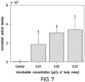
- a microbubble concentration of 0.01 ⁇ l/g was experimentally determined 130 to be the minimum concentration needed to open the BBB.
- the appropriate concentration of microbubbles can be determined 130 based on the nature of the subject, e . g ., a human or a mouse, based on the size of the target region, e . g ., the surface area of the BBB that one wishes to open, and based on the vessel size in the target region, e .
- a concentration range of 10 7 to 10 9 bubbles/mL can be appropriate.
- the bubbles were generated at an initial yield larger than the desired concentration and then diluted in PBS one minute before intravenous injection into the mouse.
- the bubble concentration can be chosen to be the same across different size distributions as opposed to the volume fraction, because it can be assumed that BBB opening occurs discretely, e.g., the sites of molecular leakage highly correlated with the instantaneous locations of the bubbles at the time of sonication. This implies that BBB opening sites are punctuated along the length of the capillaries. In the case where the volume fraction was kept the same for both sets of bubbles, it is deemed that the imaging protocol used would have the required sensitivity to detect minute increases in fluorescence.
- the appropriate size range of microbubbles can be determined by comparing the bubble size to the cerebral vasculature size and selecting a bubble size that is small enough to perfuse the vessels while at the same time large enough to induce sufficient mechanical stress on the vessel walls, such that the vessels are opened to the target value.
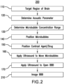
- FIG. 2 illustrates a method 200 , which is not part of the present invention, in accordance with the disclosed subject matter for imaging the opening of a tissue.
- the method 200 includes the same basic techniques for opening the tissue to a target value: targeting 110 a region of the tissue for opening, determining 120 at least one acoustic parameter corresponding to the target value, positioning 140 microbubbles of a known size range in proximity to the targeted region, and applying 170 an ultrasound beam at the acoustic parameter to the targeted region such that the tissue is opened with the assistance of the microbubbles to the target value.
- the method 200 further includes imaging 210 the opened tissue.
- imaging 210 the opened tissue can be the same as the application 170 of an ultrasound beam to open the tissue.
- imaging 210 can include utilizing an MRI device to image the opening of the tissue.
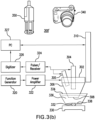
- Figure 3(a) illustrates a system 300 for opening a tissue to a target value.
- System 300 has many of the same features as the system described in U.S. Patent Pub. No. 2009/0005711 and International Patent Pub. No. WO 2010/030819 , commonly assigned patent applications.
- Ultrasound waves are generated by a focused ultrasound transducer (FUS) 302 , which can be a single-element circular-aperture FUS transducer.
- the FUS transducer 302 can be a single-element, spherical segment FUS transducer with center frequency of 1.525 MHz, a focal depth of 90 mm, an outer radius of 30 mm, and an inner radius of 11.2 mm (Riverside Research Institute, New York, NY, USA).
- the FUS transducer can be provided with hole in its center for receipt of an imaging transducer 304 , which can be a single-element diagnostic transducer having a center frequency of 7.5 MHz with a focal length of 60 mm (Riverside Research Institute, New York, NY, USA).
- the FUS transducer 302 and the diagnostic transducer 304 can be positioned so that the foci of the two transducers are properly aligned, e . g ., overlap.
- an exemplary system 300 can include a cone 306 filled with degassed and distilled water and mounted on system 300 .
- the cone 306 can, for example, be manufactured from a clear plastic, such as polyurethane.
- the water is contained in the cone 306 by capping it with a material considered substantially "transparent" to the ultrasound beam, such as an ultrathin polyurethane membrane 308 (Trojan; Church & Dwight Co., Princeton, N.J., USA).
- the transducer assembly which can include the FUS transducer 302 and the diagnostic transducer 304 , can be mounted to a computer-controlled 3-D positioning system 310 (Velmex Inc., Lachine, QC, Canada), including motors VXM-1 and VXM-2 used in the exemplary embodiment. It is understood that other positioning systems can be incorporated for positioning the transducer assembly with respect to the targeted tissue.
- a computer-controlled 3-D positioning system 310 (Velmex Inc., Lachine, QC, Canada), including motors VXM-1 and VXM-2 used in the exemplary embodiment. It is understood that other positioning systems can be incorporated for positioning the transducer assembly with respect to the targeted tissue.
- the FUS transducer 302 can be driven by a function generator 320 , e.g., function generator HP33150A, manufactured by Agilent Technologies, Palo Alto, Calif., USA, through an amplifier 322 , such as a 50-dB power amplifier 3100L (ENI, Inc., Rochester, N.Y., USA).
- the diagnostic transducer 304 can be driven by a pulser-receiver system 342 , for example a pulser-receiver 5052PR (Panametrics, Waltham, Mass., USA), connected to a digitizer 326 , e.g. , digitizer CS14200 (Gage Applied Technologies, Inc., Lachine, QC, Canada).
- Computer 328 typically includes a processor, such as a CPU (not shown), and can be any appropriate personal computer or distributed computer system including a server and a client.
- a computer useful for this system is a Dell Precision 380 personal computer. It is understood that any personal computer, laptop, or other processor that can load software and communicate with the various components discussed herein can be used.
- a memory unit (not shown), such as a disk drive, flash memory, volatile memory, etc., can be used to store software for positioning and operating the transducer assembly, image data, a user interface software, and any other software which can be loaded onto the CPU.
- system 300' can include a transducer assembly having an array of a plurality of single-element FUS transducers 304 and 305 which can be targeted to different regions of the tissue of the subject.
- Each FUS transducer 304 , 305 in the array can be fired individually, thereby permitting opening of the BBB in several locations without repositioning the transducer assembly.
- a scan such as a 3-D raster-scan (lateral step size: 0.2 mm; axial step size: 1.0 mm), of the beam of the FUS transducer 302 , can optionally be performed in a large water tank containing degassed water with a needle hydrophone having a needle diameter on the order of about 0.2 mm (Precision Acoustics Ltd., Dorchester, Dorset, UK). In this manner the pressure amplitudes and three-dimensional beam dimensions of the FUS transducer 302 can be measured.
- the pressure amplitudes can be measured by calculating the peak-rarefactional pressure values and accounting for an pressure attenuation due to transcranial propagation, e.g., an 18% pressure attenuation.
- the dimensions of the beam provided by the FUS transmitter 302 can have a lateral and axial full-width at half-maximum (FWHM) intensity of approximately 1.32 and 13.0 mm, respectively, and in some embodiments can be approximately equal to the dimensions of the beam after propagation through the skull.
- FWHM full-width at half-maximum
- System 300 also includes a liquid container 334 containing an appropriate liquid 336 , e . g ., degassed and distilled water, which is sealed at the bottom with a membrane 338 , which can be a polyurethane membrane that is acoustically and transparent, e . g ., plastic wrap.
- the system 300 can also include an optical imaging device 340 , such as a digital camera, for imaging the skull of the subject 332 and a MRI device 350 for imaging the brain of the subject 332 .
- System 300 also includes a platform 330 for the subject.
- the platform 330 for the subject can be a polyurethane bed for a smaller subject 332 , such as a mouse.
- the membrane 338 can be placed over the subject 332 .
- the platform 330 can be a hospital bed or surgical table, in which a larger subject 332 (such as a human subject) can be laid prone or supine and the transducer assembly positioned on top of the region of the skull targeted.
- Additional components of the system 300 include a targeting system 400 , coupled to the FUS transducer 302 , for locating the focus of the FUS transducer 302 in the brain of the subject 332 .
- the targeting system 400 can be coupled by any known method that permits the targeting system 400 to aid in properly targeting the FUS transducer 302 to the region of interest for opening of the target tissue, e.g., acoustic and/or optical coupling.
- Figures 4(a)-(d) illustrate a targeting system 400 for use with an embodiment where the subject 332 is a mouse.
- Figure 4(a) illustrates mouse skull 401 , where the skull's sutures can be seen through the skin and used as anatomic landmarks for targeting purposes.
- the landmarks of mouse skull 401 include the sagittal suture 402 , the frontal bone 404 , the interparietal bone 406 , the left parietal bone 408 , and the right parietal bone 410 .
- FIG. 4(b) illustrates the placement of targeting system 400 on skull 401 in accordance with an exemplary embodiment.
- the targeting system 400 can include a plurality of members 420 , 422 , 424 , such as thin metal bars, e . g ., 0.3 mm thin metal bars, fabricated from an acoustically reflective material, e . g ., paper clips.
- the metal bars 420 , 422 , 424 can be placed on several landmarks of the skull of the subject to create a layout, or grid.
- a grid consisting of three equally spaced 0.3-mm thin F2 metal bars 420 , 422 , 424 were placed in the water bath 334 on top of the skull 401 and in alignment with these landmarks, e . g ., bone sutures.
- the first bar 420 was aligned parallel and along the sagittal suture 402
- the second bar 424 was attached perpendicularly to the first bar and in alignment with the suture between the parietal 408 and interparietal bone 406 .
- the third bar 422 was placed 4 mm away from and parallel to the second bar 424 .
- Figure 4(c) illustrates the location of a brain structure 440 to be targeted, here the hippocampus, relative to the landmarks noted above.
- the location of the hippocampi are assumed relative to the sutures based on the mouse brain and known skull anatomy.
- the location of one of the hippocampi (indicated by circle 440 ) was reproducibly targeted when assumed to be at mid-distance (arrow 442 ) between the parallel bars 422 , 424 and 2 mm away from the center bar 420 (arrow 444 ).
- the targeting system can include other imaging devices, such as a digital camera 340 .
- a digital camera 340 can be used to photograph the head of the subject 332 .
- the relevant landmarks can be identified in the photograph, and the focus of the FUS transducer 302 targeted to a location relative to the landmarks.
- other MRI targeting equipment as is known in the art, can be used for targeting the desired brain structure 440 or other targeted tissue structure.
- Figure 4(d) illustrates the actual location of the hippocampus 446 as indicated in the histology slice.
- Figure 4(e) illustrates a lateral 2-D raster-scan 490 of the grid 400 using the diagnostic transducer 304 .
- the location of the hippocampus can be identified relative to this grid.
- the focus of the FUS transducer 302 was placed 3 mm beneath the top of the skull by measuring distance with the diagnostic transducer 304 .
- precise, accurate and reproducible targeting of the hippocampus or other brain structures can be performed.
- the grid positioning system 400 allowed for sonication of the same location with good accuracy across different mice. This allowed for not only good reproducibility across different mice, but also a good comparison of BBB opening effects in different regions 440 within the sonicated area.
- the subject 332 is positioned on a platform 330 .
- Subject 332 can be positioned in a prone position, and can be anesthetized for the sonication procedure.
- the degassed and distilled water bath 334 is suspended over the subject's 332 head.
- Ultrasound gel can be used to reduce any remaining impedance mismatches between the thin plastic layer 338 and the subject's 332 skin.
- the transducer assembly can be placed in the water bath 334 with its beam axis perpendicular to the surface of the skull 401 .
- the focus of the transducer is positioned inside the subject's 332 brain.
- the focus can be targeted 110 to a region of the brain 440 , such as the desired brain tissue, e.g., the hippocampus 446 , or to the vasculature of the brain, e.g., arteries, ventricles, arterioles, and capillaries of the brain, or to other target tissue regions at different locations in the subject 332 .
- the targeted region 440 of the brain can be located 110 utilizing the targeting system as discussed above.
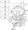
- Figure 5 illustrates a system 300 used in an experiment, approved by the Columbia University Institutional Animal Care and Use Committee, on seventy-nine wild-type mice (strain: C57BL/6, mass: 28.0 ⁇ 4.5 g, sex: male; Harlan, Indianapolis, IN, USA) which were studied in accordance with the techniques described herein.
- the system 300 can include a FUS transducer 302 , a pulse-echo diagnostic transducer 304 , a cone 306 , a latex membrane 308 , a 3-D positioning system 310 all operatively connected to a function generator 320 , a power amplifier 322 , a pulse-receiver system 324 , a digitizer 326 and a computer 327 .
- the cone 306 can be inserted into a water container 334 which is sealed at the bottom by a polyurethane membrane 338 and placed on the shaved skull 502 of the mouse subject 332 .
- the mouse subject 332 is held in place using a stereotaxic apparatus 504 .
- mice were anesthetized using 1.25-2.50% isoflurane (SurgiVet, Smiths Medical PM, Inc., Wisconsin, USA) throughout both the BBB opening and transcardial perfusion procedures.
- each mouse 332 was placed prone with its head immobilized by the stereotaxic apparatus 504 (David Kopf Instruments, Tujunga, CA, USA). The hair on the skull was removed using an electric trimmer and a depiatory cream.
- the FUS transducer 302 was then submerged in the water of the container 334 with its beam axis perpendicular to the surface of the skull 332 .
- the focus of the transducer was positioned inside the mouse brain using a grid-positioning method that utilized the pulse-echo diagnostic transducer 304 , as discussed above.
- the grid was constructed from three 0.30 mm thin metal bars (i . e ., paper clips) with two of the bars parallel to one another and separated by 4.00 mm. At the center of the parallel bars, and perpendicular to the two, was soldered the third bar.
- the grid was placed in the water bath 334 , on top of the skull, and in alignment with sutures visible through the skin. The center bar was aligned along the sagittal suture and one of the parallel bars with the lambdoid suture.
- a lateral two-dimensional raster-scan of the grid using the diagnostic transducer was made and the transducer's beam axis was positioned 2.25 and 2.00 mm away from the sagittal and lambdoid suture, respectively.
- the focal point was placed 3.00 mm beneath the top of the skull so that the acoustic wave propagated through the left parietal bone and overlapped with the left hippocampus and a small portion of the lateral region of the thalamus.
- the right hippocampus was not targeted and was used as the control.
- the grid positioning method was sufficiently precise to have the FUS beam consistently overlap the hippocampus of the murine brain.
- the tissue opening procedure 100 involved injection 140 , 150 a 25 ⁇ l bolus of Definity ® microbubbles (1-10 ⁇ m) and a dextran contrast agent (Texas-Red ® fluorescent dye with a molecular weight of 3 kDa) into the tail vein 1 minute after the start of sonication 170 , with the injection taking place over a 30 second period. Sonication was performed for 11 minutes total using pulsed FUS at a set pressure of 0.51 MPa peak-rarefactional at a single location ( e . g ., the hippocampus).
- a dextran contrast agent Texas-Red ® fluorescent dye with a molecular weight of 3 kDa
- the dextran was allowed to circulate and accumulate in the mouse brain for 10 minutes, after which a transcardial perfusion with phosphate buffer saline (138 mM sodium chloride, 10 mM phosphate, pH 7.4) and 60 ml of 4% paraformaldehyde was performed.
- the brain was extracted from the skull and then post-fixed in the paraformaldehyde overnight. Following the aforementioned procedures, the brain was prepared for frozen sections.
- the frozen sectioning protocol provided an efficient means of analyzing fluorescence in order to determine the threshold for BBB opening. In preparation of frozen sectioning, the brain was cryoprotected by soaking it in 30% sucrose overnight.
- the brain was then embedded in a cutting temperature compound (Sakura Tissue-Tek O.C.T. Compound; Torrance, CA, USA), frozen in a square mold, and then sectioned using a cryostat into nine sections of 100 ⁇ m slices in the horizontal orientation.
- a cutting temperature compound Sakura Tissue-Tek O.C.T. Compound; Torrance, CA, USA
- Bright field and fluorescent images of the frozen sections were acquired using an inverted light and fluorescence microscope (IX-81; Olympus, Melville, NY, USA) at 4X magnification and with a motorized stage-scanner. Images of the paraffin sections were acquired using an upright light and fluorescence microscope (BX61; Olympus, Melville, NY, USA) at 4 ⁇ and 10 ⁇ magnification. The Texas Red-tagged dextrans were excited at 568 ⁇ 24 nm while emissions were filtered for 610 ⁇ 40 nm.
- Figure 6(a) illustrates a horizontal section at 10 ⁇ magnification, with the left and right ROIs shown in the left and right boxes.
- Figure 6(b) shows the left ROI, which was subjected to sonication procedures, as detailed above
- Figure 6(c) shows the right ROI, which was the control.
- the regions of interest (ROIs) for each of the nine sections were outlined using Adobe ® Photoshop ® CS2 (San Jose, CA, USA), as illustrated Figures 6(b)-(c) . The outlines were then loaded into MATLAB ® (Natick, MA, USA) and used to isolate the hippocampus in the fluorescent images.
- the images were normalized by dividing both the left and right images by the spatially averaged right (control) image of the hippocampus, thus calculating F L-HIP .
- the threshold for an image was were the pixel value was greater than 2 standard deviations of F L-HIP ; images exceeding the threshold were excluded from the calculations.
- the NOD for the brain was calculated by averaging the NOD of across all nine sections. The resulting averaged NOD was then used to determine whether, to what extent, the BBB had opened.
- Figure 7 is a graph illustrating the effects of varying the microbubble concentration in an exemplary embodiment where the FUS pressure was 0.46 MPa, the microbubbles used where Definity ® bubbles, the PRF was 10 Hz and the PL was 20 ms.
- the asterisks (*) indicates a significant difference in NOD from the control. As can be seen in Figure 7 , there was a significant NOD increase for all concentrations test and further there was not a significant difference between the tested concentrations, indicating that with Definity microbubbles a concentration of 0.01 ⁇ l/g of body mass is both sufficient to open the BBB in mice and also reliable for doing the same.
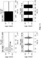
- Figure 8(a) is a graph illustrating the effects of varying the PRF in an exemplary embodiment where the FUS pressure was 0.46 MPa, the microbubbles used were Definity ® bubbles, the microbubble concentration was set at 0.05 ⁇ l/g of body mass, and the PL was 20 ms.
- the asterisks (*) indicates a significant difference in NOD from the control.
- at least one pulse is needed to open the BBB in a mouse brain prepared in accordance with the procedures set forth above. Further, the lowest PRF that was observed to open the BBB was 1 Hz, while the lowest PRF that was observed to reliably open the BBB was 5 Hz.
- Figure 8(b) further illustrates these findings, showing the PRF as a function of the probability of BBB opening. As illustrated in Figure 8(b), at 5 Hz and above there is a 100% probability of the BBB opening in the mice subjects prepared in accordance with the exemplary procedures set forth above. Figure 8(a) also illustrates that no additional benefits are gained from using a PRF greater than 5 Hz.
- Figure 9(a) is a graph illustrating the effects of varying the PL in an exemplary embodiment where the FUS pressure was 0.46 MPa, the microbubbles used were Definity ® bubbles, the microbubble concentration was set at 0.05 ⁇ l/g of body mass, and the PRF was 10 Hz.
- the asterisks (*) indicates a significant difference in NOD from the control.
- lowest PL which was resulted in BBB opening was 0.033 ms
- 0.2 ms was the lowest PL which produced reliable BBB opening in mice prepared in accordance with the above-detailed exemplary procedures.
- Figure 9(a) demonstrate that, contrary to prior understanding, pulse lengths shorter than 10 ms can reliably open the BBB for FUS applications utilizing pressures less than 0.5 MPa.
- Figure 9(a) also illustrates that no additional benefits are gained from using a PL greater than 10 ms.
- Figures 9(b) and 9(c) illustrate the PL as a function of the probability of BBB opening, with Figure 9(b) showing the data across all PLs tested in the exemplary experiment discussed herein and Figure 9(c) illustrating the shortest PLs tested.
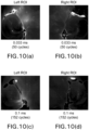
- Figures 10(a)-(h) are images of murine brains prepared in accordance with the above-detailed exemplary procedures and illustrate the results of varying the PL from 0.033 ms (50 cycles) to 30 ms (45600 cycles).
- Figures 10(a) and 10(b) illustrate the left sonicated ROI and right control ROI, respectively, at a PL of 0.033 ms (50 cycles).
- Figures 10(c) and 10(d) illustrate the left sonicated ROI and right control ROI, respectively, at a PL of 0.1 ms (152 cycles).
- Figures 10(e) and 10(f) illustrate the left sonicated ROI and right control ROI, respectively, at a PL of 20 ms (30400 cycles).
- Figures 10(g) and 10(h) illustrate the left sonicated ROI and right control ROI, respectively, at a PL of 30 ms (45600 cycles).
- FIG 11 illustrates an exemplary pulse and burst sequence, where each burst is composed set of pulses operating at a certain pulse rate frequency (PRF).
- PRF pulse rate frequency
- one pulse can have a PL of 2.5 ⁇ s (approximately 3.5 cycles) at a pressure of 0.6 MPa.
- the pulses can be repeated at a PRF of, for example, 100 kHz.
- the pulsing can be continued for a period of time, e.g., 10 ms, which comprises a single burst of a certain length (BL).
- the bursts can be repeated at a certain rate, for example 50 Hz, which is the BRF.
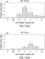
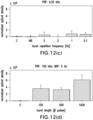
- Figures 12(a)-(c) illustrate the NOD as a function of the BRF for three different PRFs, for an exemplary experiment involving mice preformed in accordance with the above-detailed procedures.
- Figure 12(a) is a graph illustrating the effects of varying the BRF, in an exemplary embodiment where the FUS pressure was 0.46 MPa, the microbubbles used were Definity ® bubbles, the microbubble concentration was set at 0.05 ⁇ l/g of body mass, the sonication duration was 11 minutes, the BL was 1000 pulses, and the PRF was set at 100 kHz.
- Figure 12(b) is a graph illustrating the effects of varying the BRF, in an exemplary embodiment having the same parameters as noted for Figure 12(a) , except at a PRF of 25 kHz.
- Figure 12(b) is a graph illustrating the effects of varying the BRF, in an exemplary embodiment having the same parameters as noted for Figure 12(a) , except at a PRF of 6.25 kHz.
- Figure 12(d) is a graph illustrating the effects of varying the BL, in an exemplary embodiment where the FUS pressure was 0.46 MPa, the microbubbles used were Definity ® bubbles, the microbubble concentration was set at 0.05 ⁇ l/g of body mass, the sonication duration was 11 minutes, the BRF was 5 Hz, and the PRF was 100 kHz.
- Figures 13(a)-(b) illustrate images of a mouse brain, prepared in accordance with the exemplary methods described herein but not claimed, showing BBB opening where the FUS pressure was 0.6 MPa, the microbubbles used were Definity ® bubbles, the microbubble concentration was set at 0.05 ⁇ l/g of body mass, the sonication duration was 11 minutes, the PL was 3 cycles (less than 2.5 ⁇ s), the PRF was 6.25 kHz, the BL was 1000 pulses, and the BRF was 5 kHz.
- Figure 13(b) is an enlarged image of the area of interest illustrated by the box in Figure 13(a) , and it illustrates that the BBB was opened at a PL of 3 cycles.
- Figures 13(c)-(d) illustrate images of a mouse brain, prepared in accordance with the exemplary methods described but not claimed herein, showing BBB opening where the FUS pressure was 0.6 MPa, the microbubbles used were Defmity ® bubbles, the microbubble concentration was set at 0.05 ⁇ l/g of body mass, the sonication duration was 11 minutes, the PL was 3 cycles (less than 2.5 ⁇ s), the PRF was 100 kHz, the BL was 1000 pulses, and the BRF was 2 Hz.
- Figure 13(d) is an enlarged image of the area of interest illustrated by the box in Figure 13(c) , and the boxed area in Figure 13(d) illustrates that the neuronal uptake occurred at a PL of 3 cycles.
- Table 1 illustrates the results of another experiment preformed on ninety-nine male mice (strain: C57Bl6; 24.71 ⁇ 1.77 g) in accordance with the exemplary methods described herein but not claimed and the procedures of the Columbia University Institutional Animal Care and Use Committee.
- Table 1 illustrates eighteen different experimental conditions, varying the microbubble concentration ( ⁇ L/g of body mass), the PRF (Hz), and the PL (ms). The results are shown in terms of the NOD (mean ⁇ s.d. ⁇ 10 9 ) and the number of mice with delivered dextran.
- the first entry corresponds to a sham mouse, where no ultrasound was applied; in the second entry the microbubbles and dextran were administered 1 minute before a 30-second sonication; and in the final entry the microbubbles were injected over a 180 second period.
- the mice were intravenously injected with a solution of dextran and microbubble 1 minute after the start of an 11-minute sonication. All sonications were performed with an 1.525 MHz acoustic beam and at a peak-rarefactional pressure of 0.46 MPa.
- Table 1 Microbubble concentration ( ⁇ L/g of body mass) Pulse repetition frequency (Hz) Pulse length (ms) NOD x 1e9 (mean ⁇ std.
- Table 2 illustrates the results of another experiment preformed on ninety-five C57Bl6 male mice in accordance with the exemplary methods described herein and the procedures of the Columbia University Institutional Animal Care and Use Committee.
- Table 2 illustrates eighteen different experimental conditions, varying the PRF (kHz), BRF (Hz), the BL (# of pulses) and the peak-rarefactional pressure. The results are shown in terms of the NOD (mean ⁇ s.d. ⁇ 10 9 ) and the number of mice with an incidence of NOD increase, calculated as detailed above.
- Table 2 further illustrates, the dependence of acoustic peak-rarefactional pressure on BBB disruption was evaluated in a sham (0 MPa) and pressures of 0.13, 0.25, 0.37, and 0.51 MPa.
- a significant increase in NOD was only observed at 0.51 MPa.
- 0.37 MPa had no significant increase in NOD, 2 out of 3 mice had detectable levels of fluorescence. Therefore, the pressure threshold for BBB disruption for a 3.5-cycle pulse was between 0.25 and 0.51 MPa.
- the effect of BL was evaluated from 1 to 1000 pulses. A single pulse was insufficient in disrupting the BBB. The lowest pressure show feasible in disrupting the BBB was at 5 pulses and was observed in 1 out of 3 mice.
- a significant increase in NOD was observed from 50 pulses and higher. In general, increasing the number of pulses increased the likelihood and magnitude of NOD increase.
- the 70-kDa dextran had heterogeneous spots of high levels of fluorescence on top of diffusely distributed fluorescence.
- the morphology of neurons and/or glial cells can be seen.
- a neuronal axon with an approximately 1 ⁇ m diameter can be observed extending from its cellular body and attached to a capillary, which had a diameter for approximately 4.5 ⁇ m.
Landscapes
- Health & Medical Sciences (AREA)
- Life Sciences & Earth Sciences (AREA)
- Engineering & Computer Science (AREA)
- Surgery (AREA)
- General Health & Medical Sciences (AREA)
- Veterinary Medicine (AREA)
- Biomedical Technology (AREA)
- Heart & Thoracic Surgery (AREA)
- Medical Informatics (AREA)
- Animal Behavior & Ethology (AREA)
- Public Health (AREA)
- Nuclear Medicine, Radiotherapy & Molecular Imaging (AREA)
- Molecular Biology (AREA)
- Physics & Mathematics (AREA)
- Biophysics (AREA)
- Pathology (AREA)
- Radiology & Medical Imaging (AREA)
- Hematology (AREA)
- Neurology (AREA)
- Orthopedic Medicine & Surgery (AREA)
- Vascular Medicine (AREA)
- Dermatology (AREA)
- Anesthesiology (AREA)
- High Energy & Nuclear Physics (AREA)
- Surgical Instruments (AREA)
- Ultra Sonic Daignosis Equipment (AREA)
Description
- The present application relates to systems for opening a tissue utilizing acoustic parameters in conjunction with microbubbles.
- Recent advances in molecular engineering and neuroscience have led to an increasing number of biomarkers and therapeutic agents for the monitoring and treatment of neurological disorders. Many of these agents have proven in vitro specificity or neurological potency, but their in vivo efficacy remains limited by their inability to reach their target due to the blood-brain barrier. This interface regulates the exchange of molecules across the cerebral capillaries through passive, transport, and metabolic barriers, resulting in the exclusion of nearly all agents larger than 400 Da from the brain's extracellular space. Biomarkers and therapeutic agents, such as inhibitors to enzymes (~1 kDa) and antibodies (30 to 300 kDa), are thus rendered ineffective because they do not reach their intended targets. Systems for opening a tissue to a target value are known from
US 2009/0005711 , as described further below. - Systems for opening a tissue to a target value are disclosed herein according to
claim 1 and dependent claims. In the following all methods are disclosed for illustrative purposes only and do not form part of the invention. - In an embodiment of a method for opening a tissue to a target value using microbubbles, a region of the tissue is targeted for opening, which is not part of the present invention, an acoustic parameter corresponding to the target value is determined and an ultrasound beam is applied to the target region at the acoustic parameter such that the tissue at the target region is opened to the target value with the microbubbles. The method can further include positioning microbubbles in proximity to the targeted region and, in some embodiments, positioning the microbubbles can include performing an injection of the microbubbles such that the microbubbles are positioned proximate to the targeted region. The method can further include determining a number of injections and/or a duration of an injection corresponding to the target value. In some embodiments, the injection can be a systemic injection, a bolus injection and/or a slow diffusion injection. The acoustic parameter can be selected to control an acoustic cavitation event and, in some embodiments, controlling an acoustic cavitation event can include controlling a location, number and/or magnitude of acoustic cavitation events. The acoustic parameter can be a pulse length, a pulse repetition frequency, a burst length, a burst repetition frequency, an ultrasound frequency, a pressure range, and/or a duration corresponding to the target value. In some embodiments, the pressure range can correspond to the resonance frequency of the microbubbles proximate to the targeted region.
- The method, which is not part of the present invention, can include determining a concentration range of microbubbles corresponding to the target value and applying an ultrasound beam to move the microbubbles into vessels of the tissue. In some embodiments, the microbubbles can have a size range of 1 to 10 microns, and in other embodiments can have a size range of 1 to 2 microns, 4 to 5 microns, or 6 to 8 microns. The microbubbles can be acoustically activated and/or molecule-carrying. The molecule-carrying microbubbles can carry or be coated with medicinal molecules and/or a contrast agent and/or a biomarker and/or a liposome. Medicinal molecules and/or contrast agents can also be separately positioned in proximity to the targeted region.
- The method, which is not part of the present invention, can further include imaging the targeted region, to form an image of the opened tissue. In some embodiments imaging the targeted region includes applying an ultrasound beam to the targeted region, while in other embodiments imaging the targeted region includes utilizing a magnetic resonance imaging device and/or a fluorescence imaging device to image the targeted region.
- An embodiment of a system for opening a tissue to a target value according to
claim 1 and dependent claims using a solution of microbubbles having a size range corresponding to the target value includes a targeting assembly for targeting a region of the tissue, an introducer for delivering the solution to a location proximate to the targeted region and a transducer, coupled to the targeting assembly, for applying an ultrasound beam to the targeted region at an acoustic parameter corresponding to the target value thereby opening the tissue with the microbubbles to the target value. The acoustic parameter can be selected to control an acoustic cavitation event. - The system according to
claim 5 can further include an imaging device for capturing image data of the opened tissue of the targeted region, and a processor, operatively coupled to the imaging device, for processing the image data to form an image therefrom. In some embodiments the imaging device includes a transducer for applying an ultrasound beam to the targeted region, while in other embodiments the imaging device includes a magnetic resonance imaging device and/or a fluorescence imaging device to image the targeted region. - The accompanying drawings, which are incorporated and constitute part of this disclosure, illustrate some embodiments of the disclosed subject matter.
-
FIG. 1 illustrates a method for opening a blood-brain barrier in a brain of a subject to a target value in accordance with an exemplary embodiment of the disclosed subject matter, which is not part of the present invention. -
FIG. 2 illustrates a method for imaging the opening of a blood-brain barrier in a brain of a subject to a target value in accordance with an exemplary embodiment of the disclosed subject matter, which is not part of the present invention. -
FIG. 3(a) illustrates a system for opening and/or imaging the opening of a blood-brain barrier in a brain of a subject to a target value in accordance with an exemplary embodiment of the disclosed subject matter. -
FIG. 3(b) illustrates another system for opening and/or imaging the opening of a blood-brain barrier in a brain of a subject to a target value in accordance with an exemplary embodiment of the disclosed subject matter. -
FIGS. 4(a)-(e) illustrate a targeting system for locating a target region of the brain of a subject in accordance with an exemplary embodiment of the disclosed subject matter. -
FIG. 5 illustrates a system for opening the blood-brain barrier used in connection with an experiment on mice in accordance with an exemplary embodiment of the disclosed subject matter. -
FIG. 6(a) illustrates a fluorescence image at ten times magnification of a mouse brain after application of tissue opening techniques in accordance with an exemplary embodiment of the disclosed subject matter. -
FIGS. 6(b)-(c) illustrate fluorescence images at four times magnification of a mouse brain after application of tissue opening techniques in accordance with an exemplary embodiment of the disclosed subject matter. -
FIG. 7 is a graph illustrating the effects of varying the microbubble concentration in accordance with an exemplary embodiment of the disclosed subject matter. -
FIG. 8(a) is a graph illustrating the effects of varying the pulse repetition frequency in accordance with an exemplary embodiment of the disclosed subject matter. -
FIG. 8(b) is a graph illustrating the probability of blood-brain barrier opening as a function of varying the pulse repetition frequency in accordance with an exemplary embodiment of the disclosed subject matter. -
FIG. 9(a) is a graph illustrating the effects of varying the pulse length in accordance with an exemplary embodiment of the disclosed subject matter. -
FIGS. 9(b)-(c) are graphs illustrating the probability of blood-brain barrier opening as a function of varying the pulse length in accordance with an exemplary embodiment of the disclosed subject matter. -
FIGS. 10(a)-(h) illustrate histological images of a mouse brain after focused ultrasound sonication with a varying pulse length in accordance with an exemplary embodiment of the disclosed subject matter. -
FIG. 11 illustrates an exemplary pulse and burst sequence in accordance with an exemplary embodiment of the disclosed subject matter. -
FIGS. 12(a)-(c) are graphs illustrating the effects of varying the burst repetition frequency for three different pulse repetition frequencies in accordance with an exemplary embodiment of the disclosed subject matter. -
FIG. 12(d) is a graph illustrating the effects of varying the burst length for a certain pulse repetition frequency and burst length in accordance with an exemplary embodiment of the disclosed subject matter. -
FIGS. 13(a)-(d) illustrate images of a mouse brain subjected to sonication using pulse length of three cycles in accordance with an exemplary embodiment of the disclosed subject matter. - Throughout the figures and specification the same reference numerals are used to indicate similar features and/or structures.
- The systems described herein are useful for opening a tissue utilizing microbubbles and focused ultrasound at certain acoustic parameters. Although the description provides as an example opening the blood-brain barrier, the systems herein are useful for opening other tissues, such as muscular tissue, liver tissue or tumorous tissue, among others.
- The subjected matter disclosed herein are systems for determining the acoustic parameters for opening a tissue with the assistance of microbubbles to allow for the passage of certain molecules over selected areas. Accordingly, the techniques described herein make use of selected acoustic parameters chosen to produce a desired opening effect in a tissue when subjected to focus ultrasound utilizing microbubbles of selected sizes and in selected concentrations. The techniques described herein for determining the acoustic parameters for opening a tissue can also be employed in conjunction with other ultrasound techniques, e.g., diagnostic techniques, where opening of a tissue should be avoided. The techniques described herein can be used to determine the acoustic parameters that can be avoided in order to prevent unwanted tissue opening when utilizing such other techniques that, for example, use microbubbles.
- In focused ultrasound (FUS), acoustic waves propagate several centimeters through water or tissue and converge onto a focal region while its surroundings remain relatively unaffected. Noninvasive and localized drug delivery systems have emerged from advances in FUS and microbubble technologies. For example, techniques such as blood-brain barrier (BBB) disruption for the treatment of neurological diseases, delivery of nanoparticles to tumors, gene therapy for treating heart conditions, and enhancement of renal ultrafiltration have all shown promise due to their ability to increase uptake of luminal molecules into the interstitial space.
- The mechanistic event underlying the tissue opening in such examples is the reaction of microbubbles to ultrasonic pulses, which can result in an array of behaviors known as acoustic cavitation. In stable cavitation, the microbubble expands and contracts with the acoustic pressure rarefaction and compression over several cycles, and such action can result in the displacement of the vessel diameter through dilation and contraction. In inertial cavitation, the bubble can expand to several factors greater than its equilibrium radius and subsequently collapse due to the inertia of the surrounding media, thus also inducing an alteration of the vascular physiology. The type and magnitude of each cavitation activity can be dictated by, among other things, the microbubble composition and distribution, the ultrasonic pulse shape and sequence, and the in vivo environment in which the bubbles circulate. Control of molecular delivery using FUS can therefore be facilitated by selecting microbubbles and the acoustic environment conditions that they interact with.
- Generally, changing ultrasonic parameters has been associated with a tradeoff between efficacy (e.g., high dose, homogeneous distribution, and consistency) and safety (e.g., erythrocyte extravasations and neuronal damage). The acoustic pressure can have a large influence on the type and magnitude of acoustic cavitation activity. Thus, increasing the pressure increases the likelihood and extent of BBB opening, but also is associated with neurovascular and neuronal damage. At acoustic pressures near the threshold of BBB disruption, histological assessments reveal no detectable damage (e.g., erythrocyte extravasations and/or neuronal damage) but at such low pressures there is also a reduction in molecular delivery. Lowering the transmitted center frequency of the ultrasound can result in a decrease of the acoustic pressure threshold.
- The pulse repetition frequency (PRF) can effect the ability of microbubbles to reperfuse the vasculature since each pulse can destroy microbubbles. Further, it has been thought that a long pulse length (PL) was a necessary characteristic of an ultrasonic pulse for inducing BBB disruption, for example PLs of 10 or 20 ms (15200 and 30400 cycles at 1.5 MHz) and such long PLs have been associated with inhomogeneity of drug delivery. However, in accordance with an embodiment herein, it is shown that BBB disruption is feasible at a low pressure (less than 1 MPa) using a PL of 33 µs (50 cycles at 1.5 MHz). Utilizing such a low PL and pressure, an improved distribution of the delivered agent was observed, but an associated decrease in concentration of molecules delivered was also observed. As further shown in an embodiment herein, pulse sequences based on the use of a PL of 2.3 µs (3.5 cycles at 1.5 MHz) can enhance the dose and distribution of delivery without compromising safety.
- An acoustic parameter design can be formed on the basis that BBB disruption is dependent on the number, magnitude and/or location of cavitation events occurring throughout the cerebral microvasculature. Further, acoustic cavitation activity within the microvasculature can be modified by taking into account concepts of microbubble persistence, fragmentation, and microvascular replenishment. In one embodiment, a series of pulses can be grouped into a burst, with a sufficient duration between bursts to allow for microbubble replenishment in the microvasculature before arrival of the subsequent acoustic pulses. Grouping pluses into bursts can increase the persistence and mobility of the microbubbles, which can result in a single bubble generating cavitation activity at multiple sites along the cerebral microvasculature. Thus, in some embodiments, short PLs used in a burst sequence can enhance the dose and distribution of molecular delivery without attendant damage to the microvasculature. In the same or other embodiments, the number injections and duration of each injection of bubbles can be altered to enhance the microbubble persistence, for example.
-
Figure 1 illustrates amethod 100, which is not part of the present invention, for opening a tissue to a target value, e.g., a measure of increased ability of the tissue to pass molecules through. The target value can be expressed in terms of an increase in the size of vessels in the tissue, as an area of the tissue that has been opened, or in terms of a rate at which molecules pass through, e.g., a permeability, or as a combination of any of these measures. Themethod 100 involves targeting 110 a region of the tissue for opening, determining 120 at least one acoustic parameter corresponding to the target value, positioning 140 microbubbles in proximity to the targeted region, and applying 170 an ultrasound beam at the acoustic parameter to the targeted region such that the tissue is opened with the assistance of the microbubbles to the target value. In some embodiments, positioning 140 the microbubbles can include performing an injection of the microbubbles such that the microbubbles are positioned proximate to the targeted region. Themethod 100 can further include determining a number of injections and/or a duration of an injection corresponding to the target value. In some embodiments, theinjection 140 can be a systemic injection, a bolus injection and/or a slow diffusion injection. - As illustrated in
Figure 1 , in one exemplary embodiment, themethod 100, which is not part of the present invention. can further include of determining 130 a concentration range of microbubbles corresponding to the target value and thepositioning 140 of the microbubbles can also include positioning the microbubbles of the concentration range that corresponds to the target value. Themethod 100 can also include positioning 150 a contrast agent and/or medicinal molecule (e.g., a drug) in proximity to the target region. - In one exemplary embodiment,
method 100, which is not part of the present invention. can include applying 160 an ultrasound beam to move the microbubbles into vessels of the tissue. Thisapplication 160 of the ultrasound beam can be the same, or a different, than theapplication 170 that is used to open the tissue. Further, theapplication 160 of the ultrasound beam can be at the same, or at a different, acoustic parameter than that determined 120 for the purposes of opening the tissue. - The acoustic parameter to be determined 120 as corresponding to the target value can be selected to control one or more acoustic cavitation events. The acoustic parameter(s) can be selected such that the location, number and/or magnitude of acoustic cavitation events can be controlled in the targeted tissue. In some embodiments, the acoustic parameter can be at least one of the pulse length, the pulse repetition frequency, the burst length, or the burst repetition frequency, or a combination thereof. In other embodiments, the acoustic parameters that are determined 120 can be the pressure range, the frequency, and the duration of the
application 170 of ultrasound. - In some embodiments, the target value of the tissue can be selected based on the size of the molecule that is to pass through the tissue, e.g., the BBB, or based on the size, e.g., area, of the region that is to be exposed to the molecule, or a combination of the two. Thus, in some embodiments, the acoustic parameters can be determined 120 such that the tissue is subject to a certain number of acoustic cavitations at selected locations and of selected magnitudes, which can result in a selected number of molecules of a given size passing through the tissue at selected locations. In one exemplary embodiment, the target value can be such that molecules up to the megaDalton size range are able to pass through the BBB, e.g., 2 MDa molecules.
- In one embodiment, the target value can be measured in terms of the normalized optical density (NOD) of a contrast agent such as a dextran, e.g., Texas-Red® fluorescent dye and a molecular weight of 3 kiloDaltons (kDa), using the equation NOD = FL-ROI - FR-ROI , where FL-ROI is the sum of the pixel values in left region of interest (ROI) of the brain and RL-ROI is the sum of the pixel values in the right ROI of the brain, and where the ultrasound was applied 170 to the left ROI.
- In some embodiments, the acoustic parameter can be determined 120 by finding the lowest acoustic parameter value for which the tissue will open to the target value, where the target value is considered to be the minimum amount of opening. In one example involving murine brains, the acoustic parameters to be determined 120 were the PRF and the PL. In accordance with an exemplary experiment detailed below, a PRF value of 1 Hz was experimentally determined 120 to be the lowest PRF for which the BBB of a mouse subject was observed to open based on an observed NOD of approximately 2 × 10⁷. A PL value of 0.033 ms was experimentally determined 120 to be the lowest PL for which the BBB was observed to open based on an observed NOD of approximately 0.5 × 10⁷. In other embodiments, a PL of 3 cycles (less than 2.5 µs) was experimentally determined 120 to open the BBB in mice, in an experiment conducted in accordance with an exemplary embodiment described below.
- In some embodiments, the acoustic parameter can be determined 120 by finding the lowest reliable acoustic parameter value which will reliably open the tissue to the target value. In an example involving murine brains where the target value was considered the minimum NOD for which BBB opening was observed, the acoustic parameters to be determined 120 were the PRF and the PL. In accordance with an exemplary experiment detailed below, a PRF value of 5 Hz was experimentally determined 120 to be the lowest PRF for which the BBB of a mouse subject was observed to reliably open based on an observed NOD of approximately 2 × 10⁷. A PL value of 0.2 ms was experimentally determined 120 to be the lowest PL for which the BBB was observed to reliably open based on an observed NOD of approximately 0.5 × 10⁷.
- In yet other embodiments, the acoustic parameter can be determined 120 by finding the acoustic parameter value above which no further significant increase in opening of the tissue is achieved. In an example involving murine brains where the target value was considered to be the NOD above which no further increase in BBB opening was observed, the acoustic parameters to be determined 120 were the PRF and the PL. In accordance with an exemplary experiment detailed below, a PRF value of 5 Hz was experimentally determined 120 to be the PRF for which no further significant increase in the opening of the BBB of a mouse subject was observed and such opening corresponded to an NOD of approximately 2.5 × 10⁷. A PL value of 10 ms was experimentally determined 120 to be the lowest PL for which no further significant increase in the opening of the BBB of a mouse subject was observed and such opening corresponded to an observed NOD of approximately 3 × 10⁷.
- In the same or other embodiment involving murine brains, the acoustic parameters to be determined 120 can be the burst repetition frequency (BRF) and burst length (BL), where each burst can represent a cluster of pulses. A BRF of 10 Hz was experimentally determined 120 to open the BBB of a mouse subject, where the PRF was set at 100 kHz, and a BRF of 5 Hz was experimentally determined 120 to produce the maximal BBB opening at the same frequency. A BL of 100 pulses was experimentally determined 120 to open the BBB where the PRF was set at 100 kHz and the BRF was set at 5 Hz.
- In some embodiments determining 130 a concentration range of microbubbles corresponding to the target value can include determining the minimum microbubble concentration range that will open the tissue to the target value. In an example involving murine brains, a microbubble concentration of 0.01 µl/g was experimentally determined 130 to be the minimum concentration needed to open the BBB. In same or another embodiment, the appropriate concentration of microbubbles can be determined 130 based on the nature of the subject, e.g., a human or a mouse, based on the size of the target region, e.g., the surface area of the BBB that one wishes to open, and based on the vessel size in the target region, e.g., 4-8 µm, or a combination of these factors. In the example of opening a BBB area on the order of millimeters, a concentration range of 107 to 109 bubbles/mL can be appropriate. In one exemplary embodiment, the total concentration for both size ranges of bubbles, e.g., 1-2 and 4-5 µm, was kept constant at approximately 8.5 × 10⁸ number of bubbles per mL. In order to ensure accuracy of concentration, the bubbles were generated at an initial yield larger than the desired concentration and then diluted in PBS one minute before intravenous injection into the mouse.
- In one exemplary embodiment, the bubble concentration can be chosen to be the same across different size distributions as opposed to the volume fraction, because it can be assumed that BBB opening occurs discretely, e.g., the sites of molecular leakage highly correlated with the instantaneous locations of the bubbles at the time of sonication. This implies that BBB opening sites are punctuated along the length of the capillaries. In the case where the volume fraction was kept the same for both sets of bubbles, it is deemed that the imaging protocol used would have the required sensitivity to detect minute increases in fluorescence.
- As detailed in commonly assigned International Patent Pub.
WO 2010/030819 , in some embodiments the appropriate size range of microbubbles can be determined by comparing the bubble size to the cerebral vasculature size and selecting a bubble size that is small enough to perfuse the vessels while at the same time large enough to induce sufficient mechanical stress on the vessel walls, such that the vessels are opened to the target value. -
Figure 2 illustrates amethod 200, which is not part of the present invention, in accordance with the disclosed subject matter for imaging the opening of a tissue. Themethod 200 includes the same basic techniques for opening the tissue to a target value: targeting 110 a region of the tissue for opening, determining 120 at least one acoustic parameter corresponding to the target value, positioning 140 microbubbles of a known size range in proximity to the targeted region, and applying 170 an ultrasound beam at the acoustic parameter to the targeted region such that the tissue is opened with the assistance of the microbubbles to the target value. Themethod 200 further includesimaging 210 the opened tissue. In some embodiments, imaging 210 the opened tissue can be the same as theapplication 170 of an ultrasound beam to open the tissue. In another embodiment, imaging 210 can include utilizing an MRI device to image the opening of the tissue. -
Figure 3(a) illustrates asystem 300 for opening a tissue to a target value.System 300 has many of the same features as the system described inU.S. Patent Pub. No. 2009/0005711 and International Patent Pub. No.WO 2010/030819 , commonly assigned patent applications. - Ultrasound waves are generated by a focused ultrasound transducer (FUS) 302, which can be a single-element circular-aperture FUS transducer. In one exemplary embodiment the
FUS transducer 302 can be a single-element, spherical segment FUS transducer with center frequency of 1.525 MHz, a focal depth of 90 mm, an outer radius of 30 mm, and an inner radius of 11.2 mm (Riverside Research Institute, New York, NY, USA). The FUS transducer can be provided with hole in its center for receipt of animaging transducer 304, which can be a single-element diagnostic transducer having a center frequency of 7.5 MHz with a focal length of 60 mm (Riverside Research Institute, New York, NY, USA). TheFUS transducer 302 and thediagnostic transducer 304 can be positioned so that the foci of the two transducers are properly aligned, e.g., overlap. - Further illustrated in
Figure 3(a) , anexemplary system 300 can include acone 306 filled with degassed and distilled water and mounted onsystem 300. Thecone 306 can, for example, be manufactured from a clear plastic, such as polyurethane. The water is contained in thecone 306 by capping it with a material considered substantially "transparent" to the ultrasound beam, such as an ultrathin polyurethane membrane 308 (Trojan; Church & Dwight Co., Princeton, N.J., USA). - The transducer assembly, which can include the
FUS transducer 302 and thediagnostic transducer 304, can be mounted to a computer-controlled 3-D positioning system 310 (Velmex Inc., Lachine, QC, Canada), including motors VXM-1 and VXM-2 used in the exemplary embodiment. It is understood that other positioning systems can be incorporated for positioning the transducer assembly with respect to the targeted tissue. - In the same or another exemplary embodiment, the
FUS transducer 302 can be driven by afunction generator 320, e.g., function generator HP33150A, manufactured by Agilent Technologies, Palo Alto, Calif., USA, through anamplifier 322, such as a 50-dB power amplifier 3100L (ENI, Inc., Rochester, N.Y., USA). Thediagnostic transducer 304 can be driven by a pulser-receiver system 342, for example a pulser-receiver 5052PR (Panametrics, Waltham, Mass., USA), connected to adigitizer 326, e.g., digitizer CS14200 (Gage Applied Technologies, Inc., Lachine, QC, Canada). It is understood that the above-described components can be modified or replaced with other components, as is known in the art, for producing the ultrasound beams described herein. Computer 328 typically includes a processor, such as a CPU (not shown), and can be any appropriate personal computer or distributed computer system including a server and a client. For example, a computer useful for this system is a Dell Precision 380 personal computer. It is understood that any personal computer, laptop, or other processor that can load software and communicate with the various components discussed herein can be used. A memory unit (not shown), such as a disk drive, flash memory, volatile memory, etc., can be used to store software for positioning and operating the transducer assembly, image data, a user interface software, and any other software which can be loaded onto the CPU. - In another exemplary embodiment illustrated in
Figure 3(b) , system 300' can include a transducer assembly having an array of a plurality of single-element FUS transducers FUS transducer - Prior to sonication and in order to verify undistorted propagation through the skull, a scan, such as a 3-D raster-scan (lateral step size: 0.2 mm; axial step size: 1.0 mm), of the beam of the
FUS transducer 302, can optionally be performed in a large water tank containing degassed water with a needle hydrophone having a needle diameter on the order of about 0.2 mm (Precision Acoustics Ltd., Dorchester, Dorset, UK). In this manner the pressure amplitudes and three-dimensional beam dimensions of theFUS transducer 302 can be measured. The pressure amplitudes can be measured by calculating the peak-rarefactional pressure values and accounting for an pressure attenuation due to transcranial propagation, e.g., an 18% pressure attenuation. The dimensions of the beam provided by theFUS transmitter 302 can have a lateral and axial full-width at half-maximum (FWHM) intensity of approximately 1.32 and 13.0 mm, respectively, and in some embodiments can be approximately equal to the dimensions of the beam after propagation through the skull. -
System 300 also includes aliquid container 334 containing anappropriate liquid 336, e.g., degassed and distilled water, which is sealed at the bottom with amembrane 338, which can be a polyurethane membrane that is acoustically and transparent, e.g., plastic wrap. Thesystem 300 can also include anoptical imaging device 340, such as a digital camera, for imaging the skull of the subject 332 and aMRI device 350 for imaging the brain of the subject 332. -
System 300 also includes aplatform 330 for the subject. In one exemplary embodiment, theplatform 330 for the subject can be a polyurethane bed for asmaller subject 332, such as a mouse. In this configuration, themembrane 338 can be placed over the subject 332. In other embodiments, theplatform 330 can be a hospital bed or surgical table, in which a larger subject 332 (such as a human subject) can be laid prone or supine and the transducer assembly positioned on top of the region of the skull targeted. - Additional components of the
system 300 include a targetingsystem 400, coupled to theFUS transducer 302, for locating the focus of theFUS transducer 302 in the brain of the subject 332. The targetingsystem 400 can be coupled by any known method that permits the targetingsystem 400 to aid in properly targeting theFUS transducer 302 to the region of interest for opening of the target tissue, e.g., acoustic and/or optical coupling.Figures 4(a)-(d) illustrate a targetingsystem 400 for use with an embodiment where the subject 332 is a mouse.Figure 4(a) illustratesmouse skull 401, where the skull's sutures can be seen through the skin and used as anatomic landmarks for targeting purposes. As illustrated inFigure 4(a) , the landmarks ofmouse skull 401 include thesagittal suture 402, thefrontal bone 404, theinterparietal bone 406, theleft parietal bone 408, and theright parietal bone 410. -
Figure 4(b) illustrates the placement of targetingsystem 400 onskull 401 in accordance with an exemplary embodiment. The targetingsystem 400 can include a plurality ofmembers Figure 4(b) , a grid consisting of three equally spaced 0.3-mm thinF2 metal bars water bath 334 on top of theskull 401 and in alignment with these landmarks, e.g., bone sutures. Thefirst bar 420 was aligned parallel and along thesagittal suture 402, and thesecond bar 424 was attached perpendicularly to the first bar and in alignment with the suture between the parietal 408 andinterparietal bone 406. Thethird bar 422 was placed 4 mm away from and parallel to thesecond bar 424. -
Figure 4(c) illustrates the location of abrain structure 440 to be targeted, here the hippocampus, relative to the landmarks noted above. The location of the hippocampi are assumed relative to the sutures based on the mouse brain and known skull anatomy. In this exemplary embodiment, using thegrid positioning system 400, the location of one of the hippocampi (indicated by circle 440) was reproducibly targeted when assumed to be at mid-distance (arrow 442) between theparallel bars - To locate the desired
brain structure 440 an image, such as a lateral 2-D raster scan, of the grid configuration can be made using thediagnostic transducer 304. The focus of theFUS transducer 302 can then be positioned to precisely target the desiredbrain structure 440. In another exemplary embodiment, the targeting system can include other imaging devices, such as adigital camera 340. For example, adigital camera 340 can be used to photograph the head of the subject 332. The relevant landmarks can be identified in the photograph, and the focus of theFUS transducer 302 targeted to a location relative to the landmarks. In addition, other MRI targeting equipment, as is known in the art, can be used for targeting the desiredbrain structure 440 or other targeted tissue structure. -
Figure 4(d) illustrates the actual location of thehippocampus 446 as indicated in the histology slice.Figure 4(e) illustrates a lateral 2-D raster-scan 490 of thegrid 400 using thediagnostic transducer 304. The location of the hippocampus can be identified relative to this grid. The focus of theFUS transducer 302 was placed 3 mm beneath the top of the skull by measuring distance with thediagnostic transducer 304. Using thegrid positioning system 400 and depth calculations, precise, accurate and reproducible targeting of the hippocampus or other brain structures can be performed. In one exemplary embodiment, thegrid positioning system 400 allowed for sonication of the same location with good accuracy across different mice. This allowed for not only good reproducibility across different mice, but also a good comparison of BBB opening effects indifferent regions 440 within the sonicated area. - An
exemplary method 100, which is not part of the present invention, for opening the BBB will be described in connection with the above-referenced figures. The subject 332 is positioned on aplatform 330. Subject 332 can be positioned in a prone position, and can be anesthetized for the sonication procedure. The degassed and distilledwater bath 334 is suspended over the subject's 332 head. Ultrasound gel can be used to reduce any remaining impedance mismatches between thethin plastic layer 338 and the subject's 332 skin. The transducer assembly can be placed in thewater bath 334 with its beam axis perpendicular to the surface of theskull 401. - The focus of the transducer is positioned inside the subject's 332 brain. The focus can be targeted 110 to a region of the
brain 440, such as the desired brain tissue, e.g., thehippocampus 446, or to the vasculature of the brain, e.g., arteries, ventricles, arterioles, and capillaries of the brain, or to other target tissue regions at different locations in the subject 332. The targetedregion 440 of the brain can be located 110 utilizing the targeting system as discussed above. -
Figure 5 illustrates asystem 300 used in an experiment, approved by the Columbia University Institutional Animal Care and Use Committee, on seventy-nine wild-type mice (strain: C57BL/6, mass: 28.0 ± 4.5 g, sex: male; Harlan, Indianapolis, IN, USA) which were studied in accordance with the techniques described herein. As illustrated inFigure 7 , thesystem 300 can include aFUS transducer 302, a pulse-echodiagnostic transducer 304, acone 306, alatex membrane 308, a 3-D positioning system 310 all operatively connected to afunction generator 320, apower amplifier 322, a pulse-receiver system 324, adigitizer 326 and acomputer 327. Thecone 306 can be inserted into awater container 334 which is sealed at the bottom by apolyurethane membrane 338 and placed on the shavedskull 502 of themouse subject 332. Themouse subject 332 is held in place using astereotaxic apparatus 504. - In the experiment, the mice were anesthetized using 1.25-2.50% isoflurane (SurgiVet, Smiths Medical PM, Inc., Wisconsin, USA) throughout both the BBB opening and transcardial perfusion procedures. After being anesthetized, each
mouse 332 was placed prone with its head immobilized by the stereotaxic apparatus 504 (David Kopf Instruments, Tujunga, CA, USA). The hair on the skull was removed using an electric trimmer and a depiatory cream. A degassed water-filledcontainer 334 sealed at the bottom with thin, acoustically and optically transparent, Saran™ Wrap 338 (Saran™; SC Johnson, Racine, WI, USA) was placed on top of the mouse head 602 while ultrasound coupling gel was used to eliminate any remaining impedance mismatch between the two surfaces. TheFUS transducer 302 was then submerged in the water of thecontainer 334 with its beam axis perpendicular to the surface of theskull 332. - The focus of the transducer was positioned inside the mouse brain using a grid-positioning method that utilized the pulse-echo
diagnostic transducer 304, as discussed above. The grid was constructed from three 0.30 mm thin metal bars (i.e., paper clips) with two of the bars parallel to one another and separated by 4.00 mm. At the center of the parallel bars, and perpendicular to the two, was soldered the third bar. The grid was placed in thewater bath 334, on top of the skull, and in alignment with sutures visible through the skin. The center bar was aligned along the sagittal suture and one of the parallel bars with the lambdoid suture. A lateral two-dimensional raster-scan of the grid using the diagnostic transducer was made and the transducer's beam axis was positioned 2.25 and 2.00 mm away from the sagittal and lambdoid suture, respectively. Finally, the focal point was placed 3.00 mm beneath the top of the skull so that the acoustic wave propagated through the left parietal bone and overlapped with the left hippocampus and a small portion of the lateral region of the thalamus. The right hippocampus was not targeted and was used as the control. The grid positioning method was sufficiently precise to have the FUS beam consistently overlap the hippocampus of the murine brain. - The
tissue opening procedure 100involved injection 140, 150 a 25 µl bolus of Definity® microbubbles (1-10 µm) and a dextran contrast agent (Texas-Red® fluorescent dye with a molecular weight of 3 kDa) into thetail vein 1 minute after the start ofsonication 170, with the injection taking place over a 30 second period. Sonication was performed for 11 minutes total using pulsed FUS at a set pressure of 0.51 MPa peak-rarefactional at a single location (e.g., the hippocampus). - After the 11 minutes of
sonication 170, the dextran was allowed to circulate and accumulate in the mouse brain for 10 minutes, after which a transcardial perfusion with phosphate buffer saline (138 mM sodium chloride, 10 mM phosphate, pH 7.4) and 60 ml of 4% paraformaldehyde was performed. The brain was extracted from the skull and then post-fixed in the paraformaldehyde overnight. Following the aforementioned procedures, the brain was prepared for frozen sections. The frozen sectioning protocol provided an efficient means of analyzing fluorescence in order to determine the threshold for BBB opening. In preparation of frozen sectioning, the brain was cryoprotected by soaking it in 30% sucrose overnight. The brain was then embedded in a cutting temperature compound (Sakura Tissue-Tek O.C.T. Compound; Torrance, CA, USA), frozen in a square mold, and then sectioned using a cryostat into nine sections of 100 µm slices in the horizontal orientation. - Bright field and fluorescent images of the frozen sections were acquired using an inverted light and fluorescence microscope (IX-81; Olympus, Melville, NY, USA) at 4X magnification and with a motorized stage-scanner. Images of the paraffin sections were acquired using an upright light and fluorescence microscope (BX61; Olympus, Melville, NY, USA) at 4× and 10× magnification. The Texas Red-tagged dextrans were excited at 568 ± 24 nm while emissions were filtered for 610 ± 40 nm.
- As noted above, the nine horizontal sections were chosen at defined cross-sections of the hippocampus.
Figure 6(a) illustrates a horizontal section at 10× magnification, with the left and right ROIs shown in the left and right boxes.Figure 6(b) shows the left ROI, which was subjected to sonication procedures, as detailed above, andFigure 6(c) shows the right ROI, which was the control. In order to process the image, the regions of interest (ROIs) for each of the nine sections were outlined using Adobe® Photoshop® CS2 (San Jose, CA, USA), as illustratedFigures 6(b)-(c) . The outlines were then loaded into MATLAB® (Natick, MA, USA) and used to isolate the hippocampus in the fluorescent images. The images were normalized by dividing both the left and right images by the spatially averaged right (control) image of the hippocampus, thus calculating FL-HIP. The threshold for an image was were the pixel value was greater than 2 standard deviations of FL-HIP; images exceeding the threshold were excluded from the calculations. The normalized optical density (NOD) was then calculated for each section, using the equation NOD = FL-ROI -FR-ROI , where FL-ROI is the sum of the pixel values in left ROI of the brain and RL-ROI is the sum of the pixel values in the right ROI of the brain. The NOD for the brain was calculated by averaging the NOD of across all nine sections. The resulting averaged NOD was then used to determine whether, to what extent, the BBB had opened. -
Figure 7 is a graph illustrating the effects of varying the microbubble concentration in an exemplary embodiment where the FUS pressure was 0.46 MPa, the microbubbles used where Definity® bubbles, the PRF was 10 Hz and the PL was 20 ms. The asterisks (*) indicates a significant difference in NOD from the control. As can be seen inFigure 7 , there was a significant NOD increase for all concentrations test and further there was not a significant difference between the tested concentrations, indicating that with Definity microbubbles a concentration of 0.01 µl/g of body mass is both sufficient to open the BBB in mice and also reliable for doing the same. -
Figure 8(a) is a graph illustrating the effects of varying the PRF in an exemplary embodiment where the FUS pressure was 0.46 MPa, the microbubbles used were Definity® bubbles, the microbubble concentration was set at 0.05 µl/g of body mass, and the PL was 20 ms. The asterisks (*) indicates a significant difference in NOD from the control. As illustrated inFigure 8(a) , at least one pulse is needed to open the BBB in a mouse brain prepared in accordance with the procedures set forth above. Further, the lowest PRF that was observed to open the BBB was 1 Hz, while the lowest PRF that was observed to reliably open the BBB was 5 Hz.Figure 8(b) further illustrates these findings, showing the PRF as a function of the probability of BBB opening. As illustrated inFigure 8(b), at 5 Hz and above there is a 100% probability of the BBB opening in the mice subjects prepared in accordance with the exemplary procedures set forth above.Figure 8(a) also illustrates that no additional benefits are gained from using a PRF greater than 5 Hz. -
Figure 9(a) is a graph illustrating the effects of varying the PL in an exemplary embodiment where the FUS pressure was 0.46 MPa, the microbubbles used were Definity® bubbles, the microbubble concentration was set at 0.05 µl/g of body mass, and the PRF was 10 Hz. The asterisks (*) indicates a significant difference in NOD from the control. As illustrated inFigure 9(a) , lowest PL which was resulted in BBB opening was 0.033 ms, while 0.2 ms was the lowest PL which produced reliable BBB opening in mice prepared in accordance with the above-detailed exemplary procedures. The results illustrated inFigure 9(a) demonstrate that, contrary to prior understanding, pulse lengths shorter than 10 ms can reliably open the BBB for FUS applications utilizing pressures less than 0.5 MPa.Figure 9(a) also illustrates that no additional benefits are gained from using a PL greater than 10 ms.Figures 9(b) and9(c) illustrate the PL as a function of the probability of BBB opening, withFigure 9(b) showing the data across all PLs tested in the exemplary experiment discussed herein andFigure 9(c) illustrating the shortest PLs tested. As illustrated inFigure 9(c) , a PL of as short as 0.1 ms has been shown to reliably open the BBB of mice prepared in accordance with the exemplary procedures described herein, which is contrary to prior understanding of the necessary PL for FUS applications employing pressures less than 0.5 MPa. -
Figures 10(a)-(h) are images of murine brains prepared in accordance with the above-detailed exemplary procedures and illustrate the results of varying the PL from 0.033 ms (50 cycles) to 30 ms (45600 cycles).Figures 10(a) and 10(b) illustrate the left sonicated ROI and right control ROI, respectively, at a PL of 0.033 ms (50 cycles).Figures 10(c) and 10(d) illustrate the left sonicated ROI and right control ROI, respectively, at a PL of 0.1 ms (152 cycles).Figures 10(e) and 10(f) illustrate the left sonicated ROI and right control ROI, respectively, at a PL of 20 ms (30400 cycles).Figures 10(g) and 10(h) illustrate the left sonicated ROI and right control ROI, respectively, at a PL of 30 ms (45600 cycles). -
Figure 11 illustrates an exemplary pulse and burst sequence, where each burst is composed set of pulses operating at a certain pulse rate frequency (PRF). For example and as illustrated inFigure 11 , one pulse can have a PL of 2.5 µs (approximately 3.5 cycles) at a pressure of 0.6 MPa. As illustrated, the pulses can be repeated at a PRF of, for example, 100 kHz. The pulsing can be continued for a period of time, e.g., 10 ms, which comprises a single burst of a certain length (BL). Finally, the bursts can be repeated at a certain rate, for example 50 Hz, which is the BRF. -
Figures 12(a)-(c) illustrate the NOD as a function of the BRF for three different PRFs, for an exemplary experiment involving mice preformed in accordance with the above-detailed procedures.Figure 12(a) is a graph illustrating the effects of varying the BRF, in an exemplary embodiment where the FUS pressure was 0.46 MPa, the microbubbles used were Definity® bubbles, the microbubble concentration was set at 0.05 µl/g of body mass, the sonication duration was 11 minutes, the BL was 1000 pulses, and the PRF was set at 100 kHz.Figure 12(b) is a graph illustrating the effects of varying the BRF, in an exemplary embodiment having the same parameters as noted forFigure 12(a) , except at a PRF of 25 kHz.Figure 12(b) is a graph illustrating the effects of varying the BRF, in an exemplary embodiment having the same parameters as noted forFigure 12(a) , except at a PRF of 6.25 kHz. -
Figure 12(d) is a graph illustrating the effects of varying the BL, in an exemplary embodiment where the FUS pressure was 0.46 MPa, the microbubbles used were Definity® bubbles, the microbubble concentration was set at 0.05 µl/g of body mass, the sonication duration was 11 minutes, the BRF was 5 Hz, and the PRF was 100 kHz. -
Figures 13(a)-(b) illustrate images of a mouse brain, prepared in accordance with the exemplary methods described herein but not claimed, showing BBB opening where the FUS pressure was 0.6 MPa, the microbubbles used were Definity® bubbles, the microbubble concentration was set at 0.05 µl/g of body mass, the sonication duration was 11 minutes, the PL was 3 cycles (less than 2.5 µs), the PRF was 6.25 kHz, the BL was 1000 pulses, and the BRF was 5 kHz.Figure 13(b) is an enlarged image of the area of interest illustrated by the box inFigure 13(a) , and it illustrates that the BBB was opened at a PL of 3 cycles. -
Figures 13(c)-(d) illustrate images of a mouse brain, prepared in accordance with the exemplary methods described but not claimed herein, showing BBB opening where the FUS pressure was 0.6 MPa, the microbubbles used were Defmity® bubbles, the microbubble concentration was set at 0.05 µl/g of body mass, the sonication duration was 11 minutes, the PL was 3 cycles (less than 2.5 µs), the PRF was 100 kHz, the BL was 1000 pulses, and the BRF was 2 Hz.Figure 13(d) is an enlarged image of the area of interest illustrated by the box inFigure 13(c) , and the boxed area inFigure 13(d) illustrates that the neuronal uptake occurred at a PL of 3 cycles. - Table 1 below illustrates the results of another experiment preformed on ninety-nine male mice (strain: C57Bl6; 24.71 ± 1.77 g) in accordance with the exemplary methods described herein but not claimed and the procedures of the Columbia University Institutional Animal Care and Use Committee. Table 1 illustrates eighteen different experimental conditions, varying the microbubble concentration (µL/g of body mass), the PRF (Hz), and the PL (ms). The results are shown in terms of the NOD (mean ± s.d. × 109) and the number of mice with delivered dextran. The first entry corresponds to a sham mouse, where no ultrasound was applied; in the second entry the microbubbles and dextran were administered 1 minute before a 30-second sonication; and in the final entry the microbubbles were injected over a 180 second period. In the remaining entries the mice were intravenously injected with a solution of dextran and
microbubble 1 minute after the start of an 11-minute sonication. All sonications were performed with an 1.525 MHz acoustic beam and at a peak-rarefactional pressure of 0.46 MPa.Table 1 Microbubble concentration (µL/g of body mass) Pulse repetition frequency (Hz) Pulse length (ms) NOD x 1e9 (mean ± std. dev.) Number of mice with delivered dextran 0.05a - - 0.01 ± 0.23 0/5 | 0.05b 10 20 4.91 ± 0.94 5/5 0.05 10 20 4.45 ± 2.08 5/5 0.01 10 20 2.89 ± 1.99 5/5 0.25 10 20 5.34 ± 2.12 5/5 0.05 0.1 20 -0.17 ± 0.17 0/5 0.05 1 20 3.77 ± 4.52 3/5 0.05 5 20 4.21 ± 2.05 5/5 0.05 25 20 4.58 ± 1.40 5/5 0.05 10 0.03 0.78 ± 1.41 2/5 0.05 10 0.1 1.20 ± 1.05 5/6 0.05 10 0.2 0.98 ± 0.67 5/5 0.05 10 1 4.07 ± 3.71 5/5 0.05 10 2 1.84 ± 1.44 4/5 0.05 10 10 4.76 ± 2.03 5/5 0.05 10 30 5.77 ± 1.73 5/5 0.05 5 0.2 2.02 ± 1.38 5/5 0.05c 5 0.2 1.58 ± 1.37 5/5 aSham mouse. No ultrasound was applied.
bMicrobubble and dextran were administered 1 minute before a 30-second sonication.
cMicrobubbles were injected over 180 seconds. - Table 2 below illustrates the results of another experiment preformed on ninety-five C57Bl6 male mice in accordance with the exemplary methods described herein and the procedures of the Columbia University Institutional Animal Care and Use Committee. Table 2 illustrates eighteen different experimental conditions, varying the PRF (kHz), BRF (Hz), the BL (# of pulses) and the peak-rarefactional pressure. The results are shown in terms of the NOD (mean ± s.d. × 109) and the number of mice with an incidence of NOD increase, calculated as detailed above. Those entries marked with an asterisk (*) correspond to sham mice, where no ultrasound was applied and those entries marked with a double asterisk (**) correspond to parameters where pulses were emitted continuously and without bursts. For all entries the sonication was for 11 minutes at a center frequency of 1.5 MHz, in the presence of systemically administered Definity® microbubbles (0.05 µl/g of body mass) and fluorescently-tagged dextran (molecular weight: 3 kDa, fluorescent tag: Texas Red®) and the PL was 3.5 cycles (2.3 µs).
Table 2 Pulse repetition frequency (kHz) Burst repetition frequency (Hz) Burst length (# of pulses) Peak-rarefactional pressure NOD × 1e9 (mean ± std. dev.) Incidence of NOD increase *- *- *- 0 0.02 ± 0.19 0/3 100 **- **- 0.51 0.21 ± 0.32 1/3 100 10 1000 0.51 2.01 ± 1.44 3/3 100 5 1000 0.51 6.14 ± 1.45 3/3 100 2 1000 0.51 3.08 ± 2.76 3/3 100 1 1000 0.51 1.45 ± 0.73 3/3 100 0.1 1000 0.51 0.99 ± 0.85 2/3 25 **- **- 0.51 0.30 ± 0.20 1/3 25 10 1000 0.51 1.31 ± 1.35 2/3 25 5 1000 0.51 1.48 ± 0.77 3/3 25 2 1000 0.51 3.34 ± 1.35 3/3 25 1 1000 0.51 1.92 ± 0.10 3/3 25 0.1 1000 0.51 0.35 ± 0.10 2/3 6.25 **- **- 0.51 -0.11 ± 0.35 1/3 6.25 5 1000 0.51 0.56 ± 0.95 2/3 6.25 2 1000 0.51 -0.16 ± 0.67 1/3 6.25 1 1000 0.51 0.78 ± 0.85 2/3 6.25 0.1 1000 0.51 0.68 ± 0.47 2/3 100 5 1000 0.37 0.60 ± 0.48 2/3 100 5 1000 0.25 0.10 ± 0.16 1/3 100 5 1000 0.13 0.10 ± 0.21 1/3 100 5 500 0.51 3.30 ± 0.58 3/3 100 5 100 0.51 3.60 ± 0.95 3/3 100 5 50 0.51 2.20 ± 0.54 3/3 100 5 10 0.51 2.14 ± 1.33 3/3 100 5 5 0.51 0.13 ± 0.17 1/3 100 5 1 0.51 0.01 ± 0.11 0/3 *Sham mice where no ultrasound was applied.
**Parameters where pulses were emitted continuously and without bursts. - As illustrated in Table 2, emission of a continuous train of pulses at 0.51 MPa and a PRF of 6.25, 25, and 100 kHz produced no significant increase in NOD, which is a measure of the relative increase in fluorescence in the left (target) ROI relative to the right (control) ROI. Under each condition, only 1 out of 3 mice had an increase in NOD, and in these instances, the fluorescence was faint and distributed along or near large vessels. Select intervals were evaluated by grouping 1000 pulses into bursts and emitting them at a BRF of 0.1, 1, 2, 5, or 10 Hz, which corresponded to a burst repetition period (BRP) of 10, 1, 0.5, 0.2, and 0.1 s, respectively. At a 100-kHz PRF, significant increases in NOD were observed at 1 and 5 Hz, while no increase was observed at 0.1, 2, and 10 Hz. At a 25-kHz PRF, significant increases were observed at 1, 2, and 5 Hz, while no increase was observed at 0.1 and 10 Hz. At a 6.25-kHz PRF no significant increase was observed at any of the BRFs evaluated although some mice had observable increases in fluorescence. In general and as illustrated in Table 2, the NOD increased with the interval between bursts and then decreased beyond a particular duration. Also, both the level and incidence of NOD decreased with the PRF. The maximum average NOD increase was observed with a 100-kHz PRF and a 5-Hz BRF.
- Table 2 further illustrates, the dependence of acoustic peak-rarefactional pressure on BBB disruption was evaluated in a sham (0 MPa) and pressures of 0.13, 0.25, 0.37, and 0.51 MPa. A significant increase in NOD was only observed at 0.51 MPa. Although 0.37 MPa had no significant increase in NOD, 2 out of 3 mice had detectable levels of fluorescence. Therefore, the pressure threshold for BBB disruption for a 3.5-cycle pulse was between 0.25 and 0.51 MPa. The effect of BL was evaluated from 1 to 1000 pulses. A single pulse was insufficient in disrupting the BBB. The lowest pressure show feasible in disrupting the BBB was at 5 pulses and was observed in 1 out of 3 mice. A significant increase in NOD was observed from 50 pulses and higher. In general, increasing the number of pulses increased the likelihood and magnitude of NOD increase.
- In an experiment using a 100-kHz PRF, a 5-Hz BRF, a 0.51 MPa pressure and a 1000 pulse BL, the relevance of the pulse-sequence to pharmacologically-sized agents was evaluated with dextrans at molecular weights of 3-, 10-, and 70-kDa. Significant increases in NOD were observed using 3 and 70 kDa dextrans. The 10-kDa dextran was successfully delivered in all 3 mice, but the increase was not as significant (P = 0.06). The 3-kDa agent was distributed most homogeneously and across a larger area than the other two molecular weights. The distribution of 10-kDa was diffuse as well, but did not extend spatially as far as the 3-kDa dextran. The 70-kDa dextran had heterogeneous spots of high levels of fluorescence on top of diffusely distributed fluorescence. In certain instances, when high concentrations of 3-kDa dextran were diffusely delivered to the target ROI, the morphology of neurons and/or glial cells can be seen. For example and as illustrated in
Figures 13(c) and 13(d) , at a PRF of 100 kHz, a BL of 1000 pulses, and a BRF of 2 Hz, a neuronal axon with an approximately 1 µm diameter can be observed extending from its cellular body and attached to a capillary, which had a diameter for approximately 4.5 µm. - It will be understood that the foregoing is only illustrative of the principles described herein, and that various modifications can be made by those skilled in the art without departing from the scope of the disclosed subject matter. For example, the system and methods described herein are used for opening the blood-brain barrier of a subject. It is understood that that techniques described herein are useful for opening of other tissues. Further, the techniques described have been performed on mice but it is understood the techniques are applicable to other subject, such as humans.
Claims (6)
- A system (300) for opening a tissue to a target value using a solution of microbubbles having a size range corresponding to said target value, comprising:a targeting assembly for targeting (110) a region of said tissue;an introducer for delivering said solution to a location proximate to said targeted region; anda transducer, coupled to said targeting assembly, for applying an ultrasound beam (170) to said targeted region, the ultrasound beam (170) having at least one acoustic parameter corresponding to said target value, thereby opening said tissue with said microbubbles to said target value, characterized in that said at least one acoustic parameter is selected to control a magnitude and a type of one or more acoustic cavitation events to open said tissue to said target value, and said target value corresponds to a value to allow passage of medicinal molecules and contrast agents, wherein said at least one acoustic parameter is determined based on characteristics of said tissue, said microbubbles, and said medicinal molecules and/or molecules,wherein the determined acoustic parameter comprises a lowest acoustic parameter value for which the tissue opens to the target value, wherein the target value is a minimum opening of the tissue for allowing the passage, and the minimum opening is a normalized optical density (NOD) of the contrast agents above a predetermined level, wherein the predetermined level is above 0.5×10⁷,wherein said at least one acoustic parameter comprisesa pulse repetition frequency above 10Hz,a pulse length above 0.2 milliseconds, anda focal ultrasound pressure of 0.46 to 0.5 MPa,wherein a concentration of the microbubble is at least 0.05 µl/g.
- The system (300) of claim 1, wherein said targeting assembly (110) comprises an ultrasound transducer.
- The system (300) of claim 1, wherein said targeting assembly (110) comprises one or more members for placement on an anatomical landmark of said tissue.
- The system (300) of claim 1, wherein said solution of microbubbles further comprises a microbubbles concentration range corresponding to said target value.
- The system (300) of claim 1, further comprising:an imaging device (340) for capturing image data of said opened tissue of said targeted region; anda processor, operatively coupled to said imaging device (340), for processing said image data to form an image therefrom.
- The system (300) of claim 5, wherein said imaging device (340) comprises at least one of a transducer, a magnetic resonance imaging device and a fluorescence imaging device.
Applications Claiming Priority (4)
| Application Number | Priority Date | Filing Date | Title |
|---|---|---|---|
| US24431109P | 2009-09-21 | 2009-09-21 | |
| US35361110P | 2010-06-10 | 2010-06-10 | |
| US35363110P | 2010-06-10 | 2010-06-10 | |
| PCT/US2010/049681 WO2011035312A1 (en) | 2009-09-21 | 2010-09-21 | Systems and methods for opening of a tissue barrier |
Publications (3)
| Publication Number | Publication Date |
|---|---|
| EP2480144A1 EP2480144A1 (en) | 2012-08-01 |
| EP2480144A4 EP2480144A4 (en) | 2017-02-22 |
| EP2480144B1 true EP2480144B1 (en) | 2024-03-06 |
Family
ID=43759062
Family Applications (1)
| Application Number | Title | Priority Date | Filing Date |
|---|---|---|---|
| EP10818027.4A Active EP2480144B1 (en) | 2009-09-21 | 2010-09-21 | Systems for opening of a tissue barrier |
Country Status (3)
| Country | Link |
|---|---|
| US (2) | US9358023B2 (en) |
| EP (1) | EP2480144B1 (en) |
| WO (1) | WO2011035312A1 (en) |
Families Citing this family (31)
| Publication number | Priority date | Publication date | Assignee | Title |
|---|---|---|---|---|
| US10687785B2 (en) | 2005-05-12 | 2020-06-23 | The Trustees Of Columbia Univeristy In The City Of New York | System and method for electromechanical activation of arrhythmias |
| WO2008027520A2 (en) | 2006-08-30 | 2008-03-06 | The Trustees Of Columbia University In The City Of New York | Systems and methods for composite elastography and wave imaging |
| WO2010014977A1 (en) | 2008-08-01 | 2010-02-04 | The Trustees Of Columbia University In The City Of New York | Systems and methods for matching and imaging tissue characteristics |
| WO2010030819A1 (en) | 2008-09-10 | 2010-03-18 | The Trustees Of Columbia University In The City Of New York | Systems and methods for opening a tissue |
| EP2480144B1 (en) | 2009-09-21 | 2024-03-06 | The Trustees of Columbia University in the City of New York | Systems for opening of a tissue barrier |
| WO2012162664A1 (en) | 2011-05-26 | 2012-11-29 | The Trustees Of Columbia University In The City Of New York | Systems and methods for opening of a tissue barrier in primates |
| US10702244B2 (en) | 2011-06-29 | 2020-07-07 | Sunnybrook Research Institute | System and method for controlling focused ultrasound treatment |
| WO2014059170A1 (en) | 2012-10-10 | 2014-04-17 | The Trustees Of Columbia University In The City Of New York | Systems and methods for mechanical mapping of cardiac rhythm |
| DE102012222778A1 (en) | 2012-12-11 | 2014-06-12 | Siemens Aktiengesellschaft | Intravascular ultrasound source for reversible overcoming of blood-brain barrier in e.g. human, has catheter to move source through blood vessel to place within skull of human or animal, so as to locally overcome blood-brain barrier |
| US9247921B2 (en) | 2013-06-07 | 2016-02-02 | The Trustees Of Columbia University In The City Of New York | Systems and methods of high frame rate streaming for treatment monitoring |
| US10322178B2 (en) | 2013-08-09 | 2019-06-18 | The Trustees Of Columbia University In The City Of New York | Systems and methods for targeted drug delivery |
| US10028723B2 (en) | 2013-09-03 | 2018-07-24 | The Trustees Of Columbia University In The City Of New York | Systems and methods for real-time, transcranial monitoring of blood-brain barrier opening |
| EP3157631A4 (en) | 2014-06-20 | 2018-05-30 | The University of Queensland | Neurodegenerative disease treatment |
| IL252957B (en) | 2014-12-19 | 2022-07-01 | Univ Sorbonne | Implantable ultrasound generating treating device for brain treatment, apparatus comprising such device and method implementing such device |
| US10098539B2 (en) * | 2015-02-10 | 2018-10-16 | The Trustees Of Columbia University In The City Of New York | Systems and methods for non-invasive brain stimulation with ultrasound |
| CN104887266B (en) * | 2015-05-29 | 2017-04-26 | 西安交通大学 | Method for small-area three-dimensional passive cavitation imaging and three-dimensional composite imaging based on area array |
| WO2017004562A1 (en) | 2015-07-01 | 2017-01-05 | The Trustees Of Columbia University In The City Of New York | Systems and methods for modulation and mapping of brain tissue using an ultrasound assembly |
| US10076652B2 (en) * | 2015-08-10 | 2018-09-18 | Chang Gung University | Method for ultrasound-mediated delivery system to monitor molecular penetration |
| US20170157366A1 (en) * | 2015-12-03 | 2017-06-08 | Benny Assif | Urinary catheters, systems and methods for use during treatment of the prostate |
| CA3016001C (en) | 2016-03-11 | 2023-08-15 | Sorbonne Universite | Implantable ultrasound generating treating device for spinal cord and/or spinal nerve treatment, apparatus comprising such device and method |
| JP6772288B2 (en) | 2016-03-11 | 2020-10-21 | ソルボンヌ・ユニヴェルシテSorbonne Universite | An extracorporeal ultrasound generation therapy device for the treatment of the spinal cord and spinal nerves, a device equipped with the device, and a method using the device. |
| US11013938B2 (en) | 2016-07-27 | 2021-05-25 | The Trustees Of Columbia University In The City Of New York | Methods and systems for peripheral nerve modulation using non ablative focused ultrasound with electromyography (EMG) monitoring |
| US11020617B2 (en) | 2016-07-27 | 2021-06-01 | The Trustees Of Columbia University In The City Of New York | Methods and systems for peripheral nerve modulation using non ablative focused ultrasound with electromyography (EMG) monitoring |
| US10232161B2 (en) | 2016-09-13 | 2019-03-19 | National Tsing Hua University | Ultrasonic device for transversely manipulating drug delivery carriers and method using the same |
| EP3292882B1 (en) * | 2016-09-13 | 2020-06-10 | National Tsing Hua University | Ultrasonic device for transversely manipulating drug delivery carriers |
| US10589129B2 (en) * | 2016-09-14 | 2020-03-17 | Insightec, Ltd. | Therapeutic ultrasound with reduced interference from microbubbles |
| US11369810B2 (en) * | 2016-12-19 | 2022-06-28 | Michalakis Averkiou | Method and apparatus for ultrasonic mediation of drug delivery using microbubbles |
| US11123575B2 (en) * | 2017-06-29 | 2021-09-21 | Insightec, Ltd. | 3D conformal radiation therapy with reduced tissue stress and improved positional tolerance |
| EP3645116B1 (en) * | 2017-06-29 | 2021-07-28 | Insightec Ltd. | Cavitation-enhanced targeted drug delivery and dosing |
| FR3087642B1 (en) * | 2018-10-24 | 2020-11-06 | Commissariat Energie Atomique | METHOD AND SYSTEM FOR SPECTRAL ANALYSIS AND DETERMINATION OF A MARKER ENABLING THE SAFETY OF THERAPEUTIC ULTRASONIC INTERVENTIONS |
| KR102626932B1 (en) * | 2023-01-03 | 2024-01-23 | 주식회사 뉴머스 | Patient customized helmet and manufacturing method thereof |
Family Cites Families (189)
| Publication number | Priority date | Publication date | Assignee | Title |
|---|---|---|---|---|
| US3598111A (en) | 1968-12-09 | 1971-08-10 | Health Technology Corp | Technique and apparatus for measuring and monitoring the mechanical impedance of body tissues and organ systems |
| US4463608A (en) | 1979-05-07 | 1984-08-07 | Yokogawa Hokushin Electric Corp. | Ultrasound imaging system |
| US4777599A (en) | 1985-02-26 | 1988-10-11 | Gillette Company | Viscoelastometry of skin using shear wave propagation |
| DE3529195A1 (en) | 1985-08-14 | 1987-02-26 | Max Planck Gesellschaft | CONTRAST AGENTS FOR ULTRASONIC EXAMINATIONS AND METHOD FOR THE PRODUCTION THEREOF |
| AU597002B2 (en) | 1985-10-28 | 1990-05-24 | Board Of Regents, The University Of Texas System | Pulse centroid echo method and apparatus for enhanced sound velocity estimation in vivo |
| US4882679A (en) | 1987-11-27 | 1989-11-21 | Picker International, Inc. | System to reformat images for three-dimensional display |
| US4858613A (en) | 1988-03-02 | 1989-08-22 | Laboratory Equipment, Corp. | Localization and therapy system for treatment of spatially oriented focal disease |
| US4926675A (en) | 1988-03-18 | 1990-05-22 | Tennessee Valley Authority | Air detector for liquid-filled sensing lines |
| US5038787A (en) | 1988-08-10 | 1991-08-13 | The Board Of Regents, The University Of Texas System | Method and apparatus for analyzing material properties using reflected ultrasound |
| US5107837A (en) | 1989-11-17 | 1992-04-28 | Board Of Regents, University Of Texas | Method and apparatus for measurement and imaging of tissue compressibility or compliance |
| US5457754A (en) | 1990-08-02 | 1995-10-10 | University Of Cincinnati | Method for automatic contour extraction of a cardiac image |
| US6405072B1 (en) | 1991-01-28 | 2002-06-11 | Sherwood Services Ag | Apparatus and method for determining a location of an anatomical target with reference to a medical apparatus |
| JP3109749B2 (en) | 1991-04-17 | 2000-11-20 | 株式会社東芝 | Ultrasound imaging device |
| EP0766533A1 (en) | 1991-05-17 | 1997-04-09 | InnerDyne, Inc. | Method and device for thermal ablation |
| AU2317592A (en) | 1991-07-05 | 1993-02-11 | University Of Rochester | Ultrasmall non-aggregated porous particles entrapping gas-bubbles |
| DE69431741T2 (en) | 1993-03-12 | 2003-09-11 | Toshiba Kawasaki Kk | Device for medical treatment with ultrasound |
| US5435310A (en) | 1993-06-23 | 1995-07-25 | University Of Washington | Determining cardiac wall thickness and motion by imaging and three-dimensional modeling |
| US5601084A (en) | 1993-06-23 | 1997-02-11 | University Of Washington | Determining cardiac wall thickness and motion by imaging and three-dimensional modeling |
| US20020169394A1 (en) | 1993-11-15 | 2002-11-14 | Eppstein Jonathan A. | Integrated tissue poration, fluid harvesting and analysis device, and method therefor |
| US6259943B1 (en) | 1995-02-16 | 2001-07-10 | Sherwood Services Ag | Frameless to frame-based registration system |
| US5718241A (en) | 1995-06-07 | 1998-02-17 | Biosense, Inc. | Apparatus and method for treating cardiac arrhythmias with no discrete target |
| US5662113A (en) | 1995-06-30 | 1997-09-02 | Siemens Medical Systems, Inc | Edge enhancement system for ultrasound images |
| US6351659B1 (en) | 1995-09-28 | 2002-02-26 | Brainlab Med. Computersysteme Gmbh | Neuro-navigation system |
| US5606971A (en) | 1995-11-13 | 1997-03-04 | Artann Corporation, A Nj Corp. | Method and device for shear wave elasticity imaging |
| US5810731A (en) | 1995-11-13 | 1998-09-22 | Artann Laboratories | Method and apparatus for elasticity imaging using remotely induced shear wave |
| NZ331509A (en) | 1996-02-19 | 2000-04-28 | Nycomed Imaging As | Aqueous dispersions of stabilised gas microbubbles and their use as contrast agents |
| JP4237256B2 (en) | 1996-02-29 | 2009-03-11 | シーメンス メディカル ソリューションズ ユーエスエイ インコーポレイテッド | Ultrasonic transducer |
| JP3652791B2 (en) | 1996-06-24 | 2005-05-25 | 独立行政法人科学技術振興機構 | Ultrasonic diagnostic equipment |
| US6026173A (en) | 1997-07-05 | 2000-02-15 | Svenson; Robert H. | Electromagnetic imaging and therapeutic (EMIT) systems |
| US5752515A (en) | 1996-08-21 | 1998-05-19 | Brigham & Women's Hospital | Methods and apparatus for image-guided ultrasound delivery of compounds through the blood-brain barrier |
| US8003705B2 (en) | 1996-09-23 | 2011-08-23 | Incept Llc | Biocompatible hydrogels made with small molecule precursors |
| US5769790A (en) | 1996-10-25 | 1998-06-23 | General Electric Company | Focused ultrasound surgery system guided by ultrasound imaging |
| US6090800A (en) | 1997-05-06 | 2000-07-18 | Imarx Pharmaceutical Corp. | Lipid soluble steroid prodrugs |
| US6193951B1 (en) | 1997-04-30 | 2001-02-27 | Point Biomedical Corporation | Microparticles useful as ultrasonic contrast agents |
| US6102864A (en) | 1997-05-07 | 2000-08-15 | General Electric Company | Three-dimensional ultrasound imaging of velocity and power data using average or median pixel projections |
| US20020039594A1 (en) | 1997-05-13 | 2002-04-04 | Evan C. Unger | Solid porous matrices and methods of making and using the same |
| JP4656675B2 (en) | 1997-05-14 | 2011-03-23 | ユニバーシティー オブ ブリティッシュ コロンビア | High rate encapsulation of charged therapeutic agents in lipid vesicles |
| US6095976A (en) | 1997-06-19 | 2000-08-01 | Medinol Ltd. | Method for enhancing an image derived from reflected ultrasound signals produced by an ultrasound transmitter and detector inserted in a bodily lumen |
| US6106465A (en) | 1997-08-22 | 2000-08-22 | Acuson Corporation | Ultrasonic method and system for boundary detection of an object of interest in an ultrasound image |
| US5928151A (en) | 1997-08-22 | 1999-07-27 | Acuson Corporation | Ultrasonic system and method for harmonic imaging in three dimensions |
| DE19746060A1 (en) | 1997-10-17 | 1999-04-22 | Itt Mfg Enterprises Inc | Piston for hydraulic piston cylinder unit, especially for disc brake |
| US6896659B2 (en) * | 1998-02-06 | 2005-05-24 | Point Biomedical Corporation | Method for ultrasound triggered drug delivery using hollow microbubbles with controlled fragility |
| US6200266B1 (en) | 1998-03-31 | 2001-03-13 | Case Western Reserve University | Method and apparatus for ultrasound imaging using acoustic impedance reconstruction |
| JP4260920B2 (en) | 1998-05-13 | 2009-04-30 | 株式会社東芝 | Ultrasonic diagnostic equipment |
| US6287765B1 (en) | 1998-05-20 | 2001-09-11 | Molecular Machines, Inc. | Methods for detecting and identifying single molecules |
| US6270459B1 (en) | 1998-05-26 | 2001-08-07 | The Board Of Regents Of The University Of Texas System | Method for estimating and imaging of transverse displacements, transverse strains and strain ratios |
| US6511426B1 (en) | 1998-06-02 | 2003-01-28 | Acuson Corporation | Medical diagnostic ultrasound system and method for versatile processing |
| US6241675B1 (en) | 1998-06-09 | 2001-06-05 | Volumetrics Medical Imaging | Methods and systems for determining velocity of tissue using three dimensional ultrasound data |
| US6425865B1 (en) | 1998-06-12 | 2002-07-30 | The University Of British Columbia | Robotically assisted medical ultrasound |
| US6425867B1 (en) | 1998-09-18 | 2002-07-30 | University Of Washington | Noise-free real time ultrasonic imaging of a treatment site undergoing high intensity focused ultrasound therapy |
| US6246895B1 (en) | 1998-12-18 | 2001-06-12 | Sunnybrook Health Science Centre | Imaging of ultrasonic fields with MRI |
| US6309355B1 (en) * | 1998-12-22 | 2001-10-30 | The Regents Of The University Of Michigan | Method and assembly for performing ultrasound surgery using cavitation |
| US6547730B1 (en) | 1998-12-31 | 2003-04-15 | U-Systems, Inc. | Ultrasound information processing system |
| FR2791136B1 (en) | 1999-03-15 | 2001-06-08 | Mathias Fink | IMAGING METHOD AND DEVICE USING SHEAR WAVES |
| WO2000076406A1 (en) * | 1999-06-14 | 2000-12-21 | Exogen, Inc. | Method and kit for cavitation-induced tissue healing with low intensity ultrasound |
| GB9916851D0 (en) | 1999-07-20 | 1999-09-22 | Univ Wales Bangor | Manipulation of particles in liquid media |
| US6352507B1 (en) | 1999-08-23 | 2002-03-05 | G.E. Vingmed Ultrasound As | Method and apparatus for providing real-time calculation and display of tissue deformation in ultrasound imaging |
| US7520856B2 (en) | 1999-09-17 | 2009-04-21 | University Of Washington | Image guided high intensity focused ultrasound device for therapy in obstetrics and gynecology |
| US6775400B1 (en) | 1999-10-29 | 2004-08-10 | Acuson Corporation | Medical diagnostic ultrasonic imaging method and apparatus for suppressing electronic noise |
| US6447450B1 (en) | 1999-11-02 | 2002-09-10 | Ge Medical Systems Global Technology Company, Llc | ECG gated ultrasonic image compounding |
| US6312382B1 (en) | 1999-11-15 | 2001-11-06 | Ronald Mucci | Method and apparatus for extracting cardiac information from acoustic information acquired with an ultrasound device |
| US7374538B2 (en) | 2000-04-05 | 2008-05-20 | Duke University | Methods, systems, and computer program products for ultrasound measurements using receive mode parallel processing |
| EP1313397A2 (en) | 2000-07-19 | 2003-05-28 | Nigel E. Sharrock | Non-invasive measurement of suprasystolic signals |
| US6514221B2 (en) * | 2000-07-27 | 2003-02-04 | Brigham And Women's Hospital, Inc. | Blood-brain barrier opening |
| US6821274B2 (en) | 2001-03-07 | 2004-11-23 | Gendel Ltd. | Ultrasound therapy for selective cell ablation |
| US6529770B1 (en) | 2000-11-17 | 2003-03-04 | Valentin Grimblatov | Method and apparatus for imaging cardiovascular surfaces through blood |
| US6508768B1 (en) | 2000-11-22 | 2003-01-21 | University Of Kansas Medical Center | Ultrasonic elasticity imaging |
| CA2428872C (en) | 2000-11-28 | 2013-01-08 | Allez Physionix Limited | Systems and methods for making non-invasive physiological assessments |
| US7022077B2 (en) | 2000-11-28 | 2006-04-04 | Allez Physionix Ltd. | Systems and methods for making noninvasive assessments of cardiac tissue and parameters |
| US6671541B2 (en) | 2000-12-01 | 2003-12-30 | Neomed Technologies, Inc. | Cardiovascular imaging and functional analysis system |
| US6537221B2 (en) | 2000-12-07 | 2003-03-25 | Koninklijke Philips Electronics, N.V. | Strain rate analysis in ultrasonic diagnostic images |
| US6491636B2 (en) | 2000-12-07 | 2002-12-10 | Koninklijke Philips Electronics N.V. | Automated border detection in ultrasonic diagnostic images |
| WO2002064205A2 (en) | 2001-02-13 | 2002-08-22 | Quetzal Biomedical, Inc. | Multi-electrode apparatus and method for treatment of congestive heart failure |
| US6689060B2 (en) | 2001-02-28 | 2004-02-10 | Siemens Medical Solutions Usa, Inc | System and method for re-orderable nonlinear echo processing |
| US6488629B1 (en) | 2001-07-31 | 2002-12-03 | Ge Medical Systems Global Technology Company, Llc | Ultrasound image acquisition with synchronized reference image |
| AU2002356043A1 (en) | 2001-08-14 | 2003-03-03 | Washington University In St. Louis | Systems and methods for screening pharmaceutical chemicals |
| US6537217B1 (en) | 2001-08-24 | 2003-03-25 | Ge Medical Systems Global Technology Company, Llc | Method and apparatus for improved spatial and temporal resolution in ultrasound imaging |
| AU2002362438A1 (en) | 2001-10-04 | 2003-04-14 | Case Western Reserve University | Systems and methods for noninvasive electrocardiographic imaging (ecgi) using generalized minimum residual (gmres) |
| FR2830936B1 (en) | 2001-10-16 | 2004-08-27 | Agronomique Inst Nat Rech | METHOD FOR MEASURING THE STATE OF TENSION OF A MATERIAL AND APPLICATIONS OF THIS METHOD |
| US6855114B2 (en) | 2001-11-23 | 2005-02-15 | Karen Drukker | Automated method and system for the detection of abnormalities in sonographic images |
| US6685641B2 (en) | 2002-02-01 | 2004-02-03 | Siemens Medical Solutions Usa, Inc. | Plane wave scanning reception and receiver |
| US7166075B2 (en) | 2002-03-08 | 2007-01-23 | Wisconsin Alumni Research Foundation | Elastographic imaging of in vivo soft tissue |
| CA2421468A1 (en) | 2002-03-14 | 2003-09-14 | Matsushita Electric Industrial Co., Ltd. | Image processing device and ultrasonic diagnostic device |
| US6683454B2 (en) | 2002-03-28 | 2004-01-27 | Ge Medical Systems Global Technology Company, Llc | Shifting of artifacts by reordering of k-space |
| US20040126400A1 (en) | 2002-05-03 | 2004-07-01 | Iversen Patrick L. | Delivery of therapeutic compounds via microparticles or microbubbles |
| US20030220556A1 (en) | 2002-05-20 | 2003-11-27 | Vespro Ltd. | Method, system and device for tissue characterization |
| US7819806B2 (en) | 2002-06-07 | 2010-10-26 | Verathon Inc. | System and method to identify and measure organ wall boundaries |
| US7043292B2 (en) | 2002-06-21 | 2006-05-09 | Tarjan Peter P | Single or multi-mode cardiac activity data collection, processing and display obtained in a non-invasive manner |
| US6780152B2 (en) | 2002-06-26 | 2004-08-24 | Acuson Corporation | Method and apparatus for ultrasound imaging of the heart |
| US7549985B2 (en) | 2002-06-26 | 2009-06-23 | The Regents Of The University Of Michigan | Method and system to create and acoustically manipulate a microbubble |
| US20040049134A1 (en) | 2002-07-02 | 2004-03-11 | Tosaya Carol A. | System and methods for treatment of alzheimer's and other deposition-related disorders of the brain |
| US7136518B2 (en) | 2003-04-18 | 2006-11-14 | Medispectra, Inc. | Methods and apparatus for displaying diagnostic data |
| US7314446B2 (en) | 2002-07-22 | 2008-01-01 | Ep Medsystems, Inc. | Method and apparatus for time gating of medical images |
| FR2843874B1 (en) | 2002-08-28 | 2004-11-05 | Centre Nat Rech Scient | NON-INVASIVE METHOD FOR OBTAINING A PREDETERMINED FIELD OF ACOUSTIC WAVES IN A SUBSTANTIALLY HOMOGENEOUS ENVIRONMENT MASKED BY A BONE BARRIER, IMAGING METHOD, AND DEVICE FOR CARRYING OUT SAID METHODS |
| US8560063B2 (en) | 2002-09-10 | 2013-10-15 | Uab Research Foundation | Post-defibrillation pacing methods and devices |
| US6749571B2 (en) | 2002-09-19 | 2004-06-15 | Wisconsin Alumni Research Foundation | Method and apparatus for cardiac elastography |
| US7103400B2 (en) | 2002-11-08 | 2006-09-05 | Koninklijke Philips Electronics, N.V. | Artifact elimination in time-gated anatomical imaging |
| US7697972B2 (en) | 2002-11-19 | 2010-04-13 | Medtronic Navigation, Inc. | Navigation system for cardiac therapies |
| US7074187B2 (en) | 2002-12-13 | 2006-07-11 | Selzer Robert H | System and method for improving ultrasound image acquisition and replication for repeatable measurements of vascular structures |
| JP3872424B2 (en) | 2002-12-20 | 2007-01-24 | アロカ株式会社 | Ultrasonic diagnostic equipment |
| WO2004060447A2 (en) | 2002-12-31 | 2004-07-22 | Ultra-Sonic Technologies, L.L.C. | Transdermal delivery using encapsulated agent activated by ultrasound and/or heat |
| US6994673B2 (en) | 2003-01-16 | 2006-02-07 | Ge Ultrasound Israel, Ltd | Method and apparatus for quantitative myocardial assessment |
| US7257244B2 (en) | 2003-02-24 | 2007-08-14 | Vanderbilt University | Elastography imaging modalities for characterizing properties of tissue |
| US20040172081A1 (en) | 2003-02-28 | 2004-09-02 | Dai-Yuan Wang | Intracardiac pressure guided pacemaker |
| US20040258760A1 (en) | 2003-03-20 | 2004-12-23 | Wheatley Margaret A. | Isolated nanocapsule populations and surfactant-stabilized microcapsules and nanocapsules for diagnostic imaging and drug delivery and methods for their production |
| US7344509B2 (en) | 2003-04-17 | 2008-03-18 | Kullervo Hynynen | Shear mode therapeutic ultrasound |
| US7175599B2 (en) | 2003-04-17 | 2007-02-13 | Brigham And Women's Hospital, Inc. | Shear mode diagnostic ultrasound |
| US7601122B2 (en) | 2003-04-22 | 2009-10-13 | Wisconsin Alumni Research Foundation | Ultrasonic elastography with angular compounding |
| US7052460B2 (en) | 2003-05-09 | 2006-05-30 | Visualsonics Inc. | System for producing an ultrasound image using line-based image reconstruction |
| US20050277835A1 (en) | 2003-05-30 | 2005-12-15 | Angelsen Bjorn A | Ultrasound imaging by nonlinear low frequency manipulation of high frequency scattering and propagation properties |
| AU2004251360A1 (en) | 2003-06-25 | 2005-01-06 | Siemens Medical Solutions Usa, Inc. | Automated regional myocardial assessment for cardiac imaging |
| US6984209B2 (en) | 2003-07-02 | 2006-01-10 | The Brigham And Women's Hospital, Inc. | Harmonic motion imaging |
| US20050026262A1 (en) | 2003-07-30 | 2005-02-03 | Sonoenergy, Llc | Sonication-enhanced digestion process |
| US7055378B2 (en) | 2003-08-11 | 2006-06-06 | Veeco Instruments, Inc. | System for wide frequency dynamic nanomechanical analysis |
| US7358226B2 (en) | 2003-08-27 | 2008-04-15 | The Regents Of The University Of California | Ultrasonic concentration of drug delivery capsules |
| US20050080469A1 (en) | 2003-09-04 | 2005-04-14 | Larson Eugene A. | Treatment of cardiac arrhythmia utilizing ultrasound |
| US20050054930A1 (en) | 2003-09-09 | 2005-03-10 | The University Court Of The University Of Dundee | Sonoelastography using power Doppler |
| EP1520517B1 (en) | 2003-09-30 | 2005-12-14 | Esaote S.p.A. | Method for estimating tissue velocity vectors and tissue deformation from ultrasonic diagnostic imaging data |
| US7421101B2 (en) | 2003-10-02 | 2008-09-02 | Siemens Medical Solutions Usa, Inc. | System and method for local deformable motion analysis |
| US7753847B2 (en) | 2003-10-03 | 2010-07-13 | Mayo Foundation For Medical Education And Research | Ultrasound vibrometry |
| US7896821B1 (en) | 2003-11-14 | 2011-03-01 | Perfusion Technology, LLC | Low intensity directed ultrasound (LODUS) mediated blood brain barrier disruption |
| AU2004293027A1 (en) | 2003-11-19 | 2005-06-09 | Barnes-Jewish Hospital | Enhanced drug delivery |
| US7318804B2 (en) | 2003-12-09 | 2008-01-15 | The Regents Of The University Of Michigan | Methods and systems for measuring mechanical property of a vascular wall and method and system for determining health of a vascular structure |
| WO2005055831A1 (en) | 2003-12-10 | 2005-06-23 | Matsushita Electric Industrial Co., Ltd. | Ultrasonograph and ultrasonography |
| ATE466596T1 (en) | 2004-01-20 | 2010-05-15 | Sunnybrook & Womens College | HIGH FREQUENCY ULTRASONIC IMAGING WITH CONTRAST AGENTS |
| US7951083B2 (en) | 2004-02-05 | 2011-05-31 | Siemens Medical Solutions Usa, Inc. | Motion analysis improvements for medical diagnostic ultrasound |
| EP1735732A4 (en) | 2004-03-29 | 2007-12-19 | Peter T German | Systems and methods to determine elastic properties of materials |
| US7809426B2 (en) | 2004-04-29 | 2010-10-05 | The Cleveland Clinic Foundation | Acquiring contrast-enhanced, T1 weighted, cine magnetic resonance images |
| US7372984B2 (en) | 2004-05-05 | 2008-05-13 | California Institute Of Technology | Four-dimensional imaging of periodically moving objects via post-acquisition synchronization of nongated slice-sequences |
| JP4465535B2 (en) | 2004-06-09 | 2010-05-19 | 株式会社日立メディコ | Elastic image display method and ultrasonic diagnostic apparatus |
| WO2007008220A2 (en) | 2004-08-05 | 2007-01-18 | Baylor Research Institute | Gene or drug delivery system |
| US7699780B2 (en) | 2004-08-11 | 2010-04-20 | Insightec—Image-Guided Treatment Ltd. | Focused ultrasound system with adaptive anatomical aperture shaping |
| US20060058651A1 (en) | 2004-08-13 | 2006-03-16 | Chiao Richard Y | Method and apparatus for extending an ultrasound image field of view |
| US7678050B2 (en) | 2004-08-24 | 2010-03-16 | General Electric Company | Method and apparatus for detecting cardiac events |
| US20060074315A1 (en) | 2004-10-04 | 2006-04-06 | Jianming Liang | Medical diagnostic ultrasound characterization of cardiac motion |
| WO2006044996A2 (en) | 2004-10-15 | 2006-04-27 | The Trustees Of Columbia University In The City Of New York | System and method for automated boundary detection of body structures |
| WO2006044997A2 (en) | 2004-10-15 | 2006-04-27 | The Trustees Of Columbia University In The City Of New York | System and method for localized measurement and imaging of viscosity of tissues |
| US7223241B2 (en) | 2004-12-16 | 2007-05-29 | Aloka Co., Ltd. | Method and apparatus for elasticity imaging |
| US7674229B2 (en) | 2005-03-07 | 2010-03-09 | The Brigham And Women's Hospital, Inc. | Adaptive ultrasound delivery system |
| US20060241462A1 (en) | 2005-03-14 | 2006-10-26 | Yi-Hong Chou | Method of intracranial ultrasound imaging and related system |
| WO2006124603A2 (en) | 2005-05-12 | 2006-11-23 | The Trustees Of Columbia University In The City Of New York | System and method for electromechanical wave imaging of body structures |
| US20110313328A1 (en) | 2005-06-24 | 2011-12-22 | Penumbra, Inc. | Methods and apparatus for dissolving blockages in intracranial catheters |
| US20070129652A1 (en) * | 2005-11-15 | 2007-06-07 | Henry Nita | Methods and apparatus for intracranial ultrasound therapies |
| US20070059247A1 (en) | 2005-08-30 | 2007-03-15 | Lindner Jonathan R | Deposit contrast agents and related methods thereof |
| US7967763B2 (en) * | 2005-09-07 | 2011-06-28 | Cabochon Aesthetics, Inc. | Method for treating subcutaneous tissues |
| EP1937151A4 (en) * | 2005-09-19 | 2011-07-06 | Univ Columbia | Systems and methods for opening of the blood-brain barrier of a subject using ultrasound |
| US20070071683A1 (en) | 2005-09-27 | 2007-03-29 | The Regents Of The University Of California | Ultrasonic concentration of carrier particles |
| US8257338B2 (en) * | 2006-10-27 | 2012-09-04 | Artenga, Inc. | Medical microbubble generation |
| EP1952175B1 (en) | 2005-11-02 | 2013-01-09 | Visualsonics, Inc. | Digital transmit beamformer for an arrayed ultrasound transducer system |
| US20090221916A1 (en) | 2005-12-09 | 2009-09-03 | The Trustees Of Columbia University In The City Of New York | Systems and Methods for Elastography Imaging |
| WO2008048350A2 (en) | 2006-02-24 | 2008-04-24 | Nanovibronix Inc. | System and method for surface acoustic wave treatment of skin |
| JP4732925B2 (en) | 2006-03-09 | 2011-07-27 | イマグノーシス株式会社 | Medical image display method and program thereof |
| ATE479166T1 (en) | 2006-06-23 | 2010-09-15 | Koninkl Philips Electronics Nv | METHOD, APPARATUS AND COMPUTER PROGRAM FOR THREE-DIMENSIONAL ULTRASOUND IMAGING |
| ITBO20060593A1 (en) | 2006-08-04 | 2008-02-05 | Francesca Cavalieri | MICROBULTS MADE OF POLYVINYL ALCOHOL AND RELATIVE LOADING OF THE SAME WITH NITROGEN OXIDE |
| WO2008027520A2 (en) | 2006-08-30 | 2008-03-06 | The Trustees Of Columbia University In The City Of New York | Systems and methods for composite elastography and wave imaging |
| US20080089848A1 (en) | 2006-10-11 | 2008-04-17 | Dimauro Thomas | Intrathecal injection of microbubbles |
| US20100056924A1 (en) | 2006-11-20 | 2010-03-04 | Koninklijke Philips Electronics N.V. | Control and display of ultrasonic microbubble cavitation |
| US8574157B2 (en) | 2007-02-14 | 2013-11-05 | General Electric Company | Method and apparatus for generating an ultrasound image of moving objects using deformable models |
| US20080243214A1 (en) | 2007-03-26 | 2008-10-02 | Boston Scientific Scimed, Inc. | High resolution electrophysiology catheter |
| EP2142129A4 (en) * | 2007-04-19 | 2011-04-20 | Miramar Labs Inc | Methods and apparatus for reducing sweat production |
| WO2008131217A1 (en) | 2007-04-20 | 2008-10-30 | Oregon Healty & Science University | Ultrasound imaging with targeted microbubbles |
| US20080312581A1 (en) * | 2007-06-06 | 2008-12-18 | Biovaluation & Analysis, Inc. | Peptosomes for Use in Acoustically Mediated Intracellular Drug Delivery in vivo |
| WO2008157422A1 (en) | 2007-06-13 | 2008-12-24 | Charles Thomas Hardy | Materials, methods, and systems for cavitation-mediated ultrasonic drug delivery |
| US20080319355A1 (en) | 2007-06-20 | 2008-12-25 | Henry Nita | Ischemic stroke therapy |
| EP2200652B1 (en) | 2007-09-27 | 2018-03-21 | Children's Medical Center Corporation | Microbubbles and methods for oxygen delivery |
| US20090247911A1 (en) | 2008-03-25 | 2009-10-01 | Petr Novak | Multiple-angle switched high intensity focused ultrasound |
| US8208709B2 (en) | 2008-04-17 | 2012-06-26 | The Ohio State University Research Foundation | System and method for improved real-time cine imaging |
| US8545405B2 (en) | 2008-04-23 | 2013-10-01 | Therataxis, Llc | Device, methods, and control for sonic guidance of molecules and other material utilizing time-reversal acoustics |
| JP5764058B2 (en) | 2008-07-10 | 2015-08-12 | コーニンクレッカ フィリップス エヌ ヴェ | Ultrasound assessment of cardiac synchrony and viability |
| WO2010014977A1 (en) | 2008-08-01 | 2010-02-04 | The Trustees Of Columbia University In The City Of New York | Systems and methods for matching and imaging tissue characteristics |
| WO2010030819A1 (en) | 2008-09-10 | 2010-03-18 | The Trustees Of Columbia University In The City Of New York | Systems and methods for opening a tissue |
| WO2010044385A1 (en) | 2008-10-14 | 2010-04-22 | 株式会社 日立メディコ | Ultrasonographic device and ultrasonographic display method |
| US20100143241A1 (en) | 2008-10-22 | 2010-06-10 | Johnson G Allan | Method and apparatus for delivery of agents across the blood brain barrier |
| FR2939512B1 (en) | 2008-12-04 | 2012-07-27 | Echosens | DEVICE AND METHOD FOR ELASTOGRAPHY |
| US20100286527A1 (en) | 2009-05-08 | 2010-11-11 | Penrith Corporation | Ultrasound system with multi-head wireless probe |
| US8628477B2 (en) | 2009-07-31 | 2014-01-14 | Nellcor Puritan Bennett Ireland | Systems and methods for non-invasive determination of blood pressure |
| US8617892B2 (en) | 2009-09-01 | 2013-12-31 | The Trustees Of Columbia University In The City Of New York | Microbubble devices, methods and systems |
| EP2480144B1 (en) | 2009-09-21 | 2024-03-06 | The Trustees of Columbia University in the City of New York | Systems for opening of a tissue barrier |
| EP2515762A4 (en) | 2009-12-22 | 2014-04-09 | Univ Columbia | A planning system for targeting tissue structures with ultrasound |
| US8709451B2 (en) | 2010-01-20 | 2014-04-29 | University Of Utah Research Foundation | Stable nanoemulsions for ultrasound-mediated drug delivery and imaging |
| WO2011114259A1 (en) | 2010-03-19 | 2011-09-22 | Koninklijke Philips Electronics N.V. | Automatic positioning of imaging plane in ultrasonic imaging |
| US9844504B2 (en) | 2010-04-16 | 2017-12-19 | University of Pittsburgh—of the Commonwealth System of Higher Education | Custom designed microbubble contrast agents and techniques of ultrasound delivery optimized for gene therapy |
| US8717210B2 (en) | 2010-04-27 | 2014-05-06 | Technion Research & Development Foundation Ltd. | Multi-channel sampling of pulse streams at the rate of innovation |
| US8380305B2 (en) | 2010-07-01 | 2013-02-19 | DynaDx Corporation | System and method for predicting successful defibrillation for ventricular fibrillation cardiac arrest |
| US9265483B2 (en) | 2010-08-06 | 2016-02-23 | The Trustees Of Columbia University In The City Of New York | Medical imaging contrast devices, methods, and systems |
| WO2012162664A1 (en) | 2011-05-26 | 2012-11-29 | The Trustees Of Columbia University In The City Of New York | Systems and methods for opening of a tissue barrier in primates |
| JP5865050B2 (en) | 2011-12-15 | 2016-02-17 | キヤノン株式会社 | Subject information acquisition device |
| EP2846684B1 (en) | 2012-05-09 | 2021-11-03 | CardioInsight Technologies, Inc. | Channel integrity detection |
| US10322178B2 (en) | 2013-08-09 | 2019-06-18 | The Trustees Of Columbia University In The City Of New York | Systems and methods for targeted drug delivery |
-
2010
- 2010-09-21 EP EP10818027.4A patent/EP2480144B1/en active Active
- 2010-09-21 WO PCT/US2010/049681 patent/WO2011035312A1/en active Application Filing
-
2012
- 2012-03-21 US US13/426,400 patent/US9358023B2/en active Active
-
2016
- 2016-05-26 US US15/165,942 patent/US10166379B2/en active Active
Also Published As
| Publication number | Publication date |
|---|---|
| EP2480144A4 (en) | 2017-02-22 |
| US20130046229A1 (en) | 2013-02-21 |
| US20160346526A1 (en) | 2016-12-01 |
| US9358023B2 (en) | 2016-06-07 |
| US10166379B2 (en) | 2019-01-01 |
| EP2480144A1 (en) | 2012-08-01 |
| WO2011035312A1 (en) | 2011-03-24 |
Similar Documents
| Publication | Publication Date | Title |
|---|---|---|
| EP2480144B1 (en) | Systems for opening of a tissue barrier | |
| US9302124B2 (en) | Systems and methods for opening a tissue | |
| Choi et al. | Spatio-temporal analysis of molecular delivery through the blood–brain barrier using focused ultrasound | |
| Choi et al. | Molecules of various pharmacologically-relevant sizes can cross the ultrasound-induced blood-brain barrier opening in vivo | |
| US20090005711A1 (en) | Systems and methods for opening of the blood-brain barrier of a subject using ultrasound | |
| Choi et al. | Microbubble-size dependence of focused ultrasound-induced blood–brain barrier opening in mice in vivo | |
| Choi et al. | Noninvasive and localized blood—brain barrier disruption using focused ultrasound can be achieved at short pulse lengths and low pulse repetition frequencies | |
| McDannold et al. | Targeted disruption of the blood–brain barrier with focused ultrasound: association with cavitation activity | |
| Arvanitis et al. | Cavitation-enhanced nonthermal ablation in deep brain targets: feasibility in a large animal model | |
| EP3645117B1 (en) | Ultrasound frequency and microbubble size optimization in microbubble-enhanced ultrasound treatment | |
| Thanou et al. | MRI-guided focused ultrasound as a new method of drug delivery | |
| US20120116221A1 (en) | Methods and systems for image-guided treatment of blood vessels | |
| US11358007B2 (en) | Ultrasound frequency and microbubble size optimization in microbubble-enhanced ultrasound treatment | |
| US20060058708A1 (en) | Method and apparatus for ultrasonically increasing the transportation of therapeutic substances through tissue | |
| Aw et al. | The progressive role of acoustic cavitation for non-invasive therapies, contrast imaging and blood-tumor permeability enhancement | |
| Davies et al. | Imaging with therapeutic acoustic wavelets–short pulses enable acoustic localization when time of arrival is combined with delay and sum | |
| US20220273929A1 (en) | Systems and methods for opening tissues | |
| Lewis Jr et al. | Time-reversal techniques in ultrasound-assisted convection-enhanced drug delivery to the brain: technology development and in vivo evaluation | |
| Bhadane | High intensity focused ultrasound and microbubble induced tissue ablation: effect of treatment parameters on thermal lesion volume and temperature | |
| Choi et al. | P0-3 Focused Ultrasound-Induced Molecular Delivery through the Blood-Brain Barrier | |
| McDannold et al. | Use of Ultrasound Pulses Combined with Definity for Targeted Blood‐Brain Barrier Disruption | |
| Kovacs et al. | Therapeutic Ultrasound Conference 2017 | |
| Wu | Neuronavigation-guided transcranial ultrasound: development towards a clinical system & protocol for blood-brain barrier opening | |
| Magnetto | HIGH INTENSITY FOCUSED ULTRASOUND AND OXYGEN LOAD NANOBUBBLES: TWO DIFFERENT APPROCHES FOR CANCER TREATMENT | |
| Aryal | Transient disruption of vascular barriers using focused ultrasound and microbubbles for targeted drug delivery in the brain |
Legal Events
| Date | Code | Title | Description |
|---|---|---|---|
| PUAI | Public reference made under article 153(3) epc to a published international application that has entered the european phase |
Free format text: ORIGINAL CODE: 0009012 |
|
| STAA | Information on the status of an ep patent application or granted ep patent |
Free format text: STATUS: REQUEST FOR EXAMINATION WAS MADE |
|
| 17P | Request for examination filed |
Effective date: 20120420 |
|
| AK | Designated contracting states |
Kind code of ref document: A1 Designated state(s): AL AT BE BG CH CY CZ DE DK EE ES FI FR GB GR HR HU IE IS IT LI LT LU LV MC MK MT NL NO PL PT RO SE SI SK SM TR |
|
| DAX | Request for extension of the european patent (deleted) | ||
| RA4 | Supplementary search report drawn up and despatched (corrected) |
Effective date: 20170123 |
|
| RIC1 | Information provided on ipc code assigned before grant |
Ipc: A61B 17/225 20060101AFI20170117BHEP |
|
| STAA | Information on the status of an ep patent application or granted ep patent |
Free format text: STATUS: EXAMINATION IS IN PROGRESS |
|
| 17Q | First examination report despatched |
Effective date: 20210625 |
|
| STAA | Information on the status of an ep patent application or granted ep patent |
Free format text: STATUS: EXAMINATION IS IN PROGRESS |
|
| P01 | Opt-out of the competence of the unified patent court (upc) registered |
Effective date: 20230314 |
|
| GRAP | Despatch of communication of intention to grant a patent |
Free format text: ORIGINAL CODE: EPIDOSNIGR1 |
|
| STAA | Information on the status of an ep patent application or granted ep patent |
Free format text: STATUS: GRANT OF PATENT IS INTENDED |
|
| INTG | Intention to grant announced |
Effective date: 20230706 |
|
| GRAS | Grant fee paid |
Free format text: ORIGINAL CODE: EPIDOSNIGR3 |
|
| GRAA | (expected) grant |
Free format text: ORIGINAL CODE: 0009210 |
|
| STAA | Information on the status of an ep patent application or granted ep patent |
Free format text: STATUS: THE PATENT HAS BEEN GRANTED |
|
| AK | Designated contracting states |
Kind code of ref document: B1 Designated state(s): AL AT BE BG CH CY CZ DE DK EE ES FI FR GB GR HR HU IE IS IT LI LT LU LV MC MK MT NL NO PL PT RO SE SI SK SM TR |
|
| REG | Reference to a national code |
Ref country code: GB Ref legal event code: FG4D |
|
| REG | Reference to a national code |
Ref country code: CH Ref legal event code: EP |
|
| REG | Reference to a national code |
Ref country code: IE Ref legal event code: FG4D |
|
| REG | Reference to a national code |
Ref country code: DE Ref legal event code: R096 Ref document number: 602010069288 Country of ref document: DE |