WO2025101643A1 - Assessing and treating multiple sclerosis - Google Patents
Assessing and treating multiple sclerosis Download PDFInfo
- Publication number
- WO2025101643A1 WO2025101643A1 PCT/US2024/054771 US2024054771W WO2025101643A1 WO 2025101643 A1 WO2025101643 A1 WO 2025101643A1 US 2024054771 W US2024054771 W US 2024054771W WO 2025101643 A1 WO2025101643 A1 WO 2025101643A1
- Authority
- WO
- WIPO (PCT)
- Prior art keywords
- mammal
- sample
- polypeptide
- treatment
- human
- Prior art date
- Legal status (The legal status is an assumption and is not a legal conclusion. Google has not performed a legal analysis and makes no representation as to the accuracy of the status listed.)
- Pending
Links
Classifications
-
- G—PHYSICS
- G01—MEASURING; TESTING
- G01N—INVESTIGATING OR ANALYSING MATERIALS BY DETERMINING THEIR CHEMICAL OR PHYSICAL PROPERTIES
- G01N33/00—Investigating or analysing materials by specific methods not covered by groups G01N1/00 - G01N31/00
- G01N33/48—Biological material, e.g. blood, urine; Haemocytometers
- G01N33/50—Chemical analysis of biological material, e.g. blood, urine; Testing involving biospecific ligand binding methods; Immunological testing
- G01N33/68—Chemical analysis of biological material, e.g. blood, urine; Testing involving biospecific ligand binding methods; Immunological testing involving proteins, peptides or amino acids
- G01N33/6893—Chemical analysis of biological material, e.g. blood, urine; Testing involving biospecific ligand binding methods; Immunological testing involving proteins, peptides or amino acids related to diseases not provided for elsewhere
- G01N33/6896—Neurological disorders, e.g. Alzheimer's disease
-
- A—HUMAN NECESSITIES
- A61—MEDICAL OR VETERINARY SCIENCE; HYGIENE
- A61P—SPECIFIC THERAPEUTIC ACTIVITY OF CHEMICAL COMPOUNDS OR MEDICINAL PREPARATIONS
- A61P25/00—Drugs for disorders of the nervous system
-
- G—PHYSICS
- G01—MEASURING; TESTING
- G01N—INVESTIGATING OR ANALYSING MATERIALS BY DETERMINING THEIR CHEMICAL OR PHYSICAL PROPERTIES
- G01N2333/00—Assays involving biological materials from specific organisms or of a specific nature
- G01N2333/90—Enzymes; Proenzymes
- G01N2333/914—Hydrolases (3)
- G01N2333/948—Hydrolases (3) acting on peptide bonds (3.4)
-
- G—PHYSICS
- G01—MEASURING; TESTING
- G01N—INVESTIGATING OR ANALYSING MATERIALS BY DETERMINING THEIR CHEMICAL OR PHYSICAL PROPERTIES
- G01N2800/00—Detection or diagnosis of diseases
- G01N2800/28—Neurological disorders
- G01N2800/285—Demyelinating diseases; Multipel sclerosis
Definitions
- TECHNICAL FIELD This document relates to methods and materials for assessing and/or treating mammals having multiple sclerosis (MS). For example, this document relates to methods and materials that can be used to identify a mammal with MS as having relapsing remitting (RR) MS or as having primary progressive (PP) MS.
- RR MS relapsing remitting
- PP MS primary progressive MS
- relapses People with MS can experience symptom flare-ups that are known as relapses, exacerbations, and/or attacks.
- patients with MS When patients with MS experience a relapse, they may have one or more new symptoms, or they may have an increase of severity in one or more previously existing symptoms.
- RR MS subtype With the RR MS subtype, these relapses usually Attorney Docket No.07039-2205WO1 2023-006 persist for a short period of time (e.g., a few days to a few months), and afterward the person may remain symptom-free for periods of months or years.
- Individuals having the PP MS subtype may experience a steady worsening of symptoms from the start, without periodic relapses and remissions.
- this document provides methods and materials for assessing and/or treating MS. In some cases, this document provides methods and materials for determining whether a mammal (e.g., a human) having MS has RR MS or PP MS.
- a sample e.g., a cerebrospinal fluid (CSF) sample
- CSF cerebrospinal fluid
- CNDP1 carnosine dipeptidase 1
- a sample e.g., a cerebrospinal fluid (CSF) sample
- CSF cerebrospinal fluid
- APLP1 amyloid beta precursor like protein 1
- a sample e.g., a cerebrospinal fluid (CSF) sample
- CSF cerebrospinal fluid
- a sample obtained from a mammal having MS
- CSF cerebrospinal fluid
- a PP MS subtype based, at least in part, on the presence or absence of an elevated level of an olfactomedin 1 (OLFM1) polypeptide in the sample.
- OLM1 olfactomedin 1
- a mammal having MS can be administered one or more MS treatments that are selected based, at least in part, on the presence or absence of a reduced level of a CNDP1 polypeptide in a sample (e.g., a CSF sample) obtained from the mammal.
- a mammal having MS can be administered one or more MS treatments that are selected based, at least in part, on the presence or absence of a reduced level of an APLP1 polypeptide in a sample (e.g., a CSF sample) obtained from the mammal.
- a mammal e.g., a human
- Attorney Docket No.07039-2205WO1 2023-006 having MS can be administered one or more MS treatments that are selected based, at least in part, on the presence or absence of an elevated level of an ULFM1 polypeptide in a sample (e.g., a CSF sample) obtained from the mammal.
- a sample e.g., a CSF sample
- this document provides methods and materials for monitoring a mammal (e.g., a human) having MS, and methods and materials for identifying a mammal having MS as being likely to have an imminent relapse.
- a mammal having MS can be monitored over time to determine whether a sample (e.g., a CSF sample) obtained from the mammal exhibits a reduced level of a CNDP1 polypeptide as compared to samples obtained at earlier time points.
- a sample e.g., a CSF sample
- the mammal can be treated for the MS (e.g., before clinical symptoms re-emerge).
- a mammal having MS can be monitored over time to determine whether a sample (e.g., a CSF sample) obtained from the mammal exhibits a reduced level of an APLP1 polypeptide as compared to samples obtained at earlier time points.
- a sample e.g., a CSF sample
- the mammal can be treated for the MS (e.g., before clinical symptoms re-emerge).
- a mammal having MS can be monitored over time to determine whether a sample (e.g., a CSF sample) obtained from the mammal exhibits an elevated level of an OLFM1 polypeptide as compared to samples obtained at earlier time points.
- a sample e.g., a CSF sample
- the mammal can be treated for the MS (e.g., before clinical symptoms re-emerge).
- MS subtypes e.g., RR MS or PP MS
- MS subtypes e.g., RR MS or PP MS
- a sample e.g., a CSF sample obtained from the mammal
- this document features a method for identifying a mammal having MS as having RR MS or PP MS.
- the method can include, or consist essentially of, determining if a sample from the mammal has a presence or an absence of a reduced level of a CNDP1 polypeptide; and (a) classifying the mammal as having the RR MS if the sample is determined to have the presence, or (b) classifying the mammal as having the PP MS if the sample is determined to have the absence.
- the mammal can be a human.
- the sample can be a CSF sample.
- the method can include determining the presence.
- the method can include determining the absence.
- the determining step can include detecting a level of the CNDP1 polypeptide in the sample.
- the detecting can include using liquid chromatography-tandem mass spectrometry (LC-MS/MS).
- the detecting can include using an anti-CNDP1 antibody.
- the determining step can include detecting a level of mRNA encoding the CNDP1 polypeptide.
- this document features a method for treating a mammal having MS.
- the method can include, or consist essentially of, (a) determining that a sample from the mammal contains the presence of a reduced level of a CNDP1 polypeptide; and (b) administering, to the mammal, a RR MS treatment.
- the mammal can be a human.
- the mammal can be a human.
- the RR MS treatment can include administering an activator of a CNDP1 polypeptide to the mammal.
- the activator of the CNDP1 polypeptide can be an activator of CNDP1 polypeptide activity.
- the activator of the CNDP1 polypeptide can be an activator of CNDP1 polypeptide expression.
- the RR MS treatment can include administering a CNDP1 polypeptide to the mammal.
- the RR MS treatment can include administering a nucleic acid encoding a CNDP1 polypeptide to the mammal.
- the nucleic acid encoding the CNDP1 polypeptide can be in the form of an expression vector.
- the determining step can include detecting a level of the OLFM1 polypeptide in the sample.
- the detecting can include using LC-MS/MS.
- the detecting can include using an anti-OLFM1 antibody.
- the determining step can include detecting a level of mRNA encoding the OLFM1 polypeptide.
- this document features a method for treating a mammal having MS, where the method includes, or consists essentially, of (a) determining that a sample from the mammal contains the presence of an elevated level of an OLFM1 polypeptide; and (b) administering, to the mammal, a RR MS treatment.
- the mammal can be a human.
- the sample can include CSF.
- the RR MS treatment can include administering an immunotherapeutic agent to the mammal.
- the immunotherapeutic agent can be selected from the group consisting of glatiramer acetate, IFN beta-1a, IFN beta-1b, natalizumab, Attorney Docket No.07039-2205WO1 2023-006 ocrelizumab, rituximab, ofatumumab, and alemtuzumab.
- the RR MS treatment can include administering a small molecule to the mammal.
- the small molecule can be selected from the group consisting of mitoxantrone, cyclophosphamide, dimethyl fumarate, diroximel fumarate, monomethyl fumarate, fingolimod, siponimod, ozanimod, ponesimod, teriflunomide, and cladribine.
- the RR MS treatment can include transplanting bone marrow into the mammal.
- this document features a method for treating MS, where the method includes, or consists essentially of, administering, to a mammal identified as having an elevated level of an OLFM1 polypeptide, a RR MS treatment.
- the mammal can be a human.
- the RR MS treatment can include administering an immunotherapeutic agent to the mammal.
- the immunotherapeutic agent can be selected from the group consisting of glatiramer acetate, IFN beta-1a, IFN beta-1b, natalizumab, ocrelizumab, rituximab, ofatumumab, and alemtuzumab.
- the RR MS treatment can include administering a small molecule to the mammal.
- the small molecule can be selected from the group consisting of mitoxantrone, cyclophosphamide, dimethyl fumarate, diroximel fumarate, monomethyl fumarate, fingolimod, siponimod, ozanimod, ponesimod, teriflunomide, and cladribine.
- the RR MS treatment can include transplanting bone marrow into the mammal.
- this document features a method for treating a mammal having MS, where the method includes, or consists essentially of, (a) determining that a sample from the mammal has the absence of an elevated level of an OLFM1 polypeptide; and (b) administering, to the mammal, a PP MS treatment.
- the mammal can be a human.
- the sample can include CSF.
- the PP MS treatment can include administering an immunosuppressive therapy to the mammal.
- the immunosuppressive therapy can include ocrelizumab.
- This document also features a method for treating MS, where the method includes, or consists essentially of, administering, to a mammal identified as lacking an elevated level of an OLFM1 polypeptide, a PP MS treatment.
- the mammal can be a Attorney Docket No.07039-2205WO1 2023-006 human.
- the PP MS treatment can include administering an immunosuppressive therapy to the mammal.
- the immunosuppressive therapy can include ocrelizumab.
- FIG.2A is a graph plotting a receiver operating characteristic (ROC) curve for CNDP1 as a predictor of the RR vs.
- ROC receiver operating characteristic
- FIG. 2B is a graph showing that CNDP1 is preferentially expressed in oligodendrocytes.
- FIG. 2C is a graph showing that CNDP1 expression was correlated with expanded disability status score (EDSS) in males.
- FIG.3A shows representative amino acid (SEQ ID NO:1) and nucleotide (SEQ ID NO:2) sequences for human CNDP1.
- FIG. 3B shows representative amino acid (SEQ ID NO:3) and nucleotide (SEQ ID NO:4) sequences for human APLP1.
- FIG. 3C shows Attorney Docket No.07039-2205WO1 2023-006 representative amino acid (SEQ ID NO:5) and nucleotide (SEQ ID NO:6) sequences for human OLFM1.
- FIGs.4A-4C show a schematic of the study workflow, protein candidate identification, and subcellular localization.
- FIG. 4A Patient samples were collected via lumbar puncture, frozen, and sent for protein purification and analysis by liquid chromatography tandem mass spectrometry (LC-MS/MS).
- FIG. 4B Of the 63 target proteins, 54 were consistently detected by the LC-MS/MS and appropriate for analysis.
- Statistical testing including Wilcoxon p, Kruskal-Wallis, linear regression, logistic regression, or Mann-Whitney test refined the 54 target protein panel to a subset of 30 protein candidates.
- FIG. 4C Subcellular localization of the 30 protein candidates using COMPARTMENTS database indicated a predominance of membrane associated proteins.
- FIGs.5A-5F show the significantly detected proteins having decreased expression in MS.
- FIG. 5A All 54 detected peptides by LC-MS/MS are displayed relative to control.
- FIG. 5B Focused subset of proteins that were significantly reduced in all MS patients combined vs. control.
- FIG. 5C Focused subset of proteins that were significantly reduced in the relapsing remitting group vs. control.
- FIG. 5D Venn- Diagram illustrating the number of statistically significant proteins for the following comparisons: MS vs. Ctrl, RR vs. Ctrl, and PP vs. Ctrl.
- TMEM132A was the only significant protein across all three analyses.
- FIG. 5E Box and whisker plots of the relative protein quantification of TMEM132A in control, MS, RR, and PP groups.
- FIG. 5F Box and whisker plots of the relative protein quantification of CNDP1 in control, MS, RR, and PP groups. Boxes represent median, quartile 1, and quartile 3, with whiskers to range. Bar graphs represent mean with error bars +/- SEM. *p ⁇ 0.05 by Wilcoxon for all graphs in figure.
- FIGs.6A-6D show the odds ratio for each listed comparison on scaled, age- adjusted data.
- FIG. 6A Odds ratio displayed for select proteins under MS vs. Ctrl.
- FIG 6B Odds ratio displayed for select proteins under RR vs. Ctrl.
- FIG. 6C Odds ratio Attorney Docket No.07039-2205WO1 2023-006 displayed for select proteins under PP vs. Ctrl.
- FIGs.7A-7J show the relation of the parallel reaction monitoring (PRM) protein panel to clinically useful biomarkers.
- FIG. 7A Quantification of neurofilament light (NF-L) across control, MS, or RR and PP.
- FIG. 7B Odds ratio for NF-L predicting each comparison via logistic regression.
- PRM parallel reaction monitoring
- FIG. 7C NF-L quantification between patients with and without MRI enhancing lesions at time of draw.
- FIG 7D Differential expression of KLK6 between MRI positive and negative lesion status.
- FIG 7E Differential expression of MAG between MRI positive and negative lesion status.
- FIG 7F Differential expression of MOG between MRI positive and negative lesion status.
- FIG 7G Differential expression of SERPINI1 between MRI positive and negative lesion status.
- FIG 7H R2 (percent of variability explained by the model) calculated via linear regression of CNDP1 with 95% confidence intervals for either males (circles) or females (triangles).
- FIG 7I R2 calculated via linear regression of CAMK2A with 95% confidence intervals for either males (circles) or females (triangles).
- FIG 7J R2 calculated via linear regression of GFAP with 95% confidence intervals for either males (circles) or females (triangles). *p ⁇ 0.05, ***p ⁇ 0.001 via Mann-Whitney.
- FIGS.8A-8F show that candidate proteins are highly interconnected and enriched in pathways relevant to MS disease.
- FIG. 8A Visual mapping of the protein interconnectedness. Dashed polygons indicate cluster by k-means clustering. Confidence of association between proteins is indicated by line thickness.
- FIG 8B An enrichment plot of biological processes.
- FIG 8C An enrichment plot of cellular component.
- FIG 8D An enrichment plot of molecular function.
- FIG 8E An enrichment plot of associated disease.
- FIG 8F An enrichment plot of KEGG pathways. Bar indicates strength of association; fraction indicates observed gene count / background gene count for that pathway.
- Attorney Docket No.07039-2205WO1 2023-006 DETAILED DESCRIPTION This document provides methods and materials for determining whether a mammal (e.g., a human) having MS has a RR MS subtype or a PP MS subtype.
- a sample obtained from a mammal (e.g., a human) having MS can be assessed for the presence or absence of a reduced level of a CNDP1 polypeptide in the sample to determine whether the mammal has RR MS or PP MS.
- a sample e.g., a CSF sample obtained from a mammal (e.g., a human) having MS can be assessed for the presence or absence of a reduced level of an APLP1 polypeptide in the sample to determine whether the mammal has RR MS or PP MS.
- a sample e.g., a CSF sample obtained from a mammal (e.g., a human) having MS can be assessed for the presence or absence of an elevated level of an OLFM1 polypeptide in the sample to determine whether the mammal has RR MS or PP MS.
- the methods and materials provided herein also can include administering one or more MS treatments (e.g., one or more MS treatments selected based, at least in part, on whether the mammal is identified as having RR MS or PP MS) to a mammal having MS to treat the mammal. Any appropriate mammal having MS can be assessed and/or treated as described herein.
- clinical tests e.g., history, physical exam, visual evoked potentials (VEP) test
- blood tests e.g., imaging techniques (e.g., magnetic resonance imaging (MRI), optical coherence tomography (OCT)), the McDonald criteria (Thompson et al., Lancet Neurol 17(2):162-173, 2018), and/or spinal tap (lumbar puncture) techniques
- imaging techniques e.g., magnetic resonance imaging (MRI), optical coherence tomography (OCT)
- OCT optical coherence tomography
- McDonald criteria Thompson et al., Lancet Neurol 17(2):162-173, 2018
- spinal tap lumbar puncture
- a mammal having MS can be identified as having a RR MS subtype or a PP MS subtype based, at least in part, on the presence or absence of Attorney Docket No.07039-2205WO1 2023-006 a reduced level of a CNDP1 polypeptide in a sample (e.g., a CSF sample) obtained from the mammal.
- a sample e.g., a CSF sample
- the presence of a reduced level of a CNDP1 polypeptide in a sample obtained from a mammal having MS can indicate that the mammal has a RR MS subtype.
- the absence of an elevated level of an OLFM1 polypeptide in a sample obtained from a mammal having MS can indicate that the mammal has a PP MS subtype.
- reduced level refers to any level that is lower than a reference level of the polypeptide.
- a reduced level of a polypeptide can be a level that is at least 5% less than (e.g., at least 10%, at least 15%, at least 20%, at least 25%, at least 30%, at least 35%, at least 40%, at least 45%, at least 50%, at least 60%, at least 70%, at least 80%, or at least 90% less than) a reference level of the polypeptide.
- levels of a polypeptide from comparable samples are used when determining whether or not a particular level is a reduced level of the polypeptide.
- Any appropriate method can be used to detect the presence or absence of a reduced level of a CNDP1 polypeptide and/or a reduced level of an APLP1 polypeptide and/or an elevated level of an OLFM1 polypeptide within a sample (e.g., a CSF sample) obtained from a mammal (e.g., a human).
- the presence or absence of a reduced level of a CNDP1 polypeptide and/or a reduced level of an APLP1 polypeptide and/or an elevated level of an OLFM1 polypeptide within a sample can be determined by detecting the presence, absence, or level of the CNDP1 polypeptide and/or the APLP1 polypeptide and/or the OLFM1 polypeptide in the sample.
- immunoassays e.g., immunohistochemistry (IHC) techniques and western blotting techniques
- mass spectrometry techniques e.g., proteomics-based mass spectrometry assays or targeted quantification-based mass spectrometry assays such as liquid chromatography-tandem mass spectrometry (LC-MS/MS)
- enzyme-linked immunosorbent assays ELISAs
- radio-immunoassays e.g., radio-immunoassays
- IFC immunofluorescent cytochemistry
- the immunoassay can include using any appropriate anti-CNDP1 antibody.
- anti- CNDP1 antibodies that can be used in an immunoassay (e.g., IFC or ELISA) to determine the presence, absence, or level of a CNDP1 polypeptide in a sample include, for example, antibodies that are commercially available (e.g., anti-human CNDP1 antibodies 322216, OTI2F8, CL0339, OTI1A5, and 6A11F7B8, from ThermoFisher Scientific; Waltham, MA).
- a level of CNDP1 can be assessed based on CNDP1 activity. See, e.g., Rodriguez-Ni ⁇ o et al., Amino Acids 51:611-617, 2019; and Teufel et al., J Biol Chem 278:6521-6531, 2003 (particularly at page 6523).
- the immunoassay can include using any appropriate anti-APLP1 antibody.
- the immunoassay can include using any appropriate anti- OLFM1 antibody.
- anti- OLFM1 antibodies that can be used in an immunoassay (e.g., IFC or ELISA) to determine the presence, absence, or level of an OLFM1 polypeptide in a sample include, for example, antibodies that are commercially available (e.g., anti-human OLFM1 antibodies MA5-21044, PA5-28842, PA5-87397, and H00010439-M02, from ThermoFisher Scientific).
- the presence, absence, or level of a CNDP1 polypeptide within a sample can be determined by detecting mRNA encoding a CNDP1 polypeptide in the sample.
- the presence, absence, or level of an APLP1 polypeptide within a sample can be determined by detecting mRNA encoding an APLP1 polypeptide in the sample.
- the presence, absence, or level of an OLFM1 polypeptide within a Attorney Docket No.07039-2205WO1 2023-006 sample can be determined by detecting mRNA encoding an OLFM1 polypeptide in the sample.
- PCR polymerase chain reaction
- gene expression panel e.g., next generation sequencing (NGS) such as RNA-seq
- NGS next generation sequencing
- RNA-seq next generation sequencing
- microarray gene expression profiling can be used to determine the presence, absence, or level of mRNA encoding CNDP1 polypeptide and/or an APLP1 polypeptide and/or an OLFM1 polypeptide in a sample.
- a sample can be a biological sample.
- a sample can contain one or more biological molecules (e.g., nucleic acids such as mRNA, polypeptides, carbohydrates, lipids, hormones, and/or metabolites).
- samples that can be assessed as described herein include, without limitation, fluid samples (e.g., CSF, whole blood, serum, plasma, urine, and saliva samples), and tissue samples (e.g., central nervous system tissue, muscle tissue, heart tissue, and skin samples).
- a sample can be a fresh sample or a fixed sample (e.g., a formaldehyde-fixed sample or a formalin-fixed sample).
- one or more biological molecules can be isolated from a sample.
- a polypeptide e.g., a CNDP1 polypeptide, an APLP1 polypeptide, or an OLFM1 polypeptide
- a sample can be assessed as described herein.
- a mammal e.g., a human having MS and identified as having RR or PP MS as described herein (e.g., based, at least in part, on the presence or absence of a reduced level of a CNDP1 polypeptide and/or a reduced level of an APLP1 polypeptide and/or an elevated level of an OLFM1 polypeptide) can be monitored for disease progression.
- a mammal e.g., a human having MS and identified as having RR or PP MS as described herein (e.g., based, at least in part, on the presence or absence of a reduced level of a CNDP1 polypeptide and/or a reduced level of an APLP1 polypeptide and/or an elevated level of an OLFM1 polypeptide) can be monitored for disease progression.
- a mammal having MS and identified as having a reduced level of a CNDP1 polypeptide and/or a reduced level of an APLP1 polypeptide and/or an elevated level of an OLFM1 polypeptide in a sample (e.g., a CSF sample) obtained from the mammal can be screened for disease progression (e.g., formation of lesions in the Attorney Docket No.07039-2205WO1 2023-006 central nervous system lesions) every two to 24 months (e.g., every two months, every three months, every four months, every six months, every 12 months, every two to three months, every three to six months, every four to eight months, every six to 12 months, every 12 to 18 months, every 18 to 24 months).
- disease progression e.g., formation of lesions in the Attorney Docket No.07039-2205WO1 2023-006 central nervous system lesions
- every two to 24 months e.g., every two months, every three months, every four months, every six months,
- a mammal having MS and identified as not having the presence of a reduced level of a CNDP1 polypeptide and/or as not having the presence of a reduced level of an APLP1 polypeptide and/or as not having the presence of an elevated level of an OLFM1 polypeptide in a sample (e.g., a CSF sample) obtained from the mammal can be screened for disease progression (e.g., formation of lesions in the central nervous system) every two to 24 months (e.g., every two months, every three months, every four months, every six months, every 12 months, every two to three months, every three to six months, every four to eight months, every six to 12 months, every 12 to 18 months, every 18 to 24 months).
- disease progression e.g., formation of lesions in the central nervous system
- every two to 24 months e.g., every two months, every three months, every four months, every six months, every 12 months, every two to three months, every three to six months, every four to eight months,
- a mammal can be screened about three to six months after beginning treatment for MS, and then can be screened annually thereafter.
- This document also provides methods for treating a mammal (e.g., a human) having MS.
- a mammal e.g., a human having MS and assessed as described herein (e.g., for the presence or absence of a reduced level of a CNDP1 polypeptide) can be administered or instructed to self-administer one or more (e.g., one, two, three, four, five, or more) MS treatments, where the one or more MS treatments are selected based, at least in part, on the presence or absence of a reduced level of a CNDP1 polypeptide.
- one or more e.g., one, two, three, four, five, or more
- a mammal having MS can be administered or instructed to self-administer one or more MS treatments selected based, at least in part, on whether the mammal has a RR MS subtype or a PP MS subtype (e.g., based, at least in part, on the presence or absence of a reduced level of a CNDP1 polypeptide).
- This document also provides methods for treating a mammal (e.g., a human) having MS.
- a mammal e.g., a human having MS and assessed as described herein (e.g., for the presence or absence of a reduced level of an APLP1 polypeptide) can be administered or instructed to self-administer one or more (e.g., one, two, three, four, five, or more) MS treatments, where the one or more MS treatments are Attorney Docket No.07039-2205WO1 2023-006 selected based, at least in part, on the presence or absence of a reduced level of an APLP1 polypeptide.
- one or more MS treatments are Attorney Docket No.07039-2205WO1 2023-006 selected based, at least in part, on the presence or absence of a reduced level of an APLP1 polypeptide.
- a mammal having MS can be administered or instructed to self-administer one or more MS treatments selected based, at least in part, on whether the mammal has a RR MS subtype or a PP MS subtype (e.g., based, at least in part, on the presence or absence of a reduced level of an APLP1 polypeptide).
- This document also provides methods for treating a mammal (e.g., a human) having MS.
- a mammal e.g., a human having MS and assessed as described herein (e.g., for the presence or absence of an elevated level of an OLFM1 polypeptide) can be administered or instructed to self-administer one or more (e.g., one, two, three, four, five, or more) MS treatments, where the one or more MS treatments are selected based, at least in part, on the presence or absence of an elevated level of an OLFM1 polypeptide.
- one or more e.g., one, two, three, four, five, or more
- a mammal having MS can be administered or instructed to self-administer one or more MS treatments selected based, at least in part, on whether the mammal has a RR MS subtype or a PP MS subtype (e.g., based, at least in part, on the presence or absence of an elevated level of an OLFM1 polypeptide).
- a treatment for MS can include any appropriate MS treatment.
- a MS treatment can include administering one or more agents (e.g., chemotherapeutic agents, immunotherapeutic (e.g., immunosuppressive) agents, anti- inflammatory agents, and/or steroids).
- agents that can be administered to a mammal having MS can include, without limitation, mitoxantrone, cyclophosphamide, glatiramer acetate, IFN beta-1a, IFN beta-1b, natalizumab, ocrelizumab, rituximab, ofatumumab, alemtuzumab, dimethyl fumarate, diroximel fumarate, monomethyl fumarate, fingolimod, siponimod, ozanimod, ponesimod, teriflunomide, and cladribine, and any combinations thereof.
- a mammal e.g., a human
- MS e.g., a human
- a RR MS subtype as described herein (e.g., based, at least in part, on the presence of a Attorney Docket No.07039-2205WO1 2023-006 reduced level of a CNDP1 polypeptide and/or a reduced level of an APLP1 polypeptide and/or an elevated level of an OLFM1 polypeptide)
- the mammal can be administered or instructed to self-administer one or more (e.g., one, two, three, four, five or more) RR MS treatments.
- an activator of a CNDP1 polypeptide activity can be an activator of CNDP1 polypeptide expression.
- an RR MS treatment can be a CNDP1 polypeptide (and/or a nucleic acid encoding a CNDP1 polypeptide). Representative amino acid and nucleotide sequences (SEQ ID NOS:1 and 2, respectively) for CNDP1 are shown in FIG. 3A.
- a nucleic acid encoding a CNDP1 polypeptide can be in the form of an expression vector.
- a nucleic acid encoding a CNDP1 polypeptide can be in the form of a viral vector.
- an RR MS treatment can be an activator of an APLP1 polypeptide. In some cases, an activator of an APLP1 polypeptide can be an activator of an APLP1 polypeptide activity. In some cases, an activator of an APLP1 polypeptide activity can be an activator of APLP1 polypeptide expression. In some cases, an RR MS treatment can be an APLP1 polypeptide (and/or a nucleic acid encoding an APLP1 polypeptide). Representative amino acid and nucleotide sequences (SEQ ID NOS:3 and 4, respectively) for APLP1 are shown in FIG. 3B.
- an inhibitor of an OLFM1 polypeptide activity can be an inhibitor of OLFM1 polypeptide expression.
- an RR MS treatment can be an OLFM1 polypeptide inhibitor (and/or a nucleic acid encoding an OLFM1 polypeptide inhibitor).
- a nucleic acid encoding an OLFM1 polypeptide inhibitor can be in the form of an expression vector.
- a nucleic acid encoding an OLFM1 polypeptide inhibitor can be in the form of a viral vector.
- an RR MS treatment can include a treatment described above with regard to MS in general (e.g., administration of one or more chemotherapeutic agents, immunotherapeutic agents, anti-inflammatory agents, and/or steroids, such as mitoxantrone, cyclophosphamide, glatiramer acetate, IFN beta-1a, IFN beta-1b, glatiramer acetate, natalizumab, ocrelizumab, rituximab, ofatumumab, alemtuzumab, dimethyl fumarate, diroximel fumarate, monomethyl fumarate, fingolimod, siponimod, ozanimod, ponesimod, teriflunomide, and cladribine, or any combination thereof, or a medical intervention such as a stem cell transplant (e.g., a bone marrow transplants).
- chemotherapeutic agents e.g., a bone marrow transplants
- a mammal e.g., a human
- MS When treating a mammal (e.g., a human) having MS and identified as having a PP MS subtype as described herein (e.g., based, at least in part, on the absence of a reduced level of a CNDP1 polypeptide and/or the absence of a reduced level of an APLP1 polypeptide and/or the absence of an elevated level of an OLFM1 polypeptide), the mammal can be administered or instructed to self-administer one or more (e.g., one, two, three, four, five or more) PP MS treatments.
- one or more e.g., one, two, three, four, five or more
- a mammal having MS and identified as having the absence of a reduced level of a CNDP1 polypeptide in a sample (e.g., a CSF sample) obtained from the mammal can be administered or instructed to self- administer one or more PP MS treatments.
- a mammal having MS and identified as having the absence of a reduced level of an APLP1 polypeptide in a sample (e.g., a CSF sample) obtained from the mammal can be administered or instructed to self- administer one or more PP MS treatments.
- a mammal having MS and identified as having the absence of an elevated level of an OLFM1 polypeptide in a sample (e.g., a CSF sample) obtained from the mammal can be administered or instructed Attorney Docket No.07039-2205WO1 2023-006 to self-administer one or more PP MS treatments.
- a PP MS treatment can be ocrelizumab.
- the treatment when treating a mammal (e.g., a human) having MS (e.g., a RR MS subtype or a PP MS subtype) as described herein, the treatment can be effective to treat the MS.
- MS disease progression within a mammal can be slowed using the methods and materials described herein.
- the methods and materials described herein can be used to slow MS disease progression within a mammal having MS by, for example 10, 20, 30, 40, 50, 60, 70, 80, 90, 95, or more percent.
- the MS disease does not progress.
- the formation of lesions e.g., in the central nervous system
- the methods and materials described herein can be used to slow the formation of lesions in the central nervous system within a mammal having MS by, for example, 10, 20, 30, 40, 50, 60, 70, 80, 90, 95, or more percent.
- the formation of lesions in the central nervous system within a mammal does not occur.
- demyelination of nerve cells in the central nervous system can be slowed using the methods and materials described herein.
- the methods and materials described herein can be used to slow the demyelination of nerve cells in the central nervous system within a mammal having MS by, for example, 10, 20, 30, 40, 50, 60, 70, 80, 90, 95, or more percent.
- the demyelination of nerve cells in the central nervous system within a mammal does not progress.
- the methods and materials described herein can be used to reduce serum concentrations of neurofilament light chain (sNFL), a marker of neuronal injury that is associated with acute MS disease activity and treatment response (see, e.g., Bittner et al., Brain, 144(10):2954-2963, 2021; Benkert et al., Lancet Neurol, 21(3):246-257, 2022; and Thebault et al., Front Neurosci, 15:654942, 2021).
- sNFL neurofilament light chain
- the methods and materials described herein can be used to reduce sNFL by, for example, 10, 20, 30, 40, 50, 60, 70, 80, 90, 95, or more percent.
- the treatment when treating a mammal (e.g., a human) having MS and identified as having a RR MS subtype as described herein (e.g., based, at least in part, on the Attorney Docket No.07039-2205WO1 2023-006 presence of a reduced level of a CNDP1 polypeptide and/or a reduced level of an APLP1 polypeptide and/or an elevated level of an OLFM1 polypeptide), the treatment can be effective to prolong periods of disease remission.
- a mammal e.g., a human
- the treatment can be effective to prolong periods of disease remission.
- the methods and materials described herein can be used to prolong periods of disease remission in a mammal having a RR MS subtype by, for example, 10, 20, 30, 40, 50, 60, 70, 80, 90, 95, or more percent.
- the methods and materials described herein can be used to prolong periods of disease remission in a mammal having the RR MS subtype by, for example, at least 6 months (e.g., about 6 months, about 8 months, about 10 months, about 1 year, about 1.5 years, about 2 years, about 2.5 years, about 3 years, or more than about 3 years).
- the treatment can be effective to improve survival of the mammal.
- the methods and materials described herein can be used to improve disease-free survival (e.g., relapse-free survival).
- the methods and materials described herein can be used to improve progression-free survival.
- the methods and materials described herein can be used to improve the survival of a mammal having MS by, for example, 10, 20, 30, 40, 50, 60, 70, 80, 90, 95, or more percent.
- the methods and materials described herein can be used to improve the survival of a mammal having MS by, for example, at least 6 months (e.g., about 6 months, about 8 months, about 10 months, about 1 year, about 1.5 years, about 2 years, about 2.5 years, or about 3 years).
- the treatment when treating a mammal (e.g., a human) having MS as described herein, the treatment can be effective to reduce or eliminate one or more symptoms of the MS.
- symptoms of MS include, without limitation, numbness or weakness in the limbs, tingling, Lhermitte sign, lack of coordination, unsteady gait or inability to walk, partial or complete loss of vision, pain during eye movement, prolonged double vision, blurry vision, vertigo, problems with sexual, bowel, or bladder function, fatigue, slurred speech, cognitive problems, mood disturbances, epilepsy, fatigue, heat sensitivity, spasticity, and sleep disorders.
- the methods and materials described herein Attorney Docket No.07039-2205WO1 2023-006 can be used to reduce one or more symptoms within a mammal having MS by, for example, 10, 20, 30, 40, 50, 60, 70, 80, 90, 95, or more percent.
- the treatment when treating a mammal (e.g., a human) having MS as described herein, the treatment can be effective to reduce or eliminate one or more complications associated with the MS.
- complications associated with MS include, without limitation, muscle stiffness or spasms, severe weakness or paralysis, problems with bladder, bowel, or sexual function, cognitive problems (e.g., forgetfulness and aphasia), mood problems (e.g., depression, anxiety, and mood swings), seizures, fatigue, heat sensitivity, and sleep disorders.
- cognitive problems e.g., forgetfulness and aphasia
- mood problems e.g., depression, anxiety, and mood swings
- seizures fatigue, heat sensitivity, and sleep disorders.
- the methods and materials described herein can be used to reduce one or more complications associated with MS within a mammal having MS by, for example, 10, 20, 30, 40, 50, 60, 70, 80, 90, 95, or more percent.
- a course of treatment MS disease progression, formation of lesions in the central nervous system within a mammal, demyelination of nerve cells in the central nervous system within a mammal, and/or the severity of one or more symptoms related to the condition being treated (e.g., MS) can be monitored.
- imaging techniques can be used to assess whether the formation of lesions in the central nervous system has progressed within a mammal.
- the methods and materials described herein can be used in assessing and/or treating myelin injury.
- a sample e.g., a CSF sample
- a mammal e.g., a human
- CNDP1 can serve as a marker for myelin integrity by deviating from baseline during an injury state.
- CNDP1 levels can be acutely increased due to myelin injury and oligodendrocyte death, which may be associated with the release of CNDP1.
- myelin injury can include, without limitation, genetic disorders (e.g., Pelizaeus-Merzbacher, myriad leukodystrophies), metabolic dysfunction or deficits (e.g. Krabbe’s disease, Sjogren- Attorney Docket No.07039-2205WO1 2023-006 Larsson, B12 deficiency), traumatic injury, psychiatric disorders (e.g.
- the methods and materials provided herein also can include administering one or more treatments for myelin injury (e.g., one or more myelin injury treatments selected based, at least in part, on the presence, absence, or level of a CNDP1 polypeptide in a sample) to a mammal having a myelin injury to treat the mammal.
- myelin injury e.g., one or more myelin injury treatments selected based, at least in part, on the presence, absence, or level of a CNDP1 polypeptide in a sample
- a sample is determined to have a reduced level of CNDP1 polypeptide and/or a reduced level of an APLP1 polypeptide and/or an elevated level of an OLFM1 polypeptide, as compared to previously obtained samples, the mammal can be identified as being at risk of an imminent MS relapse.
- a mammal e.g., a human
- a sample e.g., a CSF sample
- the mammal can be treated for the MS (e.g., before clinical symptoms re-emerge) as described herein.
- Embodiment 1 is a method for identifying a mammal having multiple sclerosis (MS) as having relapsing remitting (RR) MS or primary progressive (PP) MS, wherein the method comprises determining if a sample from the mammal comprises a presence or an absence of a reduced level of a carnosine dipeptidase 1 (CNDP1) polypeptide; and (a) classifying the mammal as having the RR MS if the sample is determined to comprise the presence, or (b) classifying the mammal as having the PP MS if the sample is determined to comprise the absence.
- MS multiple sclerosis
- PP primary progressive
- Embodiment 2 is the method of embodiment 1, wherein the mammal is a human.
- Embodiment 3 is the method of any one of embodiments 1-2, wherein the sample is a cerebrospinal fluid (CSF) sample.
- Embodiment 4 is the method of any one of embodiments 1-3, wherein the method comprises determining the presence.
- Embodiment 5 is the method of any one of embodiments 1-3, wherein the method comprises determining the absence.
- Embodiment 6 is the method of any one of embodiments 1-5, wherein the determining step comprises detecting a level of the CNDP1 polypeptide in the sample.
- Embodiment 7 is the method of embodiment 6, wherein the detecting comprises using liquid chromatography-tandem mass spectrometry (LC-MS/MS).
- LC-MS/MS liquid chromatography-tandem mass spectrometry
- Embodiment 8 is the method of embodiment 6, wherein the detecting comprises using an anti-CNDP1 antibody.
- Embodiment 9 is the method of any one of embodiments 1-5, wherein the determining step comprises detecting a level of mRNA encoding the CNDP1 polypeptide.
- Embodiment 10 is a method for treating a mammal having MS, wherein the method comprises: (a) determining that a sample from the mammal contains the presence of a reduced level of a CNDP1 polypeptide; and (b) administering, to the mammal, a RR MS treatment.
- Embodiment 11 is the method of embodiment 10, wherein the mammal is a human.
- Embodiment 12 is the method of any one of embodiments 10-11, wherein the sample comprises CSF.
- Embodiment 13 is the method of any one of embodiments 10-12, wherein the RR MS treatment comprises administering an immunotherapeutic agent to the mammal.
- Embodiment 14 is the method of embodiment 13, wherein the immunotherapeutic agent is selected from the group consisting of glatiramer acetate, IFN beta-1a, IFN beta- 1b, natalizumab, ocrelizumab, rituximab, ofatumumab, and alemtuzumab.
- Embodiment 15 is the method of any one of embodiments 10-12, wherein the RR MS treatment comprises administering a small molecule to the mammal.
- Embodiment 16 is the method of embodiment 15, wherein the small molecule is selected from the group consisting of mitoxantrone, cyclophosphamide, dimethyl fumarate, diroximel fumarate, monomethyl fumarate, fingolimod, siponimod, ozanimod, ponesimod, teriflunomide, and cladribine.
- Embodiment 17 is the method of any one of embodiments 10-12, wherein the RR MS treatment comprises transplanting bone marrow into the mammal.
- Embodiment 18 is a method for treating MS, wherein the method comprises administering, to a mammal identified as having a reduced level of a CNDP1 polypeptide, a RR MS treatment.
- Embodiment 19 is the method of embodiment 18, wherein the mammal is a human.
- Embodiment 20 is the method of any one of embodiments 18-19, wherein the RR MS treatment comprises administering an immunotherapeutic agent to the mammal.
- Embodiment 21 is the method of embodiment 20, wherein the immunotherapeutic agent is selected from the group consisting of glatiramer acetate, IFN beta-1a, IFN beta- 1b, natalizumab, ocrelizumab, rituximab, ofatumumab, and alemtuzumab.
- Embodiment 22 is the method of any one of embodiments 18-19, wherein the RR MS treatment comprises administering a small molecule to the mammal.
- Embodiment 23 is the method of embodiment 22, wherein the small molecule is selected from the group consisting of mitoxantrone, cyclophosphamide, dimethyl Attorney Docket No.07039-2205WO1 2023-006 fumarate, diroximel fumarate, monomethyl fumarate, fingolimod, siponimod, ozanimod, ponesimod, teriflunomide, and cladribine.
- Embodiment 24 is the method of any one of embodiments 18-19, wherein the RR MS treatment comprises transplanting bone marrow into the mammal.
- Embodiment 25 is a method for treating a mammal having MS, wherein the method comprises: (a) determining that a sample from the mammal comprises the absence of a reduced level of a CNDP1 polypeptide; and (b) administering, to the mammal, a PP MS treatment.
- Embodiment 26 is the method of embodiment 25, wherein the mammal is a human.
- Embodiment 27 is the method of any one of embodiments 25-26, wherein the sample comprises CSF.
- Embodiment 28 is the method of any one of embodiments 25-27, wherein the PP MS treatment comprises administering an immunosuppressive therapy to the mammal.
- Embodiment 29 is the method of embodiment 28, wherein the immunosuppressive therapy comprises ocrelizumab.
- Embodiment 30 is a method for treating MS, wherein the method comprises administering, to a mammal identified as lacking a reduced level of a CNDP1 polypeptide, a PP MS treatment.
- Embodiment 31 is the method of embodiment 30, wherein the mammal is a human.
- Embodiment 32 is the method of any one of embodiments 30-31, wherein the PP MS treatment comprises administering an immunosuppressive therapy to the mammal.
- Embodiment 33 is the method of embodiment 32, wherein the immunosuppressive therapy is ocrelizumab.
- Embodiment 34 is a method for identifying a mammal having multiple sclerosis (MS) as having relapsing remitting (RR) MS or primary progressive (PP) MS, wherein the method comprises determining if a sample from the mammal comprises a presence or an absence of a reduced level of an amyloid beta precursor like protein 1 (APLP1) Attorney Docket No.07039-2205WO1 2023-006 polypeptide; and (a) classifying the mammal as having the RR MS if the sample is determined to comprise the presence, or (b) classifying the mammal as having the PP MS if the sample is determined to comprise the absence.
- Embodiment 35 is the method of embodiment 34, wherein the mammal is a human.
- Embodiment 36 is the method of any one of embodiments 34-35, wherein the sample is a cerebrospinal fluid (CSF) sample.
- Embodiment 37 is the method of any one of embodiments 34-36, wherein the method comprises determining the presence.
- Embodiment 38 is the method of any one of embodiments 34-36, wherein the method comprises determining the absence.
- Embodiment 39 is the method of any one of embodiments 34-38, wherein the determining step comprises detecting a level of the APLP1 polypeptide in the sample.
- Embodiment 40 is the method of embodiment 39, wherein the detecting comprises using liquid chromatography-tandem mass spectrometry (LC-MS/MS).
- LC-MS/MS liquid chromatography-tandem mass spectrometry
- Embodiment 41 is the method of embodiment 39, wherein the detecting comprises using an anti-APLP1 antibody.
- Embodiment 42 is the method of any one of embodiments 34-38, wherein the determining step comprises detecting a level of mRNA encoding the APLP1 polypeptide.
- Embodiment 43 is a method for treating a mammal having MS, wherein the method comprises: (a) determining that a sample from the mammal contains the presence of a reduced level of an APLP1 polypeptide; and (b) administering, to the mammal, a RR MS treatment.
- Embodiment 44 is the method of embodiment 43, wherein the mammal is a human.
- Embodiment 45 is the method of any one of embodiments 43-44, wherein the sample comprises CSF.
- Embodiment 46 is the method of any one of embodiments 43-45, wherein the RR MS treatment comprises administering an immunotherapeutic agent to the mammal.
- Embodiment 47 is the method of embodiment 46, wherein the immunotherapeutic agent is selected from the group consisting of glatiramer acetate, IFN beta-1a, IFN beta- 1b, natalizumab, ocrelizumab, rituximab, ofatumumab, and alemtuzumab.
- Embodiment 48 is the method of any one of embodiments 43-45, wherein the RR MS treatment comprises administering a small molecule to the mammal.
- Embodiment 49 is the method of embodiment 48, wherein the small molecule is selected from the group consisting of mitoxantrone, cyclophosphamide, dimethyl fumarate, diroximel fumarate, monomethyl fumarate, fingolimod, siponimod, ozanimod, ponesimod, teriflunomide, and cladribine.
- Embodiment 50 is the method of any one of embodiments 43-45, wherein the RR MS treatment comprises transplanting bone marrow into the mammal.
- Embodiment 51 is a method for treating MS, wherein the method comprises administering, to a mammal identified as having a reduced level of an APLP1 polypeptide, a RR MS treatment.
- Embodiment 52 is the method of embodiment 51, wherein the mammal is a human.
- Embodiment 53 is the method of any one of embodiments 51-52, wherein the RR MS treatment comprises administering an immunotherapeutic agent to the mammal.
- Embodiment 54 is the method of embodiment 53, wherein the immunotherapeutic agent is selected from the group consisting of glatiramer acetate, IFN beta-1a, IFN beta- 1b, natalizumab, ocrelizumab, rituximab, ofatumumab, and alemtuzumab.
- Embodiment 55 is the method of any one of embodiments 51-52, wherein the RR MS treatment comprises administering a small molecule to the mammal.
- Embodiment 56 is the method of embodiment 55, wherein the small molecule is selected from the group consisting of mitoxantrone, cyclophosphamide, dimethyl fumarate, diroximel fumarate, monomethyl fumarate, fingolimod, siponimod, ozanimod, ponesimod, teriflunomide, and cladribine.
- Embodiment 57 is the method of any one of embodiments 51-52, wherein the RR MS treatment comprises transplanting bone marrow into the mammal.
- Embodiment 58 is a method for treating a mammal having MS, wherein the method comprises: (a) determining that a sample from the mammal comprises the absence of a reduced level of an APLP1 polypeptide; and (b) administering, to the mammal, a PP MS treatment.
- Embodiment 59 is the method of embodiment 58, wherein the mammal is a human.
- Embodiment 60 is the method of any one of embodiments 58-59, wherein the sample comprises CSF.
- Embodiment 61 is the method of any one of embodiments 58-60, wherein the PP MS treatment comprises administering an immunosuppressive therapy to the mammal.
- Embodiment 62 is the method of embodiment 61, wherein the immunosuppressive therapy comprises ocrelizumab.
- Embodiment 63 is a method for treating MS, wherein the method comprises administering, to a mammal identified as lacking a reduced level of an APLP1 polypeptide, a PP MS treatment.
- Embodiment 64 is the method of embodiment 63, wherein the mammal is a human.
- Embodiment 65 is the method of any one of embodiments 63-64, wherein the PP MS treatment comprises administering an immunosuppressive therapy to the mammal.
- Embodiment 66 is the method of embodiment 65, wherein the immunosuppressive therapy is ocrelizumab.
- Embodiment 67 is a method for identifying a mammal having multiple sclerosis (MS) as having relapsing remitting (RR) MS or primary progressive (PP) MS, wherein the method comprises determining if a sample from the mammal comprises a presence or an absence of an elevated level of an olfactomedin 1 (OLFM1) polypeptide; and (a) classifying the mammal as having the RR MS if the sample is determined to comprise the presence, or (b) classifying the mammal as having the PP MS if the sample is determined to comprise the absence.
- OFM1 olfactomedin 1
- FIG. 68 is the method of embodiment 67, wherein the mammal is a human.
- Embodiment 69 is the method of any one of embodiments 67-68, wherein the sample is a cerebrospinal fluid (CSF) sample.
- Embodiment 70 is the method of any one of embodiments 67-69, wherein the method comprises determining the presence.
- Embodiment 71 is the method of any one of embodiments 67-69, wherein the method comprises determining the absence.
- Embodiment 72 is the method of any one of embodiments 67-71, wherein the determining step comprises detecting a level of the OLFM1 polypeptide in the sample.
- Embodiment 73 is the method of embodiment 72, wherein the detecting comprises using liquid chromatography-tandem mass spectrometry (LC-MS/MS).
- LC-MS/MS liquid chromatography-tandem mass spectrometry
- Embodiment 74 is the method of embodiment 72, wherein the detecting comprises using an anti-OLFM1 antibody.
- Embodiment 75 is the method of any one of embodiments 67-71, wherein the determining step comprises detecting a level of mRNA encoding the OLFM1 polypeptide.
- Embodiment 76 is a method for treating a mammal having MS, wherein the method comprises: (a) determining that a sample from the mammal contains the presence of an elevated level of an OLFM1 polypeptide; and (b) administering, to the mammal, a RR MS treatment.
- Embodiment 77 is the method of embodiment 76, wherein the mammal is a human.
- Embodiment 78 is the method of any one of embodiments 76-77, wherein the sample comprises CSF.
- Embodiment 79 is the method of any one of embodiments 76-78, wherein the RR MS treatment comprises administering an immunotherapeutic agent to the mammal.
- Embodiment 80 is the method of embodiment 79, wherein the immunotherapeutic agent is selected from the group consisting of glatiramer acetate, IFN beta-1a, IFN beta- 1b, natalizumab, ocrelizumab, rituximab, ofatumumab, and alemtuzumab.
- Embodiment 81 is the method of any one of embodiments 76-78, wherein the RR MS treatment comprises administering a small molecule to the mammal.
- Embodiment 82 is the method of embodiment 81, wherein the small molecule is selected from the group consisting of mitoxantrone, cyclophosphamide, dimethyl fumarate, diroximel fumarate, monomethyl fumarate, fingolimod, siponimod, ozanimod, ponesimod, teriflunomide, and cladribine.
- Embodiment 83 is the method of any one of embodiments 76-78, wherein the RR MS treatment comprises transplanting bone marrow into the mammal.
- Embodiment 84 is a method for treating MS, wherein the method comprises administering, to a mammal identified as having an elevated level of an OLFM1 polypeptide, a RR MS treatment.
- Embodiment 85 is the method of embodiment 84, wherein the mammal is a human.
- Embodiment 86 is the method of any one of embodiments 84-85, wherein the RR MS treatment comprises administering an immunotherapeutic agent to the mammal.
- Embodiment 87 is the method of embodiment 86, wherein the immunotherapeutic agent is selected from the group consisting of glatiramer acetate, IFN beta-1a, IFN beta- 1b, natalizumab, ocrelizumab, rituximab, ofatumumab, and alemtuzumab.
- Embodiment 88 is the method of any one of embodiments 84-85, wherein the RR MS treatment comprises administering a small molecule to the mammal.
- Embodiment 89 is the method of embodiment 88, wherein the small molecule is selected from the group consisting of mitoxantrone, cyclophosphamide, dimethyl fumarate, diroximel fumarate, monomethyl fumarate, fingolimod, siponimod, ozanimod, ponesimod, teriflunomide, and cladribine.
- Embodiment 90 is the method of any one of embodiments 84-85, wherein the RR MS treatment comprises transplanting bone marrow into the mammal.
- Embodiment 91 is a method for treating a mammal having MS, wherein the method comprises: (a) determining that a sample from the mammal comprises the absence of an elevated level of an OLFM1 polypeptide; and (b) administering, to the mammal, a PP MS treatment.
- Embodiment 92 is the method of embodiment 91, wherein the mammal is a human.
- Embodiment 93 is the method of any one of embodiments 91-92, wherein the sample comprises CSF.
- Embodiment 94 is the method of any one of embodiments 91-93, wherein the PP MS treatment comprises administering an immunosuppressive therapy to the mammal.
- Embodiment 95 is the method of embodiment 94, wherein the immunosuppressive therapy comprises ocrelizumab.
- Embodiment 96 is a method for treating MS, wherein the method comprises administering, to a mammal identified as lacking an elevated level of an OLFM1 polypeptide, a PP MS treatment.
- Embodiment 97 is the method of embodiment 96, wherein the mammal is a human.
- Embodiment 98 is the method of any one of embodiments 96-97, wherein the PP MS treatment comprises administering an immunosuppressive therapy to the mammal.
- Embodiment 99 is the method of embodiment 98, wherein the immunosuppressive therapy is ocrelizumab.
- Example 1 Expression of a CNDP1 polypeptide for subtype classification in MS This Example describes the identification that a CNDP1 polypeptide is reduced in RR MS patients as compared to PP MS patients and as compared to healthy control mammals.
- Mass spectrometry sample preparation Attorney Docket No.07039-2205WO1 2023-006 Each CSF sample was thawed, and volumes equivalent to ⁇ 15 g of total protein were used for protein digestion.
- CSF samples were denatured with 0.05% RAPIGESTTM (Waters, USA) and reduced with 5 mM dithiothreitol (Sigma-Aldrich, Canada) at 60 °C for 40 minutes. Samples were alkylated with 15 mM iodoacetamide (Sigma-Aldrich, Canada) for 60 minutes in the dark at room temperature. For digestion, trypsin (Sigma- Aldrich, Canada) dissolved in 50 mM ammonium bicarbonate (1:30 trypsin-to-total protein ratio) was added to CSF and left on a shaker overnight at 37 °C.
- trifluoroacetic acid (Sigma-Aldrich, Canada) was added to a final concentration of 1% and placed on a shaker at 37 °C for 40 minutes. The samples were then centrifuged at 13,000 g for 30 minutes, and the supernatant was kept. A mixture of 68 isotopically labelled peptides were spiked into the digest. Peptides were purified by extraction using OMIX C18 tips (Agilent technologies, USA), eluted in 3 L of buffer C (64.9% acetonitrile, 35% water, and 0.1% formic acid), and finally diluted with 57 L of buffer A (0.1% formic acid).
- a 60-minute PRM method was set up on the Q- EXACTIVETM HF-X mass spectrometer (Thermo Fisher Scientific).
- the full MS1 scan from 355 to 1500 m/z was acquired in the Orbitrap at a resolution of 120,000 (at 200 m/z).
- Automatic gain control for MS1 was set to 1 x 10 6 with a maximum injection time of 120 milliseconds.
- PRM MS/MS spectra were acquired at a resolution of 15,000 (at 200 Attorney Docket No.07039-2205WO1 2023-006 m/z).
- Automatic gain control target for MS2 was set to 2 x 10 5 with a maximum injection time of 120 milliseconds, isolation window of 0.4 m/z and optimized normalized HCD collision energy.
- MS1 and MS2 spectra were acquired in ‘profile’ mode with 10 ppm inclusion mass accuracy and a scheduled duration of 5 min for each peptide. Blinded clinical samples were analyzed in duplicate. Data analysis XCALIBURTM software version 4.3.73.11 on Q-EXACTIVETM HF-X was used to generate raw files.
- the raw files were uploaded to Skyline software version 20.1.0.155 which was used for peak integration and quantification of the area under the curve (AUC). Relative quantification for each peptide is reported as the average AUClight/AUCheavy (L/H) over the two technical duplicates. PRM data were manually evaluated, and samples with poor integration and unreliable quantification were excluded. Statistical analyses were performed in SAS version 9.4. GraphPad Prism 9 was used to display results. Quality control A CSF pool was prepared (with 16 individual CSF samples) and digested as a pool, simultaneously with clinical samples. CSF pools were spiked with isotopically labelled peptides. These samples were used for testing assay reproducibility during analysis of clinical samples.
- CNDP1 was the only protein that showed significant difference between relapsing remitting (RR) and primary progressive (PP) patients with MS (FIG.2A). The p-values associated with the logistic regression and the AUC for CNDP1 were 0.035 and 0.699, respectively.
- CNDP1 was expressed preferentially in oligodendrocytes (FIG.2B), and in males, CDNP1 expression was correlated with EDSS (FIG.2C).
- the presence of a reduced level of a CNDP1 polypeptide in a sample (e.g., a CSF sample) obtained from a mammal (e.g., a human) having MS can indicate that the mammal has RR MS
- the absence of a reduced level of a CNDP1 polypeptide in a sample (e.g., a CSF sample) obtained from a mammal (e.g., a human) having MS can indicate that the mammal has PP MS.
- Example 2 Identification of oligodendrocyte-enriched proteins in CSF as biomarkers of RR MS The results in this Example re-present and expand on at least some of the results provided in other Examples.
- This Example illustrates the utility of targeted mass spectrometry in identifying targets for biomarker studies, and highlights changes in the levels of oligodendrocyte-enriched proteins as markers of the RR MS disease stage.
- Methods and Materials Subjects A cohort of CSF residual samples from consecutive patients with clinically ordered laboratory tests for oligoclonal banding and CSF IgG index were banked for biomarker discovery and chart-reviewed. Chart review was conducted manually by reviewing each patient’s electronic health record, and included demographic, clinical, radiology and laboratory testing information from all patients available at the time of the sample collection.
- MS Diagnosis of MS was based in the 2010 McDonald criteria, so Attorney Docket No.07039-2205WO1 2023-006 oligoclonal banding test results were not used as part of the definition of MS.
- MS included patients with RR, PP, secondary progressive (SP) MS, tumefactive MS, clinically isolated syndrome (CIS), and radiologically isolated syndrome (RIS).
- Samples were collected as part of routine clinical procedure and transported to clinical laboratories via pneumatic tubes, often reaching central processing area in less than 1 hour post draw. If samples were not immediately sent to the clinical laboratory, they were kept frozen at -20°C overnight. Samples were then kept frozen at -80°C and underwent 2 freeze-thaw cycles by the time of the studies described below. Sample processing Patient CSF samples were coded, devoid of diagnosis, and sent for protein purification and analysis on LC-MS/MS with EASY-nLC 1000 and Q-EXACTIVE HF-X with nanospray ionization. XCALIBUR software was used to determine peak integration. Relative protein quantification was reported as AUClight/AUCheavy, where AUC stands for area under the curve.
- Parallel reaction monitoring assay A parallel reaction monitoring (PRM) assay was developed to simultaneously quantify 63 brain related proteins as described elsewhere (Sohaei et al., supra). Of the 66 peptides representing 63 distinct proteins, peptides were excluded from analysis if they had zero expression in at least a third of samples or showed non-variability, leaving 54 for further analysis.
- Mass spectrometry sample preparation The protocol for CSF sample preparation was performed as described elsewhere (Begcevic et al. 2016, supra; Begcevic et al.
- Peptides were then purified by extraction using OMIX C18 tips (Agilent Technologies, USA), eluted in 3 ⁇ L of buffer C (comprising 64.9% acetonitrile, 35% water, and 0.1% formic acid), and finally diluted with 57 ⁇ L of buffer A (0.1% formic acid).
- buffer C comprising 64.9% acetonitrile, 35% water, and 0.1% formic acid
- LC-MS/MS Following sample preparation, all samples were subjected to LC-MS/MS analysis, following targeted proteomic PRM pipelines described elsewhere (Sohaei et al., supra). In brief, 18 ⁇ L of sample was loaded onto a trap column (0.75 m ID ⁇ 3.3 cm) using the EASY-nLC 1000 system (Thermo Fisher Scientific) with buffer A (0.1% formic acid).
- Peptides were then eluted onto an analytical column (0.75 m ID ⁇ 15 cm) using an increasing concentration of buffer B (99.9% acetonitrile and 0.1% formic acid).
- the liquid chromatography system was coupled online to a Q-EXACTIVE HF-X mass spectrometer (Thermo Fisher Scientific) equipped with a nano-electrospray ionization source.
- a 60-minute PRM method was employed, with full MS1 scans acquired at a resolution of 120,000 and PRM MS/MS spectra at a resolution of 15,000. Automatic gain control settings were adjusted accordingly for MS1 and MS2. Data acquisition was performed in 'profile' mode with specific parameters for inclusion mass accuracy and scheduled duration for each peptide. Blinded clinical samples were analyzed in duplicate.
- the stability of the LC-MS system was confirmed by periodically running bovine serum albumin, as described elsewhere (Sohaei et al., supra).
- the non-parametric Wilcoxon rank sum test was used for direct comparisons between two groups (e.g., MS vs. Ctrl, RR vs. Ctrl, PP vs. Ctrl, RR vs. PP), and the Kruskal-Wallis one-way nonparametric analysis of variance was used to test for differences between more than two groups (e.g., Ctrl vs. RR vs. PP).
- the Wilcoxon rank sum test was used to test for differences between patients with and without MRI enhancing lesions.
- Logistic regression was used to evaluate the age adjusted effect of each protein on the comparison groups (e.g., MS vs Ctrl). Specifically, the comparison group was the response variable, and age was included in each model. Proteins were scaled by subtracting the mean and dividing by the standard deviation. Results from the logistic regression were presented in forest plots displaying the odds ratio for a one standard deviation change in each protein. The C-statistic from these models was reported as the AUC. The relationship between protein concentration and expanded disability status scale (EDSS) was evaluated via linear regression. R 2 values, which is the percent of Attorney Docket No.07039-2205WO1 2023-006 variability explained by the regression, are presented as a measure of association. Spearman’s correlation was used for comparison between the proteins.
- EDSS expanded disability status scale
- STRING pathway analysis A connected graph of the protein candidates was created using STRING v11 (Szklarczyk et al., Nucleic Acids Res, 47(D1):D607-d13 (2019)) using a full STRING network. Edges indicate confidence. Active interaction sources include text mining, experiments, databases, co-expression, neighborhood, gene fusion, and co-occurrence. Minimum required interaction score was set at medium confidence (0.400). K-means clustering was used with three clusters. Rstudio version 1.4.1103 was used with ggplot2 package to create the String bar graphs in FIG. 8A.
- CNDP1 was the only protein that was significantly different between the two MS subtypes, prior to controlling for age.
- the head-to-head statistical analysis outlined a subset of 22 proteins out of the original 63 analyzed that showed some level of discriminated expression in a disease state. A logistic regression was performed for each protein detected in the study to determine if the concentration could distinguish headache control from disease or disease state. Prior to logistic regression, all proteins were scaled as a group and adjusted for age. Indeed, 12 significant proteins between MS vs. control, 15 for RR vs. control, 4 for PP v s. control, and 2 for RR vs. PP were identified (FIGs. 6A-6D, TABLE 6).
- Neurofilament light is increased in relapsing remitting patients
- neurofilament light chain NF-L
- a measurable amount of NF-L was detected in 11/14 controls, 20/20 of the RR patients, and 18/20 of the PP patients.
- the NF-L values spanned three orders of magnitude from a minimum concentration of 82 pg/mL to 33,571 pg/mL. This was consistent with other studies of NF-L (Novakova et al., Neurology, 89(22):2230-7 (2017)).
- KEGG Pathways enriched in this protein set include “HIF-1 signaling” represented by ALDOC, CAMK2A, CAMK2B, and ENO2 as well as “insulin secretion” represented by RAB3A, CAMK2A, CAMK2B (FIG.8F).
- Example 3 Treating MS A biological sample (e.g., a CSF sample) is obtained from a human having MS. The obtained sample is examined for the presence or absence of a reduced level of a CNDP1 polypeptide and/or the presence or absence of a reduced level of an APLP1 polypeptide and/or the presence or absence of an elevated level of an OLFM1 polypeptide. If the presence of a reduced level of a CNDP1 polypeptide and/or the presence of a reduced level of an APLP1 polypeptide and/or the presence of an elevated level of an OLFM1 polypeptide is detected in the sample, then the human is identified as having RR MS, and the human is administered one or more RR MS treatments.
- a biological sample e.g., a CSF sample
- the administered one or more RR MS treatments can slow the progression of MS, prolong periods of disease remission, and/or reduce symptoms in the human with MS.
- Attorney Docket No.07039-2205WO1 2023-006 Example 4: Treating MS A biological sample (e.g., a CSF sample) is obtained from a human having MS. The obtained sample is examined for the presence or absence of a reduced level of a CNDP1 polypeptide and/or the presence or absence of a reduced level of an APLP1 polypeptide and/or the presence or absence of an elevated level of an OLFM1 polypeptide.
- the human is identified as having PP MS, and the human is administered one or more PP MS treatments.
- the administered one or more PP MS treatments can slow the progression of MS and/or reduce symptoms in the human with MS.
Landscapes
- Life Sciences & Earth Sciences (AREA)
- Health & Medical Sciences (AREA)
- Engineering & Computer Science (AREA)
- Biomedical Technology (AREA)
- Hematology (AREA)
- Chemical & Material Sciences (AREA)
- Urology & Nephrology (AREA)
- Molecular Biology (AREA)
- Immunology (AREA)
- Proteomics, Peptides & Aminoacids (AREA)
- Analytical Chemistry (AREA)
- Microbiology (AREA)
- Biotechnology (AREA)
- Neurosurgery (AREA)
- Neurology (AREA)
- Food Science & Technology (AREA)
- Medicinal Chemistry (AREA)
- Physics & Mathematics (AREA)
- Cell Biology (AREA)
- Biochemistry (AREA)
- General Health & Medical Sciences (AREA)
- General Physics & Mathematics (AREA)
- Pathology (AREA)
- Medicines That Contain Protein Lipid Enzymes And Other Medicines (AREA)
- Medicines Containing Antibodies Or Antigens For Use As Internal Diagnostic Agents (AREA)
- Acyclic And Carbocyclic Compounds In Medicinal Compositions (AREA)
Abstract
This document provides methods and materials for assessing and/or treating mammals (e.g., humans) having multiple sclerosis (MS). For example, methods and materials that can be used to determine whether a mammal (e.g., a human) having MS has relapsing remitting (RR) MS or primary progressive (PP) MS are provided. This document also provides methods and materials for treating a mammal (e.g., a human) having MS where the treatment is selected based, at least in part, on whether the mammal has RR MS or PP MS.
Description
Attorney Docket No.07039-2205WO1 2023-006 ASSESSING AND TREATING MULTIPLE SCLEROSIS CROSS-REFERENCE TO RELATED APPLICATIONS This application claims the benefit of U.S. Provisional Application Serial No. 63/548,346, filed on November 13, 2023, and to Greek Application Serial No 20230100923, filed November 6, 2023. The disclosure of the prior application is considered part of (and is incorporated by reference in) the disclosure of this application. SEQUENCE LISTING This application contains a Sequence Listing that has been submitted electronically as an XML file named “07039-2205WO1.XML.” The XML file, created on November 6, 2024, is 12,727 bytes in size. The material in the XML file is hereby incorporated by reference in its entirety. TECHNICAL FIELD This document relates to methods and materials for assessing and/or treating mammals having multiple sclerosis (MS). For example, this document relates to methods and materials that can be used to identify a mammal with MS as having relapsing remitting (RR) MS or as having primary progressive (PP) MS. BACKGROUND MS is an inflammatory demyelinating disease of the central nervous system that commonly affects young adults, predominantly females. Individuals with MS are broadly categorized into two clinical subtypes, a relapsing remitting MS (RR MS) subtype and a primary progressive MS (PP MS) subtype, based on the time course of symptoms and the severity of disease. People with MS can experience symptom flare-ups that are known as relapses, exacerbations, and/or attacks. When patients with MS experience a relapse, they may have one or more new symptoms, or they may have an increase of severity in one or more previously existing symptoms. With the RR MS subtype, these relapses usually
Attorney Docket No.07039-2205WO1 2023-006 persist for a short period of time (e.g., a few days to a few months), and afterward the person may remain symptom-free for periods of months or years. Individuals having the PP MS subtype may experience a steady worsening of symptoms from the start, without periodic relapses and remissions. SUMMARY This document provides methods and materials for assessing and/or treating MS. In some cases, this document provides methods and materials for determining whether a mammal (e.g., a human) having MS has RR MS or PP MS. For example, a sample (e.g., a cerebrospinal fluid (CSF) sample) obtained from a mammal having MS can be assessed to determine if the mammal has a RR MS subtype or a PP MS subtype based, at least in part, on the presence or absence of a reduced level of a carnosine dipeptidase 1 (CNDP1) polypeptide in the sample. In some cases, a sample (e.g., a cerebrospinal fluid (CSF) sample) obtained from a mammal having MS can be assessed to determine if the mammal has a RR MS subtype or a PP MS subtype based, at least in part, on the presence or absence of a reduced level of an amyloid beta precursor like protein 1 (APLP1) polypeptide in the sample. In some cases, a sample (e.g., a cerebrospinal fluid (CSF) sample) obtained from a mammal having MS can be assessed to determine if the mammal has a RR MS subtype or a PP MS subtype based, at least in part, on the presence or absence of an elevated level of an olfactomedin 1 (OLFM1) polypeptide in the sample. This document also provides methods and materials for treating a mammal (e.g., a human) having MS where the treatment is selected based, at least in part, on whether the mammal has RR MS or PP MS. For example, a mammal (e.g., a human) having MS can be administered one or more MS treatments that are selected based, at least in part, on the presence or absence of a reduced level of a CNDP1 polypeptide in a sample (e.g., a CSF sample) obtained from the mammal. In some cases, a mammal (e.g., a human) having MS can be administered one or more MS treatments that are selected based, at least in part, on the presence or absence of a reduced level of an APLP1 polypeptide in a sample (e.g., a CSF sample) obtained from the mammal. In some cases, a mammal (e.g., a human)
Attorney Docket No.07039-2205WO1 2023-006 having MS can be administered one or more MS treatments that are selected based, at least in part, on the presence or absence of an elevated level of an ULFM1 polypeptide in a sample (e.g., a CSF sample) obtained from the mammal. In addition, this document provides methods and materials for monitoring a mammal (e.g., a human) having MS, and methods and materials for identifying a mammal having MS as being likely to have an imminent relapse. For example, a mammal (e.g., a human) having MS can be monitored over time to determine whether a sample (e.g., a CSF sample) obtained from the mammal exhibits a reduced level of a CNDP1 polypeptide as compared to samples obtained at earlier time points. In some cases, when a mammal (e.g., a human) is identified as having a sample (e.g., a CSF sample) with a reduced level of a CNDP1 polypeptide, as compared to the CNDP1 polypeptide level(s) in one or more previously obtained samples, the mammal can be treated for the MS (e.g., before clinical symptoms re-emerge). In some cases, a mammal (e.g., a human) having MS can be monitored over time to determine whether a sample (e.g., a CSF sample) obtained from the mammal exhibits a reduced level of an APLP1 polypeptide as compared to samples obtained at earlier time points. In some cases, when a mammal (e.g., a human) is identified as having a sample (e.g., a CSF sample) with a reduced level of an APLP1 polypeptide, as compared to the CNDP1 polypeptide level(s) in one or more previously obtained samples, the mammal can be treated for the MS (e.g., before clinical symptoms re-emerge). In some cases, a mammal (e.g., a human) having MS can be monitored over time to determine whether a sample (e.g., a CSF sample) obtained from the mammal exhibits an elevated level of an OLFM1 polypeptide as compared to samples obtained at earlier time points. In some cases, when a mammal (e.g., a human) is identified as having a sample (e.g., a CSF sample) with an elevated level of an OLFM1 polypeptide, as compared to the CNDP1 polypeptide level(s) in one or more previously obtained samples, the mammal can be treated for the MS (e.g., before clinical symptoms re-emerge). As described herein, samples of CSF from mammals (e.g., humans) diagnosed with a RR MS subtype or a PP MS subtype contained differential levels of CNDP1,
Attorney Docket No.07039-2205WO1 2023-006 ALPL1, and OLFM1 polypeptides. For example, a level of a CNDP1 polypeptide was identified as a biomarker for MS, disease state, and myelin integrity. These findings provide new insights into the molecular disease processes that can drive MS pathogenesis and unveil targets for new treatment strategies. Having the ability to distinguish between and accurately diagnose MS subtypes (e.g., RR MS or PP MS) as described herein (e.g., based, at least in part, on the presence or absence of a reduced level of a CNDP1 polypeptide and/or a reduced level of an APLP1 polypeptide and/or an elevated level of an OLFM1 polypeptide in a sample (e.g., a CSF sample) obtained from the mammal) provides a unique and previously unrealized opportunity for healthcare providers to implement beneficial treatment strategies specific to a patient’s particular MS subtype diagnosis. Earlier and more accurate diagnosis further provides an opportunity for patients and their families to better understand and prepare for anticipated disabilities stemming from the diagnosis. In a first aspect, this document features a method for identifying a mammal having MS as having RR MS or PP MS. The method can include, or consist essentially of, determining if a sample from the mammal has a presence or an absence of a reduced level of a CNDP1 polypeptide; and (a) classifying the mammal as having the RR MS if the sample is determined to have the presence, or (b) classifying the mammal as having the PP MS if the sample is determined to have the absence. The mammal can be a human. The sample can be a CSF sample. The method can include determining the presence. The method can include determining the absence. The determining step can include detecting a level of the CNDP1 polypeptide in the sample. The detecting can include using liquid chromatography-tandem mass spectrometry (LC-MS/MS). The detecting can include using an anti-CNDP1 antibody. The determining step can include detecting a level of mRNA encoding the CNDP1 polypeptide. In another aspect, this document features a method for treating a mammal having MS. The method can include, or consist essentially of, (a) determining that a sample from the mammal contains the presence of a reduced level of a CNDP1 polypeptide; and (b) administering, to the mammal, a RR MS treatment. The mammal can be a human. The
Attorney Docket No.07039-2205WO1 2023-006 sample can include CSF. The RR MS treatment can include administering an activator of a CNDP1 polypeptide to the mammal. The activator of the CNDP1 polypeptide can be an activator of CNDP1 polypeptide activity. The activator of the CNDP1 polypeptide can be an activator of CNDP1 polypeptide expression. The RR MS treatment can include administering a CNDP1 polypeptide to the mammal. The RR MS treatment can include administering a nucleic acid encoding a CNDP1 polypeptide to the mammal. The nucleic acid encoding the CNDP1 polypeptide can be in the form of an expression vector. The nucleic acid encoding the CNDP1 polypeptide can be in the form of a viral vector. The RR MS treatment can include administering an immunotherapeutic agent (e.g., an immunotherapeutic agent selected from the group consisting of glatiramer acetate, IFN beta-1a, IFN beta-1b, natalizumab, ocrelizumab, rituximab, ofatumumab, and alemtuzumab) to the mammal. The RR MS treatment can include administering a small molecule (e.g., a small molecule selected from the group consisting of mitoxantrone, cyclophosphamide, dimethyl fumarate, diroximel fumarate, monomethyl fumarate, fingolimod, siponimod, ozanimod, ponesimod, teriflunomide, and cladribine) to the mammal. The RR MS treatment can include transplanting bone marrow into the mammal. In another aspect, this document features a method for treating MS. The method can include, or consist essentially of, administering, to a mammal identified as having a reduced level of a CNDP1 polypeptide, a RR MS treatment. The mammal can be a human. The RR MS treatment can include administering an activator of a CNDP1 polypeptide to the mammal. The activator of the CNDP1 polypeptide can be an activator of CNDP1 polypeptide activity. The activator of the CNDP1 polypeptide can be an activator of CNDP1 polypeptide expression. The RR MS treatment can include administering a CNDP1 polypeptide to the mammal. The RR MS treatment can include administering a nucleic acid encoding a CNDP1 polypeptide to the mammal. The nucleic acid encoding the CNDP1 polypeptide can be in the form of an expression vector. The nucleic acid encoding the CNDP1 polypeptide can be in the form of a viral vector. The RR MS treatment can include administering an immunotherapeutic agent (e.g., an immunotherapeutic agent selected from the group consisting of glatiramer acetate, IFN
Attorney Docket No.07039-2205WO1 2023-006 beta-1a, IFN beta-1b, natalizumab, ocrelizumab, rituximab, ofatumumab, and alemtuzumab) to the mammal. The RR MS treatment can include administering a small molecule (e.g., a small molecule selected from the group consisting of mitoxantrone, cyclophosphamide, dimethyl fumarate, diroximel fumarate, monomethyl fumarate, fingolimod, siponimod, ozanimod, ponesimod, teriflunomide, and cladribine) to the mammal. The RR MS treatment can include transplanting bone marrow into the mammal. In still another aspect, this document features a method for treating a mammal having MS. The method can include, or consist essentially of, (a) determining that a sample from the mammal has the absence of a reduced level of a CNDP1 polypeptide; and (b) administering, to the mammal, a PP MS treatment. The mammal can be a human. The sample can include CSF. The PP MS treatment can include administering an immunosuppressive therapy to the mammal. The immunosuppressive therapy can include ocrelizumab. This document also features a method for treating MS, where the method includes, or consists essentially of, administering, to a mammal identified as lacking a reduced level of a CNDP1 polypeptide, a PP MS treatment. The mammal can be a human. The PP MS treatment can include administering an immunosuppressive therapy to the mammal. The immunosuppressive therapy can include ocrelizumab. In another aspect, this document features a method for identifying a mammal having MS as having RR MS or PP MS, where the method includes, or consists essentially of, determining if a sample from the mammal has a presence or an absence of a reduced level of an amyloid beta precursor like protein 1 (APLP1) polypeptide; and (a) classifying the mammal as having RR MS if the sample is determined to have the presence, or (b) classifying the mammal as having PP MS if the sample is determined to have the absence. The mammal can be a human. The sample can be a CSF sample. The method can include determining the presence. The method can include determining the absence. The determining step can include detecting a level of the APLP1 polypeptide in the sample. The detecting can include using LC-MS/MS. The detecting can include using
Attorney Docket No.07039-2205WO1 2023-006 an anti-APLP1 antibody. The determining step can include detecting a level of mRNA encoding the APLP1 polypeptide. In another aspect, this document features a method for treating a mammal having MS, where the method includes, or consists essentially of, (a) determining that a sample from the mammal has the presence of a reduced level of an APLP1 polypeptide; and (b) administering, to the mammal, a RR MS treatment. The mammal can be a human. The sample can include CSF. The RR MS treatment can include administering an immunotherapeutic agent to the mammal. The immunotherapeutic agent can be selected from the group consisting of glatiramer acetate, IFN beta-1a, IFN beta-1b, natalizumab, ocrelizumab, rituximab, ofatumumab, and alemtuzumab. The RR MS treatment can include administering a small molecule to the mammal. The small molecule can be selected from the group consisting of mitoxantrone, cyclophosphamide, dimethyl fumarate, diroximel fumarate, monomethyl fumarate, fingolimod, siponimod, ozanimod, ponesimod, teriflunomide, and cladribine. The RR MS treatment can include transplanting bone marrow into the mammal. In another aspect, this document features a method for treating MS, where the method includes, or consists essentially of, administering, to a mammal identified as having a reduced level of an APLP1 polypeptide, a RR MS treatment. The mammal can be a human. The RR MS treatment can include administering an immunotherapeutic agent to the mammal. The immunotherapeutic agent can be selected from the group consisting of glatiramer acetate, IFN beta-1a, IFN beta-1b, natalizumab, ocrelizumab, rituximab, ofatumumab, and alemtuzumab. The RR MS treatment can include administering a small molecule to the mammal. The small molecule can be selected from the group consisting of mitoxantrone, cyclophosphamide, dimethyl fumarate, diroximel fumarate, monomethyl fumarate, fingolimod, siponimod, ozanimod, ponesimod, teriflunomide, and cladribine. The RR MS treatment can include transplanting bone marrow into the mammal. This document also features a method for treating a mammal having MS, where the method includes, or consists essentially of, (a) determining that a sample from the
Attorney Docket No.07039-2205WO1 2023-006 mammal has the absence of a reduced level of an APLP1 polypeptide; and (b) administering, to the mammal, a PP MS treatment. The mammal can be a human. The sample can include CSF. The PP MS treatment can include administering an immunosuppressive therapy to the mammal. The immunosuppressive therapy can include ocrelizumab. In another aspect, this document features a method for treating MS, where the method includes, or consists essentially of, administering, to a mammal identified as lacking a reduced level of an APLP1 polypeptide, a PP MS treatment. The mammal can be a human. The PP MS treatment can include administering an immunosuppressive therapy to the mammal. The immunosuppressive therapy can include ocrelizumab. In another aspect, this document features a method for identifying a mammal having MS as having RR MS or PP MS, where the method includes, or consists essentially of, determining if a sample from the mammal has a presence or an absence of an elevated level of an olfactomedin 1 (OLFM1) polypeptide; and (a) classifying the mammal as having RR MS if the sample is determined to have the presence, or (b) classifying the mammal as having PP MS if the sample is determined to have the absence. The mammal can be a human. The sample can be a CSF sample. The method can include determining the presence. The method can include determining the absence. The determining step can include detecting a level of the OLFM1 polypeptide in the sample. The detecting can include using LC-MS/MS. The detecting can include using an anti-OLFM1 antibody. The determining step can include detecting a level of mRNA encoding the OLFM1 polypeptide. In another aspect, this document features a method for treating a mammal having MS, where the method includes, or consists essentially, of (a) determining that a sample from the mammal contains the presence of an elevated level of an OLFM1 polypeptide; and (b) administering, to the mammal, a RR MS treatment. The mammal can be a human. The sample can include CSF. The RR MS treatment can include administering an immunotherapeutic agent to the mammal. The immunotherapeutic agent can be selected from the group consisting of glatiramer acetate, IFN beta-1a, IFN beta-1b, natalizumab,
Attorney Docket No.07039-2205WO1 2023-006 ocrelizumab, rituximab, ofatumumab, and alemtuzumab. The RR MS treatment can include administering a small molecule to the mammal. The small molecule can be selected from the group consisting of mitoxantrone, cyclophosphamide, dimethyl fumarate, diroximel fumarate, monomethyl fumarate, fingolimod, siponimod, ozanimod, ponesimod, teriflunomide, and cladribine. The RR MS treatment can include transplanting bone marrow into the mammal. In still another aspect, this document features a method for treating MS, where the method includes, or consists essentially of, administering, to a mammal identified as having an elevated level of an OLFM1 polypeptide, a RR MS treatment. The mammal can be a human. The RR MS treatment can include administering an immunotherapeutic agent to the mammal. The immunotherapeutic agent can be selected from the group consisting of glatiramer acetate, IFN beta-1a, IFN beta-1b, natalizumab, ocrelizumab, rituximab, ofatumumab, and alemtuzumab. The RR MS treatment can include administering a small molecule to the mammal. The small molecule can be selected from the group consisting of mitoxantrone, cyclophosphamide, dimethyl fumarate, diroximel fumarate, monomethyl fumarate, fingolimod, siponimod, ozanimod, ponesimod, teriflunomide, and cladribine. The RR MS treatment can include transplanting bone marrow into the mammal. In another aspect, this document features a method for treating a mammal having MS, where the method includes, or consists essentially of, (a) determining that a sample from the mammal has the absence of an elevated level of an OLFM1 polypeptide; and (b) administering, to the mammal, a PP MS treatment. The mammal can be a human. The sample can include CSF. The PP MS treatment can include administering an immunosuppressive therapy to the mammal. The immunosuppressive therapy can include ocrelizumab. This document also features a method for treating MS, where the method includes, or consists essentially of, administering, to a mammal identified as lacking an elevated level of an OLFM1 polypeptide, a PP MS treatment. The mammal can be a
Attorney Docket No.07039-2205WO1 2023-006 human. The PP MS treatment can include administering an immunosuppressive therapy to the mammal. The immunosuppressive therapy can include ocrelizumab. Unless otherwise defined, all technical and scientific terms used herein have the same meaning as commonly understood by one of ordinary skill in the art to which this invention pertains. Although methods and materials similar or equivalent to those described herein can be used to practice the invention, suitable methods and materials are described below. All publications, patent applications, patents, and other references mentioned herein are incorporated by reference in their entirety. In case of conflict, the present specification, including definitions, will control. In addition, the materials, methods, and examples are illustrative only and not intended to be limiting. The details of one or more embodiments of the invention are set forth in the accompanying drawings and the description below. Other features, objects, and advantages of the invention will be apparent from the description and drawings, and from the claims. DESCRIPTION OF DRAWINGS FIG.1 is a graph plotting the relative expression of a CNDP1 polypeptide in headache controls (ctrl.) (n=14), individuals with MS (n=40), individuals with PP MS (n = 20), and individuals with RR MS (n=20). *p<0.05 by Wilcoxon. Box and whisker plot showing median, Q1, Q3 and whiskers to range. The CNDP1 polypeptide level was significantly decreased in MS overall (PP + RR) vs. control, as well as in the direct comparison between subtypes (RR MS vs. PP MS). FIG.2A is a graph plotting a receiver operating characteristic (ROC) curve for CNDP1 as a predictor of the RR vs. PP subtype of MS. FIG. 2B is a graph showing that CNDP1 is preferentially expressed in oligodendrocytes. FIG. 2C is a graph showing that CNDP1 expression was correlated with expanded disability status score (EDSS) in males. FIG.3A shows representative amino acid (SEQ ID NO:1) and nucleotide (SEQ ID NO:2) sequences for human CNDP1. FIG. 3B shows representative amino acid (SEQ ID NO:3) and nucleotide (SEQ ID NO:4) sequences for human APLP1. FIG. 3C shows
Attorney Docket No.07039-2205WO1 2023-006 representative amino acid (SEQ ID NO:5) and nucleotide (SEQ ID NO:6) sequences for human OLFM1. FIGs.4A-4C show a schematic of the study workflow, protein candidate identification, and subcellular localization. FIG. 4A) Patient samples were collected via lumbar puncture, frozen, and sent for protein purification and analysis by liquid chromatography tandem mass spectrometry (LC-MS/MS). FIG. 4B) Of the 63 target proteins, 54 were consistently detected by the LC-MS/MS and appropriate for analysis. Statistical testing including Wilcoxon p, Kruskal-Wallis, linear regression, logistic regression, or Mann-Whitney test refined the 54 target protein panel to a subset of 30 protein candidates. FIG. 4C) Subcellular localization of the 30 protein candidates using COMPARTMENTS database indicated a predominance of membrane associated proteins. FIGs.5A-5F show the significantly detected proteins having decreased expression in MS. FIG. 5A) All 54 detected peptides by LC-MS/MS are displayed relative to control. FIG. 5B) Focused subset of proteins that were significantly reduced in all MS patients combined vs. control. FIG. 5C) Focused subset of proteins that were significantly reduced in the relapsing remitting group vs. control. FIG. 5D) Venn- Diagram illustrating the number of statistically significant proteins for the following comparisons: MS vs. Ctrl, RR vs. Ctrl, and PP vs. Ctrl. TMEM132A was the only significant protein across all three analyses. CNDP1 was the only significantly different protein in RR vs. PP analysis. FIG. 5E) Box and whisker plots of the relative protein quantification of TMEM132A in control, MS, RR, and PP groups. FIG. 5F) Box and whisker plots of the relative protein quantification of CNDP1 in control, MS, RR, and PP groups. Boxes represent median, quartile 1, and quartile 3, with whiskers to range. Bar graphs represent mean with error bars +/- SEM. *p<0.05 by Wilcoxon for all graphs in figure. FIGs.6A-6D show the odds ratio for each listed comparison on scaled, age- adjusted data. FIG. 6A) Odds ratio displayed for select proteins under MS vs. Ctrl. FIG 6B) Odds ratio displayed for select proteins under RR vs. Ctrl. FIG. 6C) Odds ratio
Attorney Docket No.07039-2205WO1 2023-006 displayed for select proteins under PP vs. Ctrl. FIG. 6D) Odds ratio displayed for select proteins under RR vs. PP. Displayed is the OR with 95% confidence range. Reference line of OR=1 also displayed. FIGs.7A-7J show the relation of the parallel reaction monitoring (PRM) protein panel to clinically useful biomarkers. FIG. 7A) Quantification of neurofilament light (NF-L) across control, MS, or RR and PP. FIG. 7B) Odds ratio for NF-L predicting each comparison via logistic regression. Error bars to 95% CI. FIG. 7C) NF-L quantification between patients with and without MRI enhancing lesions at time of draw. FIG 7D) Differential expression of KLK6 between MRI positive and negative lesion status. FIG 7E) Differential expression of MAG between MRI positive and negative lesion status. FIG 7F) Differential expression of MOG between MRI positive and negative lesion status. FIG 7G) Differential expression of SERPINI1 between MRI positive and negative lesion status. FIG 7H) R2 (percent of variability explained by the model) calculated via linear regression of CNDP1 with 95% confidence intervals for either males (circles) or females (triangles). FIG 7I) R2 calculated via linear regression of CAMK2A with 95% confidence intervals for either males (circles) or females (triangles). FIG 7J) R2 calculated via linear regression of GFAP with 95% confidence intervals for either males (circles) or females (triangles). *p<0.05, ***p<0.001 via Mann-Whitney. FIGS.8A-8F show that candidate proteins are highly interconnected and enriched in pathways relevant to MS disease. FIG. 8A) Visual mapping of the protein interconnectedness. Dashed polygons indicate cluster by k-means clustering. Confidence of association between proteins is indicated by line thickness. FIG 8B) An enrichment plot of biological processes. FIG 8C) An enrichment plot of cellular component. FIG 8D) An enrichment plot of molecular function. FIG 8E) An enrichment plot of associated disease. FIG 8F) An enrichment plot of KEGG pathways. Bar indicates strength of association; fraction indicates observed gene count / background gene count for that pathway.
Attorney Docket No.07039-2205WO1 2023-006 DETAILED DESCRIPTION This document provides methods and materials for determining whether a mammal (e.g., a human) having MS has a RR MS subtype or a PP MS subtype. For example, a sample (e.g., a CSF sample) obtained from a mammal (e.g., a human) having MS can be assessed for the presence or absence of a reduced level of a CNDP1 polypeptide in the sample to determine whether the mammal has RR MS or PP MS. In some cases, a sample (e.g., a CSF sample) obtained from a mammal (e.g., a human) having MS can be assessed for the presence or absence of a reduced level of an APLP1 polypeptide in the sample to determine whether the mammal has RR MS or PP MS. In some cases, a sample (e.g., a CSF sample) obtained from a mammal (e.g., a human) having MS can be assessed for the presence or absence of an elevated level of an OLFM1 polypeptide in the sample to determine whether the mammal has RR MS or PP MS. In some cases, the methods and materials provided herein also can include administering one or more MS treatments (e.g., one or more MS treatments selected based, at least in part, on whether the mammal is identified as having RR MS or PP MS) to a mammal having MS to treat the mammal. Any appropriate mammal having MS can be assessed and/or treated as described herein. Examples of mammals that can have MS and can be assessed and/or treated as described herein include, without limitation, humans, non-human primates (e.g., monkeys), dogs, cats, horses, cows, pigs, sheep, rabbits, mice, and rats. In some cases, the methods described herein can include identifying a mammal (e.g., a human) as having MS. Any appropriate method can be used to identify a mammal as having MS. For example, clinical tests (e.g., history, physical exam, visual evoked potentials (VEP) test), blood tests, imaging techniques (e.g., magnetic resonance imaging (MRI), optical coherence tomography (OCT)), the McDonald criteria (Thompson et al., Lancet Neurol 17(2):162-173, 2018), and/or spinal tap (lumbar puncture) techniques can be used in the identification of a mammal (e.g., a human) as having MS. In some cases, a mammal (e.g., a human) having MS can be identified as having a RR MS subtype or a PP MS subtype based, at least in part, on the presence or absence of
Attorney Docket No.07039-2205WO1 2023-006 a reduced level of a CNDP1 polypeptide in a sample (e.g., a CSF sample) obtained from the mammal. For example, the presence of a reduced level of a CNDP1 polypeptide in a sample obtained from a mammal having MS can indicate that the mammal has a RR MS subtype. In another example, the absence of a reduced level of a CNDP1 polypeptide in a sample obtained from a mammal having MS can indicate that the mammal has a PP MS subtype. In some cases, a mammal (e.g., a human) having MS can be identified as having a RR MS subtype or a PP MS subtype based, at least in part, on the presence or absence of a reduced level of an APLP1 polypeptide in a sample (e.g., a CSF sample) obtained from the mammal. For example, the presence of a reduced level of an APLP1 polypeptide in a sample obtained from a mammal having MS can indicate that the mammal has a RR MS subtype. In another example, the absence of a reduced level of an APLP1 polypeptide in a sample obtained from a mammal having MS can indicate that the mammal has a PP MS subtype. In some cases, a mammal (e.g., a human) having MS can be identified as having a RR MS subtype or a PP MS subtype based, at least in part, on the presence or absence of an elevated level of an OLFM1 polypeptide in a sample (e.g., a CSF sample) obtained from the mammal. For example, the presence of an elevated level of an OLFM1 polypeptide in a sample obtained from a mammal having MS can indicate that the mammal has a RR MS subtype. In another example, the absence of an elevated level of an OLFM1 polypeptide in a sample obtained from a mammal having MS can indicate that the mammal has a PP MS subtype. The term “reduced level” as used herein with respect to a level of a polypeptide (e.g., a CNDP1 polypeptide or an APLP1 polypeptide) refers to any level that is lower than a reference level of the polypeptide. In some cases, a reduced level of a polypeptide (e.g., a CNDP1 polypeptide or an APLP1 polypeptide) can be a level that is at least 5% less than (e.g., at least 10%, at least 15%, at least 20%, at least 25%, at least 30%, at least 35%, at least 40%, at least 45%, at least 50%, at least 60%, at least 70%, at least 80%, or at least 90% less than) a reference level of the polypeptide.
Attorney Docket No.07039-2205WO1 2023-006 The term “elevated level” as used herein with respect to the level of a polypeptide (e.g., an OLFM1 polypeptide) refers to any level that is higher than a reference level of the polypeptide. For example, an elevated level of a polypeptide (e.g., an OLFM1 polypeptide) can be a level that is at least 5% greater than (e.g., at least 10%, at least 15%, at least 20%, at least 25%, at least 30%, at least 35%, at least 40%, at least 45%, at least 50%, at least 60%, at least 70%, at least 80%, or at least 90% greater than) a reference level of the polypeptide. The term “reference level” as used herein with respect to a polypeptide (e.g., a CNDP1 polypeptide, an APLP1 polypeptide, or an OLFM1 polypeptide) refers to the level of the polypeptide typically observed in control samples. Control samples can include, without limitation, samples from one or more mammals that do not have MS (e.g., neurologically healthy humans). In some cases, a reference level of a polypeptide can be the level observed in a population of neurologically healthy mammals without MS (e.g., a random sampling of 25, 50, 100, or more neurologically healthy humans without MS). It will be appreciated that levels of a polypeptide from comparable samples are used when determining whether or not a particular level is a reduced level of the polypeptide. Any appropriate method can be used to detect the presence or absence of a reduced level of a CNDP1 polypeptide and/or a reduced level of an APLP1 polypeptide and/or an elevated level of an OLFM1 polypeptide within a sample (e.g., a CSF sample) obtained from a mammal (e.g., a human). For example, the presence or absence of a reduced level of a CNDP1 polypeptide and/or a reduced level of an APLP1 polypeptide and/or an elevated level of an OLFM1 polypeptide within a sample can be determined by detecting the presence, absence, or level of the CNDP1 polypeptide and/or the APLP1 polypeptide and/or the OLFM1 polypeptide in the sample. For example, immunoassays (e.g., immunohistochemistry (IHC) techniques and western blotting techniques), mass spectrometry techniques (e.g., proteomics-based mass spectrometry assays or targeted quantification-based mass spectrometry assays such as liquid chromatography-tandem mass spectrometry (LC-MS/MS)), enzyme-linked immunosorbent assays (ELISAs), radio-immunoassays, and/or immunofluorescent cytochemistry (IFC) can be used to
Attorney Docket No.07039-2205WO1 2023-006 determine the presence, absence, or level of a CNDP1 polypeptide and/or an APLP1 polypeptide and/or an OLFM1 polypeptide in a sample. When an immunoassay is used to determine the presence, absence, or level of a CNDP1 polypeptide in a sample, the immunoassay can include using any appropriate anti-CNDP1 antibody. Examples of anti- CNDP1 antibodies that can be used in an immunoassay (e.g., IFC or ELISA) to determine the presence, absence, or level of a CNDP1 polypeptide in a sample include, for example, antibodies that are commercially available (e.g., anti-human CNDP1 antibodies 322216, OTI2F8, CL0339, OTI1A5, and 6A11F7B8, from ThermoFisher Scientific; Waltham, MA). In some cases, a level of CNDP1 can be assessed based on CNDP1 activity. See, e.g., Rodriguez-Niño et al., Amino Acids 51:611-617, 2019; and Teufel et al., J Biol Chem 278:6521-6531, 2003 (particularly at page 6523). When an immunoassay is used to determine the presence, absence, or level of an APLP1 polypeptide in a sample, the immunoassay can include using any appropriate anti-APLP1 antibody. Examples of anti-APLP1 antibodies that can be used in an immunoassay (e.g., IFC or ELISA) to determine the presence, absence, or level of an APLP1 polypeptide in a sample include, for example, antibodies that are commercially available (e.g., anti-human APLP1 antibodies PA5-56021, PA5-56020, MA5-55388, and MA5-24099, from ThermoFisher Scientific). When an immunoassay is used to determine the presence, absence, or level of an OLFM1 polypeptide in a sample, the immunoassay can include using any appropriate anti- OLFM1 antibody. Examples of anti- OLFM1 antibodies that can be used in an immunoassay (e.g., IFC or ELISA) to determine the presence, absence, or level of an OLFM1 polypeptide in a sample include, for example, antibodies that are commercially available (e.g., anti-human OLFM1 antibodies MA5-21044, PA5-28842, PA5-87397, and H00010439-M02, from ThermoFisher Scientific). In some cases, the presence, absence, or level of a CNDP1 polypeptide within a sample can be determined by detecting mRNA encoding a CNDP1 polypeptide in the sample. In some cases, the presence, absence, or level of an APLP1 polypeptide within a sample can be determined by detecting mRNA encoding an APLP1 polypeptide in the sample. In some cases, the presence, absence, or level of an OLFM1 polypeptide within a
Attorney Docket No.07039-2205WO1 2023-006 sample can be determined by detecting mRNA encoding an OLFM1 polypeptide in the sample. For example, polymerase chain reaction (PCR)-based techniques such as quantitative RT-PCR techniques, gene expression panel (e.g., next generation sequencing (NGS) such as RNA-seq), in situ hybridization, and/or microarray gene expression profiling can be used to determine the presence, absence, or level of mRNA encoding CNDP1 polypeptide and/or an APLP1 polypeptide and/or an OLFM1 polypeptide in a sample. Any appropriate sample from a mammal (e.g., a human) having MS can be assessed as described herein (e.g., for the presence or absence of a reduced level of a CNDP1 polypeptide and/or an APLP1 polypeptide and/or an OLFM1 polypeptide). In some cases, a sample can be a biological sample. In some cases, a sample can contain one or more biological molecules (e.g., nucleic acids such as mRNA, polypeptides, carbohydrates, lipids, hormones, and/or metabolites). Examples of samples that can be assessed as described herein include, without limitation, fluid samples (e.g., CSF, whole blood, serum, plasma, urine, and saliva samples), and tissue samples (e.g., central nervous system tissue, muscle tissue, heart tissue, and skin samples). A sample can be a fresh sample or a fixed sample (e.g., a formaldehyde-fixed sample or a formalin-fixed sample). In some cases, one or more biological molecules can be isolated from a sample. For example, a polypeptide (e.g., a CNDP1 polypeptide, an APLP1 polypeptide, or an OLFM1 polypeptide) can be isolated from a sample and can be assessed as described herein. In some cases, a mammal (e.g., a human) having MS and identified as having RR or PP MS as described herein (e.g., based, at least in part, on the presence or absence of a reduced level of a CNDP1 polypeptide and/or a reduced level of an APLP1 polypeptide and/or an elevated level of an OLFM1 polypeptide) can be monitored for disease progression. For example, a mammal having MS and identified as having a reduced level of a CNDP1 polypeptide and/or a reduced level of an APLP1 polypeptide and/or an elevated level of an OLFM1 polypeptide in a sample (e.g., a CSF sample) obtained from the mammal can be screened for disease progression (e.g., formation of lesions in the
Attorney Docket No.07039-2205WO1 2023-006 central nervous system lesions) every two to 24 months (e.g., every two months, every three months, every four months, every six months, every 12 months, every two to three months, every three to six months, every four to eight months, every six to 12 months, every 12 to 18 months, every 18 to 24 months). In some cases, a mammal having MS and identified as not having the presence of a reduced level of a CNDP1 polypeptide and/or as not having the presence of a reduced level of an APLP1 polypeptide and/or as not having the presence of an elevated level of an OLFM1 polypeptide in a sample (e.g., a CSF sample) obtained from the mammal can be screened for disease progression (e.g., formation of lesions in the central nervous system) every two to 24 months (e.g., every two months, every three months, every four months, every six months, every 12 months, every two to three months, every three to six months, every four to eight months, every six to 12 months, every 12 to 18 months, every 18 to 24 months). In some cases, a mammal can be screened about three to six months after beginning treatment for MS, and then can be screened annually thereafter. This document also provides methods for treating a mammal (e.g., a human) having MS. In some cases, a mammal (e.g., a human) having MS and assessed as described herein (e.g., for the presence or absence of a reduced level of a CNDP1 polypeptide) can be administered or instructed to self-administer one or more (e.g., one, two, three, four, five, or more) MS treatments, where the one or more MS treatments are selected based, at least in part, on the presence or absence of a reduced level of a CNDP1 polypeptide. For example, a mammal having MS can be administered or instructed to self-administer one or more MS treatments selected based, at least in part, on whether the mammal has a RR MS subtype or a PP MS subtype (e.g., based, at least in part, on the presence or absence of a reduced level of a CNDP1 polypeptide). This document also provides methods for treating a mammal (e.g., a human) having MS. In some cases, a mammal (e.g., a human) having MS and assessed as described herein (e.g., for the presence or absence of a reduced level of an APLP1 polypeptide) can be administered or instructed to self-administer one or more (e.g., one, two, three, four, five, or more) MS treatments, where the one or more MS treatments are
Attorney Docket No.07039-2205WO1 2023-006 selected based, at least in part, on the presence or absence of a reduced level of an APLP1 polypeptide. For example, a mammal having MS can be administered or instructed to self-administer one or more MS treatments selected based, at least in part, on whether the mammal has a RR MS subtype or a PP MS subtype (e.g., based, at least in part, on the presence or absence of a reduced level of an APLP1 polypeptide). This document also provides methods for treating a mammal (e.g., a human) having MS. In some cases, a mammal (e.g., a human) having MS and assessed as described herein (e.g., for the presence or absence of an elevated level of an OLFM1 polypeptide) can be administered or instructed to self-administer one or more (e.g., one, two, three, four, five, or more) MS treatments, where the one or more MS treatments are selected based, at least in part, on the presence or absence of an elevated level of an OLFM1 polypeptide. For example, a mammal having MS can be administered or instructed to self-administer one or more MS treatments selected based, at least in part, on whether the mammal has a RR MS subtype or a PP MS subtype (e.g., based, at least in part, on the presence or absence of an elevated level of an OLFM1 polypeptide). In general, a treatment for MS can include any appropriate MS treatment. In some cases, a MS treatment can include administering one or more agents (e.g., chemotherapeutic agents, immunotherapeutic (e.g., immunosuppressive) agents, anti- inflammatory agents, and/or steroids). Examples of agents that can be administered to a mammal having MS can include, without limitation, mitoxantrone, cyclophosphamide, glatiramer acetate, IFN beta-1a, IFN beta-1b, natalizumab, ocrelizumab, rituximab, ofatumumab, alemtuzumab, dimethyl fumarate, diroximel fumarate, monomethyl fumarate, fingolimod, siponimod, ozanimod, ponesimod, teriflunomide, and cladribine, and any combinations thereof. Examples of other medical interventions that can be performed on a mammal having MS to treat the mammal include, without limitation, stem cell transplants (e.g., bone marrow transplants, also referred to as autologous hematopoietic stem cell transplants). When treating a mammal (e.g., a human) having MS and identified as having a RR MS subtype as described herein (e.g., based, at least in part, on the presence of a
Attorney Docket No.07039-2205WO1 2023-006 reduced level of a CNDP1 polypeptide and/or a reduced level of an APLP1 polypeptide and/or an elevated level of an OLFM1 polypeptide), the mammal can be administered or instructed to self-administer one or more (e.g., one, two, three, four, five or more) RR MS treatments. For example, a mammal having MS and identified as having the presence of a reduced level of a CNDP1 polypeptide and/or a reduced level of an APLP1 polypeptide and/or an elevated level of an OLFM1 polypeptide in a sample (e.g., a CSF sample) obtained from the mammal can be administered or instructed to self-administer one or more RR MS treatments. In some cases, an RR MS treatment can be an activator of a CNDP1 polypeptide. In some cases, an activator of a CNDP1 polypeptide can be an activator of a CNDP1 polypeptide activity. In some cases, an activator of a CNDP1 polypeptide activity can be an activator of CNDP1 polypeptide expression. In some cases, an RR MS treatment can be a CNDP1 polypeptide (and/or a nucleic acid encoding a CNDP1 polypeptide). Representative amino acid and nucleotide sequences (SEQ ID NOS:1 and 2, respectively) for CNDP1 are shown in FIG. 3A. In some cases, a nucleic acid encoding a CNDP1 polypeptide can be in the form of an expression vector. In some cases, a nucleic acid encoding a CNDP1 polypeptide can be in the form of a viral vector. In some cases, an RR MS treatment can be an activator of an APLP1 polypeptide. In some cases, an activator of an APLP1 polypeptide can be an activator of an APLP1 polypeptide activity. In some cases, an activator of an APLP1 polypeptide activity can be an activator of APLP1 polypeptide expression. In some cases, an RR MS treatment can be an APLP1 polypeptide (and/or a nucleic acid encoding an APLP1 polypeptide). Representative amino acid and nucleotide sequences (SEQ ID NOS:3 and 4, respectively) for APLP1 are shown in FIG. 3B. In some cases, a nucleic acid encoding an APLP1 polypeptide can be in the form of an expression vector. In some cases, a nucleic acid encoding an APLP1 polypeptide can be in the form of a viral vector. In some cases, an RR MS treatment can be an inhibitor of an OLFM1 polypeptide. Representative amino acid and nucleotide sequences (SEQ ID NOS:5 and 6, respectively) for APLP1 are shown in FIG. 3C. In some cases, an inhibitor of an OLFM1
Attorney Docket No.07039-2205WO1 2023-006 polypeptide can be an inhibitor of an OLFM1 polypeptide activity. In some cases, an inhibitor of an OLFM1 polypeptide activity can be an inhibitor of OLFM1 polypeptide expression. In some cases, an RR MS treatment can be an OLFM1 polypeptide inhibitor (and/or a nucleic acid encoding an OLFM1 polypeptide inhibitor). In some cases, a nucleic acid encoding an OLFM1 polypeptide inhibitor can be in the form of an expression vector. In some cases, a nucleic acid encoding an OLFM1 polypeptide inhibitor can be in the form of a viral vector. In some cases, an RR MS treatment can include a treatment described above with regard to MS in general (e.g., administration of one or more chemotherapeutic agents, immunotherapeutic agents, anti-inflammatory agents, and/or steroids, such as mitoxantrone, cyclophosphamide, glatiramer acetate, IFN beta-1a, IFN beta-1b, glatiramer acetate, natalizumab, ocrelizumab, rituximab, ofatumumab, alemtuzumab, dimethyl fumarate, diroximel fumarate, monomethyl fumarate, fingolimod, siponimod, ozanimod, ponesimod, teriflunomide, and cladribine, or any combination thereof, or a medical intervention such as a stem cell transplant (e.g., a bone marrow transplants). When treating a mammal (e.g., a human) having MS and identified as having a PP MS subtype as described herein (e.g., based, at least in part, on the absence of a reduced level of a CNDP1 polypeptide and/or the absence of a reduced level of an APLP1 polypeptide and/or the absence of an elevated level of an OLFM1 polypeptide), the mammal can be administered or instructed to self-administer one or more (e.g., one, two, three, four, five or more) PP MS treatments. For example, a mammal having MS and identified as having the absence of a reduced level of a CNDP1 polypeptide in a sample (e.g., a CSF sample) obtained from the mammal can be administered or instructed to self- administer one or more PP MS treatments. For example, a mammal having MS and identified as having the absence of a reduced level of an APLP1 polypeptide in a sample (e.g., a CSF sample) obtained from the mammal can be administered or instructed to self- administer one or more PP MS treatments. For example, a mammal having MS and identified as having the absence of an elevated level of an OLFM1 polypeptide in a sample (e.g., a CSF sample) obtained from the mammal can be administered or instructed
Attorney Docket No.07039-2205WO1 2023-006 to self-administer one or more PP MS treatments. In some cases, a PP MS treatment can be ocrelizumab. In some cases, when treating a mammal (e.g., a human) having MS (e.g., a RR MS subtype or a PP MS subtype) as described herein, the treatment can be effective to treat the MS. For example, MS disease progression within a mammal can be slowed using the methods and materials described herein. In some cases, the methods and materials described herein can be used to slow MS disease progression within a mammal having MS by, for example 10, 20, 30, 40, 50, 60, 70, 80, 90, 95, or more percent. In some cases, the MS disease does not progress. In some cases, the formation of lesions (e.g., in the central nervous system) can be slowed using the methods and materials described herein. In some cases, the methods and materials described herein can be used to slow the formation of lesions in the central nervous system within a mammal having MS by, for example, 10, 20, 30, 40, 50, 60, 70, 80, 90, 95, or more percent. In some cases, the formation of lesions in the central nervous system within a mammal does not occur. In some cases, demyelination of nerve cells in the central nervous system can be slowed using the methods and materials described herein. In some cases, the methods and materials described herein can be used to slow the demyelination of nerve cells in the central nervous system within a mammal having MS by, for example, 10, 20, 30, 40, 50, 60, 70, 80, 90, 95, or more percent. In some cases, the demyelination of nerve cells in the central nervous system within a mammal does not progress. In some cases, the methods and materials described herein can be used to reduce serum concentrations of neurofilament light chain (sNFL), a marker of neuronal injury that is associated with acute MS disease activity and treatment response (see, e.g., Bittner et al., Brain, 144(10):2954-2963, 2021; Benkert et al., Lancet Neurol, 21(3):246-257, 2022; and Thebault et al., Front Neurosci, 15:654942, 2021). For example, the methods and materials described herein can be used to reduce sNFL by, for example, 10, 20, 30, 40, 50, 60, 70, 80, 90, 95, or more percent. In some cases, when treating a mammal (e.g., a human) having MS and identified as having a RR MS subtype as described herein (e.g., based, at least in part, on the
Attorney Docket No.07039-2205WO1 2023-006 presence of a reduced level of a CNDP1 polypeptide and/or a reduced level of an APLP1 polypeptide and/or an elevated level of an OLFM1 polypeptide), the treatment can be effective to prolong periods of disease remission. For example, the methods and materials described herein can be used to prolong periods of disease remission in a mammal having a RR MS subtype by, for example, 10, 20, 30, 40, 50, 60, 70, 80, 90, 95, or more percent. For example, the methods and materials described herein can be used to prolong periods of disease remission in a mammal having the RR MS subtype by, for example, at least 6 months (e.g., about 6 months, about 8 months, about 10 months, about 1 year, about 1.5 years, about 2 years, about 2.5 years, about 3 years, or more than about 3 years). In some cases, when treating a mammal (e.g., a human) having MS as described herein, the treatment can be effective to improve survival of the mammal. For example, the methods and materials described herein can be used to improve disease-free survival (e.g., relapse-free survival). For example, the methods and materials described herein can be used to improve progression-free survival. For example, the methods and materials described herein can be used to improve the survival of a mammal having MS by, for example, 10, 20, 30, 40, 50, 60, 70, 80, 90, 95, or more percent. For example, the methods and materials described herein can be used to improve the survival of a mammal having MS by, for example, at least 6 months (e.g., about 6 months, about 8 months, about 10 months, about 1 year, about 1.5 years, about 2 years, about 2.5 years, or about 3 years). In some cases, when treating a mammal (e.g., a human) having MS as described herein, the treatment can be effective to reduce or eliminate one or more symptoms of the MS. Examples of symptoms of MS include, without limitation, numbness or weakness in the limbs, tingling, Lhermitte sign, lack of coordination, unsteady gait or inability to walk, partial or complete loss of vision, pain during eye movement, prolonged double vision, blurry vision, vertigo, problems with sexual, bowel, or bladder function, fatigue, slurred speech, cognitive problems, mood disturbances, epilepsy, fatigue, heat sensitivity, spasticity, and sleep disorders. For example, the methods and materials described herein
Attorney Docket No.07039-2205WO1 2023-006 can be used to reduce one or more symptoms within a mammal having MS by, for example, 10, 20, 30, 40, 50, 60, 70, 80, 90, 95, or more percent. In some cases, when treating a mammal (e.g., a human) having MS as described herein, the treatment can be effective to reduce or eliminate one or more complications associated with the MS. Examples of complications associated with MS include, without limitation, muscle stiffness or spasms, severe weakness or paralysis, problems with bladder, bowel, or sexual function, cognitive problems (e.g., forgetfulness and aphasia), mood problems (e.g., depression, anxiety, and mood swings), seizures, fatigue, heat sensitivity, and sleep disorders. For example, the methods and materials described herein can be used to reduce one or more complications associated with MS within a mammal having MS by, for example, 10, 20, 30, 40, 50, 60, 70, 80, 90, 95, or more percent. In some cases, a course of treatment, MS disease progression, formation of lesions in the central nervous system within a mammal, demyelination of nerve cells in the central nervous system within a mammal, and/or the severity of one or more symptoms related to the condition being treated (e.g., MS) can be monitored. For example, imaging techniques can be used to assess whether the formation of lesions in the central nervous system has progressed within a mammal. In some cases, the methods and materials described herein can be used in assessing and/or treating myelin injury. For example, a sample (e.g., a CSF sample) obtained from a mammal (e.g., a human) can be assessed for the presence, absence, or level of a CNDP1 polypeptide in the sample to determine whether the mammal has a myelin injury. For example, CNDP1 can serve as a marker for myelin integrity by deviating from baseline during an injury state. In some cases, CNDP1 levels can be acutely increased due to myelin injury and oligodendrocyte death, which may be associated with the release of CNDP1. In such cases, however, the increase in CNDP1 levels after myelin injury can be followed by a chronic decrease in CNDP1 levels due to loss of the oligodendrocytes that typically produce CNDP1. Examples of myelin injury can include, without limitation, genetic disorders (e.g., Pelizaeus-Merzbacher, myriad leukodystrophies), metabolic dysfunction or deficits (e.g. Krabbe’s disease, Sjogren-
Attorney Docket No.07039-2205WO1 2023-006 Larsson, B12 deficiency), traumatic injury, psychiatric disorders (e.g. autism, Schizophrenia), autoimmune and paraneoplastic injury, radiation induced demyelination, neurodegeneration (e.g. Alzheimer’s), stroke, and infections (e.g., HIV, or progressive leukoencephalopathy due to JC polyomavirus). In some cases, the methods and materials provided herein also can include administering one or more treatments for myelin injury (e.g., one or more myelin injury treatments selected based, at least in part, on the presence, absence, or level of a CNDP1 polypeptide in a sample) to a mammal having a myelin injury to treat the mammal. In addition, this document provides methods and materials for monitoring a mammal (e.g., a human) having MS. For example, this document provides methods and materials for monitoring a mammal having MS (e.g., RR MS) to determine whether the mammal is likely to have an imminent relapse of MS symptoms, and identifying the mammal as such. In some cases, the methods can include measuring the level of CNDP1 polypeptide and/or the level of APLP1 polypeptide and/or the level of OLFM1 polypeptide in samples (e.g., CSF samples) obtained from the mammal over time (e.g., about once a month, every two months, every three months, every four months, twice a year, once a year, or once every other year). If a sample is determined to have a reduced level of CNDP1 polypeptide and/or a reduced level of an APLP1 polypeptide and/or an elevated level of an OLFM1 polypeptide, as compared to previously obtained samples, the mammal can be identified as being at risk of an imminent MS relapse. In some cases, when a mammal (e.g., a human) is identified as having a sample (e.g., a CSF sample) with a reduced level of a CNDP1 polypeptide and/or a reduced level of an APLP1 polypeptide and/or an elevated level of an OLFM1 polypeptide, as compared to the CNDP1 polypeptide level(s) and/or APLP1 polypeptide level(s) and/or OLFM1 polypeptide level(s) in one or more previously obtained samples, the mammal can be treated for the MS (e.g., before clinical symptoms re-emerge) as described herein. Exemplary Embodiments
Attorney Docket No.07039-2205WO1 2023-006 Embodiment 1 is a method for identifying a mammal having multiple sclerosis (MS) as having relapsing remitting (RR) MS or primary progressive (PP) MS, wherein the method comprises determining if a sample from the mammal comprises a presence or an absence of a reduced level of a carnosine dipeptidase 1 (CNDP1) polypeptide; and (a) classifying the mammal as having the RR MS if the sample is determined to comprise the presence, or (b) classifying the mammal as having the PP MS if the sample is determined to comprise the absence. Embodiment 2 is the method of embodiment 1, wherein the mammal is a human. Embodiment 3 is the method of any one of embodiments 1-2, wherein the sample is a cerebrospinal fluid (CSF) sample. Embodiment 4 is the method of any one of embodiments 1-3, wherein the method comprises determining the presence. Embodiment 5 is the method of any one of embodiments 1-3, wherein the method comprises determining the absence. Embodiment 6 is the method of any one of embodiments 1-5, wherein the determining step comprises detecting a level of the CNDP1 polypeptide in the sample. Embodiment 7 is the method of embodiment 6, wherein the detecting comprises using liquid chromatography-tandem mass spectrometry (LC-MS/MS). Embodiment 8 is the method of embodiment 6, wherein the detecting comprises using an anti-CNDP1 antibody. Embodiment 9 is the method of any one of embodiments 1-5, wherein the determining step comprises detecting a level of mRNA encoding the CNDP1 polypeptide. Embodiment 10 is a method for treating a mammal having MS, wherein the method comprises: (a) determining that a sample from the mammal contains the presence of a reduced level of a CNDP1 polypeptide; and (b) administering, to the mammal, a RR MS treatment. Embodiment 11 is the method of embodiment 10, wherein the mammal is a human.
Attorney Docket No.07039-2205WO1 2023-006 Embodiment 12 is the method of any one of embodiments 10-11, wherein the sample comprises CSF. Embodiment 13 is the method of any one of embodiments 10-12, wherein the RR MS treatment comprises administering an immunotherapeutic agent to the mammal. Embodiment 14 is the method of embodiment 13, wherein the immunotherapeutic agent is selected from the group consisting of glatiramer acetate, IFN beta-1a, IFN beta- 1b, natalizumab, ocrelizumab, rituximab, ofatumumab, and alemtuzumab. Embodiment 15 is the method of any one of embodiments 10-12, wherein the RR MS treatment comprises administering a small molecule to the mammal. Embodiment 16 is the method of embodiment 15, wherein the small molecule is selected from the group consisting of mitoxantrone, cyclophosphamide, dimethyl fumarate, diroximel fumarate, monomethyl fumarate, fingolimod, siponimod, ozanimod, ponesimod, teriflunomide, and cladribine. Embodiment 17 is the method of any one of embodiments 10-12, wherein the RR MS treatment comprises transplanting bone marrow into the mammal. Embodiment 18 is a method for treating MS, wherein the method comprises administering, to a mammal identified as having a reduced level of a CNDP1 polypeptide, a RR MS treatment. Embodiment 19 is the method of embodiment 18, wherein the mammal is a human. Embodiment 20 is the method of any one of embodiments 18-19, wherein the RR MS treatment comprises administering an immunotherapeutic agent to the mammal. Embodiment 21 is the method of embodiment 20, wherein the immunotherapeutic agent is selected from the group consisting of glatiramer acetate, IFN beta-1a, IFN beta- 1b, natalizumab, ocrelizumab, rituximab, ofatumumab, and alemtuzumab. Embodiment 22 is the method of any one of embodiments 18-19, wherein the RR MS treatment comprises administering a small molecule to the mammal. Embodiment 23 is the method of embodiment 22, wherein the small molecule is selected from the group consisting of mitoxantrone, cyclophosphamide, dimethyl
Attorney Docket No.07039-2205WO1 2023-006 fumarate, diroximel fumarate, monomethyl fumarate, fingolimod, siponimod, ozanimod, ponesimod, teriflunomide, and cladribine. Embodiment 24 is the method of any one of embodiments 18-19, wherein the RR MS treatment comprises transplanting bone marrow into the mammal. Embodiment 25 is a method for treating a mammal having MS, wherein the method comprises: (a) determining that a sample from the mammal comprises the absence of a reduced level of a CNDP1 polypeptide; and (b) administering, to the mammal, a PP MS treatment. Embodiment 26 is the method of embodiment 25, wherein the mammal is a human. Embodiment 27 is the method of any one of embodiments 25-26, wherein the sample comprises CSF. Embodiment 28 is the method of any one of embodiments 25-27, wherein the PP MS treatment comprises administering an immunosuppressive therapy to the mammal. Embodiment 29 is the method of embodiment 28, wherein the immunosuppressive therapy comprises ocrelizumab. Embodiment 30 is a method for treating MS, wherein the method comprises administering, to a mammal identified as lacking a reduced level of a CNDP1 polypeptide, a PP MS treatment. Embodiment 31 is the method of embodiment 30, wherein the mammal is a human. Embodiment 32 is the method of any one of embodiments 30-31, wherein the PP MS treatment comprises administering an immunosuppressive therapy to the mammal. Embodiment 33 is the method of embodiment 32, wherein the immunosuppressive therapy is ocrelizumab. Embodiment 34 is a method for identifying a mammal having multiple sclerosis (MS) as having relapsing remitting (RR) MS or primary progressive (PP) MS, wherein the method comprises determining if a sample from the mammal comprises a presence or an absence of a reduced level of an amyloid beta precursor like protein 1 (APLP1)
Attorney Docket No.07039-2205WO1 2023-006 polypeptide; and (a) classifying the mammal as having the RR MS if the sample is determined to comprise the presence, or (b) classifying the mammal as having the PP MS if the sample is determined to comprise the absence. Embodiment 35 is the method of embodiment 34, wherein the mammal is a human. Embodiment 36 is the method of any one of embodiments 34-35, wherein the sample is a cerebrospinal fluid (CSF) sample. Embodiment 37 is the method of any one of embodiments 34-36, wherein the method comprises determining the presence. Embodiment 38 is the method of any one of embodiments 34-36, wherein the method comprises determining the absence. Embodiment 39 is the method of any one of embodiments 34-38, wherein the determining step comprises detecting a level of the APLP1 polypeptide in the sample. Embodiment 40 is the method of embodiment 39, wherein the detecting comprises using liquid chromatography-tandem mass spectrometry (LC-MS/MS). Embodiment 41 is the method of embodiment 39, wherein the detecting comprises using an anti-APLP1 antibody. Embodiment 42 is the method of any one of embodiments 34-38, wherein the determining step comprises detecting a level of mRNA encoding the APLP1 polypeptide. Embodiment 43 is a method for treating a mammal having MS, wherein the method comprises: (a) determining that a sample from the mammal contains the presence of a reduced level of an APLP1 polypeptide; and (b) administering, to the mammal, a RR MS treatment. Embodiment 44 is the method of embodiment 43, wherein the mammal is a human. Embodiment 45 is the method of any one of embodiments 43-44, wherein the sample comprises CSF. Embodiment 46 is the method of any one of embodiments 43-45, wherein the RR MS treatment comprises administering an immunotherapeutic agent to the mammal.
Attorney Docket No.07039-2205WO1 2023-006 Embodiment 47 is the method of embodiment 46, wherein the immunotherapeutic agent is selected from the group consisting of glatiramer acetate, IFN beta-1a, IFN beta- 1b, natalizumab, ocrelizumab, rituximab, ofatumumab, and alemtuzumab. Embodiment 48 is the method of any one of embodiments 43-45, wherein the RR MS treatment comprises administering a small molecule to the mammal. Embodiment 49 is the method of embodiment 48, wherein the small molecule is selected from the group consisting of mitoxantrone, cyclophosphamide, dimethyl fumarate, diroximel fumarate, monomethyl fumarate, fingolimod, siponimod, ozanimod, ponesimod, teriflunomide, and cladribine. Embodiment 50 is the method of any one of embodiments 43-45, wherein the RR MS treatment comprises transplanting bone marrow into the mammal. Embodiment 51 is a method for treating MS, wherein the method comprises administering, to a mammal identified as having a reduced level of an APLP1 polypeptide, a RR MS treatment. Embodiment 52 is the method of embodiment 51, wherein the mammal is a human. Embodiment 53 is the method of any one of embodiments 51-52, wherein the RR MS treatment comprises administering an immunotherapeutic agent to the mammal. Embodiment 54 is the method of embodiment 53, wherein the immunotherapeutic agent is selected from the group consisting of glatiramer acetate, IFN beta-1a, IFN beta- 1b, natalizumab, ocrelizumab, rituximab, ofatumumab, and alemtuzumab. Embodiment 55 is the method of any one of embodiments 51-52, wherein the RR MS treatment comprises administering a small molecule to the mammal. Embodiment 56 is the method of embodiment 55, wherein the small molecule is selected from the group consisting of mitoxantrone, cyclophosphamide, dimethyl fumarate, diroximel fumarate, monomethyl fumarate, fingolimod, siponimod, ozanimod, ponesimod, teriflunomide, and cladribine. Embodiment 57 is the method of any one of embodiments 51-52, wherein the RR MS treatment comprises transplanting bone marrow into the mammal.
Attorney Docket No.07039-2205WO1 2023-006 Embodiment 58 is a method for treating a mammal having MS, wherein the method comprises: (a) determining that a sample from the mammal comprises the absence of a reduced level of an APLP1 polypeptide; and (b) administering, to the mammal, a PP MS treatment. Embodiment 59 is the method of embodiment 58, wherein the mammal is a human. Embodiment 60 is the method of any one of embodiments 58-59, wherein the sample comprises CSF. Embodiment 61 is the method of any one of embodiments 58-60, wherein the PP MS treatment comprises administering an immunosuppressive therapy to the mammal. Embodiment 62 is the method of embodiment 61, wherein the immunosuppressive therapy comprises ocrelizumab. Embodiment 63 is a method for treating MS, wherein the method comprises administering, to a mammal identified as lacking a reduced level of an APLP1 polypeptide, a PP MS treatment. Embodiment 64 is the method of embodiment 63, wherein the mammal is a human. Embodiment 65 is the method of any one of embodiments 63-64, wherein the PP MS treatment comprises administering an immunosuppressive therapy to the mammal. Embodiment 66 is the method of embodiment 65, wherein the immunosuppressive therapy is ocrelizumab. Embodiment 67 is a method for identifying a mammal having multiple sclerosis (MS) as having relapsing remitting (RR) MS or primary progressive (PP) MS, wherein the method comprises determining if a sample from the mammal comprises a presence or an absence of an elevated level of an olfactomedin 1 (OLFM1) polypeptide; and (a) classifying the mammal as having the RR MS if the sample is determined to comprise the presence, or (b) classifying the mammal as having the PP MS if the sample is determined to comprise the absence.
Attorney Docket No.07039-2205WO1 2023-006 Embodiment 68 is the method of embodiment 67, wherein the mammal is a human. Embodiment 69 is the method of any one of embodiments 67-68, wherein the sample is a cerebrospinal fluid (CSF) sample. Embodiment 70 is the method of any one of embodiments 67-69, wherein the method comprises determining the presence. Embodiment 71 is the method of any one of embodiments 67-69, wherein the method comprises determining the absence. Embodiment 72 is the method of any one of embodiments 67-71, wherein the determining step comprises detecting a level of the OLFM1 polypeptide in the sample. Embodiment 73 is the method of embodiment 72, wherein the detecting comprises using liquid chromatography-tandem mass spectrometry (LC-MS/MS). Embodiment 74 is the method of embodiment 72, wherein the detecting comprises using an anti-OLFM1 antibody. Embodiment 75 is the method of any one of embodiments 67-71, wherein the determining step comprises detecting a level of mRNA encoding the OLFM1 polypeptide. Embodiment 76 is a method for treating a mammal having MS, wherein the method comprises: (a) determining that a sample from the mammal contains the presence of an elevated level of an OLFM1 polypeptide; and (b) administering, to the mammal, a RR MS treatment. Embodiment 77 is the method of embodiment 76, wherein the mammal is a human. Embodiment 78 is the method of any one of embodiments 76-77, wherein the sample comprises CSF. Embodiment 79 is the method of any one of embodiments 76-78, wherein the RR MS treatment comprises administering an immunotherapeutic agent to the mammal.
Attorney Docket No.07039-2205WO1 2023-006 Embodiment 80 is the method of embodiment 79, wherein the immunotherapeutic agent is selected from the group consisting of glatiramer acetate, IFN beta-1a, IFN beta- 1b, natalizumab, ocrelizumab, rituximab, ofatumumab, and alemtuzumab. Embodiment 81 is the method of any one of embodiments 76-78, wherein the RR MS treatment comprises administering a small molecule to the mammal. Embodiment 82 is the method of embodiment 81, wherein the small molecule is selected from the group consisting of mitoxantrone, cyclophosphamide, dimethyl fumarate, diroximel fumarate, monomethyl fumarate, fingolimod, siponimod, ozanimod, ponesimod, teriflunomide, and cladribine. Embodiment 83 is the method of any one of embodiments 76-78, wherein the RR MS treatment comprises transplanting bone marrow into the mammal. Embodiment 84 is a method for treating MS, wherein the method comprises administering, to a mammal identified as having an elevated level of an OLFM1 polypeptide, a RR MS treatment. Embodiment 85 is the method of embodiment 84, wherein the mammal is a human. Embodiment 86 is the method of any one of embodiments 84-85, wherein the RR MS treatment comprises administering an immunotherapeutic agent to the mammal. Embodiment 87 is the method of embodiment 86, wherein the immunotherapeutic agent is selected from the group consisting of glatiramer acetate, IFN beta-1a, IFN beta- 1b, natalizumab, ocrelizumab, rituximab, ofatumumab, and alemtuzumab. Embodiment 88 is the method of any one of embodiments 84-85, wherein the RR MS treatment comprises administering a small molecule to the mammal. Embodiment 89 is the method of embodiment 88, wherein the small molecule is selected from the group consisting of mitoxantrone, cyclophosphamide, dimethyl fumarate, diroximel fumarate, monomethyl fumarate, fingolimod, siponimod, ozanimod, ponesimod, teriflunomide, and cladribine. Embodiment 90 is the method of any one of embodiments 84-85, wherein the RR MS treatment comprises transplanting bone marrow into the mammal.
Attorney Docket No.07039-2205WO1 2023-006 Embodiment 91 is a method for treating a mammal having MS, wherein the method comprises: (a) determining that a sample from the mammal comprises the absence of an elevated level of an OLFM1 polypeptide; and (b) administering, to the mammal, a PP MS treatment. Embodiment 92 is the method of embodiment 91, wherein the mammal is a human. Embodiment 93 is the method of any one of embodiments 91-92, wherein the sample comprises CSF. Embodiment 94 is the method of any one of embodiments 91-93, wherein the PP MS treatment comprises administering an immunosuppressive therapy to the mammal. Embodiment 95 is the method of embodiment 94, wherein the immunosuppressive therapy comprises ocrelizumab. Embodiment 96 is a method for treating MS, wherein the method comprises administering, to a mammal identified as lacking an elevated level of an OLFM1 polypeptide, a PP MS treatment. Embodiment 97 is the method of embodiment 96, wherein the mammal is a human. Embodiment 98 is the method of any one of embodiments 96-97, wherein the PP MS treatment comprises administering an immunosuppressive therapy to the mammal. Embodiment 99 is the method of embodiment 98, wherein the immunosuppressive therapy is ocrelizumab. The invention will be further described in the following examples, which do not limit the scope of the invention described in the claims. EXAMPLES Example 1: Expression of a CNDP1 polypeptide for subtype classification in MS This Example describes the identification that a CNDP1 polypeptide is reduced in RR MS patients as compared to PP MS patients and as compared to healthy control mammals.
Attorney Docket No.07039-2205WO1 2023-006 Methods and Materials Subjects The CSF of 54 humans was collected via lumbar puncture for downstream proteomic analysis. Of the 54 humans in the study, 40 humans had clinically diagnosed MS (65% female, median age of 48) of both MS subtypes (n=20 RR, n=20 PP). As a non-inflammatory neurological disorder control, 14 humans with headache (78.6% female, median age of 45) were used. Demographics of the study subjects are provided in TABLE 1. Age is expressed as median with interquartile range (IQR). Time to CSF sample draw from symptom onset is expressed as median years. EDSS is reported as median with IQR (Kister et al., Neurology, 80(11):1018-1024, 2013). TABLE 1 Headache Multiple Sclerosis Control RR PP Total ) )
 b Number of samples with EDSS for RR (n=19), PP (n=15), and total (n=34) Parallel Reaction Monitoring Assay A PRM assay was developed for simultaneous quantification of 69 brain-related peptides. See, e.g., Batruch et al., J Proteome Res., 19:3060-3071, 2020; Begcevic et al., Clin Proteom. 13:11, 2016; Begcevic et al., J Proteome Res. 17:2282-2292, 2018; and Sohaei et al., Clin Proteomics.20(1):33, 2023. Mass spectrometry sample preparation
Attorney Docket No.07039-2205WO1 2023-006 Each CSF sample was thawed, and volumes equivalent to ~15 g of total protein were used for protein digestion. CSF samples were denatured with 0.05% RAPIGEST™ (Waters, USA) and reduced with 5 mM dithiothreitol (Sigma-Aldrich, Canada) at 60 °C for 40 minutes. Samples were alkylated with 15 mM iodoacetamide (Sigma-Aldrich, Canada) for 60 minutes in the dark at room temperature. For digestion, trypsin (Sigma- Aldrich, Canada) dissolved in 50 mM ammonium bicarbonate (1:30 trypsin-to-total protein ratio) was added to CSF and left on a shaker overnight at 37 °C. The next day, trifluoroacetic acid (Sigma-Aldrich, Canada) was added to a final concentration of 1% and placed on a shaker at 37 °C for 40 minutes. The samples were then centrifuged at 13,000 g for 30 minutes, and the supernatant was kept. A mixture of 68 isotopically labelled peptides were spiked into the digest. Peptides were purified by extraction using OMIX C18 tips (Agilent technologies, USA), eluted in 3 L of buffer C (64.9% acetonitrile, 35% water, and 0.1% formic acid), and finally diluted with 57 L of buffer A (0.1% formic acid). Liquid-chromatography-tandem mass spectrometry (LC-MS/MS) Each sample (18 L) was loaded onto a 0.75 m ID × 3.3 cm INTEGRAFRIT™ (New Objective, USA) trap column using the EASY-nLC 1000 system (Thermo Fisher Scientific) running buffer A (0.1% formic acid). Peptides were eluted from the trap column using an increasing concentration of buffer B (99.9% acetonitrile and 0.1% formic acid) onto a 0.75 m ID × 15 cm analytical PICOFRIT® column (New Objective, USA) at a flow rate of 300 nL/minute. Trap and analytical columns were packed in-house with 5 and 3 m Agilent Pursuit C18 media, respectively. The liquid chromatography, EASY-nLC 1000 system (Thermo Fisher Scientific), was coupled online to a Q- EXACTIVE™ HF-X (Thermo Fisher Scientific) mass spectrometer with a nanoelectrospray ionization source. A 60-minute PRM method was set up on the Q- EXACTIVE™ HF-X mass spectrometer (Thermo Fisher Scientific). The full MS1 scan from 355 to 1500 m/z was acquired in the Orbitrap at a resolution of 120,000 (at 200 m/z). Automatic gain control for MS1 was set to 1 x 10⁶ with a maximum injection time of 120 milliseconds. PRM MS/MS spectra were acquired at a resolution of 15,000 (at 200
Attorney Docket No.07039-2205WO1 2023-006 m/z). Automatic gain control target for MS2 was set to 2 x 10⁵ with a maximum injection time of 120 milliseconds, isolation window of 0.4 m/z and optimized normalized HCD collision energy. MS1 and MS2 spectra were acquired in ‘profile’ mode with 10 ppm inclusion mass accuracy and a scheduled duration of 5 min for each peptide. Blinded clinical samples were analyzed in duplicate. Data analysis XCALIBUR™ software version 4.3.73.11 on Q-EXACTIVE™ HF-X was used to generate raw files. The raw files were uploaded to Skyline software version 20.1.0.155 which was used for peak integration and quantification of the area under the curve (AUC). Relative quantification for each peptide is reported as the average AUClight/AUCheavy (L/H) over the two technical duplicates. PRM data were manually evaluated, and samples with poor integration and unreliable quantification were excluded. Statistical analyses were performed in SAS version 9.4. GraphPad Prism 9 was used to display results. Quality control A CSF pool was prepared (with 16 individual CSF samples) and digested as a pool, simultaneously with clinical samples. CSF pools were spiked with isotopically labelled peptides. These samples were used for testing assay reproducibility during analysis of clinical samples. The quality control samples were analyzed once before the run, every three days during the run, and once after the 15-day final run sequence. Reproducibility was assessed by analyzing L/H for each peptide during the run sequence. The median coefficient of variation across all LC-MS system stability was also confirmed by running 10 fmol (on column) bovine serum albumin, every 10 runs. Results Initial statistical analyses revealed 22 candidate proteins that demonstrated significantly decreased expression in MS.25 proteins showed statistically significant differences between either PP v. RR v. Ctrl (Kruskal-Wallis test); MS v. Ctrl (Wilcoxon); Ctrl vs. RR (Wilcoxon); Ctrl v. PP (Wilcoxon); or RR v. PP (Wilcoxon) (FIG.1). A
Attorney Docket No.07039-2205WO1 2023-006 logistic regression analysis was run to determine the potential of protein expression to predict disease (MS vs. Ctrl) or disease state (RR vs. PP). CNDP1 was the only protein that showed significant difference between relapsing remitting (RR) and primary progressive (PP) patients with MS (FIG.2A). The p-values associated with the logistic regression and the AUC for CNDP1 were 0.035 and 0.699, respectively. In addition, CNDP1 was expressed preferentially in oligodendrocytes (FIG.2B), and in males, CDNP1 expression was correlated with EDSS (FIG.2C). These results demonstrate that CSF levels of a CNDP1 polypeptide in a mammal (e.g., a human) having MS can be used to accurately diagnose MS subtype. For example, the presence of a reduced level of a CNDP1 polypeptide in a sample (e.g., a CSF sample) obtained from a mammal (e.g., a human) having MS can indicate that the mammal has RR MS, while the absence of a reduced level of a CNDP1 polypeptide in a sample (e.g., a CSF sample) obtained from a mammal (e.g., a human) having MS can indicate that the mammal has PP MS. Example 2: Identification of oligodendrocyte-enriched proteins in CSF as biomarkers of RR MS The results in this Example re-present and expand on at least some of the results provided in other Examples. This Example illustrates the utility of targeted mass spectrometry in identifying targets for biomarker studies, and highlights changes in the levels of oligodendrocyte-enriched proteins as markers of the RR MS disease stage. Methods and Materials Subjects A cohort of CSF residual samples from consecutive patients with clinically ordered laboratory tests for oligoclonal banding and CSF IgG index were banked for biomarker discovery and chart-reviewed. Chart review was conducted manually by reviewing each patient’s electronic health record, and included demographic, clinical, radiology and laboratory testing information from all patients available at the time of the sample collection. Diagnosis of MS was based in the 2010 McDonald criteria, so
Attorney Docket No.07039-2205WO1 2023-006 oligoclonal banding test results were not used as part of the definition of MS. MS included patients with RR, PP, secondary progressive (SP) MS, tumefactive MS, clinically isolated syndrome (CIS), and radiologically isolated syndrome (RIS). CSF samples from patients with a clinical diagnosis of RR (n=20) or PP (n=20) were hand selected. In addition, patients with a chief complaint of headache and no other reason for oligoclonal banding test in their charts were used as controls (n=14). No follow-up information was collected. Samples were collected as part of routine clinical procedure and transported to clinical laboratories via pneumatic tubes, often reaching central processing area in less than 1 hour post draw. If samples were not immediately sent to the clinical laboratory, they were kept frozen at -20°C overnight. Samples were then kept frozen at -80°C and underwent 2 freeze-thaw cycles by the time of the studies described below. Sample processing Patient CSF samples were coded, devoid of diagnosis, and sent for protein purification and analysis on LC-MS/MS with EASY-nLC 1000 and Q-EXACTIVE HF-X with nanospray ionization. XCALIBUR software was used to determine peak integration. Relative protein quantification was reported as AUClight/AUCheavy, where AUC stands for area under the curve. Clinical samples were analyzed in duplicate and diagnosis remained unknown to experimenters prior to statistical analysis. Parallel reaction monitoring assay A parallel reaction monitoring (PRM) assay was developed to simultaneously quantify 63 brain related proteins as described elsewhere (Sohaei et al., supra). Of the 66 peptides representing 63 distinct proteins, peptides were excluded from analysis if they had zero expression in at least a third of samples or showed non-variability, leaving 54 for further analysis. Mass spectrometry sample preparation The protocol for CSF sample preparation was performed as described elsewhere (Begcevic et al. 2016, supra; Begcevic et al. 2018, supra; Batruch et al., supra; Sohaei et
Attorney Docket No.07039-2205WO1 2023-006 al., supra). In brief, 15 µg of total CSF protein per patient was used for protein digestion. Samples were first denatured using 0.05% RAPIGEST™ (Waters, USA) and reduced with 5 mM dithiothreitol (DTT) (Sigma-Aldrich, Canada) at 60°C for 40 minutes. Then, the samples were alkylated with 15 mM iodoacetamide (IAA) (Sigma-Aldrich, Canada) for 60 minutes at room temperature before trypsin digestion (at a ratio of 1:30 trypsin to total protein). The following day, trifluoroacetic acid (Sigma-Aldrich, Canada) was added to a final concentration of 1% and placed on a shaker at 37°C for 40 minutes. After centrifugation at 13,000 g for 30 minutes, the supernatant was retained. At this point, a mixture of 68 isotopically labeled peptides (52 candidates described elsewhere (Begcevic et al., J Proteome Res 2018, supra; Batruch et al., supra) and C1QTNF4, CDH18, CDH8, GPR158, IGLON5, LGI1, MDGA2, PCDH8, PCDH9, PTPRN, TMEM132A, TMEM59L) was spiked into the digest. Peptides were then purified by extraction using OMIX C18 tips (Agilent Technologies, USA), eluted in 3 µL of buffer C (comprising 64.9% acetonitrile, 35% water, and 0.1% formic acid), and finally diluted with 57 µL of buffer A (0.1% formic acid). LC-MS/MS Following sample preparation, all samples were subjected to LC-MS/MS analysis, following targeted proteomic PRM pipelines described elsewhere (Sohaei et al., supra). In brief, 18 µL of sample was loaded onto a trap column (0.75 m ID × 3.3 cm) using the EASY-nLC 1000 system (Thermo Fisher Scientific) with buffer A (0.1% formic acid). Peptides were then eluted onto an analytical column (0.75 m ID × 15 cm) using an increasing concentration of buffer B (99.9% acetonitrile and 0.1% formic acid). The liquid chromatography system was coupled online to a Q-EXACTIVE HF-X mass spectrometer (Thermo Fisher Scientific) equipped with a nano-electrospray ionization source. A 60-minute PRM method was employed, with full MS1 scans acquired at a resolution of 120,000 and PRM MS/MS spectra at a resolution of 15,000. Automatic gain control settings were adjusted accordingly for MS1 and MS2. Data acquisition was performed in 'profile' mode with specific parameters for inclusion mass accuracy and scheduled duration for each peptide. Blinded clinical samples were analyzed in duplicate.
Attorney Docket No.07039-2205WO1 2023-006 Data analysis The generation of raw files was conducted using XCALIBUR software version 4.3.73.11 on the Q-EXACTIVE HF-X. Subsequently, these raw files were transferred to Skyline software version 20.1.0.155 for peak integration and quantification based on the AUC, as described elsewhere (Sohaei et al., supra). Quality control As quality control, a CSF pool including 16 individual samples was prepared and digested alongside clinical samples, with isotopically labeled peptides added. These control samples were analyzed before, during, and after the run to assess assay reproducibility by evaluating the L/H ratio for each peptide. Additionally, the stability of the LC-MS system was confirmed by periodically running bovine serum albumin, as described elsewhere (Sohaei et al., supra). Statistical analysis The non-parametric Wilcoxon rank sum test was used for direct comparisons between two groups (e.g., MS vs. Ctrl, RR vs. Ctrl, PP vs. Ctrl, RR vs. PP), and the Kruskal-Wallis one-way nonparametric analysis of variance was used to test for differences between more than two groups (e.g., Ctrl vs. RR vs. PP). The Wilcoxon rank sum test was used to test for differences between patients with and without MRI enhancing lesions. Logistic regression was used to evaluate the age adjusted effect of each protein on the comparison groups (e.g., MS vs Ctrl). Specifically, the comparison group was the response variable, and age was included in each model. Proteins were scaled by subtracting the mean and dividing by the standard deviation. Results from the logistic regression were presented in forest plots displaying the odds ratio for a one standard deviation change in each protein. The C-statistic from these models was reported as the AUC. The relationship between protein concentration and expanded disability status scale (EDSS) was evaluated via linear regression. R2 values, which is the percent of
Attorney Docket No.07039-2205WO1 2023-006 variability explained by the regression, are presented as a measure of association. Spearman’s correlation was used for comparison between the proteins. Analyses were conducted using the R statistical package v4.2.2 and GraphPad Prism v.9.5.1 for Windows (GraphPad Software; San Diego, CA). Summary figures were created in Biorender. Neurofilament light quantification In addition to quantification by PRM assay, levels of NF-L in the CSF were separately quantified using R-PLEX Human Neurofilament L assay (Meso Scale Discovery (MSD)). CSF samples were measured at tenfold dilution. Subcellular localization Subcellular analysis localization was based on COMPARTMENTS dataset (Binder et al., J Biol Databases and Curation (2014)) with a confidence of 4 or 5. Manual annotation was used if data was weak or not listed. STRING pathway analysis A connected graph of the protein candidates was created using STRING v11 (Szklarczyk et al., Nucleic Acids Res, 47(D1):D607-d13 (2019)) using a full STRING network. Edges indicate confidence. Active interaction sources include text mining, experiments, databases, co-expression, neighborhood, gene fusion, and co-occurrence. Minimum required interaction score was set at medium confidence (0.400). K-means clustering was used with three clusters. Rstudio version 1.4.1103 was used with ggplot2 package to create the String bar graphs in FIG. 8A. Results Baseline clinical and demographic data The CSF of 40 individuals with clinically diagnosed MS at time of draw was compared to a control population of 14 patients undergoing diagnostic workup for headache (FIGs.4A-4C). The MS group was further subcharacterized into 20 patients with either RR MS or PP MS. There was a strong female bias in the RR MS cases (75%),
Attorney Docket No.07039-2205WO1 2023-006 which was matched well with the control group (78.6%) (TABLE 2). In comparison, there were roughly equal numbers of males and females in PP MS cases (55% female). These data reflected the diminished sex disparity in PP MS observed in prior demographic studies (Ebers, Mult Scler, 10 suppl 1:S8-13, discussion S-5 (2004)). The ages were also well matched between MS and control populations with median ages of 49.0 and 46.0, respectively. TABLE 2 Controls RRMS PPMS All Cases (N=14) (N=20) (N=20) (N=40) S %) %) 5 %) )
b Number of samples with EDSS for RR (n=19), PP (n=15), and total (n=34) Parallel Reaction Monitoring Assay A PRM assay was developed for simultaneous quantification of 69 brain-related peptides. See, e.g., Batruch et al., J Proteome Res., 19:3060-3071, 2020; Begcevic et al., Clin Proteom. 13:11, 2016; Begcevic et al., J Proteome Res. 17:2282-2292, 2018; and Sohaei et al., Clin Proteomics.20(1):33, 2023. Mass spectrometry sample preparation
Attorney Docket No.07039-2205WO1 2023-006 Each CSF sample was thawed, and volumes equivalent to ~15 g of total protein were used for protein digestion. CSF samples were denatured with 0.05% RAPIGEST™ (Waters, USA) and reduced with 5 mM dithiothreitol (Sigma-Aldrich, Canada) at 60 °C for 40 minutes. Samples were alkylated with 15 mM iodoacetamide (Sigma-Aldrich, Canada) for 60 minutes in the dark at room temperature. For digestion, trypsin (Sigma- Aldrich, Canada) dissolved in 50 mM ammonium bicarbonate (1:30 trypsin-to-total protein ratio) was added to CSF and left on a shaker overnight at 37 °C. The next day, trifluoroacetic acid (Sigma-Aldrich, Canada) was added to a final concentration of 1% and placed on a shaker at 37 °C for 40 minutes. The samples were then centrifuged at 13,000 g for 30 minutes, and the supernatant was kept. A mixture of 68 isotopically labelled peptides were spiked into the digest. Peptides were purified by extraction using OMIX C18 tips (Agilent technologies, USA), eluted in 3 L of buffer C (64.9% acetonitrile, 35% water, and 0.1% formic acid), and finally diluted with 57 L of buffer A (0.1% formic acid). Liquid-chromatography-tandem mass spectrometry (LC-MS/MS) Each sample (18 L) was loaded onto a 0.75 m ID × 3.3 cm INTEGRAFRIT™ (New Objective, USA) trap column using the EASY-nLC 1000 system (Thermo Fisher Scientific) running buffer A (0.1% formic acid). Peptides were eluted from the trap column using an increasing concentration of buffer B (99.9% acetonitrile and 0.1% formic acid) onto a 0.75 m ID × 15 cm analytical PICOFRIT® column (New Objective, USA) at a flow rate of 300 nL/minute. Trap and analytical columns were packed in-house with 5 and 3 m Agilent Pursuit C18 media, respectively. The liquid chromatography, EASY-nLC 1000 system (Thermo Fisher Scientific), was coupled online to a Q- EXACTIVE™ HF-X (Thermo Fisher Scientific) mass spectrometer with a nanoelectrospray ionization source. A 60-minute PRM method was set up on the Q- EXACTIVE™ HF-X mass spectrometer (Thermo Fisher Scientific). The full MS1 scan from 355 to 1500 m/z was acquired in the Orbitrap at a resolution of 120,000 (at 200 m/z). Automatic gain control for MS1 was set to 1 x 10⁶ with a maximum injection time of 120 milliseconds. PRM MS/MS spectra were acquired at a resolution of 15,000 (at 200
Attorney Docket No.07039-2205WO1 2023-006 m/z). Automatic gain control target for MS2 was set to 2 x 10⁵ with a maximum injection time of 120 milliseconds, isolation window of 0.4 m/z and optimized normalized HCD collision energy. MS1 and MS2 spectra were acquired in ‘profile’ mode with 10 ppm inclusion mass accuracy and a scheduled duration of 5 min for each peptide. Blinded clinical samples were analyzed in duplicate. Data analysis XCALIBUR™ software version 4.3.73.11 on Q-EXACTIVE™ HF-X was used to generate raw files. The raw files were uploaded to Skyline software version 20.1.0.155 which was used for peak integration and quantification of the area under the curve (AUC). Relative quantification for each peptide is reported as the average AUClight/AUCheavy (L/H) over the two technical duplicates. PRM data were manually evaluated, and samples with poor integration and unreliable quantification were excluded. Statistical analyses were performed in SAS version 9.4. GraphPad Prism 9 was used to display results. Quality control A CSF pool was prepared (with 16 individual CSF samples) and digested as a pool, simultaneously with clinical samples. CSF pools were spiked with isotopically labelled peptides. These samples were used for testing assay reproducibility during analysis of clinical samples. The quality control samples were analyzed once before the run, every three days during the run, and once after the 15-day final run sequence. Reproducibility was assessed by analyzing L/H for each peptide during the run sequence. The median coefficient of variation across all LC-MS system stability was also confirmed by running 10 fmol (on column) bovine serum albumin, every 10 runs. Results Initial statistical analyses revealed 22 candidate proteins that demonstrated significantly decreased expression in MS.25 proteins showed statistically significant differences between either PP v. RR v. Ctrl (Kruskal-Wallis test); MS v. Ctrl (Wilcoxon); Ctrl vs. RR (Wilcoxon); Ctrl v. PP (Wilcoxon); or RR v. PP (Wilcoxon) (FIG.1). A
Attorney Docket No.07039-2205WO1 2023-006 logistic regression analysis was run to determine the potential of protein expression to predict disease (MS vs. Ctrl) or disease state (RR vs. PP). CNDP1 was the only protein that showed significant difference between relapsing remitting (RR) and primary progressive (PP) patients with MS (FIG.2A). The p-values associated with the logistic regression and the AUC for CNDP1 were 0.035 and 0.699, respectively. In addition, CNDP1 was expressed preferentially in oligodendrocytes (FIG.2B), and in males, CDNP1 expression was correlated with EDSS (FIG.2C). These results demonstrate that CSF levels of a CNDP1 polypeptide in a mammal (e.g., a human) having MS can be used to accurately diagnose MS subtype. For example, the presence of a reduced level of a CNDP1 polypeptide in a sample (e.g., a CSF sample) obtained from a mammal (e.g., a human) having MS can indicate that the mammal has RR MS, while the absence of a reduced level of a CNDP1 polypeptide in a sample (e.g., a CSF sample) obtained from a mammal (e.g., a human) having MS can indicate that the mammal has PP MS. Example 2: Identification of oligodendrocyte-enriched proteins in CSF as biomarkers of RR MS The results in this Example re-present and expand on at least some of the results provided in other Examples. This Example illustrates the utility of targeted mass spectrometry in identifying targets for biomarker studies, and highlights changes in the levels of oligodendrocyte-enriched proteins as markers of the RR MS disease stage. Methods and Materials Subjects A cohort of CSF residual samples from consecutive patients with clinically ordered laboratory tests for oligoclonal banding and CSF IgG index were banked for biomarker discovery and chart-reviewed. Chart review was conducted manually by reviewing each patient’s electronic health record, and included demographic, clinical, radiology and laboratory testing information from all patients available at the time of the sample collection. Diagnosis of MS was based in the 2010 McDonald criteria, so
Attorney Docket No.07039-2205WO1 2023-006 oligoclonal banding test results were not used as part of the definition of MS. MS included patients with RR, PP, secondary progressive (SP) MS, tumefactive MS, clinically isolated syndrome (CIS), and radiologically isolated syndrome (RIS). CSF samples from patients with a clinical diagnosis of RR (n=20) or PP (n=20) were hand selected. In addition, patients with a chief complaint of headache and no other reason for oligoclonal banding test in their charts were used as controls (n=14). No follow-up information was collected. Samples were collected as part of routine clinical procedure and transported to clinical laboratories via pneumatic tubes, often reaching central processing area in less than 1 hour post draw. If samples were not immediately sent to the clinical laboratory, they were kept frozen at -20°C overnight. Samples were then kept frozen at -80°C and underwent 2 freeze-thaw cycles by the time of the studies described below. Sample processing Patient CSF samples were coded, devoid of diagnosis, and sent for protein purification and analysis on LC-MS/MS with EASY-nLC 1000 and Q-EXACTIVE HF-X with nanospray ionization. XCALIBUR software was used to determine peak integration. Relative protein quantification was reported as AUClight/AUCheavy, where AUC stands for area under the curve. Clinical samples were analyzed in duplicate and diagnosis remained unknown to experimenters prior to statistical analysis. Parallel reaction monitoring assay A parallel reaction monitoring (PRM) assay was developed to simultaneously quantify 63 brain related proteins as described elsewhere (Sohaei et al., supra). Of the 66 peptides representing 63 distinct proteins, peptides were excluded from analysis if they had zero expression in at least a third of samples or showed non-variability, leaving 54 for further analysis. Mass spectrometry sample preparation The protocol for CSF sample preparation was performed as described elsewhere (Begcevic et al. 2016, supra; Begcevic et al. 2018, supra; Batruch et al., supra; Sohaei et
Attorney Docket No.07039-2205WO1 2023-006 al., supra). In brief, 15 µg of total CSF protein per patient was used for protein digestion. Samples were first denatured using 0.05% RAPIGEST™ (Waters, USA) and reduced with 5 mM dithiothreitol (DTT) (Sigma-Aldrich, Canada) at 60°C for 40 minutes. Then, the samples were alkylated with 15 mM iodoacetamide (IAA) (Sigma-Aldrich, Canada) for 60 minutes at room temperature before trypsin digestion (at a ratio of 1:30 trypsin to total protein). The following day, trifluoroacetic acid (Sigma-Aldrich, Canada) was added to a final concentration of 1% and placed on a shaker at 37°C for 40 minutes. After centrifugation at 13,000 g for 30 minutes, the supernatant was retained. At this point, a mixture of 68 isotopically labeled peptides (52 candidates described elsewhere (Begcevic et al., J Proteome Res 2018, supra; Batruch et al., supra) and C1QTNF4, CDH18, CDH8, GPR158, IGLON5, LGI1, MDGA2, PCDH8, PCDH9, PTPRN, TMEM132A, TMEM59L) was spiked into the digest. Peptides were then purified by extraction using OMIX C18 tips (Agilent Technologies, USA), eluted in 3 µL of buffer C (comprising 64.9% acetonitrile, 35% water, and 0.1% formic acid), and finally diluted with 57 µL of buffer A (0.1% formic acid). LC-MS/MS Following sample preparation, all samples were subjected to LC-MS/MS analysis, following targeted proteomic PRM pipelines described elsewhere (Sohaei et al., supra). In brief, 18 µL of sample was loaded onto a trap column (0.75 m ID × 3.3 cm) using the EASY-nLC 1000 system (Thermo Fisher Scientific) with buffer A (0.1% formic acid). Peptides were then eluted onto an analytical column (0.75 m ID × 15 cm) using an increasing concentration of buffer B (99.9% acetonitrile and 0.1% formic acid). The liquid chromatography system was coupled online to a Q-EXACTIVE HF-X mass spectrometer (Thermo Fisher Scientific) equipped with a nano-electrospray ionization source. A 60-minute PRM method was employed, with full MS1 scans acquired at a resolution of 120,000 and PRM MS/MS spectra at a resolution of 15,000. Automatic gain control settings were adjusted accordingly for MS1 and MS2. Data acquisition was performed in 'profile' mode with specific parameters for inclusion mass accuracy and scheduled duration for each peptide. Blinded clinical samples were analyzed in duplicate.
Attorney Docket No.07039-2205WO1 2023-006 Data analysis The generation of raw files was conducted using XCALIBUR software version 4.3.73.11 on the Q-EXACTIVE HF-X. Subsequently, these raw files were transferred to Skyline software version 20.1.0.155 for peak integration and quantification based on the AUC, as described elsewhere (Sohaei et al., supra). Quality control As quality control, a CSF pool including 16 individual samples was prepared and digested alongside clinical samples, with isotopically labeled peptides added. These control samples were analyzed before, during, and after the run to assess assay reproducibility by evaluating the L/H ratio for each peptide. Additionally, the stability of the LC-MS system was confirmed by periodically running bovine serum albumin, as described elsewhere (Sohaei et al., supra). Statistical analysis The non-parametric Wilcoxon rank sum test was used for direct comparisons between two groups (e.g., MS vs. Ctrl, RR vs. Ctrl, PP vs. Ctrl, RR vs. PP), and the Kruskal-Wallis one-way nonparametric analysis of variance was used to test for differences between more than two groups (e.g., Ctrl vs. RR vs. PP). The Wilcoxon rank sum test was used to test for differences between patients with and without MRI enhancing lesions. Logistic regression was used to evaluate the age adjusted effect of each protein on the comparison groups (e.g., MS vs Ctrl). Specifically, the comparison group was the response variable, and age was included in each model. Proteins were scaled by subtracting the mean and dividing by the standard deviation. Results from the logistic regression were presented in forest plots displaying the odds ratio for a one standard deviation change in each protein. The C-statistic from these models was reported as the AUC. The relationship between protein concentration and expanded disability status scale (EDSS) was evaluated via linear regression. R2 values, which is the percent of
Attorney Docket No.07039-2205WO1 2023-006 variability explained by the regression, are presented as a measure of association. Spearman’s correlation was used for comparison between the proteins. Analyses were conducted using the R statistical package v4.2.2 and GraphPad Prism v.9.5.1 for Windows (GraphPad Software; San Diego, CA). Summary figures were created in Biorender. Neurofilament light quantification In addition to quantification by PRM assay, levels of NF-L in the CSF were separately quantified using R-PLEX Human Neurofilament L assay (Meso Scale Discovery (MSD)). CSF samples were measured at tenfold dilution. Subcellular localization Subcellular analysis localization was based on COMPARTMENTS dataset (Binder et al., J Biol Databases and Curation (2014)) with a confidence of 4 or 5. Manual annotation was used if data was weak or not listed. STRING pathway analysis A connected graph of the protein candidates was created using STRING v11 (Szklarczyk et al., Nucleic Acids Res, 47(D1):D607-d13 (2019)) using a full STRING network. Edges indicate confidence. Active interaction sources include text mining, experiments, databases, co-expression, neighborhood, gene fusion, and co-occurrence. Minimum required interaction score was set at medium confidence (0.400). K-means clustering was used with three clusters. Rstudio version 1.4.1103 was used with ggplot2 package to create the String bar graphs in FIG. 8A. Results Baseline clinical and demographic data The CSF of 40 individuals with clinically diagnosed MS at time of draw was compared to a control population of 14 patients undergoing diagnostic workup for headache (FIGs.4A-4C). The MS group was further subcharacterized into 20 patients with either RR MS or PP MS. There was a strong female bias in the RR MS cases (75%),
Attorney Docket No.07039-2205WO1 2023-006 which was matched well with the control group (78.6%) (TABLE 2). In comparison, there were roughly equal numbers of males and females in PP MS cases (55% female). These data reflected the diminished sex disparity in PP MS observed in prior demographic studies (Ebers, Mult Scler, 10 suppl 1:S8-13, discussion S-5 (2004)). The ages were also well matched between MS and control populations with median ages of 49.0 and 46.0, respectively. TABLE 2 Controls RRMS PPMS All Cases (N=14) (N=20) (N=20) (N=40) S %) %) 5 %) )
 The median time from symptom onset to CSF sample collection was 1.7 years in the relapsing remitting population and 4.0 years in the primary progressive group. Of those with MS, 34 (85%) had an expanded disability status scale (EDSS) score recorded at the time of CSF sampling. The median EDSS was 1.0 in the RR MS population indicating no disability with minimal signs of disease (19). For PP MS, the median EDSS was 3.0, with males displaying a slightly higher disability (3.5) (TABLES 3 and 4). Upon clinical review, it was determined that each patient had at least one MRI enhancing
Attorney Docket No.07039-2205WO1 2023-006 lesion at the time of CSF draw (TOD). Out of the 40 patients with MS, 9 were classified as positive for enhancing lesions. TABLE 3 Female Demographics Control RRMS PPMS All Cases (N=11) (N=15) (N=11) (N=26)
The median time from symptom onset to CSF sample collection was 1.7 years in the relapsing remitting population and 4.0 years in the primary progressive group. Of those with MS, 34 (85%) had an expanded disability status scale (EDSS) score recorded at the time of CSF sampling. The median EDSS was 1.0 in the RR MS population indicating no disability with minimal signs of disease (19). For PP MS, the median EDSS was 3.0, with males displaying a slightly higher disability (3.5) (TABLES 3 and 4). Upon clinical review, it was determined that each patient had at least one MRI enhancing
Attorney Docket No.07039-2205WO1 2023-006 lesion at the time of CSF draw (TOD). Out of the 40 patients with MS, 9 were classified as positive for enhancing lesions. TABLE 3 Female Demographics Control RRMS PPMS All Cases (N=11) (N=15) (N=11) (N=26)
 TABLE 4: Patient demographics and descriptive statistics Male Demographics Control RRMS PPMS All Cases
 Attorney Docket No.07039-2205WO1 2023-006 Time to Sample from Onset of Symptoms (yrs) T
 p p To determine how MS alters the CSF proteome, a panel of 63-brain enriched proteins was measured using PRM on LC-MS (Begcevic et al. 2016, supra; Begcevic et al. 2018, supra; Batruch et al., supra; Sohaei et al., supra). Of the 63 proteins studied, 54 were consistently detected via LC-MS and appropriate for analysis. First, differences in CSF protein targets between all individuals with MS regardless of subtype and headache control (MS vs. CTRL) (TABLE 5, FIG. 5A) were investigated. All 13 significant differentially expressed proteins were decreased in the MS group by Wilcoxon test (FIG. 5B). Comparing RR vs. CTRL, it was found that there were 20 proteins differentially regulated in disease. Again, all 20 significant proteins were decreased in the RR group (FIG.5C). Fewer proteins were differentially expressed between PP vs. control. Only a single protein achieved significance, TMEM132A (TABLE 5, FIG. 5E). TABLE 5: Significantly different CSF proteins in MS, RR, or PP Mean Relative Protein Concentration + SD P-value v.
 Attorney Docket No.07039-2205WO1 2023-006 CNDP1 0.37 ± 0.26 0.16 ± 0.16 0.31 ± 0.22 --- 0.02 --- 0.03 0.03 CNTN2 0.33 ± 0.12 0.23 ± 0.14 0.27 ± 0.18 --- 0.05 --- --- ---
 by either Wilcoxon (*p<0.05) or Kruskal-Wallis (**p<0.05) statistical analysis. P-values <0.05 are bolded. Next, the difference between RR and PP was examined, and CNDP1 was found to be significantly lower in RR compared to PP. Of the panel of proteins analyzed, CNDP1 was the only protein that was significantly different between the two MS subtypes, prior to controlling for age. In summary, the head-to-head statistical analysis outlined a subset of 22 proteins out of the original 63 analyzed that showed some level of discriminated expression in a disease state. A logistic regression was performed for each protein detected in the study to determine if the concentration could distinguish headache control from disease or disease state. Prior to logistic regression, all proteins were scaled as a group and adjusted for age. Indeed, 12 significant proteins between MS vs. control, 15 for RR vs. control, 4 for PP vs. control, and 2 for RR vs. PP were identified (FIGs. 6A-6D, TABLE 6). Several proteins had an AUC for the ROC of greater than 0.7 for MS vs. control, including ALDOC (AUC = 0.714), LINGO1 (0.721), NPTXR (0.700), SST (0.713), TMEM132A
Attorney Docket No.07039-2205WO1 2023-006 (0.732), and VSTM2B (0.716). Individually, these markers could be used to discriminate cases of multiple sclerosis from control.
by either Wilcoxon (*p<0.05) or Kruskal-Wallis (**p<0.05) statistical analysis. P-values <0.05 are bolded. Next, the difference between RR and PP was examined, and CNDP1 was found to be significantly lower in RR compared to PP. Of the panel of proteins analyzed, CNDP1 was the only protein that was significantly different between the two MS subtypes, prior to controlling for age. In summary, the head-to-head statistical analysis outlined a subset of 22 proteins out of the original 63 analyzed that showed some level of discriminated expression in a disease state. A logistic regression was performed for each protein detected in the study to determine if the concentration could distinguish headache control from disease or disease state. Prior to logistic regression, all proteins were scaled as a group and adjusted for age. Indeed, 12 significant proteins between MS vs. control, 15 for RR vs. control, 4 for PP vs. control, and 2 for RR vs. PP were identified (FIGs. 6A-6D, TABLE 6). Several proteins had an AUC for the ROC of greater than 0.7 for MS vs. control, including ALDOC (AUC = 0.714), LINGO1 (0.721), NPTXR (0.700), SST (0.713), TMEM132A
Attorney Docket No.07039-2205WO1 2023-006 (0.732), and VSTM2B (0.716). Individually, these markers could be used to discriminate cases of multiple sclerosis from control.
Attorney Docket No.07039-2205WO1 2023-006 TABLE 6: Odds ratio for each listed comparison on scaled, age-adjusted data Protei ALDO APLP CADM CNDP CNTN CSPG ENO2 FRRS KLK6 LING MOG NPTX NPY NRXN OLFM SERPI SLITR SST TMEM VSTM
 2B 0.465 0.03 0.716 0.445 0.05 0.768 0.351 0.05 0.846 -- -- --

Attorney Docket No.07039-2205WO1 2023-006 A smaller subset of proteins showed discrimination between PP and control patients after adjusting for age. These included FRRS1L, NPTXR, SLITRK1, and VSTM2B. This analysis also revealed new candidates for biomarkers of disease subtypes, with APLP1 and OLFM1 showing strong significant differences between RR and PP. These candidates have opposite expression patterns, such that RR patients have a lower mean average of APLP1 and higher mean average of OLFM1 as compared to PP patients. Neurofilament light is increased in relapsing remitting patients In addition to the PRM method for quantification of specific peptides, neurofilament light chain (NF-L) was measured for each of the study participants using an alternative method. A measurable amount of NF-L was detected in 11/14 controls, 20/20 of the RR patients, and 18/20 of the PP patients. The NF-L values spanned three orders of magnitude from a minimum concentration of 82 pg/mL to 33,571 pg/mL. This was consistent with other studies of NF-L (Novakova et al., Neurology, 89(22):2230-7 (2017)). Comparing the NF-L concentrations across disease subtypes, it was found that NF-L was significantly higher in patients classified as RR as compared to controls (FIG. 7A). There was a trend toward increase in patients with PP, but this was not significant. Comparison between all MS individuals to control showed that NF-L was significantly elevated. When scaled and adjusted for age, NF-L levels could discriminate RR vs. control patients with an odds ratio of 2.39 (CI=1.00-5.69, AUC=0.80, p<0.5, FIG. 7B). Further studies examined how NF-L associates with active disease, as defined by at least one MRI enhancing lesion at the time of CSF draw (TOD). Included in this analysis were 10 headache control patients with documented negative MRI (TABLE 2). It was found that NF-L was significantly higher in patients who had a reported MRI enhancing lesion at the time of CSF draw (FIG.7C). No differences were found in levels of NF-L in patients with positive vs. negative oligoclonal band status (OCB) or kappa light chain status.
Attorney Docket No.07039-2205WO1 2023-006 Clinical and imaging features correlate with select proteins In addition to NF-L, it was discovered that 22 other peptides had concentrations significantly different in a three-way Kruskal-Wallis comparison between Control-MRI negative vs. MS-MRI negative vs. MS-MRI positive at TOD (TABLE 7). In most cases, these effects were driven by difference between Control-MRI negative vs. MS-MRI positive as determined by Dunn multiple comparison analysis. No protein other than NF- L could distinguish MRI positive vs. negative in the MS population. The list of proteins differentially regulated across disease and MRI status included proteins highly expressed in myelin-producing oligodendrocytes, such as KLK6, MAG, and MOG (FIGs.7D-7F), as well as neuron-enriched proteins like CAMK2A and SERPINI1 (FIG.7G). TABLE 7: Average protein quantified in patients with and without MRI enhancing lesions at time of CSF draw Gene Name Ctrl MS MS MRI Neg. MRI Neg. MRI Pos. p*
 Attorney Docket No.07039-2205WO1 2023-006 TMEM132A 0.003 0.001 0.001 <0.01 VSTM2B 0.001 0.001 0.000 0.02 *Only
 EDSS scores gathered at TOD were also utilized. No correlation was seen between protein expression with disease severity, including NF-L. However, when each sex was analyzed separately, it was noticed that GFAP was weakly correlated with the female population, and ALDOC, CAM2KA, CBLN2, CNDP1, ENO2, LINGO1, and RAB3A were positively correlated with the male population (TABLE 8). CNDP1 boasted the strongest correlation with an R2 = 0.67. TABLE 8: Sex specific EDSS correlations with proteins of interest Females Males Gene Name 2 * 2 *P<
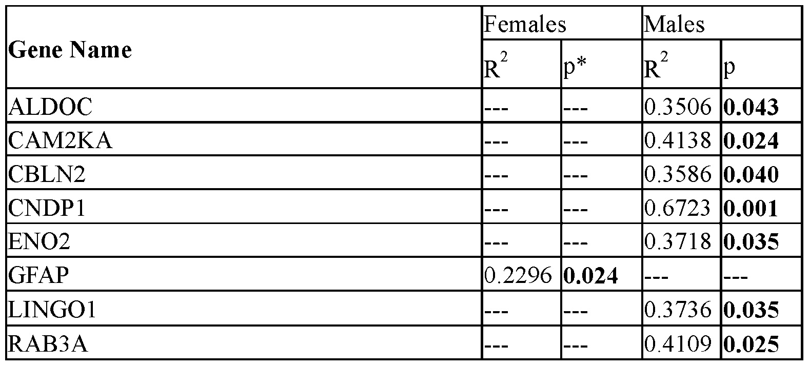 . . Candidate proteins are highly interconnected From the set of 65 target peptides, 30 proteins relevant to the disease of MS were identified as potential biomarkers for disease, identification of disease subtype, correlation with disease severity, or association with commonly used clinical imaging markers. These 30 proteins were summarized with their observed or predicted sub- cellular localization (FIG.4C). This set of proteins spanned several regions, ranging from transcription factors predominantly found in the nucleus to transmembrane associated proteins to extracellular secreted proteins. Yet, this group of brain-enriched
Attorney Docket No.07039-2205WO1 2023-006 proteins shared the association with MS, as described herein. Thus, although this list was relatively small, it was hypothesized that there would be enriched ontologies in this set, so a String.db search was performed. The String.db search revealed a tight network with a protein-protein interaction enrichment p-value of <1.0 x 10⁶ (FIG.8A). The central nodes, including neurofilament light (NEFL), GFAP, MOG, MAG, CAMK2A/B, represented known markers of disease. Using k-means clustering, three natural regions emerged. Analyzing all proteins together showed that the strongest biological process enriched in this set was related to axon integrity, regeneration, and synapse function (FIG.8B). For cellular component enrichment analysis, CAMK2A and CAMK2B drove the strong hit for calcium- dependent protein kinase complex enrichment in FIG. 8C. Further down the list, over half (n=18) of the proteins were involved with the synapse. All diseases enriched in this set were CNS diseases, with primary progressive and relapsing remitting multiple sclerosis ranking the highest (FIG.8E). KEGG Pathways enriched in this protein set include “HIF-1 signaling” represented by ALDOC, CAMK2A, CAMK2B, and ENO2 as well as “insulin secretion” represented by RAB3A, CAMK2A, CAMK2B (FIG.8F). Example 3: Treating MS A biological sample (e.g., a CSF sample) is obtained from a human having MS. The obtained sample is examined for the presence or absence of a reduced level of a CNDP1 polypeptide and/or the presence or absence of a reduced level of an APLP1 polypeptide and/or the presence or absence of an elevated level of an OLFM1 polypeptide. If the presence of a reduced level of a CNDP1 polypeptide and/or the presence of a reduced level of an APLP1 polypeptide and/or the presence of an elevated level of an OLFM1 polypeptide is detected in the sample, then the human is identified as having RR MS, and the human is administered one or more RR MS treatments. The administered one or more RR MS treatments can slow the progression of MS, prolong periods of disease remission, and/or reduce symptoms in the human with MS.
Attorney Docket No.07039-2205WO1 2023-006 Example 4: Treating MS A biological sample (e.g., a CSF sample) is obtained from a human having MS. The obtained sample is examined for the presence or absence of a reduced level of a CNDP1 polypeptide and/or the presence or absence of a reduced level of an APLP1 polypeptide and/or the presence or absence of an elevated level of an OLFM1 polypeptide. If the absence of a reduced level of a CNDP1 polypeptide and/or the absence of a reduced level of an APLP1 polypeptide and/or the absence of an elevated level of an OLFM1 polypeptide is detected in the sample, then the human is identified as having PP MS, and the human is administered one or more PP MS treatments. The administered one or more PP MS treatments can slow the progression of MS and/or reduce symptoms in the human with MS. OTHER EMBODIMENTS It is to be understood that while the invention has been described in conjunction with the detailed description thereof, the foregoing description is intended to illustrate and not limit the scope of the invention, which is defined by the scope of the appended claims. Other aspects, advantages, and modifications are within the scope of the following claims.
. . Candidate proteins are highly interconnected From the set of 65 target peptides, 30 proteins relevant to the disease of MS were identified as potential biomarkers for disease, identification of disease subtype, correlation with disease severity, or association with commonly used clinical imaging markers. These 30 proteins were summarized with their observed or predicted sub- cellular localization (FIG.4C). This set of proteins spanned several regions, ranging from transcription factors predominantly found in the nucleus to transmembrane associated proteins to extracellular secreted proteins. Yet, this group of brain-enriched
Attorney Docket No.07039-2205WO1 2023-006 proteins shared the association with MS, as described herein. Thus, although this list was relatively small, it was hypothesized that there would be enriched ontologies in this set, so a String.db search was performed. The String.db search revealed a tight network with a protein-protein interaction enrichment p-value of <1.0 x 10⁶ (FIG.8A). The central nodes, including neurofilament light (NEFL), GFAP, MOG, MAG, CAMK2A/B, represented known markers of disease. Using k-means clustering, three natural regions emerged. Analyzing all proteins together showed that the strongest biological process enriched in this set was related to axon integrity, regeneration, and synapse function (FIG.8B). For cellular component enrichment analysis, CAMK2A and CAMK2B drove the strong hit for calcium- dependent protein kinase complex enrichment in FIG. 8C. Further down the list, over half (n=18) of the proteins were involved with the synapse. All diseases enriched in this set were CNS diseases, with primary progressive and relapsing remitting multiple sclerosis ranking the highest (FIG.8E). KEGG Pathways enriched in this protein set include “HIF-1 signaling” represented by ALDOC, CAMK2A, CAMK2B, and ENO2 as well as “insulin secretion” represented by RAB3A, CAMK2A, CAMK2B (FIG.8F). Example 3: Treating MS A biological sample (e.g., a CSF sample) is obtained from a human having MS. The obtained sample is examined for the presence or absence of a reduced level of a CNDP1 polypeptide and/or the presence or absence of a reduced level of an APLP1 polypeptide and/or the presence or absence of an elevated level of an OLFM1 polypeptide. If the presence of a reduced level of a CNDP1 polypeptide and/or the presence of a reduced level of an APLP1 polypeptide and/or the presence of an elevated level of an OLFM1 polypeptide is detected in the sample, then the human is identified as having RR MS, and the human is administered one or more RR MS treatments. The administered one or more RR MS treatments can slow the progression of MS, prolong periods of disease remission, and/or reduce symptoms in the human with MS.
Attorney Docket No.07039-2205WO1 2023-006 Example 4: Treating MS A biological sample (e.g., a CSF sample) is obtained from a human having MS. The obtained sample is examined for the presence or absence of a reduced level of a CNDP1 polypeptide and/or the presence or absence of a reduced level of an APLP1 polypeptide and/or the presence or absence of an elevated level of an OLFM1 polypeptide. If the absence of a reduced level of a CNDP1 polypeptide and/or the absence of a reduced level of an APLP1 polypeptide and/or the absence of an elevated level of an OLFM1 polypeptide is detected in the sample, then the human is identified as having PP MS, and the human is administered one or more PP MS treatments. The administered one or more PP MS treatments can slow the progression of MS and/or reduce symptoms in the human with MS. OTHER EMBODIMENTS It is to be understood that while the invention has been described in conjunction with the detailed description thereof, the foregoing description is intended to illustrate and not limit the scope of the invention, which is defined by the scope of the appended claims. Other aspects, advantages, and modifications are within the scope of the following claims.
Claims
Attorney Docket No.07039-2205WO1 2023-006 WHAT IS CLAIMED IS: 1. A method for identifying a mammal having multiple sclerosis (MS) as having relapsing remitting (RR) MS or primary progressive (PP) MS, wherein said method comprises determining if a sample from said mammal comprises a presence or an absence of a reduced level of a carnosine dipeptidase 1 (CNDP1) polypeptide; and (a) classifying said mammal as having said RR MS if said sample is determined to comprise said presence, or (b) classifying said mammal as having said PP MS if said sample is determined to comprise said absence. 2. The method of claim 1, wherein said mammal is a human. 3. The method of claim 1, wherein said sample is a cerebrospinal fluid (CSF) sample. 4. The method of claim 1, wherein said method comprises determining said presence. 5. The method of claim 1, wherein said method comprises determining said absence. 6. The method of claim 1, wherein said determining step comprises detecting a level of said CNDP1 polypeptide in said sample. 7. The method of claim 6, wherein said detecting comprises using liquid chromatography-tandem mass spectrometry (LC-MS/MS). 8. The method of claim 6, wherein said detecting comprises using an anti-CNDP1 antibody. 9. The method of claim 1, wherein said determining step comprises detecting a level of mRNA encoding said CNDP1 polypeptide. 10. A method for treating a mammal having MS, wherein said method comprises:
Attorney Docket No.07039-2205WO1 2023-006 (a) determining that a sample from said mammal contains the presence of a reduced level of a CNDP1 polypeptide; and (b) administering, to said mammal, a RR MS treatment. 11. The method of claim 10, wherein said mammal is a human. 12. The method of claim 10, wherein said sample comprises CSF. 13. The method of claim 10, wherein said RR MS treatment comprises administering an immunotherapeutic agent to said mammal. 14. The method of claim 13, wherein said immunotherapeutic agent is selected from the group consisting of glatiramer acetate, IFN beta-1a, IFN beta-1b, natalizumab, ocrelizumab, rituximab, ofatumumab, and alemtuzumab. 15. The method of claim 10, wherein said RR MS treatment comprises administering a small molecule to said mammal. 16. The method of claim 15, wherein said small molecule is selected from the group consisting of mitoxantrone, cyclophosphamide, dimethyl fumarate, diroximel fumarate, monomethyl fumarate, fingolimod, siponimod, ozanimod, ponesimod, teriflunomide, and cladribine. 17. The method of claim 10, wherein said RR MS treatment comprises transplanting bone marrow into said mammal. 18. A method for treating MS, wherein said method comprises administering, to a mammal identified as having a reduced level of a CNDP1 polypeptide, a RR MS treatment. 19. The method of claim 18, wherein said mammal is a human. 20. The method of claim 18, wherein said RR MS treatment comprises administering an immunotherapeutic agent to said mammal.
Attorney Docket No.07039-2205WO1 2023-006 21. The method of claim 20, wherein said immunotherapeutic agent is selected from the group consisting of glatiramer acetate, IFN beta-1a, IFN beta-1b, natalizumab, ocrelizumab, rituximab, ofatumumab, and alemtuzumab. 22. The method of claim 18, wherein said RR MS treatment comprises administering a small molecule to said mammal. 23. The method of claim 22, wherein said small molecule is selected from the group consisting of mitoxantrone, cyclophosphamide, dimethyl fumarate, diroximel fumarate, monomethyl fumarate, fingolimod, siponimod, ozanimod, ponesimod, teriflunomide, and cladribine. 24. The method of claim 18, wherein said RR MS treatment comprises transplanting bone marrow into said mammal. 25. A method for treating a mammal having MS, wherein said method comprises: (a) determining that a sample from said mammal comprises the absence of a reduced level of a CNDP1 polypeptide; and (b) administering, to said mammal, a PP MS treatment. 26. The method of claim 25, wherein said mammal is a human. 27. The method of claim 25, wherein said sample comprises CSF. 28. The method of claim 25, wherein said PP MS treatment comprises administering an immunosuppressive therapy to said mammal. 29. The method of claim 28, wherein said immunosuppressive therapy comprises ocrelizumab. 30. A method for treating MS, wherein said method comprises administering, to a mammal identified as lacking a reduced level of a CNDP1 polypeptide, a PP MS treatment.
Attorney Docket No.07039-2205WO1 2023-006 31. The method of claim 30, wherein said mammal is a human. 32. The method of claim 30, wherein said PP MS treatment comprises administering an immunosuppressive therapy to said mammal. 33. The method of claim 32, wherein said immunosuppressive therapy is ocrelizumab. 34. A method for identifying a mammal having multiple sclerosis (MS) as having relapsing remitting (RR) MS or primary progressive (PP) MS, wherein said method comprises determining if a sample from said mammal comprises a presence or an absence of a reduced level of an amyloid beta precursor like protein 1 (APLP1) polypeptide; and (a) classifying said mammal as having said RR MS if said sample is determined to comprise said presence, or (b) classifying said mammal as having said PP MS if said sample is determined to comprise said absence. 35. The method of claim 34, wherein said mammal is a human. 36. The method of claim 34, wherein said sample is a cerebrospinal fluid (CSF) sample. 37. The method of claim 34, wherein said method comprises determining said presence. 38. The method of claim 34, wherein said method comprises determining said absence. 39. The method of claim 34, wherein said determining step comprises detecting a level of said APLP1 polypeptide in said sample.
Attorney Docket No.07039-2205WO1 2023-006 40. The method of claim 39, wherein said detecting comprises using liquid chromatography-tandem mass spectrometry (LC-MS/MS). 41. The method of claim 39, wherein said detecting comprises using an anti-APLP1 antibody. 42. The method of claim 34, wherein said determining step comprises detecting a level of mRNA encoding said APLP1 polypeptide. 43. A method for treating a mammal having MS, wherein said method comprises: (a) determining that a sample from said mammal contains the presence of a reduced level of an APLP1 polypeptide; and (b) administering, to said mammal, a RR MS treatment. 44. The method of claim 43, wherein said mammal is a human. 45. The method of claim 43, wherein said sample comprises CSF. 46. The method of claim 43, wherein said RR MS treatment comprises administering an immunotherapeutic agent to said mammal. 47. The method of claim 46, wherein said immunotherapeutic agent is selected from the group consisting of glatiramer acetate, IFN beta-1a, IFN beta-1b, natalizumab, ocrelizumab, rituximab, ofatumumab, and alemtuzumab. 48. The method of claim 43, wherein said RR MS treatment comprises administering a small molecule to said mammal. 49. The method of claim 48, wherein said small molecule is selected from the group consisting of mitoxantrone, cyclophosphamide, dimethyl fumarate, diroximel fumarate, monomethyl fumarate, fingolimod, siponimod, ozanimod, ponesimod, teriflunomide, and cladribine.
Attorney Docket No.07039-2205WO1 2023-006 50. The method of claim 43, wherein said RR MS treatment comprises transplanting bone marrow into said mammal. 51. A method for treating MS, wherein said method comprises administering, to a mammal identified as having a reduced level of an APLP1 polypeptide, a RR MS treatment. 52. The method of claim 51, wherein said mammal is a human. 53. The method of claim 51, wherein said RR MS treatment comprises administering an immunotherapeutic agent to said mammal. 54. The method of claim 53, wherein said immunotherapeutic agent is selected from the group consisting of glatiramer acetate, IFN beta-1a, IFN beta-1b, natalizumab, ocrelizumab, rituximab, ofatumumab, and alemtuzumab. 55. The method of claim 51, wherein said RR MS treatment comprises administering a small molecule to said mammal. 56. The method of claim 55, wherein said small molecule is selected from the group consisting of mitoxantrone, cyclophosphamide, dimethyl fumarate, diroximel fumarate, monomethyl fumarate, fingolimod, siponimod, ozanimod, ponesimod, teriflunomide, and cladribine. 57. The method of claim 51, wherein said RR MS treatment comprises transplanting bone marrow into said mammal. 58. A method for treating a mammal having MS, wherein said method comprises: (a) determining that a sample from said mammal comprises the absence of a reduced level of an APLP1 polypeptide; and (b) administering, to said mammal, a PP MS treatment. 59. The method of claim 58, wherein said mammal is a human.
Attorney Docket No.07039-2205WO1 2023-006 60. The method of any one of claims 58-59, wherein said sample comprises CSF. 61. The method of any one of claims 58-60, wherein said PP MS treatment comprises administering an immunosuppressive therapy to said mammal. 62. The method of claim 61, wherein said immunosuppressive therapy comprises ocrelizumab. 63. A method for treating MS, wherein said method comprises administering, to a mammal identified as lacking a reduced level of an APLP1 polypeptide, a PP MS treatment. 64. The method of claim 63, wherein said mammal is a human. 65. The method of any one of claims 63-64, wherein said PP MS treatment comprises administering an immunosuppressive therapy to said mammal. 66. The method of claim 65, wherein said immunosuppressive therapy is ocrelizumab. 67. A method for identifying a mammal having multiple sclerosis (MS) as having relapsing remitting (RR) MS or primary progressive (PP) MS, wherein said method comprises determining if a sample from said mammal comprises a presence or an absence of an elevated level of an olfactomedin 1 (OLFM1) polypeptide; and (a) classifying said mammal as having said RR MS if said sample is determined to comprise said presence, or (b) classifying said mammal as having said PP MS if said sample is determined to comprise said absence. 68. The method of claim 67, wherein said mammal is a human. 69. The method of claim 67, wherein said sample is a cerebrospinal fluid (CSF) sample.
Attorney Docket No.07039-2205WO1 2023-006 70. The method of claim 67, wherein said method comprises determining said presence. 71. The method of claim 67, wherein said method comprises determining said absence. 72. The method of claim 67, wherein said determining step comprises detecting a level of said OLFM1 polypeptide in said sample. 73. The method of claim 72, wherein said detecting comprises using liquid chromatography-tandem mass spectrometry (LC-MS/MS). 74. The method of claim 72, wherein said detecting comprises using an anti-OLFM1 antibody. 75. The method of claim 67, wherein said determining step comprises detecting a level of mRNA encoding said OLFM1 polypeptide. 76. A method for treating a mammal having MS, wherein said method comprises: (a) determining that a sample from said mammal contains the presence of an elevated level of an OLFM1 polypeptide; and (b) administering, to said mammal, a RR MS treatment. 77. The method of claim 76, wherein said mammal is a human. 78. The method of claim 76, wherein said sample comprises CSF. 79. The method of claim 76, wherein said RR MS treatment comprises administering an immunotherapeutic agent to said mammal. 80. The method of claim 79, wherein said immunotherapeutic agent is selected from the group consisting of glatiramer acetate, IFN beta-1a, IFN beta-1b, natalizumab, ocrelizumab, rituximab, ofatumumab, and alemtuzumab.
Attorney Docket No.07039-2205WO1 2023-006 81. The method of claim 76, wherein said RR MS treatment comprises administering a small molecule to said mammal. 82. The method of claim 81, wherein said small molecule is selected from the group consisting of mitoxantrone, cyclophosphamide, dimethyl fumarate, diroximel fumarate, monomethyl fumarate, fingolimod, siponimod, ozanimod, ponesimod, teriflunomide, and cladribine. 83. The method of claim 76, wherein said RR MS treatment comprises transplanting bone marrow into said mammal. 84. A method for treating MS, wherein said method comprises administering, to a mammal identified as having an elevated level of an OLFM1 polypeptide, a RR MS treatment. 85. The method of claim 84, wherein said mammal is a human. 86. The method of claim 84, wherein said RR MS treatment comprises administering an immunotherapeutic agent to said mammal. 87. The method of claim 86, wherein said immunotherapeutic agent is selected from the group consisting of glatiramer acetate, IFN beta-1a, IFN beta-1b, natalizumab, ocrelizumab, rituximab, ofatumumab, and alemtuzumab. 88. The method of claim 84, wherein said RR MS treatment comprises administering a small molecule to said mammal. 89. The method of claim 88, wherein said small molecule is selected from the group consisting of mitoxantrone, cyclophosphamide, dimethyl fumarate, diroximel fumarate, monomethyl fumarate, fingolimod, siponimod, ozanimod, ponesimod, teriflunomide, and cladribine.
Attorney Docket No.07039-2205WO1 2023-006 90. The method of claim 84, wherein said RR MS treatment comprises transplanting bone marrow into said mammal. 91. A method for treating a mammal having MS, wherein said method comprises: (a) determining that a sample from said mammal comprises the absence of an elevated level of an OLFM1 polypeptide; and (b) administering, to said mammal, a PP MS treatment. 92. The method of claim 91, wherein said mammal is a human. 93. The method of claim 91, wherein said sample comprises CSF. 94. The method of claim 91, wherein said PP MS treatment comprises administering an immunosuppressive therapy to said mammal. 95. The method of claim 94, wherein said immunosuppressive therapy comprises ocrelizumab. 96. A method for treating MS, wherein said method comprises administering, to a mammal identified as lacking an elevated level of an OLFM1 polypeptide, a PP MS treatment. 97. The method of claim 96, wherein said mammal is a human. 98. The method of claim 96, wherein said PP MS treatment comprises administering an immunosuppressive therapy to said mammal. 99. The method of claim 98, wherein said immunosuppressive therapy is ocrelizumab.
Applications Claiming Priority (4)
| Application Number | Priority Date | Filing Date | Title |
|---|---|---|---|
| GR20230100923 | 2023-11-06 | ||
| GR20230100923 | 2023-11-06 | ||
| US202363548346P | 2023-11-13 | 2023-11-13 | |
| US63/548,346 | 2023-11-13 |
Publications (1)
| Publication Number | Publication Date |
|---|---|
| WO2025101643A1 true WO2025101643A1 (en) | 2025-05-15 |
Family
ID=95696272
Family Applications (1)
| Application Number | Title | Priority Date | Filing Date |
|---|---|---|---|
| PCT/US2024/054771 Pending WO2025101643A1 (en) | 2023-11-06 | 2024-11-06 | Assessing and treating multiple sclerosis |
Country Status (1)
| Country | Link |
|---|---|
| WO (1) | WO2025101643A1 (en) |
Citations (1)
| Publication number | Priority date | Publication date | Assignee | Title |
|---|---|---|---|---|
| WO2006005583A2 (en) * | 2004-07-12 | 2006-01-19 | Geneprot Inc. | Secreted polypeptide species involved in multiple sclerosis |
-
2024
- 2024-11-06 WO PCT/US2024/054771 patent/WO2025101643A1/en active Pending
Patent Citations (1)
| Publication number | Priority date | Publication date | Assignee | Title |
|---|---|---|---|---|
| WO2006005583A2 (en) * | 2004-07-12 | 2006-01-19 | Geneprot Inc. | Secreted polypeptide species involved in multiple sclerosis |
Non-Patent Citations (4)
| Title |
|---|
| LINCOLNI. WURTZ;EVDOKIYA KNYAZHANSKAYA;DORSA SOHAEI;IOANNIS PRASSAS;SEAN PITTOCK;MARIAALICEV. WILLRICH;RUBA SAADEH;RUCHI GUPTA;HUN: "Identification of brain-enriched proteins in CSF as biomarkers of relapsing remitting multiple sclerosis", CLINICAL PROTEOMICS, vol. 21, no. 1, 16 June 2024 (2024-06-16), US , pages 1 - 17, XP021338909, ISSN: 1542-6416, DOI: 10.1186/s12014-024-09494-5 * |
| MARTYNOVA EKATERINA, KHAIBULLIN TIMUR, SALAFUTDINOV ILNUR, MARKELOVA MARIA, LAIKOV ALEXANDER, LOPUKHOV LEONID, LIU RONGZENG, SAHAY: "Seasonal Changes in Serum Metabolites in Multiple Sclerosis Relapse", INTERNATIONAL JOURNAL OF MOLECULAR SCIENCES, vol. 24, no. 4, 1 January 2023 (2023-01-01), Basel, CH , pages 1 - 21, XP093314795, ISSN: 1422-0067, DOI: 10.3390/ijms24043542 * |
| RAI NAGENDRA KUMAR, SINGH VAIBHAV, LI LING, WILLARD BELINDA, TRIPATHI AJAI, DUTTA RANJAN: "Comparative Proteomic Profiling Identifies Reciprocal Expression of Mitochondrial Proteins Between White and Gray Matter Lesions From Multiple Sclerosis Brains", FRONTIERS IN NEUROLOGY, vol. 12, 1 January 2021 (2021-01-01), pages 1 - 16, XP093314789, ISSN: 1664-2295, DOI: 10.3389/fneur.2021.779003 * |
| ROBINSON W.H , UTZ P.J , STEINMAN, L: "Genomic and proteomic analysis of multiple sclerosis - Opinion", CURRENT OPINION IN IMMUNOLOGY, vol. 15, no. 6, 1 December 2003 (2003-12-01), GB , pages 660 - 667, XP004474537, ISSN: 0952-7915, DOI: 10.1016/j.coi.2003.09.015 * |
Similar Documents
| Publication | Publication Date | Title |
|---|---|---|
| US12422433B2 (en) | Blood biomarker that predicts persistent cognitive dysfunction after concussion | |
| US20240426842A1 (en) | Novel biomarkers and methods for diagnosing and evaluating traumatic brain injury | |
| EP2300829B1 (en) | New biomarker for diagnosis, prediction and/or prognosis of sepsis and uses thereof | |
| US20150051115A1 (en) | Biomarkers of brain injury | |
| KR20160068964A (en) | Biomarkers and diagnostic methods for alzheimer's disease and other neurodegenerative disorders | |
| KR102742415B1 (en) | Treatment of spinal muscular atrophy | |
| WO2020218465A1 (en) | Method for diagnosing endometriosis, disease state monitoring method, and kit | |
| US20220236294A1 (en) | Methods for Evaluation and Treatment of Alzheimer's Disease and Applications Thereof | |
| US9977036B2 (en) | Diagnostic markers for multiple sclerosis | |
| Cunningham et al. | Protein changes in immunodepleted cerebrospinal fluid from a transgenic mouse model of Alexander disease detected using mass spectrometry | |
| KR102547504B1 (en) | Biomarkers indicative of psychiatric index | |
| US20130184173A1 (en) | Biomarkers for multiple sclerosis | |
| WO2025101643A1 (en) | Assessing and treating multiple sclerosis | |
| CA2893609A1 (en) | Method for the diagnosis of metachromatic leukodystrophy | |
| JP2025541672A (en) | Biomarker panels for brain-specific neurological abnormalities using biofluid samples | |
| EP3519829A1 (en) | Assay for diagnosing alzheimer's disease | |
| WO2019012667A1 (en) | Biomarker for cognitive impairment disorders and detection method for cognitive impairment disorders using said biomarker | |
| US10502749B1 (en) | Method for patient stratification and drug efficacy monitoring | |
| Sohaei | The use of proteomic analyses to identify potential cerebrospinal fluid biomarkers in Multiple Sclerosis | |
| JP2021181890A (en) | Biomarkers for determining adult Still's disease (ASD) | |
| May et al. | Highly Immunoreactive IgG Antibodies Directed against a Set of Twenty Human | |
| Galán et al. | Quantitative Proteomics Analysis of Cerebrospinal Fluid and Serum Reveals Putative Protein Biomarkers for Canine Non-Infectious Meningoencephalomyelitis |
Legal Events
| Date | Code | Title | Description |
|---|---|---|---|
| 121 | Ep: the epo has been informed by wipo that ep was designated in this application |
Ref document number: 24889519 Country of ref document: EP Kind code of ref document: A1 |