WO2025005076A1 - 骨格筋内脂肪組織(IMAT;Intramuscular adipose tissue)形成抑制物質をスクリーニングする方法、及びIMAT形成抑制用組成物 - Google Patents
骨格筋内脂肪組織(IMAT;Intramuscular adipose tissue)形成抑制物質をスクリーニングする方法、及びIMAT形成抑制用組成物 Download PDFInfo
- Publication number
- WO2025005076A1 WO2025005076A1 PCT/JP2024/022952 JP2024022952W WO2025005076A1 WO 2025005076 A1 WO2025005076 A1 WO 2025005076A1 JP 2024022952 W JP2024022952 W JP 2024022952W WO 2025005076 A1 WO2025005076 A1 WO 2025005076A1
- Authority
- WO
- WIPO (PCT)
- Prior art keywords
- protein
- wnt5b
- imat
- ror2
- mpcs
- Prior art date
- Legal status (The legal status is an assumption and is not a legal conclusion. Google has not performed a legal analysis and makes no representation as to the accuracy of the status listed.)
- Ceased
Links
Images





Classifications
-
- A—HUMAN NECESSITIES
- A61—MEDICAL OR VETERINARY SCIENCE; HYGIENE
- A61K—PREPARATIONS FOR MEDICAL, DENTAL OR TOILETRY PURPOSES
- A61K31/00—Medicinal preparations containing organic active ingredients
- A61K31/70—Carbohydrates; Sugars; Derivatives thereof
- A61K31/7088—Compounds having three or more nucleosides or nucleotides
-
- A—HUMAN NECESSITIES
- A61—MEDICAL OR VETERINARY SCIENCE; HYGIENE
- A61K—PREPARATIONS FOR MEDICAL, DENTAL OR TOILETRY PURPOSES
- A61K31/00—Medicinal preparations containing organic active ingredients
- A61K31/70—Carbohydrates; Sugars; Derivatives thereof
- A61K31/7088—Compounds having three or more nucleosides or nucleotides
- A61K31/7105—Natural ribonucleic acids, i.e. containing only riboses attached to adenine, guanine, cytosine or uracil and having 3'-5' phosphodiester links
-
- A—HUMAN NECESSITIES
- A61—MEDICAL OR VETERINARY SCIENCE; HYGIENE
- A61K—PREPARATIONS FOR MEDICAL, DENTAL OR TOILETRY PURPOSES
- A61K39/00—Medicinal preparations containing antigens or antibodies
- A61K39/395—Antibodies; Immunoglobulins; Immune serum, e.g. antilymphocytic serum
-
- A—HUMAN NECESSITIES
- A61—MEDICAL OR VETERINARY SCIENCE; HYGIENE
- A61K—PREPARATIONS FOR MEDICAL, DENTAL OR TOILETRY PURPOSES
- A61K48/00—Medicinal preparations containing genetic material which is inserted into cells of the living body to treat genetic diseases; Gene therapy
-
- A—HUMAN NECESSITIES
- A61—MEDICAL OR VETERINARY SCIENCE; HYGIENE
- A61P—SPECIFIC THERAPEUTIC ACTIVITY OF CHEMICAL COMPOUNDS OR MEDICINAL PREPARATIONS
- A61P21/00—Drugs for disorders of the muscular or neuromuscular system
-
- A—HUMAN NECESSITIES
- A61—MEDICAL OR VETERINARY SCIENCE; HYGIENE
- A61P—SPECIFIC THERAPEUTIC ACTIVITY OF CHEMICAL COMPOUNDS OR MEDICINAL PREPARATIONS
- A61P21/00—Drugs for disorders of the muscular or neuromuscular system
- A61P21/04—Drugs for disorders of the muscular or neuromuscular system for myasthenia gravis
-
- A—HUMAN NECESSITIES
- A61—MEDICAL OR VETERINARY SCIENCE; HYGIENE
- A61P—SPECIFIC THERAPEUTIC ACTIVITY OF CHEMICAL COMPOUNDS OR MEDICINAL PREPARATIONS
- A61P43/00—Drugs for specific purposes, not provided for in groups A61P1/00-A61P41/00
-
- C—CHEMISTRY; METALLURGY
- C07—ORGANIC CHEMISTRY
- C07K—PEPTIDES
- C07K14/00—Peptides having more than 20 amino acids; Gastrins; Somatostatins; Melanotropins; Derivatives thereof
- C07K14/435—Peptides having more than 20 amino acids; Gastrins; Somatostatins; Melanotropins; Derivatives thereof from animals; from humans
- C07K14/46—Peptides having more than 20 amino acids; Gastrins; Somatostatins; Melanotropins; Derivatives thereof from animals; from humans from vertebrates
- C07K14/47—Peptides having more than 20 amino acids; Gastrins; Somatostatins; Melanotropins; Derivatives thereof from animals; from humans from vertebrates from mammals
-
- C—CHEMISTRY; METALLURGY
- C07—ORGANIC CHEMISTRY
- C07K—PEPTIDES
- C07K16/00—Immunoglobulins [IG], e.g. monoclonal or polyclonal antibodies
- C07K16/18—Immunoglobulins [IG], e.g. monoclonal or polyclonal antibodies against material from animals or humans
-
- C—CHEMISTRY; METALLURGY
- C12—BIOCHEMISTRY; BEER; SPIRITS; WINE; VINEGAR; MICROBIOLOGY; ENZYMOLOGY; MUTATION OR GENETIC ENGINEERING
- C12N—MICROORGANISMS OR ENZYMES; COMPOSITIONS THEREOF; PROPAGATING, PRESERVING, OR MAINTAINING MICROORGANISMS; MUTATION OR GENETIC ENGINEERING; CULTURE MEDIA
- C12N15/00—Mutation or genetic engineering; DNA or RNA concerning genetic engineering, vectors, e.g. plasmids, or their isolation, preparation or purification; Use of hosts therefor
- C12N15/09—Recombinant DNA-technology
- C12N15/11—DNA or RNA fragments; Modified forms thereof; Non-coding nucleic acids having a biological activity
- C12N15/113—Non-coding nucleic acids modulating the expression of genes, e.g. antisense oligonucleotides; Antisense DNA or RNA; Triplex- forming oligonucleotides; Catalytic nucleic acids, e.g. ribozymes; Nucleic acids used in co-suppression or gene silencing
-
- G—PHYSICS
- G01—MEASURING; TESTING
- G01N—INVESTIGATING OR ANALYSING MATERIALS BY DETERMINING THEIR CHEMICAL OR PHYSICAL PROPERTIES
- G01N33/00—Investigating or analysing materials by specific methods not covered by groups G01N1/00 - G01N31/00
- G01N33/15—Medicinal preparations ; Physical properties thereof, e.g. dissolubility
-
- G—PHYSICS
- G01—MEASURING; TESTING
- G01N—INVESTIGATING OR ANALYSING MATERIALS BY DETERMINING THEIR CHEMICAL OR PHYSICAL PROPERTIES
- G01N33/00—Investigating or analysing materials by specific methods not covered by groups G01N1/00 - G01N31/00
- G01N33/48—Biological material, e.g. blood, urine; Haemocytometers
- G01N33/50—Chemical analysis of biological material, e.g. blood, urine; Testing involving biospecific ligand binding methods; Immunological testing
-
- G—PHYSICS
- G01—MEASURING; TESTING
- G01N—INVESTIGATING OR ANALYSING MATERIALS BY DETERMINING THEIR CHEMICAL OR PHYSICAL PROPERTIES
- G01N33/00—Investigating or analysing materials by specific methods not covered by groups G01N1/00 - G01N31/00
- G01N33/48—Biological material, e.g. blood, urine; Haemocytometers
- G01N33/50—Chemical analysis of biological material, e.g. blood, urine; Testing involving biospecific ligand binding methods; Immunological testing
- G01N33/68—Chemical analysis of biological material, e.g. blood, urine; Testing involving biospecific ligand binding methods; Immunological testing involving proteins, peptides or amino acids
Definitions
- the present disclosure relates to a method for screening for IMAT formation inhibitors, and a composition for inhibiting IMAT formation, etc.
- Satellite cells which are skeletal muscle tissue stem cells, and mesenchymal progenitor cells (MPCs) play important roles in the maintenance and regeneration of skeletal muscles.
- SCs After skeletal muscle injury, SCs proliferate, differentiate and fuse, and eventually form muscle fibers, which contribute to the repair of the injured area.
- MPCs which are resident in skeletal muscle, promote skeletal muscle regeneration by promoting the proliferation and differentiation of SCs.
- pathological conditions of skeletal muscle such as muscular dystrophy and sarcopenia
- MPCs differentiate into adipocytes, forming IMAT, which should not exist in skeletal muscle, and causing a significant decrease in muscle function.
- the molecular mechanism that determines whether MPCs contribute to the maintenance and regeneration of skeletal muscle or promote the formation of IMAT has not been fully elucidated, but it is expected that pathological conditions of skeletal muscle can be inhibited by maintaining the skeletal muscle maintenance and regeneration mechanism of MPCs and suppressing the IMAT formation ability of MPCs. Therefore, if a substance that suppresses the IMAT formation ability can be identified, it will greatly contribute to improving the condition of skeletal muscle diseases and aging-related skeletal muscle diseases (locomotor syndrome, frailty, etc.), and it will also greatly contribute to improving people's QOL and ADL internationally, including in Japan, which has become a super-aging society.
- An objective of the present disclosure is to provide a method for screening substances that suppress IMAT formation.
- Another objective of the present disclosure is to provide a composition for suppressing IMAT formation.
- Ror2 Receptor tyrosine kinase-like orphan receptor 2
- Wnt11-Ror2 contributes to the maintenance and regeneration of skeletal muscle by suppressing cellular senescence of MPCs, while in pathological conditions, activation of the Wnt5b-Ror2 signal in MPCs may promote IMAT formation.
- the present disclosure includes, for example, the subject matter described in the following sections: Item 1. (I) contacting a Wnt5b protein, a Ror2 protein, and a test substance; and (II) confirming that the test substance inhibits binding between the Wnt5b protein and the Ror2 protein. A method for screening for a substance that inhibits IMAT formation. Item 2. In step (I), a Wnt11 protein is further contacted, Item 2. The method according to Item 1, further comprising confirming in step (II) that the test substance does not inhibit binding between Wnt11 protein and Ror2 protein. Item 3.
- Item 6. The method according to any one of Items 1 to 5, wherein the inhibition of IMAT formation is due to inhibition of proliferation of MPCs.
- Item 7. A composition for inhibiting IMAT formation, comprising as an active ingredient a neutralizing antibody to Wnt5b protein or a nucleic acid that inhibits the expression of the Wnt5b gene.
- Item 8. A composition for inhibiting IMAT formation according to item 7, for use in inhibiting or treating a muscular disease or condition in which accumulation of IMAT is observed.
- Item 9. Item 9. The composition for inhibiting IMAT formation according to item 8, wherein the muscle disease or condition in which accumulation of IMAT is observed is muscular dystrophy, amyotrophic lateral sclerosis, sarcopenia, frailty or locomotive syndrome.
- Wnt5b, Wnt11, and Ror2 as therapeutic targets for muscle diseases will lead to the development of highly effective therapeutic methods with few side effects, and is one of the major advantages in discovering such therapeutic methods.
- MPCs are also important for the maintenance and regeneration of skeletal muscle, it is not sufficient to simply suppress the function of MPCs; it is necessary to suppress the ability to form IMAT while maintaining the mechanisms for maintaining and regenerating skeletal muscle.
- the inventors have established a method for identifying substances that can select substances that suppress Wnt5b-Ror2 signaling while maintaining Wnt11-Ror2 signaling, and that can greatly contribute to improving the conditions of various skeletal muscle diseases and age-related skeletal muscle disorders (locomotor syndrome, frailty, etc.).
- a method for screening for an IMAT formation inhibitor is provided.
- Also provided is a composition for inhibiting IMAT formation.
- the results of measuring the expression levels of Ror2 in SCs and MPCs are shown.
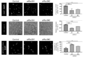
- the figures show the results of measuring the proliferation ability, adipocyte differentiation ability, and cellular senescence of MPCs isolated from tibialis anterior (TA) muscle administered Ctx.
- the results of measuring the weights of the TA muscle, gastrocnemius muscle, and soleus muscle in control mice and Ror2cKO mice, as well as the results of staining of muscle fibers in the TA muscle, are shown.
- 1 shows the results of myofiber and fat staining in the TA muscle of control mice and Ror2cKO mice.
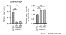
- 1 shows the results of measuring the expression level of Wnt11 and cellular senescence in MPCs isolated from TA muscles administered Ctx.
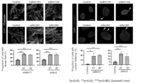
- 1 shows the results of stress fiber and nuclear staining in MPCs isolated from Ctx-administered TA muscles.
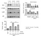
- 1 shows the expression levels of Wnt family genes in Ctx-administered muscles and glycerol-administered muscles, as well as the results of measuring cell proliferation when Wnt5a or Wnt5b was added to MPCs isolated from Ctx-administered TA muscles. Shown are the results of Western blot using MPCs isolated from Ctx-administered TA muscles, and the results of WST-8 assay using MPCs isolated from Ctx-administered TA muscles.
- EDL Extra digitorum longus muscle 3 days after administration of Ctx or glycerol
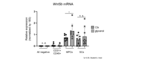
- 1 shows the results of measuring the expression level of Wnt5b in various cells isolated from TA muscle 3 days after administration of Ctx or glycerol.
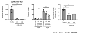
- the figures show the results of measuring the expression level of Wnt5b and cell proliferation in MPCs isolated from Ctx-administered TA muscles.
- the present disclosure includes a method for screening for a substance that inhibits IMAT formation.
- the method may be referred to as the "screening method of the present disclosure” or the like.
- IMAT intramuscular adipose tissue
- ectopic adipose tissue found within skeletal muscle. This is a condition in which fat accumulates in skeletal muscle, even though there is essentially no fat in the muscle.
- the screening method disclosed herein includes a step of contacting the Wnt5b protein, the Ror2 protein, and the test substance.
- this step may be referred to as step (I), etc.
- Wnt (wingless-type MMTV integration site family) proteins are secreted glycoproteins and are known to bind to Ror proteins and the like.
- Wnt proteins Nineteen types have been identified in mammals, including Wnt5a, Wnt5b, and Wnt11.
- the amino acid sequences, nucleotide sequences, and the like of the proteins can be known from known databases and the like.
- NCBI National Center for Biotechnology Information
- the accession number of the mRNA sequence of the human Wnt5b gene is NM_030775
- the accession number of the mRNA sequence of the human Wnt11 gene is XM_005274231.
- Wnt5b-producing cells include SCs, MPCs, Schwann cells, and the like.
- Wnt11-producing cells include, for example, MPCs.
- Ror (Receptor tyrosine kinase-like orphan receptor) protein is a single-pass transmembrane receptor that functions as a receptor for Wnt protein and is known to activate the non-classical Wnt pathway.
- Ror proteins include Ror1 and Ror2.
- the amino acid sequence, base sequence, and the like of the protein can be known from known databases and the like.
- the accession number of the mRNA sequence of the human Ror2 gene at NCBI is NM_001318204.
- Ror2-expressing cells include, for example, MPCs.
- the test substance is not particularly limited.
- it may be a compound or a composition.
- the compound may be, for example, a low molecular weight compound, a nucleic acid (e.g., DNA, RNA, etc.), a protein (e.g., an antibody or a part thereof, etc.), or a polymeric compound such as a polymer.
- the composition may also be an extract obtained from a living organism (e.g., an animal, a plant, a microorganism, etc.), or a combination of two or more compounds.
- the method for contacting the Wnt5b protein, the Ror2 protein, and the test substance is not particularly limited.
- the Wnt5b protein, the Ror2 protein, and the test substance may be added to an appropriate solvent, or the Wnt5b protein and the test substance may be added to a cell expressing the Ror2 protein.
- the suitable solvent is not particularly limited, and examples thereof include water, liquid medium for cell culture, and the like.
- the concentration of the Wnt5b protein is not particularly limited and can be, for example, about 0.1 to 4 ⁇ g/ml.
- the concentration of the Ror2 protein is not particularly limited, and may be, for example, about 0.1 to 4 ⁇ g/ml.
- the concentration of the test substance can be appropriately set depending on the type of the test substance.
- the temperature during contact is not particularly limited, and may be, for example, about 25 to 40° C., preferably about 30 to 40° C.
- the contact time is not particularly limited, and can be, for example, about 10 seconds to 3 hours, preferably about 3 minutes to 1 hour. During the contact, for example, stirring or shaking can be performed as necessary.
- Cells expressing Ror2 protein include, for example, MPCs.
- the screening method of the present disclosure can further include contacting Wnt11 protein in step (I).
- the screening method of the present disclosure can include contacting Wnt5b protein, Wnt11 protein, Ror2 protein, and the test substance in step (I).
- the method for contacting the Wnt5b protein, the Wnt11 protein, the Ror2 protein, and the test substance is not particularly limited.
- the Wnt5b protein, the Wnt11 protein, the Ror2 protein, and the test substance may be added to an appropriate solvent, or the Wnt5b protein, the Wnt11 protein, and the test substance may be added to a cell expressing the Ror2 protein.
- the concentration of the Wnt11 protein is not particularly limited and may be, for example, about 0.1 to 4 ⁇ g/ml. For other contact methods, the above description can be used.
- the screening method of the present disclosure further includes a step of confirming that the test substance inhibits the binding between the Wnt5b protein and the Ror2 protein.
- this step may be referred to as step (II) or the like.
- step (II) the method for confirming that the test substance inhibits the binding between the Wnt5b protein and the Ror2 protein is not particularly limited.
- the ELISA method, the AlphaScreen method, and analysis using the Biacore system can be used.
- the amount of binding between Wnt5b protein and Ror2 protein is reduced by 35% or more when the test substance is added compared to when the test substance is not added.
- the upper or lower limit of the range may be, for example, about 40%, 45%, 50%, 55%, 60%, 65%, 70%, 75%, 80%, 85%, 90%, 95%, 99%, or 100%. Specifically, it may be about 35 to 100%.
- the screening method disclosed herein makes it possible to screen for substances that can inhibit the binding between Wnt5b protein and Ror2 protein when the test substance is added, compared to when the test substance is not added.
- Wnt5b and Ror2 promote the proliferation of MPCs via p38, thereby inducing excessive accumulation of MPCs in skeletal muscle tissue and contributing to the formation of IMAT.
- an IMAT formation inhibitor obtained by the screening method disclosed herein may inhibit the binding between Wnt5b protein and Ror2 protein, thereby suppressing the excessive proliferation of MPCs and suppressing the formation of IMAT.
- the screening method of the present disclosure further includes, in step (II), a step of confirming that the test substance does not inhibit the binding between Wnt11 protein and Ror2 protein.
- the screening method of the present disclosure can confirm in step (II) that the test substance inhibits the binding between Wnt5b protein and Ror2 protein and does not inhibit the binding between Wnt11 protein and Ror2 protein.
- the method for confirming that the test substance does not inhibit the binding between the Wnt11 protein and the Ror2 protein is not particularly limited. Examples include ELISA, AlphaScreen, and analysis using the Biacore system.
- step (II) it may be possible to first confirm that the test substance inhibits the binding between Wnt5b protein and Ror2 protein, and then confirm that the test substance does not inhibit the binding between Wnt11 protein and Ror2 protein, or it may be possible to first confirm that the test substance does not inhibit the binding between Wnt11 protein and Ror2 protein, and then confirm that the test substance inhibits the binding between Wnt5b protein and Ror2 protein, or it may be possible to simultaneously confirm that the test substance inhibits the binding between Wnt5b protein and Ror2 protein and does not inhibit the binding between Wnt11 protein and Ror2 protein.
- does not inhibit means that it does not inhibit completely or substantially.
- Does not substantially inhibit means that, after confirming that the binding between Wnt11 protein and Ror2 protein can be detected by various analytical methods such as absorbance, luminescence, fluorescence, and surface plasmon resonance, the decrease rate of the binding amount between Wnt11 protein and Ror2 protein when the test substance is added is 30% or less compared to when the test substance is not added. It may be preferably about 20%, more preferably about 10%, even more preferably about 5%, even more preferably about 3%, and particularly preferably about 1% or less.
- the increase rate of actin stress fiber formation and abnormal nuclear morphology is 30% or less compared to when the test substance is not added when the test substance is added. It may be preferably about 20%, more preferably about 10%, even more preferably about 5%, even more preferably about 3%, and particularly preferably about 1% or less.
- the screening method disclosed herein makes it possible to screen for a substance that, for example, when the test substance is added, is capable of inhibiting the binding between the Wnt5b protein and the Ror2 protein, but is not capable of inhibiting the binding between the Wnt11 protein and the Ror2 protein, compared to when the test substance is not added.
- Wnt5b and Ror2 promote the proliferation of MPCs via p38, thereby inducing excessive accumulation of MPCs in skeletal muscle tissue and contributing to the formation of IMAT.
- Wnt11 and Ror2 suppress the formation of stress fibers in MPCs, thereby suppressing nuclear morphological abnormalities and the associated activation of the cGas-Sting pathway, and as a result, they are thought to contribute to the regeneration and maintenance of skeletal muscle by suppressing cellular senescence of MPCs.
- an IMAT formation inhibitor obtained by the screening method disclosed herein may suppress excessive proliferation of MPCs and suppress the formation of IMAT by inhibiting the binding between Wnt5b protein and Ror2 protein, while it may suppress cellular senescence of MPCs and maintain or regenerate skeletal muscle by not inhibiting the binding between Wnt11 protein and Ror2 protein.
- Wnt5b-producing cells include SCs, MPCs, Schwann cells, etc.
- the screening method of the present disclosure may include (1) a step of contacting at least one selected from the group consisting of SCs, MPCs, and Schwann cells with a test substance, and (2) a step of confirming that the test substance suppresses the expression level of Wnt5b.
- the Wnt5b-producing cells may be cells that further produce Ror2 and/or p38. Therefore, the screening method including the above-mentioned steps (I) and (II) may or may not further include the steps (1) and (2). In addition, the screening method including steps (1) and (2) may not include steps (I) and (II).
- step (I) MPCs and the like may be contacted with the test substance as cells expressing Ror2 protein, SCs, MPCs and/or Schwann cells and the like as cells expressing Wnt5b protein, and/or MPCs and the like as cells expressing Wnt11 protein.
- step (2) may be further performed to confirm that the test substance suppresses the expression level of Wnt5b.
- Step (2) may be performed before step (II), simultaneously with step (II), or after step (II).
- the method of contacting at least one selected from the group consisting of SCs, MPCs, and Schwann cells with the test substance is not particularly limited.
- at least one cell selected from the group consisting of SCs, MPCs, and Schwann cells and the test substance may be added to an appropriate solvent, or the test substance may be added to a culture solution containing the cells.
- the suitable solvent is not particularly limited, and the above description can be used.
- SCs, MPCs, and Schwann cells may be used alone or in combination of two or more.
- the concentration of the cells is not particularly limited, and the cell concentration (total cell concentration) when contacted with the test substance may be, for example, about 1 ⁇ 10 4 to 1 ⁇ 10 6 cells/ml.
- the concentration of the test substance can be appropriately set depending on the type of the test substance.
- the test substance is not particularly limited, and the above description can be used.
- the temperature during contact is not particularly limited, and may be, for example, about 25 to 40°C, preferably about 30 to 40°C.
- the contact time is not particularly limited, and can be, for example, about 1 to 6 hours, preferably about 12 to 48 hours. During the contact, for example, stirring or shaking can be performed as necessary.
- the method for confirming that the test substance suppresses the expression level of Wnt5b is not particularly limited.
- the expression level of Wnt5b may be the expression level of the Wnt5b gene or the expression level of the Wnt5b protein.
- the method for confirming that the expression level of Wnt5b is suppressed is not particularly limited, and examples thereof include quantitative RT-PCR, fluorescence in situ hybridization, Western blotting, immunostaining, and flow cytometry.
- the expression level of Wnt5b is reduced by 35% or more when the test substance is added compared to when the test substance is not added.
- the upper or lower limit of the range may be, for example, about 40%, 45%, 50%, 55%, 60%, 65%, 70%, 75%, 80%, 85%, 90%, 95%, 99%, or 100%. Specifically, it may be about 35 to 100%.
- the screening method including steps (1) and (2) of the present disclosure makes it possible to screen for a substance that can suppress the expression level of Wnt5b when the test substance is added, as compared to when the test substance is not added.
- Wnt5b and Ror2 promote the proliferation of MPCs via p38, thereby inducing excessive accumulation of MPCs in skeletal muscle tissue and contributing to the formation of IMAT.
- an IMAT formation inhibitor obtained by the screening method comprising steps (1) and (2) of the present disclosure may inhibit the excessive proliferation of MPCs and inhibit the formation of IMAT by suppressing the expression level of Wnt5b.
- step (2) may further include a step of confirming that the test substance does not suppress the expression level of Wnt11.
- step (2) it can be confirmed that the test substance suppresses the expression level of Wnt5b and does not suppress the expression level of Wnt11.
- the method for confirming that the test substance does not suppress the expression level of Wnt11 is not particularly limited.
- the expression level of Wnt11 may be the expression level of the Wnt11 gene or the expression level of the Wnt11 protein.
- the method for confirming that the test substance does not suppress the expression level of Wnt11 is not particularly limited, and examples thereof include quantitative RT-PCR, fluorescence in situ hybridization, Western blotting, immunostaining, and flow cytometry.
- step (2) it may be possible to first confirm that the test substance suppresses the expression level of Wnt5b and then confirm that the test substance does not suppress the expression level of Wnt11, or it may be possible to first confirm that the test substance does not suppress the expression level of Wnt11 and then confirm that the test substance suppresses the expression level of Wnt5b, or it may be possible to simultaneously confirm that the test substance suppresses the expression level of Wnt5b and does not suppress the expression level of Wnt11.
- does not suppress means that it does not suppress completely or substantially.
- Does not substantially suppress means that, after confirming that the expression level of Wnt11 can be measured by, for example, luminescence or fluorescence analysis using various analytical methods, the decrease rate of the expression level of Wnt11 when the test substance is added is 30% or less compared to when the test substance is not added. It may be preferably about 20%, more preferably about 10%, even more preferably about 5%, even more preferably about 3%, and particularly preferably about 1% or less. Alternatively, it means that in cells (such as MPCs) that co-express Wnt11 protein and Ror2 protein, the increase rate of actin stress fiber formation, abnormal nuclear morphology, etc. is 30% or less compared to when the test substance is not added when the test substance is added. It may be preferably about 20%, more preferably about 10%, even more preferably about 5%, even more preferably about 3%, and particularly preferably about 1% or less.
- the screening method including steps (1) and (2) of the present disclosure, it is possible to further screen for a substance that can suppress the expression level of Wnt5b and cannot suppress the expression level of Wnt11 when the test substance is added, compared to when the test substance is not added.
- Wnt5b and Ror2 promote the proliferation of MPCs via p38, thereby inducing the excessive accumulation of MPCs in skeletal muscle tissue and contributing to the formation of IMAT.
- Wnt11 and Ror2 suppress the formation of stress fibers in MPCs, thereby suppressing nuclear morphological abnormalities and the associated activation of the cGas-Sting pathway, and as a result, they are thought to contribute to the regeneration and maintenance of skeletal muscle by suppressing cellular senescence of MPCs.
- an IMAT formation inhibitor obtained by the screening method comprising steps (1) and (2) of the present disclosure may inhibit the excessive proliferation of MPCs and inhibit the formation of IMAT by suppressing the expression level of Wnt5b, while it may inhibit cellular senescence of MPCs and maintain or regenerate skeletal muscle by not suppressing the expression level of Wnt11.
- step (A) when the above-mentioned Wnt5b and Ror2 are contacted with the test substance, the p38 protein may be further coexistent (hereinafter, this may be referred to as step (A)).
- a preferable example of step (A) is, for example, contacting the cells (MPCs, etc.) expressing Ror2 protein and p38 protein with Wnt5b protein and the test substance.
- step (A) may be performed in the same manner as described in the above step (I) or (1) except that the p38 protein is contacted with the test substance, etc.
- concentration of the p38 protein is not particularly limited and may be, for example, about 0.1 to 4 ⁇ g/ml.
- the method may include a step of confirming that the test substance inhibits the phosphorylation of p38 protein (hereinafter, this may be referred to as step (B)). For this reason, step (A) may be further carried out in step (I) and/or (1). Also, step (B) may be further carried out together with step (II) and/or (2).
- Step (B) may be carried out before step (II) and/or (2), simultaneously with step (II) and/or (2), or after step (II) and/or (2). From the test examples described later, it can be said that phosphorylation of p38 protein is Wnt5b-dependent. Therefore, it can be said that by confirming the inhibition of phosphorylation, inhibition of phosphorylation of p38 protein by Wnt5b protein can be confirmed.
- a candidate substance capable of inhibiting the binding and/or suppressing the expression level and inhibiting the phosphorylation of p38 protein can be said to be a substance capable of inhibiting Wnt5b protein and Ror2 protein-dependent phosphorylation of p38 protein.
- the candidate substance when confirming that a test substance inhibits the phosphorylation of p38 protein, it may be confirmed in advance that the test substance inhibits the binding of Wnt5b protein and Ror2 protein and/or suppresses the expression level of Wnt5b according to the above steps (II) and/or (2).
- the candidate substance when a test substance that has been previously confirmed to be capable of inhibiting binding and/or suppressing expression levels is used, and it is further confirmed that phosphorylation of p38 protein is inhibited, the candidate substance can be said to be a substance that can inhibit Wnt5b protein- and Ror2 protein-dependent phosphorylation of p38 protein.
- the p38 protein is also known as p38 MAP kinase (Mitogen-activated Protein Kinase, MAPK), and is known to be involved in the signal transduction cascade that controls the cellular response to cytokines and stress.
- MAPK Mitogen-activated Protein Kinase
- Four types of p38 protein have been identified in mammals, for example, p38- ⁇ (MAPK14), p38- ⁇ (MAPK11), p38- ⁇ (MAPK12/ERK6), and p38- ⁇ (MAPK13/SAPK4).
- the amino acid sequence, nucleotide sequence, etc. of the protein can be known from publicly known databases, etc. For example, the accession number of the mRNA sequence of p38- ⁇ (MAPK14) at NCBI is NM_139012.3.
- the method for confirming that the test substance inhibits the phosphorylation of p38 protein is not particularly limited.
- the method for confirming that the test substance inhibits the phosphorylation of p38 protein is not particularly limited, and examples thereof include Western blotting, immunofluorescence staining, and flow cytometry. As shown in the examples below, a preferred example is a method that uses an anti-phosphorylated p38 antibody.
- the confirmation of inhibition of phosphorylation of p38 protein may be performed before, at the same time as, or after the confirmation of the inhibition of binding and/or the confirmation of the suppression of expression level.
- inhibiting phosphorylation means completely or substantially inhibiting phosphorylation.
- substantially inhibiting preferably means that, after confirming that phosphorylation of p38 protein can be measured by luminescence, fluorescence, or the like using various analytical techniques, phosphorylation of p38 protein is reduced by 35% or more when the test substance is added compared to when the test substance is not added.
- the upper or lower limit of the range may be, for example, about 40%, 45%, 50%, 55%, 60%, 65%, 70%, 75%, 80%, 85%, 90%, 95%, 99%, or 100%. Specifically, it may be about 35 to 100%.
- the screening method of the present disclosure which includes the steps (A) and (B), it is further possible to screen for a substance that can inhibit phosphorylation of p38 protein when the test substance is added, compared to when the test substance is not added.
- compositions for inhibiting IMAT formation that contain, as an active ingredient, a neutralizing antibody to Wnt5b protein, or a nucleic acid (siRNA, antisense oligonucleotide, etc.) that inhibits expression of the Wnt5b gene.
- compositions for inhibiting IMAT formation that contain, in addition to these, for example, low molecular weight compounds, peptides, proteins, etc. as active ingredients.
- compositions for inhibiting IMAT formation may be referred to as "compositions of the present disclosure", etc.
- a neutralizing antibody to Wnt5b protein can be obtained, for example, by preparing an antibody that recognizes Wnt5b protein by a known method or a method that can be easily derived from a known method. Furthermore, a nucleic acid (siRNA, antisense oligonucleotide, etc.) that suppresses the expression of the Wnt5b gene can be designed and synthesized, for example, based on the Wnt5b gene sequence by a known method or a method that can be easily derived from a known method. Furthermore, the thus obtained antibodies that recognize Wnt5b protein and nucleic acids that suppress the expression of the Wnt5b gene can be screened by the screening method of the present disclosure described above to obtain particularly preferred ones.
- a nucleic acid siRNA, antisense oligonucleotide, etc.
- the number of bases in either the sense strand or the antisense strand is not limited as long as it has the function of suppressing the expression of the Wnt5b gene, and preferably each has 18 to 25 bases, more preferably 20 to 22 bases.
- the nucleic acid that suppresses the expression of the Wnt5b gene is an antisense oligonucleotide
- the number of bases is not limited as long as it has the function of suppressing the expression of the Wnt5b gene, and preferably each has 12 to 28 bases, more preferably 15 to 25 bases.
- examples of nucleic acids that suppress the expression of the Wnt5b gene include, for example, siRNAs shown in SEQ ID NOs: 9 and 10, and siRNAs shown in SEQ ID NOs: 11 and 12.
- the siRNA may have a protruding sequence (overhang) at its 5' and/or 3' end, and may have a mutation in its base sequence (e.g., substitution, deletion, or addition of 1, 2, or 3 base sequences), and the base lengths of the sense strand and the antisense strand may be the same or different.
- having the function of suppressing the expression of the Wnt5b gene is preferably exemplified by a decrease in the expression level of Wnt5b by 35% or more when the nucleic acid is added, compared to when the nucleic acid is not added, according to the above-mentioned steps (1) and (2), and the suppression of expression, etc. is explained in the same manner as above.
- composition of the present disclosure can be prepared by a conventional method by combining the above-mentioned active ingredients with other ingredients as necessary.
- other ingredients include pharmacologically acceptable bases, carriers, solvents, dispersants, emulsifiers, buffers, stabilizers, excipients, binders, disintegrants, lubricants, thickeners, surfactants, antioxidants, preservatives, coating agents, colorants, fragrances, pH adjusters, gastric mucosa protectants, and other drugs.
- these ingredients can be used alone or in combination of two or more.
- mice Male mice (C57BL/6N) aged 8 to 12 weeks were purchased from Japan SLC (Shizuoka). Cardiotoxin (Ctx) (Latoxan, Valence, France) or glycerol (Nacalai Tesque, Kyoto) was used to induce injury to the tibialis anterior (TA) muscle. Ctx (2.5 ⁇ l of 10 ⁇ M Ctx/g body weight) or glycerol (50 ⁇ l of 50% (v/v) glycerol/TA muscle) was injected into the TA muscle to induce injury to the TA muscle.
- Ctx 2.5 ⁇ l of 10 ⁇ M Ctx/g body weight
- glycerol 50 ⁇ l of 50% (v/v) glycerol/TA muscle
- Ctx induces injury repair without fat accumulation
- glycerol induces injury repair with fat accumulation
- Ror2 flox/flox mice were generated as previously described (Endo M. et al., Glia, 2017, 65, 1).
- Ctx or glycerol was injected into the extensor digitorum longus (EDL) muscle, and analysis was performed by fluorescent staining.
- Wnt5b and Wnt11 were injected into the TA muscle after Ctx treatment.
- Pdgfr ⁇ CreER/+ mice were obtained from Jackson Laboratory (Stock No. 018280, Bar Harbor, ME). 10-14 week old Ror2 flox/flox ; Pdgfr ⁇ CreER/+ mice were intraperitoneally injected with tamoxifen (100 ⁇ g/g body weight) (Sigma-Aldrich, St.
- SCs and MPCs were separated from the cell suspension by FACS Aria III (BD Biosciences, Franklin Lakes, NJ) using anti-CD31, CD45, Sca-1, and SM/C-2.6 antibodies (antibody list is shown in Table 1).
- SCs can be identified as SM/C-2.6 positive, CD31, CD45, Sca-1 negative cells.
- MPCs are identified as Sca-1 positive, CD31-, CD45-, SM/C-2.6 negative cells.
- Cos7 cells were cultured in 10% FBS-containing DMEM under 5% CO2 and 20% O2 at 37°C.
- Cos7 cells were transfected with pCMV5-mWnt11 and a plasmid (pBabe-Puro) encoding a puromycin resistance gene at a ratio of 10:1, and puromycin-resistant cells that constitutively express mWnt11 were selected (Cos7-Wnt11).
- Cos7 cells transfected with an empty vector (pCMV5) together with pBabe-Puro were also selected by puromycin treatment (Cos7-mock).
- conditioned medium 5 x 10⁵ Cos7-Wnt11 or Cos7-mock were cultured in 0% FBS DMEM in four ⁇ 10 dishes for 2 days. The medium was collected and centrifuged at 500 ⁇ g for 5 minutes to remove dead cells, and then concentrated by centrifugation at 2000 ⁇ g for 60 minutes using Vivaspin 20-10K (Cytiva, Marlborough, MA). The concentrated conditioned medium was administered to the injured TA muscle.
- MPCs were isolated from both TA, soleus, and gastrocnemius muscles by FACS as described above 3 days after Ctx administration, and cultured in DMEM (Fujifilm Wako Pure Chemical Industries, Ltd.) containing 10% FBS, 50 U/ml Penicillin (Meiji Seika Pharma, Tokyo, Japan), 50 ⁇ g/ml Kanamycin (Fujifilm Wako Pure Chemical Industries, Ltd.), and 10 ng/ml bFGF (Fujifilm Wako Pure Chemical Industries, Ltd.) at 5% CO 2 , 3% O 2 , and 37°C for 3 to 4 days. The MPCs were then used in the following in vitro experiments. In vitro experiments using MPCs were performed at 37°C under 5% CO 2 and 20% O 2 .
- siRNA was introduced into MPCs (the siRNA sequences are Ror2#1 and Ror2#2 in Table 2) and cultured in DMEM containing 10% FBS for 3 days. The cells were then fixed with 4% (w/v) paraformaldehyde (PFA) for 10 minutes at room temperature. The fixed MPCs were then stained with anti-Ki67 antibody and DAPI to evaluate the percentage of Ki67-positive MPCs.
- PFA paraformaldehyde
- siRNA (Ror2#1, Ror2#2, Wnt5b#1, or Wnt5b#2 listed in Table 2) was introduced into MPCs and cultured in DMEM containing 10% FBS for 3 days. The MPCs were then treated with EdU (10 ⁇ M, Thermo Fisher Scientific, Waltham, MA) for 30 minutes and then fixed with 4% (w/v) PFA. EdU incorporation was detected using the Click-it EdU imaging kit (Thermo Fisher Scientific) according to the manufacturer's instructions.
- MPCs were cultured for 3 days in adipogenic induction medium (DMEM containing 10% FBS, 0.5 mM IBMX (Sigma-Aldrich), 0.25 ⁇ M dexamethasone (Sigma-Aldrich), and 10 ⁇ g/ml insulin (Sigma-Aldrich)) and then treated with adipogenic maintenance medium (DMEM containing 10% FBS and 10 ⁇ g/ml insulin) for 3 days.
- DMEM adipogenic induction medium
- IBMX Sigma-Aldrich
- dexamethasone Sigma-Aldrich
- 10 ⁇ g/ml insulin Sigma-Aldrich
- MPCs transfected with siRNA were cultured in 10% FBS DMEM for 3 days and then stained with SPiDER- ⁇ Gal (Dojindo Laboratories, Kumamoto) according to the manufacturer's instructions.
- Oil Red O staining To stain the adipocytes, the cells were treated with 4% (w/v) PFA at room temperature for 10 minutes and fixed. The fixed cells were washed with 60% (v/v) isopropanol. Next, the cells were stained with Oil Red O (FUJIFILM Wako Pure Chemical Industries, Ltd.) at room temperature for 30 minutes. The stained cells were washed with 60% (v/v) isopropanol, and then treated with 100% isopropanol for 20 minutes to extract the Oil Red O taken up into the adipocytes. The absorbance of the Oil Red O in the supernatant was then measured at 490 nm.
- WST-8 assay MPCs were seeded in a 96-well plate (1000 cells per well) and siRNA was introduced (Wnt5b#1 or Wnt5b#2 shown in Table 2). After that, MPCs were cultured for a given period of time, and WST-8 reagent (Cell counting kit-8, Dojindo Laboratories) was added to the medium and incubated for 90 minutes, and the absorbance at 450 nm was measured.
- WST-8 reagent Cell counting kit-8, Dojindo Laboratories
- p38 inhibitor (10 ⁇ M SB203580, Cayman Chemical, Ann Arbor, MI) and recombinant Wnt5b (200 ng/ml, R&D System) were added to the medium 1 day after seeding MPCs in 96-well plates, and then WST-8 assay was performed as described above.
- Immunofluorescence staining Isolated TA muscles were frozen in isopentane cooled with liquid nitrogen and sectioned using a cryostat (Leica CM1860, Wetzlar, Germany). Frozen sections were permeabilized with 0.2% (v/v) Triton X-100. Sections were then blocked with PBS containing 3% (w/v) BSA and incubated with primary antibodies overnight at 4°C. Sections were washed with PBS, incubated with secondary antibodies at room temperature for 1 hour, and then mounted using Fluor-KEEPER (Nacalai Tesque). Mounted sections were observed under a fluorescence microscope (BZ-X700, Keyence, Osaka, Japan).
- the percentage of Perilipin-positive areas and CSA in the TA muscle were analyzed using ImageJ (National Institutes of Health, Bethesda, MD). For whole-mount immunofluorescence staining, isolated EDL muscles were fixed with 4% (w/v) PFA for 30 min and then permeabilized with 0.2% (v/v) Triton X-100. After washing with PBS, the EDL muscles were blocked with 3% (w/v) BSA in PBS for 1 h and incubated with primary antibodies at 4°C overnight. The EDL muscles were then washed with PBS and incubated with secondary antibodies at room temperature for 1 h.
- the stained EDL muscles were placed on glass-bottom dishes (Matsunami Glass Industry, Osaka) and observed using a laser scanning confocal imaging system (LSM700, Carl Zeiss, Oberkochen, Germany). Cultured cells were fixed with 4% (w/v) PFA for 10 minutes, then permeabilized with 0.2% (v/v) Triton X-100. After washing with PBS, the cells were blocked with 3% (w/v) BSA-containing PBS for 1 hour, and incubated with primary antibodies at 4°C overnight. Next, the cells were washed with PBS and incubated with secondary antibodies at room temperature for 1 hour. The stained cells were embedded in Fluor-KEEPER (Nacalai Tesque). The embedded cells were observed using a laser scanning confocal imaging system (LSM700, Carl Zeiss).
- Membranes were blocked with 1% (v/w) BSA in TBST for 1 h and then blotted overnight at 4°C with primary antibodies (listed in Table 1). After treatment with the primary antibody, the membrane was washed with TBST for 30 minutes at room temperature, and then blotted with the secondary antibody (listed in Table 1) for 1 hour at room temperature. Finally, the membrane was washed with TBST for 30 minutes at room temperature.
- ImmunoStar LD (FUJIFILM Wako Pure Chemical Corporation) and LAS 1000 (GE Healthcare, Chicago, IL) were used for analysis. Relative band intensity was determined by ImageJ software.
- mice The TA, gastrocnemius, soleus, quadriceps, and gluteus maximus muscles of mice were collected, and SCs and MPCs were isolated using FACS AriaIII. After RNA extraction, expression analysis was performed using quantitative RT-PCR.
- MPCs isolated from Ctx-injected TA muscles were cultured for 3 days at 5% CO2 , 3% O2 , and then siRNA (Ror2#1 or Ror2#2 listed in Table 2) was introduced under normal oxygen concentration. The next day, the medium was replaced with fresh medium. Three days after siRNA introduction, the cells were fixed and stained with Ki67 antibody. MPCs isolated from TA muscles administered with Ctx were cultured for 3 days at 5% CO2 , 3% O2 , and then siRNA (Ror2#1 or Ror2#2 listed in Table 2) was introduced under normal oxygen concentration. The next day, the medium was replaced with a new one.
- siRNA Three days after the introduction of siRNA, the cells were cultured for 3 days in adipogenic induction medium, followed by culture for 3 days in adipogenic maintenance medium to induce differentiation into adipocytes. After differentiation induction, oil red O staining was performed. MPCs isolated from Ctx-administered TA muscles were cultured for 3 days in 5% CO2 , 3% O2 , and then siRNA (Ror2#1 or Ror2#2 listed in Table 2) was introduced under normal oxygen concentration. The next day, the medium was replaced with fresh medium. Three days after siRNA introduction, senescent cells were stained with SPiDER- ⁇ Gal.
- Tamixifen was administered to 12-16 week old control mice and Ror2cKO mice, and the mice were then raised for half a year, after which the skeletal muscle weights were compared and analyzed. Frozen sections of the TA muscles were prepared and stained with Laminin antibodies, and the cross-sectional area of each muscle fiber was measured.
- the thickness of muscle fibers was smaller in Ror2cKO mice 2 weeks after Ctx administration than in control mice, indicating delayed skeletal muscle regeneration, suggesting that Ror2 is important for skeletal muscle regeneration. Furthermore, fat accumulation after glycerol administration was reduced in Ror2cKO mice, indicating that Ror2 is also important in IMAT formation.
- MPCs isolated from Ctx-administered TA muscles were cultured for 3 days in 5% CO2 , 3% O2 , and then siRNA (Wnt11#1 or Wnt11#2 listed in Table 2) was introduced under normal oxygen concentration. The medium was replaced with fresh medium the next day. Three days after siRNA introduction, RNA was extracted and expression analysis was performed using quantitative RT-PCR. In parallel, SPiDER- ⁇ Gal staining was also performed, and the staining intensity was compared and analyzed using flow cytometry.
- MPCs isolated from Ctx-administered TA muscles were cultured for 3 days in 5% CO2 , 3% O2 , and then siRNA (Ror2#1, Ror2#2, Wnt11#1, or Wnt11#2 listed in Table 2) was introduced under normal oxygen concentration. The next day, the medium was replaced with fresh medium. Two days after siRNA introduction, the cells were stained with phalloidin and DAPI. MPCs isolated from Ctx-injected TA muscles were cultured for 3 days in 5% CO2 , 3% O2 , and then siRNA (Ror2#1, Ror2#2, Wnt11#1, or Wnt11#2 listed in Table 2) was introduced under normal oxygen concentration. The next day, the medium was replaced with fresh medium. Three days after siRNA introduction, the cells were fixed and stained with LaminB1 and DAPI.
- Wnt11 and Ror2 suppress the formation of stress fibers in MPCs, thereby suppressing nuclear morphological abnormalities and the associated activation of the cGas-Sting pathway, thereby inhibiting cellular senescence and contributing to the regeneration and maintenance of skeletal muscle.
- MPCs isolated from Ctx-administered TA muscles were cultured for 3 days in 5% CO2 , 3% O2 , and then siRNA (Ror2#1 or Ror2#2 listed in Table 2) was introduced under normal oxygen concentration. The medium was replaced with fresh medium the next day. Two days after siRNA introduction, the medium was replaced with OPTI-MEM and cultured overnight. Wnt5b was then added at 200 ng/ml, and one hour later, proteins were extracted from the cells using lysis buffer and Western blotting was performed.
- MPCs isolated from Ctx-administered TA muscles were cultured for 3 days at 5% CO 2 , 3% O 2 and then siRNA (Ror2#1 or Ror2#2 listed in Table 2) was introduced under normal oxygen concentration. The next day, the medium was replaced with one containing Wnt5b (200 ng/ml) or SB203580 (10 ⁇ M). At this time, cells without medium replacement were analyzed by WST-8 assay as day 0 samples. Three days after medium replacement, the number of cells in each well was measured by WST-8 assay.
- Wnt5b is expressed in Schwann cells (S100 ⁇ -positive cells), and that the proliferation of Wnt5b-positive Schwann cells is enhanced when glycerol is administered. This suggests that Schwann cells may be one of the cells that produce Wnt5b.
- Ctx or glycerol was administered to the TA muscles of 8-12 week old male mice, and the TA muscles were collected 3 days later.
- SCs, MPCs, CD31+ and CD45+ cells (a mixed population of hematopoietic cells and endothelial cells), and other cell populations were isolated using FACS AriaIII, and expression analysis was performed using quantitative RT-PCR after RNA extraction.
- MPCs isolated from Ctx-administered TA muscles were cultured for 3 days in 5% CO2 , 3% O2 , and then siRNA (Wnt5b#1 or Wnt5b#2 shown in Table 2) was introduced under normal oxygen concentration. The next day, the medium was replaced with fresh medium. Three days after siRNA introduction, RNA was extracted and expression analysis was performed by quantitative RT-PCR. MPCs isolated from Ctx-administered TA muscles were cultured for 3 days in 5% CO2 , 3% O2 , after which siRNA (Wnt5b#1 or Wnt5b#2 shown in Table 2) was introduced under normal oxygen concentration. The medium was replaced with fresh medium the next day.
- Wnt11 and Ror2 suppress cellular senescence in MPCs and contribute to the maintenance and regeneration of skeletal muscle, but that under pathological conditions, Wnt5b, in addition to Wnt11, also functions as a ligand for Ror2, promoting the excessive proliferation of MPCs and the formation of IMAT.
- muscle diseases and/or conditions in which IMAT accumulation is observed include various skeletal muscle diseases such as muscular dystrophies (Duchenne muscular dystrophy, etc.), amyotrophic lateral sclerosis (ALS), and other muscular atrophies, as well as various conditions associated with aging in skeletal muscles, such as sarcopenia, frailty, and locomotive syndrome.
- the present disclosure can be utilized to develop new treatment methods that are effective and have few side effects for such skeletal muscle diseases and conditions, including muscular dystrophies in which IMAT accumulation is observed and sarcopenia associated with aging.
Landscapes
- Health & Medical Sciences (AREA)
- Life Sciences & Earth Sciences (AREA)
- Chemical & Material Sciences (AREA)
- Engineering & Computer Science (AREA)
- General Health & Medical Sciences (AREA)
- Medicinal Chemistry (AREA)
- Molecular Biology (AREA)
- Organic Chemistry (AREA)
- Pharmacology & Pharmacy (AREA)
- Biomedical Technology (AREA)
- Immunology (AREA)
- Genetics & Genomics (AREA)
- Animal Behavior & Ethology (AREA)
- Public Health (AREA)
- Veterinary Medicine (AREA)
- Bioinformatics & Cheminformatics (AREA)
- Biochemistry (AREA)
- Biotechnology (AREA)
- Biophysics (AREA)
- Urology & Nephrology (AREA)
- Hematology (AREA)
- Microbiology (AREA)
- Physics & Mathematics (AREA)
- Epidemiology (AREA)
- General Physics & Mathematics (AREA)
- General Chemical & Material Sciences (AREA)
- Food Science & Technology (AREA)
- Proteomics, Peptides & Aminoacids (AREA)
- Zoology (AREA)
- Analytical Chemistry (AREA)
- Pathology (AREA)
- Nuclear Medicine, Radiotherapy & Molecular Imaging (AREA)
- Chemical Kinetics & Catalysis (AREA)
- General Engineering & Computer Science (AREA)
- Cell Biology (AREA)
- Wood Science & Technology (AREA)
- Neurology (AREA)
- Orthopedic Medicine & Surgery (AREA)
- Physical Education & Sports Medicine (AREA)
- Gastroenterology & Hepatology (AREA)
Abstract
骨格筋内脂肪組織(IMAT;Intramuscular adipose tissue)形成抑制物質をスクリーニングする方法等の提供。 (I)Wnt5bタンパク質、Ror2タンパク質、及び被験物質を接触させる工程、並びに (II)前記被験物質がWnt5bタンパク質とRor2タンパク質との結合を阻害することを確認する工程を含む、 IMAT形成抑制物質をスクリーニングする方法、並びにIMAT形成抑制用組成物。
Description
本開示は、IMAT形成抑制物質をスクリーニングする方法、及びIMAT形成抑制用組成物等に関する。
骨格筋の維持や再生には、骨格筋組織幹細胞である衛星細胞(Satellite cells;SCs)や間葉系前駆細胞(Mesenchymal progenitor cells;MPCs)が重要な役割を担っている。
骨格筋損傷後にSCsは増殖し、分化・融合を経て最終的に筋線維を形成し損傷部位の修復に寄与する。また、骨格筋に常在するMPCsは、SCsの増殖や分化を促進することで骨格筋再生を促す。一方で、筋ジストロフィーやサルコペニアをはじめとする骨格筋の病的状態ではMPCsが脂肪細胞に分化し、骨格筋中に本来ほとんど存在しないはずのIMATを形成し、筋機能の著しい低下を引き起こすことが知られている。これらのことから、骨格筋再生においてSCsとMPCsは厳密に制御されていると考えられるが、そのメカニズムについては不明な点が多い。
これまでに、損傷後の骨格筋において分泌性糖タンパク質であるWntタンパク質の受容体として知られるRorファミリー受容体Ror1およびRor2の発現が上昇することが見出されている(Kamizaki K, et al., J. Biol. Chem., 2017, 292, 38)。Ror1 (Receptor tyrosine kinase like orphan receptor 1)はSCsにおいて高発現しており、骨格筋損傷後のSCsの増殖を促進することで骨格筋再生に寄与することが明らかにされている(Kamizaki K, et al., J. Biol. Chem., 2017, 292, 38)。
異所性脂肪蓄積:ectopic fat accumulation
異所性脂肪細胞:ectopic adipocytes
骨格筋損傷後にSCsは増殖し、分化・融合を経て最終的に筋線維を形成し損傷部位の修復に寄与する。また、骨格筋に常在するMPCsは、SCsの増殖や分化を促進することで骨格筋再生を促す。一方で、筋ジストロフィーやサルコペニアをはじめとする骨格筋の病的状態ではMPCsが脂肪細胞に分化し、骨格筋中に本来ほとんど存在しないはずのIMATを形成し、筋機能の著しい低下を引き起こすことが知られている。これらのことから、骨格筋再生においてSCsとMPCsは厳密に制御されていると考えられるが、そのメカニズムについては不明な点が多い。
これまでに、損傷後の骨格筋において分泌性糖タンパク質であるWntタンパク質の受容体として知られるRorファミリー受容体Ror1およびRor2の発現が上昇することが見出されている(Kamizaki K, et al., J. Biol. Chem., 2017, 292, 38)。Ror1 (Receptor tyrosine kinase like orphan receptor 1)はSCsにおいて高発現しており、骨格筋損傷後のSCsの増殖を促進することで骨格筋再生に寄与することが明らかにされている(Kamizaki K, et al., J. Biol. Chem., 2017, 292, 38)。
異所性脂肪蓄積:ectopic fat accumulation
異所性脂肪細胞:ectopic adipocytes
Aging(Albany NY).2018 Apr 25;10(4):747-763.doi:10.18632/aging.101425.
Cell Stem Cell.2019 Mar 7;24(3):433-446.e7.doi:10.1016/j.stem.2018.12.014.Epub 2019 Jan 24.
Nature. 2022 Jul;607(7920):790-798.doi:10.1038/s41586-022-04924-6.Epub 2022 Jun 29.
Nat Med.2015 Jul;21(7):786-94.doi:10.1038/nm.3869.Epub 2015 Jun 8.
Nat Cell Biol.2018 Aug;20(8):917-927.doi:10.1038/s41556-018-0151-y.Epub 2018 Jul 26.
MPCsが骨格筋の維持および再生に寄与するか、IMATの形成を促進するかを決定付ける分子機構は十分に解明されていないが、MPCsの骨格筋維持・再生機構を維持するとともに、MPCsのIMAT形成能を抑制することで、骨格筋の病的状態を阻害できることが期待されることから、IMAT形成能を抑制する物質を同定することができれば、骨格筋の諸疾患や骨格筋の加齢に伴う病変(ロコモティブ症候群、フレイルなど)の病状改善に大きく貢献するとともに、超高齢社会を迎えた我が国をはじめ、国際的にも人々のQOL、ADLの向上に大きく寄与することが出来るが、現状そのような効果をもつ物質は見出されておらず、その同定方法やスクリーニング法などは確立されていない。本開示は、IMAT形成抑制物質をスクリーニングする方法を提供することを課題とする。また、本開示は、IMAT形成抑制用組成物を提供することも課題の1つである。
本発明者らは、Ror2(Receptor tyrosine kinase like orphan receptor 2)はMPCsにおいて高発現しているとともに、IMAT形成時に発現上昇するWnt5bの受容体として機能することで、MPCsの過剰な増殖を誘導しIMATの形成に寄与していることを見出しており、Wnt11-Ror2シグナルがMPCsの細胞老化を抑えることで骨格筋の維持・再生に寄与する一方で、病的状態ではMPCsにおいてWnt5b-Ror2シグナルが活性化することでIMAT形成が促進される可能性などを見出してきた。これらの独自の知見を踏まえて、これらの知見の集積に用いたin vitro及びin vivoでの解析手法を利活用することにより、MPCsにおいてWnt11-Ror2シグナルを維持しつつ、Wnt5b-Ror2シグナルを抑制する物質を選別することで、骨格筋の諸疾患や骨格筋の加齢に伴う病変(ロコモティブ症候群、フレイルなど)の病状改善に大きく貢献し得る物質の同定(スクリーニング)法を見出した。
本開示は、例えば以下の項に記載の主題を包含する。
項1.
(I)Wnt5bタンパク質、Ror2タンパク質、及び被験物質を接触させる工程、並びに
(II)前記被験物質がWnt5bタンパク質とRor2タンパク質との結合を阻害することを確認する工程を含む、
IMAT形成抑制物質をスクリーニングする方法。
項2.
工程(I)において、さらにWnt11タンパク質を接触させ、
工程(II)において、さらに前記被験物質がWnt11タンパク質とRor2タンパク質との結合を阻害しないことを確認する、項1に記載の方法。
項3.
(1)SCs、MPCs、及びシュワン細胞からなる群より選択される少なくとも1種と、被験物質とを接触させる工程、
並びに
(2)前記被験物質がWnt5bの発現量を抑制することを確認する工程を含む、
IMAT形成抑制物質をスクリーニングする方法。
項4.
前記工程(2)において、前記被験物質がWnt11の発現量を抑制しないことを確認する、項3に記載の方法。
項5.
(A)Wnt5bタンパク質、Ror2タンパク質、p38タンパク質及び被験物質を接触させる工程、並びに
(B)前記被験物質がp38タンパク質のリン酸化を阻害することを確認する工程を含む、
IMAT形成抑制物質をスクリーニングする方法。
項6.
IMAT形成抑制が、MPCsの増殖抑制によるものである、項1~5のいずれか一項に記載の方法。
項7.
Wnt5bタンパク質の中和抗体、又はWnt5b遺伝子の発現を抑制する核酸を有効成分とする、IMAT形成抑制用組成物。
項8.
IMATの蓄積が認められる筋疾患又は症状の抑制又は治療に使用するための、項7に記載のIMAT形成抑制用組成物。
項9.
前記IMATの蓄積が認められる筋疾患又は症状が、筋ジストロフィー、筋萎縮性側索硬化症、サルコペニア、フレイル又はロコモティブ症候群である、項8に記載のIMAT形成抑制用組成物。
項1.
(I)Wnt5bタンパク質、Ror2タンパク質、及び被験物質を接触させる工程、並びに
(II)前記被験物質がWnt5bタンパク質とRor2タンパク質との結合を阻害することを確認する工程を含む、
IMAT形成抑制物質をスクリーニングする方法。
項2.
工程(I)において、さらにWnt11タンパク質を接触させ、
工程(II)において、さらに前記被験物質がWnt11タンパク質とRor2タンパク質との結合を阻害しないことを確認する、項1に記載の方法。
項3.
(1)SCs、MPCs、及びシュワン細胞からなる群より選択される少なくとも1種と、被験物質とを接触させる工程、
並びに
(2)前記被験物質がWnt5bの発現量を抑制することを確認する工程を含む、
IMAT形成抑制物質をスクリーニングする方法。
項4.
前記工程(2)において、前記被験物質がWnt11の発現量を抑制しないことを確認する、項3に記載の方法。
項5.
(A)Wnt5bタンパク質、Ror2タンパク質、p38タンパク質及び被験物質を接触させる工程、並びに
(B)前記被験物質がp38タンパク質のリン酸化を阻害することを確認する工程を含む、
IMAT形成抑制物質をスクリーニングする方法。
項6.
IMAT形成抑制が、MPCsの増殖抑制によるものである、項1~5のいずれか一項に記載の方法。
項7.
Wnt5bタンパク質の中和抗体、又はWnt5b遺伝子の発現を抑制する核酸を有効成分とする、IMAT形成抑制用組成物。
項8.
IMATの蓄積が認められる筋疾患又は症状の抑制又は治療に使用するための、項7に記載のIMAT形成抑制用組成物。
項9.
前記IMATの蓄積が認められる筋疾患又は症状が、筋ジストロフィー、筋萎縮性側索硬化症、サルコペニア、フレイル又はロコモティブ症候群である、項8に記載のIMAT形成抑制用組成物。
MPCsが骨格筋の維持・再生に関わるか、あるいはIMAT形成の促進に寄与するかを決定付ける分子機構についてはこれまで十分に解明されていなかったが、本発明者らは、MPCsにおけるWnt11-Ror2シグナルは骨格筋の維持・再生に、一方、Wnt5b-Ror2シグナルはIMATの形成にそれぞれ重要であることを見出した。これらの知見から、Ror2がWnt11の受容体として機能するか、Wnt5bの受容体として機能するかが、MPCsの機能を決定付ける上で重要であることが明らかとなった。Wnt5bやWnt11、Ror2は発生過程における組織・器官形成や、がんの進展において重要な役割を担っているが、正常な成体組織においてはほとんど発現が認められないことから、Wnt5bやWnt11、Ror2を筋疾患の治療標的とすることは、副作用が少なく有効性の高い治療方法の開発に繋がることが期待され、当該治療法発見における大きな優位性の一つである。しかしながら、MPCsは骨格筋維持・再生にも重要であることから、単にMPCsの機能を抑制すれば良い訳ではなく、骨格筋維持・再生機構を保持しつつ、IMAT形成能を抑制する必要がある。
本発明者らは、以上の独自の知見を踏まえ、Wnt11-Ror2シグナルを保持しつつ、Wnt5b-Ror2シグナルを抑制するような物質を選定することが可能であり、骨格筋の諸疾患や骨格筋の加齢に伴う病変(ロコモティブ症候群、フレイルなど)の病状改善に大きく貢献し得る物質を同定する方法を確立した。
IMAT形成抑制物質をスクリーニングする方法が提供される。また、IMAT形成抑制用組成物が提供される。
本発明者らは、以上の独自の知見を踏まえ、Wnt11-Ror2シグナルを保持しつつ、Wnt5b-Ror2シグナルを抑制するような物質を選定することが可能であり、骨格筋の諸疾患や骨格筋の加齢に伴う病変(ロコモティブ症候群、フレイルなど)の病状改善に大きく貢献し得る物質を同定する方法を確立した。
IMAT形成抑制物質をスクリーニングする方法が提供される。また、IMAT形成抑制用組成物が提供される。
以下、本開示に包含される各実施形態について、さらに詳細に説明する。
本開示は、IMAT形成抑制物質をスクリーニングする方法を包含する。本明細書において、当該方法を、「本開示のスクリーニング方法」等と表記することがある。
本開示は、IMAT形成抑制物質をスクリーニングする方法を包含する。本明細書において、当該方法を、「本開示のスクリーニング方法」等と表記することがある。
本明細書において、IMAT(骨格筋内脂肪組織;Intramuscular adipose tissue)は、骨格筋内に見られる異所性脂肪組織を意味する。本来、骨格筋にはほとんど脂肪が存在しないにも関わらず、脂肪が蓄積している状態である。
本開示のスクリーニング方法は、Wnt5bタンパク質、Ror2タンパク質、及び被験物質を接触させる工程を含む。本明細書において、当該工程を工程(I)等と表記することがある。
Wnt(Wingless-type MMTV integration site family)タンパク質は、分泌性糖タンパク質であり、Rorタンパク質等と結合することが知られている。Wntタンパク質は哺乳類において19種類が同定されており、例えば、Wnt5a、Wnt5b、Wnt11等が存在する。該タンパク質のアミノ酸配列、塩基配列等は、公知のデータベース等から知ることができる。例えば、NCBI(National Center for Biotechnology Information)において、ヒトのWnt5b遺伝子のmRNA配列のアクセッションNo.は、NM_030775であり、ヒトのWnt11遺伝子のmRNA配列のアクセッションNo.は、XM_005274231である。
Wnt5b産生細胞としては、例えば、SCs、MPCs、シュワン細胞等が挙げられる。
Wnt11産生細胞としては、例えば、MPCs等が挙げられる。
Wnt5b産生細胞としては、例えば、SCs、MPCs、シュワン細胞等が挙げられる。
Wnt11産生細胞としては、例えば、MPCs等が挙げられる。
Ror(Receptor tyrosine kinase-like orphan receptor)タンパク質は、1回膜貫通型受容体であり、Wntタンパク質の受容体として機能し、非古典的Wnt経路を活性化することが知られている。Rorタンパク質としては、例えば、Ror1、Ror2等が知られている。該タンパク質のアミノ酸配列、塩基配列等は、公知のデータベース等から知ることができる。例えば、NCBIにおいて、ヒトのRor2遺伝子のmRNA配列のアクセッションNo.は、NM_001318204である。
Ror2発現細胞としては、例えば、MPCs等が挙げられる。
Ror2発現細胞としては、例えば、MPCs等が挙げられる。
被験物質としては、特に限定されない。例えば、化合物及び組成物であり得る。化合物としては、例えば低分子化合物や、核酸(例えばDNA、RNA等)やタンパク質(例えば抗体又はその一部等)、ポリマー等の高分子化合物であってよい。組成物としても、生物(例えば動物、植物、微生物等)から得た抽出物等であってもよく、化合物を2種以上組み合わせたものであってもよい。
工程(I)において、Wnt5bタンパク質、Ror2タンパク質、及び被験物質を接触させる方法としては特に限定されない。例えば、Wnt5bタンパク質、Ror2タンパク質、及び被験物質を適当な溶媒中に添加してもよく、Ror2タンパク質を発現する細胞に対して、Wnt5bタンパク質、及び被験物質を添加等してもよい。
適当な溶媒としては、特に限定されない。例えば、水、細胞培養用液体培地等が挙げられる。
Wnt5bタンパク質の濃度としては、特に限定されない。例えば、0.1~4μg/ml程度等とすることができる。
Ror2タンパク質の濃度としては、特に限定されない。例えば、0.1~4μg/ml程度等とすることができる。
被験物質の濃度は、被験物質の種類に応じて、適宜設定することができる。
接触時の温度としては、特に限定されない。例えば、25~40℃程度、好ましくは30~40℃程度等とすることができる。
接触時間としては、特に限定されない。例えば、10秒~3時間程度、好ましくは3分~1時間程度等とすることができる。
接触時には、必要に応じて、例えば、攪拌、振盪等することができる。
Ror2タンパク質を発現する細胞としては、例えば、MPCs等が挙げられる。
適当な溶媒としては、特に限定されない。例えば、水、細胞培養用液体培地等が挙げられる。
Wnt5bタンパク質の濃度としては、特に限定されない。例えば、0.1~4μg/ml程度等とすることができる。
Ror2タンパク質の濃度としては、特に限定されない。例えば、0.1~4μg/ml程度等とすることができる。
被験物質の濃度は、被験物質の種類に応じて、適宜設定することができる。
接触時の温度としては、特に限定されない。例えば、25~40℃程度、好ましくは30~40℃程度等とすることができる。
接触時間としては、特に限定されない。例えば、10秒~3時間程度、好ましくは3分~1時間程度等とすることができる。
接触時には、必要に応じて、例えば、攪拌、振盪等することができる。
Ror2タンパク質を発現する細胞としては、例えば、MPCs等が挙げられる。
本開示のスクリーニング方法は、工程(I)において、さらにWnt11タンパク質を接触させることができる。換言すれば、本開示のスクリーニング方法は、工程(I)において、Wnt5bタンパク質、Wnt11タンパク質、Ror2タンパク質、及び被験物質を接触させることができる。
Wnt5bタンパク質、Wnt11タンパク質、Ror2タンパク質及び被験物質を接触させる方法としては特に限定されない。例えば、Wnt5bタンパク質、Wnt11タンパク質、Ror2タンパク質、及び被験物質を適当な溶媒中に添加してもよく、Ror2タンパク質を発現する細胞に対して、Wnt5bタンパク質、Wnt11タンパク質、及び被験物質を添加等してもよい。
Wnt11タンパク質の濃度としては、特に限定されない。例えば、0.1~4μg/ml程度等とすることができる。
その他、接触方法については、上述した記載を援用することができる。
Wnt11タンパク質の濃度としては、特に限定されない。例えば、0.1~4μg/ml程度等とすることができる。
その他、接触方法については、上述した記載を援用することができる。
本開示のスクリーニング方法は、さらに、被験物質がWnt5bタンパク質とRor2タンパク質との結合を阻害することを確認する工程を含む。本明細書において、当該工程を工程(II)等と表記することがある。
工程(II)において、被験物質がWnt5bタンパク質とRor2タンパク質との結合を阻害することを確認する方法としては、特に限定されない。例えば、ELISA法やアルファスクリーン法、Biacoreシステムを用いた解析等が挙げられる。
例えば、各種解析手法による吸光度や発光、蛍光、表面プラズモン共鳴等の分析により、Wnt5bタンパク質とRor2タンパク質との結合が検出可能であることを確認した上で、被験物質を添加しない場合と比較して、被験物質を添加した場合に、Wnt5bタンパク質とRor2タンパク質との結合量が35%以上減少することが好ましい。当該範囲の上限若しくは下限は、例えば、40%、45%、50%、55%、60%、65%、70%、75%、80%、85%、90%、95%、99%、又は100%程度であってもよい。具体的には、35~100%程度であってもよい。
本開示のスクリーニング方法によれば、例えば、被験物質を添加しない場合と比較して、被験物質を添加した場合に、Wnt5bタンパク質とRor2タンパク質との結合を阻害し得る物質をスクリーニングすることができる。
後述する実施例に示すとおり、Wnt5b及びRor2はp38を介してMPCsの増殖を促進することにより、骨格筋組織中におけるMPCsの過剰な蓄積を誘導し、IMATの形成に寄与していると考えられる。
理論に拘束されることを望むものではないが、本開示のスクリーニング方法により得られるIMAT形成抑制物質は、Wnt5bタンパク質とRor2タンパク質との結合を阻害することによって、MPCsの過剰な増殖を抑制し、IMATの形成を抑制し得ると推測される。
理論に拘束されることを望むものではないが、本開示のスクリーニング方法により得られるIMAT形成抑制物質は、Wnt5bタンパク質とRor2タンパク質との結合を阻害することによって、MPCsの過剰な増殖を抑制し、IMATの形成を抑制し得ると推測される。
工程(I)において、Wnt5bタンパク質、Ror2タンパク質、Wnt11タンパク質及び被験物質を接触させた場合、本開示のスクリーニング方法は、工程(II)において、さらに、被験物質がWnt11タンパク質とRor2タンパク質との結合を阻害しないことを確認する工程を含む。換言すれば、本開示のスクリーニング方法は、工程(II)において、被験物質がWnt5bタンパク質とRor2タンパク質との結合を阻害すること、かつWnt11タンパク質とRor2タンパク質との結合を阻害しないことを確認することができる。
被験物質がWnt11タンパク質とRor2タンパク質との結合を阻害しないことを確認する方法としては、特に限定されない。例えば、ELISA法やアルファスクリーン法、Biacoreシステムを用いた解析等が挙げられる。
工程(II)において、最初に被験物質がWnt5bタンパク質とRor2タンパク質との結合を阻害することを確認し、次に当該被験物質がWnt11タンパク質とRor2タンパク質との結合を阻害しないことを確認してもよく、最初に被験物質がWnt11タンパク質とRor2タンパク質との結合を阻害しないことを確認し、次に当該被験物質がWnt5bタンパク質とRor2タンパク質との結合を阻害することを確認してもよく、被験物質がWnt5bタンパク質とRor2タンパク質との結合を阻害し、かつWnt11タンパク質とRor2タンパク質との結合を阻害しないことを同時に確認してもよい。
本明細書において、「阻害しない」とは、完全に、又は実質的に阻害しないことを意味する。「実質的に阻害しない」とは、例えば、各種解析手法による吸光度や発光、蛍光、表面プラズモン共鳴等の分析により、Wnt11タンパク質とRor2タンパク質との結合が検出可能であることを確認した上で、被験物質を添加しない場合と比較して、被験物質を添加した場合に、Wnt11タンパク質とRor2タンパク質との結合量の減少率が30%以下であることを意味する。好ましくは20%程度、より好ましくは10%程度、さらにより好ましくは5%程度、いっそう好ましくは3%程度、特に好ましくは1%程度以下であってもよい。あるいは、Wnt11タンパク質およびRor2タンパク質を共発現する細胞(MPCsなど)において、被験物質を添加しない場合と比較して、被験物質を添加した場合に、アクチン・ストレスファイバーの形成や核形態の異常などの増加率が30%以下であることを意味する。好ましくは20%程度、より好ましくは10%程度、さらにより好ましくは5%程度、いっそう好ましくは3%程度、特に好ましくは1%程度以下であってもよい。
本開示のスクリーニング方法によれば、例えば、被験物質を添加しない場合と比較して、被験物質を添加した場合に、Wnt5bタンパク質とRor2タンパク質との結合を阻害し得、かつWnt11タンパク質とRor2タンパク質との結合を阻害し得ない物質をスクリーニングすることができる。
後述する実施例に示すとおり、Wnt5b及びRor2はp38を介してMPCsの増殖を促進することにより、骨格筋組織中におけるMPCsの過剰な蓄積を誘導し、IMATの形成に寄与していると考えられる。また、後述する実施例に示すとおり、Wnt11及びRor2は、MPCsにおけるストレスファイバーの形成を抑制することで、核形態異常とそれに伴うcGas-Sting経路の活性化を抑制し、その結果、MPCsの細胞老化を抑制することにより骨格筋の再生や維持に寄与していると考えられる。
理論に拘束されることを望むものではないが、本開示のスクリーニング方法により得られるIMAT形成抑制物質は、Wnt5bタンパク質とRor2タンパク質との結合を阻害することによって、MPCsの過剰な増殖を抑制し、IMATの形成を抑制し得る一方で、Wnt11タンパク質とRor2タンパク質との結合を阻害しないことによって、MPCsの細胞老化を抑制し、骨格筋を維持又は再生し得ると推測される。
理論に拘束されることを望むものではないが、本開示のスクリーニング方法により得られるIMAT形成抑制物質は、Wnt5bタンパク質とRor2タンパク質との結合を阻害することによって、MPCsの過剰な増殖を抑制し、IMATの形成を抑制し得る一方で、Wnt11タンパク質とRor2タンパク質との結合を阻害しないことによって、MPCsの細胞老化を抑制し、骨格筋を維持又は再生し得ると推測される。
また、このようにWnt5bタンパク質とRor2タンパク質との結合を阻害することによって、MPCsの過剰な増殖が抑制され、IMATの形成を抑制し得る。従って、Wnt5bの発現量を抑制することによっても、Wnt5bタンパク質とRor2タンパク質との結合が低減し、これによってMPCsの過剰な増殖が抑制され、IMATの形成を抑制し得るといえる。Wnt5b産生細胞として例えばSCs、MPCs、シュワン細胞等が挙げられる。このことから、本開示のスクリーニング方法は、(1)SCs、MPCs、及びシュワン細胞からなる群より選択される少なくとも1種と、被験物質とを接触させる工程、並びに(2)前記被験物質がWnt5bの発現量を抑制することを確認する工程を包含するものであってもよい。また、Wnt5b産生細胞は、更にRor2及び/又はp38を産生する細胞であってもよい。このことから、前述の工程(I)及び(II)を包含するスクリーニング方法は、該工程(1)及び(2)をさらに含んでもよく、含まなくてもよい。また、該工程(1)及び(2)を包含するスクリーニング方法は、前記工程(I)及び(II)を含まなくてもよい。本開示を制限するものではないが、例えば前述の通り、前記工程(I)において、Ror2タンパク質を発現する細胞としてMPCs等を、Wnt5bタンパク質を発現する細胞としてSCs、MPCs及び/もしくはシュワン細胞等を、並びに/又はWnt11タンパク質タンパク質を発現する細胞としてMPCs等を、被験物質と接触させてもよく、前記工程(II)において前記結合の阻害を確認する際に、さらに工程(2)を実施して被験物質がWnt5bの発現量を抑制することを確認してもよい。工程(2)は、前記工程(II)よりも前に行ってもよく、前記工程(II)と同時に行ってもよく、前記工程(II)よりも後に行ってもよい。
工程(1)において、SCs、MPCs、及びシュワン細胞からなる群より選択される少なくとも1種と、被験物質とを接触させる方法としては特に限定されない。例えば、SCs、MPCs、及びシュワン細胞からなる群より選択される少なくとも1種の細胞と被験物質とを適当な溶媒中に添加してもよく、該細胞を含む培養液に被験物質を添加等してもよい。
適当な溶媒としては、特に限定されず、上述した記載を援用することができる。
SCs、MPCs、及びシュワン細胞は、1種単独で使用してもよく、2種以上を組み合わせて使用してもよい。該細胞の濃度は特に限定されず、被験物質と接触させる際の細胞濃度(細胞合計濃度)として、例えば1×10⁴~1×10⁶cells/ml程度等とすることができる。
被験物質の濃度は、被験物質の種類に応じて、適宜設定することができる。被験物質は、特に限定されず、上述した記載を援用することができる。
接触時の温度は、特に限定されず、例えば、25~40℃程度、好ましくは30~40℃程度等とすることができる。
接触時間としては、特に限定されず、例えば、1時間~6時間程度、好ましくは12時間~48時間程度等とすることができる。
接触時には、必要に応じて、例えば、攪拌、振盪等することができる。
適当な溶媒としては、特に限定されず、上述した記載を援用することができる。
SCs、MPCs、及びシュワン細胞は、1種単独で使用してもよく、2種以上を組み合わせて使用してもよい。該細胞の濃度は特に限定されず、被験物質と接触させる際の細胞濃度(細胞合計濃度)として、例えば1×10⁴~1×10⁶cells/ml程度等とすることができる。
被験物質の濃度は、被験物質の種類に応じて、適宜設定することができる。被験物質は、特に限定されず、上述した記載を援用することができる。
接触時の温度は、特に限定されず、例えば、25~40℃程度、好ましくは30~40℃程度等とすることができる。
接触時間としては、特に限定されず、例えば、1時間~6時間程度、好ましくは12時間~48時間程度等とすることができる。
接触時には、必要に応じて、例えば、攪拌、振盪等することができる。
工程(2)において、被験物質がWnt5bの発現量を抑制することを確認する方法は特に限定されない。Wnt5bの発現量は、Wnt5b遺伝子の発現量であってもよく、Wnt5bタンパク質の発現量であってもよい。Wnt5bの発現量を抑制することを確認する方法は、特に制限されず、例えば、定量的RT-PCR法、Fluorescence in situ hybridization法、ウェスタンブロッティング法、免疫染色法、フローサイトメトリー法等が挙げられる。
例えば、各種解析手法による発光や蛍光等の分析により、Wnt5bの発現量を測定できることを確認した上で、被験物質を添加しない場合と比較して、被験物質を添加した場合に、Wnt5bの発現量が35%以上減少することが好ましい。当該範囲の上限若しくは下限は、例えば、40%、45%、50%、55%、60%、65%、70%、75%、80%、85%、90%、95%、99%、又は100%程度であってもよい。具体的には、35~100%程度であってもよい。
本開示の工程(1)及び(2)を含むスクリーニング方法によれば、例えば、被験物質を添加しない場合と比較して、被験物質を添加した場合に、Wnt5bの発現量を抑制し得る物質をスクリーニングすることができる。
後述する実施例に示すとおり、Wnt5b及びRor2はp38を介してMPCsの増殖を促進することにより、骨格筋組織中におけるMPCsの過剰な蓄積を誘導し、IMATの形成に寄与していると考えられる。
理論に拘束されることを望むものではないが、本開示の工程(1)及び(2)を含むスクリーニング方法により得られるIMAT形成抑制物質は、Wnt5bの発現量を抑制することによって、MPCsの過剰な増殖を抑制し、IMATの形成を抑制し得ると推測される。
理論に拘束されることを望むものではないが、本開示の工程(1)及び(2)を含むスクリーニング方法により得られるIMAT形成抑制物質は、Wnt5bの発現量を抑制することによって、MPCsの過剰な増殖を抑制し、IMATの形成を抑制し得ると推測される。
また、例えばMPCsはWnt11産生細胞でもある。このため、前述のとおり、前記工程(1)において、例えばMPCsと被験物質とを接触させた場合、前記工程(2)は、さらに、被験物質がWnt11の発現量を抑制しないことを確認する工程を含んでもよい。換言すれば、前記工程(2)において、被験物質がWnt5bの発現量を抑制すること、かつWnt11の発現量を抑制しないことを確認することができる。
被験物質がWnt11の発現量を抑制しないことを確認する方法は特に限定されない。Wnt11の発現量は、Wnt11遺伝子の発現量であってもよく、Wnt11タンパク質の発現量であってもよい。Wnt11の発現量を抑制しないことを確認する方法は、特に制限されず、例えば、定量的RT-PCR法、Fluorescence in situ hybridization法、ウェスタンブロッティング法、免疫染色法、フローサイトメトリー法等が挙げられる。
工程(2)において、最初に被験物質がWnt5bの発現量を抑制することを確認し、次に当該被験物質がWnt11の発現量を抑制しないことを確認してもよく、最初に被験物質がWnt11の発現量を抑制しないことを確認し、次に当該被験物質がWnt5bの発現量を抑制することを確認してもよく、被験物質がWnt5bの発現量を抑制し、かつWnt11の発現量を抑制しないことを同時に確認してもよい。
本明細書において、「抑制しない」とは、完全に、又は実質的に抑制しないことを意味する。「実質的に抑制しない」とは、例えば、各種解析手法による発光や蛍光等の分析により、Wnt11の発現量を測定可能であることを確認した上で、被験物質を添加しない場合と比較して、被験物質を添加した場合に、Wnt11の発現量の減少率が30%以下であることを意味する。好ましくは20%程度、より好ましくは10%程度、さらにより好ましくは5%程度、いっそう好ましくは3%程度、特に好ましくは1%程度以下であってもよい。あるいは、Wnt11タンパク質及びRor2タンパク質を共発現する細胞(MPCsなど)において、被験物質を添加しない場合と比較して、被験物質を添加した場合に、アクチン・ストレスファイバーの形成や核形態の異常などの増加率が30%以下であることを意味する。好ましくは20%程度、より好ましくは10%程度、さらにより好ましくは5%程度、いっそう好ましくは3%程度、特に好ましくは1%程度以下であってもよい。
このことから、本開示の工程(1)及び(2)を含むスクリーニング方法によれば、さらに、被験物質を添加しない場合と比較して、被験物質を添加した場合に、Wnt5bの発現量を抑制し得、かつWnt11の発現量を抑制し得ない物質をスクリーニングすることができる。
また、前述のとおり、Wnt5b及びRor2はp38を介してMPCsの増殖を促進することにより、骨格筋組織中におけるMPCsの過剰な蓄積を誘導し、IMATの形成に寄与していると考えられる。また、後述する実施例に示すとおり、Wnt11及びRor2は、MPCsにおけるストレスファイバーの形成を抑制することで、核形態異常とそれに伴うcGas-Sting経路の活性化を抑制し、その結果、MPCsの細胞老化を抑制することにより骨格筋の再生や維持に寄与していると考えられる。
理論に拘束されることを望むものではないが、本開示の工程(1)及び(2)を含むスクリーニング方法により得られるIMAT形成抑制物質は、Wnt5bの発現量を抑制することによって、MPCsの過剰な増殖を抑制し、IMATの形成を抑制し得る一方で、Wnt11の発現量を抑制しないことによって、MPCsの細胞老化を抑制し、骨格筋を維持又は再生し得ると推測される。
理論に拘束されることを望むものではないが、本開示の工程(1)及び(2)を含むスクリーニング方法により得られるIMAT形成抑制物質は、Wnt5bの発現量を抑制することによって、MPCsの過剰な増殖を抑制し、IMATの形成を抑制し得る一方で、Wnt11の発現量を抑制しないことによって、MPCsの細胞老化を抑制し、骨格筋を維持又は再生し得ると推測される。
また、前述のとおり、Wnt5b及びRor2はp38を介してMPCsの増殖を促進するといえる。このことから、本開示のスクリーニング方法は、前述のWnt5b及びRor2と被験物質との接触の際に、さらにp38タンパク質を共存させてもよい(以下、これを工程(A)と記載する場合がある)。本開示を制限するものではないが、工程(A)として、例えば、Ror2タンパク質及びp38タンパク質を発現した前記細胞(MPCs等)と、Wnt5bタンパク質と、被験物質とを接触させることが好ましく例示できる。工程(A)における接触は、p38タンパク質を被験物質等と接触させる以外は、前述の工程(I)又は(1)における説明と同様にして行えばよい。p38タンパク質の濃度としては、特に限定されず、例えば0.1~4μg/ml程度等とすることができる。また、この場合、該方法は、前記被験物質がp38タンパク質のリン酸化を阻害することを確認する工程(以下、これを工程(B)と記載する場合がある)を含むことができる。
このことから、前記工程(I)及び/又は(1)において、さらに工程(A)が行われてもよい。また、前記工程(II)及び/又は(2)と共に、さらに工程(B)が行われてもよい。工程(B)は、前記工程(II)及び/又は(2)よりも前に行ってもよく、前記工程(II)及び/又は(2)と同時に行ってもよく、前記工程(II)及び/又は(2)よりも後に行ってもよい。
後述の試験例から、p38タンパク質のリン酸化は、Wnt5b依存的といえる。このことから、該リン酸化阻害の確認によれば、Wnt5bタンパク質によるp38タンパク質のリン酸化の阻害を確認できるともいえる。
このことから、前記工程(I)及び/又は(1)において、さらに工程(A)が行われてもよい。また、前記工程(II)及び/又は(2)と共に、さらに工程(B)が行われてもよい。工程(B)は、前記工程(II)及び/又は(2)よりも前に行ってもよく、前記工程(II)及び/又は(2)と同時に行ってもよく、前記工程(II)及び/又は(2)よりも後に行ってもよい。
後述の試験例から、p38タンパク質のリン酸化は、Wnt5b依存的といえる。このことから、該リン酸化阻害の確認によれば、Wnt5bタンパク質によるp38タンパク質のリン酸化の阻害を確認できるともいえる。
前述のとおり、Wnt5b及びRor2はp38を介してMPCsの増殖を促進するといえることから、Wnt5b刺激によるp38タンパク質のリン酸化を阻害できる物質もIMAT形成抑制物質として好ましいといえる。従って、前記結合阻害及び/又は前記発現量抑制が可能であって、かつp38タンパク質のリン酸化阻害が可能である候補物質は、Wnt5bタンパク質及びRor2タンパク質依存的なp38タンパク質のリン酸化を阻害し得る物質であり得るといえる。この観点から、被験物質がp38タンパク質のリン酸化を阻害することを確認する場合、被験物質が、前記工程(II)及び/又は(2)に従って、Wnt5bタンパク質とRor2タンパク質との結合を阻害すること及び/又はWnt5bの発現量を抑制することを予め確認しておいてもよい。このように結合阻害及び/又は発現量抑制が可能であることを予め確認した被験物質を用いた場合に、さらにp38タンパク質のリン酸化が阻害されたことを確認できた場合も、当該候補物質は、Wnt5bタンパク質及びRor2タンパク質依存的なp38タンパク質のリン酸化を阻害し得る物質であり得るといえる。
p38タンパク質は、p38MAPキナーゼ(Mitogen-activated Protein Kinase、MAPK)とも称され、サイトカインやストレスに対する細胞応答を制御するシグナル伝達カスケードに関与することが知られている。p38タンパク質は、例えば哺乳類において4種類が同定されており、p38-α(MAPK14)、p38-β(MAPK11)、p38-γ(MAPK12/ERK6)、p38-δ(MAPK13/SAPK4)が存在する。当該タンパク質のアミノ酸配列、塩基配列等は、公知のデータベース等から知ることができる。例えば、NCBIにおいて、p38-α(MAPK14)のmRNA配列のアクセッションNo.は、NM_139012.3である。
被験物質がp38タンパク質のリン酸化を阻害することを確認する方法は特に限定されない。p38タンパク質のリン酸化を阻害することを確認する方法は、特に制限されず、例えば、ウェスタンブロッティング法、免疫蛍光染色法、フローサイトメトリー法等が挙げられる。後述する実施例に示すとおり、抗リン酸化p38抗体を利用した手法が好ましく例示される。
p38タンパク質のリン酸化の阻害の確認は、前記結合阻害の確認及び/又は前記発現量抑制の確認よりも前に行っても良く、同時に行ってもよく、後に行ってもよい。
本明細書において、「リン酸化を阻害する」とは、完全に、又は実質的にリン酸化を阻害することを意味する。「実質的に阻害する」とは、例えば、各種解析手法による発光や蛍光等の分析により、p38タンパク質のリン酸化を測定できることを確認した上で、被験物質を添加しない場合と比較して、被験物質を添加した場合に、p38タンパク質のリン酸化が35%以上減少することが好ましい。当該範囲の上限若しくは下限は、例えば、40%、45%、50%、55%、60%、65%、70%、75%、80%、85%、90%、95%、99%、又は100%程度であってもよい。具体的には、35~100%程度であってもよい。
このことから、前記工程(A)及び(B)を含む本開示のスクリーニング方法によれば、さらに、被験物質を添加しない場合と比較して、被験物質を添加した場合に、p38タンパク質のリン酸化を阻害し得る物質をスクリーニングすることができる。
また、本開示は、Wnt5bタンパク質の中和抗体、又はWnt5b遺伝子の発現を抑制する核酸(siRNA、アンチセンスオリゴヌクレオチド等)を有効成分とするIMAT形成抑制用組成物を好ましく包含する。また、本開示は、これら以外に、例えば、低分子化合物、ペプチド、タンパク質等を有効成分として含むIMAT形成抑制用組成物をも包含する。本明細書において、このようなIMAT形成抑制用組成物を「本開示の組成物」等と表記することがある。
Wnt5bタンパク質の中和抗体は、例えば、Wnt5bタンパク質を認識する抗体を、公知の方法又は公知の方法から容易に想到できる方法によって調製することで得ることもできる。また、Wnt5b遺伝子の発現を抑制する核酸(siRNA、アンチセンスオリゴヌクレオチド等)は、例えば、Wnt5b遺伝子配列に基づき、公知の方法又は公知の方法から容易に想到できる方法によって設計して合成することができる。またさらに、このようにして得たWnt5bタンパク質を認識する抗体やWnt5b遺伝子の発現を抑制する核酸について、上述した本開示のスクリーニング方法によりスクリーニングして特に好ましいものを取得することができる。
本開示を制限するものではないが、Wnt5b遺伝子の発現を抑制する核酸がsiRNAの場合、Wnt5b遺伝子の発現抑制機能を有する限りにおいて、センス鎖とアンチセンス鎖の塩基数はいずれも制限されず、好ましくはそれぞれ塩基数18~25、より好ましくは塩基数20~22が例示される。また、Wnt5b遺伝子の発現を抑制する核酸がアンチセンスオリゴヌクレオチドの場合も、Wnt5b遺伝子の発現抑制機能を有する限りにおいて、その塩基数は制限されず、好ましくは塩基数12~28、より好ましくは塩基数15~25が例示される。
また、本開示を制限するものではないが、Wnt5b遺伝子の発現を抑制する核酸として、例えば、配列番号9及び10で示されるsiRNA、配列番号11及び12で示されるsiRNAが例示される。siRNAは、Wnt5b遺伝子の発現抑制機能を有する限りにおいて、その5’及び/又は3’末端に突出部配列(オーバーハング)を有していてもよく、また、その塩基配列に変異を有していてもよく(例えば1、2又は3個の塩基配列の置換、欠損又は付加)、また、センス鎖とアンチセンス鎖の塩基の長さは同じであっても異なってもよい。本開示においてWnt5b遺伝子の発現抑制機能を有するとは、前述の工程(1)及び(2)に従って、核酸を添加しない場合と比較して、核酸を添加した場合に、Wnt5bの発現量が35%以上減少することが好ましく例示され、発現の抑制等は前述と同様にして説明される。
本開示の組成物は、上述した有効成分と、必要に応じて他の成分とを組み合わせて常法により調製することができる。当該他の成分としては、例えば、薬理学上許容される基剤、担体、溶剤、分散剤、乳化剤、緩衝剤、安定剤、賦形剤、結合剤、崩壊剤、滑沢剤、増粘剤、界面活性剤、抗酸化剤、保存剤、コーティング剤、着色料、香料、pH調整剤、胃粘膜保護剤などのその他の薬剤等が例示される。これらの成分は、1種単独で、又は2種以上を組み合わせて用いることができる。
なお、本明細書において「含む」とは、「本質的にからなる」と、「からなる」をも包含する(The term “comprising” includes “consisting essentially of” and “consisting of.”)。また、本開示は、本明細書に説明した構成要件の任意の組み合わせを全て包含する。
また、上述した本開示の各実施形態について説明した各種特性(性質、構造、機能等)は、本開示に包含される主題を特定するにあたり、どのように組み合わせられてもよい。すなわち、本開示には、本明細書に記載される組み合わせ可能な各特性のあらゆる組み合わせからなる主題が全て包含される。
本開示の内容を以下の実験例を用いて具体的に説明する。しかし、本開示はこれらに何ら限定されるものではない。下記において、特に言及する場合を除いて、実験は大気圧及び常温条件下で行っている。また特に言及する場合を除いて、「%」は「容量%」を意味する。
動物実験
8~12週齢の雄マウス(C57BL/6N)を日本SLC(静岡)から購入した。Cardiotoxin(Ctx)(Latoxan,Valence,France)又はGlycerol(Nacalai Tesque,京都)を用いて前脛骨筋(TA筋)への損傷を誘導した。Ctx(2.5μl of 10μM Ctx/g body weight)又はGlycerol(50μl of 50%(v/v)Glycerol/TA muscle)をTA筋に注入することでTA筋の損傷を誘導し、2週間後に損傷TA筋を採取し、免疫蛍光染色を行うことで、筋繊維の断面積(CSA:Cross sectional area)とPerilipin陽性領域の割合を解析した。なお、Ctxは、脂肪蓄積を伴わない損傷修復を誘導するのに対して、Glycerolは脂肪蓄積を伴う損傷修復を誘導する。Ror2flox/floxマウスは、以前に記載したように作製した(Endo M.et al.,Glia,2017,65,1)。
また、末端シュワン細胞の数を解析する際には、Ctx又はGlycerolを長趾伸筋(EDL)に注入し、蛍光染色法により解析した。
8~12週齢の雄マウス(C57BL/6N)を日本SLC(静岡)から購入した。Cardiotoxin(Ctx)(Latoxan,Valence,France)又はGlycerol(Nacalai Tesque,京都)を用いて前脛骨筋(TA筋)への損傷を誘導した。Ctx(2.5μl of 10μM Ctx/g body weight)又はGlycerol(50μl of 50%(v/v)Glycerol/TA muscle)をTA筋に注入することでTA筋の損傷を誘導し、2週間後に損傷TA筋を採取し、免疫蛍光染色を行うことで、筋繊維の断面積(CSA:Cross sectional area)とPerilipin陽性領域の割合を解析した。なお、Ctxは、脂肪蓄積を伴わない損傷修復を誘導するのに対して、Glycerolは脂肪蓄積を伴う損傷修復を誘導する。Ror2flox/floxマウスは、以前に記載したように作製した(Endo M.et al.,Glia,2017,65,1)。
また、末端シュワン細胞の数を解析する際には、Ctx又はGlycerolを長趾伸筋(EDL)に注入し、蛍光染色法により解析した。
IMATの蓄積におけるWnt5b及びWnt11の役割を調べるために、Ctx処理後に、組み換えWnt5b(4μg/ml,R&D System,Minneapolis,MN)及びWnt11安定発現細胞のConditioned medium(詳細は後述)をTA筋に注入した。
PdgfrαCreER/+マウスはJackson Laboratoryから入手した(ストック番号018280、Bar Harbor、ME)。10~14週齢のRor2flox/flox;PdgfrαCreER/+マウスにタモキシフェン(100μg/g 体重)(Sigma-Aldrich,St.Louis,MO)を1日1回、計9日間腹腔内注射し、MPCsにおけるRor2の発現を抑えた。本研究におけるすべての動物実験は、神戸大学動物実験規程に基づき、施設動物管理使用委員会(許可番号:P171011-R1,P220904)の承認を得て、神戸大学大学院医学研究科実験動物研究所で実施した。
PdgfrαCreER/+マウスはJackson Laboratoryから入手した(ストック番号018280、Bar Harbor、ME)。10~14週齢のRor2flox/flox;PdgfrαCreER/+マウスにタモキシフェン(100μg/g 体重)(Sigma-Aldrich,St.Louis,MO)を1日1回、計9日間腹腔内注射し、MPCsにおけるRor2の発現を抑えた。本研究におけるすべての動物実験は、神戸大学動物実験規程に基づき、施設動物管理使用委員会(許可番号:P171011-R1,P220904)の承認を得て、神戸大学大学院医学研究科実験動物研究所で実施した。
SCs及びMPCsの単離
両側TA筋、ヒラメ筋、腓腹筋、大腿四頭筋及び大殿筋を、0.5%(w/v)コラゲナーゼII型(Worthington,Lakewood,NJ)を含むDMEM/F-12(富士フイルム和光純薬株式会社、大阪、日本)中で、90分間37℃で消化した。消化した骨格筋を40μmのナイロンメッシュ(pluriSelect Life Science,Leipzig,Germany)で濾過した。得られた細胞懸濁液は、2%ウシ胎児血清(FBS)を含む氷冷したPBSで洗浄した。抗CD31、CD45、Sca-1、及びSM/C-2.6抗体を用いて、FACS Aria III(BD Biosciences,Franklin Lakes,NJ)により細胞懸濁液からSCs及びMPCsを分離した(抗体リストは表1に記載)。SCsは、SM/C-2.6陽性、CD31、CD45、Sca-1陰性の細胞として同定することができる。MPCsは、Sca-1陽性、CD31-、CD45-、SM/C-2.6陰性の細胞として同定される。
両側TA筋、ヒラメ筋、腓腹筋、大腿四頭筋及び大殿筋を、0.5%(w/v)コラゲナーゼII型(Worthington,Lakewood,NJ)を含むDMEM/F-12(富士フイルム和光純薬株式会社、大阪、日本)中で、90分間37℃で消化した。消化した骨格筋を40μmのナイロンメッシュ(pluriSelect Life Science,Leipzig,Germany)で濾過した。得られた細胞懸濁液は、2%ウシ胎児血清(FBS)を含む氷冷したPBSで洗浄した。抗CD31、CD45、Sca-1、及びSM/C-2.6抗体を用いて、FACS Aria III(BD Biosciences,Franklin Lakes,NJ)により細胞懸濁液からSCs及びMPCsを分離した(抗体リストは表1に記載)。SCsは、SM/C-2.6陽性、CD31、CD45、Sca-1陰性の細胞として同定することができる。MPCsは、Sca-1陽性、CD31-、CD45-、SM/C-2.6陰性の細胞として同定される。

細胞培養
Cos7細胞は、10%FBS含有DMEMで、5%CO2及び20%O2の下で37℃にて培養した。Cos7細胞に、pCMV5-mWnt11とピューロマイシン耐性遺伝子をコードするプラスミド(pBabe-Puro)を10:1の割合でトランスフェクトし、mWnt11を恒常的に発現するピューロマイシン耐性細胞を選択した(Cos7-Wnt11)。また、pBabe-Puroとともに空ベクター(pCMV5)を導入したCos7細胞もピューロマイシン処理により選択した(Cos7-mock)。Conditioned mediumを調製するために、5×10⁵個のCos7-Wnt11又はCos7-mockを0%FBS DMEMで4つのφ10ディッシュで2日間培養した。培地を回収し、500×gで5分間遠心分離して死細胞を除去した後、Vivaspin 20-10K(Cytiva,Marlborough,MA)を用いて2000×gで60分間遠心して濃縮した。濃縮したConditioned mediumを損傷したTA筋に投与した。
Cos7細胞は、10%FBS含有DMEMで、5%CO2及び20%O2の下で37℃にて培養した。Cos7細胞に、pCMV5-mWnt11とピューロマイシン耐性遺伝子をコードするプラスミド(pBabe-Puro)を10:1の割合でトランスフェクトし、mWnt11を恒常的に発現するピューロマイシン耐性細胞を選択した(Cos7-Wnt11)。また、pBabe-Puroとともに空ベクター(pCMV5)を導入したCos7細胞もピューロマイシン処理により選択した(Cos7-mock)。Conditioned mediumを調製するために、5×10⁵個のCos7-Wnt11又はCos7-mockを0%FBS DMEMで4つのφ10ディッシュで2日間培養した。培地を回収し、500×gで5分間遠心分離して死細胞を除去した後、Vivaspin 20-10K(Cytiva,Marlborough,MA)を用いて2000×gで60分間遠心して濃縮した。濃縮したConditioned mediumを損傷したTA筋に投与した。
Ctx投与3日後の両側TA筋、ヒラメ筋、腓腹筋から上記のようにFACSによりMPCを単離し、10%FBS、50U/ml Penicillin(明治製菓ファルマ、東京、日本)、50μg/ml Kanamycin(富士フイルム和光純薬株式会社)、10ng/ml bFGF(富士フイルム和光純薬株式会社)を含むDMEM(富士フイルム和光純薬株式会社)で、5%CO2,3%O2、37℃で3~4日間培養した。その後、MPCを以下のin vitro実験に使用した。MPCsを用いたin vitro実験は、5%CO2及び20%O2の下、37℃で行った。
Ki67陽性MPCsの割合を調べるために、MPCsにsiRNAを導入し(siRNAの配列は表2に記載のRor2#1、Ror2#2)、10%FBSを含むDMEMで3日間培養した。その後、細胞を4%(w/v)パラホルムアルデヒド(PFA)で10分間、室温で固定した。その後、固定したMPCを抗Ki67抗体とDAPIで染色し、Ki67陽性MPCの割合を評価した。
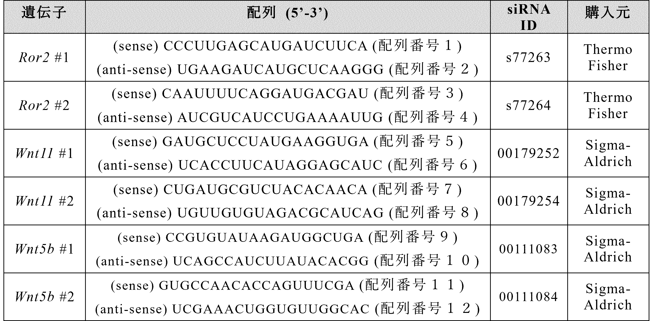
EdU陽性MPCsの割合を調べるために、MPCsにsiRNA(表2に記載のRor2#1,Ror2#2,Wnt5b#1又はWnt5b#2)を導入し、10%FBSを含むDMEMで3日間培養した。その後、MPCsをEdU(10μM、Thermo Fisher Scientific,Waltham,MA)で30分間処理し、その後4%(w/v)PFAで固定した。EdUの取り込みは、Click-it EdU imaging kit(Thermo Fisher Scientific)を用いて、製造者の説明書に従って検出した。
脂肪分化のために、MPCをAdipogenic induction medium(10%FBS、0.5mM IBMX(Sigma-Aldrich)、0.25μMデキサメタゾン(Sigma-Aldrich)、10μg/ml Insulin(Sigma-Aldrich)含有DMEM)で3日間培養した後に、Adipogenic maintenance medium(10%FBS,10μg/ml Insulin含有DMEM)で3日間処理した。
老化細胞を検出するため、siRNA(表2に記載のRor2#1,Ror2#2、Wnt11#1又はWnt11#2)を導入したMPCsを10%FBS DMEMで3日間培養した後、製造者の指示に従ってSPiDER-βGal(同仁化学研究所、熊本)で染色した。
オイルレッドO染色
脂肪細胞を染色するために、細胞を4%(w/v)PFAで室温にて10分間処理し、固定した。固定した細胞を60%(v/v)イソプロパノールで洗浄した。次に、オイルレッドO(富士フイルム和光純薬株式会社)で室温にて30分間染色した。染色した細胞を60%(v/v)イソプロパノールで洗浄した後、100%イソプロパノールで20分間処理し、脂肪細胞内に取り込まれたオイルレッドOを抽出した。その後、上清のオイルレッドOの吸光度を490nmで測定した。
脂肪細胞を染色するために、細胞を4%(w/v)PFAで室温にて10分間処理し、固定した。固定した細胞を60%(v/v)イソプロパノールで洗浄した。次に、オイルレッドO(富士フイルム和光純薬株式会社)で室温にて30分間染色した。染色した細胞を60%(v/v)イソプロパノールで洗浄した後、100%イソプロパノールで20分間処理し、脂肪細胞内に取り込まれたオイルレッドOを抽出した。その後、上清のオイルレッドOの吸光度を490nmで測定した。
WST-8アッセイ
MPCを96ウェルプレートに播種し(1ウェルあたり1000細胞)、siRNAを導入(表2に記載のWnt5b#1又はWnt5b#2)した。その後、MPCを任意の期間培養し、培地にWST-8試薬(Cell counting kit-8、同仁化学研究所)を加えて90分間インキュベートし、450nmの吸光度を測定した。
Wnt5b誘導性増殖におけるp38の関与を評価するため、96ウェルプレートにMPCを播種した1日後に、p38阻害剤(10μM SB203580,Cayman Chemical,Ann Arbor,MI)及び組換えWnt5b(200ng/ml,R&D System)を培地に追加した。その後、WST-8アッセイを上記のように実施した。
MPCを96ウェルプレートに播種し(1ウェルあたり1000細胞)、siRNAを導入(表2に記載のWnt5b#1又はWnt5b#2)した。その後、MPCを任意の期間培養し、培地にWST-8試薬(Cell counting kit-8、同仁化学研究所)を加えて90分間インキュベートし、450nmの吸光度を測定した。
Wnt5b誘導性増殖におけるp38の関与を評価するため、96ウェルプレートにMPCを播種した1日後に、p38阻害剤(10μM SB203580,Cayman Chemical,Ann Arbor,MI)及び組換えWnt5b(200ng/ml,R&D System)を培地に追加した。その後、WST-8アッセイを上記のように実施した。
免疫蛍光染色
単離したTA筋を液体窒素で冷却したイソペンタンで凍結し、クライオスタット(Leica CM1860,Wetzlar,Germany)で切片を作製した。凍結切片を0.2%(v/v)Triton X-100で透過処理を行った。その後、切片を3%(w/v)BSA含有PBSでブロッキングし、一次抗体と4℃で一晩インキュベートした。切片をPBSで洗浄し、二次抗体と室温で1時間インキュベートした後、Fluor-KEEPER(ナカライテスク)を用いて封入した。封入した切片を蛍光顕微鏡(BZ-X700、キーエンス、大阪、日本)により観察した。TA筋におけるPerilipin陽性領域の割合やCSAについては、ImageJ(National Institutes of Health,Bethesda,MD)を用いて解析した。
ホールマウント免疫蛍光染色を行うために、単離したEDL筋を4%(w/v)PFAで30分間固定し、その後0.2%(v/v)Triton X-100で透過処理した。PBSで洗浄後、EDL筋を3%(w/v)BSA含有PBSで1時間ブロッキングし、一次抗体と4℃で一晩インキュベートした。その後、EDL筋をPBSで洗浄し、二次抗体と室温で1時間インキュベートした。染色したEDL筋をガラスボトムディッシュ(松浪硝子工業、大阪)の上にのせ、レーザー走査型共焦点イメージングシステム(LSM700、Carl Zeiss、Oberkochen、Germany)を用いて観察した。
培養細胞を4%(w/v)PFAで10分間固定し、その後0.2%(v/v)Triton X-100で透過処理した。PBSで洗浄後、3%(w/v)BSA含有PBSで1時間ブロッキングし、一次抗体と4℃で一晩インキュベートした。次に、細胞をPBSで洗浄し、二次抗体と室温で1時間インキュベートした。染色した細胞をFluor-KEEPER(ナカライテスク社製)で封入した。封入した細胞を、レーザー走査型共焦点イメージングシステム(LSM700,Carl Zeiss)を用いて観察した。
単離したTA筋を液体窒素で冷却したイソペンタンで凍結し、クライオスタット(Leica CM1860,Wetzlar,Germany)で切片を作製した。凍結切片を0.2%(v/v)Triton X-100で透過処理を行った。その後、切片を3%(w/v)BSA含有PBSでブロッキングし、一次抗体と4℃で一晩インキュベートした。切片をPBSで洗浄し、二次抗体と室温で1時間インキュベートした後、Fluor-KEEPER(ナカライテスク)を用いて封入した。封入した切片を蛍光顕微鏡(BZ-X700、キーエンス、大阪、日本)により観察した。TA筋におけるPerilipin陽性領域の割合やCSAについては、ImageJ(National Institutes of Health,Bethesda,MD)を用いて解析した。
ホールマウント免疫蛍光染色を行うために、単離したEDL筋を4%(w/v)PFAで30分間固定し、その後0.2%(v/v)Triton X-100で透過処理した。PBSで洗浄後、EDL筋を3%(w/v)BSA含有PBSで1時間ブロッキングし、一次抗体と4℃で一晩インキュベートした。その後、EDL筋をPBSで洗浄し、二次抗体と室温で1時間インキュベートした。染色したEDL筋をガラスボトムディッシュ(松浪硝子工業、大阪)の上にのせ、レーザー走査型共焦点イメージングシステム(LSM700、Carl Zeiss、Oberkochen、Germany)を用いて観察した。
培養細胞を4%(w/v)PFAで10分間固定し、その後0.2%(v/v)Triton X-100で透過処理した。PBSで洗浄後、3%(w/v)BSA含有PBSで1時間ブロッキングし、一次抗体と4℃で一晩インキュベートした。次に、細胞をPBSで洗浄し、二次抗体と室温で1時間インキュベートした。染色した細胞をFluor-KEEPER(ナカライテスク社製)で封入した。封入した細胞を、レーザー走査型共焦点イメージングシステム(LSM700,Carl Zeiss)を用いて観察した。
ウェスタンブロッティング
細胞を溶解バッファー(50mM Tris-HCl(pH7.5),150mM NaCl,1%(v/v)Nonidet P-40,1mM EDTA,10mM NaF,1mM Na3VO4,10μg/ml aprotinin,10μg/ml leupeptin,1μM p-APMSF)で可溶化させた。タンパク質(1レーンあたり合計10μg)をSDS-PAGEで分離し、Immobilon-Pメンブレン(Merck Millipore,Darmstadt,Germany)上に転写した。メンブレンは1%(v/w)BSA含有TBSTで1時間ブロッキングした後、一次抗体(表1に記載)を用いて4℃で一晩ブロットした。一次抗体で処理した後、メンブレンをTBSTにより室温で30分間洗浄し、その後、二次抗体(表1に記載)を用いて室温で1時間ブロットした。最後に、メンブレンを室温で30分間TBSTによって洗浄した。解析時にはImmunoStar LD(FUJIFILM Wako Pure Chemical Corporation)及びLAS 1000(GE Healthcare,Chicago,IL)を使用した。相対的なバンド強度は、ImageJソフトウェアによって決定された。
細胞を溶解バッファー(50mM Tris-HCl(pH7.5),150mM NaCl,1%(v/v)Nonidet P-40,1mM EDTA,10mM NaF,1mM Na3VO4,10μg/ml aprotinin,10μg/ml leupeptin,1μM p-APMSF)で可溶化させた。タンパク質(1レーンあたり合計10μg)をSDS-PAGEで分離し、Immobilon-Pメンブレン(Merck Millipore,Darmstadt,Germany)上に転写した。メンブレンは1%(v/w)BSA含有TBSTで1時間ブロッキングした後、一次抗体(表1に記載)を用いて4℃で一晩ブロットした。一次抗体で処理した後、メンブレンをTBSTにより室温で30分間洗浄し、その後、二次抗体(表1に記載)を用いて室温で1時間ブロットした。最後に、メンブレンを室温で30分間TBSTによって洗浄した。解析時にはImmunoStar LD(FUJIFILM Wako Pure Chemical Corporation)及びLAS 1000(GE Healthcare,Chicago,IL)を使用した。相対的なバンド強度は、ImageJソフトウェアによって決定された。
定量的RT-PCR
Sepasol-RNA I SuperG(ナカライテスク)を用いて培養細胞あるいはTA筋からTotal RNAを抽出し、これらのRNAからPrimeScript RT Reagent(タカラバイオ、滋賀)を用いてcDNAを合成した。LightCycler 480 II system(Roche,Basel,Switzerland)を用いて、それぞれの目的遺伝子の発現を定量化した。対象mRNAの量は、18SリボソームRNAの量に対して正規化した。プライマーペアの配列は、表3に記載されている。
Sepasol-RNA I SuperG(ナカライテスク)を用いて培養細胞あるいはTA筋からTotal RNAを抽出し、これらのRNAからPrimeScript RT Reagent(タカラバイオ、滋賀)を用いてcDNAを合成した。LightCycler 480 II system(Roche,Basel,Switzerland)を用いて、それぞれの目的遺伝子の発現を定量化した。対象mRNAの量は、18SリボソームRNAの量に対して正規化した。プライマーペアの配列は、表3に記載されている。
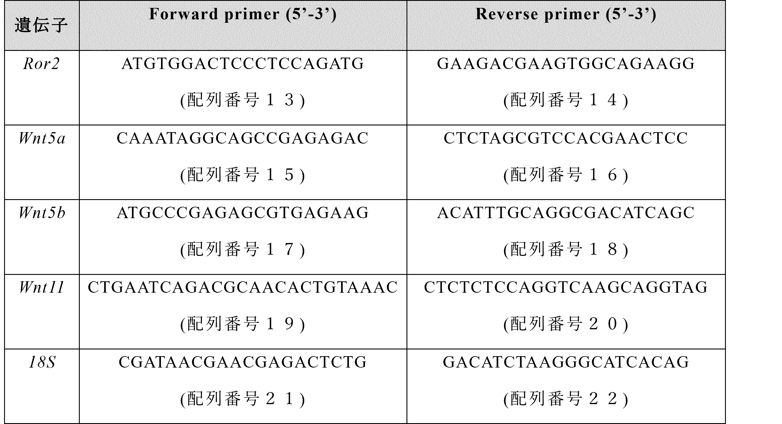
統計解析
データは、BellCurve for Excel(株式会社社会調査研究情報、東京)及びGraphPad Prism 9.0(GraphPad Software,Boston,MA)を用いて分析された。データは、平均値±標準偏差(S.D.)で示した。有意性は、2つのグループを比較した場合はunpaired two-tailed Student’s t test、3つ以上のグループを分析した場合はone-way ANOVA with Holm’s testあるいはDunnett’s testを用いて、*P<0.05,**P<0.01又は***P<0.001として決定された。
データは、BellCurve for Excel(株式会社社会調査研究情報、東京)及びGraphPad Prism 9.0(GraphPad Software,Boston,MA)を用いて分析された。データは、平均値±標準偏差(S.D.)で示した。有意性は、2つのグループを比較した場合はunpaired two-tailed Student’s t test、3つ以上のグループを分析した場合はone-way ANOVA with Holm’s testあるいはDunnett’s testを用いて、*P<0.05,**P<0.01又は***P<0.001として決定された。
マウスのTA筋、腓腹筋、ヒラメ筋、大腿四頭筋、大殿筋を回収し、FACS AriaIIIを用いてSCs及びMPCsを単離し、RNA抽出後、定量的RT-PCR法を用いて発現解析を行った。
図1に示すとおり、MPCsにおいてRor2が選択的に発現していることが確認された。
Ctxを投与したTA筋から単離したMPCsを5%CO2,3%O2で3日間培養した後、通常酸素濃度下でsiRNA(表2に記載のRor2#1又はRor2#2)を導入した。翌日培地を新しいものに置換した。siRNAを導入して3日後に細胞を固定し、Ki67抗体を用いて染色した。
Ctxを投与したTA筋から単離したMPCsを5%CO2,3%O2で3日間培養した後、通常酸素濃度下でsiRNA(表2に記載のRor2#1又はRor2#2)を導入した。翌日培地を新しいものに置換した。siRNAを導入して3日後にAdipogenic induction mediumで3日間培養し、続いてAdipogenic maintenance mediumで3日間培養することで脂肪細胞への分化を誘導した。分化誘導後、オイルレッドO染色を行った。
Ctxを投与したTA筋から単離したMPCsを5%CO2,3%O2で3日間培養した後、通常酸素濃度下でsiRNA(表2に記載のRor2#1又はRor2#2)を導入した。翌日培地を新しいものに置換した。siRNAを導入して3日後にSPiDER-βGalを用いて老化細胞を染色した。
Ctxを投与したTA筋から単離したMPCsを5%CO2,3%O2で3日間培養した後、通常酸素濃度下でsiRNA(表2に記載のRor2#1又はRor2#2)を導入した。翌日培地を新しいものに置換した。siRNAを導入して3日後にAdipogenic induction mediumで3日間培養し、続いてAdipogenic maintenance mediumで3日間培養することで脂肪細胞への分化を誘導した。分化誘導後、オイルレッドO染色を行った。
Ctxを投与したTA筋から単離したMPCsを5%CO2,3%O2で3日間培養した後、通常酸素濃度下でsiRNA(表2に記載のRor2#1又はRor2#2)を導入した。翌日培地を新しいものに置換した。siRNAを導入して3日後にSPiDER-βGalを用いて老化細胞を染色した。
図2に示すとおり、マウス骨格筋から単離・培養したMPCsにおいてRor2の発現を抑制すると、MPCsの増殖能及び脂肪細胞への分化能が低下し、細胞老化が亢進することが確認された。MPCsは細胞老化を起こすと増殖能及び脂肪細胞への分化能が低下することが報告されていることから(非特許文献1、2)、Ror2はMPCsの細胞老化を抑制することで増殖能や脂肪細胞への分化能を制御している可能性が考えられた。
12~16週齢のコントロールマウス及びRor2cKOマウスにTamoxifenを投与した後、半年間飼育し、骨格筋重量を比較解析した。このときのTA筋について、凍結切片を作製し、Laminin抗体で染色し、各筋線維の断面積を測定した。
図3に示すとおり、Ror2cKOマウスではTA筋、腓腹筋(Gastrocnemius muscle)、及びヒラメ筋(Soleus)の重量が低下し、TA筋における筋線維が萎縮することが明らかとなった。このことからRor2は骨格筋の維持において重要であると考えられる。
12~16週齢のコントロールマウス及びRor2cKOマウスにTamoxifenを投与した後、CtxあるいはGlycerolをTA筋に投与した。投与2週間後にTA筋を回収し、Laminin又はPerilipinで染色した。TA筋の各筋線維の断面積を測定した。また、Glycerol投与筋の染色画像を用いて、筋組織断面積におけるPerilipin陽性面積の割合を計測することで、脂肪蓄積の程度を比較解析した。
図4に示すとおり、Ctx投与2週間後の筋線維の太さがRor2cKOマウスではコントロールマウスに比べて小さく、骨格筋の再生遅延が示されたことから、Ror2は骨格筋再生に重要であると考えられる。
また、Ror2cKOマウスではGlycerol投与後の脂肪蓄積が減少していたことから、Ror2はIMAT形成においても重要であることが示された。
また、Ror2cKOマウスではGlycerol投与後の脂肪蓄積が減少していたことから、Ror2はIMAT形成においても重要であることが示された。
Ctxを投与したTA筋から単離したMPCsを5%CO2,3%O2で3日間培養した後、通常酸素濃度下でsiRNA(表2に記載のWnt11#1又はWnt11#2)を導入した。翌日培地を新しいものに置換した。siRNAを導入して3日後にRNAを抽出し、定量的RT-PCR法にて発現解析を行った。これと並行して、SPiDER-βGal染色も行い、フローサイトメトリー法にて染色強度を比較解析した。
図5に示すとおり、MPCsにおいてWnt11の発現を抑制することにより、MPCsの細胞老化が誘導されることが明らかになった。
Ctxを投与したTA筋から単離したMPCsを5%CO2,3%O2で3日間培養した後、通常酸素濃度下でsiRNA(表2に記載のRor2#1、Ror2#2、Wnt11#1又はWnt11#2)を導入した。翌日培地を新しいものに置換した。siRNAを導入して2日後にPhalloidin及びDAPIで染色した。
Ctxを投与したTA筋から単離したMPCsを5%CO2,3%O2で3日間培養した後、通常酸素濃度下でsiRNA(表2に記載のRor2#1、Ror2#2、Wnt11#1又はWnt11#2)を導入した。翌日培地を新しいものに置換した。siRNAを導入して3日後に固定し、LaminB1及びDAPIで染色した。
Ctxを投与したTA筋から単離したMPCsを5%CO2,3%O2で3日間培養した後、通常酸素濃度下でsiRNA(表2に記載のRor2#1、Ror2#2、Wnt11#1又はWnt11#2)を導入した。翌日培地を新しいものに置換した。siRNAを導入して3日後に固定し、LaminB1及びDAPIで染色した。
図6に示すとおり、MPCsにおいてWnt11又はRor2の発現を抑制すると、アクチン・ストレスファイバーの形成が促進されることが観察された。また、核形態の異常がみられることも見出された。
核形態の異常に伴い、細胞質中にDNAが漏出すると、cGas(cyclic GMP-AMP synthase)を介したシグナル伝達経路(cGAS-Sting経路)が活性化することが報告されている(非特許文献3)。これらの知見から、Wnt11及びRor2はMPCsにおけるストレスファイバーの形成を抑制することで、核形態異常とそれに伴うcGas-Sting経路の活性化を抑制し、その結果、細胞老化を阻害することにより骨格筋の再生や維持に寄与していると考えられた。
核形態の異常に伴い、細胞質中にDNAが漏出すると、cGas(cyclic GMP-AMP synthase)を介したシグナル伝達経路(cGAS-Sting経路)が活性化することが報告されている(非特許文献3)。これらの知見から、Wnt11及びRor2はMPCsにおけるストレスファイバーの形成を抑制することで、核形態異常とそれに伴うcGas-Sting経路の活性化を抑制し、その結果、細胞老化を阻害することにより骨格筋の再生や維持に寄与していると考えられた。
次に、Ror2がIMATの形成に関わる分子機構を明らかにすることを目的として、Ctx投与筋、及びGlycerol投与筋における遺伝子発現の比較解析を実施した。8~12週齢の野生型マウスのTA筋にCtx又はGlycerolを投与し、0(投与無し),3,7日後にTA筋を回収し、Ror2のリガンドとして機能し得るWntファミリー遺伝子について、定量的RT-PCR法にて発現解析を行った。
Ctxを投与したTA筋から単離したMPCsを5%CO2,3%O2で3日間培養した後、通常酸素濃度下でsiRNA(表2に記載のRor2#1又はRor2#2)を導入した。翌日Wnt5a又はWnt5bを含む培地に置換した。3日後にEdUあるいはKi67染色による増殖解析を実施した。
Ctxを投与したTA筋から単離したMPCsを5%CO2,3%O2で3日間培養した後、通常酸素濃度下でsiRNA(表2に記載のRor2#1又はRor2#2)を導入した。翌日Wnt5a又はWnt5bを含む培地に置換した。3日後にEdUあるいはKi67染色による増殖解析を実施した。
図7に示すとおり、Wnt5bがGlycerol投与後に発現上昇することを見出した。このことからWnt5bがIMAT形成に寄与する可能性が考えられた。
さらに、培養MPCsにWnt5bを添加することでMPCsの増殖が促進され、他方Ror2の発現抑制によってWnt5b添加による増殖促進が抑制された。また、Ror2のリガンドとして最もよく知られているWnt5aの添加ではMPCsの増殖は影響を受けなかった。
さらに、培養MPCsにWnt5bを添加することでMPCsの増殖が促進され、他方Ror2の発現抑制によってWnt5b添加による増殖促進が抑制された。また、Ror2のリガンドとして最もよく知られているWnt5aの添加ではMPCsの増殖は影響を受けなかった。
Ctxを投与したTA筋から単離したMPCsを5%CO2,3%O2で3日間培養した後、通常酸素濃度下でsiRNA(表2に記載のRor2#1又はRor2#2)を導入した。翌日培地を新しいものに置換した。siRNAを導入して2日後に培地をOPTI-MEMに置換し一晩培養した。その後、Wnt5bを200ng/mlで添加し、1時間後に溶解バッファーにて細胞からタンパク質を抽出し、ウェスタンブロットを実施した。
Ctxを投与したTA筋から単離したMPCsを5%CO2,3%O2で3日間培養した後、通常酸素濃度下でsiRNA(表2に記載のRor2#1又はRor2#2)を導入した。翌日Wnt5b(200ng/ml)又はSB203580(10μM)を含む培地に置換した。このとき、培地置換していない細胞をday0サンプルとしてWST-8アッセイにより解析した。培地置換して3日後にWST-8アッセイにより各ウェルにおける細胞数を測定した。
Ctxを投与したTA筋から単離したMPCsを5%CO2,3%O2で3日間培養した後、通常酸素濃度下でsiRNA(表2に記載のRor2#1又はRor2#2)を導入した。翌日Wnt5b(200ng/ml)又はSB203580(10μM)を含む培地に置換した。このとき、培地置換していない細胞をday0サンプルとしてWST-8アッセイにより解析した。培地置換して3日後にWST-8アッセイにより各ウェルにおける細胞数を測定した。
図8に示すとおり、培養MPCsを用いた解析から、Wnt5bはRor2依存的にp38をリン酸化(活性化を反映)することを見出した。また、p38阻害剤であるSB203580で処理すると、Wnt5b刺激依存的なMPCsの増殖促進が抑制された。以上から、Wnt5b、Ror2、及びp38がMPCsの増殖を促進することが明らかとなった。
骨格筋でIMATや線維化などが生じる際には、MPCsが過剰に骨格筋内に蓄積し脂肪細胞や線維芽細胞に分化することが示唆されている(非特許文献4、5)。上記の我々の解析結果から、Wnt5b、Ror2、及びp38はMPCsの増殖を促進することにより骨格筋組織中におけるMPCsの過剰な蓄積を誘導し、IMATの形成に寄与している可能性が考えられる。
骨格筋でIMATや線維化などが生じる際には、MPCsが過剰に骨格筋内に蓄積し脂肪細胞や線維芽細胞に分化することが示唆されている(非特許文献4、5)。上記の我々の解析結果から、Wnt5b、Ror2、及びp38はMPCsの増殖を促進することにより骨格筋組織中におけるMPCsの過剰な蓄積を誘導し、IMATの形成に寄与している可能性が考えられる。
Ctx又はGlycerolを投与し3日後のEDL筋をホールマウント染色した。
図9に示すとおり、シュワン細胞(S100β陽性細胞)においてWnt5bが発現しており、Wnt5b陽性シュワン細胞はGlycerol投与時に増殖が亢進することを見出した。このことからWnt5bの産生細胞の一つがシュワン細胞である可能性が示された。
8~12週齢の雄マウスのTA筋にCtx又はGlycerolを投与し3日後のTA筋を回収し、FACS AriaIIIを用いてSCs、MPCs、CD31陽性・CD45陽性細胞(造血系細胞と内皮細胞の混合集団)、それ以外の細胞集団をそれぞれ単離し、RNA抽出後、定量的RT-PCR法を用いて発現解析を行った。
図10に示すとおり、TA筋ではSCs及びMPCsにおいてWnt5bが発現していることを見出した。このことからTA筋におけるWnt5bの産生細胞はSCs及びMPCsであることが示された。
Ctxを投与したTA筋から単離したMPCsを5%CO2,3%O2で3日間培養した後、通常酸素濃度下でsiRNA(表2に示すWnt5b#1又はWnt5b#2)を導入した。翌日培地を新しいものに置換した。siRNAを導入して3日後にRNAを抽出し、定量的RT-PCR法にて発現解析を行った。
Ctxを投与したTA筋から単離したMPCsを5%CO2,3%O2で3日間培養した後、通常酸素濃度下でsiRNA(表2に示すWnt5b#1又はWnt5b#2)を導入した。翌日培地を新しいものに置換した。このとき、培地置換していない細胞をday0サンプルとしてWST-8アッセイにより解析した。培地置換して4日後にWST-8アッセイにより各ウェルにおける細胞数を測定した。
Ctxを投与したTA筋から単離したMPCsを5%CO2,3%O2で3日間培養した後、通常酸素濃度下でsiRNA(表2に示すWnt5b#1又はWnt5b#2)を導入した。翌日培地を新しいものに置換した。3日後にEdUによる増殖解析を実施した。
Ctxを投与したTA筋から単離したMPCsを5%CO2,3%O2で3日間培養した後、通常酸素濃度下でsiRNA(表2に示すWnt5b#1又はWnt5b#2)を導入した。翌日培地を新しいものに置換した。このとき、培地置換していない細胞をday0サンプルとしてWST-8アッセイにより解析した。培地置換して4日後にWST-8アッセイにより各ウェルにおける細胞数を測定した。
Ctxを投与したTA筋から単離したMPCsを5%CO2,3%O2で3日間培養した後、通常酸素濃度下でsiRNA(表2に示すWnt5b#1又はWnt5b#2)を導入した。翌日培地を新しいものに置換した。3日後にEdUによる増殖解析を実施した。
図11に示すとおり、siRNAによりWnt5bの発現が抑制されることが明らかになった。また、培養MPCsを用いた解析から、siRNAによるWnt5bの発現抑制により、MPCsの増殖能が阻害されることが明らかになった。
以上の結果から、生理的状態ではMPCsにおいてWnt11及びRor2がMPCsの細胞老化を抑制し骨格筋の維持や再生に寄与するが、病的状態ではWnt11に加えてWnt5bもRor2のリガンドとして機能することによってMPCsの過剰な増殖が促進され、IMAT形成が促進される可能性が考えられた。
IMATの蓄積がみられる筋疾患及び/又は症状として、筋ジストロフィー(デュシェンヌ型筋ジストロフィー等)、筋萎縮性側索硬化症(ALS)をはじめとする筋委縮症等の各種骨格筋疾患、サルコペニア、フレイル、ロコモティブ症候群等の骨格筋における加齢等に伴う各種症状等が例示される。本開示は、このようなIMATの蓄積がみられる筋ジストロフィーや加齢に伴って見られるサルコペニアなどをはじめとする骨格筋疾患・病態に対して有効かつ副作用の少ない新規治療方法の開発に活用できる。
Claims (9)
- (I)Wnt5bタンパク質、Ror2タンパク質、及び被験物質を接触させる工程、並びに
(II)前記被験物質がWnt5bタンパク質とRor2タンパク質との結合を阻害することを確認する工程を含む、
IMAT形成抑制物質をスクリーニングする方法。 - 工程(I)において、さらにWnt11タンパク質を接触させ、
工程(II)において、さらに前記被験物質がWnt11タンパク質とRor2タンパク質との結合を阻害しないことを確認する、請求項1に記載の方法。 - (1)SCs、MPCs、及びシュワン細胞からなる群より選択される少なくとも1種と、被験物質とを接触させる工程、
並びに
(2)前記被験物質がWnt5bの発現量を抑制することを確認する工程を含む、
IMAT形成抑制物質をスクリーニングする方法。 - 前記工程(2)において、前記被験物質がWnt11の発現量を抑制しないことを確認する、請求項3に記載の方法。
- (A)Wnt5bタンパク質、Ror2タンパク質、p38タンパク質、及び被験物質を接触させる工程、並びに
(B)前記被験物質がp38タンパク質のリン酸化を阻害することを確認する工程を含む、
IMAT形成抑制物質をスクリーニングする方法。 - IMAT形成抑制が、MPCsの増殖抑制によるものである、請求項1~5のいずれか一項に記載の方法。
- Wnt5bタンパク質の中和抗体、又はWnt5b遺伝子の発現を抑制する核酸を有効成分とする、IMAT形成抑制用組成物。
- IMATの蓄積が認められる筋疾患又は症状の抑制又は治療に使用するための、請求項7に記載のIMAT形成抑制用組成物。
- 前記IMATの蓄積が認められる筋疾患又は症状が、筋ジストロフィー、筋萎縮性側索硬化症、サルコペニア、フレイル又はロコモティブ症候群である、請求項8に記載のIMAT形成抑制用組成物。
Priority Applications (1)
| Application Number | Priority Date | Filing Date | Title |
|---|---|---|---|
| JP2025530134A JPWO2025005076A1 (ja) | 2023-06-26 | 2024-06-25 |
Applications Claiming Priority (2)
| Application Number | Priority Date | Filing Date | Title |
|---|---|---|---|
| JP2023-104009 | 2023-06-26 | ||
| JP2023104009 | 2023-06-26 |
Publications (1)
| Publication Number | Publication Date |
|---|---|
| WO2025005076A1 true WO2025005076A1 (ja) | 2025-01-02 |
Family
ID=93939142
Family Applications (1)
| Application Number | Title | Priority Date | Filing Date |
|---|---|---|---|
| PCT/JP2024/022952 Ceased WO2025005076A1 (ja) | 2023-06-26 | 2024-06-25 | 骨格筋内脂肪組織(IMAT;Intramuscular adipose tissue)形成抑制物質をスクリーニングする方法、及びIMAT形成抑制用組成物 |
Country Status (2)
| Country | Link |
|---|---|
| JP (1) | JPWO2025005076A1 (ja) |
| WO (1) | WO2025005076A1 (ja) |
-
2024
- 2024-06-25 WO PCT/JP2024/022952 patent/WO2025005076A1/ja not_active Ceased
- 2024-06-25 JP JP2025530134A patent/JPWO2025005076A1/ja active Pending
Non-Patent Citations (3)
| Title |
|---|
| KAMIZAKI KOKI, ENDO MITSUHARU, MINAMI YASUHIRO, KOBAYASHI YASUHIRO: "Role of noncanonical Wnt ligands and Ror‐family receptor tyrosine kinases in the development, regeneration, and diseases of the musculoskeletal system", DEVELOPMENTAL DYNAMICS, WILEY-LISS INC NEW YORK , NY, US, vol. 250, no. 1, 1 January 2021 (2021-01-01), US , pages 27 - 38, XP093252747, ISSN: 1058-8388, DOI: 10.1002/dvdy.151 * |
| TAKAKI KAMISAKI : "YIA-01: Regulation of mesenchymal progenitor cell function by Wnt-Ror2 signaling", PROGRAMS AND ABSTRACTS OF JOINT ACADEMIC CONFERENCE OF THE 9TH ANNUAL MEETING OF JAPAN MUSCLE SOCIETY AND THE 10TH MEDICAL RESEARCH GROUP OF MUSCULAR DYSTROPHY, FRIDAY, AUGUST 18TH TO SATURDAY, AUGUST 19TH, 2023, JAPAN MUSCLE SOCIETY, JP, 18 August 2023 (2023-08-18) - 19 August 2023 (2023-08-19), JP, pages 51, XP009559607 * |
| TAKAKI KAMIZAKI ET AL.: "The function control mechanism of skeletal muscle mesenchymal progenitor cells by Ror2", PROGRAMS AND ABSTRACTS OF THE 7TH ANNUAL MEETING OF JAPAN MUSCLE SOCIETY, JAPAN MUSCLE SOCIETY, JP, vol. 8, 5 August 2022 (2022-08-05) - 2022-08-06, JP, pages 86, XP009559608, ISSN: 2433-975X * |
Also Published As
| Publication number | Publication date |
|---|---|
| JPWO2025005076A1 (ja) | 2025-01-02 |
Similar Documents
| Publication | Publication Date | Title |
|---|---|---|
| Shangguan et al. | Inhibition of TGF-β/Smad signaling by BAMBI blocks differentiation of human mesenchymal stem cells to carcinoma-associated fibroblasts and abolishes their protumor effects | |
| CN108430480B (zh) | 一种包含过表达sod3的干细胞作为活性成分的炎症性疾病的预防或治疗用组合物 | |
| Gioia et al. | Impaired osteoblastogenesis in a murine model of dominant osteogenesis imperfecta: a new target for osteogenesis imperfecta pharmacological therapy | |
| Zou et al. | In vitro study of enhanced osteogenesis induced by HIF‐1α‐transduced bone marrow stem cells | |
| JP2021184752A (ja) | 免疫疾患の治療のための高効能幹細胞の選別方法 | |
| Kim et al. | Regulation of mesenchymal stromal cells through fine tuning of canonical Wnt signaling | |
| JP7849972B2 (ja) | 高機能な製造されたabcb5+間葉系幹細胞 | |
| Liu et al. | CXCL12/CXCR4 signal axis plays an important role in mediating bone morphogenetic protein 9-induced osteogenic differentiation of mesenchymal stem cells | |
| US20160333343A1 (en) | Method of Treatment of Vascular Complications | |
| Liu et al. | Islet-1 overexpression in human mesenchymal stem cells promotes vascularization through monocyte chemoattractant protein-3 | |
| Ruetz et al. | In vitro and in vivo CRISPR-Cas9 screens reveal drivers of aging in neural stem cells of the brain | |
| Ben-Shoshan et al. | Constitutive expression of HIF-1α and HIF-2α in bone marrow stromal cells differentially promotes their proangiogenic properties | |
| Barzelay et al. | A potential role for islet-1 in post-natal angiogenesis and vasculogenesis | |
| Huang et al. | IFITM1 promotes proliferation, migration and macrophage-like transdifferentiation of vascular smooth muscle cells via c-Src/MAPK/GATA2/E2F2 pathway in atherosclerosis | |
| KR101796826B1 (ko) | Oct4 및 Sox2를 과발현하는 중간엽줄기세포 및 그 용도 | |
| WO2025005076A1 (ja) | 骨格筋内脂肪組織(IMAT;Intramuscular adipose tissue)形成抑制物質をスクリーニングする方法、及びIMAT形成抑制用組成物 | |
| WO2019027299A2 (ko) | 간세포성장인자를 발현하는 중간엽줄기세포를 유효성분으로 포함하는 혈관계 질환 예방 또는 치료용 약학 조성물 | |
| Ninomiya et al. | Role of oncostatin M in the pathogenesis of vernal keratoconjunctivitis: focus on the barrier function of the epithelium and interleukin-33 production by fibroblasts | |
| Shan et al. | Naked cuticle homolog 2 controls the differentiation of osteoblasts and osteoclasts and ameliorates bone loss in ovariectomized mice | |
| Geng et al. | Impaired elastin deposition in Fstl1−/− lung allograft under the renal capsule | |
| Yang et al. | The protective effects of HMGA2 in the senescence process of bone marrow‐derived mesenchymal stromal cells | |
| CN104531613B (zh) | Wip1敲除在促进小鼠骨髓间充质干细胞迁移中的应用 | |
| Higa et al. | CD105 maintains the thermogenic program of beige adipocytes by regulating Smad2 signaling | |
| Zhang et al. | Human umbilical cord mesenchymal stem cells decellular matrix alleviates chondrocyte senescence by inhibiting the STING-NF-κB pathway | |
| Shi et al. | Novel biphasic mechanism of the canonical Wnt signalling component PYGO2 promotes cardiomyocyte differentiation from hUC-MSCs |
Legal Events
| Date | Code | Title | Description |
|---|---|---|---|
| 121 | Ep: the epo has been informed by wipo that ep was designated in this application |
Ref document number: 24831936 Country of ref document: EP Kind code of ref document: A1 |
|
| ENP | Entry into the national phase |
Ref document number: 2025530134 Country of ref document: JP Kind code of ref document: A |
|
| WWE | Wipo information: entry into national phase |
Ref document number: 2025530134 Country of ref document: JP |
|
| NENP | Non-entry into the national phase |
Ref country code: DE |