WO2024248023A1 - 視交叉上核オルガノイドの製造方法 - Google Patents
視交叉上核オルガノイドの製造方法 Download PDFInfo
- Publication number
- WO2024248023A1 WO2024248023A1 PCT/JP2024/019660 JP2024019660W WO2024248023A1 WO 2024248023 A1 WO2024248023 A1 WO 2024248023A1 JP 2024019660 W JP2024019660 W JP 2024019660W WO 2024248023 A1 WO2024248023 A1 WO 2024248023A1
- Authority
- WO
- WIPO (PCT)
- Prior art keywords
- medium
- cells
- suprachiasmatic nucleus
- day
- culture
- Prior art date
- Legal status (The legal status is an assumption and is not a legal conclusion. Google has not performed a legal analysis and makes no representation as to the accuracy of the status listed.)
- Ceased
Links
Images




Classifications
-
- G—PHYSICS
- G01—MEASURING; TESTING
- G01N—INVESTIGATING OR ANALYSING MATERIALS BY DETERMINING THEIR CHEMICAL OR PHYSICAL PROPERTIES
- G01N33/00—Investigating or analysing materials by specific methods not covered by groups G01N1/00 - G01N31/00
- G01N33/48—Biological material, e.g. blood, urine; Haemocytometers
- G01N33/50—Chemical analysis of biological material, e.g. blood, urine; Testing involving biospecific ligand binding methods; Immunological testing
- G01N33/5005—Chemical analysis of biological material, e.g. blood, urine; Testing involving biospecific ligand binding methods; Immunological testing involving human or animal cells
- G01N33/5008—Chemical analysis of biological material, e.g. blood, urine; Testing involving biospecific ligand binding methods; Immunological testing involving human or animal cells for testing or evaluating the effect of chemical or biological compounds, e.g. drugs, cosmetics
- G01N33/5082—Supracellular entities, e.g. tissue, organisms
-
- C—CHEMISTRY; METALLURGY
- C12—BIOCHEMISTRY; BEER; SPIRITS; WINE; VINEGAR; MICROBIOLOGY; ENZYMOLOGY; MUTATION OR GENETIC ENGINEERING
- C12N—MICROORGANISMS OR ENZYMES; COMPOSITIONS THEREOF; PROPAGATING, PRESERVING, OR MAINTAINING MICROORGANISMS; MUTATION OR GENETIC ENGINEERING; CULTURE MEDIA
- C12N5/00—Undifferentiated human, animal or plant cells, e.g. cell lines; Tissues; Cultivation or maintenance thereof; Culture media therefor
- C12N5/06—Animal cells or tissues; Human cells or tissues
- C12N5/0602—Vertebrate cells
- C12N5/0618—Cells of the nervous system
- C12N5/0619—Neurons
-
- G—PHYSICS
- G01—MEASURING; TESTING
- G01N—INVESTIGATING OR ANALYSING MATERIALS BY DETERMINING THEIR CHEMICAL OR PHYSICAL PROPERTIES
- G01N33/00—Investigating or analysing materials by specific methods not covered by groups G01N1/00 - G01N31/00
- G01N33/48—Biological material, e.g. blood, urine; Haemocytometers
- G01N33/50—Chemical analysis of biological material, e.g. blood, urine; Testing involving biospecific ligand binding methods; Immunological testing
- G01N33/5005—Chemical analysis of biological material, e.g. blood, urine; Testing involving biospecific ligand binding methods; Immunological testing involving human or animal cells
- G01N33/5008—Chemical analysis of biological material, e.g. blood, urine; Testing involving biospecific ligand binding methods; Immunological testing involving human or animal cells for testing or evaluating the effect of chemical or biological compounds, e.g. drugs, cosmetics
- G01N33/5044—Chemical analysis of biological material, e.g. blood, urine; Testing involving biospecific ligand binding methods; Immunological testing involving human or animal cells for testing or evaluating the effect of chemical or biological compounds, e.g. drugs, cosmetics involving specific cell types
- G01N33/5058—Neurological cells
-
- G—PHYSICS
- G01—MEASURING; TESTING
- G01N—INVESTIGATING OR ANALYSING MATERIALS BY DETERMINING THEIR CHEMICAL OR PHYSICAL PROPERTIES
- G01N33/00—Investigating or analysing materials by specific methods not covered by groups G01N1/00 - G01N31/00
- G01N33/48—Biological material, e.g. blood, urine; Haemocytometers
- G01N33/50—Chemical analysis of biological material, e.g. blood, urine; Testing involving biospecific ligand binding methods; Immunological testing
- G01N33/5005—Chemical analysis of biological material, e.g. blood, urine; Testing involving biospecific ligand binding methods; Immunological testing involving human or animal cells
- G01N33/5008—Chemical analysis of biological material, e.g. blood, urine; Testing involving biospecific ligand binding methods; Immunological testing involving human or animal cells for testing or evaluating the effect of chemical or biological compounds, e.g. drugs, cosmetics
- G01N33/5076—Chemical analysis of biological material, e.g. blood, urine; Testing involving biospecific ligand binding methods; Immunological testing involving human or animal cells for testing or evaluating the effect of chemical or biological compounds, e.g. drugs, cosmetics involving cell organelles, e.g. Golgi complex, endoplasmic reticulum
-
- C—CHEMISTRY; METALLURGY
- C12—BIOCHEMISTRY; BEER; SPIRITS; WINE; VINEGAR; MICROBIOLOGY; ENZYMOLOGY; MUTATION OR GENETIC ENGINEERING
- C12N—MICROORGANISMS OR ENZYMES; COMPOSITIONS THEREOF; PROPAGATING, PRESERVING, OR MAINTAINING MICROORGANISMS; MUTATION OR GENETIC ENGINEERING; CULTURE MEDIA
- C12N2501/00—Active agents used in cell culture processes, e.g. differentation
- C12N2501/40—Regulators of development
- C12N2501/41—Hedgehog proteins; Cyclopamine (inhibitor)
-
- C—CHEMISTRY; METALLURGY
- C12—BIOCHEMISTRY; BEER; SPIRITS; WINE; VINEGAR; MICROBIOLOGY; ENZYMOLOGY; MUTATION OR GENETIC ENGINEERING
- C12N—MICROORGANISMS OR ENZYMES; COMPOSITIONS THEREOF; PROPAGATING, PRESERVING, OR MAINTAINING MICROORGANISMS; MUTATION OR GENETIC ENGINEERING; CULTURE MEDIA
- C12N2501/00—Active agents used in cell culture processes, e.g. differentation
- C12N2501/40—Regulators of development
- C12N2501/415—Wnt; Frizzeled
-
- C—CHEMISTRY; METALLURGY
- C12—BIOCHEMISTRY; BEER; SPIRITS; WINE; VINEGAR; MICROBIOLOGY; ENZYMOLOGY; MUTATION OR GENETIC ENGINEERING
- C12N—MICROORGANISMS OR ENZYMES; COMPOSITIONS THEREOF; PROPAGATING, PRESERVING, OR MAINTAINING MICROORGANISMS; MUTATION OR GENETIC ENGINEERING; CULTURE MEDIA
- C12N2506/00—Differentiation of animal cells from one lineage to another; Differentiation of pluripotent cells
- C12N2506/02—Differentiation of animal cells from one lineage to another; Differentiation of pluripotent cells from embryonic cells
-
- C—CHEMISTRY; METALLURGY
- C12—BIOCHEMISTRY; BEER; SPIRITS; WINE; VINEGAR; MICROBIOLOGY; ENZYMOLOGY; MUTATION OR GENETIC ENGINEERING
- C12N—MICROORGANISMS OR ENZYMES; COMPOSITIONS THEREOF; PROPAGATING, PRESERVING, OR MAINTAINING MICROORGANISMS; MUTATION OR GENETIC ENGINEERING; CULTURE MEDIA
- C12N2506/00—Differentiation of animal cells from one lineage to another; Differentiation of pluripotent cells
- C12N2506/45—Differentiation of animal cells from one lineage to another; Differentiation of pluripotent cells from artificially induced pluripotent stem cells
-
- C—CHEMISTRY; METALLURGY
- C12—BIOCHEMISTRY; BEER; SPIRITS; WINE; VINEGAR; MICROBIOLOGY; ENZYMOLOGY; MUTATION OR GENETIC ENGINEERING
- C12N—MICROORGANISMS OR ENZYMES; COMPOSITIONS THEREOF; PROPAGATING, PRESERVING, OR MAINTAINING MICROORGANISMS; MUTATION OR GENETIC ENGINEERING; CULTURE MEDIA
- C12N2513/00—3D culture
-
- G—PHYSICS
- G01—MEASURING; TESTING
- G01N—INVESTIGATING OR ANALYSING MATERIALS BY DETERMINING THEIR CHEMICAL OR PHYSICAL PROPERTIES
- G01N2800/00—Detection or diagnosis of diseases
- G01N2800/28—Neurological disorders
- G01N2800/2864—Sleep disorders
Definitions
- the present invention relates to a method for producing suprachiasmatic nucleus organoids, etc.
- the suprachiasmatic nucleus In mammals, the suprachiasmatic nucleus is the center of circadian rhythm. For this reason, it is expected that regulating the circadian rhythm of the suprachiasmatic nucleus will improve sleep disorders and reduce the risk of the above-mentioned diseases. However, there are still many unknowns about the mechanism by which circadian rhythm is controlled in the suprachiasmatic nucleus.
- Non-Patent Document 1 reports a method for inducing hypothalamic nerves from mouse ES cells, but this method does not induce the suprachiasmatic nucleus.
- Non-Patent Document 2 reports a method for inducing the pituitary gland by simultaneously inducing the hypothalamus and oral epithelium, but this method does not induce the suprachiasmatic nucleus.
- the objective of the present invention is to provide a method for producing suprachiasmatic nucleus cell organoids.
- Item 2 The method according to Item 1, wherein the intermediate medium in step (1) is a medium in which the concentration of the undifferentiation maintenance factor is lower than the concentration in the undifferentiation maintenance medium.
- Item 5 The method according to any one of items 1 to 4, wherein the operation in step (3) is seeding cells in a culture well and culturing them in suspension.
- Item 6 The method according to any one of items 1 to 5, wherein the culture in the medium containing the sonic hedgehog signaling pathway active substance is continued for 4 to 15 days.
- Item 7 The method according to any one of items 1 to 6, wherein the culture in step (2) is for 4 to 15 days.
- Item 8 The method according to any one of items 1 to 7, wherein the concentration of the sonic hedgehog signaling pathway active substance in the medium is 0.5 ⁇ M or more.
- Item 10 The method according to any one of Items 1 to 9, further comprising (3) a step of culturing the cell mass obtained in step (2) in a neural differentiation medium under high oxygen conditions.
- Item 11 A suprachiasmatic nucleus organoid, in which the proportion of suprachiasmatic nucleus cells is 5% or more.
- Item 12 The suprachiasmatic nucleus organoid according to Item 11, in which the proportion of suprachiasmatic nucleus cells is 10% or more.
- Item 13 A suprachiasmatic nucleus organoid obtained by the manufacturing method described in any one of Items 1 to 10.
- a method for screening for a circadian rhythm regulating agent comprising:
- a circadian rhythm regulator having an effect of changing the circadian rhythm of the suprachiasmatic nucleus organoid described in Item 11 or 12.
- the present invention provides a method for producing suprachiasmatic nucleus cell organoids, a method for screening suprachiasmatic nucleus cell organoids, and a method for screening circadian rhythm regulators.
- Test Example 1-1 The results of immunostaining the organoids obtained in Test Example 1-1 with terminal differentiation markers of the suprachiasmatic nucleus (Six6, VIP, AVP) are shown (Test Example 1-2).
- the results of clock gene expression analysis of the organoids obtained in Test Example 1-1 are shown (Test Example 1-4).
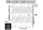
- the organoid obtained in Test Example 1-1 was transplanted into a mouse with a destroyed SCN, and the actogram obtained by measuring the presence or absence of movement of the mouse before and after transplantation is shown (Test Example 1-5).
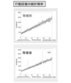
- the organoid obtained in Test Example 1-1 was transplanted into a mouse with a destroyed SCN, and the presence or absence of movement of the mouse was measured before and after transplantation, and a chi-square periodogram was obtained (Test Example 1-5).
- 1 shows a bright field image (Bright), a fluorescent image (indicating Six3 expression), and an autofluorescent image (Auto) of a cell mass on day 13 (Test Example 2).
- 1 shows a bright field image, a fluorescent image (indicating Six3 expression), and an autofluorescent image of a cell mass on day 48 (Test Example 3).
- a bright field image of the cell mass on day 13 is shown (Test Example 4).
- the present invention relates to a method for producing suprachiasmatic nucleus organoids (also referred to as the "production method of the present invention" in this specification ), which includes (1) a step of culturing pluripotent stem cells in an intermediate medium between an undifferentiated maintenance medium and a neural differentiation medium, (2) a step of culturing the cells obtained in step (1) in a neural differentiation medium, and (3) a step of performing an operation to form a cell mass before the start of step (2), in which (A) the number of raw cells seeded to form one cell mass is 6000 or more, and (B) if the start of step (2) is day 0, the medium contains a sonic hedgehog signaling pathway active substance from day 3 onward. This will be described below.
- Pluripotent stem cells are stem cells that have the ability to differentiate into all cell lineages belonging to the three germ layers (endoderm, mesoderm, ectoderm) (pluripotency) and also have the ability to proliferate.
- Pluripotent stem cells include, but are not limited to, embryonic stem (ES) cells, embryonic stem (ntES) cells derived from cloned embryos obtained by nuclear transplantation, spermatogonial stem cells (GS cells), embryonic germ cells (EG cells), induced pluripotent stem (iPS) cells, cultured fibroblasts, and pluripotent cells (Muse cells) derived from bone marrow stem cells.
- the preferred pluripotent stem cells are iPS cells, more preferably human iPS cells, from the viewpoint of being available without destroying embryos, eggs, etc. in the manufacturing process.
- iPS cells can be produced by introducing reprogramming factors into any somatic cell.
- reprogramming factors include genes or gene products such as Oct3/4, Sox2, Sox1, Sox3, Sox15, Sox17, Klf4, Klf2, c-Myc, N-Myc, L-Myc, Nanog, Lin28, Fbx15, ERas, ECAT15-2, Tcl1, beta-catenin, Lin28b, Sall1, Sall4, Esrrb, Nr5a2, Tbx3, or Glis1.
- These reprogramming factors may be used alone or in combination.
- Combinations of reprogramming factors include WO2007/069666, WO2008/118820, WO2009/007852, WO2009/032194, WO2009/058413, WO2009/057831, WO2009/075119, WO2009/079007, WO2009/091659, WO2009/101084, WO2009/101407, WO2009/102983, WO2009/114949, WO2009/117439, WO2009/126250, WO2009/126251, WO2009/ 126655, WO2009/157593, WO2010/009015, WO2010/033906, WO2010/033920, WO2010/042800, WO2010/050626, WO2010/056831, WO2010/068955, WO20 10/098419, WO2010/102267, WO2010/111409, WO2010/111422, WO2010/115050, WO2010/124290, WO2010/147395, WO2010/14761
- Somatic cells include, but are not limited to, fetal (baby) somatic cells, neonatal (baby) somatic cells, and mature healthy or diseased somatic cells, as well as primary culture cells, passaged cells, and established cell lines.
- somatic cells include, for example, (1) tissue stem cells (somatic stem cells) such as neural stem cells, hematopoietic stem cells, mesenchymal stem cells, and dental pulp stem cells, (2) tissue progenitor cells, and (3) differentiated cells such as blood cells (peripheral blood cells, umbilical cord blood cells, etc.), lymphocytes, epithelial cells, endothelial cells, muscle cells, fibroblasts (skin cells, etc.), hair cells, liver cells, gastric mucosa cells, intestinal cells, spleen cells, pancreatic cells (exocrine pancreatic cells, etc.), brain cells, lung cells, kidney cells, and adipocytes.
- tissue stem cells such as neural stem cells, hematopoietic stem cells,
- the intermediate medium is a medium that has the compositional characteristics of the undifferentiation maintenance medium used to maintain the undifferentiated state of pluripotent stem cells until subjected to step (1) and the compositional characteristics of the neural differentiation medium used in step (2), and is a medium that can acclimate pluripotent stem cells to a dedifferentiation environment (culture in neural differentiation medium). This "acclimation" can improve the efficiency of induction into suprachiasmatic nucleus organoids and the properties of the resulting suprachiasmatic nucleus organoids.
- An undifferentiated state maintenance medium is a medium capable of maintaining the undifferentiated state of pluripotent stem cells for a certain period of time (e.g., 3 days or more, 7 days or more, 14 days or more, or 28 days or more), and is not particularly limited thereto.
- the undifferentiated maintenance medium contains undifferentiated maintenance factors that have the effect of maintaining the undifferentiated state of pluripotent stem cells.
- factors for maintaining undifferentiated states include FGF-MAPK signal inhibitors (representatively, PD0325901), Wnt signal activators (GSK3 inhibitors, representatively, CHIR99021; other examples include Bromoindirubin-3'-oxime (BIO), Kenpaullone, etc.), LIF, BMP-4, 6-Bromoindirubin-3'-oxime, CDK8/19i, Emricasan, polyamines, Trans-ISRIB Chroman 1, Epiblastin A, GF 109203X, Go6983, IQ-1, PD173074, SU5402, PD184352, PD98059, SB202190, SB216763, sodium butyrate, SP600125, Surfen, U0126, WH-4-023, etc.
- the undifferentiated cell maintenance factor can be one type alone or a combination of two or
- the concentration of the undifferentiation maintenance factor in the undifferentiation maintenance medium is not particularly limited as long as it is a concentration that can maintain the undifferentiation state.
- the concentration of the FGF-MAPK signal inhibitor is preferably 0.2 to 4 ⁇ M, more preferably 0.5 to 2 ⁇ M, and the concentration of the Wnt signal activator is preferably 0.5 to 10 ⁇ M, more preferably 1.5 to 5 ⁇ M.
- the medium may be prepared as a basal medium that is used for culturing animal cells.
- basal media include Glasgow's Minimal Essential Medium (GMEM) medium, IMDM medium, Medium 199 medium, Eagle's Minimum Essential Medium (EMEM) medium, ⁇ MEM medium, Dulbecco's modified Eagle's Medium (DMEM) medium, Ham's F12 medium, RPMI 1640 medium, Fischer's medium, Neurobasal Medium (Life Technologies), and mixtures thereof.
- the medium may contain serum or may be serum-free.
- the medium may contain one or more serum substitutes, such as, for example, albumin, transferrin, Knockout Serum Replacement (KSR) (a serum substitute for FBS when culturing ES cells), N2 supplement (Invitrogen), B27 supplement (Invitrogen), fatty acids, insulin, collagen precursors, trace elements, 2-mercaptoethanol, 3'-thiolglycerol, and may also contain one or more substances, such as lipids, amino acids, L-glutamine, Glutamax (Invitrogen), non-essential amino acids, vitamins, growth factors, small molecules, antibiotics, antioxidants, pyruvate, buffers, and inorganic salts.
- serum substitutes such as, for example, albumin, transferrin, Knockout Serum Replacement (KSR) (a serum substitute for FBS when culturing ES cells), N2 supplement (Invitrogen), B27 supplement (Invitrogen), fatty acids, insulin, collagen precursors, trace elements, 2-mercaptoethanol, 3'-thio
- the undifferentiated state maintenance medium a commercially available medium prepared by adding necessary components can be used.
- examples of such medium include AK02N and AK03N of the StemFit (registered trademark) series.
- the neuronal differentiation medium used in step (2) is a medium that can release the undifferentiated state of cells and induce them to differentiate toward neurons, and is not particularly limited thereto.
- Neurodifferentiation medium does not contain the undifferentiated maintenance factors contained in undifferentiated maintenance medium. This allows the cells to be released from their undifferentiated state and induced to differentiate into neurons.
- the basic medium for the neural differentiation medium is IMDM medium, F12 medium, etc., and more preferably, a mixed medium of these.
- the neural differentiation medium is preferably a serum-free medium.
- the neural differentiation medium may contain a reducing agent, a serum replacement component, etc.
- the reducing agent is preferably thioglycerol, 2-mercaptoethanol, etc.
- the serum replacement component is preferably BSA, etc. More preferably, the medium contains a combination of a reducing agent and a serum replacement component.
- the concentration of the reducing agent (particularly thioglycerol) is preferably 100 to 1000 ⁇ M, more preferably 300 to 600 ⁇ M, and the concentration of BSA is preferably 1 to 20 mg/ml, more preferably 2 to 10 mg/ml.
- the neural differentiation medium does not contain insulin.
- the method of the present invention even if a neural differentiation medium that does not contain insulin is used, it can be applied to human cells.
- the intermediate medium is typically a medium in which the concentration of the undifferentiation maintenance factor is lower than the concentration in the undifferentiation maintenance medium.
- concentration of the undifferentiation maintenance factor in the intermediate medium is preferably lower, and can be, for example, 70/100 or less, 60/100 or less, 50/100 or less, 40/100 or less, 30/100 or less, 20/100 or less, 10/100 or less, 5/100 or less, 2/100 or less, or 1/100 or less of the concentration of the corresponding undifferentiation maintenance factor in the undifferentiation maintenance medium, and the intermediate medium can not contain the undifferentiation maintenance factor.
- the intermediate medium is preferably a medium having the same composition as the undifferentiation maintenance medium except that the concentration of the undifferentiation maintenance factor is reduced (the change in the concentration of the components is within ⁇ 20%, preferably within ⁇ 10%, more preferably within ⁇ 5%, and even more preferably within ⁇ 1%), or is a mixed medium of the undifferentiation maintenance medium and the neural differentiation medium.
- the concentration of the undifferentiated maintenance factor in the intermediate medium can be gradually reduced.
- the culture is preferably adhesion culture in which the cells are attached to the substrate of the culture vessel and cultured.
- adhesion culture can be performed by coating the substrate of the culture vessel with a cell adhesion molecule such as an extracellular matrix (specifically, for example, Matrigel (BD), type I collagen, type IV collagen, gelatin, laminin, heparan sulfate proteoglycan, or entactin, or a combination thereof) before using it for culture.
- a cell adhesion molecule such as an extracellular matrix (specifically, for example, Matrigel (BD), type I collagen, type IV collagen, gelatin, laminin, heparan sulfate proteoglycan, or entactin, or a combination thereof) before using it for culture.
- the culture temperature is not particularly limited, but is, for example, 30 to 40°C, preferably about 36 to 38°C, and the culture is performed in an atmosphere of CO2- containing air, and the CO2 concentration is preferably about 2 to 5%.
- the culture period is, for example, 1 to 7 days, preferably 1 to 5 days, more preferably 2 to 5 days, even more preferably 2 to 4 days, and even more preferably 3 to 4 days.
- ⁇ Step (2)> The neural differentiation medium is as described above in ⁇ Step (2)>.
- the cell mass formed in step (3) described below undergoes neural differentiation and begins to express suprachiasmatic nucleus markers such as Six3.
- the culture temperature is not particularly limited, but is, for example, 30 to 40°C, preferably about 36 to 38°C, and the culture is performed in an atmosphere of CO2- containing air, and the CO2 concentration is preferably about 2 to 5%.
- the culture period is, for example, 4 to 20 days, preferably 4 to 15 days, more preferably 5 to 15 days, even more preferably 5 to 13 days, and even more preferably 6 to 13 days.
- the medium contains a substance acting on the Sonic Hedgehog signaling pathway from day 3 (day 3) onward. That is, the medium (neural differentiation medium, or neuronal differentiation medium and intermediate medium) used during the period from any day before day 3 (start of addition) to any day after day 3 (end of addition) contains a substance acting on the Sonic Hedgehog signaling pathway. This can promote neuronal differentiation and improve the induction efficiency into suprachiasmatic nucleus organoids and the properties of the resulting suprachiasmatic nucleus organoids.
- a sonic hedgehog signaling pathway active substance (hereinafter sometimes referred to as Shh) is a substance that can enhance signal transduction mediated by Shh.
- Shh signaling pathway active substances include proteins belonging to the Hedgehog family (e.g., Shh and Ihh), Shh receptors, Shh receptor agonists, Purmorphamine, and SAG (Smoothened Agonist; 3-chloro-N-[trans-4-(methylamino)cyclohexyl]-N-[[3-(4-pyridinyl)phenyl]methyl]-benzo[b]thiophene-2-carboxamide).
- the Shh signaling pathway active substance is preferably SAG.
- the Sonic Hedgehog signaling promoting activity of SAG can be determined by methods well known to those skilled in the art, for example, a reporter gene assay focusing on the expression of the Gli1 gene (Oncogene (2007) 26, 5163-5168).
- the concentration of SAG in the medium is, for example, 0.001 ⁇ M or more, preferably 0.01 ⁇ M or more, more preferably 0.1 ⁇ M or more, even more preferably 0.2 ⁇ M or more, and even more preferably 0.5 ⁇ M or more.
- the concentration is preferably 0.2 to 5 ⁇ M, more preferably 0.5 to 2 ⁇ M, and even more preferably 0.7 to 1.5 ⁇ M.
- the start date for adding the sonic hedgehog signaling pathway active substance is preferably day-6 to day-3, more preferably day-4 to day-3, even more preferably day-2 to day-3, even more preferably day-0 to day-3, particularly preferably day-1 to day-3, even more preferably day-2 to day-3, and especially preferably day-2.
- the end date for adding the sonic hedgehog signaling pathway active substance is, for example, day 4 to day 100, preferably day 4 to day 20, more preferably day 4 to day 15, even more preferably day 5 to day 13, even more preferably day 6 to day 13, and particularly preferably the end of step (2).
- the timing of the operation for forming cell aggregates is not particularly limited as long as it is performed before the start of step (2).
- the timing is not particularly limited as long as it is performed between the start of step (1) and the start of step (2), but is preferably from day-6 to day 0, more preferably from day-5 to day 0, even more preferably from day-4 to day 0, and even more preferably from day-3 to day 0.
- the cells are detached from the culture substrate according to or in accordance with a known method, and the cells are dispersed in the medium by pipetting, etc.
- the procedure for forming cell aggregates is not particularly limited, so long as the cells are assembled to form cell aggregates with a diameter of 100 ⁇ m or more (preferably 200 ⁇ m or more, and particularly preferably 500 ⁇ m or more).
- Such procedures include seeding cells on a culture substrate and culturing them in suspension, and centrifuging the cell suspension, with the former being particularly preferred.
- the dispersed cells When cells are seeded on a culture substrate and cultured in suspension, the dispersed cells may be seeded in a relatively large culture vessel, such as a 10 cm dish, to simultaneously form multiple cell aggregates in one culture vessel; however, this will result in variation in the size of each aggregate. For example, if a certain number of dispersed cells are placed in a culture well (e.g., each well of a multi-well plate (U-bottom, V-bottom) such as a 96-well microplate) and cultured statically, the cells will rapidly aggregate to form a single aggregate in each well. By collecting these aggregates from multiple wells, a uniform population of aggregates can be obtained.
- a culture well e.g., each well of a multi-well plate (U-bottom, V-bottom) such as a 96-well microplate
- the culture vessel is preferably non-cell-adhesive to enable suspension culture.
- a non-cell-adhesive culture vessel one whose surface has not been artificially treated to improve adhesion to cells (for example, coating with basement membrane preparations, extracellular matrices such as laminin, entactin, collagen, gelatin, etc., or polymers such as polylysine, polyornithine, etc., or surface treatment such as positive charge treatment) can be used.
- a non-cell-adhesive culture vessel one whose surface has been artificially treated to reduce adhesion to cells (for example, superhydrophilic treatment with MPC polymer, low protein adsorption treatment, etc.) can be used.
- the time from the operation to form cell clusters to the formation of the cell clusters is preferably within 3 days, more preferably within 2 days, even more preferably within 1 day, and particularly preferably 12 to 24 hours.
- the conditions of the operation to form cell clusters e.g., the area and shape of the culture bottom surface
- suspension culture is carried out as is in steps (1) and (2).
- the number of raw cells seeded to form one cell mass in step (3) is 6000 or more. This promotes neural differentiation and improves the induction efficiency of the suprachiasmatic nucleus organoid and the properties of the obtained suprachiasmatic nucleus organoid.
- the starting cells are preferably 9,000 or more, more preferably 12,000 or more, even more preferably 15,000 or more, and even more preferably 20,000 or more. There is no particular upper limit, and it is, for example, 200,000, 100,000, or 80,000.
- the method of the present invention preferably further comprises (4) culturing the cell mass obtained in step (2) in a neural differentiation medium, thereby allowing the maturation of the suprachiasmatic nucleus organoid to be further promoted.
- Step (4) is preferably carried out under high-oxygen conditions. Specifically, in step (4), it is preferable to culture in an incubator with a high-oxygen concentration atmosphere. There are no particular limitations on the high-oxygen concentration as long as it is higher than the oxygen concentration in the atmosphere, but it is preferably 25 to 60%, more preferably 30 to 50%, and particularly preferably 35 to 45%.
- Step (4) can also be performed in semi-aerated culture.
- cell masses can be placed on Millicell or Transwell, with the bottom surface immersed in expansion medium or maturation medium, and the top surface exposed to air.
- Step (4) preferably includes a step of culturing using an expansion medium as the neural differentiation medium, and a step of culturing using a maturation medium as the neural differentiation medium.
- the basal medium for the expansion medium is DMEM medium, F12 medium, etc., and more preferably, a mixed medium of these.
- the expansion medium is preferably a serum-free medium.
- the neural differentiation medium contains a serum replacement component.
- a preferred example of the serum replacement component is KSR.
- the concentration of KSR is preferably 5-20%, more preferably 7-15%.
- the culture period for culturing in the expansion medium is, for example, 4 to 20 days, preferably 4 to 15 days, more preferably 5 to 15 days, even more preferably 5 to 13 days, and even more preferably 6 to 13 days.
- the basal medium for the maturation medium is DMEM medium, F12 medium, etc., and more preferably, a mixed medium thereof.
- the maturation medium is preferably a serum-free medium.
- the neural differentiation medium contains serum replacement components.
- the serum replacement components preferably include N2 supplement, B27 supplement, etc., and more preferably a combination of these.
- the concentration of the N2 supplement is preferably 0.2-5%, more preferably 0.5-2%, and the concentration of the B27 supplement is preferably 0.5-10%, more preferably 1-3%.
- the cultivation period for cultivation in maturation medium is, for example, 4 to 400 days.
- the cells can be cultured in a medium containing a substance that acts on the Sonic Hedgehog signaling pathway, or in a medium that does not contain a substance that acts on the Sonic Hedgehog signaling pathway.
- the manufacturing method of the present invention makes it possible to obtain suprachiasmatic nucleus organoids having a high proportion of suprachiasmatic nucleus cells.
- the proportion is, for example, 1% or more, preferably 5% or more, more preferably 10% or more, even more preferably 15% or more, and even more preferably 18% or more. There is no particular upper limit, and it is, for example, 50%, 40%, or 30%.
- the proportion can be measured according to Test Examples 1-3 described below.
- the manufacturing method of the present invention makes it possible to obtain suprachiasmatic nucleus organoids whose constituent cells are all brain tissue cells.
- brain tissue cells include excitatory nerves, inhibitory nerves, meninges, glial cells, etc.
- the constituent cells can be determined according to Test Examples 1-3 described below.
- the present invention relates to a method for screening circadian rhythm regulators, comprising: (X) contacting a suprachiasmatic nucleus organoid having a suprachiasmatic nucleus cell ratio of 5% or more (preferably 10% or more, more preferably 15% or more, even more preferably 18% or more) or a suprachiasmatic nucleus organoid obtained by the method of the present invention with a test substance; and (Y) evaluating the circadian rhythm of the suprachiasmatic nucleus organoid. This will be described below.
- test substances include cell extracts, cell culture supernatants, microbial fermentation products, extracts derived from marine organisms, plant extracts, purified or crude proteins, peptides, non-peptide compounds, synthetic low molecular weight compounds, and natural compounds.
- Test substances can also be obtained using any of the many approaches to combinatorial libraries known in the art, including (1) biological libraries, (2) synthetic library methods using deconvolution, (3) "one-bead one-compound” library methods, and (4) synthetic library methods using affinity chromatography selection. While the biological library method using affinity chromatography selection is limited to peptide libraries, the other four approaches are applicable to small molecule libraries of peptides, non-peptide oligomers, or compounds (Lam (1997) Anticancer Drug Des. 12: 145-67). Examples of methods for the synthesis of molecular libraries can be found in the art (DeWitt et al. (1993) Proc. Natl. Acad. Sci. USA 90: 6909-13; Erb et al.
- Step (X) can be carried out, for example, by adding a test substance to a culture medium containing the suprachiasmatic nucleus organoid.
- concentration of the test substance in the culture medium can be appropriately set depending on the type of test substance, etc.
- the period during which contact with the test substance is maintained is not particularly limited, but can be, for example, 1 hour to 14 days.
- Step (Y) can be carried out, for example, by measuring the expression level of clock genes in the suprachiasmatic nucleus organoids and evaluating the fluctuation, oscillation, or persistence thereof.
- the measurement and evaluation can be carried out according to or in accordance with a known method.
- step (Y) the evaluation of the circadian rhythm of the suprachiasmatic nucleus organoid in step (Y) can be performed, for example, by measuring the fluctuation, oscillation, or persistence of expression levels of receptors expressed in the suprachiasmatic nucleus organoid, as well as the responsiveness of those receptors (for example, responsiveness in the presence/absence of a test substance, or responsiveness to a ligand in the presence/absence of a test substance).
- receptors that are expressed by the suprachiasmatic nucleus organoid there are no particular limitations on the receptors that are expressed by the suprachiasmatic nucleus organoid, but the following receptors may be used in order of decreasing expression: Bsg, Rora, Prnp, Cd47, Gria2, Rack1, Nrxn1, Epha5, Thra, Ogfrl1, Rorb, Nrxn2, Gabbr1, Sqstm1, Atp6ap2, Ntrk2, Slc22a17, Grin2b, Nrp2, Vldlr, Gabrb3, Ed il3, Unc5c, Nr1d1, Adgrl1, Crlf2, Nlgn1, Tmem63b, Nlgn2, Grm5, Stat3, Nr2f2, Jmjd6, Bmpr2, Tm2d1, Gabrg3, Fz d3, Ntrk3, Gpr162, Nptxr, L1cam, Lrp1, Kit, Adgrb3,
- the evaluation of the circadian rhythm of the suprachiasmatic nucleus organoid in step (Y) can be performed by combining measuring the expression level of the clock gene and evaluating the fluctuation, oscillation, or persistence thereof with measuring the fluctuation, oscillation, or persistence of the expression level of the receptor expressed in the suprachiasmatic nucleus organoid, as well as the responsiveness of the receptor (e.g., responsiveness in the presence/absence of the test substance, or responsiveness to a ligand in the presence/absence of the test substance). For example, it can be performed by measuring the responsiveness of the receptor when a ligand is added in the fluctuation of the expression level of the clock gene.
- step (Y) If the evaluation results of step (Y) show a change in the circadian rhythm when compared with the evaluation results of a control sample under the same conditions except that the test substance is not used, the test substance can be selected as a circadian rhythm regulator (or a candidate substance).
- the selected circadian rhythm regulator can be used as an active ingredient in agents for treating, improving, or preventing diseases and disorders involving circadian rhythms.
- examples of such drugs include sleeping pills.
- Mouse ES cells (C57/B6N Per2::Luciferase cells (Scientific)) were cultured in a feeder-free adherent culture medium (Glasgow Minimum Essential Medium (GMEM, Gibco), 10% KnockOut TM Serum Replacement (KSR; Gibco), 1% foetal bovine serum (FBS), 1 mM sodium pyruvate (Gibco), 1 ⁇ MEM Non-Essential Amino Acids (Gibco), 100 ⁇ M ⁇ -mercaptoethanol (Wako, ⁇ -ME).
- GMEM Gasgow Minimum Essential Medium
- KSR KnockOut TM Serum Replacement
- FBS foetal bovine serum
- Nabco 1 mM sodium pyruvate
- Mibco 1 ⁇ MEM Non-Essential Amino Acids
- Wako, ⁇ -ME 100 ⁇ M ⁇ -mercaptoethanol
- the cells were harvested and seeded in a 96-well plate (U-bottom well) at 30,000 cells/well (Per2::Luciferase cells: used in the tests in Figures 1 to 4 and 7) or 10,000 cells/well (Six3::mVenus cells: used in the tests in Figures 5 to 6), and culture was initiated in neural differentiation medium (composition: Iscove's modified Dulbecco's medium/Ham's F-12 1:1, 1 ⁇ chemically defined lipid concentrate, penicillin/streptomycin, monothioglycerol (450 ⁇ M) and purified BSA (>99% purified by crystallization; Sigma (5 mg/ml)).) (Day 0).
- neural differentiation medium Composition: Iscove's modified Dulbecco's medium/Ham's F-12 1:1, 1 ⁇ chemically defined lipid concentrate, penicillin/streptomycin, monothioglycerol (450 ⁇ M) and purified BSA (>99% purified by crystallization; Sigma (5
- the cell clusters were collected and washed once with neural differentiation medium (expansion medium) (composition: DMEM/F-12, GlutaMAX TM supplement, KSR10%), then seeded on EZSphere or the like (culture dish), and suspension culture was started in the neural differentiation medium (expansion medium). Culture was performed in a multi-gas incubator with 40% O 2 and 5% CO 2 , and continued until Day 13. Medium was replaced every other day.
- the cell masses were collected and organoids were obtained.
- Test Example 1-1 The organoids (Day 72) obtained in Test Example 1-1 were immunostained with terminal differentiation markers of the suprachiasmatic nucleus (Six6, VIP, AVP). For comparison, the suprachiasmatic nucleus of an adult mouse was also immunostained in the same manner. Immunostaining was performed according to a known method.
- the specific method is as follows: Day 70 SCN Organoid 11 cell clumps were dispersed using a neuronal cell dispersion solution and large clumps were removed using Flowmi. After thorough washing, trypan blue was used to confirm that more than 80% of cells were viable, and a library was created using Chromium v3.1 kit (10x genomics) aiming for 5,000 cells. After that, sequencing was performed using Illumina Novaseq, followed by analysis using Cellranger and Seurat.
- the main populations were roughly half neurons and half glia, with the rest being ependyma, pia mater, and oligodendrocytes, covering a complete set of cells from the surface to the inside of the brain. There were also only seven populations of neurons, with only four being major populations.
- Test Example 1-1 The organoids obtained in Test Example 1-1 were subjected to clock gene expression analysis to evaluate the circadian oscillation sustainability.
- the specific method is as follows: SCN organoids prepared using Per2::Luciferase KI/KI cells were embedded in the bottom of a 35 mm Glass Bottom Dish using agarose gel, and the medium was replaced with phenol red-free maturation medium. From day 101, 500 uM Luciferin was added, and measurements were taken using a modified LCV 100 (Olympus) luminescence device with a 20X objective lens and a 0.5X magnification lens. The imaging conditions were 16 bit EM gain 500, Exposure 50 min, Bin 3, every hour.
- Test Example 1-5 Transplantation analysis> The organoid obtained in Test Example 1-1 was transplanted into a mouse in which the suprachiasmatic nucleus had been destroyed, and the presence or absence of movement of the mouse before and after transplantation was measured, and an actogram and a chi-square periodogram were created.
- SCN destruction and transplantation experiments were performed as described in previous literature (Sujino Curr Biol 2006, https://www.sciencedirect.com/science/article/pii/S0960982203002227).
- SCN organoids were transplanted in place of mouse fetal hypothalamus.
- Day 20 SCN organoids were placed in a Millicell upper half gas phase and cultured overnight in a CO2 incubator (5% CO2 20% O2). They were then divided into 3-4 pieces using ophthalmic scissors and transplanted into mice after SCN destruction as described in previous literature, and wheel running rhythm was analyzed.
- Test Example 2 Production of suprachiasmatic nucleus organoids 2
- the number of cells seeded on day 0 was 3300 cells/well, 6600 cells/well, or 9900 cells/well, and the cells were cultured in the same manner as in Test Example 1, except that a SAG-free group (DMSO-added group) was also obtained.
- Figure 5 shows a bright field image (Bright), a fluorescence image (showing Six3 expression), and an autofluorescence image (Auto) of the cell mass on day 13. It was found that if 6,000 or more raw cells were seeded to form one cell mass, the suprachiasmatic nucleus marker was well expressed in the organoid.
- Test Example 3 Production of suprachiasmatic nucleus organoids 3 Culture was performed in the same manner as in Test Example 1, except that SAG was added on Day 0, Day 0.5, Day 1.0, Day 2.0, Day 3.0, or Day 5.0, and a group to which SAG was not added (DMSO Day 0 addition group) was also obtained.
- Bright-field images, fluorescent images (showing Six3 expression), and autofluorescent images of cell clusters on day 48 are shown in Figure 6. It was found that if SAG was added before day 3, the suprachiasmatic nucleus marker was well expressed in the organoids.
- Test Example 4 Production of suprachiasmatic nucleus organoids 4
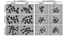
- the number of cells seeded on day 0 was 30,000 cells/well, 45,000 cells/well, or 60,000 cells/well, and a group was obtained in which undifferentiation maintenance medium was used instead of intermediate medium (2i(+) maintenance), and a group in which SAG was not added (DMSO-added group) was also obtained.
- the cells were cultured in the same manner as in Test Example 1.
- Figure 7 shows a bright field image of the cell clusters on day 13.
- the morphology of the cell clusters indicates that no differentiation occurred when the undifferentiation maintenance medium was used instead of the intermediate medium.
- Test Example 5 Production of suprachiasmatic nucleus organoids 5 Human iPS cells (201 B7) that had been cultured in a feeder-free adherent culture in undifferentiation maintenance medium (composition: AK02N, Y27632 (ROCK inhibitor) 1 ⁇ M) were seeded at 10,000 or 30,000 cells/well in a 96-well plate (U-bottom well) and cultured in undifferentiation maintenance medium plus SAG 1 ⁇ M (medium A). The next day, the entire medium was transferred to a dish containing neural differentiation medium (approximately equal amounts of SAG and Y27632). The next day, half of the medium was replaced with neural differentiation medium (Y27632 ⁇ SAG 1uM). The next day, half of the medium was replaced with neural differentiation medium (Y27632 ⁇ SAG 1uM). The next day, the entire medium was replaced with neural differentiation medium (Y27632 ⁇ SAG 1uM) (Day 0).
- undifferentiation maintenance medium composition: AK02N, Y27632
- the cell clusters were collected and washed once with neural differentiation medium (expansion medium) (composition: DMEM/F12 (Gibco 10565), KSR 10%), then seeded on EZSphere and suspension culture was started in the neural differentiation medium (expansion medium). Culture was performed in a multi-gas incubator with 40% O 2 and 5% CO 2 , and continued until day 23. Medium was changed every other day.
- the medium was changed to neural differentiation medium (maturation medium) (composition: DMEM/F12 (Gibco 10565), N2 supplements 1X, B27 (with Vitamine A) 1X), and suspension culture was continued. Culture continued in a multi-gas incubator with 40% O 2 and 5% CO 2. Medium was changed every other day.
- maturation medium composition: DMEM/F12 (Gibco 10565), N2 supplements 1X, B27 (with Vitamine A) 1X
Landscapes
- Health & Medical Sciences (AREA)
- Engineering & Computer Science (AREA)
- Life Sciences & Earth Sciences (AREA)
- Biomedical Technology (AREA)
- Immunology (AREA)
- Chemical & Material Sciences (AREA)
- Hematology (AREA)
- Cell Biology (AREA)
- Urology & Nephrology (AREA)
- Molecular Biology (AREA)
- Bioinformatics & Cheminformatics (AREA)
- Biotechnology (AREA)
- General Health & Medical Sciences (AREA)
- Microbiology (AREA)
- Biochemistry (AREA)
- Medicinal Chemistry (AREA)
- Food Science & Technology (AREA)
- Pathology (AREA)
- Toxicology (AREA)
- General Physics & Mathematics (AREA)
- Tropical Medicine & Parasitology (AREA)
- Neurology (AREA)
- Analytical Chemistry (AREA)
- Physics & Mathematics (AREA)
- Zoology (AREA)
- Genetics & Genomics (AREA)
- Wood Science & Technology (AREA)
- Neurosurgery (AREA)
- Organic Chemistry (AREA)
- General Engineering & Computer Science (AREA)
- Micro-Organisms Or Cultivation Processes Thereof (AREA)
Priority Applications (3)
| Application Number | Priority Date | Filing Date | Title |
|---|---|---|---|
| JP2025524124A JPWO2024248023A1 (https=) | 2023-05-29 | 2024-05-29 | |
| CN202480035536.9A CN121443723A (zh) | 2023-05-29 | 2024-05-29 | 视交叉上核类器官的制造方法 |
| EP24813101.3A EP4722343A1 (en) | 2023-05-29 | 2024-05-29 | Method for producing suprachiasmatic nucleus organoid |
Applications Claiming Priority (2)
| Application Number | Priority Date | Filing Date | Title |
|---|---|---|---|
| JP2023-087986 | 2023-05-29 | ||
| JP2023087986 | 2023-05-29 |
Publications (1)
| Publication Number | Publication Date |
|---|---|
| WO2024248023A1 true WO2024248023A1 (ja) | 2024-12-05 |
Family
ID=93658083
Family Applications (1)
| Application Number | Title | Priority Date | Filing Date |
|---|---|---|---|
| PCT/JP2024/019660 Ceased WO2024248023A1 (ja) | 2023-05-29 | 2024-05-29 | 視交叉上核オルガノイドの製造方法 |
Country Status (4)
| Country | Link |
|---|---|
| EP (1) | EP4722343A1 (https=) |
| JP (1) | JPWO2024248023A1 (https=) |
| CN (1) | CN121443723A (https=) |
| WO (1) | WO2024248023A1 (https=) |
Citations (36)
| Publication number | Priority date | Publication date | Assignee | Title |
|---|---|---|---|---|
| US5223409A (en) | 1988-09-02 | 1993-06-29 | Protein Engineering Corp. | Directed evolution of novel binding proteins |
| US20020103360A1 (en) | 1998-09-01 | 2002-08-01 | Yang Pan | Novel protein related to melanoma-inhibiting protein and uses thereof |
| WO2007069666A1 (ja) | 2005-12-13 | 2007-06-21 | Kyoto University | 核初期化因子 |
| WO2008118820A2 (en) | 2007-03-23 | 2008-10-02 | Wisconsin Alumni Research Foundation | Somatic cell reprogramming |
| WO2009007852A2 (en) | 2007-06-15 | 2009-01-15 | Izumi Bio, Inc | Multipotent/pluripotent cells and methods |
| WO2009032194A1 (en) | 2007-08-31 | 2009-03-12 | Whitehead Institute For Biomedical Research | Wnt pathway stimulation in reprogramming somatic cells |
| WO2009058413A1 (en) | 2007-10-29 | 2009-05-07 | Shi-Lung Lin | Generation of human embryonic stem-like cells using intronic rna |
| WO2009057831A1 (ja) | 2007-10-31 | 2009-05-07 | Kyoto University | 核初期化方法 |
| WO2009075119A1 (ja) | 2007-12-10 | 2009-06-18 | Kyoto University | 効率的な核初期化方法 |
| WO2009079007A1 (en) | 2007-12-17 | 2009-06-25 | Gliamed, Inc. | Stem-like cells and method for reprogramming adult mammalian somatic cells |
| WO2009091659A2 (en) | 2008-01-16 | 2009-07-23 | Shi-Lung Lin | Generation of tumor-free embryonic stem-like pluripotent cells using inducible recombinant rna agents |
| WO2009102983A2 (en) | 2008-02-15 | 2009-08-20 | President And Fellows Of Harvard College | Efficient induction of pluripotent stem cells using small molecule compounds |
| WO2009101407A2 (en) | 2008-02-11 | 2009-08-20 | Cambridge Enterprise Limited | Improved reprogramming of mammalian cells, and the cells obtained |
| WO2009101084A1 (en) | 2008-02-13 | 2009-08-20 | Fondazione Telethon | Method for reprogramming differentiated cells |
| WO2009114949A1 (en) | 2008-03-20 | 2009-09-24 | UNIVERSITé LAVAL | Methods for deprogramming somatic cells and uses thereof |
| WO2009117439A2 (en) | 2008-03-17 | 2009-09-24 | The Scripps Research Institute | Combined chemical and genetic approaches for generation of induced pluripotent stem cells |
| WO2009126250A2 (en) | 2008-04-07 | 2009-10-15 | Nupotential, Inc. | Reprogramming a cell by inducing a pluripotent gene through rna interference |
| WO2009157593A1 (en) | 2008-06-27 | 2009-12-30 | Kyoto University | Method of efficiently establishing induced pluripotent stem cells |
| WO2010009015A2 (en) | 2008-07-14 | 2010-01-21 | Oklahoma Medical Research Foundation | Production of pluripotent cells through inhibition of bright/arid3a function |
| WO2010033906A2 (en) | 2008-09-19 | 2010-03-25 | President And Fellows Of Harvard College | Efficient induction of pluripotent stem cells using small molecule compounds |
| WO2010033920A2 (en) | 2008-09-19 | 2010-03-25 | Whitehead Institute For Biomedical Research | Compositions and methods for enhancing cell reprogramming |
| WO2010042800A1 (en) | 2008-10-10 | 2010-04-15 | Nevada Cancer Institute | Methods of reprogramming somatic cells and methods of use for such cells |
| WO2010050626A1 (en) | 2008-10-30 | 2010-05-06 | Kyoto University | Method for producing induced pluripotent stem cells |
| WO2010056831A2 (en) | 2008-11-12 | 2010-05-20 | Nupotential, Inc. | Reprogramming a cell by inducing a pluripotent gene through use of an hdac modulator |
| WO2010068955A2 (en) | 2008-12-13 | 2010-06-17 | Dna Microarray | MICROENVIRONMENT NICHE ASSAY FOR CiPS SCREENING |
| WO2010098419A1 (en) | 2009-02-27 | 2010-09-02 | Kyoto University | Novel nuclear reprogramming substance |
| WO2010102267A2 (en) | 2009-03-06 | 2010-09-10 | Ipierian, Inc. | Tgf-beta pathway inhibitors for enhancement of cellular reprogramming of human cells |
| WO2010111409A2 (en) | 2009-03-25 | 2010-09-30 | The Salk Institute For Biological Studies | Pluripotent stem cells |
| WO2010111422A2 (en) | 2009-03-25 | 2010-09-30 | The Salk Institute For Biological Studies | Induced pluripotent stem cell generation using two factors and p53 inactivation |
| WO2010115050A2 (en) | 2009-04-01 | 2010-10-07 | The Regents Of The University Of California | Embryonic stem cell specific micrornas promote induced pluripotency |
| WO2010124290A2 (en) | 2009-04-24 | 2010-10-28 | Whitehead Institute For Biomedical Research | Compositions and methods for deriving or culturing pluripotent cells |
| WO2010147395A2 (en) | 2009-06-16 | 2010-12-23 | Korea Research Institute Of Bioscience And Biotechnology | Medium composition comprising neuropeptide y for the generation, maintenance, prologned undifferentiated growth of pluripotent stem cells and method of culturing pluripotent stem cell using the same |
| WO2010147612A1 (en) | 2009-06-18 | 2010-12-23 | Lixte Biotechnology, Inc. | Methods of modulating cell regulation by inhibiting p53 |
| WO2013065763A1 (ja) * | 2011-10-31 | 2013-05-10 | 独立行政法人理化学研究所 | 幹細胞の培養方法 |
| JP2015110564A (ja) * | 2013-11-08 | 2015-06-18 | マルハニチロ株式会社 | 概日リズム改善剤 |
| WO2017126551A1 (ja) * | 2016-01-22 | 2017-07-27 | 国立大学法人名古屋大学 | ヒト多能性幹細胞から視床下部ニューロンへの分化誘導 |
-
2024
- 2024-05-29 JP JP2025524124A patent/JPWO2024248023A1/ja active Pending
- 2024-05-29 CN CN202480035536.9A patent/CN121443723A/zh active Pending
- 2024-05-29 EP EP24813101.3A patent/EP4722343A1/en active Pending
- 2024-05-29 WO PCT/JP2024/019660 patent/WO2024248023A1/ja not_active Ceased
Patent Citations (40)
| Publication number | Priority date | Publication date | Assignee | Title |
|---|---|---|---|---|
| US5223409A (en) | 1988-09-02 | 1993-06-29 | Protein Engineering Corp. | Directed evolution of novel binding proteins |
| US5403484A (en) | 1988-09-02 | 1995-04-04 | Protein Engineering Corporation | Viruses expressing chimeric binding proteins |
| US5571698A (en) | 1988-09-02 | 1996-11-05 | Protein Engineering Corporation | Directed evolution of novel binding proteins |
| US20020103360A1 (en) | 1998-09-01 | 2002-08-01 | Yang Pan | Novel protein related to melanoma-inhibiting protein and uses thereof |
| WO2007069666A1 (ja) | 2005-12-13 | 2007-06-21 | Kyoto University | 核初期化因子 |
| WO2008118820A2 (en) | 2007-03-23 | 2008-10-02 | Wisconsin Alumni Research Foundation | Somatic cell reprogramming |
| WO2009007852A2 (en) | 2007-06-15 | 2009-01-15 | Izumi Bio, Inc | Multipotent/pluripotent cells and methods |
| WO2009032194A1 (en) | 2007-08-31 | 2009-03-12 | Whitehead Institute For Biomedical Research | Wnt pathway stimulation in reprogramming somatic cells |
| WO2009058413A1 (en) | 2007-10-29 | 2009-05-07 | Shi-Lung Lin | Generation of human embryonic stem-like cells using intronic rna |
| WO2009057831A1 (ja) | 2007-10-31 | 2009-05-07 | Kyoto University | 核初期化方法 |
| WO2009075119A1 (ja) | 2007-12-10 | 2009-06-18 | Kyoto University | 効率的な核初期化方法 |
| WO2009079007A1 (en) | 2007-12-17 | 2009-06-25 | Gliamed, Inc. | Stem-like cells and method for reprogramming adult mammalian somatic cells |
| WO2009091659A2 (en) | 2008-01-16 | 2009-07-23 | Shi-Lung Lin | Generation of tumor-free embryonic stem-like pluripotent cells using inducible recombinant rna agents |
| WO2009101407A2 (en) | 2008-02-11 | 2009-08-20 | Cambridge Enterprise Limited | Improved reprogramming of mammalian cells, and the cells obtained |
| WO2009101084A1 (en) | 2008-02-13 | 2009-08-20 | Fondazione Telethon | Method for reprogramming differentiated cells |
| WO2009102983A2 (en) | 2008-02-15 | 2009-08-20 | President And Fellows Of Harvard College | Efficient induction of pluripotent stem cells using small molecule compounds |
| WO2009117439A2 (en) | 2008-03-17 | 2009-09-24 | The Scripps Research Institute | Combined chemical and genetic approaches for generation of induced pluripotent stem cells |
| WO2009114949A1 (en) | 2008-03-20 | 2009-09-24 | UNIVERSITé LAVAL | Methods for deprogramming somatic cells and uses thereof |
| WO2009126655A2 (en) | 2008-04-07 | 2009-10-15 | Nupotential, Inc. | Reprogramming a cell by inducing a pluripotent gene through use of a small molecule modulator |
| WO2009126251A2 (en) | 2008-04-07 | 2009-10-15 | Nupotential, Inc. | Reprogramming a cell by inducing a pluripotent gene through use of an hdac modulator |
| WO2009126250A2 (en) | 2008-04-07 | 2009-10-15 | Nupotential, Inc. | Reprogramming a cell by inducing a pluripotent gene through rna interference |
| WO2009157593A1 (en) | 2008-06-27 | 2009-12-30 | Kyoto University | Method of efficiently establishing induced pluripotent stem cells |
| WO2010009015A2 (en) | 2008-07-14 | 2010-01-21 | Oklahoma Medical Research Foundation | Production of pluripotent cells through inhibition of bright/arid3a function |
| WO2010033906A2 (en) | 2008-09-19 | 2010-03-25 | President And Fellows Of Harvard College | Efficient induction of pluripotent stem cells using small molecule compounds |
| WO2010033920A2 (en) | 2008-09-19 | 2010-03-25 | Whitehead Institute For Biomedical Research | Compositions and methods for enhancing cell reprogramming |
| WO2010042800A1 (en) | 2008-10-10 | 2010-04-15 | Nevada Cancer Institute | Methods of reprogramming somatic cells and methods of use for such cells |
| WO2010050626A1 (en) | 2008-10-30 | 2010-05-06 | Kyoto University | Method for producing induced pluripotent stem cells |
| WO2010056831A2 (en) | 2008-11-12 | 2010-05-20 | Nupotential, Inc. | Reprogramming a cell by inducing a pluripotent gene through use of an hdac modulator |
| WO2010068955A2 (en) | 2008-12-13 | 2010-06-17 | Dna Microarray | MICROENVIRONMENT NICHE ASSAY FOR CiPS SCREENING |
| WO2010098419A1 (en) | 2009-02-27 | 2010-09-02 | Kyoto University | Novel nuclear reprogramming substance |
| WO2010102267A2 (en) | 2009-03-06 | 2010-09-10 | Ipierian, Inc. | Tgf-beta pathway inhibitors for enhancement of cellular reprogramming of human cells |
| WO2010111409A2 (en) | 2009-03-25 | 2010-09-30 | The Salk Institute For Biological Studies | Pluripotent stem cells |
| WO2010111422A2 (en) | 2009-03-25 | 2010-09-30 | The Salk Institute For Biological Studies | Induced pluripotent stem cell generation using two factors and p53 inactivation |
| WO2010115050A2 (en) | 2009-04-01 | 2010-10-07 | The Regents Of The University Of California | Embryonic stem cell specific micrornas promote induced pluripotency |
| WO2010124290A2 (en) | 2009-04-24 | 2010-10-28 | Whitehead Institute For Biomedical Research | Compositions and methods for deriving or culturing pluripotent cells |
| WO2010147395A2 (en) | 2009-06-16 | 2010-12-23 | Korea Research Institute Of Bioscience And Biotechnology | Medium composition comprising neuropeptide y for the generation, maintenance, prologned undifferentiated growth of pluripotent stem cells and method of culturing pluripotent stem cell using the same |
| WO2010147612A1 (en) | 2009-06-18 | 2010-12-23 | Lixte Biotechnology, Inc. | Methods of modulating cell regulation by inhibiting p53 |
| WO2013065763A1 (ja) * | 2011-10-31 | 2013-05-10 | 独立行政法人理化学研究所 | 幹細胞の培養方法 |
| JP2015110564A (ja) * | 2013-11-08 | 2015-06-18 | マルハニチロ株式会社 | 概日リズム改善剤 |
| WO2017126551A1 (ja) * | 2016-01-22 | 2017-07-27 | 国立大学法人名古屋大学 | ヒト多能性幹細胞から視床下部ニューロンへの分化誘導 |
Non-Patent Citations (38)
| Title |
|---|
| CARELL ET AL., ANGEW. CHEM. INT. ED. ENGL., vol. 33, 1994, pages 2061 |
| CHO ET AL., SCIENCE, vol. 261, 1993, pages 1303 - 5 |
| CULL ET AL., PROC. NATL. ACAD. SCI. USA, vol. 89, 1992, pages 1865 - 9 |
| CWIRLA ET AL., PROC. NATL. ACAD. SCI. USA, vol. 87, 1990, pages 6378 - 82 |
| DEWITT ET AL., PROC. NATL. ACAD. SCI. USA, vol. 90, 1993, pages 6909 - 13 |
| EMINLI S ET AL., STEM CELLS, vol. 26, 2008, pages 2467 - 2474 |
| ERB ET AL., PROC. NATL. ACAD. SCI. USA, vol. 91, 1994, pages 11422 - 6 |
| FELICI, J. MOL. BIOL., vol. 222, 1991, pages 301 - 10 |
| FENG B ET AL., NAT. CELL BIOL., vol. 11, 2009, pages 197 - 203 |
| FODOR, NATURE, vol. 364, 1993, pages 555 - 6 |
| HAN J, NATURE, vol. 463, 2010, pages 1096 - 100 |
| HENG J. C. ET AL., CELL STEM CELL, vol. 6, 2010, pages 167 - 74 |
| HIROYUKI TAMIYA; HIDEKI UKAI; JIN INOKAWA: "BP1-001, Research and Development on Clock Center Organoids Contributing to Circadian Rhythm Drug Discovery Research.", THE 45TH ANNUAL MEETING OF JAPANESE SOCIETY OF SLEEP RESEARCH / THE 30TH ANNUAL MEETING OF JAPANESE SOCIETY FOR CHRONOBIOLOGY; SEPTEMBER 15 - 17, 2023, JAPANESE SOCIETY OF SLEEP RESEARCH; JAPANESE SOCIETY FOR CHRONOBIOLOGY, JP, vol. 45, 28 November 2023 (2023-11-28) - 17 September 2023 (2023-09-17), JP, pages 268, XP009559234 * |
| HOUGHTEN, BIO/TECHNIQUES, vol. 13, 1992, pages 412 - 21 |
| HUANGFU D ET AL., NAT. BIOTECHNOL., vol. 26, 2008, pages 1269 - 1275 |
| ICHIDA J. K. ET AL., CELL STEM CELL, vol. 5, 2009, pages 491 - 503 |
| KIM J. B. ET AL., NATURE, vol. 461, 2009, pages 649 - 643 |
| LAM, ANTICANCER DRUG DES, vol. 12, 1997, pages 145 - 67 |
| LAM, NATURE, vol. 354, 1991, pages 82 - 4 |
| LEE SUENGWON, HONG CHRISTIAN I.: "Organoids as Model Systems to Investigate Circadian Clock-Related Diseases and Treatments", FRONTIERS IN GENETICS, FRONTIERS RESEARCH FOUNDATION, SWITZERLAND, vol. 13, Switzerland , XP093244757, ISSN: 1664-8021, DOI: 10.3389/fgene.2022.874288 * |
| LYSSIOTIS C. A. ET AL., PROC NATL ACAD SCI USA., vol. 106, 2009, pages 8912 - 8917 |
| MAEKAWA M ET AL., NATURE, vol. 474, 2011, pages 225 - 9 |
| MALI P ET AL., STEM CELLS, vol. 28, 2010, pages 713 - 720 |
| NATURE COMMUNICATIONS, vol. 8, no. 1339, 2017 |
| NATURE PROTOCOLS, vol. 12, 2017, pages 2513 - 2530 |
| NATURE, vol. 480, no. 7375, 9 November 2011 (2011-11-09), pages 57 - 62 |
| ONCOGENE, vol. 26, 2007, pages 5163 - 5168 |
| PROC NATL ACAD SCI USA., vol. 105, no. 33, 19 August 2008 (2008-08-19), pages 11796 - 801 |
| R. L. JUDSON ET AL., NAT. BIOTECHNOL., vol. 27, 2009, pages 459 - 461 |
| ROMANOV, NATURE, 2020 |
| SCIENTIFIC REPORTS, vol. 6, no. 32769, 2016 |
| SCOTTSMITH, SCIENCE, vol. 249, 1990, pages 404 - 90 |
| SUJINO, CURR BIOL, 2006, Retrieved from the Internet <URL:https://www.sciencedirect.com/science/article/pii/S0960982203002227> |
| TAMIYA HIROYUKI, OGAWA SUMITO, OUCHI YASUYOSHI, AKISHITA MASAHIRO: "Rigid Cooperation of Per1 and Per2 proteins", SCIENTIFIC REPORTS, NATURE PUBLISHING GROUP, US, vol. 6, no. 1, US , XP093244751, ISSN: 2045-2322, DOI: 10.1038/srep32769 * |
| TAMIYA, HIROYUKI: "Brain Organoid Analysis of Sleep/wake Cycles.", KAKEN — RESEARCH PROJECTS | 2019 FISCAL YEAR ANNUAL RESEARCH REPORT (KAKENHI-PROJECT-18J00052), 27 January 2021 (2021-01-27), XP093244748, Retrieved from the Internet <URL:https://kaken.nii.ac.jp/report/KAKENHI-PROJECT-18J00052/18J000522019jisseki/> * |
| TU HAI-QING, LI SEN, XU YU-LING, ZHANG YU-CHENG, JIAN XIAO-XIAO, SONG GUANG-PING, WU MIN, SONG ZENG-QING, HU HUAI-BIN, LI PEI-YAO,: "Rhythmic Cilium in SCN Neuron is a Gatekeeper for the Intrinsic Circadian Clock", BIORXIV, 1 February 2022 (2022-02-01), XP093244760, DOI: 10.1101/2022.01.26.477948 * |
| ZHAO Y ET AL., CELL STEM CELL, vol. 3, 2008, pages 132 - 135 |
| ZUCKERMANN ET AL., J. MED. CHEM., vol. 37, 1994, pages 1233 - 51 |
Also Published As
| Publication number | Publication date |
|---|---|
| CN121443723A (zh) | 2026-01-30 |
| JPWO2024248023A1 (https=) | 2024-12-05 |
| EP4722343A1 (en) | 2026-04-08 |
Similar Documents
| Publication | Publication Date | Title |
|---|---|---|
| AU2018297258B2 (en) | Multiplexed receptor-ligand interaction screens | |
| Doss et al. | Global transcriptome analysis of murine embryonic stem cell-derived cardiomyocytes | |
| Kim et al. | Biological importance of OCT transcription factors in reprogramming and development | |
| CA3226106A1 (en) | Systems and methods for regulating target genes | |
| US20190233795A1 (en) | Transcription factors controlling differentiation of stem cells | |
| US20110033931A1 (en) | De-differentiation of human cells | |
| WO2011014740A2 (en) | Methods and composition for identifying and validating modulators of cell fate | |
| US20180265934A1 (en) | Coincidence reporter gene system | |
| EP4722343A1 (en) | Method for producing suprachiasmatic nucleus organoid | |
| Amisten | Quantification of the mRNA expression of G protein-coupled receptors in human adipose tissue | |
| JP2016028609A (ja) | 罹患組織における遺伝的変異の同定 | |
| Valetdinova et al. | Generation of two spinal muscular atrophy (SMA) type I patient-derived induced pluripotent stem cell (iPSC) lines and two SMA type II patient-derived iPSC lines | |
| Meyer et al. | Arp2/3 complex activity enables nuclear YAP for naïve pluripotency of human embryonic stem cells | |
| Manhas et al. | Generation of two iPSC lines from hypertrophic cardiomyopathy patients carrying MYBPC3 and PRKAG2 variants | |
| Mondéjar-Parreño et al. | Generation of three heterozygous KCNH2 mutation-carrying human induced pluripotent stem cell lines for modeling LQT2 syndrome | |
| Janz et al. | CRISPR/Cas9-edited PKP2 knock-out (JMUi001-A-2) and DSG2 knock-out (JMUi001-A-3) iPSC lines as an isogenic human model system for arrhythmogenic cardiomyopathy (ACM) | |
| Caudal et al. | Generation of human induced pluripotent stem cell lines carrying heterozygous PLN mutation from dilated cardiomyopathy patients | |
| Wang et al. | Establishment of induced pluripotent stem cell lines from an ARVC patient carrying a heterozygous variant in LAMA2 gene | |
| US20250099508A1 (en) | Systems, cell lines and methods of producing and using the same | |
| Yutong et al. | Comparison of human-induced pluripotent stem cell-derived macrophages with peripheral blood-derived macrophages using single-cell genomics | |
| Lee et al. | Generation of three iPSC lines from dilated cardiomyopathy patients carrying a pathogenic LMNA variant | |
| Fang et al. | Generation of human induced pluripotent stem cell line from peripheral blood mononuclear cells from an activated phosphoinositide 3-kinase δ syndrome patient | |
| Li et al. | Establishment of induced pluripotent stem cell line (ZZUi033-A) of a male with a novel L1CAM missense mutation | |
| Duff et al. | Generation of induced pluripotent stem cells (UCLi024-A) from a patient with argininosuccinate lyase deficiency carrying a homozygous c. 437G> A (p. Arg146Gln) mutation | |
| Celiker et al. | Generation of human induced pluripotent stem cell lines from patients with a RYR2 gene variant c. 14201A> G (p. Y4734C): Implications for idiopathic ventricular fibrillation and catecholaminergic polymorphic ventricular tachycardia |
Legal Events
| Date | Code | Title | Description |
|---|---|---|---|
| 121 | Ep: the epo has been informed by wipo that ep was designated in this application |
Ref document number: 24813101 Country of ref document: EP Kind code of ref document: A1 |
|
| ENP | Entry into the national phase |
Ref document number: 2025524124 Country of ref document: JP Kind code of ref document: A |
|
| NENP | Non-entry into the national phase |
Ref country code: DE |
|
| WWE | Wipo information: entry into national phase |
Ref document number: 2024813101 Country of ref document: EP |
|
| ENP | Entry into the national phase |
Ref document number: 2024813101 Country of ref document: EP Effective date: 20260102 |
|
| ENP | Entry into the national phase |
Ref document number: 2024813101 Country of ref document: EP Effective date: 20260102 |
|
| ENP | Entry into the national phase |
Ref document number: 2024813101 Country of ref document: EP Effective date: 20260102 |
|
| ENP | Entry into the national phase |
Ref document number: 2024813101 Country of ref document: EP Effective date: 20260102 |
|
| ENP | Entry into the national phase |
Ref document number: 2024813101 Country of ref document: EP Effective date: 20260102 |
|
| ENP | Entry into the national phase |
Ref document number: 2024813101 Country of ref document: EP Effective date: 20260102 |
|
| WWP | Wipo information: published in national office |
Ref document number: 2024813101 Country of ref document: EP |