WO2024203272A1 - 細胞外小胞分離用粉体 - Google Patents
細胞外小胞分離用粉体 Download PDFInfo
- Publication number
- WO2024203272A1 WO2024203272A1 PCT/JP2024/009510 JP2024009510W WO2024203272A1 WO 2024203272 A1 WO2024203272 A1 WO 2024203272A1 JP 2024009510 W JP2024009510 W JP 2024009510W WO 2024203272 A1 WO2024203272 A1 WO 2024203272A1
- Authority
- WO
- WIPO (PCT)
- Prior art keywords
- powder
- exosomes
- pores
- inorganic particles
- extracellular vesicles
- Prior art date
- Legal status (The legal status is an assumption and is not a legal conclusion. Google has not performed a legal analysis and makes no representation as to the accuracy of the status listed.)
- Ceased
Links
Classifications
-
- B—PERFORMING OPERATIONS; TRANSPORTING
- B01—PHYSICAL OR CHEMICAL PROCESSES OR APPARATUS IN GENERAL
- B01J—CHEMICAL OR PHYSICAL PROCESSES, e.g. CATALYSIS OR COLLOID CHEMISTRY; THEIR RELEVANT APPARATUS
- B01J20/00—Solid sorbent compositions or filter aid compositions; Sorbents for chromatography; Processes for preparing, regenerating or reactivating thereof
- B01J20/02—Solid sorbent compositions or filter aid compositions; Sorbents for chromatography; Processes for preparing, regenerating or reactivating thereof comprising inorganic material
- B01J20/06—Solid sorbent compositions or filter aid compositions; Sorbents for chromatography; Processes for preparing, regenerating or reactivating thereof comprising inorganic material comprising oxides or hydroxides of metals not provided for in group B01J20/04
-
- B—PERFORMING OPERATIONS; TRANSPORTING
- B01—PHYSICAL OR CHEMICAL PROCESSES OR APPARATUS IN GENERAL
- B01J—CHEMICAL OR PHYSICAL PROCESSES, e.g. CATALYSIS OR COLLOID CHEMISTRY; THEIR RELEVANT APPARATUS
- B01J20/00—Solid sorbent compositions or filter aid compositions; Sorbents for chromatography; Processes for preparing, regenerating or reactivating thereof
- B01J20/28—Solid sorbent compositions or filter aid compositions; Sorbents for chromatography; Processes for preparing, regenerating or reactivating thereof characterised by their form or physical properties
-
- C—CHEMISTRY; METALLURGY
- C12—BIOCHEMISTRY; BEER; SPIRITS; WINE; VINEGAR; MICROBIOLOGY; ENZYMOLOGY; MUTATION OR GENETIC ENGINEERING
- C12M—APPARATUS FOR ENZYMOLOGY OR MICROBIOLOGY; APPARATUS FOR CULTURING MICROORGANISMS FOR PRODUCING BIOMASS, FOR GROWING CELLS OR FOR OBTAINING FERMENTATION OR METABOLIC PRODUCTS, i.e. BIOREACTORS OR FERMENTERS
- C12M1/00—Apparatus for enzymology or microbiology
- C12M1/26—Inoculator or sampler
Definitions
- This disclosure relates to a powder for separating extracellular vesicles.
- Exosomes extracellular vesicles
- DDS drug delivery systems
- Exosomes are known to efficiently mediate intercellular communication and signal transduction, including the transport of biologically active molecules, such as proteins, lipids, and nucleic acids, between cells and across biological barriers. Exosomes are not easily degraded and can transmit signals over long distances without dilution, making them promising as drug delivery systems (DDS) and therapeutic drugs.
- DDS drug delivery systems
- a separation method that can reduce the inclusion of unintended exosomes and impurities is required.
- Various methods for separating and recovering extracellular vesicles have been reported.
- Patent Document 1 discloses a method for recovering extracellular vesicles, which comprises contacting a dispersion containing extracellular vesicles having an average particle size of 10 nm to 700 nm with a porous substrate made of a porous material, capturing and separating the extracellular vesicles from the dispersion on the porous substrate, and then recovering the captured extracellular vesicles from the porous substrate.
- Patent Document 2 discloses a method for isolating biological particles from a liquid sample, the method comprising the steps of: a) obtaining a liquid sample from a subject or a cell culture; b) contacting the liquid sample with a crystal/precipitation-inducing agent under conditions suitable for allowing crystal formation and/or precipitation, thereby producing a mixture; c) incubating the mixture for a time sufficient to allow crystal formation and/or precipitation; and d) separating the mixture to obtain a particle fraction containing biological particles, thereby isolating the biological particles from the liquid sample.
- An object of one embodiment of the present disclosure is to provide a powder for separating extracellular vesicles that can selectively and efficiently separate and recover exosomes, which are extracellular vesicles, from a liquid containing exosomes.
- a powder for separating extracellular vesicles comprising a plurality of inorganic particles, The inorganic particles have pores
- a powder for separating extracellular vesicles wherein the average pore diameter of the pores, as measured by mercury intrusion porosimetry, is in the range of 50 nm or more and 700 nm or less, and the standard deviation of the pore diameters of the pores is in the range of 1 nm or more and 200 nm or less.
- the powder for separating extracellular vesicles according to [1] wherein the pores have a pore volume of 0.15 mL/g or more.
- a powder for separating extracellular vesicles that can selectively and efficiently separate and recover exosomes, which are extracellular vesicles, from a liquid that contains exosomes.
- a numerical range indicated using “to” means a range that includes the numerical values before and after “to” as the lower and upper limits, respectively.
- the upper or lower limit value described in a certain numerical range may be replaced with the upper or lower limit value of another numerical range described in the present disclosure.
- the upper or lower limit value described in a certain numerical range may be replaced with a value shown in the examples.
- process includes not only independent processes, but also processes that cannot be clearly distinguished from other processes, as long as the intended purpose of the process is achieved.
- Extracellular vesicles are vesicles that exist outside cells and are classified into exosomes, microvesicles, and apoptotic bodies based on their size. Exosomes and microvesicles contain proteins, nucleic acids such as mRNA and miRNA, and apoptotic bodies contain fragmented nuclei and organelles. The difference between exosomes and microvesicles is not clear, and the exosomes described in this disclosure may contain microvesicles.
- the powder for separating extracellular vesicles according to the present disclosure is a powder for separating extracellular vesicles that contains a plurality of inorganic particles, the inorganic particles having pores and a porosity measured by mercury intrusion porosimetry.
- the average pore size of the pores is in the range of 50 nm to 700 nm, and the standard deviation of the pore size of the pores is in the range of 1 nm to 200 nm.
- the inorganic particles having the above-mentioned specific structure contained in the powder are also simply referred to as "inorganic particles".
- exosomes can be selectively and efficiently separated and collected from a liquid containing exosomes, which are extracellular vesicles.
- the reason why the powder for separating extracellular vesicles of the present disclosure can have such an effect is not clear, but the present inventors speculate as follows. However, the following speculation is not intended to limit the interpretation of the powder for separating extracellular vesicles of the present disclosure, but is described as an example.
- the powder for separating extracellular vesicles according to the present disclosure was made based on the discovery that inorganic particles having pores with a specific pore size and pore size distribution have high selective adsorption and desorption properties for exosomes, which are extracellular vesicles.
- the powder for separating extracellular vesicles according to the present disclosure includes a plurality of inorganic particles, and the inorganic particles have pores.
- the pores of the inorganic particles have an average pore size and a standard deviation of the pore size measured by mercury intrusion porosimetry within a specific range.
- the powder for separating extracellular vesicles according to the present disclosure includes a plurality of inorganic particles, and the inorganic particles have pores with a specific pore size and pore size distribution, so that when a liquid containing exosomes, which are extracellular vesicles, is contacted, it is considered that exosomes are selectively and efficiently captured by the inorganic particles.
- the powder for separating extracellular vesicles according to the present disclosure includes inorganic particles with pores with a specific pore size and pore size distribution, so that exosomes captured by the inorganic particles can be efficiently released from the inorganic particles.
- the powder for separating extracellular vesicles according to the present disclosure enables selective and efficient separation and recovery of exosomes from a liquid containing exosomes, which are extracellular vesicles.
- the powder for separating extracellular vesicles according to the present disclosure contains a plurality of inorganic particles.
- the composition of the inorganic particles is not particularly limited.
- the inorganic particles are preferably poorly soluble or insoluble in water.
- “poorly soluble in water” means that the solubility in water at 25°C is 0.01% by mass to 1% by mass, and “insoluble in water” means that the solubility in water at 25°C is less than 0.01% by mass.
- compositions of inorganic particles include oxides, hydroxides, oxide hydroxides, carbides, nitrides, and metals.
- oxides include metal oxides such as silicon dioxide (silica), aluminum oxide (alumina), zeolite, titanium(IV) oxide (titania), and zirconium dioxide (zirconia).
- hydroxides include metal hydroxides such as aluminum hydroxide.
- hydroxide oxide include metal hydroxide oxides such as aluminum hydroxide oxide and iron(III) hydroxide oxide.
- carbides include metal carbides such as silicon carbide, boron carbide, and tungsten carbide.
- nitrides include metal nitrides such as aluminum nitride (aluminum nitride), silicon nitride (silicon nitride), and boron nitride (boron nitride).
- metal nitrides such as aluminum nitride (aluminum nitride), silicon nitride (silicon nitride), and boron nitride (boron nitride).
- the metal may be a simple metal or a metal alloy. Specific examples of metals include aluminum, titanium, and nickel.
- the inorganic particles are preferably a composition containing an oxide and/or a nitride, more preferably a composition containing an oxide, even more preferably a composition containing silica, alumina, titania, or zirconia, and particularly preferably a composition containing silica.
- the shape of the inorganic particles is not particularly limited. Examples of the shape of the inorganic particles include spherical, elliptical, and irregular shapes.
- the inorganic particles have pores.
- the pores of the inorganic particles preferably have a three-dimensional network-like through-hole structure.
- the fact that the pores have a three-dimensional network-like through-hole structure can be confirmed, for example, by observation with an electron microscope.
- the fact that the pores have a three-dimensional network-like through-hole structure can also be confirmed by using mercury intrusion porosimetry.
- the average pore size of the pores of the inorganic particles is in the range of 50 nm to 700 nm. When the average pore size of the pores of the inorganic particles is within the above range, exosomes tend to be efficiently captured inside the pores.
- the average pore size of the pores in the inorganic particles is preferably 60 nm or more, more preferably 100 nm or more, and even more preferably 200 nm or more.
- the average pore size of the pores in the inorganic particles is preferably 600 nm or less, more preferably 500 nm or less, and even more preferably 400 nm or less.
- the average pore diameter of the pores in the inorganic particles may be in the range of 60 nm or more and 600 nm or less, 100 nm or more and 500 nm or less, 200 nm or more and 500 nm or less, or 200 nm or more and 400 nm or less.
- the standard deviation of the pore size of the inorganic particles is in the range of 1 nm to 200 nm.
- the standard deviation of the pore diameter of the inorganic particles may be 5 nm or more, or may be 10 nm or more.
- the standard deviation of the pore diameter of the inorganic particles is preferably 190 nm or less, and more preferably 180 nm or less. In one embodiment, the standard deviation of the pore diameter of the pores of the inorganic particles may be in the range of 5 nm or more and 190 nm or less, or in the range of 10 nm or more and 180 nm or less.
- the mode of the pore size of the pores of the inorganic particles is not particularly limited, but from the viewpoint of efficiently trapping exosomes inside the pores, for example, it is preferable that the mode is larger than the minimum size of exosomes.
- the mode of the pore diameter of the pores of the inorganic particles is preferably 55 nm or more, more preferably 100 nm or more, and even more preferably 200 nm or more.
- the mode of the pore diameter of the pores of the inorganic particles is preferably 600 nm or less, more preferably 500 nm or less, and even more preferably 450 nm or less.
- the mode of the pore diameter of the pores of the inorganic particles may be in the range of 55 nm or more and 600 nm or less, in the range of 100 nm or more and 500 nm or less, in the range of 200 nm or more and 500 nm or less, or in the range of 200 nm or more and 450 nm or less.
- the average pore size, the standard deviation of the pore size, and the mode of the pore size of the pores of the inorganic particles are measured by mercury intrusion porosimetry.
- a pore size distribution measuring device is used for the measurement by mercury intrusion porosimetry. Specifically, the measurement is performed under the following conditions and operations.
- the average pore size, standard deviation of pore size, and mode of pore size of the pores of the inorganic particles are obtained from the pore distribution of 1 ⁇ m or less in order to exclude the influence of voids between the inorganic particles.
- the mercury parameters are set as follows: mercury contact angle 140°, mercury surface tension 480 mN/m.
- pore size distribution measuring device for example, AutoPore V9620 manufactured by Micromeritics, Inc. can be suitably used. However, the pore size distribution measuring device is not limited to this.
- the pore diameter of the pores of the inorganic particles can be controlled, for example, by adjusting the timing of freezing the transitional structure of spinodal decomposition (so-called phase separation), etching the skeletal surface of the pores, etc. For example, if the structure is frozen in a state where phase separation is more advanced or the skeletal surface of the pores is more strongly etched, the pore diameter becomes larger, and if the structure is frozen in a state where phase separation is less advanced or the skeletal surface of the pores is more weakly etched, the pore diameter becomes smaller.
- phase separation transitional structure of spinodal decomposition
- the standard deviation of the pore diameter of the pores of the inorganic particles can be controlled, for example, by adjusting the uniformity of the composition and temperature at the timing when phase separation proceeds, etching of the skeletal surface of the pores, etc. For example, if the composition and temperature at the timing when phase separation proceeds are non-uniform, or if the etching of the skeletal surface of the pores is performed under high etching concentration conditions and/or high temperature conditions, the standard deviation of the pore diameter becomes large, and if the composition and temperature at the timing when phase separation proceeds are uniform, or if the etching of the skeletal surface of the pores is performed under low etching concentration conditions and/or low temperature conditions, the standard deviation of the pore diameter becomes small.
- the average particle size of the inorganic particles is not particularly limited, but from the viewpoint of efficiently capturing exosomes, which are extracellular vesicles contained in a liquid such as a sample solution, inside the pores of the inorganic particles and efficiently releasing them, it is preferable that the average particle size is smaller.
- the average particle size of the inorganic particles is preferably in the range of 0.5 ⁇ m or more and 500 ⁇ m or less, more preferably in the range of 1.0 ⁇ m or more and 200 ⁇ m or less, even more preferably in the range of 2.0 ⁇ m or more and 150 ⁇ m or less, particularly preferably in the range of 3.0 ⁇ m or more and 100 ⁇ m or less, and most preferably in the range of 5.0 ⁇ m or more and 50 ⁇ m or less.
- the average particle size of inorganic particles means the volume average primary particle size.
- the average particle size of the inorganic particles is determined as the particle size (D50) at which the cumulative amount from the small diameter side reaches 50% in the volume-based particle size distribution measured by a laser diffraction scattering type particle size distribution measuring device.
- D50 particle size at which the cumulative amount from the small diameter side reaches 50% in the volume-based particle size distribution measured by a laser diffraction scattering type particle size distribution measuring device.
- the laser diffraction scattering type particle size distribution measuring device for example, Mastersizer 2000 manufactured by Malvern Panalytical Co., Ltd. can be suitably used.
- the laser diffraction scattering type particle size distribution measuring device is not limited to this.
- the particle size of the inorganic particles can be controlled, for example, by a crushing process using a crusher, a spraying process using a spray, etc. For example, if conditions are selected in the crushing process using a crusher that result in less crushing, or conditions are selected in which the size becomes larger when sprayed with a spray, the particle size of the inorganic particles will be large, and if conditions are selected in the crushing process using a crusher that result in more crushing, or conditions are selected in which the size becomes smaller when sprayed with a spray, the particle size of the inorganic particles will be small. Furthermore, the particle size distribution of the inorganic particles can be made narrower by classifying the obtained inorganic particles using a sieve having a particle size close to or near a desired particle size.
- the pore volume of the pores of the inorganic particles is not particularly limited, but is preferably larger from the viewpoint of adsorbing a sufficient amount of exosomes inside the pores, for example.
- the pore volume of the pores of the inorganic particles is preferably 0.05 mL/g or more, more preferably 0.10 mL/g or more, even more preferably 0.15 mL/g or more, and particularly preferably 0.20 mL/g or more.
- the pore volume of the pores in the inorganic particles is preferably less than 3.0 mL/g, and more preferably 2.9 mL/g or less, from the viewpoint of the mechanical strength of the inorganic particles.
- the pore volume of the pores of the inorganic particles may be in the range of 0.05 mL/g or more and less than 3.0 mL/g, 0.10 mL/g or more and less than 3.0 mL/g, 0.15 mL/g or more and less than 3.0 mL/g, or 0.20 mL/g or more and less than 3.0 mL/g.
- the pore volume of the pores in the inorganic particles is measured by mercury intrusion porosimetry.
- the measurement by the mercury intrusion method uses a pore size distribution measuring device. Specifically, the measurement is performed under the following conditions and operations.
- the pore volume of the pores is calculated from the pore distribution of pores of 1 ⁇ m or less in order to exclude the influence of the voids between the inorganic particles.
- the mercury parameters are set as follows: mercury contact angle 140°, mercury surface tension 480 mN/m. 0.01 g to 0.1 g of sample is placed in a standard cell and measured under an initial pressure of 6.9 kPa.
- the pore volume of pores with a pore diameter of 1 ⁇ m or less is calculated.
- the pore size distribution measuring device for example, AutoPore V9620 manufactured by Micromeritics, Inc. can be suitably used. However, the pore size distribution measuring device is not limited to this.
- the pore volume of the pores in the inorganic particles can be controlled, for example, by adjusting the etching of the skeletal surface of the pores. For example, if the skeletal surface of the pores is etched more strongly, the pore volume will be larger, and if the skeletal surface of the pores is etched less strongly, the pore volume will be smaller.
- the surface of the inorganic particles is preferably negatively charged at a pH level near the pH level at which the powder for separating extracellular vesicles according to the present disclosure is used.
- the surface of the inorganic particles is negatively charged, selective separation and recovery of exosomes can be performed more efficiently.
- the membrane of the exosome is usually negatively charged as a net charge, but also has a functional group with a positive charge.
- most of the impurity proteins have an isoelectric point of 7 or less and are negatively charged, it is believed that if the surface of the inorganic particles is negatively charged, exosomes are easily adsorbed and most of the impurity proteins are not easily adsorbed.
- the zeta potential at pH 7 of the inorganic particles dispersed in water is preferably less than 0 mV, more preferably ⁇ 50 mV or less, further preferably ⁇ 70 mV or less, and particularly preferably ⁇ 100 mV or less.
- the lower limit of the zeta potential of the inorganic particles dispersed in water at pH 7 is not particularly limited, but may be, for example, ⁇ 1000 mV or more.
- the pH is a value measured using a pH meter with the water temperature adjusted to 25° C.
- the pH is adjusted using potassium hydroxide and hydrochloric acid.
- pH 7 means pH 6.5 to pH 7.5.
- the zeta potential is measured using an ultrasonic zeta potential measuring device.
- the porosity of the inorganic particles is set to 60%, and the particle diameter of non-porous inorganic particles having the same true density and weight is calculated, and the zeta potential is calculated using the calculated particle diameter as the assumed particle diameter.
- the sample solution used for the measurement is adjusted so that the concentration of inorganic particles in the sample solution is about 10 mass %.
- the zeta potential measuring device for example, an ultrasonic particle size distribution/zeta potential measuring device DT-1202 manufactured by Dispersion Technology, Inc. can be suitably used.
- the zeta potential measuring device is not limited to this.
- Exosomes are vesicles released by cells, which inherit the properties of the releasing cell (so-called donor cell) and can be applied to the receiving cell (so-called recipient cell) to impart the properties derived from the donor cell to the recipient cell, making them useful as materials for drug delivery systems, therapeutic drugs, etc.
- exosomes can be selectively and efficiently separated and collected from a liquid containing exosomes, which are extracellular vesicles, and therefore can be preferably used for separating and collecting exosomes as materials for drug delivery systems and therapeutic drugs, which require ensuring purity and quantity.
- the method for producing the powder for separating extracellular vesicles according to the present disclosure is not particularly limited.
- methods for producing the powder for separating extracellular vesicles according to the present disclosure include a method via spinodal decomposition by heat treatment, a method via a sol-gel method, and a method via a hard template method.
- a method via spinodal decomposition (so-called phase separation) by heat treatment in particular a method via phase-separated glass, will be described.
- the method for producing powder for separating extracellular vesicles according to the present disclosure via phase-separated glass includes, for example, step A of firing and vitrifying the raw material, followed by phase separation, step B of crushing the phase-separated glass into particles, and step C of treating the phase-separated glass particles with an acid and then with a base.
- Step A is a step in which the raw material is fired to vitrify it, and then phase-separated.
- a phase-separated glass can be obtained.
- the raw materials are not particularly limited as long as they are vitrified and phase-separated by firing.
- As the raw materials for example, a combination of sodium carbonate (Na 2 CO 3 ), boron oxide (B 2 O 3 ), and silicon dioxide (SiO 2 ) is preferable. By firing these raw materials, Na 2 O—B 2 O 3 —SiO 2 type glass can be obtained as a phase-separated glass.
- the compounding ratio of SiO 2 , Na 2 CO 3 and B 2 O 3 is preferably such that, when the total of SiO 2 , Na 2 CO 3 and B 2 O 3 is 100 parts by mass, Na 2 CO 3 is 5 parts by mass to 30 parts by mass, B 2 O 3 is 20 parts by mass to 60 parts by mass, and SiO 2 is 25 parts by mass to 65 parts by mass.
- one or more different metal oxides other than SiO 2 , Na 2 CO 3 and B 2 O 3 may be blended into the raw materials.
- heterogeneous metal oxides include MgO, CaO, Al2O3 , ZrO2 , and TiO2 .
- the blending amount of the different metal oxide may be 1 part by mass to 10 parts by mass per 100 parts by mass of the total of SiO 2 , Na 2 CO 3 and B 2 O 3 .
- the firing temperature is not particularly limited as long as the raw material is vitrified and phase-separated.
- the firing temperature is, for example, preferably in the range of 1200° C. or more and 1600° C. or less, and more preferably in the range of 1400° C. or more and 1600° C. or less.
- the rate of temperature rise when heating to the firing temperature is not particularly limited, but is preferably, for example, 10° C./min to 50° C./min.
- the firing may be carried out in an air atmosphere or in an inert atmosphere, but is preferably carried out in an inert atmosphere.
- the firing time may be set appropriately depending on the firing temperature as long as the raw material is vitrified and phase-separated.
- the firing time may be, for example, 2 to 8 hours. After firing, it is preferable to cool the product to about room temperature.
- the cooling time is not particularly limited, but it is preferable to avoid rapid cooling.
- the phase-separated glass obtained in step A may be further subjected to a firing treatment.
- a firing treatment By carrying out a firing treatment, the phase-separated glass can be further phase-separated.
- the firing temperature is preferably 400° C. to 700° C.
- the firing time is preferably 2 hours to 100 hours.
- the temperature rise rate when heating to the firing temperature can be, for example, 5° C./min to 20° C./min.
- After firing it is preferable to cool the product to about room temperature.
- the cooling time is not particularly limited, but it is preferable to avoid rapid cooling.
- Step B is a step of forming particles from the phase-separated glass by crushing. According to step B, phase-separated glass particles can be obtained.
- the method for crushing the phase-separated glass is not particularly limited.
- the crushing means may be, for example, a crusher.
- the particle size distribution of the phase-separated glass particles obtained by the crushing treatment is preferably narrowed by a classification treatment.
- the classification means may be, for example, a sieve.
- Step C is a step of treating the phase-separated glass particles with an acid and then with a base. According to the step C, inorganic particles having pores therein can be obtained.
- phase-separated glass particles By treating the phase-separated glass particles with an acid, a pore structure can be formed in the particles.
- the phase-separated glass particles are Na 2 O—B 2 O 3 —SiO 2 type glass particles, the phase containing a large amount of boron oxide is preferentially dissolved by acid treatment, and a pore structure is formed in the particles.
- the acid treatment may be carried out, for example, by heating in an acidic solution.
- the acidic solution can be appropriately selected depending on the composition of the phase-separated glass.
- the acidic solution is preferably one that can effectively dissolve the phase containing a large amount of boron oxide, and specifically, an inorganic acid aqueous solution such as an aqueous hydrochloric acid solution, an aqueous nitric acid solution, or an aqueous perchloric acid solution is preferable.
- an inorganic acid aqueous solution such as an aqueous hydrochloric acid solution, an aqueous nitric acid solution, or an aqueous perchloric acid solution is preferable.
- the heating temperature is not particularly limited, but is preferably, for example, 60°C to 120°C.
- the heating time is not particularly limited, but is preferably, for example, 2 to 24 hours.
- the skeletal phase of the particles dissolves and pores are formed within the particles.
- the skeletal phase of the particles refers to a phase containing a large amount of silicon dioxide when the phase-separated glass particles are, for example, Na 2 O—B 2 O 3 —SiO 2 -based glass particles.
- a basic solution suitable for dissolving the skeletal phase of the particles is selected, and pores of the desired size can be formed in the particles by adjusting the ratio of the powder to the basic solution, the treatment temperature, and the treatment time.
- the base treatment may be carried out, for example, by mixing with a basic solution.
- the basic solution is not particularly limited, but is preferably a strongly basic aqueous solution, more preferably an aqueous sodium hydroxide solution, an aqueous potassium hydroxide solution, or the like.
- the higher the concentration of the basic solution the more the dissolution of the skeletal phase of the particles is promoted, the higher the porosity of the particles, and the wider the distribution of the pores tends to be.
- the concentration of the basic solution is preferably 0.1 N (mol/L) to 1.0 N (mol/L), and more preferably 0.3 N (mol/L) to 0.7 N (mol/L).
- concentration of the basic solution is within the above range, the average particle size of the pores of the particles and the standard deviation of the pore size of the pores tend to be well controlled to desired values.
- the processing temperature is not particularly limited, but is preferably 20°C to 60°C, and more preferably 20°C to 40°C, from the viewpoint of making it easier to control the dissolution of the skeletal phase of the particles.
- the treatment time is preferably 2 to 72 hours, and more preferably 4 to 48 hours.
- the average particle size of the pores in the particles and the standard deviation of the pore size of the pores tend to be well controlled to the desired values.
- a method for using the powder for extracellular vesicle isolation according to the present disclosure includes step a of adsorbing exosomes to the powder for extracellular vesicle isolation by contacting a liquid containing exosomes, which are extracellular vesicles, with the powder, and step b of detaching the exosomes adsorbed to the powder for extracellular vesicle isolation.
- exosomes can be selectively and efficiently separated and recovered from a liquid containing exosomes, which are extracellular vesicles.
- Step a is a step of adsorbing exosomes to a powder for extracellular vesicle isolation by contacting a liquid containing exosomes, which are extracellular vesicles, with the powder for extracellular vesicle isolation.
- exosomes can be selectively separated from a liquid containing exosomes and impurities.
- the liquid containing exosomes is not particularly limited as long as it is a dispersion liquid in which exosomes, which are extracellular vesicles, are dispersed in a liquid medium.
- the liquid containing exosomes may contain impurities.
- An example of the contaminants is a protein (so-called contaminant protein).
- Liquid media include, for example, serum and culture medium supernatant.
- the method for contacting a liquid containing exosomes with a powder for separating extracellular vesicles is not particularly limited.
- a method for contacting a liquid containing exosomes with a powder for separating extracellular vesicles for example, a method in which the liquid containing exosomes and the powder for separating extracellular vesicles are mixed by stirring is preferred, since this method allows effective contact between the exosomes in the liquid and the powder for separating extracellular vesicles.
- a method for contacting a liquid containing exosomes with a powder for separating extracellular vesicles for example, a method in which a liquid containing exosomes is passed through a column using the powder for separating extracellular vesicles as a carrier is also preferred.
- Step b is a step of detaching exosomes adsorbed to the powder for extracellular vesicle isolation. According to step b, it is possible to recover exosomes separated by the powder for separating extracellular vesicles.
- the method for detaching exosomes adsorbed to the powder for extracellular vesicle isolation is not particularly limited, and examples thereof include a method in which a detachment solution and the powder for extracellular vesicle isolation to which exosomes are adsorbed are mixed by stirring.
- the release liquid is not particularly limited as long as it can release exosomes from the powder for extracellular vesicle isolation.
- the release liquid is preferably an aqueous solution of an inorganic salt such as sodium hydrogen carbonate, sodium tetraborate, sodium sulfate, etc.
- the aqueous solution of the inorganic salt preferably has a pH of 5 to 10, more preferably a pH of 7 to 9.
- the method for using the powder for separating extracellular vesicles according to the present disclosure may include steps other than step a and step b (so-called other steps).
- Other steps include, for example, a step of recovering detached exosomes.
- a step of recovering detached exosomes For example, when exosomes are detached using a detachment solution, the detached exosomes are contained in the detachment solution.
- a preferred method for recovering the detachment solution containing exosomes is, for example, centrifugation.
- the powder for separating extracellular vesicles according to the present disclosure will be explained in more detail below with reference to examples.
- the powder for separating extracellular vesicles according to the present disclosure is not limited to the following examples as long as it does not exceed the gist of the present disclosure.
- the raw material mixture was placed in a platinum crucible, heated to 1500°C at a heating rate of 50°C/min, heated and fired at 1500°C for 4 hours, and then cooled to room temperature (25°C; the same applies below) over 12 hours to obtain phase-separated glass.
- the phase-separated glass was crushed and granulated using a boron carbide mortar and pestle.
- the crushed particles of the obtained phase-separated glass were subjected to a classification process using a stainless steel sieve with a mesh size of 20 ⁇ m and a stainless steel sieve with a mesh size of 53 ⁇ m to obtain particles with a maximum particle size in the range of 20 ⁇ m to 53 ⁇ m.
- the resulting crushed and classified particles were mixed with 1N (1 mol/L) hydrochloric acid aqueous solution (Kishida Chemical Co., Ltd.) and heated at 90°C for 18 hours for acid treatment.
- the acid-treated particles were cooled to room temperature and then washed three times with distilled water.
- the washed particles were mixed with 0.5N (0.5 mol/L) sodium hydroxide aqueous solution (prepared from sodium hydroxide powder manufactured by Fujifilm Wako Pure Chemical Industries, Ltd.) and allowed to stand at room temperature for 4 hours for base treatment.
- the base-treated particles were washed three times with distilled water and then dried at 90°C to obtain powder A-1 for separating extracellular vesicles.
- Powder A-2 for separating extracellular vesicles> The classification process for the crushed particles of phase-separated glass is performed using a stainless steel sieve with a mesh size of 53 ⁇ m and a stainless steel sieve with a mesh size of 100 ⁇ m to obtain particles with a maximum particle size in the range of 53 ⁇ m to 100 ⁇ m. " was used. Powder for separating extracellular vesicles A-2 was obtained by carrying out the same operation as in Production Example A-1, except that "
- ⁇ Production Example A-3 Powder A-3 for separating extracellular vesicles>
- the obtained phase-separated glass was subjected to the same procedure as in Production Example A-1, except that the obtained phase-separated glass was further subjected to a firing treatment, to obtain powder for separating extracellular vesicles A-3.
- the differences from the operation of A-1 are as follows.
- the raw material mixture was placed in a platinum crucible, heated to 1500°C at a heating rate of 50°C/min, heated and fired at 1500°C for 4 hours, and then cooled to room temperature over 12 hours to produce the first Next, the first phase-separated glass was heated to 700° C. at a heating rate of 10° C./min, and fired at 700° C. for 24 hours, and then cooled to room temperature for 7 hours.
- the second phase-separated glass was then crushed into particles using a boron carbide mortar and pestle.
- Powder A-4 for separating extracellular vesicles was obtained by carrying out the same operations as in Production Example A-3, except that the classification method was changed. is shown below.
- the crushed particles of the second phase-separated glass obtained were subjected to a classification process using a stainless steel sieve with a mesh size of 20 ⁇ m to obtain particles having a maximum particle size of 20 ⁇ m or less. After classification, the resulting particles were decanted in distilled water to remove particles that did not settle naturally, yielding crushed and pulverized particles.
- Powder A-5 for separating extracellular vesicles was obtained by carrying out the same procedure as in Production Example A-4, except that the time of the base treatment was changed. is shown below.
- the washed particles are mixed with a 0.5N (0.5 mol/L) aqueous solution of sodium hydroxide (prepared from sodium hydroxide powder manufactured by Fujifilm Wako Pure Chemical Industries, Ltd.) and allowed to stand at room temperature for 25 hours. This resulted in base treatment.
- ⁇ Powder A-6 for extracellular vesicle separation> As powder A-6 for separating extracellular vesicles, diatomaceous earth (manufactured by Fujifilm Wako Pure Chemical Industries, Ltd.) was prepared.
- the diatomaceous earth is a powder containing a plurality of porous silica particles.
- Powder for separating extracellular vesicles A-7 was obtained by carrying out the same procedure as in Production Example A-2, except that the crushed and classified particles were not treated with a base.
- Production Example A-2 The differences from the previous operation are as follows: The crushed and classified particles obtained were mixed with a 1N (1 mol/L) hydrochloric acid solution (Kishida Chemical Co., Ltd.) and heated at 90°C for 18 hours for acid treatment. The acid-treated particles were cooled to room temperature. After that, the particles were washed three times with distilled water. The washed particles were dried at 90°C to obtain powder A-7 for separating extracellular vesicles.
- Table 1 shows the mode of pore size, average pore size, standard deviation of pore size, and pore volume of the inorganic particles contained in the extracellular vesicle separation powders A-1 to A-7.
- Table 1 also shows the average particle size of the inorganic particles contained in the extracellular vesicle isolation powders A-1 to A-7, and the zeta potential at pH 7 of the inorganic particles contained in the extracellular vesicle isolation powders A-1 to A-5 and A-7.
- the mode of pore size, average pore size, standard deviation of pore size, and pore volume of the pores of the inorganic particles contained in the powders A-1 to A-7 for separating extracellular vesicles were measured by the above-mentioned mercury intrusion method using an AutoPore V9620, a pore distribution measuring device manufactured by Micromeritics, as a measuring device.
- the average particle size of the inorganic particles contained in the extracellular vesicle isolation powders A-1 to A-7 was measured using a Mastersizer 2000, a laser diffraction scattering type particle size distribution measuring device manufactured by Malvern Panalytical.
- the zeta potential at pH 7 of the inorganic particles contained in the extracellular vesicle separation powders A-1 to A-5 and A-7 was measured by the method described above using an ultrasonic particle size distribution/zeta potential measuring device DT-1202 manufactured by Dispersion Technology Co., Ltd.
- a standard line of exosome concentration versus protein concentration was created to calculate the adsorption rate of exosomes and bovine serum albumin by the powder, the desorption rate of adsorbed exosomes and bovine serum albumin from the powder, and the recovery rate of exosomes and bovine serum albumin.
- the standard exosomes were isolated from the culture supernatant of human pancreatic cancer cell line BxPC-3 cells (cell registration number: JCRB1448, JCRB Cell Bank). Specifically, they were prepared as follows. Human pancreatic cancer cell line BxPC-3 cells were cultured in bovine serum-free RPMI-1640 medium (catalog number: 189-02025, Fujifilm Wako Pure Chemical Industries, Ltd.) for 3 days.
- the resulting culture solution was then centrifuged (centrifugal force: 300 ⁇ g), rotation time: 5 minutes, Temperature: 4 ° C] to recover culture supernatant X.
- the recovered culture supernatant X was centrifuged again [centrifugal force: 1,200 ⁇ g, rotation time: 20 minutes, temperature: 4 ° C] to recover culture supernatant Y.
- the recovered culture supernatant Y was centrifuged again [centrifugal force: 10,000 ⁇ g, rotation time: 30 minutes, temperature: 4 ° C] to recover culture supernatant Z.
- the recovered culture supernatant Z contains exosomes.
- the recovered culture supernatant Z was ultracentrifuged [centrifugal force: 100,000 ⁇ g, rotation time: 1 hour, temperature: 4 ° C], and the resulting pellet was suspended in 1 mL of PBS (-) solution (phosphate buffered saline). The resulting suspension was ultracentrifuged again [centrifugal force: 100,000 ⁇ g, rotation time: 1 hour, temperature: 4 ° C], and the resulting pellet was resuspended in 1 mL of PBS (-) solution to recover exosomes.
- PBS (-) solution phosphate buffered saline
- the protein concentration in the sample solution containing exosomes was measured using a BCA Protein Assay Kit (catalog number: 23227, Thermo Fisher Scientific) with bovine serum albumin (catalog number: A7906, Sigma-Aldrich) as a standard.
- a calibration curve for calculating the exosome concentration in the sample solution was prepared by sandwich ELISA using the exosomes obtained above as a standard. Specifically, the calibration curve was prepared as follows.
- washing solution T 100 ⁇ L of 1 ⁇ g/mL mouse anti-human CD9 antibody (catalog number: 3C9-E12, manufactured by Cell Engineering Institute, Inc.) in PBS (-) was added to each well of a MaxiSorp 96 well plate (catalog number: 439454, manufactured by Thermo Fisher Scientific Co., Ltd.) and allowed to stand at room temperature for 1 hour. Next, each well was washed three times with 200 ⁇ L of TBS solution containing 0.05% Tween 20 [50 mM Tris and 0.14 M NaCl, pH 8] (hereinafter referred to as "washing solution T").
- washing solution T containing 0.01 g / mL of bovine serum albumin was added and left to stand at room temperature for 1 hour.
- 100 ⁇ L of exosome standard solution of each concentration was added and shaken at room temperature for 2 hours.
- each well was washed five times with 200 ⁇ L of washing solution T, and then 100 ⁇ L of HRP-labeled rat anti-human CD63 antibody [catalog number: 1C8-2B11, manufactured by Cell Engineering Institute Co., Ltd.] was added and left to stand at room temperature for 1 hour.
- Exosomes which are extracellular vesicles, and bovine serum albumin (catalog number: A7906, manufactured by Sigma-Aldrich) were mixed to prepare a sample solution.
- the exosomes used were the same as those used to prepare the calibration curve, i.e., exosomes isolated from the culture supernatant of human pancreatic cancer cell line BxPC-3 cells (cell registration number: JCRB1448, JCRB Cell Bank).
- the prepared sample solution had an exosome concentration of 1.2 ⁇ g/mL and a bovine serum albumin concentration of 3.0 mg/mL, and was a dispersion solution in which exosomes and bovine serum albumin were dispersed in a PBS(-) solution.
- Separation test 500 ⁇ L of the sample solution prepared above and 10 mg or 30 mg of the extracellular vesicle separation powder produced above were mixed in a microtube (ProteoSave (registered trademark) SS 1.5 mL microtube, manufactured by Sumitomo Bakelite Co., Ltd.), and then rotated and stirred at room temperature for 2 hours. Next, the microtube was centrifuged at a rotation speed of 8,000 rpm (revolutions per minute; the same applies below) and a rotation time of 5 seconds to separate the powder for extracellular vesicle separation and the supernatant, and the supernatant was collected.
- a microtube ProteoSave (registered trademark) SS 1.5 mL microtube, manufactured by Sumitomo Bakelite Co., Ltd.
- the fraction of the collected supernatant was designated as the "S fraction.”
- 500 ⁇ L of PBS(-) solution was added as a washing solution to the microtube after the supernatant was collected, and the powder for extracellular vesicle isolation and the PBS(-) solution were mixed, and then the mixture was left at room temperature for 1
- the powder for extracellular vesicles separation was washed by rotating and stirring for 10 minutes.
- the microtube was then centrifuged at a rotation speed of 8,000 rpm and a rotation time of 5 seconds to separate the powder for extracellular vesicles separation from the PBS(-) solution, and the PBS(-) solution was collected.
- the above washing and collection operations were carried out three times in succession, and the fractions of the collected PBS(-) solution were named "W1 fraction", "W2 fraction", and "W3 fraction", respectively.
- the adsorption rate (unit: %) of exosomes to the powder for extracellular vesicle isolation was calculated using the following formula.
- Exosome adsorption rate (unit: %) 100 ⁇ ⁇ [amount of exosomes contained in the sample solution (unit: ⁇ g)] ⁇ [total amount of exosomes contained in the S fraction, W1 fraction, W2 fraction, and W3 fraction (unit: ⁇ g)] ⁇ / [amount of exosomes contained in the sample solution (unit: ⁇ g)]
- Bovine serum albumin adsorption rate 100 ⁇ ⁇ [amount of bovine serum albumin contained in the sample solution (unit: mg)] - [total amount of bovine serum albumin contained in S fraction, W1 fraction, W2 fraction, and W3 fraction (unit: mg)] ⁇ / [amount of bovine serum albumin contained in the sample solution (unit: mg)]
- the release rate (unit: %) of exosomes from the powder for extracellular vesicle isolation was calculated using the following formula.
- Exosome shedding rate (unit: %) 100 ⁇ [total amount of exosomes contained in E1 fraction, E2 fraction, and E3 fraction (unit: ⁇ g)] / ⁇ [exosome adsorption rate (unit: )] ⁇ [amount of exosomes contained in sample solution (unit: ⁇ g)] / 100 ⁇
- Bovine serum albumin release rate 100 ⁇ [total amount of bovine serum albumin contained in E1 fraction, E2 fraction, and E3 fraction (unit: mg)] / ⁇ [bovine serum albumin adsorption rate (unit: )] ⁇ [amount of bovine serum albumin contained in sample solution (unit: mg)] / 100 ⁇
- exosome recovery rate (unit: %) was calculated using the following formula.
- Exosome recovery rate (unit: %) [Exosome adsorption rate (unit: %) ⁇ Exosome detachment rate (unit: %)]/100
- the recovery rate of bovine serum albumin (unit: %) was calculated according to the following formula.
- the powder for separating extracellular vesicles according to the present disclosure can selectively adsorb and separate exosomes from a sample solution containing exosomes, which are extracellular vesicles, and bovine serum albumin, which is an impurity protein, and that the adsorbed and separated exosomes can be selectively and efficiently detached and recovered.
Landscapes
- Chemical & Material Sciences (AREA)
- Health & Medical Sciences (AREA)
- Life Sciences & Earth Sciences (AREA)
- Engineering & Computer Science (AREA)
- Bioinformatics & Cheminformatics (AREA)
- Organic Chemistry (AREA)
- Wood Science & Technology (AREA)
- Zoology (AREA)
- Biotechnology (AREA)
- Genetics & Genomics (AREA)
- Microbiology (AREA)
- Sustainable Development (AREA)
- Biochemistry (AREA)
- General Engineering & Computer Science (AREA)
- General Health & Medical Sciences (AREA)
- Biomedical Technology (AREA)
- Medicinal Chemistry (AREA)
- Chemical Kinetics & Catalysis (AREA)
- Analytical Chemistry (AREA)
- Inorganic Chemistry (AREA)
- Solid-Sorbent Or Filter-Aiding Compositions (AREA)
- Apparatus Associated With Microorganisms And Enzymes (AREA)
- Micro-Organisms Or Cultivation Processes Thereof (AREA)
Abstract
複数の無機粒子を含む細胞外小胞分離用粉体であって、上記無機粒子は、細孔を有し、水銀圧入法により測定される、上記細孔の平均細孔径が50nm以上700nm以下の範囲であり、かつ、上記細孔の細孔径の標準偏差が1nm以上200nm以下の範囲である細胞外小胞分離用粉体。
Description
本開示は、細胞外小胞分離用粉体に関する。
エクソソームと呼ばれる細胞外小胞は、タンパク質、脂質、核酸等の生理活性分子の細胞間輸送及び生物学的障壁を越えた輸送を行うなど、細胞間コミュニケーション及びシグナル伝達を効率的に行う機能を有することで知られている。
エクソソームは、劣化し難く、また、希釈せずに長距離シグナル伝達が可能であることから、ドラックデリバリーシステム(DDS)及び治療薬として有望視されている。エクソソームを上記のような医療用途に用いるためには、目的外のエクソソーム及び不純物の混入を低減できるような分離方法が必要となる。細胞外小胞の分離回収方法については、これまで種々の報告がなされている。
エクソソームは、劣化し難く、また、希釈せずに長距離シグナル伝達が可能であることから、ドラックデリバリーシステム(DDS)及び治療薬として有望視されている。エクソソームを上記のような医療用途に用いるためには、目的外のエクソソーム及び不純物の混入を低減できるような分離方法が必要となる。細胞外小胞の分離回収方法については、これまで種々の報告がなされている。
例えば、特許文献1には、平均粒子径10nm~700nmの細胞外小胞を含む分散液を、多孔質体からなる孔空き基板に接触させることで、上記分散液から上記細胞外小胞を上記孔空き基板に捕捉して分離した後、上記捕捉された細胞外小胞を上記孔空き基板から回収することを特徴とする細胞外小胞の回収方法が開示されている。
また、特許文献2には、液体サンプルから生体粒子を単離する方法であって、下記工程を含むことを特徴とする生体粒子の単離方法が開示されている。a)被験者又は細胞培養物から液体サンプルを取得する;b)結晶形成及び/又は沈殿を可能にするのに適した条件下で、上記液体サンプルを結晶/沈殿誘発剤と接触させ、それにより混合物を作成する;c)結晶形成及び/又は沈殿を可能にするのに十分な時間、上記混合物をインキュベートすること;並びに;d)上記混合物を分離して、生体粒子を含む粒子画分を得て、それにより上記液体試料から生体粒子を単離する。
また、特許文献2には、液体サンプルから生体粒子を単離する方法であって、下記工程を含むことを特徴とする生体粒子の単離方法が開示されている。a)被験者又は細胞培養物から液体サンプルを取得する;b)結晶形成及び/又は沈殿を可能にするのに適した条件下で、上記液体サンプルを結晶/沈殿誘発剤と接触させ、それにより混合物を作成する;c)結晶形成及び/又は沈殿を可能にするのに十分な時間、上記混合物をインキュベートすること;並びに;d)上記混合物を分離して、生体粒子を含む粒子画分を得て、それにより上記液体試料から生体粒子を単離する。
エクソソームを医療用途に用いるためには、エクソソームをより選択的に分離回収できる方法を確立する必要がある。また、市販製剤では、多数の患者に投与されることが想定されることから、商用製剤に適用できるようなスケールでエクソソームを効率的に分離回収できる方法が求められる。
これらの点に関し、特許文献1に記載の回収方法では、孔空き基板を用いており、分離デバイスのスケールが制限されるため、商用製剤への適用を考えた場合に、効率的な方法とは言い難い。また、特許文献1及び特許文献2に記載の方法は、いずれもエクソソームの選択的かつ効率的な分離回収という点について、未だ課題が残されている。
これらの点に関し、特許文献1に記載の回収方法では、孔空き基板を用いており、分離デバイスのスケールが制限されるため、商用製剤への適用を考えた場合に、効率的な方法とは言い難い。また、特許文献1及び特許文献2に記載の方法は、いずれもエクソソームの選択的かつ効率的な分離回収という点について、未だ課題が残されている。
本開示は、上記事情に鑑みてなされたものである。
本開示の一実施形態が解決しようとする課題は、細胞外小胞であるエクソソームを含む液体から、エクソソームを選択的かつ効率的に分離回収できる細胞外小胞分離用粉体を提供することにある。
本開示の一実施形態が解決しようとする課題は、細胞外小胞であるエクソソームを含む液体から、エクソソームを選択的かつ効率的に分離回収できる細胞外小胞分離用粉体を提供することにある。
上記課題を解決するための具体的手段には、以下の態様が含まれる。
[1] 複数の無機粒子を含む細胞外小胞分離用粉体であって、
上記無機粒子は、細孔を有し、
水銀圧入法により測定される、上記細孔の平均細孔径が50nm以上700nm以下の範囲であり、かつ、上記細孔の細孔径の標準偏差が1nm以上200nm以下の範囲である、細胞外小胞分離用粉体。
[2] 上記細孔の細孔容積が0.15mL/g以上である、[1]に記載の細胞外小胞分離用粉体。
[3] 上記無機粒子の平均粒径が0.5μm以上150μm以下の範囲である、[1]又は[2]に記載の細胞外小胞分離用粉体。
[4] 水に分散させた上記無機粒子のpH7におけるゼータ電位が0mV未満である、[1]~[3]のいずれか1つに記載の細胞外小胞分離用粉体。
本開示の一実施形態によれば、細胞外小胞であるエクソソームを含む液体から、エクソソームを選択的かつ効率的に分離回収できる細胞外小胞分離用粉体が提供される。
以下、本開示に係る細胞外小胞分離用粉体について、詳細に説明する。以下に記載する要件の説明は、本開示の代表的な実施態様に基づいてなされることがあるが、本開示はそのような実施態様に限定されるものではなく、本開示の目的の範囲内において、適宜、変更を加えて実施することができる。
本開示において、「~」を用いて示された数値範囲は、「~」の前後に記載される数値をそれぞれ下限値及び上限値として含む範囲を意味する。
本開示に段階的に記載されている数値範囲において、ある数値範囲で記載された上限値又は下限値は、他の段階的な記載の数値範囲の上限値又は下限値に置き換えてもよい。また、本開示に記載されている数値範囲において、ある数値範囲で記載された上限値又は下限値は、実施例に示されている値に置き換えてもよい。
本開示に段階的に記載されている数値範囲において、ある数値範囲で記載された上限値又は下限値は、他の段階的な記載の数値範囲の上限値又は下限値に置き換えてもよい。また、本開示に記載されている数値範囲において、ある数値範囲で記載された上限値又は下限値は、実施例に示されている値に置き換えてもよい。
本開示において、2つ以上の好ましい態様の組み合わせは、より好ましい態様である。
本開示において「工程」との語は、独立した工程だけでなく、他の工程と明確に区別できない場合であっても、その工程の所期の目的が達成されれば、本用語に含まれる。
細胞外小胞は、細胞に由来する細胞外に存在する小胞であって、その大きさからエクソソーム、マイクロベシクル及びアポトーシス小体に分類される。エクソソーム及びマイクロベシクルには、タンパク質、mRNA、miRNA等の核酸が含まれ、アポトーシス小体には、断片化された核及び細胞小器官が含まれている。エクソソームとマイクロベシクルとの違いは明確ではなく、本開示に記載のエクソソームは、マイクロベシクルを含んでいてもよい。
[細胞外小胞分離用粉体]
本開示に係る細胞外小胞分離用粉体は、複数の無機粒子を含む細胞外小胞分離用粉体であって、上記無機粒子は、細孔を有し、水銀圧入法により測定される、上記細孔の平均細孔径が50nm以上700nm以下の範囲であり、かつ、上記細孔の細孔径の標準偏差が1nm以上200nm以下の範囲である。なお、以下では、細胞外小胞分離用粉体が含む上記の特定の構成を有する無機粒子を、単に「無機粒子」とも称する。
本開示に係る細胞外小胞分離用粉体は、複数の無機粒子を含む細胞外小胞分離用粉体であって、上記無機粒子は、細孔を有し、水銀圧入法により測定される、上記細孔の平均細孔径が50nm以上700nm以下の範囲であり、かつ、上記細孔の細孔径の標準偏差が1nm以上200nm以下の範囲である。なお、以下では、細胞外小胞分離用粉体が含む上記の特定の構成を有する無機粒子を、単に「無機粒子」とも称する。
本開示に係る細胞外小胞分離用粉体によれば、細胞外小胞であるエクソソームを含む液
体から、エクソソームを選択的かつ効率的に分離回収できる。本開示に係る細胞外小胞分離用粉体がこのような効果を奏し得る理由は明らかではないが、本発明者らは、以下のように推測している。但し、以下の推測は、本開示の細胞外小胞分離用粉体を限定的に解釈するものではなく、一例として説明するものである。
体から、エクソソームを選択的かつ効率的に分離回収できる。本開示に係る細胞外小胞分離用粉体がこのような効果を奏し得る理由は明らかではないが、本発明者らは、以下のように推測している。但し、以下の推測は、本開示の細胞外小胞分離用粉体を限定的に解釈するものではなく、一例として説明するものである。
本開示に係る細胞外小胞分離用粉体は、特定の細孔径及び細孔径分布の細孔を有する無機粒子が、細胞外小胞であるエクソソームの高い選択的吸着性及び選択的脱離性を有することを見出し、なされたものである。
本開示に係る細胞外小胞分離用粉体は、複数の無機粒子を含み、無機粒子は細孔を有する。無機粒子が有する細孔は、水銀圧入法により測定される平均細孔径及び細孔径の標準偏差が特定範囲である。すなわち、本開示に係る細胞外小胞分離用粉体は、複数の無機粒子を含み、無機粒子が特定の細孔径及び細孔径分布の細孔を有するため、細胞外小胞であるエクソソームを含む液体を接触させたときに、無機粒子にエクソソームが選択的かつ効率的に捕捉されると考えられる。また、本開示に係る細胞外小胞分離用粉体は、無機粒子が特定の細孔径及び細孔径分布の細孔を有するため、無機粒子に捕捉されたエクソソームを無機粒子から効率的に脱離できると考えられる。
以上のことから、本開示に係る細胞外小胞分離用粉体によれば、細胞外小胞であるエクソソームを含む液体から、エクソソームの選択的かつ効率的な分離回収が可能となる。
本開示に係る細胞外小胞分離用粉体は、複数の無機粒子を含む。
無機粒子の組成は、特に限定されない。
無機粒子は、水に難溶又は不溶であることが好ましい。
本開示において、「水に難溶」とは、25℃の水に対する溶解度が0.01質量%~1質量%であることを意味し、「水に不溶」とは、25℃の水に対する溶解度が0.01質量%未満であることを意味する。
無機粒子の組成は、特に限定されない。
無機粒子は、水に難溶又は不溶であることが好ましい。
本開示において、「水に難溶」とは、25℃の水に対する溶解度が0.01質量%~1質量%であることを意味し、「水に不溶」とは、25℃の水に対する溶解度が0.01質量%未満であることを意味する。
無機粒子の組成としては、例えば、酸化物、水酸化物、酸化水酸化物、炭化物、窒化物、及び、金属が挙げられる。
酸化物の具体例としては、二酸化ケイ素(シリカ)、酸化アルミニウム(アルミナ)、ゼオライト、酸化チタン(IV)(チタニア)、二酸化ジルコニウム(ジルコニア)等の金属酸化物が挙げられる。
水酸化物の具体例としては、水酸化アルミニウム等の金属水酸化物が挙げられる。
酸化水酸化物の具体例としては、酸化水酸化アルミニウム、酸化水酸化鉄(III)等の金属酸化水酸化物が挙げられる。
炭化物の具体例としては、炭化ケイ素(シリコンカーバイド)、炭化ホウ素(ボロンカーバイド)、炭化タングステン(タングステンカーバイド)等の金属炭化物が挙げられる。
窒化物の具体例としては、窒化アルミニウム(アルミナイトライド)、窒化ケイ素(シリコンナイトライド)、窒化ホウ素(ボロンナイトライド)等の金属窒化物が挙げられる。
水酸化物の具体例としては、水酸化アルミニウム等の金属水酸化物が挙げられる。
酸化水酸化物の具体例としては、酸化水酸化アルミニウム、酸化水酸化鉄(III)等の金属酸化水酸化物が挙げられる。
炭化物の具体例としては、炭化ケイ素(シリコンカーバイド)、炭化ホウ素(ボロンカーバイド)、炭化タングステン(タングステンカーバイド)等の金属炭化物が挙げられる。
窒化物の具体例としては、窒化アルミニウム(アルミナイトライド)、窒化ケイ素(シリコンナイトライド)、窒化ホウ素(ボロンナイトライド)等の金属窒化物が挙げられる。
金属は、金属単体であってもよく、金属合金であってもよい。
金属の具体例としては、アルミニウム、チタン、ニッケル等が挙げられる。
金属の具体例としては、アルミニウム、チタン、ニッケル等が挙げられる。
無機粒子は、酸化物及び/又は窒化物を含む組成であることが好ましく、酸化物を含む組成であることがより好ましく、シリカ、アルミナ、チタニア、又は、ジルコニアを含む組成であることが更に好ましく、シリカを含む組成であることが特に好ましい。
無機粒子の形状は、特に限定されない。
無機粒子の形状としては、例えば、球形、楕円形、不定形等の形状が挙げられる。
無機粒子の形状としては、例えば、球形、楕円形、不定形等の形状が挙げられる。
無機粒子は、細孔を有する。
無機粒子が有する細孔は、三次元網目状の貫通孔構造を有していることが好ましい。
細孔が三次元網目状であると、エクソソームの吸着サイトが多くなるため、エクソソームをより効率的に捕捉することができ、細孔が貫通孔構造を有すると、エクソソームをより効率的に脱離させることができる。
細孔が三次元網目状の貫通孔構造を有していることは、例えば、電子顕微鏡観察により確認できる。また、細孔が三次元網目状の貫通孔構造を有していることは、水銀圧入法を用いることによっても確認できる。
無機粒子が有する細孔は、三次元網目状の貫通孔構造を有していることが好ましい。
細孔が三次元網目状であると、エクソソームの吸着サイトが多くなるため、エクソソームをより効率的に捕捉することができ、細孔が貫通孔構造を有すると、エクソソームをより効率的に脱離させることができる。
細孔が三次元網目状の貫通孔構造を有していることは、例えば、電子顕微鏡観察により確認できる。また、細孔が三次元網目状の貫通孔構造を有していることは、水銀圧入法を用いることによっても確認できる。
無機粒子が有する細孔の平均細孔径は、50nm以上700nm以下の範囲である。無機粒子が有する細孔の平均細孔径が上記範囲内であると、エクソソームを細孔の内部に効率的に捕捉できる傾向がある。
無機粒子が有する細孔の平均細孔径は、60nm以上であることが好ましく、100nm以上であることがより好ましく、200nm以上であることが更に好ましい。また、無機粒子が有する細孔の平均細孔径は、600nm以下であることが好ましく、500nm以下であることがより好ましく、400nm以下であることが更に好ましい。
無機粒子が有する細孔の平均細孔径は、ある態様では、60nm以上600nm以下の範囲であってもよく、100nm以上500nm以下の範囲であってもよく、200nm以上500nm以下の範囲であってもよく、200nm以上400nm以下の範囲であってもよい。
無機粒子が有する細孔の平均細孔径は、60nm以上であることが好ましく、100nm以上であることがより好ましく、200nm以上であることが更に好ましい。また、無機粒子が有する細孔の平均細孔径は、600nm以下であることが好ましく、500nm以下であることがより好ましく、400nm以下であることが更に好ましい。
無機粒子が有する細孔の平均細孔径は、ある態様では、60nm以上600nm以下の範囲であってもよく、100nm以上500nm以下の範囲であってもよく、200nm以上500nm以下の範囲であってもよく、200nm以上400nm以下の範囲であってもよい。
無機粒子が有する細孔の細孔径の標準偏差は、1nm以上200nm以下の範囲である。無機粒子が有する細孔の細孔径の標準偏差が上記範囲内であると、細孔の内部にエクソソームを効率的に捕捉し、かつ、細孔の内部からエクソソームを効率的に脱離できる傾向がある。
無機粒子が有する細孔の細孔径の標準偏差は、5nm以上であってもよく、10nm以上であってもよい。また、無機粒子が有する細孔の細孔径の標準偏差は、190nm以下であることが好ましく、180nm以下であることがより好ましい。
無機粒子が有する細孔の細孔径の標準偏差は、ある態様では、5nm以上190nm以下の範囲であってもよく、10nm以上180nm以下の範囲であってもよい。
無機粒子が有する細孔の細孔径の標準偏差は、5nm以上であってもよく、10nm以上であってもよい。また、無機粒子が有する細孔の細孔径の標準偏差は、190nm以下であることが好ましく、180nm以下であることがより好ましい。
無機粒子が有する細孔の細孔径の標準偏差は、ある態様では、5nm以上190nm以下の範囲であってもよく、10nm以上180nm以下の範囲であってもよい。
無機粒子が有する細孔の細孔径の最頻値は、特に限定されないが、例えば、エクソソームを細孔の内部に効率的に補足する観点からは、エクソソームの最小のサイズよりも大きいことが好ましい。
このような観点から、無機粒子が有する細孔の細孔径の最頻値は、55nm以上であることが好ましく、100nm以上であることがより好ましく、200nm以上であることが更に好ましい。また、無機粒子が有する細孔の細孔径の最頻値は、600nm以下であることが好ましく、500nm以下であることがより好ましく、450nm以下であることが更に好ましい。
無機粒子が有する細孔の細孔径の最頻値は、ある態様では、55nm以上600nm以下の範囲であってもよく、100nm以上500nm以下の範囲であってもよく、200nm以上500nm以下の範囲であってもよく、200nm以上450nm以下の範囲であってもよい。
このような観点から、無機粒子が有する細孔の細孔径の最頻値は、55nm以上であることが好ましく、100nm以上であることがより好ましく、200nm以上であることが更に好ましい。また、無機粒子が有する細孔の細孔径の最頻値は、600nm以下であることが好ましく、500nm以下であることがより好ましく、450nm以下であることが更に好ましい。
無機粒子が有する細孔の細孔径の最頻値は、ある態様では、55nm以上600nm以下の範囲であってもよく、100nm以上500nm以下の範囲であってもよく、200nm以上500nm以下の範囲であってもよく、200nm以上450nm以下の範囲であってもよい。
無機粒子が有する細孔の、平均細孔径、細孔径の標準偏差、及び、細孔径の最頻値は、水銀圧入法により測定される。
水銀圧入法による測定には、細孔分布測定装置を用いる。具体的には、以下の条件及び操作により測定する。ここで、無機粒子が有する細孔の、平均細孔径、細孔径の標準偏差
、及び、細孔径の最頻値を算出する際は、無機粒子間の空隙の影響を除外するために、1μm以下の細孔分布から、平均細孔径、細孔径の標準偏差、及び、細孔径の最頻値を求める。
水銀パラメータは、水銀接触角を140°、水銀表面張力を480mN/mに設定する。試料0.01g~0.1gを標準用セルに採取し、初期圧6.9kPaの条件で測定する。得られた1μm以下の細孔分布から、平均細孔径、細孔径の標準偏差、及び細孔径の最頻値を求める。
細孔分布測定装置としては、例えば、micromeritics社製のAutoPore V9620を好適に用いることができる。但し、細孔分布測定装置は、これに限定されない。
無機粒子が有する細孔の細孔径は、例えば、スピノーダル分解(所謂、相分離)の過度的構造の凍結タイミング、細孔の骨格表面のエッチング等の調整によって制御できる。例えば、相分離をより進行させた状態で構造を凍結したり、細孔の骨格表面をより強くエッチングしたりすると、細孔径は大きくなり、相分離をより進行していない状態で構造を凍結したり、細孔の骨格表面をより弱くエッチングしたりすると、細孔径は小さくなる。
無機粒子が有する細孔の細孔径の標準偏差は、例えば、相分離の進行タイミングにおける組成及び温度の均一性、細孔の骨格表面のエッチング等の調整によって制御できる。例えば、相分離の進行タイミングにおける組成及び温度を不均一にしたり、細孔の骨格表面のエッチングを高いエッチング濃度条件及び/又は高温条件で行ったりすると、細孔径の標準偏差は大きくなり、相分離の進行タイミングにおける組成及び温度を均一にしたり、細孔の骨格表面のエッチングを低いエッチング濃度条件及び/又は低温条件で行ったりすると、細孔径の標準偏差は小さくなる。
無機粒子が有する細孔の細孔径の標準偏差は、例えば、相分離の進行タイミングにおける組成及び温度の均一性、細孔の骨格表面のエッチング等の調整によって制御できる。例えば、相分離の進行タイミングにおける組成及び温度を不均一にしたり、細孔の骨格表面のエッチングを高いエッチング濃度条件及び/又は高温条件で行ったりすると、細孔径の標準偏差は大きくなり、相分離の進行タイミングにおける組成及び温度を均一にしたり、細孔の骨格表面のエッチングを低いエッチング濃度条件及び/又は低温条件で行ったりすると、細孔径の標準偏差は小さくなる。
無機粒子の平均粒径は、特に限定されないが、例えば、試料液等の液体に含まれる細胞外小胞であるエクソソームを無機粒子が有する細孔の内部に効率的に捕捉し、かつ、効率的に脱離させる観点からは、より小さいことが好ましい。
このような観点から、無機粒子の平均粒径は、0.5μm以上500μm以下の範囲であることが好ましく、1.0μm以上200μm以下の範囲であることがより好ましく、2.0μm以上150μm以下の範囲であることが更に好ましく、3.0μm以上100μm以下の範囲であることが特に好ましく、5.0μm以上50μm以下の範囲であることが最も好ましい。
このような観点から、無機粒子の平均粒径は、0.5μm以上500μm以下の範囲であることが好ましく、1.0μm以上200μm以下の範囲であることがより好ましく、2.0μm以上150μm以下の範囲であることが更に好ましく、3.0μm以上100μm以下の範囲であることが特に好ましく、5.0μm以上50μm以下の範囲であることが最も好ましい。
本開示において、無機粒子の平均粒径は、体積平均一次粒径を意味する。
無機粒子の平均粒径は、レーザー回折散乱式粒度分布測定装置により測定された体積基準の粒度分布において、小径側からの累積が50%となるときの粒子径(D50)として求める。
レーザー回折散乱式粒度分布測定装置としては、例えば、Malvern Panalytical社製のMastersizer 2000を好適に用いることができる。但し、レーザー回折散乱式粒度分布測定装置は、これに限定されない。
無機粒子の平均粒径は、レーザー回折散乱式粒度分布測定装置により測定された体積基準の粒度分布において、小径側からの累積が50%となるときの粒子径(D50)として求める。
レーザー回折散乱式粒度分布測定装置としては、例えば、Malvern Panalytical社製のMastersizer 2000を好適に用いることができる。但し、レーザー回折散乱式粒度分布測定装置は、これに限定されない。
無機粒子の粒径は、例えば、破砕機による粉砕プロセス、スプレーによる噴霧プロセス等によって制御できる。例えば、破砕機による粉砕プロセスでより粉砕されない条件を選択したり、スプレーによる噴霧の際にサイズがより大きくなる条件を選択したりすると、無機粒子の粒径は大きくなり、破砕機による粉砕プロセスでより粉砕される条件を選択したり、スプレーによる噴霧の際にサイズがより小さくなる条件を選択したりすると、無機粒子の粒径は小さくなる。
また、無機粒子の粒径の粒度分布は、得られた無機粒子を所望の粒子サイズ前後のふるいを用いて分級することで、より狭分布にすることができる。
また、無機粒子の粒径の粒度分布は、得られた無機粒子を所望の粒子サイズ前後のふるいを用いて分級することで、より狭分布にすることができる。
無機粒子が有する細孔の細孔容積は、特に限定されないが、例えば、細孔の内部に十分量のエクソソームを吸着させる観点からは、より大きいことが好ましい。
このような観点から、無機粒子が有する細孔の細孔容積は、0.05mL/g以上であることが好ましく、0.10mL/g以上であることがより好ましく、0.15mL/g以上であることが更に好ましく、0.20mL/g以上であることが特に好ましい。
また、無機粒子が有する細孔の細孔容積は、例えば、無機粒子の力学強度の観点から、3.0mL/g未満であることが好ましく、2.9mL/g以下であることがより好ましい。
無機粒子が有する細孔の細孔容積は、ある態様では、0.05mL/g以上3.0mL/g未満の範囲であってもよく、0.10mL/g以上3.0mL/g未満の範囲であってもよく、0.15mL/g以上3.0mL/g未満の範囲であってもよく、0.20mL/g以上3.0mL/g未満の範囲であってもよい。
無機粒子が有する細孔の細孔容積は、水銀圧入法により測定される。
水銀圧入法による測定には、細孔分布測定装置を用いる。具体的には、以下の条件及び操作により測定する。ここで、無機粒子が有する細孔の細孔容積を算出する際は、無機粒子間の空隙の影響を除外するために、1μm以下の細孔分布から細孔の細孔容積を求める。
水銀パラメータは、水銀接触角を140°、水銀表面張力を480mN/mに設定する。試料0.01g~0.1gを標準用セルに採取し、初期圧6.9kPaの条件で測定する。得られた細孔分布から、細孔径が1μm以下である細孔の細孔体積の容量を算出する。
細孔分布測定装置としては、例えば、micromeritics社製のAutoPore V9620を好適に用いることができる。但し、細孔分布測定装置は、これに限定されない。
無機粒子が有する細孔の細孔容積は、例えば、細孔の骨格表面のエッチング等の調整によって制御できる。例えば、細孔の骨格表面をより強くエッチングすると、細孔容積は大きくなり、細孔の骨格表面をより弱くエッチングすると、細孔容積は小さくなる。
無機粒子の表面は、本開示に係る細胞外小胞分離用粉体を使用するpH付近において、負に帯電していることが好ましい。無機粒子の表面が負に帯電していると、エクソソームの選択的な分離回収をより効率的に行うことが可能となる。この理由としては、pH7の水中では、エクソソームの膜は、通常、正味の電荷としては負に帯電している一方で、正の電荷をもつ官能基も有するが、夾雑タンパク質の多くは、等電点が7以下であり、負に帯電しているため、無機粒子の表面が負に帯電していると、エクソソームは吸着されやすく、夾雑タンパク質の多くは吸着されにくいのではないかと考えられる。
このような観点から、水に分散させた無機粒子のpH7におけるゼータ電位は、0mV未満であることが好ましく、-50mV以下であることがより好ましく、-70mV以下であることが更に好ましく、-100mV以下であることが特に好ましい。
水に分散させた無機粒子のpH7におけるゼータ電位の下限は、特に限定されないが、例えば、-1000mV以上であってもよい。
本開示において、pHは、水温を25℃に調整し、pHメーターを用いて測定される値である。ここで、pHは、水酸化カリウム及び塩酸を用いて調整される。
ゼータ電位におけるpH7は、pH6.5~pH7.5を意味する。
このような観点から、水に分散させた無機粒子のpH7におけるゼータ電位は、0mV未満であることが好ましく、-50mV以下であることがより好ましく、-70mV以下であることが更に好ましく、-100mV以下であることが特に好ましい。
水に分散させた無機粒子のpH7におけるゼータ電位の下限は、特に限定されないが、例えば、-1000mV以上であってもよい。
本開示において、pHは、水温を25℃に調整し、pHメーターを用いて測定される値である。ここで、pHは、水酸化カリウム及び塩酸を用いて調整される。
ゼータ電位におけるpH7は、pH6.5~pH7.5を意味する。
ゼータ電位は、超音波式のゼータ電位測定装置を用いて測定される。
本開示における無機粒子のゼータ電位を測定する際は、無機粒子の気孔率を60%として、同じ真密度及び重量を有する非多孔性の無機粒子の粒子径を計算し、計算された粒子径を仮定の粒子径として用いて、ゼータ電位の算出を行う。また、測定に用いる試料液は
、試料液中の無機粒子の濃度が10質量%程度になるように調整する。
ゼータ電位測定装置としては、例えば、Dispersion Technology社製の超音波式粒度分布・ゼータ電位測定装置であるDT-1202を好適に用いることができる。但し、ゼータ電位測定装置は、これに限定されない。
本開示における無機粒子のゼータ電位を測定する際は、無機粒子の気孔率を60%として、同じ真密度及び重量を有する非多孔性の無機粒子の粒子径を計算し、計算された粒子径を仮定の粒子径として用いて、ゼータ電位の算出を行う。また、測定に用いる試料液は
、試料液中の無機粒子の濃度が10質量%程度になるように調整する。
ゼータ電位測定装置としては、例えば、Dispersion Technology社製の超音波式粒度分布・ゼータ電位測定装置であるDT-1202を好適に用いることができる。但し、ゼータ電位測定装置は、これに限定されない。
エクソソームは、細胞が放出する小胞であり、放出細胞(所謂、ドナー細胞)の性質を継承し、受け手細胞(所謂、レシピエント細胞)に適用することで、ドナー細胞由来の性質をレシピエント細胞に付与できることから、ドラックデリバリーシステム、治療薬等の素材として有用である。本開示に係る細胞外小胞分離用粉体によれば、細胞外小胞であるエクソソームを含む液体から、エクソソームを選択的かつ効率的に分離回収できることから、純度及び量の確保が求められるドラックデリバリーシステム及び治療薬の素材としてのエクソソームの分離回収に好ましく使用できる。
[細胞外小胞分離用粉体の製造方法]
本開示に係る細胞外小胞分離用粉体の製造方法は、特に限定されない。
本開示に係る細胞外小胞分離用粉体の製造方法としては、例えば、熱処理によるスピノーダル分解を経由する方法、ゾル-ゲル法を経由する方法、ハードテンプレート法を経由する方法等の方法が挙げられる。
以下、本開示に係る細胞外小胞分離用粉体の製造方法の好適な例の1つとして、熱処理によるスピノーダル分解(所謂、相分離)を経由する方法、中でも、分相ガラスを経由する方法を説明する。
本開示に係る細胞外小胞分離用粉体の製造方法は、特に限定されない。
本開示に係る細胞外小胞分離用粉体の製造方法としては、例えば、熱処理によるスピノーダル分解を経由する方法、ゾル-ゲル法を経由する方法、ハードテンプレート法を経由する方法等の方法が挙げられる。
以下、本開示に係る細胞外小胞分離用粉体の製造方法の好適な例の1つとして、熱処理によるスピノーダル分解(所謂、相分離)を経由する方法、中でも、分相ガラスを経由する方法を説明する。
分相ガラスを経由する本開示に係る細胞外小胞分離用粉体の製造方法は、例えば、原料を焼成してガラス化した後、分相する工程Aと、分相ガラスを破砕して粒子化する工程Bと、分相ガラスの粒子を酸処理した後、塩基処理する工程Cと、を含む。
工程Aは、原料を焼成してガラス化した後、分相する工程である。
工程Aによれば、分相ガラスを得ることができる。
原料は、焼成によってガラス化及び分相するものであれば、特に限定されない。
原料としては、例えば、炭酸ナトリウム(Na2CO3)、酸化ホウ素(B2O3)及び二酸化ケイ素(SiO2)の組み合わせが好ましい。これらの原料の焼成によれば、分相ガラスとして、Na2O-B2O3-SiO2系ガラスを得ることができる。
Na2O-B2O3-SiO2系ガラスを得る場合、SiO2、Na2CO3及びB2O3の配合比は、SiO2、Na2CO3及びB2O3の合計を100質量部としたとき、Na2CO3が5質量部~30質量部であり、B2O3が20質量部~60質量部であり、SiO2が25質量部~65質量部であることが好ましい。
Na2O-B2O3-SiO2系ガラスを得る場合、原料には、SiO2、Na2CO3及びB2O3以外の異種金属酸化物を1種以上配合してもよい。
異種金属酸化物としては、例えば、MgO、CaO、Al2O3、ZrO2及びTiO2が挙げられる。
SiO2、Na2CO3及びB2O3以外の異種金属酸化物を原料に配合する場合、異種金属酸化物の配合量は、SiO2、Na2CO3及びB2O3の合計100質量部に対して、1質量部~10質量部であってもよい。
異種金属酸化物としては、例えば、MgO、CaO、Al2O3、ZrO2及びTiO2が挙げられる。
SiO2、Na2CO3及びB2O3以外の異種金属酸化物を原料に配合する場合、異種金属酸化物の配合量は、SiO2、Na2CO3及びB2O3の合計100質量部に対して、1質量部~10質量部であってもよい。
焼成温度は、原料がガラス化及び分相する温度であればよく、特に限定されない。
焼成温度は、例えば、1200℃以上1600℃以下の範囲であることが好ましく、1400℃以上1600℃以下の範囲であることがより好ましい。
焼成温度まで加熱する際の昇温速度は、特に限定されないが、例えば、10℃/分~50℃/分であることが好ましい。
焼成は、大気雰囲気下で行ってもよく、不活性雰囲気下で行ってもよいが、不活性雰囲気下で行うことが好ましい。
焼成時間は、原料がガラス化及び分相する時間であればよく、焼成温度に応じて、適宜設定できる。焼成時間は、例えば、2時間~8時間とすることができる。
焼成後は、室温付近まで冷却することが好ましい。
冷却時間は、特に限定されないが、急冷を避けることが好ましい。
工程Bの前に、工程Aで得られた分相ガラスに対し、さらに焼成処理を行ってもよい。
焼成処理を行うことで、分相ガラスをさらに相分離させることができる。
工程Aで得られた分相ガラスに対し、さらに焼成処理を行う場合、焼成温度は、400℃~700℃であることが好ましく、焼成時間は、2時間~100時間であることが好ましい。焼成温度まで加熱する際の昇温速度は、例えば、5℃/分~20℃/分とすることができる。
焼成後は、室温付近まで冷却することが好ましい。
冷却時間は、特に限定されないが、急冷を避けることが好ましい。
焼成処理を行うことで、分相ガラスをさらに相分離させることができる。
工程Aで得られた分相ガラスに対し、さらに焼成処理を行う場合、焼成温度は、400℃~700℃であることが好ましく、焼成時間は、2時間~100時間であることが好ましい。焼成温度まで加熱する際の昇温速度は、例えば、5℃/分~20℃/分とすることができる。
焼成後は、室温付近まで冷却することが好ましい。
冷却時間は、特に限定されないが、急冷を避けることが好ましい。
工程Bは、分相ガラスを破砕により粒子化する工程である。
工程Bによれば、分相ガラスの粒子を得ることができる。
分相ガラスを破砕する方法は、特に限定されない。
破砕手段としては、例えば、破砕機が挙げられる。
工程Bによれば、分相ガラスの粒子を得ることができる。
分相ガラスを破砕する方法は、特に限定されない。
破砕手段としては、例えば、破砕機が挙げられる。
破砕処理により粒子化した分相ガラスの粒径は、分級処理によって、粒度分布をより狭分布にすることが好ましい。
分級手段としては、例えば、ふるいが挙げられる。
分級手段としては、例えば、ふるいが挙げられる。
工程Cは、分相ガラスの粒子を酸処理した後、塩基処理する工程である。
工程Cによれば、内部に細孔を有する無機粒子を得ることができる。
工程Cによれば、内部に細孔を有する無機粒子を得ることができる。
分相ガラスの粒子を酸処理することで、粒子に細孔構造を形成させることができる。
例えば、分相ガラスの粒子がNa2O-B2O3-SiO2系ガラス粒子の場合、酸処理を行うと、酸化ホウ素を多く含む相が優先的に溶解し、粒子に細孔構造が形成される。
例えば、分相ガラスの粒子がNa2O-B2O3-SiO2系ガラス粒子の場合、酸処理を行うと、酸化ホウ素を多く含む相が優先的に溶解し、粒子に細孔構造が形成される。
酸処理の方法としては、例えば、酸性溶液中で加熱する方法が挙げられる。
酸性溶液は、分相ガラスの組成に応じて、適宜選択できる。
例えば、分相ガラスがNa2O-B2O3-SiO2系ガラスの場合、酸性溶液は、酸化ホウ素を多く含む相を良好に溶解できるものが好ましく、具体的には、塩酸水溶液、硝酸水溶液、過塩素酸水溶液等の無機酸水溶液が好ましい。
酸処理には、2種以上の酸性溶液を組み合わせて用いてもよい。
加熱温度は、特に限定されないが、例えば、60℃~120℃であることが好ましい。
加熱時間は、特に限定されないが、例えば、2時間~24時間であることが好ましい。
酸性溶液は、分相ガラスの組成に応じて、適宜選択できる。
例えば、分相ガラスがNa2O-B2O3-SiO2系ガラスの場合、酸性溶液は、酸化ホウ素を多く含む相を良好に溶解できるものが好ましく、具体的には、塩酸水溶液、硝酸水溶液、過塩素酸水溶液等の無機酸水溶液が好ましい。
酸処理には、2種以上の酸性溶液を組み合わせて用いてもよい。
加熱温度は、特に限定されないが、例えば、60℃~120℃であることが好ましい。
加熱時間は、特に限定されないが、例えば、2時間~24時間であることが好ましい。
酸処理後の粒子に対し、塩基処理を行うと、粒子の骨格相が溶解し、粒子内に細孔が形成される。
粒子の骨格相とは、例えば、分相ガラスの粒子がNa2O-B2O3-SiO2系ガラス粒子の場合、二酸化ケイ素を多く含む相を指す。
粒子の骨格相とは、例えば、分相ガラスの粒子がNa2O-B2O3-SiO2系ガラス粒子の場合、二酸化ケイ素を多く含む相を指す。
塩基処理においては、粒子の骨格相の溶解に適した塩基性溶液を選択し、粉体と塩基性溶液との量比、処理温度、及び、処理時間を調整することで、粒子内に所望の大きさの細孔を形成させることができる。
塩基処理の方法としては、例えば、塩基性溶液と混合する方法が挙げられる。
塩基性溶液としては、特に限定されないが、強塩基性の水溶液が好ましく、水酸化ナトリウム水溶液、水酸化カリウム水溶液等がより好ましい。
塩基性溶液の濃度が高いほど、粒子の骨格相の溶解が促進され、粒子の気孔率が高くなり、細孔の分布が広くなる傾向がある。このような観点から、塩基性溶液の濃度は、0.1N(mol/L)~1.0N(mol/L)であることが好ましく、0.3N(mol/L)~0.7N(mol/L)であることがより好ましい。塩基性溶液の濃度が上記範囲内であると、粒子が有する細孔の平均粒子径及び細孔の細孔径の標準偏差を、所望の値に良好に制御できる傾向がある。
処理温度は、特に限定されないが、例えば、粒子の骨格相の溶解を制御しやすいという観点から、20℃~60℃であることが好ましく、20℃~40℃であることがより好ましい。
処理時間が長いほど、粒子の骨格相の溶解が促進され、粒子における細孔の割合が高くなり、細孔の分布が広くなる傾向がある。このような観点から、処理時間は、2時間~72時間であることが好ましく、4時間~48時間であることがより好ましい。処理時間が上記範囲内であると、粒子が有する細孔の平均粒子径及び細孔の細孔径の標準偏差を、所望の値に良好に制御できる傾向がある。
[細胞外小胞分離用粉体の使用方法]
本開示に係る細胞外小胞分離用粉体の使用方法は、細胞外小胞であるエクソソームを含む液体と細胞外小胞分離用粉体とを接触させることにより、細胞外小胞分離用粉体にエクソソームを吸着させる工程aと、細胞外小胞分離用粉体に吸着したエクソソームを脱離させる工程bと、を含む。
本開示に係る細胞外小胞分離用粉体の使用方法によれば、細胞外小胞であるエクソソームを含む液体から、エクソソームを選択的かつ効率的に分離回収できる。
本開示に係る細胞外小胞分離用粉体の使用方法は、細胞外小胞であるエクソソームを含む液体と細胞外小胞分離用粉体とを接触させることにより、細胞外小胞分離用粉体にエクソソームを吸着させる工程aと、細胞外小胞分離用粉体に吸着したエクソソームを脱離させる工程bと、を含む。
本開示に係る細胞外小胞分離用粉体の使用方法によれば、細胞外小胞であるエクソソームを含む液体から、エクソソームを選択的かつ効率的に分離回収できる。
工程aは、細胞外小胞であるエクソソームを含む液体と細胞外小胞分離用粉体とを接触させることにより、細胞外小胞分離用粉体にエクソソームを吸着させる工程である。
工程aによれば、例えば、エクソソーム及び夾雑物を含む液体から、エクソソームを選択的に分離できる。
エクソソームを含む液体は、液状媒体中に細胞外小胞であるエクソソームが分散して含まれる分散液であれば、特に限定されない。
エクソソームを含む液体は、夾雑物を含むものであってもよい。
夾雑物としては、例えば、タンパク質(所謂、夾雑タンパク質)が挙げられる。
液状媒体としては、例えば、血清及び培地上清が挙げられる。
工程aによれば、例えば、エクソソーム及び夾雑物を含む液体から、エクソソームを選択的に分離できる。
エクソソームを含む液体は、液状媒体中に細胞外小胞であるエクソソームが分散して含まれる分散液であれば、特に限定されない。
エクソソームを含む液体は、夾雑物を含むものであってもよい。
夾雑物としては、例えば、タンパク質(所謂、夾雑タンパク質)が挙げられる。
液状媒体としては、例えば、血清及び培地上清が挙げられる。
エクソソームを含む液体と細胞外小胞分離用粉体とを接触させる方法は、特に限定されない。
エクソソームを含む液体と細胞外小胞分離用粉体とを接触させる方法としては、例えば、液体中のエクソソームと細胞外小胞分離用粉体とを効果的に接触させることができる点において、エクソソームを含む液体と細胞外小胞分離用粉体とを撹拌により混合する方法が好ましい。
また、エクソソームを含む液体と細胞外小胞分離用粉体とを接触させる方法としては、例えば、細胞外小胞分離用粉体を担体とするカラムに、エクソソームを含む液体を流す方法も好ましい。
エクソソームを含む液体と細胞外小胞分離用粉体とを接触させる方法としては、例えば、液体中のエクソソームと細胞外小胞分離用粉体とを効果的に接触させることができる点において、エクソソームを含む液体と細胞外小胞分離用粉体とを撹拌により混合する方法が好ましい。
また、エクソソームを含む液体と細胞外小胞分離用粉体とを接触させる方法としては、例えば、細胞外小胞分離用粉体を担体とするカラムに、エクソソームを含む液体を流す方法も好ましい。
工程bは、細胞外小胞分離用粉体に吸着したエクソソームを脱離させる工程である。
工程bによれば、細胞外小胞分離用粉体によって分離したエクソソームの回収が可能となる。
工程bによれば、細胞外小胞分離用粉体によって分離したエクソソームの回収が可能となる。
細胞外小胞分離用粉体に吸着したエクソソームを脱離させる方法としては、特に限定されず、例えば、脱離液とエクソソームが吸着した細胞外小胞分離用粉体とを撹拌により混合する方法が挙げられる。
脱離液は、細胞外小胞分離用粉体からエクソソームを脱離させることができれば、特に限定されない。
脱離液としては、炭酸水素ナトリウム、四ホウ酸ナトリウム、硫酸ナトリウム等の無機塩の水溶液が好ましい。無機塩の水溶液は、pH5~10であることが好ましく、pH7~9であることがより好ましい。
脱離液は、細胞外小胞分離用粉体からエクソソームを脱離させることができれば、特に限定されない。
脱離液としては、炭酸水素ナトリウム、四ホウ酸ナトリウム、硫酸ナトリウム等の無機塩の水溶液が好ましい。無機塩の水溶液は、pH5~10であることが好ましく、pH7~9であることがより好ましい。
本開示に係る細胞外小胞分離用粉体の使用方法は、工程a及び工程b以外の工程(所謂、他の工程)を含んでいてもよい。
他の工程としては、例えば、脱離したエクソソームを回収する工程が挙げられる。
例えば、脱離液を用いてエクソソームを脱離させた場合、脱離したエクソソームは脱離液に含まれる。エクソソームを含む脱離液の回収方法としては、例えば、遠心分離が好ましい。
他の工程としては、例えば、脱離したエクソソームを回収する工程が挙げられる。
例えば、脱離液を用いてエクソソームを脱離させた場合、脱離したエクソソームは脱離液に含まれる。エクソソームを含む脱離液の回収方法としては、例えば、遠心分離が好ましい。
以下に実施例を挙げて、本開示に係る細胞外小胞分離用粉体を更に具体的に説明する。本開示に係る細胞外小胞分離用粉体は、その主旨を越えない限り、以下の実施例に限定されるものではない。
[細胞外小胞分離用粉体の製造]
<製造例A-1:細胞外小胞分離用粉体A-1>
酸化ケイ素〔(株)高純度化学研究所製〕11.7g、酸化ホウ素〔(株)高純度化学研究所製〕5.64g、及び炭酸ナトリウム〔富士フイルム和光純薬(株)製〕2.54gを混合し、原料混合物を得た。原料混合物を白金るつぼに入れ、昇温速度50℃/分で1500℃まで昇温し、1500℃で4時間加熱して焼成した後、室温(25℃;以下、同じ。)まで12時間かけて冷却することにより、分相ガラスを得た。次に、分相ガラスを、ボロンカーバイド乳鉢及び乳棒を用いて破砕し、粒子化した。得られた分相ガラスの破砕粒子に対し、目開き20μmのステンレス製のふるい及び目開き53μmのステンレス製のふるいを用いて、最大粒径が20μm~53μmの範囲内である粒子を得る分級処理を行った。得られた破砕分級粒子を1N(1mol/L)塩酸水溶液〔キシダ化学(株)製〕と混合し、90℃で18時間加熱することにより酸処理した。酸処理後の粒子を室温まで冷却した後、蒸留水を用いて3回洗浄した。洗浄後の粒子を0.5N(0.5mol/L)水酸化ナトリウム水溶液〔富士フイルム和光純薬(株)製の水酸化ナトリウム粉体から調製〕と混合し、室温で4時間静置することにより塩基処理した。塩基処理後の粒子を、蒸留水を用いて3回洗浄した後、90℃で乾燥させることにより、細胞外小胞分離用粉体A-1を得た。
<製造例A-2:細胞外小胞分離用粉体A-2>
分相ガラスの破砕粒子に対する分級処理を、「目開き53μmのステンレス製のふるい及び目開き100μmのステンレス製のふるいを用いて、最大粒径が53μm~100μmの範囲内である粒子を得る分級処理」に変更したこと以外は、製造例A-1と同様の操作を行うことにより、細胞外小胞分離用粉体A-2を得た。
分相ガラスの破砕粒子に対する分級処理を、「目開き53μmのステンレス製のふるい及び目開き100μmのステンレス製のふるいを用いて、最大粒径が53μm~100μmの範囲内である粒子を得る分級処理」に変更したこと以外は、製造例A-1と同様の操作を行うことにより、細胞外小胞分離用粉体A-2を得た。
<製造例A-3:細胞外小胞分離用粉体A-3>
得られた分相ガラスに対し、さらに焼成処理を行ったこと以外は、製造例A-1と同様の操作を行うことにより、細胞外小胞分離用粉体A-3を得た。製造例A-1の操作と異なる点を以下に示す。
「原料混合物を白金るつぼに入れ、昇温速度50℃/分で1500℃まで昇温し、1500℃で4時間加熱して焼成した後、室温まで12時間かけて冷却することにより、第1の分相ガラスを得た。次に、第1の分相ガラス昇温速度10℃/分で700℃まで昇温し、700℃で24時間加熱して焼成した後、室温まで7時間かけて冷却することにより、第2の分相ガラスを得た。次に、第2の分相ガラスを、ボロンカーバイド乳鉢及び乳棒を用いて破砕し、粒子化した。」
<製造例A-4:細胞外小胞分離用粉体A-4>
分級処理の方法を変更したこと以外は、製造例A-3と同様の操作を行うことにより、細胞外小胞分離用粉体A-4を得た。製造例A-3の操作と異なる点を以下に示す。
「得られた第2の分相ガラスの破砕粒子に対し、目開き20μmのステンレス製のふるいを用いて、最大粒径が20μm以下である粒子を得る分級処理を行った。このふるいを用いた分級処理の後、得られた粒子に対し、蒸留水中でのデカンテーションを行い、自然沈降しない粒子を除去する操作を行い、破砕粉砕粒子を得た。」
分級処理の方法を変更したこと以外は、製造例A-3と同様の操作を行うことにより、細胞外小胞分離用粉体A-4を得た。製造例A-3の操作と異なる点を以下に示す。
「得られた第2の分相ガラスの破砕粒子に対し、目開き20μmのステンレス製のふるいを用いて、最大粒径が20μm以下である粒子を得る分級処理を行った。このふるいを用いた分級処理の後、得られた粒子に対し、蒸留水中でのデカンテーションを行い、自然沈降しない粒子を除去する操作を行い、破砕粉砕粒子を得た。」
<製造例A-5:細胞外小胞分離用粉体A-5>
塩基処理の時間を変更したこと以外は、製造例A-4と同様の操作を行うことにより、細胞外小胞分離用粉体A-5を得た。製造例A-4の操作と異なる点を以下に示す。
「洗浄後の粒子を0.5N(0.5mol/L)水酸化ナトリウム水溶液〔富士フイルム和光純薬(株)製の水酸化ナトリウム粉体から調製〕と混合し、室温で25時間静置することにより塩基処理した。」
塩基処理の時間を変更したこと以外は、製造例A-4と同様の操作を行うことにより、細胞外小胞分離用粉体A-5を得た。製造例A-4の操作と異なる点を以下に示す。
「洗浄後の粒子を0.5N(0.5mol/L)水酸化ナトリウム水溶液〔富士フイルム和光純薬(株)製の水酸化ナトリウム粉体から調製〕と混合し、室温で25時間静置することにより塩基処理した。」
<細胞外小胞分離用粉体A-6>
細胞外小胞分離用粉体A-6として、珪藻土〔富士フイルム和光純薬(株)製〕を準備した。上記珪藻土は、複数の多孔質シリカ粒子を含む粉体である。
細胞外小胞分離用粉体A-6として、珪藻土〔富士フイルム和光純薬(株)製〕を準備した。上記珪藻土は、複数の多孔質シリカ粒子を含む粉体である。
<細胞外小胞分離用粉体A-7>
破砕分級粒子に対し、塩基処理を行わなかったこと以外は、製造例A-2と同様の操作を行うことにより、細胞外小胞分離用粉体A-7を得た。製造例A-2の操作と異なる点を以下に示す。
「得られた破砕分級粒子を1N(1mol/L)塩酸水溶液〔キシダ化学(株)製〕と混合し、90℃で18時間加熱することにより酸処理した。酸処理後の粒子を室温まで冷却した後、蒸留水を用いて3回洗浄した。洗浄後の粒子を90℃で乾燥させることにより、細胞外小胞分離用粉体A-7を得た。」
破砕分級粒子に対し、塩基処理を行わなかったこと以外は、製造例A-2と同様の操作を行うことにより、細胞外小胞分離用粉体A-7を得た。製造例A-2の操作と異なる点を以下に示す。
「得られた破砕分級粒子を1N(1mol/L)塩酸水溶液〔キシダ化学(株)製〕と混合し、90℃で18時間加熱することにより酸処理した。酸処理後の粒子を室温まで冷却した後、蒸留水を用いて3回洗浄した。洗浄後の粒子を90℃で乾燥させることにより、細胞外小胞分離用粉体A-7を得た。」
細胞外小胞分離用粉体A-1~A-7に含まれる無機粒子が有する細孔の、細孔径の最頻値、平均細孔径、細孔径の標準偏差、及び、細孔容積を表1に示す。
また、細胞外小胞分離用粉体A-1~A-7に含まれる無機粒子の平均粒径、及び、細胞外小胞分離用粉体A-1~A-5及びA-7に含まれる無機粒子のpH7におけるゼータ電位を表1に示す。
細胞外小胞分離用粉体A-1~A-7に含まれる無機粒子が有する細孔の、細孔径の最頻値、平均細孔径、細孔径の標準偏差、及び、細孔容積は、測定装置として、micromeritics社製の細孔分布測定装置であるAutoPore V9620を用い、既述の水銀圧入法により測定した。
細胞外小胞分離用粉体A-1~A-7に含まれる無機粒子の平均粒径は、測定装置として、Malvern Panalytical社製のレーザー回折散乱式粒度分布測定装置であるMastersizer 2000を用いて測定した。
細胞外小胞分離用粉体A-1~A-5及びA-7に含まれる無機粒子のpH7におけるゼータ電位は、測定装置として、Dispersion Technology社製の超音波式粒度分布・ゼータ電位測定装置であるDT-1202を用い、既述の方法により測
定した。
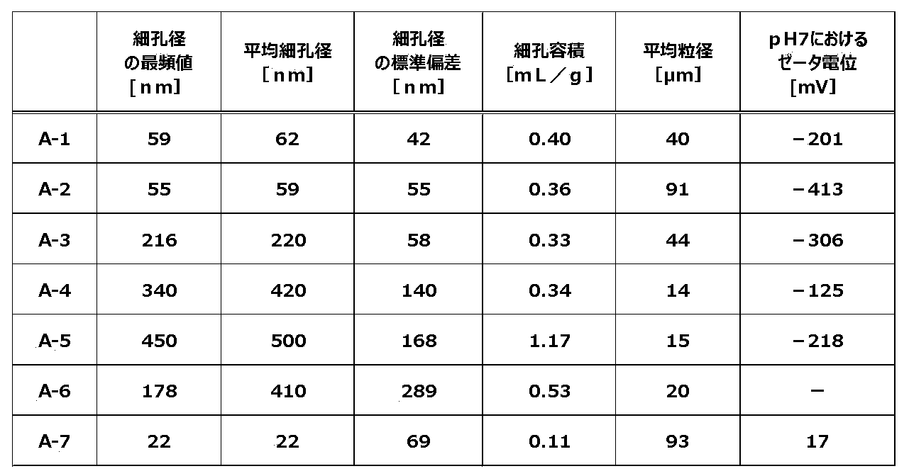
表1中、「-」は、測定を行わなかったことを意味する。
表1に示す結果から、細胞外小胞分離用粉体A-1~A-7中、細胞外小胞分離用粉体A-1~A-5は、本開示に係る細胞外小胞分離用粉体に相当することが確認された。
[検量線の作成]
粉体によるエクソソーム及び牛血清アルブミンの吸着率、吸着したエクソソーム及び牛血清アルブミンの粉体からの脱離率、並びに、エクソソーム及び牛血清アルブミンの回収率を算出するため、エクソソーム濃度とタンパク質濃度の標準直線を作成した。
標準となるエクソソームは、ヒト膵臓癌細胞株BxPC-3細胞〔細胞登録番号:JCRB1448、JCRB細胞バンク〕の培養上清から分離した。具体的には、以下のようにして調製した。ヒト膵臓癌細胞株BxPC-3細胞を牛血清不含RPMI-1640培地〔カタログ番号:189-02025、富士フイルム和光純薬(株)製〕中で3日間培養した。次いで、得られた培養液を遠心分離〔遠心力:300×g)、回転時間:5分、
温度:4℃〕し、培養上清Xを回収した。回収した培養上清Xを再度遠心分離〔遠心力:1,200×g、回転時間:20分、温度:4℃〕し、培養上清Yを回収した。回収した培養上清Yを再度遠心分離〔遠心力:10,000×g、回転時間:30分、温度:4℃〕し、培養上清Zを回収した。回収した培養上清Zには、エクソソームが含まれる。回収した培養上清Zを超遠心分離〔遠心力:100,000×g、回転時間:1時間、温度:4℃〕し、得られたペレットを1mLのPBS(-)溶液(リン酸緩衝生理食塩水)で縣濁した。得られた懸濁液を再度超遠心分離〔遠心力:100,000×g、回転時間:1時間、温度:4℃〕し、得られたペレットを1mLのPBS(-)溶液で再懸濁し、エクソソームを回収した。エクソソームを含む試料液中のタンパク質濃度は、牛血清アルブミン〔カタログ番号:A7906、Sigma-Aldrich社製〕を標準として、BCA Protein Assay Kit〔カタログ番号:23227、Thermo Fisher Scientific社製〕を用いて測定した。
次に、上記にて得られたエクソソームを標準として、試料液中のエクソソーム濃度を算
出するための検量線をsandwich ELISA法により作成した。具体的には、以下のようにして作成した。MaxiSorp 96 ウエルプレート〔カタログ番号:439454、Thermo Fisher Scientific社製〕の各ウエルに1μg/mLのマウス抗ヒトCD9抗体〔カタログ番号:3C9-E12、(株)細胞工学研究所製〕PBS(-)溶液を100μL加え、室温にて1時間静置した。次いで、各ウエルを、0.05%Tween20を含むTBS溶液〔50mMのTris及び0.14MのNaCl、pH8〕(以下、「洗浄液T」という。)200μLで3回洗浄した後、0.01g/mLの牛血清アルブミンを含む洗浄液Tを200μL加えて、室温にて1時間静置した。次いで、各ウエルから0.01g/mLの牛血清アルブミンを含む洗浄液Tを除去した後、各濃度のエクソソーム標準液を100μL加えて、室温で2時間振とうした。次いで、各ウエルを200μLの洗浄液Tで5回洗浄した後、HRP標識ラット抗ヒトCD63抗体〔カタログ番号:1C8-2B11、(株)細胞工学研究所製〕を100μL添加し、室温にて1時間静置した。次いで、各ウエルを200μLの洗浄液Tで5回洗浄した後、HRP基質液〔カタログ番号:5120-0053、SeraCare Life Sciences社製〕を100μL添加し、室温で20分間振とうした。その後、2%硫酸水溶液を50μL添加して反応を停止させ、各ウエルの波長450nmにおける吸光度を、マイクロプレートリーダーを用いて測定した。その結果、エクソソーム標準液の濃度と波長450nmにおける吸光度との間に高い直線性が示された(R2=1.000)。
また、試料液中のタンパク質濃度を算出するための検量線を、牛血清アルブミンをタンパク質の標準として、BCA Protein Assay Kit〔カタログ番号:23227、Thermo Fisher Scientific社製〕を用いて作成した。その結果、タンパク質標準液の濃度と波長570nmにおける吸光度との間に高い直線性が示された(R2=0.970)。
[試料液の調製]
細胞外小胞であるエクソソームと、牛血清アルブミン〔カタログ番号:A7906、Sigma-Aldrich社製〕とを混合し、試料液を調製した。
エクソソームには、検量線を作成する際に使用したエクソソーム、すなわち、ヒト膵臓癌細胞株BxPC-3細胞〔細胞登録番号:JCRB1448、JCRB細胞バンク〕の培養上清から分離したエクソソームを用いた。
調製した試料液は、エクソソームの濃度が1.2μg/mLであり、牛血清アルブミンの濃度が3.0mg/mLであり、エクソソーム及び牛血清アルブミンがPBS(-)溶液に分散された分散液である。
細胞外小胞であるエクソソームと、牛血清アルブミン〔カタログ番号:A7906、Sigma-Aldrich社製〕とを混合し、試料液を調製した。
エクソソームには、検量線を作成する際に使用したエクソソーム、すなわち、ヒト膵臓癌細胞株BxPC-3細胞〔細胞登録番号:JCRB1448、JCRB細胞バンク〕の培養上清から分離したエクソソームを用いた。
調製した試料液は、エクソソームの濃度が1.2μg/mLであり、牛血清アルブミンの濃度が3.0mg/mLであり、エクソソーム及び牛血清アルブミンがPBS(-)溶液に分散された分散液である。
[評価]
上記にて製造した細胞外小胞分離用粉体A-1~A-5及びA-7、並びに、準備した細胞外小胞分離用粉体A-6を用いて、下記の試験を行い、細胞外小胞分離用粉体によるエクソソームの分離及び回収の効率についての評価を行った。
上記にて製造した細胞外小胞分離用粉体A-1~A-5及びA-7、並びに、準備した細胞外小胞分離用粉体A-6を用いて、下記の試験を行い、細胞外小胞分離用粉体によるエクソソームの分離及び回収の効率についての評価を行った。
1.分離試験
上記にて調製した試料液500μLと、上記にて製造した細胞外小胞分離用粉体10mg又は30mgと、をマイクロチューブ〔プロテオセーブ(登録商標)SS 1.5mLマイクロチューブ、住友ベークライト(株)製〕中で混合した後、室温で2時間回転させて撹拌した。次に、マイクロチューブを回転数8,000rpm(revolution per minute;以下、同じ。)及び回転時間5秒の条件で遠心分離機にかけ、細胞外小胞分離用粉体
と上澄み液とに分離し、上澄み液を回収した。回収した上澄み液の分画を「S分画」とした。
次に、上澄み液を回収した後のマイクロチューブに、洗浄液としてのPBS(-)溶液500μLを加え、細胞外小胞分離用粉体とPBS(-)溶液とを混合した後、室温で1
0分間回転させて撹拌することにより、細胞外小胞分離用粉体の洗浄処理を行った。その後、マイクロチューブを回転数8,000rpm及び回転時間5秒の条件で遠心分離機にかけ、細胞外小胞分離用粉体とPBS(-)溶液とに分離し、PBS(-)溶液を回収した。上記の洗浄処理及び回収の操作を3回連続して行い、回収したPBS(-)溶液の分画をそれぞれ「W1分画」、「W2分画」及び「W3分画」とした。
細胞外小胞分離用粉体へのエクソソームの吸着率(単位:%)は、下記の計算式により算出した。
エクソソームの吸着率(単位:%)
=100×{〔試料液に含まれるエクソソームの量(単位:μg)〕-〔S分画、W1分画、W2分画及びW3分画に含まれるエクソソームの合計量(単位:μg)〕}/〔試料液に含まれるエクソソームの量(単位:μg)〕
エクソソームの吸着率(単位:%)
=100×{〔試料液に含まれるエクソソームの量(単位:μg)〕-〔S分画、W1分画、W2分画及びW3分画に含まれるエクソソームの合計量(単位:μg)〕}/〔試料液に含まれるエクソソームの量(単位:μg)〕
細胞外小胞分離用粉体への牛血清アルブミンの吸着率(単位:%)は、下記の計算式により算出した。
牛血清アルブミンの吸着率(単位:%)
=100×{〔試料液に含まれる牛血清アルブミンの量(単位:mg)〕-〔S分画、W1分画、W2分画及びW3分画に含まれる牛血清アルブミンの合計量(単位:mg)〕}/〔試料液に含まれる牛血清アルブミンの量(単位:mg)〕
牛血清アルブミンの吸着率(単位:%)
=100×{〔試料液に含まれる牛血清アルブミンの量(単位:mg)〕-〔S分画、W1分画、W2分画及びW3分画に含まれる牛血清アルブミンの合計量(単位:mg)〕}/〔試料液に含まれる牛血清アルブミンの量(単位:mg)〕
2.回収試験
上記「1.分離試験」を行った後の細胞外小胞分離用粉体と、脱離液としての120mMの四ホウ酸ナトリウム水溶液(pH9)500μLと、をマイクロチューブ中で混合した後、室温で30分間回転させて撹拌することにより、細胞外小胞分離用粉体に吸着したエクソソームを細胞外小胞分離用粉体から脱離させた。次に、マイクロチューブを回転数8,000rpm及び回転時間5秒の条件で遠心分離機にかけ、細胞外小胞分離用粉体と脱離液とに分離し、脱離液を回収した。上記の脱離処理及び回収の操作を3回連続して行い、回収した脱離液の分画をそれぞれ「E1分画」、「E2分画」及び「E3分画」とした。
上記「1.分離試験」を行った後の細胞外小胞分離用粉体と、脱離液としての120mMの四ホウ酸ナトリウム水溶液(pH9)500μLと、をマイクロチューブ中で混合した後、室温で30分間回転させて撹拌することにより、細胞外小胞分離用粉体に吸着したエクソソームを細胞外小胞分離用粉体から脱離させた。次に、マイクロチューブを回転数8,000rpm及び回転時間5秒の条件で遠心分離機にかけ、細胞外小胞分離用粉体と脱離液とに分離し、脱離液を回収した。上記の脱離処理及び回収の操作を3回連続して行い、回収した脱離液の分画をそれぞれ「E1分画」、「E2分画」及び「E3分画」とした。
細胞外小胞分離用粉体からのエクソソームの脱離率(単位:%)は、下記の計算式により算出した。
エクソソームの脱離率(単位:%)
=100×〔E1分画、E2分画及びE3分画に含まれるエクソソームの合計量(単位:μg)〕/{〔エクソソームの吸着率(単位:%)〕×〔試料液に含まれるエクソソームの量(単位:μg)〕/100}
エクソソームの脱離率(単位:%)
=100×〔E1分画、E2分画及びE3分画に含まれるエクソソームの合計量(単位:μg)〕/{〔エクソソームの吸着率(単位:%)〕×〔試料液に含まれるエクソソームの量(単位:μg)〕/100}
細胞外小胞分離用粉体からの牛血清アルブミンの脱離率(単位:%)は、下記の計算式により算出した。
牛血清アルブミンの脱離率(単位:%)
=100×〔E1分画、E2分画及びE3分画に含まれる牛血清アルブミンの合計量(単位:mg)〕/{〔牛血清アルブミンの吸着率(単位:%)〕×〔試料液に含まれる牛血清アルブミンの量(単位:mg)〕/100}
牛血清アルブミンの脱離率(単位:%)
=100×〔E1分画、E2分画及びE3分画に含まれる牛血清アルブミンの合計量(単位:mg)〕/{〔牛血清アルブミンの吸着率(単位:%)〕×〔試料液に含まれる牛血清アルブミンの量(単位:mg)〕/100}
エクソソームの回収率(単位:%)は、下記の計算式により算出した。
エクソソームの回収率(単位:%)
=〔エクソソームの吸着率(単位:%)×エクソソームの脱離率(単位:%)〕/100
エクソソームの回収率(単位:%)
=〔エクソソームの吸着率(単位:%)×エクソソームの脱離率(単位:%)〕/100
牛血清アルブミンの回収率(単位:%)は、下記の計算式により算出した。
牛血清アルブミンの回収率(単位:%)
=〔牛血清アルブミンの吸着率(単位:%)×牛血清アルブミンの脱離率(単位:%)〕/100
牛血清アルブミンの回収率(単位:%)
=〔牛血清アルブミンの吸着率(単位:%)×牛血清アルブミンの脱離率(単位:%)〕/100
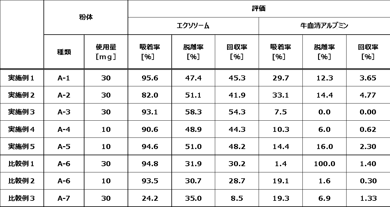
表2に示すように、本開示に係る細胞外小胞分離用粉体によれば、細胞外小胞であるエクソソーム及び夾雑タンパク質である牛血清アルブミンを含む試料液から、エクソソームを選択的に吸着して分離できること、及び、吸着して分離したエクソソームを選択的かつ効率的に脱離して回収できることが明らかとなった。
Claims (4)
- 複数の無機粒子を含む細胞外小胞分離用粉体であって、
前記無機粒子は、細孔を有し、
水銀圧入法により測定される、前記細孔の平均細孔径が50nm以上700nm以下の範囲であり、かつ、前記細孔の細孔径の標準偏差が1nm以上200nm以下の範囲である、細胞外小胞分離用粉体。 - 前記細孔の細孔容積が0.15mL/g以上である、請求項1に記載の細胞外小胞分離用粉体。
- 前記無機粒子の平均粒径が0.5μm以上150μm以下の範囲である、請求項1又は請求項2に記載の細胞外小胞分離用粉体。
- 水に分散させた前記無機粒子のpH7におけるゼータ電位が0mV未満である、請求項1又は請求項2に記載の細胞外小胞分離用粉体。
Applications Claiming Priority (2)
| Application Number | Priority Date | Filing Date | Title |
|---|---|---|---|
| JP2023-059320 | 2023-03-31 | ||
| JP2023059320 | 2023-03-31 |
Publications (1)
| Publication Number | Publication Date |
|---|---|
| WO2024203272A1 true WO2024203272A1 (ja) | 2024-10-03 |
Family
ID=92904601
Family Applications (1)
| Application Number | Title | Priority Date | Filing Date |
|---|---|---|---|
| PCT/JP2024/009510 Ceased WO2024203272A1 (ja) | 2023-03-31 | 2024-03-12 | 細胞外小胞分離用粉体 |
Country Status (2)
| Country | Link |
|---|---|
| JP (1) | JP2024146774A (ja) |
| WO (1) | WO2024203272A1 (ja) |
Citations (5)
| Publication number | Priority date | Publication date | Assignee | Title |
|---|---|---|---|---|
| WO2016136978A1 (ja) * | 2015-02-26 | 2016-09-01 | 旭硝子株式会社 | 微小物質捕捉フィルター、微小物質観察用ガラス基板、微小物質観察装置、微小物質捕捉方法及び微小物質観察方法 |
| WO2017154951A1 (ja) * | 2016-03-09 | 2017-09-14 | 国立大学法人名古屋大学 | 細胞外小胞の回収方法 |
| US20170274118A1 (en) * | 2014-06-09 | 2017-09-28 | The Royal Institution For The Advancement Of Learning/Mcgill University | Borate-glass biomaterials |
| WO2019202994A1 (ja) * | 2018-04-16 | 2019-10-24 | Agc株式会社 | サイズ排除クロマトグラフィ担体、その製造方法、および微小物質の回収方法 |
| WO2023140148A1 (ja) * | 2022-01-18 | 2023-07-27 | 株式会社ダイセル | 細胞外小胞の分離精製方法 |
-
2024
- 2024-02-26 JP JP2024026932A patent/JP2024146774A/ja active Pending
- 2024-03-12 WO PCT/JP2024/009510 patent/WO2024203272A1/ja not_active Ceased
Patent Citations (5)
| Publication number | Priority date | Publication date | Assignee | Title |
|---|---|---|---|---|
| US20170274118A1 (en) * | 2014-06-09 | 2017-09-28 | The Royal Institution For The Advancement Of Learning/Mcgill University | Borate-glass biomaterials |
| WO2016136978A1 (ja) * | 2015-02-26 | 2016-09-01 | 旭硝子株式会社 | 微小物質捕捉フィルター、微小物質観察用ガラス基板、微小物質観察装置、微小物質捕捉方法及び微小物質観察方法 |
| WO2017154951A1 (ja) * | 2016-03-09 | 2017-09-14 | 国立大学法人名古屋大学 | 細胞外小胞の回収方法 |
| WO2019202994A1 (ja) * | 2018-04-16 | 2019-10-24 | Agc株式会社 | サイズ排除クロマトグラフィ担体、その製造方法、および微小物質の回収方法 |
| WO2023140148A1 (ja) * | 2022-01-18 | 2023-07-27 | 株式会社ダイセル | 細胞外小胞の分離精製方法 |
Also Published As
| Publication number | Publication date |
|---|---|
| JP2024146774A (ja) | 2024-10-15 |
Similar Documents
| Publication | Publication Date | Title |
|---|---|---|
| EP3674704B1 (en) | Method for isolating exosome and exosome isolation kit | |
| CN102815935B (zh) | 陶瓷颗粒群及其用途 | |
| US12551828B2 (en) | Method for extracting microvesicles from biological sample | |
| WO2018112557A1 (en) | Methods and compositions for purification or isolation of microvesicles and exosomes | |
| Zheng et al. | Mesostructured chitosan–silica hybrid as a biodegradable carrier for a pH-responsive drug delivery system | |
| US12161950B2 (en) | Methods and compositions for purification or isolation of microvesicles and exosomes | |
| WO2019144016A1 (en) | Method of isolating exosomes using encapsulation and aqueous micellar system | |
| WO2017205563A1 (en) | Magnetizable glass ceramic composition and methods thereof | |
| Qiao et al. | Biomimetic hollow mesoporous hydroxyapatite microsphere with controlled morphology, entrapment efficiency and degradability for cancer therapy | |
| Notarangelo et al. | Rapid nickel-based isolation of extracellular vesicles from different biological fluids | |
| JP2024146774A (ja) | 細胞外小胞分離用粉体 | |
| Marzouk et al. | Polyethylene glycol (PEG)-based precipitation for exosome enrichment: a review on recent developments, current challenges, and future perspectives | |
| Li et al. | Nitrogen cavitation enables rapid and high-yield preparation of functional cell-membrane-derived vesicles | |
| JP2024146430A (ja) | 細胞外小胞分離用粉体 | |
| JP2025045927A (ja) | 細胞外小胞分離用粉体 | |
| US20110081379A1 (en) | Purification method and method of producing vaccine | |
| JP2025048757A (ja) | 細胞外小胞分離用粉体 | |
| Frykstrand et al. | Study of mesoporous magnesium carbonate in contact with whole human blood | |
| JP2005059001A (ja) | 析出方法 | |
| JP2024146494A (ja) | 細胞外小胞分離用粉体 | |
| CN115521894A (zh) | 用于提取细胞外囊泡的试剂盒与方法 | |
| Obuchi et al. | Isolation of small extracellular vesicles by interacting with inorganic surface | |
| JP7160294B1 (ja) | 脂質二重膜に覆われた微粒子又は細胞外小胞の回収方法および脂質二重膜に覆われた微粒子又は細胞外小胞を回収するための回収キット | |
| CN1321947C (zh) | 一种制备多孔结构球形颗粒无机材料制品的方法 | |
| Kraxner et al. | Porous Bioactive Glass Microspheres Prepared by Flame Synthesis |
Legal Events
| Date | Code | Title | Description |
|---|---|---|---|
| 121 | Ep: the epo has been informed by wipo that ep was designated in this application |
Ref document number: 24779402 Country of ref document: EP Kind code of ref document: A1 |
|
| NENP | Non-entry into the national phase |
Ref country code: DE |
|
| 122 | Ep: pct application non-entry in european phase |
Ref document number: 24779402 Country of ref document: EP Kind code of ref document: A1 |