WO2024201691A1 - オートファジーを誘導する化合物のスクリーニング方法 - Google Patents
オートファジーを誘導する化合物のスクリーニング方法 Download PDFInfo
- Publication number
- WO2024201691A1 WO2024201691A1 PCT/JP2023/012419 JP2023012419W WO2024201691A1 WO 2024201691 A1 WO2024201691 A1 WO 2024201691A1 JP 2023012419 W JP2023012419 W JP 2023012419W WO 2024201691 A1 WO2024201691 A1 WO 2024201691A1
- Authority
- WO
- WIPO (PCT)
- Prior art keywords
- dead
- rna helicase
- box rna
- autophagy
- compound
- Prior art date
- Legal status (The legal status is an assumption and is not a legal conclusion. Google has not performed a legal analysis and makes no representation as to the accuracy of the status listed.)
- Ceased
Links
Images


Classifications
-
- C—CHEMISTRY; METALLURGY
- C12—BIOCHEMISTRY; BEER; SPIRITS; WINE; VINEGAR; MICROBIOLOGY; ENZYMOLOGY; MUTATION OR GENETIC ENGINEERING
- C12N—MICROORGANISMS OR ENZYMES; COMPOSITIONS THEREOF; PROPAGATING, PRESERVING, OR MAINTAINING MICROORGANISMS; MUTATION OR GENETIC ENGINEERING; CULTURE MEDIA
- C12N9/00—Enzymes; Proenzymes; Compositions thereof; Processes for preparing, activating, inhibiting, separating or purifying enzymes
- C12N9/90—Isomerases (5.)
-
- A—HUMAN NECESSITIES
- A61—MEDICAL OR VETERINARY SCIENCE; HYGIENE
- A61P—SPECIFIC THERAPEUTIC ACTIVITY OF CHEMICAL COMPOUNDS OR MEDICINAL PREPARATIONS
- A61P35/00—Antineoplastic agents
-
- C—CHEMISTRY; METALLURGY
- C12—BIOCHEMISTRY; BEER; SPIRITS; WINE; VINEGAR; MICROBIOLOGY; ENZYMOLOGY; MUTATION OR GENETIC ENGINEERING
- C12N—MICROORGANISMS OR ENZYMES; COMPOSITIONS THEREOF; PROPAGATING, PRESERVING, OR MAINTAINING MICROORGANISMS; MUTATION OR GENETIC ENGINEERING; CULTURE MEDIA
- C12N15/00—Mutation or genetic engineering; DNA or RNA concerning genetic engineering, vectors, e.g. plasmids, or their isolation, preparation or purification; Use of hosts therefor
- C12N15/09—Recombinant DNA-technology
- C12N15/11—DNA or RNA fragments; Modified forms thereof; Non-coding nucleic acids having a biological activity
- C12N15/52—Genes encoding for enzymes or proenzymes
-
- C—CHEMISTRY; METALLURGY
- C12—BIOCHEMISTRY; BEER; SPIRITS; WINE; VINEGAR; MICROBIOLOGY; ENZYMOLOGY; MUTATION OR GENETIC ENGINEERING
- C12Q—MEASURING OR TESTING PROCESSES INVOLVING ENZYMES, NUCLEIC ACIDS OR MICROORGANISMS; COMPOSITIONS OR TEST PAPERS THEREFOR; PROCESSES OF PREPARING SUCH COMPOSITIONS; CONDITION-RESPONSIVE CONTROL IN MICROBIOLOGICAL OR ENZYMOLOGICAL PROCESSES
- C12Q1/00—Measuring or testing processes involving enzymes, nucleic acids or microorganisms; Compositions therefor; Processes of preparing such compositions
- C12Q1/02—Measuring or testing processes involving enzymes, nucleic acids or microorganisms; Compositions therefor; Processes of preparing such compositions involving viable microorganisms
- C12Q1/24—Methods of sampling, or inoculating or spreading a sample; Methods of physically isolating an intact microorganisms
-
- C—CHEMISTRY; METALLURGY
- C12—BIOCHEMISTRY; BEER; SPIRITS; WINE; VINEGAR; MICROBIOLOGY; ENZYMOLOGY; MUTATION OR GENETIC ENGINEERING
- C12Q—MEASURING OR TESTING PROCESSES INVOLVING ENZYMES, NUCLEIC ACIDS OR MICROORGANISMS; COMPOSITIONS OR TEST PAPERS THEREFOR; PROCESSES OF PREPARING SUCH COMPOSITIONS; CONDITION-RESPONSIVE CONTROL IN MICROBIOLOGICAL OR ENZYMOLOGICAL PROCESSES
- C12Q1/00—Measuring or testing processes involving enzymes, nucleic acids or microorganisms; Compositions therefor; Processes of preparing such compositions
- C12Q1/25—Measuring or testing processes involving enzymes, nucleic acids or microorganisms; Compositions therefor; Processes of preparing such compositions involving enzymes not classifiable in groups C12Q1/26 - C12Q1/66
-
- G—PHYSICS
- G01—MEASURING; TESTING
- G01N—INVESTIGATING OR ANALYSING MATERIALS BY DETERMINING THEIR CHEMICAL OR PHYSICAL PROPERTIES
- G01N2500/00—Screening for compounds of potential therapeutic value
- G01N2500/02—Screening involving studying the effect of compounds C on the interaction between interacting molecules A and B (e.g. A = enzyme and B = substrate for A, or A = receptor and B = ligand for the receptor)
-
- G—PHYSICS
- G01—MEASURING; TESTING
- G01N—INVESTIGATING OR ANALYSING MATERIALS BY DETERMINING THEIR CHEMICAL OR PHYSICAL PROPERTIES
- G01N2500/00—Screening for compounds of potential therapeutic value
- G01N2500/04—Screening involving studying the effect of compounds C directly on molecule A (e.g. C are potential ligands for a receptor A, or potential substrates for an enzyme A)
-
- G—PHYSICS
- G01—MEASURING; TESTING
- G01N—INVESTIGATING OR ANALYSING MATERIALS BY DETERMINING THEIR CHEMICAL OR PHYSICAL PROPERTIES
- G01N2500/00—Screening for compounds of potential therapeutic value
- G01N2500/10—Screening for compounds of potential therapeutic value involving cells
Definitions
- the present invention relates to a method for screening compounds that induce autophagy and a method for producing compounds that induce the degradation of target intracellular molecules or organelles by autophagic processes.
- Autophagy is one of the mechanisms by which intracellular molecules are broken down within cells, and is a mechanism found in eukaryotes from yeast to humans.
- autophagy When autophagy is induced, a membrane vesicle called an autophagosome is formed, which then fuses with a lysosome, resulting in the degradation of the molecules taken up into the autophagosome.
- autophagy is induced when cells enter a state of starvation, and it has become clear that autophagy is also involved in physiological functions such as development and differentiation, as well as infection defense functions such as the elimination of viruses that have invaded cells.
- Non-Patent Document 1 The mechanism by which autophagy is induced has also been elucidated through various reports.
- Mohammad Omar Faruk et al. report in detail the mechanism of selective autophagy, which contributes to cellular homeostasis by degrading specific soluble proteins, supramolecular complexes, liquid-phase separated droplets, abnormal or excessive organelles, pathogenic invading bacteria, etc.
- p62 binds to ubiquitinated proteins, causing liquid-phase separation, forming droplets in which p62 and ubiquitinated proteins are condensed, and these droplets recruit other proteins to form isolation membranes/phagocytic membranes, and p62 and other proteins in the droplets are degraded in lysosomes.
- Non-Patent Document 2 Lin Ma et al. suggest that knockdown of DDX5 suppresses endoplasmic reticulum stress and promotes recovery of autophagic flux in esophageal squamous cell carcinoma (ESCC) (Non-Patent Document 2).
- DDX5 stimulates autophagy and suppresses the development of liver cancer by interacting with the autophagy receptor p62 (Non-Patent Document 3). They also reported that DDX5 promotes the degradation of p62 and significantly reduces its half-life.
- Non-Patent Document 4 Yongsang Jo et al., in a report on intrinsically disordered protein-protein interactions in membrane-free protein droplets, disclosed that they formed DDX4 droplets by adding streptavidin.
- degraders protein degradation targeting chimeras
- SNIPERs specific non-genetic IAP-dependent protein eliminators
- UPS ubiquitin-proteasome system
- [6] A method for producing a compound that induces degradation of a target intracellular molecule or organelle by autophagic degradation, comprising linking, via a linker, a ligand that specifically binds to the target intracellular molecule or organelle to the compound obtained by the method according to any one of [1] to [5].
- [7] A method for inducing autophagic degradation of a target intracellular molecule or organelle by contacting a compound obtained by the method according to [6] with a DEAD-box RNA helicase in a cell in the presence of p62.
- an autophagy-inducing compound can be efficiently obtained, and by linking this as a degradation tag moiety via a linker to a ligand that specifically binds to a target intracellular molecule, a compound that induces the degradation of a target molecule or organelle by autophagic degradation can be synthesized.
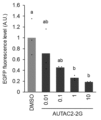
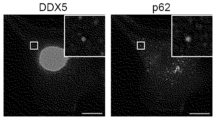
- Examples 1 to 4 the amount of light emitted 10 minutes after the start of luminescence measurement is shown. Shown are EGFP fluorescence levels when cells transfected with pEGFP-FKBP12 were cultured in fresh DMEM containing 0.01 mM, 0.1 mM, 1 mM, and 10 mM AUTAC2-2G, respectively, and in fresh DMEM containing DMSO as a control. HeLa cells transiently expressing GFP-FKBP F36V were treated with SLF'-AUTAC, fixed with paraformaldehyde, and immunostained with anti-DDX5 or anti-p62 antibodies, and images were observed under a fluorescent microscope.
- FIG. 4A shows an image observed with a fluorescent microscope when HeLa cells transiently expressing GFP-FKBP12 and RFP-DDX5 were treated with AUTAC2-2G
- FIG. 4B shows an image observed with a fluorescent microscope when HeLa cells transiently expressing GFP-FKBP12 and RFP-p62 were treated with AUTAC2-2G.
- the screening method according to the present invention screens for autophagy-inducing compounds by utilizing at least one of the following properties: (1) It has high binding affinity to DEAD-box RNA helicase. (2) It brings DEAD-box RNA helicase and p62 into close proximity to promote their interaction. (3) Promotes droplet formation of DEAD-box RNA helicase and p62. As demonstrated in the Examples described below, compounds having autophagy-inducing activity have these properties, and therefore, autophagy-inducing compounds can be obtained by screening for compounds having at least one of these properties.
- DEAD-box RNA helicase is a superfamily of RNA helicases that have the motif DEAD (Asp-Glu-Ala-Asp). DEAD-box RNA helicases are known to be involved in the disassembly and formation of double-stranded RNA and RNA-protein complexes using the energy of ATP hydrolysis, and to play important roles in transcription, splicing, nuclear export and degradation of RNA, translation, and ribosome biogenesis.
- DEAD-box RNA helicases include DDX4 (DEAD box protein 4), DDX5 (DEAD box protein 5) and DDX17 (DEAD box protein 17), with DEAD-Box Helicase 5 (DDX5) also known as P68.
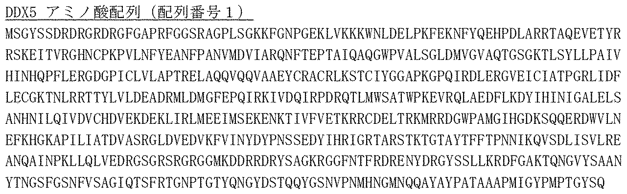
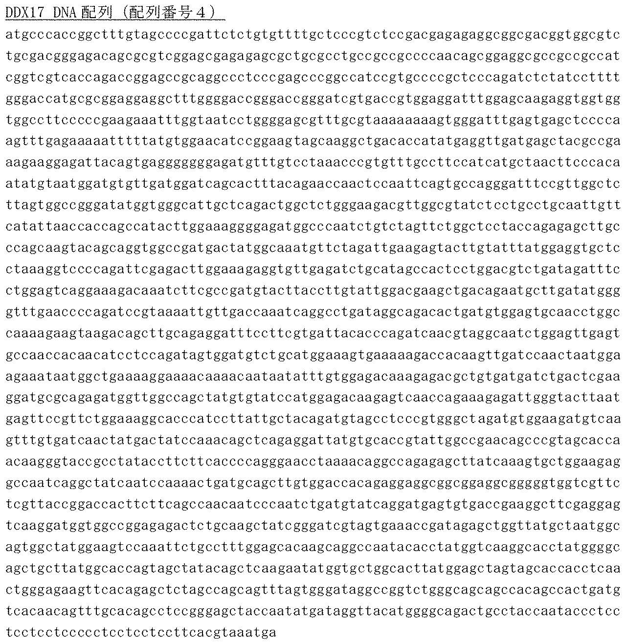
- amino acid sequences of DDX5 and DDX17 are as follows:
- Method for measuring binding affinity Measurement can be performed by surface plasmon resonance assay.
- an autophagy-inducing compound is immobilized on a thin gold film of a sensor chip, and a purified DEAD-box RNA helicase protein solution is poured onto the surface of the sensor chip, and the SPR signal that changes depending on the binding state of the two can be measured by BIAcore.
- measurement can also be performed by detecting the change in thermal stability of the DEAD-box RNA helicase protein due to binding with an autophagy-inducing compound using a thermal shift assay called CETSA.
- p62 p62 is a receptor protein that induces specific proteins and organelles into autophagy. p62 is localized diffusely in the cytoplasm and nucleus, but has a half-life of about 10 hours and is basically degraded in an autophagy-dependent manner.
- p62 has many domains, such as the N-terminal Phox1 and Bem1p domain (Phox1 and Bem1p: PB1), zinc finger (ZZ), TRAF6-binding domain (TB), LC3-interacting region (LIR, consisting of the DDDWTHL sequence), Keap1-interacting region (KIR), and C-terminal ubiquitin-associated domain (UBA), and interacts with LC3, an autophagosome-localized protein, at the LC3-interacting region (LIR) to induce autophagy (see Non-Patent Document 13).
- the amino acid sequence of p62 or the nucleic acid sequence of the SQSTM1 gene encoding it is as follows:
- Method for detecting the interaction (proximation) between DEAD-box RNA helicase and p62 there is no particular limitation on the method for detecting the interaction (proximation) between DEAD-box RNA helicase and p62, and any available method may be used.
- the method can be performed using NanoLuc (registered trademark) Binary Technology Assays (NanoBiT (registered trademark) Assays) according to the reaction scheme shown below.
- the term "droplet” refers to a liquid-like aggregate in which DEAD-box RNA helicase, a compound associated therewith, and p62 are concentrated and are separated into a liquid-liquid phase from the outside.
- Such "droplets” can be confirmed by labeling DEAD-box RNA helicase and/or p62 with a fluorescent label or the like, introducing a test compound into a system in which they exist, preferably treating cells expressing at least one of them with the test compound, and observing the presence of a substance visualized by the label using a fluorescent microscope, etc.
- a compound that induces autophagic degradation of a target intracellular molecule can be produced by linking, via a linker, a ligand that specifically binds to a target intracellular molecule or organelle to a compound obtained by the above-mentioned screening method.
- target intracellular molecule or organelle refers to an intracellular molecule or organelle targeted for degradation by autophagy.
- Intracellular molecule refers to a biomolecule at least a portion of which is present within the cell, and includes not only naturally occurring biomolecules present within the cell, but also molecules artificially introduced or expressed within the cell.
- organelle includes not only naturally occurring organelles present within the cell (e.g., dysfunctional mitochondria), but also foreign viruses and microorganisms that have invaded the cell.
- intracellular molecules include lipids, glycolipids, proteins, glycoproteins, etc.
- Intracellular proteins present within cells, and are typically proteins present within cells, including protein aggregates such as amyloid beta (A ⁇ ), tau protein, and ⁇ -synuclein.
- a ⁇ amyloid beta
- tau protein tau protein
- ⁇ -synuclein ⁇ -synuclein
- intracellular proteins intracellular proteins associated with pathology (particularly, intracellular proteins associated with human pathology) are cited as practical targets, and many reports have been published. Intracellular proteins associated with such pathological conditions can be identified, for example, by UniProt or Human Protein Atlas.
- Some examples include A ⁇ , tau protein, ⁇ -synuclein or aggregates thereof (involved in neurodegenerative diseases such as Alzheimer's disease and Parkinson's disease), BRD4 (involved in multiple myeloma, acute myeloid leukemia, etc.), Ras (involved in colon cancer, pancreatic cancer, etc.), calcineurin, CyP, FRB, FKBP, PyL, ABI1, GID1, GA1, FKBP F36V, IAA17 TIR1, EGFR, and BTK (B.Z. Stanton et al. Science; 359; eaao5902 (2016), N. Shindo et al. Bioorg. Med.
- Intracellular molecules also include intracellular proteins that can bind to a ligand, as described below, by fusing a specific tag to any target intracellular molecule or organelle using tagging techniques such as HaloTag (registered trademark), SNAP Tag (registered trademark), and CLIP Tag (registered trademark).
- tagging techniques such as HaloTag (registered trademark), SNAP Tag (registered trademark), and CLIP Tag (registered trademark).
- An intracellular protein fused with HaloTag (registered trademark) is fused with a tag having the F272H mutant of dehalogenase DhaA derived from Rhodococcus rhodochrous
- an intracellular protein fused with SNAP Tag (registered trademark) is fused with a tag having the hATG mutant of human-derived O 6 -alkylguanine-DNA alkyltransferase
- an intracellular protein fused with CLIP Tag registered trademark
- is fused with a tag having the E30R mutant of a SNAP tagged protein are outlined, for example, in Halo SNAP CLIP for Protein Labeling - Bitesize Bio, the contents of which are incorporated herein by reference.
- intracellular proteins tagged with mutant FKBPs such as ⁇ -synuclein or aggregates thereof, can also be targeted intracellular molecules for autophagy.
- ligand that specifically binds to an intracellular molecule or organelle means a structural unit that constitutes part of a compound that induces the degradation of a target intracellular molecule or organelle by autophagy and has specific binding activity to a target intracellular molecule (including the above-mentioned tagged fusion protein) or organelle. Therefore, the substance that constitutes the ligand is a substance that has specific binding activity to an intracellular molecule or organelle, and there have been many reports on such substances.
- BindingDB https://www.bindingdb.org/rwd/bind/index.jsp
- FK506 calcineurin
- CyP cyclosporin A
- FKCsA CyP, FKBP
- rapamycin FRB, FKBP
- FK1012 FKBP
- abscisic acid PyI, ABI1
- gibberellin 3 GID1, GA1
- AP1903/AP20187 FKBP F36V
- Auxin IAA17 TIR1
- afatinib EGFR
- osimertinib EGFR
- dacomitinib EGFR
- neratinib neratinib
- sotrasib ibrutinib
- BK acalabrutinib
- zanubrutinib BTK
- tirabrutinib ibrutinib
- examples of ligands for BRD4 include Examples of the ligand for FKBP12 include Examples of the ligand for MetAP2 include Examples include:
- ligand compounds for Ras include ligands described in the following documents: Nature, 2013, 503, 548-551; International Publication No. 2013/155223; Science, 2016, 351, 604-608; International Publication No. WO2014152588; International Publication No. 2015/054572; International Publication No. 2016/049524; Nat. Rev. Drug Discov., 2014, 13, 828-851; Chem. Soc. Rev., 2016, advance article (DOI: 10.1039/C5CS00911A). Therefore, the ligand for Ras can be composed of these compounds. Examples of ligands for Ras include, Examples include:
- Ligands for intracellular proteins fused with Halotag include, for example, halogenated C 1-12 alkoxy groups (particularly, C 1-6 alkoxy groups with a halogenated terminal carbon), preferably halogenated (particularly, halogenated terminal carbon) hexyloxy groups (particularly, chlorinated hexyloxy groups).
- An example of a ligand for an intracellular protein fused with a SNAP tag (registered trademark) is O 6 -benzylguanine.
- An example of a ligand for an intracellular protein fused with a CLIP tag® is O 6 -benzylcytosine.
- Ligands for intracellular proteins fused with tags are generally described in Halo SNAP CLIP for Protein Labeling--Bitsize Bio, the contents of which are incorporated herein by reference.
- examples of ligands for target intracellular molecules tagged with mutant FKBP include: Examples include:
- linker refers to an atomic group that links the "ligand” portion of the compound of the present invention to the tag portion that induces autophagy.
- the linker structure can be a known linker structure that is used to link molecules with different functions.
- Japanese Patent No. 7161760 and Non-Patent Documents 5, 9, and 10 provide detailed descriptions of linker structures that link intracellular molecules to tag portions that induce autophagy, and the structures described in these documents can be used.
- linker can be linked to the ligand and the tag moiety using known reactions.
- Patent No. 7161760 Non-Patent Documents 5, 9, and 10; 5th Edition Experimental Chemistry Lectures, Vol. 13-19 (edited by the Chemical Society of Japan); New Experimental Chemistry Lectures, Vol. 14-15 (edited by the Chemical Society of Japan); Precision Organic Chemistry Revised 2nd Edition (L. F. Tietz, Th.
- Reactions that can be used include protection or deprotection reactions of functional groups, reduction reactions, oxidation reactions, aromatic nucleophilic substitution reactions, azidation reactions of alcohols, alkyl halides, and sulfonate esters, reductive amination reactions, Mitsunobu reactions, esterification reactions, amidation reactions, urea reactions, coupling reactions, Wohl-Ziegler reactions, sulfone esterification reactions, hydrolysis reactions, bromination reactions, N-alkylation reactions, hydroxylation reactions via diazotization, deamination reactions, cyclization reactions, carbamate reactions, amidation by carbon monoxide insertion reactions, esterification, click reactions, and the like, and the reagents and reaction conditions necessary for these reactions are well known to those skilled in the art to which the present invention pertains.
- the compounds obtained by the above manufacturing method have a ligand that specifically binds to a target molecule or organelle and a tag moiety that induces autophagy linked via a linker, and have the advantage that they can target a wide range of substrates by replacing the ligand that binds to them depending on the target molecule or organelle.
- the compounds of the present invention are useful as preventive or therapeutic agents for diseases involving targeted intracellular molecules or organelles.
- the compounds of the present invention may be effective in preventing or treating any disease involving targeted intracellular molecules or organelles.
- they are expected to be applied to the treatment or prevention of cancer, inflammatory diseases, autoimmune diseases, and bone and joint degenerative diseases.
- p62 cDNA was synthesized from HeLa total RNA using oligo(dT) 15 primer, amplified using oligo primers (5'-agcggtaggcatggcgtcgctcaccg-3' and 5'-agaagatctgctagtacaacggcgggatgc-3') and inserted into the vector using the In-fusion cloning system (the underlined 15 nt is the In-fusion arm).
- the pBiT2.1-N [TK/SmBiT] Vector (Promega #N2014) was linearized by polymerase chain reaction using oligo primers (5'-ggtggctttaccaacagtaccg-3' and 5'- gctagcagatcttctagtcgg-3').
- DDX5 cDNA was synthesized from HeLa total RNA using oligo(dT) 15 primer, amplified using oligo primers (5'-agcggtggcatgtcgggattcgagtgaccg-3' and 5'- agaagatctgctagcttattgggaatcctgttggcattg-3') and inserted into the vector using the In-fusion cloning system (the underlined 15 nt is the In-fusion arm).
- nucleic acid sequences of pLgBiT-p62 and pSmBiT-DDX5 carried by each vector are shown below.
- HeLa cells RIKEN BRC Cell Bank #RCB0007
- DMEM Fujifilm Wako #043-30085
- FBS Thermo Fisher Scientific #10437028
- Non-essential amino acids Fujifilm/Wako #139-15651
- Opti-MEM I Thermo Fisher Scientific #11058-021 96-well culture plate.
- Promega #E5650 Lipofectamine3000 transfection reagent Thermo Fisher Scientific #L3000008 Nano-Glo® Live Cell Reagent: Promega #N2014 GloMax Discover Microplate Reader:Promega #GM3000 PrimeSTAR Max DNA polymerase: TaKaRa #R045A TRIzol reagent (for total RNA extraction):Thermo Fisher Scientific #15596026 PrimeScriptTM RT reagent Kit (for cDNA synthesis): TaKaRa #RR037A In-Fusion HD Cloning Kit: TaKaRa #Z9649N
- Test Compounds The following compounds were used to verify the screening method of the present invention.
- AUTAC4-2G and 8-nitro-cGMP are compounds and their tag moieties disclosed in Japanese Patent No. 7,161,760
- AUTAC2-2G and FBnG are compounds and their tag moieties that were newly synthesized to verify whether compounds screened by the method of the present invention actually induce autophagy (the synthesis schemes of FBnG and AUTAC2-2G are as follows).
- Test procedure [Example 1] HeLa cells grown in 96-well plates containing growth medium were transfected with pLgBiT-p62 and pSmBiT-DDX5 using Lipofectamine3000® transfection reagent (Thermo Fisher Scientific Inc.) as described in the accompanying documentation. 24 hours after the addition of the reagent, AUTAC2-2G was added to the wells at a concentration of 10 ⁇ M and the transfected cells were treated with AUTAC2-2G for 12 hours. The growth medium was then replaced with 100 ⁇ L of Opti-MEM® I (Thermo Fisher Scientific Inc.).
- Nano-Glo containing the substrate furimazine was added to the wells at a concentration of 10 ⁇ M and the cells were then treated with AUTAC2-2G. 25 ⁇ L of Live Cell Reagent was added, and luminescence was immediately measured using a GloMax® Discover Microplate Reader (Promega Corporation).
- Example 2 The cells were treated and luminescence was measured in the same manner as in Example 1, except that AUTAC4-2G was added to the wells at a concentration of 10 ⁇ M and the transfected cells were treated with AUTAC4-2G for 12 hours.
- Example 3 The cells were treated and luminescence was measured as in Example 1, except that 8-nitro-cGMP was added to the wells to a concentration of 10 ⁇ M and the transfected cells were treated with 8-nitro-cGMP for 24 hours.
- Example 4 The cells were treated and luminescence was measured in the same manner as in Example 1, except that FBnG was added to the wells at a concentration of 10 ⁇ M and the transfected cells were treated with FBnG for 24 hours.
- Example 1 The cells were treated and luminescence was measured in the same manner as in Example 1, except that DMSO (dimethyl sulfoxide) was added to the wells to a concentration of 10 ⁇ M and the transfected cells were treated with DMSO for 24 hours.
- DMSO dimethyl sulfoxide
- Figure 1 shows the amount of luminescence 10 minutes after the start of luminescence measurement in Examples 1 to 4.
- the data are average values from three independent experiments.
- the graph shows an upper limit of 107, and in fact, the amount of luminescence was higher than that. Error bars indicate standard deviation.
- Statistical analysis was performed using the Turkey-Kramer multiple comparison test. A P value of ⁇ 0.01 was considered to be statistically significant.
- Cells treated with AUTAC2-2G, AUTAC4-2G, 8-nitro-cGMP, and FBnG showed 1,000-fold more luminescence than cells treated with DMSO, demonstrating that the presence of these compounds brought p62 and DDX5 into close proximity, indicating that this method can be used to screen for compounds that bring p62 and DDX5 into close proximity.
- pEGFP-FKBP12 was linearized by polymerase chain reaction using oligo primers (5'- cgcaaatctagacttgtacagctcgtcc-3' and 5'- ggccgcttcgagcagatg-3').
- FKBP12 cDNA was synthesized from HeLa cell total RNA using oligo(dT) 15 primer, amplified with oligo primers (5'-aagtctagatttgcgatgggagtgcaggtgg-3' and 5'-ctgctcgaagcggcctcattccagttagaagc-3'), and inserted into a vector using the infusion cloning system (the underlined 15 nt is the infusion arm).
- HeLa cells RIKEN BRC Cell Bank #RCB0007 Dulbecco's Modified Eagle Medium (DMEM): Fujifilm Wako #043-30085
- FBS Thermo Fisher Scientific #10437028
- Non-essential amino acids Fujifilm/Wako #139-15651
- Opti-MEM I Thermo Fisher Scientific #11058-021 96-well culture plate.
- Thermo Fisher Scientific #165305 Lipofectamine3000 transfection reagent Thermo Fisher Scientific #L3000008 Live Cell Imaging Solution: Thermo Fisher Scientific #A14291DJ Fluorescence microscope: Keyence #BZ-X800 PrimeSTAR Max DNA polymerase: TaKaRa #R045A TRIzol reagent (for total RNA extraction): Thermo Fisher Scientific #15596026 PrimeScriptTM RT reagent Kit (for cDNA synthesis): TaKaRa #RR037A In-Fusion HD Cloning Kit: TaKaRa #Z9649N
- Test procedure HeLa cells were grown on 96-well plates and transfected with pEGFP-FKBP12 using Lipofectamine 3000 transfection reagent. After 24 h, the transfected cells were incubated for an additional 24 h in fresh DMEM containing 0.01 mM AUTAC2-2G, as well as in fresh DMEM containing DMSO as a control. The medium was then removed and replaced with 100 ⁇ L of live cell imaging solution. EGFP-FKBP12 levels were analyzed by measuring EGFP fluorescence.
- Example 6 Cells were treated and EGFP fluorescence was measured as in Example 5, except that the transfected cells were incubated for an additional 24 hours in fresh DMEM containing 0.1 mM AUTAC2-2G.
- Example 7 Cells were treated and EGFP fluorescence was measured as in Example 5, except that the transfected cells were incubated for an additional 24 hours in fresh DMEM containing 1 mM AUTAC2-2G.
- Example 8 Cells were treated and EGFP fluorescence was measured as in Example 5, except that the transfected cells were incubated for an additional 24 hours in fresh DMEM containing 10 mM AUTAC2-2G.
- Test procedure HeLa cells transiently expressing GFP tagged with mutant FKBP (GFP-FKBP F36V ) were treated with 1 ⁇ M SLF'-AUTAC for 12 hours. The treated cells were fixed with 4% paraformaldehyde, permeabilized with 0.1% TritonX-100/PBS, blocked with 1% BSA/PBS, and incubated overnight at 4°C in PBS containing 1% each of rabbit anti-DDX5 antibody and mouse anti-p62 antibody. Then, the cells were incubated at room temperature for 1 hour in PBS containing 1% each of AlexaFluor555-labeled anti-rabbit IgG and AlexaFluor555-labeled anti-mouse IgG. The cells after immunostaining were observed under a fluorescent microscope.
- FIG. 3 shows images observed with a fluorescent microscope (scale bar: 10 ⁇ m).
- AUTAC2-2G was used to test whether it induces the formation of droplets containing DDX5 and p62.
- Test procedure HeLa cells transiently expressing GFP-FKBP12 and RFP-DDX5, or GFP-FKBP12 and RFP-p62 were treated with AUTAC2-2G at a concentration of 10 ⁇ M for 13 hours. The treated cells were observed live under a fluorescent microscope, and the fluorescence derived from GFP and RFP was continuously observed. During the course of the observation, 5% 1,6-hexanediol was added 5 minutes after the start of observation, and it was observed whether the dot-shaped structures in the field of view disappeared. 1,6-hexanediol is known to have the ability to dissolve droplets, making it possible to confirm that dot-like structures in the microscope field of view are droplets.
- Figure 4 shows an image observed with a fluorescent microscope when HeLa cells transiently expressing GFP-FKBP12 and RFP-DDX5 were treated with AUTAC2-2G
- Figure 4B shows an image observed with a fluorescent microscope when HeLa cells transiently expressing GFP-FKBP12 and RFP-p62 were treated with AUTAC2-2G.
- droplets containing GFP-FKBP12 and RFP-DDX5, and dot-like structures containing GFP-FKBP12 and RFP-p62 were observed, which were confirmed to be droplets because they disappeared after the addition of 1,6-hexanediol.
Landscapes
- Chemical & Material Sciences (AREA)
- Health & Medical Sciences (AREA)
- Life Sciences & Earth Sciences (AREA)
- Organic Chemistry (AREA)
- Engineering & Computer Science (AREA)
- Genetics & Genomics (AREA)
- Zoology (AREA)
- Wood Science & Technology (AREA)
- Bioinformatics & Cheminformatics (AREA)
- Biotechnology (AREA)
- Molecular Biology (AREA)
- Proteomics, Peptides & Aminoacids (AREA)
- General Engineering & Computer Science (AREA)
- General Health & Medical Sciences (AREA)
- Biochemistry (AREA)
- Microbiology (AREA)
- Physics & Mathematics (AREA)
- Biophysics (AREA)
- Biomedical Technology (AREA)
- Immunology (AREA)
- Analytical Chemistry (AREA)
- Medicinal Chemistry (AREA)
- Chemical Kinetics & Catalysis (AREA)
- Nuclear Medicine, Radiotherapy & Molecular Imaging (AREA)
- Pharmacology & Pharmacy (AREA)
- Animal Behavior & Ethology (AREA)
- Public Health (AREA)
- Veterinary Medicine (AREA)
- General Chemical & Material Sciences (AREA)
- Plant Pathology (AREA)
- Pathology (AREA)
- Measuring Or Testing Involving Enzymes Or Micro-Organisms (AREA)
Abstract
新たなオートファジー誘導化合物を探索するためのスクリーニング方法を提供する。 (1)DEAD-box型RNAヘリカーゼに対して高い結合親和性を有する、 (2)DEAD-box型RNAヘリカーゼとp62とを近接化して相互作用を促す、および (3)DEAD-box型RNAヘリカーゼとp62の液滴形成を促す、 の少なくとも1つの特性を利用して、新たなオートファジー誘導化合物を探索する。
Description
本発明は、オートファジーを誘導する化合物のスクリーニング方法および標的細胞内分子またはオルガネラのオートファジーによる分解を誘導する化合物の製造方法に関する。
オートファジーは、細胞内における細胞内分子の分解機構の一つであって、酵母からヒトにいたるまでの真核生物に見られる機構である。オートファジーが誘導されると、オートファゴソームと呼ばれる膜胞が形成され、続いて、オートファゴソームがリソソームと融合することにより、オートファゴソーム内に取り込まれた分子が分解される。
細胞が飢餓状態に陥ることによってオートファジーが誘導されることが知られているほか、発生、分化等の生理機能や、細胞内に侵入したウイルスの排除等の感染防御機能にもオートファジーが関与することが明らかになってきている。
オートファジーが誘導される機構についても様々な報告により明らかになってきている。
Mohammad Omar Farukらは、レビュー論文において、特定の可溶性タンパク質、超分子複合体、液相分離した液滴、異常または過剰なオルガネラ、病原性侵入細菌などを分解し、細胞の恒常性に寄与する選択的なオートファジーのメカニズムを詳細に報告している(非特許文献1)。具体的には、p62がユビキチン化タンパク質に結合して液相分離が起こり、p62およびユビキチン化タンパク質が凝縮している液滴を形成し、この液滴が他のタンパク質をリクルートし隔離膜/貪食膜を形成させ、液滴中のp62および他のタンパク質がリソソームで分解されることを報告している。
Mohammad Omar Farukらは、レビュー論文において、特定の可溶性タンパク質、超分子複合体、液相分離した液滴、異常または過剰なオルガネラ、病原性侵入細菌などを分解し、細胞の恒常性に寄与する選択的なオートファジーのメカニズムを詳細に報告している(非特許文献1)。具体的には、p62がユビキチン化タンパク質に結合して液相分離が起こり、p62およびユビキチン化タンパク質が凝縮している液滴を形成し、この液滴が他のタンパク質をリクルートし隔離膜/貪食膜を形成させ、液滴中のp62および他のタンパク質がリソソームで分解されることを報告している。
Lin Maらは、食道扁平上皮癌(ESCC)で、DDX5のノックダウンが小胞体ストレスを抑制し、オートファジーのフラックスの回復を促進することを示唆している(非特許文献2)。
Hao Zhangらは、DDX5がオートファジー受容体p62と相互作用することで、オートファジーを刺激し、肝癌の発生を抑制することを報告している(非特許文献3)。また、DDX5がp62の分解を促進し、その半減期を顕著に減少させたことも報告している。
Yongsang Joらは、膜なしのタンパク質液滴内の本質的に無秩序なタンパク質間の相互作用に関する報告において、Streptavidinの添加によりDDX4の液滴を形成したことを開示している(非特許文献4)。
また、オートファジーを積極的に誘導して新しい治療方法(デグレーダーと呼ばれることがある)を提供する試みも著しい進展を見せている(例えば、非特許文献5乃至8)。
デグレーダーの代表的な例は、タンパク質分解ターゲットキメラ(PROTAC)/特異的・非遺伝的IAP依存性タンパク質消去剤(SNIPER)であり、これらは、標的分子に結合するリガンドとユビキチン-プロテアソームシステム(UPS)による分解を誘導する分解タグからなる異種二機能性分子である(非特許文献5、9および10)。これらの異種二機能性分子は、分解タグを最適化し、標的リガンドを置き換えることで、幅広い基質をターゲットにできる利点がある。しかし、UPSを利用した分解システムはタンパク質をターゲットにしたものに限られていた。
これに対して、近年、リソソームを利用した分解システム、細胞外基質を分解するリソソームターゲティングキメラ(LYTAC)およびオートファジー標的キメラ(AUTAC)が開発され(非特許文献11および12)、さらなる新規キメラ分子の創出が期待される。
デグレーダーの代表的な例は、タンパク質分解ターゲットキメラ(PROTAC)/特異的・非遺伝的IAP依存性タンパク質消去剤(SNIPER)であり、これらは、標的分子に結合するリガンドとユビキチン-プロテアソームシステム(UPS)による分解を誘導する分解タグからなる異種二機能性分子である(非特許文献5、9および10)。これらの異種二機能性分子は、分解タグを最適化し、標的リガンドを置き換えることで、幅広い基質をターゲットにできる利点がある。しかし、UPSを利用した分解システムはタンパク質をターゲットにしたものに限られていた。
これに対して、近年、リソソームを利用した分解システム、細胞外基質を分解するリソソームターゲティングキメラ(LYTAC)およびオートファジー標的キメラ(AUTAC)が開発され(非特許文献11および12)、さらなる新規キメラ分子の創出が期待される。
Cancer Sci, 2021,112(10):3972-3978
Biochem Biophys Res Commun,2020,533(4):1449-1456
Hepatology,2019,69(3):1046-1063
Chem. Sci.,2020,11,1269
Samarasinghe, K. T. G.; Crews, C. M. Targeted protein degradation: a promise for undruggable proteins. Cell Chem. Biol. 2021, 28, 934-951.
Li,K.;M.Crews,C.PROTACs:Past,Present and Future.Chem.Soc.Rev.2022.doi: 10.1039/d2cs00193d.
Burslem,G.M.;Crews,C.M.Proteolysis-targeting chimeras as therapeutics and tools for biological discovery.Cell 2020,181,102-114.
Paiva,S.-L.;Crews,C.M. Targeted protein degradation: elements of PROTAC design.Curr.Opin.Chem.Biol. 2019,50,111-119.
友重聡、石川昌彦 PROTACs and other chemical protein degradation technologies for the treatment of neurodegenerative disorders. Angew.Chem.Int.Ed.2021,60,3346-3354.
内藤正人、大岡信行、柴田直樹 SNIPERs-hijacking IAP activity to induce protein degradation.Drug Discov.Today Technol.2019,31,35-42.
Banik,S.M.;Pedram,K.;Wisnovsky,S.;Ahn,G.;Riley,N.M.;Bertozzi,C.R.;Lysosome-targeting Chimaeras for degradation of extracellular proteins.Nature 2020,584,291-297.
高橋大輔、有本博史 オートファジーを利用した分解機の基礎となる選択的オートファジー.Cell Chem. Biol.2021,28,1061-1071.
一村義信,小松雅明 p62/SQSTM1 :オートファジーにおけるその役割 Journal of Japanese Biochemical Society 91(3): 380-387 (2019) doi:10.14952/SEIKAGAKU.2019.910380
しかし、これら選択的なオートファジーを誘導するキメラ分子における分解タグの設計は、試行錯誤により経験的に得たものであり、新規キメラ分子を効率的に創出するために、分解タグを設計するツールの開発が望まれていた。
本発明は、このようなニーズに対応するものであり、選択的なオートファジーを誘導するキメラ分子の分解タグを得るためのツールを提供する。
本発明は、このようなニーズに対応するものであり、選択的なオートファジーを誘導するキメラ分子の分解タグを得るためのツールを提供する。
本発明者らは、自らが開発したオートファジー誘導化合物およびタグ部分について、オートファジーを誘導するメカニズムを探求していたところ、これらの化合物について、以下の特性を発見した。
(1)DEAD-box型RNAヘリカーゼに対して高い結合親和性を有する。
(2)DEAD-box型RNAヘリカーゼとp62とを近接化して相互作用を促す。
(3)DEAD-box型RNAヘリカーゼとp62の液滴形成を促す。
本発明者らは、これらの特性を利用して、新たなオートファジー誘導化合物を探索するためのスクリーニング方法を構築し、これを使用して、新規オートファジー誘導化合物の探索に成功した。
本発明は、これらの知見に基づくものであり、以下の方法を提供する。
(1)DEAD-box型RNAヘリカーゼに対して高い結合親和性を有する。
(2)DEAD-box型RNAヘリカーゼとp62とを近接化して相互作用を促す。
(3)DEAD-box型RNAヘリカーゼとp62の液滴形成を促す。
本発明者らは、これらの特性を利用して、新たなオートファジー誘導化合物を探索するためのスクリーニング方法を構築し、これを使用して、新規オートファジー誘導化合物の探索に成功した。
本発明は、これらの知見に基づくものであり、以下の方法を提供する。
[1]DEAD-box型RNAヘリカーゼに試験化合物を接触させ、前記DEAD-box型RNAヘリカーゼに対する前記試験化合物の結合親和性を測定し、
所定値以上の結合親和性を有する化合物を選択する、
オートファジー誘導化合物をスクリーニングする方法。
[2]p62の存在下で、DEAD-box型RNAヘリカーゼに試験化合物を接触させ、
前記DEAD-box型RNAヘリカーゼと前記p62の相互作用を測定し、
前記相互作用を誘導した化合物を選択する、
オートファジー誘導化合物をスクリーニングする方法。
[3]前記DEAD-box型RNAヘリカーゼおよび前記p62を、細胞内で発現し、前記細胞に前記試験化合物を接触させる、[2]に記載の方法。
[4]p62の存在下で、DEAD-box型RNAヘリカーゼに試験化合物を接触させ、
DEAD-box型RNAヘリカーゼとp62の液滴形成を測定し、
前記液滴形成を誘導した化合物を選択する、
オートファジー誘導化合物をスクリーニングする方法。
[5]前記DEAD-box型RNAヘリカーゼは、DDX5またはDDX17である、[1]~[4]のいずれか1項に記載の方法。
[6][1]~[5]のいずれか1項に記載の方法で得られた化合物に、リンカーを介して、標的細胞内分子またはオルガネラに特異的に結合するリガンドを連結する、標的細胞内分子またはオルガネラのオートファジーによる分解を誘導する化合物の製造方法。
[7][6]に記載の方法で得られた化合物を、細胞内で、p62の存在下で、DEAD-box型RNAヘリカーゼと接触させて、標的細胞内分子またはオルガネラのオートファジーによる分解を誘導する方法。
[8][6]に記載の方法で得られた化合物を、細胞内で、p62の存在下で、DEAD-box型RNAヘリカーゼと接触させて、前記化合物、前記p62および前記DEAD-box型RNAヘリカーゼを含む液滴を形成する、[7]に記載の方法。
[9]癌を予防または治療する、[7]または[8]に記載の方法。
所定値以上の結合親和性を有する化合物を選択する、
オートファジー誘導化合物をスクリーニングする方法。
[2]p62の存在下で、DEAD-box型RNAヘリカーゼに試験化合物を接触させ、
前記DEAD-box型RNAヘリカーゼと前記p62の相互作用を測定し、
前記相互作用を誘導した化合物を選択する、
オートファジー誘導化合物をスクリーニングする方法。
[3]前記DEAD-box型RNAヘリカーゼおよび前記p62を、細胞内で発現し、前記細胞に前記試験化合物を接触させる、[2]に記載の方法。
[4]p62の存在下で、DEAD-box型RNAヘリカーゼに試験化合物を接触させ、
DEAD-box型RNAヘリカーゼとp62の液滴形成を測定し、
前記液滴形成を誘導した化合物を選択する、
オートファジー誘導化合物をスクリーニングする方法。
[5]前記DEAD-box型RNAヘリカーゼは、DDX5またはDDX17である、[1]~[4]のいずれか1項に記載の方法。
[6][1]~[5]のいずれか1項に記載の方法で得られた化合物に、リンカーを介して、標的細胞内分子またはオルガネラに特異的に結合するリガンドを連結する、標的細胞内分子またはオルガネラのオートファジーによる分解を誘導する化合物の製造方法。
[7][6]に記載の方法で得られた化合物を、細胞内で、p62の存在下で、DEAD-box型RNAヘリカーゼと接触させて、標的細胞内分子またはオルガネラのオートファジーによる分解を誘導する方法。
[8][6]に記載の方法で得られた化合物を、細胞内で、p62の存在下で、DEAD-box型RNAヘリカーゼと接触させて、前記化合物、前記p62および前記DEAD-box型RNAヘリカーゼを含む液滴を形成する、[7]に記載の方法。
[9]癌を予防または治療する、[7]または[8]に記載の方法。
本発明の実施形態による方法によれば、オートファジー誘導化合物を効率的に得ることができ、これを分解タグ部分としてリンカーを介して、標的細胞内分子に特異的に結合するリガンドに連結することにより、標的分子またはオルガネラのオートファジーによる分解を誘導する化合物を合成することができる。
本発明の実施の形態を詳細に説明する。但し、本発明は以下の実施の形態に限定して理解されるべきものではない。
1.オートファジー誘導化合物をスクリーニングする方法
本発明によるスクリーニング方法は、以下の特性の少なくとも1つを利用してオートファジー誘導化合物をスクリーニングするものである。
(1)DEAD-box型RNAヘリカーゼに対して高い結合親和性を有する。
(2)DEAD-box型RNAヘリカーゼとp62とを近接化して相互作用を促す。
(3)DEAD-box型RNAヘリカーゼとp62の液滴形成を促す。
後述する実施例で実証する通り、オートファジー誘導活性を有する化合物は、これらの特性を有するため、これらの特性の少なくとも1つを有する化合物をスクリーニングすることでオートファジー誘導化合物を得ることができる。
本発明によるスクリーニング方法は、以下の特性の少なくとも1つを利用してオートファジー誘導化合物をスクリーニングするものである。
(1)DEAD-box型RNAヘリカーゼに対して高い結合親和性を有する。
(2)DEAD-box型RNAヘリカーゼとp62とを近接化して相互作用を促す。
(3)DEAD-box型RNAヘリカーゼとp62の液滴形成を促す。
後述する実施例で実証する通り、オートファジー誘導活性を有する化合物は、これらの特性を有するため、これらの特性の少なくとも1つを有する化合物をスクリーニングすることでオートファジー誘導化合物を得ることができる。
1-1.DEAD-box型RNAヘリカーゼ
DEAD-box型RNAヘリカーゼとは、DEAD(Asp-Glu-Ala-Asp)というモチーフをもつRNAヘリカーゼのスーパーファミリーである。DEAD-box型RNAヘリカーゼは、ATP加水分解のエネルギーを用いて、二本鎖RNAやRNA―タンパク質複合体の解体や形成に関与し、転写、スプライシング、RNAの核外輸送や分解、翻訳、リボソームの生合成などで重要な役割を果たすことが知られている。
DEAD-box型RNAヘリカーゼとは、DEAD(Asp-Glu-Ala-Asp)というモチーフをもつRNAヘリカーゼのスーパーファミリーである。DEAD-box型RNAヘリカーゼは、ATP加水分解のエネルギーを用いて、二本鎖RNAやRNA―タンパク質複合体の解体や形成に関与し、転写、スプライシング、RNAの核外輸送や分解、翻訳、リボソームの生合成などで重要な役割を果たすことが知られている。
DEAD-box型RNAヘリカーゼのメンバーには、DDX4(DEAD box protein 4)、DDX5(DEAD box protein 5)およびDDX17(DEAD box protein 17)が含まれ、DEAD-Box Helicase 5(DDX5)は、P68としても知られる。
DDX5およびDDX17のアミノ酸配列およびそれをコードする核酸配列は以下の通りである。


1-2.結合親和性を測定する方法
表面プラスモン共鳴アッセイによって、測定することができる。例えば、オートファジー誘導化合物をセンサーチップの金薄膜上に固定した上で、精製したDEAD-box型RNAヘリカーゼタンパク質溶液をセンサーチップの表面に流し、両者の結合状態に応じて変化するSPRシグナルをBIAcoreによって測定できる。また、CETSAと呼ばれるサーマルシフトアッセイによって、オートファジー誘導化合物と結合することによる、DEAD-box型RNAヘリカーゼタンパク質の熱安定性変化を検出することによっても測定することができる。
表面プラスモン共鳴アッセイによって、測定することができる。例えば、オートファジー誘導化合物をセンサーチップの金薄膜上に固定した上で、精製したDEAD-box型RNAヘリカーゼタンパク質溶液をセンサーチップの表面に流し、両者の結合状態に応じて変化するSPRシグナルをBIAcoreによって測定できる。また、CETSAと呼ばれるサーマルシフトアッセイによって、オートファジー誘導化合物と結合することによる、DEAD-box型RNAヘリカーゼタンパク質の熱安定性変化を検出することによっても測定することができる。
1-3.p62
p62は、特異的なタンパク質や細胞小器官(オルガネラ)をオートファジーへと導く受容体タンパク質である。p62は、細胞質や核に散在性に局在するが、半減期は10時間程度であり、基本的にオートファジー依存的に分解される。p62は、N末端のPhox1およびBem1pドメイン(Phox1 and Bem1p:PB1)、ジンクフィンガー(Zinc finger:ZZ)、TRAF6結合モチーフ(TRAF6-binding domain:TB)、LC3相互作用領域(LC3-interacting region:LIR、DDDWTHL配列からなる)、Keap1相互作用領域(Keap1-interacting region:KIR)、およびC末端のユビキチン会合ドメイン(ubiquitin-associated:UBA)といった多数のドメインを有し、LC3相互作用領域(LIR)でオートファゴソーム局在タンパク質であるLC3と相互作用してオートファジーへと導く(非特許文献13参照)。p62のアミノ酸配列またはそれをコードするSQSTM1遺伝子の核酸配列は、以下の通りある。
p62は、特異的なタンパク質や細胞小器官(オルガネラ)をオートファジーへと導く受容体タンパク質である。p62は、細胞質や核に散在性に局在するが、半減期は10時間程度であり、基本的にオートファジー依存的に分解される。p62は、N末端のPhox1およびBem1pドメイン(Phox1 and Bem1p:PB1)、ジンクフィンガー(Zinc finger:ZZ)、TRAF6結合モチーフ(TRAF6-binding domain:TB)、LC3相互作用領域(LC3-interacting region:LIR、DDDWTHL配列からなる)、Keap1相互作用領域(Keap1-interacting region:KIR)、およびC末端のユビキチン会合ドメイン(ubiquitin-associated:UBA)といった多数のドメインを有し、LC3相互作用領域(LIR)でオートファゴソーム局在タンパク質であるLC3と相互作用してオートファジーへと導く(非特許文献13参照)。p62のアミノ酸配列またはそれをコードするSQSTM1遺伝子の核酸配列は、以下の通りある。


1-4.DEAD-box型RNAヘリカーゼとp62との相互作用(近接化)を検出する方法
本発明では、DEAD-box型RNAヘリカーゼとp62との相互作用(近接化)を検出する方法について特に制限はなく、利用可能な方法で実施すればよい。例えば、NanoLuc(登録商標) Binary Technology Assays(NanoBiT(登録商標)Assays)を用いて、以下に示す反応スキームにより実施することができる。
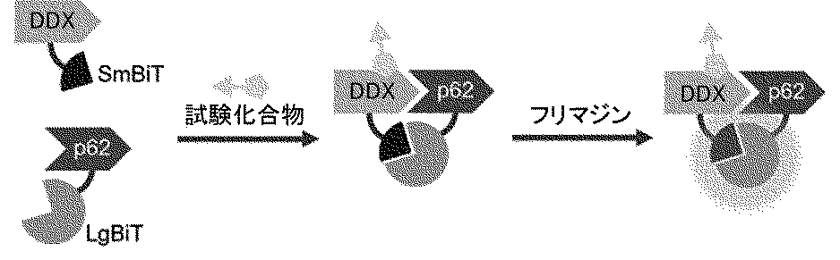
(式中、LgBiTは、Large BiTであり、SmBiTは、Small BiTの略称である。)
このアッセイでは、DEAD-box型RNAヘリカーゼとp62に、それぞれルシフェラーゼの一部を構成し、両者で完全となるルシフェラーゼ部分ペプチドを融合した修飾DEAD-box型RNAヘリカーゼ(SmBiT-DDX)およびp62(LgBiT-p62)を発現する細胞を準備し、この細胞を試験化合物と接触させた際に、試験化合物がDEAD-box型RNAヘリカーゼとp62を近接させるとそれぞれに融合されているルシフェラーゼ部分ペプチドSmBiTおよびLgBiTが会合して、ルシフェラーゼとして活性が復活する。このような状態で、ルシフェラーゼの基質であるフリマジンを添加すると、強い発光が得られ、DEAD-box型RNAヘリカーゼとp62の近接(相互作用)を検出できる。
本発明では、DEAD-box型RNAヘリカーゼとp62との相互作用(近接化)を検出する方法について特に制限はなく、利用可能な方法で実施すればよい。例えば、NanoLuc(登録商標) Binary Technology Assays(NanoBiT(登録商標)Assays)を用いて、以下に示す反応スキームにより実施することができる。
(式中、LgBiTは、Large BiTであり、SmBiTは、Small BiTの略称である。)
このアッセイでは、DEAD-box型RNAヘリカーゼとp62に、それぞれルシフェラーゼの一部を構成し、両者で完全となるルシフェラーゼ部分ペプチドを融合した修飾DEAD-box型RNAヘリカーゼ(SmBiT-DDX)およびp62(LgBiT-p62)を発現する細胞を準備し、この細胞を試験化合物と接触させた際に、試験化合物がDEAD-box型RNAヘリカーゼとp62を近接させるとそれぞれに融合されているルシフェラーゼ部分ペプチドSmBiTおよびLgBiTが会合して、ルシフェラーゼとして活性が復活する。このような状態で、ルシフェラーゼの基質であるフリマジンを添加すると、強い発光が得られ、DEAD-box型RNAヘリカーゼとp62の近接(相互作用)を検出できる。
1-5.DEAD-box型RNAヘリカーゼとp62の液滴形成の検出
本願明細書において「液滴」とは、DEAD-box型RNAヘリカーゼおよびこれと会合した化合物、ならびにp62が濃縮され、外部と液-液相分離されている液体様の凝集体である。
このような「液滴」は、DEAD-box型RNAヘリカーゼおよび/またはp62を蛍光標識などで標識しておき、これらが存在する系に試験化合物を導入、好ましくは少なくともこれらの何れか一方を発現する細胞を試験化合物で処理し、蛍光顕微鏡などで標識により可視化された物質の存在を観察することで確認できる。また、液体の性質をさらに詳しく確認する目的では、光褪色蛍光回復法(FRAP)と組み合わせことが好ましい。
本願明細書において「液滴」とは、DEAD-box型RNAヘリカーゼおよびこれと会合した化合物、ならびにp62が濃縮され、外部と液-液相分離されている液体様の凝集体である。
このような「液滴」は、DEAD-box型RNAヘリカーゼおよび/またはp62を蛍光標識などで標識しておき、これらが存在する系に試験化合物を導入、好ましくは少なくともこれらの何れか一方を発現する細胞を試験化合物で処理し、蛍光顕微鏡などで標識により可視化された物質の存在を観察することで確認できる。また、液体の性質をさらに詳しく確認する目的では、光褪色蛍光回復法(FRAP)と組み合わせことが好ましい。
2.標的細胞内分子またはオルガネラのオートファジーによる分解を誘導する化合物の製造方法
上述のスクリーニング方法で得られた化合物に、リンカーを介して、標的細胞内分子またはオルガネラに特異的に結合するリガンドを連結することで、標的細胞内分子のオートファジーによる分解を誘導する化合物を製造することができる。
上述のスクリーニング方法で得られた化合物に、リンカーを介して、標的細胞内分子またはオルガネラに特異的に結合するリガンドを連結することで、標的細胞内分子のオートファジーによる分解を誘導する化合物を製造することができる。
本明細書において、「標的細胞内分子またはオルガネラ」とは、オートファジーによる分解の標的とする細胞内分子またはオルガネラを意味する。「細胞内分子」とは、少なくともその一部が細胞内に存在する生体分子を意味し、細胞内に存在する天然の生体分子のみならず、人為的に細胞内に導入または発現させた分子を含む。また、「オルガネラ」は、細胞内に存在する天然のオルガネラ(例えば、機能不全ミトコンドリア)の他、細胞に侵入した外来のウイルス、微生物をも含むものとする。
「細胞内分子」としては、例えば、細胞内に存在する脂質、糖脂質、タンパク質、糖タンパク質等が挙げられ、典型的には細胞内に存在するタンパク質であり、これには、アミロイドβ(Aβ)、タウタンパク質、αシヌクレインなどのたんぱく質の凝集体も含まれる。「細胞内分子」が存在する細胞種について特に制限はないが、細胞としては哺乳動物(例、マウス、ラット、ハムスター、ウサギ、ネコ、イヌ、ウシ、ヒツジ、サル、ヒト)の細胞が好ましく、ヒト細胞が実用上最も念頭に置かれる。細胞内タンパク質としては、病態に関連した細胞内タンパク質(特に、ヒトの病態に関連した細胞内タンパク質)が実用的な標的として挙げられ、多数の報告がなされている。そのような病態に関連した細胞内タンパク質は、例えば、UniProtや、Human Protein Atlasで確認することができ、ごく一部の例として、Aβ、タウタンパク質、αシヌクレインまたはこれらの凝集体(アルツハイマー病、パーキンソン病などの神経変性疾患に関与)、BRD4(多発性骨髄腫、急性骨髄性白血病などに関与)、Ras(大腸癌、膵臓癌などに関与)、カルシニューリン、CyP、FRB、FKBP、PyL、ABI1、GID1、GA1、FKBP F36V、IAA17 TIR1、EGFR、BTKが挙げられる(B.Z.Stantonら Science;359;eaao5902(2018)、N.Shindoら Bioorg. Med Chem;47:116386(2021)などに記載されており、参照にその内容を本書に組み込む)。
また、細胞内分子は、例えば、ハロタグ(登録商標)、SNAPタグ(登録商標)、CLIPタグ(登録商標)などのタグ化技術により、任意の標的細胞内分子またはオルガネラに所定のタグを融合して、後述するリガンドの結合が可能となる細胞内タンパク質が含まれる。ハロタグ(登録商標)と融合した細胞内タンパク質は、Rhodococcus rhodochrous由来脱ハロゲン化酵素DhaAのF272H変異体を有するタグが融合され、SNAPタグ(登録商標)と融合した細胞内タンパク質は、ヒト由来O6-アルキルグアニン-DNAアルキルトランスフェラーゼhATG変異体を有するタグが融合され、CLIPタグ(登録商標)と融合した細胞内タンパク質は、SNAPタグタンパク質のE30R変異体を有するタグが融合される。これらは、例えば、Halo SNAP CLIP for Protein Labeling - Bitesize Bioに概説されており、参照によりその内容を本書に取り込む。
また、変異FKBPでタグ化したαシヌクレインまたはこの凝集体などの変異FKBPでタグ化した細胞内タンパク質もオートファジーのために標的化した細胞内分子となり得る。
「細胞内分子」としては、例えば、細胞内に存在する脂質、糖脂質、タンパク質、糖タンパク質等が挙げられ、典型的には細胞内に存在するタンパク質であり、これには、アミロイドβ(Aβ)、タウタンパク質、αシヌクレインなどのたんぱく質の凝集体も含まれる。「細胞内分子」が存在する細胞種について特に制限はないが、細胞としては哺乳動物(例、マウス、ラット、ハムスター、ウサギ、ネコ、イヌ、ウシ、ヒツジ、サル、ヒト)の細胞が好ましく、ヒト細胞が実用上最も念頭に置かれる。細胞内タンパク質としては、病態に関連した細胞内タンパク質(特に、ヒトの病態に関連した細胞内タンパク質)が実用的な標的として挙げられ、多数の報告がなされている。そのような病態に関連した細胞内タンパク質は、例えば、UniProtや、Human Protein Atlasで確認することができ、ごく一部の例として、Aβ、タウタンパク質、αシヌクレインまたはこれらの凝集体(アルツハイマー病、パーキンソン病などの神経変性疾患に関与)、BRD4(多発性骨髄腫、急性骨髄性白血病などに関与)、Ras(大腸癌、膵臓癌などに関与)、カルシニューリン、CyP、FRB、FKBP、PyL、ABI1、GID1、GA1、FKBP F36V、IAA17 TIR1、EGFR、BTKが挙げられる(B.Z.Stantonら Science;359;eaao5902(2018)、N.Shindoら Bioorg. Med Chem;47:116386(2021)などに記載されており、参照にその内容を本書に組み込む)。
また、細胞内分子は、例えば、ハロタグ(登録商標)、SNAPタグ(登録商標)、CLIPタグ(登録商標)などのタグ化技術により、任意の標的細胞内分子またはオルガネラに所定のタグを融合して、後述するリガンドの結合が可能となる細胞内タンパク質が含まれる。ハロタグ(登録商標)と融合した細胞内タンパク質は、Rhodococcus rhodochrous由来脱ハロゲン化酵素DhaAのF272H変異体を有するタグが融合され、SNAPタグ(登録商標)と融合した細胞内タンパク質は、ヒト由来O6-アルキルグアニン-DNAアルキルトランスフェラーゼhATG変異体を有するタグが融合され、CLIPタグ(登録商標)と融合した細胞内タンパク質は、SNAPタグタンパク質のE30R変異体を有するタグが融合される。これらは、例えば、Halo SNAP CLIP for Protein Labeling - Bitesize Bioに概説されており、参照によりその内容を本書に取り込む。
また、変異FKBPでタグ化したαシヌクレインまたはこの凝集体などの変異FKBPでタグ化した細胞内タンパク質もオートファジーのために標的化した細胞内分子となり得る。
本明細書中、「細胞内分子またはオルガネラに特異的に結合するリガンド」とは、標的細胞内分子またはオルガネラのオートファジーによる分解を誘導する化合物の一部を構成し、標的細胞内分子(これには、上記タグ化された融合タンパク質を含む)またはオルガネラへの特異的な結合活性を有する構造単位を意味する。従って、リガンドを構成する物質は、細胞内分子またはオルガネラへの特異的な結合活性を有する物質であり、このような物質についても多数の報告がある。例えば、BindingDB(https://www.bindingdb.org/rwd/bind/index.jsp)で確認することができ、ごく一部の例として、例えば、FK506(カルシニューリン)、シクロスポリンA(カルシニューリン、CyP)、FKCsA(CyP、FKBP)、ラパマイシン(FRB、FKBP)、FK1012(FKBP)、アブシジン酸(PyI、ABI1)、ジベレリン3(GID1、GA1)、AP1903/AP20187(FKBP F36V)、Auxin(IAA17 TIR1)、アファチニブ(EGFR)、オシメルチニブ(EGFR)、ダコミチニブ(EGFR)、ネラチニブ(EGFR)、ソトラシブ(EGFR)、イブルチニブ(BTK)、アカラブルチニブ(BTK)、ザヌブルチニブ(BTK)、チラブルチニブ(BTK)、アダグラシブ(BTK)が挙げられる(カッコ内は結合対象を示す)。
また、BRD4に対するリガンドとしては、例えば

が挙げられ、FKBP12に対するリガンドとしては、例えば

が挙げられ、MetAP2に対するリガンドとしては、例えば

が挙げられる。
が挙げられ、FKBP12に対するリガンドとしては、例えば
が挙げられ、MetAP2に対するリガンドとしては、例えば
が挙げられる。
Rasに対するリガンド化合物としては、例えば、以下の文献に記載されたリガンドが挙げられる;Nature, 2013, 503, 548-551; 国際公開2013/155223号;Science, 2016, 351, 604-608; 国際公開WO2014152588号;国際公開2015/054572号;国際公開2016/049524号;Nat. Rev. Drug Discov., 2014, 13, 828-851; Chem. Soc. Rev., 2016, advance article (DOI: 10.1039/C5CS00911A)。従って、Rasに対するリガンドは、これらの化合物により構成することができる。Rasに対するリガンドとしては、例えば、

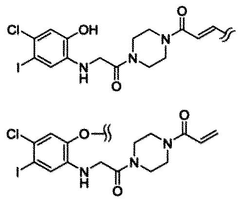
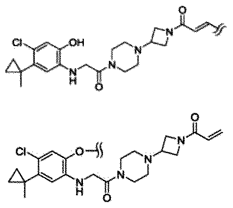
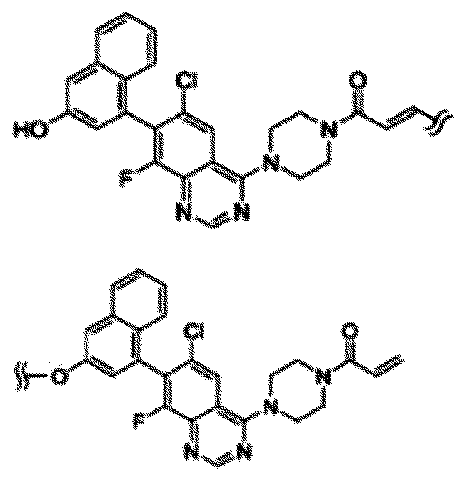
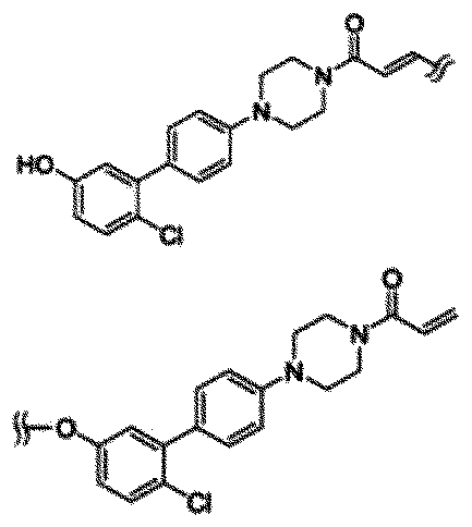
が挙げられる。
が挙げられる。
ハロタグ(登録商標)を融合した細胞内タンパク質に対するリガンドとしては、例えば、ハロゲン化C1-12アルコキシ基(特に、末端の炭素がハロゲン化されたC1-6アルコキシ基)が挙げられ、好ましくはハロゲン化された(特に、末端の炭素がハロゲン化された)ヘキシルオキシ基(特に、塩素化ヘキシルオキシ基)である。
SNAPタグ(登録商標)を融合した細胞内タンパク質に対するリガンドとしては、例えば、O6-ベンジルグアニンが挙げられる。
CLIPタグ(登録商標)を融合した細胞内タンパク質に対するリガンドとしては、例えば、O6-ベンジルシトシンが挙げられる。
なお、タグを融合した細胞内タンパク質に対するリガンドについては、Halo SNAP CLIP for Protein Labeling - Bitesize Bioに概説されており、参照によりその内容を本書に取り込む。
SNAPタグ(登録商標)を融合した細胞内タンパク質に対するリガンドとしては、例えば、O6-ベンジルグアニンが挙げられる。
CLIPタグ(登録商標)を融合した細胞内タンパク質に対するリガンドとしては、例えば、O6-ベンジルシトシンが挙げられる。
なお、タグを融合した細胞内タンパク質に対するリガンドについては、Halo SNAP CLIP for Protein Labeling - Bitesize Bioに概説されており、参照によりその内容を本書に取り込む。
また、変異FKBPでタグ化したαシヌクレインまたはこの凝集体などの変異FKBPでタグ化した標的細胞内分子に対するリガンドとしては、例えば、
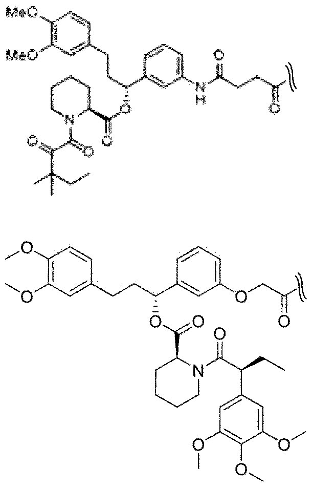
を挙げることができる。
を挙げることができる。
本明細書中、「リンカー」とは、本発明の化合物における「リガンド」の部分とオートファジーを誘導するタグ部分とを連結する原子団を意味する。リンカー構造は、異なる機能を有する分子を連結する際に利用される既知のリンカー構造を利用することができる。また、特許7161760号ならびに非特許文献5、9および10には、細胞内分子とオートファジーを誘導するタグ部分とを連結するリンカーの構造について詳細に記載されており、これら文献に記載する構造を利用すればよい。
また、「リンカー」と「リガンド」およびタグ部分との連結も公知の反応を利用して実施することができる。例えば、特許7161760号;非特許文献5、9および10;第5版実験化学講座、13巻~19巻(日本化学会編);新実験化学講座、14巻~15巻(日本化学会編);精密有機化学 改定第2版(L. F. Tietze,Th. Eicher、南江堂);改訂 有機人名反応 そのしくみとポイント(東郷秀雄著、講談社);ORGANIC SYNTHESES Collective Volume I~VII(John Wiley & SonsInc);Modern Organic Synthesis in the Laboratory A Collection of Standard Experimental Procedures(Jie Jack Li著、OXFORD UNIVERSITY出版);Comprehensive Heterocyclic Chemistry III、Vol.1~Vol.14(エルゼビア・ジャパン株式会社);人名反応に学ぶ有機合成戦略(富岡清監訳、化学同人発行);コンプリヘンシブ・オーガニック・トランスフォーメーションズ(VCH Publishers Inc.)1989年刊、Wiley-Interscience社2007年刊「Protective Groups in Organic Synthesis, 4thEd.」(Theodora W. Greene, Peter G. M. Wuts著);Thieme社2004年刊「Protecting Groups 3rdEd.」(P.J.Kocienski著)などに記載された方法に準じて行うことができる。これら文献の内容は参照により本書に組み込む。
利用される反応としては、官能基の保護または脱保護反応、還元反応、酸化反応、芳香族求核置換反応、アルコール類、アルキルハライド類、スルホン酸エステル類のアジド化反応、還元的アミノ化反応、光延反応、エステル化反応、アミド化反応、あるいはウレア化反応、カップリング反応、Wohl-Ziegler反応、スルホンエステル化反応、加水分解反応、臭素化反応、N-アルキル化反応、ジアゾ化を経由する水酸化反応、脱アミノ化反応、環化反応、カーバメート化反応、一酸化炭素挿入反応により行うアミド化、エステル化、クリック反応などがあるが、これらの反応に必要な試薬、反応条件は、本発明は属する技術分野の当業者に周知である。
利用される反応としては、官能基の保護または脱保護反応、還元反応、酸化反応、芳香族求核置換反応、アルコール類、アルキルハライド類、スルホン酸エステル類のアジド化反応、還元的アミノ化反応、光延反応、エステル化反応、アミド化反応、あるいはウレア化反応、カップリング反応、Wohl-Ziegler反応、スルホンエステル化反応、加水分解反応、臭素化反応、N-アルキル化反応、ジアゾ化を経由する水酸化反応、脱アミノ化反応、環化反応、カーバメート化反応、一酸化炭素挿入反応により行うアミド化、エステル化、クリック反応などがあるが、これらの反応に必要な試薬、反応条件は、本発明は属する技術分野の当業者に周知である。
上記の製造方法で得られた化合物は、標的分子またはオルガネラに特異的に結合するリガンドと、オートファジーを誘導するタグ部分とがリンカーを介して連結されており、標的分子またはオルガネラに応じてこれらに結合するリガンドを置き換えることで、幅広い基質をターゲットにできる利点がある。
p62の存在下で、DEAD-box型RNAヘリカーゼと接触し得る状況で、上記の製造方法で得られた化合物で細胞を処理すると、リガンドに標的分子またはオルガネラが結合した状態でオートファジーが誘導され、標的分子またはオルガネラをオートファジーにより分解することができる。上記の製造方法で得られた化合物を、細胞内で、p62の存在下で、DEAD-box型RNAヘリカーゼと接触させると、当該化合物、標的分子またはオルガネラ、p62およびDEAD-box型RNAヘリカーゼを含む液滴が形成される。これによって、隔離膜が伸長して液滴を取り囲んだオートファゴソームが形成され、生じたオートファゴソームがエンドソームないしはリソソームと融合し内容物を消化する。
このため、本発明の化合物は、標的とする細胞内分子またはオルガネラが関与する疾患の予防または治療剤として有用である。特に、本発明の化合物は、AUTACに基づくその作用機序に照らして、標的となる細胞内分子またはオルガネラが関与するあらゆる疾患の予防または治療に有効で有り得る。中でも、癌、炎症性疾患、自己免疫性疾患、および骨・関節変性疾患の治療又は予防への応用が期待される。
本発明は、更に以下の実施例、試験例および製剤例によって詳しく説明されるが、これらは本発明を限定するものではなく、また本発明の範囲を逸脱しない範囲で変化させてもよい。
1.DDX5とp62の相互作用(近接化)を誘導する化合物のスクリーニング法の構築
本発明者らが開発した標的細胞内分子をオートファジーにより分解するいくつかの化合物またはそのタグ部分(特許7161760号参照)が、DDX5とp62の相互作用を誘導するかNanoBiTシステムを用いて測定した。このシステムでは、試験化合物がDOX5とp62との相互作用を誘導した場合には、発光強度が増大して、そのような活性を有する化合物の存在を検出することができる。
本発明者らが開発した標的細胞内分子をオートファジーにより分解するいくつかの化合物またはそのタグ部分(特許7161760号参照)が、DDX5とp62の相互作用を誘導するかNanoBiTシステムを用いて測定した。このシステムでは、試験化合物がDOX5とp62との相互作用を誘導した場合には、発光強度が増大して、そのような活性を有する化合物の存在を検出することができる。
1―1.試験材料
pLgBiT-p62およびpSmBiT-DDX5の作製
pLgBiT-p62を生成するために、pBiT1.1-N [TK/LgBiT] Vector (Promega #N2014)をオリゴプライマー(5'-gcctccacctgctc-3' および 5'- gctagcagatcttagagtcgg-3')を用いてポリメラーゼチェーン反応により線形化した。オリゴ(dT)15 プライマーを用いてHeLa細胞のtotal RNAからp62 cDNAを合成し、オリゴプライマー(5'-agcggtaggcatggcgtcgctcaccg-3'と5'-agaagatctgctagtacaacggcgggatgc-3')を用いて増幅し、In-fusion cloning systemでベクターに挿入した(下線15ntはIn-fusionアーム)。
pSmBiT-DDX5を生成するために、pBiT2.1-N [TK/SmBiT] Vector (Promega #N2014)をオリゴプライマー(5'-ggtggctttaccaacagtaccg-3'と 5'- gctagcagatcttctagtcgg-3')を用いてポリメラーゼチェーン反応により直線化した。DDX5 cDNAは、HeLa細胞total RNAからオリゴ(dT)15プライマーを用いて合成し、オリゴプライマー(5'-agcggtggcatgtcgggattcgagtgaccg-3'と5'- agaagatctgctagcttattgggaatcctgttggcattg-3')を用いて増幅し、In-fusion cloning system(下線15ntはIn-fusion arm)でベクターに挿入した。
pLgBiT-p62およびpSmBiT-DDX5の作製
pLgBiT-p62を生成するために、pBiT1.1-N [TK/LgBiT] Vector (Promega #N2014)をオリゴプライマー(5'-gcctccacctgctc-3' および 5'- gctagcagatcttagagtcgg-3')を用いてポリメラーゼチェーン反応により線形化した。オリゴ(dT)15 プライマーを用いてHeLa細胞のtotal RNAからp62 cDNAを合成し、オリゴプライマー(5'-agcggtaggcatggcgtcgctcaccg-3'と5'-agaagatctgctagtacaacggcgggatgc-3')を用いて増幅し、In-fusion cloning systemでベクターに挿入した(下線15ntはIn-fusionアーム)。
pSmBiT-DDX5を生成するために、pBiT2.1-N [TK/SmBiT] Vector (Promega #N2014)をオリゴプライマー(5'-ggtggctttaccaacagtaccg-3'と 5'- gctagcagatcttctagtcgg-3')を用いてポリメラーゼチェーン反応により直線化した。DDX5 cDNAは、HeLa細胞total RNAからオリゴ(dT)15プライマーを用いて合成し、オリゴプライマー(5'-agcggtggcatgtcgggattcgagtgaccg-3'と5'- agaagatctgctagcttattgggaatcctgttggcattg-3')を用いて増幅し、In-fusion cloning system(下線15ntはIn-fusion arm)でベクターに挿入した。
以下に各ベクターが担持するpLgBiT-p62およびpSmBiT-DDX5の核酸配列を示す。

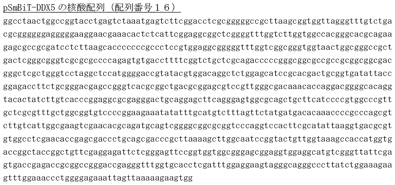
リソースおよび試薬
HeLa細胞: 理研BRC細胞バンク #RCB0007
DMEM: 富士フィルム和光 #043-30085
FBS: Thermo Fisher Scientific #10437028
非必須アミノ酸:富士フイルム・和光#139-15651
Opti-MEM I:Thermo Fisher Scientific #11058-021
96ウェルカルチャープレート。Promega #E5650
Lipofectamine3000 transfection reagent: Thermo Fisher Scientific #L3000008
Nano-Glo(登録商標)Live Cell Reagent:Promega #N2014
GloMax Discover Microplate Reader:Promega #GM3000
PrimeSTAR Max DNA polymerase: TaKaRa #R045A
TRIzol reagent (for total RNA extraction):Thermo Fisher Scientific #15596026
PrimeScript(商標)RT reagent Kit (for cDNA synthesis):TaKaRa #RR037A
In-Fusion HD Cloning Kit:TaKaRa #Z9649N
HeLa細胞: 理研BRC細胞バンク #RCB0007
DMEM: 富士フィルム和光 #043-30085
FBS: Thermo Fisher Scientific #10437028
非必須アミノ酸:富士フイルム・和光#139-15651
Opti-MEM I:Thermo Fisher Scientific #11058-021
96ウェルカルチャープレート。Promega #E5650
Lipofectamine3000 transfection reagent: Thermo Fisher Scientific #L3000008
Nano-Glo(登録商標)Live Cell Reagent:Promega #N2014
GloMax Discover Microplate Reader:Promega #GM3000
PrimeSTAR Max DNA polymerase: TaKaRa #R045A
TRIzol reagent (for total RNA extraction):Thermo Fisher Scientific #15596026
PrimeScript(商標)RT reagent Kit (for cDNA synthesis):TaKaRa #RR037A
In-Fusion HD Cloning Kit:TaKaRa #Z9649N
試験化合物
本発明のスクリーニング方法を検証するために以下の化合物を用いた。

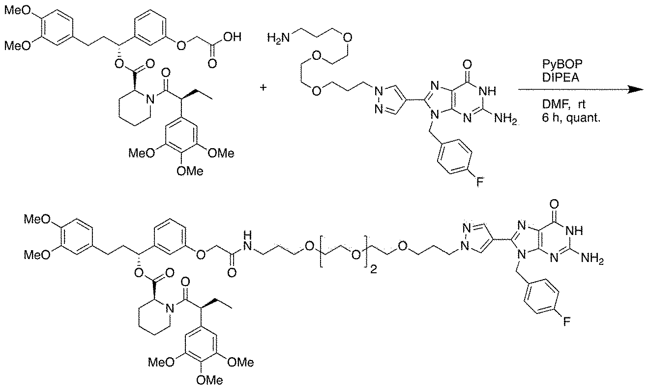
AUTAC4-2G、および8-ニトロ-cGMPは、特許7161760号に開示されている化合物およびそのタグ部分であり、AUTAC2-2G、およびFBnGは、本発明の方法でスクリーニングされた化合物が実際にオートファジーを誘導するかを検証するために新たに合成した化合物およびそのタグ部分である(FBnGおよびAUTAC2-2Gの合成スキームは以下の通り)。
本発明のスクリーニング方法を検証するために以下の化合物を用いた。
AUTAC4-2G、および8-ニトロ-cGMPは、特許7161760号に開示されている化合物およびそのタグ部分であり、AUTAC2-2G、およびFBnGは、本発明の方法でスクリーニングされた化合物が実際にオートファジーを誘導するかを検証するために新たに合成した化合物およびそのタグ部分である(FBnGおよびAUTAC2-2Gの合成スキームは以下の通り)。
FBnGの合成スキーム
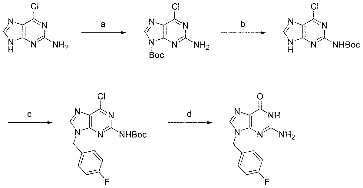
(a) Boc2O, DMAP, DMSO, rt, 12 h; (b) NaH, THF, 0 °C, 1 h, 62% (2 steps); (c) 4-fluorobenzyl alcohol, DEAD, PPh3, THF, rt, 3 h, 72%; (d) 80% HCO2H, 80 °C,16 h, 83%.
(a) Boc2O, DMAP, DMSO, rt, 12 h; (b) NaH, THF, 0 °C, 1 h, 62% (2 steps); (c) 4-fluorobenzyl alcohol, DEAD, PPh3, THF, rt, 3 h, 72%; (d) 80% HCO2H, 80 °C,16 h, 83%.
AUTAC2-2Gの合成スキーム

(a) Boc2O, CH2Cl2, rt, 3 h; (b) NaNO2, 20% CH3COOH, rt, 3 h, 40%; (c) TsCl, (CH3)3N・HCl, NEt3, CH2Cl2, rt, 2 h, 96%; (d) 4-pyrazoleboronic acid pinacol ester, K2CO3, DMF, 75 °C, overnight, 71%; (e) 6-chloro-9-(4-fluorobenzyl)-8-iodo-N-(tetrahydro-2H-pyran-2-yl)-9H-purin-2-amine, Pd(PPh3)4, K2CO3, 66% N,N'-dimethylethyleneurea, 120 °C, 2 h, 61%; (f) 80% HCOOH, 80 °C, overnight, 85%; (g) PyBOP, DIPEA, rt, 18 h, 33%.
(a) Boc2O, CH2Cl2, rt, 3 h; (b) NaNO2, 20% CH3COOH, rt, 3 h, 40%; (c) TsCl, (CH3)3N・HCl, NEt3, CH2Cl2, rt, 2 h, 96%; (d) 4-pyrazoleboronic acid pinacol ester, K2CO3, DMF, 75 °C, overnight, 71%; (e) 6-chloro-9-(4-fluorobenzyl)-8-iodo-N-(tetrahydro-2H-pyran-2-yl)-9H-purin-2-amine, Pd(PPh3)4, K2CO3, 66% N,N'-dimethylethyleneurea, 120 °C, 2 h, 61%; (f) 80% HCOOH, 80 °C, overnight, 85%; (g) PyBOP, DIPEA, rt, 18 h, 33%.
1-2.試験手順
[実施例1]
成長培地を入れた96ウェルプレートで増殖したHeLa細胞に、Lipofectamine3000(登録商標)トランスフェクション試薬(Thermo Fisher Scientific Inc.)を用いて、添付書類の記載に従って、pLgBiT-p62とpSmBiT-DDX5をトランスフェクションした。試薬を添加して24時間経過した後、ウェルにAUTAC2-2Gを10μMの濃度となる量添加して12時間トランスフェクションした細胞をAUTAC2-2Gで処理した。その後、成長培地を、100μLのOpti-MEM(登録商標)I(Thermo Fisher Scientific Inc.)に交換した。基質フリマジンを含む 5x Nano-Glo Live Cell Reagentを25μL添加し、直ちにGloMax(登録商標) Discover Microplate Reader(Promega Corporation)でルミネッセンスを測定した。

[実施例1]
成長培地を入れた96ウェルプレートで増殖したHeLa細胞に、Lipofectamine3000(登録商標)トランスフェクション試薬(Thermo Fisher Scientific Inc.)を用いて、添付書類の記載に従って、pLgBiT-p62とpSmBiT-DDX5をトランスフェクションした。試薬を添加して24時間経過した後、ウェルにAUTAC2-2Gを10μMの濃度となる量添加して12時間トランスフェクションした細胞をAUTAC2-2Gで処理した。その後、成長培地を、100μLのOpti-MEM(登録商標)I(Thermo Fisher Scientific Inc.)に交換した。基質フリマジンを含む 5x Nano-Glo Live Cell Reagentを25μL添加し、直ちにGloMax(登録商標) Discover Microplate Reader(Promega Corporation)でルミネッセンスを測定した。
[実施例2]
ウェルにAUTAC4-2Gを10μMの濃度となる量添加して12時間トランスフェクションした細胞をAUTAC4-2Gで処理した以外は、実施例1と同様にして、細胞を処理してルミネッセンスを測定した。
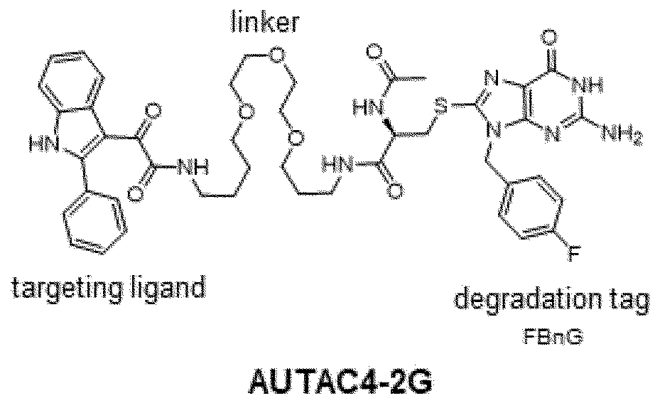
ウェルにAUTAC4-2Gを10μMの濃度となる量添加して12時間トランスフェクションした細胞をAUTAC4-2Gで処理した以外は、実施例1と同様にして、細胞を処理してルミネッセンスを測定した。
[実施例3]
ウェルに8-ニトロ-cGMPを10μMの濃度となる量添加し、24時間トランスフェクションした細胞を8-ニトロ-cGMPで処理した以外は、実施例1と同様にして、細胞を処理してルミネッセンスを測定した。
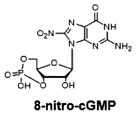
ウェルに8-ニトロ-cGMPを10μMの濃度となる量添加し、24時間トランスフェクションした細胞を8-ニトロ-cGMPで処理した以外は、実施例1と同様にして、細胞を処理してルミネッセンスを測定した。
[実施例4]
ウェルにFBnGを10μMの濃度となる量添加し、24時間トランスフェクションした細胞をFBnGで処理した以外は、実施例1と同様にして、細胞を処理してルミネッセンスを測定した。
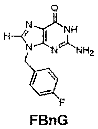
ウェルにFBnGを10μMの濃度となる量添加し、24時間トランスフェクションした細胞をFBnGで処理した以外は、実施例1と同様にして、細胞を処理してルミネッセンスを測定した。
[比較例1]
ウェルにDMSO(ジメチルスルホキシド)を10μMの濃度となる量添加し、24時間トランスフェクションした細胞をDMSOで処理した以外は、実施例1と同様にして、細胞を処理してルミネッセンスを測定した。
ウェルにDMSO(ジメチルスルホキシド)を10μMの濃度となる量添加し、24時間トランスフェクションした細胞をDMSOで処理した以外は、実施例1と同様にして、細胞を処理してルミネッセンスを測定した。
1-3.測定結果
図1に、実施例1乃至4においてルミネッセンスの測定開始から10分後の発光量を示す。データは、3回の独立した実験からの平均値で示す。グラフは、107を上限に示しており、実際には、それ以上の発光量を示した。エラーバーは標準偏差を示す。統計解析は、Turkey-Kramer多重比較検定を用いて行った。P値<0.01は統計的に有意であるとみなした。
AUTAC2-2G、AUTAC4-2G、8-ニトロ-cGMP、およびFBnGで処理した細胞で、DMSOで処理した細胞に比べ、千倍以上の発光量を示し、これらの化合物の存在で、p62とDDX5が近接したことが実証され、この方法を用いることでp62とDDX5を近接させる化合物をスクリーニングできることが示された。
図1に、実施例1乃至4においてルミネッセンスの測定開始から10分後の発光量を示す。データは、3回の独立した実験からの平均値で示す。グラフは、107を上限に示しており、実際には、それ以上の発光量を示した。エラーバーは標準偏差を示す。統計解析は、Turkey-Kramer多重比較検定を用いて行った。P値<0.01は統計的に有意であるとみなした。
AUTAC2-2G、AUTAC4-2G、8-ニトロ-cGMP、およびFBnGで処理した細胞で、DMSOで処理した細胞に比べ、千倍以上の発光量を示し、これらの化合物の存在で、p62とDDX5が近接したことが実証され、この方法を用いることでp62とDDX5を近接させる化合物をスクリーニングできることが示された。
2.スクリーニングされた試験化合物のオートファジー誘導作用の検証
本発明のスクリーニング方法の有効性を実証するために、本発明者らが今回新たに合成した化合物AUTAC2-2Gで、FKBP12でトランスフェクトした細胞を処理してFKBP12を分解し得るか試験した。
本発明のスクリーニング方法の有効性を実証するために、本発明者らが今回新たに合成した化合物AUTAC2-2Gで、FKBP12でトランスフェクトした細胞を処理してFKBP12を分解し得るか試験した。
2-1.pEGFP-FKBP12の調製
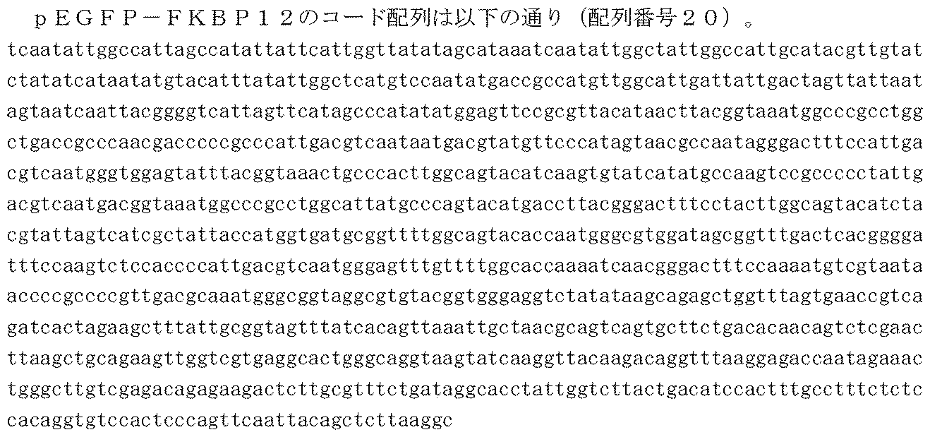
pEGFP-FKBP12を作製するために、pEGFP-HaloTag [Takahashi et al., Mol. Cell 76 797-810.e10 (2019)]を、オリゴプライマー(5'- cgcaaatctagacttgtacagctcgtcc-3' および 5'- ggccgcttcgagcagatg-3')を用いてポリメラーゼチェーン反応により線形化した。FKBP12のcDNAは、HeLa細胞total RNAからオリゴ(dT)15プライマーを用いて合成し、オリゴプライマー(5'-aagtctagatttgcgatgggagtgcaggtgg-3'と5'- ctgctcgaagcggcctcattccagttagaagc-3')で増幅し、Infusion cloning systemでベクターに挿入した(下線15 ntはInfusion arm)。
pEGFP-FKBP12を作製するために、pEGFP-HaloTag [Takahashi et al., Mol. Cell 76 797-810.e10 (2019)]を、オリゴプライマー(5'- cgcaaatctagacttgtacagctcgtcc-3' および 5'- ggccgcttcgagcagatg-3')を用いてポリメラーゼチェーン反応により線形化した。FKBP12のcDNAは、HeLa細胞total RNAからオリゴ(dT)15プライマーを用いて合成し、オリゴプライマー(5'-aagtctagatttgcgatgggagtgcaggtgg-3'と5'- ctgctcgaagcggcctcattccagttagaagc-3')で増幅し、Infusion cloning systemでベクターに挿入した(下線15 ntはInfusion arm)。
2-2.リソースと試薬
HeLa細胞: 理研BRC細胞バンク #RCB0007
Dulbecco's Modified Eagle Medium(DMEM): 富士フィルム和光 #043-30085
FBS: Thermo Fisher Scientific #10437028
非必須アミノ酸:富士フイルム・和光#139-15651
Opti-MEM I: Thermo Fisher Scientific #11058-021
96ウェル培養プレート。Thermo Fisher Scientific #165305
Lipofectamine3000 transfection reagent:サーモフィッシャーサイエンティフィック #L3000008
ライブセルイメージング溶液: Thermo Fisher Scientific #A14291DJ
蛍光顕微鏡: キーエンス #BZ-X800
PrimeSTAR Max DNA polymerase: TaKaRa #R045A
TRIzol reagent (for total RNA extraction): Thermo Fisher Scientific #15596026
PrimeScript(商標)RT reagent Kit (for cDNA synthesis): TaKaRa #RR037A
In-Fusion HD Cloning Kit: TaKaRa #Z9649N
HeLa細胞: 理研BRC細胞バンク #RCB0007
Dulbecco's Modified Eagle Medium(DMEM): 富士フィルム和光 #043-30085
FBS: Thermo Fisher Scientific #10437028
非必須アミノ酸:富士フイルム・和光#139-15651
Opti-MEM I: Thermo Fisher Scientific #11058-021
96ウェル培養プレート。Thermo Fisher Scientific #165305
Lipofectamine3000 transfection reagent:サーモフィッシャーサイエンティフィック #L3000008
ライブセルイメージング溶液: Thermo Fisher Scientific #A14291DJ
蛍光顕微鏡: キーエンス #BZ-X800
PrimeSTAR Max DNA polymerase: TaKaRa #R045A
TRIzol reagent (for total RNA extraction): Thermo Fisher Scientific #15596026
PrimeScript(商標)RT reagent Kit (for cDNA synthesis): TaKaRa #RR037A
In-Fusion HD Cloning Kit: TaKaRa #Z9649N
2-3.試験手順
[実施例5]
HeLa細胞を96ウェルプレート上で増殖させ、Lipofectamine3000トランスフェクション試薬を用いてpEGFP-FKBP12でトランスフェクトした。24時間後、トランスフェクトされた細胞を、0.01mMのAUTAC2-2Gを含む新鮮なDMEM中、並びに比較対照としてDMSOを含む新鮮なDMEM中でさらに24時間インキュベートした。その後、培地を除去し、100μLのライブセルイメージング溶液に置き換えた。EGFP-FKBP12レベルは、EGFP蛍光を測定することによって分析された。
[実施例5]
HeLa細胞を96ウェルプレート上で増殖させ、Lipofectamine3000トランスフェクション試薬を用いてpEGFP-FKBP12でトランスフェクトした。24時間後、トランスフェクトされた細胞を、0.01mMのAUTAC2-2Gを含む新鮮なDMEM中、並びに比較対照としてDMSOを含む新鮮なDMEM中でさらに24時間インキュベートした。その後、培地を除去し、100μLのライブセルイメージング溶液に置き換えた。EGFP-FKBP12レベルは、EGFP蛍光を測定することによって分析された。
[実施例6]
トランスフェクトされた細胞を、0.1mMのAUTAC2-2Gを含む新鮮なDMEM中でさらに24時間インキュベートした以外は、実施例5と同様にして細胞を処理してEGFP蛍光を測定した。
トランスフェクトされた細胞を、0.1mMのAUTAC2-2Gを含む新鮮なDMEM中でさらに24時間インキュベートした以外は、実施例5と同様にして細胞を処理してEGFP蛍光を測定した。
[実施例7]
トランスフェクトされた細胞を、1mMのAUTAC2-2Gを含む新鮮なDMEM中でさらに24時間インキュベートした以外は、実施例5と同様にして細胞を処理してEGFP蛍光を測定した。
トランスフェクトされた細胞を、1mMのAUTAC2-2Gを含む新鮮なDMEM中でさらに24時間インキュベートした以外は、実施例5と同様にして細胞を処理してEGFP蛍光を測定した。
[実施例8]
トランスフェクトされた細胞を、10mMのAUTAC2-2Gを含む新鮮なDMEM中でさらに24時間インキュベートした以外は、実施例5と同様にして細胞を処理してEGFP蛍光を測定した。
トランスフェクトされた細胞を、10mMのAUTAC2-2Gを含む新鮮なDMEM中でさらに24時間インキュベートした以外は、実施例5と同様にして細胞を処理してEGFP蛍光を測定した。
[比較例2]
トランスフェクトされた細胞を、DMSOを含む新鮮なDMEM中でさらに24時間インキュベートした以外は、実施例5と同様にして細胞を処理してEGFP蛍光を測定した。
トランスフェクトされた細胞を、DMSOを含む新鮮なDMEM中でさらに24時間インキュベートした以外は、実施例5と同様にして細胞を処理してEGFP蛍光を測定した。
2-4.測定結果
図2は、各条件でのEGFP蛍光レベルを示す。各数値は3つの独立した実験からの平均EGFP蛍光レベルを示す。同じ文字が添えられていないデータは、Turkey-Kramer多重比較検定を用いて統計的に有意である。P値<0.01は統計的に有意とみなされた。データは、DMSOコントロールの平均値に対して正規化した。
AUTAC2-2Gを含む培地中でpEGFP-FKBP12でトランスフェクトされた細胞を培養すると、AUTAC2-2G濃度依存的に、蛍光レベルが減少し、AUTAC2-2GがオートファジーによるFKBP12の分解を誘導していることが確認された。従って、本発明のスクリーニング方法によれば、オートファジーを誘導する新規化合物を探索可能である。
図2は、各条件でのEGFP蛍光レベルを示す。各数値は3つの独立した実験からの平均EGFP蛍光レベルを示す。同じ文字が添えられていないデータは、Turkey-Kramer多重比較検定を用いて統計的に有意である。P値<0.01は統計的に有意とみなされた。データは、DMSOコントロールの平均値に対して正規化した。
AUTAC2-2Gを含む培地中でpEGFP-FKBP12でトランスフェクトされた細胞を培養すると、AUTAC2-2G濃度依存的に、蛍光レベルが減少し、AUTAC2-2GがオートファジーによるFKBP12の分解を誘導していることが確認された。従って、本発明のスクリーニング方法によれば、オートファジーを誘導する新規化合物を探索可能である。
3.DEAD-box型RNAヘリカーゼおよびp62を含む液滴の形成の測定
[実施例9]
新たに合成し、オートファジーによりシヌクレインを分解することを確認した化合物SLF’-AUTACを用いて、DDX5およびp62を含む液滴の形成を誘導するかを試験した(SLF’-AUTACの構造および合成スキームならびにシヌクレイン分解試験の概要は以下の通り)。
[実施例9]
新たに合成し、オートファジーによりシヌクレインを分解することを確認した化合物SLF’-AUTACを用いて、DDX5およびp62を含む液滴の形成を誘導するかを試験した(SLF’-AUTACの構造および合成スキームならびにシヌクレイン分解試験の概要は以下の通り)。
SLF’-AUTACの構造式

SLF’-AUTACの合成スキーム
SLF’-AUTACによる変異FKBPタグ化シヌクレイン分解試験の概要

変異FKBPでタグ化したGFP-α-シヌクレイン(GFP-αSyn-FKBPF36V)が一過性発現したHeLa細胞をSLF’-AUTACで48時間処理した。非処理細胞のウェル、処理細胞のウェルそれぞれにおいて、GFP蛍光を蛍光顕微鏡で撮影した。GFP蛍光強度からGFP-αSyn-FKBPF36Vの分解を評価した。上図から、1 μM、10 μMのSLF’-AUTAC処理細胞では、非処理群(DMSO)と比較して、GFP-αSyn-FKBPF36Vの分解が認められた。
変異FKBPでタグ化したGFP-α-シヌクレイン(GFP-αSyn-FKBPF36V)が一過性発現したHeLa細胞をSLF’-AUTACで48時間処理した。非処理細胞のウェル、処理細胞のウェルそれぞれにおいて、GFP蛍光を蛍光顕微鏡で撮影した。GFP蛍光強度からGFP-αSyn-FKBPF36Vの分解を評価した。上図から、1 μM、10 μMのSLF’-AUTAC処理細胞では、非処理群(DMSO)と比較して、GFP-αSyn-FKBPF36Vの分解が認められた。
試験手順
変異FKBPでタグ化したGFP(GFP-FKBPF36V)を一過性発現するHeLa細胞を、1μMのSLF’-AUTACで12時間処理した。処理後の細胞を4%パラホルムアルデヒドで固定してから、0.1%TritonX-100/PBSで浸透化し、1%BSA/PBSでブロッキングし、ウサギ由来抗DDX5抗体とマウス由来抗p62抗体をそれぞれ1%ずつ含むPBSに浸けて一晩4℃でインキュベートした。その後、AlexaFluor555標識抗ウサギIgGとAlexaFluor555標識抗マウスIgGをそれぞれ1%ずつ含むPBSに浸けて1時間室温でインキュベートした。免疫染色後の細胞を蛍光顕微鏡で観察した。
変異FKBPでタグ化したGFP(GFP-FKBPF36V)を一過性発現するHeLa細胞を、1μMのSLF’-AUTACで12時間処理した。処理後の細胞を4%パラホルムアルデヒドで固定してから、0.1%TritonX-100/PBSで浸透化し、1%BSA/PBSでブロッキングし、ウサギ由来抗DDX5抗体とマウス由来抗p62抗体をそれぞれ1%ずつ含むPBSに浸けて一晩4℃でインキュベートした。その後、AlexaFluor555標識抗ウサギIgGとAlexaFluor555標識抗マウスIgGをそれぞれ1%ずつ含むPBSに浸けて1時間室温でインキュベートした。免疫染色後の細胞を蛍光顕微鏡で観察した。
試験結果
図3に蛍光顕微鏡で観察したイメージを示す(スケールバー:10μm)。
FBnG構造を含むSLF’-AUTACをHeLa細胞に添加することによって、細胞内でDDX5とp62が液滴を形成したことが観察された。この実験では、SLF‘-AUTACで処理後に細胞を固定化してからDDX5およびp62を観察しており、DDX5とp62が、細胞内の同一位置に観測されていることから、両者が同一の液滴に存在することが証明された。
図3に蛍光顕微鏡で観察したイメージを示す(スケールバー:10μm)。
FBnG構造を含むSLF’-AUTACをHeLa細胞に添加することによって、細胞内でDDX5とp62が液滴を形成したことが観察された。この実験では、SLF‘-AUTACで処理後に細胞を固定化してからDDX5およびp62を観察しており、DDX5とp62が、細胞内の同一位置に観測されていることから、両者が同一の液滴に存在することが証明された。
[実施例10]
AUTAC2-2Gを用いて、DDX5およびp62を含む液滴の形成を誘導するかを試験した。
AUTAC2-2Gを用いて、DDX5およびp62を含む液滴の形成を誘導するかを試験した。
試験手順
GFP-FKBP12およびRFP-DDX5、またはGFP-FKBP12およびRFP-p62を一過性発現したHeLa細胞を、10μMの濃度となるようにAUTAC2-2Gを添加して13時間細胞を処理した。処理した細胞を蛍光顕微鏡により生細胞観察し、GFPおよびRFPに由来する蛍光を継続的に観察した。途中、観察開始5分後に5% 1,6-ヘキサンジオールを添加して、視野にあるドット状の構造が消失するかを観察した。
1,6-ヘキサンジオールは、液滴を解消する作用が知られており、これにより、顕微鏡の視野にあるドット状の構造が液滴であるかを確認することが可能となる。
GFP-FKBP12およびRFP-DDX5、またはGFP-FKBP12およびRFP-p62を一過性発現したHeLa細胞を、10μMの濃度となるようにAUTAC2-2Gを添加して13時間細胞を処理した。処理した細胞を蛍光顕微鏡により生細胞観察し、GFPおよびRFPに由来する蛍光を継続的に観察した。途中、観察開始5分後に5% 1,6-ヘキサンジオールを添加して、視野にあるドット状の構造が消失するかを観察した。
1,6-ヘキサンジオールは、液滴を解消する作用が知られており、これにより、顕微鏡の視野にあるドット状の構造が液滴であるかを確認することが可能となる。
試験結果
図4に蛍光顕微鏡で観察したイメージを示す(スケールバー:10μm)。図4Aが、GFP-FKBP12およびRFP-DDX5を一過性発現したHeLa細胞をAUTAC2-2Gで処理した場合の蛍光顕微鏡で観察したイメージであり、図4Bが、GFP-FKBP12およびRFP-p62を一過性発現したHeLa細胞をAUTAC2-2Gで処理した場合の蛍光顕微鏡で観察したイメージである。
それぞれのHeLa細胞で、GFP-FKBP12およびRFP-DDX5を含む液滴、およびGFP-FKBP12およびRFP-p62を含むドット状の構造が観察された。この構造は、1,6-ヘキサンジオール添加後に消失したことから液滴であることが確認された。
図4に蛍光顕微鏡で観察したイメージを示す(スケールバー:10μm)。図4Aが、GFP-FKBP12およびRFP-DDX5を一過性発現したHeLa細胞をAUTAC2-2Gで処理した場合の蛍光顕微鏡で観察したイメージであり、図4Bが、GFP-FKBP12およびRFP-p62を一過性発現したHeLa細胞をAUTAC2-2Gで処理した場合の蛍光顕微鏡で観察したイメージである。
それぞれのHeLa細胞で、GFP-FKBP12およびRFP-DDX5を含む液滴、およびGFP-FKBP12およびRFP-p62を含むドット状の構造が観察された。この構造は、1,6-ヘキサンジオール添加後に消失したことから液滴であることが確認された。
Claims (9)
- DEAD-box型RNAヘリカーゼに試験化合物を接触させ、前記DEAD-box型RNAヘリカーゼに対する前記試験化合物の結合親和性を測定し、
所定値以上の結合親和性を有する化合物を選択する、
オートファジー誘導化合物をスクリーニングする方法。 - p62の存在下で、DEAD-box型RNAヘリカーゼに試験化合物を接触させ、
前記DEAD-box型RNAヘリカーゼと前記p62の相互作用を測定し、
前記相互作用を誘導した化合物を選択する、
オートファジー誘導化合物をスクリーニングする方法。 - 前記DEAD-box型RNAヘリカーゼおよび前記p62を、細胞内で発現し、前記細胞に前記試験化合物を接触させる、請求項2に記載の方法。
- p62、およびDEAD-box型RNAヘリカーゼの存在下で、試験化合物を導入し、
前記DEAD-box型RNAヘリカーゼおよび前記p62を含む液滴の形成を測定し、
前記液滴形成を誘導した化合物を選択する、
オートファジー誘導化合物をスクリーニングする方法。 - 前記DEAD-box型RNAヘリカーゼは、DDX5またはDDX17である、請求項1~4のいずれか1項に記載の方法。
- 請求項1~5のいずれか1項に記載の方法で得られた化合物に、リンカーを介して、標的細胞内分子またはオルガネラに特異的に結合するリガンドを連結する、標的細胞内分子またはオルガネラのオートファジーによる分解を誘導する化合物の製造方法。
- 請求項6に記載の方法で得られた化合物を、細胞内で、p62の存在下で、DEAD-box型RNAヘリカーゼと接触させて、標的細胞内分子または標的オルガネラのオートファジーによる分解を誘導する方法。
- 請求項6に記載の方法で得られた化合物を、細胞内で、p62の存在下で、DEAD-box型RNAヘリカーゼと接触させて、前記化合物、前記p62および前記DEAD-box型RNAヘリカーゼを含む液滴を形成する、請求項7に記載の方法。
- 癌または細胞変性疾患を予防または治療する、請求項7または8に記載の方法。
Priority Applications (6)
| Application Number | Priority Date | Filing Date | Title |
|---|---|---|---|
| PCT/JP2023/012419 WO2024201691A1 (ja) | 2023-03-28 | 2023-03-28 | オートファジーを誘導する化合物のスクリーニング方法 |
| JP2025511227A JPWO2024204648A1 (ja) | 2023-03-28 | 2024-03-28 | |
| EP24780746.4A EP4692367A1 (en) | 2023-03-28 | 2024-03-28 | Screening method for compound inducing autophagy |
| PCT/JP2024/012899 WO2024204648A1 (ja) | 2023-03-28 | 2024-03-28 | オートファジーを誘導する化合物のスクリーニング方法 |
| KR1020257031424A KR20250169534A (ko) | 2023-03-28 | 2024-03-28 | 오토파지를 유도하는 화합물의 스크리닝 방법 |
| CN202480022512.XA CN121002194A (zh) | 2023-03-28 | 2024-03-28 | 诱导自噬的化合物的筛选方法 |
Applications Claiming Priority (1)
| Application Number | Priority Date | Filing Date | Title |
|---|---|---|---|
| PCT/JP2023/012419 WO2024201691A1 (ja) | 2023-03-28 | 2023-03-28 | オートファジーを誘導する化合物のスクリーニング方法 |
Publications (1)
| Publication Number | Publication Date |
|---|---|
| WO2024201691A1 true WO2024201691A1 (ja) | 2024-10-03 |
Family
ID=92904278
Family Applications (2)
| Application Number | Title | Priority Date | Filing Date |
|---|---|---|---|
| PCT/JP2023/012419 Ceased WO2024201691A1 (ja) | 2023-03-28 | 2023-03-28 | オートファジーを誘導する化合物のスクリーニング方法 |
| PCT/JP2024/012899 Ceased WO2024204648A1 (ja) | 2023-03-28 | 2024-03-28 | オートファジーを誘導する化合物のスクリーニング方法 |
Family Applications After (1)
| Application Number | Title | Priority Date | Filing Date |
|---|---|---|---|
| PCT/JP2024/012899 Ceased WO2024204648A1 (ja) | 2023-03-28 | 2024-03-28 | オートファジーを誘導する化合物のスクリーニング方法 |
Country Status (5)
| Country | Link |
|---|---|
| EP (1) | EP4692367A1 (ja) |
| JP (1) | JPWO2024204648A1 (ja) |
| KR (1) | KR20250169534A (ja) |
| CN (1) | CN121002194A (ja) |
| WO (2) | WO2024201691A1 (ja) |
Citations (2)
| Publication number | Priority date | Publication date | Assignee | Title |
|---|---|---|---|---|
| WO2018143403A1 (ja) * | 2017-02-03 | 2018-08-09 | 国立大学法人東北大学 | 複素環化合物 |
| WO2019013181A1 (ja) * | 2017-07-10 | 2019-01-17 | 国立大学法人東北大学 | 傷害を受けたミトコンドリアのオートファジー機構による分解剤 |
Family Cites Families (5)
| Publication number | Priority date | Publication date | Assignee | Title |
|---|---|---|---|---|
| EP2836482B1 (en) | 2012-04-10 | 2019-12-25 | The Regents of The University of California | Compositions and methods for treating cancer |
| CA2904393A1 (en) | 2013-03-15 | 2014-09-25 | Araxes Pharma Llc | Covalent inhibitors of kras g12c |
| EP3055290B1 (en) | 2013-10-10 | 2019-10-02 | Araxes Pharma LLC | Inhibitors of kras g12c |
| JP2017528498A (ja) | 2014-09-25 | 2017-09-28 | アラクセス ファーマ エルエルシー | Kras g12c変異体タンパク質のインヒビター |
| WO2024054876A1 (en) | 2022-09-07 | 2024-03-14 | Arvinas Operations, Inc. | Leucine rich repeat kinase 2 (lrrk2) degrading compounds and associated methods of use |
-
2023
- 2023-03-28 WO PCT/JP2023/012419 patent/WO2024201691A1/ja not_active Ceased
-
2024
- 2024-03-28 WO PCT/JP2024/012899 patent/WO2024204648A1/ja not_active Ceased
- 2024-03-28 EP EP24780746.4A patent/EP4692367A1/en active Pending
- 2024-03-28 JP JP2025511227A patent/JPWO2024204648A1/ja active Pending
- 2024-03-28 KR KR1020257031424A patent/KR20250169534A/ko active Pending
- 2024-03-28 CN CN202480022512.XA patent/CN121002194A/zh active Pending
Patent Citations (2)
| Publication number | Priority date | Publication date | Assignee | Title |
|---|---|---|---|---|
| WO2018143403A1 (ja) * | 2017-02-03 | 2018-08-09 | 国立大学法人東北大学 | 複素環化合物 |
| WO2019013181A1 (ja) * | 2017-07-10 | 2019-01-17 | 国立大学法人東北大学 | 傷害を受けたミトコンドリアのオートファジー機構による分解剤 |
Non-Patent Citations (1)
| Title |
|---|
| ARIMOTO, HIROKAZU; TAKAHASHI, DAIKI; ITTO, KAORI: "4SY24-5 Targeted degradation of cytosolic materials using selective autophagy", ANNUAL MEETING OF THE JAPAN SOCIETY FOR BIOSCIENCE, BIOTECHNOLOGY, AND AGROCHEMISTRY (JSBBA 2018), vol. 2018, 5 March 2018 (2018-03-05), pages 2101, XP009558719 * |
Also Published As
| Publication number | Publication date |
|---|---|
| WO2024204648A1 (ja) | 2024-10-03 |
| KR20250169534A (ko) | 2025-12-03 |
| CN121002194A (zh) | 2025-11-21 |
| JPWO2024204648A1 (ja) | 2024-10-03 |
| EP4692367A1 (en) | 2026-02-11 |
Similar Documents
| Publication | Publication Date | Title |
|---|---|---|
| Helma et al. | Direct and dynamic detection of HIV-1 in living cells | |
| Foglieni et al. | Split GFP technologies to structurally characterize and quantify functional biomolecular interactions of FTD-related proteins | |
| Lafarga et al. | Widespread displacement of DNA‐and RNA‐binding factors underlies toxicity of arginine‐rich cell‐penetrating peptides | |
| Vu et al. | Defining the Caprin-1 interactome in unstressed and stressed conditions | |
| JP2016523088A (ja) | ペプチドの細胞輸送をモニタリングする方法 | |
| Yamashiro et al. | Armitage determines Piwi− piRISC processing from precursor formation and quality control to inter‐organelle translocation | |
| Bacon et al. | Quantitative yeast–yeast two hybrid for the discovery and binding affinity estimation of protein–protein interactions | |
| Romano et al. | Multi-omic profiling reveals the ataxia protein sacsin is required for integrin trafficking and synaptic organization | |
| Mumford et al. | Simple visualization of submicroscopic protein clusters with a phase-separation-based fluorescent reporter | |
| US20220365070A1 (en) | Compositions, systems, and methods for identifying a compound that modulates one or more characteristics associated with a rbm20 condensate and/or a rbm20 polypeptide | |
| Pírez-Schirmer et al. | Oriented functionalization of magnetic beads with in vivo biotinylated nanobodies for rapid MALDI-TOF MS ultrasensitive quantitation of microcystins in biological samples | |
| US20150105274A1 (en) | Methods for the Characterisation of Interaction Sites on Target Proteins | |
| Shen et al. | A general method for chemogenetic control of peptide function | |
| Bressendorff et al. | Importance of an N-terminal structural switch in the distinction between small RNA-bound and free ARGONAUTE | |
| KR101190157B1 (ko) | Rab5를 이용한 세포내 단백질 상호 작용 시각화 방법 | |
| Rawlinson et al. | Conformational dynamics, RNA binding, and phase separation regulate the multifunctionality of rabies virus P protein | |
| Sowa et al. | Preparation of screening assays for ADP-ribosyl readers and erasers using the GAP-tag as a binding probe | |
| WO2024201691A1 (ja) | オートファジーを誘導する化合物のスクリーニング方法 | |
| Bell et al. | An electrostatic repulsion model of centromere organisation | |
| Maurel et al. | Critical impact of lysine 136 in TDP-43 phase separation, compartmentalization, and aggregation in living vertebrates | |
| Meyer et al. | Manipulating condensation of thermo-sensitive SUF4 protein tunes flowering time in Arabidopsis thaliana | |
| CA2620934C (en) | Compositions and methods for determining protein interactions using tat signal sequence and marker protein | |
| González Montoro et al. | Specificity of transmembrane protein palmitoylation in yeast | |
| EP3172563B1 (en) | Methods for detecting interactions in eukaryotic cells using microtubule structures and dynamics | |
| Kettel et al. | Stress-induced clustering of the UPR sensor IRE1α is driven by disordered regions within its ER lumenal domain |
Legal Events
| Date | Code | Title | Description |
|---|---|---|---|
| 121 | Ep: the epo has been informed by wipo that ep was designated in this application |
Ref document number: 23930331 Country of ref document: EP Kind code of ref document: A1 |
|
| NENP | Non-entry into the national phase |
Ref country code: DE |
|
| 122 | Ep: pct application non-entry in european phase |
Ref document number: 23930331 Country of ref document: EP Kind code of ref document: A1 |