WO2024181506A1 - 情報処理システム、情報処理システムの制御方法、制御プログラム、記録媒体 - Google Patents
情報処理システム、情報処理システムの制御方法、制御プログラム、記録媒体 Download PDFInfo
- Publication number
- WO2024181506A1 WO2024181506A1 PCT/JP2024/007348 JP2024007348W WO2024181506A1 WO 2024181506 A1 WO2024181506 A1 WO 2024181506A1 JP 2024007348 W JP2024007348 W JP 2024007348W WO 2024181506 A1 WO2024181506 A1 WO 2024181506A1
- Authority
- WO
- WIPO (PCT)
- Prior art keywords
- information
- information processing
- medical image
- subject
- processing system
- Prior art date
- Legal status (The legal status is an assumption and is not a legal conclusion. Google has not performed a legal analysis and makes no representation as to the accuracy of the status listed.)
- Ceased
Links
Images


Classifications
-
- A—HUMAN NECESSITIES
- A61—MEDICAL OR VETERINARY SCIENCE; HYGIENE
- A61B—DIAGNOSIS; SURGERY; IDENTIFICATION
- A61B6/00—Apparatus or devices for radiation diagnosis; Apparatus or devices for radiation diagnosis combined with radiation therapy equipment
- A61B6/50—Apparatus or devices for radiation diagnosis; Apparatus or devices for radiation diagnosis combined with radiation therapy equipment specially adapted for specific body parts; specially adapted for specific clinical applications
- A61B6/505—Apparatus or devices for radiation diagnosis; Apparatus or devices for radiation diagnosis combined with radiation therapy equipment specially adapted for specific body parts; specially adapted for specific clinical applications for diagnosis of bone
-
- A—HUMAN NECESSITIES
- A61—MEDICAL OR VETERINARY SCIENCE; HYGIENE
- A61B—DIAGNOSIS; SURGERY; IDENTIFICATION
- A61B6/00—Apparatus or devices for radiation diagnosis; Apparatus or devices for radiation diagnosis combined with radiation therapy equipment
- A61B6/56—Details of data transmission or power supply, e.g. use of slip rings
- A61B6/563—Details of data transmission or power supply, e.g. use of slip rings involving image data transmission via a network
-
- G—PHYSICS
- G16—INFORMATION AND COMMUNICATION TECHNOLOGY [ICT] SPECIALLY ADAPTED FOR SPECIFIC APPLICATION FIELDS
- G16H—HEALTHCARE INFORMATICS, i.e. INFORMATION AND COMMUNICATION TECHNOLOGY [ICT] SPECIALLY ADAPTED FOR THE HANDLING OR PROCESSING OF MEDICAL OR HEALTHCARE DATA
- G16H30/00—ICT specially adapted for the handling or processing of medical images
- G16H30/40—ICT specially adapted for the handling or processing of medical images for processing medical images, e.g. editing
-
- G—PHYSICS
- G16—INFORMATION AND COMMUNICATION TECHNOLOGY [ICT] SPECIALLY ADAPTED FOR SPECIFIC APPLICATION FIELDS
- G16H—HEALTHCARE INFORMATICS, i.e. INFORMATION AND COMMUNICATION TECHNOLOGY [ICT] SPECIALLY ADAPTED FOR THE HANDLING OR PROCESSING OF MEDICAL OR HEALTHCARE DATA
- G16H50/00—ICT specially adapted for medical diagnosis, medical simulation or medical data mining; ICT specially adapted for detecting, monitoring or modelling epidemics or pandemics
- G16H50/20—ICT specially adapted for medical diagnosis, medical simulation or medical data mining; ICT specially adapted for detecting, monitoring or modelling epidemics or pandemics for computer-aided diagnosis, e.g. based on medical expert systems
Definitions
- This disclosure relates to a system that uses medical images to estimate a subject's disease or predict the subject's bone characteristics, and a control method for the system.
- Patent Document 1 discloses an estimation device that estimates a subject's disease by inputting medical images of the subject into a learning model.
- An information processing system includes an acquisition unit that acquires information including medical image information related to a medical image of a subject, and a generation unit that generates input information including the medical image and the medical image information.
- a method for controlling an information processing system includes an acquisition step of acquiring information including medical image information related to a medical image of a subject, and a generation step of generating input information including the medical image and the medical image information.
- the information processing system according to each aspect of the present disclosure may be realized by a computer.
- the control program for the information processing system that causes the computer to operate as each unit (software element) of the information processing system to realize the information processing system, and the computer-readable recording medium on which it is recorded also fall within the scope of the present disclosure.
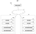
- FIG. 1 is a diagram illustrating a configuration example of an information processing system according to an embodiment of the present disclosure.
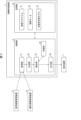
- 1 is a block diagram illustrating an example of a configuration of an information processing device according to an embodiment of the present disclosure.
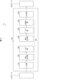
- FIG. 2 is a diagram illustrating an example of a configuration of a learning model executed by an estimation unit.
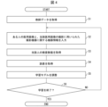
- 13 is a flowchart showing an example of the flow of a learning process by a learning unit.
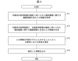
- 11 is a flowchart illustrating an example of a flow of processing performed by an information processing device.
- FIG. 2 is a block diagram showing another example of the configuration of the information processing device.
- FIG. 13 is a diagram illustrating a configuration example of an information processing system according to another embodiment of the present disclosure.
- the subject for which disease estimation or future bone characteristics are performed is a human (i.e., a "subject"), but the subject is not limited to a human. That is, the "subject” according to the present disclosure may be, for example, a mammal or vertebrate other than a human, such as an equine, feline, canine, bovine, or porcine.
- the present disclosure also includes, among the following embodiments, an embodiment in which the "subject” is replaced with "animal” if the embodiment is applicable to these animals.
- the quality or characteristics of a medical image may vary depending on the imaging equipment used to capture the medical image. For example, even in medical images of the same subject, there may be differences in at least one of the aspects, such as contrast and color tone, depending on the manufacturer of the imaging equipment (hereinafter referred to as the manufacturer). Therefore, the accuracy of the estimation can be improved by taking into account the characteristics of the imaging equipment.
- Data sent and received between devices installed in a medical facility uses a specified format from the viewpoint of versatility or safety. Therefore, the information processing device 1 obtains information including equipment information on the imaging equipment used to capture the subject's medical image based on the specified format used for communication between devices installed in the medical facility by receiving it, and generates input information including the medical image and the equipment information.
- the information processing device 1 inputs input information into a learning model having learned parameters learned using teacher data including the following (1) to (3), and estimates or predicts a subject's bone-related disease or bone characteristics.
- a learning model having learned parameters learned using teacher data including the following (1) to (3), and estimates or predicts a subject's bone-related disease or bone characteristics.
- the person may be a patient suffering from a bone-related disease, or may be a person without a disease.
- the above diseases may include at least one of the following: fractures (e.g., traumatic fractures, femoral fractures, fatigue fractures, etc.), osteoporosis, osteonecrosis (e.g., femoral head necrosis, etc.), systemic sclerosis, arthropathy (e.g., osteoarthritis of the knee or hip), lumbar spondylosis, osteopetrosis, sarcopenia, frailty, dementia, hypertension, dyslipidemia, diabetes, heart disease (e.g., myocardial infarction or arrhythmia, etc.), cerebrovascular disease (e.g., cerebral infarction, etc.), liver disease (e.g., fatty liver or hepatitis, etc.), gout, chronic obstructive pulmonary disease, malignant tumors, and periodontal disease.
- fractures e.g., traumatic fractures, femoral fractures, fatigue fractures, etc.
- the information on bone characteristics may include, for example, at least one of the following: presence or absence of fracture, possibility of osteoporosis, bone mass, bone density, trabecular number, bone quality, trabecular space, trabecular width, trabecular orientation, connectivity density, and trabecular bone structure index (trabecular bone score).
- Bone density may be a value related to bone density. Bone density may be expressed by at least one of bone mineral density per unit area (g/cm 2 ), bone mineral density per unit volume (g/cm 3 ), YAM (%), T-score, and Z-score. YAM (%) is an abbreviation for “Young Adult Mean” and may be called young adult average percent.
- bone density of bone may be a value expressed by bone mineral density per unit area (g/cm 2 ) and YAM (%).
- Bone density of bone may be an index determined by a guideline, or may be an original index.
- the bone density may be, for example, values used in osteoporosis guidelines (such as, but not limited to, the 2015 Prevention and Treatment Guidelines of the Japan Osteoporosis Society).
- Bone density can be measured by, for example, measuring actual bone density from at least one of the hand, lumbar vertebrae, proximal femur, tibia, heel, and arm (e.g., radius, etc.). Bone density can be measured by, for example, single energy X-ray absorptiometry, dual energy X-ray absorptiometry, ultrasound, MD (Micro Densitometry), or quantitative CT (Quantitative Computed Tomography).
- a DXA device that measures bone density using the DXA method, when the bone density of the lumbar vertebrae is measured, X-rays are irradiated from the front of the lumbar vertebrae of the subject.
- X-rays are irradiated from the front of the proximal femur of the subject.
- front of the lumbar vertebrae and “front of the proximal femur” refer to the direction that correctly faces the imaging site such as the lumbar vertebrae and the proximal femur, and may be the ventral side of the subject's body or the back side of the subject.
- the proximal femur includes, for example, at least one of the neck, trochanter, shaft, and the entire proximal femur (neck, trochanter, and shaft).
- the hand is irradiated with X-rays.
- the bone quality may be based on at least one of the following: statistical properties of bone, geometric properties of bone, mechanical properties of bone, and chemical properties of bone.
- the bone quality may be based on at least one of the following: bone metabolism markers, sex, race, menopausal status, age, cortical bone status, cancellous bone status, cancellous bone trabecular status, bone evaluation information, drug information, and the presence or absence of fracture.
- the bone evaluation information may include information evaluated by a fracture risk assessment tool (FRAX (registered trademark); Fracture Risk Assessment Tool).
- the drug information may include at least one of the trade name, generic name, dosage, administration period, and administration method (e.g., oral, intravenous injection, intramuscular injection, subcutaneous injection, etc.) for drugs including at least one of drugs that suppress bone resorption, drugs that promote bone formation, and other drugs (e.g., calcium preparations, vitamin preparations, female hormone preparations, etc.).
- administration method e.g., oral, intravenous injection, intramuscular injection, subcutaneous injection, etc.
- drugs including at least one of drugs that suppress bone resorption, drugs that promote bone formation, and other drugs (e.g., calcium preparations, vitamin preparations, female hormone preparations, etc.).
- osteoporosis includes, for example, "no osteoporosis,” “suspected osteoporosis,” or “yes, osteoporosis,” based on at least one of the presence or absence of fracture, the possibility of fracture, and a change in bone density. More specifically, the possibility of osteoporosis may be indicated when there is no disease that reduces bone mass, secondary osteoporosis is not observed, and when there is a fracture or a high possibility of fracture, primary osteoporosis may be indicated.
- the information processing device 1 may predict the subject's disease or bone characteristics at a time point different from the time point when the medical image included in the input information was captured. More specifically, the information processing device 1 may, for example, predict the subject's future disease or the subject's future bone characteristics.
- “future” refers to, for example, a time point after the time point when the medical image included in the input information was captured.
- the information processing device 1 is not limited to the future, and may predict information regarding past disease or bone characteristics.
- “Past” refers, for example, to a time point before the time point when the medical image included in the input information was captured.
- the medical image included in the input information may be one or more.
- the information processing device 1 When predicting future or past bone features from input information including medical images, the information processing device 1 only needs to learn information corresponding to the relationship between the input information and the prediction result in advance. For example, when predicting future bone features from input information including one medical image, it is sufficient to learn information about one medical image and the bone features after the medical image is captured. When predicting past bone features from input information including one medical image, it is sufficient to learn information about one medical image and the bone features before the medical image is captured. When predicting future bone features from input information including multiple medical images, it is sufficient to learn information about multiple medical images and the bone features after the medical images are captured. In this way, the information processing device 1 can learn features related to time changes from the input information and calculate the prediction result.
- FIG. 1 is a diagram showing an example of the configuration of the information processing system 100a in a medical facility 8 in which an information processing device 1 is installed.
- the information processing system 100a includes an information processing device 1 and one or more terminal devices 7 communicably connected to the information processing device 1.
- the information processing device 1 estimates the bone density of a subject from input information including medical images of the subject and device information related to the imaging device that captured the medical images.
- the information processing device 1 is a computer that transmits estimated information on the subject's disease to the terminal device 7.
- the medical images of the subject are also referred to as "medical images of the subject".
- the terminal device 7 functions as an output unit in the information processing system 100a, and presents information received from the information processing device 1.
- the terminal device 7 is a computer used by medical personnel such as doctors (medical personnel) belonging to the medical facility 8.
- the terminal device 7 is, for example, a personal computer, a tablet terminal, a smartphone, etc.
- the terminal device 7 has a communication unit for sending and receiving data with other devices, an input unit such as a keyboard and a microphone, a display unit capable of displaying information sent from the information processing device 1, an output unit such as a speaker, etc.
- a LAN local area network
- the information processing device 1 and terminal device 7 are connected to the LAN, but this is not a limitation.
- the network in the medical facility 8 may be the Internet, a telephone communication line network, an optical fiber communication network, a cable communication network, a satellite communication network, etc.
- a medical image management device 3 and an attribute information management device 4 may also be communicatively connected to the LAN in the medical facility 8.
- the medical image management device 3 is a computer that functions as a server for managing medical images taken at the medical facility 8.
- the medical image management device 3 may be installed, for example, within the medical facility 8 or within a company that provides analysis services, or may be a cloud.
- the medical images can be acquired via a communication network.
- the medical image may be an image that shows at least a portion of a bone.
- the medical image may be an image that shows at least a portion of at least one of the trabecular bone, the cortical bone, and/or the cancellous bone.
- the medical image may be an image that does not show a bone.
- the medical image may be any of a two-dimensional X-ray image, a CT (Computed Tomography) image, an MRI (Magnetic Resonance Imaging) image, an ultrasound image taken by an ultrasound diagnostic device, a PET (Positron Emission Tomography) image, and a DXA image.
- the medical image may be, for example, an inspection device image acquired from an inspection device (e.g., an X-ray inspection device), or may be an image in which noise has been reduced from an inspection device image captured. For example, machine learning may be used for noise reduction.
- the information processing device 1 may acquire a medical image showing the bones of the subject from the medical image management device 3.
- the X-ray image may include, for example, a panoramic X-ray image used for dentistry.
- the panoramic X-ray image may be, for example, an image including multiple teeth (e.g., all teeth).
- the medical image management device 3 manages device information related to the imaging device used to capture the medical image together with the medical image.
- the information processing device 1 may acquire, from the medical image management device 3, the medical image of the subject and device information related to the imaging device that captured the medical image.
- the medical image may be at least one of a frontal image showing the target area from the front (e.g., an image obtained by irradiating the target area with X-rays in the front-back direction) and a lateral image showing the target area from the side (e.g., an image obtained by irradiating the target area with X-rays in the left-right direction).
- the medical image may be, for example, a frontal chest X-ray image including a person's chest or a frontal lumbar X-ray image including a person's lumbar region.
- the chest X-ray image is, for example, an image showing at least one of the ribs, clavicle, and sternum.
- the lumbar X-ray image is, for example, an image showing at least one of the lumbar vertebrae, pelvis, and femur.
- the medical image is not limited to the lumbar region or chest, and may be, for example, an image showing the teeth, jaw, arm, hand, shoulder joint, knee joint, heel, skull, or foot bone.
- the medical image is a CT image
- information about the bone trabeculae based on a three-dimensionally constructed image may be used, or information about the bone trabeculae based on a two-dimensionally captured image may be used.
- the medical image is a CT image, for example, at least one of a three-dimensional image, a cross-sectional image perpendicular to the body axis connecting the head and legs (e.g., horizontal section), and a cross-sectional image parallel to the body axis (e.g., sagittal section or coronal section) may be used.
- the body parts shown in the medical images may be any of the following: head, neck, chest, lower back, hip joints, knee joints, ankle joints, feet, toes, shoulder joints, elbow joints, wrist joints, hands, and fingers, and the medical images may be dental images.
- the attribute information management device 4 is a computer that functions as a server for managing the attribute information of the subject.
- the attribute information includes at least one of age, sex, height, weight, and race.
- the attribute information may further include at least one of lifestyle habits, medication information, occupational information, blood test information, urine test information, saliva test information, medical history, medical history of the subject's family, genetic information, birth information, and menopause information.
- the lifestyle habits may be, for example, sleep time, wake-up time, sleep time, daily exercise amount, meal contents, meal time, meal time, and blood glucose level.
- the meal contents may include, for example, at least one of the name of the dish, the ingredients ingested, and the intake amount.
- the meal contents may be, for example, an estimated intake including at least one of calcium, vitamin B, vitamin D, and vitamin K.
- the blood glucose level may be, for example, a designated value estimated from parameters acquired by a wearable device.
- the information processing device 1 may acquire the attribute information of the subject from the attribute information management device 4. If this attribute information is added to the acquired medical image, the information processing device 1 may extract the attribute information from the medical image.
- Medication information may include, for example, the name of the medication, the amount taken, and the duration of taking the medication.
- Information regarding medications taken may include information regarding the steroid drug being used.
- Blood test information may be, for example, information regarding the results of at least one of a biochemistry test, a glucose metabolism test, and an endocrine system test.
- the LAN in the medical facility 8 may be communicatively connected to an external communication network.
- the information processing device 1 and the terminal device 7 may be directly connected without going through a LAN.
- Data exchange in communication between each device (equipment) of the information processing device 1, the terminal device 7, the medical image management device 3, and the attribute information management device 4 is performed via a specified information system.
- the specified information system include a hospital information system (HIS: Hospital Information System), a radiology information system (RIS: Radiology Information System), a picture archiving and communication system (PACS: Picture Archiving and Communication System), etc.
- Data is communicated using a specified format.
- the specified format includes, as an information element, device information regarding the imaging device used to capture the medical image of the subject.
- the specified format may be, for example, one that complies with standards such as TIFF (Tag Image File Format), JPEG (Joint Photographic Experts Group), BMP (Basic Multilingual Plane), PNG (Portable Network Graphics), and DICOM (Digital Imaging and Communications in Medicine).
- TIFF Tag Image File Format
- JPEG Joint Photographic Experts Group
- BMP Basic Multilingual Plane
- PNG Portable Network Graphics
- DICOM Digital Imaging and Communications in Medicine
- Fig. 2 is a block diagram showing an example of the configuration of the information processing device 1.
- the information processing device 1 includes a control unit 2 that performs overall control of each unit of the information processing device 1, and a storage unit 10 that stores various data used by the control unit 2.
- the control unit 2 includes an acquisition unit 21, a generation unit 22, an estimation unit 23, an output unit 24, and a learning unit 25.
- the storage unit 10 stores a trained learning model 33 in addition to a control program 31, which is a program for performing various controls of the information processing device 1.
- the storage unit 10 may also store teacher data 32.
- the acquisition unit 21 acquires information including medical image information related to the medical image of the subject. Specifically, the acquisition unit 21 acquires information including the medical image of the subject and device information related to the imaging device used to capture the medical image. For example, the acquisition unit 21 shown in FIG. 2 may be capable of acquiring the medical image of the subject and device information related to the imaging device used to capture the medical image from the medical image management device 3. The acquisition unit 21 may be capable of acquiring the attribute information of the subject from the attribute information management device 4. The acquisition unit 21 acquires information including device information related to the imaging device used to capture the medical image of the subject by receiving information having the above-mentioned predetermined format. For example, the acquisition unit 21 may acquire various information from the medical image management device 3 and the attribute information management device 4 by exchange using the above-mentioned predetermined format.
- the generating unit 22 generates input information including a medical image of the subject and device information regarding the imaging device used to capture the medical image, using the information acquired by the acquiring unit 21.
- the input information may include information regarding the presence or absence of a disease in the subject.
- the estimation unit 23 estimates the bone density of the subject by inputting the input information generated by the generation unit 22 to the learning model 33.
- the learning model 33 is trained in advance using teacher data 32.
- the teacher data 32 is data including medical images of a plurality of people and device information related to the imaging device used to capture the medical images.
- the device information may include at least one of the following: the imaging method (e.g., X-ray, CT, MRI, and ultrasound) and the device that captured the image.
- the device information may include, for example, the name of the manufacturer of the imaging device, the model name of the imaging device, the series name of the imaging device, the model number of the imaging device, the year of the imaging device, imaging element information of the imaging device (e.g., the manufacturer, model number, or method of the imaging element), the resolution of the medical image (e.g., the size of one pixel or the number of pixels per inch), the image size of the medical image (e.g., the number of vertical dots and the number of horizontal dots), and the gradation of the medical image (e.g., the number of colors and/or the number of bits).
- the imaging method e.g., X-ray, CT, MRI, and ultrasound
- the device information may include, for example, the name of the manufacturer of the imaging device, the model name of the imaging device, the series name of
- the device information may include information on the imaging method, and specifically, includes information on at least one of the CR (Computed Radiography) method and the DR (Digital Radiography) method.
- the device information may include the imaging conditions of the imaging device, and in the case of an X-ray inspection device, may include at least one of the tube voltage, tube current, and imaging time.
- the device information may include a format method (e.g., image compression method, compression ratio, or correction method) for an image created based on data acquired by the imaging device.
- the device information may include information about image processing, including information about at least one of gradation processing, image correction processing, noise reduction processing, and contrast adjustment processing (including the presence or absence of processing).
- the device information may also include information about an operator who operated the imaging device.
- the device information may include one of the above pieces of information, or may include a plurality of pieces of information.
- the device information may include patient information.
- the patient information may include at least one of the following: age, sex, height, weight, patient ID, patient's date of birth, and the patient's location within the device.
- the estimation unit 23 may, for example, estimate the bone density value by inputting the input information generated by the generation unit 22 to the learning model 33.
- the estimation unit 23 may estimate (determine) whether or not the subject has osteoporosis from the bone density value. Whether or not the subject has osteoporosis may be determined only from the estimated bone density value (for example, a ratio based on the average value for young adults), or may be determined in addition to that by taking into consideration at least one of the presence or absence of a fracture (including fragility fracture), bone evaluation information, and age.
- the estimation unit 23 may directly estimate whether or not the subject has osteoporosis from the input information.
- whether or not the subject has osteoporosis may be estimated based on, for example, the presence or absence of a fracture (including fragility fracture) and/or age.
- a learning model trained using teacher data on people with osteoporosis and/or teacher data on people without osteoporosis may be used.
- the learning model 33 is a calculation model used by the estimation unit 23 when performing calculations based on input data.
- the learning unit 25 executes machine learning using the teacher data 32 described below on an untrained neural network, thereby generating the learning model 33.
- the learning model 33 can also be applied to non-human animals.
- the "person" in the teacher data 32 may be of the same biological species as the "subject.”
- the information processing device 1 according to the present disclosure is also capable of estimating the bone density of non-human animals.
- the teacher data 32, the configuration of the neural network, and specific examples of the learning process will be described later.
- the output unit 24 transmits the information estimated by the estimation unit 23 to the terminal device 7.
- the information processing device 1 may be configured to include a display unit (not shown). In that case, the output unit 24 causes the display unit to display the information estimated by the estimation unit 23.
- the learning unit 25 controls the learning process for an untrained neural network.
- the learning unit 25 executes the learning process for the untrained neural network to create a trained neural network that functions as the estimation unit 23.
- Teacher data 32 (described later) is used for this learning. A specific example of the learning performed by the learning unit 25 will be described later.
- the estimation unit 23 performs calculations based on the learning model 33 on the input data input to the input layer 210, and outputs output data from the output layer 230.
- the output data is information related to the bone density of the subject.
- the estimation unit 23 in FIG. 3 includes a neural network 200 having an input layer 210 and an output layer 230.
- FIG. 3 shows a case where the neural network 200 is a CNN (Convolutional Neural Network).
- the neural network 200 includes, for example, an input layer 210, a hidden layer 220, and an output layer 230.
- the hidden layer 220 is also called an intermediate layer.
- the hidden layer 220 includes, for example, a plurality of convolution layers 240, a plurality of pooling layers 250, and a fully connected layer 260.
- the fully connected layer 260 exists before the output layer 230.
- the convolution layers 240 and the pooling layers 250 are alternately arranged between the input layer 210 and the fully connected layer 260.
- the configuration of the neural network 200 is not limited to the example in FIG. 3.
- the neural network 200 may include one convolutional layer 240 and one pooling layer 250 between the input layer 210 and the fully connected layer 260.
- the neural network 200 may also be a neural network other than a convolutional neural network.
- the neural network 200 is not limited to a CNN, and may be a Long Short Term Memory (LSTM).
- the neural network 200 may be a ConvLSTM network that combines a CNN and an LSTM.
- the input layer can extract features related to changes in the input data.
- the output layer can calculate new features based on the features extracted in the input layer, the time changes in the input data, and the initial values.
- the input layer and the output layer have multiple LSTM layers. Each of the input layer and the output layer may have three or more LSTM layers.
- Fig. 4 is a flowchart showing an example of the flow of the learning process by the learning unit 25.
- the learning unit 25 acquires the teacher data 32 from the storage unit 10 (step S1).
- the teacher data 32 includes medical images of multiple people, equipment information (e.g., the type of imaging equipment) used to capture the medical images, and information on the bone densities of multiple people, and includes explanatory variables and objective variables to be input to the input layer 210.
- the explanatory variables include medical images of multiple people and equipment information on the imaging equipment used to capture the medical images
- the objective variables are the test values of each person.
- the test value may be bone density.
- the test value may be bone density, or may be whether or not there is osteoporosis.
- a standard based on a guideline used by doctors when making a diagnosis can be used.
- the learning unit 25 inputs data including at least a medical image of a person (referred to as person A) and device information regarding the imaging device that captured the medical image to the input layer 210 (step S2).
- the learning unit 25 obtains output data related to the test values of person A from the output layer 230 (step S3).
- This output data contains the same content as the objective variable of the teacher data 32.
- steps S2 and S3 may be reversed.
- steps S2 and S3 may be configured to be executed simultaneously.
- the learning unit 25 acquires the objective variables for person A contained in the teacher data 32.
- the learning unit 25 compares the output data acquired in step S3 with the objective variables for person A, calculates the error (step S4), and adjusts the learning model being learned so as to reduce the error (step S5).
- any known method can be applied to adjust the learning model during learning.
- the backpropagation method may be used to adjust the learning model.
- the adjusted learning model becomes the new learning model, and in subsequent calculations, the estimation unit 23 uses the new learning model.
- parameters used by the estimation unit 23 e.g., filter coefficients, weighting coefficients, etc. can be adjusted.
- step S6 If the error is not within the predetermined range and explanatory variables for all people included in the teacher data 32 have not been input (NO in step S6), the learning unit 25 returns to step S2 and repeats the learning process. If the error is within the predetermined range and explanatory variables for all people included in the teacher data 32 have been input (YES in step S6), the learning unit 25 ends the learning process.
- the estimation unit 23 can estimate the bone density of the subject from input information including medical images of the subject and device information regarding the imaging device used to capture the medical images.
- the learning unit 25 predicts the future bone characteristics of a subject using the estimation unit 23
- the learning unit 25 generates a learning model 33 using human bone characteristics as the objective variable. This enables the estimation unit 23 to predict the future bone characteristics of the subject from input information including medical images of the subject and device information related to the imaging device used to capture the medical images.
- FIG. 5 is a flowchart showing an example of the flow of processing performed by the information processing device 1.
- the acquisition unit 21 acquires information including medical images of multiple people and device information related to the imaging device used to capture the medical images by receiving information having the above-mentioned specified format (step S11: acquisition step).
- the input information may also include attribute information of the subjects.
- the generation unit 22 uses the information acquired by the acquisition unit 21 to generate input information including a medical image of the subject and device information regarding the imaging device used to capture the medical image (step S12: generation step).
- the estimation unit 23 estimates the bone density of the subject by inputting the input information into the learning model 33 (step S13: estimation step). For example, when estimating osteoporosis, osteoporosis may be estimated by estimating the bone density of the subject using the learning model 33, or osteoporosis may be estimated directly.
- the output unit 24 outputs various information including the bone density value estimated in step S13 and information on whether or not the patient has osteoporosis (step S14: output step).
- the quality or characteristics of a medical image may differ depending on the type of imaging equipment used to capture the medical image.
- the estimation results by the estimation unit 23 will differ depending on the type of imaging equipment used to capture the medical image, and there is a risk that the bone density of the subject cannot be accurately estimated.
- the bone density of a subject is estimated by inputting input information including a medical image of the subject and equipment information related to the imaging equipment used to capture the medical images into a learning model 33 having learned parameters trained using training data including medical images of multiple people and equipment information related to the imaging equipment used to capture the medical images.
- the training data may include medical images of the plurality of people that have been image-processed according to the type of imaging equipment.
- the learning unit 25 may generate the learning model 33 using training data that includes medical images obtained by image-processing the medical images of the plurality of people so as to eliminate the specific characteristics. This can further reduce the variation in estimation accuracy due to differences in the quality or characteristics of the medical images that are caused by differences in the type of imaging equipment, thereby enabling the bone density of the subject to be estimated with even higher accuracy.
- the estimation unit 23 may input to the learning model 33 a medical image that has been image-processed according to the type of imaging equipment used to capture the medical image of the subject. For example, if it is known in advance that the image will exhibit specific characteristics depending on the type of imaging equipment used to capture the medical image as described above, the estimation unit 23 may input to the learning model 33 input information including a medical image that has been image-processed from the medical image of the subject so as to erase the specific characteristics. This can further reduce the variation in estimation accuracy due to differences in the quality or characteristics of the medical image caused by differences in the type of imaging equipment, thereby enabling the bone density of the subject to be estimated with high accuracy.
- the estimation unit 23 may input the input information to the learning model 33 after correcting the learned parameters of the learning model 33 according to the type of imaging equipment used to capture the subject's image.
- the estimation unit 23 may input the input information to the learning model 33 having learned parameters corrected according to the type of imaging equipment used to capture the subject's medical image.
- the learning model 33 to be a learning model adapted to the type of imaging equipment used to capture the subject's medical image. As a result, it is possible to further reduce the variation in estimation accuracy due to differences in the quality or characteristics of the medical image caused by differences in the type of imaging equipment, and therefore it is possible to estimate the subject's bone density with even higher accuracy.
- the estimation unit 23 may estimate the bone density of the subject based on data obtained by correcting the output result output from the learning model 33 according to the type of imaging equipment used to capture the medical images of the subject. For example, if a particular trend is observed in the estimated values of bone density depending on the type of imaging equipment used to capture the images of the subject, the bone density of the subject may be estimated based on a corrected value obtained by correcting the estimated value of bone density output from the learning model 33 based on the trend. This can further reduce the variation in estimation accuracy due to differences in the quality or characteristics of medical images caused by differences in the type of imaging equipment, thereby making it possible to estimate the bone density of the subject with even higher accuracy.
- the estimation unit 23 may determine whether the medical image to be input to the learning model 33 is an appropriate medical image based on information obtained by reading a DICOM tag in which various information related to the medical image is stored. For example, when a lumbar image is to be input as part of the input information to a learning model that estimates bone density by using a chest image as a medical image as part of the input information, the estimation unit 23 may determine that the medical image to be input is not an appropriate medical image. In this case, when the estimation unit 23 determines that the medical image to be input is not an appropriate medical image, it may not perform estimation, and may further display an error message indicating that the medical image is not appropriate.
- DICOM DICOM will be described as an example
- the estimation unit 23 may identify a learning model to input the input information based on the DICOM information, and perform estimation using the identified learning model. For example, when the estimation unit 23 determines that the input information includes a chest image based on the DICOM information, it may perform estimation using a learning model trained using the chest image. As another example, when the estimation unit 23 determines that the input information includes a medical image captured using a film from a manufacturer (assumed to be manufacturer A) that produces medical images based on the DICOM information, it may perform estimation using a learning model trained using medical images captured using a film from manufacturer A.
- a learning model 33 may be generated that is trained using teacher data in which information including DICOM data (e.g., imaging site, imaging device model (e.g., device that emits radiation, device that receives radiation and creates an image), film model, etc.) and medical images are used as explanatory variables, and bone density is used as the objective variable, and bone density may be estimated by inputting the DICOM data and medical images into the learning model 33.
- DICOM data e.g., imaging site, imaging device model (e.g., device that emits radiation, device that receives radiation and creates an image), film model, etc.) and medical images are used as explanatory variables
- bone density is used as the objective variable
- bone density may be estimated by inputting the DICOM data and medical images into the learning model 33.
- FIG. 6 is a block diagram showing an example of the configuration of the information processing device 1A.
- a plurality of learning models 33 are stored in the storage unit 10.
- the plurality of learning models 33 have learned parameters prepared for each type of imaging equipment.
- the plurality of learning models 33 may have learned parameters prepared for each manufacturer of the imaging equipment.
- each of the learning models 33 has learned parameters generated by the learning unit 25 performing learning using, as teacher data, medical images taken using imaging equipment manufactured by a specific manufacturer.
- the estimation unit 23 makes an estimation using the learning model 33 having learned parameters generated by learning using, as teacher data, medical images taken using the imaging equipment of the manufacturer.
- the estimation unit 23 estimates the bone density of the subject by inputting the input information acquired by the acquisition unit 21 into the learning model 33 having learned parameters prepared for the imaging equipment of the manufacturer.
- multiple learning models 33 may be prepared for each manufacturer of imaging equipment, but this is not limited to the above. Multiple learning models 33 may be prepared for each model of imaging equipment, for each model number of imaging equipment, or for each imaging element type of imaging equipment. Alternatively, multiple learning models 33 may be prepared for each resolution of medical images, for each image size of medical images, or for each gradation of medical images.
- each of the multiple learning models 33 is a learning model that has been trained with at least one of the following: the manufacturer name of the imaging equipment, the model name of the imaging equipment, the model number of the imaging equipment, the year of the imaging equipment, imaging element information of the imaging equipment (including, for example, the manufacturer, model number, or method of the imaging element), the resolution of the medical image (for example, the size of one pixel or the number of pixels per inch, etc.), the image size of the medical image (for example, the number of vertical dots and the number of horizontal dots, etc.), and the gradation of the medical image (for example, the number of colors and/or the number of bits, etc.).
- input information is input from a plurality of learning models 33 prepared for different types of imaging equipment to a learning model 33 prepared for the imaging equipment used to capture the medical images of the subject. This reduces the variation in estimation accuracy due to differences in the quality or characteristics of the medical images caused by differences in the types of imaging equipment, making it possible to estimate the bone density of the subject with high accuracy.
- the information processing device 1A has been described as having a plurality of learning models 33 having learned parameters prepared for each type of imaging device, but this is not limited to the above. In other words, the information processing device 1A only needs to utilize information regarding the type of imaging device. For example, in a learning model 33 having learned parameters prepared from learning data containing a mixture of imaging devices, corrections may be made according to the characteristics of each imaging device. In this case, the corrections according to the characteristics of each imaging device may be, for example, corrections based on the ratio of imaging devices mixed in the learning data and the characteristics of the imaging devices.
- the learning model 33 having trained parameters prepared for a specific type of imaging equipment may make corrections according to the imaging equipment when inputting a medical image captured with a different type of imaging equipment.
- the corrections may be made, for example, by using the parameters to be corrected at the time of estimation, or the estimated results may be corrected for each imaging equipment.
- the medical image input to the learning model 33 may be adjusted according to the type of imaging equipment. That is, at least one image adjustment of the medical image, such as increasing or decreasing the number of gradations, changing the color tone, changing the image size, and changing the sharpness (e.g., contour emphasis, etc.), may be made according to the type of imaging equipment.
- each learning model 33 may include, as training data, a medical image of a specific segmentation area according to the type of imaging equipment.
- each learning model 33 may adjust and change the attention area (e.g., range, position, etc.) according to the type of imaging equipment.
- the information processing device 1 may not be a computer installed in a specific medical facility 8, but may be communicably connected to a LAN installed in each of the multiple medical facilities 8 via a communication network 9.
- Fig. 7 is a diagram showing an example configuration of an information processing system 100b according to another aspect of the present disclosure.
- the LAN in medical facility 8a may be communicatively connected to one or more terminal devices 7a, as well as a medical image management device 3a and an attribute information management device 4a.
- the LAN in medical facility 8b may be communicatively connected to one or more terminal devices 7b, as well as a medical image management device 3b and an attribute information management device 4b.
- medical facility 8 when there is no particular distinction between medical facilities 8a and 8b, they will be referred to as "medical facility 8."
- terminal devices 7a and 7b, medical image management devices 3a and 3b, and attribute information management devices 4a and 4b they will be referred to as "terminal devices 7," “medical image management devices 3,” and “attribute information management devices 4,” respectively.
- FIG. 7 an example is shown in which the LANs of medical facility 8a and medical facility 8b are connected to a communication network 9.
- the information processing device 1 only needs to be communicably connected to the medical image management device 3 and attribute information management device 4 in each medical facility via the communication network 9, and is not limited to the configuration shown in FIG. 7.
- the information processing device 1 may be installed in medical facility 8a or medical facility 8b.
- the information processing device 1 can acquire medical images and attribute information of subject Pa who has been examined at the medical facility 8a from the medical image management device 3a and attribute information management device 4a of the medical facility 8a, respectively. The information processing device 1 then transmits the estimation result of bone density of subject Pa to a terminal device 7a installed at the medical facility 8a. Similarly, the information processing device 1 transmits the estimation result of bone density of subject Pb to terminal device 7b.
- the terminal device 7 may have the function of the acquisition unit 21, and the information processing device 1 may receive input information from the terminal device 7.
- the information processing device 1 may receive input information from the terminal device 7.
- the function of the learning unit 25 may be provided by a computer other than the information processing device 1, and the learning model 33 in which the learning process has been performed by the computer may be installed in the information processing device 1.
- the function of the learning unit 25 may be provided by a computer or terminal device 7 other than the information processing device 1.
- the function of the estimation unit 23 may be provided by a computer or terminal device 7 other than the information processing device 1.
- the control block (particularly the control unit 2) of the information processing device 1 may be realized by a logic circuit (hardware) formed on an integrated circuit (IC chip) or the like, or may be realized by software.
- the information processing device 1 includes a computer that executes instructions of a program, which is software that realizes each function.
- This computer includes, for example, one or more processors, and a computer-readable recording medium that stores the program.
- the processor in the computer reads the program from the recording medium and executes it, thereby achieving the object of the present disclosure.
- the processor may be, for example, a CPU (Central Processing Unit).
- the recording medium may be a "non-transitory tangible medium," such as a ROM (Read Only Memory), as well as a tape, a disk, a card, a semiconductor memory, a programmable logic circuit, etc.
- the computer may further include a RAM (Random Access Memory) in which the program is expanded.
- the program may be supplied to the computer via any transmission medium (such as a communication network or broadcast waves) that can transmit the program.
- a transmission medium such as a communication network or broadcast waves
- One aspect of the present disclosure may also be realized in the form of a data signal embedded in a carrier wave, in which the program is embodied by electronic transmission.
- the information processing system includes an acquisition unit that acquires information including medical image information relating to a medical image of a subject, and a generation unit that generates input information including the medical image and the medical image information.
- the medical image information may be information about the imaging equipment used to capture the medical images, based on a predetermined format used for communication between equipment installed in a medical facility.
- the information processing system may include an estimation unit that estimates or predicts a disease related to the subject's bones or characteristics of the bones by inputting the input information into a learning model having learned parameters trained using training data including a medical image of a person, device information related to the imaging device used to capture the medical image, and information related to a disease related to the person's bones or characteristics of the bones.
- the subject's bone-related disease may include at least one of osteoporosis and fracture.
- the teacher data may include medical images of the plurality of people that have been image-processed according to the type of imaging equipment.
- the estimation unit may input the medical image that has been image-processed according to the type of imaging equipment used to capture the medical image of the subject to the learning model.
- the estimation unit may input the input information to the learning model having the learned parameters corrected according to the type of imaging equipment used to capture the medical image of the subject.
- the estimation unit may estimate or predict a disease related to the subject's bones or characteristics of the bones based on data obtained by correcting the output results output from the learning model according to the type of imaging equipment used to capture the medical images of the subject.
- the device information may include at least one of the following: the name of the manufacturer of the photographic device, the model name of the photographic device, the model number of the photographic device, image sensor information of the photographic device, the resolution of the image captured by the photographic device, the image size of the image captured by the photographic device, and the gradation of the image captured by the photographic device.
- the teacher data may include a bone density value
- the estimation unit may be configured to estimate the bone density of the subject.
- the imaging device may be an X-ray inspection device.
- the input information may include at least one of the subject's age, sex, height, and weight.
- the input information may include information on the presence or absence of a disease in the subject.
- the control method for an information processing system includes an acquisition step of acquiring information including medical image information related to a medical image of a subject, and a generation step of generating input information including the medical image and the medical image information.
- the control program of the information processing device is a control program for causing a computer to function as the information processing system described in any one of aspects 1 to 14 above, and is a control program for causing a computer to function as the acquisition unit and the generation unit.
- the recording medium according to aspect 17 of the present disclosure is a computer-readable recording medium having the control program described in aspect 16 recorded thereon.
Landscapes
- Health & Medical Sciences (AREA)
- Engineering & Computer Science (AREA)
- Medical Informatics (AREA)
- Life Sciences & Earth Sciences (AREA)
- Public Health (AREA)
- General Health & Medical Sciences (AREA)
- Biomedical Technology (AREA)
- Pathology (AREA)
- Nuclear Medicine, Radiotherapy & Molecular Imaging (AREA)
- Radiology & Medical Imaging (AREA)
- Molecular Biology (AREA)
- Veterinary Medicine (AREA)
- High Energy & Nuclear Physics (AREA)
- Heart & Thoracic Surgery (AREA)
- Biophysics (AREA)
- Surgery (AREA)
- Animal Behavior & Ethology (AREA)
- Physics & Mathematics (AREA)
- Primary Health Care (AREA)
- Optics & Photonics (AREA)
- Epidemiology (AREA)
- Computer Networks & Wireless Communication (AREA)
- Data Mining & Analysis (AREA)
- Databases & Information Systems (AREA)
- Orthopedic Medicine & Surgery (AREA)
- Dentistry (AREA)
- Oral & Maxillofacial Surgery (AREA)
- Image Analysis (AREA)
Priority Applications (1)
| Application Number | Priority Date | Filing Date | Title |
|---|---|---|---|
| JP2025503973A JPWO2024181506A1 (cg-RX-API-DMAC7.html) | 2023-02-28 | 2024-02-28 |
Applications Claiming Priority (2)
| Application Number | Priority Date | Filing Date | Title |
|---|---|---|---|
| JP2023-030324 | 2023-02-28 | ||
| JP2023030324 | 2023-02-28 |
Publications (1)
| Publication Number | Publication Date |
|---|---|
| WO2024181506A1 true WO2024181506A1 (ja) | 2024-09-06 |
Family
ID=92589973
Family Applications (1)
| Application Number | Title | Priority Date | Filing Date |
|---|---|---|---|
| PCT/JP2024/007348 Ceased WO2024181506A1 (ja) | 2023-02-28 | 2024-02-28 | 情報処理システム、情報処理システムの制御方法、制御プログラム、記録媒体 |
Country Status (2)
| Country | Link |
|---|---|
| JP (1) | JPWO2024181506A1 (cg-RX-API-DMAC7.html) |
| WO (1) | WO2024181506A1 (cg-RX-API-DMAC7.html) |
Cited By (1)
| Publication number | Priority date | Publication date | Assignee | Title |
|---|---|---|---|---|
| JPWO2023281738A1 (cg-RX-API-DMAC7.html) * | 2021-07-09 | 2023-01-12 |
Citations (8)
| Publication number | Priority date | Publication date | Assignee | Title |
|---|---|---|---|---|
| WO2020054738A1 (ja) * | 2018-09-10 | 2020-03-19 | 京セラ株式会社 | 推定装置、推定システム及び推定プログラム |
| JP2020515924A (ja) * | 2016-11-17 | 2020-05-28 | テラリコン インコーポレイテッド | 医用画像の識別及び読影 |
| WO2021020187A1 (ja) * | 2019-07-26 | 2021-02-04 | 知明 久慈 | 医療システム、医療デバイス及び医療プログラム |
| JP2021043857A (ja) * | 2019-09-13 | 2021-03-18 | キヤノン株式会社 | 診断支援装置、診断支援システム、診断支援方法およびプログラム |
| WO2021182595A1 (ja) | 2020-03-13 | 2021-09-16 | 京セラ株式会社 | 予測装置、予測システム、制御方法、および制御プログラム |
| WO2021226322A1 (en) * | 2020-05-08 | 2021-11-11 | PAIGE.AI, Inc. | Systems and methods to process electronic images to determine salient information in digital pathology |
| WO2023281738A1 (ja) * | 2021-07-09 | 2023-01-12 | オリンパス株式会社 | 情報処理装置および情報処理方法 |
| JP2023005697A (ja) * | 2021-06-29 | 2023-01-18 | 大日本印刷株式会社 | 診断支援装置、コンピュータプログラム及び診断支援方法 |
-
2024
- 2024-02-28 JP JP2025503973A patent/JPWO2024181506A1/ja active Pending
- 2024-02-28 WO PCT/JP2024/007348 patent/WO2024181506A1/ja not_active Ceased
Patent Citations (8)
| Publication number | Priority date | Publication date | Assignee | Title |
|---|---|---|---|---|
| JP2020515924A (ja) * | 2016-11-17 | 2020-05-28 | テラリコン インコーポレイテッド | 医用画像の識別及び読影 |
| WO2020054738A1 (ja) * | 2018-09-10 | 2020-03-19 | 京セラ株式会社 | 推定装置、推定システム及び推定プログラム |
| WO2021020187A1 (ja) * | 2019-07-26 | 2021-02-04 | 知明 久慈 | 医療システム、医療デバイス及び医療プログラム |
| JP2021043857A (ja) * | 2019-09-13 | 2021-03-18 | キヤノン株式会社 | 診断支援装置、診断支援システム、診断支援方法およびプログラム |
| WO2021182595A1 (ja) | 2020-03-13 | 2021-09-16 | 京セラ株式会社 | 予測装置、予測システム、制御方法、および制御プログラム |
| WO2021226322A1 (en) * | 2020-05-08 | 2021-11-11 | PAIGE.AI, Inc. | Systems and methods to process electronic images to determine salient information in digital pathology |
| JP2023005697A (ja) * | 2021-06-29 | 2023-01-18 | 大日本印刷株式会社 | 診断支援装置、コンピュータプログラム及び診断支援方法 |
| WO2023281738A1 (ja) * | 2021-07-09 | 2023-01-12 | オリンパス株式会社 | 情報処理装置および情報処理方法 |
Cited By (2)
| Publication number | Priority date | Publication date | Assignee | Title |
|---|---|---|---|---|
| JPWO2023281738A1 (cg-RX-API-DMAC7.html) * | 2021-07-09 | 2023-01-12 | ||
| JP7688704B2 (ja) | 2021-07-09 | 2025-06-04 | オリンパス株式会社 | 情報処理装置および情報処理方法 |
Also Published As
| Publication number | Publication date |
|---|---|
| JPWO2024181506A1 (cg-RX-API-DMAC7.html) | 2024-09-06 |
Similar Documents
| Publication | Publication Date | Title |
|---|---|---|
| CN112654291B (zh) | 推定装置、推定系统以及记录推定程序的计算机可读取的非暂时性记录介质 | |
| JP2025106090A (ja) | 予測システム、予測装置、予測方法、制御プログラム、および記録媒体 | |
| WO2007026787A1 (ja) | X線画像処理システム及びその方法 | |
| WO2024181507A1 (ja) | 情報処理システム、端末装置、情報処理システムの制御方法、制御プログラム、および記録媒体 | |
| EP4675642A1 (en) | Information processing system, method for controlling information processing system, control program, and recording medium | |
| WO2025206282A1 (ja) | 情報処理システム、予測装置、情報処理方法、制御プログラム及び記録媒体 |
Legal Events
| Date | Code | Title | Description |
|---|---|---|---|
| 121 | Ep: the epo has been informed by wipo that ep was designated in this application |
Ref document number: 24763984 Country of ref document: EP Kind code of ref document: A1 |
|
| ENP | Entry into the national phase |
Ref document number: 2025503973 Country of ref document: JP Kind code of ref document: A |
|
| WWE | Wipo information: entry into national phase |
Ref document number: 2025503973 Country of ref document: JP |
|
| WWE | Wipo information: entry into national phase |
Ref document number: 2024763984 Country of ref document: EP |
|
| NENP | Non-entry into the national phase |
Ref country code: DE |
|
| ENP | Entry into the national phase |
Ref document number: 2024763984 Country of ref document: EP Effective date: 20250929 |
|
| ENP | Entry into the national phase |
Ref document number: 2024763984 Country of ref document: EP Effective date: 20250929 |
|
| ENP | Entry into the national phase |
Ref document number: 2024763984 Country of ref document: EP Effective date: 20250929 |
|
| WWW | Wipo information: withdrawn in national office |
Ref document number: 2024763984 Country of ref document: EP |
|
| WWP | Wipo information: published in national office |
Ref document number: 2024763984 Country of ref document: EP |