WO2024150786A1 - 標的物質の検出試薬及び検出方法 - Google Patents
標的物質の検出試薬及び検出方法 Download PDFInfo
- Publication number
- WO2024150786A1 WO2024150786A1 PCT/JP2024/000383 JP2024000383W WO2024150786A1 WO 2024150786 A1 WO2024150786 A1 WO 2024150786A1 JP 2024000383 W JP2024000383 W JP 2024000383W WO 2024150786 A1 WO2024150786 A1 WO 2024150786A1
- Authority
- WO
- WIPO (PCT)
- Prior art keywords
- enzyme
- target substance
- substrate
- binding molecule
- reaction
- Prior art date
- Legal status (The legal status is an assumption and is not a legal conclusion. Google has not performed a legal analysis and makes no representation as to the accuracy of the status listed.)
- Ceased
Links
Images





Classifications
-
- C—CHEMISTRY; METALLURGY
- C07—ORGANIC CHEMISTRY
- C07K—PEPTIDES
- C07K16/00—Immunoglobulins [IG], e.g. monoclonal or polyclonal antibodies
-
- C—CHEMISTRY; METALLURGY
- C07—ORGANIC CHEMISTRY
- C07K—PEPTIDES
- C07K19/00—Hybrid peptides, i.e. peptides covalently bound to nucleic acids, or non-covalently bound protein-protein complexes
-
- C—CHEMISTRY; METALLURGY
- C12—BIOCHEMISTRY; BEER; SPIRITS; WINE; VINEGAR; MICROBIOLOGY; ENZYMOLOGY; MUTATION OR GENETIC ENGINEERING
- C12N—MICROORGANISMS OR ENZYMES; COMPOSITIONS THEREOF; PROPAGATING, PRESERVING, OR MAINTAINING MICROORGANISMS; MUTATION OR GENETIC ENGINEERING; CULTURE MEDIA
- C12N9/00—Enzymes; Proenzymes; Compositions thereof; Processes for preparing, activating, inhibiting, separating or purifying enzymes
- C12N9/0004—Oxidoreductases (1.)
-
- C—CHEMISTRY; METALLURGY
- C12—BIOCHEMISTRY; BEER; SPIRITS; WINE; VINEGAR; MICROBIOLOGY; ENZYMOLOGY; MUTATION OR GENETIC ENGINEERING
- C12N—MICROORGANISMS OR ENZYMES; COMPOSITIONS THEREOF; PROPAGATING, PRESERVING, OR MAINTAINING MICROORGANISMS; MUTATION OR GENETIC ENGINEERING; CULTURE MEDIA
- C12N9/00—Enzymes; Proenzymes; Compositions thereof; Processes for preparing, activating, inhibiting, separating or purifying enzymes
- C12N9/10—Transferases (2.)
- C12N9/12—Transferases (2.) transferring phosphorus containing groups, e.g. kinases (2.7)
-
- C—CHEMISTRY; METALLURGY
- C12—BIOCHEMISTRY; BEER; SPIRITS; WINE; VINEGAR; MICROBIOLOGY; ENZYMOLOGY; MUTATION OR GENETIC ENGINEERING
- C12Q—MEASURING OR TESTING PROCESSES INVOLVING ENZYMES, NUCLEIC ACIDS OR MICROORGANISMS; COMPOSITIONS OR TEST PAPERS THEREFOR; PROCESSES OF PREPARING SUCH COMPOSITIONS; CONDITION-RESPONSIVE CONTROL IN MICROBIOLOGICAL OR ENZYMOLOGICAL PROCESSES
- C12Q1/00—Measuring or testing processes involving enzymes, nucleic acids or microorganisms; Compositions therefor; Processes of preparing such compositions
- C12Q1/02—Measuring or testing processes involving enzymes, nucleic acids or microorganisms; Compositions therefor; Processes of preparing such compositions involving viable microorganisms
- C12Q1/04—Determining presence or kind of microorganism; Use of selective media for testing antibiotics or bacteriocides; Compositions containing a chemical indicator therefor
-
- C—CHEMISTRY; METALLURGY
- C12—BIOCHEMISTRY; BEER; SPIRITS; WINE; VINEGAR; MICROBIOLOGY; ENZYMOLOGY; MUTATION OR GENETIC ENGINEERING
- C12Q—MEASURING OR TESTING PROCESSES INVOLVING ENZYMES, NUCLEIC ACIDS OR MICROORGANISMS; COMPOSITIONS OR TEST PAPERS THEREFOR; PROCESSES OF PREPARING SUCH COMPOSITIONS; CONDITION-RESPONSIVE CONTROL IN MICROBIOLOGICAL OR ENZYMOLOGICAL PROCESSES
- C12Q1/00—Measuring or testing processes involving enzymes, nucleic acids or microorganisms; Compositions therefor; Processes of preparing such compositions
- C12Q1/26—Measuring or testing processes involving enzymes, nucleic acids or microorganisms; Compositions therefor; Processes of preparing such compositions involving oxidoreductase
-
- C—CHEMISTRY; METALLURGY
- C12—BIOCHEMISTRY; BEER; SPIRITS; WINE; VINEGAR; MICROBIOLOGY; ENZYMOLOGY; MUTATION OR GENETIC ENGINEERING
- C12Q—MEASURING OR TESTING PROCESSES INVOLVING ENZYMES, NUCLEIC ACIDS OR MICROORGANISMS; COMPOSITIONS OR TEST PAPERS THEREFOR; PROCESSES OF PREPARING SUCH COMPOSITIONS; CONDITION-RESPONSIVE CONTROL IN MICROBIOLOGICAL OR ENZYMOLOGICAL PROCESSES
- C12Q1/00—Measuring or testing processes involving enzymes, nucleic acids or microorganisms; Compositions therefor; Processes of preparing such compositions
- C12Q1/26—Measuring or testing processes involving enzymes, nucleic acids or microorganisms; Compositions therefor; Processes of preparing such compositions involving oxidoreductase
- C12Q1/28—Measuring or testing processes involving enzymes, nucleic acids or microorganisms; Compositions therefor; Processes of preparing such compositions involving oxidoreductase involving peroxidase
-
- C—CHEMISTRY; METALLURGY
- C12—BIOCHEMISTRY; BEER; SPIRITS; WINE; VINEGAR; MICROBIOLOGY; ENZYMOLOGY; MUTATION OR GENETIC ENGINEERING
- C12Q—MEASURING OR TESTING PROCESSES INVOLVING ENZYMES, NUCLEIC ACIDS OR MICROORGANISMS; COMPOSITIONS OR TEST PAPERS THEREFOR; PROCESSES OF PREPARING SUCH COMPOSITIONS; CONDITION-RESPONSIVE CONTROL IN MICROBIOLOGICAL OR ENZYMOLOGICAL PROCESSES
- C12Q1/00—Measuring or testing processes involving enzymes, nucleic acids or microorganisms; Compositions therefor; Processes of preparing such compositions
- C12Q1/48—Measuring or testing processes involving enzymes, nucleic acids or microorganisms; Compositions therefor; Processes of preparing such compositions involving transferase
-
- C—CHEMISTRY; METALLURGY
- C12—BIOCHEMISTRY; BEER; SPIRITS; WINE; VINEGAR; MICROBIOLOGY; ENZYMOLOGY; MUTATION OR GENETIC ENGINEERING
- C12Q—MEASURING OR TESTING PROCESSES INVOLVING ENZYMES, NUCLEIC ACIDS OR MICROORGANISMS; COMPOSITIONS OR TEST PAPERS THEREFOR; PROCESSES OF PREPARING SUCH COMPOSITIONS; CONDITION-RESPONSIVE CONTROL IN MICROBIOLOGICAL OR ENZYMOLOGICAL PROCESSES
- C12Q1/00—Measuring or testing processes involving enzymes, nucleic acids or microorganisms; Compositions therefor; Processes of preparing such compositions
- C12Q1/66—Measuring or testing processes involving enzymes, nucleic acids or microorganisms; Compositions therefor; Processes of preparing such compositions involving luciferase
-
- C—CHEMISTRY; METALLURGY
- C12—BIOCHEMISTRY; BEER; SPIRITS; WINE; VINEGAR; MICROBIOLOGY; ENZYMOLOGY; MUTATION OR GENETIC ENGINEERING
- C12Q—MEASURING OR TESTING PROCESSES INVOLVING ENZYMES, NUCLEIC ACIDS OR MICROORGANISMS; COMPOSITIONS OR TEST PAPERS THEREFOR; PROCESSES OF PREPARING SUCH COMPOSITIONS; CONDITION-RESPONSIVE CONTROL IN MICROBIOLOGICAL OR ENZYMOLOGICAL PROCESSES
- C12Q1/00—Measuring or testing processes involving enzymes, nucleic acids or microorganisms; Compositions therefor; Processes of preparing such compositions
- C12Q1/70—Measuring or testing processes involving enzymes, nucleic acids or microorganisms; Compositions therefor; Processes of preparing such compositions involving virus or bacteriophage
-
- G—PHYSICS
- G01—MEASURING; TESTING
- G01N—INVESTIGATING OR ANALYSING MATERIALS BY DETERMINING THEIR CHEMICAL OR PHYSICAL PROPERTIES
- G01N33/00—Investigating or analysing materials by specific methods not covered by groups G01N1/00 - G01N31/00
- G01N33/48—Biological material, e.g. blood, urine; Haemocytometers
- G01N33/50—Chemical analysis of biological material, e.g. blood, urine; Testing involving biospecific ligand binding methods; Immunological testing
- G01N33/53—Immunoassay; Biospecific binding assay; Materials therefor
-
- G—PHYSICS
- G01—MEASURING; TESTING
- G01N—INVESTIGATING OR ANALYSING MATERIALS BY DETERMINING THEIR CHEMICAL OR PHYSICAL PROPERTIES
- G01N33/00—Investigating or analysing materials by specific methods not covered by groups G01N1/00 - G01N31/00
- G01N33/48—Biological material, e.g. blood, urine; Haemocytometers
- G01N33/50—Chemical analysis of biological material, e.g. blood, urine; Testing involving biospecific ligand binding methods; Immunological testing
- G01N33/53—Immunoassay; Biospecific binding assay; Materials therefor
- G01N33/536—Immunoassay; Biospecific binding assay; Materials therefor with immune complex formed in liquid phase
Definitions
- the present invention relates to a detection reagent for a target substance and a detection method for the target substance, using a molecule that specifically binds to the target substance.
- Patent Document 1 discloses an example of such an assay, in which a first enzyme and a second enzyme are specifically bound to the substance to be measured, and a reaction catalyzed by the second enzyme is detected to measure the concentration of the substance to be measured in a sample.
- the reaction catalyzed by the second enzyme is a reaction that utilizes a product generated by a reaction catalyzed by the first enzyme. Therefore, when the substance to be measured is present in a reaction solution containing the first enzyme, the second enzyme, and the substrates of each enzyme, the first enzyme and the second enzyme are brought into close proximity, and a reaction catalyzed by the second enzyme proceeds.
- Patent Document 1 discloses that glucose oxidase and peroxidase are specific examples of the first enzyme and the second enzyme, respectively.
- the hydrogen peroxide generated by glucose oxidase is utilized by the second enzyme, peroxidase, to proceed with an enzymatic reaction using a substrate such as orthophenylenediamine.
- Patent Document 1 lists antigens, antibodies, low molecular weight organic compounds, proteins, high molecular weight organic substances, lipids, sugars, peptides, hormones, nucleic acids, viruses, etc. as substances to be measured, but discloses an example in which a secondary antibody is used to detect rabbit IgG.
- Patent Document 2 also discloses a method for measuring an antigenic substance by detecting a signal enhanced by the close proximity of labels bound to each antibody fragment, which forms a complex with the antigenic substance.
- glucose oxidase is given as an example of a label bound to one of the antibody fragments
- horseradish peroxidase is given as an example of a label bound to the other antibody fragment.
- Patent Document 2 also discloses examples of detecting HIV antigens as antigenic substances, thyroxine in blood, and anti-rabbit-goat antibodies.
- Patent Documents 1 and 2 methods are also known in which a complex is formed with the substance to be measured using an antigen-antibody reaction, and the substance to be measured is detected by successive reactions between two types of enzymes that are in close proximity (e.g., Patent Documents 3, 4, 5, and 6).
- JP 2003-28874 A Japanese Patent Application Laid-Open No. 1-150856 Patent No. 3032293 Patent No. 6522636 WO2006/031815 Patent No. 5722298
- the present invention aims to provide a detection reagent and detection method for a target substance that can detect with high sensitivity objects such as microorganisms and viruses that have a dense presence of proteins on their surface as a target substance to be measured.
- a kit for detecting a target substance comprising: a third substrate which is used as a substrate for an enzyme reaction together with the second substrate by the second enzyme portion of the second binding molecule.
- the kit for detecting a target substance according to (1) which is used in a detection device for optically detecting an enzyme reaction in the second enzyme portion of the second binding molecule.
- the kit for detecting a target substance according to (2) wherein the detection device does not have a photomultiplier tube, but has a photodiode as an optical detection element.
- the kit for detecting a target substance according to (1) further comprising a third enzyme that consumes the second substrate as a substrate.
- the kit for detecting a target substance according to (1) wherein the first enzyme part in the first binding molecule has oxidase activity, the second enzyme part in the second binding molecule has peroxidase activity, and the third substrate is orthophenylenediamine or luminol.
- the kit for detecting a target substance according to (1) wherein the first enzyme part in the first binding molecule has a kinase activity for producing ATP, the second enzyme part in the second binding molecule has a luciferase activity, and the third substrate is luciferin.
- a method for detecting a target substance comprising detecting a target substance in the sample based on an enzyme reaction of the second enzyme.
- the target substance detection kit and detection method of the present invention can detect target substances with proteins present on their surfaces, such as viruses and microorganisms, with extremely high sensitivity.
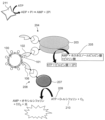
- FIG. 1 is a diagram showing a schematic diagram of an example of a detection scheme for a target substance to which the present invention is applied.
- FIG. 1 is a diagram showing a schematic diagram of another example of a detection scheme for a target substance to which the present invention is applied.
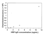
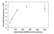
- FIG. 1 is a characteristic diagram showing the relationship between the concentration of the HRP-fused anti-SARS-CoV-2 antibody used in the examples and the chemiluminescence intensity. This is a characteristic graph showing the relationship between the concentration of inactivated SARS-CoV-2 and chemiluminescence intensity when using an HRP-fused anti-SARS-CoV-2 antibody at a final concentration of 1.1 ng/mL.
- the target substance to be detected is a target substance having a protein present on its surface, unlike substances such as proteins and antibodies. That is, the target substance has a structure having an envelope, a cell membrane, or a cell wall, and has a protein on the surface of the envelope, the cell membrane, or the cell wall.
- the target substance include an enveloped virus and a microorganism having a cell membrane or a cell wall. Note that even a non-enveloped virus can be used as the target substance if it has a capsid.
- viruses that can be the target substance may be either viruses with a DNA genome or viruses with an RNA genome, and the DNA or RNA structure may be either double-stranded or single-stranded.
- viruses that can be the target substance include, but are not limited to, influenza viruses (e.g., types A and B), rubella viruses, Ebola viruses, coronaviruses, measles viruses, chickenpox/shingles viruses, herpes viruses, mumps viruses, arboviruses, respiratory syncytial viruses, SARS viruses, hepatitis viruses (e.g., hepatitis B viruses, hepatitis C viruses), yellow fever viruses, AIDS viruses, rabies viruses, hantaviruses, dengue viruses, Nipah viruses, and lyssaviruses (all enveloped viruses), adenoviruses, noroviruses, rotaviruses, human papillomaviruses, polioviruses, enteroviruse
- the target virus be the novel coronavirus (SARS-CoV-2), which causes the novel coronavirus disease (COVID-19).
- SARS-CoV-2 novel coronavirus
- COVID-19 novel coronavirus disease
- Microorganisms that can be targeted include bacteria, fungi, microalgae, and protozoa (such as amebas and paramecium). Each of these microorganisms has a specific membrane protein, and can be targeted by using the membrane protein as an antigen.
- microorganisms that can be targeted include bacteria involved in infectious diseases such as Vibrio cholerae, Salmonella, Escherichia coli, Legionella, Bacillus anthracis, Helicobacter pylori, Listeria monocytogenes, Mycobacterium tuberculosis, nontuberculous mycobacteria, Staphylococcus aureus, Streptococcus aureus, Streptococcus pneumoniae, Neisseria meningitidis, Klebsiella pneumoniae, Serratia marcescens, Corynebacterium diphtheriae, Brucella, Bartonella henselae, Erysipelothricus rhusiopathiae, Actinomycetes, Borrelia burgdorferi, Clostridium perfringens, Shigella, Yersinia pestis, Clostridium tetani, and Enterobacter.
- infectious diseases such as Vibrio cholerae
- microorganisms that can be targeted include fungi involved in infectious diseases, such as Candida, Aspergillus, Cryptococcus, Blastomyces, Coccidioides, Histoplasma, Paracoccidioides, and Sporothrix.
- Samples that may contain the target substances as described above include, for example, samples of biological origin such as pharyngeal swabs, nasal swabs, sputum, feces (excreted feces, rectal feces), vomit, saliva, gargle, and tears. Samples that may contain the target substances are not limited to samples of biological origin, but may include non-biological samples collected from clothing, masks, glasses, filters, furniture, cooking utensils, etc.
- samples that may contain the target substance include products such as food and beverages; raw materials for food and beverages; intermediate products in the process of food and beverages; drinking water, sewage, manufacturing water, well water, cooling tower cooling water, and other water; culture media in which the above-mentioned microorganisms are cultured; filters used for concentration; and eluted solutions.
- samples that may contain the target substance include sampling devices such as cotton swabs used to wipe the surface of the sample, and suspensions in which such swabs are suspended in water.
- Examples of areas that can be wiped with sampling devices include high-frequency contact areas in food factories, restaurants, cafeterias, nursing care facilities, medical facilities, and offices, such as doorknobs, handrails, switches, PC keyboards, mice, telephones, nurse call buttons, tables, chairs, faucets, bedside rails, toilet seats, elevator buttons, manufacturing process equipment, and the hands of workers, nurses, doctors, patients, etc.
- FIG. 1 An example of a system for detecting the above-mentioned target substance with high accuracy is outlined in FIG. 1.
- the example shown in FIG. 1 is a scheme for detecting the novel coronavirus (SARS-CoV-2) as the target substance 100. Since the novel coronavirus (SARS-CoV-2) has spike proteins densely present on its surface, this system uses the receptor binding domain (RBD) in the spike protein as the antigen 101.
- the target substance 100 is not limited to the novel coronavirus (SARS-CoV-2), and it is easily understood that a similar scheme can be applied to other viruses and microorganisms.
- this system utilizes a first binding molecule 104 having a first specific binding portion 102 that specifically binds to an antigen 101 in a target substance 100, and a first enzyme portion 103 that is directly or indirectly bound to the first specific binding portion 102.
- the first enzyme portion 103 has an oxidase activity that catalyzes an enzyme reaction using a specific substance as a substrate (first substrate 105) to generate hydrogen peroxide.
- the first enzyme portion 103 also forms a complex with the first specific binding portion 102 that specifically binds to the target substance 100.
- the first specific binding portion 102 can be an antibody against the RBD (antigen 101) of the target substance 100, i.e., the novel coronavirus (SARS-CoV-2).
- the antibody when an antibody is used as the first specific binding portion 102, the antibody may be one that uses a region other than the RBD in the spike protein of the novel coronavirus (SARS-CoV-2) as the antigen 101, or one that uses a protein other than the spike protein, such as the N protein, M protein, or E protein, as the antigen 101.
- SARS-CoV-2 novel coronavirus
- glucose oxidase for example, glucose oxidase, aldehyde oxidase, cytochrome oxidase, cytochrome oxidase, catechol oxidase, diphenol oxidase, cholesterol oxidase, cytochrome C oxidase, hypoxanthine oxidase, xanthine oxidase, NADPH oxidase, lactate oxidase, galactose oxidase, alcohol oxidase, glutamate oxidase, formaldehyde oxidase, sorbitol oxidase, fructose oxidase, pyruvate oxidase, ascorbate oxidase, choline oxidase, amine oxidase, sarcosine oxidase, fructosyl amino acid oxidase, fructosyl peptide oxida
- the first substrate 105 can be appropriately selected and used depending on the type of the first enzyme part 103.
- the pair of the first enzyme part 103 and the first substrate 105 is preferably glucose oxidase and glucose. This is because glucose, which is the first substrate 105, is a stable compound and is inexpensive.
- the first specific binding portion 102 may be any of immunoglobulin (IgG, IgA, IgM, IgD, IgE) antibodies, heavy chain antibodies, and fragment antibodies.
- Immunoglobulin antibodies consist of a heavy chain (VH) and a light chain (LH), and the heavy chain and light chain each have three CDRs, CDR1, CDR2, and CDR3.
- Heavy chain antibodies consist only of a heavy chain, and the heavy chain has three CDRs, CDR1, CDR2, and CDR3.
- Fragment antibodies are antibodies fragmented using enzymes or genetic engineering techniques, and include Fab, F(ab'), F(ab')2, Fv, and VHH antibodies.
- Fragment antibodies also include scFab, scF(ab'), and scF(ab')2, which are single-chain antibodies formed by linking the light chain and heavy chain constituting Fab, F(ab'), and F(ab')2 via a linker sequence.
- a linker sequence may be attached to the C-terminus of the light chain, and a heavy chain may be attached to the C-terminus of the linker sequence.
- a linker sequence may be attached to the C-terminus of the heavy chain, and a light chain may be attached to the C-terminus of the linker sequence.
- the first specific binding portion 102 is not limited to the above-mentioned antibody molecule, and may be a protein or peptide that specifically binds to the target substance 100.
- the first specific binding portion 102 may be a polypeptide selected from the group consisting of immunoglobulin (IgG, IgA, IgM, IgD, IgE) antibodies, heavy chain antibodies, and fragment antibodies, proteins, and peptides that can specifically bind to the target substance 100.
- the system uses a second binding molecule 108 having a second specific binding portion 106 that specifically binds to the target substance 100 and a second enzyme portion 107 directly or indirectly bound to the second specific binding portion 106.
- the second enzyme portion 107 is an enzyme having peroxidase activity that uses hydrogen peroxide (second substrate 109) and luminol (third substrate 110) generated by the enzyme reaction in the first enzyme portion 103 as substrates.
- the second enzyme portion 107 may be any enzyme having peroxidase activity, and for example, horseradish peroxidase (HRP) is preferably used, and peroxidase derived from microorganisms such as mold can also be used.
- HRP horseradish peroxidase
- the second enzyme portion 107 enzymes having peroxidase activity such as myoglobin, hemoglobin, metmyoglobin, catalase, and cytochrome P450 can be appropriately used.
- the third substrate 110 is preferably a substance that serves as a substrate for the second enzyme unit 107 and that allows optical detection of the progress of the enzyme reaction in the second enzyme unit 107.
- orthophenylenediamine can be used as the third substrate 110 in addition to luminol.
- the second enzyme unit 107 forms a complex with a second specific binding unit 106 that specifically binds to the target substance 100.
- the second specific binding unit 106 can be an antibody molecule against the target substance 100, i.e., the RBD (antigen 101) of the novel coronavirus (SARS-CoV-2), like the first specific binding unit 102.
- the antibody may be one that uses a region other than the RBD in the spike protein of the novel coronavirus (SARS-CoV-2) as the antigen 101, or one that uses a protein other than the spike protein, such as the N protein, M protein, or E protein, as the antigen 101.
- the epitopes of the antigen 101 to which the second specific binding unit 106 specifically binds and the antigen 101 to which the first specific binding unit 102 specifically binds may be the same, or may be different epitopes within the same molecule. Additionally, the second specific binding portion 106 and the first specific binding portion 102 may use different proteins present on the surface of the target substance 100 as antigens.
- the second specific binding portion 106 may be any of an immunoglobulin antibody, a heavy chain antibody, and a fragment antibody, like the first specific binding portion 102. That is, the second specific binding portion 106 may also be a fragment antibody such as Fab, F(ab'), F(ab')2, Fv, VHH antibody, scFab, scF(ab'), scF(ab')2, and scFV.
- the second specific binding portion 106 is not limited to an antibody, and may be a protein or peptide that specifically binds to the target substance 100.
- the second specific binding portion 106 may be a polypeptide selected from the group consisting of immunoglobulin (IgG, IgA, IgM, IgD, IgE) antibodies, heavy chain antibodies, and fragment antibodies, proteins, and peptides, which can specifically bind to the target substance 100.
- immunoglobulin IgG, IgA, IgM, IgD, IgE
- the present system preferably includes a third enzyme 111 that consumes hydrogen peroxide as a substrate, which is generated by the oxidase reaction of the first enzyme unit 103.
- the third enzyme 111 is an enzyme that consumes hydrogen peroxide as a substrate, and for example, catalase can be used.
- the target substance 100 can be detected in a reaction system in which the first binding molecule 104, the second binding molecule 108, the first substrate 105, and the third substrate 110 are present.
- the first binding molecule 104 binds to the target substance 100 via the first specific binding portion 102
- the second binding molecule 108 binds to the target substance 100 via the second specific binding portion 106.
- the first binding molecule 104 and the second binding molecule 108 form a state in close proximity.
- an oxidase reaction using the first substrate 105 by the first enzyme unit 103 proceeds, generating hydrogen peroxide.
- the hydrogen peroxide generated by the enzyme reaction of the first enzyme unit 103 can be used as a substrate (second substrate 109) in the enzyme reaction together with a third substrate 110 such as luminol present in the reaction system.
- the peroxidase reaction by the second enzyme unit 107 proceeds.
- the target substance 100 can be detected by measuring the peroxidase reaction using the third substrate 110 by the second enzyme section 107.
- the target substance 100 can be detected by measuring the chemiluminescence generated by the so-called luminol reaction.
- the means for measuring the chemiluminescence is not particularly limited, and may be visual measurement or a detection device capable of detecting chemiluminescence.
- the target substance 100 is a virus, a microorganism, or the like, on whose surface a protein is present. Therefore, in this system, the first binding molecule 104 and the second binding molecule 108 can be stably and densely formed in close proximity, and the above-mentioned chemiluminescence can be detected visually or by a detection device.
- a detection device For example, a highly sensitive optical detection device equipped with a photomultiplier tube can be used, but the above-mentioned chemiluminescence can also be detected by using a detection device that does not have a photomultiplier tube and has a photodiode as an optical detection element, which is less expensive than the above-mentioned system.
- the target substance 100 can be detected easily and at low cost.
- a chromogenic substrate e.g., 4-aminoantipyrine and phenol, etc.
- the color development of the generated pigment can be confirmed by a spectrophotometer, a camera, visually, etc.
- the first enzyme part 103 in the first binding molecule 104 causes an oxidase reaction using the first substrate 105 to generate hydrogen peroxide, but the second binding molecule 108 is not present in a nearby position. Therefore, the second enzyme part 107 in the second binding molecule 108 cannot use hydrogen peroxide, and the peroxidase reaction using the third substrate 110 such as luminol hardly proceeds. Therefore, when the target substance 100 is not present in the reaction system, chemiluminescence caused by the luminol reaction of an optically detectable substance such as luminol is not observed.
- the third enzyme 111 when the third enzyme 111 is present in the reaction system, the hydrogen peroxide generated in the enzyme reaction of the first enzyme unit 103 can be consumed by the third enzyme 111.
- the hydrogen peroxide required for the enzyme reaction by the second enzyme unit 107 in the second binding molecule 108 can be consumed, so that even if hydrogen peroxide is generated in the reaction system, the progress of the luminol reaction by the second enzyme unit 107 can be more reliably inhibited.
- the occurrence of false positives can be suppressed by adding the third enzyme 111 to the reaction system.
- the amount of the third enzyme 111 to be added to suppress false positives can be appropriately determined. For example, when a measurement is performed using a negative control and the measurement system outputs a positive result, the third enzyme 111 may be added, for example, the third enzyme 111 may be added until the output of the measurement system becomes negative.
- the first reagent containing at least the first substrate 105 and the second reagent containing at least the first binding molecule 104 can be used separately.
- components other than the first substrate 105 and the first binding molecule 104 e.g., the second binding molecule 108, etc.
- the first reagent or the second reagent may be contained in either the first reagent or the second reagent, or may be contained in both.
- Target substance detection scheme 2 Another example of the system for detecting the above-mentioned target substance with high accuracy is outlined in Figure 2. Note that the example shown in Figure 2 is a scheme for detecting the novel coronavirus (SARS-CoV-2) as the target substance 100, similar to the system shown in Figure 1. In the system shown in Figure 2, the same components as those in the system shown in Figure 1 are denoted by the same reference numerals, and detailed description thereof will be omitted in this section.
- SARS-CoV-2 novel coronavirus
- this system utilizes a first binding molecule 204 having a first specific binding portion 102 and a first enzyme portion 203 directly or indirectly bound to the first specific binding portion 102.
- the first enzyme portion 203 has a kinase activity that catalyzes an enzyme reaction using a specific substance or group of substances as a substrate (first substrate 205) and produces ATP.
- a kinase activity that produces ATP either a kinase that produces ATP using AMP as part of the substrate or a kinase that produces ATP using ADP as part of the substrate may be used.
- Examples of enzymes with kinase activity that use AMP as part of the substrate and produce ATP include pyruvate orthophosphate dikinase and pyruvate water dikinase.
- AMP phosphoenolpyruvate
- pyrophosphate are used as substrates, and ATP, pyruvic acid, and two molecules of phosphate are produced. That is, in this example, the first binding molecule 204 uses AMP as the first substrate 205 in the first enzyme part 203, and produces ATP in the reaction system.
- examples of enzymes with kinase activity that use ADP as part of the substrate and produce ATP include pyruvate kinase, acetate kinase, creatine kinase, polyphosphate kinase, and hexokinase.
- ADP and phosphoenolpyruvate are used as substrates, and ATP, pyruvic acid, and pyrophosphate are produced. That is, in this example, the first binding molecule 204 uses ADP as the first substrate 205 in the first enzyme part 203, and produces ATP in the reaction system.
- an enzyme having kinase activity that uses AMP as a substrate and produces ATP is used as the first enzyme section 203.
- an enzyme having kinase activity that uses ADP as a substrate and produces ATP is used, ATP is produced non-enzymatically from the substrate ADP, and luciferase luminescence may be observed due to the non-enzymatically produced ATP even in the absence of target substance 100.
- the detection sensitivity of target substance 100 may be poor, possibly causing a false positive.
- samples that may contain the target substance may contain adenylate kinase (myokinase).
- Adenylate kinase is a stable enzyme that uses ADP as a substrate to produce ATP. Even if the sample does not contain the target substance 100, luciferase luminescence may be observed due to the ATP produced by adenylate kinase. In this case as well, the detection sensitivity of the target substance 100 is deteriorated, increasing the possibility of causing a false positive.
- the substrate AMP is also a product of the luciferase reaction, so background luminescence from luciferase is suppressed, allowing the target substance 100 to be detected with high sensitivity and preventing false positives. Even if the sample contains adenylate kinase (myokinase), AMP is not a substrate for adenylate kinase, so the decrease in detection sensitivity and the induction of false positives as described above can be avoided.
- adenylate kinase myokinase
- this system utilizes a second binding molecule 208 having a second specific binding portion 106 and a second enzyme portion 207 directly or indirectly bound to the second specific binding portion 106.
- the second enzyme portion 207 is an enzyme having luciferase activity that utilizes ATP (second substrate 209) and D-luciferin (third substrate 210) generated by the enzyme reaction in the first enzyme portion 203 as substrates.
- the second enzyme portion 207 may be any enzyme having luciferase activity, and for example, a beetle-derived luciferase such as firefly luciferase or an enzyme that uses ATP as a substrate and emits light can be appropriately used.
- the present system may include a third enzyme 211 that consumes, as a substrate, the ATP produced by the kinase enzyme reaction that produces ATP by the first enzyme unit 203.
- the third enzyme 211 is an enzyme that consumes ATP as a substrate, and for example, apyrase can be used.
- the target substance 100 can be detected in a reaction system in which the first binding molecule 204, the second binding molecule 208, the first substrate 205, and the third substrate 210 are present.
- the first binding molecule 204 binds to the target substance 100 via the first specific binding portion 102
- the second binding molecule 208 binds to the target substance 100 via the second specific binding portion 106.
- the first binding molecule 204 and the second binding molecule 208 form a state in close proximity.
- the first enzyme unit 203 causes a kinase reaction using the AMP contained in the first substrate 205, generating ATP. Since the second enzyme unit 207 is located in close proximity to the first enzyme unit 203, the ATP generated by the enzyme reaction of the first enzyme unit 203 can be used as a substrate (second substrate 209) in the enzyme reaction together with a third substrate 210 such as D-luciferin present in the reaction system. In other words, when the first binding molecule 204 and the second binding molecule 208 are in close proximity via the target substance 100, the luciferase reaction caused by the second enzyme unit 207 proceeds.
- the target substance 100 can be detected by measuring the luciferase reaction using the third substrate 210 by the second enzyme section 207.
- the target substance 100 can be detected by measuring the luminescence generated by the so-called luciferase reaction.
- the means for measuring the luminescence is not particularly limited, and may be visual measurement or a detection device capable of detecting luminescence.
- the target substance 100 is a virus, a microorganism, or the like, on whose surface a protein is present. Therefore, in this system, the first binding molecule 204 and the second binding molecule 208 can be stably and densely formed in close proximity, and the above-mentioned luminescence can be detected visually or by a detection device.
- a detection device equipped with a photomultiplier tube can be used, but the above-mentioned chemiluminescence can also be detected by using a detection device that does not have a photomultiplier tube and has a photodiode as an optical detection element, which is less expensive than this.
- the target substance 100 can be detected easily and at low cost.
- the first enzyme part 203 in the first binding molecule 204 causes a kinase reaction using the AMP contained in the first substrate 205 to proceed, and ATP is generated, but the second binding molecule 208 is not present in a nearby position. Therefore, the second enzyme part 207 in the second binding molecule 208 cannot use ATP, and the luciferase reaction using the third substrate 210 such as D-luciferin hardly proceeds. Therefore, when the target substance 100 is not present in the reaction system, luminescence caused by the luciferase reaction is not observed.
- the ATP generated by the enzyme reaction of the first enzyme unit 203 can be consumed by the third enzyme 211.
- the ATP required for the enzyme reaction by the second enzyme unit 207 in the second binding molecule 208 can be consumed, so that even if ATP is generated by the second enzyme unit 207 that is not bound to the target substance 100, ATP generated by the second enzyme unit 207 bound to the target substance 100 increases in the reaction system due to diffusion, or ATP derived from the sample is brought into the reaction system, the progress of the luciferase reaction by the second enzyme unit 207 can be more reliably inhibited.
- the occurrence of false positives can be suppressed by adding the third enzyme 211 to the reaction system.
- the amount of the third enzyme 211 added to suppress false positives can be determined appropriately. For example, if a measurement is performed using a negative control and the measurement system outputs a positive result, the third enzyme 211 may be added, for example, until the output of the measurement system becomes negative.
- a first reagent containing at least a part or all of the substances constituting the first substrate 205 and a second reagent containing at least the first binding molecule 204 can be used separately.
- components other than the first substrate 205 and the first binding molecule 204 e.g., the second binding molecule 208, etc.
- the first reagent contains a part of the substances constituting the first substrate 205
- the remaining substances constituting the first substrate 205 may be contained in the second reagent.
- the progress of the kinase reaction using the first substrate 205 by the first enzyme part 203 in the first binding molecule 204 can be suppressed during storage of the reagents. Then, when the first reagent and the second reagent are mixed, a kinase reaction using the first substrate 205 by the first enzyme part 203 can be caused to proceed, and the target substance 100 can be detected as described above.
- the system described above is not particularly limited and can be applied to various situations in which the target substance 100 is detected.
- the target substance 100 is a virus
- the first and second reagents are sprayed onto a location where the virus is suspected to be attached, to construct the above-mentioned reaction system.
- the luminescence caused by the above-mentioned luminol reaction or luciferase reaction is measured using the detection device as described above, and it can be determined whether the target substance 100 is attached to the location based on the measurement result.
- the present system can also be applied to devices that detect whether or not the target substance 100, a virus, is contained in the air.
- devices include air purifiers, air conditioners, handy, simplified air collectors, air collectors installed at the entrances and exits of buildings and rooms, and collectors of exhaled and coughed air installed in hospitals.
- These devices have in common that the air is passed through a filter, and the present system can be applied to detect viruses attached to the filter.
- the above-mentioned reaction system can be constructed by spraying the first and second reagents onto the filter, or the above-mentioned reaction system can be constructed by spraying the first and second reagents onto fine particles or dust repaired by the filter.
- the filters in these devices disposable, it becomes possible to sequentially detect the target substance 100, a virus.
- this system can be applied to a device that detects whether a target substance 100, a virus, is contained in a liquid.
- examples of such devices include a device that collects saliva or other bodily fluids from a patient who complains of fever or other symptoms, a device that collects liquid in which clothing or a mask worn by a medical worker is soaked, and a device that wipes a surface suspected of having a virus with a sampling tool such as a cotton swab and collects the wiped virus in a liquid.
- a device collects a liquid that may contain a virus, which is a target substance 100.
- the above-mentioned first and second reagents can be added to the collected liquid to construct the above-mentioned reaction system.
- a homogeneous detection system when detecting the target substance 100 contained in the liquid, a homogeneous detection system can be created in which only the addition of reagents is required and bound/free separation is not required.
- luminescence in the liquid can be easily detected by the detection device described above. Therefore, for example, by combining it with a microchannel device, a simple and high-performance virus inspection device can be produced.
- Example 1 Examination of the concentration of HRP-fused antibody used
- the optimal concentration range of HRP-fused anti-SARS-CoV-2 antibody (Abcam; ab273169) was examined for luminol luminescence intensity.
- 5 ⁇ L of HRP-fused anti-SARS-CoV-2 antibody (Abcam; ab273169) was added to a 96-well plate to give final concentrations of 0.42, 1.1, 2.1, 4.2, and 11 ng/mL, 5 ⁇ L of hydrogen peroxide (FUJIFILM Wako Pure Chemical Industries, Ltd.) to give a final concentration of 0.1 mM, and 10 ⁇ L of 10 mM sodium phosphate buffer (pH 7.0) containing 10 mM KCl were mixed, and then 80 ⁇ L of BM Chemiluminescence ELISA Substrate Solution A (Roche) containing luminol was immediately added, and the chemiluminescence intensity was measured using a plate reader (Therm
- Example 2 Detection of target substance (inactivated SARS-CoV-2) using an optical detection device]
- a commercially available inactivated SARS-CoV-2 was used as the target substance, and a detection scheme for inactivated SARS-CoV-2 was constructed.
- SpyCatcher-fused glucose oxidase and SpyTag-fused anti-SARS-CoV-2 S protein antibody were prepared by recombinant production using E. coli BL21(DE3) strain. Specifically, an anti-SARS-CoV-2 S protein antibody scFv with SpyTag fused to the C-terminus and GOx with SpyCatcher fused to the N-terminus (referred to as SpyCatcher-GOx) were prepared.
- the amino acid sequence of SpyCatcher-GOx is shown in SEQ ID NO: 1
- the amino acid sequence of SpyTag-fused anti-SARS-CoV-2 S protein scFv is shown in SEQ ID NO: 2.
- BM Chemiluminescence ELISA Substrate Solution A (Roche) containing glucose (final concentration 200mM) and catalase (final concentration 24ng/mL) was added, and the chemiluminescence intensity was measured using a plate reader (Thermo Fisher Scientific, product name: Varioskan Flash).
- HRP-fused anti-SARS-CoV-2 antibody was used at a final concentration of 2.1 ng/mL, and detection was performed in the same manner as above.
- the linear range was calculated to be 25 to 125 dC/mL for inactivated SARS-CoV-2 concentrations, and the limit of detection (LOD) was calculated as the inactivated SARS-CoV-2 concentration that is three times the standard deviation of the background, resulting in a detection limit of 3.3 dC/mL, indicating that highly sensitive detection was achieved.
- LOD limit of detection
- the antibody-enzyme conjugate in this example was prepared in the same manner as in Example 2. 5 ⁇ L each of the following was mixed in a low protein binding microtube: antibody-enzyme conjugate (final concentration 50 nM), HRP-fused anti-SARS-CoV-2 antibody (Abcam; ab273169) (final concentration 2.1 ng/mL), inactivated SARS-CoV-2 virus (Sino Bio) (final concentration 500, 250, 125, 50, 25, 0 dC/mL) or inactivated influenza virus (Hytest) (final concentration 500 particles/mL) or bovine serum albumin (BSA) (final concentration 500 ng/mL), and 10 mM sodium phosphate buffer (pH 7.0) containing 10 mM KCl, and left to stand at room temperature for 1 hour.
- antibody-enzyme conjugate final concentration 50 nM
- HRP-fused anti-SARS-CoV-2 antibody Abcam; ab273169)
- final concentration 2.1 ng/mL inactiv
- BM Chemiluminescence ELISA Substrate Solution A (Roche) containing a final concentration of 200 mM glucose and a final concentration of 24 ng/mL catalase was added, and the chemiluminescence intensity was measured using a detection device (Kikkoman Biochemifa Corporation, product name: Lumitester Smart) that does not have a photomultiplier tube and is equipped with a photodiode as the optical detection element.
- Example 4 Preparation of luciferase-anti-SARS-CoV-2 antibody conjugate
- biotinylated luciferase Kikkoman Biochemifa; BLU-Y, 60408
- commercially available streptavidin Tokyo Kasei; S0951
- biotinylated anti-SARS-CoV-2 antibody Abcam; ab273638
- 4 ⁇ L of 0.25 mg/mL biotinylated luciferase and 8 ⁇ L of 0.25 mg/mL commercially available streptavidin were mixed and left at room temperature for 15 minutes.
- Example 5 Preparation of pyruvate orthophosphate dikinase-anti-SARS-CoV-2 antibody complex
- pyruvate orthophosphate dikinase Kikkoman Biochemifa; PPDK-E, 61313
- PBS tablets Takara Bio; T9181
- N-ethylmaleimide Fujifilm Wako Pure Chemical Industries; 058-02061
- N-(6-maleimidocaproyloxy)succinimide Tokyo Kasei; S0428
- TCEP hydrochloride Flujifilm Wako Pure Chemical Industries; 209-19861
- anti-SARS-CoV-2 antibody Abcam; ab272854
- reaction solution 10 ⁇ L was added to a dialysis cup and left at 4°C for 2 hours to remove unreacted N-(6-maleimidocaproyloxy)succinimide.
- the reaction solution collected after dialysis was named "crosslinker-modified pyruvate phosphate dikinase”.
- 1 ⁇ L of 10 mM TCEP and 17 ⁇ L of 100 mM potassium phosphate buffer pH 6.0 were added to 2 ⁇ L of 1 mg/mL anti-SARS-CoV-2 antibody, and the mixture was left at 37 ° C for 1.5 hours to perform reduction treatment.
- Example 6 Detection of target substance (inactivated SARS-CoV-2) using pyruvate orthophosphate dikinase/luciferase
- inactivated SARS-CoV-2 ATCC; VR-1986HK (trademark)
- D-Luciferin B-Luciferin
- AMP-Na Flujifilm Wako Pure Chemical Industries, Ltd.; 306-50741
- a Tricine buffer solution (25 mM Tricine, 1 mM EDTA, 12 mM magnesium acetate, 0.1% BSA, pH 7.9) containing 1/100 of the luciferase-anti-SARS-CoV-2 antibody complex solution and 1/200 of the pyruvate orthophosphate dikinase-anti-SARS-CoV-2 antibody complex solution was used as the enzyme solution, a Tricine buffer solution containing 840 ⁇ M D-Luciferin and 1 ⁇ M AMP was used as the substrate solution, and a Tricine buffer solution containing 50 dC/50 ⁇ L of inactivated SARS-CoV-2 was used as the virus solution.
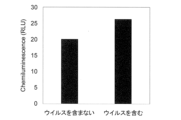
- Figure 7 shows a comparison of luminescence intensity when virus was added to a control without virus. Compared to the virus-free control, an increase in chemiluminescence intensity was observed in the reaction solution to which virus was added.
- Example 7 Detection of target substance (E. coli) using an optical detection device
- a glucose oxidase (GOx)-fused anti-Escherichia coli antibody conjugate was prepared using a Glucose Oxidase Conjugation Kit (Abcam; ab102887) and an anti-Escherichia coli antibody (Abcam; ab217910).
- BM chemiluminescence ELISA substrate (Roche; 11582950001) solution A containing a final concentration of 200 mM glucose and a final concentration of 24 ng/mL catalase was added, and the chemiluminescence intensity was measured using a plate reader (Thermo Fisher Scientific, product name: Varioskan Flash).
- BM Chemiluminescence ELISA Substrate Solution A (Roche) containing a final concentration of 200 mM glucose and a final concentration of 24 ng/mL catalase (Fujifilm Wako Pure Chemical Industries, Ltd.) was added, and the chemiluminescence intensity was measured using a plate reader (Thermo Fisher Scientific, product name: Varioskan Flash).
- SEQ ID NO:1 SEQ ID NO:2: MKYLLPTAAAGLLLLAAQPAMAEVQLVESGGGLVQPGGSLRLSCAASGFTVSSNYMSWVRQAPGKGLEWVSVIYSGGSTFYADSVKGRFTISRDNSMNTLFLQMNSLRAEDTAVYYCARVLPMYGDYLDYWGQGTLVTVSSGGGGSGGGGSGGGGSDIVMTQSPSSLSASVGDRVTITCRASQSISRYLNWYQQKPGKAPKLLIYAASSLQSG VPSRFSGSGTDFTLTISSLQPEDFATYYCQQSYSTPPEYTFGQGTKLEIKRAAAGSGAHIVMVDAYKPTKAEQKLISEEDLNLGGGMRGSHHHHHHHHH
Landscapes
- Chemical & Material Sciences (AREA)
- Life Sciences & Earth Sciences (AREA)
- Health & Medical Sciences (AREA)
- Organic Chemistry (AREA)
- Engineering & Computer Science (AREA)
- Zoology (AREA)
- Wood Science & Technology (AREA)
- Immunology (AREA)
- Proteomics, Peptides & Aminoacids (AREA)
- Molecular Biology (AREA)
- Biochemistry (AREA)
- General Health & Medical Sciences (AREA)
- Genetics & Genomics (AREA)
- Bioinformatics & Cheminformatics (AREA)
- Biotechnology (AREA)
- Microbiology (AREA)
- Analytical Chemistry (AREA)
- General Engineering & Computer Science (AREA)
- Physics & Mathematics (AREA)
- Biophysics (AREA)
- Medicinal Chemistry (AREA)
- Biomedical Technology (AREA)
- Urology & Nephrology (AREA)
- Hematology (AREA)
- Cell Biology (AREA)
- Pathology (AREA)
- General Physics & Mathematics (AREA)
- Food Science & Technology (AREA)
- Virology (AREA)
- Toxicology (AREA)
- Measuring Or Testing Involving Enzymes Or Micro-Organisms (AREA)
- Apparatus Associated With Microorganisms And Enzymes (AREA)
Abstract
微生物やウイルスといったタンパク質が表面に密に存在する対象を高感度に検出する。 標的物質に結合した第1の結合分子と第2の結合分子が近接することで、各結合分子に連結された酵素による酵素反応が連続して進行し、当該酵素反応を光学的に測定する。
Description
本発明は、標的物質に対して特異的に結合する分子を用いた、当該標的物質の検出試薬及び当該標的物質の検出方法に関する。
一般に抗原-抗体反応を用いた免疫測定システムのように、試料中の標的物質を検出する技術が知られている。特に、抗原-抗体反応を用いた免疫測定システムでは、酵素標識体の結合型(B)と遊離型(F)の分離(B/F分離)が不要なホモジニアスアッセイ(均一系免疫測定法、ホモジニアス検出系とも称す)が簡便且つ迅速な測定系として注目されている。
特許文献1には、このようなアッセイの例であり、第1の酵素と第2の酵素をそれぞれ測定対象物質に特異的に結合させ、第2の酵素により触媒される反応を検出することで、試料中の測定対象物質の濃度を測定する方法が開示されている。ここで、第2の酵素により触媒される反応は、第1の酵素が触媒する反応により生成する生成物を利用する反応である。したがって、第1の酵素と第2の酵素とそれぞれの酵素の基質とを含む反応液に測定対象物質が存在すると第1の酵素と第2の酵素とが近接し、第2の酵素が触媒する反応が進行する。特許文献1には、第1の酵素及び第2の酵素の具体的な例として、それぞれグルコースオキシダーゼ及びペルオキシダーゼとすることが開示されている。この場合、グルコースオキシダーゼが生成する過酸化水素を第2の酵素であるペルオキシダーゼが利用してオルトフェニレンジアミン等の基質を用いた酵素反応が進行する。
特許文献1には、測定対象物質として、抗原、抗体、低分子有機化合物、タンパク質、高分子有機物質、脂質、糖、ペプチド、ホルモン、核酸、ウイルス等が挙げられているものの、実施例としてはウサギIgGを検出する二次抗体を使用した例が開示されている。
また、特許文献2には、抗原性物質に対して一対の抗体フラグメントが複合体を形成し、各抗体フラグメントに結合した標識が近接して結合することにより増感されたシグナルを検出することで、抗原性物質を測定する方法が開示されている。ここで、一方の抗体フラグメントに結合した標識の例としてグルコースオキシダーゼが挙げられており、他方の抗体フラグメントに結合した標識の例としてホースラディッシュペルオキシダーゼが挙げられている。特許文献2には、抗原性物質としてHIV抗原を検出した例、血中チロキシンを検出した例、抗ウサギヤギ抗体を検出した例が開示されている。
さらに、特許文献1及び2以外にも、測定対象物質に対して抗原抗体反応を利用して複合体を形成し、近接した二種類の酵素が連続的に反応することで、測定対象物質を検出する方法が知られている(例えば、特許文献3、4、5及び6)。
上述のように、B/F分離が不要なホモジニアスアッセイ系において、2種類の酵素反応が連続的に生じることで測定対象物質を検出する例は公知である。しかしながら、従来公知の技術においては、測定対象物質の種類によってはS/N比が悪く、実際に高感度な検出系に適用できないといった問題があった。そこで、本発明は、上述した実情に鑑み、微生物やウイルスといったタンパク質が表面に密に存在する対象を測定対象の標的物質として高感度に検出できる、標的物質の検出試薬や検出方法を提供することを目的とする。
上述した目的を達成した本発明は以下を包含する。
(1) タンパク質が表面に存在する標的物質に対して特異的に結合するポリペプチドからなる第1の特異的結合部と、当該第1の特異的結合部に直接又は間接的に結合した第1の酵素部とを有する第1の結合分子と、
上記第1の結合分子における第1の酵素部の基質となる第1の基質と、
上記標的物質に特異的に結合するポリペプチドからなる第2の特異的結合部と、当該第2の特異的結合部に直接又は間接的に結合し、上記第1の酵素部における酵素反応で上記第1の基質から生成した物質を第2の基質とする第2の酵素部とを有する第2の結合分子と、
上記第2の結合分子における第2の酵素部が、上記第2の基質とともに酵素反応の基質とする第3の基質と
を含む、標的物質の検出キット。
(2) 上記第2の結合分子における上記第2の酵素部における酵素反応を光学的に検出する検出装置に使用されることを特徴とする(1)記載の標的物質の検出キット。
(3) 上記検出装置は、光電子増倍管を有さず、フォトダイオードを光学検出素子として備えることを特徴とする(2)記載の標的物質の検出キット。
(4) 上記第2の基質を基質として消費する第3の酵素を更に含むことを特徴とする(1)記載の標的物質の検出キット。
(5) 上記第1の結合分子における第1の酵素部はオキシダーゼ活性を有し、上記第2の結合分子における第2の酵素部はペルオキシダーゼ活性を有し、上記第3の基質がオルトフェニレンジアミン又はルミノールであることを特徴とする(1)記載の標的物質の検出キット。
(6) 上記第1の結合分子における第1の酵素部はATPを産生するキナーゼ活性を有し、上記第2の結合分子における第2の酵素部はルシフェラーゼ活性を有し、上記第3の基質がルシフェリンであることを特徴とする(1)記載の標的物質の検出キット。
(7) 上記標的物質はウイルス又は微生物であることを特徴とする(1)記載の標的物質の検出キット。
(8) 上記(1)乃至(7)いずれかに記載の検出キットと、タンパク質が表面に存在する標的物質を含みうる試料とを混合する工程と、
上記検出キットに含まれる第2の結合分子における第2の酵素部による酵素反応を測定する工程とを含み、
上記第2の酵素の酵素反応に基づいて上記試料中の標的物質を検出することを特徴とする標的物質の検出方法。
(9) 検出装置によって、上記第2の結合分子における上記第2の酵素部における酵素反応を光学的に検出することを特徴とする(8)記載の標的物質の検出方法。
(10) 上記検出装置は、光電子増倍管を有さず、フォトダイオードを光学検出素子として備えることを特徴とする(9)記載の標的物質の検出方法。
(1) タンパク質が表面に存在する標的物質に対して特異的に結合するポリペプチドからなる第1の特異的結合部と、当該第1の特異的結合部に直接又は間接的に結合した第1の酵素部とを有する第1の結合分子と、
上記第1の結合分子における第1の酵素部の基質となる第1の基質と、
上記標的物質に特異的に結合するポリペプチドからなる第2の特異的結合部と、当該第2の特異的結合部に直接又は間接的に結合し、上記第1の酵素部における酵素反応で上記第1の基質から生成した物質を第2の基質とする第2の酵素部とを有する第2の結合分子と、
上記第2の結合分子における第2の酵素部が、上記第2の基質とともに酵素反応の基質とする第3の基質と
を含む、標的物質の検出キット。
(2) 上記第2の結合分子における上記第2の酵素部における酵素反応を光学的に検出する検出装置に使用されることを特徴とする(1)記載の標的物質の検出キット。
(3) 上記検出装置は、光電子増倍管を有さず、フォトダイオードを光学検出素子として備えることを特徴とする(2)記載の標的物質の検出キット。
(4) 上記第2の基質を基質として消費する第3の酵素を更に含むことを特徴とする(1)記載の標的物質の検出キット。
(5) 上記第1の結合分子における第1の酵素部はオキシダーゼ活性を有し、上記第2の結合分子における第2の酵素部はペルオキシダーゼ活性を有し、上記第3の基質がオルトフェニレンジアミン又はルミノールであることを特徴とする(1)記載の標的物質の検出キット。
(6) 上記第1の結合分子における第1の酵素部はATPを産生するキナーゼ活性を有し、上記第2の結合分子における第2の酵素部はルシフェラーゼ活性を有し、上記第3の基質がルシフェリンであることを特徴とする(1)記載の標的物質の検出キット。
(7) 上記標的物質はウイルス又は微生物であることを特徴とする(1)記載の標的物質の検出キット。
(8) 上記(1)乃至(7)いずれかに記載の検出キットと、タンパク質が表面に存在する標的物質を含みうる試料とを混合する工程と、
上記検出キットに含まれる第2の結合分子における第2の酵素部による酵素反応を測定する工程とを含み、
上記第2の酵素の酵素反応に基づいて上記試料中の標的物質を検出することを特徴とする標的物質の検出方法。
(9) 検出装置によって、上記第2の結合分子における上記第2の酵素部における酵素反応を光学的に検出することを特徴とする(8)記載の標的物質の検出方法。
(10) 上記検出装置は、光電子増倍管を有さず、フォトダイオードを光学検出素子として備えることを特徴とする(9)記載の標的物質の検出方法。
本明細書は本願の優先権の基礎となる日本国特許出願番号2023-003981号の開示内容を包含する。
本発明に係る標的物質の検出キット及び検出方法によれば、ウイルスや微生物といった、タンパク質が表面に存在する標的物質を極めて高感度に検出することができる。
以下、本発明に係る標的物質の検出キット及び検出方法を、図面を参照して詳細に説明する。
[標的物質]
本発明に係る検出キット及び検出方法において、検出対象となる標的物質は、タンパク質や抗体といった物質と異なり、タンパク質が表面に存在する標的物質である。すなわち、標的物質は、エンベロープや細胞膜、細胞壁を有する構造を備え、これらエンベロープや細胞膜、細胞壁の表面にタンパク質を有している。標的物質としては、エンベロープを有するウイルス、細胞膜や細胞壁を有する微生物を挙げることができる。なお、エンベロープを有しないウイルスであっても、カプシドを有する場合には標的物質とすることができる。
本発明に係る検出キット及び検出方法において、検出対象となる標的物質は、タンパク質や抗体といった物質と異なり、タンパク質が表面に存在する標的物質である。すなわち、標的物質は、エンベロープや細胞膜、細胞壁を有する構造を備え、これらエンベロープや細胞膜、細胞壁の表面にタンパク質を有している。標的物質としては、エンベロープを有するウイルス、細胞膜や細胞壁を有する微生物を挙げることができる。なお、エンベロープを有しないウイルスであっても、カプシドを有する場合には標的物質とすることができる。
また、標的物質となり得るウイルスは、DNAゲノムを有するウイルス及びRNAゲノムを有するウイルスのいずれでも良いし、DNA又はRNAの構造も2本鎖又は1本鎖のいずれでも良い。標的物質となるウイルスの例としては、特に限定されないが、インフルエンザウイルス(例えばA型、B型等)、風疹ウイルス、エボラウイルス、コロナウイルス、麻疹ウイルス、水痘・帯状疱疹ウイルス、ヘルペスウイルス、ムンプスウイルス、アルボウイルス、RSウイルス、SARSウイルス、肝炎ウイルス(例えば、B型肝炎ウイルス、C型肝炎ウイルス等)、黄熱ウイルス、エイズウイルス、狂犬病ウイルス、ハンタウイルス、デングウイルス、ニパウイルス、リッサウイルス等のエンベロープウイルス(以上、エンベロープを有するウイルス)、アデノウイルス、ノロウイルス、ロタウイルス、ヒトパピローマウイルス、ポリオウイルス、エンテロウイルス、コクサッキーウイルス、ヒトパルボウイルス、脳心筋炎ウイルス、ライノウイルス(以上、エンベロープを有さないウイルス)を挙げることができる。
特に、標的物質となるウイルスとしては、新型コロナウイルス感染症(COVID-19)の原因となる新型コロナウイルス(SARS-CoV-2)とすることが好ましい。
また、標的物質となり得る微生物としては、細菌、真菌類、微細藻類、原生動物(アメーバやゾウリムシなど)等を挙げることができる。これら微生物は、それぞれ特異的な膜タンパク質を有しているため、当該膜タンパク質を抗原とすることで標的物質となる。なかでも、標的物質となる微生物としては、コレラ菌、サルモネラ菌、大腸菌、レジオネラ菌、炭疽菌、ヘリコバクターピロリ菌、リステリア菌、結核菌、非結核性抗酸菌、ブドウ球菌、連鎖球菌、肺炎球菌、髄膜炎菌、肺炎桿菌、セラチア菌、ジフテリア菌、ブルセラ菌、バルトネラ-ヘンセレ、エリジペロスリックス-ルシオパシエ、放線菌、ライム病菌、ウエルシュ菌、赤痢菌、ペスト菌、破傷風菌及びエンテロバクター菌等の感染症に関与する細菌を挙げることができる。また、標的物質となる微生物としては、カンジダ、アスペルギルス、クリプトコッカス、ブラストミセス、コクシジオイデス、ヒストプラスマ、パラコクシジオイデス及びスポロトリクス等の感染症に関与する真菌類を挙げることができる。
また、上述したような標的物質を含みうる試料としては、例えば、咽頭ぬぐい液、鼻腔ぬぐい液、喀痰、糞便(排泄便、直腸便)、嘔吐物、唾液、うがい液及び涙液等の生体由来試料を挙げることができる。また、標的物質を含みうる試料としては、生体由来試料に限定されず、着衣、マスク、眼鏡、フィルター、家具、調理器具等から採取した非生体試料を挙げることができる。
さらに、標的物質を含みうる試料としては、食品や飲料などの製品;食品や飲料などの原料;食品や飲料などの工程中間品;飲料水、下水、製造用水、井戸水及び冷却塔冷却水などの水;上記微生物を培養した培地;濃縮に使用したフィルター;及び溶出した溶出液なども挙げられる。さらにまた、標的物質を含みうる試料としては、試料表面をふき取った綿棒等のサンプリング器具やそれを水などに懸濁した懸濁液を挙げられる。なお、サンプリング器具でふき取られる箇所としては、食品工場、レストラン、食堂、介護現場、医療現場、オフィスにおける高頻度接触部位、例えば、ドアノブ、手すり、スイッチ、PCのキーボード、マウス、電話、ナースコール、テーブル、椅子、蛇口、ベッドサイドレール、便座、エレベーターボタン、製造工程の設備や作業者、看護士、医師、患者などの手指などが挙げられる。
[標的物質の検出スキーム1]
上述した標的物質を高精度に検出するシステム例の概略を図1に示す。なお、図1に示す例は、標的物質100として新型コロナウイルス(SARS-CoV-2)を検出するスキームである。新型コロナウイルス(SARS-CoV-2)は、表面にスパイクタンパク質が密に存在しているため、本システムではスパイクタンパク質における、受容体結合ドメイン(RBD)を抗原101として利用している。なお、上述したように、標的物質100は、新型コロナウイルス(SARS-CoV-2)に限定されず、他のウイルスや微生物でも同様なスキームが適用できることは容易に理解できる。
上述した標的物質を高精度に検出するシステム例の概略を図1に示す。なお、図1に示す例は、標的物質100として新型コロナウイルス(SARS-CoV-2)を検出するスキームである。新型コロナウイルス(SARS-CoV-2)は、表面にスパイクタンパク質が密に存在しているため、本システムではスパイクタンパク質における、受容体結合ドメイン(RBD)を抗原101として利用している。なお、上述したように、標的物質100は、新型コロナウイルス(SARS-CoV-2)に限定されず、他のウイルスや微生物でも同様なスキームが適用できることは容易に理解できる。
本システムでは、図1に示すように、標的物質100における抗原101に対して特異的に結合する第1の特異的結合部102と、第1の特異的結合部102に直接又は間接的に結合した第1の酵素部103とを有する第1の結合分子104を利用する。ここで、第1の酵素部103は、所定の物質を基質(第1の基質105)として酵素反応を触媒し、過酸化水素を発生するオキシダーゼ活性を有している。また、第1の酵素部103は、標的物質100に対して特異的に結合する第1の特異的結合部102と複合体を形成している。ここで、第1の特異的結合部102は、標的物質100、すなわち新型コロナウイルス(SARS-CoV-2)のRBD(抗原101)に対する抗体とすることができる。また、第1の特異的結合部102として抗体を利用する場合、抗体としては、新型コロナウイルス(SARS-CoV-2)のスパイクタンパク質におけるRBD以外の領域を抗原101とするものでも良いし、スパイクタンパク質以外の例えば、Nタンパク質やMタンパク質、Eタンパク質を抗原101とするものであっても良い。
第1の酵素部103としては、例えば、グルコースオキシダーゼ、アルデヒドオキシダーゼ、チトクロムオキシダーゼ、シトクロムオキシダーゼ、カテコールオキシダーゼ、ジフェノールオキシダーゼ、コレステロールオキシダーゼ、シトクロムCオキシダーゼ、ヒポキサンチンオキシダーゼ、キサンチンオキシダーゼ、NADPHオキシダーゼ、乳酸オキシダーゼ、ガラクトースオキシダーゼ、アルコールオキシダーゼ、グルタミン酸オキシダーゼ、ホルムアルデヒドオキシダーゼ、ソルビトールオキシダーゼ、フルクトースオキシダーゼ、ピルビン酸オキシダーゼ、アスコルビン酸オキシダーゼ、コリンオキシダーゼ、アミンオキシダーゼ、サルコシンオキシダーゼ、フルクトシルアミノ酸オキシダーゼ、フルクトシルペプチドオキシダーゼ、ビリルビンオキシダーゼ、ポリフェノールオキシダーゼ、ウリカーゼ、グリセロールオキシダーゼ、ピラノースオキシダーゼ等を使用することができる。中でも、第1の酵素部103としては、グルコースオキシダーゼを使用することが好ましい。
また、第1の基質105は、第1の酵素部103の種類に応じて適宜選択して使用することができる。第1の酵素部103及び第1の基質105のペアは、グルコースオキシダーゼ及びグルコースとすることが好ましい。第1の基質105であるグルコースが安定した化合物であり、安価であるためである。
第1の特異的結合部102は、具体的には、免疫グロブリン(IgG、IgA、IgM、IgD、IgE)抗体、重鎖抗体及びフラグメント抗体のいずれであってもよい。免疫グロブリン抗体は、重鎖(VH)及び軽鎖(LH)からなり、重鎖及び軽鎖がそれぞれCDR1、CDR2及びCDR3の3つのCDRを有する。重鎖抗体は、重鎖のみからなり、重鎖におけるCDR1、CDR2及びCDR3の3つのCDRを有する。フラグメント抗体は、酵素や遺伝子工学的な手法を用いて断片化された抗体であり、Fab、F(ab')、F(ab')2、Fv、VHH抗体等を含む。また、フラグメント抗体は、Fab、F(ab')、F(ab')2を構成する軽鎖と重鎖を、リンカー配列を介して結合させて、それぞれ一本鎖抗体としたscFab、scF(ab')、scF(ab')2も含まれる。ここで、scFab、scF(ab')、scF(ab')2において、軽鎖のC末端側にリンカー配列を、そして更にそのC末端側に重鎖を結合させてなるものでもよい。さらにまた、重鎖のC末端側にリンカー配列を、そして更にそのC末端側に軽鎖を結合させてなるものでもよい。さらには、軽鎖の可変領域と重鎖の可変領域をリンカー配列を介して結合させて一本鎖抗体としたscFVもまた第1の特異的結合部102として利用できる。さらには、第1の特異的結合部102は、上述した抗体分子に限定されず、標的物質100に対して特異的に結合するタンパク質及びペプチドであってもよい。すなわち、第1の特異的結合部102は、標的物質100に特異的に結合できる、免疫グロブリン(IgG、IgA、IgM、IgD、IgE)抗体、重鎖抗体並びにフラグメント抗体、タンパク質及びペプチドからなる群から選ばれるポリペプチドであってもよい。
また、図1に示すように、本システムでは、標的物質100に特異的に結合する第2の特異的結合部106と、第2の特異的結合部106に直接又は間接的に結合した第2の酵素部107とを有する第2の結合分子108を利用する。第2の酵素部107は、第1の酵素部103における酵素反応で生成した過酸化水素(第2の基質109)とルミノール(第3の基質110)を基質として利用する、ペルオキシダーゼ活性を有する酵素である。第2の酵素部107は、ペルオキシダーゼ活性を有する酵素であれば良く、例えば、西洋ワサビペルオキシダーゼ(Horseradish peroxidase、HRP)を利用することが好ましく、カビなどの微生物由来のペルオキシダーゼを利用することもできる。その他にも第2の酵素部107としては、ミオグロビン、ヘモグロビン、メトミオグロビン、カタラーゼ、シトクロムP450等のペルオキシダーゼ活性を有する酵素を適宜使用することができる。また、第3の基質110は、第2の酵素部107の基質となり、且つ、第2の酵素部107における酵素反応の進行を光学的に検出できる物質とすることが好ましい。具体的には、第3の基質110としては、ルミノールの他にオルトフェニレンジアミンを使用することができる。
また、第2の酵素部107は、標的物質100に対して特異的に結合する第2の特異的結合部106と複合体を形成している。ここで、第2の特異的結合部106は、第1の特異的結合部102と同様に、標的物質100、すなわち新型コロナウイルス(SARS-CoV-2)のRBD(抗原101)に対する抗体分子とすることができる。また、第2の特異的結合部106として抗体を利用する場合、抗体としては、新型コロナウイルス(SARS-CoV-2)のスパイクタンパク質におけるRBD以外の領域を抗原101とするものでも良いし、スパイクタンパク質以外の例えば、Nタンパク質やMタンパク質、Eタンパク質を抗原101とするものであっても良い。なお、第2の特異的結合部106が特異的に結合する抗原101と第1の特異的結合部102が特異的に結合する抗原101とにおいて、エピトープは同じでも良いし、同じ分子内の異なるエピトープでも良い。また、第2の特異的結合部106と第1の特異的結合部102とは、標的物質100に表面に存在する異なるタンパク質を抗原としても良い。
また、第2の特異的結合部106は、第1の特異的結合部102と同様に、免疫グロブリン抗体、重鎖抗体及びフラグメント抗体のいずれであってもよい。すなわち、第2の特異的結合部106もまた、Fab、F(ab')、F(ab')2、Fv、VHH抗体、scFab、scF(ab')、scF(ab')2及びscFV等のフラグメント抗体とすることもできる。また、第2の特異的結合部106は、抗体に限定されず、標的物質100に対して特異的に結合するタンパク質及びペプチドであってもよい。すなわち、第2の特異的結合部106は、標的物質100に特異的に結合できる、免疫グロブリン(IgG、IgA、IgM、IgD、IgE)抗体、重鎖抗体並びにフラグメント抗体、タンパク質及びペプチドからなる群から選ばれるポリペプチドであってもよい。
さらに、図1に示すように、本システムには、第1の酵素部103によるオキシダーゼ反応により生じる過酸化水素を基質として消費する第3の酵素111を含むことが好ましい。第3の酵素111は、過酸化水素を基質として消費する酵素であって、例えば、カタラーゼを使用することができる。
以上のように構成された本システムでは、第1の結合分子104、第2の結合分子108、第1の基質105、第3の基質110が存在する反応系において、標的物質100を検出することができる。具体的には、当該反応系に標的物質100が存在すると、第1の特異的結合部102を介して第1の結合分子104が標的物質100に結合するとともに、第2の特異的結合部106を介して第2の結合分子108が標的物質100に結合する。すなわち、標的物質100が反応系に存在すると、第1の結合分子104と第2の結合分子108とが近接した状態を形成する。
この状態で第1の酵素部103により第1の基質105を利用したオキシダーゼ反応が進行し、過酸化水素が発生する。第2の酵素部107は、第1の酵素部103と近接した位置にあるため、当該第1の酵素部103の酵素反応で発生した過酸化水素を基質(第2の基質109)として、反応系に存在するルミノール等の第3の基質110とともに酵素反応に利用することができる。すなわち、第1の結合分子104と第2の結合分子108とが標的物質100を介して近接した状態では、第2の酵素部107によるペルオキシダーゼ反応が進行することとなる。
したがって、本システムによれば、第2の酵素部107による第3の基質110を用いたペルオキシダーゼ反応を測定することで、標的物質100を検出することができる。例えば第3の基質110としてルミノール等の光学的に検出可能な物質を使用した場合、いわゆるルミノール反応により生じる化学発光を測定することで標的物質100を検出することができる。なお、化学発光を測定する手段は、特に限定されないが、目視により測定しても良いし、化学発光を検出できる検出装置を使用しても良い。
特に、本システムでは、タンパク質が表面に存在する、ウイルスや微生物等を標的物質100としている。このため、本システムでは、第1の結合分子104と第2の結合分子108とが近接した状態を安定的に密に形成することができ、目視や検出装置によって上記化学発光を検出することができる。例えば、光電子増倍管を備える高感度光学検出装置を使用することもできるが、これと比較して安価であるが、光電子増倍管を有しない、フォトダイオードを光学検出素子として備える検出装置を使用しても上記化学発光を検出することができる。このような、光電子増倍管を有しない、フォトダイオードを光学検出素子として備える検出装置を使用することで、標的物質100を簡便且つ低コストに検出することができる。さらに、第3の基質110としてルミノールではなく発色基質(例えば、4アミノアンチピリンとフェノール等)を使うこともでき、この場合には生成する色素の発色を分光光度計やカメラ、目視などで確認することができる。
一方、第1の結合分子104、第2の結合分子108、第1の基質105、第3の基質110が存在する反応系において、標的物質100が存在しない場合には、第1の結合分子104における第1の酵素部103により第1の基質105を利用したオキシダーゼ反応が進行し、過酸化水素が発生するものの、近接した位置に第2の結合分子108が存在しない。このため、第2の結合分子108における第2の酵素部107は、過酸化水素を利用することができず、ルミノール等の第3の基質110を用いたペルオキシダーゼ反応が殆ど進行することはない。したがって、反応系に標的物質100が存在しない場合には、ルミノール等の光学的に検出可能な物質のルミノール反応により生じる化学発光は観察されない。
また、当該反応系に第3の酵素111が存在する場合には、第1の酵素部103の酵素反応で生じた過酸化水素を第3の酵素111により消費することができる。この場合には、第2の結合分子108における第2の酵素部107による酵素反応に必要な過酸化水素を消費することができるため、反応系に過酸化水素が生成されても、第2の酵素部107によるルミノール反応の進行をより確実に阻害することができる。このように、本システムでは、反応系に第3の酵素111を加えることで、偽陽性の発生を抑えることができる。偽陽性を抑制するための第3の酵素111の添加量は適宜、決定し得る。例えば、陰性対照を用いて測定を行い、測定系がこれを陽性と出力した場合には、第3の酵素111を添加してもよく、例えば測定系の出力が陰性となるまで第3の酵素111を添加し得る。
さらに、上述した本システムを用いた標的物質100の検出方法では、少なくとも第1の基質105を含む第1の試薬と、少なくとも第1の結合分子104を含む第2の試薬とを分けて用いることができる。なお、第1の基質105及び第1の結合分子104以外の成分(例えば、第2の結合分子108等)は、第1の試薬及び第2の試薬の何れに含まれていても良いし、両方に含まれていても良い。第1の試薬と第2の試薬とに分けることで、試薬の保管時において、第1の結合分子104における第1の酵素部103による第1の基質105を用いたオキシダーゼ反応の進行を抑えることができる。そして、第1の試薬と第2の試薬とを混合したときに、第1の酵素部103による第1の基質105を用いたオキシダーゼ反応を進行させることができ、上述したように、標的物質100を検出することができる。
[標的物質の検出スキーム2]
上述した標的物質を高精度に検出するシステムの他の例の概略を図2に示す。なお、図2に示す例は、図1に示したシステムと同様に、標的物質100として新型コロナウイルス(SARS-CoV-2)を検出するスキームである。図2に示すシステムにおいて、図1に示しシステムと同じ構成については同じ符号を付すことによって、本項においては、その詳細な説明を割愛する。
上述した標的物質を高精度に検出するシステムの他の例の概略を図2に示す。なお、図2に示す例は、図1に示したシステムと同様に、標的物質100として新型コロナウイルス(SARS-CoV-2)を検出するスキームである。図2に示すシステムにおいて、図1に示しシステムと同じ構成については同じ符号を付すことによって、本項においては、その詳細な説明を割愛する。
本システムでは、図2に示すように、第1の特異的結合部102と、第1の特異的結合部102に直接又は間接的に結合した第1の酵素部203とを有する第1の結合分子204を利用する。ここで、第1の酵素部203は、所定の物質或いは物質群を基質(第1の基質205)として酵素反応を触媒し、ATPを産生するキナーゼ活性を有している。ATPを産生するキナーゼ活性を有する酵素としては、AMPを基質の一部としてATPを産生するキナーゼ及びADPを基質の一部としてATPを産生するキナーゼのいずれを使用してもよい。
AMPを基質の一部とし、ATPを産生するキナーゼ活性を有する酵素としては、一例として、ピルベートオルトホスフェートジキナーゼ及びピルベートウォータージキナーゼ等を挙げることができる。ピルベートオルトホスフェートジキナーゼの酵素反応では、AMP、ホスホエノールピルビン酸及びピロリン酸が基質として利用され、ATP、ピルビン酸及び2分子のリン酸が生成される。すなわち、本例では、第1の結合分子204は、第1の酵素部203において第1の基質205としてAMPを利用し、ATPを反応系に生成する。
一方、ADPを基質の一部とし、ATPを産生するキナーゼ活性を有する酵素としては、一例として、ピルベートキナーゼ、アセテートキナーゼ、クレアチンキナーゼ、ポリリン酸キナーゼ及びヘキソキナーゼ等を挙げることができる。ピルベートキナーゼの酵素反応では、ADP及びホスホエノールピルビン酸が基質として利用され、ATP、ピルビン酸及びピロリン酸が生成される。すなわち、本例では、第1の結合分子204は、第1の酵素部203において第1の基質205としてADPを利用し、ATPを反応系に生成する。
特に、第1の酵素部203としては、AMPを基質とし、ATPを産生するキナーゼ活性を有する酵素を使用することが好ましい。ADPを基質とし、ATPを産生するキナーゼ活性を有する酵素を使用する場合、基質となるADPからATPが非酵素的に産生されることから、標的物質100を含まない場合であっても、非酵素的に産生したATPによりルシフェラーゼ発光が観察される場合もある。このように、ADPを基質とし、ATPを産生するキナーゼ活性を有する酵素を使用する場合、標的物質100の検出感度が悪くなり、偽陽性を引き起こす可能性が考えられる。
また、標的物質を含みうる試料には、アデニル酸キナーゼ(ミオキナーゼ)が含まれることがある。アデニル酸キナーゼはADPを基質として利用しATPを産生する安定的な酵素である。標的物質100を含まない場合であっても、アデニル酸キナーゼが産生したATPによりルシフェラーゼ発光が観察される場合もある。この場合も標的物質100の検出感度を悪化させ、偽陽性を引き起こす可能性が高くなる。
これに対して、AMPを基質とし、ATPを産生するキナーゼ活性を有する酵素を使用する場合、基質となるAMPはルシフェラーゼ反応の生成物でもあるため、ルシフェラーゼのバックグランド発光が抑えられる結果、高感度に標的物質100を検出することができ、偽陽性の発生を抑えることができる。また、試料にアデニル酸キナーゼ(ミオキナーゼ)が含まれる場合であっても、AMPはアデニル酸キナーゼの基質ではないため、上述したような検出感度の低下、偽陽性の誘発を回避することができる。
また、本システムでは、図2に示すように、第2の特異的結合部106と、第2の特異的結合部106に直接又は間接的に結合した第2の酵素部207とを有する第2の結合分子208を利用する。ここで、第2の酵素部207は、第1の酵素部203における酵素反応で生成したATP(第2の基質209)とD-ルシフェリン(第3の基質210)を基質として利用する、ルシフェラーゼ活性を有する酵素である。第2の酵素部207は、ルシフェラーゼ活性を有する酵素であれば良く、例えば、ホタルルシフェラーゼ等の甲虫類由来ルシフェラーゼ或いはATPを基質とし発光する酵素を適宜使用することができる。
さらに、図2に示すように、本システムには、第1の酵素部203によるATPを産生するキナーゼ酵素反応により生じるATPを基質として消費する第3の酵素211が含まれていても良い。第3の酵素211は、ATPを基質として消費する酵素であって、例えば、アピラーゼを使用することができる。
以上のように構成された本システムでは、第1の結合分子204、第2の結合分子208、第1の基質205、第3の基質210が存在する反応系において、標的物質100を検出することができる。具体的には、当該反応系に標的物質100が存在すると、第1の特異的結合部102を介して第1の結合分子204が標的物質100に結合するとともに、第2の特異的結合部106を介して第2の結合分子208が標的物質100に結合する。すなわち、標的物質100が反応系に存在すると、第1の結合分子204と第2の結合分子208とが近接した状態を形成する。
この状態で第1の酵素部203により第1の基質205に含まれるAMPを利用したキナーゼ反応が進行し、ATPが発生する。第2の酵素部207は、第1の酵素部203と近接した位置にあるため、当該第1の酵素部203の酵素反応で発生したATPを基質(第2の基質209)として、反応系に存在するD-ルシフェリン等の第3の基質210とともに酵素反応に利用することができる。すなわち、第1の結合分子204と第2の結合分子208とが標的物質100を介して近接した状態では、第2の酵素部207によるルシフェラーゼ反応が進行することとなる。
したがって、本システムによれば、第2の酵素部207による第3の基質210を用いたルシフェラーゼ反応を測定することで、標的物質100を検出することができる。例えば第3の基質210としてD-ルシフェリン等の光学的に検出可能な物質を使用した場合、いわゆるルシフェラーゼ反応により生じる発光を測定することで標的物質100を検出することができる。なお、発光を測定する手段は、特に限定されないが、目視により測定しても良いし、発光を検出できる検出装置を使用しても良い。
特に、本システムでは、タンパク質が表面に存在する、ウイルスや微生物等を標的物質100としている。このため、本システムでは、第1の結合分子204と第2の結合分子208とが近接した状態を安定的に密に形成することができ、目視や検出装置によって上記発光を検出することができる。例えば、光電子増倍管を備える高感度光学検出装置を使用することもできるが、これと比較して安価であるが、光電子増倍管を有しない、フォトダイオードを光学検出素子として備える検出装置を使用しても上記化学発光を検出することができる。このような、光電子増倍管を有しない、フォトダイオードを光学検出素子として備える検出装置を使用することで、標的物質100を簡便且つ低コストに検出することができる。
一方、第1の結合分子204、第2の結合分子208、第1の基質205、第3の基質210が存在する反応系において、標的物質100が存在しない場合には、第1の結合分子204における第1の酵素部203により第1の基質205に含まれるAMPを利用したキナーゼ反応が進行し、ATPが発生するものの、近接した位置に第2の結合分子208が存在しない。このため、第2の結合分子208における第2の酵素部207は、ATPを利用することができず、D-ルシフェリン等の第3の基質210を用いたルシフェラーゼ反応が殆ど進行することはない。したがって、反応系に標的物質100が存在しない場合には、ルシフェラーゼ反応により生じる発光は観察されない。
また、当該反応系に第3の酵素211が存在する場合には、第1の酵素部203の酵素反応で生じたATPを第3の酵素211により消費することができる。この場合には、第2の結合分子208における第2の酵素部207による酵素反応に必要なATPを消費することができるため、標的物質100に結合していない第2の酵素部207によりATPが生成されたり、標的物質100に結合した第2の酵素部207により生成されたATPが拡散により反応系に増加したり、試料に由来するATPが反応系に持ち込まれたりしても、第2の酵素部207によるルシフェラーゼ反応の進行をより確実に阻害することができる。このように、本システムでは、反応系に第3の酵素211を加えることで、偽陽性の発生を抑えることができる。偽陽性を抑制するための第3の酵素211の添加量は適宜、決定し得る。例えば、陰性対照を用いて測定を行い、測定系がこれを陽性と出力した場合には、第3の酵素211を添加してもよく、例えば測定系の出力が陰性となるまで第3の酵素211を添加し得る。
さらに、上述した本システムを用いた標的物質100の検出方法では、少なくとも第1の基質205を構成する一部の物質又は全部の物質を含む第1の試薬と、少なくとも第1の結合分子204を含む第2の試薬とを分けて用いることができる。なお、第1の基質205及び第1の結合分子204以外の成分(例えば、第2の結合分子208等)は、第1の試薬及び第2の試薬の何れに含まれていても良いし、両方に含まれていても良い。また、第1の試薬が第1の基質205を構成する一部の物質を含む場合、第1の基質205を構成する残りの物質は第2の試薬に含まれていれば良い。第1の試薬と第2の試薬とに分けることで、試薬の保管時において、第1の結合分子204における第1の酵素部203による第1の基質205を用いたキナーゼ反応の進行を抑えることができる。そして、第1の試薬と第2の試薬とを混合したときに、第1の酵素部203による第1の基質205を用いたキナーゼ反応を進行させることができ、上述したように、標的物質100を検出することができる。
[標的物質の検出場面]
以上で説明した本システムは、特に限定されず、標的物質100を検出する様々な場面に適用することができる。標的物質100をウイルスとした場合、一例としては、ウイルスの付着が疑われる場所に上記第1の試薬及び第2の試薬を噴霧して上述した反応系を構築する。上述したルミノール反応やルシフェラーゼ反応に起因する発光を上述したような検出装置を使用して測定し、測定結果に基づいて当該場所に標的物質100が付着しているかを判断することができる。
以上で説明した本システムは、特に限定されず、標的物質100を検出する様々な場面に適用することができる。標的物質100をウイルスとした場合、一例としては、ウイルスの付着が疑われる場所に上記第1の試薬及び第2の試薬を噴霧して上述した反応系を構築する。上述したルミノール反応やルシフェラーゼ反応に起因する発光を上述したような検出装置を使用して測定し、測定結果に基づいて当該場所に標的物質100が付着しているかを判断することができる。
また、本システムは、標的物質100であるウイルスが空気中に含まれているか検出する装置に適用することができる。このような装置としては、空気清浄機、エアコン、ハンディタイプの簡易型空気捕集機、建物や部屋の出入り口に設置する空気捕集装置、病院等に設置した呼気や咳などの捕集装置等を挙げることができる。これら装置では、空気をフィルターに通過させる点が共通しており、本システムを適用することでフィルターに付着したウイルスを検出することができる。この場合、フィルターに対して上記第1の試薬及び第2の試薬を噴霧して上述した反応系を構築しても良いし、フィルターで補修した微細粒子や埃に上記第1の試薬及び第2の試薬を噴霧して上述した反応系を構築しても良い。また、これら装置においてフィルターをディスポーザブルとすることで、標的物質100であるウイルスの逐次検出が可能となる。
さらに、本システムは、標的物質100であるウイルスが液体中に含まれているかを検出する装置に適用することができる。このような装置としては、発熱症状等を訴える患者から採取した唾液やその他体液を捕集する装置、医療従事者が着用した衣類やマスク等を浸漬した液体を捕集する装置、ウイルス付着が疑われる表面を綿棒等のサンプリング器具でふき取り、ふき取ったウイルスを液体に捕集する装置などを挙げることができる。このような装置は、標的物質100であるウイルスを含みうる液体を回収する。回収した液体に上記第1の試薬及び第2の試薬を添加して上述した反応系を構築することができる。このように液体に含まれる標的物質100を検出する際には、試薬類の添加のみでBound/Free分離を行う必要のないホモジニアス検出系とすることができる。特に、液体における発光は、上述したような検出装置により簡易に検出することができる。よって、例えばマイクロ流路デバイスと組み合わせて、簡易且つ高性能なウイルス検査装置を作製することができる。
以下、実施例を用いて本発明を更に詳細に説明するが、本発明の技術的範囲は以下の実施例に限定されるものではない。
〔実施例1:使用するHRP融合抗体の濃度検討〕
本実施例では、ルミノール発光強度に関して、HRP融合抗SARS-CoV-2抗体(Abcam;ab273169)の至適濃度範囲を検討した。具体的には、先ず、96穴プレートに終濃度0.42、1.1、2.1、4.2、11ng/mLになるようにHRP融合抗SARS-CoV-2抗体(Abcam;ab273169)、終濃度0.1mMになるように過酸化水素(富士フイルム和光純薬株式会社)をそれぞれ5μLずつ、及び10mM KClを含む10mMリン酸ナトリウムバッファー(pH 7.0)を10μL混合し、その後、直ちに、ルミノールを含むBM Chemiluminescence ELISA Substrate Solution A(Roche)を80μL添加してプレートリーダー(Thermo Fisher Scientific社製、製品名:Varioskan Flash)にて化学発光強度を測定した。
本実施例では、ルミノール発光強度に関して、HRP融合抗SARS-CoV-2抗体(Abcam;ab273169)の至適濃度範囲を検討した。具体的には、先ず、96穴プレートに終濃度0.42、1.1、2.1、4.2、11ng/mLになるようにHRP融合抗SARS-CoV-2抗体(Abcam;ab273169)、終濃度0.1mMになるように過酸化水素(富士フイルム和光純薬株式会社)をそれぞれ5μLずつ、及び10mM KClを含む10mMリン酸ナトリウムバッファー(pH 7.0)を10μL混合し、その後、直ちに、ルミノールを含むBM Chemiluminescence ELISA Substrate Solution A(Roche)を80μL添加してプレートリーダー(Thermo Fisher Scientific社製、製品名:Varioskan Flash)にて化学発光強度を測定した。
結果を図3に示した。図3に示したように、HRP融合抗SARS-CoV-2抗体の濃度依存的な化学発光強度の増加が認められた。
〔実施例2:光学的検出装置による標的物質(不活化SARS-CoV-2)の検出〕
本実施例では、標的物質として市販の不活性化SARS-CoV-2を用い、不活性化SARS-CoV-2の検出スキームを構築した。
本実施例では、標的物質として市販の不活性化SARS-CoV-2を用い、不活性化SARS-CoV-2の検出スキームを構築した。
先ず、本実施例では、大腸菌BL21(DE3)株を用いた組換え生産により、SpyCatcher融合グルコース酸化酵素と、SpyTag融合抗SARS-CoV-2 Sタンパク抗体を調製した。具体的には、SpyTagをC末端に融合した抗SARS-CoV-2 Sタンパク質抗体scFvと、SpyCatcherをN末端に融合したGOx(SpyCatcher-GOxと称す)を調製した。SpyCatcher-GOxのアミノ酸配列を配列番号1に示し、SpyTag融合抗SARS-CoV-2 Sタンパク質scFvのアミノ酸配列を配列番号2に示した。これらを量論的に混合し、4℃で一晩静置することで抗体酵素複合体を調製した。
そして、96穴プレートに、終濃度50nMの抗体酵素複合体、終濃度1.1ng/mLのHRP融合抗SARS-CoV-2抗体(Abcam;ab273169)、終濃度500、250、125、50、25、0dC/mLの不活化SARS-CoV-2ウイルス(Sino Bio)、及び10mM KClを含む10mMリン酸ナトリウムバッファー(pH 7.0)をそれぞれ5μLずつ混合し、室温で1時間静置した。その後、終濃度200mMのグルコース及び終濃度24ng/mLのカタラーゼを含むBM Chemiluminescence ELISA Substrate Solution A(Roche)を80μL添加してプレートリーダー(Thermo Fisher Scientific社製、製品名:Varioskan Flash)にて化学発光強度を測定した。また、続く実験では、終濃度2.1ng/mLのHRP融合抗SARS-CoV-2抗体を使用し、他は上記同様に検出を行った。
終濃度1.1ng/mLのHRP融合抗SARS-CoV-2抗体を用いたときの結果を図4に示し、終濃度2.1ng/mLのHRP融合抗SARS-CoV-2抗体を用いたときの結果を図5に示した。図4及び5から判るように、いずれのHRP融合抗SARS-CoV-2抗体濃度を用いた場合でも、不活化SARS-CoV-2濃度依存的な化学発光強度の増加が認められた。また、直線範囲は不活化SARS-CoV-2濃度が25~125dC/mLと算出され、バックグラウンドの標準偏差の3倍の値を示す不活化SARS-CoV-2濃度として検出下限(Limit of detection: LOD)を算出した結果、3.3dC/mLと高感度検出が達成されたことが示された。
〔実施例3:フォトダイオードルミノメーターによる標的物質(不活化SARS-CoV-2)の検出〕
本実施例では、実施例2に倣い、標的物質として市販の不活性化SARS-CoV-2を用い、不活性化SARS-CoV-2の検出スキームを構築し、光電子倍増管を有しない、フォトダイオードを光学検出素子として備えた検出装置によって化学発光強度を測定した。
本実施例では、実施例2に倣い、標的物質として市販の不活性化SARS-CoV-2を用い、不活性化SARS-CoV-2の検出スキームを構築し、光電子倍増管を有しない、フォトダイオードを光学検出素子として備えた検出装置によって化学発光強度を測定した。
本実施例における抗体酵素複合体の調製は実施例2と同様である。タンパク質低吸着マイクロチューブに、終濃度50nMの抗体酵素複合体、終濃度2.1ng/mLのHRP融合抗SARS-CoV-2抗体(Abcam;ab273169)、終濃度500、250、125、50、25、0dC/mLの不活化SARS-CoV-2ウイルス(Sino Bio)または終濃度500 particles/mLの不活化インフルエンザウイルス (Hytest)又は終濃度500ng/mLの牛血清アルブミン(BSA)、及び10mM KClを含む10mMリン酸ナトリウムバッファー(pH 7.0)をそれぞれ5μLずつ混合し、室温で1時間静置した。その後、検出装置測定用チューブに全量移し、終濃度200mMのグルコース及び終濃度24ng/mLのカタラーゼを含むBM Chemiluminescence ELISA Substrate Solution A(Roche)を80μL添加して、光電子倍増管を有しない、フォトダイオードを光学検出素子として備えた検出装置(キッコーマンバイオケミファ社製、製品名:ルミテスター Smart)によって化学発光強度を測定した。
化学発光強度の測定結果を図6に示した。図6から判るように、同量の不活化インフルエンザウイルス又はBSAにおいては、バックグラウンドと同程度の化学発光しか確認されなかったのに対し、不活化SARS-CoV-2では濃度依存的な化学発光強度の増加が認められた。また、直線範囲は不活化SARS-CoV-2濃度が25~125dC/mLと算出され、光電子倍増管を有しない、フォトダイオードを光学検出素子として備えた検出装置を使用した場合でも検出が達成されたことが示された。
〔実施例4:ルシフェラーゼ-抗SARS-CoV-2抗体複合体の調製〕
本実施例では、ビオチン化ルシフェラーゼ(キッコーマンバイオケミファ;BLU-Y、60408)と、市販のストレプトアビジン(東京化成;S0951)、ビオチン化抗SARS-CoV-2抗体(Abcam;ab273638)を用いた。0.25mg/mLのビオチン化ルシフェラーゼ4μLと、0.25mg/mLの市販ストレプトアビジン8μLを混合し、室温で15分間放置した。その後、0.25mg/mLのビオチン化抗SARS-CoV-2抗体を8μL添加し、室温でさらに15分間放置した。この反応液を、「ルシフェラーゼ-抗SARS-CoV-2抗体複合体溶液」とした。
本実施例では、ビオチン化ルシフェラーゼ(キッコーマンバイオケミファ;BLU-Y、60408)と、市販のストレプトアビジン(東京化成;S0951)、ビオチン化抗SARS-CoV-2抗体(Abcam;ab273638)を用いた。0.25mg/mLのビオチン化ルシフェラーゼ4μLと、0.25mg/mLの市販ストレプトアビジン8μLを混合し、室温で15分間放置した。その後、0.25mg/mLのビオチン化抗SARS-CoV-2抗体を8μL添加し、室温でさらに15分間放置した。この反応液を、「ルシフェラーゼ-抗SARS-CoV-2抗体複合体溶液」とした。
〔実施例5:ピルベートオルトホスフェートジキナーゼ-抗SARS-CoV-2抗体複合体の調製〕
本実施例では、ピルベートオルトホスフェートジキナーゼ(キッコーマンバイオケミファ;PPDK-E、61313)と、PBSタブレット(タカラバイオ;T9181)、N-エチルマレイミド(富士フィルム和光純薬;058-02061)、N-(6-マレイミドカプロイルオキシ)スクシンイミド(東京化成;S0428)、TCEP塩酸塩(富士フィルム和光純薬;209-19861)、抗SARS-CoV-2抗体(Abcam;ab272854)を用いた。2.5mg/mLピルビン酸リン酸ジキナーゼ 8μLに、5mMのN-エチルマレイミド5μL、PBS 7μLを混合し、1時間室温で放置した。反応液20μLを透析カップ(ThermoFisher;69550)に添加、4℃で2時間放置し、未反応のN-エチルマレイミドを除去した。透析後、回収した反応液8μLに、10mM N-(6-マレイミドカプロイルオキシ)スクシンイミド 1μLと、PBS 1μLを添加、室温、遮光下で1時間放置した。反応液10μLを透析カップに添加、4℃で2時間放置し、未反応の N-(6-マレイミドカプロイルオキシ)スクシンイミドを除去した。透析後回収した反応液を「架橋剤修飾ピルビン酸リン酸ジキナーゼ」とした。続いて、1mg/mLの抗SARS-CoV-2抗体 2μLに、10mM TCEP 1μL、100mM リン酸カリウムバッファー pH6.0 17μLを添加、37℃で1.5時間放置し、還元処理を行った。この還元処理抗体10μLに、先ほどの「架橋剤修飾ピルベートオルトホスフェートジキナーゼ」を2μL、10mM EDTA含有100mM リン酸カリウムバッファー pH6.0を3μL添加、4℃で終夜放置した。この反応液を「ピルビン酸リン酸ジキナーゼ-抗SARS-CoV-2抗体複合体溶液」とした。
本実施例では、ピルベートオルトホスフェートジキナーゼ(キッコーマンバイオケミファ;PPDK-E、61313)と、PBSタブレット(タカラバイオ;T9181)、N-エチルマレイミド(富士フィルム和光純薬;058-02061)、N-(6-マレイミドカプロイルオキシ)スクシンイミド(東京化成;S0428)、TCEP塩酸塩(富士フィルム和光純薬;209-19861)、抗SARS-CoV-2抗体(Abcam;ab272854)を用いた。2.5mg/mLピルビン酸リン酸ジキナーゼ 8μLに、5mMのN-エチルマレイミド5μL、PBS 7μLを混合し、1時間室温で放置した。反応液20μLを透析カップ(ThermoFisher;69550)に添加、4℃で2時間放置し、未反応のN-エチルマレイミドを除去した。透析後、回収した反応液8μLに、10mM N-(6-マレイミドカプロイルオキシ)スクシンイミド 1μLと、PBS 1μLを添加、室温、遮光下で1時間放置した。反応液10μLを透析カップに添加、4℃で2時間放置し、未反応の N-(6-マレイミドカプロイルオキシ)スクシンイミドを除去した。透析後回収した反応液を「架橋剤修飾ピルビン酸リン酸ジキナーゼ」とした。続いて、1mg/mLの抗SARS-CoV-2抗体 2μLに、10mM TCEP 1μL、100mM リン酸カリウムバッファー pH6.0 17μLを添加、37℃で1.5時間放置し、還元処理を行った。この還元処理抗体10μLに、先ほどの「架橋剤修飾ピルベートオルトホスフェートジキナーゼ」を2μL、10mM EDTA含有100mM リン酸カリウムバッファー pH6.0を3μL添加、4℃で終夜放置した。この反応液を「ピルビン酸リン酸ジキナーゼ-抗SARS-CoV-2抗体複合体溶液」とした。
〔実施例6:ピルベートオルトホスフェートジキナーゼ/ルシフェラーゼを用いた標的物質(不活化SARS-CoV-2)の検出〕
本実施例では、不活化SARS-CoV-2(ATCC;VR-1986HK(商標))、D-Luciferin(BIOSYNTH;L-8200)、AMP-Na(富士フィルム和光純薬;306-50741)を用いた。1/100量のルシフェラーゼ-抗SARS-CoV-2抗体複合体溶液、および、1/200量のピルベートオルトホスフェートジキナーゼ-抗SARS-CoV-2抗体複合体溶液を含むTricine緩衝液(25mM Tricine、1mM EDTA、12mM 酢酸マグネシウム、0.1%BSA、pH7.9)を酵素液とし、840μM D-Luciferin 、1μM AMPを含むTricine緩衝液を基質液とし、50dC/50μLの不活化SARS-CoV-2を含むTricine緩衝液をウイルス液とし、100μLの酵素液、100μLの基質液、50μLのウイルス液を測定用チューブに加え、混合し、360秒後の化学発光強度をルミノメーター(キッコーマンバイオケミファ;ルミテスター C-110)にて測定した。
本実施例では、不活化SARS-CoV-2(ATCC;VR-1986HK(商標))、D-Luciferin(BIOSYNTH;L-8200)、AMP-Na(富士フィルム和光純薬;306-50741)を用いた。1/100量のルシフェラーゼ-抗SARS-CoV-2抗体複合体溶液、および、1/200量のピルベートオルトホスフェートジキナーゼ-抗SARS-CoV-2抗体複合体溶液を含むTricine緩衝液(25mM Tricine、1mM EDTA、12mM 酢酸マグネシウム、0.1%BSA、pH7.9)を酵素液とし、840μM D-Luciferin 、1μM AMPを含むTricine緩衝液を基質液とし、50dC/50μLの不活化SARS-CoV-2を含むTricine緩衝液をウイルス液とし、100μLの酵素液、100μLの基質液、50μLのウイルス液を測定用チューブに加え、混合し、360秒後の化学発光強度をルミノメーター(キッコーマンバイオケミファ;ルミテスター C-110)にて測定した。
ウイルスを添加しないコントロールと、ウイルスを添加した場合の発光強度の比較を図7に示す。ウイルスを含まないコントロールと比較し、ウイルスを添加した反応液において化学発光強度の増加が認められた。この結果から、ピルベートオルトホスフェートジキナーゼ/ルシフェラーゼを用いた系でウイルスが検出できることが示された。また、ほかのウイルスについても、表面に標的タンパク質が集積していることから、同様に検出できることが理解される。
〔実施例7:光学的検出装置による標的物質(大腸菌)の検出〕
先ず、本実施例ではGlucose Oxidase Conjugation Kit(Abcam;ab102887)及び抗大腸菌抗体(Abcam;ab217910)を用いて、グルコース酸化酵素(GOx)融合抗大腸菌抗体複合体を作製した。
先ず、本実施例ではGlucose Oxidase Conjugation Kit(Abcam;ab102887)及び抗大腸菌抗体(Abcam;ab217910)を用いて、グルコース酸化酵素(GOx)融合抗大腸菌抗体複合体を作製した。
大腸菌懸濁液は、DH5α Competent cell(コスモバイオ;DH01-20)を用い、培養したものを分光光度計 (GE Healthcare社製、製品名:GeneQuant 1300)を用いて、OD600を測定した後、1OD600=8.0×10⁸cells/mLとし、 濃度(cells/mL)を算出した。
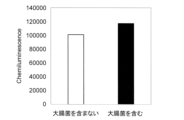
そして、96穴プレート(LBS OptiPlate-96、PerkinElmer;6065420)に終濃度2.0 ng/mLのGOx-抗大腸菌抗体複合体、終濃度2.0 ng/mL のHRP-抗OmpC抗体(CUSABIO;CSB-PA356994LB01ENV)、PBS、及び大腸菌懸濁液をPBSにて1.0×10⁵cells/mLになるように希釈した大腸菌溶液を、それぞれ5μLずつ添加し、25℃にて1時間静置した(大腸菌を含む)。比較として、大腸菌溶液の代わりにPBSを添加したものも同様に静置した(大腸菌を含まない)。静置後、さらに終濃度200mMのグルコース及び終濃度24 ng/mLのカタラーゼを含むBM chemiluminescence ELISA substrate (POD) (Roche;11582950001) solution Aを80μL添加し、プレートリーダー(Thermo Fisher Scientific社製、製品名:Varioskan Flash)にて化学発光強度を測定した。
大腸菌を含む溶液、及び、含まない溶液を加えた反応液を測定した結果を図8に示した。図8に示したように、大腸菌を含む溶液を加えた反応液において化学発光強度の増加が認められた。また、ほかの微生物についても、表面に標的タンパク質が集積していることから、同様に検出できることが理解される。
[比較例:光学的検出装置による標的物質(SARS-CoV-2三量体スパイクタンパク質及び受容体結合ドメイン(RBD))の検出]
96穴プレート(LBS OptiPlate-96、PerkinElmer;6065420)に、終濃度50 nMの抗体酵素複合体、終濃度2.1 ng/mLのHRP融合抗SARS-CoV-2抗体、10 mM KClを含む10 mMリン酸ナトリウムバッファー (pH 7.0) および終濃度100、50、25、0 nMのSARS-CoV-2三量体スパイクタンパク質 (BioServ)、または終濃度300、150、75、0 nMのSARS-CoV-2受容体結合ドメイン (RBD) (Sino Biological) をそれぞれ5 μLずつ添加、混合し、25℃で1時間静置した。その後、終濃度200 mMのグルコース、終濃度24 ng/mLのカタラーゼ (富士フイルム和光純薬) を含むBM Chemiluminescence ELISA Substrate Solution A (Roche) を80 μL添加してプレートリーダー(Thermo Fisher Scientific社製、製品名:Varioskan Flash)にて化学発光強度を測定した。
96穴プレート(LBS OptiPlate-96、PerkinElmer;6065420)に、終濃度50 nMの抗体酵素複合体、終濃度2.1 ng/mLのHRP融合抗SARS-CoV-2抗体、10 mM KClを含む10 mMリン酸ナトリウムバッファー (pH 7.0) および終濃度100、50、25、0 nMのSARS-CoV-2三量体スパイクタンパク質 (BioServ)、または終濃度300、150、75、0 nMのSARS-CoV-2受容体結合ドメイン (RBD) (Sino Biological) をそれぞれ5 μLずつ添加、混合し、25℃で1時間静置した。その後、終濃度200 mMのグルコース、終濃度24 ng/mLのカタラーゼ (富士フイルム和光純薬) を含むBM Chemiluminescence ELISA Substrate Solution A (Roche) を80 μL添加してプレートリーダー(Thermo Fisher Scientific社製、製品名:Varioskan Flash)にて化学発光強度を測定した。
SARS-CoV-2三量体スパイクタンパク質の結果を図9に、SARS-CoV-2受容体結合ドメインの結果を図10に示した。図9、及び、図10で示したように、それぞれのタンパク質を含む場合に得られる化学発光強度は、それぞれのタンパク質を含まない場合に得られる化学発光強度とほぼ同程度であり、実施例2のような濃度依存的な化学発光強度の増加が認められなかった。この結果から、本検出系はそれぞれの標的タンパク質が分散されている状態では発光量の増加は見られないことを示しており、微生物やウイルスなどの表面に標的タンパク質が集積し、それぞれの酵素が近接する必要があることが示している。
[配列情報]
本開示は以下の配列情報を包含する。
配列番号1:
MHHHHHHGAMVDTLSGLSSEQGQSGDMTIEEDSATHIKFSKRDEDGKELAGATMELRDSSGKTISTWISDGQVKDFYLYPGKYTFVETAAPDGYEVATAITFTVNEQGQVTVNGKATKGDAHIGSGMNGIEASLLTDPKDVSGRTVDYIIAGGGLTGLTTAARLTENPNISVLVIESGSYESDRGPIIEDLNAYGDIFGSSVDHAYETVELATNNQTALIRSGNGLGGSTLVNGGTWTRPHKAQVDSWETVFGNEGWNWDNVAAYSLQAERARAPNAKQIAAGHYFNASCHGVNGTVHAGPRDTGDDYSPIVKALMSAVEDRGVPTKKDFGCGDPHGVSMFPNTLHEDQVRSDAAREWLLPNYQRPNLQVLTGQYVGKVLLSQNGTTPRAVGVEFGTHKGNTHNVYAKHEVLLAAGSAVSPTILEYSGIGMKSILEPLGIDTVVDLPVGLNLQDQTTATVRSRITSAGAGQGQAAWFATFNETFGDYSEKAHELLNTKLEQWAEEAVARGGFHNTTALLIQYENYRDWIVNHNVAYSELFLDTAGVASFDVWDLLPFTRGYVHILDKDPYLHHFAYDPQYFLNELDLLGQAAATQLARNISNSGAMQTYFAGETIPGDNLAYDADLSAWTEYIPYHFRPNYHGVGTCSMMPKEMGGVVDNAARVYGVQGLRVIDGSIPPTQMSSHVMTVFYAMALKISDAILEDYASMQ
配列番号2:
MKYLLPTAAAGLLLLAAQPAMAEVQLVESGGGLVQPGGSLRLSCAASGFTVSSNYMSWVRQAPGKGLEWVSVIYSGGSTFYADSVKGRFTISRDNSMNTLFLQMNSLRAEDTAVYYCARVLPMYGDYLDYWGQGTLVTVSSGGGGSGGGGSGGGGSDIVMTQSPSSLSASVGDRVTITCRASQSISRYLNWYQQKPGKAPKLLIYAASSLQSGVPSRFSGSGSGTDFTLTISSLQPEDFATYYCQQSYSTPPEYTFGQGTKLEIKRAAAGSGAHIVMVDAYKPTKAEQKLISEEDLNLGGGMRGSHHHHHH
本開示は以下の配列情報を包含する。
配列番号1:
MHHHHHHGAMVDTLSGLSSEQGQSGDMTIEEDSATHIKFSKRDEDGKELAGATMELRDSSGKTISTWISDGQVKDFYLYPGKYTFVETAAPDGYEVATAITFTVNEQGQVTVNGKATKGDAHIGSGMNGIEASLLTDPKDVSGRTVDYIIAGGGLTGLTTAARLTENPNISVLVIESGSYESDRGPIIEDLNAYGDIFGSSVDHAYETVELATNNQTALIRSGNGLGGSTLVNGGTWTRPHKAQVDSWETVFGNEGWNWDNVAAYSLQAERARAPNAKQIAAGHYFNASCHGVNGTVHAGPRDTGDDYSPIVKALMSAVEDRGVPTKKDFGCGDPHGVSMFPNTLHEDQVRSDAAREWLLPNYQRPNLQVLTGQYVGKVLLSQNGTTPRAVGVEFGTHKGNTHNVYAKHEVLLAAGSAVSPTILEYSGIGMKSILEPLGIDTVVDLPVGLNLQDQTTATVRSRITSAGAGQGQAAWFATFNETFGDYSEKAHELLNTKLEQWAEEAVARGGFHNTTALLIQYENYRDWIVNHNVAYSELFLDTAGVASFDVWDLLPFTRGYVHILDKDPYLHHFAYDPQYFLNELDLLGQAAATQLARNISNSGAMQTYFAGETIPGDNLAYDADLSAWTEYIPYHFRPNYHGVGTCSMMPKEMGGVVDNAARVYGVQGLRVIDGSIPPTQMSSHVMTVFYAMALKISDAILEDYASMQ
配列番号2:
MKYLLPTAAAGLLLLAAQPAMAEVQLVESGGGLVQPGGSLRLSCAASGFTVSSNYMSWVRQAPGKGLEWVSVIYSGGSTFYADSVKGRFTISRDNSMNTLFLQMNSLRAEDTAVYYCARVLPMYGDYLDYWGQGTLVTVSSGGGGSGGGGSGGGGSDIVMTQSPSSLSASVGDRVTITCRASQSISRYLNWYQQKPGKAPKLLIYAASSLQSGVPSRFSGSGSGTDFTLTISSLQPEDFATYYCQQSYSTPPEYTFGQGTKLEIKRAAAGSGAHIVMVDAYKPTKAEQKLISEEDLNLGGGMRGSHHHHHH
本明細書で引用した全ての刊行物、特許及び特許出願はそのまま引用により本明細書に組み入れられるものとする。
Claims (10)
- タンパク質が表面に存在する標的物質に対して特異的に結合するポリペプチドからなる第1の特異的結合部と、当該第1の特異的結合部に直接又は間接的に結合した第1の酵素部とを有する第1の結合分子と、
上記第1の結合分子における第1の酵素部の基質となる第1の基質と、
上記標的物質に特異的に結合するポリペプチドからなる第2の特異的結合部と、当該第2の特異的結合部に直接又は間接的に結合し、上記第1の酵素部における酵素反応で上記第1の基質から生成した物質を第2の基質とする第2の酵素部とを有する第2の結合分子と、
上記第2の結合分子における第2の酵素部が、上記第2の基質とともに酵素反応の基質とする第3の基質と
を含む、標的物質の検出キット。 - 上記第2の結合分子における上記第2の酵素部における酵素反応を光学的に検出する検出装置に使用されることを特徴とする請求項1記載の標的物質の検出キット。
- 上記検出装置は、光電子増倍管を有さず、フォトダイオードを光学検出素子として備えることを特徴とする請求項2記載の標的物質の検出キット。
- 上記第2の基質を基質として消費する第3の酵素を更に含むことを特徴とする請求項1記載の標的物質の検出キット。
- 上記第1の結合分子における第1の酵素部はオキシダーゼ活性を有し、上記第2の結合分子における第2の酵素部はペルオキシダーゼ活性を有し、上記第3の基質がオルトフェニレンジアミン又はルミノールであることを特徴とする請求項1記載の標的物質の検出キット。
- 上記第1の結合分子における第1の酵素部はATPを産生するキナーゼ活性を有し、上記第2の結合分子における第2の酵素部はルシフェラーゼ活性を有し、上記第3の基質がルシフェリンであることを特徴とする請求項1記載の標的物質の検出キット。
- 上記標的物質はウイルス又は微生物であることを特徴とする請求項1記載の標的物質の検出キット。
- 請求項1乃至7いずれか一項記載の検出キットと、タンパク質が表面に存在する標的物質を含みうる試料とを混合する工程と、
上記検出キットに含まれる第2の結合分子における第2の酵素部による酵素反応を測定する工程とを含み、
上記第2の酵素の酵素反応に基づいて上記試料中の標的物質を検出することを特徴とする標的物質の検出方法。 - 検出装置によって、上記第2の結合分子における上記第2の酵素部における酵素反応を光学的に検出することを特徴とする請求項8記載の標的物質の検出方法。
- 上記検出装置は、光電子増倍管を有さず、フォトダイオードを光学検出素子として備えることを特徴とする請求項9記載の標的物質の検出方法。
Priority Applications (1)
| Application Number | Priority Date | Filing Date | Title |
|---|---|---|---|
| JP2024570209A JPWO2024150786A1 (ja) | 2023-01-13 | 2024-01-11 |
Applications Claiming Priority (2)
| Application Number | Priority Date | Filing Date | Title |
|---|---|---|---|
| JP2023003981 | 2023-01-13 | ||
| JP2023-003981 | 2023-01-13 |
Publications (1)
| Publication Number | Publication Date |
|---|---|
| WO2024150786A1 true WO2024150786A1 (ja) | 2024-07-18 |
Family
ID=91896955
Family Applications (1)
| Application Number | Title | Priority Date | Filing Date |
|---|---|---|---|
| PCT/JP2024/000383 Ceased WO2024150786A1 (ja) | 2023-01-13 | 2024-01-11 | 標的物質の検出試薬及び検出方法 |
Country Status (2)
| Country | Link |
|---|---|
| JP (1) | JPWO2024150786A1 (ja) |
| WO (1) | WO2024150786A1 (ja) |
Citations (2)
| Publication number | Priority date | Publication date | Assignee | Title |
|---|---|---|---|---|
| JPH01150856A (ja) * | 1987-11-02 | 1989-06-13 | Bioventures Inc | 抗原および抗体の同時免疫定量法 |
| JP2003028874A (ja) * | 2001-07-12 | 2003-01-29 | Mitsubishi Chemicals Corp | 2段階酵素反応を用いた測定対象物質の測定法 |
-
2024
- 2024-01-11 WO PCT/JP2024/000383 patent/WO2024150786A1/ja not_active Ceased
- 2024-01-11 JP JP2024570209A patent/JPWO2024150786A1/ja active Pending
Patent Citations (2)
| Publication number | Priority date | Publication date | Assignee | Title |
|---|---|---|---|---|
| JPH01150856A (ja) * | 1987-11-02 | 1989-06-13 | Bioventures Inc | 抗原および抗体の同時免疫定量法 |
| JP2003028874A (ja) * | 2001-07-12 | 2003-01-29 | Mitsubishi Chemicals Corp | 2段階酵素反応を用いた測定対象物質の測定法 |
Also Published As
| Publication number | Publication date |
|---|---|
| JPWO2024150786A1 (ja) | 2024-07-18 |
Similar Documents
| Publication | Publication Date | Title |
|---|---|---|
| Zhang et al. | An updated review of SARS‐CoV‐2 detection methods in the context of a novel coronavirus pandemic | |
| Huang et al. | Rapid detection of IgM antibodies against the SARS-CoV-2 virus via colloidal gold nanoparticle-based lateral-flow assay | |
| Islam et al. | An update on molecular diagnostics for COVID-19 | |
| Barlev-Gross et al. | Spike vs nucleocapsid SARS-CoV-2 antigen detection: application in nasopharyngeal swab specimens | |
| Grzelak et al. | A comparison of four serological assays for detecting anti–SARS-CoV-2 antibodies in human serum samples from different populations | |
| Engelmann et al. | Preanalytical issues and cycle threshold values in SARS-CoV-2 real-time RT-PCR testing: should test results include these? | |
| Kabir et al. | Diagnosis for COVID-19: current status and future prospects | |
| Tang et al. | Detection methods for Pseudomonas aeruginosa: history and future perspective | |
| Nguyen et al. | Development of diagnostic tests for detection of SARS-CoV-2 | |
| US6811971B2 (en) | Method for the detection of influenza A/B viruses | |
| US20090186368A1 (en) | Assay with reduced background | |
| Naikoo et al. | Nanomaterials‐based sensors for the detection of COVID‐19: A review | |
| Shabani et al. | Laboratory detection methods for the human coronaviruses | |
| Kang et al. | Ultrasensitive detection platform of disease biomarkers based on recombinase polymerase amplification with H-sandwich aptamers | |
| Chang et al. | Detection of swine-origin influenza A (H1N1) viruses using a localized surface plasmon coupled fluorescence fiber-optic biosensor | |
| WO2022006536A1 (en) | Crispr-cas-based detection of sars-cov-2 using recombinase polymerase amplification | |
| CN110998324A (zh) | 数字测定 | |
| Dong et al. | Two types of nanoparticle-based bio-barcode amplification assays to detect HIV-1 p24 antigen | |
| Khan et al. | Evolution of RNA viruses from SARS to SARS-CoV-2 and diagnostic techniques for COVID-19: A review | |
| Burakova et al. | Bioluminescent detection probe for tick-borne encephalitis virus immunoassay | |
| Nyaruaba et al. | SARS-CoV-2/COVID-19 laboratory biosafety practices and current molecular diagnostic tools | |
| Kyosei et al. | Improved detection sensitivity of an antigen test for SARS-CoV-2 nucleocapsid proteins with thio-NAD cycling | |
| US20220089697A1 (en) | Monoclonal antibody or antigen binding fragment thereof that binds to the l protein of the human parainfluenza virus (piv); method and kit for detecting piv | |
| Kim et al. | Simultaneous detection of infectious diseases using aptamer-conjugated gold nanoparticles in the lateral flow immunoassay-based signal amplification platform | |
| US20190257824A1 (en) | Allosteric Split Trehalase Biosensor |
Legal Events
| Date | Code | Title | Description |
|---|---|---|---|
| 121 | Ep: the epo has been informed by wipo that ep was designated in this application |
Ref document number: 24741558 Country of ref document: EP Kind code of ref document: A1 |
|
| WWE | Wipo information: entry into national phase |
Ref document number: 2024570209 Country of ref document: JP |
|
| NENP | Non-entry into the national phase |
Ref country code: DE |
|
| 122 | Ep: pct application non-entry in european phase |
Ref document number: 24741558 Country of ref document: EP Kind code of ref document: A1 |