WO2024143784A1 - Nanofibrous scaffold for skin regeneration, comprising polydeoxyribonucleotide derived from patiria pectinifera - Google Patents
Nanofibrous scaffold for skin regeneration, comprising polydeoxyribonucleotide derived from patiria pectinifera Download PDFInfo
- Publication number
- WO2024143784A1 WO2024143784A1 PCT/KR2023/014358 KR2023014358W WO2024143784A1 WO 2024143784 A1 WO2024143784 A1 WO 2024143784A1 KR 2023014358 W KR2023014358 W KR 2023014358W WO 2024143784 A1 WO2024143784 A1 WO 2024143784A1
- Authority
- WO
- WIPO (PCT)
- Prior art keywords
- nanofibers
- pdrn
- wound
- gelatin
- pectinifera
- Prior art date
Links
- 241000258745 Patiria pectinifera Species 0.000 title claims abstract description 39
- 239000002062 molecular scaffold Substances 0.000 title claims abstract description 25
- 230000036560 skin regeneration Effects 0.000 title description 7
- 239000002121 nanofiber Substances 0.000 claims abstract description 150
- 206010052428 Wound Diseases 0.000 claims abstract description 68
- 208000027418 Wounds and injury Diseases 0.000 claims abstract description 68
- 229920001610 polycaprolactone Polymers 0.000 claims abstract description 54
- 108010010803 Gelatin Proteins 0.000 claims abstract description 42
- 229920000159 gelatin Polymers 0.000 claims abstract description 42
- 239000008273 gelatin Substances 0.000 claims abstract description 42
- 235000019322 gelatine Nutrition 0.000 claims abstract description 42
- 235000011852 gelatine desserts Nutrition 0.000 claims abstract description 42
- 239000004632 polycaprolactone Substances 0.000 claims description 47
- 238000000034 method Methods 0.000 claims description 17
- 238000001523 electrospinning Methods 0.000 claims description 14
- 241000258957 Asteroidea Species 0.000 claims description 13
- 239000000203 mixture Substances 0.000 claims description 11
- 238000004519 manufacturing process Methods 0.000 claims description 10
- 238000002156 mixing Methods 0.000 claims description 6
- 239000002336 ribonucleotide Substances 0.000 claims 2
- 230000001737 promoting effect Effects 0.000 claims 1
- 230000008929 regeneration Effects 0.000 claims 1
- 238000011069 regeneration method Methods 0.000 claims 1
- 238000010521 absorption reaction Methods 0.000 abstract description 6
- -1 poly(ε-caprolactone) Polymers 0.000 abstract description 4
- 238000002360 preparation method Methods 0.000 abstract description 4
- 239000012530 fluid Substances 0.000 abstract description 2
- 230000035876 healing Effects 0.000 abstract description 2
- 230000014759 maintenance of location Effects 0.000 abstract description 2
- 210000004027 cell Anatomy 0.000 description 42
- 230000000694 effects Effects 0.000 description 28
- LNTHITQWFMADLM-UHFFFAOYSA-N gallic acid Chemical compound OC(=O)C1=CC(O)=C(O)C(O)=C1 LNTHITQWFMADLM-UHFFFAOYSA-N 0.000 description 24
- 210000001519 tissue Anatomy 0.000 description 22
- 230000029663 wound healing Effects 0.000 description 21
- 210000003491 skin Anatomy 0.000 description 19
- 108020004414 DNA Proteins 0.000 description 18
- 238000004458 analytical method Methods 0.000 description 18
- 231100000135 cytotoxicity Toxicity 0.000 description 17
- 230000003013 cytotoxicity Effects 0.000 description 17
- 230000001965 increasing effect Effects 0.000 description 17
- 108090000623 proteins and genes Proteins 0.000 description 16
- 102000004169 proteins and genes Human genes 0.000 description 16
- 239000000243 solution Substances 0.000 description 14
- XLYOFNOQVPJJNP-UHFFFAOYSA-N water Substances O XLYOFNOQVPJJNP-UHFFFAOYSA-N 0.000 description 14
- LFQSCWFLJHTTHZ-UHFFFAOYSA-N Ethanol Chemical compound CCO LFQSCWFLJHTTHZ-UHFFFAOYSA-N 0.000 description 13
- LOKCTEFSRHRXRJ-UHFFFAOYSA-I dipotassium trisodium dihydrogen phosphate hydrogen phosphate dichloride Chemical compound P(=O)(O)(O)[O-].[K+].P(=O)(O)([O-])[O-].[Na+].[Na+].[Cl-].[K+].[Cl-].[Na+] LOKCTEFSRHRXRJ-UHFFFAOYSA-I 0.000 description 13
- 239000002953 phosphate buffered saline Substances 0.000 description 13
- 238000010186 staining Methods 0.000 description 13
- 229940074391 gallic acid Drugs 0.000 description 12
- 235000004515 gallic acid Nutrition 0.000 description 12
- 239000000499 gel Substances 0.000 description 12
- XJMOSONTPMZWPB-UHFFFAOYSA-M propidium iodide Chemical compound [I-].[I-].C12=CC(N)=CC=C2C2=CC=C(N)C=C2[N+](CCC[N+](C)(CC)CC)=C1C1=CC=CC=C1 XJMOSONTPMZWPB-UHFFFAOYSA-M 0.000 description 12
- 238000002835 absorbance Methods 0.000 description 11
- 239000000835 fiber Substances 0.000 description 11
- 102000008186 Collagen Human genes 0.000 description 10
- 108010035532 Collagen Proteins 0.000 description 10
- 230000012292 cell migration Effects 0.000 description 10
- 229920001436 collagen Polymers 0.000 description 10
- 238000002474 experimental method Methods 0.000 description 10
- 238000000338 in vitro Methods 0.000 description 10
- YIQKLZYTHXTDDT-UHFFFAOYSA-H Sirius red F3B Chemical compound C1=CC(=CC=C1N=NC2=CC(=C(C=C2)N=NC3=C(C=C4C=C(C=CC4=C3[O-])NC(=O)NC5=CC6=CC(=C(C(=C6C=C5)[O-])N=NC7=C(C=C(C=C7)N=NC8=CC=C(C=C8)S(=O)(=O)[O-])S(=O)(=O)[O-])S(=O)(=O)O)S(=O)(=O)O)S(=O)(=O)[O-])S(=O)(=O)[O-].[Na+].[Na+].[Na+].[Na+].[Na+].[Na+] YIQKLZYTHXTDDT-UHFFFAOYSA-H 0.000 description 9
- 230000037319 collagen production Effects 0.000 description 9
- 241000699670 Mus sp. Species 0.000 description 8
- 230000035755 proliferation Effects 0.000 description 8
- 238000013424 sirius red staining Methods 0.000 description 8
- IJGRMHOSHXDMSA-UHFFFAOYSA-N Atomic nitrogen Chemical compound N#N IJGRMHOSHXDMSA-UHFFFAOYSA-N 0.000 description 7
- 241001465754 Metazoa Species 0.000 description 7
- 241000699666 Mus <mouse, genus> Species 0.000 description 7
- 238000001727 in vivo Methods 0.000 description 7
- 238000001356 surgical procedure Methods 0.000 description 7
- IAZDPXIOMUYVGZ-UHFFFAOYSA-N Dimethylsulphoxide Chemical compound CS(C)=O IAZDPXIOMUYVGZ-UHFFFAOYSA-N 0.000 description 6
- WZUVPPKBWHMQCE-UHFFFAOYSA-N Haematoxylin Chemical compound C12=CC(O)=C(O)C=C2CC2(O)C1C1=CC=C(O)C(O)=C1OC2 WZUVPPKBWHMQCE-UHFFFAOYSA-N 0.000 description 6
- 231100000002 MTT assay Toxicity 0.000 description 6
- 238000000134 MTT assay Methods 0.000 description 6
- 238000002441 X-ray diffraction Methods 0.000 description 6
- 230000003247 decreasing effect Effects 0.000 description 6
- 239000000284 extract Substances 0.000 description 6
- 238000002411 thermogravimetry Methods 0.000 description 6
- 230000017423 tissue regeneration Effects 0.000 description 6
- 238000001262 western blot Methods 0.000 description 6
- 102000001187 Collagen Type III Human genes 0.000 description 5
- 108010069502 Collagen Type III Proteins 0.000 description 5
- 101710143123 Mothers against decapentaplegic homolog 2 Proteins 0.000 description 5
- 102000057208 Smad2 Human genes 0.000 description 5
- 239000000872 buffer Substances 0.000 description 5
- 230000004663 cell proliferation Effects 0.000 description 5
- 239000003153 chemical reaction reagent Substances 0.000 description 5
- 238000009826 distribution Methods 0.000 description 5
- 239000003814 drug Substances 0.000 description 5
- 239000000463 material Substances 0.000 description 5
- 239000000843 powder Substances 0.000 description 5
- 108091003079 Bovine Serum Albumin Proteins 0.000 description 4
- 102000012422 Collagen Type I Human genes 0.000 description 4
- 108010022452 Collagen Type I Proteins 0.000 description 4
- 239000006144 Dulbecco’s modified Eagle's medium Substances 0.000 description 4
- 102000007665 Extracellular Signal-Regulated MAP Kinases Human genes 0.000 description 4
- 108010007457 Extracellular Signal-Regulated MAP Kinases Proteins 0.000 description 4
- 238000001157 Fourier transform infrared spectrum Methods 0.000 description 4
- KFZMGEQAYNKOFK-UHFFFAOYSA-N Isopropanol Chemical compound CC(C)O KFZMGEQAYNKOFK-UHFFFAOYSA-N 0.000 description 4
- 102000043136 MAP kinase family Human genes 0.000 description 4
- 108091054455 MAP kinase family Proteins 0.000 description 4
- 238000012512 characterization method Methods 0.000 description 4
- 239000013078 crystal Substances 0.000 description 4
- 238000000354 decomposition reaction Methods 0.000 description 4
- 229940079593 drug Drugs 0.000 description 4
- 238000010828 elution Methods 0.000 description 4
- 239000001963 growth medium Substances 0.000 description 4
- UYTPUPDQBNUYGX-UHFFFAOYSA-N guanine Chemical compound O=C1NC(N)=NC2=C1N=CN2 UYTPUPDQBNUYGX-UHFFFAOYSA-N 0.000 description 4
- 238000011534 incubation Methods 0.000 description 4
- 230000002062 proliferating effect Effects 0.000 description 4
- 238000012360 testing method Methods 0.000 description 4
- 229920000936 Agarose Polymers 0.000 description 3
- 241000277275 Oncorhynchus mykiss Species 0.000 description 3
- 241000283973 Oryctolagus cuniculus Species 0.000 description 3
- HEMHJVSKTPXQMS-UHFFFAOYSA-M Sodium hydroxide Chemical compound [OH-].[Na+] HEMHJVSKTPXQMS-UHFFFAOYSA-M 0.000 description 3
- 150000001408 amides Chemical class 0.000 description 3
- 238000012925 biological evaluation Methods 0.000 description 3
- 230000015556 catabolic process Effects 0.000 description 3
- 230000003833 cell viability Effects 0.000 description 3
- 238000004132 cross linking Methods 0.000 description 3
- 238000002425 crystallisation Methods 0.000 description 3
- 230000008025 crystallization Effects 0.000 description 3
- 230000007423 decrease Effects 0.000 description 3
- 238000006731 degradation reaction Methods 0.000 description 3
- 238000010586 diagram Methods 0.000 description 3
- 238000000113 differential scanning calorimetry Methods 0.000 description 3
- 238000001962 electrophoresis Methods 0.000 description 3
- 238000005516 engineering process Methods 0.000 description 3
- 210000000416 exudates and transudate Anatomy 0.000 description 3
- 239000012091 fetal bovine serum Substances 0.000 description 3
- 238000010438 heat treatment Methods 0.000 description 3
- 238000002844 melting Methods 0.000 description 3
- 230000008018 melting Effects 0.000 description 3
- 239000012528 membrane Substances 0.000 description 3
- 230000005012 migration Effects 0.000 description 3
- 238000013508 migration Methods 0.000 description 3
- 229910052757 nitrogen Inorganic materials 0.000 description 3
- 230000037361 pathway Effects 0.000 description 3
- 239000011148 porous material Substances 0.000 description 3
- 230000008569 process Effects 0.000 description 3
- 239000011734 sodium Substances 0.000 description 3
- 239000002904 solvent Substances 0.000 description 3
- 229910001220 stainless steel Inorganic materials 0.000 description 3
- 239000010935 stainless steel Substances 0.000 description 3
- 235000000346 sugar Nutrition 0.000 description 3
- QAPSNMNOIOSXSQ-YNEHKIRRSA-N 1-[(2r,4s,5r)-4-[tert-butyl(dimethyl)silyl]oxy-5-(hydroxymethyl)oxolan-2-yl]-5-methylpyrimidine-2,4-dione Chemical compound O=C1NC(=O)C(C)=CN1[C@@H]1O[C@H](CO)[C@@H](O[Si](C)(C)C(C)(C)C)C1 QAPSNMNOIOSXSQ-YNEHKIRRSA-N 0.000 description 2
- QKNYBSVHEMOAJP-UHFFFAOYSA-N 2-amino-2-(hydroxymethyl)propane-1,3-diol;hydron;chloride Chemical compound Cl.OCC(N)(CO)CO QKNYBSVHEMOAJP-UHFFFAOYSA-N 0.000 description 2
- 102000007469 Actins Human genes 0.000 description 2
- 108010085238 Actins Proteins 0.000 description 2
- 241000283707 Capra Species 0.000 description 2
- RTZKZFJDLAIYFH-UHFFFAOYSA-N Diethyl ether Chemical compound CCOCC RTZKZFJDLAIYFH-UHFFFAOYSA-N 0.000 description 2
- KCXVZYZYPLLWCC-UHFFFAOYSA-N EDTA Chemical compound OC(=O)CN(CC(O)=O)CCN(CC(O)=O)CC(O)=O KCXVZYZYPLLWCC-UHFFFAOYSA-N 0.000 description 2
- 102000010834 Extracellular Matrix Proteins Human genes 0.000 description 2
- 108010037362 Extracellular Matrix Proteins Proteins 0.000 description 2
- WSFSSNUMVMOOMR-UHFFFAOYSA-N Formaldehyde Chemical compound O=C WSFSSNUMVMOOMR-UHFFFAOYSA-N 0.000 description 2
- 238000005033 Fourier transform infrared spectroscopy Methods 0.000 description 2
- 238000012404 In vitro experiment Methods 0.000 description 2
- PIWKPBJCKXDKJR-UHFFFAOYSA-N Isoflurane Chemical compound FC(F)OC(Cl)C(F)(F)F PIWKPBJCKXDKJR-UHFFFAOYSA-N 0.000 description 2
- CTQNGGLPUBDAKN-UHFFFAOYSA-N O-Xylene Chemical compound CC1=CC=CC=C1C CTQNGGLPUBDAKN-UHFFFAOYSA-N 0.000 description 2
- 241000277329 Oncorhynchus keta Species 0.000 description 2
- 229910019142 PO4 Inorganic materials 0.000 description 2
- ISWSIDIOOBJBQZ-UHFFFAOYSA-N Phenol Chemical compound OC1=CC=CC=C1 ISWSIDIOOBJBQZ-UHFFFAOYSA-N 0.000 description 2
- 206010059516 Skin toxicity Diseases 0.000 description 2
- 206010072170 Skin wound Diseases 0.000 description 2
- CDBYLPFSWZWCQE-UHFFFAOYSA-L Sodium Carbonate Chemical compound [Na+].[Na+].[O-]C([O-])=O CDBYLPFSWZWCQE-UHFFFAOYSA-L 0.000 description 2
- FAPWRFPIFSIZLT-UHFFFAOYSA-M Sodium chloride Chemical compound [Na+].[Cl-] FAPWRFPIFSIZLT-UHFFFAOYSA-M 0.000 description 2
- 230000004913 activation Effects 0.000 description 2
- OIRDTQYFTABQOQ-KQYNXXCUSA-N adenosine Chemical compound C1=NC=2C(N)=NC=NC=2N1[C@@H]1O[C@H](CO)[C@@H](O)[C@H]1O OIRDTQYFTABQOQ-KQYNXXCUSA-N 0.000 description 2
- 239000011543 agarose gel Substances 0.000 description 2
- XAGFODPZIPBFFR-UHFFFAOYSA-N aluminium Chemical compound [Al] XAGFODPZIPBFFR-UHFFFAOYSA-N 0.000 description 2
- 229910052782 aluminium Inorganic materials 0.000 description 2
- 238000000540 analysis of variance Methods 0.000 description 2
- 239000007864 aqueous solution Substances 0.000 description 2
- 238000005452 bending Methods 0.000 description 2
- AFYNADDZULBEJA-UHFFFAOYSA-N bicinchoninic acid Chemical compound C1=CC=CC2=NC(C=3C=C(C4=CC=CC=C4N=3)C(=O)O)=CC(C(O)=O)=C21 AFYNADDZULBEJA-UHFFFAOYSA-N 0.000 description 2
- 210000004204 blood vessel Anatomy 0.000 description 2
- 150000001720 carbohydrates Chemical class 0.000 description 2
- 235000014633 carbohydrates Nutrition 0.000 description 2
- 125000002915 carbonyl group Chemical group [*:2]C([*:1])=O 0.000 description 2
- 238000005119 centrifugation Methods 0.000 description 2
- OPTASPLRGRRNAP-UHFFFAOYSA-N cytosine Chemical compound NC=1C=CNC(=O)N=1 OPTASPLRGRRNAP-UHFFFAOYSA-N 0.000 description 2
- 231100000433 cytotoxic Toxicity 0.000 description 2
- 230000001472 cytotoxic effect Effects 0.000 description 2
- 231100000263 cytotoxicity test Toxicity 0.000 description 2
- 238000000151 deposition Methods 0.000 description 2
- 230000008021 deposition Effects 0.000 description 2
- 238000010790 dilution Methods 0.000 description 2
- 239000012895 dilution Substances 0.000 description 2
- 231100000673 dose–response relationship Toxicity 0.000 description 2
- 210000002744 extracellular matrix Anatomy 0.000 description 2
- 238000000445 field-emission scanning electron microscopy Methods 0.000 description 2
- 239000010408 film Substances 0.000 description 2
- 239000006260 foam Substances 0.000 description 2
- 108020004445 glyceraldehyde-3-phosphate dehydrogenase Proteins 0.000 description 2
- 210000003780 hair follicle Anatomy 0.000 description 2
- 230000036541 health Effects 0.000 description 2
- 238000007490 hematoxylin and eosin (H&E) staining Methods 0.000 description 2
- 238000010562 histological examination Methods 0.000 description 2
- 229920001477 hydrophilic polymer Polymers 0.000 description 2
- 230000001939 inductive effect Effects 0.000 description 2
- 229960002725 isoflurane Drugs 0.000 description 2
- 239000012139 lysis buffer Substances 0.000 description 2
- 230000010534 mechanism of action Effects 0.000 description 2
- 238000000386 microscopy Methods 0.000 description 2
- 239000011259 mixed solution Substances 0.000 description 2
- 239000012120 mounting media Substances 0.000 description 2
- 239000002086 nanomaterial Substances 0.000 description 2
- 229920005615 natural polymer Polymers 0.000 description 2
- 108020004707 nucleic acids Proteins 0.000 description 2
- 102000039446 nucleic acids Human genes 0.000 description 2
- 150000007523 nucleic acids Chemical class 0.000 description 2
- YBYRMVIVWMBXKQ-UHFFFAOYSA-N phenylmethanesulfonyl fluoride Chemical compound FS(=O)(=O)CC1=CC=CC=C1 YBYRMVIVWMBXKQ-UHFFFAOYSA-N 0.000 description 2
- NBIIXXVUZAFLBC-UHFFFAOYSA-K phosphate Chemical compound [O-]P([O-])([O-])=O NBIIXXVUZAFLBC-UHFFFAOYSA-K 0.000 description 2
- 239000010452 phosphate Substances 0.000 description 2
- 230000026731 phosphorylation Effects 0.000 description 2
- 238000006366 phosphorylation reaction Methods 0.000 description 2
- 150000008442 polyphenolic compounds Chemical class 0.000 description 2
- 235000013824 polyphenols Nutrition 0.000 description 2
- 230000005855 radiation Effects 0.000 description 2
- 230000001172 regenerating effect Effects 0.000 description 2
- 238000001878 scanning electron micrograph Methods 0.000 description 2
- 210000001732 sebaceous gland Anatomy 0.000 description 2
- 231100000438 skin toxicity Toxicity 0.000 description 2
- 239000000126 substance Substances 0.000 description 2
- 230000008961 swelling Effects 0.000 description 2
- 229920001059 synthetic polymer Polymers 0.000 description 2
- 238000001757 thermogravimetry curve Methods 0.000 description 2
- 230000001988 toxicity Effects 0.000 description 2
- 231100000419 toxicity Toxicity 0.000 description 2
- 239000008096 xylene Substances 0.000 description 2
- CHADEQDQBURGHL-UHFFFAOYSA-N (6'-acetyloxy-3-oxospiro[2-benzofuran-1,9'-xanthene]-3'-yl) acetate Chemical compound O1C(=O)C2=CC=CC=C2C21C1=CC=C(OC(C)=O)C=C1OC1=CC(OC(=O)C)=CC=C21 CHADEQDQBURGHL-UHFFFAOYSA-N 0.000 description 1
- BYEAHWXPCBROCE-UHFFFAOYSA-N 1,1,1,3,3,3-hexafluoropropan-2-ol Chemical compound FC(F)(F)C(O)C(F)(F)F BYEAHWXPCBROCE-UHFFFAOYSA-N 0.000 description 1
- CPKVUHPKYQGHMW-UHFFFAOYSA-N 1-ethenylpyrrolidin-2-one;molecular iodine Chemical compound II.C=CN1CCCC1=O CPKVUHPKYQGHMW-UHFFFAOYSA-N 0.000 description 1
- ASJSAQIRZKANQN-CRCLSJGQSA-N 2-deoxy-D-ribose Chemical compound OC[C@@H](O)[C@@H](O)CC=O ASJSAQIRZKANQN-CRCLSJGQSA-N 0.000 description 1
- FHVDTGUDJYJELY-UHFFFAOYSA-N 6-{[2-carboxy-4,5-dihydroxy-6-(phosphanyloxy)oxan-3-yl]oxy}-4,5-dihydroxy-3-phosphanyloxane-2-carboxylic acid Chemical compound O1C(C(O)=O)C(P)C(O)C(O)C1OC1C(C(O)=O)OC(OP)C(O)C1O FHVDTGUDJYJELY-UHFFFAOYSA-N 0.000 description 1
- 229930024421 Adenine Natural products 0.000 description 1
- GFFGJBXGBJISGV-UHFFFAOYSA-N Adenine Chemical compound NC1=NC=NC2=C1N=CN2 GFFGJBXGBJISGV-UHFFFAOYSA-N 0.000 description 1
- 108010039627 Aprotinin Proteins 0.000 description 1
- 241000972773 Aulopiformes Species 0.000 description 1
- 208000035404 Autolysis Diseases 0.000 description 1
- 238000000035 BCA protein assay Methods 0.000 description 1
- 108010027529 Bio-glue Proteins 0.000 description 1
- 206010057248 Cell death Diseases 0.000 description 1
- 229920001661 Chitosan Polymers 0.000 description 1
- 102000053602 DNA Human genes 0.000 description 1
- 238000007400 DNA extraction Methods 0.000 description 1
- 108010067770 Endopeptidase K Proteins 0.000 description 1
- 241000283074 Equus asinus Species 0.000 description 1
- 108700039887 Essential Genes Proteins 0.000 description 1
- WQZGKKKJIJFFOK-GASJEMHNSA-N Glucose Natural products OC[C@H]1OC(O)[C@H](O)[C@@H](O)[C@@H]1O WQZGKKKJIJFFOK-GASJEMHNSA-N 0.000 description 1
- 102100031181 Glyceraldehyde-3-phosphate dehydrogenase Human genes 0.000 description 1
- GDBQQVLCIARPGH-UHFFFAOYSA-N Leupeptin Natural products CC(C)CC(NC(C)=O)C(=O)NC(CC(C)C)C(=O)NC(C=O)CCCN=C(N)N GDBQQVLCIARPGH-UHFFFAOYSA-N 0.000 description 1
- NPPQSCRMBWNHMW-UHFFFAOYSA-N Meprobamate Chemical compound NC(=O)OCC(C)(CCC)COC(N)=O NPPQSCRMBWNHMW-UHFFFAOYSA-N 0.000 description 1
- 239000000020 Nitrocellulose Substances 0.000 description 1
- 108020002230 Pancreatic Ribonuclease Proteins 0.000 description 1
- 102000005891 Pancreatic ribonuclease Human genes 0.000 description 1
- 108091000080 Phosphotransferase Proteins 0.000 description 1
- 229920001213 Polysorbate 20 Polymers 0.000 description 1
- 229920000153 Povidone-iodine Polymers 0.000 description 1
- 208000004210 Pressure Ulcer Diseases 0.000 description 1
- BQCADISMDOOEFD-UHFFFAOYSA-N Silver Chemical compound [Ag] BQCADISMDOOEFD-UHFFFAOYSA-N 0.000 description 1
- 229910000831 Steel Inorganic materials 0.000 description 1
- 239000008049 TAE buffer Substances 0.000 description 1
- HGEVZDLYZYVYHD-UHFFFAOYSA-N acetic acid;2-amino-2-(hydroxymethyl)propane-1,3-diol;2-[2-[bis(carboxymethyl)amino]ethyl-(carboxymethyl)amino]acetic acid Chemical compound CC(O)=O.OCC(N)(CO)CO.OC(=O)CN(CC(O)=O)CCN(CC(O)=O)CC(O)=O HGEVZDLYZYVYHD-UHFFFAOYSA-N 0.000 description 1
- 239000002253 acid Substances 0.000 description 1
- 230000003213 activating effect Effects 0.000 description 1
- 229960000643 adenine Drugs 0.000 description 1
- 238000000246 agarose gel electrophoresis Methods 0.000 description 1
- 229940072056 alginate Drugs 0.000 description 1
- 235000010443 alginic acid Nutrition 0.000 description 1
- 229920000615 alginic acid Polymers 0.000 description 1
- 125000001931 aliphatic group Chemical group 0.000 description 1
- 229920003232 aliphatic polyester Polymers 0.000 description 1
- 230000002424 anti-apoptotic effect Effects 0.000 description 1
- 230000003110 anti-inflammatory effect Effects 0.000 description 1
- 239000000427 antigen Substances 0.000 description 1
- 108091007433 antigens Proteins 0.000 description 1
- 102000036639 antigens Human genes 0.000 description 1
- 229960004405 aprotinin Drugs 0.000 description 1
- 239000012298 atmosphere Substances 0.000 description 1
- 239000011324 bead Substances 0.000 description 1
- 230000004071 biological effect Effects 0.000 description 1
- 230000005540 biological transmission Effects 0.000 description 1
- 238000001574 biopsy Methods 0.000 description 1
- 230000015572 biosynthetic process Effects 0.000 description 1
- 229940098773 bovine serum albumin Drugs 0.000 description 1
- 238000004364 calculation method Methods 0.000 description 1
- 238000007707 calorimetry Methods 0.000 description 1
- 238000004113 cell culture Methods 0.000 description 1
- 239000013592 cell lysate Substances 0.000 description 1
- 239000013553 cell monolayer Substances 0.000 description 1
- 230000019522 cellular metabolic process Effects 0.000 description 1
- 230000005754 cellular signaling Effects 0.000 description 1
- 238000010382 chemical cross-linking Methods 0.000 description 1
- 238000003776 cleavage reaction Methods 0.000 description 1
- 239000011248 coating agent Substances 0.000 description 1
- 238000000576 coating method Methods 0.000 description 1
- 239000002131 composite material Substances 0.000 description 1
- 239000003431 cross linking reagent Substances 0.000 description 1
- 229940104302 cytosine Drugs 0.000 description 1
- 230000007547 defect Effects 0.000 description 1
- 238000013461 design Methods 0.000 description 1
- 230000006866 deterioration Effects 0.000 description 1
- 238000009792 diffusion process Methods 0.000 description 1
- 229910001873 dinitrogen Inorganic materials 0.000 description 1
- 239000012153 distilled water Substances 0.000 description 1
- AUZONCFQVSMFAP-UHFFFAOYSA-N disulfiram Chemical compound CCN(CC)C(=S)SSC(=S)N(CC)CC AUZONCFQVSMFAP-UHFFFAOYSA-N 0.000 description 1
- 230000002900 effect on cell Effects 0.000 description 1
- 230000002500 effect on skin Effects 0.000 description 1
- YQGOJNYOYNNSMM-UHFFFAOYSA-N eosin Chemical compound [Na+].OC(=O)C1=CC=CC=C1C1=C2C=C(Br)C(=O)C(Br)=C2OC2=C(Br)C(O)=C(Br)C=C21 YQGOJNYOYNNSMM-UHFFFAOYSA-N 0.000 description 1
- SEACYXSIPDVVMV-UHFFFAOYSA-L eosin Y Chemical compound [Na+].[Na+].[O-]C(=O)C1=CC=CC=C1C1=C2C=C(Br)C(=O)C(Br)=C2OC2=C(Br)C([O-])=C(Br)C=C21 SEACYXSIPDVVMV-UHFFFAOYSA-L 0.000 description 1
- 210000000981 epithelium Anatomy 0.000 description 1
- 125000004185 ester group Chemical group 0.000 description 1
- ZMMJGEGLRURXTF-UHFFFAOYSA-N ethidium bromide Chemical compound [Br-].C12=CC(N)=CC=C2C2=CC=C(N)C=C2[N+](CC)=C1C1=CC=CC=C1 ZMMJGEGLRURXTF-UHFFFAOYSA-N 0.000 description 1
- 229960005542 ethidium bromide Drugs 0.000 description 1
- 238000000605 extraction Methods 0.000 description 1
- 210000002950 fibroblast Anatomy 0.000 description 1
- 239000011888 foil Substances 0.000 description 1
- 239000013505 freshwater Substances 0.000 description 1
- 230000006870 function Effects 0.000 description 1
- 125000000524 functional group Chemical group 0.000 description 1
- 150000002243 furanoses Chemical class 0.000 description 1
- 230000009477 glass transition Effects 0.000 description 1
- 239000008103 glucose Substances 0.000 description 1
- 102000006602 glyceraldehyde-3-phosphate dehydrogenase Human genes 0.000 description 1
- 230000012010 growth Effects 0.000 description 1
- 230000002209 hydrophobic effect Effects 0.000 description 1
- 229920001600 hydrophobic polymer Polymers 0.000 description 1
- 238000003384 imaging method Methods 0.000 description 1
- 125000002883 imidazolyl group Chemical group 0.000 description 1
- 238000010348 incorporation Methods 0.000 description 1
- 230000006698 induction Effects 0.000 description 1
- 230000008595 infiltration Effects 0.000 description 1
- 238000001764 infiltration Methods 0.000 description 1
- 238000002329 infrared spectrum Methods 0.000 description 1
- ZPNFWUPYTFPOJU-LPYSRVMUSA-N iniprol Chemical compound C([C@H]1C(=O)NCC(=O)NCC(=O)N[C@H]2CSSC[C@H]3C(=O)N[C@@H](CCCCN)C(=O)N[C@@H](C)C(=O)N[C@@H](CCCNC(N)=N)C(=O)N[C@H](C(N[C@H](C(=O)N[C@@H](CCCNC(N)=N)C(=O)N[C@@H](CC=4C=CC(O)=CC=4)C(=O)N[C@@H](CC=4C=CC=CC=4)C(=O)N[C@@H](CC=4C=CC(O)=CC=4)C(=O)N[C@@H](CC(N)=O)C(=O)N[C@@H](C)C(=O)N[C@@H](CCCCN)C(=O)N[C@@H](C)C(=O)NCC(=O)N[C@@H](CC(C)C)C(=O)N[C@@H](CSSC[C@H](NC(=O)[C@H](CC(O)=O)NC(=O)[C@H](CCC(O)=O)NC(=O)[C@H](C)NC(=O)[C@H](CO)NC(=O)[C@H](CCCCN)NC(=O)[C@H](CC=4C=CC=CC=4)NC(=O)[C@H](CC(N)=O)NC(=O)[C@H](CC(N)=O)NC(=O)[C@H](CCCNC(N)=N)NC(=O)[C@H](CCCCN)NC(=O)[C@H](C)NC(=O)[C@H](CCCNC(N)=N)NC2=O)C(=O)N[C@@H](CCSC)C(=O)N[C@@H](CCCNC(N)=N)C(=O)N[C@@H]([C@@H](C)O)C(=O)N[C@@H](CSSC[C@H](NC(=O)[C@H](CC=2C=CC=CC=2)NC(=O)[C@H](CC(O)=O)NC(=O)[C@H]2N(CCC2)C(=O)[C@@H](N)CCCNC(N)=N)C(=O)N[C@@H](CC(C)C)C(=O)N[C@@H](CCC(O)=O)C(=O)N2[C@@H](CCC2)C(=O)N2[C@@H](CCC2)C(=O)N[C@@H](CC=2C=CC(O)=CC=2)C(=O)N[C@@H]([C@@H](C)O)C(=O)NCC(=O)N2[C@@H](CCC2)C(=O)N3)C(=O)NCC(=O)NCC(=O)N[C@@H](C)C(O)=O)C(=O)N[C@@H](CCC(N)=O)C(=O)N[C@H](C(=O)N[C@@H](CC=2C=CC=CC=2)C(=O)N[C@H](C(=O)N1)C(C)C)[C@@H](C)O)[C@@H](C)CC)=O)[C@@H](C)CC)C1=CC=C(O)C=C1 ZPNFWUPYTFPOJU-LPYSRVMUSA-N 0.000 description 1
- 208000014674 injury Diseases 0.000 description 1
- 238000011081 inoculation Methods 0.000 description 1
- 210000002510 keratinocyte Anatomy 0.000 description 1
- GDBQQVLCIARPGH-ULQDDVLXSA-N leupeptin Chemical compound CC(C)C[C@H](NC(C)=O)C(=O)N[C@@H](CC(C)C)C(=O)N[C@H](C=O)CCCN=C(N)N GDBQQVLCIARPGH-ULQDDVLXSA-N 0.000 description 1
- 108010052968 leupeptin Proteins 0.000 description 1
- 150000002632 lipids Chemical class 0.000 description 1
- 238000011068 loading method Methods 0.000 description 1
- 239000003550 marker Substances 0.000 description 1
- 239000011159 matrix material Substances 0.000 description 1
- 238000005259 measurement Methods 0.000 description 1
- 239000002609 medium Substances 0.000 description 1
- 108020004999 messenger RNA Proteins 0.000 description 1
- 238000010232 migration assay Methods 0.000 description 1
- 229940126619 mouse monoclonal antibody Drugs 0.000 description 1
- 210000003205 muscle Anatomy 0.000 description 1
- VMGAPWLDMVPYIA-HIDZBRGKSA-N n'-amino-n-iminomethanimidamide Chemical compound N\N=C\N=N VMGAPWLDMVPYIA-HIDZBRGKSA-N 0.000 description 1
- 230000001338 necrotic effect Effects 0.000 description 1
- 229920001220 nitrocellulos Polymers 0.000 description 1
- 231100001083 no cytotoxicity Toxicity 0.000 description 1
- 239000004745 nonwoven fabric Substances 0.000 description 1
- 235000015097 nutrients Nutrition 0.000 description 1
- 239000008188 pellet Substances 0.000 description 1
- 102000020233 phosphotransferase Human genes 0.000 description 1
- 230000000704 physical effect Effects 0.000 description 1
- OXNIZHLAWKMVMX-UHFFFAOYSA-N picric acid Chemical compound OC1=C([N+]([O-])=O)C=C([N+]([O-])=O)C=C1[N+]([O-])=O OXNIZHLAWKMVMX-UHFFFAOYSA-N 0.000 description 1
- 229920003023 plastic Polymers 0.000 description 1
- 239000004033 plastic Substances 0.000 description 1
- 229920000642 polymer Polymers 0.000 description 1
- 235000010486 polyoxyethylene sorbitan monolaurate Nutrition 0.000 description 1
- 239000000256 polyoxyethylene sorbitan monolaurate Substances 0.000 description 1
- 229960001621 povidone-iodine Drugs 0.000 description 1
- 230000001023 pro-angiogenic effect Effects 0.000 description 1
- 239000000047 product Substances 0.000 description 1
- 238000002731 protein assay Methods 0.000 description 1
- 238000011002 quantification Methods 0.000 description 1
- 239000002994 raw material Substances 0.000 description 1
- 239000012925 reference material Substances 0.000 description 1
- BOLDJAUMGUJJKM-LSDHHAIUSA-N renifolin D Natural products CC(=C)[C@@H]1Cc2c(O)c(O)ccc2[C@H]1CC(=O)c3ccc(O)cc3O BOLDJAUMGUJJKM-LSDHHAIUSA-N 0.000 description 1
- 230000004044 response Effects 0.000 description 1
- 235000019515 salmon Nutrition 0.000 description 1
- 150000003839 salts Chemical class 0.000 description 1
- 239000004576 sand Substances 0.000 description 1
- 229920006395 saturated elastomer Polymers 0.000 description 1
- 230000007017 scission Effects 0.000 description 1
- 230000028043 self proteolysis Effects 0.000 description 1
- 210000002966 serum Anatomy 0.000 description 1
- 239000012679 serum free medium Substances 0.000 description 1
- 238000004904 shortening Methods 0.000 description 1
- 229910052709 silver Inorganic materials 0.000 description 1
- 239000004332 silver Substances 0.000 description 1
- 235000020183 skimmed milk Nutrition 0.000 description 1
- 229910052708 sodium Inorganic materials 0.000 description 1
- 229910000029 sodium carbonate Inorganic materials 0.000 description 1
- 239000011780 sodium chloride Substances 0.000 description 1
- 238000002415 sodium dodecyl sulfate polyacrylamide gel electrophoresis Methods 0.000 description 1
- 238000001228 spectrum Methods 0.000 description 1
- 238000004544 sputter deposition Methods 0.000 description 1
- 239000012192 staining solution Substances 0.000 description 1
- 238000007619 statistical method Methods 0.000 description 1
- 239000010959 steel Substances 0.000 description 1
- 230000001954 sterilising effect Effects 0.000 description 1
- 238000004659 sterilization and disinfection Methods 0.000 description 1
- 150000008163 sugars Chemical class 0.000 description 1
- 239000006228 supernatant Substances 0.000 description 1
- 230000004083 survival effect Effects 0.000 description 1
- 230000002522 swelling effect Effects 0.000 description 1
- 239000008399 tap water Substances 0.000 description 1
- 235000020679 tap water Nutrition 0.000 description 1
- 239000012085 test solution Substances 0.000 description 1
- 238000005979 thermal decomposition reaction Methods 0.000 description 1
- 238000012932 thermodynamic analysis Methods 0.000 description 1
- 239000010409 thin film Substances 0.000 description 1
- 230000008733 trauma Effects 0.000 description 1
- 230000035899 viability Effects 0.000 description 1
- 238000005406 washing Methods 0.000 description 1
- 230000003313 weakening effect Effects 0.000 description 1
- 238000009736 wetting Methods 0.000 description 1
Images


Classifications
-
- A—HUMAN NECESSITIES
- A61—MEDICAL OR VETERINARY SCIENCE; HYGIENE
- A61L—METHODS OR APPARATUS FOR STERILISING MATERIALS OR OBJECTS IN GENERAL; DISINFECTION, STERILISATION OR DEODORISATION OF AIR; CHEMICAL ASPECTS OF BANDAGES, DRESSINGS, ABSORBENT PADS OR SURGICAL ARTICLES; MATERIALS FOR BANDAGES, DRESSINGS, ABSORBENT PADS OR SURGICAL ARTICLES
- A61L15/00—Chemical aspects of, or use of materials for, bandages, dressings or absorbent pads
- A61L15/16—Bandages, dressings or absorbent pads for physiological fluids such as urine or blood, e.g. sanitary towels, tampons
- A61L15/22—Bandages, dressings or absorbent pads for physiological fluids such as urine or blood, e.g. sanitary towels, tampons containing macromolecular materials
- A61L15/225—Mixtures of macromolecular compounds
-
- A—HUMAN NECESSITIES
- A61—MEDICAL OR VETERINARY SCIENCE; HYGIENE
- A61L—METHODS OR APPARATUS FOR STERILISING MATERIALS OR OBJECTS IN GENERAL; DISINFECTION, STERILISATION OR DEODORISATION OF AIR; CHEMICAL ASPECTS OF BANDAGES, DRESSINGS, ABSORBENT PADS OR SURGICAL ARTICLES; MATERIALS FOR BANDAGES, DRESSINGS, ABSORBENT PADS OR SURGICAL ARTICLES
- A61L15/00—Chemical aspects of, or use of materials for, bandages, dressings or absorbent pads
- A61L15/16—Bandages, dressings or absorbent pads for physiological fluids such as urine or blood, e.g. sanitary towels, tampons
- A61L15/22—Bandages, dressings or absorbent pads for physiological fluids such as urine or blood, e.g. sanitary towels, tampons containing macromolecular materials
-
- A—HUMAN NECESSITIES
- A61—MEDICAL OR VETERINARY SCIENCE; HYGIENE
- A61L—METHODS OR APPARATUS FOR STERILISING MATERIALS OR OBJECTS IN GENERAL; DISINFECTION, STERILISATION OR DEODORISATION OF AIR; CHEMICAL ASPECTS OF BANDAGES, DRESSINGS, ABSORBENT PADS OR SURGICAL ARTICLES; MATERIALS FOR BANDAGES, DRESSINGS, ABSORBENT PADS OR SURGICAL ARTICLES
- A61L15/00—Chemical aspects of, or use of materials for, bandages, dressings or absorbent pads
- A61L15/16—Bandages, dressings or absorbent pads for physiological fluids such as urine or blood, e.g. sanitary towels, tampons
- A61L15/42—Use of materials characterised by their function or physical properties
- A61L15/425—Porous materials, e.g. foams or sponges
-
- D—TEXTILES; PAPER
- D04—BRAIDING; LACE-MAKING; KNITTING; TRIMMINGS; NON-WOVEN FABRICS
- D04H—MAKING TEXTILE FABRICS, e.g. FROM FIBRES OR FILAMENTARY MATERIAL; FABRICS MADE BY SUCH PROCESSES OR APPARATUS, e.g. FELTS, NON-WOVEN FABRICS; COTTON-WOOL; WADDING ; NON-WOVEN FABRICS FROM STAPLE FIBRES, FILAMENTS OR YARNS, BONDED WITH AT LEAST ONE WEB-LIKE MATERIAL DURING THEIR CONSOLIDATION
- D04H1/00—Non-woven fabrics formed wholly or mainly of staple fibres or like relatively short fibres
- D04H1/70—Non-woven fabrics formed wholly or mainly of staple fibres or like relatively short fibres characterised by the method of forming fleeces or layers, e.g. reorientation of fibres
- D04H1/72—Non-woven fabrics formed wholly or mainly of staple fibres or like relatively short fibres characterised by the method of forming fleeces or layers, e.g. reorientation of fibres the fibres being randomly arranged
- D04H1/728—Non-woven fabrics formed wholly or mainly of staple fibres or like relatively short fibres characterised by the method of forming fleeces or layers, e.g. reorientation of fibres the fibres being randomly arranged by electro-spinning
-
- A—HUMAN NECESSITIES
- A61—MEDICAL OR VETERINARY SCIENCE; HYGIENE
- A61L—METHODS OR APPARATUS FOR STERILISING MATERIALS OR OBJECTS IN GENERAL; DISINFECTION, STERILISATION OR DEODORISATION OF AIR; CHEMICAL ASPECTS OF BANDAGES, DRESSINGS, ABSORBENT PADS OR SURGICAL ARTICLES; MATERIALS FOR BANDAGES, DRESSINGS, ABSORBENT PADS OR SURGICAL ARTICLES
- A61L2400/00—Materials characterised by their function or physical properties
- A61L2400/12—Nanosized materials, e.g. nanofibres, nanoparticles, nanowires, nanotubes; Nanostructured surfaces
Definitions
- Wet dressings are designed to seal the wound surface and maintain a moist state.
- various hydrophilic and hydrophobic polymers they are available in the form of film, sheet, non-woven fabric, sponge, and foam ( It is rapidly developing into various forms such as foam, rope, pellet, and powder.
- the material used for such wound healing is a product that can absorb and contain a large amount of exudate discharged from the wound surface. It must have the form of a three-dimensional network structure made by cross-linking hydrophilic polymers with covalent or non-covalent bonds. Due to its hydrophilic nature, it absorbs a large amount of moisture and swells in aqueous solutions and in aqueous environments, but must have the property of not dissolving due to the cross-linked structure.
- Nanofibers produced using electrospinning technology are receiving considerable attention in skin tissue engineering applications because they have structural characteristics similar to extracellular matrix (ECM) in terms of their interconnected porous structure.
- Electrospinning can be used with a variety of synthetic polymers, including polycaprolactone (PCL), due to their unique properties such as mechanical properties, biodegradability, and biocompatibility.
- PCL is a widely used semi-crystalline aliphatic polyester that has been approved for biomedical use by the U.S. Food and Drug Administration (FDA).
- FDA U.S. Food and Drug Administration
- it due to its hydrophobicity and low response to cells, it is mixed with various natural polymers such as collagen, gelatin, alginate, and chitosan to control biological and mechanical properties and provide structural functions suitable for tissue regeneration.
- PDRN polydeoxyribonucleotides
- Oncorhynchus mykiss an active mixture with a molecular weight ranging from 50 to 1,500 kDa
- PDRN is a type of DNA-derived medicine currently approved by the Food and Drug Administration and is widely used for tissue repair and wound treatment.
- PDRN exhibits multiple efficacies including pro-angiogenic, anti-apoptotic, anti-inflammatory and tissue repair activities.
- O. mykiss and O. keta are used as extraction raw materials for PDRN.
- the purpose of the present invention is to provide a nanofiber scaffold containing polydeoxyribonucleotide (PDRN) derived from starfish ( Patiria pectinifera ) and a method for manufacturing the same.
- PDRN polydeoxyribonucleotide
- the present invention provides a nanofiber scaffold containing polycaprolactone (poly( ⁇ -caprolactone; PCL); gelatin; and polydeoxyribonucleotide (PDRN) derived from starfish ( Patiria pectinifera ).
- polycaprolactone poly( ⁇ -caprolactone; PCL); gelatin; and polydeoxyribonucleotide (PDRN) derived from starfish ( Patiria pectinifera ).
- the present invention includes the steps of isolating polydeoxyribonucleotide (PDRN) from starfish ( Patiria pectinifera ) (step 1); Mixing the polydeoxyribonucleotide with polycaprolactone and gelatin (step 2); and electrospinning the mixture to produce a nanofiber mat (step 3).
- PDRN polydeoxyribonucleotide
- Figure 5 shows the results of FDA/PI staining confirming the cytotoxicity of P. pectinifera- derived PDRN against HaCaT.
- Figure 8 shows the effect on the expression and (B) mechanism of action of several proteins (type I collagen, type III collagen, ⁇ -SMA) related to skin regeneration in the HDF of PDRN extracted from P. pectinifera (A).
- proteins type I collagen, type III collagen, ⁇ -SMA
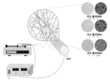
- Figure 9 is a schematic diagram showing the manufacturing process of nanofibers using electrospinning.
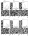
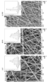
- Figure 10 shows SEM micrographs and the corresponding nanofiber diameter distributions (percentage and frequency of distribution): (a) P10G0, (b) P8G2, (c) P6G4, (d) P4G6, (e) P2G8, and (f) P0G10 nanofiber.
- Figure 11 shows the mechanical properties of PCL/Gel nanofibers: (A) typical stress-strain curve, (B) tensile strength, (C) strain at maximum load, and (D) strain at maximum extension. # p ⁇ 0.05 was considered to be a statistically significant difference compared to P10G0 nanofiber.
- Figure 21 is a graph showing (A) a representative image and (B) a dynamic water contact angle of the water contact angle of the manufactured nanofibers (n > 3). # p ⁇ 0.05 was considered to be a statistically significant difference compared to the P nanofiber group at the same time.
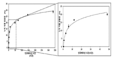
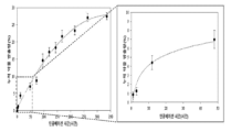
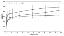
- Figures 32A and 32B show wound closure of nanofibers in a rectangular full-thickness wound model at 28 days.
- A H&E, Masson's trichrome, and Picro-Sirius Red staining results of mouse skin tissue 28 days after wounding. Scale bar 1000 ⁇ m at 1X and 200 ⁇ m at 5X.
- B Relative wound length and
- C wound thickness of the rectangular full-thickness wound model at day 28. Error bars represent standard deviation (n ⁇ 3). * p ⁇ 0.05 was considered a statistically significant difference compared to the untreated (blank) group.
- the present invention relates to a nanofiber scaffold containing polycaprolactone (poly( ⁇ -caprolactone; PCL); gelatin; and polydeoxyribonucleotide (PDRN) derived from starfish ( Patiria pectinifera ); and a method for manufacturing the same. will be.
- polycaprolactone poly( ⁇ -caprolactone; PCL); gelatin; and polydeoxyribonucleotide (PDRN) derived from starfish ( Patiria pectinifera ); and a method for manufacturing the same.
- the polycaprolactone, gelatin, and polydeoxyribonucleotide may be mixed at a weight ratio of 4 to 8:2 to 6:0.005 to 0.1, for example, 6:4:0.02. It can be done, but is not limited to this.
- PDRN Polydeoxyribonucleotide
- P. pectinifera starfish
- AccuPrep Genomic DNA extraction kit Booneer, Daejeon, Korea
- P. pectinifera was washed three times with tap water to remove epiphytes, salt, and sand attached to the surface, and then carefully washed again with fresh water.
- P. pectinifera frozen at -20°C was freeze-dried and then homogenized to powder.
- Crushed P. pectinifera (1 g) was extracted in 4 ml TL buffer containing Proteinase K (2 mg/ml) and RNase A (2 mg/ml) at 60°C for 2 hours.
- HDF Human dermal fibroblasts
- HaCaT human keratinocytes
- HDFs and HaCaT were incubated with FDA (10 ⁇ g/ml) in serum-free medium for 15 min at 37 °C. and PI (20 ⁇ g/ml). The stained cells were then washed with PBS to remove untreated FDA and PI and residues. FDA appears as green fluorescence in live cells and PI appears as red in dead cells, and was quantitatively analyzed using a fluorescence microscope (Leica DMI3000B).
- HDF was inoculated into 2 ml of culture medium in a 6-well plate at a density of 4 Cells were incubated for 24 hours to attach at a density of approximately 90%. After cells were attached to the plate, a wound line was created with a 2 mm wide plastic pipette tip, and unattached cells were washed with PBS. Then, cells were treated with PDRN at different concentrations and allowed to migrate. Pictures were taken at a magnification of 50 was carried out.
- Collagen production was assessed by Picro-Sirius red staining.
- HDFs were incubated with a staining solution made of Sirius red (0.1%, Sigma) dissolved in a saturated aqueous solution of picric acid (1.3% in water, Sigma) for 2 hours, washed three times with PBS, and then dehydrated.
- the stained crystals were dissolved in 0.1 M NaOH, and the absorbance was measured using a PowerWave XS2 microplate reader (BioTek Instruments, Inc., Winooski, VT, USA). Data were expressed as a percentage of the average collagen production rate ⁇ standard deviation of visualized cells in three replicate experiments.
- HDFs were grown in 100 cm 2 dishes at a density of approximately 2 ⁇ 10 6 cells.
- Cells were treated with PDRN at various concentrations and incubated for 24 hours to check the collagen expression level, and incubated for 30 minutes to analyze the degree of activation of Smad2/3 and mitogen-activated protein kinase (MAPK) pathways.
- MAPK mitogen-activated protein kinase
- mice were sacrificed, skin tissue was removed, and tissue samples were stored in a nitrogen gas tank until further analysis. Frozen tissue samples were homogenized with a Tissue Lyser (SpeedMill PLUS, Jena, Germany) in lysis buffer. Specimens were destroyed using steel beads at 30 cycles/second for 5 minutes.
- Tissue Lyser SpeedMill PLUS, Jena, Germany
- PDRN derived from P. pectinifera were evaluated using MTT assay and FDA/PI staining.
- MTT assay HDF and HaCaT cells were treated with increasing concentrations of PDRN for 1, 3, and 5 days, and for FDA/PI staining, they were treated with increasing concentrations of PDRN for 1 and 3 days.
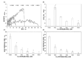
- PDRN 5-200 ⁇ g/ml significantly increased cell proliferation after 3 days of treatment, with treatment at 50 ⁇ g/ml PDRN showing the highest proliferation ( Figures 2 and 3).
- PDRN did not show toxicity to HaCaT cells, but also showed no significant proliferative effect ( Figures 4 and 5). Based on these results, it was concluded that PDRN at the above concentration (5-50 ⁇ g/ml) showed a proliferative effect on HDF cells without showing toxicity to HaCaT cells.
- PDRN extracted from P. pectinifera The effect on activation of MAPK and Smad2/3 pathways was assessed by Western blot analysis. PDRN significantly increased phosphorylation of ERK and Smad2/3 compared to untreated cells ( Figure 8B). These results suggest that PDRN increases the expression of proteins related to wound healing by activating the phosphorylation of ERK and Smad2/3.
- nanofibers The wetting behavior of nanofibers was evaluated using a contact angle analyzer (SEO Phoenix MT, Suwon, Gyeonggi-do, Korea). The volume of the droplet was 2 ⁇ l, and the contact angle was measured at five separate random locations and averaged. DMEM medium without serum was used as the test solution. Nanofibers of the same size were placed on the sample stage at ambient temperature ( ⁇ 296 K) and the contact angle was measured. Pictures were taken with a digital camera over time and analyzed using image processing software (Image Pro 300).
- the tensile strength of nanofibers was measured using a universal tensile machine (LR5K Plus, Lloyd Instruments). Each sample was cut into dumbbell-shaped strips (15 mm I ordered it.
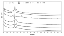
- the functional groups of pure PCL, pure gelatin, pure PDRN, P nanofiber, PG nanofiber, and PGP nanofiber were analyzed using Fourier transform infrared (FT-IR) spectroscopy (FT-4100, JASCO).
- FT-IR Fourier transform infrared
- FT-4100 Fourier transform infrared spectroscopy
- the IR spectrum showed an average of 30 scans at a frequency of 650-4000 cm -1 at a resolution of 4 cm -1 .
- Thermogravimetric analysis was performed using a Pyris 1 TGA analyzer (PerkinElmer TGA-7, Waltham, MA, USA) with a scan range of 30 °C to 700 °C and a constant heating rate of 20 °C under continuous nitrogen. Calorimetry was performed using differential scanning calorimetry (DSC) under nitrogen flowing at a rate of 10 ml/min. The specimen was pressed into a sealed aluminum pan. Heating cycles were performed until the glass transition temperature (T g ) and melting temperature (T m ) were reached. During the cycle, the sample was heated from 30°C to 180°C at a rate of 10°C. The sample was then cooled using nitrogen at an exponentially decreasing rate.
- DSC differential scanning calorimetry
- X-ray diffraction (XRD) analysis of the nanofibers was performed using X-ray diffraction (X'Pert3-Powder, PANalytical, Netherlands) with Cu-K ⁇ radiation. Diffraction intensity was recorded at a scanning speed of 2.4° min -1 in the range of 5 to 90°.
- the weak band at 1440 cm -1 represents the aliphatic CH bending vibration.
- the bands at 1524 and 1237 cm -1 represent stretching vibrations of NH bending and CN stretching, respectively, and are attributed to the characteristic bands of amide II and amide III in gelatin, respectively.
- the FT-IR spectrum of PDRN showed several characteristic peaks of DNA.
- the third peak shows vibration as the -PO 2 vibration of the phosphodieter skeleton in nucleic acid and was recorded at 1287 cm -1 .
- the peak corresponding to the deoxyribose CO stretching vibration appeared at 1033 cm -1 .
- the strong peak at 1020 cm -1 represents furanose vibration, and the next strongest peak at 981 cm -1 is due to CC stretching of the DNA deoxyribose-phosphate backbone.
- the weak vibration appearing near 788 cm -1 is related to deoxy c3'-endo-OPO and represents the A-type DNA conformation. All characteristic peaks of pure PCL, pure gelatin and PDRN are shown in Table 5.
- elution solutions of nanofibers prepared for 1 and 3 days were prepared and analyzed by MTT analysis and FDA and PI fluorescence in HDF and HaCaT. Indirect cytotoxicity was studied by performing live/dead cell staining. The MTT results showed that the elution solution of the prepared nanofibers was not cytotoxic ( Figures 24, 25, and 26). The live cell/dead cell staining results also demonstrated no cytotoxicity, similar to the MTT analysis results.
- H&E hematoxylin and eosin
- the blank group showed a lower density of collagen fibers in the wound area compared to the PGP group. Moreover, the deposition of collagen fibers was smaller and thicker in the nanofiber-implanted group.
Landscapes
- Health & Medical Sciences (AREA)
- Engineering & Computer Science (AREA)
- Chemical & Material Sciences (AREA)
- Animal Behavior & Ethology (AREA)
- Hematology (AREA)
- Materials Engineering (AREA)
- Epidemiology (AREA)
- Life Sciences & Earth Sciences (AREA)
- General Health & Medical Sciences (AREA)
- Public Health (AREA)
- Veterinary Medicine (AREA)
- Textile Engineering (AREA)
- Dispersion Chemistry (AREA)
- Materials For Medical Uses (AREA)
Abstract
The present invention relates to a nanofibrous scaffold and a preparation method therefor, the nanofibrous scaffold comprising: poly(ε-caprolactone) (PCL); gelatin; and polydeoxyribonucleotide (PDRN) derived from Patiria pectinifera. The nanofibrous scaffold of the present invention has a uniform nanofiber structure, excellent fluid absorption and retention, an adequate release speed, high mechanical stability, thermal stability, and thus is suitable for a dressing for healing wounds.
Description
본 발명은 폴리카프로락톤(poly(ε-caprolactone; PCL); 젤라틴; 및 별불가사리 (Patiria pectinifera) 유래 폴리데옥시리보뉴클레오티드 (Polydeoxyribonucleotide; PDRN);을 포함하는 나노파이버 스캐폴드 및 이의 제조방법에 관한 것이다.The present invention relates to a nanofiber scaffold containing polycaprolactone (poly(ε-caprolactone; PCL); gelatin; and polydeoxyribonucleotide (PDRN) derived from starfish ( Patiria pectinifera ); and a method for manufacturing the same. will be.
드레싱(dressing)은 화상이나 창상, 욕창 및 외상에 의한 피부결손 부위인 상처면을 피복하여 치유속도를 향상시키기 위해 사용되는 방법으로, 1962년 Winter에 의한 돼지 창상의 상피형성 속도가 습윤 환경이 건조 환경에 비해 2배 이상 빠르다는 발표 이후 습윤 드레싱제(wet dressing)가 속속 개발, 출시되고 상처의 처치 방법도 다양하게 전개되고 있다.Dressing is a method used to improve the healing rate by covering the wound surface, which is a skin defect caused by burns, wounds, bedsores, and trauma. In 1962, Winter reported that the rate of epithelium formation in porcine wounds decreased when the wet environment was dry. Since the announcement that it is more than twice as fast as the environment, wet dressings have been developed and released one after another, and various wound treatment methods are being developed.
습윤 드레싱은 상처면을 밀폐시켜 습윤 상태를 유지시켜주기 위한 것으로, 친수성 고분자 및 소수성 고분자를 다양하게 개발 조합하여 필름상(film), 시트상(sheet), 부직포상, 스폰지(sponge), 폼상(foam), 로프상(lope), 펠렛상(pellet), 분말상(powder) 등의 다양한 형태로 빠르게 발전하고 있다.Wet dressings are designed to seal the wound surface and maintain a moist state. By developing and combining various hydrophilic and hydrophobic polymers, they are available in the form of film, sheet, non-woven fabric, sponge, and foam ( It is rapidly developing into various forms such as foam, rope, pellet, and powder.
이러한 상처치유에 사용되는 소재로는 상처면으로부터 배출되는 다량의 삼출물을 흡수, 함유할 수 있는 제품으로 친수성 고분자가 공유결합 내지는 비공유 결합으로 가교되어져 만들어진 3차원 망상구조물의 형태를 가져야 하며, 구성물질의 친수성으로 인해 수용액 내 및 수성 환경하에서 많은 양의 수분을 흡수하여 팽윤되지만 가교 구조에 의해 용해되지 않는 성질을 가져야 한다.The material used for such wound healing is a product that can absorb and contain a large amount of exudate discharged from the wound surface. It must have the form of a three-dimensional network structure made by cross-linking hydrophilic polymers with covalent or non-covalent bonds. Due to its hydrophilic nature, it absorbs a large amount of moisture and swells in aqueous solutions and in aqueous environments, but must have the property of not dissolving due to the cross-linked structure.
따라서 구성성분이나 제조방법에 따라 다양한 방법이 제시되고 있으나 비공유 결합에 의한 물리적 가교의 경우 물성의 저하가 발생하기 쉽고, 화학적 가교의 경우 가교제에 의한 피부 독성이 있는 것으로 알려져 있으며, 초음파나 방사선 가교에 의한 방법 등이 있으나 장비나 고비용의 한계가 있는 것이 현실적인 상황이다.Therefore, various methods have been proposed depending on the composition or manufacturing method, but in the case of physical crosslinking by non-covalent bonds, deterioration of physical properties is likely to occur, and in the case of chemical crosslinking, it is known that there is skin toxicity due to the crosslinking agent, and ultrasonic or radiation crosslinking is known to cause skin toxicity. There are methods, but the realistic situation is that there are limitations in equipment and high costs.
전기방사 기술을 활용해 제작된 나노파이버는 상호 연결된 다공성 구조 측면에서 세포외기질(Extracellular matrix; ECM)과 유사한 구조적 특성을 가지고 있기 때문에 피부 조직 공학 응용 분야에서 상당한 주목을 받고 있다. 전기방사는 기계적 특성, 생분해성 및 생체적합성과 같은 고유한 특성으로 인해 폴리카프로락톤(polycaprolactone, PCL)을 비롯한 다양한 합성 고분자와 함께 사용할 수 있습니다. PCL은 널리 사용되는 반결정성 지방족 폴리에스터로 미국 식품의약국(FDA)에서 생물의학 용도로 승인한 제품이다. 그러나 소수성과 세포에 대한 낮은 반응으로 인해 콜라겐, 젤라틴, 알지네이트, 키토산과 같은 다양한 천연 고분자와 혼합사용함으로써 생물학적, 기계적 특성을 조절하고 조직재생에 적합한 구조적 기능을 부여한다. Nanofibers produced using electrospinning technology are receiving considerable attention in skin tissue engineering applications because they have structural characteristics similar to extracellular matrix (ECM) in terms of their interconnected porous structure. Electrospinning can be used with a variety of synthetic polymers, including polycaprolactone (PCL), due to their unique properties such as mechanical properties, biodegradability, and biocompatibility. PCL is a widely used semi-crystalline aliphatic polyester that has been approved for biomedical use by the U.S. Food and Drug Administration (FDA). However, due to its hydrophobicity and low response to cells, it is mixed with various natural polymers such as collagen, gelatin, alginate, and chitosan to control biological and mechanical properties and provide structural functions suitable for tissue regeneration.
한편, 폴리데옥시리보뉴클레오티드(Polydeoxyribonucleotides, PDRN)는 분자량이 50~1,500 kDa 범위인 활성 혼합물로 무지개 송어(Oncorhynchus mykiss) 또는 연어(Oncorhynchus keta)의 정자 세포에서 주로 추출 및 정제된다. PDRN은 현재 식품의약품안전청의 허가를 받은 DNA 유래 의약품의 일종으로 조직수복 및 상처치료에 많이 시행되고 있다. PDRN은 혈관 신생 촉진, 항 세포 사멸, 항염증 및 조직 복구 활성을 포함한 여러 효능을 나타낸다. 그러나, 이러한 조직 복구와 관련된 생물학적 효과에도 불구하고 PDRN의 추출 원료로는 O. mykiss 및 O. keta만 활용되고 있는 실정이다.Meanwhile, polydeoxyribonucleotides (PDRN), an active mixture with a molecular weight ranging from 50 to 1,500 kDa, are mainly extracted and purified from sperm cells of rainbow trout ( Oncorhynchus mykiss ) or salmon ( Oncorhynchus keta ). PDRN is a type of DNA-derived medicine currently approved by the Food and Drug Administration and is widely used for tissue repair and wound treatment. PDRN exhibits multiple efficacies including pro-angiogenic, anti-apoptotic, anti-inflammatory and tissue repair activities. However, despite these biological effects related to tissue repair, only O. mykiss and O. keta are used as extraction raw materials for PDRN.
이에, 본 발명의 발명자들은 별불가사리(Patiria pectinifera)로부터 DNA 중합체로서 뛰어난 피부재생 효능을 지닌 폴리데옥시리보뉴클레오티드(Polydeoxyribonucleotide; PDRN)를 분리하고, 이를 이용하여 제조한 나노파이버의 구조적 특성 및 피부 재생 능력을 확인하여 본 발명을 완성하였다. Accordingly, the inventors of the present invention isolated polydeoxyribonucleotide (PDRN), which has excellent skin regeneration efficacy as a DNA polymer, from starfish ( Patiria pectinifera ), and studied the structural properties and skin regeneration of nanofibers manufactured using it. The ability was confirmed and the present invention was completed.
이에 따라, 본 발명은 별불가사리(Patiria pectinifera) 유래 폴리데옥시리보뉴클레오티드(Polydeoxyribonucleotide; PDRN)를 포함하는 나노파이버 스캐폴드 및 이의 제조방법을 제공하는 것을 목적으로 한다.Accordingly, the purpose of the present invention is to provide a nanofiber scaffold containing polydeoxyribonucleotide (PDRN) derived from starfish ( Patiria pectinifera ) and a method for manufacturing the same.
본 발명은 폴리카프로락톤(poly(ε-caprolactone; PCL); 젤라틴; 및 별불가사리(Patiria pectinifera) 유래 폴리데옥시리보뉴클레오티드(Polydeoxyribonucleotide; PDRN);를 포함하는 나노파이버 스캐폴드를 제공한다. The present invention provides a nanofiber scaffold containing polycaprolactone (poly(ε-caprolactone; PCL); gelatin; and polydeoxyribonucleotide (PDRN) derived from starfish ( Patiria pectinifera ).
또한, 본 발명은 별불가사리(Patiria pectinifera)로부터 폴리데옥시리보뉴클레오티드(Polydeoxyribonucleotide; PDRN)를 분리하는 단계(단계 1); 상기 폴리데옥시리보뉴클레오티드를 폴리카프로락톤 및 젤라틴과 혼합하는 단계(단계 2); 및 상기 혼합물을 전기방사하여 나노파이버 매트를 제조하는 단계(단계 3);를 포함하는, 나노파이버 스캐폴드의 제조방법을 제공한다. In addition, the present invention includes the steps of isolating polydeoxyribonucleotide (PDRN) from starfish ( Patiria pectinifera ) (step 1); Mixing the polydeoxyribonucleotide with polycaprolactone and gelatin (step 2); and electrospinning the mixture to produce a nanofiber mat (step 3).
또한, 본 발명은 본 발명에 따른 나노파이버 스캐폴드를 포함하는 상처 드레싱을 제공한다.Additionally, the present invention provides a wound dressing comprising the nanofiber scaffold according to the present invention.
본 발명은 미활용 자원이자 해적 생물인 별불가사리(Patiria pectinifera)로부터 뛰어난 피부재생 효능을 지닌 폴리데옥시리보뉴클레오티드(Polydeoxyribonucleotide; PDRN)를 확보하고 이를 피부 재생용 나노파이버 스캐폴드에 적용시킴으로써, 미활용 해양 생명 자원의 조직공학적 활용가능성과 별불가사리 PDRN을 활용한 피부조직재생용 의료기기로서 새로운 활용 가능성을 제공하였다.The present invention secures polydeoxyribonucleotide (PDRN) with excellent skin regeneration efficacy from the starfish ( Patiria pectinifera ), which is an unutilized resource and a pirate organism, and applies it to a nanofiber scaffold for skin regeneration, thereby protecting unused marine life. It provided new possibilities for utilizing resources in tissue engineering and as a medical device for skin tissue regeneration using Byeolbulgasari PDRN.
도 1은 아가로스 겔 전기영동을 사용하여 P. pectinifera 유래 PDRN의 분자량 분포를 확인한 결과를 나타낸 것이다. Lane A: DNA marker; Lane B: PDRN from P. pectinifera
(20 μg).Figure 1 shows the results of confirming the molecular weight distribution of PDRN derived from P. pectinifera using agarose gel electrophoresis. Lane A: DNA marker; Lane B: PDRN from P. pectinifera (20 μg).
도 2는 HDF에서 P. pectinifera 유래 PDRN에 의한 증식 효과를 나타낸 것이다. *
p < 0.10 및 **
p < 0.05은 비처리 그룹에 비해 통계학적으로 유의차가 있는 것으로 간주하였다.Figure 2 shows the proliferation effect of PDRN derived from P. pectinifera in HDF. * p < 0.10 and ** p < 0.05 were considered statistically significant differences compared to the untreated group.
도 3은 HDF에서 P. pectinifera 유래 PDRN에 의한 증식 효과를 나타낸 FDA/PI 염색 결과를 나타낸 것이다. Figure 3 shows the results of FDA/PI staining showing the proliferation effect by PDRN derived from P. pectinifera in HDF.
도 4는 P. pectinifera 유래 PDRN의 HaCaT에 대한 세포독성을 확인한 MTT 검정 결과를 나타낸 그래프이다. *
p < 0.10 및 **
p < 0.05은 비처리 그룹에 비해 통계학적으로 유의차가 있는 것으로 간주하였다.Figure 4 is a graph showing the results of the MTT assay confirming the cytotoxicity of P. pectinifera- derived PDRN against HaCaT. * p < 0.10 and ** p < 0.05 were considered statistically significant differences compared to the untreated group.
도 5는 P. pectinifera 유래 PDRN의 HaCaT에 대한 세포독성을 확인한 FDA/PI 염색 결과를 나타낸 것이다. Figure 5 shows the results of FDA/PI staining confirming the cytotoxicity of P. pectinifera- derived PDRN against HaCaT.
도 6은 콜라겐 생성 효과 of P. pectinifera 유래 PDRN의 HDF에서의 콜라겐 0 효과를 확인한 Picro-Sirius red 염색 결과를 나타낸 그래프이다. *
p < 0.05 및 **
p < 0.10은 비처리 그룹에 비해 통계학적으로 유의차가 있는 것으로 간주하였다.Figure 6 is a graph showing the results of Picro-Sirius red staining confirming the effect of collagen production on the HDF of PDRN derived from P. pectinifera . * p < 0.05 and ** p < 0.10 were considered statistically significant differences compared to the untreated group.
도 7a 및 7b는 24시간 동안 P. pectinifera 유래 PDRN의 세포 이동에 대한 효과를 나타낸 것이다: (A) 세포 이동 분석 결과의 대표 이미지 및 (B) 상처 치유 속도 그래프. a
p < 0.05는 각 그룹에서 초기 스크래치 상처 영역에 비해 통계학적으로 유의차가 있는 것으로 간주하였으며, b
p<0.05은 비처리 그룹에 비해 통계학적으로 유의차가 있는 것으로 간주하였다.Figures 7a and 7b show the effect of P. pectinifera -derived PDRN on cell migration for 24 hours: (A) representative images of cell migration assay results and (B) graph of wound healing rate. a p < 0.05 was considered to be a statistically significant difference compared to the initial scratch wound area in each group, and b p <0.05 was considered to be a statistically significant difference compared to the untreated group.
도 8은 P. pectinifera로부터 추출된 PDRN의 HDF 내 피부 재생과 관련된(A) 여러 단백질 (I형 콜라겐, III형 콜라겐, α-SMA)의 발현 및 (B) 작용기전에 대한효과를 나타낸 것이다. Figure 8 shows the effect on the expression and (B) mechanism of action of several proteins (type I collagen, type III collagen, α-SMA) related to skin regeneration in the HDF of PDRN extracted from P. pectinifera (A).
도 9는 전기방사를 사용한 나노파이버의 제조 공정을 나타낸 모식도이다.Figure 9 is a schematic diagram showing the manufacturing process of nanofibers using electrospinning.
도 10은 SEM 현미경 사진 및 상응하는 나노파이버 지름 분포 (분포의 백분율 및 빈도)를 나타낸 것이다: (a) P10G0, (b) P8G2, (c) P6G4, (d) P4G6, (e) P2G8, 및 (f) P0G10 나노파이버.Figure 10 shows SEM micrographs and the corresponding nanofiber diameter distributions (percentage and frequency of distribution): (a) P10G0, (b) P8G2, (c) P6G4, (d) P4G6, (e) P2G8, and (f) P0G10 nanofiber.
도 11은 PCL/Gel 나노파이버의 기계적 특성을 나타낸 것이다: (A) 일반 응력-변성 곡선, (B) 인장 강도, (C) 최대 하중에서의 변형, 및 (D) 최대 연장에서의 변형. #
p < 0.05은 P10G0 나노파이버에 비해 통계학적으로 유의차가 있는 것으로 간주하였다. Figure 11 shows the mechanical properties of PCL/Gel nanofibers: (A) typical stress-strain curve, (B) tensile strength, (C) strain at maximum load, and (D) strain at maximum extension. # p < 0.05 was considered to be a statistically significant difference compared to P10G0 nanofiber.
도 12는 제조된 나노파이버 (n > 3)의 물 접촉각의 (A) 대표 이미지 및 (B) 동역학적 물 접촉각을 나타낸 그래프이다. *p < 0.05는 동시간대에 P0G10 나노파이버 그룹에 비해 통계학적으로 유의차가 있는 것으로 간주하였다.Figure 12 is a graph showing (A) a representative image and (B) a dynamic water contact angle of the water contact angle of the manufactured nanofibers (n > 3). *p < 0.05 was considered to be a statistically significant difference compared to the P0G10 nanofiber group at the same time.
도 14는 SEM 현미경 사진 및 및 상응하는 나노파이버 지름 분포 (분포의 백분율 및 빈도)를 나타낸 것이다: (a) P, (b) PG, 및 (c) PGP 나노파이버.Figure 14 shows SEM micrographs and the corresponding nanofiber diameter distribution (percentage and frequency of distribution): (a) P, (b) PG, and (c) PGP nanofibers.
도 15는 나노파이버의 기계적 특성을 나타낸 것이다: (A) 일반 응력-변성 곡선, (B) 인장 강도, (C) 최대 하중에서의 변형 및 (D) 최대 연장에서의 변형. #
p < 0.05은 P 나노파이버에 비해 통계학적으로 유의차가 있는 것으로 간주하였다. Figure 15 shows the mechanical properties of nanofibers: (A) typical stress-strain curve, (B) tensile strength, (C) strain at maximum load, and (D) strain at maximum extension. # p < 0.05 was considered to be a statistically significant difference compared to P nanofiber.
도 16은 37℃의 1X PBS (pH 7.4) 중에서 PGP 나노파이버의 PDRN 방출 프로파일을 나타낸 것이다. 데이터는 평균 ± 표준편차로 나타냈다 (n = 4).Figure 16 shows the PDRN release profile of PGP nanofibers in 1X PBS (pH 7.4) at 37°C. Data are presented as mean ± standard deviation (n = 4).
도 17은 순수 PCL, 젤라틴, PDRN 및 제조된 나노파이버의 FTIR 스펙트럼 및 피크 강도에서의 시프트 및 혼합 비율에 따른 위치를 나타낸 것이다. (a) 순수 PCL (검은색), (b) 순수 젤라틴 (노란색), (c) PDRN (갈색), (d) P(회색), (e) PG(파란색), 및 (f) PGP (녹색) 나노파이버. Figure 17 shows the FTIR spectra and peak intensities of pure PCL, gelatin, PDRN, and prepared nanofibers and their positions according to the mixing ratio. (a) pure PCL (black), (b) pure gelatin (yellow), (c) PDRN (brown), (d) P (gray), (e) PG (blue), and (f) PGP (green). ) Nanofiber.
도 18은 순수 물질 및 제조된 나노파이버의 열중량 분석 결과를 나타낸 그래프이다.Figure 18 is a graph showing the results of thermogravimetric analysis of pure materials and manufactured nanofibers.
도 19는 순수 물질 및 제조된 나노파이버의 시차주사열량 (differential scanning calorimetry; DSC) 곡선을 나타낸 것이다.Figure 19 shows differential scanning calorimetry (DSC) curves of pure materials and manufactured nanofibers.
도 20은 순수 물질 및 제조된 나노파이버의 X-선 회절 프로파일을 나타낸 것이다.Figure 20 shows the X-ray diffraction profiles of pure material and prepared nanofibers.
도 21은 제조된 나노파이버 (n > 3)의 물 접촉각의 (A) 대표 이미지 및 (B) 동역학적 물 접촉각을 나타낸 그래프이다. #
p < 0.05은 동시간대에 P 나노파이버 그룹에 비해 통계학적으로 유의차가 있는 것으로 간주하였다. Figure 21 is a graph showing (A) a representative image and (B) a dynamic water contact angle of the water contact angle of the manufactured nanofibers (n > 3). # p < 0.05 was considered to be a statistically significant difference compared to the P nanofiber group at the same time.
도 22는 PBS (pH 7.4) 중에서 96시간 동안 37℃에서의 제조된 나노파이버의 팽윤성을 나타낸 것이다. 데이터는 평균 ± 표준편차로 나타냈다 (n = 4).Figure 22 shows the swelling properties of nanofibers prepared at 37°C for 96 hours in PBS (pH 7.4). Data are presented as mean ± standard deviation (n = 4).
도 23은 제조된 나노파이버에서 배양된 (A) HDF 및 (B) HaCaT의 직접 세포독성 및 증식을 확인한 MTT 검정 결과를 나타낸 것이다. *
p < 0.05은 비처리 그룹에 비해 통계학적으로 유의차가 있는 것으로 간주하였다.Figure 23 shows the results of the MTT assay confirming the direct cytotoxicity and proliferation of (A) HDF and (B) HaCaT cultured on the manufactured nanofibers. * p < 0.05 was considered a statistically significant difference compared to the untreated group.
도 24는 FDA/PI 염색을 사용한 간접 세포독성 분석을 통해 제조된 나노파이버의 HDF 세포 생존도에 대한 효과를 확인한 것이다. Figure 24 confirms the effect of the manufactured nanofibers on HDF cell viability through indirect cytotoxicity analysis using FDA/PI staining.
도 25는 FDA/PI 염색을 사용한 간접 세포독성 분석을 통해 제조된 나노파이버의 HaCaT 세포 생존도에 대한 효과를 확인한 것이다.Figure 25 confirms the effect of the manufactured nanofibers on HaCaT cell viability through indirect cytotoxicity analysis using FDA/PI staining.
도 26은 MTT 검정을 사용한 간접 세포독성 분석을 통해 제조된 나노파이버의 (A) HDF 및 (B) HaCaT의 세포 생존도에 대한 효과를 확인한 것이다. *
p < 0.05은 비처리 그룹에 비해 통계학적으로 유의차가 있는 것으로 간주하였다.Figure 26 confirms the effect of the nanofibers prepared through indirect cytotoxicity analysis using the MTT assay on the cell viability of (A) HDF and (B) HaCaT. * p < 0.05 was considered a statistically significant difference compared to the untreated group.
도 27은 ICR 마우스의 원형 전체-두께 절개 상처 유도 및 제조된 나노파이버의 드레싱 적용 실험 스케쥴을 나타낸 모식도이다.Figure 27 is a schematic diagram showing the experimental schedule for inducing a circular full-thickness incision wound in an ICR mouse and applying a dressing of the manufactured nanofiber.
도 28은 ICR 마우스의 직사각형 전체-두께 절개 상처 유도 및 제조된 나노파이버의 드레싱 적용 실험 스케쥴을 나타낸 모식도이다.Figure 28 is a schematic diagram showing the experimental schedule for inducing a rectangular full-thickness incision wound in an ICR mouse and applying a dressing of the manufactured nanofiber.
도 29는 수술 후 21일째에 비처리군과 비교한 원형 전체-두께 상처 모델에서 제조된 나노파이버의 효과를 나타낸 것이다: (A) 상처봉합 동역학의 대표 이미지 및 (B) 수술 후 시간에 따른 평균 상처 봉합. 오류 바는 표준편차 (n≥3)를 의미한다.Figure 29 shows the effect of fabricated nanofibers on a circular full-thickness wound model compared to the untreated group at 21 days after surgery: (A) representative images of wound closure kinetics and (B) average over time after surgery. Wound closure. Error bars represent standard deviation (n≥3).
도 30은 수술 후 28일째에 비처리군과 비교한 직사각형 전체-두께 상처 모델에서 제조된 나노파이버의 효과를 나타낸 것이다: (A) 상처봉합 동역학의 대표 이미지 및 (B) 수술 후 시간에 따른 평균 상처 봉합. 오류 바는 표준편차 (n≥3)를 의미한다.Figure 30 shows the effect of fabricated nanofibers on a rectangular full-thickness wound model compared to the untreated group at 28 days after surgery: (A) representative images of wound closure kinetics and (B) average over time after surgery. Wound closure. Error bars represent standard deviation (n≥3).
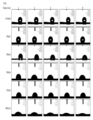
도 31a 및 31b는 21 일에 원형 전체-두께 상처 모델에서 나노파이버의 상처 봉합을 나타낸 것이다. (A) 상처낸 후 21일째의 마우스 피부 조직의 H&E, Masson's trichrome, 및 Picro-Sirius Red 염색 결과. Scale bar 600 μm at 2X 및 100 μm at 10X. (B) 21일째에서 원형 전체-두께 상처 모델의 상대적인 상처 길이 및 (C) 상처 두께. 오류 바는 표준편차 (n≥3)을 의미한다. *
p < 0.05는 비처리 (blank) 그룹에 비해 통계학적으로 유의차가 있는 것으로 간주되었다.Figures 31A and 31B show wound closure of nanofibers in a circular full-thickness wound model at 21 days. (A) H&E, Masson's trichrome, and Picro-Sirius Red staining results of mouse skin tissue 21 days after wounding. Scale bar 600 μm at 2X and 100 μm at 10X. (B) Relative wound length and (C) wound thickness of the circular full-thickness wound model at day 21. Error bars represent standard deviation (n≥3). * p < 0.05 was considered a statistically significant difference compared to the untreated (blank) group.
도 32a 및 32b는 28 일에 직사각형 전체-두께 상처 모델에서 나노파이버의 상처 봉합을 나타낸 것이다. (A) 상처낸 후 28일째의 마우스 피부 조직의 H&E, Masson's trichrome, 및 Picro-Sirius Red 염색 결과. Scale bar 1000 μm at 1X 및 200 μm at 5X. (B) 28일째에서 직사각형 전체-두께 상처 모델의 상대적인 상처 길이 및 (C) 상처 두께. 오류 바는 표준편차 (n≥3)을 의미한다. *
p < 0.05는 비처리 (blank) 그룹에 비해 통계학적으로 유의차가 있는 것으로 간주되었다.Figures 32A and 32B show wound closure of nanofibers in a rectangular full-thickness wound model at 28 days. (A) H&E, Masson's trichrome, and Picro-Sirius Red staining results of mouse skin tissue 28 days after wounding. Scale bar 1000 μm at 1X and 200 μm at 5X. (B) Relative wound length and (C) wound thickness of the rectangular full-thickness wound model at day 28. Error bars represent standard deviation (n≥3). * p < 0.05 was considered a statistically significant difference compared to the untreated (blank) group.
도 33은 (A) 원형 및 (B) 직사각형 전체-두께 상처 모델의 피부 재생과 관련된 여러 단백질 (I형 콜라겐, III형 콜라겐, α-SMA)의 발현에 대한 PGP 나노파이버의 효과를 나타낸 것이다. β-actin은 웨스턴 블롯 분석 결과의 정량화를 위한 세포유지유전자(Housekeeping gene)로서 사용되었다.Figure 33 shows the effect of PGP nanofibers on the expression of several proteins (type I collagen, type III collagen, α-SMA) related to skin regeneration in (A) circular and (B) rectangular full-thickness wound models. β-actin was used as a housekeeping gene for quantification of Western blot analysis results.
이하, 본 발명을 더욱 상세하게 설명한다. 그러나 본 발명은 여러 가지 상이한 형태로 구현될 수 있으며 여기에서 설명하는 실시예에 의해 본 발명이 한정되지 않으며 본 발명은 후술할 청구 범위에 의해 정의될 뿐이다.Hereinafter, the present invention will be described in more detail. However, the present invention may be implemented in various different forms, and the present invention is not limited to the embodiments described herein, and the present invention is only defined by the claims to be described later.
덧붙여, 본 발명에서 사용한 용어는 단지 특정한 실시 예를 설명하기 위해 사용된 것으로, 본 발명을 한정하려는 의도가 아니다. 본 발명의 명세서 전체에서 어떤 구성요소를 '포함'한다는 것은 특별히 반대되는 기재가 없는 한 다른 구성요소를 제외하는 것이 아니라 다른 구성요소를 더 포함할 수 있다는 것을 의미한다.In addition, the terms used in the present invention are only used to describe specific embodiments and are not intended to limit the present invention. In the entire specification of the present invention, 'including' a certain element means that other elements may be further included rather than excluding other elements, unless specifically stated to the contrary.
본 발명은 폴리카프로락톤(poly(ε-caprolactone; PCL); 젤라틴; 및 별불가사리(Patiria pectinifera) 유래 폴리데옥시리보뉴클레오티드(Polydeoxyribonucleotide; PDRN);를 포함하는 나노파이버 스캐폴드 및 이의 제조방법에 관한 것이다. The present invention relates to a nanofiber scaffold containing polycaprolactone (poly(ε-caprolactone; PCL); gelatin; and polydeoxyribonucleotide (PDRN) derived from starfish ( Patiria pectinifera ); and a method for manufacturing the same. will be.
상기 나노파이버 스캐폴드는 전기방사에 의해 제조될 수 있으나, 이에 제한되지 않는다.The nanofiber scaffold may be manufactured by electrospinning, but is not limited thereto.
상기 나노파이버 스캐폴드는 상기 폴리카프로락톤 및 젤라틴을 8 내지 2 : 2 내지 8의 중량비로 포함할 수 있으며, 예를 들어, 상기 폴리카프로락톤 및 젤라틴을 8 : 2, 6 : 4, 4 : 6 또는 2 : 8의 중량비로 포함할 수 있으나, 이에 제한되지 않는다.The nanofiber scaffold may include the polycaprolactone and gelatin in a weight ratio of 8 to 2:2 to 8, for example, the polycaprolactone and gelatin in a weight ratio of 8:2, 6:4, 4:6. Alternatively, it may be included in a weight ratio of 2:8, but is not limited thereto.
상기 나노파이버 스캐폴드는 상기 폴리카프로락톤, 젤라틴 및 폴리데옥시리보뉴클레오티드를 4 내지 8 : 2 내지 6 : 0.005 내지 0.1의 중량비로 포함할 수 있으며, 예를 들어, 6 : 4 : 0.02의 중량비로 포함할 수 있으나, 이에 제한되지 않는다.The nanofiber scaffold may include the polycaprolactone, gelatin, and polydeoxyribonucleotide in a weight ratio of 4 to 8:2 to 6:0.005 to 0.1, for example, in a weight ratio of 6:4:0.02. It may include, but is not limited to this.
상기 젤라틴은 본 분야에서 상처 드레싱에 사용되는 젤라틴이라면 제한없이 사용할 수 있으나, 이에 제한되지 않는다.The gelatin can be used without limitation as long as it is gelatin used for wound dressing in the field, but is not limited thereto.
상기 나노파이버 스캐폴드는 별불가사리(Patiria pectinifera)로부터 폴리데옥시리보뉴클레오티드(Polydeoxyribonucleotide; PDRN)를 분리하는 단계(단계 1); 상기 폴리데옥시리보뉴클레오티드를 폴리카프로락톤 및 젤라틴과 혼합하는 단계(단계 2); 및 상기 혼합물을 전기방사하여 나노파이버 매트를 제조하는 단계(단계 3)를 포함하는 방법에 의해 제조될 수 있으나, 이에 제한되지 않는다.The nanofiber scaffold includes the steps of isolating polydeoxyribonucleotide (PDRN) from starfish ( Patiria pectinifera ) (step 1); Mixing the polydeoxyribonucleotide with polycaprolactone and gelatin (step 2); and electrospinning the mixture to produce a nanofiber mat (step 3), but is not limited thereto.
이하, 본 발명의 나노파이버 스캐폴드의 제조방법을 바탕으로, 본 발명의 나노파이버 스캐폴드를 설명한다. Hereinafter, the nanofiber scaffold of the present invention will be described based on the manufacturing method of the nanofiber scaffold of the present invention.
먼저, 본 발명의 나노파이버 스캐폴드의 제조방법은 별불가사리(Patiria pectinifera)로부터 폴리데옥시리보뉴클레오티드(Polydeoxyribonucleotide; PDRN)를 분리하는 단계(단계 1)를 포함한다.First, the method for manufacturing the nanofiber scaffold of the present invention includes the step (step 1) of isolating polydeoxyribonucleotide (PDRN) from starfish ( Patiria pectinifera ).
다음으로, 본 발명은 폴리데옥시리보뉴클레오티드를 폴리카프로락톤 및 젤라틴과 혼합하는 단계(단계 2)를 포함한다. Next, the present invention includes mixing polydeoxyribonucleotide with polycaprolactone and gelatin (step 2).
상기 단계 2에서 상기 폴리카프로락톤 및 젤라틴을 8 내지 2 : 2 내지 8의 중량비로 혼합할 수 있으며, 예를 들어, 상기 폴리카프로락톤 및 젤라틴을 8 : 2, 6 : 4, 4 : 6 또는 2 : 8의 중량비로 혼합할 수 있으나, 이에 제한되지 않는다.In step 2, the polycaprolactone and gelatin may be mixed at a weight ratio of 8 to 2:2 to 8, for example, the polycaprolactone and gelatin may be mixed at a weight ratio of 8:2, 6:4, 4:6 or 2. : Can be mixed at a weight ratio of 8, but is not limited thereto.
또한 상기 단계 2에서 상기 폴리카프로락톤, 젤라틴 및 폴리데옥시리보뉴클레오티드를 4 내지 8 : 2 내지 6 : 0.005 내지 0.1의 중량비로 혼합할 수 있으며, 예를 들어, 6 : 4 : 0.02의 중량비로 혼합할 수 있으나, 이에 제한되지 않는다.Additionally, in step 2, the polycaprolactone, gelatin, and polydeoxyribonucleotide may be mixed at a weight ratio of 4 to 8:2 to 6:0.005 to 0.1, for example, 6:4:0.02. It can be done, but is not limited to this.
다음으로, 본 발명의 나노파이버 스캐폴드의 제조방법은 상기 혼합물을 전기방사하여 나노파이버 매트를 제조하는 단계(단계 3)을 포함한다. Next, the method for producing a nanofiber scaffold of the present invention includes the step (step 3) of producing a nanofiber mat by electrospinning the mixture.
상기 단계 3에서, 전기방사는 본 분야에서 일반적으로 사용되는 기술을 사용하여 실시될 수 있으나, 이에 제한되지 않는다.In step 3, electrospinning may be performed using techniques commonly used in the field, but is not limited thereto.
또한, 본 발명은 본 발명의 나노파이버 스캐폴드를 포함하는 상처 드레싱에 관한 것이다.Additionally, the present invention relates to a wound dressing comprising the nanofiber scaffold of the present invention.
하기의 실시예를 통하여 본 발명을 보다 상세하게 설명한다. 그러나 하기 실시예는 본 발명의 내용을 구체화하기 위한 것일 뿐 이에 의해 본 발명이 한정되는 것은 아니다.The present invention will be described in more detail through the following examples. However, the following examples are only for illustrating the content of the present invention and are not intended to limit the present invention.
<실시예><Example>
물질matter
별 불가사리 (P. pectinifera)는 동삼어촌계(대한민국 부산광역시 영도구)로부터 제공받았다. Dulbecco's modified Eagle's medium (DMEM), fetal bovine serum (FBS), phosphate-buffered saline (PBS), 및 0.25% trypsin-ethylenediaminetetraacetic acid (EDTA)는 Invitrogen (Thermo Fisher Scientific, Inc., Grand Island, NY, USA)에서 구입하였다. 3-(4,5-Dimethyl-2-yl)-2,5-diphenyltetrazolium bromide (MTT), dimethyl sulfoxide (DMSO), fluorescein diacetate (FDA), PCL, 및 propidium iodide (PI)는 Sigma Chemical Co. (St. Louis, MO, USA)에서 구입하였다. rabbit monoclonal antibodies against anti-COL type I (ab90395) 및 Collagen III (ab7778) 및 anti-α-smooth muscle actin (α-SMA) mouse monoclonal (ab7817) antibody는 Abcam (Cambridge, UK)에서 구입하였다. phosphorylated (p)-c-Jun N-terminal kinases (p-JNK; #9251), 및 p-p38 (#9211)는 Cell Signaling Technology (Danvers, MA, USA)에서 구입하였다. Mouse monoclonal antibodies against glyceraldehyde 3-phosphate dehydrogenase (GAPDH; sc-25778), extracellular signal-regulated kinase (ERK) 1/2 (sc-514302), p-ERK 1/2 (sc-7383), 및 JNK (sc-7345), rabbit polyclonal antibody against p38 (sc-7149), 및 goat IgG donkey polyclonal secondary antibody (sc-2020)는 Santa Cruz Biotechnology (Dallas, TX, USA)에서 구입하였다. goat polyclonal secondary antibodies against rabbit IgG (G21234) 및 mouse IgG (G21040)는 Invitrogen (Thermo Fisher Scientific, Inc., Grand Island, NY, USA)에서 구입하였다. 그외 본 발명에서 사용된 일반적인 분석 등급 화학물질 및 시약은 상업적으로 구입가능한 것이다.Starfish ( P. pectinifera ) was provided by Dongsam Fishery Village (Yeongdo-gu, Busan, South Korea). Dulbecco's modified Eagle's medium (DMEM), fetal bovine serum (FBS), phosphate-buffered saline (PBS), and 0.25% trypsin-ethylenediaminetetraacetic acid (EDTA) were purchased from Invitrogen (Thermo Fisher Scientific, Inc., Grand Island, NY, USA). Purchased from . 3-(4,5-Dimethyl-2-yl)-2,5-diphenyltetrazolium bromide (MTT), dimethyl sulfoxide (DMSO), fluorescein diacetate (FDA), PCL, and propidium iodide (PI) were purchased from Sigma Chemical Co. (St. Louis, MO, USA). Rabbit monoclonal antibodies against anti-COL type I (ab90395) and Collagen III (ab7778) and anti-α-smooth muscle actin (α-SMA) mouse monoclonal (ab7817) antibodies were purchased from Abcam (Cambridge, UK). Phosphorylated (p)-c-Jun N-terminal kinases (p-JNK; #9251), and p-p38 (#9211) were purchased from Cell Signaling Technology (Danvers, MA, USA). Mouse monoclonal antibodies against glyceraldehyde 3-phosphate dehydrogenase (GAPDH; sc-25778), extracellular signal-regulated kinase (ERK) 1/2 (sc-514302), p-ERK 1/2 (sc-7383), and JNK (sc-7383) -7345), rabbit polyclonal antibody against p38 (sc-7149), and goat IgG donkey polyclonal secondary antibody (sc-2020) were purchased from Santa Cruz Biotechnology (Dallas, TX, USA). Goat polyclonal secondary antibodies against rabbit IgG (G21234) and mouse IgG (G21040) were purchased from Invitrogen (Thermo Fisher Scientific, Inc., Grand Island, NY, USA). Other general analytical grade chemicals and reagents used in the present invention are commercially available.
실시예 1Example 1
P. pectiniferaP. pectinifera
으로부터 PDRN 추출물의 제조Preparation of PDRN extract from
PDRN (Polydeoxyribonucleotide; 폴리데옥시리보뉴클레오티드)은 AccuPrep Genomic DNA extraction kit (Bioneer, Daejeon, Korea)를 사용하여 P. pectinifera (별불가사리)로부터 추출하였다. 우선, P. pectinifera를 수돗물로 3회 세척하여 표면에 붙어있는 착생식물, 소금 및 모래를 제거하고 신선한 물로 다시 한번 조심스럽게 세척하였다. -20 ℃에서 냉동한 P. pectinifera를 동결건조한 후 분말이 되도록 균질화하였다. 분쇄된 P. pectinifera (1 g)를 Proteinase K (2 mg/ml) 및 RNase A (2 mg/ml)를 함유하는 60 ℃의 4 ml TL 버퍼에서 2시간 동안 추출하였다. 그런 다음, 추출물을 4 ml의 GB 버퍼 및 8 ml 무수 에탄올에 첨가하고 파이펫으로 혼합한 후, 추출물을 바인딩 컬럼 튜브로 옮기고 8,000 rpm에서 1분간 원심분리하였다. W1 및 W2 버퍼를 바인딩 컬럼 튜브에 첨가하고 8,000 rpm에서 각각 1분간 원심분리하였다. 그런 다음, 원심분리(13,000 rpm, 1min)하여 바인딩 컬럼 튜브에서 에탄올을 완전히 제거하였다. PDRN의 용출을 위해, EA 버퍼를 바인딩 컬럼 튜브에 첨가하고 8,000 rpm에서 1분간 원심분리하였다. 마지막으로, PDRN 용출액을 동결건조하고 사용 전까지 -20 ℃에 보관하였다.PDRN (Polydeoxyribonucleotide) was extracted from P. pectinifera (starfish) using the AccuPrep Genomic DNA extraction kit (Bioneer, Daejeon, Korea). First, P. pectinifera was washed three times with tap water to remove epiphytes, salt, and sand attached to the surface, and then carefully washed again with fresh water. P. pectinifera frozen at -20°C was freeze-dried and then homogenized to powder. Crushed P. pectinifera (1 g) was extracted in 4 ml TL buffer containing Proteinase K (2 mg/ml) and RNase A (2 mg/ml) at 60°C for 2 hours. Then, the extract was added to 4 ml of GB buffer and 8 ml of absolute ethanol and mixed with a pipette, and then the extract was transferred to a binding column tube and centrifuged at 8,000 rpm for 1 minute. W1 and W2 buffers were added to the binding column tube and centrifuged at 8,000 rpm for 1 minute each. Then, ethanol was completely removed from the binding column tube by centrifugation (13,000 rpm, 1 min). For elution of PDRN, EA buffer was added to the binding column tube and centrifuged at 8,000 rpm for 1 minute. Finally, the PDRN eluate was lyophilized and stored at -20 °C until use.
P. pectiniferaP. pectinifera
유래 PDRN의 특성화 Characterization of origin PDRN
PDRN의 순도를 260 nm 및 280 nm의 흡광도 비율에 근거하여 계산하고 DNA 함량을 260 nm에서의 흡광도를 사용하여 확인하였다.The purity of PDRN was calculated based on the ratio of absorbance at 260 nm and 280 nm, and the DNA content was confirmed using absorbance at 260 nm.
단백질 함량은 bicinchoninic acid (BCA) protein assay kit (Thermo, Rockford, IL, USA)를 사용하여 측정하였다. 우선, 180 μl의 작동 시약 용액 내의 20 μl의 각 추출물을 37 ℃에서 30 분간 인큐베이션하였다. 흡광도는 562 nm에서 microplate reader (PowerWave XS2, BioTek Instruments, Inc., Winooski, VT, USA)를 사용하여 측정하였다. 우혈청 알부민 표준 곡선을 제작하여 단백질 함량을 계산하였다. 탄수화물 함량은 Dubois 등의 문헌에 기재된 내용에 따라 측정하였다. 100 μl의 각 추출물을 100 μl의 5% 페놀 및 500 μl의 H2SO4와 혼합하고 실온에서 20분간 반응시켰다. 흡광도를 490 nm에서 microplate reader를 사용하여 측정하였다. 글루코스 표준 곡선을 제작하여 당 함량을 계산하였다. 폴리페놀 함량은 이전 문헌에 공개된 프로토콜에 따라 측정하였다. 250 μl의 7.5% Na2CO3을 100 μl의 각 추출물에 첨가하고 실온에서 5분간 반응시켰다. 그런 다음, 300 μl의 1N Folin-Ciocalteu reagent을 첨가하고 암 조건에서 30분간 인큐베이션하였다. 인큐베이션 후, 흡광도를 765 nm에서 microplate reader를 사용하여 측정하였다. 갈산 표준 곡선을 제작하여 폴리페놀 함량을 계산하였다.Protein content was measured using a bicinchoninic acid (BCA) protein assay kit (Thermo, Rockford, IL, USA). First, 20 μl of each extract in 180 μl of working reagent solution was incubated at 37 °C for 30 min. Absorbance was measured using a microplate reader (PowerWave XS2, BioTek Instruments, Inc., Winooski, VT, USA) at 562 nm. A standard curve of bovine serum albumin was prepared and protein content was calculated. Carbohydrate content was measured according to the information described in Dubois et al. 100 μl of each extract was mixed with 100 μl of 5% phenol and 500 μl of H 2 SO 4 and reacted at room temperature for 20 minutes. Absorbance was measured using a microplate reader at 490 nm. A glucose standard curve was prepared and the sugar content was calculated. Polyphenol content was measured according to protocols published in previous literature. 250 μl of 7.5% Na 2 CO 3 was added to 100 μl of each extract and reacted at room temperature for 5 minutes. Then, 300 μl of 1N Folin-Ciocalteu reagent was added and incubated for 30 minutes in dark conditions. After incubation, absorbance was measured using a microplate reader at 765 nm. A gallic acid standard curve was prepared to calculate the polyphenol content.
아가로스 분말을 Tris-acetate-EDTA (TAE) 버퍼에 용해하고 가열하였다. 가열된 아가로스 용액을 몰드에 붓고 실온까지 식혀 투명한 겔을 제조하였다. 그런 다음 2% 아가로스 겔을 캐소드쪽에 근접한 겔의 로딩 웰을 갖는 TAE 버퍼 내에 잠긴 전기영동 챔버로 천천히 옮겼다. 아가로스 겔을 빛으로부터 멀리 떨어져 100 V의 전압을 인가하여 1시간 동안 전기영동하였다. 겔을 UNOK-8000 Gel Manager System (Biotechnology, Seoul, Korea)을 사용하여 브롬화 에티듐으로 염색한 후 시각화하고 mRNA 발현을 Image J software (National Institutes of Health, Bethesda, MD, USA)로 정량하였다.Agarose powder was dissolved in Tris-acetate-EDTA (TAE) buffer and heated. The heated agarose solution was poured into a mold and cooled to room temperature to prepare a transparent gel. The 2% agarose gel was then slowly transferred into an electrophoresis chamber submerged in TAE buffer with the loading well of the gel proximal to the cathode side. The agarose gel was electrophoresed for 1 hour by applying a voltage of 100 V away from light. The gel was visualized after staining with ethidium bromide using the UNOK-8000 Gel Manager System (Biotechnology, Seoul, Korea), and mRNA expression was quantified using Image J software (National Institutes of Health, Bethesda, MD, USA).
세포 배양cell culture
인간 피부 섬유아세포 (HDF) 및 인간 각질세포 (HaCaT)를 American Type of Culture Collection (ATCC, Manassas, VA)에서 입수하였다. 세포를 10% 열-불활성화 FBS가 보충된 DMEM에서 성장시켰다. 세포를 37 ℃, 5% CO2의 습식 대기 하에서 인큐베이션하였다. 세포가 70% 내지 80%의 밀도에 이를 때까지 배양하였다.Human dermal fibroblasts (HDF) and human keratinocytes (HaCaT) were obtained from the American Type of Culture Collection (ATCC, Manassas, VA). Cells were grown in DMEM supplemented with 10% heat-inactivated FBS. Cells were incubated at 37° C. in a humid atmosphere with 5% CO 2 . Cells were cultured until they reached a density of 70% to 80%.
PDRN의 세포독성Cytotoxicity of PDRN
HDF 및 HaCaT를 24-웰 플레이트에서 500 μl의 배양 배지에 1 X 10⁵ cells/well의 밀도로 각각 접종하였다. 다음 날에 세포에 다양한 농도의 PDRN를 처리한 후, 세포를 추가로 24시간 동안 인큐베이션하였다. MTT 용액 (1 mg/ml in PBS)을 각 웰의 20 μl/100 μl의 배양 배지에 첨가하였다. 2시간 동안 인큐베이션한 후, 상층액을 제거하고 포르마잔 크리스탈을 DMSO에 용해시켰다. 그런 다음, 흡광도를 540 nm에서 PowerWave XS2 microplate reader (BioTek Instruments, Inc., Winooski, VT, USA)를 사용하여 측정하였다. 데이터는 3회 반복 실험의 가시화된 세포의 생존율 ± 표준 편차의 백분율로 표시하였다HDF and HaCaT were each inoculated at a density of 1 The next day, the cells were treated with various concentrations of PDRN, and then the cells were incubated for an additional 24 hours. MTT solution (1 mg/ml in PBS) was added to the culture medium at 20 μl/100 μl of each well. After incubation for 2 hours, the supernatant was removed and the formazan crystals were dissolved in DMSO. Then, the absorbance was measured at 540 nm using a PowerWave XS2 microplate reader (BioTek Instruments, Inc., Winooski, VT, USA). Data are expressed as percentage of viability of visualized cells ± standard deviation of three replicate experiments.
생세포/죽은세포 염색Live/dead cell staining
접종 후 1 및 3일 동안 샘플 또는 용출액이 처리된 HDF 및 HaCaT 세포의 생세포/죽은세포 및 세포 증식을 관찰하기 위해, HDF 및 HaCaT를 37 ℃에서 15분간 무혈청 배지 중의 FDA (10 μg/ml) 및 PI (20 μg/ml)로 염색하였다. 그런 다음 염색된 세포를 PBS로 세척하여 비처리된 FDA 및 PI 및 잔여물을 제거하였다. FDA는 생세포에서는 녹색 형광으로 나타나고 PI는 죽은 세포에 대하여 빨간색으로 나타나고, 형광 현미경 (Leica DMI3000B)으로 정량분석하였다.To observe live/dead cells and cell proliferation of HDF and HaCaT cells treated with samples or eluates for 1 and 3 days after inoculation, HDFs and HaCaT were incubated with FDA (10 μg/ml) in serum-free medium for 15 min at 37 °C. and PI (20 μg/ml). The stained cells were then washed with PBS to remove untreated FDA and PI and residues. FDA appears as green fluorescence in live cells and PI appears as red in dead cells, and was quantitatively analyzed using a fluorescence microscope (Leica DMI3000B).
In vitro 세포이동 분석In vitro cell migration analysis
HDF를 6-웰 플레이트에 4 X 10⁵ cells/well의 밀도로 2 ml의 배양 배지에 접종하였다. 세포를 24시간 동안 인큐베이션하여 약 90%의 밀도로 부착시켰다. 세포를 플레이트에 부착시킨 후, 2 mm 너비 플라스틱 파이펫 팁으로 상처 선을 만들고 부착되지 않은 세포를 PBS로 세척하였다. 그런 다음, 세포에 농도별 PDRN을 처리 한 후 세포가 이동하게 하였다. Zeiss Axio Observer A1 microscope (Carl Zeiss, Inc., Oberkochen, Germany)를 사용하여 시점마다 50X의 배율로 사진을 촬영하고 ProgRes CapturePro 2.10.0.1 software (Jenoptik Laser Optik System GmbH, Jena, Germany)를 사용하여 공정을 실시했다.HDF was inoculated into 2 ml of culture medium in a 6-well plate at a density of 4 Cells were incubated for 24 hours to attach at a density of approximately 90%. After cells were attached to the plate, a wound line was created with a 2 mm wide plastic pipette tip, and unattached cells were washed with PBS. Then, cells were treated with PDRN at different concentrations and allowed to migrate. Pictures were taken at a magnification of 50 was carried out.
콜라겐 생성 효능 분석Collagen production efficacy analysis
콜라겐 생성을 Picro-Sirius red 염색으로 평가하였다. HDF를 피크르산 (1.3% in water, Sigma)의 포화 수용액에 용해된 Sirius red (0.1%, Sigma)으로 만들어진 염색 용액과 2시간 동안 인큐베이션하고 PBS로 3회 세척한 후, 탈수시켰다. 염색된 결정을 0.1 M NaOH에 용해시킨 다음, PowerWave XS2 microplate reader (BioTek Instruments, Inc., Winooski, VT, USA)을 사용하여 흡광도를 측정하였다. 데이터는 3회 반복 실험의 가시화 세포의 콜라겐 평균 생성률 ± 표준편차의 백분율로 나타냈다.Collagen production was assessed by Picro-Sirius red staining. HDFs were incubated with a staining solution made of Sirius red (0.1%, Sigma) dissolved in a saturated aqueous solution of picric acid (1.3% in water, Sigma) for 2 hours, washed three times with PBS, and then dehydrated. The stained crystals were dissolved in 0.1 M NaOH, and the absorbance was measured using a PowerWave XS2 microplate reader (BioTek Instruments, Inc., Winooski, VT, USA). Data were expressed as a percentage of the average collagen production rate ± standard deviation of visualized cells in three replicate experiments.
웨스턴 블롯 분석Western blot analysis
HDF에서의 PDRN의 세포 증식 효과를 평가하기 위해, HDF를 약 2 X 10⁶ 세포의 밀도로 100 cm2 디쉬에 성장시켰다. 세포를 다양한 농도의 PDRN으로 처리하고 24시간 동안 인큐베이션하여 콜라겐 발현 수준을 확인하고, 30분 동안 인큐베이션하여 Smad2/3 및 mitogen-activated protein kinase (MAPK) 경로의 활성화 정도를 분석하였다.To evaluate the cell proliferation effect of PDRN on HDFs, HDFs were grown in 100 cm 2 dishes at a density of approximately 2 × 10 6 cells. Cells were treated with PDRN at various concentrations and incubated for 24 hours to check the collagen expression level, and incubated for 30 minutes to analyze the degree of activation of Smad2/3 and mitogen-activated protein kinase (MAPK) pathways.
In vivo 분석을 위해, 마우스를 희생시킨 후 피부 조직을 제거한 후, 조직 샘플을 추후 분석까지 질소 가스 탱크에 보관하였다. 냉동된 조직 샘플을 용해 버퍼 내에서 Tissue Lyser (SpeedMill PLUS, Jena, Germany)로 균질화하였다. 표본을 30 cycles/second로 5분간 스틸 비드를 사용하여 파괴하였다.For in vivo analysis, mice were sacrificed, skin tissue was removed, and tissue samples were stored in a nitrogen gas tank until further analysis. Frozen tissue samples were homogenized with a Tissue Lyser (SpeedMill PLUS, Jena, Germany) in lysis buffer. Specimens were destroyed using steel beads at 30 cycles/second for 5 minutes.
샘플을 용해 버퍼 (20 mM Tris-HCl at pH 7.4, 5 mM EDTA, 10 mM Na4P2O7, 100 mM NaF, 2 mM Na3VO4, 1% NP-40, 10 mg/ml aprotinin, 10 mg/ml leupeptin, 및 1 mM phenylmethylsulfonyl fluoride)에 30분간 용해시킨 후, 4 ℃에서 13,000 rpm으로 15분간 원심분리하여 잔해를 제거하였다. 세포 용해물의 단백질 농도를 Pierce BCA protein assay kit (Thermo Fisher Scientific, Inc., Grand Island, NY, USA)로 측정하였다. 그런 다음, 30 μg의 단백질을 10% SDS-PAGE를 사용하는 전기영동을 통해 분리하고 Amersham Protran Premium 0.45-μm 니트로셀룰로오스 블롯팅 맴브레인 (GE Healthcare Life Sciences, Pittsburgh, PA, USA)으로 옮겼다. 맴브레인을 실온에서 2시간 TBS-T (25 mM Tris-HCl at pH 7.4, 137 mM NaCl, 2.65 mM KCl, 0.05% Tween-20) 중의 5% 탈지분유로 차단시킨 후 4 ℃에서 일차 항원 (1:1,000 희석)과 함께 24시간 동안 인큐베이션하였다. 그런 다음 TBS-T로 세척하고, 맴브레인을 이차 항체 (1:5,000 희석)와 함께 실온에서 2시간 동안 인큐베이션하였다. 밴드를 CAS-400SM Davinch-Chemi image™ system (Young Hwa Scientific Co. LTD., Seoul, Korea)를 사용하여 시각화시키고, Image J software (National Institutes of Health, Bethesda, MD, USA)를 사용하여 단백질 발현을 정량하였다. 또한, 인 비트로 및 인 비보 분석을 위해 웨스턴 블롯을 3회 반복하여 실시하였다.Samples were lysed in lysis buffer (20mM Tris-HCl at pH 7.4, 5mM EDTA, 10mM Na 4 P 2 O 7 , 100mM NaF, 2mM Na 3 VO 4 , 1% NP-40, 10 mg/ml aprotinin, After dissolving in 10 mg/ml leupeptin and 1 mM phenylmethylsulfonyl fluoride) for 30 minutes, the residue was removed by centrifugation at 13,000 rpm for 15 minutes at 4°C. The protein concentration of cell lysates was measured using the Pierce BCA protein assay kit (Thermo Fisher Scientific, Inc., Grand Island, NY, USA). Then, 30 μg of protein was separated by electrophoresis using 10% SDS-PAGE and transferred to Amersham Protran Premium 0.45-μm nitrocellulose blotting membrane (GE Healthcare Life Sciences, Pittsburgh, PA, USA). The membrane was blocked with 5% skim milk powder in TBS-T (25mM Tris-HCl at pH 7.4, 137mM NaCl, 2.65mM KCl, 0.05% Tween-20) for 2h at room temperature and then incubated with primary antigen (1: 1,000 dilution) and incubated for 24 hours. After washing with TBS-T, the membrane was incubated with secondary antibody (1:5,000 dilution) for 2 hours at room temperature. Bands were visualized using the CAS-400SM Davinch-Chemi image™ system (Young Hwa Scientific Co. LTD., Seoul, Korea), and protein expression was analyzed using Image J software (National Institutes of Health, Bethesda, MD, USA). was quantified. In addition, Western blot was repeated three times for in vitro and in vivo analysis.
<결과><Result>
P. pectiniferaP. pectinifera
으로부터의 PDRN의 특성화Characterization of PDRN from
P. pectinifera로부터 추출된 PDRN의 순도 및 DNA 농도는 각각 1.24 ± 0.01 및 176.67 ± 5.20 μg/ml이었다. PDRN의 화학 조성은 표 1에 나타냈으며, 약간 높은 함량의 단백질을 포함하고 있다. 이러한 결과는 다량의 물, 단백질 및 지질 및 소량의 탄수화물로 구성된 P. pectinifera의 일반 성분과 유사하다. 또한, 아가로스 전기영동을 통해 P. pectinifera 유래 PDRN이 작은 염기쌍 범위 (< 400 bp)를 가짐을 확인하였다 (도 1).The purity and DNA concentration of PDRN extracted from P. pectinifera were 1.24 ± 0.01 and 176.67 ± 5.20 μg/ml, respectively. The chemical composition of PDRN is shown in Table 1, and it contains a slightly higher content of protein. These results are similar to the general composition of P. pectinifera, which consists of large amounts of water, proteins and lipids, and small amounts of carbohydrates. In addition, it was confirmed through agarose electrophoresis that PDRN derived from P. pectinifera had a small base pair range (<400 bp) (Figure 1).
나노파이버 제조시 사용된 HFIP의 P. pectinifera로부터 추출된 PDRN의 안정성에 대한 효과를 확인하기 위해, 두 용매 (DW 및 HFIP) 중의 PDRN의 순도 및 DNA 농도를 흡광도 수치에 기반하여 비교하였다. 용매의 종류에 관계없이, P. pectinifera로부터 추출된 PDRN의 순도 및 DNA 농도는 유지되었다 (표 2). 이러한 결과는 P. pectinifera로부터 추출된 PDRN이 HFIP에 의해 영향을 받지 않음을 나타내는 것으로, HFIP을 전기방사 기술을 통한 나노파이버 제조용 용매로서 선택하여 사용하였다.To confirm the effect of HFIP used in nanofiber production on the stability of PDRN extracted from P. pectinifera , the purity and DNA concentration of PDRN in two solvents (DW and HFIP) were compared based on absorbance values. Regardless of the type of solvent, the purity and DNA concentration of PDRN extracted from P. pectinifera were maintained (Table 2). These results indicate that PDRN extracted from P. pectinifera is not affected by HFIP, and HFIP was selected and used as a solvent for producing nanofibers through electrospinning technology.
[표 1][Table 1]

[표 2][Table 2]

In vitro 세포독성 및 증식 활성In vitro cytotoxic and proliferative activity
P. pectinifera 유래 PDRN의 In vitro 세포독성 및 증식 활성을 MTT 검정 및 FDA/PI 염색을 사용하여 평가하였다. MTT 검정을 위해 HDF 및 HaCaT 세포를 증가되는 농도의 PDRN로 1, 3, 및 5일 동안 처리하고 FDA/PI 염색을 위해 1 및 3일 동안 증가되는 농도의 PDRN을 처리하였다. HDF 세포에서, PDRN (5-200 μg/ml)은 처리 3일 후에 세포 증식을 상당히 증가시켰으며, 50 μg/ml PDRN의 처리시 가장 높은 증식을 보였다 (도 2 및 3). 또한, PDRN 5, 10, 및 50 μg/ml의 농도에서 HDF의 증식을 유도하였다. 반면에, PDRN은 HaCaT 세포에 독성을 보이지 않았으나 유의한 증식 효과도 보이지 않았다 (도 4 및 5). 이러한 결과에 근거하여, 상기 농도 (5-50 μg/ml)의 PDRN은 HaCaT 세포에 독성을 보이지 않으면서 HDF 세포에 증식 효과를 보인다는 결론을 내렸다. In vitro cytotoxicity and proliferative activity of PDRN derived from P. pectinifera were evaluated using MTT assay and FDA/PI staining. For the MTT assay, HDF and HaCaT cells were treated with increasing concentrations of PDRN for 1, 3, and 5 days, and for FDA/PI staining, they were treated with increasing concentrations of PDRN for 1 and 3 days. In HDF cells, PDRN (5-200 μg/ml) significantly increased cell proliferation after 3 days of treatment, with treatment at 50 μg/ml PDRN showing the highest proliferation (Figures 2 and 3). Additionally, PDRN induced HDF proliferation at concentrations of 5, 10, and 50 μg/ml. On the other hand, PDRN did not show toxicity to HaCaT cells, but also showed no significant proliferative effect (Figures 4 and 5). Based on these results, it was concluded that PDRN at the above concentration (5-50 μg/ml) showed a proliferative effect on HDF cells without showing toxicity to HaCaT cells.
콜라겐 생성에 대한 효과Effect on collagen production
P. pectinifera 유래 PDRN의 콜라겐 생성에 대한 효과를 조사하기 위해, HDF에 증가하는 농도의 P. pectinifera 유래 PDRN (0-200 μg/ml)을 1, 3, 및 5 일 동안 처리하고 Picro-Sirius red 염색을 실시하였다. 그 결과 50-200 μg/ml PDRN가 HDF 세포에서 콜라겐 생성을 증가시켰다. 이러한 결과는 P. pectinifera 유래 PDRN이 고농도 콜라겐 생산에 영향을 미침을 입증하는 것이다; 그러나, 50-200 μg/ml 사이의 유의차는 보이지 않았다 (도 6). To investigate the effect of P. pectinifera- derived PDRN on collagen production, increasing concentrations of P. pectinifera- derived PDRN were added to HDF. (0-200 μg/ml) for 1, 3, and 5 days, and Picro-Sirius red staining was performed. As a result, 50-200 μg/ml PDRN increased collagen production in HDF cells. These results demonstrate that P. pectinifera- derived PDRN affects high-concentration collagen production; However, no significant difference was seen between 50 and 200 μg/ml (Figure 6).
In vitroIn vitro
세포이동 촉진 효과 Cell migration promotion effect
In vitro 세포이동 분석을 실시하여 PDRN이 세포 이동을 촉진하는지를 확인하였다. 도 7에 도시된 바와 같이, 비처리군에서 스크래치 낸 상처 영역은 72시간 후에도 완전히 커버되지 않은 반면, PDRN은 투여량에 의존하여 세포 이동을 촉진시켰다. 또한, 고농도 (50-200 μg/ml) PDRN-처리군에서의 상처 영역은 농도 사이의 유의차를 보이지 않았다. 특히, PDRN은 세포독성 없이 세포 이동 및 콜라겐 생성을 자극하였다. 이를 종합하여, 50 μg/ml 이하의 농도 범위를 추후 실험을 위해 선택하였다. In vitro cell migration analysis was performed to confirm whether PDRN promotes cell migration. As shown in Figure 7, the scratched wound area in the untreated group was not completely covered even after 72 hours, whereas PDRN promoted cell migration in a dose-dependent manner. Additionally, the wound area in the high concentration (50-200 μg/ml) PDRN-treated group showed no significant difference between concentrations. In particular, PDRN stimulated cell migration and collagen production without cytotoxicity. Taken together, a concentration range of 50 μg/ml or less was selected for further experiments.
상처 치유 관련 단백질 발현에 대한 효과Effect on wound healing-related protein expression
P. pectinifera로부터 추출된 PDRN의 HDF 세포 내 I형 콜라겐, III형 콜라겐, 및 α-SMA와 같은 상처 치유와 관련된 단백질 발현에 대한 효과를 확인하기 위해 웨스턴 블롯 분석하였다. HDF 세포를 P. pectinifera로부터 추출된 PDRN (0-50 μg/ml)로 5일간 처리하였다. 도 8A는 PDRN이 HDF 세포에서 III형 콜라겐 발현을 상당히 증가시키고 투여량 의존적으로 α-SMA 발현을 가속화시킴을 보였다. 이러한 결과는 P. pectinifera로부터 추출된 PDRN이 상처 치유와 관련된 단백질의 발현을 촉진함을 나타낸다.Western blot analysis was performed to confirm the effect of PDRN extracted from P. pectinifera on the expression of proteins related to wound healing such as type I collagen, type III collagen, and α-SMA in HDF cells. PDRN extracted from HDF cells from P. pectinifera (0-50 μg/ml) for 5 days. Figure 8A showed that PDRN significantly increased type III collagen expression in HDF cells and accelerated α-SMA expression in a dose-dependent manner. These results indicate that PDRN extracted from P. pectinifera promotes the expression of proteins related to wound healing.
Smad2/3 및 MAPK 경로에서의 효과Effects on Smad2/3 and MAPK pathways
상처 치유 및 세포 이동과 관련된 단백질 발현의 PDRN의 작용 기전을 확인하기 위해, P. pectinifera로부터 추출된 PDRN의 MAPK 및 Smad2/3 경로의 활성화에 대한 효과를 웨스턴 블롯 분석으로 평가하였다. PDRN은 비처리된 세포에 비해 ERK 및 Smad2/3의 인산화를 현저하게 증가시켰다 (도 8B). 이러한 결과는 PDRN은 ERK 및 Smad2/3의 인산화를 활성화하여 상처 치유와 관련된 단백질의 발현을 증가시킴을 제시하는 것이다.To determine the mechanism of action of PDRN in protein expression related to wound healing and cell migration, PDRN extracted from P. pectinifera The effect on activation of MAPK and Smad2/3 pathways was assessed by Western blot analysis. PDRN significantly increased phosphorylation of ERK and Smad2/3 compared to untreated cells (Figure 8B). These results suggest that PDRN increases the expression of proteins related to wound healing by activating the phosphorylation of ERK and Smad2/3.
실시예 2. Example 2.
PG 및 PGP 나노파이버의 제조 Preparation of PG and PGP nanofibers
젤라틴 10% (w/v)(G1890, Sigma-Aldrich, USA), PDRN 0.02% (w/v), 및 PCL 10% (w/v) 용액을 1, 1, 1, 3, 3, 3-hexafluoro-2-propanol (HFIP) (Sigma-Aldrich, USA)에 각각 용해시켜 제조하였다. Syringe pump (KD Scientific Inc., Holliston, MA, USA), 고압전력 공급기, 알루미늄 호일로 싸인 스테인레스-스틸 로테이팅 콜렉터 및 다른 작동 조건을 갖는 24-게이지 니들을 사용하여 전기방사를 실시하였다 (표 3 및 4 및 도 9). 제조된 나노파이버를 사용 전까지 -80 ℃ 냉동고에 보관하고, 이들 중 일부는 목적에 따라 동결건조하였다.Solutions of gelatin 10% (w/v) (G1890, Sigma-Aldrich, USA), PDRN 0.02% (w/v), and PCL 10% (w/v) were added at 1, 1, 1, 3, 3, 3- Each was prepared by dissolving it in hexafluoro-2-propanol (HFIP) (Sigma-Aldrich, USA). Electrospinning was performed using a 24-gauge needle with a syringe pump (KD Scientific Inc., Holliston, MA, USA), high-pressure power supply, stainless-steel rotating collector wrapped in aluminum foil, and different operating conditions (Table 3 and 4 and Figure 9). The manufactured nanofibers were stored in a -80°C freezer until use, and some of them were freeze-dried depending on the purpose.
[표 3][Table 3]

[표 4][Table 4]

나노파이버의 특성화Characterization of nanofibers
Emi-Tech K500X를 사용하여 나노파이버에 은 박막으로 스퍼터링 코팅한 후 표면 형태를 5 kV에서 field emission scanning electron microscopy (FE-SEM, Hithachi S-2700, Japan)로 확인하였다. 평균 섬유 지름 및 기공 크기를 이미지 분석 소프트웨어 (ImageJ Wayne, Rsband)로 측정하였다.After sputtering coating the nanofibers with a silver thin film using Emi-Tech K500X, the surface morphology was confirmed using field emission scanning electron microscopy (FE-SEM, Hithachi S-2700, Japan) at 5 kV. Average fiber diameter and pore size were measured using image analysis software (ImageJ Wayne, Rsband).
나노파이버의 습윤 거동을 contact angle analyzer (SEO Phoenix MT, Suwon, Gyeonggi-do, Korea)을 사용하여 평가하였다. 액적의 부피는 2 μl이었으며 접촉각은 5군데의 분리된 임의의 위치에서 측정하여 평균을 냈다. 테스트 용액은 혈청이 포함되지 않은 DMEM 배지를 사용하였다. 동일 크기의 나노파이버를 대기 온도(~296 K)에서 샘플 스테이지에 올려놓고 접촉각을 측정하였다. 시간에 따라 디지털 카메라로 사진을 촬영하고 image processing software (Image Pro 300)을 사용하여 분석하였다.The wetting behavior of nanofibers was evaluated using a contact angle analyzer (SEO Phoenix MT, Suwon, Gyeonggi-do, Korea). The volume of the droplet was 2 μl, and the contact angle was measured at five separate random locations and averaged. DMEM medium without serum was used as the test solution. Nanofibers of the same size were placed on the sample stage at ambient temperature (~296 K) and the contact angle was measured. Pictures were taken with a digital camera over time and analyzed using image processing software (Image Pro 300).
나노파이버의 인장 강도를 universal tensile machine (LR5K Plus, Lloyd Instruments)으로 측정하였다. 덤벨 형태의 스트립 (15 mm X 5 mm x T; where T was thickness)으로 자른 각 샘플을 인장 테스터의 그립핑 유닛에 수직으로 올려놓고 실온에서 파괴될 때까지 1 mm/min의 크로스헤드 속도를 적용시켰다. The tensile strength of nanofibers was measured using a universal tensile machine (LR5K Plus, Lloyd Instruments). Each sample was cut into dumbbell-shaped strips (15 mm I ordered it.
복합 스캐폴드의 흡수 능력을 평가하기 위해, 각각 다른 스캐폴드를 정사각형 (15 mm X 15 mm)으로 잘라 실온에서 평형화될 때까지 PBS (pH 7.4)에 함침시켰다. 나노파이버의 인 비트로 팽창 거동을 측정하기 위해, 과량의 물과 표면 습기를 플롯팅 페이퍼로 제거하고 1분간 더 그대로 두어 표면 수분을 제거한 후 중량을 측정했다. 나노파이버의 평형-팽창 백분율을 식 (Swelling (%) = [(Ws-Wd)/Wd] X 100)에 따라 계산하였다. 상기 식에서 Wd 및 Ws는 각각 나노파이버의 건조 및 팽창 상태를 의미한다.To evaluate the absorption capacity of the composite scaffolds, each different scaffold was cut into squares (15 mm To measure the in vitro expansion behavior of the nanofibers, excess water and surface moisture were removed with plotting paper, left for 1 minute to remove surface moisture, and the weight was measured. The equilibrium-swelling percentage of the nanofiber was calculated according to the equation (Swelling (%) = [(W s -W d )/W d ] In the above equation, W d and W s mean the dry and expanded states of the nanofiber, respectively.
Fourier transform infrared (FT-IR) spectroscopy (FT-4100, JASCO)을 사용하여 순수 PCL, 순수 젤라틴, 순수 PDRN, P 나노파이버, PG 나노파이버, 및 PGP 나노파이버의 기능기를 분석하였다. IR 스펙트럼은 4 cm-1의 해상도에서 650-4000 cm-1의 주파수에서 30회 스캔의 평균을 나타냈다.The functional groups of pure PCL, pure gelatin, pure PDRN, P nanofiber, PG nanofiber, and PGP nanofiber were analyzed using Fourier transform infrared (FT-IR) spectroscopy (FT-4100, JASCO). The IR spectrum showed an average of 30 scans at a frequency of 650-4000 cm -1 at a resolution of 4 cm -1 .
Thermogravimetric 분석 (TGA)를 30 ℃ 내지 700 ℃의 스캔 범위 및 지속적인 질소 하의 20 ℃의 변함없는 가열 속도로 Pyris 1 TGA analyzer (PerkinElmer TGA-7, Waltham, MA, USA)을 사용하여 실시하였다. 열량측정을 10 ml/min의 속도로 흐르는 질소 하에서 differential scanning calorimetry (DSC)을 사용하여 실시하였다. 표본을 밀봉된 알루미늄 팬에 눌렀다. 유리 변이 온도 (Tg) 및 용융 온도 (Tm)에 도달할 때까지 가열 사이클을 실시하였다. 사이클 동안, 샘플을 30 ℃로부터 180 ℃까지 10 ℃의 속도로 가열하였다. 그런 다음 샘플을 기하급수적인 감소 속도로 질소를 사용하여 냉각하였다. Thermogravimetric analysis (TGA) was performed using a Pyris 1 TGA analyzer (PerkinElmer TGA-7, Waltham, MA, USA) with a scan range of 30 °C to 700 °C and a constant heating rate of 20 °C under continuous nitrogen. Calorimetry was performed using differential scanning calorimetry (DSC) under nitrogen flowing at a rate of 10 ml/min. The specimen was pressed into a sealed aluminum pan. Heating cycles were performed until the glass transition temperature (T g ) and melting temperature (T m ) were reached. During the cycle, the sample was heated from 30°C to 180°C at a rate of 10°C. The sample was then cooled using nitrogen at an exponentially decreasing rate.
Cu-Kα 방사선으로 X-ray diffraction (X'Pert3-Powder, PANalytical, Netherland)을 사용하여 나노파이버의 X-ray diffraction (XRD) 분석을 실시하였다. 5 내지 90°의 범위 내에서 2.4° min-1의 스캐닝 속도로 회절 강도를 기록하였다.X-ray diffraction (XRD) analysis of the nanofibers was performed using X-ray diffraction (X'Pert3-Powder, PANalytical, Netherlands) with Cu-Kα radiation. Diffraction intensity was recorded at a scanning speed of 2.4° min -1 in the range of 5 to 90°.
갈산 함유 PG 나노파이버의 방출 프로파일Release profile of gallic acid-containing PG nanofibers
갈산 함유 PG 나노파이버로부터 방출된 갈산을 37 ℃의 PBS 용액 (pH 7.4) 내에서 14일 동안 갈산을 방출시키고 4 ℃, 10,000 g에서 10분간 원심분리하여 잔여물을 제거하였다. 그런 다음 갈산 함유 PG 나노파이버로부터의 갈산을 Folin-Ciocalteu 방법을 통해 분석하였다. 우선, 20 μl의 필름 용액을 100 μl의 1 N Folin-Ciocalteu reagent 및 80 μl의 7.5% 탄산나트륨과 혼합한 후, 실온에서 10분간 인큐베이션하고, 765 nm에서 PowerWave XS2 microplate reader (BioTek Instruments, Inc., Winooski, VT, USA)를 사용하여 흡광도를 측정하였다. 표준 곡선을 PBS 중의 갈산 용액을 사용하여 구성하였다.Gallic acid released from the gallic acid-containing PG nanofibers was released in a PBS solution (pH 7.4) at 37°C for 14 days and centrifuged at 4°C at 10,000 g for 10 minutes to remove residues. Then, gallic acid from gallic acid-containing PG nanofibers was analyzed via the Folin-Ciocalteu method. First, 20 μl of the film solution was mixed with 100 μl of 1 N Folin-Ciocalteu reagent and 80 μl of 7.5% sodium carbonate, incubated for 10 minutes at room temperature, and incubated at 765 nm with a PowerWave XS2 microplate reader (BioTek Instruments, Inc., Winooski, VT, USA) was used to measure absorbance. A standard curve was constructed using gallic acid solution in PBS.
PGP 나노파이버의 PDRN 방출 프로파일PDRN emission profile of PGP nanofibers
PGP 나노파이버로부터 PDRN 방출을 37 ℃에서 PBS 용액 (pH 7.4) 중에 14일간 실시하고 4 ℃에서 10분간 10,000 g로 원심분리하여 잔여물을 제거하였다. 그런 다음 PGP 나노파이버로부터 방출된 PDRN을 총 DNA 함량을 측정하여 분석하였다. 우선, 1 μl의 방출된 용액을 49 μl의 DW와 혼합한 후, 실온에서 10분간 인큐베이션하고 260 nm에서 PowerWave XS2 microplate reader (BioTek Instruments, Inc., Winooski, VT, USA)을 사용하여 흡광도를 측정하였다.PDRN release from PGP nanofibers was performed in a PBS solution (pH 7.4) at 37°C for 14 days and centrifuged at 10,000 g for 10 minutes at 4°C to remove residues. Then, PDRN released from PGP nanofibers was analyzed by measuring the total DNA content. First, 1 μl of the released solution was mixed with 49 μl of DW, incubated at room temperature for 10 minutes, and absorbance was measured using a PowerWave XS2 microplate reader (BioTek Instruments, Inc., Winooski, VT, USA) at 260 nm. did.
나노파이버의 직접 및 간접 세포독성Direct and indirect cytotoxicity of nanofibers
직접 세포독성 테스트를 위해, 나노파이버 (P, PG, 및 PGP)를 세포 실험하기 전에 각 측면에서 UV 광에 적어도 1시간 동안 노출시켰다. 나노파이버의 생체적합성은 ISO 10993-5 표준에 기반한 직접 접촉 테스트를 사용하여 평가하였다. 세포-나노파이버 접촉 세포독성을 평가하기 위해, 세포를 배양하고 12-웰 플레이트에 접종하고 37 ℃에서 24시간 동안 배양하였다. 그런 다음 나노파이버를 8 mm 지름의 디스크로 자르고 세포 단층의 표면에 위치시켰다. 24시간 동안 추가 인큐베이션한 후, 광학 현미경을 사용하여 일반적인 형태에 대해 세포를 분석하고 비-접촉 그룹과 비교하였다.For direct cytotoxicity testing, nanofibers (P, PG, and PGP) were exposed to UV light for at least 1 hour on each side before cell experiments. The biocompatibility of the nanofibers was evaluated using direct contact testing based on the ISO 10993-5 standard. To evaluate cell-nanofiber contact cytotoxicity, cells were cultured, seeded in 12-well plates, and incubated at 37 °C for 24 hours. The nanofibers were then cut into 8 mm diameter disks and placed on the surface of the cell monolayer. After further incubation for 24 hours, cells were analyzed for general morphology using light microscopy and compared to the no-contact group.
간접 세포독성을 위해, 인 비트로 실험을 위한 나노파이버 (P, PG, 및 PGP)의 용출 용액을 ISO10993-12:2014 (Biological evaluation of medical devices-Sample preparation and reference materials)에 따라 제조하였다. 우선, 30 X 30 mm의 나노파이버를 1.5 ml DMEM에 위치시킨 후 37 ℃에서 1일 및 3일 동안 인큐베이션하였다. 멸균을 위해 용액을 0.20 μm syringe filter (Sartorius, Gφtingen, Germany)로 여과하고 사용 전까지 4 ℃ 챔버에 보관하였다.For indirect cytotoxicity, elution solutions of nanofibers (P, PG, and PGP) for in vitro experiments were prepared according to ISO10993-12:2014 ( Biological evaluation of medical devices - Sample preparation and reference materials ). First, nanofibers of 30 For sterilization, the solution was filtered through a 0.20 μm syringe filter (Sartorius, Gϕtingen, Germany) and stored in a 4°C chamber until use.
통계 분석statistical analysis
모든 정량 데이터는 신선한 시약을 사용하여 적어도 3회 독립 실험의 평균 ± S.D.로 나타냈다. 그룹간 관찰된 차이의 통계학적 유의성은 분산 분석 (analysis of variance; ANOVA) 및 뒤이은 Duncan's multiple range test로 평가하였다. 모든 통계학적 분석은 SPSS Statistics 12.0 software (SPSS, Inc., Chicago, IL, USA)을 사용하여 실시하였다. 각 특징은 통계학적 유의차를 나타내는 것으로 간주하였다.All quantitative data are expressed as the mean ± S.D. of at least three independent experiments using fresh reagents. The statistical significance of the observed differences between groups was assessed by analysis of variance (ANOVA) followed by Duncan's multiple range test. All statistical analyzes were performed using SPSS Statistics 12.0 software (SPSS, Inc., Chicago, IL, USA). Each characteristic was considered to indicate a statistically significant difference.
<결과><Result>
PCL/Gel 나노파이버의 표면 형태 분석Surface morphology analysis of PCL/Gel nanofibers
PCL/Gel 혼합 용액을 최적의 전기방사 조건 하에서 스테인레스-스틸 드럼으로 전기방사하였다 (표 3). P2G8 및 P0G10 나노파이버를 제외하고, 다른 블렌드 비율로 제조된 나노파이버는 밀집되고 비드가 없는 미세구조로 성공적으로 제조되었다 (도 10). P2G8 및 P0G10 나노파이버의 경우, 다공성 및 섬유 형태에 상당한 손실을 보였다. 젤라틴 비율의 증가에 따라 파이버 직경이 다르게 영향을 받았다 (P10G0, P8G2, P6G4, P4G6, P2G8, 및 P0G10 나노파이버에 대해 각각 582.88 ± 202.65, 444.61 ± 124.64, 435.65 ± 149.87, 686.13 ± 218.35, 703.01 ± 124.64, 및 1370.93 ± 492.57 nm).The PCL/Gel mixed solution was electrospun into a stainless-steel drum under optimal electrospinning conditions (Table 3). Except for P2G8 and P0G10 nanofibers, nanofibers prepared with different blend ratios were successfully fabricated with dense and bead-free microstructures (Figure 10). For P2G8 and P0G10 nanofibers, significant loss in porosity and fiber morphology was observed. Increasing the gelatin ratio affected the fiber diameter differently (582.88 ± 202.65, 444.61 ± 124.64, 435.65 ± 149.87, 686.13 ± 218.35, 703 for P10G0, P8G2, P6G4, P4G6, P2G8, and P0G10 nanofibers, respectively. .01 ± 124.64 , and 1370.93 ± 492.57 nm).
PCL/Gel 나노파이버의 인장 강도Tensile strength of PCL/Gel nanofibers
응력-변성 곡선 (도 11A), 인장 강도 (도 11B), 최대 하중에서의 변형 (도 11C), 및 최대 연장에서의 변형 (도 11D)과 같은 나노파이버의 기계적 특성을 도 22에 나타냈다. P0G10 나노파이버는 가장 낮은 유연성 및 탄성, 0.50 ± 0.17 MPa의 가장 낮은 인장 강도, 3.54 ± 1.35 (%)의 가장 낮은 최대 하중에서의 가장 낮은 변형, 및 12.66 ± 6.00 (%)의 가장 낮은 파열에서의 연장을 보였다. P8G2 및 P6G4은 인장 강도가 각각 2.95 ± 0.34 및 1.50 ± 0.10 MPa으로 증가하였다. PCL/Gel 혼합 비율이 4:6으로 변화되었으며, 최대 하중 및 최대 연장에서의 완전한 파괴 후에 P10G0 나노파이버 (1.15 ± 0.06 MPa)보다 인장 강도의 급작스런 감소 (0.77 ± 0.14 MPa)가 관찰되었다. The mechanical properties of the nanofibers, such as stress-strain curve (Figure 11A), tensile strength (Figure 11B), strain at maximum load (Figure 11C), and strain at maximum extension (Figure 11D), are shown in Figure 22. P0G10 nanofibers have the lowest flexibility and elasticity, the lowest tensile strength of 0.50 ± 0.17 MPa, the lowest strain at ultimate load of 3.54 ± 1.35 (%), and the lowest rupture at 12.66 ± 6.00 (%). showed an extension. The tensile strength of P8G2 and P6G4 increased to 2.95 ± 0.34 and 1.50 ± 0.10 MPa, respectively. The PCL/Gel blend ratio was changed to 4:6, and a sudden decrease in tensile strength (0.77 ± 0.14 MPa) was observed compared to P10G0 nanofiber (1.15 ± 0.06 MPa) after complete failure at maximum load and maximum extension.
PCL/Gel 나노파이버의 접촉각Contact angle of PCL/Gel nanofibers
나노파이버의 표면 습윤성은 세포 부착, 성장, 증식 및 이동에 관련된 중요한 특성이고, 나노파이버의 설계 및 표면 구조와 관련된 물질 화학에 의해 영향받을 수 있다. 나노파이버의 친수성을 확인하기 위해, 나노파이버 및 배양 배지 사이에 접촉각을 측정하였다. 시간에 따른 접촉각의 이미지 및 그래프를 도 12에 나타냈다. 합성 중합체 PCL은 자연적으로는 소수성이나, 콜라겐, 젤라틴과 같은 천연 중합체 친수성이다. P10G0 및 P8G2 나노파이버는 높은 초기 접촉각을 갖는다 (각각 125.77 ± 5.00° 및 128.53 ± 5.28°). P6G4 나노파이버는 높은 초기 접촉각을 가지며 (120.09 ± 10.68°) 92.68 ± 13.97°로 상당히 감소하였다. 반면에, P4G6, P2G8, 및 P0G10 나노파이버는 낮은 초기 접촉각을 갖는다 (각각 94.82 ± 8.71°, 88.99 ± 3.44° 및 40.43 ± 7.53°). 특히, P0G10의 접촉각은 다른 나노파이버에 비해 거의 0에 가깝게 감소하였다 (40.43 ± 7.53°에서부터 15.16 ± 1.95°까지). 이러한 결과는 나노파이버에 젤라틴의 첨가로 나노파이버의 표면 습윤성이 증가됨을 나타내며, 이는 세포 부착, 증식 및 이동의 유도를 야기하였다. 종합하면, 추후 실험을 위해 피부-조직 공학에 사용하기에 적합한 기계적 특성, 표면 형태 및 표면 습윤성 특성을 갖는 P6G4 (PG) 나노파이버와 젤라틴의 영향을 확인하기 위한 젤라틴 미첨가군으로 P10G0 (P) 나노파이버를 선택하였다. The surface wettability of nanofibers is an important property related to cell attachment, growth, proliferation and migration, and can be influenced by the material chemistry associated with the design and surface structure of the nanofibers. To confirm the hydrophilicity of the nanofibers, the contact angle between the nanofibers and the culture medium was measured. Images and graphs of contact angle over time are shown in Figure 12. The synthetic polymer PCL is naturally hydrophobic, but natural polymers such as collagen and gelatin are hydrophilic. P10G0 and P8G2 nanofibers have high initial contact angles (125.77 ± 5.00° and 128.53 ± 5.28°, respectively). P6G4 nanofibers had a high initial contact angle (120.09 ± 10.68°), which decreased significantly to 92.68 ± 13.97°. On the other hand, P4G6, P2G8, and P0G10 nanofibers have low initial contact angles (94.82 ± 8.71°, 88.99 ± 3.44°, and 40.43 ± 7.53°, respectively). In particular, the contact angle of P0G10 decreased close to 0 compared to other nanofibers (from 40.43 ± 7.53° to 15.16 ± 1.95°). These results indicate that the addition of gelatin to the nanofibers increased the surface wettability of the nanofibers, which led to the induction of cell attachment, proliferation, and migration. In summary, P6G4 (PG) nanofibers with mechanical properties, surface morphology, and surface wettability properties suitable for use in skin-tissue engineering for further experiments and P10G0 (P) nanofibers as a gelatin-free group to determine the effect of gelatin. Fiber was selected.
갈산 함유 PG 나노파이버의 약물 방출 능력Drug release ability of gallic acid-containing PG nanofibers
PG 나노파이버에서의 PDRN의 함유량을 결정하기 위해, 갈산 함유 PG 나노파이버를 사용하여 약물 방출 테스트를 실시하였다. PG 나노파이버로부터 갈산의 방출율은 7일간의 25%로 지속적으로 증가하였으며, pH 7.4에서 PBS 용액에서 37℃에서 3.57% 비율로 갈산을 꾸준히 방출하였다(도 13). 그러므로, 인 비트로 실험(50 μg/ml)을 위한 농도와 일치하기 위해 PG 나노파이버에서 PDRN의 함유량을 200 μg/ml으로 선택하였다.To determine the content of PDRN in PG nanofibers, a drug release test was performed using gallic acid-containing PG nanofibers. The release rate of gallic acid from PG nanofibers continued to increase to 25% over 7 days, and gallic acid was steadily released at a rate of 3.57% at 37°C in a PBS solution at pH 7.4 (FIG. 13). Therefore, the content of PDRN in PG nanofibers was chosen as 200 μg/ml to match the concentration for in vitro experiments (50 μg/ml).
PGP 나노파이버의 표면 형태 분석Surface morphology analysis of PGP nanofibers
PCL/Gel/PDRN 혼합 용액을 최적 전기방사 조건 하에서 스테인레스-스틸 드럼으로 전기방사하였다 (표 4). 다른 혼합 비율로 제조한 나노파이버를 밀집되고 비드가 없는 나노구조로 성공적으로 제조하였다 (도 14). 도 25C에 도시된 바와 같이, PGP 나노파이버의 표면 형태 (파이버 지름 = 334.63 ± 98.09 nm)는 P 및 PG 나노파이버보다 더 밀집되고 더 통합되어 있다. The PCL/Gel/PDRN mixed solution was electrospun into a stainless-steel drum under optimal electrospinning conditions (Table 4). Nanofibers prepared at different mixing ratios were successfully fabricated into dense, bead-free nanostructures (Figure 14). As shown in Figure 25C, the surface morphology of PGP nanofibers (fiber diameter = 334.63 ± 98.09 nm) is denser and more integrated than P and PG nanofibers.
PGP 나노파이버의 인장 강도Tensile strength of PGP nanofibers
응력-변성 곡선 (도 15A), 인장 강도 (도 15B), 최대 하중에서의 변형 (도 15C) 및 최대 연장에서의 변형 (도 15D)과 같은 나노파이버의 기계적 특성을 도 26에 나타냈다. P 나노파이버는 1.15 ± 0.06 MPa의 인장 강도, 475.85 ± 75.16%의 최대 하중에서의 가장 높은 변형, 및 620.84 ± 58.16%. P8G2의 최대 연장에서 가장 높은 변형을 보였다. P 나노파이버와 비교했을 때, PGP 나노파이버의 인장 강도는 1.80 ± 0.16 MPa로 증가하고, 최대 하중에서의 변형 (102.52 ± 3.61%) 및 최대 연장에서의 변형 (116.44 ± 0.50%)은 감소하였다. 이러한 결과는 PDRN의 추가가 인장강도, 최대 하중에서의 변형 및 최대 연장에서의 변형과 같은 기계적 특성에 영향을 미치지 않음을 나타낸다.The mechanical properties of the nanofibers, such as stress-strain curve (Figure 15A), tensile strength (Figure 15B), strain at maximum load (Figure 15C), and strain at maximum extension (Figure 15D), are shown in Figure 26. P nanofibers had a tensile strength of 1.15 ± 0.06 MPa, the highest strain at maximum load of 475.85 ± 75.16%, and 620.84 ± 58.16%. P8G2 showed the highest strain at its maximum extension. Compared with P nanofibers, the tensile strength of PGP nanofibers increased to 1.80 ± 0.16 MPa, and the strain at maximum load (102.52 ± 3.61%) and strain at maximum extension (116.44 ± 0.50%) decreased. These results indicate that the addition of PDRN does not affect mechanical properties such as tensile strength, strain at maximum load, and strain at maximum extension.
PGPPGP
나노파이버의 PDRN 방출 능력PDRN release ability of nanofibers
도 16에 도시된 바와 같이, PGP 나노파이버로부터 방출된 PDRN은 갈산 함유 PG 나노파이버의 경우와 유사하였다. PGP 나노파이버로부터의 PDRN의 방출율은 7일 동안 지속적으로 증가하였으며, pH 7.4에서 37 ℃ PBS 중의 3.09%의 비율로 꾸준히 PDRN이 방출되었다. 따라서, PGP 나노파이버로부터 PDRN의 지속적인 방출이 7일까지 유지됨이 확인되었다.As shown in Figure 16, PDRN released from PGP nanofibers was similar to that of gallic acid-containing PG nanofibers. The release rate of PDRN from PGP nanofibers continued to increase over 7 days, and PDRN was steadily released at a rate of 3.09% in PBS at 37°C at pH 7.4. Therefore, it was confirmed that continuous release of PDRN from PGP nanofibers was maintained for up to 7 days.
FT-IR 분석FT-IR analysis
순수 PCL, 순수 젤라틴, PDRN 및 제조된 나노파이버의 FT-IR 스펙트럼을 도 17에 나타냈다. PCL 중합체의 비대칭 및 대칭 C-H 스트레칭 진동의 중요 피크가 각각 2994 및 2866 cm-1에 나타났다. 1722 cm-1에서의 강한 피크는 에스테르기에서의 카르보닐 밴드 (C=O)의 스트레칭 진동을 나타내며, 1293 cm-1에서의 피크는 결정상에서 C-O, 및 C-C의 스트레칭에 의한 것이다. 1239 및 1165 cm-1에서의 다른 유의한 피크는 각각 에테르 (C-O-C)의 비대칭 및 대칭 진동에 상응하는 것이다 (도 17의 a).FT-IR spectra of pure PCL, pure gelatin, PDRN, and prepared nanofibers are shown in Figure 17. Significant peaks of asymmetric and symmetric CH stretching vibrations of PCL polymer appeared at 2994 and 2866 cm -1 , respectively. The strong peak at 1722 cm -1 represents the stretching vibration of the carbonyl band (C=O) in the ester group, and the peak at 1293 cm -1 is due to stretching of CO and CC in the crystal phase. Other significant peaks at 1239 and 1165 cm -1 correspond to asymmetric and symmetric vibrations of ether (COC), respectively (Figure 17a).
도 17의 b에서, 젤라틴 내의 C=O 및 C-N 기의 스트레칭 진동을 나타내는 아미드 I 밴드는 1630 cm-1에서 기록되었으며, 1440 cm-1에서의 약한 밴드는 지방족 C-H 벤딩 진동을 나타내는 것이다. 1524 및 1237 cm-1에서의 밴드는 각각 N-H 벤딩 및 C-N 스트레칭의 스트레칭 진동을 나타내는 것으로, 각각 젤라틴 내의 아미드 II 및 아미드 III의 특징적인 밴드에 기인한 것이다. 또한, PDRN의 FT-IR 스펙트럼은 DNA의 여러 특징적인 피크를 보였다.In Figure 17b, the amide I band representing the stretching vibration of C=O and CN groups in gelatin was recorded at 1630 cm -1 , and the weak band at 1440 cm -1 represents the aliphatic CH bending vibration. The bands at 1524 and 1237 cm -1 represent stretching vibrations of NH bending and CN stretching, respectively, and are attributed to the characteristic bands of amide II and amide III in gelatin, respectively. Additionally, the FT-IR spectrum of PDRN showed several characteristic peaks of DNA.
도 17의 c에서 볼 수 있는 바와 같이, PDRN의 FT-IR 스펙트럼의 3개의 주요 영역은 하기 DNA 분자 진동과 관련된 투과 피크 성분의 세트로 이루어져 있다. DNA 핵염기 영역 (1800-1500 cm-1)은 DNA 염기와 관련되어 있으며, 1660, 1588, 및 1532 cm-1에서 관찰되는 3개의 피크를 포함한다; 염기-당 영역 (1500-1250 cm-1)은 염기의 진동에서의 흡광도에 해당하는 것으로 1462, 1398, 및 1287 cm-1에서 관찰되는 3개의 피크를 포함한다. 포스페이트 DNA 골격 영역 (1250-650 cm-1)은 포스페이트 골격 영역을 나타내며 1033, 1020, 981, 및 788 cm-1에서 기록된 4개의 피크를 포함한다. 구아닌 염기 고리(C6=C6)에서 카르보닐기의 면내 스트레칭 진동의 피크는 1660 cm-1에서 발견되었으며, 시토신 단일-가닥 또는 이중-가닥의 면내 진동은 1532 cm-1에서 나타났다. 또한, 핵산 (구아닌 및 아데닌)에서 이미다졸 고리의 -C=N 고리 진동에 해당하는 피크는 1588 cm-1에서 나타났다. 염기 당의 영역에서, 1462 cm-1에서의 피크는 A-형 또는 B-형 DNA의 데옥시아데노신 N1=C6-N6을 나타내며, 1398 cm-1에서의 피크는 A-형 DNA 형태를 나타내는 모든 염기에서의 C3'-endo 의 당에 기인한 진동을 의미한다. 세 번째 피크는 핵산에서의 포스포디에테르 골격의 -PO2 진동으로서의 진동을 보이는 것으로, 1287 cm-1에서 기록되었다. 데옥시리보스 C-O 스트레칭 진동에 해당하는 피크는 1033 cm-1에서 나타났다. 1020 cm-1에서의 강한 피크는 푸라노오스 진동을 나타내는 것이고, 그 다음으로 강한 981 cm-1에서의 피크는 DNA 데옥시리보스-포스페이트 골격의 C-C 스트레칭에 기인한 것이다. 788 cm-1 근처에서 나타난 약한 진동은 deoxy c3'-endo-O-P-O와 관련된 것으로, A형 DNA 형태를 나타낸다. 순수 PCL, 순수 젤라틴 및 PDRN의 모든 특징적인 피크를 표 5에 나타냈다.As can be seen in Figure 17c, the three main regions of the FT-IR spectrum of PDRN consist of a set of transmission peak components related to the following DNA molecular vibrations. The DNA nucleobase region (1800-1500 cm -1 ) is associated with DNA bases and contains three peaks observed at 1660, 1588, and 1532 cm -1 ; The base-sugar region (1500-1250 cm -1 ) contains three peaks observed at 1462, 1398, and 1287 cm -1 that correspond to the absorbance at the vibration of the base. The phosphate DNA framework region (1250-650 cm -1 ) represents the phosphate framework region and contains four peaks recorded at 1033, 1020, 981, and 788 cm -1 . The peak of the in-plane stretching vibration of the carbonyl group in the guanine base ring (C6=C6) was found at 1660 cm -1 , and the peak of the in-plane stretching vibration of the cytosine single-strand or double-strand was found at 1532 cm -1 . Additionally, the peak corresponding to the -C=N ring vibration of the imidazole ring in nucleic acids (guanine and adenine) appeared at 1588 cm -1 . In the region of base sugars, the peak at 1462 cm -1 represents deoxyadenosine N1=C6-N6 of A-type or B-type DNA, and the peak at 1398 cm -1 represents all bases representing the A-type DNA form. It refers to the vibration caused by the sugar of C3'-endo. The third peak shows vibration as the -PO 2 vibration of the phosphodieter skeleton in nucleic acid and was recorded at 1287 cm -1 . The peak corresponding to the deoxyribose CO stretching vibration appeared at 1033 cm -1 . The strong peak at 1020 cm -1 represents furanose vibration, and the next strongest peak at 981 cm -1 is due to CC stretching of the DNA deoxyribose-phosphate backbone. The weak vibration appearing near 788 cm -1 is related to deoxy c3'-endo-OPO and represents the A-type DNA conformation. All characteristic peaks of pure PCL, pure gelatin and PDRN are shown in Table 5.
[표 5][Table 5]

열역학 분석 Thermodynamic analysis
도 18은 30 내지 700 ℃에서 순수 PCL, 순수 젤라틴, PDRN, 및 제조된 나노파이버의 TGA 결과를 나타낸 것이다. 순수 PCL은 약 380 ℃에서 시작되는 1개의 분해 단계만을 나타냈다. 순수 젤라틴의 TGA 곡선은 두 구간의 분해를 보였다. 약 80-90 ℃에서 발생한 첫 번째 분해 구간은 흡수되고 결합된 물의 손실때문이고 약 260 ℃에서의 두 번째 분해 구간은 단백질 사슬의 펩티드 결합의 분해를 비롯한 젤라틴의 다른 규모의 열 분해때문이다. 도 18에 도시된 바와 같이, PDRN의 온도기록 또한 두 개의 분해 구간을 보였다: 100 ℃ 이하의 첫 번째 구간 및 220 ℃ 이상의 두 번째 구간. 첫 번째 구간은 느슨하거나 단단히 결합되어 있던 물의 손실때문이고 두 번째 구간은 증가된 온도에서 시작되는 장쇄 DNA의 단축에 의한 것이다. 게다가, TGA 결과는 700 ℃에서 잔여물로서 남아있는 순수 젤라틴, PDRN, PG 및 PGP 나노파이버의 비율이 각각 약 18.84%, 33.68%, 6.59%, 및 7.38%임을 입증하였다. 나노파이버에 젤라틴 및 PDRN의 첨가로 인해 잔여 중량이 증가하고 분해 시작 온도가 감소하였다. 또한, PGP 나노파이버의 온도기록은 거의 PG 나노파이버와 거의 중복되었으며, 이는 PDRN의 소량 첨가로는 열 분해에 영향을 주지 않음을 나타내는 것이다. Figure 18 shows the TGA results of pure PCL, pure gelatin, PDRN, and manufactured nanofibers at 30 to 700 °C. Pure PCL showed only one decomposition step starting at approximately 380 °C. The TGA curve of pure gelatin showed two divisions of decomposition. The first degradation zone, occurring at about 80-90 °C, is due to the loss of absorbed and bound water, and the second degradation zone, at about 260 °C, is due to thermal degradation of different scales of gelatin, including the cleavage of peptide bonds in the protein chains. As shown in Figure 18, the thermogram of PDRN also showed two decomposition zones: a first zone below 100 °C and a second zone above 220 °C. The first section is due to the loss of loosely or tightly bound water, and the second section is due to the shortening of long DNA chains that begins at increased temperature. Moreover, the TGA results demonstrated that the proportions of pure gelatin, PDRN, PG, and PGP nanofibers remaining as residue at 700 °C were about 18.84%, 33.68%, 6.59%, and 7.38%, respectively. The addition of gelatin and PDRN to the nanofibers increased the residual weight and decreased the onset temperature of decomposition. Additionally, the temperature record of PGP nanofibers almost overlapped with that of PG nanofibers, indicating that adding a small amount of PDRN did not affect thermal decomposition.
PCL, 순수 젤라틴, PDRN, 및 제조된 나노파이버의 열 효과를 DSC 측정으로 확인하였다 (도 19). 가열하는 동안, 55.3 ℃에서 피크가 나타났으며, 이는 PCL의 녹는점을 나타낸다. 결정화 비율 Xc는 식 (Xc = enthalpy of melting from DSC tests / reference enthalpy of PCL, 142 j/g X 100의 수치를 가짐)에 따라 계산한 결과 48.36%이었다. 이 수치는 PCL의 반결정질 성질을 나타낸다. 유사하게, 제조된 나노파이버는 P, PG, 및 PGP 나노파이버에 대해 각각 56.1 ℃, 55.1 ℃, 및 55.8 ℃에서 피크를 보였다. 블렌드의 결정화 비율 Xc는 P, PG, 및 PGP 나노파이버에 대해 각각 48.45%, 30.16%, 및 32.24%이었다. 이러한 발견은 Gel 및 PDRN의 통합이 결정화 비율 Xc가 감소하더라도 PCL 매트릭스의 녹는점에 유의미한 영향을 주지 않음을 입증하는 것이다.The thermal effects of PCL, pure gelatin, PDRN, and the prepared nanofibers were confirmed by DSC measurements (Figure 19). During heating, a peak appeared at 55.3 °C, which represents the melting point of PCL. The crystallization rate X c was 48.36% as a result of calculation according to the formula ( This figure represents the semi-crystalline nature of PCL. Similarly, the prepared nanofibers showed peaks at 56.1 °C, 55.1 °C, and 55.8 °C for P, PG, and PGP nanofibers, respectively. The crystallization percentage X c of the blend was 48.45%, 30.16%, and 32.24% for P, PG, and PGP nanofibers, respectively. These findings demonstrate that the incorporation of Gel and PDRN does not significantly affect the melting point of the PCL matrix even though the crystallization rate X c decreases.
XRD 분석XRD analysis
순수 PCL, 순수 젤라틴, PDRN, 및 제조된 나노파이버의 회절 패턴을 도 20에 나타냈다. PCL의 XRD 프로파일은 2θ = 21.7° 및 24.26°의 각에서 2개의 특유의 피크를 보였으며, 이는 각각 PCL의 반결정질 성질의 (110) 및 (200) 결정학적 평면을 나타내는 것이다. 순수 젤라틴에서는 구별되는 회절 피크는 관찰되지 않았다. PDRN의 XRD 스펙트럼은 2θ = 20.5°의 회절각 주변에서 넓게 튀어나와 있는 것을 보이는데, 이는 이중 헬릭스의 염기쌍 거리에 해당하는 것이다. 제조된 나노파이버에서, 2θ = 21.7° 및 24.26°의 각에서의 피크는 PCL 결정의 회절에 의한 것으로, 젤라틴 및 PDRN의 증가에 따라 낮은 강도를 가지며, 반결정질의 약화 및 이의 무정형 성질의 증가를 나타내는 것이다.The diffraction patterns of pure PCL, pure gelatin, PDRN, and prepared nanofibers are shown in Figure 20. The XRD profile of PCL showed two characteristic peaks at angles of 2θ = 21.7° and 24.26°, which represent the (110) and (200) crystallographic planes of the semicrystalline nature of PCL, respectively. No distinct diffraction peaks were observed in pure gelatin. The XRD spectrum of PDRN shows a broad protrusion around the diffraction angle of 2θ = 20.5°, which corresponds to the base pair distance of the double helix. In the fabricated nanofibers, the peaks at angles of 2θ = 21.7° and 24.26° are due to the diffraction of PCL crystals, which have low intensity with increasing gelatin and PDRN, weakening of semi-crystalline and increasing its amorphous nature. It represents.
PGP 나노파이버의 접촉각Contact angle of PGP nanofibers
P, PG, 및 PGP 나노파이버의 친수성을 확인하기 위해, 제조된 나노파이버 및 배양 배지 사이의 접촉각을 측정하였다. 대표 이미지 및 시간에 따른 접촉각을 도 21에 나타냈다. PGP 나노파이버는 P 및 PG 나노파이버에 비해 0에 가까운 접촉각 (90.17 ± 4.78° 내지 53.47 ± 15.35°)에서의 감소를 보였으며, 이는 PGP 나노파이버가 친수성이고 고도의 상호연결된 기공 구조를 가짐을 나타내는 것이다. 이러한 결과는 주위의 유체 내에서 세포 부착 및 분자의 빠른 흡수때문에 세포가 좀 더 쉽게 나노파이버에 부착될 수 있어 PGP 나노파이버가 In vitro 세포 부착, 증식 및 이동에 적용될 수 있음을 나타낸다.To confirm the hydrophilicity of P, PG, and PGP nanofibers, the contact angle between the prepared nanofibers and the culture medium was measured. Representative images and contact angles over time are shown in Figure 21. PGP nanofibers showed a decrease in contact angle close to zero (90.17 ± 4.78° to 53.47 ± 15.35°) compared to P and PG nanofibers, indicating that PGP nanofibers are hydrophilic and have a highly interconnected pore structure. will be. These results indicate that cells can more easily attach to nanofibers due to cell attachment and rapid absorption of molecules in the surrounding fluid, and that PGP nanofibers can be applied to in vitro cell attachment, proliferation, and migration.
팽윤성Swelling
상처 치유 과정 동안, 손상된 부위로부터 흘러나온 삼출물이 세포 이동 및 증식을 촉진하고 손상되거나 괴사한 조직에서의 세포 대사 및 산 자기분해를 제공하는 필수적인 상처 치유 인자의 확산을 증가시킨다. 이러한 사실에 근거하여, 높은 물 흡수량을 갖는 나노파이버는 상처 베드로부터의 삼출물을 흡수하고 영양분의 공급을 증가시킬 수 있어 드레싱제로 적합하다. 제조된 모든 나노파이버가 3시간의 인큐베이션 시간 내에 200% 이상의 물 흡수량을 가지며 평형을 달성함을 확인하였다 (도 22). 젤라틴 및 PDRN의 존재로 인해 물 흡수량 및 보유 시간이 증가하였다. 그러나, 나노파이버의 섬유상 나노구조 및 기공 성질은 더 많은 침윤이 가능하게 하여 팽창성이 제한된다. 제조된 나노파이버의 특성화 결과에 기반하여, PGP 나노파이버가 In vivo 피부 조직 재생을 더 빠르게 촉진할 수 있다는 가설을 세웠다.During the wound healing process, exudate from the damaged area promotes cell migration and proliferation and increases the diffusion of essential wound healing factors that provide cellular metabolism and acid autolysis in damaged or necrotic tissue. Based on these facts, nanofibers with high water absorption capacity are suitable as dressings because they can absorb exudate from the wound bed and increase the supply of nutrients. It was confirmed that all manufactured nanofibers had a water absorption of more than 200% and achieved equilibrium within 3 hours of incubation (FIG. 22). The presence of gelatin and PDRN increased water absorption and retention time. However, the fibrous nanostructure and pore nature of the nanofibers allow for greater infiltration, limiting their expandability. Based on the characterization results of the prepared nanofibers, we hypothesized that PGP nanofibers could promote skin tissue regeneration more rapidly in vivo .
제조된 나노파이버의 직접 세포독성 Direct cytotoxicity of manufactured nanofibers
MTT 분석을 사용하여 제조된 나노파이버의 HDF 및 HaCaT에 대한 직접 세포독성을 평가하였다. ISO10993-5:2014 표준 (Biological evaluation of medical devices - in vitro cytotoxicity test)에 따르면, 제조된 나노파이버는 플레이트된-배양 그룹에 비해 나노파이버 유형에 관계없이 세포 생존에 영향을 주지 않는 것으로 확인되었다(도 23).The direct cytotoxicity of the prepared nanofibers against HDF and HaCaT was evaluated using the MTT assay. According to the ISO10993-5:2014 standard ( Biological evaluation of medical devices - in vitro cytotoxicity test ), the manufactured nanofibers were confirmed to have no effect on cell survival regardless of nanofiber type compared to the plated-culture group ( Figure 23).
나노파이버의 간접 세포독성Indirect cytotoxicity of nanofibers
ISO10993-5:2014 표준 (Biological evaluation of medical devices - in vitro cytotoxicity test)에 따라, 1일 및 3일 동안 제조된 나노파이버의 용출 용액을 제조하고 HDF 및 HaCaT에서 MTT 분석 및 FDA 및 PI 형광에 의한 생세포/죽은세포 염색을 실시하여 간접 세포독성을 연구하였다. MTT 결과는 제조된 나노파이버의 용출 용액이 세포독성이 없음을 나타냈다 (도 24, 25, 및 26). 생세포/죽은세포 염색 결과도 MTT 분석 결과와 유사하게 세포독성이 없음을 입증하였다. According to the ISO10993-5:2014 standard ( Biological evaluation of medical devices - in vitro cytotoxicity test ), elution solutions of nanofibers prepared for 1 and 3 days were prepared and analyzed by MTT analysis and FDA and PI fluorescence in HDF and HaCaT. Indirect cytotoxicity was studied by performing live/dead cell staining. The MTT results showed that the elution solution of the prepared nanofibers was not cytotoxic (Figures 24, 25, and 26). The live cell/dead cell staining results also demonstrated no cytotoxicity, similar to the MTT analysis results.
실시예 3Example 3
원형 전체-두께 절개 상처 모델을 사용한 Using a circular full-thickness incision wound model
In vivo In vivo
실험Experiment
나노파이버 (P, PG, 및 PGP)의 상처 치유 능력을 확인하기 위해, 7주령 수컷 ICR 마우스를 인 비보 상처 치유 모델로 사용하였다. ICR 마우스를 제어된 환경 (상대습도: 40-70%; 온도: 20-24 ℃에서 12시간 광/암 사이클을 유지하였다. 모든 동물을 이소프로판올 중의 20% 이소플루란이 있는 챔버에 20분간 두어 마취시키고 페이스마스크를 통해 유지시켰다. ICR 마우스의 상처 치유를 평가하기 위해, 등 표면 털을 면도하고 포비돈-요오드 및 70% 에탄올로 피부 표면을 소독한 후 생검 펀치를 사용하여 등쪽 피부에 원형의 전체-두께 피부 절개 상처(5 mm)를 냈다. 그런 다음 나노파이버 (5 mm)를 상처에 위치시키고 바이오 글루로 고정하였다. 0, 4, 7, 11, 14, 17, 및 21일에, 상처 봉합을 촬영하고 조직 검사를 위해 동물을 희생시켰다. 동물 실험 과정은 도 27에 도시하였다.To confirm the wound healing ability of nanofibers (P, PG, and PGP), 7-week-old male ICR mice were used as an in vivo wound healing model. ICR mice were maintained in a controlled environment (relative humidity: 40-70%; temperature: 20-24 °C with a 12-h light/dark cycle. All animals were anesthetized by placing them in a chamber with 20% isoflurane in isopropanol for 20 min. To evaluate wound healing in ICR mice, the dorsal surface fur was shaved, the skin surface was disinfected with povidone-iodine and 70% ethanol, and a biopsy punch was used to make circular, full-circle marks on the dorsal skin. A thick skin incision wound (5 mm) was then placed into the wound and secured with bio glue at days 0, 4, 7, 11, 14, 17, and 21. Animals were sacrificed for imaging and tissue examination. The animal experiment process is shown in Figure 27.
직사각형 전체-두께 절개 상처 모델을 이용한 인 비보 실험In vivo experiments using a rectangular full-thickness incision wound model
나노파이버 (P, PG, 및 PGP)의 상처 치유 능력을 확인하기 위해, 7주령 수컷 ICR 마우스를 인 비보 상처 치유 모델로 사용하였다. ICR 마우스를 제어된 환경 (상대습도: 40-70%; 온도: 20-24 ℃에서 12시간 광/암 사이클을 유지하였다. To confirm the wound healing ability of nanofibers (P, PG, and PGP), 7-week-old male ICR mice were used as an in vivo wound healing model. ICR mice were maintained in a controlled environment (relative humidity: 40-70%; temperature: 20-24 °C with a 12-hour light/dark cycle.
모든 동물을 이소프로판올 중의 20% 이소플루란이 있는 챔버에 20분간 두어 마취시키고 페이스마스크를 통해 유지시켰다. ICR 마우스의 상처 치유를 평가하기 위해, 등 표면 털을 면도하고 포비돈-요오드 및 70% 에탄올로 피부 표면을 소독한 후 생검 펀치를 사용하여 등쪽 직사각형 전체-두께 피부 절개 상처(15 X 15 mm)를 냈다. 그런 다음 나노파이버 (15 X 15 mm)를 상처에 위치시키고 봉합선으로 고정하였다. 0, 4, 7, 11, 14, 17, 21 및 28일에, 상처 봉합을 촬영하고 조직 검사를 위해 동물을 희생시켰다. 동물 실험 과정은 도 28에 도시하였다.All animals were anesthetized by placing them in a chamber with 20% isoflurane in isopropanol for 20 minutes and maintained through a facemask. To assess wound healing in ICR mice, dorsal rectangular full-thickness skin incision wounds (15 paid it Then, nanofibers (15 x 15 mm) were placed on the wound and secured with sutures. On days 0, 4, 7, 11, 14, 17, 21, and 28, wound closure was photographed and animals were sacrificed for histological examination. The animal experiment process is shown in Figure 28.
조직학적 분석 Histological analysis
헤마톡실린 및 에오신 (H&E)을 사용하여 조직학적 분석을 실시하였다. 14일 후에 동물을 희생시키고, 조직을 수집하여 포르말린 고정, 가공, 함침 및 절개하였다. 절개된 조직을 5-mm 두께 슬라이드로 나눈 후 헤마톡실린으로 3분간 염색하였다. 그런 다음 슬라이드를 에오신 Y 로 45초간 염색하였다. 그 후, 섹션을 저농도의 에탄올에서 고농도의 에탄올을 이용하여 탈수시키고 자일렌으로 투명화하였다. 마지막으로, 조직을 MM24 마운팅 배지 (Leica Leica Biosystems, Wetzlar, Germany)로 봉합하고 광학현미경으로 분석하였다.Histological analysis was performed using hematoxylin and eosin (H&E). After 14 days, animals were sacrificed, and tissues were collected, formalin fixed, processed, impregnated, and dissected. The excised tissue was divided into 5-mm thick slides and stained with hematoxylin for 3 minutes. The slides were then stained with Eosin Y for 45 seconds. Afterwards, the sections were dehydrated using low to high concentration ethanol and cleared with xylene. Finally, the tissue was sutured with MM24 mounting medium (Leica Leica Biosystems, Wetzlar, Germany) and analyzed by light microscopy.
Picro-Sirius Red 염색을 위한 절제된 조직을 상기와 같이 준비하고, 조직 슬라이드를 헤마톡실린으로 8분간 염색하고 증류수로 세척한 후, Picro-Sirius Red로 60분간 염색하였다. 섹션을 산성화된 물로 세척하고 저농도의 에탄올에서 고농도에의 에탄올을 이용하여 탈수시킨 후 자일렌으로 투명화하였다. 마지막으로, 조직 슬라이드를 MM24 마운팅 배지로 봉합하고 극성 필터를 갖는 광학 현미경으로 분석하였다.The excised tissue for Picro-Sirius Red staining was prepared as above, and the tissue slides were stained with hematoxylin for 8 minutes, washed with distilled water, and then stained with Picro-Sirius Red for 60 minutes. The sections were washed with acidified water, dehydrated using low to high concentration ethanol, and then cleared with xylene. Finally, tissue slides were sealed with MM24 mounting medium and analyzed under a light microscope with a polar filter.
<결과><Result>
피부 상처의 봉합suturing of skin wounds
PGP 나노파이버의 상처 치유 활성을 평가하기 위해, 5 mm 직경의 원형의 전체-두께 상처를 8주령 수컷 ICR 마우스의 등 부위에 만들었다. 제조된 나노파이버로 치료하거나 치료하지 않은 상처는 모두 3주 내에 거의 완전히 나았다 (도 29). 4, 7, 11, 14, 17, 및 21일 후에 처리되지 않은 상처의 봉합은 각각 70%, 42%, 26%, 21%, 14%, 및 12%였으며, PGP 그룹에서는 각각 67%, 35%, 16%, 13%, 8% 및 6%이었다. 따라서, PGP 나노파이버가 원형 전체-두께 모델에서 가장 좋은 상처 치유 효과를 보였다.To evaluate the wound healing activity of PGP nanofibers, circular, full-thickness wounds of 5 mm diameter were created on the back of 8-week-old male ICR mice. All wounds treated or not treated with the manufactured nanofibers were almost completely healed within 3 weeks (FIG. 29). Closure of untreated wounds after 4, 7, 11, 14, 17, and 21 days was 70%, 42%, 26%, 21%, 14%, and 12%, respectively, compared with 67% and 35%, respectively, in the PGP group. %, 16%, 13%, 8% and 6%. Therefore, PGP nanofibers showed the best wound healing effect in the circular full-thickness model.
게다가, 직사각형 전체-두께 (15 mm X 15 mm) 상처 모델도 사용하여 8주령 수컷 ICR 마우스의 등 부위의 상처 축소, 봉합 및 상처 크기의 회복 효과를 평가하였다. 상처는 4주동안 3일 또는 4일 간격으로 관찰하고 상처 영역을 측정하였다. 그 결과, 모든 그룹에서 상처는 거의 회복되었지만 4주 후 초기 상처 치유 및 상처 봉합은 대조군에 비해 PGP 군에서 촉진되었다 (도 30).In addition, a rectangular full-thickness (15 mm Wounds were observed every 3 or 4 days for 4 weeks, and the wound area was measured. As a result, wounds were almost completely recovered in all groups, but after 4 weeks, initial wound healing and wound closure were promoted in the PGP group compared to the control group (Figure 30).
피부 상처의 재-상피화Re-epithelialization of skin wounds
수술 후 21일째에 마우스의 피부에 H&E, Masson's trichrome, 및 Picro-Sirius Red 염색을 사용하여 조직학적 검사를 실시하여 상처 부위의 피부 조직 복원에 대한 나노파이버의 효과를 확인하였다 (도 31). H&E 염색 분석 결과는 비처리(blank) 그룹의 피부 조직 복원이 불완전함을 보였다. 나노파이버가 이식된 그룹 또한 완전히 낫지는 않았지만, 비처리 그룹에 비해 상처가 더 빨리 봉합되고 더 많은 혈관, 모낭, 및 피지샘이 형성된 더 진전된 피부 복원을 보였다. 특히, PGP 그룹은 가장 우수한 재생 활성을 보였다. 상처 부위에서 콜라겐 섬유의 조직학적 외향을 평가한 Masson's trichrome 및 Picro-Sirius 염색 결과에 따르면, 비처리 (blank) 그룹은 PGP 그룹에 비해 상처 부위에 낮은 밀도의 콜라겐 섬유를 보였다. 게다가, 콜라겐 섬유의 증착이 나노파이버가 이식된 그룹에서 작고 더 두꺼웠다. On the 21st day after surgery, histological examination was performed on the mouse skin using H&E, Masson's trichrome, and Picro-Sirius Red staining to confirm the effect of nanofibers on skin tissue restoration at the wound site (FIG. 31). The results of H&E staining analysis showed that skin tissue restoration in the blank group was incomplete. The nanofiber-implanted group also showed more advanced skin restoration, with wounds closing more quickly and more blood vessels, hair follicles, and sebaceous glands forming than the untreated group, although it was not completely cured. In particular, the PGP group showed the best regenerative activity. According to Masson's trichrome and Picro-Sirius staining results that evaluated the histological appearance of collagen fibers in the wound area, the blank group showed a lower density of collagen fibers in the wound area compared to the PGP group. Moreover, the deposition of collagen fibers was smaller and thicker in the nanofiber-implanted group.
상처 부위의 조직 복원에 대한 나노파이버의 효과를 확인하기 위해, 수술 후 28일째에 피부 조직에 H&E, Masson's trichrome, 및 Picro-Sirius Red 염색으로 조직학적 염색을 실시하였다 (도 32). H&E 염색 분석 결과는 비처리(blank) 그룹의 피부 조직 복원이 불완전함을 보였다. 나노파이버가 이식된 그룹 또한 완전히 낫지는 않았지만, 비처리 그룹에 비해 상처가 더 빨리 봉합되고 더 많은 혈관, 모낭, 및 피지샘이 형성된 더 진전된 피부 복원을 보였다. 특히, PGP 그룹은 가장 우수한 재생 활성을 보였다. 상처 부위에서 콜라겐 섬유의 조직학적 외향을 평가한 Masson's trichrome 및 Picro-Sirius 염색 결과에 따르면, PGP 그룹은 다른 그륩에 비해 상처 부위에서 더 높은 콜라겐 섬유 밀도를 보였다. 게다가, 콜라겐 섬유의 증착이 나노파이버가 이식된 그룹에서 작고 더 두꺼웠다. 그러므로, 이러한 결과는 PGP 나노파이버가 상처 치유를 촉진할 수 있음을 나타낸다.To confirm the effect of nanofibers on tissue restoration in the wound area, histological staining was performed on skin tissue with H&E, Masson's trichrome, and Picro-Sirius Red staining on the 28th day after surgery (FIG. 32). The results of H&E staining analysis showed that skin tissue restoration in the blank group was incomplete. The nanofiber-implanted group also showed more advanced skin restoration, with wounds closing more quickly and more blood vessels, hair follicles, and sebaceous glands forming than the untreated group, although it was not completely cured. In particular, the PGP group showed the best regenerative activity. According to Masson's trichrome and Picro-Sirius staining results that evaluated the histological appearance of collagen fibers in the wound area, the PGP group showed a higher density of collagen fibers in the wound area compared to other groups. Moreover, the deposition of collagen fibers was smaller and thicker in the nanofiber-implanted group. Therefore, these results indicate that PGP nanofibers can promote wound healing.
전체-두께 상처 모델에서 상처 치유에 관련된 단백질의 발현Expression of proteins involved in wound healing in a full-thickness wound model
원형 및 직사각형 전체-두께 상처 모델에서 각각 수술 후 21일 및 28일에 피부 조직에 웨스턴 블롯 분석을 실시하여 I형 콜라겐, III형 콜라겐, 및 α-SMA과 같은 상처 치유와 관련된 단백질의 발현에 대한 PGP 나노파이버의 효과를 확인하였다. 도 33에서 볼 수 있는 바와 같이, 원형 전체-두께 상처 모델에서 COL I의 발현 수준이 다른 그룹에 비해 PGP 그룹에서 상당히 증가하였다. 그러나, 직사각형 전체-두께 상처 모델에서는 단백질 발현 수준에서 차이를 보이지 않았다. Western blot analysis was performed on skin tissue at 21 and 28 days after surgery in round and rectangular full-thickness wound models, respectively, for the expression of proteins associated with wound healing, such as type I collagen, type III collagen, and α-SMA. The effect of PGP nanofibers was confirmed. As can be seen in Figure 33, the expression level of COL I was significantly increased in the PGP group compared to the other groups in the circular full-thickness wound model. However, no differences were seen in protein expression levels in the rectangular full-thickness wound model.
이제까지 본 발명에 대하여 그 바람직한 실시예들을 중심으로 살펴보았다. 본 발명이 속하는 기술 분야에서 통상의 지식을 가진 자는 본 발명이 본 발명의 본질적인 특성에서 벗어나지 않는 범위에서 변형된 형태로 구현될 수 있음을 이해할 수 있을 것이다. 그러므로 개시된 실시예들은 한정적인 관점이 아니라 설명적인 관점에서 고려되어야 한다. 본 발명의 범위는 전술한 설명이 아니라 특허청구범위에 나타나 있으며, 그와 동등한 범위 내에 있는 모든 차이점은 본 발명에 포함된 것으로 해석되어야 할 것이다.So far, the present invention has been examined focusing on its preferred embodiments. A person skilled in the art to which the present invention pertains will understand that the present invention may be implemented in a modified form without departing from the essential characteristics of the present invention. Therefore, the disclosed embodiments should be considered from an illustrative rather than a restrictive perspective. The scope of the present invention is indicated in the claims rather than the foregoing description, and all differences within the equivalent scope should be construed as being included in the present invention.
Claims (8)
- 폴리카프로락톤(poly(ε-caprolactone; PCL); poly(ε-caprolactone; PCL);젤라틴; 및gelatin; and별불가사리 (Patiria pectinifera) 유래 폴리데옥시리보뉴클레오티드 (Polydeoxy-ribonucleotide; PDRN);Polydeoxy-ribonucleotide (PDRN) derived from starfish ( Patiria pectinifera );를 포함하는, 나노파이버 스캐폴드.A nanofiber scaffold comprising.
- 제 1 항에 있어서, According to claim 1,상기 나노파이버 스캐폴드는 전기방사에 의해 제조된 것인, 나노파이버 스캐폴드. The nanofiber scaffold is a nanofiber scaffold manufactured by electrospinning.
- 제 1 항에 있어서.According to clause 1.상기 폴리카프로락톤, 젤라틴 및 폴리데옥시리보뉴클레오티드를 4 내지 8 : 2 내지 6 : 0.005 내지 0.1의 중량비로 포함하는 것인, 나노파이버 스캐폴드.A nanofiber scaffold comprising the polycaprolactone, gelatin, and polydeoxyribonucleotide in a weight ratio of 4 to 8:2 to 6:0.005 to 0.1.
- 별불가사리 (Patiria pectinifera)로부터 폴리데옥시리보뉴클레오티드 (Polydeoxy-ribonucleotide; PDRN)를 분리하는 단계(단계 1);Isolating polydeoxy-ribonucleotide (PDRN) from starfish ( Patiria pectinifera ) (step 1);상기 폴리데옥시리보뉴클레오티드를 폴리카프로락톤 및 젤라틴과 혼합하는 단계(단계 2); 및Mixing the polydeoxyribonucleotide with polycaprolactone and gelatin (step 2); and상기 혼합물을 전기방사하여 나노파이버 매트를 제조하는 단계(단계 3); Preparing a nanofiber mat by electrospinning the mixture (step 3);를 포함하는, 나노파이버 스캐폴드의 제조방법.Method for manufacturing a nanofiber scaffold, including.
- 제 4 항에 있어서, According to claim 4,상기 단계 2에서 상기 폴리카프로락톤, 젤라틴 및 폴리데옥시리보뉴클레오티드를 4 내지 8 : 2 내지 6 : 0.005 내지 0.1의 중량비로 혼합하는 것인, 나노파이버 스캐폴드의 제조방법.In step 2, the polycaprolactone, gelatin, and polydeoxyribonucleotide are mixed at a weight ratio of 4 to 8: 2 to 6: 0.005 to 0.1.
- 제 1 항 내지 제 3 항 중 어느 한 항에 따른 나노파이버 스캐폴드를 포함하는, 상처 드레싱.A wound dressing comprising a nanofiber scaffold according to any one of claims 1 to 3.
- 제 6항의 상처 드레싱을 상처에 도포하는 단계;를 포함하는 상처 처치 방법.A wound treatment method comprising: applying the wound dressing of claim 6 to the wound.
- 제 7항에 있어서, 상기 상처 처치방법은 상처 부위 피부의 재생을 촉진하여 상처를 처치하는 것인, 상처 처치 방법.The method of claim 7, wherein the wound treatment method treats the wound by promoting regeneration of skin at the wound area.
Applications Claiming Priority (2)
| Application Number | Priority Date | Filing Date | Title |
|---|---|---|---|
| KR10-2022-0185655 | 2022-12-27 | ||
| KR1020220185655A KR20240103455A (en) | 2022-12-27 | 2022-12-27 | Nanofibrous scaffold comprising polydeoxy-ribonucleotide from patiria pectinifera for skin tissue regeneration |
Publications (1)
| Publication Number | Publication Date |
|---|---|
| WO2024143784A1 true WO2024143784A1 (en) | 2024-07-04 |
Family
ID=91718052
Family Applications (1)
| Application Number | Title | Priority Date | Filing Date |
|---|---|---|---|
| PCT/KR2023/014358 WO2024143784A1 (en) | 2022-12-27 | 2023-09-21 | Nanofibrous scaffold for skin regeneration, comprising polydeoxyribonucleotide derived from patiria pectinifera |
Country Status (2)
| Country | Link |
|---|---|
| KR (1) | KR20240103455A (en) |
| WO (1) | WO2024143784A1 (en) |
Citations (5)
| Publication number | Priority date | Publication date | Assignee | Title |
|---|---|---|---|---|
| JP2011525495A (en) * | 2008-06-23 | 2011-09-22 | イノバクティブ インコーポレイティド | A cosmetic composition comprising a starfish body fluid and a method of use thereof. |
| KR20170096842A (en) * | 2016-02-17 | 2017-08-25 | 남기종 | Process of extracting skin renewal factors and cell growth factors and application thereof |
| KR20180048520A (en) * | 2018-04-27 | 2018-05-10 | 주식회사 한국비엔씨 | Method for Preparing of Composition for Anti-Inflammatory and Skin Regeneration Using Polydeoxyribonucleotide |
| KR20200112256A (en) * | 2019-03-21 | 2020-10-05 | 순천향대학교 산학협력단 | Method for manufacturing bone-induction bilayer membrane containing electrospun polycaprolactone/gelatin/β-TCP mat and alginate/gelatin hydrogel |
| KR102308773B1 (en) * | 2018-09-06 | 2021-10-06 | 미국 마린 에센스 바이오사이언시즈 코퍼레이션 | Biomaterial devices and topical compositions for guided tissue regeneration |
Family Cites Families (1)
| Publication number | Priority date | Publication date | Assignee | Title |
|---|---|---|---|---|
| KR20100133117A (en) | 2009-06-11 | 2010-12-21 | 주식회사 아모텍 | Nanofiber for dressing, dressing composite using the same, and method of manufacturing the same |
-
2022
- 2022-12-27 KR KR1020220185655A patent/KR20240103455A/en unknown
-
2023
- 2023-09-21 WO PCT/KR2023/014358 patent/WO2024143784A1/en unknown
Patent Citations (5)
| Publication number | Priority date | Publication date | Assignee | Title |
|---|---|---|---|---|
| JP2011525495A (en) * | 2008-06-23 | 2011-09-22 | イノバクティブ インコーポレイティド | A cosmetic composition comprising a starfish body fluid and a method of use thereof. |
| KR20170096842A (en) * | 2016-02-17 | 2017-08-25 | 남기종 | Process of extracting skin renewal factors and cell growth factors and application thereof |
| KR20180048520A (en) * | 2018-04-27 | 2018-05-10 | 주식회사 한국비엔씨 | Method for Preparing of Composition for Anti-Inflammatory and Skin Regeneration Using Polydeoxyribonucleotide |
| KR102308773B1 (en) * | 2018-09-06 | 2021-10-06 | 미국 마린 에센스 바이오사이언시즈 코퍼레이션 | Biomaterial devices and topical compositions for guided tissue regeneration |
| KR20200112256A (en) * | 2019-03-21 | 2020-10-05 | 순천향대학교 산학협력단 | Method for manufacturing bone-induction bilayer membrane containing electrospun polycaprolactone/gelatin/β-TCP mat and alginate/gelatin hydrogel |
Also Published As
| Publication number | Publication date |
|---|---|
| KR20240103455A (en) | 2024-07-04 |
Similar Documents
| Publication | Publication Date | Title |
|---|---|---|
| Siritienthong et al. | Development of ethyl alcohol-precipitated silk sericin/polyvinyl alcohol scaffolds for accelerated healing of full-thickness wounds | |
| Zhang et al. | Carboxyl-modified poly (vinyl alcohol)-crosslinked chitosan hydrogel films for potential wound dressing | |
| Huang et al. | Functional improvement and neurogenesis after collagen-GAG matrix implantation into surgical brain trauma | |
| Arasteh et al. | Fabrication and characterization of nano-fibrous bilayer composite for skin regeneration application | |
| WO2016010330A1 (en) | Polymer foam composition, method for preparing polymer foam composition using same, and polymer foam for packing | |
| Augustine et al. | Stromal cell-derived factor loaded co-electrospun hydrophilic/hydrophobic bicomponent membranes for wound protection and healing | |
| WO2014092239A1 (en) | Tissue sealant in which collagen and fibrin are mixed, and method for preparing same | |
| Shen et al. | Bilayer silk fibroin/sodium alginate scaffold promotes vascularization and advances inflammation stage in full-thickness wound | |
| CN108261557B (en) | Nanofiber membrane for wound healing and preparation method and application thereof | |
| EP0682534A1 (en) | Pharmaceutical compositions comprising a spongy material consisting of ester derivatives of hyaluronic acid combined with other pharmacologically active substances | |
| Kumar et al. | Design and fabrication of a dual protein-based trilayered nanofibrous scaffold for efficient wound healing | |
| WO2016195152A1 (en) | Method for manufacturing collagen film using ultraviolet light, collagen film manufactured by using same, and biomaterial prepared using collagen film | |
| Arango et al. | Effect of freezing temperature on the properties of lyophilized silk sericin scaffold | |
| Kim et al. | PCL/gelatin nanofibers incorporated with starfish polydeoxyribonucleotides for potential wound healing applications | |
| CN105056279A (en) | Double-layer polyurethane-based collagen-compounded dressing | |
| DE69508356T2 (en) | Wound dressing | |
| Li et al. | Resveratrol loaded native silk fiber-sericin hydrogel double interpenetrating bioactive wound dressing facilitates full-thickness skin wound healing | |
| WO2024143784A1 (en) | Nanofibrous scaffold for skin regeneration, comprising polydeoxyribonucleotide derived from patiria pectinifera | |
| WO2017069365A1 (en) | Artificial biomembrane using silk matrix and method of manufacturing the same | |
| WO2019221360A1 (en) | Partially cured contact lens-type amniotic membrane dressing and method of manufacturing same | |
| KR20170049784A (en) | Wound Dressing Comprising Fiberized Acellular Dermal Matrix and Biocompatible Polymer, and Method for Preparation Thereof | |
| WO2012081944A2 (en) | Dental membrane and method of manufacturing the same | |
| KR101714695B1 (en) | Method of producing cross-linked PVA-ECM composite and PVA-ECM composite produced thereby | |
| KR20130083596A (en) | Method for preparing dermal substitute for treatment of wound containing collagen and hialuronic acid and dermal substitute prepared therefrom | |
| Tian et al. | Biomimetic tri-layered artificial skin comprising silica gel-collagen membrane-collagen porous scaffold for enhanced full-thickness wound healing |
Legal Events
| Date | Code | Title | Description |
|---|---|---|---|
| 121 | Ep: the epo has been informed by wipo that ep was designated in this application |
Ref document number: 23912467 Country of ref document: EP Kind code of ref document: A1 |