WO2024095886A1 - 脳波解析装置及び脳波解析プログラム、並びに、運動支援システム及び運動支援方法 - Google Patents
脳波解析装置及び脳波解析プログラム、並びに、運動支援システム及び運動支援方法 Download PDFInfo
- Publication number
- WO2024095886A1 WO2024095886A1 PCT/JP2023/038668 JP2023038668W WO2024095886A1 WO 2024095886 A1 WO2024095886 A1 WO 2024095886A1 JP 2023038668 W JP2023038668 W JP 2023038668W WO 2024095886 A1 WO2024095886 A1 WO 2024095886A1
- Authority
- WO
- WIPO (PCT)
- Prior art keywords
- electroencephalogram
- frequency
- subject
- analysis device
- unit
- Prior art date
- Legal status (The legal status is an assumption and is not a legal conclusion. Google has not performed a legal analysis and makes no representation as to the accuracy of the status listed.)
- Ceased
Links
Images



Classifications
-
- A—HUMAN NECESSITIES
- A61—MEDICAL OR VETERINARY SCIENCE; HYGIENE
- A61B—DIAGNOSIS; SURGERY; IDENTIFICATION
- A61B5/00—Measuring for diagnostic purposes; Identification of persons
- A61B5/24—Detecting, measuring or recording bioelectric or biomagnetic signals of the body or parts thereof
- A61B5/316—Modalities, i.e. specific diagnostic methods
- A61B5/369—Electroencephalography [EEG]
- A61B5/372—Analysis of electroencephalograms
- A61B5/374—Detecting the frequency distribution of signals, e.g. detecting delta, theta, alpha, beta or gamma waves
-
- A—HUMAN NECESSITIES
- A61—MEDICAL OR VETERINARY SCIENCE; HYGIENE
- A61H—PHYSICAL THERAPY APPARATUS, e.g. DEVICES FOR LOCATING OR STIMULATING REFLEX POINTS IN THE BODY; ARTIFICIAL RESPIRATION; MASSAGE; BATHING DEVICES FOR SPECIAL THERAPEUTIC OR HYGIENIC PURPOSES OR SPECIFIC PARTS OF THE BODY
- A61H1/00—Apparatus for passive exercising; Vibrating apparatus; Chiropractic devices, e.g. body impacting devices, external devices for briefly extending or aligning unbroken bones
- A61H1/02—Stretching or bending or torsioning apparatus for exercising
- A61H1/0274—Stretching or bending or torsioning apparatus for exercising for the upper limbs
-
- G—PHYSICS
- G16—INFORMATION AND COMMUNICATION TECHNOLOGY [ICT] SPECIALLY ADAPTED FOR SPECIFIC APPLICATION FIELDS
- G16H—HEALTHCARE INFORMATICS, i.e. INFORMATION AND COMMUNICATION TECHNOLOGY [ICT] SPECIALLY ADAPTED FOR THE HANDLING OR PROCESSING OF MEDICAL OR HEALTHCARE DATA
- G16H40/00—ICT specially adapted for the management or administration of healthcare resources or facilities; ICT specially adapted for the management or operation of medical equipment or devices
- G16H40/60—ICT specially adapted for the management or administration of healthcare resources or facilities; ICT specially adapted for the management or operation of medical equipment or devices for the operation of medical equipment or devices
- G16H40/63—ICT specially adapted for the management or administration of healthcare resources or facilities; ICT specially adapted for the management or operation of medical equipment or devices for the operation of medical equipment or devices for local operation
-
- A—HUMAN NECESSITIES
- A61—MEDICAL OR VETERINARY SCIENCE; HYGIENE
- A61H—PHYSICAL THERAPY APPARATUS, e.g. DEVICES FOR LOCATING OR STIMULATING REFLEX POINTS IN THE BODY; ARTIFICIAL RESPIRATION; MASSAGE; BATHING DEVICES FOR SPECIAL THERAPEUTIC OR HYGIENIC PURPOSES OR SPECIFIC PARTS OF THE BODY
- A61H2230/00—Measuring physical parameters of the user
- A61H2230/08—Other bio-electrical signals
- A61H2230/10—Electroencephalographic signals
- A61H2230/105—Electroencephalographic signals used as a control parameter for the apparatus
Definitions
- the present invention relates to an electroencephalogram analysis device, an electroencephalogram analysis program, an exercise support system, and an exercise support method.
- Non-Patent Document 1 a technique for detecting the movement or state of a person being analyzed using various sensors, and analyzing the movement state of the person being analyzed using the obtained detection data.
- This technique is a brain-machine interface that analyzes EEG signals to identify a specific frequency that indicates the movement intention or brain state of the person being analyzed, and operates a machine according to the signal strength of the specific frequency contained in the EEG signal (see, for example, Non-Patent Document 1).
- Figure 12 is a schematic diagram showing a conventional method for measuring the natural frequency of a subject.
- the horizontal axis of the map indicates time (unit: s), while the vertical axis of the map indicates frequency (unit: Hz).
- the shading of the map indicates the magnitude of signal strength corresponding to the combination of time and frequency, and is defined so that the lighter the color, the greater the signal strength.
- the measurement time for the intrinsic frequency is approximately three minutes.
- the time required to move the relevant part increases, and the measurement time can reach 15 minutes, for example. The longer the measurement time, the greater the mental and physical burden on the person being analyzed.
- the present invention was made in consideration of these problems, and its purpose is to provide an electroencephalogram analysis device and electroencephalogram analysis program, as well as an exercise support system and exercise support method, that can significantly reduce the time required to identify inherent frequencies that correlate with motor intentions or brain states by analyzing the electroencephalogram signals of a subject.
- the electroencephalogram analysis device includes an acquisition unit that acquires a time series of electroencephalogram signals from a subject to be analyzed, and a calculation unit that determines a specific frequency that correlates with the motor intention or brain state of the subject to be analyzed based on the frequency characteristics of the time series of electroencephalogram signals acquired by the acquisition unit while the subject is at rest.
- the electroencephalogram analysis device in the second aspect of the present invention further includes a calculation unit that calculates a sample value of a peak frequency in the frequency characteristics, and an estimation unit that uses the population of the sample values calculated by the calculation unit to obtain an estimate of the peak frequency, and the calculation unit converts the estimate obtained by the estimation unit into the natural frequency according to a predetermined conversion rule.
- the EEG analysis device determines the inherent frequency without acquiring EEG signals from the subject when he or she intends to move by having the estimation unit determine the estimated value and the calculation unit convert the estimated value into the inherent frequency.
- the electroencephalogram analysis device determines the natural frequency without the subject moving the paralyzed part by having the estimation unit determine the estimated value and the calculation unit convert the estimated value into the natural frequency.
- the estimation unit obtains the estimated value based on the sequential Bayes method for each unit period, using the population of sample values accumulated by repeating the acquisition by the acquisition unit and the calculation by the calculation unit for each unit period from the start of measurement of the EEG signal.
- the electroencephalogram analysis device in a sixth aspect of the present invention further includes a determination unit that determines whether or not a termination condition is satisfied each time the estimation unit performs an estimation, and the estimation unit terminates the estimation of the peak frequency when the determination unit determines that the termination condition is satisfied.
- the peak frequency is an alpha frequency within the alpha band
- the intrinsic frequency is an individual SMR-ERD frequency
- the conversion rule is expressed as an identity function or a linear function with the estimated value as an argument.
- the conversion rules are determined according to the person being analyzed.
- the acquisition unit acquires a first time series of the electroencephalogram signal measured when the subject is at rest or a second time series of the electroencephalogram signal measured when the subject is imagining a movement, and the calculation unit performs a first calculation to determine the intrinsic frequency using only the first time series.
- the calculation unit switches between performing the first calculation or the second calculation that uses both the first time series and the second time series to find the inherent frequency.
- the electroencephalogram analysis device in an eleventh aspect of the present invention further includes a presentation unit that presents information requesting a resting state to the subject.
- the electroencephalogram analysis device in a twelfth aspect of the present invention further includes a support control unit that controls an exercise support device for supporting the exercise of the person being analyzed based on the natural frequency determined by the calculation unit.
- one or more computers are caused to execute an acquisition step of acquiring a time series of electroencephalogram signals of the subject to be analyzed, and a calculation step of determining an inherent frequency that correlates with the motor intention or brain state of the subject to be analyzed, based on the frequency characteristics of the time series of the electroencephalogram signals acquired while the subject is at rest.
- the exercise support system in a fourteenth aspect of the present invention includes the electroencephalogram analysis device in the twelfth aspect described above, an electroencephalograph that measures the electroencephalograms of the person being analyzed and supplies the electroencephalogram signals obtained to the electroencephalogram analysis device, and an exercise support device that supports the exercise of the person being analyzed by operating according to the control performed by the electroencephalogram analysis device.
- the exercise support method in a fifteenth aspect of the present invention is a method using a system including an electroencephalograph that measures the brain waves of the person being analyzed and outputs an electroencephalograph signal, an electroencephalograph analysis device that analyzes the electroencephalograph signal, and an exercise support device that supports the exercise of the person being analyzed by operating according to the control performed by the electroencephalograph, and executes the following steps: the electroencephalograph analysis device measures the electroencephalograph brain waves of the person being analyzed at rest and acquires a time series of the electroencephalograph signal; the electroencephalograph analysis device calculates an intrinsic frequency that correlates with the movement intention or brain state of the person being analyzed based on the frequency characteristics of the time series of the acquired electroencephalograph signal; the calibration step performs a calibration to set the calculated intrinsic frequency as a calibration parameter of the exercise support device; and the support step supports the exercise of the person being analyzed by controlling the operation of the exercise support device on which the calibration has been performed.
- the present invention makes it possible to significantly reduce the time required for identification when analyzing the EEG signal of a subject to identify the inherent frequency that correlates with a motor intention.
- FIG. 1 is an overall configuration diagram of a BMI system incorporating an electroencephalogram analysis device according to an embodiment of the present invention.
- FIG. 2 is a functional block diagram of the processor and memory shown in FIG. 1 .
- 2 is a flowchart showing an exercise support method using the BMI system shown in FIG. 1 .
- 2 is a flowchart showing an analysis operation performed by the electroencephalogram analysis device of FIG. 1 .
- FIG. 1 is a diagram showing changes in an electroencephalogram signal over time when the subject is at rest.
- FIG. 11 is a diagram illustrating a method for calculating an IAF sample value.
- FIG. 13 is a diagram illustrating an example of the convergence of an IAF estimate by the sequential Bayes method.
- FIG. 13 is a diagram illustrating the effect of reducing time by aborting processing upon convergence.
- FIG. 1 illustrates the relationship of ISF to IAF.
- FIG. 13 shows the probability density distribution for the deviation between IAF and ISF.
- 10A to 10C are schematic diagrams showing the effects of the electroencephalogram analysis method in this embodiment.
- FIG. 1 is a schematic diagram showing a conventional method for measuring the natural frequency of a subject to be analyzed.
- BMI system 10 ⁇ Overall composition> 1 is a diagram showing the overall configuration of a brain-machine interface system (hereinafter, BMI system 10) incorporating an electroencephalogram analysis device 16 according to an embodiment of the present invention.
- the BMI system 10 is configured to analyze electroencephalograms emitted by a subject 12 and to support the exercise of the subject 12 based on the analysis results.
- the BMI system 10 includes an electroencephalograph 14, an electroencephalogram analysis device 16, and an exercise support device 18.
- the electroencephalograph 14 is, for example, a headset configured to be able to measure the electroencephalograms emitted by the head 12h of the subject 12.
- the electroencephalograph 14 outputs the electrical signals detected via electrodes (not shown) to the electroencephalogram analyzer 16.
- the EEG analysis device 16 is a computer configured to be able to analyze the motor intentions, fatigue, cognition, and other brain states of the subject 12 based on the EEG signals measured by the EEG 14.
- the EEG analysis device 16 includes an operation unit 22, a presentation unit 23, a sensor controller 24, a processor 26, and a memory 28.
- the operation unit 22 is configured to allow various operations to be performed by users, including the subject 12 and medical personnel.
- the operation unit 22 is, for example, an input device including an operation button and a microphone, or an output device including a display panel and a speaker.
- the presentation unit 23 is an output device that presents information requesting a state of rest (hereinafter also referred to as request information) to the subject 12 in response to a command from the processor 26.
- the presentation unit 23 is composed of, for example, a display panel, a lamp, a speaker, etc. Examples of the manner in which the request information may be presented include text or audio guidance, turning on a lamp, and outputting various types of sounds.
- the subject presenting the request information is not limited to the presentation unit 23 of the EEG analysis device 16, but may be a person other than the subject 12 (for example, the operator of the EEG analysis device 16).
- the sensor controller 24 is a control circuit that performs various controls on the electroencephalograph 14.
- the sensor controller 24 can execute various signal processing including, for example, sampling processing including sensor synchronization, low-pass filter processing, and A/D conversion processing.
- sampling processing including sensor synchronization, low-pass filter processing, and A/D conversion processing.
- the sensor controller 24 acquires electrical signals (i.e., electroencephalogram signals) indicating the electroencephalograms of the subject 12 at a predetermined sampling interval, and supplies the electroencephalogram signals to the processor 26.
- the sampling interval can be any value within the range of several tens to several hundreds of ms.
- the processor 26 provides overall control over each component of the EEG analysis device 16.
- the processor 26 may be a general-purpose processor including a CPU (Central Processing Unit) and an MPU (Micro-Processing Unit), or a dedicated processor including an FPGA (Field Programmable Gate Array) and a GPU (Graphics Processing Unit).
- CPU Central Processing Unit
- MPU Micro-Processing Unit
- FPGA Field Programmable Gate Array
- GPU Graphics Processing Unit
- Memory 28 is a non-transitory storage medium including ROM (Read Only Memory) and RAM (Random access memory), and stores the programs and data necessary for processor 26 to control each component.
- ROM Read Only Memory
- RAM Random access memory
- the exercise support device 18 is a device for supporting or assisting the movement of a target part (arm 12a in the example shown in this figure) by the subject 12.
- target parts include the arm 12a as well as various body parts that perform extension/flexion movements, such as hands, feet, fingers, knees, and elbows.
- the exercise support device 18 may be a "wearable robot” that assists the subject 12 in extending/flexing body parts by driving an actuator, or an "illusion induction device” that assists the patient in extending/flexing body parts by providing an illusionary stimulus through vision or touch.
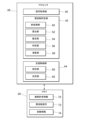
- Fig. 2 is a functional block diagram of the processor 26 and the memory 28 shown in Fig. 1.
- the processor 26 reads out an electroencephalogram analysis program from the memory 28 and executes it, thereby functioning as a signal acquisition unit 40 (corresponding to an "acquisition unit"), a frequency identification unit 42, and an assistance control unit 44.
- the signal acquisition unit 40 acquires a time series of EEG signals from the subject 12 via the sensor controller 24 ( Figure 1). This allows EEG signals within a unit period to be acquired sequentially.
- the unit period may be a time length equal to the sampling interval, or an integer multiple of the sampling interval.
- the time series of EEG signals includes one measured when the subject 12 is at rest (hereinafter also referred to as the "first time series") and one measured when the subject 12 is imagining a movement (hereinafter also referred to as the "second time series").
- the frequency identification unit 42 analyzes the EEG signal acquired by the signal acquisition unit 40 to identify an inherent frequency that correlates with the motor intention of the subject 12. Examples of inherent frequencies include the individual ERD frequency (Event-related desynchronization Frequency) and the individual SMR-ERD frequency (i.e., ISF).
- SMR-ERD frequency refers to the frequency within the alpha band of 8-13 Hz of scalp EEG measured near the motor cortex at which event-related desynchronization (ERD), a movement-related response, is strong.
- SMR-ERD frequency varies from person to person, and is known to fluctuate between 8 and 13 Hz. Therefore, to clarify that it is a frequency unique to each individual, it is sometimes called the individual SMR-ERD frequency (i.e., ISF).
- the frequency identification unit 42 specifically includes a preprocessing unit 50, a calculation unit 52, an estimation unit 54, a determination unit 56, and an arithmetic unit 58.
- the pre-processing unit 50 performs pre-processing required for calculating the Individual Alpha Frequency (IAF) on the time series of EEG signals acquired by the signal acquisition unit 40.
- This pre-processing includes, for example, [1] "filter processing” including moving average, [2] “frequency conversion processing” including FFT (Fast Fourier Transform), and [3] “trend removal processing” to remove 1/f noise from the frequency characteristics (or power spectrum).
- alpha frequency refers to the frequency that shows a peak in signal strength within the alpha band of 8-13 Hz in scalp electroencephalograms, which reflect the collective activity of brain nerve cells.
- Alpha frequency varies from person to person, and is known to fluctuate within the range of 8-13 Hz. For this reason, to clarify that it is a frequency unique to each individual, it is sometimes called individual alpha frequency (i.e., IAF).
- the calculation unit 52 calculates the IAF of the subject 12 from the frequency characteristics of the EEG signal obtained by the preprocessing unit 50, thereby obtaining sample values for each time series (hereinafter also referred to as "IAF sample values"). Specifically, the calculation unit 52 detects the maximum peak in a specific band (here, the alpha band) in the frequency characteristics, and calculates the frequency corresponding to the maximum peak as the IAF sample value. Note that in addition to the alpha band (8-13 Hz), at least one of the delta band (1-3 Hz), theta band (3-7 Hz), beta band (14-30 Hz), and gamma band (30 Hz or higher) can be selected as the specific band depending on the analysis target.
- the estimation unit 54 uses the population of IAF sample values calculated by the calculation unit 52 to estimate the IAF of the subject 12, thereby obtaining an estimate for each population (hereinafter also referred to as an "IAF estimate").
- IAF estimate various statistical methods including the Bayes method and the sequential Bayes method (or a Kalman filter) are used.
- the estimation unit 54 may obtain an estimate based on the sequential Bayes method for each unit period, using a population accumulated from the start of measurement of the EEG signal.
- the determination unit 56 determines whether the IAF estimated value satisfies a termination condition each time the estimation unit 54 makes an estimation, and instructs the estimation unit 54 to terminate the estimation process when the termination condition is satisfied.
- the termination conditions include: [Condition 1] the IAF estimated value has converged (e.g., the amount of change has become equal to or less than a threshold value); [Condition 2] the number of estimations made by the estimation unit 54 has exceeded a threshold value; [Condition 3] an amount of time has elapsed since the start of measurement of the EEG signal; and [Condition 4] a combination of the above conditions 1 to 3.
- the calculation unit 58 converts the IAF estimate obtained most recently when the determination unit 56 determines that the termination condition is satisfied into an inherent frequency (ISF here) that correlates with the motor intention of the subject 12 according to a predetermined conversion rule, thereby obtaining a conversion value for each analysis movement (hereinafter, ISF conversion value).
- This conversion rule may be a rule common to the subjects 12, or may be a rule that differs depending on the subjects 12.
- the function shape may be [1] a linear function such as an identity function or a linear function, or [2] a nonlinear function such as a polynomial function with a power of 2 or more, or an exponential function.
- the calculation unit 58 performs a calculation process (hereinafter referred to as the "first calculation”) to obtain the ISF using only the above-mentioned first time series, but may also perform a calculation (hereinafter referred to as the "second calculation") to obtain the ISF using both the first time series and the second time series.
- the calculation unit 58 may switch between the first calculation and the second calculation as necessary.
- the two types of calculation may be switched manually via an input operation from the operation unit 22 ( Figure 1), for example, or may be switched automatically based on the analysis results of the electroencephalogram signal obtained by the signal acquisition unit 40.
- the support control unit 44 performs control (hereinafter also referred to as "support control") on the exercise support device 18 based on the ISF conversion value obtained by the calculation unit 58.
- the support control unit 44 includes a setting unit 60 that sets an ISF setting value suitable for the subject 12, and a determination unit 62 that determines the amount of control for the exercise support device 18 using the frequency characteristics of the EEG signal and the ISF setting value set in the setting unit 60.
- memory 28 stores subject information 70, frequency information 72, and conversion information 74 in a corresponding relationship.
- the subject information 70 includes various information related to the subject 12, such as the identification information and personal information of the subject 12, the diagnosis results and degree of recovery of the subject 12, the type and usage history of the exercise support device 18, etc.
- the frequency information 72 includes various information related to the peak frequency or natural frequency, such as the IAF sample value, the IAF estimated value, and the ISF conversion value.
- the conversion information 74 includes various information that can identify the conversion rule, such as the type of function shape, coefficients, order, and LUT (lookup table).
- the BMI system 10 in this embodiment is configured as described above. Next, the operation of the BMI system 10, more specifically, the exercise support operation by the electroencephalogram analyzer 16 will be described with reference to the flowcharts of FIGS.
- ⁇ Explanation of exercise support method> 3 is a flow chart of the exercise support method using the BMI system 10 shown in FIG. 1.
- a "wearing” step is performed in which the electroencephalograph 14 is worn on the head of the person being analyzed 12.
- a "start” step is performed in which monitoring of the electroencephalogram emitted by the person being analyzed 12 is started.
- a "confirmation” step is performed in which it is confirmed whether the person being analyzed 12 has become quiet. If the person being analyzed 12 is not quiet (step SP104: NO), the process remains in step SP104 until the person being analyzed 12 becomes quiet. On the other hand, if the person being analyzed 12 has become quiet (step SP104: YES), the process proceeds to the next step SP106.
- step SP106 a "calibration" process is performed to calibrate the exercise support device 18.
- the EEG analysis device 16 analyzes the EEG signals sequentially supplied from the EEG 14 to obtain the intrinsic frequency (i.e., ISF) of the subject 12, and sets the ISF value as a calibration parameter for the exercise support device 18.
- ISF intrinsic frequency
- step SP108 a "support” process is carried out to provide neurorehabilitation to the subject 12.
- the EEG analysis device 16 controls the operation of the exercise support device 18 that was calibrated in step SP106. Then, the exercise of the subject 12 is supported through the operation of the exercise support device 18.
- ⁇ Analysis Operation by Electroencephalogram Analysis Device 16> 4 is a flowchart of the analysis operation by the electroencephalogram analysis device 16 of FIG. 1.
- the electroencephalogram analysis device 16 executes this flowchart to identify the intrinsic frequency (i.e., ISF) of the subject 12 prior to rehabilitation of the arm 12a of the subject 12.
- ISF intrinsic frequency
- This flowchart is not limited to being executed once at the start of rehabilitation, but may be executed once or more during rehabilitation as necessary.
- This operation can be directly confirmed by analyzing the electroencephalogram analysis program, and can be indirectly confirmed by comparing an ideal signal waveform inputted artificially to the electroencephalograph 14 with the output result from the electroencephalogram analysis device 16.
- step SP10 of FIG. 4 the signal acquisition unit 40 acquires a time series of EEG signals within a unit period via the EEG meter 14 and the sensor controller 24.
- Figure 5 shows the change over time in the EEG signal when the subject is at rest.
- the horizontal axis of the graph shows time (unit: s), and the vertical axis of the graph shows brain potential (unit: mV).
- the EEG signal has a complex waveform that fluctuates finely above and below a reference value.
- step SP12 of FIG. 4 the frequency identification unit 42 (more specifically, the preprocessing unit 50) performs preprocessing on the time series of the EEG signal acquired in step SP10. This allows the frequency characteristics of the EEG signal to be obtained.
- step SP14 the frequency identification unit 42 (more specifically, the calculation unit 52) calculates the IAF sample value from the frequency characteristics obtained by the preprocessing in step SP12.
- Figure 6 shows how the IAF sample value is calculated.
- the horizontal axis of the graph indicates frequency (unit: Hz), and the vertical axis of the graph indicates signal strength (unit: dimensionless).
- this frequency characteristic has a maximum peak around 11 [Hz].
- the IAF sample value is calculated as 11 [Hz].
- step SP16 of FIG. 4 the processor 26 checks whether the timing to estimate the IAF (hereinafter referred to as the "estimation timing") has arrived. If the estimation timing has not yet arrived (step SP16: NO), the processor 26 returns to step SP10 and sequentially repeats the execution of steps SP10 to SP16 until the timing arrives. On the other hand, if the estimation timing has arrived (step SP16: YES), the processor 26 proceeds to the next step SP18.
- the frequency identification unit 42 estimates the IAF by applying the sequential Bayes method to the population accumulated through the sequential calculation in step SP14.
- the posterior probability p(IAF A
- step SP20 the frequency identification unit 42 (more specifically, the determination unit 56) determines whether the IAF estimate obtained in step SP18 satisfies the termination condition. If the termination condition is not satisfied (step SP20: NO), the processor 26 returns to step SP10 and sequentially repeats the execution of steps SP10 to SP20 until the termination condition is satisfied.
- Figure 7 shows an example of the convergence of the IAF estimate by the sequential Bayes method.
- the horizontal axis of the graph indicates time (unit: s), and the vertical axis of the graph indicates the estimation error (unit: Hz).
- estimate error corresponds to the value obtained by subtracting the actual value from the estimated IAF value (i.e., the deviation).
- the solid line graph shows the average value of data from 180 healthy adults.
- the dashed line graph on the bottom shows the boundary line of "average value - 2 ⁇ " ( ⁇ : standard deviation), and the dashed line graph on the top shows the boundary line of "average value + 2 ⁇ ".
- Figure 8 is a diagram that shows the time-saving effect of the termination process at the time of convergence.
- the axis of the histogram shows the estimated time required for an individual's estimation error (see Figure 6) to fall within 1 Hz, that is, the convergence time (unit: s).
- the convergence time unit: s
- the frequency of convergence times within 4 s is the highest, and the frequency gradually decreases as the convergence time increases.
- the median convergence time is "11 s" and the 2 ⁇ boundary line of the convergence time is "30 s".
- step SP22 NO
- the processor 26 proceeds to the next step SP22.
- step SP22 of FIG. 4 the frequency identification unit 42 (more specifically, the calculation unit 58) converts the IAF estimate most recently obtained in step SP18 into an ISF according to the conversion rule specified by the conversion information 74. Specifically, the ISF is converted according to the following equation (2). Note that g( ⁇ ) is an arbitrary function.
- Figure 10 shows the probability density distribution of the deviation between IAF and ISF.
- the ISF may be calculated according to the following formula (3) by focusing on the relationship shown in Figures 9 and 10.
- g(x) x in formula (2), which represents the so-called identity transformation.
- step SP22 of FIG. 4 the support control unit 44 (more specifically, the setting unit 60) sets the ISF conversion value obtained in step SP20. This allows the support control unit 44 to control the exercise support device 18 in a manner suitable for the person being analyzed 12.
- Fig. 11 is a schematic diagram showing the effect of the electroencephalogram analysis method in this embodiment.
- the axis of the graph shows the time (unit: s) from the start of electroencephalogram measurement.
- the upper bar graph shows the breakdown of ISF identification time in the "Comparative Example” (see Fig. 12).
- the lower bar graph shows the breakdown of ISF identification time in the "Example” (see Figs. 3 and 4).
- Tc T1 + T2
- the measurement time can be, for example, 200 to 1000 seconds. As this measurement time becomes longer, the mental and physical burden on the subject 12 increases.
- T3 approximately 30 s.
- ISF is identified in a short time without acquiring an EEG signal when the subject 12 intends to move, or without the subject 12 moving the paralyzed part. This significantly reduces the mental and physical burden on the subject 12.
- the exercise support system in this embodiment (here, the BMI system 10) includes an EEG analysis device 16, an EEG meter 14 that measures the EEG of the person being analyzed 12 and supplies the EEG signal obtained to the EEG analysis device 16, and an exercise support device 18 that operates according to the control performed by the EEG analysis device 16 to support the exercise of the person being analyzed 12.
- This EEG analysis device 16 includes an acquisition unit (here, a signal acquisition unit 40) that acquires a time series of EEG signals from the subject 12, and a calculation unit 58 that determines a specific frequency that correlates with the motor intention or brain state of the subject 12 based on the frequency characteristics of the time series of the acquired EEG signals.
- acquisition unit here, a signal acquisition unit 40
- calculation unit 58 that determines a specific frequency that correlates with the motor intention or brain state of the subject 12 based on the frequency characteristics of the time series of the acquired EEG signals.
- one or more computers execute an acquisition step (SP10) of acquiring a time series of electroencephalogram signals from the subject 12, and a calculation step (SP22) of determining an inherent frequency that correlates with the motor intention of the subject 12 based on the frequency characteristics of the time series of electroencephalogram signals acquired when the subject 12 is at rest.
- SP10 acquisition step
- SP22 calculation step
- one or more computers in addition to the acquisition step (SP10) and the calculation step (SP22) described above, one or more computers (or processors 26) further execute a calibration step (SP24) of performing a calibration in which the determined natural frequency is set as a calibration parameter of the exercise support device 18, and a support step (SP108) of supporting the exercise of the subject 12 by controlling the operation of the exercise support device 18 after the calibration has been performed.
- SP24 a calibration step of performing a calibration in which the determined natural frequency is set as a calibration parameter of the exercise support device 18
- SP108 support step of supporting the exercise of the subject 12 by controlling the operation of the exercise support device 18 after the calibration has been performed.
- the EEG analysis device 16 may further include a calculation unit 52 that calculates sample values of the peak frequency in the frequency characteristics, and an estimation unit 54 that uses a population of the calculated sample values to obtain an estimate of the peak frequency, and the calculation unit 58 may convert the estimate obtained by the estimation unit 54 into a natural frequency according to a predetermined conversion rule.
- the EEG analysis device 16 may obtain the natural frequency without acquiring the EEG signal when the subject 12 intends to move, or without the subject 12 moving the paralyzed part, by having the estimation unit 54 obtain an estimated value of the peak frequency and the calculation unit 58 convert the estimated value into the natural frequency. This significantly reduces the mental and physical burden on the subject 12.
- the estimation unit 54 may also use a population of sample values accumulated by repeating acquisition by the signal acquisition unit 40 and calculation by the calculation unit 52 for each unit period from the start of measurement of the EEG signal to obtain an estimate based on the sequential Bayes method for each unit period.
- the estimation unit 54 may terminate the estimation of the peak frequency when the determination unit 56 determines that the termination condition is satisfied. By terminating the estimation process when the termination condition is satisfied, the time required to identify the natural frequency can be further shortened.
- the conversion rule may be expressed as an identity function or a linear function with the IAF estimate as an argument.
- the conversion rules may also be determined according to the person being analyzed 12. This allows for conversion that is more suited to the characteristics of the person being analyzed 12.
- the calculation unit 58 may perform a first calculation to obtain a natural frequency using only the first time series. Furthermore, the calculation unit 58 may switch between the first calculation described above and a second calculation to obtain a natural frequency using both the first time series and the second time series.
- the EEG analysis device 16 may further include a presentation unit that presents information to the subject 12 requesting a resting state.
- the EEG analysis device 16 may further include a support control unit 44 that controls the exercise support device 18 to support the exercise of the subject 12 based on the natural frequency determined by the calculation unit 58. This makes it possible to provide analysis or exercise support to the subject 12.
- the present invention is not limited to the above-described embodiment, and can be freely modified without departing from the spirit of the present invention.
- the configurations may be combined in any way without causing any technical inconsistency.
- the order of execution of each step in the flow chart may be changed without causing any technical inconsistency.
- the EEG analysis device 16 analyzes EEG signals and controls the exercise support device 18, but the system configuration is not limited to this.
- the analysis unit and the control unit may be provided separately, and configured to be able to exchange necessary data with each other via wired or wireless communication.
- a cloud-type or on-premise server device may perform the analysis process.
- the server device is a cloud-type device, the server device may be a group of computers that make up a distributed system.
- Reference Signs List 10 BMI system (exercise support system), 12: subject, 14: electroencephalograph, 16: electroencephalogram analysis device (computer), 18: exercise support device, 26: processor, 28: memory, 40: signal acquisition unit (acquisition unit), 42: frequency identification unit, 44: exercise support unit, 50: pre-processing unit, 52: calculation unit, 54: estimation unit, 56: judgment unit, 58: calculation unit
Landscapes
- Health & Medical Sciences (AREA)
- Life Sciences & Earth Sciences (AREA)
- Engineering & Computer Science (AREA)
- Biomedical Technology (AREA)
- General Health & Medical Sciences (AREA)
- Public Health (AREA)
- Medical Informatics (AREA)
- Animal Behavior & Ethology (AREA)
- Veterinary Medicine (AREA)
- Epidemiology (AREA)
- Physics & Mathematics (AREA)
- Pathology (AREA)
- Surgery (AREA)
- Molecular Biology (AREA)
- Heart & Thoracic Surgery (AREA)
- Biophysics (AREA)
- Psychiatry (AREA)
- Psychology (AREA)
- Pain & Pain Management (AREA)
- Primary Health Care (AREA)
- Rehabilitation Therapy (AREA)
- Physical Education & Sports Medicine (AREA)
- General Business, Economics & Management (AREA)
- Business, Economics & Management (AREA)
- Measurement And Recording Of Electrical Phenomena And Electrical Characteristics Of The Living Body (AREA)
Priority Applications (4)
| Application Number | Priority Date | Filing Date | Title |
|---|---|---|---|
| JP2024554454A JP7851645B2 (ja) | 2022-10-31 | 2023-10-26 | 脳波解析装置及び脳波解析プログラム、並びに、運動支援システム及び運動支援方法 |
| CN202380065598.XA CN120239585A (zh) | 2022-10-31 | 2023-10-26 | 脑电波解析装置、脑电波解析程序、运动辅助系统以及运动辅助方法 |
| EP23885642.1A EP4613200A1 (en) | 2022-10-31 | 2023-10-26 | Brain wave analysis device, brain wave analysis program, motion assistance system, and motion assistance method |
| US19/110,951 US20250352120A1 (en) | 2022-10-31 | 2023-10-26 | Electroencephalogram Analysis Device and Electroencephalogram Analysis Program, and Movement Assistance System, and Movement Assistance Method |
Applications Claiming Priority (2)
| Application Number | Priority Date | Filing Date | Title |
|---|---|---|---|
| JP2022-174759 | 2022-10-31 | ||
| JP2022174759 | 2022-10-31 |
Publications (1)
| Publication Number | Publication Date |
|---|---|
| WO2024095886A1 true WO2024095886A1 (ja) | 2024-05-10 |
Family
ID=90930375
Family Applications (1)
| Application Number | Title | Priority Date | Filing Date |
|---|---|---|---|
| PCT/JP2023/038668 Ceased WO2024095886A1 (ja) | 2022-10-31 | 2023-10-26 | 脳波解析装置及び脳波解析プログラム、並びに、運動支援システム及び運動支援方法 |
Country Status (5)
| Country | Link |
|---|---|
| US (1) | US20250352120A1 (https=) |
| EP (1) | EP4613200A1 (https=) |
| JP (1) | JP7851645B2 (https=) |
| CN (1) | CN120239585A (https=) |
| WO (1) | WO2024095886A1 (https=) |
Citations (3)
| Publication number | Priority date | Publication date | Assignee | Title |
|---|---|---|---|---|
| WO2019078325A1 (ja) * | 2017-10-20 | 2019-04-25 | パナソニック株式会社 | 脳波判定システム、脳波判定方法、プログラム、及び非一時的記録媒体 |
| WO2021070456A1 (ja) * | 2019-10-08 | 2021-04-15 | Vie Style株式会社 | イヤホン、情報処理装置、及び情報処理方法 |
| WO2022236342A1 (en) * | 2021-05-07 | 2022-11-10 | Wave Neuroscience, Inc. | Rhythmic stimulus to estimate an intrinsic frequency of an eeg band |
-
2023
- 2023-10-26 WO PCT/JP2023/038668 patent/WO2024095886A1/ja not_active Ceased
- 2023-10-26 JP JP2024554454A patent/JP7851645B2/ja active Active
- 2023-10-26 EP EP23885642.1A patent/EP4613200A1/en active Pending
- 2023-10-26 US US19/110,951 patent/US20250352120A1/en active Pending
- 2023-10-26 CN CN202380065598.XA patent/CN120239585A/zh active Pending
Patent Citations (3)
| Publication number | Priority date | Publication date | Assignee | Title |
|---|---|---|---|---|
| WO2019078325A1 (ja) * | 2017-10-20 | 2019-04-25 | パナソニック株式会社 | 脳波判定システム、脳波判定方法、プログラム、及び非一時的記録媒体 |
| WO2021070456A1 (ja) * | 2019-10-08 | 2021-04-15 | Vie Style株式会社 | イヤホン、情報処理装置、及び情報処理方法 |
| WO2022236342A1 (en) * | 2021-05-07 | 2022-11-10 | Wave Neuroscience, Inc. | Rhythmic stimulus to estimate an intrinsic frequency of an eeg band |
Non-Patent Citations (2)
| Title |
|---|
| B. BLANKERTZ ET AL.: "Neurophysiological predictor of SMR-based BCI performance", NEUROIMAGE, vol. 51, 2010, pages 1303 - 1309, XP027048616 |
| MAGNUSON JUSTINE R.; MCNEIL CHRIS J.: "Low-frequency neural activity at rest is correlated with the movement-related cortical potentials elicited during both real and imagined movements", NEUROSCIENCE LETTERS, ELSEVIER, AMSTERDAM, NL, vol. 742, 25 November 2020 (2020-11-25), AMSTERDAM, NL , XP086430448, ISSN: 0304-3940, DOI: 10.1016/j.neulet.2020.135530 * |
Also Published As
| Publication number | Publication date |
|---|---|
| JP7851645B2 (ja) | 2026-04-27 |
| CN120239585A (zh) | 2025-07-01 |
| US20250352120A1 (en) | 2025-11-20 |
| JPWO2024095886A1 (https=) | 2024-05-10 |
| EP4613200A1 (en) | 2025-09-10 |
Similar Documents
| Publication | Publication Date | Title |
|---|---|---|
| RU2669466C2 (ru) | Система нейронной обратной связи | |
| EP3698710B1 (en) | Brain wave measurement system, brain wave measurement method, program, and non-transitory recording medium | |
| JP6190466B2 (ja) | 生体信号測定器及び接触状態推定方法 | |
| Shukla et al. | Efficient wavelet-based artifact removal for electrodermal activity in real-world applications | |
| CN106413541B (zh) | 用于诊断睡眠的系统和方法 | |
| CN103405225B (zh) | 一种获取疼感评测指标的方法、装置及设备 | |
| Chuckravanen | Approximate entropy as a measure of cognitive fatigue: An eeg pilot study | |
| US12076152B2 (en) | Electroencephalogram decision system, electroencephalogram decision method, program, and non-transitory storage medium | |
| CN113729624A (zh) | 意识水平测定方法及计算机程序 | |
| KR101527273B1 (ko) | 전두엽 부착형 뇌파신호 검출 및 뇌파기반 집중력 분석 방법 및 장치 | |
| JP2019030520A (ja) | 筋電計測装置、方法及びプログラム | |
| JP7851645B2 (ja) | 脳波解析装置及び脳波解析プログラム、並びに、運動支援システム及び運動支援方法 | |
| US11116452B2 (en) | Biological signal measurement system | |
| Badura et al. | Multimodal signal acquisition for pain assessment in physiotherapy | |
| KR101455518B1 (ko) | 뇌파를 이용한 전자의수 제어 방법 및 그 장치 | |
| Melindaa et al. | Savitzky-Golay And Wiener Filtering Performance Analysis In Electroencephalography Signal Processing Of Autistic Children | |
| CN110234272B (zh) | 麻醉阶段识别与麻醉深度计算方法及装置 | |
| Liu et al. | Adaptive EOG artifact removal algorithm using a spectrum magnitude template matching for single-channel EEG | |
| KR101211065B1 (ko) | 브레인 컴퓨터 인터페이스를 이용한 무선 제어시스템 | |
| JP2011167251A (ja) | 快適性判定装置 | |
| Sato et al. | Simulation study of a P300 speller for single-lead hybrid BCI | |
| KR20050011396A (ko) | 급작스런 기저선 변동이 있는 뇌파의 잡파를 제거하기위한 뇌파 측정 방법 및 장치 | |
| Mahmood et al. | Methodology for EEG Based System Development to Detect Objective Pain in Human Body. | |
| Luong et al. | Design of Electrocardiography Signal Acquisition and Processing Software Module |
Legal Events
| Date | Code | Title | Description |
|---|---|---|---|
| 121 | Ep: the epo has been informed by wipo that ep was designated in this application |
Ref document number: 23885642 Country of ref document: EP Kind code of ref document: A1 |
|
| WWE | Wipo information: entry into national phase |
Ref document number: 2024554454 Country of ref document: JP |
|
| WWE | Wipo information: entry into national phase |
Ref document number: 202380065598.X Country of ref document: CN |
|
| WWE | Wipo information: entry into national phase |
Ref document number: 2023885642 Country of ref document: EP |
|
| NENP | Non-entry into the national phase |
Ref country code: DE |
|
| ENP | Entry into the national phase |
Ref document number: 2023885642 Country of ref document: EP Effective date: 20250602 |
|
| WWP | Wipo information: published in national office |
Ref document number: 202380065598.X Country of ref document: CN |
|
| WWP | Wipo information: published in national office |
Ref document number: 2023885642 Country of ref document: EP |