WO2024024955A1 - 迷走神経様副交感神経細胞およびその作製方法 - Google Patents
迷走神経様副交感神経細胞およびその作製方法 Download PDFInfo
- Publication number
- WO2024024955A1 WO2024024955A1 PCT/JP2023/027801 JP2023027801W WO2024024955A1 WO 2024024955 A1 WO2024024955 A1 WO 2024024955A1 JP 2023027801 W JP2023027801 W JP 2023027801W WO 2024024955 A1 WO2024024955 A1 WO 2024024955A1
- Authority
- WO
- WIPO (PCT)
- Prior art keywords
- cells
- parasympathetic
- nerve
- gastrointestinal hormone
- expressing
- Prior art date
- Legal status (The legal status is an assumption and is not a legal conclusion. Google has not performed a legal analysis and makes no representation as to the accuracy of the status listed.)
- Ceased
Links
Images




































Classifications
-
- C—CHEMISTRY; METALLURGY
- C07—ORGANIC CHEMISTRY
- C07K—PEPTIDES
- C07K14/00—Peptides having more than 20 amino acids; Gastrins; Somatostatins; Melanotropins; Derivatives thereof
- C07K14/435—Peptides having more than 20 amino acids; Gastrins; Somatostatins; Melanotropins; Derivatives thereof from animals; from humans
-
- C—CHEMISTRY; METALLURGY
- C12—BIOCHEMISTRY; BEER; SPIRITS; WINE; VINEGAR; MICROBIOLOGY; ENZYMOLOGY; MUTATION OR GENETIC ENGINEERING
- C12N—MICROORGANISMS OR ENZYMES; COMPOSITIONS THEREOF; PROPAGATING, PRESERVING, OR MAINTAINING MICROORGANISMS; MUTATION OR GENETIC ENGINEERING; CULTURE MEDIA
- C12N5/00—Undifferentiated human, animal or plant cells, e.g. cell lines; Tissues; Cultivation or maintenance thereof; Culture media therefor
- C12N5/06—Animal cells or tissues; Human cells or tissues
-
- C—CHEMISTRY; METALLURGY
- C12—BIOCHEMISTRY; BEER; SPIRITS; WINE; VINEGAR; MICROBIOLOGY; ENZYMOLOGY; MUTATION OR GENETIC ENGINEERING
- C12N—MICROORGANISMS OR ENZYMES; COMPOSITIONS THEREOF; PROPAGATING, PRESERVING, OR MAINTAINING MICROORGANISMS; MUTATION OR GENETIC ENGINEERING; CULTURE MEDIA
- C12N5/00—Undifferentiated human, animal or plant cells, e.g. cell lines; Tissues; Cultivation or maintenance thereof; Culture media therefor
- C12N5/10—Cells modified by introduction of foreign genetic material
-
- C—CHEMISTRY; METALLURGY
- C12—BIOCHEMISTRY; BEER; SPIRITS; WINE; VINEGAR; MICROBIOLOGY; ENZYMOLOGY; MUTATION OR GENETIC ENGINEERING
- C12Q—MEASURING OR TESTING PROCESSES INVOLVING ENZYMES, NUCLEIC ACIDS OR MICROORGANISMS; COMPOSITIONS OR TEST PAPERS THEREFOR; PROCESSES OF PREPARING SUCH COMPOSITIONS; CONDITION-RESPONSIVE CONTROL IN MICROBIOLOGICAL OR ENZYMOLOGICAL PROCESSES
- C12Q1/00—Measuring or testing processes involving enzymes, nucleic acids or microorganisms; Compositions therefor; Processes of preparing such compositions
- C12Q1/02—Measuring or testing processes involving enzymes, nucleic acids or microorganisms; Compositions therefor; Processes of preparing such compositions involving viable microorganisms
-
- G—PHYSICS
- G01—MEASURING; TESTING
- G01N—INVESTIGATING OR ANALYSING MATERIALS BY DETERMINING THEIR CHEMICAL OR PHYSICAL PROPERTIES
- G01N33/00—Investigating or analysing materials by specific methods not covered by groups G01N1/00 - G01N31/00
- G01N33/48—Biological material, e.g. blood, urine; Haemocytometers
- G01N33/483—Physical analysis of biological material
-
- G—PHYSICS
- G01—MEASURING; TESTING
- G01N—INVESTIGATING OR ANALYSING MATERIALS BY DETERMINING THEIR CHEMICAL OR PHYSICAL PROPERTIES
- G01N33/00—Investigating or analysing materials by specific methods not covered by groups G01N1/00 - G01N31/00
- G01N33/48—Biological material, e.g. blood, urine; Haemocytometers
- G01N33/50—Chemical analysis of biological material, e.g. blood, urine; Testing involving biospecific ligand binding methods; Immunological testing
Definitions
- the present invention relates to vagus nerve-like parasympathetic nerve cells and methods for producing the same.
- gastrointestinal secretory cells present in the gastrointestinal epithelium sense nutrients and secrete gastrointestinal hormones.
- vagal afferents signals are transmitted to the central nervous system. From the central nervous system, the efferent vagal pathway regulates peristalsis of the gastrointestinal tract and the secretion of digestive fluids and gastrointestinal hormones.
- the present inventors have already reported a method for efficiently inducing autonomic nerves from human pluripotent stem cells and a method for selectively inducing parasympathetic nerve cells from human pluripotent stem cells (Patent Documents 1 and 2).
- the parasympathetic nerve cells produced by the above method do not endogenously express or have low expression of gastrointestinal hormone receptors, which are indicators of gastrointestinal vagus nerves.
- analysis of mice and the like has shown that more than 10 gastrointestinal vagus nerve cell types exist in vivo, with different expression patterns of gastrointestinal hormone receptors.
- the purpose of the present invention is to provide vagus nerve-like parasympathetic neurons that constitutively express gastrointestinal hormone receptors and are responsive to gastrointestinal hormones and can be used in an in vitro test system for brain-gut correlation. It is something that
- vagus nerve-like parasympathetic neurons expressing glucagon-like peptide 1 receptors, neuropeptide Y2 type receptors, glucagon-like peptide 2 receptors, serotonin 3 receptors, or cholecystokinin 1 receptors succeeded in creating nerve cells.
- the present invention provides a step of (a) inducing differentiation of pluripotent stem cells into parasympathetic nerve cells, and (b1) prior to step (a), (b2) introducing an exogenous nucleic acid encoding a gastrointestinal hormone receptor into the parasympathetic neurons or their progenitor cells obtained in step (a);
- the present invention provides a method for producing vagus nerve-like parasympathetic nerve cells, the method comprising the step of introducing vagus nerve-like parasympathetic nerve cells.
- the present invention also provides vagus nerve-like parasympathetic nerve cells produced by the above method.
- the present invention also provides pluripotent stem cells comprising an exogenous nucleic acid encoding a gastrointestinal hormone receptor.
- the present invention also provides a vagus nerve-like parasympathetic nerve cell comprising an exogenous nucleic acid encoding a gastrointestinal hormone receptor.
- the present invention also provides the steps of: (1) bringing a sample into contact with the vagus nerve-like parasympathetic nerve cells; and (2) analyzing neural activity of the vagus nerve-like parasympathetic nerve cells.
- a method of detecting a gastrointestinal hormone receptor agonist in a sample is provided.
- the gastrointestinal hormone receptor is selected from the group consisting of glucagon-like peptide 1 receptor, neuropeptide Y type 2 receptor, glucagon-like peptide 2 receptor, serotonin 3 receptor and cholecystokinin 1 receptor. .
- the present invention also provides the steps of: (1) culturing the vagus nerve-like parasympathetic nerve cells in the presence of a candidate substance; and (2) culturing the neural activity of the vagus nerve-like parasympathetic nerve cells.
- the present invention provides a method for screening for substances that control brain-gut correlation, including a step of analyzing.
- the present invention also provides a step of (1) culturing gastrointestinal hormone-secreting cells in the presence of a candidate substance, (2) a culture supernatant from the culture obtained in step (1). (3) bringing the culture supernatant into contact with the vagus nerve-like parasympathetic nerve cells; and (4) analyzing the neural activity of the vagus nerve-like parasympathetic nerve cells.
- the present invention provides a method for screening substances that control
- the present invention also provides the steps of: (1) co-culturing the gastrointestinal hormone-secreting cells and the vagus nerve-like parasympathetic nerve cells in the presence of a candidate substance;
- the present invention provides a method for screening for substances that control the brain-gut correlation, which includes the step of analyzing neural activity of parasympathetic nerve cells.
- the present invention provides a brain-gut correlation model comprising co-culture of gastrointestinal hormone-secreting cells and the vagus nerve-like parasympathetic nerve cells.
- the gastrointestinal hormone-secreting cells are enteroendocrine cells.
- the present invention also provides the use of the vagus nerve-like parasympathetic nerve cells or the brain-gut correlation model for screening for substances that control the brain-gut correlation.
- vagus nerve-like parasympathetic neurons that can be used in an in vitro test system for brain-gut correlation can be produced with high efficiency and at low cost. Furthermore, the vagus nerve-like parasympathetic nerve cells according to the present invention can mimic the gastrointestinal hormonal response of the gastrointestinal vagal nerve afferent pathway. Therefore, the vagus nerve-like parasympathetic neurons according to the present invention can be used, for example, to develop an in vitro test system for brain-gut correlation, to detect agonists for gastrointestinal hormone receptors or to search for new agonists, and to detect gastrointestinal hormones from gastrointestinal endocrine cells. Useful for detecting release or searching for substances that induce gastrointestinal hormone release.
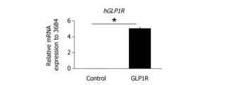
- FIG. 1 is a graph showing the expression level of hGLP1R mRNA in wild-type iPS cells and iPS cells into which exogenous hGLP1R gene has been introduced.
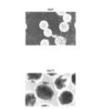
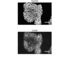
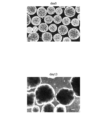
- FIG. 2 shows a Hoechst staining image (top) and a fluorescent immunostaining image using an anti-GLP1R antibody (bottom) of iPS cells into which an exogenous hGLP1R gene has been introduced.
- FIG. 3 is a diagram showing a microscopic image (bright field) of hGLP1R-expressing iPS cells.
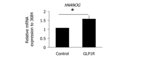
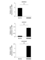
- FIG. 4 is a graph showing the expression level of hNANOG mRNA in wild-type iPS cells and hGLP1R-expressing iPS cells.
- FIG. 1 is a graph showing the expression level of hGLP1R mRNA in wild-type iPS cells and iPS cells into which exogenous hGLP1R gene has been introduced.
- FIG. 2 shows a Hoechs
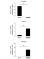
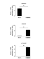
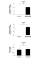
- FIG. 5 is a graph showing the expression levels of hPAX6, hNCAM1, or hSOX17 mRNA in undifferentiated hGLP1R-expressing iPS cells and hGLP1R-expressing iPS cells induced to differentiate into ectoderm, mesoderm, or endoderm. .
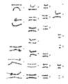
- FIG. 6 is a diagram showing the results of analyzing the chromosomes of hGLP1R-expressing iPS cells by the G-staining method.
- FIG. 7 is a diagram showing microscopic images (bright field) of EBs prepared from hGLP1R-expressing iPS cells on day 0 of induction and cells on day 13 of induction.
- FIG. 8 is a graph showing the expression levels of hNANOG, hSOX10, and hPHOX2B mRNA in cells prepared from hGLP1R-expressing iPS cells on day 13 of induction.
- FIG. 9 is a graph showing the expression levels of hPRPH, hCHAT, and hGLP1R mRNA in cells prepared from hGLP1R-expressing iPS cells on day 40 of induction.
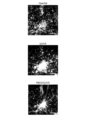
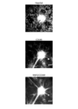
- Figure 10 shows a Hoechst staining image (top) of cells prepared from hGLP1R-expressing iPS cells on day 45 of induction (top), a fluorescent immunostaining image using an anti-GLP1R antibody (center), and an image using an anti-GLP1R antibody + anti-MAP2 antibody.
- FIG. 11 is a diagram showing an extracellular potential heat map of cells prepared from hGLP1R-expressing iPS cells on day 27 of induction, with or without the addition of GLP1.
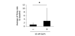
- FIG. 12 is a graph showing the rate of change in firing frequency of cells prepared from hGLP1R-expressing iPS cells on day 27 of induction under conditions with and without the addition of GLP1.
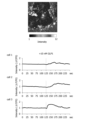
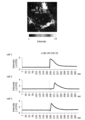
- FIG. 13 is a diagram showing the results of calcium imaging of cells prepared from hGLP1R-expressing iPS cells on day 40 of induction.
- FIG. 14 is a graph showing the number of Ca 2+ spikes in cells prepared from hGLP1R-expressing iPS cells at day 40 of induction under conditions with and without the addition of GLP1.
- FIG. 15 is a graph showing the amplitude of Ca 2+ spikes in cells prepared from hGLP1R-expressing iPS cells at day 40 of induction, with or without the addition of GLP1.
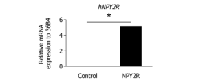
- FIG. 16 is a graph showing the expression level of hNPY2R mRNA in wild-type iPS cells and iPS cells into which exogenous hNPY2R gene has been introduced.
- FIG. 17 shows a Hoechst staining image (top) and a fluorescent immunostaining image using an anti-NPY2R antibody (bottom) of iPS cells into which exogenous hNPY2R gene has been introduced.
- FIG. 18 is a diagram showing a microscopic image (bright field) of hNPY2R-expressing iPS cells.
- FIG. 19 is a graph showing the expression level of hNANOG mRNA in wild-type iPS cells and hNPY2R-expressing iPS cells.
- FIG. 20 is a graph showing the expression levels of hPAX6, hNCAM1, or hSOX17 mRNA in undifferentiated hNPY2R-expressing iPS cells and hNPY2R-expressing iPS cells induced to differentiate into ectoderm, mesoderm, or endoderm. .
- FIG. 21 is a diagram showing the results of analyzing the chromosomes of hNPY2R-expressing iPS cells by the G-staining method.
- FIG. 22 is a diagram showing microscopic images (bright field) of EBs prepared from hNPY2R-expressing iPS cells on day 0 of induction and cells on day 13 of induction.
- FIG. 23 is a graph showing the expression levels of hNANOG, hSOX10, and hPHOX2B mRNA in cells prepared from hNPY2R-expressing iPS cells on day 13 of induction.
- FIG. 24 is a graph showing the expression levels of hPRPH, hCHAT, and hNPY2R mRNA in cells prepared from hNPY2R-expressing iPS cells on day 40 of induction.
- Figure 25 shows a Hoechst staining image (top) of cells prepared from hNPY2R-expressing iPS cells on day 45 of induction, a fluorescent immunostaining image using an anti-NPY2R antibody (center), and an image using an anti-NPY2R antibody + anti-MAP2 antibody.
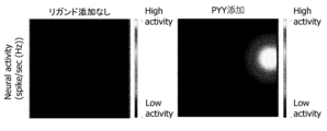
- FIG. 26 is a diagram showing an extracellular potential heat map of cells prepared from hNPY2R-expressing iPS cells on day 27 of induction, with or without the addition of PYY.
- FIG. 27 is a graph showing the rate of change in firing frequency of cells prepared from hNPY2R-expressing iPS cells on day 27 of induction under conditions with or without the addition of PYY.
- FIG. 28 is a diagram showing the results of calcium imaging of cells prepared from hNPY2R-expressing iPS cells on day 40 of induction.
- FIG. 29 is a graph showing the number of Ca 2+ spikes in cells prepared from hNPY2R-expressing iPS cells at day 40 of induction under conditions with and without the addition of PYY.
- FIG. 30 is a graph showing the amplitude of Ca 2+ spikes in cells prepared from hNPY2R-expressing iPS cells at day 40 of induction under conditions with and without the addition of PYY.
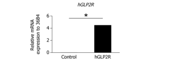
- FIG. 31 is a graph showing the expression level of hGLP2R mRNA in wild-type iPS cells and iPS cells into which exogenous hGLP2R gene has been introduced.
- FIG. 32 shows a Hoechst staining image (top) and a fluorescent immunostaining image using an anti-GLP2R antibody (bottom) of iPS cells into which exogenous hGLP2R gene has been introduced.
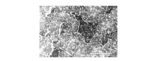
- FIG. 33 is a diagram showing a microscopic image (bright field) of hGLP2R-expressing iPS cells.
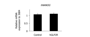
- FIG. 34 is a graph showing the expression level of hNANOG mRNA in wild-type iPS cells and hGLP2R-expressing iPS cells.
- FIG. 35 is a graph showing the expression levels of hNES, hNCAM1, or hSOX17 mRNA in undifferentiated hGLP2R-expressing iPS cells and hGLP2R-expressing iPS cells induced to differentiate into ectoderm, mesoderm, or endoderm. .
- FIG. 36 is a diagram showing the results of analyzing the chromosomes of hGLP2R-expressing iPS cells by G-staining method.
- FIG. 37 is a diagram showing microscopic images (bright field) of EBs prepared from hGLP2R-expressing iPS cells on day 0 of induction and cells on day 13 of induction.
- FIG. 38 is a graph showing the expression levels of hNANOG, hSOX10, and hPHOX2B mRNA in cells prepared from hGLP2R-expressing iPS cells on day 13 of induction.
- FIG. 39 is a graph showing the expression levels of hPRPH, hCHAT, and hGLP2R mRNA in cells prepared from hGLP2R-expressing iPS cells on day 40 of induction.
- Figure 40 shows a Hoechst staining image (top) of cells prepared from hGLP2R-expressing iPS cells on day 40 of induction, a fluorescent immunostaining image using an anti-GLP2R antibody (center), and an image using an anti-GLP2R antibody + anti-MAP2 antibody.
- FIG. 41 is a graph showing the expression level of hHTR3A mRNA in wild-type iPS cells and iPS cells into which exogenous hHTR3A gene has been introduced.
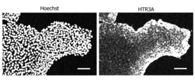
- FIG. 42 shows a Hoechst staining image (top) and a fluorescent immunostaining image using an anti-HTR3A antibody (bottom) of iPS cells into which exogenous hHTR3A gene has been introduced.
- FIG. 43 is a diagram showing a microscopic image (bright field) of hHTR3A-expressing iPS cells.
- FIG. 41 is a graph showing the expression level of hHTR3A mRNA in wild-type iPS cells and iPS cells into which exogenous hHTR3A gene has been introduced.
- FIG. 42 shows a Hoechst staining image (top) and a fluorescent immunostaining image using an anti-HTR3A antibody (bottom) of iPS cells into which exogenous hHTR3A
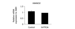
- FIG. 44 is a graph showing the expression level of hNANOG mRNA in wild-type iPS cells and hHTR3A-expressing iPS cells.
- FIG. 45 is a graph showing the expression levels of hNES, hNCAM1, or hSOX17 mRNA in undifferentiated hHTR3A-expressing iPS cells and hHTR3A-expressing iPS cells induced to differentiate into ectoderm, mesoderm, or endoderm. .
- FIG. 46 is a diagram showing the results of analyzing the chromosomes of hHTR3A-expressing iPS cells by the G-staining method.
- FIG. 45 is a graph showing the expression levels of hNES, hNCAM1, or hSOX17 mRNA in undifferentiated hHTR3A-expressing iPS cells and hHTR3A-expressing iPS cells induced to differentiate into ectoderm, mesoderm, or endoderm
- FIG. 47 is a diagram showing microscopic images (bright field) of EBs prepared from hHTR3A-expressing iPS cells on day 0 of induction and cells on day 13 of induction.
- FIG. 48 is a graph showing the expression levels of hNANOG, hSOX10, and hPHOX2B mRNA in cells prepared from hHTR3A-expressing iPS cells on day 13 of induction.
- FIG. 49 is a graph showing the expression levels of hPRPH, hCHAT, and hHTR3A mRNA in cells prepared from hHTR3A-expressing iPS cells on day 40 of induction.
- FIG. 51 shows a Hoechst staining image (top) of cells prepared from hHTR3A-expressing iPS cells on day 40 of induction, a fluorescent immunostaining image using an anti-HTR3A antibody (center), and an image using an anti-HTR3A antibody + anti-MAP2 antibody. It is a figure showing a fluorescent immunostaining image (bottom).
- FIG. 51 is a graph showing the expression level of hCCKAR mRNA in wild-type iPS cells and iPS cells into which exogenous hCCKAR gene has been introduced.
- FIG. 52 is a diagram showing a Hoechst staining image (top) and a fluorescent immunostaining image using an anti-CCKAR antibody (bottom) of iPS cells into which an exogenous hCCKAR gene has been introduced.
- FIG. 53 is a diagram showing a microscopic image (bright field) of hCCKAR-expressing iPS cells.
- FIG. 54 is a graph showing the expression level of hNANOG mRNA in wild-type iPS cells and hCCKAR-expressing iPS cells.
- FIG. 55 is a graph showing the expression levels of hNES, hNCAM1, or hSOX17 mRNA in undifferentiated hCCKAR-expressing iPS cells and hCCKAR-expressing iPS cells induced to differentiate into ectoderm, mesoderm, or endoderm. .
- FIG. 56 is a diagram showing the results of analyzing the chromosomes of hCCKAR-expressing iPS cells by the G-staining method.
- FIG. 57 is a diagram showing microscopic images (bright field) of EBs prepared from hCCKAR-expressing iPS cells on day 0 of induction and cells on day 13 of induction.
- FIG. 58 is a graph showing the expression levels of hNANOG, hSOX10, and hPHOX2B mRNA in cells prepared from hCCKAR-expressing iPS cells on day 13 of induction.
- FIG. 59 is a graph showing the expression levels of hPRPH, hCHAT, and hCCKAR mRNA in cells prepared from hCCKAR-expressing iPS cells on day 40 of induction.
- Figure 60 shows a Hoechst staining image (top) of cells prepared from hCCKAR-expressing iPS cells on day 40 of induction, a fluorescent immunostaining image using an anti-CCKAR antibody (center), and an image using an anti-CCKAR antibody + anti-MAP2 antibody.
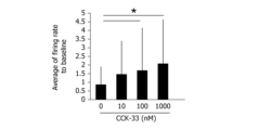
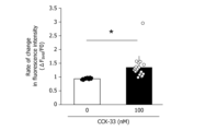
- FIG. 61 shows an extracellular potential heat map of cells prepared from hCCKAR-expressing iPS cells on day 27 of induction, with or without the addition of CCK-33.
- FIG. 62 is a graph showing the rate of change in firing frequency of cells prepared from hCCKAR-expressing iPS cells on day 27 of induction under conditions with or without the addition of CCK-33.
- FIG. 63 is a diagram showing the results of calcium imaging of cells prepared from hCCKAR-expressing iPS cells on day 40 of induction.
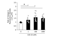
- FIG. 64 is a graph showing the rate of change in fluorescence intensity in calcium imaging of cells prepared from hCCKAR-expressing iPS cells on day 34 of induction under conditions with or without the addition of CCK-33.
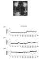
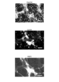
- Figure 65 shows a Hoechst staining image (top) of parasympathetic neurons in which the hGLP1R gene was forcibly expressed after differentiation (day 40 of induction), a fluorescent immunostaining image using an anti-ChAT antibody (center), and an image using an anti-GLP1R antibody. It is a figure showing a fluorescent immunostaining image (bottom).
- FIG. 66 shows the results of calcium imaging of parasympathetic nerve cells (30th day of induction) in which hGLP1R was forcibly expressed after differentiation.
- FIG. 67 shows a Hoechst staining image (top) of parasympathetic neurons in which the hNPY2R gene was forcibly expressed after differentiation (day 40 of induction), a fluorescent immunostaining image using an anti-ChAT antibody (center), and an image using an anti-NPY2R antibody. It is a figure showing a fluorescent immunostaining image (bottom).
- FIG. 67 shows a Hoechst staining image (top) of parasympathetic neurons in which the hNPY2R gene was forcibly expressed after differentiation (day 40 of induction), a fluorescent immunostaining image using an anti-ChAT antibody (center), and an image using an anti-NPY2R antibody. It is a figure showing a fluorescent immunostaining image (bottom).
- FIG. 69 is a graph showing the expression levels of hPRPH, hCHAT, and hGLP2R mRNA in parasympathetic neurons in which the hGLP2R gene was forcibly expressed after differentiation (40th day of induction).
- Figure 70 shows a Hoechst staining image (top) of parasympathetic neurons in which the hGLP2R gene was forcibly expressed after differentiation (day 40 of induction), a fluorescent immunostaining image using an anti-GLP2R antibody (center), and an anti-MAP2 antibody using an anti-MAP2 antibody. It is a figure showing a fluorescent immunostaining image (bottom).
- FIG. 71 is a diagram showing the results of calcium imaging of parasympathetic nerve cells in which hGLP2R was forcibly expressed after differentiation (37th day of induction).
- FIG. 72 is a graph showing the rate of change in firing intensity of parasympathetic neurons in which the hGLP2R gene was forcibly expressed after differentiation (37th day of induction) under conditions with and without the addition of GLP2.
- FIG. 73 is a graph showing the expression levels of hPRPH, hCHAT, and hHTR3A mRNA in parasympathetic neurons in which the hHTR3A gene was forcibly expressed after differentiation (40th day of induction).
- Figure 74 shows a Hoechst staining image (top) of parasympathetic neurons in which hHTR3A gene was forcibly expressed after differentiation (day 40 of induction), a fluorescent immunostaining image using anti-HTR3A antibody (center), and an anti-MAP2 antibody using anti-MAP2 antibody. It is a figure showing a fluorescent immunostaining image (bottom).
- FIG. 75 is a diagram showing the results of calcium imaging of parasympathetic neurons in which hHTR3A was forcibly expressed after differentiation (37th day of induction).
- FIG. 76 is a graph showing the rate of change in the firing intensity of parasympathetic neurons in which the hHTR3A gene was forcibly expressed after differentiation (37th day of induction) under conditions with or without the addition of 5-HT.
- FIG. 77 is a graph showing the expression levels of hPRPH, hCHAT, and hCCKAR mRNA in parasympathetic neurons in which the hCCKAR gene was forcibly expressed after differentiation (40th day of induction).
- Figure 78 shows a Hoechst staining image (top) of parasympathetic neurons in which the hCCKAR gene was forcibly expressed after differentiation (day 40 of induction), a fluorescent immunostaining image using an anti-CCKAR antibody (center), and an image using an anti-MAP2 antibody. It is a figure showing a fluorescent immunostaining image (bottom).
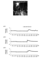
- FIG. 79 is a diagram showing the results of calcium imaging of parasympathetic neurons (37th day of induction) in which hCCKAR was forcibly expressed after differentiation.
- FIG. 80 is a graph showing the rate of change in firing intensity of parasympathetic neurons in which the hCCKAR gene was forcibly expressed after differentiation (37th day of induction) under conditions with or without the addition of CCK-33.
- FIG. 81 shows a fluorescent immunostaining image using an anti-MAP2 antibody (top), a MemGlow fluorescence image (center), and an overlapping image (bottom) of co-culture of hGLP1R-expressing parasympathetic neurons and NCI-H716 cells.
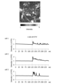
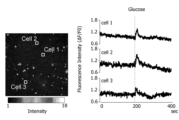
- FIG. FIG. 82 shows the results of calcium imaging of co-culture of hGLP1R-expressing parasympathetic neurons (induction day 38) and NCI-H716 cells supplemented with glucose.
- Bottom Time-dependent changes in fluorescence intensity (Intensity ( ⁇ F/F0)) of three hGLP1R-expressing parasympathetic neurons (cells 1 to 3).
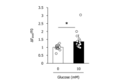
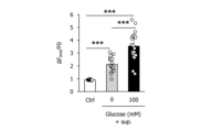
- FIG. 83 is a graph showing the rate of change in firing intensity of hGLP1R-expressing parasympathetic neurons in co-culture of hGLP1R-expressing parasympathetic neurons and NCI-H716 cells under conditions with or without glucose addition.
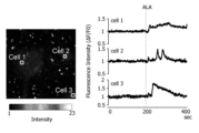
- FIG. 84 shows the results of calcium imaging of co-culture of hGLP1R-expressing parasympathetic neurons (induction day 44) and NCI-H716 cells supplemented with ALA.
- Bottom Changes in fluorescence intensity (Intensity ( ⁇ F/F0)) of three hGLP1R-expressing parasympathetic neurons (cells 1 to 3) over time.
- FIG. ⁇ F/F0 Changes in fluorescence intensity
- FIG. 85 is a graph showing the rate of change in the firing intensity of hGLP1R-expressing parasympathetic neurons in co-culture of hGLP1R-expressing parasympathetic neurons and NCI-H716 cells under conditions with or without the addition of ALA.
- FIG. 86 shows the results of calcium imaging of hGLP1R-expressing parasympathetic neurons (day 44 of induction) to which culture supernatant from glucose-loaded NCI-H716 cells was added.
- FIG. 87 is a graph showing the rate of change in firing intensity of hGLP1R-expressing parasympathetic neurons under conditions with and without addition of culture supernatant of NCI-H716 cells loaded with glucose.
- FIG. 88 shows the results of calcium imaging of hGLP1R-expressing parasympathetic neurons (34th day of induction) to which culture supernatant from NCI-H716 cells loaded with ALA was added.
- FIG. 89 is a graph showing the rate of change in firing intensity of hGLP1R-expressing parasympathetic neurons under conditions with and without addition of culture supernatant of NCI-H716 cells loaded with ALA or glucose.
- step (a) inducing differentiation of pluripotent stem cells into parasympathetic nerve cells, and (b1) prior to step (a), the pluripotent stem cells (b2) introducing an exogenous nucleic acid encoding a gastrointestinal hormone receptor into the parasympathetic neurons or their progenitor cells obtained in step (a);
- This is a method for producing vagus nerve-like parasympathetic nerve cells, including the step of introducing them.
- parasympathetic nerve cells are induced from pluripotent stem cells.
- pluripotent stem cells include, for example, embryonic stem (ES) cells, induced pluripotent stem (iPS) cells, embryonic germ (EG) cells, pluripotent germ stem (mGS) cells, Muse cells, etc. These include, but are not limited to: In the method of this embodiment, any pluripotent stem cell may be used, but it is preferable to use ES cells or iPS cells, and more preferably to use iPS cells.
- the pluripotent stem cells in this embodiment may be derived from any vertebrate, preferably from a mammal such as a mouse, rat, rabbit, sheep, goat, pig, cow, monkey, or human, Particularly preferred is human origin.
- pluripotent stem cells can be prepared from any tissue or cell according to methods known in the art.
- iPS cell lines or ES cell lines can be obtained from cell banks such as Kyoto University iPS Cell Research Foundation (CiRA_F), RIKEN BioResource Research Center (RIKEN BRC), American Type Culture Collection (ATCC), etc. You may.
- parasympathetic nerve cells can be prepared according to methods known in the art. can do. Specifically, for example, DMEM/HAM's F-12 medium, human stem cell (hES) medium, N2 medium, or a mixture thereof is used as a basal medium, and a BMP signal pathway inhibitor such as dorsomorphin, SB431542, etc.
- TGF signal pathway inhibitors such as CHIR99021, Wnt signal pathway activators such as CHIR99021, FGF signal pathway activators such as fibroblast growth factor (bFGF), and EGF signal pathway activators such as epidermal growth factor (EGF).
- Neural crest cells are prepared by culturing pluripotent stem cells under conditions in which agents are added as appropriate, and then cAMP production promoters such as forskolin and BDNF signals such as brain-derived neurotrophic factor (BDNF) are added.
- BDNF brain-derived neurotrophic factor
- GDNF signal pathway activators such as glial cell line-derived neurotrophic factor (GDNF), NGF signal pathway activators such as NGF- ⁇ , NT-3 signal pathway such as neurotrophin 3 Conditions in which an activator, vitamin C such as ascorbic acid, a protein kinase C activator such as phorbol 12-myristate 13-acetate (PMA), a retinoic acid receptor agonist such as retinoic acid, etc. are appropriately added.
- Parasympathetic nerve cells can be prepared by culturing neural crest cells.
- the pluripotent stem cells and neural crest cells are preferably preconditioned before the differentiation induction, and the pluripotent stem cells are placed in a medium containing a Rho kinase (ROCK) inhibitor such as Y-27632.
- a Rho kinase (ROCK) inhibitor such as Y-27632.
- the neural crest cells are cultured for 2 to 3 days, and the neural crest cells are treated with a BMP signal pathway activator such as BMP4, a SHH signal pathway inhibitor such as SANT-1, or a Wnt signal pathway inhibitor such as IWR-1. It is preferable to culture for 2 to 3 days in a medium containing the following (International Publication No. 2020/040286, International Publication No. 2016/194522).
- an exogenous nucleic acid encoding a gastrointestinal hormone receptor is introduced into cells.
- the "gastrointestinal hormone receptor” in this embodiment may be any gastrointestinal hormone receptor expressed in vagus nerve cells. Therefore, in the method of this embodiment, for example, glucagon-like peptide 1 receptor (GLP1R), neuropeptide Y2 type receptor (NPY2R), glucagon-like peptide 2 receptor (GLP2R), serotonin 3 receptor (5-HT3R)
- GLP1R glucagon-like peptide 1 receptor
- NPY2R neuropeptide Y2 type receptor
- GLP2R glucagon-like peptide 2 receptor
- serotonin 3 receptor 5-HT3R
- Exogenous nucleic acids can be used that encode gastrointestinal hormone receptors such as, but not limited to, and cholecystokinin 1 receptor (CCKAR).
- exogenous nucleic acids encoding one or more types of gastrointestinal hormone receptors selected from the above may be
- Exogenous nucleic acids encoding gastrointestinal hormone receptors may be introduced into cells at any time.
- an exogenous nucleic acid encoding a gastrointestinal hormone receptor may be introduced into pluripotent stem cells before induction of differentiation, into parasympathetic nerve cells obtained by induction of differentiation, or into parasympathetic nerve cells obtained by induction of differentiation. It may also be introduced into progenitor cells that are differentiating into cells.
- exogenous nucleic acids encoding GLP1R, NPY2R and CCKAR may be introduced into pluripotent stem cells prior to induction of differentiation;
- exogenous nucleic acids encoding GLP2R and 5-HT3R may be introduced into parasympathetic stem cells obtained by induction of differentiation. may be introduced into nerve cells.
- the exogenous nucleic acid encoding the gastrointestinal hormone receptor in this embodiment may be derived from any vertebrate, preferably mouse, rat, rabbit, sheep, goat, pig, cow, monkey, human, etc. of mammalian origin, particularly preferably human origin.
- NM_002062.5 for the human glucagon-like peptide 1 receptor (hGLP1R) gene
- NM_000910.4 for the human neuropeptide Y2 receptor (hNPY2R) gene
- NM_000910.4 for the human glucagon-like peptide 2 receptor (hGLP2R) gene.
- NM_004246.3, NM_213621.4 for the human serotonin 3 receptor (hHTR3A) gene, and NM_000730.3 for the human cholecystokinin 1 receptor (hCCKAR) gene are available. be.
- the gastrointestinal hormone receptors in this embodiment may also include variants and homologues thereof that have equivalent activity.
- the gastrointestinal hormone receptor in this embodiment has an amino acid sequence registered in the database, for example, 80% or more, preferably 90% or more, as long as the receptor activity is maintained. Proteins consisting of amino acid sequences having an identity of more preferably 95% or more, even more preferably 97% or more, even more preferably 98% or more, and most preferably 99% or more may be included. Amino acid sequence identity can be calculated using sequence analysis software or using programs commonly used in the art (FASTA, BLAST, etc.).
- the gastrointestinal hormone receptor in this embodiment may have one to several amino acids substituted or deleted in the amino acid sequence registered in the database, as long as the receptor activity is maintained. Proteins consisting of inserted and/or added amino acid sequences may be included.
- “1 to several” may be, for example, 1 to 30, preferably 1 to 10, particularly preferably 1 to 5.
- Exogenous nucleic acids encoding gastrointestinal hormone receptors can be introduced into cells by methods well known in the art, for example, by cloning those nucleic acids into expression vectors and introducing them into cells.
- expression vectors include, but are not limited to, viral vectors such as retrovirus, lentivirus, adenovirus, and Sendai virus, and plasmid vectors such as pCMV.
- the expression vector can be introduced into cells by methods well known in the art depending on the type.
- Non-viral vectors can be introduced, for example, by lipofection, electroporation, microinjection, and the like.
- Viral vectors can be introduced by infecting cells at an appropriate titer or multiplicity of infection (MOI).
- the present invention is a vagus nerve-like parasympathetic nerve cell produced by the above method.
- vagus nerve-like parasympathetic nerve cells refer to parasympathetic nerve cells that can mimic the gastrointestinal hormone response of vagus nerve cells.
- the vagus nerve-like parasympathetic nerve cells in this embodiment may be defined by any combination of well-known parasympathetic nerve cell markers, autonomic nerve cell markers, peripheral nerve cell markers, mature nerve cell markers, and gastrointestinal hormone receptors, e.g.
- ChAT choline acetyltransferase
- PHOX2B paired-like homeobox 2B
- PRPH peripherin
- MAP2 microtubule-associated protein 2
- VAChT also known as SLC18A3, vesicular acetylcholine transporter
- CHT also known as SLC5A7, high based on the expression of gastrointestinal hormone receptors such as the affinity choline transporter
- STMN2 stathmin 2
- PHOX2A paired-like homeobox 2A
- ASCL1 also known as MASH1, Achaete-scute homolog1 or as defined above. May be defined.
- vagus nerve-like parasympathetic nerve cells in this embodiment preferably express nicotinic acetylcholine receptors in addition to the above markers.
- Expression of markers and gastrointestinal hormone receptors can be analyzed by known techniques such as RT-PCR, Western blotting, and flow cytometry.
- vagus nerve-like parasympathetic nerve cells in this embodiment may be defined based on gastrointestinal hormone responsiveness, for example, by confirming reactivity to gastrointestinal hormones by calcium imaging or membrane potential measurement. .
- the present invention is a pluripotent stem cell comprising an exogenous nucleic acid encoding a gastrointestinal hormone receptor.
- the present invention is a vagus nerve-like parasympathetic nerve cell comprising an exogenous nucleic acid encoding a gastrointestinal hormone receptor.
- Exogenous nucleic acid encoding a gastrointestinal hormone receptor "pluripotent stem cell” and “vagus nerve-like parasympathetic nerve cell” in the third and fourth embodiments are defined in the first and second embodiments. It is similar to what was done.
- the vagus nerve-like parasympathetic nerve cells of the fourth embodiment may be produced by the method of the first embodiment, and the pluripotent stem cells of the third embodiment may be an intermediate product thereof.
- vagus nerve-like parasympathetic neurons of the second and fourth embodiments are useful for creating an in vitro evaluation model of brain-gut correlation. Furthermore, the pluripotent stem cells of the third embodiment are useful for stably producing vagus nerve-like parasympathetic nerve cells.
- the present invention includes (1) bringing a sample into contact with the vagus nerve-like parasympathetic nerve cells, and (2) analyzing the neural activity of the vagus nerve-like parasympathetic nerve cells.
- a method for detecting a gastrointestinal hormone receptor agonist in a sample comprising:
- the “sample” in this embodiment may be any material that can contain a gastrointestinal hormone receptor agonist.
- the "gastrointestinal hormone receptor agonist” in this embodiment may be any substance that acts on and activates the gastrointestinal hormone receptor defined in the first embodiment, and may be any substance that acts on and activates the gastrointestinal hormone receptor defined in the first embodiment. In addition, it may be a new or known low-molecular compound, nucleic acid, protein, peptide, antibody, lipid, etc.
- the gastrointestinal hormone receptor agonist in this embodiment may include not only a full agonist but also a partial agonist.
- the sample in the method of the present embodiment may be prepared from, for example, an animal-derived body fluid, an extract from a cell or tissue, a culture supernatant of a cell or tissue, a plant extract, or the like.
- the sample used in the method of the present embodiment is preferably prepared from body fluids such as human blood, plasma, and serum, or culture supernatants of human gastrointestinal tract-derived cells or intestinal epithelial tissue.
- the sample may be added to a culture of vagus nerve-like parasympathetic nerve cells and incubated for a certain period of time. Incubation time may be, for example, 0.01 seconds to 2 hours.
- Neural activity may be analyzed by methods well known in the art, for example, by calcium imaging or membrane potential measurement.
- calcium imaging for example, vagus nerve-like parasympathetic nerve cells may be loaded with a fluorescent calcium indicator, and changes in cellular calcium concentration may be measured based on changes in fluorescence intensity.
- Membrane potential can be analyzed, for example, by measuring extracellular potential by measuring local field potential, measuring intracellular potential by patch clamping, and measuring changes in membrane potential using a membrane potential sensitive dye.
- the present invention provides the steps of: (1) culturing the vagus nerve-like parasympathetic nerve cells in the presence of a candidate substance; and (2) culturing the neural activity of the vagus nerve-like parasympathetic nerve cells.
- This is a method of screening for substances that control brain-gut correlation, including the step of analyzing.
- Candidate substances may be ions, nanoparticles, low molecular weight compounds, nucleic acids, proteins, peptides, antibodies, lipids, animal or plant cells, tissues or extracts thereof, bacterial cells or crushed products or extracts thereof, etc. These candidate substances may be new or known.
- Vagus nerve-like parasympathetic nerve cells are cultured for a certain period of time in a medium supplemented with a candidate substance.
- concentration of the candidate substance to be added varies depending on the type of candidate substance, but for example, in the case of a low molecular weight compound, it can be appropriately selected within the range of 0.001 nM to 10 mM.
- the culture period may be, for example, 0.01 seconds to 2 hours.
- neural activity can be analyzed, for example, by calcium imaging or membrane potential measurement.
- calcium imaging for example, vagus nerve-like parasympathetic nerve cells may be loaded with a fluorescent calcium indicator, and changes in cellular calcium concentration may be measured based on changes in fluorescence intensity.
- Membrane potential can be analyzed, for example, by measuring extracellular potential by measuring local field potential, measuring intracellular potential by patch clamping, and measuring changes in membrane potential using a membrane potential sensitive dye.
- the neural activity of vagus nerve-like parasympathetic neurons in a medium supplemented with a candidate substance is significantly enhanced compared to that of vagus nerve-like parasympathetic nerve cells in a medium without a candidate substance.
- the candidate substance can be evaluated as a gastrointestinal hormone receptor agonist, that is, as a promising substance that controls the brain-gut relationship.
- brain-gut correlation refers to bidirectional communication between the central nervous system and the gastrointestinal tract.
- Brain-gut relationships are already known to be involved in a wide variety of symptoms or disorders.
- Symptoms or disorders that are closely related to brain-gut correlation abnormalities include, for example, abnormal eating behavior; gastrointestinal disorders such as nausea, abdominal pain, diarrhea, and constipation; psychological stress symptoms such as anxiety behavior and depression; Alzheimer's disease and Parkinson's disease.
- disease neurodegenerative diseases such as multiple sclerosis; abnormal immune responses such as inflammation and allergies; and metabolic diseases such as obesity, dyslipidemia, and diabetes, but are not limited to these.
- a substance that regulates the brain-gut correlation may be determined to be capable of normalizing the brain-gut correlation and, as a result, prevent or treat the symptoms or disorders described above, and may be considered capable of preventing or treating the symptoms or disorders described above. It may be useful for the development of preventive or therapeutic drugs, supplements, foods for specified health uses, and foods with functional claims.
- a seventh embodiment of the present invention (1) culturing gastrointestinal hormone-secreting cells in the presence of a candidate substance; (2) a culture supernatant from the culture obtained in step (1); (3) bringing the culture supernatant into contact with the vagus nerve-like parasympathetic nerve cells; and (4) analyzing the neural activity of the vagus nerve-like parasympathetic nerve cells.
- This is a screening method for substances that control
- the "vagus nerve-like parasympathetic nerve cells" in this embodiment are the same as those defined in the first and second embodiments, and the "candidate substances" and "brain-gut correlation” are defined in the sixth embodiment. It is similar to what was done.
- the "gastrointestinal hormone-secreting cell” in this embodiment may be any cell derived from any tissue as long as it can secrete a gastrointestinal hormone.
- Gastrointestinal hormone-secreting cells are scattered in the gastrointestinal epithelium from the stomach to the rectum and in the islets of Langerhans of the pancreas, and are classified into L cells, K cells, I cells, S cells, It is classified into enterochromaffin (EC) cells, G cells, D cells, N cells, M cells, etc.
- the gastrointestinal hormone-secreting cells selected from the above can be used alone or in an appropriate combination of two or more types.
- the gastrointestinal hormone-secreting cells in this embodiment are preferably enteroendocrine cells, and may be, for example, L cells, K cells, I cells, S cells, EC cells, N cells, M cells, etc. but not limited to.
- the gastrointestinal hormone-secreting cells in this embodiment may be derived from any vertebrate, preferably from a mammal such as a mouse, rat, rabbit, sheep, goat, pig, cow, monkey, or human. , particularly preferably of human origin.
- the gastrointestinal hormone-secreting cells in the method of this embodiment may be derived from the same animal as the vagus nerve-like parasympathetic nerve cells, or may be derived from a different animal. Therefore, in the method of this embodiment, for example, human gastrointestinal hormone secreting cells and human vagus nerve-like parasympathetic nerve cells may be used in combination, or mouse gastrointestinal hormone secreting cells and human vagus nerve-like parasympathetic nerve cells may be used in combination. It may also be used.
- gastrointestinal hormone secreting cells and vagus nerve-like parasympathetic nerve cells derived from the same species of mammal can be used, particularly preferably human gastrointestinal hormone secreting cells and human vagus nerve-like parasympathetic nerve cells. Nerve cells can be used.
- Gastrointestinal hormone-secreting cells can be prepared by already established and known methods (for example, Sinagoga K.L. et al., Development, 2018, 145(19): dev165795). Specifically, for example, in the process of inducing intestinal organoids and colon organoids from human pluripotent stem cells, by forcibly expressing the transcription factor NEUROG3, gastrointestinal hormones such as GIP, ghrelin, GLP-1, and oxyntomodulin can be expressed. Enteroendocrine cells that secrete can be prepared. Alternatively, an already established gastrointestinal hormone-secreting cell line may be obtained from a cell bank such as ATCC. Examples of gastrointestinal hormone-secreting cell lines include human L cell line NCI-H716, mouse L cell line STC-1, mouse L cell line GLUTag, and enteroendocrine cells collected from mouse and human gastrointestinal tracts.
- Gastrointestinal hormone-secreting cells are grown in a basal medium such as DMEM, HAM's F-12 medium, RPMI1640 medium, or a mixture thereof, supplemented with fetal bovine serum (FBS), non-essential amino acid solution, penicillin/streptomycin, etc. can be used for stable subculture.
- a basal medium such as DMEM, HAM's F-12 medium, RPMI1640 medium, or a mixture thereof, supplemented with fetal bovine serum (FBS), non-essential amino acid solution, penicillin/streptomycin, etc.
- Gastrointestinal hormone-secreting cells are cultured for a certain period of time in a medium supplemented with candidate substances.
- concentration of the candidate substance to be added varies depending on the type of candidate substance, but for example, in the case of a low molecular weight compound, it can be appropriately selected within the range of 0.001 nM to 10 mM.
- the addition period may be, for example, from 1 second to 72 hours.
- the culture supernatant is collected from the above culture.
- the collected culture supernatant may optionally be purified or concentrated by dialysis, ultrafiltration, or the like.
- the above culture supernatant is brought into contact with the vagus nerve-like parasympathetic nerve cells.
- the culture supernatant may be added to the culture medium of the vagus nerve-like parasympathetic nerve cells and incubated for a certain period of time.
- the culture supernatant may be added in an amount of 1 to 10 times the amount of the culture medium for vagus nerve-like parasympathetic nerve cells. Incubation time may be, for example, 0.01 seconds to 2 hours.
- neural activity can be analyzed, for example, by calcium imaging or membrane potential measurement.
- calcium imaging for example, vagus nerve-like parasympathetic nerve cells may be loaded with a fluorescent calcium indicator, and changes in cellular calcium concentration may be measured based on changes in fluorescence intensity.
- Membrane potential can be analyzed, for example, by measuring extracellular potential by measuring local field potential, measuring intracellular potential by patch clamping, and measuring changes in membrane potential using a membrane potential sensitive dye. The method of this embodiment can be carried out using a culture supernatant of gastrointestinal hormone-secreting cells prepared without adding a candidate substance as a control.
- the neural activity of vagus nerve-like parasympathetic neurons that came into contact with the culture supernatant of gastrointestinal hormone-secreting cells prepared by adding a candidate substance was compared with the neural activity of vagus nerve-like parasympathetic nerve cells that came into contact with the control culture supernatant. If the candidate substance is significantly enhanced in comparison, the candidate substance is evaluated to be capable of promoting gastrointestinal hormone secretion from gastrointestinal hormone-secreting cells, that is, to be promising as a substance that controls the brain-gut relationship. Can be done.
- the present invention provides the steps of: (1) co-culturing the gastrointestinal hormone secreting cells and the vagus nerve-like parasympathetic nerve cells in the presence of a candidate substance, and (2) the vagus nerve-like parasympathetic nerve cells.
- This is a method of screening for substances that control the brain-gut correlation, including the step of analyzing the neural activity of parasympathetic nerve cells.
- the "candidate substance” and “brain-gut correlation” in this embodiment are the same as those defined in the sixth and seventh embodiments.
- "Gastrointestinal hormone-secreting cells” are the same as those defined in the seventh embodiment.
- gastrointestinal hormone secreting cells and vagus nerve-like parasympathetic nerve cells are co-cultured.
- "Co-culture” in this embodiment may be any type of culture as long as both cells share the same medium. Therefore, the co-culture in this embodiment may be either direct (contact) co-culture or indirect (non-contact) co-culture.
- gastrointestinal hormone-secreting cells and vagus nerve-like parasympathetic nerve cells may be directly mixed and co-cultured, or gastrointestinal hormone-secreting cells and vagus nerve-like parasympathetic nerve cells may be cultured using a filter or the like. They may be separated and co-cultured.
- gastrointestinal hormone-secreting cells may be seeded onto the culture of vagus nerve-like parasympathetic nerve cells.
- the ratio of gastrointestinal hormone-secreting cells to vagus nerve-like parasympathetic nerve cells may be, for example, 1:3 to 3:1, preferably 1:1 to 2:1.
- the seeding concentrations of gastrointestinal hormone-secreting cells and vagus nerve-like parasympathetic nerve cells range from 1 ⁇ 10 5 to 1 ⁇ 10 6 cells/cm 2 and from 1 ⁇ 10 5 to 5 ⁇ 10 5 cells/cm 2 , respectively. good.
- the medium used here may be a mixture of a medium for culturing gastrointestinal hormone secreting cells and a medium for culturing vagus nerve-like parasympathetic nerve cells.
- it may be a mixed medium of DMEM, HAM's F-12 medium and RPMI1640 medium to which fetal bovine serum (FBS), non-essential amino acid solution, N2 supplement, B-27 supplement, etc. are added as appropriate.
- FBS fetal bovine serum
- Gastrointestinal hormone secreting cells and vagus nerve-like parasympathetic nerve cells are cultured for a certain period of time in a medium supplemented with a candidate substance.
- concentration of the candidate substance to be added varies depending on the type of candidate substance, but for example, in the case of a low molecular weight compound, it can be appropriately selected within the range of 0.001 nM to 10 mM.
- the culture period may be, for example, 0.01 seconds to 2 hours.
- neural activity can be analyzed, for example, by calcium imaging or membrane potential measurement.
- co-culture in a medium without a candidate substance and/or single culture of vagus nerve-like parasympathetic neurons in a medium to which a candidate substance is added can be carried out as a control. If the neural activity of vagus nerve-like parasympathetic neurons in the co-culture with the addition of a candidate substance is significantly enhanced compared to the neural activity of vagus nerve-like parasympathetic nerve cells in the control, then the candidate substance is effective in the gastrointestinal tract. It can be evaluated as a promising substance that can promote gastrointestinal hormone secretion from hormone-secreting cells, that is, as a substance that controls the brain-intestine relationship.
- the present invention is a brain-gut correlation model comprising co-culture of gastrointestinal hormone-secreting cells and the vagus nerve-like parasympathetic nerve cells.
- the "vagus nerve-like parasympathetic nerve cells” in this embodiment are the same as those defined in the first and second embodiments, and the "gastrointestinal hormone secreting cells” and “brain-gut correlation” are defined in the seventh embodiment.
- “Co-culture” is the same as defined in the eighth embodiment.
- the brain-gut correlation model of this embodiment may be provided as a kit for screening substances that control the brain-gut correlation in combination with the above-described medium, co-culture container, and the like.
- the kit may further include additional components as appropriate, such as a buffer, a calcium indicator, a compound library, an instruction manual describing a screening protocol, and the like.
- the brain-gut correlation model of this embodiment can be created by co-culturing gastrointestinal hormone secreting cells and vagus nerve-like parasympathetic neurons according to the procedure described in the eighth embodiment, and It can be used to screen for substances that can promote gastrointestinal hormone secretion from cells, ie, substances that control brain-gut correlation.
- the present invention is also the use of the vagus nerve-like parasympathetic nerve cells or the brain-gut correlation model for screening for substances that control the brain-gut correlation.
- the "vagus nerve-like parasympathetic nerve cells" in this embodiment are the same as those defined in the first and second embodiments, and the "brain-gut correlation” are the same as those defined in the seventh embodiment.
- the "brain-gut correlation model” is the same as that defined in the ninth embodiment. According to the content described in the seventh, eighth, and ninth embodiments, the vagus nerve-like parasympathetic nerve cells or the brain-gut correlation model may be used for screening for substances that control the brain-gut correlation. can.
- VectorBuilder was commissioned to synthesize the following plasmids containing sequences encoding gastrointestinal hormone receptors: pLV-hGLP1R (plasmid containing human GLP1R gene (NCBI RefSeq ID: NM_002062.5), vector ID: VB900123-1388xwv); pLV-hNPY2R (plasmid containing human NPY2R gene (NCBI RefSeq ID: NM_000910.4), vector ID: VB900123-1387bah); pLV-hGLP2R (plasmid containing human GLP2R gene (NCBI RefSeq ID: NM_004246.3), vector ID: VB210830-1115xup; pLV-hHTR3A (plasmid containing human HTR3A gene (NCBI RefSeq ID: NM_213621.4, vector ID: VB210823-1277qqk
- pLV-mCherry vector ID: VB900084-0158zxv
- HEK293T cells (1.5 ⁇ 10 6 /well) were seeded in 6-well plates coated with 0.1 w/v% gelatin.
- the medium used was DMEM containing 10% FBS, 1% non-essential amino acids (NEAA), and 1% penicillin-streptomycin.
- the above plasmids (4 ⁇ g each), package plasmid psPAX2 (addgene, #12260) (2 ⁇ g) and envelope plasmid (pMD2.G) (addgene, #12259) (2 ⁇ g) were added to 500 ⁇ l of Opti-MEM (Thermo Fisher Scientific) and polyethylene.
- HEK293T cells (2 wells) were transfected using imine (Polysciences inc.).
- mTeSR1-cGMP ST-85850G
- mTeSR1 ⁇ DMEM/Ham's F-12 (Fujifilm Wako Pure Chemical: 048-29785)
- DMEM high-glucose
- RPMI-1640 contains L-glutamine and phenol red
- RPMI-1640 ⁇ Opti-MEM (Thermo Fisher Scientific: 31985062)
- STEM-CELLBANKER (trademark) GMP grade) ZNQ: CB045)
- FBS Fretal Bovine Serum Replacement
- DMEM/Ham's F-12 supplemented with 20% KSR, 1% NEAA, 1% Monothioglycerol solution, and 1% P/S -N2 medium: DMEM/Ham's F-12 supplemented with 1% N2, 1% NEAA, and 1% P/S ⁇ NDM1: N2 medium supplemented with 10 ⁇ M FSK, 10 ng/mL GDNF, 10 ng/mL NT-3, 10 ng/mL NGF, and 50 ⁇ g/mL ascorbic acid ⁇ NDM2: 100 ng/mL BDNF, and NDM1 supplemented with 100 ng/mL CNTF ⁇ NDM3: NDM1 supplemented with 10 ng/mL BDNF, 1 ⁇ M RA, and 10 ng/mL PMA
- Human iPS cells (201B7 strain) were obtained from RIKEN BRC. Six-well plates were coated with Lipidure and washed with PBS. Human iPS cells (201B7 strain) were seeded at a concentration of 1 x 10 cells/well using mTeSR1 supplemented with 10 ⁇ M Y-27632, and cultured on an orbital shaker (Wakenbee Tech: WB-101SRC). (95 rpm). 24 hours and 48 hours after seeding, mTeSR1 supplemented with 10 ⁇ M Y-27632 was added at 2 mL/well.
- ⁇ Differentiation step 1 day 0: The medium was replaced with hESM supplemented with 2 ⁇ M DM, 10 ⁇ M SB, and 10 ng/ml bFGF (4 mL/well) and cultured on an orbital shaker for 2 days.
- ⁇ Differentiation step 2 (2nd day): The medium was replaced with hESM supplemented with 3 ⁇ M CHIR, 20 ⁇ M SB, and 10 ng/ml bFGF (6 mL/well) and cultured on an orbital shaker for 3 days.
- ⁇ Differentiation step 3 The medium was replaced with hESM:N2 (3:1) mixed medium supplemented with 3 ⁇ M CHIR and 10 ng/ml bFGF (4 mL/well), and cultured on an orbital shaker for 2 days.
- ⁇ Differentiation step 4 (7th day): The medium was replaced with hESM:N2 (1:1) mixed medium supplemented with 10 ⁇ M IWR-1, 250 nM SANT-1, 25 ng/ml BMP4, and 10 ng/ml bFGF (3 mL/well), Culture was performed on an orbital shaker for 2 days.
- ⁇ Differentiation step 5 (9th day): The medium was replaced with hESM:N2 (1:3) mixed medium supplemented with 10 ⁇ M IWR-1, 250 nM SANT-1, 25 ng/ml BMP4, and 10 ng/ml bFGF (3 mL/well), The cells were cultured on an orbital shaker for 3 days, replaced with the same fresh medium, and further cultured for 24 hours.
- ⁇ Differentiation step 6 (13th day): The cells were coated with PLO and laminin by coating with a PLO solution (20 ⁇ g/ml) prepared in DW for 1 hour, washing once with DW, and coating with a laminin solution (5 ⁇ g/ml) prepared in PBS for 2 hours.
- a 24-well plate was prepared.
- the EB obtained in differentiation step 5 was collected, 2 mL of TrypLE express was added, and the mixture was incubated at 37°C for 10 minutes.
- the enzyme reaction was stopped by adding DMEM (4 mL) containing 10% FBS, and the cells were suspended by pipetting.
- the cells were collected by centrifugation at 200 ⁇ g for 4 minutes, suspended in NDM1, and the number of cells was counted.
- a cell suspension was prepared using NDM2 (1 ⁇ 10 5 cells/mL) and added at 1 mL/well to a 24-well plate coated with PLO and laminin. Thereafter, the culture was continued while half of the medium was replaced with fresh NDM2 twice a week.
- Puromycin selection was performed by culturing cells in mTeSR1 supplemented with 0.3 ⁇ g/mL puromycin for 10 days or more after virus addition.
- the obtained cells (hereinafter referred to as "iPS cells expressing gastrointestinal hormone receptors") were cryopreserved.
- Microscope (Olympus, IX71), Confocal scanner unit (Yokogawa, CSU-W1), LED light source (OPTO SCIENCE, X-Cite 120LED), and analysis software (MetaMorph) (Molecular Devices) 500-800 at 500ms/frame I took a frame photo. Gastrointestinal hormone receptor agonist stimulation was added midway (for example, at the 250th frame).
- GLP1 human, 7-37
- PYY human, 3-36
- Peptide Institute, #4400-v PYY
- GLP2 human
- GLP2 Serotonin
- 5-HT Serotonin hydrochloride
- CCK-33 human) (peptide research #4201-s) (hereinafter referred to as "CCK-33") (10, 100, 1000 nM).
- Membrane potential changes were measured in NDM3 or Ringer's solution. Using a 1000 ⁇ gain and a 200 Hz-3 kHz filter, potential values that changed six times or more relative to the measurement noise standard deviation were detected as nerve firings (spikes). After measuring the potential for 5 minutes, a gastrointestinal hormone receptor agonist was added at the same final concentration as in (4-2) above, and measurement was continued for an additional 5 minutes.
- hGLP1R-expressing iPS cells A bright field image of hGLP1R-expressing iPS cells is shown in FIG. 3. In the figure, the scale bar indicates 100 ⁇ m.
- Figure 4 shows the results of confirming the expression of the stem cell marker hNANOG gene in hGLP1R-expressing iPS cells by qPCR according to the procedure (3-1) above.
- GLP1R indicates hGLP1R-expressing iPS cells
- Control indicates wild-type iPS cells.
- Figure 6 shows the results of chromosome testing using the G differential staining method. It was confirmed that there were no abnormalities in the chromosomes of hGLP1R-expressing iPS cells.
- the ligand responsiveness of cells prepared from hGLP1R-expressing iPS cells according to the procedure (2-7) above was analyzed by measuring extracellular potential according to the procedure (4-3) above.
- the extracellular potential heat map is shown in FIG. 11.
- the ligand responsiveness of cells prepared from hGLP1R-expressing iPS cells according to the procedure (2-7) above was analyzed by intracellular calcium imaging according to the procedure (4-2) above.
- the measurement results are shown in FIG.
- the horizontal axis shows time (seconds), and the vertical axis shows the rate of change in fluorescence intensity ( ⁇ F/F0) when the average value (F0) of fluorescence intensity before addition of GLP1 is set to 1.
- Addition of GLP1 increased intracellular Ca 2+ concentration.
- the number of neural firings (spikes) per minute is shown in FIG.
- hNPY2R-expressing iPS cells A bright field image of hNPY2R-expressing iPS cells is shown in FIG. 18. In the figure, the scale bar indicates 100 ⁇ m.
- FIG. 19 shows the results of confirming the expression of the stem cell marker NANOG in hNPY2R-expressing iPS cells by qPCR according to the procedure (3-1) above.
- NPY2R indicates hNPY2R-expressing iPS cells
- Control indicates wild-type iPS cells.
- hNPY2R-expressing iPS cells According to the procedure (3-2) above, the ability of hNPY2R-expressing iPS cells to differentiate into three embryonic forms was evaluated. The results are shown in FIG. The hNPY2R-expressing iPS cells induced to differentiate into ectoderm, mesoderm, or endoderm strongly expressed the ectoderm marker hPAX6 gene, mesoderm marker hNCAM1 gene, or endoderm marker hSOX17 gene, respectively. From these results, it was confirmed that the hNPY2R-expressing iPS cells had the ability to differentiate into three embryo-like cells.
- Figure 21 shows the results of chromosome testing using the G differential staining method. It was confirmed that there were no abnormalities in the chromosomes of hNPY2R-expressing iPS cells.
- the ligand responsiveness of cells prepared from hNPY2R-expressing iPS cells according to the procedure (2-7) above was analyzed by measuring extracellular potential according to the procedure (4-3) above.
- the extracellular potential heat map is shown in FIG. 26.
- the measurement results are shown in FIG.
- the horizontal axis shows time (seconds), and the vertical axis shows the rate of change in fluorescence intensity ( ⁇ F/F0) when the average value (F0) of fluorescence intensity before addition of PYY (0 to 125 seconds) is set to 1.
- Addition of PYY increased intracellular Ca 2+ concentration.
- the number of neural firings (spikes) per minute is shown in FIG. Addition of PYY increased the number of spikes (*P ⁇ 0.05, n 15 cells/3 samples, student-t).
- hGLP2R-expressing iPS cells A bright field image of hGLP2R-expressing iPS cells is shown in FIG. 33. In the figure, the scale bar indicates 1 mm.
- FIG. 34 shows the results of confirming the expression of the stem cell marker hNANOG gene in hGLP2R-expressing iPS cells by qPCR according to the procedure (3-1) above.
- hGLP2R indicates hGLP2R-expressing iPS cells
- Control indicates wild-type iPS cells. It was confirmed that hGLP2R-expressing iPS cells expressed the hNANOG gene to the same extent as wild-type iPS cells and were in an undifferentiated state.
- the ability of hGLP2R-expressing iPS cells to differentiate into three embryo-like cells was evaluated.
- the results are shown in FIG.
- the hGLP2R-expressing iPS cells induced to differentiate into ectoderm, mesoderm, or endoderm strongly expressed the ectoderm marker hNES gene, mesoderm marker hNCAM1 gene, or endoderm marker hSOX17 gene, respectively. From these results, it was confirmed that the hGLP2R-expressing iPS cells had the ability to differentiate into three embryo-like cells.
- Figure 36 shows the results of chromosome testing using the G differential staining method. It was confirmed that there were no abnormalities in the chromosomes of hGLP2R-expressing iPS cells.
- FIG. 40 The results of immune cell staining are shown in Figure 40.
- the scale bar indicates 100 ⁇ m.
- hHTR3A-expressing iPS cells A bright field image of hHTR3A-expressing iPS cells is shown in FIG. 43. In the figure, the scale bar indicates 1 mm.
- FIG. 44 shows the results of confirming the expression of the stem cell marker hNANOG gene in hHTR3A-expressing iPS cells by qPCR according to the procedure (3-1) above.
- hHTR3A indicates hHTR3A-expressing iPS cells
- Control indicates wild-type iPS cells. It was confirmed that hHTR3A-expressing iPS cells expressed the hNANOG gene to the same extent as wild-type iPS cells and were in an undifferentiated state.
- FIG. 50 The results of immune cell staining are shown in Figure 50.
- the scale bar indicates 100 ⁇ m. Expression of hHTR3A protein and mature neuron marker MAP2 protein was confirmed.
- hCCKAR-expressing iPS cells A bright field image of hCCKAR-expressing iPS cells is shown in FIG. 53. In the figure, the scale bar indicates 1 mm.
- FIG. 54 shows the results of confirming the expression of the stem cell marker hNANOG gene in hCCKAR-expressing iPS cells by qPCR according to the procedure (3-1) above.
- hCCKAR indicates hCCKAR-expressing iPS cells
- Control indicates wild-type iPS cells. It was confirmed that hCCKAR-expressing iPS cells expressed the hNANOG gene to the same extent as wild-type iPS cells and were in an undifferentiated state.
- the ability of hCCKAR-expressing iPS cells to differentiate into three embryo-like cells was evaluated by the procedure described in (3-2) above. The results are shown in FIG.
- the hCCKAR-expressing iPS cells induced to differentiate into ectoderm, mesoderm, or endoderm strongly expressed the ectoderm marker hNES gene, mesoderm marker hNCAM1 gene, or endoderm marker hSOX17 gene, respectively. From these results, it was confirmed that the hCCKAR-expressing iPS cells had the ability to differentiate into three embryo-like cells.
- Figure 56 shows the results of chromosome testing using the G differential staining method. It was confirmed that there were no abnormalities in the chromosomes of hCCKAR-expressing iPS cells.
- FIG. 60 The results of immune cell staining are shown in Figure 60.
- the scale bar indicates 100 ⁇ m. Expression of hCCKAR protein and mature neuron marker MAP2 protein was confirmed.
- the ligand responsiveness of cells prepared from hCCKAR-expressing iPS cells according to the procedure (2-7) above was analyzed by intracellular calcium imaging according to the procedure (4-2) above.
- the measurement results are shown in FIG. 63.
- the horizontal axis shows time (seconds), and the vertical axis shows the rate of change in fluorescence intensity ( ⁇ F/F0) when the average value (F0) of fluorescence intensity before addition of CCK-33 (0 to 199 seconds) is set to 1. show. Addition of CCK-33 increased intracellular Ca 2+ concentration.
- step 1 Analysis of parasympathetic neurons forced to express hGLP1R after differentiation> Using pLV-hGLP1R as the lentiviral vector in step 1 above, expression of hGLP1R protein in parasympathetic nerve cells prepared by step (2-5) above was analyzed by step (3-1) above. The results of immune cell staining on day 40 of induction are shown in FIG. 65. In the figure, the scale bar indicates 100 ⁇ m. Strong expression of parasympathetic nerve cell markers hChAT protein and hGLP1R protein was confirmed.
- the ligand responsiveness of parasympathetic nerve cells in which hGLP1R protein was forced to be expressed after differentiation according to the procedure (2-7) above was analyzed by intracellular calcium imaging according to the procedure (4-2) above.
- the measurement results are shown in FIG.
- the horizontal axis shows time (seconds), and the vertical axis shows the rate of change in fluorescence intensity ( ⁇ F/F0) when the average value (F0) of fluorescence intensity before the addition of GLP1 (0 to 124 seconds) is set to 1. Addition of GLP1 increased intracellular Ca 2+ concentration.
- step 1 Analysis of parasympathetic neurons forced to express hNPY2R after differentiation> Using pLV-hNPY2R as the lentiviral vector in step 1 above, expression of hNPY2R protein in parasympathetic nerve cells prepared by step (2-5) above was analyzed by step (3-1) above. The results of immune cell staining on day 40 of induction are shown in FIG. 67. In the figure, the scale bar indicates 100 ⁇ m. Strong expression of parasympathetic nerve cell markers hChAT protein and hNPY2R protein was confirmed.
- the ligand responsiveness of parasympathetic nerve cells in which hNPY2R protein was forced to be expressed after differentiation according to the procedure (2-7) above was analyzed by intracellular calcium imaging according to the procedure (4-2) above.
- the measurement results are shown in FIG.
- the horizontal axis shows time (seconds), and the vertical axis shows the rate of change in fluorescence intensity ( ⁇ F/F0) when the average value (F0) of fluorescence intensity before addition of PYY (0 to 124 seconds) is set to 1. Addition of PYY increased intracellular Ca 2+ concentration.
- FIG. 70 The results of immune cell staining on day 40 of induction are shown in Figure 70.
- the scale bar indicates 100 ⁇ m. Strong expression of mature neuron markers MAP2 protein and hGLP2R protein was confirmed.
- the ligand responsiveness of parasympathetic nerve cells in which the hGLP2R protein was forcibly expressed after differentiation according to the procedure (2-7) above was analyzed by intracellular calcium imaging according to the procedure (4-2) above.
- the measurement results are shown in FIG.
- the horizontal axis shows time (seconds), and the vertical axis shows the rate of change in fluorescence intensity ( ⁇ F/F0) when the average value (F0) of fluorescence intensity before the addition of GLP2 (0 to 80 seconds) is set to 1. Addition of GLP2 increased intracellular Ca 2+ concentration.
- the ligand responsiveness of parasympathetic nerve cells in which hHTR3A protein was forced to be expressed after differentiation according to the procedure (2-7) above was analyzed by intracellular calcium imaging according to the procedure (4-2) above.
- the measurement results are shown in FIG. 75.
- the horizontal axis shows time (seconds), and the vertical axis shows the rate of change in fluorescence intensity ( ⁇ F/F0) when the average value (F0) of fluorescence intensity before 5-HT addition (0 to 80 seconds) is set to 1. show. Addition of 5-HT increased intracellular Ca 2+ concentration.
- Rate of change ( ⁇ F ave /F0) in the average value of fluorescence intensity ( ⁇ F ave ) after addition of 5-HT (240 to 400 seconds) to the average value (F0) of fluorescence intensity before addition of 5-HT (0 to 80 seconds) ) is shown in FIG.
- Figure 78 shows the results of immune cell staining on day 40 of induction.
- the scale bar indicates 100 ⁇ m. Strong expression of mature neuron markers MAP2 protein and hCCKAR protein was confirmed.
- the ligand responsiveness of parasympathetic nerve cells in which the hCCKAR protein was forcibly expressed after differentiation according to the procedure (2-7) above was analyzed by intracellular calcium imaging according to the procedure (4-2) above.
- the measurement results are shown in FIG.
- the horizontal axis shows time (seconds), and the vertical axis shows the rate of change in fluorescence intensity ( ⁇ F/F0) when the average value (F0) of fluorescence intensity before addition of CCK-33 (0 to 80 seconds) is set to 1. show. Addition of CCK-33 increased intracellular Ca 2+ concentration.
- H716 medium Human L cell-derived cell line NCI-H716 (ATCC: CCL-251) was grown in a 10 cm dish using RPMI-1640 (hereinafter referred to as "H716 medium") containing 10% FBS and 1% P/S. The cells were seeded (1-2 ⁇ 10 6 cells/dish) and cultured for 3-4 days. The cells and medium were collected into a 15 mL centrifuge tube, centrifuged at 200 xg for 5 minutes, and the supernatant was removed.
- FBS-free H716 medium or Ringer's solution was added to the cells to adjust to 1 ⁇ 10 6 cells/mL.
- 5 ⁇ L of 20 ⁇ M MemGlow/DMSO solution was added to 1 mL of the obtained cell suspension (final concentration 100 nM), and the mixture was incubated at room temperature for 10 minutes. Thereafter, the mixture was centrifuged at 200 ⁇ g for 5 minutes, and the supernatant was removed. After washing once with H716 medium, the cells were seeded in a 10 cm dish. It was confirmed that cell membrane staining with MemGlow was maintained for up to 10 days.
- NCI-H716 cells stained with MemGlow will be referred to as "MemGlow-stained NCI-H716.”
- hGLP1R-expressing parasympathetic neurons prepared from hGLP1R-expressing iPS cells by the procedure (2-7) above (37th day of induction) were injected with MemGlow-stained NCI-H716 (2 ⁇ 1 mL of 10 5 cells/mL) was added. As a result, co-culture was carried out in a 1:1 mixed medium of NDM2 and H716 medium. After 48 hours of culture, immune cell staining was performed using the same procedure as (3-1) (ii) above. Anti-MAP2 antibody (Abcam, ab11267) (1:1000 dilution) was used as the primary antibody.
- FIG. The horizontal axis shows time (seconds), and the vertical axis shows the rate of change in fluorescence intensity ( ⁇ F/F0) when the average value (F0) of fluorescence intensity before glucose addition (0 to 199 seconds) is set to 1.
- Addition of glucose to the co-culture increased the intracellular Ca 2+ concentration of hGLP1R-expressing parasympathetic neurons.
- Figure 83 shows the rate of change ( ⁇ F ave /F0) of the average fluorescence intensity ( ⁇ F ave ) after glucose addition (200 to 400 seconds) to the average fluorescence intensity (F0) before glucose addition (0 to 199 seconds). Shown below.
- ⁇ -linolenic acid Fujifilm Wako Pure Chemicals: 127-05901
- ALA ⁇ -linolenic acid
- Glucose-free Ringer's solution was used as the solution.
- FIG. The results are shown in FIG.
- the horizontal axis shows time (seconds), and the vertical axis shows the rate of change in fluorescence intensity ( ⁇ F/F0) when the average value (F0) of fluorescence intensity before addition of ALA (0 to 199 seconds) is set to 1.
- Addition of ALA to co-culture increased the intracellular Ca 2+ concentration of hGLP1R-expressing parasympathetic neurons.
- Figure 85 shows the rate of change ( ⁇ F ave /F0) in the average value of fluorescence intensity ( ⁇ F ave ) after addition of ALA (200 to 400 seconds) to the average value (F0) of fluorescence intensity before addition of ALA (0 to 199 seconds). Shown below.
- glucose-loaded supernatant the supernatant prepared with Ringer's solution containing glucose
- ALA-loaded supernatant the supernatant prepared with Ringer's solution containing ALA
- the results of intracellular calcium imaging using the glucose-loaded supernatant are shown in FIG. 86.
- the horizontal axis shows time (seconds), and the vertical axis shows the rate of change in fluorescence intensity ( ⁇ F/F0) when the average value (F0) of fluorescence intensity before addition of the supernatant (0 to 199 seconds) is set to 1.
- Addition of the glucose-loaded supernatant increased the intracellular Ca 2+ concentration in hGLP1R-expressing parasympathetic neurons (44th day of induction).
- the rate of change ( ⁇ F ave /F0) in the average fluorescence intensity ( ⁇ F ave ) after adding the supernatant (200 to 400 seconds) to the average fluorescence intensity (F0) before adding the supernatant (0 to 199 seconds) is Shown in FIG.
- "Ctrl” indicates Ringer's solution that has not come into contact with NCI-H716 cells.
- the results of intracellular calcium imaging using ALA-loaded supernatant are shown in FIG. 88.
- the horizontal axis shows time (seconds), and the vertical axis shows the rate of change in fluorescence intensity ( ⁇ F/F0) when the average value (F0) of fluorescence intensity before addition of the supernatant (0 to 199 seconds) is set to 1. .
- the intracellular Ca 2+ concentration of hGLP1R-expressing parasympathetic neurons 34th day of induction
- the rate of change ( ⁇ F ave /F0) in the average fluorescence intensity ( ⁇ F ave ) after adding the supernatant (200 to 400 seconds) to the average fluorescence intensity (F0) before adding the supernatant (0 to 199 seconds) is Shown in FIG.
- "Ctrl” indicates Ringer's solution that is not in contact with NCI-H716 cells.
Landscapes
- Health & Medical Sciences (AREA)
- Life Sciences & Earth Sciences (AREA)
- Engineering & Computer Science (AREA)
- Chemical & Material Sciences (AREA)
- Biomedical Technology (AREA)
- Organic Chemistry (AREA)
- Genetics & Genomics (AREA)
- Zoology (AREA)
- General Health & Medical Sciences (AREA)
- Biotechnology (AREA)
- Wood Science & Technology (AREA)
- Biochemistry (AREA)
- Bioinformatics & Cheminformatics (AREA)
- Molecular Biology (AREA)
- Physics & Mathematics (AREA)
- Microbiology (AREA)
- Immunology (AREA)
- Cell Biology (AREA)
- Medicinal Chemistry (AREA)
- Analytical Chemistry (AREA)
- Biophysics (AREA)
- Proteomics, Peptides & Aminoacids (AREA)
- Urology & Nephrology (AREA)
- General Engineering & Computer Science (AREA)
- Hematology (AREA)
- General Physics & Mathematics (AREA)
- Food Science & Technology (AREA)
- Pathology (AREA)
- Toxicology (AREA)
- Gastroenterology & Hepatology (AREA)
- Micro-Organisms Or Cultivation Processes Thereof (AREA)
Abstract
(a)多能性幹細胞の副交感神経細胞への分化を誘導するステップ、および(b1)前記ステップ(a)の前に、前記多能性幹細胞に消化管ホルモン受容体をコードする外因性核酸を導入するステップ、または(b2)前記ステップ(a)により得られた副交感神経細胞またはその前駆細胞に消化管ホルモン受容体をコードする外因性核酸を導入するステップを含む、迷走神経様副交感神経細胞を作製する方法を提供する。また、前記方法により作製された迷走神経様副交感神経細胞と消化管ホルモン分泌細胞とを含んでなる、脳腸相関モデルを提供する。
Description
本発明は、迷走神経様副交感神経細胞およびその作製方法に関する。
脳と腸は相互作用していることが知られており、これらの双方向的な関係は「脳腸相関」または「脳腸軸」と呼ばれる。腸においては、消化管上皮に存在する胃腸内分泌細胞が栄養素を感知すると、消化管ホルモンを分泌する。消化管ホルモンが求心性迷走神経に作用すると、中枢神経系へとシグナルが伝達される。中枢神経系からは、遠心性迷走神経路により、消化管の蠕動、消化液や消化管ホルモンの分泌が調節される。
従来、脳腸相関の研究では、ヒト以外の動物を使用した試験、例えば、迷走神経束を切断したマウスを用いた試験や、解剖により単離された臓器の解析などが行われてきた。しかし、近年では、動物福祉・愛護などの倫理上の観点から、動物を用いない代替試験系が強く望まれている。また、動物種差を考慮し、ヒトに近い試験系が求められている。脳腸相関の研究においても、インビトロ試験系の開発が求められている。しかし、消化管迷走神経細胞の培養方法や、ヒト多能性幹細胞から消化管迷走神経細胞を作製する方法は、今までに報告されていない。
本発明者らは、ヒト多能性幹細胞から自律神経を効率的に誘導する方法およびヒト多能性幹細胞から副交感神経細胞を選択的に誘導する方法をすでに報告した(特許文献1および2)。しかし、上記方法により作製した副交感神経細胞は、消化管迷走神経の指標となる消化管ホルモン受容体を内在的に発現していないまたは低発現である。一方、マウスなどの解析により、生体内には消化管ホルモン受容体の発現様式が異なる10を超える消化管迷走神経細胞型が存在することが知られている。
本発明は、消化管ホルモン受容体を恒常的に発現し、かつ、消化管ホルモン応答性を有する、脳腸相関のインビトロ試験系に使用できる迷走神経様副交感神経細胞を提供することを目的としてなされたものである。
本発明者らは、鋭意研究の結果、グルカゴン様ペプチド1受容体、ニューロペプチドY2型受容体、グルカゴン様ペプチド2受容体、セロトニン3受容体またはコレシストキニン1受容体を発現する迷走神経様副交感神経細胞を作出することに成功した。
すなわち、本発明は、一実施形態によれば、(a)多能性幹細胞の副交感神経細胞への分化を誘導するステップ、および(b1)前記ステップ(a)の前に、前記多能性幹細胞に消化管ホルモン受容体をコードする外因性核酸を導入するステップ、または(b2)前記ステップ(a)により得られた副交感神経細胞またはその前駆細胞に消化管ホルモン受容体をコードする外因性核酸を導入するステップを含む、迷走神経様副交感神経細胞を作製する方法を提供するものである。
また、本発明は、一実施形態によれば、上記方法により作製された、迷走神経様副交感神経細胞を提供するものである。
また、本発明は、一実施形態によれば、消化管ホルモン受容体をコードする外因性核酸を含んでなる多能性幹細胞を提供するものである。
また、本発明は、一実施形態によれば、消化管ホルモン受容体をコードする外因性核酸を含んでなる迷走神経様副交感神経細胞を提供するものである。
また、本発明は、一実施形態によれば、(1)上記迷走神経様副交感神経細胞に試料を接触させるステップ、および、(2)前記迷走神経様副交感神経細胞の神経活動を解析するステップを含む、試料中の消化管ホルモン受容体アゴニストを検出する方法を提供するものである。
上記消化管ホルモン受容体は、グルカゴン様ペプチド1受容体、ニューロペプチドY2型受容体、グルカゴン様ペプチド2受容体、セロトニン3受容体およびコレシストキニン1受容体からなる群から選択されることが好ましい。
また、本発明は、一実施形態によれば、(1)候補物質の存在下で上記迷走神経様副交感神経細胞を培養するステップ、および、(2)前記迷走神経様副交感神経細胞の神経活動を解析するステップを含む、脳腸相関を制御する物質のスクリーニング方法を提供するものである。
また、本発明は、一実施形態によれば、(1)候補物質の存在下で消化管ホルモン分泌細胞を培養するステップ、(2)前記ステップ(1)により得られた培養物から培養上清を回収するステップ、(3)上記迷走神経様副交感神経細胞に前記培養上清を接触させるステップ、および、(4)前記迷走神経様副交感神経細胞の神経活動を解析するステップを含む、脳腸相関を制御する物質のスクリーニング方法を提供するものである。
また、本発明は、一実施形態によれば、(1)候補物質の存在下で消化管ホルモン分泌細胞と上記迷走神経様副交感神経細胞とを共培養するステップ、および(2)前記迷走神経様副交感神経細胞の神経活動を解析するステップを含む、脳腸相関を制御する物質のスクリーニング方法を提供するものである。
また、本発明は、一実施形態によれば、消化管ホルモン分泌細胞と上記迷走神経様副交感神経細胞との共培養を含んでなる、脳腸相関モデルを提供するものである。
上記消化管ホルモン分泌細胞は、腸内分泌細胞であることが好ましい。
また、本発明は、一実施形態によれば、脳腸相関を制御する物質のスクリーニングのための、上記迷走神経様副交感神経細胞または上記脳腸相関モデルの使用を提供するものである。
本発明に係る方法によれば、脳腸相関のインビトロ試験系に使用できる迷走神経様副交感神経細胞を、高効率かつ安価に作製することができる。また、本発明に係る迷走神経様副交感神経細胞は、消化管迷走神経求心路の消化管ホルモン応答を模倣することができる。そのため、本発明に係る迷走神経様副交感神経細胞は、例えば、脳腸相関のインビトロ試験系の開発、消化管ホルモン受容体に対するアゴニストの検出または新規アゴニストの探索、消化管内分泌細胞からの消化管ホルモン放出の検出または消化管ホルモン放出を誘導する物質の探索のために有用である。
以下、本発明を詳細に説明するが、本発明は本明細書中に説明した実施形態に限定されるものではない。
本発明は、第一の実施形態によれば、(a)多能性幹細胞の副交感神経細胞への分化を誘導するステップ、および(b1)前記ステップ(a)の前に、前記多能性幹細胞に消化管ホルモン受容体をコードする外因性核酸を導入するステップ、または(b2)前記ステップ(a)により得られた副交感神経細胞またはその前駆細胞に消化管ホルモン受容体をコードする外因性核酸を導入するステップを含む、迷走神経様副交感神経細胞を作製する方法である。
本実施形態の方法では、多能性幹細胞から副交感神経細胞を誘導する。「多能性幹細胞」には、例えば、胚性幹(ES)細胞、人工多能性幹(iPS)細胞、胚性生殖(EG)細胞、多能性生殖幹(mGS)細胞、Muse細胞などが挙げられるが、これらに限定されない。本実施形態の方法では、いずれの多能性幹細胞を用いてもよいが、ES細胞またはiPS細胞を用いることが好ましく、iPS細胞を用いることがより好ましい。
本実施形態における多能性幹細胞は、任意の脊椎動物由来のものであってよく、好ましくは、マウス、ラット、ウサギ、ヒツジ、ヤギ、ブタ、ウシ、サル、ヒトなどの哺乳動物由来であり、特に好ましくはヒト由来である。
多能性幹細胞の調製方法は十分に確立されており(例えば、iPS細胞については、Cell,2007;131(5):861-872,doi:10.1016/j.cell.2007.11.019)、当分野において公知の方法にしたがって、任意の組織または細胞から多能性幹細胞を調製することができる。あるいは、すでに樹立されたiPS細胞株やES細胞株を、例えば、京都大学iPS細胞研究財団(CiRA_F)、理化学研究所バイオリソース研究センター(RIKEN BRC)、American Type Culture Collection(ATCC)などのセルバンクから入手してもよい。
多能性幹細胞を副交感神経細胞へと誘導する方法はすでに確立されており(例えば、国際公開第2020/040286、国際公開第2016/194522)、当分野において公知の方法にしたがって副交感神経細胞を調製することができる。具体的には、例えば、DMEM/HAM’s F-12培地、ヒト幹細胞(hES)培地、N2培地またはそれらの混合物などを基礎培地として、ドルソモルフィンのようなBMPシグナル経路阻害剤、SB431542のようなTGFシグナル経路阻害剤、CHIR99021のようなWntシグナル経路活性化剤、線維芽細胞成長因子(bFGF)のようなFGFシグナル経路活性化剤、上皮成長因子(EGF)のようなEGFシグナル経路活性化剤などを適宜添加した条件下で多能性幹細胞を培養することにより、神経堤細胞を調製し、次いで、フォルスコリンなどのcAMP産生促進剤、脳由来神経栄養因子(BDNF)のようなBDNFシグナル経路活性化剤、グリア細胞株由来神経栄養因子(GDNF)のようなGDNFシグナル経路活性化剤、NGF-βのようなNGFシグナル経路活性化剤、ニューロトロフィン3のようなNT-3シグナル経路活性化剤、アスコルビン酸のようなビタミンC、ホルボール12-ミリスタート13-アセタート(PMA)のようなプロテインキナーゼC活性化剤、レチノイン酸のようなレチノイン酸受容体アゴニストなどを適宜添加した条件下で神経堤細胞を培養することにより、副交感神経細胞を調製することができる。この際、多能性幹細胞および神経堤細胞は、上記分化誘導前に事前調整されることが好ましく、多能性幹細胞は、Y-27632のようなRhoキナーゼ(ROCK)阻害剤を含む培地中で2~3日培養されることが好ましく、神経堤細胞は、BMP4のようなBMPシグナル経路活性化剤、SANT-1のようなSHHシグナル経路阻害剤、IWR-1のようなWntシグナル経路阻害剤などを含む培地中で2~3日培養されることが好ましい(国際公開第2020/040286、国際公開第2016/194522)。
本実施形態の方法では、消化管ホルモン受容体をコードする外因性核酸を細胞に導入する。本実施形態における「消化管ホルモン受容体」は、迷走神経細胞に発現する任意の消化管ホルモン受容体であってよい。したがって、本実施形態の方法では、例えば、グルカゴン様ペプチド1受容体(GLP1R)、ニューロペプチドY2型受容体(NPY2R)、グルカゴン様ペプチド2受容体(GLP2R)、セロトニン3受容体(5-HT3R)およびコレシストキニン1受容体(CCKAR)などから選択される消化管ホルモン受容体をコードする外因性核酸を用いることができるが、これらには限定されない。本実施形態の方法では、上記から選択される単独または2種類以上の消化管ホルモン受容体をコードする外因性核酸を適宜組み合わせて用いてよい。
消化管ホルモン受容体をコードする外因性核酸は、任意の時点で細胞に導入されてよい。例えば、消化管ホルモン受容体をコードする外因性核酸は、分化誘導前の多能性幹細胞に導入されてもよいし、分化誘導によって得られた副交感神経細胞に導入されてもよいし、副交感神経細胞へと分化しつつある前駆細胞に導入されてもよい。好ましくは、GLP1R、NPY2RおよびCCKARをコードする外因性核酸は、分化誘導前の多能性幹細胞に導入されてよく;GLP2Rおよび5-HT3Rをコードする外因性核酸は、分化誘導によって得られた副交感神経細胞に導入されてよい。
本実施形態における消化管ホルモン受容体をコードする外因性核酸は、任意の脊椎動物由来のものであってよく、好ましくは、マウス、ラット、ウサギ、ヒツジ、ヤギ、ブタ、ウシ、サル、ヒトなどの哺乳動物由来であり、特に好ましくはヒト由来である。
消化管ホルモン受容体をコードする遺伝子はすでにクローニングされており、それらの核酸配列情報は、所定のデータベースから入手することができる。例えば、ヒトグルカゴン様ペプチド1受容体(hGLP1R)遺伝子であればNM_002062.5、ヒトニューロペプチドY2型受容体(hNPY2R)遺伝子であればNM_000910.4、ヒトグルカゴン様ペプチド2受容体(hGLP2R)遺伝子であればNM_004246.3、ヒトセロトニン3受容体(hHTR3A)遺伝子であればNM_213621.4、ヒトコレシストキニン1受容体(hCCKAR)遺伝子であればNM_000730.3(いずれもNCBI RefSeq ID)が利用可能である。
本実施形態における消化管ホルモン受容体には、同等の活性を有するそれらのバリアントやホモログなども含まれてよい。言い換えれば、本実施形態における消化管ホルモン受容体には、その受容体活性が維持されていることを限度として、データベースに登録されているアミノ酸配列と、例えば80%以上、好ましくは90%以上、より好ましくは95%以上、一層好ましくは97%以上、一層より好ましくは98%以上、最も好ましくは99%以上の同一性を有するアミノ酸配列からなるタンパク質が包含され得る。アミノ酸配列の同一性は、配列解析ソフトウェアを用いて、または、当分野で慣用のプログラム(FASTA、BLASTなど)を用いて算出することができる。また、本実施形態における消化管ホルモン受容体には、その受容体活性が維持されていることを限度として、データベースに登録されているアミノ酸配列において、1~数個のアミノ酸が置換、欠失、挿入および/または付加されたアミノ酸配列からなるタンパク質が包含され得る。ここで、「1~数個」とは、例えば1~30個であってよく、好ましくは1~10個であってよく、特に好ましくは1~5個であってよい。
消化管ホルモン受容体をコードする外因性核酸は、当分野において周知の方法により細胞に導入することができ、例えば、それらの核酸を発現ベクターにクローニングして、細胞に導入すればよい。発現ベクターには、例えば、以下に限定されないが、レトロウイルス、レンチウイルス、アデノウイルス、センダイウイルスなどのウイルスベクターや、pCMVなどのプラスミドベクターなどを用いることができる。
本実施形態の方法において、発現ベクターは、その種類に応じて当分野において周知の方法により細胞に導入され得る。非ウイルスベクターであれば、例えば、リポフェクション、エレクトロポレーション、マイクロインジェクションなどにより導入することができる。ウイルスベクターであれば、適切な力価または多重感染度(MOI)において細胞に感染させることにより導入することができる。
本実施形態の方法によれば、迷走神経様副交感神経細胞を作製することが可能である。
言い換えれば、本発明は、第二の実施形態によれば、上記方法により作製された迷走神経様副交感神経細胞である。
ここで、「迷走神経様副交感神経細胞」とは、迷走神経細胞の消化管ホルモン応答を模倣することができる副交感神経細胞をいう。本実施形態における迷走神経様副交感神経細胞は、周知の副交感神経細胞マーカー、自律神経細胞マーカー、末梢神経細胞マーカー、成熟神経細胞マーカーおよび消化管ホルモン受容体の任意の組み合わせによって定義されてよく、例えば、ChAT(コリンアセチルトランスフェラーゼ)、PHOX2B(paired-likeホメオボックス2B)、PRPH(ペリフェリン)、MAP2(微小管関連タンパク質2)、VAChT(別名SLC18A3、小胞アセチルコリントランスポーター)、CHT(別名SLC5A7、高親和性コリントランスポーター)、STMN2(スタスミン2)、PHOX2A(paired-likeホメオボックス2A)、ASCL1(別名MASH1、Achaete-scute homolog1)または上で定義したような消化管ホルモン受容体の発現に基づいて定義されてよい。さらに、本実施形態における迷走神経様副交感神経細胞は、上記マーカーに加え、ニコチン性アセチルコリン受容体の発現していることが好ましい。マーカーおよび消化管ホルモン受容体の発現は、RT-PCR、ウェスタンブロット、フローサイトメトリーなどの公知の手法により解析することができる。
本実施形態における迷走神経様副交感神経細胞は、消化管ホルモン応答性に基づいて定義されてもよく、例えば、カルシウムイメージングや膜電位測定により、消化管ホルモンに対する反応性を確認することにより定義され得る。
本発明は、第三の実施形態によれば、消化管ホルモン受容体をコードする外因性核酸を含んでなる多能性幹細胞である。
本発明は、第四の実施形態によれば、消化管ホルモン受容体をコードする外因性核酸を含んでなる迷走神経様副交感神経細胞である。
第三および第四の実施形態における「消化管ホルモン受容体をコードする外因性核酸」、「多能性幹細胞」および「迷走神経様副交感神経細胞」は、第一および第二の実施形態で定義したものと同様である。第四の実施形態の迷走神経様副交感神経細胞は、第一の実施形態の方法により製造されてよく、第三の実施形態の多能性幹細胞は、その中間産物であってよい。
第二および第四の実施形態の迷走神経様副交感神経細胞は、脳腸相関のインビトロ評価モデルを作製するために有用である。また、第三の実施形態の多能性幹細胞は、迷走神経様副交感神経細胞を安定的に作製するために有用である。
本発明は、第五の実施形態によれば、(1)上記迷走神経様副交感神経細胞に試料を接触させるステップ、および、(2)前記迷走神経様副交感神経細胞の神経活動を解析するステップを含む、試料中の消化管ホルモン受容体アゴニストを検出する方法である。
本実施形態における「試料」は、消化管ホルモン受容体アゴニストを含み得る任意のものであってよい。本実施形態における「消化管ホルモン受容体アゴニスト」は、第一の実施形態において定義した消化管ホルモン受容体に作用し活性化する任意の物質であってよく、既知の内因性消化管ホルモンのみならず、新規または既知の低分子化合物、核酸、タンパク質、ペプチド、抗体、脂質などであってよい。また、本実施形態における消化管ホルモン受容体アゴニストには、フルアゴニストのみならず、パーシャルアゴニストも含まれてよい。したがって、本実施形態の方法における試料は、例えば、動物由来の体液、細胞または組織からの抽出物、細胞または組織の培養上清、植物抽出物などから調製されたものであり得る。本実施形態の方法における試料は、好ましくは、ヒト由来の血液、血漿、血清などの体液や、ヒト消化管由来細胞または腸管上皮組織の培養上清から調製されたものである。
迷走神経様副交感神経細胞に試料を接触させるためには、迷走神経様副交感神経細胞の培養に試料を添加し、一定時間インキュベートすればよい。インキュベート時間は、例えば0.01秒~2時間であってよい。
次いで、迷走神経様副交感神経細胞の神経活動を解析する。神経活動は、当分野において周知の方法により解析すればよく、例えばカルシウムイメージングや膜電位測定により解析することができる。カルシウムイメージングは、例えば、迷走神経様副交感神経細胞に蛍光カルシウム指示薬を負荷し、蛍光強度の変化に基づいて細胞カルシウム濃度変化を測定すればよい。膜電位は、例えば、局所場電位を計測することによる細胞外電位の測定、パッチクランプ法による細胞内電位の測定、膜電位感受性色素を用いた膜電位変化の測定などにより解析することができる。試料の添加により神経活動が変化したかどうかを判定するためには、試料を添加しない培養を並行して解析して比較してもよいし、過去に実施した試料を添加しない培養についての解析結果と比較してもよい。本実施形態の方法において、試料を添加した培地中での迷走神経様副交感神経細胞の神経活動が、試料を添加しない培地中での迷走神経様副交感神経細胞と比較して有意に増強された場合には、試料中に消化管ホルモン受容体アゴニストが存在すると評価することができる。
本発明は、第六の実施形態によれば、(1)候補物質の存在下で上記迷走神経様副交感神経細胞を培養するステップ、および、(2)前記迷走神経様副交感神経細胞の神経活動を解析するステップを含む、脳腸相関を制御する物質のスクリーニング方法である。
候補物質は、イオン、ナノ粒子、低分子化合物、核酸、タンパク質、ペプチド、抗体、脂質、動物または植物の細胞、組織もしくはその抽出物、菌体またはその破砕物もしくは抽出物などであってよく、これらの候補物質は、新規なものであってもよいし、公知のものであってもよい。
迷走神経様副交感神経細胞は、候補物質を添加した培地中で一定期間培養される。添加される候補物質の濃度は、候補物質の種類により異なるが、例えば、低分子化合物であれば、0.001nM~10mMの範囲で適宜選択することができる。培養期間は、例えば、0.01秒~2時間であってよい。
次いで、迷走神経様副交感神経細胞の神経活動を解析する。神経活動は、第五の実施形態と同様に、例えばカルシウムイメージングや膜電位測定により解析することができる。カルシウムイメージングは、例えば、迷走神経様副交感神経細胞に蛍光カルシウム指示薬を負荷し、蛍光強度の変化に基づいて細胞カルシウム濃度変化を測定すればよい。膜電位は、例えば、局所場電位を計測することによる細胞外電位の測定、パッチクランプ法による細胞内電位の測定、膜電位感受性色素を用いた膜電位変化の測定などにより解析することができる。候補物質の添加により神経活動が変化したかどうかを判定するためには、候補物質を添加しない培養を並行して解析して比較してもよいし、過去に実施した候補物質を添加しない培養についての解析結果と比較してもよい。本実施形態の方法において、候補物質を添加した培地中での迷走神経様副交感神経細胞の神経活動が、候補物質を添加しない培地中での迷走神経様副交感神経細胞と比較して有意に増強された場合には、当該候補物質は消化管ホルモン受容体アゴニストであり、すなわち、脳腸相関を制御する物質として有望であると評価することができる。
ここで、「脳腸相関」とは、中枢神経系と消化管との双方向的なコミュニケーションをいう。脳腸相関は、多種多様な症状または障害に関与することがすでに知られている。脳腸相関異常との関連が深い症状または障害としては、例えば、摂食行動異常;吐き気、腹痛、下痢、便秘などの消化管障害;不安行動、うつなどの精神的ストレス症状;アルツハイマー病、パーキンソン病、多発性硬化症などの神経変性疾患;炎症、アレルギーなどの免疫応答異常;肥満、脂質異常症、糖尿病などの代謝性疾患などが挙げられるが、これらに限定されない。脳腸相関を制御する物質は、脳腸相関を正常化することができ、その結果、上記のような症状または障害を予防または処置し得ると判断されてよく、上記のような症状または障害を予防または処置し得る医薬品、サプリメント、特定保健用食品、機能性表示食品の開発に有用であり得る。
本発明は、第七の実施形態によれば、(1)候補物質の存在下で消化管ホルモン分泌細胞を培養するステップ、(2)前記ステップ(1)により得られた培養物から培養上清を回収するステップ、(3)上記迷走神経様副交感神経細胞に前記培養上清を接触させるステップ、および、(4)前記迷走神経様副交感神経細胞の神経活動を解析するステップを含む、脳腸相関を制御する物質のスクリーニング方法である。本実施形態における「迷走神経様副交感神経細胞」は、第一および第二の実施形態で定義したものと同様であり、「候補物質」および「脳腸相関」は、第六の実施形態において定義したものと同様である。
本実施形態における「消化管ホルモン分泌細胞」は、消化管ホルモンを分泌し得る細胞であれば、任意の組織に由来する任意の細胞であってよい。消化管ホルモン分泌細胞は、胃から直腸までの消化管上皮および膵臓のランゲルハンス島などに散在しており、その組織分布および分泌するホルモンに基づいて、L細胞、K細胞、I細胞、S細胞、腸クロム親和性(EC)細胞、G細胞、D細胞、N細胞、M細胞などに分類される。本実施形態の方法では、上記から選択される単独または2種類以上の消化管ホルモン分泌細胞を適宜組み合わせて用いることができる。本実施形態における消化管ホルモン分泌細胞は、腸内分泌細胞であることが好ましく、例えば、L細胞、K細胞、I細胞、S細胞、EC細胞、N細胞、M細胞などであってよいが、これらに限定されない。
本実施形態における消化管ホルモン分泌細胞は、任意の脊椎動物由来のものであってよく、好ましくは、マウス、ラット、ウサギ、ヒツジ、ヤギ、ブタ、ウシ、サル、ヒトなどの哺乳動物由来であり、特に好ましくはヒト由来である。
また、本実施形態の方法における消化管ホルモン分泌細胞は、迷走神経様副交感神経細胞と同じ動物由来のものであってもよいし、異なる動物由来のものであってもよい。したがって、本実施形態の方法では、例えば、ヒト消化管ホルモン分泌細胞とヒト迷走神経様副交感神経細胞を組み合わせて用いてもよいし、マウス消化管ホルモン分泌細胞とヒト迷走神経様副交感神経細胞を組み合わせて用いてもよい。好ましくは、本実施形態の方法では、同種の哺乳動物由来の消化管ホルモン分泌細胞および迷走神経様副交感神経細胞を用いることができ、特に好ましくは、ヒト消化管ホルモン分泌細胞およびヒト迷走神経様副交感神経細胞を用いることができる。
消化管ホルモン分泌細胞は、すでに確立された公知の方法により調製することができる(例えば、Sinagoga K.L.et al.,Development,2018,145(19):dev165795)。具体的には、例えば、ヒト多能性幹細胞から腸管オルガノイドおよび大腸オルガノイドを誘導する過程において、転写因子NEUROG3を強制発現させることにより、GIP、グレリン、GLP-1、オキシントモジュリンなどの消化管ホルモンを分泌する腸内分泌細胞を調製することができる。あるいは、すでに樹立された消化管ホルモン分泌細胞株を、ATCCなどのセルバンクから入手してもよい。消化管ホルモン分泌細胞株には、例えば、ヒトL細胞株NCI-H716、マウスL細胞株STC-1、マウスL細胞株GLUTag、マウスやヒト消化管から採取された腸内分泌細胞などが挙げられる。
消化管ホルモン分泌細胞は、DMEM、HAM’s F-12培地、RPMI1640培地またはそれらの混合物などを基礎培地として、ウシ胎児血清(FBS)、非必須アミノ酸溶液、ペニシリン・ストレプトマイシンなどを添加した培地を用いて、安定的に継代培養することができる。
消化管ホルモン分泌細胞は、候補物質を添加した培地中で一定期間培養される。添加される候補物質の濃度は、候補物質の種類により異なるが、例えば、低分子化合物であれば、0.001nM~10mMの範囲で適宜選択することができる。添加期間は、例えば、1秒~72時間であってよい。
次いで、上記培養物から培養上清を回収する。回収された培養上清は、任意選択的に、透析や限外ろ過などにより精製または濃縮されてよい。
次いで、迷走神経様副交感神経細胞に上記培養上清を接触させる。迷走神経様副交感神経細胞に培養上清を接触させるためには、迷走神経様副交感神経細胞の培地に培養上清を添加し、一定時間インキュベートすればよい。培養上清は、迷走神経様副交感神経細胞の培養培地に対して1~10倍の液量で添加されてよい。インキュベート時間は、例えば0.01秒~2時間であってよい。
次いで、迷走神経様副交感神経細胞の神経活動を解析する。神経活動は、第五および第六の実施形態と同様に、例えばカルシウムイメージングや膜電位測定により解析することができる。カルシウムイメージングは、例えば、迷走神経様副交感神経細胞に蛍光カルシウム指示薬を負荷し、蛍光強度の変化に基づいて細胞カルシウム濃度変化を測定すればよい。膜電位は、例えば、局所場電位を計測することによる細胞外電位の測定、パッチクランプ法による細胞内電位の測定、膜電位感受性色素を用いた膜電位変化の測定などにより解析することができる。本実施形態の方法では、候補物質を添加せずに調製された消化管ホルモン分泌細胞の培養上清を対照として実施することができる。候補物質を添加して調製された消化管ホルモン分泌細胞の培養上清と接触した迷走神経様副交感神経細胞の神経活動が、対照の培養上清と接触した迷走神経様副交感神経細胞の神経活動と比較して有意に増強された場合には、当該候補物質は、消化管ホルモン分泌細胞からの消化管ホルモン分泌を促進し得る、すなわち、脳腸相関を制御する物質として有望であると評価することができる。
本発明は、第八の実施形態によれば、(1)候補物質の存在下で消化管ホルモン分泌細胞と上記迷走神経様副交感神経細胞とを共培養するステップ、および(2)前記迷走神経様副交感神経細胞の神経活動を解析するステップを含む、脳腸相関を制御する物質のスクリーニング方法である。本実施形態における「候補物質」および「脳腸相関」は、第六および第七の実施形態において定義したものと同様である。「消化管ホルモン分泌細胞」は、第七の実施形態において定義したものと同様である。
本実施形態の方法では、消化管ホルモン分泌細胞と迷走神経様副交感神経細胞とを共培養する。本実施形態における「共培養」は、両細胞が同一の培地を共有している限り、任意の様式の培養であってよい。したがって、本実施形態における共培養は、直接(接触)共培養または間接(非接触)共培養のいずれであってもよい。具体的には、例えば、消化管ホルモン分泌細胞と迷走神経様副交感神経細胞とを直接混合して共培養してもよいし、消化管ホルモン分泌細胞と迷走神経様副交感神経細胞とをフィルターなどにより分離して共培養してもよい。
本実施形態の方法では、消化管ホルモン分泌細胞と迷走神経様副交感神経細胞とを直接共培養することが好ましい。具体的には、迷走神経様副交感神経細胞の培養の上に、消化管ホルモン分泌細胞を播種すればよい。消化管ホルモン分泌細胞と迷走神経様副交感神経細胞との比率は、例えば1:3~3:1であってよく、好ましくは1:1~2:1であってよい。消化管ホルモン分泌細胞および迷走神経様副交感神経細胞の播種濃度は、それぞれ1×10⁵~1×10⁶細胞/cm2、1×105~5×105細胞/cm2の範囲であってよい。
ここで用いられる培地は、消化管ホルモン分泌細胞を培養するための培地と迷走神経様副交感神経細胞を培養するための培地とを混合したものであってよい。例えば、DMEM、HAM’s F-12培地およびRPMI1640培地の混合培地に、ウシ胎児血清(FBS)、非必須アミノ酸溶液、N2サプリメント、B-27サプリメントなどを適宜添加したものであってよい。
消化管ホルモン分泌細胞および迷走神経様副交感神経細胞は、候補物質を添加した培地中で一定期間培養される。添加される候補物質の濃度は、候補物質の種類により異なるが、例えば、低分子化合物であれば、0.001nM~10mMの範囲で適宜選択することができる。培養期間は、例えば、0.01秒~2時間であってよい。
次いで、迷走神経様副交感神経細胞の神経活動を解析する。神経活動は、第五~第七の実施形態と同様に、例えばカルシウムイメージングや膜電位測定により解析することができる。本実施形態の方法では、候補物質を添加しない培地中での共培養および/または候補物質を添加した培地中での迷走神経様副交感神経細胞の単独培養を、対照として実施することができる。候補物質を添加した共培養における迷走神経様副交感神経細胞の神経活動が、対照における迷走神経様副交感神経細胞の神経活動と比較して有意に増強された場合には、当該候補物質は、消化管ホルモン分泌細胞からの消化管ホルモン分泌を促進し得る、すなわち、脳腸相関を制御する物質として、有望であると評価することができる。
本発明は、第九の実施形態によれば、消化管ホルモン分泌細胞と上記迷走神経様副交感神経細胞との共培養を含んでなる、脳腸相関モデルである。本実施形態における「迷走神経様副交感神経細胞」は、第一および第二の実施形態で定義したものと同様であり、「消化管ホルモン分泌細胞」および「脳腸相関」は、第七の実施形態において定義したものと同様であり、「共培養」は、第八の実施形態において定義したものと同様である。
本実施形態の脳腸相関モデルは、上で記載した培地や共培養のための容器などと組み合わせて、脳腸相関を制御する物質のスクリーニングのためのキットとして提供されてもよい。キットには、適宜追加の構成、例えば、緩衝液、カルシウム指示薬、化合物ライブラリー、スクリーニングのプロトコールを記載した説明書などがさらに含まれてよい。
本実施形態の脳腸相関モデルは、第八の実施形態において記載した手順により、消化管ホルモン分泌細胞と迷走神経様副交感神経細胞とを共培養することにより作製することができ、消化管ホルモン分泌細胞からの消化管ホルモン分泌を促進し得る物質、すなわち、脳腸相関を制御する物質のスクリーニングのために使用することができる。
また、本発明は、第十の実施形態によれば、脳腸相関を制御する物質のスクリーニングのための、上記迷走神経様副交感神経細胞または上記脳腸相関モデルの使用である。本実施形態における「迷走神経様副交感神経細胞」は、第一および第二の実施形態で定義したものと同様であり、「脳腸相関」は、第七の実施形態において定義したものと同様であり、「脳腸相関モデル」は、第九の実施形態において定義したものと同様である。第七、第八および第九の実施形態において記載された内容にしたがって、上記迷走神経様副交感神経細胞または上記脳腸相関モデルを、脳腸相関を制御する物質のスクリーニングのために使用することができる。
以下に実施例を挙げ、本発明についてさらに説明する。なお、これらは本発明を何ら限定するものではない。
<1.消化管ホルモン受容体を発現するレンチウイルスベクターの作製>
VectorBuilder社に依頼して、消化管ホルモン受容体をコードする配列を含む以下のプラスミドを合成した:
pLV-hGLP1R(ヒトGLP1R遺伝子(NCBI RefSeq ID:NM_002062.5)を含むプラスミド、ベクターID:VB900123-1388xwv);
pLV-hNPY2R(ヒトNPY2R遺伝子(NCBI RefSeq ID:NM_000910.4)を含むプラスミド、ベクターID:VB900123-1387bah);
pLV-hGLP2R(ヒトGLP2R遺伝子(NCBI RefSeq ID:NM_004246.3)を含むプラスミド、ベクターID:VB210830-1115xup;
pLV-hHTR3A(ヒトHTR3A遺伝子(NCBI RefSeq ID:NM_213621.4を含むプラスミド、ベクターID:VB210823-1277qqk;および
pLV-hCCKAR(ヒトCCKAR遺伝子(NCBI RefSeq ID:NM_000730.3を含むプラスミド、ベクターID:VB900123-1385gxm。
VectorBuilder社に依頼して、消化管ホルモン受容体をコードする配列を含む以下のプラスミドを合成した:
pLV-hGLP1R(ヒトGLP1R遺伝子(NCBI RefSeq ID:NM_002062.5)を含むプラスミド、ベクターID:VB900123-1388xwv);
pLV-hNPY2R(ヒトNPY2R遺伝子(NCBI RefSeq ID:NM_000910.4)を含むプラスミド、ベクターID:VB900123-1387bah);
pLV-hGLP2R(ヒトGLP2R遺伝子(NCBI RefSeq ID:NM_004246.3)を含むプラスミド、ベクターID:VB210830-1115xup;
pLV-hHTR3A(ヒトHTR3A遺伝子(NCBI RefSeq ID:NM_213621.4を含むプラスミド、ベクターID:VB210823-1277qqk;および
pLV-hCCKAR(ヒトCCKAR遺伝子(NCBI RefSeq ID:NM_000730.3を含むプラスミド、ベクターID:VB900123-1385gxm。
また、モック感染用のレンチウイルスベクターを調製するために、消化管ホルモン受容体に代えてmCherry蛍光タンパク質をコードする配列を含む以下のプラスミドを用意した:
pLV-mCherry(ベクターID:VB900084-0158zxv)。
pLV-mCherry(ベクターID:VB900084-0158zxv)。
0.1w/v%ゼラチンでコートされた6ウェルプレートにHEK293T細胞(1.5×106/ウェル)を播種した。培地には、10%FBS、1%非必須アミノ酸(NEAA)、1%ペニシリン-ストレプトマイシンを含有するDMEMを用いた。上記プラスミド(各4μg)、パッケージプラスミドpsPAX2(addgene、#12260)(2μg)およびエンベローププラスミド(pMD2.G)(addgene、#12259)(2μg)を、500μlのOpti-MEM(Thermo Fisher Scientific)およびポリエチレンイミン(Polysciences inc.)を用いてHEK293T細胞(2ウェル)にトランスフェクションした。24時間後に培地を全量交換し、さらに24時間後に培養上清を回収して4℃で保管した。細胞に新たな培地を加え、24時間後に培養上清を回収した。前日に回収した培養上清と混合してPVDFシリンジフィルター(0.45μm)で濾過した後、1/3量のLenti-X Concentrator(クロンテック)を混合し、4℃で1時間インキュベートした。その後、1500×g、4℃で45分間遠心分離を行い、ペレットを回収した。ペレットをその後使用する培地(800μL)に懸濁し、ウイルス懸濁液を得た。ウイルス懸濁液を200μLずつ分注し、-80℃で保管した。
<2.消化管ホルモン受容体を強制発現させた副交感神経細胞およびiPS細胞の作製>
(2-1)試薬
本実施例において使用した試薬の情報(試薬名、製品番号、製造元、略称など)は以下の通りである。
・mTeSR1-cGMP (STEMCELL Technologies: ST-85850G) (以下、「mTeSR1」と表記)
・DMEM/Ham’s F-12 (富士フイルム和光純薬: 048-29785)
・DMEM (high-glucose) (富士フイルム和光純薬: 043-30085)
・RPMI-1640(L-グルタミン、フェノールレッド含有) (富士フイルム和光純薬: 189-02025) (以下、「RPMI-1640」と表記)
・Opti-MEM (Thermo Fisher Scientific: 31985062)
・ステムセルバンカー (STEM-CELLBANKER(商標)GMP grade)(ZNQ: CB045)
・Fetal Bovine Serum (Biowest, Nuaille, France) (以下、「FBS」と表記)
・Knockout Serum Replacement (Thermo Fisher Scientific: 10828-028) (以下、「KSR」と表記)
・N2 supplement with transferrin(Apo) (富士フイルム和光純薬: 141-09041) supplement (以下、「N2」と表記)
・MEM non-essential amino acids solution (富士フイルム和光純薬: 139-15651) (以下、「NEAA」と表記)
・Monothioglycerol solution (富士フイルム和光純薬: 195-15791)
・Penicillin-streptomycin solution (富士フイルム和光純薬: 168-23191) (以下、「P/S」と表記)
・エタノール(99.5) (富士フイルム和光純薬: 057-00456)
・Brain Derived Neurotrophic Factor, Human, recombinant (富士フイルム和光純薬: 020-12913) (以下、「BDNF」と表記)
・Glial-cell Derived Neurotrophil Factor, Human, recombinant (富士フイルム和光純薬: 075-04153) (以下、「GDNF」と表記)
・Nerve Growth Factor-β, Human, recombinant (富士フイルム和光純薬: 141-07601) (以下、「NGF」と表記)
・Neurotrophin-3, Human, recombinant (富士フイルム和光純薬: 141-06643) (以下、「NT-3」と表記)
・Ciliary Neurotrophic Factor, Human, recombinant (富士フイルム和光純薬: 032-18851) (以下、「CNTF」と表記)
・L-アスコルビン酸エステルマグネシウム塩n水和物 (富士フイルム和光純薬: 013-12061) (以下、「アスコルビン酸」と表記)
・Y-27632 (富士フイルム和光純薬: 036-24023)
・Forskolin(富士フイルム和光純薬: 067-02191) (以下、「FSK」と表記)
・Dorsomorphin (Sigma-Aldrich: P5499-5MG) (以下、「DM」と表記)
・SB431542 hydrate (Sigma-Aldrich: S4317-5MG) (以下、「SB」と表記)
・CHIR99021 (Cayman Chemical Company: 13122) (以下、「CHIR」と表記)
・IWR-1 (Sigma-Aldrich: I0161-5MG)
・SANT1 (Sigma-Aldrich: S4572-5MG)
・骨形成因子4(truncated)、ヒト、組換え体 (富士フイルム和光純薬: 022-17071) (以下、「BMP4」と表記)
・線維芽細胞成長因子(塩基性)(basic FGF)、ヒト、組換え体 (富士フイルム和光純薬: 064-04541) (以下、「bFGF」と表記)
・ホルボール12-ミリスタート13-アセタート (富士フイルム和光純薬: 162-23591) (以下、「PMA」と表記)
・レチノイン酸 (Sigma-Aldrich: R2625-50MG) (以下、「RA」と表記)
・iMatrix-511 (ニッピ: 892012)
・Poly-L-ornithine hydrobromide (Sigma-Aldrich: P3655-10MG) (以下、「PLO」と表記)
・Laminin from Engelbreth-Holm-Swarm murine sarcoma basement membrane (Sigma-Aldrich: L2020-1MG) (以下、「ラミニン」と表記)
・Lipidure (日油株式会社: CM5206)
・マトリゲル(Standard Formulation) (コーニング: 256234)
・Accutase (Thermo Fisher Scientific: A11105-01)
・TrypLE express (Thermo Fisher Scientific: 12604-013)
・Ultrapure distilled water (Thermo Fisher Scientific: 10977-015) (以下、「DW」と表記)
・D-PBS(-) (富士フイルム和光純薬: 045-29795)
・Tris Buffer Powder, pH7.4 (タカラバイオ: T9153)
・HBSS(+)(フェノールレッド不含) (富士フイルム和光純薬: 084-08965) (以下、「HBSS」と表記)
・塩酸 (富士フイルム和光純薬: 080-01066)
・Albumin, from Bovine Serum (富士フイルム和光純薬: 017-23294) (以下、「BSA」と表記)
・ジメチルスルホキシド (富士フイルム和光純薬: 046-21981) (以下、「DMSO」と表記)
・フェニルメチルスルホニルフルオリド (Sigma-Aldrich: P7626-250MG) (以下、「PMSF」と表記)
・MemGlowTM Fluorogenic Plasma Membrane Probes 590 (Cytoskeleton,Inc.: MG03-02) (以下、「MemGlow」と表記)
(2-1)試薬
本実施例において使用した試薬の情報(試薬名、製品番号、製造元、略称など)は以下の通りである。
・mTeSR1-cGMP (STEMCELL Technologies: ST-85850G) (以下、「mTeSR1」と表記)
・DMEM/Ham’s F-12 (富士フイルム和光純薬: 048-29785)
・DMEM (high-glucose) (富士フイルム和光純薬: 043-30085)
・RPMI-1640(L-グルタミン、フェノールレッド含有) (富士フイルム和光純薬: 189-02025) (以下、「RPMI-1640」と表記)
・Opti-MEM (Thermo Fisher Scientific: 31985062)
・ステムセルバンカー (STEM-CELLBANKER(商標)GMP grade)(ZNQ: CB045)
・Fetal Bovine Serum (Biowest, Nuaille, France) (以下、「FBS」と表記)
・Knockout Serum Replacement (Thermo Fisher Scientific: 10828-028) (以下、「KSR」と表記)
・N2 supplement with transferrin(Apo) (富士フイルム和光純薬: 141-09041) supplement (以下、「N2」と表記)
・MEM non-essential amino acids solution (富士フイルム和光純薬: 139-15651) (以下、「NEAA」と表記)
・Monothioglycerol solution (富士フイルム和光純薬: 195-15791)
・Penicillin-streptomycin solution (富士フイルム和光純薬: 168-23191) (以下、「P/S」と表記)
・エタノール(99.5) (富士フイルム和光純薬: 057-00456)
・Brain Derived Neurotrophic Factor, Human, recombinant (富士フイルム和光純薬: 020-12913) (以下、「BDNF」と表記)
・Glial-cell Derived Neurotrophil Factor, Human, recombinant (富士フイルム和光純薬: 075-04153) (以下、「GDNF」と表記)
・Nerve Growth Factor-β, Human, recombinant (富士フイルム和光純薬: 141-07601) (以下、「NGF」と表記)
・Neurotrophin-3, Human, recombinant (富士フイルム和光純薬: 141-06643) (以下、「NT-3」と表記)
・Ciliary Neurotrophic Factor, Human, recombinant (富士フイルム和光純薬: 032-18851) (以下、「CNTF」と表記)
・L-アスコルビン酸エステルマグネシウム塩n水和物 (富士フイルム和光純薬: 013-12061) (以下、「アスコルビン酸」と表記)
・Y-27632 (富士フイルム和光純薬: 036-24023)
・Forskolin(富士フイルム和光純薬: 067-02191) (以下、「FSK」と表記)
・Dorsomorphin (Sigma-Aldrich: P5499-5MG) (以下、「DM」と表記)
・SB431542 hydrate (Sigma-Aldrich: S4317-5MG) (以下、「SB」と表記)
・CHIR99021 (Cayman Chemical Company: 13122) (以下、「CHIR」と表記)
・IWR-1 (Sigma-Aldrich: I0161-5MG)
・SANT1 (Sigma-Aldrich: S4572-5MG)
・骨形成因子4(truncated)、ヒト、組換え体 (富士フイルム和光純薬: 022-17071) (以下、「BMP4」と表記)
・線維芽細胞成長因子(塩基性)(basic FGF)、ヒト、組換え体 (富士フイルム和光純薬: 064-04541) (以下、「bFGF」と表記)
・ホルボール12-ミリスタート13-アセタート (富士フイルム和光純薬: 162-23591) (以下、「PMA」と表記)
・レチノイン酸 (Sigma-Aldrich: R2625-50MG) (以下、「RA」と表記)
・iMatrix-511 (ニッピ: 892012)
・Poly-L-ornithine hydrobromide (Sigma-Aldrich: P3655-10MG) (以下、「PLO」と表記)
・Laminin from Engelbreth-Holm-Swarm murine sarcoma basement membrane (Sigma-Aldrich: L2020-1MG) (以下、「ラミニン」と表記)
・Lipidure (日油株式会社: CM5206)
・マトリゲル(Standard Formulation) (コーニング: 256234)
・Accutase (Thermo Fisher Scientific: A11105-01)
・TrypLE express (Thermo Fisher Scientific: 12604-013)
・Ultrapure distilled water (Thermo Fisher Scientific: 10977-015) (以下、「DW」と表記)
・D-PBS(-) (富士フイルム和光純薬: 045-29795)
・Tris Buffer Powder, pH7.4 (タカラバイオ: T9153)
・HBSS(+)(フェノールレッド不含) (富士フイルム和光純薬: 084-08965) (以下、「HBSS」と表記)
・塩酸 (富士フイルム和光純薬: 080-01066)
・Albumin, from Bovine Serum (富士フイルム和光純薬: 017-23294) (以下、「BSA」と表記)
・ジメチルスルホキシド (富士フイルム和光純薬: 046-21981) (以下、「DMSO」と表記)
・フェニルメチルスルホニルフルオリド (Sigma-Aldrich: P7626-250MG) (以下、「PMSF」と表記)
・MemGlowTM Fluorogenic Plasma Membrane Probes 590 (Cytoskeleton,Inc.: MG03-02) (以下、「MemGlow」と表記)
(2-2)ストック溶液
・FSK: DMSOを用いて10mMに調製
・DM: DMSOを用いて1mMに調製
・SB: DMSOを用いて10mMに調製
・CHIR: DMSOを用いて3mMに調製
・IWR-1: DMSOを用いて10mMに調製
・SANT1: DMSOを用いて250μMに調製
・PMA: DMSOを用いて10μg/mLに調製
・RA: DMSOを用いて1mMに調製
・BMP4: 0.1%BSAを添加した4mM塩酸溶液を用いて100μg/mLに調製
・bFGF: 1mM Tris buffer(pH7.4)で500μg/mLに調製後、DMEMを用いて10μg/mLに調製
・BDNF、GDNF、NGF、およびNT-3: PBSを用いて10μg/mLに調製
・アスコルビン酸: PBSを用いて50mg/mLに調製
・Y-27632: Opti-MEMを用いて10mMに調製
・PLO: DWを用いて1mg/mLに調製
・Lipidure: エタノール(99.5)を用いて0.5%に調製
・FSK: DMSOを用いて10mMに調製
・DM: DMSOを用いて1mMに調製
・SB: DMSOを用いて10mMに調製
・CHIR: DMSOを用いて3mMに調製
・IWR-1: DMSOを用いて10mMに調製
・SANT1: DMSOを用いて250μMに調製
・PMA: DMSOを用いて10μg/mLに調製
・RA: DMSOを用いて1mMに調製
・BMP4: 0.1%BSAを添加した4mM塩酸溶液を用いて100μg/mLに調製
・bFGF: 1mM Tris buffer(pH7.4)で500μg/mLに調製後、DMEMを用いて10μg/mLに調製
・BDNF、GDNF、NGF、およびNT-3: PBSを用いて10μg/mLに調製
・アスコルビン酸: PBSを用いて50mg/mLに調製
・Y-27632: Opti-MEMを用いて10mMに調製
・PLO: DWを用いて1mg/mLに調製
・Lipidure: エタノール(99.5)を用いて0.5%に調製
(2-3)培地
・ヒト幹細胞培地(hESM): 20%のKSR、1%のNEAA、1%のMonothioglycerol solution、および1%P/Sが添加されたDMEM/Ham’s F-12
・N2培地: 1%のN2、1%のNEAA、および1%のP/Sが添加されたDMEM/Ham’s F-12
・NDM1: 10μMのFSK、10ng/mLのGDNF、10ng/mLのNT-3、10ng/mLのNGF、および50μg/mLのアスコルビン酸が添加されたN2培地
・NDM2: 100ng/mLのBDNF、および100ng/mLのCNTFが添加されたNDM1
・NDM3: 10ng/mLのBDNF、1μMのRA、および10ng/mLのPMAが添加されたNDM1
・ヒト幹細胞培地(hESM): 20%のKSR、1%のNEAA、1%のMonothioglycerol solution、および1%P/Sが添加されたDMEM/Ham’s F-12
・N2培地: 1%のN2、1%のNEAA、および1%のP/Sが添加されたDMEM/Ham’s F-12
・NDM1: 10μMのFSK、10ng/mLのGDNF、10ng/mLのNT-3、10ng/mLのNGF、および50μg/mLのアスコルビン酸が添加されたN2培地
・NDM2: 100ng/mLのBDNF、および100ng/mLのCNTFが添加されたNDM1
・NDM3: 10ng/mLのBDNF、1μMのRA、および10ng/mLのPMAが添加されたNDM1
(2-4)iPS細胞から副交感神経細胞への分化誘導
・胚様体の形成(-3日目):
ヒトiPS細胞(201B7株)は、RIKEN BRCより入手した。6ウェルプレートをLipidureによりコートし、PBSで洗浄した。ヒトiPS細胞(201B7株)を、10μMのY-27632が添加されたmTeSR1を用いて1×10⁶細胞/ウェルの濃度で播種し、オービタルシェイカー(ワケンビーテック:WB-101SRC)上で培養した(95rpm)。播種後24時間および48時間に、10μMのY-27632が添加されたmTeSR1を2mL/ウェルにて添加した。胚様体(EB)を形成されるまで3日間培養した。
・分化ステップ1(0日目):
培地を、2μMのDM、10μMのSB、および10ng/mlのbFGFが添加されたhESMに交換し(4mL/ウェル)、オービタルシェイカー上で2日間培養した。
・分化ステップ2(2日目):
培地を、3μMのCHIR、20μMのSB、および10ng/mlのbFGFが添加されたhESMに交換し(6mL/ウェル)、オービタルシェイカー上で3日間培養した。
・分化ステップ3(5日目):
培地を、3μMのCHIR、および10ng/mlのbFGFが添加されたhESM:N2(3:1)混合培地に交換し(4mL/ウェル)、オービタルシェイカー上で2日間培養した。
・分化ステップ4(7日目):
培地を、10μMのIWR-1、250nMのSANT-1、25ng/mlのBMP4、および10ng/mlのbFGFが添加されたhESM:N2(1:1)混合培地に交換し(3mL/ウェル)、オービタルシェイカー上で2日間培養した。
・分化ステップ5(9日目):
培地を、10μMのIWR-1、250nMのSANT-1、25ng/mlのBMP4、および10ng/mlのbFGFが添加されたhESM:N2(1:3)混合培地に交換し(3mL/ウェル)、オービタルシェイカー上で3日間培養し、フレッシュな同培地に交換し、さらに24時間培養した。
・分化ステップ6(13日目):
DWで調製したPLO溶液(20μg/ml)により1時間コートした後、DWで1回洗浄し、PBSで調製したラミニン溶液(5μg/ml)により2時間コートすることにより、PLOおよびラミニンによりコートされた24ウェルプレートを準備した。分化ステップ5により得られたEBを回収し、2mLのTrypLE expressを加え、37℃で10分間インキュベートした。10%FBS含有DMEM(4mL)を加えて酵素反応を停止させ、ピペッティングにより細胞を懸濁した。200×g、4分間遠心分離して細胞を回収し、NDM1により細胞を懸濁させ、細胞数をカウントした。NDM2を用いて細胞懸濁液を調整し(1×10⁵細胞/mL)、PLOおよびラミニンによりコートされた24ウェルプレートに1mL/ウェルずつ添加した。以降、1週間に2度、フレッシュなNDM2により培地を半量交換しながら培養を継続した。
・胚様体の形成(-3日目):
ヒトiPS細胞(201B7株)は、RIKEN BRCより入手した。6ウェルプレートをLipidureによりコートし、PBSで洗浄した。ヒトiPS細胞(201B7株)を、10μMのY-27632が添加されたmTeSR1を用いて1×10⁶細胞/ウェルの濃度で播種し、オービタルシェイカー(ワケンビーテック:WB-101SRC)上で培養した(95rpm)。播種後24時間および48時間に、10μMのY-27632が添加されたmTeSR1を2mL/ウェルにて添加した。胚様体(EB)を形成されるまで3日間培養した。
・分化ステップ1(0日目):
培地を、2μMのDM、10μMのSB、および10ng/mlのbFGFが添加されたhESMに交換し(4mL/ウェル)、オービタルシェイカー上で2日間培養した。
・分化ステップ2(2日目):
培地を、3μMのCHIR、20μMのSB、および10ng/mlのbFGFが添加されたhESMに交換し(6mL/ウェル)、オービタルシェイカー上で3日間培養した。
・分化ステップ3(5日目):
培地を、3μMのCHIR、および10ng/mlのbFGFが添加されたhESM:N2(3:1)混合培地に交換し(4mL/ウェル)、オービタルシェイカー上で2日間培養した。
・分化ステップ4(7日目):
培地を、10μMのIWR-1、250nMのSANT-1、25ng/mlのBMP4、および10ng/mlのbFGFが添加されたhESM:N2(1:1)混合培地に交換し(3mL/ウェル)、オービタルシェイカー上で2日間培養した。
・分化ステップ5(9日目):
培地を、10μMのIWR-1、250nMのSANT-1、25ng/mlのBMP4、および10ng/mlのbFGFが添加されたhESM:N2(1:3)混合培地に交換し(3mL/ウェル)、オービタルシェイカー上で3日間培養し、フレッシュな同培地に交換し、さらに24時間培養した。
・分化ステップ6(13日目):
DWで調製したPLO溶液(20μg/ml)により1時間コートした後、DWで1回洗浄し、PBSで調製したラミニン溶液(5μg/ml)により2時間コートすることにより、PLOおよびラミニンによりコートされた24ウェルプレートを準備した。分化ステップ5により得られたEBを回収し、2mLのTrypLE expressを加え、37℃で10分間インキュベートした。10%FBS含有DMEM(4mL)を加えて酵素反応を停止させ、ピペッティングにより細胞を懸濁した。200×g、4分間遠心分離して細胞を回収し、NDM1により細胞を懸濁させ、細胞数をカウントした。NDM2を用いて細胞懸濁液を調整し(1×10⁵細胞/mL)、PLOおよびラミニンによりコートされた24ウェルプレートに1mL/ウェルずつ添加した。以降、1週間に2度、フレッシュなNDM2により培地を半量交換しながら培養を継続した。
(2-5)消化管ホルモン受容体を分化後に強制発現させた副交感神経細胞の作製
上記(2-4)の手順により、ヒトiPS細胞(201B7株)を副交感神経細胞へと誘導した。誘導20日目の培養に、上記1で調製したウイルス懸濁液を1/10希釈になるように添加した。24時間後にNDM2により培地を全量交換した。以降、1週間に2度、フレッシュなNDM2により培地を半量交換しながら培養を継続し、誘導30日目以降の細胞を解析した。
上記(2-4)の手順により、ヒトiPS細胞(201B7株)を副交感神経細胞へと誘導した。誘導20日目の培養に、上記1で調製したウイルス懸濁液を1/10希釈になるように添加した。24時間後にNDM2により培地を全量交換した。以降、1週間に2度、フレッシュなNDM2により培地を半量交換しながら培養を継続し、誘導30日目以降の細胞を解析した。
(2-6)消化管ホルモン受容体を強制発現させたiPS細胞の作製
iMatrixによりコートされた6ウェルプレートに、ヒトiPS細胞(201B7株)を、10μMのY-27632が添加されたmTeSR1を用いて6×10⁴細胞/ウェルの濃度で播種した(0日目)。翌日、培地をフレッシュなmTeSR1に交換し(2mL/ウェル)、上記1で調製した200μL/ウェルのウイルス懸濁液を添加した(1日目)。その後、毎日培地をフレッシュなmTeSR1に交換し、ウイルスの添加から4日後に0.3μg/mLのピューロマイシンを添加したmTeSR1に交換した(5日目)。ウイルスの添加から10日以上、0.3μg/mLのピューロマイシンを添加したmTeSR1中で細胞を培養することにより、ピューロマイシン選択を行った。得られた細胞(以下、「消化管ホルモン受容体発現iPS細胞」と記載する)を凍結保存した。
iMatrixによりコートされた6ウェルプレートに、ヒトiPS細胞(201B7株)を、10μMのY-27632が添加されたmTeSR1を用いて6×10⁴細胞/ウェルの濃度で播種した(0日目)。翌日、培地をフレッシュなmTeSR1に交換し(2mL/ウェル)、上記1で調製した200μL/ウェルのウイルス懸濁液を添加した(1日目)。その後、毎日培地をフレッシュなmTeSR1に交換し、ウイルスの添加から4日後に0.3μg/mLのピューロマイシンを添加したmTeSR1に交換した(5日目)。ウイルスの添加から10日以上、0.3μg/mLのピューロマイシンを添加したmTeSR1中で細胞を培養することにより、ピューロマイシン選択を行った。得られた細胞(以下、「消化管ホルモン受容体発現iPS細胞」と記載する)を凍結保存した。
(2-7)消化管ホルモン受容体を強制発現させたiPS細胞からの副交感神経細胞の作製
上記(2-6)で作製した消化管ホルモン受容体発現iPS細胞を、上記(2-4)の手順により副交感神経細胞へと誘導した。
上記(2-6)で作製した消化管ホルモン受容体発現iPS細胞を、上記(2-4)の手順により副交感神経細胞へと誘導した。
<3.消化管ホルモン受容体を強制発現させたiPS細胞の品質評価>
(3-1)消化管ホルモン受容体遺伝子の発現
(i)qPCR解析
消化管ホルモン受容体遺伝子および幹細胞マーカーNANOG遺伝子の発現をqPCRにより確認した。NucleoSpin RNA(タカラバイオ、U0955B)を用いて、上記(2-6)で作製した消化管ホルモン受容体発現iPS細胞から全RNAを抽出した。ReverTra Ace(商標)qPCR RT Master Mix with gDNA Remover(東洋紡、FSQ-301)を用いてcDNAを調製した。THUNDERBIRD(商標)SYBR(商標)qPCR Mix(東洋紡、QPS-201)を用いてqPCRを行った。内部コントロールとしてハウスキーピング遺伝子36B4を用いた。反応および解析は3重に行った。
(3-1)消化管ホルモン受容体遺伝子の発現
(i)qPCR解析
消化管ホルモン受容体遺伝子および幹細胞マーカーNANOG遺伝子の発現をqPCRにより確認した。NucleoSpin RNA(タカラバイオ、U0955B)を用いて、上記(2-6)で作製した消化管ホルモン受容体発現iPS細胞から全RNAを抽出した。ReverTra Ace(商標)qPCR RT Master Mix with gDNA Remover(東洋紡、FSQ-301)を用いてcDNAを調製した。THUNDERBIRD(商標)SYBR(商標)qPCR Mix(東洋紡、QPS-201)を用いてqPCRを行った。内部コントロールとしてハウスキーピング遺伝子36B4を用いた。反応および解析は3重に行った。
表1.qPCRに使用したプライマーセット(1)

(ii)免疫細胞染色
細胞を4%パラホルムアルデヒドで固定後、PBS(-)により2回洗浄した。0.2%TritonX-100/PBS(-)溶液を加えて5分間室温でインキュベート後、上清を除去した。ブロッキング液(4%ブロックエース(DSファーマバイオメディカル)/0.2%TritonX-100/PBS(-))を加え、30分間室温でインキュベートした。ブロッキング液を除去し、一次抗体を添加して16~24時間4℃でインキュベートした。0.2%TritonX-100/PBS(-)により2回洗浄した後、二次抗体およびHoechst33342溶液(同仁化学、1/3000希釈)を添加して1時間室温でインキュベートした。一次抗体および二次抗体の希釈には、Can Get Signal immunostain Immunoreaction Enhancer Solution A(東洋紡)を用いた。
細胞を4%パラホルムアルデヒドで固定後、PBS(-)により2回洗浄した。0.2%TritonX-100/PBS(-)溶液を加えて5分間室温でインキュベート後、上清を除去した。ブロッキング液(4%ブロックエース(DSファーマバイオメディカル)/0.2%TritonX-100/PBS(-))を加え、30分間室温でインキュベートした。ブロッキング液を除去し、一次抗体を添加して16~24時間4℃でインキュベートした。0.2%TritonX-100/PBS(-)により2回洗浄した後、二次抗体およびHoechst33342溶液(同仁化学、1/3000希釈)を添加して1時間室温でインキュベートした。一次抗体および二次抗体の希釈には、Can Get Signal immunostain Immunoreaction Enhancer Solution A(東洋紡)を用いた。
(一次抗体)
・抗GLP1R抗体(Thermo Fisher Scientific、PA5-97789)(1:50希釈)
・抗NPY2R抗体(Sigma-Aldrich、SAB4502029-100UG)(1:100希釈)
・抗GLP2R抗体(R&D Systems、MAB4285)(1:50希釈)
・抗HTR3A抗体(Thermo Fisher Scientific、MA5-31771)(1:250希釈)
・抗CCKAR抗体(Abcam、ab115287)(1:100希釈)
・抗GLP1R抗体(Thermo Fisher Scientific、PA5-97789)(1:50希釈)
・抗NPY2R抗体(Sigma-Aldrich、SAB4502029-100UG)(1:100希釈)
・抗GLP2R抗体(R&D Systems、MAB4285)(1:50希釈)
・抗HTR3A抗体(Thermo Fisher Scientific、MA5-31771)(1:250希釈)
・抗CCKAR抗体(Abcam、ab115287)(1:100希釈)
(二次抗体)
・Alexa Fluor-555 (F(ab’)2-Goat anti-Rabbit IgG (H+L) Secondary Antibody) (Thermo Fisher Scientific、A21430)
・Alexa Fluor-488 (F(ab’)2-Goat anti-Mouse IgG (H+L) Secondary Antibody) (Thermo Fisher Scientific、A11017)
・Alexa Fluor-555 (F(ab’)2-Goat anti-Rabbit IgG (H+L) Secondary Antibody) (Thermo Fisher Scientific、A21430)
・Alexa Fluor-488 (F(ab’)2-Goat anti-Mouse IgG (H+L) Secondary Antibody) (Thermo Fisher Scientific、A11017)
(3-2)三胚様への分化能
STEMdiff(商標)Trilineage differentiation kit(STEMCELL Technologies、#ST-05230)を用いて、キットに付属のプロトコールにしたがって、消化管ホルモン受容体発現iPS細胞を内胚葉または中胚葉へと分化させた。5日間の培養後の細胞をqPCR解析に用いた。外胚葉への分化能の解析には、上記(2-7)における分化誘導13日目の細胞を用いた。外胚葉マーカー遺伝子(hPAX6またはhNES)、中胚葉マーカー遺伝子(hNCAM1)または内胚葉マーカー遺伝子(hSOX17)の発現をqPCRにより解析した。qPCRは、以下のプライマーを用いた以外は上記(3-1)と同様の手順により行った。
STEMdiff(商標)Trilineage differentiation kit(STEMCELL Technologies、#ST-05230)を用いて、キットに付属のプロトコールにしたがって、消化管ホルモン受容体発現iPS細胞を内胚葉または中胚葉へと分化させた。5日間の培養後の細胞をqPCR解析に用いた。外胚葉への分化能の解析には、上記(2-7)における分化誘導13日目の細胞を用いた。外胚葉マーカー遺伝子(hPAX6またはhNES)、中胚葉マーカー遺伝子(hNCAM1)または内胚葉マーカー遺伝子(hSOX17)の発現をqPCRにより解析した。qPCRは、以下のプライマーを用いた以外は上記(3-1)と同様の手順により行った。
表2.qPCRに使用したプライマーセット(2)
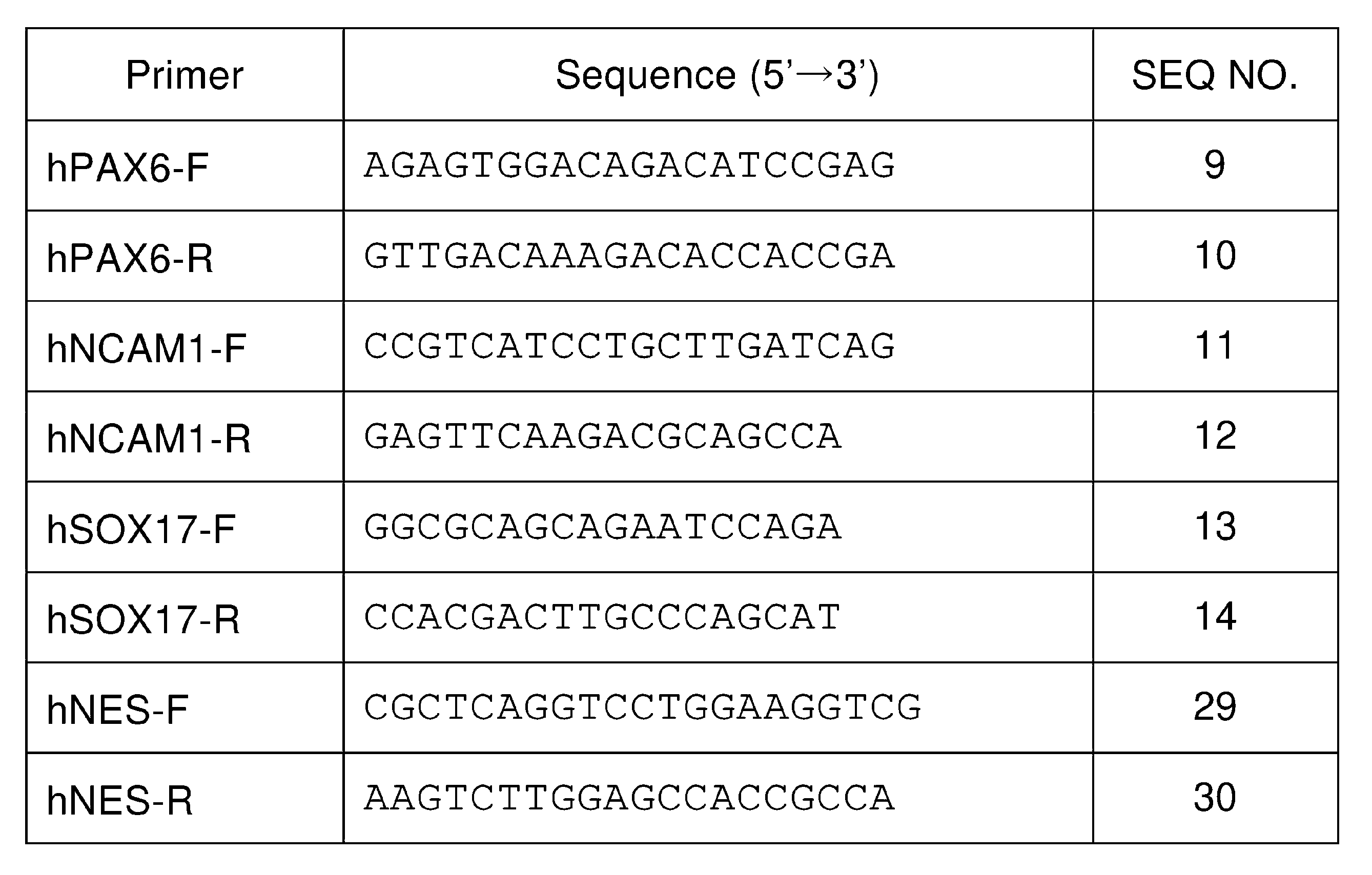
(3-3)染色体検査
消化管ホルモン受容体発現iPS細胞の安全性を評価するために、G分染法による染色体検査を株式会社日本遺伝子研究所に委託した。
消化管ホルモン受容体発現iPS細胞の安全性を評価するために、G分染法による染色体検査を株式会社日本遺伝子研究所に委託した。
<4.消化管ホルモン受容体を強制発現させた副交感神経細胞またはその自律神経系前駆細胞の解析>
(4-1)マーカー発現
(i)qPCR解析
神経堤細胞マーカー遺伝子(hSOX10)、自律神経細胞マーカー遺伝子(hPHOX2B)、末梢神経細胞マーカー遺伝子(hPRPH)および副交感神経細胞マーカー遺伝子(hCHAT)の発現をqPCRにより解析した。qPCRは、以下のプライマーセットを用いた以外は上記(3-1)と同様の手順により行った。
(4-1)マーカー発現
(i)qPCR解析
神経堤細胞マーカー遺伝子(hSOX10)、自律神経細胞マーカー遺伝子(hPHOX2B)、末梢神経細胞マーカー遺伝子(hPRPH)および副交感神経細胞マーカー遺伝子(hCHAT)の発現をqPCRにより解析した。qPCRは、以下のプライマーセットを用いた以外は上記(3-1)と同様の手順により行った。
表3.qPCRに使用したプライマーセット(3)
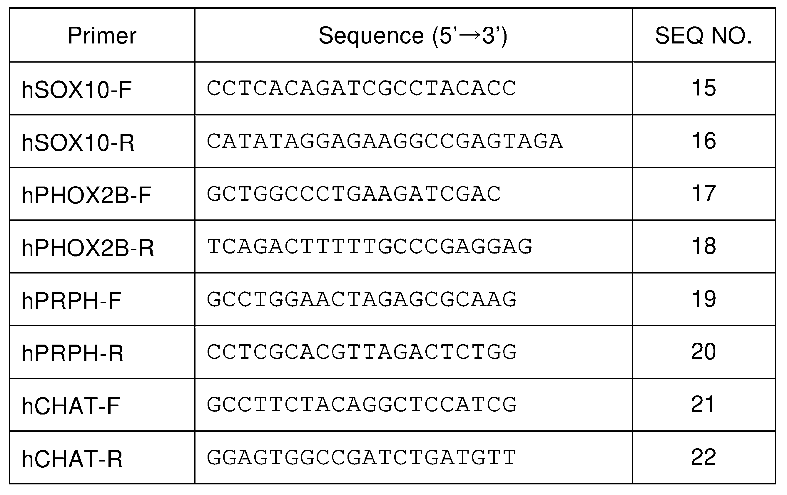
(ii)免疫細胞染色
消化管ホルモン受容体およびMAP2またはChATの発現を免疫細胞染色により解析した。免疫細胞染色は、上記(3-1)と同様の手順により行った。MAP2およびChATの染色には、以下の抗体を用いた。
・抗MAP2抗体(Abcam、ab11267)(1:500希釈)
・抗ChAT抗体(Abcam、ab181023)(1:100希釈)
消化管ホルモン受容体およびMAP2またはChATの発現を免疫細胞染色により解析した。免疫細胞染色は、上記(3-1)と同様の手順により行った。MAP2およびChATの染色には、以下の抗体を用いた。
・抗MAP2抗体(Abcam、ab11267)(1:500希釈)
・抗ChAT抗体(Abcam、ab181023)(1:100希釈)
(4-2)細胞内カルシウムイメージング
上記(2-5)および(2-7)で作製した細胞(誘導30日目以降)に、終濃度10μMのFluo-8 AM(AAT Bioquest、21083)を添加し、37℃で20~30分間インキュベートした。培地を除去し、リンガー液(149mM NaCl、2.8mM KCl、2mM CaCl2、1mM MgCl2、10mM HEPES、10mM D-(+)-glucose(pH7.2))に置換した後、細胞内カルシウム濃度変化を測定した。顕微鏡(Olympus、IX71)、Confocal scanner unit(Yokogawa、CSU-W1)、LED光源(OPTO SCIENCE、X-Cite 120LED)、および解析ソフト(MetaMorph)(Molecular Devices)を用い、500ms/フレームで500~800フレーム撮影した。途中(例えば250フレーム目)で消化管ホルモン受容体アゴニスト刺激を添加した。消化管ホルモン受容体アゴニストおよびその添加量(終濃度)は以下の通りである:GLP1(ヒト、7-37)(ペプチド研究所、#4280-v)(以下、「GLP1」と記載する)(10nM);PYY(ヒト、3-36)(ペプチド研究所、#4400-v)(以下、「PYY」と記載する)(100nM);GLP2(ヒト)(ペプチド研究所、#4376-v)(以下、「GLP2」と記載する)(100nM);セロトニン(Serotonin hydrochloride)(Sigma-Aldrich、H9523)(以下、「5-HT」と記載する)(100μM);CCK-33(ヒト)(ペプチド研究所、#4201-s)(以下、「CCK-33」と記載する)(10、100、1000nM)。
上記(2-5)および(2-7)で作製した細胞(誘導30日目以降)に、終濃度10μMのFluo-8 AM(AAT Bioquest、21083)を添加し、37℃で20~30分間インキュベートした。培地を除去し、リンガー液(149mM NaCl、2.8mM KCl、2mM CaCl2、1mM MgCl2、10mM HEPES、10mM D-(+)-glucose(pH7.2))に置換した後、細胞内カルシウム濃度変化を測定した。顕微鏡(Olympus、IX71)、Confocal scanner unit(Yokogawa、CSU-W1)、LED光源(OPTO SCIENCE、X-Cite 120LED)、および解析ソフト(MetaMorph)(Molecular Devices)を用い、500ms/フレームで500~800フレーム撮影した。途中(例えば250フレーム目)で消化管ホルモン受容体アゴニスト刺激を添加した。消化管ホルモン受容体アゴニストおよびその添加量(終濃度)は以下の通りである:GLP1(ヒト、7-37)(ペプチド研究所、#4280-v)(以下、「GLP1」と記載する)(10nM);PYY(ヒト、3-36)(ペプチド研究所、#4400-v)(以下、「PYY」と記載する)(100nM);GLP2(ヒト)(ペプチド研究所、#4376-v)(以下、「GLP2」と記載する)(100nM);セロトニン(Serotonin hydrochloride)(Sigma-Aldrich、H9523)(以下、「5-HT」と記載する)(100μM);CCK-33(ヒト)(ペプチド研究所、#4201-s)(以下、「CCK-33」と記載する)(10、100、1000nM)。
(4-3)細胞外電位測定
上記(2-4)の分化ステップ6に記載の手順により、PLOおよびラミニンによりコートされたMEA測定用培養プレート(CytoView MEA 6(M384-tMEA-6W), CytoView MEA 24(M384-tMEA-24W))(AXION Biosystems)を用意した。培地にNDM3を用いて、上記(2-4)または(2-7)における誘導13日目の細胞を播種した(1×10⁵細胞/8μL)。以降、1週間に2度、フレッシュなNDM3により培地を半量交換しながら培養を継続した。
上記(2-4)の分化ステップ6に記載の手順により、PLOおよびラミニンによりコートされたMEA測定用培養プレート(CytoView MEA 6(M384-tMEA-6W), CytoView MEA 24(M384-tMEA-24W))(AXION Biosystems)を用意した。培地にNDM3を用いて、上記(2-4)または(2-7)における誘導13日目の細胞を播種した(1×10⁵細胞/8μL)。以降、1週間に2度、フレッシュなNDM3により培地を半量交換しながら培養を継続した。
NDM3またはリンガー液中にて、膜電位変化を測定した。1000×ゲイン、200Hz-3kHzフィルターを用い、測定ノイズ標準偏差に対し6倍以上に変化した電位値を神経発火(スパイク)として検出した。電位を5分間測定した後、消化管ホルモン受容体アゴニストを上記(4-2)と同様の終濃度で添加し、さらに5分間測定した。
<5.hGLP1Rを強制発現させたiPS細胞の品質評価>
上記1の手順においてレンチウイルスベクターとしてpLV-hGLP1Rを用い、上記(2-6)の手順により調製したiPS細胞におけるhGLP1R遺伝子およびタンパク質の発現を、上記(3-1)の手順により解析した。qPCRの結果を図1に示す。野生型iPS細胞(対照)と比較して、外因性hGLP1R遺伝子を導入されたiPS細胞は、hGLP1R遺伝子を強く発現した(*P<0.05、n=3、Studentのt検定)。免疫細胞染色の結果を図2に示す。図中、スケールバーは100μmを示す。外因性hGLP1R遺伝子を導入されたiPS細胞は、hGLP1Rタンパク質を強く発現していることが確認された。これらの結果に基づき、この細胞を「hGLP1R発現iPS細胞」とした。hGLP1R発現iPS細胞の明視野像を図3に示す。図中、スケールバーは100μmを示す。
上記1の手順においてレンチウイルスベクターとしてpLV-hGLP1Rを用い、上記(2-6)の手順により調製したiPS細胞におけるhGLP1R遺伝子およびタンパク質の発現を、上記(3-1)の手順により解析した。qPCRの結果を図1に示す。野生型iPS細胞(対照)と比較して、外因性hGLP1R遺伝子を導入されたiPS細胞は、hGLP1R遺伝子を強く発現した(*P<0.05、n=3、Studentのt検定)。免疫細胞染色の結果を図2に示す。図中、スケールバーは100μmを示す。外因性hGLP1R遺伝子を導入されたiPS細胞は、hGLP1Rタンパク質を強く発現していることが確認された。これらの結果に基づき、この細胞を「hGLP1R発現iPS細胞」とした。hGLP1R発現iPS細胞の明視野像を図3に示す。図中、スケールバーは100μmを示す。
上記(3-1)の手順により、hGLP1R発現iPS細胞における幹細胞マーカーhNANOG遺伝子の発現をqPCRにより確認した結果を図4に示す。図中、「GLP1R」はhGLP1R発現iPS細胞を、「Control」は野生型iPS細胞を示す。hGLP1R発現iPS細胞は、野生型iPS細胞よりもhNANOG遺伝子を強く発現し(*P<0.05、n=3、Studentのt検定)、未分化状態であることが確認された。
上記(3-2)の手順により、hGLP1R発現iPS細胞の三胚様への分化能を評価した。結果を図5に示す。外胚葉、中胚葉または内胚葉への分化を誘導されたhGLP1R発現iPS細胞は、外胚葉マーカーhPAX6遺伝子、中胚葉マーカーhNCAM1遺伝子または内胚葉マーカーhSOX17遺伝子をそれぞれ強く発現した。これらの結果から、hGLP1R発現iPS細胞の三胚様への分化能を有していることが確認された。
G分染法による染色体検査の結果を図6に示す。hGLP1R発現iPS細胞の染色体には異常がないことが確認された。
<6.hGLP1R発現iPS細胞から調製された副交感神経細胞またはその自律神経系前駆細胞の解析>
上記(2-7)の手順により、hGLP1R発現iPS細胞の副交感神経細胞への分化を誘導した。誘導0日目のEBおよび誘導13日目の細胞の顕微鏡像(明視野)を図7に示す。スケールバーは100μmを示す。誘導13日目の細胞についてのqPCRの結果を図8に示す。幹細胞マーカーhNANOG遺伝子の発現が顕著に減少し、神経堤細胞マーカーhSOX10遺伝子および自律神経細胞マーカーhPHOX2B遺伝子の発現が顕著に増加した(*P<0.05、n=3、Studentのt検定)。これらの結果から、誘導13日目の細胞が自律神経系前駆細胞であることが確認された。
上記(2-7)の手順により、hGLP1R発現iPS細胞の副交感神経細胞への分化を誘導した。誘導0日目のEBおよび誘導13日目の細胞の顕微鏡像(明視野)を図7に示す。スケールバーは100μmを示す。誘導13日目の細胞についてのqPCRの結果を図8に示す。幹細胞マーカーhNANOG遺伝子の発現が顕著に減少し、神経堤細胞マーカーhSOX10遺伝子および自律神経細胞マーカーhPHOX2B遺伝子の発現が顕著に増加した(*P<0.05、n=3、Studentのt検定)。これらの結果から、誘導13日目の細胞が自律神経系前駆細胞であることが確認された。
誘導40日目の細胞についてのqPCRの結果を図9に示す。末梢神経細胞マーカーhPRPH遺伝子、副交感神経細胞マーカーhCHAT遺伝子およびhGLP1R遺伝子の強い発現が確認された(*P<0.05、n=3、Studentのt検定)。免疫細胞染色の結果を図10に示す。図中、スケールバーは100μmを示す。hGLP1Rタンパク質の発現に加え、成熟神経細胞マーカーMAP2タンパク質の発現が確認された。
上記(2-7)の手順によりhGLP1R発現iPS細胞から調製された細胞のリガンド応答性を、上記(4-3)の手順による細胞外電位測定により解析した。細胞外電位ヒートマップを図11に示す。GLP1を添加することにより、高い神経発火頻度を示す領域が見られた。神経発火頻度の変化率を図12に示す。GLP1を添加することにより、神経発火頻度が上昇した(*P<0.05、n=74電極/4ウェル、student-t)。
上記(2-7)の手順によりhGLP1R発現iPS細胞から調製された細胞のリガンド応答性を、上記(4-2)の手順による細胞内カルシウムイメージングにより解析した。測定結果を図13に示す。横軸は時間(秒)を示し、縦軸はGLP1添加前の蛍光強度の平均値(F0)を1とした際の蛍光強度の変化率(ΔF/F0)を示す。GLP1を添加することにより、細胞内Ca2+濃度が上昇した。1分間あたりの神経発火(スパイク)の数を図14に示す。GLP1を添加することにより、スパイク数が増加した(*P<0.05、n=15細胞/3サンプル、student-t)。神経発火のピークの振幅(GLP1添加前の蛍光強度の平均値(F0)に対する蛍光強度の変化率(ΔF/F0-1)×100)を図15に示す。GLP1を添加することにより、神経発火の強度が増強された(*P<0.05、n=15細胞/3サンプル、student-t)。
以上の結果から、hGLP1R発現iPS細胞から調製された副交感神経細胞が、GLP1応答性を有する迷走神経様副交感神経細胞であることが示された。
<7.hNPY2Rを強制発現させたiPS細胞の品質評価>
上記1の手順においてレンチウイルスベクターとしてpLV-hNPY2Rを用い、上記(2-6)の手順により調製したiPS細胞におけるhNPY2R遺伝子およびタンパク質の発現を、上記(3-1)の手順により解析した。qPCRの結果を図16に示す。野生型iPS細胞(対照)と比較して、外因性hNPY2R遺伝子を導入されたiPS細胞は、hNPY2R遺伝子を強く発現した(*P<0.05、n=3、Studentのt検定)。免疫細胞染色の結果を図17に示す。図中、スケールバーは100μmを示す。外因性hNPY2R遺伝子を導入されたiPS細胞は、hNPY2Rタンパク質を強く発現していることが確認された。これらの結果に基づき、この細胞を「hNPY2R発現iPS細胞」とした。hNPY2R発現iPS細胞の明視野像を図18に示す。図中、スケールバーは100μmを示す。
上記1の手順においてレンチウイルスベクターとしてpLV-hNPY2Rを用い、上記(2-6)の手順により調製したiPS細胞におけるhNPY2R遺伝子およびタンパク質の発現を、上記(3-1)の手順により解析した。qPCRの結果を図16に示す。野生型iPS細胞(対照)と比較して、外因性hNPY2R遺伝子を導入されたiPS細胞は、hNPY2R遺伝子を強く発現した(*P<0.05、n=3、Studentのt検定)。免疫細胞染色の結果を図17に示す。図中、スケールバーは100μmを示す。外因性hNPY2R遺伝子を導入されたiPS細胞は、hNPY2Rタンパク質を強く発現していることが確認された。これらの結果に基づき、この細胞を「hNPY2R発現iPS細胞」とした。hNPY2R発現iPS細胞の明視野像を図18に示す。図中、スケールバーは100μmを示す。
上記(3-1)の手順により、hNPY2R発現iPS細胞における幹細胞マーカーNANOGの発現をqPCRにより確認した結果を図19に示す。図中、「NPY2R」はhNPY2R発現iPS細胞を、「Control」は野生型iPS細胞を示す。hNPY2R発現iPS細胞は、野生型iPS細胞よりもNANOG遺伝子を強く発現し(*P<0.05、n=3、Studentのt検定)、未分化状態であることが確認された。
上記(3-2)の手順により、hNPY2R発現iPS細胞の三胚様への分化能を評価した。結果を図20に示す。外胚葉、中胚葉または内胚葉への分化を誘導されたhNPY2R発現iPS細胞は、外胚葉マーカーhPAX6遺伝子、中胚葉マーカーhNCAM1遺伝子または内胚葉マーカーhSOX17遺伝子をそれぞれ強く発現した。これらの結果から、hNPY2R発現iPS細胞の三胚様への分化能を有していることが確認された。
G分染法による染色体検査の結果を図21に示す。hNPY2R発現iPS細胞の染色体には異常がないことが確認された。
<8.hNPY2R発現iPS細胞から調製された副交感神経細胞またはその自律神経系前駆細胞の解析>
上記(2-7)の手順により、hNPY2R発現iPS細胞の副交感神経細胞への分化を誘導した。誘導0日目のEBおよび誘導13日目の細胞の顕微鏡像(明視野)を図22に示す。スケールバーは100μmを示す。誘導13日目の細胞についてのqPCRの結果を図23に示す。幹細胞マーカーhNANOG遺伝子の発現が顕著に減少し、神経堤細胞マーカーhSOX10遺伝子および自律神経細胞マーカーhPHOX2B遺伝子の発現が顕著に増加した(*P<0.05、n=3、Studentのt検定)。これらの結果から、誘導13日目の細胞が自律神経系前駆細胞であることが確認された。
上記(2-7)の手順により、hNPY2R発現iPS細胞の副交感神経細胞への分化を誘導した。誘導0日目のEBおよび誘導13日目の細胞の顕微鏡像(明視野)を図22に示す。スケールバーは100μmを示す。誘導13日目の細胞についてのqPCRの結果を図23に示す。幹細胞マーカーhNANOG遺伝子の発現が顕著に減少し、神経堤細胞マーカーhSOX10遺伝子および自律神経細胞マーカーhPHOX2B遺伝子の発現が顕著に増加した(*P<0.05、n=3、Studentのt検定)。これらの結果から、誘導13日目の細胞が自律神経系前駆細胞であることが確認された。
誘導40日目の細胞についてのqPCRの結果を図24に示す。末梢神経細胞マーカーhPRPH遺伝子、副交感神経細胞マーカーhCHAT遺伝子およびhNPY2R遺伝子の強い発現が確認された(*P<0.05、n=3、Studentのt検定)。免疫細胞染色の結果を図25に示す。図中、スケールバーは100μmを示す。hNPY2Rタンパク質の発現に加え、成熟神経細胞マーカーMAP2タンパク質の発現が確認された。
上記(2-7)の手順によりhNPY2R発現iPS細胞から調製された細胞のリガンド応答性を、上記(4-3)の手順による細胞外電位測定により解析した。細胞外電位ヒートマップを図26に示す。PYYを添加することにより、高い神経発火頻度を示す領域が見られた。神経発火頻度の変化率を図27に示す。PYYを添加することにより、神経発火頻度が上昇した(*P<0.05、n=74電極/4ウェル、student-t)。
上記(2-7)の手順によりhNPY2R発現iPS細胞から調製された細胞のリガンド応答性を、上記(4-2)の手順による細胞内カルシウムイメージングにより解析した。測定結果を図28に示す。横軸は時間(秒)を示し、縦軸はPYY添加前(0~125秒)の蛍光強度の平均値(F0)を1とした際の蛍光強度の変化率(ΔF/F0)を示す。PYYを添加することにより、細胞内Ca2+濃度が上昇した。1分間あたりの神経発火(スパイク)の数を図29に示す。PYYを添加することにより、スパイク数が増加した(*P<0.05、n=15細胞/3サンプル、student-t)。神経発火のピークの振幅(PYY添加前の蛍光強度の平均値(F0)に対する蛍光強度の変化率(ΔF/F0-1)×100)を図30に示す。PYYを添加することにより、神経発火の強度が増強された(*P<0.05、n=15細胞/3サンプル、student-t)。
以上の結果から、hNPY2R発現iPS細胞から調製された副交感神経細胞が、PYY応答性を有する迷走神経様副交感神経細胞であることが示された。
<9.hGLP2Rを強制発現させたiPS細胞の品質評価>
上記1の手順においてレンチウイルスベクターとしてpLV-hGLP2Rを用い、上記(2-6)の手順により調製したiPS細胞におけるhGLP2R遺伝子およびタンパク質の発現を、上記(3-1)の手順により解析した。qPCRの結果を図31に示す。野生型iPS細胞(対照)と比較して、外因性hGLP2R遺伝子を導入されたiPS細胞は、hGLP2R遺伝子を強く発現した(*P<0.05、n=3、Studentのt検定)。免疫細胞染色の結果を図32に示す。図中、スケールバーは100μmを示す。外因性hGLP2R遺伝子を導入されたiPS細胞は、hGLP2Rタンパク質を発現していることが確認された。これらの結果に基づき、この細胞を「hGLP2R発現iPS細胞」とした。hGLP2R発現iPS細胞の明視野像を図33に示す。図中、スケールバーは1mmを示す。
上記1の手順においてレンチウイルスベクターとしてpLV-hGLP2Rを用い、上記(2-6)の手順により調製したiPS細胞におけるhGLP2R遺伝子およびタンパク質の発現を、上記(3-1)の手順により解析した。qPCRの結果を図31に示す。野生型iPS細胞(対照)と比較して、外因性hGLP2R遺伝子を導入されたiPS細胞は、hGLP2R遺伝子を強く発現した(*P<0.05、n=3、Studentのt検定)。免疫細胞染色の結果を図32に示す。図中、スケールバーは100μmを示す。外因性hGLP2R遺伝子を導入されたiPS細胞は、hGLP2Rタンパク質を発現していることが確認された。これらの結果に基づき、この細胞を「hGLP2R発現iPS細胞」とした。hGLP2R発現iPS細胞の明視野像を図33に示す。図中、スケールバーは1mmを示す。
上記(3-1)の手順により、hGLP2R発現iPS細胞における幹細胞マーカーhNANOG遺伝子の発現をqPCRにより確認した結果を図34に示す。図中、「hGLP2R」はhGLP2R発現iPS細胞を、「Control」は野生型iPS細胞を示す。hGLP2R発現iPS細胞は、野生型iPS細胞と同程度にhNANOG遺伝子を発現し、未分化状態であることが確認された。
上記(3-2)の手順により、hGLP2R発現iPS細胞の三胚様への分化能を評価した。結果を図35に示す。外胚葉、中胚葉または内胚葉への分化を誘導されたhGLP2R発現iPS細胞は、外胚葉マーカーhNES遺伝子、中胚葉マーカーhNCAM1遺伝子または内胚葉マーカーhSOX17遺伝子をそれぞれ強く発現した。これらの結果から、hGLP2R発現iPS細胞の三胚様への分化能を有していることが確認された。
G分染法による染色体検査の結果を図36に示す。hGLP2R発現iPS細胞の染色体には異常がないことが確認された。
<10.hGLP2R発現iPS細胞から調製された副交感神経細胞またはその自律神経系前駆細胞の解析>
上記(2-7)の手順により、hGLP2R発現iPS細胞の副交感神経細胞への分化を誘導した。誘導0日目のEBおよび誘導13日目の細胞の顕微鏡像(明視野)を図37に示す。スケールバーは100μmを示す。誘導13日目の細胞についてのqPCRの結果を図38に示す。幹細胞マーカーhNANOG遺伝子の発現が顕著に減少し、神経堤細胞マーカーhSOX10遺伝子および自律神経細胞マーカーhPHOX2B遺伝子の発現が顕著に増加した(*P<0.05、n=3、Studentのt検定)。これらの結果から、誘導13日目の細胞が自律神経系前駆細胞であることが確認された。
上記(2-7)の手順により、hGLP2R発現iPS細胞の副交感神経細胞への分化を誘導した。誘導0日目のEBおよび誘導13日目の細胞の顕微鏡像(明視野)を図37に示す。スケールバーは100μmを示す。誘導13日目の細胞についてのqPCRの結果を図38に示す。幹細胞マーカーhNANOG遺伝子の発現が顕著に減少し、神経堤細胞マーカーhSOX10遺伝子および自律神経細胞マーカーhPHOX2B遺伝子の発現が顕著に増加した(*P<0.05、n=3、Studentのt検定)。これらの結果から、誘導13日目の細胞が自律神経系前駆細胞であることが確認された。
誘導40日目の細胞についてのqPCRの結果を図39に示す。末梢神経細胞マーカーhPRPH遺伝子および副交感神経細胞マーカーhCHAT遺伝子の強い発現が確認された(*P<0.05、n=3、Studentのt検定)。また、誘導40日目のモック感染副交感神経細胞(対照)と比較して、hGLP2R遺伝子の強い発現が確認された(*P<0.05、n=3、Studentのt検定)。
免疫細胞染色の結果を図40に示す。図中、スケールバーは100μmを示す。hGLP2Rタンパク質の弱い発現および成熟神経細胞マーカーMAP2タンパク質の発現が確認された。
<11.hHTR3Aを強制発現させたiPS細胞の品質評価>
上記1の手順においてレンチウイルスベクターとしてpLV-hHTR3Aを用い、上記(2-6)の手順により調製したiPS細胞におけるhHTR3A遺伝子およびタンパク質の発現を、上記(3-1)の手順により解析した。qPCRの結果を図41に示す。野生型iPS細胞(対照)と比較して、外因性hHTR3A遺伝子を導入されたiPS細胞は、hHTR3A遺伝子を強く発現した(*P<0.05、n=3、Studentのt検定)。免疫細胞染色の結果を図42に示す。図中、スケールバーは100μmを示す。外因性hHTR3A遺伝子を導入されたiPS細胞は、hHTR3Aタンパク質を強く発現していることが確認された。これらの結果に基づき、この細胞を「hHTR3A発現iPS細胞」とした。hHTR3A発現iPS細胞の明視野像を図43に示す。図中、スケールバーは1mmを示す。
上記1の手順においてレンチウイルスベクターとしてpLV-hHTR3Aを用い、上記(2-6)の手順により調製したiPS細胞におけるhHTR3A遺伝子およびタンパク質の発現を、上記(3-1)の手順により解析した。qPCRの結果を図41に示す。野生型iPS細胞(対照)と比較して、外因性hHTR3A遺伝子を導入されたiPS細胞は、hHTR3A遺伝子を強く発現した(*P<0.05、n=3、Studentのt検定)。免疫細胞染色の結果を図42に示す。図中、スケールバーは100μmを示す。外因性hHTR3A遺伝子を導入されたiPS細胞は、hHTR3Aタンパク質を強く発現していることが確認された。これらの結果に基づき、この細胞を「hHTR3A発現iPS細胞」とした。hHTR3A発現iPS細胞の明視野像を図43に示す。図中、スケールバーは1mmを示す。
上記(3-1)の手順により、hHTR3A発現iPS細胞における幹細胞マーカーhNANOG遺伝子の発現をqPCRにより確認した結果を図44に示す。図中、「hHTR3A」はhHTR3A発現iPS細胞を、「Control」は野生型iPS細胞を示す。hHTR3A発現iPS細胞は、野生型iPS細胞と同程度にhNANOG遺伝子を発現し、未分化状態であることが確認された。
上記(3-2)の手順により、hHTR3A発現iPS細胞の三胚様への分化能を評価した。結果を図45に示す。外胚葉、中胚葉または内胚葉への分化を誘導されたhHTR3A発現iPS細胞は、外胚葉マーカーhNES遺伝子、中胚葉マーカーhNCAM1遺伝子または内胚葉マーカーhSOX17遺伝子をそれぞれ強く発現した。これらの結果から、hHTR3A発現iPS細胞の三胚様への分化能を有していることが確認された。
G分染法による染色体検査の結果を図46に示す。hHTR3A発現iPS細胞の染色体には異常がないことが確認された。
<12.hHTR3A発現iPS細胞から調製された副交感神経細胞またはその自律神経系前駆細胞の解析>
上記(2-7)の手順により、hHTR3A発現iPS細胞の副交感神経細胞への分化を誘導した。誘導0日目のEBおよび誘導13日目の細胞の顕微鏡像(明視野)を図47に示す。スケールバーは100μmを示す。誘導13日目の細胞についてのqPCRの結果を図48に示す。幹細胞マーカーhNANOG遺伝子の発現が顕著に減少し、神経堤細胞マーカーhSOX10遺伝子および自律神経細胞マーカーhPHOX2B遺伝子の発現が顕著に増加した(*P<0.05、n=3、Studentのt検定)。これらの結果から、誘導13日目の細胞が自律神経系前駆細胞であることが確認された。
上記(2-7)の手順により、hHTR3A発現iPS細胞の副交感神経細胞への分化を誘導した。誘導0日目のEBおよび誘導13日目の細胞の顕微鏡像(明視野)を図47に示す。スケールバーは100μmを示す。誘導13日目の細胞についてのqPCRの結果を図48に示す。幹細胞マーカーhNANOG遺伝子の発現が顕著に減少し、神経堤細胞マーカーhSOX10遺伝子および自律神経細胞マーカーhPHOX2B遺伝子の発現が顕著に増加した(*P<0.05、n=3、Studentのt検定)。これらの結果から、誘導13日目の細胞が自律神経系前駆細胞であることが確認された。
誘導40日目の細胞についてのqPCRの結果を図49に示す。末梢神経細胞マーカーhPRPH遺伝子および副交感神経細胞マーカーhCHAT遺伝子の強い発現が確認された(*P<0.05、n=3、Studentのt検定)。また、誘導40日目のモック感染副交感神経細胞(対照)と比較して、hHTR3A遺伝子の同程度の発現が確認された(*P<0.05、n=3、Studentのt検定)。
免疫細胞染色の結果を図50に示す。図中、スケールバーは100μmを示す。hHTR3Aタンパク質および成熟神経細胞マーカーMAP2タンパク質の発現が確認された。
<13.hCCKARを強制発現させたiPS細胞の品質評価>
上記1の手順においてレンチウイルスベクターとしてpLV-hCCKARを用い、上記(2-6)の手順により調製したiPS細胞におけるhCCKAR遺伝子およびタンパク質の発現を、上記(3-1)の手順により解析した。qPCRの結果を図51に示す。野生型iPS細胞(対照)と比較して、外因性hCCKAR遺伝子を導入されたiPS細胞は、hCCKAR遺伝子を強く発現した(*P<0.05、n=3、Studentのt検定)。免疫細胞染色の結果を図52に示す。図中、スケールバーは100μmを示す。外因性hCCKAR遺伝子を導入されたiPS細胞は、hCCKARタンパク質を強く発現していることが確認された。これらの結果に基づき、この細胞を「hCCKAR発現iPS細胞」とした。hCCKAR発現iPS細胞の明視野像を図53に示す。図中、スケールバーは1mmを示す。
上記1の手順においてレンチウイルスベクターとしてpLV-hCCKARを用い、上記(2-6)の手順により調製したiPS細胞におけるhCCKAR遺伝子およびタンパク質の発現を、上記(3-1)の手順により解析した。qPCRの結果を図51に示す。野生型iPS細胞(対照)と比較して、外因性hCCKAR遺伝子を導入されたiPS細胞は、hCCKAR遺伝子を強く発現した(*P<0.05、n=3、Studentのt検定)。免疫細胞染色の結果を図52に示す。図中、スケールバーは100μmを示す。外因性hCCKAR遺伝子を導入されたiPS細胞は、hCCKARタンパク質を強く発現していることが確認された。これらの結果に基づき、この細胞を「hCCKAR発現iPS細胞」とした。hCCKAR発現iPS細胞の明視野像を図53に示す。図中、スケールバーは1mmを示す。
上記(3-1)の手順により、hCCKAR発現iPS細胞における幹細胞マーカーhNANOG遺伝子の発現をqPCRにより確認した結果を図54に示す。図中、「hCCKAR」はhCCKAR発現iPS細胞を、「Control」は野生型iPS細胞を示す。hCCKAR発現iPS細胞は、野生型iPS細胞と同程度にhNANOG遺伝子を発現し、未分化状態であることが確認された。
上記(3-2)の手順により、hCCKAR発現iPS細胞の三胚様への分化能を評価した。結果を図55に示す。外胚葉、中胚葉または内胚葉への分化を誘導されたhCCKAR発現iPS細胞は、外胚葉マーカーhNES遺伝子、中胚葉マーカーhNCAM1遺伝子または内胚葉マーカーhSOX17遺伝子をそれぞれ強く発現した。これらの結果から、hCCKAR発現iPS細胞の三胚様への分化能を有していることが確認された。
G分染法による染色体検査の結果を図56に示す。hCCKAR発現iPS細胞の染色体には異常がないことが確認された。
<14.hCCKAR発現iPS細胞から調製された副交感神経細胞またはその自律神経系前駆細胞の解析>
上記(2-7)の手順により、hCCKAR発現iPS細胞の副交感神経細胞への分化を誘導した。誘導0日目のEBおよび誘導13日目の細胞の顕微鏡像(明視野)を図57に示す。スケールバーは100μmを示す。誘導13日目の細胞についてのqPCRの結果を図58に示す。幹細胞マーカーhNANOG遺伝子の発現が顕著に減少し、神経堤細胞マーカーhSOX10遺伝子および自律神経細胞マーカーhPHOX2B遺伝子の発現が顕著に増加した(*P<0.05、n=3、Studentのt検定)。これらの結果から、誘導13日目の細胞が自律神経系前駆細胞であることが確認された。
上記(2-7)の手順により、hCCKAR発現iPS細胞の副交感神経細胞への分化を誘導した。誘導0日目のEBおよび誘導13日目の細胞の顕微鏡像(明視野)を図57に示す。スケールバーは100μmを示す。誘導13日目の細胞についてのqPCRの結果を図58に示す。幹細胞マーカーhNANOG遺伝子の発現が顕著に減少し、神経堤細胞マーカーhSOX10遺伝子および自律神経細胞マーカーhPHOX2B遺伝子の発現が顕著に増加した(*P<0.05、n=3、Studentのt検定)。これらの結果から、誘導13日目の細胞が自律神経系前駆細胞であることが確認された。
誘導40日目の細胞についてのqPCRの結果を図59に示す。末梢神経細胞マーカーhPRPH遺伝子および副交感神経細胞マーカーhCHAT遺伝子の強い発現が確認された(*P<0.05、n=3、Studentのt検定)。また、誘導40日目のモック感染副交感神経細胞(対照)と比較して、hCCKAR遺伝子の強い発現が確認された(*P<0.05、n=3、Studentのt検定)。
免疫細胞染色の結果を図60に示す。図中、スケールバーは100μmを示す。hCCKARタンパク質および成熟神経細胞マーカーMAP2タンパク質の発現が確認された。
上記(2-7)の手順によりhCCKAR発現iPS細胞から調製された細胞のリガンド応答性を、上記(4-3)の手順による細胞外電位測定により解析した。細胞外電位ヒートマップを図61に示す。CCK-33を添加することにより、高い神経発火頻度を示す領域が見られた。神経発火頻度の変化率を図62に示す。CCK-33を添加することにより、神経発火頻度が上昇した(*P<0.05、0nM=128電極、10nM=23電極、100nM=57電極、1000nM=24電極/2~4ウェル、student-t)。
上記(2-7)の手順によりhCCKAR発現iPS細胞から調製された細胞のリガンド応答性を、上記(4-2)の手順による細胞内カルシウムイメージングにより解析した。測定結果を図63に示す。横軸は時間(秒)を示し、縦軸はCCK-33添加前(0~199秒)の蛍光強度の平均値(F0)を1とした際の蛍光強度の変化率(ΔF/F0)を示す。CCK-33を添加することにより、細胞内Ca2+濃度が上昇した。CCK-33添加前(0~199秒)の蛍光強度の平均値(F0)に対するCCK-33添加後(200~400秒)の蛍光強度の平均値(ΔFave)の変化率(ΔFave/F0)を図64に示す。CCK-33を添加することにより、神経発火の強度が増強された(*P<0.05、n=20細胞/3サンプル、student-t)。
以上の結果から、hCCKAR発現iPS細胞から調製された副交感神経細胞が、CCK-33応答性を有する迷走神経様副交感神経細胞であることが示された。
<15.hGLP1Rを分化後に強制発現させた副交感神経細胞の解析>
上記1の手順においてレンチウイルスベクターとしてpLV-hGLP1Rを用い、上記(2-5)の手順により調製した副交感神経細胞におけるhGLP1Rタンパク質の発現を、上記(3-1)の手順により解析した。誘導40日目における免疫細胞染色の結果を図65に示す。図中、スケールバーは100μmを示す。副交感神経細胞マーカーhChATタンパク質およびhGLP1Rタンパク質の強い発現が確認された。
上記1の手順においてレンチウイルスベクターとしてpLV-hGLP1Rを用い、上記(2-5)の手順により調製した副交感神経細胞におけるhGLP1Rタンパク質の発現を、上記(3-1)の手順により解析した。誘導40日目における免疫細胞染色の結果を図65に示す。図中、スケールバーは100μmを示す。副交感神経細胞マーカーhChATタンパク質およびhGLP1Rタンパク質の強い発現が確認された。
上記(2-7)の手順により、hGLP1Rタンパク質を分化後に強制発現させた副交感神経細胞のリガンド応答性を、上記(4-2)の手順による細胞内カルシウムイメージングにより解析した。測定結果を図66に示す。横軸は時間(秒)を示し、縦軸はGLP1添加前(0~124秒)の蛍光強度の平均値(F0)を1とした際の蛍光強度の変化率(ΔF/F0)を示す。GLP1を添加することにより、細胞内Ca2+濃度が上昇した。
<16.hNPY2Rを分化後に強制発現させた副交感神経細胞の解析>
上記1の手順においてレンチウイルスベクターとしてpLV-hNPY2Rを用い、上記(2-5)の手順により調製した副交感神経細胞におけるhNPY2Rタンパク質の発現を、上記(3-1)の手順により解析した。誘導40日目における免疫細胞染色の結果を図67に示す。図中、スケールバーは100μmを示す。副交感神経細胞マーカーhChATタンパク質およびhNPY2Rタンパク質の強い発現が確認された。
上記1の手順においてレンチウイルスベクターとしてpLV-hNPY2Rを用い、上記(2-5)の手順により調製した副交感神経細胞におけるhNPY2Rタンパク質の発現を、上記(3-1)の手順により解析した。誘導40日目における免疫細胞染色の結果を図67に示す。図中、スケールバーは100μmを示す。副交感神経細胞マーカーhChATタンパク質およびhNPY2Rタンパク質の強い発現が確認された。
上記(2-7)の手順により、hNPY2Rタンパク質を分化後に強制発現させた副交感神経細胞のリガンド応答性を、上記(4-2)の手順による細胞内カルシウムイメージングにより解析した。測定結果を図68に示す。横軸は時間(秒)を示し、縦軸はPYY添加前(0~124秒)の蛍光強度の平均値(F0)を1とした際の蛍光強度の変化率(ΔF/F0)を示す。PYYを添加することにより、細胞内Ca2+濃度が上昇した。
<17.hGLP2Rを分化後に強制発現させた副交感神経細胞の解析>
上記1の手順においてレンチウイルスベクターとしてpLV-hGLP2Rを用い、上記(2-5)の手順により調製した副交感神経細胞におけるhGLP2R遺伝子およびタンパク質の発現を、上記(3-1)の手順により解析した。qPCRの結果を図69に示す。野生型iPS細胞(図中、「未分化」)では末梢神経細胞マーカーhPRPH遺伝子および副交感神経細胞マーカーhCHAT遺伝子の発現が見られなかったのに対し、外因性hGLP2R遺伝子を導入された誘導40日目の細胞(図中、「副交感神経細胞」)では、両マーカー遺伝子の強い発現が確認された(*P<0.05、n=3、Studentのt検定)。また、誘導40日目のモック感染副交感神経細胞(対照)と比較して、hGLP2R遺伝子の強い発現が確認された(*P<0.05、n=3、Studentのt検定)。
上記1の手順においてレンチウイルスベクターとしてpLV-hGLP2Rを用い、上記(2-5)の手順により調製した副交感神経細胞におけるhGLP2R遺伝子およびタンパク質の発現を、上記(3-1)の手順により解析した。qPCRの結果を図69に示す。野生型iPS細胞(図中、「未分化」)では末梢神経細胞マーカーhPRPH遺伝子および副交感神経細胞マーカーhCHAT遺伝子の発現が見られなかったのに対し、外因性hGLP2R遺伝子を導入された誘導40日目の細胞(図中、「副交感神経細胞」)では、両マーカー遺伝子の強い発現が確認された(*P<0.05、n=3、Studentのt検定)。また、誘導40日目のモック感染副交感神経細胞(対照)と比較して、hGLP2R遺伝子の強い発現が確認された(*P<0.05、n=3、Studentのt検定)。
誘導40日目における免疫細胞染色の結果を図70に示す。図中、スケールバーは100μmを示す。成熟神経細胞マーカーMAP2タンパク質およびhGLP2Rタンパク質の強い発現が確認された。
上記(2-7)の手順により、hGLP2Rタンパク質を分化後に強制発現させた副交感神経細胞のリガンド応答性を、上記(4-2)の手順による細胞内カルシウムイメージングにより解析した。測定結果を図71に示す。横軸は時間(秒)を示し、縦軸はGLP2添加前(0~80秒)の蛍光強度の平均値(F0)を1とした際の蛍光強度の変化率(ΔF/F0)を示す。GLP2を添加することにより、細胞内Ca2+濃度が上昇した。GLP2添加前(0~80秒)の蛍光強度の平均値(F0)に対するGLP2添加後(240~400秒)の蛍光強度の平均値(ΔFave)の変化率(ΔFave/F0)を図72に示す。GLP2を添加することにより、神経発火の強度が増強された(*P<0.05、n=20細胞/3サンプル、student-t)。
<18.hHTR3Aを分化後に強制発現させた副交感神経細胞の解析>
上記1の手順においてレンチウイルスベクターとしてpLV-hHTR3Aを用い、上記(2-5)の手順により調製した副交感神経細胞におけるhHTR3A遺伝子およびタンパク質の発現を、上記(3-1)の手順により解析した。qPCRの結果を図73に示す。野生型iPS細胞(図中、「未分化」)では末梢神経細胞マーカーhPRPH遺伝子および副交感神経細胞マーカーhCHAT遺伝子の発現が見られなかったのに対し、外因性hHTR3A遺伝子を導入された誘導40日目の細胞(図中、「副交感神経細胞」)では、両マーカー遺伝子の強い発現が確認された(*P<0.05、n=3、Studentのt検定)。また、誘導40日目のモック感染副交感神経細胞(対照)と比較して、hHTR3A遺伝子の強い発現が確認された(*P<0.05、n=3、Studentのt検定)。
上記1の手順においてレンチウイルスベクターとしてpLV-hHTR3Aを用い、上記(2-5)の手順により調製した副交感神経細胞におけるhHTR3A遺伝子およびタンパク質の発現を、上記(3-1)の手順により解析した。qPCRの結果を図73に示す。野生型iPS細胞(図中、「未分化」)では末梢神経細胞マーカーhPRPH遺伝子および副交感神経細胞マーカーhCHAT遺伝子の発現が見られなかったのに対し、外因性hHTR3A遺伝子を導入された誘導40日目の細胞(図中、「副交感神経細胞」)では、両マーカー遺伝子の強い発現が確認された(*P<0.05、n=3、Studentのt検定)。また、誘導40日目のモック感染副交感神経細胞(対照)と比較して、hHTR3A遺伝子の強い発現が確認された(*P<0.05、n=3、Studentのt検定)。
誘導40日目における免疫細胞染色の結果を図74に示す。図中、スケールバーは100μmを示す。成熟神経細胞マーカーMAP2タンパク質およびhHTR3Aタンパク質の強い発現が確認された。
上記(2-7)の手順により、hHTR3Aタンパク質を分化後に強制発現させた副交感神経細胞のリガンド応答性を、上記(4-2)の手順による細胞内カルシウムイメージングにより解析した。測定結果を図75に示す。横軸は時間(秒)を示し、縦軸は5-HT添加前(0~80秒)の蛍光強度の平均値(F0)を1とした際の蛍光強度の変化率(ΔF/F0)を示す。5-HTを添加することにより、細胞内Ca2+濃度が上昇した。5-HT添加前(0~80秒)の蛍光強度の平均値(F0)に対する5-HT添加後(240~400秒)の蛍光強度の平均値(ΔFave)の変化率(ΔFave/F0)を図76に示す。5-HTを添加することにより、神経発火の強度が増強された(*P<0.05、n=20細胞/3サンプル、student-t)。
<19.hCCKARを分化後に強制発現させた副交感神経細胞の解析>
上記1の手順においてレンチウイルスベクターとしてpLV-hCCKARを用い、上記(2-5)の手順により調製した副交感神経細胞におけるhCCKAR遺伝子およびタンパク質の発現を、上記(3-1)の手順により解析した。qPCRの結果を図77に示す。野生型iPS細胞(図中、「未分化」)では末梢神経細胞マーカーhPRPH遺伝子および副交感神経細胞マーカーhCHAT遺伝子の発現が見られなかったのに対し、外因性hCCKAR遺伝子を導入された誘導40日目の細胞(図中、「副交感神経細胞」)では、両マーカー遺伝子の強い発現が確認された(*P<0.05、n=3、Studentのt検定)。また、誘導40日目のモック感染副交感神経細胞(対照)と比較して、hCCKAR遺伝子の強い発現が確認された(*P<0.05、n=3、Studentのt検定)。
上記1の手順においてレンチウイルスベクターとしてpLV-hCCKARを用い、上記(2-5)の手順により調製した副交感神経細胞におけるhCCKAR遺伝子およびタンパク質の発現を、上記(3-1)の手順により解析した。qPCRの結果を図77に示す。野生型iPS細胞(図中、「未分化」)では末梢神経細胞マーカーhPRPH遺伝子および副交感神経細胞マーカーhCHAT遺伝子の発現が見られなかったのに対し、外因性hCCKAR遺伝子を導入された誘導40日目の細胞(図中、「副交感神経細胞」)では、両マーカー遺伝子の強い発現が確認された(*P<0.05、n=3、Studentのt検定)。また、誘導40日目のモック感染副交感神経細胞(対照)と比較して、hCCKAR遺伝子の強い発現が確認された(*P<0.05、n=3、Studentのt検定)。
誘導40日目における免疫細胞染色の結果を図78に示す。図中、スケールバーは100μmを示す。成熟神経細胞マーカーMAP2タンパク質およびhCCKARタンパク質の強い発現が確認された。
上記(2-7)の手順により、hCCKARタンパク質を分化後に強制発現させた副交感神経細胞のリガンド応答性を、上記(4-2)の手順による細胞内カルシウムイメージングにより解析した。測定結果を図79に示す。横軸は時間(秒)を示し、縦軸はCCK-33添加前(0~80秒)の蛍光強度の平均値(F0)を1とした際の蛍光強度の変化率(ΔF/F0)を示す。CCK-33を添加することにより、細胞内Ca2+濃度が上昇した。CCK-33添加前(0~80秒)の蛍光強度の平均値(F0)に対するCCK-33添加後(240~400秒)の蛍光強度の平均値(ΔFave)の変化率(ΔFave/F0)を図80に示す。CCK-33を添加することにより、神経発火の強度が増強された(*P<0.05、n=20細胞/3サンプル、student-t)。
<20.hGLP1R発現iPS細胞から調製された副交感神経細胞と腸内分泌細胞との共培養の作製>
ヒトL細胞由来細胞株NCI-H716(ATCC:CCL-251)を、10%FBSおよび1%P/Sを含有するRPMI-1640(以下、「H716培地」と記載する)を用いて10cmディッシュに播種し(1~2×106細胞/ディッシュ)、3~4日間培養した。細胞および培地を15mL遠沈管に回収し、200×g、5分間遠心分離し、上清を除去した。細胞にFBSを含有しないH716培地またはリンガー液を加え、1×106細胞/mLに調製した。得られた細胞懸濁液の1mLに20μMのMemGlow/DMSO溶液を5μL加え(終濃度100nM)、室温で10分間インキュベートした。その後、200×g、5分間遠心分離し、上清を除去した。H716培地を用いて1回洗浄後、10cmディッシュに播種した。MemGlowによる細胞膜の染色は、最大10日間維持されることを確認した。以下、MemGlowにより染色されたNCI-H716細胞を「MemGlow染色NCI-H716」と記載する。
ヒトL細胞由来細胞株NCI-H716(ATCC:CCL-251)を、10%FBSおよび1%P/Sを含有するRPMI-1640(以下、「H716培地」と記載する)を用いて10cmディッシュに播種し(1~2×106細胞/ディッシュ)、3~4日間培養した。細胞および培地を15mL遠沈管に回収し、200×g、5分間遠心分離し、上清を除去した。細胞にFBSを含有しないH716培地またはリンガー液を加え、1×106細胞/mLに調製した。得られた細胞懸濁液の1mLに20μMのMemGlow/DMSO溶液を5μL加え(終濃度100nM)、室温で10分間インキュベートした。その後、200×g、5分間遠心分離し、上清を除去した。H716培地を用いて1回洗浄後、10cmディッシュに播種した。MemGlowによる細胞膜の染色は、最大10日間維持されることを確認した。以下、MemGlowにより染色されたNCI-H716細胞を「MemGlow染色NCI-H716」と記載する。
上記(2-7)の手順によりhGLP1R発現iPS細胞から調製された副交感神経細胞(以下、「hGLP1R発現副交感神経細胞」と記載する)(誘導37日目)に、MemGlow染色NCI-H716(2×10⁵細胞/mL)を1mL添加した。この結果、共培養は、NDM2およびH716培地の1:1混合培地中で行われた。48時間の培養後、上記(3-1)(ii)と同様の手順により、免疫細胞染色を行った。一次抗体には、抗MAP2抗体(Abcam、ab11267)(1:1000希釈)を用いた。
結果を図81に示す。共培養において、hGLP1R発現副交感神経細胞の上にMemGlow染色NCI-H716細胞が接着したことが確認された。hGLP1R発現副交感神経細胞およびMemGlow染色NCI-H716のいずれにも形態異常は見られなかった。さらに、副交感神経のアゴニストであるニコチンを用いて、上記(4-2)の手順により細胞内カルシウムイメージングを行い、共培養におけるhGLP1R発現副交感神経細胞のニコチン応答が正常であることを確認した(データは省略)。これらの結果から、hGLP1R発現副交感神経細胞とNCI-H716細胞とを直接共培養できることが示された。
<21.NCI-H716細胞との共培養におけるhGLP1R発現副交感神経細胞の解析>
上記(2-7)の手順により調製されたhGLP1R発現副交感神経細胞(誘導35日目)に、MemGlow染色NCI-H716(1×10⁶細胞/mL)を1mL添加した。共培養は、NDM2およびH716培地の1:1混合培地中で行われた。1時間の共培養後、培地をグルコース不含リンガー液に交換した。グルコース(腸内分泌細胞を刺激し、摂食制御にかかわる)に対する応答を、上記(4-2)の手順による細胞内カルシウムイメージングにより解析した。
上記(2-7)の手順により調製されたhGLP1R発現副交感神経細胞(誘導35日目)に、MemGlow染色NCI-H716(1×10⁶細胞/mL)を1mL添加した。共培養は、NDM2およびH716培地の1:1混合培地中で行われた。1時間の共培養後、培地をグルコース不含リンガー液に交換した。グルコース(腸内分泌細胞を刺激し、摂食制御にかかわる)に対する応答を、上記(4-2)の手順による細胞内カルシウムイメージングにより解析した。
結果を図82に示す。横軸は時間(秒)を示し、縦軸はグルコース添加前(0~199秒)の蛍光強度の平均値(F0)を1とした際の蛍光強度の変化率(ΔF/F0)を示す。共培養にグルコースを添加することにより、hGLP1R発現副交感神経細胞の細胞内Ca2+濃度が上昇した。グルコース添加前(0~199秒)の蛍光強度の平均値(F0)に対するグルコース添加後(200~400秒)の蛍光強度の平均値(ΔFave)の変化率(ΔFave/F0)を図83に示す。共培養にグルコースを添加することにより、hGLP1R発現副交感神経細胞の神経発火の強度が増強された(*P<0.05、n=20細胞/サンプル、student-t)。一方、hGLP1R発現副交感神経細胞の単独培養はグルコースに応答しなかった(データは省略)。これらの結果から、NCI-H716細胞がグルコースに応答してGLP1を分泌し、hGLP1R発現副交感神経細胞がGLP1に応答したことが示唆された。
次に、上記共培養のα-リノレン酸に対する応答を、上記(4-2)の手順による細胞内カルシウムイメージングにより解析した。NCI-H716細胞が発現する多価不飽和脂肪酸受容体のアゴニストであるα-リノレン酸(富士フイルム和光純薬:127-05901)(以下、「ALA」と記載する)(100μM)を用い、測定溶液にはグルコース不含リンガー液を用いた。
結果を図84に示す。横軸は時間(秒)を示し、縦軸はALA添加前(0~199秒)の蛍光強度の平均値(F0)を1とした際の蛍光強度の変化率(ΔF/F0)を示す。共培養にALAを添加することにより、hGLP1R発現副交感神経細胞の細胞内Ca2+濃度が上昇した。ALA添加前(0~199秒)の蛍光強度の平均値(F0)に対するALA添加後(200~400秒)の蛍光強度の平均値(ΔFave)の変化率(ΔFave/F0)を図85に示す。共培養にALAを添加することにより、hGLP1R発現副交感神経細胞の神経発火の強度が増強された(*P<0.05、n=20細胞/サンプル、student-t)。一方、hGLP1R発現副交感神経細胞の単独培養はALAに応答しなかった(データは省略)。これらの結果から、NCI-H716細胞がALAに応答してGLP1を分泌し、hGLP1R発現副交感神経細胞がGLP1に応答したことが示唆された。
<22.NCI-H716細胞の培養上清に対するhGLP1R発現副交感神経細胞の応答性>
HBSSにより希釈したマトリゲル(終濃度0.4~0.6mg/mL)を24ウェルプレートに添加し、37℃で1時間インキュベートした。HBSSで2回洗浄し、乾燥させた後、H716培地を用いてNCI-H716細胞を播種した(2×105細胞/ウェル)。48時間培養した後、リンガー液で1回洗浄し、グルコース(10mM)またはALA(100μM)を含むリンガー液を添加した。室温で4分間インキュベートした後、上清を回収した。PMSFを添加し(終濃度50μg/mL)、測定に用いるまで上清を氷上で保存し、測定時には室温に戻して使用した。以下、グルコースを含むリンガー液により調製された上清を「グルコース負荷上清」、ALAを含むリンガー液により調製された上清を「ALA負荷上清」と記載する。グルコース負荷上清またはALA負荷上清を用い、上記(4-2)と同様の手順により細胞内カルシウムイメージングを行った。
HBSSにより希釈したマトリゲル(終濃度0.4~0.6mg/mL)を24ウェルプレートに添加し、37℃で1時間インキュベートした。HBSSで2回洗浄し、乾燥させた後、H716培地を用いてNCI-H716細胞を播種した(2×105細胞/ウェル)。48時間培養した後、リンガー液で1回洗浄し、グルコース(10mM)またはALA(100μM)を含むリンガー液を添加した。室温で4分間インキュベートした後、上清を回収した。PMSFを添加し(終濃度50μg/mL)、測定に用いるまで上清を氷上で保存し、測定時には室温に戻して使用した。以下、グルコースを含むリンガー液により調製された上清を「グルコース負荷上清」、ALAを含むリンガー液により調製された上清を「ALA負荷上清」と記載する。グルコース負荷上清またはALA負荷上清を用い、上記(4-2)と同様の手順により細胞内カルシウムイメージングを行った。
グルコース負荷上清を用いた細胞内カルシウムイメージングの結果を図86に示す。横軸は時間(秒)を示し、縦軸は上清添加前(0~199秒)の蛍光強度の平均値(F0)を1とした際の蛍光強度の変化率(ΔF/F0)を示す。グルコース負荷上清を添加することにより、hGLP1R発現副交感神経細胞(誘導44日目)の細胞内Ca2+濃度が上昇した。上清添加前(0~199秒)の蛍光強度の平均値(F0)に対する上清添加後(200~400秒)の蛍光強度の平均値(ΔFave)の変化率(ΔFave/F0)を図87に示す。図中、「Ctrl」はNCI-H716細胞と触れていないリンガー液を示す。hGLP1R発現副交感神経細胞はグルコース未負荷上清にも反応したが、グルコース負荷上清により強い反応を示した(***P<0.001、n=20細胞/サンプル、Tukey事後検定を伴う二元配置ANOVA)。
ALA負荷上清を用いた細胞内カルシウムイメージングの結果を図88に示す。横軸は時間(秒)を示し、縦軸は上清添加前(0~199秒)の蛍光強度の平均値(F0)を1とした際の蛍光強度の変化率(ΔF/F0)を示す。ALA負荷上清を添加することにより、hGLP1R発現副交感神経細胞(誘導34日目)の細胞内Ca2+濃度が上昇した。上清添加前(0~199秒)の蛍光強度の平均値(F0)に対する上清添加後(200~400秒)の蛍光強度の平均値(ΔFave)の変化率(ΔFave/F0)を図89に示す。図中、「Ctrl」はNCI-H716細胞と接触していないリンガー液を示す。hGLP1R発現副交感神経細胞は、グルコース負荷上清と同程度にALA負荷上清にも強い反応を示した(***P<0.001、n=20細胞/サンプル、Tukey事後検定を伴う二元配置ANOVA)。
以上の結果から、NCI-H716細胞がグルコースおよびALAに応答してGLP1を分泌し、hGLP1R発現副交感神経細胞がGLP1に応答したことが確認された。
Claims (13)
- (a)多能性幹細胞の副交感神経細胞への分化を誘導するステップ、および
(b1)前記ステップ(a)の前に、前記多能性幹細胞に消化管ホルモン受容体をコードする外因性核酸を導入するステップ、または
(b2)前記ステップ(a)により得られた副交感神経細胞またはその前駆細胞に消化管ホルモン受容体をコードする外因性核酸を導入するステップ
を含む、迷走神経様副交感神経細胞を作製する方法。 - 前記消化管ホルモン受容体が、グルカゴン様ペプチド1受容体、ニューロペプチドY2型受容体、グルカゴン様ペプチド2受容体、セロトニン3受容体およびコレシストキニン1受容体からなる群から選択される、請求項1に記載の方法。
- 請求項1または2に記載の方法により作製された、迷走神経様副交感神経細胞。
- 消化管ホルモン受容体をコードする外因性核酸を含んでなる多能性幹細胞。
- 消化管ホルモン受容体をコードする外因性核酸を含んでなる迷走神経様副交感神経細胞。
- 前記消化管ホルモン受容体が、グルカゴン様ペプチド1受容体、ニューロペプチドY2型受容体、グルカゴン様ペプチド2受容体、セロトニン3受容体およびコレシストキニン1受容体からなる群から選択される、請求項4または5に記載の細胞。
- (1)請求項3、5または6に記載の迷走神経様副交感神経細胞に試料を接触させるステップ、および、
(2)前記迷走神経様副交感神経細胞の神経活動を解析するステップ
を含む、試料中の消化管ホルモン受容体アゴニストを検出する方法。 - (1)候補物質の存在下で請求項3、5または6に記載の迷走神経様副交感神経細胞を培養するステップ、および、
(2)前記迷走神経様副交感神経細胞の神経活動を解析するステップ
を含む、脳腸相関を制御する物質のスクリーニング方法。 - (1)候補物質の存在下で消化管ホルモン分泌細胞を培養するステップ、
(2)前記ステップ(1)により得られた培養物から培養上清を回収するステップ、
(3)請求項3、5または6に記載の迷走神経様副交感神経細胞に前記培養上清を接触させるステップ、および、
(4)前記迷走神経様副交感神経細胞の神経活動を解析するステップ
を含む、脳腸相関を制御する物質のスクリーニング方法。 - (1)候補物質の存在下で消化管ホルモン分泌細胞と請求項3、5または6に記載の迷走神経様副交感神経細胞とを共培養するステップ、および
(2)前記迷走神経様副交感神経細胞の神経活動を解析するステップ
を含む、脳腸相関を制御する物質のスクリーニング方法。 - 前記消化管ホルモン分泌細胞が腸内分泌細胞である、請求項9または10に記載のスクリーニング方法。
- 消化管ホルモン分泌細胞と請求項3、5または6に記載の迷走神経様副交感神経細胞との共培養を含んでなる、脳腸相関モデル。
- 前記消化管ホルモン分泌細胞が腸内分泌細胞である、請求項12に記載の脳腸相関モデル。
Priority Applications (1)
| Application Number | Priority Date | Filing Date | Title |
|---|---|---|---|
| JP2024537267A JPWO2024024955A1 (ja) | 2022-07-29 | 2023-07-28 |
Applications Claiming Priority (2)
| Application Number | Priority Date | Filing Date | Title |
|---|---|---|---|
| JP2022121608 | 2022-07-29 | ||
| JP2022-121608 | 2022-07-29 |
Publications (1)
| Publication Number | Publication Date |
|---|---|
| WO2024024955A1 true WO2024024955A1 (ja) | 2024-02-01 |
Family
ID=89706684
Family Applications (1)
| Application Number | Title | Priority Date | Filing Date |
|---|---|---|---|
| PCT/JP2023/027801 Ceased WO2024024955A1 (ja) | 2022-07-29 | 2023-07-28 | 迷走神経様副交感神経細胞およびその作製方法 |
Country Status (3)
| Country | Link |
|---|---|
| JP (1) | JPWO2024024955A1 (ja) |
| TW (1) | TW202409273A (ja) |
| WO (1) | WO2024024955A1 (ja) |
Citations (2)
| Publication number | Priority date | Publication date | Assignee | Title |
|---|---|---|---|---|
| WO2016194522A1 (ja) * | 2015-06-02 | 2016-12-08 | 国立研究開発法人産業技術総合研究所 | 神経堤細胞から自律神経系の細胞への分化誘導方法 |
| WO2020040286A1 (ja) * | 2018-08-24 | 2020-02-27 | 国立研究開発法人産業技術総合研究所 | 副交感神経細胞の作製方法 |
-
2023
- 2023-07-28 JP JP2024537267A patent/JPWO2024024955A1/ja active Pending
- 2023-07-28 TW TW112128487A patent/TW202409273A/zh unknown
- 2023-07-28 WO PCT/JP2023/027801 patent/WO2024024955A1/ja not_active Ceased
Patent Citations (2)
| Publication number | Priority date | Publication date | Assignee | Title |
|---|---|---|---|---|
| WO2016194522A1 (ja) * | 2015-06-02 | 2016-12-08 | 国立研究開発法人産業技術総合研究所 | 神経堤細胞から自律神経系の細胞への分化誘導方法 |
| WO2020040286A1 (ja) * | 2018-08-24 | 2020-02-27 | 国立研究開発法人産業技術総合研究所 | 副交感神経細胞の作製方法 |
Non-Patent Citations (7)
| Title |
|---|
| BERTHOUD H.‐R.: "Vagal and hormonal gut–brain communication: from satiation to satisfaction", NEUROGASTROENTEROLOGY AND MOTILITY, BLACKWELL SCIENTIFIC PUBLICATIONS, OXFORD, GB, vol. 20, no. s1, 1 May 2008 (2008-05-01), GB , pages 64 - 72, XP093133520, ISSN: 1350-1925, DOI: 10.1111/j.1365-2982.2008.01104.x * |
| DATABASE Nucleotide 20210627; ANONYMOUS : "Homo sapiens cholecystokinin A receptor (CCKAR), mRNA ", XP093133534, retrieved from NCBI Database accession no. NM_000730.3 * |
| DATABASE Nucleotide 25 July 2022 (2022-07-25), ANONYMOUS : "Homo sapiens 5-hydroxytryptamine receptor 3A (HTR3A), transcript variant 1, mRNA", XP093133531, retrieved from NCBI Database accession no. NM_213621.4 * |
| DATABASE Nucleotide 26 June 2021 (2021-06-26), ANONYMOUS M : "Homo sapiens neuropeptide Y receptor Y2 (NPY2R), transcript variant 1, mRNA", XP093133525, retrieved from NCBI Database accession no. NM_000910.4 * |
| DATABASE Nucleotide 26 June 2022 (2022-06-26), ANONYMOUS : "Homo sapiens glucagon like peptide 1 receptor (GLP1R), transcript variant 1, mRNA", XP093133523, retrieved from NCBI Database accession no. NM_002062.5 * |
| DATABASE Nucleotide 9 August 2021 (2021-08-09), ANONYMOUS : "Homo sapiens glucagon like peptide 2 receptor (GLP2R), mRNA", XP093133529, retrieved from NCBI Database accession no. NM_004246.3 * |
| MINAGAWA ATSUTAKA; YOSHIKAWA TOSHIAKI; YASUKAWA MASAKI; HOTTA AKITSU; KUNITOMO MIHOKO; IRIGUCHI SHOICHI; TAKIGUCHI MAIKO; KASSAI Y: "Enhancing T Cell Receptor Stability in Rejuvenated iPSC-Derived T Cells Improves Their Use in Cancer Immunotherapy", CELL STEM CELL, ELSEVIER, CELL PRESS, AMSTERDAM, NL, vol. 23, no. 6, 6 December 2018 (2018-12-06), AMSTERDAM, NL , pages 850, XP085555973, ISSN: 1934-5909, DOI: 10.1016/j.stem.2018.10.005 * |
Also Published As
| Publication number | Publication date |
|---|---|
| TW202409273A (zh) | 2024-03-01 |
| JPWO2024024955A1 (ja) | 2024-02-01 |
Similar Documents
| Publication | Publication Date | Title |
|---|---|---|
| Trzaska et al. | Brain‐derived neurotrophic factor facilitates maturation of mesenchymal stem cell‐derived dopamine progenitors to functional neurons | |
| JP7023820B2 (ja) | 多分化能細胞および多能性細胞の分化を方向付けることによって発生させる皮質介在ニューロンおよびその他のニューロン細胞 | |
| JP6756786B2 (ja) | 移植用中脳ドーパミン(da)ニューロン | |
| JP6367824B2 (ja) | 三次元不均一分化組織培養 | |
| Henion et al. | trkC-mediated NT-3 signaling is required for the early development of a subpopulation of neurogenic neural crest cells | |
| CN108368486B (zh) | 鉴定神经肌肉接头活动的调节剂的体外方法 | |
| US20210139849A1 (en) | Methods for efficient derivation of human motor neurons from diverse spinal regions | |
| Wu et al. | Role of Sonic hedgehog signaling and the expression of its components in human embryonic stem cells | |
| JP2020513823A (ja) | 幹細胞由来星状細胞、作製方法および使用方法 | |
| US10494602B1 (en) | Functional astrocytes and cortical neurons from induced pluripotent stem cells and methods of use thereof | |
| JP2018531012A6 (ja) | 神経筋接合活性のモジュレーターを同定するin vitroの方法 | |
| Poliandri et al. | Generation of kisspeptin-responsive GnRH neurons from human pluripotent stem cells | |
| US20250230405A1 (en) | Generation of norepinephrine neurons from human stem cells | |
| WO2024024955A1 (ja) | 迷走神経様副交感神経細胞およびその作製方法 | |
| Tamaki et al. | Overexpression of cyclin dependent kinase inhibitor P27/Kip1 increases oligodendrocyte differentiation from induced pluripotent stem cells: Tamaki and Tokumoto | |
| CN118773135A (zh) | 一种人类多能干细胞诱导为类听觉神经元的方法 | |
| US20230089966A1 (en) | Method for differentiation of brain mural cells from human pluripotent stem cells | |
| CA3155277A1 (en) | Production method for induced dopaminergic neuronal progenitors, using direct reprogramming | |
| Shirzad et al. | Production of stable GFP-expressing neural cells from P19 embryonal carcinoma stem cells | |
| Xie | Modeling SCN1A Epilepsy with Dual Isogenic Pairs of Human iPSC-derived Neurons | |
| Zhang | A patient-specific stem cell model of neurovascular interaction for vascular dementia CADASIL | |
| Alvandi | c-Src Controls Mouse Embryonic Osteogenic Differentiation Through Regulation of Stat1 Stability | |
| WO2021015245A1 (ja) | 繊毛関連疾患モデルおよびその利用 | |
| Adnani | TRANSCRIPTIONAL REGULATION OF NEURONAL MIGRATION AND CELL FATE SPECIFICATION IN THE NEOCORTEX | |
| Brosenitsch | Induction of the dopaminergic phenotype in primary sensory neurons: Activity-dependent signaling and lineage-related transcription factors |
Legal Events
| Date | Code | Title | Description |
|---|---|---|---|
| 121 | Ep: the epo has been informed by wipo that ep was designated in this application |
Ref document number: 23846685 Country of ref document: EP Kind code of ref document: A1 |
|
| WWE | Wipo information: entry into national phase |
Ref document number: 2024537267 Country of ref document: JP |
|
| NENP | Non-entry into the national phase |
Ref country code: DE |
|
| 122 | Ep: pct application non-entry in european phase |
Ref document number: 23846685 Country of ref document: EP Kind code of ref document: A1 |