WO2024005275A1 - Strem-1을 이용한 인체내 항생제 감수성의 평가 방법 - Google Patents
Strem-1을 이용한 인체내 항생제 감수성의 평가 방법 Download PDFInfo
- Publication number
- WO2024005275A1 WO2024005275A1 PCT/KR2022/017165 KR2022017165W WO2024005275A1 WO 2024005275 A1 WO2024005275 A1 WO 2024005275A1 KR 2022017165 W KR2022017165 W KR 2022017165W WO 2024005275 A1 WO2024005275 A1 WO 2024005275A1
- Authority
- WO
- WIPO (PCT)
- Prior art keywords
- antibiotic
- blood
- strem
- over time
- human body
- Prior art date
- Legal status (The legal status is an assumption and is not a legal conclusion. Google has not performed a legal analysis and makes no representation as to the accuracy of the status listed.)
- Ceased
Links
Images


Classifications
-
- G—PHYSICS
- G01—MEASURING; TESTING
- G01N—INVESTIGATING OR ANALYSING MATERIALS BY DETERMINING THEIR CHEMICAL OR PHYSICAL PROPERTIES
- G01N33/00—Investigating or analysing materials by specific methods not covered by groups G01N1/00 - G01N31/00
- G01N33/48—Biological material, e.g. blood, urine; Haemocytometers
- G01N33/50—Chemical analysis of biological material, e.g. blood, urine; Testing involving biospecific ligand binding methods; Immunological testing
- G01N33/53—Immunoassay; Biospecific binding assay; Materials therefor
- G01N33/531—Production of immunochemical test materials
- G01N33/532—Production of labelled immunochemicals
- G01N33/535—Production of labelled immunochemicals with enzyme label or co-enzymes, co-factors, enzyme inhibitors or enzyme substrates
-
- G—PHYSICS
- G01—MEASURING; TESTING
- G01N—INVESTIGATING OR ANALYSING MATERIALS BY DETERMINING THEIR CHEMICAL OR PHYSICAL PROPERTIES
- G01N33/00—Investigating or analysing materials by specific methods not covered by groups G01N1/00 - G01N31/00
- G01N33/48—Biological material, e.g. blood, urine; Haemocytometers
- G01N33/50—Chemical analysis of biological material, e.g. blood, urine; Testing involving biospecific ligand binding methods; Immunological testing
- G01N33/58—Chemical analysis of biological material, e.g. blood, urine; Testing involving biospecific ligand binding methods; Immunological testing involving labelled substances
-
- G—PHYSICS
- G01—MEASURING; TESTING
- G01N—INVESTIGATING OR ANALYSING MATERIALS BY DETERMINING THEIR CHEMICAL OR PHYSICAL PROPERTIES
- G01N33/00—Investigating or analysing materials by specific methods not covered by groups G01N1/00 - G01N31/00
- G01N33/48—Biological material, e.g. blood, urine; Haemocytometers
- G01N33/50—Chemical analysis of biological material, e.g. blood, urine; Testing involving biospecific ligand binding methods; Immunological testing
- G01N33/68—Chemical analysis of biological material, e.g. blood, urine; Testing involving biospecific ligand binding methods; Immunological testing involving proteins, peptides or amino acids
Definitions
- the present invention relates to a method for evaluating antibiotic susceptibility in the human body, and more specifically, to a method for evaluating antibiotic susceptibility in the human body using sTREM-1.
- Sepsis is a fatal infectious disease in which a local infection caused by bacteria at a wound or inflamed area progresses into a systemic inflammatory response. Sepsis is a disease with a very high mortality and morbidity rate if it is not recognized quickly and treated appropriately, and is the most important cause of death in intensive care units.
- antibiotics are mostly selected empirically and administered to patients based on the patient's demographic characteristics, underlying disease, and infection site. The appropriateness of the empirically selected antibiotic can be confirmed from changes in the patient's condition over the next few days, or can be confirmed by performing in vitro antibiotic sensitivity testing when the pathogen is identified through culture testing.
- the identification ability or yield rate for the causative bacteria of infectious diseases is very low, about 20-40%, and it takes 3 to 5 days, up to a week or more, to obtain test results, so it is used in emergency diseases such as sepsis. Adjusting or changing antibiotics through culture tests is clinically very difficult.
- the inventor of the present application developed a biomarker that can evaluate antibiotic susceptibility through reactions in the human body and completed the present invention.
- the problem to be solved by the present invention is to provide a method for evaluating antibiotic susceptibility in the human body using sTREM-1.
- the method for assessing antibiotic susceptibility according to an embodiment of the present invention to achieve the above problem is to measure antibiotic susceptibility in the human body (in Includes methods for evaluating in vivo antibiotic susceptibility.
- the infectious disease may be sepsis.
- the blood concentration of sTREM-1 in the blood collected from the human body is measured to be 30 - 320 ng/mL and 390 - 1580 ng/mL, it is classified as a susceptible antibiotic and a resistant antibiotic, respectively. can be judged.
- the antibiotic is judged to be susceptible and resistant, respectively. You can.
- the antibiotic is judged to be susceptible and resistant, respectively. You can.
- the method for evaluating antibiotic susceptibility in the human body according to the present invention has the following excellent effects.
- sTREM-1 soluble Triggering Receptor Expressed on Myeloid cells-1
- sTREM-1 is previously known as an inflammatory marker and its expression level is known to increase in the serum of patients with inflammatory diseases.
- sTREM-1 is not used as an inflammatory marker, but rather as an evaluation tool for in vivo antibiotic susceptibility.
- sTREM-1 used in the antibiotic susceptibility evaluation of the present invention has a short half-life of about 2 hours and responds characteristically to bacterial infection, it was confirmed that the blood concentration of sTREM-1 varies significantly within a short period of time depending on the appropriateness of the antibiotic. It has been done. For example, by measuring the blood concentration of sTREM-1 within 2 to 5 hours after administering any antibiotic to a patient with an infectious disease, it is possible to evaluate whether the antibiotic is susceptible or resistant to the antibiotic. Therefore, the method for evaluating antibiotic susceptibility in the human body according to the present invention can evaluate antibiotic suitability in a shorter time compared to antibiotic susceptibility testing through existing culture.
- the present invention provides a method for evaluating antibiotic susceptibility in vivo. For example, according to the present invention, after administering any antibiotic to a patient with an infectious disease, the blood concentration of sTREM-1 is measured in blood collected from the patient and the appropriateness of the antibiotic is judged based on the concentration, so the appropriateness of the antibiotic can be checked. can be judged accurately.
- antibiotics are empirically administered to patients with infectious diseases, but according to the present invention, the appropriateness of the antibiotic can be quickly evaluated and the antibiotics preemptively adjusted according to the results, thereby improving the patient's prognosis.
- many clinicians have always felt burdened about the appropriateness of empirically prescribed antibiotics, but the method for evaluating antibiotic susceptibility in the human body according to the present invention can relieve this psychological burden.
- the appropriateness of antibiotics can be confirmed within a few hours, preventing the abuse of a wide range of antibiotics and creating a safer society by reducing the occurrence of resistant bacteria.
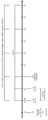
- Figure 1 is a diagram showing an experimental method of a porcine bacteremia model according to an embodiment of the present invention.
- Figures 2A to 2F are graphs measuring hemodynamic changes over time.
- Figures 3A to 3E are graphs measuring changes in fluid administration and vasopressor usage over time.
- Figures 4a to 4f are graphs measuring changes in blood cell count over time.
- Figures 5A to 5F are graphs measuring relevant values to evaluate organ failure by the porcine bacteremia model of the present invention.
- Figures 6a to 6f are graphs comparing trends in SOFA scores in the porcine bacteremia model of the present invention.
- Figure 7 is a diagram showing blood culture test results over time.
- Figures 8A to 8K are graphs measuring blood concentrations of various biomarkers over time.
- the method for assessing antibiotic susceptibility is to measure the blood concentration of sTREM-1 (soluble Triggering Receptor Expressed on Myeloid cells-1) in blood collected from a human body administered antibiotics for infectious diseases. You can determine your antibiotic susceptibility (in vivo antibiotic susceptibility).
- the infectious disease may be bacteremia or sepsis.
- the blood concentration of sTREM-1 in blood collected from the human body is measured to be 30 to 320 ng/mL, it can be judged to be a susceptible antibiotic, and 390 to 1580 ng/mL. If measured in mL, it can be judged to be a resistant antibiotic.
- the blood concentration of sTREM-1 in blood collected from the human body is measured to be 80 - 260 ng/mL, it can be judged to be a susceptible antibiotic, and 390 - 970 ng/mL. If measured as , it can be judged as a resistant antibiotic.
- the blood concentration of sTREM-1 in blood collected from the human body is measured to be 30 - 320 ng/mL, it can be judged to be a susceptible antibiotic, and 510 - 1580 ng/mL. If measured as , it can be judged as a resistant antibiotic.
- a preclinical ⁇ porcine bacteremia model> induced by intravenous administration of ESBL-producing E. coli was used.
- the pig bacteremia model consists of a ⁇ susceptible antibiotic treatment group> administered a susceptible antibiotic (ertapenem) against ESBL-producing E. coli, and a ⁇ resistant antibiotic treatment group> administered a resistant antibiotic (ceftriaxone) against ESBL-producing E. coli. > was assigned.
- a 6-Fr arterial catheter (Merit Medical, South Jordan, UT, USA) was inserted into the femoral artery using ultrasound.
- a Swan-Ganz catheter (Model 131HF, 7 Fr; Edwards Lifesciences, Irvine, CA, USA) was inserted into the right jugular vein to monitor cardiac output and pulmonary capillary wedge pressure (PCWP). ) was inserted into.
- a central venous catheter (ARROW CVC; Teleflex, Morrisville, NC, USA) was inserted into the left jugular vein for intravenous administration of fluids and vasopressors. Suprapubic cystostomy was performed, and a Foley catheter was inserted to monitor urine output. After the above aseptic procedure, blood samples containing cultures were obtained from the arterial catheter.
- FIG 1 is a diagram showing an experimental method of a porcine bacteremia model according to an embodiment of the present invention.
- ESBL extended-spectrum ⁇ -lactamase
- Escherichia coli 5.0 ⁇ 10 9 CFU, strain: BAA-196, ATCC, Manassas, VA, USA
- It was diluted in 1000 mL of normal saline and injected intravenously into the subject for 1 hour (the starting point of bacterial injection corresponds to “Pre” in Figure 1).
- Pigs were monitored for up to 7 hours after completion of bacterial infusion, with the primary goal being to maintain mean arterial pressure (MAP) above 65 mmHg.
- Balanced crystalloid solution (Plasma Solution A, HK inno. N, Seoul, Korea) was administered when mean arterial pressure (MAP) was less than 65 mmHg.
- Vasopressors were administered sequentially.
- Hemodynamic parameters such as arterial blood pressure, heart rate (HR), lead II electrocardiographic variables, pulse oximetry variables, pulmonary arterial pressure, and central venous pressure. Variables were continuously acquired and recorded hourly. Cardiac output was measured every hour using the thermodilution technique and the median value was calculated. Using blood samples obtained via femoral artery catheter, P/F ratio (ratio of partial pressure of oxygen to fraction of inspired oxygen), electrolyte and lactate variables (Nova CCX; Nova, Waltham, MA, USA), and complete blood count.
- P/F ratio ratio of partial pressure of oxygen to fraction of inspired oxygen
- electrolyte and lactate variables Nova CCX; Nova, Waltham, MA, USA
- Arterial blood gas analysis including (Hemavet 950FS; Drew Scientific Inc, Miami Lakes, FL, USA) and chemistry panel tests (albumin, creatinine, and bilirubin; VetScan; Abaxis, Union City, CA, USA) were performed at baseline. , performed at 1 hour, 3 hours, 5 hours, and 7 hours. Based on hemodynamic and laboratory variables, the sequential organ failure assessment score (SOFA score) was calculated to evaluate organ failure and prognosis.
- SOFA score sequential organ failure assessment score
- Blood samples (20 mL) for culture were collected at baseline, 0 hours, 1 hour, 3 hours, 5 hours, and 7 hours and cultured in paired aerobic and anaerobic blood culture bottles (BD BACTEC; Becton Dickinson, Franklin Lakes, NJ; USA) was injected. A pair of blood culture bottles were then transferred without delay to the microbiology laboratory's blood culture system (BD BACTEC FX; Becton Dickinson, Franklin Lakes, NJ, USA). Final blood culture reports were obtained from the Department of Laboratory Medicine microbiology laboratory.
- cytokine analysis additional blood samples (10 ml) were collected in EDTA tubes (BD Vacutainer Systems; Becton Dickinson, Franklin Lakes, NJ. USA) at baseline, 1 hour, 3 hours, 5 hours, and 7 hours. did. The collected blood samples were centrifuged at 3000 rpm for 15 minutes, and the upper layer of plasma was separated and stored in a deep freezer at -70°C until analysis.
- EDTA tubes BD Vacutainer Systems; Becton Dickinson, Franklin Lakes, NJ. USA
- cytokines TNF- ⁇ , IL-1 ⁇ , IL-6, IL-8, IL-10
- CRP C-reactive protein
- procalcitonin presepsin, heparan sulfate, syndecan, and sTREM-1 (soluble Triggering Receptor Expressed on Myeloid cells-1) levels were measured.
- Variables are described as median and interquartile range. Two-way RM ANOVA was performed to compare variable trends between the two treatment groups. P values less than 0.05 were considered significant. Statistical analysis and graph generation were performed using Prism 9.2 (GraphPad Software Inc., San Diego, CA, USA).
- pigs administered antibiotics were classified as ⁇ susceptible antibiotic treatment group>, and six pigs administered antibiotics (ceftriaxone) were classified as ⁇ resistant antibiotic treatment group>.
- Figures 2A to 2F are graphs measuring hemodynamic changes over time. Specifically, Figure 2a is a graph measuring mean arterial pressure (MAP) over time. Figure 2b is a graph measuring heart rate (HR) over time. Figure 2c is a graph measuring cardiac output over time. Figure 2d is a graph measuring stroke volume over time. Figure 2e is a graph measuring systemic vascular resistance (SVR) over time. Figure 2f is a graph measuring PCWP (pulmonary capillary wedge pressure) over time.
- MAP mean arterial pressure
- HR heart rate
- Figure 2c is a graph measuring cardiac output over time.
- Figure 2d is a graph measuring stroke volume over time.
- Figure 2e is a graph measuring systemic vascular resistance (SVR) over time.
- Figure 2f is a graph measuring PCWP (pulmonary capillary wedge pressure) over time.
- SVR systemic vascular resistance
- Figures 3A to 3E are graphs measuring changes in fluid administration and vasopressor usage over time. Specifically, Figure 3a is a graph showing the amount of fluid per hour required to maintain average arterial pressure above 65 mmHg over time. Figure 3b is a graph showing the cumulative amount of fluid required to maintain average arterial pressure above 65 mmHg over time. Figure 3c is a graph showing the dose of norepinephrine vasopressor required to maintain average arterial pressure above 65 mmHg over time. Figure 3d is a graph showing the dosage of vasopressin vasopressor required to maintain average arterial pressure above 65 mmHg over time. Figure 3e is a graph showing the dosage of epinephrine vasopressor required to maintain average arterial pressure above 65 mmHg over time.
- Figures 4a to 4f are graphs measuring changes in blood cell count over time. Specifically, Figure 4a is a graph measuring hemoglobin (Hb) levels over time. Figure 4b is a graph measuring total white blood cell (WBC) count over time. Figure 4c is a graph measuring the number of neutrophils over time. Figure 4d is a graph measuring the number of lymphocytes over time. Figure 4e is a graph measuring the number of monocytes over time. Figure 4f is a graph measuring the number of platelets over time.
- Hb hemoglobin
- WBC white blood cell
- Figures 5A to 5F are graphs measuring relevant values to evaluate organ failure by the porcine bacteremia model of the present invention. Specifically, Figure 5a is a graph measuring lactate over time. Figure 5b is a graph measuring creatinine over time. Figure 5c is a graph measuring bilirubin over time. Figure 5d is a graph measuring urine output over time. Figure 5e is a graph measuring the P/F ratio over time. Figure 5f is a graph measuring albumin over time.
- Figures 6a to 6f are graphs comparing trends in SOFA scores in the porcine bacteremia model of the present invention. Specifically, Figure 6a is a graph showing the total SOFA score over time. Figure 6b is a graph showing the SOFA score for the respiratory system over time. Figure 6c is a graph showing SOFA scores for coagulation over time. Figure 6d is a graph showing the SOFA score for the liver over time. Figure 6e is a graph showing the SOFA score for cardiovascular (cardiovascular) over time. Figure 6f is a graph showing SOFA scores for renal over time.
- FIG. 7 is a diagram showing blood culture test results over time.
- E. coli was identified in all samples at the time of completion of E. coli injection (0h) and up to 1 hour after completion of injection (1h).
- One hour after antibiotic administration i.e., 2 hours after E. coli injection
- blood culture tests showed that all groups treated with susceptible antibiotics (ertapenem) were negative, and all groups treated with resistant antibiotics (ceftriaxone) were confirmed positive. .
- Figures 8A to 8K are graphs measuring blood concentrations of various biomarkers over time. Specifically, Figure 8a is a graph measuring the blood concentration of TNF- ⁇ over time. Figure 8b is a graph measuring the blood concentration of IL-1 ⁇ over time. Figure 8c is a graph measuring the blood concentration of IL-6 over time. Figure 8d is a graph measuring the blood concentration of IL-8 over time. Figure 8e is a graph measuring the blood concentration of IL-10 over time. Figure 8f is a graph measuring the blood concentration of CRP (C-reactive protein) over time. Figure 8g is a graph measuring the blood concentration of procalcitonin over time.
- CRP C-reactive protein
- Figure 8h is a graph measuring the blood concentration of presepsin over time.
- Figure 8i is a graph measuring the blood concentration of heparan sulfate over time.
- Figure 8j is a graph measuring the blood concentration of syndecan over time.
- Figure 8k is a graph measuring the blood concentration of sTREM-1 over time.
- the blood concentration of sTREM-1 2 hours after antibiotic administration i.e., 3 hours after E. coli administration
- the treatment group it was measured at 390-970 ng/mL.
- the blood concentration of sTREM-1 4 hours after antibiotic administration i.e., 5 hours after E. coli administration
- the blood concentration of sTREM-1 was confirmed to have a significant difference within a short period of time after antibiotic treatment between the susceptible antibiotic treatment group and the resistant antibiotic treatment group.
- sTREM-1 is an amplifier of the innate immune response whose membrane-bound form (TREM-1) is expressed in neutrophils, mature monocytes, and macrophages. Lipopolysaccharide stimulation induces upregulation of TREM-1 in monocytes and neutrophils, which then releases the soluble form of TREM-1 in the blood through cleavage of the membrane-bound receptor. Activation of TREM-1 creates neutrophil extracellular trap (NET) structures, and inhibition of TREM-1 reduces NET release, resulting in endothelial cell activation and vascular dysfunction.
- NET neutrophil extracellular trap
Landscapes
- Health & Medical Sciences (AREA)
- Life Sciences & Earth Sciences (AREA)
- Immunology (AREA)
- Engineering & Computer Science (AREA)
- Molecular Biology (AREA)
- Biomedical Technology (AREA)
- Chemical & Material Sciences (AREA)
- Hematology (AREA)
- Urology & Nephrology (AREA)
- Biotechnology (AREA)
- Microbiology (AREA)
- Cell Biology (AREA)
- Food Science & Technology (AREA)
- Medicinal Chemistry (AREA)
- Physics & Mathematics (AREA)
- Analytical Chemistry (AREA)
- Biochemistry (AREA)
- General Health & Medical Sciences (AREA)
- General Physics & Mathematics (AREA)
- Pathology (AREA)
- Proteomics, Peptides & Aminoacids (AREA)
- Investigating Or Analysing Biological Materials (AREA)
Abstract
Description
Claims (5)
- 감염성 질환에 대한 항생제가 투여된 인체로부터 채취된 혈액에서 sTREM-1의 혈중 농도를 측정하여 인체내 항생제 감수성(in vivo antibiotic susceptibility)을 평가하는 방법.
- 제1항에 있어서,상기 감염성 질환은 패혈증인 것을 특징으로 하는 인체내 항생제 감수성을 평가하는 방법.
- 제1항에 있어서,상기 항생제 투여 후 2 - 4시간 경과 시, 상기 인체로부터 채취된 혈액에서 sTREM-1의 혈중 농도가 30 - 320 ng/mL 및 390 - 1580 ng/mL로 측정되는 경우에 각각 감수성 항생제 및 내성 항생제로 판단되는 것을 특징으로 하는 인체내 항생제 감수성을 평가하는 방법.
- 제1항에 있어서,상기 항생제 투여 후 2시간 경과 시, 상기 인체로부터 채취된 혈액에서 sTREM-1의 혈중 농도가 80 - 260 ng/mL 및 390 - 970 ng/mL로 측정되는 경우에 각각 감수성 항생제 및 내성 항생제로 판단되는 것을 특징으로 하는 인체내 항생제 감수성을 평가하는 방법.
- 제1항에 있어서,상기 항생제 투여 후 4시간 경과 시, 상기 인체로부터 채취된 혈액에서 sTREM-1의 혈중 농도가 30 - 320 ng/mL 및 510 - 1580 ng/mL로 측정되는 경우에 각각 감수성 항생제 및 내성 항생제로 판단되는 것을 특징으로 하는 인체내 항생제 감수성을 평가하는 방법.
Applications Claiming Priority (4)
| Application Number | Priority Date | Filing Date | Title |
|---|---|---|---|
| KR10-2022-0079979 | 2022-06-29 | ||
| KR20220079979 | 2022-06-29 | ||
| KR1020220130020A KR102831155B1 (ko) | 2022-06-29 | 2022-10-11 | sTREM-1을 이용한 인체내 항생제 감수성의 평가 방법 |
| KR10-2022-0130020 | 2022-10-11 |
Publications (1)
| Publication Number | Publication Date |
|---|---|
| WO2024005275A1 true WO2024005275A1 (ko) | 2024-01-04 |
Family
ID=89380827
Family Applications (1)
| Application Number | Title | Priority Date | Filing Date |
|---|---|---|---|
| PCT/KR2022/017165 Ceased WO2024005275A1 (ko) | 2022-06-29 | 2022-11-03 | Strem-1을 이용한 인체내 항생제 감수성의 평가 방법 |
Country Status (1)
| Country | Link |
|---|---|
| WO (1) | WO2024005275A1 (ko) |
Citations (2)
| Publication number | Priority date | Publication date | Assignee | Title |
|---|---|---|---|---|
| KR20130087342A (ko) * | 2012-01-27 | 2013-08-06 | 연세대학교 산학협력단 | sTREM-1을 이용한 대장염 진단에 관한 정보를 제공하는 방법 |
| WO2020065044A1 (en) * | 2018-09-28 | 2020-04-02 | Inotrem | Use of soluble trem-1 levels for identifying subjects susceptible to respond to an anti-inflammatory therapy |
-
2022
- 2022-11-03 WO PCT/KR2022/017165 patent/WO2024005275A1/ko not_active Ceased
Patent Citations (2)
| Publication number | Priority date | Publication date | Assignee | Title |
|---|---|---|---|---|
| KR20130087342A (ko) * | 2012-01-27 | 2013-08-06 | 연세대학교 산학협력단 | sTREM-1을 이용한 대장염 진단에 관한 정보를 제공하는 방법 |
| WO2020065044A1 (en) * | 2018-09-28 | 2020-04-02 | Inotrem | Use of soluble trem-1 levels for identifying subjects susceptible to respond to an anti-inflammatory therapy |
Non-Patent Citations (3)
| Title |
|---|
| ADLY AMIRA A.M., ISMAIL EMAN A., ANDRAWES NEVINE G., EL-SAADANY MARWA A.: "Circulating soluble triggering receptor expressed on myeloid cells-1 (sTREM-1) as diagnostic and prognostic marker in neonatal sepsis", CYTOKINE, ACADEMIC PRESS LTD, PHILADELPHIA, PA, US, vol. 65, no. 2, 1 February 2014 (2014-02-01), US , pages 184 - 191, XP093124262, ISSN: 1043-4666, DOI: 10.1016/j.cyto.2013.11.004 * |
| EL-KHIER NOHATHARWATABOU, NASSAR DALIAKAMAL, ABDELAAL SANAAMOHEIEL-DIN, ABDEL-HADY HESHAMEL-SAYED: "SOLUBLE TRIGGERING RECEPTOR EXPRESSED ON MYELOID CELLS -1 (sTREM-1) AS aDIAGNOSTIC AND PROGNOSTIC MARKER FOR LATE-ONSET SEPSIS IN PRETERM NEONATES.", INTERNATIONAL JOURNAL OF ADVANCED RESEARCH (INDORE), INDIA, vol. 4, no. 8, India , pages 123 - 131, XP093124260, ISSN: 2320-5407, DOI: 10.21474/IJAR01/1193 * |
| PARK INWON, KIM DONGSUNG, LEE JAE HYUK, PARK SUNG JIN, JEONG HWAIN, BAEK SUMIN, KIM SEONGHYE, KIM SERIN, HWANG JI EUN, KWON HYUKSO: "Changes in Biomarkers and Hemodynamics According to Antibiotic Susceptibility in a Model of Bacteremia", MICROBIOLOGY SPECTRUM, AMERICAN SOCIETY FOR MICROBIOLOGY, US, vol. 10, no. 4, 31 August 2022 (2022-08-31), US , XP093124264, ISSN: 2165-0497, DOI: 10.1128/spectrum.00864-22 * |
Similar Documents
| Publication | Publication Date | Title |
|---|---|---|
| Bokinsky et al. | Spontaneous platelet activation and aggregation during obstructive sleep apnea and its response to therapy with nasal continuous positive airway pressure: a preliminary investigation | |
| CN108333373B (zh) | 血清炎症因子的应用 | |
| WO2024005275A1 (ko) | Strem-1을 이용한 인체내 항생제 감수성의 평가 방법 | |
| KR102831155B1 (ko) | sTREM-1을 이용한 인체내 항생제 감수성의 평가 방법 | |
| Glynn et al. | Biochemical changes associated with intractable pain | |
| Ono et al. | Minocycline-induced acute eosinophilic pneumonia: controversial results of lymphocyte stimulation test and re-challenge test | |
| Kühl et al. | Endogenous formation of prostanoids in neonates with persistent pulmonary hypertension. | |
| RU2827885C1 (ru) | Способ моделирования асфиксической остановки кровообращения и постреанимационной болезни | |
| Sogayar et al. | What are the direct costs of sepsis treatment in Brazilian ICUs? | |
| Barros et al. | Burnout syndrome and quality of life in intensivists | |
| Bradford | The effects of esmolol on the control of coronary blood flow and myocardial oxygen supply-demand balance in sepsis | |
| Rolland | Mechanisms of Acute Inflammation in Post-Cardiac Arrest Syndrome | |
| Azevedo et al. | Intensive insulin therapy versus glycemic control in critically ill patients: a prospective controlled trial | |
| Lamblet et al. | Peripherally inserted central catheter in critically ill patients | |
| Santos et al. | Heart rate variability of patients with acute myocardial infarction submitted to a physiotherapy intervention 24 hours after the cardiac event: phase I of cardiac rehabilitation | |
| Carvalho et al. | Mixed and central venous oxygen saturation in patients with septic shock: is there a difference? | |
| Lascelles | Extensive retinal haemorrhages in infancy-an innocent cause | |
| CHRONIC | Oral Sessions Haemodynamic optimization: 0822–0826 | |
| Filho et al. | Assessment of the general knowledge of emergency physicians from hospitals of the city of Salvador (Brazil) on the care of cardiac arrest patients | |
| Gomes et al. | Results of an intensive insulin infusion protocol in diabetic and nondiabetic critically ill patients | |
| Duailibe et al. | Profile of patients admitted to the intensive care unit after exogenous intoxication at a university hospital | |
| Chaves et al. | Mortality in elderly patients with hip fractures: a retrospective cohort study | |
| Sogayar et al. | Costs of sepsis treatment between survivors and nonsurvivors in Brazilian ICUs: does it matter? | |
| Silva et al. | Procalcitonin in a semi-quantitative test and C-reactive protein in the evaluation of postoperative patients admitted to a critical care unit | |
| Albuquerque et al. | Intensive care medicine teaching in a Brazilian medical school: the student's perspective |
Legal Events
| Date | Code | Title | Description |
|---|---|---|---|
| 121 | Ep: the epo has been informed by wipo that ep was designated in this application |
Ref document number: 22949566 Country of ref document: EP Kind code of ref document: A1 |
|
| WWE | Wipo information: entry into national phase |
Ref document number: 202517005655 Country of ref document: IN |
|
| NENP | Non-entry into the national phase |
Ref country code: DE |
|
| WWP | Wipo information: published in national office |
Ref document number: 202517005655 Country of ref document: IN |
|
| 122 | Ep: pct application non-entry in european phase |
Ref document number: 22949566 Country of ref document: EP Kind code of ref document: A1 |