WO2023182328A1 - Method for producing regulatory t cells - Google Patents
Method for producing regulatory t cells Download PDFInfo
- Publication number
- WO2023182328A1 WO2023182328A1 PCT/JP2023/011134 JP2023011134W WO2023182328A1 WO 2023182328 A1 WO2023182328 A1 WO 2023182328A1 JP 2023011134 W JP2023011134 W JP 2023011134W WO 2023182328 A1 WO2023182328 A1 WO 2023182328A1
- Authority
- WO
- WIPO (PCT)
- Prior art keywords
- cells
- cell
- regulatory
- derived
- cell population
- Prior art date
Links
- 210000003289 regulatory T cell Anatomy 0.000 title claims abstract description 142
- 238000004519 manufacturing process Methods 0.000 title claims abstract description 27
- 210000004027 cell Anatomy 0.000 claims abstract description 459
- 210000001744 T-lymphocyte Anatomy 0.000 claims abstract description 185
- 238000000034 method Methods 0.000 claims abstract description 136
- 239000000556 agonist Substances 0.000 claims abstract description 67
- 210000001778 pluripotent stem cell Anatomy 0.000 claims abstract description 47
- 102100033145 Cyclin-dependent kinase 19 Human genes 0.000 claims abstract description 45
- 101000944345 Homo sapiens Cyclin-dependent kinase 19 Proteins 0.000 claims abstract description 43
- 102100033733 Tumor necrosis factor receptor superfamily member 1B Human genes 0.000 claims abstract description 43
- 101710187830 Tumor necrosis factor receptor superfamily member 1B Proteins 0.000 claims abstract description 43
- 239000003112 inhibitor Substances 0.000 claims abstract description 37
- 239000000126 substance Substances 0.000 claims abstract description 35
- 229940124302 mTOR inhibitor Drugs 0.000 claims abstract description 31
- 239000003628 mammalian target of rapamycin inhibitor Substances 0.000 claims abstract description 31
- 238000012258 culturing Methods 0.000 claims abstract description 25
- 239000003814 drug Substances 0.000 claims abstract description 21
- 102100024456 Cyclin-dependent kinase 8 Human genes 0.000 claims abstract 7
- 101000980937 Homo sapiens Cyclin-dependent kinase 8 Proteins 0.000 claims abstract 7
- 108090000623 proteins and genes Proteins 0.000 claims description 105
- 230000014509 gene expression Effects 0.000 claims description 74
- 101000861452 Homo sapiens Forkhead box protein P3 Proteins 0.000 claims description 66
- 102100027581 Forkhead box protein P3 Human genes 0.000 claims description 63
- 230000004069 differentiation Effects 0.000 claims description 40
- 102000039446 nucleic acids Human genes 0.000 claims description 38
- 108020004707 nucleic acids Proteins 0.000 claims description 38
- 150000007523 nucleic acids Chemical class 0.000 claims description 38
- 210000002536 stromal cell Anatomy 0.000 claims description 35
- 101001057504 Homo sapiens Interferon-stimulated gene 20 kDa protein Proteins 0.000 claims description 31
- 101001055144 Homo sapiens Interleukin-2 receptor subunit alpha Proteins 0.000 claims description 31
- 102100027268 Interferon-stimulated gene 20 kDa protein Human genes 0.000 claims description 31
- 239000003446 ligand Substances 0.000 claims description 21
- 230000001939 inductive effect Effects 0.000 claims description 17
- 208000009329 Graft vs Host Disease Diseases 0.000 claims description 16
- 108020004459 Small interfering RNA Proteins 0.000 claims description 16
- 102000004887 Transforming Growth Factor beta Human genes 0.000 claims description 16
- 108090001012 Transforming Growth Factor beta Proteins 0.000 claims description 16
- 208000024908 graft versus host disease Diseases 0.000 claims description 16
- ZRKFYGHZFMAOKI-QMGMOQQFSA-N tgfbeta Chemical group C([C@H](NC(=O)[C@H](C(C)C)NC(=O)CNC(=O)[C@H](CCC(O)=O)NC(=O)[C@H](CCCNC(N)=N)NC(=O)[C@H](CC(N)=O)NC(=O)[C@H](CC(C)C)NC(=O)[C@H]([C@@H](C)O)NC(=O)[C@H](CCC(O)=O)NC(=O)[C@H]([C@@H](C)O)NC(=O)[C@H](CC(C)C)NC(=O)CNC(=O)[C@H](C)NC(=O)[C@H](CO)NC(=O)[C@H](CCC(N)=O)NC(=O)[C@@H](NC(=O)[C@H](C)NC(=O)[C@H](C)NC(=O)[C@@H](NC(=O)[C@H](CC(C)C)NC(=O)[C@@H](N)CCSC)C(C)C)[C@@H](C)CC)C(=O)N[C@@H]([C@@H](C)O)C(=O)N[C@@H](C(C)C)C(=O)N[C@@H](CC=1C=CC=CC=1)C(=O)N[C@@H](C)C(=O)N1[C@@H](CCC1)C(=O)N[C@@H]([C@@H](C)O)C(=O)N[C@@H](CC(N)=O)C(=O)N[C@@H](CCC(O)=O)C(=O)N[C@@H](C)C(=O)N[C@@H](CC=1C=CC=CC=1)C(=O)N[C@@H](CCCNC(N)=N)C(=O)N[C@@H](C)C(=O)N[C@@H](CC(C)C)C(=O)N1[C@@H](CCC1)C(=O)N1[C@@H](CCC1)C(=O)N[C@@H](CCCNC(N)=N)C(=O)N[C@@H](CCC(O)=O)C(=O)N[C@@H](CCCNC(N)=N)C(=O)N[C@@H](CO)C(=O)N[C@@H](CCCNC(N)=N)C(=O)N[C@@H](CC(C)C)C(=O)N[C@@H](CC(C)C)C(O)=O)C1=CC=C(O)C=C1 ZRKFYGHZFMAOKI-QMGMOQQFSA-N 0.000 claims description 15
- 208000023275 Autoimmune disease Diseases 0.000 claims description 13
- ZAHRKKWIAAJSAO-UHFFFAOYSA-N rapamycin Natural products COCC(O)C(=C/C(C)C(=O)CC(OC(=O)C1CCCCN1C(=O)C(=O)C2(O)OC(CC(OC)C(=CC=CC=CC(C)CC(C)C(=O)C)C)CCC2C)C(C)CC3CCC(O)C(C3)OC)C ZAHRKKWIAAJSAO-UHFFFAOYSA-N 0.000 claims description 13
- 229960002930 sirolimus Drugs 0.000 claims description 13
- QFJCIRLUMZQUOT-HPLJOQBZSA-N sirolimus Chemical compound C1C[C@@H](O)[C@H](OC)C[C@@H]1C[C@@H](C)[C@H]1OC(=O)[C@@H]2CCCCN2C(=O)C(=O)[C@](O)(O2)[C@H](C)CC[C@H]2C[C@H](OC)/C(C)=C/C=C/C=C/[C@@H](C)C[C@@H](C)C(=O)[C@H](OC)[C@H](O)/C(C)=C/[C@@H](C)C(=O)C1 QFJCIRLUMZQUOT-HPLJOQBZSA-N 0.000 claims description 13
- 108091032973 (ribonucleotides)n+m Proteins 0.000 claims description 11
- 206010052779 Transplant rejections Diseases 0.000 claims description 9
- 230000002265 prevention Effects 0.000 claims description 6
- 108091029433 Conserved non-coding sequence Proteins 0.000 claims description 5
- OORKHRHPVSWORX-UHFFFAOYSA-N 4-[1-(2-methyl-3H-benzimidazol-5-yl)imidazo[4,5-c]pyridin-2-yl]-1,2,5-oxadiazol-3-amine Chemical group CC1=NC2=C(N1)C=CC(=C2)N1C(=NC=2C=NC=CC=21)C=1C(=NON=1)N OORKHRHPVSWORX-UHFFFAOYSA-N 0.000 claims description 4
- 125000004482 piperidin-4-yl group Chemical group N1CCC(CC1)* 0.000 claims 1
- XFTQRUTUGRCSGO-UHFFFAOYSA-N pyrazin-2-amine Chemical compound NC1=CN=CC=N1 XFTQRUTUGRCSGO-UHFFFAOYSA-N 0.000 claims 1
- 229940079593 drug Drugs 0.000 abstract description 7
- 101000716102 Homo sapiens T-cell surface glycoprotein CD4 Proteins 0.000 description 96
- 102100036011 T-cell surface glycoprotein CD4 Human genes 0.000 description 96
- 239000002609 medium Substances 0.000 description 76
- 210000003819 peripheral blood mononuclear cell Anatomy 0.000 description 59
- 108010019670 Chimeric Antigen Receptors Proteins 0.000 description 49
- 241000282414 Homo sapiens Species 0.000 description 43
- 108010025415 Cyclin-Dependent Kinase 8 Proteins 0.000 description 39
- 102000013742 Cyclin-Dependent Kinase 8 Human genes 0.000 description 39
- 239000002585 base Substances 0.000 description 32
- 102000016715 Transforming Growth Factor beta Receptors Human genes 0.000 description 29
- 108010092867 Transforming Growth Factor beta Receptors Proteins 0.000 description 29
- 108010002350 Interleukin-2 Proteins 0.000 description 27
- 102100031573 Hematopoietic progenitor cell antigen CD34 Human genes 0.000 description 23
- 101000777663 Homo sapiens Hematopoietic progenitor cell antigen CD34 Proteins 0.000 description 23
- 210000001151 cytotoxic T lymphocyte Anatomy 0.000 description 23
- 210000003958 hematopoietic stem cell Anatomy 0.000 description 23
- 210000000130 stem cell Anatomy 0.000 description 23
- 102000017420 CD3 protein, epsilon/gamma/delta subunit Human genes 0.000 description 21
- 108050005493 CD3 protein, epsilon/gamma/delta subunit Proteins 0.000 description 21
- 230000006870 function Effects 0.000 description 21
- 125000003729 nucleotide group Chemical group 0.000 description 20
- 210000002220 organoid Anatomy 0.000 description 20
- 230000035755 proliferation Effects 0.000 description 20
- 235000018102 proteins Nutrition 0.000 description 19
- 102000004169 proteins and genes Human genes 0.000 description 19
- 239000004055 small Interfering RNA Substances 0.000 description 18
- 230000000694 effects Effects 0.000 description 16
- 238000001943 fluorescence-activated cell sorting Methods 0.000 description 16
- 239000000427 antigen Substances 0.000 description 15
- 108091007433 antigens Proteins 0.000 description 15
- 102000036639 antigens Human genes 0.000 description 15
- 150000001875 compounds Chemical class 0.000 description 15
- 239000013598 vector Substances 0.000 description 15
- 102000004127 Cytokines Human genes 0.000 description 14
- 108090000695 Cytokines Proteins 0.000 description 14
- 238000000684 flow cytometry Methods 0.000 description 14
- 210000002950 fibroblast Anatomy 0.000 description 13
- 101001043809 Homo sapiens Interleukin-7 receptor subunit alpha Proteins 0.000 description 12
- 101000738771 Homo sapiens Receptor-type tyrosine-protein phosphatase C Proteins 0.000 description 12
- 102100021593 Interleukin-7 receptor subunit alpha Human genes 0.000 description 12
- 102100037422 Receptor-type tyrosine-protein phosphatase C Human genes 0.000 description 12
- 239000013604 expression vector Substances 0.000 description 12
- 239000003550 marker Substances 0.000 description 12
- 108010074032 HLA-A2 Antigen Proteins 0.000 description 11
- 102000025850 HLA-A2 Antigen Human genes 0.000 description 11
- 241000282412 Homo Species 0.000 description 11
- 210000004263 induced pluripotent stem cell Anatomy 0.000 description 11
- 230000006698 induction Effects 0.000 description 11
- 210000002966 serum Anatomy 0.000 description 11
- 108091003079 Bovine Serum Albumin Proteins 0.000 description 10
- 241000699670 Mus sp. Species 0.000 description 10
- 108091008874 T cell receptors Proteins 0.000 description 10
- 238000010586 diagram Methods 0.000 description 10
- 239000002773 nucleotide Substances 0.000 description 10
- 239000000047 product Substances 0.000 description 10
- 238000012827 research and development Methods 0.000 description 10
- 150000003839 salts Chemical class 0.000 description 10
- 101000608935 Homo sapiens Leukosialin Proteins 0.000 description 9
- 102100039564 Leukosialin Human genes 0.000 description 9
- 102000016266 T-Cell Antigen Receptors Human genes 0.000 description 9
- 150000001413 amino acids Chemical class 0.000 description 9
- 238000004458 analytical method Methods 0.000 description 9
- 108700041286 delta Proteins 0.000 description 9
- -1 CD7 Proteins 0.000 description 8
- 102000013530 TOR Serine-Threonine Kinases Human genes 0.000 description 8
- 108010065917 TOR Serine-Threonine Kinases Proteins 0.000 description 8
- 210000004748 cultured cell Anatomy 0.000 description 8
- 238000001514 detection method Methods 0.000 description 8
- 239000012091 fetal bovine serum Substances 0.000 description 8
- 210000003566 hemangioblast Anatomy 0.000 description 8
- 230000002757 inflammatory effect Effects 0.000 description 8
- NOESYZHRGYRDHS-UHFFFAOYSA-N insulin Chemical compound N1C(=O)C(NC(=O)C(CCC(N)=O)NC(=O)C(CCC(O)=O)NC(=O)C(C(C)C)NC(=O)C(NC(=O)CN)C(C)CC)CSSCC(C(NC(CO)C(=O)NC(CC(C)C)C(=O)NC(CC=2C=CC(O)=CC=2)C(=O)NC(CCC(N)=O)C(=O)NC(CC(C)C)C(=O)NC(CCC(O)=O)C(=O)NC(CC(N)=O)C(=O)NC(CC=2C=CC(O)=CC=2)C(=O)NC(CSSCC(NC(=O)C(C(C)C)NC(=O)C(CC(C)C)NC(=O)C(CC=2C=CC(O)=CC=2)NC(=O)C(CC(C)C)NC(=O)C(C)NC(=O)C(CCC(O)=O)NC(=O)C(C(C)C)NC(=O)C(CC(C)C)NC(=O)C(CC=2NC=NC=2)NC(=O)C(CO)NC(=O)CNC2=O)C(=O)NCC(=O)NC(CCC(O)=O)C(=O)NC(CCCNC(N)=N)C(=O)NCC(=O)NC(CC=3C=CC=CC=3)C(=O)NC(CC=3C=CC=CC=3)C(=O)NC(CC=3C=CC(O)=CC=3)C(=O)NC(C(C)O)C(=O)N3C(CCC3)C(=O)NC(CCCCN)C(=O)NC(C)C(O)=O)C(=O)NC(CC(N)=O)C(O)=O)=O)NC(=O)C(C(C)CC)NC(=O)C(CO)NC(=O)C(C(C)O)NC(=O)C1CSSCC2NC(=O)C(CC(C)C)NC(=O)C(NC(=O)C(CCC(N)=O)NC(=O)C(CC(N)=O)NC(=O)C(NC(=O)C(N)CC=1C=CC=CC=1)C(C)C)CC1=CN=CN1 NOESYZHRGYRDHS-UHFFFAOYSA-N 0.000 description 8
- 239000000203 mixture Substances 0.000 description 8
- 238000012731 temporal analysis Methods 0.000 description 8
- 238000000700 time series analysis Methods 0.000 description 8
- DGVVWUTYPXICAM-UHFFFAOYSA-N β‐Mercaptoethanol Chemical compound OCCS DGVVWUTYPXICAM-UHFFFAOYSA-N 0.000 description 8
- 108010074328 Interferon-gamma Proteins 0.000 description 7
- 102000004388 Interleukin-4 Human genes 0.000 description 7
- 108090000978 Interleukin-4 Proteins 0.000 description 7
- 241000699666 Mus <mouse, genus> Species 0.000 description 7
- 230000003394 haemopoietic effect Effects 0.000 description 7
- 229940028885 interleukin-4 Drugs 0.000 description 7
- 239000013612 plasmid Substances 0.000 description 7
- 238000002360 preparation method Methods 0.000 description 7
- 230000004083 survival effect Effects 0.000 description 7
- PEDCQBHIVMGVHV-UHFFFAOYSA-N Glycerine Chemical compound OCC(O)CO PEDCQBHIVMGVHV-UHFFFAOYSA-N 0.000 description 6
- MIJPAVRNWPDMOR-ZAFYKAAXSA-N L-ascorbic acid 2-phosphate Chemical compound OC[C@H](O)[C@H]1OC(=O)C(OP(O)(O)=O)=C1O MIJPAVRNWPDMOR-ZAFYKAAXSA-N 0.000 description 6
- 239000011324 bead Substances 0.000 description 6
- 230000024245 cell differentiation Effects 0.000 description 6
- 230000000295 complement effect Effects 0.000 description 6
- 230000017858 demethylation Effects 0.000 description 6
- 238000010520 demethylation reaction Methods 0.000 description 6
- 210000002889 endothelial cell Anatomy 0.000 description 6
- 238000002474 experimental method Methods 0.000 description 6
- 238000001727 in vivo Methods 0.000 description 6
- 210000002501 natural regulatory T cell Anatomy 0.000 description 6
- 229920000642 polymer Polymers 0.000 description 6
- UCSJYZPVAKXKNQ-HZYVHMACSA-N streptomycin Chemical compound CN[C@H]1[C@H](O)[C@@H](O)[C@H](CO)O[C@H]1O[C@@H]1[C@](C=O)(O)[C@H](C)O[C@H]1O[C@@H]1[C@@H](NC(N)=N)[C@H](O)[C@@H](NC(N)=N)[C@H](O)[C@H]1O UCSJYZPVAKXKNQ-HZYVHMACSA-N 0.000 description 6
- 102100022464 5'-nucleotidase Human genes 0.000 description 5
- 102100031650 C-X-C chemokine receptor type 4 Human genes 0.000 description 5
- 101000678236 Homo sapiens 5'-nucleotidase Proteins 0.000 description 5
- 101000922348 Homo sapiens C-X-C chemokine receptor type 4 Proteins 0.000 description 5
- 102100037850 Interferon gamma Human genes 0.000 description 5
- ZDXPYRJPNDTMRX-VKHMYHEASA-N L-glutamine Chemical compound OC(=O)[C@@H](N)CCC(N)=O ZDXPYRJPNDTMRX-VKHMYHEASA-N 0.000 description 5
- 229930182816 L-glutamine Natural products 0.000 description 5
- 241000124008 Mammalia Species 0.000 description 5
- 108091027967 Small hairpin RNA Proteins 0.000 description 5
- 230000000692 anti-sense effect Effects 0.000 description 5
- 239000000306 component Substances 0.000 description 5
- 230000003247 decreasing effect Effects 0.000 description 5
- 238000000338 in vitro Methods 0.000 description 5
- 238000010212 intracellular staining Methods 0.000 description 5
- 108020004999 messenger RNA Proteins 0.000 description 5
- 238000012545 processing Methods 0.000 description 5
- 238000011160 research Methods 0.000 description 5
- 239000013589 supplement Substances 0.000 description 5
- 230000001629 suppression Effects 0.000 description 5
- 210000001519 tissue Anatomy 0.000 description 5
- 238000013518 transcription Methods 0.000 description 5
- 230000035897 transcription Effects 0.000 description 5
- 108010021064 CTLA-4 Antigen Proteins 0.000 description 4
- 229940045513 CTLA4 antagonist Drugs 0.000 description 4
- 102100039498 Cytotoxic T-lymphocyte protein 4 Human genes 0.000 description 4
- 101000914514 Homo sapiens T-cell-specific surface glycoprotein CD28 Proteins 0.000 description 4
- 102000004877 Insulin Human genes 0.000 description 4
- 108090001061 Insulin Proteins 0.000 description 4
- 241000713666 Lentivirus Species 0.000 description 4
- 102100027213 T-cell-specific surface glycoprotein CD28 Human genes 0.000 description 4
- 239000002253 acid Substances 0.000 description 4
- 235000001014 amino acid Nutrition 0.000 description 4
- 239000007640 basal medium Substances 0.000 description 4
- 210000001185 bone marrow Anatomy 0.000 description 4
- 238000004113 cell culture Methods 0.000 description 4
- 239000002458 cell surface marker Substances 0.000 description 4
- 239000002299 complementary DNA Substances 0.000 description 4
- 230000007423 decrease Effects 0.000 description 4
- 238000011156 evaluation Methods 0.000 description 4
- 229940125396 insulin Drugs 0.000 description 4
- 230000035800 maturation Effects 0.000 description 4
- 229910052760 oxygen Inorganic materials 0.000 description 4
- 230000008569 process Effects 0.000 description 4
- 230000001105 regulatory effect Effects 0.000 description 4
- 229940091258 selenium supplement Drugs 0.000 description 4
- 239000000243 solution Substances 0.000 description 4
- 230000007704 transition Effects 0.000 description 4
- 239000013603 viral vector Substances 0.000 description 4
- 102000040650 (ribonucleotides)n+m Human genes 0.000 description 3
- AZFZGIUILKLSGH-UHFFFAOYSA-N 3-[1-[1-(4-methoxyphenyl)piperidin-4-yl]-4-methylimidazo[4,5-c]pyridin-2-yl]pyrazin-2-amine Chemical compound COC1=CC=C(C=C1)N1CCC(CC1)N1C(=NC=2C(=NC=CC=21)C)C=1C(=NC=CN=1)N AZFZGIUILKLSGH-UHFFFAOYSA-N 0.000 description 3
- VNADJTWHOAMTLY-UHFFFAOYSA-N 4-[2-[6-(4-methylpiperazine-1-carbonyl)naphthalen-2-yl]ethylamino]quinazoline-6-carbonitrile Chemical compound C1CN(C)CCN1C(=O)C1=CC=C(C=C(CCNC=2C3=CC(=CC=C3N=CN=2)C#N)C=C2)C2=C1 VNADJTWHOAMTLY-UHFFFAOYSA-N 0.000 description 3
- VPJXPDLMACOGIZ-UHFFFAOYSA-N 8-[2-amino-3-chloro-5-(1-methylindazol-5-yl)pyridin-4-yl]-2,8-diazaspiro[4.5]decan-1-one Chemical compound C=1C=C2N(C)N=CC2=CC=1C1=CN=C(N)C(Cl)=C1N(CC1)CCC21CCNC2=O VPJXPDLMACOGIZ-UHFFFAOYSA-N 0.000 description 3
- QTBSBXVTEAMEQO-UHFFFAOYSA-N Acetic acid Chemical compound CC(O)=O QTBSBXVTEAMEQO-UHFFFAOYSA-N 0.000 description 3
- HJCMDXDYPOUFDY-WHFBIAKZSA-N Ala-Gln Chemical compound C[C@H](N)C(=O)N[C@H](C(O)=O)CCC(N)=O HJCMDXDYPOUFDY-WHFBIAKZSA-N 0.000 description 3
- 108010088751 Albumins Proteins 0.000 description 3
- 102000009027 Albumins Human genes 0.000 description 3
- 102100024505 Bone morphogenetic protein 4 Human genes 0.000 description 3
- 241000283690 Bos taurus Species 0.000 description 3
- 108020004414 DNA Proteins 0.000 description 3
- 239000006144 Dulbecco’s modified Eagle's medium Substances 0.000 description 3
- 102100024785 Fibroblast growth factor 2 Human genes 0.000 description 3
- 108090000379 Fibroblast growth factor 2 Proteins 0.000 description 3
- 101000872077 Homo sapiens Delta-like protein 4 Proteins 0.000 description 3
- OFOBLEOULBTSOW-UHFFFAOYSA-N Malonic acid Chemical compound OC(=O)CC(O)=O OFOBLEOULBTSOW-UHFFFAOYSA-N 0.000 description 3
- 206010028980 Neoplasm Diseases 0.000 description 3
- 241000283973 Oryctolagus cuniculus Species 0.000 description 3
- MUBZPKHOEPUJKR-UHFFFAOYSA-N Oxalic acid Chemical compound OC(=O)C(O)=O MUBZPKHOEPUJKR-UHFFFAOYSA-N 0.000 description 3
- 241001494479 Pecora Species 0.000 description 3
- 229930182555 Penicillin Natural products 0.000 description 3
- JGSARLDLIJGVTE-MBNYWOFBSA-N Penicillin G Chemical compound N([C@H]1[C@H]2SC([C@@H](N2C1=O)C(O)=O)(C)C)C(=O)CC1=CC=CC=C1 JGSARLDLIJGVTE-MBNYWOFBSA-N 0.000 description 3
- NBIIXXVUZAFLBC-UHFFFAOYSA-N Phosphoric acid Chemical compound OP(O)(O)=O NBIIXXVUZAFLBC-UHFFFAOYSA-N 0.000 description 3
- 102100036467 Protein delta homolog 1 Human genes 0.000 description 3
- 238000003559 RNA-seq method Methods 0.000 description 3
- 239000012980 RPMI-1640 medium Substances 0.000 description 3
- 241000700159 Rattus Species 0.000 description 3
- 108091023040 Transcription factor Proteins 0.000 description 3
- 102000040945 Transcription factor Human genes 0.000 description 3
- 102000004338 Transferrin Human genes 0.000 description 3
- 108090000901 Transferrin Proteins 0.000 description 3
- 241000700605 Viruses Species 0.000 description 3
- 235000010724 Wisteria floribunda Nutrition 0.000 description 3
- WULUGQONDYDNKY-UHFFFAOYSA-N bi-1347 Chemical compound CN(C)C(=O)CN1C=C(C=N1)C1=CC=C(C=C1)C1=C2C=CC=CC2=CN=C1 WULUGQONDYDNKY-UHFFFAOYSA-N 0.000 description 3
- 210000000601 blood cell Anatomy 0.000 description 3
- 239000000872 buffer Substances 0.000 description 3
- KRKNYBCHXYNGOX-UHFFFAOYSA-N citric acid Chemical compound OC(=O)CC(O)(C(O)=O)CC(O)=O KRKNYBCHXYNGOX-UHFFFAOYSA-N 0.000 description 3
- 238000003501 co-culture Methods 0.000 description 3
- 229940125904 compound 1 Drugs 0.000 description 3
- 229940125782 compound 2 Drugs 0.000 description 3
- 238000012136 culture method Methods 0.000 description 3
- 239000005547 deoxyribonucleotide Substances 0.000 description 3
- 125000002637 deoxyribonucleotide group Chemical group 0.000 description 3
- LOKCTEFSRHRXRJ-UHFFFAOYSA-I dipotassium trisodium dihydrogen phosphate hydrogen phosphate dichloride Chemical compound P(=O)(O)(O)[O-].[K+].P(=O)(O)([O-])[O-].[Na+].[Na+].[Cl-].[K+].[Cl-].[Na+] LOKCTEFSRHRXRJ-UHFFFAOYSA-I 0.000 description 3
- 210000003515 double negative t cell Anatomy 0.000 description 3
- 239000003937 drug carrier Substances 0.000 description 3
- 210000003162 effector t lymphocyte Anatomy 0.000 description 3
- 210000002257 embryonic structure Anatomy 0.000 description 3
- 239000001963 growth medium Substances 0.000 description 3
- 230000028993 immune response Effects 0.000 description 3
- 230000036039 immunity Effects 0.000 description 3
- 230000001506 immunosuppresive effect Effects 0.000 description 3
- 210000002602 induced regulatory T cell Anatomy 0.000 description 3
- 108091070501 miRNA Proteins 0.000 description 3
- 239000002679 microRNA Substances 0.000 description 3
- 229940049954 penicillin Drugs 0.000 description 3
- 239000002953 phosphate buffered saline Substances 0.000 description 3
- 239000002243 precursor Substances 0.000 description 3
- 108090000765 processed proteins & peptides Proteins 0.000 description 3
- 230000002829 reductive effect Effects 0.000 description 3
- 230000019491 signal transduction Effects 0.000 description 3
- 238000012174 single-cell RNA sequencing Methods 0.000 description 3
- 239000002904 solvent Substances 0.000 description 3
- 230000000638 stimulation Effects 0.000 description 3
- 229960005322 streptomycin Drugs 0.000 description 3
- 239000000725 suspension Substances 0.000 description 3
- 239000012581 transferrin Substances 0.000 description 3
- 238000002054 transplantation Methods 0.000 description 3
- 239000011782 vitamin Substances 0.000 description 3
- 235000013343 vitamin Nutrition 0.000 description 3
- 229940088594 vitamin Drugs 0.000 description 3
- 229930003231 vitamin Natural products 0.000 description 3
- 230000004580 weight loss Effects 0.000 description 3
- YBJHBAHKTGYVGT-ZKWXMUAHSA-N (+)-Biotin Chemical compound N1C(=O)N[C@@H]2[C@H](CCCCC(=O)O)SC[C@@H]21 YBJHBAHKTGYVGT-ZKWXMUAHSA-N 0.000 description 2
- QFLWZFQWSBQYPS-AWRAUJHKSA-N (3S)-3-[[(2S)-2-[[(2S)-2-[5-[(3aS,6aR)-2-oxo-1,3,3a,4,6,6a-hexahydrothieno[3,4-d]imidazol-4-yl]pentanoylamino]-3-methylbutanoyl]amino]-3-(4-hydroxyphenyl)propanoyl]amino]-4-[1-bis(4-chlorophenoxy)phosphorylbutylamino]-4-oxobutanoic acid Chemical compound CCCC(NC(=O)[C@H](CC(O)=O)NC(=O)[C@H](Cc1ccc(O)cc1)NC(=O)[C@@H](NC(=O)CCCCC1SC[C@@H]2NC(=O)N[C@H]12)C(C)C)P(=O)(Oc1ccc(Cl)cc1)Oc1ccc(Cl)cc1 QFLWZFQWSBQYPS-AWRAUJHKSA-N 0.000 description 2
- UZOVYGYOLBIAJR-UHFFFAOYSA-N 4-isocyanato-4'-methyldiphenylmethane Chemical compound C1=CC(C)=CC=C1CC1=CC=C(N=C=O)C=C1 UZOVYGYOLBIAJR-UHFFFAOYSA-N 0.000 description 2
- KDCGOANMDULRCW-UHFFFAOYSA-N 7H-purine Chemical compound N1=CNC2=NC=NC2=C1 KDCGOANMDULRCW-UHFFFAOYSA-N 0.000 description 2
- CIWBSHSKHKDKBQ-JLAZNSOCSA-N Ascorbic acid Chemical compound OC[C@H](O)[C@H]1OC(=O)C(O)=C1O CIWBSHSKHKDKBQ-JLAZNSOCSA-N 0.000 description 2
- 102100031170 CCN family member 3 Human genes 0.000 description 2
- 241000282472 Canis lupus familiaris Species 0.000 description 2
- 102000013392 Carboxylesterase Human genes 0.000 description 2
- 108010051152 Carboxylesterase Proteins 0.000 description 2
- 241000700198 Cavia Species 0.000 description 2
- 241000282693 Cercopithecidae Species 0.000 description 2
- VYZAMTAEIAYCRO-UHFFFAOYSA-N Chromium Chemical compound [Cr] VYZAMTAEIAYCRO-UHFFFAOYSA-N 0.000 description 2
- ODRITQGYYWHQGM-INIZCTEOSA-N ClC1=CC=C(C=C1)[C@H]1N(CCC1)C(=O)C=1C=C2C(=NC=1)NN=C2C Chemical compound ClC1=CC=C(C=C1)[C@H]1N(CCC1)C(=O)C=1C=C2C(=NC=1)NN=C2C ODRITQGYYWHQGM-INIZCTEOSA-N 0.000 description 2
- 102000008186 Collagen Human genes 0.000 description 2
- 108010035532 Collagen Proteins 0.000 description 2
- 241000699800 Cricetinae Species 0.000 description 2
- 101710179322 Cyclin-dependent kinase 19 Proteins 0.000 description 2
- 241000701022 Cytomegalovirus Species 0.000 description 2
- 102000000311 Cytosine Deaminase Human genes 0.000 description 2
- 108010080611 Cytosine Deaminase Proteins 0.000 description 2
- 102100029588 Deoxycytidine kinase Human genes 0.000 description 2
- 108010033174 Deoxycytidine kinase Proteins 0.000 description 2
- 241000283086 Equidae Species 0.000 description 2
- 101150027879 FOXP3 gene Proteins 0.000 description 2
- 241000282326 Felis catus Species 0.000 description 2
- 102100020715 Fms-related tyrosine kinase 3 ligand protein Human genes 0.000 description 2
- 101710162577 Fms-related tyrosine kinase 3 ligand protein Proteins 0.000 description 2
- 208000004262 Food Hypersensitivity Diseases 0.000 description 2
- 206010016946 Food allergy Diseases 0.000 description 2
- VZCYOOQTPOCHFL-OWOJBTEDSA-N Fumaric acid Chemical compound OC(=O)\C=C\C(O)=O VZCYOOQTPOCHFL-OWOJBTEDSA-N 0.000 description 2
- 101150112014 Gapdh gene Proteins 0.000 description 2
- 108010010803 Gelatin Proteins 0.000 description 2
- 101000762379 Homo sapiens Bone morphogenetic protein 4 Proteins 0.000 description 2
- 101100120550 Homo sapiens FOXP3 gene Proteins 0.000 description 2
- 101000599858 Homo sapiens Intercellular adhesion molecule 2 Proteins 0.000 description 2
- 101001018097 Homo sapiens L-selectin Proteins 0.000 description 2
- 101001098352 Homo sapiens OX-2 membrane glycoprotein Proteins 0.000 description 2
- 101000928541 Homo sapiens Protein delta homolog 2 Proteins 0.000 description 2
- 101000766306 Homo sapiens Serotransferrin Proteins 0.000 description 2
- VEXZGXHMUGYJMC-UHFFFAOYSA-N Hydrochloric acid Chemical compound Cl VEXZGXHMUGYJMC-UHFFFAOYSA-N 0.000 description 2
- 206010061218 Inflammation Diseases 0.000 description 2
- 102100037872 Intercellular adhesion molecule 2 Human genes 0.000 description 2
- 102000008070 Interferon-gamma Human genes 0.000 description 2
- 102100033467 L-selectin Human genes 0.000 description 2
- 241001465754 Metazoa Species 0.000 description 2
- AFVFQIVMOAPDHO-UHFFFAOYSA-N Methanesulfonic acid Chemical compound CS(O)(=O)=O AFVFQIVMOAPDHO-UHFFFAOYSA-N 0.000 description 2
- 102100036203 Microfibrillar-associated protein 5 Human genes 0.000 description 2
- 108020005196 Mitochondrial DNA Proteins 0.000 description 2
- 241000713869 Moloney murine leukemia virus Species 0.000 description 2
- 102100038895 Myc proto-oncogene protein Human genes 0.000 description 2
- 101710135898 Myc proto-oncogene protein Proteins 0.000 description 2
- 102000004459 Nitroreductase Human genes 0.000 description 2
- 108010077850 Nuclear Localization Signals Proteins 0.000 description 2
- 101710163270 Nuclease Proteins 0.000 description 2
- 108091028043 Nucleic acid sequence Proteins 0.000 description 2
- 102100037589 OX-2 membrane glycoprotein Human genes 0.000 description 2
- 108091093037 Peptide nucleic acid Proteins 0.000 description 2
- 102100037765 Periostin Human genes 0.000 description 2
- 101710199268 Periostin Proteins 0.000 description 2
- 208000025237 Polyendocrinopathy Diseases 0.000 description 2
- 102000007327 Protamines Human genes 0.000 description 2
- 108010007568 Protamines Proteins 0.000 description 2
- 102100036463 Protein delta homolog 2 Human genes 0.000 description 2
- 102100032702 Protein jagged-1 Human genes 0.000 description 2
- 102100032733 Protein jagged-2 Human genes 0.000 description 2
- 101710132082 Pyrimidine/purine nucleoside phosphorylase Proteins 0.000 description 2
- LCTONWCANYUPML-UHFFFAOYSA-M Pyruvate Chemical compound CC(=O)C([O-])=O LCTONWCANYUPML-UHFFFAOYSA-M 0.000 description 2
- 238000012228 RNA interference-mediated gene silencing Methods 0.000 description 2
- 108091028664 Ribonucleotide Proteins 0.000 description 2
- 241000714474 Rous sarcoma virus Species 0.000 description 2
- 108091081021 Sense strand Proteins 0.000 description 2
- 241000282887 Suidae Species 0.000 description 2
- QAOWNCQODCNURD-UHFFFAOYSA-N Sulfuric acid Chemical compound OS(O)(=O)=O QAOWNCQODCNURD-UHFFFAOYSA-N 0.000 description 2
- 102000006601 Thymidine Kinase Human genes 0.000 description 2
- 108020004440 Thymidine kinase Proteins 0.000 description 2
- 102100031372 Thymidine phosphorylase Human genes 0.000 description 2
- 101710150448 Transcriptional regulator Myc Proteins 0.000 description 2
- 101900150902 Varicella-zoster virus Thymidine kinase Proteins 0.000 description 2
- 102000005789 Vascular Endothelial Growth Factors Human genes 0.000 description 2
- 108010019530 Vascular Endothelial Growth Factors Proteins 0.000 description 2
- 102100033177 Vascular endothelial growth factor receptor 2 Human genes 0.000 description 2
- 108010027570 Xanthine phosphoribosyltransferase Proteins 0.000 description 2
- HCHKCACWOHOZIP-UHFFFAOYSA-N Zinc Chemical compound [Zn] HCHKCACWOHOZIP-UHFFFAOYSA-N 0.000 description 2
- 230000004913 activation Effects 0.000 description 2
- 208000026935 allergic disease Diseases 0.000 description 2
- 229910000147 aluminium phosphate Inorganic materials 0.000 description 2
- 210000004102 animal cell Anatomy 0.000 description 2
- 239000003242 anti bacterial agent Substances 0.000 description 2
- 229940088710 antibiotic agent Drugs 0.000 description 2
- QVGXLLKOCUKJST-UHFFFAOYSA-N atomic oxygen Chemical compound [O] QVGXLLKOCUKJST-UHFFFAOYSA-N 0.000 description 2
- 230000004071 biological effect Effects 0.000 description 2
- 210000004369 blood Anatomy 0.000 description 2
- 239000008280 blood Substances 0.000 description 2
- 210000002798 bone marrow cell Anatomy 0.000 description 2
- 201000011510 cancer Diseases 0.000 description 2
- 101150058049 car gene Proteins 0.000 description 2
- 230000032823 cell division Effects 0.000 description 2
- 230000004663 cell proliferation Effects 0.000 description 2
- 239000006285 cell suspension Substances 0.000 description 2
- 238000002659 cell therapy Methods 0.000 description 2
- 230000008859 change Effects 0.000 description 2
- 238000006243 chemical reaction Methods 0.000 description 2
- 239000003795 chemical substances by application Substances 0.000 description 2
- HVYWMOMLDIMFJA-DPAQBDIFSA-N cholesterol Chemical compound C1C=C2C[C@@H](O)CC[C@]2(C)[C@@H]2[C@@H]1[C@@H]1CC[C@H]([C@H](C)CCCC(C)C)[C@@]1(C)CC2 HVYWMOMLDIMFJA-DPAQBDIFSA-N 0.000 description 2
- 239000011651 chromium Substances 0.000 description 2
- 229910052804 chromium Inorganic materials 0.000 description 2
- 229920001436 collagen Polymers 0.000 description 2
- 229940125773 compound 10 Drugs 0.000 description 2
- 229940126214 compound 3 Drugs 0.000 description 2
- 229940125898 compound 5 Drugs 0.000 description 2
- 238000011109 contamination Methods 0.000 description 2
- 230000000139 costimulatory effect Effects 0.000 description 2
- 230000000445 cytocidal effect Effects 0.000 description 2
- 230000001419 dependent effect Effects 0.000 description 2
- 235000014113 dietary fatty acids Nutrition 0.000 description 2
- 238000010790 dilution Methods 0.000 description 2
- 239000012895 dilution Substances 0.000 description 2
- XBDQKXXYIPTUBI-UHFFFAOYSA-N dimethylselenoniopropionate Natural products CCC(O)=O XBDQKXXYIPTUBI-UHFFFAOYSA-N 0.000 description 2
- BVTBRVFYZUCAKH-UHFFFAOYSA-L disodium selenite Chemical compound [Na+].[Na+].[O-][Se]([O-])=O BVTBRVFYZUCAKH-UHFFFAOYSA-L 0.000 description 2
- 210000001671 embryonic stem cell Anatomy 0.000 description 2
- 238000005516 engineering process Methods 0.000 description 2
- 208000037902 enteropathy Diseases 0.000 description 2
- 239000003797 essential amino acid Substances 0.000 description 2
- 235000020776 essential amino acid Nutrition 0.000 description 2
- 229930195729 fatty acid Natural products 0.000 description 2
- 239000000194 fatty acid Substances 0.000 description 2
- 150000004665 fatty acids Chemical class 0.000 description 2
- 239000012894 fetal calf serum Substances 0.000 description 2
- 239000012634 fragment Substances 0.000 description 2
- 108010062699 gamma-Glutamyl Hydrolase Proteins 0.000 description 2
- 229920000159 gelatin Polymers 0.000 description 2
- 239000008273 gelatin Substances 0.000 description 2
- 235000019322 gelatine Nutrition 0.000 description 2
- 235000011852 gelatine desserts Nutrition 0.000 description 2
- 230000009368 gene silencing by RNA Effects 0.000 description 2
- 238000011194 good manufacturing practice Methods 0.000 description 2
- 239000003102 growth factor Substances 0.000 description 2
- 210000002443 helper t lymphocyte Anatomy 0.000 description 2
- 102000044457 human DLL4 Human genes 0.000 description 2
- 210000005260 human cell Anatomy 0.000 description 2
- 230000004054 inflammatory process Effects 0.000 description 2
- 238000001802 infusion Methods 0.000 description 2
- 238000002347 injection Methods 0.000 description 2
- 239000007924 injection Substances 0.000 description 2
- 230000010354 integration Effects 0.000 description 2
- 229960003130 interferon gamma Drugs 0.000 description 2
- 208000028774 intestinal disease Diseases 0.000 description 2
- 230000003834 intracellular effect Effects 0.000 description 2
- PGHMRUGBZOYCAA-UHFFFAOYSA-N ionomycin Natural products O1C(CC(O)C(C)C(O)C(C)C=CCC(C)CC(C)C(O)=CC(=O)C(C)CC(C)CC(CCC(O)=O)C)CCC1(C)C1OC(C)(C(C)O)CC1 PGHMRUGBZOYCAA-UHFFFAOYSA-N 0.000 description 2
- PGHMRUGBZOYCAA-ADZNBVRBSA-N ionomycin Chemical compound O1[C@H](C[C@H](O)[C@H](C)[C@H](O)[C@H](C)/C=C/C[C@@H](C)C[C@@H](C)C(/O)=C/C(=O)[C@@H](C)C[C@@H](C)C[C@@H](CCC(O)=O)C)CC[C@@]1(C)[C@@H]1O[C@](C)([C@@H](C)O)CC1 PGHMRUGBZOYCAA-ADZNBVRBSA-N 0.000 description 2
- 238000002955 isolation Methods 0.000 description 2
- ZLVXBBHTMQJRSX-VMGNSXQWSA-N jdtic Chemical compound C1([C@]2(C)CCN(C[C@@H]2C)C[C@H](C(C)C)NC(=O)[C@@H]2NCC3=CC(O)=CC=C3C2)=CC=CC(O)=C1 ZLVXBBHTMQJRSX-VMGNSXQWSA-N 0.000 description 2
- JVTAAEKCZFNVCJ-UHFFFAOYSA-N lactic acid Chemical compound CC(O)C(O)=O JVTAAEKCZFNVCJ-UHFFFAOYSA-N 0.000 description 2
- 150000002632 lipids Chemical class 0.000 description 2
- 238000002826 magnetic-activated cell sorting Methods 0.000 description 2
- 230000007246 mechanism Effects 0.000 description 2
- BDAGIHXWWSANSR-UHFFFAOYSA-N methanoic acid Natural products OC=O BDAGIHXWWSANSR-UHFFFAOYSA-N 0.000 description 2
- 239000011325 microbead Substances 0.000 description 2
- PJUIMOJAAPLTRJ-UHFFFAOYSA-N monothioglycerol Chemical compound OCC(O)CS PJUIMOJAAPLTRJ-UHFFFAOYSA-N 0.000 description 2
- 108020001162 nitroreductase Proteins 0.000 description 2
- 238000010606 normalization Methods 0.000 description 2
- 238000010899 nucleation Methods 0.000 description 2
- 210000000056 organ Anatomy 0.000 description 2
- 239000001301 oxygen Substances 0.000 description 2
- 238000004806 packaging method and process Methods 0.000 description 2
- 238000007911 parenteral administration Methods 0.000 description 2
- 210000005259 peripheral blood Anatomy 0.000 description 2
- 239000011886 peripheral blood Substances 0.000 description 2
- 230000035699 permeability Effects 0.000 description 2
- 239000004810 polytetrafluoroethylene Substances 0.000 description 2
- 229920001343 polytetrafluoroethylene Polymers 0.000 description 2
- 239000003755 preservative agent Substances 0.000 description 2
- 102000004196 processed proteins & peptides Human genes 0.000 description 2
- 230000002062 proliferating effect Effects 0.000 description 2
- 229940048914 protamine Drugs 0.000 description 2
- XNSAINXGIQZQOO-SRVKXCTJSA-N protirelin Chemical compound NC(=O)[C@@H]1CCCN1C(=O)[C@@H](NC(=O)[C@H]1NC(=O)CC1)CC1=CN=CN1 XNSAINXGIQZQOO-SRVKXCTJSA-N 0.000 description 2
- RXWNCPJZOCPEPQ-NVWDDTSBSA-N puromycin Chemical compound C1=CC(OC)=CC=C1C[C@H](N)C(=O)N[C@H]1[C@@H](O)[C@H](N2C3=NC=NC(=C3N=C2)N(C)C)O[C@@H]1CO RXWNCPJZOCPEPQ-NVWDDTSBSA-N 0.000 description 2
- 230000006798 recombination Effects 0.000 description 2
- 238000005215 recombination Methods 0.000 description 2
- 230000008672 reprogramming Effects 0.000 description 2
- 230000001177 retroviral effect Effects 0.000 description 2
- 239000002336 ribonucleotide Substances 0.000 description 2
- 125000002652 ribonucleotide group Chemical group 0.000 description 2
- 229910052711 selenium Inorganic materials 0.000 description 2
- 239000011669 selenium Substances 0.000 description 2
- 238000000926 separation method Methods 0.000 description 2
- 239000012679 serum free medium Substances 0.000 description 2
- 229960001471 sodium selenite Drugs 0.000 description 2
- 239000011781 sodium selenite Substances 0.000 description 2
- 235000015921 sodium selenite Nutrition 0.000 description 2
- 230000010473 stable expression Effects 0.000 description 2
- 208000011580 syndromic disease Diseases 0.000 description 2
- 210000001541 thymus gland Anatomy 0.000 description 2
- 239000011573 trace mineral Substances 0.000 description 2
- 235000013619 trace mineral Nutrition 0.000 description 2
- VZCYOOQTPOCHFL-UHFFFAOYSA-N trans-butenedioic acid Natural products OC(=O)C=CC(O)=O VZCYOOQTPOCHFL-UHFFFAOYSA-N 0.000 description 2
- 238000012546 transfer Methods 0.000 description 2
- 238000013519 translation Methods 0.000 description 2
- 241001430294 unidentified retrovirus Species 0.000 description 2
- 230000009385 viral infection Effects 0.000 description 2
- 239000011701 zinc Substances 0.000 description 2
- 229910052725 zinc Inorganic materials 0.000 description 2
- HSINOMROUCMIEA-FGVHQWLLSA-N (2s,4r)-4-[(3r,5s,6r,7r,8s,9s,10s,13r,14s,17r)-6-ethyl-3,7-dihydroxy-10,13-dimethyl-2,3,4,5,6,7,8,9,11,12,14,15,16,17-tetradecahydro-1h-cyclopenta[a]phenanthren-17-yl]-2-methylpentanoic acid Chemical compound C([C@@]12C)C[C@@H](O)C[C@H]1[C@@H](CC)[C@@H](O)[C@@H]1[C@@H]2CC[C@]2(C)[C@@H]([C@H](C)C[C@H](C)C(O)=O)CC[C@H]21 HSINOMROUCMIEA-FGVHQWLLSA-N 0.000 description 1
- STGXGJRRAJKJRG-JDJSBBGDSA-N (3r,4r,5r)-5-(hydroxymethyl)-3-methoxyoxolane-2,4-diol Chemical group CO[C@H]1C(O)O[C@H](CO)[C@H]1O STGXGJRRAJKJRG-JDJSBBGDSA-N 0.000 description 1
- MZOFCQQQCNRIBI-VMXHOPILSA-N (3s)-4-[[(2s)-1-[[(2s)-1-[[(1s)-1-carboxy-2-hydroxyethyl]amino]-4-methyl-1-oxopentan-2-yl]amino]-5-(diaminomethylideneamino)-1-oxopentan-2-yl]amino]-3-[[2-[[(2s)-2,6-diaminohexanoyl]amino]acetyl]amino]-4-oxobutanoic acid Chemical compound OC[C@@H](C(O)=O)NC(=O)[C@H](CC(C)C)NC(=O)[C@H](CCCN=C(N)N)NC(=O)[C@H](CC(O)=O)NC(=O)CNC(=O)[C@@H](N)CCCCN MZOFCQQQCNRIBI-VMXHOPILSA-N 0.000 description 1
- YOMYFNSGKOTKDY-FARCUNLSSA-N (E)-3-[4-(1-cyclopropylpyrazol-4-yl)pyridin-3-yl]-N-(1-methylindol-5-yl)prop-2-enamide Chemical compound C1(CC1)N1N=CC(=C1)C1=C(C=NC=C1)/C=C/C(=O)NC=1C=C2C=CN(C2=CC=1)C YOMYFNSGKOTKDY-FARCUNLSSA-N 0.000 description 1
- OKVGHTWURZZQHN-HWKANZROSA-N (E)-N-[2-fluoro-4-(morpholin-4-ylmethyl)phenyl]-3-[4-(1-methylpyrazol-4-yl)pyridin-3-yl]prop-2-enamide Chemical compound FC1=C(C=CC(=C1)CN1CCOCC1)NC(\C=C\C=1C=NC=CC=1C=1C=NN(C=1)C)=O OKVGHTWURZZQHN-HWKANZROSA-N 0.000 description 1
- BJEPYKJPYRNKOW-REOHCLBHSA-N (S)-malic acid Chemical compound OC(=O)[C@@H](O)CC(O)=O BJEPYKJPYRNKOW-REOHCLBHSA-N 0.000 description 1
- NSVIDGDZEVEYKX-XNTDXEJSSA-N (e)-n-[4-(diethoxyphosphorylmethyl)phenyl]-3-[5-(4-fluorophenyl)-1-methylpyrazol-4-yl]prop-2-enamide Chemical compound C1=CC(CP(=O)(OCC)OCC)=CC=C1NC(=O)\C=C\C1=C(C=2C=CC(F)=CC=2)N(C)N=C1 NSVIDGDZEVEYKX-XNTDXEJSSA-N 0.000 description 1
- KWFNEXWWFPXUMC-UHFFFAOYSA-N 2-(benzylamino)-4-(1H-pyrrolo[2,3-b]pyridin-3-yl)benzamide Chemical compound C(C1=CC=CC=C1)NC1=C(C(=O)N)C=CC(=C1)C1=CNC2=NC=CC=C21 KWFNEXWWFPXUMC-UHFFFAOYSA-N 0.000 description 1
- 108020005345 3' Untranslated Regions Proteins 0.000 description 1
- ALSLDAQZLACBCR-UHFFFAOYSA-N 3-(3-phenylmethoxyphenyl)-1h-pyrrolo[2,3-b]pyridine Chemical compound C=1C=CC(C=2C3=CC=CN=C3NC=2)=CC=1OCC1=CC=CC=C1 ALSLDAQZLACBCR-UHFFFAOYSA-N 0.000 description 1
- JUSFANSTBFGBAF-IRXDYDNUSA-N 3-[2,4-bis[(3s)-3-methylmorpholin-4-yl]pyrido[2,3-d]pyrimidin-7-yl]-n-methylbenzamide Chemical compound CNC(=O)C1=CC=CC(C=2N=C3N=C(N=C(C3=CC=2)N2[C@H](COCC2)C)N2[C@H](COCC2)C)=C1 JUSFANSTBFGBAF-IRXDYDNUSA-N 0.000 description 1
- BMYNFMYTOJXKLE-UHFFFAOYSA-N 3-azaniumyl-2-hydroxypropanoate Chemical compound NCC(O)C(O)=O BMYNFMYTOJXKLE-UHFFFAOYSA-N 0.000 description 1
- OSWFIVFLDKOXQC-UHFFFAOYSA-N 4-(3-methoxyphenyl)aniline Chemical compound COC1=CC=CC(C=2C=CC(N)=CC=2)=C1 OSWFIVFLDKOXQC-UHFFFAOYSA-N 0.000 description 1
- FPEIJQLXFHKLJV-UHFFFAOYSA-N 4-[6-(1h-indol-5-yl)-1-[1-(pyridin-3-ylmethyl)piperidin-4-yl]pyrazolo[3,4-d]pyrimidin-4-yl]morpholine Chemical compound C=1C=CN=CC=1CN(CC1)CCC1N(C1=NC(=N2)C=3C=C4C=CNC4=CC=3)N=CC1=C2N1CCOCC1 FPEIJQLXFHKLJV-UHFFFAOYSA-N 0.000 description 1
- IMXHGCRIEAKIBU-UHFFFAOYSA-N 4-[6-[4-(methoxycarbonylamino)phenyl]-4-(4-morpholinyl)-1-pyrazolo[3,4-d]pyrimidinyl]-1-piperidinecarboxylic acid methyl ester Chemical compound C1=CC(NC(=O)OC)=CC=C1C1=NC(N2CCOCC2)=C(C=NN2C3CCN(CC3)C(=O)OC)C2=N1 IMXHGCRIEAKIBU-UHFFFAOYSA-N 0.000 description 1
- FWMNVWWHGCHHJJ-SKKKGAJSSA-N 4-amino-1-[(2r)-6-amino-2-[[(2r)-2-[[(2r)-2-[[(2r)-2-amino-3-phenylpropanoyl]amino]-3-phenylpropanoyl]amino]-4-methylpentanoyl]amino]hexanoyl]piperidine-4-carboxylic acid Chemical compound C([C@H](C(=O)N[C@H](CC(C)C)C(=O)N[C@H](CCCCN)C(=O)N1CCC(N)(CC1)C(O)=O)NC(=O)[C@H](N)CC=1C=CC=CC=1)C1=CC=CC=C1 FWMNVWWHGCHHJJ-SKKKGAJSSA-N 0.000 description 1
- GYLDXIAOMVERTK-UHFFFAOYSA-N 5-(4-amino-1-propan-2-yl-3-pyrazolo[3,4-d]pyrimidinyl)-1,3-benzoxazol-2-amine Chemical compound C12=C(N)N=CN=C2N(C(C)C)N=C1C1=CC=C(OC(N)=N2)C2=C1 GYLDXIAOMVERTK-UHFFFAOYSA-N 0.000 description 1
- OQRXBXNATIHDQO-UHFFFAOYSA-N 6-chloropyridine-3,4-diamine Chemical compound NC1=CN=C(Cl)C=C1N OQRXBXNATIHDQO-UHFFFAOYSA-N 0.000 description 1
- WNCYZAVYHURDRD-UHFFFAOYSA-N 8-(2,4-difluorophenoxy)-1-methyl-4,5-dihydrothieno[3,4-g]indazole-6-carboxamide Chemical compound C1(F)=C(OC2=C3C=4N(N=CC=4CCC3=C(C(=O)N)S2)C)C=CC(F)=C1 WNCYZAVYHURDRD-UHFFFAOYSA-N 0.000 description 1
- LYHZYSKAHAGRQR-UHFFFAOYSA-N 8-(4-methylphenyl)-N,N-dipropyl-1,6-naphthyridine-2-carboxamide Chemical compound CC1=CC=C(C=C1)C=1C=NC=C2C=CC(=NC=12)C(=O)N(CCC)CCC LYHZYSKAHAGRQR-UHFFFAOYSA-N 0.000 description 1
- BUROJSBIWGDYCN-GAUTUEMISA-N AP 23573 Chemical compound C1C[C@@H](OP(C)(C)=O)[C@H](OC)C[C@@H]1C[C@@H](C)[C@H]1OC(=O)[C@@H]2CCCCN2C(=O)C(=O)[C@](O)(O2)[C@H](C)CC[C@H]2C[C@H](OC)/C(C)=C/C=C/C=C/[C@@H](C)C[C@@H](C)C(=O)[C@H](OC)[C@H](O)/C(C)=C/[C@@H](C)C(=O)C1 BUROJSBIWGDYCN-GAUTUEMISA-N 0.000 description 1
- KVLFRAWTRWDEDF-IRXDYDNUSA-N AZD-8055 Chemical compound C1=C(CO)C(OC)=CC=C1C1=CC=C(C(=NC(=N2)N3[C@H](COCC3)C)N3[C@H](COCC3)C)C2=N1 KVLFRAWTRWDEDF-IRXDYDNUSA-N 0.000 description 1
- 108010059616 Activins Proteins 0.000 description 1
- 208000035285 Allergic Seasonal Rhinitis Diseases 0.000 description 1
- 102100030988 Angiotensin-converting enzyme Human genes 0.000 description 1
- 108020005544 Antisense RNA Proteins 0.000 description 1
- 239000004475 Arginine Substances 0.000 description 1
- 108010049955 Bone Morphogenetic Protein 4 Proteins 0.000 description 1
- 108010007726 Bone Morphogenetic Proteins Proteins 0.000 description 1
- 102000007350 Bone Morphogenetic Proteins Human genes 0.000 description 1
- 102100037086 Bone marrow stromal antigen 2 Human genes 0.000 description 1
- 101150036984 CCN3 gene Proteins 0.000 description 1
- 238000010453 CRISPR/Cas method Methods 0.000 description 1
- AQGNHMOJWBZFQQ-UHFFFAOYSA-N CT 99021 Chemical compound CC1=CNC(C=2C(=NC(NCCNC=3N=CC(=CC=3)C#N)=NC=2)C=2C(=CC(Cl)=CC=2)Cl)=N1 AQGNHMOJWBZFQQ-UHFFFAOYSA-N 0.000 description 1
- 101100257359 Caenorhabditis elegans sox-2 gene Proteins 0.000 description 1
- 241000283707 Capra Species 0.000 description 1
- OKTJSMMVPCPJKN-UHFFFAOYSA-N Carbon Chemical compound [C] OKTJSMMVPCPJKN-UHFFFAOYSA-N 0.000 description 1
- 229940123169 Caspase inhibitor Drugs 0.000 description 1
- 102100026550 Caspase-9 Human genes 0.000 description 1
- 108090000566 Caspase-9 Proteins 0.000 description 1
- 241000700199 Cavia porcellus Species 0.000 description 1
- 108010012236 Chemokines Proteins 0.000 description 1
- 102000019034 Chemokines Human genes 0.000 description 1
- 206010010744 Conjunctivitis allergic Diseases 0.000 description 1
- 108010087195 Contactin 1 Proteins 0.000 description 1
- 102100024326 Contactin-1 Human genes 0.000 description 1
- 102100040500 Contactin-6 Human genes 0.000 description 1
- 101710107702 Contactin-6 Proteins 0.000 description 1
- 108010015742 Cytochrome P-450 Enzyme System Proteins 0.000 description 1
- 102000003849 Cytochrome P450 Human genes 0.000 description 1
- 238000000018 DNA microarray Methods 0.000 description 1
- 101000994439 Danio rerio Protein jagged-1a Proteins 0.000 description 1
- 102100026662 Delta and Notch-like epidermal growth factor-related receptor Human genes 0.000 description 1
- 241000702421 Dependoparvovirus Species 0.000 description 1
- 201000004624 Dermatitis Diseases 0.000 description 1
- 206010012438 Dermatitis atopic Diseases 0.000 description 1
- FEWJPZIEWOKRBE-JCYAYHJZSA-N Dextrotartaric acid Chemical compound OC(=O)[C@H](O)[C@@H](O)C(O)=O FEWJPZIEWOKRBE-JCYAYHJZSA-N 0.000 description 1
- 101100310856 Drosophila melanogaster spri gene Proteins 0.000 description 1
- 206010013700 Drug hypersensitivity Diseases 0.000 description 1
- KCXVZYZYPLLWCC-UHFFFAOYSA-N EDTA Chemical compound OC(=O)CN(CC(O)=O)CCN(CC(O)=O)CC(O)=O KCXVZYZYPLLWCC-UHFFFAOYSA-N 0.000 description 1
- 238000002965 ELISA Methods 0.000 description 1
- 102100025137 Early activation antigen CD69 Human genes 0.000 description 1
- 102100029722 Ectonucleoside triphosphate diphosphohydrolase 1 Human genes 0.000 description 1
- 102000004190 Enzymes Human genes 0.000 description 1
- 108090000790 Enzymes Proteins 0.000 description 1
- HKVAMNSJSFKALM-GKUWKFKPSA-N Everolimus Chemical compound C1C[C@@H](OCCO)[C@H](OC)C[C@@H]1C[C@@H](C)[C@H]1OC(=O)[C@@H]2CCCCN2C(=O)C(=O)[C@](O)(O2)[C@H](C)CC[C@H]2C[C@H](OC)/C(C)=C/C=C/C=C/[C@@H](C)C[C@@H](C)C(=O)[C@H](OC)[C@H](O)/C(C)=C/[C@@H](C)C(=O)C1 HKVAMNSJSFKALM-GKUWKFKPSA-N 0.000 description 1
- 101800000656 Fetal antigen 1 Proteins 0.000 description 1
- 102100020856 Forkhead box protein F1 Human genes 0.000 description 1
- 102100024412 GTPase IMAP family member 4 Human genes 0.000 description 1
- 102100024422 GTPase IMAP family member 7 Human genes 0.000 description 1
- 108700028146 Genetic Enhancer Elements Proteins 0.000 description 1
- WHUUTDBJXJRKMK-UHFFFAOYSA-N Glutamic acid Natural products OC(=O)C(N)CCC(O)=O WHUUTDBJXJRKMK-UHFFFAOYSA-N 0.000 description 1
- 206010053759 Growth retardation Diseases 0.000 description 1
- 101710194452 Growth/differentiation factor 11 Proteins 0.000 description 1
- 102100040898 Growth/differentiation factor 11 Human genes 0.000 description 1
- 102100039939 Growth/differentiation factor 8 Human genes 0.000 description 1
- 108010013476 HLA-A24 Antigen Proteins 0.000 description 1
- 101000773743 Homo sapiens Angiotensin-converting enzyme Proteins 0.000 description 1
- 101000740785 Homo sapiens Bone marrow stromal antigen 2 Proteins 0.000 description 1
- 101001054266 Homo sapiens Delta and Notch-like epidermal growth factor-related receptor Proteins 0.000 description 1
- 101000934374 Homo sapiens Early activation antigen CD69 Proteins 0.000 description 1
- 101001012447 Homo sapiens Ectonucleoside triphosphate diphosphohydrolase 1 Proteins 0.000 description 1
- 101000931494 Homo sapiens Forkhead box protein F1 Proteins 0.000 description 1
- 101000833375 Homo sapiens GTPase IMAP family member 4 Proteins 0.000 description 1
- 101000833390 Homo sapiens GTPase IMAP family member 7 Proteins 0.000 description 1
- 101000955035 Homo sapiens Homeobox protein MOX-1 Proteins 0.000 description 1
- 101001056180 Homo sapiens Induced myeloid leukemia cell differentiation protein Mcl-1 Proteins 0.000 description 1
- 101000976075 Homo sapiens Insulin Proteins 0.000 description 1
- 101001078158 Homo sapiens Integrin alpha-1 Proteins 0.000 description 1
- 101000994365 Homo sapiens Integrin alpha-6 Proteins 0.000 description 1
- 101001046677 Homo sapiens Integrin alpha-V Proteins 0.000 description 1
- 101001015004 Homo sapiens Integrin beta-3 Proteins 0.000 description 1
- 101001034844 Homo sapiens Interferon-induced transmembrane protein 1 Proteins 0.000 description 1
- 101000961065 Homo sapiens Interleukin-18 receptor 1 Proteins 0.000 description 1
- 101000616300 Homo sapiens Leucine zipper transcription factor-like protein 1 Proteins 0.000 description 1
- 101000917826 Homo sapiens Low affinity immunoglobulin gamma Fc region receptor II-a Proteins 0.000 description 1
- 101000917824 Homo sapiens Low affinity immunoglobulin gamma Fc region receptor II-b Proteins 0.000 description 1
- 101000575378 Homo sapiens Microfibrillar-associated protein 2 Proteins 0.000 description 1
- 101000947695 Homo sapiens Microfibrillar-associated protein 5 Proteins 0.000 description 1
- 101001108219 Homo sapiens NADPH oxidase 1 Proteins 0.000 description 1
- 101000581981 Homo sapiens Neural cell adhesion molecule 1 Proteins 0.000 description 1
- 101000622137 Homo sapiens P-selectin Proteins 0.000 description 1
- 101001094700 Homo sapiens POU domain, class 5, transcription factor 1 Proteins 0.000 description 1
- 101001070790 Homo sapiens Platelet glycoprotein Ib alpha chain Proteins 0.000 description 1
- 101000928535 Homo sapiens Protein delta homolog 1 Proteins 0.000 description 1
- 101000994437 Homo sapiens Protein jagged-1 Proteins 0.000 description 1
- 101000994434 Homo sapiens Protein jagged-2 Proteins 0.000 description 1
- 101000836650 Homo sapiens Selenoprotein W Proteins 0.000 description 1
- 101001133085 Homo sapiens Sialomucin core protein 24 Proteins 0.000 description 1
- 101000884271 Homo sapiens Signal transducer CD24 Proteins 0.000 description 1
- 101000713275 Homo sapiens Solute carrier family 22 member 3 Proteins 0.000 description 1
- 101000617130 Homo sapiens Stromal cell-derived factor 1 Proteins 0.000 description 1
- 101000658110 Homo sapiens Synaptotagmin-like protein 2 Proteins 0.000 description 1
- 101000946843 Homo sapiens T-cell surface glycoprotein CD8 alpha chain Proteins 0.000 description 1
- 101000946833 Homo sapiens T-cell surface glycoprotein CD8 beta chain Proteins 0.000 description 1
- 101000800116 Homo sapiens Thy-1 membrane glycoprotein Proteins 0.000 description 1
- 101000642514 Homo sapiens Transcription factor SOX-4 Proteins 0.000 description 1
- 101000904499 Homo sapiens Transcription regulator protein BACH2 Proteins 0.000 description 1
- 101000610602 Homo sapiens Tumor necrosis factor receptor superfamily member 10C Proteins 0.000 description 1
- 101000851007 Homo sapiens Vascular endothelial growth factor receptor 2 Proteins 0.000 description 1
- 108091006905 Human Serum Albumin Proteins 0.000 description 1
- 102000008100 Human Serum Albumin Human genes 0.000 description 1
- 206010020751 Hypersensitivity Diseases 0.000 description 1
- 206010021143 Hypoxia Diseases 0.000 description 1
- XQFRJNBWHJMXHO-RRKCRQDMSA-N IDUR Chemical compound C1[C@H](O)[C@@H](CO)O[C@H]1N1C(=O)NC(=O)C(I)=C1 XQFRJNBWHJMXHO-RRKCRQDMSA-N 0.000 description 1
- 102100026539 Induced myeloid leukemia cell differentiation protein Mcl-1 Human genes 0.000 description 1
- 102100026818 Inhibin beta E chain Human genes 0.000 description 1
- 102100025323 Integrin alpha-1 Human genes 0.000 description 1
- 102100032816 Integrin alpha-6 Human genes 0.000 description 1
- 102100022337 Integrin alpha-V Human genes 0.000 description 1
- 102100032999 Integrin beta-3 Human genes 0.000 description 1
- 102100040021 Interferon-induced transmembrane protein 1 Human genes 0.000 description 1
- 102100039340 Interleukin-18 receptor 1 Human genes 0.000 description 1
- 108010002586 Interleukin-7 Proteins 0.000 description 1
- 108700021430 Kruppel-Like Factor 4 Proteins 0.000 description 1
- RFSMUFRPPYDYRD-CALCHBBNSA-N Ku-0063794 Chemical compound C1=C(CO)C(OC)=CC=C1C1=CC=C(C(=NC(=N2)N3C[C@@H](C)O[C@@H](C)C3)N3CCOCC3)C2=N1 RFSMUFRPPYDYRD-CALCHBBNSA-N 0.000 description 1
- AHLPHDHHMVZTML-BYPYZUCNSA-N L-Ornithine Chemical compound NCCC[C@H](N)C(O)=O AHLPHDHHMVZTML-BYPYZUCNSA-N 0.000 description 1
- ODKSFYDXXFIFQN-BYPYZUCNSA-P L-argininium(2+) Chemical compound NC(=[NH2+])NCCC[C@H]([NH3+])C(O)=O ODKSFYDXXFIFQN-BYPYZUCNSA-P 0.000 description 1
- CKLJMWTZIZZHCS-REOHCLBHSA-N L-aspartic acid Chemical compound OC(=O)[C@@H](N)CC(O)=O CKLJMWTZIZZHCS-REOHCLBHSA-N 0.000 description 1
- WHUUTDBJXJRKMK-VKHMYHEASA-N L-glutamic acid Chemical compound OC(=O)[C@@H](N)CCC(O)=O WHUUTDBJXJRKMK-VKHMYHEASA-N 0.000 description 1
- 102100021803 Leucine zipper transcription factor-like protein 1 Human genes 0.000 description 1
- 102100029204 Low affinity immunoglobulin gamma Fc region receptor II-a Human genes 0.000 description 1
- 239000004472 Lysine Substances 0.000 description 1
- KDXKERNSBIXSRK-UHFFFAOYSA-N Lysine Natural products NCCCCC(N)C(O)=O KDXKERNSBIXSRK-UHFFFAOYSA-N 0.000 description 1
- 238000000585 Mann–Whitney U test Methods 0.000 description 1
- 102000008135 Mechanistic Target of Rapamycin Complex 1 Human genes 0.000 description 1
- 108010035196 Mechanistic Target of Rapamycin Complex 1 Proteins 0.000 description 1
- 102000009308 Mechanistic Target of Rapamycin Complex 2 Human genes 0.000 description 1
- 108010034057 Mechanistic Target of Rapamycin Complex 2 Proteins 0.000 description 1
- 102100025599 Microfibrillar-associated protein 2 Human genes 0.000 description 1
- 101710147471 Microfibrillar-associated protein 5 Proteins 0.000 description 1
- 229930191564 Monensin Natural products 0.000 description 1
- GAOZTHIDHYLHMS-UHFFFAOYSA-N Monensin A Natural products O1C(CC)(C2C(CC(O2)C2C(CC(C)C(O)(CO)O2)C)C)CCC1C(O1)(C)CCC21CC(O)C(C)C(C(C)C(OC)C(C)C(O)=O)O2 GAOZTHIDHYLHMS-UHFFFAOYSA-N 0.000 description 1
- 101100257363 Mus musculus Sox2 gene Proteins 0.000 description 1
- 108010056852 Myostatin Proteins 0.000 description 1
- MQNSHYGROMUWAS-UHFFFAOYSA-N N-butyl-8-(4-methoxyphenyl)-1,6-naphthyridine-2-carboxamide Chemical compound C(CCC)NC(=O)C1=NC2=C(C=NC=C2C=C1)C1=CC=C(C=C1)OC MQNSHYGROMUWAS-UHFFFAOYSA-N 0.000 description 1
- 108700024729 Nephroblastoma Overexpressed Proteins 0.000 description 1
- 108010074223 Netrin-1 Proteins 0.000 description 1
- 102000009065 Netrin-1 Human genes 0.000 description 1
- 102100027347 Neural cell adhesion molecule 1 Human genes 0.000 description 1
- GRYLNZFGIOXLOG-UHFFFAOYSA-N Nitric acid Chemical compound O[N+]([O-])=O GRYLNZFGIOXLOG-UHFFFAOYSA-N 0.000 description 1
- 108091092724 Noncoding DNA Proteins 0.000 description 1
- 102000001760 Notch3 Receptor Human genes 0.000 description 1
- 108010029756 Notch3 Receptor Proteins 0.000 description 1
- NQKWNDDPTRVJIF-UHFFFAOYSA-N OC1=CC(O)=CC=C1C1=NNC=C1C1=CC=C(OCCO2)C2=C1 Chemical compound OC1=CC(O)=CC=C1C1=NNC=C1C1=CC=C(OCCO2)C2=C1 NQKWNDDPTRVJIF-UHFFFAOYSA-N 0.000 description 1
- AHLPHDHHMVZTML-UHFFFAOYSA-N Orn-delta-NH2 Natural products NCCCC(N)C(O)=O AHLPHDHHMVZTML-UHFFFAOYSA-N 0.000 description 1
- UTJLXEIPEHZYQJ-UHFFFAOYSA-N Ornithine Natural products OC(=O)C(C)CCCN UTJLXEIPEHZYQJ-UHFFFAOYSA-N 0.000 description 1
- 101710160107 Outer membrane protein A Proteins 0.000 description 1
- 102100023472 P-selectin Human genes 0.000 description 1
- 102100035423 POU domain, class 5, transcription factor 1 Human genes 0.000 description 1
- 108091000080 Phosphotransferase Proteins 0.000 description 1
- 102100034173 Platelet glycoprotein Ib alpha chain Human genes 0.000 description 1
- 102100029811 Protein S100-A11 Human genes 0.000 description 1
- 101710119301 Protein delta homolog 1 Proteins 0.000 description 1
- 101710170213 Protein jagged-2 Proteins 0.000 description 1
- 102100036286 Purine nucleoside phosphorylase Human genes 0.000 description 1
- 101100247004 Rattus norvegicus Qsox1 gene Proteins 0.000 description 1
- 108700008625 Reporter Genes Proteins 0.000 description 1
- 206010039085 Rhinitis allergic Diseases 0.000 description 1
- PYMYPHUHKUWMLA-LMVFSUKVSA-N Ribose Natural products OC[C@@H](O)[C@@H](O)[C@@H](O)C=O PYMYPHUHKUWMLA-LMVFSUKVSA-N 0.000 description 1
- 239000006146 Roswell Park Memorial Institute medium Substances 0.000 description 1
- 101710102802 Runt-related transcription factor 2 Proteins 0.000 description 1
- 101150086694 SLC22A3 gene Proteins 0.000 description 1
- 108010044012 STAT1 Transcription Factor Proteins 0.000 description 1
- 102100027054 Selenoprotein W Human genes 0.000 description 1
- 238000012300 Sequence Analysis Methods 0.000 description 1
- 102100034258 Sialomucin core protein 24 Human genes 0.000 description 1
- 102100038081 Signal transducer CD24 Human genes 0.000 description 1
- 102100029904 Signal transducer and activator of transcription 1-alpha/beta Human genes 0.000 description 1
- 108010074687 Signaling Lymphocytic Activation Molecule Family Member 1 Proteins 0.000 description 1
- 102100029215 Signaling lymphocytic activation molecule Human genes 0.000 description 1
- BLRPTPMANUNPDV-UHFFFAOYSA-N Silane Chemical compound [SiH4] BLRPTPMANUNPDV-UHFFFAOYSA-N 0.000 description 1
- 241000700584 Simplexvirus Species 0.000 description 1
- 102100021669 Stromal cell-derived factor 1 Human genes 0.000 description 1
- KDYFGRWQOYBRFD-UHFFFAOYSA-N Succinic acid Natural products OC(=O)CCC(O)=O KDYFGRWQOYBRFD-UHFFFAOYSA-N 0.000 description 1
- 102100035007 Synaptotagmin-like protein 2 Human genes 0.000 description 1
- 230000006044 T cell activation Effects 0.000 description 1
- 102100034922 T-cell surface glycoprotein CD8 alpha chain Human genes 0.000 description 1
- 102100034928 T-cell surface glycoprotein CD8 beta chain Human genes 0.000 description 1
- 238000010459 TALEN Methods 0.000 description 1
- 102000043168 TGF-beta family Human genes 0.000 description 1
- 108091085018 TGF-beta family Proteins 0.000 description 1
- FEWJPZIEWOKRBE-UHFFFAOYSA-N Tartaric acid Natural products [H+].[H+].[O-]C(=O)C(O)C(O)C([O-])=O FEWJPZIEWOKRBE-UHFFFAOYSA-N 0.000 description 1
- CBPNZQVSJQDFBE-FUXHJELOSA-N Temsirolimus Chemical compound C1C[C@@H](OC(=O)C(C)(CO)CO)[C@H](OC)C[C@@H]1C[C@@H](C)[C@H]1OC(=O)[C@@H]2CCCCN2C(=O)C(=O)[C@](O)(O2)[C@H](C)CC[C@H]2C[C@H](OC)/C(C)=C/C=C/C=C/[C@@H](C)C[C@@H](C)C(=O)[C@H](OC)[C@H](O)/C(C)=C/[C@@H](C)C(=O)C1 CBPNZQVSJQDFBE-FUXHJELOSA-N 0.000 description 1
- RYYWUUFWQRZTIU-UHFFFAOYSA-N Thiophosphoric acid Chemical class OP(O)(S)=O RYYWUUFWQRZTIU-UHFFFAOYSA-N 0.000 description 1
- 102100029529 Thrombospondin-2 Human genes 0.000 description 1
- 102100029524 Thrombospondin-3 Human genes 0.000 description 1
- 102100029219 Thrombospondin-4 Human genes 0.000 description 1
- 102100033523 Thy-1 membrane glycoprotein Human genes 0.000 description 1
- 108010043645 Transcription Activator-Like Effector Nucleases Proteins 0.000 description 1
- 102100036693 Transcription factor SOX-4 Human genes 0.000 description 1
- 102100023998 Transcription regulator protein BACH2 Human genes 0.000 description 1
- 102000046299 Transforming Growth Factor beta1 Human genes 0.000 description 1
- 102000011117 Transforming Growth Factor beta2 Human genes 0.000 description 1
- 101800002279 Transforming growth factor beta-1 Proteins 0.000 description 1
- 101800000304 Transforming growth factor beta-2 Proteins 0.000 description 1
- 102000056172 Transforming growth factor beta-3 Human genes 0.000 description 1
- 108090000097 Transforming growth factor beta-3 Proteins 0.000 description 1
- 108700019146 Transgenes Proteins 0.000 description 1
- DTQVDTLACAAQTR-UHFFFAOYSA-M Trifluoroacetate Chemical compound [O-]C(=O)C(F)(F)F DTQVDTLACAAQTR-UHFFFAOYSA-M 0.000 description 1
- 108060008682 Tumor Necrosis Factor Proteins 0.000 description 1
- 102000000852 Tumor Necrosis Factor-alpha Human genes 0.000 description 1
- 102100040115 Tumor necrosis factor receptor superfamily member 10C Human genes 0.000 description 1
- 108090000848 Ubiquitin Proteins 0.000 description 1
- 108091023045 Untranslated Region Proteins 0.000 description 1
- 208000024780 Urticaria Diseases 0.000 description 1
- 108010053099 Vascular Endothelial Growth Factor Receptor-2 Proteins 0.000 description 1
- 235000011054 acetic acid Nutrition 0.000 description 1
- 230000002378 acidificating effect Effects 0.000 description 1
- 150000007513 acids Chemical class 0.000 description 1
- 230000009471 action Effects 0.000 description 1
- 239000012190 activator Substances 0.000 description 1
- 239000000488 activin Substances 0.000 description 1
- 108010023082 activin A Proteins 0.000 description 1
- 108010023079 activin B Proteins 0.000 description 1
- 208000002205 allergic conjunctivitis Diseases 0.000 description 1
- 201000010105 allergic rhinitis Diseases 0.000 description 1
- 230000007815 allergy Effects 0.000 description 1
- 230000000735 allogeneic effect Effects 0.000 description 1
- 238000011316 allogeneic transplantation Methods 0.000 description 1
- HMFHBZSHGGEWLO-UHFFFAOYSA-N alpha-D-Furanose-Ribose Natural products OCC1OC(O)C(O)C1O HMFHBZSHGGEWLO-UHFFFAOYSA-N 0.000 description 1
- BJEPYKJPYRNKOW-UHFFFAOYSA-N alpha-hydroxysuccinic acid Natural products OC(=O)C(O)CC(O)=O BJEPYKJPYRNKOW-UHFFFAOYSA-N 0.000 description 1
- AZDRQVAHHNSJOQ-UHFFFAOYSA-N alumane Chemical class [AlH3] AZDRQVAHHNSJOQ-UHFFFAOYSA-N 0.000 description 1
- 150000003863 ammonium salts Chemical class 0.000 description 1
- 229960000723 ampicillin Drugs 0.000 description 1
- AVKUERGKIZMTKX-NJBDSQKTSA-N ampicillin Chemical compound C1([C@@H](N)C(=O)N[C@H]2[C@H]3SC([C@@H](N3C2=O)C(O)=O)(C)C)=CC=CC=C1 AVKUERGKIZMTKX-NJBDSQKTSA-N 0.000 description 1
- 229940121363 anti-inflammatory agent Drugs 0.000 description 1
- 239000002260 anti-inflammatory agent Substances 0.000 description 1
- 239000003963 antioxidant agent Substances 0.000 description 1
- 235000006708 antioxidants Nutrition 0.000 description 1
- ODKSFYDXXFIFQN-UHFFFAOYSA-N arginine Natural products OC(=O)C(N)CCCNC(N)=N ODKSFYDXXFIFQN-UHFFFAOYSA-N 0.000 description 1
- 235000010323 ascorbic acid Nutrition 0.000 description 1
- 229960005070 ascorbic acid Drugs 0.000 description 1
- 239000011668 ascorbic acid Substances 0.000 description 1
- 125000003289 ascorbyl group Chemical class [H]O[C@@]([H])(C([H])([H])O*)[C@@]1([H])OC(=O)C(O*)=C1O* 0.000 description 1
- 235000003704 aspartic acid Nutrition 0.000 description 1
- 238000003556 assay Methods 0.000 description 1
- 208000006673 asthma Diseases 0.000 description 1
- 208000024998 atopic conjunctivitis Diseases 0.000 description 1
- 201000008937 atopic dermatitis Diseases 0.000 description 1
- 208000010668 atopic eczema Diseases 0.000 description 1
- 230000001363 autoimmune Effects 0.000 description 1
- 208000037979 autoimmune inflammatory disease Diseases 0.000 description 1
- 230000005784 autoimmunity Effects 0.000 description 1
- 210000003651 basophil Anatomy 0.000 description 1
- 230000008901 benefit Effects 0.000 description 1
- OQFSQFPPLPISGP-UHFFFAOYSA-N beta-carboxyaspartic acid Natural products OC(=O)C(N)C(C(O)=O)C(O)=O OQFSQFPPLPISGP-UHFFFAOYSA-N 0.000 description 1
- 239000003613 bile acid Substances 0.000 description 1
- 230000033228 biological regulation Effects 0.000 description 1
- 239000011616 biotin Substances 0.000 description 1
- 229960002685 biotin Drugs 0.000 description 1
- 235000020958 biotin Nutrition 0.000 description 1
- 210000001772 blood platelet Anatomy 0.000 description 1
- 230000037396 body weight Effects 0.000 description 1
- 229940112869 bone morphogenetic protein Drugs 0.000 description 1
- KDYFGRWQOYBRFD-NUQCWPJISA-N butanedioic acid Chemical compound O[14C](=O)CC[14C](O)=O KDYFGRWQOYBRFD-NUQCWPJISA-N 0.000 description 1
- 239000001506 calcium phosphate Substances 0.000 description 1
- 229910000389 calcium phosphate Inorganic materials 0.000 description 1
- 235000011010 calcium phosphates Nutrition 0.000 description 1
- 159000000007 calcium salts Chemical class 0.000 description 1
- 229910052799 carbon Inorganic materials 0.000 description 1
- 238000002737 cell proliferation kit Methods 0.000 description 1
- 230000005754 cellular signaling Effects 0.000 description 1
- 239000003153 chemical reaction reagent Substances 0.000 description 1
- 235000012000 cholesterol Nutrition 0.000 description 1
- 238000010367 cloning Methods 0.000 description 1
- 239000011248 coating agent Substances 0.000 description 1
- 238000000576 coating method Methods 0.000 description 1
- 239000012141 concentrate Substances 0.000 description 1
- 210000002808 connective tissue Anatomy 0.000 description 1
- 239000000470 constituent Substances 0.000 description 1
- 230000001276 controlling effect Effects 0.000 description 1
- 239000013078 crystal Substances 0.000 description 1
- 230000016396 cytokine production Effects 0.000 description 1
- 210000000805 cytoplasm Anatomy 0.000 description 1
- 229950006418 dactolisib Drugs 0.000 description 1
- JOGKUKXHTYWRGZ-UHFFFAOYSA-N dactolisib Chemical compound O=C1N(C)C2=CN=C3C=CC(C=4C=C5C=CC=CC5=NC=4)=CC3=C2N1C1=CC=C(C(C)(C)C#N)C=C1 JOGKUKXHTYWRGZ-UHFFFAOYSA-N 0.000 description 1
- 230000002950 deficient Effects 0.000 description 1
- 210000004443 dendritic cell Anatomy 0.000 description 1
- 238000011161 development Methods 0.000 description 1
- 230000018109 developmental process Effects 0.000 description 1
- UREBDLICKHMUKA-CXSFZGCWSA-N dexamethasone Chemical compound C1CC2=CC(=O)C=C[C@]2(C)[C@]2(F)[C@@H]1[C@@H]1C[C@@H](C)[C@@](C(=O)CO)(O)[C@@]1(C)C[C@@H]2O UREBDLICKHMUKA-CXSFZGCWSA-N 0.000 description 1
- 229960003957 dexamethasone Drugs 0.000 description 1
- 239000003085 diluting agent Substances 0.000 description 1
- 206010013023 diphtheria Diseases 0.000 description 1
- 201000005311 drug allergy Diseases 0.000 description 1
- 230000008482 dysregulation Effects 0.000 description 1
- 210000003981 ectoderm Anatomy 0.000 description 1
- 238000004520 electroporation Methods 0.000 description 1
- 210000001900 endoderm Anatomy 0.000 description 1
- 210000003038 endothelium Anatomy 0.000 description 1
- 210000003979 eosinophil Anatomy 0.000 description 1
- 210000002919 epithelial cell Anatomy 0.000 description 1
- 210000000981 epithelium Anatomy 0.000 description 1
- 210000003743 erythrocyte Anatomy 0.000 description 1
- CCIVGXIOQKPBKL-UHFFFAOYSA-M ethanesulfonate Chemical compound CCS([O-])(=O)=O CCIVGXIOQKPBKL-UHFFFAOYSA-M 0.000 description 1
- 150000002169 ethanolamines Chemical class 0.000 description 1
- DKQVJMREABFYNT-UHFFFAOYSA-N ethene Chemical group C=C.C=C DKQVJMREABFYNT-UHFFFAOYSA-N 0.000 description 1
- VFRSADQPWYCXDG-LEUCUCNGSA-N ethyl (2s,5s)-5-methylpyrrolidine-2-carboxylate;2,2,2-trifluoroacetic acid Chemical compound OC(=O)C(F)(F)F.CCOC(=O)[C@@H]1CC[C@H](C)N1 VFRSADQPWYCXDG-LEUCUCNGSA-N 0.000 description 1
- 125000001495 ethyl group Chemical group [H]C([H])([H])C([H])([H])* 0.000 description 1
- 150000003947 ethylamines Chemical class 0.000 description 1
- 229960005167 everolimus Drugs 0.000 description 1
- 230000003203 everyday effect Effects 0.000 description 1
- 230000007717 exclusion Effects 0.000 description 1
- 210000004700 fetal blood Anatomy 0.000 description 1
- 238000001914 filtration Methods 0.000 description 1
- MHMNJMPURVTYEJ-UHFFFAOYSA-N fluorescein-5-isothiocyanate Chemical compound O1C(=O)C2=CC(N=C=S)=CC=C2C21C1=CC=C(O)C=C1OC1=CC(O)=CC=C21 MHMNJMPURVTYEJ-UHFFFAOYSA-N 0.000 description 1
- 102000034287 fluorescent proteins Human genes 0.000 description 1
- 108091006047 fluorescent proteins Proteins 0.000 description 1
- 235000020932 food allergy Nutrition 0.000 description 1
- 235000019253 formic acid Nutrition 0.000 description 1
- 238000013467 fragmentation Methods 0.000 description 1
- 238000006062 fragmentation reaction Methods 0.000 description 1
- 239000012737 fresh medium Substances 0.000 description 1
- 239000001530 fumaric acid Substances 0.000 description 1
- 235000011087 fumaric acid Nutrition 0.000 description 1
- 238000002825 functional assay Methods 0.000 description 1
- 108091008053 gene clusters Proteins 0.000 description 1
- 238000010362 genome editing Methods 0.000 description 1
- 210000001654 germ layer Anatomy 0.000 description 1
- 239000004220 glutamic acid Substances 0.000 description 1
- 235000013922 glutamic acid Nutrition 0.000 description 1
- 229960002743 glutamine Drugs 0.000 description 1
- 230000012010 growth Effects 0.000 description 1
- 229940093915 gynecological organic acid Drugs 0.000 description 1
- 230000036541 health Effects 0.000 description 1
- 238000007417 hierarchical cluster analysis Methods 0.000 description 1
- 238000000703 high-speed centrifugation Methods 0.000 description 1
- 210000003630 histaminocyte Anatomy 0.000 description 1
- 102000053917 human FOXP3 Human genes 0.000 description 1
- 210000004408 hybridoma Anatomy 0.000 description 1
- 150000004677 hydrates Chemical class 0.000 description 1
- XMBWDFGMSWQBCA-UHFFFAOYSA-N hydrogen iodide Chemical compound I XMBWDFGMSWQBCA-UHFFFAOYSA-N 0.000 description 1
- 229940071870 hydroiodic acid Drugs 0.000 description 1
- 230000000521 hyperimmunizing effect Effects 0.000 description 1
- 230000001146 hypoxic effect Effects 0.000 description 1
- 230000008938 immune dysregulation Effects 0.000 description 1
- 230000008105 immune reaction Effects 0.000 description 1
- 230000006058 immune tolerance Effects 0.000 description 1
- 238000003018 immunoassay Methods 0.000 description 1
- 238000012744 immunostaining Methods 0.000 description 1
- 238000009776 industrial production Methods 0.000 description 1
- 208000015181 infectious disease Diseases 0.000 description 1
- 230000002458 infectious effect Effects 0.000 description 1
- 208000027866 inflammatory disease Diseases 0.000 description 1
- 230000002401 inhibitory effect Effects 0.000 description 1
- 150000007529 inorganic bases Chemical class 0.000 description 1
- 229910052500 inorganic mineral Inorganic materials 0.000 description 1
- PBGKTOXHQIOBKM-FHFVDXKLSA-N insulin (human) Chemical compound C([C@@H](C(=O)N[C@@H](CC(C)C)C(=O)N[C@H]1CSSC[C@H]2C(=O)N[C@H](C(=O)N[C@@H](CO)C(=O)N[C@H](C(=O)N[C@H](C(N[C@@H](CO)C(=O)N[C@@H](CC(C)C)C(=O)N[C@@H](CC=3C=CC(O)=CC=3)C(=O)N[C@@H](CCC(N)=O)C(=O)N[C@@H](CC(C)C)C(=O)N[C@@H](CCC(O)=O)C(=O)N[C@@H](CC(N)=O)C(=O)N[C@@H](CC=3C=CC(O)=CC=3)C(=O)N[C@@H](CSSC[C@H](NC(=O)[C@H](C(C)C)NC(=O)[C@H](CC(C)C)NC(=O)[C@H](CC=3C=CC(O)=CC=3)NC(=O)[C@H](CC(C)C)NC(=O)[C@H](C)NC(=O)[C@H](CCC(O)=O)NC(=O)[C@H](C(C)C)NC(=O)[C@H](CC(C)C)NC(=O)[C@H](CC=3NC=NC=3)NC(=O)[C@H](CO)NC(=O)CNC1=O)C(=O)NCC(=O)N[C@@H](CCC(O)=O)C(=O)N[C@@H](CCCNC(N)=N)C(=O)NCC(=O)N[C@@H](CC=1C=CC=CC=1)C(=O)N[C@@H](CC=1C=CC=CC=1)C(=O)N[C@@H](CC=1C=CC(O)=CC=1)C(=O)N[C@@H]([C@@H](C)O)C(=O)N1[C@@H](CCC1)C(=O)N[C@@H](CCCCN)C(=O)N[C@@H]([C@@H](C)O)C(O)=O)C(=O)N[C@@H](CC(N)=O)C(O)=O)=O)CSSC[C@@H](C(N2)=O)NC(=O)[C@H](CCC(N)=O)NC(=O)[C@H](CCC(O)=O)NC(=O)[C@H](C(C)C)NC(=O)[C@@H](NC(=O)CN)[C@@H](C)CC)[C@@H](C)CC)[C@@H](C)O)NC(=O)[C@H](CCC(N)=O)NC(=O)[C@H](CC(N)=O)NC(=O)[C@@H](NC(=O)[C@@H](N)CC=1C=CC=CC=1)C(C)C)C1=CN=CN1 PBGKTOXHQIOBKM-FHFVDXKLSA-N 0.000 description 1
- 238000001361 intraarterial administration Methods 0.000 description 1
- 238000007918 intramuscular administration Methods 0.000 description 1
- 238000007912 intraperitoneal administration Methods 0.000 description 1
- 238000001990 intravenous administration Methods 0.000 description 1
- 238000010253 intravenous injection Methods 0.000 description 1
- 229930027917 kanamycin Natural products 0.000 description 1
- 229960000318 kanamycin Drugs 0.000 description 1
- SBUJHOSQTJFQJX-NOAMYHISSA-N kanamycin Chemical compound O[C@@H]1[C@@H](O)[C@H](O)[C@@H](CN)O[C@@H]1O[C@H]1[C@H](O)[C@@H](O[C@@H]2[C@@H]([C@@H](N)[C@H](O)[C@@H](CO)O2)O)[C@H](N)C[C@@H]1N SBUJHOSQTJFQJX-NOAMYHISSA-N 0.000 description 1
- 229930182823 kanamycin A Natural products 0.000 description 1
- 239000004310 lactic acid Substances 0.000 description 1
- 235000014655 lactic acid Nutrition 0.000 description 1
- 210000000265 leukocyte Anatomy 0.000 description 1
- 238000001638 lipofection Methods 0.000 description 1
- 210000004698 lymphocyte Anatomy 0.000 description 1
- 210000002540 macrophage Anatomy 0.000 description 1
- 159000000003 magnesium salts Chemical class 0.000 description 1
- VZCYOOQTPOCHFL-UPHRSURJSA-N maleic acid Chemical compound OC(=O)\C=C/C(O)=O VZCYOOQTPOCHFL-UPHRSURJSA-N 0.000 description 1
- 239000011976 maleic acid Substances 0.000 description 1
- 239000001630 malic acid Substances 0.000 description 1
- 235000011090 malic acid Nutrition 0.000 description 1
- 210000004962 mammalian cell Anatomy 0.000 description 1
- 210000001161 mammalian embryo Anatomy 0.000 description 1
- 239000011159 matrix material Substances 0.000 description 1
- 239000013028 medium composition Substances 0.000 description 1
- 210000002901 mesenchymal stem cell Anatomy 0.000 description 1
- 210000003716 mesoderm Anatomy 0.000 description 1
- 229940098779 methanesulfonic acid Drugs 0.000 description 1
- VDOCQQKGPJENHJ-UHFFFAOYSA-N methyl n-[4-[4-morpholin-4-yl-1-[1-(pyridin-3-ylmethyl)piperidin-4-yl]pyrazolo[3,4-d]pyrimidin-6-yl]phenyl]carbamate Chemical compound C1=CC(NC(=O)OC)=CC=C1C1=NC(N2CCOCC2)=C(C=NN2C3CCN(CC=4C=NC=CC=4)CC3)C2=N1 VDOCQQKGPJENHJ-UHFFFAOYSA-N 0.000 description 1
- 150000003956 methylamines Chemical class 0.000 description 1
- 230000011987 methylation Effects 0.000 description 1
- 238000007069 methylation reaction Methods 0.000 description 1
- 238000002493 microarray Methods 0.000 description 1
- 108700005457 microfibrillar Proteins 0.000 description 1
- 238000000520 microinjection Methods 0.000 description 1
- 235000010755 mineral Nutrition 0.000 description 1
- 239000011707 mineral Substances 0.000 description 1
- 238000002156 mixing Methods 0.000 description 1
- 230000004048 modification Effects 0.000 description 1
- 238000012986 modification Methods 0.000 description 1
- 238000009126 molecular therapy Methods 0.000 description 1
- 229960005358 monensin Drugs 0.000 description 1
- GAOZTHIDHYLHMS-KEOBGNEYSA-N monensin A Chemical compound C([C@@](O1)(C)[C@H]2CC[C@@](O2)(CC)[C@H]2[C@H](C[C@@H](O2)[C@@H]2[C@H](C[C@@H](C)[C@](O)(CO)O2)C)C)C[C@@]21C[C@H](O)[C@@H](C)[C@@H]([C@@H](C)[C@@H](OC)[C@H](C)C(O)=O)O2 GAOZTHIDHYLHMS-KEOBGNEYSA-N 0.000 description 1
- 238000012544 monitoring process Methods 0.000 description 1
- 210000001616 monocyte Anatomy 0.000 description 1
- 238000010172 mouse model Methods 0.000 description 1
- 210000002894 multi-fate stem cell Anatomy 0.000 description 1
- 229940028444 muse Drugs 0.000 description 1
- 210000000440 neutrophil Anatomy 0.000 description 1
- 229910017604 nitric acid Inorganic materials 0.000 description 1
- 238000003199 nucleic acid amplification method Methods 0.000 description 1
- 238000011330 nucleic acid test Methods 0.000 description 1
- 239000002777 nucleoside Substances 0.000 description 1
- 125000003835 nucleoside group Chemical group 0.000 description 1
- 108010009099 nucleoside phosphorylase Proteins 0.000 description 1
- CGBJSGAELGCMKE-UHFFFAOYSA-N omipalisib Chemical compound COC1=NC=C(C=2C=C3C(C=4C=NN=CC=4)=CC=NC3=CC=2)C=C1NS(=O)(=O)C1=CC=C(F)C=C1F CGBJSGAELGCMKE-UHFFFAOYSA-N 0.000 description 1
- 229950008089 omipalisib Drugs 0.000 description 1
- 150000007524 organic acids Chemical class 0.000 description 1
- 235000005985 organic acids Nutrition 0.000 description 1
- 150000007530 organic bases Chemical class 0.000 description 1
- 229960003104 ornithine Drugs 0.000 description 1
- 235000006408 oxalic acid Nutrition 0.000 description 1
- AICOOMRHRUFYCM-ZRRPKQBOSA-N oxazine, 1 Chemical compound C([C@@H]1[C@H](C(C[C@]2(C)[C@@H]([C@H](C)N(C)C)[C@H](O)C[C@]21C)=O)CC1=CC2)C[C@H]1[C@@]1(C)[C@H]2N=C(C(C)C)OC1 AICOOMRHRUFYCM-ZRRPKQBOSA-N 0.000 description 1
- 239000007800 oxidant agent Substances 0.000 description 1
- 210000004738 parenchymal cell Anatomy 0.000 description 1
- 230000036961 partial effect Effects 0.000 description 1
- 230000037361 pathway Effects 0.000 description 1
- 239000000546 pharmaceutical excipient Substances 0.000 description 1
- 102000020233 phosphotransferase Human genes 0.000 description 1
- 239000002504 physiological saline solution Substances 0.000 description 1
- 230000008488 polyadenylation Effects 0.000 description 1
- 229920000768 polyamine Polymers 0.000 description 1
- 239000011148 porous material Substances 0.000 description 1
- 159000000001 potassium salts Chemical class 0.000 description 1
- 239000000843 powder Substances 0.000 description 1
- 230000002335 preservative effect Effects 0.000 description 1
- 238000000513 principal component analysis Methods 0.000 description 1
- 230000002035 prolonged effect Effects 0.000 description 1
- 235000019260 propionic acid Nutrition 0.000 description 1
- 239000012460 protein solution Substances 0.000 description 1
- 229950010131 puromycin Drugs 0.000 description 1
- IUVKMZGDUIUOCP-BTNSXGMBSA-N quinbolone Chemical compound O([C@H]1CC[C@H]2[C@H]3[C@@H]([C@]4(C=CC(=O)C=C4CC3)C)CC[C@@]21C)C1=CCCC1 IUVKMZGDUIUOCP-BTNSXGMBSA-N 0.000 description 1
- 230000005855 radiation Effects 0.000 description 1
- 230000009467 reduction Effects 0.000 description 1
- 230000008439 repair process Effects 0.000 description 1
- 108010056030 retronectin Proteins 0.000 description 1
- 238000010839 reverse transcription Methods 0.000 description 1
- 238000003757 reverse transcription PCR Methods 0.000 description 1
- 229960001302 ridaforolimus Drugs 0.000 description 1
- 229920002477 rna polymer Polymers 0.000 description 1
- 229950009216 sapanisertib Drugs 0.000 description 1
- 230000028327 secretion Effects 0.000 description 1
- 229910000077 silane Inorganic materials 0.000 description 1
- 150000003384 small molecules Chemical class 0.000 description 1
- 210000000329 smooth muscle myocyte Anatomy 0.000 description 1
- 159000000000 sodium salts Chemical class 0.000 description 1
- 239000012453 solvate Substances 0.000 description 1
- 210000001082 somatic cell Anatomy 0.000 description 1
- 125000006850 spacer group Chemical group 0.000 description 1
- 230000006641 stabilisation Effects 0.000 description 1
- 238000011105 stabilization Methods 0.000 description 1
- 239000003381 stabilizer Substances 0.000 description 1
- 230000001954 sterilising effect Effects 0.000 description 1
- 238000004659 sterilization and disinfection Methods 0.000 description 1
- 150000003431 steroids Chemical class 0.000 description 1
- 238000007920 subcutaneous administration Methods 0.000 description 1
- 239000000758 substrate Substances 0.000 description 1
- 239000006228 supernatant Substances 0.000 description 1
- 238000001847 surface plasmon resonance imaging Methods 0.000 description 1
- 239000004094 surface-active agent Substances 0.000 description 1
- 239000000375 suspending agent Substances 0.000 description 1
- 208000024891 symptom Diseases 0.000 description 1
- 230000009885 systemic effect Effects 0.000 description 1
- 230000008685 targeting Effects 0.000 description 1
- 239000011975 tartaric acid Substances 0.000 description 1
- 235000002906 tartaric acid Nutrition 0.000 description 1
- 229960000235 temsirolimus Drugs 0.000 description 1
- QFJCIRLUMZQUOT-UHFFFAOYSA-N temsirolimus Natural products C1CC(O)C(OC)CC1CC(C)C1OC(=O)C2CCCCN2C(=O)C(=O)C(O)(O2)C(C)CCC2CC(OC)C(C)=CC=CC=CC(C)CC(C)C(=O)C(OC)C(O)C(C)=CC(C)C(=O)C1 QFJCIRLUMZQUOT-UHFFFAOYSA-N 0.000 description 1
- 238000012360 testing method Methods 0.000 description 1
- 230000001225 therapeutic effect Effects 0.000 description 1
- 238000002560 therapeutic procedure Methods 0.000 description 1
- AYEKOFBPNLCAJY-UHFFFAOYSA-O thiamine pyrophosphate Chemical compound CC1=C(CCOP(O)(=O)OP(O)(O)=O)SC=[N+]1CC1=CN=C(C)N=C1N AYEKOFBPNLCAJY-UHFFFAOYSA-O 0.000 description 1
- 108010060887 thrombospondin 2 Proteins 0.000 description 1
- 108010060803 thrombospondin 3 Proteins 0.000 description 1
- 108010060815 thrombospondin 4 Proteins 0.000 description 1
- 230000002992 thymic effect Effects 0.000 description 1
- AKCRNFFTGXBONI-UHFFFAOYSA-N torin 1 Chemical compound C1CN(C(=O)CC)CCN1C1=CC=C(N2C(C=CC3=C2C2=CC(=CC=C2N=C3)C=2C=C3C=CC=CC3=NC=2)=O)C=C1C(F)(F)F AKCRNFFTGXBONI-UHFFFAOYSA-N 0.000 description 1
- GUXXEUUYCAYESJ-UHFFFAOYSA-N torin 2 Chemical compound C1=NC(N)=CC=C1C1=CC=C(N=CC2=C3N(C=4C=C(C=CC=4)C(F)(F)F)C(=O)C=C2)C3=C1 GUXXEUUYCAYESJ-UHFFFAOYSA-N 0.000 description 1
- 239000003053 toxin Substances 0.000 description 1
- 231100000765 toxin Toxicity 0.000 description 1
- 238000001890 transfection Methods 0.000 description 1
- QORWJWZARLRLPR-UHFFFAOYSA-H tricalcium bis(phosphate) Chemical compound [Ca+2].[Ca+2].[Ca+2].[O-]P([O-])([O-])=O.[O-]P([O-])([O-])=O QORWJWZARLRLPR-UHFFFAOYSA-H 0.000 description 1
- 210000004881 tumor cell Anatomy 0.000 description 1
- 241000701161 unidentified adenovirus Species 0.000 description 1
- 238000011144 upstream manufacturing Methods 0.000 description 1
- 229950007259 vistusertib Drugs 0.000 description 1
- 238000012800 visualization Methods 0.000 description 1
- 150000003722 vitamin derivatives Chemical class 0.000 description 1
- 238000005303 weighing Methods 0.000 description 1
Images
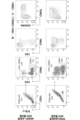

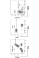
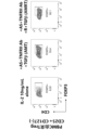


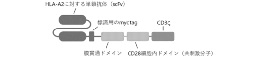
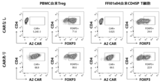


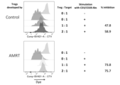


Classifications
-
- A—HUMAN NECESSITIES
- A61—MEDICAL OR VETERINARY SCIENCE; HYGIENE
- A61K—PREPARATIONS FOR MEDICAL, DENTAL OR TOILETRY PURPOSES
- A61K35/00—Medicinal preparations containing materials or reaction products thereof with undetermined constitution
- A61K35/12—Materials from mammals; Compositions comprising non-specified tissues or cells; Compositions comprising non-embryonic stem cells; Genetically modified cells
- A61K35/14—Blood; Artificial blood
- A61K35/17—Lymphocytes; B-cells; T-cells; Natural killer cells; Interferon-activated or cytokine-activated lymphocytes
-
- A—HUMAN NECESSITIES
- A61—MEDICAL OR VETERINARY SCIENCE; HYGIENE
- A61P—SPECIFIC THERAPEUTIC ACTIVITY OF CHEMICAL COMPOUNDS OR MEDICINAL PREPARATIONS
- A61P37/00—Drugs for immunological or allergic disorders
- A61P37/02—Immunomodulators
- A61P37/06—Immunosuppressants, e.g. drugs for graft rejection
-
- C—CHEMISTRY; METALLURGY
- C12—BIOCHEMISTRY; BEER; SPIRITS; WINE; VINEGAR; MICROBIOLOGY; ENZYMOLOGY; MUTATION OR GENETIC ENGINEERING
- C12N—MICROORGANISMS OR ENZYMES; COMPOSITIONS THEREOF; PROPAGATING, PRESERVING, OR MAINTAINING MICROORGANISMS; MUTATION OR GENETIC ENGINEERING; CULTURE MEDIA
- C12N15/00—Mutation or genetic engineering; DNA or RNA concerning genetic engineering, vectors, e.g. plasmids, or their isolation, preparation or purification; Use of hosts therefor
- C12N15/09—Recombinant DNA-technology
- C12N15/11—DNA or RNA fragments; Modified forms thereof; Non-coding nucleic acids having a biological activity
- C12N15/113—Non-coding nucleic acids modulating the expression of genes, e.g. antisense oligonucleotides; Antisense DNA or RNA; Triplex- forming oligonucleotides; Catalytic nucleic acids, e.g. ribozymes; Nucleic acids used in co-suppression or gene silencing
-
- C—CHEMISTRY; METALLURGY
- C12—BIOCHEMISTRY; BEER; SPIRITS; WINE; VINEGAR; MICROBIOLOGY; ENZYMOLOGY; MUTATION OR GENETIC ENGINEERING
- C12N—MICROORGANISMS OR ENZYMES; COMPOSITIONS THEREOF; PROPAGATING, PRESERVING, OR MAINTAINING MICROORGANISMS; MUTATION OR GENETIC ENGINEERING; CULTURE MEDIA
- C12N5/00—Undifferentiated human, animal or plant cells, e.g. cell lines; Tissues; Cultivation or maintenance thereof; Culture media therefor
- C12N5/06—Animal cells or tissues; Human cells or tissues
-
- C—CHEMISTRY; METALLURGY
- C12—BIOCHEMISTRY; BEER; SPIRITS; WINE; VINEGAR; MICROBIOLOGY; ENZYMOLOGY; MUTATION OR GENETIC ENGINEERING
- C12N—MICROORGANISMS OR ENZYMES; COMPOSITIONS THEREOF; PROPAGATING, PRESERVING, OR MAINTAINING MICROORGANISMS; MUTATION OR GENETIC ENGINEERING; CULTURE MEDIA
- C12N5/00—Undifferentiated human, animal or plant cells, e.g. cell lines; Tissues; Cultivation or maintenance thereof; Culture media therefor
- C12N5/10—Cells modified by introduction of foreign genetic material
Definitions
- the present invention relates to a method for producing a cell population containing regulatory T cells, a cell population containing regulatory T cells obtained by the method, a medicament containing the cell population containing regulatory T cells, and the like. (Background of the invention)
- regulatory T cells have an immune response suppressing function, so research and development of cell therapy (Treg therapy) using regulatory T cells has been conducted for the purpose of treating various autoimmune diseases. It is being promoted around the world.
- regulatory T cells are expected to be used for the treatment and prevention of graft-versus-host disease (GvHD), autoimmune diseases, inflammatory diseases, and allergic diseases.
- Regulatory T cells are divided into endogenous regulatory T cells (naturally occurring regulatory T cells (natural Tregs): nTregs, or systemic Tregs: tTregs) and inducible regulatory T cells (natural Tregs).
- regulation T cell (induced Treg): iTreg) It is known that nTregs occur naturally in the thymus, and iTregs are induced to differentiate from naive T cells in peripheral blood by antigen stimulation and cytokines such as IL-2 and TGF- ⁇ . There is. These regulatory T cells are further subdivided according to the type of marker expressed on the cells.
- regulatory T cells When regulatory T cells are removed from a living body, various organ-specific autoimmune diseases spontaneously develop; however, when regulatory T cells are transplanted, the onset of autoimmune diseases is prevented; was thought to play an important role in maintaining immune self-tolerance in the periphery. Subsequently, it has become clear that regulatory T cells can suppress not only autoimmunity but also most immune reactions such as inflammation caused by foreign antigens, rejection reactions caused by transplants, infectious immunity, allergies, and tumor immunity. Furthermore, the transcription factor FOXP3 has now been revealed as a master regulator of regulatory T cells.
- FOXP3 is a master regulator of Tregs, and Tregs constantly express FOXP3, and they are known to be constitutively expressed in conventional T cells (Tconv (conventional T cells), effector T cells, and inflammatory T cells) that do not have a suppressive function. It is known that constitutive expression is not observed in some cases. Therefore, research has been conducted on maintaining the expression of FOXP3 in primary Tregs and inducing FOXP3 expression in primary Tconv or bulk CD4 single positive (SP) T cells, and using compounds and cytokines that have the activity of maintaining and inducing their expression, as well as their combinations. has been reported.
- Tconv conventional T cells
- SP bulk CD4 single positive
- Such compounds and cytokines, and combinations thereof include AS2863619, a CDK8/19 inhibitor (Non-Patent Document 1, Patent Document 1), and rapamycin, an mTOR inhibitor (Non-Patent Document 2, Non-Patent Document 1). 3, non-patent literature 4, non-patent literature 5), TNFR2 agonist antibodies (non-patent literature 4, non-patent literature 6, non-patent literature 7), cytokine TGF- ⁇ (non-patent literature 1, non-patent literature 5), etc. be.
- regulatory T cells derived from pluripotent stem cells compounds and cytokines that maintain FOXP3 expression and combinations thereof, compounds and cytokines that induce FOXP3 expression from T cells that do not express FOXP3, and combinations thereof, etc. unknown.
- an industrial production method with high production efficiency (yield) is desired, regulatory T cells are known to be difficult to proliferate, and an efficient method for inducing them is expected. .
- Non-Patent Document 8 describes IPEX (Immune dysregulation, polyendocrinopathy, enteropathy, It is described that the cells were introduced and administered to a mouse model of IPEX syndrome. As a result, it has been reported that hematopoietic stem cells differentiated into functional regulatory T cells and recovered from the autoimmune phenotype. Further, Patent Document 2 also describes a recombinant lentiviral vector containing a nucleotide sequence encoding a similar human FOXP3 protein.
- Patent Document 3 discloses that expressing Mcl-1 together with Foxp3 is important for improving the survival of Treg.
- Patent Document 4 discloses genetically engineered mammalian cells containing a transgene encoding a differentiation lineage determinant that promotes differentiation into CD4 + Tregs.
- US Pat. No. 5,001,002 discloses the preparation of compositions of T cells from stem cells or progenitor cells by culturing three-dimensional cell aggregates comprising a selected population of stromal cells expressing Notch ligands and a selected population of stem cells or progenitor cells. A method is disclosed.
- An object of the present invention is to provide a method for producing a cell population containing regulatory T cells that can produce regulatory T cells with high efficiency.
- the present inventors have discovered that at least one drug selected from the group consisting of CDK8 and/or CDK19 inhibitors, TNFR2 agonists, mTOR inhibitors, and TGF- ⁇ R agonists.
- a drug selected from the group consisting of CDK8 and/or CDK19 inhibitors, TNFR2 agonists, mTOR inhibitors, and TGF- ⁇ R agonists.
- the present invention was completed based on these findings through further studies, and provides the following methods for producing cell populations containing regulatory T cells, cell populations containing regulatory T cells, medicines, etc. It is.
- a method for producing a cell population containing regulatory T cells comprising: (1) CD4 + T cells derived from pluripotent stem cells in the presence of at least one substance selected from the group consisting of CDK8 and/or CDK19 inhibitors, TNFR2 agonists, mTOR inhibitors, and TGF- ⁇ R agonists.
- a method comprising the step of culturing a cell population comprising: [1a]
- a method for producing a cell population containing regulatory T cells comprising: (1) CD8 + T cells derived from pluripotent stem cells in the presence of at least one substance selected from the group consisting of CDK8 and/or CDK19 inhibitors, TNFR2 agonists, mTOR inhibitors, and TGF- ⁇ R agonists.
- a method comprising the step of culturing a cell population comprising: [2] Step (1), The method according to [1] or [1a], which is carried out in the presence of a CDK8 and/or CDK19 inhibitor and an mTOR inhibitor. [3] Step (1), The method according to [1] or [1a], which is carried out in the presence of a CDK8 and/or CDK19 inhibitor, an mTOR inhibitor, and a TGF- ⁇ R agonist. [4] Step (1), The method according to [1] or [1a], which is carried out in the presence of a CDK8 and/or CDK19 inhibitor, a TNFR2 agonist, an mTOR inhibitor, and a TGF- ⁇ R agonist.
- the CDK8 and/or CDK19 inhibitor is 4-[1-(2-methyl-1H-benzimidazol-5-yl)-1H-imidazo[4,5-c]pyridin-2-yl]- 1,2,5-oxadiazol-3-amine, 3- ⁇ 1-[1-(4-methoxyphenyl)piperidin-4-yl]-4-methyl-1H-imidazo[4,5-c]pyridine -2-yl ⁇ pyrazin-2-amine, and at least one selected from the group consisting of CDK8 and/or CDK19 siRNA, the method according to any one of [1] to [4]. [6] The method according to any one of [1] to [5], wherein the mTOR inhibitor is rapamycin.
- [7] The method according to any one of [1], [1a], and [4] to [6], wherein the TNFR2 agonist is a TNFR2 agonist antibody.
- the TGF- ⁇ R agonist is TGF- ⁇ .
- the regulatory T cells are CD25 + /CD127 ⁇ cells.
- [11b] (3) Adding a foreign gene (especially a gene for expressing a chimeric antigen receptor) to a cell population including pluripotent stem cells (especially iPS cells), CD4 + T cells, CD8 + T cells, or regulatory T cells
- a foreign gene especially a gene for expressing a chimeric antigen receptor
- the method according to any one of [1] to [11a] including the step of introducing.
- step (2) culturing a three-dimensional cell aggregate containing cells capable of differentiating into CD4 + T cells derived from the pluripotent stem cells and stromal cells expressing Notch ligand; [11]; [11b] or the method described in [12].
- step (2) culturing a three-dimensional cell aggregate containing cells capable of differentiating into CD8 + T cells derived from the pluripotent stem cells and stromal cells expressing Notch ligand; [11a]; [11b] or the method described in [12].
- a cell population containing regulatory T cells obtained by the method according to any one of [1] to [13a].
- a medicament comprising a cell population containing regulatory T cells according to [14].
- the medicament according to [15] for use in the prevention and/or treatment of autoimmune disease, graft-versus-host disease, or transplant rejection.
- CD4 + T cells (1) (a) conserveed Non-coding Sequence (CNS) 1, CNS2, and CNS3 of Foxp3 gene; (b) a promoter; and (c) the method according to any one of [1] to [13a], wherein the cell is introduced with an expression construct comprising a nucleic acid encoding FOXP3.
- a method for expanding a cell population containing regulatory T cells comprising: (A) A method comprising the step of culturing a cell population containing regulatory T cells, which are primary cultured cells, in the presence of a CDK8 and/or CDK19 inhibitor, a TNFR2 agonist, and a TGF- ⁇ R agonist.
- step (A) is performed in the presence of a CDK8 and/or CDK19 inhibitor, a TNFR2 agonist, a TGF- ⁇ R agonist, and an mTOR inhibitor.
- the CDK8 and/or CDK19 inhibitor is 4-[1-(2-methyl-1H-benzimidazol-5-yl)-1H-imidazo[4,5-c]pyridin-2-yl]- 1,2,5-oxadiazol-3-amine, 3- ⁇ 1-[1-(4-methoxyphenyl)piperidin-4-yl]-4-methyl-1H-imidazo[4,5-c]pyridine -2-yl ⁇ pyrazin-2-amine, and at least one selected from the group consisting of CDK8 and/or CDK19 siRNA, the method according to any one of [20a] and [20b].
- [20d] The method according to [20b] or [20c], wherein the mTOR inhibitor is rapamycin.
- [20e] The method according to any one of [20a] to [20d], wherein the TNFR2 agonist is a TNFR2 agonist antibody.
- [20f] The method according to any one of [20a] to [20e], wherein the TGF- ⁇ R agonist is TGF- ⁇ .
- [20g] The method according to any one of [20a] to [20f], wherein the regulatory T cells are CD25 + /FOXP3 + cells.
- [20h] The method according to any one of [20a] to [20f], wherein the regulatory T cells are CD25 + /CD127 ⁇ cells.
- FIG. 4 is a flow cytometry (FACS) plot showing constituent cells of a 9-week ATO derived from 4GAD1-4 (top) and a 6-week ATO derived from FFI01s04 (bottom).
- FIG. 3 is a diagram showing a FACS plot showing the profile of PBMC-derived Tconv and 4GAD1-4-derived CD4SP T cells after initial expansion culture in base medium. Bulk (sorts CD4 + ), Tconv (further fractionates and sorts CD25 (-) from CD4 + ), Treg (further fractionates and sorts CD25 + CD127 (-) from CD4 + ) in PBMC of healthy volunteer donors.
- FIG. 3 is a diagram showing a FACS plot showing each fraction of .
- FIG. 3 is a diagram showing the results of expansion culture of 4GAD1-4-derived CD4SP T cells (FACS plot showing FOXP3 expression rate) when AR, ART, AMT, and AMRT were added to the base medium or thereto.
- FIG. 3 shows a FACS plot of origin Tconv (lower row).
- FIG. 3 is a diagram showing FACS plots of 4GAD1-4-derived CD4SP T cells expanded and cultured in base medium (upper row) and 4GAD1-4-derived CD4SP T cells expanded and cultured in base medium with AMRT added (lower row).
- FIG. 3 is a diagram showing the structure of a CAR molecule used in Example 4. FACS plot of PBMC-derived Tregs expanded and cultured with the addition of AMT and iPS cell-derived CD4SP T cells expanded and cultured with the addition of AMRT when CAR was transfected (lower row) and when gene introduction was not performed (upper row)
- FIG. 3 is a diagram showing the structure of a CAR molecule used in Example 4. FACS plot of PBMC-derived Tregs expanded and cultured with the addition of AMT and iPS cell-derived CD4SP T cells expanded and cultured with the addition of AMRT when CAR was transfected (lower row) and when gene introduction was not performed (upper row)
- FIG. 3 is a diagram showing the structure of a CAR molecule used in Example 4. FACS plot of PBMC-derived Tregs expanded and cultured with the addition of AMT and iPS cell-derived CD4SP T cells expanded and cultured with the addition of AMRT when CAR was transfected (low
- Cytotoxic T cells sorted and expanded from the same PBMC used in the in vivo experiments described below, and iPS cell-derived CD4SP T cells expanded and cultured with the addition of IL-2 alone or AMRT.
- CTL Cytotoxic T cells
- iPS cell-derived CD4SP T cells expanded and cultured with the addition of only IL-2, cells in which CAR was not introduced (iPS-CD4T w/o CAR IL-2), and cells in which CAR was also introduced (iPS-CD4T with CAR IL-2), iPS cell-derived CD4SP T cells expanded and cultured with the addition of AMRT, cells in which CAR was not introduced (iPS-CD4T w/o CAR AMRT), and cells also introduced with CAR (iPS-CD4T).
- CAR AMRT PBMC-derived Tregs expanded with AMT and not introduced with CAR
- nTreg CAR with CAR AMT PBMC-derived Tregs expanded with AMT and not introduced with CAR
- CAR nTreg CAR with CAR AMT
- n 3 iPS cell-derived CD4SP T cells that were expanded and cultured with the addition of AMRT, but cells that were not introduced with CAR, cells that were also introduced with CAR, and cells that were expanded and cultured with the addition of AMT that were PBMC-derived Tregs that were not introduced with CAR.
- n 3 iPS cell-derived CD4SP T cells that were expanded and cultured with the addition of AMRT, but cells that were not introduced with CAR, cells that were also introduced with CAR, and cells that were expanded and cultured with the addition of AMT that were PBMC-derived Tregs that were not introduced with CAR.
- n 3 iPS cell-derived CD4SP T cells that were expanded and cultured with the addition of AMRT, but cells that were not introduced with CAR, cells that were also introduced with CAR, and cells that were
- FIG. 3 is a diagram showing the results of expansion culture (graph of expansion magnification) of iPS cell-derived Tregs incorporating CNS-Foxp3 when AMRT was added to the medium and when AMRT was not added.
- FIG. 3 is a diagram showing the results of expansion culture (FACS plot) of iPS cell-derived Tregs incorporating CNS-Foxp3 when AMRT was added to the medium and when AMRT was not added. This is a plot showing the degree of proliferation of target cells when PBMCs derived from another donor are co-cultured with iPS cell-derived Tregs incorporating CNS-Foxp3 that have been expanded and cultured with or without the addition of AMRT to the medium. .
- FIG. 3 is a diagram showing the results of pseudo-time series analysis performed on an integrated data set of organoid cultured cells and thymocytes (trajectory analysis of 3D organoid culture data integrated with human thymocyte data).
- FIG. 3 is a diagram showing the results of pseudo time series analysis performed on an integrated data set of cultured organoid cells and thymocytes (extracted T cell lineage differentiated cells). It is a figure showing the result of pseudo time series analysis performed on the integrated data set of organoid cultured cells and thymocytes (heat map of DEG module based on pseudo time series).
- FIG. 3 is a diagram showing the results of pseudo-time series analysis performed on an integrated data set of organoid cultured cells and thymocytes (trajectory analysis of 3D organoid culture data integrated with human thymocyte data).
- FIG. 3 is a diagram showing the results of pseudo time series analysis performed on an integrated data set of cultured organoid
- FIG. 3 is a diagram showing the results of pseudo-time series analysis performed on an integrated data set of cultured organoid cells and thymocytes (top 10 significant genes of each module cluster).
- FIG. 2 shows transcriptome profiles of differentiated cells in 2D and 3D cultures by single cell RNA sequencing analysis (integrated UMAP of 3D organoids, 2D cultures and human thymocytes).
- Figure 2 shows transcriptome profiles of differentiated cells in 2D and 3D cultures by single cell RNA sequencing analysis (feature plot showing CD4, CD8A and CD8B).
- FIG. 3 shows transcriptome profiles of differentiated cells in 2D and 3D cultures by single-cell RNA sequence analysis (CD4 feature plots of 3D organoid culture samples, thymocytes, and 2D mature cells).
- FIG. 2 shows transcriptome profiles of differentiated cells in 2D and 3D cultures by single cell RNA sequencing analysis (integrated UMAP of 3D organoids, 2D cultures and human thymocytes).
- Figure 2 shows transcriptome profiles of differentiate
- FIG. 4 shows feature plots of designated gene sets for 3D culture, thymocytes, and 2D culture.
- FIG. 4 shows feature plots of designated gene sets for 3D culture, thymocytes, and 2D culture.
- FIG. 4 shows feature plots of designated gene sets for 3D culture, thymocytes, and 2D culture.
- “Comprise(s)” or “comprising” means, but is not limited to, the inclusion of the element following the phrase. Thus, the inclusion of the element following the phrase is implied, but the exclusion of any other element is not implied.
- Consist(s) of” or “consisting of” means to include and be limited to all elements following the phrase. Thus, the phrase “consisting of” indicates that the listed element is required or essential and substantially no other elements are present.
- “consist(s) essentially of or consisting essentially of” includes any element following the phrase and that affects the activity or effect identified in this disclosure for that element. means that it is limited to other elements that do not. Thus, the phrase “consisting essentially of” means that the listed element is required or essential, but that other elements are optional and that they affect the activity or action of the listed element. Indicates that it may or may not exist, depending on whether or not it has an effect.
- culture refers to maintaining and/or proliferating cells in an in vitro environment.
- “Culture” means to sustain and/or proliferate cells outside of a tissue or body, eg, in a cell culture dish or flask.
- culturing a cell population in the presence of a substance refers to, for example, culturing a cell population in a medium containing a substance.
- examples of such culture include culturing in a medium in which only the substance concerned or the substance and other differentiation-inducing factors coexist.
- the substance When adding the substance to the medium, it may be added directly to the medium, or it may be dissolved in an appropriate solvent before use and then added to the medium.
- the substance can also be immobilized on the surface of a substrate or carrier for culturing.
- positive (+) means that the protein or gene is expressed in a detectable amount by techniques known in the art.
- a reporter protein in the case of a protein that is expressed intracellularly and does not appear on the cell surface (for example, a transcription factor or its subunit), a reporter protein can be expressed together with the protein, and the reporter protein can be detected. can be detected.
- Gene detection can be performed using, for example, nucleic acid amplification methods and/or nucleic acid detection methods such as RT-PCR, microarrays, biochips, and RNAseq.
- negative (-) means that the expression level of a protein or gene is below the lower limit of detection by all or any of the above-mentioned known methods, or the degree of expression thereof. This means that the expression is low.
- the lower detection limit for protein or gene expression may differ depending on each technique.
- the degree of protein or gene expression (low expression or high expression) can be determined by comparison with the results of control cells measured under the same conditions. Examples of control cells include PBMC-derived Tregs and iPS cell-derived CD4SP T cells described in the Examples section.
- the level of CD25 expression in a certain cell population can be determined using flow cytometry, and the level of CD25 expression in that cell population can be measured in PBMC-derived Tconv (control: known to have low CD25 expression).
- control known to have low CD25 expression
- the expression level of CD25 is compared with that of the control cells, it can be determined that the cells have low expression, and when the expression is higher than that of the control cells, it can be determined that the cells have high expression.
- the term "marker” refers to a protein or its gene that is specifically expressed on the cell surface, in the cytoplasm, in the nucleus, etc. in a given cell type.
- the marker is preferably a "cell surface marker.”
- Cell surface marker refers to a protein expressed on the cell surface that can be labeled (stained) with a fluorescent substance and allows easy detection, concentration, isolation, etc. of cells expressing the cell surface marker.
- the cell surface marker is a gene that is specifically expressed (positive marker) or not expressed (negative marker) in a given cell type, specifically as mRNA by transcription of the gene in the genome, or by translation of that mRNA. Refers to a substance that is produced (positive marker) or not produced (negative marker) as a protein.
- Detection of such cell surface markers can be performed using immunoassays using antibodies specific to the cell surface markers, such as ELISA, immunostaining, and flow cytometry.
- expression is defined as the transcription and/or translation of a specific nucleotide sequence driven by a promoter within a cell.
- pluripotent stem cells refer to embryonic stem cells (ES cells) and similar pluripotent cells, that is, to various tissues of the body (endoderm, mesoderm, ectoderm). refers to cells that potentially have the ability to differentiate into all germ layers). Examples of cells having pluripotency similar to ES cells include "induced pluripotent stem cells” (sometimes referred to herein as “iPS cells”).
- hematopoietic stem cells are multipotent stem cells that can differentiate into blood cells.
- hematopoietic stem cells mainly exist in the bone marrow and are distributed to white blood cells (neutrophils, eosinophils, basophils, lymphocytes, monocytes, macrophages), red blood cells, platelets, mast cells, dendritic cells, etc. Differentiate.
- hematopoietic stem cells can be positive for CD34 and negative for CD7 (CD34 + /CD7 ⁇ ). Note that in this specification, "/" used in expressions such as “CD34 + /CD7 - " means "and".
- hematopoietic progenitor cells are cells that have the ability to differentiate into blood cell lineage cells, but do not have the same self-renewal ability as stem cells. Hematopoietic progenitor cells exist primarily in the bone marrow in humans. As used herein, hematopoietic progenitor cells (HPC) are positive for CD34 and can be positive for CD43 (CD34 + /CD43 + ). In addition, as described in International Publication No.
- HPC includes CD24, CD62L, CD90, CD143, CD263, Notch3, CD32, CD39, CD49a, CD164, CD317, CD200, CD218a, CD7, CD144, CD56, CD226, CD262 and CD325 may be positive and CD49f, CD51, CD102, CD42b, CD61, CD62P, CD69, CD102 and CD156c may be negative.
- HEC hemogenic endothelial cells
- T progenitor cells refers to hematopoietic cells that are generated in the human body during the process of differentiation from hematopoietic stem cells to CD3-positive T cells, and are positive for CD34 and CD7 (CD34 + /CD7 + cells). Also, as used herein, proT can be negative for CD43, CD1a and/or CD116.
- meodermal progenitor cell means, for example, one in which at least one marker gene selected from the marker genes consisting of T (synonymous with Brachyury), KDR, FOXF1, FLK1, BMP4, MOX1, and SDF1 is expressed. means cell.
- Mesodermal progenitor cells are not distinguished from mesodermal cells, and cells that weakly express the above marker genes may be referred to as mesodermal progenitor cells.
- regulatory T cells have the ability to inhibit activation of effector T cells when stimulated via T cell receptors, and suppress immune responses (immune tolerance). It refers to T cells that control the Regulatory T cells are usually CD25 + /FOXP3 + cells or CD25 + /CD127 ⁇ cells, among which there are CD4 + or CD8 + cells.
- regulatory T cells may be CD4 + cells (i.e., CD4 + /CD25 + /FOXP3 + or CD4 + /CD25 + /CD127 ⁇ ), or CD8 + cells (i.e., CD8 + /CD25 + /FOXP3 + or CD8 + /CD25 + /CD127 ⁇ ), with CD4 + cells being more preferred.
- the transcription factor FOXP3 is known as a master regulator of CD4 + /CD25 + regulatory T cells.
- CD4 + T cell refers to a T cell that is positive for the surface antigen CD4.
- the CD4 + T cells are negative for CD8 (CD4 + /CD8 ⁇ , CD4 single positive (SP)), in which case the T cells are recognized by being positive for the surface antigens CD3 and CD45. Therefore, CD4 + T cells can be identified as cells that are negative for CD8 and positive for CD4, CD3, and CD45 (CD4 + /CD8 ⁇ /CD3 + /CD45 + cells).
- CD4 + T cells are cells that are negative for CD25 and FOXP3 and positive for CD127. Examples of CD4 + T cells include helper T cells.
- CD8 + T cells refers to T cells that are positive for the surface antigen CD8.
- the CD8 + T cells are negative for CD4 (CD4 ⁇ /CD8 + , CD8 single positive (SP)), in which case the T cells are recognized by being positive for the surface antigens CD3 and CD45. Therefore, CD8 + T cells can be identified as cells that are negative for CD4 and positive for CD8, CD3, and CD45 (CD4 ⁇ /CD8 + /CD3 + /CD45 + cells).
- CD8 + T cells are cells that are negative for CD25 and FOXP3 and positive for CD127. Examples of CD8 + T cells include cytotoxic T cells.
- stromal cells refer to cells that constitute the connective tissue that supports parenchymal cells in living tissues, and in particular, stromal cells that can produce ATO that produces T cells, typically bone marrow, thymus, etc. It refers to stromal cells contained in hematopoietic tissues, such as fibroblasts, blood cells, mesenchymal stem cells, endothelial cells, smooth muscle cells, epithelial cells, and tissue stem cells. Stromal cells include established cell lines such as mouse bone marrow cell lines MS-5, OP9, and S17, human stromal cell lines HS-5 and HS-27a, primary cells collected from humans, etc.
- iPS cells pluripotent stem cells
- stromal cells induced to differentiate from human iPS cells are preferred.
- CD4CD8 double-positive T cells refers to T cells that are positive for both surface antigens CD4 and CD8 (CD8 + /CD4 + ); CD4CD8 double-positive T cells can be identified as cells that are positive for CD4, CD8, CD3, and CD45 because they can be recognized by being positive for certain CD3 and CD45 (CD8 + /CD4 + /CD3 + /CD45 + cells).
- CD4CD8 double negative T cells refers to T cells that are negative for both surface antigens CD4 and CD8 (CD8 - /CD4 - ), and T cells are cells that are negative for both surface antigens CD4 and CD8. Since they can be recognized by being positive for certain CD3 and CD45, CD4CD8 double-negative T cells can be identified as cells that are negative for CD4 and CD8 and positive for CD3 and CD45 (CD8 - / CD4 ⁇ /CD3 + /CD45 + cells).
- cell population means two or more cells of the same type or different types.
- Cell population also means a mass of cells of the same or different types.
- the various cells used in the present invention are preferably cells that comply with GMP (Good Manufacturing Practice) standards.
- pluripotent stem cells examples include embryonic stem cells (ES cells), induced pluripotent stem cells (iPS cells), embryonic tumor cells (EC cells), and embryonic germ stem cells ( EG cells), Muse cells, and preferably iPS cells (more preferably human iPS cells).
- ES cells embryonic stem cells
- iPS cells induced pluripotent stem cells
- EC cells embryonic tumor cells
- EG cells embryonic germ stem cells
- Muse cells preferably iPS cells (more preferably human iPS cells).
- the cells may be cells produced by destroying embryos or cells produced without destroying embryos.
- the cells are produced without destroying the embryo.
- ES cells various mouse ES cell lines established by inGenious targeting laboratory, RIKEN (RIKEN), etc. are available for mouse ES cells, and for human ES cells, various mouse ES cell lines established by the University of Wisconsin, Various human ES cell lines established by NIH, RIKEN, Kyoto University, National Center for Child Health and Development, Cellartis, etc. are available.
- human ES cell lines include strains CHB-1 to CHB-12, RUES1, RUES2, and HUES1 to HUES28 distributed by ESI Bio, H1 and H9 strains distributed by WiCell Research, and RIKEN.
- the distributed KhES-1 strain, KhES-2 strain, KhES-3 strain, KhES-4 strain, KhES-5 strain, SSES1 strain, SSES2 strain, SSES3 strain, etc. can be used.
- “Induced pluripotent stem cells” refer to cells obtained by reprogramming mammalian somatic cells or undifferentiated stem cells by introducing specific factors (nuclear reprogramming factors).
- specific factors nuclear reprogramming factors
- Yamanaka et al. established iPS cells by introducing four factors, Oct3/4, Sox2, Klf4, and c-Myc, into mouse fibroblasts.
- Nanog-iPS cells were selected using Nanog expression as an indicator and established Nanog-iPS cells (Okita, K., Ichisaka, T. , and Yamanaka, S. (2007). Nature 448, 313-317.), iPS cells produced by a method that does not contain c-Myc (Nakagawa M, Yamanaka S., et al.
- IPS cells (OKITA KTAT AL.NAT. Methods 2011 MAY; 8 (5): 409-12, OKITA K ET AL. STEM Cells. 31(3):458-66.) can also be used.
- induced pluripotent stem cells Yu J., Thomson JA. et al., Science (2007) 318: 1917-1920.
- induced pluripotent stem cells created by Daley et al. Park IH, Daley GQ. et al., Nature (2007) 451:141-146
- induced pluripotent stem cells created by Sakurada et al. Japanese Unexamined Patent Publication No. 2008-307007, etc.
- iPS cell lines established by NIH, RIKEN, Kyoto University, etc. are available as induced pluripotent stem cell lines.
- human iPS cell lines include RIKEN's HiPS-RIKEN-1A strain, HiPS-RIKEN-2A strain, HiPS-RIKEN-12A strain, Nips-B2 strain, Kyoto University's Ff-I01s04 strain, QHJI strain, and RWMH.
- Nucleic acid can be any molecule as long as it is a polymer of nucleotides and molecules having the same function as the nucleotides, such as RNA, which is a polymer of ribonucleotides, DNA, which is a polymer of deoxyribonucleotides, and ribonucleotides. Examples include a mixed polymer of nucleotides and deoxyribonucleotides, and a nucleotide polymer containing a nucleotide analog, and may also be a nucleotide polymer containing a nucleic acid derivative. Nucleic acids may be single-stranded or double-stranded. Double-stranded nucleic acids also include double-stranded nucleic acids in which one strand hybridizes to the other strand under stringent conditions.
- Nucleotide analogs can be used to improve or stabilize nuclease resistance compared to RNA or DNA, to increase affinity with complementary strand nucleic acids, to increase cell permeability, or to improve visualization. Any molecule may be used as long as it is a modified nucleotide, deoxyribonucleotide, RNA or DNA.
- Nucleotide analogs can be naturally occurring or non-natural molecules, such as sugar-modified nucleotide analogs (e.g., nucleotide analogs substituted with 2'-O-methyl ribose, 2'-O- Nucleotide analogs substituted with propyl ribose, nucleotide analogs substituted with 2'-methoxyethoxyribose, nucleotide analogs substituted with 2'-O-methoxyethyl ribose, 2'-O-[2-(guanidium )ethyl]ribose-substituted nucleotide analogs, 2'-fluoro-ribose-substituted nucleotide analogs, bridged nucleic acid (BNA), locked nucleic acid (LNA), ethylene Ethylene bridged nucleic acid (ENA), peptide nucleic acid (PNA), oxypeptide nucleic acid (OPNA),
- Nucleic acid derivatives can be used to add other chemicals to the nucleic acid in order to improve nuclease resistance, to stabilize it, to increase affinity with complementary strand nucleic acids, to increase cell permeability, or to make it visible. Any molecule may be used as long as it has a substance added thereto, and specific examples include 5'-polyamine addition derivatives, cholesterol addition derivatives, steroid addition derivatives, bile acid addition derivatives, vitamin addition derivatives, Cy5 addition derivatives, Cy3 addition derivatives. , 6-FAM-added derivatives, biotin-added derivatives, and the like.
- the method for producing a cell population containing regulatory T cells of the present invention (herein sometimes simply referred to as "the production method of the present invention") is characterized by including the following steps.
- a CDK8 (cyclin-dependent kinase 8) and/or CDK19 inhibitor is defined as a substance that inhibits the function of CDK8 and/or CDK19, particularly a substance that inhibits the kinase activity of CDK8 and/or CDK19. It may be both inhibitors or it may be either one of the inhibitors.
- CDK8 and/or CDK19 inhibitors include 4-[1-(2-methyl-1H-benzimidazol-5-yl)-1H-imidazo[4,5-c]pyridin-2-yl]-1 , 2,5-oxadiazol-3-amine (AS2863619) (hereinafter sometimes referred to as "Compound 1”), 3- ⁇ 1-[1-(4-methoxyphenyl)piperidin-4-yl]- 4-Methyl-1H-imidazo[4,5-c]pyridin-2-yl ⁇ pyrazin-2-amine (AS3334366) (hereinafter sometimes referred to as "compound 2”), which encodes CDK8 and/or CDK19 siRNA, shRNA, dsRNA, miRNA, antisense nucleic acid for genes and/or their transcription products, and expression vectors expressing these, US Patent No.
- One type of CDK8 and/or CDK19 inhibitor can be used alone or two or more types can be used in combination.
- a method for inhibiting CDK8 and/or CDK19 can also be performed by knocking out the gene encoding CDK8 and/or CDK19.
- methods known in the art such as genome editing using ZFN, TALEN, CRISPR/Cas systems, etc., can be used.
- a TNFR2 (tumor necrosis factor receptor-2) agonist is defined as a substance that can bind to TNFR2 and activate TNFR2 signal transduction, such as antibodies (e.g., anti-TNFR2 antibodies), peptides, and small molecules. Compounds, proteins, etc.
- Examples of the TNFR2 agonist antibody include monoclonal antibodies that bind to TNFR2, such as clone MR2-1 (Hycult Biotech) and clone MAB2261 (R&D Systems).
- a TNFR2 agonist may also be a TNF- ⁇ mutein that only binds TNFR2 as an agonist.
- One type of TNFR2 agonist can be used alone or two or more types can be used in combination.
- the TNFR2 agonist is preferably a TNFR2 agonist antibody.
- the antibody may be a functional fragment thereof, and examples of the functional fragment include Fd, Fv, Fab, F(ab'), F(ab) 2 , F(ab') 2 , single chain Mention may be made of Fv (scFv), diabodies, triabodies, tetrabodies and minibodies.
- Examples of antibodies include those derived from animals such as mouse, rat, cow, rabbit, goat, sheep, and guinea pig.
- the isotype of the antibody is not particularly limited, and examples of the isotype include IgG (IgG1, IgG2, IgG3, IgG4), IgA, IgD, IgE, and IgM.
- the antibody may be either a monoclonal antibody or a polyclonal antibody, preferably a monoclonal antibody, and the antibody may be a humanized antibody, a chimeric antibody, a multispecific antibody (for example, a bispecific antibody). etc.
- Antibodies can be produced by known methods, for example, by constructing an expression vector containing a nucleic acid encoding the antibody and culturing a transformant into which the nucleic acid has been introduced, or by culturing a hybridoma that produces the antibody. It can be produced by culturing.
- An mTOR (mechanistic target of rapamycin) inhibitor is defined as a substance that inhibits the function of mTOR, and includes substances that inhibit the function of mTOR itself, and mTOR complex 1 (mTORC1), which is a complex of mTOR. Contains substances that inhibit the function of mTOR complex 2 (mTORC2).
- mTOR inhibitors include rapamycin or its derivatives, everolimus, temsirolimus, ridaforolimus, sirolimus, KU0063794, AZD805, AZD8055, WYE-354, WAY-600, WYE-687, Pp121, Pp242, Dactolisib, Sapa nisertib, omipalisib , Vistusertib, Torin 1, Torin 2, and the like.
- mTOR inhibitors examples include siRNA, shRNA, dsRNA, miRNA, antisense nucleic acids directed against the mTOR-encoding gene and/or its transcription products, and expression vectors that express these; Also included are siRNAs, shRNAs, dsRNAs, miRNAs, antisense nucleic acids for genes encoding enzymes that are activated and/or transcription products thereof, and expression vectors for expressing these.
- rapamycin or a derivative thereof is preferred, and rapamycin is more preferred.
- One type of mTOR inhibitor can be used alone or two or more types can be used in combination.
- a TGF- ⁇ R (Transforming Growth Factor- ⁇ Receptor) agonist is defined as a substance that can bind to TGF- ⁇ R and activate TGF- ⁇ R signal transduction.
- TGF- ⁇ R agonists include factors belonging to the TGF- ⁇ superfamily, and the TGF- ⁇ superfamily includes the TGF- ⁇ family, the activin family, and the BMP family (bone morphogenetic protein). Examples of such factors belonging to the TGF- ⁇ superfamily include TGF- ⁇ (TGF- ⁇ 1, TGF- ⁇ 2, TGF- ⁇ 3), activin A, activin B, GDF-8, GDF-11, etc. .
- the TGF- ⁇ R agonist is preferably TGF- ⁇ . TGF- ⁇ R agonists can be used alone or in combination of two or more.
- the CDK8 and/or CDK19 inhibitor, TNFR2 agonist, mTOR inhibitor, and TGF- ⁇ R agonist can be used in a free state or a salt state.
- salts include salts with inorganic bases such as sodium salts, magnesium salts, potassium salts, calcium salts, and aluminum salts; salts with organic bases such as methylamine salts, ethylamine salts, and ethanolamine salts; lysine, ornithine, Examples include salts with basic amino acids such as arginine and ammonium salts.
- the salt may be an acid addition salt, and examples thereof include mineral acids such as hydrochloric acid, hydrobromic acid, hydroiodic acid, sulfuric acid, nitric acid, and phosphoric acid; formic acid, acetic acid, Organic acids such as propionic acid, oxalic acid, malonic acid, malic acid, tartaric acid, fumaric acid, succinic acid, lactic acid, maleic acid, citric acid, methanesulfonic acid, ethanesulfonic acid; with acidic amino acids such as aspartic acid, glutamic acid, etc. Examples include acid addition salts.
- CDK8 and/or CDK19 inhibitors, TNFR2 agonists, mTOR inhibitors, and TGF- ⁇ R agonists also include hydrates, solvates, crystal polymorphs, and the like.
- CDK8 and/or CDK19 inhibitors, TNFR2 agonists, mTOR inhibitors, and TGF- ⁇ R agonists contain an asymmetric carbon, they include R-forms, S-forms, and mixtures containing both in arbitrary proportions ( Racemic forms, etc.) are also included.
- siRNA small interfering RNA
- siRNA is a double-stranded oligo RNA consisting of an RNA having a sequence complementary to the nucleotide sequence of the mRNA of a target gene or a partial sequence thereof (target nucleotide sequence) and its complementary strand.
- the first sequence forms a double-stranded structure with the second sequence (small hairpin RNA (shRNA)), and the double-stranded structure of the first sequence and the second sequence.
- siRNA also includes a dumbbell-shaped nucleic acid with a loop structure at both ends.
- the siRNA may have an overhang at the 5' end or 3' end of one or both of the sense strand and antisense strand.
- An overhang is formed by the addition of one to several (eg, 1, 2, or 3) bases at the end of the sense strand and/or antisense strand.
- the base length of siRNA is not particularly limited as long as RNA interference can be induced, and is, for example, 10 to 50 bases, 15 to 30 bases, or 21 to 27 bases on one side.
- siRNA is siRNA that targets mRNA encoding CDK8 and/or CDK19, mTOR, etc., and can be designed based on information on the base sequence of the mRNA, and the sequence can be changed as long as it can induce RNA interference. Not particularly limited. siRNA can be chemically synthesized using known techniques or produced using genetic recombination technology.
- the concentration of the CDK8 and/or CDK19 inhibitor in the medium is not particularly limited as long as regulatory T cells can be produced, and is adjusted as appropriate depending on the type of CDK8 and/or CDK19 inhibitor used.
- the concentration of the CDK8 and/or CDK19 inhibitor is, for example, 0.1 to 300 nM, preferably 0.5 to 150 nM.
- the concentration of the TNFR2 agonist in the medium is not particularly limited as long as regulatory T cells can be produced, and is adjusted as appropriate depending on the type of TNFR2 agonist used.
- the concentration of the TNFR2 agonist is, for example, 0.0001 to 100 ⁇ g/mL, preferably 0.01 to 10 ⁇ g/mL.
- the concentration of the mTOR inhibitor in the medium is not particularly limited as long as regulatory T cells can be produced, and is adjusted as appropriate depending on the type of mTOR inhibitor used.
- the concentration of mTOR inhibitor is, for example, 1-100 nM, preferably 10-100 nM.
- the concentration of the TGF- ⁇ R agonist in the medium is not particularly limited as long as regulatory T cells can be produced, and is adjusted as appropriate depending on the type of TGF- ⁇ R agonist used.
- the concentration of the TGF- ⁇ R agonist is, for example, 0.1-100 ng/mL, preferably 1-50 ng/mL.
- a CDK8 and/or CDK19 inhibitor hereinafter sometimes abbreviated as "A”
- a TNFR2 agonist hereinafter sometimes abbreviated as “M”
- an mTOR inhibitor hereinafter sometimes abbreviated as “ Culture
- T TGF- ⁇ R agonists
- the medium used in the culture in step (1) consists of the aforementioned CDK8 and/or CDK19 inhibitor, TNFR2 agonist, mTOR inhibitor, and TGF- ⁇ R agonist, using the medium used for animal cell culture as a basal medium. It contains at least one substance selected from the group.
- the basal medium is not particularly limited as long as it can be used for culturing animal cells, and examples include AIM V, X-VIVO-15, NeuroBasal, EGM2, TeSR, BME, BGJb, CMRL 1066, Glasgow MEM, and improved MEM. Examples include Zinc Option, IMDM, 199 medium, Eagle MEM, ⁇ MEM, DMEM, Ham, RPMI-1640, Fisher medium, and the like. These media may be used alone or in combination of two or more.
- the medium may contain serum or may be serum-free.
- the medium also contains serum replacements (e.g., albumin, transferrin, Knockout Serum Replacement (KSR), fatty acids, insulin, collagen precursors, trace elements, 2-mercaptoethanol, 3'-thiol glycerol, ITS-Supplement, B27TM). ) supplements, etc.).
- serum replacements e.g., albumin, transferrin, Knockout Serum Replacement (KSR), fatty acids, insulin, collagen precursors, trace elements, 2-mercaptoethanol, 3'-thiol glycerol, ITS-Supplement, B27TM).
- KSR Knockout Serum Replacement
- the medium contains lipids, amino acids (such as non-essential amino acids), L-glutamine, vitamins, growth factors, cytokines (such as IL-2), beads bound with CD3 antibodies and/or CD28 antibodies, antibiotics, and anti-inflammatory agents. It may also contain one or more substances such as oxidizing agents, pyruvate, buffers, inorganic salts, and preferably contains IL-2. It is desirable to use a chemically-defined medium that does not contain unknown components such as serum, as this reduces differences between lots of medium and allows the preparation of cells with stable quality. .
- the concentration of IL-2 is, for example, 1-500 ng/mL, preferably 1-300 ng/mL.
- the concentration is preferably 1 to 100 ng/mL, more preferably 1 to 50 ng/mL, and 3 to 30 ng/mL. mL is more preferred, and 5 to 15 ng/mL is particularly preferred.
- the concentration of IL-2 to be used is, for example, preferably 30 to 300 ng/mL, more preferably 50 to 200 ng/mL.
- the pH of the medium is usually 7.0 to 7.8, preferably 7.2 to 7.6.
- the medium is preferably sterilized by methods such as filtration, ultraviolet irradiation, heat sterilization, and radiation irradiation to prevent contamination.
- Cultivation is performed in the presence or absence of feeder cells.
- the production method of the present invention can be carried out in the absence of feeder cells because regulatory T cells with uniform properties can be stably produced without contamination with substances whose components are not clear. is desirable.
- the culture conditions of the production method of the present invention are not particularly limited, and the culture temperature is, for example, about 37 to 42°C, preferably about 37 to 39°C, and the CO 2 concentration is, for example, 2% to 10%, preferably is 2 to 5%, and the oxygen concentration is, for example, 1 to 20%, preferably 5 to 20%.
- the culture period is not particularly limited, and can be appropriately determined by those skilled in the art while monitoring the number of regulatory T cells, etc., for example, 10 days or more, preferably 1 week or more, or more. Preferably it is 2 weeks or more.
- the upper limit of the culture period is not particularly limited, and is, for example, 35 days or less, preferably 28 days or less, and more preferably 21 days or less.
- passages may be performed as many times as necessary to obtain a desired amount of regulatory T cells, and the medium may be added or replaced.
- Culture in the present invention can be performed using a known CO 2 incubator.
- the culture container is not particularly limited, and can be appropriately selected from plates, dishes, petri dishes, flasks, bags, bottles, tanks (culture vessels), bioreactors, and the like.
- Pluripotent stem cells and cells for producing pluripotent stem cells used in the present invention may be derived from humans or may be derived from mammals other than humans (non-human mammals), Preferably it is a human.
- Non-human mammals include, for example, mice, rats, hamsters, guinea pigs, rabbits, dogs, cats, pigs, cows, horses, sheep, and monkeys.
- universalized iPS cells (having a specific HLA type that does not cause immune rejection in many patients, or having the HLA knocked out) can also be used.
- the cell population containing CD4 + T cells used in the production method of the present invention is a cell population containing CD4 + T cells induced to differentiate from pluripotent stem cells (especially iPS cells (especially human iPS cells)).
- the proportion (number of cells) of CD4 + T cells contained in the cell population containing CD4 + T cells is, for example, 5% or more, preferably 50% or more, more preferably 90% or more, and the upper limit is, for example, It is 100% or less.
- the production method of the present invention may further include the step of separating regulatory T cells in order to concentrate the obtained regulatory T cells.
- Regulatory T cells can be isolated by known methods such as flow cytometry and magnetic cell separation.
- the production method of the present invention it is possible to produce a cell population containing regulatory T cells that has a high content ratio of regulatory T cells.
- the proportion (number of cells) of regulatory T cells contained in a cell population containing regulatory T cells is, for example, 10% or more, preferably 40% or more, more preferably 80% or more, and the upper limit is, for example, It is 90% or less, more preferably 100% or less.
- the content of regulatory cells that exhibit high suppressive function e.g., Helios-positive cells, which are functional markers of suppressive ability, cells that actually exhibit high suppressive ability in vitro and/or in vivo
- Cell populations containing high regulatory T cells can be produced.
- the proportion (number of cells) of regulatory T cells contained in a cell population containing regulatory T cells is, for example, 10% or more, preferably 50% or more, more preferably 80% or more, and the upper limit is, for example, It is 90% or less, more preferably 100% or less.
- the production method of the present invention can also be applied to cell populations containing CD8 + T cells in addition to cell populations containing CD4 + T cells.
- a method for proliferating a cell population containing regulatory T cells includes the following steps. It is characterized by (A) In primary cultured cells in the presence of a CDK8 and/or CDK19 inhibitor, a TNFR2 agonist, and a TGF- ⁇ R agonist, or a CDK8 and/or CDK19 inhibitor, a TNFR2 agonist, a TGF- ⁇ R agonist, and an mTOR inhibitor. A step of culturing a cell population containing certain regulatory T cells.
- the proliferation method of the present invention can be carried out in the same manner as the production method of the present invention described above, unless otherwise specified, except that a cell population containing regulatory T cells, which are primary cultured cells, is used.
- the cell population containing regulatory T cells used in the proliferation method of the present invention is primary cultured cells isolated from living tissues such as bone marrow, umbilical cord blood, and blood.
- the proportion (number of cells) of regulatory T cells contained in a cell population containing regulatory T cells is, for example, 80% or more, preferably 90% or more, more preferably 95% or more, and the upper limit is, for example, It is 100% or less.
- the cell population containing regulatory T cells used in the proliferation method of the present invention may be derived from humans or from mammals other than humans (non-human mammals), preferably from humans. be.
- Non-human mammals include, for example, mice, rats, hamsters, guinea pigs, rabbits, dogs, cats, pigs, cows, horses, sheep, and monkeys.
- step (A) in the presence of a CDK8 and/or CDK19 inhibitor (A), a TNFR2 agonist (M), an mTOR inhibitor (R), and a TGF- ⁇ R agonist (T), or (ii) CDK8 And/or culture is performed in the presence of a CDK19 inhibitor (A), a TNFR2 agonist (M), and a TGF- ⁇ R agonist (T). More preferably, the culture is performed in the presence of AMT.
- the manufacturing method of the present invention includes, before step (1), (2)
- the method may further include the step of inducing differentiation of pluripotent stem cells into a cell population containing CD4 + T cells.
- Induction of differentiation of pluripotent stem cells into a cell population containing CD4 + T cells can be performed according to known methods. Such a method includes, for example, inducing differentiation of pluripotent stem cells into cells capable of differentiating into CD4 + T cells, and then inducing differentiation of the cells capable of differentiating into CD4 + T cells into a cell population containing CD4 + T cells. There are many things you can do.
- Examples of cells capable of differentiating into CD4 + T cells include CD34 + cells, mesodermal progenitor cells, CD4CD8 double-negative T cells, CD4CD8 double-positive T cells, etc., and preferably CD34 + cells or mesodermal precursor cells, More preferred are hematopoietic endothelial cells, hematopoietic stem cells, hematopoietic progenitor cells, T progenitor cells, or mesodermal progenitor cells, and particularly preferred are hematopoietic endothelial cells.
- the above-mentioned CD34 + cells are cells that express CD34 (CD34 + ), particularly cells that express CD34 but do not express CD7 (CD34 + /CD7 ⁇ ).
- Examples of CD34 + cells include hematopoietic endothelial cells, hematopoietic stem cells, and hematopoietic progenitor cells.
- Induction of differentiation of pluripotent stem cells into cells capable of differentiating into CD4 + T cells can be performed according to known methods (for example, see International Publication No. 2021/085576, etc.).
- cells capable of differentiating into CD4 + T cells can be produced by a feeder-free method, and among these, the "embryonic body method (EB)" in which hematopoietic progenitor cells are produced by forming an embryonic body without using feeder cells.
- EB embryonic body method
- pluripotent stem cells can be cultured on feeder cells to induce differentiation into hematopoietic stem cells and/or hematopoietic progenitor cells (see International Publication No. 2011/096482 and International Publication No. 2013/176197).
- the feeder cells are preferably stromal cells from the viewpoint of facilitating induction of mesodermal differentiation.
- the stromal cells are preferably OP9 cells, 10T1/2 cells (C3H10T1/2 cells), etc. from the viewpoint of facilitating differentiation into hematopoietic cells.
- Induction of differentiation of pluripotent stem cells into CD34 + cells can be carried out according to known methods, and when the pluripotent stem cells are iPS cells, for example, WO 2017/221975, WO 2018/ 135646 and Cell Reports 2 (2012) 1722-1735, hematopoietic progenitor cells can be induced to differentiate and CD34 + cells can be produced.
- Examples of methods for inducing differentiation of cells capable of differentiating into CD4 + T cells into a cell population containing CD4 + T cells include the ATO (artificial thymic organoid) method (International Publication No. 2017/075389). (See No., etc.) Such ATO method involves culturing three-dimensional cell aggregates containing cells capable of differentiating into CD4 + T cells and stromal cells expressing Notch ligands, thereby achieving high efficiency. It becomes possible to induce differentiation into CD4 + T cells.
- the ATO method is a known method, and can be implemented with reference to the descriptions in International Publication No. 2017/075389, International Publication No. 2021/085576, etc.
- the ATO method can also be performed on cells capable of differentiating into CD4 + T cells separated according to the method (separation method).
- a nucleic acid for expressing a Notch ligand is introduced into the stromal cells.
- Stromal cells expressing Notch ligands can be produced by introducing the above nucleic acid into stromal cells. It can also be produced by introducing the nucleic acid into pluripotent stem cells (especially iPS cells (especially human iPS cells)) and then differentiating the pluripotent stem cells into stromal cells.
- Examples of the stromal cells used in the production method of the present invention include stromal cells induced to differentiate from pluripotent stem cells (particularly iPS cells (particularly human iPS cells)), and among them, pluripotent stem cells ( In particular, it is desirable to use cells in which iPS cells (particularly human iPS cells) are induced to differentiate into fibroblasts. Induction of differentiation of pluripotent stem cells into stromal cells can be carried out according to known methods, and when the pluripotent stem cells are human iPS cells, for example, as described in PLoS ONE 8(10): e77673, 2013. By the method described above, fibroblasts induced to differentiate from iPS cells can be produced.
- Notch ligands are not particularly limited, and include standard Notch ligands and non-standard Notch ligands described in International Publication No. 2017/075389.
- Standard Notch ligands include, for example, DLL4 (delta-like ligand 4), DLL1 (delta-like ligand 1), JAG1 (Jagged1), JAG2 (Jagged2), and the like. These can be used alone or in combination of two or more.
- Non-canonical Notch ligands include, for example, contactin-1, NOV/CCN3, contactin-6, periostin/OSF-2, DLK2/EGFL9, Pref-1/DLK1/FA1, DNER, thrombospondin-2, MAGP- 1/MFAP2, thrombospondin-3, MAGP-2/MFAP5, thrombospondin-4 and netrin-1. It is preferable to use human DLL4 as the Notch ligand.
- the means for introducing the nucleic acid for expressing the Notch ligand into stromal cells or pluripotent stem cells is not particularly limited, and various known or common methods can be employed.
- the nucleic acid for expressing the Notch ligand is introduced into stromal cells using an expression vector and expressed.
- the expression vector may be linear or circular, and may be a non-viral vector such as a plasmid, a viral vector, or a transposon-based vector.
- expression vectors can be introduced into stromal cells by known methods such as virus infection, calcium phosphate, lipofection, microinjection, and electroporation.
- Expression vectors can be prepared in a form suitable for use in each technique by known means or using an appropriate commercially available kit (according to its instructions).
- the expression vector can be introduced into stromal cells by a virus infection method.
- viral vectors include retrovirus vectors, lentivirus vectors, adenovirus vectors, and adeno-associated virus vectors.
- the vector containing the nucleic acid for expressing the Notch ligand and the packaging vector (plasmid) of each virus are transfected into host cells using a corresponding commercially available kit for recombination. After producing the virus, the resulting recombinant virus may be used to infect stromal cells.
- the expression vector may contain sequences such as a nuclear localization signal (NLS) and a multiple cloning site (MCS), as necessary.
- the expression vector may further contain reporter genes (e.g., genes encoding fluorescent proteins of each color), drug selection genes (e.g., kanamycin resistance gene, ampicillin resistance gene, puromycin resistance gene), suicide genes (e.g., diphtheria A toxin, simplex Herpes thymidine kinase (HSV-TK), carboxypeptidase G2 (CPG2), carboxylesterase (CA), cytosine deaminase (CD), cytochrome P450 (cyt-450), deoxycytidine kinase (dCK), nitroreductase (NR), purine Genes encoding nucleoside phosphorylase (PNP), thymidine phosphorylase (TP), varicella zoster virus th
- reporter genes e.g., genes encoding fluorescent
- Three-dimensional cell aggregates can be formed, for example, by centrifuging cells capable of differentiating into CD4 + T cells and stromal cells expressing Notch ligand. It is preferable that a nucleic acid for expressing a Notch ligand is introduced into the stromal cells.
- the ratio of stromal cells: cells capable of differentiating into CD4 + T cells can be adjusted as appropriate by those skilled in the art depending on the stromal cells used, the type of cells capable of differentiating into CD4 + T cells, etc.; for example, 100:1 ⁇ 1:100, 90:1 ⁇ 1:90, 80:1 ⁇ 1:80, 70:1 ⁇ 1:70, 60:1 ⁇ 1:60, 50:1 ⁇ 1:50, 40: Examples include 1 to 1:40, 30:1 to 1:30, 20:1 to 1:20, and 10:1 to 1:10.
- the ratio is preferably 20:1 to 1:4; In the case of fibroblasts induced to differentiate from cells), the ratio is preferably 4:1 to 1:4, more preferably 1:1.
- the medium for culturing three-dimensional cell aggregates is not particularly limited, and may be, for example, a serum-containing medium, a serum-free medium, or a xeno-free medium, preferably a serum-free medium, especially insulin (and biotin, transferrin, and albumin).
- Basal media for culturing three-dimensional cell aggregates include, for example, AIM V, X-VIVO-15, NeuroBasal, EGM2, TeSR, BME, BGJb, CMRL 1066, Glasgow MEM, Improved MEM Zinc Option, IMDM, 199 Medium, Eagle's MEM, ⁇ MEM, DMEM, Ham, RPMI-1640, Fisher's medium, and the like. These media may be used alone or in combination of two or more.
- the medium may contain serum or may be serum-free.
- the medium also contains serum replacements (e.g., albumin, transferrin, Knockout Serum Replacement (KSR), fatty acids, insulin, collagen precursors, trace elements, 2-mercaptoethanol, 3'-thiol glycerol, ITS-Supplement, B27TM). ) supplements, etc.).
- serum replacements e.g., albumin, transferrin, Knockout Serum Replacement (KSR), fatty acids, insulin, collagen precursors, trace elements, 2-mercaptoethanol, 3'-thiol glycerol, ITS-Supplement, B27TM).
- KSR Knockout Serum Replacement
- the medium may also contain one or more substances such as lipids, amino acids (such as non-essential amino acids), L-glutamine, vitamins, growth factors, cytokines, antibiotics, antioxidants, pyruvate, buffers, and inorganic salts. It can contain. It is desirable to use a chemically-defined medium that does not contain unknown components such as serum, as this reduces differences between lots of medium and allows the preparation of cells with stable quality. .
- culture medium components described in International Publication No. 2017/075389 and the like may be appropriately blended.
- the culture temperature of the three-dimensional cell aggregate is, for example, 20 to 40°C, preferably about 37°C
- the CO 2 concentration is, for example, 2% to 10%, preferably 2 to 5%
- the oxygen concentration is, for example, It is 1 to 20%, preferably 5 to 20%.
- the cell density at the start of culture can be, for example, about 1.0 ⁇ 10 4 to 1.0 ⁇ 10 10 cells/mL.
- the culture period of the three-dimensional cell aggregate can be appropriately adjusted by a person skilled in the art depending on the stromal cells used, the type of cells capable of differentiating into CD4 + T cells, etc., and may be, for example, 4 weeks or more, preferably 5 weeks or more. , more preferably 6 weeks or more, still more preferably 7 weeks or more.
- the upper limit of the culture period is not particularly limited, and is preferably 16 weeks or less, more preferably 14 weeks or less, even more preferably 12 weeks or less, particularly preferably 9 weeks or less.
- CD4 + T cells may be isolated and collected by flow cytometry (typically fluorescence activated cell sorting: FACS) using a fluorescently labeled anti-CD4 antibody and a cell sorter.
- FACS fluorescence activated cell sorting
- CD4SP T cells may be isolated and collected.
- effector T cells (Tconv) that are fractionated as CD25 negative may be isolated and collected.
- step (2) is carried out to induce differentiation of the cell population containing CD8 + T cells.
- CD4 + T cells (a) Foxp3 gene CNS1, CNS2, and CNS3; (b) a promoter; and (c) a cell into which an expression construct comprising a nucleic acid encoding FOXP3 (herein sometimes simply referred to as "CNS-Foxp3”) may be used.
- the cells into which the expression construct is introduced are not particularly limited, and may be at any stage of differentiation, including, for example, CD4 + T cells, pluripotent stem cells, or cells capable of differentiating into CD4 + T cells.
- Methods for introducing the expression construct into CD4 + T cells, pluripotent stem cells, or cells capable of differentiating into CD4 + T cells include the methods described above.
- the expression construct in the present invention is for expressing FOXP3, and includes (a) CNS1 (conserved non-coding sequence 1), CNS2 (conserved non-coding sequence 2) of the Foxp3 gene, and CNS3 (conserved non-coding sequence 3), (b) a promoter, and (c) a nucleic acid encoding FOXP3.
- the expression construct is not particularly limited as long as it can express FOXP3, and in addition to the above (a), (b), and (c), it may include a terminator, a polyadenylation signal, Foxp3 3'UTR, etc. It is preferable that these cells function within the cells into which they are introduced.
- the base sequence of the human Foxp3 gene is available in RefSeq Accession No. They are registered as NM_001114377 (SEQ ID NO: 1) and NM_014009 (SEQ ID NO: 2), and the amino acid sequences are also listed in RefSeq Accession No. They are registered as NP_001107849 (SEQ ID NO: 3) and NP_054728 (SEQ ID NO: 4).
- the RefSeq ID here is registered on the NCBI website.
- the above-mentioned genes include degenerate products and mutants thereof, even if they do not have the base sequences registered in the database as described above, and mutants include genes having the above-mentioned amino acid sequences.
- Variants include 80% or more, 85% or more, 90% or more, 91% or more, 92% or more, 93% or more, 94% or more, 95% or more, 96% or more, 97% or more with respect to the natural amino acid sequence. % or more, 98% or more, or 99% or more of the same amino acid sequence.
- FOXP3 means a protein
- Foxp3 means a gene, but when it is appropriate to interpret FOXP3 and Foxp3 as a gene and a protein, respectively, they mean a gene and a protein.
- the FOXP3-encoding nucleic acid is not particularly limited as long as it is a FOXP3 protein-encoding nucleic acid, and is preferably FOXP3 cDNA.
- CNS1, CNS2, and CNS3 used in the present invention are all derived from the Foxp3 gene.
- CNS1, CNS2 and CNS3 are Foxp3 enhancer elements.
- the promoters used in the present invention are not particularly limited, and include, for example, CAG promoter, ubiquitin gene promoter, Foxp3 gene promoter, EF1 ⁇ promoter, SR ⁇ promoter, SV40 promoter, LTR promoter, CMV (cytomegalovirus) promoter, RSV (Rous sarcoma virus) promoter, MoMuLV (Moloney murine leukemia virus) LTR, HSV-TK (herpes simplex virus thymidine kinase) promoter, etc., and preferably the promoter of the Foxp3 gene.
- Preferred base sequences of CNS1, CNS2, CNS3 and the promoter of the human Foxp3 gene include the base sequences set forth in SEQ ID NOs: 5 to 8, respectively.
- the promoters, CNS1, CNS2, and CNS3 include mutants even if they do not have the above-mentioned base sequences, and the mutants include promoters that have the above-mentioned base sequences, and are equivalent to the promoters, CNS1, CNS2, and CNS3. It is desirable that the biological activity is as follows. Variants include 80% or more, 85% or more, 90% or more, 91% or more, 92% or more, 93% or more, 94% or more, 95% or more, 96% or more, 97% or more with respect to the natural base sequence. % or more, 98% or more, or 99% or more of base sequences.
- the arrangement (order) of the nucleic acids encoding the promoter, CNS1, CNS2, CNS3, and FOXP3 in the expression construct is not particularly limited, and it is preferable that CNS1, CNS2, and CNS3 are located upstream of the promoter, and from the 5' end side. More preferably, the nucleic acids encode CNS1-CNS2-CNS3-promoter-FOXP3 in this order.
- the cells into which the above expression construct is introduced are not particularly limited as long as they are CD4 + T cells, pluripotent stem cells, or cells that can be differentiated into CD4 + T cells, such as pluripotent stem cells, hematopoietic stem cells, hematopoietic endothelium. cells, hematopoietic progenitor cells, T progenitor cells, mesodermal progenitor cells, helper T cells, etc., preferably pluripotent stem cells, more preferably iPS cells.
- the regulatory T cells obtained by the production method and proliferation method of the present invention may be regulatory T cells into which a foreign gene has been introduced.
- a “foreign gene” is a gene that is introduced from the outside in order to cause regulatory T cells to express a desired protein, and can be appropriately selected depending on the use of regulatory T cells.
- the foreign gene can be, for example, a gene for expressing a CAR (chimeric antigen receptor), and can further include a gene for expressing a cytokine and/or a chemokine.
- CARs expressed by regulatory T cells basically have the following characteristics as general or known CARs: (i) an antigen recognition site (e.g., single chain antibody) that recognizes a cell surface antigen of cancer cells; (ii) It is constructed by linking the peptides of the following regions: ) a cell membrane-spanning region, and (iii) a signal transduction region that induces T cell activation, via a spacer as necessary.
- the foreign gene can also be, for example, a gene for expressing an exogenous T cell receptor (TCR).
- Exogenous TCR means that the nucleic acid encoding the exogenous TCR is exogenous to the T cell into which it is introduced, and the amino acid sequence of the exogenous TCR may be the same as the endogenous TCR of the T cell. , may be different.
- One or more types of foreign genes can be introduced (eg, CAR and exogenous TCR).
- Examples of means for introducing foreign genes into cells include the methods described above.
- the cells into which the foreign gene is introduced are not particularly limited and may be at any stage of differentiation, and include, for example, pluripotent stem cells (particularly iPS cells), CD4 + T cells, regulatory T cells, etc. It will be done.
- the cell population containing regulatory T cells produced by the production method and proliferation method of the present invention is useful for treating animals (particularly humans) with abnormally enhanced immune responses, such as X-linked immunity.
- Regulatory T cells produced by the production method of the present invention may be used for autologous or allogeneic transplantation. It may also be used in combination with other drugs.
- the subject from whom cells are isolated for use in producing regulatory T cells must have a similar HLA relationship with the subject to whom the regulatory T cells are administered.
- they are of the same type, and more preferably from the same subject to which the regulatory T cells are administered.
- a medicament containing a cell population containing regulatory T cells (hereinafter sometimes referred to as the medicament of the present invention) can be produced.
- the drug of the present invention is manufactured as a parenteral preparation by mixing an effective amount of regulatory T cells with a pharmaceutically acceptable carrier according to known means (for example, the method described in the Japanese Pharmacopoeia). It is preferable to do so.
- the medicament of the present invention is preferably manufactured as a parenteral preparation such as an injection, a suspension, or an infusion.
- Parenteral administration methods include intravenous, intraarterial, intramuscular, intraperitoneal, and subcutaneous administration.
- Pharmaceutically acceptable carriers include, for example, solvents, bases, diluents, excipients, soothing agents, buffers, preservatives, stabilizers, suspending agents, tonicity agents, surfactants, Examples include solubilizing agents.
- the dosage of the medicament of the present invention can be appropriately determined depending on various conditions such as the patient's weight, age, sex, and symptoms, but usually, the number of cells per dose is determined per dose for a subject weighing 60 kg. , usually 1 ⁇ 10 6 to 1 ⁇ 10 10 pieces, preferably 1 ⁇ 10 7 to 1 ⁇ 10 9 pieces, more preferably 5 ⁇ 10 7 to 5 ⁇ 10 8 pieces. administered to Moreover, it may be administered once or multiple times.
- the medicament of the present invention can be in a known form suitable for parenteral administration, such as an injection or an infusion.
- the medicament of the present invention may contain physiological saline, phosphate buffered saline (PBS), a medium, etc. in order to stably maintain cells.
- PBS phosphate buffered saline
- Examples of the medium include, but are not limited to, RPMI, AIM-V, and X-VIVO10.
- a pharmaceutically acceptable carrier eg, human serum albumin
- preservative e.g., preservative, etc.
- the medicament of the present invention is applicable to mammals including humans.
- Example 1 Induction of differentiation from iPS cells to CD4SP T cells, isolation of CD4SP T cells
- Step i T cell-derived iPS cell line (4GAD1-4) or non-T cell-derived iPS cell line (FFI01s04) containing hematopoietic endothelial cells (HEC) and hematopoietic progenitor cells (HPC) by the Embryonic Body method (EB method)
- HEC hematopoietic endothelial cells
- HPC hematopoietic progenitor cells
- EB method products Embryonic Body method
- Step ii For the iPS cell line 4GAD1-4 derived from T cells, the EB method product on day 5 was subjected to the ATO differentiation culture method, with day 0 being the day on which induction of differentiation into undifferentiated cells was started. For the iPS cell line FFI01s04 derived from non-T cells, the day 7 EB method product was subjected to the ATO differentiation culture method.
- MS5hDLL4 4:1.
- For FFI01s04 mix at a ratio of 1:20, place droplets of the suspension on an insert (Merck Millipore, MILLICELL INSERT 30MM ORGANOTYPIC PTFE 0.4UM 50/PK) and insert. Floating in whole medium The ATO differentiation culture method was started.
- the medium composition is the same as the method described in International Publication No. 2021/085576.
- Step iv When the mixture of the iPS cell-derived EB method product and MS5hDLL4 is cultured for 6 to 9 weeks on the insert suspended in this medium, CD4SP T cells and CD8SP T cells differentiate as shown in Figure 1.
- Step v Suspend ATO at this stage in 2% FBS/1mM EDTA/PBS, sort CD4-positive CD8b-negative cells among CD3-positive TCRab-positive T cells using a cell sorter, and expand the isolated CD4SP T cells.
- Dynabeads Human T-Activator CD3/CD28 manufactured by Thermo Fisher Scientific, hereinafter referred to as "Dynabeads"
- the ratio of beads to cells was 4:1 only in the first expansion culture (1:1 from the second time onwards).
- the medium (hereinafter referred to as "base medium”) is ⁇ -MEM (manufactured by Thermo Fisher Scientific, prepared with MEM-a, nucleosides, powder), 15% fetal bovine serum (FBS), and PSG for a 100-fold dilution.
- ITS manufactured by Thermo Fisher Scientific, Insulin-Transferrin-Selenium (ITS-G) (100X) was added to the same 100-fold dilution as (penicillin, streptomycin, and L-glutamine), and IL-2 A final concentration of 10 ng/mL) and PAA (phosphorylated ascorbic acid) (100 ⁇ g/mL) were added.
- CD4SP T cells derived from iPS cells showed proliferation several hundred to several thousand times during the initial expansion culture.
- the cytokine production ability of these CD4SP T cells was evaluated by intracellular staining flow cytometry.
- PMA 40 ng/mL
- ionomycin 4 ⁇ g/mL
- monensin 2 ⁇ M
- CD4SP T cells derived from 4GAD1-4 increased to 13.24 ⁇ M during stimulation.
- % produced interferon- ⁇ (IFN- ⁇ ) and 23.77% produced interleukin-4 (IL-4) (both partially overlapped).
- Tconv inflammatory T cells
- PBMC peripheral blood mononuclear cell fraction
- IFN- ⁇ interferon- ⁇
- IL-4 interleukin-4
- T cells contained in the peripheral blood mononuclear cell fraction (PBMC) of a human body are TCRab-positive and CD4-positive CD4SP T cells, which are further induced by the above-mentioned It can be fractionated into CD25-negative inflammatory T cells (Tconv) and CD25-positive, CD127-negative regulatory T cells (Treg) that have the function of suppressing inflammation (Figure 3).
- PBMC peripheral blood mononuclear cell fraction
- the base medium contained 32.52 nM of AS2863619 (manufactured by MedChemExpress), 100 nM of rapamycin (manufactured by LKT Labs), 2.5 ⁇ g/mL of TNFR2 antibody (manufactured by Hycult Biotech, clone MR2-1), and 2.5 ⁇ g/mL of TGF- ⁇ (manufactured by PeproTech).
- HEK293-derived 5 ng/mL was added in the following combinations to expand each cell, and the FOXP3 positivity rate after 14 days was analyzed by intracellular staining flow cytometry.
- AR AS2863619+rapamycin ART: AS2863619+rapamycin+TGF- ⁇
- AMT AS2863619 + TNFR2 antibody + TGF- ⁇
- AMRT AS2863619 + rapamycin + TNFR2 antibody + TGF- ⁇
- Tregs were sorted from PBMC, mixed with Dynabeads at a bead:cell ratio of 4:1, suspended in a base medium, and expanded and cultured for 14 days.
- AR, ART, AMT, and AMRT the magnification and FOXP3 positivity rate were analyzed using cell number counting and intracellular staining flow cytometry. The results are shown in Figure 4.
- FOXP3 was not maintained at a sufficiently high level with IL-2 alone, and the necessary proliferation was not obtained when AR and ART were added. On the other hand, when AMT or AMRT was added, FOXP3 expression was maintained at a high level.
- the FOXP3 positive rate decreases compared to AMRT when the IL-2 concentration is 10 ng/mL, but the same FOXP3 expression rate is maintained when the IL-2 concentration is 100 ng/mL. I found out that it drips. Therefore, when PBMC-derived Tregs are expanded and cultured, the IL-2 concentration is then set at 100 ng/mL. The results in this case are shown in FIG.
- the FOXP3-positive cell percentage was 85.6% in AMT and 85.3% in AMRT.
- the magnification is several times higher in AMRT (2.4 times this time) compared to when only IL-2 is added, and in AMT it is tens to hundreds of times higher at higher altitudes (31.5 times this time). did.
- FOXP3 induction using each of the above combinations was performed using CD4SP T cells derived from the iPS cell line 4GAD1-4.
- the FOXP3 positive rate was analyzed using intracellular staining flow cytometry.
- CD4-positive CD8b-negative cells CD4SP T cells
- IL-2 10 ng/mL a base medium
- Example 3 Evaluation of Treg [Study regarding marker expression pattern and inflammatory cytokine secretion ability]
- FOXP3 expression was induced under the conditions described in Example 2 show the CD25-positive, CD127-negative, and CTLA-4-positive expression pattern that is characteristic of Tregs, and whether they were stimulated.
- the cells were analyzed by intracellular staining flow cytometry under conditions stimulated with PMA and ionomycin.
- FIG. 7 shows FACS plots comparing PBMC-derived Tconv expanded and cultured under IL-2 only conditions (base medium) and ART-added conditions, using PBMC-derived Tregs as a control.
- FIG. 8 shows FACS plots comparing 4GAD1-4-derived CD4SP T cells expanded and cultured under IL-2 only conditions (base medium) and conditions in which AMRT was added.
- CNS transcription factor binding site
- TSDR was hardly demethylated in PBMC-derived bulk CD4SP T cells and iPS cell-derived CD4SP T cells expanded with IL-2 alone. That is, the constitutive expression of FOXP3, a transcription factor that controls immunosuppressive functions, is strongly suppressed by methylation. This is consistent with the fact that these two types of cells exhibit a strong tendency to secrete inflammatory cytokines, as shown in Figure 2.
- PBMC-derived bulk CD4SP T cells expanded and cultured with the addition of AMRT showed a higher demethylation rate of 43.3% than when expanded with IL-2 alone;
- the iPS cell-derived CD4SP T cells showed a very high demethylation rate of 86.6%.
- CAR chimeric antigen receptor
- VL light chain
- HL heavy chain
- HLA-A2 as the antigen
- CD28 CD28
- the above-mentioned CAR molecule gene (SEQ ID NO: 9) was introduced into PBMC-derived Tregs and iPS cell-derived CD4SP T cells using a retrovirus vector. Then, CAR-expressing cells were labeled using a PE-conjugated antibody against myc tag (manufactured by Cell Signaling Technology, clone: 9B11), and Anti-PE MicroBeads and MS Columns (both Miltenyi) were used to label CAR-expressing cells. MACS using Biotec) It was concentrated using the following method.
- Tregs graft-versus-host disease
- CTL cytotoxic T cells
- CD8b-positive CTL sorted and expanded from the same PBMC used in the in vivo experiment described below (derived from a donor different from the iPS cell donor and the PBMC donor from which Tregs were sorted) and PBMC-derived Tregs or iPS cell-derived CD4SP T cells (each without CAR introduction or with CAR introduction) were co-cultured. Then, the extent to which CTL proliferation was suppressed was analyzed by flow cytometry using CellTrace Violet Cell Proliferation Kit (manufactured by Thermo Fisher Scientific).
- iPS cell-derived CD4SP T cells expanded and cultured with IL-2 alone did not exhibit a proliferation-suppressing effect on CTLs, regardless of the presence or absence of CAR expression.
- iPS cell-derived CD4SP T cells expanded and cultured with the addition of AMRT showed an effect of suppressing CTL proliferation regardless of the presence or absence of CAR introduction.
- iPS cell-derived CD4SP T cells (without or with CAR introduction) expanded and cultured with the addition of only IL-2, and iPS cells expanded and cultured with the addition of AMRT.
- iPS cell-derived CD4SP T cells (or Treg): CTL They were mixed and co-cultured at ratios of 1:16, 1:8, 1:4, 1:2, 1:1, 2:1, and 4:1. After co-culturing for 96 hours, the fluorescence intensity of CTV was measured using a flow cytometer, and the proportion of divided CTL was calculated.
- %Suppression ⁇ (Percentage of divided CTL (%) when CD4SP T cells are not added) - (Percentage of divided CTL when CD4SP T cells are added at each ratio (%)) /CD4SP Ratio (%) of divided CTL when T cells were not added ⁇ 100 was calculated and graphed. The results are shown in FIG. A larger value indicates that CTL proliferation was more strongly suppressed.
- iPS cell-derived CD4SP T cells did not differ significantly depending on the presence or absence of CAR introduction.
- the suppressive effects of PBMC-derived Tregs were also the same regardless of the presence or absence of CAR introduction.
- iPS cell-derived CD4SP T cells showed a stronger CTL proliferation suppressive effect than PBMC-derived Tregs.
- iPS cell-derived CD4SP T cells expanded and cultured with the addition of AMRT and PBMC-derived Tregs expanded and cultured with the addition of AMT have the function of suppressing CTL proliferation in vitro.
- HLA-A2 positive PBMC HLA- A2-positive PBMC were mixed with 5.0 x 10 6 cells and transplanted by intravenous injection into NSG mice after irradiation with 2.2 Gy. Thereafter, body weight was measured every day from day 0 to day 29, the day of transplantation, to measure the survival period. The results are shown in FIG.
- HLA-A2-positive human PBMCs were confirmed in the peripheral blood of surviving NSG mice, and the GvHD suppression mechanism was solely due to the cytocidal effect of CAR-positive T cells on HLA-A2-positive human PBMCs. It was suggested that this is not the case.
- the expected immunosuppressive ability of PBMC-derived Tregs expanded and cultured in AMT was confirmed, and the iPS cell-derived CD4SP T cells expanded in AMRT were also shown to be able to stimulate PBMC-derived Tregs in actual living bodies. It was confirmed that it has the same immune-suppressing function.
- Example 5 Evaluation using iPS cells incorporating CNS-Foxp3
- Introduction of construct into iPS cells and differentiation of introduced iPS cells into HEC As a cell population containing hematopoietic endothelial cells, Kyoto University iPS cell research iPS cells (Ff-I01s04 strain: derived from peripheral blood mononuclear cells of a healthy individual) donated from A floating cell population differentiated according to Methods) was used.
- a plasmid sequence was designed and synthesized in which a DNA sequence in which the mStrawberry protein sequence in the sequence (MK012431) registered in GenBank was changed to a dTomato sequence was incorporated into a transfer plasmid for producing a third generation lentiviral vector.
- This plasmid was transfected into HEK293 cells along with the packaging plasmid and envelope plasmid for lentiviral vector production, and the supernatant containing the produced lentiviral vector was collected, concentrated by high-speed centrifugation, and introduced into iPS cells.
- a lentiviral vector incorporating CNS-Foxp3 was obtained.
- the Ff-I01s04 strain was seeded in a 24-well plate at 1 ⁇ 10 4 cells/well, protamine was added at a final concentration of 10 ⁇ g/mL, and then a lentivirus solution containing the CNS-Foxp3 gene was directly added. , virus-infected iPS cells were produced.
- the virus-infected Ff-I01s04 strain was seeded at 1 ⁇ 10 6 cells/well in a 6-well plate treated with ultra-low adhesion (Corning) (Day 0), and 10 ⁇ g/ml human was added to EB medium (StemPro34 (Gibco)).
- Insulin 5.5 ⁇ g/ml human transferrin, 5ng/ml sodium selenite (ITS, Gibco), 2mM L-glutamine (Sigma-Aldrich), 45mM ⁇ -monothioglycerol (Nacalai Tesque Co., Ltd.), and 50 ng/ml BMP4 (manufactured by R&D Systems), 50 ng/ml bFGF (manufactured by Fujifilm Wako Pure Chemical Industries, Ltd.), 50 ng/ml VEGF to 50 ⁇ g/ml ascorbic acid 2-phosphate (manufactured by Sigma-Aldrich).
- ITS sodium selenite
- 2mM L-glutamine Sigma-Aldrich
- 45mM ⁇ -monothioglycerol Nacalai Tesque Co., Ltd.
- 50 ng/ml BMP4 manufactured by R&D Systems
- 50 ng/ml bFGF manufactured by Fujifilm Wako Pure Chemical
- Fibroblasts were obtained from iPS cells (Ff-I01s04 strain: derived from peripheral blood mononuclear cells from healthy individuals) provided by the Kyoto University iPS Cell Research Institute in order to be used as feeder cells when inducing differentiation of T cells from iPS cells. Differentiation was induced. Subsequently, cells in which the Notch ligand DLL4 was forcibly expressed under the EF1alpha promoter were produced using a lentivirus.
- the Ff-I01s04 strain was placed at 1 ⁇ 10 6 cells/well in a 6-well plate treated with ultra-low adhesion (manufactured by Corning), and Y-27632 (Fujifilm Wako Pure) was added to AK03N (manufactured by Ajinomoto Co., Ltd.). (manufactured by Yakuhin Co., Ltd.) and CHIR990211 (manufactured by Tocris Bioscience) (Day 0), and the next day, the cells were seeded in Fibroblast medium (alpha-MEM (Invitrogen) containing 20% FBS (manufactured by Corning), 10 ⁇ g).
- Fibroblast medium alpha-MEM (Invitrogen) containing 20% FBS (manufactured by Corning), 10 ⁇ g).
- /ml human insulin, 5.5 ⁇ g/ml human transferrin, 5ng/ml sodium selenite (ITS, Gibco), 2mM L-glutamine, 100U/ml penicillin, 100 ⁇ g/ml streptomycin (PSG, manufactured by Sigma-Aldrich), and 50 ⁇ g/ml ascorbic acid 2-phosphate (manufactured by Sigma-Aldrich)) were seeded in a 10 cm dish (manufactured by Corning) coated with 0.1% gelatin (manufactured by Nacalai Tesque Co., Ltd.) (Day 1) .
- the culture medium was exchanged twice a week and culture was continued for 7 days to obtain a cell population containing fibroblasts (Day 8).
- iPS cell-derived Fibroblasts were seeded in a gelatin-coated 24-well plate at 1 ⁇ 10 5 cells/well, and the next day, a lentivirus solution containing the human DLL4 gene downstream of the EF1alpha promoter was added to a final concentration of 10 ⁇ g.
- iPS cell-derived Fibroblast/DLL4 cells iFibro/DLL4 cells were produced by adding protamine so that the amount of iPS cell-derived Fibroblast/DLL4 cells was /mL.
- the prepared iFibro/DLL4 was co-cultured with the CNS-Foxp3-introduced human iPS cell-derived HEC prepared in (1) above at a cell ratio of 1:1.
- the medium contains 2xB27 supplement (Invitrogen), 1xPSG (Sigma-Aldrich), 1x Glutamax (Invitrogen), 5ng/mL IL-7 (PeproTech), and 5ng/mL FlT at final concentrations.
- 3L (manufactured by PeproTech), 50 ⁇ g/ Using RPMI-1640 (manufactured by Fuji Film Wako Pure Chemical Industries, Ltd.) containing mL ascorbic acid (manufactured by Sigma-Aldrich), a 30 mm Millicell (hydrophilic PTFE, pore size 0.4 ⁇ m, height 5 mm, Merck Millipore Company-made ) was placed in a 6-well plate (TPP) and cultured for 7 weeks while replacing the medium once every 3-4 days.
- RPMI-1640 manufactured by Fuji Film Wako Pure Chemical Industries, Ltd.
- mL ascorbic acid manufactured by Sigma-Aldrich
- a 30 mm Millicell hydrophilic PTFE, pore size 0.4 ⁇ m, height 5 mm, Merck Millipore Company-made
- Expansion culture of iPS-Treg The cells obtained in (2) above after inducing differentiation from iPS cells to Tregs were treated with rapamycin (Merck) 10 nM and anti-TNFR2 antibody (manufactured by Hycult Biotech, clone MR2-1). ) Expansion culture was performed in the following medium containing 3 ⁇ g/mL.
- the CNS-Foxp3-introduced human iPS cell-derived Tregs obtained in (2) above were cultured for 3 days in a 48-well cell culture plate bound with an anti-CD3 antibody (manufactured by eBioscience), and then The cells were replated onto a G-Rex cell culture plate and culture was continued.
- the medium contained FBS (15%, manufactured by Corning), L-glutamine-penicillin-streptomycin solution (1/100, Invitrogen, manufactured by Sigma-Aldrich), and insulin-transferrin-selenium supplement (1/100, manufactured by Invitrogen) at the final concentration of the medium.
- the iPS-Treg obtained in (3) above was further treated under the same conditions (Control) as in (3) above, or AS2863619 (manufactured by MedChemExpress, 30 nM), TGF- ⁇ (manufactured by Peprotech, 5 ng/mL). ) was further expanded and cultured in a medium (AMRT). The culture period was 14 days, and expansion culture was repeated three times. The cell proliferation results at this time are shown in FIG. 16. Furthermore, each cell population on the final day of expansion culture was stained using the following antibody set (Zombie NIR, BV510 CD3, BV421 CD4, PE/Cy7 CD8 ⁇ , APC CD25, FITC FOXP3, PE Helios). The results are shown in FIG. Figure 17 displays Zombie NIR ⁇ CD3 + CD4 + CD8 ⁇ ⁇ cells.
- Example 6 Evaluation of the suppressive function of Tregs derived from iPS cells incorporating CNS-Foxp3 Using Tregs expanded in Example 5, flow cytology after co-culture with allogeneic T cells derived from human PBMC Analysis by metry was performed.
- PBMC untreated or treated with an anti-CD3 antibody derived from a donor different from Treg were stained with CellTrace Violet (Thermo Fisher Scientific) and used as target cells, and Treg (control group of Example 5 or AMRT Groups expanded under the following conditions: PBMC were mixed and co-cultured at cell number ratios of 0:1, 1:1, and 2:1.
- the medium contained FBS (15%, manufactured by Corning), L-glutamine-penicillin-streptomycin solution (1/100, Invitrogen, manufactured by Sigma-Aldrich), and insulin-transferrin-selenium supplement (1/100, manufactured by Invitrogen) at the final concentration of the medium.
- Candidate gene that induces differentiation of CD4SP T cells [Human induced pluripotent stem cell line] Human iPS cell line TKT3V1-7 was derived from antigen-nonspecific CD3 T cells of healthy volunteers using retroviral vectors harboring OCT3/4, KLF-4, SOX-2, and C-MYC. (Molecular Therapy 29 10, 2013 Kaneko S)
- MS5 cells To generate MS5-hDLL4, MS5 cells (K Itoh et al., 1989. Exp Hematol 17, 145-153) were transformed with a retroviral vector encoding full-length human DLL4. Up to 5% of DLL4 expressing cells were sorted by FACS using anti-DLL4 antibody and passaged in DMEM/10% fetal calf serum (FCS). After several weeks of culture, stable expression of DLL4 was confirmed by flow cytometry.
- iPSCs Preparation of iHPCs from iPSCs
- EBs were collected and precipitated at the bottom of the tube, and each well was treated with 10 ng/mL penicillin/streptomycin (manufactured by Sigma-Aldrich), 2 mM Glutamax (manufactured by Thermo Fisher Scientific), and 50 ⁇ g/mL ascorbic acid-2- Phosphoric acid (manufactured by Sigma-Aldrich), 4 ⁇ 10 ⁇ 4 M monothioglycerol (MTG, manufactured by Nacalai Tesque Co., Ltd.), and 1x insulin-transferrin-selenium solution (ITS-G, manufactured by Thermo Fisher Scientific) (EB basics) 50 ng/ml recombinant human (rh) BMP-4 (manufactured by R&D Systems), 50 ng/ml rhVEGF (manufactured by R&D Systems), and 50 ng/ml bFGF (manufactured by Fujifilm Wako Pure Chemical Industries, Ltd.
- the suspension was resuspended in 2 mL of StemPro-34 (manufactured by Thermo Fisher Scientific). After 24 hours, 6 ⁇ M SB431542 (manufactured by Fujifilm Wako Pure Chemical Industries, Ltd.) was added. After 4 days, differentiated EBs were collected, washed, resuspended in 2 mL EB basal medium supplemented with 50 ng/mL rhVEGF, 50 ng/mL rhbFGF, and 50 ng/mL rhSCF (manufactured by R&D Systems) per well, and cultured for 2 days. .
- the differentiated EBs were collected again, washed, and treated with 50 ng/mL rhVEGF, 50 ng/mL rhbFGF, 50 ng/mL rhSCF, 30 ng/mL rhTPO (PeproTech), and 10 ng/mL FLT3L (PeproTech) per well.
- the cells were resuspended in 2 mL of EB basal medium supplemented with . From day 7 onwards, cells were collected every 2 to 3 days and replaced with fresh medium. A 5% CO 2 /5% O 2 /90% N 2 environment was maintained for the first 7 days, and a 5% CO 2 environment thereafter.
- the differentiated iHPCs were filtered through a 40 ⁇ m cell strainer and stored frozen at ⁇ 80° C. using a TC-protector (manufactured by Bio-Rad antibodies).
- the coating solution was removed just before adding the T cell differentiation medium.
- 11 to 14 EBs were collected and dissociated into single cells using TryPLE Select (manufactured by Thermo Fisher Scientific).
- a total of 2000 CD34 + /CD43 + cells were incubated with 15% FBS (Corning), 100x ITS-G (1x), 55 ⁇ M 2-mercaptoethanol (Thermo Fisher Scientific), and 50 ⁇ g/mL ascorbic acid-2-phosphate.
- DL4 cells on day 21 were treated with ⁇ MEM, 15% FBS, 100xITS-G (1x), 50 ⁇ g/mL ascorbic acid-2-phosphate, 100xPSG (1x, manufactured by Sigma-Aldrich), 10ng/mL rhIL-7, 10ng
- the cells were stimulated with 500 ng/mL anti-CD3 monoclonal antibody (clone: OKT3, eBioscience) in a maturation medium consisting of /mL rhIL-2 (PeproTech) and 10 nM dexamethasone (Fuji Pharmaceutical Co., Ltd.). After 3 days, the cells were collected, washed, resuspended in OKT3-free maturation medium, and cultured for 4 days at 37°C in an environment containing 5% CO2 .
- RNA-seq Single cell RNA-seq
- the isolated single cell suspension was subjected to droplet-based massively parallel scRNA-seq using the Chromium Single Cell 50 Reagent Kit according to the manufacturer's instructions (10x Genomics). Briefly, cell suspensions were loaded at 700-1,200 cells/ ⁇ L with the aim of capturing 3,000 cells per well. GEM droplets were generated on a 10x Chromium Controller, each cell was labeled with a specific barcode, and each transcript was labeled with a unique molecular identifier (UMI) during reverse transcription. Barcoded cDNA was isolated by Dynabeads MyOne SILANE bead cleanup mixture. cDNA was amplified by PCR and purified by SPRI bead cleanup.
- UMI unique molecular identifier
- a BAM file (GSE148978) of human thymocyte sample was downloaded from the SRA repository.
- the BAM file was converted into a FASTQ sequence file using bamtofastq software v1.3.2 (10x manufactured by Genomics). Sequence reads were aligned to the human GRCh38 genome reference, and gene tag counts were quantified as UMI using Cell Ranger v5.0.1 software. Cells with a UMI number less than 200 or more than 10,000 or a mitochondrial gene number percentage of 5% or more were excluded from further analysis.
- Human thymocyte datasets were integrated and data processed using Seurat's standard integration method. Clusters consisting of cells with the same UMI number or cells derived from a single donor were removed.
- Genes with different expression levels within a cluster relative to other clusters were defined as having a fold change of 2 times or more and a p value of less than 0.05 by Wilcoxon rank sum test.
- the trajectory graph was obtained by fitting the embedding space of the main UMAP. A pseudo time was determined starting from the node of the DN cell. Moran's I test was used to extract genes with different expression levels on different paths of the trajectory, and the identified genes with different expression levels were classified into gene set modules of co-regulated genes along with the trajectory and pseudo-time.
- CD4SP T cell differentiation by feeder-free culture we compared single-cell transcriptomes of cells obtained from 2D culture (CD8SP only) and organoid culture (CD4SP and CD8SP cells). .
Abstract
Disclosed are: a method for producing a cell population that includes regulatory T cells, said method comprising (1) a step for culturing a cell population including pluripotent stem cell-derived CD4+ T cells in the presence of at least one substance selected from the group consisting of CDK8 and/or CDK19 inhibitors, TNFR2 agonists, mTOR inhibitors, and TGF-βR agonists; a cell population including regulatory T cells obtained by said method; and a drug containing said cell population that includes regulatory T cells.
Description
本発明は、制御性T細胞を含む細胞集団の製造方法、該方法により得られた制御性T細胞を含む細胞集団、当該制御性T細胞を含む細胞集団を含有してなる医薬等に関する。
(発明の背景) The present invention relates to a method for producing a cell population containing regulatory T cells, a cell population containing regulatory T cells obtained by the method, a medicament containing the cell population containing regulatory T cells, and the like.
(Background of the invention)
近年、制御性T細胞(regulatory T cell(Treg))は免疫応答抑制機能を持つため、様々な自己免疫疾患に対する治療目的として、制御性T細胞を用いた細胞治療(Treg therapy)の研究開発が世界で進められている。このように、制御性T細胞は、移植片対宿主病(GvHD)、自己免疫疾患の治療、炎症性疾患、アレルギー性疾患の治療及び予防に用いることが期待されている。
In recent years, regulatory T cells (Tregs) have an immune response suppressing function, so research and development of cell therapy (Treg therapy) using regulatory T cells has been conducted for the purpose of treating various autoimmune diseases. It is being promoted around the world. Thus, regulatory T cells are expected to be used for the treatment and prevention of graft-versus-host disease (GvHD), autoimmune diseases, inflammatory diseases, and allergic diseases.
制御性T細胞は、内在性制御性T細胞(naturally occurring regulatory T cell(natural Treg):nTreg、又はthymic Treg:tTreg)と誘導性制御性T細胞(inducible regulatory T cell(induced Treg):iTreg)の大きく2つに分類され、nTregは胸腺内で自然発生し、iTregは抗原刺激及びIL-2、TGF-βなどのサイトカインにより末梢血中のナイーブT細胞から分化誘導されることが知られている。これらの制御性T細胞は更に、その細胞に発現するマーカーの種類に従い細分化されている。
Regulatory T cells are divided into endogenous regulatory T cells (naturally occurring regulatory T cells (natural Tregs): nTregs, or systemic Tregs: tTregs) and inducible regulatory T cells (natural Tregs). regulation T cell (induced Treg): iTreg) It is known that nTregs occur naturally in the thymus, and iTregs are induced to differentiate from naive T cells in peripheral blood by antigen stimulation and cytokines such as IL-2 and TGF-β. There is. These regulatory T cells are further subdivided according to the type of marker expressed on the cells.
制御性T細胞を生体より除去すると、各種の臓器特異的な自己免疫疾患が自然発症し、その際、制御性T細胞を移植すると、自己免疫疾患の発症が阻止されるため、制御性T細胞は、末梢での免疫自己寛容の維持に重要な働きをしていると考えられていた。その後、制御性T細胞は、自己免疫だけでなく、外来抗原による炎症、移植による拒絶反応、感染免疫、アレルギー、腫瘍免疫などのほとんどの免疫反応を抑制し得ることが明らかになってきた。また、現在、この制御性T細胞のマスターレギュレーターとして、転写因子FOXP3が明らかにされている。
When regulatory T cells are removed from a living body, various organ-specific autoimmune diseases spontaneously develop; however, when regulatory T cells are transplanted, the onset of autoimmune diseases is prevented; was thought to play an important role in maintaining immune self-tolerance in the periphery. Subsequently, it has become clear that regulatory T cells can suppress not only autoimmunity but also most immune reactions such as inflammation caused by foreign antigens, rejection reactions caused by transplants, infectious immunity, allergies, and tumor immunity. Furthermore, the transcription factor FOXP3 has now been revealed as a master regulator of regulatory T cells.
FOXP3はTregのマスターレギュレーターであり、TregではFOXP3が恒常的に発現していること、及び抑制機能を有さない通常型T細胞(Tconv(conventional T細胞)、エフェクターT細胞、炎症性T細胞と称することもある)では恒常的な発現は見られないことが知られている。そこで、primary TregにおけるFOXP3の発現維持及びPrimary Tconv又はbulkのCD4シングルポジティブ(SP) T細胞におけるFOXP3発現誘導に関する研究が行われ、それら発現維持及び発現誘導の活性を有する化合物及びサイトカイン、並びにその組み合わせが報告されている。そのような化合物及びサイトカイン、並びにその組み合わせとしては、CDK8/19阻害薬であるAS2863619(非特許文献1、特許文献1)、mTOR阻害剤であるラパマイシン(Rapamycin)(非特許文献2、非特許文献3、非特許文献4、非特許文献5)、TNFR2アゴニスト抗体(非特許文献4、非特許文献6、非特許文献7)、サイトカインTGF-β(非特許文献1、非特許文献5)などがある。
FOXP3 is a master regulator of Tregs, and Tregs constantly express FOXP3, and they are known to be constitutively expressed in conventional T cells (Tconv (conventional T cells), effector T cells, and inflammatory T cells) that do not have a suppressive function. It is known that constitutive expression is not observed in some cases. Therefore, research has been conducted on maintaining the expression of FOXP3 in primary Tregs and inducing FOXP3 expression in primary Tconv or bulk CD4 single positive (SP) T cells, and using compounds and cytokines that have the activity of maintaining and inducing their expression, as well as their combinations. has been reported. Such compounds and cytokines, and combinations thereof include AS2863619, a CDK8/19 inhibitor (Non-Patent Document 1, Patent Document 1), and rapamycin, an mTOR inhibitor (Non-Patent Document 2, Non-Patent Document 1). 3, non-patent literature 4, non-patent literature 5), TNFR2 agonist antibodies (non-patent literature 4, non-patent literature 6, non-patent literature 7), cytokine TGF-β (non-patent literature 1, non-patent literature 5), etc. be.
しかしながら、多能性幹細胞由来の制御性T細胞については、FOXP3の発現を維持する化合物及びサイトカイン、並びにその組み合わせ、FOXP3を発現しないT細胞からFOXP3発現を誘導する化合物及びサイトカイン、並びにその組み合わせなどは知られていない。また、工業的であって製造効率(収率)の高い製造方法が望まれるところ、制御性T細胞は増殖させることが難しいことが知られており、その効率的な誘導方法が期待されている。
However, for regulatory T cells derived from pluripotent stem cells, compounds and cytokines that maintain FOXP3 expression and combinations thereof, compounds and cytokines that induce FOXP3 expression from T cells that do not express FOXP3, and combinations thereof, etc. unknown. Furthermore, although an industrial production method with high production efficiency (yield) is desired, regulatory T cells are known to be difficult to proliferate, and an efficient method for inducing them is expected. .
非特許文献8では、FOXP3における変異によって引き起こされるIPEX(Immune dysregulation,polyendocrinopathy,enteropathy,X-linked)症候群について、FOXP3の発現を回復させるためにレンチウイルスベクターを使用して自己の造血幹細胞に遺伝子を導入して、当該細胞をIPEX症候群のモデルマウスに投与したことが記載されている。その結果、造血幹細胞が機能的な制御性T細胞に分化し、自己免疫性の表現型から回復したことが報告されている。また、特許文献2にも同様のヒトFOXP3タンパク質をコードするヌクレオチド配列を含む組換えレンチウイルスベクターについて記載されている。
Non-Patent Document 8 describes IPEX (Immune dysregulation, polyendocrinopathy, enteropathy, It is described that the cells were introduced and administered to a mouse model of IPEX syndrome. As a result, it has been reported that hematopoietic stem cells differentiated into functional regulatory T cells and recovered from the autoimmune phenotype. Further, Patent Document 2 also describes a recombinant lentiviral vector containing a nucleotide sequence encoding a similar human FOXP3 protein.
特許文献3では、Foxp3とともに、Mcl-1を発現させることがTregの生存向上に重要であることが開示されている。また、特許文献4では、CD4+Tregへの分化を促進する分化系列決定因子をコードする導入遺伝子を含む遺伝子操作された哺乳動物細胞が開示されている。特許文献5では、Notchリガンドを発現する間質細胞の選択集団と幹細胞又は前駆細胞の選択集団とを含む三次元細胞凝集体を培養することによる、幹細胞又は前駆細胞からT細胞の組成物を調製する方法が開示されている。
Patent Document 3 discloses that expressing Mcl-1 together with Foxp3 is important for improving the survival of Treg. Further, Patent Document 4 discloses genetically engineered mammalian cells containing a transgene encoding a differentiation lineage determinant that promotes differentiation into CD4 + Tregs. US Pat. No. 5,001,002 discloses the preparation of compositions of T cells from stem cells or progenitor cells by culturing three-dimensional cell aggregates comprising a selected population of stromal cells expressing Notch ligands and a selected population of stem cells or progenitor cells. A method is disclosed.
本発明は、高い効率で制御性T細胞を製造することができる、制御性T細胞を含む細胞集団の製造方法を提供することを目的とする。
An object of the present invention is to provide a method for producing a cell population containing regulatory T cells that can produce regulatory T cells with high efficiency.
本発明者らは、上記目的を達成すべく鋭意研究を重ねた結果、CDK8及び/又はCDK19阻害剤、TNFR2アゴニスト、mTOR阻害剤、並びにTGF-βRアゴニストからなる群から選択される少なくとも1種の物質の存在下で多能性幹細胞(特にiPS細胞)由来のCD4+T細胞を含む細胞集団を培養することで、高い効率で制御性T細胞を含む細胞集団を製造できるという知見を得た。
As a result of extensive research to achieve the above object, the present inventors have discovered that at least one drug selected from the group consisting of CDK8 and/or CDK19 inhibitors, TNFR2 agonists, mTOR inhibitors, and TGF-βR agonists. We have found that by culturing a cell population containing CD4 + T cells derived from pluripotent stem cells (particularly iPS cells) in the presence of a substance, a cell population containing regulatory T cells can be produced with high efficiency.
本発明は、これら知見に基づき、更に検討を重ねて完成されたものであり、次の制御性T細胞を含む細胞集団の製造方法、制御性T細胞を含む細胞集団、医薬等を提供するものである。
The present invention was completed based on these findings through further studies, and provides the following methods for producing cell populations containing regulatory T cells, cell populations containing regulatory T cells, medicines, etc. It is.
[1]制御性T細胞を含む細胞集団の製造方法であって、
(1)CDK8及び/又はCDK19阻害剤、TNFR2アゴニスト、mTOR阻害剤、並びにTGF-βRアゴニストからなる群から選択される少なくとも1種の物質の存在下で多能性幹細胞由来のCD4+T細胞を含む細胞集団を培養する工程を含む方法。
[1a]制御性T細胞を含む細胞集団の製造方法であって、
(1)CDK8及び/又はCDK19阻害剤、TNFR2アゴニスト、mTOR阻害剤、並びにTGF-βRアゴニストからなる群から選択される少なくとも1種の物質の存在下で多能性幹細胞由来のCD8+T細胞を含む細胞集団を培養する工程を含む方法。
[2]工程(1)を、
CDK8及び/又はCDK19阻害剤とmTOR阻害剤の存在下で行う、[1]又は[1a]に記載の方法。
[3]工程(1)を、
CDK8及び/又はCDK19阻害剤とmTOR阻害剤とTGF-βRアゴニストの存在下で行う、[1]又は[1a]に記載の方法。
[4]工程(1)を、
CDK8及び/又はCDK19阻害剤とTNFR2アゴニストとmTOR阻害剤とTGF-βRアゴニストの存在下で行う、[1]又は[1a]に記載の方法。
[5]前記CDK8及び/又はCDK19阻害剤が、4-[1-(2-メチル-1H-ベンゾイミダゾール-5-イル)-1H-イミダゾ[4,5-c]ピリジン-2-イル]-1,2,5-オキサジアゾール-3-アミン、3-{1-[1-(4-メトキシフェニル)ピペリジン-4-イル]-4-メチル-1H-イミダゾ[4,5-c]ピリジン-2-イル}ピラジン-2-アミン、並びにCDK8及び/又はCDK19のsiRNAからなる群から選択される少なくとも1種である、[1]~[4]のいずれかに記載の方法。
[6]前記mTOR阻害剤が、ラパマイシンである、[1]~[5]のいずれかに記載の方法。
[7]前記TNFR2アゴニストが、TNFR2アゴニスト抗体である、[1]、[1a]及び[4]~[6]のいずれかに記載の方法。
[8]前記TGF-βRアゴニストが、TGF-βである、[1]、[1a]及び[3]~[7]のいずれかに記載の方法。
[9]前記制御性T細胞が、CD25+/FOXP3+細胞である、[1]~[8]のいずれかに記載の方法。
[9a]前記制御性T細胞が、CD25+/CD127-細胞である、[1]~[8]のいずれかに記載の方法。
[10]前記CD4+T細胞が、CD4+/CD8-T細胞である、[1]~[9]のいずれかに記載の方法。
[10a]前記CD8+T細胞が、CD4-/CD8+T細胞である、[1a]~[9a]のいずれかに記載の方法。
[11]工程(1)の前に、
(2)多能性幹細胞をCD4+T細胞を含む細胞集団に分化誘導する工程を更に含む、[1]~[10]のいずれかに記載の方法。
[11a]工程(1)の前に、
(2)多能性幹細胞をCD8+T細胞を含む細胞集団に分化誘導する工程を更に含む、[1a]~[9a]及び[10a]のいずれかに記載の方法。
[11b](3)多能性幹細胞(特にiPS細胞)、CD4+T細胞、CD8+T細胞又は制御性T細胞を含む細胞集団に外来遺伝子(特にキメラ抗原受容体を発現するための遺伝子)を導入する工程を含む、[1]~[11a]のいずれかに記載の方法。
[12]前記多能性幹細胞が、iPS細胞である、[1]~[11b]のいずれかに記載の方法。
[13]工程(2)において、前記多能性幹細胞由来のCD4+T細胞に分化可能な細胞とNotchリガンドを発現する間質細胞とを含む3次元細胞凝集体を培養する、[11]、[11b]又は[12]に記載の方法。
[13a]工程(2)において、前記多能性幹細胞由来のCD8+T細胞に分化可能な細胞とNotchリガンドを発現する間質細胞とを含む3次元細胞凝集体を培養する、[11a]、[11b]又は[12]に記載の方法。
[14][1]~[13a]のいずれかに記載の方法により得られた、制御性T細胞を含む細胞集団。
[15][14]に記載の制御性T細胞を含む細胞集団を含有してなる、医薬。
[16]自己免疫疾患、移植片対宿主病、若しくは移植拒絶の予防及び/又は治療に用いるための、[15]に記載の医薬。
[17][14]に記載の制御性T細胞を含む細胞集団を、それを必要とする対象に投与することを含む、自己免疫疾患、移植片対宿主病、若しくは移植拒絶の予防及び/又は治療方法。
[18]自己免疫疾患、移植片対宿主病、若しくは移植拒絶の予防及び/又は治療において使用するための、[14]に記載の制御性T細胞を含む細胞集団。
[19]自己免疫疾患、移植片対宿主病、若しくは移植拒絶を予防及び/又は治療するための医薬の製造における、[14]に記載の制御性T細胞を含む細胞集団の使用。
[20]前記CD4+T細胞が、
(1)(a)Foxp3遺伝子のConserved Non-coding Sequence(CNS)1、CNS2、及びCNS3;
(b)プロモーター;及び
(c)FOXP3をコードする核酸
を含む発現構築物が導入された細胞である、[1]~[13a]のいずれかに記載の方法。
[20a]制御性T細胞を含む細胞集団の増殖方法であって、
(A)CDK8及び/又はCDK19阻害剤とTNFR2アゴニストとTGF-βRアゴニストの存在下で初代培養細胞である制御性T細胞を含む細胞集団を培養する工程を含む方法。
[20b]工程(A)を、CDK8及び/又はCDK19阻害剤とTNFR2アゴニストとTGF-βRアゴニストと更にmTOR阻害剤の存在下で行う、[20a]に記載の方法。
[20c]前記CDK8及び/又はCDK19阻害剤が、4-[1-(2-メチル-1H-ベンゾイミダゾール-5-イル)-1H-イミダゾ[4,5-c]ピリジン-2-イル]-1,2,5-オキサジアゾール-3-アミン、3-{1-[1-(4-メトキシフェニル)ピペリジン-4-イル]-4-メチル-1H-イミダゾ[4,5-c]ピリジン-2-イル}ピラジン-2-アミン、並びにCDK8及び/又はCDK19のsiRNAからなる群から選択される少なくとも1種である、[20a]又は[20b]のいずれかに記載の方法。
[20d]前記mTOR阻害剤が、ラパマイシンである、[20b]又は[20c]に記載の方法。
[20e]前記TNFR2アゴニストが、TNFR2アゴニスト抗体である、[20a]~[20d]のいずれかに記載の方法。
[20f]前記TGF-βRアゴニストが、TGF-βである、[20a]~[20e]のいずれかに記載の方法。
[20g]前記制御性T細胞が、CD25+/FOXP3+細胞である、[20a]~[20f]のいずれかに記載の方法。
[20h]前記制御性T細胞が、CD25+/CD127-細胞である、[20a]~[20f]のいずれかに記載の方法。 [1] A method for producing a cell population containing regulatory T cells, comprising:
(1) CD4 + T cells derived from pluripotent stem cells in the presence of at least one substance selected from the group consisting of CDK8 and/or CDK19 inhibitors, TNFR2 agonists, mTOR inhibitors, and TGF-βR agonists. A method comprising the step of culturing a cell population comprising:
[1a] A method for producing a cell population containing regulatory T cells, comprising:
(1) CD8 + T cells derived from pluripotent stem cells in the presence of at least one substance selected from the group consisting of CDK8 and/or CDK19 inhibitors, TNFR2 agonists, mTOR inhibitors, and TGF-βR agonists. A method comprising the step of culturing a cell population comprising:
[2] Step (1),
The method according to [1] or [1a], which is carried out in the presence of a CDK8 and/or CDK19 inhibitor and an mTOR inhibitor.
[3] Step (1),
The method according to [1] or [1a], which is carried out in the presence of a CDK8 and/or CDK19 inhibitor, an mTOR inhibitor, and a TGF-βR agonist.
[4] Step (1),
The method according to [1] or [1a], which is carried out in the presence of a CDK8 and/or CDK19 inhibitor, a TNFR2 agonist, an mTOR inhibitor, and a TGF-βR agonist.
[5] The CDK8 and/or CDK19 inhibitor is 4-[1-(2-methyl-1H-benzimidazol-5-yl)-1H-imidazo[4,5-c]pyridin-2-yl]- 1,2,5-oxadiazol-3-amine, 3-{1-[1-(4-methoxyphenyl)piperidin-4-yl]-4-methyl-1H-imidazo[4,5-c]pyridine -2-yl}pyrazin-2-amine, and at least one selected from the group consisting of CDK8 and/or CDK19 siRNA, the method according to any one of [1] to [4].
[6] The method according to any one of [1] to [5], wherein the mTOR inhibitor is rapamycin.
[7] The method according to any one of [1], [1a], and [4] to [6], wherein the TNFR2 agonist is a TNFR2 agonist antibody.
[8] The method according to any one of [1], [1a] and [3] to [7], wherein the TGF-βR agonist is TGF-β.
[9] The method according to any one of [1] to [8], wherein the regulatory T cells are CD25 + /FOXP3 + cells.
[9a] The method according to any one of [1] to [8], wherein the regulatory T cells are CD25 + /CD127 − cells.
[10] The method according to any one of [1] to [9], wherein the CD4 + T cells are CD4 + /CD8 − T cells.
[10a] The method according to any one of [1a] to [9a], wherein the CD8 + T cells are CD4 − /CD8 + T cells.
[11] Before step (1),
(2) The method according to any one of [1] to [10], further comprising the step of inducing differentiation of pluripotent stem cells into a cell population containing CD4 + T cells.
[11a] Before step (1),
(2) The method according to any one of [1a] to [9a] and [10a], further comprising the step of inducing differentiation of pluripotent stem cells into a cell population containing CD8 + T cells.
[11b] (3) Adding a foreign gene (especially a gene for expressing a chimeric antigen receptor) to a cell population including pluripotent stem cells (especially iPS cells), CD4 + T cells, CD8 + T cells, or regulatory T cells The method according to any one of [1] to [11a], including the step of introducing.
[12] The method according to any one of [1] to [11b], wherein the pluripotent stem cells are iPS cells.
[13] In step (2), culturing a three-dimensional cell aggregate containing cells capable of differentiating into CD4 + T cells derived from the pluripotent stem cells and stromal cells expressing Notch ligand; [11]; [11b] or the method described in [12].
[13a] In step (2), culturing a three-dimensional cell aggregate containing cells capable of differentiating into CD8 + T cells derived from the pluripotent stem cells and stromal cells expressing Notch ligand; [11a]; [11b] or the method described in [12].
[14] A cell population containing regulatory T cells obtained by the method according to any one of [1] to [13a].
[15] A medicament comprising a cell population containing regulatory T cells according to [14].
[16] The medicament according to [15] for use in the prevention and/or treatment of autoimmune disease, graft-versus-host disease, or transplant rejection.
[17] Prevention of autoimmune disease, graft-versus-host disease, or transplant rejection and/or comprising administering the cell population containing regulatory T cells described in [14] to a subject in need thereof. Method of treatment.
[18] A cell population comprising the regulatory T cell according to [14] for use in the prevention and/or treatment of autoimmune disease, graft-versus-host disease, or transplant rejection.
[19] Use of the cell population containing regulatory T cells according to [14] in the manufacture of a medicament for preventing and/or treating autoimmune disease, graft-versus-host disease, or transplant rejection.
[20] The CD4 + T cells,
(1) (a) Conserved Non-coding Sequence (CNS) 1, CNS2, and CNS3 of Foxp3 gene;
(b) a promoter; and (c) the method according to any one of [1] to [13a], wherein the cell is introduced with an expression construct comprising a nucleic acid encoding FOXP3.
[20a] A method for expanding a cell population containing regulatory T cells, the method comprising:
(A) A method comprising the step of culturing a cell population containing regulatory T cells, which are primary cultured cells, in the presence of a CDK8 and/or CDK19 inhibitor, a TNFR2 agonist, and a TGF-βR agonist.
[20b] The method according to [20a], wherein step (A) is performed in the presence of a CDK8 and/or CDK19 inhibitor, a TNFR2 agonist, a TGF-βR agonist, and an mTOR inhibitor.
[20c] The CDK8 and/or CDK19 inhibitor is 4-[1-(2-methyl-1H-benzimidazol-5-yl)-1H-imidazo[4,5-c]pyridin-2-yl]- 1,2,5-oxadiazol-3-amine, 3-{1-[1-(4-methoxyphenyl)piperidin-4-yl]-4-methyl-1H-imidazo[4,5-c]pyridine -2-yl}pyrazin-2-amine, and at least one selected from the group consisting of CDK8 and/or CDK19 siRNA, the method according to any one of [20a] and [20b].
[20d] The method according to [20b] or [20c], wherein the mTOR inhibitor is rapamycin.
[20e] The method according to any one of [20a] to [20d], wherein the TNFR2 agonist is a TNFR2 agonist antibody.
[20f] The method according to any one of [20a] to [20e], wherein the TGF-βR agonist is TGF-β.
[20g] The method according to any one of [20a] to [20f], wherein the regulatory T cells are CD25 + /FOXP3 + cells.
[20h] The method according to any one of [20a] to [20f], wherein the regulatory T cells are CD25 + /CD127 − cells.
本発明によると、高い効率で制御性T細胞を製造することが可能となる。
According to the present invention, it is possible to produce regulatory T cells with high efficiency.
以下、本発明の実施の形態について詳細に説明する。
Hereinafter, embodiments of the present invention will be described in detail.
「~を含む(comprise(s)又はcomprising)」とは、その語句に続く要素の包含を示すがこれに限定されないことを意味する。したがって、その語句に続く要素の包含は示唆するが、他の任意の要素の除外は示唆しない。「~からなる(consist(s) of又はconsisting of)」とは、その語句に続くあらゆる要素を包含し、かつ、これに限定されることを意味する。したがって、「~からなる」という語句は、列挙された要素が要求されるか又は必須であり、他の要素は実質的に存在しないことを示す。「~から本質的になる(consist(s) essentially of又はconsisting essentially of)」とは、その語句に続く任意の要素を包含し、かつ、その要素について本開示で特定された活性又は作用に影響しない他の要素に限定されることを意味する。したがって、「~から本質的になる」という語句は、列挙された要素が要求されるか又は必須であるが、他の要素は任意選択であり、それらが列挙された要素の活性又は作用に影響を及ぼすかどうかに応じて、存在させる場合もあり、存在させない場合もあることを示す。
"Comprise(s)" or "comprising" means, but is not limited to, the inclusion of the element following the phrase. Thus, the inclusion of the element following the phrase is implied, but the exclusion of any other element is not implied. "Consist(s) of" or "consisting of" means to include and be limited to all elements following the phrase. Thus, the phrase "consisting of" indicates that the listed element is required or essential and substantially no other elements are present. "consist(s) essentially of or consisting essentially of" includes any element following the phrase and that affects the activity or effect identified in this disclosure for that element. means that it is limited to other elements that do not. Thus, the phrase "consisting essentially of" means that the listed element is required or essential, but that other elements are optional and that they affect the activity or action of the listed element. Indicates that it may or may not exist, depending on whether or not it has an effect.
本明細書において、「培養」とは、細胞をインビトロ環境において維持し、又は/及び増殖させることを指す。「培養する」とは、組織外又は体外で、例えば、細胞培養ディッシュ又はフラスコ中で細胞を持続させ、又は/及び増殖させることを意味する。
As used herein, "culture" refers to maintaining and/or proliferating cells in an in vitro environment. "Culture" means to sustain and/or proliferate cells outside of a tissue or body, eg, in a cell culture dish or flask.
本明細書において、「物質の存在下で細胞集団を培養する」とは、例えば、物質を含む培地で細胞集団を培養することを指す。そのような培養としては、例えば、当該物質のみ、又は該物質と他の分化誘導因子などとが共存した培地での培養を挙げることができる。当該物質を培地に添加する場合には、培地に直接添加するか、もしくは適切な溶媒にて当該物質を用時溶解した後、培地中に添加してもよい。また、当該物質を培養の際の基板又は担体表面上に固定化して培養することもできる。
As used herein, "culturing a cell population in the presence of a substance" refers to, for example, culturing a cell population in a medium containing a substance. Examples of such culture include culturing in a medium in which only the substance concerned or the substance and other differentiation-inducing factors coexist. When adding the substance to the medium, it may be added directly to the medium, or it may be dissolved in an appropriate solvent before use and then added to the medium. Furthermore, the substance can also be immobilized on the surface of a substrate or carrier for culturing.
本明細書において、「陽性(positive(+))」とは、タンパク質又は遺伝子が当該分野で公知の手法による検出可能量で発現していることを意味する。また、細胞内に発現し、細胞表面には現れないタンパク質(例えば転写因子又はそのサブユニットなど)の場合は、当該タンパク質とともにレポータータンパク質を発現させ、当該レポータータンパク質を検出することによって対象とするタンパク質を検出できる。遺伝子の検出は、例えば、RT-PCR、マイクロアレイ、バイオチップ及びRNAseq等の核酸増幅方法及び/又は核酸検出方法を利用して行うことができる。
As used herein, "positive (+)" means that the protein or gene is expressed in a detectable amount by techniques known in the art. In addition, in the case of a protein that is expressed intracellularly and does not appear on the cell surface (for example, a transcription factor or its subunit), a reporter protein can be expressed together with the protein, and the reporter protein can be detected. can be detected. Gene detection can be performed using, for example, nucleic acid amplification methods and/or nucleic acid detection methods such as RT-PCR, microarrays, biochips, and RNAseq.
本明細書において、「陰性(negative(-))」とは、タンパク質又は遺伝子の発現量が、上記のような公知手法の全てあるいはいずれかによる検出下限値未満であること、あるいはその発現の程度が低発現性であることを意味する。タンパク質又は遺伝子の発現の検出下限値は、各手法により異なりえる。タンパク質又は遺伝子の発現の程度(低発現性であるか、高発現性であるか)は、同一条件で測定した対照細胞の結果との比較により、判断することができる。対照細胞の例としては、実施例の項に記載したPBMC由来Treg及びiPS細胞由来CD4SP T細胞が挙げられる。例えば、ある細胞の集団におけるCD25の発現の程度は、フローサイトメトリーを用い、その細胞集団におけるCD25発現量を、PBMC由来Tconv(対照:CD25低発現性であることが知られている。)におけるCD25発現量と比較し、対照と同等の発現が見られる場合は低発現性であると判断し、対照細胞より発現が高い場合は高発現性であると判断することができる。
As used herein, "negative (-)" means that the expression level of a protein or gene is below the lower limit of detection by all or any of the above-mentioned known methods, or the degree of expression thereof. This means that the expression is low. The lower detection limit for protein or gene expression may differ depending on each technique. The degree of protein or gene expression (low expression or high expression) can be determined by comparison with the results of control cells measured under the same conditions. Examples of control cells include PBMC-derived Tregs and iPS cell-derived CD4SP T cells described in the Examples section. For example, the level of CD25 expression in a certain cell population can be determined using flow cytometry, and the level of CD25 expression in that cell population can be measured in PBMC-derived Tconv (control: known to have low CD25 expression). When the expression level of CD25 is compared with that of the control cells, it can be determined that the cells have low expression, and when the expression is higher than that of the control cells, it can be determined that the cells have high expression.
本明細書において、「マーカー」とは、所定の細胞型において細胞表面、細胞質内、核内等に特異的に発現されるタンパク質又はその遺伝子を意味する。マーカーは、好ましくは「細胞表面マーカー」である。「細胞表面マーカー」とは、蛍光物質により標識(染色)可能であり、細胞表面マーカーを発現している細胞の検出、濃縮、単離等を容易に行える、細胞表面に発現するタンパク質を指す。当該細胞表面マーカーとは、所定の細胞型において特異的に発現する(陽性マーカー)又は発現しない(陰性マーカー)遺伝子、具体的にはゲノム中の当該遺伝子の転写によるmRNAとして、又はそのmRNAの翻訳によるタンパク質として、生成する(陽性マーカー)又は生成しない(陰性マーカー)物質を指す。
As used herein, the term "marker" refers to a protein or its gene that is specifically expressed on the cell surface, in the cytoplasm, in the nucleus, etc. in a given cell type. The marker is preferably a "cell surface marker." "Cell surface marker" refers to a protein expressed on the cell surface that can be labeled (stained) with a fluorescent substance and allows easy detection, concentration, isolation, etc. of cells expressing the cell surface marker. The cell surface marker is a gene that is specifically expressed (positive marker) or not expressed (negative marker) in a given cell type, specifically as mRNA by transcription of the gene in the genome, or by translation of that mRNA. Refers to a substance that is produced (positive marker) or not produced (negative marker) as a protein.
このような細胞表面マーカーの検出は、当該細胞表面マーカーに特異的な抗体を用いた免疫学的アッセイ、例えば、ELISA、免疫染色、フローサイトメトリーを利用して行うことができる。
Detection of such cell surface markers can be performed using immunoassays using antibodies specific to the cell surface markers, such as ELISA, immunostaining, and flow cytometry.
本明細書において、「発現(expression)」とは、細胞内のプロモーターにより駆動される特定のヌクレオチド配列の転写及び/又は翻訳として定義される。
As used herein, "expression" is defined as the transcription and/or translation of a specific nucleotide sequence driven by a promoter within a cell.
本明細書において、「多能性幹細胞(pluripotent stem cell)」とは、胚性幹細胞(ES細胞)及びこれと同様の分化多能性、すなわち生体の様々な組織(内胚葉、中胚葉、外胚葉の全て)に分化する能力を潜在的に有する細胞を指す。ES細胞と同様の分化多能性を有する細胞としては、「人工多能性幹細胞」(本明細書中、「iPS細胞」と称することもある)が挙げられる。
As used herein, "pluripotent stem cells" refer to embryonic stem cells (ES cells) and similar pluripotent cells, that is, to various tissues of the body (endoderm, mesoderm, ectoderm). refers to cells that potentially have the ability to differentiate into all germ layers). Examples of cells having pluripotency similar to ES cells include "induced pluripotent stem cells" (sometimes referred to herein as "iPS cells").
本明細書において、「造血幹細胞(HSC)」とは、血球系細胞に分化し得る多能性幹細胞(multipotent stem cell)である。造血幹細胞は、ヒト生体内では、主として骨髄に存在し、白血球(好中球、好酸球、好塩基球、リンパ球、単球、マクロファージ)、赤血球、血小板、肥満細胞、樹状細胞等に分化する。本明細書において、造血幹細胞(HSC)はCD34が陽性であり、CD7が陰性であり得る(CD34+/CD7-)。なお、本明細書中、「CD34+/CD7-」などの表記において用いられる「/」とは「及び」を意味する。
As used herein, "hematopoietic stem cells (HSC)" are multipotent stem cells that can differentiate into blood cells. In the human body, hematopoietic stem cells mainly exist in the bone marrow and are distributed to white blood cells (neutrophils, eosinophils, basophils, lymphocytes, monocytes, macrophages), red blood cells, platelets, mast cells, dendritic cells, etc. Differentiate. As used herein, hematopoietic stem cells (HSCs) can be positive for CD34 and negative for CD7 (CD34 + /CD7 − ). Note that in this specification, "/" used in expressions such as "CD34 + /CD7 - " means "and".
本明細書において、本明細書において「造血前駆細胞」(HPC)とは、血球系細胞への分化能は有するが、幹細胞ほどの自己再生能は有さない細胞である。造血前駆細胞は、ヒト生体内では、主として骨髄に存在する。本明細書において、造血前駆細胞(HPC)はCD34が陽性であり、CD43が陽性であり得る(CD34+/CD43+)。また、HPCは、国際公開第2018/199186号に記載されているように、CD24、CD62L、CD90、CD143、CD263、Notch3、CD32、CD39、CD49a、CD164、CD317、CD200、CD218a、CD7、CD144、CD56、CD226、CD262及びCD325が陽性であり得、CD49f、CD51、CD102、CD42b、CD61、CD62P、CD69、CD102及びCD156cが陰性であり得る。
As used herein, "hematopoietic progenitor cells" (HPC) are cells that have the ability to differentiate into blood cell lineage cells, but do not have the same self-renewal ability as stem cells. Hematopoietic progenitor cells exist primarily in the bone marrow in humans. As used herein, hematopoietic progenitor cells (HPC) are positive for CD34 and can be positive for CD43 (CD34 + /CD43 + ). In addition, as described in International Publication No. 2018/199186, HPC includes CD24, CD62L, CD90, CD143, CD263, Notch3, CD32, CD39, CD49a, CD164, CD317, CD200, CD218a, CD7, CD144, CD56, CD226, CD262 and CD325 may be positive and CD49f, CD51, CD102, CD42b, CD61, CD62P, CD69, CD102 and CD156c may be negative.
本明細書において、「造血性内皮細胞(hemogenic endothelial cell:HEC)」とは、CD34を発現し、CD43、CD184及びCD73を発現しない(CD34+/CD43-/CD184-/CD73-)細胞である(なお、CD34+/CD43-/CD184-/CD73-細胞はCD7を発現しないため、CD34+/CD43-/CD184-/CD73-細胞はCD34+/CD7-/CD43-/CD184-/CD73-細胞とも表される)。
As used herein, "hemogenic endothelial cells (HEC)" are cells that express CD34 but do not express CD43, CD184 and CD73 (CD34 + /CD43 - /CD184 - /CD73 - ) (Note that since CD34 + /CD43 - /CD184 - /CD73 - cells do not express CD7, CD34 + /CD43 - /CD184 - /CD73 - cells are called CD34 + /CD7 - /CD43 - /CD184 - /CD73 - cells. ).
本明細書において、「T前駆細胞(proT)」とは、ヒト生体内においては造血幹細胞からCD3陽性T細胞へ分化する過程で生じる造血系細胞を意味し、CD34及びCD7が陽性である(CD34+/CD7+細胞)。また、本明細書において、proTはCD43、CD1a及び/又はCD116が陰性であり得る。
As used herein, "T progenitor cells (proT)" refers to hematopoietic cells that are generated in the human body during the process of differentiation from hematopoietic stem cells to CD3-positive T cells, and are positive for CD34 and CD7 (CD34 + /CD7 + cells). Also, as used herein, proT can be negative for CD43, CD1a and/or CD116.
本明細書において、「中胚葉前駆細胞」とは、例えば、T(Brachyuryと同義)、KDR、FOXF1、FLK1、BMP4、MOX1及びSDF1から成るマーカー遺伝子から選択される少なくとも一つのマーカー遺伝子が発現する細胞を意味する。中胚葉前駆細胞は、中胚葉細胞と区別されるものではなく、上記マーカー遺伝子の発現が弱いものを中胚葉前駆細胞と称してもよい。
As used herein, the term "mesodermal progenitor cell" means, for example, one in which at least one marker gene selected from the marker genes consisting of T (synonymous with Brachyury), KDR, FOXF1, FLK1, BMP4, MOX1, and SDF1 is expressed. means cell. Mesodermal progenitor cells are not distinguished from mesodermal cells, and cells that weakly express the above marker genes may be referred to as mesodermal progenitor cells.
本明細書において、「制御性T細胞」とは、T細胞受容体を介する刺激を受けたときに、エフェクターT細胞の活性化を阻害し得る能力を有し、免疫応答の抑制(免疫寛容)を司っているT細胞を意味する。制御性T細胞は、通常CD25+/FOXP3+細胞又はCD25+/CD127-細胞であり、その中に、CD4+又はCD8+の細胞が存在する。本発明において、制御性T細胞は、CD4+の細胞(すなわち、CD4+/CD25+/FOXP3+又はCD4+/CD25+/CD127-)であってもよいし、CD8+の細胞(すなわち、CD8+/CD25+/FOXP3+又はCD8+/CD25+/CD127-)であってもよく、CD4+の細胞がより好ましい。また、CD4+/CD25+制御性T細胞のマスターレギュレーターとして、転写因子FOXP3が知られている。
As used herein, "regulatory T cells" have the ability to inhibit activation of effector T cells when stimulated via T cell receptors, and suppress immune responses (immune tolerance). It refers to T cells that control the Regulatory T cells are usually CD25 + /FOXP3 + cells or CD25 + /CD127 − cells, among which there are CD4 + or CD8 + cells. In the present invention, regulatory T cells may be CD4 + cells (i.e., CD4 + /CD25 + /FOXP3 + or CD4 + /CD25 + /CD127 − ), or CD8 + cells (i.e., CD8 + /CD25 + /FOXP3 + or CD8 + /CD25 + /CD127 − ), with CD4 + cells being more preferred. Furthermore, the transcription factor FOXP3 is known as a master regulator of CD4 + /CD25 + regulatory T cells.
本明細書において「CD4+T細胞」とは、T細胞のうち、表面抗原のCD4が陽性である細胞を意味する。好ましくは、CD4+T細胞はCD8が陰性であり(CD4+/CD8-、CD4シングルポジティブ(SP))、その場合、T細胞は、表面抗原であるCD3及びCD45が陽性であることによって認識することができることから、CD4+T細胞は、CD8が陰性であり、CD4、CD3及びCD45が陽性である細胞として同定することができる(CD4+/CD8-/CD3+/CD45+細胞)。CD4+T細胞は、CD25及びFOXP3が陰性であり、CD127が陽性である細胞である。CD4+T細胞としては、例えば、ヘルパーT細胞が挙げられる。
As used herein, the term "CD4 + T cell" refers to a T cell that is positive for the surface antigen CD4. Preferably, the CD4 + T cells are negative for CD8 (CD4 + /CD8 − , CD4 single positive (SP)), in which case the T cells are recognized by being positive for the surface antigens CD3 and CD45. Therefore, CD4 + T cells can be identified as cells that are negative for CD8 and positive for CD4, CD3, and CD45 (CD4 + /CD8 − /CD3 + /CD45 + cells). CD4 + T cells are cells that are negative for CD25 and FOXP3 and positive for CD127. Examples of CD4 + T cells include helper T cells.
本明細書において「CD8+T細胞」とは、T細胞のうち、表面抗原のCD8が陽性である細胞を意味する。好ましくは、CD8+T細胞はCD4が陰性であり(CD4-/CD8+、CD8シングルポジティブ(SP))、その場合、T細胞は、表面抗原であるCD3及びCD45が陽性であることによって認識することができることから、CD8+T細胞は、CD4が陰性であり、CD8、CD3及びCD45が陽性である細胞として同定することができる(CD4-/CD8+/CD3+/CD45+細胞)。CD8+T細胞はCD25及びFOXP3が陰性であり、CD127が陽性である細胞である。CD8+T細胞としては、例えば、細胞傷害性T細胞が挙げられる。
As used herein, "CD8 + T cells" refers to T cells that are positive for the surface antigen CD8. Preferably, the CD8 + T cells are negative for CD4 (CD4 − /CD8 + , CD8 single positive (SP)), in which case the T cells are recognized by being positive for the surface antigens CD3 and CD45. Therefore, CD8 + T cells can be identified as cells that are negative for CD4 and positive for CD8, CD3, and CD45 (CD4 − /CD8 + /CD3 + /CD45 + cells). CD8 + T cells are cells that are negative for CD25 and FOXP3 and positive for CD127. Examples of CD8 + T cells include cytotoxic T cells.
本明細書において「間質細胞」とは、生体組織において実質細胞を支える結合組織を構成する細胞であって、特にT細胞を産生するATOを作製できる間質細胞、代表的には骨髄、胸腺等の造血系の組織に含まれる間質細胞を指し、例えば、線維芽細胞、血球細胞、間葉系幹細胞、内皮細胞、平滑筋細胞、上皮細胞、組織幹細胞などが挙げられる。間質細胞は、マウス骨髄細胞株であるMS-5、OP9、S17、ヒト間質細胞株であるHS-5、HS-27aなどの株化細胞、ヒト等から採取された初代細胞、及びヒト等の多能性幹細胞(iPS細胞等)から分化誘導された細胞であってもよく、ヒト等の多能性幹細胞から分化誘導された細胞の場合、ヒトiPS細胞から分化誘導された間質細胞(特にヒトiPS細胞から分化誘導された線維芽細胞)が好ましい。
As used herein, "stromal cells" refer to cells that constitute the connective tissue that supports parenchymal cells in living tissues, and in particular, stromal cells that can produce ATO that produces T cells, typically bone marrow, thymus, etc. It refers to stromal cells contained in hematopoietic tissues, such as fibroblasts, blood cells, mesenchymal stem cells, endothelial cells, smooth muscle cells, epithelial cells, and tissue stem cells. Stromal cells include established cell lines such as mouse bone marrow cell lines MS-5, OP9, and S17, human stromal cell lines HS-5 and HS-27a, primary cells collected from humans, etc. They may be cells induced to differentiate from pluripotent stem cells (iPS cells, etc.) such as humans, etc. In the case of cells induced to differentiate from pluripotent stem cells such as humans, stromal cells induced to differentiate from human iPS cells (Especially fibroblasts induced to differentiate from human iPS cells) are preferred.
本明細書において、「CD4CD8両陽性T細胞」とは、T細胞のうち、表面抗原のCD4及びCD8が共に陽性である細胞(CD8+/CD4+)を意味し、T細胞は、表面抗原であるCD3及びCD45が陽性であることによって認識することができることから、CD4CD8両陽性T細胞は、CD4、CD8、CD3及びCD45が陽性である細胞として同定することができる(CD8+/CD4+/CD3+/CD45+細胞)。
As used herein, "CD4CD8 double-positive T cells" refers to T cells that are positive for both surface antigens CD4 and CD8 (CD8 + /CD4 + ); CD4CD8 double-positive T cells can be identified as cells that are positive for CD4, CD8, CD3, and CD45 because they can be recognized by being positive for certain CD3 and CD45 (CD8 + /CD4 + /CD3 + /CD45 + cells).
本明細書において、「CD4CD8両陰性T細胞」とは、T細胞のうち、表面抗原のCD4及びCD8が共に陰性である細胞(CD8-/CD4-)を意味し、T細胞は、表面抗原であるCD3及びCD45が陽性であることによって認識することができることから、CD4CD8両陰性T細胞は、CD4及びCD8が陰性であり、CD3及びCD45が陽性である細胞として同定することができる(CD8-/CD4-/CD3+/CD45+細胞)。
As used herein, "CD4CD8 double negative T cells" refers to T cells that are negative for both surface antigens CD4 and CD8 (CD8 - /CD4 - ), and T cells are cells that are negative for both surface antigens CD4 and CD8. Since they can be recognized by being positive for certain CD3 and CD45, CD4CD8 double-negative T cells can be identified as cells that are negative for CD4 and CD8 and positive for CD3 and CD45 (CD8 - / CD4 − /CD3 + /CD45 + cells).
本明細書において、「細胞集団(population)」とは、同じ種類又は異なる種類の2以上の細胞を意味する。「細胞集団(population)」は、同じ種類又は異なる種類の細胞の一塊(mass)をも意味する。
As used herein, "cell population" means two or more cells of the same type or different types. "Cell population" also means a mass of cells of the same or different types.
本発明で用いる各種の細胞は、治療への適用の観点からは、GMP(Good Manufacturing Practice)規格の細胞であることが好ましい。
From the viewpoint of therapeutic application, the various cells used in the present invention are preferably cells that comply with GMP (Good Manufacturing Practice) standards.
多能性幹細胞としては、例えば、胚性幹細胞(embryonic stem cell:ES細胞)、人工多能性幹細胞(induced pluripotent stem cell:iPS細胞)、胚性腫瘍細胞(EC細胞)、胚性生殖幹細胞(EG細胞)、Muse細胞が挙げられ、好ましくはiPS細胞(より好ましくは、ヒトiPS細胞)である。上記多能性幹細胞がES細胞又はヒト胚に由来する任意の細胞である場合、その細胞は胚を破壊して作製された細胞であっても、又は胚を破壊することなく作製された細胞であってもよく、好ましくは、胚を破壊することなく作製された細胞である。
Examples of pluripotent stem cells include embryonic stem cells (ES cells), induced pluripotent stem cells (iPS cells), embryonic tumor cells (EC cells), and embryonic germ stem cells ( EG cells), Muse cells, and preferably iPS cells (more preferably human iPS cells). When the above-mentioned pluripotent stem cells are ES cells or any cells derived from human embryos, the cells may be cells produced by destroying embryos or cells produced without destroying embryos. Preferably, the cells are produced without destroying the embryo.
「ES細胞」としては、マウスES細胞であれば、inGenious targeting laboratory社、理研(理化学研究所)等が樹立した各種マウスES細胞株が利用可能であり、ヒトES細胞であれば、ウィスコンシン大学、NIH、理研、京都大学、国立成育医療研究センター、Cellartis社などが樹立した各種ヒトES細胞株が利用可能である。例えば、ヒトES細胞株としては、ESI Bio社が分譲するCHB-1~CHB-12株、RUES1株、RUES2株、HUES1~HUES28株等、WiCell Researchが分譲するH1株、H9株等、理研が分譲するKhES-1株、KhES-2株、KhES-3株、KhES-4株、KhES-5株、SSES1株、SSES2株、SSES3株等を利用することができる。
As for "ES cells", various mouse ES cell lines established by inGenious targeting laboratory, RIKEN (RIKEN), etc. are available for mouse ES cells, and for human ES cells, various mouse ES cell lines established by the University of Wisconsin, Various human ES cell lines established by NIH, RIKEN, Kyoto University, National Center for Child Health and Development, Cellartis, etc. are available. For example, human ES cell lines include strains CHB-1 to CHB-12, RUES1, RUES2, and HUES1 to HUES28 distributed by ESI Bio, H1 and H9 strains distributed by WiCell Research, and RIKEN. The distributed KhES-1 strain, KhES-2 strain, KhES-3 strain, KhES-4 strain, KhES-5 strain, SSES1 strain, SSES2 strain, SSES3 strain, etc. can be used.
「人工多能性幹細胞」とは、哺乳動物体細胞又は未分化幹細胞に、特定の因子(核初期化因子)を導入して再プログラミングすることにより得られる細胞を指す。現在、「人工多能性幹細胞」には様々なものがあり、山中らにより、マウス線維芽細胞にOct3/4・Sox2・Klf4・c-Mycの4因子を導入することにより、樹立されたiPS細胞(Takahashi K,Yamanaka S.,Cell,(2006)126: 663-676)のほか、同様の4因子をヒト線維芽細胞に導入して樹立されたヒト細胞由来のiPS細胞(Takahashi K,Yamanaka S.,et al.Cell,(2007)131:861-872.)、上記4因子導入後、Nanogの発現を指標として選別し、樹立したNanog-iPS細胞(Okita, K.,Ichisaka, T., and Yamanaka, S.(2007).Nature 448,313-317.)、c-Mycを含まない方法で作製されたiPS細胞(Nakagawa M,Yamanaka S.,et al.Nature Biotechnology,(2008)26,101-106)、ウイルスフリー法で6因子を導入して樹立されたiPS細胞(Okita K et al.Nat. Methods 2011 May;8(5):409-12,Okita K et al.Stem Cells.31(3):458-66.)も用いることができる。また、Thomsonらにより作製されたOCT3/4・SOX2・NANOG・LIN28の4因子を導入して樹立された人工多能性幹細胞(Yu J.,Thomson JA.et al.,Science(2007)318:1917-1920.)、Daleyらにより作製された人工多能性幹細胞(Park IH,Daley GQ.et al.,Nature(2007)451:141-146)、桜田らにより作製された人工多能性幹細胞(日本国特開2008-307007号公報)等も用いることができる。
"Induced pluripotent stem cells" refer to cells obtained by reprogramming mammalian somatic cells or undifferentiated stem cells by introducing specific factors (nuclear reprogramming factors). Currently, there are various types of "induced pluripotent stem cells", and Yamanaka et al. established iPS cells by introducing four factors, Oct3/4, Sox2, Klf4, and c-Myc, into mouse fibroblasts. In addition to human cell-derived iPS cells (Takahashi K, Yamanaka S., Cell, (2006) 126: 663-676), human cell-derived iPS cells established by introducing the same four factors into human fibroblasts Cell, (2007) 131:861-872.) After introducing the above four factors, Nanog-iPS cells were selected using Nanog expression as an indicator and established Nanog-iPS cells (Okita, K., Ichisaka, T. , and Yamanaka, S. (2007). Nature 448, 313-317.), iPS cells produced by a method that does not contain c-Myc (Nakagawa M, Yamanaka S., et al. Nature Biot technology, (2008) 26 , 101-106), IPS cells (OKITA KTAT AL.NAT. Methods 2011 MAY; 8 (5): 409-12, OKITA K ET AL. STEM Cells. 31(3):458-66.) can also be used. In addition, induced pluripotent stem cells (Yu J., Thomson JA. et al., Science (2007) 318: 1917-1920.), induced pluripotent stem cells created by Daley et al. (Park IH, Daley GQ. et al., Nature (2007) 451:141-146), induced pluripotent stem cells created by Sakurada et al. (Japanese Unexamined Patent Publication No. 2008-307007), etc. can also be used.
この他、公開されているすべての論文(例えば、Shi Y.,Ding S.,et al.,Cell Stem Cell,(2008)Vol3,Issue 5,568-574;Kim JB.,Scholer HR.,et al.,Nature,(2008)454,646-650;Huangfu D.,Melton,DA.,et al.,Nature Biotechnology,(2008)26,No 7,795-797)、あるいは特許(例えば、日本国特開2008-307007号公報、日本国特開2008-283972号公報、米国特許出願公開第2008/2336610号明細書、米国特許出願公開第2009/047263号明細書、国際公開第2007/069666号、国際公開第2008/118220号、国際公開第2008/124133号、国際公開第2008/151058号、国際公開第2009/006930号、国際公開第2009/006997号、国際公開第2009/007852号)に記載されている当該分野で公知の人工多能性幹細胞のいずれも用いることができる。
In addition, all published papers (for example, Shi Y., Ding S., et al., Cell Stem Cell, (2008) Vol 3, Issue 5, 568-574; Kim JB., Scholar HR., et al. al., Nature, (2008) 454, 646-650; Huangfu D., Melton, DA., et al., Nature Biotechnology, (2008) 26, No. 7, 795-797), or patents (for example, Japanese Japanese Patent Application Publication No. 2008-307007, Japanese Patent Application Publication No. 2008-283972, US Patent Application Publication No. 2008/2336610, US Patent Application Publication No. 2009/047263, International Publication No. 2007/069666, International Publication No. 2008/118220, International Publication No. 2008/124133, International Publication No. 2008/151058, International Publication No. 2009/006930, International Publication No. 2009/006997, International Publication No. 2009/007852) Any of the induced pluripotent stem cells known in the art can be used.
人工多能性幹細胞株としては、NIH、理研、京都大学等が樹立した各種iPS細胞株が利用可能である。例えば、ヒトiPS細胞株であれば、理研のHiPS-RIKEN-1A株、HiPS-RIKEN-2A株、HiPS-RIKEN-12A株、Nips-B2株、京都大学のFf-I01s04株、QHJI株、RWMH株、DRXT株、RJWI株、YZWJ株、ILCL株、GLKV株、253G1株、201B7株、409B2株、454E2株、606A1株、610B1株、648A1株等が挙げられる。
Various iPS cell lines established by NIH, RIKEN, Kyoto University, etc. are available as induced pluripotent stem cell lines. For example, human iPS cell lines include RIKEN's HiPS-RIKEN-1A strain, HiPS-RIKEN-2A strain, HiPS-RIKEN-12A strain, Nips-B2 strain, Kyoto University's Ff-I01s04 strain, QHJI strain, and RWMH. DRXT strain, RJWI strain, YZWJ strain, ILCL strain, GLKV strain, 253G1 strain, 201B7 strain, 409B2 strain, 454E2 strain, 606A1 strain, 610B1 strain, 648A1 strain, and the like.
「核酸」とは、ヌクレオチド及び該ヌクレオチドと同等の機能を有する分子が重合した分子であればいかなるものでもよく、例えば、リボヌクレオチドの重合体であるRNA、デオキシリボヌクレオチドの重合体であるDNA、リボヌクレオチド及びデオキシリボヌクレオチドが混合した重合体、及びヌクレオチド類似体を含むヌクレオチド重合体を挙げることができ、さらに、核酸誘導体を含むヌクレオチド重合体であってもよい。核酸は、一本鎖核酸又は二本鎖核酸であってもよい。二本鎖核酸には、一方の鎖に対し、他方の鎖がストリンジェントな条件でハイブリダイズする二本鎖核酸も含まれる。
"Nucleic acid" can be any molecule as long as it is a polymer of nucleotides and molecules having the same function as the nucleotides, such as RNA, which is a polymer of ribonucleotides, DNA, which is a polymer of deoxyribonucleotides, and ribonucleotides. Examples include a mixed polymer of nucleotides and deoxyribonucleotides, and a nucleotide polymer containing a nucleotide analog, and may also be a nucleotide polymer containing a nucleic acid derivative. Nucleic acids may be single-stranded or double-stranded. Double-stranded nucleic acids also include double-stranded nucleic acids in which one strand hybridizes to the other strand under stringent conditions.
ヌクレオチド類似体としては、RNA又はDNAと比較して、ヌクレアーゼ耐性の向上又は、安定化させるため、相補鎖核酸とのアフィニティーを上げるため、あるいは細胞透過性を上げるため、あるいは可視化させるために、リボヌクレオチド、デオキシリボヌクレオチド、RNA又はDNAに修飾を施した分子であればいかなる分子でもよい。ヌクレオチド類似体としては、天然に存在する分子でも非天然の分子でもよく、例えば、糖部修飾ヌクレオチド類似体(例:2’-O-メチルリボースで置換されたヌクレオチド類似体、2’-O-プロピルリボースで置換されたヌクレオチド類似体、2’-メトキシエトキシリボースで置換されたヌクレオチド類似体、2’-O-メトキシエチルリボースで置換されたヌクレオチド類似体、2’-O-[2-(グアニジウム)エチル]リボースで置換されたヌクレオチド類似体、2’-フルオロリボースで置換されたヌクレオチド類似体、架橋構造型人工核酸(BNA:Bridged Nucleic Acid)、ロックド人工核酸(LNA:Locked Nucleic Acid)、エチレン架橋構造型人工核酸(ENA:Ethylene bridged nucleic acid)、ペプチド核酸(PNA)、オキシペプチド核酸(OPNA)、ペプチドリボ核酸(PRNA))、リン酸ジエステル結合修飾ヌクレオチド類似体(例:ホスフォロチオエート結合に置換されたヌクレオチド類似体、N3’-P5’ホスフォアミデート結合に置換されたヌクレオチド類似体)等が挙げられる。
Nucleotide analogs can be used to improve or stabilize nuclease resistance compared to RNA or DNA, to increase affinity with complementary strand nucleic acids, to increase cell permeability, or to improve visualization. Any molecule may be used as long as it is a modified nucleotide, deoxyribonucleotide, RNA or DNA. Nucleotide analogs can be naturally occurring or non-natural molecules, such as sugar-modified nucleotide analogs (e.g., nucleotide analogs substituted with 2'-O-methyl ribose, 2'-O- Nucleotide analogs substituted with propyl ribose, nucleotide analogs substituted with 2'-methoxyethoxyribose, nucleotide analogs substituted with 2'-O-methoxyethyl ribose, 2'-O-[2-(guanidium )ethyl]ribose-substituted nucleotide analogs, 2'-fluoro-ribose-substituted nucleotide analogs, bridged nucleic acid (BNA), locked nucleic acid (LNA), ethylene Ethylene bridged nucleic acid (ENA), peptide nucleic acid (PNA), oxypeptide nucleic acid (OPNA), peptide ribonucleic acid (PRNA)), phosphodiester bond-modified nucleotide analogs (e.g. phosphorothioate) nucleotide analogs substituted with a bond, nucleotide analogs substituted with a N3'-P5' phosphoramidate bond), and the like.
核酸誘導体としては、核酸に比べ、ヌクレアーゼ耐性を向上させるため、安定化させるため、相補鎖核酸とのアフィニティーを上げるため、細胞透過性を上げるため、あるいは可視化させるために、該核酸に別の化学物質を付加した分子であればいかなる分子でもよく、その具体例としては、5’-ポリアミン付加誘導体、コレステロール付加誘導体、ステロイド付加誘導体、胆汁酸付加誘導体、ビタミン付加誘導体、Cy5付加誘導体、Cy3付加誘導体、6-FAM付加誘導体、ビオチン付加誘導体等を挙げることができる。
Nucleic acid derivatives can be used to add other chemicals to the nucleic acid in order to improve nuclease resistance, to stabilize it, to increase affinity with complementary strand nucleic acids, to increase cell permeability, or to make it visible. Any molecule may be used as long as it has a substance added thereto, and specific examples include 5'-polyamine addition derivatives, cholesterol addition derivatives, steroid addition derivatives, bile acid addition derivatives, vitamin addition derivatives, Cy5 addition derivatives, Cy3 addition derivatives. , 6-FAM-added derivatives, biotin-added derivatives, and the like.
本発明の制御性T細胞を含む細胞集団の製造方法(本明細書において、単に「本発明の製造方法」と称することがある)は、以下の工程を含むことを特徴とする。
(1)CDK8及び/又はCDK19阻害剤、TNFR2アゴニスト、mTOR阻害剤、並びにTGF-βRアゴニストからなる群から選択される少なくとも1種の物質の存在下で多能性幹細胞由来のCD4+T細胞を含む細胞集団を培養する工程。 The method for producing a cell population containing regulatory T cells of the present invention (herein sometimes simply referred to as "the production method of the present invention") is characterized by including the following steps.
(1) CD4 + T cells derived from pluripotent stem cells in the presence of at least one substance selected from the group consisting of CDK8 and/or CDK19 inhibitors, TNFR2 agonists, mTOR inhibitors, and TGF-βR agonists. A step of culturing a cell population containing.
CDK8(cyclin-dependent kinase 8)及び/又はCDK19阻害剤とは、CDK8及び/又はCDK19の機能を阻害する物質、特にCDK8及び/又はCDK19のキナーゼ活性を阻害する物質として定義され、CDK8及びCDK19の両方の阻害剤であってもよいし、又はいずれか一方の阻害剤であってもよい。CDK8及び/又はCDK19阻害剤としては、例えば、4-[1-(2-メチル-1H-ベンゾイミダゾール-5-イル)-1H-イミダゾ[4,5-c]ピリジン-2-イル]-1,2,5-オキサジアゾール-3-アミン(AS2863619)(以下、「化合物1」と称することもある)、3-{1-[1-(4-メトキシフェニル)ピペリジン-4-イル]-4-メチル-1H-イミダゾ[4,5-c]ピリジン-2-イル}ピラジン-2-アミン(AS3334366)(以下、「化合物2」と称することもある)、CDK8及び/又はCDK19をコードする遺伝子及び/又はそれらの転写産物に対するsiRNA、shRNA、dsRNA、miRNA、アンチセンス核酸及びこれらを発現する発現ベクター、米国特許第8598344号明細書、国際公開第2013/116786号、Proc.Natl.Acad.Sci.U.S.A.109 13799-13804(2012)、国際公開第2013/001310号、国際公開第2013/040153号、国際公開第2014/029726号、国際公開第2014063778号、国際公開第2014/072435号、国際公開第2014/090692号、国際公開第2014/106606号、国際公開第2014/123900号、国際公開第2014/154723号、国際公開第2014/194245号、国際公開第2015/049325号、国際公開第2015/100420号、国際公開第2015/144290号、国際公開第2015/159937号、国際公開第2015/159938号(特に、(2E)-N-(2-フルオロ-4-(モルホリン-4-イルメチル)フェニル)-3-(4-(1-メチル-1H-ピラゾール-4-イル)ピリジン-3-イル)アクリルアミド(以下、「化合物3」と称することがある)及び(2E)-3-(4-(1-シクロプロピル-1H-ピラゾール-4-イル)ピリジン-3-イル)-N-(1-メチル-1H-インドール-5-イル)アクリルアミド(以下、「化合物4」と称することがある))、国際公開第2016/009076号、国際公開第2018/159805号(特に、8-(2,4-ジフルオロフエノキシ)-1-メチル-4,5-ジヒドロ-1H-チエノ[3,4-g]インダゾール-6-カルボキサミド(以下、「化合物5」と称することがある)、4-((4-フルオロフェニル)スルホニル)-3-(2-(イミダゾ[1,2-b]ピリダジン-6-イルスルファ二ル)エチル)-3,4-ジヒドロキノキサリン-2(1H)-オン(以下、「化合物6」と称することがある)、2-(ベンジルアミノ)-4-(1H-ピロロ[2,3-b]ピリジン-3-イル)ベンズアミド(以下、「化合物7」と称することがある)、3-(3-(ベンジルオキシ)フェニル)-1H-ピロロ[2,3-b]ピリジン(以下、「化合物8」と称することがある)、4-(4-(2,3-ジヒドロ-1,4-ベンゾジオキシン-6-イル)-1H-ピラゾール-3-イル)ベンゼン-1,3-ジオール(特にそのトリフルオロ酢酸塩)(以下、「化合物9」と称することがある)、N-ブチル-8-(4-メトキシフェニル)-1,6-ナフチリジン-2-カルボキサミド及び8-(4-メチルフェニル)-N,N-ジプロピル-1,6-ナフチリジン-2-カルボキサミド(特にそのトリフルオロ酢酸塩)(以下、「化合物10」と称することがある))、国際公開第2022/107877号(特に、ジエチル(E)-(4-(3-(5-(4-フルオロフェニル)-1-メチル-1H-ピラゾール-4-イル)アクリルアミド)ベンジル)ホスホネート(以下、「化合物11」と称することがある)、2-(4-(4-(イソキノリン-4-イル)フェニル)-1H-ピラゾール-1-イル)-N,N-ジメチルアセトアミド(BI-1347)及び4-((2-(6-(4-メチルピペラジン-1-カルボニル)ナフタレン-2-イル)エチル)アミノ)キナゾリン-6-カルボニトリル(Senexin B))に記載される化合物、MSC2530818(CAS番号:1883423-59-3)及びCCT251921(CAS番号:1607837-31-9)などが挙げられる。中でも好ましくは、化合物1、化合物2、化合物3、化合物4、化合物5、化合物6、化合物7、化合物8、化合物9、化合物10、化合物11、BI-1347、Senexin B、MSC2530818、CCT251921、CDK8及び/又はCDK19をコードする遺伝子及び/又はそれらの転写産物に対するsiRNAであり、より好ましくは、化合物1、化合物2、CDK8及び/又はCDK19をコードする遺伝子及び/又はそれらの転写産物に対するsiRNAであり、更に好ましくは、化合物1である。CDK8及び/又はCDK19阻害剤は、1種類単独で、又は2種類以上を組み合わせて使用することができる。その他、CDK8及び/又はCDK19を阻害する方法としては、CDK8及び/又はCDK19をコードする遺伝子をノックアウトすることにより行うこともできる。そのような遺伝子をノックアウトする方法としては、例えば、ZFN、TALEN、CRISPR/Casシステム等を利用するゲノム編集などの当技術分野での公知の方法を用いることができる。
A CDK8 (cyclin-dependent kinase 8) and/or CDK19 inhibitor is defined as a substance that inhibits the function of CDK8 and/or CDK19, particularly a substance that inhibits the kinase activity of CDK8 and/or CDK19. It may be both inhibitors or it may be either one of the inhibitors. Examples of CDK8 and/or CDK19 inhibitors include 4-[1-(2-methyl-1H-benzimidazol-5-yl)-1H-imidazo[4,5-c]pyridin-2-yl]-1 , 2,5-oxadiazol-3-amine (AS2863619) (hereinafter sometimes referred to as "Compound 1"), 3-{1-[1-(4-methoxyphenyl)piperidin-4-yl]- 4-Methyl-1H-imidazo[4,5-c]pyridin-2-yl}pyrazin-2-amine (AS3334366) (hereinafter sometimes referred to as "compound 2"), which encodes CDK8 and/or CDK19 siRNA, shRNA, dsRNA, miRNA, antisense nucleic acid for genes and/or their transcription products, and expression vectors expressing these, US Patent No. 8598344, International Publication No. 2013/116786, Proc. Natl. Acad. Sci. U. S. A. 109 13799-13804 (2012), International Publication No. 2013/001310, International Publication No. 2013/040153, International Publication No. 2014/029726, International Publication No. 2014063778, International Publication No. 2014/072435, International Publication No. 2014 /090692, International Publication No. 2014/106606, International Publication No. 2014/123900, International Publication No. 2014/154723, International Publication No. 2014/194245, International Publication No. 2015/049325, International Publication No. 2015/100420 No., International Publication No. 2015/144290, International Publication No. 2015/159937, International Publication No. 2015/159938 (especially (2E)-N-(2-fluoro-4-(morpholin-4-ylmethyl)phenyl) -3-(4-(1-methyl-1H-pyrazol-4-yl)pyridin-3-yl)acrylamide (hereinafter sometimes referred to as "compound 3") and (2E)-3-(4-( 1-Cyclopropyl-1H-pyrazol-4-yl)pyridin-3-yl)-N-(1-methyl-1H-indol-5-yl)acrylamide (hereinafter sometimes referred to as "Compound 4")) , WO 2016/009076, WO 2018/159805 (especially 8-(2,4-difluorophenoxy)-1-methyl-4,5-dihydro-1H-thieno[3,4- g] indazole-6-carboxamide (hereinafter sometimes referred to as "compound 5"), 4-((4-fluorophenyl)sulfonyl)-3-(2-(imidazo[1,2-b]pyridazine-6) 2-(benzylamino)-4-(1H-pyrrolo[2 ,3-b]pyridin-3-yl)benzamide (hereinafter sometimes referred to as "Compound 7"), 3-(3-(benzyloxy)phenyl)-1H-pyrrolo[2,3-b]pyridine ( (hereinafter sometimes referred to as "Compound 8"), 4-(4-(2,3-dihydro-1,4-benzodioxin-6-yl)-1H-pyrazol-3-yl)benzene-1,3 -diol (especially its trifluoroacetate) (hereinafter sometimes referred to as "Compound 9"), N-butyl-8-(4-methoxyphenyl)-1,6-naphthyridine-2-carboxamide and 8-( 4-methylphenyl)-N,N-dipropyl-1,6-naphthyridine-2-carboxamide (especially its trifluoroacetate salt) (hereinafter sometimes referred to as "Compound 10")), International Publication No. 2022/107877 (in particular, diethyl (E)-(4-(3-(5-(4-fluorophenyl)-1-methyl-1H-pyrazol-4-yl)acrylamido)benzyl)phosphonate (hereinafter referred to as "Compound 11"). ), 2-(4-(4-(isoquinolin-4-yl)phenyl)-1H-pyrazol-1-yl)-N,N-dimethylacetamide (BI-1347) and 4-((2 -(6-(4-methylpiperazine-1-carbonyl)naphthalen-2-yl)ethyl)amino)quinazoline-6-carbonitrile (Senexin B)), MSC2530818 (CAS number: 1883423-59- 3) and CCT251921 (CAS number: 1607837-31-9). Among them, compound 1, compound 2, compound 3, compound 4, compound 5, compound 6, compound 7, compound 8, compound 9, compound 10, compound 11, BI-1347, Senexin B, MSC2530818, CCT251921, CDK8 and /or siRNA against the gene encoding CDK19 and/or their transcripts, more preferably siRNA against the gene encoding Compound 1, Compound 2, CDK8 and/or CDK19 and/or their transcripts, More preferred is Compound 1. One type of CDK8 and/or CDK19 inhibitor can be used alone or two or more types can be used in combination. In addition, a method for inhibiting CDK8 and/or CDK19 can also be performed by knocking out the gene encoding CDK8 and/or CDK19. As a method for knocking out such a gene, methods known in the art, such as genome editing using ZFN, TALEN, CRISPR/Cas systems, etc., can be used.
TNFR2(Tumor necrosis Factor Receptor-2)アゴニストとは、TNFR2に結合して、TNFR2のシグナル伝達を活性化することができる物質と定義され、例えば、抗体(例えば、抗TNFR2抗体)、ペプチド、低分子化合物、タンパク質などである。TNFR2アゴニスト抗体としては、例えば、クローンMR2-1(Hycult Biotech社)、クローンMAB2261(R&D Systems社)などのTNFR2に結合するモノクローナル抗体などが挙げられる。TNFR2アゴニストはまた、アゴニストとしてTNFR2にのみ結合するTNF-αムテインであってもよい。TNFR2アゴニストは、1種類単独で、又は2種類以上を組み合わせて使用することができる。
A TNFR2 (tumor necrosis factor receptor-2) agonist is defined as a substance that can bind to TNFR2 and activate TNFR2 signal transduction, such as antibodies (e.g., anti-TNFR2 antibodies), peptides, and small molecules. Compounds, proteins, etc. Examples of the TNFR2 agonist antibody include monoclonal antibodies that bind to TNFR2, such as clone MR2-1 (Hycult Biotech) and clone MAB2261 (R&D Systems). A TNFR2 agonist may also be a TNF-α mutein that only binds TNFR2 as an agonist. One type of TNFR2 agonist can be used alone or two or more types can be used in combination.
TNFR2アゴニストとしては、好ましくはTNFR2アゴニスト抗体である。当該抗体としては、その機能性断片であってもよく、機能性断片としては、例えば、Fd、Fv、Fab、F(ab’)、F(ab)2、F(ab’)2、単鎖Fv(scFv)、ダイアボディ、トリアボディ(triabody)、テトラボディ(tetrabody)及びミニボディを挙げることができる。抗体としては、マウス、ラット、ウシ、ウサギ、ヤギ、ヒツジ、モルモット等の動物由来のものが挙げられる。抗体のアイソタイプは特に制限されず、アイソタイプとしては、例えば、IgG(IgG1、IgG2、IgG3、IgG4)、IgA、IgD、IgE及びIgMが挙げられる。また、抗体は、モノクローナル抗体及びポリクローナル抗体のいずれであってもよく、好ましくはモノクローナル抗体であり、また、抗体は、ヒト化抗体、キメラ抗体、多重特異性抗体(例えば、二重特異性抗体)などであってもよい。抗体は、公知の方法により製造することが可能であり、例えば、抗体をコードする核酸を含有する発現ベクターを構築し、当該核酸を導入した形質転換体を培養すること、抗体を産生するハイブリドーマを培養することなどにより生産することができる。
The TNFR2 agonist is preferably a TNFR2 agonist antibody. The antibody may be a functional fragment thereof, and examples of the functional fragment include Fd, Fv, Fab, F(ab'), F(ab) 2 , F(ab') 2 , single chain Mention may be made of Fv (scFv), diabodies, triabodies, tetrabodies and minibodies. Examples of antibodies include those derived from animals such as mouse, rat, cow, rabbit, goat, sheep, and guinea pig. The isotype of the antibody is not particularly limited, and examples of the isotype include IgG (IgG1, IgG2, IgG3, IgG4), IgA, IgD, IgE, and IgM. Further, the antibody may be either a monoclonal antibody or a polyclonal antibody, preferably a monoclonal antibody, and the antibody may be a humanized antibody, a chimeric antibody, a multispecific antibody (for example, a bispecific antibody). etc. Antibodies can be produced by known methods, for example, by constructing an expression vector containing a nucleic acid encoding the antibody and culturing a transformant into which the nucleic acid has been introduced, or by culturing a hybridoma that produces the antibody. It can be produced by culturing.
mTOR(mechanistic target of rapamycin)阻害剤とは、mTORの機能を阻害する物質として定義され、この中にはmTOR自体の機能を阻害する物質、及びmTORの複合体であるmTOR complex 1(mTORC1)及びmTOR complex 2(mTORC2)の機能を阻害する物質が含まれる。mTOR阻害剤としては、例えば、ラパマイシン又はその誘導体、エベロリムス、テムシロリムス、リダフォロリムス、シロリムス、KU0063794、AZD805、AZD8055、WYE-354、WAY-600、WYE-687、Pp121、Pp242、Dactolisib、Sapanisertib、Omipalisib、Vistusertib、Torin 1、Torin 2などが挙げられる。mTOR阻害剤としては、その他にも、mTORをコードする遺伝子及び/又はそれの転写産物に対するsiRNA、shRNA、dsRNA、miRNA、アンチセンス核酸及びこれらを発現する発現ベクター、更には、mTORをリン酸化して活性化する酵素をコードする遺伝子及び/又はそれの転写産物に対するsiRNA、shRNA、dsRNA、miRNA、アンチセンス核酸及びこれらを発現する発現ベクターなども挙げられる。mTOR阻害剤としては、中でも好ましくは、ラパマイシン又はその誘導体であり、より好ましくは、ラパマイシンである。mTOR阻害剤は、1種類単独で、又は2種類以上を組み合わせて使用することができる。
An mTOR (mechanistic target of rapamycin) inhibitor is defined as a substance that inhibits the function of mTOR, and includes substances that inhibit the function of mTOR itself, and mTOR complex 1 (mTORC1), which is a complex of mTOR. Contains substances that inhibit the function of mTOR complex 2 (mTORC2). Examples of mTOR inhibitors include rapamycin or its derivatives, everolimus, temsirolimus, ridaforolimus, sirolimus, KU0063794, AZD805, AZD8055, WYE-354, WAY-600, WYE-687, Pp121, Pp242, Dactolisib, Sapa nisertib, omipalisib , Vistusertib, Torin 1, Torin 2, and the like. Examples of mTOR inhibitors include siRNA, shRNA, dsRNA, miRNA, antisense nucleic acids directed against the mTOR-encoding gene and/or its transcription products, and expression vectors that express these; Also included are siRNAs, shRNAs, dsRNAs, miRNAs, antisense nucleic acids for genes encoding enzymes that are activated and/or transcription products thereof, and expression vectors for expressing these. Among the mTOR inhibitors, rapamycin or a derivative thereof is preferred, and rapamycin is more preferred. One type of mTOR inhibitor can be used alone or two or more types can be used in combination.
TGF-βR(Transforming Growth Factor-β Receptor)アゴニストとは、TGF-βRに結合して、TGF-βRのシグナル伝達を活性化することができる物質と定義される。TGF-βRアゴニストとしては、TGF-βスーパーファミリーに属する因子が挙げられ、TGF-βスーパーファミリーにはTGF-βファミリー、アクチビンファミリー、及びBMPファミリー(bone morphogenetic protein)が存在する。そのようなTGF-βスーパーファミリーに属する因子としては、例えば、TGF-β(TGF-β1、TGF-β2、TGF-β3)、アクチビンA、アクチビンB、GDF-8、GDF-11などが挙げられる。TGF-βRアゴニストとしては、好ましくはTGF-βである。TGF-βRアゴニストは、1種類単独で、又は2種類以上を組み合わせて使用することができる。
A TGF-βR (Transforming Growth Factor-β Receptor) agonist is defined as a substance that can bind to TGF-βR and activate TGF-βR signal transduction. Examples of TGF-βR agonists include factors belonging to the TGF-β superfamily, and the TGF-β superfamily includes the TGF-β family, the activin family, and the BMP family (bone morphogenetic protein). Examples of such factors belonging to the TGF-β superfamily include TGF-β (TGF-β1, TGF-β2, TGF-β3), activin A, activin B, GDF-8, GDF-11, etc. . The TGF-βR agonist is preferably TGF-β. TGF-βR agonists can be used alone or in combination of two or more.
また、CDK8及び/又はCDK19阻害剤、TNFR2アゴニスト、mTOR阻害剤、並びにTGF-βRアゴニストは、フリーの状態又は塩の状態で使用することができる。塩としては、例えば、ナトリウム塩、マグネシウム塩、カリウム塩、カルシウム塩、アルミニウム塩等の無機塩基との塩;メチルアミン塩、エチルアミン塩、エタノールアミン塩等の有機塩基との塩;リジン、オルニチン、アルギニン等の塩基性アミノ酸との塩及びアンモニウム塩が挙げられる。当該塩は、酸付加塩であってもよく、かかる塩としては、具体的には、塩酸、臭化水素酸、ヨウ化水素酸、硫酸、硝酸、リン酸等の鉱酸;ギ酸、酢酸、プロピオン酸、シュウ酸、マロン酸、リンゴ酸、酒石酸、フマル酸、コハク酸、乳酸、マレイン酸、クエン酸、メタンスルホン酸、エタンスルホン酸等の有機酸;アスパラギン酸、グルタミン酸等の酸性アミノ酸との酸付加塩が挙げられる。CDK8及び/又はCDK19阻害剤、TNFR2アゴニスト、mTOR阻害剤、並びにTGF-βRアゴニストには、水和物、溶媒和物、結晶多形なども含まれる。
Furthermore, the CDK8 and/or CDK19 inhibitor, TNFR2 agonist, mTOR inhibitor, and TGF-βR agonist can be used in a free state or a salt state. Examples of salts include salts with inorganic bases such as sodium salts, magnesium salts, potassium salts, calcium salts, and aluminum salts; salts with organic bases such as methylamine salts, ethylamine salts, and ethanolamine salts; lysine, ornithine, Examples include salts with basic amino acids such as arginine and ammonium salts. The salt may be an acid addition salt, and examples thereof include mineral acids such as hydrochloric acid, hydrobromic acid, hydroiodic acid, sulfuric acid, nitric acid, and phosphoric acid; formic acid, acetic acid, Organic acids such as propionic acid, oxalic acid, malonic acid, malic acid, tartaric acid, fumaric acid, succinic acid, lactic acid, maleic acid, citric acid, methanesulfonic acid, ethanesulfonic acid; with acidic amino acids such as aspartic acid, glutamic acid, etc. Examples include acid addition salts. CDK8 and/or CDK19 inhibitors, TNFR2 agonists, mTOR inhibitors, and TGF-βR agonists also include hydrates, solvates, crystal polymorphs, and the like.
また、CDK8及び/又はCDK19阻害剤、TNFR2アゴニスト、mTOR阻害剤、並びにTGF-βRアゴニストが不斉炭素を含む場合は、これらには、R体、S体及び両者を任意の割合で含む混合物(ラセミ体など)のいずれも含まれる。
In addition, when CDK8 and/or CDK19 inhibitors, TNFR2 agonists, mTOR inhibitors, and TGF-βR agonists contain an asymmetric carbon, they include R-forms, S-forms, and mixtures containing both in arbitrary proportions ( Racemic forms, etc.) are also included.
siRNA(small interfering RNA)とは、標的遺伝子のmRNAのヌクレオチド配列又はその部分配列(標的ヌクレオチド配列)と相補的な配列を有するRNAとその相補鎖とからなる2本鎖オリゴRNAである。また、ヘアピンループ部分を介して、標的ヌクレオチド配列に相補的な配列(第1の配列)と、その相補配列(第2の配列)とが連結された一本鎖RNAであって、ヘアピンループ型の構造をとることにより、第1の配列が第2の配列と2重鎖構造を形成するRNA(small hairpin RNA(shRNA))、及び第1の配列と第2の配列の2重鎖構造の両末端をループ構造で閉環状にしたダンベル型の核酸もsiRNAに含まれる。
siRNA (small interfering RNA) is a double-stranded oligo RNA consisting of an RNA having a sequence complementary to the nucleotide sequence of the mRNA of a target gene or a partial sequence thereof (target nucleotide sequence) and its complementary strand. Also, a single-stranded RNA in which a sequence complementary to a target nucleotide sequence (first sequence) and a complementary sequence thereof (second sequence) are linked via a hairpin loop portion, and is a hairpin loop type RNA. By taking the structure, the first sequence forms a double-stranded structure with the second sequence (small hairpin RNA (shRNA)), and the double-stranded structure of the first sequence and the second sequence. siRNA also includes a dumbbell-shaped nucleic acid with a loop structure at both ends.
siRNAは、センス鎖及びアンチセンス鎖の一方又は双方の5’末端又は3’末端にオーバーハングを有していてもよい。オーバーハングとは、センス鎖及び/又はアンチセンス鎖の末端における1~数個(例えば、1、2、3個)の塩基の付加により形成されるものである。siRNAの塩基長は、RNA干渉を誘導できる限り特に制限されず、例えば、片側鎖として10~50塩基、15~30塩基、又は21~27塩基である。
The siRNA may have an overhang at the 5' end or 3' end of one or both of the sense strand and antisense strand. An overhang is formed by the addition of one to several (eg, 1, 2, or 3) bases at the end of the sense strand and/or antisense strand. The base length of siRNA is not particularly limited as long as RNA interference can be induced, and is, for example, 10 to 50 bases, 15 to 30 bases, or 21 to 27 bases on one side.
siRNAは、CDK8及び/又はCDK19、mTORなどをコードするmRNAを標的とするsiRNAであって、当該mRNAの塩基配列の情報を基に設計することが可能であり、RNA干渉を誘導できる限り配列は特に限定されない。siRNAは、公知の手法を用いて、化学的に合成すること、又は遺伝子組換え技術を用いて産生することができる。
siRNA is siRNA that targets mRNA encoding CDK8 and/or CDK19, mTOR, etc., and can be designed based on information on the base sequence of the mRNA, and the sequence can be changed as long as it can induce RNA interference. Not particularly limited. siRNA can be chemically synthesized using known techniques or produced using genetic recombination technology.
培地中におけるCDK8及び/又はCDK19阻害剤の濃度は、制御性T細胞を製造することができる限り特に限定されず、用いるCDK8及び/又はCDK19阻害剤の種類などによって適宜調整される。CDK8及び/又はCDK19阻害剤の濃度は、例えば、0.1~300nM、好ましくは0.5~150nMである。
The concentration of the CDK8 and/or CDK19 inhibitor in the medium is not particularly limited as long as regulatory T cells can be produced, and is adjusted as appropriate depending on the type of CDK8 and/or CDK19 inhibitor used. The concentration of the CDK8 and/or CDK19 inhibitor is, for example, 0.1 to 300 nM, preferably 0.5 to 150 nM.
培地中におけるTNFR2アゴニストの濃度は、制御性T細胞を製造することができる限り特に限定されず、用いるTNFR2アゴニストの種類などによって適宜調整される。TNFR2アゴニストの濃度は、例えば、0.0001~100μg/mL、好ましくは0.01~10μg/mLである。
The concentration of the TNFR2 agonist in the medium is not particularly limited as long as regulatory T cells can be produced, and is adjusted as appropriate depending on the type of TNFR2 agonist used. The concentration of the TNFR2 agonist is, for example, 0.0001 to 100 μg/mL, preferably 0.01 to 10 μg/mL.
培地中におけるmTOR阻害剤の濃度は、制御性T細胞を製造することができる限り特に限定されず、用いるmTOR阻害剤の種類などによって適宜調整される。mTOR阻害剤の濃度は、例えば、1~100nM、好ましくは10~100nMである。
The concentration of the mTOR inhibitor in the medium is not particularly limited as long as regulatory T cells can be produced, and is adjusted as appropriate depending on the type of mTOR inhibitor used. The concentration of mTOR inhibitor is, for example, 1-100 nM, preferably 10-100 nM.
培地中におけるTGF-βRアゴニストの濃度は、制御性T細胞を製造することができる限り特に限定されず、用いるTGF-βRアゴニストの種類などによって適宜調整される。TGF-βRアゴニストの濃度は、例えば、0.1~100ng/mL、好ましくは1~50ng/mLである。
The concentration of the TGF-βR agonist in the medium is not particularly limited as long as regulatory T cells can be produced, and is adjusted as appropriate depending on the type of TGF-βR agonist used. The concentration of the TGF-βR agonist is, for example, 0.1-100 ng/mL, preferably 1-50 ng/mL.
工程(1)では、CDK8及び/又はCDK19阻害剤(以下、「A」と略記することもある)、TNFR2アゴニスト(以下、「M」と略記することもある)、mTOR阻害剤(以下、「R」と略記することもある)、並びにTGF-βRアゴニスト(以下、「T」と略記することもある)からなる群から選択される少なくとも1種の物質の存在下で培養を行う。これら4種の物質は、それぞれ単独で使用されるか、又はAM、AR、AT、MR、MT、RT、AMR、ART、AMT、MRT、AMRTの組合せで使用される。好ましくは、AR、ART、AMRTの組合せで使用され、より好ましくはAMRTの組合せで使用される。
In step (1), a CDK8 and/or CDK19 inhibitor (hereinafter sometimes abbreviated as "A"), a TNFR2 agonist (hereinafter sometimes abbreviated as "M"), an mTOR inhibitor (hereinafter sometimes abbreviated as " Culture is performed in the presence of at least one substance selected from the group consisting of TGF-βR agonists (hereinafter sometimes abbreviated as “T”); These four substances are used alone or in combinations of AM, AR, AT, MR, MT, RT, AMR, ART, AMT, MRT, AMRT. Preferably, it is used in combination with AR, ART, AMRT, more preferably in combination with AMRT.
工程(1)の培養で使用される培地は、動物細胞の培養に用いられる培地を基礎培地として、前述するCDK8及び/又はCDK19阻害剤、TNFR2アゴニスト、mTOR阻害剤、並びにTGF-βRアゴニストからなる群から選択される少なくとも1種の物質を含むものである。基礎培地としては、動物細胞の培養に使用できるものであれば特に限定されず、例えば、AIM V、X-VIVO-15、NeuroBasal、EGM2、TeSR、BME、BGJb、CMRL 1066、グラスゴーMEM、改良MEM亜鉛オプション、IMDM、199培地、イーグルMEM、αMEM、DMEM、Ham、RPMI-1640、フィッシャー培地などが挙げられる。これらの培地は、いずれか1種を単独で用いてもよいし、2種以上を混合して用いてもよい。
The medium used in the culture in step (1) consists of the aforementioned CDK8 and/or CDK19 inhibitor, TNFR2 agonist, mTOR inhibitor, and TGF-βR agonist, using the medium used for animal cell culture as a basal medium. It contains at least one substance selected from the group. The basal medium is not particularly limited as long as it can be used for culturing animal cells, and examples include AIM V, X-VIVO-15, NeuroBasal, EGM2, TeSR, BME, BGJb, CMRL 1066, Glasgow MEM, and improved MEM. Examples include Zinc Option, IMDM, 199 medium, Eagle MEM, αMEM, DMEM, Ham, RPMI-1640, Fisher medium, and the like. These media may be used alone or in combination of two or more.
培地は、血清を含有していてもよいし、無血清でもよい。培地はまた、血清代替物(例えば、アルブミン、トランスフェリン、Knockout Serum Replacement(KSR)、脂肪酸、インスリン、コラーゲン前駆体、微量元素、2-メルカプトエタノール、3’-チオールグリセロール、ITS-サプリメント、B27(商標)サプリメント等)を含有していてもよい。血清代替物は、1種又は2種以上を使用することができる。
The medium may contain serum or may be serum-free. The medium also contains serum replacements (e.g., albumin, transferrin, Knockout Serum Replacement (KSR), fatty acids, insulin, collagen precursors, trace elements, 2-mercaptoethanol, 3'-thiol glycerol, ITS-Supplement, B27™). ) supplements, etc.). One type or two or more types of serum substitutes can be used.
さらに、培地には、脂質、アミノ酸(非必須アミノ酸等)、L-グルタミン、ビタミン、増殖因子、サイトカイン(IL-2など)、CD3抗体及び/又はCD28抗体が結合されたビーズ、抗生物質、抗酸化剤、ピルビン酸、緩衝剤、無機塩類などの1つ以上の物質も含有することができ、特にIL-2を含有することが好ましい。培地としては、血清等の成分が明らかでないものを含まない既知組成(chemically-defined)の培地を使用することが、培地のロット間の差が低減され、品質の安定した細胞を調製できるため望ましい。
Furthermore, the medium contains lipids, amino acids (such as non-essential amino acids), L-glutamine, vitamins, growth factors, cytokines (such as IL-2), beads bound with CD3 antibodies and/or CD28 antibodies, antibiotics, and anti-inflammatory agents. It may also contain one or more substances such as oxidizing agents, pyruvate, buffers, inorganic salts, and preferably contains IL-2. It is desirable to use a chemically-defined medium that does not contain unknown components such as serum, as this reduces differences between lots of medium and allows the preparation of cells with stable quality. .
培地がIL-2を含有する場合、IL-2の濃度は、例えば、1~500 ng/mL、好ましくは1~300 ng/mLである。特に、工程(1)において多能性幹細胞由来のCD4+T細胞を含む細胞集団を培養する場合は、例えば、1~100ng/mLが好ましく、1~50ng/mLがより好ましく、3~30ng/mLが更に好ましく、5~15ng/mLが特に好ましい。また、後述する工程(A)において初代培養細胞である制御性T細胞を含む細胞集団をAMTの組合せを使用して拡大培養する場合には、FOXP3発現率を高度に維持するため、培地に含有されるIL-2の濃度を、例えば、30~300ng/mLとすることが好ましく、50~200ng/mLとすることがより好ましい。
When the medium contains IL-2, the concentration of IL-2 is, for example, 1-500 ng/mL, preferably 1-300 ng/mL. In particular, when culturing a cell population containing CD4 + T cells derived from pluripotent stem cells in step (1), the concentration is preferably 1 to 100 ng/mL, more preferably 1 to 50 ng/mL, and 3 to 30 ng/mL. mL is more preferred, and 5 to 15 ng/mL is particularly preferred. In addition, when expanding a cell population containing regulatory T cells, which are primary cultured cells, in step (A) described below using a combination of AMT, in order to maintain a high FOXP3 expression rate, it is necessary to include The concentration of IL-2 to be used is, for example, preferably 30 to 300 ng/mL, more preferably 50 to 200 ng/mL.
培地のpHは、通常7.0~7.8、好ましくは7.2~7.6である。培地は、使用前にはコンタミネーションを防止するため、濾過、紫外線照射、加熱滅菌、放射線照射等の方法により好ましくは滅菌される。
The pH of the medium is usually 7.0 to 7.8, preferably 7.2 to 7.6. Before use, the medium is preferably sterilized by methods such as filtration, ultraviolet irradiation, heat sterilization, and radiation irradiation to prevent contamination.
培養は、フィーダー細胞の存在下又は非存在下で実施される。本発明の製造方法は、成分が明らかでないものが混入することがなく、均一な性質を持つ制御性T細胞を安定して製造させることができるため、フィーダー細胞の非存在下で実施されることが望ましい。
Cultivation is performed in the presence or absence of feeder cells. The production method of the present invention can be carried out in the absence of feeder cells because regulatory T cells with uniform properties can be stably produced without contamination with substances whose components are not clear. is desirable.
本発明の製造方法の培養条件は、特に制限されず、培養温度は、例えば、37~42℃程度、好ましくは37~39℃程度であり、CO2濃度は、例えば2%~10%、好ましくは2~5%であり、酸素濃度は、例えば1~20%、好ましくは5~20%である。
The culture conditions of the production method of the present invention are not particularly limited, and the culture temperature is, for example, about 37 to 42°C, preferably about 37 to 39°C, and the CO 2 concentration is, for example, 2% to 10%, preferably is 2 to 5%, and the oxygen concentration is, for example, 1 to 20%, preferably 5 to 20%.
また、培養期間についても、特に制限されず、当業者であれば制御性T細胞の数などをモニターしながら適宜決定することが可能であり、例えば、10日間以上、好ましくは1週以上、より好ましくは2週以上である。培養期間の上限は特に限定されず、例えば、35日間以下、好ましくは28日間以下、より好ましくは21日間以下である。本発明における培養では、所望の量の制御性T細胞を得るために必要な回数継代を行ってもよいし、培地の追加及び交換を行ってもよい。本発明における培養は、公知のCO2インキュベーターを使用して行うことができる。培養容器は特に限定されるものではなく、プレート、ディッシュ、シャーレ、フラスコ、バッグ、ボトル、タンク(培養槽)、バイオリアクターなどの中から適宜選択することができる。
Furthermore, the culture period is not particularly limited, and can be appropriately determined by those skilled in the art while monitoring the number of regulatory T cells, etc., for example, 10 days or more, preferably 1 week or more, or more. Preferably it is 2 weeks or more. The upper limit of the culture period is not particularly limited, and is, for example, 35 days or less, preferably 28 days or less, and more preferably 21 days or less. In the culture according to the present invention, passages may be performed as many times as necessary to obtain a desired amount of regulatory T cells, and the medium may be added or replaced. Culture in the present invention can be performed using a known CO 2 incubator. The culture container is not particularly limited, and can be appropriately selected from plates, dishes, petri dishes, flasks, bags, bottles, tanks (culture vessels), bioreactors, and the like.
本発明で使用する、多能性幹細胞、多能性幹細胞を作製するための細胞は、ヒト由来であってもよいし、ヒト以外の哺乳類(非ヒト哺乳類)由来の細胞であってもよく、好ましくはヒトである。非ヒト哺乳類としては、例えば、マウス、ラット、ハムスター、モルモット、ウサギ、イヌ、ネコ、ブタ、ウシ、ウマ、ヒツジ、サルが挙げられる。また、ユニバーサル化された(多数の患者に対して免疫拒絶を起こさないような特定のHLA型を有する、又はHLAをノックアウトした)iPS細胞を用いることもできる。
Pluripotent stem cells and cells for producing pluripotent stem cells used in the present invention may be derived from humans or may be derived from mammals other than humans (non-human mammals), Preferably it is a human. Non-human mammals include, for example, mice, rats, hamsters, guinea pigs, rabbits, dogs, cats, pigs, cows, horses, sheep, and monkeys. Furthermore, universalized iPS cells (having a specific HLA type that does not cause immune rejection in many patients, or having the HLA knocked out) can also be used.
本発明の製造方法で使用するCD4+T細胞を含む細胞集団は、多能性幹細胞(特にiPS細胞(とりわけヒトiPS細胞))から分化誘導されたCD4+T細胞を含む細胞集団である。CD4+T細胞を含む細胞集団に含まれるCD4+T細胞の割合(細胞数)は、例えば、5%以上、好ましくは50%以上、より好ましくは90%以上であり、また上限は、例えば、100%以下である。
The cell population containing CD4 + T cells used in the production method of the present invention is a cell population containing CD4 + T cells induced to differentiate from pluripotent stem cells (especially iPS cells (especially human iPS cells)). The proportion (number of cells) of CD4 + T cells contained in the cell population containing CD4 + T cells is, for example, 5% or more, preferably 50% or more, more preferably 90% or more, and the upper limit is, for example, It is 100% or less.
本発明の製造方法は、得られた制御性T細胞を濃縮するために、制御性T細胞を分離する工程を更に含んでいてもよい。制御性T細胞の分離は、フローサイトメトリーを用いた方法、磁気細胞分離法などの公知の方法により行うことができる。
The production method of the present invention may further include the step of separating regulatory T cells in order to concentrate the obtained regulatory T cells. Regulatory T cells can be isolated by known methods such as flow cytometry and magnetic cell separation.
本発明の製造方法により、制御性T細胞の含有割合が高い、制御性T細胞を含む細胞集団を製造することができる。制御性T細胞を含む細胞集団に含まれる制御性T細胞の割合(細胞数)は、例えば、10%以上、好ましくは40%以上、より好ましくは80%以上であり、また上限は、例えば、90%以下、より好ましくは100%以下である。
By the production method of the present invention, it is possible to produce a cell population containing regulatory T cells that has a high content ratio of regulatory T cells. The proportion (number of cells) of regulatory T cells contained in a cell population containing regulatory T cells is, for example, 10% or more, preferably 40% or more, more preferably 80% or more, and the upper limit is, for example, It is 90% or less, more preferably 100% or less.
本発明の製造方法により、高い抑制機能を示す制御性細胞(例えば、抑制能の機能マーカーであるHelios陽性細胞、in vitro及び/又はin vivoで実際に高い抑制能を示す細胞)の含有率が高い、制御性T細胞を含む細胞集団を製造することができる。制御性T細胞を含む細胞集団に含まれる制御性T細胞の割合(細胞数)は、例えば、10%以上、好ましくは50%以上、より好ましくは80%以上であり、また上限は、例えば、90%以下、より好ましくは100%以下である。
By the production method of the present invention, the content of regulatory cells that exhibit high suppressive function (e.g., Helios-positive cells, which are functional markers of suppressive ability, cells that actually exhibit high suppressive ability in vitro and/or in vivo) can be reduced. Cell populations containing high regulatory T cells can be produced. The proportion (number of cells) of regulatory T cells contained in a cell population containing regulatory T cells is, for example, 10% or more, preferably 50% or more, more preferably 80% or more, and the upper limit is, for example, It is 90% or less, more preferably 100% or less.
本発明の製造方法は、CD4+T細胞を含む細胞集団以外にもCD8+T細胞を含む細胞集団に適用することもできる。
The production method of the present invention can also be applied to cell populations containing CD8 + T cells in addition to cell populations containing CD4 + T cells.
また、本発明の別の実施形態における、制御性T細胞を含む細胞集団の増殖方法(本明細書において、単に「本発明の増殖方法」と称することがある)は、以下の工程を含むことを特徴とする。
(A)CDK8及び/又はCDK19阻害剤とTNFR2アゴニストとTGF-βRアゴニストの存在下、又はCDK8及び/又はCDK19阻害剤とTNFR2アゴニストとTGF-βRアゴニストとmTOR阻害剤の存在下で初代培養細胞である制御性T細胞を含む細胞集団を培養する工程。 Further, in another embodiment of the present invention, a method for proliferating a cell population containing regulatory T cells (herein sometimes simply referred to as "proliferation method of the present invention") includes the following steps. It is characterized by
(A) In primary cultured cells in the presence of a CDK8 and/or CDK19 inhibitor, a TNFR2 agonist, and a TGF-βR agonist, or a CDK8 and/or CDK19 inhibitor, a TNFR2 agonist, a TGF-βR agonist, and an mTOR inhibitor. A step of culturing a cell population containing certain regulatory T cells.
本発明の増殖方法は、初代培養細胞である制御性T細胞を含む細胞集団を使用する点などを除いては特に明記しない限り上記の本発明の製造方法と同様に実施することができる。
The proliferation method of the present invention can be carried out in the same manner as the production method of the present invention described above, unless otherwise specified, except that a cell population containing regulatory T cells, which are primary cultured cells, is used.
本発明の増殖方法で使用する制御性T細胞を含む細胞集団は、骨髄、臍帯血、血液などの生体組織から単離された初代培養細胞である。制御性T細胞を含む細胞集団に含まれる制御性T細胞の割合(細胞数)は、例えば、80%以上、好ましくは90%以上、より好ましくは95%以上であり、また上限は、例えば、100%以下である。
The cell population containing regulatory T cells used in the proliferation method of the present invention is primary cultured cells isolated from living tissues such as bone marrow, umbilical cord blood, and blood. The proportion (number of cells) of regulatory T cells contained in a cell population containing regulatory T cells is, for example, 80% or more, preferably 90% or more, more preferably 95% or more, and the upper limit is, for example, It is 100% or less.
本発明の増殖方法で使用する、制御性T細胞を含む細胞集団は、ヒト由来であってもよいし、ヒト以外の哺乳類(非ヒト哺乳類)由来の細胞であってもよく、好ましくはヒトである。非ヒト哺乳類としては、例えば、マウス、ラット、ハムスター、モルモット、ウサギ、イヌ、ネコ、ブタ、ウシ、ウマ、ヒツジ、サルが挙げられる。
The cell population containing regulatory T cells used in the proliferation method of the present invention may be derived from humans or from mammals other than humans (non-human mammals), preferably from humans. be. Non-human mammals include, for example, mice, rats, hamsters, guinea pigs, rabbits, dogs, cats, pigs, cows, horses, sheep, and monkeys.
工程(A)では、(i)CDK8及び/又はCDK19阻害剤(A)とTNFR2アゴニスト(M)とmTOR阻害剤(R)とTGF-βRアゴニスト(T)の存在下、又は、(ii)CDK8及び/又はCDK19阻害剤(A)とTNFR2アゴニスト(M)とTGF-βRアゴニスト(T)の存在下で培養を行う。より好ましくは、AMTの存在下で培養を行う。
In step (A), (i) in the presence of a CDK8 and/or CDK19 inhibitor (A), a TNFR2 agonist (M), an mTOR inhibitor (R), and a TGF-βR agonist (T), or (ii) CDK8 And/or culture is performed in the presence of a CDK19 inhibitor (A), a TNFR2 agonist (M), and a TGF-βR agonist (T). More preferably, the culture is performed in the presence of AMT.
本発明の製造方法は、工程(1)の前に、
(2)多能性幹細胞をCD4+T細胞を含む細胞集団に分化誘導する工程を更に含んでいてもよい。 The manufacturing method of the present invention includes, before step (1),
(2) The method may further include the step of inducing differentiation of pluripotent stem cells into a cell population containing CD4 + T cells.
(2)多能性幹細胞をCD4+T細胞を含む細胞集団に分化誘導する工程を更に含んでいてもよい。 The manufacturing method of the present invention includes, before step (1),
(2) The method may further include the step of inducing differentiation of pluripotent stem cells into a cell population containing CD4 + T cells.
多能性幹細胞のCD4+T細胞を含む細胞集団への分化誘導は、公知の方法に従って実施することができる。そのような方法として、例えば、多能性幹細胞をCD4+T細胞に分化可能な細胞に分化誘導した後、当該CD4+T細胞に分化可能な細胞をCD4+T細胞を含む細胞集団に分化誘導することが挙げられる。CD4+T細胞に分化可能な細胞としては、CD34+細胞、中胚葉前駆細胞、CD4CD8両陰性T細胞、CD4CD8両陽性T細胞等が挙げられ、好ましくはCD34+細胞又は中胚葉前駆細胞であり、より好ましくは造血性内皮細胞、造血幹細胞、造血前駆細胞、T前駆細胞、又は中胚葉前駆細胞であり、特に好ましくは造血性内皮細胞である。上記CD34+細胞は、CD34を発現する(CD34+)細胞であり、特にCD34を発現し、CD7を発現しない(CD34+/CD7-)細胞である。CD34+細胞としては例えば造血性内皮細胞、造血幹細胞、造血前駆細胞が挙げられる。
Induction of differentiation of pluripotent stem cells into a cell population containing CD4 + T cells can be performed according to known methods. Such a method includes, for example, inducing differentiation of pluripotent stem cells into cells capable of differentiating into CD4 + T cells, and then inducing differentiation of the cells capable of differentiating into CD4 + T cells into a cell population containing CD4 + T cells. There are many things you can do. Examples of cells capable of differentiating into CD4 + T cells include CD34 + cells, mesodermal progenitor cells, CD4CD8 double-negative T cells, CD4CD8 double-positive T cells, etc., and preferably CD34 + cells or mesodermal precursor cells, More preferred are hematopoietic endothelial cells, hematopoietic stem cells, hematopoietic progenitor cells, T progenitor cells, or mesodermal progenitor cells, and particularly preferred are hematopoietic endothelial cells. The above-mentioned CD34 + cells are cells that express CD34 (CD34 + ), particularly cells that express CD34 but do not express CD7 (CD34 + /CD7 − ). Examples of CD34 + cells include hematopoietic endothelial cells, hematopoietic stem cells, and hematopoietic progenitor cells.
多能性幹細胞をCD4+T細胞に分化可能な細胞への分化誘導は、公知の方法に従って実施することができる(例えば、国際公開第2021/085576号等参照)。例えば、CD4+T細胞に分化可能な細胞は、フィーダーフリー法により作製することができ、その中でも、フィーダー細胞を用いず、embryonic bodyを形成させて造血前駆細胞を産生させる「Embryonic body法(EB法)」(AE Grigoriadis et al.(2010).Blood,115(14):2769-2776.参照)が好ましい。
Induction of differentiation of pluripotent stem cells into cells capable of differentiating into CD4 + T cells can be performed according to known methods (for example, see International Publication No. 2021/085576, etc.). For example, cells capable of differentiating into CD4 + T cells can be produced by a feeder-free method, and among these, the "embryonic body method (EB)" in which hematopoietic progenitor cells are produced by forming an embryonic body without using feeder cells. AE Grigoriadis et al. (2010). Blood, 115(14): 2769-2776.) is preferred.
また、多能性幹細胞をフィーダー細胞上で培養して造血幹細胞及び/又は造血前駆細胞に分化誘導することもできる(国際公開第2011/096482号、国際公開第2013/176197号参照)。フィーダー細胞は、中胚葉系への分化を誘導し易くするという観点から、間質細胞であることが好ましい。間質細胞は、造血系への分化を誘導し易くするという観点から、OP9細胞、10T1/2細胞(C3H10T1/2細胞)などであることが好ましい。
Additionally, pluripotent stem cells can be cultured on feeder cells to induce differentiation into hematopoietic stem cells and/or hematopoietic progenitor cells (see International Publication No. 2011/096482 and International Publication No. 2013/176197). The feeder cells are preferably stromal cells from the viewpoint of facilitating induction of mesodermal differentiation. The stromal cells are preferably OP9 cells, 10T1/2 cells (C3H10T1/2 cells), etc. from the viewpoint of facilitating differentiation into hematopoietic cells.
多能性幹細胞のCD34+細胞への分化誘導は、公知の方法に従って実施することができ、多能性幹細胞がiPS細胞である場合、例えば、国際公開第2017/221975号、国際公開第2018/135646号及びCell Reports 2(2012)1722-1735に記載された方法により、造血前駆細胞を分化誘導しCD34+細胞を製造することができる。
Induction of differentiation of pluripotent stem cells into CD34 + cells can be carried out according to known methods, and when the pluripotent stem cells are iPS cells, for example, WO 2017/221975, WO 2018/ 135646 and Cell Reports 2 (2012) 1722-1735, hematopoietic progenitor cells can be induced to differentiate and CD34 + cells can be produced.
CD4+T細胞に分化可能な細胞をCD4+T細胞を含む細胞集団に分化誘導する方法としては、例えば、ATO(人工胸腺オルガノイド(artificial thymic organoid))法が挙げられる(国際公開第2017/075389号等参照)。このようなATO法としては、CD4+T細胞に分化可能な細胞とNotchリガンドを発現する間質細胞(stromal cell)とを含む3次元細胞凝集体を培養することであり、これにより高い効率でCD4+T細胞へ分化誘導させることが可能となる。ATO法は公知の方法であり、国際公開第2017/075389号、国際公開第2021/085576号等の記載を参考にして実施することができる。また、その他の方法としては、国際公開第2021/092581号に記載の分化誘導方法(MS5-DLL1/4間質細胞、OP9若しくはOP9-DLL1間質細胞又はEpCAM-CD56+間質細胞と共培養する方法など)が挙げられる。
Examples of methods for inducing differentiation of cells capable of differentiating into CD4 + T cells into a cell population containing CD4 + T cells include the ATO (artificial thymic organoid) method (International Publication No. 2017/075389). (See No., etc.) Such ATO method involves culturing three-dimensional cell aggregates containing cells capable of differentiating into CD4 + T cells and stromal cells expressing Notch ligands, thereby achieving high efficiency. It becomes possible to induce differentiation into CD4 + T cells. The ATO method is a known method, and can be implemented with reference to the descriptions in International Publication No. 2017/075389, International Publication No. 2021/085576, etc. Other methods include the differentiation induction method described in International Publication No. 2021/092581 (co-culture with MS5-DLL1/4 stromal cells, OP9 or OP9-DLL1 stromal cells, or EpCAM-CD56 + stromal cells). methods, etc.).
ATO法を実施する場合、ATO法を実施する前にCD4+T細胞に分化可能な細胞を分離する工程を実施しなくてもよいし、また、公知の方法(例えば、フローサイトメトリー、磁気細胞分離法)に従って分離したCD4+T細胞に分化可能な細胞にATO法を実施することもできる。
When carrying out the ATO method, it is not necessary to carry out the step of isolating cells capable of differentiating into CD4 + T cells before carrying out the ATO method. The ATO method can also be performed on cells capable of differentiating into CD4 + T cells separated according to the method (separation method).
間質細胞にはNotchリガンドを発現するための核酸が導入されていることが好ましい。Notchリガンドを発現する間質細胞は、間質細胞に上記核酸を導入することにより製造することができる。また、多能性幹細胞(特にiPS細胞(とりわけヒトiPS細胞))に上記核酸を導入した後、多能性幹細胞を間質細胞に分化させることによっても製造することができる。
It is preferable that a nucleic acid for expressing a Notch ligand is introduced into the stromal cells. Stromal cells expressing Notch ligands can be produced by introducing the above nucleic acid into stromal cells. It can also be produced by introducing the nucleic acid into pluripotent stem cells (especially iPS cells (especially human iPS cells)) and then differentiating the pluripotent stem cells into stromal cells.
本発明の製造方法で使用する間質細胞としては、例えば、多能性幹細胞(特にiPS細胞(とりわけヒトiPS細胞))から分化誘導された間質細胞が挙げられ、中でも、多能性幹細胞(特にiPS細胞(とりわけヒトiPS細胞))を線維芽細胞に分化誘導した細胞を使用することが望ましい。多能性幹細胞の間質細胞への分化誘導は、公知の方法に従って実施することができ、多能性幹細胞がヒトiPS細胞である場合、例えば、PLoS ONE 8(10):e77673,2013に記載された方法により、iPS細胞から分化誘導された線維芽細胞を製造することができる。
Examples of the stromal cells used in the production method of the present invention include stromal cells induced to differentiate from pluripotent stem cells (particularly iPS cells (particularly human iPS cells)), and among them, pluripotent stem cells ( In particular, it is desirable to use cells in which iPS cells (particularly human iPS cells) are induced to differentiate into fibroblasts. Induction of differentiation of pluripotent stem cells into stromal cells can be carried out according to known methods, and when the pluripotent stem cells are human iPS cells, for example, as described in PLoS ONE 8(10): e77673, 2013. By the method described above, fibroblasts induced to differentiate from iPS cells can be produced.
Notchリガンドとしては、特に限定されず、国際公開第2017/075389号に記載の標準的Notchリガンドと非標準的Notchリガンドとを含む。標準的Notchリガンドとしては、例えば、DLL4(デルタ様リガンド4)、DLL1(デルタ様リガンド1)、JAG1(Jagged1)、JAG2(Jagged2)などが挙げられる。これらは1種単独で又は2種以上を組み合わせて使用することができる。非標準的Notchリガンドとしては、例えば、コンタクチン-1、NOV/CCN3、コンタクチン-6、ペリオスチン/OSF-2、DLK2/EGFL9、Pref-1/DLK1/FA1、DNER、トロンボスポンジン-2、MAGP-1/MFAP2、トロンボスポンジン-3、MAGP-2/MFAP5、トロンボスポンジン-4及びネトリン-1が挙げられる。Notchリガンドとしては、ヒトDLL4を用いることが好ましい。
Notch ligands are not particularly limited, and include standard Notch ligands and non-standard Notch ligands described in International Publication No. 2017/075389. Standard Notch ligands include, for example, DLL4 (delta-like ligand 4), DLL1 (delta-like ligand 1), JAG1 (Jagged1), JAG2 (Jagged2), and the like. These can be used alone or in combination of two or more. Non-canonical Notch ligands include, for example, contactin-1, NOV/CCN3, contactin-6, periostin/OSF-2, DLK2/EGFL9, Pref-1/DLK1/FA1, DNER, thrombospondin-2, MAGP- 1/MFAP2, thrombospondin-3, MAGP-2/MFAP5, thrombospondin-4 and netrin-1. It is preferable to use human DLL4 as the Notch ligand.
上記Notchリガンドを発現するための核酸を間質細胞又は多能性幹細胞に導入するための手段は特に限定されるものではなく、公知又は一般的な各種の手段を採用することができる。典型的には上記Notchリガンドを発現するための核酸は、発現ベクターを用いて間質細胞に導入し、発現させる。発現ベクターは、直鎖状でも環状でもよく、プラスミドなどの非ウイルスベクターでも、ウイルスベクターでも、トランスポゾンによるベクターでもよい。
The means for introducing the nucleic acid for expressing the Notch ligand into stromal cells or pluripotent stem cells is not particularly limited, and various known or common methods can be employed. Typically, the nucleic acid for expressing the Notch ligand is introduced into stromal cells using an expression vector and expressed. The expression vector may be linear or circular, and may be a non-viral vector such as a plasmid, a viral vector, or a transposon-based vector.
発現ベクターを間質細胞に導入するための手法は、実施形態に応じた適切なものとすることができる。例えば、ウイルス感染法、カルシウムリン酸法、リポフェクション法、マイクロインジェクション法、エレクトロポレーション法などの公知の方法により、発現ベクターを間質細胞に導入することができる。発現ベクターは、公知の手段によって、また適宜市販のキットを用いて(その指示書に従って)、各手法における使用に適した形態に調製することができる。
The method for introducing the expression vector into stromal cells can be appropriate depending on the embodiment. For example, expression vectors can be introduced into stromal cells by known methods such as virus infection, calcium phosphate, lipofection, microinjection, and electroporation. Expression vectors can be prepared in a form suitable for use in each technique by known means or using an appropriate commercially available kit (according to its instructions).
発現ベクターは、ウイルス感染法により間質細胞に導入することができる。ウイルスベクターとしては、例えば、レトロウイルスベクター、レンチウイルスベクター、アデノウイルスベクター、アデノ随伴ウイルスベクターが挙げられる。これらのウイルスベクターを用いる場合は、対応する市販のキットを用いて、上記Notchリガンドを発現するための核酸を含むベクター及び各ウイルスのパッケージングベクター(プラスミド)を宿主細胞にトランスフェクションして組換えウイルスを作製した後、得られた組換えウイルスを間質細胞に感染させるようにすればよい。
The expression vector can be introduced into stromal cells by a virus infection method. Examples of viral vectors include retrovirus vectors, lentivirus vectors, adenovirus vectors, and adeno-associated virus vectors. When using these viral vectors, the vector containing the nucleic acid for expressing the Notch ligand and the packaging vector (plasmid) of each virus are transfected into host cells using a corresponding commercially available kit for recombination. After producing the virus, the resulting recombinant virus may be used to infect stromal cells.
発現ベクターは、上記Notchリガンドを発現するための核酸に加えて、必要に応じて、核局在化シグナル(NLS)、マルチクローニングサイト(MCS)等の配列を含んでいてもよい。発現ベクターは更に、レポーター遺伝子(例えば、各色の蛍光タンパク質をコードする遺伝子)、薬剤選択遺伝子(例えば、カナマイシン耐性遺伝子、アンピシリン耐性遺伝子、ピューロマイシン耐性遺伝子)、自殺遺伝子(例えば、ジフテリアA毒素、単純ヘルペスチミジンキナーゼ(HSV-TK)、カルボキシペプチダーゼG2(CPG2)、カルボキシルエステラーゼ(CA)、シトシンデアミナーゼ(CD)、チトクロームP450(cyt-450)、デオキシシチジンキナーゼ(dCK)、ニトロレダクターゼ(NR)、プリンヌクレオシドホスホリラーゼ(PNP)、チミジンホスホリラーゼ(TP)、水痘帯状疱疹ウイルスチミジンキナーゼ(VZV-TK)、キサンチン-グアニンホスホリボシルトランスフェラーゼ(XGPRT)、誘導性カスパーゼ9(inducible caspase 9)などをコードする遺伝子)のような“機能的遺伝子”をコードする核酸(塩基配列)を含んでいてもよい。
In addition to the nucleic acid for expressing the Notch ligand, the expression vector may contain sequences such as a nuclear localization signal (NLS) and a multiple cloning site (MCS), as necessary. The expression vector may further contain reporter genes (e.g., genes encoding fluorescent proteins of each color), drug selection genes (e.g., kanamycin resistance gene, ampicillin resistance gene, puromycin resistance gene), suicide genes (e.g., diphtheria A toxin, simplex Herpes thymidine kinase (HSV-TK), carboxypeptidase G2 (CPG2), carboxylesterase (CA), cytosine deaminase (CD), cytochrome P450 (cyt-450), deoxycytidine kinase (dCK), nitroreductase (NR), purine Genes encoding nucleoside phosphorylase (PNP), thymidine phosphorylase (TP), varicella zoster virus thymidine kinase (VZV-TK), xanthine-guanine phosphoribosyltransferase (XGPRT), inducible caspase 9, etc.) It may contain a nucleic acid (base sequence) encoding such a "functional gene".
3次元細胞凝集体は、例えば、CD4+T細胞に分化可能な細胞とNotchリガンドを発現する間質細胞とを遠心分離することにより形成することができる。間質細胞にはNotchリガンドを発現するための核酸が導入されていることが好ましい。間質細胞:CD4+T細胞に分化可能な細胞の比率としては、用いる間質細胞、CD4+T細胞に分化可能な細胞の種類などによって当業者により適宜調整することが可能であり、例えば、100:1~1:100、90:1~1:90、80:1~1:80、70:1~1:70、60:1~1:60、50:1~1:50、40:1~1:40、30:1~1:30、20:1~1:20、10:1~1:10などが挙げられる。例えば、間質細胞がマウス骨髄細胞(特にMS-5)の場合、好ましくは20:1~1:4であり、間質細胞が多能性幹細胞由来間質細胞(特にiPS細胞(とりわけヒトiPS細胞)から分化誘導された線維芽細胞)の場合、好ましくは4:1~1:4、更に好ましくは1:1である。
Three-dimensional cell aggregates can be formed, for example, by centrifuging cells capable of differentiating into CD4 + T cells and stromal cells expressing Notch ligand. It is preferable that a nucleic acid for expressing a Notch ligand is introduced into the stromal cells. The ratio of stromal cells: cells capable of differentiating into CD4 + T cells can be adjusted as appropriate by those skilled in the art depending on the stromal cells used, the type of cells capable of differentiating into CD4 + T cells, etc.; for example, 100:1~1:100, 90:1~1:90, 80:1~1:80, 70:1~1:70, 60:1~1:60, 50:1~1:50, 40: Examples include 1 to 1:40, 30:1 to 1:30, 20:1 to 1:20, and 10:1 to 1:10. For example, when the stromal cells are mouse bone marrow cells (especially MS-5), the ratio is preferably 20:1 to 1:4; In the case of fibroblasts induced to differentiate from cells), the ratio is preferably 4:1 to 1:4, more preferably 1:1.
3次元細胞凝集体を培養するための培地としては、特に限定されず、例えば、血清含有培地、血清不含培地、又はゼノフリー培地であり、好ましくは血清不含培地、特にインスリン(更に、ビオチン、トランスフェリン、及びアルブミン)を含有する血清不含培地が挙げられる。
The medium for culturing three-dimensional cell aggregates is not particularly limited, and may be, for example, a serum-containing medium, a serum-free medium, or a xeno-free medium, preferably a serum-free medium, especially insulin (and biotin, transferrin, and albumin).
3次元細胞凝集体を培養するための基礎培地としては、例えば、AIM V、X-VIVO-15、NeuroBasal、EGM2、TeSR、BME、BGJb、CMRL 1066、グラスゴーMEM、改良MEM亜鉛オプション、IMDM、199培地、イーグルMEM、αMEM、DMEM、Ham、RPMI-1640、フィッシャー培地などが挙げられる。これらの培地は、いずれか1種を単独で用いてもよいし、2種以上を混合して用いてもよい。
Basal media for culturing three-dimensional cell aggregates include, for example, AIM V, X-VIVO-15, NeuroBasal, EGM2, TeSR, BME, BGJb, CMRL 1066, Glasgow MEM, Improved MEM Zinc Option, IMDM, 199 Medium, Eagle's MEM, αMEM, DMEM, Ham, RPMI-1640, Fisher's medium, and the like. These media may be used alone or in combination of two or more.
培地は、血清を含有していてもよいし、無血清でもよい。培地はまた、血清代替物(例えば、アルブミン、トランスフェリン、Knockout Serum Replacement(KSR)、脂肪酸、インスリン、コラーゲン前駆体、微量元素、2-メルカプトエタノール、3’-チオールグリセロール、ITS-サプリメント、B27(商標)サプリメント等)を含有していてもよい。血清代替物は、1種又は2種以上を使用することができる。
The medium may contain serum or may be serum-free. The medium also contains serum replacements (e.g., albumin, transferrin, Knockout Serum Replacement (KSR), fatty acids, insulin, collagen precursors, trace elements, 2-mercaptoethanol, 3'-thiol glycerol, ITS-Supplement, B27™). ) supplements, etc.). One type or two or more types of serum substitutes can be used.
さらに、培地には、脂質、アミノ酸(非必須アミノ酸等)、L-グルタミン、ビタミン、増殖因子、サイトカイン、抗生物質、抗酸化剤、ピルビン酸、緩衝剤、無機塩類などの1つ以上の物質も含有することができる。培地としては、血清等の成分が明らかでないものを含まない既知組成(chemically-defined)の培地を使用することが、培地のロット間の差が低減され、品質の安定した細胞を調製できるため望ましい。その他、国際公開第2017/075389号に記載の培地成分などが適宜配合され得る。
In addition, the medium may also contain one or more substances such as lipids, amino acids (such as non-essential amino acids), L-glutamine, vitamins, growth factors, cytokines, antibiotics, antioxidants, pyruvate, buffers, and inorganic salts. It can contain. It is desirable to use a chemically-defined medium that does not contain unknown components such as serum, as this reduces differences between lots of medium and allows the preparation of cells with stable quality. . In addition, culture medium components described in International Publication No. 2017/075389 and the like may be appropriately blended.
3次元細胞凝集体の培養温度は、例えば20~40℃、好ましくは約37℃であり、CO2濃度は、例えば2%~10%、好ましくは2~5%であり、酸素濃度は、例えば1~20%、好ましくは5~20%である。培養開始時の細胞密度は、例えば、1.0×10⁴~1.0×10¹⁰cells/mL程度とすることができる。
The culture temperature of the three-dimensional cell aggregate is, for example, 20 to 40°C, preferably about 37°C, the CO 2 concentration is, for example, 2% to 10%, preferably 2 to 5%, and the oxygen concentration is, for example, It is 1 to 20%, preferably 5 to 20%. The cell density at the start of culture can be, for example, about 1.0×10 4 to 1.0×10 10 cells/mL.
3次元細胞凝集体の培養期間は、用いる間質細胞、CD4+T細胞に分化可能な細胞の種類などによって当業者により適宜調整することが可能であり、例えば4週以上、好ましくは5週以上、より好ましくは6週以上、さらに好ましくは7週以上である。培養期間の上限は特に限定されず、好ましくは16週以下、より好ましくは14週以下、さらに好ましくは12週以下、特に好ましくは9週以下である。
The culture period of the three-dimensional cell aggregate can be appropriately adjusted by a person skilled in the art depending on the stromal cells used, the type of cells capable of differentiating into CD4 + T cells, etc., and may be, for example, 4 weeks or more, preferably 5 weeks or more. , more preferably 6 weeks or more, still more preferably 7 weeks or more. The upper limit of the culture period is not particularly limited, and is preferably 16 weeks or less, more preferably 14 weeks or less, even more preferably 12 weeks or less, particularly preferably 9 weeks or less.
3次元細胞凝集体の培養により得られるCD4+T細胞を含む細胞集団には、CD4+T細胞以外の細胞が含まれていることがあるため、CD4+T細胞を含む細胞集団に対して、例えば、蛍光標識された抗CD4抗体及びセルソーターを用いたフローサイトメトリー(代表的には蛍光活性化セルソーティング:FACS)により、CD4+T細胞を単離して回収してもよい。さらにCD4SP T細胞を単離して回収してもよい。また、さらにCD25陰性で分画されるエフェクターT細胞(Tconv)を単離して回収してもよい。
Since the cell population containing CD4 + T cells obtained by culturing three-dimensional cell aggregates may contain cells other than CD4 + T cells, for the cell population containing CD4 + T cells, For example, CD4 + T cells may be isolated and collected by flow cytometry (typically fluorescence activated cell sorting: FACS) using a fluorescently labeled anti-CD4 antibody and a cell sorter. Additionally, CD4SP T cells may be isolated and collected. Furthermore, effector T cells (Tconv) that are fractionated as CD25 negative may be isolated and collected.
工程(1)においてCD8+T細胞を含む細胞集団を使用する場合は、工程(2)についてはCD8+T細胞を含む細胞集団を分化誘導するために実施される。
When a cell population containing CD8 + T cells is used in step (1), step (2) is carried out to induce differentiation of the cell population containing CD8 + T cells.
本発明の製造方法において、CD4+T細胞として
(a)Foxp3遺伝子のCNS1、CNS2、及びCNS3;
(b)プロモーター;及び
(c)FOXP3をコードする核酸
を含む発現構築物(本明細書において、単に「CNS-Foxp3」と称することがある)が導入された細胞を使用してもよい。 In the production method of the present invention, as CD4 + T cells (a) Foxp3 gene CNS1, CNS2, and CNS3;
(b) a promoter; and (c) a cell into which an expression construct comprising a nucleic acid encoding FOXP3 (herein sometimes simply referred to as "CNS-Foxp3") may be used.
発現構築物を導入する細胞は、特に限定されず、分化のいずれの段階であってもよく、例えば、CD4+T細胞、多能性幹細胞又はCD4+T細胞に分化可能な細胞が挙げられる。発現構築物をCD4+T細胞、多能性幹細胞又はCD4+T細胞に分化可能な細胞に導入する方法としては、前述する方法が挙げられる
The cells into which the expression construct is introduced are not particularly limited, and may be at any stage of differentiation, including, for example, CD4 + T cells, pluripotent stem cells, or cells capable of differentiating into CD4 + T cells. Methods for introducing the expression construct into CD4 + T cells, pluripotent stem cells, or cells capable of differentiating into CD4 + T cells include the methods described above.
本発明における発現構築物(expression construct)は、FOXP3を発現させるためのものであって、(a)Foxp3遺伝子のCNS1(保存された非コード配列1)、CNS2(保存された非コード配列2)、及びCNS3(保存された非コード配列3)、(b)プロモーター、及び(c)FOXP3をコードする核酸を含んでいる。このような発現構築物をCD4+T細胞、多能性幹細胞又はCD4+T細胞に分化可能な細胞に導入することにより、高い効率で制御性T細胞を製造することが可能となる。発現構築物は、FOXP3を発現させることができるものであれば特に限定されず、上記(a)、(b)、及び(c)以外にも、ターミネーター、ポリアデニル化シグナル、Foxp3 3’UTRなどが含まれることが好ましく、これらが導入された細胞内で機能するものであればより好ましい。
The expression construct in the present invention is for expressing FOXP3, and includes (a) CNS1 (conserved non-coding sequence 1), CNS2 (conserved non-coding sequence 2) of the Foxp3 gene, and CNS3 (conserved non-coding sequence 3), (b) a promoter, and (c) a nucleic acid encoding FOXP3. By introducing such an expression construct into CD4 + T cells, pluripotent stem cells, or cells capable of differentiating into CD4 + T cells, it becomes possible to produce regulatory T cells with high efficiency. The expression construct is not particularly limited as long as it can express FOXP3, and in addition to the above (a), (b), and (c), it may include a terminator, a polyadenylation signal, Foxp3 3'UTR, etc. It is preferable that these cells function within the cells into which they are introduced.
ヒトのFoxp3遺伝子の塩基配列は、RefSeq Accession No.NM_001114377(配列番号1)、NM_014009(配列番号2)として登録されており、アミノ酸配列についてもRefSeq Accession No.NP_001107849(配列番号3)、NP_054728(配列番号4)として登録されている。ここでのRefSeq IDはNCBIのweb siteに登録されているものである。本発明においては、上記遺伝子には、前述するようなデータベースに登録されている塩基配列を有するもの以外であっても、その縮重物及び変異体も含まれ、変異体としては上記アミノ酸配列からなるタンパク質と同等の生物学的活性を有するタンパク質をコードするものが望ましい。変異体としては、天然のアミノ酸配列に対して、80%以上、85%以上、90%以上、91%以上、92%以上、93%以上、94%以上、95%以上、96%以上、97%以上、98%以上又は99%以上の同一性のアミノ酸配列を有する変異型のFOXP3が挙げられる。本明細書において、FOXP3はタンパク質をFoxp3は遺伝子を意味するものとするが、FOXP3、Foxp3をそれぞれ遺伝子、タンパク質として解釈することが適切な場合は遺伝子、タンパク質を意味するものとする。
The base sequence of the human Foxp3 gene is available in RefSeq Accession No. They are registered as NM_001114377 (SEQ ID NO: 1) and NM_014009 (SEQ ID NO: 2), and the amino acid sequences are also listed in RefSeq Accession No. They are registered as NP_001107849 (SEQ ID NO: 3) and NP_054728 (SEQ ID NO: 4). The RefSeq ID here is registered on the NCBI website. In the present invention, the above-mentioned genes include degenerate products and mutants thereof, even if they do not have the base sequences registered in the database as described above, and mutants include genes having the above-mentioned amino acid sequences. It is desirable to encode a protein that has a biological activity equivalent to that of the other protein. Variants include 80% or more, 85% or more, 90% or more, 91% or more, 92% or more, 93% or more, 94% or more, 95% or more, 96% or more, 97% or more with respect to the natural amino acid sequence. % or more, 98% or more, or 99% or more of the same amino acid sequence. In this specification, FOXP3 means a protein, and Foxp3 means a gene, but when it is appropriate to interpret FOXP3 and Foxp3 as a gene and a protein, respectively, they mean a gene and a protein.
FOXP3をコードする核酸としては、FOXP3タンパク質をコードする核酸であれば特に限定されず、好ましくはFOXP3のcDNAである。
The FOXP3-encoding nucleic acid is not particularly limited as long as it is a FOXP3 protein-encoding nucleic acid, and is preferably FOXP3 cDNA.
本発明で使用するCNS1、CNS2及びCNS3は、全てFoxp3遺伝子に由来するものである。CNS1、CNS2及びCNS3は、Foxp3エンハンサーエレメントである。本発明で使用するプロモーターは特に限定されず、例えば、CAGプロモーター、ユビキチン遺伝子のプロモーター、Foxp3遺伝子のプロモーター、EF1αプロモーター、SRαプロモーター、SV40プロモーター、LTRプロモーター、CMV(サイトメガロウイルス)プロモーター、RSV(ラウス肉腫ウイルス)プロモーター、MoMuLV(モロニーマウス白血病ウイルス)LTR、HSV-TK(単純ヘルペスウイルスチミジンキナーゼ)プロモーターなどであり、好ましくはFoxp3遺伝子のプロモーターである。ヒトのFoxp3遺伝子のCNS1、CNS2、CNS3及びプロモーターの好ましい塩基配列としては、それぞれ配列番号5~8に記載された塩基配列が挙げられる。当該プロモーター、CNS1、CNS2及びCNS3には、前述するような塩基配列を有するもの以外であっても、変異体も含まれ、変異体としては上記塩基配列からなるプロモーター、CNS1、CNS2及びCNS3と同等の生物学的活性を有するものが望ましい。変異体としては、天然の塩基配列に対して、80%以上、85%以上、90%以上、91%以上、92%以上、93%以上、94%以上、95%以上、96%以上、97%以上、98%以上又は99%以上の同一性の塩基配列を有するものが挙げられる。
CNS1, CNS2, and CNS3 used in the present invention are all derived from the Foxp3 gene. CNS1, CNS2 and CNS3 are Foxp3 enhancer elements. The promoters used in the present invention are not particularly limited, and include, for example, CAG promoter, ubiquitin gene promoter, Foxp3 gene promoter, EF1α promoter, SRα promoter, SV40 promoter, LTR promoter, CMV (cytomegalovirus) promoter, RSV (Rous sarcoma virus) promoter, MoMuLV (Moloney murine leukemia virus) LTR, HSV-TK (herpes simplex virus thymidine kinase) promoter, etc., and preferably the promoter of the Foxp3 gene. Preferred base sequences of CNS1, CNS2, CNS3 and the promoter of the human Foxp3 gene include the base sequences set forth in SEQ ID NOs: 5 to 8, respectively. The promoters, CNS1, CNS2, and CNS3 include mutants even if they do not have the above-mentioned base sequences, and the mutants include promoters that have the above-mentioned base sequences, and are equivalent to the promoters, CNS1, CNS2, and CNS3. It is desirable that the biological activity is as follows. Variants include 80% or more, 85% or more, 90% or more, 91% or more, 92% or more, 93% or more, 94% or more, 95% or more, 96% or more, 97% or more with respect to the natural base sequence. % or more, 98% or more, or 99% or more of base sequences.
プロモーター、CNS1、CNS2、CNS3及びFOXP3をコードする核酸の発現構築物における配置(順番)は特に限定されず、CNS1、CNS2及びCNS3がプロモーターの上流に位置していることが好ましく、5’末端側から順にCNS1-CNS2-CNS3-プロモーター-FOXP3をコードする核酸であることがより好ましい。
The arrangement (order) of the nucleic acids encoding the promoter, CNS1, CNS2, CNS3, and FOXP3 in the expression construct is not particularly limited, and it is preferable that CNS1, CNS2, and CNS3 are located upstream of the promoter, and from the 5' end side. More preferably, the nucleic acids encode CNS1-CNS2-CNS3-promoter-FOXP3 in this order.
上記発現構築物を導入する細胞としては、CD4+T細胞、多能性幹細胞又はCD4+T細胞に分化可能な細胞であれば特に限定されず、例えば、多能性幹細胞、造血幹細胞、造血性内皮細胞、造血前駆細胞、T前駆細胞、中胚葉前駆細胞、ヘルパーT細胞などが挙げられ、好ましくは多能性幹細胞であり、より好ましくはiPS細胞である。
The cells into which the above expression construct is introduced are not particularly limited as long as they are CD4 + T cells, pluripotent stem cells, or cells that can be differentiated into CD4 + T cells, such as pluripotent stem cells, hematopoietic stem cells, hematopoietic endothelium. cells, hematopoietic progenitor cells, T progenitor cells, mesodermal progenitor cells, helper T cells, etc., preferably pluripotent stem cells, more preferably iPS cells.
本発明の製造方法及び増殖方法により得られた制御性T細胞は、外来遺伝子が導入された制御性T細胞であってもよい。
The regulatory T cells obtained by the production method and proliferation method of the present invention may be regulatory T cells into which a foreign gene has been introduced.
「外来遺伝子」は、制御性T細胞に所望のタンパク質を発現させるために、外部より導入される遺伝子であり、制御性T細胞の用途に応じて適宜選択することができる。
A "foreign gene" is a gene that is introduced from the outside in order to cause regulatory T cells to express a desired protein, and can be appropriately selected depending on the use of regulatory T cells.
外来遺伝子は、例えば、CAR(キメラ抗原受容体)を発現するための遺伝子とすることができ、そこに更に、サイトカイン及び/又はケモカインを発現するための遺伝子を含むことができる。制御性T細胞が発現するCARは基本的に、一般的又は公知のCARと同様に、(i)がん細胞の細胞表面抗原を認識する抗原認識部位(例えば、一本鎖抗体)、(ii)細胞膜貫通領域、及び(iii)T細胞の活性化を誘導するシグナル伝達領域、の各部位のペプチドが、必要に応じてスペーサーを介して連結することによって構成されている。また、外来遺伝子は、例えば、外因性T細胞受容体(TCR)を発現するための遺伝子とすることもできる。外因性TCRとは、外因性TCRをコードする核酸が導入されるT細胞にとって外因性であることを意味し、外因性TCRのアミノ酸配列は当該T細胞の内因性TCRと同一であってもよく、異なっていてもよい。外来遺伝子は、1種類、又は2種類以上を導入することができる(例えば、CARと外因性TCR)。
The foreign gene can be, for example, a gene for expressing a CAR (chimeric antigen receptor), and can further include a gene for expressing a cytokine and/or a chemokine. CARs expressed by regulatory T cells basically have the following characteristics as general or known CARs: (i) an antigen recognition site (e.g., single chain antibody) that recognizes a cell surface antigen of cancer cells; (ii) It is constructed by linking the peptides of the following regions: ) a cell membrane-spanning region, and (iii) a signal transduction region that induces T cell activation, via a spacer as necessary. The foreign gene can also be, for example, a gene for expressing an exogenous T cell receptor (TCR). Exogenous TCR means that the nucleic acid encoding the exogenous TCR is exogenous to the T cell into which it is introduced, and the amino acid sequence of the exogenous TCR may be the same as the endogenous TCR of the T cell. , may be different. One or more types of foreign genes can be introduced (eg, CAR and exogenous TCR).
外来遺伝子を細胞に導入するための手段としては前述する方法が挙げられる。なお、外来遺伝子を導入する細胞は、特に限定されず、分化のいずれの段階であってもよく、例えば、多能性幹細胞(特にiPS細胞)、CD4+T細胞、制御性T細胞などが挙げられる。
Examples of means for introducing foreign genes into cells include the methods described above. Note that the cells into which the foreign gene is introduced are not particularly limited and may be at any stage of differentiation, and include, for example, pluripotent stem cells (particularly iPS cells), CD4 + T cells, regulatory T cells, etc. It will be done.
本発明の製造方法及び増殖方法により製造した制御性T細胞を含む細胞集団は、免疫反応が異常に亢進している動物(特に、ヒト)の治療に有用であり、例えば、X染色体連鎖型免疫調節異常・多発性内分泌障害・腸症(IPEX)症候群、移植片対宿主病(GVHD)、臓器移植における拒絶反応、自己免疫疾患、炎症性疾患、アレルギー疾患(花粉症、喘息、アトピー性皮膚炎、湿疹、食品アレルギー、食物過敏症、蕁麻疹、アレルギー性鼻炎、アレルギー性結膜炎、薬剤アレルギー)などの治療及び予防に有用であるが、これらに限定されない。本発明の製造方法により製造した制御性T細胞は、自家移植に用いてもよく、他家移植に用いてもよい。また、他の薬剤と併用してもよい。
The cell population containing regulatory T cells produced by the production method and proliferation method of the present invention is useful for treating animals (particularly humans) with abnormally enhanced immune responses, such as X-linked immunity. Dysregulation/polyendocrinopathy/enteropathy (IPEX) syndrome, graft-versus-host disease (GVHD), organ transplant rejection, autoimmune diseases, inflammatory diseases, allergic diseases (hay fever, asthma, atopic dermatitis) , eczema, food allergy, food hypersensitivity, urticaria, allergic rhinitis, allergic conjunctivitis, drug allergy), etc., but are not limited to these. Regulatory T cells produced by the production method of the present invention may be used for autologous or allogeneic transplantation. It may also be used in combination with other drugs.
このような細胞治療を実施する場合、拒絶反応が起こらないという観点から、制御性T細胞の製造に使用するための細胞が単離される対象は、制御性T細胞が投与される対象とHLAの型が一致していることが好ましく、制御性T細胞が投与される対象と同一の対象であることがより好ましい。
When carrying out such cell therapy, from the viewpoint of not causing rejection, the subject from whom cells are isolated for use in producing regulatory T cells must have a similar HLA relationship with the subject to whom the regulatory T cells are administered. Preferably, they are of the same type, and more preferably from the same subject to which the regulatory T cells are administered.
本発明によれば、制御性T細胞を含む細胞集団を含有してなる医薬(以下、本発明の医薬と称することがある)を製造することができる。本発明の医薬は、公知の手段(例えば、日本薬局方に記載の方法等)に従って、有効量の制御性T細胞を薬学的に許容される担体と混合するなどして、非経口製剤として製造することが好ましい。本発明の医薬は、注射剤、懸濁剤、点滴剤等の非経口製剤として製造することが好ましい。非経口的な投与方法としては、静脈内、動脈内、筋肉内、腹腔内、皮下投与などの方法が挙げられる。薬学的に許容される担体としては、例えば、溶剤、基剤、希釈剤、賦形剤、無痛化剤、緩衝剤、保存料、安定剤、懸濁剤、等張化剤、界面活性剤、溶解補助剤等が挙げられる。
According to the present invention, a medicament containing a cell population containing regulatory T cells (hereinafter sometimes referred to as the medicament of the present invention) can be produced. The drug of the present invention is manufactured as a parenteral preparation by mixing an effective amount of regulatory T cells with a pharmaceutically acceptable carrier according to known means (for example, the method described in the Japanese Pharmacopoeia). It is preferable to do so. The medicament of the present invention is preferably manufactured as a parenteral preparation such as an injection, a suspension, or an infusion. Parenteral administration methods include intravenous, intraarterial, intramuscular, intraperitoneal, and subcutaneous administration. Pharmaceutically acceptable carriers include, for example, solvents, bases, diluents, excipients, soothing agents, buffers, preservatives, stabilizers, suspending agents, tonicity agents, surfactants, Examples include solubilizing agents.
本発明の医薬の投与量は、患者の体重、年齢、性別、症状などの種々の条件に応じて適宜決定することができるが、通常、細胞数として、体重60kgの対象に対し、1回当り、通常1×10⁶~1×10¹⁰個となるように、好ましくは1×10⁷~1×10⁹個となるように、より好ましくは5×10⁷~5×10⁸個となるように投与される。また、1回で投与してもよく、複数回にわたって投与してもよい。本発明の医薬は、非経口投与に適した公知の形態、例えば、注射又は注入剤とすることができる。また、本発明の医薬は、細胞を安定に維持するために、生理食塩水、リン酸緩衝生理食塩水(PBS)、培地等を含んでもよい。培地としては、特に限定するものではないが、RPMI、AIM-V、X-VIVO10などの培地が挙げられるが、これらに限定されない。また、該医薬には医薬的に許容される担体(例:ヒト血清アルブミン)、保存剤等が安定化の目的で添加されていてもよい。本発明の医薬は、ヒトを含む哺乳動物に対して適用されるものである。
The dosage of the medicament of the present invention can be appropriately determined depending on various conditions such as the patient's weight, age, sex, and symptoms, but usually, the number of cells per dose is determined per dose for a subject weighing 60 kg. , usually 1×10 6 to 1×10 10 pieces, preferably 1×10 7 to 1×10 9 pieces, more preferably 5×10 7 to 5×10 8 pieces. administered to Moreover, it may be administered once or multiple times. The medicament of the present invention can be in a known form suitable for parenteral administration, such as an injection or an infusion. Furthermore, the medicament of the present invention may contain physiological saline, phosphate buffered saline (PBS), a medium, etc. in order to stably maintain cells. Examples of the medium include, but are not limited to, RPMI, AIM-V, and X-VIVO10. Furthermore, a pharmaceutically acceptable carrier (eg, human serum albumin), preservative, etc. may be added to the drug for the purpose of stabilization. The medicament of the present invention is applicable to mammals including humans.
本明細書及び請求の範囲で使用される場合、特に文脈上必要とされない限り、単数形の用語は複数形を含み、複数形の用語は単数形を含むものとする。したがって、単数形の冠詞(例えば、英語の場合は「a」、「an」、「the」など)は、特に言及しない限り、その複数形の概念をも含むことが理解されるべきである。
As used in this specification and the claims, singular terms shall include the plural and plural terms shall include the singular, unless the context otherwise requires. Accordingly, singular articles (e.g., "a," "an," "the," etc. in English) should be understood to also include the plural concept, unless specifically stated otherwise.
以下、本発明を更に詳しく説明するため実施例を挙げる。しかし、本発明はこれら実施例等になんら限定されるものではない。
Examples will be given below to explain the present invention in more detail. However, the present invention is not limited to these Examples in any way.
[実施例1]iPS細胞からCD4SP T細胞への分化誘導、CD4SP T細胞の分離
[Example 1] Induction of differentiation from iPS cells to CD4SP T cells, isolation of CD4SP T cells
工程i:T細胞由来iPS細胞株(4GAD1-4)又は非T細胞由来iPS細胞株(FFI01s04)をEmbryonic Body法(EB法)により造血性内皮細胞(HEC)及び造血前駆細胞(HPC)を含む細胞集団(以下、EB法産物とも記載する)に分化させた。本工程iからvは国際公開第2021/085576号に記載の方法に準じて行っており、相違点を抜粋して記載する。
Step i: T cell-derived iPS cell line (4GAD1-4) or non-T cell-derived iPS cell line (FFI01s04) containing hematopoietic endothelial cells (HEC) and hematopoietic progenitor cells (HPC) by the Embryonic Body method (EB method) The cells were differentiated into cell populations (hereinafter also referred to as EB method products). Steps i to v are performed according to the method described in International Publication No. 2021/085576, and the differences will be excerpted and described.
工程ii:T細胞に由来するiPS細胞株4GAD1-4では、未分化細胞に分化誘導を開始した日をday0としてday5のEB法産物をATO分化培養法に供した。非T細胞に由来するiPS細胞株FFI01s04では、day7のEB法産物をATO分化培養法に供した。
Step ii: For the iPS cell line 4GAD1-4 derived from T cells, the EB method product on day 5 was subjected to the ATO differentiation culture method, with day 0 being the day on which induction of differentiation into undifferentiated cells was started. For the iPS cell line FFI01s04 derived from non-T cells, the day 7 EB method product was subjected to the ATO differentiation culture method.
工程iii:本実施例ではソーティングせずに回収されたEB法産物とMS5hDLL4細胞を、細胞数の比が4GAD1-4ではEB法産物に含まれる全細胞数:MS5hDLL4=4:1となるように、FFI01s04では1:20となるように混合し、懸濁液の液滴をインサート(Merck Millipore社製、MILLICELL INSERT 30MM ORGANOTYPIC PTFE 0.4UM 50/PK)上に載せてインサートごと培地に浮遊させることでATO分化培養法を開始した。培地組成は国際公開第2021/085576号に記載の方法と同一である。
Step iii: In this example, the EB method product and MS5hDLL4 cells, which were collected without sorting, were mixed so that the cell number ratio was 4GAD1-4, the total number of cells contained in the EB method product: MS5hDLL4 = 4:1. , For FFI01s04, mix at a ratio of 1:20, place droplets of the suspension on an insert (Merck Millipore, MILLICELL INSERT 30MM ORGANOTYPIC PTFE 0.4UM 50/PK) and insert. Floating in whole medium The ATO differentiation culture method was started. The medium composition is the same as the method described in International Publication No. 2021/085576.
工程iv:本培地に浮遊させたインサート上で、iPS細胞由来EB法産物とMS5hDLL4の混合物を6から9週間培養すると、図1に示すようにCD4SP T細胞とCD8SP T細胞とが分化する。
Step iv: When the mixture of the iPS cell-derived EB method product and MS5hDLL4 is cultured for 6 to 9 weeks on the insert suspended in this medium, CD4SP T cells and CD8SP T cells differentiate as shown in Figure 1.
工程v:この段階のATOを2%FBS/1mM EDTA/PBSで懸濁し、CD3陽性TCRab陽性のT細胞のうちCD4陽性CD8b陰性細胞をセルソーターでソーティングし、単離されたCD4SP T細胞を拡大培養した。拡大培養にはCD3抗体とCD28抗体が結合されたビーズであるDynabeads Human T-Activator CD3/CD28(Thermo Fisher Scientific社製、以下「Dynabeads」と呼ぶ)を用いた。ビーズと細胞の比は初回拡大培養においてのみ4:1とした(2回目以降は1:1とした)。培地(以下「ベース培地」と呼ぶ)はα-MEM(Thermo Fisher Scientific社製、MEM-a,nucleosides,powderを調製)に、15%のウシ胎児血清(FBS)、100倍希釈となるようPSG(ペニシリン、ストレプトマイシン、及びL-グルタミン)と同じく100倍希釈となるようITS(Thermo Fisher Scientific社製、Insulin-Transferrin-Selenium(ITS-G)(100X))を加えたものに、IL-2(最終濃度10ng/mL)とPAA(リン酸化アスコルビン酸)(100μg/mL)を添加して作製した。
Step v: Suspend ATO at this stage in 2% FBS/1mM EDTA/PBS, sort CD4-positive CD8b-negative cells among CD3-positive TCRab-positive T cells using a cell sorter, and expand the isolated CD4SP T cells. did. Dynabeads Human T-Activator CD3/CD28 (manufactured by Thermo Fisher Scientific, hereinafter referred to as "Dynabeads"), which are beads to which CD3 and CD28 antibodies were bound, were used for expansion culture. The ratio of beads to cells was 4:1 only in the first expansion culture (1:1 from the second time onwards). The medium (hereinafter referred to as "base medium") is α-MEM (manufactured by Thermo Fisher Scientific, prepared with MEM-a, nucleosides, powder), 15% fetal bovine serum (FBS), and PSG for a 100-fold dilution. ITS (manufactured by Thermo Fisher Scientific, Insulin-Transferrin-Selenium (ITS-G) (100X)) was added to the same 100-fold dilution as (penicillin, streptomycin, and L-glutamine), and IL-2 A final concentration of 10 ng/mL) and PAA (phosphorylated ascorbic acid) (100 μg/mL) were added.
iPS細胞に由来するCD4SP T細胞は初回拡大培養において数百倍から数千倍の増殖を示した。これらCD4SP T細胞のサイトカイン産生能を、細胞内染色フローサイトメトリーの方法で評価した。モネンシン(2μM)の添加下でPMA(40ng/mL)及びイオノマイシン(4μg/mL)によって4時間刺激した結果、図2に示される通り、4GAD1-4に由来するCD4SP T細胞は刺激時に13.24%がインターフェロン-γ(IFN-γ)を産生し、23.77%がインターロイキン-4(IL-4)を産生した(両者は一部重複していた)。
CD4SP T cells derived from iPS cells showed proliferation several hundred to several thousand times during the initial expansion culture. The cytokine production ability of these CD4SP T cells was evaluated by intracellular staining flow cytometry. As a result of stimulation with PMA (40 ng/mL) and ionomycin (4 μg/mL) in the presence of monensin (2 μM) for 4 hours, as shown in Figure 2, CD4SP T cells derived from 4GAD1-4 increased to 13.24 μM during stimulation. % produced interferon-γ (IFN-γ) and 23.77% produced interleukin-4 (IL-4) (both partially overlapped).
コントロールとして、ヒト生体の末梢血単核球分画(PBMC)よりCD4SP T細胞のうちさらにCD25陰性で分画される炎症性T細胞(Tconv)をソーティングし、同様の方法で拡大培養した細胞を置いたところ、これらTconvも刺激時に43.6%がインターフェロン-γ(IFN-γ)を産生し、38.8%がインターロイキン-4(IL-4)を産生した(同様に、両者は一部重複していた)。この結果から、ATOから得られたCD4SP T細胞をDynabeadsとIL-2を用いて拡大培養した細胞は、FOXP3陰性で炎症性サイトカインを分泌するTconvに近い細胞であることが示された。
As a control, inflammatory T cells (Tconv), which are further classified as CD25 negative among CD4SP T cells, were sorted from the peripheral blood mononuclear cell fraction (PBMC) of a human body, and the cells were expanded and cultured in the same manner. When stimulated, 43.6% of these Tconv produced interferon-γ (IFN-γ) and 38.8% produced interleukin-4 (IL-4) (similarly, both (parts were duplicated). This result showed that the cells obtained by expanding CD4SP T cells obtained from ATO using Dynabeads and IL-2 are cells close to Tconv that are FOXP3 negative and secrete inflammatory cytokines.
[実施例2]CD4SP T細胞からTregの誘導
ヒト生体の末梢血単核球分画(PBMC)に含まれるT細胞は、TCRab陽性かつCD4陽性のCD4SP T細胞であり、これをさらに、前述のCD25陰性の炎症性T細胞(Tconv)と、炎症を抑制する機能を有するCD25陽性CD127陰性の制御性T細胞(Treg)へと分画することができる(図3)。 [Example 2] Induction of Tregs from CD4SP T cells The T cells contained in the peripheral blood mononuclear cell fraction (PBMC) of a human body are TCRab-positive and CD4-positive CD4SP T cells, which are further induced by the above-mentioned It can be fractionated into CD25-negative inflammatory T cells (Tconv) and CD25-positive, CD127-negative regulatory T cells (Treg) that have the function of suppressing inflammation (Figure 3).
ヒト生体の末梢血単核球分画(PBMC)に含まれるT細胞は、TCRab陽性かつCD4陽性のCD4SP T細胞であり、これをさらに、前述のCD25陰性の炎症性T細胞(Tconv)と、炎症を抑制する機能を有するCD25陽性CD127陰性の制御性T細胞(Treg)へと分画することができる(図3)。 [Example 2] Induction of Tregs from CD4SP T cells The T cells contained in the peripheral blood mononuclear cell fraction (PBMC) of a human body are TCRab-positive and CD4-positive CD4SP T cells, which are further induced by the above-mentioned It can be fractionated into CD25-negative inflammatory T cells (Tconv) and CD25-positive, CD127-negative regulatory T cells (Treg) that have the function of suppressing inflammation (Figure 3).
FOXP3を維持又は誘導する実験を、健常ボランティアドナー由来のPBMCに由来するbulkのCD4SP T細胞、Tconv又はTregを用いて行った。ベース培地に、AS2863619(MedChemExpress社製)32.52nM、ラパマイシン(LKT Labs社製)100nM、TNFR2抗体(Hycult Biotech社製、クローンMR2-1)2.5μg/mL、TGF-β(PeproTech社製、HEK293由来)5ng/mLを、以下の組み合わせで添加して各細胞を拡大培養し、14日後のFOXP3陽性率を細胞内染色フローサイトメトリーの手法で解析した。
AR : AS2863619+ラパマイシン
ART : AS2863619+ラパマイシン+TGF-β
AMT : AS2863619+TNFR2抗体+TGF-β
AMRT: AS2863619+ラパマイシン+TNFR2抗体+TGF-β Experiments to maintain or induce FOXP3 were performed using bulk CD4SP T cells, Tconv or Tregs derived from PBMC from healthy volunteer donors. The base medium contained 32.52 nM of AS2863619 (manufactured by MedChemExpress), 100 nM of rapamycin (manufactured by LKT Labs), 2.5 μg/mL of TNFR2 antibody (manufactured by Hycult Biotech, clone MR2-1), and 2.5 μg/mL of TGF-β (manufactured by PeproTech). HEK293-derived) 5 ng/mL was added in the following combinations to expand each cell, and the FOXP3 positivity rate after 14 days was analyzed by intracellular staining flow cytometry.
AR: AS2863619+rapamycin ART: AS2863619+rapamycin+TGF-β
AMT: AS2863619 + TNFR2 antibody + TGF-β
AMRT: AS2863619 + rapamycin + TNFR2 antibody + TGF-β
まず、PBMCよりTregをソートし、ビーズ:細胞=4:1となるようにDynabeadsと混合した上でベース培地に懸濁して、14日間拡大培養した。その際、ベース培地のみ、AR、ART、AMT、AMRTの各群について、拡大倍率とFOXP3陽性率を細胞数カウントと細胞内染色フローサイトメトリーの手法で解析した。結果を図4に示す。
First, Tregs were sorted from PBMC, mixed with Dynabeads at a bead:cell ratio of 4:1, suspended in a base medium, and expanded and cultured for 14 days. At that time, for each group of base medium only, AR, ART, AMT, and AMRT, the magnification and FOXP3 positivity rate were analyzed using cell number counting and intracellular staining flow cytometry. The results are shown in Figure 4.
IL-2のみではFOXP3が十分高度に維持されず、また、ARとARTを添加した場合には必要な増殖が得られなかった。一方、AMT又はAMRTを添加した場合にはFOXP3発現が高度に維持された。
FOXP3 was not maintained at a sufficiently high level with IL-2 alone, and the necessary proliferation was not obtained when AR and ART were added. On the other hand, when AMT or AMRT was added, FOXP3 expression was maintained at a high level.
なお、AMTを添加した場合、IL-2濃度が10ng/mLの場合にはAMRTに比してFOXP3陽性率が低下するが、IL-2濃度を100ng/mLにすると同等のFOXP3発現率が保たれることがわかった。このため、PBMC由来Tregを拡大培養する際には、その後IL-2濃度を100ng/mLとしている。この場合の結果を図5に示す。FOXP3陽性細胞割合はAMTで85.6%、AMRTで85.3%であった。また、拡大倍率はIL-2のみが添加された場合に比してAMRTで数倍(今回2.4倍)、AMTでは一層高度で数十倍から数百倍(今回31.5倍)上昇した。
Note that when AMT is added, the FOXP3 positive rate decreases compared to AMRT when the IL-2 concentration is 10 ng/mL, but the same FOXP3 expression rate is maintained when the IL-2 concentration is 100 ng/mL. I found out that it drips. Therefore, when PBMC-derived Tregs are expanded and cultured, the IL-2 concentration is then set at 100 ng/mL. The results in this case are shown in FIG. The FOXP3-positive cell percentage was 85.6% in AMT and 85.3% in AMRT. In addition, the magnification is several times higher in AMRT (2.4 times this time) compared to when only IL-2 is added, and in AMT it is tens to hundreds of times higher at higher altitudes (31.5 times this time). did.
次に、iPS細胞株4GAD1-4に由来するCD4SP T細胞を用いて、上述の各組み合わせによるFOXP3誘導を施行した。同様にベース培地のみ、AR、ART、AMT、AMRTの各群において、FOXP3陽性率を細胞内染色フローサイトメトリーの手法で解析した。このとき、ATOからCD4陽性CD8b陰性細胞(CD4SP T細胞)をソートし、ベース培地(IL-2 10ng/mL)を用いて1回拡大培養した細胞を用いた。
Next, FOXP3 induction using each of the above combinations was performed using CD4SP T cells derived from the iPS cell line 4GAD1-4. Similarly, in each group of base medium only, AR, ART, AMT, and AMRT, the FOXP3 positive rate was analyzed using intracellular staining flow cytometry. At this time, CD4-positive CD8b-negative cells (CD4SP T cells) were sorted from ATO, and the cells were expanded and cultured once using a base medium (IL-2 10 ng/mL).
結果を図6に示す。AR、ART、AMRTの各組み合わせでFOXP3陽性率が50%を超える高度なFOXP3発現が誘導された。中でもAMRTではこの回84.8%と極めて高度なFOXP3陽性率を認めた。
The results are shown in Figure 6. Each combination of AR, ART, and AMRT induced high FOXP3 expression with a FOXP3 positivity rate of over 50%. Among them, AMRT showed an extremely high FOXP3 positivity rate of 84.8%.
[実施例3]Tregの評価
[マーカー発現パターン及び炎症性サイトカイン分泌能に関する検討]
実施例2で記載した条件で得られたFOXP3発現が誘導された細胞が、Tregの特徴とされる、CD25陽性・CD127陰性・CTLA-4陽性の発現パターンを示すかどうか、また、刺激されても炎症性サイトカインを分泌しにくい形質を有するかどうかを検証した。PMAとイオノマイシンで刺激した条件下で細胞内染色フローサイトメトリーにより解析した。 [Example 3] Evaluation of Treg [Study regarding marker expression pattern and inflammatory cytokine secretion ability]
We investigated whether the cells in which FOXP3 expression was induced under the conditions described in Example 2 show the CD25-positive, CD127-negative, and CTLA-4-positive expression pattern that is characteristic of Tregs, and whether they were stimulated. We verified whether or not they also have traits that make it difficult to secrete inflammatory cytokines. The cells were analyzed by intracellular staining flow cytometry under conditions stimulated with PMA and ionomycin.
PBMC由来Tregをコントロールとして、IL-2のみの条件(ベース培地)とARTを添加した条件とでそれぞれ拡大培養されたPBMC由来Tconvを比較したFACSプロットを図7に示す。
FIG. 7 shows FACS plots comparing PBMC-derived Tconv expanded and cultured under IL-2 only conditions (base medium) and ART-added conditions, using PBMC-derived Tregs as a control.
TregはFOXP3を高度に発現し、CD25陽性(この回、陽性率93.15%)、CD127陰性(陽性率2.09%)、CTLA-4陽性(陽性率90.9%)、であった。また、IFN-γの陽性率は2.84%、IL-4の陽性率は3.01%と、炎症性サイトカインをほとんど分泌しなかった。
Tregs highly expressed FOXP3 and were CD25 positive (positive rate 93.15% this time), CD127 negative (positive rate 2.09%), and CTLA-4 positive (positive rate 90.9%). . Furthermore, the positive rate for IFN-γ was 2.84%, and the positive rate for IL-4 was 3.01%, indicating that almost no inflammatory cytokines were secreted.
PBMC由来Tconvをベース培地で拡大培養した場合とそこにARTを添加して拡大培養した場合では、CD25等のマーカー発現率に変化が見られた。具体的には、CD25陽性率が1.23%から88.09%に、CD127陽性率が39.52%から6.80%に、CTLA-4陽性率が16.49%から60.10%に変化した。また、IFN-γは陽性率50.12%から5.06%へと、IL-4は陽性率39.5%から5.04%へといずれも低下していた。この結果から、ARTの添加が、PBMC由来Tconvに対して、FOXP3の発現以外にもTregに近い形質を誘導する効果を有することが確かめられた。
A change was observed in the expression rate of markers such as CD25 when PBMC-derived Tconv was expanded in a base medium and when ART was added thereto. Specifically, the CD25 positivity rate increased from 1.23% to 88.09%, the CD127 positivity rate increased from 39.52% to 6.80%, and the CTLA-4 positivity rate increased from 16.49% to 60.10%. It changed to Furthermore, the positive rate for IFN-γ decreased from 50.12% to 5.06%, and the positive rate for IL-4 decreased from 39.5% to 5.04%. From this result, it was confirmed that the addition of ART has the effect of inducing traits similar to Treg in addition to the expression of FOXP3 on PBMC-derived Tconv.
FOXP3以外のTregを特徴づけるマーカー分子の発現パターンが、iPS細胞由来CD4SP T細胞にAMRT添加によりFOXP3発現を誘導した場合にも生じるかどうかを、同様の手法で検証した。IL-2のみの条件(ベース培地)と、そこにAMRTを添加した条件とでそれぞれ拡大培養された4GAD1-4由来CD4SP T細胞を比較したFACSプロットを図8に示す。
A similar method was used to verify whether the expression pattern of marker molecules characterizing Tregs other than FOXP3 also occurs when FOXP3 expression is induced by adding AMRT to iPS cell-derived CD4SP T cells. FIG. 8 shows FACS plots comparing 4GAD1-4-derived CD4SP T cells expanded and cultured under IL-2 only conditions (base medium) and conditions in which AMRT was added.
4GAD1-4由来CD4SP T細胞の場合にも、AMRTの添加により、CD25の陽性化(この回、28.89%から92.85%へ)、CD127の発現レベルの低下(13.03%から4.87%へ)、CTLA-4の発現レベルの上昇(2.65%から5.15%へ)が認められた。また、IFN-γは陽性率19.02%から0.100%へと、IL-4は陽性率27.6%から0.802%へといずれも低下した。
In the case of 4GAD1-4-derived CD4SP T cells, the addition of AMRT resulted in positive CD25 (from 28.89% to 92.85% this time) and a decrease in the expression level of CD127 (from 13.03% to 4%). (from 2.65% to 5.15%) and an increase in the expression level of CTLA-4 (from 2.65% to 5.15%). Furthermore, the positive rate for IFN-γ decreased from 19.02% to 0.100%, and the positive rate for IL-4 decreased from 27.6% to 0.802%.
このことから、AMRTの添加が、iPS細胞由来CD4SP T細胞に対して、FOXP3の発現以外にもTregに近い形質を誘導する効果を有することが確かめられた。
From this, it was confirmed that the addition of AMRT has the effect of inducing traits similar to Treg in addition to the expression of FOXP3 on iPS cell-derived CD4SP T cells.
[CNS領域の脱メチル化に関する検討]
FOXP3遺伝子座の非翻訳領域に、CNS(conserved non-coding DNA sequence)と呼ばれるFOXP3遺伝子の発現を安定させる働きのある転写因子結合部位が存在し(Zheng et al.Nature,463(7282),808-812(2010))、Tregでは脱メチル化されている。その他にもTregで特異的に脱メチル化されている遺伝子領域が同定されており、これらはTSDR(Treg-specific demethylation region、Treg特異的脱メチル化領域)と呼ばれる。 [Study regarding demethylation of CNS region]
In the untranslated region of the FOXP3 gene locus, there is a transcription factor binding site called CNS (conserved non-coding DNA sequence) that functions to stabilize the expression of the FOXP3 gene (Zheng et al. Nature, 463 (7282), 808 -812 (2010)), and is demethylated in Tregs. Other gene regions that are specifically demethylated by Tregs have been identified, and these are called TSDRs (Treg-specific demethylation regions).
そこで、AMRTを添加して拡大培養したPBMC由来bulk CD4SP T細胞、AMRTを添加して拡大培養したiPS細胞由来CD4SP T細胞、がTSDR(ここではFOXP3 CNS2)の脱メチル化を伴うかどうかを、PureQuant Treg Assay(Thermo Fisher Scientific社製、Wieczorek et al.Cancer Res.,69(2),599-608(2009)に原法の記載あり)を用いて解析した。結果を図9に示す。
Therefore, we investigated whether PBMC-derived bulk CD4SP T cells expanded and cultured with the addition of AMRT and iPS cell-derived CD4SP T cells expanded and cultured with the addition of AMRT are accompanied by demethylation of TSDR (here, FOXP3 CNS2). Analysis using PureQuant Treg Assay (manufactured by Thermo Fisher Scientific, original method described in Wieczorek et al. Cancer Res., 69(2), 599-608 (2009)) I did. The results are shown in FIG.
IL-2のみで拡大培養された、PBMC由来bulk CD4SP T細胞及びiPS細胞由来CD4SP T細胞ではTSDRはほとんど脱メチル化されていなかった。すなわち、免疫抑制性の機能を司る転写因子であるFOXP3の恒常的発現はメチル化により強く抑制されている。この2種の細胞が、図2のように炎症性サイトカインの強い分泌傾向を示していることとも一致する。
TSDR was hardly demethylated in PBMC-derived bulk CD4SP T cells and iPS cell-derived CD4SP T cells expanded with IL-2 alone. That is, the constitutive expression of FOXP3, a transcription factor that controls immunosuppressive functions, is strongly suppressed by methylation. This is consistent with the fact that these two types of cells exhibit a strong tendency to secrete inflammatory cytokines, as shown in Figure 2.
さらに、AMRTを添加して拡大培養したPBMC由来bulk CD4SP T細胞は43.3%とIL-2のみで拡大培養した場合に比して高い脱メチル化割合を示し、AMRTを添加して拡大培養したiPS細胞由来CD4SP T細胞は86.6%と非常に高い脱メチル化割合を示した。
Furthermore, PBMC-derived bulk CD4SP T cells expanded and cultured with the addition of AMRT showed a higher demethylation rate of 43.3% than when expanded with IL-2 alone; The iPS cell-derived CD4SP T cells showed a very high demethylation rate of 86.6%.
これらの事実は、これら化合物・サイトカインの組み合わせを添加した拡大培養が、活性化によりFOXP3を一過性に発現させたのではなく、TSDRの脱メチル化を伴う安定したTreg形質の発現を誘導する方法であることを示唆している。
These facts indicate that expanded culture supplemented with these compound/cytokine combinations does not transiently express FOXP3 through activation, but rather induces stable expression of Treg traits accompanied by demethylation of TSDR. It suggests that there is a method.
[実施例4]免疫抑制機能の評価
AMTにより拡大培養したPBMC由来Treg、並びに、AMRTにより拡大培養したiPS細胞由来CD4SP T細胞が、実際に免疫抑制性の機能を有するかどうかをin vitro及びin vivoで検証した。 [Example 4] Evaluation of immunosuppressive function In vitro and in Verified in vivo.
AMTにより拡大培養したPBMC由来Treg、並びに、AMRTにより拡大培養したiPS細胞由来CD4SP T細胞が、実際に免疫抑制性の機能を有するかどうかをin vitro及びin vivoで検証した。 [Example 4] Evaluation of immunosuppressive function In vitro and in Verified in vivo.
これらの実験では、iPS細胞として、再構成されたTCR遺伝子を持たない非T細胞由来iPS細胞株FFI01s04を用いた。この場合、ATOから得られるCD4SP T細胞は特定のTCRレパトアを有さない。その上で、抗原特異性をキメラ抗原受容体(CAR)を遺伝子導入することによって付与した。CARは、抗原分子に対するモノクローナル抗体の可変領域の軽鎖(VL)と重鎖(HL)を直列に結合させた単鎖抗体(scFv)を細胞外ドメインとし、共刺激分子とCD3z鎖を細胞内ドメインとするキメラ構造を有する。今回、抗原としてHLA-A2を、共刺激分子としてCD28を採用した(図10参照)。この分子を遺伝子導入によって発現させることで、これを発現したT細胞(CAR-T細胞)はHLA非拘束性に直接細胞表面の抗原、ここではHLA-A2を認識することができる。
In these experiments, the non-T cell-derived iPS cell line FFI01s04, which does not have a rearranged TCR gene, was used as the iPS cell. In this case, CD4SP T cells obtained from ATO do not have a specific TCR repertoire. Furthermore, antigen specificity was imparted by gene introduction of a chimeric antigen receptor (CAR). CAR uses a single chain antibody (scFv) in which the light chain (VL) and heavy chain (HL) of the variable region of a monoclonal antibody against an antigen molecule are linked in series as an extracellular domain, and a costimulatory molecule and a CD3z chain in the intracellular domain. It has a chimeric structure with domains. This time, we used HLA-A2 as the antigen and CD28 as the costimulatory molecule (see Figure 10). By expressing this molecule through gene transfer, T cells expressing it (CAR-T cells) can directly recognize cell surface antigens, in this case HLA-A2, in a non-HLA-restricted manner.
PBMC由来TregとiPS細胞由来CD4SP T細胞に、レトロウイルスベクターを用いて上述のCAR分子の遺伝子(配列番号9)を導入した。その上で、myc tagに対するPE共役抗体(Cell Signaling Technology社製、clone:9B11)を用いてCAR発現細胞を標識し、Anti-PE MicroBeadsとMS Columns(いずれもMiltenyi Biotec社製)を用いたMACSの手法で濃縮した。
The above-mentioned CAR molecule gene (SEQ ID NO: 9) was introduced into PBMC-derived Tregs and iPS cell-derived CD4SP T cells using a retrovirus vector. Then, CAR-expressing cells were labeled using a PE-conjugated antibody against myc tag (manufactured by Cell Signaling Technology, clone: 9B11), and Anti-PE MicroBeads and MS Columns (both Miltenyi) were used to label CAR-expressing cells. MACS using Biotec) It was concentrated using the following method.
その結果得られた細胞のCAR発現率とFOXP3発現率とをフローサイトメトリーの手法で解析した。結果を図11に示す。CARの遺伝子導入を行った細胞では、MACSによる濃縮を行った結果、AMTにより拡大培養したPBMC由来Treg、AMRTにより拡大培養したiPS細胞由来CD4SP T細胞ともにほぼ100%に近いCAR発現率を示した。また、CAR導入によってFOXP3発現が消失する、あるいは著しく低下することがないことも確かめられた。
The CAR expression rate and FOXP3 expression rate of the resulting cells were analyzed using flow cytometry. The results are shown in FIG. As a result of MACS enrichment in cells into which the CAR gene was introduced, both PBMC-derived Tregs expanded by AMT and iPS cell-derived CD4SP T cells expanded by AMRT showed a CAR expression rate close to 100%. . It was also confirmed that FOXP3 expression did not disappear or significantly decrease due to the introduction of CAR.
超免疫不全系統マウスにHLA-A2陽性のヒトPBMCを移植し、異種抗原に対して生じる移植片対宿主病(GvHD)反応をHLA-A2を標的抗原とするCARを導入したTregによりどれだけ抑制できるか、また、その傍証として、マウスに移植したのと同じPBMC又はそれに由来する細胞傷害性T細胞(CTL)の増殖をin vitroにおいてTregがどれだけ抑制できるか、によりTregの機能を評価する方法は、Tregの機能アッセイの代表的手法として確立している(MacDonald et al.,J.Clin.Invest.,126(4),1413-1424(2016)、Dawson et al.,Sci.Transl.Med.12,eaaz3866(2020)、Lamarthee et al.,Nat.Commun.,12(1),6446(2021))。
We transplanted HLA-A2-positive human PBMC into hyperimmune-deficient mice and investigated how much the graft-versus-host disease (GvHD) reaction that occurs against foreign antigens is suppressed by Tregs introduced with CAR that targets HLA-A2. As evidence of this, the function of Treg will be evaluated by determining in vitro the extent to which Treg can suppress the proliferation of the same PBMC transplanted into mice or cytotoxic T cells (CTL) derived therefrom. The method has been established as a typical method for assaying Treg function (MacDonald et al., J. Clin. Invest., 126(4), 1413-1424 (2016), Dawson et al., Sci. Transl. Med. 12, eaaz3866 (2020), Lamarthee et al., Nat. Commun., 12(1), 6446 (2021)).
後述のin vivo実験で使用したのと同一のPBMC(iPS細胞のドナーともTregをソートしたPBMCのドナーとも別のドナーに由来する)からソートし拡大培養されたCD8b陽性CTLと、PBMC由来Treg又はiPS細胞由来CD4SP T細胞(各々について、CAR導入なし又はCAR導入あり)とを共培養した。そして、CTLの増殖がどの程度抑制されるかをCellTrace Violet Cell Proliferation Kit(Thermo Fisher Scientific社製)を用いてフローサイトメトリーの手法により解析した。CTLをCellTrace Violet色素(CTV)で染色した上で共培養を開始し、細胞分裂が各々その親細胞の蛍光強度の半分を持つ娘細胞をもたらすことを利用して増殖の程度を評価するものである。フローサイトメトリーにより直接得られた結果を図12に示す。
CD8b-positive CTL sorted and expanded from the same PBMC used in the in vivo experiment described below (derived from a donor different from the iPS cell donor and the PBMC donor from which Tregs were sorted) and PBMC-derived Tregs or iPS cell-derived CD4SP T cells (each without CAR introduction or with CAR introduction) were co-cultured. Then, the extent to which CTL proliferation was suppressed was analyzed by flow cytometry using CellTrace Violet Cell Proliferation Kit (manufactured by Thermo Fisher Scientific). Co-culture is started after CTL are stained with CellTrace Violet dye (CTV), and the degree of proliferation is evaluated by taking advantage of the fact that cell division results in daughter cells each having half the fluorescence intensity of its parent cell. be. The results directly obtained by flow cytometry are shown in FIG.
まず、IL-2のみで拡大培養されたiPS細胞由来CD4SP T細胞は、CAR発現の有無によらずCTLに対する増殖抑制効果を示さなかった。CD4SP T細胞:CTL=4:1のように、CTLに対してCD4SP T細胞の細胞数が優位となる条件下では、CAR導入細胞を用いた場合にCTLの数が減少しており、CD4SP T細胞がCTLに殺細胞作用を及ぼしたことが示唆された。一方、AMRTを添加して拡大培養されたiPS細胞由来CD4SP T細胞は、CAR導入の有無を問わずCTLの増殖を抑制する作用を示した。
First, iPS cell-derived CD4SP T cells expanded and cultured with IL-2 alone did not exhibit a proliferation-suppressing effect on CTLs, regardless of the presence or absence of CAR expression. Under conditions where the number of CD4SP T cells is dominant over CTLs, such as CD4SP T cells:CTL = 4:1, the number of CTLs decreases when CAR-transduced cells are used, and CD4SP T cells It was suggested that the cells exerted a cytocidal effect on CTL. On the other hand, iPS cell-derived CD4SP T cells expanded and cultured with the addition of AMRT showed an effect of suppressing CTL proliferation regardless of the presence or absence of CAR introduction.
各種細胞のCTLに対する増殖抑制作用を定量的に評価するため、IL-2のみを添加して拡大培養したiPS細胞由来CD4SP T細胞(CAR導入なし又はあり)、AMRTを添加して拡大培養したiPS細胞由来CD4SP T細胞(CAR導入なし又はあり)、AMTを添加して拡大培養したPBMC由来Treg(CAR導入なし又はあり)の6種類の細胞を、iPS細胞由来CD4SP T細胞(又はTreg):CTL=1:16、1:8、1:4、1:2、1:1、2:1、4:1までの各比で混合し共培養した。96時間共培養した後に、CTVの蛍光強度をフローサイトメーターで計測し、分裂したCTLの割合を算出した。その上でさらに、%Suppression={(CD4SP T細胞を加えなかったときの分裂したCTLの割合(%))-(CD4SP T細胞を各比で加えたときの分裂したCTLの割合(%))/CD4SP T細胞を加えなかったときの分裂したCTLの割合(%)}×100、を算出しグラフ化した。結果を図13に示す。値が大きいほどCTLの増殖が強く抑制されたことを示す。
In order to quantitatively evaluate the growth suppressive effect on CTL of various cells, iPS cell-derived CD4SP T cells (without or with CAR introduction) expanded and cultured with the addition of only IL-2, and iPS cells expanded and cultured with the addition of AMRT. iPS cell-derived CD4SP T cells (or Treg): CTL They were mixed and co-cultured at ratios of 1:16, 1:8, 1:4, 1:2, 1:1, 2:1, and 4:1. After co-culturing for 96 hours, the fluorescence intensity of CTV was measured using a flow cytometer, and the proportion of divided CTL was calculated. Furthermore, %Suppression = {(Percentage of divided CTL (%) when CD4SP T cells are not added) - (Percentage of divided CTL when CD4SP T cells are added at each ratio (%)) /CD4SP Ratio (%) of divided CTL when T cells were not added}×100 was calculated and graphed. The results are shown in FIG. A larger value indicates that CTL proliferation was more strongly suppressed.
AMRT又はAMTを添加して拡大培養した4種類の細胞はいずれも細胞数比依存的にCTLの増殖を抑制する作用を示した。iPS細胞由来CD4SP T細胞の抑制作用は、CAR導入の有無によって大きく異ならなかった。PBMC由来Tregについても、それらの抑制作用はCAR導入の有無によらず同等であった。また、iPS細胞由来CD4SP T細胞はPBMC由来Tregよりも強いCTL増殖抑制作用を示した。
All four types of cells expanded and cultured with the addition of AMRT or AMT exhibited the effect of suppressing CTL proliferation in a cell number ratio-dependent manner. The suppressive effect of iPS cell-derived CD4SP T cells did not differ significantly depending on the presence or absence of CAR introduction. The suppressive effects of PBMC-derived Tregs were also the same regardless of the presence or absence of CAR introduction. Furthermore, iPS cell-derived CD4SP T cells showed a stronger CTL proliferation suppressive effect than PBMC-derived Tregs.
以上より、AMRTを添加して拡大培養したiPS細胞由来CD4SP T細胞と、AMTを添加して拡大培養したPBMC由来Tregが、in vitroにおいてCTLの増殖を抑制する機能を有することが確かめられた。
From the above, it was confirmed that iPS cell-derived CD4SP T cells expanded and cultured with the addition of AMRT and PBMC-derived Tregs expanded and cultured with the addition of AMT have the function of suppressing CTL proliferation in vitro.
続いて、同じHLA-A2陽性PBMCを用いてin vivo実験を行った。AMRTを添加して拡大培養したiPS細胞由来CD4SP T細胞(CAR導入なし又はあり)、AMTを添加して拡大培養したPBMC由来Treg(CAR導入なし又はあり)の4種類の細胞いずれかと、HLA-A2陽性PBMCとを、5.0×10⁶個ずつ混合し、2.2Gyの放射線照射後のNSGマウスに静脈注射により移植した。その後、移植日をday0としてday29までの期間、連日の体重測定を行い、生存期間を計測した。結果を図14に示す。
Subsequently, in vivo experiments were performed using the same HLA-A2 positive PBMC. HLA- A2-positive PBMC were mixed with 5.0 x 10 6 cells and transplanted by intravenous injection into NSG mice after irradiation with 2.2 Gy. Thereafter, body weight was measured every day from day 0 to day 29, the day of transplantation, to measure the survival period. The results are shown in FIG.
In vivoでの機能アッセイでは、CAR導入のない細胞はiPS細胞由来CD4SP T細胞かPBMC由来Tregかを問わず、PBMCのみを移植した場合と比べて体重減少率の低下及び生存期間の延長を示さなかった。一方、CARが導入された細胞は、iPS細胞由来CD4SP T細胞、PBMC由来Tregともにマウスの体重減少率を顕著に低下させ、有意に生存期間を延長した。GvHDが抑制されたことを意味する結果である。また、このとき、生存したNSGマウスの末梢血中にHLA-A2陽性のヒトPBMCを確認しており、GvHDの抑制機序がHLA-A2陽性ヒトPBMCに対するCAR陽性T細胞の殺細胞作用のみによるものではないことが示唆された。
In vivo functional assays showed that cells without CAR transfection, regardless of whether they were iPS cell-derived CD4SP T cells or PBMC-derived Tregs, showed lower weight loss rates and longer survival times than when PBMCs alone were transplanted. There wasn't. On the other hand, cells into which CAR was introduced, both iPS cell-derived CD4SP T cells and PBMC-derived Tregs, significantly reduced the weight loss rate of mice and significantly extended the survival period. This result means that GvHD was suppressed. In addition, at this time, HLA-A2-positive human PBMCs were confirmed in the peripheral blood of surviving NSG mice, and the GvHD suppression mechanism was solely due to the cytocidal effect of CAR-positive T cells on HLA-A2-positive human PBMCs. It was suggested that this is not the case.
以上のように、AMTで拡大培養したPBMC由来Tregにおいて期待された通りの免疫抑制能が確認されるとともに、AMRTで拡大培養されたiPS細胞由来CD4SP T細胞が、実際の生体内でもPBMC由来Tregと同じように免疫を抑制する機能を有することが確かめられた。
As described above, the expected immunosuppressive ability of PBMC-derived Tregs expanded and cultured in AMT was confirmed, and the iPS cell-derived CD4SP T cells expanded in AMRT were also shown to be able to stimulate PBMC-derived Tregs in actual living bodies. It was confirmed that it has the same immune-suppressing function.
また、別のドナー由来のHLA-A2陽性PBMCを使用して再度同じ実験を繰り返した。結果を図15に示す。ここでも、結果は、CARが導入されたiPS細胞由来CD4SP T細胞は、CARが導入されたPBMC由来Tregと同様にマウスの体重減少率を低下させ、生存期間を延長した。
Additionally, the same experiment was repeated using HLA-A2 positive PBMCs derived from another donor. The results are shown in FIG. Again, the results showed that iPS cell-derived CD4SP T cells into which CAR had been introduced reduced the weight loss rate and prolonged the survival period of mice, similar to CAR-introduced PBMC-derived Tregs.
[実施例5]CNS-Foxp3を組み込んだiPS細胞による評価
(1)iPS細胞へのコンストラクトの導入及び導入したiPS細胞からHECへの分化
造血性内皮細胞を含む細胞集団として、京都大学iPS細胞研究所から供与されたiPS細胞(Ff-I01s04株:健常人末梢血単核球由来)を公知の方法(例えば、Cell Reports 2(2012)1722-1735及び国際公開第2017/221975号に記載された方法)に準じて分化させた浮遊細胞集団を用いた。 [Example 5] Evaluation using iPS cells incorporating CNS-Foxp3 (1) Introduction of construct into iPS cells and differentiation of introduced iPS cells into HEC As a cell population containing hematopoietic endothelial cells, Kyoto University iPS cell research iPS cells (Ff-I01s04 strain: derived from peripheral blood mononuclear cells of a healthy individual) donated from A floating cell population differentiated according to Methods) was used.
具体的には、以下のとおりである。GenBankに登録されている配列(MK012431)内のmStrawberryタンパク質配列をdTomato配列に変更したDNA配列を、第3世代レンチウイルスベクター作製用のトランスファープラスミドに組み込んだプラスミド配列をデザイン、合成した。このプラスミドをレンチウイルスベクター作製用のパッケージングプラスミドとエンベローププラスミドと共にHEK293系統の細胞にトランスフェクションを行い、産生されたレンチウイルスベクターを含む上清を回収後、高速遠心法で濃縮しiPS細胞へ導入するCNS-Foxp3を組み込んだレンチウイルスベクターを取得した。次に、Ff-I01s04株を24ウェルプレートに1×10⁴cells/wellで播種し、終濃度10μg/mLでプロタミンを添加した後に、CNS-Foxp3遺伝子を組み込んだレンチウイルス溶液を直接添加して、ウイルス感染iPS細胞を作製した。超低接着処理された6ウェルプレート(Corning社製)にウイルス感染させたFf-I01s04株を1×106cells/wellで播種し(Day0)、EB培地(StemPro34(Gibco)に10μg/mlヒトインスリン、5.5μg/mlヒトトランスフェリン、5ng/ml亜セレン酸ナトリウム(ITS,Gibco)、2mM L-グルタミン(Sigma-Aldrich社製)、45mM α-モノチオグリセロール(ナカライテスク株式会社製)、及び50μg/ml アスコルビン酸2-リン酸(Sigma-Aldrich社製)を添加)に50ng/ml BMP4(R&D Systems社製)、50ng/ml bFGF(富士フイルム和光純薬株式会社製)、50ng/ml VEGF(R&D Systems社製)、2μM SB431542(富士フイルム和光純薬株式会社製)を加えて、低酸素条件下(5%O2)にて4日間培養を行った(Day4)。続いて、50ng/ml bFGF、50ng/ml VEGF、50ng/ml SCF(R&D Systems社製)が添加された培地でさらに4日間培養を行い(Day8)、HECを含む細胞集団を得た。
Specifically, it is as follows. A plasmid sequence was designed and synthesized in which a DNA sequence in which the mStrawberry protein sequence in the sequence (MK012431) registered in GenBank was changed to a dTomato sequence was incorporated into a transfer plasmid for producing a third generation lentiviral vector. This plasmid was transfected into HEK293 cells along with the packaging plasmid and envelope plasmid for lentiviral vector production, and the supernatant containing the produced lentiviral vector was collected, concentrated by high-speed centrifugation, and introduced into iPS cells. A lentiviral vector incorporating CNS-Foxp3 was obtained. Next, the Ff-I01s04 strain was seeded in a 24-well plate at 1×10 4 cells/well, protamine was added at a final concentration of 10 μg/mL, and then a lentivirus solution containing the CNS-Foxp3 gene was directly added. , virus-infected iPS cells were produced. The virus-infected Ff-I01s04 strain was seeded at 1×10 6 cells/well in a 6-well plate treated with ultra-low adhesion (Corning) (Day 0), and 10 μg/ml human was added to EB medium (StemPro34 (Gibco)). Insulin, 5.5μg/ml human transferrin, 5ng/ml sodium selenite (ITS, Gibco), 2mM L-glutamine (Sigma-Aldrich), 45mM α-monothioglycerol (Nacalai Tesque Co., Ltd.), and 50 ng/ml BMP4 (manufactured by R&D Systems), 50 ng/ml bFGF (manufactured by Fujifilm Wako Pure Chemical Industries, Ltd.), 50 ng/ml VEGF to 50 μg/ml ascorbic acid 2-phosphate (manufactured by Sigma-Aldrich). (manufactured by R&D Systems) and 2 μM SB431542 (manufactured by Fuji Film Wako Pure Chemical Industries, Ltd.) were added, and culture was performed for 4 days under hypoxic conditions (5% O 2 ) (Day 4). Subsequently, culture was performed for another 4 days (Day 8) in a medium supplemented with 50 ng/ml bFGF, 50 ng/ml VEGF, and 50 ng/ml SCF (manufactured by R&D Systems) to obtain a cell population containing HEC.
(2)HECからTregへの分化誘導
上記(1)で得られたHECを含む細胞集団を国際公開第2017/075389号等に記載の方法を参考にしてTreg(CD25+/FOXP3+)へ分化させた。 (2) Induction of differentiation from HEC to Tregs The cell population containing HEC obtained in (1) above is differentiated into Tregs (CD25 + /FOXP3 + ) using the method described in International Publication No. 2017/075389 etc. I let it happen.
上記(1)で得られたHECを含む細胞集団を国際公開第2017/075389号等に記載の方法を参考にしてTreg(CD25+/FOXP3+)へ分化させた。 (2) Induction of differentiation from HEC to Tregs The cell population containing HEC obtained in (1) above is differentiated into Tregs (CD25 + /FOXP3 + ) using the method described in International Publication No. 2017/075389 etc. I let it happen.
具体的には以下のとおりである。iPS細胞からT細胞を分化誘導する際のフィーダー細胞として用いるために、京都大学iPS細胞研究所から供与されたiPS細胞(Ff-I01s04株:健常人末梢血単核球由来)から線維芽細胞を分化誘導した。続いて、レンチウイルスを用いてNotchリガンドであるDLL4をEF1alphaプロモーター下で強制発現する細胞を作製した。より具体的には、超低接着処理された6ウェルプレート(Corning社製)にFf-I01s04株を1×10⁶cells/wellでAK03N(味の素株式会社製)にY-27632(富士フイルム和光純薬株式会社製)、CHIR990211(Tocris Bioscience社製)を添加した培地にて播種し(Day0)、その翌日に細胞をFibroblast培地(alpha-MEM(Invitrogen)に20%FBS(Corning社製)、10μg/mlヒトインスリン、5.5μg/mlヒトトランスフェリン、5ng/ml亜セレン酸ナトリウム(ITS,Gibco)、2mM L-グルタミン、100U/mlペニシリン、100μg/mlストレプトマイシン(PSG,Sigma-Aldrich社製)、及び50μg/mlアスコルビン酸2-リン酸(Sigma-Aldrich社製)を添加)にて0.1%ゼラチン(ナカライテスク株式会社製)でコーティングした10cmディッシュ(Corning社製)に播種した(Day1)。週に2回培地を交換し、7日間培養を続け、線維芽細胞を含む細胞集団を得た(Day8)。得られたiPS細胞由来線維芽細胞をゼラチンコートした24ウェルプレートに1×105cells/wellで播種し、その翌日に、EF1alphaプロモーターの下流にヒトDLL4遺伝子を組み込んだレンチウイルス溶液と終濃度10μg/mLとなるようにプロタミンを添加して、iPS細胞由来Fibroblast/DLL4細胞(iFibro/DLL4)を作製した。
Specifically, the details are as follows. Fibroblasts were obtained from iPS cells (Ff-I01s04 strain: derived from peripheral blood mononuclear cells from healthy individuals) provided by the Kyoto University iPS Cell Research Institute in order to be used as feeder cells when inducing differentiation of T cells from iPS cells. Differentiation was induced. Subsequently, cells in which the Notch ligand DLL4 was forcibly expressed under the EF1alpha promoter were produced using a lentivirus. More specifically, the Ff-I01s04 strain was placed at 1×10 6 cells/well in a 6-well plate treated with ultra-low adhesion (manufactured by Corning), and Y-27632 (Fujifilm Wako Pure) was added to AK03N (manufactured by Ajinomoto Co., Ltd.). (manufactured by Yakuhin Co., Ltd.) and CHIR990211 (manufactured by Tocris Bioscience) (Day 0), and the next day, the cells were seeded in Fibroblast medium (alpha-MEM (Invitrogen) containing 20% FBS (manufactured by Corning), 10 μg). /ml human insulin, 5.5μg/ml human transferrin, 5ng/ml sodium selenite (ITS, Gibco), 2mM L-glutamine, 100U/ml penicillin, 100μg/ml streptomycin (PSG, manufactured by Sigma-Aldrich), and 50 μg/ml ascorbic acid 2-phosphate (manufactured by Sigma-Aldrich)) were seeded in a 10 cm dish (manufactured by Corning) coated with 0.1% gelatin (manufactured by Nacalai Tesque Co., Ltd.) (Day 1) . The culture medium was exchanged twice a week and culture was continued for 7 days to obtain a cell population containing fibroblasts (Day 8). The obtained iPS cell-derived fibroblasts were seeded in a gelatin-coated 24-well plate at 1×10 5 cells/well, and the next day, a lentivirus solution containing the human DLL4 gene downstream of the EF1alpha promoter was added to a final concentration of 10 μg. iPS cell-derived Fibroblast/DLL4 cells (iFibro/DLL4) were produced by adding protamine so that the amount of iPS cell-derived Fibroblast/DLL4 cells was /mL.
作製したiFibro/DLL4を支持体として、上記(1)で作製したCNS-Foxp3導入ヒトiPS細胞由来HECと、1:1の細胞比で共培養した。培地には終濃度で2xB27 supplement(Invitrogen)、1xPSG(Sigma-Aldrich社製)、1xGlutamax(Invitrogen)、5ng/mL IL-7(PeproTech社製)、5ng/mL FlT3L(PeproTech社製)、50μg/mLアスコルビン酸(Sigma-Aldrich社製)を含んだRPMI-1640(富士フイルム和光純薬株式会社製)を用い、30mm Millicell(親水性PTFE、pore size 0.4μm、高さ5mm、Merck Millipore社製)の上で共培養したものを6ウェルプレート(TPP)内に静置して、3-4日に一回の頻度で培地交換しながら7週間培養した。
Using the prepared iFibro/DLL4 as a support, it was co-cultured with the CNS-Foxp3-introduced human iPS cell-derived HEC prepared in (1) above at a cell ratio of 1:1. The medium contains 2xB27 supplement (Invitrogen), 1xPSG (Sigma-Aldrich), 1x Glutamax (Invitrogen), 5ng/mL IL-7 (PeproTech), and 5ng/mL FlT at final concentrations. 3L (manufactured by PeproTech), 50μg/ Using RPMI-1640 (manufactured by Fuji Film Wako Pure Chemical Industries, Ltd.) containing mL ascorbic acid (manufactured by Sigma-Aldrich), a 30 mm Millicell (hydrophilic PTFE, pore size 0.4 μm, height 5 mm, Merck Millipore Company-made ) was placed in a 6-well plate (TPP) and cultured for 7 weeks while replacing the medium once every 3-4 days.
(3)iPS-Tregの拡大培養
上記(2)で得られた、iPS細胞からTregへの分化誘導後の細胞を、ラパマイシン(Merck)10nM及び抗TNFR2抗体(Hycult Biotech社製、クローンMR2-1)3μg/mLを含む以下の培地で拡大培養を行った。 (3) Expansion culture of iPS-Treg The cells obtained in (2) above after inducing differentiation from iPS cells to Tregs were treated with rapamycin (Merck) 10 nM and anti-TNFR2 antibody (manufactured by Hycult Biotech, clone MR2-1). ) Expansion culture was performed in the following medium containing 3 μg/mL.
上記(2)で得られた、iPS細胞からTregへの分化誘導後の細胞を、ラパマイシン(Merck)10nM及び抗TNFR2抗体(Hycult Biotech社製、クローンMR2-1)3μg/mLを含む以下の培地で拡大培養を行った。 (3) Expansion culture of iPS-Treg The cells obtained in (2) above after inducing differentiation from iPS cells to Tregs were treated with rapamycin (Merck) 10 nM and anti-TNFR2 antibody (manufactured by Hycult Biotech, clone MR2-1). ) Expansion culture was performed in the following medium containing 3 μg/mL.
具体的には、上記(2)で得られたCNS-Foxp3導入ヒトiPS細胞由来Tregを、抗CD3抗体(eBioscience社製)を結合させた48穴細胞培養プレートにて3日間培養した後、24穴G-Rex細胞培養プレートに播き直して培養を続けた。培地には終濃度でFBS(15%,Corning社製)、L-グルタミン-ペニシリン-ストレプトマイシン溶液(1/100,Invitrogen,Sigma-Aldrich社製)、インスリン-トランスフェリン-セレン サプリメント(1/100,Invitrogen)、アスコルビン酸2-リン酸(50μg/mL,Sigma-Aldrich社製)、及びIL-2(10ng/mL,PeproTech社製)を含んだα-MEM(Invitrogen)を用い、最初の3日間は抗CD28抗体(1.5μg/mL,BioLegend社製)、抗CD30抗体(300ng/mL,R&D Systems社製)及びカスパーゼインヒビター(10μM,R&D Systems社製)を上記の組成に対してさらに添加した。培養は培養開始3日目と、それ以降は3-4日に一回の頻度で培地交換しながら9又は10日間培養した。1回目の拡大培養後に得られた細胞を回収した際、TregCD4 Microbeads(Myltenyi Biotec社製)を用いてCD4陽性画分を分取した。この拡大培養をさらに2回続けて行い、合計3回の拡大培養をおこなった。
Specifically, the CNS-Foxp3-introduced human iPS cell-derived Tregs obtained in (2) above were cultured for 3 days in a 48-well cell culture plate bound with an anti-CD3 antibody (manufactured by eBioscience), and then The cells were replated onto a G-Rex cell culture plate and culture was continued. The medium contained FBS (15%, manufactured by Corning), L-glutamine-penicillin-streptomycin solution (1/100, Invitrogen, manufactured by Sigma-Aldrich), and insulin-transferrin-selenium supplement (1/100, manufactured by Invitrogen) at the final concentration of the medium. ), ascorbic acid 2-phosphate (50 μg/mL, manufactured by Sigma-Aldrich), and IL-2 (10 ng/mL, manufactured by PeproTech) using α-MEM (Invitrogen) for the first 3 days. Anti-CD28 antibody (1.5 μg/mL, manufactured by BioLegend), anti-CD30 antibody (300 ng/mL, manufactured by R&D Systems), and caspase inhibitor (10 μM, manufactured by R&D Systems) were further added to the above composition. The culture was carried out on the 3rd day after the start of the culture, and thereafter for 9 or 10 days while changing the medium once every 3-4 days. When the cells obtained after the first expansion culture were collected, a CD4-positive fraction was fractionated using TregCD4 Microbeads (manufactured by Myltenyi Biotec). This expansion culture was performed two more times, making a total of three expansion cultures.
(4)iPS-TregのAMRTによる拡大培養
上記(3)で得られたiPS-TregをさらにCDK8/19阻害薬であるAS2863619、TGF-βをさらに追加した培地(AMRT)にて拡大培養した。 (4) Expansion culture of iPS-Treg by AMRT The iPS-Treg obtained in (3) above was further expanded and cultured in a medium (AMRT) further supplemented with CDK8/19 inhibitor AS2863619 and TGF-β.
上記(3)で得られたiPS-TregをさらにCDK8/19阻害薬であるAS2863619、TGF-βをさらに追加した培地(AMRT)にて拡大培養した。 (4) Expansion culture of iPS-Treg by AMRT The iPS-Treg obtained in (3) above was further expanded and cultured in a medium (AMRT) further supplemented with CDK8/19 inhibitor AS2863619 and TGF-β.
具体的には、上記(3)で得られたiPS-Tregをさらに上記(3)と同条件(Control)、あるいはAS2863619(MedChemExpress社製,30nM)、TGF-β(Peprotech社製,5ng/mL)をさらに追加した培地(AMRT)にて拡大培養した。培養期間は14日間で、3回繰り返し拡大培養をおこなった。このときの細胞増殖結果を図16に示す。また拡大培養最終日の各細胞集団は以下の抗体セットを用いて染色した(Zombie NIR,BV510 CD3,BV421 CD4,PE/Cy7 CD8β,APC CD25,FITC FOXP3,PE Helios)。結果を図17に示す。図17はZombie NIR-CD3+CD4+CD8β-細胞を表示している。
Specifically, the iPS-Treg obtained in (3) above was further treated under the same conditions (Control) as in (3) above, or AS2863619 (manufactured by MedChemExpress, 30 nM), TGF-β (manufactured by Peprotech, 5 ng/mL). ) was further expanded and cultured in a medium (AMRT). The culture period was 14 days, and expansion culture was repeated three times. The cell proliferation results at this time are shown in FIG. 16. Furthermore, each cell population on the final day of expansion culture was stained using the following antibody set (Zombie NIR, BV510 CD3, BV421 CD4, PE/Cy7 CD8β, APC CD25, FITC FOXP3, PE Helios). The results are shown in FIG. Figure 17 displays Zombie NIR − CD3 + CD4 + CD8β − cells.
AMRT条件にて拡大培養した群では、コントロール群と比較して細胞増殖において有意な差は認められなかったが、CD25+FOXP3+画分及びFOXP3+Helios+画分がそれぞれ76.5%及び47.4%得られ、コントロール群より高い値を示した(コントロール群はそれぞれ28.6%及び24.7%)。
In the group expanded under AMRT conditions, no significant difference was observed in cell proliferation compared to the control group, but the CD25 + FOXP3 + fraction and FOXP3 + Helios + fraction increased by 76.5% and 47%, respectively. .4%, which was higher than the control group (28.6% and 24.7%, respectively).
[実施例6]CNS-Foxp3を組み込んだiPS細胞由来のTregの抑制性機能の評価
実施例5で拡大培養をしたTregを用いて、ヒトPBMC由来の他家T細胞と共培養後のフローサイトメトリーによる分析を行った。 [Example 6] Evaluation of the suppressive function of Tregs derived from iPS cells incorporating CNS-Foxp3 Using Tregs expanded in Example 5, flow cytology after co-culture with allogeneic T cells derived from human PBMC Analysis by metry was performed.
実施例5で拡大培養をしたTregを用いて、ヒトPBMC由来の他家T細胞と共培養後のフローサイトメトリーによる分析を行った。 [Example 6] Evaluation of the suppressive function of Tregs derived from iPS cells incorporating CNS-Foxp3 Using Tregs expanded in Example 5, flow cytology after co-culture with allogeneic T cells derived from human PBMC Analysis by metry was performed.
具体的にはTregとは別ドナー由来の抗CD3抗体未処置又は処置したPBMCをCellTrace Violet(Thermo Fisher Scientific社製)で染色したものをターゲット細胞として用い、Treg(実施例5のコントロール群又はAMRT条件にて拡大培養した群):PBMC=0:1、1:1、2:1の細胞数比で混合し共培養した。培地には終濃度でFBS(15%,Corning社製)、L-グルタミン-ペニシリン-ストレプトマイシン溶液(1/100,Invitrogen,Sigma-Aldrich社製)、インスリン-トランスフェリン-セレン サプリメント(1/100,Invitrogen)、及びアスコルビン酸2-リン酸(50μg/mL,Sigma-Aldrich社製)を含んだα-MEM(Invitrogen)を用い、5日間培養した。各細胞集団は次の抗体セットを用いて染色した(Zombie NIR,PE/Cy7 CD3,PE HLA-A24)。結果を図18に示す。
Specifically, PBMC untreated or treated with an anti-CD3 antibody derived from a donor different from Treg were stained with CellTrace Violet (Thermo Fisher Scientific) and used as target cells, and Treg (control group of Example 5 or AMRT Groups expanded under the following conditions: PBMC were mixed and co-cultured at cell number ratios of 0:1, 1:1, and 2:1. The medium contained FBS (15%, manufactured by Corning), L-glutamine-penicillin-streptomycin solution (1/100, Invitrogen, manufactured by Sigma-Aldrich), and insulin-transferrin-selenium supplement (1/100, manufactured by Invitrogen) at the final concentration of the medium. ) and ascorbic acid 2-phosphate (50 μg/mL, manufactured by Sigma-Aldrich) using α-MEM (Invitrogen) for 5 days. Each cell population was stained using the following antibody set (Zombie NIR, PE/Cy7 CD3, PE HLA-A24). The results are shown in FIG.
iPS-Tregと共培養した群では、Tregの細胞数依存的にターゲット細胞の細胞分裂が抑制された。またその抑制性機能はAMRT条件で培養したTregにおいて、コントロール群に比較しより強い効果が見られた。Treg:Target=2:1の場合において、コントロール群の抑制率が58.9%だったのに対し、AMRT条件で培養した細胞集団では75.7%の抑制率が見られた。
In the group co-cultured with iPS-Treg, cell division of target cells was suppressed in a manner dependent on the number of Treg cells. Moreover, its suppressive function was found to have a stronger effect in Tregs cultured under AMRT conditions than in the control group. In the case of Treg:Target=2:1, the suppression rate in the control group was 58.9%, whereas in the cell population cultured under AMRT conditions, a suppression rate of 75.7% was observed.
[実施例7]CD4SP T細胞を分化誘導する候補遺伝子
[ヒト人工多能性幹細胞株]
ヒトiPS細胞株TKT3V1-7は、OCT3/4、KLF-4、SOX-2及びC-MYCを保有するレトロウイルスベクターを用いて、健康なボランティアの抗原非特異的CD3 T細胞から誘導されたものである(Molecular Therapy 29 10,2013 kaneko S) [Example 7] Candidate gene that induces differentiation of CD4SP T cells [Human induced pluripotent stem cell line]
Human iPS cell line TKT3V1-7 was derived from antigen-nonspecific CD3 T cells of healthy volunteers using retroviral vectors harboring OCT3/4, KLF-4, SOX-2, and C-MYC. (Molecular Therapy 29 10, 2013 Kaneko S)
[細胞株]
MS5-hDLL4を生成するために、MS5細胞(K Itoh et al.,1989.Exp Hematol 17,145-153)を、全長ヒトDLL4をコードするレトロウイルスベクターで形質転換した。DLL4発現細胞の最高5%を抗DLL4抗体を用いてFACSで選別し、DMEM/10%ウシ胎児血清(FCS)中で継代した。数週間の培養後、フローサイトメトリーによりDLL4発現の安定な発現を確認した。 [Cell line]
To generate MS5-hDLL4, MS5 cells (K Itoh et al., 1989.Exp Hematol 17, 145-153) were transformed with a retroviral vector encoding full-length human DLL4. Up to 5% of DLL4 expressing cells were sorted by FACS using anti-DLL4 antibody and passaged in DMEM/10% fetal calf serum (FCS). After several weeks of culture, stable expression of DLL4 was confirmed by flow cytometry.
MS5-hDLL4を生成するために、MS5細胞(K Itoh et al.,1989.Exp Hematol 17,145-153)を、全長ヒトDLL4をコードするレトロウイルスベクターで形質転換した。DLL4発現細胞の最高5%を抗DLL4抗体を用いてFACSで選別し、DMEM/10%ウシ胎児血清(FCS)中で継代した。数週間の培養後、フローサイトメトリーによりDLL4発現の安定な発現を確認した。 [Cell line]
To generate MS5-hDLL4, MS5 cells (K Itoh et al., 1989.
[iPSCからのiHPCsの作製]
iPSC(iPS細胞)をT細胞に分化させる方法は以前に報告した(Nishimura et al.,2013,Cell Stem Cell 12,114-126)。iPSCをStemFit AK02NのiMatrix-511上で6~7日間拡大培養し、0.5xTryPLE select(Thermo Fisher Scientific社製)で単細胞に解離させた。3~6×10⁵個の細胞全体を10μM Y-27632(富士フイルム和光純薬株式会社製)及び10μM CHIR99021(Tocirs Bioscience社製)を添加したStemFit AK02Nに再懸濁し、6ウェル超低接着プレート(Corning社製)で24時間培養を行った。その後、EBを回収し、チューブの底に沈殿させ、1ウェルあたり、10ng/mLペニシリン/ストレプトマイシン(Sigma-Aldrich社製)、2mM Glutamax(Thermo Fisher Scientific社製)、50μg/mLアスコルビン酸-2-リン酸(Sigma-Aldrich社製)、4×10-4Mモノチオグリセロール(MTG、ナカライテスク株式会社製)及び1xインスリン-トランスフェリン-セレン溶液(ITS-G、Thermo Fisher Scientific社製)(EB基礎培地と呼ぶ)、50ng/ml組換えヒト(rh)BMP-4(R&D Systems社製)、50ng/ml rhVEGF(R&D Systems社製)及び50ng/ml bFGF(富士フイルム和光純薬株式会社製)を添加した2mL StemPro-34(Thermo Fisher Scientific社製)で再懸濁させた。24時間後、6μM SB431542(富士フイルム和光純薬社製)を添加した。4日後、分化したEBを回収、洗浄し、1ウェルあたり50ng/mL rhVEGF、50ng/mL rhbFGF及び50ng/mL rhSCF(R&D Systems社製)を添加した2mL EB基礎培地に再懸濁し2日間培養した。7日後、分化したEBを再度回収、洗浄し、1ウェルあたり50ng/mL rhVEGF、50ng/mL rhbFGF、50ng/mL rhSCF、30ng/mL rhTPO(PeproTech社製)及び10ng/mL FLT3L(PeproTech社製)を添加した2mL EB基礎培地に再懸濁させた。7日目からは、2~3日おきに細胞を回収し、新鮮な培地に交換した。最初の7日間は5%CO2/5%O2/90%N2環境で、それ以降は5%CO2環境で維持した。分化したiHPCsは40μmのセルストレーナーで濾過し、TC-protector(Bio-Rad antibodies社製)を用いて-80℃にて凍結保存した。 [Preparation of iHPCs from iPSCs]
A method for differentiating iPSCs (iPS cells) into T cells has been previously reported (Nishimura et al., 2013,Cell Stem Cell 12, 114-126). iPSCs were expanded and cultured on iMatrix-511 of StemFit AK02N for 6 to 7 days and dissociated into single cells using 0.5xTryPLE select (manufactured by Thermo Fisher Scientific). 3 to 6 × 10 5 whole cells were resuspended in StemFit AK02N supplemented with 10 μM Y-27632 (manufactured by Fujifilm Wako Pure Chemical Industries, Ltd.) and 10 μM CHIR99021 (manufactured by Tocirs Bioscience), and placed in a 6-well ultra-low attachment plate. (manufactured by Corning) for 24 hours. Thereafter, the EBs were collected and precipitated at the bottom of the tube, and each well was treated with 10 ng/mL penicillin/streptomycin (manufactured by Sigma-Aldrich), 2 mM Glutamax (manufactured by Thermo Fisher Scientific), and 50 μg/mL ascorbic acid-2- Phosphoric acid (manufactured by Sigma-Aldrich), 4×10 −4 M monothioglycerol (MTG, manufactured by Nacalai Tesque Co., Ltd.), and 1x insulin-transferrin-selenium solution (ITS-G, manufactured by Thermo Fisher Scientific) (EB basics) 50 ng/ml recombinant human (rh) BMP-4 (manufactured by R&D Systems), 50 ng/ml rhVEGF (manufactured by R&D Systems), and 50 ng/ml bFGF (manufactured by Fujifilm Wako Pure Chemical Industries, Ltd.). The suspension was resuspended in 2 mL of StemPro-34 (manufactured by Thermo Fisher Scientific). After 24 hours, 6 μM SB431542 (manufactured by Fujifilm Wako Pure Chemical Industries, Ltd.) was added. After 4 days, differentiated EBs were collected, washed, resuspended in 2 mL EB basal medium supplemented with 50 ng/mL rhVEGF, 50 ng/mL rhbFGF, and 50 ng/mL rhSCF (manufactured by R&D Systems) per well, and cultured for 2 days. . After 7 days, the differentiated EBs were collected again, washed, and treated with 50 ng/mL rhVEGF, 50 ng/mL rhbFGF, 50 ng/mL rhSCF, 30 ng/mL rhTPO (PeproTech), and 10 ng/mL FLT3L (PeproTech) per well. The cells were resuspended in 2 mL of EB basal medium supplemented with . From day 7 onwards, cells were collected every 2 to 3 days and replaced with fresh medium. A 5% CO 2 /5% O 2 /90% N 2 environment was maintained for the first 7 days, and a 5% CO 2 environment thereafter. The differentiated iHPCs were filtered through a 40 μm cell strainer and stored frozen at −80° C. using a TC-protector (manufactured by Bio-Rad antibodies).
iPSC(iPS細胞)をT細胞に分化させる方法は以前に報告した(Nishimura et al.,2013,Cell Stem Cell 12,114-126)。iPSCをStemFit AK02NのiMatrix-511上で6~7日間拡大培養し、0.5xTryPLE select(Thermo Fisher Scientific社製)で単細胞に解離させた。3~6×10⁵個の細胞全体を10μM Y-27632(富士フイルム和光純薬株式会社製)及び10μM CHIR99021(Tocirs Bioscience社製)を添加したStemFit AK02Nに再懸濁し、6ウェル超低接着プレート(Corning社製)で24時間培養を行った。その後、EBを回収し、チューブの底に沈殿させ、1ウェルあたり、10ng/mLペニシリン/ストレプトマイシン(Sigma-Aldrich社製)、2mM Glutamax(Thermo Fisher Scientific社製)、50μg/mLアスコルビン酸-2-リン酸(Sigma-Aldrich社製)、4×10-4Mモノチオグリセロール(MTG、ナカライテスク株式会社製)及び1xインスリン-トランスフェリン-セレン溶液(ITS-G、Thermo Fisher Scientific社製)(EB基礎培地と呼ぶ)、50ng/ml組換えヒト(rh)BMP-4(R&D Systems社製)、50ng/ml rhVEGF(R&D Systems社製)及び50ng/ml bFGF(富士フイルム和光純薬株式会社製)を添加した2mL StemPro-34(Thermo Fisher Scientific社製)で再懸濁させた。24時間後、6μM SB431542(富士フイルム和光純薬社製)を添加した。4日後、分化したEBを回収、洗浄し、1ウェルあたり50ng/mL rhVEGF、50ng/mL rhbFGF及び50ng/mL rhSCF(R&D Systems社製)を添加した2mL EB基礎培地に再懸濁し2日間培養した。7日後、分化したEBを再度回収、洗浄し、1ウェルあたり50ng/mL rhVEGF、50ng/mL rhbFGF、50ng/mL rhSCF、30ng/mL rhTPO(PeproTech社製)及び10ng/mL FLT3L(PeproTech社製)を添加した2mL EB基礎培地に再懸濁させた。7日目からは、2~3日おきに細胞を回収し、新鮮な培地に交換した。最初の7日間は5%CO2/5%O2/90%N2環境で、それ以降は5%CO2環境で維持した。分化したiHPCsは40μmのセルストレーナーで濾過し、TC-protector(Bio-Rad antibodies社製)を用いて-80℃にて凍結保存した。 [Preparation of iHPCs from iPSCs]
A method for differentiating iPSCs (iPS cells) into T cells has been previously reported (Nishimura et al., 2013,
[iHPCのT前駆細胞への2Dフィーダーフリー分化]
TKT3v1-7由来の既報のiHPCを使用した(Iriguchi et al,2021,Nat Commun 12,430.10.1038/s41467-020-20658-3)。iHPC播種の前日に用意したrhDL4コートプレート上でT細胞分化を誘導した。rhDL4/Fcキメラタンパク質溶液(5μg/mL,Sino Biological社製)を等量のレトロネクチン(5μg/mL,タカラバイオ株式会社製)で希釈し、48ウェルプレートの各ウェルに150μL添加し、プレートを4℃で一晩インキュベーションした。コーティング液は、T細胞分化培地を添加する直前に除去した。
iHPCの播種には、EBを11~14個採取し、TryPLE Select(Thermo Fisher Scientific社製)処理により単細胞に解離させた。合計2000個のCD34+/CD43+細胞を、15%FBS(Corning社製)、100xITS-G(1x)、55μM 2-メルカプトエタノール(Thermo Fisher Scientific社製)、50μg/mLアスコルビン酸-2-リン酸、2mM Glutamax、50ng/mL rhSCF、50ng/mL rhTPO、50ng/mL rhIL-7、50ng/mL FLT3L、30nM rhSDF-1α(PeproTech社製)及び15μM SB203580(Tocris Bioscience社製)を添加したαMEM(Thermo Fisher Scientific社製)からなるT細胞分化培地を含むDL4コートプレートのウェルに直接FACSソーティングした。培地の大部分(80%)は、1日おきに交換した。分化した細胞は7日目に新しいDL4コートプレートに移し、1~2×10⁵細胞/ウェルを14日目に新しいDL4コートプレートに移した。培養は5%CO2環境で維持した。 [2D feeder-free differentiation of iHPCs into T progenitor cells]
A previously reported iHPC derived from TKT3v1-7 was used (Iriguchi et al, 2021, Nat Commun 12,430.10.1038/s41467-020-20658-3). T cell differentiation was induced on rhDL4-coated plates prepared the day before iHPC seeding. rhDL4/Fc chimera protein solution (5 μg/mL, manufactured by Sino Biological) was diluted with an equal amount of RetroNectin (5 μg/mL, manufactured by Takara Bio Inc.), and 150 μL was added to each well of a 48-well plate. Incubated overnight at °C. The coating solution was removed just before adding the T cell differentiation medium.
For iHPC seeding, 11 to 14 EBs were collected and dissociated into single cells using TryPLE Select (manufactured by Thermo Fisher Scientific). A total of 2000 CD34 + /CD43 + cells were incubated with 15% FBS (Corning), 100x ITS-G (1x), 55 μM 2-mercaptoethanol (Thermo Fisher Scientific), and 50 μg/mL ascorbic acid-2-phosphate. acid, 2mM Glutamax, 50ng/mL rhSCF, 50ng/mL rhTPO, 50ng/mL rhIL-7, 50ng/mL FLT3L, 30nM rhSDF-1α (PeproTech), and 15μM SB203580 (Tocris B αMEM (manufactured by ioscience) added FACS sorting was performed directly into the wells of a DL4 coated plate containing T cell differentiation medium (manufactured by Thermo Fisher Scientific). The majority of the medium (80%) was changed every other day. Differentiated cells were transferred to new DL4-coated plates onday 7 and 1-2×10 5 cells/well were transferred to new DL4-coated plates on day 14. Cultures were maintained in a 5% CO2 environment.
TKT3v1-7由来の既報のiHPCを使用した(Iriguchi et al,2021,Nat Commun 12,430.10.1038/s41467-020-20658-3)。iHPC播種の前日に用意したrhDL4コートプレート上でT細胞分化を誘導した。rhDL4/Fcキメラタンパク質溶液(5μg/mL,Sino Biological社製)を等量のレトロネクチン(5μg/mL,タカラバイオ株式会社製)で希釈し、48ウェルプレートの各ウェルに150μL添加し、プレートを4℃で一晩インキュベーションした。コーティング液は、T細胞分化培地を添加する直前に除去した。
iHPCの播種には、EBを11~14個採取し、TryPLE Select(Thermo Fisher Scientific社製)処理により単細胞に解離させた。合計2000個のCD34+/CD43+細胞を、15%FBS(Corning社製)、100xITS-G(1x)、55μM 2-メルカプトエタノール(Thermo Fisher Scientific社製)、50μg/mLアスコルビン酸-2-リン酸、2mM Glutamax、50ng/mL rhSCF、50ng/mL rhTPO、50ng/mL rhIL-7、50ng/mL FLT3L、30nM rhSDF-1α(PeproTech社製)及び15μM SB203580(Tocris Bioscience社製)を添加したαMEM(Thermo Fisher Scientific社製)からなるT細胞分化培地を含むDL4コートプレートのウェルに直接FACSソーティングした。培地の大部分(80%)は、1日おきに交換した。分化した細胞は7日目に新しいDL4コートプレートに移し、1~2×10⁵細胞/ウェルを14日目に新しいDL4コートプレートに移した。培養は5%CO2環境で維持した。 [2D feeder-free differentiation of iHPCs into T progenitor cells]
A previously reported iHPC derived from TKT3v1-7 was used (Iriguchi et al, 2021, Nat Commun 12,430.10.1038/s41467-020-20658-3). T cell differentiation was induced on rhDL4-coated plates prepared the day before iHPC seeding. rhDL4/Fc chimera protein solution (5 μg/mL, manufactured by Sino Biological) was diluted with an equal amount of RetroNectin (5 μg/mL, manufactured by Takara Bio Inc.), and 150 μL was added to each well of a 48-well plate. Incubated overnight at °C. The coating solution was removed just before adding the T cell differentiation medium.
For iHPC seeding, 11 to 14 EBs were collected and dissociated into single cells using TryPLE Select (manufactured by Thermo Fisher Scientific). A total of 2000 CD34 + /CD43 + cells were incubated with 15% FBS (Corning), 100x ITS-G (1x), 55 μM 2-mercaptoethanol (Thermo Fisher Scientific), and 50 μg/mL ascorbic acid-2-phosphate. acid, 2mM Glutamax, 50ng/mL rhSCF, 50ng/mL rhTPO, 50ng/mL rhIL-7, 50ng/mL FLT3L, 30nM rhSDF-1α (PeproTech), and 15μM SB203580 (Tocris B αMEM (manufactured by ioscience) added FACS sorting was performed directly into the wells of a DL4 coated plate containing T cell differentiation medium (manufactured by Thermo Fisher Scientific). The majority of the medium (80%) was changed every other day. Differentiated cells were transferred to new DL4-coated plates on
[T前駆細胞のCD8シングルポジティブT細胞への成熟]
21日目のDL4細胞をαMEM、15%FBS、100xITS-G(1x)、50μg/mLアスコルビン酸-2-リン酸、100xPSG(1x,Sigma-Aldrich社製)、10ng/mL rhIL-7、10ng/mL rhIL-2(PeproTech社製)及び10nMデキサメタゾン(富士製薬工業株式会社製)からなる成熟培地中で500ng/mLの抗CD3モノクローナル抗体(clone:OKT3,eBioscience社製)により刺激した。3日後に細胞を回収、洗浄し、OKT3を含まない成熟培地に再懸濁し、37℃、5%CO2を含む環境下で4日間培養した。 [Maturation of T progenitor cells into CD8 single positive T cells]
DL4 cells onday 21 were treated with αMEM, 15% FBS, 100xITS-G (1x), 50μg/mL ascorbic acid-2-phosphate, 100xPSG (1x, manufactured by Sigma-Aldrich), 10ng/mL rhIL-7, 10ng The cells were stimulated with 500 ng/mL anti-CD3 monoclonal antibody (clone: OKT3, eBioscience) in a maturation medium consisting of /mL rhIL-2 (PeproTech) and 10 nM dexamethasone (Fuji Pharmaceutical Co., Ltd.). After 3 days, the cells were collected, washed, resuspended in OKT3-free maturation medium, and cultured for 4 days at 37°C in an environment containing 5% CO2 .
21日目のDL4細胞をαMEM、15%FBS、100xITS-G(1x)、50μg/mLアスコルビン酸-2-リン酸、100xPSG(1x,Sigma-Aldrich社製)、10ng/mL rhIL-7、10ng/mL rhIL-2(PeproTech社製)及び10nMデキサメタゾン(富士製薬工業株式会社製)からなる成熟培地中で500ng/mLの抗CD3モノクローナル抗体(clone:OKT3,eBioscience社製)により刺激した。3日後に細胞を回収、洗浄し、OKT3を含まない成熟培地に再懸濁し、37℃、5%CO2を含む環境下で4日間培養した。 [Maturation of T progenitor cells into CD8 single positive T cells]
DL4 cells on
[iHPCのCD4及びCD8シングルポジティブT細胞への成熟]
以前に報告された方法に若干の修正を加えて使用した(Seet et al.,2017,Nat Methods 14,521-530.10.1038/nmeth.4237)。簡潔に述べると、1.5mLマイクロ遠心チューブに1×106個のMS5-hDLL4細胞と1×105個の精製iHPCを入れて、高密度MS5-hDLL4-iHPC混合物を作製した。混合物は、1ウェルあたり15μLになるように調整した。各培養について、0.4μm Millicell Transwell insert(EMD Millipore社製,Billerica,MA;Cat.PICM0RG50)を、1ウェルあたり1.5mLのオルガノイド培養液を含む6ウェルプレートに設置した。培地は3~4日ごとに完全に交換した。 [Maturation of iHPC into CD4 and CD8 single positive T cells]
A previously reported method was used with some modifications (Seet et al., 2017, Nat Methods 14,521-530.10.1038/nmeth.4237). Briefly, 1×10 6 MS5-hDLL4 cells and 1×10 5 purified iHPCs were placed in a 1.5 mL microcentrifuge tube to create a high-density MS5-hDLL4-iHPC mixture. The mixture was adjusted to 15 μL per well. For each culture, 0.4 μm Millicell Transwell inserts (EMD Millipore, Billerica, MA; Cat. PICM0RG50) were placed in 6-well plates containing 1.5 mL of organoid culture medium per well. The medium was completely replaced every 3-4 days.
[シングルセルRNA-seq]
単離した単一細胞懸濁液を、Chromium Single Cell 50 Reagent Kitを使用して、製造元(10x Genomics社製)の使用説明書通りにドロップレットベースの超並列scRNA-seqに供した。簡潔に述べると、細胞懸濁液は、1ウェルあたり3,000個の細胞を捕捉することを目的として、700~1,200個/μLでロードした。10x Chromium ControllerでGEMドロップレットを生成し、各細胞を特定のバーコードで標識し、逆転写時に各転写物を固有の分子識別子(UMI)で標識した。バーコード付きのcDNAは、Dynabeads MyOne SILANEビーズクリーンアップ混合物により単離した。cDNAはPCRにより増幅され、SPRIビーズクリーンアップにより精製した。 [Single cell RNA-seq]
The isolated single cell suspension was subjected to droplet-based massively parallel scRNA-seq using theChromium Single Cell 50 Reagent Kit according to the manufacturer's instructions (10x Genomics). Briefly, cell suspensions were loaded at 700-1,200 cells/μL with the aim of capturing 3,000 cells per well. GEM droplets were generated on a 10x Chromium Controller, each cell was labeled with a specific barcode, and each transcript was labeled with a unique molecular identifier (UMI) during reverse transcription. Barcoded cDNA was isolated by Dynabeads MyOne SILANE bead cleanup mixture. cDNA was amplified by PCR and purified by SPRI bead cleanup.
単離した単一細胞懸濁液を、Chromium Single Cell 50 Reagent Kitを使用して、製造元(10x Genomics社製)の使用説明書通りにドロップレットベースの超並列scRNA-seqに供した。簡潔に述べると、細胞懸濁液は、1ウェルあたり3,000個の細胞を捕捉することを目的として、700~1,200個/μLでロードした。10x Chromium ControllerでGEMドロップレットを生成し、各細胞を特定のバーコードで標識し、逆転写時に各転写物を固有の分子識別子(UMI)で標識した。バーコード付きのcDNAは、Dynabeads MyOne SILANEビーズクリーンアップ混合物により単離した。cDNAはPCRにより増幅され、SPRIビーズクリーンアップにより精製した。 [Single cell RNA-seq]
The isolated single cell suspension was subjected to droplet-based massively parallel scRNA-seq using the
遺伝子発現ライブラリーには、50ngの増幅cDNAを使用し、メーカーの使用説明書に従い、断片化、末端修復、A-tailing、アダプターライゲーション、サンプルインデックスPCRからなるライブラリー調製を行った。ライブラリーはNovaSeqシークエンサー(Illumina社製)で配列決定した。
For the gene expression library, 50 ng of amplified cDNA was used and library preparation consisting of fragmentation, end repair, A-tailing, adapter ligation, and sample index PCR was performed according to the manufacturer's instructions. The library was sequenced using a NovaSeq sequencer (manufactured by Illumina).
[シングルセルRNA-seqデータ処理]
オルガノイド培養サンプルのシーケンスリードをヒトGRCh38ゲノムリファレンスにアライメントし、Cell Ranger v3.1.0 software(10x Genomics社製)を用いて遺伝子及び抗体由来のタグカウントをUMIとして定量化した。UMIカウントマトリックスをR v3.6.1ソフトウェアSeurat v3.1.1パッケージにインポートし、さらにSeuratパッケージを使用してデータ処理を行った。ミトコンドリア遺伝子数の割合が20%以上の細胞は、死細胞又は損傷細胞としてさらなる解析から除外した。遺伝子数及び抗体由来のタグカウントは、それぞれグローバルスケール正規化法及び有心対数比正規化法によって正規化した。細胞内の変動遺伝子はSeuratパッケージの“vst”メソッドで選択し、主成分分析を行った。算出された主成分は次元削減とグラフベースのクラスタリングに使用された。 [Single cell RNA-seq data processing]
Sequence reads of organoid culture samples were aligned to the human GRCh38 genome reference, and gene and antibody-derived tag counts were quantified as UMI using Cell Ranger v3.1.0 software (manufactured by 10x Genomics). The UMI count matrix was imported into the R v3.6.1 software Seurat v3.1.1 package and further data processing was performed using the Seurat package. Cells with a mitochondrial gene number ratio of 20% or more were excluded from further analysis as dead or damaged cells. The number of genes and antibody-derived tag counts were normalized by the global scale normalization method and the centered log ratio normalization method, respectively. Intracellular variable genes were selected using the "vst" method of the Seurat package, and principal component analysis was performed. The calculated principal components were used for dimensionality reduction and graph-based clustering.
ヒト胸腺細胞サンプルのBAMファイル(GSE148978)をSRAリポジトリからダウンロードした。BAMファイルは、bamtofastq software v1.3.2(10x Genomics社製)を用いてFASTQシーケンスファイルに変換した。シークエンスリードをヒトGRCh38ゲノムリファレンスにアライメントし、Cell Ranger v5.0.1ソフトウェアを用いて遺伝子タグカウントをUMIとして定量した。UMI数が200未満又は10000以上の細胞、あるいはミトコンドリア遺伝子数の割合が5%以上の細胞は、さらなる解析から除外された。ヒト胸腺細胞のデータセットは、Seuratの標準統合法によって統合し、データ処理を行った。UMI数が同じ細胞や、1人のドナーに由来する細胞からなるクラスターは削除した。
A BAM file (GSE148978) of human thymocyte sample was downloaded from the SRA repository. The BAM file was converted into a FASTQ sequence file using bamtofastq software v1.3.2 (10x manufactured by Genomics). Sequence reads were aligned to the human GRCh38 genome reference, and gene tag counts were quantified as UMI using Cell Ranger v5.0.1 software. Cells with a UMI number less than 200 or more than 10,000 or a mitochondrial gene number percentage of 5% or more were excluded from further analysis. Human thymocyte datasets were integrated and data processed using Seurat's standard integration method. Clusters consisting of cells with the same UMI number or cells derived from a single donor were removed.
2D培養サンプルのシークエンスリードをヒトGRCh38ゲノムリファレンスにアライメントし、Cell Rangerソフトウェアv5.0.1を用いてUMIとして遺伝子タグカウントを定量化した。データ処理はSeurat v3.2.3.パッケージで行った。
Sequence reads of 2D culture samples were aligned to the human GRCh38 genome reference, and gene tag counts were quantified as UMI using Cell Ranger software v5.0.1. Data processing was done using Seurat v3.2.3. I went with a package.
オルガノイド培養、2次元培養及びヒト胸腺細胞サンプルのデータセットをSeuratの標準的な統合手法で統合し、データ処理を実施した。
Data sets of organoid culture, two-dimensional culture, and human thymocyte samples were integrated using Seurat's standard integration method, and data processing was performed.
他のクラスターに対するクラスター内の発現量が異なる遺伝子は、倍率変化が2倍以上、且つWilcoxonの順位和検定によるp値が0.05未満と定義した。
Genes with different expression levels within a cluster relative to other clusters were defined as having a fold change of 2 times or more and a p value of less than 0.05 by Wilcoxon rank sum test.
[擬似時系列解析]
統合されたデータセットをmonocle3 v0.2.3.0パッケージ(Cao et al.,2019,Nature 566,496-502.10.1038/s41586-019-0969-x;Qiu et al.,2017,Nat Methods 14,309-315.10.1038/nmeth.4150;Qiu et al.,2017,Nat Methods 14,979-982.10.1038/nmeth.4402;Trapnell et al.,2014,Nat Biotechnol 32,381-386.10.1038/nbt.2859)フォーマットに変換し、データ処理を実施した。さらなる解析にはUMAPのSeuratの埋め込み空間を用いた。主なUMAPの埋め込み空間をフィッティングして、軌道グラフを取得した。DN細胞のノードをスタートとし、擬似的時間を決定した。MoranのI検定により、軌道の異なるパスで発現量の異なる遺伝子を抽出し、同定された発現量の異なる遺伝子は、軌道と擬似的時間とともに共制御遺伝子の遺伝子セットモジュールに分類された。 [Pseudo time series analysis]
The integrated dataset was transferred to the monocle3 v0.2.3.0 package (Cao et al., 2019, Nature 566, 496-502.10.1038/s41586-019-0969-x; Qiu et al., 2017, Nat Methods 14,309-315.10.1038/nmeth.4150; Qiu et al., 2017, Nat Methods 14,979-982.10.1038/nmeth.4402; Trapnell et al., 2014, Nat Biotechnol 32,381 -386.10.1038/nbt.2859) format and data processing was performed. UMAP's Seurat embedding space was used for further analysis. The trajectory graph was obtained by fitting the embedding space of the main UMAP. A pseudo time was determined starting from the node of the DN cell. Moran's I test was used to extract genes with different expression levels on different paths of the trajectory, and the identified genes with different expression levels were classified into gene set modules of co-regulated genes along with the trajectory and pseudo-time.
[擬似時系列解析により、オルガノイド培養におけるCD4SP T細胞への分化移行に関わる遺伝子候補を同定した]
DP(CD4+CD8+ダブルポジティブ)細胞からのCD4SP T-iPS-T分化における主要遺伝子を同定するために、全てのオルガノイド培養細胞と胸腺細胞の統合されたデータセットに対して擬似時系列解析を行った(図19A)。オルガノイド培養に特有のクラスターは、統合されたデータセットから除外した。軌道解析の結果、細胞データセットは3つのブランチに分けられた(図19B)。これらのブランチは、細胞を未熟細胞(赤)、CD4SP系(緑)、CD8SP系(青)に分割した(図19B)。有意に制御された遺伝子はさらに階層的クラスタリング解析にかけられ、17のモジュールが得られた(図19C;各モジュールクラスターの上位10遺伝子を図19Dに示す)。DPからCD4SPへのT細胞分化の過程で発現が異なる遺伝子を調べるために、未熟な段階からCD4SPへの分化の過程で発現が増加した遺伝子を含むモジュールクラスター#7、#8及び#9と、CD8SPへの分化の比較的初期の段階で発現が減少した遺伝子を含むモジュールクラスター#2、#5及び#6に注目した。これらのモジュールクラスターに含まれる遺伝子は、DP期からCD4SP T細胞への移行の候補遺伝子であると仮定した。 [Pseudo-time series analysis identified gene candidates involved in differentiation transition to CD4SP T cells in organoid culture]
To identify key genes in CD4SP T-iPS-T differentiation from DP (CD4 + CD8 + double positive) cells, pseudo-time series analysis was performed on an integrated dataset of all organoid cultured cells and thymocytes. (Fig. 19A). Clusters specific to organoid cultures were excluded from the integrated dataset. As a result of trajectory analysis, the cell dataset was divided into three branches (Figure 19B). These branches divided cells into immature cells (red), CD4SP lineage (green), and CD8SP lineage (blue) (Figure 19B). Significantly regulated genes were further subjected to hierarchical clustering analysis, resulting in 17 modules (Figure 19C; the top 10 genes in each module cluster are shown in Figure 19D). In order to investigate genes that are differentially expressed in the process of T cell differentiation from DP to CD4SP,module clusters # 7, #8, and #9 containing genes whose expression increases in the process of differentiation from the immature stage to CD4SP, We focused on module clusters # 2, #5, and #6, which contain genes whose expression decreased at a relatively early stage of differentiation into CD8SP. We hypothesized that the genes included in these modular clusters are candidate genes for the transition from DP phase to CD4SP T cells.
DP(CD4+CD8+ダブルポジティブ)細胞からのCD4SP T-iPS-T分化における主要遺伝子を同定するために、全てのオルガノイド培養細胞と胸腺細胞の統合されたデータセットに対して擬似時系列解析を行った(図19A)。オルガノイド培養に特有のクラスターは、統合されたデータセットから除外した。軌道解析の結果、細胞データセットは3つのブランチに分けられた(図19B)。これらのブランチは、細胞を未熟細胞(赤)、CD4SP系(緑)、CD8SP系(青)に分割した(図19B)。有意に制御された遺伝子はさらに階層的クラスタリング解析にかけられ、17のモジュールが得られた(図19C;各モジュールクラスターの上位10遺伝子を図19Dに示す)。DPからCD4SPへのT細胞分化の過程で発現が異なる遺伝子を調べるために、未熟な段階からCD4SPへの分化の過程で発現が増加した遺伝子を含むモジュールクラスター#7、#8及び#9と、CD8SPへの分化の比較的初期の段階で発現が減少した遺伝子を含むモジュールクラスター#2、#5及び#6に注目した。これらのモジュールクラスターに含まれる遺伝子は、DP期からCD4SP T細胞への移行の候補遺伝子であると仮定した。 [Pseudo-time series analysis identified gene candidates involved in differentiation transition to CD4SP T cells in organoid culture]
To identify key genes in CD4SP T-iPS-T differentiation from DP (CD4 + CD8 + double positive) cells, pseudo-time series analysis was performed on an integrated dataset of all organoid cultured cells and thymocytes. (Fig. 19A). Clusters specific to organoid cultures were excluded from the integrated dataset. As a result of trajectory analysis, the cell dataset was divided into three branches (Figure 19B). These branches divided cells into immature cells (red), CD4SP lineage (green), and CD8SP lineage (blue) (Figure 19B). Significantly regulated genes were further subjected to hierarchical clustering analysis, resulting in 17 modules (Figure 19C; the top 10 genes in each module cluster are shown in Figure 19D). In order to investigate genes that are differentially expressed in the process of T cell differentiation from DP to CD4SP,
[2D分化サンプルには感作ヘルパー集団が含まれる]
フィーダーフリー培養によるCD4SP T細胞分化の潜在的な遺伝子/パスウェイを同定するために、2D培養(CD8SPのみ)とオルガノイド培養(CD4SPとCD8SP細胞)から得られた細胞のシングルセルトランスクリプトームを比較した。 [2D differentiation sample contains sensitized helper population]
To identify potential genes/pathways for CD4SP T cell differentiation by feeder-free culture, we compared single-cell transcriptomes of cells obtained from 2D culture (CD8SP only) and organoid culture (CD4SP and CD8SP cells). .
6週間オルガノイド培養、2D培養、及び胸腺細胞のデータセットを統合し、UMAPプロットとして処理した(図20A、左)。統合されていないデータセットも、共通のクラスターを示した(図20A、右)。胸腺細胞は、様々なクラスターを示した。クラスター#1と#3は圧倒的にCD8発現が上昇したが、クラスター#0と#2はCD4発現を示した。これらのクラスター遺伝子発現は、胸腺細胞データセットでは培養データよりも高かった。2D及びオルガノイド培養クラスターは、T細胞関連遺伝子の発現を示した。予期せぬことに、2D培養サンプルからのいくつかの細胞は、胸腺細胞及びオルガノイド培養データ(図20B及びC)におけるCD4発現についてもクラスター(図20C)に示した。また、Tscmに関連するSELL(Cheng-Chi Chao et al.,1997,J Immunol 159,1686-1694)はクラスター#0に、T細胞に関連するBACH2(Rahul Roychoudhuri et al.,2013,Nature 498,506-512.10.1038/nature12199)はクラスター#2に発現していた。これらの結果は、2D培養の一部の細胞がヘルパー機能を有していることを示している。
The 6-week organoid culture, 2D culture, and thymocyte data sets were combined and processed as a UMAP plot (Figure 20A, left). The uncombined dataset also showed common clusters (Fig. 20A, right). Thymocytes showed various clusters. Clusters # 1 and #3 had overwhelmingly increased CD8 expression, while clusters # 0 and #2 showed CD4 expression. These cluster gene expressions were higher in the thymocyte dataset than in the culture data. 2D and organoid culture clusters showed expression of T cell related genes. Unexpectedly, some cells from the 2D culture samples also showed clusters (FIG. 20C) for CD4 expression in thymocyte and organoid culture data (FIGS. 20B and C). In addition, SELL related to Tscm (Cheng-Chi Chao et al., 1997, J Immunol 159, 1686-1694) is in cluster # 0, and BACH2 related to T cells (Rahul Roychoudhuri et al. ., 2013, Nature 498, 506-512.10.1038/nature12199) was expressed in cluster # 2. These results indicate that some cells in 2D culture have helper functions.
2D培養でCD4が低発現するメカニズムを解明するために、成熟CD4SP T細胞への移行過程における遺伝子発現動態を比較した。疑似時系列解析で同定した制御された遺伝子群を利用し(図19D)、その発現差がオルガノイド培養でのCD4発現に関連していると仮定し、2D培養データセットのモジュールクラスター#2、#5、#6、#7、#8、#9の上位10遺伝子の発現を評価した。潜在的なヘルパー機能クラスター(図20C)については、モジュールクラスター#2のSELENOW(ENSG00000178980)及びSYTL2(ENSG00000137501)、モジュールクラスター#5のS100A11(ENSG00000163191)及びSTAT1(ENSG00000115415)、モジュールクラスター#6のGIMAP4(ENSG00000133574)、GIMAP7(ENSG00000179144)、IFITM1(ENSG00000185885)及びIL7R(ENSG00000168685)、モジュールクラスター#8のLZTFL1(ENSG00000163818)、SATB1(ENSG00000182568)、SLAMF1(ENSG00000117090)及びSOX4(ENSG00000124766)が2D培養よりオルガノイド培養で高い発現レベルを示した(括弧内はEnsemble gene IDを示す)(図21)。これらの遺伝子を制御することにより、多能性幹細胞(特にiPS細胞)等から、所望のT細胞(特にCD4SP T細胞)への分化を誘導できるものと考えられる。
In order to elucidate the mechanism of low CD4 expression in 2D culture, we compared gene expression dynamics during the transition process to mature CD4SP T cells. Utilizing the regulated gene clusters identified by pseudo-time series analysis (Fig. 19D) and assuming that their differential expression is related to CD4 expression in organoid culture, we created module clusters # 2, #2 in the 2D culture dataset. The expression of the top 10 genes # 5, #6, #7, #8, and #9 was evaluated. For potential helper function clusters (Figure 20C), SELENOW (ENSG00000178980) and SYTL2 (ENSG00000137501) in module cluster # 2, S100A11 (ENSG00000163191) and STAT1 (ENSG00000115415) in module cluster # 5, GIMAP4 of module cluster #6 ( ENSG00000133574), GIMAP7 (ENSG00000179144), IFITM1 (ENSG00000185885) and IL7R (ENSG00000168685), LZTFL1 (ENSG00000163818) in module cluster # 8, SAT B1 (ENSG00000182568), SLAMF1 (ENSG00000117090) and SOX4 (ENSG00000124766) are higher in organoid culture than in 2D culture. Expression levels are shown (Ensemble gene ID is shown in parentheses) (Figure 21). By controlling these genes, it is thought that differentiation of pluripotent stem cells (particularly iPS cells) and the like into desired T cells (particularly CD4SP T cells) can be induced.
本出願は、日本で2022年3月23日に出願された特願2022-47437号及び2022年9月26日に出願された特願2022-152604号を基礎としており、その内容は本明細書にすべて包含される。
This application is based on Japanese Patent Application No. 2022-47437 filed on March 23, 2022 and Japanese Patent Application No. 2022-152604 filed on September 26, 2022 in Japan, the contents of which are herein are all included.
Claims (20)
- 制御性T細胞を含む細胞集団の製造方法であって、
(1)CDK8及び/又はCDK19阻害剤、TNFR2アゴニスト、mTOR阻害剤、並びにTGF-βRアゴニストからなる群から選択される少なくとも1種の物質の存在下で多能性幹細胞由来のCD4+T細胞を含む細胞集団を培養する工程を含む方法。 A method for producing a cell population containing regulatory T cells, the method comprising:
(1) CD4 + T cells derived from pluripotent stem cells in the presence of at least one substance selected from the group consisting of CDK8 and/or CDK19 inhibitors, TNFR2 agonists, mTOR inhibitors, and TGF-βR agonists. A method comprising the step of culturing a cell population comprising: - 工程(1)を、
CDK8及び/又はCDK19阻害剤とmTOR阻害剤の存在下で行う、請求項1に記載の方法。 Step (1),
The method according to claim 1, which is carried out in the presence of a CDK8 and/or CDK19 inhibitor and an mTOR inhibitor. - 工程(1)を、
CDK8及び/又はCDK19阻害剤とmTOR阻害剤とTGF-βRアゴニストの存在下で行う、請求項1に記載の方法。 Step (1),
The method according to claim 1, which is carried out in the presence of a CDK8 and/or CDK19 inhibitor, an mTOR inhibitor, and a TGF-βR agonist. - 工程(1)を、
CDK8及び/又はCDK19阻害剤とTNFR2アゴニストとmTOR阻害剤とTGF-βRアゴニストの存在下で行う、請求項1に記載の方法。 Step (1),
The method according to claim 1, which is carried out in the presence of a CDK8 and/or CDK19 inhibitor, a TNFR2 agonist, an mTOR inhibitor, and a TGF-βR agonist. - 前記CDK8及び/又はCDK19阻害剤が、4-[1-(2-メチル-1H-ベンゾイミダゾール-5-イル)-1H-イミダゾ[4,5-c]ピリジン-2-イル]-1,2,5-オキサジアゾール-3-アミン、3-{1-[1-(4-メトキシフェニル)ピペリジン-4-イル]-4-メチル-1H-イミダゾ[4,5-c]ピリジン-2-イル}ピラジン-2-アミン、並びにCDK8及び/又はCDK19のsiRNAからなる群から選択される少なくとも1種である、請求項1に記載の方法。 The CDK8 and/or CDK19 inhibitor is 4-[1-(2-methyl-1H-benzimidazol-5-yl)-1H-imidazo[4,5-c]pyridin-2-yl]-1,2 ,5-oxadiazol-3-amine, 3-{1-[1-(4-methoxyphenyl)piperidin-4-yl]-4-methyl-1H-imidazo[4,5-c]pyridine-2- The method according to claim 1, wherein the RNA is at least one selected from the group consisting of pyrazin-2-amine and CDK8 and/or CDK19 siRNA.
- 前記mTOR阻害剤が、ラパマイシンである、請求項1に記載の方法。 The method according to claim 1, wherein the mTOR inhibitor is rapamycin.
- 前記TNFR2アゴニストが、TNFR2アゴニスト抗体である、請求項1に記載の方法。 The method according to claim 1, wherein the TNFR2 agonist is a TNFR2 agonist antibody.
- 前記TGF-βRアゴニストが、TGF-βである、請求項1に記載の方法。 The method according to claim 1, wherein the TGF-βR agonist is TGF-β.
- 前記制御性T細胞が、CD25+/FOXP3+細胞である、請求項1に記載の方法。 2. The method of claim 1, wherein the regulatory T cells are CD25 + /FOXP3 + cells.
- 前記CD4+T細胞が、CD4+/CD8-T細胞である、請求項1に記載の方法。 2. The method of claim 1, wherein the CD4 + T cells are CD4 + /CD8 − T cells.
- 工程(1)の前に、
(2)多能性幹細胞をCD4+T細胞を含む細胞集団に分化誘導する工程を更に含む、請求項1に記載の方法。 Before step (1),
The method according to claim 1, further comprising the step of (2) inducing differentiation of pluripotent stem cells into a cell population containing CD4 + T cells. - 前記多能性幹細胞が、iPS細胞である、請求項1に記載の方法。 The method according to claim 1, wherein the pluripotent stem cells are iPS cells.
- 工程(2)において、前記多能性幹細胞由来のCD4+T細胞に分化可能な細胞とNotchリガンドを発現する間質細胞とを含む3次元細胞凝集体を培養する、請求項11に記載の方法。 The method according to claim 11, wherein in step (2), a three-dimensional cell aggregate containing cells capable of differentiating into CD4 + T cells derived from the pluripotent stem cells and stromal cells expressing Notch ligand is cultured. .
- 請求項1に記載の方法により得られた、制御性T細胞を含む細胞集団。 A cell population containing regulatory T cells obtained by the method according to claim 1.
- 請求項14に記載の制御性T細胞を含む細胞集団を含有してなる、医薬。 A medicament comprising a cell population containing regulatory T cells according to claim 14.
- 自己免疫疾患、移植片対宿主病、若しくは移植拒絶の予防及び/又は治療に用いるための、請求項15に記載の医薬。 The medicament according to claim 15, for use in the prevention and/or treatment of autoimmune disease, graft-versus-host disease, or transplant rejection.
- 請求項14に記載の制御性T細胞を含む細胞集団を、それを必要とする対象に投与することを含む、自己免疫疾患、移植片対宿主病、若しくは移植拒絶の予防及び/又は治療方法。 A method for preventing and/or treating autoimmune disease, graft-versus-host disease, or transplant rejection, which comprises administering a cell population containing regulatory T cells according to claim 14 to a subject in need thereof.
- 自己免疫疾患、移植片対宿主病、若しくは移植拒絶の予防及び/又は治療において使用するための、請求項14に記載の制御性T細胞を含む細胞集団。 A cell population comprising the regulatory T cell according to claim 14 for use in the prevention and/or treatment of autoimmune disease, graft-versus-host disease, or transplant rejection.
- 自己免疫疾患、移植片対宿主病、若しくは移植拒絶を予防及び/又は治療するための医薬の製造における、請求項14に記載の制御性T細胞を含む細胞集団の使用。 Use of a cell population comprising regulatory T cells according to claim 14 in the manufacture of a medicament for preventing and/or treating autoimmune disease, graft-versus-host disease, or transplant rejection.
- 前記CD4+T細胞が、
(1)(a)Foxp3遺伝子のConserved Non-coding Sequence(CNS)1、CNS2、及びCNS3;
(b)プロモーター;及び
(c)FOXP3をコードする核酸
を含む発現構築物が導入された細胞である、請求項1に記載の方法。 The CD4 + T cells are
(1) (a) Conserved Non-coding Sequence (CNS) 1, CNS2, and CNS3 of Foxp3 gene;
The method according to claim 1, wherein the cell is introduced with an expression construct comprising (b) a promoter; and (c) a nucleic acid encoding FOXP3.
Applications Claiming Priority (4)
| Application Number | Priority Date | Filing Date | Title |
|---|---|---|---|
| JP2022047437 | 2022-03-23 | ||
| JP2022-047437 | 2022-03-23 | ||
| JP2022152604 | 2022-09-26 | ||
| JP2022-152604 | 2022-09-26 |
Publications (1)
| Publication Number | Publication Date |
|---|---|
| WO2023182328A1 true WO2023182328A1 (en) | 2023-09-28 |
Family
ID=88101140
Family Applications (1)
| Application Number | Title | Priority Date | Filing Date |
|---|---|---|---|
| PCT/JP2023/011134 WO2023182328A1 (en) | 2022-03-23 | 2023-03-22 | Method for producing regulatory t cells |
Country Status (2)
| Country | Link |
|---|---|
| TW (1) | TW202345878A (en) |
| WO (1) | WO2023182328A1 (en) |
Cited By (1)
| Publication number | Priority date | Publication date | Assignee | Title |
|---|---|---|---|---|
| WO2024071010A1 (en) * | 2022-09-26 | 2024-04-04 | 国立大学法人京都大学 | T cell production method |
Citations (7)
| Publication number | Priority date | Publication date | Assignee | Title |
|---|---|---|---|---|
| JP2020525010A (en) * | 2017-06-22 | 2020-08-27 | ボード・オブ・リージエンツ,ザ・ユニバーシテイ・オブ・テキサス・システム | Methods for producing regulatory immune cells and uses thereof |
| JP2020533969A (en) * | 2017-08-22 | 2020-11-26 | ザ リージェンツ オブ ザ ユニバーシティ オブ カリフォルニア | Lentiviral vector expressing FOXP3 in hematopoietic stem cells for the treatment of immunodeficiency and autoimmune diseases |
| CN112458053A (en) * | 2020-11-26 | 2021-03-09 | 成都云测医学生物技术有限公司 | Umbilical blood Treg cell in-vitro amplification method based on trophoblast cells and application |
| JP2021069322A (en) * | 2019-10-30 | 2021-05-06 | 学校法人順天堂 | Method for producing cd4-positive regulatory t-cell |
| WO2021092581A1 (en) * | 2019-11-08 | 2021-05-14 | Sangamo Therapeutics, Inc. | Generation of engineered regulatory t cells |
| WO2022094154A1 (en) * | 2020-10-28 | 2022-05-05 | Sangamo Therapeutics, Inc. | Generation of cd4+ effector and regulatory t cells from human pluripotent stem cells |
| WO2023048275A1 (en) * | 2021-09-27 | 2023-03-30 | 国立大学法人京都大学 | Method for producing t cell |
-
2023
- 2023-03-22 TW TW112110745A patent/TW202345878A/en unknown
- 2023-03-22 WO PCT/JP2023/011134 patent/WO2023182328A1/en unknown
Patent Citations (7)
| Publication number | Priority date | Publication date | Assignee | Title |
|---|---|---|---|---|
| JP2020525010A (en) * | 2017-06-22 | 2020-08-27 | ボード・オブ・リージエンツ,ザ・ユニバーシテイ・オブ・テキサス・システム | Methods for producing regulatory immune cells and uses thereof |
| JP2020533969A (en) * | 2017-08-22 | 2020-11-26 | ザ リージェンツ オブ ザ ユニバーシティ オブ カリフォルニア | Lentiviral vector expressing FOXP3 in hematopoietic stem cells for the treatment of immunodeficiency and autoimmune diseases |
| JP2021069322A (en) * | 2019-10-30 | 2021-05-06 | 学校法人順天堂 | Method for producing cd4-positive regulatory t-cell |
| WO2021092581A1 (en) * | 2019-11-08 | 2021-05-14 | Sangamo Therapeutics, Inc. | Generation of engineered regulatory t cells |
| WO2022094154A1 (en) * | 2020-10-28 | 2022-05-05 | Sangamo Therapeutics, Inc. | Generation of cd4+ effector and regulatory t cells from human pluripotent stem cells |
| CN112458053A (en) * | 2020-11-26 | 2021-03-09 | 成都云测医学生物技术有限公司 | Umbilical blood Treg cell in-vitro amplification method based on trophoblast cells and application |
| WO2023048275A1 (en) * | 2021-09-27 | 2023-03-30 | 国立大学法人京都大学 | Method for producing t cell |
Non-Patent Citations (2)
| Title |
|---|
| GUO ZENGLI, WANG GANG, LV YANFANG, WAN YISONG Y., ZHENG JUNNIAN: "Inhibition of Cdk8/Cdk19 Activity Promotes Treg Cell Differentiation and Suppresses Autoimmune Diseases", FRONTIERS IN IMMUNOLOGY, vol. 10, 20 August 2019 (2019-08-20), XP093095184, DOI: 10.3389/fimmu.2019.01988 * |
| URBANO PAULO C. M., KOENEN HANS J. P. M., JOOSTEN IRMA, HE XUEHUI: "An Autocrine TNFα–Tumor Necrosis Factor Receptor 2 Loop Promotes Epigenetic Effects Inducing Human Treg Stability In Vitro", FRONTIERS IN IMMUNOLOGY, vol. 9, XP093095185, DOI: 10.3389/fimmu.2018.00573 * |
Cited By (1)
| Publication number | Priority date | Publication date | Assignee | Title |
|---|---|---|---|---|
| WO2024071010A1 (en) * | 2022-09-26 | 2024-04-04 | 国立大学法人京都大学 | T cell production method |
Also Published As
| Publication number | Publication date |
|---|---|
| TW202345878A (en) | 2023-12-01 |
Similar Documents
| Publication | Publication Date | Title |
|---|---|---|
| CN108350429B (en) | Method for directed differentiation of pluripotent stem cells into immune cells | |
| US11578310B2 (en) | Method for producing CD4/CD8 double-positive T cells | |
| KR102598351B1 (en) | Method for direct differentiation of pluripotent stem cells into HLA homozygous immune cells | |
| JP2022130607A (en) | Compositions and methods for immune cell modulation in adoptive immunotherapies | |
| JP7136454B2 (en) | Method for producing CD8α+β+ cytotoxic T cells | |
| WO2023048275A1 (en) | Method for producing t cell | |
| JP2023516632A (en) | Method for producing natural killer cells from pluripotent stem cells | |
| CN114269904A (en) | Generation of T cells from RAG-inactivated iPSCs | |
| JP2022172252A (en) | Methods of improving hematopoietic grafts | |
| WO2023182328A1 (en) | Method for producing regulatory t cells | |
| WO2017065288A1 (en) | Method for producing cd4-positive t cells from pluripotent stem cells | |
| EP3693456A1 (en) | Production method for ips cell-derived genetically diverse t cell colony | |
| US20220411752A1 (en) | Method for producing t cells | |
| US20220233665A1 (en) | Medicinal composition | |
| WO2024071010A1 (en) | T cell production method | |
| WO2023149555A1 (en) | T cell production method | |
| WO2023243627A1 (en) | Thymic epithelial cell production method | |
| WO2022014604A1 (en) | Skeletal muscle precursor cells and method for purifying same, composition for treating myogenic diseases, and method for producing cell group containing skeletal muscle precursor cells | |
| KR102660814B1 (en) | Methods for improving hematopoietic transplants | |
| WO2024077159A1 (en) | B cell lineages derived from pluripotent cells | |
| WO2024077153A1 (en) | Pluripotent stem cell-derived megakaryocytes and platelets | |
| EA046022B1 (en) | ANTIGENSPECIFIC IMMUNE EFFECTOR CELLS | |
| KR20240057445A (en) | Methods of improving hematopoietic grafts |
Legal Events
| Date | Code | Title | Description |
|---|---|---|---|
| 121 | Ep: the epo has been informed by wipo that ep was designated in this application |
Ref document number: 23774923 Country of ref document: EP Kind code of ref document: A1 |