WO2023167282A1 - O-function型activatableラマンプローブ - Google Patents
O-function型activatableラマンプローブ Download PDFInfo
- Publication number
- WO2023167282A1 WO2023167282A1 PCT/JP2023/007813 JP2023007813W WO2023167282A1 WO 2023167282 A1 WO2023167282 A1 WO 2023167282A1 JP 2023007813 W JP2023007813 W JP 2023007813W WO 2023167282 A1 WO2023167282 A1 WO 2023167282A1
- Authority
- WO
- WIPO (PCT)
- Prior art keywords
- group
- compound
- jcr
- salt
- βgal
- Prior art date
- Legal status (The legal status is an assumption and is not a legal conclusion. Google has not performed a legal analysis and makes no representation as to the accuracy of the status listed.)
- Ceased
Links
Classifications
-
- A—HUMAN NECESSITIES
- A61—MEDICAL OR VETERINARY SCIENCE; HYGIENE
- A61K—PREPARATIONS FOR MEDICAL, DENTAL OR TOILETRY PURPOSES
- A61K31/00—Medicinal preparations containing organic active ingredients
- A61K31/33—Heterocyclic compounds
- A61K31/395—Heterocyclic compounds having nitrogen as a ring hetero atom, e.g. guanethidine or rifamycins
- A61K31/435—Heterocyclic compounds having nitrogen as a ring hetero atom, e.g. guanethidine or rifamycins having six-membered rings with one nitrogen as the only ring hetero atom
-
- A—HUMAN NECESSITIES
- A61—MEDICAL OR VETERINARY SCIENCE; HYGIENE
- A61K—PREPARATIONS FOR MEDICAL, DENTAL OR TOILETRY PURPOSES
- A61K49/00—Preparations for testing in vivo
-
- C—CHEMISTRY; METALLURGY
- C07—ORGANIC CHEMISTRY
- C07D—HETEROCYCLIC COMPOUNDS
- C07D487/00—Heterocyclic compounds containing nitrogen atoms as the only ring hetero atoms in the condensed system, not provided for by groups C07D451/00 - C07D477/00
- C07D487/02—Heterocyclic compounds containing nitrogen atoms as the only ring hetero atoms in the condensed system, not provided for by groups C07D451/00 - C07D477/00 in which the condensed system contains two hetero rings
- C07D487/06—Peri-condensed systems
-
- C—CHEMISTRY; METALLURGY
- C07—ORGANIC CHEMISTRY
- C07K—PEPTIDES
- C07K5/00—Peptides containing up to four amino acids in a fully defined sequence; Derivatives thereof
- C07K5/04—Peptides containing up to four amino acids in a fully defined sequence; Derivatives thereof containing only normal peptide links
- C07K5/06—Dipeptides
- C07K5/06139—Dipeptides with the first amino acid being heterocyclic
-
- C—CHEMISTRY; METALLURGY
- C12—BIOCHEMISTRY; BEER; SPIRITS; WINE; VINEGAR; MICROBIOLOGY; ENZYMOLOGY; MUTATION OR GENETIC ENGINEERING
- C12Q—MEASURING OR TESTING PROCESSES INVOLVING ENZYMES, NUCLEIC ACIDS OR MICROORGANISMS; COMPOSITIONS OR TEST PAPERS THEREFOR; PROCESSES OF PREPARING SUCH COMPOSITIONS; CONDITION-RESPONSIVE CONTROL IN MICROBIOLOGICAL OR ENZYMOLOGICAL PROCESSES
- C12Q1/00—Measuring or testing processes involving enzymes, nucleic acids or microorganisms; Compositions therefor; Processes of preparing such compositions
- C12Q1/02—Measuring or testing processes involving enzymes, nucleic acids or microorganisms; Compositions therefor; Processes of preparing such compositions involving viable microorganisms
- C12Q1/04—Determining presence or kind of microorganism; Use of selective media for testing antibiotics or bacteriocides; Compositions containing a chemical indicator therefor
-
- G—PHYSICS
- G01—MEASURING; TESTING
- G01N—INVESTIGATING OR ANALYSING MATERIALS BY DETERMINING THEIR CHEMICAL OR PHYSICAL PROPERTIES
- G01N21/00—Investigating or analysing materials by the use of optical means, i.e. using sub-millimetre waves, infrared, visible or ultraviolet light
- G01N21/62—Systems in which the material investigated is excited whereby it emits light or causes a change in wavelength of the incident light
- G01N21/63—Systems in which the material investigated is excited whereby it emits light or causes a change in wavelength of the incident light optically excited
- G01N21/65—Raman scattering
-
- G—PHYSICS
- G01—MEASURING; TESTING
- G01N—INVESTIGATING OR ANALYSING MATERIALS BY DETERMINING THEIR CHEMICAL OR PHYSICAL PROPERTIES
- G01N33/00—Investigating or analysing materials by specific methods not covered by groups G01N1/00 - G01N31/00
- G01N33/48—Biological material, e.g. blood, urine; Haemocytometers
-
- G—PHYSICS
- G01—MEASURING; TESTING
- G01N—INVESTIGATING OR ANALYSING MATERIALS BY DETERMINING THEIR CHEMICAL OR PHYSICAL PROPERTIES
- G01N33/00—Investigating or analysing materials by specific methods not covered by groups G01N1/00 - G01N31/00
- G01N33/48—Biological material, e.g. blood, urine; Haemocytometers
- G01N33/483—Physical analysis of biological material
Definitions
- the present invention relates to a novel O-function type activatable Raman probe. More specifically, the present invention relates to an O-function Raman probe having an activatable property that the Raman signal is off before reaction with the target enzyme, but is turned on by the reaction with the target enzyme. .
- the wavelength shift amount (Raman shift value) in Raman scattering corresponds to the natural frequency of the molecule that scattered the light, it is possible to obtain information such as the type and state of the molecules contained in the sample from the Raman spectrum. Therefore, an imaging method using Raman scattering has attracted attention as a method for observing biomolecules without labels (see FIG. 1).
- Raman scattered light is extremely weak, there are problems such as (1) low detection sensitivity and (2) low time resolution due to the need for a long observation time (several tens of minutes to several hours). Its application was limited.
- Non-Patent Document 1 stimulated Raman scattering (SRS)
- Non-Patent Document 2 resonant Raman scattering
- biocompatibility has been dramatically improved by using a micro tag such as alkyne having stretching vibration in a silent region (1800-2800 cm ⁇ 1 ) where Raman signals of biomolecules do not occur (Non-Patent Document 3).
- the Raman signal is off before encountering the target enzyme, but the absorption wavelength is lengthened due to hydrolysis by the target enzyme (non-resonance condition ⁇ early resonance condition) Raman signal
- the absorption wavelength is lengthened due to hydrolysis by the target enzyme (non-resonance condition ⁇ early resonance condition)
- Raman signal We have succeeded in developing an activatable Raman probe that turns on.
- isotopic substitution of the CN group of 9CN-JCP which was found as the probe mother nucleus
- the developed Raman probes are N-function type probes that mainly target aminopeptidases, and the kinds of enzymes that can be targeted are limited.
- the probe does not have a single-cell level resolution because it does not have a structure that stays in cells, and when the probe is added to Drosophila tissue, target enzyme expression It was found that areas other than the region were also stained non-specifically (see FIG. 2).
- the purpose of the present invention is to provide an activatable Raman probe that can expand targetable enzymes and has improved intracellular retention.
- the present inventors have found that by using a rhodol-based O-function type fluorescent scaffold instead of a pyronine-based dye, the target enzyme can be expanded, and cells
- the target enzyme can be expanded, and cells
- an activatable Raman probe with improved internal retention can be provided, and have completed the present invention.
- R 1 and R 2 each independently represent a hydrogen atom, an alkyl group having 1 to 6 carbon atoms or a halogen atom
- R 3 and R 4 each independently represent a hydrogen atom, an alkyl group having 1 to 6 carbon atoms or a halogen atom
- R 1 or R 3 may be -CH 2 -F or -CH-F 2
- R 5 and R 6 each independently represent a hydrogen atom or an alkyl group having 1 to 6 carbon atoms, however, (1) R 5 or R 6 together with R 2 or R 4 form a 5- to 7-membered heterocyclyl or heteroaryl containing the nitrogen atom to which R 2 or R 4 is attached; or (2) R 5 and R 6 together with R 2 and R 4 , respectively, form a 5- to 7-membered heterocyclyl or heteroaryl containing the nitrogen atom to which R 2 and R 4 are
- R1a is a natural amino acid (glycine, alanine, leucine, isoleucine, valine, lysine, cysteine, threonine, arginine, asparagine, aspartic acid, glutamine, glutamic acid, serine, histidine, phenylalanine, methionine, tryptophan, tyrosine, proline) selected from the group consisting of a group constituting a side chain, a group constituting a side chain of an unnatural amino acid (citrulline, norvaline, etc.), a substituted or unsubstituted alkyl group; * represents the location where T is bonded.
- R1a is a natural amino acid (glycine, alanine, leucine, isoleucine, valine, lysine, cysteine, threonine, arginine, asparagine, aspartic acid, glutamine, glutamic acid, serine, histidine,
- R2a is a natural amino acid (glycine, alanine, leucine, isoleucine, valine, lysine, cysteine, threonine, arginine, asparagine, aspartic acid, glutamine, glutamic acid, serine, histidine, phenylalanine, methionine, tryptophan, tyrosine, proline) selected from the group consisting of a group constituting a side chain, a group constituting a side chain of an unnatural amino acid (citrulline, norvaline, etc.), a substituted or unsubstituted alkyl group; represents an amino group, an N-acetylated amino group, or a structure in which one or more amino acids are linked by peptide bonds; * represents a site where T is bonded.
- R2a is a natural amino acid (glycine, alanine, leucine, isoleucine, valine, lysine, cysteine,
- a method for detecting a target enzyme or reactive oxygen species in a cell or tissue comprising the step of (a) introducing a compound represented by general formula (I) or a salt thereof into the cell or tissue, and ( b) A method comprising the step of measuring Raman scattered light enhanced by reaction of the compound or its salt with target enzymes or reactive oxygen species in cells or tissues.
- FIG. 1 shows a schematic diagram of a biological imaging method using Raman scattering. Fluorescence and SRS imaging results of Drosophila tissue spiked with ⁇ Gal-9 13 C 15 N-JCP, which is our previous study, are shown. (Wing Discs were incubated with 100 ⁇ M ⁇ Gal-9 13 C 15 N-JCP in medium containing 0.1% DMSO for 30 minutes. Scale bar: 10 ⁇ m. Acquisition time 67 seconds.) The optical properties of a series of 9CN-rhodol derivatives synthesized in Reference Examples are shown. The results of evaluating the optical properties of 9CN-JR-Bn- ⁇ Gal and 9C 15 N-JCR-Bn- ⁇ Gal are shown.
- FIG. 11 shows the results of comparing the aggregate-forming ability. Shown are the results of target enzyme-selective ex vivo imaging of Drosophila tissue using aggregate formation with 9C 15 N-JCR-Bn- ⁇ Gal.
- Results of simultaneous imaging of ⁇ Gal-9 13 C 15 N-JCP and 9C 15 N-JCR-Bn- ⁇ Gal using Drosophila wing discs are shown.
- Results of simultaneous imaging of ⁇ Gal-9 13 C 15 N-JCP and 9C 15 N-JCR-Bn- ⁇ Gal using the Drosophila fat body are shown.
- the conceptual diagram of the intracellular retention type probe is shown.
- a labeling scheme of 9CN-2CH 2 F-JCR-Bn- ⁇ Gal upon reaction with ⁇ -galactosidase is shown.
- FIG. 2 shows the results of evaluating the in vitro enzymatic reaction of 9CN-2CH 2 F-JCR-Bn- ⁇ Gal.
- the results of Live-cell confocal fluorescence imaging using 9CN-2CH 2 F-JCR-Bn- ⁇ Gal are shown.
- 9 shows the results of comparing wash resistance and fixation resistance of 9CN-2CH 2 F-JCR-Bn- ⁇ Gal and 9C 15 N-JCR-Bn- ⁇ Gal) in live-cell confocal fluorescence imaging.
- 9 shows the results of comparing wash resistance and fixation resistance of 9CN-2CH 2 F-JCR-Bn- ⁇ Gal and 9C 15 N-JCR-Bn- ⁇ Gal) in live-cell confocal fluorescence imaging.
- the results of two-color imaging of 9CN-2CH 2 F-JCR-Bn- ⁇ Gal and 9C 15 N-JCR-Bn- ⁇ Gal on Drosophila wing discs are shown.
- alkyl may be straight chain, branched chain, cyclic, or an aliphatic hydrocarbon group consisting of a combination thereof.
- the number of carbon atoms in the alkyl group is not particularly limited, but for example, 1 to 6 carbon atoms (C 1-6 ), 1 to 10 carbon atoms (C 1-10 ), ) and 1 to 20 carbon atoms (C 1-20 ).
- the number of carbon atoms is specified, it means “alkyl” having the number of carbon atoms within the specified range.
- C 1-8 alkyl includes methyl, ethyl, n-propyl, isopropyl, n-butyl, isobutyl, sec-butyl, tert-butyl, n-pentyl, isopentyl, neo-pentyl, n-hexyl, isohexyl, n-heptyl, n-octyl and the like are included.
- an alkyl group may have one or more optional substituents.
- substituents include, but are not limited to, alkoxy groups, halogen atoms, amino groups, mono- or di-substituted amino groups, substituted silyl groups, acyl, and the like.
- alkyl group When an alkyl group has more than one substituent, they may be the same or different.
- alkyl moieties of other substituents containing alkyl moieties eg, alkoxy groups, arylalkyl groups, etc.
- halogen atom may be fluorine, chlorine, bromine or iodine, preferably fluorine, chlorine or bromine.
- substituents include, but are not limited to, alkyl groups, alkoxy groups, hydroxyl groups, carboxyl groups, halogen atoms, sulfo groups, amino groups, alkoxycarbonyl groups, and oxo groups. These substituents may further have a substituent. Examples of such groups include, but are not limited to, halogenated alkyl groups, dialkylamino groups, and the like.
- aryl may be either a monocyclic or condensed polycyclic aromatic hydrocarbon group, and a heteroatom (e.g., an oxygen atom, a nitrogen atom, or a sulfur atom) as a ring-constituting atom. etc.) may be aromatic heterocycles containing one or more. In this case, it is sometimes referred to as “heteroaryl” or “heteroaromatic.” Whether the aryl is a single ring or a condensed ring, it may be attached at all possible positions.
- a heteroatom e.g., an oxygen atom, a nitrogen atom, or a sulfur atom
- Non-limiting examples of monocyclic aryl include phenyl (Ph), thienyl (2- or 3-thienyl), pyridyl, furyl, thiazolyl, oxazolyl, pyrazolyl, 2-pyrazinyl. group, pyrimidinyl group, pyrrolyl group, imidazolyl group, pyridazinyl group, 3-isothiazolyl group, 3-isoxazolyl group, 1,2,4-oxadiazol-5-yl group or 1,2,4-oxadiazole-3 -yl group and the like.
- Non-limiting examples of fused polycyclic aryl include 1-naphthyl, 2-naphthyl, 1-indenyl, 2-indenyl, 2,3-dihydroinden-1-yl, 2,3 -dihydroinden-2-yl group, 2-anthryl group, indazolyl group, quinolyl group, isoquinolyl group, 1,2-dihydroisoquinolyl group, 1,2,3,4-tetrahydroisoquinolyl group, indolyl group, isoindolyl group, phthalazinyl group, quinoxalinyl group, benzofuranyl group, 2,3-dihydrobenzofuran-1-yl group, 2,3-dihydrobenzofuran-2-yl group, 2,3-dihydrobenzothiophen-1-yl group, 2 ,3-dihydrobenzothiophen-2-yl group, benzothiazolyl group, benzimidazo
- an aryl group may have one or more optional substituents on its ring.
- substituents include, but are not limited to, alkoxy groups, halogen atoms, amino groups, mono- or di-substituted amino groups, substituted silyl groups, acyl groups, and the like.
- substituents include, but are not limited to, alkoxy groups, halogen atoms, amino groups, mono- or di-substituted amino groups, substituted silyl groups, acyl groups, and the like.
- substituents include, but are not limited to, alkoxy groups, halogen atoms, amino groups, mono- or di-substituted amino groups, substituted silyl groups, acyl groups, and the like.
- arylalkyl represents alkyl substituted with the above aryl.
- Arylalkyl may have one or more optional substituents. Examples of such substituents include, but are not limited to, alkoxy groups, halogen atoms, amino groups, mono- or di-substituted amino groups, substituted silyl groups, acyl groups, and the like. When an acyl group has two or more substituents, they may be the same or different.
- Non-limiting examples of arylalkyl include benzyl, 2-thienylmethyl, 3-thienylmethyl, 2-pyridylmethyl, 3-pyridylmethyl, 4-pyridylmethyl, 2-furylmethyl, 3-furylmethyl group, 2-thiazolylmethyl group, 4-thiazolylmethyl group, 5-thiazolylmethyl group, 2-oxazolylmethyl group, 4-oxazolylmethyl group, 5-oxazolylmethyl group, 1-pyrazolylmethyl group , 3-pyrazolylmethyl group, 4-pyrazolylmethyl group, 2-pyrazinylmethyl group, 2-pyrimidinylmethyl group, 4-pyrimidinylmethyl group, 5-pyrimidinylmethyl group, 1-pyrrolylmethyl group, 2-pyrrolylmethyl group, 3-pyrrolylmethyl group , 1-imidazolylmethyl group, 2-imidazolylmethyl group, 4-imidazolylmethyl group, 3-pyridazinylmethyl group, 4-pyridazinylmethyl group,
- alkoxy group refers to a structure in which the aforementioned alkyl group is bonded to an oxygen atom, and examples thereof include saturated alkoxy groups that are linear, branched, cyclic, or a combination thereof.
- methoxy, ethoxy, n-propoxy, isopropoxy, cyclopropoxy, n-butoxy, isobutoxy, s-butoxy, t-butoxy, cyclobutoxy, cyclopropylmethoxy, n- Pentyloxy group, cyclopentyloxy group, cyclopropylethyloxy group, cyclobutylmethyloxy group, n-hexyloxy group, cyclohexyloxy group, cyclopropylpropyloxy group, cyclobutylethyloxy group, cyclopentylmethyloxy group and the like are preferred. Examples include:
- alkylene refers to a linear or branched saturated hydrocarbon divalent group, such as methylene, 1-methylmethylene, 1,1-dimethylmethylene, ethylene, 1-methylethylene, 1-ethylethylene, 1,1-dimethylethylene, 1,2-dimethylethylene, 1,1-diethylethylene, 1,2-diethylethylene, 1-ethyl-2-methylethylene, trimethylene, 1 -methyltrimethylene, 2-methyltrimethylene, 1,1-dimethyltrimethylene, 1,2-dimethyltrimethylene, 2,2-dimethyltrimethylene, 1-ethyltrimethylene, 2-ethyltrimethylene, 1,1 -diethyltrimethylene, 1,2-diethyltrimethylene, 2,2-diethyltrimethylene, 2-ethyl-2-methyltrimethylene, tetramethylene, 1-methyltetramethylene, 2-methyltetramethylene, 1,1- dimethyltetramethylene, 1,2-dimethyltetram
- a compound represented by the general formula (I) or a salt thereof is a compound represented by the following general formula (I) or a salt thereof (hereinafter also referred to as "the compound of the present invention") .
- the compound of the present invention has a rhodol O-function type as a fluorescent core.
- probes with an N-function type pyronine dye as a mother core were limited in the types of enzymes that can be targeted (for example, when introducing a sugar substrate into 9CN-pyronine, carbamate Although it is via a linker, it is difficult to introduce with a carbamate linker depending on the structure of the sugar substrate (for example, ⁇ -glycosidic bond)). is possible.
- probes having a pyronine dye as a core nucleus become cationic after reaction with a target enzyme, and thus dissolve in water.
- R 1 and R 2 each independently represent a hydrogen atom, an alkyl group having 1 to 6 carbon atoms, or a halogen atom.
- the alkyl group has one or more halogen atoms, carboxy groups, sulfonyl groups, hydroxyl groups, amino groups, alkoxy groups, etc. good too.
- the alkyl groups represented by R 1 and/or R 2 may be halogenated alkyl groups, hydroxyalkyl groups, carboxyalkyl groups, and the like.
- R 1 and R 2 are each independently a hydrogen atom or a halogen atom . more preferred.
- R 3 and R 4 each independently represent a hydrogen atom, an alkyl group having 1 to 6 carbon atoms, or a halogen atom.
- the alkyl group contains one or more of a halogen atom, a carboxy group, a sulfonyl group, a hydroxyl group, an amino group, an alkoxy group, etc. good too.
- the alkyl groups represented by R3 and/or R4 may be halogenated alkyl groups, hydroxyalkyl groups, carboxyalkyl groups, and the like.
- R 3 and R 4 are preferably each independently a hydrogen atom or a halogen atom, and when both R 3 and R 4 are hydrogen atoms, or both R 3 and R 4 are fluorine atoms or chlorine atoms more preferred.
- R 1 or R 3 may be -CH 2 -F or -CH-F 2 .
- introduction of —CH 2 —F or —CH—F 2 at the position of R 1 or R 3 results in reaction with the target enzyme
- a quinone methide intermediate can be generated by elimination of the fluoride ion that accompanies (see first reaction in FIG. 15). It is expected that this quinone methide active intermediate forms a covalent bond with an intracellular protein or the like to be immobilized in the cell and further improve the intracellular retention.
- R 5 and R 6 each independently represent a hydrogen atom or an alkyl group having 1 to 6 carbon atoms.
- the alkyl group may contain one or more of a halogen atom, a carboxy group, a sulfonyl group, a hydroxyl group, an amino group, an alkoxy group, etc.
- the alkyl group represented by R5 or R6 may be a halogenated alkyl group, a hydroxyalkyl group, a carboxyalkyl group, or the like.
- R5 and R6 must satisfy the following conditions. (1) R 5 or R 6 together with R 2 or R 4 form a 5- to 7-membered heterocyclyl or heteroaryl containing the nitrogen atom to which R 2 or R 4 is attached; or (2) R5 and R6 together with R2 and R4 , respectively, form a 5- to 7-membered heterocyclyl or heteroaryl containing the nitrogen atom to which R2 and R4 are attached; ing, That is, either or both of R 5 and R 6 together with R 2 or R 4 or together with R 2 and R 4 are 5- to 7-membered heterocyclyl containing a nitrogen atom or form a heteroaryl.
- the substituents on the nitrogen atom form a 5- to 7-membered heterocyclyl or heteroaryl ring structure that is bright (absorption) as a probe using the epr-SRS method. wavelength) and dye stability.
- heterocyclyl or heteroaryl may contain from 1 to 3 additional heteroatoms selected from the group consisting of oxygen, nitrogen and sulfur atoms as ring members.
- heterocyclyl or heteroaryl is alkyl of 1 to 6 carbons, alkenyl of 2 to 6 carbons or alkynyl of 2 to 6 carbons, aralkyl of 6 to 10 carbons, 6 to 10 carbons may be substituted with an alkyl-substituted alkenyl group of Examples of heterocyclyl or heteroaryl so formed include, but are not limited to, pyrrolidine, piperidine, hexamethyleneimine, pyrrole, imidazole, pyrazole, oxazole, thiazole, and the like.
- R5 and R6 together with R2 and R4 form a julolidine structure with the benzene ring to which the nitrogen atom is attached.
- R 7 and R 8 each independently represent an alkyl group having 1 to 6 carbon atoms, and R 7 and R 8 each independently represent 1 carbon atom. It is preferred that there are to 3 alkyl groups, and more preferred that both R 7 and R 8 are methyl groups.
- the alkyl group represented by R 7 and R 8 may contain one or more of halogen atom, carboxy group, sulfonyl group, hydroxyl group, amino group, alkoxy group, etc.
- R 7 or R 8 represents Alkyl groups may be halogenated alkyl groups, hydroxyalkyl groups, carboxyalkyl groups, and the like.
- R 7 and R 8 do not exist.
- X represents an oxygen atom or a carbon atom.
- X either an oxygen atom or a carbon atom can provide a suitable Raman probe.
- T is a linker.
- the compounds of the present invention essentially have a linker between the oxygen (O) of the fluorophore and Y, which is the site that reacts with the target molecule. It is theoretically possible to directly introduce a site (for example, sugar) that reacts with the target molecule into the oxygen (O) of the fluorescent scaffold. It was found that direct binding to the oxygen of the fluorophore lowers the stability of the compound molecule, resulting in elimination of the sugar, making it impossible to obtain the desired compound. On the other hand, by introducing a linker between the oxygen of the fluorophore and the site that reacts with the target molecule such as a sugar substrate, it has become possible to synthesize the target compound.
- the linker for T is, for example, an alkylene group (provided that one or more —CH 2 — in the alkylene group may be substituted with —O—, —S—, —NH—, or —CO—). , arylene (including heteroarylene), cycloalkylene, alkoxyl group, polyethylene glycol chain, and a group consisting of two or more groups selected from these groups arbitrarily bonded to each other. .
- the linker is a benzyl group optionally having a substituent (-Bz-CH 2 - * : Bz represents a benzene ring optionally having a substituent, * represents an oxygen (O) ), or a methylene group.
- Y is a site that reacts with the target molecule.
- Y can be selected according to the type of target molecule.
- the target enzyme is a glycosidase
- Y is selected from groups derived from saccharides
- the target enzyme is a peptidase
- Y is selected from groups derived from and containing amino acids.
- Y is -OL, -NH-L' or a scavenging group for reactive oxygen species.
- L is a saccharide or a partial structure of a saccharide
- L' is a partial structure of an amino acid.
- a target enzyme glycosidase or Before the reaction with aminopeptidase, the signal is off because the absorption wavelength is short. When a peptide is introduced) is cleaved and the absorption wavelength is lengthened, so the signal is turned on.
- Raman probes makes it possible to detect the activity of these target enzymes even in living cells.
- L is a saccharide or a partial structure of a saccharide.
- the partial structure of the saccharide of L refers to a structure corresponding to the remaining partial structure after removing one hydroxyl group from the saccharide.
- the partial structure of the saccharide, together with the O to which L is bound, constitutes the saccharide and a part of the saccharide.
- Sugars include ⁇ -D-glucose, ⁇ -D-galactose, ⁇ -L-galactose, ⁇ -D-xylose, ⁇ -D-mannose, ⁇ -D-fucose, ⁇ -L-fucose, ⁇ -L- fucose, ⁇ -D-arabinose, ⁇ -L-arabinose, ⁇ -DN-acetylglucosamine, ⁇ -DN-acetylgalactosamine and the like, preferably ⁇ -D-galactose.
- L' is a partial structure of an amino acid.
- the amino acid partial structure of L' constitutes an amino acid, an amino acid residue, a peptide, a portion of an amino acid, or a portion of a peptide.
- the "part of the amino acid” includes the structure of the portion excluding -NH when the side chain of the amino acid is bonded to -NH (for example, between the carboxyl group on the side chain side of glutamic acid and -NH A glutamic acid residue portion when forming an amide bond, etc.) are also included.
- Part of the peptide also includes the structure of the portion excluding -NH when the side chain of one amino acid residue constituting the peptide is bonded to -NH.
- amino acid can be any compound as long as it has both an amino group and a carboxyl group, including natural and non-natural compounds. It may be a neutral amino acid, a basic amino acid, or an acidic amino acid. In addition to amino acids that themselves function as transmitters such as neurotransmitters, physiologically active peptides (dipeptides, tripeptides, tetrapeptides, (including oligopeptides) and proteins that are constituents of polypeptide compounds, such as ⁇ -amino acids, ⁇ -amino acids, and ⁇ -amino acids. As the amino acid, it is preferable to use an optically active amino acid.
- ⁇ -amino acids either D- or L-amino acids may be used, but it may be preferable to select optically active amino acids that function in vivo.
- the N-terminus of the amino acid may be N-acetylated.
- the C-terminal of the amino acid may be amidated (for example, alkylamide such as ethylamide) or esterified.
- amino acid residue refers to a structure corresponding to a partial structure remaining after removing the hydroxyl group from the carboxyl group of an amino acid.
- Amino acid residues include ⁇ -amino acid residues, ⁇ -amino acid residues, and ⁇ -amino acid residues.
- peptide also includes a structure corresponding to a partial structure remaining after removing the hydroxyl group from the carboxyl group of the amino acid at the C-terminus of the peptide.
- the target peptidase can be ⁇ -glutamyl transpeptidase (GGT), dipeptidyl peptidase 4 (DPP-4), or calpain. Therefore, when the target peptidase is ⁇ -glutamyltranspeptidase, the partial structure of the amino acid is preferably a ⁇ -glutamyl group (the structure of the portion of the formula (1) described below with —NH removed). . When the target peptidase is dipeptidyl peptidase 4, the amino acid partial structure is preferably an acyl group containing a proline residue or a peptide containing a proline residue.
- the amino acid substructure can be, for example, an acyl group containing a cysteine residue, or Suc-Leu-Leu-Val-Tyr, known in the art as a calpain substrate. (Suc-LLVY) and AcLM can also be used.
- Preferred amino acid partial structures include the " ⁇ -glutamyl group" of the GGT substrate, the dipeptide of the DPP-4 substrate (a dipeptide consisting of amino acids and proline; where the amino acids are, for example, glycine, glutamic acid, and proline), and the LAP substrate.
- a leucine residue and the like can be mentioned.
- Y is selected from any of the following.
- * represents the location where T is bonded.
- R1a is a natural amino acid (glycine, alanine, leucine, isoleucine, valine, lysine, cysteine, threonine, arginine, asparagine, aspartic acid, glutamine, glutamic acid, serine, histidine, phenylalanine, methionine, tryptophan, tyrosine, proline), groups constituting side chains of non-natural amino acids (citrulline, norvaline, etc.), and substituted or unsubstituted alkyl groups.
- R2a is a natural amino acid (glycine, alanine, leucine, isoleucine, valine, lysine, cysteine, threonine, arginine, asparagine, aspartic acid, glutamine, glutamic acid, serine, histidine, phenylalanine, methionine, tryptophan, tyrosine, proline), groups constituting side chains of non-natural amino acids (citrulline, norvaline, etc.), and substituted or unsubstituted alkyl groups. represents a structure in which an amino group, an N-acetylated amino group, or one or more amino acids are linked by a peptide bond.
- Y is represented below.
- the right nitrogen atom of the proline residue is attached to the T linker.
- the scavenging group is preferably hydroxyphenoxy, aminophenoxy, azide, or pinacolborane.
- Z is selected from -C ⁇ N, -C ⁇ 15N , -13C ⁇ N , or -13C ⁇ 15N .
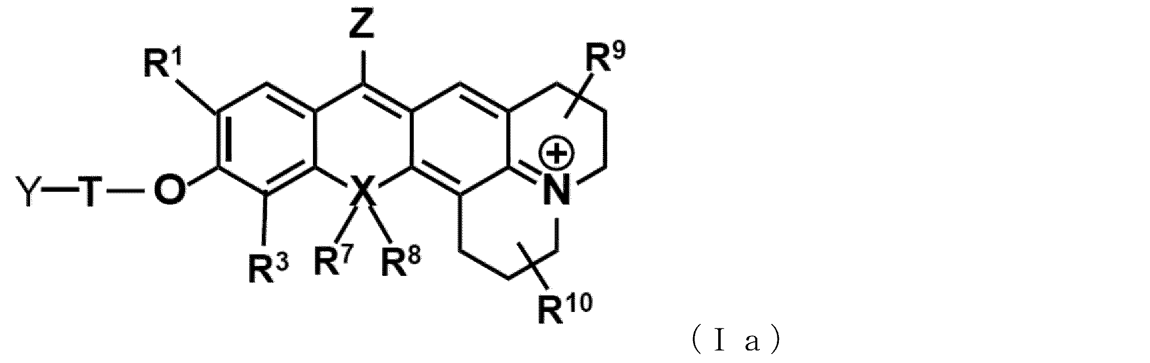
- One preferred embodiment of the compound of the present invention is a compound represented by the following general formula (Ia) or a salt thereof.
- R 1 , R 3 , R 7 , R 8 , T and Y are as defined in general formula (I).
- R 9 if present, is an alkyl group of 1-3 carbon atoms.
- R 10 if present, is an alkyl group of 1 to 3 carbon atoms.
- the compounds represented by general formulas (I) and (Ia) can exist as acid addition salts or base addition salts.
- Acid addition salts include, for example, mineral acid salts such as hydrochlorides, sulfates and nitrates, or organic acid salts such as methanesulfonates, p-toluenesulfonates, oxalates, citrates and tartrates.
- base addition salts include metal salts such as sodium, potassium, calcium and magnesium salts, ammonium salts, organic amine salts such as triethylamine salts, and the like. In addition to these, it may form a salt with an amino acid such as glycine.
- the compounds represented by formulas (I) and (Ia) or salts thereof may exist as hydrates or solvates, and these substances can also be used in the present invention.
- the compounds represented by general formulas (I) and (Ia) may have one or more asymmetric carbon atoms depending on the type of substituent.
- stereoisomers such as optically active isomers based on the asymmetric carbon of , and diastereoisomers based on two or more asymmetric carbons, arbitrary mixtures and racemates of stereoisomers can also be used.
- Raman Probe of the Present Invention Another aspect of the present invention is a Raman probe containing the compound of formula (I) or a salt thereof (hereinafter also referred to as "the Raman probe of the present invention").
- the Raman probe of the present invention is a Raman probe that can be used in the epr-SRS method.
- the epr-SRS method is a Raman imaging method that combines the electronic pre-resonance (epr) effect and stimulated Raman scattering (SRS).
- excitation is performed by light with a wavelength slightly longer than the electronic absorption band of the molecule (pre-resonance condition), so background rise due to photobleaching and fluorescence can be suppressed, and stimulated emission by Stokes light can further enhance sensitivity. Imaging can be realized. With conventional SRS microscopes, it takes time on the order of seconds to change the wavelength of Stokes light, so one of the problems was the long measurement time when performing spectral analysis and spectral imaging.
- the high-speed SRS spectroscopic microscope developed by Koseki Laboratory in the Department of Electrical Engineering, graduate School of Engineering is capable of high-speed imaging that changes by 30 wavenumbers per second (Ozeki, Y., Biological Imaging Based on Stimulated Raman Scattering. Seibutsu Butsuri, 2014. 54(6): p.311-314).
- a schematic diagram of the high-speed SRS spectroscopic microscope is shown in FIG.
- Raman imaging using high-speed SRS spectroscopic microscopy can be performed with reference to the Ozeki paper cited above.
- data are obtained about 5 to 10 times for in vitro measurements and about 100 to 1000 times for in cellulo measurements. averaging is preferably performed.
- the excitation light of this high-speed SRS spectroscopic microscope of Oseki Lab is 843 nm
- the method for detecting a target enzyme using the Raman probe of the present invention can preferably use the high-speed SRS spectroscopic microscope described above, but is not limited to the method using the high-speed SRS spectroscopic microscope.
- one preferred aspect of the present invention is an activatable Raman probe that can be used in the epr-SRS method and contains the compound of general formula (I) or a salt thereof.
- the method of using the Raman probe of the present invention is not particularly limited, and it can be used in the same manner as conventionally known Raman probes.
- the compound represented by the above formula (I) is added to an aqueous medium such as physiological saline or a buffer solution, or a mixture of a water-miscible organic solvent such as ethanol, acetone, ethylene glycol, dimethylsulfoxide and dimethylformamide and an aqueous medium.
- the compounds or their salts are dissolved, this solution is added to an appropriate buffer containing cells or tissues, and the Raman spectrum is measured.
- the Raman probe of the present invention may be used in the form of a composition in combination with suitable additives. For example, it can be combined with additives such as buffers, solubilizers, and pH adjusters.
- Another aspect of the present invention is a method for detecting a target enzyme in a cell or tissue, comprising: (a) a Raman probe containing a compound represented by general formula (I) or a salt thereof; and (b) measuring Raman scattered light that is enhanced by the reaction of the compound or its salt with a target enzyme in cells or tissues (hereinafter “the method of the present invention”).
- the cells include normal cells, cancer cells, nerve cells and the like.
- Raman scattered light is preferably measured using the epr-SRS method.
- the compound represented by the general formula (I) or a salt thereof can be introduced into cells or tissues by spraying the compound represented by the general formula (I) or a salt thereof on a cell or tissue sample. be able to.
- Types of cancer cells or cancer tissues to be detected by the detection method of the present invention include lung cancer, prostate cancer, ovarian cancer, breast cancer, bladder cancer, brain tumor, esophageal cancer, stomach cancer, bile duct cancer, and liver cancer.
- cancer pancreatic cancer, head and neck cancer, renal cancer, leukemia, skin cancer, and thyroid cancer cells or tissues.
- cancer tissue means any tissue containing cancer cells.
- tissue should be interpreted in the broadest sense, including a part or the whole of an organ, and should not be interpreted restrictively in any way.
- One aspect of the detection method of the present invention includes the step of (a) introducing two or more compounds represented by formula (I) or a salt thereof into a cell or tissue, and (b) the two or more compounds or A method for detecting two or more target enzymes in a cell or tissue, comprising the step of measuring Raman scattered light enhanced by the salt reacting with each of the target enzymes in the cell or tissue.
- the detection method of the present invention and the probe of the present invention can stain tissue as well as cells
- the detection method of the present invention and the probe of the present invention can be used in cancer diagnostic methods based on enzyme activity patterns. can also be used. That is, another embodiment of the present invention includes the steps of: (a) administering a Raman probe containing the compound of general formula (I) or a salt thereof to a subject; , a method for diagnosing cancer comprising the step of measuring Raman scattered light enhanced by reaction with a target enzyme in a tissue or organ (hereinafter also referred to as "diagnostic method of the present invention").
- Administration methods include topical administration, oral administration, intravenous administration, and the like.
- Types of cancer targeted by the diagnostic method of the present invention include lung cancer, prostate cancer, ovarian cancer, breast cancer, bladder cancer, brain tumor, esophageal cancer, stomach cancer, bile duct cancer, liver cancer, and pancreatic cancer. , head and neck cancer, kidney cancer, leukemia, skin cancer, and thyroid cancer.
- Another embodiment of the present invention is a target molecule detection kit containing the Raman probe of the present invention.
- the Raman probe of the present invention is usually prepared as a solution. It can also be applied by dissolving in distilled water for injection or an appropriate buffer solution.
- kit may contain other reagents and the like as necessary.
- additives such as dissolution aids, pH adjusters, buffers, tonicity agents, and the like can be used, and the amount of these additives can be appropriately selected by those skilled in the art.
- NMR spectra were obtained on a Bruker NMR AVANCE III 400 spectrometer [ 1 H 400 MHz, 13 C 100 MHz] using deuterated solvents.
- High resolution ESI mass spectra were obtained on a Bruker microTOF II-TM (ESI).
- HPLC purification was performed on JASCO PU-2080 Plus pumps (GL Science Co., Ltd.) and MD-2015 with Inertstil-ODS-3 columns ( ⁇ 10 ⁇ 250 mm (semi-preparative) and ⁇ 20 ⁇ 250 mm (preparative)).
- a detector JASCO was used.
- Solvents used for HPLC were obtained from Wako Co., Ltd.
- Silica gel column chromatography was performed using a medium-pressure preparative liquid chromatograph YFLC-Al560 (Yamazen Co., Ltd.). TLC was performed on silica gel plates F254 (0.25 mm (analytical); Merck, AKG).
- UV-vis spectra were obtained on a Shimadzu UV-2450 spectrophotometer. Fluorescence spectra were acquired with F-7000 (Hitachi). The SRS spectrum and SRS image were obtained with a high-speed SRS spectroscopic microscope developed by Kozeki Laboratory, Department of Electrical Engineering, graduate School of Engineering, University of Tokyo.
- the wavelengths of the pump light pulse and the Stokes light pulse are 843 nm and 1014-1046 nm, the pulse duration is about 5 picoseconds, and the spectral resolution is 5/cm.
- a water immersion objective lens is used and its numerical aperture is 1.2. Images of 500 ⁇ 500 pixels were acquired at 30 frames per second by varying the wavelength of the Stokes light pulse for each frame. Data were acquired 5 times in vitro and 1000 times in cellulo and averaged to increase the signal-to-noise ratio.
- the julolidine structure is considered to be the optimal substituent on N from the viewpoint of both brightness and stability. was selected as
- FIG. 4 shows the results of evaluating the optical properties of 9CN-JR-Bn- ⁇ Gal and 9C 15 N-JCR-Bn- ⁇ Gal synthesized above.
- FIG. 4(a) shows absorption spectra before and after reaction of 1 ⁇ M 9CN-JR-Bn- ⁇ Gal (upper) and 9C15N-JCR-Bn- ⁇ Gal (lower) with 1 unit ⁇ -galactosidase.
- FIG. 5 shows the results of cell imaging using the two types of probes synthesized above, 9CN-JR-Bn- ⁇ Gal and 9C 15 N-JCR-Bn- ⁇ Gal.
- FIG. 5(a) shows structural changes of 9CN-JR-Bn- ⁇ Gal (upper) and 9C15N-JCR-Bn- ⁇ Gal (lower) before and after reaction with ⁇ -galactosidase.
- HEK-LacZ cells were seeded in a 35 mm glass-bottom dish (Matsunami, Glass-bottom dish Hydro) and cultured overnight. The medium was then replaced with a D-MEM solution (phenol red free) containing 20 ⁇ M 9C 15 N-JCR-Bn- ⁇ Gal and 20 ⁇ M 9CN-JR-Bn- ⁇ Gal with 0.2% DMSO as a co-solvent. and exchanged at 37° C. for 2.5 hours. After incubation, the probe solution was removed and replaced with fresh HBSS(+), and SRS measurements were performed with an SRS microscope as described above.
- D-MEM solution phenol red free
- the detected wavenumbers were 2190 cm ⁇ 1 and 2193 cm ⁇ 1 for 9C 15 N-JCR-Bn- ⁇ Gal and 2223 cm ⁇ 1 and 2227 cm ⁇ 1 for 9CN- JR -Bn- ⁇ Gal. Background wavenumbers were 2170 cm ⁇ 1 and 2247 cm ⁇ 1 . Scale bar: 10 ⁇ m. Acquisition time for each cell was 60 seconds.
- (d) shows the SRS spectra of the whole field (left) and the aggregate part (right) of (c).
- b of FIG. 5 is an experiment with the same content as that of FIG. 4c.
- 9C 15 N-JCR-Bn- ⁇ Gal could be imaged brighter than 9CN-JR-Bn- ⁇ Gal. suggested to be better.
- the resulting eluent was evaporated and the residue was subjected to preparative HPLC using eluent A ( H2O with 1% MeCN and 0.1% TFA) and eluent B (MeCN with 1% H2O ).
- A/B 95/5 to 5/95 (0-3.5min), 5/95 (3.5-4.0min), 5/95 to 95/5 (4.0-4.1min), 95/5 (4.1-5.0min) ).
- FIG. 6(a) shows a schematic diagram of the reaction of 9 13 CN-JCR-Bn-gGlu with an enzyme (GGT).
- FIG. 6( b ) shows 1 ⁇ M of 1 ⁇ M dimethylformamide before (blue) and after (red) reaction with 1 unit of GGT, measured in PBS (pH 7.4) containing 0.1% DMSO as co-solvent.
- 9 shows the absorption spectrum of 9 13 CN-JCR-Bn-gGlu. The reaction solution was incubated at room temperature for 20 minutes.
- (c) of FIG. 6 shows the concentration of 200 ⁇ M before (blue) and after (red) reaction with 1 unit GGT, measured in PBS (pH 7.4, final DMSO concentration 30% (v/v)).
- FIG. 9 shows the SRS spectrum of 9 13 CN-JCR-Bn-gGlu.
- the reaction solution was incubated at room temperature for 1 hour.
- the image was constructed by subtracting the image at 2100 cm ⁇ 1 as background. Scale bar: 10 ⁇ m.
- FIG. 6(d) shows the time-dependent change in absorbance of 9 13 CN-JCR-Bn-gGlu upon addition of GGT1 unit (red, arrow indicates timing of enzyme addition) and 50 ⁇ M GGT-specific inhibitor. Time-dependent change in absorbance in the presence of GGsTop (green). The observed wavelength was 690 nm.
- FIG. 6(e) shows the results of LC-MS analysis of the reaction solution of 1 unit of GGT and 9 13 CN-JCR-Bn-gGlu under neutral conditions. The reaction solution was incubated at room temperature for 20 minutes.
- FIG. 7(a) shows a schematic diagram of the reaction of 9 13 CN-JCR-Bn-EP with an enzyme (DPP-4).
- FIG. 7(b) shows before (blue) and after (red ) of 9 13 CN-JCR-Bn-EP at 1 ⁇ M. The reaction solution was incubated at room temperature for 1 hour.
- FIG. 7(c) shows before (blue) and after (red) reaction with 0.033 units of DPP-4, measured in PBS (pH 7.4, final DMSO concentration 30% (v/v)). shows the SRS spectrum of 200 ⁇ M 9 13 C 15 N-JCR-Bn-EP of . The reaction solution was incubated at room temperature for 1 hour.
- FIG. 7(d) shows the time dependence of the absorbance of 9 13 C 15 N-JCR-Bn-EP upon addition of 0.033 units of DPP-4 (red, arrow indicates timing of enzyme addition). changes and time-dependent changes in absorbance in the presence of 1.8 ⁇ M DPP-4 specific inhibitor; sitagliptin (green) is shown. The observed wavelength was 690 nm.
- FIG. 7(e) shows the results of LC-MS analysis of the reaction solution of 0.033 units of DPP-4 and 9 13 C 15 N-JCR-Bn-EP under neutral conditions. The reaction solution was incubated at room temperature for 20 minutes.
- A549 cells were purchased from the Riken Cell Bank (RCB 0098) and H226 cells were purchased from the American Type Culture Collection. A549 cells were cultured in Dulbecco's modified Eagle medium (D-MEM) (Wako) and H226 cells in RPMI 1640 (Wako) containing 10% fetal bovine serum (GIBCO), 100 U/mL, penicillin, and 100 ⁇ g/mL streptomycin. at 37°C in humidified air containing 5% CO2 .
- D-MEM Dulbecco's modified Eagle medium
- RPMI 1640 Wako
- GEBCO fetal bovine serum
- the probe solution was removed and replaced with fresh HBSS(+), and SRS measurements were performed with an SRS microscope as described above.
- the detected wavenumbers were 2187 cm -1 and 2190 cm -1 for 9C 15 N-JCR-Bn- ⁇ Gal, 2167 cm -1 and 2170 cm -1 for 9 13 CN-JCR-Bn-gGlu, and 9 13 C 15 N-JCR-Bn-.
- the EP was 2137 cm ⁇ 1 and 2140 cm ⁇ 1 .
- Background wavenumbers were 2117 cm ⁇ 1 and 2210 cm ⁇ 1 .
- Results of simultaneous imaging of three enzymatic activities in live cultured cells are shown in FIG.
- Left panel of FIG. 8 is SRS imaging of ⁇ -Gal, GGT and DPP-4 activity in live A549 cells (upper panel) and H226 cells (lower panel). Scale bar: 10 ⁇ m. Acquisition time for each cell was 80 seconds.
- the right panel of FIG. 8 shows SRS spectra of enzymatic activity in A549 cells (upper panel) and H226 cells (lower panel). Highlighted regions indicate peaks for each probe.
- FIG. 10 shows the results of comparing the ability of 9CN-DEP and 9CN-DER to form aggregates.
- FIG. 10(a) shows the chemical structures of 9CN-DEP and 9CN-DER under neutral conditions.
- FIG. 10(b) Permeation of a 300 ⁇ M solution of 9CN-DEP (top) or 9CN-DER (bottom) in PBS (pH 7.4) containing 3%, 10%, 30% and 90% DMSO. Show the image. Scale bar is 100 ⁇ m.
- FIG. 10(c,d) shows 9CN-DEP (c) and 9CN-DER (d) from 5 to 100 ⁇ M measured in PBS (pH 7.4) containing 0.05% to 1% DMSO as co-solvent. ) shows the normalized absorption spectrum.
- FIG. 10(e) shows the relationship between dye concentration and normalized absorbance at the absorption maximum of a 5 ⁇ M solution.
- the black dashed line represents the linear relationship between dye concentration and normalized absorbance.
- FIG. 10(f,g) 300 ⁇ M 9CN-DEP (f) and 9CN-DER (g) measured in PBS (pH 7.4) containing 3%, 10%, 30% and 90% DMSO. shows the SRS spectrum of The image was constructed by subtracting the image at 2150 cm ⁇ 1 as background. Scale bar is 10 ⁇ m.
- FIG. 11 shows the results of comparing the ability of nuclei, 9CN-JCR, to form aggregates.
- FIG. 11 (a-d) 9C 15 N-JCR-Bn- ⁇ Gal (a) from 5 to 100 ⁇ M measured in PBS (pH 7.4) with 0.05% to 1% DMSO as co-solvent. , 9 13 CN-JCR-Bn-gGlu (b), 9 13 C 15 N-JCR-Bn-EP (c) and 9CN-JCR (d). Absorbance was normalized based on the absorbance at the absorption maximum of a 5 ⁇ M solution.
- FIG. 11(e) shows the relationship between dye concentration and normalized absorbance at the absorption maximum of a 5 ⁇ M solution. The black dashed line represents the linear relationship between dye concentration and normalized absorbance.
- Example 5 Based on the above results, it was examined whether it is possible to perform target enzyme-specific ex vivo imaging with the 9CN-JCR probe, which could not be performed with the conventional 9CN-JCP probe.
- the experimental method is as follows.
- SRS images and spectra of living cells and ex vivo tissues were created by subtracting the average image of background wavenumbers from the average image of detected wavenumbers. For detection wavenumbers, two wavenumbers were selected around the maximum of the SRS intensity of each probe. For the background wavenumber, a wavenumber 20 cm ⁇ 1 away from the detected wavenumber was chosen. If the background and detected wavenumbers were too close due to crosstalk, we chose the highest and lowest background wavenumbers as the background wavenumbers common to all peaks. Data were acquired 1000 times and averaged to enhance the signal-to-noise ratio. To measure the spectrum, the wavenumber was adjusted continuously at 3.3 cm ⁇ 1 intervals and the number of frames used for averaging at each wavenumber was 5.
- Drosophila culture conditions and strains The Drosophila strains used in this study were en-Gal4, UAS-mCD8-GFP (from Dr. E. Kuranaga), hs- Flp122 , UAS-mCD8-GFP; , UAS-GFP, UAS-lacZ (Bloomington Drosophila Stock Center 1776), and UAS-Ggt-1.
- Drosophila tissue was cultured in Schneider's Drosophila medium (containing 0.5%, 1% DMSO as co-solvent) with 100 ⁇ M 9C 15 N-JCR-Bn- ⁇ Gal solution, 100 ⁇ M 9C 15 N-JCR-Bn- ⁇ Gal.
- FIG. 12 shows the results of target enzyme-selective ex vivo imaging of Drosophila tissue using aggregate formation with 9C 15 N-JCR-Bn- ⁇ Gal.
- FIG. 12(a) is an anatomical drawing of a Drosophila larva. Ex vivo imaging of the wing disc or fat pad was performed.
- FIG. 12(b) shows the results of fluorescence imaging of membrane-localized GFP and SRS imaging of ⁇ -galactosidase activity in the Drosophila wing disc.
- Drosophila wing discs (genotype: en-Gal 4, UAS-mCD 8-GFP/UAS-lacZ) were incubated with 100 ⁇ M 9C 15 N-JCR-Bn in Schneider's Drosophila medium with 0.5% DMSO as co-solvent. - incubated with ⁇ Gal for 2.5 hours at room temperature.
- (c) of FIG. 12 shows the SRS spectrum of the enzyme activity obtained from the field of view of (b).
- FIG. 12(d) shows the results of fluorescence imaging of membrane and cytoplasmic GFP and SRS imaging of b-galactosidase activity in the Drosophila fat pad.
- Drosophila fat bodies (genotypes: hs-Flp 122 , UAS-mCD8-GFP; Ay-Gal4, UAS-GFP/UAS-lacZ) were added as co-solvents to 100 ⁇ M in Schneider's Drosophila medium containing 0.5% DMSO. Cultured with 9C 15 N-JCR-Bn- ⁇ Gal at room temperature for 3.5 hours.
- FIG. 12 shows the SRS spectrum of ⁇ -Gal activity obtained from the field of view (d). Scale bar: 10 ⁇ m. The image acquisition time was 133 seconds.
- Drosophila tissue larvae in which ⁇ -galactosidase was forcibly expressed region-specifically were stained with the 9Cn-JCR probe. were confirmed in the wing disc and fat body tissues, suggesting that enzyme active region-specific detection was possible.
- FIG. 13 shows the results of simultaneous imaging of ⁇ Gal-9 13 C 15 N-JCP and 9C 15 N-JCR-Bn- ⁇ Gal using Drosophila wing discs.
- FIG. 13(a) shows the results of fluorescence imaging of GFP and SRS imaging of ⁇ -Gal activity in the wing disc of Drosophila (genotype: en-Gal4, UAS-mCD8-GFP/UAS-lacZ).
- FIG. 13(b) shows the SRS spectrum of the ⁇ -Gal probe obtained from the field of view of (a). Highlighted regions indicate peaks for each probe. Green (right area): 9C 15 N-JCR-Bn- ⁇ Gal, yellow (left area): ⁇ Gal-9 13 C 15 N-JCP.
- the red ROI (lower right box) indicates the LacZ(+) region of (b), and the blue ROI (upper left box) indicates the LacZ( ⁇ ) region of (b).
- Probes 100 ⁇ M each. Scale bar: 10 ⁇ m.
- the image acquisition time was 267 seconds.
- FIG. 14 shows the results of simultaneous imaging of ⁇ Gal-9 13 C 15 N-JCP and 9C 15 N-JCR-Bn- ⁇ Gal using the Drosophila fat pad.
- FIG. 14(a) shows fluorescence imaging of GFP and ⁇ -Gal activity in Drosophila fat pads (genotypes: hs-Flp 122 , UAS-mCD8-GFP; Ay-Gal4, UAS-GFP/UAS-lacZ). Results of SRS imaging are shown.
- (b) of FIG. 14 shows the SRS spectrum of the ⁇ -Gal probe obtained from the field of view of (a). Highlighted regions indicate peaks for each probe.
- the 9CN-JCP probe non-specifically stains the non-enzyme-expressing region, but the 9CN-JCR probe can specifically stain only the enzyme-expressing region in the same tissue. confirmed by imaging at
- FIG. 15 shows a conceptual diagram of the intracellular retention type probe.
- 9CN-2CH 2 F-JCR-Bn- ⁇ Gal (see FIG. 16) targeting ⁇ -galactosidase was synthesized according to Synthetic Example 5 described below.
- Example 6 The in vitro enzymatic reaction of 9CN-2CH 2 F-JCR-Bn- ⁇ Gal synthesized above was evaluated. The results are shown in FIG. As shown in FIG. 16, it was observed to react rapidly with ⁇ -galactosidase to produce a long wavelength product.
- Example 7 Cell imaging with 9CN-2CH 2 F-JCR-Bn- ⁇ Gal was then performed. At this time, imaging of 9C 15 N-JCR-Bn- ⁇ Gal as a control compound was performed in parallel for comparison. The experimental method is shown below.
- HEK293 cells and HEK-LacZ cells were purchased from JCRM (JCRB1414/Murakami, T.).
- HEK293 cells and HEK-LacZ cells were grown in 5% Dulbecco's modified Eagle medium (D-MEM) (Wako) containing 10% fetal bovine serum (GIBCO), 100 U/mL, penicillin, and 100 ⁇ g/mL streptomycin. was cultured at 37 °C in humidified air with 300 rpm of CO2 .
- D-MEM Dulbecco's modified Eagle medium
- GEBCO fetal bovine serum
- FIG. 18 are confocal fluorescence images of ⁇ -galactosidase activity in live HEK-LacZ or HEK293 cells.
- Cells were incubated with 10 ⁇ M 9CN-2CH 2 F-JCR-Bn- ⁇ Gal (upper panel) or 9C 15 N-JCR-Bn- ⁇ Gal (lower panel) for 30 min in HBSS(+).
- all probes were confirmed to emit fluorescence specifically to LacZ-expressing cells. Also, the localization of both probes appeared to be almost the same.
- Example 8 the wash resistance and fixation resistance of both probes (9CN-2CH 2 F-JCR-Bn- ⁇ Gal and 9C 15 N-JCR-Bn- ⁇ Gal) were compared.
- the wash operation was carried out two times in total, with one operation of replacing with fresh HBSS(+) and incubating for 10 minutes, and imaging after each wash. Furthermore, after the second wash, PFA fixation (15 minutes) was performed and the final imaging was performed. The results are shown in Figures 19 and 20.
- Figures 19 and 20 are confocal fluorescence images of ⁇ -galactosidase activity in live or fixed HEK-LacZ cells.
- Cells were incubated with 10 ⁇ M 9CN-2CH 2 F-JCR-Bn- ⁇ Gal (FIG. 19) or 9C 15 N-JCR-Bn- ⁇ Gal (FIG. 20) for 30 min in HBSS(+), then HBSS(+ ) for 10 minutes twice. After washing, cells were fixed with PFA for 15 minutes.
- Drosophila Culture Conditions and Strains The Drosophila strain used in this study was en-Gal4, UAS-mCD8-GFP (from Dr. E. Kuranaga), and Drosophila tissue was grown in Schneider's Drosophila medium (2% DMSO as co-solvent). containing) with a mixture of 100 ⁇ M 9C 15 N-JCR-Bn- ⁇ Gal and 100 ⁇ M 9CN-2CH 2 F-JCR-Bn- ⁇ Gal for 3 hours at room temperature. After culturing, the tissue was removed from the medium and immersed in fresh PBS(-) on the imaging chamber. SRS measurements were performed with an SRS microscope as previously described.
- the detected wavenumbers were 2190 cm ⁇ 1 and 2193 cm ⁇ 1 for 9C 15 N-JCR-Bn- ⁇ Gal and 2220 cm ⁇ 1 and 2223 cm ⁇ 1 for 9CN-2CH 2 F-JCR-Bn- ⁇ Gal. Background wavenumbers were 2170 cm ⁇ 1 and 2243 cm ⁇ 1 . Confocal fluorescence imaging was performed after SRS image acquisition. For fluorescence imaging of GFP, the excitation wavelength was 488 nm and emission was detected in the range of 500-520 nm.
- FIG. 21 shows confocal fluorescence and SRS images of ⁇ -galactosidase activity in the Drosophila wing disc. Scale bar: 10 ⁇ m. The average time was 1000 hours.
- aggregates such as 9C 15 N-JCR-Bn- ⁇ Gal were also confirmed specifically in the LacZ(+) region of the SPiDER type. Although the LacZ(-) region was also stained widely and leaked out from the enzyme expression region, the signal intensity was significantly stronger with the SPiDER type, which is expected to improve retention in the entire tissue. It was thought that there was.
Landscapes
- Health & Medical Sciences (AREA)
- Chemical & Material Sciences (AREA)
- Life Sciences & Earth Sciences (AREA)
- Organic Chemistry (AREA)
- Engineering & Computer Science (AREA)
- General Health & Medical Sciences (AREA)
- Physics & Mathematics (AREA)
- Biochemistry (AREA)
- Medicinal Chemistry (AREA)
- Analytical Chemistry (AREA)
- Molecular Biology (AREA)
- Immunology (AREA)
- Pathology (AREA)
- Biophysics (AREA)
- General Physics & Mathematics (AREA)
- Proteomics, Peptides & Aminoacids (AREA)
- Biomedical Technology (AREA)
- Zoology (AREA)
- Animal Behavior & Ethology (AREA)
- Genetics & Genomics (AREA)
- Food Science & Technology (AREA)
- Veterinary Medicine (AREA)
- Urology & Nephrology (AREA)
- Wood Science & Technology (AREA)
- Public Health (AREA)
- Hematology (AREA)
- Epidemiology (AREA)
- General Engineering & Computer Science (AREA)
- Nuclear Medicine, Radiotherapy & Molecular Imaging (AREA)
- Toxicology (AREA)
- Pharmacology & Pharmacy (AREA)
- Bioinformatics & Cheminformatics (AREA)
- Microbiology (AREA)
- Biotechnology (AREA)
- Measuring Or Testing Involving Enzymes Or Micro-Organisms (AREA)
Abstract
【解決課題】 標的可能な酵素を拡充することができ、細胞内滞留性が改善されたactivatable型ラマンプローブを提供すること。 【解決手段】 以下の一般式(I)で表される化合物又はその塩。
Description
本発明は、新規なO-function型activatableラマンプローブに関する。より具体的には、本発明は、標的酵素との反応前はラマンシグナルがoffであるが、標的酵素との反応によってラマンシグナルがonになる、activatableな特性を有するO-function型ラマンプローブに関する。
細胞などの生体試料に光を照射すると、透過・吸収・散乱といった物理現象が生じる。この散乱光は大部分が入射光と同じ波長の光(レイリー散乱)であるが、ごく僅かに入射光よりも振動数が減少した(波長が長波長側にシフトした)光が含まれており、ストークスラマン散乱(ラマン散乱)と呼ばれている。
ラマン散乱における波長のシフト量(ラマンシフト値)は、光を散乱した分子の固有振動数に相当するため、ラマンスペクトルから試料に含まれる分子の種類やその状態などの情報を得ることができる。そのため、ラマン散乱を用いたイメージング法は、生体分子を無標識で観察する手法として注目されてきた(図1参照)。しかしながら、ラマン散乱光は極めて微弱であるため、(1)検出感度が低い、(2)長時間の観測時間(数十分から数時間ほど)が必要で時間分解能が低い、といった課題があり、その応用先が限られていた。
このようなラマンイメージングの課題が、誘導ラマン散乱(stimulated Raman scattering;SRS)(非特許文献1)や共鳴ラマン散乱(非特許文献2)を活用したイメージング法の開発によって克服されてきた。さらに、生体分子のラマン信号が生じないsilent region(1800-2800cm-1)に伸縮振動をもつアルキンなどの微小タグとの併用によって、生体適合性が飛躍的に向上した(非特許文献3)。
このように、近年、ラマンイメージングの発展に伴い、より多様なラマンプローブ開発の需要が高まっているが、既存のラマンプローブは常に同じラマンシフト値・信号強度を示すalways-on型のプローブであるため、ラベル化剤としての用途に留まっていた。
本発明者らは、これまでに、標的酵素と出会う前はラマン信号がoffだが,標的酵素による加水分解を受けて吸収波長が長波長化することで(非共鳴条件→前期共鳴条件)ラマン信号がonになる、activatable型ラマンプローブの開発に成功した。さらに,プローブ母核として見出した9CN-JCPのCN基の同位体置換を行うことで、異なる酵素を標的とした、同時に分離検出可能な4種類のラマンプローブを開発し、2種類の細胞において各標的酵素の活性パターンが異なる様子をlive-cellでイメージングすることに成功した(非特許文献4)。
しかしながら,開発したラマンプローブは主にアミノペプチダーゼを標的としたN-function型のプローブであり、標的とできる酵素の種類には制限があった。さらに、本発明者らの更なる研究の結果、当該プローブには細胞内に滞留する構造がないため一細胞レベルの分解能を有さず、ショウジョウバエ組織にプローブを添加した際には、標的酵素発現領域以外も非特異的に染色してしまうことが分かった(図2参照)。
しかしながら,開発したラマンプローブは主にアミノペプチダーゼを標的としたN-function型のプローブであり、標的とできる酵素の種類には制限があった。さらに、本発明者らの更なる研究の結果、当該プローブには細胞内に滞留する構造がないため一細胞レベルの分解能を有さず、ショウジョウバエ組織にプローブを添加した際には、標的酵素発現領域以外も非特異的に染色してしまうことが分かった(図2参照)。
Min, W., et al., Coherent nonlinear optical imaging: beyond fluorescence microscopy. Annu Rev Phys Chem, 2011. 62: p. 507-30.
Kuzmin, A.N., et al., Resonance Raman Probes for Organelle-Specific Labeling in Live Cells. Sci Rep, 2016. 6: p. 28483.
Yamakoshi, H., et al., Imaging of EdU, an alkyne-tagged cell proliferation probe, by Raman microscopy. J Am Chem Soc, 2011. 133(16): p. 6102-5.
Fujioka, H.; Shou, J.; Kojima, R.; Urano, Y.; Ozeki, Y.; Kamiya, M., Multicolor Activatable Raman Probes for Simultaneous Detection of Plural Enzyme Activities. J Am Chem Soc 2020, 142 (49), 20701-20707.
本発明は、標的可能な酵素を拡充することができ、細胞内滞留性が改善されたactivatable型ラマンプローブを提供することを目的とする。
本発明者らは、上記課題を解決するために鋭意研究を行った結果、ピロニン系色素に代えて、蛍光母核をロドール系のO-function型とすることにより、標的酵素を拡充でき、細胞内滞留性が改善されたactivatable型ラマンプローブを提供できることを見出し、本発明を完成した。
即ち、本発明は、
[1] 以下の一般式(I)で表される化合物又はその塩。
 (式中、
(式中、
R1及びR2は、それぞれ独立に、水素原子、炭素数1~6個のアルキル基又はハロゲン原子を表し;
R3及びR4は、それぞれ独立に、水素原子、炭素数1~6個のアルキル基又はハロゲン原子を表し;
ここで、R1又はR3は、-CH2-F又は-CH-F2でもよく;
R5及びR6は、それぞれ独立に、水素原子又は炭素数1~6個のアルキル基を表し、
ただし、
(1)R5又はR6は、R2又はR4と一緒になって、R2又はR4が結合している窒素原子を含む5~7員のヘテロシクリル又はヘテロアリールを形成しているか、あるいは
(2)R5及びR6は、夫々、R2及びR4と一緒になって、R2及びR4が結合している窒素原子を含む5~7員のヘテロシクリル又はヘテロアリールを形成しており、
該ヘテロシクリル又はヘテロアリールは環構成員として酸素原子、窒素原子及び硫黄原子からなる群から選択される1~3個の更なるヘテロ原子を含有していてもよく、更に該ヘテロシクリル又はヘテロアリールは、炭素数1~6個のアルキル、炭素数2~6個のアルケニル、又は炭素数2~6個のアルキニル、炭素数6~10個のアラルキル基、炭素数6~10個のアルキル置換アルケニル基で置換されていてもよく;
R7及びR8は、存在する場合は、それぞれ独立に、炭素数1~6個のアルキル基を表し、
ここで、Xが酸素原子の場合は、R7及びR8は存在せず、
Xは、酸素原子又は炭素原子を表し;
Tは、リンカーであり:
Yは、-O-L、-NH-L’又は活性酸素種に対する捕捉基であり、
ここで、Lは、糖類又は糖類の部分構造であり、
L’は、アミノ酸の部分構造であり;
Zは、-C≡N、-C≡15N、-13C≡N、又は-13C≡15Nから選択される。)
[2] 以下の一般式(Ia)で表される、[1]に記載の化合物又はその塩。
 (R1、R3、R7、R8、T、Yは、一般式(I)で定義した通りであり;
R9は、存在する場合は、炭素数1~3のアルキル基であり:
R10は、存在する場合は、炭素数1~3のアルキル基である。)
[3]Tのリンカーは、アルキレン基(但し、アルキレン基の1以上の-CH2-は、-O-、-S-、-NH-、又は-CO-で置換されていてもよい。)、アリーレン(ヘテロアリーレンを含む)、シクロアルキレン、アルコキシル基、ポリエチレングリコール鎖、及び、これらの基から選択される2種以上の基が任意に結合して構成される基からなる群から選択される、[1]又は[2]に記載の化合物又はその塩。
[4]前記リンカーは、置換基を有していてもよいベンジル基、又はメチレン基である、[3]に記載の化合物又はその塩。
[5]Lの糖類の部分構造は、それが結合しているOと一緒になって、糖類、糖類の一部を構成している、[1]~[4]のいずれか1項に記載の化合物又はその塩。
[6]L’のアミノ酸の部分構造は、アミノ酸、アミノ酸残基、ペプチド、アミノ酸の一部を構成している、[1]~[4]のいずれか1項に記載の化合物又はその塩。
[7]Yが以下のいずれかから選択される、[1]~[4]のいずれか1項に記載の化合物又はその塩。
 [8]Yが以下のいずれかから選択される、[1]~[4]のいずれか1項に記載の化合物又はその塩。
 (*は、Tと結合する箇所を表す。)
 (R1aは、天然アミノ酸(グリシン、アラニン、ロイシン、イソロイシン、バリン、リジン、システイン、トレオニン、アルギニン、アスパラギン、アスパラギン酸、グルタミン、グルタミン酸、セリン、ヒスチジン、フェニルアラニン、メチオニン、トリプトファン、チロシン、プロリン)の側鎖を構成する基、非天然アミノ酸(シトルリン、ノルバリンなど)の側鎖を構成する基、置換又は無置換のアルキル基からなる群から選択され;
*は、Tと結合する箇所を表す。)
 (R2aは、天然アミノ酸(グリシン、アラニン、ロイシン、イソロイシン、バリン、リジン、システイン、トレオニン、アルギニン、アスパラギン、アスパラギン酸、グルタミン、グルタミン酸、セリン、ヒスチジン、フェニルアラニン、メチオニン、トリプトファン、チロシン、プロリン)の側鎖を構成する基、非天然アミノ酸(シトルリン、ノルバリンなど)の側鎖を構成する基、置換又は無置換のアルキル基からなる群から選択され;
 は、アミノ基、Nアセチル化等されたアミノ基、1つまたは複数のアミノ酸がペプチド結合により連なった構造を表し;
*は、Tと結合する箇所を表す。)
[9]活性酸素種に対する捕捉基が、ヒドロキシフェノキシ、アミノフェノキシ、アジド、又はピナコールボランである、[1]~[4]のいずれか1項に記載の化合物又はその塩。
[10]以下のいずれかの構造を有する化合物又はその塩。
 [11][1]~[10]のいずれか1項に記載の化合物又はその塩を含むラマンプローブ。
[12]epr-SRS法に利用可能な[11]に記載のラマンプローブ。
[13]細胞又は組織内の標的酵素又は活性酸素種を検出する方法であって、(a)一般式(I)で表される化合物又はその塩を細胞又は組織内に導入する工程、及び(b)当該化合物又はその塩が細胞又は組織内で標的酵素又は活性酸素種と反応することにより増強されるラマン散乱光を測定する工程を含む方法。
[14]epr-SRS法を用いてラマン散乱光を測定する、[13]に記載の方法。
[1] 以下の一般式(I)で表される化合物又はその塩。
R1及びR2は、それぞれ独立に、水素原子、炭素数1~6個のアルキル基又はハロゲン原子を表し;
R3及びR4は、それぞれ独立に、水素原子、炭素数1~6個のアルキル基又はハロゲン原子を表し;
ここで、R1又はR3は、-CH2-F又は-CH-F2でもよく;
R5及びR6は、それぞれ独立に、水素原子又は炭素数1~6個のアルキル基を表し、
ただし、
(1)R5又はR6は、R2又はR4と一緒になって、R2又はR4が結合している窒素原子を含む5~7員のヘテロシクリル又はヘテロアリールを形成しているか、あるいは
(2)R5及びR6は、夫々、R2及びR4と一緒になって、R2及びR4が結合している窒素原子を含む5~7員のヘテロシクリル又はヘテロアリールを形成しており、
該ヘテロシクリル又はヘテロアリールは環構成員として酸素原子、窒素原子及び硫黄原子からなる群から選択される1~3個の更なるヘテロ原子を含有していてもよく、更に該ヘテロシクリル又はヘテロアリールは、炭素数1~6個のアルキル、炭素数2~6個のアルケニル、又は炭素数2~6個のアルキニル、炭素数6~10個のアラルキル基、炭素数6~10個のアルキル置換アルケニル基で置換されていてもよく;
R7及びR8は、存在する場合は、それぞれ独立に、炭素数1~6個のアルキル基を表し、
ここで、Xが酸素原子の場合は、R7及びR8は存在せず、
Xは、酸素原子又は炭素原子を表し;
Tは、リンカーであり:
Yは、-O-L、-NH-L’又は活性酸素種に対する捕捉基であり、
ここで、Lは、糖類又は糖類の部分構造であり、
L’は、アミノ酸の部分構造であり;
Zは、-C≡N、-C≡15N、-13C≡N、又は-13C≡15Nから選択される。)
[2] 以下の一般式(Ia)で表される、[1]に記載の化合物又はその塩。
R9は、存在する場合は、炭素数1~3のアルキル基であり:
R10は、存在する場合は、炭素数1~3のアルキル基である。)
[3]Tのリンカーは、アルキレン基(但し、アルキレン基の1以上の-CH2-は、-O-、-S-、-NH-、又は-CO-で置換されていてもよい。)、アリーレン(ヘテロアリーレンを含む)、シクロアルキレン、アルコキシル基、ポリエチレングリコール鎖、及び、これらの基から選択される2種以上の基が任意に結合して構成される基からなる群から選択される、[1]又は[2]に記載の化合物又はその塩。
[4]前記リンカーは、置換基を有していてもよいベンジル基、又はメチレン基である、[3]に記載の化合物又はその塩。
[5]Lの糖類の部分構造は、それが結合しているOと一緒になって、糖類、糖類の一部を構成している、[1]~[4]のいずれか1項に記載の化合物又はその塩。
[6]L’のアミノ酸の部分構造は、アミノ酸、アミノ酸残基、ペプチド、アミノ酸の一部を構成している、[1]~[4]のいずれか1項に記載の化合物又はその塩。
[7]Yが以下のいずれかから選択される、[1]~[4]のいずれか1項に記載の化合物又はその塩。
*は、Tと結合する箇所を表す。)
*は、Tと結合する箇所を表す。)
[9]活性酸素種に対する捕捉基が、ヒドロキシフェノキシ、アミノフェノキシ、アジド、又はピナコールボランである、[1]~[4]のいずれか1項に記載の化合物又はその塩。
[10]以下のいずれかの構造を有する化合物又はその塩。
[12]epr-SRS法に利用可能な[11]に記載のラマンプローブ。
[13]細胞又は組織内の標的酵素又は活性酸素種を検出する方法であって、(a)一般式(I)で表される化合物又はその塩を細胞又は組織内に導入する工程、及び(b)当該化合物又はその塩が細胞又は組織内で標的酵素又は活性酸素種と反応することにより増強されるラマン散乱光を測定する工程を含む方法。
[14]epr-SRS法を用いてラマン散乱光を測定する、[13]に記載の方法。
本発明により、標的可能な酵素を拡充することができ、細胞内滞留性が改善されたactivatable型ラマンプローブを提供することが可能である。
本明細書中において、「アルキル」は直鎖状、分枝鎖状、環状、又はそれらの組み合わせからなる脂肪族炭化水素基のいずれであってもよい。アルキル基の炭素数は特に限定されないが、例えば、炭素数1~6個(C1~6)、炭素数1~10個(C1~10)、炭素数1~15個(C1~15)、炭素数1~20個(C1~20)である。炭素数を指定した場合は、その数の範囲の炭素数を有する「アルキル」を意味する。例えば、C1~8アルキルには、メチル、エチル、n-プロピル、イソプロピル、n-ブチル、イソブチル、sec-ブチル、tert-ブチル、n-ペンチル、イソペンチル、neo-ペンチル、n-ヘキシル、イソヘキシル、n-ヘプチル、n-オクチル等が含まれる。本明細書において、アルキル基は任意の置換基を1個以上有していてもよい。そのような置換基としては、例えば、アルコキシ基、ハロゲン原子、アミノ基、モノ若しくはジ置換アミノ基、置換シリル基、又はアシルなどを挙げることができるが、これらに限定されることはない。アルキル基が2個以上の置換基を有する場合には、それらは同一でも異なっていてもよい。アルキル部分を含む他の置換基(例えばアルコシ基、アリールアルキル基など)のアルキル部分についても同様である。
本明細書において「ハロゲン原子」という場合には、フッ素原子、塩素原子、臭素原子、又はヨウ素原子のいずれでもよく、好ましくはフッ素原子、塩素原子、又は臭素原子である。
本明細書において、ある官能基について「置換されていてもよい」と定義されている場合には、置換基の種類、置換位置、及び置換基の個数は特に限定されず、2個以上の置換基を有する場合には、それらは同一でも異なっていてもよい。置換基としては、例えば、アルキル基、アルコキシ基、水酸基、カルボキシル基、ハロゲン原子、スルホ基、アミノ基、アルコキシカルボニル基、オキソ基などを挙げることができるが、これらに限定されることはない。これらの置換基にはさらに置換基が存在していてもよい。このような例として、例えば、ハロゲン化アルキル基、ジアルキルアミノ基などを挙げることができるが、これらに限定されることはない。
本明細書中において、「アリール」は単環式又は縮合多環式の芳香族炭化水素基のいずれであってもよく、環構成原子としてヘテロ原子(例えば、酸素原子、窒素原子、又は硫黄原子など)を1個以上含む芳香族複素環であってもよい。この場合、これを「ヘテロアリール」または「ヘテロ芳香族」と呼ぶ場合もある。アリールが単環および縮合環のいずれである場合も、すべての可能な位置で結合しうる。単環式のアリールの非限定的な例としては、フェニル基(Ph)、チエニル基(2-又は3-チエニル基)、ピリジル基、フリル基、チアゾリル基、オキサゾリル基、ピラゾリル基、2-ピラジニル基、ピリミジニル基、ピロリル基、イミダゾリル基、ピリダジニル基、3-イソチアゾリル基、3-イソオキサゾリル基、1,2,4-オキサジアゾール-5-イル基又は1,2,4-オキサジアゾール-3-イル基等が挙げられる。縮合多環式のアリールの非限定的な例としては、1-ナフチル基、2-ナフチル基、1-インデニル基、2-インデニル基、2,3-ジヒドロインデン-1-イル基、2,3-ジヒドロインデン-2-イル基、2-アンスリル基、インダゾリル基、キノリル基、イソキノリル基、1,2-ジヒドロイソキノリル基、1,2,3,4-テトラヒドロイソキノリル基、インドリル基、イソインドリル基、フタラジニル基、キノキサリニル基、ベンゾフラニル基、2,3-ジヒドロベンゾフラン-1-イル基、2,3-ジヒドロベンゾフラン-2-イル基、2,3-ジヒドロベンゾチオフェン-1-イル基、2,3-ジヒドロベンゾチオフェン-2-イル基、ベンゾチアゾリル基、ベンズイミダゾリル基、フルオレニル基又はチオキサンテニル基等が挙げられる。本明細書において、アリール基はその環上に任意の置換基を1個以上有していてもよい。該置換基としては、例えば、アルコキシ基、ハロゲン原子、アミノ基、モノ若しくはジ置換アミノ基、置換シリル基、又はアシルなどを挙げることができるが、これらに限定されることはない。アリール基が2個以上の置換基を有する場合には、それらは同一でも異なっていてもよい。アリール部分を含む他の置換基(例えばアリールオキシ基やアリールアルキル基など)のアリール部分についても同様である。
本明細書中において、「アリールアルキル」は、上記アリールで置換されたアルキルを表す。アリールアルキルは、任意の置換基を1個以上有していてもよい。該置換基としては、例えば、アルコキシ基、ハロゲン原子、アミノ基、モノ若しくはジ置換アミノ基、置換シリル基、又はアシル基などを挙げることができるが、これらに限定されることはない。アシル基が2個以上の置換基を有する場合には、それらは同一でも異なっていてもよい。アリールアルキルの非限定的な例としては、ベンジル基、2-チエニルメチル基、3-チエニルメチル基、2-ピリジルメチル基、3-ピリジルメチル基、4-ピリジルメチル基、2-フリルメチル基、3-フリルメチル基、2-チアゾリルメチル基、4-チアゾリルメチル基、5-チアゾリルメチル基、2-オキサゾリルメチル基、4-オキサゾリルメチル基、5-オキサゾリルメチル基、1-ピラゾリルメチル基、3-ピラゾリルメチル基、4-ピラゾリルメチル基、2-ピラジニルメチル基、2-ピリミジニルメチル基、4-ピリミジニルメチル基、5-ピリミジニルメチル基、1-ピロリルメチル基、2-ピロリルメチル基、3-ピロリルメチル基、1-イミダゾリルメチル基、2-イミダゾリルメチル基、4-イミダゾリルメチル基、3-ピリダジニルメチル基、4-ピリダジニルメチル基、3-イソチアゾリルメチル基、3-イソオキサゾリルメチル基、1,2,4-オキサジアゾール-5-イルメチル基又は1,2,4-オキサジアゾール-3-イルメチル基等が挙げられる。
本明細書中において、「アルコキシ基」とは、前記アルキル基が酸素原子に結合した構造であり、例えば直鎖状、分枝状、環状又はそれらの組み合わせである飽和アルコキシ基が挙げられる。例えば、メトキシ基、エトキシ基、n-プロポキシ基、イソプロポキシ基、シクロプロポキシ基、n-ブトキシ基、イソブトキシ基、s-ブトキシ基、t-ブトキシ基、シクロブトキシ基、シクロプロピルメトキシ基、n-ペンチルオキシ基、シクロペンチルオキシ基、シクロプロピルエチルオキシ基、シクロブチルメチルオキシ基、n-ヘキシルオキシ基、シクロヘキシルオキシ基、シクロプロピルプロピルオキシ基、シクロブチルエチルオキシ基又はシクロペンチルメチルオキシ基等が好適な例として挙げられる。
本明細書中において、「アルキレン」とは、直鎖状または分枝状の飽和炭化水素からなる二価の基であり、例えば、メチレン、1-メチルメチレン、1,1-ジメチルメチレン、エチレン、1-メチルエチレン、1-エチルエチレン、1,1-ジメチルエチレン、1,2-ジメチルエチレン、1,1-ジエチルエチレン、1,2-ジエチルエチレン、1-エチル-2-メチルエチレン、トリメチレン、1-メチルトリメチレン、2-メチルトリメチレン、1,1-ジメチルトリメチレン、1,2-ジメチルトリメチレン、2,2-ジメチルトリメチレン、1-エチルトリメチレン、2-エチルトリメチレン、1,1-ジエチルトリメチレン、1,2-ジエチルトリメチレン、2,2-ジエチルトリメチレン、2-エチル-2-メチルトリメチレン、テトラメチレン、1-メチルテトラメチレン、2-メチルテトラメチレン、1,1-ジメチルテトラメチレン、1,2-ジメチルテトラメチレン、2,2-ジメチルテトラメチレン、2,2-ジ-n-プロピルトリメチレン等が挙げられる。
1.一般式(I)で表される化合物又はその塩
本発明の1つの実施態様は、以下の一般式(I)で表される化合物又はその塩である(以下「本発明の化合物」とも言う)。
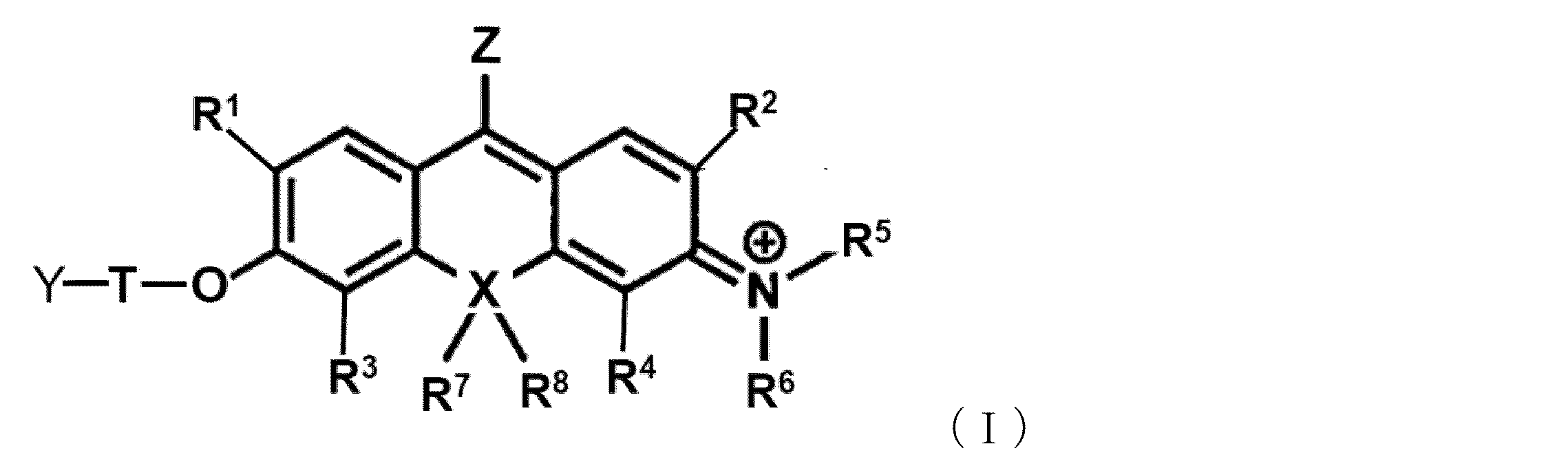
本発明の1つの実施態様は、以下の一般式(I)で表される化合物又はその塩である(以下「本発明の化合物」とも言う)。
本発明の化合物は、蛍光母核としてロドール系のO-function型であることが重要である。本発明者らの先行研究であるN-function型のピロニン系色素を母核とするプローブでは標的とできる酵素の種類に制限があった(例えば、9CN-ピロニンに糖基質を導入する場合はカルバメートリンカーを介することになるが,糖基質の構造(例えば、α-グリコシド結合)によってはカルバメートリンカーでは導入が困難である)のに対して、O-function型とすることにより標的酵素を拡充することが可能である。
また、理論に拘束されることを意図するものではないが、ピロニン系色素を母核とするプローブは、標的酵素との反応後にカチオン型となるため水に溶解し、その結果、標的細胞内での滞留性に劣ることが考えられた。これに対して、ロドール系のO-function型プローブとすることにより、標的酵素と反応すると、-O-と、N+を有する両性イオン型となるため、水中で凝集体を形成することができ、その結果、標的細胞内での滞留性が向上することが見出された(図9参照)。更に、凝集体を形成することで細胞内に局所的に分子の濃度が高まることで、ラマン信号の強度も上がると考えられる。
また、理論に拘束されることを意図するものではないが、ピロニン系色素を母核とするプローブは、標的酵素との反応後にカチオン型となるため水に溶解し、その結果、標的細胞内での滞留性に劣ることが考えられた。これに対して、ロドール系のO-function型プローブとすることにより、標的酵素と反応すると、-O-と、N+を有する両性イオン型となるため、水中で凝集体を形成することができ、その結果、標的細胞内での滞留性が向上することが見出された(図9参照)。更に、凝集体を形成することで細胞内に局所的に分子の濃度が高まることで、ラマン信号の強度も上がると考えられる。
一般式(I)において、R1及びR2は、それぞれ独立に、水素原子、炭素数1~6個のアルキル基又はハロゲン原子を表す。R1及びR2の少なくとも1つがアルキル基を示す場合には、該アルキル基にはハロゲン原子、カルボキシ基、スルホニル基、水酸基、アミノ基、アルコキシ基などが1個又は2個以上存在していてもよい。例えば、R1及び/又はR2が示すアルキル基は、ハロゲン化アルキル基、ヒドロキシアルキル基、カルボキシアルキル基などであってもよい。
R1及びR2はそれぞれ独立に水素原子又はハロゲン原子であることが好ましく、R1及びR2がともに水素原子である場合、又はR1及びR2がともにフッ素原子又は塩素原子である場合がより好ましい。
R1及びR2はそれぞれ独立に水素原子又はハロゲン原子であることが好ましく、R1及びR2がともに水素原子である場合、又はR1及びR2がともにフッ素原子又は塩素原子である場合がより好ましい。
一般式(I)において、R3及びR4は、それぞれ独立に、水素原子、炭素数1~6個のアルキル基又はハロゲン原子を表す。R3及びR4の少なくとも1つがアルキル基を示す場合には、該アルキル基にはハロゲン原子、カルボキシ基、スルホニル基、水酸基、アミノ基、アルコキシ基などが1個又は2個以上存在していてもよい。例えば、R3及び/又はR4が示すアルキル基は、ハロゲン化アルキル基、ヒドロキシアルキル基、カルボキシアルキル基などであってもよい。
R3及びR4はそれぞれ独立に水素原子又はハロゲン原子であることが好ましく、R3及びR4がともに水素原子である場合、又はR3及びR4がともにフッ素原子又は塩素原子である場合がより好ましい。
R3及びR4はそれぞれ独立に水素原子又はハロゲン原子であることが好ましく、R3及びR4がともに水素原子である場合、又はR3及びR4がともにフッ素原子又は塩素原子である場合がより好ましい。
一般式(I)において、R1又はR3は、-CH2-F又は-CH-F2でもよい。
理論に拘束されることを意図するものではないが、一般式(I)において、R1又はR3の位置に、-CH2-F又は-CH-F2を導入すると、標的酵素との反応に伴うフッ化物イオンの脱離によってキノンメチド中間体を生成することができる(図15の最初の反応を参照)。このキノンメチド活性中間体が細胞内のタンパク質などと共有結合を形成することによって、細胞内に固定化されて、細胞内滞留性をより向上させることができることが期待される。
理論に拘束されることを意図するものではないが、一般式(I)において、R1又はR3の位置に、-CH2-F又は-CH-F2を導入すると、標的酵素との反応に伴うフッ化物イオンの脱離によってキノンメチド中間体を生成することができる(図15の最初の反応を参照)。このキノンメチド活性中間体が細胞内のタンパク質などと共有結合を形成することによって、細胞内に固定化されて、細胞内滞留性をより向上させることができることが期待される。
一般式(I)において、R5及びR6は、それぞれ独立に、水素原子又は炭素数1~6個のアルキル基を示す。R5又はR6がアルキル基を示す場合には、該アルキル基にはハロゲン原子、カルボキシ基、スルホニル基、水酸基、アミノ基、アルコキシ基などが1個又は2個以上存在していてもよく、例えばR5又はR6が示すアルキル基はハロゲン化アルキル基、ヒドロキシアルキル基、カルボキシアルキル基などであってもよい。
ただし、R5及びR6は以下の条件を満たす必要がある。
(1)R5又はR6は、R2又はR4と一緒になって、R2又はR4が結合している窒素原子を含む5~7員のヘテロシクリル又はヘテロアリールを形成しているか、あるいは
(2)R5及びR6は、夫々、R2及びR4と一緒になって、R2及びR4が結合している窒素原子を含む5~7員のヘテロシクリル又はヘテロアリールを形成している、
即ち、R5及びR6のいずれか、あるいは、両方は、R2又はR4と一緒になって、あるいは、R2及びR4と一緒になって、窒素原子を含む5~7員のヘテロシクリル又はヘテロアリールを形成する。理論に拘束されることを意図するものではないが、窒素原子上の置換基が5~7員のヘテロシクリル又はヘテロアリールの環構造を形成すると、epr-SRS法を用いるプローブとしての明るさ(吸収波長の長さ)と色素の安定性の両方の観点から好ましい。
(1)R5又はR6は、R2又はR4と一緒になって、R2又はR4が結合している窒素原子を含む5~7員のヘテロシクリル又はヘテロアリールを形成しているか、あるいは
(2)R5及びR6は、夫々、R2及びR4と一緒になって、R2及びR4が結合している窒素原子を含む5~7員のヘテロシクリル又はヘテロアリールを形成している、
即ち、R5及びR6のいずれか、あるいは、両方は、R2又はR4と一緒になって、あるいは、R2及びR4と一緒になって、窒素原子を含む5~7員のヘテロシクリル又はヘテロアリールを形成する。理論に拘束されることを意図するものではないが、窒素原子上の置換基が5~7員のヘテロシクリル又はヘテロアリールの環構造を形成すると、epr-SRS法を用いるプローブとしての明るさ(吸収波長の長さ)と色素の安定性の両方の観点から好ましい。
上記のヘテロシクリル又はヘテロアリールは環構成員として酸素原子、窒素原子及び硫黄原子からなる群から選択される1~3個の更なるヘテロ原子を含有していてもよい。
ヘテロシクリル又はヘテロアリールは、炭素数1~6個のアルキル、炭素数2~6個のアルケニル、又は炭素数2~6個のアルキニル、炭素数6~10個のアラルキル基、炭素数6~10個のアルキル置換アルケニル基で置換されていてもよい。
このようにして形成されるヘテロシクリル又はヘテロアリールとしては、例えば、ピロリジン、ピペリジン、ヘキサメチレンイミン、ピロール、イミダゾール、ピラゾール、オキサゾール、チアゾールなどが挙げられるが、これらに限定されない。
ヘテロシクリル又はヘテロアリールは、炭素数1~6個のアルキル、炭素数2~6個のアルケニル、又は炭素数2~6個のアルキニル、炭素数6~10個のアラルキル基、炭素数6~10個のアルキル置換アルケニル基で置換されていてもよい。
このようにして形成されるヘテロシクリル又はヘテロアリールとしては、例えば、ピロリジン、ピペリジン、ヘキサメチレンイミン、ピロール、イミダゾール、ピラゾール、オキサゾール、チアゾールなどが挙げられるが、これらに限定されない。
本発明の化合物の1つの好ましい側面においては、R5及びR6は、R2及びR4と一緒になって、窒素原子が結合しているベンゼン環と共にジュロリジン(julolidine)構造を形成する。
一般式(I)において、R7及びR8は、存在する場合は、それぞれ独立に、炭素数1~6個のアルキル基を示すが、R7及びR8は、それぞれ独立に、炭素数1~3個のアルキル基であることが好ましく、R7及びR8がともにメチル基であることがより好ましい。
R7及びR8が示すアルキル基にはハロゲン原子、カルボキシ基、スルホニル基、水酸基、アミノ基、アルコキシ基などが1個又は2個以上存在していてもよく、例えばR7又はR8が示すアルキル基はハロゲン化アルキル基、ヒドロキシアルキル基、カルボキシアルキル基などであってもよい。
R7及びR8が示すアルキル基にはハロゲン原子、カルボキシ基、スルホニル基、水酸基、アミノ基、アルコキシ基などが1個又は2個以上存在していてもよく、例えばR7又はR8が示すアルキル基はハロゲン化アルキル基、ヒドロキシアルキル基、カルボキシアルキル基などであってもよい。
また、後述するXが酸素原子の場合は、R7及びR8は存在しない。
一般式(I)において、Xは、酸素原子又は炭素原子を示す。
Xとしては、酸素原子、炭素原子のいずれも好適なラマンプローブを提供することができる。
Xとしては、酸素原子、炭素原子のいずれも好適なラマンプローブを提供することができる。
一般式(I)において、Tは、リンカーである。
本発明の化合物は、蛍光母核の酸素(O)と標的分子と反応する部位であるYとの間にリンカーを必須的に有する。
蛍光母核の酸素(O)に標的分子と反応する部位(例えば、糖等)を直接導入することも原理的に可能ではあるが、本発明者らの検討結果によると、糖等の分子を蛍光母核の酸素に直接結合させると、化合物分子の安定性が低くなり、糖が脱離してしまい目的とする化合物を得ることができないことが見出された。これに対して、蛍光母核の酸素と糖基質等の標的分子と反応する部位との間にリンカーを導入することにより、目的とする化合物を合成することが可能となった。
本発明の化合物は、蛍光母核の酸素(O)と標的分子と反応する部位であるYとの間にリンカーを必須的に有する。
蛍光母核の酸素(O)に標的分子と反応する部位(例えば、糖等)を直接導入することも原理的に可能ではあるが、本発明者らの検討結果によると、糖等の分子を蛍光母核の酸素に直接結合させると、化合物分子の安定性が低くなり、糖が脱離してしまい目的とする化合物を得ることができないことが見出された。これに対して、蛍光母核の酸素と糖基質等の標的分子と反応する部位との間にリンカーを導入することにより、目的とする化合物を合成することが可能となった。
Tのリンカーとしては、例えば、アルキレン基(但し、アルキレン基の1以上の-CH2-は、-O-、-S-、-NH-、又は-CO-で置換されていてもよい。)、アリーレン(ヘテロアリーレンを含む)、シクロアルキレン、アルコキシル基、ポリエチレングリコール鎖、及び、これらの基から選択される2種以上の基が任意に結合して構成される基からなる群から選択される。
また、リンカーは、置換基を有していてもよいベンジル基(-Bz-CH2-*:Bzは置換基を有してもよいベンゼン環を表し、*は蛍光母核の酸素(O)と結合する側を表す)、又はメチレン基であることが好ましい。
また、リンカーは、置換基を有していてもよいベンジル基(-Bz-CH2-*:Bzは置換基を有してもよいベンゼン環を表し、*は蛍光母核の酸素(O)と結合する側を表す)、又はメチレン基であることが好ましい。
一般式(I)において、Yは、標的分子と反応する部位である。Yは標的分子の種類に応じて選択することができる。標的酵素がグリコシダーゼである場合は、Yは糖類に由来する基から選択され、標的酵素がペプチダーゼである場合は、Yはアミノ酸類に由来する基、アミノ酸類を含む基から選択される。
一般式(I)において、Yは、-O-L、-NH-L’又は活性酸素種に対する捕捉基である。
ここで、Lは、糖類又は糖類の部分構造であり、L’は、アミノ酸の部分構造である。
ここで、Lは、糖類又は糖類の部分構造であり、L’は、アミノ酸の部分構造である。
理論に拘束されることを意図するものではないが、本発明においては、ロドール系色素の酸素原子に対してリンカーを介して糖類や、アミノ酸やペプチドを導入することにより、標的酵素であるグリコシダーゼやアミノペプチダーゼとの反応前は吸収波長が短いためシグナルがoffであるが、反応後は、酸素原子とリンカー間の結合、及び、グルコシド結合(糖類が導入されている場合)又はアミド結合(アミノ酸やペプチドが導入されている場合)が切断され吸収波長が長波長化するためシグナルがonになる。かかるラマンプローブを用いると、生細胞内においてもこれらの標的酵素の活性を検出することが可能となる。
Lは、糖類又は糖類の部分構造である。
ここで、Lの糖類の部分構造は、糖類から1つの水酸基を除去した残りの部分構造に対応する構造をいう。糖類の部分構造は、Lが結合しているOと一緒になって、糖類、糖類の一部を構成している。
ここで、Lの糖類の部分構造は、糖類から1つの水酸基を除去した残りの部分構造に対応する構造をいう。糖類の部分構造は、Lが結合しているOと一緒になって、糖類、糖類の一部を構成している。
糖類としては、β-D-グルコース、β-D-ガラクトース、β-L-ガラクトース、β-D-キシロース、α-D-マンノース、β-D-フコース、α-L-フコース、β-L-フコース、β-D-アラビノース、β-L-アラビノース、β-D-N-アセチルグルコサミン、β-D-N-アセチルガラクトサミン等が挙げられ、好ましくは、β-D-ガラクトースである。
L’は、アミノ酸の部分構造である。L’のアミノ酸の部分構造は、アミノ酸、アミノ酸残基、ペプチド、アミノ酸の一部又はペプチドの一部を構成している、
ここで、「アミノ酸の一部」には、アミノ酸の側鎖が-NHと結合した場合の-NHを除いた部分の構造(例えば、グルタミン酸の側鎖側のカルボキシル基と-NHとの間にアミド結合を形成する場合のグルタミン酸残基部分等)も含まれる。
ペプチドの一部には、ペプチドを構成する一つのアミノ酸残基の側鎖が-NHと結合した場合の-NHを除いた部分の構造も含まれる。
ここで、「アミノ酸の一部」には、アミノ酸の側鎖が-NHと結合した場合の-NHを除いた部分の構造(例えば、グルタミン酸の側鎖側のカルボキシル基と-NHとの間にアミド結合を形成する場合のグルタミン酸残基部分等)も含まれる。
ペプチドの一部には、ペプチドを構成する一つのアミノ酸残基の側鎖が-NHと結合した場合の-NHを除いた部分の構造も含まれる。
本明細書において、「アミノ酸」は、アミノ基とカルボキシル基の両方を有する化合物であれば任意の化合物を用いることができ、天然及び非天然のものを含む。中性アミノ酸、塩基性アミノ酸、又は酸性アミノ酸のいずれであってもよく、それ自体が神経伝達物質などの伝達物質として機能するアミノ酸のほか、生理活性ペプチド(ジペプチド、トリペプチド、テトラペプチドのほか、オリゴペプチドを含む)やタンパク質などのポリペプチド化合物の構成成分であるアミノ酸を用いることができ、例えばαアミノ酸、βアミノ酸、γアミノ酸などであってもよい。アミノ酸としては、光学活性アミノ酸を用いることが好ましい。例えば、αアミノ酸についてはD-又はL-アミノ酸のいずれを用いてもよいが、生体において機能する光学活性アミノ酸を選択することが好ましい場合がある。
また、アミノ酸のN末端は、Nアセチル化されていてもよい。
また、アミノ酸のC末端はアミド化(例えば、エチルアミド等のアルキルアミド)あるいはエステル化されていてもよい。
また、アミノ酸のN末端は、Nアセチル化されていてもよい。
また、アミノ酸のC末端はアミド化(例えば、エチルアミド等のアルキルアミド)あるいはエステル化されていてもよい。
本明細書において、「アミノ酸残基」とは、アミノ酸のカルボキシル基からヒドロキシル基を除去した残りの部分構造に対応する構造をいう。
アミノ酸残基には、αアミノ酸の残基、βアミノ酸の残基、γアミノ酸の残基が含まれる。
本明細書において、「ペプチド」には、ペプチドのC末端のアミノ酸のカルボキシル基からヒドロキシル基を除去した残りの部分構造に対応する構造も含まれる。
アミノ酸残基には、αアミノ酸の残基、βアミノ酸の残基、γアミノ酸の残基が含まれる。
本明細書において、「ペプチド」には、ペプチドのC末端のアミノ酸のカルボキシル基からヒドロキシル基を除去した残りの部分構造に対応する構造も含まれる。
標的ペプチダーゼが、γ-グルタミルトランスペプチダーゼ(GGT)、ジペプチジルペプチダーゼ4(DPP-4)、又はカルパインであることができる。それゆえ、標的ペプチダーゼがγ-グルタミルトランスペプチダーゼである場合、アミノ酸の部分構造としては、γ-グルタミル基(後述する式(1)の構造から-NHを除いた部分の構造)であることが好ましい。また、標的ペプチダーゼがジペプチジルペプチダーゼ4である場合、アミノ酸の部分構造としては、プロリン残基を含むアシル基、プロリン残基を含むペプチドであることが好ましい。標的ペプチダーゼがカルパインである場合、アミノ酸の部分構造としては、例えば、システイン残基を含むアシル基であることができ、或いは、カルパイン基質として当該技術分野において公知のSuc-Leu-Leu-Val-Tyr(Suc-LLVY)やAcLMを用いることもできる。
好ましいアミノ酸の部分構造としては、GGT基質の「γ―グルタミル基」やDPP-4基質のジペプチド(アミノ酸―プロリンからなるジペプチド;ここで、アミノ酸は、例えば、グリシン、グルタミン酸、プロリン)、LAP基質のロイシン残基などが挙げられる。
好ましいアミノ酸の部分構造としては、GGT基質の「γ―グルタミル基」やDPP-4基質のジペプチド(アミノ酸―プロリンからなるジペプチド;ここで、アミノ酸は、例えば、グリシン、グルタミン酸、プロリン)、LAP基質のロイシン残基などが挙げられる。
本発明の1つの好ましい態様においては、Yは以下の構造を有する。
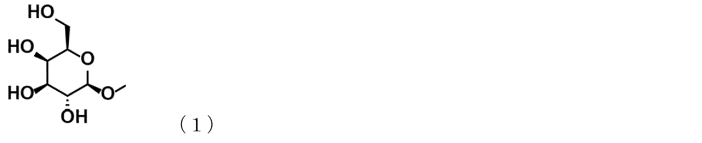
また、本発明のもう1つの好ましい態様においては、Yは以下のいずれかから選択される。
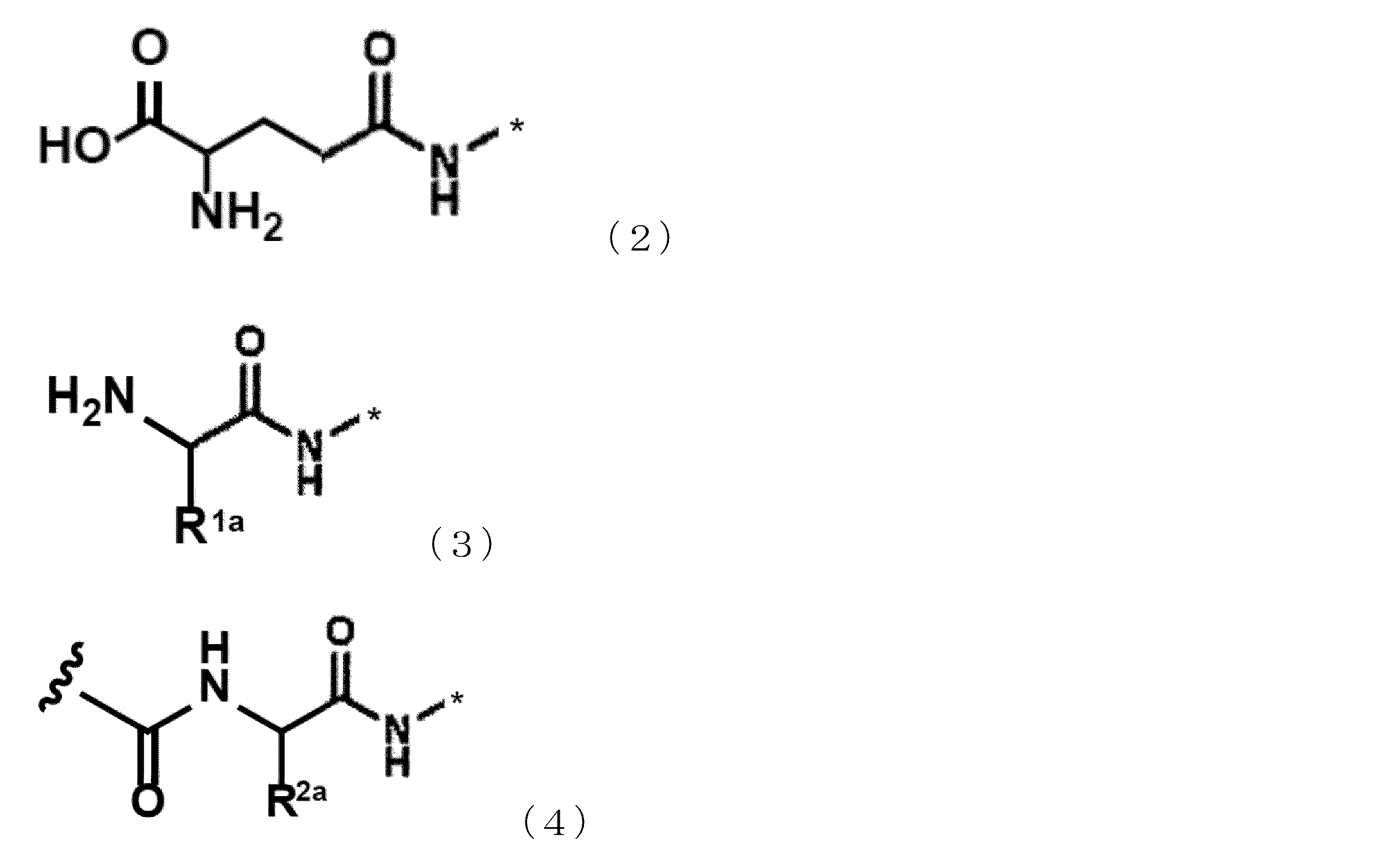
式(2)~(4)において、*は、Tと結合する箇所を表す。
式(3)において、R1aは、天然アミノ酸(グリシン、アラニン、ロイシン、イソロイシン、バリン、リジン、システイン、トレオニン、アルギニン、アスパラギン、アスパラギン酸、グルタミン、グルタミン酸、セリン、ヒスチジン、フェニルアラニン、メチオニン、トリプトファン、チロシン、プロリン)の側鎖を構成する基、非天然アミノ酸(シトルリン、ノルバリンなど)の側鎖を構成する基、置換又は無置換のアルキル基からなる群から選択される。
式(4)において、R2aは、天然アミノ酸(グリシン、アラニン、ロイシン、イソロイシン、バリン、リジン、システイン、トレオニン、アルギニン、アスパラギン、アスパラギン酸、グルタミン、グルタミン酸、セリン、ヒスチジン、フェニルアラニン、メチオニン、トリプトファン、チロシン、プロリン)の側鎖を構成する基、非天然アミノ酸(シトルリン、ノルバリンなど)の側鎖を構成する基、置換又は無置換のアルキル基からなる群から選択される。
 は、アミノ基、Nアセチル化等されたアミノ基、1つまたは複数のアミノ酸がペプチド結合により連なった構造を表す。
本発明の1つの好ましい側面においては、Yは以下で表される。
 上記構造において、プロリン残基の右側の窒素原子がTのリンカーと結合する。
Yが活性酸素種に対する捕捉基である場合は、当該捕捉基としては、ヒドロキシフェノキシ、アミノフェノキシ、アジド、又はピナコールボランであることが好ましい、
一般式(I)において、Zは、-C≡N、-C≡15N、-13C≡N、又は-13C≡15Nから選択される。
本発明の化合物の1つの好ましい態様は、以下の一般式(Ia)で表される化合物又はその塩である。

一般式(Ia)において、R1、R3、R7、R8、T、Yは、一般式(I)で定義した通りである。
R9は、存在する場合は、炭素数1~3のアルキル基である。
R10は、存在する場合は、炭素数1~3のアルキル基である。
R9は、存在する場合は、炭素数1~3のアルキル基である。
R10は、存在する場合は、炭素数1~3のアルキル基である。
本発明の化合物の非限定的な具体例を以下に示すが、本発明はこれらに限定されるものではない。
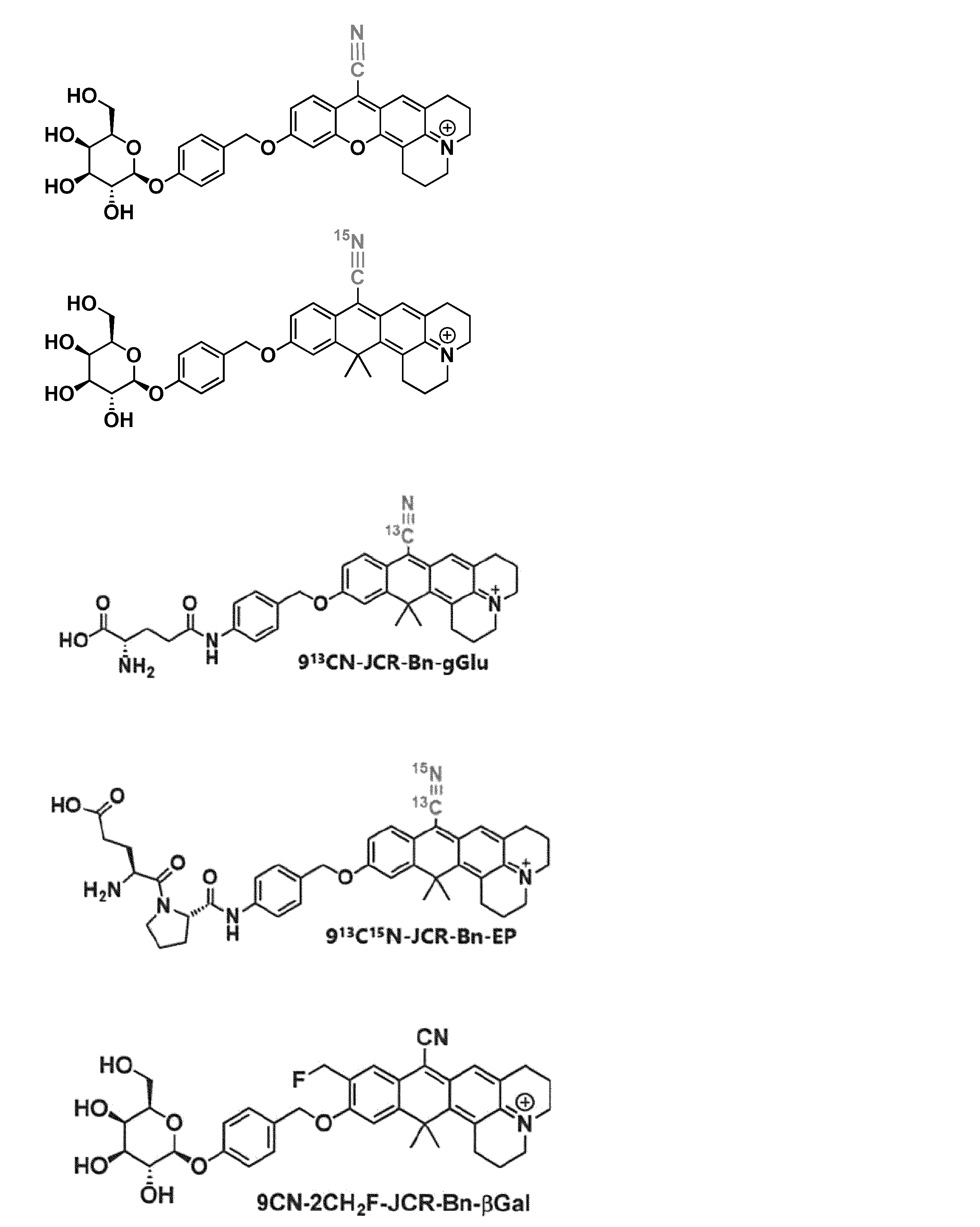
一般式(I)及び(Ia)で表される化合物は、酸付加塩又は塩基付加塩として存在することができる。酸付加塩としては、例えば、塩酸塩、硫酸塩、硝酸塩などの鉱酸塩、又はメタンスルホン酸塩、p-トルエンスルホン酸塩、シュウ酸塩、クエン酸塩、酒石酸塩などの有機酸塩などを挙げることができ、塩基付加塩としては、ナトリウム塩、カリウム塩、カルシウム塩、マグネシウム塩などの金属塩、アンモニウム塩、又はトリエチルアミン塩などの有機アミン塩などを挙げることができる。これらのほか、グリシンなどのアミノ酸との塩を形成する場合もある。一般式(I)及び(Ia)で表される化合物又はその塩は、水和物又は溶媒和物として存在する場合もあるが、本発明においては、これらの物質も用いることができる。
一般式(I)及び(Ia)で表される化合物は、置換基の種類により、1個又は2個以上の不斉炭素を有する場合があるが、本発明においては、1個又は2個以上の不斉炭素に基づく光学活性体や2個以上の不斉炭素に基づくジアステレオ異性体などの立体異性体のほか、立体異性体の任意の混合物、ラセミ体なども用いることができる。
一般式(I)及び(Ia)で表される化合物の代表的化合物の製造方法を本明細書の実施例に具体的に示した。従って、当業者は、これらの説明をもとにして、反応原料、反応条件、及び反応試薬などを適宜選択して、必要に応じてこれらの方法に修飾や改変を加えることにより、一般式(I)及び(Ia)で表される化合物を製造することができる。
2.本発明のラマンプローブ
本発明のもう1つの態様は、一般式(I)の化合物又はその塩を含むラマンプローブである(以下「本発明のラマンプローブ」ともいう)。
本発明のもう1つの態様は、一般式(I)の化合物又はその塩を含むラマンプローブである(以下「本発明のラマンプローブ」ともいう)。
本発明のラマンプローブは、epr-SRS法に利用可能なラマンプローブである。epr-SRS法とは、前期共鳴 (electronic pre-resonance; epr)効果と誘導ラマン散乱 (stimulated Raman scattering; SRS) を組み合わせたラマンイメージング法である。
この方法では、分子の電子吸収帯よりやや長波長側の光によって励起する(前期共鳴条件)ため、光褪色や蛍光によるバックグラウンド上昇を抑えることができ、ストークス光による誘導放出によってさらに高感度なイメージングが実現できる。従来のSRS顕微鏡ではストークス光の波長変化に秒オーダーの時間がかかるため、スペクトル解析やスペクトルイメージングを行う際には測定時間が長くなってしまうのが課題の1つであったが、東京大学大学院工学系研究科電気系工学専攻の小関研究室で開発された高速SRS分光顕微鏡では1秒当たり30波数分変化させる高速イメージングが可能である(Ozeki, Y., Biological Imaging Based on Stimulated Raman Scattering. Seibutsu Butsuri, 2014. 54(6): p. 311-314)。当該高速SRS分光顕微鏡の模式図を図2に示す。
高速SRS分光顕微鏡を用いるラマンイメージング法は、上記のOzekiの論文を参照して行うことができる。また、高速SRS分光顕微鏡でイメージングを行うに際しては、信号対雑音比を高めるため、in vitroでの測定ではデータを5~10回程度、in celluloでの測定ではデータを100~1000回程度取得して、平均化を行うことが好ましい。
この方法では、分子の電子吸収帯よりやや長波長側の光によって励起する(前期共鳴条件)ため、光褪色や蛍光によるバックグラウンド上昇を抑えることができ、ストークス光による誘導放出によってさらに高感度なイメージングが実現できる。従来のSRS顕微鏡ではストークス光の波長変化に秒オーダーの時間がかかるため、スペクトル解析やスペクトルイメージングを行う際には測定時間が長くなってしまうのが課題の1つであったが、東京大学大学院工学系研究科電気系工学専攻の小関研究室で開発された高速SRS分光顕微鏡では1秒当たり30波数分変化させる高速イメージングが可能である(Ozeki, Y., Biological Imaging Based on Stimulated Raman Scattering. Seibutsu Butsuri, 2014. 54(6): p. 311-314)。当該高速SRS分光顕微鏡の模式図を図2に示す。
高速SRS分光顕微鏡を用いるラマンイメージング法は、上記のOzekiの論文を参照して行うことができる。また、高速SRS分光顕微鏡でイメージングを行うに際しては、信号対雑音比を高めるため、in vitroでの測定ではデータを5~10回程度、in celluloでの測定ではデータを100~1000回程度取得して、平均化を行うことが好ましい。
この小関研の高速SRS分光顕微鏡の励起光は843nmであるため、当該epr-SRS法の装置を用いる場合は、前期共鳴がかかる波長域がおよそ650~750nmであるラマンプローブを用いるのが好ましい。
本発明のラマンプローブを用いる標的酵素の検出方法には、上記の高速SRS分光顕微鏡を好適に用いることができるが、当該高速SRS分光顕微鏡を用いた方法に限定されるものではない。
本発明のラマンプローブを用いる標的酵素の検出方法には、上記の高速SRS分光顕微鏡を好適に用いることができるが、当該高速SRS分光顕微鏡を用いた方法に限定されるものではない。
即ち、本発明の1つの好ましい側面は、一般式(I)の化合物又はその塩を含むepr-SRS法に利用可能なactivatable型ラマンプローブである。
本発明のラマンプローブの使用方法は特に限定されず、従来公知のラマンプローブと同様に用いることが可能である。通常は、生理食塩水や緩衝液などの水性媒体、又はエタノール、アセトン、エチレングリコール、ジメチルスルホキシド、ジメチルホルムアミドなどの水混合性の有機溶媒と水性媒体との混合物などに上記式(I)で表される化合物又はそれらの塩を溶解し、細胞や組織を含む適切な緩衝液中にこの溶液を添加して、ラマンスペクトルを測定すればよい。本発明のラマンプローブを適切な添加物と組み合わせて組成物の形態で用いてもよい。例えば、緩衝剤、溶解補助剤、pH調節剤などの添加物と組み合わせることができる。
また、本発明のもう1つの態様は、細胞又は組織内の標的酵素を検出する方法であって、(a)一般式(I)で表される化合物又はその塩を含むラマンプローブを細胞又は組織内に導入する工程、及び(b)当該化合物又はその塩が細胞又は組織内で標的酵素と反応することにより増強されるラマン散乱光を測定する工程を含む方法、である(以下「本発明の検出方法」とも言う)。
ここで、細胞としては、正常細胞、癌細胞、神経細胞等が挙げられる。
ここで、細胞としては、正常細胞、癌細胞、神経細胞等が挙げられる。
本発明の検出方法において、好ましくは、ラマン散乱光はepr-SRS法を用いて測定される。
一般式(I)で表される化合物又はその塩を細胞又は組織内に導入するには、一般式(I)で表される化合物又はその塩を細胞又は組織の試料に散布すること等により行うことができる。
本発明の検出方法の対象となるがん細胞又はがん組織の種類として、肺がん、前立腺がん、卵巣がん、乳がん、膀胱がん、脳腫瘍、食道がん、胃がん、胆管がん、肝がん、膵がん、頭頚部がん、腎がん、白血病、皮膚がん、甲状腺がんの細胞又は組織が挙げられる。
本明細書において、「がん組織」の用語はがん細胞を含む任意の組織を意味している。「組織」の用語は臓器の一部又は全体を含めて最も広義に解釈しなければならず、いかなる意味においても限定的に解釈してはならない。
本発明の検出方法の1つの態様は、(a)2以上の一般式(I)で表される化合物又はその塩を細胞又は組織内に導入する工程、及び(b)当該2以上の化合物又はその塩が細胞又は組織内において、夫々、標的酵素と反応することにより増強されるラマン散乱光を測定する工程を含む、細胞又は組織内の2以上の標的酵素を検出する方法である。
本発明の検出方法及び本発明のプローブによって、細胞に加えて、組織の状態で染色することができることから、本発明の検出方法及び本発明のプローブは、酵素活性パターンによるがんの診断方法にも用いることが可能である。
即ち、本発明のもう1つの実施態様は、(a)一般式(I)の化合物又はその塩を含むラマンプローブを被検体に投与する工程、(b)当該化合物又はその塩が被検体の細胞、組織又は臓器内で標的酵素と反応することにより増強されるラマン散乱光を測定する工程を含む、がんを診断する方法である(以下「本発明の診断方法」とも言う)。
投与方法としては、局所投与、経口投与、静脈内投与等が挙げられる。
本発明の診断方法の対象となるがんの種類として、肺がん、前立腺がん、卵巣がん、乳がん、膀胱がん、脳腫瘍、食道がん、胃がん、胆管がん、肝がん、膵がん、頭頚部がん、腎がん、白血病、皮膚がん、甲状腺がんが挙げられる。
即ち、本発明のもう1つの実施態様は、(a)一般式(I)の化合物又はその塩を含むラマンプローブを被検体に投与する工程、(b)当該化合物又はその塩が被検体の細胞、組織又は臓器内で標的酵素と反応することにより増強されるラマン散乱光を測定する工程を含む、がんを診断する方法である(以下「本発明の診断方法」とも言う)。
投与方法としては、局所投与、経口投与、静脈内投与等が挙げられる。
本発明の診断方法の対象となるがんの種類として、肺がん、前立腺がん、卵巣がん、乳がん、膀胱がん、脳腫瘍、食道がん、胃がん、胆管がん、肝がん、膵がん、頭頚部がん、腎がん、白血病、皮膚がん、甲状腺がんが挙げられる。
本発明のもう1つの実施態様は、本発明のラマンプローブを含む、標的分子の検出用キットである。
当該キットにおいて、通常、本発明のラマンプローブは溶液として調製されているが、例えば、粉末形態の混合物、凍結乾燥物、顆粒剤、錠剤、液剤など適宜の形態の組成物として提供され、使用時に注射用蒸留水や適宜の緩衝液に溶解して適用することもできる。
また、当該キットには、必要に応じてそれ以外の試薬等を適宜含んでいてもよい。例えば、添加剤として、溶解補助剤、pH調節剤、緩衝剤、等張化剤などの添加剤を用いることができ、これらの配合量は当業者に適宜選択可能である。
以下、本発明を実施例により説明するが、本発明はこれに限定されるものではない。
[原料]
合成に使用した全ての化学物質は、東京化成工業(株)、和光純薬工業(株)、シグマアルドリッチ(株)、Cambridge Isotope Laboratories, Inc. から購入した。 さらに精製することなく使用した。
合成に使用した全ての化学物質は、東京化成工業(株)、和光純薬工業(株)、シグマアルドリッチ(株)、Cambridge Isotope Laboratories, Inc. から購入した。 さらに精製することなく使用した。
[測定機器]
NMRスペクトルは、重水素化溶媒中を用い、Bruker NMR AVANCE III 400分光計[1H 400 MHz、13C 100 MHz]で得た。
高分解能ESI質量スペクトルは、Bruker microTOF II-TM (ESI)で得た。
HPLC精製は、Inertstil-ODS-3カラム(Φ10×250mm(セミ分取)およびΦ20×250mm(分取))を備えたJASCO PU-2080 Plusポンプ(GL Science Co.、Ltd.)およびMD-2015検出器(JASCO)で行った。
HPLCに使用した溶媒は、和光(株)より入手した。シリカゲルカラムクロマトグラフィーは、中圧分取液体クロマトグラフYFLC-Al560(山善株式会社)を用いて行った。
TLCは、シリカゲルプレートF254(0.25mm(分析);Merck、AKG)で行った。
UV-visスペクトルは、Shimadzu UV-2450分光光度計で得た。
蛍光スペクトルは、F-7000 (日立)で取得した。
SRSスペクトルおよびSRS像は、東京大学大学院工学系研究科電気系工学専攻小関研究室で開発された高速SRS分光顕微鏡で取得した。ポンプ光パルスおよびストークス光パルスの波長は843nmおよび1014-1046nm、パルス時間幅は約5ピコ秒、スペクトル分解能は5/cmである。水浸対物レンズを用い、その開口数は1.2である。フレームごとにストークス光パルスの波長を変化させ、500×500ピクセルの画像を毎秒30フレーム取得した。信号対雑音比を高めるためにデータをin vitroでは5回、in celluloでは1000回取得し、平均化を行った。
NMRスペクトルは、重水素化溶媒中を用い、Bruker NMR AVANCE III 400分光計[1H 400 MHz、13C 100 MHz]で得た。
高分解能ESI質量スペクトルは、Bruker microTOF II-TM (ESI)で得た。
HPLC精製は、Inertstil-ODS-3カラム(Φ10×250mm(セミ分取)およびΦ20×250mm(分取))を備えたJASCO PU-2080 Plusポンプ(GL Science Co.、Ltd.)およびMD-2015検出器(JASCO)で行った。
HPLCに使用した溶媒は、和光(株)より入手した。シリカゲルカラムクロマトグラフィーは、中圧分取液体クロマトグラフYFLC-Al560(山善株式会社)を用いて行った。
TLCは、シリカゲルプレートF254(0.25mm(分析);Merck、AKG)で行った。
UV-visスペクトルは、Shimadzu UV-2450分光光度計で得た。
蛍光スペクトルは、F-7000 (日立)で取得した。
SRSスペクトルおよびSRS像は、東京大学大学院工学系研究科電気系工学専攻小関研究室で開発された高速SRS分光顕微鏡で取得した。ポンプ光パルスおよびストークス光パルスの波長は843nmおよび1014-1046nm、パルス時間幅は約5ピコ秒、スペクトル分解能は5/cmである。水浸対物レンズを用い、その開口数は1.2である。フレームごとにストークス光パルスの波長を変化させ、500×500ピクセルの画像を毎秒30フレーム取得した。信号対雑音比を高めるためにデータをin vitroでは5回、in celluloでは1000回取得し、平均化を行った。
1.候補母核の検討
[参考実施例]
O-function型プローブの開発を行うべく、まずは候補色素骨格の検討を行った。図3に示すように、O-function型とするためにロドール色素を足場とし、6位のN原子上の置換基や10位元素Xの置換を行った。6種類の誘導体を開発し、プローブ母核として最適な構造の探索を行った。合成した一連の9CN-rhodol誘導体の光学特性を図3及び表1に示す。
[参考実施例]
O-function型プローブの開発を行うべく、まずは候補色素骨格の検討を行った。図3に示すように、O-function型とするためにロドール色素を足場とし、6位のN原子上の置換基や10位元素Xの置換を行った。6種類の誘導体を開発し、プローブ母核として最適な構造の探索を行った。合成した一連の9CN-rhodol誘導体の光学特性を図3及び表1に示す。

[2]0.2Mリン酸ナトリウム緩衝液中で測定した。
[3]共溶媒として30%DMSOを含有するPBS(pH7.4)中で測定した。
上記の結果から、N上の置換基はジュロリジン(julolidine)構造が明るさと安定性の両観点から最適であると考えられ、以下の構造を有する9CN-JRと9CN-JCRをプローブの候補母核として選定した。
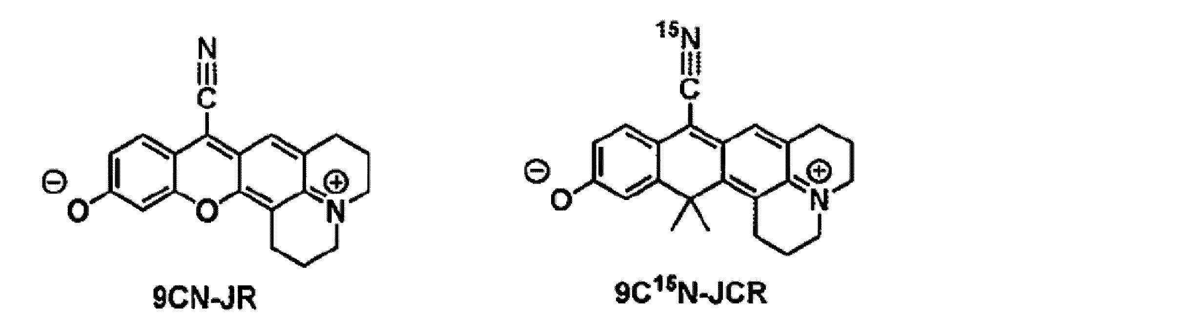
2.O-function型βGal検出ラマンプローブの開発
次に、選定した9CN-JRと9CN-JCRのプローブとしての特性をSRSによる同時イメージングで比較するため、標的酵素を同じく(β-ガラクトシダーゼ)しているがCN基に同位体置換を施していることでラマン散乱によって分離検出可能な2つのβGal検出プローブ9CN-JR-Bn-βGalと9C15N-JCR-Bn-βGalを開発した。
ここで、当初、以下のスキームA及びBにしたがって、9CN-rhodol構造に直接糖基質を結合させたプローブである9CN-JR-βGalおよび9C15N-JCR-βGalの合成を試みたが、O-coreの誘導体において糖を縮合したロイコ体16からの色素17へ酸化する反応が50%未満で,酸化反応によって糖が脱離したJRが多く生じてしまうことが明らかとなった。さらに,C-coreの誘導体においてはロイコ体19から色素20の酸化によってほぼ全量がJCRへと変換されてしまい、目的の化合物20を得ることができなかった。
次に、選定した9CN-JRと9CN-JCRのプローブとしての特性をSRSによる同時イメージングで比較するため、標的酵素を同じく(β-ガラクトシダーゼ)しているがCN基に同位体置換を施していることでラマン散乱によって分離検出可能な2つのβGal検出プローブ9CN-JR-Bn-βGalと9C15N-JCR-Bn-βGalを開発した。
ここで、当初、以下のスキームA及びBにしたがって、9CN-rhodol構造に直接糖基質を結合させたプローブである9CN-JR-βGalおよび9C15N-JCR-βGalの合成を試みたが、O-coreの誘導体において糖を縮合したロイコ体16からの色素17へ酸化する反応が50%未満で,酸化反応によって糖が脱離したJRが多く生じてしまうことが明らかとなった。さらに,C-coreの誘導体においてはロイコ体19から色素20の酸化によってほぼ全量がJCRへと変換されてしまい、目的の化合物20を得ることができなかった。
スキームA
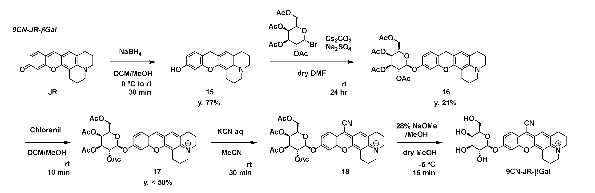
スキームB

そこで、9CN-rhodolと糖基質との間に自己分解性リンカーであるBnリンカーを導入したところ、目的とするプローブの合成が可能となった。
9CN-JR-Bn-βGal及び9C15N-JCR-Bn-βGalの合成実施例を以下に示す。
9CN-JR-Bn-βGal及び9C15N-JCR-Bn-βGalの合成実施例を以下に示す。
[合成実施例1]
9CN-JR-Bn-βGalの合成
以下のスキーム1に則り、本発明の化合物である9CN-JR-Bn-βGalを合成した。
9CN-JR-Bn-βGalの合成
以下のスキーム1に則り、本発明の化合物である9CN-JR-Bn-βGalを合成した。
スキーム1

(1)化合物37の合成
化合物34(891mg、7.3mmol)、臭化2,3,4,6-テトラ-O-アセチル-α-D-ガラクトピラノシル(3.0g、7.3mmol)、Ag2O(5.0 g、22mmol)及びNa2SO4(過剰量)の30mL乾燥MeCN中の懸濁液を、アルゴン雰囲気下、室温で12時間撹拌した。反応溶液をセライトで濾過し、濾液を蒸発乾固した。残渣をカラムクロマトグラフィー(シリカゲル、n-ヘキサン/AcOEt=50/50~29/71)で精製し、化合物35(3.0g)を得た。10mLのDCM及び10mLのMeOH中の化合物35(3.0g、6.6mmol)の溶液に、0℃でNaBH4(376mg、9.9mmol)を加えた。室温まで温めた後、反応混合物を1時間撹拌した。反応を飽和NH4Cl水溶液でクエンチし、混合物をDCMで抽出した。有機層を塩水で洗浄し、Na2SO4で乾燥させ、蒸発乾固させて、化合物36(2.3g)を得た。20mLの乾燥Et2O中の化合物36(2.3g、5.1mmol)の溶液に、アルゴン雰囲気下、0℃でPBr3(480 μL、5.1mmol)を加えた。撹拌を同じ温度で2時間続けた。次に、反応を飽和NaHCO3水溶液でクエンチし、混合物をAcOEtで抽出した。有機層をブラインで洗浄し、Na2SO4で乾燥させ、蒸発乾固させた。残渣をカラムクロマトグラフィー(シリカゲル、n-ヘキサン/AcOEt=78/22~57/43)で精製し、化合物37を得た(2.1g、3段階で56%)。
1H NMR (400 MHz, CDCl3): δ 7.34 (d, J = 8.7 Hz, 2H), 6.97 (d, J = 8.7 Hz, 2H), 5.49 (dd, J = 10.5, 8.0 Hz, 1H), 5.46 (d, J = 3.7 Hz, 1H), 5.11 (dd, J = 10.5, 3.5 Hz, 1H), 5.06 (d, J = 8.0 Hz, 1H), 4.49 (s, 2H), 4.25-4.06 (m, 3H), 2.19 (s, 3H), 2.07 (s, 6H), 2.02 (s, 3H); 13C NMR (101 MHz, CDCl3): δ 170.46, 170.33, 170.20, 169.47, 156.79, 132.72, 130.46, 117.08, 99.30, 71.03, 70.77, 68.59, 66.91, 61.41, 33.17, 20.70, 20.65, 20.62, 20.56; HRMS (ESI+): Calcd for [M+Na]+, 539.05233, Found, 539.05123 (1.1 mDa).
化合物34(891mg、7.3mmol)、臭化2,3,4,6-テトラ-O-アセチル-α-D-ガラクトピラノシル(3.0g、7.3mmol)、Ag2O(5.0 g、22mmol)及びNa2SO4(過剰量)の30mL乾燥MeCN中の懸濁液を、アルゴン雰囲気下、室温で12時間撹拌した。反応溶液をセライトで濾過し、濾液を蒸発乾固した。残渣をカラムクロマトグラフィー(シリカゲル、n-ヘキサン/AcOEt=50/50~29/71)で精製し、化合物35(3.0g)を得た。10mLのDCM及び10mLのMeOH中の化合物35(3.0g、6.6mmol)の溶液に、0℃でNaBH4(376mg、9.9mmol)を加えた。室温まで温めた後、反応混合物を1時間撹拌した。反応を飽和NH4Cl水溶液でクエンチし、混合物をDCMで抽出した。有機層を塩水で洗浄し、Na2SO4で乾燥させ、蒸発乾固させて、化合物36(2.3g)を得た。20mLの乾燥Et2O中の化合物36(2.3g、5.1mmol)の溶液に、アルゴン雰囲気下、0℃でPBr3(480 μL、5.1mmol)を加えた。撹拌を同じ温度で2時間続けた。次に、反応を飽和NaHCO3水溶液でクエンチし、混合物をAcOEtで抽出した。有機層をブラインで洗浄し、Na2SO4で乾燥させ、蒸発乾固させた。残渣をカラムクロマトグラフィー(シリカゲル、n-ヘキサン/AcOEt=78/22~57/43)で精製し、化合物37を得た(2.1g、3段階で56%)。
1H NMR (400 MHz, CDCl3): δ 7.34 (d, J = 8.7 Hz, 2H), 6.97 (d, J = 8.7 Hz, 2H), 5.49 (dd, J = 10.5, 8.0 Hz, 1H), 5.46 (d, J = 3.7 Hz, 1H), 5.11 (dd, J = 10.5, 3.5 Hz, 1H), 5.06 (d, J = 8.0 Hz, 1H), 4.49 (s, 2H), 4.25-4.06 (m, 3H), 2.19 (s, 3H), 2.07 (s, 6H), 2.02 (s, 3H); 13C NMR (101 MHz, CDCl3): δ 170.46, 170.33, 170.20, 169.47, 156.79, 132.72, 130.46, 117.08, 99.30, 71.03, 70.77, 68.59, 66.91, 61.41, 33.17, 20.70, 20.65, 20.62, 20.56; HRMS (ESI+): Calcd for [M+Na]+, 539.05233, Found, 539.05123 (1.1 mDa).
(2)化合物38の合成
5mLのDCM及び5mLのMeOH中の化合物12(197mg、0.68mmol)の溶液に、NaBH4(51mg、1.4mmol)を0℃で加えた。室温まで温めた後、反応混合物を1時間撹拌し、次いで蒸発させた。残渣をカラムクロマトグラフィー(シリカゲル、n-ヘキサン/AcOEt=88/12~67/33)で精製し、化合物38(64mg、32%)を得た。
1H NMR (400 MHz, CDCl3): δ 6.98 (d, J = 8.2 Hz, 1H), 6.60 (s, 1H), 6.52 (d, J = 2.5 Hz, 1H), 6.47 (dd, J = 8.3, 2.5 Hz, 1H), 3.80 (s, 2H), 3.08 (q, J = 5.2 Hz, 4H), 2.82 (t, J = 6.7 Hz, 2H), 2.72 (t, J = 6.6 Hz, 2H), 2.01-1.96 (m, 4H); 13C NMR (101 MHz, CDCl3): δ 154.64, 152.91, 147.61, 142.41, 129.48, 126.01, 116.78, 113.65, 109.79, 109.29, 108.02, 103.41, 50.22, 49.71, 27.15, 26.63, 22.39, 21.71, 21.16; HRMS (ESI+): Calcd for [M+H]+, 294.14886, Found, 294.14927 (-0.4 mDa).
5mLのDCM及び5mLのMeOH中の化合物12(197mg、0.68mmol)の溶液に、NaBH4(51mg、1.4mmol)を0℃で加えた。室温まで温めた後、反応混合物を1時間撹拌し、次いで蒸発させた。残渣をカラムクロマトグラフィー(シリカゲル、n-ヘキサン/AcOEt=88/12~67/33)で精製し、化合物38(64mg、32%)を得た。
1H NMR (400 MHz, CDCl3): δ 6.98 (d, J = 8.2 Hz, 1H), 6.60 (s, 1H), 6.52 (d, J = 2.5 Hz, 1H), 6.47 (dd, J = 8.3, 2.5 Hz, 1H), 3.80 (s, 2H), 3.08 (q, J = 5.2 Hz, 4H), 2.82 (t, J = 6.7 Hz, 2H), 2.72 (t, J = 6.6 Hz, 2H), 2.01-1.96 (m, 4H); 13C NMR (101 MHz, CDCl3): δ 154.64, 152.91, 147.61, 142.41, 129.48, 126.01, 116.78, 113.65, 109.79, 109.29, 108.02, 103.41, 50.22, 49.71, 27.15, 26.63, 22.39, 21.71, 21.16; HRMS (ESI+): Calcd for [M+H]+, 294.14886, Found, 294.14927 (-0.4 mDa).
(3)化合物39の合成
化合物38(80mg、0.27mmol)、化合物37(210mg、0.41mmol)及びNaH(10mg、0.41mmol)を2mLの乾燥DMFに0℃、アルゴン雰囲気下、暗所で溶解し、室温まで昇温し、撹拌を18時間続けた。反応を飽和NH4Cl水溶液でクエンチし、混合物をDCMで抽出した。有機層を塩水で洗浄し、Na2SO4で乾燥させ、蒸発乾固させた。残渣を5mLのDCMおよび5mLのMeOHに溶解し、クロラニル(66mg、0.27mmol)を室温で添加した。 混合物を10分間撹拌し、次いで蒸発させた。残渣を、溶離液A(1%MeCN及び0.1%TFAを含むH2O)及び溶離液剤B(1%H2Oを含むMeCN)(40分間でA/B=90/10から0/100)を使用する分取HPLCにより粗精製した。 溶出液を蒸発させ、残渣をカラムクロマトグラフィー(シリカゲル、DCM/MeOH=90/10から83/17)で精製して、残りの化合物37を除去し、化合物39(79mg、40%)を得た。
1H NMR (400 MHz, CD3OD): δ 8.50 (s, 1H), 7.87 (d, J = 8.9 Hz, 1H), 7.49 (s, 1H), 7.42 (d, J = 8.7 Hz, 2H), 7.35 (d, J = 2.2 Hz, 1H), 7.20 (dd, J = 8.8, 2.4 Hz, 1H), 7.04 (d, J = 8.0 Hz, 2H), 5.46 (d, J = 3.6 Hz, 1H), 5.37 (dd, J = 10.1, 7.8 Hz, 1H), 5.33 (d, J = 7.8 Hz, 1H), 5.26 (dd, J = 10.0, 3.5 Hz, 1H), 5.22 (s, 2H), 4.31 (t, J = 6.0 Hz, 1H), 4.19-4.16 (m, 2H), 3.69 (t, J = 5.5 Hz, 4H), 3.01 (t, J = 6.2 Hz, 2H), 2.93 (t, J = 5.9 Hz, 2H), 2.17 (s, 3H), 2.11-2.04 (m, 4H), 2.06 (s, 3H), 2.03 (s, 3H), 1.97 (s, 3H); HRMS (ESI+): Calcd for [M]+, 728.27015, Found, 728.27203 (-1.9 mDa).
化合物38(80mg、0.27mmol)、化合物37(210mg、0.41mmol)及びNaH(10mg、0.41mmol)を2mLの乾燥DMFに0℃、アルゴン雰囲気下、暗所で溶解し、室温まで昇温し、撹拌を18時間続けた。反応を飽和NH4Cl水溶液でクエンチし、混合物をDCMで抽出した。有機層を塩水で洗浄し、Na2SO4で乾燥させ、蒸発乾固させた。残渣を5mLのDCMおよび5mLのMeOHに溶解し、クロラニル(66mg、0.27mmol)を室温で添加した。 混合物を10分間撹拌し、次いで蒸発させた。残渣を、溶離液A(1%MeCN及び0.1%TFAを含むH2O)及び溶離液剤B(1%H2Oを含むMeCN)(40分間でA/B=90/10から0/100)を使用する分取HPLCにより粗精製した。 溶出液を蒸発させ、残渣をカラムクロマトグラフィー(シリカゲル、DCM/MeOH=90/10から83/17)で精製して、残りの化合物37を除去し、化合物39(79mg、40%)を得た。
1H NMR (400 MHz, CD3OD): δ 8.50 (s, 1H), 7.87 (d, J = 8.9 Hz, 1H), 7.49 (s, 1H), 7.42 (d, J = 8.7 Hz, 2H), 7.35 (d, J = 2.2 Hz, 1H), 7.20 (dd, J = 8.8, 2.4 Hz, 1H), 7.04 (d, J = 8.0 Hz, 2H), 5.46 (d, J = 3.6 Hz, 1H), 5.37 (dd, J = 10.1, 7.8 Hz, 1H), 5.33 (d, J = 7.8 Hz, 1H), 5.26 (dd, J = 10.0, 3.5 Hz, 1H), 5.22 (s, 2H), 4.31 (t, J = 6.0 Hz, 1H), 4.19-4.16 (m, 2H), 3.69 (t, J = 5.5 Hz, 4H), 3.01 (t, J = 6.2 Hz, 2H), 2.93 (t, J = 5.9 Hz, 2H), 2.17 (s, 3H), 2.11-2.04 (m, 4H), 2.06 (s, 3H), 2.03 (s, 3H), 1.97 (s, 3H); HRMS (ESI+): Calcd for [M]+, 728.27015, Found, 728.27203 (-1.9 mDa).
(4)化合物40の合成
5mLMeCN中の化合物39(31mg、0.043mmol)の溶液に0.3M KCN水溶液(300μL、0.086mmol)を添加し、混合物を室温で10分間撹拌した。次いで、反応をHPLC用溶離液A(1%MeCN及び0.1%TFAを含むH2O)でクエンチし、混合物をDCMで抽出した。有機層を塩水で洗浄し、Na2SO4で乾燥させ、蒸発乾固させた。粗化合物を5mLのDCM及び1mLのMeOHに溶解し、クロラニル(5mg、0.020mmol)を室温で添加した。混合物を5分間撹拌し、次いで蒸発させた。残渣を、溶離液A(1%MeCN及び0.1% TFAを含むH2O)および溶離剤B(1%H2Oを含むMeCN)(40分間でA/B=90/10から0/100)を使用する分取HPLCにより精製して、化合物40を得た(7.6mg、23%)。
1H NMR (400 MHz, CD3OD): δ 7.99 (d, J = 9.0 Hz, 1H), 7.68 (s, 1H), 7.45 (d, J = 8.7 Hz, 2H), 7.41 (d, J = 2.4 Hz, 1H), 7.32 (dd, J = 8.8, 2.4 Hz, 1H), 7.06 (d, J = 9.1 Hz, 2H), 5.46 (d, J = 3.4 Hz, 1H), 5.38-5.31 (m, 2H), 5.28 (s, 2H), 5.26 (dd, J = 9.7, 3.5 Hz, 1H), 4.31 (t, J = 6.4 Hz, 1H), 4.21-4.18 (m, 2H), 3.84-3.81 (m, 4H), 3.07-3.03 (m, 4H), 2.18 (s, 3H), 2.16-2.12 (m, 4H), 2.05 (s, 3H), 2.03 (s, 3H), 1.98 (s, 3H); HRMS (ESI+): Calcd for [M]+, 753.26540, Found, 753.26591 (-0.5 mDa).
5mLMeCN中の化合物39(31mg、0.043mmol)の溶液に0.3M KCN水溶液(300μL、0.086mmol)を添加し、混合物を室温で10分間撹拌した。次いで、反応をHPLC用溶離液A(1%MeCN及び0.1%TFAを含むH2O)でクエンチし、混合物をDCMで抽出した。有機層を塩水で洗浄し、Na2SO4で乾燥させ、蒸発乾固させた。粗化合物を5mLのDCM及び1mLのMeOHに溶解し、クロラニル(5mg、0.020mmol)を室温で添加した。混合物を5分間撹拌し、次いで蒸発させた。残渣を、溶離液A(1%MeCN及び0.1% TFAを含むH2O)および溶離剤B(1%H2Oを含むMeCN)(40分間でA/B=90/10から0/100)を使用する分取HPLCにより精製して、化合物40を得た(7.6mg、23%)。
1H NMR (400 MHz, CD3OD): δ 7.99 (d, J = 9.0 Hz, 1H), 7.68 (s, 1H), 7.45 (d, J = 8.7 Hz, 2H), 7.41 (d, J = 2.4 Hz, 1H), 7.32 (dd, J = 8.8, 2.4 Hz, 1H), 7.06 (d, J = 9.1 Hz, 2H), 5.46 (d, J = 3.4 Hz, 1H), 5.38-5.31 (m, 2H), 5.28 (s, 2H), 5.26 (dd, J = 9.7, 3.5 Hz, 1H), 4.31 (t, J = 6.4 Hz, 1H), 4.21-4.18 (m, 2H), 3.84-3.81 (m, 4H), 3.07-3.03 (m, 4H), 2.18 (s, 3H), 2.16-2.12 (m, 4H), 2.05 (s, 3H), 2.03 (s, 3H), 1.98 (s, 3H); HRMS (ESI+): Calcd for [M]+, 753.26540, Found, 753.26591 (-0.5 mDa).
(5)9CN-JR-Bn-βGalの合成
乾燥MeOH1.5mL中の化合物40(7.6mg、0.010mmol)の溶液に、500μLの乾燥MeOHで希釈した28%NaOMe/MeOH(20μL、0.1mmol)を、アルゴン雰囲気下、-5℃で添加した。混合物を同じ温度で15分間撹拌した。次いで、溶液が着色するまで、HPLC溶離液A(1%MeCN及び0.1%TFAを含むH2O)で反応をクエンチした。蒸発後、溶離液A(1%MeCN及び0.1%TFAを含むH2O)および溶離液B(1%H2Oを含むMeCN)(30分間でA/B=90/10から30/70)を使用する分取HPLCによって残渣を精製し、9CN-JR-Bn-βGalを得た(4.3 mg、73%)。
1H NMR (400 MHz, CD3OD): δ 7.99 (d, J = 9.0 Hz, 1H), 7.69 (s, 1H), 7.42 (d, J = 8.6 Hz, 2H), 7.42 (d, J = 2.4 Hz, 1H), 7.32 (dd, J = 9.1, 2.4 Hz, 1H), 7.15 (d, J = 8.7 Hz, 2H), 5.28 (s, 2H), 3.91 (d, J = 3.1 Hz, 1H), 3.83-3.80 (m, 4H), 3.80-3.76 (m, 3H), 3.74-3.65 (m, 2H), 3.58 (dd, J = 9.2, 3.4 Hz, 1H), 3.07-3.02 (m, 4H), 2.15-2.11 (m, 4H); HRMS (ESI+): Calcd for [M]+, 585.22314, Found, 585.22357 (-0.4 mDa).
乾燥MeOH1.5mL中の化合物40(7.6mg、0.010mmol)の溶液に、500μLの乾燥MeOHで希釈した28%NaOMe/MeOH(20μL、0.1mmol)を、アルゴン雰囲気下、-5℃で添加した。混合物を同じ温度で15分間撹拌した。次いで、溶液が着色するまで、HPLC溶離液A(1%MeCN及び0.1%TFAを含むH2O)で反応をクエンチした。蒸発後、溶離液A(1%MeCN及び0.1%TFAを含むH2O)および溶離液B(1%H2Oを含むMeCN)(30分間でA/B=90/10から30/70)を使用する分取HPLCによって残渣を精製し、9CN-JR-Bn-βGalを得た(4.3 mg、73%)。
1H NMR (400 MHz, CD3OD): δ 7.99 (d, J = 9.0 Hz, 1H), 7.69 (s, 1H), 7.42 (d, J = 8.6 Hz, 2H), 7.42 (d, J = 2.4 Hz, 1H), 7.32 (dd, J = 9.1, 2.4 Hz, 1H), 7.15 (d, J = 8.7 Hz, 2H), 5.28 (s, 2H), 3.91 (d, J = 3.1 Hz, 1H), 3.83-3.80 (m, 4H), 3.80-3.76 (m, 3H), 3.74-3.65 (m, 2H), 3.58 (dd, J = 9.2, 3.4 Hz, 1H), 3.07-3.02 (m, 4H), 2.15-2.11 (m, 4H); HRMS (ESI+): Calcd for [M]+, 585.22314, Found, 585.22357 (-0.4 mDa).
[合成実施例2]
9C15N-JCR-Bn-βGalの合成
以下のスキーム2に則り、本発明の化合物である9C15N-JCR-Bn-βGalを合成した。
9C15N-JCR-Bn-βGalの合成
以下のスキーム2に則り、本発明の化合物である9C15N-JCR-Bn-βGalを合成した。
スキーム2

(1)化合物41の合成
5mLのDCMと5mLのMeOH中の化合物29(138mg、0.43mmol)の溶液に、0℃でNaBH4(33mg、0.87mmol)を加えた。室温に温めた後、反応混合物を1時間撹拌し、その後蒸発させた。残渣をカラムクロマトグラフィー(シリカゲル、n-ヘキサン/AcOEt=95/5~74/26)で精製し、化合物41(99mg、72%)を得た。
1H NMR (400 MHz, CDCl3): δ 6.99 (d, J = 8.2 Hz,, 1H), 6.90 (d, J = 2.6 Hz, 1H), 6.70 (s, 1H), 6.62 (dd, J = 8.2, 2.6 Hz, 1H), 5.25 (brs, 1H), 3.95 (s, 2H), 3.16 (t, J = 5.7 Hz, 2H), 3.13 (t, J = 6.7 Hz, 2H), 2.94 (t, J = 6.0 Hz, 2H), 2.72 (t, J = 6.3 Hz, 2H), 1.97-1.91 (m, 4H), 1.73 (s, 6H); 13C NMR (101 MHz, CDCl3): δ 153.82, 149.08, 143.03, 138.30, 128.48, 126.97, 124.35, 123.04, 121.71, 121.31, 113.45, 113.20, 51.13, 50.09, 38.94, 33.44, 31.93, 27.98, 27.66, 22.82, 22.03; HRMS (ESI+): Calcd for [M+H]+, 320.20089, Found, 320.20025 (0.6 mDa).
5mLのDCMと5mLのMeOH中の化合物29(138mg、0.43mmol)の溶液に、0℃でNaBH4(33mg、0.87mmol)を加えた。室温に温めた後、反応混合物を1時間撹拌し、その後蒸発させた。残渣をカラムクロマトグラフィー(シリカゲル、n-ヘキサン/AcOEt=95/5~74/26)で精製し、化合物41(99mg、72%)を得た。
1H NMR (400 MHz, CDCl3): δ 6.99 (d, J = 8.2 Hz,, 1H), 6.90 (d, J = 2.6 Hz, 1H), 6.70 (s, 1H), 6.62 (dd, J = 8.2, 2.6 Hz, 1H), 5.25 (brs, 1H), 3.95 (s, 2H), 3.16 (t, J = 5.7 Hz, 2H), 3.13 (t, J = 6.7 Hz, 2H), 2.94 (t, J = 6.0 Hz, 2H), 2.72 (t, J = 6.3 Hz, 2H), 1.97-1.91 (m, 4H), 1.73 (s, 6H); 13C NMR (101 MHz, CDCl3): δ 153.82, 149.08, 143.03, 138.30, 128.48, 126.97, 124.35, 123.04, 121.71, 121.31, 113.45, 113.20, 51.13, 50.09, 38.94, 33.44, 31.93, 27.98, 27.66, 22.82, 22.03; HRMS (ESI+): Calcd for [M+H]+, 320.20089, Found, 320.20025 (0.6 mDa).
(2)化合物42の合成
化合物41(163mg、0.51mmol)、化合物37(612mg、1.2mmol)およびNaH(18mg、0.76mmol)を、暗所で、アルゴン雰囲気下、0℃で10mLの乾燥DMFに溶解した。混合物を室温まで温め、18時間撹拌を続けた。反応を飽和NH4Cl水溶液でクエンチし、DCMで抽出した。有機層を塩水で洗浄し、Na2SO4で乾燥させ、蒸発乾固させた。残渣を5mLのDCMと5mLのMeOHに溶解し、室温でクロラニル(62mg、0.25mmol)を加えた。混合物を10分間撹拌した後、蒸発させた。残渣を、溶離液A(1%MeCN及び0.1%TFAを含むH2O)及び溶離液B(1%H2Oを含むMeCN)(40分でA/B=90/10から0/100)を用いる分取HPLCによって粗精製し、化合物42(77mg、20%)を得た。
1H NMR (400 MHz, CD3OD): δ 7.92 (s, 1H), 7.63 (d, J = 8.6 Hz, 1H), 7.44 (d, J = 8.7 Hz, 2H), 7.42 (s, 1H), 7.33 (d, J = 2.1 Hz, 1H), 7.10 (dd, J = 8.7, 2.4 Hz, 1H), 7.06 (d, J = 8.7 Hz, 2H), 5.46 (d, J = 3.5 Hz, 1H), 5.36 (dd, J = 9.8, 7.8 Hz, 1H), 5.32 (d, J = 7.6 Hz, 1H), 5.25 (dd, J = 10.4, 3.4 Hz, 1H), 5.24 (s, 2H), 4.30 (t, J = 6.4 Hz, 1H), 4.18-4.16 (m, 2H), 3.77-3.73 (m, 4H), 3.23 (t, J = 6.1 Hz, 2H), 2.84 (t, J = 6.0 Hz, 2H), 2.17 (s, 3H), 2.10-2.02 (m, 4H), 2.05 (s, 3H), 2.01 (s, 3H), 1.97 (s, 3H), 1.80 (s, 6H); HRMS (ESI+): Calcd for [M]+, 754.32219, Found, 754.32264 (-0.4 mDa).
化合物41(163mg、0.51mmol)、化合物37(612mg、1.2mmol)およびNaH(18mg、0.76mmol)を、暗所で、アルゴン雰囲気下、0℃で10mLの乾燥DMFに溶解した。混合物を室温まで温め、18時間撹拌を続けた。反応を飽和NH4Cl水溶液でクエンチし、DCMで抽出した。有機層を塩水で洗浄し、Na2SO4で乾燥させ、蒸発乾固させた。残渣を5mLのDCMと5mLのMeOHに溶解し、室温でクロラニル(62mg、0.25mmol)を加えた。混合物を10分間撹拌した後、蒸発させた。残渣を、溶離液A(1%MeCN及び0.1%TFAを含むH2O)及び溶離液B(1%H2Oを含むMeCN)(40分でA/B=90/10から0/100)を用いる分取HPLCによって粗精製し、化合物42(77mg、20%)を得た。
1H NMR (400 MHz, CD3OD): δ 7.92 (s, 1H), 7.63 (d, J = 8.6 Hz, 1H), 7.44 (d, J = 8.7 Hz, 2H), 7.42 (s, 1H), 7.33 (d, J = 2.1 Hz, 1H), 7.10 (dd, J = 8.7, 2.4 Hz, 1H), 7.06 (d, J = 8.7 Hz, 2H), 5.46 (d, J = 3.5 Hz, 1H), 5.36 (dd, J = 9.8, 7.8 Hz, 1H), 5.32 (d, J = 7.6 Hz, 1H), 5.25 (dd, J = 10.4, 3.4 Hz, 1H), 5.24 (s, 2H), 4.30 (t, J = 6.4 Hz, 1H), 4.18-4.16 (m, 2H), 3.77-3.73 (m, 4H), 3.23 (t, J = 6.1 Hz, 2H), 2.84 (t, J = 6.0 Hz, 2H), 2.17 (s, 3H), 2.10-2.02 (m, 4H), 2.05 (s, 3H), 2.01 (s, 3H), 1.97 (s, 3H), 1.80 (s, 6H); HRMS (ESI+): Calcd for [M]+, 754.32219, Found, 754.32264 (-0.4 mDa).
(3)化合物43の合成
5mLMeCN中の化合物42(52mg、0.069mmol)の溶液に、0.3 M KC15N水溶液(470μL、0.14mmol)を加え、混合物を室温で10分間撹拌した。次に、反応をHPLC用溶離液A(1%MeCN及び0.1%TFAを含むH2O)でクエンチし、混合物をDCMで抽出した。有機層を塩水で洗浄し、Na2SO4で乾燥させ、蒸発乾固させた。粗化合物を5mLのDCMと1mLのMeOHに溶解し、室温でクロラニル(17mg、0.069mmol)を加えた。得られた混合物を5分間撹拌した後、蒸発させた。残渣は、溶離液A(1%MeCN及び0.1%TFAを含むH2O)および溶離液B(1%H2Oを含むMeCN)(40分でA/B=90/10から0/100)を使用する分取HPLCによって精製し、化合物43を得た(8.5mg、16%)。
1H NMR (400 MHz, CD3OD, partially overlapped with CD3OD-derived peaks): δ 7.91 (d, J = 8.9 Hz, 1H), 7.79 (s, 1H), 7.46 (d, J = 8.7 Hz, 2H), 7.37 (d, J = 2.4 Hz, 1H), 7.21 (dd, J = 8.8, 2.4 Hz, 1H), 7.07 (d, J = 8.7 Hz, 2H), 5.47 (d, J = 3.2 Hz, 1H), 5.39-5.32 (m, 2H), 5.27 (s, 2H), 5.26 (dd, J = 9.2, 3.4 Hz, 1H), 4.32 (t, J = 6.4 Hz, 1H), 4.19-4.17 (m, 2H), 3.92-3.90 (m, 4H), 3.29 (t, J = 6.0 Hz, 2H), 2.95 (t, J = 6.5 Hz, 2H), 2.19 (s, 3H), 2.16-2.08 (m, 4H), 2.06 (s, 3H), 2.03 (s, 3H), 1.99 (s, 3H), 1.85 (s, 6H); HRMS (ESI+): Calcd for [M]+, 780.31463, Found, 780.31408 (0.5 mDa).
5mLMeCN中の化合物42(52mg、0.069mmol)の溶液に、0.3 M KC15N水溶液(470μL、0.14mmol)を加え、混合物を室温で10分間撹拌した。次に、反応をHPLC用溶離液A(1%MeCN及び0.1%TFAを含むH2O)でクエンチし、混合物をDCMで抽出した。有機層を塩水で洗浄し、Na2SO4で乾燥させ、蒸発乾固させた。粗化合物を5mLのDCMと1mLのMeOHに溶解し、室温でクロラニル(17mg、0.069mmol)を加えた。得られた混合物を5分間撹拌した後、蒸発させた。残渣は、溶離液A(1%MeCN及び0.1%TFAを含むH2O)および溶離液B(1%H2Oを含むMeCN)(40分でA/B=90/10から0/100)を使用する分取HPLCによって精製し、化合物43を得た(8.5mg、16%)。
1H NMR (400 MHz, CD3OD, partially overlapped with CD3OD-derived peaks): δ 7.91 (d, J = 8.9 Hz, 1H), 7.79 (s, 1H), 7.46 (d, J = 8.7 Hz, 2H), 7.37 (d, J = 2.4 Hz, 1H), 7.21 (dd, J = 8.8, 2.4 Hz, 1H), 7.07 (d, J = 8.7 Hz, 2H), 5.47 (d, J = 3.2 Hz, 1H), 5.39-5.32 (m, 2H), 5.27 (s, 2H), 5.26 (dd, J = 9.2, 3.4 Hz, 1H), 4.32 (t, J = 6.4 Hz, 1H), 4.19-4.17 (m, 2H), 3.92-3.90 (m, 4H), 3.29 (t, J = 6.0 Hz, 2H), 2.95 (t, J = 6.5 Hz, 2H), 2.19 (s, 3H), 2.16-2.08 (m, 4H), 2.06 (s, 3H), 2.03 (s, 3H), 1.99 (s, 3H), 1.85 (s, 6H); HRMS (ESI+): Calcd for [M]+, 780.31463, Found, 780.31408 (0.5 mDa).
(4)9C15N-JCR-Bn-βGalの合成
1mLの乾燥MeOH中の化合物43(8.5mg、0.011mmol)の溶液に、アルゴン雰囲気下、-5°Cで、500μLの乾燥MeOHで希釈した28%NaOMe/MeOH(4μL、0.02mmol)を加えた。混合物を同じ温度で15分間撹拌した。次に、反応物をHPLC溶離液A(1%MeCN及び0.1%TFAを含むH2O)で溶液が着色するまでクエンチした。蒸発後、残渣を溶離液A(1%MeCN及び0.1%TFAを含むH2O)および溶離液B(1%H2Oを含むMeCN) (30分でA/B=90/10から30/70)を用いた分取HPLCによって精製し、9C15N-JCR-Bn-βGal(2.1mg、31%)を得た。
1H NMR (400 MHz, CD3OD, partially overlapped with CD3OD-derived peaks): δ 7.89 (d, J = 8.9 Hz, 1H), 7.78 (s, 1H), 7.42 (d, J = 8.4 Hz, 2H), 7.36 (d, J = 2.6 Hz, 1H), 7.20 (dd, J = 8.8, 2.2 Hz, 1H), 7.15 (d, J = 8.5 Hz, 2H), 5.25 (s, 2H), 3.91-3.90 (m, 5H), 3.82-3.75 (m, 3H), 3.72-3.66 (m, 2H), 3.58 (dd, J = 9.6, 3.4 Hz, 1H), 3.28 (t, J = 6.0 Hz, 2H), 2.94 (t, J = 5.8 Hz, 2H), 2.16-2.09 (m, 4H), 1.84 (s, 6H); HRMS (ESI+): Calcd for [M]+, 612.27234, Found, 612.26948 (2.9 mDa).
1mLの乾燥MeOH中の化合物43(8.5mg、0.011mmol)の溶液に、アルゴン雰囲気下、-5°Cで、500μLの乾燥MeOHで希釈した28%NaOMe/MeOH(4μL、0.02mmol)を加えた。混合物を同じ温度で15分間撹拌した。次に、反応物をHPLC溶離液A(1%MeCN及び0.1%TFAを含むH2O)で溶液が着色するまでクエンチした。蒸発後、残渣を溶離液A(1%MeCN及び0.1%TFAを含むH2O)および溶離液B(1%H2Oを含むMeCN) (30分でA/B=90/10から30/70)を用いた分取HPLCによって精製し、9C15N-JCR-Bn-βGal(2.1mg、31%)を得た。
1H NMR (400 MHz, CD3OD, partially overlapped with CD3OD-derived peaks): δ 7.89 (d, J = 8.9 Hz, 1H), 7.78 (s, 1H), 7.42 (d, J = 8.4 Hz, 2H), 7.36 (d, J = 2.6 Hz, 1H), 7.20 (dd, J = 8.8, 2.2 Hz, 1H), 7.15 (d, J = 8.5 Hz, 2H), 5.25 (s, 2H), 3.91-3.90 (m, 5H), 3.82-3.75 (m, 3H), 3.72-3.66 (m, 2H), 3.58 (dd, J = 9.6, 3.4 Hz, 1H), 3.28 (t, J = 6.0 Hz, 2H), 2.94 (t, J = 5.8 Hz, 2H), 2.16-2.09 (m, 4H), 1.84 (s, 6H); HRMS (ESI+): Calcd for [M]+, 612.27234, Found, 612.26948 (2.9 mDa).
[実施例1]
(1)光学特性の評価
上記で合成した9CN-JR-Bn-βGalおよび9C15N-JCR-Bn-βGalについて光学特性を評価した結果を図4に示す。
図4の(a)は、1μM 9CN-JR-Bn-βGal(上)と9C15N-JCR-Bn-βGal(下)の1ユニットβ-ガラクトシダーゼとの反応前後の吸収スペクトルである。(b)は、1ユニットβ-ガラクトシダーゼを添加した(1μM β-ガラクトシダーゼ特異的阻害剤;N-(n-ノニル)デオキシガラクトノジリマイシンの存在下での)、9CN-JR-Bn-βGal(上)と9C15N-JCR-Bn-βGal(下)の吸光度の経時変化を示す。(c)は、1ユニットのβ-ガラクトシダーゼとの反応前後での100μM 9CN-JR-Bn-βGal(上)および9C15N-JCR-Bn-βGal(下)のSRSスペクトルを示す。スペクトルは、共溶媒として30%DMSOを含むPBS(pH7.4)で測定した。
いずれのプローブも標的酵素であるβ-ガラクトシダーゼと反応してプローブ母核を生成していることが示唆され,また酵素反応が阻害剤の存在下で抑制されることも確認された。さらに,SRSスペクトルにおいても酵素の添加によって信号がactivateされる様子が観測された。
(1)光学特性の評価
上記で合成した9CN-JR-Bn-βGalおよび9C15N-JCR-Bn-βGalについて光学特性を評価した結果を図4に示す。
図4の(a)は、1μM 9CN-JR-Bn-βGal(上)と9C15N-JCR-Bn-βGal(下)の1ユニットβ-ガラクトシダーゼとの反応前後の吸収スペクトルである。(b)は、1ユニットβ-ガラクトシダーゼを添加した(1μM β-ガラクトシダーゼ特異的阻害剤;N-(n-ノニル)デオキシガラクトノジリマイシンの存在下での)、9CN-JR-Bn-βGal(上)と9C15N-JCR-Bn-βGal(下)の吸光度の経時変化を示す。(c)は、1ユニットのβ-ガラクトシダーゼとの反応前後での100μM 9CN-JR-Bn-βGal(上)および9C15N-JCR-Bn-βGal(下)のSRSスペクトルを示す。スペクトルは、共溶媒として30%DMSOを含むPBS(pH7.4)で測定した。
いずれのプローブも標的酵素であるβ-ガラクトシダーゼと反応してプローブ母核を生成していることが示唆され,また酵素反応が阻害剤の存在下で抑制されることも確認された。さらに,SRSスペクトルにおいても酵素の添加によって信号がactivateされる様子が観測された。
(2)細胞イメージング試験
上記で合成した9CN-JR-Bn-βGalおよび9C15N-JCR-Bn-βGalの2種類のプローブを同時に用いて細胞イメージングを行った結果を図5に示す。
図5の(a)は、9CN-JR-Bn-βGal(上)と9C15N-JCR-Bn-βGal(下)のβ-ガラクトシダーゼとの反応前後の構造変化を示している。(b)は、1ユニットのβ-ガラクトシダーゼとの反応前後での100μM 9CN-JR-Bn-βGal(上)および9C15N-JCR-Bn-βGal(下)の凝集体を含む観察領域でのSRSスペクトルを示す。スペクトルは、共溶媒として30%DMSOを含むPBS(pH7.4)で測定した。(c)は、HEK-LacZ細胞を35mmのガラス底皿(マツナミ、ガラス底皿ハイドロ)に播種し、一晩培養した。次に、培地を、共溶媒として0.2%のDMSOを含み、20μMの9C15N-JCR-Bn-βGal及び20μMの9CN-JR-Bn-βGalを含むD-MEM溶液(フェノールレッドフリー)で、37℃で2.5時間交換した。培養後、プローブ溶液を除去して新鮮なHBSS(+)と交換し、上述のようにSRS顕微鏡でSRS測定を行った。検出波数は、9C15N-JCR-Bn-βGalで2190cm-1と2193cm-1、9CN-JR-Bn-βGalで2223cm-1と2227cm-1であった。背景波数は2170cm-1と2247cm-1であった。スケールバー:10μm。各細胞の獲得時間は60秒であった。(d)は、(c)の視野全体(左)と凝集体部分(右)でのSRSスペクトルを示す。
ここで、図5のbは図4cと同じ内容の実験となっている。図5の方では凝集体を含む部分からSRSスペクトルを抽出しているため,図4では信号の小さい9CN-JRプローブでも図5では高い信号強度が得られた結果となっている。
図5のc、dに示すように9C15N-JCR-Bn-βGalの方が9CN-JR-Bn-βGalよりも明るくイメージングすることができたことから、色素母核としては9CN-JCRの方が優れていることが示唆された。
上記で合成した9CN-JR-Bn-βGalおよび9C15N-JCR-Bn-βGalの2種類のプローブを同時に用いて細胞イメージングを行った結果を図5に示す。
図5の(a)は、9CN-JR-Bn-βGal(上)と9C15N-JCR-Bn-βGal(下)のβ-ガラクトシダーゼとの反応前後の構造変化を示している。(b)は、1ユニットのβ-ガラクトシダーゼとの反応前後での100μM 9CN-JR-Bn-βGal(上)および9C15N-JCR-Bn-βGal(下)の凝集体を含む観察領域でのSRSスペクトルを示す。スペクトルは、共溶媒として30%DMSOを含むPBS(pH7.4)で測定した。(c)は、HEK-LacZ細胞を35mmのガラス底皿(マツナミ、ガラス底皿ハイドロ)に播種し、一晩培養した。次に、培地を、共溶媒として0.2%のDMSOを含み、20μMの9C15N-JCR-Bn-βGal及び20μMの9CN-JR-Bn-βGalを含むD-MEM溶液(フェノールレッドフリー)で、37℃で2.5時間交換した。培養後、プローブ溶液を除去して新鮮なHBSS(+)と交換し、上述のようにSRS顕微鏡でSRS測定を行った。検出波数は、9C15N-JCR-Bn-βGalで2190cm-1と2193cm-1、9CN-JR-Bn-βGalで2223cm-1と2227cm-1であった。背景波数は2170cm-1と2247cm-1であった。スケールバー:10μm。各細胞の獲得時間は60秒であった。(d)は、(c)の視野全体(左)と凝集体部分(右)でのSRSスペクトルを示す。
ここで、図5のbは図4cと同じ内容の実験となっている。図5の方では凝集体を含む部分からSRSスペクトルを抽出しているため,図4では信号の小さい9CN-JRプローブでも図5では高い信号強度が得られた結果となっている。
図5のc、dに示すように9C15N-JCR-Bn-βGalの方が9CN-JR-Bn-βGalよりも明るくイメージングすることができたことから、色素母核としては9CN-JCRの方が優れていることが示唆された。
3.O-function型アミノペプチダーゼ検出ラマンプローブの開発
次に、これまでの検討で見出した9CN-JCRを母核として新たに2種類のactivatable型ラマンプローブを開発した。新たに開発したプローブではアミノベンジルリンカーを介することでアミノペプチダーゼを標的としており、GGTを標的とした913CN-JCR-Bn-gGluと、DPP-4を標的とした913C15N-JCR-Bn-EPを開発した。以下にそれぞれの化合物の合成実施例を示す。
次に、これまでの検討で見出した9CN-JCRを母核として新たに2種類のactivatable型ラマンプローブを開発した。新たに開発したプローブではアミノベンジルリンカーを介することでアミノペプチダーゼを標的としており、GGTを標的とした913CN-JCR-Bn-gGluと、DPP-4を標的とした913C15N-JCR-Bn-EPを開発した。以下にそれぞれの化合物の合成実施例を示す。
[合成実施例3]
以下のスキーム3に則り、本発明の化合物である913CN-JCR-Bn-gGluを合成した。
以下のスキーム3に則り、本発明の化合物である913CN-JCR-Bn-gGluを合成した。
スキーム3

(1)化合物45の合成
化合物44(135mg、1.1mmol)、Boc-Glu-OtBu(381mg、1.3mmol)およびDIEA(375μL、2.2mmol)を2mLの乾燥DMFに溶解し、その溶液をアルゴン雰囲気下、0℃で10分間撹拌した。次に、DMF3mL中のCOMU(561mg、1.3mmol)を加え、混合物を室温で4時間撹拌した。反応を飽和NaHCO3水溶液でクエンチし、混合物をAcOEtで抽出した。有機層を塩水で洗浄し、Na2SO4で乾燥させ、蒸発させて乾固させた。残渣をカラムクロマトグラフィー(シリカゲル、n-ヘキサン/AcOEt=32/68~11/89)で精製し、化合物45(381mg、85%)を得た。
1H NMR (400 MHz, CDCl3): δ 8.86 (s, 1H), 7.60 (d, J = 8.1 Hz, 2H), 7.33 (d, J = 8.4 Hz, 2H), 5.36 (d, J = 8.1 Hz, 1H), 4.65 (s, 2H), 4.22 (t, J = 6.4 Hz, 1H), 2.44 (t, J = 7.1 Hz, 2H), 2.31-2.23 (m, 1H), 1.91-1.82 (m, 1H), 1.47 (s, 9H), 1.46 (s, 9H); 13C NMR (101 MHz, CDCl3): δ 171.42, 170.77, 156.76, 138.07, 136.66, 127.93, 120.00, 82.94, 80.69, 65.19, 53.35, 34.31, 30.89, 28.46, 28.12; HRMS (ESI+): Calcd for [M+Na]+, 431.21526, Found, 431.21541 (-0.2 mDa).
化合物44(135mg、1.1mmol)、Boc-Glu-OtBu(381mg、1.3mmol)およびDIEA(375μL、2.2mmol)を2mLの乾燥DMFに溶解し、その溶液をアルゴン雰囲気下、0℃で10分間撹拌した。次に、DMF3mL中のCOMU(561mg、1.3mmol)を加え、混合物を室温で4時間撹拌した。反応を飽和NaHCO3水溶液でクエンチし、混合物をAcOEtで抽出した。有機層を塩水で洗浄し、Na2SO4で乾燥させ、蒸発させて乾固させた。残渣をカラムクロマトグラフィー(シリカゲル、n-ヘキサン/AcOEt=32/68~11/89)で精製し、化合物45(381mg、85%)を得た。
1H NMR (400 MHz, CDCl3): δ 8.86 (s, 1H), 7.60 (d, J = 8.1 Hz, 2H), 7.33 (d, J = 8.4 Hz, 2H), 5.36 (d, J = 8.1 Hz, 1H), 4.65 (s, 2H), 4.22 (t, J = 6.4 Hz, 1H), 2.44 (t, J = 7.1 Hz, 2H), 2.31-2.23 (m, 1H), 1.91-1.82 (m, 1H), 1.47 (s, 9H), 1.46 (s, 9H); 13C NMR (101 MHz, CDCl3): δ 171.42, 170.77, 156.76, 138.07, 136.66, 127.93, 120.00, 82.94, 80.69, 65.19, 53.35, 34.31, 30.89, 28.46, 28.12; HRMS (ESI+): Calcd for [M+Na]+, 431.21526, Found, 431.21541 (-0.2 mDa).
(2)化合物46の合成
化合物45(182mg、0.45mmol)の5mL乾燥THF溶液に、アルゴン雰囲気下、0℃でPBr3(43μL、0.45mmol)を加えた。撹拌は同じ温度で1時間続けた。次に、反応を飽和NaHCO3水溶液でクエンチし、混合物をAcOEtで抽出した。有機層を塩水で洗浄し、Na2SO4で乾燥させ、蒸発させて乾固させた。残渣をカラムクロマトグラフィー(シリカゲル、n-ヘキサン/AcOEt=70/30~49/51)で精製し、化合物46(94mg、45%)を得た。
1H NMR (400 MHz, CDCl3): δ 9.05 (s, 1H), 7.61 (d, J = 8.1 Hz, 2H), 7.35 (d, J = 8.6 Hz, 2H), 5.37 (d, J = 8.1 Hz, 1H), 4.49 (s, 2H), 4.21 (t, J = 7.6 Hz, 1H), 2.43 (t, J = 6.6 Hz, 2H), 2.31-2.23 (m, 1H), 1.88-1.79 (m, 1H), 1.47 (s, 9H), 1.46 (s, 9H); 13C NMR (101 MHz, CDCl3): δ 171.36, 170.78, 156.88, 138.81, 133.31, 129.93, 119.98, 83.03, 80.79, 53.26, 34.40, 33.74, 31.16, 28.47, 28.13; HRMS (ESI+): Calcd for [M+Na]+, 493.13086, Found, 493.13238 (-1.5 mDa).
化合物45(182mg、0.45mmol)の5mL乾燥THF溶液に、アルゴン雰囲気下、0℃でPBr3(43μL、0.45mmol)を加えた。撹拌は同じ温度で1時間続けた。次に、反応を飽和NaHCO3水溶液でクエンチし、混合物をAcOEtで抽出した。有機層を塩水で洗浄し、Na2SO4で乾燥させ、蒸発させて乾固させた。残渣をカラムクロマトグラフィー(シリカゲル、n-ヘキサン/AcOEt=70/30~49/51)で精製し、化合物46(94mg、45%)を得た。
1H NMR (400 MHz, CDCl3): δ 9.05 (s, 1H), 7.61 (d, J = 8.1 Hz, 2H), 7.35 (d, J = 8.6 Hz, 2H), 5.37 (d, J = 8.1 Hz, 1H), 4.49 (s, 2H), 4.21 (t, J = 7.6 Hz, 1H), 2.43 (t, J = 6.6 Hz, 2H), 2.31-2.23 (m, 1H), 1.88-1.79 (m, 1H), 1.47 (s, 9H), 1.46 (s, 9H); 13C NMR (101 MHz, CDCl3): δ 171.36, 170.78, 156.88, 138.81, 133.31, 129.93, 119.98, 83.03, 80.79, 53.26, 34.40, 33.74, 31.16, 28.47, 28.13; HRMS (ESI+): Calcd for [M+Na]+, 493.13086, Found, 493.13238 (-1.5 mDa).
(3)化合物47の合成
暗所でアルゴン雰囲気下、0℃で3mLの乾燥DMFに化合物41(117mg、0.37mmol)、化合物46(249mg、0.53mmol)およびNaH(13mg、0.53mmol)を溶解した。混合物を室温まで温め、16時間撹拌を続けた。反応を飽和NaHCO3水溶液でクエンチし、得られた混合物をDCMで抽出した。有機層を塩水で洗浄し、Na2SO4で乾燥させ、蒸発させて乾固させた。残渣をDCM5mLとMeOH5mLに溶解し、室温でクロラニル(91mg、0.37mmol)を加えた。混合物を10分間かき混ぜた後、蒸発させた。残渣を、溶離液A(1%MeCNおよび0.1%TFAを含むH2O)および溶離液B(1%H2Oを含むMeCN)を使用する分取HPLCで粗精製した(40分間のA/B=90/10から0/100)。溶出液を蒸発させ、残渣をカラムクロマトグラフィー(シリカゲルDCM/MeOH=100/0~88/12)で粗精製した。得られた溶出液を蒸発させ、残渣を再び溶離液A(1%MeCN及び0.1%TFAを含むH2O)および溶離液B(1%H2Oを含むMeCN)を用いた分取HPLCで精製し(40分間のA/B=90/10から0/100)、化合物47(94mg、36%)を得た。
1H NMR (400 MHz, CD3OD): δ 7.88 (s, 1H), 7.61-7.58 (m, 3H), 7.39 (d, J = 8.5 Hz, 2H), 7.38 (s, 1H), 7.29 (d, J = 2.4 Hz, 1H), 7.05 (dd, J = 8.6, 2.3 Hz, 1H), 5.18 (s, 2H), 4.06-4.02 (m, 1H), 3.75-3.71 (m, 4H), 3.19 (t, J = 6.2 Hz, 2H), 2.81 (t, J = 6.2 Hz, 2H), 2.48 (t, J = 7.2 Hz, 2H), 2.22-2.13 (m, 1H), 2.09-1.99 (m, 4H), 1.98-1.92 (m, 1H), 1.77 (s, 6H), 1.46 (s, 9H), 1.42 (s, 9H); HRMS (ESI+): Calcd for [M]+, 708.40071, Found, 708.39982 (0.9 mDa).
暗所でアルゴン雰囲気下、0℃で3mLの乾燥DMFに化合物41(117mg、0.37mmol)、化合物46(249mg、0.53mmol)およびNaH(13mg、0.53mmol)を溶解した。混合物を室温まで温め、16時間撹拌を続けた。反応を飽和NaHCO3水溶液でクエンチし、得られた混合物をDCMで抽出した。有機層を塩水で洗浄し、Na2SO4で乾燥させ、蒸発させて乾固させた。残渣をDCM5mLとMeOH5mLに溶解し、室温でクロラニル(91mg、0.37mmol)を加えた。混合物を10分間かき混ぜた後、蒸発させた。残渣を、溶離液A(1%MeCNおよび0.1%TFAを含むH2O)および溶離液B(1%H2Oを含むMeCN)を使用する分取HPLCで粗精製した(40分間のA/B=90/10から0/100)。溶出液を蒸発させ、残渣をカラムクロマトグラフィー(シリカゲルDCM/MeOH=100/0~88/12)で粗精製した。得られた溶出液を蒸発させ、残渣を再び溶離液A(1%MeCN及び0.1%TFAを含むH2O)および溶離液B(1%H2Oを含むMeCN)を用いた分取HPLCで精製し(40分間のA/B=90/10から0/100)、化合物47(94mg、36%)を得た。
1H NMR (400 MHz, CD3OD): δ 7.88 (s, 1H), 7.61-7.58 (m, 3H), 7.39 (d, J = 8.5 Hz, 2H), 7.38 (s, 1H), 7.29 (d, J = 2.4 Hz, 1H), 7.05 (dd, J = 8.6, 2.3 Hz, 1H), 5.18 (s, 2H), 4.06-4.02 (m, 1H), 3.75-3.71 (m, 4H), 3.19 (t, J = 6.2 Hz, 2H), 2.81 (t, J = 6.2 Hz, 2H), 2.48 (t, J = 7.2 Hz, 2H), 2.22-2.13 (m, 1H), 2.09-1.99 (m, 4H), 1.98-1.92 (m, 1H), 1.77 (s, 6H), 1.46 (s, 9H), 1.42 (s, 9H); HRMS (ESI+): Calcd for [M]+, 708.40071, Found, 708.39982 (0.9 mDa).
(4)化合物48の合成
化合物47(94mg、0.13mmol)の10mLMeCN溶液に、0.3M K13CN水溶液を加え(870μL、0.26mmol)、混合物を室温で10分間撹拌した。次に、反応をHPLC溶離液A(1%MeCN、0.1%TFAを含むH2O)でクエンチし、混合物をDCMで抽出した。有機層を塩水で洗浄し、Na2SO4で乾燥させ、蒸発させて乾固させた。粗化合物をDCM5mLとMeOH1mLに溶解し、室温でクロラニル(48mg、0.20mmol)を加えた。混合物を5分間かき混ぜた後、蒸発させた。残渣を、溶離液A(1%MeCNおよび0.1%TFAを含むH2O)および溶離液B(1%H2Oを含むMeCN)を使用する分取HPLCで粗精製した(40分間のA/B=90/10から0/100)。溶出液を蒸発させ、残渣をカラムクロマトグラフィー(シリカゲルDCM/MeOH=100/0~86/14)で粗精製した。蒸発後、残留物を溶離液A(1%MeCNおよび0.1%TFAを含む90/10H2O)および溶離液B(1%H2Oを含むMeCN)を用いた分取HPLCで再び精製し、化合物48(15mg、16%)を得た。
1H NMR (400 MHz, CD3OD): δ 7.88 (d, J = 8.8 Hz, 1H), 7.77 (s, 1H), 7.60 (d, J = 8.5 Hz, 2H), 7.43 (d, J = 8.6 Hz, 2H), 7.35 (d, J = 2.4 Hz, 1H), 7.18 (dd, J = 8.8, 2.4 Hz, 1H), 5.26 (s, 2H), 4.06-4.02 (m, 1H), 3.92-3.88 (m, 4H), 3.27 (t, J = 6.2 Hz, 2H), 2.94 (t, J = 6.1 Hz, 2H), 2.48 (t, J = 7.5 Hz, 2H), 2.22-2.07 (m, 5H), 1.99-1.90 (m, 1H), 1.84 (s, 6H), 1.47 (s, 9H), 1.42 (s, 9H); HRMS (ESI+): Calcd for [M]+, 734.39932, Found, 734.39831 (1.0 mDa).
化合物47(94mg、0.13mmol)の10mLMeCN溶液に、0.3M K13CN水溶液を加え(870μL、0.26mmol)、混合物を室温で10分間撹拌した。次に、反応をHPLC溶離液A(1%MeCN、0.1%TFAを含むH2O)でクエンチし、混合物をDCMで抽出した。有機層を塩水で洗浄し、Na2SO4で乾燥させ、蒸発させて乾固させた。粗化合物をDCM5mLとMeOH1mLに溶解し、室温でクロラニル(48mg、0.20mmol)を加えた。混合物を5分間かき混ぜた後、蒸発させた。残渣を、溶離液A(1%MeCNおよび0.1%TFAを含むH2O)および溶離液B(1%H2Oを含むMeCN)を使用する分取HPLCで粗精製した(40分間のA/B=90/10から0/100)。溶出液を蒸発させ、残渣をカラムクロマトグラフィー(シリカゲルDCM/MeOH=100/0~86/14)で粗精製した。蒸発後、残留物を溶離液A(1%MeCNおよび0.1%TFAを含む90/10H2O)および溶離液B(1%H2Oを含むMeCN)を用いた分取HPLCで再び精製し、化合物48(15mg、16%)を得た。
1H NMR (400 MHz, CD3OD): δ 7.88 (d, J = 8.8 Hz, 1H), 7.77 (s, 1H), 7.60 (d, J = 8.5 Hz, 2H), 7.43 (d, J = 8.6 Hz, 2H), 7.35 (d, J = 2.4 Hz, 1H), 7.18 (dd, J = 8.8, 2.4 Hz, 1H), 5.26 (s, 2H), 4.06-4.02 (m, 1H), 3.92-3.88 (m, 4H), 3.27 (t, J = 6.2 Hz, 2H), 2.94 (t, J = 6.1 Hz, 2H), 2.48 (t, J = 7.5 Hz, 2H), 2.22-2.07 (m, 5H), 1.99-1.90 (m, 1H), 1.84 (s, 6H), 1.47 (s, 9H), 1.42 (s, 9H); HRMS (ESI+): Calcd for [M]+, 734.39932, Found, 734.39831 (1.0 mDa).
(5)913CN-JCR-Bn-gGluの合成
化合物48(3.2mg、4.4μmol)を10℃でアニソール1mL、TFA1mL及びDCM1mLに溶解し、混合物を同温度で14時間撹拌した。次に、反応を飽和NaHCO3水溶液でクエンチした。混合物を蒸発させ、残渣を溶離液A(1%MeCNおよび0.1%TFAを含むH2O)および溶離液B(1%H2Oを含むMeCN)(35分でA/B=90/10から30/70)を用いた分取HPLCによって精製し、913CN-JCR-Bn-gGlu(1.1mg、43%)を得た。
1H NMR (400 MHz, CD3OD, partially overlapped with CD3OD-derived peaks): δ 7.90 (d, J = 8.8 Hz, 1H), 7.78 (s, 1H), 7.62 (d, J = 8.5 Hz, 2H), 7.45 (d, J = 8.4 Hz, 2H), 7.37 (d, J = 2.5 Hz, 1H), 7.20 (dd, J = 8.7, 2.4 Hz, 1H), 5.26 (s, 2H), 4.07 (t, J = 6.4 Hz, 1H), 3.92-3.89 (m, 4H), 2.94 (t, J = 6.3 Hz, 2H), 2.70 (t, J = 7.1 Hz, 2H), 2.30-2.20 (m, 2H), 2.15-2.09 (m, 4H), 1.85 (s, 6H); HRMS (ESI+): Calcd for [M]+, 578.28429, Found, 578.28456 (-0.3 mDa).
化合物48(3.2mg、4.4μmol)を10℃でアニソール1mL、TFA1mL及びDCM1mLに溶解し、混合物を同温度で14時間撹拌した。次に、反応を飽和NaHCO3水溶液でクエンチした。混合物を蒸発させ、残渣を溶離液A(1%MeCNおよび0.1%TFAを含むH2O)および溶離液B(1%H2Oを含むMeCN)(35分でA/B=90/10から30/70)を用いた分取HPLCによって精製し、913CN-JCR-Bn-gGlu(1.1mg、43%)を得た。
1H NMR (400 MHz, CD3OD, partially overlapped with CD3OD-derived peaks): δ 7.90 (d, J = 8.8 Hz, 1H), 7.78 (s, 1H), 7.62 (d, J = 8.5 Hz, 2H), 7.45 (d, J = 8.4 Hz, 2H), 7.37 (d, J = 2.5 Hz, 1H), 7.20 (dd, J = 8.7, 2.4 Hz, 1H), 5.26 (s, 2H), 4.07 (t, J = 6.4 Hz, 1H), 3.92-3.89 (m, 4H), 2.94 (t, J = 6.3 Hz, 2H), 2.70 (t, J = 7.1 Hz, 2H), 2.30-2.20 (m, 2H), 2.15-2.09 (m, 4H), 1.85 (s, 6H); HRMS (ESI+): Calcd for [M]+, 578.28429, Found, 578.28456 (-0.3 mDa).
[合成実施例4]
以下のスキーム4に則り、本発明の化合物である913CN-JCR-Bn-EPを合成した。
以下のスキーム4に則り、本発明の化合物である913CN-JCR-Bn-EPを合成した。
スキーム4
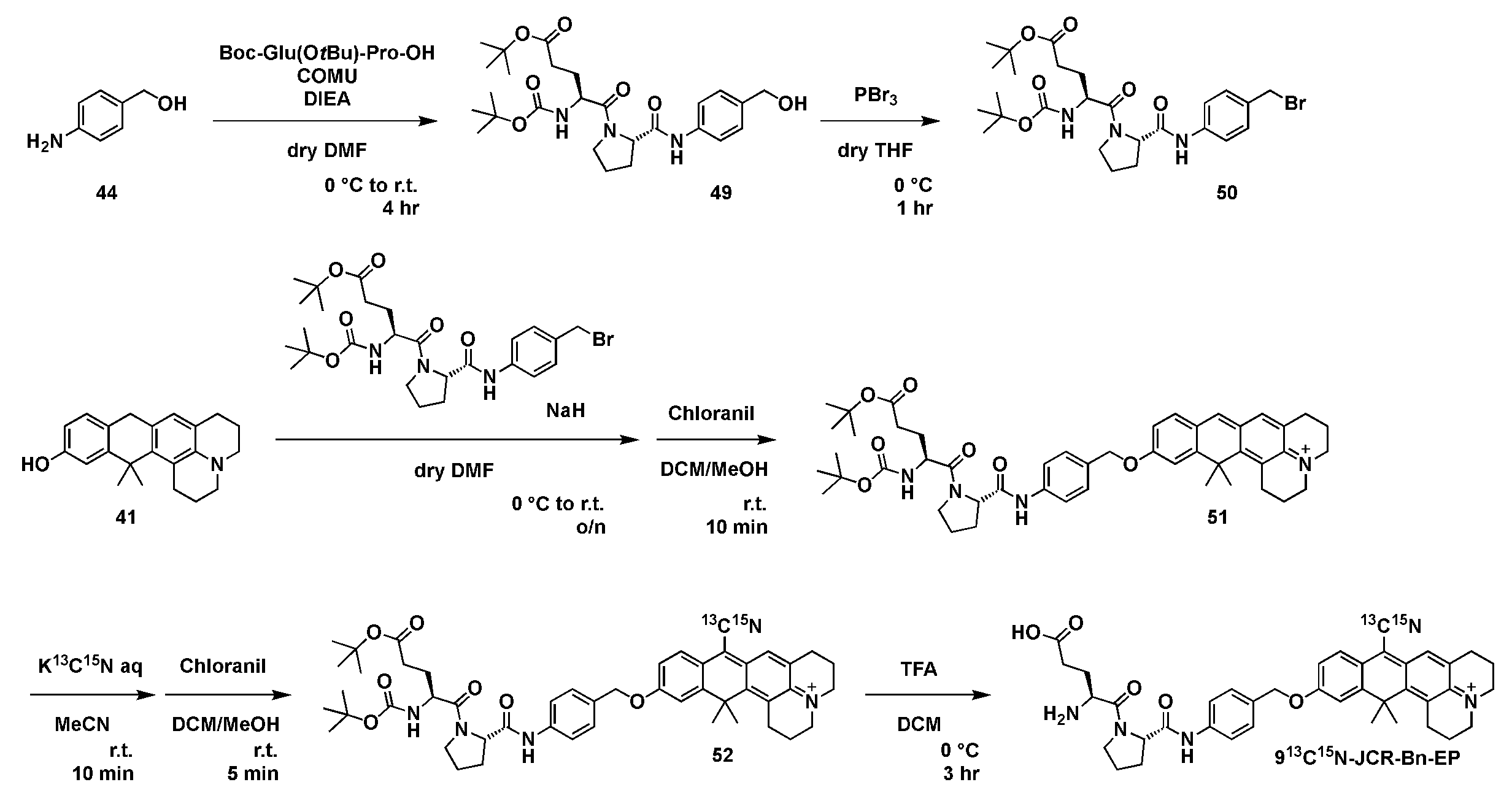
(1)化合物49の合成
化合物44(96mg、0.78mmol)、Boc-Glu(OtBu)-Pro-OH(262mg、0.65mmol)およびDIEA(225μL、1.31mmol)を2mLの乾燥DMFに溶解し、その溶液をアルゴン雰囲気下、0℃で10分間撹拌した。次に、DMF3mL中のCOMU(335mg、0.78mmol)を加え、混合物を室温で4時間撹拌した。反応を飽和NaHCO3水溶液でクエンチし、そして混合物をAcOEtで抽出した。有機層を塩水で洗浄し、Na2SO4で乾燥させ、蒸発させて乾固させた。残渣をカラムクロマトグラフィー(シリカゲル、n-ヘキサン/AcOEt=14/86~0/100)で精製し、化合物49(292mg、88%)を得た。
1H NMR (400 MHz, CDCl3): δ 9.39 (s, 1H), 7.38 (d, J = 8.1 Hz, 2H), 7.15 (d, J = 8.2 Hz, 2H), 5.34 (d, J = 8.8 Hz, 1H), 4.74 (d, J = 8.0 Hz, 1H), 4.56 (m, 3H), 3.76 (t, J = 6.3 Hz, 2H), 2.43-2.27 (m, 4H), 2.22-2.14 (m, 1H), 2.11-1.95 (m, 2H), 1.82-1.73 (m, 1H), 1.44 (s, 9H), 1.43 (s, 9H); 13C NMR (101 MHz, CDCl3): δ 172.76, 172.28, 169.34, 155.67, 137.64, 136.52, 127.70, 119.82, 80.89, 80.00, 64.90, 60.95, 51.38, 47.74, 31.03, 28.45, 28.17, 27.80, 27.61, 25.22; HRMS (ESI+): Calcd for [M+Na]+, 528.26802, Found, 528.27085 (-2.8 mDa).
化合物44(96mg、0.78mmol)、Boc-Glu(OtBu)-Pro-OH(262mg、0.65mmol)およびDIEA(225μL、1.31mmol)を2mLの乾燥DMFに溶解し、その溶液をアルゴン雰囲気下、0℃で10分間撹拌した。次に、DMF3mL中のCOMU(335mg、0.78mmol)を加え、混合物を室温で4時間撹拌した。反応を飽和NaHCO3水溶液でクエンチし、そして混合物をAcOEtで抽出した。有機層を塩水で洗浄し、Na2SO4で乾燥させ、蒸発させて乾固させた。残渣をカラムクロマトグラフィー(シリカゲル、n-ヘキサン/AcOEt=14/86~0/100)で精製し、化合物49(292mg、88%)を得た。
1H NMR (400 MHz, CDCl3): δ 9.39 (s, 1H), 7.38 (d, J = 8.1 Hz, 2H), 7.15 (d, J = 8.2 Hz, 2H), 5.34 (d, J = 8.8 Hz, 1H), 4.74 (d, J = 8.0 Hz, 1H), 4.56 (m, 3H), 3.76 (t, J = 6.3 Hz, 2H), 2.43-2.27 (m, 4H), 2.22-2.14 (m, 1H), 2.11-1.95 (m, 2H), 1.82-1.73 (m, 1H), 1.44 (s, 9H), 1.43 (s, 9H); 13C NMR (101 MHz, CDCl3): δ 172.76, 172.28, 169.34, 155.67, 137.64, 136.52, 127.70, 119.82, 80.89, 80.00, 64.90, 60.95, 51.38, 47.74, 31.03, 28.45, 28.17, 27.80, 27.61, 25.22; HRMS (ESI+): Calcd for [M+Na]+, 528.26802, Found, 528.27085 (-2.8 mDa).
(2)化合物50の合成
化合物49(146mg、0.29mmol)の3mL乾燥THF溶液に、アルゴン雰囲気下、0℃でPBr3(28μL、0.29mmol)を加えた。撹拌は同じ温度で1時間続けた。次に、反応を飽和NaHCO3水溶液でクエンチし、AcOEtで抽出した。有機層を塩水で洗浄し、Na2SO4で乾燥させ、蒸発させて乾固させた。残渣をカラムクロマトグラフィー(シリカゲル、n-ヘキサン/AcOEt=54/46~33/67)で精製し、化合物50(90mg、55%)を得た。
1H NMR (400 MHz, CDCl3): δ 9.41 (s, 1H), 7.45 (d, J = 8.5 Hz, 2H), 7.28 (d, J = 8.4 Hz, 2H), 5.26 (d, J = 8.5 Hz, 1H), 4.79 (d, J = 8.1 Hz, 1H), 4.53 (m, 1H), 4.45 (s, 2H), 3.75-3.71 (m, 2H), 2.54-2.45 (m, 1H), 2.41-2.24 (m, 2H), 2.19-2.12 (m, 1H), 2.07-1.99 (m, 2H), 1.96-1.87 (m, 1H), 1.80-1.71 (m, 1H), 1.44 (s, 9H), 1.42 (s, 9H); 13C NMR (101 MHz, CDCl3): δ 173.41, 172.18, 168.93, 155.64, 138.46, 133.33, 129.88, 120.01, 80.99, 80.09, 60.96, 51.36, 47.74, 33.66, 31.03, 28.47, 28.20, 27.97, 26.69, 25.30; HRMS (ESI+): Calcd for [M+Na]+, 590.18362, Found, 590.18454 (-0.9 mDa).
化合物49(146mg、0.29mmol)の3mL乾燥THF溶液に、アルゴン雰囲気下、0℃でPBr3(28μL、0.29mmol)を加えた。撹拌は同じ温度で1時間続けた。次に、反応を飽和NaHCO3水溶液でクエンチし、AcOEtで抽出した。有機層を塩水で洗浄し、Na2SO4で乾燥させ、蒸発させて乾固させた。残渣をカラムクロマトグラフィー(シリカゲル、n-ヘキサン/AcOEt=54/46~33/67)で精製し、化合物50(90mg、55%)を得た。
1H NMR (400 MHz, CDCl3): δ 9.41 (s, 1H), 7.45 (d, J = 8.5 Hz, 2H), 7.28 (d, J = 8.4 Hz, 2H), 5.26 (d, J = 8.5 Hz, 1H), 4.79 (d, J = 8.1 Hz, 1H), 4.53 (m, 1H), 4.45 (s, 2H), 3.75-3.71 (m, 2H), 2.54-2.45 (m, 1H), 2.41-2.24 (m, 2H), 2.19-2.12 (m, 1H), 2.07-1.99 (m, 2H), 1.96-1.87 (m, 1H), 1.80-1.71 (m, 1H), 1.44 (s, 9H), 1.42 (s, 9H); 13C NMR (101 MHz, CDCl3): δ 173.41, 172.18, 168.93, 155.64, 138.46, 133.33, 129.88, 120.01, 80.99, 80.09, 60.96, 51.36, 47.74, 33.66, 31.03, 28.47, 28.20, 27.97, 26.69, 25.30; HRMS (ESI+): Calcd for [M+Na]+, 590.18362, Found, 590.18454 (-0.9 mDa).
(3)化合物51の合成
暗所でアルゴン雰囲気下、0℃で2mLの乾燥DMFに化合物41(38mg、0.12mmol)、化合物50(90mg、0.16mmol)およびNaH(3.8mg、0.16mmol)を溶解した。混合物を室温まで温め、19時間撹拌を続けた。反応を飽和NaHCO3水溶液でクエンチし、DCMで抽出した。有機層を塩水で洗浄し、Na2SO4で乾燥させ、蒸発させて乾固させた。残渣をDCM5mLとMeOH5mLに溶解し、室温でクロラニル(29mg、0.12mmol)を加えた。混合物を10分間攪拌した後、蒸発させた。残渣を、溶離液A(1%MeCNおよび0.1%TFAを含むH2O)および溶離液B(1%H2Oを含むMeCN)を使用する分取HPLCで粗精製した(40分間でA/B=90/10から0/100)。溶出液を蒸発させ、残渣をカラムクロマトグラフィー(シリカゲルDCM/MeOH=100/0~50/50)で粗精製した。得られた溶出液を蒸発させ、残渣を溶離液A (1%MeCNおよび0.1%TFAを含むH2O)および溶離液B(1%H2Oを含むMeCN)を用いた分取HPLCによって再び精製し(40分間でA/B=90/10から0/100)、化合物51を得た(23mg、23%)。
1H NMR (400 MHz, CD3OD): δ 7.92 (s, 1H), 7.63-7.59 (m, 3H), 7.43-7.41 (m, 3H), 7.33 (d, J = 2.4 Hz, 1H), 7.09 (dd, J = 8.6, 2.4 Hz, 1H), 5.22 (s, 2H), 4.57-4.54 (m, 1H), 4.47-4.43 (m, 1H), 3.88-3.79 (m, 2H), 3.76-3.74 (m, 4H), 3.23 (t, J = 6.0 Hz, 2H), 2.84 (t, J = 6.3 Hz, 2H), 2.40 (t, J = 6.8 Hz, 2H), 2.35-2.27 (m, 1H), 2.16-2.01 (m, 8H), 1.80 (s, 6H), 1.78-1.74 (m, 1H), 1.46 (s, 9H), 1.44 (s, 9H); HRMS (ESI+): Calcd for [M]+, 805.45348, Found, 805.45267 (0.8 mDa).
暗所でアルゴン雰囲気下、0℃で2mLの乾燥DMFに化合物41(38mg、0.12mmol)、化合物50(90mg、0.16mmol)およびNaH(3.8mg、0.16mmol)を溶解した。混合物を室温まで温め、19時間撹拌を続けた。反応を飽和NaHCO3水溶液でクエンチし、DCMで抽出した。有機層を塩水で洗浄し、Na2SO4で乾燥させ、蒸発させて乾固させた。残渣をDCM5mLとMeOH5mLに溶解し、室温でクロラニル(29mg、0.12mmol)を加えた。混合物を10分間攪拌した後、蒸発させた。残渣を、溶離液A(1%MeCNおよび0.1%TFAを含むH2O)および溶離液B(1%H2Oを含むMeCN)を使用する分取HPLCで粗精製した(40分間でA/B=90/10から0/100)。溶出液を蒸発させ、残渣をカラムクロマトグラフィー(シリカゲルDCM/MeOH=100/0~50/50)で粗精製した。得られた溶出液を蒸発させ、残渣を溶離液A (1%MeCNおよび0.1%TFAを含むH2O)および溶離液B(1%H2Oを含むMeCN)を用いた分取HPLCによって再び精製し(40分間でA/B=90/10から0/100)、化合物51を得た(23mg、23%)。
1H NMR (400 MHz, CD3OD): δ 7.92 (s, 1H), 7.63-7.59 (m, 3H), 7.43-7.41 (m, 3H), 7.33 (d, J = 2.4 Hz, 1H), 7.09 (dd, J = 8.6, 2.4 Hz, 1H), 5.22 (s, 2H), 4.57-4.54 (m, 1H), 4.47-4.43 (m, 1H), 3.88-3.79 (m, 2H), 3.76-3.74 (m, 4H), 3.23 (t, J = 6.0 Hz, 2H), 2.84 (t, J = 6.3 Hz, 2H), 2.40 (t, J = 6.8 Hz, 2H), 2.35-2.27 (m, 1H), 2.16-2.01 (m, 8H), 1.80 (s, 6H), 1.78-1.74 (m, 1H), 1.46 (s, 9H), 1.44 (s, 9H); HRMS (ESI+): Calcd for [M]+, 805.45348, Found, 805.45267 (0.8 mDa).
(4)化合物52の合成
化合物51(23mg、0.028mmol)の5mLのMeCN溶液に0.3MのK13C15N水溶液を加えた。(190μL、0.056mmol)、混合物を室温で10分間撹拌した。次に、反応をHPLC用溶離液A(1%MeCN、0.1%TFAを含むH2O)でクエンチし、混合物をDCMで抽出した。有機層を塩水で洗浄し、Na2SO4で乾燥させ、蒸発させて乾固させた。粗化合物を5mL DCMと1mL MeOHに溶解し、室温でクロラニル(10mg、0.042mmol)を加えた。混合物を5分間かき混ぜた後、蒸発させた。残渣を、溶離液A(1%MeCNおよび0.1%TFAを含むH2O)および溶離液B(1%H2Oを含むMeCN)を使用する分取HPLCで粗精製した(40分間でA/B=90/10から0/100)。溶出液を蒸発させ、残渣をカラムクロマトグラフィー(シリカゲルDCM/MeOH=100/0~86/14)で粗精製した。得られた溶出液を蒸発させ、残渣を溶離液A(1%MeCNおよび0.1%TFAを含むH2O)および溶離液B(1%H2Oを含むMeCN)を用いた分取HPLCで再び精製し(40分間でA/B=90/10から0/100)、化合物52(3.6mg、15%)を得た。
1H NMR (400 MHz, CD3OD, partially overlapped with CD3OD-derived peaks): δ 7.89 (d, J = 8.8 Hz, 1H), 7.78 (s, 1H), 7.61 (d, J = 8.4 Hz, 2H), 7.43 (d, J = 8.3 Hz, 2H), 7.36 (d, J = 2.4 Hz, 1H), 7.20 (dd, J = 8.8, 2.5 Hz, 1H), 5.27 (s, 2H), 4.57-4.54 (m, 1H), 4.47-4.43 (m, 1H), 3.91-3.90 (m, 4H), 3.86-3.77 (m, 2H), 3.28-3.26 (m, 2H), 2.94 (t, J = 6.3 Hz, 2H), 2.40 (t, J = 6.9 Hz, 2H), 2.33-2.28 (m, 1H), 2.16-2.03 (m, 8H), 1.84 (s, 6H), 1.81-1.74 (m, 1H), 1.46 (s, 9H), 1.44 (s, 9H); HRMS (ESI+): Calcd for [M]+, 832.44928, Found, 832.44844 (0.8 mDa).
化合物51(23mg、0.028mmol)の5mLのMeCN溶液に0.3MのK13C15N水溶液を加えた。(190μL、0.056mmol)、混合物を室温で10分間撹拌した。次に、反応をHPLC用溶離液A(1%MeCN、0.1%TFAを含むH2O)でクエンチし、混合物をDCMで抽出した。有機層を塩水で洗浄し、Na2SO4で乾燥させ、蒸発させて乾固させた。粗化合物を5mL DCMと1mL MeOHに溶解し、室温でクロラニル(10mg、0.042mmol)を加えた。混合物を5分間かき混ぜた後、蒸発させた。残渣を、溶離液A(1%MeCNおよび0.1%TFAを含むH2O)および溶離液B(1%H2Oを含むMeCN)を使用する分取HPLCで粗精製した(40分間でA/B=90/10から0/100)。溶出液を蒸発させ、残渣をカラムクロマトグラフィー(シリカゲルDCM/MeOH=100/0~86/14)で粗精製した。得られた溶出液を蒸発させ、残渣を溶離液A(1%MeCNおよび0.1%TFAを含むH2O)および溶離液B(1%H2Oを含むMeCN)を用いた分取HPLCで再び精製し(40分間でA/B=90/10から0/100)、化合物52(3.6mg、15%)を得た。
1H NMR (400 MHz, CD3OD, partially overlapped with CD3OD-derived peaks): δ 7.89 (d, J = 8.8 Hz, 1H), 7.78 (s, 1H), 7.61 (d, J = 8.4 Hz, 2H), 7.43 (d, J = 8.3 Hz, 2H), 7.36 (d, J = 2.4 Hz, 1H), 7.20 (dd, J = 8.8, 2.5 Hz, 1H), 5.27 (s, 2H), 4.57-4.54 (m, 1H), 4.47-4.43 (m, 1H), 3.91-3.90 (m, 4H), 3.86-3.77 (m, 2H), 3.28-3.26 (m, 2H), 2.94 (t, J = 6.3 Hz, 2H), 2.40 (t, J = 6.9 Hz, 2H), 2.33-2.28 (m, 1H), 2.16-2.03 (m, 8H), 1.84 (s, 6H), 1.81-1.74 (m, 1H), 1.46 (s, 9H), 1.44 (s, 9H); HRMS (ESI+): Calcd for [M]+, 832.44928, Found, 832.44844 (0.8 mDa).
(5)913C15N-JCR-Bn-EPの合成
化合物52(3.6mg、4.3μmol)を0℃で0.5mLのTFAと0.5mLのDCMに溶解し、混合物を同じ温度で3時間かき混ぜた。次に、反応を飽和NaHCO3水溶液を用いて0℃でクエンチした。混合物を蒸発させ、残渣を溶離液A(1%MeCNおよび0.1%TFAを含むH2O)および溶離液B(1%H2Oを含むMeCN)(30分でA/B=90/10から30/70)を用いた分取HPLCによって精製し、913C15N-JCR-Bn-EP(2.2mg、77%)を得た。
1H NMR (400 MHz, CD3OD, partially overlapped with CD3OD-derived peaks): δ 7.90 (d, J = 8.9 Hz, 1H), 7.78 (s, 1H), 7.62 (d, J = 8.5 Hz, 2H), 7.45 (d, J = 8.4 Hz, 2H), 7.38 (d, J = 2.5 Hz, 1H), 7.20 (dd, J = 8.8, 2.5 Hz, 1H), 5.26 (s, 2H), 4.64-4.60 (m, 1H), 4.41-4.38 (m, 1H), 3.91-3.90 (m, 4H), 3.79-3.72 (m, 2H), 3.27-3.25 (m, 2H), 2.94 (t, J = 6.4 Hz, 2H), 2.64 (t, J = 6.4 Hz, 2H), 2.39-2.31 (m, 1H), 2.28-2.21 (m, 1H), 2.17-2.02 (m, 8H), 1.85 (s, 6H); HRMS (ESI+): Calcd for [M]+, 676.33422, Found, 676.33440 (-0.2 mDa).
化合物52(3.6mg、4.3μmol)を0℃で0.5mLのTFAと0.5mLのDCMに溶解し、混合物を同じ温度で3時間かき混ぜた。次に、反応を飽和NaHCO3水溶液を用いて0℃でクエンチした。混合物を蒸発させ、残渣を溶離液A(1%MeCNおよび0.1%TFAを含むH2O)および溶離液B(1%H2Oを含むMeCN)(30分でA/B=90/10から30/70)を用いた分取HPLCによって精製し、913C15N-JCR-Bn-EP(2.2mg、77%)を得た。
1H NMR (400 MHz, CD3OD, partially overlapped with CD3OD-derived peaks): δ 7.90 (d, J = 8.9 Hz, 1H), 7.78 (s, 1H), 7.62 (d, J = 8.5 Hz, 2H), 7.45 (d, J = 8.4 Hz, 2H), 7.38 (d, J = 2.5 Hz, 1H), 7.20 (dd, J = 8.8, 2.5 Hz, 1H), 5.26 (s, 2H), 4.64-4.60 (m, 1H), 4.41-4.38 (m, 1H), 3.91-3.90 (m, 4H), 3.79-3.72 (m, 2H), 3.27-3.25 (m, 2H), 2.94 (t, J = 6.4 Hz, 2H), 2.64 (t, J = 6.4 Hz, 2H), 2.39-2.31 (m, 1H), 2.28-2.21 (m, 1H), 2.17-2.02 (m, 8H), 1.85 (s, 6H); HRMS (ESI+): Calcd for [M]+, 676.33422, Found, 676.33440 (-0.2 mDa).
[実施例2]
上記で合成した913CN-JCR-Bn-gGlu及び913C15N-JCR-Bn-EPについて標的酵素との反応による吸光度、SRSスペクトルの変化を以下の測定方法により評価した。
上記で合成した913CN-JCR-Bn-gGlu及び913C15N-JCR-Bn-EPについて標的酵素との反応による吸光度、SRSスペクトルの変化を以下の測定方法により評価した。
(1)吸光度測定
吸収スペクトルはUV-2450 UV/Vis分光計(島津製作所)で取得した。プローブをDMSO(蛍光等級、同仁堂)に溶解してストック溶液を得た。
(2)in vitro酵素反応のSRS測定
200μMのプローブをPBS中の対応するターゲット酵素と混合した(PH7.4)。反応溶液を室温で酵素反応が起こるのに十分な時間インキュベートした後、最終濃度30%(v/v)においてDMSOを加え、生成した色素を溶解させた。溶液をイメージングチャンバーに封入し、SRS測定値を得た。
(3)LC分析
913CN-JCR-Bn-gGlu及び913C15N-JCR-Bn-EPがそれぞれ913CN-JCR及び913C15N-JCRに加水分解されることを確認するために、GGTおよびDPP-4と反応させた後、カラム(Waters、ACQUITY UPLC BEH C18 1.7mm)、オートサンプラー(Waters、SMFTN;186015017)、ポンプ(Waters、QSM;186015018)、PDA検出器(Waters、eλ検出器;186015033)、及びMS検出器(Waters、QDa;186006511)を備えたACUITY UPLC H-クラス超高速液体クロマトグラフィー質量分析(UPLC-MS)システム(Waters)を用いて、中性条件の溶離液として10mMのギ酸アンモニウム溶液(A溶液)およびアセトニトリル(B溶液)を、酸性条件の溶離液として0.1%のギ酸アンモニウム溶液(A溶液)およびアセトニトリル(B溶液)を用いて、色素溶液および酵素反応溶液を分析した。
A/B = 95/5 to 5/95 (0-3.5 min), 5/95 (3.5-4.0 min), 5/95 to 95/5 (4.0-4.1 min), 95/5 (4.1-5.0 min). For 913CN-JCR-Bn-gGlu, m/z = 578; for 913CN-JCR, m/z = 344; for 913C15N-JCR-Bn-EP, m/z = 676.5; for 913C15N-JCR, m/z = 345.
吸収スペクトルはUV-2450 UV/Vis分光計(島津製作所)で取得した。プローブをDMSO(蛍光等級、同仁堂)に溶解してストック溶液を得た。
(2)in vitro酵素反応のSRS測定
200μMのプローブをPBS中の対応するターゲット酵素と混合した(PH7.4)。反応溶液を室温で酵素反応が起こるのに十分な時間インキュベートした後、最終濃度30%(v/v)においてDMSOを加え、生成した色素を溶解させた。溶液をイメージングチャンバーに封入し、SRS測定値を得た。
(3)LC分析
913CN-JCR-Bn-gGlu及び913C15N-JCR-Bn-EPがそれぞれ913CN-JCR及び913C15N-JCRに加水分解されることを確認するために、GGTおよびDPP-4と反応させた後、カラム(Waters、ACQUITY UPLC BEH C18 1.7mm)、オートサンプラー(Waters、SMFTN;186015017)、ポンプ(Waters、QSM;186015018)、PDA検出器(Waters、eλ検出器;186015033)、及びMS検出器(Waters、QDa;186006511)を備えたACUITY UPLC H-クラス超高速液体クロマトグラフィー質量分析(UPLC-MS)システム(Waters)を用いて、中性条件の溶離液として10mMのギ酸アンモニウム溶液(A溶液)およびアセトニトリル(B溶液)を、酸性条件の溶離液として0.1%のギ酸アンモニウム溶液(A溶液)およびアセトニトリル(B溶液)を用いて、色素溶液および酵素反応溶液を分析した。
A/B = 95/5 to 5/95 (0-3.5 min), 5/95 (3.5-4.0 min), 5/95 to 95/5 (4.0-4.1 min), 95/5 (4.1-5.0 min). For 913CN-JCR-Bn-gGlu, m/z = 578; for 913CN-JCR, m/z = 344; for 913C15N-JCR-Bn-EP, m/z = 676.5; for 913C15N-JCR, m/z = 345.
測定結果等を図6及び図7に示す。
図6の(a)は、913CN-JCR-Bn-gGluの酵素(GGT)との反応の模式図を示す。
図6の(b)は、共溶媒として0.1%のDMSOを含むPBS(pH7.4)中で測定した、1ユニットのGGTとの反応前(青)と反応後(赤)の1μMの913CN-JCR-Bn-gGluの吸収スペクトルを示す。反応溶液を室温で20分間インキュベートした。
図6の(c)は、PBS(pH7.4、最終DMSO濃度30%(v/v))で測定した、1ユニットのGGTとの反応の前(青)と反応後(赤)の200μMの913CN-JCR-Bn-gGluのSRSスペクトルを示す。反応溶液を室温で1時間インキュベートした。背景として2100cm-1の画像を差し引いて画像を構成した。スケールバー:10μm。
図6の(d)は、GGT1ユニットの添加(赤、矢印は酵素添加のタイミングを表す)による913CN-JCR-Bn-gGluの吸光度の時間依存的変化、および50μMのGGT特異的阻害剤の存在下での吸光度の時間依存的変化;GGsTop (緑)を示す。観察した波長は690nmであった。
図6の(e)は、中性条件下での1ユニットのGGTと913CN-JCR-Bn-gGluとの反応溶液のLC-MS分析の結果を示す。反応溶液を室温で20分間インキュベートした。
図6の(b)は、共溶媒として0.1%のDMSOを含むPBS(pH7.4)中で測定した、1ユニットのGGTとの反応前(青)と反応後(赤)の1μMの913CN-JCR-Bn-gGluの吸収スペクトルを示す。反応溶液を室温で20分間インキュベートした。
図6の(c)は、PBS(pH7.4、最終DMSO濃度30%(v/v))で測定した、1ユニットのGGTとの反応の前(青)と反応後(赤)の200μMの913CN-JCR-Bn-gGluのSRSスペクトルを示す。反応溶液を室温で1時間インキュベートした。背景として2100cm-1の画像を差し引いて画像を構成した。スケールバー:10μm。
図6の(d)は、GGT1ユニットの添加(赤、矢印は酵素添加のタイミングを表す)による913CN-JCR-Bn-gGluの吸光度の時間依存的変化、および50μMのGGT特異的阻害剤の存在下での吸光度の時間依存的変化;GGsTop (緑)を示す。観察した波長は690nmであった。
図6の(e)は、中性条件下での1ユニットのGGTと913CN-JCR-Bn-gGluとの反応溶液のLC-MS分析の結果を示す。反応溶液を室温で20分間インキュベートした。
図7の(a)は、913CN-JCR-Bn-EPの酵素(DPP-4)との反応の模式図を示す。
図7の(b)は、共溶媒として0.1%のDMSOを含むPBS(pH7.4)中で測定した、0.033単位のDPP-4との反応前(青)と反応後(赤)の1μMの913CN-JCR-Bn-EPの吸収スペクトルを示す。反応溶液を室温で1時間インキュベートした。
図7の(c)は、PBS(pH7.4、最終DMSO濃度30%(v/v))で測定した、0.033単位のDPP-4との反応前(青)と反応後(赤)の200μMの913C15N-JCR-Bn-EPのSRSスペクトルを示す。反応溶液を室温で1時間インキュベートした。背景として2100cm-1の画像を差し引いて画像を構成した。スケールバー:10μm。
図7の(d)は、0.033単位のDPP-4を添加(赤、矢印は酵素添加のタイミングを表す)したときの913C15N-JCR-Bn-EPの吸光度の時間依存的変化、および1.8μMのDPP-4特異的阻害剤の存在下での吸光度の時間依存的変化;シタグリプチン(緑色)を示す。観察した波長は690nmであった。
図7の(e)は、中性条件下で0.033単位のDPP-4と913C15N-JCR-Bn-EPの反応溶液のLC-MS分析の結果を示す。反応溶液を室温で20分間インキュベートした。
図7の(b)は、共溶媒として0.1%のDMSOを含むPBS(pH7.4)中で測定した、0.033単位のDPP-4との反応前(青)と反応後(赤)の1μMの913CN-JCR-Bn-EPの吸収スペクトルを示す。反応溶液を室温で1時間インキュベートした。
図7の(c)は、PBS(pH7.4、最終DMSO濃度30%(v/v))で測定した、0.033単位のDPP-4との反応前(青)と反応後(赤)の200μMの913C15N-JCR-Bn-EPのSRSスペクトルを示す。反応溶液を室温で1時間インキュベートした。背景として2100cm-1の画像を差し引いて画像を構成した。スケールバー:10μm。
図7の(d)は、0.033単位のDPP-4を添加(赤、矢印は酵素添加のタイミングを表す)したときの913C15N-JCR-Bn-EPの吸光度の時間依存的変化、および1.8μMのDPP-4特異的阻害剤の存在下での吸光度の時間依存的変化;シタグリプチン(緑色)を示す。観察した波長は690nmであった。
図7の(e)は、中性条件下で0.033単位のDPP-4と913C15N-JCR-Bn-EPの反応溶液のLC-MS分析の結果を示す。反応溶液を室温で20分間インキュベートした。
図6及び図7で示されるように、913CN-JCR-Bn-gGlu及び913C15N-JCR-Bn-EPはいずれも標的酵素との反応によって信号がactivateされることが確認された。
[実施例3]
これまでに開発した9C15N-JCR-Bn-βGalと合わせて3種類のプローブを用いて複数酵素活性の同時検出を行った。実験方法は以下の通りである。
これまでに開発した9C15N-JCR-Bn-βGalと合わせて3種類のプローブを用いて複数酵素活性の同時検出を行った。実験方法は以下の通りである。
(1)細胞培養
A549細胞はRiken Cell Bank(RCB 0098)から、H226細胞はAmerican Type Culture Collectionから購入した。A549細胞をDulbeccoの改変Eagle培地(D-MEM) (和光)で培養し、H226細胞を10%ウシ胎児血清(GIBCO)、100U/mL、ペニシリン、100μg/mLのストレプトマイシンを含むRPMI 1640 (和光)で、5%CO2を含む加湿空気中、37℃で培養した。
A549細胞はRiken Cell Bank(RCB 0098)から、H226細胞はAmerican Type Culture Collectionから購入した。A549細胞をDulbeccoの改変Eagle培地(D-MEM) (和光)で培養し、H226細胞を10%ウシ胎児血清(GIBCO)、100U/mL、ペニシリン、100μg/mLのストレプトマイシンを含むRPMI 1640 (和光)で、5%CO2を含む加湿空気中、37℃で培養した。
(2)3つの異なる酵素活性の同時生細胞SRSイメージング
A549細胞とH226細胞を35mmのガラス底皿(マツナミ、ガラス底皿ハイドロ)に播種し、一晩培養した。次に、培地を、共溶媒として0.6%のDMSOを含み、20μMの9C15N-JCR-Bn-βGal、20μMの913CN-JCR-Bn-gGlu及び20μMの913C15N-JCR-Bn-EPを含む溶液:A549についてはD-MEM溶液(フェノールレッドフリー)、H226についてはRPMI1640溶液(フェノールレッドフリー)で、37℃で2.5時間交換した。培養後、プローブ溶液を除去して新鮮なHBSS(+)と交換し、上述のようにSRS顕微鏡でSRS測定を行った。
検出波数は、9C15N-JCR-Bn-βGalで2187cm-1と2190cm-1、913CN-JCR-Bn-gGluで2167cm-1と2170cm-1、913C15N-JCR-Bn-EPで2137cm-1と2140cm-1であった。背景波数は2117cm-1と2210cm-1であった。
A549細胞とH226細胞を35mmのガラス底皿(マツナミ、ガラス底皿ハイドロ)に播種し、一晩培養した。次に、培地を、共溶媒として0.6%のDMSOを含み、20μMの9C15N-JCR-Bn-βGal、20μMの913CN-JCR-Bn-gGlu及び20μMの913C15N-JCR-Bn-EPを含む溶液:A549についてはD-MEM溶液(フェノールレッドフリー)、H226についてはRPMI1640溶液(フェノールレッドフリー)で、37℃で2.5時間交換した。培養後、プローブ溶液を除去して新鮮なHBSS(+)と交換し、上述のようにSRS顕微鏡でSRS測定を行った。
検出波数は、9C15N-JCR-Bn-βGalで2187cm-1と2190cm-1、913CN-JCR-Bn-gGluで2167cm-1と2170cm-1、913C15N-JCR-Bn-EPで2137cm-1と2140cm-1であった。背景波数は2117cm-1と2210cm-1であった。
生きた培養細胞における3つの酵素活性の同時イメージングの結果を図8に示す。
図8の左図は、生きたA549細胞(上図)とH226細胞(下図)におけるβ-Gal、GGT、DPP-4活性のSRSイメージングである。スケールバー:10μm。各細胞の獲得時間は80秒であった。
図8の右図は、A549細胞(上図)とH226細胞(下図)の酵素活性のSRSスペクトルを示す。強調表示された領域は、各プローブのピークを示す。
右の領域:9C15N-JCR-Bn-βGal、中央の領域:913CN-JCR-Bn-gGlu、左の領域:913C15N-JCR-Bn-EP。
図8に示すように2種類の細胞種における酵素活性パターンの違いをイメージングすることに成功した。
図8の左図は、生きたA549細胞(上図)とH226細胞(下図)におけるβ-Gal、GGT、DPP-4活性のSRSイメージングである。スケールバー:10μm。各細胞の獲得時間は80秒であった。
図8の右図は、A549細胞(上図)とH226細胞(下図)の酵素活性のSRSスペクトルを示す。強調表示された領域は、各プローブのピークを示す。
右の領域:9C15N-JCR-Bn-βGal、中央の領域:913CN-JCR-Bn-gGlu、左の領域:913C15N-JCR-Bn-EP。
図8に示すように2種類の細胞種における酵素活性パターンの違いをイメージングすることに成功した。
[実施例4]
これまで検討を行った9CN-rhodolは、本発明者らが以前開発した9CN-pyroninと比較して水溶液中での凝集体形成能が高いことが考えられる(図9、また、図3のdは、9CN-rhodol誘導体がいずれの構造でも凝集性を有していることを示している)。この凝集体形成によって9CN-pyroninプローブと比べて細胞内滞留性が向上すると考えられる。
この点を具体的に示すため、9CN-pyroninと9CN-rhodolの代表として9CN-DEPと9CN-DERの凝集体形成能を以下の実験を行い比較した。
これまで検討を行った9CN-rhodolは、本発明者らが以前開発した9CN-pyroninと比較して水溶液中での凝集体形成能が高いことが考えられる(図9、また、図3のdは、9CN-rhodol誘導体がいずれの構造でも凝集性を有していることを示している)。この凝集体形成によって9CN-pyroninプローブと比べて細胞内滞留性が向上すると考えられる。
この点を具体的に示すため、9CN-pyroninと9CN-rhodolの代表として9CN-DEPと9CN-DERの凝集体形成能を以下の実験を行い比較した。
(1)透過画像の凝集体評価
透過画像は対物レンズ(Leica、HI PLAN 10 x/0.25 PH 1)を備えた直立型顕微鏡(Leica、DM 4000 B)で得た。溶液サンプルは、in vitroでのSRSスペクトル測定用に記載された通りに準備した。
(2)吸収スペクトルに基づく凝集体形成の評価
0.5%から1%DMSOを共溶媒とするPBS(pH7.4)中で5から100μMの色素の吸収スペクトルを測定した。濃度と吸光度の関係の直線性を評価するために、5μMの吸収極大における吸光度を基準として、その波長における各濃度の吸光度を正規化し、濃度に対してプロットした。
(3)in vitroにおけるSRSスペクトル測定
SRSスペクトルは上記のようにSRS顕微鏡で得た。全てのサンプルは、2つのガラスカバースリップ(Matsunami、C218181、C024361)とイメージングスペーサー(メルク、GBL654002、直径×厚さ9mm×0.12mm)で構成されるイメージングチャンバーに保持した。チャンバーは、溶液の位置を見つけやすくするためにPMMAビーズ(積水、Techpolymer SSX-110、平均粒径10μm)を含む色素溶液で満たし、マニキュアで密封した。SRSスペクトルの各点は、特定の波数で5フレームのピクセル値を平均することによって取得した。
透過画像は対物レンズ(Leica、HI PLAN 10 x/0.25 PH 1)を備えた直立型顕微鏡(Leica、DM 4000 B)で得た。溶液サンプルは、in vitroでのSRSスペクトル測定用に記載された通りに準備した。
(2)吸収スペクトルに基づく凝集体形成の評価
0.5%から1%DMSOを共溶媒とするPBS(pH7.4)中で5から100μMの色素の吸収スペクトルを測定した。濃度と吸光度の関係の直線性を評価するために、5μMの吸収極大における吸光度を基準として、その波長における各濃度の吸光度を正規化し、濃度に対してプロットした。
(3)in vitroにおけるSRSスペクトル測定
SRSスペクトルは上記のようにSRS顕微鏡で得た。全てのサンプルは、2つのガラスカバースリップ(Matsunami、C218181、C024361)とイメージングスペーサー(メルク、GBL654002、直径×厚さ9mm×0.12mm)で構成されるイメージングチャンバーに保持した。チャンバーは、溶液の位置を見つけやすくするためにPMMAビーズ(積水、Techpolymer SSX-110、平均粒径10μm)を含む色素溶液で満たし、マニキュアで密封した。SRSスペクトルの各点は、特定の波数で5フレームのピクセル値を平均することによって取得した。
9CN-DEPと9CN-DERの凝集体形成能力を比較した結果を図10に示す。
図10の(a)は、中性条件下での9CN-DEPと9CN-DERの化学構造を示す。図10の(b)は、3%、10%、30%、90%のDMSOを含むPBS(pH7.4)中の9CN-DEP(上図)又は9CN-DER(下図)の300μM溶液の透過画像を示す。スケールバーは100μmである。
図10の(c、d)は、共溶媒として0.05%から1%のDMSOを含むPBS(pH7.4)中で測定した5から100μMの9CN-DEP(c)及び9CN-DER(d)の正規化吸収スペクトルを示す。5μM溶液の吸収極大での吸光度に基づいて吸光度を正規化した。
図10の(e)は、5μM溶液の吸収極大における色素濃度と正規化吸光度の関係を示す。黒い点線は、染料濃度と正規化された吸光度の間の線形関係を表している。図10の(f、g)は、3%、10%、30%および90%のDMSOを含むPBS(pH7.4)中で測定した300μMの9CN-DEP(f)および9CN-DER(g)のSRSスペクトルを示す。背景として2150cm-1の画像を差し引いて画像を構成した。スケールバーは10μmである。
図10の(a)は、中性条件下での9CN-DEPと9CN-DERの化学構造を示す。図10の(b)は、3%、10%、30%、90%のDMSOを含むPBS(pH7.4)中の9CN-DEP(上図)又は9CN-DER(下図)の300μM溶液の透過画像を示す。スケールバーは100μmである。
図10の(c、d)は、共溶媒として0.05%から1%のDMSOを含むPBS(pH7.4)中で測定した5から100μMの9CN-DEP(c)及び9CN-DER(d)の正規化吸収スペクトルを示す。5μM溶液の吸収極大での吸光度に基づいて吸光度を正規化した。
図10の(e)は、5μM溶液の吸収極大における色素濃度と正規化吸光度の関係を示す。黒い点線は、染料濃度と正規化された吸光度の間の線形関係を表している。図10の(f、g)は、3%、10%、30%および90%のDMSOを含むPBS(pH7.4)中で測定した300μMの9CN-DEP(f)および9CN-DER(g)のSRSスペクトルを示す。背景として2150cm-1の画像を差し引いて画像を構成した。スケールバーは10μmである。
図10から、中性条件下で9CN-DERは顕著に凝集体を形成することが目視でも確認可能であり(b)、吸収スペクトル上では濃度に対する最大吸収波長の吸光度増大が頭打ちになっていく様子が観測された(c~e)。また,凝集体形成によって局所的な分子濃度が高まるためにラマン信号強度を効率的に上昇させられることも確認された。
このような凝集性の違いは中性条件下における色素の正味電荷(net charge)の違いによるものであると考えられ,酸性条件下でカチオン体として存在させた9CN-rhodolや,フェノール基をメチルエーテル化した9CN-rhodolでは凝集体形成は確認されなかった。
さらに,これまでに開発した3種類の9CN-JCRプローブ(9C15N-JCR-Bn-βGal、913CN-JCR-Bn-gGlu及び913C15N-JCR-Bn-EP)と色素母核である9CN-JCRの凝集体形成能力を比較した結果を図11に示す。
図11の(a~d)は、共溶媒として0.05%から1%のDMSOを含むPBS(pH7.4)中で測定した5から100μMの9C15N-JCR-Bn-βGal(a)、913CN-JCR-Bn-gGlu(b)、913C15N-JCR-Bn-EP(c)及び9CN-JCR(d)の正規化吸収スペクトルを示す。5μM溶液の吸収極大での吸光度に基づいて吸光度を正規化した。図11の(e)は、5μM溶液の吸収極大における染料濃度と正規化吸光度の関係を示す。黒い点線は、染料濃度と正規化された吸光度の間の線形関係を表している。
図11で示すように、プローブと母核とで凝集性が変化し,酵素反応によって母核を生成することで凝集性がactivateされることが示唆された。
[実施例5]
以上の結果を踏まえて、9CN-JCRプローブでこれまでの9CN-JCPプローブで行えなかった標的酵素特異的なex vivoイメージングを行うことが可能か検討を行った。実験方法は以下の通りである。
以上の結果を踏まえて、9CN-JCRプローブでこれまでの9CN-JCPプローブで行えなかった標的酵素特異的なex vivoイメージングを行うことが可能か検討を行った。実験方法は以下の通りである。
(1)生細胞およびex vivo組織のSRS画像およびスペクトル
SRS画像は、検出波数の平均画像から背景波数の平均画像を差し引いて作成した。検出波数については、それぞれのプローブのSRS強度の最大値付近に2つの波数を選択した。背景波数については、検出波数から20cm-1離れた波数を選択した。クロストークのために背景波数と検出波数が近すぎる場合、すべてのピークに共通の背景波数として最も高い背景波数と最も低い背景波数を選択した。信号対雑音比を高めるためにデータを1000回取得し、平均化を行った。スペクトルを測定するために、波数は3.3cm-1間隔で連続的に調整され、各波数での平均化に使用されるフレーム数は5であった。
SRS画像は、検出波数の平均画像から背景波数の平均画像を差し引いて作成した。検出波数については、それぞれのプローブのSRS強度の最大値付近に2つの波数を選択した。背景波数については、検出波数から20cm-1離れた波数を選択した。クロストークのために背景波数と検出波数が近すぎる場合、すべてのピークに共通の背景波数として最も高い背景波数と最も低い背景波数を選択した。信号対雑音比を高めるためにデータを1000回取得し、平均化を行った。スペクトルを測定するために、波数は3.3cm-1間隔で連続的に調整され、各波数での平均化に使用されるフレーム数は5であった。
(2)ショウジョウバエの培養条件および株
この研究で使用したショウジョウバエ系統はen-Gal 4、UAS-mCD 8-GFP(E.Kuranaga博士より)、hs-Flp122、UAS-mCD8-GFP;Ay-Gal4、UAS-GFP、UAS-lacZ(ブルーミントンショウジョウバエストックセンター1776)、およびUAS-Ggt-1であった。ショウジョウバエの組織を、Schneiderのショウジョウバエ培地(共溶媒としてDMSOを0.5%、1%含む)で、100μMの9C15N-JCR-Bn-βGal溶液、100μMの9C15N-JCR-Bn-βGal及び100μMのβGal-913C15N-JCPの混合物で、翼盤(wing disc)については2.5時間、脂肪体(fat body)については3.5時間、室温で培養した。培養後、組織を培地から取り出し、イメージングチャンバー上の新鮮なPBS(-)に浸した。SRS測定は前述のようにSRS顕微鏡で行った。検出波数は、9C15N-JCR-Bn-βGalで2187cm-1及び2190cm-1、βGal-913C15N-JCPで2133cm-1及び2137cm-1であった。背景波数は、9C15N-JCR-Bn-βGalで2167cm-1及び2210cm-1、βGal-913C15N-JCPで2113cm-1及び2157cm-1であった。
SRS画像取得後に共焦点蛍光イメージングを行った。GFPの蛍光イメージングでは、励起波長は488nmであり、500-520nmの範囲内の発光が検出された。
この研究で使用したショウジョウバエ系統はen-Gal 4、UAS-mCD 8-GFP(E.Kuranaga博士より)、hs-Flp122、UAS-mCD8-GFP;Ay-Gal4、UAS-GFP、UAS-lacZ(ブルーミントンショウジョウバエストックセンター1776)、およびUAS-Ggt-1であった。ショウジョウバエの組織を、Schneiderのショウジョウバエ培地(共溶媒としてDMSOを0.5%、1%含む)で、100μMの9C15N-JCR-Bn-βGal溶液、100μMの9C15N-JCR-Bn-βGal及び100μMのβGal-913C15N-JCPの混合物で、翼盤(wing disc)については2.5時間、脂肪体(fat body)については3.5時間、室温で培養した。培養後、組織を培地から取り出し、イメージングチャンバー上の新鮮なPBS(-)に浸した。SRS測定は前述のようにSRS顕微鏡で行った。検出波数は、9C15N-JCR-Bn-βGalで2187cm-1及び2190cm-1、βGal-913C15N-JCPで2133cm-1及び2137cm-1であった。背景波数は、9C15N-JCR-Bn-βGalで2167cm-1及び2210cm-1、βGal-913C15N-JCPで2113cm-1及び2157cm-1であった。
SRS画像取得後に共焦点蛍光イメージングを行った。GFPの蛍光イメージングでは、励起波長は488nmであり、500-520nmの範囲内の発光が検出された。
9C15N-JCR-Bn-βGalを用いた凝集体形成を利用したショウジョウバエ組織の標的酵素選択的ex vivoイメージングの結果を図12に示す。
図12の(a)は、ショウジョウバエ幼虫の解剖図である。翼盤または脂肪体のex vivoイメージングを行った。図12の(b)は、膜に局在するGFPの蛍光イメージングおよびショウジョウバエの翼盤におけるβ-ガラクトシダーゼ活性のSRSイメージングの結果を示す。ショウジョウバエの翼盤(遺伝子型:en-Gal 4、UAS-mCD 8-GFP/UAS-lacZ)を、共溶媒として0.5%DMSOを含むSchneiderのショウジョウバエ培地で100μMの9C15N-JCR-Bn-βGalと共に室温で2.5時間培養した。
図12の(c)は、(b)の視野から得られた酵素活性のSRSスペクトルを示す。図12の(d)は、膜および細胞質GFPの蛍光イメージングおよびショウジョウバエ脂肪体におけるb-ガラクトシダーゼ活性のSRSイメージングの結果を示す。ショウジョウバエの脂肪体(遺伝子型:hs-Flp122、UAS-mCD8-GFP; Ay-Gal4、UAS-GFP/UAS-lacZ)を共溶媒として0.5%のDMSOを含むSchneiderのショウジョウバエ培地で100μMの9C15N-JCR-Bn-βGalと室温で3.5時間培養した。図12の(e)は、(d) の視野から得られたβ-Gal活性のSRSスペクトルを示す。スケールバー:10μm。画像取得時間は133秒であった。
図12の(a)は、ショウジョウバエ幼虫の解剖図である。翼盤または脂肪体のex vivoイメージングを行った。図12の(b)は、膜に局在するGFPの蛍光イメージングおよびショウジョウバエの翼盤におけるβ-ガラクトシダーゼ活性のSRSイメージングの結果を示す。ショウジョウバエの翼盤(遺伝子型:en-Gal 4、UAS-mCD 8-GFP/UAS-lacZ)を、共溶媒として0.5%DMSOを含むSchneiderのショウジョウバエ培地で100μMの9C15N-JCR-Bn-βGalと共に室温で2.5時間培養した。
図12の(c)は、(b)の視野から得られた酵素活性のSRSスペクトルを示す。図12の(d)は、膜および細胞質GFPの蛍光イメージングおよびショウジョウバエ脂肪体におけるb-ガラクトシダーゼ活性のSRSイメージングの結果を示す。ショウジョウバエの脂肪体(遺伝子型:hs-Flp122、UAS-mCD8-GFP; Ay-Gal4、UAS-GFP/UAS-lacZ)を共溶媒として0.5%のDMSOを含むSchneiderのショウジョウバエ培地で100μMの9C15N-JCR-Bn-βGalと室温で3.5時間培養した。図12の(e)は、(d) の視野から得られたβ-Gal活性のSRSスペクトルを示す。スケールバー:10μm。画像取得時間は133秒であった。
図12に示すようにβ-ガラクトシダーゼを領域特異的に強制発現させたショウジョウバエ組織幼虫を9Cn-JCRプローブで染色したところ,β-ガラクトシダーゼの活性領域を示すGFPの蛍光とプローブのSRS信号の局在が一致する様子が翼盤(wing disc)及び脂肪体(fat body)の組織で確認され,酵素活性領域特異的な検出を行えていることが示唆された。
さらに,以前開発した9CN-JCPを母核としたβGal-913C15N-JCPと,9CN-JCRを母核とした9C15N-JCR-Bn-βGalで同時染色を行った結果を図13及び図14に示す。
図13は、ショウジョウバエの翼盤を用いたβGal-913C15N-JCP及び9C15N-JCR-Bn-βGalの同時イメージングの結果を示す。
図13の(a)は、ショウジョウバエの翼盤(遺伝子型:en-Gal4、UAS-mCD8-GFP/UAS-lacZ)におけるGFPの蛍光イメージングおよびβ-Gal活性のSRSイメージングの結果である。図13の(b)は、(a) の視野から得られたβ-GalプローブのSRSスペクトルを示す。強調表示された領域は、各プローブのピークを示す。緑(右の領域):9C15N-JCR-Bn-βGal、黄(左の領域):βGal-913C15N-JCP。図13の(c)において、赤色のROI(右下の囲み)は(b)のLacZ(+)領域を、青色のROI(左上の囲み)は(b)のLacZ(-)領域を示す。プローブ:各100μM。スケールバー:10μm。画像取得時間は267秒であった。
図13の(a)は、ショウジョウバエの翼盤(遺伝子型:en-Gal4、UAS-mCD8-GFP/UAS-lacZ)におけるGFPの蛍光イメージングおよびβ-Gal活性のSRSイメージングの結果である。図13の(b)は、(a) の視野から得られたβ-GalプローブのSRSスペクトルを示す。強調表示された領域は、各プローブのピークを示す。緑(右の領域):9C15N-JCR-Bn-βGal、黄(左の領域):βGal-913C15N-JCP。図13の(c)において、赤色のROI(右下の囲み)は(b)のLacZ(+)領域を、青色のROI(左上の囲み)は(b)のLacZ(-)領域を示す。プローブ:各100μM。スケールバー:10μm。画像取得時間は267秒であった。
図14は、ショウジョウバエの脂肪体を用いたβGal-913C15N-JCP及び9C15N-JCR-Bn-βGalの同時イメージングの結果を示す。
図14の(a)は、ショウジョウバエの脂肪体(遺伝子型:hs-Flp122、UAS-mCD8-GFP;Ay-Gal4、UAS-GFP/UAS-lacZ)におけるGFPの蛍光イメージングおよびβ-Gal活性のSRSイメージングの結果を示す。図14の(b)は、(a)の視野から得られたβ-GalプローブのSRSスペクトルを示す。強調表示された領域は、各プローブのピークを示す。緑(右の領域):9C15N-JCR-Bn-βGal、黄(左の領域):βGal-913C15N-JCP。図14の(c)において、赤色のROI(左下の囲み)は(b)のLacZ(+)領域、青色のROI(右上の囲み)は(b)のLacZ(-)領域を示す。プローブ:各100μM。スケールバー:10μm。画像取得時間は267秒であった。
図14の(a)は、ショウジョウバエの脂肪体(遺伝子型:hs-Flp122、UAS-mCD8-GFP;Ay-Gal4、UAS-GFP/UAS-lacZ)におけるGFPの蛍光イメージングおよびβ-Gal活性のSRSイメージングの結果を示す。図14の(b)は、(a)の視野から得られたβ-GalプローブのSRSスペクトルを示す。強調表示された領域は、各プローブのピークを示す。緑(右の領域):9C15N-JCR-Bn-βGal、黄(左の領域):βGal-913C15N-JCP。図14の(c)において、赤色のROI(左下の囲み)は(b)のLacZ(+)領域、青色のROI(右上の囲み)は(b)のLacZ(-)領域を示す。プローブ:各100μM。スケールバー:10μm。画像取得時間は267秒であった。
図13及び14に示すように9CN-JCPプローブは酵素非発現領域も非特異的に染色してしまうが,9CN-JCRプローブでは発現領域のみを特異的に染色可能であることが同一の組織中でのイメージングによって確認された。
4.共有結合形成による細胞内滞留型プローブの開発検討
次に、O-function型ラマンプローブ系において共有結合による細胞内滞留型プローブ(SPiDER型)の開発を行った。細胞内滞留型プローブの概念図を図15に示す。
β-ガラクトシダーゼを標的とする9CN-2CH2F-JCR-Bn-βGal(図16参照)を以下に記載の合成実施例5により合成した。
次に、O-function型ラマンプローブ系において共有結合による細胞内滞留型プローブ(SPiDER型)の開発を行った。細胞内滞留型プローブの概念図を図15に示す。
β-ガラクトシダーゼを標的とする9CN-2CH2F-JCR-Bn-βGal(図16参照)を以下に記載の合成実施例5により合成した。
[合成実施例5]
以下のスキーム5に則り、本発明の化合物である9CN-2CH2F-JCR-Bn-βGalを合成した。
以下のスキーム5に則り、本発明の化合物である9CN-2CH2F-JCR-Bn-βGalを合成した。
スキーム5

(1)化合物32の合成
化合物30(169mg、0.53mmol)とヘキサメチレンテトラミン(82 mg、0.58mmol)を2mLのTFAに溶解し、その溶液をアルゴン雰囲気下、90℃で3時間撹拌した。次に2mLのH2Oを加え、混合物を同じ条件で1時間攪拌した。その後、溶媒を蒸発させて乾固させた。残渣を、溶離液A(1%MeCNおよび0.1%TFAを含むH2O)及び溶離液B(1%H2Oを含むMeCN)(40分間でA/B=90/10から0/100)を使用する分取HPLCで粗精製し、粗化合物31(97mg)を得た。10mLのDCMと2mLのMeOH中の化合物31(97mg、0.28mmol)の溶液に、0℃でNaBH4(32mg、0.84mmol)を加えた。室温に加温した後、反応混合物を1時間攪拌し、その後蒸発させた。残渣をカラムクロマトグラフィー(シリカゲル、n-ヘキサン/AcOEt=71/29~50/50)で精製し、化合物32(24mg、化合物30から13%)を得た。
1H NMR (400 MHz, CDCl3): δ 7.00 (s, 1H), 6.80 (s, 1H), 6.69 (s, 1H), 4.81 (s, 2H), 3.92 (s, 2H), 3.18-3.12 (m, 4H), 2.95 (t, J = 5.6 Hz, 2H), 2.72 (t, J = 6.3 Hz, 2H), 1.97-1.90 (m, 4H), 1.75 (s, 6H); 13C NMR (101 MHz, CDCl3): δ 154.09, 149.04, 143.04, 138.12, 126.94, 126.62, 123.59, 122.85, 122.71, 121.57, 120.77, 114.64, 64.33, 51.02, 50.01, 38.75, 33.18, 31.95, 27.99, 27.64, 22.78, 22.00; HRMS (ESI+): Calcd for [M+H]+, 350.21146, Found, 350.21162 (-0.2 mDa).
化合物30(169mg、0.53mmol)とヘキサメチレンテトラミン(82 mg、0.58mmol)を2mLのTFAに溶解し、その溶液をアルゴン雰囲気下、90℃で3時間撹拌した。次に2mLのH2Oを加え、混合物を同じ条件で1時間攪拌した。その後、溶媒を蒸発させて乾固させた。残渣を、溶離液A(1%MeCNおよび0.1%TFAを含むH2O)及び溶離液B(1%H2Oを含むMeCN)(40分間でA/B=90/10から0/100)を使用する分取HPLCで粗精製し、粗化合物31(97mg)を得た。10mLのDCMと2mLのMeOH中の化合物31(97mg、0.28mmol)の溶液に、0℃でNaBH4(32mg、0.84mmol)を加えた。室温に加温した後、反応混合物を1時間攪拌し、その後蒸発させた。残渣をカラムクロマトグラフィー(シリカゲル、n-ヘキサン/AcOEt=71/29~50/50)で精製し、化合物32(24mg、化合物30から13%)を得た。
1H NMR (400 MHz, CDCl3): δ 7.00 (s, 1H), 6.80 (s, 1H), 6.69 (s, 1H), 4.81 (s, 2H), 3.92 (s, 2H), 3.18-3.12 (m, 4H), 2.95 (t, J = 5.6 Hz, 2H), 2.72 (t, J = 6.3 Hz, 2H), 1.97-1.90 (m, 4H), 1.75 (s, 6H); 13C NMR (101 MHz, CDCl3): δ 154.09, 149.04, 143.04, 138.12, 126.94, 126.62, 123.59, 122.85, 122.71, 121.57, 120.77, 114.64, 64.33, 51.02, 50.01, 38.75, 33.18, 31.95, 27.99, 27.64, 22.78, 22.00; HRMS (ESI+): Calcd for [M+H]+, 350.21146, Found, 350.21162 (-0.2 mDa).
(2)化合物34の合成
暗所でアルゴン雰囲気下、0℃で2mLの乾燥DMFに化合物32(24mg、0.068mmol)、化合物29(64mg、0.12mmol)およびNaH (1.8mg、0.075mmol)を溶解した。混合物を室温まで加温し、28時間撹拌を続けた。反応を飽和NH4Cl水溶液でクエンチし、得られた混合物をDCMで抽出した。有機層を塩水で洗浄し、Na2SO4で乾燥させ、蒸発させて乾固させた。残渣をDCM5mLとMeOH1mLに溶解し、室温でクロラニル(17 mg、0.068mmol)を加えた。混合物を10分間攪拌した後、蒸発させた。残渣を、40溶離液A(1%MeCN及び0.1%TFAを含むH2O)及び溶離液B(1%H2Oを含むMeCN)を用いる分取HPLCで精製し(40分間でA/B=90/10から0/100)、化合物34(44mg、化合物32から82%)を得た。
1H NMR (400 MHz, CD3OD): δ 7.83 (s, 1H), 7.58 (s, 1H), 7.36 (d, J = 8.5 Hz, 2H), 7.31 (s, 1H), 7.22 (s, 1H), 6.96 (d, J = 8.5 Hz, 2H), 5.36 (d, J = 3.3 Hz, 1H), 5.28-5.21 (m, 4H), 5.15 (dd, J = 9.7, 3.5 Hz, 1H), 4.58 (s, 2H), 4.20 (t, J = 6.2 Hz, 1H), 4.08-4.05 (m, 2H), 3.65 (q, J = 5.6 Hz, 6H), 3.13 (t, J = 6.0 Hz, 2H), 2.74 (t, J = 6.7 Hz, 2H), 2.07 (s, 3H), 1.98-1.89 (m, 4H), 1.95 (s, 3H), 1.92 (s, 3H), 1.87 (s, 3H), 1.68 (s, 6H); HRMS (ESI+): Calcd for [M]+, 784.33275, Found, 784.33465 (-1.9 mDa).
暗所でアルゴン雰囲気下、0℃で2mLの乾燥DMFに化合物32(24mg、0.068mmol)、化合物29(64mg、0.12mmol)およびNaH (1.8mg、0.075mmol)を溶解した。混合物を室温まで加温し、28時間撹拌を続けた。反応を飽和NH4Cl水溶液でクエンチし、得られた混合物をDCMで抽出した。有機層を塩水で洗浄し、Na2SO4で乾燥させ、蒸発させて乾固させた。残渣をDCM5mLとMeOH1mLに溶解し、室温でクロラニル(17 mg、0.068mmol)を加えた。混合物を10分間攪拌した後、蒸発させた。残渣を、40溶離液A(1%MeCN及び0.1%TFAを含むH2O)及び溶離液B(1%H2Oを含むMeCN)を用いる分取HPLCで精製し(40分間でA/B=90/10から0/100)、化合物34(44mg、化合物32から82%)を得た。
1H NMR (400 MHz, CD3OD): δ 7.83 (s, 1H), 7.58 (s, 1H), 7.36 (d, J = 8.5 Hz, 2H), 7.31 (s, 1H), 7.22 (s, 1H), 6.96 (d, J = 8.5 Hz, 2H), 5.36 (d, J = 3.3 Hz, 1H), 5.28-5.21 (m, 4H), 5.15 (dd, J = 9.7, 3.5 Hz, 1H), 4.58 (s, 2H), 4.20 (t, J = 6.2 Hz, 1H), 4.08-4.05 (m, 2H), 3.65 (q, J = 5.6 Hz, 6H), 3.13 (t, J = 6.0 Hz, 2H), 2.74 (t, J = 6.7 Hz, 2H), 2.07 (s, 3H), 1.98-1.89 (m, 4H), 1.95 (s, 3H), 1.92 (s, 3H), 1.87 (s, 3H), 1.68 (s, 6H); HRMS (ESI+): Calcd for [M]+, 784.33275, Found, 784.33465 (-1.9 mDa).
(3)化合物35の合成
化合物34(44mg、0.055mmol)を5mLの乾燥DCMに溶解し、その溶液をアルゴン雰囲気下-20℃で撹拌した。次に三フッ化ジエチルアミノ硫黄(29μL、0.22mmol)を加え、混合物を同じ条件で3時間撹拌した。その後、反応をMeOHでクエンチし、溶媒を蒸発させて乾固させた。残渣を、溶離液A(1%MeCN及び0.1%TFAを含むH2O)および溶離液B(1%H2Oを含むMeCN)(40分でA/B=90/10から0/100)を使用する分取HPLCで精製し、化合物35(5.1mg、12%)を得た。
1H NMR (400 MHz, CD3OD): δ 7.84 (s, 1H), 7.58 (s, 1H), 7.36 (d, J = 8.6 Hz, 2H), 7.34 (s, 1H), 7.29 (s, 1H), 6.97 (d, J = 8.6 Hz, 2H), 5.44 (s, 1H), 5.36 (dd, J = 3.4, 1.2 Hz, 1H), 5.32 (s, 1H), 5.28-5.21 (m, 4H), 5.15 (dd, J = 9.7, 3.4 Hz, 1H), 4.20 (t, J = 6.5 Hz, 1H), 4.07 (dd, J = 6.6, 2.8 Hz, 2H), 3.67 (q, J = 5.6 Hz, 4H), 3.14 (t, J = 7.1 Hz, 3H), 2.76 (t, J = 6.3 Hz, 2H), 2.07 (s, 3H), 2.01-1.96 (m, 4H), 1.95 (s, 3H), 1.91 (s, 2H), 1.87 (s, 3H), 1.71 (s, 6H); 19F NMR (376 MHz, CD3OD): δ -77.5; HRMS (ESI+): Calcd for [M]+, 786.32842, Found, 786.32980 (-1.4. mDa).
化合物34(44mg、0.055mmol)を5mLの乾燥DCMに溶解し、その溶液をアルゴン雰囲気下-20℃で撹拌した。次に三フッ化ジエチルアミノ硫黄(29μL、0.22mmol)を加え、混合物を同じ条件で3時間撹拌した。その後、反応をMeOHでクエンチし、溶媒を蒸発させて乾固させた。残渣を、溶離液A(1%MeCN及び0.1%TFAを含むH2O)および溶離液B(1%H2Oを含むMeCN)(40分でA/B=90/10から0/100)を使用する分取HPLCで精製し、化合物35(5.1mg、12%)を得た。
1H NMR (400 MHz, CD3OD): δ 7.84 (s, 1H), 7.58 (s, 1H), 7.36 (d, J = 8.6 Hz, 2H), 7.34 (s, 1H), 7.29 (s, 1H), 6.97 (d, J = 8.6 Hz, 2H), 5.44 (s, 1H), 5.36 (dd, J = 3.4, 1.2 Hz, 1H), 5.32 (s, 1H), 5.28-5.21 (m, 4H), 5.15 (dd, J = 9.7, 3.4 Hz, 1H), 4.20 (t, J = 6.5 Hz, 1H), 4.07 (dd, J = 6.6, 2.8 Hz, 2H), 3.67 (q, J = 5.6 Hz, 4H), 3.14 (t, J = 7.1 Hz, 3H), 2.76 (t, J = 6.3 Hz, 2H), 2.07 (s, 3H), 2.01-1.96 (m, 4H), 1.95 (s, 3H), 1.91 (s, 2H), 1.87 (s, 3H), 1.71 (s, 6H); 19F NMR (376 MHz, CD3OD): δ -77.5; HRMS (ESI+): Calcd for [M]+, 786.32842, Found, 786.32980 (-1.4. mDa).
(4)化合物36の合成
5mLのMeCN中の化合物35(8.5mg、0.011mmol)の溶液に0.3MKCN水溶液(53μL、0.016mmol)を加え、混合物を室温で10分間撹拌した。次にこの反応をHPLC用溶離液A(1%MeCN及び0.1%TFAを含むH2O)でクエンチし、生成物は自然酸化によって着色した。混合物をDCMで抽出した。有機層を塩水で洗浄し、Na2SO4で乾燥させ、蒸発させて乾固させた。残渣を、溶離液A(1%MeCN及び0.1%TFAを含むH2O)および溶離液B(1% H2Oを含むMeCN)(40分でA/B=90/10から0/100)を使用する分取HPLCで精製し、化合物36(2.5mg、28%)を得た。
1H NMR (400 MHz, CD3OD, partially overlapped with CD3OD-derived peaks): δ 7.91 (s, 1H), 7.81 (s, 1H), 7.48 (d, J = 8.7 Hz, 2H), 7.42 (s, 1H), 7.09 (d, J = 8.6 Hz, 2H), 5.60 (s, 1H), 5.48 (s, 1H), 5.47 (d, J = 4.2 Hz, 1H), 5.38-5.33 (m, 4H), 5.27 (dd, J = 9.6, 3.4 Hz, 1H), 4.32 (t, J = 6.6 Hz, 1H), 4.19 (dd, J = 6.6, 3.1 Hz, 2H), 3.93 (q, J = 5.7 Hz, 5H), 2.96 (t, J = 6.4 Hz, 2H), 2.19 (s, 3H), 2.17-2.10 (m, 4H), 2.07 (s, 3H), 2.03 (s, 3H), 1.99 (s, 3H), 1.86 (s, 6H); 19F NMR (376 MHz, CD3OD): δ -77.2; HRMS (ESI+): Calcd for [M]+, 811.32367, Found, 811.32617 (-2.5 mDa).
5mLのMeCN中の化合物35(8.5mg、0.011mmol)の溶液に0.3MKCN水溶液(53μL、0.016mmol)を加え、混合物を室温で10分間撹拌した。次にこの反応をHPLC用溶離液A(1%MeCN及び0.1%TFAを含むH2O)でクエンチし、生成物は自然酸化によって着色した。混合物をDCMで抽出した。有機層を塩水で洗浄し、Na2SO4で乾燥させ、蒸発させて乾固させた。残渣を、溶離液A(1%MeCN及び0.1%TFAを含むH2O)および溶離液B(1% H2Oを含むMeCN)(40分でA/B=90/10から0/100)を使用する分取HPLCで精製し、化合物36(2.5mg、28%)を得た。
1H NMR (400 MHz, CD3OD, partially overlapped with CD3OD-derived peaks): δ 7.91 (s, 1H), 7.81 (s, 1H), 7.48 (d, J = 8.7 Hz, 2H), 7.42 (s, 1H), 7.09 (d, J = 8.6 Hz, 2H), 5.60 (s, 1H), 5.48 (s, 1H), 5.47 (d, J = 4.2 Hz, 1H), 5.38-5.33 (m, 4H), 5.27 (dd, J = 9.6, 3.4 Hz, 1H), 4.32 (t, J = 6.6 Hz, 1H), 4.19 (dd, J = 6.6, 3.1 Hz, 2H), 3.93 (q, J = 5.7 Hz, 5H), 2.96 (t, J = 6.4 Hz, 2H), 2.19 (s, 3H), 2.17-2.10 (m, 4H), 2.07 (s, 3H), 2.03 (s, 3H), 1.99 (s, 3H), 1.86 (s, 6H); 19F NMR (376 MHz, CD3OD): δ -77.2; HRMS (ESI+): Calcd for [M]+, 811.32367, Found, 811.32617 (-2.5 mDa).
(5)9CN-2CH2F-JCR-Bn-βGalの合成
1mLの乾燥MeOH中の化合物36(2.5mg、3.1μmol)の溶液に、アルゴン雰囲気下-5℃で、500μLの乾燥MeOHで希釈した28%NaOMe/MeOH(4μL、20μmol)を加えた。混合物を同じ温度で15分間撹拌した。次に、反応をHPLC溶離液A(1%MeCN及び0.1%TFAを含むH2O)で溶液が着色するまでクエンチした。蒸発後、残留物を溶離液A(1%MeCN及び0.1%TFAを含むH2O)及び溶離液B(1%H2Oを含むMeCN)(30分でA/B=90/10から30/70)を用いる分取HPLCで精製し、9CN-2CH2F-JCR-Bn-βGal(0.8mg、40%)を得た。
1H NMR (400 MHz, CD3OD, partially overlapped with CD3OD-derived peaks): δ 7.91 (s, 1H), 7.81 (s, 1H), 7.45 (d, J = 9.2 Hz, 2H), 7.44 (s, 1H), 7.17 (d, J = 8.6 Hz, 2H), 5.60 (s, 1H), 5.48 (s, 1H), 5.37 (s, 2H), 3.94-3.91 (m, 5H), 3.84-3.76 (m, 3H), 3.72-3.66 (m, 2H), 3.59 (dd, J = 9.6, 3.4 Hz, 1H), 2.97 (t, J = 6.5 Hz, 2H), 2.18-2.11 (m, 4H), 1.86 (s, 6H); 19F NMR (376 MHz, CD3OD): δ -77.2; HRMS (ESI+): Calcd for [M]+, 643.28141, Found, 643.28127 (0.1 mDa).
1mLの乾燥MeOH中の化合物36(2.5mg、3.1μmol)の溶液に、アルゴン雰囲気下-5℃で、500μLの乾燥MeOHで希釈した28%NaOMe/MeOH(4μL、20μmol)を加えた。混合物を同じ温度で15分間撹拌した。次に、反応をHPLC溶離液A(1%MeCN及び0.1%TFAを含むH2O)で溶液が着色するまでクエンチした。蒸発後、残留物を溶離液A(1%MeCN及び0.1%TFAを含むH2O)及び溶離液B(1%H2Oを含むMeCN)(30分でA/B=90/10から30/70)を用いる分取HPLCで精製し、9CN-2CH2F-JCR-Bn-βGal(0.8mg、40%)を得た。
1H NMR (400 MHz, CD3OD, partially overlapped with CD3OD-derived peaks): δ 7.91 (s, 1H), 7.81 (s, 1H), 7.45 (d, J = 9.2 Hz, 2H), 7.44 (s, 1H), 7.17 (d, J = 8.6 Hz, 2H), 5.60 (s, 1H), 5.48 (s, 1H), 5.37 (s, 2H), 3.94-3.91 (m, 5H), 3.84-3.76 (m, 3H), 3.72-3.66 (m, 2H), 3.59 (dd, J = 9.6, 3.4 Hz, 1H), 2.97 (t, J = 6.5 Hz, 2H), 2.18-2.11 (m, 4H), 1.86 (s, 6H); 19F NMR (376 MHz, CD3OD): δ -77.2; HRMS (ESI+): Calcd for [M]+, 643.28141, Found, 643.28127 (0.1 mDa).
[実施例6]
上記で合成した9CN-2CH2F-JCR-Bn-βGalのin vitro酵素反応を評価した。結果を図16に示す。図16に示すように,β-ガラクトシダーゼと速やかに反応して長波長の生成物を生じる様子が観測された。
上記で合成した9CN-2CH2F-JCR-Bn-βGalのin vitro酵素反応を評価した。結果を図16に示す。図16に示すように,β-ガラクトシダーゼと速やかに反応して長波長の生成物を生じる様子が観測された。
[実施例7]
次に、9CN-2CH2F-JCR-Bn-βGalを用いた細胞イメ―ジングを行った。この時,control化合物として9C15N-JCR-Bn-βGalのイメージングを並行して行って比較した。実験方法を以下に示す。
次に、9CN-2CH2F-JCR-Bn-βGalを用いた細胞イメ―ジングを行った。この時,control化合物として9C15N-JCR-Bn-βGalのイメージングを並行して行って比較した。実験方法を以下に示す。
(1)細胞培養
HEK293細胞とHEK-LacZ細胞はJCRM(JCRB1414/Murakami,T.)から購入した。HEK293細胞とHEK-LacZ細胞を、10%のウシ胎児血清(GIBCO)、100U/mL、ペニシリン、100μg/mLのストレプトマイシンを含むDulbecco社の改変Eagle培地(D-MEM) (Wako)で、5%のCO2を含む加湿空気中、37℃で培養した。イメージングのためにHEK293及びHEK-LacZ細胞を継代するために、皿をポリ-L-リジン溶液(Sigma、P4832)で1時間コーティングし、PBS(-)で2回すすいで細胞接着を強化した。
HEK293細胞とHEK-LacZ細胞はJCRM(JCRB1414/Murakami,T.)から購入した。HEK293細胞とHEK-LacZ細胞を、10%のウシ胎児血清(GIBCO)、100U/mL、ペニシリン、100μg/mLのストレプトマイシンを含むDulbecco社の改変Eagle培地(D-MEM) (Wako)で、5%のCO2を含む加湿空気中、37℃で培養した。イメージングのためにHEK293及びHEK-LacZ細胞を継代するために、皿をポリ-L-リジン溶液(Sigma、P4832)で1時間コーティングし、PBS(-)で2回すすいで細胞接着を強化した。
(2)Live-cell共焦点蛍光イメージング
HEK293細胞とHEK-LacZ細胞を8チャンバープレート(Ibidi、μ-スライド)に播種し、一晩培養した。次に、培地を、37℃で0.5時間、10μMの9C15N-JCR-Bn-βGalまたは9CN-2CH2F-JCR-Bn-βGalのD-MEM(フェノールレッドフリー)溶液(共溶媒として0.1%のDMSOを含む)で置き換えた。白色光レーザーと対物レンズ(Leica、HCX PLAPO CS 40x/1.25オイル)を備えた共焦点蛍光顕微鏡(Leica、TCS SP8 STED)で蛍光画像を撮影した。励起/発光:640 nm/690-790nm。
HEK293細胞とHEK-LacZ細胞を8チャンバープレート(Ibidi、μ-スライド)に播種し、一晩培養した。次に、培地を、37℃で0.5時間、10μMの9C15N-JCR-Bn-βGalまたは9CN-2CH2F-JCR-Bn-βGalのD-MEM(フェノールレッドフリー)溶液(共溶媒として0.1%のDMSOを含む)で置き換えた。白色光レーザーと対物レンズ(Leica、HCX PLAPO CS 40x/1.25オイル)を備えた共焦点蛍光顕微鏡(Leica、TCS SP8 STED)で蛍光画像を撮影した。励起/発光:640 nm/690-790nm。
結果を図18に示す。図18は、生きたHEK-LacZ又はHEK293細胞におけるβ-ガラクトシダーゼ活性の共焦点蛍光画像である。細胞を10μMの9CN-2CH2F-JCR-Bn-βGal(上図)または9C15N-JCR-Bn-βGal(下図)と共にHBSS(+)中で30分間インキュベートした。
図18で示す通り、いずれのプローブもLacZ発現細胞特異的に発蛍光することが確認された。また,両プローブの局在はほぼ同じであるように見えた。
図18で示す通り、いずれのプローブもLacZ発現細胞特異的に発蛍光することが確認された。また,両プローブの局在はほぼ同じであるように見えた。
[実施例8]
次に、両プローブ(9CN-2CH2F-JCR-Bn-βGal及び9C15N-JCR-Bn-βGal)のwash耐性,固定耐性を比較した。Wash操作は、freshなHBSS(+)に置換して10分間incubateする操作を1回として計2回行い、各wash後にイメージングを行った。さらに、2回目のwash後にPFA固定(15分間)を行い最後のイメージングを行った。
結果を図19及び20に示す。
次に、両プローブ(9CN-2CH2F-JCR-Bn-βGal及び9C15N-JCR-Bn-βGal)のwash耐性,固定耐性を比較した。Wash操作は、freshなHBSS(+)に置換して10分間incubateする操作を1回として計2回行い、各wash後にイメージングを行った。さらに、2回目のwash後にPFA固定(15分間)を行い最後のイメージングを行った。
結果を図19及び20に示す。
図19及び20は、生または固定HEK-LacZ細胞におけるβ-ガラクトシダーゼ活性の共焦点蛍光画像である。細胞を10μMの9CN-2CH2F-JCR-Bn-βGal(図19)又は9C15N-JCR-Bn-βGal(図20)と共にHBSS(+)中で30分間インキュベートし、次にHBSS(+)で10分間2回洗浄した。洗浄後、細胞をPFAで15分間固定した。
9CN-2CH2F-JCR-Bn-βGalの1回目のwashで蛍光強度が大きく低下してしまったのは,キノンメチド中間体の多くが水分子と反応して9CN-2CH2OH-JCRとなることで膜透過してしまったからであると考えられる。しかし,wash後およびPFA固定後にも蛍光が観測されたことから,一部のプローブは目的通り細胞内タンパク質やGSH等と反応して細胞内にトラップされたと考えられる。
[実施例9]
9CN-2CH2F-JCR-Bn-βGalが組織でworkするかどうかを、ショウジョウバエのwing discに対して9C15N-JCR-Bn-βGalとの2色イメージングを行うことで比較した。実験方法は以下の通りである。
9CN-2CH2F-JCR-Bn-βGalが組織でworkするかどうかを、ショウジョウバエのwing discに対して9C15N-JCR-Bn-βGalとの2色イメージングを行うことで比較した。実験方法は以下の通りである。
ショウジョウバエの培養条件および株
この研究で使用したショウジョウバエ系統はen-Gal4、UAS-mCD 8-GFP(E.Kuranaga博士より)であり、ショウジョウバエ組織をSchneiderのショウジョウバエ培地(共溶媒として2%のDMSOを含む) で、100μMの9C15N-JCR-Bn-βGalと100μMの9CN-2CH2F-JCR-Bn-βGalの混合物と共に、3時間室温で培養した。培養後、組織を培地から取り出し、イメージングチャンバー上の新鮮なPBS(-)に浸した。SRS測定は前述のようにSRS顕微鏡で行った。検出波数は、9C15N-JCR-Bn-βGalで2190cm-1と2193cm-1、9CN-2CH2F-JCR-Bn-βGalで2220cm-1と2223cm-1であった。背景波数は2170cm-1と2243cm-1であった。
SRS画像取得後に共焦点蛍光イメージングを行った。GFPの蛍光イメージングでは、励起波長は488nmであり、500-520nmの範囲内の発光が検出された。
この研究で使用したショウジョウバエ系統はen-Gal4、UAS-mCD 8-GFP(E.Kuranaga博士より)であり、ショウジョウバエ組織をSchneiderのショウジョウバエ培地(共溶媒として2%のDMSOを含む) で、100μMの9C15N-JCR-Bn-βGalと100μMの9CN-2CH2F-JCR-Bn-βGalの混合物と共に、3時間室温で培養した。培養後、組織を培地から取り出し、イメージングチャンバー上の新鮮なPBS(-)に浸した。SRS測定は前述のようにSRS顕微鏡で行った。検出波数は、9C15N-JCR-Bn-βGalで2190cm-1と2193cm-1、9CN-2CH2F-JCR-Bn-βGalで2220cm-1と2223cm-1であった。背景波数は2170cm-1と2243cm-1であった。
SRS画像取得後に共焦点蛍光イメージングを行った。GFPの蛍光イメージングでは、励起波長は488nmであり、500-520nmの範囲内の発光が検出された。
結果を図21に示す。図21は、ショウジョウバエの翼盤(wing disc)におけるβ-ガラクトシダーゼ活性の共焦点蛍光及びSRS画像を示す。スケールバー:10μm。平均時間は1000時間であった。
図21で示す通り、SPiDER型でも9C15N-JCR-Bn-βGalのような凝集体がLacZ(+)領域特異的に確認された。LacZ(-)領域も広く染めて、酵素発現領域からは漏れ出したものの、信号強度はSPiDER型の方が顕著に強、組織全体への滞留性は向上していることが期待される結果であると考えられた。
Claims (14)
- 以下の一般式(I)で表される化合物又はその塩。
(式中、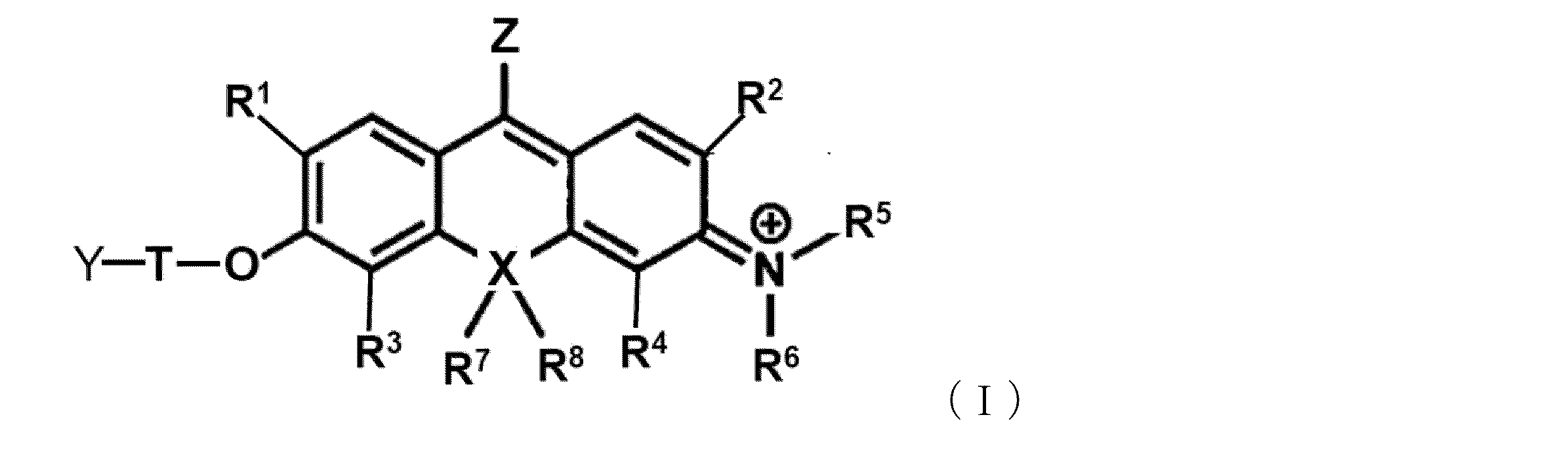
R1及びR2は、それぞれ独立に、水素原子、炭素数1~6個のアルキル基又はハロゲン原子を表し;
R3及びR4は、それぞれ独立に、水素原子、炭素数1~6個のアルキル基又はハロゲン原子を表し;
ここで、R1又はR3は、-CH2-F又は-CH-F2でもよく;
R5及びR6は、それぞれ独立に、水素原子又は炭素数1~6個のアルキル基を表し、
ただし、
(1)R5又はR6は、R2又はR4と一緒になって、R2又はR4が結合している窒素原子を含む5~7員のヘテロシクリル又はヘテロアリールを形成しているか、あるいは
(2)R5及びR6は、夫々、R2及びR4と一緒になって、R2及びR4が結合している窒素原子を含む5~7員のヘテロシクリル又はヘテロアリールを形成しており、
該ヘテロシクリル又はヘテロアリールは環構成員として酸素原子、窒素原子及び硫黄原子からなる群から選択される1~3個の更なるヘテロ原子を含有していてもよく、更に該ヘテロシクリル又はヘテロアリールは、炭素数1~6個のアルキル、炭素数2~6個のアルケニル、又は炭素数2~6個のアルキニル、炭素数6~10個のアラルキル基、炭素数6~10個のアルキル置換アルケニル基で置換されていてもよく;
R7及びR8は、存在する場合は、それぞれ独立に、炭素数1~6個のアルキル基を表し、
ここで、Xが酸素原子の場合は、R7及びR8は存在せず、
Xは、酸素原子又は炭素原子を表し;
Tは、リンカーであり:
Yは、-O-L、-NH-L’又は活性酸素種に対する捕捉基であり、
ここで、Lは、糖類又は糖類の部分構造であり、
L’は、アミノ酸の部分構造であり;
Zは、-C≡N、-C≡15N、-13C≡N、又は-13C≡15Nから選択される。) - 以下の一般式(Ia)で表される、請求項1に記載の化合物又はその塩。
(R1、R3、R7、R8、T、Yは、一般式(I)で定義した通りであり;
R9は、存在する場合は、炭素数1~3のアルキル基であり:
R10は、存在する場合は、炭素数1~3のアルキル基である。) - Tのリンカーは、アルキレン基(但し、アルキレン基の1以上の-CH2-は、-O-、-S-、-NH-、又は-CO-で置換されていてもよい。)、アリーレン(ヘテロアリーレンを含む)、シクロアルキレン、アルコキシル基、ポリエチレングリコール鎖、及び、これらの基から選択される2種以上の基が任意に結合して構成される基からなる群から選択される、請求項1又は2に記載の化合物又はその塩。
- 前記リンカーは、置換基を有していてもよいベンジル基、又はメチレン基である、請求項3に記載の化合物又はその塩。
- Lの糖類の部分構造は、それが結合しているOと一緒になって、糖類、糖類の一部を構成している、請求項1~4のいずれか1項に記載の化合物又はその塩。
- L’のアミノ酸の部分構造は、アミノ酸、アミノ酸残基、ペプチド、アミノ酸の一部を構成している、請求項1~4のいずれか1項に記載の化合物又はその塩。
- Yが以下のいずれかから選択される、請求項1~4のいずれか1項に記載の化合物又はその塩。

- Yが以下のいずれかから選択される、請求項1~4のいずれか1項に記載の化合物又はその塩。
(*は、Tと結合する箇所を表す。)
(R1aは、天然アミノ酸(グリシン、アラニン、ロイシン、イソロイシン、バリン、リジン、システイン、トレオニン、アルギニン、アスパラギン、アスパラギン酸、グルタミン、グルタミン酸、セリン、ヒスチジン、フェニルアラニン、メチオニン、トリプトファン、チロシン、プロリン)の側鎖を構成する基、非天然アミノ酸(シトルリン、ノルバリンなど)の側鎖を構成する基、置換又は無置換のアルキル基からなる群から選択され;
*は、Tと結合する箇所を表す。)
(R2aは、天然アミノ酸(グリシン、アラニン、ロイシン、イソロイシン、バリン、リジン、システイン、トレオニン、アルギニン、アスパラギン、アスパラギン酸、グルタミン、グルタミン酸、セリン、ヒスチジン、フェニルアラニン、メチオニン、トリプトファン、チロシン、プロリン)の側鎖を構成する基、非天然アミノ酸(シトルリン、ノルバリンなど)の側鎖を構成する基、置換又は無置換のアルキル基からなる群から選択され;
は、アミノ基、Nアセチル化等されたアミノ基、1つまたは複数のアミノ酸がペプチド結合により連なった構造を表し;
*は、Tと結合する箇所を表す。) - 活性酸素種に対する捕捉基が、ヒドロキシフェノキシ、アミノフェノキシ、アジド、又はピナコールボランである、請求項1~4のいずれか1項に記載の化合物又はその塩。
- 以下のいずれかの構造を有する化合物又はその塩。
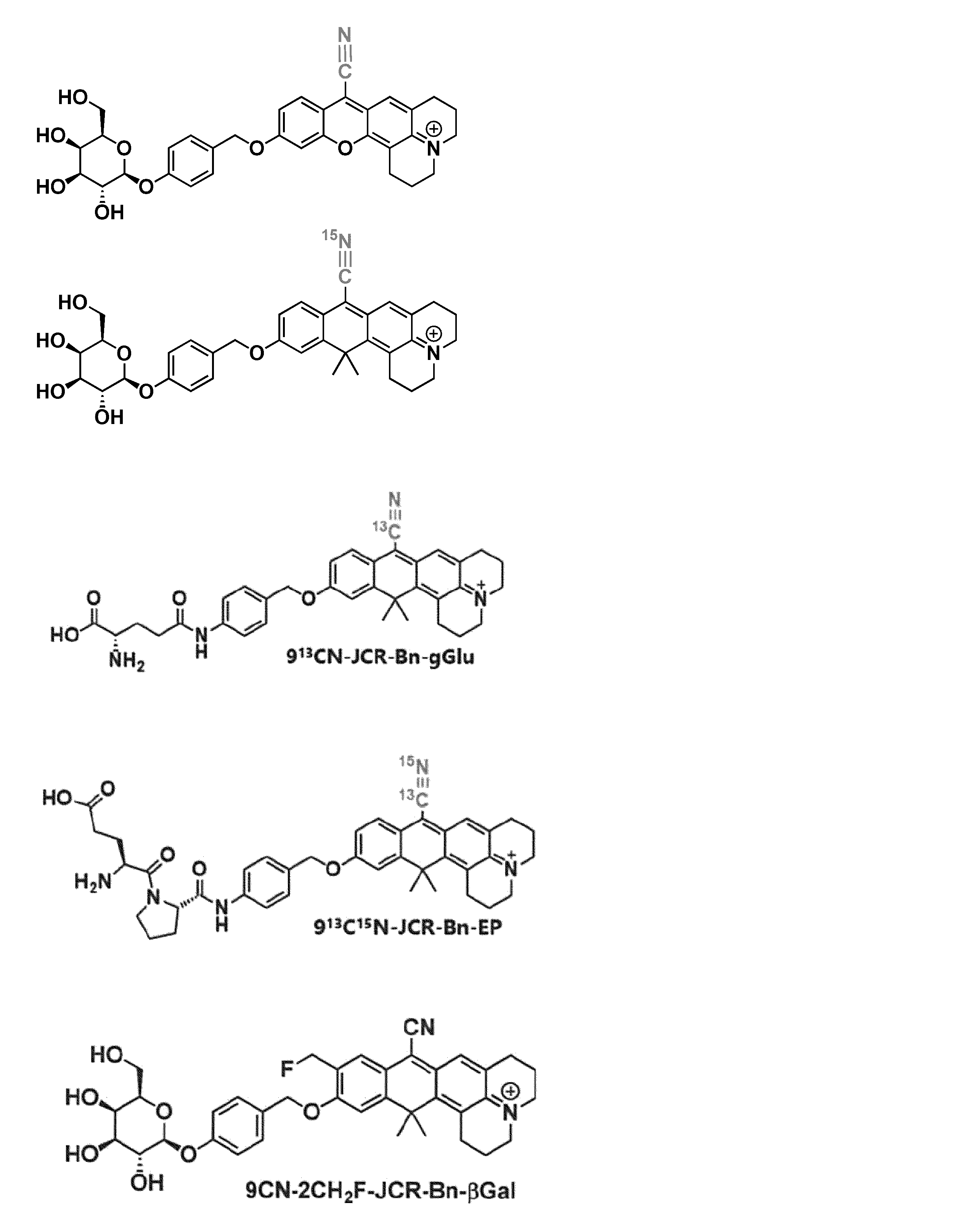
- 請求項1~10のいずれか1項に記載の化合物又はその塩を含むラマンプローブ。
- epr-SRS法に利用可能な請求項11に記載のラマンプローブ。
- 細胞又は組織内の標的酵素又は活性酸素種を検出する方法であって、(a)一般式(I)で表される化合物又はその塩を細胞又は組織内に導入する工程、及び(b)当該化合物又はその塩が細胞又は組織内で標的酵素又は活性酸素種と反応することにより増強されるラマン散乱光を測定する工程を含む方法。
- epr-SRS法を用いてラマン散乱光を測定する、請求項13に記載の方法。
Applications Claiming Priority (2)
| Application Number | Priority Date | Filing Date | Title |
|---|---|---|---|
| US202263315766P | 2022-03-02 | 2022-03-02 | |
| US63/315,766 | 2022-03-02 |
Publications (1)
| Publication Number | Publication Date |
|---|---|
| WO2023167282A1 true WO2023167282A1 (ja) | 2023-09-07 |
Family
ID=87883726
Family Applications (1)
| Application Number | Title | Priority Date | Filing Date |
|---|---|---|---|
| PCT/JP2023/007813 Ceased WO2023167282A1 (ja) | 2022-03-02 | 2023-03-02 | O-function型activatableラマンプローブ |
Country Status (1)
| Country | Link |
|---|---|
| WO (1) | WO2023167282A1 (ja) |
Cited By (1)
| Publication number | Priority date | Publication date | Assignee | Title |
|---|---|---|---|---|
| CN119409748A (zh) * | 2025-01-06 | 2025-02-11 | 四川省医学科学院·四川省人民医院 | 一种N-乙酰-β-D-氨基葡萄糖酶的化学发光探针、试剂盒及其制备方法和应用 |
Citations (1)
| Publication number | Priority date | Publication date | Assignee | Title |
|---|---|---|---|---|
| WO2021177050A1 (ja) * | 2020-03-03 | 2021-09-10 | 国立大学法人 東京大学 | Activatable型ラマンプローブ |
-
2023
- 2023-03-02 WO PCT/JP2023/007813 patent/WO2023167282A1/ja not_active Ceased
Patent Citations (1)
| Publication number | Priority date | Publication date | Assignee | Title |
|---|---|---|---|---|
| WO2021177050A1 (ja) * | 2020-03-03 | 2021-09-10 | 国立大学法人 東京大学 | Activatable型ラマンプローブ |
Non-Patent Citations (3)
| Title |
|---|
| FUJIOKA HIROYOSHI, SHOU JINGWEN, KOJIMA RYOSUKE, URANO YASUTERU, OZEKI YASUYUKI, KAMIYA MAKO: "Multicolor Activatable Raman Probes for Simultaneous Detection of Plural Enzyme Activities", JOURNAL OF THE AMERICAN CHEMICAL SOCIETY, AMERICAN CHEMICAL SOCIETY, vol. 142, no. 49, 1 November 2020 (2020-11-01), pages 20701 - 20707, XP055852906, ISSN: 0002-7863, DOI: 10.1021/jacs.0c09200 * |
| HIROKI ITO; YU KAWAMATA; MAKO KAMIYA; KAYOKO TSUDA‐SAKURAI; SHINJI TANAKA; TASUKU UENO; TORU KOMATSU; KENJIRO HANAOKA; SHIGEO OKAB: "Red‐Shifted Fluorogenic Substrate for Detection of lacZ‐Positive Cells in Living Tissue with Single‐Cell Resolution", ANGEWANDTE CHEMIE INTERNATIONAL EDITION, VERLAG CHEMIE, HOBOKEN, USA, vol. 57, no. 48, 2 November 2018 (2018-11-02), Hoboken, USA, pages 15702 - 15706, XP072103608, ISSN: 1433-7851, DOI: 10.1002/anie.201808670 * |
| JOANNA WRZESIEN; DUNCAN GRAHAM;: "Synthesis of SERS active nanoparticles for detection of biomolecules", TETRAHEDRON, ELSEVIER SIENCE PUBLISHERS, AMSTERDAM, NL, vol. 68, no. 4, 21 November 2011 (2011-11-21), AMSTERDAM, NL , pages 1230 - 1240, XP028123160, ISSN: 0040-4020, DOI: 10.1016/j.tet.2011.11.053 * |
Cited By (1)
| Publication number | Priority date | Publication date | Assignee | Title |
|---|---|---|---|---|
| CN119409748A (zh) * | 2025-01-06 | 2025-02-11 | 四川省医学科学院·四川省人民医院 | 一种N-乙酰-β-D-氨基葡萄糖酶的化学发光探针、试剂盒及其制备方法和应用 |
Similar Documents
| Publication | Publication Date | Title |
|---|---|---|
| Ding et al. | Smart probe for rapid and simultaneous detection and discrimination of hydrogen sulfide, cysteine/homocysteine, and glutathione | |
| Huang et al. | A turn-on fluorescence probe based on aggregation-induced emission for leucine aminopeptidase in living cells and tumor tissue | |
| Gan et al. | A novel fluorescent probe for selective imaging of cellular cysteine with large Stokes shift and high quantum yield | |
| Wang et al. | A novel near-infrared styryl-BODIPY fluorescent probe for discrimination of GSH and its application in living cells | |
| Liu et al. | A sensitive and selective fluorescent probe for the detection of hydrogen peroxide with a red emission and a large Stokes shift | |
| Zhang et al. | Simultaneous detection of Cys/Hcy and H2S through distinct fluorescence channels | |
| Huo et al. | A turn-on green fluorescent thiol probe based on the 1, 2-addition reaction and its application for bioimaging | |
| Zhang et al. | An off–on fluorescent probe based on maleimide for detecting thiols and its application for bioimaging | |
| Liu et al. | A red-emitting fluorescent probe for specific detection of cysteine over homocysteine and glutathione with a large Stokes shift | |
| WO2010095450A1 (ja) | プロテアーゼ測定用蛍光プローブ | |
| Yang et al. | An instantaneous near-infrared trimethyl lock based fluorescent probe for biothiols with a large Stokes shift | |
| Guo et al. | Fabrication of a water-soluble near-infrared fluorescent probe for selective detection and imaging of dipeptidyl peptidase IV in biological systems | |
| Wang et al. | Near-infrared squaraine dye as a selective protein sensor based on self-assembly | |
| Wu et al. | Novel near-infrared frequency up-conversion luminescence probe for monitoring biothiols in vitro and in vivo | |
| Hou et al. | A reversible turn-on fluorescent probe for quantitative imaging and dynamic monitoring of cellular glutathione | |
| Tang et al. | A novel aminopeptidase N triggered near-infrared fluorescence probe for imaging enzyme activity in cells and mice | |
| WO2008145609A1 (en) | Method of making covalent conjugates with his-tagged proteins | |
| Hou et al. | A near-infrared fluorescent probe derived from naphthalimide for detecting cysteine within lysosomes | |
| Wei et al. | A novel off-on fluorescent probe for imaging of hypoxia in tumor cell | |
| Wang et al. | A near-infrared fluorescent probe based on chloroacetate modified naphthofluorescein for selectively detecting cysteine/homocysteine and its application in living cells | |
| Wang et al. | Visual monitoring of the mitochondrial pH changes during mitophagy with a NIR fluorescent probe and its application in tumor imaging | |
| WO2021177050A1 (ja) | Activatable型ラマンプローブ | |
| Li et al. | Biotin-tagged fluorescent probe for in situ visualization of γ-glutamyl transpeptidase in cancerous cells and tissues | |
| Wei et al. | Design, synthesis and cell imaging of a new 3-thiolflavone fluorescent probe for biothiols | |
| JP7339675B2 (ja) | カルボキシペプチダーゼ活性検出用蛍光プローブ |
Legal Events
| Date | Code | Title | Description |
|---|---|---|---|
| 121 | Ep: the epo has been informed by wipo that ep was designated in this application |
Ref document number: 23763539 Country of ref document: EP Kind code of ref document: A1 |
|
| NENP | Non-entry into the national phase |
Ref country code: DE |
|
| 122 | Ep: pct application non-entry in european phase |
Ref document number: 23763539 Country of ref document: EP Kind code of ref document: A1 |
|
| NENP | Non-entry into the national phase |
Ref country code: JP |