WO2023167276A1 - Capped rna and method for producing same, apparatus for producing protein, and method for producing protein - Google Patents
Capped rna and method for producing same, apparatus for producing protein, and method for producing protein Download PDFInfo
- Publication number
- WO2023167276A1 WO2023167276A1 PCT/JP2023/007759 JP2023007759W WO2023167276A1 WO 2023167276 A1 WO2023167276 A1 WO 2023167276A1 JP 2023007759 W JP2023007759 W JP 2023007759W WO 2023167276 A1 WO2023167276 A1 WO 2023167276A1
- Authority
- WO
- WIPO (PCT)
- Prior art keywords
- rna
- capped
- circular
- cap structure
- region
- Prior art date
Links
Classifications
-
- C—CHEMISTRY; METALLURGY
- C12—BIOCHEMISTRY; BEER; SPIRITS; WINE; VINEGAR; MICROBIOLOGY; ENZYMOLOGY; MUTATION OR GENETIC ENGINEERING
- C12N—MICROORGANISMS OR ENZYMES; COMPOSITIONS THEREOF; PROPAGATING, PRESERVING, OR MAINTAINING MICROORGANISMS; MUTATION OR GENETIC ENGINEERING; CULTURE MEDIA
- C12N15/00—Mutation or genetic engineering; DNA or RNA concerning genetic engineering, vectors, e.g. plasmids, or their isolation, preparation or purification; Use of hosts therefor
- C12N15/09—Recombinant DNA-technology
- C12N15/63—Introduction of foreign genetic material using vectors; Vectors; Use of hosts therefor; Regulation of expression
- C12N15/79—Vectors or expression systems specially adapted for eukaryotic hosts
-
- C—CHEMISTRY; METALLURGY
- C12—BIOCHEMISTRY; BEER; SPIRITS; WINE; VINEGAR; MICROBIOLOGY; ENZYMOLOGY; MUTATION OR GENETIC ENGINEERING
- C12P—FERMENTATION OR ENZYME-USING PROCESSES TO SYNTHESISE A DESIRED CHEMICAL COMPOUND OR COMPOSITION OR TO SEPARATE OPTICAL ISOMERS FROM A RACEMIC MIXTURE
- C12P21/00—Preparation of peptides or proteins
-
- C—CHEMISTRY; METALLURGY
- C12—BIOCHEMISTRY; BEER; SPIRITS; WINE; VINEGAR; MICROBIOLOGY; ENZYMOLOGY; MUTATION OR GENETIC ENGINEERING
- C12Q—MEASURING OR TESTING PROCESSES INVOLVING ENZYMES, NUCLEIC ACIDS OR MICROORGANISMS; COMPOSITIONS OR TEST PAPERS THEREFOR; PROCESSES OF PREPARING SUCH COMPOSITIONS; CONDITION-RESPONSIVE CONTROL IN MICROBIOLOGICAL OR ENZYMOLOGICAL PROCESSES
- C12Q1/00—Measuring or testing processes involving enzymes, nucleic acids or microorganisms; Compositions therefor; Processes of preparing such compositions
- C12Q1/68—Measuring or testing processes involving enzymes, nucleic acids or microorganisms; Compositions therefor; Processes of preparing such compositions involving nucleic acids
- C12Q1/6813—Hybridisation assays
- C12Q1/6816—Hybridisation assays characterised by the detection means
- C12Q1/6825—Nucleic acid detection involving sensors
Definitions
- the present invention relates to a capped RNA having a cap structure, a method for producing the same, an apparatus for producing protein, and a method for producing protein.
- a 5' cap structure is known in which 7-methylguanylic acid is linked 5'-5' to the 5' end of RNA via a triphosphate bond.
- a cap structure is known to promote translation of mRNA, and a target protein can be efficiently synthesized by artificially introducing a cap structure into mRNA in a protein expression system or the like.
- capped RNA which is RNA whose 5' end is cap-modified, comprising reacting an activated cap compound with monophosphorylated RNA whose 5' end is monophosphorylated.
- a method for producing capped RNA that allows the production of capped RNA (see, for example, Patent Document 1).
- a cap structure can be introduced to the 5' end of RNA by a chemical method.
- a typical RNA having a cap structure at the 5' end of the RNA backbone is shown in Fig. 1(a).
- a protein that encodes a full-length base number that is a multiple of 3 of 102 or more has at least one start codon, and a stop codon (stop codons), and have developed a circular RNA that does not contain an IRES (see, for example, Patent Document 2 and Non-Patent Document 1).
- This circular RNA has no cap structure, but is capable of rotating protein translation with high efficiency.
- linear RNAs generally have one cap structure at the 5' end of the main chain
- circular RNAs have a cap structure.
- introduction of a cap structure to the 5'-end of a linear RNA initiates a translation reaction from the initiation codon immediately following it.
- a cap structure cannot be introduced into circular mRNAs without a 5' end, it is common to introduce an IRES sequence in order to initiate the translation reaction.
- a method of introducing an IRES sequence is known for initiating a translation reaction in the second translational region (for example, Non-Patent Document 2 reference).
- an IRES is incorporated into the eToehold, which is designed to be inactive until activated by sense-antisense interactions with specific trRNAs.
- a capped RNA comprising a translation region starting from an initiation codon and encoding a protein, and an upstream region on the upstream side of the initiation codon, and comprising a branched portion having a cap structure in the upstream region.
- a capped RNA wherein the branched portion has at least one structure of any one of the following (a) to (c): (a) a capped probe having a sequence complementary to part or all of the upstream region and having a cap structure is hybridized to the upstream region; (b) a part of the upstream region is provided with a branched chain branching from the middle of the main chain constituting the RNA, and the branched chain is provided with the cap structure; (c)
- the RNA is a linear RNA, the 5'-end of the main chain of the upstream region has a branched structure with two or more branches, and each branch is provided with the cap structure.
- the RNA is a circular RNA
- the capped probe has a sequence complementary to a sequence of part or all of the upstream region of the circular RNA and has a cap structure.
- the RNA is a linear RNA, has a sequence complementary to part or all of the upstream region of the linear RNA, and has a cap structure.
- the RNA is a circular RNA, a branched chain branching from a main chain constituting the RNA is provided in a part of the upstream region of the circular RNA, and the branched chain
- the RNA is a linear RNA, and a branched chain branching from a main chain constituting the RNA is provided in a part of the upstream region of the linear RNA,
- RNA of [2] above wherein in (b) above, it has a structure represented by the following formula (3).
- m 7 G represents a cap
- p represents a phosphate group
- Nuc represents a polynucleotide in which one or more natural or non-natural nucleotides are linked
- m represents an integer of 1 to 5.
- a plurality of m may be the same or different from each other.
- RNA1 and RNA2 represent RNAs constituting the main chain, RNA1 on the 3′-end side and RNA2 on the 5′-end side, each having a branched structure.
- the RNA of formula (3) may be a circular RNA in which the 5' end of RNA1 and the 3' end of RNA2 are linked to form a circular structure, or may be a linear RNA without linking. .
- RNA of [2] above wherein in (c) above, it has a structure represented by the following formula (1) or formula (2).
- m 7 G represents a cap
- p represents a phosphate group
- Gm represents a structure in which the 2′-hydroxyl group of a guanine nucleotide is methylated
- m represents an integer of 1 to 5, and multiple m may be the same or different
- n represents an integer of 1 to 5, and multiple n may be the same or different.
- the method for producing capped RNA according to [1] above which comprises at least one of the steps (a) to (c) below (a ) hybridizing a capped probe having a sequence complementary to a part or all of the upstream region and having a cap structure to the upstream region, or (b) after synthesizing the RNA having a branched chain branching from the main chain constituting the RNA in a part of the upstream region, binding the cap structure to the branched chain; (c)
- the RNA is a linear RNA, and after synthesizing an RNA having a branch structure in which the 5′ end of the main chain of the upstream region is branched into two or more branches, the cap structure is bound to each branch.
- a protein comprising the capped RNA of [1] above and a eukaryotic cell expression system capable of expressing the protein encoded by the translation region using the capped RNA as a template. manufacturing equipment.
- capped RNA according to [1] above is added to a eukaryotic cell expression system capable of expressing the protein encoded by the translational region using the capped RNA as a template to express the protein.
- a method for producing a protein characterized by:
- a target RNA detection device having a cap structure at the 5' end of RNA, a translation region starting from an initiation codon and encoding a labeling protein, an upstream region on the upstream side of the initiation codon and having a sequence complementary to part or all of the sequence of the target RNA to be detected, a detection probe consisting of a circular RNA having means for adding the circular RNA to a sample containing the target RNA and hybridizing the target RNA to the upstream region of the circular RNA to generate capped RNA; a eukaryotic cell expression system capable of expressing the labeled protein by adding the capped RNA; and means for detecting the labeled protein expressed in the expression system.
- a method for detecting a target RNA having a cap structure at the 5' end of the RNA comprising: a translation region starting from an initiation codon and encoding a labeling protein, an upstream region on the upstream side of the initiation codon and having a sequence complementary to part or all of the sequence of the target RNA to be detected, A step of preparing a detection probe consisting of a circular RNA having adding the circular RNA to a sample containing the target RNA and hybridizing the target RNA to the upstream region of the circular RNA to generate capped RNA; adding the capped RNA to a eukaryotic cell expression system capable of expressing the labeled protein to express the labeled protein; and detecting the labeled protein expressed in the expression system.
- RNA production method of [1(Pre)] above comprising at least one of the following steps (a) to (c):
- a method for producing RNA (a) hybridizing a capped probe having a sequence complementary to part or all of the untranslated region and having a cap structure to the untranslated region, or (b) synthesizing the RNA having a branched chain branching from the main chain constituting the RNA in a part of the untranslated region, and then binding the cap structure to the branched chain; (c) After synthesizing an RNA having a branch structure in which the 5′ end of the main chain of the untranslated region is branched into two or more branches, the cap structure is bound to each branch.
- a method for detecting a target RNA having a cap structure at the 5' end of [13(Pre)]RNA comprising: A translation region starting from an initiation codon and encoding a labeling protein, and an untranslated region upstream of the initiation codon and having a sequence complementary to part or all of the sequence of the target RNA to be detected.
- a step of preparing a probe consisting of a circular RNA having adding the circular RNA to a sample containing the target RNA and hybridizing the target RNA to the untranslated region of the circular RNA to generate capped RNA; adding the capped RNA to a eukaryotic cell expression system capable of expressing the labeled protein to express the labeled protein; and detecting the labeled protein expressed in the expression system.
- Another object of the present invention is to provide a protein production apparatus and a protein production method capable of synthesizing a protein using such capped RNA.
- FIG. 1 is a diagram showing an outline of capped RNA (hybridization type) of the present invention.
- FIG. BRIEF DESCRIPTION OF THE DRAWINGS It is a figure which shows the outline
- FIG. 2 shows sequences of template DNA and transcribed RNA in Examples.
- FIG. 4 shows the results of synthesis experiments of capped probes in Examples.
- FIG. 2 shows the results of linear RNA synthesis experiments in Examples.
- FIG. 4 shows the results of linear RNA circularization experiments in Examples.
- FIG. 4 is a diagram showing the results of relative evaluation of translation activity in Examples.
- FIG. 10 is a diagram showing evaluation results of translation activity when using various probes in Examples.
- FIG. 10 is a diagram showing evaluation results of translation activity in the presence or absence of a poly-A sequence in Examples.
- FIG. 4 is a diagram showing the results of evaluation of translation activity by changing the ratio of probe RNA in Examples.
- 1 is a schematic diagram showing a synthesis scheme for capped branch-containing RNA oligonucleotides in Examples.
- FIG. FIG. 4 shows chemical capping reactions of branched chains in the examples.
- FIG. 2 is a diagram showing a circular RNA synthesis reaction in Examples.
- FIG. 2 is a diagram showing evaluation of translational activity of capped branched-chain-containing circular mRNA in human-derived cultured cells (HeLa) in Examples.
- HeLa human-derived cultured cells
- FIG. 4 shows the structure of a 107-base-long RNA containing a branch structure synthesized in Examples.
- FIG. 10 is a diagram showing evaluation results of translation activity in 107-mer HeLa cells containing branch structures/plural cap structures synthesized in Examples.
- FIG. 4 shows the results of HPLC analysis of RNA synthesized in Examples.
- FIG. 3 shows the results of absorbance measurement of deprotected RNA and the results of electrophoresis of capped RNA in Examples.
- FIG. 4 shows the results of absorbance measurement of capped RNA in Examples.
- FIG. 4 shows the sequence of RNA after annealing in Examples.
- FIG. 2 shows the results of evaluating the structure and translational activity of bicistronic mRNA in Examples.
- FIG. 10 is a diagram showing evaluation results of translation activity in 107-mer HeLa cells containing branch structures/plural cap structures synthesized in Examples.
- FIG. 4 shows the results of HPLC analysis of RNA synthesized in Examples.
- FIG. 2 shows the overall structure of multi-uORF-mRNA and the sequence of probe RNA in Examples.
- FIG. 1 shows the overall structure and sequence of multi-uORF-mRNA in Examples.
- FIG. 3 shows the results of HPLC analysis of multi-uORF-mRNA in Examples.
- FIG. 3 shows the results of HPLC analysis of multi-uORF-mRNA in Examples.
- FIG. 4 shows photographs of denaturing PAGE of multi-uORF-mRNA and capping efficiencies in Examples.
- FIG. 2 shows the evaluation results of multi-uORF-mRNA translation activity in Examples.
- FIG. 4 shows sequences of plasmid fragments containing IRES in Examples.
- FIG. 4 is a diagram showing evaluation results of translation activity of capped circular RNA and IRES in Examples.
- FIG. 4 is a diagram showing evaluation results of translation activity of capped circular RNA and IRES in Examples.
- FIG. 3 shows experimental results showing that the translational activity of circular mRNAs in which a cap structure has been introduced into the branched chain is significantly improved compared to those without the cap structure.
- FIG. 3 shows experimental results showing the relationship between branched chain length and translational activity (variable branched chain length) of circular mRNA in which a cap structure has been introduced into the branched chain.
- FIG. 3 shows experimental results showing that the translational activity of circular mRNA having a cap structure in its branched chain is higher than that of mRNA containing an EMCV-derived IRES sequence.
- FIG. 2 shows the results of synthesis of ATF4-Renilla mRNA by in vitro transcription. It is a figure which shows the result of chemical synthesis of RNA.
- FIG. 2 shows the results of chemical capping of RNA.
- FIG. 2 shows the results of synthesis by in vitro transcription of circular NanoLuc mRNA.
- FIG. 10 shows the results of circularization reaction of circular NanoLuc mRNA.
- FIG. 10 shows the results of csRNA against circular Nluc mRNA. It is a figure which shows the result of chemical synthesis of RNA.
- FIG. 2 shows the results of chemical capping of RNA.
- FIG. 2 shows the results of chemical capping of RNA.
- FIG. 4 shows the sequences and secondary structures of ATF4-Renilla mRNA and ATF4-csRNA.
- FIG. 4 shows the translation reaction of ATF4-Renilla mRNA and ATF4-csRNA5-8 in HeLa cells.
- FIG. 2 is a diagram showing experimental results of translation reaction of chemically modified csRNA in HeLa cell extract.
- FIG. 3 shows the results of translation reaction of LNA-modified csRNA and circular mRNA in HeLa
- the capped RNA of the present invention has a translation region that starts from an initiation codon (eg, AUG) and encodes a protein, and an upstream region (5' terminal side) upstream of this initiation codon.
- This upstream region may be an untranslated region (5'UTR) or may be in frame with another open reading frame (ORF).
- This capped RNA has a branch with a cap structure in the upstream region.
- the “branched portion having a cap structure” includes all forms in which the cap structure is branched from the main chain RNA.
- molecules such as nucleic acids (DNA, RNA, etc.) having a cap structure are hybridized to RNA of the main chain, branched nucleotides constituting the main chain, and Including combined ones.
- 3'UTR and polyA are also included in the upstream region, but the branched portion is preferably located at 5'UTR or uORF near the initiation codon.
- the capped RNA has at least one structure of any one of (a) to (c) below.
- a capped probe having a sequence complementary to the sequence of part or all of the upstream region and having a cap structure is hybridized to the upstream region (hereinafter, “hybridized type”).
- hybridized type A structure in which a part of the upstream region is provided with a branched chain branching from the middle of the main chain constituting RNA, and the branched chain is provided with a cap structure (hereinafter, "branched chain type").
- the capped RNA of the present invention includes not only natural RNA (that is, a polynucleotide in which nucleotides consisting of ATP, GTP, CTP, and UTP are linked), but also mC4 linkers, 2'OMe, and LNA in the nucleotides of RNA. Non-natural nucleic acid strands (modified nucleic acid strands) into which such as have been introduced are also included. Each type of capped RNA is described below.
- Hybridized type The hybridized type capped RNA will be described below.
- (B) shows the capped RNA hybridized in the linear RNA
- (C) shows the capped RNA hybridized in the circular RNA, each with the structure described in the Examples.
- a cap structure is attached to the upstream region (for example, untranslated region (5'UTR)) of RNA encoding a protein to be translated (hereinafter referred to as "coding RNA").
- a capped probe that is an RNA having a is hybridized.
- the coding RNA consists of a translation region (“Nano Luc” region in the figure) that starts from the start codon and encodes a protein, and an upstream region (5′ end side) upstream of the start codon (for example, an untranslated region (“5′UTR ” area)).
- an upstream region (5′ end side) upstream of the start codon for example, an untranslated region (“5′UTR ” area)
- there is a stop codon at the 3' end of the translation region and there may be an untranslated region (3'UTR) downstream (on the 3' end side) of this stop codon.
- the 3'UTR contains a poly A sequence.
- the coding RNA is linear in linear RNA, and has a circular structure in which the 5'-end and 3'-end of the linear RNA are linked together in circular RNA.
- this embodiment shows an embodiment in which the code RNA contains a stop codon, it is not limited to this. may be In the case where this stop codon is not included, as disclosed in these prior arts, if a circular RNA having a full-length base number of 102 or more and a multiple of 3 is used as a circular RNA, rotational protein translation results in high translation. The target protein can be produced efficiently.
- the coding RNA may have only one translational region or may have two or more translational regions. When there are two or more translational regions, each translational region has an upstream region, and a capping probe that hybridizes to any of the upstream regions can be used. In this case, the translational activity of the protein in the translational region starting from the initiation codon immediately below the upstream region is enhanced. Moreover, it may have a normal cap structure at the 5' end of the main chain of the coding RNA. When the capped probe has a normal cap structure at the 5' end, the combined use of the capped probe with the cap structure further improves the translation efficiency of the capped RNA.
- a capped probe is composed of an RNA that is shorter than the coding RNA, and has a base sequence complementary to the sequence of part or all of the 5'UTR of the coding RNA.
- the position at which the capped probe hybridizes in the 5'UTR of the coding RNA is not particularly limited, but the upstream side is preferred if it has a Kozak sequence.
- the length of the capped probe is not particularly limited, but a length of about 10 to 50 bases (nt) is preferred.
- the capped probe may have a cap bound to the 5' end or a cap bound to the 3' end. Additionally, the capped probe may have a poly A sequence at the 3' end.
- the length (number of bases) of the poly A sequence is not particularly limited, but is preferably about 10 to 50 bases.
- the capped probe may have a Locked Nucleic Acid (LNA) in which the 2' and 4' carbons of ribose are bridged, a C4 linker, and the like.
- LNA Locked Nucleic Acid
- capped RNA the capped probe hybridizes to the 5'UTR of the coding RNA.
- hybridize means to hybridize under stringent conditions.
- stringent conditions refers to conditions under which specific hybrids are formed and non-specific hybrids are rarely formed.
- RNAs that "hybridize under stringent conditions” also include RNAs that have a certain degree or more of homology to a sequence that is completely complementary to a predetermined range of the 5'UTR sequence of the coding RNA. .
- the homology in this case is usually 60% or more, preferably 80% or more, more preferably 90% or more, particularly preferably 95% or more, and most preferably 100%, according to, for example, the BLAST algorithm.
- the coding RNA that constitutes the capped RNA can be produced by using a DNA having a sequence complementary to the RNA having the desired sequence and transcribe it with an RNA polymerase.
- the coding RNA may be chemically synthesized using a DNA synthesizer or the like.
- Circular coding RNA can be prepared by preparing a linear coding RNA and then ligating the ends using T4 RNA ligase 2 or the like to circularize it.
- Probe RNA can be produced by the following method. First, base uncapped RNA is prepared by RNA solid-phase synthesis method using phosphoramidite, phosphorylation reagent, 2'-OMe RNA amidite, LNA amidite, C4 linker amidite, CPG column, etc. do. Next, this RNA is reacted with m 7 GDP, 2-nitroimidazole or the like to cap the phosphorylated RNA. For details of this reaction, reference can be made to Patent Document 1.
- Capped RNA can be produced by annealing code RNA and probe RNA.
- the ratio (molar ratio) of coding RNA to probe RNA during annealing is preferably in the range of 1:1 to 1:20, more preferably in the range of 1:5 to 1:15, from the viewpoint of translation activity.
- the annealing temperature can be 80° C. or higher, preferably 90° C. or higher, and the reaction time can be about 1 to 30 minutes, preferably 3 to 10 minutes.
- a hybridizing-type capped RNA can add a cap structure to any position of the 5'UTR, regardless of whether the coding RNA is linear or circular.
- RNA with a cap structure increases the rate at which a translation initiation factor that constitutes a ribosome binds to coding RNA, resulting in improved translational activity, compared to RNA without a cap structure.
- hybrid type capped RNA it is possible to enhance translation or induce a translation phenomenon from an arbitrary position by presenting a cap by hybridization with a probe in circular RNA or linear RNA.
- translation reaction can be induced from, for example, the second and subsequent initiation codons, which are not originally used as reading frames for protein synthesis. This allows translation to be induced from any RNA location by a capped probe.
- the capped structure of the hybridized type capped RNA of the present invention can be introduced into circular RNAs as well. As a result, translational activity can be greatly improved compared to circular RNAs that do not have a cap structure.
- FIG. 2(a) shows a branched-chain capped RNA in a linear RNA
- FIG. 2(b) shows a branched-chain capped RNA in a circular RNA, each with the structure described in the Examples.
- the upstream region of the coding RNA has a branched chain branched from the main chain constituting the coding RNA, and the branched chain is provided with a cap structure.
- the overall structure of the code RNA is as described in "1. Hybridization type" above.
- a portion of the 5'UTR of the coding RNA has a branched chain branching off from the main chain, and the branched chain has a cap structure at the 5' end.
- the 5' end of the backbone of the coding RNA is phosphorylated in linear RNA.
- the coding RNA may have either one translation region or two or more translation regions. When the coding RNA is linear, it may have a normal cap structure at the 5' end.
- RNA1 and RNA2 represent RNAs constituting the main chain, RNA1 on the 3′-end side, RNA2 on the 5′-end side, each having a branched structure.
- the RNA of formula (3) may be a circular RNA in which the 5' end of RNA1 and the 3' end of RNA2 are linked to form a circular structure, or may be a linear RNA without linking. .
- a coding RNA having a branched chain in the 5′UTR is obtained by using a normal phosphoramidite reagent used in RNA synthesis, a phosphoramidite reagent having a branched chain, and a chemical phosphorylation reagent, and using an automatic nucleic acid synthesizer.
- a normal phosphoramidite reagent used in RNA synthesis a phosphoramidite reagent having a branched chain
- a chemical phosphorylation reagent and using an automatic nucleic acid synthesizer.
- the desired sequence can be obtained.
- Branching can be introduced at the position. Since the 5' end of the branched chain of the coding RNA has a hydrophobic protective group introduced by a chemical phosphorylation reagent, this hydrophobic protective group is decomposed by light irradiation etc. to remove the 5' end of the branched chain as a phosphate group.
- RNA Circular coding RNA can be produced by producing a linear capped RNA and then ligating the ends using T4 RNA ligase 2 or the like to circularize it.
- a heteroaromatic compound such as 7-methylguanosine 5'-diphosphate imidazolide salt (Im-m 7 GDP), 2-nitroimidazole, or the like.
- the 5' end of the backbone of the coding RNA is phosphorylated using, for example, T4 polynucleotide kinase. This results in linear capped RNA.
- Circular coding RNA can be produced by producing a linear capped RNA and then ligating the ends using T4 RNA ligase 2 or the like to circularize it.
- branched-chain capped RNA can add a cap structure to any position of the 5'UTR regardless of whether the coding RNA is linear or circular. If the coding RNA has two or more translational regions, the translational activity of that translational region can be improved by introducing a branched cap into the 5'UTR upstream of the desired translational region. As a result, translation reaction can be induced from initiation codons that are not used as reading frames for protein synthesis, for example. In branched-chain capped RNA, a cap structure can also be introduced into a circular RNA that does not conventionally have a cap structure. As a result, translational activity can be greatly improved compared to circular RNAs that do not have a cap structure.
- Branched-chain capped RNA tends to have higher translational activity and lower immunogenicity when it is circular than when it is linear, as shown in Examples described later.
- the position of the branch in the upstream region (that is, the position of the structure between RNA1 and RNA2 in formula (3)) is not particularly limited, but since a cap structure is provided at the end of the branch, the position of the branch is the start There is a tendency that the closer to the codon, the higher the translation activity.
- the position of the branch is preferably within the range of 1 to 50 nt, more preferably within the range of 1 to 30 nt, particularly preferably within the range of 1 to 15 nt on the 5' side of the start codon.
- the chain length of the branched portion of the branched chain (that is, the length of the polynucleotide of Nuc in formula (3)) is not particularly limited, but translation activity tends to decrease if it is too long or too short. be.
- the chain length of the branched chain is defined as between the branch point and the 5' cap.
- the branched chain length is preferably in the range of 1 to 20 nt, more preferably in the range of 3 to 10 nt, and particularly preferably in the range of 5 to 8 nt. If the chain length is too short, the distance between the position of the cap structure and the branched portion becomes short, and the structure of the branched portion tends to inhibit the formation of the translation initiation factor complex and the binding of the ribosome. It is thought that branched RNAs have lower translational activity.
- the multi-cap type capped RNA will be described below.
- the capped RNA of the multitype cap has a branch structure in which the 5' end of the 5'UTR of the linear coding RNA is branched into two or more branches, and each branch is provided with a cap structure.
- Examples of multi-cap type capped RNA include biantennary type structures represented by the following formula (1) and triantennary type structures represented by formula (2).
- m 7 G represents a cap
- p represents a phosphate group
- Gm represents a structure in which the 2′-hydroxyl group of a guanine nucleotide is methylated
- m represents an integer of 1 to 5
- multiple m may be the same or different
- n represents an integer of 1 to 5, and multiple n may be the same or different.
- the above formula (1) has a structure represented by the following formula (1-1) and the above formula (2) has a structure represented by the following formula (2-1).
- Pre-capped RNA is synthesized using a conventional phosphoramidite reagent, a branched-chain phosphoramidite reagent, and a chemical phosphorylation reagent used in RNA synthesis, using an automated nucleic acid synthesizer. be able to.
- a phosphoramidite reagent having a branched chain to form a branch structure at the 5′ end and using a conventional phosphoramidite reagent and equipment to synthesize the sequence of the 5′UTR of the coding RNA from the upstream side. to synthesize a coding RNA with multiple branches at the 5' end.
- Hydrophobic protective groups are introduced into the 5' ends of each branch of the coding RNA using a chemical phosphorylation reagent, and the hydrophobic protective groups are decomposed by light irradiation or the like to convert the 5' ends of the branched chains into phosphate groups. do.
- a heteroaromatic compound such as 7-methylguanosine 5′-diphosphate imidazolide salt (Im-m 7 GDP), 2-nitroimidazole or the like is used to cap the 5′ end of each branch of the branch structure. This yields a linear capped RNA with a cap attached to each branch.
- multicap-type capped RNA has multiple caps at the 5′ end of the main chain, the translation initiation factor that constitutes the ribosome binds to the cap compared to normal capped RNA with one cap.
- the multiligand effect can greatly improve the translation activity.
- the protein production apparatus of the present invention comprises the above capped RNA and a eukaryotic cell expression system capable of expressing a protein encoded by a translation region using the capped RNA as a template. .
- capped RNA is added to a eukaryotic cell expression system to express the protein.
- Eukaryotic cells include fungi such as yeast and filamentous fungi, plant cells, insect cells, and animal cells such as humans. Specific examples include mouse L-cells, Ehrlich ascites cancer cells, HeLa Cells, CHO cells, Saccharomyces cerevisiae, etc. can be exemplified.
- the eukaryotic cell expression system may be either a cell expression system or a cell-free expression system.
- Introduction of capped RNA into a cell expression system can be performed by methods such as the calcium phosphate method, DEAE dextran method, lipofection method, and electroporation method.
- the cells are cultured under conditions such as an appropriate temperature, the cells are lysed after the culture, and the target protein is separated and purified.
- Eukaryotic cell lysates can be used in cell-free expression systems.
- Target RNA Detection Apparatus and Detection Method As described above, since circular RNA does not have a cap structure, it cannot be translated by itself. can be translated. This property can be used to detect target RNA using circular RNA as a detection probe.
- Target RNAs include RNAs derived from eukaryotes, viruses, and the like. The apparatus and method for detecting target RNA of the present invention will be described below.
- the device for detecting target RNA of the present invention is a device for detecting target RNA having a cap structure at the 5' end of the RNA.
- This detection apparatus comprises a detection probe composed of circular RNA, means for generating capped RNA, an expression system for eukaryotic cells, and means for detecting a labeled protein.
- the detection probe has a translation region starting from the start codon and encoding a labeled protein, and an upstream region upstream of this start codon.
- This upstream region has a sequence complementary to part or all of the target RNA sequence to be detected.
- labeling proteins include fluorescence-labeling proteins such as luciferase and green fluorescent protein (GFP), enzyme-labeling proteins such as horseradish peroxidase (HRP) and alkaline phosphatase (AP), which are used in Examples described later. can.
- the means for generating capped RNA is means for hybridizing the detection probe (circular RNA) and the target RNA.
- Common reagents and equipment for annealing RNA can be used for this.
- Such reagents include buffers such as Tri-HCl.
- Examples of the device include a heat block or a thermal cycler, which heats the reagent to 80° C. or higher and then cools it to room temperature.
- the expression system of eukaryotic cells is a system capable of expressing the marker protein encoded by the translational region using the capped RNA produced above as a template, and the above-mentioned HeLa cells and the like can be mentioned.
- Various devices can be used as means for detecting the labeled protein, depending on the type of labeled protein.
- Examples of such means include a fluorometer when the labeled protein is a fluorescently labeled protein, and a luminescent substrate and a fluorometer when the labeled protein is an enzyme-labeled protein.
- the target RNA detection method of the present invention can be implemented using the above-described detection device.
- a detection probe containing the circular RNA is prepared and added to a sample containing the target RNA to generate capped RNA.
- the capped RNA is added to a eukaryotic cell expression system capable of expressing the labeled protein to express the labeled protein, and then the expressed labeled protein is detected.
- a target RNA can be efficiently detected by such a method.
- FIG. 3 shows the base sequences of circular RNA and capped probes used in this experiment.
- the diagram on the upper left side of the figure shows the structure of the capped RNA.
- a circular RNA As a circular RNA (coding RNA), it has a 5′ upstream region (untranslated region (denoted as “5′UTR”) in the figure) and a Kozak sequence (denoted as “Kozak”) downstream thereof, Nano Luc luciferase-encoding region (“Nano Luc”), a stop codon (“stop”), a 3′-side untranslated region (“3′UTR”), and a polyA sequence (“polyA 30 ”) there is
- the capped probe in the figure has a cap structure (m 7 G) at the 5' end, has a sequence complementary to a partial region of the 5'UTR of the circular RNA, and has polyA30 at the 3' end. have.
- the capped RNA then has the capped probe hybridized to the 5'UTR of the circular RNA.
- the upper right side of the figure shows the sequence of the capped probe used in this example.
- the capped probes are "5' cap probe” having a cap structure at the 5' end, "3' cap probe” having a cap structure at the 3' end, and "3' cap probe” having a cap structure at the 5' end and poly A
- Three types of "5' cap probe with poly A” were used.
- the lower part of the figure shows the sequence of the circular RNA.
- the bold-faced region represented by “ggcg...cgc” is the portion where the capped probe hybridizes.
- Probe RNA RNA was synthesized at a scale of 0.2 ⁇ mol using Nippon Techno Service Co., Ltd. NTS M-2-MX DNA/RNA synthesizer or NRs-4A10R7 DNA/RNA synthesizer.
- Phosphoramidites used for RNA solid-phase synthesis include rA (5'-DMT-2'-TOM-ribo-Adenosine (n-acetyl) OP), rC (5'-DMT-2'-TOM-ribo- Cytidine (n-acetyl) OP), rG (5'-DMT-2'-TOM-ribo-Guanosine (n-acetyl) OP), U (5'-DMT-2'-TOM-ribo-Uridine OP) ( ChemGenes) was used. 5' Phosphate-ON Reagent (ChemGenes) was used as a phosphorylation reagent.
- CPG is 3' -Tom -RIBO CYTIDINE (N -ACETYL) 2' -LCAA CPG 1000 ⁇ , 3' -TOM -RIBO ADENOSINE (N -ACETYL) 2' -LCAA CPG 100 ⁇ (CHEMGENES), 3' -PHOSPH ATE ON LCAA CPG 1000A was used.
- RNA synthesis was transferred to a 1.5 mL looped screw tube (Sarstedt) and 40% aq. Methylamine/28% aq. 1.0 mL of ammonia (1:1) was added and incubated at 65°C for 20 minutes. It was filtered using a Millex LH 0.45 ⁇ m filter, washed with sterilized water (500 ⁇ L), and dried with a centrifugal evaporator. 1.0 mL of 1 M TBAF in THF was added and dissolved, and allowed to stand overnight at room temperature. 1.0 mL of 1M Tris-HCl buffer (pH 7.5) was added, and the organic solvent was distilled off with a centrifugal evaporator. Desalting was performed using a NAP-25 column (GE Healthcare). RNA was purified by reverse-phase HPLC.
- TBE buffer 89 mM Tris-borate, 2 mM EDTA, pH 8.3 was used for PAGE.
- RNAs 1 to 12 correspond to SEQ ID NOS: 1 to 12, respectively.
- RNA and 10 mM CaCl 2 were mixed at final concentrations, lyophilized to dryness, and then dissolved in DMSO.
- Im(m 7 GDP) ⁇ Na (Hongene Biotech) dissolved in DMSO was added thereto to a final concentration of 10 mM, and 2-nitroimidazole was added to a final concentration of 10 mM.
- the reaction was incubated at 55°C for 3 hours. Isopropanol precipitation was carried out after diluting the reaction solution twice with sterilized water. HPLC purified and analyzed by LC/MS.
- HPLC conditions column: hydrosphere C18 column (4.6 ⁇ 250 mm; YMC), mobile phase: A) 50 mM TEAA buffer (pH 7.0), 5% acetonitrile, B) acetonitrile, gradient: 0-20 min B conc. 0% to 20%, flow rate: 1.0 mL/min LC/MS (agilent) conditions; ACQUITY UPLC BEH C18 Column, 0.3 mL/min, A; 8.6 mM TEA/100 mM HFIP solution, B; MeOH, 0-30%/20 min HPLC-purified capped RNA was precipitated with isopropanol and sodium acetate (pH 5.2).
- capped RNAs 1 to 12 correspond to SEQ ID NOS: 13 to 24, respectively.
- FIG. 5 shows the results of capped probe synthesis experiments.
- A of the figure shows a synthetic scheme for capped probes. Chemically synthesized monophosphorylated RNA was lyophilized with calcium chloride. Im(m 7 GDP) ⁇ Na (chemical capping reagent) and 2-nitroimidazole were added and allowed to react in DMSO solution.
- B of the figure shows the results of the capped probe, and the capped RNA was analyzed by PAGE and stained with SYBR Green II. As the capping reaction progressed, the band shifted upward by one base due to the addition of methylguanine. As a result of PAGE analysis, it can be seen that the capping reaction proceeded with an efficiency of about 80% to 90%.
- Linear RNA was synthesized by transcription using T7 RNA polymerase using double-stranded DNA as a template.
- the upper part of FIG. 4 shows the template DNA sequence (SEQ ID NO: 32) for RNA transcription synthesis, and the lower part shows the sequence of the transcribed RNA (SEQ ID NO: 31).
- the template double-stranded DNA was obtained by amplifying pNL-TK1.1 (Promega) by PCR reaction using PrimeSTAR HS DNA Polymerase (Takara Bio) (2.5 ng/ ⁇ L DNA vector, 1 ⁇ PrimeSTAR buffer , 0.2 mM dNTPs, 0.5 ⁇ M primers, 0.025 units/ ⁇ L polymerase).
- Transcription reaction solution (template DNA 2.5 ng/ ⁇ L, 1 ⁇ T7 RNA Polymerase buffer (Takara) (40 mM Tris-HCl (pH 8.0), 8 mM MgCl 2 , 2 mM spermidine), 2.5 U/ ⁇ L T7 RNA polymerase (Takara ), 5 mM DTT, 2 mM ATP, 2 mM UTP, 2 mM, CTP, 0.4 mM GTP, 2 mM GMP) were prepared and incubated at 37° C. for 2 hours. Recombinant DNase (Takara) was added to the reaction solution to 0.1 U/ ⁇ L, and the mixture was incubated at 37° C. for 30 minutes to decompose the template DNA. After the reaction, phenol/chloroform extraction was performed. This was followed by ultrafiltration using an Amicon Ultra 10K (Millipore) and alcohol precipitation.
- Amicon Ultra 10K Amicon Ultra 10K (Milli
- FIG. 6 is a diagram showing the results of a linear RNA synthesis experiment.
- A) of the figure shows the result of synthesis by PCR reaction of double-stranded DNA that serves as a template for linear RNA. After the PCR reaction, it was analyzed on an agarose gel and is a photograph of the gel stained with Gel Red. be.
- B) of the figure shows the results of transcription synthesis of linear RNA, and is a photograph of a gel stained with SYBR Green after PAGE analysis after transcription reaction.
- FIG. 7 shows the results of linear RNA circularization experiments.
- A) of the figure shows the synthesis scheme of circular RNA. The transcribed and synthesized linear RNA was annealed with the guide DNA, and circular RNA was synthesized using T4 RNA ligase2.
- B) of the figure shows the results of confirmation of the cyclized product, in which RNase R was added to the cyclized RNA and incubated. Analyzed by PAGE and stained with SYBR Green II. The transcribed and synthesized linear RNA was circularized using T4 RNA ligase 2. The cyclized form was identified by PAGE analysis using an enzyme (RNase R) that selectively degrades linear RNA. The efficiency of cyclization was about 60%. The cyclized product and the starting linear RNA were excised from the gel and isolated and purified.
- RNase R an enzyme
- HeLa cells (RIKEN Cell Bank) were cultured in Dulbecco's modified Eagle's medium (DMEM; WAKO) supplemented with 10% fetal bovine serum (FBS; Invitrogen) (37 °C, 5% CO2 ). The day before transfection, HeLa cells were seeded in a 96-well plate (1.0 ⁇ 10 4 cells/well). The next day, the medium was removed and replaced with 100 ⁇ L/well Opti-MEM® (Thermo Fisher SCIENTIFIC). 0.15 ⁇ L Lipofectamine® MessengerMAX®, 20 ng mRNA was diluted in 10 ⁇ L Opti-MEM® and introduced into cells. After incubation at 37° C. for 5 hours, 20 ⁇ L/well 1 ⁇ Cell Lysis Buffer (Promega) was added to lyse the cells. Luminescence was measured using the Nano-Glo® luciferase assay® (Promega).
- FIG. 8 is a diagram showing the results of relative evaluation of translation activity.
- A) of the figure shows the results of the translational activity of linear RNA
- B) of the figure shows the results of the translational activity of circular RNA.
- “Capped RNA8” is used as capped RNA probe and "RNA8" as uncapped RNA probe.
- the translation activity of a sample not hybridized with an RNA probe is relatively evaluated as "1".
- Both RNAs were transfected into HeLa cells with capped RNAs annealed. After 5 hours, the cells were lysed and nanoluciferase luminescence was measured. From these results, the hybridization of capped RNA to both linear RNA and circular RNA showed about 40 times the translational activity promoting effect as compared to the case of RNA alone.
- the mRNA hybridized with the phosphorylated RNA to which no cap structure was added did not show any translational activity-promoting effect. This indicated that the translation initiation step was promoted in a cap-dependent manner.
- FIG. 9 is a diagram showing evaluation results of translation activity when using various probes.
- A) of the figure shows the results of the translational activity of linear RNA
- B) of the figure shows the results of the translational activity of circular RNA.
- Both RNAs were transfected into HeLa cells with capped RNAs annealed. After 5 hours, the cells were lysed and nanoluciferase luminescence was measured. From these results, both linear RNA and circular RNA were hybridized with capped RNA to promote translational activity (capped RNA 1-4, 6-12).
- the capped RNA having a sequence not complementary to the mRNA did not exhibit a translation-promoting effect (capped RNA 5), indicating that the capped RNA hybridized to the mRNA to promote translation.
- introduction of a C4 linker and a poly A sequence into the cap structure increased the promoting effect (capped RNA 8-10).
- FIG. 10 is a diagram showing the evaluation results of translation activity with or without poly A sequence.
- the upper row (Linear) in the figure shows the results using linear RNA
- the lower row (Circular) shows the results using circular RNA.
- “cap probe” indicates the result of annealing a capped probe
- "p probe” indicates the result of annealing an uncapped phosphate-terminated probe.
- FIG. 12 is a schematic diagram showing a scheme for synthesizing capped branched-chain-containing RNA oligonucleotides. Below, based on this synthesis scheme, capped branch-containing RNA oligonucleotides were synthesized.
- RNA chemical synthesis An oligoribonucleotide having a main chain of 54 bases long and containing a branched chain phosphorylated at the 5' end is compound 1 (the above formula (3): see Japanese Patent Application No. 2021-111420), and Commercially available phosphoramidite reagents [Amidites for RNA synthesis are ChemGenes products (ANP-3201, ANP-3202, ANP-3203, ANP-3205), branch chain introduction is Asymmetric Doubler (Lev) Phosphoramidite purchased from Glen Research (10-1981)] and a solid phase carrier (3'-TOM-ribo Uridine CPG 1000A, ChemGenes), and an automatic nucleic acid synthesizer (NR-2A 7MX, Nippon Techno Service) according to a conventional method.
- a 54-base RNA sequence consisting of only the main chain was synthesized and its 5'-end was phosphorylated with Compound 1.
- RNA contained in the eluate was recovered by alcohol precipitation.
- RNA solution 74 ⁇ M RNA, 1 mM Tris-HCl (pH 7.5), 0.1 mM EDTA
- 365 nm light 50 ⁇ L of RNA solution (74 ⁇ M RNA, 1 mM Tris-HCl (pH 7.5), 0.1 mM EDTA) was added to each well of a transparent 96-well multiwell plate, and 365 nm light was applied at a light intensity of 4 mW/cm 2 for 10 minutes. irradiated. After irradiation, RNA was recovered and isolated and purified by alcohol precipitation.
- RNA before and after deprotection of phosphate group were analyzed by LC/MS system (Agilent), and it was confirmed that the desired product was obtained [theoretical molecular weight of Observed value 20, 114.1 (+3.7): theoretical molecular weight of phosphate deprotected product 19,919.2, observed value 19,921.7 (+2.5)].
- DMSO dimethylsulfoxide
- 50 ⁇ L of Im- m GDP/DMSO solution and 5 ⁇ L of 200 mM 2-nitroimidazole/DMSO were added thereto.
- the solution was added and warmed to 55° C. for 3 hours.
- 122 ⁇ L of 0.541 M sodium acetate (pH 5.2) aqueous solution was added and an aliquot was taken to assess the efficiency of the capping reaction.
- 5 pmol each of raw material RNA and post-reaction RNA were analyzed by 10% denaturing PAGE containing 7.5 M urea as a denaturing agent to confirm the band shift of the capped product.
- FIG. 13 is a diagram showing the chemical capping reaction of branched chains.
- (a) of the figure shows a schematic diagram of the reaction (SEQ ID NO: 33).
- (b) of the figure shows the analysis results of RNA before and after reaction by 10% denaturing PAGE (containing 7.5 M urea as a denaturing agent).
- Raw material RNA before the capping reaction and the same molar amount of RNA (5 pmol) from the reaction solution were analyzed. After electrophoresis, the gel was stained with the nucleic acid staining reagent SYBR Green II to visualize the bands. The product of the capping reaction was observed at a migration position higher than that of the raw material, and the reaction yield calculated from the band intensity ratio was 78%.
- the target product was recovered from the eluate by alcohol precipitation to obtain 1.185 nmol of capped RNA (isolation yield 59%).
- the capped RNA was analyzed with an LC/MS system (Agilent), and it was confirmed that the desired product was obtained [theoretical molecular weight of 20,360.4, observed value of 20,361.2 (+0. 8)].
- RNA Phosphorylation of the 5'-end of the main chain using T4 polynucleotide kinase
- RNA after phosphorylation was analyzed by an LC/MS system (Agilent), and it was confirmed that the desired product was obtained [theoretical molecular weight of uncapped RNA: 19,993,3, observed value: 20,002. 3 (+9.0): theoretical molecular weight of capped RNA 20,434.4, observed value 20,441.4 (+7.0)].
- TK 0.3 ⁇ M primers, 0.2 mM dNTPs, 1.5 mM MgSO 4 , 1 ⁇ PCR Buffer for KOD-Plus-Neo, 0.02 U/ ⁇ L KOD-Plus-Neo (Toyobo).
- T100 thermal cycler Bio-Rad
- the reaction solution was subjected to the following thermal cycle conditions. 94°C, 2 minutes ⁇ [98°C, 10 seconds ⁇ 55°C, 30 seconds ⁇ 72°C, 70 seconds] ⁇ 25 cycles.
- reaction solution 50 ⁇ L x 8 tubes
- 0.4 mL of TE saturated phenol: chloroform equivalent mixture was added thereto, vigorously mixed and centrifuged (20,000 x g, 1 minute) to separate the aqueous layer. did.
- 0.4 mL of chloroform was added, vigorously mixed and centrifuged (20,000 ⁇ g, 1 minute) to separate the aqueous layer.
- 40 ⁇ L of 3M sodium acetate aqueous solution (pH 5.2) and 440 ⁇ L of 2-propanol are added thereto, cooled overnight at ⁇ 30° C., and centrifuged (20,000 ⁇ g, 20 minutes) to remove the target DNA. obtained as pellets.
- the pellet was redissolved in 200 ⁇ L of ultrapure water, the absorbance of the solution at 260 nm (A 260 ) was measured using a NanoDrop 2000 spectrometer, and the DNA concentration of the solution was calculated (A 260 5.632; DNA concentration 281.6 ng/ ⁇ L).
- T7 RNA polymerase was used to prepare a 550 base-long RNA transcript phosphorylated at the 5' end.
- the composition of the reaction solution was as follows. 15 ng/ ⁇ L template DNA, 2 mM ATP, 2 mM UTP, 2 mM CTP, 0.5 mM GTP, 2 mM GMP, 40 mM Tris-HCl (pH 8.0), 8 mM MgCl 2 , 2 mM spermidine, 5 mM DTT, 0.001 U/ ⁇ L Pyrophosphotase ( New England Biolabs, M2403), 2.34 ng/ ⁇ L T7 RNA polymerase. After 1 mL of the reaction solution was incubated at 37°C for 2 hours, 10 ⁇ L of DNase I (5 U/ ⁇ L, Takara Bio) was added and further incubated at 37°C for 20 minutes.
- DNase I 5 U/ ⁇ L, Takara Bio
- reaction solution was extracted with an equal volume of saturated phenol:chloroform equal volume mixture and chloroform to remove protein, then 100 ⁇ L of 3M sodium acetate aqueous solution (pH 5.2) and 1.1 mL of 2-propanol were added to the aqueous layer and centrifuged. Separated and crude RNA was obtained as a precipitate.
- RNA was further isolated and purified by reversed-phase HPLC [system, Hitachi LaChrom Elite (pump L-2130, etc., Hitachi High-Tech Science); column, YMC Hydrosphere C18 (250 mm x 4.6 mm ID, YMC); Solvent A, 50 mM triethylammonium acetate (pH 7.0), 5% acetonitrile; Solvent B, acetonitrile; Linear gradient 5-15% Solvent B (0-20 minutes) ); flow rate: 1 mL/min; detection wavelength, 260 nm; column temperature, 50° C.], and finally 420 ⁇ g of RNA transcript was obtained.
- reversed-phase HPLC system, Hitachi LaChrom Elite (pump L-2130, etc., Hitachi High-Tech Science); column, YMC Hydrosphere C18 (250 mm x 4.6 mm ID, YMC); Solvent A, 50 mM triethylammonium acetate (pH 7.0), 5% ace
- RNA synthesis (ligase ligation, isolation) A chemically synthesized RNA fragment having a main chain portion of 54 bases and a transcribed and synthesized RNA of 550 bases length were annealed in the presence of two types of template DNA, and ligated and cyclized using the enzyme T4 RNA ligase 2.
- the sequences of the two types of template DNA each consisting of 30 bases were as follows. Splint — 1, 5′-ACCTTAATATGCGCCTTTTTTTTTTTTTTTTTT-3′ (SEQ ID NO: 27); Splint — 2, 5′-TCGAGTGTGAAGACCATGGTGGCTTTACCC-3′ (SEQ ID NO: 28).
- composition of the enzyme reaction solution was as shown below. 1 ⁇ M chemically synthesized RNA, 0.5 ⁇ M transcribed synthetic RNA, 2 ⁇ M Splint DNA_1, 2 ⁇ M Splint DNA_2, 20 (w/v)% PEG6000, 0.05 ⁇ g/ ⁇ L T4 RNA ligase 2.
- the mixture was heated at 90° C. for 3 minutes and slowly cooled to room temperature over about 20 minutes before adding PEG6000 with a stock solution concentration of 60 (w/v)% and T4 RNA ligase 2 at 1 ⁇ g/ ⁇ L, respectively. .
- PEG6000 and ligase were then added to the final concentrations indicated above and the mixture was incubated at 37° C. for 1 hour.
- the resulting pellet was dissolved in 10 ⁇ L of ultrapure water and subjected to denaturing polyacrylamide gel electrophoresis (PAGE) [6% polyacrylamide containing 7.5 M urea and 25 (v/v)% formamide as denaturants (acrylamide/N, N'-methylenebisacrylamide, 19/1) was separated with TBE buffer (89 mM Tris-borate, 2 mM EDTA)], and the gel portion containing the target circularized RNA was excised and crushed. After that, ultrapure water was added for extraction. RNA contained in the gel extract was recovered by alcohol precipitation and redissolved in ultrapure water. RNA concentration was calculated based on absorbance at 260 nm. The isolated yield of circular RNA calculated based on the amount of transcribed RNA used was 5-6%. Purity of circular RNA after purification was confirmed by denaturing PAGE analysis.
- PAGE polyacrylamide gel electrophoresis
- FIG. 14 is a diagram showing the synthesis reaction of circular RNA.
- Figure A) shows a schematic representation of the reaction.
- the circled P represents the phosphate group at the 5' end of the RNA strand.
- Figure b) shows the analysis results of the ligase ligation reaction product by 6% denaturing PAGE (containing 7.5 M urea and 25 (v/v) % formamide as denaturants). The gel after electrophoresis was stained with the nucleic acid staining reagent SYBR Green II to visualize the bands.
- FIG. 15 is a diagram showing translational activity evaluation of capped branched chain-containing circular mRNA in human-derived cultured cells (HeLa).
- FIG. 4 is a diagram showing the results of translation reaction using circular RNA.
- (a) of the figure is a schematic diagram showing the structures of three kinds of synthesized circular mRNAs (604-base length).
- (b) of the figure is a graph showing the results of comparison of the translation activity in HeLa cells of three kinds of RNAs encoding NanoLuc luciferase (unbranched, branched and uncapped, branched and capped).
- RNA was denatured by heating in a loading solution containing formamide, and then run on a 5% polyacrylamide gel containing 7.5 M urea as a denaturing agent. The gel after electrophoresis was stained with a nucleic acid staining reagent SYBR Green II to visualize the RNA band.
- cap-containing circular RNA (Branch(+)Cap(+)) has a translational activity that is more than 2000 times higher than that of circular RNA that has neither a branched chain nor a cap (Branch(-)). I know it will be higher.
- Multicap RNA (1) Synthesis of multi-capped RNA
- RNA amidite (TOM protection) reagents and Trebler Phosphoramidite, Symmetric Double Phosphoramidite, Spacer Phosphoramidite 9 (both manufactured by Glen Research) were used to synthesize RNA strands and non-nucleotide strand introduction sites.
- nucleic acid It was synthesized using an automatic synthesizer.
- Nb-CPR compound 1: formula (3)
- the deprotected and purified RNA was reacted with a chemical capping reagent (Im-m 7 GDP) to introduce a cap structure of mRNA.
- FIG. 16 is a diagram showing the structure of a 107 base long RNA containing a synthesized branch structure. The sequence is shown below. “p” at the 5' end indicates phosphorylation, and “Gm” indicates that the 2'-hydroxyl group of the G nucleotide is methylated. “X” indicated three types of non-nucleotide linker structures. The structure of each different 5'-end vicinity region is shown in the figure. 5′_p_G m _X_G m _AGCCACCAUGGACUACAAGGACGACGACGAUAAGAUCAUCGACUAUAAAGACGACGACGAUAAAACACCACCACCACCACUGAAAAAAAAAAAAAAAAAAAAAAAAAAA_3′
- FIG. 17 shows the results of evaluation of translation activity in HeLa cells with a length of 107 nucleotides containing a synthesized branch structure/plural cap structures.
- Bicistronic mRNA linear type: Fluc-Nluc system
- (Firefly luciferase) DNA, Nanoluc (Nano luciferase: Nluc) DNA and vector DNA were synthesized.
- reaction solution 100 ng/ ⁇ L Fluc DNA, 100 ng/ ⁇ L Nluc DNA, 100 ng/ ⁇ L vector DNA, 1 ⁇ NEBuilder (registered trademark) HiFi DNA Assembly Master Mix (NEW ENGLAND BioLabs), diluted with MQ water
- E. coli competent cells (ECOS (registered trademark) Competent E. coli JM109 (Nippon Gene)) were transformed with 2 ⁇ L of the reaction mixture, and the cells were spread on an LB agar medium (containing 100 ⁇ g/mL ampicillin sodium).
- plasmid DNA was obtained by culturing overnight at 37° C. and 160 rpm and extracting the plasmid using Wizard (registered trademark) Plus SV Minipreps DNA Purification Systems (Promega) the next day.
- E. coli competent cells (ECOS (registered trademark) Competent E. coli JM109 (Nippon Gene) were transformed with 5 ⁇ L of the reaction mixture, and the cells were spread on an LB agar medium (containing 100 ⁇ g/mL ampicillin sodium). After overnight culture at 37° C., colonies were picked and added to 5 mL of LB liquid medium (containing 50 ⁇ g/mL ampicillin sodium).
- the desired plasmid DNA was obtained by culturing overnight at 37° C. and 180 rpm and extracting the plasmid using Wizard (registered trademark) Plus SV Minipreps DNA Purification Systems (Promega) the next day.
- Uncapped mRNA was synthesized using 2 mM GTP, and capped mRNA was synthesized by adding 0.4 mM GTP and 1.6 mM ARCA to the reaction solution.
- Recombinant DNaesI (TaKaRa) was added to the reaction solution to 0.3 U/ ⁇ L, and the mixture was incubated at 37° C. for 15 minutes to decompose the template DNA. After the reaction, purification was performed by phenol/chloroform extraction and alcohol precipitation.
- RNA 5′-GACAGUCUGCUCGAAGCGGCCGCUCUAGAAGCGTCACCTTAATATGCGCC-3′ (SEQ ID NO: 30) Synthesis was performed on an automated nucleic acid synthesizer using Universal UnyLinker Support 2000 ⁇ CPG (ChemGenes). The 5' end was monophosphorylated with a synthetic amidite (compound 1 above) having a photoprotective group at the 5' end. The synthesis of Compound 1 was performed based on the content of "Onda, K. Master Thesis. Master thesis (Nagoya University) 2021.” After synthesis of compound 1, the CPG was transferred to a 1.5 mL looped screw tube (Sarstedt) and added to 40% aq.
- methylamine/28% aq. 1.0 mL of ammonia (1:1) was added and incubated at 65° C. for 1 hour. After allowing to cool to room temperature, it was filtered using a Millex LH 0.45 ⁇ m filter (Merck, Millipore), washed with sterilized water (500 ⁇ L), and dried using a centrifugal evaporator. 1.0 mL of 1M TBAF in THF was added and dissolved, and left at room temperature overnight. 1.0 mL of 1M Tris-HCl buffer (pH 7.5) was added, and the organic solvent was distilled off with a centrifugal evaporator. Desalting was performed using NAP-25 columns (GE Healthcare).
- RNA solution was purified under the same conditions as the purification of the chemically synthesized RNA after confirming the removal of the photoprotective group by reverse-phase HPLC. After removing the acetonitrile and TEAA buffer with a centrifugal evaporator, the absorbance of the nucleic acid aqueous solution was measured at 260 nm to determine the concentration. The result of the absorbance measurement is shown in FIG. 19(a).
- RNA Mixed in a 1.5 mL tube to a final concentration of 10 ⁇ M RNA and 10 mM CaCl 2 , and lyophilized to dryness.
- a capping reagent (structure shown in the formula below) dissolved in DMSO was added thereto to a final concentration of 10 mM, and 2-nitroimidazole was added to a final concentration of 10 mM, and the volume was made up to 20 ⁇ L with DMSO.
- the reaction was incubated at 55°C for 3 hours. After diluting the reaction solution 10-fold with sterilized water, 5 pmol of RNA was mixed with 2 ⁇ formamide loading solution and analyzed by 8% denaturing PAGE.
- Electrophoresis was performed at 20 W constant current for 1.5 hours, staining was performed with SYBR Green II, and capping efficiency was calculated from band intensity. Reactions diluted with sterile water were ultrafiltered with Amicon 3K (Merck, Millipore). The absorbance of the obtained RNA solution was measured at 260 nm to determine the concentration. A photograph of a gel obtained by electrophoresis of capped RNA is shown in FIG. 19(b). FIG. 20 shows the results of absorbance measurement of capped RNA. As a result, the capping efficiency was 78%.
- RNA after annealing (SEQ ID NO:34) is shown in FIG.
- hybridizing the Cap-probe before the initiation codon of the latter half of the coding region can induce translation of the downstream coding region. This indicates that translation can be specifically induced in the late coding region with or without the usual cap at the 5' end.
- Multi-uORF-mRNA linear type: Rluc system
- mRNA having multiple coding regions Fig. 23(a), Fig. 24: SEQ ID NO: 39
- capped probes RNAs 5 to 8 in Fig. 23(b)
- the downstream coding regions bound with the probes It was examined whether the translation activity of A luciferase sequence is inserted downstream of ORF3 in the full-length 1338 nt mRNA.
- a complementary probe (RNA1-8) was designed and synthesized upstream of ORF3. The translation activity was evaluated by the expression level of luciferase. The details of the experiment are shown below.
- RNA RNA was synthesized by an automatic nucleic acid synthesizer using CPG and amidite shown below. After synthesis, the CPG was transferred to a 1.5 mL looped screw tube (Sarstedt) and added with 40% methylamine aq. /28% ammonia aq. (1:1) 1.0 mL was added and incubated at 65° C. for 30 minutes. After allowing to cool to room temperature, it was filtered using a Millex LH 0.45 ⁇ m filter (Merck, Millipore), washed with sterilized water (500 ⁇ L), and dried using a centrifugal evaporator.
- CPG 1.5 mL looped screw tube
- 40% methylamine aq. /28% ammonia aq. (1:1) 1.0 mL was added and incubated at 65° C. for 30 minutes. After allowing to cool to room temperature, it was filtered using a Millex LH 0.45 ⁇ m filter (Merck, Millipore), washe
- Annealing mRNA and csRNA or sRNA Using the mRNA (Rluc mRNA), csRNA (capped short RNA), sRNA (short RNA: uncapped RNA) obtained above, 10 ⁇ L of annealing solution (0 1 ⁇ M mRNA, 1.0 ⁇ M csRNA or sRNA, 10 mM Tris-HCl (pH 7.5), 50 mM NaCl, 10 mM EDTA (pH 8.0)) was prepared and heated at 80° C. for 5 minutes. Then, it was gradually cooled to room temperature.
- annealing solution 0 1 ⁇ M mRNA, 1.0 ⁇ M csRNA or sRNA, 10 mM Tris-HCl (pH 7.5), 50 mM NaCl, 10 mM EDTA (pH 8.0)
- Luminescence measurements were performed using the Renilla Luciferase Assay System (Promega). The results are shown in FIG. From the mRNA without a cap structure ((a) in the figure), it is found that the capped RNA (csRNA) has a higher luminescence intensity, that is, the translation activity, than the capped RNA (sRNA) for all of RNAs 5 to 8. Recognize. In addition, both the mRNA without a cap structure ((a) in the figure) and the mRNA with a cap structure ((b) in the figure), especially when the cap probe 7 or 8 is bound, the translation activity is improved. did.
- plasmid containing IRES 1 ⁇ L of plasmid DNA containing 10 ng/ ⁇ L of IRES was prepared and placed in a 1.5 mL tube. 10 ⁇ L of ECOS JM109 competent cells were added thereto, and transformation was performed according to the ECOS 6-minute protocol. Under an alcohol lamp, the transformed sample was spread on LB/agar medium (containing 100 ⁇ L/mL ampicillin) and incubated overnight in a 37° C. incubator. After 15 hours, colony formation was confirmed, and a single colony was added to 10 mL of LB/ampicillin liquid medium. Then, it was shaken overnight at 37°C. Plasmid extraction was then performed using the Wizard Plus SV Minipreps DNA Purification system. Plasmid concentration was measured by absorbance at 260 nm.
- PCR was performed on the adjusted sample under the conditions of 94°C for 2 minutes ⁇ (98°C for 10 seconds, 55°C for 30 seconds, 68°C for 3 minutes) ⁇ 25 ⁇ 68°C for 3 minutes ⁇ 4°C. The results were confirmed by 1% agarose electrophoresis (100V, 30 minutes).
- 0.5 ⁇ L of restriction enzyme DpnI was added to digest the plasmid before the reaction, and incubated at 37° C. for 1 hour.
- the PCR reaction solution was purified by Wizard Plus SV Minipreps DNA Purification system.
- a negative control sample 1.5 ⁇ L of a linear vector containing NanoLuc luciferase (31.1 ng/ ⁇ L) was mixed with 1 ⁇ L of 5 ⁇ In-fusion HD Enzyme Premix (Takara), and MQ water was added to a total volume of 5 ⁇ L to prepare a sample. . The conditioned samples were then incubated at 50°C for 15 minutes. The samples after incubation were again subjected to ECOS protocol for 6 minutes, and then 50 ⁇ L of ECOS JM109 competent cells were added. This sample was spread on LB/agar medium (containing 100 ⁇ L/mL ampicillin) under an alcohol lamp and incubated overnight in a 37° C. incubator.
- LB/agar medium containing 100 ⁇ L/mL ampicillin
- NanoLuc luciferase region from plasmid DNA PCR reaction solution (10 ⁇ buffer 5 ⁇ L, 25 mM MgSO 4 3 ⁇ L, 2 mM dNTPs 5 ⁇ L, forward primer 10 ⁇ M 1.5 ⁇ L, reverse primer 10 ⁇ M 1.5 ⁇ L, plasmid DNA 0.37 ⁇ L, KOD plus neo 1 ⁇ L) was adjusted to 50 ⁇ L. Then, the adjusted sample was subjected to PCR under the conditions of 94°C for 2 minutes ⁇ (98°C for 10 seconds, 55°C for 30 seconds, 72°C for 90 seconds) ⁇ 25 ⁇ 72°C for 3 minutes ⁇ 4°C.
- NanoLuc luciferase region and IRES sequence were excised from the plasmid extracted by the above operation by PCR.
- the sequence of the excised DNA fragment (SEQ ID NO: 40) is shown in FIG.
- Products amplified by PCR were run on an agarose gel (1.0%, 1 ⁇ TAE, 100 V, 30 minutes).
- PCR products were purified by Wizard SV gel and PCR Clean-up System after PCR.
- IVT in vitro transcription
- HPLC Purification of IVT Product As described above, the sample was purified after the IVT reaction, but double-stranded RNA, triphosphate, etc. could not be removed in this operation. Since these have effects such as reduction in yield and translation activity when verifying the ligation reaction and translation reaction in cells, they were removed by HPLC. HITACHI HPLC system was used. Conditions were Hydrpshere C18: solvent A, 50 mM TEAA (pH 7.0), 5% ACN; solvent B, ACN; gradient 0-20% B/0-20 min; flow rate, 1 mL/min; column temperature, 50°C; 250 x 4.6mm ml. D, S--5 ⁇ L, 12 nm; detection wavelength, 260 nm. After fractionating by HPLC, acetonitrile was removed by concentration under reduced pressure. Then, after alcohol precipitation, the absorbance at 260 nm was measured and used for future experiments.
- Lipofectamine Messenger MAX transfection reagent (Invitrogen, 0.3 ⁇ L/well) and Opti-MEM (10 ⁇ L/well) were mixed with the cultured samples. 33 ⁇ L of the mixture was added to each sample RNA. After adding 89 ⁇ L of Opti-MEM to each well, 11.3 ⁇ L of prepared RNA/Lipo reagent was added to each well. After incubation for 3 hours at 37° C. 5% CO 2 conditions, the supernatant was removed and the medium was replaced with D-MEM.
- Luminescence measurement of human cultured cells was performed 24 hours after the above transfection. After sucking the medium from the cell culture multiwell plate (96 wells), the plate was washed once with 150 ⁇ L of PBS. Luminescence measurements of the washed samples were performed using the Nano Glo Luciferase Assay System (Promega). First, 20 ⁇ L of 1 ⁇ Go Lysis Buffer was added per well. After incubating this at room temperature for 5 minutes, 2 ⁇ L of each cell lysate was added per well to a 96-well white plate.
- RNA sequence (SEQ ID NO: 43) consisting of only the main chain was synthesized.
- 1 mL of an equal mixture of 40% methylamine aqueous solution and concentrated ammonia water was added to the solid phase carrier and heated at 65° C. for 20 minutes to cut out from the solid phase alone and deprotect the nucleobase portion.
- the supernatant was filtered through a Millex LH filter (0.45 ⁇ m, Merck) and dried under reduced pressure using a centrifugal evaporator.
- 1M tetrabutylammonium fluoride THF solution was added to the residue, and the mixture was incubated overnight at room temperature.
- RNA contained in the eluate was recovered by alcohol precipitation. This was redissolved in ultrapure water, and the desired product was isolated and purified by reversed-phase HPLC.
- HPLC reversed-phase HPLC.
- the protective group on the phosphoric acid group of the resulting target product was removed by irradiation with 365-nm light.
- a MAX-305 light source device (Asahi Spectro) was used for light irradiation.
- 50 ⁇ L of the RNA solution was added to wells of a transparent 96-well multiwell plate and irradiated with 365 nm light at a light intensity of 4 mW/cm 2 for 10 minutes. After irradiation, RNA was recovered and isolated and purified by alcohol precipitation. Both RNA before and after deprotection of the phosphate group were analyzed with an LC/MS system (Agilent), and it was confirmed that the desired product was obtained.
- RNA chain 7-methylguanosine 5'-diphosphate imidazolide sodium salt (Im-m 7 GDP) was reacted with the 5' terminal phosphate group of the branched chain to obtain a capped RNA chain (WO2021172204A1).
- 5 ⁇ L of 200 mM calcium chloride aqueous solution was added to 2 nmol (26 ⁇ L) of starting RNA containing a phosphate group at the 5′ end of the branched chain, and the mixture was frozen in liquid nitrogen and then lyophilized.
- DMSO dimethylsulfoxide
- 50 ⁇ L of Im- m GDP/DMSO solution and 5 ⁇ L of 200 mM 2-nitroimidazole/DMSO were added thereto.
- the solution was added and warmed to 55° C. for 3 hours.
- 122 ⁇ L of 0.541 M sodium acetate (pH 5.2) aqueous solution was added and an aliquot was taken to assess the efficiency of the capping reaction.
- 5 pmol each of raw material RNA and post-reaction RNA were analyzed by 10% denaturing PAGE containing 7.5 M urea as a denaturing agent to confirm the band shift of the capped product.
- the target product was collected from the eluate by alcohol precipitation to obtain capped RNA.
- the 5' end of the main strand RNA was phosphorylated using the enzyme T4 polynucleotide kinase.
- the composition of the phosphorylation reaction is shown below. 8 ⁇ M RNA, 1 mM ATP, 50 mM Tris-HCl (pH 8.0), 10 mM magnesium chloride, 5 mM dithiothreitol, 0.2 U/ ⁇ L T4 polynucleotide kinase (Takara Bio). After incubating the reaction at 37° C. for 1 hour, the protein was deproteinized with an equal volume mixture of TE-saturated phenol:chloroform, and the RNA was recovered by alcohol precipitation.
- TK 0.3 ⁇ M oligo DNA, 0.2 mM dNTPs, 1.5 mM MgSO 4 , 1 ⁇ PCR Buffer for KOD-Plus-Neo, 0.02 U/ ⁇ L KOD-Plus-Neo (Toyobo).
- T100 thermal cycler Bio-Rad
- the reaction solution was subjected to the following thermal cycle conditions. 94°C, 2 minutes ⁇ [98°C, 10 seconds ⁇ 55°C, 30 seconds ⁇ 72°C, 70 seconds] ⁇ 25 cycles.
- reaction solution 50 ⁇ L x 8 tubes
- 0.4 mL of a TE saturated phenol: chloroform equivalent mixture was added, vigorously mixed and centrifuged (20,000 x g, 1 minute) to separate the aqueous layer. did.
- 0.4 mL of chloroform was added, vigorously mixed and centrifuged (20,000 ⁇ g, 1 minute) to separate the aqueous layer.
- 40 ⁇ L of 3M sodium acetate aqueous solution (pH 5.2) and 440 ⁇ L of 2-propanol are added thereto, cooled overnight at ⁇ 30° C., and centrifuged (20,000 ⁇ g, 20 minutes) to remove the target DNA. obtained as pellets.
- T7 RNA polymerase was used to prepare a 550 base-long RNA transcript phosphorylated at the 5' end.
- the composition of the reaction solution was as follows. 15 ng/ ⁇ L template DNA, 2 mM ATP, 2 mM UTP, 2 mM CTP, 0.5 mM GTP, 2 mM GMP, 40 mM Tris-HCl (pH 8.0), 8 mM MgCl 2 , 2 mM spermidine, 5 mM DTT, 0.001 U/ ⁇ L Pyrophosphotase ( New England Biolabs, M2403), 2.34 ng/ ⁇ L T7 RNA polymerase.
- RNA precipitate After incubating 1 mL of the reaction solution at 37° C. for 2 hours, 10 ⁇ L of DNase I (5 U/ ⁇ L, Takara Bio) was added and further incubated at 37° C. for 20 minutes. The reaction mixture was extracted with an equal volume of saturated phenol:chloroform mixture and chloroform to remove protein, and then 1/10 volume of 3M sodium acetate aqueous solution (pH 5.2) and an equal volume of 2-propanol were added to the aqueous layer. , and centrifuged to obtain a crude RNA precipitate.
- DNase I 5 U/ ⁇ L, Takara Bio
- RNA transcripts were isolated and purified by reverse-phase HPLC [Hitachi LaChrom Elite or Chromaster system (Hitachi High-Tech Science); column, YMC Hydrosphere C18 (250 mm ⁇ 4.6 mm ID, YMC); 50 mM triethylammonium acetate (pH 7.0), 5% acetonitrile; Solvent B, acetonitrile; linear gradient 5-15% Solvent B (0-20 minutes); flow rate: 1 mL/min; detection wavelength, 260 nm; ] to finally obtain 420 ⁇ g of RNA transcripts.
- RNA fragment having a main chain portion of 54 nucleotides and a transcribed synthetic RNA of 550 nucleotides length were annealed in the presence of two types of template DNA, and ligated and cyclized using an enzyme T4 RNA ligase 2.
- the sequences of the two types of template DNA each consisting of 30 bases were as follows. Splint_1, 5′-ACCTTAATATGCGCCTTTTTTTTTTTTTTTTTTTTTTTTTTTTTTTTTTTTTTTTTTTTTTTTTTTTTTTTTTTTTTTTTTTT-3′ (SEQ ID NO:27); Splint_2, 5′-TCGAGTGTGAAGACCATGGTGGCTTTACCC-3′ (SEQ ID NO:28).
- the composition of the enzyme reaction solution was as shown below.
- RNA 1 ⁇ M chemically synthesized RNA, 0.5 ⁇ M transcribed synthetic RNA, 2 ⁇ M Splint DNA_1, 2 ⁇ M Splint DNA_2, 50 mM Tris-HCl (pH 7.5), 2 mM MgCl 2 , 1 mM DTT, 400 ⁇ M ATP, 10 (w/v)% PEG6000, 0 .05 ⁇ g/ ⁇ L T4 RNA ligase 2; The mixture was heated at 90° C. for 3 minutes and slowly cooled to room temperature over approximately 20 minutes before adding PEG6000 at a stock concentration of 60 (w/v)% and T4 RNA ligase 2 at 1 ⁇ g/ ⁇ L each. .
- PEG6000 and ligase were then added to the final concentrations indicated above and the mixture was incubated at 37° C. for 1 hour.
- An equal amount of TE-saturated phenol:chloroform mixed solution was added to the reaction solution, and the mixture was vigorously mixed and centrifuged (20,000 ⁇ g, 1 minute) to separate the aqueous layer.
- An equal amount of chloroform was further added, vigorously mixed and centrifuged (20,000 ⁇ g, 1 minute) to separate the aqueous layer.
- Add 1/10 amount of 3M sodium acetate aqueous solution (pH 5.2) and equal amount of 2-propanol to the aqueous layer, cool to ⁇ 30° C.
- RNA was dissolved in a small amount of ultrapure water and subjected to denaturing polyacrylamide gel electrophoresis (PAGE) [6% polyacrylamide containing 7.5 M urea and 25 (v/v)% formamide as denaturants (acrylamide/N , N′-methylenebisacrylamide, 19/1) were separated in TBE buffer (89 mM Tris-borate, 2 mM EDTA)].
- PAGE polyacrylamide gel electrophoresis
- RNA was extracted from the gel. RNA contained in the extract was recovered by alcohol precipitation and redissolved in ultrapure water. RNA concentration was calculated based on absorbance at 260 nm. Purity of circular RNA after isolation and purification was confirmed by denaturing PAGE analysis (>90%).
- a chemical capping reagent 7-methylguanosine 5'-diphosphate imidazolide sodium salt (Im- m GDP) was reacted with the 5' terminal phosphate group to obtain a capped RNA strand (WO2021172204A1).
- 5 ⁇ L of 200 mM calcium chloride aqueous solution was added to 2 nmol (26 ⁇ L) of 5′-terminal phosphorylated RNA, frozen in liquid nitrogen, and then lyophilized.
- DMSO dimethylsulfoxide
- 50 ⁇ L of Im- m GDP/DMSO solution and 5 ⁇ L of 200 mM 2-nitroimidazole/DMSO were added thereto.
- the solution was added and warmed to 55° C. for 3 hours.
- 122 ⁇ L of 0.541 M sodium acetate (pH 5.2) aqueous solution was added and an aliquot was taken to assess the efficiency of the capping reaction.
- 5 pmol each of raw material RNA and post-reaction RNA were analyzed by 10% denaturing PAGE containing 7.5 M urea as a denaturing agent to confirm the band shift of the capped product.
- the resulting capped RNA was ligated with T4 RNA ligase 2 in the presence of Splint DNA_2 only with the same transcribed RNA used in the synthesis of branch-containing circular mRNA described above.
- the composition of the enzyme reaction solution was as shown below.
- RNA contained in the extract was recovered by alcohol precipitation and redissolved in ultrapure water. RNA concentration was calculated based on absorbance at 260 nm. Purity of RNA after isolation and purification was confirmed by denaturing PAGE analysis.
- IRES internal ribosome entry site
- Fig. 33 A sequence containing an IRES sequence was excised from a commercially available plasmid DNA pIRES Vector (Takara Bio 631605) at two restriction enzyme sites (EcoRI/BamHI) and inserted into pNL1.1 TK Vector (Promega N1501). Plasmid DNA was made. The sequence of this plasmid DNA (4396-bp, pIRES_NL) used as a template for PCR reaction is shown in SEQ ID NO:41.
- RNA ligase 2 Using the obtained DNA as a template, a transcription reaction using T7 RNA polymerase was performed in the same manner as for the IRES sequence-free sequence to prepare linear mRNA. The resulting linear mRNA was similarly purified using reverse phase HPLC. Circular mRNA containing an IRES sequence undergoes a circularization reaction in the presence of only one type of splint DNA, Splint DNA_1, in the same manner as for circular mRNA not containing an IRES [0.5 ⁇ M transcribed and synthesized IRES-containing RNA, 2 ⁇ M Splint DNA_1 , 50 mM Tris-HCl (pH 7.5), 2 mM MgCl 2 , 1 mM DTT, 400 ⁇ M ATP, 10 (w/v)% PEG6000, 0.05 ⁇ g/ ⁇ L T4 RNA ligase 2], similarly isolated and purified by denaturing PAGE did. Purity of circular RNA after isolation and purification was confirmed by denaturing P
- RNA translation activity evaluation experiment using cultured HeLa cells (Figs. 31 to 33)
- 1 ⁇ 10 4 cultured HeLa cells (Riken Cell Bank) were seeded in a 96-well cell culture plate.
- 10 ng of RNA per well was introduced into HeLa cells based on the recommended conditions of the transfection reagent (Lipofectamine MessengerMAX, Thermo Fisher, 0.15 ⁇ L/well).
- the supernatant was replaced with Dulbecco's Modified Eagle Medium (DMEM, Wako Pure Chemical Industries) medium containing 10 (w/w)% fetal bovine serum (FBS, Hyclone), and the cells were incubated at 37°C.
- DMEM Dulbecco's Modified Eagle Medium
- FBS Hyclone
- NanoLuc luciferase protein contained in the lysate was determined using the Nano-Glo® Luciferase Assay System (Promega), Mithras LB 940 plate reader (Bertold).
- mRNA translation activity evaluation experiment using experimental animals (mice) (Fig. 33) pH-responsive lipid (ALC-0315, MedChemExpress, Mommout Junction, NJ, USA), Phospholipid (1,2-distearoyl-sn-glycero-3-phosphocholine, DSPC, Fujifilm Wako, Osaka, Japan ), cholesterol (Sigma Aldrich ), ALC-0159 (MedChemExpress) was prepared in ethanol solvent at a molar ratio of 46.3:9.4:42.7:1.6. mRNA was dissolved in 50 mM citrate buffer (pH 3).
- mRNA and lipid were mixed in a microchannel (NanoAssemblr® Spark, Precision Nanosystems, Vancouver, Canada) at a volume ratio of 2:1. Then, it was diluted 40-fold with PBS and subjected to ultrafiltration using Amicon Ultra-15-30K centrifugal units (Merck Millipore). LNP was administered through the tail vein so that the mRNA was 0.33 ug/mouse. Each organ was harvested after 4 hours.
- NF- ⁇ B reporter (Luc)-HEK293 cells were purchased from BPS Bioscience. The day before transfection, 3 ⁇ 10 5 cells of the same trypsinized cells were seeded in 48-well cell culture plates. Immediately before transfection, the growth medium (DMEM medium containing 10% FBS, 100 ⁇ g/mL hygromycin B) was removed and replaced with 240 ⁇ L per well of Opti-MEM (registered trademark) I Reduced Serum Medium (Thermo Fisher). .
- DMEM medium containing 10% FBS, 100 ⁇ g/mL hygromycin B
- RNA 20 ng of mRNA was mixed with 0.15 ⁇ L of Lipofectamine MessengerMAX (Thermo Fisher), 10 ⁇ L of Opti-MEM I medium per well, which was added to the wells and the cells were cultured for 3 hours. Supernatant was replaced with growth medium (250 ⁇ L) and cells were cultured for a total of 24 hours. Cells were lysed using 50 ⁇ L of Glo Lysis Buffer (Promega) per well. The total protein concentration in the cell lysate was measured using Pierce BCA Protein Assay Kit (Thermo Fisher), and the luciferase protein expression level was measured using One-Glo Luciferase Assay System (Promega). The luciferase expression level was standardized by the total protein concentration, and the relative value of the NF- ⁇ B signaling pathway activation level by each mRNA introduction was calculated.
- Lipofectamine MessengerMAX Thermo Fisher
- Opti-MEM I medium 10 ⁇ L of Opti-ME
- Fig. 31 shows experimental results showing that the translational activity of circular mRNAs in which a cap structure was introduced into the branched chain was greatly improved compared to those not containing the cap structure.
- (a) shows a conceptual diagram showing a method for synthesizing a circular mRNA containing a 5' cap structure in the branched chain.
- a 54-base-long chemically synthesized RNA fragment (SEQ ID NOS: 44 to 48) and a transcriptionally synthesized RNA fragment (SEQ ID NO: 49) were ligated with an enzymatic ligase to prepare circular mRNA.
- SEQ ID NOS: 44 to 48 A 54-base-long chemically synthesized RNA fragment
- SEQ ID NO: 49 a transcriptionally synthesized RNA fragment
- (b) shows the evaluation results of translational activity of circular mRNA synthesized by the method shown in (a) in cultured HeLa cells.
- Circular mRNA was introduced into cultured HeLa cells by the lipofection method, cultured for 24 hours, the cells were lysed, and the nanoluciferase activity of the lysate was measured. Activities of circular mRNAs with branched chains of three different chain lengths were compared. It can be seen that the translational activity of the circular mRNA having a cap structure in the branched chain was greatly improved compared to that having no cap structure.
- FIG. 32 shows experimental results showing the relationship between branched chain length and translational activity (variable branched chain length) of circular mRNA in which a cap structure is introduced into the branched chain.
- (a) is a diagram showing the structures of chemically synthesized RNA fragments (SEQ ID NOS: 48, 50, 51) containing 5' cap structures in branched chains. Three types of RNA strands with different branch lengths and sequences were prepared.
- (b) shows the evaluation results of translation activity in cultured HeLa cells of circular mRNA synthesized using the fragment shown in (a) as a substrate.
- Circular mRNA was introduced into cultured HeLa cells by the lipofection method, cultured for 24 hours, the cells were lysed, and the nanoluciferase activity of the lysate was measured.
- the activities of three types of cyclic mRNAs having branched chains with different chain lengths were compared, although the activities did not differ greatly, the cyclic mRNAs having a branched chain with a length of 6 bases showed the highest translational activity.
- FIG. 33 shows experimental results showing that the translational activity of circular mRNAs having cap structures in branched chains is higher than that of mRNAs containing EMCV-derived IRES sequences. All RNAs contain a common Nanoluciferase coding sequence, 3'UTR and poly A30 tail.
- (a) is a schematic diagram showing the sequence and structure of the mRNA used.
- (b, c) are translation activity evaluation results in cultured HeLa cells. mRNA was introduced into cultured HeLa cells by the lipofection method, cultured for 24 hours (b) or 18, 24, 48, 72 hours (c), the cells were lysed, and the nanoluciferase activity of the lysate was measured.
- (d) is the result of expression level measurement in mouse spleen.
- (e) is the experimental result of immunogenicity evaluation of mRNA in the HEK293-NFkappa B-luciferase reporter cell line. mRNA was introduced into cells by the lipofection method.
- a template DNA was synthesized.
- the PCR conditions were 30 cycles of initial denaturation at 94°C for 2 minutes, 98°C for 10 seconds, 56°C for 30 seconds, and 72°C for 60 seconds.
- a 1% agarose gel (Agarose S (Nippon Gene), 1 ⁇ TAE buffer) was prepared, 6 ⁇ Loading Buffer (Takara) was added to the PCR reaction solution, applied, and electrophoresis was performed at 100 V for 30 minutes. After staining with EtBr, the band of interest was excised, and the transcribed DNA was extracted and purified using Wizard (registered trademark) SV Gel and PCR Clean-Up System (Promega). Absorbance of the obtained DNA solution was measured at 260 nm to determine the concentration.
- a transcription reaction solution was prepared with the following composition (10 ng/ ⁇ L template DNA, 1 ⁇ T7 polymerase buffer (takara) (40 mM Tris-HCl (pH 8.0), 8 mM MgCl 2 , 2 mM spermidine), 5 mM DTT, 2 mM ATP, 2 mM CTP, 2 mM UTP, 2 mM GTP, 2.5 U/ ⁇ L T7 RNA polymerase, volume up with MQ water). After incubating at 37° C. for 1 hour, Recombinant DNase I (Takara) was added to the reaction solution at 0.1 U/ ⁇ L, and incubated at 37° C. for 15 minutes to degrade the template DNA.
- T7 polymerase buffer takara
- the 5' end was phosphorylated using 5'-Phosphate-ON Reagent (ChemGenes). Transfer CPG to a 1.5 mL looped screw tube (Sarstedt) and add 40% aq. methylamine/28% aq. 1 mL of ammonia (1:1) was added and incubated at 65° C. for 30 minutes. After cooling on ice, it was filtered using a Millex LH 0.45 ⁇ m filter, rinsed with 0.75 mL MQ water, and evaporated to dryness on a centrifugal evaporator. 1 mL of 1M TBAF in THF was added and allowed to stand overnight at room temperature.
- RNA aqueous RNA solution and an aqueous CaCl 2 solution were placed in a 1.5 mL tube and lyophilized to dryness. A capping reagent and 2-nitroimidazole dissolved in DMSO were added thereto, and the volume was made up with DMSO.
- the composition of the final capping reaction solution is as follows (20 ⁇ M RNA, 10 mM CaCl 2 , 10 mM capping reagent, 10 mM 2-nitroimidazole, diluted with DMSO). After incubating the reaction solution at 55° C.
- the medium was removed and mRNA annealed using Lipofectamine® MessengerMAX® (Thermo Fisher SCIENTIFIC) was transfected (Lipofectamine® MessengerMAX® 0.3 ⁇ L, annealing solution 1 ⁇ L, Make up to 100 ⁇ L with Opti-MEM® (Thermo Fisher SCIENTIFIC)). After incubating for 3 hours at 37° C. in a 5% CO 2 environment, the medium was removed and 100 ⁇ L of 10% FBS-containing D-MEM (Wako) was added. After incubating again at 37° C.
- Lipofectamine® MessengerMAX® Thermo Fisher SCIENTIFIC
- FIG. 43(a) shows the sequences of newly designed csRNAs. The complementary strand region with mRNA is underlined. All sequences are written in the 5' to 3' direction.
- (b) shows the secondary structure of ATF4-Renilla mRNA 5'UTR. The binding position of csRNA is indicated by a marker in the figure.
- FIG. 44 is a diagram showing optimization of csRNA binding positions.
- (a) is a schematic diagram of mRNA and csRNA used for translation. Red circles in the figure indicate cap structures.
- RNA synthesis was performed at a scale of 0.2 ⁇ mol, and was performed by Nippon Techno Service Co., Ltd. NTS M-2-MX DNA. /RNA synthesizer or NRs-4A10R7 DNA/RNA synthesizer was used.
- Phosphoramidites used for RNA solid-phase synthesis include rA (5'-DMT-2'-TOM-ribo-Adenosine (n-acetyl) OP), rC (5'-DMT-2'-TOM-ribo- Cytidine (n-acetyl) OP), rG (5'-DMT-2'-TOM-ribo-Guanosine (n-acetyl) OP), U (5'-DMT-2'-TOM-ribo-Uridine OP) ( ChemGenes) was used. 5′ Phosphate-ON Reagent (ChemGenes) was used as a phosphorylation reagent.
- 2'-OMe-C-CE-phosphoramidite and 2'-OMe-G(iBu)-CE-phosphoramidite were used as 2'-OMe RNA amidites.
- MBN-Ganosine (N,N-DMF)-phosphoramidite (ChemGenes) and MBN-5′-Methyl-Cytidine (N-bz)-phosphoramidite (ChemGenes) were used as LNA amidites.
- CPG is 3′-TOM-ribo Cytidine (n-acetyl) 2′-lcaa CPG 1000 ⁇ , 3′-TOM-ribo Adenosine (n-acetyl) 2′-lcaa CPG 1000 ⁇ (ChemGenes), 3′-phosphate ON lcaa CPG 1000A was used. After synthesis, the CPG was transferred to a 1.5 mL looped screw tube (Sarstedt) and added to 40% aq. methylamine/28% aq. 1.0 mL of ammonia (1:1) was added and incubated at 65° C. for 20 minutes.
- RNA and 10 mM CaCl 2 were mixed at final concentrations, lyophilized to dryness, and then dissolved in DMSO.
- Im(m7GDP) ⁇ Na (Hongene Biotech) dissolved in DMSO was added thereto to a final concentration of 10 mM, and 2-nitroimidazole was added to a final concentration of 10 mM.
- the reaction was incubated at 55°C for 3 hours. Isopropanol precipitation was carried out after diluting the reaction solution twice with sterilized water. HPLC purified.
- RNA RNA was synthesized with an automatic nucleic acid synthesizer using Universal UnyLinker Support 2000 ⁇ CPG (ChemGenes). The 5' end was phosphorylated using 5'-Phosphate-ON Reagent (ChemGenes). Transfer CPG to a 1.5 mL looped screw tube (Sarstedt) and add 40% aq. methylamine/28% aq. 1 mL of ammonia (1:1) was added and incubated at 65° C. for 30 minutes. After cooling on ice, it was filtered using a Millex LH 0.45 ⁇ m filter, rinsed with 1 mL MQ water, and evaporated to dryness on a centrifugal evaporator.
- RNA aqueous RNA solution and an aqueous CaCl 2 solution were placed in a 1.5 mL tube and lyophilized to dryness.
- the composition of the final capping reaction solution is as follows (20 ⁇ M RNA, 10 mM CaCl 2 , 10 mM capping reagent, 10 mM 2-nitroimidazole, diluted with DMSO). After incubating the reaction at 55° C. for 3 hours, purification by alcohol precipitation and reverse-phase HPLC was performed.
- Annealing solution (0.2 ⁇ M circular NanoLuc mRNA, 0.4 ⁇ M csRNA (SEQ ID NOS: 56-58), 10 mM Tris-HCl (pH 7.5), 50 mM NaCl, 1 mM EDTA (pH 8.0)) was prepared and heated at 80°C. Heated for 5 minutes. After that, by gradually cooling to room temperature, mRNA and csRNA were annealed. Mixture-2 w/o DTT 4.85 ⁇ L, 50 mM DTT 1.648 ⁇ L, and HeLa cell extract 8.7 ⁇ L were mixed and incubated at room temperature for 10 minutes, then Mixture-3 1.8 ⁇ L and annealing solution 1 ⁇ L were added.
- FIG. 45(a) shows the sequences of chemically modified csRNAs.
- the complementary strand region with mRNA is underlined. Nm , 2'OMe; bold letters, LNA. All sequences are written in the 5' to 3' direction.
- Nluc-csRNA_polyA_LNA had a high translation-enhancing effect when it was not annealed (w/o annealing), and the presence of LNA exhibited an excellent effect even without annealing.
- Annealing/luminescence measurement Annealing solution (0.1 ⁇ M circular NanoLuc mRNA, 1 ⁇ M csRNA (SEQ ID NOS: 59-64), 10 mM Tris-HCl (pH 7.5), 50 mM NaCl, 1 mM EDTA (pH 8.0)) is prepared. and heated at 80° C. for 5 minutes. After that, by gradually cooling to room temperature, mRNA and csRNA were annealed. On the day before transfection, HeLa cells suspended in 100 ⁇ L of D-MEM (Wako) containing 10% FBS were seeded in a 96-well plate at 1.5 ⁇ 10 4 cells/well.
- D-MEM D-MEM
- the medium was removed and annealed mRNA was transfected using Lipofectamine® MessengerMAX® (Thermo Fisher SCIENTIFIC) (Lipofectamine® MessengerMAX® 0.3 ⁇ L, annealing solution 1 ⁇ L, Make up to 100 ⁇ L with Opti-MEM® (Thermo Fisher SCIENTIFIC)).
- Lipofectamine® MessengerMAX® Lipofectamine® MessengerMAX® 0.3 ⁇ L, annealing solution 1 ⁇ L, Make up to 100 ⁇ L with Opti-MEM® (Thermo Fisher SCIENTIFIC)
- Opti-MEM® Thermo Fisher SCIENTIFIC
- FIG. 46(a) shows the sequence of csRNA used for translation.
- the complementary strand region with mRNA is underlined.
- Bold letters, LNA. All sequences are written in the 5' to 3' direction.
Abstract
The present invention is capped RNA which has a translated region that begins with a start codon and encodes proteins, and an upstream region that is located upstream of the start codon. The capped RNA is provided with a branched part having a cap structure. Further, the branched part preferably has at least one structure of (a) to (c). (a) A structure which has a sequence complementary to a part or the entirety of an untranslated region and in which a capped probe having a cap structure is hybridized to the untranslated region; (b) a structure in which there is provided, in a portion of an untranslated region, a branched chain branching from a main chain constituting the RNA, the branched chain having a cap structure provided thereto; and (c) a structure in which the 5' terminus of a main chain of an untranslated region has a branch structure that divides into two or more branches, and in which each of the branches is provided with a cap structure.
Description
本発明は、キャップ構造を有するキャップ化RNA及びその製造方法並びにタンパク質の製造装置及びタンパク質の製造方法に関する。
The present invention relates to a capped RNA having a cap structure, a method for producing the same, an apparatus for producing protein, and a method for producing protein.
真核生物のmRNAなどでは、RNAの5’末端に7-メチルグアニル酸が三リン酸結合を介して5’-5’結合した5’キャップ構造が知られている。キャップ構造は、mRNAの翻訳を促進することが知られており、タンパク質発現系などでmRNAにキャップ構造を人工的に導入して目的タンパク質を効率的に合成することができる。
In eukaryotic mRNA, etc., a 5' cap structure is known in which 7-methylguanylic acid is linked 5'-5' to the 5' end of RNA via a triphosphate bond. A cap structure is known to promote translation of mRNA, and a target protein can be efficiently synthesized by artificially introducing a cap structure into mRNA in a protein expression system or the like.
本発明者らは、これまで、5’末端がキャップ修飾されたRNAであるキャップ化RNAの製造方法であって、活性化キャップ化合物と、5’末端がモノリン酸化されたモノリン酸RNAとを反応させるキャップ化RNAの製造方法を開発している(例えば、特許文献1参照)。この方法によって、化学的な方法でRNAの5’末端にキャップ構造を導入することができる。RNAの主鎖の5’末端にキャップ構造を有する一般的なRNAを図1(a)に示す。
The present inventors have so far proposed a method for producing capped RNA, which is RNA whose 5' end is cap-modified, comprising reacting an activated cap compound with monophosphorylated RNA whose 5' end is monophosphorylated. We have developed a method for producing capped RNA that allows the production of capped RNA (see, for example, Patent Document 1). By this method, a cap structure can be introduced to the 5' end of RNA by a chemical method. A typical RNA having a cap structure at the 5' end of the RNA backbone is shown in Fig. 1(a).
一方、本発明者らは、タンパク質をコードし、全長の塩基数が102以上の3の倍数であり、少なくとも1の開始コドンを有しており、この開始コドンと同じ読み枠の終止コドン(停止コドン)を有しておらず、さらにIRESを含んでいない環状RNAを開発している(例えば、特許文献2、非特許文献1参照)。この環状RNAは、キャップ構造を有していないが、高い効率で回転式タンパク質翻訳を行うことが可能である。
On the other hand, the present inventors have found that a protein that encodes a full-length base number that is a multiple of 3 of 102 or more, has at least one start codon, and a stop codon (stop codons), and have developed a circular RNA that does not contain an IRES (see, for example, Patent Document 2 and Non-Patent Document 1). This circular RNA has no cap structure, but is capable of rotating protein translation with high efficiency.
上記のとおり、RNAには直鎖状と環状があり、直鎖状RNAでは主鎖の5’末端に1つのキャップ構造を有するものが一般的であり、一方で環状RNAではキャップ構造を有するものは知られていない。一般的に、直鎖状RNAでは、5’末端にキャップ構造を導入することで、その直後にある開始コドンから翻訳反応が開始されることが知られている。一方、5’末端のない環状mRNAにおいては、キャップ構造を導入できないため、翻訳反応を開始するためにはIRES配列を導入することが一般的である。また、直鎖mRNAにおいても2つのタンパク質がコードされている場合、2番目の翻訳領域において翻訳反応を開始するためには、IRES配列を導入する方法が知られている(例えば、非特許文献2参照)。本文献では、eToeholdにIRESが組み込まれており、eToeholdは特定のtrRNAとのセンス・アンチセンス相互作用によって活性化されるまで不活性されるように設計されている。
As described above, there are linear and circular RNAs, and linear RNAs generally have one cap structure at the 5' end of the main chain, while circular RNAs have a cap structure. is not known. It is generally known that introduction of a cap structure to the 5'-end of a linear RNA initiates a translation reaction from the initiation codon immediately following it. On the other hand, since a cap structure cannot be introduced into circular mRNAs without a 5' end, it is common to introduce an IRES sequence in order to initiate the translation reaction. Also, when two proteins are encoded in a linear mRNA, a method of introducing an IRES sequence is known for initiating a translation reaction in the second translational region (for example, Non-Patent Document 2 reference). In this document, an IRES is incorporated into the eToehold, which is designed to be inactive until activated by sense-antisense interactions with specific trRNAs.
特許文献1のような直鎖状RNAでは、5’末端にキャップ構造を1つ化学的に導入することが可能であるが、翻訳活性を向上させるためのさらなる改良が求められている。
Although it is possible to chemically introduce one cap structure at the 5' end of a linear RNA such as that of Patent Document 1, further improvements are required to improve translation activity.
また、特許文献2のようなキャップ構造を有していない環状RNAは、リボソームを構成する翻訳開始因子がRNAに結合する速度が遅く、翻訳活性においてさらなる改善の余地があった。
In addition, in circular RNAs that do not have a cap structure, such as in Patent Document 2, the rate at which translation initiation factors that constitute ribosomes bind to RNA is slow, and there is room for further improvement in translation activity.
本発明の目的は、従来にない様々なタイプの構造を有し翻訳活性が高いキャップ化RNA及びその製造方法を提供することにある。また、本発明の他の目的は、このようなキャップ化RNAを用いてタンパク質を合成可能なタンパク質の製造装置及びタンパク質の製造方法を提供することにある。
The purpose of the present invention is to provide capped RNAs with various types of structures that have never existed and high translation activity, and methods for producing the same. Another object of the present invention is to provide a protein production apparatus and a protein production method capable of synthesizing a protein using such capped RNA.
[1]開始コドンから始まりタンパク質をコードする翻訳領域と、前記開始コドンの上流側の上流領域と、を有し、前記上流領域にキャップ構造を有する分岐部を備えることを特徴するキャップ化RNA。
[1] A capped RNA comprising a translation region starting from an initiation codon and encoding a protein, and an upstream region on the upstream side of the initiation codon, and comprising a branched portion having a cap structure in the upstream region.
[2]前記分岐部が下記(a)~(c)のいずれか少なくとも1つの構造を有することを特徴とするキャップ化RNA
(a)前記上流領域の一部又は全部の配列と相補的な配列を有し、かつキャップ構造を有するキャップ化プローブが前記上流領域にハイブリダイズされている、
(b)前記上流領域の一部に前記RNAを構成する主鎖の途中から分岐する分岐鎖が設けられており、該分岐鎖に前記キャップ構造が設けられている、
(c)前記RNAが直鎖状RNAであり、前記上流領域の主鎖の5’末端が2以上に枝分かれするブランチ構造を有し、かつ各枝分かれに前記キャップ構造が設けられている。 [2] A capped RNA, wherein the branched portion has at least one structure of any one of the following (a) to (c):
(a) a capped probe having a sequence complementary to part or all of the upstream region and having a cap structure is hybridized to the upstream region;
(b) a part of the upstream region is provided with a branched chain branching from the middle of the main chain constituting the RNA, and the branched chain is provided with the cap structure;
(c) The RNA is a linear RNA, the 5'-end of the main chain of the upstream region has a branched structure with two or more branches, and each branch is provided with the cap structure.
[3]前記(a)において、前記RNAが環状RNAであり、該環状RNAの前記上流領域の一部又は全部の配列と相補的な配列を有し、かつキャップ構造を有するキャップ化プローブが前記環状RNAの前記上流領域にハイブリダイズされていることを特徴とする上記[2]に記載のキャップ化RNA。
[3] In (a) above, the RNA is a circular RNA, and the capped probe has a sequence complementary to a sequence of part or all of the upstream region of the circular RNA and has a cap structure. The capped RNA of [2] above, which is hybridized to the upstream region of the circular RNA.
[4]前記(a)において、前記RNAが直鎖状RNAであり、該直鎖状RNAの前記上流領域の一部又は全部の配列と相補的な配列を有し、かつキャップ構造を有するキャップ化プローブが前記直鎖状RNAの前記上流領域にハイブリダイズされていることを特徴とする上記[2]に記載のキャップ化RNA。
[4] In (a) above, the RNA is a linear RNA, has a sequence complementary to part or all of the upstream region of the linear RNA, and has a cap structure. The capped RNA of [2] above, wherein a capped probe is hybridized to the upstream region of the linear RNA.
[5]前記(b)において、前記RNAが環状RNAであり、該環状RNAの前記上流領域の一部に前記RNAを構成する主鎖から分岐する分岐鎖が設けられており、該分岐鎖に前記キャップ構造が設けられていることを特徴とする上記[2]に記載のキャップ化RNA。
[5] In (b) above, the RNA is a circular RNA, a branched chain branching from a main chain constituting the RNA is provided in a part of the upstream region of the circular RNA, and the branched chain The capped RNA of [2] above, which is provided with the cap structure.
[6]前記(b)において、前記RNAが直鎖状RNAであり、該直鎖状RNAの前記上流領域の一部に前記RNAを構成する主鎖から分岐する分岐鎖が設けられており、該分岐鎖に前記キャップ構造が設けられていることを特徴とする上記[2]に記載のキャップ化RNA。
[6] In (b) above, the RNA is a linear RNA, and a branched chain branching from a main chain constituting the RNA is provided in a part of the upstream region of the linear RNA, The capped RNA of [2] above, wherein the branched chain is provided with the cap structure.
[7]前記(b)において、下記式(3)で示される構造を有することを特徴とする上記[2]に記載のキャップ化RNA。
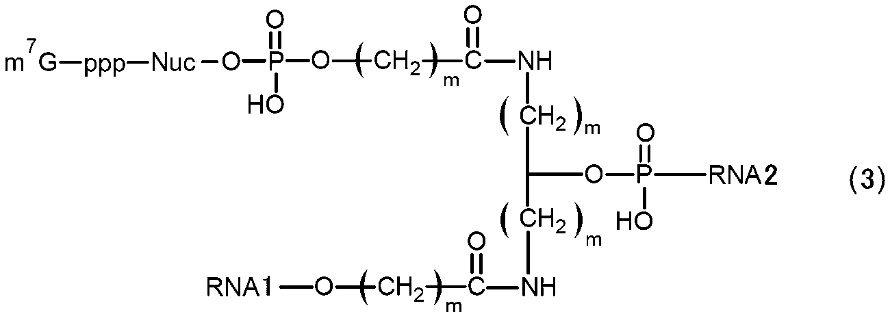
(ここで、m7Gはキャップを示し、pはリン酸基を示す。Nucは天然型又は非天然型のヌクレオチドが1つ以上連結したポリヌクレオチドを示す。mは1~5の整数を示し、複数のmは互いに同一であっても異なっていてもよい。RNA1とRNA2は主鎖を構成するRNAを示し、RNA1は3’末端側で、RNA2は5’末端側で、それぞれ分岐構造の部分と連結されている。式(3)のRNAは、RNA1の5’末端とRNA2の3’末端が連結して環状構造を形成した環状RNAでもよく、連結せずに直鎖状RNAでもよい。) [7] The capped RNA of [2] above, wherein in (b) above, it has a structure represented by the following formula (3).
(Here, m 7 G represents a cap, p represents a phosphate group, Nuc represents a polynucleotide in which one or more natural or non-natural nucleotides are linked, and m represents an integer of 1 to 5. , a plurality of m may be the same or different from each other.RNA1 and RNA2 represent RNAs constituting the main chain, RNA1 on the 3′-end side and RNA2 on the 5′-end side, each having a branched structure. The RNA of formula (3) may be a circular RNA in which the 5' end of RNA1 and the 3' end of RNA2 are linked to form a circular structure, or may be a linear RNA without linking. .)
[8]前記(c)において、下記式(1)又は式(2)で示される構造を有することを特徴とする上記[2]に記載のキャップ化RNA。

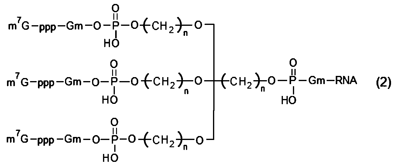
(ここで、m7Gはキャップを示し、pはリン酸基を示し、Gmはグアニンヌクレオチドの2’-水酸基がメチル化された構造を示し、mは1~5の整数を示し、複数のmは互いに同一であっても異なっていてもよく、nは1~5の整数を示し、複数のnは互いに同一であっても異なっていてもよい。) [8] The capped RNA of [2] above, wherein in (c) above, it has a structure represented by the following formula (1) or formula (2).
(Here, m 7 G represents a cap, p represents a phosphate group, Gm represents a structure in which the 2′-hydroxyl group of a guanine nucleotide is methylated, m represents an integer of 1 to 5, and multiple m may be the same or different, n represents an integer of 1 to 5, and multiple n may be the same or different.)
[9]前記式(1)が下記式(1-1)で示される構造を有し、前記式(2)が下記式(2-1)で示される構造を有することを特徴とする上記[8]に記載のキャップ化RNA。
[9] The above [ 8].
[10]前記RNAが、複数の翻訳領域を有することを特徴とする上記[1]に記載のキャップ化RNA。
[10] The capped RNA of [1] above, wherein the RNA has a plurality of translational regions.
[11]上記[1]に記載のキャップ化RNAの製造方法であって、下記(a)~(c)のいずれか少なくとも1つの工程を有することを特徴とするキャップ化RNAの製造方法
(a)前記上流領域の一部又は全部の配列と相補的な配列を有し、かつキャップ構造を有するキャップ化プローブを前記上流領域にハイブリダイズさせる、又は、
(b)前記上流領域の一部に前記RNAを構成する主鎖から分岐する分岐鎖を有する前記RNAを合成したのち、前記分岐鎖に前記キャップ構造を結合させる、
(c)前記RNAが直鎖状RNAであり、前記上流領域の主鎖の5’末端が2以上に枝分かれするブランチ構造を有するRNAを合成したのち、各枝分かれに前記キャップ構造を結合させる。 [11] The method for producing capped RNA according to [1] above, which comprises at least one of the steps (a) to (c) below (a ) hybridizing a capped probe having a sequence complementary to a part or all of the upstream region and having a cap structure to the upstream region, or
(b) after synthesizing the RNA having a branched chain branching from the main chain constituting the RNA in a part of the upstream region, binding the cap structure to the branched chain;
(c) The RNA is a linear RNA, and after synthesizing an RNA having a branch structure in which the 5′ end of the main chain of the upstream region is branched into two or more branches, the cap structure is bound to each branch.
[12]上記[1]に記載のキャップ化RNAと、該キャップ化RNAを鋳型として前記翻訳領域がコードする前記タンパク質を発現可能な真核細胞の発現系と、を備えることを特徴とするタンパク質の製造装置。
[12] A protein comprising the capped RNA of [1] above and a eukaryotic cell expression system capable of expressing the protein encoded by the translation region using the capped RNA as a template. manufacturing equipment.
[13]上記[1]に記載のキャップ化RNAを、該キャップ化RNAを鋳型として前記翻訳領域がコードする前記タンパク質を発現可能な真核細胞の発現系に添加して、前記タンパク質を発現させることを特徴とするタンパク質の製造方法。
[13] The capped RNA according to [1] above is added to a eukaryotic cell expression system capable of expressing the protein encoded by the translational region using the capped RNA as a template to express the protein. A method for producing a protein, characterized by:
[14]RNAの5’末端にキャップ構造を有する目的RNAの検出装置であって、
開始コドンから始まり標識タンパク質をコードする翻訳領域と、前記開始コドンの上流側の上流領域であって検出対象となる前記目的RNAの配列の一部又は全部と相補的な配列を有する上流領域と、を有する環状RNAからなる検出用プローブと、
前記目的RNAを含む試料に前記環状RNAを添加して前記環状RNAの前記上流領域に前記目的RNAをハイブリダイズさせてキャップ化RNAを生成させる手段と、
前記キャップ化RNAを添加することで前記標識タンパク質を発現可能な真核細胞の発現系と、
前記発現系で発現した前記標識タンパク質を検出する手段と、を備えることを特徴とする目的RNAの検出装置。 [14] A target RNA detection device having a cap structure at the 5' end of RNA,
a translation region starting from an initiation codon and encoding a labeling protein, an upstream region on the upstream side of the initiation codon and having a sequence complementary to part or all of the sequence of the target RNA to be detected, a detection probe consisting of a circular RNA having
means for adding the circular RNA to a sample containing the target RNA and hybridizing the target RNA to the upstream region of the circular RNA to generate capped RNA;
a eukaryotic cell expression system capable of expressing the labeled protein by adding the capped RNA;
and means for detecting the labeled protein expressed in the expression system.
[15]RNAの5’末端にキャップ構造を有する目的RNAの検出方法であって、
開始コドンから始まり標識タンパク質をコードする翻訳領域と、前記開始コドンの上流側の上流領域であって検出対象となる前記目的RNAの配列の一部又は全部と相補的な配列を有する上流領域と、を有する環状RNAからなる検出用プローブを用意する工程と、
前記目的RNAを含む試料に前記環状RNAを添加して前記環状RNAの前記上流領域に前記目的RNAをハイブリダイズさせてキャップ化RNAを生成させる工程と、
前記標識タンパク質を発現可能な真核細胞の発現系に該キャップ化RNAを添加して前記標識タンパク質を発現させる工程と、
前記発現系で発現した前記標識タンパク質を検出する工程と、を備えることを特徴とする目的RNAの検出方法。 [15] A method for detecting a target RNA having a cap structure at the 5' end of the RNA, comprising:
a translation region starting from an initiation codon and encoding a labeling protein, an upstream region on the upstream side of the initiation codon and having a sequence complementary to part or all of the sequence of the target RNA to be detected, A step of preparing a detection probe consisting of a circular RNA having
adding the circular RNA to a sample containing the target RNA and hybridizing the target RNA to the upstream region of the circular RNA to generate capped RNA;
adding the capped RNA to a eukaryotic cell expression system capable of expressing the labeled protein to express the labeled protein;
and detecting the labeled protein expressed in the expression system.
[1(Pre)]開始コドンから始まりタンパク質をコードする翻訳領域と、前記開始コドンの上流側の非翻訳領域と、を有するRNAの前記非翻訳領域にキャップ構造を有するキャップ化RNAであって、下記(a)~(c)のいずれか少なくとも1つの構造を有することを特徴とするキャップ化RNA
(a)前記非翻訳領域の一部又は全部の配列と相補的な配列を有し、かつキャップ構造を有するキャップ化プローブが前記非翻訳領域にハイブリダイズされている、
(b)前記非翻訳領域の一部に前記RNAを構成する主鎖の途中から分岐する分岐鎖が設けられており、該分岐鎖に前記キャップ構造が設けられている、
(c)前記非翻訳領域の主鎖の5’末端が2以上に枝分かれするブランチ構造を有し、かつ各枝分かれに前記キャップ構造が設けられている。 [1(Pre)] A capped RNA having a cap structure in the untranslated region of RNA having a protein-encoding translation region starting from the initiation codon and an untranslated region upstream of the initiation codon, Capped RNA characterized by having at least one structure of any one of (a) to (c) below
(a) a capped probe having a sequence complementary to part or all of the untranslated region and having a cap structure is hybridized to the untranslated region;
(b) a part of the untranslated region is provided with a branched chain branching from the middle of the main chain constituting the RNA, and the branched chain is provided with the cap structure;
(c) The 5'-end of the main chain of the untranslated region has a branch structure in which two or more branches are provided, and each branch is provided with the cap structure.
[9(Pre)]上記[1(Pre)]に記載のキャップ化RNAの製造方法であって、下記(a)~(c)のいずれか少なくとも1つの工程を有することを特徴とするキャップ化RNAの製造方法
(a)前記非翻訳領域の一部又は全部の配列と相補的な配列を有し、かつキャップ構造を有するキャップ化プローブを前記非翻訳領域にハイブリダイズさせる、又は、
(b)前記非翻訳領域の一部に前記RNAを構成する主鎖から分岐する分岐鎖を有する前記RNAを合成したのち、前記分岐鎖に前記キャップ構造を結合させる、
(c)前記非翻訳領域の主鎖の5’末端が2以上に枝分かれするブランチ構造を有するRNAを合成したのち、各枝分かれに前記キャップ構造を結合させる。 [9(Pre)] The capped RNA production method of [1(Pre)] above, comprising at least one of the following steps (a) to (c): A method for producing RNA (a) hybridizing a capped probe having a sequence complementary to part or all of the untranslated region and having a cap structure to the untranslated region, or
(b) synthesizing the RNA having a branched chain branching from the main chain constituting the RNA in a part of the untranslated region, and then binding the cap structure to the branched chain;
(c) After synthesizing an RNA having a branch structure in which the 5′ end of the main chain of the untranslated region is branched into two or more branches, the cap structure is bound to each branch.
[12(Pre)]RNAの5’末端にキャップ構造を有する目的RNAの検出装置であって、
開始コドンから始まり標識タンパク質をコードする翻訳領域と、前記開始コドンの上流側の非翻訳領域であって検出対象となる前記目的RNAの配列の一部又は全部と相補的な配列を有する非翻訳領域と、を有する環状RNAからなるプローブと、
前記目的RNAを含む試料に前記環状RNAを添加して前記環状RNAの前記非翻訳領域に前記目的RNAをハイブリダイズさせてキャップ化RNAを生成させる手段と、
前記キャップ化RNAを添加することで前記標識タンパク質を発現可能な真核細胞の発現系と、
前記発現系で発現した前記標識タンパク質を検出する手段と、を備えることを特徴とする目的RNAの検出装置。 A device for detecting target RNA having a cap structure at the 5′ end of [12(Pre)]RNA,
A translation region starting from an initiation codon and encoding a labeling protein, and an untranslated region upstream of the initiation codon and having a sequence complementary to part or all of the sequence of the target RNA to be detected. and a probe consisting of a circular RNA having
means for adding the circular RNA to a sample containing the target RNA and hybridizing the target RNA to the untranslated region of the circular RNA to generate capped RNA;
a eukaryotic cell expression system capable of expressing the labeled protein by adding the capped RNA;
and means for detecting the labeled protein expressed in the expression system.
[13(Pre)]RNAの5’末端にキャップ構造を有する目的RNAの検出方法であって、
開始コドンから始まり標識タンパク質をコードする翻訳領域と、前記開始コドンの上流側の非翻訳領域であって検出対象となる前記目的RNAの配列の一部又は全部と相補的な配列を有する非翻訳領域と、を有する環状RNAからなるプローブを用意する工程と、
前記目的RNAを含む試料に前記環状RNAを添加して前記環状RNAの前記非翻訳領域に前記目的RNAをハイブリダイズさせてキャップ化RNAを生成させる工程と、
前記標識タンパク質を発現可能な真核細胞の発現系に該キャップ化RNAを添加して前記標識タンパク質を発現させる工程と、
前記発現系で発現した前記標識タンパク質を検出する工程と、を備えることを特徴とする目的RNAの検出方法。 A method for detecting a target RNA having a cap structure at the 5' end of [13(Pre)]RNA, comprising:
A translation region starting from an initiation codon and encoding a labeling protein, and an untranslated region upstream of the initiation codon and having a sequence complementary to part or all of the sequence of the target RNA to be detected. and a step of preparing a probe consisting of a circular RNA having
adding the circular RNA to a sample containing the target RNA and hybridizing the target RNA to the untranslated region of the circular RNA to generate capped RNA;
adding the capped RNA to a eukaryotic cell expression system capable of expressing the labeled protein to express the labeled protein;
and detecting the labeled protein expressed in the expression system.
本発明によれば、従来にない様々なタイプの構造を有し翻訳活性が高いキャップ化RNA及びその製造方法を提供することができる。また、本発明の他の目的は、このようなキャップ化RNAを用いてタンパク質を合成可能なタンパク質の製造装置及びタンパク質の製造方法を提供することができる。
According to the present invention, it is possible to provide capped RNAs having various types of structures that have never existed and high translation activity, and methods for producing the same. Another object of the present invention is to provide a protein production apparatus and a protein production method capable of synthesizing a protein using such capped RNA.
本発明のキャップ化RNAは、開始コドン(例えば、AUG)から始まりタンパク質をコードする翻訳領域と、この開始コドンの上流側(5’末端側)の上流領域とを有する。この上流領域は、非翻訳領域(5’UTR)であってもよく、他のオープンリーディングフレーム(ORF)の読み枠内であってもよい。そして、このキャップ化RNAは、上流領域にキャップ構造を有する分岐部を有する。ここで、「キャップ構造を有する分岐部」とは、キャップ構造が主鎖のRNAから分岐するあらゆる形態が含まれる。これは、例えば、キャップ構造を有する核酸(DNA、RNAなど)などの分子が、主鎖のRNAにハイブリダイズしたもの、主鎖を構成するヌクレオチドが枝分かれしたもの、主鎖を構成するヌクレオチドに共有結合したものなどが含まれる。なお、環状RNAの場合、3’UTRやpolyAも上流領域に含まれるが、分岐部は開始コドンに近い5’UTRやuORFに位置するのが好ましい。
The capped RNA of the present invention has a translation region that starts from an initiation codon (eg, AUG) and encodes a protein, and an upstream region (5' terminal side) upstream of this initiation codon. This upstream region may be an untranslated region (5'UTR) or may be in frame with another open reading frame (ORF). This capped RNA has a branch with a cap structure in the upstream region. Here, the “branched portion having a cap structure” includes all forms in which the cap structure is branched from the main chain RNA. For example, molecules such as nucleic acids (DNA, RNA, etc.) having a cap structure are hybridized to RNA of the main chain, branched nucleotides constituting the main chain, and Including combined ones. In the case of circular RNA, 3'UTR and polyA are also included in the upstream region, but the branched portion is preferably located at 5'UTR or uORF near the initiation codon.
具体的には、キャップ化RNAは、下記(a)~(c)のいずれか少なくとも1つの構造を有する。
(a)上流領域の一部又は全部の配列と相補的な配列を有し、かつキャップ構造を有するキャップ化プローブが上流領域にハイブリダイズされている(以下、「ハイブリダイズタイプ」)。
(b)上流領域の一部にRNAを構成する主鎖の途中から分岐する分岐鎖が設けられており、該分岐鎖にキャップ構造が設けられている構造(以下、「分岐鎖タイプ」)。
(c)前記上流領域の主鎖の5’末端が2以上に枝分かれするブランチ構造を有し、かつ各枝分かれに前記キャップ構造が設けられている構造(以下、「マルチキャップタイプ」)。
なお、本発明のキャップ化RNAには、天然型のRNA(すなわち、ATP、GTP、CTP、UTPからなるヌクレオチドが連結したポリヌクレオチド)だけでなく、RNAのヌクレオチドにmC4リンカー、2’OMe、LNAなどが導入された非天然核酸鎖(修飾核酸鎖)なども含まれる。以下、各タイプのキャップ化RNAについて説明する。 Specifically, the capped RNA has at least one structure of any one of (a) to (c) below.
(a) A capped probe having a sequence complementary to the sequence of part or all of the upstream region and having a cap structure is hybridized to the upstream region (hereinafter, "hybridized type").
(b) A structure in which a part of the upstream region is provided with a branched chain branching from the middle of the main chain constituting RNA, and the branched chain is provided with a cap structure (hereinafter, "branched chain type").
(c) A structure in which the 5'-end of the main chain of the upstream region has a branch structure in which two or more branches are provided, and each branch is provided with the cap structure (hereinafter referred to as "multi-cap type").
The capped RNA of the present invention includes not only natural RNA (that is, a polynucleotide in which nucleotides consisting of ATP, GTP, CTP, and UTP are linked), but also mC4 linkers, 2'OMe, and LNA in the nucleotides of RNA. Non-natural nucleic acid strands (modified nucleic acid strands) into which such as have been introduced are also included. Each type of capped RNA is described below.
1.ハイブリダイズタイプ
以下、ハイブリダイズタイプのキャップ化RNAについて説明する。図1の(B)に、直鎖状RNAにおけるハイブリダイズタイプのキャップ化RNAを、(C)に環状RNAにおけるハイブリダイズタイプのキャップ化RNAを、それぞれ実施例に記載された構造で示している。直鎖状RNA、環状RNAのいずれにおいても、翻訳対象であるタンパク質がコードされたRNA(以下、「コードRNA」)の上流領域(例えば非翻訳領域(5’UTR))に対して、キャップ構造を有するRNAであるキャップ化プローブがハイブリダイズされている。 1. Hybridized type The hybridized type capped RNA will be described below. In FIG. 1, (B) shows the capped RNA hybridized in the linear RNA, and (C) shows the capped RNA hybridized in the circular RNA, each with the structure described in the Examples. . In both linear RNA and circular RNA, a cap structure is attached to the upstream region (for example, untranslated region (5'UTR)) of RNA encoding a protein to be translated (hereinafter referred to as "coding RNA"). A capped probe that is an RNA having a is hybridized.
以下、ハイブリダイズタイプのキャップ化RNAについて説明する。図1の(B)に、直鎖状RNAにおけるハイブリダイズタイプのキャップ化RNAを、(C)に環状RNAにおけるハイブリダイズタイプのキャップ化RNAを、それぞれ実施例に記載された構造で示している。直鎖状RNA、環状RNAのいずれにおいても、翻訳対象であるタンパク質がコードされたRNA(以下、「コードRNA」)の上流領域(例えば非翻訳領域(5’UTR))に対して、キャップ構造を有するRNAであるキャップ化プローブがハイブリダイズされている。 1. Hybridized type The hybridized type capped RNA will be described below. In FIG. 1, (B) shows the capped RNA hybridized in the linear RNA, and (C) shows the capped RNA hybridized in the circular RNA, each with the structure described in the Examples. . In both linear RNA and circular RNA, a cap structure is attached to the upstream region (for example, untranslated region (5'UTR)) of RNA encoding a protein to be translated (hereinafter referred to as "coding RNA"). A capped probe that is an RNA having a is hybridized.
コードRNAは、開始コドンから始まりタンパク質をコードする翻訳領域(図では「Nano Luc」の領域)と、開始コドンの上流(5’末端側)の上流領域(例えば、非翻訳領域(「5’UTR」の領域))を有している。本実施形態では、翻訳領域の3’末端には停止コドンがあり、この停止コドンの下流(3’末端側)には非翻訳領域(3’UTR)を有する場合がある。さらに、3’UTRにはポリA配列が含まれている。コードRNAは直鎖状RNAでは直鎖状であり、環状RNAでは直鎖状RNAの5’末端と3’末端が連結した環状構造となっている。なお、本実施形態では、コードRNAには停止コドンを含む実施態様を示しているが、これに限定されず、例えば、特許文献2や非特許文献1のように、停止コドンを有さないものとしてもよい。この停止コドンを含まないケースでは、これら先行技術に開示されているように、全長の塩基数を102以上の3の倍数とした環状コードRNAを環状RNAとすれば、回転式タンパク質翻訳により高い翻訳効率で目的タンパク質を生成することができる。
The coding RNA consists of a translation region (“Nano Luc” region in the figure) that starts from the start codon and encodes a protein, and an upstream region (5′ end side) upstream of the start codon (for example, an untranslated region (“5′UTR ” area)). In this embodiment, there is a stop codon at the 3' end of the translation region, and there may be an untranslated region (3'UTR) downstream (on the 3' end side) of this stop codon. Additionally, the 3'UTR contains a poly A sequence. The coding RNA is linear in linear RNA, and has a circular structure in which the 5'-end and 3'-end of the linear RNA are linked together in circular RNA. Although this embodiment shows an embodiment in which the code RNA contains a stop codon, it is not limited to this. may be In the case where this stop codon is not included, as disclosed in these prior arts, if a circular RNA having a full-length base number of 102 or more and a multiple of 3 is used as a circular RNA, rotational protein translation results in high translation. The target protein can be produced efficiently.
コードRNAは、翻訳領域が1つだけのものや、2つ以上のもののいずれであってもよい。翻訳領域が2つ以上ある場合、各翻訳領域の上流にそれぞれ上流領域を有しており、キャップ化プローブはいずれかの上流領域にハイブリダイズするものを使用することができる。この場合、その上流領域の直下の開始コドンから始まる翻訳領域のタンパク質の翻訳活性が向上する。また、コードRNAの主鎖の5’末端に通常のキャップ構造を有するものであってもよい。5’末端に通常のキャップ構造を有する場合、キャップ化プローブのキャップ構造との併用により、キャップ化RNAの翻訳効率がより向上する。
The coding RNA may have only one translational region or may have two or more translational regions. When there are two or more translational regions, each translational region has an upstream region, and a capping probe that hybridizes to any of the upstream regions can be used. In this case, the translational activity of the protein in the translational region starting from the initiation codon immediately below the upstream region is enhanced. Moreover, it may have a normal cap structure at the 5' end of the main chain of the coding RNA. When the capped probe has a normal cap structure at the 5' end, the combined use of the capped probe with the cap structure further improves the translation efficiency of the capped RNA.
キャップ化プローブは、コードRNAよりも短鎖のRNAで構成され、コードRNAの5’UTRの一部又は全部の配列と相補的な塩基配列を有する。コードRNAの5’UTRにおけるキャップ化プローブがハイブリダイズする位置は、特に制限はないが、Kozak配列を有する場合はその上流側が好ましい。キャップ化プローブの長さは特に制限はないが、10~50塩基数(nt)程度の長さが好ましい。また、キャップ化プローブは、5’末端にキャップが結合していてもよく、3’末端にキャップが結合していてもよい。さらに、キャップ化プローブは、3’末端側にポリA配列を有していてもよい。ポリA配列の長さ(塩基数)は、特に制限はないが、10~50塩基数程度が好ましい。キャップ化プローブは、リボースの2’炭素と4’炭素が架橋されたLocked Nucleic Acid(LNA)や、C4リンカーなどを有していてもよい。
A capped probe is composed of an RNA that is shorter than the coding RNA, and has a base sequence complementary to the sequence of part or all of the 5'UTR of the coding RNA. The position at which the capped probe hybridizes in the 5'UTR of the coding RNA is not particularly limited, but the upstream side is preferred if it has a Kozak sequence. The length of the capped probe is not particularly limited, but a length of about 10 to 50 bases (nt) is preferred. In addition, the capped probe may have a cap bound to the 5' end or a cap bound to the 3' end. Additionally, the capped probe may have a poly A sequence at the 3' end. The length (number of bases) of the poly A sequence is not particularly limited, but is preferably about 10 to 50 bases. The capped probe may have a Locked Nucleic Acid (LNA) in which the 2' and 4' carbons of ribose are bridged, a C4 linker, and the like.
キャップ化RNAでは、キャップ化プローブは、コードRNAの5’UTRにハイブリダイズしている。ここで、「ハイブリダイズ」とは、ストリンジェントな条件でハイブリダイズすることを意味する。この「ストリンジェントな条件」とは、特異的なハイブリッドが形成され、非特異的なハイブリッドがほとんど形成されない条件をいう。また、「ストリンジェントな条件でハイブリダイズ」するRNAとしては、コードRNAの5’UTRの所定の範囲の配列と完全に相補的な配列に対して、一定以上の相同性を有するRNAが挙げられる。この場合の相同性としては、例えばBLASTアルゴリズムにおいて、通常60%以上であり、好ましくは80%以上、より好ましくは90%以上、特に好ましくは95%以上、最も好ましくは100%である。
In capped RNA, the capped probe hybridizes to the 5'UTR of the coding RNA. Here, "hybridize" means to hybridize under stringent conditions. The term "stringent conditions" refers to conditions under which specific hybrids are formed and non-specific hybrids are rarely formed. RNAs that "hybridize under stringent conditions" also include RNAs that have a certain degree or more of homology to a sequence that is completely complementary to a predetermined range of the 5'UTR sequence of the coding RNA. . The homology in this case is usually 60% or more, preferably 80% or more, more preferably 90% or more, particularly preferably 95% or more, and most preferably 100%, according to, for example, the BLAST algorithm.
次に、ハイブリダイズタイプのキャップ化RNAの製造方法について説明する。キャップ化RNAを構成するコードRNAは、目的とする配列を有するRNAに対して相補的な配列を有するDNAを用い、これをRNAポリメラーゼで転写することで作製することができる。あるいは、コードRNAは、DNA合成装置などを用いて化学的に合成してもよい。環状のコードRNAは、直鎖状のコードRNAを作製したのち、T4 RNAリガーゼ2などを用いて末端どうしを連結して環化することで作製することができる。
Next, a method for producing hybridized capped RNA will be described. The coding RNA that constitutes the capped RNA can be produced by using a DNA having a sequence complementary to the RNA having the desired sequence and transcribe it with an RNA polymerase. Alternatively, the coding RNA may be chemically synthesized using a DNA synthesizer or the like. Circular coding RNA can be prepared by preparing a linear coding RNA and then ligating the ends using T4 RNA ligase 2 or the like to circularize it.
プローブRNAは、以下の方法で作製することができる。まず、ベースとなるキャップ化されていないRNAを、ホスホロアミダイト、リン酸化試薬、2’-OMe RNAアミダイト、LNAアミダイト、C4リンカーアミダイト、CPGカラムなどを使用し、RNA固相合成法などで作製する。次に、このRNAに、m7GDP、2-ニトロイミダゾールなどを反応させてリン酸化RNAのキャップ化を行う。この反応の詳細は、特許文献1を参照することができる。
Probe RNA can be produced by the following method. First, base uncapped RNA is prepared by RNA solid-phase synthesis method using phosphoramidite, phosphorylation reagent, 2'-OMe RNA amidite, LNA amidite, C4 linker amidite, CPG column, etc. do. Next, this RNA is reacted with m 7 GDP, 2-nitroimidazole or the like to cap the phosphorylated RNA. For details of this reaction, reference can be made to Patent Document 1.
キャップ化RNAは、コードRNAとプローブRNAをアニーリングさせることで作製することができる。アニーリングの際のコードRNAとプローブRNAの比率(モル比)は、翻訳活性の観点から、1:1~1:20の範囲内が好ましく、1:5~1:15の範囲内がより好ましい。アニーリングの温度は、80℃以上、好ましくは90℃以上とし、反応時間は1~30分、好ましくは3~10分程度とすることができる。
Capped RNA can be produced by annealing code RNA and probe RNA. The ratio (molar ratio) of coding RNA to probe RNA during annealing is preferably in the range of 1:1 to 1:20, more preferably in the range of 1:5 to 1:15, from the viewpoint of translation activity. The annealing temperature can be 80° C. or higher, preferably 90° C. or higher, and the reaction time can be about 1 to 30 minutes, preferably 3 to 10 minutes.
ハイブリダイズタイプのキャップ化RNAは、コードRNAが直鎖状、環状であるかに問わず、5’UTRの任意の位置にキャップ構造を付加できる。キャップ構造を有するRNAでは、キャップ構造を有していないRNAと比較して、リボソームを構成する翻訳開始因子がコードRNAに結合する速度が向上し、その結果、翻訳活性が向上する。又はハイブリダイズタイプのキャップ化RNAでは、環状RNAあるいは直鎖RNAにおいて、プローブによるハイブリダイゼーションによりキャップを提示することで、翻訳を増強あるいは任意の位置から翻訳現象を誘導することを可能にする。
A hybridizing-type capped RNA can add a cap structure to any position of the 5'UTR, regardless of whether the coding RNA is linear or circular. RNA with a cap structure increases the rate at which a translation initiation factor that constitutes a ribosome binds to coding RNA, resulting in improved translational activity, compared to RNA without a cap structure. Alternatively, in hybrid type capped RNA, it is possible to enhance translation or induce a translation phenomenon from an arbitrary position by presenting a cap by hybridization with a probe in circular RNA or linear RNA.
さらに、直鎖状RNAにおいては、キャップ化プローブを用いることで、本来タンパク質合成の読み枠として用いられない、例えば2番目以降の開始コドンから翻訳反応を誘導できる。これにより、キャップ化プローブにより任意のRNAの位置から翻訳を誘導できる。
Furthermore, in linear RNA, by using a capped probe, translation reaction can be induced from, for example, the second and subsequent initiation codons, which are not originally used as reading frames for protein synthesis. This allows translation to be induced from any RNA location by a capped probe.
また、環状RNAについては、これまでキャップ構造を有するものは知られていなかったが、本発明のハイブリダイズタイプのキャップ化RNAでは、環状RNAにおいてもキャップ構造を導入することができる。これにより、キャップ構造を有していない環状RNAと比較して、翻訳活性を大幅に向上させることができる。
In addition, although circular RNAs having a cap structure have not been known so far, the capped structure of the hybridized type capped RNA of the present invention can be introduced into circular RNAs as well. As a result, translational activity can be greatly improved compared to circular RNAs that do not have a cap structure.
2.分岐鎖タイプ
以下、分岐鎖タイプのキャップ化RNAについて説明する。図2の(a)に、直鎖状RNAにおける分岐鎖タイプのキャップ化RNAを、(b)に環状RNAにおける分岐鎖タイプのキャップ化RNAを、それぞれ実施例に記載された構造で示す。直鎖状RNA、環状RNAのいずれにおいても、コードRNAの上流領域は、コードRNAを構成する主鎖から分岐する分岐鎖を有しており、この分岐鎖にキャップ構造が設けられている。 2. Branched Chain Type The branched chain type capped RNA will be described below. FIG. 2(a) shows a branched-chain capped RNA in a linear RNA, and FIG. 2(b) shows a branched-chain capped RNA in a circular RNA, each with the structure described in the Examples. In both linear RNA and circular RNA, the upstream region of the coding RNA has a branched chain branched from the main chain constituting the coding RNA, and the branched chain is provided with a cap structure.
以下、分岐鎖タイプのキャップ化RNAについて説明する。図2の(a)に、直鎖状RNAにおける分岐鎖タイプのキャップ化RNAを、(b)に環状RNAにおける分岐鎖タイプのキャップ化RNAを、それぞれ実施例に記載された構造で示す。直鎖状RNA、環状RNAのいずれにおいても、コードRNAの上流領域は、コードRNAを構成する主鎖から分岐する分岐鎖を有しており、この分岐鎖にキャップ構造が設けられている。 2. Branched Chain Type The branched chain type capped RNA will be described below. FIG. 2(a) shows a branched-chain capped RNA in a linear RNA, and FIG. 2(b) shows a branched-chain capped RNA in a circular RNA, each with the structure described in the Examples. In both linear RNA and circular RNA, the upstream region of the coding RNA has a branched chain branched from the main chain constituting the coding RNA, and the branched chain is provided with a cap structure.
コードRNAの全体的な構成は、上記の「1.ハイブリダイズタイプ」で説明したとおりである。分岐鎖タイプでは、コードRNAの5’UTRの一部が主鎖から分岐する分岐鎖を有しており、その分岐鎖の5’末端にキャップ構造を有している。コードRNAの主鎖の5’末端は、直鎖状RNAではリン酸化されている。また、コードRNAは、翻訳領域が1つだけのものや、2つ以上のもののいずれであってもよい。コードRNAが直鎖状の場合、5’末端に通常のキャップ構造を有していてもよい。
The overall structure of the code RNA is as described in "1. Hybridization type" above. In the branched chain type, a portion of the 5'UTR of the coding RNA has a branched chain branching off from the main chain, and the branched chain has a cap structure at the 5' end. The 5' end of the backbone of the coding RNA is phosphorylated in linear RNA. Also, the coding RNA may have either one translation region or two or more translation regions. When the coding RNA is linear, it may have a normal cap structure at the 5' end.
分岐鎖タイプのRNAとしては以下の式(3)を例示することができる。

(ここで、m7Gはキャップを示し、pはリン酸基を示す。Nucは天然型又は非天然型のヌクレオチドが1つ以上連結したポリヌクレオチドを示す。mは1~5の整数を示し、複数のmは互いに同一であっても異なっていてもよい。RNA1とRNA2は主鎖を構成するRNAを示し、RNA1は3’末端側で、RNA2は5’末端側で、それぞれ分岐構造の部分と連結されている。式(3)のRNAは、RNA1の5’末端とRNA2の3’末端が連結して環状構造を形成した環状RNAでもよく、連結せずに直鎖状RNAでもよい。) The following formula (3) can be exemplified as branched chain type RNA.
(Here, m 7 G represents a cap, p represents a phosphate group, Nuc represents a polynucleotide in which one or more natural or non-natural nucleotides are linked, and m represents an integer of 1 to 5. , a plurality of m may be the same or different from each other.RNA1 and RNA2 represent RNAs constituting the main chain, RNA1 on the 3′-end side, RNA2 on the 5′-end side, each having a branched structure. The RNA of formula (3) may be a circular RNA in which the 5' end of RNA1 and the 3' end of RNA2 are linked to form a circular structure, or may be a linear RNA without linking. .)
次に、分岐鎖タイプのキャップ化RNAの製造方法について説明する。5’UTRに分岐鎖を有するコードRNAは、RNA合成に使用される通常のホスホロアミダイト試薬と、分岐鎖を有するホスホロアミダイト試薬と、化学リン酸化試薬と、を使用し、核酸自動合成機を用いて合成することができる。分岐鎖を有するホスホロアミダイトとしては、後述する実施例で使用している1-[5-(4,4’-ジメトキシトリチルオキシ)ペンチルアミド]-3-[5-レブリニルオキシペンチルアミド]-プロピル-2-[(2-シアノエチル)-(N,N-ジイソプロピル)]-ホスホロアミダイトなどを挙げることができる。また、化学リン酸化試薬としては、後述する実施例の化合物1(下記式(3))を挙げることができる。

Next, a method for producing branched-chain capped RNA will be described. A coding RNA having a branched chain in the 5′UTR is obtained by using a normal phosphoramidite reagent used in RNA synthesis, a phosphoramidite reagent having a branched chain, and a chemical phosphorylation reagent, and using an automatic nucleic acid synthesizer. can be synthesized using As the phosphoramidite having a branched chain, 1-[5-(4,4'-dimethoxytrityloxy)pentylamide]-3-[5-levulinyloxypentylamide] used in Examples described later. -propyl-2-[(2-cyanoethyl)-(N,N-diisopropyl)]-phosphoramidite and the like. Further, as a chemical phosphorylation reagent, compound 1 (formula (3) below) of Examples to be described later can be exemplified.
通常のホスホロアミダイト試薬や装置を用いて、コードRNAの5’UTRの配列を上流側から合成し、分岐鎖を導入したい位置において分岐鎖を有するホスホロアミダイト試薬を使用することで、所望の位置に分岐鎖を導入することができる。コードRNAの分岐鎖の5’末端は、化学リン酸化試薬により疎水性保護基が導入されているため、この疎水性保護基を光照射などで分解して分岐鎖の5’末端をリン酸基とする。そして、7-メチルグアノシン 5’-二リン酸イミダゾリド塩(Im-m7GDP)、2-ニトロイミダゾールなどのヘテロ芳香族化合物を用いて分岐鎖の5’末端へキャップを付加する。最後に、T4ポリヌクレオチドキナーゼなどを用いてコードRNAの主鎖の5’末端をリン酸化する。これにより、直鎖状のキャップ化RNAが得られる。環状のコードRNAは、直鎖状のキャップ化RNAを作製したのち、T4 RNAリガーゼ2などを用いて末端どうしを連結して環化することで作製することができる。
By synthesizing the sequence of the 5'UTR of the coding RNA from the upstream side using ordinary phosphoramidite reagents and devices, and using a phosphoramidite reagent having a branched chain at the position where the branched chain is to be introduced, the desired sequence can be obtained. Branching can be introduced at the position. Since the 5' end of the branched chain of the coding RNA has a hydrophobic protective group introduced by a chemical phosphorylation reagent, this hydrophobic protective group is decomposed by light irradiation etc. to remove the 5' end of the branched chain as a phosphate group. and The 5' end of the branched chain is then capped using a heteroaromatic compound such as 7-methylguanosine 5'-diphosphate imidazolide salt (Im-m 7 GDP), 2-nitroimidazole, or the like. Finally, the 5' end of the backbone of the coding RNA is phosphorylated using, for example, T4 polynucleotide kinase. This results in linear capped RNA. Circular coding RNA can be produced by producing a linear capped RNA and then ligating the ends using T4 RNA ligase 2 or the like to circularize it.
分岐鎖タイプのキャップ化RNAは、ハイブリダイズタイプのキャップ化RNAと同様に、コードRNAが直鎖状、環状であるかに問わず、5’UTRの任意の位置にキャップ構造を付加できる。コードRNAが2以上の翻訳領域を有する場合、目的とする翻訳領域の上流にある5’UTRに分岐鎖キャップを導入することで、その翻訳領域の翻訳活性を向上させることができる。これにより、例えばタンパク質合成の読み枠として用いられていない開始コドンから翻訳反応を誘導することができる。また、分岐鎖タイプのキャップ化RNAでは、従来はキャップ構造を有していない環状RNAにおいてもキャップ構造を導入することができる。これにより、キャップ構造を有していない環状RNAと比較して、翻訳活性を大幅に向上させることができる。
As with hybridizing-type capped RNA, branched-chain capped RNA can add a cap structure to any position of the 5'UTR regardless of whether the coding RNA is linear or circular. If the coding RNA has two or more translational regions, the translational activity of that translational region can be improved by introducing a branched cap into the 5'UTR upstream of the desired translational region. As a result, translation reaction can be induced from initiation codons that are not used as reading frames for protein synthesis, for example. In branched-chain capped RNA, a cap structure can also be introduced into a circular RNA that does not conventionally have a cap structure. As a result, translational activity can be greatly improved compared to circular RNAs that do not have a cap structure.
分岐鎖タイプのキャップ化RNAは、後述する実施例に示すように、直鎖状よりも環状の方が、翻訳活性が高く、免疫原性が低い傾向がある。
上流領域におけるキャップの位置は、開始コドンに近いほど翻訳活性が高くなる傾向がある。上流領域における分岐の位置(すなわち、式(3)におけるRNA1とRNA2の間の構造の位置)は、特に制限はないが、分岐の先にキャップ構造が設けられいることから、分岐の位置は開始コドンに近いほど翻訳活性が高くなる傾向がある。分岐の位置は、開始コドンを基準に5’側の1~50ntの範囲内が好ましく、1~30ntの範囲内がより好ましく、1~15ntの範囲内が特に好ましい。
さらに、分岐鎖の分岐した部分の鎖長(すなわち、式(3)におけるNucのポリヌクレオチドの長さ)は、特に制限はないが、長すぎても短すぎても翻訳活性が低くなる傾向がある。ここで、分岐鎖の鎖長は、分岐箇所から5’キャップの間と定義する。分岐鎖の鎖長は、1~20ntの範囲内が好ましく、3~10ntの範囲内がより好ましく、5~8ntの範囲内が特に好ましい。鎖長が短すぎると、キャップ構造の位置と分岐の部分の距離が短くなり、分岐部分の構造によって翻訳開始因子複合体の形成やリボソームの結合などが阻害されやすくなり、通常のRNAと比較して分岐型のRNAでは翻訳活性が低くなると考えられる。 Branched-chain capped RNA tends to have higher translational activity and lower immunogenicity when it is circular than when it is linear, as shown in Examples described later.
The closer the position of the cap in the upstream region to the initiation codon, the higher the translation activity tends to be. The position of the branch in the upstream region (that is, the position of the structure between RNA1 and RNA2 in formula (3)) is not particularly limited, but since a cap structure is provided at the end of the branch, the position of the branch is the start There is a tendency that the closer to the codon, the higher the translation activity. The position of the branch is preferably within the range of 1 to 50 nt, more preferably within the range of 1 to 30 nt, particularly preferably within the range of 1 to 15 nt on the 5' side of the start codon.
Furthermore, the chain length of the branched portion of the branched chain (that is, the length of the polynucleotide of Nuc in formula (3)) is not particularly limited, but translation activity tends to decrease if it is too long or too short. be. Here, the chain length of the branched chain is defined as between the branch point and the 5' cap. The branched chain length is preferably in the range of 1 to 20 nt, more preferably in the range of 3 to 10 nt, and particularly preferably in the range of 5 to 8 nt. If the chain length is too short, the distance between the position of the cap structure and the branched portion becomes short, and the structure of the branched portion tends to inhibit the formation of the translation initiation factor complex and the binding of the ribosome. It is thought that branched RNAs have lower translational activity.
3.マルチキャップタイプ
以下、マルチキャップタイプのキャップ化RNAについて説明する。マルチタイプキャップのキャップ化RNAは、直鎖状コードRNAの5’UTRの5’末端が2以上に枝分かれするブランチ構造を有しており、各枝分かれにキャップ構造が設けられている。 3. Multi-cap type The multi-cap type capped RNA will be described below. The capped RNA of the multitype cap has a branch structure in which the 5' end of the 5'UTR of the linear coding RNA is branched into two or more branches, and each branch is provided with a cap structure.
以下、マルチキャップタイプのキャップ化RNAについて説明する。マルチタイプキャップのキャップ化RNAは、直鎖状コードRNAの5’UTRの5’末端が2以上に枝分かれするブランチ構造を有しており、各枝分かれにキャップ構造が設けられている。 3. Multi-cap type The multi-cap type capped RNA will be described below. The capped RNA of the multitype cap has a branch structure in which the 5' end of the 5'UTR of the linear coding RNA is branched into two or more branches, and each branch is provided with a cap structure.
マルチキャップタイプのキャップ化RNAの例として、下記式(1)で示される2分岐タイプか、式(2)で示される3分岐タイプの構造を挙げることができる。


(ここで、m7Gはキャップを示し、pはリン酸基を示し、Gmはグアニンヌクレオチドの2’-水酸基がメチル化された構造を示し、mは1~5の整数を示し、複数のmは互いに同一であっても異なっていてもよく、nは1~5の整数を示し、複数のnは互いに同一であっても異なっていてもよい。) Examples of multi-cap type capped RNA include biantennary type structures represented by the following formula (1) and triantennary type structures represented by formula (2).
(Here, m 7 G represents a cap, p represents a phosphate group, Gm represents a structure in which the 2′-hydroxyl group of a guanine nucleotide is methylated, m represents an integer of 1 to 5, and multiple m may be the same or different, n represents an integer of 1 to 5, and multiple n may be the same or different.)
この場合において、上記式(1)が下記式(1-1)で示される構造を有し、上記式(2)が下記式(2-1)で示される構造を有することが好ましい。
In this case, it is preferable that the above formula (1) has a structure represented by the following formula (1-1) and the above formula (2) has a structure represented by the following formula (2-1).
次に、マルチキャップタイプのキャップ化RNAの製造方法について説明する。キャップ化前のRNAは、RNA合成に使用される通常のホスホロアミダイト試薬と、分岐鎖を有するホスホロアミダイト試薬と、化学リン酸化試薬と、を使用し、核酸自動合成機を用いて合成することができる。分岐鎖を有するホスホロアミダイトとしては、後述する実施例で使用している1,3-ビス-[5-(4,4’-ジメトキシトリチルオキシ)ペンチルアミド]プロピル-2-[(2-シアノエチル)-(N,N-ジイソプロピル)]- ホスホロアミダイト(2分岐タイプ)、トリス-2,2,2-[3-(4,4’-ジメトキシトリチルオキシ)プロピルオキシメチル]エチル-[(2-シアノエチル)-(N,N-ジイソプロピル)]-ホスホロアミダイト(3分岐タイプ)などを挙げることができる。また、化学リン酸化試薬としては、後述する実施例の化合物1(式(3))を挙げることができる。
Next, a method for producing multi-cap type capped RNA will be described. Pre-capped RNA is synthesized using a conventional phosphoramidite reagent, a branched-chain phosphoramidite reagent, and a chemical phosphorylation reagent used in RNA synthesis, using an automated nucleic acid synthesizer. be able to. As the phosphoramidite having a branched chain, 1,3-bis-[5-(4,4'-dimethoxytrityloxy)pentylamido]propyl-2-[(2-cyanoethyl )-(N,N-diisopropyl)]-phosphoramidite (2-branched type), tris-2,2,2-[3-(4,4'-dimethoxytrityloxy)propyloxymethyl]ethyl-[(2 -cyanoethyl)-(N,N-diisopropyl)]-phosphoramidite (tri-branched type). Further, as a chemical phosphorylating reagent, compound 1 (formula (3)) of Examples described later can be mentioned.
分岐鎖を有するホスホロアミダイト試薬を使用して、5’末端にブランチ構造を形成し、通常のホスホロアミダイト試薬や装置を用いて、コードRNAの5’UTRの配列を上流側から合成することで、5’末端に複数の枝分かれを有するコードRNAを合成する。コードRNAの各枝分かれの5’末端は、化学リン酸化試薬により疎水性保護基を導入しておき、この疎水性保護基を光照射などで分解して分岐鎖の5’末端をリン酸基とする。そして、7-メチルグアノシン 5’-二リン酸イミダゾリド塩(Im-m7GDP)、2-ニトロイミダゾールなどのヘテロ芳香族化合物を用いてブランチ構造の各枝分かれの5’末端へキャップを付加する。これにより、それぞれの枝分かれにキャップが結合した直鎖状のキャップ化RNAが得られる。
Using a phosphoramidite reagent having a branched chain to form a branch structure at the 5′ end, and using a conventional phosphoramidite reagent and equipment to synthesize the sequence of the 5′UTR of the coding RNA from the upstream side. to synthesize a coding RNA with multiple branches at the 5' end. Hydrophobic protective groups are introduced into the 5' ends of each branch of the coding RNA using a chemical phosphorylation reagent, and the hydrophobic protective groups are decomposed by light irradiation or the like to convert the 5' ends of the branched chains into phosphate groups. do. Then, a heteroaromatic compound such as 7-methylguanosine 5′-diphosphate imidazolide salt (Im-m 7 GDP), 2-nitroimidazole or the like is used to cap the 5′ end of each branch of the branch structure. This yields a linear capped RNA with a cap attached to each branch.
マルチキャップタイプのキャップ化RNAは主鎖の5’末端に複数のキャップを有しているため、キャップが1つの通常のキャップ化RNAと比較して、リボソームを構成する翻訳開始因子がキャップと結合しやすくなり、マルチリガンド効果により翻訳活性を大幅に向上させることができる。
Since multicap-type capped RNA has multiple caps at the 5′ end of the main chain, the translation initiation factor that constitutes the ribosome binds to the cap compared to normal capped RNA with one cap. The multiligand effect can greatly improve the translation activity.
4.タンパク質の製造装置及び製造方法
本発明のタンパク質の製造装置は、上記のキャップ化RNAと、このキャップ化RNAを鋳型として翻訳領域がコードするタンパク質を発現可能な真核細胞の発現系と、を備える。本発明のタンパク質の製造方法では、キャップ化RNAを真核細胞の発現系に添加してタンパク質を発現させる。真核細胞としては、酵母や糸状菌等の真菌類、植物細胞、昆虫細胞、人などの動物細胞を挙げることができ、具体的には、例えば、マウスL-細胞、エールリッヒ腹水癌細胞、HeLa細胞、CHO細胞、出芽酵母などを例示することができる。真核細胞の発現系は、細胞発現系、無細胞発現系のいずれであってもよい。細胞発現系へのキャップ化RNAの導入は、リン酸カルシウム法、DEAEデキストラン法、リポフェクション法、エレクトロポレーション法などの方法で行うことができる。細胞発現系では、細胞へのキャップ化RNAの導入後、適切な温度などの条件で細胞を培養し、培養後に細胞を溶解させたのち、目的とするタンパク質を分離精製する。無細胞発現系では、真核細胞の溶解物を使用することができる。 4. Protein Production Apparatus and Production Method The protein production apparatus of the present invention comprises the above capped RNA and a eukaryotic cell expression system capable of expressing a protein encoded by a translation region using the capped RNA as a template. . In the method for producing the protein of the present invention, capped RNA is added to a eukaryotic cell expression system to express the protein. Eukaryotic cells include fungi such as yeast and filamentous fungi, plant cells, insect cells, and animal cells such as humans. Specific examples include mouse L-cells, Ehrlich ascites cancer cells, HeLa Cells, CHO cells, Saccharomyces cerevisiae, etc. can be exemplified. The eukaryotic cell expression system may be either a cell expression system or a cell-free expression system. Introduction of capped RNA into a cell expression system can be performed by methods such as the calcium phosphate method, DEAE dextran method, lipofection method, and electroporation method. In the cell expression system, after the capped RNA is introduced into the cells, the cells are cultured under conditions such as an appropriate temperature, the cells are lysed after the culture, and the target protein is separated and purified. Eukaryotic cell lysates can be used in cell-free expression systems.
本発明のタンパク質の製造装置は、上記のキャップ化RNAと、このキャップ化RNAを鋳型として翻訳領域がコードするタンパク質を発現可能な真核細胞の発現系と、を備える。本発明のタンパク質の製造方法では、キャップ化RNAを真核細胞の発現系に添加してタンパク質を発現させる。真核細胞としては、酵母や糸状菌等の真菌類、植物細胞、昆虫細胞、人などの動物細胞を挙げることができ、具体的には、例えば、マウスL-細胞、エールリッヒ腹水癌細胞、HeLa細胞、CHO細胞、出芽酵母などを例示することができる。真核細胞の発現系は、細胞発現系、無細胞発現系のいずれであってもよい。細胞発現系へのキャップ化RNAの導入は、リン酸カルシウム法、DEAEデキストラン法、リポフェクション法、エレクトロポレーション法などの方法で行うことができる。細胞発現系では、細胞へのキャップ化RNAの導入後、適切な温度などの条件で細胞を培養し、培養後に細胞を溶解させたのち、目的とするタンパク質を分離精製する。無細胞発現系では、真核細胞の溶解物を使用することができる。 4. Protein Production Apparatus and Production Method The protein production apparatus of the present invention comprises the above capped RNA and a eukaryotic cell expression system capable of expressing a protein encoded by a translation region using the capped RNA as a template. . In the method for producing the protein of the present invention, capped RNA is added to a eukaryotic cell expression system to express the protein. Eukaryotic cells include fungi such as yeast and filamentous fungi, plant cells, insect cells, and animal cells such as humans. Specific examples include mouse L-cells, Ehrlich ascites cancer cells, HeLa Cells, CHO cells, Saccharomyces cerevisiae, etc. can be exemplified. The eukaryotic cell expression system may be either a cell expression system or a cell-free expression system. Introduction of capped RNA into a cell expression system can be performed by methods such as the calcium phosphate method, DEAE dextran method, lipofection method, and electroporation method. In the cell expression system, after the capped RNA is introduced into the cells, the cells are cultured under conditions such as an appropriate temperature, the cells are lysed after the culture, and the target protein is separated and purified. Eukaryotic cell lysates can be used in cell-free expression systems.
5.目的RNAの検出装置及び検出方法
上述したように、環状RNAはキャップ構造を有していないため、単独では翻訳されないが、キャップ構造を有するRNAをハイブリダイズさせることで、環状RNAにコードされたタンパク質を翻訳させることができる。この性質を利用して、環状RNAを検出用プローブとして、目的RNAを検出することができる。目的RNAとしては、真核生物やウイルスなどを由来とするRNAを挙げることができる。以下、本発明の目的RNAの検出装置及び検出方法について説明する。 5. Target RNA Detection Apparatus and Detection Method As described above, since circular RNA does not have a cap structure, it cannot be translated by itself. can be translated. This property can be used to detect target RNA using circular RNA as a detection probe. Target RNAs include RNAs derived from eukaryotes, viruses, and the like. The apparatus and method for detecting target RNA of the present invention will be described below.
上述したように、環状RNAはキャップ構造を有していないため、単独では翻訳されないが、キャップ構造を有するRNAをハイブリダイズさせることで、環状RNAにコードされたタンパク質を翻訳させることができる。この性質を利用して、環状RNAを検出用プローブとして、目的RNAを検出することができる。目的RNAとしては、真核生物やウイルスなどを由来とするRNAを挙げることができる。以下、本発明の目的RNAの検出装置及び検出方法について説明する。 5. Target RNA Detection Apparatus and Detection Method As described above, since circular RNA does not have a cap structure, it cannot be translated by itself. can be translated. This property can be used to detect target RNA using circular RNA as a detection probe. Target RNAs include RNAs derived from eukaryotes, viruses, and the like. The apparatus and method for detecting target RNA of the present invention will be described below.
本発明の目的RNAの検出装置は、RNAの5’末端にキャップ構造を有する目的RNAを検出する装置である。本検出装置は、環状RNAからなる検出用プローブ、キャップ化RNAを生成させる手段、真核細胞の発現系、標識タンパク質を検出する手段を備える。
The device for detecting target RNA of the present invention is a device for detecting target RNA having a cap structure at the 5' end of the RNA. This detection apparatus comprises a detection probe composed of circular RNA, means for generating capped RNA, an expression system for eukaryotic cells, and means for detecting a labeled protein.
検出用プローブは、開始コドンから始まり標識タンパク質をコードする翻訳領域と、この開始コドンの上流側の上流領域と、を有している。この上流領域は、検出対象となる目的RNAの配列の一部又は全部と相補的な配列を有している。標識タンパク質としては、後述する実施例で使用しているルシフェラーゼや緑色蛍光タンパク質(GFP)などの蛍光標識タンパク質、西洋わさびペルオキシダーゼ(HRP)やアルカリフォスファターゼ(AP)などの酵素標識タンパク質などを挙げることができる。
The detection probe has a translation region starting from the start codon and encoding a labeled protein, and an upstream region upstream of this start codon. This upstream region has a sequence complementary to part or all of the target RNA sequence to be detected. Examples of labeling proteins include fluorescence-labeling proteins such as luciferase and green fluorescent protein (GFP), enzyme-labeling proteins such as horseradish peroxidase (HRP) and alkaline phosphatase (AP), which are used in Examples described later. can.
キャップ化RNAを生成させる手段は、上記の検出用プローブ(環状RNA)と目的RNAとをハイブリダイズさせる手段である。これには、RNAをアニーリングさせる通常の試薬や装置を使用することができる。このような試薬としては、Tri-HClなどの緩衝液を挙げることができる。また、装置としては、試薬を80℃以上に加熱したのちに室温まで冷却するヒートブロック又はサーマルサイクラーなどの装置を挙げることができる。
真核細胞の発現系は、上記で生成したキャップ化RNAを鋳型として翻訳領域がコードする前記標識タンパク質を発現可能な系であり、上述したHeLa細胞などを挙げることができる。 The means for generating capped RNA is means for hybridizing the detection probe (circular RNA) and the target RNA. Common reagents and equipment for annealing RNA can be used for this. Such reagents include buffers such as Tri-HCl. Examples of the device include a heat block or a thermal cycler, which heats the reagent to 80° C. or higher and then cools it to room temperature.
The expression system of eukaryotic cells is a system capable of expressing the marker protein encoded by the translational region using the capped RNA produced above as a template, and the above-mentioned HeLa cells and the like can be mentioned.
標識タンパク質を検出する手段としては、標識タンパク質の種類によって種々の装置を使用することができる。このような手段としては、標識タンパク質が蛍光標識タンパク質の場合は蛍光測定装置を挙げることができ、酵素標識タンパク質の場合は発光基質や蛍光測定装置などを挙げることができる。
Various devices can be used as means for detecting the labeled protein, depending on the type of labeled protein. Examples of such means include a fluorometer when the labeled protein is a fluorescently labeled protein, and a luminescent substrate and a fluorometer when the labeled protein is an enzyme-labeled protein.
本発明の目的RNAの検出方法は、上記の検出装置を使用して実施することができる。まず、上記の環状RNAを含む検出用プローブを用意し、目的RNAを含む試料に添加してキャップ化RNAを生成させる。次に、標識タンパク質を発現可能な真核細胞の発現系にキャップ化RNAを添加して標識タンパク質を発現させ、その後、発現した標識タンパク質を検出する。このような方法により、目的RNAを効率的に検出することができる。
The target RNA detection method of the present invention can be implemented using the above-described detection device. First, a detection probe containing the circular RNA is prepared and added to a sample containing the target RNA to generate capped RNA. Next, the capped RNA is added to a eukaryotic cell expression system capable of expressing the labeled protein to express the labeled protein, and then the expressed labeled protein is detected. A target RNA can be efficiently detected by such a method.
以下、本発明を実施例に基づいて具体的に説明するが、これらは本発明の目的を限定するものではない。また、以下の実施例において「%」表示は特に規定しない限り質量基準(質量パ-セント)である。
The present invention will be specifically described below based on examples, but these are not intended to limit the purpose of the present invention. In the following examples, "%" is based on mass (mass percent) unless otherwise specified.
1.キャップ化RNA(ハイブリダイズタイプ)
以下では、ハイブリダイゼーションタイプのキャップ化RNAに関する実験を示す。図3は、本実験で使用した環状RNAとキャップ化プローブの塩基配列などを示す図である。図の上部左側の図がキャップ化RNAの構造を示している。環状RNA(コードRNA)として、5’側の上流領域(図では非翻訳領域(「5’UTR」と表記))、その下流側にKozak配列(「Kozak」と表記)を有しており、Nano Lucルシフェラーゼをコードする領域(「Nano Luc」)と、停止コドン(「stop」)、3’側の非翻訳領域(「3’UTR」)、polyA配列(「polyA30」)を有している。また、図のキャップ化プローブは、5’末端にキャップ構造(m7G)を有し、環状RNAの5’UTRの一部の領域と相補的な配列を有し、3’末端のpolyA30を有している。そして、キャップ化RNAは、キャップ化プローブが環状RNAの5’UTRにハイブリダイズしている。 1. capped RNA (hybridized type)
Experiments with capped RNA of the hybridization type are presented below. FIG. 3 shows the base sequences of circular RNA and capped probes used in this experiment. The diagram on the upper left side of the figure shows the structure of the capped RNA. As a circular RNA (coding RNA), it has a 5′ upstream region (untranslated region (denoted as “5′UTR”) in the figure) and a Kozak sequence (denoted as “Kozak”) downstream thereof, Nano Luc luciferase-encoding region (“Nano Luc”), a stop codon (“stop”), a 3′-side untranslated region (“3′UTR”), and a polyA sequence (“polyA 30 ”) there is In addition, the capped probe in the figure has a cap structure (m 7 G) at the 5' end, has a sequence complementary to a partial region of the 5'UTR of the circular RNA, and has polyA30 at the 3' end. have. The capped RNA then has the capped probe hybridized to the 5'UTR of the circular RNA.
図の上部右側は本実施例で使用したキャップ化プローブの配列を示している。キャップ化プローブは、5’末端にキャップ構造を有する「5’ cap probe」、3’末端にキャップ構造を有する「3’ cap probe」、5’末端にキャップ構造を有し3’末端にポリAを有する「5’ cap probe with polyA」の3種類を使用した。図の下部は環状RNAの配列を示している。「ggcg・・・cgc」で表される太字の領域が、キャップ化プローブがハイブリダイゼーションする部分である。
The upper right side of the figure shows the sequence of the capped probe used in this example. The capped probes are "5' cap probe" having a cap structure at the 5' end, "3' cap probe" having a cap structure at the 3' end, and "3' cap probe" having a cap structure at the 5' end and poly A Three types of "5' cap probe with poly A" were used. The lower part of the figure shows the sequence of the circular RNA. The bold-faced region represented by “ggcg...cgc” is the portion where the capped probe hybridizes.
(1)プローブRNA合成
RNA合成は0.2μmolスケールで行い、日本テクノサービス株式会社 NTS M-2-MX DNA/RNA合成機又はNRs-4A10R7 DNA/RNA合成機を使用した。RNA固相合成に用いたホスホロアミダイトには、rA(5’-DMT-2’-TOM-ribo-Adenosine(n-acetyl) OP),rC(5’-DMT-2’-TOM-ribo-Cytidine(n-acetyl) OP),rG(5’-DMT-2’-TOM-ribo-Guanosine(n-acetyl)OP),U(5’-DMT-2’-TOM-ribo-Uridine OP)(ChemGenes)を使用した。リン酸化試薬には、5’phosphate-ON Reagent(ChemGenes)を使用した。2’-OMe RNAアミダイトには、2’-OMe-C-CE-phosphoramidite、2’-OMe-G(iBu)-CE-phosphoramidite(ChemGenes)を使用した。LNAアミダイトには、MBN-Ganosine(N,N-DMF)-phosphoramidite(ChemGenes)、MBN-5’-Methyl-Cytidine(N-bz)-phosphoramidite(ChemGenes)を使用した。C4リンカーアミダイトは、既報(Angew. Chem. Int. Ed. 2011, 50, 8922-8926)に従って合成したものを用いた。CPGは3’-TOM-ribo Cytidine(n-acetyl)2’-lcaa CPG 1000Å,3’-TOM-ribo Adenosine(n-acetyl)2’-lcaa CPG 1000Å(ChemGenes)、3’-phosphate ON lcaa CPG 1000Aを使用した。 (1) Synthesis of Probe RNA RNA was synthesized at a scale of 0.2 μmol using Nippon Techno Service Co., Ltd. NTS M-2-MX DNA/RNA synthesizer or NRs-4A10R7 DNA/RNA synthesizer. Phosphoramidites used for RNA solid-phase synthesis include rA (5'-DMT-2'-TOM-ribo-Adenosine (n-acetyl) OP), rC (5'-DMT-2'-TOM-ribo- Cytidine (n-acetyl) OP), rG (5'-DMT-2'-TOM-ribo-Guanosine (n-acetyl) OP), U (5'-DMT-2'-TOM-ribo-Uridine OP) ( ChemGenes) was used. 5' Phosphate-ON Reagent (ChemGenes) was used as a phosphorylation reagent. 2'-OMe-C-CE-phosphoramidite and 2'-OMe-G(iBu)-CE-phosphoramidite (ChemGenes) were used as 2'-OMe RNA amidites. MBN-Ganosine (N,N-DMF)-phosphoramidite (ChemGenes) and MBN-5′-Methyl-Cytidine (N-bz)-phosphoramidite (ChemGenes) were used as LNA amidites. The C4 linker amidite used was synthesized according to a previous report (Angew. Chem. Int. Ed. 2011, 50, 8922-8926). CPG is 3' -Tom -RIBO CYTIDINE (N -ACETYL) 2' -LCAA CPG 1000å, 3' -TOM -RIBO ADENOSINE (N -ACETYL) 2' -LCAA CPG 100å (CHEMGENES), 3' -PHOSPH ATE ON LCAA CPG 1000A was used.
RNA合成は0.2μmolスケールで行い、日本テクノサービス株式会社 NTS M-2-MX DNA/RNA合成機又はNRs-4A10R7 DNA/RNA合成機を使用した。RNA固相合成に用いたホスホロアミダイトには、rA(5’-DMT-2’-TOM-ribo-Adenosine(n-acetyl) OP),rC(5’-DMT-2’-TOM-ribo-Cytidine(n-acetyl) OP),rG(5’-DMT-2’-TOM-ribo-Guanosine(n-acetyl)OP),U(5’-DMT-2’-TOM-ribo-Uridine OP)(ChemGenes)を使用した。リン酸化試薬には、5’phosphate-ON Reagent(ChemGenes)を使用した。2’-OMe RNAアミダイトには、2’-OMe-C-CE-phosphoramidite、2’-OMe-G(iBu)-CE-phosphoramidite(ChemGenes)を使用した。LNAアミダイトには、MBN-Ganosine(N,N-DMF)-phosphoramidite(ChemGenes)、MBN-5’-Methyl-Cytidine(N-bz)-phosphoramidite(ChemGenes)を使用した。C4リンカーアミダイトは、既報(Angew. Chem. Int. Ed. 2011, 50, 8922-8926)に従って合成したものを用いた。CPGは3’-TOM-ribo Cytidine(n-acetyl)2’-lcaa CPG 1000Å,3’-TOM-ribo Adenosine(n-acetyl)2’-lcaa CPG 1000Å(ChemGenes)、3’-phosphate ON lcaa CPG 1000Aを使用した。 (1) Synthesis of Probe RNA RNA was synthesized at a scale of 0.2 μmol using Nippon Techno Service Co., Ltd. NTS M-2-MX DNA/RNA synthesizer or NRs-4A10R7 DNA/RNA synthesizer. Phosphoramidites used for RNA solid-phase synthesis include rA (5'-DMT-2'-TOM-ribo-Adenosine (n-acetyl) OP), rC (5'-DMT-2'-TOM-ribo- Cytidine (n-acetyl) OP), rG (5'-DMT-2'-TOM-ribo-Guanosine (n-acetyl) OP), U (5'-DMT-2'-TOM-ribo-Uridine OP) ( ChemGenes) was used. 5' Phosphate-ON Reagent (ChemGenes) was used as a phosphorylation reagent. 2'-OMe-C-CE-phosphoramidite and 2'-OMe-G(iBu)-CE-phosphoramidite (ChemGenes) were used as 2'-OMe RNA amidites. MBN-Ganosine (N,N-DMF)-phosphoramidite (ChemGenes) and MBN-5′-Methyl-Cytidine (N-bz)-phosphoramidite (ChemGenes) were used as LNA amidites. The C4 linker amidite used was synthesized according to a previous report (Angew. Chem. Int. Ed. 2011, 50, 8922-8926). CPG is 3' -Tom -RIBO CYTIDINE (N -ACETYL) 2' -LCAA CPG 1000å, 3' -TOM -RIBO ADENOSINE (N -ACETYL) 2' -LCAA CPG 100å (CHEMGENES), 3' -PHOSPH ATE ON LCAA CPG 1000A was used.
RNAの合成後、CPGを1.5mLループ付きスクリューチューブ(ザルスタット)へ移し、40% aq. methylamine/28% aq. ammonia(1:1)1.0mLを加えて65℃で20分インキュベートした。Millex LH 0.45μmフィルターを用いてろ過し、滅菌水(500μL)で洗い込み、遠心エバポレーターで乾固させた。1M TBAF in THF 1.0mLを加えて溶解させ、室温に一晩静置した。1M Tris-HCl緩衝液(pH7.5)1.0mLを加え、遠心エバポレーターで有機溶媒を留去した。NAP-25カラム(GE Healthcare)を用いて脱塩を行った。逆相HPLCによりRNAを精製した。
After RNA synthesis, the CPG was transferred to a 1.5 mL looped screw tube (Sarstedt) and 40% aq. Methylamine/28% aq. 1.0 mL of ammonia (1:1) was added and incubated at 65°C for 20 minutes. It was filtered using a Millex LH 0.45 μm filter, washed with sterilized water (500 μL), and dried with a centrifugal evaporator. 1.0 mL of 1 M TBAF in THF was added and dissolved, and allowed to stand overnight at room temperature. 1.0 mL of 1M Tris-HCl buffer (pH 7.5) was added, and the organic solvent was distilled off with a centrifugal evaporator. Desalting was performed using a NAP-25 column (GE Healthcare). RNA was purified by reverse-phase HPLC.
HPLC(HITACHI)条件;カラム:hydrosphere C18 column(4.6x250mm;YMC)、移動相:A)50mM TEAA緩衝液(pH7.0),5%アセトニトリル、B)アセトニトリル、グラジエント:0-20min B conc.0% to 20%、流速:1.0mL/min
HPLC (Hitachi) conditions; column: hydrosphere C18 column (4.6 x 250 mm; YMC), mobile phase: A) 50 mM TEAA buffer (pH 7.0), 5% acetonitrile, B) acetonitrile, gradient: 0-20 min B conc. 0% to 20%, flow rate: 1.0 mL/min
15%変性ポリアクリルアミドゲル電気泳動(PAGE)(アクリルアミド:ビスアクリルアミド=19:1(wt/wt),7.5M尿素)にて合成したRNAを分析した。PAGEには、TBE 緩衝液 (89mM Tris-borate,2mM EDTA,pH8.3)を用いた。LC/MSに分析した。LC/MS(agilent)条件;ACQUITY UPLC BEH C18 Column,0.3mL/min,A;8.6mM TEA/100mM HFIP solution, B; MeOH, 0-30%/20min
The synthesized RNA was analyzed by 15% denaturing polyacrylamide gel electrophoresis (PAGE) (acrylamide:bisacrylamide=19:1 (wt/wt), 7.5M urea). TBE buffer (89 mM Tris-borate, 2 mM EDTA, pH 8.3) was used for PAGE. Analyzed by LC/MS. LC/MS (Agilent) conditions; ACQUITY UPLC BEH C18 Column, 0.3 mL/min, A; 8.6 mM TEA/100 mM HFIP solution, B; MeOH, 0-30%/20 min
合成したRNAの配列や分子量などの情報を下記表に示す(RNA1~12はそれぞれ配列番号1~12に対応)。

Information such as the sequences and molecular weights of the synthesized RNAs is shown in the table below (RNAs 1 to 12 correspond to SEQ ID NOS: 1 to 12, respectively).
(2)リン酸化RNAのキャップ化反応
1.5mLチューブに、それぞれ終濃度20μM RNA,10mM CaCl2となるように混合し、凍結乾燥により乾固させた後、DMSOに溶解した。そこへDMSOに溶解したIm(m7GDP)・Na(Hongene Biotech)を終濃度10mM、2-ニトロイミダゾールを終濃度10mM となるように加えた。その反応液を55℃で3時間インキュベートした。反応液を滅菌水により2倍希釈した後に、イソプロパノール沈殿を行った。HPLC精製し、LC/MSに分析した。 (2) Phosphorylated RNA Capping Reaction In a 1.5 mL tube, 20 μM RNA and 10 mM CaCl 2 were mixed at final concentrations, lyophilized to dryness, and then dissolved in DMSO. Im(m 7 GDP)·Na (Hongene Biotech) dissolved in DMSO was added thereto to a final concentration of 10 mM, and 2-nitroimidazole was added to a final concentration of 10 mM. The reaction was incubated at 55°C for 3 hours. Isopropanol precipitation was carried out after diluting the reaction solution twice with sterilized water. HPLC purified and analyzed by LC/MS.
1.5mLチューブに、それぞれ終濃度20μM RNA,10mM CaCl2となるように混合し、凍結乾燥により乾固させた後、DMSOに溶解した。そこへDMSOに溶解したIm(m7GDP)・Na(Hongene Biotech)を終濃度10mM、2-ニトロイミダゾールを終濃度10mM となるように加えた。その反応液を55℃で3時間インキュベートした。反応液を滅菌水により2倍希釈した後に、イソプロパノール沈殿を行った。HPLC精製し、LC/MSに分析した。 (2) Phosphorylated RNA Capping Reaction In a 1.5 mL tube, 20 μM RNA and 10 mM CaCl 2 were mixed at final concentrations, lyophilized to dryness, and then dissolved in DMSO. Im(m 7 GDP)·Na (Hongene Biotech) dissolved in DMSO was added thereto to a final concentration of 10 mM, and 2-nitroimidazole was added to a final concentration of 10 mM. The reaction was incubated at 55°C for 3 hours. Isopropanol precipitation was carried out after diluting the reaction solution twice with sterilized water. HPLC purified and analyzed by LC/MS.
HPLC条件;カラム:hydrosphere C18 column(4.6x250mm;YMC)、移動相:A) 50mM TEAA 緩衝液(pH7.0),5%アセトニトリル、B)アセトニトリル、グラジエント:0-20min B conc. 0% to20%、流速:1.0mL/min
LC/MS(agilent)条件; ACQUITY UPLC BEH C18 Column, 0.3mL/min, A; 8.6mM TEA/100mM HFIP solution, B; MeOH,0-30%/20min
HPLC精製したキャプ化RNAは、イソプロパノールと酢酸ナトリウム(pH5.2)を用いて沈殿させた。 HPLC conditions; column: hydrosphere C18 column (4.6×250 mm; YMC), mobile phase: A) 50 mM TEAA buffer (pH 7.0), 5% acetonitrile, B) acetonitrile, gradient: 0-20 min B conc. 0% to 20%, flow rate: 1.0 mL/min
LC/MS (agilent) conditions; ACQUITY UPLC BEH C18 Column, 0.3 mL/min, A; 8.6 mM TEA/100 mM HFIP solution, B; MeOH, 0-30%/20 min
HPLC-purified capped RNA was precipitated with isopropanol and sodium acetate (pH 5.2).
合成したキャップ化RNAの配列や分子量などの情報を下記表に示す(キャップ化RNA1~12はそれぞれ配列番号13~24に対応)。

Information such as the sequences and molecular weights of the synthesized capped RNAs are shown in the table below (capped RNAs 1 to 12 correspond to SEQ ID NOS: 13 to 24, respectively).
図5はキャップ化プローブの合成実験の結果を示す図である。図の(A)は、キャップ化プローブの合成スキームを示している。化学合成したモノリン酸化RNAを塩化カルシウムと共に凍結乾燥した。Im(m7GDP)・Na(化学的キャップ化試薬)と2-ニトロイミダゾールを添加して、DMSO溶液中で反応させた。図の(B)は、キャップ化プローブの結果を示しており、キャップ化反応したRNAをPAGEにより解析して、SYBR Green IIにより染色した。キャップ化反応が進行すると、メチルグアニンが付加するため、一塩基分バンドが上にシフトした。PAGE解析の結果、8~9割程度の効率でキャップ化反応が進行したことがわかる。
FIG. 5 shows the results of capped probe synthesis experiments. (A) of the figure shows a synthetic scheme for capped probes. Chemically synthesized monophosphorylated RNA was lyophilized with calcium chloride. Im(m 7 GDP)·Na (chemical capping reagent) and 2-nitroimidazole were added and allowed to react in DMSO solution. (B) of the figure shows the results of the capped probe, and the capped RNA was analyzed by PAGE and stained with SYBR Green II. As the capping reaction progressed, the band shifted upward by one base due to the addition of methylguanine. As a result of PAGE analysis, it can be seen that the capping reaction proceeded with an efficiency of about 80% to 90%.
(3)転写反応による直鎖状RNAの合成
直鎖状RNAは、二本鎖DNAを鋳型としてT7 RNA polymeraseにより転写合成した。図4の上段はRNAの転写合成のための鋳型DNAの配列(配列番号32)を、下段は転写されたRNAの配列(配列番号31)を示している。鋳型の二本鎖DNAは、pNL-TK1.1(Promega)をPrimeSTAR HS DNA Polymerase(Takara Bio)を用いてPCR反応により増幅することで得た(2.5ng/μL DNA vector, 1× PrimeSTAR buffer,0.2mM dNTPs,0.5μM primers,0.025units/μL polymerase)。 (3) Synthesis of Linear RNA by Transcription Reaction Linear RNA was synthesized by transcription using T7 RNA polymerase using double-stranded DNA as a template. The upper part of FIG. 4 shows the template DNA sequence (SEQ ID NO: 32) for RNA transcription synthesis, and the lower part shows the sequence of the transcribed RNA (SEQ ID NO: 31). The template double-stranded DNA was obtained by amplifying pNL-TK1.1 (Promega) by PCR reaction using PrimeSTAR HS DNA Polymerase (Takara Bio) (2.5 ng/μL DNA vector, 1× PrimeSTAR buffer , 0.2 mM dNTPs, 0.5 μM primers, 0.025 units/μL polymerase).
直鎖状RNAは、二本鎖DNAを鋳型としてT7 RNA polymeraseにより転写合成した。図4の上段はRNAの転写合成のための鋳型DNAの配列(配列番号32)を、下段は転写されたRNAの配列(配列番号31)を示している。鋳型の二本鎖DNAは、pNL-TK1.1(Promega)をPrimeSTAR HS DNA Polymerase(Takara Bio)を用いてPCR反応により増幅することで得た(2.5ng/μL DNA vector, 1× PrimeSTAR buffer,0.2mM dNTPs,0.5μM primers,0.025units/μL polymerase)。 (3) Synthesis of Linear RNA by Transcription Reaction Linear RNA was synthesized by transcription using T7 RNA polymerase using double-stranded DNA as a template. The upper part of FIG. 4 shows the template DNA sequence (SEQ ID NO: 32) for RNA transcription synthesis, and the lower part shows the sequence of the transcribed RNA (SEQ ID NO: 31). The template double-stranded DNA was obtained by amplifying pNL-TK1.1 (Promega) by PCR reaction using PrimeSTAR HS DNA Polymerase (Takara Bio) (2.5 ng/μL DNA vector, 1× PrimeSTAR buffer , 0.2 mM dNTPs, 0.5 μM primers, 0.025 units/μL polymerase).
転写反応液(鋳型DNA 2.5ng/μL, 1×T7 RNA Polymerase buffer(Takara)(40mM Tris-HCl(pH8.0),8mM MgCl2,2mMスペルミジン),2.5U/μL T7 RNA polymerase (Takara),5mM DTT,2mM ATP,2mM UTP,2mM,CTP,0.4mM GTP,2mM GMP)を調製し、37℃で2時間インキュベートした。反応液にRecombinant DNase(Takara)を0.1 U/μLとなるように加え、37℃で30分間インキュベートすることで鋳型DNAを分解した。
反応後、フェノール・クロロホルム抽出を行った。続いて、Amicon Ultra 10K(Millipore)を用いて限外ろ過を行い、アルコール沈殿を行った。 Transcription reaction solution (template DNA 2.5 ng/μL, 1×T7 RNA Polymerase buffer (Takara) (40 mM Tris-HCl (pH 8.0), 8 mM MgCl 2 , 2 mM spermidine), 2.5 U/μL T7 RNA polymerase (Takara ), 5 mM DTT, 2 mM ATP, 2 mM UTP, 2 mM, CTP, 0.4 mM GTP, 2 mM GMP) were prepared and incubated at 37° C. for 2 hours. Recombinant DNase (Takara) was added to the reaction solution to 0.1 U/μL, and the mixture was incubated at 37° C. for 30 minutes to decompose the template DNA.
After the reaction, phenol/chloroform extraction was performed. This was followed by ultrafiltration using an Amicon Ultra 10K (Millipore) and alcohol precipitation.
図6は、直鎖状RNAの合成実験の結果を示す図である。図のA)は、直鎖状RNAの鋳型となる二本鎖DNAのPCR反応による合成の結果を示しており、PCR反応後、アガロースゲルにて分析し、Gel Redで染色したゲルの写真である。図のB)は、直鎖状RNAの転写合成の結果を示しており、転写反応後、PAGE分析し、SYBR Greenで染色したゲルの写真である。
FIG. 6 is a diagram showing the results of a linear RNA synthesis experiment. A) of the figure shows the result of synthesis by PCR reaction of double-stranded DNA that serves as a template for linear RNA. After the PCR reaction, it was analyzed on an agarose gel and is a photograph of the gel stained with Gel Red. be. B) of the figure shows the results of transcription synthesis of linear RNA, and is a photograph of a gel stained with SYBR Green after PAGE analysis after transcription reaction.
(4)T4 RNA ligase2を用いた環化反応
転写合成した直鎖状RNAをT4 RNA ligase 2(New England Biolabs)と20-nt DNA(ガイドDNA)を用いて環化した。0.5μM直鎖状RNAと1.0μMガイドDNAをT4 RNA ligase 2 buffer(50mM Tris-HCl(pH7.5),2mM MgCl2,1mM DTT and 0.4mM ATP)中でアニーリングした後、0.1U/μL T4 RNA ligase 2と10% PEG8000を加えて37℃で1時間インキュベートした。連結生成物の環状RNAと原料の直鎖状RNAは、5%変性PAGEにより単離した。 (4) Circularization reaction using T4 RNA ligase 2 The transcribed and synthesized linear RNA was circularized using T4 RNA ligase 2 (New England Biolabs) and 20-nt DNA (guide DNA). After annealing 0.5 μM linear RNA and 1.0 μM guide DNA in T4 RNA ligase 2 buffer (50 mM Tris-HCl (pH 7.5), 2 mM MgCl 2 , 1 mM DTT and 0.4 mM ATP), 1 U/μL T4 RNA ligase 2 and 10% PEG8000 were added and incubated at 37° C. for 1 hour. The circular RNA of the ligation product and the starting linear RNA were isolated by 5% denaturing PAGE.
転写合成した直鎖状RNAをT4 RNA ligase 2(New England Biolabs)と20-nt DNA(ガイドDNA)を用いて環化した。0.5μM直鎖状RNAと1.0μMガイドDNAをT4 RNA ligase 2 buffer(50mM Tris-HCl(pH7.5),2mM MgCl2,1mM DTT and 0.4mM ATP)中でアニーリングした後、0.1U/μL T4 RNA ligase 2と10% PEG8000を加えて37℃で1時間インキュベートした。連結生成物の環状RNAと原料の直鎖状RNAは、5%変性PAGEにより単離した。 (4) Circularization reaction using T4 RNA ligase 2 The transcribed and synthesized linear RNA was circularized using T4 RNA ligase 2 (New England Biolabs) and 20-nt DNA (guide DNA). After annealing 0.5 μM linear RNA and 1.0 μM guide DNA in T4 RNA ligase 2 buffer (50 mM Tris-HCl (pH 7.5), 2 mM MgCl 2 , 1 mM DTT and 0.4 mM ATP), 1 U/μL T4 RNA ligase 2 and 10% PEG8000 were added and incubated at 37° C. for 1 hour. The circular RNA of the ligation product and the starting linear RNA were isolated by 5% denaturing PAGE.
(5)RNase Rを用いた環化の確認
RNA(0.18μg/μL)をRNase R(1unit/μL;Epicenter)と20mM Tris-HCl(pH8.0)0.1M KCl、0.1mM MgCl2中で37℃、10分インキュベートした。反応液を5%変性PAGEで解析した。 (5) Confirmation of cyclization using RNase R RNA (0.18 µg/µL) was mixed with RNase R (1 unit/µL; Epicenter) and 20 mM Tris-HCl (pH 8.0) 0.1 M KCl, 0.1 mM MgCl 2 Incubated at 37°C for 10 minutes. The reaction was analyzed by 5% denaturing PAGE.
RNA(0.18μg/μL)をRNase R(1unit/μL;Epicenter)と20mM Tris-HCl(pH8.0)0.1M KCl、0.1mM MgCl2中で37℃、10分インキュベートした。反応液を5%変性PAGEで解析した。 (5) Confirmation of cyclization using RNase R RNA (0.18 µg/µL) was mixed with RNase R (1 unit/µL; Epicenter) and 20 mM Tris-HCl (pH 8.0) 0.1 M KCl, 0.1 mM MgCl 2 Incubated at 37°C for 10 minutes. The reaction was analyzed by 5% denaturing PAGE.
図7は、直鎖状RNAの環化実験の結果を示す図である。図のA)は、環状RNAの合成スキームを示している。転写合成した直鎖状RNAをガイドDNAとアニーリングさせて、T4 RNA ligase2を用いて、環状RNAを合成した。図のB)は、環化体の確認結果を示しており、環化反応したRNAにRNase Rを加えてインキュベートした。PAGEにより解析して、SYBR GreenIIにより染色した。転写合成した直鎖状RNAをT4 RNA ligase 2を用いて環化した。環化体の同定は、直鎖状RNAを選択的に分解する酵素(RNase R)を作用させて、PAGE分析することで行った。環化の効率は、6割程度であった。環化体及び原料の直鎖状RNAをゲルから切り出し、単離精製した。
FIG. 7 shows the results of linear RNA circularization experiments. A) of the figure shows the synthesis scheme of circular RNA. The transcribed and synthesized linear RNA was annealed with the guide DNA, and circular RNA was synthesized using T4 RNA ligase2. B) of the figure shows the results of confirmation of the cyclized product, in which RNase R was added to the cyclized RNA and incubated. Analyzed by PAGE and stained with SYBR Green II. The transcribed and synthesized linear RNA was circularized using T4 RNA ligase 2. The cyclized form was identified by PAGE analysis using an enzyme (RNase R) that selectively degrades linear RNA. The efficiency of cyclization was about 60%. The cyclized product and the starting linear RNA were excised from the gel and isolated and purified.
(6)アニーリング
0.2μMキャップ化プローブと0.1μM直鎖状又は0.1μM環状RNAを緩衝液(10mM Tris-HCl(pH7.5),50mM NaCl)中でアニーリングした(90℃、3分)。 (6) Annealing 0.2 μM capped probe and 0.1 μM linear or 0.1 μM circular RNA were annealed in buffer (10 mM Tris-HCl (pH 7.5), 50 mM NaCl) (90° C., 3 minutes ).
0.2μMキャップ化プローブと0.1μM直鎖状又は0.1μM環状RNAを緩衝液(10mM Tris-HCl(pH7.5),50mM NaCl)中でアニーリングした(90℃、3分)。 (6) Annealing 0.2 μM capped probe and 0.1 μM linear or 0.1 μM circular RNA were annealed in buffer (10 mM Tris-HCl (pH 7.5), 50 mM NaCl) (90° C., 3 minutes ).
(7)細胞内における翻訳活性の評価
HeLa細胞(RIKEN Cell Bank)は10% fetal bovine serum(FBS; Invitrogen)を添加したDulbecco’s modified Eagle’s medium(DMEM; WAKO)中で培養した(37℃、5%CO2)。トランスフェクション前日に、96wellプレートにHeLa細胞を播種した(1.0×10⁴cell/well)。翌日培地を除去し、100 μL/well Opti-MEM(登録商標)(Thermo Fisher SCIENTIFIC)に置換した。0.15μL Lipofectamine(登録商標) MessengerMAX(登録商標)、20ng mRNAを10μLOpti-MEM(登録商標)に希釈して細胞へ導入した。37℃、5時間インキュベートした後、20μL/well 1×Cell Lysis Buffer(Promega)を加えて細胞を溶解した。Nano-Glo(登録商標) luciferase assay(登録商標)(Promega)を用いて発光測定した。 (7) Evaluation of intracellular translation activity HeLa cells (RIKEN Cell Bank) were cultured in Dulbecco's modified Eagle's medium (DMEM; WAKO) supplemented with 10% fetal bovine serum (FBS; Invitrogen) (37 °C, 5% CO2 ). The day before transfection, HeLa cells were seeded in a 96-well plate (1.0×10 4 cells/well). The next day, the medium was removed and replaced with 100 μL/well Opti-MEM® (Thermo Fisher SCIENTIFIC). 0.15 μL Lipofectamine® MessengerMAX®, 20 ng mRNA was diluted in 10 μL Opti-MEM® and introduced into cells. After incubation at 37° C. for 5 hours, 20 μL/well 1×Cell Lysis Buffer (Promega) was added to lyse the cells. Luminescence was measured using the Nano-Glo® luciferase assay® (Promega).
HeLa細胞(RIKEN Cell Bank)は10% fetal bovine serum(FBS; Invitrogen)を添加したDulbecco’s modified Eagle’s medium(DMEM; WAKO)中で培養した(37℃、5%CO2)。トランスフェクション前日に、96wellプレートにHeLa細胞を播種した(1.0×10⁴cell/well)。翌日培地を除去し、100 μL/well Opti-MEM(登録商標)(Thermo Fisher SCIENTIFIC)に置換した。0.15μL Lipofectamine(登録商標) MessengerMAX(登録商標)、20ng mRNAを10μLOpti-MEM(登録商標)に希釈して細胞へ導入した。37℃、5時間インキュベートした後、20μL/well 1×Cell Lysis Buffer(Promega)を加えて細胞を溶解した。Nano-Glo(登録商標) luciferase assay(登録商標)(Promega)を用いて発光測定した。 (7) Evaluation of intracellular translation activity HeLa cells (RIKEN Cell Bank) were cultured in Dulbecco's modified Eagle's medium (DMEM; WAKO) supplemented with 10% fetal bovine serum (FBS; Invitrogen) (37 °C, 5% CO2 ). The day before transfection, HeLa cells were seeded in a 96-well plate (1.0×10 4 cells/well). The next day, the medium was removed and replaced with 100 μL/well Opti-MEM® (Thermo Fisher SCIENTIFIC). 0.15 μL Lipofectamine® MessengerMAX®, 20 ng mRNA was diluted in 10 μL Opti-MEM® and introduced into cells. After incubation at 37° C. for 5 hours, 20 μL/well 1×Cell Lysis Buffer (Promega) was added to lyse the cells. Luminescence was measured using the Nano-Glo® luciferase assay® (Promega).
図8は、翻訳活性を相対評価した結果を示す図である。図のA)は、直鎖状RNAの翻訳活性の結果を、図のB)は、環状RNAの翻訳活性の結果を示している。キャップ化RNAプローブとして「キャップ化RNA8」を、キャップ化していないRNAプローブとして「RNA8」を使用している。RNAプローブをハイブリダイゼーションしなかったサンプルの翻訳活性を「1」として相対評価している。いずれのRNAでも、キャップ化RNAをアニーリングさせて、HeLa細胞へトランスフェクションした。5時間後に細胞を溶解し、ナノルシフェラーゼの発光を測定した。これらの結果から、直鎖状RNA、環状RNAともにキャップ化RNAをハイブリダイゼーションさせると、RNA単体の場合と比較して、およそ40倍の翻訳活性促進効果が見られた。キャップ構造を付加していないリン酸化RNAをハイブリダイゼーションしたmRNAは、翻訳活性促進効果がみられなかった。このことから、キャップ依存的に翻訳開始段階が促進されていることが示された。
FIG. 8 is a diagram showing the results of relative evaluation of translation activity. A) of the figure shows the results of the translational activity of linear RNA, and B) of the figure shows the results of the translational activity of circular RNA. "Capped RNA8" is used as capped RNA probe and "RNA8" as uncapped RNA probe. The translation activity of a sample not hybridized with an RNA probe is relatively evaluated as "1". Both RNAs were transfected into HeLa cells with capped RNAs annealed. After 5 hours, the cells were lysed and nanoluciferase luminescence was measured. From these results, the hybridization of capped RNA to both linear RNA and circular RNA showed about 40 times the translational activity promoting effect as compared to the case of RNA alone. The mRNA hybridized with the phosphorylated RNA to which no cap structure was added did not show any translational activity-promoting effect. This indicated that the translation initiation step was promoted in a cap-dependent manner.
図9は、各種のプローブを使用した場合における翻訳活性の評価結果を示す図である。図のA)は、直鎖状RNAの翻訳活性の結果を、図のB)は、環状RNAの翻訳活性の結果を示している。いずれのRNAでも、キャップ化RNAをアニーリングさせて、HeLa細胞へトランスフェクションした。5時間後に細胞を溶解し、ナノルシフェラーゼの発光を測定した。これらの結果から、直鎖状RNA、環状RNAともに、キャップ化RNAをハイブリダイゼーションさせると、翻訳活性が促進された(キャップ化RNA1-4,6-12)。mRNAに相補的でない配列のキャップ化RNAでは、翻訳促進効果が見られなかった(キャップ化RNA5)ことから、キャップ化RNAがmRNAにハイブリダイゼーションして翻訳促進していることが示された。また、キャップ構造にC4 リンカー及びポリA配列を導入すると促進効果が増大した(キャップ化RNA8-10)。
FIG. 9 is a diagram showing evaluation results of translation activity when using various probes. A) of the figure shows the results of the translational activity of linear RNA, and B) of the figure shows the results of the translational activity of circular RNA. Both RNAs were transfected into HeLa cells with capped RNAs annealed. After 5 hours, the cells were lysed and nanoluciferase luminescence was measured. From these results, both linear RNA and circular RNA were hybridized with capped RNA to promote translational activity (capped RNA 1-4, 6-12). The capped RNA having a sequence not complementary to the mRNA did not exhibit a translation-promoting effect (capped RNA 5), indicating that the capped RNA hybridized to the mRNA to promote translation. In addition, introduction of a C4 linker and a poly A sequence into the cap structure increased the promoting effect (capped RNA 8-10).
図10は、ポリA配列の有無による翻訳活性の評価結果を示す図である。図中の上段(Linear)は直鎖状RNAを、下段(Circular)は環状RNAを用いた結果を示している。「cap probe」はキャップ化プローブをアニーリングさせた結果を、「p probe」はキャップ化していないリン酸末端のプローブをアニーリングした結果を示している。これらのグラフから、直鎖状RNAと環状RNAのいずれの場合においても、キャップ化プローブをアニーリングすることにより、キャップ化していない場合と比較して、翻訳活性が向上したことがわかる。また、ポリAを付加した場合はポリAを付加していない場合と比較して、翻訳活性が若干向上することもわかる。
FIG. 10 is a diagram showing the evaluation results of translation activity with or without poly A sequence. The upper row (Linear) in the figure shows the results using linear RNA, and the lower row (Circular) shows the results using circular RNA. "cap probe" indicates the result of annealing a capped probe, and "p probe" indicates the result of annealing an uncapped phosphate-terminated probe. These graphs show that for both linear RNA and circular RNA, annealing capped probes improved the translation activity compared to non-capped probes. It is also found that the addition of poly-A slightly improves the translation activity compared to the case of no addition of poly-A.
キャップ化プローブの濃度を2種類(0.2μM、1μM)にした以外は上記「(6)細胞内における翻訳活性の評価」と同様にして細胞内における翻訳活性の評価を行った。その結果を図11に示す。図11は、プローブRNAの比率を変えて翻訳活性を評価した結果を示す図である。この図から、mRNA:probe=1:2よりも1:10の方が、翻訳活性が高いことがわかる。
Evaluation of translation activity in cells was performed in the same manner as in "(6) Evaluation of translation activity in cells" above, except that the concentration of the capped probe was changed to two types (0.2 μM and 1 μM). The results are shown in FIG. FIG. 11 is a diagram showing the results of evaluating translational activity by changing the ratio of probe RNA. This figure shows that mRNA:probe=1:10 has higher translational activity than mRNA:probe=1:2.
2.キャップ含有環状RNA(分岐鎖タイプ)
(1)RNAの化学合成・精製・化学キャップ化
図12は、キャップ化分岐鎖含有RNAオリゴヌクレオチドの合成スキームを示す概略図である。以下、この合成スキームに基づいて、キャップ化分岐鎖含有RNAオリゴヌクレオチドを合成した。 2. Cap-containing circular RNA (branched chain type)
(1) Chemical synthesis, purification, and chemical capping of RNA FIG. 12 is a schematic diagram showing a scheme for synthesizing capped branched-chain-containing RNA oligonucleotides. Below, based on this synthesis scheme, capped branch-containing RNA oligonucleotides were synthesized.
(a)RNA化学合成
主鎖が54塩基長からなり、5’末端がリン酸化された分岐鎖を含むオリゴリボヌクレオチドは、化合物1(上記式(3):特願2021-111420参照),及び市販のホスホロアミダイト試薬[RNA合成用アミダイトはChemGenes社製品(ANP-3201,ANP-3202,ANP-3203,ANP-3205)、分岐鎖導入にはGlen Research社から購入したAsymmetric Doubler(Lev)Phosphoramidite(10-1981)]及び固相担体(3’-TOM-ribo Uridine CPG 1000A,ChemGenes)を用い、常法に従い核酸自動合成機(NR-2A 7MX,日本テクノサービス)を用いて合成した。分岐鎖を含まない環状RNAを合成するためには、主鎖のみからなる54塩基長のRNA配列を合成し、その5’末端を化合物1によりリン酸化した。 (a) RNA chemical synthesis An oligoribonucleotide having a main chain of 54 bases long and containing a branched chain phosphorylated at the 5' end is compound 1 (the above formula (3): see Japanese Patent Application No. 2021-111420), and Commercially available phosphoramidite reagents [Amidites for RNA synthesis are ChemGenes products (ANP-3201, ANP-3202, ANP-3203, ANP-3205), branch chain introduction is Asymmetric Doubler (Lev) Phosphoramidite purchased from Glen Research (10-1981)] and a solid phase carrier (3'-TOM-ribo Uridine CPG 1000A, ChemGenes), and an automatic nucleic acid synthesizer (NR-2A 7MX, Nippon Techno Service) according to a conventional method. In order to synthesize a circular RNA containing no branched chain, a 54-base RNA sequence consisting of only the main chain was synthesized and its 5'-end was phosphorylated with Compound 1.
主鎖が54塩基長からなり、5’末端がリン酸化された分岐鎖を含むオリゴリボヌクレオチドは、化合物1(上記式(3):特願2021-111420参照),及び市販のホスホロアミダイト試薬[RNA合成用アミダイトはChemGenes社製品(ANP-3201,ANP-3202,ANP-3203,ANP-3205)、分岐鎖導入にはGlen Research社から購入したAsymmetric Doubler(Lev)Phosphoramidite(10-1981)]及び固相担体(3’-TOM-ribo Uridine CPG 1000A,ChemGenes)を用い、常法に従い核酸自動合成機(NR-2A 7MX,日本テクノサービス)を用いて合成した。分岐鎖を含まない環状RNAを合成するためには、主鎖のみからなる54塩基長のRNA配列を合成し、その5’末端を化合物1によりリン酸化した。 (a) RNA chemical synthesis An oligoribonucleotide having a main chain of 54 bases long and containing a branched chain phosphorylated at the 5' end is compound 1 (the above formula (3): see Japanese Patent Application No. 2021-111420), and Commercially available phosphoramidite reagents [Amidites for RNA synthesis are ChemGenes products (ANP-3201, ANP-3202, ANP-3203, ANP-3205), branch chain introduction is Asymmetric Doubler (Lev) Phosphoramidite purchased from Glen Research (10-1981)] and a solid phase carrier (3'-TOM-ribo Uridine CPG 1000A, ChemGenes), and an automatic nucleic acid synthesizer (NR-2A 7MX, Nippon Techno Service) according to a conventional method. In order to synthesize a circular RNA containing no branched chain, a 54-base RNA sequence consisting of only the main chain was synthesized and its 5'-end was phosphorylated with Compound 1.
RNAの合成終了後、固相担体に40%メチルアミン水溶液と濃アンモニア水の等量混合液1mLを加えて65℃で20分間加熱した。上清をMillex LHフィルター(0.45μm,メルク)でろ過したのち、遠心エバポレーターで減圧下乾固した。さらに残渣に1Mテトラブチルアンモニウムフルオリド/THF溶液を加え、室温で終夜インキュベートした。NAP-25ゲルろ過カラム(メルク)を用いて脱塩したのち、溶出液に含まれるRNAをアルコール沈殿法により回収した。これを超純水に再溶解し、逆相HPLCにて目的物を単離精製した[システム,Hitachi Chromaster(ポンプ5110他,日立ハイテクサイエンス);カラム,YMC Triart Bio C4(250mm×10mm I.D.、ワイエムシィ);Solvent A,50mM 酢酸トリエチルアンモニウム(pH7.0),5% アセトニトリル;Solvent B,アセトニトリル;直線勾配 0-20% Solvent B(0-20分);流速:3mL/分;検出波長,260nm;カラム温度,50℃]。
After completion of RNA synthesis, 1 mL of an equal mixture of 40% methylamine aqueous solution and concentrated ammonia water was added to the solid phase carrier and heated at 65°C for 20 minutes. After the supernatant was filtered through a Millex LH filter (0.45 μm, Merck), it was dried under reduced pressure using a centrifugal evaporator. Furthermore, 1M tetrabutylammonium fluoride/THF solution was added to the residue and incubated overnight at room temperature. After desalting using a NAP-25 gel filtration column (Merck), RNA contained in the eluate was recovered by alcohol precipitation. This was redissolved in ultrapure water, and the target product was isolated and purified by reversed-phase HPLC [system, Hitachi Chromaster (pump 5110, etc., Hitachi High-Tech Science); column, YMC Triart Bio C4 (250 mm x 10 mm ID , YMC); Solvent A, 50 mM triethylammonium acetate (pH 7.0), 5% acetonitrile; Solvent B, acetonitrile; linear gradient 0-20% Solvent B (0-20 minutes); flow rate: 3 mL/min; detection wavelength , 260 nm; column temperature, 50° C.].
得られた目的物のリン酸基上の保護基を、365-nm光を照射することで除去した。光照射にはMAX-305光源装置(朝日分光)を使用した。RNA溶液(74μM RNA,1mM Tris-HCl(pH7.5),0.1mM EDTA)を1ウェル当たり50μLずつ透明96穴マルチウェルプレートのウェルに加え、4mW/cm2の光量で365nm光を10分間照射した。照射後、RNAを回収しアルコール沈殿により単離精製した。リン酸基脱保護前後のRNAいずれについても、LC/MSシステム(Agilent)にて解析し、目的物が得られたことを確認した[リン酸基保護体の分子量理論値20,110.4、観測値20,114.1(+3.7):リン酸基脱保護体の分子量理論値19,919.2、観測値19,921.7(+2.5)]。
The protective group on the phosphoric acid group of the resulting target product was removed by irradiation with 365-nm light. A MAX-305 light source device (Asahi Spectro) was used for light irradiation. 50 μL of RNA solution (74 μM RNA, 1 mM Tris-HCl (pH 7.5), 0.1 mM EDTA) was added to each well of a transparent 96-well multiwell plate, and 365 nm light was applied at a light intensity of 4 mW/cm 2 for 10 minutes. irradiated. After irradiation, RNA was recovered and isolated and purified by alcohol precipitation. Both RNA before and after deprotection of phosphate group were analyzed by LC/MS system (Agilent), and it was confirmed that the desired product was obtained [theoretical molecular weight of Observed value 20, 114.1 (+3.7): theoretical molecular weight of phosphate deprotected product 19,919.2, observed value 19,921.7 (+2.5)].
(b)分岐鎖5’末端へのキャップ付加反応
分岐鎖5’末端のリン酸基に対し、7-メチルグアノシン 5’-二りん酸イミダゾリドナトリウム塩(Im-m7GDP)を反応させ、キャップ化体を得た(特願2021-111420参照)。分岐鎖の5’末端にリン酸基を含む原料RNA 2nmol(26μL)に5μLの200mM塩化カルシウム水溶液を加え、液体窒素で凍結後、凍結乾燥した。3時間後、乾固したRNAと塩化カルシウムの混合物に45μLのジメチルスルホキシド(DMSO)を加えて溶解し、ここに50μLのIm-m7GDP/DMSO溶液、及び5μLの200mM 2-ニトロイミダゾール/DMSO溶液を添加し、55℃で3時間加温した。122μLの0.541M酢酸ナトリウム(pH5.2)水溶液を添加し、一部を分取し、キャップ化反応の効率を評価した。原料及び反応後のRNA,各5pmolずつを変性剤として7.5M尿素を含む10%変性PAGEにより分析し、キャップ化体のバンドシフトを確認した。 ( b ) Cap addition reaction to branched chain 5' end The phosphate group at the branched chain 5' end is reacted with 7-methylguanosine 5'-diphosphate imidazolide sodium salt (Im-m GDP), A capped product was obtained (see Japanese Patent Application No. 2021-111420). 5 μL of 200 mM calcium chloride aqueous solution was added to 2 nmol (26 μL) of starting RNA containing a phosphate group at the 5′ end of the branched chain, and the mixture was frozen in liquid nitrogen and then lyophilized. After 3 hours, 45 μL of dimethylsulfoxide (DMSO) was added to dissolve the dried RNA and calcium chloride mixture, and 50 μL of Im- m GDP/DMSO solution and 5 μL of 200 mM 2-nitroimidazole/DMSO were added thereto. The solution was added and warmed to 55° C. for 3 hours. 122 μL of 0.541 M sodium acetate (pH 5.2) aqueous solution was added and an aliquot was taken to assess the efficiency of the capping reaction. 5 pmol each of raw material RNA and post-reaction RNA were analyzed by 10% denaturing PAGE containing 7.5 M urea as a denaturing agent to confirm the band shift of the capped product.
分岐鎖5’末端のリン酸基に対し、7-メチルグアノシン 5’-二りん酸イミダゾリドナトリウム塩(Im-m7GDP)を反応させ、キャップ化体を得た(特願2021-111420参照)。分岐鎖の5’末端にリン酸基を含む原料RNA 2nmol(26μL)に5μLの200mM塩化カルシウム水溶液を加え、液体窒素で凍結後、凍結乾燥した。3時間後、乾固したRNAと塩化カルシウムの混合物に45μLのジメチルスルホキシド(DMSO)を加えて溶解し、ここに50μLのIm-m7GDP/DMSO溶液、及び5μLの200mM 2-ニトロイミダゾール/DMSO溶液を添加し、55℃で3時間加温した。122μLの0.541M酢酸ナトリウム(pH5.2)水溶液を添加し、一部を分取し、キャップ化反応の効率を評価した。原料及び反応後のRNA,各5pmolずつを変性剤として7.5M尿素を含む10%変性PAGEにより分析し、キャップ化体のバンドシフトを確認した。 ( b ) Cap addition reaction to branched chain 5' end The phosphate group at the branched chain 5' end is reacted with 7-methylguanosine 5'-diphosphate imidazolide sodium salt (Im-m GDP), A capped product was obtained (see Japanese Patent Application No. 2021-111420). 5 μL of 200 mM calcium chloride aqueous solution was added to 2 nmol (26 μL) of starting RNA containing a phosphate group at the 5′ end of the branched chain, and the mixture was frozen in liquid nitrogen and then lyophilized. After 3 hours, 45 μL of dimethylsulfoxide (DMSO) was added to dissolve the dried RNA and calcium chloride mixture, and 50 μL of Im- m GDP/DMSO solution and 5 μL of 200 mM 2-nitroimidazole/DMSO were added thereto. The solution was added and warmed to 55° C. for 3 hours. 122 μL of 0.541 M sodium acetate (pH 5.2) aqueous solution was added and an aliquot was taken to assess the efficiency of the capping reaction. 5 pmol each of raw material RNA and post-reaction RNA were analyzed by 10% denaturing PAGE containing 7.5 M urea as a denaturing agent to confirm the band shift of the capped product.
図13は、分岐鎖の化学キャップ化反応を示す図である。図の(a)は反応の模式図を示している(配列番号33)。図の(b)は、反応前、反応後のRNAの10%変性PAGEでの分析結果を示している(変性剤として7.5M尿素含有)。キャップ化反応前の原料RNA及び、反応液から同モル量のRNA(5pmol)を分析した。泳動後のゲルを核酸染色試薬 SYBR Green IIで染色し、バンドを可視化した。キャップ化反応の生成物は原料より高い泳動位置に観察され、バンド強度比から算出された反応収率は78%であった。
FIG. 13 is a diagram showing the chemical capping reaction of branched chains. (a) of the figure shows a schematic diagram of the reaction (SEQ ID NO: 33). (b) of the figure shows the analysis results of RNA before and after reaction by 10% denaturing PAGE (containing 7.5 M urea as a denaturing agent). Raw material RNA before the capping reaction and the same molar amount of RNA (5 pmol) from the reaction solution were analyzed. After electrophoresis, the gel was stained with the nucleic acid staining reagent SYBR Green II to visualize the bands. The product of the capping reaction was observed at a migration position higher than that of the raw material, and the reaction yield calculated from the band intensity ratio was 78%.
酢酸ナトリウム水溶液を添加した後の溶液に、220μLの2-プロパノールを添加、-30℃で終夜冷却し、遠心分離(20,000g,25分間)してペレットを得た。これを超純水に再溶解し、目的物を逆相HPLCで単離精製した[システム,Hitachi LaChrom Elite(ポンプL-2130他,日立ハイテクサイエンス);カラム,YMC Hydrosphere C18(250mm×4.6mm I.D.、ワイエムシィ);Solvent A,50mM 酢酸トリエチルアンモニウム(pH7.0),5% アセトニトリル;Solvent B,アセトニトリル;直線勾配0-20% Solvent B(0-20分);流速:1mL/分;検出波長,260nm;カラム温度,50℃]。
220 µL of 2-propanol was added to the solution after the addition of the aqueous sodium acetate solution, cooled overnight at -30°C, and centrifuged (20,000 g, 25 minutes) to obtain a pellet. This was redissolved in ultrapure water, and the target product was isolated and purified by reversed-phase HPLC [system, Hitachi LaChrom Elite (pump L-2130, etc., Hitachi High-Tech Science); column, YMC Hydrosphere C18 (250 mm x 4.6 mm ID, YMC); Solvent A, 50 mM triethylammonium acetate (pH 7.0), 5% acetonitrile; Solvent B, acetonitrile; linear gradient 0-20% Solvent B (0-20 minutes); flow rate: 1 mL/min detection wavelength, 260 nm; column temperature, 50° C.].
溶出液から目的物をアルコール沈殿にて回収し、1.185nmolのキャップ化RNAを得た(単離収率59%)。キャップ付加後のRNAは、LC/MSシステム(Agilent)にて解析し、目的物が得られたことを確認した[分子量の理論値20,360.4、観測値20,361.2(+0.8)]。
The target product was recovered from the eluate by alcohol precipitation to obtain 1.185 nmol of capped RNA (isolation yield 59%). The capped RNA was analyzed with an LC/MS system (Agilent), and it was confirmed that the desired product was obtained [theoretical molecular weight of 20,360.4, observed value of 20,361.2 (+0. 8)].
(c)T4ポリヌクレオチドキナーゼを用いた主鎖5’末リン酸化
分岐鎖のリン酸基にキャップ構造を付加したもの、していないものの2種類のRNAに関し、同様の条件で主鎖の5’末端を酵素的にリン酸化した。リン酸化反応の組成を次に示す。8μM RNA,1mM ATP,50mM Tris-HCl(pH8.0),10mM 塩化マグネシウム,5mMジチオスレイトール,0.2U/μL T4ポリヌクレオチドキナーゼ(タカラバイオ)。 (c) Phosphorylation of the 5'-end of the main chain using T4 polynucleotide kinase Two types of RNA, one with and without a cap structure added to the phosphate group of the branched chain, were subjected to phosphorylation at the 5'-end of the main chain under the same conditions. The ends were enzymatically phosphorylated. The composition of the phosphorylation reaction is shown below. 8 μM RNA, 1 mM ATP, 50 mM Tris-HCl (pH 8.0), 10 mM magnesium chloride, 5 mM dithiothreitol, 0.2 U/μL T4 polynucleotide kinase (Takara Bio).
分岐鎖のリン酸基にキャップ構造を付加したもの、していないものの2種類のRNAに関し、同様の条件で主鎖の5’末端を酵素的にリン酸化した。リン酸化反応の組成を次に示す。8μM RNA,1mM ATP,50mM Tris-HCl(pH8.0),10mM 塩化マグネシウム,5mMジチオスレイトール,0.2U/μL T4ポリヌクレオチドキナーゼ(タカラバイオ)。 (c) Phosphorylation of the 5'-end of the main chain using T4 polynucleotide kinase Two types of RNA, one with and without a cap structure added to the phosphate group of the branched chain, were subjected to phosphorylation at the 5'-end of the main chain under the same conditions. The ends were enzymatically phosphorylated. The composition of the phosphorylation reaction is shown below. 8 μM RNA, 1 mM ATP, 50 mM Tris-HCl (pH 8.0), 10 mM magnesium chloride, 5 mM dithiothreitol, 0.2 U/μL T4 polynucleotide kinase (Takara Bio).
本反応液50μLを37℃で1時間インキュベートした後、TE飽和フェノール:クロロホルム等量混合液を用いて除タンパクし、アルコール沈殿してRNAを回収した。リン酸化後のRNAは、LC/MSシステム(Agilent)にて解析し、目的物が得られたことを確認した[未キャップ化RNAの分子量理論値19,993,3、観測値20,002.3(+9.0):キャップ化RNAの分子量理論値20,434.4、観測値 20,441.4(+7.0)]。
After incubating 50 μL of this reaction solution at 37° C. for 1 hour, protein was removed using a mixture of equal volumes of TE-saturated phenol and chloroform, followed by alcohol precipitation to recover RNA. The RNA after phosphorylation was analyzed by an LC/MS system (Agilent), and it was confirmed that the desired product was obtained [theoretical molecular weight of uncapped RNA: 19,993,3, observed value: 20,002. 3 (+9.0): theoretical molecular weight of capped RNA 20,434.4, observed value 20,441.4 (+7.0)].
(2)転写RNAの合成
NanoLucルシフェラーゼのコード配列を含む市販のプラスミドDNA,pNL1.1.TK(プロメガ)を鋳型、2本の合成オリゴDNA(5’-CCCGGATCCTAATACGACTCACTATAGGTCTTCACACTCGAAGATTTC-3’(配列番号25)/ 5’-TTTTTTTTTTTTTTTTTTTTTTTTTTTTTTCTAGAATTACGCCAGAATGCG-3’(配列番号26))をプライマーとして得たPCR反応産物を転写鋳型として調製した。PCR液組成は以下のとおりであった。1ng/μL pNL1.1.TK,0.3μM primers,0.2mM dNTPs,1.5mM MgSO4,1×PCR Buffer for KOD -Plus- Neo,0.02U/μL KOD-Plus-Neo(東洋紡)。T100サーマルサイクラー(Bio-Rad)を用い、反応液を次に示す熱サイクル条件に付した。94℃,2分→[98℃,10秒→55℃,30秒→72℃,70秒]×25サイクル。 (2) Synthesis of transcribed RNA Commercially available plasmid DNA containing the coding sequence for NanoLuc luciferase, pNL1.1. Using TK (Promega) as a template and two synthetic oligo DNAs (5′-CCCGGATCCTAATACGACTCACTATAGGTCTTTCACACTCGAAGATTTC-3′ (SEQ ID NO: 25)/5′-TTTTTTTTTTTTTTTTTTTTTTTCTAGAATTACGCCAGAATGCG-3′ (SEQ ID NO: 26)) as primers Transcription of the obtained PCR reaction product Prepared as a template. The composition of the PCR solution was as follows. 1 ng/μL pNL1.1. TK, 0.3 μM primers, 0.2 mM dNTPs, 1.5 mM MgSO 4 , 1×PCR Buffer for KOD-Plus-Neo, 0.02 U/μL KOD-Plus-Neo (Toyobo). Using a T100 thermal cycler (Bio-Rad), the reaction solution was subjected to the following thermal cycle conditions. 94°C, 2 minutes → [98°C, 10 seconds → 55°C, 30 seconds → 72°C, 70 seconds] × 25 cycles.
NanoLucルシフェラーゼのコード配列を含む市販のプラスミドDNA,pNL1.1.TK(プロメガ)を鋳型、2本の合成オリゴDNA(5’-CCCGGATCCTAATACGACTCACTATAGGTCTTCACACTCGAAGATTTC-3’(配列番号25)/ 5’-TTTTTTTTTTTTTTTTTTTTTTTTTTTTTTCTAGAATTACGCCAGAATGCG-3’(配列番号26))をプライマーとして得たPCR反応産物を転写鋳型として調製した。PCR液組成は以下のとおりであった。1ng/μL pNL1.1.TK,0.3μM primers,0.2mM dNTPs,1.5mM MgSO4,1×PCR Buffer for KOD -Plus- Neo,0.02U/μL KOD-Plus-Neo(東洋紡)。T100サーマルサイクラー(Bio-Rad)を用い、反応液を次に示す熱サイクル条件に付した。94℃,2分→[98℃,10秒→55℃,30秒→72℃,70秒]×25サイクル。 (2) Synthesis of transcribed RNA Commercially available plasmid DNA containing the coding sequence for NanoLuc luciferase, pNL1.1. Using TK (Promega) as a template and two synthetic oligo DNAs (5′-CCCGGATCCTAATACGACTCACTATAGGTCTTTCACACTCGAAGATTTC-3′ (SEQ ID NO: 25)/5′-TTTTTTTTTTTTTTTTTTTTTTTCTAGAATTACGCCAGAATGCG-3′ (SEQ ID NO: 26)) as primers Transcription of the obtained PCR reaction product Prepared as a template. The composition of the PCR solution was as follows. 1 ng/μL pNL1.1. TK, 0.3 μM primers, 0.2 mM dNTPs, 1.5 mM MgSO 4 , 1×PCR Buffer for KOD-Plus-Neo, 0.02 U/μL KOD-Plus-Neo (Toyobo). Using a T100 thermal cycler (Bio-Rad), the reaction solution was subjected to the following thermal cycle conditions. 94°C, 2 minutes → [98°C, 10 seconds → 55°C, 30 seconds → 72°C, 70 seconds] × 25 cycles.
反応液(50μL×8本)をまとめ、ここに0.4mLのTE飽和フェノール:クロロホルム等量混合液を添加し激しく混和・遠心分離(20,000×g,1分)し、水層を分離した。さらに0.4mLのクロロホルムを添加し、激しく混和・遠心分離(20,000×g,1分)し、水層を分離した。ここに40μLの3M 酢酸ナトリウム水溶液(pH5.2),440μLの2-プロパノールを加え、-30℃で終夜冷却した後、遠心分離(20,000×g,20分)し、目的物のDNAをペレットとして得た。ペレットを超純水200μLに再溶解し、NanoDrop2000スペクトロメーターを用いて260nmにおける溶液の吸光度 (A260)を測定し、溶液のDNA濃度を算出した(A260 5.632;DNA濃度281.6ng/μL)。
The reaction solution (50 µL x 8 tubes) was put together, 0.4 mL of TE saturated phenol: chloroform equivalent mixture was added thereto, vigorously mixed and centrifuged (20,000 x g, 1 minute) to separate the aqueous layer. did. Further, 0.4 mL of chloroform was added, vigorously mixed and centrifuged (20,000×g, 1 minute) to separate the aqueous layer. 40 μL of 3M sodium acetate aqueous solution (pH 5.2) and 440 μL of 2-propanol are added thereto, cooled overnight at −30° C., and centrifuged (20,000×g, 20 minutes) to remove the target DNA. obtained as pellets. The pellet was redissolved in 200 μL of ultrapure water, the absorbance of the solution at 260 nm (A 260 ) was measured using a NanoDrop 2000 spectrometer, and the DNA concentration of the solution was calculated (A 260 5.632; DNA concentration 281.6 ng/ μL).
上記で得られたDNAを鋳型として、T7 RNAポリメラーゼを用いて5’末端がリン酸化された550塩基長のRNA転写物を調製した。反応液の組成は次のとおりであった。15ng/μL 鋳型DNA,2mM ATP,2mM UTP,2mM CTP,0.5mM GTP,2mM GMP,40mM Tris-HCl(pH8.0),8mM MgCl2,2mM スペルミジン,5mM DTT,0.001U/μL Pyrophosphatase(New England Biolabs, M2403),2.34ng/μL T7 RNA ポリメラーゼ。1mLの反応液を37℃で2時間インキュベートしたのち、10μLのDNase I(5U/μL、タカラバイオ)を添加しさらに37℃で20分間インキュベートした。
Using the DNA obtained above as a template, T7 RNA polymerase was used to prepare a 550 base-long RNA transcript phosphorylated at the 5' end. The composition of the reaction solution was as follows. 15 ng/μL template DNA, 2 mM ATP, 2 mM UTP, 2 mM CTP, 0.5 mM GTP, 2 mM GMP, 40 mM Tris-HCl (pH 8.0), 8 mM MgCl 2 , 2 mM spermidine, 5 mM DTT, 0.001 U/μL Pyrophosphotase ( New England Biolabs, M2403), 2.34 ng/μL T7 RNA polymerase. After 1 mL of the reaction solution was incubated at 37°C for 2 hours, 10 µL of DNase I (5 U/µL, Takara Bio) was added and further incubated at 37°C for 20 minutes.
反応液を等量の飽和フェノール:クロロホルム等量混合液、クロロホルムで抽出し除タンパクしたのち、水層に100μLの3M 酢酸ナトリウム水溶液(pH5.2),1.1mLの2-プロパノールを加え、遠心分離しRNA粗精製物を沈殿として得た。得られたRNA粗精製物に含まれるヌクレオチド等を除去するため、さらにRNAを逆相HPLCで単離精製[システム,Hitachi LaChrom Elite(ポンプL-2130他,日立ハイテクサイエンス);カラム,YMC Hydrosphere C18(250mm×4.6mm I.D.、ワイエムシィ);Solvent A,50mM 酢酸トリエチルアンモニウム(pH 7.0),5% アセトニトリル;Solvent B,アセトニトリル;直線勾配5-15% Solvent B(0-20分);流速:1mL/分;検出波長,260nm;カラム温度,50℃]し、最終的に420μgのRNA転写物を得た。
The reaction solution was extracted with an equal volume of saturated phenol:chloroform equal volume mixture and chloroform to remove protein, then 100 μL of 3M sodium acetate aqueous solution (pH 5.2) and 1.1 mL of 2-propanol were added to the aqueous layer and centrifuged. Separated and crude RNA was obtained as a precipitate. In order to remove nucleotides and the like contained in the obtained RNA crude product, the RNA was further isolated and purified by reversed-phase HPLC [system, Hitachi LaChrom Elite (pump L-2130, etc., Hitachi High-Tech Science); column, YMC Hydrosphere C18 (250 mm x 4.6 mm ID, YMC); Solvent A, 50 mM triethylammonium acetate (pH 7.0), 5% acetonitrile; Solvent B, acetonitrile; Linear gradient 5-15% Solvent B (0-20 minutes) ); flow rate: 1 mL/min; detection wavelength, 260 nm; column temperature, 50° C.], and finally 420 μg of RNA transcript was obtained.
(3)環状RNA合成(リガーゼ連結、単離)
主鎖部分が54塩基からなる化学合成RNA断片と550塩基長の転写合成RNAを2種類の鋳型DNA存在下アニールさせ、酵素T4 RNAリガーゼ2を用いて連結・環化した。それぞれ30塩基からなる2種類の鋳型DNAの配列は次のとおりであった。
Splint_1, 5’-ACCTTAATATGCGCCTTTTTTTTTTTTTTT-3’(配列番号27); Splint_2, 5’-TCGAGTGTGAAGACCATGGTGGCTTTACCC-3’(配列番号28)。 (3) circular RNA synthesis (ligase ligation, isolation)
A chemically synthesized RNA fragment having a main chain portion of 54 bases and a transcribed and synthesized RNA of 550 bases length were annealed in the presence of two types of template DNA, and ligated and cyclized using the enzyme T4 RNA ligase 2. The sequences of the two types of template DNA each consisting of 30 bases were as follows.
Splint — 1, 5′-ACCTTAATATGCGCCTTTTTTTTTTTTTT-3′ (SEQ ID NO: 27); Splint — 2, 5′-TCGAGTGTGAAGACCATGGTGGCTTTACCC-3′ (SEQ ID NO: 28).
酵素反応液組成は次に示すとおりであった。
1μM 化学合成RNA,0.5μM転写合成RNA,2μM Splint DNA_1,2μM Splint DNA_2,20(w/v)% PEG6000, 0.05μg/μL T4 RNAリガーゼ2。 The composition of the enzyme reaction solution was as shown below.
1 μM chemically synthesized RNA, 0.5 μM transcribed synthetic RNA, 2 μM Splint DNA_1, 2 μM Splint DNA_2, 20 (w/v)% PEG6000, 0.05 μg/μL T4 RNA ligase 2.
ストック溶液用度がそれぞれ60(w/v)%のPEG6000と1μg/μLのT4 RNAリガーゼ2を添加する前に、混合物を90℃で3分間加熱し、約20分かけて室温まで徐冷した。その後、上に示す終濃度になるようにPEG6000及びリガーゼを添加し、混合物を37℃で1時間インキュベートした。320μLの反応液に対し、0.4mLのTE飽和フェノール:クロロホルム等量混合液を添加し激しく混和・遠心分離(20,000×g,1分)し、水層を分離した。さらに0.4mLのクロロホルムを添加し、激しく混和・遠心分離(20,000×g,1分)し、水層を分離した。ここに40μLの3M 酢酸ナトリウム水溶液(pH5.2),440μLの2-プロパノールを加え、-30℃で終夜冷却した後、遠心分離(20,000×g,20分)した。
The mixture was heated at 90° C. for 3 minutes and slowly cooled to room temperature over about 20 minutes before adding PEG6000 with a stock solution concentration of 60 (w/v)% and T4 RNA ligase 2 at 1 μg/μL, respectively. . PEG6000 and ligase were then added to the final concentrations indicated above and the mixture was incubated at 37° C. for 1 hour. To 320 μL of the reaction solution, 0.4 mL of an equivalent mixture of TE-saturated phenol and chloroform was added, vigorously mixed and centrifuged (20,000×g, 1 minute) to separate the aqueous layer. Further, 0.4 mL of chloroform was added, vigorously mixed and centrifuged (20,000×g, 1 minute) to separate the aqueous layer. 40 μL of 3M sodium acetate aqueous solution (pH 5.2) and 440 μL of 2-propanol were added thereto, cooled overnight at −30° C., and centrifuged (20,000×g, 20 minutes).
得られたペレットを超純水10μLに溶解し、変性ポリアクリルアミドゲル電気泳動(PAGE)[変性剤として7.5M尿素、25(v/v)%ホルムアミドを含む6%ポリアクリルアミド(アクリルアミド/N,N’-メチレンビスアクリルアミド,19/1)をTBE緩衝液(89mM Tris-ホウ酸,2mM EDTA)を用いて作成]にて分離し、目的とする環化RNAを含むゲル部分を切り出し、破砕した後超純水を加えて抽出した。ゲル抽出液に含まれるRNAをアルコール沈殿法により回収し、超純水に再溶解した。260nmにおける吸光度に基づいてRNA濃度を算出した。用いた転写RNAの量に基づいて算出した環状RNAの単離収率は5-6%であった。精製後の環状RNAの純度は変性PAGE分析により確認した。
The resulting pellet was dissolved in 10 μL of ultrapure water and subjected to denaturing polyacrylamide gel electrophoresis (PAGE) [6% polyacrylamide containing 7.5 M urea and 25 (v/v)% formamide as denaturants (acrylamide/N, N'-methylenebisacrylamide, 19/1) was separated with TBE buffer (89 mM Tris-borate, 2 mM EDTA)], and the gel portion containing the target circularized RNA was excised and crushed. After that, ultrapure water was added for extraction. RNA contained in the gel extract was recovered by alcohol precipitation and redissolved in ultrapure water. RNA concentration was calculated based on absorbance at 260 nm. The isolated yield of circular RNA calculated based on the amount of transcribed RNA used was 5-6%. Purity of circular RNA after purification was confirmed by denaturing PAGE analysis.
図14は、環状RNAの合成反応を示す図である。図のA)は反応の模式図を示している。丸で囲んだPは、RNA鎖5’末端のリン酸基を表す。図のb)は、リガーゼ連結反応産物の6%変性PAGEでの分析結果を示している(変性剤として7.5M尿素,25(v/v)% ホルムアミド含有)。泳動後のゲルを核酸染色試薬SYBR Green IIで染色し、バンドを可視化した。
FIG. 14 is a diagram showing the synthesis reaction of circular RNA. Figure A) shows a schematic representation of the reaction. The circled P represents the phosphate group at the 5' end of the RNA strand. Figure b) shows the analysis results of the ligase ligation reaction product by 6% denaturing PAGE (containing 7.5 M urea and 25 (v/v) % formamide as denaturants). The gel after electrophoresis was stained with the nucleic acid staining reagent SYBR Green II to visualize the bands.
(4)HeLa細胞での翻訳反応
前日に96穴マルチウェルプレートに1×10⁴個ずつHeLa細胞(理研セルバンク)を播種し、ここに1ウェルあたり10ngのRNAをトランスフェクション試薬(Lipofectamine MessengerMAX,Thermo)の推奨条件に基づきHeLa細胞に導入した。トランスフェクション後3時間後に上清を10(w/w)%ウシ胎児血清(Hyclone)を含むDulbecco’s Modified Eagle Medium(Wako)培地に交換し、さらに細胞を37℃、5%CO2条件下で24時間培養後、溶解した。溶解液中に含まれるNanoLucルシフェラーゼ量をNano-Glo(登録商標) Luciferase Assay System(プロメガ)、Mithras LB 940プレートリーダー(Berthold)を用いて測定した。 (4) Translation reaction in HeLa cells The day before, 1×10 4 HeLa cells (Riken Cell Bank) were seeded in a 96-well multiwell plate, and 10 ng of RNA per well was added to a transfection reagent (Lipofectamine Messenger MAX, Thermo ) into HeLa cells based on the recommended conditions. Three hours after transfection, the supernatant was replaced with Dulbecco's Modified Eagle Medium (Wako) medium containing 10 (w/w)% fetal bovine serum (Hyclone), and the cells were incubated at 37°C under 5% CO 2 conditions. was cultured for 24 hours and then lysed. The amount of NanoLuc luciferase contained in the lysate was determined using the Nano-Glo® Luciferase Assay System (Promega), Mithras LB 940 plate reader (Berthold).
前日に96穴マルチウェルプレートに1×10⁴個ずつHeLa細胞(理研セルバンク)を播種し、ここに1ウェルあたり10ngのRNAをトランスフェクション試薬(Lipofectamine MessengerMAX,Thermo)の推奨条件に基づきHeLa細胞に導入した。トランスフェクション後3時間後に上清を10(w/w)%ウシ胎児血清(Hyclone)を含むDulbecco’s Modified Eagle Medium(Wako)培地に交換し、さらに細胞を37℃、5%CO2条件下で24時間培養後、溶解した。溶解液中に含まれるNanoLucルシフェラーゼ量をNano-Glo(登録商標) Luciferase Assay System(プロメガ)、Mithras LB 940プレートリーダー(Berthold)を用いて測定した。 (4) Translation reaction in HeLa cells The day before, 1×10 4 HeLa cells (Riken Cell Bank) were seeded in a 96-well multiwell plate, and 10 ng of RNA per well was added to a transfection reagent (Lipofectamine Messenger MAX, Thermo ) into HeLa cells based on the recommended conditions. Three hours after transfection, the supernatant was replaced with Dulbecco's Modified Eagle Medium (Wako) medium containing 10 (w/w)% fetal bovine serum (Hyclone), and the cells were incubated at 37°C under 5% CO 2 conditions. was cultured for 24 hours and then lysed. The amount of NanoLuc luciferase contained in the lysate was determined using the Nano-Glo® Luciferase Assay System (Promega), Mithras LB 940 plate reader (Berthold).
図15は、キャップ化分岐鎖含有環状mRNAのヒト由来培養細胞(HeLa)における翻訳活性評価を示す図である。環状RNAを用いた翻訳反応の結果を示す図である。図の(a)は、合成した3種類の環状mRNA(604塩基長)の構造を示す模式図である。
図の(b)は、NanoLucルシフェラーゼをコードする3種類のRNA(分岐なし、分岐ありキャップ構造なし、分岐ありキャップ構造あり)のHeLa細胞における翻訳活性を比較した結果を示すグラフである。前日に96穴マルチウェルプレートに1×10⁴個ずつHeLa細胞を播種し、ここに1ウェルあたり10ngのRNAをトランスフェクション試薬(Lipofectamine MessengerMAX,0.15μL per well)を用いて導入した。細胞を24時間培養後、溶解し、溶解液中に含まれるNanoLucルシフェラーゼ量をNano-Glo(登録商標) Luciferase Assay System (プロメガ)を用いて評価した。
図の(c)は、翻訳活性測定に用いた環状RNAの変性ポリアクリルアミド電気泳動分析結果を示す写真である。50ngのRNAを、ホルムアミドを含むローディング液中で加熱し変性後、変性剤として7.5 M尿素を含む5% ポリアクリルアミドゲルを用いて泳動した。泳動後のゲルを核酸染色試薬 SYBR Green IIにて染色し、RNAのバンドを可視化した。 FIG. 15 is a diagram showing translational activity evaluation of capped branched chain-containing circular mRNA in human-derived cultured cells (HeLa). FIG. 4 is a diagram showing the results of translation reaction using circular RNA. (a) of the figure is a schematic diagram showing the structures of three kinds of synthesized circular mRNAs (604-base length).
(b) of the figure is a graph showing the results of comparison of the translation activity in HeLa cells of three kinds of RNAs encoding NanoLuc luciferase (unbranched, branched and uncapped, branched and capped). 1×10 4 HeLa cells were seeded in a 96-well multiwell plate the day before, and 10 ng of RNA was introduced per well using a transfection reagent (Lipofectamine Messenger MAX, 0.15 μL per well). After culturing the cells for 24 hours, they were lysed and the amount of NanoLuc luciferase contained in the lysate was evaluated using the Nano-Glo (registered trademark) Luciferase Assay System (Promega).
(c) of the figure is a photograph showing the results of denaturing polyacrylamide electrophoresis analysis of the circular RNA used for translation activity measurement. 50 ng of RNA was denatured by heating in a loading solution containing formamide, and then run on a 5% polyacrylamide gel containing 7.5 M urea as a denaturing agent. The gel after electrophoresis was stained with a nucleic acid staining reagent SYBR Green II to visualize the RNA band.
これらの図から、キャップ含有環状RNA(Branch(+)Cap(+))では、分岐鎖もキャップも備えていない環状RNA(Branch(-))と比べて、翻訳活性が2000倍以上と非常に高くなることがわかる。
These figures show that cap-containing circular RNA (Branch(+)Cap(+)) has a translational activity that is more than 2000 times higher than that of circular RNA that has neither a branched chain nor a cap (Branch(-)). I know it will be higher.
3.マルチキャップRNA
(1)マルチキャップRNAの合成
市販のRNAアミダイト(TOM保護)試薬及びTrebler Phosphoramidite,Symmetric Doubler Phosphoramidite, Spacer Phosphoroamidite 9(いずれもGlen Research社製)を用いてRNA鎖及び非ヌクレオチド鎖導入部位を、核酸自動合成機を用いて合成した。この際、5’末端を化学リン酸化試薬Nb-CPR(化合物1:式(3))を用いて合成機上でリン酸化した。脱保護・精製後のRNAに対し、化学キャップ化試薬(Im-m7GDP)を反応させることで、mRNAのキャップ構造を導入した。 3. Multicap RNA
(1) Synthesis of multi-capped RNA Commercially available RNA amidite (TOM protection) reagents and Trebler Phosphoramidite, Symmetric Double Phosphoramidite, Spacer Phosphoramidite 9 (both manufactured by Glen Research) were used to synthesize RNA strands and non-nucleotide strand introduction sites. , nucleic acid It was synthesized using an automatic synthesizer. At this time, the 5' end was phosphorylated on the synthesizer using a chemical phosphorylating reagent Nb-CPR (compound 1: formula (3)). The deprotected and purified RNA was reacted with a chemical capping reagent (Im-m 7 GDP) to introduce a cap structure of mRNA.
図16は、合成したブランチ構造を含む107塩基長のRNAの構造を示す図である。配列は次に示すとおりである。5’末端の「p」はリン酸化されていることを示し、「Gm」はGヌクレオチドの2’-水酸基がメチル化されていることを表す。「X」は3種類の非ヌクレオチドリンカー構造を示した。それぞれの異なる5’末端近傍部位の構造を図中に示した。
5’_p_Gm_X_Gm_AGCCACCAUGGACUACAAGGACGACGACGAUAAGAUCAUCGACUAUAAAGACGACGACGAUAAACACCACCACCACCACCACUGAAAAAAAAAAAAAAAAAAAAA_3’ FIG. 16 is a diagram showing the structure of a 107 base long RNA containing a synthesized branch structure. The sequence is shown below. "p" at the 5' end indicates phosphorylation, and "Gm" indicates that the 2'-hydroxyl group of the G nucleotide is methylated. "X" indicated three types of non-nucleotide linker structures. The structure of each different 5'-end vicinity region is shown in the figure.
5′_p_G m _X_G m _AGCCACCAUGGACUACAAGGACGACGACGAUAAGAUCAUCGACUAUAAAGACGACGACGAUAAAACACCACCACCACCACCACUGAAAAAAAAAAAAAAAAAAAAAAAAA_3′
(2)翻訳活性の評価
図17は、合成したブランチ構造/複数のキャップ構造を含む107塩基長のHeLa細胞における翻訳活性の評価結果を示す図である。前日に1×105個ずつ12穴プレートに播種したHeLa細胞にリポフェクション法(Lipofectamine MessengerMAX試薬)によりRNA導入し、6時間培養後に溶解し、溶解液中に含まれる発現ペプチド量をELISA法 (固定化抗体として抗Hisタグ抗体、検出抗体として抗FLAG抗体HRP複合体を使用し、固定化された抗FLAG抗体HRP複合体)にて定量した。この結果、3つのキャップ構造を含むRNAで、未修飾のコントロール配列に比較し、約1.4倍の翻訳活性向上が確認された。 (2) Evaluation of translation activity FIG. 17 shows the results of evaluation of translation activity in HeLa cells with a length of 107 nucleotides containing a synthesized branch structure/plural cap structures. 1×10 5 HeLa cells seeded on a 12-well plate the day before were transfected with RNA by lipofection (Lipofectamine MessengerMAX reagent), cultured for 6 hours and then lysed. An anti-His tag antibody was used as the immobilization antibody, and an anti-FLAG antibody-HRP complex was used as the detection antibody, and quantification was performed on the immobilized anti-FLAG antibody-HRP complex). As a result, it was confirmed that the RNA containing the three cap structures had an approximately 1.4-fold improvement in translational activity compared to the unmodified control sequence.
図17は、合成したブランチ構造/複数のキャップ構造を含む107塩基長のHeLa細胞における翻訳活性の評価結果を示す図である。前日に1×105個ずつ12穴プレートに播種したHeLa細胞にリポフェクション法(Lipofectamine MessengerMAX試薬)によりRNA導入し、6時間培養後に溶解し、溶解液中に含まれる発現ペプチド量をELISA法 (固定化抗体として抗Hisタグ抗体、検出抗体として抗FLAG抗体HRP複合体を使用し、固定化された抗FLAG抗体HRP複合体)にて定量した。この結果、3つのキャップ構造を含むRNAで、未修飾のコントロール配列に比較し、約1.4倍の翻訳活性向上が確認された。 (2) Evaluation of translation activity FIG. 17 shows the results of evaluation of translation activity in HeLa cells with a length of 107 nucleotides containing a synthesized branch structure/plural cap structures. 1×10 5 HeLa cells seeded on a 12-well plate the day before were transfected with RNA by lipofection (Lipofectamine MessengerMAX reagent), cultured for 6 hours and then lysed. An anti-His tag antibody was used as the immobilization antibody, and an anti-FLAG antibody-HRP complex was used as the detection antibody, and quantification was performed on the immobilized anti-FLAG antibody-HRP complex). As a result, it was confirmed that the RNA containing the three cap structures had an approximately 1.4-fold improvement in translational activity compared to the unmodified control sequence.
4.バイシストロニックmRNA(直鎖タイプ:Fluc-Nluc系)
(1)HiFi DNAアッセンブリーによるFluc-NlucプラスミドDNAの作成
下記に示すプラスミドDNAとプライマーを用いてPCRを行い、続いてDpnIにより鋳型プラスミドを消化することで、末端に20塩基の相同配列を持つFluc(Firefly luciferase) DNA,Nanoluc(Nano luciferase:Nluc) DNA及びベクターDNAを合成した。反応液20μL(100ng/μL Fluc DNA,100ng/μL Nluc DNA,100ng/μL ベクターDNA,1×NEBuiler(登録商標) HiFi DNA Assembly Master Mix(NEW ENGLAND BioLabs),MQ水によりメスアップ)を調製し50℃で1時間インキュベートした後、氷上で冷却した。反応液2μLを大腸菌コンピテントセル(ECOS(登録商標) Competent E.coli JM109(ニッポン・ジーン))に形質転換し、LB寒天培地(100μg/mL アンピシリンナトリウム含有)に塗布した。37℃で一晩培養した後コロニーをピックし、LB液体培地(50μg/mL アンピシリンナトリウム含有)5mLに加えた。37℃、160rpmで一晩培養し、翌日Wizard(登録商標) Plus SV Minipreps DNA Purification Systems(Promega)によりプラスミドを抽出することで、目的のプラスミドDNAを得た。 4. Bicistronic mRNA (linear type: Fluc-Nluc system)
(1) Preparation of Fluc-Nluc plasmid DNA by HiFi DNA assembly PCR was performed using the plasmid DNA and primers shown below, and then the template plasmid was digested with DpnI to obtain Fluc having a 20-base homologous sequence at the end. (Firefly luciferase) DNA, Nanoluc (Nano luciferase: Nluc) DNA and vector DNA were synthesized. 20 μL of the reaction solution (100 ng/μL Fluc DNA, 100 ng/μL Nluc DNA, 100 ng/μL vector DNA, 1×NEBuilder (registered trademark) HiFi DNA Assembly Master Mix (NEW ENGLAND BioLabs), diluted with MQ water) was prepared. After incubation for 1 hour at °C, it was chilled on ice. E. coli competent cells (ECOS (registered trademark) Competent E. coli JM109 (Nippon Gene)) were transformed with 2 μL of the reaction mixture, and the cells were spread on an LB agar medium (containing 100 μg/mL ampicillin sodium). After overnight culture at 37° C., colonies were picked and added to 5 mL of LB liquid medium (containing 50 μg/mL ampicillin sodium). The desired plasmid DNA was obtained by culturing overnight at 37° C. and 160 rpm and extracting the plasmid using Wizard (registered trademark) Plus SV Minipreps DNA Purification Systems (Promega) the next day.
(2)HiFi DNAアッセンブリーによる3×FLAG-Fluc-NlucプラスミドDNAの作成
下記に示すプラスミドDNAとプライマーを用いてPCRを行うことで、末端に20塩基程度の相同配列を持つ3×FLAG DNAとpGEM-Fluc-Nlucベクターを合成した。0.8%アガロースゲル電気泳動法によりPCR産物の解析を行った。目的バンドを切り出し、Wizard(登録商標) SV Gel and PCR Clean-Up System(Promega)によりゲル抽出を行うことで精製した。反応液10μL(20nM 3×FLAG DNA,4nM pGEM-Fluc-Nlucベクター,1×NE Builer(登録商標) HiFi DNA Assembly Master Mix,MQ水によりメスアップ)を調製し、50℃で1時間インキュベートした後、氷上で冷却した。反応液5μLを大腸菌コンピテントセル(ECOS(登録商標) Competent E.coli JM109(ニッポン・ジーン))に形質転換し、LB寒天培地(100μg/mL アンピシリンナトリウム含有)に塗布した。37℃で一晩培養した後コロニーをピックし、LB液体培地(50μg/mL アンピシリンナトリウム含有)5mLに加えた。37℃、180rpmで一晩培養し、翌日Wizard(登録商標) Plus SV Minipreps DNA Purification Systems(Promega)によりプラスミドを抽出することで、目的のプラスミドDNAを得た。 (2) Preparation of 3×FLAG-Fluc-Nluc plasmid DNA by HiFi DNA assembly By performing PCR using the plasmid DNA and primers shown below, 3×FLAG DNA and pGEM having a homologous sequence of about 20 bases at the end - Fluc-Nluc vector was synthesized. PCR products were analyzed by 0.8% agarose gel electrophoresis. The band of interest was excised and purified by gel extraction using Wizard (registered trademark) SV Gel and PCR Clean-Up System (Promega). After preparing 10 μL of the reaction solution (20 nM 3×FLAG DNA, 4 nM pGEM-Fluc-Nluc vector, 1×NE Builder (registered trademark) HiFi DNA Assembly Master Mix, volume up with MQ water) and incubating at 50° C. for 1 hour , cooled on ice. E. coli competent cells (ECOS (registered trademark) Competent E. coli JM109 (Nippon Gene)) were transformed with 5 μL of the reaction mixture, and the cells were spread on an LB agar medium (containing 100 μg/mL ampicillin sodium). After overnight culture at 37° C., colonies were picked and added to 5 mL of LB liquid medium (containing 50 μg/mL ampicillin sodium). The desired plasmid DNA was obtained by culturing overnight at 37° C. and 180 rpm and extracting the plasmid using Wizard (registered trademark) Plus SV Minipreps DNA Purification Systems (Promega) the next day.
下記に示すプラスミドDNAとプライマーを用いてPCRを行うことで、末端に20塩基程度の相同配列を持つ3×FLAG DNAとpGEM-Fluc-Nlucベクターを合成した。0.8%アガロースゲル電気泳動法によりPCR産物の解析を行った。目的バンドを切り出し、Wizard(登録商標) SV Gel and PCR Clean-Up System(Promega)によりゲル抽出を行うことで精製した。反応液10μL(20nM 3×FLAG DNA,4nM pGEM-Fluc-Nlucベクター,1×NE Builer(登録商標) HiFi DNA Assembly Master Mix,MQ水によりメスアップ)を調製し、50℃で1時間インキュベートした後、氷上で冷却した。反応液5μLを大腸菌コンピテントセル(ECOS(登録商標) Competent E.coli JM109(ニッポン・ジーン))に形質転換し、LB寒天培地(100μg/mL アンピシリンナトリウム含有)に塗布した。37℃で一晩培養した後コロニーをピックし、LB液体培地(50μg/mL アンピシリンナトリウム含有)5mLに加えた。37℃、180rpmで一晩培養し、翌日Wizard(登録商標) Plus SV Minipreps DNA Purification Systems(Promega)によりプラスミドを抽出することで、目的のプラスミドDNAを得た。 (2) Preparation of 3×FLAG-Fluc-Nluc plasmid DNA by HiFi DNA assembly By performing PCR using the plasmid DNA and primers shown below, 3×FLAG DNA and pGEM having a homologous sequence of about 20 bases at the end - Fluc-Nluc vector was synthesized. PCR products were analyzed by 0.8% agarose gel electrophoresis. The band of interest was excised and purified by gel extraction using Wizard (registered trademark) SV Gel and PCR Clean-Up System (Promega). After preparing 10 μL of the reaction solution (20 nM 3×FLAG DNA, 4 nM pGEM-Fluc-Nluc vector, 1×NE Builder (registered trademark) HiFi DNA Assembly Master Mix, volume up with MQ water) and incubating at 50° C. for 1 hour , cooled on ice. E. coli competent cells (ECOS (registered trademark) Competent E. coli JM109 (Nippon Gene)) were transformed with 5 μL of the reaction mixture, and the cells were spread on an LB agar medium (containing 100 μg/mL ampicillin sodium). After overnight culture at 37° C., colonies were picked and added to 5 mL of LB liquid medium (containing 50 μg/mL ampicillin sodium). The desired plasmid DNA was obtained by culturing overnight at 37° C. and 180 rpm and extracting the plasmid using Wizard (registered trademark) Plus SV Minipreps DNA Purification Systems (Promega) the next day.
(3)PCRによる3×FLAG-Fluc-Nluc mRNAの転写鋳型DNAの合成
PCR反応液50μL(0.3μM Forward primer,0.3μM Reverse primer,1ng/μL 3×FLAG-Fluc-NlucプラスミドDNA,0.2mM dNTPs,1.5mM MgSO4,1×PCR Buffer,0.02U/μL KOD-plus-Neo(TOYOBO))を調製し、PCRにより鋳型DNAを合成した。0.8%アガロースゲル電気泳動法によりPCR産物の解析を行った。目的バンドを切り出し、Wizard(登録商標) SV Gel and PCR Clean-Up System(Promega)によりゲル抽出を行うことで精製した。 (3) Synthesis of transcription template DNA of 3× FLAG-Fluc-Nluc mRNA by PCR PCR reaction solution 50 μL (0.3 μM Forward primer, 0.3 μM Reverse primer, 1 ng/μL 3× FLAG-Fluc-Nluc plasmid DNA, 0 2 mM dNTPs, 1.5 mM MgSO 4 , 1×PCR buffer, 0.02 U/μL KOD-plus-Neo (TOYOBO)) was prepared, and template DNA was synthesized by PCR. PCR products were analyzed by 0.8% agarose gel electrophoresis. The band of interest was excised and purified by gel extraction using Wizard (registered trademark) SV Gel and PCR Clean-Up System (Promega).
PCR反応液50μL(0.3μM Forward primer,0.3μM Reverse primer,1ng/μL 3×FLAG-Fluc-NlucプラスミドDNA,0.2mM dNTPs,1.5mM MgSO4,1×PCR Buffer,0.02U/μL KOD-plus-Neo(TOYOBO))を調製し、PCRにより鋳型DNAを合成した。0.8%アガロースゲル電気泳動法によりPCR産物の解析を行った。目的バンドを切り出し、Wizard(登録商標) SV Gel and PCR Clean-Up System(Promega)によりゲル抽出を行うことで精製した。 (3) Synthesis of transcription template DNA of 3× FLAG-Fluc-Nluc mRNA by PCR PCR reaction solution 50 μL (0.3 μM Forward primer, 0.3 μM Reverse primer, 1 ng/μL 3× FLAG-Fluc-Nluc plasmid DNA, 0 2 mM dNTPs, 1.5 mM MgSO 4 , 1×PCR buffer, 0.02 U/μL KOD-plus-Neo (TOYOBO)) was prepared, and template DNA was synthesized by PCR. PCR products were analyzed by 0.8% agarose gel electrophoresis. The band of interest was excised and purified by gel extraction using Wizard (registered trademark) SV Gel and PCR Clean-Up System (Promega).
(4)In vitro転写による3×FLAG-Fluc-Nluc mRNAの合成
転写反応液(10ng/μL 鋳型DNA,1×T7 RNA Polymerase buffer(Takara)(40mM Tris-HCl(pH 8.0),8mM MgCl2,2mM スペルミジン),T7 RNA polymerase(Takara)2.5U/μL,5mM DTT,2mM ATP,2mM UTP,2mM CTP,2mM GTP又は0.4mM GTPと1.6mM ARCA)を調製し、37℃で1時間インキュベートした。キャップなしmRNAは2mM GTPを用い、キャップ化mRNAには、0.4mM GTPと1.6mM ARCAを反応液に加え合成した。反応液にRecombinant DNaesI(TaKaRa)を0.3U/μLとなるように加え、37℃で15分インキュベートすることで鋳型DNAを分解した。反応後、フェノール・クロロホルム抽出、アルコール沈殿により精製を行った。 (4) Synthesis of 3×FLAG-Fluc-Nluc mRNA by in vitro transcription Transcription reaction solution (10 ng/μL template DNA, 1×T7 RNA Polymerase buffer (Takara) (40 mM Tris-HCl (pH 8.0), 8 mM MgCl 2 , 2 mM spermidine), T7 RNA polymerase (Takara) 2.5 U/μL, 5 mM DTT, 2 mM ATP, 2 mM UTP, 2 mM CTP, 2 mM GTP or 0.4 mM GTP and 1.6 mM ARCA) were prepared and heated at 37°C. Incubated for 1 hour. Uncapped mRNA was synthesized using 2 mM GTP, and capped mRNA was synthesized by adding 0.4 mM GTP and 1.6 mM ARCA to the reaction solution. Recombinant DNaesI (TaKaRa) was added to the reaction solution to 0.3 U/μL, and the mixture was incubated at 37° C. for 15 minutes to decompose the template DNA. After the reaction, purification was performed by phenol/chloroform extraction and alcohol precipitation.
転写反応液(10ng/μL 鋳型DNA,1×T7 RNA Polymerase buffer(Takara)(40mM Tris-HCl(pH 8.0),8mM MgCl2,2mM スペルミジン),T7 RNA polymerase(Takara)2.5U/μL,5mM DTT,2mM ATP,2mM UTP,2mM CTP,2mM GTP又は0.4mM GTPと1.6mM ARCA)を調製し、37℃で1時間インキュベートした。キャップなしmRNAは2mM GTPを用い、キャップ化mRNAには、0.4mM GTPと1.6mM ARCAを反応液に加え合成した。反応液にRecombinant DNaesI(TaKaRa)を0.3U/μLとなるように加え、37℃で15分インキュベートすることで鋳型DNAを分解した。反応後、フェノール・クロロホルム抽出、アルコール沈殿により精製を行った。 (4) Synthesis of 3×FLAG-Fluc-Nluc mRNA by in vitro transcription Transcription reaction solution (10 ng/μL template DNA, 1×T7 RNA Polymerase buffer (Takara) (40 mM Tris-HCl (pH 8.0), 8 mM MgCl 2 , 2 mM spermidine), T7 RNA polymerase (Takara) 2.5 U/μL, 5 mM DTT, 2 mM ATP, 2 mM UTP, 2 mM CTP, 2 mM GTP or 0.4 mM GTP and 1.6 mM ARCA) were prepared and heated at 37°C. Incubated for 1 hour. Uncapped mRNA was synthesized using 2 mM GTP, and capped mRNA was synthesized by adding 0.4 mM GTP and 1.6 mM ARCA to the reaction solution. Recombinant DNaesI (TaKaRa) was added to the reaction solution to 0.3 U/μL, and the mixture was incubated at 37° C. for 15 minutes to decompose the template DNA. After the reaction, purification was performed by phenol/chloroform extraction and alcohol precipitation.
(5)RNAの化学合成
5’-GACAGUCUGCUCGAAGCGGCCGCUCUAGAAGCGTCACCTTAATATGCGCC-3’(配列番号30)
Universal UnyLinker Support 2000Å CPG(ChemGenes)を使用し、核酸自動合成機で合成した。5’末端には光保護基を持つ合成アミダイト(上記の化合物1)により5’末端のモノリン酸化を行った。化合物1の合成は、「Onda, K. Master Thesis. Master thesis (Nagoya university) 2021.」の内容に基づいて行った。化合物1の合成後、CPGを1.5mLループ付きスクリューチューブ(ザルスタット)へ移し、40% aq.methylamine/28% aq.ammonia(1:1) 1.0mLを加えて65℃で1時間インキュベートした。室温まで放冷させたのちMillex LH0.45μmフィルター(Merck,Millipore)を用いてろ過し、滅菌水(500μL)で洗い込み、遠心エバポレーターで乾固させた。1M TBAF in THF 1.0mLを加え溶解させ,室温に一晩静置した。1M Tris-HCl緩衝液(pH7.5)1.0mLを加え、遠心エバポレーターで有機溶媒を留去した。NAP-25カラム(GE Healthcare)を用いて脱塩を行った。逆相HPLCにより、リン酸化合成アミダイトが付加したRNAの精製を行った。アセトニトリル及びTEAA緩衝液を遠心エバポレーターで除去した後に、核酸水溶液の260nmにおける吸光度測定を行い、濃度を決定した。HPLCの条件を以下に示す。また、HPLCの結果を図18に示す。
カラム:Triart C4(YMC)10×250mm、移動相:A)50mM TEAA緩衝液(pH7.0),5%アセトニトリル、B)アセトニトリル、グラジエント:0-20min B conc.0% to 40%、20-30min B conc.80%、流速:3.0mL/min (5) Chemical synthesis of RNA 5′-GACAGUCUGCUCGAAGCGGCCGCUCUAGAAGCGTCACCTTAATATGCGCC-3′ (SEQ ID NO: 30)
Synthesis was performed on an automated nucleic acid synthesizer using Universal UnyLinker Support 2000 Å CPG (ChemGenes). The 5' end was monophosphorylated with a synthetic amidite (compound 1 above) having a photoprotective group at the 5' end. The synthesis of Compound 1 was performed based on the content of "Onda, K. Master Thesis. Master thesis (Nagoya University) 2021." After synthesis of compound 1, the CPG was transferred to a 1.5 mL looped screw tube (Sarstedt) and added to 40% aq. methylamine/28% aq. 1.0 mL of ammonia (1:1) was added and incubated at 65° C. for 1 hour. After allowing to cool to room temperature, it was filtered using a Millex LH 0.45 μm filter (Merck, Millipore), washed with sterilized water (500 μL), and dried using a centrifugal evaporator. 1.0 mL of 1M TBAF in THF was added and dissolved, and left at room temperature overnight. 1.0 mL of 1M Tris-HCl buffer (pH 7.5) was added, and the organic solvent was distilled off with a centrifugal evaporator. Desalting was performed using NAP-25 columns (GE Healthcare). Purification of the phosphorylated synthetic amidite-attached RNA was performed by reverse-phase HPLC. After removing the acetonitrile and TEAA buffer with a centrifugal evaporator, the absorbance of the nucleic acid aqueous solution was measured at 260 nm to determine the concentration. HPLC conditions are shown below. HPLC results are shown in FIG.
Column: Triart C4 (YMC) 10×250 mm, mobile phase: A) 50 mM TEAA buffer (pH 7.0), 5% acetonitrile, B) acetonitrile, gradient: 0-20 min B conc. 0% to 40%, 20-30min B conc. 80%, flow rate: 3.0 mL/min
(6)光照射によるRNAの脱保護
96-welプレート中の約100μM RNAにMAX-305(朝日分光)を用いて4000μW/cm2の強度で10分間、波長365nmの光を照射した。光照射後のRNA溶液を化学合成RNAの精製と同条件で、逆相HPLCによる光保護基の除去を確認し、精製を行った。アセトニトリル及びTEAA緩衝液を遠心エバポレーターで除去した後に、核酸水溶液の260nmにおける吸光度測定を行い、濃度を決定した。吸光度測定の結果を図19(a)に示す。 (6) Deprotection of RNA by Light Irradiation About 100 μM RNA in a 96-well plate was irradiated with light having a wavelength of 365 nm at an intensity of 4000 μW/cm 2 for 10 minutes using MAX-305 (Asahi Spectroscopy). After the photoirradiation, the RNA solution was purified under the same conditions as the purification of the chemically synthesized RNA after confirming the removal of the photoprotective group by reverse-phase HPLC. After removing the acetonitrile and TEAA buffer with a centrifugal evaporator, the absorbance of the nucleic acid aqueous solution was measured at 260 nm to determine the concentration. The result of the absorbance measurement is shown in FIG. 19(a).
96-welプレート中の約100μM RNAにMAX-305(朝日分光)を用いて4000μW/cm2の強度で10分間、波長365nmの光を照射した。光照射後のRNA溶液を化学合成RNAの精製と同条件で、逆相HPLCによる光保護基の除去を確認し、精製を行った。アセトニトリル及びTEAA緩衝液を遠心エバポレーターで除去した後に、核酸水溶液の260nmにおける吸光度測定を行い、濃度を決定した。吸光度測定の結果を図19(a)に示す。 (6) Deprotection of RNA by Light Irradiation About 100 μM RNA in a 96-well plate was irradiated with light having a wavelength of 365 nm at an intensity of 4000 μW/cm 2 for 10 minutes using MAX-305 (Asahi Spectroscopy). After the photoirradiation, the RNA solution was purified under the same conditions as the purification of the chemically synthesized RNA after confirming the removal of the photoprotective group by reverse-phase HPLC. After removing the acetonitrile and TEAA buffer with a centrifugal evaporator, the absorbance of the nucleic acid aqueous solution was measured at 260 nm to determine the concentration. The result of the absorbance measurement is shown in FIG. 19(a).
(7)RNAの化学的キャップ化
1.5mLチューブに、それぞれ終濃度10μM RNA,10mM CaCl2となるように混ぜ合わせ、凍結乾燥により乾固させた。そこへDMSOに溶解したキャップ化試薬(下記式に示す構造)を終濃度10mM、2-ニトロイミダゾールを終濃度10mMとなるように加えてDMSOで20μLにメスアップした。その反応液を55℃で3時間インキュベートした。反応液を滅菌水により10倍希釈した後に、RNA 5pmolを2×formamide loading solutionと混合し、8%変性PAGEにより分析を行った。20W定電流で1.5時間泳動し、SYBR Green IIにより染色を行い、バンド強度からキャップ化効率を算出した。滅菌水により希釈した反応液は、Amicon 3K(Merck,Millipore)により限外濾過した。得られたRNA溶液の260nmにおける吸光度測定を行い、濃度を決定した。キャップ化したRNAを電気泳動したゲルの写真を図19(b)に示す。また、キャップ化RNAの吸光度測定の結果を図20に示す。この結果、キャップ化効率は78%であった。

(7) Chemical capping of RNA Mixed in a 1.5 mL tube to a final concentration of 10 μM RNA and 10 mM CaCl 2 , and lyophilized to dryness. A capping reagent (structure shown in the formula below) dissolved in DMSO was added thereto to a final concentration of 10 mM, and 2-nitroimidazole was added to a final concentration of 10 mM, and the volume was made up to 20 μL with DMSO. The reaction was incubated at 55°C for 3 hours. After diluting the reaction solution 10-fold with sterilized water, 5 pmol of RNA was mixed with 2×formamide loading solution and analyzed by 8% denaturing PAGE. Electrophoresis was performed at 20 W constant current for 1.5 hours, staining was performed with SYBR Green II, and capping efficiency was calculated from band intensity. Reactions diluted with sterile water were ultrafiltered with Amicon 3K (Merck, Millipore). The absorbance of the obtained RNA solution was measured at 260 nm to determine the concentration. A photograph of a gel obtained by electrophoresis of capped RNA is shown in FIG. 19(b). FIG. 20 shows the results of absorbance measurement of capped RNA. As a result, the capping efficiency was 78%.
1.5mLチューブに、それぞれ終濃度10μM RNA,10mM CaCl2となるように混ぜ合わせ、凍結乾燥により乾固させた。そこへDMSOに溶解したキャップ化試薬(下記式に示す構造)を終濃度10mM、2-ニトロイミダゾールを終濃度10mMとなるように加えてDMSOで20μLにメスアップした。その反応液を55℃で3時間インキュベートした。反応液を滅菌水により10倍希釈した後に、RNA 5pmolを2×formamide loading solutionと混合し、8%変性PAGEにより分析を行った。20W定電流で1.5時間泳動し、SYBR Green IIにより染色を行い、バンド強度からキャップ化効率を算出した。滅菌水により希釈した反応液は、Amicon 3K(Merck,Millipore)により限外濾過した。得られたRNA溶液の260nmにおける吸光度測定を行い、濃度を決定した。キャップ化したRNAを電気泳動したゲルの写真を図19(b)に示す。また、キャップ化RNAの吸光度測定の結果を図20に示す。この結果、キャップ化効率は78%であった。
(8)mRNAとcsRNA又はsRNAのアニーリング
上記で得られたmRNA(3×FLAG-Fluc-Nluc mRNA)、csRNA(capped short RNA:キャップ化RNA)、sRNA(short RNA:キャップ無しRNA)を使用し、アニーリング溶液10μL(0.1μM mRNA,0.2μM csRNA又はsRNA 2,50mM NaCl)を調製し、80℃で5分間加熱した。その後、室温になるまで徐々に冷やした。アニーリング後のRNAの配列(配列番号34)を図21に示す。 (8) Annealing of mRNA and csRNA or sRNA , 10 μL of annealing solution (0.1 μM mRNA, 0.2 μM csRNA or sRNA 2, 50 mM NaCl) was prepared and heated at 80° C. for 5 minutes. Then, it was gradually cooled to room temperature. The sequence of RNA after annealing (SEQ ID NO:34) is shown in FIG.
上記で得られたmRNA(3×FLAG-Fluc-Nluc mRNA)、csRNA(capped short RNA:キャップ化RNA)、sRNA(short RNA:キャップ無しRNA)を使用し、アニーリング溶液10μL(0.1μM mRNA,0.2μM csRNA又はsRNA 2,50mM NaCl)を調製し、80℃で5分間加熱した。その後、室温になるまで徐々に冷やした。アニーリング後のRNAの配列(配列番号34)を図21に示す。 (8) Annealing of mRNA and csRNA or sRNA , 10 μL of annealing solution (0.1 μM mRNA, 0.2 μM csRNA or sRNA 2, 50 mM NaCl) was prepared and heated at 80° C. for 5 minutes. Then, it was gradually cooled to room temperature. The sequence of RNA after annealing (SEQ ID NO:34) is shown in FIG.
(9)HeLa細胞へのトランスフェクションと翻訳反応
トランスフェクション前日に、96wellプレートに1×10⁴cells/wellの濃度で100μLのD-MEM培地(FUJIFILM,10% FBS含有)に懸濁したHeLa細胞を播種した。翌日90μLのOpti-MEM(登録商標)(Thermo Fisher SCIENTIFIC)に培地を交換し、Lipofectamine(登録商標) MessengerMAX(登録商標)(Thermo Fisher SCIENTIFIC)を用いてアニーリングしたmRNA-csRNA又はmRNA-sRNAをトランスフェクションした(Lipofectamine(登録商標) MessengerMAX(登録商標)0.3μL,アニーリング溶液1μL,Opti-MEM(登録商標)で10μLにメスアップ)。37℃、5% CO2環境下で3.5時間インキュベートした後に、10% FBS含有D-MEM培地50μLに交換し、37℃で3時間インキュベートした。発光測定はNano-Glo(登録商標) Dual-Luciferase(登録商標) Reporter Assay System(Promega)を用いて行い、Nano luciferaseの発光量をFirefly luciferaseの発光量で規格化することで翻訳効率を求めた。その結果を図22に示す。この図の(a)はRNAの構成を、(b)はそれに対応する翻訳効率の結果を示している。 (9) Transfection into HeLa Cells and Translation Reaction On the day before transfection, HeLa cells were suspended in 100 μL of D-MEM medium (FUJIFILM, containing 10% FBS) in a 96-well plate at a concentration of 1×10 4 cells/well. was sown. The next day, the medium was replaced with 90 μL of Opti-MEM (registered trademark) (Thermo Fisher SCIENTIFIC), and mRNA-csRNA or mRNA-sRNA annealed using Lipofectamine (registered trademark) MessengerMAX (registered trademark) (Thermo Fisher SCIENTIFIC) was transfected. (Lipofectamine® MessengerMAX® 0.3 μL, annealing solution 1 μL, volume up to 10 μL with Opti-MEM®). After incubation for 3.5 hours at 37°C under 5% CO 2 environment, the medium was replaced with 50 µL of D-MEM medium containing 10% FBS and incubated at 37°C for 3 hours. The luminescence measurement was performed using Nano-Glo (registered trademark) Dual-Luciferase (registered trademark) Reporter Assay System (Promega), and the translation efficiency was obtained by normalizing the luminescence level of Nano luciferase with the luminescence level of Firefly luciferase. . The results are shown in FIG. In this figure, (a) shows the structure of RNA, and (b) shows the result of the corresponding translation efficiency.
トランスフェクション前日に、96wellプレートに1×10⁴cells/wellの濃度で100μLのD-MEM培地(FUJIFILM,10% FBS含有)に懸濁したHeLa細胞を播種した。翌日90μLのOpti-MEM(登録商標)(Thermo Fisher SCIENTIFIC)に培地を交換し、Lipofectamine(登録商標) MessengerMAX(登録商標)(Thermo Fisher SCIENTIFIC)を用いてアニーリングしたmRNA-csRNA又はmRNA-sRNAをトランスフェクションした(Lipofectamine(登録商標) MessengerMAX(登録商標)0.3μL,アニーリング溶液1μL,Opti-MEM(登録商標)で10μLにメスアップ)。37℃、5% CO2環境下で3.5時間インキュベートした後に、10% FBS含有D-MEM培地50μLに交換し、37℃で3時間インキュベートした。発光測定はNano-Glo(登録商標) Dual-Luciferase(登録商標) Reporter Assay System(Promega)を用いて行い、Nano luciferaseの発光量をFirefly luciferaseの発光量で規格化することで翻訳効率を求めた。その結果を図22に示す。この図の(a)はRNAの構成を、(b)はそれに対応する翻訳効率の結果を示している。 (9) Transfection into HeLa Cells and Translation Reaction On the day before transfection, HeLa cells were suspended in 100 μL of D-MEM medium (FUJIFILM, containing 10% FBS) in a 96-well plate at a concentration of 1×10 4 cells/well. was sown. The next day, the medium was replaced with 90 μL of Opti-MEM (registered trademark) (Thermo Fisher SCIENTIFIC), and mRNA-csRNA or mRNA-sRNA annealed using Lipofectamine (registered trademark) MessengerMAX (registered trademark) (Thermo Fisher SCIENTIFIC) was transfected. (Lipofectamine® MessengerMAX® 0.3 μL, annealing solution 1 μL, volume up to 10 μL with Opti-MEM®). After incubation for 3.5 hours at 37°C under 5% CO 2 environment, the medium was replaced with 50 µL of D-MEM medium containing 10% FBS and incubated at 37°C for 3 hours. The luminescence measurement was performed using Nano-Glo (registered trademark) Dual-Luciferase (registered trademark) Reporter Assay System (Promega), and the translation efficiency was obtained by normalizing the luminescence level of Nano luciferase with the luminescence level of Firefly luciferase. . The results are shown in FIG. In this figure, (a) shows the structure of RNA, and (b) shows the result of the corresponding translation efficiency.
バイシストロニックのmRNAにおいて、後半のコード領域の開始コドン手前に、Cap-プローブをハイブリダイズさせると、その下流にあるコード領域の翻訳を誘導させることができる。これは、5‘末端に通常のキャップがあってもなくても、後半部のコード領域で翻訳を特異的に誘導できることを示している。
In the bicistronic mRNA, hybridizing the Cap-probe before the initiation codon of the latter half of the coding region can induce translation of the downstream coding region. This indicates that translation can be specifically induced in the late coding region with or without the usual cap at the 5' end.
5.マルチuORF-mRNA(直鎖タイプ:Rluc系)
複数のコード領域を有するmRNA(図23(a)、図24:配列番号39)にキャップ化プローブ(図23(b)のRNA5~8)を結合させて、プローブを結合させた下流のコード領域の翻訳活性が向上するか検討した。全長1338ntのmRNAのORF3の下流にルシフェラーゼの配列が挿入されている。ORF3の上流に相補的なプローブ(RNA1-8)を設計し、合成した。ルシフェラーゼの発現量で翻訳活性を評価した。以下、実験の詳細を示す。 5. Multi-uORF-mRNA (linear type: Rluc system)
mRNA having multiple coding regions (Fig. 23(a), Fig. 24: SEQ ID NO: 39) was bound with capped probes (RNAs 5 to 8 in Fig. 23(b)), and the downstream coding regions bound with the probes It was examined whether the translation activity of A luciferase sequence is inserted downstream of ORF3 in the full-length 1338 nt mRNA. A complementary probe (RNA1-8) was designed and synthesized upstream of ORF3. The translation activity was evaluated by the expression level of luciferase. The details of the experiment are shown below.
(1)PCRによるRluc mRNAの転写鋳型DNAの合成
PCR反応液50μL(0.3μM Forward primer,0.3μM Reverse primer,1ng/μL RlucプラスミドDNA,0.2mM dNTPs,1.5mM MgSO4,1×PCR Buffer,0.02U/μL KOD-plus-Neo(TOYOBO))を調製し、PCRにより鋳型DNAを合成した。1%アガロースゲル電気泳動法によりPCR産物の解析を行った。目的バンドを切り出し、Wizard(登録商標) SV Gel and PCR Clean-Up System(Promega)によりゲル抽出を行うことで精製した。 (1) Synthesis of transcription template DNA of Rluc mRNA by PCR PCR reaction solution 50 μL (0.3 μM Forward primer, 0.3 μM Reverse primer, 1 ng/μL Rluc plasmid DNA, 0.2 mM dNTPs, 1.5 mM MgSO 4 , 1× A PCR buffer, 0.02 U/μL KOD-plus-Neo (TOYOBO) was prepared, and template DNA was synthesized by PCR. PCR products were analyzed by 1% agarose gel electrophoresis. The band of interest was excised and purified by gel extraction using Wizard (registered trademark) SV Gel and PCR Clean-Up System (Promega).
PCR反応液50μL(0.3μM Forward primer,0.3μM Reverse primer,1ng/μL RlucプラスミドDNA,0.2mM dNTPs,1.5mM MgSO4,1×PCR Buffer,0.02U/μL KOD-plus-Neo(TOYOBO))を調製し、PCRにより鋳型DNAを合成した。1%アガロースゲル電気泳動法によりPCR産物の解析を行った。目的バンドを切り出し、Wizard(登録商標) SV Gel and PCR Clean-Up System(Promega)によりゲル抽出を行うことで精製した。 (1) Synthesis of transcription template DNA of Rluc mRNA by PCR PCR reaction solution 50 μL (0.3 μM Forward primer, 0.3 μM Reverse primer, 1 ng/μL Rluc plasmid DNA, 0.2 mM dNTPs, 1.5 mM MgSO 4 , 1× A PCR buffer, 0.02 U/μL KOD-plus-Neo (TOYOBO) was prepared, and template DNA was synthesized by PCR. PCR products were analyzed by 1% agarose gel electrophoresis. The band of interest was excised and purified by gel extraction using Wizard (registered trademark) SV Gel and PCR Clean-Up System (Promega).
(2)in vitro転写によるRluc mRNAの合成
転写反応液(10ng/μL 鋳型DNA,1×T7 RNA Polymerase buffer(Takara)(40mM Tris-HCl(pH8.0),8mM MgCl2,2mM スペルミジン),2.5U/μL T7 RNA polymerase(Takara),5mM DTT,2mM ATP,2mM UTP,2mM CTP,2mM GTP又は0.4mM GTPと1.6mM ARCA)を調製し、37℃で1時間インキュベートした。反応液にRecombinant DNaesI(TaKaRa)を0.3U/μLとなるように加え、37℃で15分インキュベートすることで鋳型DNAを分解した。反応後、フェノール・クロロホルム抽出、アルコール沈殿、逆相HPLCにより精製を行った。HPLCの条件は以下のとおり。HPLC分取後分析の結果を図25に示す。
カラム:Hydrosphere C18(HS12S05-2546WT,250×4.6mml.D.S-5μm,12nm)、移動相:A)50mM TEAA緩衝液(pH7.0),5%アセトニトリル、B)アセトニトリル、グラジエント:0-20min B conc.0% to 20%、流速:1.0mL/min、カラム温度:50℃ (2) Synthesis of Rluc mRNA by in vitro transcription Transcription reaction solution (10 ng/μL template DNA, 1×T7 RNA Polymerase buffer (Takara) (40 mM Tris-HCl (pH 8.0), 8 mM MgCl 2 , 2 mM spermidine), 2 .5 U/μL T7 RNA polymerase (Takara), 5 mM DTT, 2 mM ATP, 2 mM UTP, 2 mM CTP, 2 mM GTP or 0.4 mM GTP and 1.6 mM ARCA) were prepared and incubated at 37° C. for 1 hour. Recombinant DNaesI (TaKaRa) was added to the reaction solution at 0.3 U/μL, and incubated at 37° C. for 15 minutes to decompose the template DNA. After the reaction, purification was performed by phenol/chloroform extraction, alcohol precipitation, and reverse phase HPLC. HPLC conditions are as follows. The results of HPLC post-prep analysis are shown in FIG.
Column: Hydrosphere C18 (HS12S05-2546WT, 250 x 4.6 mml.DS-5 μm, 12 nm), mobile phase: A) 50 mM TEAA buffer (pH 7.0), 5% acetonitrile, B) acetonitrile, gradient: 0 -20min B conc. 0% to 20%, flow rate: 1.0 mL/min, column temperature: 50°C
(3)RNAの化学合成
以下に示すCPG、アミダイトを用いて核酸自動合成機で合成した。合成後、CPGを1.5mLループ付きスクリューチューブ(ザルスタット)へ移し、40% methylamine aq./28% ammonia aq.(1:1)1.0mLを加えて65℃で30分インキュベートした。室温まで放冷させたのちMillex LH 0.45μmフィルター(Merck, Millipore)を用いてろ過し、滅菌水(500μL)で洗い込み、遠心エバポレーターで乾固させた。1M TBAF in THF 1.0mLを加え溶解させ,室温に一晩静置した。1M Tris-HCl緩衝液(pH 7.5) 1.0mLを加え、遠心エバポレーターで有機溶媒を留去した。NAP-25カラム(GE Healthcare)を用いて脱塩した後、逆相HPLCにより精製を行った。HPLC分取後分析の結果を図26に、LC-MSの結果を下記表に示す。
<CPG>
3’-TOM ribo Adenosine(n-acetyl) 2’-lcaa CPG 1000Å(N-3201-10,ChemGenes)/2’-TOM ribo Cytidine(n-acetyl) 3’-lcaa CPG 1000Å(N-3202-10,ChemGenes)/2’-TOM ribo Guanosine(n-acetyl) CPG 1000Å(N-3203-10,ChemGenes)/2’-TOM ribo Uridine 2’-lcaa CPG 1000Å(N-3205-10,ChemGenes)
<アミダイト>
5’-DMT- 2’-TOM ribo Adenosine(ANP-3201,ChemGenes)/2’-TOM ribo Cytidine(ANP-3202)/2’-TOM ribo Guanosine(ANP-3203,ChemGenes)/2’-TOM ribo Uridine(ANP-3205,ChemGenes)/5’-Phosphate-ON Reagent(CLP-1544,ChemGenes)
<HPLCの条件>
カラム:Hydrosphere C18(HS12S05-2546WT,250×4.6mml.D.S-5μm,12nm)、移動相:A)50mM TEAA緩衝液(pH7.0),5%アセトニトリル、B)アセトニトリル、グラジエント:0-20min B conc.0% to 30%、流速:1.0mL/min、カラム温度:室温
(3) Chemical Synthesis of RNA RNA was synthesized by an automatic nucleic acid synthesizer using CPG and amidite shown below. After synthesis, the CPG was transferred to a 1.5 mL looped screw tube (Sarstedt) and added with 40% methylamine aq. /28% ammonia aq. (1:1) 1.0 mL was added and incubated at 65° C. for 30 minutes. After allowing to cool to room temperature, it was filtered using a Millex LH 0.45 μm filter (Merck, Millipore), washed with sterilized water (500 μL), and dried using a centrifugal evaporator. 1.0 mL of 1M TBAF in THF was added and dissolved, and allowed to stand overnight at room temperature. 1.0 mL of 1M Tris-HCl buffer (pH 7.5) was added, and the organic solvent was distilled off with a centrifugal evaporator. After desalting using a NAP-25 column (GE Healthcare), purification was performed by reverse-phase HPLC. The HPLC post-preparative analysis results are shown in FIG. 26, and the LC-MS results are shown in the table below.
<CPG>
3′-TOM ribo Adenosine (n-acetyl) 2′-lcaa CPG 1000 Å (N-3201-10, ChemGenes)/2′-TOM ribo Cytidine (n-acetyl) 3′-lcaa CPG 1000 Å (N-3202-10 , ChemGenes) / 2'-TOM ribo Guanosine (n-acetyl) CPG 1000 Å (N-3203-10, ChemGenes) / 2'-TOM ribo Uridine 2'-lcaa CPG 1000 Å (N-3205-10, ChemGenes)
<Amidite>
5′-DMT- 2′-TOM ribo Adenosine (ANP-3201, ChemGenes)/2′-TOM ribo Cytidine (ANP-3202)/2′-TOM ribo Guanosine (ANP-3203, ChemGenes)/2′-TOM ribo Uridine (ANP-3205, ChemGenes)/5′-Phosphate-ON Reagent (CLP-1544, ChemGenes)
<HPLC conditions>
Column: Hydrosphere C18 (HS12S05-2546WT, 250 × 4.6 mml. DS-5 μm, 12 nm), mobile phase: A) 50 mM TEAA buffer (pH 7.0), 5% acetonitrile, B) acetonitrile, gradient: 0 -20min B conc. 0% to 30%, flow rate: 1.0 mL/min, column temperature: room temperature
以下に示すCPG、アミダイトを用いて核酸自動合成機で合成した。合成後、CPGを1.5mLループ付きスクリューチューブ(ザルスタット)へ移し、40% methylamine aq./28% ammonia aq.(1:1)1.0mLを加えて65℃で30分インキュベートした。室温まで放冷させたのちMillex LH 0.45μmフィルター(Merck, Millipore)を用いてろ過し、滅菌水(500μL)で洗い込み、遠心エバポレーターで乾固させた。1M TBAF in THF 1.0mLを加え溶解させ,室温に一晩静置した。1M Tris-HCl緩衝液(pH 7.5) 1.0mLを加え、遠心エバポレーターで有機溶媒を留去した。NAP-25カラム(GE Healthcare)を用いて脱塩した後、逆相HPLCにより精製を行った。HPLC分取後分析の結果を図26に、LC-MSの結果を下記表に示す。
<CPG>
3’-TOM ribo Adenosine(n-acetyl) 2’-lcaa CPG 1000Å(N-3201-10,ChemGenes)/2’-TOM ribo Cytidine(n-acetyl) 3’-lcaa CPG 1000Å(N-3202-10,ChemGenes)/2’-TOM ribo Guanosine(n-acetyl) CPG 1000Å(N-3203-10,ChemGenes)/2’-TOM ribo Uridine 2’-lcaa CPG 1000Å(N-3205-10,ChemGenes)
<アミダイト>
5’-DMT- 2’-TOM ribo Adenosine(ANP-3201,ChemGenes)/2’-TOM ribo Cytidine(ANP-3202)/2’-TOM ribo Guanosine(ANP-3203,ChemGenes)/2’-TOM ribo Uridine(ANP-3205,ChemGenes)/5’-Phosphate-ON Reagent(CLP-1544,ChemGenes)
<HPLCの条件>
カラム:Hydrosphere C18(HS12S05-2546WT,250×4.6mml.D.S-5μm,12nm)、移動相:A)50mM TEAA緩衝液(pH7.0),5%アセトニトリル、B)アセトニトリル、グラジエント:0-20min B conc.0% to 30%、流速:1.0mL/min、カラム温度:室温
<CPG>
3′-TOM ribo Adenosine (n-acetyl) 2′-lcaa CPG 1000 Å (N-3201-10, ChemGenes)/2′-TOM ribo Cytidine (n-acetyl) 3′-lcaa CPG 1000 Å (N-3202-10 , ChemGenes) / 2'-TOM ribo Guanosine (n-acetyl) CPG 1000 Å (N-3203-10, ChemGenes) / 2'-TOM ribo Uridine 2'-lcaa CPG 1000 Å (N-3205-10, ChemGenes)
<Amidite>
5′-DMT- 2′-TOM ribo Adenosine (ANP-3201, ChemGenes)/2′-TOM ribo Cytidine (ANP-3202)/2′-TOM ribo Guanosine (ANP-3203, ChemGenes)/2′-TOM ribo Uridine (ANP-3205, ChemGenes)/5′-Phosphate-ON Reagent (CLP-1544, ChemGenes)
<HPLC conditions>
Column: Hydrosphere C18 (HS12S05-2546WT, 250 × 4.6 mml. DS-5 μm, 12 nm), mobile phase: A) 50 mM TEAA buffer (pH 7.0), 5% acetonitrile, B) acetonitrile, gradient: 0 -20min B conc. 0% to 30%, flow rate: 1.0 mL/min, column temperature: room temperature
(4)RNAの化学的キャップ化
1.5mLチューブに、それぞれ終濃度10μM RNA,10mM CaCl2となるように混ぜ合わせ、凍結乾燥により乾固させた。そこへDMSOに溶解したキャップ化試薬(上記の化10)を終濃度10mM、2-ニトロイミダゾールを終濃度10mMとなるように加えてDMSOで50μLにメスアップした。その反応液を55℃で3時間インキュベートした。反応後、Amicon Ultra(0.5mL,3K,Merck Millipore)、アルコール沈殿により精製を行い、20%変性PAGEにより分析し、バンド強度からキャップ化効率を算出した。その結果を図26に示す。 (4) Chemical capping of RNA Mixed in a 1.5 mL tube to a final concentration of 10 μM RNA and 10 mM CaCl 2 , and lyophilized to dryness. A capping reagent (Formula 10 above) dissolved in DMSO was added to a final concentration of 10 mM, and 2-nitroimidazole was added to a final concentration of 10 mM, and the volume was made up to 50 μL with DMSO. The reaction was incubated at 55°C for 3 hours. After reaction, purification was performed by Amicon Ultra (0.5 mL, 3K, Merck Millipore), alcohol precipitation, analysis by 20% denaturing PAGE, and capping efficiency was calculated from band intensity. The results are shown in FIG.
1.5mLチューブに、それぞれ終濃度10μM RNA,10mM CaCl2となるように混ぜ合わせ、凍結乾燥により乾固させた。そこへDMSOに溶解したキャップ化試薬(上記の化10)を終濃度10mM、2-ニトロイミダゾールを終濃度10mMとなるように加えてDMSOで50μLにメスアップした。その反応液を55℃で3時間インキュベートした。反応後、Amicon Ultra(0.5mL,3K,Merck Millipore)、アルコール沈殿により精製を行い、20%変性PAGEにより分析し、バンド強度からキャップ化効率を算出した。その結果を図26に示す。 (4) Chemical capping of RNA Mixed in a 1.5 mL tube to a final concentration of 10 μM RNA and 10 mM CaCl 2 , and lyophilized to dryness. A capping reagent (Formula 10 above) dissolved in DMSO was added to a final concentration of 10 mM, and 2-nitroimidazole was added to a final concentration of 10 mM, and the volume was made up to 50 μL with DMSO. The reaction was incubated at 55°C for 3 hours. After reaction, purification was performed by Amicon Ultra (0.5 mL, 3K, Merck Millipore), alcohol precipitation, analysis by 20% denaturing PAGE, and capping efficiency was calculated from band intensity. The results are shown in FIG.
(5)mRNAとcsRNA又はsRNAのアニーリング
上記で得られたmRNA(Rluc mRNA)、csRNA(capped short RNA:キャップ化RNA)、sRNA(short RNA:キャップ無しRNA)を使用し、アニーリング溶液10μL(0.1μM mRNA,1.0μM csRNA又はsRNA,10mM Tris-HCl(pH7.5),50mM NaCl,10mM EDTA(pH8.0))を調製し、80℃で5分間加熱した。その後、室温になるまで徐々に冷やした。 (5) Annealing mRNA and csRNA or sRNA Using the mRNA (Rluc mRNA), csRNA (capped short RNA), sRNA (short RNA: uncapped RNA) obtained above, 10 μL of annealing solution (0 1 μM mRNA, 1.0 μM csRNA or sRNA, 10 mM Tris-HCl (pH 7.5), 50 mM NaCl, 10 mM EDTA (pH 8.0)) was prepared and heated at 80° C. for 5 minutes. Then, it was gradually cooled to room temperature.
上記で得られたmRNA(Rluc mRNA)、csRNA(capped short RNA:キャップ化RNA)、sRNA(short RNA:キャップ無しRNA)を使用し、アニーリング溶液10μL(0.1μM mRNA,1.0μM csRNA又はsRNA,10mM Tris-HCl(pH7.5),50mM NaCl,10mM EDTA(pH8.0))を調製し、80℃で5分間加熱した。その後、室温になるまで徐々に冷やした。 (5) Annealing mRNA and csRNA or sRNA Using the mRNA (Rluc mRNA), csRNA (capped short RNA), sRNA (short RNA: uncapped RNA) obtained above, 10 μL of annealing solution (0 1 μM mRNA, 1.0 μM csRNA or sRNA, 10 mM Tris-HCl (pH 7.5), 50 mM NaCl, 10 mM EDTA (pH 8.0)) was prepared and heated at 80° C. for 5 minutes. Then, it was gradually cooled to room temperature.
(6)HeLa細胞へのトランスフェクションと翻訳反応
トランスフェクション前日に、96wellプレートに1×10⁴cells/wellの濃度で100μLのD-MEM培地(FUJIFILM,10% FBS含有)に懸濁したHeLa細胞を播種した。翌日90μLのOpti-MEM(登録商標)(Thermo Fisher SCIENTIFIC)に培地を交換し、Lipofectamine(登録商標)MessengerMAX(登録商標)(Thermo Fisher SCIENTIFIC)を用いてアニーリングしたmRNA-csRNA又はmRNA-sRNAをトランスフェクションした(Lipofectamine(登録商標) MessengerMAX(登録商標) 0.3μL,アニーリング溶液1μL,Opti-MEM(登録商標)で10μLにメスアップ)。37℃、5% CO2環境下で3時間インキュベートした後に、10% FBS含有D-MEM培地50μLに交換し、37℃で3時間インキュベートした。発光測定はRenilla Luciferase Assay System(Promega)を用いて行った。その結果を図28に示す。キャップ構造を持たないmRNA(図の(a))から、RNA5~8のいずれもキャップ無しのRNA(sRNA)よりもキャップありのRNA(csRNA)の方が発光強度、すなわち翻訳活性が高いことがわかる。また、キャップ構造を持たないmRNA(図の(a))とキャップ構造を有するmRNA(図の(b))のどちらにおいても、特にキャッププローブ7又は8を結合させたときに、翻訳活性が向上した。 (6) Transfection into HeLa Cells and Translation Reaction On the day before transfection, HeLa cells were suspended in 100 μL of D-MEM medium (FUJIFILM, containing 10% FBS) in a 96-well plate at a concentration of 1×10 4 cells/well. was sown. The next day, the medium was replaced with 90 μL of Opti-MEM (registered trademark) (Thermo Fisher SCIENTIFIC), and mRNA-csRNA or mRNA-sRNA annealed using Lipofectamine (registered trademark) MessengerMAX (registered trademark) (Thermo Fisher SCIENTIFIC) was transfected. (Lipofectamine® MessengerMAX® 0.3 μL, annealing solution 1 μL, volume up to 10 μL with Opti-MEM®). After incubation for 3 hours at 37°C under 5% CO 2 environment, the medium was replaced with 50 µL of D-MEM medium containing 10% FBS and incubated at 37°C for 3 hours. Luminescence measurements were performed using the Renilla Luciferase Assay System (Promega). The results are shown in FIG. From the mRNA without a cap structure ((a) in the figure), it is found that the capped RNA (csRNA) has a higher luminescence intensity, that is, the translation activity, than the capped RNA (sRNA) for all of RNAs 5 to 8. Recognize. In addition, both the mRNA without a cap structure ((a) in the figure) and the mRNA with a cap structure ((b) in the figure), especially when the cap probe 7 or 8 is bound, the translation activity is improved. did.
トランスフェクション前日に、96wellプレートに1×10⁴cells/wellの濃度で100μLのD-MEM培地(FUJIFILM,10% FBS含有)に懸濁したHeLa細胞を播種した。翌日90μLのOpti-MEM(登録商標)(Thermo Fisher SCIENTIFIC)に培地を交換し、Lipofectamine(登録商標)MessengerMAX(登録商標)(Thermo Fisher SCIENTIFIC)を用いてアニーリングしたmRNA-csRNA又はmRNA-sRNAをトランスフェクションした(Lipofectamine(登録商標) MessengerMAX(登録商標) 0.3μL,アニーリング溶液1μL,Opti-MEM(登録商標)で10μLにメスアップ)。37℃、5% CO2環境下で3時間インキュベートした後に、10% FBS含有D-MEM培地50μLに交換し、37℃で3時間インキュベートした。発光測定はRenilla Luciferase Assay System(Promega)を用いて行った。その結果を図28に示す。キャップ構造を持たないmRNA(図の(a))から、RNA5~8のいずれもキャップ無しのRNA(sRNA)よりもキャップありのRNA(csRNA)の方が発光強度、すなわち翻訳活性が高いことがわかる。また、キャップ構造を持たないmRNA(図の(a))とキャップ構造を有するmRNA(図の(b))のどちらにおいても、特にキャッププローブ7又は8を結合させたときに、翻訳活性が向上した。 (6) Transfection into HeLa Cells and Translation Reaction On the day before transfection, HeLa cells were suspended in 100 μL of D-MEM medium (FUJIFILM, containing 10% FBS) in a 96-well plate at a concentration of 1×10 4 cells/well. was sown. The next day, the medium was replaced with 90 μL of Opti-MEM (registered trademark) (Thermo Fisher SCIENTIFIC), and mRNA-csRNA or mRNA-sRNA annealed using Lipofectamine (registered trademark) MessengerMAX (registered trademark) (Thermo Fisher SCIENTIFIC) was transfected. (Lipofectamine® MessengerMAX® 0.3 μL, annealing solution 1 μL, volume up to 10 μL with Opti-MEM®). After incubation for 3 hours at 37°C under 5% CO 2 environment, the medium was replaced with 50 µL of D-MEM medium containing 10% FBS and incubated at 37°C for 3 hours. Luminescence measurements were performed using the Renilla Luciferase Assay System (Promega). The results are shown in FIG. From the mRNA without a cap structure ((a) in the figure), it is found that the capped RNA (csRNA) has a higher luminescence intensity, that is, the translation activity, than the capped RNA (sRNA) for all of RNAs 5 to 8. Recognize. In addition, both the mRNA without a cap structure ((a) in the figure) and the mRNA with a cap structure ((b) in the figure), especially when the cap probe 7 or 8 is bound, the translation activity is improved. did.
6.Cap化環状RNAとencephalomyocarditis virus(EMCV)IRESの比較
(1)IRESを含むプラスミドの作成
初めに、10ng/μLのIRESを含むプラスミドDNAを1μL用意し、1.5mLチューブに入れた。そこに、ECOS JM109コンピテントセル10μLを入れ、ECOS 6分間プロトコールによって形質転換を行なった。アルコールランプ下で、形質転換後のサンプルをLB/寒天培地(アンピシリン100μL/mL含有)に塗布して37℃インキュベーターで一晩インキュベートした。15時間後、コロニーができていることを確認して、シングルコロニーをLB/アンピシリン液体培地10mLに加えた。そして、37℃条件で一晩振盪した。その後、Wizard Plus SV Minipreps DNA Purification systemを用いてプラスミド抽出を行なった。260nmの吸光度でプラスミドの濃度を測定した。 6. Comparison of capped circular RNA and encephalomyocarditis virus (EMCV) IRES (1) Preparation of plasmid containing IRES First, 1 µL of plasmid DNA containing 10 ng/µL of IRES was prepared and placed in a 1.5 mL tube. 10 μL of ECOS JM109 competent cells were added thereto, and transformation was performed according to the ECOS 6-minute protocol. Under an alcohol lamp, the transformed sample was spread on LB/agar medium (containing 100 μL/mL ampicillin) and incubated overnight in a 37° C. incubator. After 15 hours, colony formation was confirmed, and a single colony was added to 10 mL of LB/ampicillin liquid medium. Then, it was shaken overnight at 37°C. Plasmid extraction was then performed using the Wizard Plus SV Minipreps DNA Purification system. Plasmid concentration was measured by absorbance at 260 nm.
(2)精製プラスミドの制限酵素によるIRES断片精製
精製したIRESを含むプラスミドDNA500ngに対して、10×K buffer 1μL、EcoRI(Takara)0.5μL、BamHI(Takara)0.5μLを加えて全量が10μLになるようにMQ水を加えた。サンプルを37℃で1時間インキュベートしたのち、1%アガロース電気泳動によって制限酵素処理ができていることを確認した(100V、30分)。645bpのバンドがIRESを含む配列断片であるため、そのバンドをWizard Plus SV Minipreps DNA Purification systemによって単離精製した。 (2) Purification of IRES fragment of purified plasmid by restriction enzyme To 500 ng of purified plasmid DNA containing IRES, add 1 μL of 10×K buffer, 0.5 μL of EcoRI (Takara), and 0.5 μL of BamHI (Takara) to make the total volume 10 μL. MQ water was added to obtain a After incubating the samples at 37° C. for 1 hour, completion of restriction enzyme treatment was confirmed by 1% agarose electrophoresis (100 V, 30 minutes). Since the 645 bp band is the sequence fragment containing the IRES, the band was isolated and purified by Wizard Plus SV Minipreps DNA Purification system.
精製したIRESを含むプラスミドDNA500ngに対して、10×K buffer 1μL、EcoRI(Takara)0.5μL、BamHI(Takara)0.5μLを加えて全量が10μLになるようにMQ水を加えた。サンプルを37℃で1時間インキュベートしたのち、1%アガロース電気泳動によって制限酵素処理ができていることを確認した(100V、30分)。645bpのバンドがIRESを含む配列断片であるため、そのバンドをWizard Plus SV Minipreps DNA Purification systemによって単離精製した。 (2) Purification of IRES fragment of purified plasmid by restriction enzyme To 500 ng of purified plasmid DNA containing IRES, add 1 μL of 10×K buffer, 0.5 μL of EcoRI (Takara), and 0.5 μL of BamHI (Takara) to make the total volume 10 μL. MQ water was added to obtain a After incubating the samples at 37° C. for 1 hour, completion of restriction enzyme treatment was confirmed by 1% agarose electrophoresis (100 V, 30 minutes). Since the 645 bp band is the sequence fragment containing the IRES, the band was isolated and purified by Wizard Plus SV Minipreps DNA Purification system.
(3)NanoLucルシフェラーゼを含む断片作成
pNL1.1TKの5’UTRが50nt短縮されたプラスミドDNA 10ngに対して、フォワードプライマーとリバースプライマーをそれぞれ10μMから25pmol分だけ添加した。このサンプルをPCR反応液(10×buffer 5μL,25mM MgSO4 3μL,2mM dNTPs 5μL,KOD plus neo 1μL)に加え、全量が50μLとなるように調製した。そして、調整後のサンプルに対して、94℃ 2分間→(98℃ 10秒,55℃ 30秒,68℃ 3分)×25→68℃3分→4℃の条件でPCRを行なった。その結果を1%アガロース電気泳動で確認した(100V、30分)。PCR反応液48μLに対して、反応前のプラスミドを消化するために、制限酵素であるDpnIを0.5μL加え、37℃で1時間インキュベートした。また、PCR反応液はWizard Plus SV Minipreps DNA Purification systemによって精製した。 (3) Fragmentation Containing NanoLuc Luciferase 10 μM to 25 pmol of the forward primer and the reverse primer were added to 10 ng of pNL1.1TK 5′UTR truncated plasmid DNA by 50 nt. This sample was added to a PCR reaction solution (5 μL of 10×buffer, 3 μL of 25 mM MgSO 4 , 5 μL of 2 mM dNTPs, 1 μL of KOD plus neo) to prepare a total volume of 50 μL. Then, PCR was performed on the adjusted sample under the conditions of 94°C for 2 minutes → (98°C for 10 seconds, 55°C for 30 seconds, 68°C for 3 minutes) × 25 → 68°C for 3 minutes → 4°C. The results were confirmed by 1% agarose electrophoresis (100V, 30 minutes). To 48 μL of the PCR reaction solution, 0.5 μL of restriction enzyme DpnI was added to digest the plasmid before the reaction, and incubated at 37° C. for 1 hour. In addition, the PCR reaction solution was purified by Wizard Plus SV Minipreps DNA Purification system.
pNL1.1TKの5’UTRが50nt短縮されたプラスミドDNA 10ngに対して、フォワードプライマーとリバースプライマーをそれぞれ10μMから25pmol分だけ添加した。このサンプルをPCR反応液(10×buffer 5μL,25mM MgSO4 3μL,2mM dNTPs 5μL,KOD plus neo 1μL)に加え、全量が50μLとなるように調製した。そして、調整後のサンプルに対して、94℃ 2分間→(98℃ 10秒,55℃ 30秒,68℃ 3分)×25→68℃3分→4℃の条件でPCRを行なった。その結果を1%アガロース電気泳動で確認した(100V、30分)。PCR反応液48μLに対して、反応前のプラスミドを消化するために、制限酵素であるDpnIを0.5μL加え、37℃で1時間インキュベートした。また、PCR反応液はWizard Plus SV Minipreps DNA Purification systemによって精製した。 (3) Fragmentation Containing NanoLuc Luciferase 10 μM to 25 pmol of the forward primer and the reverse primer were added to 10 ng of pNL1.1TK 5′UTR truncated plasmid DNA by 50 nt. This sample was added to a PCR reaction solution (5 μL of 10×buffer, 3 μL of 25 mM MgSO 4 , 5 μL of 2 mM dNTPs, 1 μL of KOD plus neo) to prepare a total volume of 50 μL. Then, PCR was performed on the adjusted sample under the conditions of 94°C for 2 minutes → (98°C for 10 seconds, 55°C for 30 seconds, 68°C for 3 minutes) × 25 → 68°C for 3 minutes → 4°C. The results were confirmed by 1% agarose electrophoresis (100V, 30 minutes). To 48 μL of the PCR reaction solution, 0.5 μL of restriction enzyme DpnI was added to digest the plasmid before the reaction, and incubated at 37° C. for 1 hour. In addition, the PCR reaction solution was purified by Wizard Plus SV Minipreps DNA Purification system.
(4)In-fusion反応
上記で得られたIRES断片とNanoLucルシフェラーゼ断片をそれぞれ260nmの吸光度で測定した。そして、IRES fragment(13.5ng/μL)を2.5μL、NanoLucルシフェラーゼを含む直鎖ベクター(31.1ng/μL)を1.5μL、5×In-fusion HD Enzyme Premix(Takara)を1μL混合して全量を5μLにした。ネガティブコントロールサンプルとしてNanoLucルシフェラーゼを含む直鎖ベクター(31.1ng/μL)を1.5μL、5×In-fusion HD Enzyme Premix(Takara)を1μL混合し、MQ水で全量を5μLしたサンプルも作成した。そして、調整したサンプルを50℃で15分間インキュベートした。インキュベート後のサンプルに対して、再度、ECOS 6分間プルトコールを行なったのちに、ECOS JM109コンピテントセルを50μL加えた。このサンプルを、アルコールランプ下で、LB/寒天培地(アンピシリン100μL/mL含有)に塗布して37℃インキュベーターで一晩インキュベートした。15時間後、コロニーができていることを確認して、シングルコロニーをLB/アンピシリン液体培地10mLに加えた。そして、37℃条件で一晩振盪した。その後、Wizard Plus SV Minipreps DNA Purification systemを用いてプラスミド抽出を行なった。260nmの吸光度でプラスミドの濃度を測定した。 (4) In-fusion Reaction The IRES fragment and the NanoLuc luciferase fragment obtained above were each measured by absorbance at 260 nm. Then, 2.5 μL of IRES fragment (13.5 ng/μL), 1.5 μL of linear vector containing NanoLuc luciferase (31.1 ng/μL), and 1 μL of 5×In-fusion HD Enzyme Premix (Takara) were mixed. The total volume was adjusted to 5 μL. As a negative control sample, 1.5 μL of a linear vector containing NanoLuc luciferase (31.1 ng/μL) was mixed with 1 μL of 5×In-fusion HD Enzyme Premix (Takara), and MQ water was added to a total volume of 5 μL to prepare a sample. . The conditioned samples were then incubated at 50°C for 15 minutes. The samples after incubation were again subjected to ECOS protocol for 6 minutes, and then 50 μL of ECOS JM109 competent cells were added. This sample was spread on LB/agar medium (containing 100 μL/mL ampicillin) under an alcohol lamp and incubated overnight in a 37° C. incubator. After 15 hours, colony formation was confirmed, and a single colony was added to 10 mL of LB/ampicillin liquid medium. Then, it was shaken overnight at 37°C. Plasmid extraction was then performed using the Wizard Plus SV Minipreps DNA Purification system. Plasmid concentration was measured by absorbance at 260 nm.
上記で得られたIRES断片とNanoLucルシフェラーゼ断片をそれぞれ260nmの吸光度で測定した。そして、IRES fragment(13.5ng/μL)を2.5μL、NanoLucルシフェラーゼを含む直鎖ベクター(31.1ng/μL)を1.5μL、5×In-fusion HD Enzyme Premix(Takara)を1μL混合して全量を5μLにした。ネガティブコントロールサンプルとしてNanoLucルシフェラーゼを含む直鎖ベクター(31.1ng/μL)を1.5μL、5×In-fusion HD Enzyme Premix(Takara)を1μL混合し、MQ水で全量を5μLしたサンプルも作成した。そして、調整したサンプルを50℃で15分間インキュベートした。インキュベート後のサンプルに対して、再度、ECOS 6分間プルトコールを行なったのちに、ECOS JM109コンピテントセルを50μL加えた。このサンプルを、アルコールランプ下で、LB/寒天培地(アンピシリン100μL/mL含有)に塗布して37℃インキュベーターで一晩インキュベートした。15時間後、コロニーができていることを確認して、シングルコロニーをLB/アンピシリン液体培地10mLに加えた。そして、37℃条件で一晩振盪した。その後、Wizard Plus SV Minipreps DNA Purification systemを用いてプラスミド抽出を行なった。260nmの吸光度でプラスミドの濃度を測定した。 (4) In-fusion Reaction The IRES fragment and the NanoLuc luciferase fragment obtained above were each measured by absorbance at 260 nm. Then, 2.5 μL of IRES fragment (13.5 ng/μL), 1.5 μL of linear vector containing NanoLuc luciferase (31.1 ng/μL), and 1 μL of 5×In-fusion HD Enzyme Premix (Takara) were mixed. The total volume was adjusted to 5 μL. As a negative control sample, 1.5 μL of a linear vector containing NanoLuc luciferase (31.1 ng/μL) was mixed with 1 μL of 5×In-fusion HD Enzyme Premix (Takara), and MQ water was added to a total volume of 5 μL to prepare a sample. . The conditioned samples were then incubated at 50°C for 15 minutes. The samples after incubation were again subjected to ECOS protocol for 6 minutes, and then 50 μL of ECOS JM109 competent cells were added. This sample was spread on LB/agar medium (containing 100 μL/mL ampicillin) under an alcohol lamp and incubated overnight in a 37° C. incubator. After 15 hours, colony formation was confirmed, and a single colony was added to 10 mL of LB/ampicillin liquid medium. Then, it was shaken overnight at 37°C. Plasmid extraction was then performed using the Wizard Plus SV Minipreps DNA Purification system. Plasmid concentration was measured by absorbance at 260 nm.
(5)プラスミドDNAからNanoLucルシフェラーゼ領域の増幅
PCR反応液(10×buffer 5μL,25mM MgSO4 3μL,2mM dNTPs 5μL, フォワードプライマー 10μM 1.5μL,リバースプライマー 10μM 1.5μL,プラスミドDNA 0.37μL, KOD plus neo 1μL)を50μLとなるように調製した。そして、調整後のサンプルを、94℃ 2分間→(98℃ 10秒,55℃ 30秒,72℃ 90秒)×25→72℃ 3分間→4℃の条件でPCRを行なった。以上の操作により、プラスミド抽出したものから、必要となるNanoLucルシフェラーゼ領域とIRES配列をPCRによって切り出した。切り出したDNA断片の配列(配列番号40)を図29に示す。PCRによって増幅した産物をアガロースゲル(1.0%,1×TAE,100V,30分間)で泳動した。PCR産物については、PCR後に、Wizard SV gel and PCR Clean-up Systemによって精製した。 (5) Amplification of NanoLuc luciferase region from plasmid DNA PCR reaction solution (10×buffer 5 μL, 25 mM MgSO 4 3 μL, 2 mM dNTPs 5 μL, forward primer 10 μM 1.5 μL, reverse primer 10 μM 1.5 μL, plasmid DNA 0.37 μL, KOD plus neo 1 μL) was adjusted to 50 μL. Then, the adjusted sample was subjected to PCR under the conditions of 94°C for 2 minutes → (98°C for 10 seconds, 55°C for 30 seconds, 72°C for 90 seconds) × 25 → 72°C for 3 minutes → 4°C. The required NanoLuc luciferase region and IRES sequence were excised from the plasmid extracted by the above operation by PCR. The sequence of the excised DNA fragment (SEQ ID NO: 40) is shown in FIG. Products amplified by PCR were run on an agarose gel (1.0%, 1×TAE, 100 V, 30 minutes). PCR products were purified by Wizard SV gel and PCR Clean-up System after PCR.
PCR反応液(10×buffer 5μL,25mM MgSO4 3μL,2mM dNTPs 5μL, フォワードプライマー 10μM 1.5μL,リバースプライマー 10μM 1.5μL,プラスミドDNA 0.37μL, KOD plus neo 1μL)を50μLとなるように調製した。そして、調整後のサンプルを、94℃ 2分間→(98℃ 10秒,55℃ 30秒,72℃ 90秒)×25→72℃ 3分間→4℃の条件でPCRを行なった。以上の操作により、プラスミド抽出したものから、必要となるNanoLucルシフェラーゼ領域とIRES配列をPCRによって切り出した。切り出したDNA断片の配列(配列番号40)を図29に示す。PCRによって増幅した産物をアガロースゲル(1.0%,1×TAE,100V,30分間)で泳動した。PCR産物については、PCR後に、Wizard SV gel and PCR Clean-up Systemによって精製した。 (5) Amplification of NanoLuc luciferase region from plasmid DNA PCR reaction solution (10×buffer 5 μL, 25 mM MgSO 4 3 μL, 2 mM dNTPs 5 μL, forward primer 10 μM 1.5 μL, reverse primer 10 μM 1.5 μL, plasmid DNA 0.37 μL, KOD plus neo 1 μL) was adjusted to 50 μL. Then, the adjusted sample was subjected to PCR under the conditions of 94°C for 2 minutes → (98°C for 10 seconds, 55°C for 30 seconds, 72°C for 90 seconds) × 25 → 72°C for 3 minutes → 4°C. The required NanoLuc luciferase region and IRES sequence were excised from the plasmid extracted by the above operation by PCR. The sequence of the excised DNA fragment (SEQ ID NO: 40) is shown in FIG. Products amplified by PCR were run on an agarose gel (1.0%, 1×TAE, 100 V, 30 minutes). PCR products were purified by Wizard SV gel and PCR Clean-up System after PCR.
(6)精製DNAのIVT(in vitro transcription)
上記で精製したDNA(3’fragment,full length)に対して、IVT反応を行なった。転写反応液600μL(それぞれの精製DNA 4000ng,10×buffer 60μL,50mM DTT 60μL,20mM ATP,CTP,UTP,GMP 60μL,20mM GTP 15μL,T7 RNA polemerase(Takara)30μL,MQ水でメスアップ)を用意して37℃で2時間インキュベートした。その後、このサンプルを、5%変性PAGE(7.5M 尿素,1×TBE buffer,26mA,60分間)で泳動した。その後、SYBR Green II 3μLとMQ水 30mLで作成した染色液を使用して、泳動後のサンプルを20分間染色した。ChemiDocを用いて、変性PAGEを撮影してIVT産物を確認した。 (6) IVT (in vitro transcription) of purified DNA
IVT reaction was performed on the DNA (3′ fragment, full length) purified above. Prepare 600 μL of transcription reaction solution (each purified DNA 4000 ng, 10×buffer 60 μL, 50 mM DTT 60 μL, 20 mM ATP, CTP, UTP, GMP 60 μL, 20 mM GTP 15 μL, T7 RNA polemerase (Takara) 30 μL, volume up with MQ water) and incubated at 37°C for 2 hours. After that, this sample was run on 5% denatured PAGE (7.5 M urea, 1×TBE buffer, 26 mA, 60 minutes). After that, using a staining solution made up of 3 μL of SYBR Green II and 30 mL of MQ water, the sample after electrophoresis was stained for 20 minutes. Denaturing PAGE was taken using ChemiDoc to confirm the IVT product.
(7)IVT産物の精製
3’fragment及びfull lengthのIVT産物600μLに対して、DNaseI(Takara)を20μL加えて37℃で15分間インキュベートした。これにより、IVT反応で残存しているDNAを取り除いた。次に、中性フェノール600μLとクロロホルム600μLを加え、遠心機で14500rpm15秒の条件で遠心分離をした。その後、600μLのクロロホルムを加え、同様に遠心機で遠心分離を行なった。これによって、酵素をはじめとするタンパク質を除去した。次に、遠心エバポレーターを使用して、室温で5分間遠心分離をした。これによって、アミコンに適さないクロロホルムを除去した。次に、アミコン10Kを用いた。これによって、低分子を除去した。最後にアミコンによって、得られた100μLに対してイソプロパノール100μLと3M酢酸ナトリウム水溶液(pH5.2)を20μL加えて、-30℃で1時間静置した。その後、遠心機で14500rpm,室温,30分間の条件で遠心分離した。遠心分離によって得られたペレットを吸わないように上清を取り除いた後に80%エタノール1mL加えて洗浄を行なった。同様の条件で2分間遠心分離した後に、上清を取り除き、5分間静置した。最後に、MQ水200μLで3回遠心分離することで溶解させて、3’fragment及びfull lengthのRNAを精製した。 (7) Purification of IVT product 20 μL of DNase I (Takara) was added to 600 μL of the 3′ fragment and full length IVT product and incubated at 37° C. for 15 minutes. This removed any DNA remaining in the IVT reaction. Next, 600 μL of neutral phenol and 600 μL of chloroform were added and centrifuged at 14500 rpm for 15 seconds using a centrifuge. After that, 600 μL of chloroform was added, and centrifugation was similarly performed using a centrifuge. This removed proteins, including enzymes. Next, centrifugation was performed for 5 minutes at room temperature using a centrifugal evaporator. This removed the unsuitable chloroform for Amicon. Next, Amicon 10K was used. This removed small molecules. Finally, 100 μL of isopropanol and 20 μL of 3M sodium acetate aqueous solution (pH 5.2) were added to the obtained 100 μL by Amicon, and the mixture was allowed to stand at −30° C. for 1 hour. After that, it was centrifuged at 14,500 rpm at room temperature for 30 minutes in a centrifuge. After removing the supernatant so as not to absorb the pellet obtained by centrifugation, washing was performed by adding 1 mL of 80% ethanol. After centrifuging for 2 minutes under the same conditions, the supernatant was removed and allowed to stand for 5 minutes. Finally, it was dissolved by centrifuging three times with 200 μL of MQ water to purify the 3′ fragment and full length RNA.
3’fragment及びfull lengthのIVT産物600μLに対して、DNaseI(Takara)を20μL加えて37℃で15分間インキュベートした。これにより、IVT反応で残存しているDNAを取り除いた。次に、中性フェノール600μLとクロロホルム600μLを加え、遠心機で14500rpm15秒の条件で遠心分離をした。その後、600μLのクロロホルムを加え、同様に遠心機で遠心分離を行なった。これによって、酵素をはじめとするタンパク質を除去した。次に、遠心エバポレーターを使用して、室温で5分間遠心分離をした。これによって、アミコンに適さないクロロホルムを除去した。次に、アミコン10Kを用いた。これによって、低分子を除去した。最後にアミコンによって、得られた100μLに対してイソプロパノール100μLと3M酢酸ナトリウム水溶液(pH5.2)を20μL加えて、-30℃で1時間静置した。その後、遠心機で14500rpm,室温,30分間の条件で遠心分離した。遠心分離によって得られたペレットを吸わないように上清を取り除いた後に80%エタノール1mL加えて洗浄を行なった。同様の条件で2分間遠心分離した後に、上清を取り除き、5分間静置した。最後に、MQ水200μLで3回遠心分離することで溶解させて、3’fragment及びfull lengthのRNAを精製した。 (7) Purification of IVT product 20 μL of DNase I (Takara) was added to 600 μL of the 3′ fragment and full length IVT product and incubated at 37° C. for 15 minutes. This removed any DNA remaining in the IVT reaction. Next, 600 μL of neutral phenol and 600 μL of chloroform were added and centrifuged at 14500 rpm for 15 seconds using a centrifuge. After that, 600 μL of chloroform was added, and centrifugation was similarly performed using a centrifuge. This removed proteins, including enzymes. Next, centrifugation was performed for 5 minutes at room temperature using a centrifugal evaporator. This removed the unsuitable chloroform for Amicon. Next, Amicon 10K was used. This removed small molecules. Finally, 100 μL of isopropanol and 20 μL of 3M sodium acetate aqueous solution (pH 5.2) were added to the obtained 100 μL by Amicon, and the mixture was allowed to stand at −30° C. for 1 hour. After that, it was centrifuged at 14,500 rpm at room temperature for 30 minutes in a centrifuge. After removing the supernatant so as not to absorb the pellet obtained by centrifugation, washing was performed by adding 1 mL of 80% ethanol. After centrifuging for 2 minutes under the same conditions, the supernatant was removed and allowed to stand for 5 minutes. Finally, it was dissolved by centrifuging three times with 200 μL of MQ water to purify the 3′ fragment and full length RNA.
(8)IVT産物のHPLC精製
上記のように、IVT反応後にサンプルの精製操作を行なったが、この操作において、二本鎖RNAやトリリン酸などが除去できていない。これらは、ライゲーション反応や細胞で翻訳反応を検証する際に収率や翻訳活性低下などの効果をもたらすため、HPLCによってそれらを除去した。HITACHI HPLC systemを用いた。条件は、HydrpshereC18:溶媒A,50mM TEAA(pH7.0),5% ACN;溶媒B,ACN;グラジエント0-20% B/0-20min;流速,1mL/分;カラム温度,50℃;カラムサイズ250×4.6mml.D,S-5μL,12nm;検出波長,260nmである。HPLCによって分取したのちに、アセトニトリルを減圧下濃縮で取り除いた。そして、アルコール沈殿後、260nmの吸光度を測定し、今後の実験に用いた。 (8) HPLC Purification of IVT Product As described above, the sample was purified after the IVT reaction, but double-stranded RNA, triphosphate, etc. could not be removed in this operation. Since these have effects such as reduction in yield and translation activity when verifying the ligation reaction and translation reaction in cells, they were removed by HPLC. HITACHI HPLC system was used. Conditions were Hydrpshere C18: solvent A, 50 mM TEAA (pH 7.0), 5% ACN; solvent B, ACN; gradient 0-20% B/0-20 min; flow rate, 1 mL/min; column temperature, 50°C; 250 x 4.6mm ml. D, S--5 μL, 12 nm; detection wavelength, 260 nm. After fractionating by HPLC, acetonitrile was removed by concentration under reduced pressure. Then, after alcohol precipitation, the absorbance at 260 nm was measured and used for future experiments.
上記のように、IVT反応後にサンプルの精製操作を行なったが、この操作において、二本鎖RNAやトリリン酸などが除去できていない。これらは、ライゲーション反応や細胞で翻訳反応を検証する際に収率や翻訳活性低下などの効果をもたらすため、HPLCによってそれらを除去した。HITACHI HPLC systemを用いた。条件は、HydrpshereC18:溶媒A,50mM TEAA(pH7.0),5% ACN;溶媒B,ACN;グラジエント0-20% B/0-20min;流速,1mL/分;カラム温度,50℃;カラムサイズ250×4.6mml.D,S-5μL,12nm;検出波長,260nmである。HPLCによって分取したのちに、アセトニトリルを減圧下濃縮で取り除いた。そして、アルコール沈殿後、260nmの吸光度を測定し、今後の実験に用いた。 (8) HPLC Purification of IVT Product As described above, the sample was purified after the IVT reaction, but double-stranded RNA, triphosphate, etc. could not be removed in this operation. Since these have effects such as reduction in yield and translation activity when verifying the ligation reaction and translation reaction in cells, they were removed by HPLC. HITACHI HPLC system was used. Conditions were Hydrpshere C18: solvent A, 50 mM TEAA (pH 7.0), 5% ACN; solvent B, ACN; gradient 0-20% B/0-20 min; flow rate, 1 mL/min; column temperature, 50°C; 250 x 4.6mm ml. D, S--5 μL, 12 nm; detection wavelength, 260 nm. After fractionating by HPLC, acetonitrile was removed by concentration under reduced pressure. Then, after alcohol precipitation, the absorbance at 260 nm was measured and used for future experiments.
(9)IRESを含む環状mRNAの単離精製
最適化条件で、目的RNAの環化反応を行なった。ガイド鎖1μM、3’fragment 0.4μM,RNA 0.4μM、10×T4 RNAリガーゼ2 buffer、60%PEG、T4 RNAリガーゼ2 0.025μg/μLを用いて行なった。6%dPAGE(7.5μM 尿素、1×TBE buffer,30mA,180分間)で泳動をした後に、TLCプレート上で紫外線を照射しながら環状mRNAと思われるバンドをカッターで切り取って1.5mLチューブに入れた。その後、合計で500μLのMQ水をチューブに加えて振盪機で一晩振盪した。そして、14500rpm,1分間,室温で遠心分離した後に、上清を回収し、残ったゲルに対してMQ水を400μL加えて同様の遠心分離を行い、上清を回収した。続いて、Millex LCR 13mmを使って上清を2回に分けてフィルター濾過した。そして、ろ過後のサンプルに対してイソプロパノール沈殿によって環状mRNAの精製操作を行なった。精製してきた環状mRNAの濃度測定を260nmの吸光度で行い、細胞実験用のサンプルを調製した。 (9) Isolation and Purification of Circular mRNA Containing IRES Circularization reaction of target RNA was performed under optimized conditions. 1 μM guide strand, 0.4 μM 3′ fragment, 0.4 μM RNA, 10×T4 RNA ligase 2 buffer, 60% PEG, and 0.025 μg/μL T4 RNA ligase 2 were used. After electrophoresis with 6% dPAGE (7.5 μM urea, 1×TBE buffer, 30 mA, 180 minutes), a band thought to be circular mRNA was cut off with a cutter while irradiating ultraviolet light on a TLC plate and placed in a 1.5 mL tube. I put it in. A total of 500 μL of MQ water was then added to the tube and shaken overnight on a shaker. After centrifugation at 14,500 rpm for 1 minute at room temperature, the supernatant was recovered, 400 μL of MQ water was added to the remaining gel, and the same centrifugation was performed to recover the supernatant. The supernatant was subsequently filtered in two portions using a Millex LCR 13mm. Then, purification of circular mRNA was performed on the filtered sample by isopropanol precipitation. The densitometry of the purified circular mRNA was performed at an absorbance of 260 nm to prepare a sample for cell experiments.
最適化条件で、目的RNAの環化反応を行なった。ガイド鎖1μM、3’fragment 0.4μM,RNA 0.4μM、10×T4 RNAリガーゼ2 buffer、60%PEG、T4 RNAリガーゼ2 0.025μg/μLを用いて行なった。6%dPAGE(7.5μM 尿素、1×TBE buffer,30mA,180分間)で泳動をした後に、TLCプレート上で紫外線を照射しながら環状mRNAと思われるバンドをカッターで切り取って1.5mLチューブに入れた。その後、合計で500μLのMQ水をチューブに加えて振盪機で一晩振盪した。そして、14500rpm,1分間,室温で遠心分離した後に、上清を回収し、残ったゲルに対してMQ水を400μL加えて同様の遠心分離を行い、上清を回収した。続いて、Millex LCR 13mmを使って上清を2回に分けてフィルター濾過した。そして、ろ過後のサンプルに対してイソプロパノール沈殿によって環状mRNAの精製操作を行なった。精製してきた環状mRNAの濃度測定を260nmの吸光度で行い、細胞実験用のサンプルを調製した。 (9) Isolation and Purification of Circular mRNA Containing IRES Circularization reaction of target RNA was performed under optimized conditions. 1 μM guide strand, 0.4 μM 3′ fragment, 0.4 μM RNA, 10×T4 RNA ligase 2 buffer, 60% PEG, and 0.025 μg/μL T4 RNA ligase 2 were used. After electrophoresis with 6% dPAGE (7.5 μM urea, 1×TBE buffer, 30 mA, 180 minutes), a band thought to be circular mRNA was cut off with a cutter while irradiating ultraviolet light on a TLC plate and placed in a 1.5 mL tube. I put it in. A total of 500 μL of MQ water was then added to the tube and shaken overnight on a shaker. After centrifugation at 14,500 rpm for 1 minute at room temperature, the supernatant was recovered, 400 μL of MQ water was added to the remaining gel, and the same centrifugation was performed to recover the supernatant. The supernatant was subsequently filtered in two portions using a Millex LCR 13mm. Then, purification of circular mRNA was performed on the filtered sample by isopropanol precipitation. The densitometry of the purified circular mRNA was performed at an absorbance of 260 nm to prepare a sample for cell experiments.
(10)RNA分解酵素による環状mRNAの確認
上記の単離精製操作によって精製した環状mRNA10ngに対して、RnaseR(Epicenter,20U/μL)を1U/μLになるように加えてMQ水で5μLにメスアップした後に37℃で15分間インキュベートした。そして、フェノール・クロロホルム抽出、イソプロパノール沈殿によって環状mRNAを精製した。その後、5%dPAGE(7.5μM 尿素、1×TBE buffer,30mA,240分間)泳動した。泳動後のゲルをSYBR Green IIで染色を30分間して、ChemiDocで泳動結果を確認した。 (10) Confirmation of cyclic mRNA by RNA degrading enzyme RnaseR (Epicenter, 20 U/μL) was added to 10 ng of the cyclic mRNA purified by the above isolation and purification procedure to make 1 U/μL, and diluted to 5 μL with MQ water. After heating, it was incubated at 37°C for 15 minutes. Circular mRNA was then purified by phenol/chloroform extraction and isopropanol precipitation. After that, 5% dPAGE (7.5 μM urea, 1×TBE buffer, 30 mA, 240 minutes) was electrophoresed. After electrophoresis, the gel was stained with SYBR Green II for 30 minutes, and the electrophoresis results were confirmed with ChemiDoc.
上記の単離精製操作によって精製した環状mRNA10ngに対して、RnaseR(Epicenter,20U/μL)を1U/μLになるように加えてMQ水で5μLにメスアップした後に37℃で15分間インキュベートした。そして、フェノール・クロロホルム抽出、イソプロパノール沈殿によって環状mRNAを精製した。その後、5%dPAGE(7.5μM 尿素、1×TBE buffer,30mA,240分間)泳動した。泳動後のゲルをSYBR Green IIで染色を30分間して、ChemiDocで泳動結果を確認した。 (10) Confirmation of cyclic mRNA by RNA degrading enzyme RnaseR (Epicenter, 20 U/μL) was added to 10 ng of the cyclic mRNA purified by the above isolation and purification procedure to make 1 U/μL, and diluted to 5 μL with MQ water. After heating, it was incubated at 37°C for 15 minutes. Circular mRNA was then purified by phenol/chloroform extraction and isopropanol precipitation. After that, 5% dPAGE (7.5 μM urea, 1×TBE buffer, 30 mA, 240 minutes) was electrophoresed. After electrophoresis, the gel was stained with SYBR Green II for 30 minutes, and the electrophoresis results were confirmed with ChemiDoc.
(11)ヒト培養細胞の播種
ディッシュで培養したヒト培養細胞(HeLa)をPBS(Phosphate buffered saline,nacalai tesque)3mLで2回洗浄し、0.05%トリプシン溶液1mLを加えて細胞を分解した後、1分間インキュベートした。その後、培地3mLを加えてトリプシン分解を停止した。サンプルを遠心分離(Multripurpose refrigerated centrifuge EX-126,200G,3分)し、上清を除去後、培地に再懸濁して上清20μLを用いてセルカウントを行なった。その後、10×104個になるように培地で調整した後に、そこから100μLずつ加えて1ウェルあたり10×103個にした後に、37℃ 5%CO2条件化で24時間インキュベートした。 (11) Seeding human cultured cells Human cultured cells (HeLa) cultured in a dish were washed twice with 3 mL of PBS (Phosphate buffered saline, nacalai tesque), and 1 mL of 0.05% trypsin solution was added to decompose the cells. , incubated for 1 min. After that, 3 mL of medium was added to stop the trypsin digestion. The sample was centrifuged (Multipurpose Refrigerated Centrifuge EX-126, 200G, 3 minutes), the supernatant was removed, and the cells were resuspended in a medium and 20 μL of the supernatant was used for cell counting. After that, after adjusting with a medium to obtain 10×10 4 cells, 100 μL of each was added from there to make 10×10 3 cells per well, followed by incubation at 37° C., 5% CO 2 conditions for 24 hours.
ディッシュで培養したヒト培養細胞(HeLa)をPBS(Phosphate buffered saline,nacalai tesque)3mLで2回洗浄し、0.05%トリプシン溶液1mLを加えて細胞を分解した後、1分間インキュベートした。その後、培地3mLを加えてトリプシン分解を停止した。サンプルを遠心分離(Multripurpose refrigerated centrifuge EX-126,200G,3分)し、上清を除去後、培地に再懸濁して上清20μLを用いてセルカウントを行なった。その後、10×104個になるように培地で調整した後に、そこから100μLずつ加えて1ウェルあたり10×103個にした後に、37℃ 5%CO2条件化で24時間インキュベートした。 (11) Seeding human cultured cells Human cultured cells (HeLa) cultured in a dish were washed twice with 3 mL of PBS (Phosphate buffered saline, nacalai tesque), and 1 mL of 0.05% trypsin solution was added to decompose the cells. , incubated for 1 min. After that, 3 mL of medium was added to stop the trypsin digestion. The sample was centrifuged (Multipurpose Refrigerated Centrifuge EX-126, 200G, 3 minutes), the supernatant was removed, and the cells were resuspended in a medium and 20 μL of the supernatant was used for cell counting. After that, after adjusting with a medium to obtain 10×10 4 cells, 100 μL of each was added from there to make 10×10 3 cells per well, followed by incubation at 37° C., 5% CO 2 conditions for 24 hours.
(12)ヒト培養細胞への環状mRNAのトランスフェクション
初めに、ヒト培養細胞で翻訳量を測定するサンプルを、1ウェルあたり5ngで、1サンプルあたり3ウェルとして実験を行なった。上記のインキュベート後のサンプルから培地を取り除いた後に、PBS(Phosphate buffered saline, nacalai tesque)150μLで1回洗浄した。細胞培養用マルチウェルプレート(96well)に対し、細胞を加えた。そして、細胞培養用マルチウェルプレート中でヒト培養細胞を培養した(D-MEM、高グルコース、フェノールレッド含有、37℃,5%CO2)。Lipofectamine Messenger MAX transfection reagent(Invitrogen,0.3μL/well)とOpti-MEM(10μL/well)とを培養後のサンプルに混合した。その混合液をサンプルRNAにそれぞれ33μLずつ加えた。Opti-MEMを、それぞれのウェルに89μLずつ加えた後に、作成したRNA/Lipo試薬を11.3μLずつ加えた。37℃ 5%CO2条件下で3時間インキュベートした後に、上清を除去してD-MEMに培地を交換した。 (12) Transfection of Circular mRNA into Cultured Human Cells First, an experiment was performed with 5 ng of a sample for measuring the amount of translation in cultured human cells, and 3 wells per sample. After removing the culture medium from the above-incubated samples, the samples were washed once with 150 μL of PBS (Phosphate buffered saline, nacalai tesque). Cells were added to a multiwell plate (96 wells) for cell culture. Human cultured cells were then cultured in multiwell plates for cell culture (D-MEM, high glucose, phenol red content, 37° C., 5% CO 2 ). Lipofectamine Messenger MAX transfection reagent (Invitrogen, 0.3 μL/well) and Opti-MEM (10 μL/well) were mixed with the cultured samples. 33 μL of the mixture was added to each sample RNA. After adding 89 μL of Opti-MEM to each well, 11.3 μL of prepared RNA/Lipo reagent was added to each well. After incubation for 3 hours at 37° C. 5% CO 2 conditions, the supernatant was removed and the medium was replaced with D-MEM.
初めに、ヒト培養細胞で翻訳量を測定するサンプルを、1ウェルあたり5ngで、1サンプルあたり3ウェルとして実験を行なった。上記のインキュベート後のサンプルから培地を取り除いた後に、PBS(Phosphate buffered saline, nacalai tesque)150μLで1回洗浄した。細胞培養用マルチウェルプレート(96well)に対し、細胞を加えた。そして、細胞培養用マルチウェルプレート中でヒト培養細胞を培養した(D-MEM、高グルコース、フェノールレッド含有、37℃,5%CO2)。Lipofectamine Messenger MAX transfection reagent(Invitrogen,0.3μL/well)とOpti-MEM(10μL/well)とを培養後のサンプルに混合した。その混合液をサンプルRNAにそれぞれ33μLずつ加えた。Opti-MEMを、それぞれのウェルに89μLずつ加えた後に、作成したRNA/Lipo試薬を11.3μLずつ加えた。37℃ 5%CO2条件下で3時間インキュベートした後に、上清を除去してD-MEMに培地を交換した。 (12) Transfection of Circular mRNA into Cultured Human Cells First, an experiment was performed with 5 ng of a sample for measuring the amount of translation in cultured human cells, and 3 wells per sample. After removing the culture medium from the above-incubated samples, the samples were washed once with 150 μL of PBS (Phosphate buffered saline, nacalai tesque). Cells were added to a multiwell plate (96 wells) for cell culture. Human cultured cells were then cultured in multiwell plates for cell culture (D-MEM, high glucose, phenol red content, 37° C., 5% CO 2 ). Lipofectamine Messenger MAX transfection reagent (Invitrogen, 0.3 μL/well) and Opti-MEM (10 μL/well) were mixed with the cultured samples. 33 μL of the mixture was added to each sample RNA. After adding 89 μL of Opti-MEM to each well, 11.3 μL of prepared RNA/Lipo reagent was added to each well. After incubation for 3 hours at 37° C. 5% CO 2 conditions, the supernatant was removed and the medium was replaced with D-MEM.
(13)ヒト培養細胞での環状mRNAの発光測定
上記のトランスフェクションから24時間後に、ヒト培養細胞の発光測定を行なった。細胞培養用マルチウェルプレート(96well)から培地を吸い取った後にPBS 150μLで1回洗浄した。Nano GloルシフェラーゼAssay System(Promega)を使用し、洗浄後のサンプルの発光測定を行った。まず、1×Go Lysis Bufferを1ウェルあたり20μL加えた。これを室温で5分間インキュベートした後に、96well white plateに各細胞のライセートを1ウェルあたり2μLずつ加えた。そこに、1ウェルあたりNano Gloルシフェラーゼassay substrate 0.1μL、Nano Gloルシフェラーゼassay buffer 10μLずつ加えた。さらに、試薬を加えた後に、プレートリーダーで測定を行なった(比較例)。その結果を図30に示す。
また、上記の「2.キャップ含有環状RNA(分岐鎖タイプ)」におけるキャップ含有環状RNA(Branch(+)Cap(+))を使用して、上記のヒト培養細胞を使用した評価実験も行った。その結果も図中に併せて示す。この図より、既存のIRESを用いる方法と比べてキャップ含有環状RNAでは10倍のルシフェラーゼ発現量を示すことがわかった。 (13) Luminescence Measurement of Circular mRNA in Human Cultured Cells Luminescence measurement of human cultured cells was performed 24 hours after the above transfection. After sucking the medium from the cell culture multiwell plate (96 wells), the plate was washed once with 150 μL of PBS. Luminescence measurements of the washed samples were performed using the Nano Glo Luciferase Assay System (Promega). First, 20 μL of 1×Go Lysis Buffer was added per well. After incubating this at room temperature for 5 minutes, 2 μL of each cell lysate was added per well to a 96-well white plate. 0.1 μL of Nano Glo luciferase assay substrate and 10 μL of Nano Glo luciferase assay buffer were added to each well. Furthermore, after adding the reagent, measurement was performed with a plate reader (Comparative Example). The results are shown in FIG.
In addition, using the cap-containing circular RNA (Branch (+) Cap (+)) in "2. Cap-containing circular RNA (branched chain type)" above, an evaluation experiment using the above human cultured cells was also performed. . The results are also shown in the figure. From this figure, it was found that the amount of luciferase expression was 10 times higher with the cap-containing circular RNA than with the existing method using an IRES.
6.化学合成RNA断片(54塩基長)と転写合成RNA断片(550塩基長)の2断片からなる環状mRNAの合成(図31~33)
主鎖が54塩基長からなり、5’末端がリン酸化された分岐鎖を含むオリゴリボヌクレオチドは、光分解性保護基含有化学リン酸化試薬(特願2021-111420),及び市販のホスホロアミダイト試薬[RNA鎖合成にはChemGenes社製品(ANP-3201,ANP-3202,ANP-3203,ANP-3205)、分岐鎖導入にはGlen Research社から購入したAsymmetric Doubler(Lev) Phosphoramidite(10-1981)]及び固相担体(3’-TOM-ribo Uridine CPG 1000A, ChemGenes)を用い、常法に従い核酸自動合成機(NR-2A 7MX又はNRs-4A10R7NP,日本テクノサービス)を用いて合成した。分岐鎖を含まない環状RNAを合成するためには、主鎖のみからなる54塩基長のRNA配列(配列番号43)を合成した。合成終了後、固相担体に40%メチルアミン水溶液と濃アンモニア水の等量混合液1mLを加え65℃で20分間加熱し固相単体からの切り出しと核酸塩基部を脱保護した。上清をMillex LHフィルター(0.45μm,メルク)でろ過したのち、遠心エバポレーターで減圧下乾固した。さらに残渣に1M フッ化テトラブチルアンモニウム THF溶液を加え、室温で終夜インキュベートした。NAP-25ゲルろ過カラム(メルク)を用いて脱塩したのち、溶出液に含まれるRNAをアルコール沈殿法により回収した。これを超純水に再溶解し、逆相HPLCにて目的物を単離精製した。
[Hitachi LaChrom Elite又はChromasterシステム(日立ハイテクサイエンス);カラム,YMC Triart Bio C4(250mm×10mm I.D.、ワイエムシィ); Solvent A, 50mM 酢酸トリエチルアンモニウム(pH7.0), 5% アセトニトリル; Solvent B, アセトニトリル; 直線勾配 0-20% Solvent B(0-20 分); 流速:3mL/分; 検出波長,260nm; カラム温度,50℃]。 6. Synthesis of circular mRNA consisting of two fragments, a chemically synthesized RNA fragment (54 bases long) and a transcriptionally synthesized RNA fragment (550 bases long) (Figs. 31-33)
An oligoribonucleotide with a main chain consisting of 54 bases long and containing a branched chain phosphorylated at the 5' end is a photodegradable protective group-containing chemical phosphorylation reagent (Japanese Patent Application No. 2021-111420), and a commercially available phosphoramidite Reagents [ChemGenes products (ANP-3201, ANP-3202, ANP-3203, ANP-3205) for RNA chain synthesis, Asymmetric Doubler (Lev) Phosphoramidite (10-1981) purchased from Glen Research for branch chain introduction ] and a solid phase carrier (3′-TOM-ribo Uridine CPG 1000A, ChemGenes), and an automatic nucleic acid synthesizer (NR-2A 7MX or NRs-4A10R7NP, Nippon Techno Service) according to a conventional method. In order to synthesize a circular RNA containing no branch chain, a 54-base RNA sequence (SEQ ID NO: 43) consisting of only the main chain was synthesized. After completion of the synthesis, 1 mL of an equal mixture of 40% methylamine aqueous solution and concentrated ammonia water was added to the solid phase carrier and heated at 65° C. for 20 minutes to cut out from the solid phase alone and deprotect the nucleobase portion. The supernatant was filtered through a Millex LH filter (0.45 μm, Merck) and dried under reduced pressure using a centrifugal evaporator. Furthermore, 1M tetrabutylammonium fluoride THF solution was added to the residue, and the mixture was incubated overnight at room temperature. After desalting using a NAP-25 gel filtration column (Merck), RNA contained in the eluate was recovered by alcohol precipitation. This was redissolved in ultrapure water, and the desired product was isolated and purified by reversed-phase HPLC.
[Hitachi LaChrom Elite or Chromaster system (Hitachi High-Tech Science); column, YMC Triart Bio C4 (250 mm × 10 mm ID, YMC); Solvent A, 50 mM triethylammonium acetate (pH 7.0), 5% acetonitrile; Solvent B , acetonitrile; linear gradient 0-20% Solvent B (0-20 min); flow rate: 3 mL/min; detection wavelength, 260 nm; column temperature, 50°C].
得られた目的物のリン酸基上の保護基を、365-nm光を照射することで除去した。光照射にはMAX-305光源装置(朝日分光)を使用した。RNA溶液を1ウェル当たり50μLずつ透明96穴マルチウェルプレートのウェルに加え、4mW/cm2の光量で365nm光を10分間照射した。照射後、RNAを回収しアルコール沈殿により単離精製した。リン酸基脱保護前後のRNAいずれについても、LC/MSシステム(Agilent)にて解析し、目的物が得られたことを確認した。次いで、分岐鎖の5’末リン酸基に対し、7-メチルグアノシン 5’-二りん酸イミダゾリドナトリウム塩(Im-m7GDP)を反応させ、キャップ化RNA鎖を得た(WO2021172204A1)。分岐鎖の5’末端にリン酸基を含む原料RNA 2nmol(26μL)に5μLの200mM 塩化カルシウム水溶液を加え、液体窒素で凍結後、凍結乾燥した。3時間後、乾固したRNAと塩化カルシウムの混合物に45μLのジメチルスルホキシド(DMSO)を加えて溶解し、ここに50μLのIm-m7GDP/DMSO溶液、及び5μLの200mM 2-ニトロイミダゾール/DMSO溶液を添加し、55℃で3時間加温した。122μLの0.541M 酢酸ナトリウム(pH5.2)水溶液を添加し、一部を分取し、キャップ化反応の効率を評価した。原料及び反応後のRNA,各5pmolずつを変性剤として7.5M 尿素を含む10% 変性PAGEにより分析し、キャップ化体のバンドシフトを確認した。酢酸ナトリウム水溶液を添加した後の溶液に、220μLの2-プロパノールを添加、-30℃で終夜冷却し、遠心分離(20,000g,25分間)してペレットを得た。これを超純水に再溶解し、目的物を逆相HPLCで単離精製した。
[Hitachi LaChrom Elite 又はChromasterシステム(日立ハイテクサイエンス);カラム,YMC Hydrosphere C18(250mm×4.6mm I.D.、ワイエムシィ);Solvent A,50mM 酢酸トリエチルアンモニウム(pH7.0),5% アセトニトリル;Solvent B,アセトニトリル;直線勾配 0-20% Solvent B(0-20分) 流速: 1mL/分;検出波長,260nm;カラム温度,50℃]。 The protective group on the phosphoric acid group of the resulting target product was removed by irradiation with 365-nm light. A MAX-305 light source device (Asahi Spectro) was used for light irradiation. 50 μL of the RNA solution was added to wells of a transparent 96-well multiwell plate and irradiated with 365 nm light at a light intensity of 4 mW/cm 2 for 10 minutes. After irradiation, RNA was recovered and isolated and purified by alcohol precipitation. Both RNA before and after deprotection of the phosphate group were analyzed with an LC/MS system (Agilent), and it was confirmed that the desired product was obtained. Next, 7-methylguanosine 5'-diphosphate imidazolide sodium salt (Im-m 7 GDP) was reacted with the 5' terminal phosphate group of the branched chain to obtain a capped RNA chain (WO2021172204A1). 5 μL of 200 mM calcium chloride aqueous solution was added to 2 nmol (26 μL) of starting RNA containing a phosphate group at the 5′ end of the branched chain, and the mixture was frozen in liquid nitrogen and then lyophilized. After 3 hours, 45 μL of dimethylsulfoxide (DMSO) was added to dissolve the dried RNA and calcium chloride mixture, and 50 μL of Im- m GDP/DMSO solution and 5 μL of 200 mM 2-nitroimidazole/DMSO were added thereto. The solution was added and warmed to 55° C. for 3 hours. 122 μL of 0.541 M sodium acetate (pH 5.2) aqueous solution was added and an aliquot was taken to assess the efficiency of the capping reaction. 5 pmol each of raw material RNA and post-reaction RNA were analyzed by 10% denaturing PAGE containing 7.5 M urea as a denaturing agent to confirm the band shift of the capped product. 220 μL of 2-propanol was added to the solution after addition of the aqueous sodium acetate solution, cooled overnight at −30° C., and centrifuged (20,000 g, 25 minutes) to obtain a pellet. This was redissolved in ultrapure water, and the desired product was isolated and purified by reverse phase HPLC.
[Hitachi LaChrom Elite or Chromaster system (Hitachi High-Tech Science); column, YMC Hydrosphere C18 (250 mm × 4.6 mm ID, YMC); Solvent A, 50 mM triethylammonium acetate (pH 7.0), 5% acetonitrile; B, acetonitrile; linear gradient 0-20% Solvent B (0-20 min) flow rate: 1 mL/min; detection wavelength, 260 nm; column temperature, 50°C].
溶出液から目的物をアルコール沈殿にて回収しキャップ化RNAを得た。上述のように分岐鎖にキャップ構造を導入した後、主鎖RNAの5’末端を酵素T4 ポリヌクレオチドキナーゼを用いてリン酸化した。リン酸化反応の組成を次に示す。8μM RNA,1mM ATP, 50mM Tris-HCl(pH8.0),10mM 塩化マグネシウム, 5mMジチオスレイトール, 0.2U/μL T4 ポリヌクレオチドキナーゼ(タカラバイオ)。本反応を37℃で1時間インキュベートした後、TE飽和フェノール:クロロホルム等量混合液を用いて除タンパクし、アルコール沈殿してRNAを回収した。
The target product was collected from the eluate by alcohol precipitation to obtain capped RNA. After introducing a cap structure into the branched chain as described above, the 5' end of the main strand RNA was phosphorylated using the enzyme T4 polynucleotide kinase. The composition of the phosphorylation reaction is shown below. 8 μM RNA, 1 mM ATP, 50 mM Tris-HCl (pH 8.0), 10 mM magnesium chloride, 5 mM dithiothreitol, 0.2 U/μL T4 polynucleotide kinase (Takara Bio). After incubating the reaction at 37° C. for 1 hour, the protein was deproteinized with an equal volume mixture of TE-saturated phenol:chloroform, and the RNA was recovered by alcohol precipitation.
NanoLucルシフェラーゼ(Nluc)のコード配列を含む市販のプラスミドDNA,pNL1.1.TK(プロメガ)を鋳型とし、2本の合成オリゴDNA(5’-CCCGGATCCTAATACGACTCACTATAGGTCTTCACACTCGAAGATTTC-3’(配列番号25)/ 5’-TTTTTTTTTTTTTTTTTTTTTTTTTTTTTTCTAGAATTACGCCAGAATGCG-3’(配列番号26))をプライマーとしてPCR反応を実施した。反応液組成は以下のとおりであった。1ng/μL pNL1.1. TK,0.3μM オリゴDNA,0.2mM dNTPs,1.5mM MgSO4,1×PCR Buffer for KOD-Plus-Neo,0.02U/μL KOD -Plus- Neo(東洋紡)。T100サーマルサイクラー(Bio-Rad)を用い、反応液を次に示す熱サイクル条件に付した。94℃,2分→[98℃,10秒→55℃,30秒→72℃,70秒]×25サイクル。反応液(50μL×8本)をまとめ、ここに0.4mLのTE飽和フェノール:クロロホルム等量混合液を添加し激しく混和・遠心分離(20,000×g,1分)し、水層を分離した。さらに0.4mLのクロロホルムを添加し、激しく混和・遠心分離(20,000×g,1分)し、水層を分離した。ここに40μLの3M 酢酸ナトリウム水溶液(pH5.2),440μLの2-プロパノールを加え、-30℃で終夜冷却した後、遠心分離(20,000×g,20分)し、目的物のDNAをペレットとして得た。得られたDNAを鋳型として、T7 RNAポリメラーゼを用いて5’末端がリン酸化された550塩基長のRNA転写物を調製した。反応液の組成は次のとおりであった。
15ng/μL 鋳型DNA,2mM ATP,2mM UTP,2mM CTP,0.5mM GTP,2mM GMP,40mM Tris-HCl(pH8.0),8mM MgCl2,2mM スペルミジン,5mM DTT,0.001U/μL Pyrophosphatase(New England Biolabs, M2403),2.34ng/μL T7 RNA ポリメラーゼ。
1mLの反応液を37℃で2時間インキュベートしたのち、10μLのDNase I(5U/μL、タカラバイオ)を添加しさらに37℃で20分間インキュベートした。反応液を等量の飽和フェノール:クロロホルム等量混合液、クロロホルムで抽出し除タンパクしたのち、水層に1/10量の3M 酢酸ナトリウム水溶液(pH5.2),等量の2-プロパノールを加え、遠心分離しRNA粗精製物を沈殿として得た。得られたRNA転写物は、逆相HPLCで単離精製[Hitachi LaChrom Elite 又はChromaster システム(日立ハイテクサイエンス);カラム,YMC Hydrosphere C18(250mm×4.6mm I.D.、ワイエムシィ); Solvent A, 50mM 酢酸トリエチルアンモニウム(pH7.0),5% アセトニトリル; Solvent B, アセトニトリル; 直線勾配 5-15% Solvent B(0-20分); 流速:1mL/分; 検出波長,260nm; カラム温度,50℃]し、最終的に420μgのRNA転写物を得た。 A commercially available plasmid DNA containing the coding sequence for NanoLuc luciferase (Nluc), pNL1.1. Using TK (Promega) as a template, two synthetic oligo DNAs (5′-CCCGGATCCTAATACGACTCACTATAGGTCTTTCACACTCGAAGATTTC-3′ (SEQ ID NO: 25)/5′-TTTTTTTTTTTTTTTTTTTTTTTTTTTTTTTCTAGAATTACGCCAGAATGCG-3′ (SEQ ID NO: 26)) were used as primers. PCR reactions were performed as The composition of the reaction solution was as follows. 1 ng/μL pNL1.1. TK, 0.3 μM oligo DNA, 0.2 mM dNTPs, 1.5 mM MgSO 4 , 1×PCR Buffer for KOD-Plus-Neo, 0.02 U/μL KOD-Plus-Neo (Toyobo). Using a T100 thermal cycler (Bio-Rad), the reaction solution was subjected to the following thermal cycle conditions. 94°C, 2 minutes → [98°C, 10 seconds → 55°C, 30 seconds → 72°C, 70 seconds] × 25 cycles. The reaction solution (50 µL x 8 tubes) was put together, 0.4 mL of a TE saturated phenol: chloroform equivalent mixture was added, vigorously mixed and centrifuged (20,000 x g, 1 minute) to separate the aqueous layer. did. Further, 0.4 mL of chloroform was added, vigorously mixed and centrifuged (20,000×g, 1 minute) to separate the aqueous layer. 40 μL of 3M sodium acetate aqueous solution (pH 5.2) and 440 μL of 2-propanol are added thereto, cooled overnight at −30° C., and centrifuged (20,000×g, 20 minutes) to remove the target DNA. obtained as pellets. Using the obtained DNA as a template, T7 RNA polymerase was used to prepare a 550 base-long RNA transcript phosphorylated at the 5' end. The composition of the reaction solution was as follows.
15 ng/μL template DNA, 2 mM ATP, 2 mM UTP, 2 mM CTP, 0.5 mM GTP, 2 mM GMP, 40 mM Tris-HCl (pH 8.0), 8 mM MgCl 2 , 2 mM spermidine, 5 mM DTT, 0.001 U/μL Pyrophosphotase ( New England Biolabs, M2403), 2.34 ng/μL T7 RNA polymerase.
After incubating 1 mL of the reaction solution at 37° C. for 2 hours, 10 μL of DNase I (5 U/μL, Takara Bio) was added and further incubated at 37° C. for 20 minutes. The reaction mixture was extracted with an equal volume of saturated phenol:chloroform mixture and chloroform to remove protein, and then 1/10 volume of 3M sodium acetate aqueous solution (pH 5.2) and an equal volume of 2-propanol were added to the aqueous layer. , and centrifuged to obtain a crude RNA precipitate. The resulting RNA transcripts were isolated and purified by reverse-phase HPLC [Hitachi LaChrom Elite or Chromaster system (Hitachi High-Tech Science); column, YMC Hydrosphere C18 (250 mm × 4.6 mm ID, YMC); 50 mM triethylammonium acetate (pH 7.0), 5% acetonitrile; Solvent B, acetonitrile; linear gradient 5-15% Solvent B (0-20 minutes); flow rate: 1 mL/min; detection wavelength, 260 nm; ] to finally obtain 420 μg of RNA transcripts.
主鎖部分が54塩基からなる化学合成RNA断片と550塩基長の転写合成RNAを2種類の鋳型DNA存在下アニールさせ、酵素T4 RNAリガーゼ2を用いて連結・環化した。それぞれ30塩基からなる2種類の鋳型DNAの配列は次のとおりであった。Splint_1, 5’-ACCTTAATATGCGCCTTTTTTTTTTTTTTT-3’(配列番号27);Splint_2, 5’-TCGAGTGTGAAGACCATGGTGGCTTTACCC-3’(配列番号28)。
酵素反応液組成は次に示すとおりであった。1μM 化学合成RNA, 0.5μM転写合成RNA, 2μM Splint DNA_1, 2μM Splint DNA_2, 50mM Tris-HCl(pH7.5), 2mM MgCl2, 1mM DTT, 400μM ATP, 10(w/v)% PEG6000, 0.05μg/μL T4 RNAリガーゼ2。ストック溶液用度がそれぞれ60(w/v)%のPEG6000と1μg/μLのT4 RNAリガーゼ2を添加する前に、混合物を90℃で3分間加熱し、約20分かけて室温まで徐冷した。その後、上に示す終濃度になるようにPEG6000及びリガーゼを添加し、混合物を37℃で1時間インキュベートした。反応液に対し、等量のTE飽和フェノール:クロロホルム等量混合液を添加し激しく混和・遠心分離(20,000×g,1分)し、水層を分離した。さらに等量のクロロホルムを添加し、激しく混和・遠心分離(20,000×g,1分)し、水層を分離した。ここに水層の1/10量の3M 酢酸ナトリウム水溶液(pH5.2),等量の2-プロパノールを加え、-30℃で1時間以上冷却した後、遠心分離(20,000×g, 20分)し、RNAを回収した。得られたペレットを少量の超純水に溶解し、変性ポリアクリルアミドゲル電気泳動(PAGE)[変性剤として7.5M尿素、25(v/v)%ホルムアミドを含む6% ポリアクリルアミド(アクリルアミド/N,N’-メチレンビスアクリルアミド,19/1)をTBE緩衝液(89mM Tris-ホウ酸, 2mM EDTA)を用いて作成]にて分離した。目的とする環状mRNAを含むゲル部分をUV shadowing法で検出して切り出し、ゲル片を破砕した後,50mM酢酸ナトリウム水溶液(pH5.2)を加えてRNAをゲルから抽出した。抽出液に含まれるRNAをアルコール沈殿法により回収し、超純水に再溶解した。260nmにおける吸光度に基づいてRNA濃度を算出した。単離精製後の環状RNAの純度は変性PAGE分析により確認した(>90%)。 A chemically synthesized RNA fragment having a main chain portion of 54 nucleotides and a transcribed synthetic RNA of 550 nucleotides length were annealed in the presence of two types of template DNA, and ligated and cyclized using an enzyme T4 RNA ligase 2. The sequences of the two types of template DNA each consisting of 30 bases were as follows. Splint_1, 5′-ACCTTAATATGCGCCTTTTTTTTTTTTTT-3′ (SEQ ID NO:27); Splint_2, 5′-TCGAGTGTGAAGACCATGGTGGCTTTACCC-3′ (SEQ ID NO:28).
The composition of the enzyme reaction solution was as shown below. 1 μM chemically synthesized RNA, 0.5 μM transcribed synthetic RNA, 2 μM Splint DNA_1, 2 μM Splint DNA_2, 50 mM Tris-HCl (pH 7.5), 2 mM MgCl 2 , 1 mM DTT, 400 μM ATP, 10 (w/v)% PEG6000, 0 .05 μg/μL T4 RNA ligase 2; The mixture was heated at 90° C. for 3 minutes and slowly cooled to room temperature over approximately 20 minutes before adding PEG6000 at a stock concentration of 60 (w/v)% and T4 RNA ligase 2 at 1 μg/μL each. . PEG6000 and ligase were then added to the final concentrations indicated above and the mixture was incubated at 37° C. for 1 hour. An equal amount of TE-saturated phenol:chloroform mixed solution was added to the reaction solution, and the mixture was vigorously mixed and centrifuged (20,000×g, 1 minute) to separate the aqueous layer. An equal amount of chloroform was further added, vigorously mixed and centrifuged (20,000×g, 1 minute) to separate the aqueous layer. Add 1/10 amount of 3M sodium acetate aqueous solution (pH 5.2) and equal amount of 2-propanol to the aqueous layer, cool to −30° C. for 1 hour or more, and then centrifuge (20,000×g, 20 minutes) to collect the RNA. The resulting pellet was dissolved in a small amount of ultrapure water and subjected to denaturing polyacrylamide gel electrophoresis (PAGE) [6% polyacrylamide containing 7.5 M urea and 25 (v/v)% formamide as denaturants (acrylamide/N , N′-methylenebisacrylamide, 19/1) were separated in TBE buffer (89 mM Tris-borate, 2 mM EDTA)]. The gel portion containing the target cyclic mRNA was detected by UV shadowing method and excised. After crushing the gel pieces, 50 mM sodium acetate aqueous solution (pH 5.2) was added to extract RNA from the gel. RNA contained in the extract was recovered by alcohol precipitation and redissolved in ultrapure water. RNA concentration was calculated based on absorbance at 260 nm. Purity of circular RNA after isolation and purification was confirmed by denaturing PAGE analysis (>90%).
7.化学合成RNA断片(54塩基長)と転写合成RNA断片(550塩基長)の2断片からなる5’末端キャップ化直鎖状mRNAの合成(図33)
主鎖が54塩基長からなり、分岐鎖を含まない5’末端リン酸化化学合成RNAオリゴマーを上述の分岐鎖を含む化学合成と同様に常法に従い核酸自動合成機を使用して合成した。その5’末端のリン酸基に対し、化学キャップ化試薬 7-メチルグアノシン 5′-二りん酸イミダゾリドナトリウム塩(Im-m7GDP)を反応させ、キャップ化RNA鎖を得た(WO2021172204A1)。5’末端リン酸化RNA 2nmol(26μL)に5μLの200mM 塩化カルシウム水溶液を加え、液体窒素で凍結後、凍結乾燥した。3時間後、乾固したRNAと塩化カルシウムの混合物に45μLのジメチルスルホキシド(DMSO)を加えて溶解し、ここに50μLのIm-m7GDP/DMSO溶液、及び5μLの200mM 2-ニトロイミダゾール/DMSO溶液を添加し、55℃で3時間加温した。122μLの0.541M 酢酸ナトリウム(pH5.2)水溶液を添加し、一部を分取し、キャップ化反応の効率を評価した。原料及び反応後のRNA, 各5pmolずつを変性剤として7.5M尿素を含む10%変性PAGEにより分析し、キャップ化体のバンドシフトを確認した。酢酸ナトリウム水溶液を添加した後の溶液に、220μLの2-プロパノールを添加、-30℃で終夜冷却し、遠心分離(20,000g,25分間)してペレットを得た。これを超純水に再溶解し、目的物を逆相HPLCで単離精製した。[Hitachi LaChrom Elite 又はChromaster システム(日立ハイテクサイエンス); カラム, YMC Hydrosphere C18(250mm×4.6mm I.D.、ワイエムシィ); Solvent A, 50mM 酢酸トリエチルアンモニウム(pH7.0), 5% アセトニトリル; Solvent B, アセトニトリル; 直線勾配 0-20% Solvent B(0-20分); 流速: 1mL/分; 検出波長, 260nm; カラム温度, 50℃]。
溶出液から目的物をアルコール沈殿にて回収しキャップ化RNAを得た。得られたキャップ化RNAを、上述の分岐鎖含有環状mRNAの合成で用いたものと同じ転写RNAと、Splint DNA_2のみの存在下、T4 RNA リガーゼ2により連結した。酵素反応液組成は次に示すとおりであった。1μM 化学合成5’キャップ化RNA, 0.5μM転写合成RNA, 2μM Splint DNA_2, 50mM Tris-HCl(pH7.5), 2mM MgCl2, 1mM DTT, 400μM ATP, 10(w/v)% PEG6000, 0.05μg/μL T4 RNA リガーゼ2。連結反応の進行は変性ポリアクリルアミドゲル電気泳動(PAGE)[変性剤として7.5M尿素、25(v/v)%ホルムアミドを含む6% ポリアクリルアミド(アクリルアミド/N,N’-メチレンビスアクリルアミド,19/1)をTBE緩衝液(89mM Tris-ホウ酸, 2mM EDTA)を用いて作成]により確認し、分離精製した。目的連結体である直鎖状mRNAを含むゲル部分をUV shadowing法で検出して切り出し、ゲル片を破砕した後,50mM酢酸ナトリウム水溶液(pH5.2)を加えてRNAをゲルから抽出した。抽出液に含まれるRNAをアルコール沈殿法により回収し、超純水に再溶解した。260nmにおける吸光度に基づいてRNA濃度を算出した。単離精製後のRNAの純度は変性PAGE分析により確認した。 7. Synthesis of 5' end-capped linear mRNA consisting of two fragments, a chemically synthesized RNA fragment (54 bases long) and a transcriptionally synthesized RNA fragment (550 bases long) (Fig. 33)
A 5'-terminal phosphorylated chemically synthesized RNA oligomer having a main chain of 54 nucleotides and containing no branched chain was synthesized using an automatic nucleic acid synthesizer according to a conventional method in the same manner as the chemical synthesis containing a branched chain described above. A chemical capping reagent 7-methylguanosine 5'-diphosphate imidazolide sodium salt (Im- m GDP) was reacted with the 5' terminal phosphate group to obtain a capped RNA strand (WO2021172204A1). . 5 μL of 200 mM calcium chloride aqueous solution was added to 2 nmol (26 μL) of 5′-terminal phosphorylated RNA, frozen in liquid nitrogen, and then lyophilized. After 3 hours, 45 μL of dimethylsulfoxide (DMSO) was added to dissolve the dried RNA and calcium chloride mixture, and 50 μL of Im- m GDP/DMSO solution and 5 μL of 200 mM 2-nitroimidazole/DMSO were added thereto. The solution was added and warmed to 55° C. for 3 hours. 122 μL of 0.541 M sodium acetate (pH 5.2) aqueous solution was added and an aliquot was taken to assess the efficiency of the capping reaction. 5 pmol each of raw material RNA and post-reaction RNA were analyzed by 10% denaturing PAGE containing 7.5 M urea as a denaturing agent to confirm the band shift of the capped product. 220 μL of 2-propanol was added to the solution after addition of the aqueous sodium acetate solution, cooled overnight at −30° C., and centrifuged (20,000 g, 25 minutes) to obtain a pellet. This was redissolved in ultrapure water, and the desired product was isolated and purified by reverse phase HPLC. [Hitachi LaChrom Elite or Chromaster system (Hitachi High-Tech Science); column, YMC Hydrosphere C18 (250 mm × 4.6 mm ID, YMC); Solvent A, 50 mM triethylammonium acetate (pH 7.0), 5% acetonitrile; B, acetonitrile; linear gradient 0-20% Solvent B (0-20 min); flow rate: 1 mL/min; detection wavelength, 260 nm; column temperature, 50°C].
The desired product was recovered from the eluate by alcohol precipitation to obtain capped RNA. The resulting capped RNA was ligated with T4 RNA ligase 2 in the presence of Splint DNA_2 only with the same transcribed RNA used in the synthesis of branch-containing circular mRNA described above. The composition of the enzyme reaction solution was as shown below. 1 μM chemically synthesized 5′ capped RNA, 0.5 μM transcribed synthetic RNA, 2 μM Splint DNA_2, 50 mM Tris-HCl (pH 7.5), 2 mM MgCl 2 , 1 mM DTT, 400 μM ATP, 10 (w/v)% PEG6000, 0 .05 μg/μL T4 RNA ligase 2; The progress of the ligation reaction was monitored by denaturing polyacrylamide gel electrophoresis (PAGE) [6% polyacrylamide (acrylamide/N,N'-methylenebisacrylamide, 19 /1) was prepared using a TBE buffer (89 mM Tris-borate, 2 mM EDTA)] and separated and purified. The gel portion containing linear mRNA, which is the target conjugate, was detected by the UV shadowing method and cut out. After crushing the gel piece, 50 mM sodium acetate aqueous solution (pH 5.2) was added to extract RNA from the gel. RNA contained in the extract was recovered by alcohol precipitation and redissolved in ultrapure water. RNA concentration was calculated based on absorbance at 260 nm. Purity of RNA after isolation and purification was confirmed by denaturing PAGE analysis.
8.内部リボソーム侵入部位(IRES)配列を含む直鎖状及び環状mRNAの合成(図33)
市販のプラスミドDNA pIRES Vector(タカラバイオ631605)から、IRES配列を含む配列を2か所の制限酵素部位(EcoR I/BamH I)で切り出し、これをpNL1.1 TK Vector(プロメガ N1501)に挿入したプラスミドDNAを作成した。PCR反応の鋳型として以降使用した、本プラスミドDNA(4396-bp, pIRES_NL)の配列は配列番号41のとおりである。 8. Synthesis of linear and circular mRNAs containing internal ribosome entry site (IRES) sequences (Fig. 33)
A sequence containing an IRES sequence was excised from a commercially available plasmid DNA pIRES Vector (Takara Bio 631605) at two restriction enzyme sites (EcoRI/BamHI) and inserted into pNL1.1 TK Vector (Promega N1501). Plasmid DNA was made. The sequence of this plasmid DNA (4396-bp, pIRES_NL) used as a template for PCR reaction is shown in SEQ ID NO:41.
上述のpIRES_NLを鋳型とし、2本の合成オリゴDNA(5’-CCCGGATCCTAATACGACTCACTATAGGCGCATATTAAGGTGACGCGT-3’(配列番号42)/ 5’-TTTTTTTTTTTTTTTTTTTTTTTTTTTTTTCTAGAATTACGCCAGAATGCG-3’(配列番号43))をプライマーとしてPCR反応を実施した。反応液組成は以下のとおりであった。1ng/μL pIRES_NL, 0.3μM オリゴDNA, 0.2mM dNTPs, 1.5mM MgSO4, 1×PCR Buffer for KOD-Plus-Neo, 0.02U/μL KOD-Plus-Neo(東洋紡)。T100サーマルサイクラー(Bio-Rad)を用い、反応液を次に示す熱サイクル条件に付した。94℃,2分→98℃,10秒→55℃,30秒→72℃,90秒]×25サイクル。反応液に除タンパク操作を行った後、アルコール沈殿によりDNAを回収した。得られたDNAを鋳型として、IRES配列不含配列と同様の方法にて、T7 RNA ポリメラーゼを用いた転写反応を行い、直鎖状mRNAを調製した。得られた直鎖状mRNAは同様に逆相HPLCを用いて精製した。IRES配列を含む環状mRNAは、1種類のスプリントDNA Splint DNA_1のみの存在下、IRESを含まない環状mRNと同様の方法にて環状化反応を行い[0.5μM転写合成IRES含有RNA, 2μM Splint DNA_1, 50mM Tris-HCl(pH 7.5), 2mM MgCl2, 1mM DTT, 400μM ATP, 10(w/v)% PEG6000, 0.05μg/μL T4 RNA リガーゼ2]、同様に変性PAGEで単離精製した。単離精製後の環状RNAの純度は変性PAGE分析により確認した(>90%)。
Using the above pIRES_NL as a template, two synthetic oligo DNAs (5′-CCCGGATCCTAATACGACTCACTATAGGCGCATATTAAGGTGACGCGT-3′ (SEQ ID NO: 42)/5′-TTTTTTTTTTTTTTTTTTTTTTTTTTTTTTTTTCTAGAATTACGCCAGAATGCG-3′ (SEQ ID NO: 43) ) was used as a primer to perform a PCR reaction. The composition of the reaction solution was as follows. 1 ng/μL pIRES_NL, 0.3 μM oligo DNA, 0.2 mM dNTPs, 1.5 mM MgSO 4 , 1×PCR Buffer for KOD-Plus-Neo, 0.02 U/μL KOD-Plus-Neo (Toyobo). Using a T100 thermal cycler (Bio-Rad), the reaction solution was subjected to the following thermal cycle conditions. 94° C., 2 minutes→98° C., 10 seconds→55° C., 30 seconds→72° C., 90 seconds]×25 cycles. After deproteinizing the reaction solution, DNA was recovered by alcohol precipitation. Using the obtained DNA as a template, a transcription reaction using T7 RNA polymerase was performed in the same manner as for the IRES sequence-free sequence to prepare linear mRNA. The resulting linear mRNA was similarly purified using reverse phase HPLC. Circular mRNA containing an IRES sequence undergoes a circularization reaction in the presence of only one type of splint DNA, Splint DNA_1, in the same manner as for circular mRNA not containing an IRES [0.5 μM transcribed and synthesized IRES-containing RNA, 2 μM Splint DNA_1 , 50 mM Tris-HCl (pH 7.5), 2 mM MgCl 2 , 1 mM DTT, 400 μM ATP, 10 (w/v)% PEG6000, 0.05 μg/μL T4 RNA ligase 2], similarly isolated and purified by denaturing PAGE did. Purity of circular RNA after isolation and purification was confirmed by denaturing PAGE analysis (>90%).
9.培養HeLa細胞を用いたmRNAの翻訳活性評価実験(図31~33)
前日に96ウェル細胞培養用プレートに1×10⁴個ずつ培養HeLa細胞(理研セルバンク)を播種した。ここに1ウェルあたり10ngのRNAをトランスフェクション試薬(Lipofectamine MessengerMAX, サーモフィッシャー, 0.15μL/ウェル)の推奨条件に基づきHeLa細胞に導入した。トランスフェクション後3時間後に上清を10(w/w)% ウシ胎児血清(FBS, Hyclone)を含むDulbecco’s Modified Eagle Medium (DMEM, 和光純薬工業)培地に交換し、さらに細胞を37℃、5% CO2条件下で24時間培養後、溶解した。溶解液中に含まれるNanoLucルシフェラーゼタンパク質量をNano-Glo(登録商標) Luciferase Assay System(プロメガ)、Mithras LB 940プレートリーダー(ベルトールド)を用いて測定した。 9. mRNA translation activity evaluation experiment using cultured HeLa cells (Figs. 31 to 33)
On the previous day, 1×10 4 cultured HeLa cells (Riken Cell Bank) were seeded in a 96-well cell culture plate. Here, 10 ng of RNA per well was introduced into HeLa cells based on the recommended conditions of the transfection reagent (Lipofectamine MessengerMAX, Thermo Fisher, 0.15 μL/well). Three hours after transfection, the supernatant was replaced with Dulbecco's Modified Eagle Medium (DMEM, Wako Pure Chemical Industries) medium containing 10 (w/w)% fetal bovine serum (FBS, Hyclone), and the cells were incubated at 37°C. , lysed after 24 hours of incubation under 5% CO2 conditions. The amount of NanoLuc luciferase protein contained in the lysate was determined using the Nano-Glo® Luciferase Assay System (Promega), Mithras LB 940 plate reader (Bertold).
10.実験動物 (マウス)を用いたmRNAの翻訳活性評価実験(図33)
pH応答性脂質(ALC-0315, MedChemExpress, Monmouth Junction, NJ, USA)、リン脂質(1,2-distearoyl-sn-glycero-3-phosphocholine, DSPC, Fujifilm Wako, Osaka, Japan)、コレステロール(Sigma Aldrich)、ALC-0159(MedChemExpress)をmol比 46.3: 9.4: 42.7: 1.6でエタノール溶媒に調製した。mRNAを、50mM citrate buffer(pH3)に溶解した。mRNAと脂質を2:1のvolume比でマイクロ流路(NanoAssemblr(登録商標) Spark, Precision Nanosystems, Vancouver, Canada)にて混合した。その後、PBSで40倍希釈し、Amicon Ultra-15-30K centrifugal units(Merck Millipore)で限外濾過をおこなった。mRNAが0.33ug/mouseになるように、尾静脈よりLNPを投与した。4時間後に各臓器を回収した。Passive lysis buffer(Promega)中でホモジェナイズしたのち、Nano-Glo(登録商標) Luciferase Assay System(Promega)とLumat3 LB9508 luminometer(Berthold tech-nologies, Bad Wildbad, Germany)を用いて、ルシフェラーゼ量を定量した。 10. mRNA translation activity evaluation experiment using experimental animals (mice) (Fig. 33)
pH-responsive lipid (ALC-0315, MedChemExpress, Mommout Junction, NJ, USA), Phospholipid (1,2-distearoyl-sn-glycero-3-phosphocholine, DSPC, Fujifilm Wako, Osaka, Japan ), cholesterol (Sigma Aldrich ), ALC-0159 (MedChemExpress) was prepared in ethanol solvent at a molar ratio of 46.3:9.4:42.7:1.6. mRNA was dissolved in 50 mM citrate buffer (pH 3). mRNA and lipid were mixed in a microchannel (NanoAssemblr® Spark, Precision Nanosystems, Vancouver, Canada) at a volume ratio of 2:1. Then, it was diluted 40-fold with PBS and subjected to ultrafiltration using Amicon Ultra-15-30K centrifugal units (Merck Millipore). LNP was administered through the tail vein so that the mRNA was 0.33 ug/mouse. Each organ was harvested after 4 hours. After homogenization in a passive lysis buffer (Promega), a Nano-Glo® Luciferase Assay System (Promega) and a Lumat3 LB9508 luminometer (Berthold tech-nologies, Bad Wildb Ad, Germany) was used to quantify the amount of luciferase.
11.培養HEK293-NF kappa Bレポーター細胞を用いたmRNAの免疫原性評価実験(図33)
NF-κBレポーター(Luc)-HEK293細胞はBPSバイオサイエンス社から購入した。トランスフェクション前日に、トリプシン処理した同細胞を48-ウェル細胞培養用プレートに3×10⁵個ずつ播種した。トランスフェクション直前に、生育培地(10% FBS, 100μg/mL ハイグロマイシンBを含むDMEM培地)を除去し,1ウェルあたり240μLのOpti-MEM(登録商標) I Reduced Serum Medium(サーモフィッシャー)に置換した。1ウェルあたり,20ngのmRNA を0.15μLのLipofectamine MessengerMAX(サーモフィッシャー)、10μLのOpti-MEM I培地と混合し、これをウェルに加え、細胞を3時間培養した。上清を生育培地(250μL)に交換し、合計で24時間、細胞を培養した。1ウェル50μLのGlo Lysis Buffer(プロメガ)を用いて細胞を溶解した。細胞溶解液中の総タンパク質濃度をPierce BCA Protein Assay Kit(サーモフィッシャー)、ルシフェラーゼタンパク質発現量をOne-Glo Luciferase Assay System(プロメガ)を用いて測定した。ルシフェラーゼ発現量を総タンパク質濃度で標準化し、各mRNA導入によるNF-κB情報伝達経路活性化量の相対値を算出した。 11. Immunogenicity evaluation experiment of mRNA using cultured HEK293-NF kappa B reporter cells (Fig. 33)
NF-κB reporter (Luc)-HEK293 cells were purchased from BPS Bioscience. The day before transfection, 3×10 5 cells of the same trypsinized cells were seeded in 48-well cell culture plates. Immediately before transfection, the growth medium (DMEM medium containing 10% FBS, 100 μg/mL hygromycin B) was removed and replaced with 240 μL per well of Opti-MEM (registered trademark) I Reduced Serum Medium (Thermo Fisher). . 20 ng of mRNA was mixed with 0.15 μL of Lipofectamine MessengerMAX (Thermo Fisher), 10 μL of Opti-MEM I medium per well, which was added to the wells and the cells were cultured for 3 hours. Supernatant was replaced with growth medium (250 μL) and cells were cultured for a total of 24 hours. Cells were lysed using 50 μL of Glo Lysis Buffer (Promega) per well. The total protein concentration in the cell lysate was measured using Pierce BCA Protein Assay Kit (Thermo Fisher), and the luciferase protein expression level was measured using One-Glo Luciferase Assay System (Promega). The luciferase expression level was standardized by the total protein concentration, and the relative value of the NF-κB signaling pathway activation level by each mRNA introduction was calculated.
図31は、分岐鎖にキャップ構造を導入した環状mRNAの翻訳活性が、これを含まないものを比較して大きく向上したことを示す実験結果を示している。(a)は、分岐鎖に5’キャップ構造を含む環状mRNAの合成方法を示す概念図を示している。54塩基長の化学合成RNA断片(配列番号44~48)と、転写合成RNA断片(配列番号49)を酵素リガーゼにより連結し、環状mRNAを調製した。図中に化学合成RNA断片及び酵素合成RNA断片の配列及び構造を示した。(b)は、(a)に示した方法で合成した環状mRNAの培養HeLa細胞中における翻訳活性評価結果を示している。環状mRNAは、リポフェクション法により培養HeLa細胞に導入し、24時間培養した後、細胞を溶解し、溶解液のナノルシフェラーゼ活性を測定した。3種類の鎖長の異なる分岐鎖を持つ環状mRNAの活性を比較した。分岐鎖にキャップ構造を持つ環状mRNAの翻訳活性は、持たないものに比較して大きく向上したことが分かる。
Fig. 31 shows experimental results showing that the translational activity of circular mRNAs in which a cap structure was introduced into the branched chain was greatly improved compared to those not containing the cap structure. (a) shows a conceptual diagram showing a method for synthesizing a circular mRNA containing a 5' cap structure in the branched chain. A 54-base-long chemically synthesized RNA fragment (SEQ ID NOS: 44 to 48) and a transcriptionally synthesized RNA fragment (SEQ ID NO: 49) were ligated with an enzymatic ligase to prepare circular mRNA. The sequences and structures of chemically synthesized RNA fragments and enzymatically synthesized RNA fragments are shown in the figure. (b) shows the evaluation results of translational activity of circular mRNA synthesized by the method shown in (a) in cultured HeLa cells. Circular mRNA was introduced into cultured HeLa cells by the lipofection method, cultured for 24 hours, the cells were lysed, and the nanoluciferase activity of the lysate was measured. Activities of circular mRNAs with branched chains of three different chain lengths were compared. It can be seen that the translational activity of the circular mRNA having a cap structure in the branched chain was greatly improved compared to that having no cap structure.
図32は、分岐鎖にキャップ構造を導入した環状mRNAの分岐鎖長と翻訳活性の関係(分岐鎖長が可変であること)を示す実験結果を示している。(a)は、分岐鎖に5’キャップ構造を含む化学合成RNA断片(配列番号48,50,51)の構造を示す図である。分岐鎖の鎖長及び配列が異なる3種類のRNA鎖を調製した。(b)は、(a)で示す断片を基質として合成した環状mRNAの培養HeLa細胞中における翻訳活性評価結果を示している。分岐鎖の5’末端にキャップ構造を導入する前のRNA鎖を用いて調製した環状mRNAとその活性を比較し、分岐鎖末端へのキャップ構造導入の重要性を示した結果である。環状mRNAは、リポフェクション法により培養HeLa細胞に導入し、24時間培養した後、細胞を溶解し、溶解液のナノルシフェラーゼ活性を測定した。3種類の鎖長の異なる分岐鎖を持つ環状mRNAの活性を比較したところ、いずれも活性は大きくは異ならなかったものの6塩基長の分岐鎖を持つ環状mRNAが最も高い翻訳活性を示した。
FIG. 32 shows experimental results showing the relationship between branched chain length and translational activity (variable branched chain length) of circular mRNA in which a cap structure is introduced into the branched chain. (a) is a diagram showing the structures of chemically synthesized RNA fragments (SEQ ID NOS: 48, 50, 51) containing 5' cap structures in branched chains. Three types of RNA strands with different branch lengths and sequences were prepared. (b) shows the evaluation results of translation activity in cultured HeLa cells of circular mRNA synthesized using the fragment shown in (a) as a substrate. These results show the importance of introducing a cap structure to the end of a branched chain by comparing the activity with a circular mRNA prepared using an RNA strand before introducing a cap structure at the 5′ end of the branched chain. Circular mRNA was introduced into cultured HeLa cells by the lipofection method, cultured for 24 hours, the cells were lysed, and the nanoluciferase activity of the lysate was measured. When the activities of three types of cyclic mRNAs having branched chains with different chain lengths were compared, although the activities did not differ greatly, the cyclic mRNAs having a branched chain with a length of 6 bases showed the highest translational activity.
図33は、分岐鎖にキャップ構造を持つ環状mRNAの翻訳活性が、EMCV由来IRES配列を含むmRNA等に比較して高いことを示す実験結果を示している。全てのRNAは共通のNano luciferaseコード配列、3’UTR及びpoly A30テイルを含む。(a)は、用いたmRNAの配列及び構造を示す模式図である。(b,c)は、培養HeLa細胞中における翻訳活性評価結果である。mRNAは、リポフェクション法により培養HeLa細胞に導入し、24時間(b)又は18,24,48,72時間(c)培養した後、細胞を溶解し、溶解液のナノルシフェラーゼ活性を測定した。(d)は、マウス脾臓における発現量測定結果である。(e)は、HEK293-NF kappa B-luciferaseレポーター細胞系におけるmRNAの免疫原性評価の実験結果である。mRNAはリポフェクション法により細胞に導入した。
FIG. 33 shows experimental results showing that the translational activity of circular mRNAs having cap structures in branched chains is higher than that of mRNAs containing EMCV-derived IRES sequences. All RNAs contain a common Nanoluciferase coding sequence, 3'UTR and poly A30 tail. (a) is a schematic diagram showing the sequence and structure of the mRNA used. (b, c) are translation activity evaluation results in cultured HeLa cells. mRNA was introduced into cultured HeLa cells by the lipofection method, cultured for 24 hours (b) or 18, 24, 48, 72 hours (c), the cells were lysed, and the nanoluciferase activity of the lysate was measured. (d) is the result of expression level measurement in mouse spleen. (e) is the experimental result of immunogenicity evaluation of mRNA in the HEK293-NFkappa B-luciferase reporter cell line. mRNA was introduced into cells by the lipofection method.
(b-e)から、分岐鎖にキャップ構造をもつ環状mRNA(E)の翻訳活性は、ヒト由来培養細胞系(b)、マウス動物実験(d)のいずれにおいてもEMCV-IRESを含む直鎖(A)及び環状mRNA(B)や、キャップ構造をもつ直鎖状mRNA(C),キャップ構造をもたない環状mRNA(D)より高かった(b)。また、培養細胞系において、キャップ構造を持つ環状RNA(E)はキャップ構造をもつ直鎖状mRNA(C)と比較し、より長時間にわたり持続的な翻訳活性を示した。分岐鎖にキャップ構造をもつ環状mRNA(E)の免疫原性がキャップ構造をもつ直鎖状mRNA(C)に比較して低いことを示す実験結果である。
From (b–e), the translational activity of circular mRNA (E) having a cap structure on the branched chain was observed in both the human-derived cultured cell line (b) and the mouse animal experiment (d). It was higher than (A) and circular mRNA (B), linear mRNA with a cap structure (C), and circular mRNA without a cap structure (D) (b). In addition, in the cultured cell system, the capped circular RNA (E) exhibited sustained translational activity over a longer period of time than the capped linear mRNA (C). Experimental results showing that the immunogenicity of a circular mRNA (E) having a cap structure on a branched chain is lower than that of a linear mRNA (C) having a cap structure.
12.ATF4-Renilla mRNA及びATF4-csRNA5~8のHeLa細胞における翻訳反応(図43,図44)
(1)PCRによるATF4-Renilla mRNAの転写鋳型DNAの合成
PCR反応液(0.3μM Forward primer, 0.3μM Reverse primer, 1ng/μL プラスミドDNA(psiCHECK2_ATF4-Renilla),0.2mM dNTPs, 1.5mM MgSO4, 1×PCR Buffer for KOD-Plus-Neo(TOYOBO), 0.02U/μL KOD-Plus-Neo(TOYOBO),MQ水でメスアップ)を調製し、サーマルサイクラーを用いて、PCR法により鋳型DNAを合成した。PCR条件は、初期変性を94℃で2分、98℃で10秒、56℃で30秒、72℃で60秒のサイクルを30サイクルとした。1%アガロースゲル(AgaroseS(ニッポン・ジーン), 1×TAE buffer)を作成し、PCR反応液に6×Loading Buffer(takara)を加えてアプライし、100Vで30分間電気泳動を行った。EtBrによる染色後、目的バンドを切り出し、Wizard(登録商標) SV Gel and PCR Clean-Up System Promega)を用いて転写型DNAを抽出・精製した。得られたDNA溶液の260nmにおける吸光度測定を行い、濃度を決定した。 12. Translation reaction of ATF4-Renilla mRNA and ATF4-csRNA5-8 in HeLa cells (Fig. 43, Fig. 44)
(1) Synthesis of transcription template DNA of ATF4-Renilla mRNA by PCR PCR reaction solution (0.3 μM Forward primer, 0.3 μM Reverse primer, 1 ng/μL plasmid DNA (psiCHECK2_ATF4-Renilla), 0.2 mM dNTPs, 1.5 mM MgSO 4 , 1× PCR Buffer for KOD-Plus-Neo (TOYOBO), 0.02 U/μL KOD-Plus-Neo (TOYOBO), volume up with MQ water) was prepared and PCR was performed using a thermal cycler. A template DNA was synthesized. The PCR conditions were 30 cycles of initial denaturation at 94°C for 2 minutes, 98°C for 10 seconds, 56°C for 30 seconds, and 72°C for 60 seconds. A 1% agarose gel (Agarose S (Nippon Gene), 1×TAE buffer) was prepared, 6×Loading Buffer (Takara) was added to the PCR reaction solution, applied, and electrophoresis was performed at 100 V for 30 minutes. After staining with EtBr, the band of interest was excised, and the transcribed DNA was extracted and purified using Wizard (registered trademark) SV Gel and PCR Clean-Up System (Promega). Absorbance of the obtained DNA solution was measured at 260 nm to determine the concentration.
(2)ATF4-Renilla mRNAのin vitro転写による合成
5’末端にキャップ構造を導入するために、ARCAとGTPの比率を4:1にした以下の組成でin vitro転写反応を行った。転写反応液(10ng/μL 鋳型DNA, 1×T7 polymerase buffer(takara)(40mM Tris-HCl(pH8.0), 8mM MgCl2, 2mMスペルミジン), 5mM DTT, 2mM ATP, 2mM CTP, 2mM UTP, 0.4mM GTP, 1.6mM ARCA, 2.5U/μL T7 RNA polymerase, MQ水でメスアップ)を調製した。また、キャップ構造を導入しない場合は、以下の組成で転写反応液を調製した(10ng/μL 鋳型DNA, 1×T7 polymerase buffer(takara)(40mM Tris-HCl(pH8.0), 8mM MgCl2, 2mMスペルミジン), 5mM DTT, 2mM ATP, 2mM CTP, 2mM UTP, 2mM GTP, 2.5U/μL T7 RNA polymerase, MQ水でメスアップ)。37℃で1時間インキュベートした後、反応液にRecombinant DNase I(takara)を0.1U/μLとなるように加え、37℃で15分間インキュベートすることで鋳型DNAを分解した。反応後、フェノール・クロロホルム抽出によりタンパク質を除き、Amicon Ultra(0.5mL, 10K)(Merck Millipore)を用いて濃縮した。その後、アルコール沈殿、逆相HPLCによる精製を行った。アセトニトリルを遠心エバポレーターで除去した後に、アルコール沈殿を行った。核酸水溶液に260nmにおける吸光度測定を行い、濃度を決定した。その結果を図34に示す。 (2) Synthesis of ATF4-Renilla mRNA by In Vitro Transcription In order to introduce a cap structure at the 5′ end, an in vitro transcription reaction was carried out with the following composition at a ratio of ARCA to GTP of 4:1. Transcription reaction solution (10 ng/μL template DNA, 1×T7 polymerase buffer (takara) (40 mM Tris-HCl (pH 8.0), 8 mM MgCl 2 , 2 mM spermidine), 5 mM DTT, 2 mM ATP, 2 mM CTP, 2 mM UTP, 0 .4 mM GTP, 1.6 mM ARCA, 2.5 U/μL T7 RNA polymerase, volume up with MQ water) was prepared. When no cap structure was introduced, a transcription reaction solution was prepared with the following composition (10 ng/μL template DNA, 1×T7 polymerase buffer (takara) (40 mM Tris-HCl (pH 8.0), 8 mM MgCl 2 , 2 mM spermidine), 5 mM DTT, 2 mM ATP, 2 mM CTP, 2 mM UTP, 2 mM GTP, 2.5 U/μL T7 RNA polymerase, volume up with MQ water). After incubating at 37° C. for 1 hour, Recombinant DNase I (Takara) was added to the reaction solution at 0.1 U/μL, and incubated at 37° C. for 15 minutes to degrade the template DNA. After the reaction, proteins were removed by phenol-chloroform extraction and concentrated using Amicon Ultra (0.5 mL, 10K) (Merck Millipore). Thereafter, alcohol precipitation and purification by reverse-phase HPLC were performed. After removing acetonitrile with a centrifugal evaporator, alcohol precipitation was performed. An absorbance measurement at 260 nm was performed on the aqueous nucleic acid solution to determine the concentration. The results are shown in FIG.
5’末端にキャップ構造を導入するために、ARCAとGTPの比率を4:1にした以下の組成でin vitro転写反応を行った。転写反応液(10ng/μL 鋳型DNA, 1×T7 polymerase buffer(takara)(40mM Tris-HCl(pH8.0), 8mM MgCl2, 2mMスペルミジン), 5mM DTT, 2mM ATP, 2mM CTP, 2mM UTP, 0.4mM GTP, 1.6mM ARCA, 2.5U/μL T7 RNA polymerase, MQ水でメスアップ)を調製した。また、キャップ構造を導入しない場合は、以下の組成で転写反応液を調製した(10ng/μL 鋳型DNA, 1×T7 polymerase buffer(takara)(40mM Tris-HCl(pH8.0), 8mM MgCl2, 2mMスペルミジン), 5mM DTT, 2mM ATP, 2mM CTP, 2mM UTP, 2mM GTP, 2.5U/μL T7 RNA polymerase, MQ水でメスアップ)。37℃で1時間インキュベートした後、反応液にRecombinant DNase I(takara)を0.1U/μLとなるように加え、37℃で15分間インキュベートすることで鋳型DNAを分解した。反応後、フェノール・クロロホルム抽出によりタンパク質を除き、Amicon Ultra(0.5mL, 10K)(Merck Millipore)を用いて濃縮した。その後、アルコール沈殿、逆相HPLCによる精製を行った。アセトニトリルを遠心エバポレーターで除去した後に、アルコール沈殿を行った。核酸水溶液に260nmにおける吸光度測定を行い、濃度を決定した。その結果を図34に示す。 (2) Synthesis of ATF4-Renilla mRNA by In Vitro Transcription In order to introduce a cap structure at the 5′ end, an in vitro transcription reaction was carried out with the following composition at a ratio of ARCA to GTP of 4:1. Transcription reaction solution (10 ng/μL template DNA, 1×T7 polymerase buffer (takara) (40 mM Tris-HCl (pH 8.0), 8 mM MgCl 2 , 2 mM spermidine), 5 mM DTT, 2 mM ATP, 2 mM CTP, 2 mM UTP, 0 .4 mM GTP, 1.6 mM ARCA, 2.5 U/μL T7 RNA polymerase, volume up with MQ water) was prepared. When no cap structure was introduced, a transcription reaction solution was prepared with the following composition (10 ng/μL template DNA, 1×T7 polymerase buffer (takara) (40 mM Tris-HCl (pH 8.0), 8 mM MgCl 2 , 2 mM spermidine), 5 mM DTT, 2 mM ATP, 2 mM CTP, 2 mM UTP, 2 mM GTP, 2.5 U/μL T7 RNA polymerase, volume up with MQ water). After incubating at 37° C. for 1 hour, Recombinant DNase I (Takara) was added to the reaction solution at 0.1 U/μL, and incubated at 37° C. for 15 minutes to degrade the template DNA. After the reaction, proteins were removed by phenol-chloroform extraction and concentrated using Amicon Ultra (0.5 mL, 10K) (Merck Millipore). Thereafter, alcohol precipitation and purification by reverse-phase HPLC were performed. After removing acetonitrile with a centrifugal evaporator, alcohol precipitation was performed. An absorbance measurement at 260 nm was performed on the aqueous nucleic acid solution to determine the concentration. The results are shown in FIG.
(3)RNAの化学合成
Universal UnyLinker Support 2000Å CPG(ChemGenes)、3’-TOM ribo Adenosine(n-acetyl) 2’-lcaa CPG 1000Å(ChemGenes)、2’-TOM ribo Cytidine(n-acetyl) 3’-lcaa CPG 1000Å(ChemGenes)、2’-TOM ribo Guanosine(n-acetyl) CPG 1000Å(ChemGenes)、2’-TOM ribo Uridine 2’-lcaa CPG 1000Å(ChemGenes)のいずれかを使用し、核酸自動合成機で合成した。5’末端は5’-Phosphate-ON Reagent(ChemGenes)を用いてリン酸化した。合成語、CPGを1.5mLループ付きスクリューチューブ(ザルスタット)へ移し、40% aq. methylamine/28% aq. ammonia(1:1) 1mLを加えてて 65℃で30分間インキュベートした。氷上で冷却した後、Millex LH 0.45μmフィルターを用いてろ過し、MQ水0.75mLで洗い込み、遠心エバポレーターで乾固させた。1M TBAF in THF 1mLを加え、室温に一晩静置した。1M Tris-HCl緩衝液(pH7.5) 1mLを加え、遠心エバポレーターで有機溶媒を留去した。NAP-25カラム(GE Healthcare)を用いて脱塩した後、アルコール沈殿と逆相HPLCによる精製を行った。アセトニトリルを遠心エバポレーターで除去した後に、アルコール沈殿を行い、260nmにおける吸光度測定から核酸水溶液の濃度を決定した。その結果を図35に示す。

(3) Chemical synthesis of RNA Universal UnyLinker Support 2000 Å CPG (ChemGenes), 3′-TOM ribo Adenosine (n-acetyl) 2′-lcaa CPG 1000 Å (ChemGenes), 2′-TOM ribo Cytidine ( n-acetyl) 3' -LCAA CPG 100å (CHEMGENES), 2' -Tom RIBO GUANOSINE (N -ACETYL) CPG 100å (CHEMGENES), 2' -Tom RIBO URIDINE 2' -LCAA CPG 100å Using any of Genes), nucleic acid automatic synthesis synthesized by machine. The 5' end was phosphorylated using 5'-Phosphate-ON Reagent (ChemGenes). Transfer CPG to a 1.5 mL looped screw tube (Sarstedt) and add 40% aq. methylamine/28% aq. 1 mL of ammonia (1:1) was added and incubated at 65° C. for 30 minutes. After cooling on ice, it was filtered using a Millex LH 0.45 μm filter, rinsed with 0.75 mL MQ water, and evaporated to dryness on a centrifugal evaporator. 1 mL of 1M TBAF in THF was added and allowed to stand overnight at room temperature. 1 mL of 1 M Tris-HCl buffer (pH 7.5) was added, and the organic solvent was distilled off with a centrifugal evaporator. Desalting using a NAP-25 column (GE Healthcare) was followed by alcohol precipitation and purification by reverse-phase HPLC. After removing acetonitrile with a centrifugal evaporator, alcohol precipitation was performed, and the concentration of the nucleic acid aqueous solution was determined from absorbance measurement at 260 nm. The results are shown in FIG.
Universal UnyLinker Support 2000Å CPG(ChemGenes)、3’-TOM ribo Adenosine(n-acetyl) 2’-lcaa CPG 1000Å(ChemGenes)、2’-TOM ribo Cytidine(n-acetyl) 3’-lcaa CPG 1000Å(ChemGenes)、2’-TOM ribo Guanosine(n-acetyl) CPG 1000Å(ChemGenes)、2’-TOM ribo Uridine 2’-lcaa CPG 1000Å(ChemGenes)のいずれかを使用し、核酸自動合成機で合成した。5’末端は5’-Phosphate-ON Reagent(ChemGenes)を用いてリン酸化した。合成語、CPGを1.5mLループ付きスクリューチューブ(ザルスタット)へ移し、40% aq. methylamine/28% aq. ammonia(1:1) 1mLを加えてて 65℃で30分間インキュベートした。氷上で冷却した後、Millex LH 0.45μmフィルターを用いてろ過し、MQ水0.75mLで洗い込み、遠心エバポレーターで乾固させた。1M TBAF in THF 1mLを加え、室温に一晩静置した。1M Tris-HCl緩衝液(pH7.5) 1mLを加え、遠心エバポレーターで有機溶媒を留去した。NAP-25カラム(GE Healthcare)を用いて脱塩した後、アルコール沈殿と逆相HPLCによる精製を行った。アセトニトリルを遠心エバポレーターで除去した後に、アルコール沈殿を行い、260nmにおける吸光度測定から核酸水溶液の濃度を決定した。その結果を図35に示す。
(4)RNAの化学的キャップ化
1.5mLチューブにRNA水溶液,CaCl2水溶液を入れ、凍結乾燥により乾固させた。そこへDMSOに溶解したキャップ化試薬と2-nitroimidazoleを加え、DMSOでメスアップした。最終的なキャップ化反応液の組成は以下のとおりである(20μM RNA, 10mM CaCl2, 10mM キャップ化試薬, 10mM 2-nitroimidazole, DMSOによりメスアップ)。反応液を55℃で3時間インキュベートした後、Amicon Ultra(Merck Millipore, 0.5mL, 3K)とアルコール沈殿により精製を行い、260nmにおける吸光度測定から核酸水溶液の濃度を決定した。その後、変性PAGEによる解析を行った。RNA 3pmolを2×formamide loading solutionと混合し、80℃で5分間加熱した後にゲルにロードした。25mAで2時間又は20mAで2.5時間泳動し、SYBR Green IIによる染色後ChemiDocによりゲルを撮影した。キャップ化効率はバンド強度から算出した。その結果を図36に示す。

(4) Chemical capping of RNA An aqueous RNA solution and an aqueous CaCl 2 solution were placed in a 1.5 mL tube and lyophilized to dryness. A capping reagent and 2-nitroimidazole dissolved in DMSO were added thereto, and the volume was made up with DMSO. The composition of the final capping reaction solution is as follows (20 μM RNA, 10 mM CaCl 2 , 10 mM capping reagent, 10 mM 2-nitroimidazole, diluted with DMSO). After incubating the reaction solution at 55° C. for 3 hours, purification was performed by Amicon Ultra (Merck Millipore, 0.5 mL, 3K) and alcohol precipitation, and the concentration of the aqueous nucleic acid solution was determined by absorbance measurement at 260 nm. After that, analysis by denaturing PAGE was performed. 3 pmol of RNA was mixed with 2×formamide loading solution, heated at 80° C. for 5 minutes and then loaded onto the gel. The gel was run at 25 mA for 2 hours or 20 mA for 2.5 hours and the gel was photographed with ChemiDoc after staining with SYBR Green II. Capping efficiency was calculated from the band intensity. The results are shown in FIG.
1.5mLチューブにRNA水溶液,CaCl2水溶液を入れ、凍結乾燥により乾固させた。そこへDMSOに溶解したキャップ化試薬と2-nitroimidazoleを加え、DMSOでメスアップした。最終的なキャップ化反応液の組成は以下のとおりである(20μM RNA, 10mM CaCl2, 10mM キャップ化試薬, 10mM 2-nitroimidazole, DMSOによりメスアップ)。反応液を55℃で3時間インキュベートした後、Amicon Ultra(Merck Millipore, 0.5mL, 3K)とアルコール沈殿により精製を行い、260nmにおける吸光度測定から核酸水溶液の濃度を決定した。その後、変性PAGEによる解析を行った。RNA 3pmolを2×formamide loading solutionと混合し、80℃で5分間加熱した後にゲルにロードした。25mAで2時間又は20mAで2.5時間泳動し、SYBR Green IIによる染色後ChemiDocによりゲルを撮影した。キャップ化効率はバンド強度から算出した。その結果を図36に示す。
(2)アニーリング・発光測定
アニーリング溶液(0.1μM ATF4_Renilla mRNA, 1μM csRNA(配列番号52~55), 10mM Tris-HCl(pH7.5), 50mM NaCl, 1mM EDTA(pH8.0))を調製し、80℃で5分間加熱した。その後、室温になるまで徐々に冷やすことで、mRNAとcsRNAのアニーリングを行った。トランスフェクション前日に、96wellプレートに、1.5×10⁴cells/wellで10% FBS含有D-MEM(Wako) 100μLに懸濁したHeLa細胞を播種した。翌日培地を除去し、Lipofectamine(登録商標) MessengerMAX(登録商標) (Thermo Fisher SCIENTIFIC)を用いてアニーリングしたmRNAをトランスフェクションした(Lipofectamine(登録商標) MessengerMAX(登録商標) 0.3μL, アニーリング溶液1μL, Opti-MEM(登録商標) (Thermo Fisher SCIENTIFIC)で100μLにメスアップ)。37℃、5% CO2環境下で3時間インキュベートした後に、培地を除去し10% FBS含有D-MEM(Wako) 100μLを添加した。再び37℃、5%CO2環境下で3.5時間インキュベートした後に培地を除去し、1×Renilla Luciferase Assay Lysis Buffer(Promega) 20μLを加えライセート化した。ライセート10μLをRenilla Luciferase Assay Reagent(Promega) 30μLと混合し、発光測定を行った。 (2) Annealing/luminescence measurement An annealing solution (0.1 μM ATF4_Renilla mRNA, 1 μM csRNA (SEQ ID NOS: 52-55), 10 mM Tris-HCl (pH 7.5), 50 mM NaCl, 1 mM EDTA (pH 8.0)) was prepared. , 80° C. for 5 minutes. After that, by gradually cooling to room temperature, mRNA and csRNA were annealed. On the day before transfection, HeLa cells suspended in 100 μL of D-MEM (Wako) containing 10% FBS were seeded in a 96-well plate at 1.5×10 4 cells/well. The next day, the medium was removed and mRNA annealed using Lipofectamine® MessengerMAX® (Thermo Fisher SCIENTIFIC) was transfected (Lipofectamine® MessengerMAX® 0.3 μL, annealing solution 1 μL, Make up to 100 μL with Opti-MEM® (Thermo Fisher SCIENTIFIC)). After incubating for 3 hours at 37° C. in a 5% CO 2 environment, the medium was removed and 100 μL of 10% FBS-containing D-MEM (Wako) was added. After incubating again at 37° C. under 5% CO 2 environment for 3.5 hours, the medium was removed and 20 μL of 1× Renilla Luciferase Assay Lysis Buffer (Promega) was added for lysate. 10 μL of lysate was mixed with 30 μL of Renilla Luciferase Assay Reagent (Promega) and luminescence measurement was performed.
アニーリング溶液(0.1μM ATF4_Renilla mRNA, 1μM csRNA(配列番号52~55), 10mM Tris-HCl(pH7.5), 50mM NaCl, 1mM EDTA(pH8.0))を調製し、80℃で5分間加熱した。その後、室温になるまで徐々に冷やすことで、mRNAとcsRNAのアニーリングを行った。トランスフェクション前日に、96wellプレートに、1.5×10⁴cells/wellで10% FBS含有D-MEM(Wako) 100μLに懸濁したHeLa細胞を播種した。翌日培地を除去し、Lipofectamine(登録商標) MessengerMAX(登録商標) (Thermo Fisher SCIENTIFIC)を用いてアニーリングしたmRNAをトランスフェクションした(Lipofectamine(登録商標) MessengerMAX(登録商標) 0.3μL, アニーリング溶液1μL, Opti-MEM(登録商標) (Thermo Fisher SCIENTIFIC)で100μLにメスアップ)。37℃、5% CO2環境下で3時間インキュベートした後に、培地を除去し10% FBS含有D-MEM(Wako) 100μLを添加した。再び37℃、5%CO2環境下で3.5時間インキュベートした後に培地を除去し、1×Renilla Luciferase Assay Lysis Buffer(Promega) 20μLを加えライセート化した。ライセート10μLをRenilla Luciferase Assay Reagent(Promega) 30μLと混合し、発光測定を行った。 (2) Annealing/luminescence measurement An annealing solution (0.1 μM ATF4_Renilla mRNA, 1 μM csRNA (SEQ ID NOS: 52-55), 10 mM Tris-HCl (pH 7.5), 50 mM NaCl, 1 mM EDTA (pH 8.0)) was prepared. , 80° C. for 5 minutes. After that, by gradually cooling to room temperature, mRNA and csRNA were annealed. On the day before transfection, HeLa cells suspended in 100 μL of D-MEM (Wako) containing 10% FBS were seeded in a 96-well plate at 1.5×10 4 cells/well. The next day, the medium was removed and mRNA annealed using Lipofectamine® MessengerMAX® (Thermo Fisher SCIENTIFIC) was transfected (Lipofectamine® MessengerMAX® 0.3 μL, annealing solution 1 μL, Make up to 100 μL with Opti-MEM® (Thermo Fisher SCIENTIFIC)). After incubating for 3 hours at 37° C. in a 5% CO 2 environment, the medium was removed and 100 μL of 10% FBS-containing D-MEM (Wako) was added. After incubating again at 37° C. under 5% CO 2 environment for 3.5 hours, the medium was removed and 20 μL of 1× Renilla Luciferase Assay Lysis Buffer (Promega) was added for lysate. 10 μL of lysate was mixed with 30 μL of Renilla Luciferase Assay Reagent (Promega) and luminescence measurement was performed.
図43の(a)は、新たに設計したcsRNAの配列を示している。mRNAとの相補鎖領域は下線で示した。配列はすべて5’→3’の方向で記載した。(b)は、ATF4-Renilla mRNA 5’UTRの二次構造を示している。csRNAの結合位置を図中にマーカーで示した。
図44は、csRNAの結合位置の最適化を示す図である。(a)は、翻訳に用いたmRNA及びcsRNAの模式図である。図中の赤丸はキャップ構造を示す。(b)は、HeLa細胞におけるルシフェラーゼ発光測定を示す図である。mRNA(cap(-))又はmRNA(cap(+))の発光強度を1としたときの相対値を示した。エラーバーは3回の実験平均の標準誤差とした。
これらの結果から、mRNAが分子内で二本鎖を形成していないと思われる領域に結合するようcsRNAを設計することで、翻訳促進効果が向上した(mRNA cap(-), mRNA cap(+)の両方で大きく翻訳量が増加した)。 FIG. 43(a) shows the sequences of newly designed csRNAs. The complementary strand region with mRNA is underlined. All sequences are written in the 5' to 3' direction. (b) shows the secondary structure of ATF4-Renilla mRNA 5'UTR. The binding position of csRNA is indicated by a marker in the figure.
FIG. 44 is a diagram showing optimization of csRNA binding positions. (a) is a schematic diagram of mRNA and csRNA used for translation. Red circles in the figure indicate cap structures. (b) Luciferase luminescence measurements in HeLa cells. Relative values are shown when the luminescence intensity of mRNA (cap(-)) or mRNA (cap(+)) is set to 1. Error bars are the standard error of the mean of triplicate experiments.
From these results, by designing csRNA so that it binds to the region where mRNA does not form a double strand in the molecule, the translation promoting effect was improved (mRNA cap (−), mRNA cap (+ ) significantly increased the amount of translation).
13.化学修飾(OMe、LNA)を導入したcsRNAのHeLa細胞抽出液における翻訳反応(図45)
13. Translation reaction of chemically modified (OMe, LNA)-introduced csRNA in HeLa cell extract (Fig. 45)
(1)環状NanoLuc mRNAのin vitro転写による合成
5’末端にモノリン酸基を導入するために、GMPとGTPの比率を4:1にした以下の組成でin vitro転写反応を行った。転写反応液(10ng/μL 鋳型DNA, 1×T7 polymerase buffer(takara)(40mM Tris-HCl(pH 8.0), 8mM MgCl2, 2mMスペルミジン), 5mM DTT, 2mM ATP, 2mM CTP, 2mM UTP, 2mM GTP, 10mM GMP, 2.5U/μL T7 RNA polymerase, MQ水でメスアップ)を調製し、37℃で2時間インキュベートした。反応液にRecombinant DNase I(takara) を0.1U/μLとなるように加え、37℃で15分間インキュベートすることで鋳型DNAを分解した。反応後、フェノール・クロロホルム抽出によりタンパク質を除き、Amicon Ultra(Merck Millipore, 0.5mL, 10K)を用いて濃縮した。その後、アルコール沈殿、逆相HPLCによる精製を行った。アセトニトリルを遠心エバポレーターで除去した後に、アルコール沈殿を行った。核酸水溶液に260nmにおける吸光度測定を行い、濃度を決定した。その結果を図37に示す。 (1) Synthesis of Cyclic NanoLuc mRNA by In Vitro Transcription In order to introduce a monophosphate group at the 5′-end, an in vitro transcription reaction was carried out with the following composition with a GMP:GTP ratio of 4:1. Transcription reaction solution (10 ng/μL template DNA, 1×T7 polymerase buffer (takara) (40 mM Tris-HCl (pH 8.0), 8 mM MgCl 2 , 2 mM spermidine), 5 mM DTT, 2 mM ATP, 2 mM CTP, 2 mM UTP, 2 mM GTP, 10 mM GMP, 2.5 U/μL T7 RNA polymerase, volume up with MQ water) was prepared and incubated at 37° C. for 2 hours. Recombinant DNase I (Takara) was added to the reaction solution at 0.1 U/μL, and the mixture was incubated at 37° C. for 15 minutes to degrade the template DNA. After the reaction, proteins were removed by phenol/chloroform extraction and concentrated using Amicon Ultra (Merck Millipore, 0.5 mL, 10K). Thereafter, alcohol precipitation and purification by reverse-phase HPLC were performed. After removing acetonitrile with a centrifugal evaporator, alcohol precipitation was performed. An absorbance measurement at 260 nm was performed on the aqueous nucleic acid solution to determine the concentration. The results are shown in FIG.
5’末端にモノリン酸基を導入するために、GMPとGTPの比率を4:1にした以下の組成でin vitro転写反応を行った。転写反応液(10ng/μL 鋳型DNA, 1×T7 polymerase buffer(takara)(40mM Tris-HCl(pH 8.0), 8mM MgCl2, 2mMスペルミジン), 5mM DTT, 2mM ATP, 2mM CTP, 2mM UTP, 2mM GTP, 10mM GMP, 2.5U/μL T7 RNA polymerase, MQ水でメスアップ)を調製し、37℃で2時間インキュベートした。反応液にRecombinant DNase I(takara) を0.1U/μLとなるように加え、37℃で15分間インキュベートすることで鋳型DNAを分解した。反応後、フェノール・クロロホルム抽出によりタンパク質を除き、Amicon Ultra(Merck Millipore, 0.5mL, 10K)を用いて濃縮した。その後、アルコール沈殿、逆相HPLCによる精製を行った。アセトニトリルを遠心エバポレーターで除去した後に、アルコール沈殿を行った。核酸水溶液に260nmにおける吸光度測定を行い、濃度を決定した。その結果を図37に示す。 (1) Synthesis of Cyclic NanoLuc mRNA by In Vitro Transcription In order to introduce a monophosphate group at the 5′-end, an in vitro transcription reaction was carried out with the following composition with a GMP:GTP ratio of 4:1. Transcription reaction solution (10 ng/μL template DNA, 1×T7 polymerase buffer (takara) (40 mM Tris-HCl (pH 8.0), 8 mM MgCl 2 , 2 mM spermidine), 5 mM DTT, 2 mM ATP, 2 mM CTP, 2 mM UTP, 2 mM GTP, 10 mM GMP, 2.5 U/μL T7 RNA polymerase, volume up with MQ water) was prepared and incubated at 37° C. for 2 hours. Recombinant DNase I (Takara) was added to the reaction solution at 0.1 U/μL, and the mixture was incubated at 37° C. for 15 minutes to degrade the template DNA. After the reaction, proteins were removed by phenol/chloroform extraction and concentrated using Amicon Ultra (Merck Millipore, 0.5 mL, 10K). Thereafter, alcohol precipitation and purification by reverse-phase HPLC were performed. After removing acetonitrile with a centrifugal evaporator, alcohol precipitation was performed. An absorbance measurement at 260 nm was performed on the aqueous nucleic acid solution to determine the concentration. The results are shown in FIG.
(2)環状NanoLuc mRNAの環化反応
反応液(0.5μM mRNA, 1.0μM splint DNA, T4 RNA ligaseII, 1×T4 RNA ligaseII buffer, 10% PEG8000, MQ水でメスアップ) 1mLを調製し、37 ℃で1時間インキュベートした。反応液にRecombinant DNase I(takara)を0.1U/μLとなるように加え、37℃で15分間インキュベートすることでsplint DNAを分解した。反応後、フェノール・クロロホルム抽出によりタンパク質を除き、Amicon Ultra(0.5mL, 10K)(Merck Millipore)を用いて濃縮した。続いてアルコール沈殿を行い、5%変性PAGEにより環化反応の進行を確認した。その後、変性PAGEによる環状mRNAの単離精製を行った。UVシャドウイングにより目的バンドを含むゲルを切り出し、切り出したゲルをよく砕いた。ゲル片を2mLのMQ水に浸し、4℃で一晩浸透することにより、RNAを抽出した。4℃、10000rpmで5分間遠心し、上清を回収した。再びゲル片にMQ水500μLを加え、同様に遠心し、上清を回収した。回収したすべての上清をフィルターろ過(Millex-LCR, 0.45μm (Merck Millipore))することで、混入したゲル片を除いた。ろ液を限外ろ過(Amicon Ultra (0.5mL, 10K)(Merck Millipore))によって脱塩、濃縮した後アルコール沈殿を行った。核酸水溶液に260nmにおける吸光度測定を行い、濃度を決定した。その結果を図38に示す。 (2) Circularization reaction of cyclic NanoLuc mRNA Reaction solution (0.5 μM mRNA, 1.0 μM splint DNA, T4 RNA ligaseII, 1×T4 RNA ligaseII buffer, 10% PEG8000, volume up with MQ water) 1 mL is prepared, Incubated for 1 hour at 37°C. Recombinant DNase I (Takara) was added to the reaction solution to 0.1 U/μL, and incubated at 37° C. for 15 minutes to degrade splint DNA. After the reaction, proteins were removed by phenol/chloroform extraction and concentrated using Amicon Ultra (0.5 mL, 10K) (Merck Millipore). Subsequently, alcohol precipitation was performed, and progress of the cyclization reaction was confirmed by 5% denatured PAGE. After that, circular mRNA was isolated and purified by denaturing PAGE. A gel containing the band of interest was excised by UV shadowing, and the excised gel was crushed well. RNA was extracted by soaking gel pieces in 2 mL MQ water and soaking overnight at 4°C. After centrifugation at 4° C. and 10000 rpm for 5 minutes, the supernatant was recovered. 500 μL of MQ water was added to the gel pieces again, and the mixture was centrifuged in the same manner to recover the supernatant. All collected supernatants were filtered (Millex-LCR, 0.45 μm (Merck Millipore)) to remove contaminating gel debris. The filtrate was desalted and concentrated by ultrafiltration (Amicon Ultra (0.5 mL, 10K) (Merck Millipore)), followed by alcohol precipitation. An absorbance measurement at 260 nm was performed on the aqueous nucleic acid solution to determine the concentration. The results are shown in FIG.
反応液(0.5μM mRNA, 1.0μM splint DNA, T4 RNA ligaseII, 1×T4 RNA ligaseII buffer, 10% PEG8000, MQ水でメスアップ) 1mLを調製し、37 ℃で1時間インキュベートした。反応液にRecombinant DNase I(takara)を0.1U/μLとなるように加え、37℃で15分間インキュベートすることでsplint DNAを分解した。反応後、フェノール・クロロホルム抽出によりタンパク質を除き、Amicon Ultra(0.5mL, 10K)(Merck Millipore)を用いて濃縮した。続いてアルコール沈殿を行い、5%変性PAGEにより環化反応の進行を確認した。その後、変性PAGEによる環状mRNAの単離精製を行った。UVシャドウイングにより目的バンドを含むゲルを切り出し、切り出したゲルをよく砕いた。ゲル片を2mLのMQ水に浸し、4℃で一晩浸透することにより、RNAを抽出した。4℃、10000rpmで5分間遠心し、上清を回収した。再びゲル片にMQ水500μLを加え、同様に遠心し、上清を回収した。回収したすべての上清をフィルターろ過(Millex-LCR, 0.45μm (Merck Millipore))することで、混入したゲル片を除いた。ろ液を限外ろ過(Amicon Ultra (0.5mL, 10K)(Merck Millipore))によって脱塩、濃縮した後アルコール沈殿を行った。核酸水溶液に260nmにおける吸光度測定を行い、濃度を決定した。その結果を図38に示す。 (2) Circularization reaction of cyclic NanoLuc mRNA Reaction solution (0.5 μM mRNA, 1.0 μM splint DNA, T4 RNA ligaseII, 1×T4 RNA ligaseII buffer, 10% PEG8000, volume up with MQ water) 1 mL is prepared, Incubated for 1 hour at 37°C. Recombinant DNase I (Takara) was added to the reaction solution to 0.1 U/μL, and incubated at 37° C. for 15 minutes to degrade splint DNA. After the reaction, proteins were removed by phenol/chloroform extraction and concentrated using Amicon Ultra (0.5 mL, 10K) (Merck Millipore). Subsequently, alcohol precipitation was performed, and progress of the cyclization reaction was confirmed by 5% denatured PAGE. After that, circular mRNA was isolated and purified by denaturing PAGE. A gel containing the band of interest was excised by UV shadowing, and the excised gel was crushed well. RNA was extracted by soaking gel pieces in 2 mL MQ water and soaking overnight at 4°C. After centrifugation at 4° C. and 10000 rpm for 5 minutes, the supernatant was recovered. 500 μL of MQ water was added to the gel pieces again, and the mixture was centrifuged in the same manner to recover the supernatant. All collected supernatants were filtered (Millex-LCR, 0.45 μm (Merck Millipore)) to remove contaminating gel debris. The filtrate was desalted and concentrated by ultrafiltration (Amicon Ultra (0.5 mL, 10K) (Merck Millipore)), followed by alcohol precipitation. An absorbance measurement at 260 nm was performed on the aqueous nucleic acid solution to determine the concentration. The results are shown in FIG.
(3)環状Nluc mRNAに対するcsRNA(Nluc-csRNA_polyA, Nluc-csRNA_polyA_OMe, Nluc-csRNA_polyA_LNA)の合成
(a)RNA合成
RNA合成は 0.2μmolスケールで行い、日本テクノサービス株式会社 NTS M-2-MX DNA/RNA合成機又はNRs-4A10R7 DNA/RNA合成機を使用した。RNA固相合成に用いたホスホロアミダイトには、rA(5’-DMT-2’-TOM-ribo-Adenosine(n-acetyl) OP), rC(5’-DMT-2’-TOM-ribo-Cytidine(n-acetyl) OP), rG(5’-DMT-2’-TOM-ribo-Guanosine(n-acetyl) OP), U(5’-DMT-2’-TOM-ribo-Uridine OP)(ChemGenes)を使用した。リン酸化試薬には、5’phosphate-ON Reagent(ChemGenes)を使用した。2’-OMe RNAアミダイトには、2’-OMe-C-CE-phosphoramidite、 2’-OMe-G(iBu)-CE-phosphoramidite(ChemGenes)を使用した。LNAアミダイトには、MBN-Ganosine(N,N-DMF)-phosphoramidite(ChemGenes)、MBN-5’-Methyl-Cytidine(N-bz)-phosphoramidite(ChemGenes)を使用した。CPGは3’-TOM-ribo Cytidine(n-acetyl) 2’-lcaa CPG 1000Å, 3’-TOM-ribo Adenosine(n-acetyl) 2’-lcaa CPG 1000Å(ChemGenes)、3’-phosphate ON lcaa CPG 1000Aを使用した。合成後、CPGを1.5mLループ付きスクリューチューブ(ザルスタット)へ移し、40% aq. methylamine/28% aq. ammonia(1:1) 1.0mLを加えて65℃で20分インキュベートした。Millex LH 0.45μmフィルターを用いてろ過し、滅菌水(500μL)で洗い込み、遠心エバポレーターで乾固させた。1M TBAF in THF 1.0mLを加え溶解させ、室温に一晩静置した。1M Tris-HCl緩衝液(pH7.5) 1.0mLを加え、遠心エバポレーターで有機溶媒を留去した。NAP-25カラム(GE Healthcare)を用いて脱塩を行った。逆相HPLCにより精製した。HPLC(HITACHI) 条件;カラム:hydrosphere C18 column(4.6x250mm; YMC)、移動相:A) 50mM TEAA緩衝液(pH7.0), 5%アセトニトリル、B) アセトニトリル、グラジエント:0-20min B conc. 0% to20%、流速:1.0mL/min
15% 変性ポリアクリルアミドゲル電気泳動(PAGE)(アクリルアミド:ビスアクリルアミド=19:1(wt/wt),7.5M 尿素)にて分析した。PAGEには、TBE緩衝液(89 mM Tris-borate, 2mM EDTA, pH8.3)を用いた。LC/MSに分析した。LC/MS(agilent)条件; ACQUITY UPLC BEH C18 Column, 0.3 mL/min, A; 8.6mM TEA/100mM HFIP solution, B; MeOH,0-30%/20min その結果を図39に示す。
LC-MS
Nluc-sRNA_polyA; calcd. 16259.1, obs. 16261.4
Nluc-sRNA_polyA_OMe; calcd. 16287.1, obs. 16289.5
Nluc-sRNA_polyA_LNA; calcd. 16373.2, obs. 16375.2 (3) Synthesis of csRNA (Nluc-csRNA_polyA, Nluc-csRNA_polyA_OMe, Nluc-csRNA_polyA_LNA) against circular Nluc mRNA (a) RNA synthesis RNA synthesis was performed at a scale of 0.2 μmol, and was performed by Nippon Techno Service Co., Ltd. NTS M-2-MX DNA. /RNA synthesizer or NRs-4A10R7 DNA/RNA synthesizer was used. Phosphoramidites used for RNA solid-phase synthesis include rA (5'-DMT-2'-TOM-ribo-Adenosine (n-acetyl) OP), rC (5'-DMT-2'-TOM-ribo- Cytidine (n-acetyl) OP), rG (5'-DMT-2'-TOM-ribo-Guanosine (n-acetyl) OP), U (5'-DMT-2'-TOM-ribo-Uridine OP) ( ChemGenes) was used. 5′ Phosphate-ON Reagent (ChemGenes) was used as a phosphorylation reagent. 2'-OMe-C-CE-phosphoramidite and 2'-OMe-G(iBu)-CE-phosphoramidite (ChemGenes) were used as 2'-OMe RNA amidites. MBN-Ganosine (N,N-DMF)-phosphoramidite (ChemGenes) and MBN-5′-Methyl-Cytidine (N-bz)-phosphoramidite (ChemGenes) were used as LNA amidites. CPG is 3′-TOM-ribo Cytidine (n-acetyl) 2′-lcaa CPG 1000 Å, 3′-TOM-ribo Adenosine (n-acetyl) 2′-lcaa CPG 1000 Å (ChemGenes), 3′-phosphate ON lcaa CPG 1000A was used. After synthesis, the CPG was transferred to a 1.5 mL looped screw tube (Sarstedt) and added to 40% aq. methylamine/28% aq. 1.0 mL of ammonia (1:1) was added and incubated at 65° C. for 20 minutes. It was filtered using a Millex LH 0.45 μm filter, rinsed with sterile water (500 μL) and dried in a centrifugal evaporator. 1.0 mL of 1M TBAF in THF was added and dissolved, and left at room temperature overnight. 1.0 mL of 1M Tris-HCl buffer (pH 7.5) was added, and the organic solvent was distilled off with a centrifugal evaporator. Desalting was performed using NAP-25 columns (GE Healthcare). Purified by reverse phase HPLC. HPLC (HITACHI) conditions; column: hydrosphere C18 column (4.6×250 mm; YMC), mobile phase: A) 50 mM TEAA buffer (pH 7.0), 5% acetonitrile, B) acetonitrile, gradient: 0-20 min B conc. 0% to 20%, flow rate: 1.0 mL/min
Analysis was performed by 15% denaturing polyacrylamide gel electrophoresis (PAGE) (acrylamide:bisacrylamide=19:1 (wt/wt), 7.5M urea). TBE buffer (89 mM Tris-borate, 2 mM EDTA, pH 8.3) was used for PAGE. Analyzed by LC/MS. LC/MS (agilent) conditions; ACQUITY UPLC BEH C18 Column, 0.3 mL/min, A; 8.6 mM TEA/100 mM HFIP solution, B; MeOH, 0-30%/20 min The results are shown in FIG.
LC-MS
Nluc-sRNA_polyA; calcd. 16259.1, obs. 16261.4
Nluc-sRNA_polyA_OMe; calcd. 16287.1, obs. 16289.5
Nluc-sRNA_polyA_LNA; calcd. 16373.2, obs. 16375.2
(4)リン酸化RNAのキャップ化反応
1.5mLチューブに、それぞれ終濃度20μM RNA, 10mM CaCl2となるように混合し、凍結乾燥により乾固させた後、DMSOに溶解した。そこへDMSOに溶解したIm(m7GDP)・Na(Hongene Biotech)を終濃度10mM、2-ニトロイミダゾールを終濃度10mMとなるように加えた。その反応液を55℃で3時間インキュベートした。反応液を滅菌水により2倍希釈した後に、イソプロパノール沈殿を行った。HPLC精製した。HPLC条件;カラム:hydrosphere C18 column(4.6x250mm; YMC)、移動相:A) 50mM TEAA緩衝液(pH7.0), 5%アセトニトリル、B) アセトニトリル、グラジエント:0-20min B conc. 0% to20%、流速:1.0mL/min
LC/MSに分析した。LC/MS(agilent)条件; ACQUITY UPLC BEH C18 Column, 0.3mL/min, A; 8.6mM TEA/100mM HFIP solution, B; MeOH, 0-30%/20min
HPLC精製したキャプ化RNAは、イソプロパノールと酢酸ナトリウム(pH5.2)を用いて沈殿させた。
LC-MS
Nluc-csRNA_polyA; calcd. 16699.3, obs. 16701.0
Nluc-csRNA_polyA_OMe; calcd. 16727.4, obs. 16728.7
Nluc-csRNA_polyA_LNA; calcd. 16813.5, obs. 16814.9 (4) Capping Reaction of Phosphorylated RNA In a 1.5 mL tube, 20 μM RNA and 10 mM CaCl 2 were mixed at final concentrations, lyophilized to dryness, and then dissolved in DMSO. Im(m7GDP)·Na (Hongene Biotech) dissolved in DMSO was added thereto to a final concentration of 10 mM, and 2-nitroimidazole was added to a final concentration of 10 mM. The reaction was incubated at 55°C for 3 hours. Isopropanol precipitation was carried out after diluting the reaction solution twice with sterilized water. HPLC purified. HPLC conditions; column: hydrosphere C18 column (4.6 x 250 mm; YMC), mobile phase: A) 50 mM TEAA buffer (pH 7.0), 5% acetonitrile, B) acetonitrile, gradient: 0-20 min B conc. 0% to 20%, flow rate: 1.0 mL/min
Analyzed by LC/MS. LC/MS (agilent) conditions; ACQUITY UPLC BEH C18 Column, 0.3 mL/min, A; 8.6 mM TEA/100 mM HFIP solution, B; MeOH, 0-30%/20 min
HPLC-purified capped RNA was precipitated with isopropanol and sodium acetate (pH 5.2).
LC-MS
Nluc-csRNA_polyA; calcd. 16699.3, obs. 16701.0
Nluc-csRNA_polyA_OMe; calcd. 16727.4, obs. 16728.7
Nluc-csRNA_polyA_LNA; calcd. 16813.5, obs. 16814.9
(5)RNAの化学合成
Universal UnyLinker Support 2000 Å CPG(ChemGenes)を用いて、核酸自動合成機で合成した。5’末端は5’-Phosphate-ON Reagent(ChemGenes)を用いてリン酸化した。合成語、CPGを1.5mLループ付きスクリューチューブ(ザルスタット)へ移し、40% aq. methylamine/28% aq. ammonia(1:1) 1mLを加えてて 65℃で30分間インキュベートした。氷上で冷却した後、Millex LH 0.45μmフィルターを用いてろ過し、MQ水1mLで洗い込み、遠心エバポレーターで乾固させた。1M TBAF in THF 1mLを加え、室温に一晩静置した。1M Tris-HCl緩衝液(pH7.5) 1mLを加え、遠心エバポレーターで有機溶媒を留去した。NAP-25カラム(GE Healthcare)を用いて脱塩した後、アルコール沈殿と逆相HPLCによる精製を行った。アセトニトリルを遠心エバポレーターで除去した後に、アルコール沈殿を行い、260nmにおける吸光度測定から核酸水溶液の濃度を決定した。その結果を図40に示す。

(5) Chemical Synthesis of RNA RNA was synthesized with an automatic nucleic acid synthesizer using Universal UnyLinker Support 2000 Å CPG (ChemGenes). The 5' end was phosphorylated using 5'-Phosphate-ON Reagent (ChemGenes). Transfer CPG to a 1.5 mL looped screw tube (Sarstedt) and add 40% aq. methylamine/28% aq. 1 mL of ammonia (1:1) was added and incubated at 65° C. for 30 minutes. After cooling on ice, it was filtered using a Millex LH 0.45 μm filter, rinsed with 1 mL MQ water, and evaporated to dryness on a centrifugal evaporator. 1 mL of 1M TBAF in THF was added and allowed to stand overnight at room temperature. 1 mL of 1 M Tris-HCl buffer (pH 7.5) was added, and the organic solvent was distilled off with a centrifugal evaporator. Desalting using a NAP-25 column (GE Healthcare) was followed by alcohol precipitation and purification by reverse-phase HPLC. After removing acetonitrile with a centrifugal evaporator, alcohol precipitation was performed, and the concentration of the nucleic acid aqueous solution was determined from absorbance measurement at 260 nm. The results are shown in FIG.
Universal UnyLinker Support 2000 Å CPG(ChemGenes)を用いて、核酸自動合成機で合成した。5’末端は5’-Phosphate-ON Reagent(ChemGenes)を用いてリン酸化した。合成語、CPGを1.5mLループ付きスクリューチューブ(ザルスタット)へ移し、40% aq. methylamine/28% aq. ammonia(1:1) 1mLを加えてて 65℃で30分間インキュベートした。氷上で冷却した後、Millex LH 0.45μmフィルターを用いてろ過し、MQ水1mLで洗い込み、遠心エバポレーターで乾固させた。1M TBAF in THF 1mLを加え、室温に一晩静置した。1M Tris-HCl緩衝液(pH7.5) 1mLを加え、遠心エバポレーターで有機溶媒を留去した。NAP-25カラム(GE Healthcare)を用いて脱塩した後、アルコール沈殿と逆相HPLCによる精製を行った。アセトニトリルを遠心エバポレーターで除去した後に、アルコール沈殿を行い、260nmにおける吸光度測定から核酸水溶液の濃度を決定した。その結果を図40に示す。
(6)RNAの化学的キャップ化
1.5mLチューブにRNA水溶液,CaCl2水溶液を入れ、凍結乾燥により乾固させた。そこへDMSOに溶解したキャップ化試薬と2-nitroimidazoleを加え、DMSOでメスアップした。最終的なキャップ化反応液の組成は以下のとおりである(20μM RNA, 10mM CaCl2, 10mM キャップ化試薬, 10mM 2-nitroimidazole, DMSOによりメスアップ)。反応液を55℃で3時間インキュベートした後、アルコール沈殿と逆相HPLCによる精製を行った。アセトニトリルを遠心エバポレーターで除去した後に、凍結乾燥により濃縮し、260nmにおける吸光度測定から核酸水溶液の濃度を決定した。その後、変性PAGEによる解析を行った。RNA 10pmolを2×formamide loading solutionと混合し、80℃で5分間加熱した後にゲルにロードした。25mAで2時間時間泳動し、SYBR Green IIによる染色後ChemiDocによりゲルを撮影した。キャップ化効率はバンド強度から算出した。その結果を図41、図42に示す。

(6) Chemical capping of RNA An aqueous RNA solution and an aqueous CaCl 2 solution were placed in a 1.5 mL tube and lyophilized to dryness. A capping reagent and 2-nitroimidazole dissolved in DMSO were added thereto, and the volume was made up with DMSO. The composition of the final capping reaction solution is as follows (20 μM RNA, 10 mM CaCl 2 , 10 mM capping reagent, 10 mM 2-nitroimidazole, diluted with DMSO). After incubating the reaction at 55° C. for 3 hours, purification by alcohol precipitation and reverse-phase HPLC was performed. After removing acetonitrile with a centrifugal evaporator, the solution was concentrated by freeze-drying, and the concentration of the aqueous nucleic acid solution was determined by measuring the absorbance at 260 nm. After that, analysis by denaturing PAGE was performed. 10 pmol of RNA was mixed with 2×formamide loading solution, heated at 80° C. for 5 minutes and then loaded on the gel. The gel was run at 25 mA for 2 hours and the gel was photographed with ChemiDoc after staining with SYBR Green II. Capping efficiency was calculated from the band intensity. The results are shown in FIGS. 41 and 42. FIG.
1.5mLチューブにRNA水溶液,CaCl2水溶液を入れ、凍結乾燥により乾固させた。そこへDMSOに溶解したキャップ化試薬と2-nitroimidazoleを加え、DMSOでメスアップした。最終的なキャップ化反応液の組成は以下のとおりである(20μM RNA, 10mM CaCl2, 10mM キャップ化試薬, 10mM 2-nitroimidazole, DMSOによりメスアップ)。反応液を55℃で3時間インキュベートした後、アルコール沈殿と逆相HPLCによる精製を行った。アセトニトリルを遠心エバポレーターで除去した後に、凍結乾燥により濃縮し、260nmにおける吸光度測定から核酸水溶液の濃度を決定した。その後、変性PAGEによる解析を行った。RNA 10pmolを2×formamide loading solutionと混合し、80℃で5分間加熱した後にゲルにロードした。25mAで2時間時間泳動し、SYBR Green IIによる染色後ChemiDocによりゲルを撮影した。キャップ化効率はバンド強度から算出した。その結果を図41、図42に示す。
アニーリング溶液(0.2μM 環状NanoLuc mRNA, 0.4μM csRNA(配列番号56~58), 10mM Tris-HCl(pH7.5), 50mM NaCl, 1mM EDTA(pH8.0))を調製し、80℃で 5分間加熱した。その後、室温になるまで徐々に冷やすことで、mRNAとcsRNAのアニーリングを行った。Mixture-2 w/o DTT 4.85μL, 50mM DTT 1.648μL, HeLa細胞抽出液8.7μLを混合し、室温で10分間インキュベートした後、Mixture-3 1.8μLとアニーリング溶液1μLを添加した。32℃で4時間インキュベートした後に、反応液1μLを1×Glo Lysis Buffer(Promega)で希釈したNano-Glo(登録商標) Luciferase Assay Substrate(Promega) 20μLと混合し、発光測定を行った。
Annealing solution (0.2 μM circular NanoLuc mRNA, 0.4 μM csRNA (SEQ ID NOS: 56-58), 10 mM Tris-HCl (pH 7.5), 50 mM NaCl, 1 mM EDTA (pH 8.0)) was prepared and heated at 80°C. Heated for 5 minutes. After that, by gradually cooling to room temperature, mRNA and csRNA were annealed. Mixture-2 w/o DTT 4.85 μL, 50 mM DTT 1.648 μL, and HeLa cell extract 8.7 μL were mixed and incubated at room temperature for 10 minutes, then Mixture-3 1.8 μL and annealing solution 1 μL were added. After incubating at 32°C for 4 hours, 1 μL of the reaction solution was mixed with 20 μL of Nano-Glo (registered trademark) Luciferase Assay Substrate (Promega) diluted with 1×Glo Lysis Buffer (Promega), and luminescence was measured.
図45(a)は、化学修飾を導入したcsRNAの配列を示している。mRNAとの相補鎖領域は下線で示した。Nm,2’OMe; bold letters, LNA。配列はすべて5’→3’の方向で記載した。(b)は、HeLa細胞抽出液におけるルシフェラーゼ発光測定の図である。circ. Nluc mRNAの発光強度を1としたときの相対値を示した。エラーバーは3回の実験平均の標準誤差とした。
これらの結果から、アニーリングをした場合(w/ annealing)では、どのcsRNAでもキャップ構造依存的な翻訳促進が見られた。LNAを導入することで効果は増大した。一方、アニーリングをしなかった場合(w/o annealing)、Nluc-csRNA_polyA_LNAの翻訳促進効果が高く、LNAを含むことによりアニールなしでも優れた効果を発揮することがわかった。 FIG. 45(a) shows the sequences of chemically modified csRNAs. The complementary strand region with mRNA is underlined. Nm , 2'OMe; bold letters, LNA. All sequences are written in the 5' to 3' direction. (b) Luciferase luminescence measurements in HeLa cell extracts. circ. Relative values are shown when the luminescence intensity of Nluc mRNA is set to 1. Error bars are the standard error of the mean of triplicate experiments.
From these results, cap-structure-dependent translation promotion was observed in all csRNAs when annealed (w/annealing). The effect increased by introducing LNA. On the other hand, it was found that Nluc-csRNA_polyA_LNA had a high translation-enhancing effect when it was not annealed (w/o annealing), and the presence of LNA exhibited an excellent effect even without annealing.
14.LNA修飾を導入したcsRNAと環状mRNAのHeLa細胞における翻訳反応(図46)
14. Translation reaction of LNA-modified csRNA and circular mRNA in HeLa cells (Fig. 46)
(1)アニーリング・発光測定
アニーリング溶液(0.1μM 環状NanoLuc mRNA, 1μM csRNA(配列番号59~64), 10mM Tris-HCl(pH7.5), 50mM NaCl, 1mM EDTA(pH8.0))を調製し、80℃で5分間加熱した。その後、室温になるまで徐々に冷やすことで、mRNAとcsRNAのアニーリングを行った。トランスフェクション前日に、96wellプレートに、1.5×10⁴cells/wellで10% FBS含有D-MEM(Wako) 100μLに懸濁したHeLa細胞を播種した。翌日培地を除去し、Lipofectamine(登録商標) MessengerMAX(登録商標) (Thermo Fisher SCIENTIFIC)を用いてアニーリングしたmRNAをトランスフェクションした(Lipofectamine(登録商標) MessengerMAX(登録商標) 0.3μL, アニーリング溶液1μL, Opti-MEM(登録商標) (Thermo Fisher SCIENTIFIC)で100μLにメスアップ)。37℃、5% CO2環境下で3時間インキュベートした後に、培地を除去し10% FBS含有D-MEM(Wako) 100μLを添加した。再び37℃、5% CO2環境下で3時間インキュベートした後に培地を除去し、1×Glo Lysis Buffer(Promega) 20μLを加えライセート化した。ライセート10μLを1×Glo Lysis Buffer(Promega)で希釈したNano-Glo(登録商標) Luciferase Assay Substrate(Promega) 20μLと混合し、発光測定を行った。 (1) Annealing/luminescence measurement Annealing solution (0.1 μM circular NanoLuc mRNA, 1 μM csRNA (SEQ ID NOS: 59-64), 10 mM Tris-HCl (pH 7.5), 50 mM NaCl, 1 mM EDTA (pH 8.0)) is prepared. and heated at 80° C. for 5 minutes. After that, by gradually cooling to room temperature, mRNA and csRNA were annealed. On the day before transfection, HeLa cells suspended in 100 μL of D-MEM (Wako) containing 10% FBS were seeded in a 96-well plate at 1.5×10 4 cells/well. The next day, the medium was removed and annealed mRNA was transfected using Lipofectamine® MessengerMAX® (Thermo Fisher SCIENTIFIC) (Lipofectamine® MessengerMAX® 0.3 μL, annealing solution 1 μL, Make up to 100 μL with Opti-MEM® (Thermo Fisher SCIENTIFIC)). After incubating for 3 hours at 37° C. in a 5% CO 2 environment, the medium was removed and 100 μL of 10% FBS-containing D-MEM (Wako) was added. After incubating again at 37° C. under 5% CO 2 environment for 3 hours, the medium was removed, and 20 μL of 1×Glo Lysis Buffer (Promega) was added for lysate. 10 μL of the lysate was mixed with 20 μL of Nano-Glo (registered trademark) Luciferase Assay Substrate (Promega) diluted with 1×Glo Lysis Buffer (Promega), and luminescence was measured.
アニーリング溶液(0.1μM 環状NanoLuc mRNA, 1μM csRNA(配列番号59~64), 10mM Tris-HCl(pH7.5), 50mM NaCl, 1mM EDTA(pH8.0))を調製し、80℃で5分間加熱した。その後、室温になるまで徐々に冷やすことで、mRNAとcsRNAのアニーリングを行った。トランスフェクション前日に、96wellプレートに、1.5×10⁴cells/wellで10% FBS含有D-MEM(Wako) 100μLに懸濁したHeLa細胞を播種した。翌日培地を除去し、Lipofectamine(登録商標) MessengerMAX(登録商標) (Thermo Fisher SCIENTIFIC)を用いてアニーリングしたmRNAをトランスフェクションした(Lipofectamine(登録商標) MessengerMAX(登録商標) 0.3μL, アニーリング溶液1μL, Opti-MEM(登録商標) (Thermo Fisher SCIENTIFIC)で100μLにメスアップ)。37℃、5% CO2環境下で3時間インキュベートした後に、培地を除去し10% FBS含有D-MEM(Wako) 100μLを添加した。再び37℃、5% CO2環境下で3時間インキュベートした後に培地を除去し、1×Glo Lysis Buffer(Promega) 20μLを加えライセート化した。ライセート10μLを1×Glo Lysis Buffer(Promega)で希釈したNano-Glo(登録商標) Luciferase Assay Substrate(Promega) 20μLと混合し、発光測定を行った。 (1) Annealing/luminescence measurement Annealing solution (0.1 μM circular NanoLuc mRNA, 1 μM csRNA (SEQ ID NOS: 59-64), 10 mM Tris-HCl (pH 7.5), 50 mM NaCl, 1 mM EDTA (pH 8.0)) is prepared. and heated at 80° C. for 5 minutes. After that, by gradually cooling to room temperature, mRNA and csRNA were annealed. On the day before transfection, HeLa cells suspended in 100 μL of D-MEM (Wako) containing 10% FBS were seeded in a 96-well plate at 1.5×10 4 cells/well. The next day, the medium was removed and annealed mRNA was transfected using Lipofectamine® MessengerMAX® (Thermo Fisher SCIENTIFIC) (Lipofectamine® MessengerMAX® 0.3 μL, annealing solution 1 μL, Make up to 100 μL with Opti-MEM® (Thermo Fisher SCIENTIFIC)). After incubating for 3 hours at 37° C. in a 5% CO 2 environment, the medium was removed and 100 μL of 10% FBS-containing D-MEM (Wako) was added. After incubating again at 37° C. under 5% CO 2 environment for 3 hours, the medium was removed, and 20 μL of 1×Glo Lysis Buffer (Promega) was added for lysate. 10 μL of the lysate was mixed with 20 μL of Nano-Glo (registered trademark) Luciferase Assay Substrate (Promega) diluted with 1×Glo Lysis Buffer (Promega), and luminescence was measured.
図46(a)は、翻訳に用いたcsRNAの配列。mRNAとの相補鎖領域は下線で示した。Bold letters, LNA。配列はすべて5’→3’の方向で記載した。(b)は、HeLa細胞におけるルシフェラーゼ発光測定。mRNA(cap(-))又はmRNA(cap(+))の発光強度を1としたときの相対値を示した。エラーバーは3回の実験平均の標準誤差とした。
これらの結果から、Nluc-csRNA_LNA_6/20 typeでは天然のcsRNAと比較して本格活性の向上がみられる。 FIG. 46(a) shows the sequence of csRNA used for translation. The complementary strand region with mRNA is underlined. Bold letters, LNA. All sequences are written in the 5' to 3' direction. (b) Luciferase luminescence measurements in HeLa cells. Relative values are shown when the luminescence intensity of mRNA (cap(-)) or mRNA (cap(+)) is set to 1. Error bars are the standard error of the mean of triplicate experiments.
These results show that the Nluc-csRNA_LNA_6/20 type has improved full-fledged activity compared to the natural csRNA.
Claims (16)
- 開始コドンから始まりタンパク質をコードする翻訳領域と、前記開始コドンの上流側の上流領域と、を有し、前記上流領域にキャップ構造を有する分岐部を備えることを特徴とするキャップ化RNA。 A capped RNA characterized by having a translation region starting from an initiation codon and encoding a protein, and an upstream region on the upstream side of the initiation codon, and comprising a branch portion having a cap structure in the upstream region.
- 前記分岐部が下記(a)~(c)のいずれか少なくとも1つの構造を有することを特徴とする請求項1に記載のキャップ化RNA
(a)前記上流領域の一部又は全部の配列と相補的な配列を有し、かつ前記キャップ構造を有するキャップ化プローブが前記上流領域にハイブリダイズされている、
(b)前記上流領域の一部に前記RNAを構成する主鎖の途中から分岐する分岐鎖が設けられており、該分岐鎖に前記キャップ構造が設けられている、
(c)前記RNAが直鎖状RNAであり、前記上流領域の主鎖の5’末端が2以上に枝分かれするブランチ構造を有し、かつ各枝分かれに前記キャップ構造が設けられている。 The capped RNA according to claim 1, wherein the branch has at least one structure of any one of (a) to (c) below.
(a) a capped probe having a sequence complementary to part or all of the upstream region and having the cap structure is hybridized to the upstream region;
(b) a part of the upstream region is provided with a branched chain branching from the middle of the main chain constituting the RNA, and the branched chain is provided with the cap structure;
(c) The RNA is a linear RNA, the 5'-end of the main chain of the upstream region has a branched structure with two or more branches, and each branch is provided with the cap structure. - 前記(a)において、前記RNAが環状RNAであり、
該環状RNAの前記上流領域の一部又は全部の配列と相補的な配列を有し、かつキャップ構造を有するキャップ化プローブが前記環状RNAの前記上流域にハイブリダイズされていることを特徴とする請求項2記載のキャップ化RNA。 In (a) above, the RNA is a circular RNA,
A capped probe having a sequence complementary to a part or all of the upstream region of the circular RNA and having a cap structure is hybridized to the upstream region of the circular RNA. 3. The capped RNA of claim 2. - 前記(a)において、前記RNAが直鎖状RNAであり、
該直鎖状RNAの前記上流領域の一部又は全部の配列と相補的な配列を有し、かつキャップ構造を有するキャップ化プローブが前記直鎖状RNAの前記上流領域にハイブリダイズされていることを特徴とする請求項2に記載のキャップ化RNA。 In (a) above, the RNA is a linear RNA,
A capped probe having a sequence complementary to a part or all of the upstream region of the linear RNA and having a cap structure is hybridized to the upstream region of the linear RNA. 3. The capped RNA of claim 2, characterized by: - 前記(b)において、前記RNAが環状RNAであり、
該環状RNAの前記上流領域の一部に前記RNAを構成する主鎖の途中から分岐する分岐鎖が設けられており、該分岐鎖に前記キャップ構造が設けられていることを特徴とする請求項2に記載のキャップ化RNA。 In (b) above, the RNA is circular RNA,
3. A branched chain branching from the middle of a main chain constituting said RNA is provided in a part of said upstream region of said circular RNA, and said branched chain is provided with said cap structure. 2. The capped RNA of 2. - 前記(b)において、前記RNAが直鎖状RNAであり、
該直鎖状RNAの前記上流領域の一部に前記RNAを構成する主鎖の途中から分岐する分岐鎖が設けられており、該分岐鎖に前記キャップ構造が設けられていることを特徴とする請求項2に記載のキャップ化RNA。 In (b) above, the RNA is a linear RNA,
A part of the upstream region of the linear RNA is provided with a branched chain branching from the middle of the main chain constituting the RNA, and the branched chain is provided with the cap structure. 3. The capped RNA of claim 2. - 前記(b)において、下記式(3)で示される構造を有することを特徴とする請求項2に記載のキャップ化RNA。

(ここで、m7Gはキャップを示し、pはリン酸基を示す。Nucは天然型又は非天然型のヌクレオチドが1つ以上連結したポリヌクレオチドを示す。mは1~5の整数を示し、複数のmは互いに同一であっても異なっていてもよい。RNA1とRNA2は主鎖を構成するRNAを示し、RNA1は3’末端側で、RNA2は5’末端側で、それぞれ分岐構造の部分と連結されている。式(3)のRNAは、RNA1の5’末端とRNA2の3’末端が連結して環状構造を形成した環状RNAでもよく、連結せずに直鎖状RNAでもよい。) 3. The capped RNA according to claim 2, wherein (b) has a structure represented by the following formula (3).
(Here, m 7 G represents a cap, p represents a phosphate group, Nuc represents a polynucleotide in which one or more natural or non-natural nucleotides are linked, and m represents an integer of 1 to 5. , a plurality of m may be the same or different from each other.RNA1 and RNA2 represent RNAs constituting the main chain, RNA1 on the 3′-end side and RNA2 on the 5′-end side, each having a branched structure. The RNA of formula (3) may be a circular RNA in which the 5' end of RNA1 and the 3' end of RNA2 are linked to form a circular structure, or may be a linear RNA without linking. .) - 前記(c)において、下記式(1)又は式(2)で示される構造を有することを特徴とする請求項2に記載のキャップ化RNA。
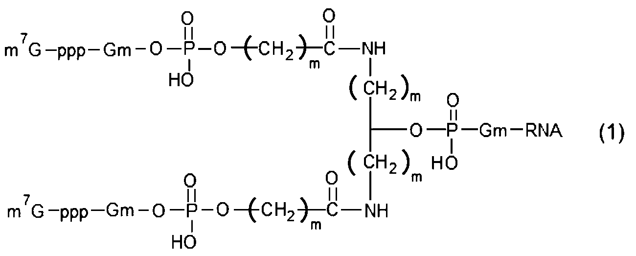

(ここで、m7Gはキャップを示し、pはリン酸基を示し、Gmはグアニンヌクレオチドの2’-水酸基がメチル化された構造を示し、mは1~5の整数を示し、複数のmは互いに同一であっても異なっていてもよく、nは1~5の整数を示し、複数のnは互いに同一であっても異なっていてもよい。) 3. The capped RNA according to claim 2, wherein (c) has a structure represented by the following formula (1) or formula (2).
(Here, m 7 G represents a cap, p represents a phosphate group, Gm represents a structure in which the 2′-hydroxyl group of a guanine nucleotide is methylated, m represents an integer of 1 to 5, and multiple m may be the same or different, n represents an integer of 1 to 5, and multiple n may be the same or different.) - 前記式(1)が下記式(1-1)で示される構造を有し、前記式(2)が下記式(2-1)で示される構造を有することを特徴とする請求項8に記載のキャップ化RNA。
- 前記RNAが、複数の翻訳領域を有することを特徴とする請求項1に記載のキャップ化RNA。 The capped RNA according to claim 1, wherein the RNA has a plurality of translational regions.
- 請求項1に記載のキャップ化RNAの製造方法であって、下記(a)~(c)のいずれか少なくとも1つの工程を有することを特徴とするキャップ化RNAの製造方法
(a)前記上流領域の一部又は全部の配列と相補的な配列を有し、かつキャップ構造を有するキャップ化プローブを前記上流領域にハイブリダイズさせる、又は、
(b)前記上流領域の一部に前記RNAを構成する主鎖から分岐する分岐鎖を有する前記RNAを合成したのち、前記分岐鎖に前記キャップ構造を結合させる、
(c)前記RNAが直鎖状RNAであり、前記上流領域の主鎖の5’末端が2以上に枝分かれするブランチ構造を有するRNAを合成したのち、各枝分かれに前記キャップ構造を結合させる。 2. The method for producing capped RNA according to claim 1, comprising at least one of the following steps (a) to (c): (a) said upstream region Hybridize a capped probe having a sequence complementary to part or all of the sequence of and having a cap structure to the upstream region, or
(b) after synthesizing the RNA having a branched chain branching from the main chain constituting the RNA in a part of the upstream region, binding the cap structure to the branched chain;
(c) The RNA is a linear RNA, and after synthesizing an RNA having a branch structure in which the 5′ end of the main chain of the upstream region is branched into two or more branches, the cap structure is bound to each branch. - 請求項1に記載のキャップ化RNAと、該キャップ化RNAを鋳型として前記翻訳領域がコードする前記タンパク質を発現可能な真核細胞の発現系と、を備えることを特徴とするタンパク質の製造装置。 A protein production apparatus comprising: the capped RNA according to claim 1; and a eukaryotic cell expression system capable of expressing the protein encoded by the translation region using the capped RNA as a template.
- 請求項1に記載のキャップ化RNAを、該キャップ化RNAを鋳型として前記翻訳領域がコードする前記タンパク質を発現可能な真核細胞の発現系に添加して、前記タンパク質を発現させることを特徴とするタンパク質の製造方法。 The capped RNA according to claim 1 is added to a eukaryotic cell expression system capable of expressing the protein encoded by the translation region using the capped RNA as a template to express the protein. A method for producing a protein that
- RNAの5’末端にキャップ構造を有する目的RNAの検出装置であって、
開始コドンから始まり標識タンパク質をコードする翻訳領域と、前記開始コドンの上流側の上流領域であって検出対象となる前記目的RNAの配列の一部又は全部と相補的な配列を有する前記上流領域と、を有する環状RNAからなる検出用プローブと、
前記目的RNAを含む試料に前記環状RNAを添加して前記環状RNAの前記上流領域に前記目的RNAをハイブリダイズさせてキャップ化RNAを生成させる手段と、
前記キャップ化RNAを添加することで前記標識タンパク質を発現可能な真核細胞の発現系と、
前記発現系で発現した前記標識タンパク質を検出する手段と、を備えることを特徴とする目的RNAの検出装置。 A target RNA detection device having a cap structure at the 5' end of RNA,
a translation region starting from an initiation codon and encoding a labeling protein; and an upstream region on the upstream side of the initiation codon and having a sequence complementary to part or all of the sequence of the target RNA to be detected. A detection probe consisting of a circular RNA having
means for adding the circular RNA to a sample containing the target RNA and hybridizing the target RNA to the upstream region of the circular RNA to generate capped RNA;
a eukaryotic cell expression system capable of expressing the labeled protein by adding the capped RNA;
and means for detecting the labeled protein expressed in the expression system. - RNAの5’末端にキャップ構造を有する目的RNAの検出方法であって、
開始コドンから始まり標識タンパク質をコードする翻訳領域と、前記開始コドンの上流側の上流領域であって検出対象となる前記目的RNAの配列の一部又は全部と相補的な配列を有する上流領域と、を有する環状RNAからなる検出用プローブを用意する工程と、
前記目的RNAを含む試料に前記環状RNAを添加して前記環状RNAの前記上流領域に前記目的RNAをハイブリダイズさせてキャップ化RNAを生成させる工程と、
前記標識タンパク質を発現可能な真核細胞の発現系に該キャップ化RNAを添加して前記標識タンパク質を発現させる工程と、
前記発現系で発現した前記標識タンパク質を検出する工程と、を備えることを特徴とする目的RNAの検出方法。 A method for detecting a target RNA having a cap structure at the 5' end of the RNA, comprising:
a translation region starting from an initiation codon and encoding a labeling protein, an upstream region on the upstream side of the initiation codon and having a sequence complementary to part or all of the sequence of the target RNA to be detected, A step of preparing a detection probe consisting of a circular RNA having
adding the circular RNA to a sample containing the target RNA and hybridizing the target RNA to the upstream region of the circular RNA to generate capped RNA;
adding the capped RNA to a eukaryotic cell expression system capable of expressing the labeled protein to express the labeled protein;
and detecting the labeled protein expressed in the expression system. - 開始コドンから始まりタンパク質をコードする翻訳領域と、前記開始コドンの上流側の非翻訳領域と、を有するRNAの前記非翻訳領域にキャップ構造を有するキャップ化RNAであって、下記(a)~(c)のいずれか少なくとも1つの構造を有することを特徴とするキャップ化RNA
(a)前記非翻訳領域の一部又は全部の配列と相補的な配列を有し、かつキャップ構造を有するキャップ化プローブが前記非翻訳領域にハイブリダイズされている、
(b)前記非翻訳領域の一部に前記RNAを構成する主鎖の途中から分岐する分岐鎖が設けられており、該分岐鎖に前記キャップ構造が設けられている、
(c)前記RNAが直鎖状RNAであり、前記非翻訳領域の主鎖の5’末端が2以上に枝分かれするブランチ構造を有し、かつ各枝分かれに前記キャップ構造が設けられている。 A capped RNA having a cap structure in the untranslated region of RNA having a translation region starting from an initiation codon and encoding a protein, and an untranslated region upstream of the initiation codon, wherein the following (a) to ( A capped RNA characterized by having at least one structure of c)
(a) a capped probe having a sequence complementary to the sequence of part or all of the untranslated region and having a cap structure is hybridized to the untranslated region;
(b) a part of the untranslated region is provided with a branched chain branching from the middle of the main chain constituting the RNA, and the branched chain is provided with the cap structure;
(c) The RNA is a linear RNA, the 5'-end of the main chain of the untranslated region has a branched structure with two or more branches, and each branch is provided with the cap structure.
Applications Claiming Priority (2)
| Application Number | Priority Date | Filing Date | Title |
|---|---|---|---|
| JP2022-033316 | 2022-03-04 | ||
| JP2022033316 | 2022-03-04 |
Publications (1)
| Publication Number | Publication Date |
|---|---|
| WO2023167276A1 true WO2023167276A1 (en) | 2023-09-07 |
Family
ID=87883837
Family Applications (1)
| Application Number | Title | Priority Date | Filing Date |
|---|---|---|---|
| PCT/JP2023/007759 WO2023167276A1 (en) | 2022-03-04 | 2023-03-02 | Capped rna and method for producing same, apparatus for producing protein, and method for producing protein |
Country Status (1)
| Country | Link |
|---|---|
| WO (1) | WO2023167276A1 (en) |
Citations (5)
| Publication number | Priority date | Publication date | Assignee | Title |
|---|---|---|---|---|
| WO2013118878A1 (en) * | 2012-02-09 | 2013-08-15 | 独立行政法人理化学研究所 | Cyclic rna and protein production method |
| WO2020204130A1 (en) * | 2019-04-05 | 2020-10-08 | 国立大学法人京都大学 | Rna capping method, production method for modified rna, and modified rna |
| WO2020214830A1 (en) * | 2019-04-16 | 2020-10-22 | The Regents Of The University Of California | Protein translational control |
| WO2021155175A1 (en) * | 2020-01-29 | 2021-08-05 | Flagship Pioneering Innovations Vi, Llc | Compositions for translation and methods of use thereof |
| WO2021172204A1 (en) * | 2020-02-28 | 2021-09-02 | 国立研究開発法人科学技術振興機構 | Method for producing capped rna |
-
2023
- 2023-03-02 WO PCT/JP2023/007759 patent/WO2023167276A1/en unknown
Patent Citations (6)
| Publication number | Priority date | Publication date | Assignee | Title |
|---|---|---|---|---|
| WO2013118878A1 (en) * | 2012-02-09 | 2013-08-15 | 独立行政法人理化学研究所 | Cyclic rna and protein production method |
| WO2020204130A1 (en) * | 2019-04-05 | 2020-10-08 | 国立大学法人京都大学 | Rna capping method, production method for modified rna, and modified rna |
| WO2020214830A1 (en) * | 2019-04-16 | 2020-10-22 | The Regents Of The University Of California | Protein translational control |
| WO2020214806A1 (en) * | 2019-04-16 | 2020-10-22 | The Regents Of The University Of California | Protein translational control |
| WO2021155175A1 (en) * | 2020-01-29 | 2021-08-05 | Flagship Pioneering Innovations Vi, Llc | Compositions for translation and methods of use thereof |
| WO2021172204A1 (en) * | 2020-02-28 | 2021-09-02 | 国立研究開発法人科学技術振興機構 | Method for producing capped rna |
Similar Documents
| Publication | Publication Date | Title |
|---|---|---|
| JP6624743B2 (en) | Site-specific RNA mutagenesis method, target editing guide RNA used therefor, and target RNA-target editing guide RNA complex | |
| JP6752957B2 (en) | A novel process for the production of oligonucleotides | |
| EP1871896B1 (en) | Circle probes and their use in the identification of biomolecules | |
| JP7058839B2 (en) | Cell-free protein expression using rolling circle amplification products | |
| CN113661242A (en) | Compositions comprising modified cyclic polyribonucleotides and uses thereof | |
| JP6910295B2 (en) | Methods and kits for diagnostic and therapeutic fusion applications | |
| CA3114892A1 (en) | Methods and compositions for increasing capping efficiency of transcribed rna | |
| JP6057185B2 (en) | Nucleic acid linker | |
| JP6506838B2 (en) | High-speed photocrosslinkable co-linker for in-vitro sputum and intermolecular interaction analysis, in-vitro sputum method using the linker | |
| EP2307575B1 (en) | Unprocessed rolling circle amplification product | |
| KR20140068992A (en) | Nucleic acid enzyme substrates | |
| TW202227100A (en) | Reverse transcription of polynucleotides comprising unnatural nucleotides | |
| JP2021507699A (en) | A novel process for the production of oligonucleotides | |
| CA3218778A1 (en) | Modified mrna, modified non-coding rna, and uses thereof | |
| US20230079822A1 (en) | Method and products for producing single stranded dna polynucleotides | |
| WO2023167276A1 (en) | Capped rna and method for producing same, apparatus for producing protein, and method for producing protein | |
| WO2018200884A1 (en) | Methods for linking polynucleotides | |
| JPWO2005024018A1 (en) | Nucleic acid construct and method for producing the same | |
| US20060019298A1 (en) | Sequencing of modified nucleic acid molecules | |
| JP6703948B2 (en) | Non-enzymatic nucleic acid chain binding method | |
| WO2019193416A1 (en) | Oligonucleotide-mediated sense codon reassignment | |
| EP4219723A1 (en) | Circular rna platforms, uses thereof, and their manufacturing processes from engineered dna | |
| Yuan | Design of multivalent DNA nanomaterials for use in cancer diagnostics and therapeutics | |
| ES2326578B1 (en) | PROCEDURE FOR THE SYNTHESIS OF COLORED FRAGMENTS OF MONOCATENARY DNA FROM AN EXPRESSION VECTOR AND A UNIVERSAL PRIMER, AND ITS USE IN IN SITU HYBRIDIZATION. | |
| WO2024039652A1 (en) | Cell-free method of producing synthetic circular nucleic acid |
Legal Events
| Date | Code | Title | Description |
|---|---|---|---|
| 121 | Ep: the epo has been informed by wipo that ep was designated in this application |
Ref document number: 23763533 Country of ref document: EP Kind code of ref document: A1 |