WO2023145530A1 - 癌治療剤 - Google Patents
癌治療剤 Download PDFInfo
- Publication number
- WO2023145530A1 WO2023145530A1 PCT/JP2023/001111 JP2023001111W WO2023145530A1 WO 2023145530 A1 WO2023145530 A1 WO 2023145530A1 JP 2023001111 W JP2023001111 W JP 2023001111W WO 2023145530 A1 WO2023145530 A1 WO 2023145530A1
- Authority
- WO
- WIPO (PCT)
- Prior art keywords
- cancer
- therapeutic agent
- braf
- enc
- tre
- Prior art date
Links
- 206010028980 Neoplasm Diseases 0.000 title claims abstract description 114
- 229940124597 therapeutic agent Drugs 0.000 title claims abstract description 86
- 201000011510 cancer Diseases 0.000 title claims abstract description 65
- 239000003814 drug Substances 0.000 title claims abstract description 38
- CMJCXYNUCSMDBY-ZDUSSCGKSA-N lgx818 Chemical compound COC(=O)N[C@@H](C)CNC1=NC=CC(C=2C(=NN(C=2)C(C)C)C=2C(=C(NS(C)(=O)=O)C=C(Cl)C=2)F)=N1 CMJCXYNUCSMDBY-ZDUSSCGKSA-N 0.000 claims abstract description 158
- 229950001969 encorafenib Drugs 0.000 claims abstract description 153
- 150000001875 compounds Chemical class 0.000 claims abstract description 116
- 102000027483 retinoid hormone receptors Human genes 0.000 claims abstract description 93
- 108091008679 retinoid hormone receptors Proteins 0.000 claims abstract description 93
- SHGAZHPCJJPHSC-YCNIQYBTSA-N all-trans-retinoic acid Chemical compound OC(=O)\C=C(/C)\C=C\C=C(/C)\C=C\C1=C(C)CCCC1(C)C SHGAZHPCJJPHSC-YCNIQYBTSA-N 0.000 claims abstract description 75
- 229940125431 BRAF inhibitor Drugs 0.000 claims abstract description 74
- 239000002829 mitogen activated protein kinase inhibitor Substances 0.000 claims abstract description 60
- NAVMQTYZDKMPEU-UHFFFAOYSA-N Targretin Chemical compound CC1=CC(C(CCC2(C)C)(C)C)=C2C=C1C(=C)C1=CC=C(C(O)=O)C=C1 NAVMQTYZDKMPEU-UHFFFAOYSA-N 0.000 claims abstract description 47
- 229960002938 bexarotene Drugs 0.000 claims abstract description 46
- 229940124647 MEK inhibitor Drugs 0.000 claims abstract description 34
- BFSMGDJOXZAERB-UHFFFAOYSA-N dabrafenib Chemical compound S1C(C(C)(C)C)=NC(C=2C(=C(NS(=O)(=O)C=3C(=CC=CC=3F)F)C=CC=2)F)=C1C1=CC=NC(N)=N1 BFSMGDJOXZAERB-UHFFFAOYSA-N 0.000 claims abstract description 33
- 229960002465 dabrafenib Drugs 0.000 claims abstract description 33
- 229950010130 tamibarotene Drugs 0.000 claims abstract description 30
- MUTNCGKQJGXKEM-UHFFFAOYSA-N tamibarotene Chemical compound C=1C=C2C(C)(C)CCC(C)(C)C2=CC=1NC(=O)C1=CC=C(C(O)=O)C=C1 MUTNCGKQJGXKEM-UHFFFAOYSA-N 0.000 claims abstract description 30
- 102000001301 EGF receptor Human genes 0.000 claims abstract description 18
- 108060006698 EGF receptor Proteins 0.000 claims abstract description 18
- -1 retinoid compound Chemical class 0.000 claims abstract description 11
- ACWZRVQXLIRSDF-UHFFFAOYSA-N binimetinib Chemical compound OCCONC(=O)C=1C=C2N(C)C=NC2=C(F)C=1NC1=CC=C(Br)C=C1F ACWZRVQXLIRSDF-UHFFFAOYSA-N 0.000 claims description 119
- 229950003054 binimetinib Drugs 0.000 claims description 118
- 239000012830 cancer therapeutic Substances 0.000 claims description 65
- 229960005395 cetuximab Drugs 0.000 claims description 60
- 101000984753 Homo sapiens Serine/threonine-protein kinase B-raf Proteins 0.000 claims description 51
- 102100027103 Serine/threonine-protein kinase B-raf Human genes 0.000 claims description 51
- 206010009944 Colon cancer Diseases 0.000 claims description 42
- LIRYPHYGHXZJBZ-UHFFFAOYSA-N trametinib Chemical group CC(=O)NC1=CC=CC(N2C(N(C3CC3)C(=O)C3=C(NC=4C(=CC(I)=CC=4)F)N(C)C(=O)C(C)=C32)=O)=C1 LIRYPHYGHXZJBZ-UHFFFAOYSA-N 0.000 claims description 23
- 229960004066 trametinib Drugs 0.000 claims description 23
- 208000001333 Colorectal Neoplasms Diseases 0.000 claims description 13
- VBEQCZHXXJYVRD-GACYYNSASA-N uroanthelone Chemical compound C([C@@H](C(=O)N[C@H](C(=O)N[C@@H](CS)C(=O)N[C@@H](CC(N)=O)C(=O)N[C@@H](CS)C(=O)N[C@H](C(=O)N[C@@H]([C@@H](C)CC)C(=O)NCC(=O)N[C@@H](CC=1C=CC(O)=CC=1)C(=O)N[C@@H](CO)C(=O)NCC(=O)N[C@@H](CC(O)=O)C(=O)N[C@@H](CCCNC(N)=N)C(=O)N[C@@H](CS)C(=O)N[C@@H](CCC(N)=O)C(=O)N[C@@H]([C@@H](C)O)C(=O)N[C@@H](CCCNC(N)=N)C(=O)N[C@@H](CC(O)=O)C(=O)N[C@@H](CC(C)C)C(=O)N[C@@H](CCCNC(N)=N)C(=O)N[C@@H](CC=1C2=CC=CC=C2NC=1)C(=O)N[C@@H](CC=1C2=CC=CC=C2NC=1)C(=O)N[C@@H](CCC(O)=O)C(=O)N[C@@H](CC(C)C)C(=O)N[C@@H](CCCNC(N)=N)C(O)=O)C(C)C)[C@@H](C)O)NC(=O)[C@H](CO)NC(=O)[C@H](CC(O)=O)NC(=O)[C@H](CC(C)C)NC(=O)[C@H](CO)NC(=O)[C@H](CCC(O)=O)NC(=O)[C@@H](NC(=O)[C@H](CC=1NC=NC=1)NC(=O)[C@H](CCSC)NC(=O)[C@H](CS)NC(=O)[C@@H](NC(=O)CNC(=O)CNC(=O)[C@H](CC(N)=O)NC(=O)[C@H](CC(C)C)NC(=O)[C@H](CS)NC(=O)[C@H](CC=1C=CC(O)=CC=1)NC(=O)CNC(=O)[C@H](CC(O)=O)NC(=O)[C@H](CC=1C=CC(O)=CC=1)NC(=O)[C@H](CO)NC(=O)[C@H](CO)NC(=O)[C@H]1N(CCC1)C(=O)[C@H](CS)NC(=O)CNC(=O)[C@H]1N(CCC1)C(=O)[C@H](CC=1C=CC(O)=CC=1)NC(=O)[C@H](CO)NC(=O)[C@@H](N)CC(N)=O)C(C)C)[C@@H](C)CC)C1=CC=C(O)C=C1 VBEQCZHXXJYVRD-GACYYNSASA-N 0.000 claims description 13
- 101800003838 Epidermal growth factor Proteins 0.000 claims description 12
- 229940116977 epidermal growth factor Drugs 0.000 claims description 12
- 229960001972 panitumumab Drugs 0.000 claims description 7
- 229960001727 tretinoin Drugs 0.000 claims description 7
- 102000009024 Epidermal Growth Factor Human genes 0.000 claims description 4
- 229960000397 bevacizumab Drugs 0.000 claims description 4
- 210000004027 cell Anatomy 0.000 description 195
- HDTRYLNUVZCQOY-LIZSDCNHSA-N alpha,alpha-trehalose Chemical compound O[C@@H]1[C@@H](O)[C@H](O)[C@@H](CO)O[C@@H]1O[C@@H]1[C@H](O)[C@@H](O)[C@H](O)[C@@H](CO)O1 HDTRYLNUVZCQOY-LIZSDCNHSA-N 0.000 description 103
- 229930002330 retinoic acid Natural products 0.000 description 68
- IAZDPXIOMUYVGZ-UHFFFAOYSA-N Dimethylsulphoxide Chemical compound CS(C)=O IAZDPXIOMUYVGZ-UHFFFAOYSA-N 0.000 description 60
- 230000000694 effects Effects 0.000 description 55
- 230000014509 gene expression Effects 0.000 description 55
- 101001093899 Homo sapiens Retinoic acid receptor RXR-alpha Proteins 0.000 description 47
- 102100035178 Retinoic acid receptor RXR-alpha Human genes 0.000 description 46
- 239000003112 inhibitor Substances 0.000 description 46
- 108090000623 proteins and genes Proteins 0.000 description 39
- 102100023606 Retinoic acid receptor alpha Human genes 0.000 description 35
- 108091008726 retinoic acid receptors α Proteins 0.000 description 35
- FPIPGXGPPPQFEQ-OVSJKPMPSA-N all-trans-retinol Chemical compound OC\C=C(/C)\C=C\C=C(/C)\C=C\C1=C(C)CCCC1(C)C FPIPGXGPPPQFEQ-OVSJKPMPSA-N 0.000 description 33
- 238000011282 treatment Methods 0.000 description 32
- 238000001262 western blot Methods 0.000 description 32
- 230000019491 signal transduction Effects 0.000 description 31
- 208000029742 colonic neoplasm Diseases 0.000 description 29
- 238000012360 testing method Methods 0.000 description 29
- 230000003833 cell viability Effects 0.000 description 23
- 230000002708 enhancing effect Effects 0.000 description 23
- 102000004169 proteins and genes Human genes 0.000 description 22
- 238000004458 analytical method Methods 0.000 description 19
- FPIPGXGPPPQFEQ-UHFFFAOYSA-N 13-cis retinol Natural products OCC=C(C)C=CC=C(C)C=CC1=C(C)CCCC1(C)C FPIPGXGPPPQFEQ-UHFFFAOYSA-N 0.000 description 17
- 230000002401 inhibitory effect Effects 0.000 description 17
- 229960003471 retinol Drugs 0.000 description 16
- 235000020944 retinol Nutrition 0.000 description 16
- 239000011607 retinol Substances 0.000 description 16
- 230000004663 cell proliferation Effects 0.000 description 15
- 230000010261 cell growth Effects 0.000 description 14
- 208000024891 symptom Diseases 0.000 description 14
- 230000002195 synergetic effect Effects 0.000 description 14
- 230000000259 anti-tumor effect Effects 0.000 description 13
- 229940079593 drug Drugs 0.000 description 13
- 239000008194 pharmaceutical composition Substances 0.000 description 13
- 230000006907 apoptotic process Effects 0.000 description 12
- 238000000034 method Methods 0.000 description 12
- 230000004083 survival effect Effects 0.000 description 12
- 108020004459 Small interfering RNA Proteins 0.000 description 11
- 230000002829 reductive effect Effects 0.000 description 11
- 102200055464 rs113488022 Human genes 0.000 description 11
- 230000001225 therapeutic effect Effects 0.000 description 11
- 206010053759 Growth retardation Diseases 0.000 description 10
- 230000008859 change Effects 0.000 description 10
- 101710179684 Poly [ADP-ribose] polymerase Proteins 0.000 description 9
- 102100023712 Poly [ADP-ribose] polymerase 1 Human genes 0.000 description 9
- 229920000776 Poly(Adenosine diphosphate-ribose) polymerase Polymers 0.000 description 9
- 230000007423 decrease Effects 0.000 description 9
- 230000003247 decreasing effect Effects 0.000 description 9
- 102000043136 MAP kinase family Human genes 0.000 description 8
- 108091054455 MAP kinase family Proteins 0.000 description 8
- 230000001640 apoptogenic effect Effects 0.000 description 8
- 230000037396 body weight Effects 0.000 description 8
- 238000005516 engineering process Methods 0.000 description 8
- 238000012795 verification Methods 0.000 description 8
- 241000699670 Mus sp. Species 0.000 description 7
- 238000003556 assay Methods 0.000 description 7
- 238000002493 microarray Methods 0.000 description 7
- 239000000203 mixture Substances 0.000 description 7
- 108091005981 phosphorylated proteins Proteins 0.000 description 7
- 230000009467 reduction Effects 0.000 description 7
- 150000004492 retinoid derivatives Chemical class 0.000 description 7
- 108050008874 Annexin Proteins 0.000 description 6
- 102000000412 Annexin Human genes 0.000 description 6
- 102000051485 Bcl-2 family Human genes 0.000 description 6
- 108700038897 Bcl-2 family Proteins 0.000 description 6
- 230000001085 cytostatic effect Effects 0.000 description 6
- 230000001419 dependent effect Effects 0.000 description 6
- 238000001727 in vivo Methods 0.000 description 6
- 230000005764 inhibitory process Effects 0.000 description 6
- 230000007246 mechanism Effects 0.000 description 6
- 238000010172 mouse model Methods 0.000 description 6
- 238000007920 subcutaneous administration Methods 0.000 description 6
- 206010064571 Gene mutation Diseases 0.000 description 5
- 238000002474 experimental method Methods 0.000 description 5
- 238000010195 expression analysis Methods 0.000 description 5
- 230000009422 growth inhibiting effect Effects 0.000 description 5
- 239000004615 ingredient Substances 0.000 description 5
- 238000002372 labelling Methods 0.000 description 5
- 238000004519 manufacturing process Methods 0.000 description 5
- 230000001404 mediated effect Effects 0.000 description 5
- 238000003068 pathway analysis Methods 0.000 description 5
- XJMOSONTPMZWPB-UHFFFAOYSA-M propidium iodide Chemical compound [I-].[I-].C12=CC(N)=CC=C2C2=CC=C(N)C=C2[N+](CCC[N+](C)(CC)CC)=C1C1=CC=CC=C1 XJMOSONTPMZWPB-UHFFFAOYSA-M 0.000 description 5
- 239000000126 substance Substances 0.000 description 5
- 230000001629 suppression Effects 0.000 description 5
- 108091032973 (ribonucleotides)n+m Proteins 0.000 description 4
- 230000007730 Akt signaling Effects 0.000 description 4
- 108090000672 Annexin A5 Proteins 0.000 description 4
- 102000004121 Annexin A5 Human genes 0.000 description 4
- 230000005778 DNA damage Effects 0.000 description 4
- 231100000277 DNA damage Toxicity 0.000 description 4
- 238000000134 MTT assay Methods 0.000 description 4
- 231100000002 MTT assay Toxicity 0.000 description 4
- 241001465754 Metazoa Species 0.000 description 4
- 208000024770 Thyroid neoplasm Diseases 0.000 description 4
- 230000002238 attenuated effect Effects 0.000 description 4
- 230000033077 cellular process Effects 0.000 description 4
- 238000006243 chemical reaction Methods 0.000 description 4
- 230000007613 environmental effect Effects 0.000 description 4
- 238000009472 formulation Methods 0.000 description 4
- 230000001939 inductive effect Effects 0.000 description 4
- 230000010365 information processing Effects 0.000 description 4
- 201000001441 melanoma Diseases 0.000 description 4
- 230000037361 pathway Effects 0.000 description 4
- 238000011160 research Methods 0.000 description 4
- 102000003702 retinoic acid receptors Human genes 0.000 description 4
- 108090000064 retinoic acid receptors Proteins 0.000 description 4
- 239000000523 sample Substances 0.000 description 4
- 239000002904 solvent Substances 0.000 description 4
- 201000002510 thyroid cancer Diseases 0.000 description 4
- 230000004614 tumor growth Effects 0.000 description 4
- 239000003981 vehicle Substances 0.000 description 4
- 102100032305 Bcl-2 homologous antagonist/killer Human genes 0.000 description 3
- 206010058467 Lung neoplasm malignant Diseases 0.000 description 3
- 206010027476 Metastases Diseases 0.000 description 3
- 108091000080 Phosphotransferase Proteins 0.000 description 3
- 206010060862 Prostate cancer Diseases 0.000 description 3
- 208000000236 Prostatic Neoplasms Diseases 0.000 description 3
- 108010038912 Retinoid X Receptors Proteins 0.000 description 3
- 102000034527 Retinoid X Receptors Human genes 0.000 description 3
- 229940121908 Retinoid X receptor agonist Drugs 0.000 description 3
- 108010073929 Vascular Endothelial Growth Factor A Proteins 0.000 description 3
- 102000005789 Vascular Endothelial Growth Factors Human genes 0.000 description 3
- 108010019530 Vascular Endothelial Growth Factors Proteins 0.000 description 3
- 239000000556 agonist Substances 0.000 description 3
- 230000007321 biological mechanism Effects 0.000 description 3
- 101150048834 braF gene Proteins 0.000 description 3
- 238000012258 culturing Methods 0.000 description 3
- 238000010790 dilution Methods 0.000 description 3
- 239000012895 dilution Substances 0.000 description 3
- 238000001647 drug administration Methods 0.000 description 3
- 230000012010 growth Effects 0.000 description 3
- 238000011534 incubation Methods 0.000 description 3
- 230000006698 induction Effects 0.000 description 3
- 201000005202 lung cancer Diseases 0.000 description 3
- 208000020816 lung neoplasm Diseases 0.000 description 3
- 210000004379 membrane Anatomy 0.000 description 3
- 239000012528 membrane Substances 0.000 description 3
- 230000009401 metastasis Effects 0.000 description 3
- 230000035772 mutation Effects 0.000 description 3
- 102000020233 phosphotransferase Human genes 0.000 description 3
- 229920002981 polyvinylidene fluoride Polymers 0.000 description 3
- 230000035755 proliferation Effects 0.000 description 3
- 239000000243 solution Substances 0.000 description 3
- 231100000419 toxicity Toxicity 0.000 description 3
- 230000001988 toxicity Effects 0.000 description 3
- NHBKXEKEPDILRR-UHFFFAOYSA-N 2,3-bis(butanoylsulfanyl)propyl butanoate Chemical compound CCCC(=O)OCC(SC(=O)CCC)CSC(=O)CCC NHBKXEKEPDILRR-UHFFFAOYSA-N 0.000 description 2
- SHGAZHPCJJPHSC-ZVCIMWCZSA-N 9-cis-retinoic acid Chemical compound OC(=O)/C=C(\C)/C=C/C=C(/C)\C=C\C1=C(C)CCCC1(C)C SHGAZHPCJJPHSC-ZVCIMWCZSA-N 0.000 description 2
- 239000012116 Alexa Fluor 680 Substances 0.000 description 2
- 108091003079 Bovine Serum Albumin Proteins 0.000 description 2
- 206010006187 Breast cancer Diseases 0.000 description 2
- 208000026310 Breast neoplasm Diseases 0.000 description 2
- 241000283707 Capra Species 0.000 description 2
- 238000000729 Fisher's exact test Methods 0.000 description 2
- WSFSSNUMVMOOMR-UHFFFAOYSA-N Formaldehyde Chemical compound O=C WSFSSNUMVMOOMR-UHFFFAOYSA-N 0.000 description 2
- 208000008839 Kidney Neoplasms Diseases 0.000 description 2
- 102000004232 Mitogen-Activated Protein Kinase Kinases Human genes 0.000 description 2
- 241000699666 Mus <mouse, genus> Species 0.000 description 2
- 241000699660 Mus musculus Species 0.000 description 2
- 101100381978 Mus musculus Braf gene Proteins 0.000 description 2
- 206010033128 Ovarian cancer Diseases 0.000 description 2
- 206010061535 Ovarian neoplasm Diseases 0.000 description 2
- 239000002033 PVDF binder Substances 0.000 description 2
- 206010035226 Plasma cell myeloma Diseases 0.000 description 2
- 206010038389 Renal cancer Diseases 0.000 description 2
- 229940096885 Retinoic acid receptor agonist Drugs 0.000 description 2
- FAPWRFPIFSIZLT-UHFFFAOYSA-M Sodium chloride Chemical compound [Na+].[Cl-] FAPWRFPIFSIZLT-UHFFFAOYSA-M 0.000 description 2
- 102000004142 Trypsin Human genes 0.000 description 2
- 108090000631 Trypsin Proteins 0.000 description 2
- 230000009471 action Effects 0.000 description 2
- 229960002916 adapalene Drugs 0.000 description 2
- LZCDAPDGXCYOEH-UHFFFAOYSA-N adapalene Chemical compound C1=C(C(O)=O)C=CC2=CC(C3=CC=C(C(=C3)C34CC5CC(CC(C5)C3)C4)OC)=CC=C21 LZCDAPDGXCYOEH-UHFFFAOYSA-N 0.000 description 2
- 229960001445 alitretinoin Drugs 0.000 description 2
- 229940125644 antibody drug Drugs 0.000 description 2
- 210000002469 basement membrane Anatomy 0.000 description 2
- 238000010609 cell counting kit-8 assay Methods 0.000 description 2
- 239000006285 cell suspension Substances 0.000 description 2
- 238000002648 combination therapy Methods 0.000 description 2
- 208000035250 cutaneous malignant susceptibility to 1 melanoma Diseases 0.000 description 2
- 230000002354 daily effect Effects 0.000 description 2
- 239000003623 enhancer Substances 0.000 description 2
- 239000012091 fetal bovine serum Substances 0.000 description 2
- 239000001963 growth medium Substances 0.000 description 2
- 230000036541 health Effects 0.000 description 2
- 238000011532 immunohistochemical staining Methods 0.000 description 2
- 238000002513 implantation Methods 0.000 description 2
- 230000005917 in vivo anti-tumor Effects 0.000 description 2
- 230000010354 integration Effects 0.000 description 2
- 201000010982 kidney cancer Diseases 0.000 description 2
- 208000003747 lymphoid leukemia Diseases 0.000 description 2
- 108010082117 matrigel Proteins 0.000 description 2
- 239000011159 matrix material Substances 0.000 description 2
- 108020004999 messenger RNA Proteins 0.000 description 2
- 238000011580 nude mouse model Methods 0.000 description 2
- 230000002265 prevention Effects 0.000 description 2
- 210000001519 tissue Anatomy 0.000 description 2
- 239000012588 trypsin Substances 0.000 description 2
- 230000035899 viability Effects 0.000 description 2
- 238000012800 visualization Methods 0.000 description 2
- 238000005406 washing Methods 0.000 description 2
- 230000003442 weekly effect Effects 0.000 description 2
- QKNYBSVHEMOAJP-UHFFFAOYSA-N 2-amino-2-(hydroxymethyl)propane-1,3-diol;hydron;chloride Chemical compound Cl.OCC(N)(CO)CO QKNYBSVHEMOAJP-UHFFFAOYSA-N 0.000 description 1
- AZKSAVLVSZKNRD-UHFFFAOYSA-M 3-(4,5-dimethylthiazol-2-yl)-2,5-diphenyltetrazolium bromide Chemical compound [Br-].S1C(C)=C(C)N=C1[N+]1=NC(C=2C=CC=CC=2)=NN1C1=CC=CC=C1 AZKSAVLVSZKNRD-UHFFFAOYSA-M 0.000 description 1
- SHGAZHPCJJPHSC-CDMOMSTLSA-N 9,13-cis-Retinoic acid Chemical compound OC(=O)\C=C(\C)/C=C/C=C(/C)\C=C\C1=C(C)CCCC1(C)C SHGAZHPCJJPHSC-CDMOMSTLSA-N 0.000 description 1
- 210000002925 A-like Anatomy 0.000 description 1
- 102100022900 Actin, cytoplasmic 1 Human genes 0.000 description 1
- 108010085238 Actins Proteins 0.000 description 1
- 208000031261 Acute myeloid leukaemia Diseases 0.000 description 1
- 102100027308 Apoptosis regulator BAX Human genes 0.000 description 1
- 102100026596 Bcl-2-like protein 1 Human genes 0.000 description 1
- 206010005003 Bladder cancer Diseases 0.000 description 1
- 208000003174 Brain Neoplasms Diseases 0.000 description 1
- 201000009030 Carcinoma Diseases 0.000 description 1
- 206010008342 Cervix carcinoma Diseases 0.000 description 1
- 208000005243 Chondrosarcoma Diseases 0.000 description 1
- 208000037051 Chromosomal Instability Diseases 0.000 description 1
- 235000005956 Cosmos caudatus Nutrition 0.000 description 1
- OHOQEZWSNFNUSY-UHFFFAOYSA-N Cy3-bifunctional dye zwitterion Chemical compound O=C1CCC(=O)N1OC(=O)CCCCCN1C2=CC=C(S(O)(=O)=O)C=C2C(C)(C)C1=CC=CC(C(C1=CC(=CC=C11)S([O-])(=O)=O)(C)C)=[N+]1CCCCCC(=O)ON1C(=O)CCC1=O OHOQEZWSNFNUSY-UHFFFAOYSA-N 0.000 description 1
- 238000000018 DNA microarray Methods 0.000 description 1
- 101100202237 Danio rerio rxrab gene Proteins 0.000 description 1
- 101100309320 Danio rerio rxrga gene Proteins 0.000 description 1
- 102100031480 Dual specificity mitogen-activated protein kinase kinase 1 Human genes 0.000 description 1
- 101710146526 Dual specificity mitogen-activated protein kinase kinase 1 Proteins 0.000 description 1
- 102100023266 Dual specificity mitogen-activated protein kinase kinase 2 Human genes 0.000 description 1
- 101710146529 Dual specificity mitogen-activated protein kinase kinase 2 Proteins 0.000 description 1
- 239000006144 Dulbecco’s modified Eagle's medium Substances 0.000 description 1
- 206010014733 Endometrial cancer Diseases 0.000 description 1
- 206010014759 Endometrial neoplasm Diseases 0.000 description 1
- 102000004190 Enzymes Human genes 0.000 description 1
- 108090000790 Enzymes Proteins 0.000 description 1
- 108010040476 FITC-annexin A5 Proteins 0.000 description 1
- 206010017993 Gastrointestinal neoplasms Diseases 0.000 description 1
- 208000032612 Glial tumor Diseases 0.000 description 1
- 201000010915 Glioblastoma multiforme Diseases 0.000 description 1
- 206010018338 Glioma Diseases 0.000 description 1
- 102100031181 Glyceraldehyde-3-phosphate dehydrogenase Human genes 0.000 description 1
- 208000017604 Hodgkin disease Diseases 0.000 description 1
- 208000021519 Hodgkin lymphoma Diseases 0.000 description 1
- 208000010747 Hodgkins lymphoma Diseases 0.000 description 1
- 101000971171 Homo sapiens Apoptosis regulator Bcl-2 Proteins 0.000 description 1
- 101001056180 Homo sapiens Induced myeloid leukemia cell differentiation protein Mcl-1 Proteins 0.000 description 1
- 101001112293 Homo sapiens Retinoic acid receptor alpha Proteins 0.000 description 1
- 102100026539 Induced myeloid leukemia cell differentiation protein Mcl-1 Human genes 0.000 description 1
- 230000004163 JAK-STAT signaling pathway Effects 0.000 description 1
- 208000028018 Lymphocytic leukaemia Diseases 0.000 description 1
- 206010025323 Lymphomas Diseases 0.000 description 1
- 208000034578 Multiple myelomas Diseases 0.000 description 1
- 208000033776 Myeloid Acute Leukemia Diseases 0.000 description 1
- 206010029260 Neuroblastoma Diseases 0.000 description 1
- 208000015914 Non-Hodgkin lymphomas Diseases 0.000 description 1
- 241000283973 Oryctolagus cuniculus Species 0.000 description 1
- 206010061902 Pancreatic neoplasm Diseases 0.000 description 1
- 108010089430 Phosphoproteins Proteins 0.000 description 1
- 102000007982 Phosphoproteins Human genes 0.000 description 1
- 229920001213 Polysorbate 20 Polymers 0.000 description 1
- 208000006664 Precursor Cell Lymphoblastic Leukemia-Lymphoma Diseases 0.000 description 1
- 241000932075 Priacanthus hamrur Species 0.000 description 1
- 102000042888 RAF family Human genes 0.000 description 1
- 108091082327 RAF family Proteins 0.000 description 1
- 238000011530 RNeasy Mini Kit Methods 0.000 description 1
- 101150050070 RXRA gene Proteins 0.000 description 1
- 102100033912 Retinoic acid receptor gamma Human genes 0.000 description 1
- 206010039491 Sarcoma Diseases 0.000 description 1
- 206010041067 Small cell lung cancer Diseases 0.000 description 1
- DBMJMQXJHONAFJ-UHFFFAOYSA-M Sodium laurylsulphate Chemical compound [Na+].CCCCCCCCCCCCOS([O-])(=O)=O DBMJMQXJHONAFJ-UHFFFAOYSA-M 0.000 description 1
- 208000005718 Stomach Neoplasms Diseases 0.000 description 1
- 238000000692 Student's t-test Methods 0.000 description 1
- 102000004243 Tubulin Human genes 0.000 description 1
- 108090000704 Tubulin Proteins 0.000 description 1
- 206010054094 Tumour necrosis Diseases 0.000 description 1
- 208000007097 Urinary Bladder Neoplasms Diseases 0.000 description 1
- 208000006105 Uterine Cervical Neoplasms Diseases 0.000 description 1
- FPIPGXGPPPQFEQ-BOOMUCAASA-N Vitamin A Natural products OC/C=C(/C)\C=C\C=C(\C)/C=C/C1=C(C)CCCC1(C)C FPIPGXGPPPQFEQ-BOOMUCAASA-N 0.000 description 1
- 235000010724 Wisteria floribunda Nutrition 0.000 description 1
- OGQICQVSFDPSEI-UHFFFAOYSA-N Zorac Chemical compound N1=CC(C(=O)OCC)=CC=C1C#CC1=CC=C(SCCC2(C)C)C2=C1 OGQICQVSFDPSEI-UHFFFAOYSA-N 0.000 description 1
- 238000002835 absorbance Methods 0.000 description 1
- 229960005339 acitretin Drugs 0.000 description 1
- 230000003213 activating effect Effects 0.000 description 1
- 239000000654 additive Substances 0.000 description 1
- 230000000996 additive effect Effects 0.000 description 1
- IHUNBGSDBOWDMA-AQFIFDHZSA-N all-trans-acitretin Chemical compound COC1=CC(C)=C(\C=C\C(\C)=C\C=C\C(\C)=C\C(O)=O)C(C)=C1C IHUNBGSDBOWDMA-AQFIFDHZSA-N 0.000 description 1
- 150000004347 all-trans-retinol derivatives Chemical class 0.000 description 1
- 150000001413 amino acids Chemical group 0.000 description 1
- 230000033115 angiogenesis Effects 0.000 description 1
- 238000010171 animal model Methods 0.000 description 1
- 230000008485 antagonism Effects 0.000 description 1
- 230000001093 anti-cancer Effects 0.000 description 1
- 230000001028 anti-proliverative effect Effects 0.000 description 1
- 239000002246 antineoplastic agent Substances 0.000 description 1
- AFYNADDZULBEJA-UHFFFAOYSA-N bicinchoninic acid Chemical compound C1=CC=CC2=NC(C=3C=C(C4=CC=CC=C4N=3)C(=O)O)=CC(C(O)=O)=C21 AFYNADDZULBEJA-UHFFFAOYSA-N 0.000 description 1
- 230000033228 biological regulation Effects 0.000 description 1
- 201000000053 blastoma Diseases 0.000 description 1
- 230000000903 blocking effect Effects 0.000 description 1
- 239000000872 buffer Substances 0.000 description 1
- 230000000711 cancerogenic effect Effects 0.000 description 1
- 231100000315 carcinogenic Toxicity 0.000 description 1
- 230000022131 cell cycle Effects 0.000 description 1
- 230000005754 cellular signaling Effects 0.000 description 1
- 238000005119 centrifugation Methods 0.000 description 1
- 201000010881 cervical cancer Diseases 0.000 description 1
- 239000003153 chemical reaction reagent Substances 0.000 description 1
- 230000000052 comparative effect Effects 0.000 description 1
- 229940125782 compound 2 Drugs 0.000 description 1
- 239000002285 corn oil Substances 0.000 description 1
- 235000005687 corn oil Nutrition 0.000 description 1
- 229940043239 cytotoxic antineoplastic drug Drugs 0.000 description 1
- 238000000326 densiometry Methods 0.000 description 1
- 229960003964 deoxycholic acid Drugs 0.000 description 1
- KXGVEGMKQFWNSR-LLQZFEROSA-N deoxycholic acid Chemical compound C([C@H]1CC2)[C@H](O)CC[C@]1(C)[C@@H]1[C@@H]2[C@@H]2CC[C@H]([C@@H](CCC(O)=O)C)[C@@]2(C)[C@@H](O)C1 KXGVEGMKQFWNSR-LLQZFEROSA-N 0.000 description 1
- 238000001514 detection method Methods 0.000 description 1
- 230000004069 differentiation Effects 0.000 description 1
- 238000002651 drug therapy Methods 0.000 description 1
- 230000000547 effect on apoptosis Effects 0.000 description 1
- 239000012636 effector Substances 0.000 description 1
- 238000001962 electrophoresis Methods 0.000 description 1
- 201000008184 embryoma Diseases 0.000 description 1
- 229940082150 encore Drugs 0.000 description 1
- 229960002199 etretinate Drugs 0.000 description 1
- HQMNCQVAMBCHCO-DJRRULDNSA-N etretinate Chemical compound CCOC(=O)\C=C(/C)\C=C\C=C(/C)\C=C\C1=C(C)C=C(OC)C(C)=C1C HQMNCQVAMBCHCO-DJRRULDNSA-N 0.000 description 1
- 238000011156 evaluation Methods 0.000 description 1
- 230000003203 everyday effect Effects 0.000 description 1
- 230000001747 exhibiting effect Effects 0.000 description 1
- JYEFSHLLTQIXIO-SMNQTINBSA-N folfiri regimen Chemical compound FC1=CNC(=O)NC1=O.C1NC=2NC(N)=NC(=O)C=2N(C=O)C1CNC1=CC=C(C(=O)N[C@@H](CCC(O)=O)C(O)=O)C=C1.C1=C2C(CC)=C3CN(C(C4=C([C@@](C(=O)OC4)(O)CC)C=4)=O)C=4C3=NC2=CC=C1OC(=O)N(CC1)CCC1N1CCCCC1 JYEFSHLLTQIXIO-SMNQTINBSA-N 0.000 description 1
- 101150046266 foxo gene Proteins 0.000 description 1
- 206010017758 gastric cancer Diseases 0.000 description 1
- 230000002496 gastric effect Effects 0.000 description 1
- 230000002068 genetic effect Effects 0.000 description 1
- 208000005017 glioblastoma Diseases 0.000 description 1
- 108020004445 glyceraldehyde-3-phosphate dehydrogenase Proteins 0.000 description 1
- 230000009036 growth inhibition Effects 0.000 description 1
- 239000003966 growth inhibitor Substances 0.000 description 1
- 201000010536 head and neck cancer Diseases 0.000 description 1
- 208000014829 head and neck neoplasm Diseases 0.000 description 1
- 206010073071 hepatocellular carcinoma Diseases 0.000 description 1
- 102000048323 human RARA Human genes 0.000 description 1
- 102000046668 human RXRA Human genes 0.000 description 1
- 230000008595 infiltration Effects 0.000 description 1
- 238000001764 infiltration Methods 0.000 description 1
- 238000003331 infrared imaging Methods 0.000 description 1
- 238000002347 injection Methods 0.000 description 1
- 239000007924 injection Substances 0.000 description 1
- UWKQSNNFCGGAFS-XIFFEERXSA-N irinotecan Chemical compound C1=C2C(CC)=C3CN(C(C4=C([C@@](C(=O)OC4)(O)CC)C=4)=O)C=4C3=NC2=CC=C1OC(=O)N(CC1)CCC1N1CCCCC1 UWKQSNNFCGGAFS-XIFFEERXSA-N 0.000 description 1
- 208000032839 leukemia Diseases 0.000 description 1
- 201000007270 liver cancer Diseases 0.000 description 1
- 208000014018 liver neoplasm Diseases 0.000 description 1
- 230000003211 malignant effect Effects 0.000 description 1
- 208000015486 malignant pancreatic neoplasm Diseases 0.000 description 1
- 239000003550 marker Substances 0.000 description 1
- 230000005012 migration Effects 0.000 description 1
- 238000013508 migration Methods 0.000 description 1
- 239000011259 mixed solution Substances 0.000 description 1
- 238000002156 mixing Methods 0.000 description 1
- 201000000050 myeloid neoplasm Diseases 0.000 description 1
- 230000007935 neutral effect Effects 0.000 description 1
- 208000002154 non-small cell lung carcinoma Diseases 0.000 description 1
- 238000010606 normalization Methods 0.000 description 1
- 235000016709 nutrition Nutrition 0.000 description 1
- 230000035764 nutrition Effects 0.000 description 1
- QIQXTHQIDYTFRH-UHFFFAOYSA-N octadecanoic acid Chemical compound CCCCCCCCCCCCCCCCCC(O)=O QIQXTHQIDYTFRH-UHFFFAOYSA-N 0.000 description 1
- 238000011275 oncology therapy Methods 0.000 description 1
- 238000001543 one-way ANOVA Methods 0.000 description 1
- 230000003287 optical effect Effects 0.000 description 1
- 238000003305 oral gavage Methods 0.000 description 1
- 210000000056 organ Anatomy 0.000 description 1
- 201000002528 pancreatic cancer Diseases 0.000 description 1
- 208000008443 pancreatic carcinoma Diseases 0.000 description 1
- 230000007170 pathology Effects 0.000 description 1
- 239000008188 pellet Substances 0.000 description 1
- 230000002093 peripheral effect Effects 0.000 description 1
- 230000026731 phosphorylation Effects 0.000 description 1
- 238000006366 phosphorylation reaction Methods 0.000 description 1
- 230000004962 physiological condition Effects 0.000 description 1
- 229920002401 polyacrylamide Polymers 0.000 description 1
- 235000010486 polyoxyethylene sorbitan monolaurate Nutrition 0.000 description 1
- 239000000256 polyoxyethylene sorbitan monolaurate Substances 0.000 description 1
- 238000010837 poor prognosis Methods 0.000 description 1
- 238000004393 prognosis Methods 0.000 description 1
- 238000000751 protein extraction Methods 0.000 description 1
- 239000012132 radioimmunoprecipitation assay buffer Substances 0.000 description 1
- 102000005962 receptors Human genes 0.000 description 1
- 108020003175 receptors Proteins 0.000 description 1
- 108091008760 retinoic acid receptors γ Proteins 0.000 description 1
- 238000012216 screening Methods 0.000 description 1
- 238000011519 second-line treatment Methods 0.000 description 1
- 230000035945 sensitivity Effects 0.000 description 1
- 238000000926 separation method Methods 0.000 description 1
- 238000009097 single-agent therapy Methods 0.000 description 1
- 208000000587 small cell lung carcinoma Diseases 0.000 description 1
- 239000011780 sodium chloride Substances 0.000 description 1
- 206010041823 squamous cell carcinoma Diseases 0.000 description 1
- 238000011272 standard treatment Methods 0.000 description 1
- 201000011549 stomach cancer Diseases 0.000 description 1
- 238000006467 substitution reaction Methods 0.000 description 1
- 239000000758 substrate Substances 0.000 description 1
- VLYWMPOKSSWJAL-UHFFFAOYSA-N sulfamethoxypyridazine Chemical compound N1=NC(OC)=CC=C1NS(=O)(=O)C1=CC=C(N)C=C1 VLYWMPOKSSWJAL-UHFFFAOYSA-N 0.000 description 1
- 229960000565 tazarotene Drugs 0.000 description 1
- 238000010998 test method Methods 0.000 description 1
- 238000009210 therapy by ultrasound Methods 0.000 description 1
- 238000001890 transfection Methods 0.000 description 1
- 239000012096 transfection reagent Substances 0.000 description 1
- 238000012546 transfer Methods 0.000 description 1
- 238000002054 transplantation Methods 0.000 description 1
- 239000003656 tris buffered saline Substances 0.000 description 1
- 210000004881 tumor cell Anatomy 0.000 description 1
- 208000029729 tumor suppressor gene on chromosome 11 Diseases 0.000 description 1
- 201000005112 urinary bladder cancer Diseases 0.000 description 1
- 229940088594 vitamin Drugs 0.000 description 1
- 229930003231 vitamin Natural products 0.000 description 1
- 235000013343 vitamin Nutrition 0.000 description 1
- 239000011782 vitamin Substances 0.000 description 1
- 235000019155 vitamin A Nutrition 0.000 description 1
- 239000011719 vitamin A Substances 0.000 description 1
- 229940045997 vitamin a Drugs 0.000 description 1
- 150000003722 vitamin derivatives Chemical class 0.000 description 1
- 238000012447 xenograft mouse model Methods 0.000 description 1
Images


Classifications
-
- A—HUMAN NECESSITIES
- A61—MEDICAL OR VETERINARY SCIENCE; HYGIENE
- A61K—PREPARATIONS FOR MEDICAL, DENTAL OR TOILETRY PURPOSES
- A61K31/00—Medicinal preparations containing organic active ingredients
- A61K31/045—Hydroxy compounds, e.g. alcohols; Salts thereof, e.g. alcoholates
- A61K31/07—Retinol compounds, e.g. vitamin A
-
- A—HUMAN NECESSITIES
- A61—MEDICAL OR VETERINARY SCIENCE; HYGIENE
- A61K—PREPARATIONS FOR MEDICAL, DENTAL OR TOILETRY PURPOSES
- A61K31/00—Medicinal preparations containing organic active ingredients
- A61K31/185—Acids; Anhydrides, halides or salts thereof, e.g. sulfur acids, imidic, hydrazonic or hydroximic acids
- A61K31/19—Carboxylic acids, e.g. valproic acid
- A61K31/192—Carboxylic acids, e.g. valproic acid having aromatic groups, e.g. sulindac, 2-aryl-propionic acids, ethacrynic acid
-
- A—HUMAN NECESSITIES
- A61—MEDICAL OR VETERINARY SCIENCE; HYGIENE
- A61K—PREPARATIONS FOR MEDICAL, DENTAL OR TOILETRY PURPOSES
- A61K31/00—Medicinal preparations containing organic active ingredients
- A61K31/185—Acids; Anhydrides, halides or salts thereof, e.g. sulfur acids, imidic, hydrazonic or hydroximic acids
- A61K31/19—Carboxylic acids, e.g. valproic acid
- A61K31/20—Carboxylic acids, e.g. valproic acid having a carboxyl group bound to a chain of seven or more carbon atoms, e.g. stearic, palmitic, arachidic acids
- A61K31/203—Retinoic acids ; Salts thereof
-
- A—HUMAN NECESSITIES
- A61—MEDICAL OR VETERINARY SCIENCE; HYGIENE
- A61K—PREPARATIONS FOR MEDICAL, DENTAL OR TOILETRY PURPOSES
- A61K31/00—Medicinal preparations containing organic active ingredients
- A61K31/33—Heterocyclic compounds
- A61K31/395—Heterocyclic compounds having nitrogen as a ring hetero atom, e.g. guanethidine or rifamycins
- A61K31/41—Heterocyclic compounds having nitrogen as a ring hetero atom, e.g. guanethidine or rifamycins having five-membered rings with two or more ring hetero atoms, at least one of which being nitrogen, e.g. tetrazole
- A61K31/4164—1,3-Diazoles
- A61K31/4184—1,3-Diazoles condensed with carbocyclic rings, e.g. benzimidazoles
-
- A—HUMAN NECESSITIES
- A61—MEDICAL OR VETERINARY SCIENCE; HYGIENE
- A61K—PREPARATIONS FOR MEDICAL, DENTAL OR TOILETRY PURPOSES
- A61K31/00—Medicinal preparations containing organic active ingredients
- A61K31/33—Heterocyclic compounds
- A61K31/395—Heterocyclic compounds having nitrogen as a ring hetero atom, e.g. guanethidine or rifamycins
- A61K31/495—Heterocyclic compounds having nitrogen as a ring hetero atom, e.g. guanethidine or rifamycins having six-membered rings with two or more nitrogen atoms as the only ring heteroatoms, e.g. piperazine or tetrazines
- A61K31/505—Pyrimidines; Hydrogenated pyrimidines, e.g. trimethoprim
- A61K31/506—Pyrimidines; Hydrogenated pyrimidines, e.g. trimethoprim not condensed and containing further heterocyclic rings
-
- A—HUMAN NECESSITIES
- A61—MEDICAL OR VETERINARY SCIENCE; HYGIENE
- A61K—PREPARATIONS FOR MEDICAL, DENTAL OR TOILETRY PURPOSES
- A61K31/00—Medicinal preparations containing organic active ingredients
- A61K31/33—Heterocyclic compounds
- A61K31/395—Heterocyclic compounds having nitrogen as a ring hetero atom, e.g. guanethidine or rifamycins
- A61K31/495—Heterocyclic compounds having nitrogen as a ring hetero atom, e.g. guanethidine or rifamycins having six-membered rings with two or more nitrogen atoms as the only ring heteroatoms, e.g. piperazine or tetrazines
- A61K31/505—Pyrimidines; Hydrogenated pyrimidines, e.g. trimethoprim
- A61K31/519—Pyrimidines; Hydrogenated pyrimidines, e.g. trimethoprim ortho- or peri-condensed with heterocyclic rings
-
- A—HUMAN NECESSITIES
- A61—MEDICAL OR VETERINARY SCIENCE; HYGIENE
- A61K—PREPARATIONS FOR MEDICAL, DENTAL OR TOILETRY PURPOSES
- A61K39/00—Medicinal preparations containing antigens or antibodies
- A61K39/395—Antibodies; Immunoglobulins; Immune serum, e.g. antilymphocytic serum
-
- A—HUMAN NECESSITIES
- A61—MEDICAL OR VETERINARY SCIENCE; HYGIENE
- A61K—PREPARATIONS FOR MEDICAL, DENTAL OR TOILETRY PURPOSES
- A61K45/00—Medicinal preparations containing active ingredients not provided for in groups A61K31/00 - A61K41/00
- A61K45/06—Mixtures of active ingredients without chemical characterisation, e.g. antiphlogistics and cardiaca
-
- A—HUMAN NECESSITIES
- A61—MEDICAL OR VETERINARY SCIENCE; HYGIENE
- A61P—SPECIFIC THERAPEUTIC ACTIVITY OF CHEMICAL COMPOUNDS OR MEDICINAL PREPARATIONS
- A61P35/00—Antineoplastic agents
-
- A—HUMAN NECESSITIES
- A61—MEDICAL OR VETERINARY SCIENCE; HYGIENE
- A61P—SPECIFIC THERAPEUTIC ACTIVITY OF CHEMICAL COMPOUNDS OR MEDICINAL PREPARATIONS
- A61P43/00—Drugs for specific purposes, not provided for in groups A61P1/00-A61P41/00
Definitions
- the present invention relates to a therapeutic agent for cancer containing a component that enhances the effect of a growth inhibitor.
- Cancers such as colon cancer, lung cancer, breast cancer, and prostate cancer are cancers that have seen a marked increase in the number of cases in recent years, and their effective treatment is a continuous goal in basic research and the medical field. Genetic and chromosomal instability is recognized in many of these carcinogenic mechanisms of cancer. Among these, BRAF gene mutation has been reported in thyroid cancer, malignant melanoma, colon cancer, ovarian cancer, prostate cancer, and the like.
- RAF family kinases including BRAF, function as important regulators of the MEK-ERK MAP kinase signaling pathway downstream of RAS. This pathway contributes to cell cycle, cell proliferation, differentiation, angiogenesis, apoptosis, migration and metastasis.
- BRAF V600E mutations in cancers described above are activating mutations V600E that cause a single amino acid substitution in the kinase site.
- BRAF V600E mutation is found in thyroid cancer (59%), malignant melanoma (50%), colon cancer (10%), lung cancer (6%), etc.
- BRAF V600E mutant colon cancer is known to have a poor prognosis compared to wild-type and RAS mutant colon cancer.
- molecular targeted drugs such as bevacizumab, a monoclonal antibody against vascular endothelial growth factor (VEGF), and cetuximab and panitumumab, monoclonal antibodies against epidermal growth factor receptor (EGFR), have been used. things are known.
- VEGF vascular endothelial growth factor
- EGFR epidermal growth factor receptor
- Patent Document 1 discloses a pharmaceutical composition combining a BRAF inhibitor and a MEK inhibitor. This technology is intended to be used in said pharmaceutical composition for the treatment, suppression, severity reduction, risk reduction or inhibition of metastasis to BRAF-mutant cancers, especially melanoma.
- the inventors have focused on the inhibitory effects of BRAF inhibitors and MEK inhibitors on proliferation, and are conducting research on means to enhance these therapeutic effects.
- the present invention has been made in view of the above circumstances, and an object of the present invention is to contain a component that exhibits a synergistic effect with a BRAF inhibitor, exhibit a strong antitumor effect, and provide a BRAF inhibitor, an anti-EGFR antibody, and a MEK inhibitor.
- An object of the present invention is to provide a cancer therapeutic agent that exhibits an enhancing effect on therapeutic effects even on drug-resistant cells and that is particularly effective in the treatment of genetically mutated cancers.
- a cancer therapeutic agent according to the first aspect of the present invention comprises a compound acting on a retinoid receptor and a BRAF inhibitor.
- a therapeutic agent for cancer according to the second aspect of the present invention is a therapeutic agent for cancer to be used in combination with a BRAF inhibitor and includes a compound acting on a retinoid receptor.
- the compound acting on the retinoid receptor may be a retinoid compound or a derivative thereof.
- the compound acting on the retinoid receptor may be tretinoin, tamibarotene or bexarotene.
- the BRAF inhibitor may be dabrafenib or encorafenib.
- the cancer therapeutic agent according to the first aspect of the present invention may further contain a MEK inhibitor.
- the MEK inhibitor may be trametinib or binimetinib.
- the cancer therapeutic agent according to the first aspect of the present invention may further contain a compound that acts on epidermal growth factor or epidermal growth factor receptor.
- the compound acting on epidermal growth factor or epidermal growth factor receptor may be bevacizumab, cetuximab or panitumumab.
- the cancer therapeutic agent according to the first or second aspect of the present invention may be a therapeutic agent for BRAF mutant cancer.
- the therapeutic agent for cancer according to the first or second aspect of the present invention may be a therapeutic agent for colon cancer.
- the cancer therapeutic agent of the above aspect contains a component that exhibits a synergistic effect with a BRAF inhibitor, exhibits a strong antitumor effect, and treats cells that are resistant to BRAF inhibitors, anti-EGFR antibodies, and MEK inhibitors. It is possible to provide a cancer therapeutic agent that exhibits an enhancing effect and is particularly effective in the treatment of gene-mutant cancers.
- FIG. 1 is a graph showing the enhancement of inhibitory effects of tretinoin (TRE or ATRA), dabrafenib (DAB) and trametinib (TRA) in this example.
- FIG. 2 is a graph showing enhancement of inhibitors by ATRA in various colon cancer cell lines in this example.
- FIG. 3 is a graph showing the results of using the RKO cell line for the effects of compounds acting on various retinoid receptors in this example.
- FIG. 4 is a graph showing the results of using HT29 cell line for the effects of compounds acting on various retinoid receptors in this Example.
- FIG. 5 is a graph showing the results of using CO115 cell line for the effects of compounds acting on various retinoid receptors in this example.
- FIG. 6 is a graph showing the enhancing effect of inhibitors by ATRA on encorafenib/cetuximab-resistant strains in this example.
- FIG. 7 is a graph showing changes in tumor volume by ATRA in vivo in this example.
- FIG. 8 is a graph showing the results of using various colorectal cancer cell lines regarding the effects of retinol in this example.
- FIG. 9 is a graph showing the results of using various colorectal cancer cell lines for the effect of tamibarotene in this example.
- FIG. 10 is a graph showing the results of using various colorectal cancer cell lines regarding the effects of bexarotene in this example.
- FIG. 11 is a graph showing the results of the Annexin V-propidium iodide (PI) assay using the RKO cell line in this example.
- FIG. 12 is a graph showing the results of the Annexin V-PI assay using the HT29 cell line in this example.
- FIG. 13 shows images and graphs showing the analysis results of changes in expression of p-MEK using Western blot in this example.
- FIG. 14 is an image showing the analysis results of expression changes of p-ERK and ERK using Western blot in this example.
- FIG. 15 shows images and graphs showing the analysis results of p-AKT using Western blot in this example.
- FIG. 16 shows images and graphs showing the analysis results of changes in PARP expression using Western blot in this example.
- FIG. 17 is an image showing the analysis results of changes in expression of proteins related to the Bcl-2 family using Western blot in this Example.
- FIG. 18 is a graph quantifying the signal intensity of each band in FIG.
- FIG. 19 is an image showing the analysis results of endogenous RAR ⁇ and RXR ⁇ protein expression using Western blot in each BRAF mutant colon cancer cell line in this example.
- FIG. 20 is images and graphs showing the analysis results of the expression of RAR ⁇ and RXR ⁇ using Western blot in the RKO cell line under RAR ⁇ or RXR ⁇ knockdown in this example.
- FIG. 21 is a graph showing the results of examining the growth inhibitory effect of the combination of ATRA, encorafenib, and binimetinib, or the combination of bexarotene, encorafenib, and binimetinib in the RKO cell line under RAR ⁇ or RXR ⁇ knockdown in this example.
- FIG. 22 shows images and graphs showing the analysis results of RXR ⁇ expression by Western blot in HT29 cell lines under RXR ⁇ knockdown in this example.
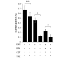
- Figure 23 shows the combination of TRE, encorafenib (ENC), and binimetinib (BIN) or bexarotene (BEX), encorafenib (ENC), and binimetinib (BIN) in HT29 cell lines under RXRa knockdown in this example. It is a graph showing the results of examination of the growth inhibitory effect of the combined use.
- FIG. 24 shows images and graphs showing the results of expression analysis of cleaved PARP by combined use of TRE with ENC, BIN and cetuximab (CET) in RKO cell lines under RAR ⁇ or RXR ⁇ knockdown in this example.
- FIG. 25 is a graph showing the results of examination of the antitumor effect of the combination of TRE and ENC, BIN and CET, or ENC and CET in a subcutaneous tumor-implanted mouse model in this example.
- FIG. 26 shows an image of immunohistochemical staining with an anti-Ki-67 antibody (magnification: 400 times) in the mouse model of subcutaneous tumor implantation in this example. Note that the scale bar is 100 ⁇ m.
- FIG. 27 is a graph showing the Ki-67 positive sub-fraction (%) calculated using the stained image of FIG.
- the cancer therapeutic agent of this embodiment contains a compound acting on a retinoid receptor and a BRAF inhibitor.
- Retinoid compounds broadly refer to vitamin A-derived compounds, vitamin A derivatives, or vitamin A-like compounds. More specific examples include retinoic acid or derivatives thereof.
- Compounds acting on retinoid receptors include retinol, tretinoin (ATRA (all-trans-retinoic acid)), isotretinoin (13-cis-retinoic acid), and alitretinoin (9-cis-retinoic acid) as first-generation retinoids. etc.
- Second-generation retinoids include etretinate and acitretin.
- Third-generation retinoids include adapalene, bexarotene, tazarotene, and tamibarotene.
- Fourth-generation retinoids include triphalotene.
- First and second generation retinoids can bind to several retinoid receptors due to their flexibility obtained by alternating single and double bonds.
- adapalene is a selective RAR agonist
- bexarotene is a selective RXR agonist
- tamibarotene is a RAR/RXR agonist, with high RAR A selectivity.
- Triphalotene is a selective RAR ⁇ agonist.
- retinol, ATRA, tamibarotene or bexarotene is more preferably used as the compound acting on the retinoid receptor, and ATRA, tamibarotene or bexarotene is further preferably used.
- retinoid receptors can enhance the effects of BRAF inhibitors. That is, a compound that acts on retinoid receptors can also be said to be an antitumor effect enhancer of a BRAF inhibitor.
- a compound that acts on retinoid receptors can be said to be an antitumor effect enhancer of BRAF inhibitors and MEK inhibitors.
- the cancer therapeutic agent of this embodiment contains a BRAF inhibitor.
- a therapeutic agent for cancer containing a BRAF inhibitor means that in addition to the form in which the BRAF inhibitor is contained in the same formulation, it is provided in the form in which other components and the component containing the BRAF inhibitor are individually accommodated and used in combination. Including cases where it is a cancer therapeutic agent.
- BRAF refers to the gene that expresses the B-Raf protein.
- BRAF inhibitors broadly include components that inhibit the expression of B-Raf protein, and more specifically components that are known to inhibit the BRAF gene. Conventionally known BRAF inhibitors can be used, but those used as components of cancer therapy are preferred.
- the cancer therapeutic agent of the present embodiment it is more preferable to use Dabrafenib or Encorafenib as the BRAF inhibitor.
- ATRA and dabrafenib As a combination of the compound acting on the retinoid receptor and the BRAF inhibitor, it is more preferable to use ATRA and dabrafenib, ATRA and encorafenib, tamibarotene and dabrafenib, tamibarotene and encorafenib, bexarotene and dabrafenib, or bexarotene and encorafenib.
- the cancer therapeutic agent of this embodiment can further contain other ingredients depending on the type of cancer, in addition to the compound acting on the retinoid receptor and the BRAF inhibitor.
- the cancer therapeutic agent of this embodiment preferably further contains a MEK inhibitor.
- MEK refers to the kinase enzyme MEK1 or MEK2 of mitogen-activated protein kinase, which is a kinase enzyme that phosphorylates mitogen-activated protein kinase.
- MEK inhibitors broadly include components that inhibit the expression of MEK proteins (enzymes), and more specifically, components that are known to inhibit the MEK gene. As MEK inhibitors, conventionally known ones can be used, but those used as ingredients for cancer treatment are preferable.
- trametinib or binimetinib as the MEK inhibitor.
- ATRA encorafenib, and binimetinib, tamibarotene, encorafenib, and binimetinib, or bexarotene, encorafenib, and binimetinib.
- ATRA, dabrafenib and trametinib, tamibarotene, dabrafenib and trametinib, or bexarotene, dabrafenib and trametinib are more preferably used.
- MEK inhibitors are known to be effective when used in combination with conventional BRAF inhibitors in the treatment of cancer. Furthermore, the compound acting on the retinoid receptor of the present embodiment enhances the effects of MEK inhibitors in addition to BRAF inhibitors. Therefore, in cancer therapeutic agents, a further synergistic effect can be obtained by using a compound acting on retinoid receptors, a BRAF inhibitor and a MEK inhibitor in combination.
- the cancer therapeutic agent of the present embodiment may contain other ingredients known to be used in combination with BRAF inhibitors or MEK inhibitors in cancer treatment.
- it may contain compounds that act on epidermal growth factor (EGF) or epidermal growth factor receptor (EGFR).
- EGF epidermal growth factor
- EGFR epidermal growth factor receptor
- Antibodies against VEGF and antibodies against EGFR may be used as such compounds. More specifically, bevacizumab, cetuximab, panitumumab, and the like may be included.
- ATRA and encorafenib and cetuximab, tamibarotene and encorafenib and cetuximab, or bexarotene and encorafenib and cetuximab can be used.
- ATRA dabrafenib and panitumumab
- tamibarotene dabrafenib and panitumumab
- bexarotene dabrafenib and panitumumab
- Combinations of the compounds acting on retinoid receptors, BRAF inhibitors, MEK inhibitors, and compounds acting on EGF or EGFR include ATRA, encorafenib, binimetinib, and cetuximab, tamibarotene, encorafenib, binimetinib, and cetuximab, or bexarotene. and encorafenib, binimetinib, and cetuximab are preferred.
- ATRA dabrafenib, trametinib and cetuximab, tamibarotene, dabrafenib, trametinib and cetuximab, or bexarotene, dabrafenib, trametinib and cetuximab are preferably used.
- the therapeutic agent for cancer according to the present embodiment can be widely used for pharmaceuticals, pharmaceutical compositions, anticancer agents, anticancer compositions, therapeutic agents for cancer, and the like. These cancer therapeutic agents and the like can be used for cancer treatment, prevention, and accompanying treatment.
- the cancer therapeutic agent of the present embodiment can be suitably used for the treatment of mutated cancer, that is, cancer caused by gene mutation.
- the cancer therapeutic agent of the present embodiment can be suitably used for treatment of BRAF mutant cancer.
- BRAF-mutated cancers it can be used for BRAF V600E-mutated cancer, and particularly for BRAF V600E-mutated colorectal cancer.
- BRAF V600E mutant cancer refers to those showing a positive reaction in the BRAF V600 mutation test.
- cancer therapeutic agent of the present embodiment can also be used to treat various cancers.
- "Cancer” in this embodiment refers to a physiological condition characterized mainly by unregulated cell proliferation, and broadly refers to malignant tumors (cancer), also referred to as “cancerous” or “malignant.” Cancers include carcinoma, lymphoma, leukemia, blastoma and sarcoma.
- cancer More specific examples of cancer include squamous cell carcinoma, myeloma, small cell lung cancer, non-small cell lung cancer, glioma, Hodgkin's lymphoma, non-Hodgkin's lymphoma, acute myelogenous leukemia, multiple myeloma, gastrointestinal ) cancer, kidney cancer, ovarian cancer, liver cancer, lymphoblastic leukemia, lymphocytic leukemia, colorectal cancer, endometrial cancer, kidney cancer, prostate cancer, thyroid cancer, melanoma, chondrosarcoma, neuroblastoma , pancreatic cancer, glioblastoma multiforme, cervical cancer, brain cancer, gastric cancer, bladder cancer, hepatoma, breast cancer, colon cancer and head and neck cancer.
- the therapeutic agent for cancer of the present embodiment can be used, for example, for gastrointestinal cancer, lung cancer, and the like. Moreover, among these, it can be used particularly preferably for the treatment of colon cancer.
- Treatment of cancer broadly includes amelioration of symptoms such as reduction of cancer cell number, reduction of tumor size, reduction of cancer cell infiltration rate into peripheral organs, tumor metastasis, or reduction of tumor growth rate. .
- the therapeutic agent for cancer of this embodiment is a method of treating cancer as described above, specifically, a method of treating, suppressing, reducing the severity of, reducing the risk of, inhibiting, or metastasizing cancer as described above. can be used for
- the cancer therapeutic agent of this embodiment can be used for the production of other drugs or compositions used for the treatment of cancer as described above.
- the cancer therapeutic agent of this embodiment contains a component that exhibits a synergistic effect with a BRAF inhibitor, and exhibits a strong antitumor effect. Specifically, an effect of enhancing the growth inhibitory effect of the BRAF inhibitor by 20% or more is observed. These effects have been confirmed to inhibit growth in multiple BRAF mutant colon cancer cell lines.
- reaction mechanisms are expected to involve the effects of compounds that act on retinoid receptors on RXR, RAR, RFR, etc. Therefore, compounds that are selective agonists of RXR, RAR, RFR, etc. may be effective.
- the cancer therapeutic agent of this embodiment also exerts a synergistic effect on the effects of MEK inhibitors. That is, by further including a MEK inhibitor, a higher cancer therapeutic effect can be obtained.
- the inventors have found that compounds that act on retinoid receptors enhance the effects of BRAF inhibitors.
- the present inventors have found that a synergistic effect that further enhances the effect is exhibited by using these compounds acting on retinoid receptors in combination with a BRAF inhibitor and a MEK inhibitor.
- the inventors focused on the possibility of obtaining a higher cancer therapeutic effect by using a compound that can further enhance the effects of BRAF inhibitors and MEK inhibitors in cancer therapeutic agents. Then, the inventors searched for a component capable of enhancing the effect of these inhibitors, and performed screening, thereby obtaining the configuration of the present embodiment.
- the cancer therapeutic agent of the present embodiment exhibits a therapeutic effect-enhancing effect even on cells resistant to BRAF inhibitors, compounds acting on EGF or EGFR, preferably anti-EGFR antibodies and MEK inhibitors.
- the cancer therapeutic agent of the present embodiment is particularly effective for treatment of gene mutation cancer.
- the dosage of the compound that acts on the retinoid receptor can be the conventionally clinically used dosage.
- the dose of the compound acting on the retinoid receptor is 1/2, 1/3, 1/4, 1/5, or 1/6 of the dose conventionally clinically used. , 1/7, 1/8, or even 1/9, the enhancement effect can be exhibited.
- the doses of the BRAF inhibitor, MEK inhibitor, and compound that acts on EGF or EGFR can also be used at conventionally clinically used doses.
- the therapeutic effect of these drugs is enhanced, so that the dose used conventionally clinically is reduced to 1/2, 1/3, or 1/2. It can be reduced to about 1/4, 1/5, 1/6, 1/7, 1/8, or 1/9.
- the dose of the above drug can be increased. may be reduced. By reducing the dose, the toxicity of these drugs can be suppressed, and the burden on the patients to whom they are administered can be reduced.
- Cancer therapeutic agents in embodiments other than the foregoing embodiment are cancer therapeutic agents for use in combination with BRAF inhibitors, including compounds that act on retinoid receptors.
- the cancer therapeutic agent of this embodiment contains at least a compound that acts on retinoid receptors.
- the cancer therapeutic agent of this embodiment is used in combination with a BRAF inhibitor.
- the combined use of a cancer therapeutic agent and a BRAF inhibitor broadly includes aspects in which a cancer therapeutic agent and a BRAF inhibitor are used in combination.
- it includes an aspect in which the cancer therapeutic agent of this embodiment and the BRAF inhibitor are administered simultaneously.
- the phrase "administered and used at the same time" includes the case where they are administered in the same formulation, such as a combination formulation, and also includes the case where they are administered in different formulations and administered at the same time.
- using together also includes the case of using sequentially not simultaneously. Sequential use refers to the sequential use of the cancer therapeutic agent and the other ingredients.
- the administration frequency and dose of the cancer therapeutic agent and other components may be the same or different.
- the mode of administration may be oral administration, injection, or the like.
- each may be taken orally on consecutive days, and in that case, the administration time may be at approximately the same time on the same day or at a different time. More specifically, for example, a compound acting on a retinoid receptor and a BRAF inhibitor are administered orally every day, and a compound acting on EGF or EGFR, preferably an anti-EGFR antibody, is administered intravenously once a week or every other week. may be administered at
- the same components as in the above embodiments can be used.
- cancer therapeutic agent of this embodiment may be used in combination with the above embodiment and a MEK inhibitor, a compound acting on EGF or EGFR, preferably an anti-EGFR antibody.
- cancer therapeutic agent of this embodiment may contain other components of the above-described embodiments, and may be used in combination.
- Still other embodiments than those described above include cancer therapeutics that combine a BRAF inhibitor and a retinoid receptor. Still yet other embodiments include a method of ameliorating symptoms of cancer using a combination of a BRAF inhibitor and a retinoid receptor, or a method of treatment or prevention using a combination of a BRAF inhibitor and a retinoid receptor.
- a cancer therapeutic agent of still another embodiment is a cancer therapeutic agent for combined use with a compound that acts on a retinoid receptor, and includes a BRAF inhibitor.
- a therapeutic agent for cancer of still another embodiment is a therapeutic agent for cancer to be used in combination with a compound that acts on a retinoid receptor, and includes a MEK inhibitor.
- examples of other embodiments of the present invention include the following. a) A method of ameliorating symptoms of cancer in a subject, comprising administering to the subject a compound that acts on a retinoid receptor and a BRAF inhibitor. b) A method of ameliorating symptoms of cancer in a subject comprising administering to the subject a compound that acts on a retinoid receptor and a BRAF inhibitor, wherein the subject has a BRAF-mutant cancer. c) A method of ameliorating symptoms of cancer in a subject comprising administering to the subject a compound that acts on a retinoid receptor and a BRAF inhibitor, wherein the subject is suffering from colon cancer.
- a method of treating or preventing cancer in a subject comprising administering to the subject a compound that acts on a retinoid receptor and a BRAF inhibitor.
- e) A method of inhibiting cancer progression in a subject comprising administering to the subject a compound that acts on a retinoid receptor and a BRAF inhibitor.
- g) Compounds acting on retinoid receptors for use in combination with BRAF inhibitors to ameliorate symptoms of BRAF-mutant cancers.
- a pharmaceutical composition comprising a compound acting on a retinoid receptor for use in treating or preventing cancer in combination with BRAF inhibitors.
- j Compounds acting on retinoid receptors for use in combination with BRAF inhibitors to inhibit cancer progression.
- a pharmaceutical composition comprising a compound acting on a retinoid receptor for use in combination with a BRAF inhibitor to ameliorate symptoms of cancer.
- a pharmaceutical composition comprising a compound acting on a retinoid receptor for use in combination with a BRAF inhibitor to ameliorate symptoms of BRAF-mutated cancer.
- a pharmaceutical composition comprising a compound acting on a retinoid receptor for use in combination with a BRAF inhibitor to ameliorate symptoms of colorectal cancer.
- a pharmaceutical composition comprising a compound acting on a retinoid receptor for use in treating or preventing cancer in combination with a BRAF inhibitor.
- q Use of a compound acting on retinoid receptors for the manufacture of a pharmaceutical composition that, in combination with a BRAF inhibitor, ameliorate symptoms of BRAF-mutated cancer.
- r Use of a compound acting on retinoid receptors for the manufacture of a pharmaceutical composition for ameliorating symptoms of colorectal cancer in combination with a BRAF inhibitor.
- s Use of compounds acting on retinoid receptors for the manufacture of pharmaceutical compositions for treating or preventing cancer in combination with BRAF inhibitors.
- t Use of a compound acting on a retinoid receptor for producing a pharmaceutical composition for inhibiting cancer progression in combination with a BRAF inhibitor.
- a cancer therapeutic agent kit comprising a BRAF inhibitor and a compound acting on a retinoid receptor.
- v A cancer therapeutic agent kit comprising a BRAF inhibitor, a compound acting on a retinoid receptor, and a MEK inhibitor.
- w A cancer treatment kit comprising a BRAF inhibitor, a compound acting on retinoid receptors, and a MEK inhibitor or a compound acting on EGF or EGFR.
- Dabrafenib (catalog # D-5699) from LC Laboratories (Woburn, MA, USA), Trametinib (catalog # 16292), ATRA (all-trans Retinoic Acid) (catalog # 11017) from Cayman Chemical (Ann Arbor, MI, USA ) was purchased from
- Encorafenib (catalog # 16994, HY-15605), Binimetinib (catalog # 16996, HY-15202) are Cayman Chemical (Ann Arbor, MI, USA), MedChemoExpress (Monmouth Ju nction, NJ, USA).
- Bexarotene (catalog # HY-14171) was purchased from MedChemoExpress (Monmouth Junction, NJ, USA).
- Cetuximab (Erbitax) was purchased from Merck Serono (Tokyo, Japan).
- WiDR cells those distributed from the JCRB Cell Bank (Osaka, Japan), National Institute of Biomedical Innovation, Health and Nutrition were used.
- RKO, WiDR, HT29, CO115 cells were cultured in Dulbecco's Modified Eagle's Medium (Sigma-Aldrich Inc. St. Lois, MO, USA) containing 10% fetal bovine serum (FBS). C. and 5% CO 2 concentration conditions were used.
- Dulbecco's Modified Eagle's Medium Sigma-Aldrich Inc. St. Lois, MO, USA
- FBS fetal bovine serum
- Test Example 1 Search for compounds that enhance sensitivity to BRAF inhibitors and MEK inhibitors
- Test method A SCAD Inhibitor Kit 4 containing 80 compounds was used to screen compounds that might enhance the anti-tumor effects of BRAF inhibitors, MEK inhibitors in RKO cells.
- the SCADS Inhibitor Kit was provided by the Molecular Profiling Committee of the Ministry of Education, Culture, Sports, Science and Technology's Grant-in-Aid for Scientific Research "Advanced Animal Model Support (AdAMS)”.
- RKO cells were seeded on a 96-well plate at a cell number of 2.7 ⁇ 10 3 cells/well, and after 24 hours, SCADS Inhibitor Kit 4 ver2.3 was administered to 500 nM.
- Two similarly treated plates were prepared, and 50 nM of the BRAF inhibitor Dabrafenib and 5 nM of the MEK inhibitor Trametinib were administered to one of the plates. After incubation at 37° C. for 72 hours after drug administration, cell viability was measured by MTT assay (described below). Cell viability was evaluated for RKO cells that received only DMSO.
- MTT Thiazolyl blue tetrazolium bromide
- DAB+TRA indicates the value of dabrafenib (DAB) 50 nM and trametinib (TRA) 5 nM in combination.
- the arrow indicates ATRA, and the survival rate is 101% with ATRA alone, and 26% with DAB+TRA and ATRA in combination.
- the viability in this combination is significantly lower than 48% in the case of DAB+TRA alone (solvent DMSO alone), that is, the effect of reducing and suppressing viability is high. Therefore, ATRA was recognized as a compound exhibiting a synergistic effect especially when used in combination with DAB+TRA.
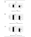
- Fig. 2 shows the survival rate of RKO, (b) WiDR, (c) HT29, (d) CO115, and (e) 8505C colon cancer cell lines.
- the horizontal axis shows the component concentrations of encorafenib and binimetinib in the range of 0 to 10 ⁇ M, and the vertical axis shows the cell viability when ATRA of 0, 1.0 or 10 ⁇ M (each bar graph) is administered.
- encorafenib and binimetinib were used at equal molar concentrations.
- the IC50 (0% inhibitory concentration) is RKO (BRAF V600E mutant colon cancer cell line)
- ATRA 1 uM: 7.5 HT29 (BRAF V600E mutant colon cancer cell line)
- ATRA 1 uM: 0.32 WiDR (BRAF V600E mutant colon cancer cell line)
- ATRA 1uM: 3.6 CO115 (BRAF V600E mutant colon cancer cell line)
- ATRA was shown to increase the cell growth inhibitory effect of encorafenib and binimetinib in any colon cancer cell line.
- Test Example 3 Verification of cell growth inhibitory effect by each retinoid compound
- Compounds that act on retinoid receptors other than ATRA tretinoin
- retinoids retinol, tamibarotene, and bexarotene were used as compounds acting on retinoid receptors.
- cell lines the above-mentioned RKO, HT29, and CO115 cell lines were used.
- BRAF inhibitor and MEK inhibitor to be used in combination as in Test Example 2, a component (EB) in which equal amounts of encorafenib and binimetinib were mixed was used.
- EB component in which equal amounts of encorafenib and binimetinib were mixed was used.
- Fig. 3 shows the results using the RKO cell line.
- retinol (0 ⁇ M or 30 ⁇ M) in FIG. 3(a)
- tamibarotene (0 ⁇ M or 10 ⁇ M) in FIG. 3(b)
- bexarotene (0 ⁇ M or 10 ⁇ M) in FIG.
- FIG. 3(a) shows cell viability.
- retinol, tamibarotene, and bexarotene all caused a greater decrease in cell viability when the retinoid was added to the EBs, indicating inhibition of EB cell proliferation. The effect of enhancing the action of was shown.
- Fig. 4 shows the results using the HT29 cell line.
- retinol (0 ⁇ M or 1 ⁇ M) in FIG. 4 (a) tamibarotene (0 ⁇ M or 1 ⁇ M) in FIG. 4 (b), and bexarotene (0 ⁇ M or 1 ⁇ M) in FIG. , indicated cell viability.
- retinol, tamibarotene, and bexarotene caused a greater decrease in cell viability and inhibition of EB cell proliferation when the retinoid was added to the EB. The effect of enhancing the action of was shown.
- Fig. 5 shows the results using the CO115 cell line.
- Fig. 5(a) shows cell viability using retinol (0 ⁇ M or 30 ⁇ M)
- Fig. 5(b) shows tamibarotene (0 ⁇ M or 30 ⁇ M)
- EB is 0 ⁇ M or 0.1 ⁇ M.
- both retinol and tamibarotene caused a greater decrease in cell viability when the retinoid was added to the EB, indicating that the cell proliferation of the EB was inhibited. was shown to be effective in enhancing
- Test Example 4 Verification of effects in encorafenib / cetuximab-resistant strains
- An encorafenib/cetuximab-resistant strain was prepared, and the enhancing effect of the inhibitor by ATRA was examined for the resistant strain.
- the above-mentioned RKO cells were seeded on a 15 mm dish, and the BRAF inhibitor Encorafenib, the anti-EGFR antibody drug Cetuximab, and the BRAF inhibitor Encorafenib, the MEK inhibitor Binimetinib, and the anti-EGFR antibody drug Cetuximab were administered. Groups were prepared.
- Encorafenib 10 nM, Binimetinib 10 nM, and Cetuximab were started at 1 ⁇ g/ml, respectively, and the drug was gradually increased while considering the growth rate and administration period to obtain resistant strains.
- the survival rate of the susceptible strain (S) decreased according to the encorafenib dose of 0-10 ⁇ M, even if ATRA was not administered (0 ⁇ M). In the ATRA-administered group (1 ⁇ M), the survival rate was further decreased, and the survival rate was around 0.1.
- Resistant strains (R) acquired encorafenib resistance when ATRA was not administered (0 ⁇ M), so the decrease in survival rate was small with respect to the dose of encorafenib, and even with encorafenib 10 ⁇ M, the survival rate was 0.7 to 0. 0.8 survival rate.
- the IC50 (50% inhibitory concentration) for each cell line is Sensitive strain ATRA 0 ⁇ M: 1.23 ATRA 1 ⁇ M: 0.0046 Resistant strain ATRA 0 ⁇ M: >10 ATRA 1 ⁇ M: 1.58 Met.
- the Xenograft mouse model was prepared as follows.
- mice Female nude mice (BALB/c-nu) were purchased from Charles River Laboratories Japan (Yokohama, Japan) and bred in a specific pathogen-free environment.
- the above cultured HT29 cells were collected with trypsin and suspended in a mixed solution of culture medium and Corning Matrigel basement membrane matrix (Corning, NY, USA) at 1 ⁇ 10 7 cells/ml.
- mice were randomly grouped into control group, all-trans Retinoic Acid (ATRA) administration group, Encorafenib/Cetuximab administration group, ATRA/Encorafenib/Cetuximab administration group.
- ATRA all-trans Retinoic Acid
- Encorafenib was given orally at a dose of 10 mg/kg and ATRA at a dose of 10 mg/kg daily for 21 days.
- An oral probe for mice was used for oral administration. Cetuximab was administered intraperitoneally twice weekly at a dose of 20 mg/kg. Each solvent was used as a control.
- Animal experiments were approved by the Tohoku University Institutional Animal Care and Use Committee and were performed in accordance with the Tohoku University Institutional Guidelines.
- Fig. 7(a) shows the average tumor volume
- Fig. 7(b) shows the average body weight.
- the significant difference in FIG. 7(a) was p ⁇ 0.05 by One-way ANOVA and Tukey-Kramer test.
- the tumor volume increased day by day in the group administered DMSO (control solvent) and ATRA only, indicating tumor cell proliferation.
- the increase in tumor size was suppressed more than in the above group, and as is conventionally known, Encorafenib/Cetuximab has an inhibitory effect on proliferation.
- ATRA/Encorafenib/Cetuximab administration group ATRA/encore/cet volume. This indicates that the administration of ATRA causes a synergistic effect with Encorafenib/Cetuximab, and the inhibitory effect on proliferation is more pronounced as the tumor size decreases. As shown in FIG. 7(b), there was no significant difference in body weight among the administration groups, and ATRA caused no significant side effects.
- Test Example 6 Verification of cell growth inhibitory effect by each retinoid compound 2
- the effect of suppressing cell proliferation that is, the effect of enhancing the effect of suppressing cell proliferation of BRAF inhibitors and MEK inhibitors
- the cell lines used were the RKO, HT29, CO115, WiDR, COLO205, and LIM2405 colon cancer cell lines described above.
- a component (EB) in which equal amounts of encorafenib and binimetinib were mixed was used as in Test Example 2.
- FIG. 8 shows the results using retinol.
- RKO cell line in FIG. 8(a) shows the results using retinol.
- HT29 cell line in FIG. 8(b) shows the results using retinol.
- HT29 cell line in FIG. 8(b) shows the results using retinol.
- CO115 cell line in FIG. 8(c) shows the results using the WiDR cell line in FIG. 8(d), COLO205 cell line in FIG. 8(e),
- FIG. 8(f) shows the results using the LIM2405 cell line.
- the cell viability decreased in an EB concentration-dependent manner in all of the RKO, HT29, CO115, WiDR, COLO205, and LIM2405 cell lines.
- the cell viability is greatly reduced, and the decrease in cell viability is dependent on the concentration of retinol, and the effect of enhancing the effect of EB to suppress cell growth. It has been shown.
- FIG. 9 shows the results using Tamibarotene.
- RKO cell line in FIG. 9(a) shows the results using the HT29 cell line in FIG. 8(b), CO115 cell line in FIG. 9(c), WiDR cell line in FIG. 9(d), COLO205 cell line in FIG. 9(f) shows the results using the LIM2405 cell line.
- the cell viability decreased in an EB concentration-dependent manner in all of the RKO, HT29, CO115, WiDR, COLO205, and LIM2405 cell lines.
- the cell viability is greatly reduced, and the decrease in cell viability is dependent on the concentration of tamibarotene, and the effect of enhancing the effect of EB to suppress cell proliferation. It has been shown.
- FIG. 10 shows the results using bexarotene.
- RKO cell line in FIG. 10(a) shows the results using HT29 cell line in FIG. 10(b), CO115 cell line in FIG. 10(c), WiDR cell line in FIG. 10(d), COLO205 cell line in FIG.
- FIG. 10(f) shows the results using the LIM2405 cell line.
- the cell viability is greatly reduced, and the decrease in cell viability is dependent on the concentration of bexarotene, and the effect of enhancing the effect of EB to suppress cell proliferation. It has been shown.
- the RKO cell line and the HT29 cell line were seeded on a 6-well plate at cell numbers of 7.5 ⁇ 10 4 cells/well and 8.0 ⁇ 10 4 cells/well, respectively. After culturing for 24 hours, each cell line was administered with DMSO, TRE, ENC+BIN or TRE+ENC+BIN, respectively.
- Annexin V-FITC Apoptosis Detection Kit Nakalai Tesque
- Apoptotic cells were detected using CytoFLEX LX.
- the percentage of Annexin V-positive cells was calculated as the percentage of apoptotic cells. The above was performed 3 times as an independent experiment. Student's t test was used for the significance test.
- FIG. 11 shows the results of the Annexin V-PI assay in the RKO cell line.
- FIG. 11(a) shows dot plots of propidium iodide (PI) against Annexin V in DMSO, TRE, ENC+BIN, and TRE+ENC+BIN.
- FIG. 11(b) shows the percentage of apoptotic cells in DMSO, TRE, ENC+BIN, and TRE+ENC+BIN.
- DMSO and TRE did not significantly change the percentage of apoptotic cells.
- TRE+ENC+BIN significantly increased the percentage of apoptotic cells compared to ENC+BIN.
- FIG. 12 shows the results of the Annexin V-PI assay in the HT29 cell line.
- FIG. 12(a) shows dot plots of PI against Annexin V in DMSO, TRE, ENC+BIN, and TRE+ENC+BIN.
- FIG. 12(b) shows the percentage of apoptotic cells in DMSO, TRE, ENC+BIN, and TRE+ENC+BIN.
- DMSO and TRE did not significantly change the percentage of apoptotic cells.
- TRE+ENC+BIN significantly increased the percentage of apoptotic cells compared to ENC+BIN.
- the RKO cell line and HT29 cell line were seeded in a 6-well plate at a cell number of 5.0 ⁇ 10 4 cells/well and 1.2 ⁇ 10 5 cells/well, respectively. After culturing for 24 hours, each cell line was administered with DMSO, TRE, ENC+BIN or TRE+ENC+BIN, respectively. Both cell lines were administered with 0.1 v/v% DMSO, 1 ⁇ M TRE, 10 nM ENC, and 10 nM BIN. Twenty-four hours after drug administration, the cells were collected, and total RNA was extracted using RNeasy Mini Kit (QIAGEN Inc.). The amount of total RNA was measured using a NanoDrop ND-1000 spectrophotometer (Thermo Fisher Scientific Inc.).
- RNA 100 ng of extracted total RNA was amplified using Low Input Quick Amp Labeling Kit, one-color (Agilent Technologies) and labeled with Cyanine3.
- the quantity and quality of cRNA were measured using an Agilent Bioanalyzer (Agilent Technologies) and a NanoDrop One ND-ONE-W (Thermo Fisher Scientific Inc.).
- the labeled RNA was hybridized to Sure Print G3 Human GE Microarray 8*60K Ver3.0 (Agilent Technologies) by rotating at 65° C. for 17 hours. After washing, the microarrays were scanned with an Agilent DNA microarray scanner G2505C (Agilent Technologies).
- the genes of the RKO cell line that were significantly changed by TRE, TRE + ENC + BIN and could be analyzed were 790 genes and 1,222 genes, respectively. Pathway analysis using these variable genes enriched 3 signaling pathways for TRE and 14 signaling pathways for TRE+ENC+BIN (see Tables 1 and 3 below). Furthermore, in order to focus on molecular signaling pathways in tumors, we limited the signaling pathways belonging to the Environmental Information Processing and Cellular Processes in the KEGG category, and identified 3 signaling pathways for TRE and 6 signaling pathways for TRE + ENC + BIN.
- genes that are changed by TRE + ENC + BIN are included in a group of genes belonging to signal transduction pathways involved in cell proliferation, such as the JAK-STAT signal transduction pathway and the PI3K-Akt signal transduction pathway, which were not observed in TRE.
- the genes of the HT29 cell line that were significantly changed by TRE and TRE + ENC + BIN and could be analyzed were 1,274 genes and 1,619 genes, respectively. Pathway analysis using these variable genes revealed that 12 signaling pathways were enriched for TRE and 31 for TRE+ENC+BIN (see Tables 2 and 4 above). The analysis was performed in the same manner as the RKO cell line, and finally 3 signaling pathways were identified for TRE and 9 signaling pathways for TRE + ENC + BIN. Genes that are changed by TRE + ENC + BIN are included in a group of genes belonging to molecular signaling pathways involved in cell proliferation, such as JAK-STAT signaling pathway, Ras signaling pathway and PI3K-Akt signaling pathway, which were not observed in TRE. It was shown that
- the RKO cell line was seeded in a 6-well plate at a cell number of 5.0 ⁇ 10 4 cells/well. After culturing for 24 hours, DMSO, TRE, ENC+BIN or TRE+ENC+BIN were administered respectively. They were administered so that DMSO 0.1 v/v%, TRE 10 ⁇ M, ENC 10 nM, and BIN 10 nM, respectively. Cells were harvested 48 hours after drug administration. Separation was performed by centrifugation, cell pellets were made and frozen at -80°C. The experimental steps after protein extraction were carried out by Kinex TM Antibody Microarray Contract Service (Cosmo Bio Co., Ltd.) for contract analysis.
- Kinex TM Antibody Microarray Contract Service Cosmo Bio Co., Ltd.
- the antibody array KAM-2000 (Kinexus Bioinformatics) used in this study covers 875 phosphorylation site-specific antibodies and 451 pan-specific antibodies.
- % CFC % Change From Control value
- % CFC ⁇ -45 % CFC ⁇ -45 recommended by Kinexus Bioinformatics was used as a criterion for significant difference.
- the proteins and phosphorylated proteins specifically altered by this TRE + ENC + BIN belong to signaling pathways involved in tumor growth, such as MAPK signaling pathway, PI3K-Akt signaling pathway, FoxO signaling pathway and ErbB signaling pathway. showed that
- Table 6 shows the types and dilution ratios of the primary antibodies used for Western blot.
- Radioimmunoprecipitation assay buffer composition: 50 mM Tris-HCL, pH 8.0, 150 mM sodium chloride , 0.5 w/v% sodium deoxycholate, 0.1 w/ v% Sodium Dodecyl Sulfate, 1.0 w/v% NP-40 substrate
- the extracted protein was quantified by the bicinchoninic acid method.
- a 5 ⁇ g protein sample was separated by polyacrylamide electrophoresis, transferred to a polyvinylidene difluoride membrane (PVDF membrane, Merck Millipore Ltd.), and blocked with an Odyssey Blocking Buffer (Licor Inc.) for 1 hour at room temperature.
- the PVDF membrane after transfer was immersed in a primary antibody solution and cultured at room temperature for 2 hours or at 4° C. overnight. After primary antibody reaction, the cells were washed three times with Tris Buffered Saline with Tween 20 (TBS-T) for 5 minutes at room temperature, immersed in secondary antibody and incubated at room temperature for 1 hour.
- Secondary antibodies are Goat anti-Rabbit IgG (H+L) Highly Cross-Adsorbed Secondary Antibody Alexa Fluor 680 (Thermo Fisher Scientific Inc.) and Goat anti-Mouse IgG (H+L) Hi Ghly Cross-Adsorbed Secondary Antibody Alexa Fluor 680 (Thermo Fisher Scientific Inc.
- TRE enhances the ability of ENC+BIN to induce apoptosis. Since one of the mechanisms of apoptosis induction by TRE is the regulation of the Bcl-2 family, expression levels of the Bcl-2 family proteins Bcl-2, Mcl-1, Bcl-xL, BAX and BAK in addition to PARP was analyzed.
- TRE induces DNA damage and tumor growth suppression by single agent or combination therapy with cytotoxic anticancer drugs. It has also been reported that DNA damage and the Bcl-2 family interact with each other. Based on these previous reports, we also analyzed the expression level of p-H2AX, which reflects DNA damage that is thought to be involved in TRE-mediated Bcl-2 family-mediated apoptosis.
- FIG. 13 shows the analysis results of p-MEK expression changes related to the MAPK signaling pathway using Western blot.
- FIG. 13(a) shows the Western blot band using the RKO cell line
- FIG. 13(b) shows the Western blot band using the HT29 cell line
- FIG. 13(c) shows the band of FIG. 13(a).
- a graph quantifying the signal intensity is shown in FIG. 13(d), which is a graph quantifying the signal intensity of the bands in FIG. 13(b).
- Fig. 14 shows the analysis results of expression changes of p-ERK and t-ERK related to the MAPK signaling pathway using Western blot.
- FIG. 15 shows the analysis results of changes in p-AKT expression associated with the PI3K-Akt signaling pathway using Western blot.
- FIG. 15(a) shows the Western blot band using the RKO cell line
- FIG. 15(b) shows the Western blot band using the HT29 cell line
- FIG. 15(c) shows the band of FIG. 15(a).
- a graph quantifying the signal intensity is shown in FIG. 15(d), which is a graph quantifying the signal intensity of the bands in FIG. 15(b).
- FIG. 16 shows the analysis results of changes in PARP expression related to the mechanism of apoptosis induction using Western blot.
- FIG. 16(a) shows the Western blot band using the RKO cell line
- FIG. 16(b) shows the Western blot band using the HT29 cell line
- FIG. 16(c) shows the band of FIG. 16(a).
- a graph quantifying the signal intensity is shown in FIG. 16(d), which is a graph quantifying the signal intensity of the band in FIG. 16(b).
- FIG. 17 shows the analysis results of expression changes of proteins related to the Bcl-2 family using Western blot.
- FIG. 17(a) shows the results using the RKO cell line
- FIG. 17(b) shows the results using the HT29 cell line.
- FIG. 18 shows a graph quantifying the signal intensity of each protein band in FIG. Figure 18 (a), Figure 18 (b), Figure 18 (e), Figure 18 (f), Figure 18 (i) are the results using the RKO cell line
- Figure 18 (c), Figure 18 (d) Figure 18(g), FIG. 18(h), FIG. 18(j), and FIG. 18(k) are the results using the HT29 cell line.
- the combination of TRE and ENC+BIN enhanced the expression of cleaved PARP, BAK and p-H2AX in RKO and HT29 cell lines compared to ENC+BIN. This result indicates that the enhanced apoptosis-inducing ability contributes to the molecular biological mechanism that enhances cell growth suppression by the combination of TRE and ENC+BIN, and supports the results of the Annexin V-PI assay.
- the combination of TRE and ENC + BIN enhanced the expression of BAK, which is an effector of the Bcl-2 family, and p-H2AX, which is a marker of DNA damage, compared to ENC + BIN. suggested to be involved.
- RAR ⁇ and RXR ⁇ are TRE receptors that are associated with the cytostatic effect of TRE. It was hypothesized that the mechanism by which compounds acting on retinoid receptors, including TRE, enhance the cytostatic effect of ENC+BIN is via RAR ⁇ or RXR ⁇ .
- Western blot analysis was performed in the same manner as in Test Example 10. .
- Fig. 19 shows the results of Western blot analysis of endogenous RAR ⁇ and RXR ⁇ protein expression in each BRAF mutant colon cancer cell line.
- endogenous RAR ⁇ was observed in the RKO cell line, it was at a level that was hardly observed in other cell lines.
- the expression of endogenous RXR ⁇ was higher in WiDR cell line, HT29 cell line, LIM2405 cell line, RKO cell line, COLO205 cell line and CO115 cell line in that order.
- Test Example 12 Verification of cell growth inhibitory effect by combined use of a compound acting on retinoid receptors under the control of RAR ⁇ or RXR ⁇ expression, a BRAF inhibitor, and a MEK inhibitor
- RAR ⁇ by siRNA
- the MTT assay was performed using the RKO cell line and the HT29 cell line in which the expression of RXR ⁇ was suppressed.
- siGENOME siRNA Reagents Horizon Discovery Ltd.
- siGENOME Human RARA siRNA SMARTpool catalog #M-003437-02-0005
- siGENOME Human RXRA siRNA SMA siGENOME Human RXRA siRNA SMA
- RTpool catalog#M- 003443-02-0005
- siGENOME Non-Targeting siRNA Pool catalog #D-001206-13-05
- RAR ⁇ and RXR ⁇ are endogenously expressed in RKO cells, and only RXR ⁇ is endogenously expressed in other BRAF-mutant colon cancer cells, TRE and selective RXR, which act on both RAR ⁇ and RXR ⁇ among retinoids, are endogenously expressed.
- BEX an agonist
- siRNA-induced changes in RAR ⁇ and RXR ⁇ protein expression in the RKO cell line were analyzed using Western blot in the same manner as in Test Example 10.
- DMSO in si-NC-introduced cells was set to 1 and evaluated as a relative value.
- FIG. 20 shows the analysis results of RAR ⁇ and RXR ⁇ expression by Western blot in RKO cell lines under RAR ⁇ or RXR ⁇ knockdown.
- FIG. 20(a) shows Western blot bands in the RKO cell line under RAR ⁇ knockdown.
- FIG. 20(b) shows a graph quantifying the bands in FIG. 20(a).
- FIG. 20(c) shows Western blot bands in the RKO cell line under RXR ⁇ knockdown.
- FIG. 20(d) shows a graph quantifying the bands in FIG. 20(c).
- si-RAR ⁇ reduced RAR ⁇ expression by 39 ⁇ 6% and si-RXR ⁇ reduced RXR ⁇ expression by 87 ⁇ 2.3% compared to the control si-NC.
- DMSO 0.1 v/v%, TRE 10 ⁇ M, ENC 10 nM + BIN 10 nM, TRE 10 ⁇ M + ENC 10 nM + BIN 10 nM, BEX 10 ⁇ M, BEX 10 ⁇ M + ENC 10 nM + BIN 10 nM were administered to the RKO cell line introduced with each siRNA or si-NC, and after administration 72 hours was evaluated for cell viability.
- FIG. 21 shows the results of examining the growth inhibitory effect of TRE + ENC + BIN or BEX + ENC + BIN in RKO cell lines under RAR ⁇ or RXR ⁇ knockdown.
- FIG. 21(d) shows the results of BEX+ENC+BIN in the RKO cell line under RXR ⁇ knockdown.
- FIG. 22 shows the analysis results of RXR ⁇ expression by Western blot in HT29 cell lines under RXR ⁇ knockdown.
- FIG. 22(a) shows Western blot bands in the HT29 cell line under RXR ⁇ knockdown.
- FIG. 22(b) shows a graph quantifying the bands in FIG. 22(a).
- si-RXR ⁇ reduced the expression of RXR ⁇ by 60 ⁇ 5%.
- DMSO 0.1 v/v%, TRE 10 ⁇ M, ENC 10 nM + BIN 10 nM, TRE 10 ⁇ M + ENC 10 nM + BIN 10 nM, BEX 10 ⁇ M, BEX 10 ⁇ M + ENC 10 nM + BIN 10 nM were administered to the HT29 cell line introduced with si-RXR ⁇ or si-NC. and 72 days after administration Time to assess cell viability.
- FIG. 23 shows the results of examining the growth inhibitory effect of TRE + ENC + BIN or BEX + ENC + BIN in RKO cell lines under RXR ⁇ knockdown.
- FIG. 23(a) shows the results of TRE+ENC+BIN under RXR ⁇ knockdown
- FIG. 23(b) shows the results of BEX+ENC+BIN under RXR ⁇ knockdown.
- FIG. 24 shows the results of expression analysis of cleaved PARP by TRE + ENC + BIN + CET in RKO cell lines under RAR ⁇ or RXR ⁇ knockdown.
- FIG. 24(a) shows Western blot bands in the RKO cell line under RAR ⁇ or RXR ⁇ knockdown.
- FIG. 24(b) shows a graph quantifying the bands in FIG. 24(a).
- TRE enhanced the expression levels of cleaved PARP by ENC+BIN and ENC+BIN+CET about 13-fold and about 11-fold, respectively.
- RAR ⁇ was suppressed
- the effect of TRE on enhancing the expression level of cleaved PARP by ENC+BIN and ENC+BIN+CET was attenuated to about 8-fold and about 3-fold, respectively.
- RXR ⁇ was suppressed, it was attenuated about 1.5 times and about 2 times.
- Test Example 13 Verification of anti-tumor effect by combination of compound acting on retinoid receptor, BRAF inhibitor, MEK inhibitor, and anti-EGFR antibody in subcutaneous tumor transplant mouse model
- mice Female nude mice (BALB/c-nu) were purchased from The Jackson Laboratory Japan and housed in a specific pathogen-free environment.
- the cultured HT29 cell line was recovered with trypsin, and suspended by mixing a culture medium and Corning Matrigel basement membrane matrix (Corning) at a volume ratio of 1:1 so as to obtain 1 ⁇ 10 7 cells/mL.
- 0.1 mL of cell suspension was subcutaneously implanted into the left flank region of each mouse.
- mice were randomly assigned to the vehicle group, TRE alone group, ENC + CET group, TRE and ENC + CET triple combination (TRE + ENC + CET) group, ENC + BIN + CET group, or TRE and ENC + BIN + CET four drugs. They were grouped into a combination (TRE+ENC+BIN+CET) group. TRE, ENC and BIN were dissolved in corn oil (Fuji Film Wako Pure Chemical Industries) and subjected to ultrasonic treatment using Bioruptor UCW-310 (Sonic Bio Co., Ltd.) to prepare a chemical solution.
- a dose of ENC 5 mg/kg, BIN 1.75 mg/kg, TRE 10 mg/kg was administered by oral gavage once daily for 28 days.
- FIG. 25 shows the results of examination of the antitumor effect of combined use of TRE and ENC+BIN+CET or ENC+CET in subcutaneous tumor transplant mouse models.
- FIG. 25(a) shows changes in tumor volume (mm 3 ) over time
- FIG. 25(b) shows changes over time in body weight (g).
- the tumor volume in the vehicle group was 1339 mm 3 ⁇ 231 mm 3
- the tumor volume in the TRE alone group was 1156 mm 3 ⁇ 283 mm 3
- the tumor volumes of the ENC + BIN + CET group and the ENC + CET group were 854 mm 3 ⁇ 425 mm 3 and 543 mm 3 ⁇ 88 mm 3 respectively
- the tumor volumes of the TRE + ENC + BIN + CET group and the TRE + ENC + CET group were 389 mm 3 ⁇ 196 mm 3 and 255 mm 3 respectively. ⁇ 111 mm 3 , with significant reductions of 54% and 53%, respectively.
- mice in the treatment group was measured as shown in Fig. 25(b).
- the body weights of the vehicle group and the TRE alone group, the ENC+CET group and the TRE+ENC+CET group, and the ENC+BIN+CET group and the TRE+ENC+BIN+CET group on day 28 of treatment were compared. As a result, there was no significant difference in body weight of mice with or without TRE.
- Ki-67 labeling index was calculated using resected tumors at the end of treatment in order to evaluate the effect of the combination of TRE and ENC + BIN + CET or ENC + CET on in vivo tumor growth.
- tumors were excised from 4 to 6 mice randomly selected from each treatment group.
- Excised tumors were fixed with 10 v/v % neutral formalin.
- a paraffin-embedded tissue section was prepared from the fixed tumor at the Tohoku University Experimental Animal Pathology Platform, and immunostained with a rabbit anti-Ki-67 antibody (Cell Signaling Technology, dilution ratio 1:800).
- the stained sample was observed with an optical microscope at a magnification of 400. Five fields were selected in the center of the tumor without tumor necrosis, and a total of 500 or more cells were counted in each group.
- the Ki-67 labeling index was calculated as the number of Ki-67 positive cells/total number of cells.
- FIG. 26 shows an image of immunohistochemical staining with an anti-Ki-67 antibody (magnification: 400x) in a mouse model of subcutaneous tumor transplantation. Note that the scale bar in FIG. 26 is 100 ⁇ m.
- FIG. 27 shows the Ki-67 positive fraction (%) calculated using the stained image of FIG.
- Ki-67 labeling index of the TRE+ENC+CET group and the TRE+ENC+BIN+CET group decreased significantly compared to the ENC+CET group and the ENC+BIN+CET group, respectively.
- the cancer therapeutic agent of the present invention contains a component that exhibits a synergistic effect with a BRAF inhibitor, exhibits a strong antitumor effect, and treats cells that are resistant to BRAF inhibitors, anti-EGFR antibodies, and MEK inhibitors. It is possible to provide a cancer therapeutic agent that exhibits an enhancing effect and is particularly effective in the treatment of gene-mutant cancers.
Landscapes
- Health & Medical Sciences (AREA)
- Chemical & Material Sciences (AREA)
- Medicinal Chemistry (AREA)
- Life Sciences & Earth Sciences (AREA)
- Pharmacology & Pharmacy (AREA)
- Veterinary Medicine (AREA)
- Public Health (AREA)
- General Health & Medical Sciences (AREA)
- Animal Behavior & Ethology (AREA)
- Epidemiology (AREA)
- Organic Chemistry (AREA)
- Nuclear Medicine, Radiotherapy & Molecular Imaging (AREA)
- General Chemical & Material Sciences (AREA)
- Chemical Kinetics & Catalysis (AREA)
- Bioinformatics & Cheminformatics (AREA)
- Engineering & Computer Science (AREA)
- Immunology (AREA)
- Microbiology (AREA)
- Mycology (AREA)
- Acyclic And Carbocyclic Compounds In Medicinal Compositions (AREA)
Abstract
癌治療剤は、レチノイド受容体に作用する化合物と、BRAF阻害剤とを含む。癌治療剤は、BRAF阻害剤と併用するための癌治療剤であって、レチノイド受容体に作用する化合物を含む。前記レチノイド受容体に作用する化合物が、レチノイド化合物又はその誘導体であってもよい。前記レチノイド受容体に作用する化合物が、ATRA、タミバロテン又はベキサロテンであってもよい。前記BRAF阻害剤が、ダブラフェニブ又はエンコラフェニブであってもよい。前記癌治療剤は、MEK阻害剤をさらに含んでもよい。前記癌治療剤は、EFG又はEGF受容体に作用する化合物をさらに含んでもよい。
Description
本発明は、増殖阻害剤の効果を増強する成分を含む癌治療剤に関する。
本願は、2022年1月27日に、日本に出願された特願2022-011064号に基づき優先権を主張し、その内容をここに援用する。
本願は、2022年1月27日に、日本に出願された特願2022-011064号に基づき優先権を主張し、その内容をここに援用する。
大腸癌、肺癌、乳癌及び前立腺癌等の癌は、近年、罹患数の増加が顕著な癌であり、その有効な治療は基礎研究から医療分野にわたって継続的な目標である。これらの癌の発癌メカニズムのうち、遺伝子、染色体不安定性が多くに認められる。このうち、BRAF遺伝子変異は、甲状腺癌、悪性黒色腫、大腸癌、卵巣癌、及び前立腺癌等で報告されている。
BRAFを含むRAFファミリーキナーゼは、RASの下流でMEK-ERK MAPキナーゼシグナル伝達経路の重要な調節因子として機能する。この伝達経路は細胞周期、細胞増殖、分化、血管新生、アポトーシス、遊走及び転移などに寄与する。
前記した癌におけるBRAF遺伝子変異のうちの80%以上は、キナーゼ部位の1アミノ酸置換を生ずる活性型変異V600Eである。BRAF V600E変異は甲状腺癌(59%)、悪性黒色腫(50%)、大腸癌(10%)、肺癌(6%)等で認められる。BRAF V600E変異型大腸癌は、野生型やRAS変異型大腸癌と比較して予後不良であることが知られている。
遺伝子変異を伴う癌に対する薬物療法としては、血管内皮成長因子(VEGF)に対するモノクローナル抗体であるベバシズマブや、上皮成長因子受容体(EGFR)に対するモノクローナル抗体のセツキシマブやパニツムマブなどの分子標的治療薬を用いたものが知られている。
BRAF V600E変異型大腸癌に対する治療としては、二次治療としての標準治療(FOLFIRI or CPT-11、セツキシマブ)を使用する治療が知られている。また、BEACON III試験で、エンコラフェニブ、ビニメチニブ、セツキシマブを併用することで全生存期間の延長を示した例がある。ダブラフェニブがBRAF阻害剤、ビニメチニブ及びトラメチニブが上記経路のMEK阻害剤として知られている。
特許文献1は、BRAF阻害剤及びMEK阻害剤を組み合わせた薬学的組成物を開示している。この技術は、前記薬学的組成物において、BRAF変異癌、特に黒色腫に対する転移の処置、抑制、重症度を低減する、危険度を低減するもしくは阻害する処置に用いようとするものである。
前述したように、BRAF変異癌については各種阻害剤及びその併用による薬物治療の技術が開発されているものの、依然として予後不良であり、さらなる有効な治療手段が求められている。特に、大腸癌においてはさらなる治療効果が強く求められている。
これらの治療手段として、発明者らは、BRAF阻害剤及びMEK阻害剤による増殖の抑制効果に注目し、これらの治療効果を増強する手段について研究を進めている。
本発明は上記のような事情を鑑みてなされたものであり、その目的は、BRAF阻害剤と相乗効果を示す成分を含み、強い抗腫瘍効果を示し、BRAF阻害剤、抗EGFR抗体及びMEK阻害剤に抵抗性の細胞に対しても治療効果の増強効果を示し、遺伝子の変異癌に対する治療に特に有効な癌治療剤を提供することにある。
上記課題を解決するため、本発明は以下の態様を有する。
本発明の第1態様に係る癌治療剤は、レチノイド受容体に作用する化合物と、BRAF阻害剤とを含む。
本発明の第2態様に係る癌治療剤は、BRAF阻害剤と併用するための癌治療剤であって、レチノイド受容体に作用する化合物を含む。
前記レチノイド受容体に作用する化合物が、レチノイド化合物又はその誘導体であってもよい。
前記レチノイド受容体に作用する化合物が、トレチノイン、タミバロテン又はベキサロテンであってもよい。
前記BRAF阻害剤が、ダブラフェニブ又はエンコラフェニブであってもよい。
本発明の第1態様に係る癌治療剤は、MEK阻害剤をさらに含んでもよい。
前記MEK阻害剤が、トラメチニブ又はビニメチニブであってもよい。
本発明の第1態様に係る癌治療剤は、上皮成長因子又は上皮成長因子受容体に作用する化合物をさらに含んでもよい。
前記上皮成長因子又は上皮成長因子受容体に作用する化合物が、ベバシズマブ、セツキシマブ又はパニツムマブであってもよい。
本発明の第1又は第2態様に係る癌治療剤は、BRAF変異癌の治療剤であってもよい。
本発明の第1又は第2態様に係る癌治療剤は、大腸癌の治療剤であってもよい。
上記態様の癌治療剤によれば、BRAF阻害剤と相乗効果を示す成分を含み、強い抗腫瘍効果を示し、BRAF阻害剤、抗EGFR抗体及びMEK阻害剤に抵抗性の細胞に対しても治療効果の増強効果を示し、遺伝子の変異癌に対する治療に特に有効な癌治療剤を提供することができる。
以下、本発明に係る癌治療剤について、実施形態を示して説明する。ただし、本発明は以下の実施形態に限定されるものではない。
(癌治療剤)
本実施形態の癌治療剤は、レチノイド受容体に作用する化合物と、BRAF阻害剤とを含む。
本実施形態の癌治療剤は、レチノイド受容体に作用する化合物と、BRAF阻害剤とを含む。
(レチノイド受容体に作用する化合物)
前記レチノイド受容体に作用する化合物としては、レチノイド化合物又はその誘導体が挙げられる。レチノイド化合物は、ビタミンA由来化合物、ビタミンA誘導体、又はビタミンA類似化合物を広く指す。さらに具体的には、レチノイン酸又はその誘導体が挙げられる。
前記レチノイド受容体に作用する化合物としては、レチノイド化合物又はその誘導体が挙げられる。レチノイド化合物は、ビタミンA由来化合物、ビタミンA誘導体、又はビタミンA類似化合物を広く指す。さらに具体的には、レチノイン酸又はその誘導体が挙げられる。
レチノイド受容体に作用する化合物としては、第1世代レチノイドとしてレチノール、トレチノイン(ATRA(全トランスレチノイン酸))、イソトレチノイン(13-シス-レチノイン酸)、及びアリトレチノイン(9-シス-レチノイン酸)等が挙げられる。
第2世代レチノイドとしてエトレチナート、及びアシトレチン等が挙げられる。
第3世代レチノイドとしてアダパレン、ベキサロテン、タザロテン、及びタミバロテン等が挙げられる。
第4世代レチノイドとしてトリファロテン等が挙げられる。
第1世代及び第2世代のレチノイドは、単一結合と二重結合が交互に現れることで柔軟性が得られるため、いくつかのレチノイド受容体と結合することができる。
上述のレチノイドのうち、アダパレンは選択的RARアゴニスト、ベキサロテンは選択的RXRアゴニスト、タミバロテンはRAR/RXRのアゴニストで、RAR Aの選択性が高い。トリファロテンは選択的RARγアゴニストである。
本実施形態では、前記レチノイド受容体に作用する化合物としては、これらのうちレチノール、ATRA、タミバロテン又はベキサロテンを用いることがより好ましく、ATRA、タミバロテン又はベキサロテンを用いることがさらに好ましい。
これらのレチノイド受容体に作用する化合物は、BRAF阻害剤の効果を増強することができる。すなわち、レチノイド受容体に作用する化合物は、BRAF阻害剤の抗腫瘍効果増強剤ということもできる。また、これらのレチノイド受容体に作用する化合物をBRAF阻害剤及びMEK阻害剤と併用することで、さらに効果が増強される相乗効果を発揮する。すなわち、レチノイド受容体に作用する化合物は、BRAF阻害剤及びMEK阻害剤の抗腫瘍効果増強剤ということもできる。
(阻害剤)
本実施形態の癌治療剤は、BRAF阻害剤を含む。癌治療剤がBRAF阻害剤を含むとは、同製剤中にBRAF阻害剤が含まれる形態の他、他の成分とBRAF阻害剤を含む成分とが個別に収納され、併用する形態で提供される癌治療剤となっている場合を含む。
本実施形態の癌治療剤は、BRAF阻害剤を含む。癌治療剤がBRAF阻害剤を含むとは、同製剤中にBRAF阻害剤が含まれる形態の他、他の成分とBRAF阻害剤を含む成分とが個別に収納され、併用する形態で提供される癌治療剤となっている場合を含む。
BRAFとは、B-Rafタンパク質を発現させる遺伝子を指す。BRAF阻害剤とは、広くはB-Rafタンパク質の発現を阻害する成分を広く含み、より具体的には、BRAF遺伝子を阻害することが知られている成分である。BRAF阻害剤としては、従来知られたものを用いることができるが、癌治療の成分として用いられているものが好ましい。
本実施形態の癌治療剤では、BRAF阻害剤としては、ダブラフェニブ(Dabrafenib)又はエンコラフェニブ(Encorafenib)を用いることがより好ましい。
上述のレチノイド受容体に作用する化合物とBRAF阻害剤との組み合わせとしては、ATRAとダブラフェニブ、ATRAとエンコラフェニブ、タミバロテンとダブラフェニブ、タミバロテンとエンコラフェニブ、ベキサロテンとダブラフェニブ、又は、ベキサロテンとエンコラフェニブを用いることがさらに好ましい。
本実施形態の癌治療剤は、レチノイド受容体に作用する化合物とBRAF阻害剤に加えて、癌種に応じて、更に他の成分を含むことができる。
本実施形態の癌治療剤は、MEK阻害剤をさらに含むことが好ましい。MEKとは、マイトジェン活性化プロテインキナーゼのキナーゼ酵素MEK1又はMEK2を指し、マイトジェン活性化プロテインキナーゼをリン酸化するキナーゼ酵素である。MEK阻害剤とは、広くはMEKタンパク質(酵素)の発現を阻害する成分を広く含み、より具体的には、MEK遺伝子を阻害することが知られている成分である。MEK阻害剤としては、従来知られたものを用いることができるが、癌治療の成分として用いられているものが好ましい。
本実施形態の癌治療剤では、MEK阻害剤としては、トラメチニブ(Trametinib)又はビニメチニブ(Binimetinib)を用いることがより好ましい。
上述のレチノイド受容体に作用する化合物、BRAF阻害剤及びMEK阻害剤との組み合わせとしては、ATRAとエンコラフェニブとビニメチニブ、タミバロテンとエンコラフェニブとビニメチニブ、又は、ベキサロテンとエンコラフェニブとビニメチニブを用いることがさらに好ましい。或いは、ATRAとダブラフェニブとトラメチニブ、タミバロテンとダブラフェニブとトラメチニブ、又は、ベキサロテンとダブラフェニブとトラメチニブを用いることがさらに好ましい。
MEK阻害剤は、癌の処置において従来BRAF阻害剤と併用することが有効であることが知られている。さらに、本実施形態のレチノイド受容体に作用する化合物は、BRAF阻害剤に加えて、MEK阻害剤による効果も増強する。したがって、癌治療剤において、レチノイド受容体に作用する化合物、BRAF阻害剤及びMEK阻害剤を併用することで、さらなる相乗効果が得られる。
本実施形態の癌治療剤は、癌の処置において、BRAF阻害剤又はMEK阻害剤と併用されるものとして知られる他の成分を含んでいてもよい。
例えば、上皮成長因子(EGF)又は上皮成長因子受容体(EGFR)に作用する化合物を含んでいてもよい。このような化合物としては、VEGFに対する抗体やEGFRに対する抗体を用いてもよい。さらに具体的には、ベバシズマブ、セツキシマブやパニツムマブ等を含んでいてもよい。
上述のレチノイド受容体に作用する化合物、BRAF阻害剤及びEGF又はEGFRに作用する化合物との組み合わせとしては、ATRAとエンコラフェニブとセツキシマブ、タミバロテンとエンコラフェニブとセツキシマブ、又は、ベキサロテンとエンコラフェニブとセツキシマブを用いることが好ましい。或いは、ATRAとダブラフェニブとセツキシマブ、タミバロテンとダブラフェニブとセツキシマブ、又は、ベキサロテンとダブラフェニブとセツキシマブを用いることが好ましい。或いは、ATRAとダブラフェニブとパニツムマブ、タミバロテンとダブラフェニブとパニツムマブ、又は、ベキサロテンとダブラフェニブとパニツムマブを用いることが好ましい。
上述のレチノイド受容体に作用する化合物、BRAF阻害剤、MEK阻害剤及びEGF又はEGFRに作用する化合物との組み合わせとしては、ATRAとエンコラフェニブとビニメチニブとセツキシマブ、タミバロテンとエンコラフェニブとビニメチニブとセツキシマブ、又は、ベキサロテンとエンコラフェニブとビニメチニブとセツキシマブを用いることが好ましい。或いは、ATRAとダブラフェニブとトラメチニブとセツキシマブ、タミバロテンとダブラフェニブとトラメチニブとセツキシマブ、又は、ベキサロテンとダブラフェニブとトラメチニブとセツキシマブを用いることが好ましい。
(癌治療剤の用途)
本実施形態の癌治療剤は、医薬、医薬組成物、抗癌剤、抗癌用組成物、癌の治療薬等に広く用いることができる。これらの癌治療剤等は、癌の治療、予防やそれに伴う処置に用いることができる。
本実施形態の癌治療剤は、医薬、医薬組成物、抗癌剤、抗癌用組成物、癌の治療薬等に広く用いることができる。これらの癌治療剤等は、癌の治療、予防やそれに伴う処置に用いることができる。
本実施形態の癌治療剤は、変異癌、すなわち遺伝子の変異による癌の治療に好適に用いることができる。
本実施形態の癌治療剤は、これらの変異癌のうち、BRAF変異癌の治療に好適に用いることができる。BRAF変異癌のうち、BRAF V600E変異癌に用いることができ、特にBRAF V600E変異型大腸癌に用いることができる。ここで、BRAF V600E変異癌とは、BRAF V600変異検査での陽性反応を示すものなどを指す。
本実施形態の癌治療剤はまた、癌としては、各種の癌の治療に用いることができる。本実施形態での「癌」は、主に無秩序な細胞増殖を特徴とする生理学的状態を指し、悪性腫瘍(がん)を広く指し、「癌性」又は「悪性」ともいう。癌としては、癌腫、リンパ腫、白血病、芽細胞腫及び肉腫が挙げられる。癌のより具体的な例には、扁平上皮癌、骨髄腫、小細胞肺癌、非小細胞肺癌、神経膠腫、ホジキンリンパ腫、非ホジキンリンパ腫、急性骨髄性白血病、多発性骨髄腫、胃腸(管)癌、腎臓癌、卵巣癌、肝臓癌、リンパ芽球性白血病、リンパ性白血病、結腸直腸癌、子宮内膜癌、腎臓癌、前立腺癌、甲状腺癌、黒色腫、軟骨肉腫、神経芽細胞腫、膵臓癌、多形膠芽腫、子宮頸癌、脳癌、胃癌、膀胱癌、ヘパトーマ、乳癌、結腸癌及び頭頸部癌等が挙げられる。このうち、本実施形態の癌治療剤は、例えば、消化器の癌、又は肺癌等に用いることができる。また、このうち、大腸癌の治療に特に好適に用いることができる。
癌の治療とは、例えば癌細胞数の減少、腫瘍サイズの減少、末梢器官へのがん細胞浸潤速度の低下、腫瘍の転移、又は腫瘍の増殖速度の低下等の症状の改善が広く含まれる。
本実施形態の癌治療剤は、上述の癌の治療方法、具体的には上述の癌の処置、抑制、重症度の低減、危険度の低減、阻害、又は癌の転移に対するこれらの処置の方法に用いることができる。
本実施形態の癌治療剤は、上述の癌の治療に用いる他の薬剤又は組成物の製造のための使用に用いることができる。
(癌治療剤の効果)
本実施形態の癌治療剤は、BRAF阻害剤と相乗効果を示す成分を含み、強い抗腫瘍効果を示す。具体的には、BRAF阻害剤による増殖抑制の効果を20%以上増強する効果がみられる。これらの効果は、BRAF変異型大腸癌細胞株の複数の株において、増殖抑制の効果が確認されたものである。
本実施形態の癌治療剤は、BRAF阻害剤と相乗効果を示す成分を含み、強い抗腫瘍効果を示す。具体的には、BRAF阻害剤による増殖抑制の効果を20%以上増強する効果がみられる。これらの効果は、BRAF変異型大腸癌細胞株の複数の株において、増殖抑制の効果が確認されたものである。
これらの反応機序については、レチノイド受容体に作用する化合物における、RXR、RAR、RFR等に作用する効果が関わっていると予測される。したがって、RXR、RAR、RFR等の選択的なアゴニストである化合物は有効である可能性がある。
本実施形態の癌治療剤は、MEK阻害剤の効果に対しても相乗効果を発揮する。すなわち、さらにMEK阻害剤を含むことで、さらに高い癌治療効果を奏する。
発明者らは、レチノイド受容体に作用する化合物が、BRAF阻害剤の効果を増強することを見出した。また、これらのレチノイド受容体に作用する化合物をBRAF阻害剤及びMEK阻害剤と併用することで、さらに効果が増強される相乗効果を発揮することを見出した。
すなわち、発明者らは、癌治療剤においてBRAF阻害剤やMEK阻害剤の効果をより増強することができる化合物を併用することで、より高い癌治療効果を得られる可能性に着目した。そして、これらの阻害剤の効果を増強させることのできる成分を求めてスクリーニングを行い、本実施形態の構成を得るに至った。
レチノイド受容体に作用する化合物は、従来は主に皮膚科領域で用いられ、腫瘍領域で注目されることはなかった。また、BRAF阻害剤との相乗効果、及び、MEK阻害剤などのその他の癌処置に用いられる成分との相乗効果は知られていなかった。
本実施形態の癌治療剤は、BRAF阻害剤、EGF又はEGFRに作用する化合物、好ましくは抗EGFR抗体及びMEK阻害剤に抵抗性を有する細胞に対しても、治療効果の増強効果を示す。
すなわち、BRAF阻害剤、EGF又はEGFRに作用する化合物、好ましくは抗EGFR抗体、MEK阻害剤、又はその他の成分に対して耐性を獲得した細胞組織に対しても有効である可能性がある。また、これらの成分の治療剤による治療効果が出なくなった患者に対して、治療効果を発揮することができる可能性がある。
これらの効果から、本実施形態の癌治療剤は、遺伝子の変異癌に対する治療に特に有効である。
本実施形態の癌治療剤において、レチノイド受容体に作用する化合物の投与量については、従来から臨床上使用されている投与量で用いることができる。或いは、本実施形態の癌治療剤において、レチノイド受容体に作用する化合物は、従来から臨床上使用されている投与量の1/2、1/3、1/4、1/5、1/6、1/7、1/8、又は1/9程度であっても、上記増強効果を発揮することができる。
また、BRAF阻害剤、MEK阻害剤、及びEGF又はEGFRに作用する化合物の投与量についても、従来から臨床上使用されている投与量で用いることができる。或いは、レチノイド受容体に作用する化合物と併用することで、これら薬剤による治療効果が増強されることから、従来から臨床上使用されている投与量よりも使用量の1/2、1/3、1/4、1/5、1/6、1/7、1/8、又は1/9程度まで低減することが可能である。また、レチノイド受容体に作用する化合物と併用して、上記薬剤を従来から臨床上使用されている投与量による投薬を行うことで、ある程度治療効果がみられた場合に、上記薬剤の投与量を低減してもよい。投与量を低減することにより、これら薬剤による毒性を抑え、投与される患者への負担を低減することができる。
(BRAF阻害剤と併用するための癌治療剤)
前述の実施形態とは他の実施形態における癌治療剤は、BRAF阻害剤と併用するための癌治療剤であって、レチノイド受容体に作用する化合物を含む。
前述の実施形態とは他の実施形態における癌治療剤は、BRAF阻害剤と併用するための癌治療剤であって、レチノイド受容体に作用する化合物を含む。
本実施形態の癌治療剤は、少なくともレチノイド受容体に作用する化合物を含む。本実施形態の癌治療剤は、BRAF阻害剤と併用する用途で用いられる。
ここで癌治療剤とBRAF阻害剤とを併用するとは、癌治療剤とBRAF阻害剤とを組み合わせて使用する態様を広く含む。例えば本実施形態の癌治療剤とBRAF阻害剤が同時に投与されて用いられる態様を含む。同時に投与されて用いられるとは、同一の製剤とされた場合、例えば合剤とされて投与される場合を含み、別の製剤とされ、同時に投与される場合も含む。また、併用するとは、同時でなく逐次に使用する場合も含む。逐次に使用するとは、癌治療剤と他の成分を連続して使用することを指す。癌治療剤と他の成分の投与回数や投与量は、同じであっても、異なっていてもよい。投与の形態は、内服でも、注射等でもよい。
投与する際は、例えば、それぞれを連日内服してもよく、その場合、投与の時間は同日のほぼ同時でも、別の時間であってもよい。さらに具体的には、例えば、レチノイド受容体に作用する化合物とBRAF阻害剤とは連日内服し、EGF又はEGFRに作用する化合物、好ましくは抗EGFR抗体については静脈注射により週1回か、又は隔週で投与してもよい。
BRAF阻害剤としては、前述の実施形態と同様の成分を用いることができる。
また、本実施形態の癌治療剤は、前述の実施形態とMEK阻害剤、EGF又はEGFRに作用する化合物、好ましくは抗EGFR抗体と併用してもよい。
また、本実施形態の癌治療剤は、前述の実施形態のその他の成分が含まれていてもよく、併用してもよい。
(その他の形態)
前述の実施形態とはさらに他の実施形態は、BRAF阻害剤とレチノイド受容体とを併用する癌治療剤を含む。またさらに他の実施形態は、BRAF阻害剤とレチノイド受容体とを併用する癌の症状の改善の方法、又は、BRAF阻害剤とレチノイド受容体とを併用する治療若しくは予防の方法を含む。
前述の実施形態とはさらに他の実施形態は、BRAF阻害剤とレチノイド受容体とを併用する癌治療剤を含む。またさらに他の実施形態は、BRAF阻害剤とレチノイド受容体とを併用する癌の症状の改善の方法、又は、BRAF阻害剤とレチノイド受容体とを併用する治療若しくは予防の方法を含む。
またさらに他の実施形態の癌治療剤は、レチノイド受容体に作用する化合物と併用するための癌治療剤であって、BRAF阻害剤を含む。またさらに他の実施形態の癌治療剤は、レチノイド受容体に作用する化合物と併用するための癌治療剤であって、MEK阻害剤を含む。
また、本発明の実施の他の形態の例としては、以下が挙げられる。
a)対象における癌の症状を改善する方法であって、対象にレチノイド受容体に作用する化合物と、BRAF阻害剤とを投与する方法。
b)対象における癌の症状を改善する方法であって、対象にレチノイド受容体に作用する化合物と、BRAF阻害剤とを投与することを含み、対象がBRAF変異癌に罹患している、方法。
c)対象における癌の症状を改善する方法であって、対象にレチノイド受容体に作用する化合物と、BRAF阻害剤とを投与することを含み、対象が大腸癌に罹患している、方法。
d)対象における癌の治療又は予防方法であって、対象にレチノイド受容体に作用する化合物と、BRAF阻害剤とを投与することを含む、方法。
e)対象における癌の進行抑制方法であって、対象にレチノイド受容体に作用する化合物と、BRAF阻害剤とを投与することを含む、方法。
f)BRAF阻害剤と併用し癌の症状の改善に使用するための、レチノイド受容体に作用する化合物。
g)BRAF阻害剤と併用しBRAF変異癌の症状の改善に使用するための、レチノイド受容体に作用する化合物。
h)BRAF阻害剤と併用し大腸癌の症状の改善に使用するための、レチノイド受容体に作用する化合物。
i)BRAF阻害剤と併用し癌の治療又は予防に使用するための、レチノイド受容体に作用する化合物。
j)BRAF阻害剤と併用し癌の進行抑制に使用するための、レチノイド受容体に作用する化合物。
k)BRAF阻害剤と併用し癌の症状の改善に使用するための、レチノイド受容体に作用する化合物を含む医薬組成物。
l)BRAF阻害剤と併用しBRAF変異癌の症状の改善に使用するための、レチノイド受容体に作用する化合物を含む医薬組成物。
m)BRAF阻害剤と併用し大腸癌の症状の改善に使用するための、レチノイド受容体に作用する化合物を含む医薬組成物。
n)BRAF阻害剤と併用し癌の治療又は予防に使用するための、レチノイド受容体に作用する化合物を含む医薬組成物。
о)BRAF阻害剤と併用し癌の進行抑制に使用するための、レチノイド受容体に作用する化合物を含む医薬組成物。
p)BRAF阻害剤と併用し癌の症状を改善する医薬組成物を製造するための、レチノイド受容体に作用する化合物の使用。
q)BRAF阻害剤と併用しBRAF変異癌の症状を改善する医薬組成物を製造するための、レチノイド受容体に作用する化合物の使用。
r)BRAF阻害剤と併用し大腸癌の症状を改善する医薬組成物を製造するための、レチノイド受容体に作用する化合物する化合物の使用。
s)BRAF阻害剤と併用し癌を治療又は予防する医薬組成物を製造するための、レチノイド受容体に作用する化合物の使用。
t)BRAF阻害剤と併用し癌を進行抑制する医薬組成物を製造するための、レチノイド受容体に作用する化合物の使用。
u)BRAF阻害剤と、レチノイド受容体に作用する化合物と、を備える、癌治療剤キット。
v)BRAF阻害剤と、レチノイド受容体に作用する化合物と、MEK阻害剤と、を備える、癌治療剤キット。
w)BRAF阻害剤と、レチノイド受容体に作用する化合物と、MEK阻害剤、又は、EGF若しくはEGFRに作用する化合物と、を備える、癌治療剤キット。
以下、実施例及び比較例により、本発明の効果をより明らかなものとする。なお、本発明は、以下の実施例のみに限定されるものではなく、その要旨を変更しない範囲で適宜変更して実施できるものである。
(使用した薬剤)
Dabrafenib(catalog # D-5699)はLC Laboratories (Woburn, MA, USA)より、Trametinib(catalog # 16292)、ATRA(all-trans Retinoic Acid)(catalog # 11017)はCayman Chemical (Ann Arbor, MI, USA)より購入した。
Dabrafenib(catalog # D-5699)はLC Laboratories (Woburn, MA, USA)より、Trametinib(catalog # 16292)、ATRA(all-trans Retinoic Acid)(catalog # 11017)はCayman Chemical (Ann Arbor, MI, USA)より購入した。
Encorafenib(catalog # 16994、HY-15605)、Binimetinib(catalog # 16996、HY-15202)はCayman Chemical (Ann Arbor, MI, USA)、MedChemoExpress(Monmouth Junction, NJ, USA)より購入した。
Retinol(catalog # S5592)、Tamibarotene(catalog # S4260)はSelleck Chemicals (Houston, TX, USA)より購入した。
Bexarotene(catalog # HY-14171)はMedChemoExpress(Monmouth Junction, NJ, USA)より購入した。
Cetuximab(Erbitax)はMerck Serono(Tokyo, Japan)より購入した。
(大腸癌細胞株)
後述の試験において使用した4種類の大腸癌細胞株については、American Type Culture Collection(Manassas, VA, USA)より2015年にRKO細胞を購入した。
後述の試験において使用した4種類の大腸癌細胞株については、American Type Culture Collection(Manassas, VA, USA)より2015年にRKO細胞を購入した。
WiDR細胞については国立研究開発法人 医薬基盤・健康・栄養研究所 JCRB細胞バンク(Osaka, Japan)より分譲されたものを使用した。
HT29、CO115、LIM2405、COLO205についてはJohn M. Mariadason博士(Ludwig Institute for Cancer Research, Melbourne, Vic, Australia)より譲り受けたものを使用した。
RKO細胞、WiDR細胞、HT29細胞、CO115細胞は10% fetal bovine serum(ウシ胎児血清、FBS)を含むDulbecco’s Modified Eagle’s Medium(Sigma-Aldrich Inc. St.Lois, MO, USA)で37℃、5% CO2濃度の条件下で培養したものを使用した。
(試験例1:BRAF阻害剤及びMEK阻害剤の感受性を増強させる化合物の探索)
(試験方法)
80の化合物を含むSCAD Inhibitor Kit4を使用して、RKO細胞におけるBRAF阻害剤、MEK阻害剤の抗腫瘍効果を高める可能性のある化合物をスクリーニングした。
(試験方法)
80の化合物を含むSCAD Inhibitor Kit4を使用して、RKO細胞におけるBRAF阻害剤、MEK阻害剤の抗腫瘍効果を高める可能性のある化合物をスクリーニングした。
SCADS Inhibitor Kitは、文部科学省の革新的分野の科学研究費補助金「先端動物モデル支援(AdAMS)」の分子プロファイリング委員会から提供されたものを用いた。
RKO細胞を2.7×10³個/wellの細胞数で96-well plateに播き、24時間後にSCADS Inhibitor Kit 4 ver2.3を500nMになるよう投与した。同様に処理したplateを2枚準備し、そのうちの1枚のplateにはBRAF阻害剤Dabrafenib 50nM及びMEK阻害剤Trametinib 5nMを投与した。薬剤投与から37℃で72時間インキュベートした後、MTTアッセイ(後述)で細胞生存率を測定した。DMSOのみが投与されたRKO細胞を対象として細胞生存率を評価した。
MTT(Thiazolyl blue tetrazolium bromide)アッセイは以下のように行った。
RKO細胞を1.5×10³個/wellの細胞数、CO115細胞を2.5×103個/well、HT29細胞、WiDR細胞を6×103個/wellで96-well plateに播いた。細胞密度が40-60%となるまで約24時間インキュベートした後、薬剤を投与した。37℃で72時間インキュベートした後、MTTアッセイをCell Counting Kit-8 (Dojindo Laboratories, Kumamoto, Japan)を用いて行った。Cell Counting Kit-8溶液を各wellに10μlずつ添加し、5% CO2、37℃の培養器で120分呈色反応を行った後、マイクロプレートリーダーSpectraMax M2e(Molecular Devices, Sunnyvale, CA, USA)で450nmの吸光度を測定し評価した。0.1% DMSOが投与された細胞をコントロールとして用いた。以上を独立した実験として3回以上行った。
相乗効果についてはCombination Indexを用いて評価した。Combination Indexの計算には、CompuSyn software (ComboSyn Inc、NJ、USA)を使用した。以前の報告に基づいて、CI<0.7を相乗効果、CI 0.7-1.0をわずかな相乗効果/相加効果、CI> 1.0を拮抗作用と定義した。
(試験結果)
標準阻害剤キット0.5μMで72時間処理の場合の相乗効果評価の結果を図1に示した。DAB+TRAはダブラフェニブ(DAB)50nM及びトラメチニブ(TRA)5nMを併用した値を示す。矢印がATRAを示し、ATRA単剤では101%、DAB+TRAとATRAを併用した場合は26%の生存率となっている。この併用した場合の生存率は、DAB+TRAのみ(溶媒であるDMSOのみ)の場合の48%よりも顕著に低く、すなわち生存率の低下効果、抑制効果が高い。このため、ATRAが特にDAB+TRAと併用した場合に相乗効果を示す化合物と認められた。
標準阻害剤キット0.5μMで72時間処理の場合の相乗効果評価の結果を図1に示した。DAB+TRAはダブラフェニブ(DAB)50nM及びトラメチニブ(TRA)5nMを併用した値を示す。矢印がATRAを示し、ATRA単剤では101%、DAB+TRAとATRAを併用した場合は26%の生存率となっている。この併用した場合の生存率は、DAB+TRAのみ(溶媒であるDMSOのみ)の場合の48%よりも顕著に低く、すなわち生存率の低下効果、抑制効果が高い。このため、ATRAが特にDAB+TRAと併用した場合に相乗効果を示す化合物と認められた。
ATRA単独では増殖抑制が20%未満であり、DAB+TRA+ATRA併用ではDAB+TRAによる増殖抑制より20%以上の抑制効果を認めた。したがって、ATRAはDAB及びTRAと相乗効果を示し、これらの成分による抑制効果を増強する成分であることが示された。
(試験例2:各大腸癌細胞株におけるATRAの効果)
各種の大腸癌細胞株において、細胞の生存率に対するBRAF阻害剤(エンコラフェニブ)及びMEK阻害剤(ビニメチニブ)の阻害効果と、ATRAによる増強について調べた。図2(a)にRKO、(b)にWiDR、(c)にHT29、(d)にCO115、(e)に8505Cの各大腸癌細胞株について各生存率を示した。横軸は、エンコラフェニブ及びビニメチニブの成分濃度を0~10μMの範囲で示し、縦軸には0、1.0又は10μM(各棒グラフ)のATRAを投与した場合の細胞生存率を示した。図に示すように、エンコラフェニブ及びビニメチニブはモル濃度で等量となるよう用いた。
各種の大腸癌細胞株において、細胞の生存率に対するBRAF阻害剤(エンコラフェニブ)及びMEK阻害剤(ビニメチニブ)の阻害効果と、ATRAによる増強について調べた。図2(a)にRKO、(b)にWiDR、(c)にHT29、(d)にCO115、(e)に8505Cの各大腸癌細胞株について各生存率を示した。横軸は、エンコラフェニブ及びビニメチニブの成分濃度を0~10μMの範囲で示し、縦軸には0、1.0又は10μM(各棒グラフ)のATRAを投与した場合の細胞生存率を示した。図に示すように、エンコラフェニブ及びビニメチニブはモル濃度で等量となるよう用いた。
各細胞株について、IC50(50%阻害濃度)は、
RKO (BRAF V600E変異型大腸癌細胞株)
ATRA 0uM:103
ATRA 1uM:7.5
HT29 (BRAF V600E変異型大腸癌細胞株)
ATRA 0uM:14.5
ATRA 1uM:0.32
WiDR (BRAF V600E変異型大腸癌細胞株)
ATRA 0uM:19.1
ATRA 1uM:3.6
CO115 (BRAF V600E変異型大腸癌細胞株)
ATRA 0uM:14.3
ATRA 10uM:4.4
8505C (BRAF V600E変異型甲状腺癌細胞株)
ATRA 0uM:398
ATRA 10uM:22.8
であった。
RKO (BRAF V600E変異型大腸癌細胞株)
ATRA 0uM:103
ATRA 1uM:7.5
HT29 (BRAF V600E変異型大腸癌細胞株)
ATRA 0uM:14.5
ATRA 1uM:0.32
WiDR (BRAF V600E変異型大腸癌細胞株)
ATRA 0uM:19.1
ATRA 1uM:3.6
CO115 (BRAF V600E変異型大腸癌細胞株)
ATRA 0uM:14.3
ATRA 10uM:4.4
8505C (BRAF V600E変異型甲状腺癌細胞株)
ATRA 0uM:398
ATRA 10uM:22.8
であった。
各図に示すように、いずれの細胞株においても、エンコラフェニブ及びビニメチニブの濃度が高くなるほど細胞生存率が低下し、癌細胞の増殖が抑制されているが、いずれもATRAの濃度が高い方がより細胞生存率が低下している。したがって、ATRAはいずれの大腸癌細胞株においても、エンコラフェニブ及びビニメチニブの細胞増殖阻害効果を増大させることが示された。
これらの結果より、BRAF V600E変異型大腸癌細胞株RKO、HT29、WiDR、CO115において、ATRA、エンコラフェニブ及びビニメチニブの併用療法は、相乗効果を示す可能性が示唆された。
(試験例3:各レチノイド化合物による細胞増殖抑制効果の検証)
ATRA(トレチノイン)以外のレチノイド受容体に作用する化合物について、細胞増殖の抑制効果、すなわちBRAF阻害剤及びMEK阻害剤の細胞増殖の抑制の作用を増強する効果について調べた。レチノイド受容体に作用する化合物としてはレチノイドのうち、レチノール、タミバロテン、ベキサロテンを用いた。細胞株としては上述のRKO、HT29、CO115の細胞株を用いた。併用するBRAF阻害剤及びMEK阻害剤としては試験例2と同様、エンコラフェニブ及びビニメチニブを等量で混合した成分(EB)を用いた。
ATRA(トレチノイン)以外のレチノイド受容体に作用する化合物について、細胞増殖の抑制効果、すなわちBRAF阻害剤及びMEK阻害剤の細胞増殖の抑制の作用を増強する効果について調べた。レチノイド受容体に作用する化合物としてはレチノイドのうち、レチノール、タミバロテン、ベキサロテンを用いた。細胞株としては上述のRKO、HT29、CO115の細胞株を用いた。併用するBRAF阻害剤及びMEK阻害剤としては試験例2と同様、エンコラフェニブ及びビニメチニブを等量で混合した成分(EB)を用いた。
図3にRKO細胞株を用いた結果を示した。図3(a)にレチノール(0μM又は30μM)、図3(b)にタミバロテン(0μM又は10μM)、図3(c)にベキサロテン(0μM又は10μM)を用いて、EBが0μM又は0.1μMにおける、細胞生存率を示した。これらの図が示すように、RKO細胞株において、レチノール、タミバロテン、ベキサロテンのいずれのレチノイドによっても、EBにさらにレチノイドを加えた場合の方が細胞生存率が大きく減少し、EBの細胞増殖の抑制の作用を増強する効果が示された。
図4にHT29細胞株を用いた結果を示した。図4(a)にレチノール(0μM又は1μM)、図4(b)にタミバロテン(0μM又は1μM)、図3(c)にベキサロテン(0μM又は1μM)を用いて、EBが0μM又は0.01μMにおける、細胞生存率を示した。これらの図が示すように、HT29細胞株において、レチノール、タミバロテン、ベキサロテンのいずれのレチノイドによっても、EBにさらにレチノイドを加えた場合の方が細胞生存率が大きく減少し、EBの細胞増殖の抑制の作用を増強する効果が示された。
図5にCO115細胞株を用いた結果を示した。図5(a)にレチノール(0μM又は30μM)、図5(b)にタミバロテン(0μM又は30μM)を用いて、EBが0μM又は0.1μMにおける、細胞生存率を示した。これらの図が示すように、CO115細胞株において、レチノール、タミバロテンのいずれのレチノイドによっても、EBにさらにレチノイドを加えた場合の方が細胞生存率が大きく減少し、EBの細胞増殖の抑制の作用を増強する効果が示された。
(試験例4:エンコラフェニブ/セツキシマブ耐性株における効果の検証)
エンコラフェニブ/セツキシマブ耐性株を作成し、その耐性株について、ATRAによる阻害剤の増強効果を調べた。
エンコラフェニブ/セツキシマブ耐性株を作成し、その耐性株について、ATRAによる阻害剤の増強効果を調べた。
耐性株の作成については、上述のRKO細胞を15mmディシュに播き、BRAF阻害剤Encorafenib、抗EGFR抗体薬Cetuximabを投与する群及びBRAF阻害剤Encorafenib、MEK阻害剤Binimetinib、抗EGFR抗体薬Cetuximabを投与する群を準備した。
RKO細胞ではそれぞれEncorafenib 10nM、Binimetinib 10nM、Cetuximabは1μg/mlから開始し、増殖速度や投与期間を考慮しつつ、薬剤を漸増して、耐性株を得た。
この耐性株(図中のR)とコントロールとして感受性株(図中のS)を用い、トレチノイン(ATRA)を0μM又は1μM投与した効果を比較した結果を図6に示す。縦軸に生存率(比)、横軸にエンコラフェニブの投与量を示した。
図の結果からは、感受性株(S)はATRAを投与しない(0μM)ものでも、エンコラフェニブ投与量0~10μMにしたがって生存率が低下している。ATRAを投与したもの(1μM)ではより低下し、生存率は0.1前後となっている。
耐性株(R)は、ATRAを投与していない(0μM)ものは、エンコラフェニブ耐性を獲得しているので、エンコラフェニブの投与量に対して生存率の低下が少なく、エンコラフェニブ10μmでも0.7~0.8の生存率を示している。
しかしながら、耐性株(R)にATRAを投与した(1μM)ものは、感受性株にATRAを投与しないものと同程度(生存率0.3前後)まで生存率が低下している。この結果から、ATRAはエンコラフェニブ耐性を獲得した細胞株に対しても、エンコラフェニブによる阻害すなわち細胞増殖抑制効果を付与することができることが示された。
また、各細胞株についてIC50(50%阻害濃度)は、
感受性株
ATRA 0μM:1.23
ATRA 1μM:0.0046
耐性株
ATRA 0μM:>10
ATRA 1μM:1.58
であった。
感受性株
ATRA 0μM:1.23
ATRA 1μM:0.0046
耐性株
ATRA 0μM:>10
ATRA 1μM:1.58
であった。
これらの結果から、BRAF変異型大腸癌細胞株のBRAF阻害剤に抵抗性となった細胞株(二次性)でも、ATRAはBRAF阻害剤+抗EGFR抗体+-MEK阻害剤の効果増強を示すことが明らかとなった。
(試験例5:PDXモデルにおける効果の検証)
In Vivo試験として、本実施形態の癌治療剤をマウスに投与し、腫瘍サイズ(体積)の変化により抗腫瘍効果を検証した。
In Vivo試験として、本実施形態の癌治療剤をマウスに投与し、腫瘍サイズ(体積)の変化により抗腫瘍効果を検証した。
Xenograft mouse modelは以下のように準備した。
メスのヌードマウス(BALB/c-nu)は、Charles River Laboratories Japan(Yokohama、Japan)から購入し、特定病原体除去環境で飼育した。
培養された上述のHT29細胞をトリプシンで回収し、1×10⁷個/mlとなるよう、培養培地とCorningマトリゲル基底膜マトリックス(Corning、NY、USA)の混合溶液に懸濁した。
0.1mLの細胞懸濁液を各マウスの左側腹部領域に皮下移植した。腫瘍体積が150~200mm3に達した時点で、マウスをランダムにコントロール群、all-trans Retinoic Acid(ATRA)投与群、Encorafenib/Cetuximab投与群、ATRA/Encorafenib/Cetuximab投与群に群分けした。Encorafenibは10mg/kg、ATRAは10mg/kgの用量で21日間毎日経口投与をされた。経口投与にはマウス用経口ゾンデを用いた。Cetuximabは20mg/kgの用量で週に2回腹腔内投与された。コントロールには各溶媒を用いた。治療開始後より腫瘍サイズ及び体重を3日毎に測定し、計算式(腫瘍体積(mm3)=1/2×長径(mm)×短径(mm))を用いて体積を推定した。動物実験は東北大学の施設動物管理使用委員会によって承認され、東北大学の施設ガイドラインに従って実施された。
結果を図7に示す。図7(a)に平均の腫瘍体積、図7(b)に平均の体重を示した。図7(a)の有意差はOne-way ANOVA、Tukey-Kramer testでp<0.05とした。
図7(a)に示すように、腫瘍体積は、DMSO(コントロール溶媒)及びATRAのみ投与した群では、体積(腫瘍サイズ)は日によって増加しており、腫瘍細胞の増殖が起こっている。Encorafenib/Cetuximab投与群(enco/cet体積)では前記群よりは腫瘍サイズの増加が抑えられ、従来知られているようにEncorafenib/Cetuximabの増殖の抑制効果が起こっている。
これに対して、ATRA/Encorafenib/Cetuximab投与群(ATRA/encore/cet体積)では腫瘍サイズの減少がみられる。これはATRAの投与によって、Encorafenib/Cetuximabと相乗効果を起こし、増殖の抑制効果が、腫瘍サイズが減少するほどに顕著であることを示している。図7(b)に示すように、各投与群において体重に関してはいずれも大きな差はなく、ATRAにより顕著な副作用等は生じていない。
この結果により、本実施形態のATRAとエンコラフェニブ、セツキシマブを併用した成分はIn Vivoでも顕著に腫瘍サイズの減少の効果を持ち、癌治療剤として有効であることが明らかとなった。
(試験例6:各レチノイド化合物による細胞増殖抑制効果の検証2)
ATRA以外のレチノイド受容体に作用する化合物を用いて、細胞増殖の抑制効果、すなわちBRAF阻害剤及びMEK阻害剤の細胞増殖の抑制の作用を増強する効果について、試験例3よりも細かく濃度をふってより詳細に検証した。レチノイド受容体に作用する化合物としてはレチノイドのうち、レチノール、タミバロテン、ベキサロテンを用いた。細胞株としては上述のRKO、HT29、CO115、WiDR、COLO205、及びLIM2405の大腸癌細胞株を用いた。併用するBRAF阻害剤及びMEK阻害剤としては試験例2と同様、エンコラフェニブ及びビニメチニブを等量で混合した成分(EB)を用いた。
ATRA以外のレチノイド受容体に作用する化合物を用いて、細胞増殖の抑制効果、すなわちBRAF阻害剤及びMEK阻害剤の細胞増殖の抑制の作用を増強する効果について、試験例3よりも細かく濃度をふってより詳細に検証した。レチノイド受容体に作用する化合物としてはレチノイドのうち、レチノール、タミバロテン、ベキサロテンを用いた。細胞株としては上述のRKO、HT29、CO115、WiDR、COLO205、及びLIM2405の大腸癌細胞株を用いた。併用するBRAF阻害剤及びMEK阻害剤としては試験例2と同様、エンコラフェニブ及びビニメチニブを等量で混合した成分(EB)を用いた。
図8にレチノールを用いた結果を示した。図8(a)にRKO細胞株、図8(b)にHT29細胞株、図8(c)にCO115細胞株、図8(d)にWiDR細胞株、図8(e)にCOLO205細胞株、図8(f)にLIM2405細胞株を用いた結果を示した。
これらの図が示すように、RKO、HT29、CO115、WiDR、COLO205、及びLIM2405のいずれの細胞株においても、EBの濃度依存的に細胞生存率が減少していた。また、EBにさらにレチノールを加えた場合の方が、細胞生存率が大きく減少し、且つ、細胞生存率の減少はレチノールの濃度依存的であり、EBの細胞増殖の抑制の作用を増強する効果が示された。
図9にタミバロテンを用いた結果を示した。図9(a)にRKO細胞株、図8(b)にHT29細胞株、図9(c)にCO115細胞株、図9(d)にWiDR細胞株、図9(e)にCOLO205細胞株、図9(f)にLIM2405細胞株を用いた結果を示した。
これらの図が示すように、RKO、HT29、CO115、WiDR、COLO205、及びLIM2405のいずれの細胞株においても、EBの濃度依存的に細胞生存率が減少していた。また、EBにさらにタミバロテンを加えた場合の方が、細胞生存率が大きく減少し、且つ、細胞生存率の減少はタミバロテンの濃度依存的であり、EBの細胞増殖の抑制の作用を増強する効果が示された。
図10にベキサロテンを用いた結果を示した。図10(a)にRKO細胞株、図10(b)にHT29細胞株、図10(c)にCO115細胞株、図10(d)にWiDR細胞株、図10(e)にCOLO205細胞株、図10(f)にLIM2405細胞株を用いた結果を示した。
これらの図が示すように、RKO、HT29、CO115、WiDR、COLO205、及びLIM2405のいずれの細胞株においても、EBの濃度依存的に細胞生存率が減少していた。また、EBにさらにベキサロテンを加えた場合の方が、細胞生存率が大きく減少し、且つ、細胞生存率の減少はベキサロテンの濃度依存的であり、EBの細胞増殖の抑制の作用を増強する効果が示された。
以上の結果から、レチノール、タミバロテン、ベキサロテンは、複数のBRAF変異型大腸癌細胞株において、EBによる細胞増殖抑制を相乗的に増強することが示された。
(試験例7:レチノイド受容体に作用する化合物、BRAF阻害剤、及びMEK阻害剤の併用のアポトーシスへの影響の検証)
レチノイド受容体に作用する化合物、BRAF阻害剤、及びMEK阻害剤の併用によるBRAF変異型大腸癌細胞のアポトーシス誘導への影響を検証した。
レチノイド受容体に作用する化合物、BRAF阻害剤、及びMEK阻害剤の併用によるBRAF変異型大腸癌細胞のアポトーシス誘導への影響を検証した。
RKO細胞株及びHT29細胞株をそれぞれ7.5×10⁴個/well、8.0×104個/wellの細胞数で6-wellプレートに播種した。24時間培養した後、各細胞株にDMSO、TRE、ENC+BIN又はTRE+ENC+BINをそれぞれ投与した。RKO細胞株にはDMSO 0.1v/v%、TRE 10μM、ENC 100nM、BIN 100nMとなるよう、HT29細胞株にはDMSO 0.1v/v%、TRE 10μM、ENC 10nM、BIN 10nMとなるよう投与し、37℃で72時間培養した。その後、細胞を回収しPBSで洗浄を行い、Annexin V-FITC Apoptosis Detection Kit(Nakalai Tesque)を用いてAnnexin VとPIで細胞を染色した。CytoFLEX LXを用いてアポトーシス細胞を検出した。Annexin Vが陽性の細胞割合をアポトーシス細胞割合として計算した。以上を独立した実験として3回行った。有意差検定にはStudent’s t testを使用した。
図11にRKO細胞株におけるAnnexin V-PIアッセイの結果を示した。図11(a)にDMSO、TRE、ENC+BIN、及びTRE+ENC+BINにおけるAnnexin Vに対するヨウ化プロピジウム(PI)のドットプロットを示した。図11(b)にDMSO、TRE、ENC+BIN、及びTRE+ENC+BINにおけるアポトーシス細胞割合(%)を示した。
RKO細胞株において、DMSOとTREではアポトーシス細胞割合に有意な変化を認めなかった。一方で、ENC+BINと比較してTRE+ENC+BINはアポトーシス細胞割合を有意に増加させた。
図12にHT29細胞株におけるAnnexin V-PIアッセイの結果を示した。図12(a)にDMSO、TRE、ENC+BIN、及びTRE+ENC+BINにおけるAnnexin Vに対するPIのドットプロットを示した。図12(b)にDMSO、TRE、ENC+BIN、及びTRE+ENC+BINにおけるアポトーシス細胞割合(%)を示した。
HT29細胞株において、DMSOとTREではアポトーシス細胞割合に有意な変化を認めなかった。一方で、ENC+BINと比較してTRE+ENC+BINはアポトーシス細胞割合を有意に増加させた。
以上の結果から、TREはENC+BINと併用することによってアポトーシス誘導能を増強することを示した。
(試験例8:レチノイド受容体に作用する化合物単独、並びに、レチノイド受容体に作用する化合物、BRAF阻害剤、及びMEK阻害剤の併用によって変化する遺伝子発現の解析)
レチノイド受容体に作用する化合物単独と、レチノイド受容体に作用する化合物、BRAF阻害剤、及びMEK阻害剤の併用によって変化する遺伝子発現の違いを明らかにするために、mRNAマイクロアレイを用いて網羅的な遺伝子発現解析を行った。
レチノイド受容体に作用する化合物単独と、レチノイド受容体に作用する化合物、BRAF阻害剤、及びMEK阻害剤の併用によって変化する遺伝子発現の違いを明らかにするために、mRNAマイクロアレイを用いて網羅的な遺伝子発現解析を行った。
RKO細胞株及びHT29細胞株をそれぞれ5.0×10⁴個/well、1.2×105個/wellの細胞数で6-wellプレートに播種した。24時間培養した後、各細胞株にDMSO、TRE、ENC+BIN又はTRE+ENC+BINをそれぞれ投与した。両細胞株にはDMSO 0.1v/v%、TRE 1μM、ENC 10nM、BIN 10nMとなるよう投与した。薬剤投与から24時間後に細胞を回収し、RNeasy Mini Kit(QIAGEN Inc.)を用いてtotal RNAを抽出した。total RNAの量をNanoDrop ND-1000分光光度計(Thermo Fisher Scientific Inc.)を使用して測定した。Low Input Quick Amp Labeling Kit、one-color(Agilent Technologies)を使用して抽出された100ngのtotal RNAを増幅し、Cyanine3でラベル化した。cRNAの量と質をAgilent Bioanalyzer(Agilent Technologies)とNanoDrop One ND-ONE-W(Thermo Fisher Scientific Inc.)を使用して測定した。ラベル化されたRNAをSure Print G3 Human GEマイクロアレイ8*60K Ver3.0(Agilent Technologies)に65℃で17時間回転させてハイブリダイズした。洗浄後、Agilent DNAマイクロアレイスキャナーG2505C(Agilent Technologies)でマイクロアレイをスキャンした。Agilent Feature Extractionソフトウェアバージョン10.7を使用して、スキャンされた蛍光強度を定量化した。Gene Spring ver.14.5(Agilent Technologies)を使用して、発現分析を行った。75パーセンタイルシフトで正規化を行った。Sure Print G3 Human GEマイクロアレイ8*60K Ver3.0には合計50,599個のプローブがある。本試験では、遺伝子発現変化の有意差を特定するために、倍率変化2倍以上の変化を有意差ありの基準とした。
次いで、有意な遺伝子発現の変化に対するPathway解析には、DAVID(the Database for Annotation、Visualization and Integration Discovery)というバイオインフォマティクス・ソフトウェア(http://david.abcc.ncifcrf.gov/)を用いて行った。修正フィッシャー正確確率検定でp≦0.05かつ遺伝子数が10個以上含まれるKyoto Encyclopedia of Genes and Genomes(KEGG)pathwayを抽出した。さらに腫瘍内の分子シグナル伝達経路に変化があるかどうかを調べるため、KEGGカテゴリー3のEnvironmental Information Processingとカテゴリー4のCellular Processesに属する分子シグナル伝達経路を抽出した。
TRE、TRE+ENC+BINによって有意に変化し、解析可能であったRKO細胞株の遺伝子はそれぞれ790遺伝子、1,222遺伝子であった。これらの変動遺伝子を用いてPathway解析を行ったところ、TREでは3個の、TRE+ENC+BINでは14個のシグナル伝達経路が濃縮された(以下の表1及び表3参照)。さらに腫瘍内の分子シグナル伝達経路に着目するために、KEGG categoryにおけるEnvironmental Information ProcessingとCellular Processesに属するシグナル伝達経路に限定し、TREでは3個の、TRE+ENC+BINでは6個のシグナル伝達経路を同定した。


TRE+ENC+BINによって変化する遺伝子は、TREでは認められなかったようなJAK-STATシグナル伝達経路及びPI3K-Aktシグナル伝達経路等の細胞増殖に関与するシグナル伝達経路に属する遺伝子群に含まれることを示した。
TRE、TRE+ENC+BINによって有意に変化し、解析可能であったHT29細胞株の遺伝子はそれぞれ1,274遺伝子、1,619遺伝子であった。これらの変動遺伝子を用いてPathway解析を行ったところ、TREでは12個、TRE+ENC+BINでは31個のシグナル伝達経路がエンリッチされた(上記表2及び表4参照)。RKO細胞株の解析と同様に行い、最終的にTREでは3個、TRE+ENC+BINでは9個のシグナル伝達経路を同定した。TRE+ENC+BINによって変化する遺伝子は、TREでは認められなかったようなJAK-STATシグナル伝達経路、Rasシグナル伝達経路及びPI3K-Aktシグナル伝達経路等の細胞増殖に関与する分子シグナル伝達経路に属する遺伝子群に含まれることを示した。
(試験例9:レチノイド受容体に作用する化合物、BRAF阻害剤、及びMEK阻害剤の併用によって変化するタンパク質発現の解析)
レチノイド受容体に作用する化合物、BRAF阻害剤、及びMEK阻害剤の併用が特異的なタンパク質又はリン酸化タンパク質発現に及ぼす影響を明らかにするために、抗体マイクロアレイを用いて、網羅的な遺伝子発現解析を行った。
レチノイド受容体に作用する化合物、BRAF阻害剤、及びMEK阻害剤の併用が特異的なタンパク質又はリン酸化タンパク質発現に及ぼす影響を明らかにするために、抗体マイクロアレイを用いて、網羅的な遺伝子発現解析を行った。
RKO細胞株を5.0×10⁴個/wellの細胞数となるよう6-well plateに播種した。24時間培養した後、DMSO、TRE、ENC+BIN又はTRE+ENC+BINをそれぞれ投与した。それぞれDMSO 0.1v/v%、TRE 10μM、ENC 10nM、BIN 10nMとなるよう投与した。薬剤投与から48時間後に細胞を回収した。遠心操作によって分離し、細胞ペレットを作成し、-80℃で凍結した。タンパク質抽出以降の実験工程はKinexTM抗体マイクロアレイ受託サービス(コスモ・バイオ株式会社)の受託解析にて行った。本試験で使用された抗体アレイKAM-2000(Kinexus Bioinformatics)は875個のリン酸化部位特異的抗体と451個の汎特異的抗体が対象となっている。本試験ではタンパク質又はリン酸化タンパク質発現変化の有意差を特定するために、Kinexus Bioinformatics社が推奨する%Change From Control値(%CFC)≧45、%CFC≦-45の変化を有意差ありの基準とした。
次いで、タンパク質又はリン酸化タンパク質発現の変化に対するPathway解析には、DAVID(the Database for Annotation、Visualization and Integration Discovery)というバイオインフォマティクス・ソフトウェア(http://david.abcc.ncifcrf.gov/)を用いて行った。修正フィッシャー正確確率検定でp≦0.05かつタンパク質又はリン酸化タンパク質数が10個以上含まれるKyoto Encyclopedia of Genes and Genomes(KEGG)pathwayを抽出した。さらに腫瘍内の分子シグナル伝達経路に変化があるかどうかを調べるため、KEGGカテゴリー3のEnvironmental Information Processingとカテゴリー4のCellular Processesに属する分子シグナル伝達経路を抽出した。
TRE+ENC+BINで特異的に変動したタンパク質及びリン酸化タンパク質を抽出するために、TRE+ENC+BINで変動したタンパク質からTRE、ENC+BINで変動したタンパク質及びリン酸化タンパク質を除外した。その結果、解析可能なタンパク質は106個であった。これらのタンパク質及びリン酸化タンパク質を用いてPathway解析を行うと、30個のシグナル伝達経路がエンリッチされた。腫瘍内の分子シグナル伝達経路に着目するために、KEGG categoryにおけるEnvironmental Information ProcessingとCellular Processesに属するシグナル伝達経路に限定し、9個のシグナル伝達経路を同定した(以下の表5参照)。

このTRE+ENC+BINによって特異的に変動したタンパク質及びリン酸化タンパク質は、MAPKシグナル伝達経路、PI3K-Aktシグナル伝達経路、FoxOシグナル伝達経路及びErbBシグナル伝達経路等、腫瘍増殖に関与するシグナル伝達経路に属していることを示した。
(試験例10:レチノイド受容体に作用する化合物、BRAF阻害剤、及びMEK阻害剤の併用によるタンパク質発現の変化)
レチノイド受容体に作用する化合物、BRAF阻害剤、及びMEK阻害剤の併用による細胞増殖抑制効果の増強に関わる分子生物学的機序を明らかにするために、レチノイド受容体に作用する化合物、BRAF阻害剤、及びMEK阻害剤の併用によるタンパク質の発現変化を確認した。
レチノイド受容体に作用する化合物、BRAF阻害剤、及びMEK阻害剤の併用による細胞増殖抑制効果の増強に関わる分子生物学的機序を明らかにするために、レチノイド受容体に作用する化合物、BRAF阻害剤、及びMEK阻害剤の併用によるタンパク質の発現変化を確認した。
Western blotに使用した一次抗体の種類、希釈倍率については以下の表6に示す。

回収した細胞にHalt Protease Inhibitor Cocktail(Thermo Fisher Scientific Inc.)を添加したRadioimmunoprecipitation assay buffer(組成:50mM Tris-HCL、pH8.0、150mM Sodium Chloride、0.5w/v% Sodium Deoxycholate、0.1w/v% Sodium Dodecyl Sulfate、1.0w/v% NP-40 substitute)を加えて溶解し、タンパク質を抽出した。抽出したタンパク質をビシンコニン酸法により定量した。5μgの蛋白質サンプルをポリアクリルアミド電気泳動で分離し、Polyvinylidene difluoride membrane(PVDF膜、Merck Millipore Ltd.)に転写した後、Odyssey Blocking Buffer(Licor Inc.)にて1時間室温でブロッキングした。その後、一次抗体液に転写後のPVDF膜を浸し、室温で2時間又は4℃で一晩培養した。一次抗体反応後、Tris Buffered Saline with Tween 20(TBS-T)で室温にて5分の洗浄を3回行い、二次抗体に浸して室温で1時間培養した。二次抗体はGoat anti-Rabbit IgG(H+L)Highly Cross-Adsorbed Secondary Antibody Alexa Fluor680(Thermo Fisher Scientific Inc.)とGoat anti-Mouse IgG(H+L)Highly Cross-Adsorbed Secondary Antibody Alexa Fluor680(Thermo Fisher Scientific Inc.)を希釈倍率1:1000で用いた。その後TBS-Tで5分間の洗浄を3回行い、Odyssey Infrared Imaging System(Licor Inc.)を用いてシグナルの測定を行った。解析用ソフトImageJ version 1.53a(The National Institute of Health、available at:http://imagej.nih.gov/ij/)によるdensitometryを用いて、検出したバンドを定量化し、内在コントロールβ-Actin、α-tubulin又はGAPDHで補正した。以上を独立した実験として3回以上行った。有意差検定にはStudent’s t testを使用した。
mRNAマイクロアレイ及び抗体アレイの結果を踏まえて、MAPKシグナル伝達経路に関連するp-ERK、t-ERK及びp-MEK、PI3K-Aktシグナル伝達経路に関連するp-AKTの発現レベルを解析した。また前述のAnnexin V-PIアッセイの結果から、TREはENC+BINによるアポトーシス誘導能を増強する。TREによるアポトーシス誘導の機序の一つとしてBcl-2ファミリーの制御があることから、PARPに加えBcl-2ファミリータンパク質であるBcl-2、Mcl-1、Bcl-xL、BAX及びBAKの発現レベルを解析した。さらに、TREは単剤又は細胞障害性抗がん薬との併用療法によってDNA損傷を誘導し、腫瘍増殖抑制を誘導する。またDNA損傷とBcl-2ファミリーが相互に影響することが報告されている。これらの過去の報告からTREによるBcl-2ファミリーを介したアポトーシスと関与すると考えられるDNA損傷を反映するp-H2AXの発現レベルについても解析した。
図13にWestern blotを用いたMAPKシグナル伝達経路に関連するp-MEKの発現変化の解析結果を示した。図13(a)にRKO細胞株を用いたWestern blotのバンドを、図13(b)にHT29細胞株を用いたWestern blotのバンドを、図13(c)に図13(a)のバンドのシグナル強度を定量化したグラフを、図13(d)に図13(b)のバンドのシグナル強度を定量化したグラフを示した。
図14にWestern blotを用いたMAPKシグナル伝達経路に関連するp-ERK、及びt-ERKの発現変化の解析結果を示した。
TREとENC+BINの併用はENC+BINと比較して、RKO細胞株及びHT29細胞株におけるp-MEKの発現を有意に低下させた(図13(a)及び図13(b))。しかし、その下流であるp-ERKの発現には有意な差は認めなかった(図14)。この結果は、TREとENC+BINの併用による細胞増殖抑制を増強する機序にはMAPKシグナル伝達経路を直接介することが主ではないことを示唆した。
図15にWestern blotを用いたPI3K-Aktシグナル伝達経路に関連するp-AKTの発現変化の解析結果を示した。図15(a)にRKO細胞株を用いたWestern blotのバンドを、図15(b)にHT29細胞株を用いたWestern blotのバンドを、図15(c)に図15(a)のバンドのシグナル強度を定量化したグラフを、図15(d)に図15(b)のバンドのシグナル強度を定量化したグラフを示した。
TREとENC+BINの併用はENC+BINと比較してRKO細胞株におけるp-AKTの発現を有意に低下させたものの、HT29細胞株では同様の傾向は認めなかった。
図16にWestern blotを用いたアポトーシス誘導の機序に関連するPARPの発現変化の解析結果を示した。図16(a)にRKO細胞株を用いたWestern blotのバンドを、図16(b)にHT29細胞株を用いたWestern blotのバンドを、図16(c)に図16(a)のバンドのシグナル強度を定量化したグラフを、図16(d)に図16(b)のバンドのシグナル強度を定量化したグラフを示した。
図17にWestern blotを用いたBcl-2ファミリーに関連するタンパク質の発現変化の解析結果を示した。図17(a)にRKO細胞株、図17(b)にHT29細胞株を用いた結果を示した。図18に、図17の各タンパク質のバンドのシグナル強度を定量化したグラフを示した。図18(a)、図18(b)、図18(e)、図18(f)、図18(i)はRKO細胞株を用いた結果であり、図18(c)、図18(d)、図18(g)、図18(h)、図18(j)、図18(k)はHT29細胞株を用いた結果である。
TREとENC+BINの併用は、ENC+BINと比較してRKO細胞株とHT29細胞株におけるcleaved PARP、BAK及びp-H2AXの発現を増強した。この結果はTREとENC+BINの併用による細胞増殖抑制を増強する分子生物学的機序にアポトーシス誘導能の増強が寄与していることを示し、Annexin V-PIアッセイの結果を裏付けるものであった。またTREとENC+BINの併用がENC+BINと比較してBcl-2ファミリーのエフェクターであるBAK及びDNA損傷のマーカーであるp-H2AXの発現を増強したことは、その機序にBcl-2ファミリーやDNA損傷が関与することを示唆した。
(試験例11:レチノイド受容体に作用する化合物による細胞増殖抑制の増強効果と内因性のRARα及びRXRαのタンパク質発現との関連性の検証)
TREによる細胞増殖抑制効果と関連するTREの受容体にRARαやRXRαがある。TREをはじめとしたレチノイド受容体に作用する化合物がENC+BINの細胞増殖抑制効果を増強させる機序に、RARα又はRXRαを介した機序があると仮説を立てた。まず、内因性のRARα又はRXRαのタンパク質発現の程度とTREがENC+BINの細胞増殖抑制を増強する効果との関係を評価するために、試験例10と同様の方法により、Western blotを用いて解析した。
TREによる細胞増殖抑制効果と関連するTREの受容体にRARαやRXRαがある。TREをはじめとしたレチノイド受容体に作用する化合物がENC+BINの細胞増殖抑制効果を増強させる機序に、RARα又はRXRαを介した機序があると仮説を立てた。まず、内因性のRARα又はRXRαのタンパク質発現の程度とTREがENC+BINの細胞増殖抑制を増強する効果との関係を評価するために、試験例10と同様の方法により、Western blotを用いて解析した。
図19に各BRAF変異型大腸癌細胞株における内因性のRARα及びRXRαのタンパク質発現をWestern blotにより解析した結果を示した。
内因性のRARαの発現はRKO細胞株では認めたものの、他の細胞株ではほとんど認められないレベルであった。内因性のRXRαの発現はWiDR細胞株、HT29細胞株、LIM2405細胞株、RKO細胞株、COLO205細胞株、CO115細胞株の順で高かった。
これらの結果から、TREがENC+BINの細胞増殖抑制を増強する効果と内因性のRARαとRXRαのタンパク質発現レベルの間に明らかな相関は認められなかった。
(試験例12:RARα又はRXRαの発現制御下でのレチノイド受容体に作用する化合物、BRAF阻害剤、及びMEK阻害剤の併用による細胞増殖抑制効果の検証)
次に、レチノイド受容体に作用する化合物によるBRAF阻害剤、及びMEK阻害剤の細胞増殖抑制を増強する効果が、RARα又はRXRαの発現変化に影響を受けるかを明らかにするために、siRNAによってRARα又はRXRαの発現を抑制させたRKO細胞株とHT29細胞株を用いてMTTアッセイを行った。
次に、レチノイド受容体に作用する化合物によるBRAF阻害剤、及びMEK阻害剤の細胞増殖抑制を増強する効果が、RARα又はRXRαの発現変化に影響を受けるかを明らかにするために、siRNAによってRARα又はRXRαの発現を抑制させたRKO細胞株とHT29細胞株を用いてMTTアッセイを行った。
具体的には、siRNAによる発現抑制に、siGENOME siRNA Reagents(Horizon Discovery Ltd.)のうち、siGENOME Human RARA siRNA SMARTpool(catalog#M-003437-02-0005)、siGENOME Human RXRA siRNA SMARTpool(catalog#M-003443-02-0005)及びsiGENOME Non-Targeting siRNA Pool(catalog#D-001206-13-05)を使用した。LipofectamineTM 2000 Transfection Reagent(Thermo Fisher Scientific Inc.)を使用し、siRNAの終濃度が25nMとなる条件でトランスフェクションを行った。
RKO細胞ではRARαとRXRαの両方が、その他のBRAF変異型大腸癌細胞ではRXRαのみが内因性に発現している点を考慮し、レチノイドのうちRARαとRXRαの両方に作用するTREと選択的RXRアゴニストであるBEXを使用した。
まず、siRNAによるRKO細胞株のRARαとRXRαのタンパク質発現変化を試験例10と同様の方法により、Western blotを用いて解析した。siRNAを用いたRARα、RXRαの発現を抑制されたRKO細胞におけるWestern blot解析では、si-NC導入細胞におけるDMSOを1とした相対値で評価した。
図20にRARα又はRXRαノックダウン下のRKO細胞株におけるWestern blotによるRARα及びRXRαの発現の解析結果を示した。図20(a)にRARαノックダウン下のRKO細胞株におけるWestern blotのバンドを示した。図20(b)に図20(a)のバンドを定量化したグラフを示した。図20(c)にRXRαノックダウン下のRKO細胞株におけるWestern blotのバンドを示した。図20(d)に図20(c)のバンドを定量化したグラフを示した。
コントロールであるsi-NCと比較して、si-RARαによってRARαの発現が39±6%減少し、si-RXRαによってRXRαの発現が87±2.3%減少することを確認した。
次いで、各siRNA又はsi-NCを導入したRKO細胞株にDMSO 0.1v/v%、TRE 10μM、ENC 10nM+BIN 10nM、TRE 10μM+ENC 10nM+BIN 10nM、BEX 10μM、BEX 10μM+ENC 10nM+BIN 10nMを投与し、投与後72時間で細胞生存率を評価した。
図21にRARα又はRXRαノックダウン下のRKO細胞株におけるTRE+ENC+BIN又はBEX+ENC+BINによる増殖抑制効果の検討結果を示した。図21(a)にRARαノックダウン下のRKO細胞株におけるTRE+ENC+BIN、図21(b)にRXRαノックダウン下のRKO細胞株におけるTRE+ENC+BIN、図21(c)にRARαノックダウン下のRKO細胞株におけるBEX+ENC+BIN、図21(d)にRXRαノックダウン下のRKO細胞株におけるBEX+ENC+BINの結果を示した。
RARαの抑制はTRE、BEXを用いた試験でそれぞれ19±6%、21±4%細胞増殖を抑制させた。一方で、選択的RXRアゴニストであるBEXを使用した場合には、RXRα抑制のみがBEX+ENC+BINによる細胞増殖抑制効果を減弱させた。
次いで、Western blotにおけるHT29細胞株の内因性RARαのタンパク質発現レベルは低く、ノックダウン効率の評価が困難であった。そのため、HT29細胞ではRXRαの抑制のみ、上記RKO細胞株と同様の方法を用いて評価した。
図22にRXRαノックダウン下のHT29細胞株におけるWestern blotによるRXRαの発現の解析結果を示した。図22(a)にRXRαノックダウン下のHT29細胞株におけるWestern blotのバンドを示した。図22(b)に図22(a)のバンドを定量化したグラフを示した。
コントロールであるsi-NCと比較して、si-RXRαによってRXRαの発現が60±5%減少することを確認した。
次いで、si-RXRα又はsi-NCを導入したHT29細胞株にDMSO 0.1v/v%、TRE 10μM、ENC 10nM+BIN 10nM、TRE 10μM+ENC 10nM+BIN 10nM、BEX 10μM、BEX 10μM+ENC 10nM+BIN 10nMを投与し、投与後72時間で細胞生存率を評価した。
図23にRXRαノックダウン下のRKO細胞株におけるTRE+ENC+BIN又はBEX+ENC+BINによる増殖抑制効果の検討結果を示した。図23(a)にRXRαノックダウン下におけるTRE+ENC+BIN、図23(b)にRXRαノックダウン下におけるBEX+ENC+BINの結果を示した。
RXRα抑制はRKO細胞株と同様に、TRE+ENC+BIN又はBEX+ENC+BINの細胞増殖抑制効果を減弱させた。
これらの結果から、レチノイドがENC+BINの細胞増殖抑制を増強する効果は部分的にRARα又はRXRαを介することを示した。
さらに次に、RARα又はRXRαの抑制がTREとENC+BIN又はENC+BIN+CETの併用によるアポトーシスに影響を与えるかを明らかにするために、cleaved PARPの発現レベルを試験例10と同様の方法により、Western blotを用いて解析した。
図24にRARα又はRXRαノックダウン下のRKO細胞株におけるTRE+ENC+BIN+CETによるcleaved PARPの発現解析の結果を示した。図24(a)に、RARα又はRXRαノックダウン下のRKO細胞株におけるWestern blotのバンドを示した。図24(b)に図24(a)のバンドを定量化したグラフを示した。
si-NCを導入した細胞では、TREはそれぞれENC+BIN及びENC+BIN+CETによるcleaved PARPの発現レベルを約13倍、約11倍に増強した。RARαを抑制すると、TREがENC+BIN及びENC+BIN+CETによるcleaved PARPの発現レベルを増強する効果はそれぞれ約8倍、約3倍と減弱した。また、RXRαを抑制すると約1.5倍、約2倍に減弱した。これらの結果は、siRNAを用いたMTTアッセイの結果と一致する結果と考えられた。
以上の結果から、TREをはじめとしたレチノイド受容体に作用する化合物がENC+BIN及びENC+BIN+CETによる細胞増殖抑制を増強する分子生物学的機構には、部分的にRARα又はRXRαを介したアポトーシス誘導能の増強が存在することを示した。
(試験例13:腫瘍皮下移植マウスモデルにおけるレチノイド受容体に作用する化合物、BRAF阻害剤、MEK阻害剤、及び抗EGFR抗体の併用による抗腫瘍効果の検証)
レチノイド受容体に作用する化合物とBRAF阻害剤、MEK阻害剤、及び抗EGFR抗体、又は、BRAF阻害剤、及び抗EGFR抗体の併用によるin vivoでの抗腫瘍効果と毒性をHT29細胞株の皮下移植マウスモデルで評価した。
レチノイド受容体に作用する化合物とBRAF阻害剤、MEK阻害剤、及び抗EGFR抗体、又は、BRAF阻害剤、及び抗EGFR抗体の併用によるin vivoでの抗腫瘍効果と毒性をHT29細胞株の皮下移植マウスモデルで評価した。
メスのヌードマウス(BALB/c-nu)をThe Jackson Laboratory Japanから購入し、特定病原体除去環境で飼育した。培養したHT29細胞株をトリプシンで回収し、1×10⁷個/mLとなるよう培養培地とCorningマトリゲル基底膜マトリックス(Corning)を容量比1:1で混合し、懸濁した。0.1mLの細胞懸濁液を各マウスの左側腹部領域に皮下移植した。腫瘍体積が150~200mm3に達した時点で、マウスをランダムにvehicle群、TRE単独群、ENC+CET群、TREとENC+CETの3剤併用(TRE+ENC+CET)群、ENC+BIN+CET群、又は、TREとENC+BIN+CETの4剤併用(TRE+ENC+BIN+CET)群に群分けした。TRE、ENC及びBINをCorn oil(富士フィルム和光純薬)に溶解し、さらにBioruptor UCW-310(ソニック・バイオ株式会社)を用いて超音波処理を行い、薬液を作成した。ENC 5mg/kg、BIN 1.75mg/kg、TRE 10mg/kgの用量で1日1回28日間強制経口投与を行った。CET 20mg/kgの用量で週2回腹腔内投与を行った。コントロールには各溶媒を用いた。腫瘍サイズ及び体重を治療開始から3日毎に測定した。腫瘍体積を計算式「腫瘍体積(mm3)=1/2×長径(mm)×短径(mm)」で推定した。動物実験は東北大学の施設動物管理使用委員会によって承認され、東北大学の施設ガイドラインに従って実施した。
図25に腫瘍皮下移植マウスモデルにおけるTREとENC+BIN+CET又はENC+CETの併用による抗腫瘍効果の検討結果を示した。図25(a)に腫瘍体積(mm3)の経時変化、図25(b)に体重(g)の経時変化を示した。
治療開始28日時点のvehicle群の腫瘍体積は1339mm3±231mm3であったのに対して、TRE単独群の腫瘍体積は1156mm3±283mm3であり、有意な縮小を認めなかった。一方で、ENC+BIN+CET群、ENC+CET群の腫瘍体積はそれぞれ854mm3±425mm3、543mm3±88mm3であったのに対して、TRE+ENC+BIN+CET群、TRE+ENC+CET群の腫瘍体積はそれぞれ389mm3±196mm3、255mm3±111mm3であり、それぞれ54%、53%の有意な縮小を認めた。
この結果から、TREはENC+BIN+CET又はENC+CETによるin vivoでの抗腫瘍効果を増強することを示した。
TREによる毒性を評価するために、図25(b)に示すように治療群のマウスの体重を測定した。治療28日時点におけるvehicle群とTRE単独群、ENC+CET群とTRE+ENC+CET群、ENC+BIN+CET群とTRE+ENC+BIN+CET群の体重をそれぞれ比較した。その結果、TREの有無によってマウスの体重は有意な差を認めなかった。
続いて、TREとENC+BIN+CET又はENC+CETとの併用によるin vivoでの腫瘍増殖への影響を評価するために、治療終了時点での切除腫瘍を用いてKi-67 labeling indexを算出した。
具体的には、各治療群から無作為に選択された4~6匹のマウスから腫瘍を摘出した。摘出腫瘍を10v/v%中性ホルマリンで固定した。東北大学実験動物病理プラットフォームに依頼し、固定した腫瘍からパラフィン包埋組織切片を作成し、ウサギ抗Ki-67抗体(Cell Signaling Technology、希釈倍率1:800)による免疫染色を行った。染色されたサンプルを光学顕微鏡400倍で観察を行った。腫瘍壊死をきたしていない腫瘍中心部の5つの視野を選択し、各群で計500個以上の細胞をカウントした。Ki-67 labeling indexはKi-67陽性細胞数/全体の細胞数として計算された。
図26に腫瘍皮下移植マウスモデルにおける抗Ki-67抗体による免疫組織化学染色像(倍率:400倍)を示した。なお、図26において、スケールバーは100μmである。図27に図26の染色像を用いて算出されたKi-67陽性細割合(%)を示した。
vehicle群とTRE単独群のKi-67 labeling indexは有意な差を認めなかった。一方で、TRE+ENC+CET群、TRE+ENC+BIN+CET群のKi-67 labeling indexはそれぞれENC+CET群、ENC+BIN+CET群と比較して有意に低下した。
これらの結果から、TREはENC+CET又はENC+BIN+CETへの上乗せにより抗腫瘍効果を相乗的に増強すること、またこれらの治療は良好な忍容性を示すことをin vivoモデルで示した。
以上の結果から、TRE、RET、TAM、BEX等のレチノイド受容体に作用する化合物は、BRAF阻害剤及びMEK阻害剤によって、RARαやRXRαを介してアポトーシス誘導能を増強し、BRAF変異型大腸癌細胞の増殖抑制効果を相乗的に増強させる化合物であることを発見した。さらに、TRE等のレチノイド受容体に作用する化合物は、in vivoモデルにおいてもBRAF阻害剤、MEK阻害剤、及び抗EGFR抗体による抗腫瘍効果を増強することを初めて明らかにした。本試験は、TREをはじめとしたレチノイド受容体に作用する化合物がBRAF変異型大腸癌における有望な新規治療薬となる可能性を示した。
本発明の癌治療剤によれば、BRAF阻害剤と相乗効果を示す成分を含み、強い抗腫瘍効果を示し、BRAF阻害剤、抗EGFR抗体及びMEK阻害剤に抵抗性の細胞に対しても治療効果の増強効果を示し、遺伝子の変異癌に対する治療に特に有効な癌治療剤を提供することができる。
Claims (11)
- レチノイド受容体に作用する化合物と、BRAF阻害剤とを含む、癌治療剤。
- BRAF阻害剤と併用するための癌治療剤であって、レチノイド受容体に作用する化合物を含む、癌治療剤。
- 前記レチノイド受容体に作用する化合物が、レチノイド化合物又はその誘導体である、請求項1又は2に記載の癌治療剤。
- 前記レチノイド受容体に作用する化合物が、トレチノイン、タミバロテン又はベキサロテンである、請求項1又は2に記載の癌治療剤。
- 前記BRAF阻害剤が、ダブラフェニブ又はエンコラフェニブである、請求項1又は2に記載の癌治療剤。
- MEK阻害剤をさらに含む、請求項1に記載の癌治療剤。
- 前記MEK阻害剤が、トラメチニブ又はビニメチニブである、請求項6に記載の癌治療剤。
- 上皮成長因子又は上皮成長因子受容体に作用する化合物をさらに含む、請求項1又は6に記載の癌治療剤。
- 前記上皮成長因子又は上皮成長因子受容体に作用する化合物が、ベバシズマブ、セツキシマブ又はパニツムマブである、請求項8に記載の癌治療剤。
- BRAF変異癌の治療剤である、請求項1又は2に記載の癌治療剤。
- 大腸癌の治療剤である、請求項1又は2に記載の癌治療剤。
Priority Applications (1)
| Application Number | Priority Date | Filing Date | Title |
|---|---|---|---|
| GBGB2410886.2A GB202410886D0 (en) | 2022-01-27 | 2023-01-17 | Therapeutic agent for cancer |
Applications Claiming Priority (2)
| Application Number | Priority Date | Filing Date | Title |
|---|---|---|---|
| JP2022-011064 | 2022-01-27 | ||
| JP2022011064 | 2022-01-27 |
Publications (1)
| Publication Number | Publication Date |
|---|---|
| WO2023145530A1 true WO2023145530A1 (ja) | 2023-08-03 |
Family
ID=87471353
Family Applications (1)
| Application Number | Title | Priority Date | Filing Date |
|---|---|---|---|
| PCT/JP2023/001111 WO2023145530A1 (ja) | 2022-01-27 | 2023-01-17 | 癌治療剤 |
Country Status (2)
| Country | Link |
|---|---|
| GB (1) | GB202410886D0 (ja) |
| WO (1) | WO2023145530A1 (ja) |
Citations (6)
| Publication number | Priority date | Publication date | Assignee | Title |
|---|---|---|---|---|
| JP2014505658A (ja) * | 2010-11-05 | 2014-03-06 | グラクソスミスクライン、インテレクチュアル、プロパティー、ナンバー2、リミテッド | 癌を治療する方法 |
| JP2018512396A (ja) * | 2015-03-09 | 2018-05-17 | キングス・カレッジ・ロンドン | Th1応答を増強するためのrarアルファアゴニストを用いた併用療法 |
| JP2018109022A (ja) * | 2012-08-07 | 2018-07-12 | ノバルティス アーゲー | B−Raf阻害薬、EGFR阻害薬及び場合によってはPI3K−α阻害薬を含む組合せ医薬 |
| WO2019158579A1 (en) * | 2018-02-13 | 2019-08-22 | Vib Vzw | Targeting minimal residual disease in cancer with rxr antagonists |
| JP2020518568A (ja) * | 2017-05-02 | 2020-06-25 | ノバルティス アーゲー | 組み合わせ療法 |
| JP2021515779A (ja) * | 2018-03-13 | 2021-06-24 | トリリアム・セラピューティクス・インコーポレイテッドTrillium Therapeutics Inc. | Egfr抗体によるcd47遮断療法における改善 |
-
2023
- 2023-01-17 WO PCT/JP2023/001111 patent/WO2023145530A1/ja active Application Filing
- 2023-01-17 GB GBGB2410886.2A patent/GB202410886D0/en active Pending
Patent Citations (6)
| Publication number | Priority date | Publication date | Assignee | Title |
|---|---|---|---|---|
| JP2014505658A (ja) * | 2010-11-05 | 2014-03-06 | グラクソスミスクライン、インテレクチュアル、プロパティー、ナンバー2、リミテッド | 癌を治療する方法 |
| JP2018109022A (ja) * | 2012-08-07 | 2018-07-12 | ノバルティス アーゲー | B−Raf阻害薬、EGFR阻害薬及び場合によってはPI3K−α阻害薬を含む組合せ医薬 |
| JP2018512396A (ja) * | 2015-03-09 | 2018-05-17 | キングス・カレッジ・ロンドン | Th1応答を増強するためのrarアルファアゴニストを用いた併用療法 |
| JP2020518568A (ja) * | 2017-05-02 | 2020-06-25 | ノバルティス アーゲー | 組み合わせ療法 |
| WO2019158579A1 (en) * | 2018-02-13 | 2019-08-22 | Vib Vzw | Targeting minimal residual disease in cancer with rxr antagonists |
| JP2021515779A (ja) * | 2018-03-13 | 2021-06-24 | トリリアム・セラピューティクス・インコーポレイテッドTrillium Therapeutics Inc. | Egfr抗体によるcd47遮断療法における改善 |
Non-Patent Citations (2)
| Title |
|---|
| JANAKIRAM NAVEENA B., MOHAMMED ALTAF, QIAN LI, CHOI CHANG-IN, STEELE VERNON E., RAO CHINTHALAPALLY V.: "Chemopreventive Effects of RXR-Selective Rexinoid Bexarotene on Intestinal Neoplasia of ApcMin/+ Mice", NEOPLASIA, NEOPLASIA PRESS, US, vol. 14, no. 2, 1 February 2012 (2012-02-01), US , pages 159 - 168, XP093081897, ISSN: 1476-5586, DOI: 10.1593/neo.111440 * |
| TSURUDOME YOSUKE: "22. Effects of new compound retinoid in colon carcinoma cell strains", JOURNAL OF UOEH (UNIVERSITY OF OCCUPATIONAL AND ENVIRONMENTAL HEALTH), vol. 26, no. 1, 1 March 2004 (2004-03-01), pages 142, XP093081889 * |
Also Published As
| Publication number | Publication date |
|---|---|
| GB202410886D0 (en) | 2024-09-11 |
Similar Documents
| Publication | Publication Date | Title |
|---|---|---|
| Vigneswaran et al. | YAP/TAZ transcriptional coactivators create therapeutic vulnerability to verteporfin in EGFR-mutant glioblastoma | |
| Podar et al. | The small-molecule VEGF receptor inhibitor pazopanib (GW786034B) targets both tumor and endothelial cells in multiple myeloma | |
| Wu et al. | Identification of G-protein-coupled receptor 120 as a tumor-promoting receptor that induces angiogenesis and migration in human colorectal carcinoma | |
| Liu et al. | REG3A accelerates pancreatic cancer cell growth under IL-6-associated inflammatory condition: Involvement of a REG3A–JAK2/STAT3 positive feedback loop | |
| Xie et al. | Inhibition of the PI3K/Akt pathway increases the chemosensitivity of gastric cancer to vincristine | |
| Sun et al. | CXCR4-targeted therapy inhibits VEGF expression and chondrosarcoma angiogenesis and metastasis | |
| Li et al. | Inhibition of Osteoclastogenesis and Bone Resorption in vitro and in vivo by a prenylflavonoid xanthohumol from hops | |
| Birkl et al. | TNFα promotes mucosal wound repair through enhanced platelet activating factor receptor signaling in the epithelium | |
| Wang et al. | Autophagy inhibition facilitates erlotinib cytotoxicity in lung cancer cells through modulation of endoplasmic reticulum stress | |
| Saraswati et al. | Phloretin attenuates STAT-3 activity and overcomes sorafenib resistance targeting SHP-1–mediated inhibition of STAT3 and Akt/VEGFR2 pathway in hepatocellular carcinoma | |
| Lu et al. | Reprogramming of TAMs via the STAT3/CD47-SIRPα axis promotes acquired resistance to EGFR-TKIs in lung cancer | |
| Li et al. | Discoidin domain receptor 1 (DDR1) promote intestinal barrier disruption in Ulcerative Colitis through tight junction proteins degradation and epithelium apoptosis | |
| Han et al. | Tricin, 4', 5, 7-trihydroxy-3', 5'-dimethoxyflavone, exhibits potent antiangiogenic activity in vitro | |
| Luo et al. | NPRL2 promotes docetaxel chemoresistance in castration resistant prostate cancer cells by regulating autophagy through the mTOR pathway | |
| Yang et al. | Lovastatin overcomes gefitinib resistance through TNF-α signaling in human cholangiocarcinomas with different LKB1 statuses in vitro and in vivo | |
| Qu et al. | MiR-128-3p inhibits vascular smooth muscle cell proliferation and migration by repressing FOXO4/MMP9 signaling pathway | |
| Wang et al. | Exosomal PD-L1 confers chemoresistance and promotes tumorigenic properties in esophageal cancer cells via upregulating STAT3/miR-21 | |
| Gao et al. | Identification of molecular anti-metastasis mechanisms of lycorine in colorectal cancer by RNA-seq analysis | |
| Yang et al. | AXL/MET dual inhibitor, CB469, has activity in non-small cell lung cancer with acquired resistance to EGFR TKI with AXL or MET activation | |
| Ma et al. | Artesunate Sensitizes human hepatocellular carcinoma to sorafenib via exacerbating AFAP1L2-SRC-FUNDC1 axis-dependent mitophagy | |
| Searle et al. | Initial findings from a first-in-human Phase 1a/b trial of NX-5948, a selective Bruton's tyrosine kinase (BTK) degrader, in patients with relapsed/refractory B cell malignancies | |
| Chen et al. | β-Sitosterol activates autophagy to inhibit the development of hepatocellular carcinoma by regulating the complement C5a receptor 1/alpha fetoprotein axis | |
| Takata et al. | Lack of AKT activation in lung cancer cells with EGFR mutation is a novel marker of cetuximab sensitivity | |
| WO2023145530A1 (ja) | 癌治療剤 | |
| Jin et al. | Anthelmintic niclosamide suppresses transcription of BCR-ABL fusion oncogene via disabling Sp1 and induces apoptosis in imatinib-resistant CML cells harboring T315I mutant |
Legal Events
| Date | Code | Title | Description |
|---|---|---|---|
| 121 | Ep: the epo has been informed by wipo that ep was designated in this application |
Ref document number: 23746744 Country of ref document: EP Kind code of ref document: A1 |
|
| WWE | Wipo information: entry into national phase |
Ref document number: 2023576807 Country of ref document: JP |